CN100446713C - Gastrointestinal tract examining apparatus - Google Patents
Gastrointestinal tract examining apparatus Download PDFInfo
- Publication number
- CN100446713C CN100446713C CNB2004800285551A CN200480028555A CN100446713C CN 100446713 C CN100446713 C CN 100446713C CN B2004800285551 A CNB2004800285551 A CN B2004800285551A CN 200480028555 A CN200480028555 A CN 200480028555A CN 100446713 C CN100446713 C CN 100446713C
- Authority
- CN
- China
- Prior art keywords
- gastrointestinal tract
- examining apparatus
- form parts
- rope form
- tract examining
- Prior art date
- Legal status (The legal status is an assumption and is not a legal conclusion. Google has not performed a legal analysis and makes no representation as to the accuracy of the status listed.)
- Expired - Fee Related
Links
- 210000001035 gastrointestinal tract Anatomy 0.000 title claims abstract description 76
- 239000002775 capsule Substances 0.000 claims abstract description 156
- 238000007689 inspection Methods 0.000 claims description 32
- 239000007788 liquid Substances 0.000 claims description 14
- 230000001079 digestive effect Effects 0.000 claims description 9
- 235000003301 Ceiba pentandra Nutrition 0.000 claims description 7
- 244000146553 Ceiba pentandra Species 0.000 claims description 7
- 239000012530 fluid Substances 0.000 claims description 4
- 239000004809 Teflon Substances 0.000 claims description 3
- 229920006362 Teflon® Polymers 0.000 claims description 3
- 239000011347 resin Substances 0.000 claims description 3
- 229920005989 resin Polymers 0.000 claims description 3
- 239000004677 Nylon Substances 0.000 claims description 2
- 238000009940 knitting Methods 0.000 claims description 2
- 229920001778 nylon Polymers 0.000 claims description 2
- 229920001343 polytetrafluoroethylene Polymers 0.000 claims description 2
- 239000004810 polytetrafluoroethylene Substances 0.000 claims description 2
- 210000002268 wool Anatomy 0.000 claims description 2
- 235000019602 lubricity Nutrition 0.000 claims 2
- 239000002184 metal Substances 0.000 claims 1
- 229920000728 polyester Polymers 0.000 claims 1
- 210000002784 stomach Anatomy 0.000 description 22
- 210000003238 esophagus Anatomy 0.000 description 21
- 238000000034 method Methods 0.000 description 16
- 239000000049 pigment Substances 0.000 description 16
- 230000000694 effects Effects 0.000 description 14
- 230000003287 optical effect Effects 0.000 description 12
- ZCYVEMRRCGMTRW-UHFFFAOYSA-N 7553-56-2 Chemical compound [I] ZCYVEMRRCGMTRW-UHFFFAOYSA-N 0.000 description 10
- 229910052740 iodine Inorganic materials 0.000 description 10
- 239000011630 iodine Substances 0.000 description 10
- 229920001971 elastomer Polymers 0.000 description 9
- 238000003384 imaging method Methods 0.000 description 9
- 238000003780 insertion Methods 0.000 description 8
- 230000037431 insertion Effects 0.000 description 8
- 230000009747 swallowing Effects 0.000 description 8
- 230000008859 change Effects 0.000 description 7
- 238000004043 dyeing Methods 0.000 description 6
- 239000007787 solid Substances 0.000 description 6
- 230000027455 binding Effects 0.000 description 4
- 238000009739 binding Methods 0.000 description 4
- 239000000178 monomer Substances 0.000 description 4
- 230000011514 reflex Effects 0.000 description 4
- 238000005452 bending Methods 0.000 description 3
- 230000005540 biological transmission Effects 0.000 description 3
- 239000007767 bonding agent Substances 0.000 description 3
- 230000006870 function Effects 0.000 description 3
- 230000000007 visual effect Effects 0.000 description 3
- JOYRKODLDBILNP-UHFFFAOYSA-N Ethyl urethane Chemical compound CCOC(N)=O JOYRKODLDBILNP-UHFFFAOYSA-N 0.000 description 2
- 241000167880 Hirundinidae Species 0.000 description 2
- 230000009471 action Effects 0.000 description 2
- 238000007906 compression Methods 0.000 description 2
- 230000006835 compression Effects 0.000 description 2
- 230000008878 coupling Effects 0.000 description 2
- 238000010168 coupling process Methods 0.000 description 2
- 238000005859 coupling reaction Methods 0.000 description 2
- 238000013500 data storage Methods 0.000 description 2
- 238000009826 distribution Methods 0.000 description 2
- 239000003814 drug Substances 0.000 description 2
- 210000001198 duodenum Anatomy 0.000 description 2
- 230000002496 gastric effect Effects 0.000 description 2
- 238000012423 maintenance Methods 0.000 description 2
- 230000001151 other effect Effects 0.000 description 2
- 238000004806 packaging method and process Methods 0.000 description 2
- 230000008569 process Effects 0.000 description 2
- 238000012545 processing Methods 0.000 description 2
- 238000010186 staining Methods 0.000 description 2
- 238000003860 storage Methods 0.000 description 2
- 0 CC1C(C)=C*(CC*)C1 Chemical compound CC1C(C)=C*(CC*)C1 0.000 description 1
- 229920000742 Cotton Polymers 0.000 description 1
- 229920001774 Perfluoroether Polymers 0.000 description 1
- 229920006311 Urethane elastomer Polymers 0.000 description 1
- BZHJMEDXRYGGRV-UHFFFAOYSA-N Vinyl chloride Chemical compound ClC=C BZHJMEDXRYGGRV-UHFFFAOYSA-N 0.000 description 1
- 230000015572 biosynthetic process Effects 0.000 description 1
- 238000006243 chemical reaction Methods 0.000 description 1
- 239000011248 coating agent Substances 0.000 description 1
- 238000000576 coating method Methods 0.000 description 1
- 238000003745 diagnosis Methods 0.000 description 1
- 238000005516 engineering process Methods 0.000 description 1
- 239000012467 final product Substances 0.000 description 1
- 125000001153 fluoro group Chemical class F* 0.000 description 1
- 230000000968 intestinal effect Effects 0.000 description 1
- 238000011835 investigation Methods 0.000 description 1
- 210000002429 large intestine Anatomy 0.000 description 1
- 238000002690 local anesthesia Methods 0.000 description 1
- 230000001050 lubricating effect Effects 0.000 description 1
- 239000000463 material Substances 0.000 description 1
- 210000003097 mucus Anatomy 0.000 description 1
- 238000011017 operating method Methods 0.000 description 1
- 230000008447 perception Effects 0.000 description 1
- 229950000845 politef Drugs 0.000 description 1
- 238000002360 preparation method Methods 0.000 description 1
- 229920002379 silicone rubber Polymers 0.000 description 1
- 239000004945 silicone rubber Substances 0.000 description 1
- 238000012360 testing method Methods 0.000 description 1
- 238000002604 ultrasonography Methods 0.000 description 1
- XLYOFNOQVPJJNP-UHFFFAOYSA-N water Substances O XLYOFNOQVPJJNP-UHFFFAOYSA-N 0.000 description 1
- 238000003466 welding Methods 0.000 description 1
- 239000002023 wood Substances 0.000 description 1
Images


Classifications
-
- A—HUMAN NECESSITIES
- A61—MEDICAL OR VETERINARY SCIENCE; HYGIENE
- A61B—DIAGNOSIS; SURGERY; IDENTIFICATION
- A61B1/00—Instruments for performing medical examinations of the interior of cavities or tubes of the body by visual or photographical inspection, e.g. endoscopes; Illuminating arrangements therefor
- A61B1/04—Instruments for performing medical examinations of the interior of cavities or tubes of the body by visual or photographical inspection, e.g. endoscopes; Illuminating arrangements therefor combined with photographic or television appliances
- A61B1/041—Capsule endoscopes for imaging
-
- A—HUMAN NECESSITIES
- A61—MEDICAL OR VETERINARY SCIENCE; HYGIENE
- A61B—DIAGNOSIS; SURGERY; IDENTIFICATION
- A61B1/00—Instruments for performing medical examinations of the interior of cavities or tubes of the body by visual or photographical inspection, e.g. endoscopes; Illuminating arrangements therefor
- A61B1/273—Instruments for performing medical examinations of the interior of cavities or tubes of the body by visual or photographical inspection, e.g. endoscopes; Illuminating arrangements therefor for the upper alimentary canal, e.g. oesophagoscopes, gastroscopes
Abstract
The digestive tract interior examination instrument comprises a capsule endoscope capable of examining a digestive tract, a flexible tube, a flexible string-like member passed through the tube, and a connecting section disposed in the capsule endoscope. The tube and the capsule endoscope are separably connected by removably connecting the string-like member to the connecting section.
Description
Technical field
The present invention relates to be applicable to the gastrointestinal tract examining apparatus that carries out inspection in the digestive tube.
Background technology
In order to check in the body cavity etc., various capsule medical apparatus have been proposed.For example, as the 1st conventional example, in Japanese Unexamined Patent Application Publication 2001-526072 communique, disclose as lower device, promptly kept by grip unit under the capsular state, the endoscope with the band flezxible coupling inserts with capsule, removes capsular maintenance at target site.
Grip unit is to hold the shape of capsular profile in the month, and the external diameter of hold mode is bigger than capsule monomer external diameter.
In addition,, openly be numbered in the WO01/89596A2 communique, disclose,, attract to keep capsule, remove at target site and attract release capsule at far-end with pipe near-end and far-end, that be full of by liquid as lower device in the PCT world as the 2nd conventional example.
And, as the 3rd conventional example, in TOHKEMY 2003-135388 communique, disclose as lower device, the body of the band pin of double pipe structure is installed on the rubber bolt of the capsule rear end of being with gas cell, when removing, the needle-like small diameter part is accommodated in the urceolus of double pipe with capsular binding.
But there is following shortcoming in above-mentioned the 1st to the 3rd conventional example.
In the 1st conventional example, owing to the capsule of swallowing under the state that is held the unit maintenance, so compare with swallowing the capsule monomer, those who are investigated are more difficult to be swallowed.
In addition, when inserting capsule in the body, be with the also insertion simultaneously of endoscope of flezxible coupling, clean the work of grade so need insert the auxiliary preparation of endoscope and the endoscope after the use of using bothersomely.In addition, existence must be carried out the required shortcomings such as pretreatment such as local anesthesia of endoscope's insertion to those who are investigated.
In addition, in the 2nd conventional example because the maintaining part that attract to keep capsule to use is thicker than capsule appearance, thus when swallowing capsule to swallow easness poorer when only swallowing the capsule monomer.
Capsule unloads when remove attracting, so usually utilize situation etc. that capsule observes (pigment observations) down wanting to scatter the dyeing liquor isochrome in esophagus, existence is difficult to shortcomings such as observation.
And, in the 3rd conventional example, when the double pipe that takes out after link removing, must be cautious of doing at pin and not be accommodated in fully in the double pipe and take out under the state that exposes.In addition, capsule unloads during pin in taking out, and observes so be difficult to further carry out pigment.
Therefore, the object of the present invention is to provide a kind of gastrointestinal tract examining apparatus, insertion or the taking out property of inserting those who are investigated are good, can be easy to method implement in the upper side digestive tube detailed inspection, with the more wireless inspection of deep side.
More specifically, purpose is to provide a kind of gastrointestinal tract examining apparatus of capsule-type, can implement intraesophageal retract inspection, with begin the more wireless inspection of deep side from stomach.
In addition, the present invention also aims to provide a kind of gastrointestinal tract examining apparatus of capsule-type, insertion or the taking out property of inserting those who are investigated are good, can implement near the gastric inspection of the beginning wide region pars cardiaca to be easy to method.
In addition, the present invention also aims to provide a kind of gastrointestinal tract examining apparatus of capsule-type, insertion or the taking out property of inserting those who are investigated are good, can easily carry out pigment inspection (chromoscopy) in the position of intraesophageal expectation.
Summary of the invention
Gastrointestinal tract examining apparatus of the present invention is characterised in that to possess:
Can carry out the capsule endoscope of the inspection of digestive tube;
Flexual body;
Inserted the flexual rope form parts that lead in the described body; With
Be arranged on the linking part in the described capsule endoscope,
By described rope form parts are linked on the described linking part removably, link described body and described capsule endoscope separably.
Pass through said structure, be linked under the state on the capsule type endoscope will inserting the rope form parts that lead in body, can be set at the speed that is suitable for checking in the digestive tube at length checks, or retract inspection as required, and at deep side more, by removing the binding in the linking part, can easily carry out the inspection etc. of the capsule type endoscope of the wireless state that breaks away from body.
Description of drawings
Fig. 1 is the overall structure figure that possesses the capsule-type endoscope system of embodiments of the invention 1.
Fig. 2 is the basic overall structure figure of the gastrointestinal tract examining apparatus of capsule-type.
Fig. 3 is the figure of detailed structure of the gastrointestinal tract examining apparatus of expression capsule-type.
Fig. 4 is the figure of structure of the leading section of expression gastrointestinal tract examining apparatus.
Fig. 5 is the figure of the structure example etc. of expression body.
Fig. 6 is the figure that expression makes use-case and inspection method.
Fig. 7 is the flow chart of the step of the representational inspection method of expression.
Fig. 8 is the structure chart of the gastrointestinal tract examining apparatus of expression embodiments of the invention 2.
Fig. 9 is the figure of structure of major part of the gastrointestinal tract examining apparatus of expression embodiments of the invention 3.
Figure 10 is the figure of structure of major part of the gastrointestinal tract examining apparatus of expression embodiments of the invention 4.
Figure 11 is the figure of structure of major part of the gastrointestinal tract examining apparatus of expression the 1st variation.
Figure 12 is the figure of structure of major part of the gastrointestinal tract examining apparatus of expression the 2nd variation.
Figure 13 is the figure of structure of major part of the gastrointestinal tract examining apparatus of expression embodiments of the invention 5.
Figure 14 is the figure of structure of major part of the gastrointestinal tract examining apparatus of expression variation.
Figure 15 is the figure of structure of major part of the gastrointestinal tract examining apparatus of expression embodiments of the invention 6.
Figure 16 is the figure of structure of major part of the gastrointestinal tract examining apparatus of expression embodiments of the invention 7.
Figure 17 is the figure of structure of major part of the gastrointestinal tract examining apparatus of expression embodiments of the invention 8.
The specific embodiment
Below, with reference to accompanying drawing embodiments of the invention are described.
With reference to Figure 1A~Fig. 7 embodiments of the invention 1 are described.
The purpose of present embodiment is to provide the gastrointestinal tract examining apparatus and the inspection method of capsule-type, can improve insertion and the taking out property of inserting those who are investigated, can utilize easy method, implementing intraesophageal retracting checks and begins the more wireless inspection of deep side from gastric, and, can carry out the pigment inspection at intraesophageal desired locations.
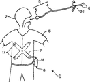
Shown in Figure 1A, the capsule-type endoscope system 1 that possesses the embodiment of the invention 1 has gastrointestinal tract examining apparatus 5, this gastrointestinal tract examining apparatus 5 is made of capsule type endoscope (below abbreviate capsule as) 3 and body 4, wherein capsule 3 by from swallowing as those who are investigated's patient's 2 mouth pipeline in by body cavity, when being specially in the digestive tube, send the picture signal that obtains digesting the inside pipe wall face to carry out optical pickup with wireless mode, body 4 is linked on this capsule 3 removably through rope form parts 6.
In addition, this capsule-type endoscope system 1 also possesses (being configured in the external of patient 2) external unit 8, and it has following function, and the external antenna element 7 through being arranged on patient 2 receives the signal that is sent by capsule 3, and preserves image.
In addition, shown in Figure 1B, external unit 8 freely is connected with PC 11 by for example USB cable 12 cables such as grade are dismantled and assembled.This PC 11 has been connected with CPU built-in and the personal computer main body 13 of the hard disk of document image etc., be connected on this personal computer main body 13 and as the display 14 of the display unit that carries out pictorial display and the keyboard 15 that carries out data input etc.In addition, can be stored in the hard disk in the personal computer main body 13 being stored in image in the external unit 8, or show the image of being preserved by display 14.
Shown in Figure 1A, swallowing capsule 3, for example carry out on the shirt 16 that patient 2 wears, having assembled the antenna element 7 of a plurality of antennas 17 under the situation of splanchnoscopy as medical act.This antenna element 7 receives by the signal that capsule 3 is taken, antenna from be built in capsule 3 sends, and preserves captured image by the external unit 8 that is connected on this antenna element 7.
In addition, in this external unit 8, be provided with LCD monitor 18, can show the image that sends from capsule 3.This external unit 8 is for example linked up with on the belt that is installed in patient 2 freely by dismounting.
Fig. 2 represents the basic structure of gastrointestinal tract examining apparatus 5 in the external unit 8 of expression.
As shown in Figure 2, capsule 3 forms the outer packaging container that its inside is watertight structure by the transparency cover 22 of exterior member main body 21 and semi-spherical shape, wherein this exterior member main body 21 is inaccessible for drum, end form semi-spherical shape, and this transparency cover 22 is chimeric to be fixed on the opening of the other end of this exterior member main body 21.
The inboard of the transparency cover 22 outside this in packaging container, (object lens) optical system 23 that configuration is carried out imaging to the optical image of object of observation thing disposes the imaging apparatus 24 that cmos imager for example etc. is taken on its image space.
In addition, adjacent in the inboard of transparency cover 22 with optical system 23, dispose for example Lighting Division 25 such as White LED, the image pickup scope (range of observation) that images in by optical system 23 on the imaging apparatus 24 is thrown light on.
In the rear side of imaging apparatus 24, configuration: control circuit 26, it drives Lighting Division 25, carries out driving and the signal processing or the control of imaging apparatus 24 simultaneously; Send the wireless transmission part 27 of the picture signal of taking by imaging apparatus 24 to external unit 8; The power supply 28 etc. of battery etc. of the power supply of action usefulness is provided to each circuit etc.In addition, wireless transmission part 27 is connected with not shown antenna.
In addition, adorn outside on article body 21 and rear ends transparency cover 22 opposition sides, setting has the through hole 29 with body 4 banded linking part functions, by rope form parts 6 are fed this through hole 29, capsule 3 dismounting freedom are linked on the body 4 (separable or disengaging) through these rope form parts 6.
That is, the reflex part that makes rope form parts 6 is the state by through hole 29, two the rope form parts 6 that turn back are inserted are led in the hollow bulb of body 4, thereby can be inserted in the body with body 4 banded capsules 3, specifically in the digestive tube.
In addition, throw light on by the Lighting Division 25 that is arranged in the capsule 3, take illuminated internal face etc. by optical system 23 and imaging apparatus 24, carry out splanchnoscopy, send to the image information that photographs external with wireless mode, can utilize to be disposed at external external unit 8, through the endoscopic image information that antenna element 7 receives and storage is obtained by capsule 3.
This external unit 8 possesses: the wireless receiving portion 31 that is connected with antenna element 7 (each antenna 17); The control circuit 32 that signal to being received by this wireless receiving portion 31 after the demodulation carries out A/D conversion and compression processing etc.; Storage is through the memory element 33 of these control circuit 32 compression processed images signals; With the power supply 34 that power supply is provided to control circuit 32 and other circuit.In addition, on control circuit 32, connect display process circuit 35, carry out the display process that on LCD monitor 18, shows from the image of capsule 3 transmissions.In addition, the user can utilize the image that is shown on the LCD monitor 18, monitors the image that the imaging apparatus 24 by capsule 3 photographs.
Fig. 3 represents the more detailed overall structure of gastrointestinal tract examining apparatus 5.
As shown in Figure 3, pliability body 4 forms the reflex part by the through hole 29 of capsule 3 near its front opening, leads in the hollow bulb of body 4 by being inserted by the rope form parts 6 that this reflex part turns back, and links capsule 3 and body 4.Utilize welding or bonding etc. with this body 4 rear end and the body that is branched off into V-shape roughly on hand portion 36 be connected.
This body on hand portion 36 be formed with the hollow bulb straight line of body 4 link the extension hollow bulb 37 that extends on ground and halfway obliquely branch become branch's hollow bulb 38 of hollow bulb.
This extension hollow bulb 37 is used for that inserting of rope form parts 6 is logical waits, and branch's hollow bulb 38 has the function that the dismantled and assembled syringe that freely front end of syringe 39,40 is inserted inserts mouthful.
In addition, extending near the rear end of hollow bulb 37, with body for example on hand portion 36 be provided as the bolt shape parts 41 of rope form parts holding unit integratedly, its opening that extends hollow bulb 37 by closure keeps inserting the logical state of inserting of logical rope form parts 6.
In addition, in branch's hollow bulb 38, also in its back-end near with body for example on hand portion 36 bolt shape parts 42 with the opening closure of branch's hollow bulb 38 are set integratedly, under the situation of not using syringe 39,40, available these bolt shape parts 42 stop up.
In a syringe 39, take in about 1.5% the iodine liquid 43 that being useful on that dyeing observes that pigment, for example iodine staining of (inspection) use, by using the observation of can dyeing of this syringe 39.
In addition, in another syringe 40, put into transparency liquids 44 such as pure water, by using this syringe 40, the pigment after the flushable dyeing, or wash clean mucus or bubble.
Among Fig. 3, to illustrate with capsule 3 banded states by inserting the rope form parts 6 that lead in the body 4, Fig. 4 A~Fig. 4 C illustrates and capsule 3 banded operating procedures.
Shown in Fig. 4 A, will go between etc. with belt etc. and to insert utensil 45 predetermined fixed on the front end of rope form parts 6, by will inserting utensil 45 slotting leading in the hollow bulb of body 4, can lead in the hollow bulb of body 4 rope form parts 6 are easily slotting.The insertion utensil of inserting in the hollow bulb that leads to this body 4 45 is passed through in the through hole 29 of capsule 3 again.
The through hole 29 of this moment as the P of Fig. 4 A to shown in Fig. 4 B that looks, the both sides of the holding section 46 of rope form parts 6 engagings of central part are become the shape that for example is cut into substantially elliptical.Shown in Fig. 4 A or Fig. 4 B, after passing through the operation of through hole 29, by will insert utensil 45 once more from the front end of body 4 by in its hollow bulb, shown in Fig. 4 C, finish the assembling that links bodys 4 and the state of capsule 3 by (insert and lead in the body 4) rope form parts 6.
At this moment, shown in Fig. 4 C, the inner surface and the outer surface of the part of the front opening of formation body 4 form the chamfered section 4a that has carried out the R chamfering.In addition, holding section 46 between the through hole 29 of capsule 3 sides also forms the R shape 46a of portion along the through hole 29 of its both sides, rope form parts 6 are being inserted under the logical states, even if under the state of the wall of rope form parts 6 contact holding sections 46, also can easily mobile smoothly rope form parts 6.
Fig. 5 A~Fig. 5 D represents the structure example of body 4.Body 4 in the present embodiment has pliability, but is incompressibility, preferably has also do not allowing flexible characteristic by inserting logical lead-in wire etc. under the situation of rope form parts 6 effect pull strengths.In addition, preferably having can crooked simply characteristic under the situation of push-and-pull body 4 yet.
In addition, the external diameter of body 4 is smaller or equal to 5mm (more preferably about 2~3mm), and wherein 5mm is smaller or equal to half of the external diameter of capsule 3, because expectation is inserted in the body cavity easily, and patient 2 swallows easily when inserting etc.In addition, this body 4 is little about more than the external diameter of capsule 3, for example 10mm.Like this, the external diameter of body 4 is very thin with respect to the external diameter of capsule 3, and body 4 is set the pliability with following grade for, draws isochronous power under the state of the logical rope form parts 6 of empty therein portion interpolation and also is not easy to make its bending.The concrete structure example of exposed conduit body 4 with reference to Fig. 5 A~Fig. 5 D.
In the example shown in Fig. 5 A, show the example that forms body 4 by torque tube 47.This torque tube 47 is embedded to braid 48a etc. in the flexual pipe 48b.
In addition, among Fig. 5 B, body 4 forms coiled type parts 49a is embedded in the pipe 48b.In addition, at this moment, also can form and improve lubricating coating film 49b on the surface in inboard and the outside.If adopt special teflon fluororesin such as (registered trade marks) as this coated film 49b, then have good lubricity.
Fig. 5 C represent by PTFE (politef), PFA (perfluoroalkoxy resin) etc. have pliability, the pipe 50 of resin that has a fluorine class etc. of good lubricity simultaneously forms body 4.At this moment, can form thin wall.
In addition, Fig. 5 D represents to utilize (the poalon pipes etc.) such as heavy-walled pitch tubes 51 that are made of material soft, that lubricity is good such as vinyl chloride or urethane to constitute body 4.In addition, in the above-mentioned explanation, the hollow bulb of having supposed body 4 is that the situation in a chamber describes, but also can form two chamber 52a, 52b shown in Fig. 5 E, and each inserts logical rope form parts 6 that turn back in two chamber 52a, 52b.
And, also can partly change the hardness or the bullet property sent out of body 4.For example, by having hardness/bullet property sent out of bending simply or warpage beyond the 10cm that makes front end, only the 10cm of front opening side can make the operability of capsule 3 and patient's the easiness of swallowing enjoy a double blessing than other parts softness.The variation of hardness or the bullet property sent out both can have differentially and changed, and also slowly changed serially.
On the other hand, as rope form parts 6, the expectation lubricity is good, even if for thin footpath also has intensity, for example can use good special teflon (registered trade mark) of lubricity or nylon wire or wild silk yarn, operation with line etc.
In addition, preferably the length setting with body 4 is more than or equal to about 60cm, even make that the rear end of body 4 also is present in the outside of mouth 53 at least under the situation of capsule arrival stomach 57.In addition, preferably rope form parts 6 are set at 2 times more than or equal to the length of this body 4.
Illustrate that with reference to Fig. 6 A~Fig. 6 C the gastrointestinal tract examining apparatus 5 that uses this structure checks the action in the digestive tube.
As shown in Figure 6A, be incorporated with the syringe 39 of iodine liquid 43 on hand in the portion 36 at the body of digestive tube testing fixture 5.In addition, insert the body that is connected with rope form parts 6 and utilize bolt shape parts 41 to come on the bolt in the rear end of portion 36 on hand, prevent that rope form parts 6 from invalidly moving.
In addition, patient 2 swallows capsule 3 from mouth 53.The capsule of swallowing from mouth 53 3 moves to esophagus 55 sides through throat 54.This esophagus 55 is divided into as near the bottom 55c the top 55a of throat 54 sides, middle part 55b and the pars cardiaca 56.
Under capsule 3 monomeric situations, sometimes can arrive stomaches 57 by esophagus 55 at short notice because of the weight of capsule 3 or the vermicular movement of esophagus 55 by the capsule behind the throat 54 3, but according to present embodiment, because be linked on the body 4, come push-and-pull etc. so can control the rear end side of body 4, the translational speed of the capsule 3 of body 4 front ends is set at the speed of expectation, simultaneously, also can stop etc., carry out in more detail and check in esophagus 55 optional position midway.
In addition, owing to the translational speed of capsule 3 can be set at the speed of expectation, so, also can photograph the number of necessity even if the shooting speed of capsule 3 photographic images be for example 2/second or less than 2/second slower speed.
For example, under the situation of wanting its inwall of detailed inspection in the 55a of the top of esophagus 55, as shown in Figure 6A, be positioned at capsule 3 under the state of top 55a, the syringe 39 of going into to have iodine liquid is contained in operation, injects iodine liquid near the body of the hollow bulb 55a of top of body 4.The iodine liquid 43 that injects spread to capsule 3 around, spread to the internal face of top 55a.
Like this, the distribution of the pigment by iodine liquid 43 grades, the image of being taken by capsule 3 is colored, and its structure etc. becomes clearer and more definite, easily diagnosis.That is, can utilize iodine staining to check simply.
After capsule 3 finishes shooting to the position of having scattered pigment by the distribution of pigment, capsule 3 moves under the situation of the place ahead (deep) side at this position, also can further scatter pigment as required and take.
In addition, also can move to after bottom 55c takes at the top 55a that does not carry out making under the state that pigment scatters capsule 3 from esophagus 55, stretching body 4 is drawn high (retracting) to top 55a with capsule 3, under this state, carry out pigment and scatter, utilize capsule 3 to take.
That is, also can and use the different condition of the inspection under the painted state of pigment, easily check repeatedly according to the inspection under the painted state that does not use pigment.Owing to can retract like this, so also can push away or trombone slide body 4 etc., change translational speed, check repeatedly.
Like this, in the present embodiment,,, capsule 3 is set on the optional position, takes once more so also can be positioned at the side on hand of external body 4 by operation owing to utilize body 4 to link capsule 3.
Like this, when the shooting in the esophagus 55 that is through with was checked, capsule 3 arrived in the stomach 57 through pars cardiaca 56.
Under situation about checking in the stomach 57 with capsule 3, the method shown in also available Fig. 6 B is checked.Shown in Fig. 6 B, carry out push-and-pull (traction, loose) from body on hand two rope form parts 6 coming out of portion 36 operation and rotate the operation of body 4.
Push-and-pull (traction, loose) operation by this moment, push-and-pull (traction, loose) is outstanding and two rope form parts 6 that turn back by through hole 29 from the front end of body 4, by make capsule 3 as shown by arrow A the state shown in the solid line from Fig. 6 B rotate to the state shown in the double dot dash line, variable visual field direction, check in the stomach 57 in a wide range.
In addition,, the front end of body 4 is rotated shown in the arrow B of Fig. 6 B by carrying out rotating operation to reverse body 4, this moment also variable visual field direction, check in the stomach 57 in a wide range.
After so checking stomach 57 inside, only draw in two rope form parts 6, make another rope form parts 6 move to the front of body 4, shown in Fig. 6 C, can remove thus, make capsule 3 break away from rope form parts 6 based on the fastening state of turning back, capsule 3 is dropped in the stomach 57, break away from body 4.
Drop to the vermicular movement that stomach 57 interior capsules 3 pass through stomach 57, move to duodenum 58 sides.Capsule 3 is for example pressed some cycles and carry out to be taken, and sends the view data that photographs with wireless mode, with the image data storage that sends in the memory element 33 of external unit 8.In addition, also can utilize LCD monitor 18 to confirm the image that photographs.
In addition, the shooting speed of the capsule 3 before and after breaking away from both can be identical, also can be after disengaging before for example break away from 2/second bring up to more than or equal to 4/second or 4/second.Under the situation that the translational speed of capsule after the disengaging 3 accelerates,, can photograph necessary number by improving shooting speed.Shooting speed change during disengaging is to carry out by the signal that sends the shooting speed change to intravital capsule 3 from external unit 8.Perhaps also intervalometer can be built in the capsule 3, shooting speed automaticallyes switch according to the time (for example 10 minutes) after the inspection beginning.
Fig. 7 illustrates the typical example of the inspection method of above explanation.In initial step S1, under the state that has linked capsule 3 and body 4, swallow capsule 3.Among the step S2 below,, draw on one side/operation of loose body 4 by after the throat at capsule 3, the desired site in esophagus 55 utilizes capsule 3 to carry out splanchnoscopy (shooting inspection) on one side.
Among the step S3 below, in body 4, send into iodine liquid 43 grades dyeing and use fluid, near the rear end of capsule 3, emit fluid (being injected into esophagus 55 sides), will dye around it from the front end of body 4 from external.
Among the step S4 after this fluid is emitted, to taking inspection in the esophagus 55 after the dyeing.
And among the step S5 below, in case capsule 3 is then operated many rope form parts 6 by in pars cardiaca 56 backs, the arrival stomach 57, the visual field direction of change capsule 3 is taken inspection.After step S6 in, body 4 and capsule 3 are cut off out, only body 4 is extracted externally, only utilize capsule 3 to take inspection.
Thus, can check that stomach 57 backs cut off out with body 4, utilize capsule 3 monomers to carry out splanchnoscopy at length carrying out splanchnoscopy in 57 from esophagus 55 to stomach.
Like this, according to present embodiment, can utilize the method that is easy to implement that retracting in the esophagus 55 are observed and the wireless observation in the deeps of beginning in the stomach 57.
In addition, can implement observation near the stomach 57 of the wide regions of the beginning pars cardiaca 56 by being easy to method.And, have and can carry out the effect of pigment observation etc. by being easy to the desired site of method in esophagus 55.
Below, the gastrointestinal tract examining apparatus of embodiments of the invention 2 is described with reference to Fig. 8 A and Fig. 8 B.In embodiment 1, hollow bulb interpolation at body 4 is connected with two rope form parts 6 that turn back, but the gastrointestinal tract examining apparatus 5B of present embodiment is shown in Fig. 8 A, the rope form parts 6 that turn back after will through hole 29 by capsule 3 insert pass to body 4 hollow bulb midway, for example near front end, form the solid portion 61 of point, the solid portion 61 of this point by connect body 4 sew on or utilize the bonding agent stickup to wait laterally to put admittedly.
At this moment, also can be shown in Fig. 4 A, front end assembling lead-in wire at rope form parts 4 waits insertion utensil 45 or needle-like member, near the front end of body 4, make and insert utensil 45 or needle-like member perforation body 4, a plurality of through holes can be set near the front end of body 4 in advance, form to utilize and easily insert logical through hole in these through holes and wait the solid some portion 61 admittedly of put.
The solid portion 61 of the point of this moment becomes ring-type or has carried out the fastening state of pasting with bonding agent for making rope form parts 6, so by pulling at from the extended rope form parts 6 of the rear end side of body 4, shown in Fig. 8 B, can remove a little solid state, by further carrying out the operation of stay cord shape parts 6 rearward, can be set at the state that body 4 sides have been left capsule 3.
In addition, be that the structure of portion 36 on hand such as the body shown in Fig. 3 etc. is not set in the rear end of body 4 in the present embodiment.As embodiment 1, as long as scatter under the situation of observing carrying out pigment, be provided with body on hand portion 36 get final product.In addition, also can body portion's 36 dismounting on hand be mounted freely on the rear end of body 4 by being pressed into etc.Other structure in the present embodiment is identical with embodiment 1.
Can carry out the inspection method of present embodiment with embodiment 1 roughly the samely.Explanation simply is under the situation in checking esophagus 55 as shown in Figure 6A, under the state that body 4 is linked on the capsule 3, be to check under the state shown in Fig. 8 A.
In addition, in case after in checking esophagus 55, in the capsule 3 arrival stomaches 57, then pull at the operation of the rope form parts 6 that come out from the rear end of body 4 by execution, the rope form parts 6 of the part that the point in the desirable front that goes out rope form parts 6 is solid.In addition, body 4 and capsule 3 are broken away from.
Afterwards,, body 4 can be pulled to externally, give up this body 4 by carrying out the operation of trombone slide body 4.On the other hand, capsule 3 falls in the stomach 57, afterwards, moves to duodenum 58 sides by vermicular movement, moves to small intestinal, large intestine by vermicular movement again, and this moment, capsule 3 was taken.Promptly carry out splanchnoscopy.The view data that is photographed by capsule 3 is sent to external unit 8 with wireless mode, with the image data storage that sends in the memory element 33 of external unit 8.In addition, also can utilize LCD monitor 18 to confirm the image that photographs.
According to present embodiment, can make body 4 break away from capsule 3 more reliably with few drawing operational ton.In addition, have the effect roughly the same with embodiment 1.
Below, with reference to Fig. 9 A~Fig. 9 D the gastrointestinal tract examining apparatus of embodiments of the invention 3 is described.The gastrointestinal tract examining apparatus 5C of present embodiment is characterised in that near the structure the through hole 29 among the capsule 3C shown in Fig. 9 A and Fig. 9 B.In addition, in the present embodiment,, adopt the kapok line 6C that cuts off easily as the concrete example of rope form parts 6.In addition, except that kapok line 6C, also can be soft scissile lines such as knitting wool.
In the holding section 46 shown in Fig. 4 C of capsule 3C in the present embodiment in the capsule 3 of embodiment 1, on U word shape ground roughly by the part of kapok line 6C is arranged, be provided with for example constitute wedge shape, as the jut 46b of sharp keen portion.
That is, the C-C cross section among Fig. 9 A is formed with sharp keen jut 46b shown in Fig. 9 B.
Therefore, under the state of Fig. 9 A,, shown in Fig. 9 C, utilize jut 46b to cut off kapok line 6C, the kapok line 6C of body 4 sides and capsule 3C are broken away from by carrying out the wherein operation of a wood strip cotton thread 6C that firmly stretches.Capsule 3 after the disengaging drops to the below.Structure herein is identical with embodiment 1 or embodiment 2.
The effect of present embodiment and embodiment 2 etc. are roughly the same.That is, the user can make body 4 sides and capsule 3C break away from by carrying out the operation of the kapok line 6C that firmly stretches simply.
In addition, in the present embodiment, shown in Fig. 9 B, come the C-C cross section of pie graph 9A, but also can form the cross sectional shape that replaces these cross sectional shapes.For example, also can form the cross sectional shape of Fig. 9 A shown in Fig. 9 D, form sharp keen jut 46b in holding section 46, be the structure that sharp keen jut 46b do not occur in the C-C cross sectional shape of this moment.
Below, the gastrointestinal tract examining apparatus of embodiments of the invention 4 is described with reference to Figure 10 A and Figure 10 B.The gastrointestinal tract examining apparatus 5D of present embodiment is by inserting the body 4 that is connected with rope form parts 6 and constituting the capsule 3D that links this body 4 through rope form parts 6.
This capsule 3D forms recess 63, and the rear end of adorning article body 21 outside that replaces among the embodiment 1 for example is provided with through hole 29, takes in the elastomeric element 65 that is provided with through hole 64 in this recess 63, utilizes bonding agent 66 (or being pressed into) etc. to fix.
This elastomeric element 65 is for example formed by urethane or silicone rubber etc., in the near distance in distance outside this through hole 64 is set, and forms the thin thinner wall section 65a of wall thickness of this part.In addition, set this thinner wall section 65a fracture for when when this thinner wall section 65a applies brute force.Other structure example is as identical with embodiment 2.
The inspection method of present embodiment can be carried out in the same manner with the situation of embodiment 2.In addition, the effect when in the present embodiment body 4 sides being separated with capsule 3D is different, so the effect of explanation this moment.
In the present embodiment, shown in Figure 10 A, under the state of the through hole 64 that makes rope form parts 6 perforation elastomeric elements 65, insert the operation of leading to two rope form parts 6 in the body 4 by carrying out firmly to stretch rearward, shown in Figure 10 B, can make the thinner wall section 65a fracture of elastomeric element 65.By this fracture, body 4 sides and capsule 3D are separated, capsule 3D falls.
The situation of the effect of present embodiment and embodiment 2 is roughly the same.
Figure 11 A represents the gastrointestinal tract examining apparatus 5E of the 1st variation.Capsule 3E in this variation and the capsule 3D of Figure 10 A are roughly the same, are formed with recess 67.But this recess 67 forms the taper that open end side broadens.
In addition, in this recess 67, insert the rope form parts 6 turn back into U word shape, after inserting these rope form parts 6, be pressed into rubber bolt 68 elastomeric elements such as grade, link body 4 sides and capsule 3E from the top of the reflex part of these rope form parts 6.
In addition, in this variation,, shown in Figure 11 B, can make the rubber bolt 68 that is pressed in the recess 67 break away from this recess 67 by the operation of the rope form parts 6 that firmly stretch rearward.In addition have effect and the effect identical with embodiment 3.
In this variation, the top of the rope form parts 6 from be accommodated in recess 67 is pressed into rubber bolt 68.Under this state, shown in Figure 11 A, form through hole by recess 67 and rubber bolt 68.
On the contrary, the gastrointestinal tract examining apparatus 5F of the 2nd variation that also can be shown in Figure 12 A and Figure 12 B is such, also can link under the state that does not form through hole under the situation in the recess 69 that rubber bolt 70 is pressed into capsule 3F.
In this variation, shown in Figure 12 A, in capsule 3F, as capsule 3E, form recess 69.In this recess 69, an inner surface of this recess 69 is a tabular surface, with this tabular surface interior surface opposing in be formed with jog 69a.The D-D cross section of Figure 12 A shown in Figure 12 B.
The slotting rope form parts 6 that turn back in the body 4 circlewise that lead to are pressed in this recess 69 with rubber bolt 70, can link body 4 sides and capsule 3F thus.
At this moment, rubber bolt 70 is at the compressed state in the bottom of recess 69, and its upper side forms to the side-prominent protuberance 70a of the rope form parts that turn back 6.
Therefore,, can make rubber bolt 70, move to its upper side from the bottom surface of recess 69 with rope form parts 6 by carrying out the operation of the rope form parts 6 that stretch rearward.
At this moment, because in recess 69, jog 69a is formed on the depth direction of this recess 69, so when stretching, can come the moving of spacing of perception jog 69a with " card is taken " phonoreception.
Under the situation of Figure 12 A and Figure 12 B, after twice " card is taken " phonoreception arranged, know if further stretching then can make disengaging.Other effect and embodiment 3 are roughly the same.
Below, the gastrointestinal tract examining apparatus of embodiments of the invention 5 is described with reference to Figure 13 A~Figure 14.Shown in Figure 13 A and Figure 14 B, the gastrointestinal tract examining apparatus 5G of present embodiment has: be provided with two through hole 29a, 29b capsule 3G, be provided with the body 4 of front end component 71 and insert the 1st and the 2nd rope form parts 6a, 6b in the hollow bulb lead to body 4 at front end.
In addition, near the foot of the jut 73 of the roughly semi-spherical shape in front end component 71, with described two opposed positions of through hole 29a, 29b on, be provided with two opening 71a, 71b, to be communicated with at the hollow bulb of body 4.
In addition, rope form parts 6a, the 6b of insert in the hollow bulb that leads to body 4, opening 71a, 71b from be arranged on front end component 71 are outstanding turns back by through hole 29a, the 29b of opposed capsule 3G, pass through once more in opening 71a, the 71b, insert and lead in the hollow bulb of body 4, link body 4 sides and capsule 3G.
In the present embodiment, to be set under the equal state through the pull strength that the 1st and the 2nd rope form parts 6a, 6b feed through to capsule 3G side, as shown in FIG. 13A, the back end in contact of the exterior member main body 21 on the central shaft O is observed in the front end of the jut 73 of the semi-spherical shape of the jut 73 of front end component 71 and conduct, and body 4 can be set roughly linearity for capsule 3G.
On the contrary, by relatively changing the pull strength that feeds through to capsule 3G side through the 1st and the 2nd rope form parts 6a, 6b, shown in Figure 13 B, can tilt from the length direction of body 4 direction with the observation central shaft O of capsule 3G.
For example shown in Figure 13 B, user the 1st rope form parts 6a that relaxes draws the 2nd rope form parts 6b, thus, can set for bent or tilt to the 2nd rope form parts 6b lateral bending that has drawn capsule 3G.Like this,, capsule 3G is tilted, set the direction that it observes central shaft O changeably by drawing a side, the loose the opposing party among the 1st rope form parts 6a and the 2nd rope form parts 6b.
In addition, can take the direction of checking wide region.Therefore, for example as illustrating among Fig. 6 B, can check the internal face of stomach 57 in a wide range in the inside of stomach 57.In addition, in esophagus 55,, also can change direction of observation, check in more detail by carrying out the operation shown in Figure 13 B.In addition have and roughly the same effects such as embodiment 1 or embodiment 2.
In addition, the gastrointestinal tract examining apparatus 5H of the 1st variation that also can be as shown in figure 14 is such.In this variation, the double lumen tube of two chamber 52a, 52b shown in Fig. 5 E forms body 4 by for example having, and can make the 1st and the 2nd rope form parts 6a, 6b outstanding from each chamber 52a, 52b, and through hole 29a, 29b by capsule 3G link.
In addition, the center at the front end face of this body 4 is provided with hemispheric jut 75.And, make the rear end surface butt of this jut 75 and capsule 3G, by drawing a side, the loose the opposing party among the 1st rope form parts 6a and the 2nd rope form parts 6b, can set the direction of observation of capsule 3G changeably.
The situation of the 1st variation has effect and the effect roughly the same with the situation of present embodiment.
In addition, as this variation, 4 chambeies also can be set replace two chamber 52a, 52b.
Below, the gastrointestinal tract examining apparatus of embodiments of the invention 6 is described with reference to Figure 15.The gastrointestinal tract examining apparatus 5I of present embodiment constitutes body 4 as getting off, for example replace two chamber 52a, 52b in Figure 14, and four chamber 52a, 52a ', 52b, 52b ' are set, and simultaneously, jut 75 is not set, liquid is set injects with chamber 76.
In addition,, connect the body parts of portion 36 on hand be similar to explanation among the embodiment 1, can inject with chamber 76 through liquid and inject iodine liquid or medicine for treatment liquid in the rear end side of this body 4.
Present embodiment has the effect roughly the same with embodiment 1.
Below, the gastrointestinal tract examining apparatus of embodiments of the invention 7 is described with reference to Figure 16.The gastrointestinal tract examining apparatus 5J of present embodiment is made of near capsule 3J that is provided with through hole 77 cardinal extremity of transparency cover 22 and the slotting body 4 that is connected with rope form parts 6.This through hole 77 forms and connects the part that the approximate diameter place with capsule 3J is close.
Make insert the rope form parts 6 led to body 4 from the front opening of body 4 by being arranged on the through hole 77 the capsule 3J, thereby link bodys 4 and capsule 3J through rope form parts 6.
At this moment, since through hole 77 be formed on the position of comparing the more close front that is provided with transparency cover 22 to the distance of the central authorities of the length direction of capsule 3J on, so linked under the state of body 4 and capsule 3J by rope form parts 6, transparency cover 22 becomes and the opposed state of body 4 sides.In addition, through hole 77 is formed on the field of view θ outside.
Present embodiment is opposite with the direction of observation (shooting direction) of embodiment 1 grade.Except the direction of observation difference, have and roughly the same effects such as embodiment 1 or embodiment 2.
Below, the gastrointestinal tract examining apparatus of embodiments of the invention 8 is described with reference to Figure 17 A and Figure 17 B.The gastrointestinal tract examining apparatus 5K of present embodiment is made of capsule 3K that is provided with transparency cover 22a, 22b at two ends and the slotting body 4 that is connected with the 1st and the 2nd rope form parts 6a, 6b.
In the inboard of transparency cover 22a and the inboard of transparency cover 22b, dispose optical system shown in Figure 2 23 and imaging apparatus 24 and Lighting Division 25 respectively.In addition, the range of observation of representing each optical system with θ.
In addition, in exterior member main body 21, be respectively arranged with through hole 77a, 77b, inserted logical respectively by the 1st and the 2nd rope form parts 6a, 6b near the position of the cardinal extremity of transparency cover 22a, 22b.
The inspection method of present embodiment is described with reference to Figure 17 A and Figure 17 B.Shown in Figure 17 A, check as esophagus 55 in and tube chamber that the external diameter of capsule 3K is approaching partly in the time, operate the 1st and the 2nd rope form parts 6a, 6b, make the length direction of the length direction of capsule 3K along tube chamber.
For example,, can be set at the state shown in Figure 17 A, check in the esophagus 55 by drawing the 1st rope form parts 6a more at full tilt than the 2nd rope form parts 6b.
On the contrary, under the situation in checking the stomach 57 more much bigger, equate, can shown in Figure 17 B, set the state of growing crosswise for by the pull strength that makes the 1st rope form parts 6a and the 2nd rope form parts 6b than the size of capsule 3K.
After observing under this state,, capsule 3K is tilted for example by drawing the 1st rope form parts 6a more at full tilt than the 2nd rope form parts 6b.Like this, by carrying out the draw operations of the 1st rope form parts 6a and the 2nd rope form parts 6b, can in stomach 57, change direction of observation inspection in a wide range.
Other effect is identical with embodiment 1 etc.
In addition, the various embodiments described above etc. are partly made up and the embodiment that constitutes waits and also belongs to the present invention.In addition, in the various embodiments described above, illustrated that the execution optical pickup checks the situation of (splanchnoscopy), but also applicable to the situation of carrying out the inspection beyond the optics such as PH sensing or ultrasound investigation or scatter the situations such as medical treatment that medicament is treated usefulness.
Utilizability on the industry
In the bindings such as through hole that are attached to capsule type endoscope by the rope form parts that are inserted through body Under the state in the section, check fully the digest tubes such as endoceliac esophagus, afterwards, remove binding, Also carried out separately the inspection of deep side by capsule type endoscope.
Claims (30)
1, a kind of gastrointestinal tract examining apparatus is characterized in that, possesses:
Can carry out the capsule endoscope of the inspection of digestive tube;
Flexual body;
Inserted the flexual rope form parts that lead in the described body; With
Be arranged on the linking part in the described capsule endoscope,
By described rope form parts are linked on the described linking part removably, link described body and described capsule endoscope separably.
2, gastrointestinal tract examining apparatus according to claim 1 is characterized in that:
Described linking part is arranged on the through hole in the described capsule endoscope, by described rope form parts being inserted logical described through hole, links capsule endoscope and body removably.
3, gastrointestinal tract examining apparatus according to claim 1 is characterized in that:
Described linking part is made of the chimera that is arranged on the recess in the described capsule endoscope and can be embedded in the recess, by utilizing described chimera described rope form parts is fastened in the recess, links capsule endoscope and body removably.
4, gastrointestinal tract examining apparatus according to claim 1 is characterized in that:
The external diameter of described body is smaller or equal to half of the external diameter of capsule endoscope.
5, gastrointestinal tract examining apparatus according to claim 4 is characterized in that:
The external diameter of described body is smaller or equal to 5mm.
6, gastrointestinal tract examining apparatus according to claim 4 is characterized in that:
The power that described body has when drawing under the slotting state that leads in inside of described rope form parts also is not easy to make its crooked pliability.
7, gastrointestinal tract examining apparatus according to claim 1 is characterized in that:
Possess holding unit, its end with described rope form parts remains near the end on hand of described body removably.
8, gastrointestinal tract examining apparatus according to claim 1 is characterized in that:
Fix an end of described rope form parts removably by the fixed part midway that is arranged on described body, the other end of described rope form parts is via the linking part that is arranged in the described capsule endoscope, inserts from the front end of described body and leads to end on hand.
9, gastrointestinal tract examining apparatus according to claim 8 is characterized in that:
Fix an end of described rope form parts removably by near the fixed part the front end that is arranged on described body.
10, gastrointestinal tract examining apparatus according to claim 2 is characterized in that:
Periphery at described through hole is provided with sharp keen portion, by at described rope form parts to stretch more than or equal to certain a certain size power, can cut off the rope form parts.
11, gastrointestinal tract examining apparatus according to claim 2 is characterized in that:
Be provided with thinner wall section at described through hole periphery, by at described rope form parts stretching and it can be cut off more than or equal to certain a certain size power, or it is come off from described through hole periphery.
12, gastrointestinal tract examining apparatus according to claim 3 is characterized in that:
By with the described rope form parts that stretch more than or equal to certain a certain size power, the chimera that is embedded in the described recess comes off from recess.
13, gastrointestinal tract examining apparatus according to claim 1 is characterized in that:
Leading section outer surface at described body is provided with the roughly chamfered section of R shape.
14, gastrointestinal tract examining apparatus according to claim 1 is characterized in that:
Be provided with at the leading section of described body and have front end component roughly semi-spherical shape portion, that be independent of body.
15, gastrointestinal tract examining apparatus according to claim 1 is characterized in that:
Make the hardness ratio side softness on hand of the front of described body.
16, gastrointestinal tract examining apparatus according to claim 15 is characterized in that:
Make the other parts softness of hardness ratio body of certain specific length part of the front of described body.
17, gastrointestinal tract examining apparatus according to claim 15 is characterized in that:
The hardness of described body changes continuously.
18, gastrointestinal tract examining apparatus according to claim 1 is characterized in that:
Near the end on hand of body, be provided with in described body and inject the fluid inlet that liquid or gas are used.
19, gastrointestinal tract examining apparatus according to claim 2 is characterized in that:
The position of separating is provided with a plurality of described through holes, inserts slotting the leading in the body of many rope form parts of logical each through hole, has drawn at least one end of each rope form parts from the end on hand of body.
20, gastrointestinal tract examining apparatus according to claim 19 is characterized in that:
Constituting can be by drawing/loose operation the operation of shaking capsule endoscope to described many rope form parts respectively.
21, gastrointestinal tract examining apparatus according to claim 19 is characterized in that:
Described body is a multi-lumen tube, and the quantity in the chamber that has is more than or equal to the bar number of the rope form parts that use.
22, gastrointestinal tract examining apparatus according to claim 1 is characterized in that:
Described body is a multi-lumen tube, and the quantity in the chamber that has is more than or equal to 2 times of the bar number of the rope form parts that use.
23, gastrointestinal tract examining apparatus according to claim 1 is characterized in that:
Described body is wire netting or the metal spiral body is embedded in the resin, the revolving force transitivity is good torque tube, or the pipe that is made of the fluororesin with lubricity and hardness, or the heavy wall pitch tube.
24. gastrointestinal tract examining apparatus according to claim 23 is characterized in that: described fluororesin is special teflon or PTFE.
25, gastrointestinal tract examining apparatus according to claim 1 is characterized in that:
Even if the described rope form parts line that to be lubricities good also has intensity for thin footpath.
26, gastrointestinal tract examining apparatus according to claim 25 is characterized in that: described line is fluororesin, nylon wire or wild silk yarn.
27, gastrointestinal tract examining apparatus according to claim 25 is characterized in that: described line is the operation line.
28, gastrointestinal tract examining apparatus according to claim 10 is characterized in that:
Described rope form parts are soft scissile lines.
29, gastrointestinal tract examining apparatus according to claim 28 is characterized in that: described line is kapok line, thin,tough silk line or polyester line.
30, gastrointestinal tract examining apparatus according to claim 28 is characterized in that: described line is a knitting wool.
Applications Claiming Priority (2)
| Application Number | Priority Date | Filing Date | Title |
|---|---|---|---|
| JP2003342417A JP3993550B2 (en) | 2003-09-30 | 2003-09-30 | Gastrointestinal inspection device |
| JP342417/2003 | 2003-09-30 |
Publications (2)
| Publication Number | Publication Date |
|---|---|
| CN1859866A CN1859866A (en) | 2006-11-08 |
| CN100446713C true CN100446713C (en) | 2008-12-31 |
Family
ID=34419261
Family Applications (1)
| Application Number | Title | Priority Date | Filing Date |
|---|---|---|---|
| CNB2004800285551A Expired - Fee Related CN100446713C (en) | 2003-09-30 | 2004-09-08 | Gastrointestinal tract examining apparatus |
Country Status (5)
| Country | Link |
|---|---|
| US (1) | US7448993B2 (en) |
| EP (1) | EP1671575B1 (en) |
| JP (1) | JP3993550B2 (en) |
| CN (1) | CN100446713C (en) |
| WO (1) | WO2005032352A1 (en) |
Families Citing this family (80)
| Publication number | Priority date | Publication date | Assignee | Title |
|---|---|---|---|---|
| US8091164B2 (en) * | 2003-10-06 | 2012-01-10 | Olympus Corporation | Introduction-assisting apparatus for capsule medical device |
| US7429259B2 (en) | 2003-12-02 | 2008-09-30 | Cadeddu Jeffrey A | Surgical anchor and system |
| JP4445812B2 (en) * | 2004-07-08 | 2010-04-07 | オリンパス株式会社 | Intra-subject introduction apparatus and intra-subject introduction system |
| US7530948B2 (en) * | 2005-02-28 | 2009-05-12 | University Of Washington | Tethered capsule endoscope for Barrett's Esophagus screening |
| JP4875315B2 (en) * | 2005-04-05 | 2012-02-15 | オリンパスメディカルシステムズ株式会社 | Intra-subject introduction device |
| US8491464B2 (en) | 2005-07-08 | 2013-07-23 | Olympus Corporation | In-vivo information acquiring apparatus, in-vivo information acquiring system, and in-vivo information acquiring method |
| CN101217908B (en) * | 2005-07-08 | 2012-10-24 | 奥林巴斯医疗株式会社 | Apparatus for placing capsule type medical device, apparatus for placing capsule endoscope in the body |
| WO2007023671A1 (en) * | 2005-08-24 | 2007-03-01 | Konica Minolta Medical & Graphic, Inc. | Capsule-type medical apparatus and diagnosis system |
| US20080015413A1 (en) * | 2006-02-22 | 2008-01-17 | Olympus Medical Systems Corporation | Capsule endoscope system and medical procedure |
| JP4827570B2 (en) * | 2006-03-27 | 2011-11-30 | 佳彦 平尾 | Capsule insertion device |
| EP2012697A4 (en) | 2006-04-29 | 2010-07-21 | Univ Texas | Devices for use in transluminal and endoluminal surgery |
| CN103356153B (en) | 2006-09-12 | 2016-08-10 | 奥林巴斯株式会社 | Capsule type endoscope |
| US9675285B2 (en) * | 2006-10-16 | 2017-06-13 | Given Imaging Ltd. | Delivery device for implantable monitor |
| KR100876647B1 (en) * | 2006-11-22 | 2009-01-08 | 주식회사 코렌 | Capsule type image photographing apparatus and endoscopy using the same |
| EP2111148B1 (en) * | 2007-01-19 | 2015-08-12 | Given Imaging (Los Angeles) LLC | Micro-remote gastrointestinal physiological measurement device |
| US7824270B2 (en) | 2007-01-23 | 2010-11-02 | C-Flex Bearing Co., Inc. | Flexible coupling |
| US20080177141A1 (en) * | 2007-01-24 | 2008-07-24 | Hsien-Ming Wu | Memory-type two-section endoscopic system |
| DE102007000230A1 (en) | 2007-04-16 | 2008-10-30 | Hothan, Thorsten, Dr. | Endoscope for examining intestine of patient i.e. human being, has sphere/retainer, whose weight and/or shape are dimensioned such that sphere/retainer is movable against natural intestinal peristalsis due to net weight of sphere/retainer |
| JP2008307226A (en) * | 2007-06-14 | 2008-12-25 | Olympus Medical Systems Corp | Endoscope system |
| US9339174B2 (en) * | 2007-07-18 | 2016-05-17 | Given Imaging Ltd | Device and method for viewing a body lumen |
| JP5259141B2 (en) * | 2007-08-31 | 2013-08-07 | オリンパスメディカルシステムズ株式会社 | In-subject image acquisition system, in-subject image processing method, and in-subject introduction device |
| JP2012509104A (en) * | 2008-11-17 | 2012-04-19 | メイヨ・ファウンデーション・フォー・メディカル・エデュケーション・アンド・リサーチ | Diagnostic capsule, delivery / recovery system, kit, and method |
| CN102448364A (en) * | 2009-05-28 | 2012-05-09 | 基文影像公司 | Apparatus for delivery of autonomous in-vivo capsules |
| JP4599474B1 (en) * | 2009-08-24 | 2010-12-15 | オリンパスメディカルシステムズ株式会社 | Medical equipment |
| US9186203B2 (en) | 2009-10-09 | 2015-11-17 | Ethicon Endo-Surgery, Inc. | Method for exchanging end effectors In Vivo |
| US8623011B2 (en) * | 2009-10-09 | 2014-01-07 | Ethicon Endo-Surgery, Inc. | Magnetic surgical sled with locking arm |
| US9295485B2 (en) | 2009-10-09 | 2016-03-29 | Ethicon Endo-Surgery, Inc. | Loader for exchanging end effectors in vivo |
| US20110087224A1 (en) * | 2009-10-09 | 2011-04-14 | Cadeddu Jeffrey A | Magnetic surgical sled with variable arm |
| US10172669B2 (en) | 2009-10-09 | 2019-01-08 | Ethicon Llc | Surgical instrument comprising an energy trigger lockout |
| US8219171B2 (en) * | 2010-03-16 | 2012-07-10 | Given Imaging Ltd. | Delivery device for implantable monitor |
| US8764632B2 (en) * | 2010-04-08 | 2014-07-01 | Eric James Kezirian | Endoscopic device and system |
| GB2480498A (en) | 2010-05-21 | 2011-11-23 | Ethicon Endo Surgery Inc | Medical device comprising RF circuitry |
| JP4838398B1 (en) * | 2010-07-13 | 2011-12-14 | オリンパスメディカルシステムズ株式会社 | Medical equipment |
| WO2012008188A1 (en) * | 2010-07-13 | 2012-01-19 | オリンパスメディカルシステムズ株式会社 | Medical device |
| WO2012120837A1 (en) * | 2011-03-10 | 2012-09-13 | パナソニック株式会社 | Endoscopic camera and endoscopic device |
| US9314292B2 (en) | 2011-10-24 | 2016-04-19 | Ethicon Endo-Surgery, Llc | Trigger lockout mechanism |
| EP2596756B1 (en) * | 2011-11-22 | 2014-02-26 | Ovesco Endoscopy AG | Implanting apparatus |
| US20150051589A1 (en) * | 2012-03-27 | 2015-02-19 | Sony Corporation | Capsule type medical device and medical system |
| WO2013177154A1 (en) * | 2012-05-21 | 2013-11-28 | The General Hospital Corporation | Apparatus, device and method for capsule microscopy |
| CN102743148B (en) * | 2012-06-21 | 2015-02-25 | 中国人民解放军第一七五医院 | Spray liquid propelling type intestinal tract examination system |
| CN103156568A (en) * | 2012-08-22 | 2013-06-19 | 武汉安康通光电技术有限公司 | Filament tube capsule oesophagoscope capable of being released and magnetically controlled |
| US9125681B2 (en) | 2012-09-26 | 2015-09-08 | Ethicon Endo-Surgery, Inc. | Detachable end effector and loader |
| US20140171977A1 (en) | 2012-12-13 | 2014-06-19 | Ethicon Endo-Surgery, Inc. | Pawl Mechanism in Circular Needle Applier |
| US9451937B2 (en) | 2013-02-27 | 2016-09-27 | Ethicon Endo-Surgery, Llc | Percutaneous instrument with collet locking mechanisms |
| CN103222844B (en) * | 2013-04-25 | 2016-01-27 | 中国人民解放军成都军区总医院 | Controllable capsule endoscopy |
| JP5985764B2 (en) * | 2013-11-29 | 2016-09-06 | シャープ株式会社 | Internal surveillance camera system, support tube accessory for internal surveillance camera system, and fixture for internal surveillance camera system |
| WO2015116701A1 (en) * | 2014-01-28 | 2015-08-06 | The General Hospital Corporation | Apparatus, systems and methods which controls and facilitates information gathering using a tethered capsule catheter |
| CN103961047A (en) * | 2014-04-16 | 2014-08-06 | 姜泊 | Flexible wire traction-type capsule endoscopy and manufacturing method thereof |
| CN103961048A (en) * | 2014-04-16 | 2014-08-06 | 姜泊 | Traction type capsule endoscopy |
| CN103961104A (en) * | 2014-04-16 | 2014-08-06 | 姜泊 | Traction-type recyclable capsule endoscopy |
| US10159524B2 (en) | 2014-12-22 | 2018-12-25 | Ethicon Llc | High power battery powered RF amplifier topology |
| US10314638B2 (en) | 2015-04-07 | 2019-06-11 | Ethicon Llc | Articulating radio frequency (RF) tissue seal with articulating state sensing |
| CN104905757B (en) * | 2015-06-30 | 2016-08-24 | 中国人民解放军成都军区总医院 | There is the capsule endoscope of ultrasonic sensing |
| CN105011892A (en) * | 2015-06-30 | 2015-11-04 | 中国人民解放军成都军区总医院 | Multiple-pipe capsule endoscope |
| US10342520B2 (en) | 2015-08-26 | 2019-07-09 | Ethicon Llc | Articulating surgical devices and loaders having stabilizing features |
| US10335196B2 (en) | 2015-08-31 | 2019-07-02 | Ethicon Llc | Surgical instrument having a stop guard |
| US10251636B2 (en) | 2015-09-24 | 2019-04-09 | Ethicon Llc | Devices and methods for cleaning a surgical device |
| US10702257B2 (en) | 2015-09-29 | 2020-07-07 | Ethicon Llc | Positioning device for use with surgical instruments |
| US10959771B2 (en) | 2015-10-16 | 2021-03-30 | Ethicon Llc | Suction and irrigation sealing grasper |
| US10912543B2 (en) | 2015-11-03 | 2021-02-09 | Ethicon Llc | Surgical end effector loading device and trocar integration |
| US10675009B2 (en) | 2015-11-03 | 2020-06-09 | Ethicon Llc | Multi-head repository for use with a surgical device |
| US10265130B2 (en) | 2015-12-11 | 2019-04-23 | Ethicon Llc | Systems, devices, and methods for coupling end effectors to surgical devices and loading devices |
| US10959806B2 (en) | 2015-12-30 | 2021-03-30 | Ethicon Llc | Energized medical device with reusable handle |
| US10856934B2 (en) | 2016-04-29 | 2020-12-08 | Ethicon Llc | Electrosurgical instrument with electrically conductive gap setting and tissue engaging members |
| US10987156B2 (en) | 2016-04-29 | 2021-04-27 | Ethicon Llc | Electrosurgical instrument with electrically conductive gap setting member and electrically insulative tissue engaging members |
| CN107569204A (en) * | 2016-07-05 | 2018-01-12 | 曾锦顺 | The big colonoscopy of egg shape and traveling control method |
| US10751117B2 (en) | 2016-09-23 | 2020-08-25 | Ethicon Llc | Electrosurgical instrument with fluid diverter |
| CN106580238A (en) * | 2016-12-07 | 2017-04-26 | 广州中医药大学第附属医院 | Device capable of intelligently positioning and identifying throat anatomical structure and control method thereof |
| US11033325B2 (en) | 2017-02-16 | 2021-06-15 | Cilag Gmbh International | Electrosurgical instrument with telescoping suction port and debris cleaner |
| US10799284B2 (en) | 2017-03-15 | 2020-10-13 | Ethicon Llc | Electrosurgical instrument with textured jaws |
| US11497546B2 (en) | 2017-03-31 | 2022-11-15 | Cilag Gmbh International | Area ratios of patterned coatings on RF electrodes to reduce sticking |
| US10603117B2 (en) | 2017-06-28 | 2020-03-31 | Ethicon Llc | Articulation state detection mechanisms |
| US11033323B2 (en) | 2017-09-29 | 2021-06-15 | Cilag Gmbh International | Systems and methods for managing fluid and suction in electrosurgical systems |
| US11484358B2 (en) | 2017-09-29 | 2022-11-01 | Cilag Gmbh International | Flexible electrosurgical instrument |
| US11490951B2 (en) | 2017-09-29 | 2022-11-08 | Cilag Gmbh International | Saline contact with electrodes |
| US11786114B2 (en) * | 2019-04-09 | 2023-10-17 | AnX Robotica Corp | Systems and methods for liquid biopsy and drug delivery |
| CN111808916A (en) | 2020-07-24 | 2020-10-23 | 上海安翰医疗技术有限公司 | Trypsin detection film, preparation method and application thereof and trypsin detection kit |
| CN113440093B (en) * | 2021-07-19 | 2022-11-25 | 山东第一医科大学附属省立医院(山东省立医院) | Digestive tract secretion microscopic detection system |
| US20230190084A1 (en) * | 2021-12-16 | 2023-06-22 | Karl Storz Imaging, Inc. | Implantable Internal Observation Device and System |
| CN115399713B (en) * | 2022-05-24 | 2023-03-07 | 北京大学第一医院 | Magnetic control capsule endoscope traction cap and kit with spraying function |
Citations (6)
| Publication number | Priority date | Publication date | Assignee | Title |
|---|---|---|---|---|
| JPH06142081A (en) * | 1992-11-05 | 1994-05-24 | Olympus Optical Co Ltd | Capsule apparatus for intracavitary diagnosis |
| US6285897B1 (en) * | 1999-04-07 | 2001-09-04 | Endonetics, Inc. | Remote physiological monitoring system |
| JP2002000556A (en) * | 2000-06-26 | 2002-01-08 | Nonomura Tomosuke | Endoscope |
| JP2003135388A (en) * | 2001-10-30 | 2003-05-13 | Olympus Optical Co Ltd | Endoscope apparatus |
| US20030139647A1 (en) * | 2000-05-23 | 2003-07-24 | Dan Raz | Device and method for positioning an object in a body lumen |
| JP2003210393A (en) * | 2002-01-22 | 2003-07-29 | Olympus Optical Co Ltd | Capsule-type medical apparatus |
Family Cites Families (19)
| Publication number | Priority date | Publication date | Assignee | Title |
|---|---|---|---|---|
| JPS5394515A (en) * | 1977-01-31 | 1978-08-18 | Kubota Ltd | Method of producing glass fiber reinforced cement plate |
| US5741429A (en) * | 1991-09-05 | 1998-04-21 | Cardia Catheter Company | Flexible tubular device for use in medical applications |
| US5653677A (en) * | 1994-04-12 | 1997-08-05 | Fuji Photo Optical Co. Ltd | Electronic endoscope apparatus with imaging unit separable therefrom |
| US6071274A (en) * | 1996-12-19 | 2000-06-06 | Ep Technologies, Inc. | Loop structures for supporting multiple electrode elements |
| US5738110A (en) * | 1996-05-29 | 1998-04-14 | Beal; Charles B. | Device for the diagnosis of certain gastrointestinal pathogens |
| IL122716A0 (en) | 1997-12-22 | 1998-08-16 | Tally Eitan Zeev Pearl And Co | System and method for in vivo delivery of autonomous capsule |
| US6632171B2 (en) * | 1997-12-22 | 2003-10-14 | Given Imaging Ltd. | Method for in vivo delivery of autonomous capsule |
| US5984860A (en) * | 1998-03-25 | 1999-11-16 | Shan; Yansong | Pass-through duodenal enteroscopic device |
| JP4716594B2 (en) * | 2000-04-17 | 2011-07-06 | オリンパス株式会社 | Endoscope |
| US6475145B1 (en) * | 2000-05-17 | 2002-11-05 | Baymar, Inc. | Method and apparatus for detection of acid reflux |
| US6659981B2 (en) * | 2000-12-08 | 2003-12-09 | Medtronic, Inc. | Medical device delivery catheter with distal locator |
| US6951536B2 (en) * | 2001-07-30 | 2005-10-04 | Olympus Corporation | Capsule-type medical device and medical system |
| US6986738B2 (en) * | 2001-08-06 | 2006-01-17 | Given Imaging Ltd | System and method for maneuvering a device in vivo |
| JP4643089B2 (en) | 2001-09-27 | 2011-03-02 | オリンパス株式会社 | Capsule medical device |
| US20030135266A1 (en) * | 2001-12-03 | 2003-07-17 | Xtent, Inc. | Apparatus and methods for delivery of multiple distributed stents |
| US7001329B2 (en) * | 2002-07-23 | 2006-02-21 | Pentax Corporation | Capsule endoscope guidance system, capsule endoscope holder, and capsule endoscope |
| JP4166525B2 (en) | 2002-07-23 | 2008-10-15 | Hoya株式会社 | Capsule endoscope having external terminal and capsule endoscope holder |
| JP4133074B2 (en) | 2002-07-23 | 2008-08-13 | Hoya株式会社 | Capsule endoscope holding mechanism |
| JP4320329B2 (en) | 2006-02-28 | 2009-08-26 | サミー株式会社 | Slot machine |
-
2003
- 2003-09-30 JP JP2003342417A patent/JP3993550B2/en not_active Expired - Fee Related
-
2004
- 2004-09-08 EP EP20040787711 patent/EP1671575B1/en not_active Expired - Fee Related
- 2004-09-08 CN CNB2004800285551A patent/CN100446713C/en not_active Expired - Fee Related
- 2004-09-08 WO PCT/JP2004/013028 patent/WO2005032352A1/en active Application Filing
- 2004-09-27 US US10/951,100 patent/US7448993B2/en active Active
Patent Citations (7)
| Publication number | Priority date | Publication date | Assignee | Title |
|---|---|---|---|---|
| JPH06142081A (en) * | 1992-11-05 | 1994-05-24 | Olympus Optical Co Ltd | Capsule apparatus for intracavitary diagnosis |
| US6285897B1 (en) * | 1999-04-07 | 2001-09-04 | Endonetics, Inc. | Remote physiological monitoring system |
| US6689056B1 (en) * | 1999-04-07 | 2004-02-10 | Medtronic Endonetics, Inc. | Implantable monitoring probe |
| US20030139647A1 (en) * | 2000-05-23 | 2003-07-24 | Dan Raz | Device and method for positioning an object in a body lumen |
| JP2002000556A (en) * | 2000-06-26 | 2002-01-08 | Nonomura Tomosuke | Endoscope |
| JP2003135388A (en) * | 2001-10-30 | 2003-05-13 | Olympus Optical Co Ltd | Endoscope apparatus |
| JP2003210393A (en) * | 2002-01-22 | 2003-07-29 | Olympus Optical Co Ltd | Capsule-type medical apparatus |
Also Published As
| Publication number | Publication date |
|---|---|
| WO2005032352A1 (en) | 2005-04-14 |
| JP2005103092A (en) | 2005-04-21 |
| EP1671575A1 (en) | 2006-06-21 |
| EP1671575A4 (en) | 2009-06-17 |
| US20050085697A1 (en) | 2005-04-21 |
| US7448993B2 (en) | 2008-11-11 |
| JP3993550B2 (en) | 2007-10-17 |
| CN1859866A (en) | 2006-11-08 |
| EP1671575B1 (en) | 2015-05-13 |
Similar Documents
| Publication | Publication Date | Title |
|---|---|---|
| CN100446713C (en) | Gastrointestinal tract examining apparatus | |
| JP4643089B2 (en) | Capsule medical device | |
| JP4870670B2 (en) | In vivo information acquisition apparatus and in vivo information acquisition system | |
| JP4578740B2 (en) | Capsule medical device | |
| US20090018396A1 (en) | Indwelling device for encapsulated medical device, in-vivo indwelling device for capsule endoscope, and capsule-indwelling medical device | |
| JP4081259B2 (en) | Endoscope apparatus and endoscope detachment method | |
| US20080051655A1 (en) | Ultrasonic endoscope, therapeutic system, treatment method using therapeutic system, endoscopic system, treatment method using ultrasonic endoscope, t-bar, and t-bar suturing device | |
| EP2651282B1 (en) | Magnetically guided robotic device for endoscopic procedures | |
| WO2007097393A1 (en) | Capsule endoscope system | |
| JP2004528919A (en) | Method for in vivo imaging of the undeformed gastrointestinal tract | |
| JP2003526413A (en) | Apparatus and method for encapsulated medical imaging | |
| DE10028153A1 (en) | Endoscope system is flexible has radio control and picture transmission can be completely swallowed | |
| KR20080030549A (en) | Device, system and method for in-vivo examination | |
| CN106462934A (en) | Tethered endoscope | |
| US20080154093A1 (en) | Capsule Type Endoscope with an Insertion Tube | |
| CN101045013A (en) | Medical snaring device | |
| JP4734051B2 (en) | Capsule type medical device indwelling device and capsule endoscope in vivo indwelling device | |
| CN1628603A (en) | Inward vision mirror apparatus | |
| JP2003135387A (en) | Capsule type medical apparatus | |
| CN101044971A (en) | Intubation device for colonic decompression | |
| CN102871635B (en) | Capsule endoscope feeding device | |
| EP1769714A1 (en) | Device and system for introduction into examinee | |
| CN104771129B (en) | The enteroscope guide can developed under lonizing radiation | |
| Filip | Self–stabilizing capsule endoscope for early detection and biopsy of colonic polyps | |
| CN208976333U (en) | A kind of stomach check probe cleaning device |
Legal Events
| Date | Code | Title | Description |
|---|---|---|---|
| C06 | Publication | ||
| PB01 | Publication | ||
| C10 | Entry into substantive examination | ||
| SE01 | Entry into force of request for substantive examination | ||
| C14 | Grant of patent or utility model | ||
| GR01 | Patent grant | ||
| CF01 | Termination of patent right due to non-payment of annual fee |
Granted publication date: 20081231 Termination date: 20180908 |
|
| CF01 | Termination of patent right due to non-payment of annual fee |