CN103228230A - Fenestration devices, systems, and methods - Google Patents
Fenestration devices, systems, and methods Download PDFInfo
- Publication number
- CN103228230A CN103228230A CN2011800548237A CN201180054823A CN103228230A CN 103228230 A CN103228230 A CN 103228230A CN 2011800548237 A CN2011800548237 A CN 2011800548237A CN 201180054823 A CN201180054823 A CN 201180054823A CN 103228230 A CN103228230 A CN 103228230A
- Authority
- CN
- China
- Prior art keywords
- expandable members
- guiding
- guiding device
- slender member
- tube
- Prior art date
- Legal status (The legal status is an assumption and is not a legal conclusion. Google has not performed a legal analysis and makes no representation as to the accuracy of the status listed.)
- Pending
Links
Images


Classifications
-
- A—HUMAN NECESSITIES
- A61—MEDICAL OR VETERINARY SCIENCE; HYGIENE
- A61M—DEVICES FOR INTRODUCING MEDIA INTO, OR ONTO, THE BODY; DEVICES FOR TRANSDUCING BODY MEDIA OR FOR TAKING MEDIA FROM THE BODY; DEVICES FOR PRODUCING OR ENDING SLEEP OR STUPOR
- A61M25/00—Catheters; Hollow probes
- A61M25/01—Introducing, guiding, advancing, emplacing or holding catheters
- A61M25/06—Body-piercing guide needles or the like
- A61M25/0662—Guide tubes
-
- A—HUMAN NECESSITIES
- A61—MEDICAL OR VETERINARY SCIENCE; HYGIENE
- A61B—DIAGNOSIS; SURGERY; IDENTIFICATION
- A61B1/00—Instruments for performing medical examinations of the interior of cavities or tubes of the body by visual or photographical inspection, e.g. endoscopes; Illuminating arrangements therefor
- A61B1/00147—Holding or positioning arrangements
- A61B1/00154—Holding or positioning arrangements using guiding arrangements for insertion
-
- A—HUMAN NECESSITIES
- A61—MEDICAL OR VETERINARY SCIENCE; HYGIENE
- A61B—DIAGNOSIS; SURGERY; IDENTIFICATION
- A61B17/00—Surgical instruments, devices or methods, e.g. tourniquets
- A61B17/34—Trocars; Puncturing needles
- A61B17/3403—Needle locating or guiding means
-
- A—HUMAN NECESSITIES
- A61—MEDICAL OR VETERINARY SCIENCE; HYGIENE
- A61B—DIAGNOSIS; SURGERY; IDENTIFICATION
- A61B17/00—Surgical instruments, devices or methods, e.g. tourniquets
- A61B17/34—Trocars; Puncturing needles
- A61B17/3478—Endoscopic needles, e.g. for infusion
-
- A—HUMAN NECESSITIES
- A61—MEDICAL OR VETERINARY SCIENCE; HYGIENE
- A61F—FILTERS IMPLANTABLE INTO BLOOD VESSELS; PROSTHESES; DEVICES PROVIDING PATENCY TO, OR PREVENTING COLLAPSING OF, TUBULAR STRUCTURES OF THE BODY, e.g. STENTS; ORTHOPAEDIC, NURSING OR CONTRACEPTIVE DEVICES; FOMENTATION; TREATMENT OR PROTECTION OF EYES OR EARS; BANDAGES, DRESSINGS OR ABSORBENT PADS; FIRST-AID KITS
- A61F2/00—Filters implantable into blood vessels; Prostheses, i.e. artificial substitutes or replacements for parts of the body; Appliances for connecting them with the body; Devices providing patency to, or preventing collapsing of, tubular structures of the body, e.g. stents
- A61F2/02—Prostheses implantable into the body
- A61F2/04—Hollow or tubular parts of organs, e.g. bladders, tracheae, bronchi or bile ducts
- A61F2/06—Blood vessels
- A61F2/07—Stent-grafts
-
- A—HUMAN NECESSITIES
- A61—MEDICAL OR VETERINARY SCIENCE; HYGIENE
- A61F—FILTERS IMPLANTABLE INTO BLOOD VESSELS; PROSTHESES; DEVICES PROVIDING PATENCY TO, OR PREVENTING COLLAPSING OF, TUBULAR STRUCTURES OF THE BODY, e.g. STENTS; ORTHOPAEDIC, NURSING OR CONTRACEPTIVE DEVICES; FOMENTATION; TREATMENT OR PROTECTION OF EYES OR EARS; BANDAGES, DRESSINGS OR ABSORBENT PADS; FIRST-AID KITS
- A61F2/00—Filters implantable into blood vessels; Prostheses, i.e. artificial substitutes or replacements for parts of the body; Appliances for connecting them with the body; Devices providing patency to, or preventing collapsing of, tubular structures of the body, e.g. stents
- A61F2/95—Instruments specially adapted for placement or removal of stents or stent-grafts
- A61F2/958—Inflatable balloons for placing stents or stent-grafts
-
- A—HUMAN NECESSITIES
- A61—MEDICAL OR VETERINARY SCIENCE; HYGIENE
- A61M—DEVICES FOR INTRODUCING MEDIA INTO, OR ONTO, THE BODY; DEVICES FOR TRANSDUCING BODY MEDIA OR FOR TAKING MEDIA FROM THE BODY; DEVICES FOR PRODUCING OR ENDING SLEEP OR STUPOR
- A61M25/00—Catheters; Hollow probes
- A61M25/01—Introducing, guiding, advancing, emplacing or holding catheters
- A61M25/0105—Steering means as part of the catheter or advancing means; Markers for positioning
- A61M25/0108—Steering means as part of the catheter or advancing means; Markers for positioning using radio-opaque or ultrasound markers
-
- A—HUMAN NECESSITIES
- A61—MEDICAL OR VETERINARY SCIENCE; HYGIENE
- A61B—DIAGNOSIS; SURGERY; IDENTIFICATION
- A61B1/00—Instruments for performing medical examinations of the interior of cavities or tubes of the body by visual or photographical inspection, e.g. endoscopes; Illuminating arrangements therefor
- A61B1/00064—Constructional details of the endoscope body
- A61B1/00071—Insertion part of the endoscope body
- A61B1/0008—Insertion part of the endoscope body characterised by distal tip features
- A61B1/00082—Balloons
-
- A—HUMAN NECESSITIES
- A61—MEDICAL OR VETERINARY SCIENCE; HYGIENE
- A61B—DIAGNOSIS; SURGERY; IDENTIFICATION
- A61B1/00—Instruments for performing medical examinations of the interior of cavities or tubes of the body by visual or photographical inspection, e.g. endoscopes; Illuminating arrangements therefor
- A61B1/00064—Constructional details of the endoscope body
- A61B1/00071—Insertion part of the endoscope body
- A61B1/0008—Insertion part of the endoscope body characterised by distal tip features
- A61B1/00085—Baskets
-
- A—HUMAN NECESSITIES
- A61—MEDICAL OR VETERINARY SCIENCE; HYGIENE
- A61B—DIAGNOSIS; SURGERY; IDENTIFICATION
- A61B18/00—Surgical instruments, devices or methods for transferring non-mechanical forms of energy to or from the body
- A61B18/04—Surgical instruments, devices or methods for transferring non-mechanical forms of energy to or from the body by heating
- A61B18/12—Surgical instruments, devices or methods for transferring non-mechanical forms of energy to or from the body by heating by passing a current through the tissue to be heated, e.g. high-frequency current
- A61B18/14—Probes or electrodes therefor
- A61B18/1492—Probes or electrodes therefor having a flexible, catheter-like structure, e.g. for heart ablation
-
- A—HUMAN NECESSITIES
- A61—MEDICAL OR VETERINARY SCIENCE; HYGIENE
- A61F—FILTERS IMPLANTABLE INTO BLOOD VESSELS; PROSTHESES; DEVICES PROVIDING PATENCY TO, OR PREVENTING COLLAPSING OF, TUBULAR STRUCTURES OF THE BODY, e.g. STENTS; ORTHOPAEDIC, NURSING OR CONTRACEPTIVE DEVICES; FOMENTATION; TREATMENT OR PROTECTION OF EYES OR EARS; BANDAGES, DRESSINGS OR ABSORBENT PADS; FIRST-AID KITS
- A61F2/00—Filters implantable into blood vessels; Prostheses, i.e. artificial substitutes or replacements for parts of the body; Appliances for connecting them with the body; Devices providing patency to, or preventing collapsing of, tubular structures of the body, e.g. stents
- A61F2/82—Devices providing patency to, or preventing collapsing of, tubular structures of the body, e.g. stents
- A61F2/86—Stents in a form characterised by the wire-like elements; Stents in the form characterised by a net-like or mesh-like structure
- A61F2/89—Stents in a form characterised by the wire-like elements; Stents in the form characterised by a net-like or mesh-like structure the wire-like elements comprising two or more adjacent rings flexibly connected by separate members
-
- A—HUMAN NECESSITIES
- A61—MEDICAL OR VETERINARY SCIENCE; HYGIENE
- A61F—FILTERS IMPLANTABLE INTO BLOOD VESSELS; PROSTHESES; DEVICES PROVIDING PATENCY TO, OR PREVENTING COLLAPSING OF, TUBULAR STRUCTURES OF THE BODY, e.g. STENTS; ORTHOPAEDIC, NURSING OR CONTRACEPTIVE DEVICES; FOMENTATION; TREATMENT OR PROTECTION OF EYES OR EARS; BANDAGES, DRESSINGS OR ABSORBENT PADS; FIRST-AID KITS
- A61F2/00—Filters implantable into blood vessels; Prostheses, i.e. artificial substitutes or replacements for parts of the body; Appliances for connecting them with the body; Devices providing patency to, or preventing collapsing of, tubular structures of the body, e.g. stents
- A61F2/02—Prostheses implantable into the body
- A61F2/04—Hollow or tubular parts of organs, e.g. bladders, tracheae, bronchi or bile ducts
- A61F2/06—Blood vessels
- A61F2002/061—Blood vessels provided with means for allowing access to secondary lumens
-
- A—HUMAN NECESSITIES
- A61—MEDICAL OR VETERINARY SCIENCE; HYGIENE
- A61F—FILTERS IMPLANTABLE INTO BLOOD VESSELS; PROSTHESES; DEVICES PROVIDING PATENCY TO, OR PREVENTING COLLAPSING OF, TUBULAR STRUCTURES OF THE BODY, e.g. STENTS; ORTHOPAEDIC, NURSING OR CONTRACEPTIVE DEVICES; FOMENTATION; TREATMENT OR PROTECTION OF EYES OR EARS; BANDAGES, DRESSINGS OR ABSORBENT PADS; FIRST-AID KITS
- A61F2250/00—Special features of prostheses classified in groups A61F2/00 - A61F2/26 or A61F2/82 or A61F9/00 or A61F11/00 or subgroups thereof
- A61F2250/0004—Special features of prostheses classified in groups A61F2/00 - A61F2/26 or A61F2/82 or A61F9/00 or A61F11/00 or subgroups thereof adjustable
- A61F2250/0007—Special features of prostheses classified in groups A61F2/00 - A61F2/26 or A61F2/82 or A61F9/00 or A61F11/00 or subgroups thereof adjustable for adjusting length
-
- A—HUMAN NECESSITIES
- A61—MEDICAL OR VETERINARY SCIENCE; HYGIENE
- A61M—DEVICES FOR INTRODUCING MEDIA INTO, OR ONTO, THE BODY; DEVICES FOR TRANSDUCING BODY MEDIA OR FOR TAKING MEDIA FROM THE BODY; DEVICES FOR PRODUCING OR ENDING SLEEP OR STUPOR
- A61M25/00—Catheters; Hollow probes
- A61M25/10—Balloon catheters
- A61M2025/1043—Balloon catheters with special features or adapted for special applications
- A61M2025/1045—Balloon catheters with special features or adapted for special applications for treating bifurcations, e.g. balloons in y-configuration, separate balloons or special features of the catheter for treating bifurcations
Landscapes
- Health & Medical Sciences (AREA)
- Life Sciences & Earth Sciences (AREA)
- Engineering & Computer Science (AREA)
- Biomedical Technology (AREA)
- Veterinary Medicine (AREA)
- Heart & Thoracic Surgery (AREA)
- Animal Behavior & Ethology (AREA)
- General Health & Medical Sciences (AREA)
- Public Health (AREA)
- Surgery (AREA)
- Oral & Maxillofacial Surgery (AREA)
- Cardiology (AREA)
- Vascular Medicine (AREA)
- Transplantation (AREA)
- Pulmonology (AREA)
- Biophysics (AREA)
- Pathology (AREA)
- Nuclear Medicine, Radiotherapy & Molecular Imaging (AREA)
- Medical Informatics (AREA)
- Molecular Biology (AREA)
- Gastroenterology & Hepatology (AREA)
- Anesthesiology (AREA)
- Hematology (AREA)
- Physics & Mathematics (AREA)
- Optics & Photonics (AREA)
- Radiology & Medical Imaging (AREA)
- Prostheses (AREA)
- Media Introduction/Drainage Providing Device (AREA)
Abstract
The present disclosure provides methods and apparatuses for guiding an endovascular tool, such as a puncturing tool or an angioscope, in a radial direction, such as toward or through the sidewall of a vessel, stent, or stent graft, using elongate members and specialized catheters. The present disclosure provides methods and apparatuses for locating branch vessels from within a grafted main vessel while maintaining continuous blood flow to the branch vessel. Another aspect of the present disclosure involves a reverse cannulation system, particularly useful for stenting the abdominal aorta proximate the renal arteries and stenting the renal artery.
Description
The cross reference of related application
The application requires the U.S. Provisional Patent Application No.61/414 that submits to November in 2010 16,155 rights and interests, the full content of this application with referring to mode include this paper in.
Background
Technical field
The present invention relates to the intracavity product and the field of relate more specifically to window aperture apparatus, system and method.
Background technology
Operation is the minimally-invasive method from internal blood vessel treatment angiopathy in the haemocoele body.The benefit of minimally invasive surgical comprises the shorter hospital stays, recovers faster, and lower complication risk.The difficulty of blood vessel intracavity operation comprises passes irregularly shaped, highly tortuous, hyperbranched and very narrow blood vessel leading to therapentic part, and in case arrive this position, difficulty also comprises the rotation and the lateral attitude of meticulous adjustment instrument.Another difficulty relates in therapeutic process and to keep successive blood flow to cross blood vessel.
When stent graft launched, near the branch vessel stent graft can become and the blood flow sealing is opened.In order to keep blood flow, graft must be in the branch vessel joint hole of windowing.This is problematic, because the stent graft material is usually very firm and durable existing for many years in the endovascular environment of master of motion, and pierces through the difficulty that this durable material has increased task in lumen of vessels.Because the kinetics of aortic size and dimension and blood flow is compared with other vascular treatment site, the operation in large artery trunks has bigger challenge.
Need reaching subsequently in operation process, safety keeps blood to flow to preceding large artery trunks is ramose.The scene of large artery trunks device window the hole allow potentially before the ramose continous pouring of large artery trunks, and do not need additionally to dissect bypass, and do not need the device that customizes.Is that the obtuse angle is successfully to carry out this technology by branch vessel with respect to main angiopoietic angle.
Therefore, for the tube chamber internal therapy of blood vessel, exist the needs of exploitation better device, system and method, these devices, system and method are especially windowed on the spot at the large artery trunks device and are allowed more accurate localization and successive blood flow aspect the hole.In addition, exist a plurality of instruments are covered needs in the single assembly, the continuous blood that selected position could accurately be located and dredge to this single assembly flows.
Summary of the invention
The present invention relates in general to the slender member that uses cooperation, term as herein defined, radially or crooked direction, such as towards or pass device, the system of instrument in the sidewall guiding tube chamber such as Centesis instrument or angioscope of blood vessel or holder device and method.The present invention relates in general in the main lumen of vessels of transplanting location branch vessel and instrument in the tube chamber is guided to device, the system and method at the junction surface of branch vessel master blood vessel.Illustrative embodiment can be used for piercing through graft or stent graft with the formation hole of windowing.Illustrative embodiment can be used for treating aneurysm, interlayer and other disease that is positioned at aortic arch.Except other situation of conduit was dissected in influence, illustrative embodiment can be used in combination with the treatment of coronary artery disease, peripheral vascular disease, portal hypertension, carotid disease, renal vascular hypertension.Illustrative embodiment also can be used for medicine or other implantable device are delivered to specific therapentic part.
Brief description of the drawings
Accompanying drawing provides further understanding of the present invention and in this manual combined and constitute its part, embodiments of the present invention is shown, and is used for explaining principle of the present invention with description.
Figure 1A illustrates the axonometric chart of an embodiment who is in collapsed position;
Figure 1B illustrates the side view of an embodiment who is in collapsed position;
Fig. 2 A illustrates the side view of an embodiment who is in expanded position;
Fig. 2 B illustrates the axonometric chart of an embodiment who is in expanded position;
Fig. 3 illustrates the sectional side view of an embodiment who is in expanded position;
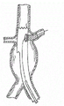
Fig. 4 illustrates the axonometric chart of an embodiment who is in expanded position, and wherein, the hole instrument of windowing launches by the far-end of the 3rd slender member;
Fig. 5 A illustrates the cutaway view of embodiment of lumen of vessels internal object and the side view of lumen of vessels internal object, and wherein inflatable distal anchors is positioned in the branch vessel;
Fig. 5 B illustrates the cutaway view of embodiment of lumen of vessels internal object and the side view of lumen of vessels internal object, and wherein the helical form distal anchors is positioned in the branch vessel; And
Fig. 6 A-6M illustrates the step of the embodiment that the support of main blood vessel and branch vessel inserts.
The detailed description of illustrative embodiments
It will be appreciated by those of ordinary skill in the art that each side of the present invention can realize by any amount of method and the equipment that is configured to the implement plan function.In other words, other method and apparatus can be comprised in here the function with implement plan.The accompanying drawing not to scale (NTS) that be also pointed out that reference is here drawn, but can enlarge each side of the present invention is described, and about this, each view should not be considered to restriction.At last, though can describe the present invention in conjunction with various principles and theory, the present invention should not be limited by theory.
Term " nearside " and " distally " are when being meant when using more close here about device or device feature and further from the operator of device.Because the invention is not restricted to the periphery or the center is close, when use the term nearside or the distally the time, this device should straitly do not explained, because device characteristic can change slightly with respect to anatomical features with respect to its setting position.
The present invention relates to be used for radially or crooked direction, such as towards or the sidewall guiding of passing blood vessel or holder device such as the tube chamber of Centesis instrument or angioscope in device, system and the method for instrument, as using slender member and specific conduit this defined term.The present invention relates to be used in the main blood vessel of transplanting location branch vessel and instrument in the tube chamber is guided to device, the system and method at branch vessel-main blood vessel junction surface.Illustrative embodiment can be used for piercing through graft or stent graft with the formation hole of windowing.Illustrative embodiment can be used for treating aneurysm, interlayer (dissection) and other disease that is positioned at aortic arch.Dissect in other situation of conduit in influence, illustrative embodiment can be used in combination with the treatment of coronary artery disease, peripheral vascular disease, portal hypertension, carotid disease, renal vascular hypertension.Illustrative embodiment also can be used for medicine or other implantable device are delivered to specific therapentic part.
As used herein, " blood vessel " can be tremulous pulse, vein, blood capillary etc., or any other dissection path that exists in health volunteer's body, as stomach or intestinal, conduit or cavity.This this paper is employed, and " connection " meaning is no matter be directly or indirectly, no matter be for good and all or temporarily engage, connection, attached two or more elements.As used herein, " holder device " can be called graft, support, stent graft, any other device in the hole that maybe may require to window.
According to an embodiment, guiding device can pass blood vessel and be expanded to therapentic part.Guiding device is expanded to therapentic part with instrument in the tube chamber, and leads or guiding tool along the direction of the longitudinal axis that is substantially perpendicular to or leaves holder device.Among another embodiment, guiding device can be detected in health by using the vitro detection device, is convenient to the basic accurately location of guiding device thus.
Guiding device can comprise expandable members that is configured to radial dilatation and the guiding tube that may be operably coupled to expandable members, in order to along crooked, roughly radially or roughly orthogonal direction guiding or guiding tube intracavity instrument.As used herein, the crooked description roughly and delivery path direction at angle.Among one embodiment, expandable members is outwardly-bent or extend, and opens in response to compression stress thus, and this compression stress can apply with respect to the vertical shift of another slender member via a slender member.In other embodiments, expandable members can be convenient to expandable members is opened.The far-end of guiding tube or other parts can comprise radiopaque material, so that substantially accurately locate in vivo.
Using method can comprise that making guiding device pass blood vessel is expanded to therapentic part, and also can comprise and expandable members is stretched out or expand and instrument (for example, being used for puncture, intubate intubate etc.) that will be subsequently guides to selected target.Guiding device can be located by using detector.Instrument can be expanded to selected therapentic part by guiding device in the tube chamber.
Others of the present invention relate to the tube chamber internal object, these tube chamber internal objects are configured to anchor in the branch vessel, and be convenient to large artery trunks graft, support, or the hole of windowing of stent graft, the perfusion of successive large artery trunks downstream in the entire bracket expansion and the hole process of windowing, kept simultaneously.
Relate to the retrograde intubation system on the other hand, (stent supports) inserted especially for the support near arteriorenal ventral aorta by this system and arteriorenal support is inserted (support).The embodiment of this system comprises Centesis instrument and snaring device, and sting device enters and pierce through stent graft from the renal artery side, and snaring device enters from the abdomen arterial side.Snaring device is caught Centesis instrument and pulling instrument and its appended guide line and is passed abdomen arterial inlet point.Holder device launches to support renal artery along this guide line.
In addition, it should be appreciated by those skilled in the art that embodiment as herein described can be mainly used in the lumen of vessels internal therapy, thereby with instrument in long-range rotation of certain precision or the oblique lumens, and then with the angle hole of windowing on the spot, to pierce through the sidewall of robust graft material on the spot, keeping the perfusion of branch vessel, and cross over or form one or more pipelines by multiple anatomical features.
According to an embodiment, and with reference to Figure 1A, 1B, 2A and 2B, guiding device 200 can structurally or be configured on the material instrument in the tube chamber is launched or be implanted to therapentic part, and along basically radially, quadrature or the direction 251 that otherwise departs from its bang path 250 axis are come guiding tool basically." tube chamber in instrument " comprises any instrument that can be used in operation in the tube chamber, such as instrument and combination thereof in Centesis instrument as herein described, puncture conduit, deflector equipment, double lumen deflector equipment, angioscope, slender member, guiding device as described herein, tube chamber internal object as described herein, support, stent graft, medicine means of delivery, other tube chamber.Authorize being entitled as of people such as Cully the U.S. Provisional Patent Application series number 13/273 of " System and Method of Percutaneous Occlusion Crossing(percutaneous obturation across system and method) ", 111 have more completely described exemplary " puncture conduit ", " deflector equipment ", " double lumen deflector equipment " and combination thereof, and the whole content of this patent is to include this paper in referring to mode.
According to an embodiment, guiding device 200 comprises expandable members 210 and guiding tube 240, this guiding tube operationally is attached to expandable members 210 and has distal end 241, guiding tube 240 and be in the primary importance of expandable members 210 almost parallels of collapsed position and crooked movable between the second position of this primary importance.
But expandable members 210 comprises the radial dilatation that can move or the structure that stretches out between collapsed position and expanded position.Expandable members 210 can structurally or be configured on the material expand into the guiding tube 240 that operationally is attached to it is radially extended along crooked or roughly orthogonal direction.Expandable members 210 can also structurally or be configured such that on the material in expandable members 210 and to keep at least some continuous bloods to flow in its expanded position and/or carrying out the transition in the expanded position process, for example only flow with small interruption.Expandable members 210 can structurally or be configured on the material provide extra supporting in treatment site for blood vessel, for example by expanding in the mode that contacts with blood vessel wall.In addition, expandable members 210 can structurally or be configured to the snare slender member on the material.About this, Fig. 2 A and 2B illustrate the embodiment with the expandable members 210 that is in expanded position, and Figure 1A and 1B illustrate the embodiment with the expandable members 210 that is in collapsed position.
According to an embodiment, guiding device can pass blood vessel and be expanded to therapentic part.Guiding device is expanded to therapentic part with instrument in the tube chamber, and along being substantially perpendicular to or from the direction guiding tool of axle in the longitudinal axis of holder device.Among another embodiment, guiding device can be detected in vivo by using the vitro detection device, is convenient to the basic accurately location of guiding device thus.
Guiding device can comprise expandable members that is configured to radial dilatation and the guiding tube that operationally is attached to this expandable members, in order to along crooked, orthogonal direction guiding tube intracavity instrument radially or basically basically.As used herein, the crooked description roughly and delivery path direction at angle.Among one embodiment, expandable members is outwardly-bent or extend, and opens in response to compression stress thus, and this compression stress can apply with respect to the vertical shift of another slender member via a slender member.Among other embodiment, expandable members can be convenient to make the expandable members expansion.The far-end of guiding tube or other parts can comprise radiopaque material so that substantially accurately be positioned in the health.
The method of using can comprise makes guiding device be expanded to therapentic part by blood vessel, and also can comprise and expandable members is stretched out or expand, and general's instrument (for example, puncture, intubate etc.) subsequently guides to selected target.Guiding device can be located by using detector.Instrument can be expanded to selected therapentic part by guiding device in the tube chamber.
Others of the present invention relate to the tube chamber internal object, these tube chamber internal objects are configured to anchoring and go into branch vessel, and be convenient to large artery trunks graft, support, or the hole of windowing of stent graft, the perfusion of successive large artery trunks downstream in the entire bracket expansion and the hole process of windowing, kept simultaneously.
Relate to the retrograde intubation system on the other hand, this system is inserted especially for inserting with arteriorenal support near the support of arteriorenal ventral aorta.The embodiment of this system comprises Centesis instrument and snaring device, and sting device enters and pierce through stent graft from the renal artery side, and snaring device enters from the abdomen arterial side.Snaring device is caught Centesis instrument, and the pulling instrument passes abdomen arterial inlet point together with its appended guide line.Holder device launches along this guide line, to support renal artery.
For example, expandable members 210 can be actuated into expanded position slidably.This expansion can be in response to the corresponding length travel of second slender member 230 with respect to first slender member 220, and it applies compression stress or tension force to expandable members 210.So, expandable members 210 can be connected to first slender member 220 at the first end place, and second opposed end is connected to second slender member 230, thereby expandable members 210 is in response to second slender member 230 radial deformation with respect to the selectivity axial displacement of first slender member 220 and basically.Can be by expandable members 210 being collapsed with the required opposite selectivity axial displacement of expansion.
In this embodiment, first slender member 220 can comprise first cavity, and second slender member 230 can comprise second cavity and side opening 231.Second slender member 230 can be arranged along first cavity, and side opening 231 can be adjacent with expandable members 210.In one embodiment, guiding tube 240 holds along second cavity of second slender member 230 slidably, and guiding tube 240 can pass side opening 231 to be connected with expandable members 210.For example, Fig. 3 illustrates the cutaway view of the guiding tube of arranging along the cavity of second slender member 230 340.In alternate embodiment, second slender member does not comprise side opening.In such an embodiment, guiding tube is slidably received within the first slender member cavity, and with second slender member and put.
As used herein, " slender member " has near-end and far-end and can pass blood vessel.Usually, slender member is flexible, especially when the needs slender member passes through zigzag vascular system.Example comprises guide line, conduit, optical fiber etc.Slender member can comprise cavity on the whole distance or on its part, perhaps can be fully solid.For example, distal end (only listing) blunt, circle or convergent that slender member can comprise, and feature is to change rigidity or pliability, this rigidity or pliability can further change along the length of slender member.Slender member can have any shape of cross section, comprises circle, ellipse, triangle, square, polygon or arbitrary shape.Slender member or its part can be hydrophilic or hydrophobic.As used herein, " conduit " and/or " pipe " is the slender member that comprises the cavity from proximal extension to far-end.
The typical material that is used to construct slender member can comprise the well known materials such as general amorphous (amorphous) thermoplastic, comprise polymethyl methacrylate (PMMA or acrylic compounds), the poly terephthalic acid ethylene glycol (PETG) of polystyrene (PS), acronitrile-butadiene-styrene (ABS), polrvinyl chloride (PVC), modification, cellulose acetate butyrate (CAB) (CAB); General hemihedral crystal plastics (Semi-Crystalline Commodity Plastics) comprise polyethylene (PE), high density polyethylene (HDPE) (HDPE), low density polyethylene (LDPE) (LDPE or LLDPE), polypropylene (PP), polymethylpentene (PMP); Amorphous engineering thermoplasties, the phenylene oxide (PPO of modification), polyphenylene oxide (Polyphenelyne Ether) that comprises Merlon (PC), polyphenylene oxide (PPO), modification (PPE), the polyphenylene oxide (PPE of modification) of modification, thermoplastic polyurethane (TPU); Hemihedral crystal engineering thermoplasties (Semi-Crystalline Engineering Thermoplastics), comprise polyamide (PA or nylon), polyformaldehyde (POM or acetal), polyethylene terephthalate (PET, thermoplastic polyester), polybutylene terephthalate (PBT) (PBT, thermoplastic polyester), ultra-high molecular weight polyethylene (UHMW-PE); High-performance thermoplastics comprises polyimides (PI, imidizate plastics), polyamidoimide (PAI, imidizate plastics), polybenzimidazoles (PBI, imidizate plastics); Amorphous high-performance thermoplastics comprises polysulfones (PSU), Polyetherimide (PEI), polyether sulfone (PES), polyarylsufone (PAS); The semi-crystal high-performance thermoplastics comprises polyphenylene sulfide (PPS), polyether-ether-ketone (PEEK); With the semi-crystal high-performance thermoplastics, fluoropolymer polymer comprises ethylene fluoride-propylene (FEP), ethylene-chlorotrifluoroethylene (ECTFE), ethylene, ethylene-tetrafluoroethylene (ETFE), polychlorotrifluoroethylene (PCTFE), polytetrafluoroethylene (PTFE), polyvinylidene fluoride (PVDF), perfluoroalkoxy (PFA).Other known medical grade material comprises the elastomer organosilicon polymer, and polyether block amide or thermoplasticity copolyether (PEBAX) and metal are as rustless steel and nickel/titanium alloys.
Go back with reference to Figure 1A, 1B, 2A and 2B, another opens mechanism and can comprise such as the inflatable actuating via expandable members.For example, guiding device 200 can comprise the utricule that is positioned at expandable members 210, and this utricule makes expandable members 210 extend when outwards expanding.Expandable members can shrink, then expandable members 210 change back to its collapsed position when withdrawing from by expandable members 210 restrictions.
Perhaps, expandable members 210 can be made at self expandable when discharging such as the limiting mechanism of sheath.Among this embodiment, expandable members 210 can be made by marmem or polymer, and such as rustless steel (SST), Nitinol, polyurethane etc., thereby it is configured to passive extension fixture.Guiding device 200 can comprise is convenient to any structure or the material that expandable members 210 is moved between collapsed position and its expanded position that expands outwardly.
In one embodiment, expandable members 210 can comprise the bendable member that can expand outwardly.In addition, expandable members 210 can comprise the structure of being interrupted or opening when being in expanded position.The structure of being interrupted or opening allows fluid to flow through expandable members 210, and the only small interruption of fluid.Keep the accuracy that the downstream perfusion also relates to intubate, because guiding device 200 does not need to bear or resist the pressure that is associated with temporary transient obturation.For example, thereby expandable members 210 can comprise the spatial bendable that forms separated from one another between it, this is because these elements stretch out, such as chrysanthemum type structure (malecot structure), reticular component with diagonally opposing corner or vertical slit, or flexible pipe.Similarly, expandable members 210 can comprise at least one lath 211 that is connected to first slender member 220 at one end and is connected to second slender member 230 at other end place.Expandable members 210 can be integral with first slender member 220, second slender member 230, maybe can be the independent structure that is connected to first and second slender members.
The expansion of expandable members 210 can be that the part of circumference radial symmetric or that only center on expandable members 210 takes place, thereby produces radially asymmetric.In addition, expandable members 210 stretches out or the distance expanded can be around the perimeter variations of expandable members 210 or homogeneous from longitudinal axis.Can provide this variation of asymmetric profile can obtain various benefits to expandable members 210, such as wideer radius of turn or bigger flexibility by in instrument in the guiding tube 240 unfolded tube chambers all types of.For example, in one embodiment, expandable members 210 can comprise the different materials with different-stiffness, thereby a side of expandable members 210 stretches out more than opposition side.
The width or the diameter that are in the expandable members 210 of expanded position can be regulated in a series of width or diameter range.For example, the expandable members 210 that is in expanded position can be included in down width or the diameter range in the train value: about 1mm is to about 65mm, about 2mm about 45mm or about 3mm about 30mm extremely extremely.So the guiding device 200 of certain structure can be suitable for various blood vessel diameters.Similarly, guiding device 200 can be scalable, and is used for various application thus, such as the operation of peripheral vessels structure.
Among the various embodiment, expandable members 210 can be configured to make the blood vessel diameter measuring device.For example, guiding device 200 can comprise calibrated ruler on the near-end of second slender member 230, so that degrees of expansion is associated with axial location on second slender member 230.The words of knowing blood vessel diameter can be assisted and be determined the support that will use or the suitable diameter dimension of stent graft.In addition, when measuring blood vessel diameter, apply interior expansion power further assisted Selection support or stent graft.In measuring process, the power that applies by device should be associated by the power that support or stent graft apply in same position.
In one embodiment, guiding tube 240 comprise be configured to instrument in the tube chamber radially, basically quadrature or or axis otherwise crooked or that depart from delivery path 250 guide to any structure at a position.For example, guiding tube 240 can comprise slender member, and this slender member has tube chamber and passes its far-end and near-end.Perhaps, guiding tube can comprise semicanal, groove, or some other guide rails or track columnar structure, with along crooked direction guiding tube intracavity instrument.In addition, guiding tube 240 can along orthogonal direction basically or on whole expanded position diameter or width range the substantially constant angle come guiding tube intracavity instrument, described in the U.S. Patent Application Serial 13/273,111 of authorizing people such as Cully like that.
In the exemplary embodiment, the distal end 241 of guiding tube 240 or its part comprise the material of radiopaque, echogenic or magnetic or any other material that can vitro detection.This material can be terminal 241 integral part, coating, or is connected to its separate marking.
Among the various embodiment, guiding device 200 can be configured to drug delivery device.For example, medicine can stay in expandable members 210, thereby when expandable members 210 was in its collapsed position, this medicine was limited basically.In case expandable members 210 reaches therapentic part, expandable members 210 expansible with drug release in blood vessel structure.In other embodiments, medicine can be injected into guiding tube 240, or instrument can comprise the medicine means of delivery in the tube chamber, and this medicine means of delivery can be sent towards the desired therapeutic position and be passed guiding tube 240.This instrument can be configured to pierce through block, and this medicine directly is delivered in this block.
In one embodiment, use method such as guiding device 200 as herein described to comprise to be expanded to guiding device 200 in the blood vessel and make expandable members 210 extend outwardly into expanded position and make distal end 241 move to the step of its second position thus.Can carry out this expansion and the transvascular fluid of interrupt flow stream not basically.The distal end 241 that is in the second position can be extended by the therapentic part on blood vessel wall, thus instrument in the tube chamber is passed guiding tube 240 guiding to therapentic part.
In one embodiment, by expandable members 210 is applied compression stress,, make distal end 241 radially or stretch out thus such as with respect to the vertical shift of first slender member 220 expandable members 210 being stretched out by second slender member 230.Perhaps, by expandable members is expanded expandable members 210 is stretched out.
Another embodiment comprises optionally external, shows the auxiliary position guiding device 200 down of the detector of position in the body of distal end 241 or expandable members 210.Then, can advance such as instrument in the tube chamber of describing in the U.S. Patent Application Serial 13/273,111 of authorizing people such as Cully and pass the far-end of guiding tube 240.For example, Fig. 4 illustrates instrument 450 in the tube chamber that passes guiding tube 440 that advances.Another embodiment relates to instrument 450 in the tube chamber and pierces through selected position.Another embodiment relate to contrast agent inject instrument in guiding tube 440 or the tube chamber, with windowed hole and/or be communicated with main vascular fluid of checking side branch by intubate or checking side branch.
With reference to figure 5A and 5B, various embodiments of the present invention comprise lumen of vessels internal object 510A, B.Lumen of vessels internal object 510A, B are the temporary transient or permanent grafts as the target of instrument in the tube chamber.Lumen of vessels internal object 510A, B are convenient to fluid by branch vessel 501 perfusions.The embodiment of lumen of vessels internal object 510A, B comprises slender member 513A, B, have the tube chamber in the remote area of slender member 513A, B at least and in the wall of slender member 513A, B, lead at least one opening 512A, B of tube chamber, and distal anchors 511A, B.
In the exemplary embodiment, distal anchors 511A, B are configured to be positioned at regularly in the branch vessel 501.Distal anchors 511A, B can be integral or be attached to its independent structure with slender member 513A, B.Distal anchors 511A, B can comprise helical structure (for example, shown in Fig. 5 B), ring, perforated disc, expandable members (for example, shown in Fig. 5 A) or be configured to be positioned at regularly any structure in the branch vessel 501.But distal anchors 511A, B can comprise test material, such as magnetic, radiopaque, echogenic or fluorescent material or any other material that can vitro detection, such as use electromagnetic radiation, fluoroscopy, angioscope, or alternate manner.In the situation of expandable members, inflating medium can be radiopaque.In the exemplary embodiment, when distal anchors 511A, B locate regularly, the leap of distal anchors 511A, B its diameter of wide part from about 1mm to 15mm, from about 4mm to about 7mm or from about 3mm about 6mm extremely.Distal anchors 511A, B can be made of any amount of material, comprise silicon, latex, polyurethane, polrvinyl chloride, polysiloxanes, Merlon, polyethylene, nylon, PTFE(for example, ePTFE), Nitinol or any biocompatible materials make, and comprises above-mentioned combination.Among another embodiment, distal anchors 511A, B comprise heparin coating or other anticoagulant coating.
Among each embodiment, opening 512A, B in the wall of slender member 513A, B can have any amount, position or structure.Opening 512A, B are convenient to the perfusion of side branch vessel.In the exemplary embodiment, opening 512A, B become to make when distal anchors 511A, B are positioned in the branch vessel 501 regularly along the Position Design of slender member 513A, B, and opening 512A, B be positioned at main blood vessel 500, outside the scope of main blood vessel 500 unfolded supports.This allows to enter tube chamber and be convenient to the side branch vessel structure and the perfusion of downstream tissue (for example kidney) such as the fluid of blood.Opening 512A, B can be configured such that blood is easily along passing opening 512A, B from the retrograde direction of main blood vessel and entering the tube chamber of slender member 513A, B, if necessary.Can adopt fin to be convenient to way flow and cross opening 512A, B.
In other embodiments, the wall of slender member 513A, B is not destroyed.In other words, be not used in dabbling opening.Cavity may extend to the hub of slender member 513A, B, and here, the clinician can utilize this its blood vessel structure of pouring into side branch and being associated with it by injecting with syringe such as self-blood or other fluid.Among this embodiment, the clinician can be according to circumstances sidepiece by skiving slender member 513A, the B opening 512A, the B that are formed for reverse perfusion retrofit.
Another embodiment comprises as described herein tube chamber internal object 510A, B are deployed in the branch vessel 501 via main blood vessel 500, and distal anchors 511A, B are positioned at step in the branch vessel 501 regularly.
In one embodiment, guidance system can comprise that lumen of vessels internal object and guiding device as described herein are so that the horizontal and rotational positioning of guiding device.Another embodiment comprises detector, angioscope etc., so that the horizontal and rotational positioning of guiding device.
Another embodiment comprises the lumen of vessels internal object is expanded to area for treatment, and the tube chamber internal object is navigated to step in the branch vessel 501.Other step is included in and launches the main support device in the main blood vessel 500, guiding device as described herein is expanded to transplanted zone, makes the expandable members expansion, and by using detector, angioscope etc. that guiding device is located with respect to lumen of vessels internal object 510A, B.Another embodiment comprises instrument in the tube chamber is launched to pass guiding tube, to pierce through the holder device near distal anchors 511A, B.Other embodiment can comprise the utricule device is deployed into the site of puncture, so that perforation expansion and branch's holder device is launched to support branch vessel 501.Each embodiment of branch's holder device can comprise holder device (this term as herein defined), packing ring, sealing ring, flange, self expandable structure, utricule expanded configuration or aforesaid any combination.
Another embodiment comprises that as described herein being configured to allows angioscope to pass the guiding device of guiding tube, comprises the holder device that is clear to translucent cradle wall, and angioscope.In case holder device launches, then angioscope advances and passes the guiding tube of guiding device.Guiding device is convenient to located lateral and rotational positioning.Angioscope allows from verifying that visually the branch vessel mouth passes this and is clear to translucent stent graft wall, thereby window hole instrument or guiding puncture pipe can launch by guiding tube, with at place, branch vessel junction surface to the stent graft hole of windowing.Another embodiment can comprise the utricule device is deployed into the support that is punctured, so that perforation expansion and branch's holder device is launched to support branch vessel.
According to another exemplary embodiment and with reference to figure 6H, the retrograde intubation system comprises snaring device 610, the guiding puncture device 620 that contains expandable members 615 as herein described, and main support device 630.Main support device 630 comprises support or stent graft, and the both can be that utricule is expanded or self expandable.The embodiment of holder device comprises graft material or the fiber that is configured to prevent in puncture or splits or tear when expanding.In certain embodiments, expandable members 615 can make 630 expansions of main support device.In case support is in place, cradle wall by Centesis instrument 621 from the outside of support towards inner conform to branch vessel the quilt hole of being windowed.Guiding puncture pipe 622 supporting Centesis instruments 621 also guide to site of puncture on the support shaft with it.
Guiding puncture device 620 comprises guiding puncture pipe 622 and can be by the Centesis instrument 621 of guiding puncture pipe 622 supportings, and guiding puncture pipe 622 has at least one cavity 623, and can pour into the distal outer that anchoring piece 624 is positioned at conduit 622.In the exemplary embodiment, guiding puncture pipe 622 offers Centesis instrument 621 to pierce through main support device 630 via cavity 623 with desired fracture strength.The guiding puncture conduit can comprise second cavity of permission hemoperfusion to kidney.Blood can be injected into second cavity, if necessary.Second cavity can be coated with heparin.
According to exemplary embodiment, Centesis instrument 621 comprises slender member and is positioned at the convergent of far-end or sharp-pointed tip.In other embodiments, Centesis instrument 621 can comprise the puncture conduit.In other embodiments, Centesis instrument 621 by such as radio frequency can etc. electromagnetic radiation or mechanical energy energy supply.
The embodiment that can pour into anchoring piece 624 can comprise at least one expandable members of the outside, distally that is connected to conduit 622.Expandable members has enough sizes so that conduit 622 is stabilized in the branch vessel.Other embodiment that can pour into anchoring piece 624 comprises at least one ring, and this at least one ring is connected to the outside, distally of conduit 622, thereby fluid passes ring, and should the auxiliary anchoring guiding puncture conduit 622 of ring.Anchoring piece 624 can be poured into and the geometry of the ring formation of any amount of the outside, distally that is connected to conduit 622 or kind can be comprised.Can pour into anchoring piece 624 and can comprise expansible, pourable mechanic anchor,, replace expandable members such as expansible fabric.Other embodiment that can pour into anchoring piece 624 can comprise perforated disc as described herein or lumen of vessels internal object.
According to an embodiment, snaring device 610 comprises expandable members as described herein 615.In one embodiment, first slender member 612 and second slender member 614 are connected to another the relative end in the expandable members 615, thereby second slender member makes expandable members 615 expansions with respect to the length travel of first slender member 612 and collapses.Other embodiment of snaring device 610 comprises guiding device as described herein.The use of expandable members 615 also allows to support or supporting bracket graft internal diameter in order to prepare to window the hole on the spot.When the firm stent graft material of puncture, the supporting of this increase is helpful.The embodiment of snaring device 610 can be configured to guide line or Centesis instruments such as catching, catch on, snarl.
With reference to figure 6A-6M, in order to illustrate, another embodiment comprises step: pass the waist inlet near side branch (Fig. 6 A-6B) with pin and/or waist rod; With guide line advance pass pin and by branch vessel (for example renal artery) near main blood vessel (for example large artery trunks) (Fig. 6 C); Remove pin and/or waist rod; Make guiding puncture device above guide line, advance (Fig. 6 D); With guiding puncture device as described herein be positioned in the branch vessel, near with the place, junction surface (Fig. 6 D) of main blood vessel; Withdraw from guide line (Fig. 6 E) from main blood vessel; The main support device advanced pass main blood vessel, near junction surface place (Fig. 6 E); Launch main support device (Fig. 6 F); The snaring device as described herein that advances, thus expandable members is near junction surface (Fig. 6 G); Make expandable members expansion (Fig. 6 G); Centesis instrument advanced pass the guiding puncture pipe, to pierce through main support and to enter expandable members (Fig. 6 H); Expandable members is collapsed, and withdraw from expandable members (Fig. 6 I) by dragging Centesis instrument.Because these steps, slender member penetrates branch vessel to main blood vessel from secondary access point, and comes out by main access point.
Another embodiment comprises step: make balloon catheter advance to (Fig. 6 J) in the site of puncture; Expanded in the position of puncture, further to open this puncture (Fig. 6 J); Extract slender member out from secondary access point and enter branch vessel (Fig. 6 K); Extract balloon catheter out; The assistant support device is advanced by the position (Fig. 6 L) of puncture; And the holder device that expands branch (Fig. 6 M).Each embodiment of branch's holder device can comprise packing ring, sealing ring, flange, self expandable structure, utricule expanded configuration or aforesaid any combination.
Set forth a plurality of feature and advantage in the description of front, comprised the details of the 26S Proteasome Structure and Function of various alternatives and device and/or method.The present invention only represents exemplary and is not expressed as exclusive equally.For example, though main reference large artery trunks should be used for describing the present invention, about having, can use each exemplary embodiment from other blood vessel of the branch vessel of its extension.For a person skilled in the art obviously can be in concept of the present invention in the indicated maximum magnitude of the wide in range upper implication of the expressed term of appended claims, carry out various remodeling, especially in the layout of structure, material, element, parts, shape, size and parts.Do not depart from the degree of appended claims spirit and scope at these various remodeling, they also mean and are contained in this.
Claims (26)
1. one kind can be passed through the unfolded guiding device of blood vessel, and described guiding device comprises:
Expandable members, described expandable members can be moved between collapsed position and outward extending expanded position; And
Guiding tube, described guiding tube operationally is attached to described expandable members and has distal end, described distal end can and be in the substantially parallel primary importance of the described expandable members of described collapsed position and crookedly between the second position of described primary importance, move.
2. guiding device as claimed in claim 1 is characterized in that the described second position is substantially normal to described primary importance.
3. guiding device as claimed in claim 1 is characterized in that, described expandable members activates in response to the selectivity of described expandable members between described collapsed position and expanded position and moves.
4. guiding device as claimed in claim 1 is characterized in that described expandable members comprises the hole, and the distal end of described guiding tube is attached to described expandable members with described hole with overlapping.
5. guiding device as claimed in claim 1 also comprises:
First slender member, described first slender member comprises first cavity; And
Second slender member, described second slender member comprises second cavity and side opening,
Wherein, described guiding tube is arranged along described second cavity, and described second slender member is arranged along described first cavity, and
Wherein, described expandable members is at first end that is connected to described first slender member and be connected between the second opposed end of described second slender member and extend, thereby, described expandable members in response to described second slender member with respect to the length travel of described first slender member and stretch out.
6. guiding device as claimed in claim 5, it is characterized in that described distal end can be moved between the described second position in described primary importance when described expandable members is expanded with respect to the corresponding vertical shift of described first slender member in response to described second slender member.
7. guiding device as claimed in claim 1 is characterized in that described expandable members has discontinuous construction, pours into by it to allow fluid when being in expanded position.
8. guiding device as claimed in claim 7 is characterized in that described expandable members has discontinuous construction, to allow fluid to pour into by it when moving between described expanded position and collapsed position.
9. guiding device as claimed in claim 7 is characterized in that, described expandable members is expanded in the mode of radial symmetric basically.
10. guiding device as claimed in claim 7 is characterized in that, described expandable members comprises chrysanthemum type structure, reticular component, has diagonal angle or the pipe of vertical slit and at least one at least one lath.
11. guiding device as claimed in claim 1 is characterized in that, described expandable members comprises Nitinol.
12. guiding device as claimed in claim 1 also comprises: expandable members, described expandable members make described expandable members expansion when expanding.
13. guiding device as claimed in claim 12 is characterized in that, when moving to described collapsed position, described expandable members is configured to after described expandable members shrinks described expandable members restriction.
14. guiding device as claimed in claim 1 is characterized in that, the distal end of described guiding tube is along the direction guiding tube intracavity instrument that is substantially normal to delivery path.
15. guiding device as claimed in claim 1 is characterized in that, described expandable members is adjustable in the expanded position diameter range.
16. guiding device as claimed in claim 15 is characterized in that, the distal end of described guiding tube edge in whole expanded position diameter range is substantially perpendicular to the direction guiding device of delivery path.
17. guiding device as claimed in claim 1 is characterized in that, described distal end comprises radiopaque material.
18. a method that is used for guiding tube intracavity instrument in blood vessel comprises:
Guiding device is deployed in the main blood vessel, and wherein, described guiding device comprises:
Expandable members, described expandable members can be moved between collapsed position and outward extending expanded position; And
Guiding tube, described guiding tube operationally is attached to described expandable members and has distal end, described distal end can and be in the substantially parallel primary importance of the described expandable members of described collapsed position and be substantially normal between the second position of described primary importance and move.
Described expandable members is stretched out, and make described guiding tube move to the described second position thus, and only small interruption fluid flows through blood vessel.
19. method as claimed in claim 18 is characterized in that, in the described second position, the therapentic part of the described distal end of described guiding tube on main blood vessel wall extends, and will guide described therapentic part into by instrument in the tube chamber of described guiding tube thus.
20. method as claimed in claim 18 is characterized in that, described expandable members stretches out by expandable members is expanded.
21. method as claimed in claim 18 is characterized in that, by described expandable members being applied compression stress described expandable members is stretched out.
22. method as claimed in claim 18 also comprises: assisting of the vitro detection device of position optionally located described guiding device down in the body of radiopaque distal end is shown.
23. method as claimed in claim 18 also comprises: instrument in the tube chamber is advanced by the far-end of described guiding tube.
24. method as claimed in claim 23 also comprises: pierce through selected position with instrument in the described tube chamber.
25. method as claimed in claim 24 also comprises: injection of contrast medium is communicated with described main vascular fluid with checking side branch.
26. method as claimed in claim 23 is characterized in that, instrument is to comprise the convergent that can extend through described guiding tube or the slender member of sharp distal in the described tube chamber.
Priority Applications (1)
| Application Number | Priority Date | Filing Date | Title |
|---|---|---|---|
| CN201710542827.9A CN107432778B (en) | 2010-11-16 | 2011-11-15 | Open a window aperture apparatus, system and method |
Applications Claiming Priority (5)
| Application Number | Priority Date | Filing Date | Title |
|---|---|---|---|
| US41415510P | 2010-11-16 | 2010-11-16 | |
| US61/414,155 | 2010-11-16 | ||
| US13/295,817 US9545323B2 (en) | 2010-11-16 | 2011-11-14 | Fenestration devices, systems, and methods |
| US13/295,817 | 2011-11-14 | ||
| PCT/US2011/060699 WO2012068048A1 (en) | 2010-11-16 | 2011-11-15 | Fenestration devices, systems, and methods |
Related Child Applications (1)
| Application Number | Title | Priority Date | Filing Date |
|---|---|---|---|
| CN201710542827.9A Division CN107432778B (en) | 2010-11-16 | 2011-11-15 | Open a window aperture apparatus, system and method |
Publications (1)
| Publication Number | Publication Date |
|---|---|
| CN103228230A true CN103228230A (en) | 2013-07-31 |
Family
ID=45044737
Family Applications (2)
| Application Number | Title | Priority Date | Filing Date |
|---|---|---|---|
| CN2011800548237A Pending CN103228230A (en) | 2010-11-16 | 2011-11-15 | Fenestration devices, systems, and methods |
| CN201710542827.9A Active CN107432778B (en) | 2010-11-16 | 2011-11-15 | Open a window aperture apparatus, system and method |
Family Applications After (1)
| Application Number | Title | Priority Date | Filing Date |
|---|---|---|---|
| CN201710542827.9A Active CN107432778B (en) | 2010-11-16 | 2011-11-15 | Open a window aperture apparatus, system and method |
Country Status (10)
| Country | Link |
|---|---|
| US (2) | US9545323B2 (en) |
| EP (2) | EP2640452B1 (en) |
| JP (2) | JP6059149B2 (en) |
| CN (2) | CN103228230A (en) |
| AU (1) | AU2011329143B2 (en) |
| BR (1) | BR112013012055A2 (en) |
| CA (1) | CA2817029C (en) |
| ES (2) | ES2899239T3 (en) |
| RU (1) | RU2013127325A (en) |
| WO (1) | WO2012068048A1 (en) |
Cited By (11)
| Publication number | Priority date | Publication date | Assignee | Title |
|---|---|---|---|---|
| CN104770361A (en) * | 2015-03-11 | 2015-07-15 | 浙江大学 | Supplied-liver quickly-fixed perfusion device for liver transplant |
| CN105615999A (en) * | 2016-03-03 | 2016-06-01 | 福建省立医院 | Reversible laser balloon catheter and method for applying same |
| CN105902326A (en) * | 2016-06-29 | 2016-08-31 | 黄连军 | Windowing method and device of covered stent |
| CN108652786A (en) * | 2017-03-31 | 2018-10-16 | 上海交通大学医学院附属新华医院 | Holder windowing facility and stent system |
| CN109561958A (en) * | 2016-05-26 | 2019-04-02 | 瑞士资本工程公司 | Vascular medical device and system |
| CN109982652A (en) * | 2016-09-25 | 2019-07-05 | Tva医疗公司 | Blood vessel stent device and method |
| CN110124184A (en) * | 2019-06-14 | 2019-08-16 | 深圳市慧极创新医疗科技有限公司 | A kind of encephalic drainage of cerebrospinal fluid pipe |
| CN110141761A (en) * | 2019-06-14 | 2019-08-20 | 深圳市慧极创新医疗科技有限公司 | A kind of extradural hemorrhage remove device |
| CN110248585A (en) * | 2016-10-07 | 2019-09-17 | 博迪维仁医疗有限公司 | Device and its application method used in intervention and surgical operation |
| CN110831527A (en) * | 2017-06-19 | 2020-02-21 | W.L.戈尔及同仁股份有限公司 | Fenestration devices, systems, and methods |
| CN112790188A (en) * | 2019-10-28 | 2021-05-14 | 西安交通大学医学院第一附属医院 | Rapid perfusion device for liver supply in liver transplantation |
Families Citing this family (40)
| Publication number | Priority date | Publication date | Assignee | Title |
|---|---|---|---|---|
| US8100822B2 (en) | 2004-03-16 | 2012-01-24 | Macroplata Systems, Llc | Anoscope for treating hemorrhoids without the trauma of cutting or the use of an endoscope |
| US9358099B2 (en) * | 2009-11-30 | 2016-06-07 | Biflow Medical Ltd. | Method of implanting a stent graft and creating a fenestration therein |
| USRE48850E1 (en) | 2009-12-16 | 2021-12-14 | Boston Scientific Scimed, Inc. | Multi-lumen-catheter retractor system for a minimally-invasive, operative gastrointestinal treatment |
| US10595711B2 (en) | 2009-12-16 | 2020-03-24 | Boston Scientific Scimed, Inc. | System for a minimally-invasive, operative gastrointestinal treatment |
| US10966701B2 (en) | 2009-12-16 | 2021-04-06 | Boston Scientific Scimed, Inc. | Tissue retractor for minimally invasive surgery |
| US10758116B2 (en) | 2009-12-16 | 2020-09-01 | Boston Scientific Scimed, Inc. | System for a minimally-invasive, operative gastrointestinal treatment |
| US9186131B2 (en) | 2009-12-16 | 2015-11-17 | Macroplata, Inc. | Multi-lumen-catheter retractor system for a minimally-invasive, operative gastrointestinal treatment |
| US10531869B2 (en) | 2009-12-16 | 2020-01-14 | Boston Scientific Scimed, Inc. | Tissue retractor for minimally invasive surgery |
| US9565998B2 (en) | 2009-12-16 | 2017-02-14 | Boston Scientific Scimed, Inc. | Multi-lumen-catheter retractor system for a minimally-invasive, operative gastrointestinal treatment |
| EP2512577B1 (en) | 2009-12-16 | 2018-03-07 | Boston Scientific Scimed, Inc. | Arrangements for effecting an endoluminal anatomical structure |
| US8932211B2 (en) | 2012-06-22 | 2015-01-13 | Macroplata, Inc. | Floating, multi-lumen-catheter retractor system for a minimally-invasive, operative gastrointestinal treatment |
| US9545253B2 (en) | 2010-09-24 | 2017-01-17 | Ethicon Endo-Surgery, Llc | Surgical instrument with contained dual helix actuator assembly |
| US20120078244A1 (en) | 2010-09-24 | 2012-03-29 | Worrell Barry C | Control features for articulating surgical device |
| US9545323B2 (en) * | 2010-11-16 | 2017-01-17 | W. L. Gore & Associates, Inc. | Fenestration devices, systems, and methods |
| WO2014098810A1 (en) | 2012-12-18 | 2014-06-26 | Empire Technology Development, Llc | Vascular reinforcement device |
| JP6134008B2 (en) * | 2013-01-18 | 2017-05-24 | エスアールアイ インターナショナルSRI International | Fixed nerve block catheter |
| EP3096817B8 (en) * | 2014-01-22 | 2018-12-26 | Regenexx, LLC | Percutaneous delivery device for tendon-ligament-muscle repair |
| JP6345491B2 (en) * | 2014-06-03 | 2018-06-20 | テルモ株式会社 | Expansion device |
| WO2015186626A1 (en) * | 2014-06-03 | 2015-12-10 | テルモ株式会社 | Expansion device |
| KR20160011530A (en) * | 2014-07-22 | 2016-02-01 | 부산대학교 산학협력단 | Devices and Method of trans-coronary sinus intraseptal pacing in the lead end of the cardiac pacemaker |
| GB2550825B (en) | 2015-02-12 | 2018-10-17 | Foundry Innovation & Res 1 Ltd | Implantable devices and related methods for heart failure monitoring |
| WO2017024051A1 (en) | 2015-08-03 | 2017-02-09 | Foundry Innovation & Research 1, Ltd. | Devices and methods for measurement of vena cava dimensions, pressure, and oxygen saturation |
| CN109475418A (en) | 2016-07-13 | 2019-03-15 | 波士顿科学国际有限公司 | For the instrument and method in the intravascular maintenance smoothness adjacent with nearby performing the operation |
| US11701018B2 (en) | 2016-08-11 | 2023-07-18 | Foundry Innovation & Research 1, Ltd. | Wireless resonant circuit and variable inductance vascular monitoring implants and anchoring structures therefore |
| EP3496606A1 (en) | 2016-08-11 | 2019-06-19 | Foundry Innovation & Research 1, Ltd. | Systems and methods for patient fluid management |
| US11206992B2 (en) | 2016-08-11 | 2021-12-28 | Foundry Innovation & Research 1, Ltd. | Wireless resonant circuit and variable inductance vascular monitoring implants and anchoring structures therefore |
| JP7241405B2 (en) | 2016-11-29 | 2023-03-17 | ファウンドリー イノベーション アンド リサーチ 1,リミテッド | Wireless resonant circuit and variable inductance vascular implant for monitoring vascular and fluid status in patients, and systems and methods utilizing same |
| US11071534B2 (en) | 2016-12-30 | 2021-07-27 | Boston Scientific Scimed, Inc. | System for a minimally-invasive treatment within a body lumen |
| WO2018175125A1 (en) * | 2017-03-18 | 2018-09-27 | Boston Scientific Scimed, Inc. | System for a minimally-invasive treatment within a body lumen |
| WO2018220146A1 (en) | 2017-05-31 | 2018-12-06 | Foundry Innovation & Research 1, Ltd. | Implantable sensors for vascular monitoring |
| WO2018220143A1 (en) | 2017-05-31 | 2018-12-06 | Foundry Innovation And Research 1, Ltd | Implantable ultrasonic vascular sensor |
| US11541248B2 (en) * | 2017-12-11 | 2023-01-03 | Commissariat A L'energie Atomique Et Aux Energies Alternatives | Implantable localised illuminating device with improved architecture |
| JP2019191313A (en) | 2018-04-23 | 2019-10-31 | 株式会社デンソー | Head-up display device |
| CN113473904A (en) | 2018-12-21 | 2021-10-01 | W.L.戈尔及同仁股份有限公司 | Medical system using measurement data from multiple sensors |
| WO2020191203A1 (en) | 2019-03-20 | 2020-09-24 | inQB8 Medical Technologies, LLC | Aortic dissection implant |
| US11832789B2 (en) | 2019-12-13 | 2023-12-05 | Boston Scientific Scimed, Inc. | Devices, systems, and methods for minimally invasive surgery in a body lumen |
| IT202000001336A1 (en) | 2020-01-23 | 2021-07-23 | Univ Pisa | GUIDE DEVICE FOR GUIDING INSTRUMENTS FOR ENDOVASCULAR APPLICATIONS |
| CN111772773B (en) * | 2020-06-23 | 2021-11-30 | 广州启骏生物科技有限公司 | Ablation catheter for pulmonary artery stimulation |
| WO2023034252A2 (en) * | 2021-09-01 | 2023-03-09 | The University Of Toledo | Size adjustable aortic endograft fenestration device and retrievable vessel marker |
| WO2024006230A1 (en) * | 2022-06-27 | 2024-01-04 | Edwards Lifesciences Corporation | Leaflet perforation tools and associated methods |
Citations (10)
| Publication number | Priority date | Publication date | Assignee | Title |
|---|---|---|---|---|
| US5617878A (en) * | 1996-05-31 | 1997-04-08 | Taheri; Syde A. | Stent and method for treatment of aortic occlusive disease |
| US20030023248A1 (en) * | 1998-03-13 | 2003-01-30 | Parodi Juan C. | Systems and methods for applying a suture within a blood vesel lumen |
| US20050209506A1 (en) * | 2001-10-18 | 2005-09-22 | Atropos Limited | Device |
| US20060074475A1 (en) * | 2004-09-28 | 2006-04-06 | Darrel Gumm | Rotating stent delivery system for side branch access and protection and method of using same |
| WO2007082343A1 (en) * | 2006-01-17 | 2007-07-26 | Craig Steven Mclachlan | Method and device for graft fenestration |
| US20070208256A1 (en) * | 2006-03-03 | 2007-09-06 | Medtronic Vascular, Inc. | Multiple Branch Tubular Prosthesis and Methods |
| US20070225750A1 (en) * | 2006-03-10 | 2007-09-27 | Brooke Ren | Embolic protection systems |
| US20090228020A1 (en) * | 2008-03-06 | 2009-09-10 | Hansen Medical, Inc. | In-situ graft fenestration |
| US20090264988A1 (en) * | 2008-04-18 | 2009-10-22 | Medtronic Vascular, Inc. | Stent Graft Delivery System Including Support for Fenestration in Situ and a Mechanism for Modeling |
| WO2010127040A1 (en) * | 2009-04-28 | 2010-11-04 | Endologix, Inc. | Apparatus and method of placement of a graft or graft system |
Family Cites Families (49)
| Publication number | Priority date | Publication date | Assignee | Title |
|---|---|---|---|---|
| US3799172A (en) | 1972-09-25 | 1974-03-26 | R Szpur | Retention catheter |
| WO1994021169A1 (en) | 1993-03-16 | 1994-09-29 | Ep Technologies, Inc. | Flexible interlaced multiple electrode assemblies |
| US5456667A (en) * | 1993-05-20 | 1995-10-10 | Advanced Cardiovascular Systems, Inc. | Temporary stenting catheter with one-piece expandable segment |
| US6009877A (en) | 1994-06-24 | 2000-01-04 | Edwards; Stuart D. | Method for treating a sphincter |
| US5795322A (en) | 1995-04-10 | 1998-08-18 | Cordis Corporation | Catheter with filter and thrombus-discharge device |
| KR100188169B1 (en) * | 1995-08-29 | 1999-06-01 | 박원훈 | Wastewater treatment by catalytic oxidation |
| NL1002423C2 (en) | 1996-02-22 | 1997-08-25 | Cordis Europ | Temporary filter catheter. |
| US5916194A (en) | 1996-05-24 | 1999-06-29 | Sarcos, Inc. | Catheter/guide wire steering apparatus and method |
| US5928260A (en) * | 1997-07-10 | 1999-07-27 | Scimed Life Systems, Inc. | Removable occlusion system for aneurysm neck |
| ATE404123T1 (en) | 1997-11-12 | 2008-08-15 | Genesis Technologies Llc | DEVICE FOR REMOVAL OF OCCLUSIONS IN BIOLOGICAL PASSAGES |
| WO1999042044A1 (en) | 1998-02-19 | 1999-08-26 | Conway-Stuart Medical, Inc. | Electrosurgical sphincter treatment apparatus |
| US6099497A (en) * | 1998-03-05 | 2000-08-08 | Scimed Life Systems, Inc. | Dilatation and stent delivery system for bifurcation lesions |
| US7044134B2 (en) | 1999-11-08 | 2006-05-16 | Ev3 Sunnyvale, Inc | Method of implanting a device in the left atrial appendage |
| US6136010A (en) * | 1999-03-04 | 2000-10-24 | Perclose, Inc. | Articulating suturing device and method |
| US6579261B1 (en) | 1999-06-19 | 2003-06-17 | Adam Spence Corporation | Double lumen-type catheter |
| US6494862B1 (en) * | 1999-07-13 | 2002-12-17 | Advanced Cardiovascular Systems, Inc. | Substance delivery apparatus and a method of delivering a therapeutic substance to an anatomical passageway |
| US6283947B1 (en) * | 1999-07-13 | 2001-09-04 | Advanced Cardiovascular Systems, Inc. | Local drug delivery injection catheter |
| US6231561B1 (en) * | 1999-09-20 | 2001-05-15 | Appriva Medical, Inc. | Method and apparatus for closing a body lumen |
| US6652567B1 (en) | 1999-11-18 | 2003-11-25 | David H. Deaton | Fenestrated endovascular graft |
| US6814752B1 (en) | 2000-03-03 | 2004-11-09 | Endovascular Technologies, Inc. | Modular grafting system and method |
| US20020026217A1 (en) | 2000-04-26 | 2002-02-28 | Steven Baker | Apparatus and method for repair of perigraft flow |
| US20040106972A1 (en) | 2000-11-20 | 2004-06-03 | Deaton David H. | Fenestrated endovascular graft |
| US7060025B2 (en) * | 2002-03-15 | 2006-06-13 | Ethicon Endo-Surgery, Inc. | Method for controlling position of medical instruments |
| US7547304B2 (en) | 2002-12-19 | 2009-06-16 | Gore Enterprise Holdings, Inc. | Guidewire-centering catheter tip |
| EP3045136B1 (en) | 2003-09-12 | 2021-02-24 | Vessix Vascular, Inc. | Selectable eccentric remodeling and/or ablation of atherosclerotic material |
| US20050209673A1 (en) * | 2004-03-04 | 2005-09-22 | Y Med Inc. | Bifurcation stent delivery devices |
| WO2006110830A2 (en) | 2005-04-11 | 2006-10-19 | Cierra, Inc. | Methods and apparatus to achieve a closure of a layered tissue defect |
| US20070043389A1 (en) * | 2005-08-05 | 2007-02-22 | Shintech, Llc | System for treating chronic total occlusion caused by lower extremity arterial disease |
| US20070244547A1 (en) | 2006-04-18 | 2007-10-18 | Medtronic Vascular, Inc., A Delaware Corporation | Device and Method for Controlling the Positioning of a Stent Graft Fenestration |
| US8834494B2 (en) * | 2006-10-20 | 2014-09-16 | St. Jude Medical, Cardiology Division, Inc. | Method and device for automated needle deployment |
| US20080139915A1 (en) | 2006-12-07 | 2008-06-12 | Medtronic Vascular, Inc. | Vascular Position Locating and/or Mapping Apparatus and Methods |
| US20080140180A1 (en) | 2006-12-07 | 2008-06-12 | Medtronic Vascular, Inc. | Vascular Position Locating Apparatus and Method |
| US20080147173A1 (en) | 2006-12-18 | 2008-06-19 | Medtronic Vascular, Inc. | Prosthesis Deployment Apparatus and Methods |
| WO2008097999A2 (en) * | 2007-02-05 | 2008-08-14 | Mitralsolutions, Inc. | Minimally invasive system for delivering and securing an annular implant |
| US20080234717A1 (en) | 2007-03-20 | 2008-09-25 | Medtronic Vascular, Inc. | Helical Screw Puncture Tip |
| US8701783B2 (en) | 2007-07-26 | 2014-04-22 | Antelope Oil Tool & Mfg. Co., Llc | Apparatus for and method of deploying a centralizer installed on an expandable casing string |
| GB0714222D0 (en) * | 2007-07-20 | 2007-08-29 | Harris Peter L | branch vessel targetter |
| US8597342B2 (en) | 2007-08-24 | 2013-12-03 | Cook Medical Technologies Llc | Textile graft for in situ fenestration |
| US20090125097A1 (en) | 2007-11-13 | 2009-05-14 | Medtronic Vascular, Inc. | Device and Method for Stent Graft Fenestration in Situ |
| US8197475B2 (en) | 2008-03-17 | 2012-06-12 | Medtronic Vascular, Inc. | Method and structure for ameliorating side-effects of performing in situ fenestration using a plasma RF catheter |
| US20090259290A1 (en) | 2008-04-14 | 2009-10-15 | Medtronic Vascular, Inc. | Fenestration Segment Stent-Graft and Fenestration Method |
| US20090264976A1 (en) | 2008-04-17 | 2009-10-22 | Medtronic Vascular, Inc. | Combination Dilator-Embolic Protection Device |
| US8128686B2 (en) | 2008-04-18 | 2012-03-06 | Cook Medical Technologies Llc | Branched vessel prosthesis |
| US20090264990A1 (en) | 2008-04-21 | 2009-10-22 | Medtronic Vascular, Inc. | Radiopaque Imprinted Ink Marker for Stent Graft |
| US8206430B2 (en) | 2008-04-21 | 2012-06-26 | Medtronic Vascular, Inc. | Endolumenal sealant delivery apparatus and methods |
| US8409224B2 (en) * | 2010-10-04 | 2013-04-02 | Edgar L Shriver | Suturing graft tubes to lumen walls percutaneously |
| US9545323B2 (en) * | 2010-11-16 | 2017-01-17 | W. L. Gore & Associates, Inc. | Fenestration devices, systems, and methods |
| US8403927B1 (en) | 2012-04-05 | 2013-03-26 | William Bruce Shingleton | Vasectomy devices and methods |
| US20140046347A1 (en) | 2012-08-10 | 2014-02-13 | W. L. Gore & Associates, Inc. | Devices, systems and methods for engaging tissue |
-
2011
- 2011-11-14 US US13/295,817 patent/US9545323B2/en active Active
- 2011-11-15 AU AU2011329143A patent/AU2011329143B2/en active Active
- 2011-11-15 EP EP11788295.1A patent/EP2640452B1/en active Active
- 2011-11-15 JP JP2013539934A patent/JP6059149B2/en active Active
- 2011-11-15 BR BR112013012055A patent/BR112013012055A2/en not_active Application Discontinuation
- 2011-11-15 ES ES17210738T patent/ES2899239T3/en active Active
- 2011-11-15 CN CN2011800548237A patent/CN103228230A/en active Pending
- 2011-11-15 CN CN201710542827.9A patent/CN107432778B/en active Active
- 2011-11-15 EP EP17210738.5A patent/EP3388025B1/en active Active
- 2011-11-15 CA CA2817029A patent/CA2817029C/en active Active
- 2011-11-15 WO PCT/US2011/060699 patent/WO2012068048A1/en active Application Filing
- 2011-11-15 ES ES11788295.1T patent/ES2659410T3/en active Active
- 2011-11-15 RU RU2013127325/14A patent/RU2013127325A/en not_active Application Discontinuation
-
2016
- 2016-09-26 JP JP2016186946A patent/JP6334638B2/en active Active
- 2016-12-28 US US15/392,199 patent/US10596352B2/en active Active
Patent Citations (10)
| Publication number | Priority date | Publication date | Assignee | Title |
|---|---|---|---|---|
| US5617878A (en) * | 1996-05-31 | 1997-04-08 | Taheri; Syde A. | Stent and method for treatment of aortic occlusive disease |
| US20030023248A1 (en) * | 1998-03-13 | 2003-01-30 | Parodi Juan C. | Systems and methods for applying a suture within a blood vesel lumen |
| US20050209506A1 (en) * | 2001-10-18 | 2005-09-22 | Atropos Limited | Device |
| US20060074475A1 (en) * | 2004-09-28 | 2006-04-06 | Darrel Gumm | Rotating stent delivery system for side branch access and protection and method of using same |
| WO2007082343A1 (en) * | 2006-01-17 | 2007-07-26 | Craig Steven Mclachlan | Method and device for graft fenestration |
| US20070208256A1 (en) * | 2006-03-03 | 2007-09-06 | Medtronic Vascular, Inc. | Multiple Branch Tubular Prosthesis and Methods |
| US20070225750A1 (en) * | 2006-03-10 | 2007-09-27 | Brooke Ren | Embolic protection systems |
| US20090228020A1 (en) * | 2008-03-06 | 2009-09-10 | Hansen Medical, Inc. | In-situ graft fenestration |
| US20090264988A1 (en) * | 2008-04-18 | 2009-10-22 | Medtronic Vascular, Inc. | Stent Graft Delivery System Including Support for Fenestration in Situ and a Mechanism for Modeling |
| WO2010127040A1 (en) * | 2009-04-28 | 2010-11-04 | Endologix, Inc. | Apparatus and method of placement of a graft or graft system |
Cited By (18)
| Publication number | Priority date | Publication date | Assignee | Title |
|---|---|---|---|---|
| CN104770361A (en) * | 2015-03-11 | 2015-07-15 | 浙江大学 | Supplied-liver quickly-fixed perfusion device for liver transplant |
| CN104770361B (en) * | 2015-03-11 | 2017-05-17 | 浙江大学 | Supplied-liver quickly-fixed perfusion device for liver transplant |
| CN105615999A (en) * | 2016-03-03 | 2016-06-01 | 福建省立医院 | Reversible laser balloon catheter and method for applying same |
| CN105615999B (en) * | 2016-03-03 | 2018-06-08 | 福建省立医院 | A kind of laser balloon catheter turned to and its application method |
| CN109561958B (en) * | 2016-05-26 | 2021-03-12 | 瑞士资本工程公司 | Vascular medical devices and systems |
| CN109561958A (en) * | 2016-05-26 | 2019-04-02 | 瑞士资本工程公司 | Vascular medical device and system |
| CN105902326A (en) * | 2016-06-29 | 2016-08-31 | 黄连军 | Windowing method and device of covered stent |
| CN105902326B (en) * | 2016-06-29 | 2018-05-22 | 黄连军 | A kind of overlay film frame windowing facility |
| CN109982652A (en) * | 2016-09-25 | 2019-07-05 | Tva医疗公司 | Blood vessel stent device and method |
| CN110248585A (en) * | 2016-10-07 | 2019-09-17 | 博迪维仁医疗有限公司 | Device and its application method used in intervention and surgical operation |
| CN110248585B (en) * | 2016-10-07 | 2022-03-08 | 博迪维仁医疗有限公司 | Devices for use in interventions and surgical procedures and methods of use thereof |
| CN108652786A (en) * | 2017-03-31 | 2018-10-16 | 上海交通大学医学院附属新华医院 | Holder windowing facility and stent system |
| CN108652786B (en) * | 2017-03-31 | 2024-03-15 | 上海交通大学医学院附属新华医院 | Bracket windowing device and bracket system |
| CN110831527A (en) * | 2017-06-19 | 2020-02-21 | W.L.戈尔及同仁股份有限公司 | Fenestration devices, systems, and methods |
| CN110124184A (en) * | 2019-06-14 | 2019-08-16 | 深圳市慧极创新医疗科技有限公司 | A kind of encephalic drainage of cerebrospinal fluid pipe |
| CN110141761A (en) * | 2019-06-14 | 2019-08-20 | 深圳市慧极创新医疗科技有限公司 | A kind of extradural hemorrhage remove device |
| CN112790188A (en) * | 2019-10-28 | 2021-05-14 | 西安交通大学医学院第一附属医院 | Rapid perfusion device for liver supply in liver transplantation |
| CN112790188B (en) * | 2019-10-28 | 2022-06-17 | 西安交通大学医学院第一附属医院 | Rapid perfusion device for liver supply in liver transplantation |
Also Published As
| Publication number | Publication date |
|---|---|
| JP6059149B2 (en) | 2017-01-11 |
| US20120136385A1 (en) | 2012-05-31 |
| ES2899239T3 (en) | 2022-03-10 |
| ES2659410T3 (en) | 2018-03-15 |
| AU2011329143B2 (en) | 2015-07-02 |
| CA2817029A1 (en) | 2012-05-24 |
| CN107432778B (en) | 2019-08-30 |
| JP2014501564A (en) | 2014-01-23 |
| AU2011329143A1 (en) | 2013-05-23 |
| EP3388025A1 (en) | 2018-10-17 |
| BR112013012055A2 (en) | 2016-08-16 |
| CN107432778A (en) | 2017-12-05 |
| EP2640452B1 (en) | 2018-01-03 |
| JP2017018645A (en) | 2017-01-26 |
| EP3388025B1 (en) | 2021-09-22 |
| US9545323B2 (en) | 2017-01-17 |
| US10596352B2 (en) | 2020-03-24 |
| US20170216565A1 (en) | 2017-08-03 |
| EP2640452A1 (en) | 2013-09-25 |
| RU2013127325A (en) | 2014-12-27 |
| CA2817029C (en) | 2016-01-05 |
| WO2012068048A1 (en) | 2012-05-24 |
| JP6334638B2 (en) | 2018-05-30 |
Similar Documents
| Publication | Publication Date | Title |
|---|---|---|
| CN103228230A (en) | Fenestration devices, systems, and methods | |
| JP6669872B2 (en) | Devices and methods for treating acute kidney injury | |
| US9393041B2 (en) | Expandable medical access sheath | |
| EP3099372B1 (en) | Arterial sheath which allows distal perfusion within a cannulated vessel | |
| US20050107817A1 (en) | Dynamic cannula | |
| US10456518B2 (en) | Arterial cannula which allows perfusion along opposing directions within a cannulated vessel | |
| WO1997018006A1 (en) | Flow directed placement of an intraluminal guidewire | |
| US20210338468A1 (en) | Rotatable stent delivery apparatus to cover access site | |
| US20140276611A1 (en) | Trapping Sheaths and Guide Catheters | |
| WO2020081427A2 (en) | Apparatus and method for sensor deployment and fixation | |
| US20200114062A1 (en) | Arterial cannula which allows perfusion along opposing directions within a cannulated vessel | |
| AU2019284115A1 (en) | Apparatuses for stent delivery and positioning to cover an access site |
Legal Events
| Date | Code | Title | Description |
|---|---|---|---|
| C06 | Publication | ||
| PB01 | Publication | ||
| C10 | Entry into substantive examination | ||
| SE01 | Entry into force of request for substantive examination | ||
| RJ01 | Rejection of invention patent application after publication | ||
| RJ01 | Rejection of invention patent application after publication |
Application publication date: 20130731 |