DE69521002T3 - Mutations of the 17q associated ovarian and breast cancer susceptibility gene - Google Patents
Mutations of the 17q associated ovarian and breast cancer susceptibility gene Download PDFInfo
- Publication number
- DE69521002T3 DE69521002T3 DE69521002T DE69521002T DE69521002T3 DE 69521002 T3 DE69521002 T3 DE 69521002T3 DE 69521002 T DE69521002 T DE 69521002T DE 69521002 T DE69521002 T DE 69521002T DE 69521002 T3 DE69521002 T3 DE 69521002T3
- Authority
- DE
- Germany
- Prior art keywords
- brca1
- gene
- mutations
- dna
- cancer
- Prior art date
- Legal status (The legal status is an assumption and is not a legal conclusion. Google has not performed a legal analysis and makes no representation as to the accuracy of the status listed.)
- Expired - Lifetime
Links
Classifications
-
- A—HUMAN NECESSITIES
- A61—MEDICAL OR VETERINARY SCIENCE; HYGIENE
- A61K—PREPARATIONS FOR MEDICAL, DENTAL OR TOILETRY PURPOSES
- A61K38/00—Medicinal preparations containing peptides
- A61K38/16—Peptides having more than 20 amino acids; Gastrins; Somatostatins; Melanotropins; Derivatives thereof
- A61K38/17—Peptides having more than 20 amino acids; Gastrins; Somatostatins; Melanotropins; Derivatives thereof from animals; from humans
- A61K38/1703—Peptides having more than 20 amino acids; Gastrins; Somatostatins; Melanotropins; Derivatives thereof from animals; from humans from vertebrates
- A61K38/1709—Peptides having more than 20 amino acids; Gastrins; Somatostatins; Melanotropins; Derivatives thereof from animals; from humans from vertebrates from mammals
-
- C—CHEMISTRY; METALLURGY
- C07—ORGANIC CHEMISTRY
- C07K—PEPTIDES
- C07K14/00—Peptides having more than 20 amino acids; Gastrins; Somatostatins; Melanotropins; Derivatives thereof
- C07K14/435—Peptides having more than 20 amino acids; Gastrins; Somatostatins; Melanotropins; Derivatives thereof from animals; from humans
- C07K14/46—Peptides having more than 20 amino acids; Gastrins; Somatostatins; Melanotropins; Derivatives thereof from animals; from humans from vertebrates
- C07K14/47—Peptides having more than 20 amino acids; Gastrins; Somatostatins; Melanotropins; Derivatives thereof from animals; from humans from vertebrates from mammals
- C07K14/4701—Peptides having more than 20 amino acids; Gastrins; Somatostatins; Melanotropins; Derivatives thereof from animals; from humans from vertebrates from mammals not used
- C07K14/4702—Regulators; Modulating activity
- C07K14/4703—Inhibitors; Suppressors
-
- C—CHEMISTRY; METALLURGY
- C07—ORGANIC CHEMISTRY
- C07K—PEPTIDES
- C07K14/00—Peptides having more than 20 amino acids; Gastrins; Somatostatins; Melanotropins; Derivatives thereof
- C07K14/82—Translation products from oncogenes
-
- C—CHEMISTRY; METALLURGY
- C12—BIOCHEMISTRY; BEER; SPIRITS; WINE; VINEGAR; MICROBIOLOGY; ENZYMOLOGY; MUTATION OR GENETIC ENGINEERING
- C12Q—MEASURING OR TESTING PROCESSES INVOLVING ENZYMES, NUCLEIC ACIDS OR MICROORGANISMS; COMPOSITIONS OR TEST PAPERS THEREFOR; PROCESSES OF PREPARING SUCH COMPOSITIONS; CONDITION-RESPONSIVE CONTROL IN MICROBIOLOGICAL OR ENZYMOLOGICAL PROCESSES
- C12Q1/00—Measuring or testing processes involving enzymes, nucleic acids or microorganisms; Compositions therefor; Processes of preparing such compositions
- C12Q1/68—Measuring or testing processes involving enzymes, nucleic acids or microorganisms; Compositions therefor; Processes of preparing such compositions involving nucleic acids
- C12Q1/6876—Nucleic acid products used in the analysis of nucleic acids, e.g. primers or probes
- C12Q1/6883—Nucleic acid products used in the analysis of nucleic acids, e.g. primers or probes for diseases caused by alterations of genetic material
- C12Q1/6886—Nucleic acid products used in the analysis of nucleic acids, e.g. primers or probes for diseases caused by alterations of genetic material for cancer
-
- A—HUMAN NECESSITIES
- A01—AGRICULTURE; FORESTRY; ANIMAL HUSBANDRY; HUNTING; TRAPPING; FISHING
- A01K—ANIMAL HUSBANDRY; CARE OF BIRDS, FISHES, INSECTS; FISHING; REARING OR BREEDING ANIMALS, NOT OTHERWISE PROVIDED FOR; NEW BREEDS OF ANIMALS
- A01K2217/00—Genetically modified animals
- A01K2217/05—Animals comprising random inserted nucleic acids (transgenic)
-
- A—HUMAN NECESSITIES
- A61—MEDICAL OR VETERINARY SCIENCE; HYGIENE
- A61K—PREPARATIONS FOR MEDICAL, DENTAL OR TOILETRY PURPOSES
- A61K48/00—Medicinal preparations containing genetic material which is inserted into cells of the living body to treat genetic diseases; Gene therapy
-
- C—CHEMISTRY; METALLURGY
- C12—BIOCHEMISTRY; BEER; SPIRITS; WINE; VINEGAR; MICROBIOLOGY; ENZYMOLOGY; MUTATION OR GENETIC ENGINEERING
- C12Q—MEASURING OR TESTING PROCESSES INVOLVING ENZYMES, NUCLEIC ACIDS OR MICROORGANISMS; COMPOSITIONS OR TEST PAPERS THEREFOR; PROCESSES OF PREPARING SUCH COMPOSITIONS; CONDITION-RESPONSIVE CONTROL IN MICROBIOLOGICAL OR ENZYMOLOGICAL PROCESSES
- C12Q2600/00—Oligonucleotides characterized by their use
- C12Q2600/136—Screening for pharmacological compounds
-
- C—CHEMISTRY; METALLURGY
- C12—BIOCHEMISTRY; BEER; SPIRITS; WINE; VINEGAR; MICROBIOLOGY; ENZYMOLOGY; MUTATION OR GENETIC ENGINEERING
- C12Q—MEASURING OR TESTING PROCESSES INVOLVING ENZYMES, NUCLEIC ACIDS OR MICROORGANISMS; COMPOSITIONS OR TEST PAPERS THEREFOR; PROCESSES OF PREPARING SUCH COMPOSITIONS; CONDITION-RESPONSIVE CONTROL IN MICROBIOLOGICAL OR ENZYMOLOGICAL PROCESSES
- C12Q2600/00—Oligonucleotides characterized by their use
- C12Q2600/172—Haplotypes
Abstract
Description
Die vorliegende Erfindung betrifft allgemein den Bereich der Humangenetik. Spezifisch betrifft die vorliegende Erfindung ein Verfahren zur Diagnose einer Prädisposition für Brust- und Eierstockkrebs bei einem menschlichen Individuum, welches das Bestimmen umfasst, ob es eine 185delAG → ter39-Keimbahn-Veränderung im BRCA1-Gen in einer Gewebeprobe des Individuums gibt, wobei die Veränderung eine Prädisposition für den Krebs anzeigt. Die vorliegende Erfindung betrifft ferner ein Verfahren zur Diagnose einer Brust- oder Eierstockläsion bei einem menschlichen Individuum auf Neoplasie, assoziiert mit dem BRCA1-Gen-Locus, welches das Bestimmen umfasst, ob es eine 185delAG → ter39-Mutation im BRCA1-Gen in einer Probe der Läsion gibt. Die vorliegende Erfindung stellt auch eine Nucleinsäuresonde bereit, die 15 bis 30 Nucleotide der SEQ ID NO: 1 aufweist und die 185delAG → ter39-Mutation enthält, sowie Vektoren und Wirtszellen wie in den Ansprüchen definiert.The The present invention relates generally to the field of human genetics. Specifically, the present invention relates to a method for Diagnosis of predisposition for breast and ovarian cancer in a human individual using the Determining includes whether there is a 185delAG → ter39 germline change in the BRCA1 gene in a tissue sample of the subject, the change a predisposition for the Cancer indicates. The present invention further relates to a method for the diagnosis of a thoracic or ovarian lesion in a human Individual on neoplasia associated with the BRCA1 gene locus which determining whether there is a 185del AG → ter39 mutation in the BRCA1 gene in a sample of the lesion gives. The present invention also provides a nucleic acid probe which has 15 to 30 nucleotides of SEQ ID NO: 1 and the 185del AG → ter39 mutation contains and vectors and host cells as defined in the claims.
Die hierin zur Erhellung des Hintergrunds der Erfindung verwendeten Veröffentlichungen und weiteren Materialien, und insbesondere die Fälle zur Bereitstellung zusätzlicher Einzelheiten unter Berücksichtigung der Praxis, sind hierin durch Bezugnahme mitaufgenommen, wobei darauf zur bequemeren Handhabung unter Nennung von Autor und Datum im folgenden Text und entsprechender Gruppierung in der Liste der Literaturstellen im Anhang Bezug genommen wird.The used herein to elucidate the background of the invention Publications and other materials, and in particular the cases for providing additional Details under consideration In practice, they are incorporated herein by reference for easier handling with naming of author and date in the following Text and corresponding grouping in the list of references in the annex.
HINTERGRUND DER ERFINDUNGBACKGROUND OF THE INVENTION
Die Genetik von Krebs ist kompliziert, da multiple dominante, positive Regulatoren des transformierten Stadiums (Onkogene) als auch multiple rezessive, negative Regulatoren (Tumorsuppressor-Gene) beteiligt sind. Über 100 Onkogene wurden beschrieben. Weniger als ein Dutzend Tumorsuppressor-Gene wurden identifiziert, doch wird ein Anstieg der Zahl auf über 50 erwartet (Knudson, 1993).The Genetics of cancer is complicated because multiple dominant, positive Regulators of the transformed stage (oncogenes) as well as multiple recessive, negative regulators (tumor suppressor genes) are involved. Over 100 Oncogenes have been described. Less than a dozen tumor suppressor genes have been identified, but an increase in the number to more than 50 is expected (Knudson, 1993).
Die Beteiligung derart vieler Gene unterstreicht die Komplexität der Wachstums-Kontrollmechanismen, die in Zellen am Werk sind, um die Unversehrtheit des gesunden Gewebes zu erhalten. Diese Komplexität offenbart sich in anderer Weise. Bisher konnte von keinem einzelnen Gen eine Beteiligung an der Entwicklung aller, oder selbst der Mehrheit der humanen Krebsformen nachgewiesen werden. Die häufigsten onkogenen Mutationen finden sich im H-ras-Gen, das bei 10 bis 15% aller festen Tumoren festgestellt wird (Anderson et al., 1992). Die am häufigsten mutierten Tumorsuppressor-Gene sind das TP53-Gen, das bei etwa 50% aller Tumoren homozygot deletiert ist, und CDKN2, das bei 46% aller untersuchten Tumor-Zelllinien homozygot deletiert war (Kamb et al., 1994). Ohne ein Ziel, das allen transformierten Zellen gemeinsam ist, erscheint der Traum einer „Zauberkugel”, die Krebszellen zerstören oder rückverwandeln kann und dabei gesundes Gewebe unversehrt lässt, unwahrscheinlich. Die Hoffnung auf eine neue Generation spezifisch gerichteter Antitumor-Wirkstoffe beruht wohl auf der Identifizierbarkeit von Tumorsuppressor-Genen oder Onkogenen, die Hauptrollen in der Kontrolle der Zellteilung spielen.The Involvement of so many genes underlines the complexity of the growth control mechanisms, which are at work in cells to the integrity of healthy tissue to obtain. This complexity reveals itself in another way. So far, none of them could Gen a participation in the development of all, or even the majority of human cancers. The most common oncogenic mutations are found in the H-ras gene, which accounts for 10 to 15% of all solid tumors (Anderson et al., 1992). The most common mutant tumor suppressor genes are the TP53 gene, accounting for about 50% all tumors are homozygous deleted, and CDKN2, which accounts for 46% of all was homozygous deleted (Kamb et al., 1994). Without a goal, that is common to all transformed cells is the dream of a "magic ball", the cancer cells to destroy or reconvert can and leaves healthy tissue intact, unlikely. The Hope for a new generation of targeted anti-tumor drugs probably based on the identifiability of tumor suppressor genes or oncogenes, play the main roles in the control of cell division.
Die Tumorsuppressor-Gene, die bisher kloniert und charakterisiert wurden, beeinflussen die Anfälligkeit für: 1) Retinoblastoma (RB1); 2) Wilms-Tumor (WT1); 3) Li-Fraumeni (TP53); 4) Hereditäre adenomatöse Polyposis (APC); 5) Neurofibromatose Typ 1 (NF1); 6) Neurofibromatose Typ 2 (NF2); 7) von Hippel-Lindau-Syndrom (VHL); 8) Multiple endokrine Neoplasie Typ 2A (MEN2A); und 9) Melanom (CDKN2).The Tumor suppressor genes that have been cloned and characterized so far affect susceptibility for 1) Retinoblastoma (RB1); 2) Wilms tumor (WT1); 3) Li-Fraumeni (TP53); 4) Hereditary adenomatous polyposis (APC); 5) Neurofibromatosis type 1 (NF1); 6) Neurofibromatosis type 2 (NF2); 7) of Hippel-Lindau syndrome (VHL); 8) Multiple Endocrine Neoplasia Type 2A (MEN2A); and 9) melanoma (CDKN2).
Die Tumorsuppressor-Loci, die genetisch kartiert, doch bisher nicht isoliert worden sind, umfassen Gene für: Multiple endokrine Neoplasie Typ 1 (MEN1); Familiäres Lynch-Krebs-Syndrom 2 (LCFS2); Neuroblastom (NB); Basalzellkarzinom-Syndrom (BCNS); Beckwith-Wiedemann-Syndrom (BWS); Klarzelliges Nierenkarzinom (RCC); Tuberöse Sklerose 1 (TSC1); und tuberöse Sklerose 2 (TSC2). Die Tumorsuppressor-Gene, die bisher bestimmt wurden, kodieren für Produkte mit Ähnlichkeiten zu einer Vielzahl von Proteinarten, einschließlich DNA-bindender Proteine (WT1), ergänzende Transkriptions-Regulatoren (RB1), GTPase-aktivierende Proteine oder GAPs (NF1), zytoskelettale Komponenten (NF2), Membran-gebundene Rezeptor-Kinasen (MEN2A), Zellzyklus-Regulatoren (CDKN2), nebst weiteren ohne erkennbare Ähnlichkeit zu bekannten Proteinen (APC und VHL).The Tumor suppressor loci that map genetically, but not yet have been isolated include genes for: multiple endocrine neoplasia Type 1 (MEN1); familiar Lynch Cancer Syndrome 2 (LCFS2); Neuroblastoma (NB); Basal cell carcinoma syndrome (BCNS); Beckwith-Wiedemann syndrome (BWS); Clear cell renal carcinoma (RCC); tuberous Sclerosis 1 (TSC1); and tuberous Sclerosis 2 (TSC2). The tumor suppressor genes that have been determined so far were code for Products with similarities to a variety of protein types, including DNA-binding proteins (WT1), supplementary Transcriptional regulators (RB1), GTPase-activating proteins or GAPs (NF1), cytoskeletal components (NF2), membrane-bound Receptor kinases (MEN2A), cell cycle regulators (CDKN2), together with others without apparent similarity to known proteins (APC and VHL).
In vielen Fällen wurde gezeigt, dass die ursprünglich durch genetische Untersuchungen identifizierten Tumorsuppressor-Gene verloren gegangen oder bei irgendwelchen vereinzelt auftretenden Tumorformen mutiert waren. Dieses Ergebnis legt nahe, dass Regionen von chromosomaler Abweichung für die Position der wichtigen Tumorsuppressor-Gene, die sowohl bei der genetischen Prädisposition für Krebs als auch bei vereinzelt auftretendem Krebs involviert sind, von Bedeutung sein können.In many cases was shown that originally tumor suppressor genes identified by genetic engineering lost or in any isolated occur Tumor forms were mutated. This result suggests that regions of chromosomal aberration for the position of important tumor suppressor genes, both in the genetic predisposition for cancer as well as occasionally occurring cancer are involved Meaning can be.
Eines der Kennzeichen der bis heute bestimmten verschiedenen Tumorsuppressor-Gene besteht darin, dass sie bei bestimmten Tumorarten in großer Häufigkeit deletiert sind. Die Deletionen beinhalten oftmals den Verlust eines einzelnen Allels, einen sogenannten Verlust der Heterozygotie (LOH), doch können auch eine homozygote Deletion beider Allele umfassen. Bezüglich LOH wird das verbliebene Allel entweder auf Grund einer vorbestehenden erblich bedingten Mutation oder auf Grund einer sekundären sporadischen Mutation als nicht-funktionell angenommen.One the hallmark of the various tumor suppressor genes to date determined is that they are deleted in certain types of tumors in high frequency. The Deletions often involve the loss of a single allele, a so-called loss of heterozygosity (LOH), but can also be a homozygous deletion of both alleles. Regarding LOH, the remaining Allele either due to a pre-existing hereditary Mutation or due to a secondary sporadic mutation as non-functional accepted.
Bei Brustkrebs handelt es sich um eine der signifikantesten Erkrankungen, von denen Frauen betroffen sind. Bei der derzeitigen Rate besteht für amerikanische Frauen ein Risiko von 1 aus 8 der Ausbildung von Brustkrebs bis zum Alter von 95 Jahren (American Cancer Society, 1992). Die Behandlung des Brustkrebses in einem späteren Stadium erweist sich oftmals als wirkungslos und verunstaltend, was dem frühen Nachweis eine hohe Priorität in der medizinischen Handhabung dieser Krankheit zuordnet. Eierstockkrebs, obschon weniger häufig als Brustkrebs, führt oft schnell zum Tod und macht die vierthäufigste Ursache der Krebssterblichkeit bei amerikanischen Frauen aus. Genetische Faktoren tragen zu einem ungenau definierten Anteil der Häufigkeit von Brustkrebs bei, der auf etwa 5% aller Fälle, doch auf etwa 25% der vor dem Alter 40 Jahre diagnostizierten Fälle geschätzt wird (Claus et al., 1991). Brustkrebs ist in zwei Typen unterteilt, nämlich den in jungen Jahren und den in späteren Jahren ausbrechenden Typus, basierend auf einer Krümmung in der alterspezifischen Inzidenzkurve um das Alter 50 Jahre. Die Mutation eines Gens, BRCA1, wird als für etwa 45% des familiär gehäuft auftretenden Brustkrebses, doch für mindestens 80% bei den Familien sowohl mit Brust- als auch Eierstockkrebs verantwortlich erachtet (Esston et al., 1993).at Breast cancer is one of the most significant diseases, which affects women. At the current rate exists for American Women take a risk of 1 in 8 breast cancer training to the age of 95 years (American Cancer Society, 1992). The treatment of breast cancer in a later Stadium often proves ineffective and defaced, what the early Proof a high priority in the medical management of this disease. Ovarian cancer, though less frequently as breast cancer, leads often fast to death and makes the fourth leading cause of cancer mortality from American women. Genetic factors contribute to one inaccurately defined proportion of the frequency of breast cancer, accounting for about 5% of all cases, but about 25% of estimated before age 40 years (Claus et al., 1991). Breast cancer is divided into two types, namely those at a young age and in later ones Years outbreaking type, based on a curvature in the age-specific incidence curve around age 50 years. The mutation of a gene, BRCA1, is considered as About 45% of the family heaped occurring breast cancer, but for at least 80% in families considered responsible for both breast and ovarian cancers (Esston et al., 1993).
Es wurden intensive Anstrengungen unternommen, um das BRCA1-Gen zu isolieren, seit es im Jahr 1990 erstmals kartiert wurde (Hall et al., 1990; Narod et al., 1991). Ein zweiter Locus, BRCA2, wurde kürzlich dem Chromosom 13q zukartiert (Wooster et al., 1994) und scheint für einen Anteil am früh ausbrechenden Brustkrebs von etwa demselben wie bei BRCA1, doch einem geringeren Risiko von Eierstockkrebs verantwortlich zu sein. Die verbliebene Anfälligkeit für früh ausbrechenden Brustkrebs teilt sich zwischen bisher unkartierten Genen für den familiär bedingten Krebs und selteneren Keimbahn-Mutationen bei Genen wie TP53 auf (Malkin et al., 1990). Es wurde auch vorgeschlagen, dass heterozygote Träger von defekten Formen des Ataxia-Teleangectasia-Gens ein höheres Risiko für Brustkrebs tragen (Swift et al., 1976; Swift et al., 1991). Der spät ausbrechende Brustkrebs ist ebenfalls häufig familiär bedingt, obschon die Risiken bei Blutsverwandten nicht so hoch sind wie bei früh ausbrechendem Brustkrebs (Cannon-Albright et al., 1994; Mettlin et al., 1990). Allerdings ist der prozentuale Anteil dieser Fälle auf Grund einer genetischen Anfälligkeit unbekannt.It Intensive efforts have been made to target the BRCA1 gene isolate since it was first mapped in 1990 (Hall et al., 1990; Narod et al., 1991). A second locus, BRCA2, became recently the Chromosome 13q maps (Wooster et al., 1994) and seems to be for one Share in the morning erupting breast cancer of about the same as BRCA1, yet a lower risk of ovarian cancer. The remaining vulnerability for early breaking out Breast cancer divides between previously unmapped genes for the family-related Cancer and rarer germ line mutations in genes such as TP53 (Malkin et al., 1990). It has also been suggested that heterozygous carriers of defective forms of the Ataxia teleangectasia gene are at higher risk for breast cancer (Swift et al., 1976; Swift et al., 1991). The late breaking out Breast cancer is also common family conditional, although the risks are not so high for blood relatives as early as Breast cancer (Cannon-Albright et al., 1994; Mettlin et al., 1990). However, the percentage of these cases is up Reason for a genetic susceptibility unknown.
Brustkrebs ist seit langem als hereditäre Erkrankung anerkannt (Anderson, 1972). Zahlreiche Forscher untersuchten die Anzeichen für eine genetische Vererbung und folgerten, dass die Daten in höchstem Maße mit der dominanten Vererbung eines Hauptanfälligkeits-Locus oder -Loci übereinstimmen (Bishop und Gardner, 1980; Go et al., 1983; Willams und Anderson, 1984; Bishop et al., 1988; Newman et al., 1988; Claus et al., 1991). Jüngste Ergebnisse zeigen, dass mindestens drei Loci existieren, die eine Anfälligkeit für Brustkrebs als auch für andere Krebsarten verleihen. Bei diesen Loci handelt es sich um den TP53-Locus auf Chromosom 17p (Malkin et al., 1990), einen 17q-gekoppelten Anfälligkeits-Locus, bekannt als BRCA1 (Hall et al., 1990) und einen oder mehrere Loci, die für den unkartierten Rest verantwortlich sind. Hall et al. (1990) geben an, dass die erbliche Brustkrebs-Anfälligkeit bei Familienstämmen mit frühem Ausbruch mit dem Chromosom 17q21 verknüpft ist; obschon spätere Untersuchungen dieser Gruppe unter Verwendung eines geeigneteren genetischen Modells die Beschränkung auf den früh ausbrechenden Brustkrebs teilweise widerlegten (Margaritte et al., 1992).breast cancer has long been considered hereditary Disease recognized (Anderson, 1972). Numerous researchers investigated the signs of a genetic inheritance, concluding that the data is highly correlated with the dominant inheritance of a major vulnerability locus or locus (Bishop and Gardner, 1980; Go et al., 1983; Willams and Anderson, 1984; Bishop et al., 1988; Newman et al., 1988; Claus et al., 1991). recent Results show that there are at least three loci that have one susceptibility for breast cancer as also for confer on other cancers. These loci are the TP53 locus on chromosome 17p (Malkin et al., 1990), a 17q-coupled one Susceptibility locus known as BRCA1 (Hall et al., 1990) and one or more loci, the for responsible for the unmapped remainder. Hall et al. (1990) that the hereditary breast cancer susceptibility in family strains with early Outbreak is linked to the chromosome 17q21; although later investigations this group using a more appropriate genetic model the restriction in the morning outbreaks of breast cancer (Margaritte et al., 1992).
Die meisten Strategien für das Klonieren des 17q-gekoppelten Brustkrebsprädisponierenden Gens (BRCA1) erfordern präzise genetische Lokalisations-Studien. Das einfachste Modell für die funktionelle Rolle des BRCA1 geht davon aus, dass Allele des BRCA1, die für Krebs prädisponieren, rezessiv gegenüber Wildtyp-Allelen sind; das heißt, dass Zellen, die mindestens ein Wildtyp-BRCA1-Allel enthalten, nicht kanzerös sind. Zellen, die ein Wildtyp-BRCA1-Allel und ein prädisponierendes Allel enthalten, können jedoch gelegentlich das Wildtyp-Allel entweder durch Zufallsmutation oder durch Chromosomenverlust während der Zellteilung (Non-Disjunktion) verlieren. Der gesamten Nachkommenschaft solcher mutierter Zellen fehlt die Wildtyp-Funktion des BRCA1 und kann sich zu Tumoren entwickeln. Gemäß diesem Modell sind prädisponierende Allele des BRCA1 rezessiv, doch wird die Anfälligkeit für Krebs in einer dominanten Weise vererbt: Frauen, die ein prädisponierendes Allel (und ein Wildtyp-Allel) besitzen, tragen das Risiko einer Ausbildung von Krebs, da ihre Brustepithelzellen das Wildtyp-BRCA1-Allel spontan verlieren können. Dieses Modell trifft auf eine Gruppe von Krebsanfälligkeits-Loci zu, die als Tumorsuppressoren oder Antionkogene bekannt und eine Klasse von Genen sind, die das Retinoblastoma-Gen und das Neurofibromatosis-Gen umfasst. Durch Schlussfolgern kann dieses Modell auch die BRCA1-Funktion erklären, wie kürzlich vorgeschlagen wurde (Smith et al., 1992).The Most strategies for cloning the 17q-linked breast cancer predisposing gene (BRCA1) require precise genetic localization studies. The simplest model for The functional role of BRCA1 assumes that alleles of the BRCA1, which for Predispose cancer, recessive Wild-type alleles are; this means, cells that contain at least one wild-type BRCA1 allele are not are cancerous. Cells containing a wild-type BRCA1 allele and a predisposing allele can however, occasionally the wild type allele either by chance mutation or by chromosome loss during lose cell division (non-disjunction). The whole progeny such mutant cells lack the wild-type function of BRCA1 and can develop into tumors. According to this model are predisposing Alleles of BRCA1 are recessive, yet susceptibility to cancer is dominant Manner inherited: women who have a predisposing allele (and a Own wild-type allele) carry the risk of training Cancer, as its mammary epithelial cells spontaneously lose the wild-type BRCA1 allele can. This model encounters a group of susceptibility loci to, known as tumor suppressors or antioncogenes and one Class of genes are the retinoblastoma gene and the neurofibromatosis gene includes. By inference, this model can also use the BRCA1 feature to explain, like recently (Smith et al., 1992).
Eine zweite Möglichkeit ist die, dass die BRCA1-prädisponierenden Allele in Wirklichkeit dominant sind; das heißt, dass ein Wildtyp-Allel von BRCA1 die Tumorbildende Rolle des prädisponierenden Allels nicht überwinden kann. Folglich würde eine Zelle, die sowohl das Wildtyp- als auch das mutante Allel trägt, nicht notwendigerweise die Wildtyp-Kopie des BRCA1 verlieren, bevor sie bösartige Zellen entstehen lässt. Stattdessen würden Brustzellen bei prädisponierten Individuen (eine) andere Zufallsveränderung(en) vollziehen, die zum Krebs führt/en.A second option is that the BRCA1-predisposing Alleles are actually dominant; that is, a wild-type allele BRCA1 does not overcome the tumor-forming role of the predisposing allele can. Consequently, would a cell carrying both the wild-type and the mutant allele, not necessarily lose the wild-type copy of BRCA1 before becoming malignant Cells arise. Instead, would Breast cells in predisposed Individual (other) random change (s), the leads to cancer.
Wenn BRCA1-prädisponierende Allele rezessiv sind, so sollte das BRCA1-Gen normalerweise in gesundem Brustgewebe, doch nicht in Brusttumoren funktionell exprimiert werden. Sind dagegen BRCA1-prädisponierende Allele dominant, so kann das Wildtyp-BRCA1-Gen in gesundem Brustgewebe exprimiert werden oder auch nicht. Allerdings ist es wahrscheinlich, dass das prädisponierende Allel in Brusttumorzellen exprimiert wird.If BRCA1 predisposing Alleles are recessive, so the BRCA1 gene should normally be healthy Breast tissue, yet not functionally expressed in breast tumors. Are BRCA1-predisposing Alleles dominant, so can the wild-type BRCA1 gene in healthy breast tissue expressed or not. However, it is likely that the predisposing Allele is expressed in breast tumor cells.
Die 17q-Verknüpfung von BRCA1 wurde unabhängig bei drei von fünf Familienstämmen sowohl mit Brustkrebs als auch Eierstockkrebs bestätigt (Narod et al., 1991). Bei diesen Untersuchungen wurde behauptet, das Gen innerhalb einer sehr großen Region von 15 centiMorgan (cM), oder etwa 15 Millionen Basenpaaren, auf jeder Seite des gekoppelten Markers pCMM86 (D17S74) lokalisieren zu können. Allerdings verliefen die Versuche, die Region durch genetische Studien unter Verwendung von pCMMS6 umgebenden Markern weiter zu definieren, erfolglos. Spätere Studien ergaben, dass das Gen beträchtlich näher lag (Esston et al., 1993) und dass die ursprüngliche Analyse fehlerhaft war (Margaritte et al., 1992). Hall et al., (1992) lokalisierten das BRCA1-Gen kürzlich auf einem Intervall von etwa 8 cM (etwa 8 Millionen Basenpaare), begrenzt durch Mfd15 (D17S250) an der proximalen Seite und das humane GIP-Gen an der distalen Seite. Ein etwas näheres Intervall für den BRCA1-Locus, basierend auf öffentlich zugänglichen Daten, wurde per Übereinkunft beim Chromosom-17-Workshop im März 1992 festgelegt (Fair, 1992). Die Größe dieser Regionen und die mit ihnen verbundene Unsicherheit hat es überaus schwierig gemacht, eine physische Karte und/oder Klonierungs-Strategien zur Isolierung des BRCA1-Gens zu entwerfen und auszuführen.The 17q-linkage from BRCA1 became independent at three out of five kindreds confirmed with both breast cancer and ovarian cancer (Narod et al., 1991). In these investigations, the gene was claimed within a very large one Region of 15 centiMorgan (cM), or about 15 million base pairs, on each side of the coupled marker pCMM86 (D17S74) to be able to. However, the trials ran the region through genetic studies using markers surrounding pCMMS6, unsuccessful. latter Studies showed that the gene was considerably closer (Esston et al., 1993) and that the original one Analysis was flawed (Margaritte et al., 1992). Hall et al., (1992) recently localized the BRCA1 gene at an interval of about 8 cM (about 8 million base pairs), bounded by Mfd15 (D17S250) on the proximal side and human GIP gene on the distal side. A slightly closer interval for the BRCA1 locus, based on public accessible Data, by agreement at the Chromosome 17 Workshop in March 1992 (Fair, 1992). The size of these regions and the uncertainty associated with them has made it extremely difficult, one physical map and / or cloning strategies for isolating the To design and execute BRCA1 gene.
Die Identifikation eines Locus für die Brustkrebs-Anfälligkeit würde den frühen Nachweis anfälliger Personen ermöglichen und unser Verständnis der einleitenden Schritte, die zu Krebs führen, in starkem Maße erhöhen. Da sich Anfälligkeits-Loci oftmals während des Fortschreitens des Tumors verändern, könnte auch das Klonieren dieser Gene für die Entwicklung besserer diagnostischer und prognostischer Produkte als auch besserer Krebstherapien wichtig sein.The Identification of a locus for the breast cancer susceptibility would the early Proof of vulnerable persons enable and our understanding greatly increase the initial steps leading to cancer. There susceptibility loci often during altering the progression of the tumor, could also be the cloning of this Genes for the development of better diagnostic and prognostic products as well as better cancer therapies.
ZUSAMMENFASSUNG DER ERFINDUNGSUMMARY OF THE INVENTION
Bei einer Ausführungsform stellt die vorliegende Erfindung ein Verfahren zur Diagnose einer Prädisposition für Brust- und Eierstockkrebs bei einem menschlichen Individuum bereit, welches das Bestimmen umfasst, ob es eine 185delAG → ter39-Keimbahn-Veränderung im BRCA1-Gen in einer Gewebeprobe des Individuums gibt, wobei die Veränderung eine Prädisposition für den Krebs anzeigt. Die vorliegende Erfindung betrifft ferner ein Verfahren zur Diagnose einer Brust- oder Eierstockläsion bei einem menschlichen Individuum auf Neoplasie, assoziiert mit dem BRCA1-Gen-Locus, welches das Bestimmen umfasst, ob es eine 185delAG → ter39-Mutation im BRCA1-Gen in einer Probe der Läsion gibt. Die vorliegende Erfindung stellt auch eine Nucleinsäuresonde bereit, die 15 bis 30 Nucleotide der SEQ ID NO: 1 aufweist und die 185delAG → ter39-Mutation enthält, sowie Vektoren und Wirtszellen wie in den Ansprüchen definiert.at an embodiment The present invention provides a method for diagnosing a predisposition for breast and ovarian cancer in a human individual ready determining whether there is a 185del AG → ter39 germline mutation in the BRCA1 gene in one Tissue sample of the individual gives, the change being a predisposition for the Cancer indicates. The present invention further relates to a method for the diagnosis of a thoracic or ovarian lesion in a human Individual on neoplasia associated with the BRCA1 gene locus carrying the Determine whether there is a 185delAG → ter39 mutation in the BRCA1 gene in a sample of the lesion gives. The present invention also provides a nucleic acid probe which has 15 to 30 nucleotides of SEQ ID NO: 1 and the 185del AG → ter39 mutation contains and vectors and host cells as defined in the claims.
KURZE BESCHREIBUNG DER ZEICHNUNGENBRIEF DESCRIPTION OF THE DRAWINGS
In
In
In
In
In
In
In
In
In
In
In
In
AUSFÜHRLICHE BESCHREIBUNG DER ERFINDUNGDETAILED DESCRIPTION OF THE INVENTION
Die vorliegende Erfindung betrifft allgemein den Bereich der Humangenetik. Spezifisch betrifft die vorliegende Erfindung ein Verfahren zur Diagnose einer Prädisposition für Brust- und Eierstockkrebs bei einem menschlichen Individuum, welches das Bestimmen umfasst, ob es eine 185delAG → ter39-Keimbahn-Veränderung im BRCA1-Gen in einer Gewebeprobe des Individuums gibt, wobei die Veränderung eine Prädisposition für den Krebs anzeigt. Die vorliegende Erfindung betrifft ferner ein Verfahren zur Diagnose einer Brust- oder Eierstockläsion bei einem menschlichen Individuum auf Neoplasie, assoziiert mit dem BRCA1-Gen-Locus, welches das Bestimmen umfasst, ob es eine 185delAG → ter39-Mutation im BRCA1-Gen in einer Probe der Läsion gibt. Die vorliegende Erfindung stellt auch eine Nucleinsäuresonde bereit, die 15 bis 30 Nucleotide der SEQ ID NO: 1 aufweist und die 185delAG → ter39-Mutation enthält, sowie Vektoren und Wirtszellen wie in den Ansprüchen definiert.The present invention relates generally to the field of human genetics. Specifically, the present invention relates to a method of diagnosing a predisposition to breast and ovarian cancer in a human subject comprising determining whether there is a 185del AG → ter39 germline mutation in the BRCA1 gene in a tissue sample of the subject a predisposi tion for the cancer. The present invention further relates to a method of diagnosing a thoracic or ovarian lesion in a human subject for neoplasia associated with the BRCA1 gene locus comprising determining whether there is a 185del AG → ter39 mutation in the BRCA1 gene in a sample the lesion gives. The present invention also provides a nucleic acid probe having 15 to 30 nucleotides of SEQ ID NO: 1 and containing 185del AG → ter39 mutation, and vectors and host cells as defined in the claims.
Die vorliegende Offenbarung stellt ein isoliertes Polynukleotid bereit, das den gesamten oder einen Teil des BRCA1-Locus oder eines mutierten BRCA1-Locus, vorzugsweise mindestens acht Basen und nicht mehr als etwa 100 kb Länge, umfasst. Solche Polynukleotide können Antisense-Polynukleotide sein. Die vorliegende Erfindung stellt auch ein rekombinantes Konstrukt bereit, umfassend solch ein isoliertes Polynukleotid, z. B. ein zur Exprimierung in einer transformierten Wirtszelle geeignetes rekombinantes Konstrukt.The present disclosure provides an isolated polynucleotide all or part of the BRCA1 locus or one mutant BRCA1 locus, preferably at least eight bases and not more than about 100 kb in length, includes. Such polynucleotides can Be antisense polynucleotides. The present invention provides also provides a recombinant construct comprising such an isolated construct Polynucleotide, e.g. B. for expression in a transformed Host cell suitable recombinant construct.
Die vorliegende Offenbarung stellt darüber hinaus Verfahren zur Herstellung eines Polynukleotids bereit, umfassend das Polymerisieren von Nukleotiden zum Erhalt einer Sequenz, die aus mindestens acht aufeinanderfolgenden Nukleotiden des BRCA1-Locus zusammengesetzt ist; und Verfahren zur Herstellung eines Polypeptids, umfassend das Polymerisieren von Aminosäuren zum Erhalt einer Sequenz, die mindestens fünf innerhalb des BRCA1-Locus kodierte Aminosäuren umfasst.The The present disclosure also provides methods of manufacture a polynucleotide comprising polymerizing nucleotides to obtain a sequence consisting of at least eight consecutive Nucleotides of the BRCA1 locus is composed; and methods for Preparation of a polypeptide comprising polymerizing amino acids to obtain a sequence that is at least five within the BRCA1 locus coded amino acids includes.
Das erfindungsgemäße Verfahren kann außerdem den Schritt der Amplifikation eines Abschnitts des BRCA1-Locus umfassen und kann darüber hinaus einen Schritt der Bereitstellung einer Gruppe von Polynukleotiden beinhalten, die Primer für die Amplifikation dieses Abschnitts des BRCA1-Locus sind. Das Verfahren ist zur Identifizierung einer 185delAG-Mutation zur Anwendung entweder bei der Diagnose einer Prädisposition für Krebs oder der Diagnose oder Prognose von Krebs nützlich.The inventive method can also comprise the step of amplifying a portion of the BRCA1 locus and can about it a step of providing a group of polynucleotides include, the primer for are the amplification of this section of the BRCA1 locus. The procedure is to identify a 185del AG mutation for use either in the diagnosis of a predisposition for cancer or the diagnosis or prognosis of cancer useful.
Der BRCA1-Locus prädisponiert Menschen für Brustkrebs oder Eierstockkrebs, ist ein für ein BRCA1-Protein kodierendes Gen, bei dem keine signifikante Homologie mit bekannten Proteinen oder DNA-Sequenzen festgestellt wurde. Dieses Gen wird hierin als BRCA1 bezeichnet. Mutationen im BRCA1-Locus in der Keimbahn sind indikativ für eine Prädisposition für Brustkrebs und Eierstockkrebs. Schließlich sind somatische Mutationen im BRCA1-Locus auch mit Brustkrebs, Eierstockkrebs und weiteren Krebsformen verknüpft, die einen Indikator dieser Krebsformen oder der Prognose dieser Krebsformen darstellen. Die Mutations-Ereignisse des BRCA1-Locus können Deletionen, Insertionen und Punktmutationen innerhalb der kodierenden Sequenz und der nicht-kodierenden Sequenz umfassen.Of the BRCA1 locus predisposes People for Breast cancer or ovarian cancer is one that encodes a BRCA1 protein Gene in which there is no significant homology with known proteins or DNA sequences was detected. This gene is referred to herein as BRCA1 designates. There are mutations in the BRCA1 locus in the germline indicative of a predisposition for breast cancer and ovarian cancer. After all are somatic mutations in the BRCA1 locus also with breast cancer, ovarian cancer and other cancers, which is an indicator of these cancers or the prognosis of these Represent forms of cancer. The mutation events of the BRCA1 locus may be deletions, Insertions and point mutations within the coding sequence and the non-coding sequence.
Ausgehend von einer Region am langen Arm des Humanchromosoms 17 des Humangenoms, 17q, das eine geschätzte Größe von etwa acht Millionen Basenpaaren aufweist, wurde eine Region identifiziert, die einen genetischen Locus, BRCA1, enthält, der eine Anfälligkeit für Krebs, einschließlich Brust- und Eierstockkrebs, bewirkt.outgoing from a region on the long arm of human chromosome 17 of the human genome, 17q, the one estimated Size of about eight million base pairs, a region has been identified which contains a genetic locus, BRCA1, that has a susceptibility for cancer, including Breast and ovarian cancer causes.
Die Region, die den BRCA1-Locus enthält, wurde unter Verwendung einer Vielzahl genetischer Methoden identifiziert. Mit genetischen Kartierungs-Methoden wurde ursprünglich die BRCA1-Region bezüglich der Rekombination mit genetischen Markern definiert. Basierend auf Untersuchungen an großen ausgedehnten Familien („Stämmen”) mit vielen Fällen von Brustkrebs (und Fällen von Eierstockkrebs bei einigen Stämmen) wurde eine Chromosomenregion genau bestimmt, die das BRCA1-Gen als auch weitere vermutete Anfälligkeits-Allele im BRCA1-Locus enthält. Zwei meiotische Bruchstellen wurden an der distalen Seite des BRCA1-Locus entdeckt, die als Rekombinanten zwischen genetischen Markern und der Erkrankung exprimiert werden, und eine Rekombinante an der proximalen Seite des BRCA1-Locus. Folglich ist eine Region, die den BRCA1-Locus enthält, durch diese Marker physisch begrenzt.The Region containing the BRCA1 locus, was identified using a variety of genetic methods. Using genetic mapping methods, the BRCA1 region was originally identified with respect to the Defined recombination with genetic markers. Based on investigations at large extended families ("tribes") with many make of breast cancer (and cases ovarian cancer in some strains) became a chromosomal region accurately determines the BRCA1 gene as well as other suspected susceptibility alleles in the BRCA1 locus. Two meiotic breaks were made on the distal side of the BRCA1 locus discovered as recombinants between genetic markers and of the disease, and a recombinant at the proximal Side of the BRCA1 locus. Thus, a region containing the BRCA1 locus is through physically limited these markers.
Die Verwendung der bereitgestellten genetischen Marker ermöglichte die Identifikation von Klonen, die den Bereich einer Bank von humanen, künstlich hergestellten Chromosomen der Hefe (yeast artificial chromosome – YAC) oder von humanen, künstlich hergestellten Chromosomen der Bakterien (bacterial artificial chromosome – BAC) abdeckt. Es wird auch die Identifikation und Präparation leichter manipulierbarer Cosmid-, P1 und BAC-Klone aus dieser Region und die Konstruktion eines „Contig” aus einer Untergruppe von Klonen ermöglicht. Diese Cosmide, P1, YAC und BAC stellen die Grundlage für das Klonieren des BRCA1-Locus bereit, ebenso wie die Grundlage für die Entwicklung von z. B. in der Diagnose und Behandlung von Brust- und/oder Eierstockkrebs wirksamer Reagenzien bereit. Das BRCA1-Gen und weitere potentielle Anfälligkeits-Gene wurden aus dieser Region isoliert. Die Isolation wurde unter Anwendung von Software-Trapping (eine Computer-Methode zur Identifizierung von Sequenzen, die wahrscheinlich kodierende Exons aus angrenzenden oder diskontinuierlichen genomischen DNA-Sequenzen enthalten), Hybrid-Auswahlmethoden und direktem Screening mit ganzen oder teilweisen cDNA-Inserten von Cosmiden, P1 und BAC in der Region zum Screening von cDNA-Banken vorgenommen. Diese Methoden wurden zum Erhalt von Sequenzen von in Brust- und anderem Gewebe exprimierten Loci verwendet. Diese Kandidat-Loci wurden zur Identifizierung von Sequenzen analysiert, die eine Krebsanfälligkeit verleihen. Wir haben entdeckt, dass Mutationen in der kodierenden Sequenz des BRCA1-Locus bei Stämmen vorliegen, die für die 17q-gekoppelte Krebsanfälligkeit, bekannt als BRCA1, verantwortlich sind. Von diesem Gen war nicht bekannt, dass es sich in dieser Region befindet. Die vorliegende Erfindung erleichtert nicht nur den frühen Nachweis bestimmter Krebsformen, was für das Überleben des Patienten so entscheidend ist, sondern auch die Bestimmung anfälliger Personen, bevor überhaupt Krebs entsteht.The use of the provided genetic markers allowed the identification of clones covering the range of a library of yeast yeast artificial chromosomes (YAC) or of human bacterial artificial chromosomes (BAC). It also allows the identification and preparation of more easily manipulated cosmid, P1 and BAC clones from this region and the construction of a contig from a subset of clones. These cosmids, P1, YAC and BAC provide the basis for cloning the BRCA1 locus as well as the basis for the development of e.g. B. in the diagnosis and treatment of breast and / or ovarian cancer of effective reagents ready. The BRCA1 gene and other potential susceptibility genes were isolated from this region. The isolation was accomplished using software trapping (a computer method to identify sequences likely to contain coding exons from contiguous or discontinuous genomic DNA sequences), hybrid selection methods, and direct scree with full or partial cDNA inserts of cosmids, P1 and BAC in the region for screening cDNA libraries. These methods were used to obtain sequences from loci expressed in the breast and other tissues. These candidate loci were analyzed to identify sequences that confer cancer susceptibility. We have discovered that mutations in the coding sequence of the BRCA1 locus are present in strains responsible for the 17q-linked cancer susceptibility known as BRCA1. This gene was not known to be in this region. The present invention not only facilitates the early detection of certain cancers, which is so crucial to patient survival, but also the identification of susceptible individuals, before any cancer occurs.
Populationsquellenpopulation resources
Große, wohldokumentierte Familienstämme aus Utah sind zur Bereitstellung guter Quellen für humangenetische Untersuchungen besonders wichtig. Jeder große Stamm bietet unabhängig die Erbringbarkeit des Nachweises dessen, ob ein BRCA1-Anfälligkeits-Allel in jener Familie auftritt. Für die Lokalisierung und Isolierung des BRCA1-Locus informative Rekombinante konnten lediglich von Stämmen erhalten werden, die groß genug sind, um das Vorhandensein eines Anfälligkeits-Allels zu bestätigen. Große Blutsverwandschaften sind zur Untersuchung des Brustkrebses besonders wichtig, da die Penetranz des BRCA1-Anfälligkeits-Allels sowohl durch Alter als auch Geschlecht vermindert wird, was informative Blutsverwandschaften schwer zu finden macht. Darüber hinaus sind große Verwandschaftsgruppen für die Konstruktion von Haplotypen verstorbener Personen durch Folgerung von den Haplotypen auf ihre engen Verwandten wesentlich.Big, well-documented kindreds from Utah are to provide good sources for human genetic studies particularly important. Every big one Trunk offers independent the deliverability of evidence of whether a BRCA1 susceptibility allele occurs in that family. For the localization and isolation of the BRCA1 locus informative recombinant could only by tribes get that big enough are to confirm the presence of a susceptibility allele. Great bloodmates are particularly important for the study of breast cancer, as the Penetrance of the BRCA1 susceptibility allele diminished both by age and gender, which is informative Blood relatives difficult to find. In addition, there are large groups of relatives for the Construction of haplotypes of deceased persons by inference of haplotypes on their close relatives essential.
So können zwar auch andere Populationen nützliche Informationen liefern, doch erfordern solche Untersuchungen im allgemeinen viel größere Anstrengungen und sind dabei die Familien gewöhnlich viel kleiner und daher weniger informativ. Das altersangepasste Auftreten von Brustkrebs in Utah liegt um 20% niedriger als die amerikanische Durchschnittsrate. Das geringere Auftreten in Utah liegt vermutlich zum Großteil am jungen Alter bei der ersten Schwangerschaft, was die Wahrscheinlichkeit erhöht, dass die bei Familienstämmen in Utah gefundenen Fälle eine genetische Prädisposition tragen.So can Although other populations are useful Provide information, but generally require such examinations much greater effort and at the same time families are ordinary much smaller and therefore less informative. The age-adapted Breast cancer incidence in Utah is 20% lower than that American average rate. The lower incidence in Utah probably lies for the most part at the young age at the first pregnancy, what the probability elevated, that in family strains in Utah found cases a genetic predisposition wear.
Genetische KartierungGenetic mapping
Eine Gruppe informativer Familien vorausgesetzt, sind genetische Marker zur Verknüpfung einer Erkrankung mit einer Region eines Chromosoms wesentlich. Zu solchen Markern zählen Restriktions-Fragment-Längen-Polymorphismen (RFLPs) (Botstein et al., 1980), Marker mit einer variablen Anzahl an Tandem-Repeats (VNTRs) (Jeffreys et al., 1985; Nakamura et al., 1987) und eine reiche Klasse an DNA-Polymorphismen auf der Basis kurzer Tandem-Repeats (STRs), besonders Repeats von CpA (Weber und May, 1989; Litt et al., 1989). Zur Erzeugung einer genetischen Karte wählt man potentielle genetische Marker aus und testet sie unter Anwendung von aus Mitgliedern der zu untersuchenden Familienstämme extrahierten DNA.A Assumed group of informative families are genetic markers for linking a disease with a region of a chromosome essential. To count such markers , Restriction fragment length polymorphisms (RFLPs) (Botstein et al., 1980), variable number markers to tandem repeats (VNTRs) (Jeffreys et al., 1985, Nakamura et al. 1987) and a rich class of DNA polymorphisms based short tandem repeats (STRs), especially repeats of CpA (Weber and May, 1989; Litt et al., 1989). To create a genetic map you choose identify and test potential genetic markers extracted from members of the family strains to be examined DNA.
Genetische Marker, die zur Suche nach einem mit einer Erkrankung verknüpften genetischen Locus nützlich sind, können auf einer ad hoc-Basis ausgewählt werden, indem ein spezifisches Chromosom dicht abgedeckt wird oder eine spezifische Region eines Chromosoms detailiert analysiert wird. Ein bevorzugtes Verfahren zur Auswahl von mit einer Erkrankung verknüpften genetischen Marker bezieht das Auswerten des Informationsgrades von Familienstämmen ein, um den idealen Abstand zwischen genetischen Markern eines gegebenen Polymorphismusgrades zu bestimmen, dann die Auswahl jener Marker aus bekannten genetischen Karten, die ideale Abstände für eine maximale Wirksamkeit aufweisen. Der informative Gehalt von Familienstämmen wird anhand der Wahrscheinlichkeit gemessen, dass die Marker bei nicht verwandten Personen heterozygot sein werden. Auch die Verwendung von STR-Markern ist überaus wirksam, die durch Amplifikation der Ziel-Nukleinsäure-Sequenz unter Anwendung von PCR nachgewiesen werden; solche Marker sind in hohem Maße informativ, leicht zu untersuchen (Weber und May, 1989) und können unter Anwendung multiplexierender Strategien gleichzeitig untersucht werden (Skolnick und Wallace, 1988), was die Anzahl der erforderlichen Experimente stark vermindert.genetic Markers used to search for a genetic linked to a disease Locus useful are, can selected on an ad hoc basis be by a specific chromosome is tightly covered or a specific region of a chromosome is analyzed in detail. A preferred method for selecting genetic linked to a disease Marker involves evaluating the level of information of family strains, about the ideal distance between genetic markers of a given Determine the degree of polymorphism, then the selection of those markers from known genetic maps, the ideal distances for a maximum Have effectiveness. The informative salary of family strains becomes measured by the probability that the markers are not related persons will be heterozygous. Also the use of STR markers is overwhelming effective by amplification of the target nucleic acid sequence using be detected by PCR; such markers are highly informative, easy to study (Weber and May, 1989) and can be under Application of muxing strategies simultaneously (Skolnick and Wallace, 1988), what the number of required Experiments greatly reduced.
Wurde die Verknüpfung einmal hergestellt, so müssen die Marker gefunden werden, die den Krankheits-Locus flankieren, d. h. einen oder mehrere proximal zum Krankheits-Locus gelegene Marker und einen oder mehrere distal zum Krankheits-Locus gelegene Marker. Wo möglich, können Kandidat-Marker aus einer bekannten genetischen Karte ausgewählt werden. Wo keiner bekannt ist, können neue Marker mittels der STR-Methode identifiziert werden, wie in den Beispielen gezeigt.Has been The link once made, so must the markers flanking the disease locus are found, d. H. one or more proximal to the disease locus Marker and one or more markers distal to the disease locus. Where possible, can Candidate markers are selected from a known genetic map. Where no one knows, you can new markers are identified by the STR method, as in shown the examples.
Bei der genetischen Kartierung handelt es sich gewöhnlich um einen sich wiederholenden Prozess. Dieser begann durch Definieren flankierender genetischer Marker um den BRCA1-Locus herum, dann durch Ersetzen dieser flankierenden Marker durch andere Marker, die sukzessiv näher am BRCA1-Locus lagen. Als Anfangsschritt waren Rekombinations-Ereignisse, die durch große ausgedehnte Familienstämme definiert waren, spezifisch bei der Lokalisation des BRCA1-Locus als entweder distal oder proximal zu einem spezifischen genetischen Marker hilfreich (Goldgar et al., 1994).Genetic mapping is usually a repetitive process. This began by defining flanking genetic markers around the BRCA1 locus, then replacing these flanking markers with other markers that were successively closer to the BRCA1 locus. As an initial step, recombination events defined by large extended familial strains were specifically helpful in locating the BRCA1 locus as either distal or proximal to a specific genetic marker (Goldgar et al., 1994).
Die
Region um BRCA1 war nicht gut kartiert und es gab wenige Marker.
Daher wurden kurze repetitive Sequenzen von aus YACs subklonierten
Cosmiden, die physisch kartiert worden waren, analysiert, um neue genetische
Marker zu entwickeln. Unter Anwendung dieses Ansatzes wurde ein
Marker, 42D6, entdeckt, der pCMM86 als den distal flankierenden
Marker für
die BRCA1-Region ersetzte. Da 42D6 etwa 14 cM von pCMM86 ist, war
die BRCA1-Region daher um etwa 14 centiMorgan reduziert (Esston
et al., 1993) und begann daher mit dem Auffinden eines viel enger
gekoppelten distal flankierenden Markers der BRCA1-Region, dann wurde
BRCA1 als distal zum genetischen Marker Mfd15 entdeckt. Daher wurde
gezeigt, dass BRCA1 in einer Region von 6 bis 10 Millionen Basen,
begrenzt durch Mfd15 und 42D6, lag. Vom Marker Mfd191 wurde anschließend entdeckt,
dass er distal zu Mfd15 und proximal zu BRCA1 lag. Daher wurde Mfd15
durch Mfd191 als der dichtest proximale genetische Marker ersetzt.
Entsprechend wurde entdeckt, dass der genetische Marker Mfd188 den
genetischen Marker 4206 ersetzen konnte, was die Region, die den
BRCA1-Locus enthielt, auf etwa 1,5 Millionen Basen eingrenzte. Dann
wurde der Marker Mfd191 durch tdj1474 als der proximale Marker und
Mfd188 durch U5R als der distale Marker ersetzt, was die BRCA1-Region
weiter auf eine Region eingrenzte, die klein genug war, um die Isolierung
und Charakterisierung des BRCA1-Locus (siehe
Physische KartierungPhysical mapping
Zur physischen Kartierung der Region wurden drei unterschiedliche Methoden angewandt. Die Erste bestand in der Verwendung künstlich hergestellter Hefe-Vektoren (YACs) zur Klonierung der Region, die durch tdj1474 und U5R flankiert ist. Die Zweite bestand in der Schaffung eines Satzes von PI-, BAC- und Cosmid-Klonen, welche die den BRCA1-Locus enthaltende Region abdecken.to physical mapping of the region were three different methods applied. The first was the use of artificially prepared yeast vectors (YACs) Cloning of the region flanked by tdj1474 and U5R. The second was the creation of a set of PI, BAC and Cosmid clones covering the BRCA1 locus-containing region.
Künstliche hergestellte Chromosomen der Hefe (YACs). Sobald eine ausreichend kleine, den BRCA1-Locus enthaltende Region identifiziert war, wurde die physikalische Isolierung der DNA in der Region durch Identifizieren eines Satzes überlappender YACs, die die Region abdecken, vorgenommen. Nützliche YACs können aus bekannten Banken isoliert werden, wie etwa den St. Louis- und CEPH YAC-Banken, die weit verteilt sind und jeweils etwa 50.000 YACs umfassen. Die isolierten YACs stammten aus diesen öffentlich zugänglichen Banken und können von einer Vielzahl von Quellen bezogen werden, einschließlich des Michigan Genome Center. Ganz klar hätten andere, die Zugang zu diesen YACs gehabt hätten, ohne die vorliegende Offenbarung den Wert der spezifischen YACs, die wir ausgewählt haben, nicht erkannt, da sie nicht gewusst hätten, welche YACs sich innerhalb und welche YACs sich außerhalb der kleinsten, den BRCA1- Locus enthaltenden Region befunden hätten.artificial prepared chromosomes of yeast (YACs). Once a sufficient small region containing the BRCA1 locus was identified the physical isolation of DNA in the region by identification a sentence overlapping YACs that cover the region made. Useful YACs can be out isolated banks, such as the St. Louis and CEPH YAC banks, which are widely distributed and each about 50,000 YACs include. The isolated YACs came from these publicly accessible Banks and can be sourced from a variety of sources, including the Michigan Genome Center. Clearly, others would have access to it had had these YACs, without the present disclosure, the value of the specific YACs, which we selected did not realize, because they did not know which YACs were inside and what YACs are outside of the smallest region containing the BRCA1 locus.
Cosmid-, P1- und BAC-Klone. Bei der vorliegenden Offenbarung ist es von Vorteil, mit dem Erhalt der Cosmid-, P1- und BAC-Klone zur Abdeckung dieser Region zu beginnen. Die kleinere Größe dieser Inserte im Vergleich zu YAC-Inserten macht diese nützlicher als spezifische Hybridisierungssonden. Außerdem lässt sich dadurch, dass die klonierte DNA in Bakterienzellen und nicht in Hefezellen vorliegt, die DNA von Interesse viel einfacher manipulieren und wird das Signal-Geräusch-Verhältnis der Hybridisierungs-Assays verbessern. Bei den Cosmid-Subklonen der YACs wird die DNA mit dem Restriktionsenzym Sau3A teilweise verdaut und in die BamHI-Stelle des pWE15-Cosmid-Vektors (Stratagene, Kat. #1251201) kloniert. Die Humansequenzen enthaltenden Cosmide werden durch Hybridisierung mit humaner repetitiver DNA (z. B. Gibco/BRL, Humane Cot-1 DNA, Kat. 5279SA) gescreent und dann mittels einer Vielfalt von Methoden ein Fingerprint angefertigt, wie in den Beispielen ausgeführt.Cosmid, P1 and BAC clones. In the present disclosure, it is advantageous to begin by obtaining the cosmid, P1 and BAC clones to cover this region. The smaller size of these inserts compared to YAC inserts makes them more useful than specific hybridization probes. In addition, by having the cloned DNA in bacterial cells rather than in yeast cells, it is much easier to manipulate the DNA of interest and will improve the signal-to-noise ratio of the hybridization assays. In the cosmid subclones of the YACs, the DNA is partially digested with the restriction enzyme Sau3A and cloned into the BamHI site of the pWE15 cosmid vector (Stratagene, Cat. # 1251201). Cosmids containing human sequences are screened by hybridization with human repetitive DNA (e.g., Gibco / BRL, Human C o t-1 DNA, cat. 5279SA) and then fingerprinted by a variety of methods, as set forth in the Examples.
Es werden P1- und BAC-Klone durch Screenen von Banken erhalten, die konstruiert sind aus dem gesamten Humangenom mit spezifischen Sequenz-markierten Stellen (STS), hergeleitet von den wie hierin beschrieben isolierten YACs, Cosmiden oder P1s und BACs.It P1 and BAC clones are obtained by screening banks that are constructed from the entire human genome with specific sequence-labeled Sites (STS) derived from those isolated as described herein YACs, cosmids or P1s and BACs.
Diese P1-, BAC- und Cosmid-Klone können durch eingestreute repetitive Sequenz-(IRS)-PCR und/oder Restriktionsenzym-Verdauungen, gefolgt von Gel-Elektrophorese und Vergleich der resultierenden DNA-Fragmente („Fingerprints”) verglichen werden (Maniatis et al., 1982). Die Klone können auch durch das Vorhandensein von STS bestimmt werden. Die Fingerprints werden zur Definierung eines überlappenden angrenzenden Klonsatzes verwendet, der die Region abdeckt, doch nicht übermäßig redundant ist und hierin als ein „minimal plattierter Weg” bezeichnet wird. Solch ein minimal plattierter Weg stellt die Grundlage für anschließende Experimente zur Identifizierung von cDNAs dar, die aus dem BRCA1-Locus stammen können.These P1, BAC and cosmid clones can be compared by interspersed repetitive sequence (IRS) PCR and / or restriction enzyme digestions followed by gel electrophoresis and comparison of the resulting DNA fragments ("fingerprints") (Maniatis et al., 1982). The clones can also be determined by the presence of STS. The fingerprints are used to define an overlapping adjacent clone set that covers the region but is not overly redundant and is referred to herein as a "minimally plated path" is called. Such a minimally plated pathway forms the basis for subsequent experiments to identify cDNAs that can be derived from the BRCA1 locus.
Abdeckung der Lücke mit P1- und BAC-Klonen. Zur Abdeckung jeglicher Lücken im BRCA-1-Contig zwischen den identifizierten Cosmiden mit genomischen Klonen wurden Klone in PI- und BAC-Vektoren, die Inserte aus genomischer DNA von etwa der doppelten Größe der Cosmide für P1 und sogar noch größer für BAC (Sternberg, 1990; Sternberg et al., 1990; Pierce et al., 1992; Shizuya et al., 1992) verwendet. Die P1-Klone wurden von Genome Sciences unter Verwendung von durch uns für das Screening bereitgestellten PCR-Primern isoliert. Die BACs wurden durch Hybridisierungs-Methoden in Dr. Mel Simons Labor bereitgestellt. Die Strategie der Verwendung von P1-Klonen gestattete auch die Abdeckung der genomischen Region mit einem unabhängigen, nicht von YAC stammenden Klonsatz. Dies bewahrt vor der Möglichkeit weiterer Deletionen bei YACs, die bisher nicht nachgewiesen wurden. Diese neuen, von den P1-Klonen stammenden Sequenzen liefern das Material für ein weiteres Screening auf Kandidat-Gene, wie nachstehend beschrieben.cover the gap with P1 and BAC clones. To cover any gaps in the BRCA-1 contig between the identified cosmids with genomic Clones were clones in PI and BAC vectors, the inserts from genomic DNA of about twice the size of the cosmids for P1 and even bigger for BAC (Sternberg, 1990; Sternberg et al., 1990; Pierce et al., 1992; Shizuya et al. 1992). The P1 clones were used by Genome Sciences by us for isolated the screening PCR primers. The BACs were by hybridization methods in Dr. med. Mel Simons laboratory provided. The strategy of using P1 clones also allowed the coverage the genomic region with an independent non-YAC origin Klonsatz. This saves the possibility further deletions in YACs that have not been detected so far. These new sequences derived from the P1 clones provide the Material for a further screening for candidate genes, as described below.
Genisolierunggene isolation
Es gibt viele Methoden zum Testen der genomischen Klone auf das Vorhandensein von Sequenzen, die möglicherweise Kandidaten für die kodierende Sequenz eines zu isolierenden Locus darstellen können, einschließlich doch nicht beschränkt auf:
- a. Zoo-Blots
- b. Identifizieren von HTF-Inseln
- c. Exon-Trapping
- d. Hybridisieren von cDNA an Cosmide oder YACs.
- e. Screening von cDNA-Banken.
- a. Zoo blots
- b. Identify HTF islands
- c. Exon trapping
- d. Hybridize cDNA to cosmids or YACs.
- e. Screening of cDNA libraries.
(a) Zoo-Blots. Die erste Methode besteht in der Hybridisierung von Cosmiden an Southern-Blots, um DNA-Sequenzen zu identifizieren, die evolutionär konserviert sind und daher positive Hybridisierungs-Signale mit einer DNA von Spezies von variierenden Verwandschaftsgraden zum Menschen ergeben (wie etwa Affe, Kuh, Huhn, Schwein, Maus und Ratte). Southern Blots, die eine DNA von einer Vielzahl von Spezies enthalten, stehen kommerziell zur Verfügung (Clonetech, Kat. 7753-1).(A) Zoo blots. The first method is the hybridization of cosmids on Southern blots to identify DNA sequences that are evolutionarily conserved and therefore positive hybridization signals with a DNA of Yield species of varying degrees of relationship to humans (such as monkey, cow, chicken, pig, mouse and rat). Southern Blots, containing one DNA from a variety of species are commercial to disposal (Clonetech, cat. 7753-1).
(b) Identifizieren von HTF-Inseln. Die zweite Methode bezieht das Auffinden von Regionen ein, die reich an Nukleotiden C und G sind und oftmals nahe oder innerhalb der kodierenden Sequenzen auftreten. Diese Sequenzen werden HTF-(HpaI-”tiny fragment”) oder CpG-Inseln genannt, da Restriktionsenzyme, die für Stellen spezifisch sind, die CpG-Dimeren enthalten, häufig in diesen Regionen spalten (Lindsay et al., 1987).(B) Identify HTF islands. The second method involves finding of regions rich in nucleotides C and G, and often occur near or within the coding sequences. These sequences become HTF (HpaI "tiny fragment") or Called CpG islands, since restriction enzymes that are specific for sites containing CpG dimers, often in these regions (Lindsay et al., 1987).
(c) Exon-Trapping. Die dritte Methode besteht im Exon-Trapping, einer Methode, mit der Sequenzen in der genomischen DNA identifiziert werden, die Spleißstellen enthalten und daher mit einiger Wahrscheinlichkeit kodierende Gensequenzen umfassen. Die Exon-Amplifikation (Buckler et al., 1991) wird zur Auswahl und Amplifikation von Exons aus den oben beschriebenen DNA-Klonen verwendet. Die Exon-Amplifikation basiert auf der Auswahl von RNA-Sequenzen, die von funktionellen 5'- und/oder 3'-Spleißstellen flankiert sind. Die Produkte der Exon-Amplifikation werden zum Screening der Brust-cDNA-Banken verwendet, um eine handhabbare Anzahl von Kandidat-Genen für die weitere Untersuchung zu identifizieren. Das Exon-Trapping kann auch an kleinen Segmenten der sequenzierten DNA unter Verwendung von Computer-Programmen oder mittels Software-Trapping durchgeführt werden.(C) Exon trapping. The third method is exon trapping, one Method used to identify sequences in genomic DNA be, the splices contain and therefore with some probability coding gene sequences include. Exon amplification (Buckler et al., 1991) becomes Selection and amplification of exons from the DNA clones described above used. Exon amplification is based on the selection of RNA sequences, that of functional 5'- and / or 3 'splice sites flanked. The products of exon amplification are being screened The breast cDNA libraries used to be a manageable number of Candidate genes for to identify the further investigation. Exon trapping can also on small segments of the sequenced DNA using be performed by computer programs or by software trapping.
(d) Hybridisierung von cDNA an Cosmide, PI, BAC oder YAC. Die vierte Methode besteht in einer Modifikation der selektiven Anreicherungsmethode, die die Hybridisierung von cDNA an Cosmide, P1, BAC oder YAC verwendet und erlaubt die Identifikation transkribierter Sequenzen in, und rückgewonnen aus, klonierter genomischer DNA (Kandpal et al., 1990). Die selektive Anreicherungsmethode, bezieht in ihrer Modifikation für den vorliegenden Zweck die Bindung von DNA aus der Region des in einem YAC vorhandenen BRCA1 an eine Säulen-Matrix und das Auswählen von cDNAs aus den relevanten Banken mitein, die mit der gebundenen DNA hybridisieren, gefolgt von Amplifikation und Reinigung der gebundenen DNA, was zu einer starken Anreicherung von cDNAs in der durch die klonierte genomische DNA dargestellten Region führt.(D) Hybridization of cDNA to cosmids, PI, BAC or YAC. The fourth Method consists in a modification of the selective enrichment method, using hybridization of cDNA to cosmids, P1, BAC or YAC and allows the identification of transcribed sequences in, and recovered from cloned genomic DNA (Kandpal et al., 1990). The selective Enrichment method, refers in its modification for the present Purpose of binding DNA from the region of the existing in a YAC BRCA1 to a column matrix and selecting of cDNAs from the relevant libraries that bind with the bound DNA hybridize, followed by amplification and purification of the bound DNA, resulting in a strong accumulation of cDNAs in the by the cloned genomic DNA region depicted.
(e) Identifikation von cDNAs. Die fünfte Methode besteht im Identifizieren von cDNAs, die dem BRCA1-Locus entsprechen. Die vermuteten kodierenden Sequenzen enthaltenden Hybridisierungs-Sonden, die mit einer der obigen Methoden ausgewählt werden, werden zum Screening verschiedener Banken, einschließlich Brustgewebe-cDNA-Banken, Eierstock-cDNA-Banken und beliebiger anderer erforderlicher Banken verwendet.(e) identification of cDNAs. The fifth method is to identify cDNAs corresponding to the BRCA1 locus. Hybridization probes containing suspected coding sequences selected by any of the above methods are used to screen various libraries, including breast tissue cDNA libraries, ovarian cDNA libraries, and any other required libraries used.
Eine weitere Variation des Themas der direkten Auswahl von cDNA wurde ebenfalls zum Auffinden von Kandidat-Genen für BRCA1 angewandt (Lovett et al., 1991; Futreal, 1993). Bei dieser Methode wird Cosmid-, P1- oder BAC-DNA als Sonde verwendet. Die Sonden-DNA wird mit einem stumpfspaltenden Restriktionsenzym wie HaeIII verdaut. Dann werden doppelsträngige Adaptoren an die DNA ligiert, die als Bindungsstellen für Primer in anschließenden PCR-Amplifikationsreaktionen unter Verwendung von biotinylierten Primern dienen. Die Ziel-cDNA wird aus von Gewebeproben wie Brustgewebe stammender mRNA durch Synthese von einem entweder zufallsgeprimten oder Oligo(dT)-geprimten ersten Strang, gefolgt von der Zweistrangsynthese, generiert. Die cDNA-Enden werden stumpf gemacht und an doppelsträngige Adaptoren ligiert. Diese Adaptoren dienen als Amplifikationsstellen für die PCR. Die Ziel- und Sondensequenzen werden denaturiert und mit humaner Cot-1-DNA zum Blockieren repetitiver Sequenzen gemischt. Die Lösungs-Hybridisierung wird auf hohe Cot-1/2-Werte vorgenommen, um die Hybridisierung rarer Ziel-cDNA-Moleküle zu gewährleisten. Das annealte Material wird dann auf Avidinkügelchen eingefangen, bei hoher Stringenz gewaschen und die rückerhaltene cDNA eluiert und mittels PCR amplifiziert. Die ausgewählte cDNA wird weiteren Runden der Anreicherung unterzogen, bevor sie in einen Plasmid-Vektor für die Analyse kloniert wird.Another variation of the topic of direct selection of cDNA has also been used to find candidate genes for BRCA1 (Lovett et al., 1991, Futreal, 1993). This method uses cosmid, P1 or BAC DNA as a probe. The probe DNA is digested with a blunt-cleaving restriction enzyme such as HaeIII. Then, double-stranded adapters are ligated to the DNA, which serve as binding sites for primers in subsequent PCR amplification reactions using biotinylated primers. The target cDNA is generated from tissue samples such as breast tissue-derived mRNA by synthesis of either random primed or oligo (dT) -primed first strand, followed by two-strand synthesis. The cDNA ends are made blunt and ligated to double-stranded adapters. These adapters serve as amplification sites for the PCR. The target and probe sequences are denatured and mixed with human C o t-1 DNA to block repetitive sequences. Solution hybridization is performed to high C o t 1/2 values to ensure hybridization of rare target cDNA molecules. The annealed material is then captured on avidin beads, washed at high stringency and the recovered cDNA eluted and amplified by PCR. The selected cDNA is subjected to additional rounds of enrichment before being cloned into a plasmid vector for analysis.
Testen der cDNA auf ihr Kandidat-PotentialTesting the cDNA for Candidate Potential
Der Beweis, dass die cDNA der BRCA1-Locus ist, wird durch Auffinden von Sequenzen in einer DNA erhalten, die aus betroffenen Mitgliedern von Familienstämmen extrahiert wurde, die abnormale BRCA1-Genprodukte oder abnormale Gehalte an BRCA1-Genprodukt produzieren. Solche BRCA1-Anfälligkeits-Allele spalten sich zusammen mit der Erkrankung in großen Familienstämmen ab. Sie liegen auch in viel größerer Häufigkeit bei nicht-stammzugehörigen Personen mit Brust- und Eierstockkrebs als bei Personen aus der Allgemeinbevölkerung vor. Da schließlich Tumoren oftmals an Loci somatisch mutieren, die in anderen Fällen in der Keimbahn mutiert sind, erwarten wir, normale Keimbahn-BRCA1-Allele vorzufinden, die zu den BRCA1-Anfälligkeits-Allelen in aus Tumorgewebe extrahierter DNA identischen oder ähnlichen Sequenzen mutiert sind. Ob nun BRCA1-Sequenzen aus Tumorgewebe zu BRCA1-Allelen aus der Keimbahn derselben Individuen verglichen werden oder die Keimbahn-BRCA1-Allele von Krebsfällen mit denen nicht betroffener Personen verglichen werden, so liegt doch der Schlüssel in der Auffindung von Mutationen, die ernstlich genug sind, um eine offensichtliche Zerstörung der normalen Funktion des Genprodukts zu bewirken. Diese Mutationen können eine Reihe von Formen annehmen. Die schwersten Formen sind Frameshift-Mutationen oder große Deletionen, die das Gen zum Kodieren für ein abnormales Protein oder ein solches bringen, das die Proteinexpression signifikant verändert. Weniger schwer zerstörende Mutationen umfassen kleine In-frame-Deletionen und nicht-konservative Basenpaar-Substitutionen, die eine signifikante Auswirkung auf das produzierte Protein haben, wie etwa Veränderungen an oder von einem Cystein-Rest, von einer basischen zu einer sauren Aminosäure oder umgekehrt, von einer hydrophoben zu einer hydrophilen Aminosäure oder umgekehrt, oder andere Mutationen, die die sekundäre, tertiäre oder quaternäre Proteinstruktur beeinträchtigen. Stille Mutationen oder solche, die zu konservativen Aminosäure-Substitutionen führen, sollten im allgemeinen die Proteinfunktion nicht zerstören.Of the Evidence that the cDNA is the BRCA1 locus is obtained by finding of sequences in a DNA obtained from affected members of family trunks was extracted, the abnormal BRCA1 gene products or abnormal To produce levels of BRCA1 gene product. Such BRCA1 susceptibility alleles split together with the disease in large family strains. They are also in much greater frequency in non-associated persons with breast and ovarian cancer than those from the general population in front. There finally Tumors often mutate somatically at loci, which in other cases in the Germ line are mutated, we expect normal germ line BRCA1 alleles present to the BRCA1 susceptibility alleles in tumor tissue extracted DNA identical or similar sequences mutated are. Whether BRCA1 sequences from tumor tissue to BRCA1 alleles from of the germ line of the same individuals or the germline BRCA1 alleles of cancer cases are compared with those of unaffected persons, so lies but the key in the finding of mutations that are serious enough to one obvious destruction to effect the normal function of the gene product. These mutations can take a number of forms. The heaviest forms are frameshift mutations or big Deletions that encode the gene for an abnormal protein or bring one that significantly alters protein expression. Fewer badly damaging Mutations include small in-frame deletions and non-conservative ones Base pair substitutions, which have a significant effect on the protein produced, like changes to or from a cysteine residue, from a basic to an acidic amino acid or conversely, from a hydrophobic to a hydrophilic amino acid or conversely, or other mutations that are secondary, tertiary or quaternary Impair protein structure. Silent mutations or those leading to conservative amino acid substitutions to lead, generally should not destroy protein function.
Gemäß der diagnostischen und prognostischen Methode der vorliegenden Erfindung wird die 185delAG-Veränderung des Wildtyp-BRCA1-Locus nachgewiesen. Darüber hinaus kann die Methode durch Nachweis des Wildtyp-BRCM-Locus und Bestätigung des Fehlens einer Prädisposition für Krebs am BRCM-Locus erfolgen. „Veränderung des Wildtyp-Gens” umfasst allgemein alle Formen von Mutationen, einschließlich Deletionen, Insertionen und Punktmutationen in den kodierenden und nicht-kodierenden Regionen. Deletionen können das gesamte Gen oder lediglich einen Abschnitt des Gens umfassen. Punktmutationen können zu Stopcodons, Frameshift-Mutationen oder Aminosäure-Substitutionen führen. Bei somatischen Mutationen handelt es sich um solche, die lediglich in gewissen Geweben, z. B. in Tumorgewebe, auftreten und nicht in der Keimbahn vererbt werden. Keimbahn-Mutationen sind in jeglichen Körpergeweben vorfindbar und sind erblich. Ist lediglich ein einzelnes Allel somatisch mutiert, so ist ein frühes neoplastisches Stadium indiziert. Sind allerdings beide Allele somatisch mutiert, so ist ein spätes neoplastisches Stadium indiziert. Das Auffinden von BRCA1-Mutationen bietet daher sowohl diagnostische als auch prognostische Information. Ein BRCA1-Allel, das nicht deletiert ist (wie etwa auf einem Schwester-Chromosom zu einem Chromosom festgestellt, das eine BRCA1-Deletion trägt) kann auf andere Mutationen wie Insertionen, kleine Deletionen und Punktmutationen gescreent werden. Es wird angenommen, dass viele, in Tumorgeweben gefundene Mutationen jene sind, die zu einer verminderten Expression des BRCM-Genprodukts führen. Allerdings führen Mutationen, die zu nicht-funktionellen Genprodukten führen, auch zu einem kanzerösen Zustand. Punktmutations-Ereignisse können in regulatorischen Regionen auftreten, wie etwa im Promotor des Gens, was zu Verlust oder Abnahme der Expression der mRNA führt. Punktmutationen können auch eine korrekte RNA-Prozessierung aufheben, was zu einem Verlust der Expression des BRCA1-Genprodukts oder zu einer Abnahme der mRNA-Stabilität oder der Translations-Leistung führt.According to the diagnostic and prognostic method of the present invention, the 185del AG change of the wild-type BRCA1 locus is detected. In addition, the method can be accomplished by detecting the wild-type BRCM locus and confirming the lack of predisposition to cancer at the BRCM locus. "Alteration of the wild-type gene" generally includes all forms of mutations, including deletions, insertions, and point mutations in the coding and noncoding regions. Deletions may include the entire gene or only a portion of the gene. Point mutations can lead to stop codons, frameshift mutations or amino acid substitutions. Somatic mutations are those that are present only in certain tissues, e.g. As in tumor tissue, occur and not be inherited in the germ line. Germline mutations are found in any body tissues and are hereditary. If only a single allele is somatically mutated, an early neoplastic stage is indicated. However, if both alleles are somatically mutated, a late neoplastic stage is indicated. The discovery of BRCA1 mutations therefore provides both diagnostic and prognostic information. A BRCA1 allele that is not deleted (as detected on a sister chromosome to a chromosome carrying a BRCA1 deletion) can be screened for other mutations such as insertions, small deletions, and point mutations. It is believed that many mutations found in tumor tissues are those that result in decreased expression of the BRCM gene product. However, mutations that result in non-functional gene products also result in a cancerous condition. Point mutation events can occur in regulatory regions, such as in the promoter of the gene, resulting in the loss or decrease in expression of the mRNA. Point mutations can also be a correct RNA process abolish, resulting in a loss of expression of the BRCA1 gene product or a decrease in mRNA stability or translation performance.
Zu nützlichen diagnostischen Methoden zählen, ohne darauf beschränkt zu sein, Fluoreszenz-in situ-Hybridisierung (FISH), direkte DNA-Sequenzierung, PFGE-Analyse, Southern-Blot-Analyse, einzelsträngige Konformationsanalyse (SSCA), RNase-Schutzassay, Allel-spezifisches Oligonukleotid (ASO), Dot-Blot-Analyse und PCR-SSCP, wie weiter unten ausführlich erörtert.To useful counting diagnostic methods, without limitation to be, fluorescence in situ hybridization (FISH), direct DNA sequencing, PFGE analysis, Southern blot analysis, single Conformational analysis (SSCA), RNase protection assay, allele-specific Oligonucleotide (ASO), dot blot analysis and PCR-SSCP, as further below in detail discussed.
Eine Prädisposition für Krebsformen wie Brust- und Eierstockkrebs als auch die anderen hierin identifizierten Krebsformen kann durch Testen jeglichen Gewebes eines Menschen auf Mutationen des BRCA1-Gens bestimmt werden. Z. B. wiese eine Person, die eine Keimbahn-BRCA1-Mutation geerbt hätte, eine Anfälligkeit zur Ausbildung von Krebs auf. Dies kann durch Testen der DNA aus einem beliebigen Körpergewebe der Person bestimmt werden. Am einfachsten kann Blut entnommen und die DNA aus den Blutzellen extrahiert werden. Außerdem kann eine pränatale Diagnose durch Testen von Fötuszellen, Plazentazellen oder amniotischen Zellen auf Mutationen des BRCA1-Gens erfolgen. Eine Veränderung eines Wildtyp-BRCA1-Allels, ob nun z. B. durch Punktmutation oder Deletion, kann mittels einer der hierin erörterten Methoden nachgewiesen werden.A predisposition for cancers breast and ovarian cancers as well as the others identified herein Cancer forms can occur by testing any human tissue Mutations of the BRCA1 gene can be determined. For example, a person which would have inherited a germline BRCA1 mutation, a susceptibility on the education of cancer. This can be done by testing the DNA any body tissue the person to be determined. The easiest way to get blood is and the DNA is extracted from the blood cells. In addition, a prenatal diagnosis by testing fetal cells, Placental cells or amniotic cells for mutations of the BRCA1 gene respectively. A change a wild-type BRCA1 allele, whether now z. B. by point mutation or deletion, can by means of a the one discussed herein Methods are demonstrated.
Es gibt verschiedene Methoden, die zum Nachweis einer DNA-Sequenz-Variation eingesetzt werden können. Mit einer direkten DNA-Sequenzierung, d. h. entweder einer manuellen Sequenzierung oder automatisierten Fluoreszenz-Sequenzierung, kann eine Sequenz-Variation nachgewiesen werden. Bei einem derart großen Gen wie BRCA1 ist die manuelle Sequenzierung sehr arbeitsintensiv, doch werden unter optimalen Bedingungen Mutationen in der kodierenden Sequenz eines Gens selten übersehen. Einen weiteren Ansatz stellt der einzelsträngige Konformations-Polymorphismus-Assay (SSCA) dar (Orita et al., 1989). Mit dieser Methode werden nicht alle Sequenzveränderungen nachgewiesen, besonders wenn die DNA-Fragmentgröße mehr als 200 bp beträgt, doch kann sie zum Nachweis des Großteils der DNA-Sequenz-Variation optimiert werden. Die verminderte Nachweis-Empfindlichkeit stellt einen Nachteil dar, doch macht der mit SSCA mögliche erhöhte Durchsatz ihn zu einer attraktiven, machbaren Alternative zur direkten Sequenzierung für den Mutationsnachweis auf einer Forschungsbasis. Die Fragmente, die die Mobilität auf SSCA-Gelen verschoben haben, werden dann zur Bestimmung der genauen Beschaffenheit der DNA-Sequenz-Variation sequenziert. Zu weiteren, auf dem Nachweis von Fehlpaarungen zwischen zwei komplementären DNA-Strängen basierenden Ansätzen zählen die „clamped denaturating”-Gel-Elektrophorese (CDGE) (Sheffield et al., 1991), die Heteroduplex-Analyse (HA) (White et al., 1992) und die chemische Fehlpaarungs-Spaltung (CMC) (Grompe et al., 1989). Mit keiner der oben beschriebenen Methoden werden große Deletionen, Duplikationen oder Insertionen nachgewiesen, noch eine regulatorische Mutation, die die Transkription oder Translation des Proteins beeinflusst. Andere Methoden, mit denen diese Klassen von Mutationen nachgewiesen werden könnten, wie etwa ein Proteinverkürzungs-Assay oder der asymmetrische Assay, weisen lediglich spezifische Typen von Mutationen nach und würden Missense-Mutationen nicht feststellen. Einen Überblick über derzeit verfügbare Methoden zum Nachweisen einer DNA-Sequenz-Variation ist in einer kürzlich erschienen Übersicht von Grompe (1993) zu finden. Ist eine Mutation einmal bekannt, so kann ein Allel-spezifischer Nachweis-Ansatz, wie etwa eine Allel-spezifische Oligonukleotid-(ASO)-Hybridisierung, zum schnellen Screening großer Zahlen an weiteren Proben auf dieselbe Mutation angewandt werden.It There are several methods for detecting a DNA sequence variation can be used. With direct DNA sequencing, d. H. either a manual one Sequencing or automated fluorescence sequencing, can a sequence variation can be detected. With such a big gene Like BRCA1, manual sequencing is labor intensive, though be under optimal conditions mutations in the coding Rarely overlooked sequence of a gene. Another approach is the single-stranded conformation polymorphism assay (SSCA) (Orita et al., 1989). This method will not work all sequence changes detected, especially if the DNA fragment size is more than 200 bp, yet she can prove the bulk the DNA sequence variation can be optimized. The decreased detection sensitivity poses a disadvantage, but the increased throughput possible with SSCA makes it one attractive, feasible alternative to direct sequencing for mutation detection on a research basis. The fragments that facilitate mobility on SSCA gels are then used to determine the exact nature the DNA sequence variation sequenced. To further, on the proof mismatches between two complementary DNA strands based approaches include the "clamped denaturing "gel electrophoresis (CDGE) (Sheffield et al., 1991), heteroduplex analysis (HA) (White et al., 1992) and chemical mismatch cleavage (CMC) (Grompe et al., 1989). With none of the methods described above size Deletions, duplications or insertions detected, one more regulatory mutation affecting transcription or translation of the protein. Other methods by which these classes of mutations, such as a protein shortening assay or the asymmetric assay, are merely specific types of mutations and would Do not notice missense mutations. An overview of currently available methods for detecting a DNA sequence variation is in a recent review by Grompe (1993). Once a mutation is known, so can be an allele-specific detection approach, such as an allele-specific Oligonucleotide (ASO) hybridization for rapid screening of large numbers be applied to the same mutation on further samples.
Um die Veränderung des Wildtyp-BRCA1-Gels in einem Gewebe nachzuweisen, ist es hilfreich, das Gewebe frei von umgebenden gesunden Geweben zu isolieren. Methoden zur Anreicherung von Gewebepräparationen für Tumorzellen sind im Fachbereich bekannt. Z. B. kann das Gewebe von Paraffin- oder Kryostatschnitten isoliert werden. Krebszellen können auch von gesunden Zellen mittels Durchflusszytometrie getrennt werden. Diese Methoden als auch andere Methoden zur Trennung von Tumorzellen von gesunden Zellen sind im Fachbereich wohlbekannt. Ist das Tumorgewebe in hohem Maße mit gesunden Zellen durchsetzt, so ist der Nachweis der Mutationen schwieriger.Around the change of the wild-type BRCA1 gel in a tissue, it is helpful to to isolate the tissue free from surrounding healthy tissues. methods for the enrichment of tissue preparations for tumor cells are known in the field. For example, the tissue may be covered by paraffin or cryostat sections are isolated. Cancer cells can too be separated from healthy cells by flow cytometry. These methods as well as other methods for the separation of tumor cells of healthy cells are well known in the art. Is this tumor tissue to a great extent interspersed with healthy cells, so is the evidence of mutations more difficult.
Eine schnelle Vorab-Analyse zum Nachweis von Polymorphismen in DNA-Sequenzen kann vorgenommen werden, indem eine Reihe von Southern Blots von einer DNA begutachtet wird, die mit ein oder mehreren Restriktionsenzymen, vorzugsweise mit einer großen Zahl von Restriktionsenzymen, gespalten wurde. Jeder Blot enthält eine Serie von gesunden Individuen und eine Serie von Krebsfällen, Tumoren oder beidem. Southern Blots, die Hybridisierungs-Fragmente aufweisen (unterschiedlich lang zu Kontroll-DNA, wenn mit Sequenzen nahe oder einschließlich dem BRCA1-Locus sondiert), zeigen eine mögliche Mutation an. Werden Restriktionsenzyme verwendet, die sehr große Restriktionsfragmente produzieren, so wird die Pulsfeld-Gel-Elektrophorese (PFGE) angewandt.A rapid pre-analysis to detect polymorphisms in DNA sequences can be made are examined by a series of Southern blots of a DNA is preferably those with one or more restriction enzymes with a big one Number of restriction enzymes, was cleaved. Each blot contains one Series of healthy individuals and a series of cancer cases, tumors or both. Southern blots having hybridization fragments (varying in length to control DNA when close to or with sequences) including probing the BRCA1 locus) indicate a possible mutation. Become restriction enzymes used that very big Restriction fragments produce, so is the pulse field gel electrophoresis (PFGE) applied.
Der Nachweis von Punktmutationen kann mittels einer molekularen Klonierung des/der BRCA1-Allels/e und Sequenzierung des/der Allels/e unter Anwendung im Fachbereich wohlbekannter Methoden erzielt werden. Alternativ dazu können die Gensequenzen direkt aus einer genomischen DNA-Präparation des Tumorgewebes unter Anwendung bekannter Methoden amplifiziert werden. Die DNA-Sequenz der amplifizierten Sequenzen kann dann bestimmt werden.Of the Detection of point mutations can be achieved by means of molecular cloning of the BRCA1 allele (s) and sequencing of the allele (s) under Application in the field of well-known methods can be achieved. Alternatively, you can the gene sequences directly from a genomic DNA preparation of the tumor tissue using known methods amplified become. The DNA sequence of the amplified sequences can then be determined become.
Es
gibt sechs wohlbekannte Methoden für einen umfassenderen, jedoch
indirekten Test zur Bestätigung
des Vorhandenseins eines Anfälligkeits-Allels:
1) einzelsträngige
Konformationsanalyse (SSCA) (Orita et al., 1989); 2) denaturierende
Gradientengel-Elektrophorese (DGGE) (Wartell et al., 1990; Sheffield
et al., 1989); 3) RNase-Schutzassays (Finkelstein et al., 1990;
Kinszler et al., 1991); 4) Allel-spezifische
Oligonukleotide (ASOs) (Conner et al., 1983); 5) die Verwendung
von Proteinen, die Nukleotid-Fehlpaarungen erkennen, wie etwa das
E. coli mutS-Protein (Modrich, 1991); und 6) Allel-spezifische PCR
(Rann & Kidd,
1989). Für
die Allel-spezifische
PCR werden Primer verwendet, die an ihren 3'-Enden an eine bestimmte BRCA1-Mutation hybridisieren.
Ist die bestimmte BRCA1-Mutation nicht vorhanden, so wird kein Amplifikationsprodukt
beobachtet. Auch das Amplifikationsrefraktorische Mutationssystem
(ARMS) kann angewandt werden, wie in der
Bei den ersten drei Methoden (SSCA, DGGE und RNase-Schutzassay) taucht eine neue elektrophoretische Bande auf. SSCA weist eine Bande nach, die differenziell wandert, da die Sequenzveränderung zu einer Differenz bei der einzelsträngigen, intramolekularen Basenpaarung führt. Der RNase-Schutz bezieht die Spaltung des mutanten Polynukleotids in zwei oder mehr kleinere Fragmente ein. Die DGGE weist die Differenzen in den Wanderungsraten der mutanten Sequenzen im Vergleich zu den Wildtyp-Sequenzen unter Verwendung eines denaturierenden Gradientengels auf. Bei einem Allel-spezifischen Oligonukleotid-Assay wird ein Oligonukleotid entworfen, das eine spezifische Sequenz nachweist, wobei der Assay unter Nachweis der Gegenwart oder der Abwesenheit eines Hybridisierungs-Signals vorgenommen wird. Beim mutS-Assay bindet das Protein lediglich an Sequenzen, die eine Nukleotid-Fehlpaarung in einem Heteroduplex zwischen einer mutanten und einer Wildtyp-Sequenz enthalten.at the first three methods (SSCA, DGGE and RNase protection assay) emerged a new electrophoretic band. SSCA shows a gang, which migrates differentially as the sequence change becomes a difference in the single-stranded, intramolecular base pairing leads. RNase protection involves cleavage of the mutant polynucleotide into two or more smaller fragments. The DGGE shows the differences in the migration rates of the mutant sequences compared to the Wild-type sequences using a denaturing gradient gel on. In an allele-specific oligonucleotide assay, a Designed oligonucleotide that detects a specific sequence, the assay being assayed for the presence or absence a hybridization signal is made. In the mutS assay binds the protein only to sequences that have a nucleotide mismatch in a heteroduplex between a mutant and a wild-type sequence contain.
Bei Fehlpaarungen handelt es sich um hybridisierte Nukleinsäure-Duplexe, bei denen die beiden Stränge nicht 100%ig komplementär sind. Das Fehlen einer vollkommenden Homologie kann auf Deletionen, Insertionen, Inversionen oder Substitutionen rückführbar sein. Der Fehlpaarungs-Nachweis kann zur Feststellung von Punktmutationen im Gen oder in seinem mRNA-Produkt angewandt werden. Zwar sind die Methoden weniger empfindlich als die Sequenzierung, doch sind sie leichter an einer großen Zahl von Tumorproben durchführbar. Ein Beispiel einer Fehlpaarungs-Spaltmethode stellt die RNase-Schutzmethode dar. In der Ausführung der vorliegenden Erfindung umfasst die Methode die Verwendung einer markierten Ribosonde, die komplementär zur humanen Wildtyp-BRCA1-Gen-kodierenden Sequenz ist. Die Ribosonde und entweder die aus dem Tumorgewebe isolierte mRNA oder DNA werden miteinander vereinigt (hybridisiert) und anschließend mit dem Enzym RNase A verdaut, das zum Nachweis einiger Fehlpaarungen in einer Duplex-RNA-Struktur fähig ist. Wird eine Fehlpaarung durch RNase A nachgewiesen, so spaltet sie an der Stelle der Fehlpaarung. Wird folglich die annealte RNA-Präparation auf einer elektrophoretischen Gelmatrix aufgetrennt, so wird, sofern eine Fehlpaarung nachgewiesen und durch RNase A gespalten wurde, ein RNA-Produkt zu erkennen sein, das kleiner als die Duplex-RNA der vollen Länge für die Ribosonde und die mRNA oder DNA ist. Die Ribosonde braucht nicht die volle Länge der BRCA1-mRNA oder des Gens zu umfassen, sondern kann auch jeweils ein Segment sein. Umfasst die Ribosonde lediglich ein Segment der BRCA1-mRNA oder des Gens, so ist die Verwendung einer Anzahl dieser Sonden zum Screening der vollständigen mRNA-Sequenz auf Fehlpaarungen erwünscht.at Mismatches are hybridized nucleic acid duplexes, where the two strands not 100% complementary are. The absence of perfect homology can be attributed to deletions, insertions, Inversions or substitutions be traceable. Mismatch detection can be used to detect point mutations in the gene or in its mRNA product can be applied. Although the methods are less sensitive as the sequencing, but they are easier on a large number of tumor samples feasible. An example of a mismatch-gap method is the RNase protection method In the execution According to the present invention, the method comprises the use of a labeled riboprobe complementary to the human wild type BRCA1 gene encoding Sequence is. The riboprobe and either the tumor tissue isolated mRNA or DNA are combined (hybridized) and subsequently digested with the enzyme RNase A, to detect some mismatches capable of a duplex RNA structure is. If a mismatch is detected by RNase A, then cleaves she in the place of the mismatch. Thus becomes the annealed RNA preparation separated on an electrophoretic gel matrix, if so a mismatch has been detected and cleaved by RNase A, be an RNA product that is smaller than the duplex RNA full length for the Riboprobe and the mRNA or DNA is. The riboprobe does not need the full length but can also include the BRCA1 mRNA or the gene be a segment. Does the riboprobe only one segment of the BRCA1 mRNA or the gene, so is the use of a number of these Probes for screening the complete mRNA sequence desired for mismatches.
In ähnlicher Weise können DNA-Sonden zum Nachweis von Fehlpaarungen durch enzymatische oder chemische Spaltung verwendet werden. Siehe z. B. Cotton et al., 1988; Shenk et al., 1975; Novack et al., 1986. Alternativ dazu können Fehlpaarungen durch Verschiebungen in der elektrophoretischen Mobilität fehlgepaarter Duplexe relativ zu korrekt gepaarten Duplexen nachgewiesen werden. Siehe z. B. Cariello, 1988. Die zelluläre mRNA oder DNA, die eine Mutation enthalten könnte, kann entweder mit Ribosonden oder DNA-Sonden unter Anwendung von PCR (siehe unten) vor der Hybridisierung amplifiziert werden. Veränderungen bei der DNA des BRCAI-Gens können auch unter Anwendung der Southern-Hybridisierung nachgewiesen werden, besonders wenn die Veränderungen grobe Neuanordnungen darstellen wie Deletionen und Insertionen.Similarly, DNA probes can be used to detect mismatches by enzymatic or chemical cleavage. See, for example, Cotton et al., 1988; Shenk et al., 1975; Novack et al., 1986. Alternatively, mismatches can be detected by shifts in the electrophoretic mobility of mismatched duplexes relative to correctly paired duplexes. See, for example, Cariello, 1988. The cellular mRNA or DNA that might contain a mutation can be amplified with either riboprobes or DNA probes using PCR (see below) prior to hybridization. Changes in the DNA of the BRCAI gene can also be detected using Southern hybridization, especially when the changes are gross rearrangements such as deletions and insertions.
Die DNA-Sequenzen des BRCA1-Gens, die unter Anwendung von PCR amplifiziert wurden, können auch unter Verwendung Allel-spezifischer Sonden gescreent werden. Bei diesen Sonden handelt es sich um Nukleinsäure-Oligomere, von denen jedes eine Region der BRCA1-Gensequenz enthält, die eine bekannte Mutation beherbergt. Z. B. kann ein Oligomer etwa 30 Nukleotide lang sein, entsprechend einem Abschnitt der BRCA1-Gensequenz. Durch Verwenden einer Batterie solcher Allel-spezifischer Sonden können die PCR-Amplifikationsprodukte zur Identifizierung des Vorhandenseins einer zuvor identifizierten Mutation im BRCA1-Gen gescreent werden. Die Hybridisierung der Allel-spezifischen Sonden mit amplifizierten BRCA1-Sequenzen kann z. B. auf einem Nylonfilter durchgeführt werden. Die Hybridisierung einer bestimmten Sonde unter stringenten Hybridisierungsbedingungen gibt das Vorhandensein derselben Mutation im Tumorgewebe wie in der Allel-spezifischen Sonde an.The DNA sequences of the BRCA1 gene that amplifies using PCR could, too screened using allele-specific probes. at these probes are nucleic acid oligomers, each of which contains a region of the BRCA1 gene sequence that has a known mutation houses. For example, an oligomer may be about 30 nucleotides in length, corresponding to a portion of the BRCA1 gene sequence. By using a battery of such allele-specific probes can PCR amplification products for identification of the presence a previously identified mutation in the BRCA1 gene. Hybridization of allele-specific probes with amplified BRCA1 sequences can z. B. be performed on a nylon filter. The hybridization a particular probe under stringent hybridization conditions indicates the presence of the same mutation in tumor tissue as in the allele-specific probe.
Den definitivsten Test auf Mutationen in einem Kandidat-Locus stellt der direkte Vergleich genomischer BRCA1-Sequenzen von Krebspatienten mit denen von einer Kontrollpopulation dar. Alternativ dazu könnte man Messenger-RNA nach der Amplifikation, z. B. durch PCR, sequenzieren und dabei das Erfordernis des Bestimmens der Exon-Struktur des Kandidat-Gens eliminieren.The most definitive test for mutations in a candidate locus the direct comparison of genomic BRCA1 sequences from cancer patients with those of a control population. Alternatively, one could Messenger RNA after amplification, e.g. By PCR and the requirement of determining the exon structure of the candidate gene eliminate.
Mutationen bei Krebspatienten, die außerhalb der kodierenden Region von BRCA1 liegen, können durch Untersuchen der nicht-kodierenden Regionen, wie etwa Introns und regulatorische Sequenzen nahe oder innerhalb des BRCA1-Gens, nachgewiesen werden. Ein früher Hinweis darauf, dass Mutationen in den nicht-kodierenden Regionen wichtig sind, kann aus Northern-Blot-Experimenten abgelesen werden, die Messenger-RNA-Moleküle von abnormaler Größe oder Menge bei Krebspatienten im Vergleich zu Kontrollpersonen aufzeigen.mutations in cancer patients who are outside The coding region of BRCA1 may be determined by examining the non-coding regions, such as introns and regulatory Sequences near or within the BRCA1 gene. An early one Indication that mutations in non-coding regions are important can be read from Northern blot experiments, the messenger RNA molecules of abnormal Size or Amount in cancer patients compared to control persons.
Eine Veränderung der BRCA1-mRNA-Expression kann mittels jeglicher im Fachbereich bekannten Methoden nachgewiesen werden. Zu diesen zählen Northern-Blot-Analyse, PCR-Amplifikation und der RNase-Schutz. Eine verminderte mRNA-Expression zeigt eine Veränderung des Wildtyp-BRCA1-Gens an. Eine Veränderung der Wildtyp-BRCA1-Gene kann auch durch Screening auf eine Veränderung des Wildtyp-BRCA1-Proteins nachgewiesen werden. Z. B. können mit BRCA1 immunreaktive monoklonale Antikörper zum Screenen eines Gewebes verwendet werden. Das Fehlen eines artverwandten Antigens wiese auf eine BRCA1-Mutation hin. Antikörper, die für Produkte mutanter Allese spezifisch sind, könnten ebenfalls zum Nachweis des mutanten BRCA1-Genprodukts verwendet werden. Solche immunologischen Assays können in einem der im Fachbereich bekannten, in bequemer Weise durchzuführenden Formate vorgenommen werden. Zu diesen zählen Western Blots, immunhistochemische Assays und ELISA-Assays. Jegliche Methode zum Nachweis eines veränderten BRCA1-Proteins kann zum Nachweis einer Veränderung der Wildtyp-BRCA1-Gene eingesetzt werden. Funktionelle Assays, wie z. B. Bestimmungen der Proteinbindung, können angewandt werden. Darüber hinaus können Assays eingesetzt werden, die die biochemische Funktion des BRCA1 nachweisen. Das Auffinden eines mutanten BRCA1-Genprodukts weist auf eine Veränderung eines Wildtyp-BRCA1-Gens hin.A change BRCA1 mRNA expression may be by any means in the art be detected by known methods. These include Northern blot analysis, PCR amplification and RNase protection. A reduced mRNA expression shows a change of the wild-type BRCA1 gene. A change in wild-type BRCA1 genes may also be screened for alteration of the wild-type BRCA1 protein be detected. For example, you can monoclonal antibody immunoreactive with BRCA1 to screen a tissue be used. The absence of a related antigen meadow to a BRCA1 mutation. Antibodies responsible for products of mutant allies are specific also used to detect the mutant BRCA1 gene product become. Such immunological assays may be in one of the specialist areas known, made in a convenient manner to be performed formats become. These include Western blots, immunohistochemical assays and ELISA assays. Any Method for detecting an altered BRCA1 protein can be used to detect alteration of wild-type BRCA1 genes be used. Functional assays such. B. provisions of Protein binding, can be applied. About that can out Assays can be used that demonstrate the biochemical function of BRCA1 prove. Finding a mutant BRCA1 gene product indicates a change of a wild-type BRCA1 gene.
Mutante BRCA1-Gene oder -Genprodukte können auch in anderen menschlichen Körperproben nachgewiesen werden, wie etwa Serum, Stuhl, Harn und Sputum. Dieselben Methoden, wie oben zum Nachweis mutanter BRCA1-Gene oder Genprodukte in Geweben erörtert, können auf andere Körperproben angewandt werden. Von den Tumoren schilfern Krebszellen ab und zeigen sich in den Körperproben. Außerdem kann das BRCA1-Genprodukt selbst in den extrazellulären Raum sekretiert und in den Körperproben sogar in Abwesenheit der Krebszellen vorgefunden werden. Durch Screenen solcher Körperproben kann in einfacher Weise eine Frühdiagnose für viele Krebsarten erhalten werden. Außerdem läßt sich der Verlauf der Chemotherapie oder Radiotherapie durch Testen solcher Körperproben auf mutante BRCA1-Gene oder -Genprodukte leichter überwachen.mutant BRCA1 genes or gene products can also in other human body samples be detected, such as serum, stool, urine and sputum. the same Methods as above for the detection of mutant BRCA1 genes or gene products discussed in tissues, can to other body samples be applied. From the tumors cancer cells ablate and show in the body samples. Furthermore The BRCA1 gene product itself can enter the extracellular space secreted and in the body samples even be found in the absence of cancer cells. By screening such body samples can easily make an early diagnosis for many Cancers are obtained. Furthermore let yourself the course of chemotherapy or radiotherapy by testing such body samples monitor mutant BRCA1 genes or gene products more easily.
Die Diagnoseverfahren der vorliegenden Erfindung sind auf jeglichen Tumor anwendbar, bei dem die 185delAG-Mutation in BRCA1 eine Rolle bei der Tumorentstehung spielt. Das diagnostische Verfahren der vorliegenden Erfindung ist für Kliniker nützlich, da sie anhand dessen über eine geeignete Behandlung entscheiden können.The Diagnostic methods of the present invention are for any Tumor applicable in which the 185del AG mutation in BRCA1 plays a role plays in tumorigenesis. The diagnostic procedure of present invention is for Clinician useful, since they are based on that decide on a suitable treatment.
Die Primer-Paare der vorliegenden Offenbarung sind zur Bestimmung der Nukleotidsequenz eines bestimmten BRCA1-Allels unter Anwendung der PCR nützlich. Die Paare der einzelsträngigen DNA-Primer können an Sequenzen innerhalb oder in der Umgebung des BRCA1-Gens auf Chromosom 17q21 annealt werden, um die amplifizierende DNA-Synthese des BRCA1-Gens selbst zu primen. Ein vollständiger Satz dieser Primer erlaubt die Synthese aller Nukleotide der BRCA1-Gen-kodierenden Sequenzen, d. h. der Exons. Der Primersatz erlaubt vorzugsweise die Synthese sowohl der Intron- als auch der Exon-Sequenzen. Auch Allel-spezifische Primer können verwendet werden. Solche Primer lagern sich lediglich an bestimmte BRCA1-mutante Allele an und amplifizieren daher ein Produkt nur in Gegenwart des mutanten Allels als einer Matrize.The primer pairs of the present disclosure are useful for determining the nucleotide sequence of a particular BRCA1 allele using PCR. The pairs of single-stranded DNA primers can be annealed to sequences within or around the BRCA1 gene on chromosome 17q21, to prime the amplifying DNA synthesis of the BRCA1 gene itself. A complete set of these primers allows the synthesis of all nucleotides of the BRCA1 gene coding sequences, ie the exons. The primer set preferably allows the synthesis of both the intron and exon sequences. Allele-specific primers can also be used. Such primers anneal only to certain BRCA1 mutant alleles and therefore amplify a product only in the presence of the mutant allele as a template.
Um die anschließende Klonierung der amplifizierten Sequenzen zu erleichtern, können die Primer Sequenzen von Restriktionsenzymstellen aufweisen, die an ihr 51 Ende angehängt sind. Daher stammen alle Nukleotide der Primer von BRCA1-Sequenzen oder von an BRCA1 angrenzenden Sequenzen ab, mit Ausnahme der wenigen Nukleotide, die zur Bildung einer Restriktionsenzym-Stelle erforderlich sind. Solche Enzyme und Stellen sind im Fachbereich wohlbekannt. Die Primer selbst können unter Anwendung im Fachbereich wohlbekannter Methoden synthetisiert werden. Allgemein können die Primer unter Verwendung von Oligonukleotid-Syntheseapparaturen hergestellt werden, die kommerziell verfügbar sind. Steht die Sequenz des offenen Leserasters von BRCA1, wie in SEQ ID NO: 1 gezeigt, zur Verfügung, so liegt das Design bestimmter Primer durchaus im Rahmen der Möglichkeiten dieses Fachbereichs.Around the subsequent one To facilitate cloning of the amplified sequences, the Primers have sequences of restriction enzyme sites which her 51 end attached are. Therefore, all nucleotides of the primers are derived from BRCA1 sequences or from sequences adjacent to BRCA1, except for the few nucleotides, which are necessary for the formation of a restriction enzyme site. Such enzymes and sites are well known in the art. The primers yourself can synthesized using techniques well known in the art become. General can the primers are prepared using oligonucleotide synthesis equipment, which is commercially available are. Is the sequence of the open reading frame of BRCA1, as in SEQ ID NO: 1 shown available, so the design of certain primers is well within the possibilities this department.
Die durch die vorliegende Erfindung bereitgestellten Nukleinsäure-Sonden sind für eine Reihe von Zwecken nützlich. Sie können bei der Southern-Hybridisierung an genomische DNA und bei der RNase-Schutzmethode zum Nachweis der oben bereits erörterten Punktmutationen verwendet werden. Die Sonden können zum Nachweis von PCR-Amplifikationsprodukten eingesetzt werden. Sie können außerdem zum Nachweis von Fehlpaarungen mit dem BRCA-1-Gen oder der mRNA unter Anwendung weiterer Methoden verwendet werden.The Nucleic acid probes provided by the present invention are for one Series of purposes useful. You can in Southern hybridization to genomic DNA and in the RNase protection method to prove the above already discussed Point mutations are used. The probes can be used to detect PCR amplification products be used. You can Furthermore to detect mismatches with the BRCA-1 gene or mRNA be used using other methods.
Es wurde entdeckt, dass Individuen mit dem Wildtyp-BRCA1-Gen keinen Krebs haben, was die Folge des BRCA1-Allels ist. Allerdings sind Mutationen, die die Funktion des BRCA1-Proteins beeinflussen, bei der Pathogenese von Krebs beteiligt. Daher korreliert das Vorhandensein eines veränderten (oder eines mutierten) BRCA1-Gens, das ein Protein mit einem Funktionsverlust oder einer veränderten Funktion produziert, direkt mit einem erhöhten Krebsrisiko. Um eine BRCA1-Genmutation nachzuweisen, wird eine biologische Probe hergestellt und auf einen Unterschied zwischen der Sequenz des zu analysierenden BRCA1-Allels und der Sequenz des Wildtyp-BRCA1-Allels analysiert. Mutante BRCA1-Allele können zunächst mittels einer der oben beschriebenen Methoden identifiziert werden. Dann werden mutante Allele zur Identifizierung der spezifischen Mutation des bestimmten mutanten Allels sequenziert. Alternativ dazu können mutante BRCA1-Allele zunächst durch Identifizierung mutanter (veränderter) BRCA1-Proteine unter Anwendung herkömmlicher Methoden identifiziert werden. Die mutanten Allele werden dann zur Identifizierung der spezifischen Mutation bei jedem Allel sequenziert. Die 185delAG-Mutation, die zu einer veränderten Funktion des BRCA1-Proteins führt, findet dann für die diagnostischen und prognostischen Methoden der vorliegenden Erfindung Verwendung.It it was discovered that individuals with the wild-type BRCA1 gene did not Cancer has what is the consequence of the BRCA1 allele. However, they are Mutations that affect the function of the BRCA1 protein at involved in the pathogenesis of cancer. Therefore, the presence correlates an altered one (or a mutant) BRCA1 gene that is a protein with a loss of function or an altered one Function produces directly with an increased cancer risk. To detect a BRCA1 gene mutation, a biological sample is made and at a difference between the sequence of the BRCA1 allele to be analyzed and the Sequence of wild-type BRCA1 allele analyzed. Mutant BRCA1 alleles can first be identified by one of the methods described above. Then, mutant alleles become the identification of the specific mutation of the particular mutant allele. Alternatively, mutant BRCA1 alleles first by identifying mutant (altered) BRCA1 proteins Application of conventional Methods are identified. The mutant alleles then become Identification of the specific mutation sequenced at each allele. The 185del AG mutation, that changed to one Function of the BRCA1 protein leads found then for Diagnostic and prognostic methods of the given Invention use.
Definitionendefinitions
In
der vorliegenden Erfindung gelten die folgenden Definitionen:
Zur „Amplifikation
der Polynukleotide” werden
Methoden wie die Polymerase-Kettenreaktion
(polymerase chain reaction – PCR),
Ligations-Amplifikation (oder ligase chain reaction – LCR) und
Amplifikations-Methoden, die auf der Verwendung von Q-beta-Replikase
basieren, eingesetzt. Diese Methoden sind wohlbekannt und werden
im Fachbereich breit praktiziert. Siehe z. B.
For "amplification of the polynucleotides" methods such as the polymerase chain reaction (PCR), ligation amplification (or ligase chain reaction - LCR) and amplification methods based on the use of Q-beta-replicase are used , These methods are well known and widely practiced in the field. See, for example, B.
„Analyt-Polynukleotid” und „Analyt-Strang” beziehen sich auf ein einzel- oder doppelsträngiges Polynukleotid, bei dem das Vorliegen einer Zielsequenz vermutet wird, und das in einer Vielzahl von Probenarten, einschließlich biologischer Proben, vorhanden sein kann.Refer to "analyte polynucleotide" and "analyte strand" refers to a single- or double-stranded polynucleotide in which the presence of a target sequence is suspected, and that in one Variety of sample types, including biological samples, can be present.
„Bindungspartner” bezieht sich auf ein Molekül, das zur Bindung eines Ligand-Moleküls mit hoher Spezifität fähig ist, wie z. B. ein Antigen und einen Antigenspezifischen Antikörper oder ein Enzym und seinen Inhibitor. Generell müssen die spezifischen Bindungspartner bei ausreichender Affinität binden, um den Analytkopie/komplementären Strang-Duplex (im Falle der Polynukleotid-Hybridisierung) unter den Isolationsbedingungen zu immobilisieren. Spezifische Bindungspartner sind im Fachbereich bekannt und umfassen z. B. Biotin und Avidin oder Streptavidin, IgG und Protein A, die zahlreichen bekannten Rezeptor-Ligand-Paare und komplementäre Polynukleotidstränge. Im Falle der komplementären Polynukleotid-Bindungspartner sind die Partner normalerweise mindestens etwa 15 Basen lang und können mindestens 40 Basen lang sein. Die Polynukleotide können sich aus DNA, RNA oder synthetischen Nukleotid-Analoga zusammensetzen."Binding partner" refers to a molecule capable of binding a ligand molecule of high specificity, such as, e.g. An antigen and an antigen-specific antibody or an enzyme and its inhibitor. In general, the specific binding partners must bind with sufficient affinity to the analyte copy / complementary strand duplex (in the case of polynucleotide hybridization) under the isolation conditions to immobilize. Specific binding partners are known in the art and include, for. Biotin and avidin or streptavidin, IgG and protein A, the numerous known receptor-ligand pairs and complementary polynucleotide strands. In the case of complementary polynucleotide binding partners, the partners are normally at least about 15 bases long and may be at least 40 bases long. The polynucleotides may be composed of DNA, RNA or synthetic nucleotide analogs.
„Biologische Probe” bezieht sich auf eine Probe von Gewebe oder Flüssigkeit, bei der ein Gehalt an Analyt-Polynukleotid oder -Polypeptid von einem Individuum vermutet wird, einschließlich, doch nicht beschränkt auf z. B. Plasma, Serum, Rückenmarksflüssigkeit, Lymphflüssigkeit, die äußeren Abschnitte der Haut, die Atemwege, die Darm- und Urogenitaltrakte, Tränen, Speichel, Blutzellen, Tumoren, Organe, Gewebe und Proben von in vitro-Zelikulturbestandteilen."Biological Sample "refers focus on a sample of tissue or fluid containing a content suspected of analyte polynucleotide or polypeptide from an individual will, including, but not limited on z. As plasma, serum, spinal fluid, lymph fluid, the outer sections skin, respiratory, intestinal and genitourinary tracts, tears, saliva, Blood cells, tumors, organs, tissues and samples of in vitro cell culture components.
Wie hierin verwendet, werden die Begriffe „Diagnostizieren” oder „Prognostizieren”, wie im Zusammenhang mit Neoplasie verwendet, zur Angabe 1) der Klassifikation von Läsionen als Neoplasie, 2) der Bestimmung des Schweregrades der Neoplasie oder 3) der Überwachung des Krankheitsverlaufs vor, während und nach der Behandlung verwendet.As As used herein, the terms "diagnosing" or "predicting" are used as in Related to neoplasia used to indicate 1) the classification of lesions as neoplasia, 2) the determination of the severity of neoplasia or 3) monitoring the course of the disease before, during and used after treatment.
„Kodieren”. Ein Polynukleotid wird als für ein Polypeptid „kodierend” bezeichnet, wenn es in seinem nativen Zustand oder einem mittels im Fachbereich wohlbekannten Methoden manipulierten Zustand transkribiert und/oder translatiert werden kann, um die mRNA für das Polypeptid oder ein Fragment davon zu produzieren. Der Antisense-Strang stellt das Komplement solch einer Nukleinsäure dar, und die kodierende Sequenz kann davon abgeleitet werden."Encode". A polynucleotide is considered as for a polypeptide "coding", when it in its native state or by means well known in the art Methods manipulated state transcribed and / or translated can be to get the mRNA for to produce the polypeptide or a fragment thereof. The antisense strand represents the complement of such a nucleic acid, and the coding Sequence can be derived from it.
„Isoliert” oder „im Wesentlichen rein”. Eine „isolierte” oder „im Wesentlichen reine” Nukleinsäure (z. B. eine RNA, DNA oder ein Mischpolymer) ist eine solche, die im Wesentlichen von anderen zellulären Komponenten befreit ist, die natürlicherweise eine native Humansequenz oder ein Protein, wie z. B. Ribosomen, Polymerasen, viele andere humane Genomsequenzen und Proteine, begleiten. Der Begriff umfasst eine Nukleinsäuresequenz oder ein Protein, das aus seiner natürlich vorhandenen Umgebung entfernt wurde und umfasst rekombinante oder klonierte DNA-Isolate und chemisch synthetisierte Analoga oder mittels heterologer Systeme biologisch synthetisierte Analoga."Isolated" or "Essentially purely". An "isolated" or "essentially pure "nucleic acid (e.g. B. an RNA, DNA or a copolymer) is one that in the Essentially different from other cellular Components are released naturally a native human sequence or a protein, such as Ribosomes, Polymerases, many other human genome sequences and proteins accompany. The term includes a nucleic acid sequence or a protein, that from his course existing environment has been removed and includes recombinant or cloned DNA isolates and chemically synthesized analogs or by means of heterologous systems biologically synthesized analogs.
„BRCA1-Allel” bezieht sich auf normale Allele des BRCA1-Locus als auch auf Allele, die Varianten tragen, welche den Menschen zur Ausbildung von Krebs an vielen Stellen, einschließlich z. B. Brust-, Eierstock-, Kolorektal- und Prostatakrebs, prädisponieren. Solche prädisponierenden Allele werden auch als „BRCA1-Anfälligkeits-Allele” bezeichnet.Refers to "BRCA1 allele" on normal alleles of the BRCA1 locus as well as on alleles that Variants contribute to the development of cancer many places, including z. As breast, ovarian, colorectal and prostate cancer predispose. Such predisposing Alleles are also referred to as "BRCA1 susceptibility alleles".
Die Begriffe „BRCA1-Locus”, BRCA1-Gen, „BRCA1-Nukleinsäuren” oder „BRCA1-Polynukleotid” beziehen sich jeweils auf Polynukleotide, die sich alle in der BRCA1-Region befinden und die wahrscheinlich in gesundem Gewebe exprimiert werden, wobei bestimmte Allele davon ein Individuum zur Ausbildung von Brust-, Eierstock-, Dickdarm- und Prostatakrebs prädisponieren. Mutationen des BRCA1-Locus können an der Auslösung und/oder dem Fortschreiten weiterer Tumorarten beteiligt sein. Der Locus ist zum Teil durch Mutationen angegeben, die Individuen zur Ausbildung von Krebs prädisponieren. Diese Mutationen fallen innerhalb die nachstehend beschriebene BRCA1-Region. Der BRCA1-Locus soll kodierende Sequenzen, intervenierende Sequenzen und regulatorische Elemente, die die Transkription und/oder Translation kontrollieren, einschließen. Der BRCA1-Locus soll alle allelen Varianten der DNA-Sequenzen umfassen.The Terms "BRCA1 locus", BRCA1 gene, "BRCA1 nucleic acids" or "BRCA1 polynucleotide" each on polynucleotides, all in the BRCA1 region which are likely to be expressed in healthy tissue, whereby certain alleles of which are an individual for the formation of breast, Preventing ovarian, colon and prostate cancer. Mutations of the BRCA1 locus can attach the trigger and / or the progression of other tumor types. Of the Locus is indicated in part by mutations that cause individuals to Prevent cancer formation. These mutations fall within the BRCA1 region described below. The BRCA1 locus should be coding sequences, intervening sequences and regulatory elements affecting transcription and / or translation control, include. The BRCA1 locus is intended to encompass all allelic variants of the DNA sequences.
Diese Begriffe, wenn auf eine Nukleinsäure angewandt, beziehen sich auf eine solche Nukleinsäure, die für ein BRCA1-Polypeptid, Fragment, Homologon oder eine Variante kodiert, einschließlich z. B. von Protein-Fusionen oder -Deletionen. Die Nukleinsäuren der vorliegenden Offenbarung besitzen eine Sequenz, die entweder abgeleitet ist von oder im Wesentlichen ähnlich ist zu einem natürlichen BRCA1-kodierenden Gen oder einem solchen, das wesentliche Homologie zu einem natürlichen BRCA1-kodierenden Gen oder einem Abschnitt davon aufweist. Die kodierende Sequenz für ein BRCA1-Polypeptid ist in SEQ ID NO: 1 gezeigt, die Aminosäuresequenz dagegen in SEQ ID NO: 2.These Terms when referring to a nucleic acid applied, refer to such a nucleic acid, the for a Encodes BRCA1 polypeptide, fragment, homologue or variant, including z. From protein fusions or deletions. The nucleic acids of present disclosure have a sequence derived either is similar or essentially similar is a natural one BRCA1 coding Gen or such, the essential homology to a natural BRCA1-encoding gene or a portion thereof. The coding Sequence for a BRCA1 polypeptide is shown in SEQ ID NO: 1, the amino acid sequence in contrast, in SEQ ID NO: 2.
Die Polynukleotid-Zusammensetzungen dieser Offenbarung umfassen RNA, cDNA, genomische DNA, synthetische Formen und Mischpolymere, sowohl Sense- als auch Antisense-Stränge, und können chemisch oder biochemisch modifiziert sein oder können nicht-natürliche oder derivatisierte Nukleotidbasen enthalten, wie für Fachleute des Bereichs leicht zu erkennen sein wird. Zu solchen Modifikationen zählen z. B. Markierungen, Methylierung, Substitution von einem oder mehreren der natürlich vorkommenden Nukleotide durch ein Analogon, Internukleotid-Modifikationen, wie etwa ungeladene Bindungen (z. B. Methylphosphonate, Phosphotriester, Phosphoamidate, Carbamate etc.), geladene Bindungen (z. B. Phosphorthioate, Phosphordithioate etc.) überhängende Komponenten (z. B. Polypeptide), Interkalatoren (z. B. Acridin, Psoralen etc.), Chelatoren, Alkylatoren und modifizierte Verknüpfungen (z. B. Alpha-anomere Nukleinsäuren etc.). Ebenfalls enthalten sind synthetische Moleküle, die Polynukleotide in ihrer Bindungsfähigkeit an eine bestimmte Sequenz über eine Wasserstoffbindung oder andere chemische Wechselwirkungen nachahmen. Solche Moleküle sind im Fachbereich bekannt und umfassen z. B. jene, bei denen Peptidverknüpfungen die Phosphatverknüpfungen im Rückgrat des Moleküls ersetzen.The polynucleotide compositions of this disclosure include RNA, cDNA, genomic DNA, synthetic forms and mixed polymers, both sense and antisense strands, and may be chemically or biochemically modified, or may contain non-natural or derivatized nucleotide bases, as will be understood by those of skill in the art will be easy to recognize. Such modifications include, for. As labels, methylation, substitution of one or more of the naturally occurring nucleotides overhanging components (e.g., polypeptides) by an analogue, internucleotide modifications such as uncharged bonds (e.g., methylphosphonates, phosphotriesters, phosphoamidates, carbamates, etc.), charged bonds (e.g., phosphorothioates, phosphorodithioates, etc.) , Intercalators (eg, acridine, psoralen, etc.), chelators, alkylators, and modified linkages (eg, alpha-anomeric nucleic acids, etc.). Also included are synthetic molecules that mimic polynucleotides in their ability to bind to a particular sequence via hydrogen bonding or other chemical interactions. Such molecules are known in the art and include, for. For example, those in which peptide linkages replace the phosphate linkages in the backbone of the molecule.
Die vorliegende Erfindung stellt rekombinante Nukleinsäuren bereit, die replikative Klonierungsvektoren sind, die die Nukleinsäuresonden der BRCA1-Region umfassen. Das rekombinante Konstrukt kann zur autonomen Replikation in einer Wirtszelle fähig sein. Alternativ dazu kann das rekombinante Konstrukt in die chromosomale DNA der Wirtszelle integrierbar sein. Solch ein rekombinantes Polynukleotid umfasst ein Polynukleotid von genomischem, cDNA-, halbsynthetischem oder synthetischem Ursprung, das Kraft seines Ursprungs oder der Manipulation 1) nicht an das gesamte oder einen Abschnitt eines Polynukleotids geknüpft ist, an das es von Natur aus geknüpft ist; 2) an ein anderes Polynukleotid geknüpft ist als das, an das es von Natur aus geknüpft ist; oder 3) nicht von Natur aus vorkommt.The present invention provides recombinant nucleic acids are the replicative cloning vectors that are the nucleic acid probes of the BRCA1 region. The recombinant construct can become autonomous Be capable of replication in a host cell. Alternatively, it can the recombinant construct into the chromosomal DNA of the host cell be integrable. Such a recombinant polynucleotide comprises a polynucleotide of genomic, cDNA, semisynthetic or synthetic origin, the power of its origin or manipulation 1) not to the entire or a portion of a polynucleotide linked is, to which it is naturally linked; 2) to another Polynucleotide knotted is as that to which it is naturally linked; or 3) not of Nature comes from.
Daher werden durch diese Erfindung rekombinante Nukleinsäuren bereitgestellt, die Sequenzen umfassen, die ansonsten nicht natürlich vorkommen. Obschon die Wildtyp-Sequenz verwendet werden kann, wird diese oftmals verändert, z. B. durch Deletion, Substitution oder Insertion.Therefore this invention provides recombinant nucleic acids, include the sequences that are otherwise non-natural. Although the Wild-type sequence can be used, this is often changed, for. By deletion, substitution or insertion.
cDNA oder genomische Banken verschiedener Arten können als natürliche Quellen der Nukleinsäuren der vorliegenden Offenbarung gescreent werden, oder es können diese Nukleinsäuren durch Amplifikation der in genomischer DNA oder anderen natürlichen Quellen vorliegenden Sequenzen bereitgestellt werden, wie z. B. mittels PCR. Die Wahl der cDNA-Banken entspricht normalerweise einer Gewebequelle, die in mRNA für die gewünschten Proteine reichlich vorhanden ist. Phagen-Banken sind normalerweise bevorzugt, doch können auch andere Arten von Banken verwendet werden. Die Klone einer Bank werden auf Platten ausgestrichen, auf ein Substrat für das Screening übertragen, denaturiert und auf das Vorhandensein der gewünschten Sequenzen hin sondiert.cDNA or genomic banks of various species can be considered natural sources the nucleic acids of the These disclosures may or may not be screened in accordance with the present disclosure nucleic acids by amplification in genomic DNA or other natural Sources present sequences are provided such. B. by PCR. The choice of cDNA libraries usually corresponds to one Tissue source involved in mRNA for the desired Proteins are abundant. Phage banks are usually preferred, but can Also other types of banks are used. The clones of a bank are spread on plates, transferred to a substrate for screening, denatured and probed for the presence of the desired sequences.
Die bei dieser Erfindung verwendeten DNA-Sequenzen umfassen gewöhnlich mindestens etwa fünf Codons (15 Nukleotide), gewöhnlicher mindestens etwa 7 bis 15 Codons, und am bevorzugtesten mindestens etwa 35 Codons. Ein oder mehrere Introns können ebenfalls vorhanden sein. Diese Anzahl von Nukleotiden macht gewöhnlich etwa die minimale Länge aus, die für eine erfolgreiche Sonde erforderlich ist, die spezifisch mit einer BRCA1-kodierenden Sequenz hybridisieren würde.The DNA sequences used in this invention usually comprise at least about five codons (15 nucleotides), more common at least about 7 to 15 codons, and most preferably at least about 35 codons. One or more introns may also be present. This number of nucleotides usually makes up about the minimum length, the for a successful probe is required that is specific to one BRCA1-encoding sequence would hybridize.
Methoden für die Nukleinsäure-Manipulation sind allgemein beschrieben z. B. bei Sambrook et al., 1989 oder Ausubel et al., 1992. Die zur Anwendung dieser Methoden nützlichen Reagenzien, wie etwa Restriktionsenzyme und ähnliches, sind im Fachbereich weit bekannt und im Handel beziehbar von Vertreibern wie New England BioLabs, Boehringer Mannheim, Amersham, Promega Biotec, U.S. Biochemicals, New England Nuclear und einer Reihe weiterer Quellen.methods for the Nucleic acid manipulation are generally described for. In Sambrook et al., 1989 or Ausubel et al., 1992. Useful for applying these methods Reagents such as restriction enzymes and the like are in the art widely known and commercially available from distributors such as New England BioLabs, Boehringer Mannheim, Amersham, Promega Biotec, U.S. Pat. Biochemicals, New England Nuclear and a number of other sources.
”BRCA1-Region” bezieht sich auf einen Abschnitt des Humanchromosoms 17q21, der von den Markern tdj1474 und U5R umgrenzt ist. Diese Region enthält den BRCA1-Locus, einschließlich des BRCA1-Gens.Refers to "BRCA1 region" to a portion of the human chromosome 17q21, which is derived from the Markers tdj1474 and U5R is bounded. This region contains the BRCA1 locus, including of the BRCA1 gene.
Wie hierin verwendet, beziehen sich die Begriffe „BRCA1-Locus”, „BRCA1-Allel” und „BRCA1-Region” alle auf doppelsträngige DNA, umfassend den Locus, das Allel oder die Region, als auch jede der einzelsträngigen DNAs, die den Locus, das Allel oder die Region ausmachen.As As used herein, the terms "BRCA1 locus", "BRCA1 allele" and "BRCA1 region" all refer to double DNA comprising the locus, the allele or the region, as well as each the single-stranded one DNAs that make up the locus, allele or region.
Wie hierin verwendet, ist ein „Abschnitt” des BRCA1-Locus oder der Region oder des Allels so definiert, dass er eine minimale Größe von mindestens etwa acht Nukleotiden, oder bevorzugt etwa 15 Nukleotiden, oder noch bevorzugter mindestens etwa 25 Nukleotiden, und kann eine minimale Größe von mindestens etwa 40 Nukleotiden aufweisen.As used herein is a "portion" of the BRCA1 locus or the region or allele defined so that it has a minimum Size of at least about eight nucleotides, or preferably about 15 nucleotides, or more preferably at least about 25 nucleotides, and may be a minimum Size of at least about 40 nucleotides.
„Funktionsfähig gekoppelt” bezieht sich auf eine Aneinanderlagerung, wobei die so beschriebenen Komponenten in einer Beziehung stehen, die ihnen eine Funktion in ihrer vorgesehenen Weise erlaubt. Z. B. ist ein Promotor funktionsfähig mit einer kodierenden Sequenz gekoppelt, wenn der Promotor ihre Transkription oder Expression beeinflusst."Operationally coupled" refers on a juxtaposition, with the components thus described are in a relationship that gives them a function in their intended Way allowed. For example, a promoter is functional with a coding sequence coupled when the promoter is transcribed or expressed affected.
„Sonden”. Polynukleotid-Polymorphismen, die mit BRCA1-Allelen assoziiert sind, welche für bestimmte Krebsarten prädisponieren oder mit den meisten Krebsarten verknüpft sind, werden durch Hybridisierung mit einer Polynukleotid-Sonde nachgewiesen, die ein stabiles Hybrid mit der Zielsequenz unter stringenten bis mäßig stringenten Hybridisierungs- und Waschbedingungen bilden. Wird erwartet, dass die Sonden perfekt komplementär zur Zielsequenz sind, so werden stringente Bedingungen angelegt. Die Hybridisierungs-Stringenz kann vermindert werden, wenn ein gewisses Maß an Fehlpaarung erwartet wird, z. B. wenn Varianten erwartet werden, mit dem Ergebnis, dass die Sonde nicht vollständig komplementär sein wird. Es werden Bedingungen gewählt, die nicht-spezifische/zufällige Bindungen aussschalten, d. h. die Störung minimieren. Da solche Indikationen sowohl neutrale DNA-Polymorphismen als auch Mutationen identifizieren, erfordern diese Indikationen eine weitere Analyse, um den Nachweis eines BRCA1-Anfälligkeits-Allels zu erbringen."Probes". Polynucleotide polymorphisms associated with BRCA1 alleles that predispose to certain cancers or associated with most cancers, are identified by hybridization with a polynucleotide probe that is a stable hybrid with the target sequence under stringent to moderately stringent hybridization and wash conditions. It is expected that the probes are perfectly complementary to the target sequence are, then stringent conditions are created. The hybridization stringency can be reduced if a certain degree of mismatching is expected, z. If variants are expected, with the result that the Probe not complete complementary will be. Conditions are chosen that are non-specific / random bindings switch off, d. H. the disorder minimize. Because such indications both neutral DNA polymorphisms Identify as well as mutations require these indications another analysis to provide evidence of a BRCA1 susceptibility allele.
Die Sonden für BRCA1-Allele können aus den Sequenzen der BRCA1-Region oder ihren cDNAs abgeleitet werden. Die 185delAG-Sonden weisen 15 bis 30 Nukleotide auf, die einen Abschnitt der BRCA1-Region umspannen und die eine spezifische Hybridisierung an die BRCA1-Region ermöglichen. Enthält die Zielsequenz eine Sequenz, die identisch zur Sonde ist, ist das Hybrid selbst unter stringenten Bedingungen relativ stabil. Wird ein gewisser Grad an Fehlpaarungen bei der Sonde erwartet, d. h. wird vermutet, dass die Sonde an eine Varianten-Region hybridisiert, so kann eine längere Sonde verwendet werden, die an die Zielsequenz mit der erforderlichen Spezifität hybridisiert.The Probes for BRCA1 alleles can be derived from the sequences of the BRCA1 region or their cDNAs. The 185delAG probes have 15 to 30 nucleotides, which is a section the BRCA1 region and a specific hybridization to the BRCA1 region. contains the target sequence is a sequence that is identical to the probe Hybrid even under stringent conditions relatively stable. Becomes expected a certain degree of mismatches in the probe, d. H. it is believed that the probe hybridizes to a variant region, so can a longer one Probe can be used, which is required to target the target sequence specificity hybridized.
Die Sonden umfassen ein isoliertes Polynukleotid, das an einen Marker oder ein Reporter-Molekül gebunden ist und zur Isolierung anderer Polynukleotid-Sequenzen mit Sequenz-Ähnlichkeit mittels standardmäßiger Methoden verwendet werden kann. Zu Methoden zur Herstellung und Markierung von Sonden siehe z. B. Sambrook et al., 1989 oder Ausubel et al., 1992. Weitere ähnliche Polynukleotide können unter Verwendung homologer Polynukleotide ausgewählt werden. Alternativ dazu können Polynukleotide, die für diese oder ähnliche Polypeptide kodieren, synthetisiert oder unter Ausnutzung der Redundanz im genetischen Code ausgewählt werden. Verschiedene Codon-Substitutionen können eingeführt werden, z. B. durch stille Veränderungen (wobei verschiedene Restriktionsstellen erzeugt werden) oder zur Optimierung der Expression für ein bestimmtes System. Mutationen können zur Modifikation der Eigenschaften des Polypeptids, eventuell zur Veränderung der Ligand-bindenden Affinitäten, Interketten-Affinitäten oder des Polypeptid-Abbaus oder der Umsatzrate eingeführt werden.The Probes include an isolated polynucleotide attached to a marker or a reporter molecule is bound and for isolation of other polynucleotide sequences with sequence similarity using standard methods can be used. For methods of preparation and labeling of probes see, for. Sambrook et al., 1989 or Ausubel et al., 1992. More similar Polynucleotides can be selected using homologous polynucleotides. Alternatively can Polynucleotides used for these or similar Polypeptides encode, synthesized or exploiting the redundancy selected in the genetic code become. Various codon substitutions may be introduced, e.g. B. by silence Changes (where different restriction sites are generated) or for optimization the expression for a particular system. Mutations can be used to modify the properties of the polypeptide, possibly to alter the ligand-binding affinities, Inter-chain affinities or polypeptide degradation or turnover rate.
Sonden der vorliegenden Erfindung, die synthetische Oligonukleotide oder andere Polynukleotide umfassen, können von natürlich vorkommenden oder rekombinanten einzel- oder doppelsträngigen Polynukleotiden abgeleitet oder chemisch synthetisiert sein. Die Sonden können auch durch Nick-Translation, Klenow-Einbaureaktion oder andere im Fachbereich bekannte Methoden markiert werden.probes of the present invention, the synthetic oligonucleotides or Other polynucleotides may include naturally occurring ones or recombinant single or double stranded polynucleotides or chemically synthesized. The probes can also be characterized by nick translation, Klenow installation reaction or other methods known in the art be marked.
Abschnitte der Polynukleotid-Sequenz mit 15 bis 30 Nukleotiden aus einer für BRCA1 kodierenden Polynukleotid-Sequenz sind als 185delAG-Sonden bevorzugt. Die Sonden können auch zur Bestimmung dessen verwendet werden, ob für BRCA1 kodierende mRNA in einer Zelle oder einem Gewebe vorhanden ist.sections the polynucleotide sequence with 15 to 30 nucleotides from one for BRCA1 encoding polynucleotide sequence are preferred as 185delAG probes. The probes can also be used to determine whether for BRCA1 coding mRNA is present in a cell or a tissue.
Bei „rekombinanter Nukleinsäure” handelt es sich um eine Nukleinsäure, die nicht natürlich vorkommt, oder die durch die künstliche Kombination zweier ansonsten getrennter Segmente einer Sequenz erhalten wird. Diese künstliche Kombination wird oftmals entweder durch chemische Synthesemethoden oder durch die künstliche Manipulation isolierter Segmente von Nukleinsäuren, z. B. durch gentechnische Methoden, erreicht. Dies wird gewöhnlich zur Ersetzung eines Codons durch ein redundantes Codon vorgenommen, das für dieselbe oder eine konservative Aminosäure kodiert, wobei typischerweise eine Sequenz-Erkennungsstelle eingeführt oder entfernt wird. Alternativ dazu wird dies zur Koppelung von Nukleinsäure-Segmenten mit gewünschten Funktionen zum Erhalt einer gewünschten Kombination von Funktionen vorgenommen.In "recombinant Nucleic acid "acts it is a nucleic acid, not natural occurs, or by the artificial Combination of two otherwise separate segments of a sequence is obtained. This artificial Combination is often done either by chemical synthesis methods or by the artificial one Manipulation of isolated segments of nucleic acids, e.g. B. by genetic engineering Methods achieved. This is usually used to replace a Codons are made by a redundant codon that works for the same or a conservative amino acid typically introduced with a sequence recognition site or Will get removed. Alternatively, this will be to couple nucleic acid segments with desired Functions for obtaining a desired Combination of functions made.
„Regulatorische Sequenzen” bezieht sich auf jene Sequenzen, die normalerweise innerhalb von 100 kb der kodierenden Region eines Locus liegen, doch auch entfernter von der kodierenden Region sein können und welche die Exprimierung des Gens (einschließlich der Transkription des Gens, und Translation, Spleißen, Stabilität oder ähnliches der Messenger-RNA) beeinflusst."Regulatory Sequences " on those sequences normally within 100 kb the coding region of a locus, but also more distant from the coding region and which expression of the gene (including transcription of the gene, and translation, splicing, stability or the like the messenger RNA).
„Wesentliche Homologie oder Ähnlichkeit”. Eine Nukleinsäure oder ein Fragment davon ist „im Wesentlichen homolog” (oder „im Wesentlichen ähnlich”) zu einem anderen, wenn, sofern mit der anderen Nukleinsäure (oder ihrem komplementären Strang) optimal angeordnet (mit geeigneten Nukleotid-Insertionen oder -Deletionen), eine Nukleotid-Sequenzidentität bei mindestens etwa 60% der Nukleotidbasen, gewöhnlich mindestens etwa 70%, noch gewöhnlicher mindestens etwa 80%, vorzugsweise mindestens etwa 90%, und noch bevorzugter mindestens etwa 95 bis 98% der Nukleotidbasen, vorliegt."Significant Homology or similarity ". A nucleic acid or a fragment of it is "im Substantially homologous "(or" substantially similar ") to one other, if, with the other nucleic acid (or its complementary strand) optimally arranged (with suitable nucleotide insertions or deletions), a nucleotide sequence identity at least about 60% of the nucleotide bases, usually at least about 70%, even more common at least about 80%, preferably at least about 90%, and more preferably at least about 95 to 98% of the nucleotide bases are present.
Alternativ dazu liegt eine wesentliche Homologie (oder Ähnlichkeit) dann vor, wenn eine Nukleinsäure oder ein Fragment davon an eine andere Nukleinsäure (oder einen komplementären Strang davon) unter selektiven Hybridisierungsbedingungen an den Strang oder sein Komplement hybridisiert. Eine Selektivität der Hybridisierung liegt vor, wenn die Hybridisierung, die wesentlich selektiver als das völlige Fehlen von Spezifität ist, eintritt. Typischerweise tritt eine selektive Hybridisierung ein, wenn eine Homologie von mindestens etwa 55% über eine Strecke von mindestens etwa 14 Nukleotiden vorliegt, bevorzugt von mindestens etwa 65%, noch bevorzugter mindestens etwa 75%, und am bevorzugtesten von mindestens etwa 90%. Siehe Kanehisa, 1984. Die Länge des beschriebenen Homologie-Vergleichs kann längere Strecken betragen; bei bestimmten Ausführungsformen beträgt sie oftmals eine Strecke von mindestens etwa neun Nukleotiden, gewöhnlich mindestens etwa 20 Nukleotiden, gewöhnlicher mindestens etwa 24 Nukleotiden, typischerweise mindestens etwa 28 Nukleotiden, noch typischer mindestens etwa 32 Nukleotiden und bevorzugt mindestens etwa 36 oder mehr Nukleotiden.alternative there is an essential homology (or similarity) to that if a nucleic acid or a fragment thereof to another nucleic acid (or a complementary strand thereof) under selective hybridization conditions to the strand or its complement hybridizes. A selectivity of hybridization occurs when hybridization, which is much more selective than the complete Lack of specificity, entry. Typically, selective hybridization occurs, if a homology of at least about 55% over a stretch of at least about 14 nucleotides, preferably at least about 65%, still more preferably at least about 75%, and most preferably at least about 90%. See Kanehisa, 1984. The length of the described homology comparison can be longer Distances are; in certain embodiments it is often a distance of at least about nine nucleotides, usually at least about 20 nucleotides, more common at least about 24 nucleotides, typically at least about 28 Nucleotides, more typically at least about 32 nucleotides, and preferred at least about 36 or more nucleotides.
Die Nukleinsäure-Hybridisierung wird durch Bedingungen wie Salzkonzentration, Temperatur oder organische Lösungsmittel, über die Basenzusammensetzung, Länge der komplementären Stränge und der Anzahl der Nukleotidbasen-Fehlpaarungen zwischen den hybridisierenden Nukleinsäuren hinaus beeinflusst, wie für Fachleute des Bereichs ohne weiteres erkennbar sein wird. Stringente Temperaturbedingungen umfassen allgemein Temperaturen von über 30°C, typischerweise über 37°C, und bevorzugt über 45°C. Stringente Salzbedingungen betragen gewöhnlich weniger als 100 mM, typischerweise weniger als 500 mM und bevorzugt weniger als 200 mM. Allerdings ist die Kombination von Parametern viel wichtiger als das Maß eines einzelnen Parameters. Siehe z. B. Wetmur & Davidson, 1968.The Nucleic acid hybridization is determined by conditions such as salt concentration, temperature or organic Solvent, over the Base composition, length the complementary one strands and the number of nucleotide base mismatches between the hybridizing nucleic acids In addition, as for It will be readily apparent to those skilled in the art. stringent Temperature conditions generally include temperatures above 30 ° C, typically above 37 ° C, and preferably above 45 ° C. stringent Salt conditions are usually less than 100 mM, typically less than 500 mM and preferably less as 200 mM. However, the combination of parameters is much more important as the measure of one single parameter. See, for example, Wetmur & Davidson, 1968.
Auch Sonden-Sequenzen können unter bestimmten Bedingungen spezifisch an Duplex-DNA hybridisieren, um Triplex-DNA oder andere DNA-Komplexe einer höheren Ordnung zu bilden. Die Herstellung solcher Sonden und geeignete Hybridisierungs-Bedingungen sind im Fachbereich wohlbekannt.Also Probe sequences can hybridize specifically to duplex DNA under certain conditions, to form triplex DNA or other higher order DNA complexes. The Preparation of such probes and suitable hybridization conditions are well known in the art.
Die Begriffe „wesentliche Homologle” oder „wesentliche Identität” geben, wenn auf Polypeptide bezogen, an, dass das in Frage stehende Polypeptid oder Protein zu mindestens etwa 30% mit einem vollständigen, natürlich vorkommenden Protein oder einem Abschnitt davon, gewöhnlich zu mindestens etwa 70% und bevorzugt zu mindestens etwa 95% identisch ist.The Terms "essential Homologues "or" essential Give identity, when referring to polypeptides, that the polypeptide in question or at least about 30% protein with a complete, naturally occurring Protein or a portion thereof, usually at least about 70% and preferably at least about 95% identical.
Eine „im Wesentlichen ähnliche Funktion” bezieht sich auf die Funktion einer modifizierten Nukleinsäure oder eines modifizierten Proteins bezüglich der Wildtyp-BRCA1-Nukleinsäure oder des Wildtyp-BRCA1-Polypeptids. Das modifizierte Polypeptid ist im Wesentlichen homolog zum Wildtyp-BRCA1-Polypeptid und weist im Wesentlichen dieselbe Funktion auf. Das modifizierte Polypeptid kann eine veränderte Aminosäure-Sequenz aufweisen und/oder kann modifizierte Aminosäuren enthalten. Über die Ähnlichkeit der Funktion hinaus kann das modifizierte Polypeptid weitere nützliche Eigenschaften wie eine längere Halbwertszeit aufweisen. Die Ähnlichkeit der Funktion (Aktivität) des modifizierten Polypeptids kann im Wesentlichen dieselbe wie die Aktivität des Wildtyp-BRCA1-Polypeptids sein. Alternativ dazu kann die Ähnlichkeit der Funktion (Aktivität) des modifizierten Polypeptids höher als die Aktivität des Wildtyp-BRCA1-Polypeptids sein. Das modifizierte Polypeptid wird unter Anwendung herkömmlicher Methoden synthetisiert, oder wird durch eine modifizierte Nukleinsäure kodiert und unter Anwendung herkömmlicher Methoden erzeugt. Die modifizierte Nukleinsäure wird mittels herkömmlicher Methoden hergestellt. Eine Nukleinsäure mit einer im Wesentlichen ähnlichen Funktion zur Wildtyp-BRCA1-Genfunktion erzeugt das oben beschriebene modifizierte Protein.A "substantially similar Function "refers on the function of a modified nucleic acid or of a modified protein with respect to the wild-type BRCA1 nucleic acid or of the wild-type BRCA1 polypeptide. The modified polypeptide is in Substantially homologous to the wild-type BRCA1 polypeptide and substantially comprises the same function. The modified polypeptide may be an altered amino acid sequence and / or may contain modified amino acids. About the similarity In addition to the function, the modified polypeptide can be further useful Features like a longer one Have half-life. The similarity the function (activity) The modified polypeptide may be substantially the same as the activity of the wild-type BRCA1 polypeptide. Alternatively, the similarity the function (activity) of the modified polypeptide higher as the activity of the wild-type BRCA1 polypeptide. The modified polypeptide is using conventional Methods are synthesized, or is encoded by a modified nucleic acid and using conventional Generated methods. The modified nucleic acid is purified by conventional Methods produced. A nucleic acid having a substantially similar Function for wild-type BRCA1 gene function produces the one described above modified protein.
Die Homologle für Polypeptide wird gewöhnlich unter Verwendung von Sequenzanalyse-Software gemessen. Siehe z. B. das Sequence Analysis Software Package der Genetics Computer Group, University of Wisconsin Biotechnology Center, 910 University Avenue, Madison, Wisconsin 53705. Die Proteinanalyse-Software vergleicht ähnliche Sequenzen unter Anlegung eines Maßes der Homologie, das verschiedenen Substitutionen, Deletionen und anderen Modifikationen zugeordnet wird. Konservative Substitutionen umfassen typischerweise Substitutionen innerhalb der folgenden Gruppen: Glycin, Alanin; Valin, Isoleucin, Leucin; Asparaginsäure, Glutaminsäure; Asparagin, Glutamin; Serin, Threonin; Lysin, Arginin; und Phenylalanin, Tyrosin.The Homologs for Polypeptides become common measured using sequence analysis software. See, for example, For example, the Sequence Analysis Software Package of the Genetics Computer Group, University of Wisconsin Biotechnology Center, 910 University Avenue, Madison, Wisconsin 53705. The protein analysis software compares similar Sequences under application of a measure of homology, the different Associated with substitutions, deletions and other modifications becomes. Conservative substitutions typically include substitutions within the following groups: glycine, alanine; Valine, isoleucine, leucine; aspartic acid, glutamic acid; Asparagine, glutamine; Serine, threonine; Lysine, arginine; and phenylalanine, Tyrosine.
Bei einem Polypeptid-„Fragment”, -„Abschnitt” oder -„Segment” handelt es sich um eine Strecke von Aminosäure-Resten von mindestens etwa fünf bis sieben benachbarten Aminosäuren, oftmals von mindestens etwa sieben bis neun benachbarten Aminosäuren, typischerweise mindestens etwa neun bis 13 benachbarten Aminosäuren, und am bevorzugtesten mindestens etwa 20 bis 30 oder mehr benachbarten Aminosäuren.at a polypeptide "fragment", "segment" or "segment" it is a stretch of amino acid residues of at least about five to seven adjacent amino acids, often of at least about seven to nine contiguous amino acids, typically at least about nine to 13 contiguous amino acids, and most preferably at least about 20 to 30 or more adjacent amino acids.
Die Polypeptide können, sofern löslich, an einen Festphasen-Träger gekoppelt werden, z. B. Nitrocellulose, Nylon, Säulen-Füllstoffen (z. B. Sepharose-Kügelchen), Magnetkügelchen, Glaswolle, Kunststoff, Metall, Polymergele, Zellen oder andere Substrate. Solche Träger können z. B. die Form von Kügelchen, Wells, Röhrchen oder Membranen aufweisen.The polypeptides, if soluble, can be coupled to a solid phase support, e.g. Nitrocellulose, nylon, column fillers (e.g., Sepharose beads), magnetic beads, glass wool, plastic, Me tall, polymer gels, cells or other substrates. Such carriers can z. B. have the form of beads, wells, tubes or membranes.
„Zielregion” bezieht sich auf eine Region der Nukleinsäure, die amplifiziert und/oder nachgewiesen wird. Der Begriff „Zielsequenz” bezieht sich auf eine Sequenz, mit der eine Sonde oder ein Primer unter erwünschten Bedingungen ein stabiles Hybrid bilden wird.Refers to "destination region" refers to a region of the nucleic acid that amplifies and / or is detected. The term "target sequence" refers refers to a sequence with a probe or a primer under it desired Conditions will form a stable hybrid.
Die Ausführung der vorliegenden Erfindung wendet, sofern nicht anders angegeben, herkömmliche Methoden der Chemie, Molekularbiologie, Mikrobiologie, rekombinanten DNA, Genetik und Immunologie an. Siehe z. B. Maniatis et al., 1982; Sambrook et al., 1989; Ausubel et al., 1992; Glover, 1985; Anand, 1992; Guthrie & Fink, 1991. Eine allgemeine Erörterung der Methoden und Materialien für die Humangen-Kartierung, einschließlich der Kartierung des Humanchromosoms 17q, ist zu finden z. B. bei White and Lalouel, 1988.The execution of the present invention, unless otherwise stated, conventional methods chemistry, molecular biology, microbiology, recombinant DNA, Genetics and immunology. See, for example, Maniatis et al., 1982; Sambrook et al., 1989; Ausubel et al., 1992; Glover, 1985; Anand, 1992; Guthrie & Fink, 1991. One general discussion the methods and materials for human mapping, including mapping of the human chromosome 17q, can be found z. For example, White and Lalouel, 1988.
Herstellung rekombinanter oder chemisch synthetisierter Nukleinsäuren; Vektoren, Transformation, WirtszellenProducing recombinant or chemical synthesized nucleic acids; Vectors, transformation, host cells
Große Menge der Sonden der vorliegenden Erfindung können durch Replikation in einer geeigneten Wirtszelle hergestellt werden. Natürliche oder synthetische Polynukleotid-Fragmente werden in rekombinante Polynukleotid-Konstrukte, gewöhnlich DNA-Konstrukte, die zur Einführung und Replikation in einer prokaryontischen oder eukaryontischen Zelle fähig sind, eingebaut. Gewöhnlich eignen sich die Polynukleotid-Konstrukte zur Replikation in einem einzelligen Wirt, wie etwa Hefe oder Bakterien, doch können auch zur Einführung (mit oder ohne Integration in das Genom) in kultivierte Säuger- oder Pflanzen- oder andere eukaryontische Zelllinien vorgesehen sein. Die Reinigung der mittels der Verfahren der vorliegenden Erfindung erzeugten Nukleinsäuren ist beschrieben z. B. bei Sambrook et al., 1989, oder Ausubel et al., 1992.Large quantity The probes of the present invention can be obtained by replication in a suitable host cell are produced. Natural or synthetic polynucleotide fragments are used in recombinant polynucleotide constructs, usually DNA constructs, the introduction and replication in a prokaryotic or eukaryotic cell are capable built-in. Usually The polynucleotide constructs are suitable for replication in one unicellular host, such as yeast or bacteria, but also can for introduction (with or without integration into the genome) in cultured mammalian or Plant or other eukaryotic cell lines may be provided. Purification by means of the methods of the present invention generated nucleic acids is described for. In Sambrook et al., 1989, or Ausubel et al., 1992.
Die Sonden der vorliegenden Erfindung können auch mittels chemischer Synthese hergestellt werden, z. B. durch die Phosporamidit-Methode, wie beschrieben bei Beaucage & Carruthers, 1981, oder die Triester-Methode nach Matteucci und Caruthers, 1981, und kann auf handelsüblichen automatisierten Oligonukleotid-Synthesegeräten durchgeführt werden. Ein doppelsträngiges Fragment kann aus dem einzelsträngigen Produkt der chemischen Synthese entweder durch Synthetisieren des komplementären Strangs und Vereinigen der Stränge unter geeigneten Bedingungen oder durch Zugabe des komplementären Strangs unter Verwendung von DNA-Polymerase mit einer geeigneten Primersequenz erhalten werden.The Probes of the present invention can also be obtained by means of chemical Synthesis are prepared, for. By the phosphoramidite method, as described by Beaucage & Carruthers, 1981, or the Trieste method of Matteucci and Caruthers, 1981, and may be on commercial automated oligonucleotide synthesizers are performed. A double-stranded Fragment may be from the single-stranded product chemical synthesis either by synthesizing the complementary strand and uniting the strands under suitable conditions or by addition of the complementary strand using DNA polymerase with a suitable primer sequence to be obtained.
Die zur Einführung in einen prokaryontischen oder eukaryontischen Wirt präparierten Polynukleotid-Konstrukte können ein durch den Wirt erkanntes Replikationssystem umfassen, einschließlich des angestrebten, für das gewünschte Polypeptid kodierenden Polynukleotid-Fragments, welches vorzugsweise auch regulatorische Sequenzen für den Transkriptions- und Translationsstart enthalten wird, die funktionsfähig an das Polypeptid-kodierende Segment geknüpft sind. Expressionsvektoren können z. B. einen Replikationsursprung oder eine autonom replizierende Sequenz (ARS) und Expressions-Kontrollsequenzen, einen Promotor, einen Enhancer und die erforderlichen Processing-Informationsstellen, wie z. B. Ribosom-Bindungsstellen, RNA-Spleißstellen, Polyadenylierungsstellen, Transkriptions-Beendigungssequenzen und mRNA-stabilisierende Sequenzen umfassen. Auch Sekretions-Signale können, wo geeignet, enthalten sein, und zwar sowohl von einem nativen BRCA1-Protein als auch von anderen Rezeptoren oder von sekretierten Polypeptiden derselben oder verwandter Spezies, die es dem Protein ermöglichen, Zellmembranen zu durchqueren und/oder darin deponiert zu werden, und so seine funktionelle Topologie zu erreichen, oder aber um aus der Zelle ausgeschieden zu werden. Solche Vektoren können mittels standardmäßiger rekombinanter Methoden hergestellt werden, wie im Fachbereich wohlbekannt und erörtert z. B. bei Sambrook et al., 1989, oder Ausubel et al., 1992.The for the introduction into a prokaryotic or eukaryotic host Polynucleotide constructs can include a host-recognized replication system, including the aimed for, for the wished Polypeptide-encoding polynucleotide fragment, which preferably also regulatory sequences for will contain the start of transcription and translation functional to the Polypeptide-coding segment are linked. expression vectors can z. B. an origin of replication or an autonomously replicating Sequence (ARS) and expression control sequences, a promoter, a Enhancers and the required processing information points, such as Ribosome binding sites, RNA splice sites, polyadenylation sites, Transcriptional termination sequences and mRNA stabilizing sequences include. Secretion signals may also be included where appropriate be from both a native BRCA1 protein and from other receptors or secreted polypeptides thereof or related species that allow the protein to cross cell membranes and / or to be deposited therein, and so its functional topology or to be eliminated from the cell. Such vectors can using standard recombinant Methods are prepared as well known in the art and discussed z. In Sambrook et al., 1989, or Ausubel et al., 1992.
Ein geeigneter Promotor und weitere erforderliche Vektor-Sequenzen werden so ausgewählt, dass sie im Wirt funktionell sind, und können, wenn geeignet, solche umfassen, die natürlicherweise mit BRCA1-Genen verbunden sind. Beispiele zweckdienlicher Kombinationen von Zelllinien und Expressionsvektoren sind beschrieben bei Sambrook et al., 1989, oder Ausubel et al., 1992; siehe auch z. B. Metzger et al., 1988. Im Fachbereich sind viele nützliche Vektoren bekannt, die im Handel bezogen werden können z. B. von Stratagene, New England Biolabs, Promega Biotech und anderen. Promotoren, wie die trp-, lac- und Phagen-Promotoren, tRNA-Promotoren und glycolytischen Enzym-Promotoren können bei prokaryontischen Wirten verwendet werden. Zu nützlichen Hefe-Promotoren zählen Promotor-Regionen für Metallothionein, 3-Phosphoglycerat-Kinase oder andere glycolytische Enzyme, wie z. B. Enolase oder Glyceraldehyd-3-phosphatdehydrogenase, für die Verwertung von Maltose und Galactose verantwortliche Enzyme und weitere. Zur Verwendung in der Hefe-Expression geeignete Vektoren und Promotoren sind weiterhin beschrieben bei Hitzemann et al., EP-Schrift Nr. 73.675 A. Geeignete nicht-native Säuger-Promotoren können die frühen und späten Promotoren von SV40 (Fiers et al., 1978) oder Promotoren, die vom murinen Moloney-Leukämievirus, Mäusetumorvirus, Vogelsarkomviren, Adenovirus II, Rinder-Papillomavirus oder Polyomavirus hergeleitet sind, umfassen. Außerdem kann das Konstrukt an ein amplifizierbares Gen (z. B. DHFR) gebunden werden, so dass multiple Kopien des Gens herstellbar sind. Für geeignete Enhancer und andere Expressions-Kontrollsequenzen siehe auch Enhancers and Eukaryotic Gene Expression, Cold Spring Harbor Press, Cold Spring Harbor, New York (1983).A suitable promoter and other required vector sequences will be selected to be functional in the host and, if appropriate, may include those naturally linked to BRCA1 genes. Examples of useful combinations of cell lines and expression vectors are described in Sambrook et al., 1989, or Ausubel et al., 1992; see also z. See, for example, Metzger et al., 1988. Many useful vectors are known in the art which can be obtained commercially, e.g. From Stratagene, New England Biolabs, Promega Biotech and others. Promoters such as the trp, lac and phage promoters, tRNA promoters and glycolytic enzyme promoters may be used in prokaryotic hosts. Useful yeast promoters include promoter regions for metallothionein, 3-phosphoglycerate kinase or other glycolytic enzymes, such as. As enolase or glyceraldehyde-3-phosphate dehydrogenase, for the utilization of maltose and galactose responsible enzymes and others. Vectors and promoters suitable for use in yeast expression are further described in Hitzemann et al., EP document No. 73,675 A. Suitable non-native mammalian promoters may include the SV40 early and late promoters (Fiers et al., 1978) or Moloney murine leukemia virus, mouse tumor virus, avian sarcoma, adenovirus II, bovine papillomavirus, or polyomavirus are derived. In addition, the construct can be linked to an amplifiable gene (eg DHFR) so that multiple copies of the gene can be made. For suitable enhancers and other expression control sequences, see also Enhancers and Eukaryotic Gene Expression, Cold Spring Harbor Press, Cold Spring Harbor, New York (1983).
Solche Expressionsvektoren können sich zwar autonom replizieren, können aber auch durch Einführung in das Genom der Wirtszelle mittels im Fachbereich wohlbekannter Methoden repliziert werden.Such Expression vectors can can replicate autonomously but also by introduction into the genome of the host cell by means well known in the art Methods are replicated.
Expressions- und Kloniervektoren enthalten geeigneterweise einen selektierbaren Marker, d. h. ein für ein Protein kodierendes Gen, das für das Überleben oder Wachstum einer mit dem Vektor transformierten Wirtszelle notwendig ist. Das Vorhandensein dieses Gens stellt das Wachstum lediglich jener Wirtszellen sicher, die die Inserte exprimieren. Typische Selektionsgene kodieren für Proteine, die a) eine Resistenz gegenüber Antibiotika oder anderen toxischen Substanzen, z. B. Ampicillin, Neomycin, Methotrexat etc. verleihen; b) auxotrophe Defizite ergänzen oder c) entscheidende Nährstoffe liefern, die aus komplexen Medien, z. B. das für D-Alanin-Racemase von Bacilli kodierende Gen nicht verfügbar sind. Die Wahl des geeigneten selektierbaren Markers hängt von der Wirtszelle ab, wobei geeignete Marker für verschiedene Wirte im Fachbereich wohlbekannt sind.expression and cloning vectors suitably contain a selectable one Markers, d. H. one for one Protein coding gene coding for the survival or growth of a host cell transformed with the vector is. The presence of this gene only provides growth of those host cells that express the inserts. typical Selection genes encode for Proteins that have a) resistance to antibiotics or others toxic substances, eg. As ampicillin, neomycin, methotrexate etc. to lend; b) complement auxotrophic deficits or c) crucial nutrient deliver from complex media, eg. As coding for Bacilli D-alanine racemase Gene not available are. The choice of the appropriate selectable marker depends on the host cell, with suitable markers for various hosts in the field well known.
Die die Nukleinsäuren von Interesse enthaltenden Vektoren können in vitro transkribiert und die resultierende RNA in die Wirtszelle mittels wohlbekannter Methoden eingeführt werden, z. B. durch Injektion (siehe Kubo et al., 1988); alternativ können die Vektoren mittels im Fachbereich wohlbekannter Methoden in Abhängigkeit von der Art des zellulären Wirts, einschließlich der Elektroporation; der Transfektion unter Verwendung von Calciumchlorid, Rubidiumchlorid, Calciumphosphat, DEA-Dextran oder anderen Substanzen; dem Mikroprojektil-Beschuss; der Lipofektion; der Infektion (wobei der Vektor ein infektiöses Agens ist, wie etwa ein retrovirales Genom), und weiteren Methoden, eingebracht werden. Siehe allgemein Sambrook et al., 1989, und Ausubel et al., 1992. Die Einführung der Polynukleotide in die Wirtszelle mittels einer im Fachbereich bekannten Methode, einschließlich unter anderem der oben beschriebenen, wird hierin als „Transformation” bezeichnet. Bei den Zellen, in die die oben beschriebenen Nukleinsäuren eingeführt wurden, soll auch die Nachkommenschaft dieser Zellen umfasst sein.The the nucleic acids of interest containing vectors can be transcribed in vitro and the resulting RNA into the host cell by means of well-known Methods introduced be, for. By injection (see Kubo et al., 1988); alternative can the vectors depending on methods well known in the art by the nature of the cellular Host, including electroporation; transfection using calcium chloride, Rubidium chloride, calcium phosphate, DEA-dextran or other substances; microprojectile bombardment; lipofection; the infection (where the vector is an infectious agent is, such as a retroviral genome), and other methods introduced become. See generally Sambrook et al., 1989, and Ausubel et al., 1992. The introduction the polynucleotides into the host cell by means of one in the art known method, including inter alia, that described above is referred to herein as "transformation". In the cells into which the above-described nucleic acids have been introduced should also include the progeny of these cells.
Große Mengen der Nukleinsäuren der vorliegenden Erfindung sind präparierbar, indem die BRCA1-Nukleinsäure-Abschnitte in Vektoren oder anderen Expressions-Trägern in kompatiblen prokaryontischen oder eukaryontischen Wirtszellen exprimiert werden. Bei den am häufigsten verwendeten prokaryontischen Wirten handelt es sich um Stämme von Escherichia coli, obschon auch andere Prokaryonten wie Bacillus subtilis oder Pseudomonas verwendet werden können.Large quantities of the nucleic acids of the present invention are preparable by using the BRCA1 nucleic acid sections in vectors or other expression carriers in compatible prokaryotic or eukaryotic host cells are expressed. The most common used prokaryotic hosts are strains of Escherichia coli, although other prokaryotes such as Bacillus subtilis or Pseudomonas can be used.
Die Klone werden unter Verwendung von Markern ausgewählt, die von der Art der Vektorkonstruktion abhängen. Die Marker können auf demselben oder einem unterschiedlichen DNA-Molekül, vorzugsweise aber auf demselben DNA-Molekül, vorhanden sein. Bei prokaryontischen Wirten kann der Transformant z. B. mittels Resistenz gegenüber Ampicillin, Tetracyclin oder anderen Antibiotika, ausgewählt werden. Die Herstellung eines bestimmten Produkts auf der Grundlage der Temperaturempfindlichkeit kann ebenfalls als ein geeigneter Marker dienen.The Clones are selected using markers that depend on the type of vector construction. The Markers can on the same or a different DNA molecule, preferably but on the same DNA molecule, to be available. In prokaryotic hosts, the transformant z. B. by means of resistance Ampicillin, tetracycline or other antibiotics. The production of a specific product on the basis of Temperature sensitivity can also be used as a suitable marker serve.
Mit den Polynukleotiden der vorliegenden Erfindung transformierte prokaryontische oder eukaryontische Zellen sind nicht nur zur Herstellung der Nukleinsäuren der vorliegenden Erfindung nützlich, sondern auch z. B. zur Untersuchung der Eigenschaften der BRCA1-Polypeptide.With the prokaryotic transformed in the polynucleotides of the present invention or eukaryotic cells are not only for the production of nucleic acids of the useful in the present invention but also z. For examining the properties of BRCA1 polypeptides.
Antisense-Polynukleotid-Sequenzen sind zur Verhinderung oder Verminderung der Expression des BRCA1-Locus nützlich, wie für Fachleute des Bereichs erkennbar sein wird. Z. B. können Polynukleotid-Vektoren, die den gesamten oder einen Teil des BRCA1-Locus oder andere Sequenzen aus der BRCA1-Region enthalten (besonders jene, die den BRCA1-Locus flankieren), unter die Kontrolle eines Promotors in einer Antisense-Orientierung platziert und in eine Zelle eingeführt werden. Die Expression solch eines Antisense-Konstrukts innerhalb einer Zelle beeinflusst die BRCA1-Transkription und/oder -Translation und/oder -Replikation.Antisense polynucleotide sequences are for preventing or reducing the expression of the BRCA1 locus useful, as for Professionals of the area will be apparent. For example, polynucleotide vectors, all or part of the BRCA1 locus or other sequences from the BRCA1 region (especially those containing the BRCA1 locus flank) under the control of a promoter in an antisense orientation placed and inserted into a cell. The expression such an antisense construct within a cell the BRCA1 transcription and / or translation and / or replication.
Die Sonden und Primer, die auf den hierin beschriebenen BRCA1-Gensequenzen basieren, werden zur Identifizierung homologer BRCA1-Gensequenzen und -Proteine in anderen Spezies verwendet. Diese hierin beschriebenen BRCA1-Gensequenzen und -Proteine werden bei den hierin beschriebenen diagnostischen/prognostischen, therapeutischen und Wirkstoff-Screening-Methoden für die Spezies, aus der sie isoliert wurden, verwendet.The probes and primers based on the BRCA1 gene sequences described herein are used to identify homologous BRCA1 gene sequences and proteins in other species. These BRCA1 gene sequences and proteins described herein are isolated from the diagnostic / prognostic, therapeutic and drug screening methods described herein for the species from which they are isolated were used.
Anwendungsmethoden: Nukleinsäure-Diagnose und diagnostische KitsApplication methods: Nucleic acid diagnosis and diagnostic kits
Um das Vorhandensein eines BRCA1-Allels nachzuweisen, das ein Individuum für Krebs prädisponiert, wird eine biologische Probe, z. B. Blut, präpariert und auf das Vorhandensein oder die Abwesenheit der Anfälligkeits-Allele von BRCA1 analysiert. Um das Vorhandensein einer Neoplasie und die Weiterentwicklung einer Läsions-Vorstufe zur Malignität nachzuweisen oder als ein prognostischer Indikator wird eine biologische Probe der Läsion präpariert und auf das Vorhandensein oder die Abwesenheit mutanter Allele von BRCA1 hin analysiert. Die Ergebnisse dieser Tests und interpretative Informationen werden dem Gesundheitsvorsorge leistenden Arzt zur Mitteilung an die getestete Person rückgemeldet. Diese Diagnosen können durch diagnostische Laboratorien vorgenommen werden, oder alternativ dazu können diagnostische Kits hergestellt und an die Gesundheitsvorsorge leistenden Stellen oder an Privatpersonen für die Selbstdiagnose vertrieben werden.Around to detect the presence of a BRCA1 allele that is an individual for cancer predisposed is a biological sample, for. As blood, prepared and for the presence or the absence of susceptibility alleles analyzed by BRCA1. To the presence of a neoplasm and the Evidence of further development of a lesion precursor to malignancy or as a prognostic indicator becomes a biological sample the lesion prepared and the presence or absence of mutant alleles BRCA1 analyzed. The results of these tests and interpretative Information is provided to the healthcare provider Message back to the tested person. These diagnoses can be made by diagnostic laboratories, or alternatively can do this manufactured and supplied with health care kits Bodies or to individuals for the self-diagnosis will be distributed.
Am Anfang steht bei der Screening-Methode die Amplifikation der relevanten BRCA1-Sequenzen. Bei einer anderen bevorzugten Ausführungsform der Erfindung umfasst die Screening-Methode eine nicht auf der PCR basierende Strategie. Derartige Screening-Methoden wenden eine zweistufige Markierungs-Amplifikations-Methode an, die im Fachbereich wohlbekannt ist. Sowohl auf PCR als auch nicht auf PCR basierende Screening-Strategien sind in der Lage, Zielsequenzen bei einem hohen Grad der Empfindlichkeit nachzuweisen.At the The beginning of the screening method is the amplification of the relevant BRCA1 sequences. In another preferred embodiment In accordance with the invention, the screening method does not include PCR based strategy. Such screening methods apply a two-step approach Label amplification method which is well known in the art. Both on PCR as well non-PCR based screening strategies are able to Detect target sequences at a high level of sensitivity.
Die verbreitetste der heutzutage angewandten Methoden ist die Ziel-Amplifikation. Hierbei wird die Ziel-Nukleinsäure-Sequenz mit Polymerasen amplifiziert. Eine besonders bevorzugte Methode unter Verwendung der Polymerase-angetriebenen Amplifikation ist die Polymerase-Kettenreaktion (polymerase chain reaction – PCR). Die Polymerase-Kettenreaktion und andere Polymerase-angetriebene Amplifikations-Assays können eine über eine millionfache Vermehrung der Kopienzahl durch Ausnutzung der Polymerase-angeregten Amplifikations-Zyklen erreichen. Einmal amplifiziert, kann die resultierende Nukleinsäure sequenziert oder als ein Substrat für DNA-Sonden verwendet werden.The The most common method used today is target amplification. This will be the target nucleic acid sequence amplified with polymerases. A particularly preferred method using the polymerase-driven amplification the polymerase chain reaction (PCR). The polymerase chain reaction and other polymerase-driven Amplification assays can one over a million times increase in the number of copies by exploiting the Achieve polymerase-stimulated amplification cycles. Once amplified, may be the resulting nucleic acid be sequenced or used as a substrate for DNA probes.
Werden die Sonden zum Nachweis des Vorhandenseins der Zielsequenzen verwendet, (z. B. beim Screening auf Krebs-Anfälligkeit), so kann die zu analysierende biologische Probe, z. B. Blut oder Serum, zur Extrahierung der Nukleinsäuren behandelt werden, wenn erwünscht. Die Nukleinsäure der Probe kann auf unterschiedliche Weise zur Vereinfachung des Nachweises der Zielsequenz präpariert werden; z. B. durch Denaturierung, Restriktionsverdau, Elektrophorese oder Dot-Blotting. Die angezielte Region der Nukleinsäure des Analyts muss gewöhnlich zumindest teilweise einzelsträngig sein, um Hybride mit der Zielsequenz der Sonde bilden zu können. Ist die Sequenz von Natur aus einzelsträngig, so ist keine Denaturierung erforderlich. Ist die Sequenz jedoch doppelsträngig, so macht die Sequenz möglicherweise eine Denaturierung erforderlich. Die Denaturierung kann mittels verschiedener, im Fachbereich bekannter Methoden vorgenommen werden.Become using the probes to detect the presence of the target sequences, (eg when screening for cancer susceptibility), so can the analyzed biological sample, e.g. As blood or serum, treated for extracting the nucleic acids if desired. The nucleic acid The sample can be used in different ways to simplify the process Detection of the target sequence prepared become; z. By denaturation, restriction digestion, electrophoresis or dot blotting. The targeted region of the nucleic acid of the Analyts usually have at least partially single-stranded be able to form hybrids with the target sequence of the probe. is the sequence is naturally single-stranded, so there is no denaturation required. However, if the sequence is double-stranded then the sequence does possibly a denaturation required. The denaturation can by means of various methods known in the art.
Die Nukleinsäure des Analyts und die Sonde werden unter Bedingungen inkubiert, die die Bildung eines stabilen Hybrids aus der Zielsequenz in der Sonde mit der vermuteten angezielten Sequenz im Analyt fördert. Die Region der Sonden, die zur Bindung an das Analyt dienen soll, kann vollkommen komplementär zur angezielten Region des Humanchromosoms 17q gemacht werden. Daher sind hochstringente Bedingungen erwünscht, um falsch-positive Bindungen zu vermeiden. Allerdings werden Bedingungen von hoher Stringenz lediglich dann angelegt, wenn die Sonden komplementär zu Regionen des Chromosoms sind, die im Genom einzigartig sind. Die Stringenz der Hybridisierung wird durch eine Anzahl von Faktoren während der Hybridisierung und während des Waschvorgangs bestimmt, einschließlich der Temperatur, der Ionenstärke, der Basen-Zusammensetzung, der Sondenlänge und der Konzentration an Formamid. Diese Faktoren sind z. B. bei Maniatis et al., 1982, und Sambrook et al., 1989, in groben Zügen dargestellt. Unter bestimmten Umständen kann die Bildung von Hybriden einer höheren Ordnung, z. B. Triplexen, Quadruplexen etc. zur Bereitstellung der Mittel zum Nachweis der Zielsequenzen erwünscht sein.The nucleic acid The analyte and the probe are incubated under conditions that the formation of a stable hybrid from the target sequence in the probe with the suspected targeted sequence in the analyte. The Region of the probes that should serve to bind to the analyte can perfectly complementary be made to the targeted region of the human chromosome 17q. Therefore High stringency conditions are desirable to create false-positive bonds to avoid. However, conditions of high stringency become mere then applied when the probes are complementary to regions of the chromosome are unique in the genome. The stringency of hybridization is due to a number of factors during hybridization and while of the washing process, including the temperature, the ionic strength, the Base composition, probe length and concentration Formamide. These factors are z. In Maniatis et al., 1982, and Sambrook et al., 1989, in outline. Under certain Circumstances can the formation of hybrids of a higher Order, z. B. triplexes, quadruplexes, etc. to provide the Means for detecting the target sequences may be desired.
Die
Detektion des resultierenden Hybrids, sofern vorhanden, wird gewöhnlich durch
die Verwendung markierter Sonden erreicht. Alternativ dazu kann
die Sonde unmarkiert sein, kann jedoch durch spezifische Bindung
mit einem Liganden, der entweder direkt oder indirekt markiert ist,
nachweisbar sein. Bekannte Marker und Methoden zur Markierung von
Sonden und Liganden sind im Fachbereich bekannt und umfassen z.
B. radioaktive Marker, die mittels bekannter Methoden eingebaut werden
können
(z. B. Nick-Translation, Zufallspriming oder Kinasieren), Biotin,
fluoreszierende Gruppen, chemilumineszierende Gruppen (z. B. Dioxetane, speziell
getriggerte Dioxetane), Enzyme, Antikörper und Ähnliches. Variationen dieses
Grundschemas sind im Fachbereich bekannt und umfassen jene Abwandlungen,
die die Abtrennung der nachzuweisenden Hybride von Fremdstoffen
erleichtern und/oder die das Signal aus der markierten Komponente
amplifizieren. Eine Übersicht über eine
Reihe dieser Variationen ist wiedergegeben z. B. bei Matthews & Kricka, 1988;
Landegren et al., 1988; Mittlin, 1989;
Wie oben angemerkt, werden auch nicht auf PCR basierende Screening-Assays bei dieser Erfindung in Betracht gezogen. Eine veranschaulichende Darstellung einer nicht auf PCR basierenden Verfahrensweise ist in Beispiel 11 wiedergegeben. Bei diesem Verfahren wird eine Nukleinsäure-Sonde (oder ein Analogon, wie z. B. ein Methylphosphonat-Rückgrat als Ersatz für den normalen Phosphordiester) an ein geringfügig vorhandenes DNA-Ziel hybridisiert. Diese Sonde kann ein kovalent an die Sonde geknüpftes Enzym aufweisen, so dass die kovalente Bindung die Spezifität der Hybridisierung nicht beeinflusst. Dieser Enzym-Sonde-Konjugat-Ziel-Nukleinsäure-Komplex kann dann aus dem freien Sonde-Enzym-Konjugat wegisoliert und ein Substrat zum Enzymnachweis zugefügt werden. Die enzymatische Aktivität wird als eine Veränderung bei der Farbentwicklung oder der Lumineszenz-Abgabe beobachtet, was zu einer Erhöhung der Empfindlichkeit von 103–106 führt. Für ein Beispiel bezüglich der Präparation von Oligodesoxynukleotid-alkalische Phosphatase-Konjugaten und deren Verwendung als Hybridisierungs-Sonden siehe Jablonski et al., 1986.As noted above, non-PCR-based screening assays are also contemplated in this invention. An illustrative non-PCR based procedure is given in Example 11. In this method, a nucleic acid probe (or an analog, such as a methylphosphonate backbone as a replacement for the normal phosphodiester) is hybridized to a minor DNA target. This probe may have an enzyme covalently attached to the probe such that the covalent bond does not affect the specificity of the hybridization. This enzyme-probe-conjugate-target nucleic acid complex can then be isolated from the free probe-enzyme conjugate and a substrate added for enzyme detection. The enzymatic activity is observed as a change in color development or luminescence release, resulting in an increase in the sensitivity of 10 3 -10 6 . For an example regarding the preparation of oligodeoxynucleotide-alkaline phosphatase conjugates and their use as hybridization probes, see Jablonski et al., 1986.
Im Fachbereich sind zweistufige Markierungs-Amplifikations-Methoden bekannt. Diese Assays gehen von dem Prinzip aus, dass ein kleiner Ligand (wie z. B. Digoxigenin, Biotin oder Ähnliches) an eine Nukleinsäure-Sonde gebunden wird, die zur spezifischen Bindung von BRCA1 fähig ist. In Tabelle 9 dieser Patentanmeldung sind Beispiele der Sonden wiedergegeben, die außerdem die Nukleinsäure-Sonde entsprechend der Nukleotid-Positionen 3631 bis 3930 der SEQ ID NO: 1 umfassen. Auch Allel-spezifische Sonden werden als im Rahmen dieser Beispiele befindlich erachtet, wobei zu den Beispielen der Allel-spezifischen Sonden solche zählen, die die in Tabellen 11 und 12 dieser Patentanmeldung zusammengefassten prädisponierenden Mutationen umfassen.in the The field is two-stage labeling amplification methods known. These assays are based on the principle that a smaller Ligand (such as digoxigenin, biotin or the like) to a nucleic acid probe which is capable of specific binding of BRCA1. Table 9 of this patent application shows examples of the probes, the moreover the nucleic acid probe corresponding to nucleotide positions 3631 to 3930 of SEQ ID NO: 1 include. Also, allele-specific probes are considered as part of this Examples are given, wherein the examples of the allele-specific Probes include such those summarized in Tables 11 and 12 of this patent application predisposing Include mutations.
Bei einem Beispiel wird der an die Nukleinsäure-Sonde gebundene kleine Ligand spezifisch durch ein Antikörper-Enzym-Konjugat erkannt. Bei einer Ausführungsform dieses Beispiels ist Digoxigenin an die Nukleinsäure-Sonde gebunden. Die Hybridisierung wird mittels eines Antikörper-alkalische Phosphatase-Konjugats nachgewiesen, welches ein chemilumineszierendes Substrat umsetzt. Methoden zur Markierung von Nukleinsäure-Sonden gemäß dieser Ausführungsform siehe Martin et al., 1990. Bei einem zweiten Beispiel wird der kleine Ligand durch ein zweites Ligand-Enzym-Konjugat erkannt, das zur spezifischen Komplexbildung mit dem ersten Liganden fähig ist. Eine wohlbekannte Ausführung dieses Beispiels involviert die Biotin-Avidin-artigen Wechselwirkungen. Methoden zur Markierung von Nukleinsäure-Sonden und ihre Verwendung in auf Biotin-Avidin basierenden Assays siehe Rigby et al., 1977, und Nguyen et al., 1992.at an example is the small bound to the nucleic acid probe Ligand specifically recognized by an antibody-enzyme conjugate. In one embodiment of this For example, digoxigenin is bound to the nucleic acid probe. The hybridization is by means of an antibody-alkaline Phosphatase conjugate detected, which is a chemiluminescent Substrate converts. Methods for Labeling Nucleic Acid Probes according to this embodiment see Martin et al., 1990. In a second example, the small Ligand is recognized by a second ligand-enzyme conjugate, which belongs to specific complex formation with the first ligand. A well-known design This example involves the biotin-avidin-like interactions. Methods for labeling nucleic acid probes and their use in biotin-avidin based assays, see Rigby et al., 1977, and Nguyen et al., 1992.
Es wird auch als im Rahmen dieser Erfindung befindlich erachtet, dass die Nukleinsäure-Sonde dieser Erfindung in einem Cocktail von Nukleinsäuresonden verwendet wird, die zum Nachweis von BRCA1 fähig sind. Daher wird bei einem Beispiel zum Nachweis des Vorhandenseins von BRCA1 in einer Zellprobe mehr als eine, zu BRCA1 komplementäre Sonde verwendet; insbesondere beträgt die Zahl der verschiedenen Sonden alternativ 2, 3 oder 5 unterschiedliche Nukleinsäure-Sonden-Sequenzen. Bei einem anderen Beispiel wird zum Nachweis des Vorhandensein von Mutationen in der BRCA1-Gensequenz eines Patienten mehr als eine zu BRCA1 komplementäre Sonde verwendet, wobei der Cocktail Sonden umfasst, die zur Bindung an die Allel-spezifischen Mutationen fähig sind, die in Patienten-Populationen mit Veränderungen bei BRCA1 identifiziert wurden. Bei dieser Ausführungsform kann eine beliebige Zahl von Sonden verwendet werden, wobei vorzugsweise Sonden entsprechend den wichtigsten Genmutationen umfasst sind, die als ein Individuum für Brustkrebs prädisponierend identifiziert wurden. Zu einigen Kandidat-Sonden, die als in den Rahmen der vorliegenden Offenbarung fallend betrachtet werden, zählen Sonden, die die in Tabelle 11 und 12 identifizierten Allel-spezifischen Mutationen sowie jene umfassen, die die BRCA1-Regionen entsprechend der SEQ ID NO: 1 sowohl 5' als auch 3' zur Mutationsstelle aufweisen.It is also considered to be within the scope of this invention that the nucleic acid probe of this invention is used in a cocktail of nucleic acid probes which capable of detecting BRCA1. Therefore, in one example, to detect the presence of BRCA1 in a cell sample more than one BRCA1 complementary probe used; in particular the number of different probes alternatively 2, 3 or 5 different Nucleic acid probe sequences. In another example, proof of the presence of Mutations in the BRCA1 gene sequence of a patient more than one too BRCA1 complementary Probe, wherein the cocktail comprises probes that bind capable of allele-specific mutations that are present in patient populations with changes identified at BRCA1. In this embodiment, any Number of probes, preferably with probes The most important gene mutations are considered as an individual for breast cancer predisposing were identified. To some candidate probes used as in the Falling within the scope of the present disclosure include probes, those identified in Table 11 and 12 allele-specific Mutations as well as those corresponding to the BRCA1 regions SEQ ID NO: 1 both 5 'and also 3 'to the mutation site exhibit.
Die Offenbarung wird von den folgenden Beispielen gestützt. Es wurden die im Fachbereich wohlbekannten standardmäßigen Methoden oder die nachfolgend spezifisch beschriebenen Methoden angewandt.The Revelation is supported by the following examples. It have become the standard methods well known in the art or the methods specifically described below.
BEISPIEL 1EXAMPLE 1
Ermittlung und Untersuchung von Stämmen, die voraussichtlich einen 17g-gekoppelten Anfälligkeits-Locus für Brustkrebs aufweisenIdentification and study of strains that Expected to have a 17g-linked susceptibility locus for breast cancer exhibit
Ausgedehnte, zu Krebs neigende Familienstämme wurden aus einer definierten Population ermittelt, wodurch eine große Gruppe ausgedehnter Stämme mit multiplen Fällen von Brustkrebs und vielen, zur Untersuchung zur Verfügung stehenden Verwandten erhalten wurde. Die große Zahl der bei diesen umfangreichen Stämmen vorliegenden Meiosen bot die Möglichkeit nachzuweisen, ob der BRCA1-Locus sich aufspaltete, und erhöhten die Wahrscheinlichkeit des Auftretens informativer Rekombinanter innerhalb der untersuchten kleinen Region. Dies steigerte die Chancen enorm, eine Verknüpfung zur BRCA1-Region herzustellen, und erleichterte die Verminderung der BRCA1-Region auf eine handhabbare Größe, die die Identifikation des BRCA1-Locus selbst erlaubt, in starkem Maße.Extensive, cancer-prone family strains were identified from a defined population, yielding a large group of extensive strains with multiple cases of breast cancer and many subcategories was obtained for available relatives. The large number of meioses present in these extensive strains provided the ability to detect whether the BRCA1 locus was cleaving and increased the likelihood of informative recombinant events occurring within the small region under study. This greatly increased the chances of linking to the BRCA1 region and greatly facilitated the reduction of the BRCA1 region to a manageable size that would allow the identification of the BRCA1 locus itself.
Jeder Stamm wurde durch alle verfügbaren Verwandtschafts-Bindeglieder ausgedehnt und um alle informativen Verwandten ersten Grades jedes Probanden oder Krebsfalles erweitert. Bei diesen Familienstämmen wurden weitere Fälle von Brustkrebs und Individuen mit Krebs an anderen Stellen von Interesse (z. B. Eierstöcke), die ebenfalls in den Stämmen auftraten, durch die Tumorregistrierungssakten identifiziert. Alle im Familienstamm berichteten Fälle von Brustkrebs, die nicht im Krebsregister von Utah bestätigt waren, wurden untersucht. Die medizinischen Berichte oder Totenscheine wurden zur Bestätigung aller Krebsfälle angefordert. Alle Schlüsselpersonen und alle informativen Individuen wurden zur Teilnahme durch Bereitstellung einer Blutprobe, aus der DNA extrahiert wurde, eingeladen. Ebenfalls wurden Proben der Ehegatten und Verwandten der Todesfälle genommen, so dass auf den Genotyp der Todesfälle von den Genotypen ihrer Verwandten geschlossen werden konnte.Everyone Trunk was available through all Kinship links extended and around all informative ones Extended first degree relatives of each subject or cancer case. With these family strains were more cases of breast cancer and individuals with cancer elsewhere interest (eg ovaries), which also in the tribes identified by the tumor registration records. All in the family strain reported cases breast cancer that was not confirmed in the Utah Cancer Registry were examined. The medical reports or death certificates were for confirmation all cancer cases requested. All key persons and all informative individuals were eligible to participate by providing a blood sample from which DNA was extracted. Also Samples of spouses and relatives of deaths were taken, so that on the genotype of the deaths of the genotypes of their Relatives could be closed.
Zehn Familienstämme, bei denen drei oder mehr Krebsfälle mit ableitbaren Genotypen vorlagen, wurden für Studien der Koppelung an 17q-Marker aus einer Gruppe von 29 Stämmen ausgewählt, die ursprünglich aus den verbundenen Datenbanken für eine Studie von proliferativen Mastopathien und Brustkrebs ermittelt worden waren (Skolnick et al., 1990). Das Kriterium für die Auswahl dieser Stämme bestand im Vorhandensein zweier Schwestern oder einer Mutter und ihrer Tochter mit Brustkrebs. Außerdem wurden zwei Familienstämme, die seit 1980 als Teil unserer Brustkrebs-Kopplungsstudien (K1001, K9018) untersucht worden waren, sechs Familienstämme, die aus den verbundenen Datenbanken für das Vorhandensein von Anhäufungen von Brust- und/oder Eierstockkrebs ermittelt wurden (K2019, K2073, K2079, K2080, K2039, K2082) und ein selbst zugewiesener Familienstamm mit Brustkrebs im Frühstadium (K2035) aufgenommen. Diese Familienstämme wurden untersucht und in unserer Klinik in oben beschriebener Weise ausgedehnt. In Tabelle 1 sind die Charakteristika dieser 19 Stämme gezeigt, die die Objekte der anschließenden Beispiele sind. In Tabelle 1 ist für jeden Familienstamm die Gesamtzahl der Individuen in unserer Datenbank gezeigt, die Zahl der typisierten Individuen und das minimale, mittlere und maximale Alter bei Diagnose von Brust/Eierstockkrebs. Die Familienstämme sind in aufsteigender Ordnung des mittleren Alters bei Diagnose des Brustkrebses sortiert. Vier sowohl mit Eierstock- als auch Brustkrebs diagnostizierte Frauen sind in beiden Kategorien aufgeführt.ten Kindreds where three or more cancers with derivable genotypes were used for studies of coupling 17q markers selected from a group of 29 strains, originally dating the linked databases for a study of proliferative mastopathies and breast cancer has been identified were (Skolnick et al., 1990). The criterion for the selection of these strains was in the presence of two sisters or a mother and her daughter with breast cancer. Furthermore were two family tribes, since 1980 as part of our breast cancer-coupling studies (K1001, K9018), six family strains belonging to the associated Databases for the presence of accumulations of breast and / or ovarian cancer (K2019, K2073, K2079, K2080, K2039, K2082) and a self-assigned family tree with early breast cancer (K2035). These family strains have been studied and studied in our clinic in the manner described above. In table 1, the characteristics of these 19 tribes are shown, which are the objects the subsequent Examples are. Table 1 shows the total number for each family strain of the individuals shown in our database, the number of typed Individuals and the minimum, mean and maximum age at diagnosis of breast / ovarian cancer. The family tribes are in ascending order middle-aged in breast cancer diagnosis. Four both women diagnosed with ovarian and breast cancer are listed in both categories.

BEISPIEL 2EXAMPLE 2
Auswahl der mit Chromosom 17q verknüpften Familienstämme und Lokalisation des BRCA1 auf dem Intervall Mfd15–Mfd188Selection of family strains linked to chromosome 17q and Localization of BRCA1 on the Mfd15-Mfd188 Interval
Aus jeder bei diesen 19 Familienstämmen entnommenen Probe wurde die DNA unter Anwendung standardmäßiger Laborprotokolle aus Blut extrahiert (oder in zwei Fällen aus in Paraffin eingebetteten Gewebeblöcken). Die Genotypisierung in dieser Studie war auf kurze Tandem-Repeat-(STR)-Marker beschränkt, da sie im Allgemeinen eine hohe Heterozygotie aufweisen und die PCR-Methoden einen schnellen Umsatz bei Verwendung sehr geringer Mengen an DNA bieten. Zur Unterstützung dieser Bemühungen wurden vier solcher STR-Marker auf Chromosom 17 durch Screening einer Chromosom-spezifischen Cosmidbank für CA-positive Klone entwickelt. Drei dieser Marker lokalisierten sich am langen Arm: (46E6, Esston et al., 1993); (42D6, Esston et al., 1993); 26C2 (D17S514, Oliphant et al., 1991), während sich der andere, 12G6 (D17S513, Oliphant et al., 1991) am kurzen Arm nahe des p53-Tumor-Suppressor-Locus lokalisierte. Zwei davon, 42D6 und 46E6, wurden dem Breast Cancer Linkage Consortium zur Typisierung der Brustkrebs-Familien durch weltweite Untersuchungen vorgelegt. Oligonukleotid-Sequenzen für Marker, die nicht in unserem Labor entwickelt worden waren, wurden aus veröffentlichten Berichten oder als Teil des Breast Cancer Linkage Consortium oder von anderen Forschern erhalten. Alle genotypisierten Filme wurden mit einem standardmäßigen Bahnmarker, der zur Aufrechterhaltung einer konsequenten Kodierung der Allele diente, blind ausgewertet. Schlüsselproben der vier hierin vorgestellten Familienstämme wurden einer doppelten Typisierung für alle relevanten Marker unterzogen. Alle 19 Stämme wurden für zwei polymorphe CA-Repeat-Marker typisiert: 42D6 (D17S588), ein in unserem Labor isolierter CA-Repeat, und Mfd15 (D17S250), ein von J. Weber (Weber et al., 1990) verfügbar gemachter CA-Repeat. Es wurden verschiedene Quellen von Sonden verwendet, um genetische Marker auf Chromosom 17 zu schaffen, spezifisch Chromosom-17-Cosmid und Lambda-Phagen-Banken, die aus geordneten Chromosomen durch die Los Alamos National Laboratories (van Dills et al., 1986) angelegt wurden.Out everyone in these 19 family tribes The DNA was removed from the sample using standard laboratory protocols extracted from blood (or in two cases embedded in paraffin Tissue blocks). Genotyping in this study was for short tandem repeat (STR) markers limited, as they generally have a high heterozygosity and the PCR methods have a fast turnover when used very low Provide amounts of DNA. In support of this effort have been four such STR markers on chromosome 17 by screening a chromosome-specific one Cosmidbank for CA-positive clones developed. Three of these markers localized on the long arm: (46E6, Esston et al., 1993); (42D6, Esston et al., 1993); 26C2 (D17S514, Oliphant et al., 1991) while the other, 12G6 (D17S513, Oliphant et al., 1991) on the short arm near the p53 tumor suppressor locus localized. Two of them, 42D6 and 46E6, became breast cancer Linkage Consortium for the typing of breast cancer families by submitted worldwide investigations. Oligonucleotide sequences for markers, which were not developed in our laboratory were published Report or as part of the Breast Cancer Linkage Consortium or by received from other researchers. All genotyped films were included a standard track marker that served to maintain a consistent coding of alleles, evaluated blindly. Key samples of four family strains presented here were duplicated Typing for subjected to all relevant markers. All 19 strains were polymorphic for two CA Repeat Marker typed: 42D6 (D17S588), one in our lab isolated CA repeat, and Mfd15 (D17S250), one by J. Weber (Weber et al., 1990) CA repeat. Various sources of probes have been used to create genetic markers on chromosome 17, specifically chromosome 17 cosmid and lambda phage banks, those from ordered chromosomes by the Los Alamos National Laboratories (van Dills et al., 1986).
Die LOD-Scores für jeden Familienstamm mit diesen beiden Markern (42D6, Mfd15) und einem dritten Marker, Mfd188 (D17S579, Hall et al., 1992), der etwa auf halbem Weg zwischen diesen beiden Markern lokalisiert ist, wurden für zwei Werte der Rekombinationsfraktion, 0,001 und 0,1 berechnet. (Zur Berechnung der LOD-Scores siehe Oh, 1985). Die Wahrscheinlichkeiten wurden mit dem von Claus et al., 1991, abgeleiteten Modell berechnet, das eine geschätzte Genhäufigkeit von 0,003, ein lebenslanges Risiko bei Genträgern von etwa 0,80 und populationsbezogene, altersspezifische Risiken von Brustkrebs bei Nicht-Genträgern zugrundelegt. Die Allelhäufigkeiten für die drei für LOD-Score-Berechnungen verwendeten Marker wurden aus unseren eigenen Labor-Typisierungen nicht-verwandter Individuen im CEPH-Panel berechnet (White und Lalouel, 1988). In Tabelle 2 sind die Ergebnisse der paarweisen Kopplungsanalyse jedes Familienstammes mit den drei Markern 42D6, Mfd188 und Mfd15 gezeigt.The LOD scores for every family tree with these two markers (42D6, Mfd15) and a third marker, Mfd188 (D17S579, Hall et al., 1992), which is approximately Located halfway between these two markers were for two Values of the recombination fraction, 0.001 and 0.1 calculated. (For Calculation of LOD scores see Oh, 1985). The probabilities were calculated using the model derived from Claus et al., 1991, the one estimated gene frequency of 0.003, a life-long risk in gene carriers of about 0.80 and population-related, age-specific Risks of breast cancer in non-carriers. The allele frequencies for the three for LOD score calculations used were from our own markers Laboratory typing of unrelated individuals calculated in the CEPH panel (White and Lalouel, 1988). Table 2 shows the results of Pairwise coupling analysis of each family tree with the three markers 42D6, Mfd188 and Mfd15.

Unter Anwendung eines Kriteriums für die Kopplung mit 17q von einem LCD-Score > 1,0 für mindestens einen Locus unter dem CASH-Modell (Claus et al., 1991) schienen vier der 19 Familienstämme mit 17q verknüpft zu sein (K1901, K1925, K2035, K2082). Eine Reihe weiterer Familienstämme zeigte gewisse Hinweise auf eine Verknüpfung, konnten aber zu diesem Zeitpunkt nicht definitiv der Kopplungskategorie zugeordnet werden. Zu diesen zählten Familienstämme K1911, K2073, K2039 und K2080. Drei der 17q-gekoppelten Familienstämme wiesen informative Rekombinanten in dieser Region auf, wobei diese nachfolgend ausführlich beschrieben sind.Using a criterion for coupling with 17q of an LCD score> 1.0 for at least one locus under the CASH model (Claus et al., 1991), four of the 19 familial strains appeared to be linked to 17q (K1901, K1925, K2035, K2082). A number of other family strains showed some evidence of linkage, but could not be definitively attributed to the link category at this time be ordered. These included family strains K1911, K2073, K2039 and K2080. Three of the 17q-linked family strains had informative recombinants in this region, which are described in detail below.
Bei Stamm 2082 handelt es sich um die größte, bisher von irgendeiner Gruppe berichtete 17q-verknüpfte Brustkrebsfamilie. Der Stamm umfasst 20 Fälle von Brustkrebs und 10 Fälle von Eierstockkrebs. Zwei Fälle weisen sowohl Eierstock- als auch Brustkrebs auf. Der Beweis einer Kopplung mit 17q ist bei dieser Familie überwältigend; der LCD-Score mit dem gekoppelten Haplotyp beträgt über 6,0, trotz der Existenz dreier Fälle von Brustkrebs, die sporadisch zu sein scheinen, d. h. diese Fälle teilen sich den gekoppelten Haplotyp zu keinem Teil zwischen Mfd15 und 42D6 auf. Diese drei sporadischen Fälle wurden mit Brustkrebs im Alter von 46, 47 und 54 Jahren diagnostiziert. Bei kleineren Stämmen verwirrt das sporadische Auftreten von Krebs dieser Art die Kopplungsanalyse und die korrekte Identifikation der Schlüssel-Rekombinanten hochgradig. Die Schlüssel-Rekombinante in Stamm 2082 ist eine Frau, die im Alter von 45 Jahren Eierstockkrebs ausbildete und deren Mutter und Tante im Alter von 58 bzw. 66 Jahren Eierstockkrebs hatten. Sie erbte den gekoppelten Abschnitt des Haplotyps sowohl für Mfd188 als auch 42D6 und zugleich die ungekoppelten Allele bei Mfd15; dieses rekombinante Ereignis platzierte BRCA1 distal zu Mfd15.at Tribe 2082 is the largest, so far from any The group reported 17q-linked Breast cancer family. The strain includes 20 cases of breast cancer and 10 cases of Ovarian cancer. Two cases have both ovarian and breast cancer. The proof of one Coupling with 17q is overwhelming in this family; the LCD score with the coupled haplotype is greater than 6.0, despite the existence of three cases of breast cancer, which seem to be sporadic, d. H. share these cases the coupled haplotype to no part between Mfd15 and 42D6 on. These three sporadic cases were related to breast cancer Age of 46, 47 and 54 years diagnosed. Confused with smaller trunks the sporadic occurrence of cancer of this kind the linkage analysis and the correct identification of the key recombinants highly. The key recombinant in strain 2082 is a woman who is ovarian cancer at the age of 45 years and their mother and aunt at the age of 58 and 66 respectively Had ovarian cancer. She inherited the coupled portion of the haplotype as well as Mfd188 as well as 42D6 and at the same time the uncoupled alleles at Mfd15; this recombinant event placed BRCA1 distal to Mfd15.
K1901 ist ein typisches Beispiel eines Familienstamms mit früh auftretendem Brustkrebs. Der Stamm enthält zehn Fälle von Brustkrebs im mittleren Alter bei einer Diagnose mit 43,5 Jahren; vier Fälle wurden unter dem Alter 40 Jahre diagnostiziert. Der LOD-Score für diesen Stamm mit dem Marker 42D6 beträgt 1,5, was eine A-posteriori-Wahrscheinlichkeit der 17q-Kopplung von 0,96 ergibt. Die Untersuchung der Haplotypen in diesem Familienstamm identifizierte einen rekombinanten Haplotyp bei einem obligaten männlichen Träger und seiner betroffenen Tochter, bei der im Alter von 45 Jahren Brustkrebs diagnostiziert worden war. Ihr gekoppeltes Allel für Marker Mfd15 unterscheidet sich von dem in allen anderen Fällen im Familienstamm gefundenen Allel (außer einem Fall, der nicht gänzlich aus ihren Kindern gefolgert werden konnte). Die beiden Haplotypen sind für Mfd188 und 42D6 identisch. Demgemäß würden auch die Daten aus Stamm 1901 den BRCA1-Locus distal zu Mfd15 platzieren.K1901 is a typical example of a family tree with early occurring Breast cancer. The trunk contains ten cases middle-aged breast cancer diagnosed at 43.5 years; four cases were diagnosed under the age of 40 years. The LOD score for this Strain with marker 42D6 1.5, which is an a posteriori probability the 17q coupling of 0.96. The study of haplotypes in this family strain identified a recombinant haplotype in a male obligatory Carrier and his affected daughter, at the age of 45, has breast cancer had been diagnosed. Your coupled allele for markers Mfd15 is different from the one in all other cases Family strain found allele (except a case that is not entirely out their children could be inferred). The two haplotypes are for Mfd188 and 42D6 identical. Accordingly, too data from strain 1901 place the BRCA1 locus distal to Mfd15.
Stamm 2035 ist K1901 im Krankheits-Phänotyp ähnlich. Das mittlere Alter der Diagnose bei acht Fällen von Brustkrebs in diesem Stamm beträgt 37 Jahre. Ein Fall wies im Alter von 60 Jahren auch Eierstockkrebs auf. Die Fälle von Brustkrebs in dieser Familie stammten von zwei Schwestern ab, die beide bis zu ihrem Tod in der achten Dekade nicht von Brustkrebs betroffen waren. Jeder Zweig enthält vier Fälle von Brustkrebs, wobei dieser bei mindestens einem Fall in jedem Zweig deutlich früh ausbrach. Dieser Stamm weist einen LOD-Score von 2,34 bei Mfd15 auf. Die Haplotypen, die sich in den beiden Zweigen mit Brustkrebs aufspalten, teilen ein identisches Allel bei Mfd15, doch unterscheiden sich bei den distalen Loci Mfd188 und NM23 (ein vom Konsortium typisierter Marker, der direkt distal zu 42D6 lokalisiert ist (Hall et al., 1992)). Obschon die beiden Haplotypen für Marker 42D6 übereinstimmend sind, ist es wahrscheinlich, dass die Allele identisch durch Lage (dasselbe Allel, doch von unterschiedlichen Vorfahren stammend) und nicht identisch durch Abkunft (von einem gemeinsamen Vorfahr stammend) sind, da es sich bei dem geteilten Allel um das zweithäufigste, an diesem Locus beobachtete Allel handelt. Im Gegensatz dazu weist das verkoppelte, bei Mfd15 gemeinsame Allel eine Häufigkeit von 0,04 auf. Es handelt sich hierbei um eine Schlüssel-Rekombinante in unserer Datenbank, da es die einzige Rekombinante ist, bei der sich BRCA1 mit dem proximalen Abschnitt des Haplotyps aufspaltet und damit die distale Grenze für die BRCA1-Region setzt. Damit dieses Ereignis nicht eine Schlüssel-Rekombinante darstellt, ist es erforderlich, dass ein zweites mutantes BRCA1-Gen bei einem in die Familie eingeheirateten Ehepartner vorhanden ist, der ebenfalls das seltene Mfd15-Allel teilt, das sich in beiden Zweigen des Familienstamms mit Brustkrebs aufspaltet. Dieses Ereignis weist eine Wahrscheinlichkeit von weniger als eins zu tausend auf. Der Nachweis aus diesem Familienstamm platzierte daher den BRCA1-Locus proximal zu Mfd188.tribe In 2035, K1901 is similar in disease phenotype. The mean age of diagnosis in eight cases of breast cancer in this Trunk is 37 years. One case also showed ovarian cancer at the age of 60 years on. The cases Breast cancer in this family came from two sisters, they were not breast cancer until they died in the eighth decade were affected. Each branch contains four cases of breast cancer, this one at least one case in each branch broke out very early. This strain has a LOD score of 2.34 on Mfd15. The haplotypes, that split in the two branches with breast cancer, sharing an identical allele at Mfd15, but differ in the distal loci Mfd188 and NM23 (a consortium-typed marker, which is located directly distal to 42D6 (Hall et al., 1992)). Although the two haplotypes for Matching marker 42D6 are, it is likely that the alleles are identical by location (the same allele, but from different ancestors) and not identical by descent (from a common ancestor are the second most common among the shared allele, At this locus observed allele acts. In contrast, points the coupled, common in Mfd15 allele a frequency from 0.04 to. It is a key recombinant in our database because it is the only recombinant in which BRCA1 splits with the proximal portion of the haplotype and thus the distal limit for the BRCA1 region is set. So this event is not a key recombinant It is necessary that a second mutant BRCA1 gene is present in a spouse married to the family, also the rare Mfd15 allele shares that in both branches of the family strain with breast cancer splits. This event has a probability of less as one in a thousand. The evidence from this family strain placed hence the BRCA1 locus proximal to Mfd188.
BEISPIEL 3EXAMPLE 3
Schaffung einer Feinstrukturkarte und Verfeinerung der BRCA1-Region auf Mfd191–Mfd188 unter Anwendung weiterer STR-PolvmorphismenCreation of a fine structure map and Refinement of the BRCA1 region on Mfd191 Mfd188 using further STR Polvmorphismen
Um
die Charakterisierung unserer Rekombinanten zu verbessern und enger
flankierende Marker zu definieren, war eine verdichtete Karte dieser
relativ kleinen Region auf Chromosom 17q erforderlich. Der Chromosom-17-Workshop
hat eine Konsensus-Karte dieser Region (
Genetische Lokalisation der Marker. Um neue Marker innerhalb der Region von Interesse genetisch zu lokalisieren, haben wir eine Reihe von meiotischen Schlüssel-Bruchstellen innerhalb der Region sowohl im CEPH-Bezugspanel als auch in unserem großen Brustkrebs-Familienstamm (K2082) identifiziert. Auf Grund des geringen genetischen Abstands in dieser Region handelt es sich wahrscheinlich um einen lediglich relativ kleinen Satz von Rekombinanten, der für diesen Zweck verwendet werden kann und durch die die Marker wahrscheinlich in Sätze gruppiert werden. Die Ordnungen der Marker innerhalb jedes Satzes können nur durch physische Kartierung bestimmt werden. Die Anzahl der Genotypisierungen allerdings, die zur Positionierung eines neuen Markers erforderlich ist, wird minimiert. Diese Bruchstellen sind in Tabellen 3 und 4 veranschaulicht. Unter Anwendung dieses Ansatzes waren wir in der Lage, die Marker THRA1, 6C1, SCG40 und Mfd191 genetisch zu ordnen. Wie aus Tabellen 3 und 4 ersichtlich, kartieren sowohl THRA1 als auch Mfd191 innerhalb der Mfd15–Mfd188-Region, die wir zuvor als den BRCA1-Locus enthaltend identifiziert hatten. In Tabellen 3 und 4 steht M/V für eine mütterliche oder väterliche Rekombinante. Ein „1” steht dafür, dass das ererbte Allel großväterlichen Ursprungs ist, während eine „0” für einen großmütterlichen Ursprung steht, und „–” steht dafür, dass der Locus untypisiert oder uninformativ war.genetic Localization of the markers. To find new markers within the region of To genetically locate interest, we have a number of meiotic Key breakpoints within the region both in the CEPH reference panel and in our large breast cancer family strain (K2082) identified. Due to the low genetic distance this region is probably only one relatively small set of recombinants used for this purpose can and through which the markers are probably grouped into sentences become. The orders of the markers within each sentence can only be determined by physical mapping. The number of genotyping however, required for positioning a new marker is minimized. These breakages are in Tables 3 and 4 illustrated. Using this approach, we were in the Able to genetically label the markers THRA1, 6C1, SCG40 and Mfd191. As can be seen from Tables 3 and 4, both THRA1 and also Mfd191 within the Mfd15 Mfd188 region, which we previously called the BRCA1 locus containing identified. In Tables 3 and 4, M / V stands for maternal or paternal Recombinant. A "1" stands for this, that the inherited allele is grandfather Origin is while a "0" for one grandmotherly Origin stands, and "-" stands that the locus was untyped or uninformative.


Analyse der Marker Mfd15, Mfd188, Mfd191 und THRA1 in unseren rekombinanten Familien. Mfd15, Mfd188, Mfd191 und THRA1 wurden in unseren rekombinanten Familien typisiert und auf zusätzliche Information zur Lokalisation des BRCA1-Locus untersucht. Bei Stamm 1901 war die Mfd15-Rekombinante für THRA1 rekombinant, doch für Mfd191 uninformativ, was BRCA1 distal zu THRA1 platzierte. In K2082 war die Rekombinante mit Mfd15 auch mit Mfd191 rekombinant, was den BRCA1-Locus distal zu Mfd191 platzierte (Goldgar et et., 1994). Die Untersuchung von THRA1 und Mfd191 bei Stamm K2035 ergab keine weitere Lokalisations-Information, da die beiden Zweige für beide Marker übereinstimmend waren. Allerdings zeigten SCG40 und 6C1 beide dasselbe Muster wie Mfd188, was unser Vertrauen in die Lokalisations-Information durch die Mfd188-Rekombinante in dieser Familie bestärkte. Der BRCA1-Locus, oder zumindest ein Teil davon, liegt daher innerhalb eines durch Mfd191 an der proximalen Seite und Mfd188 an der distalen Seite begrenzten Intervalls.Analysis of the markers Mfd15, Mfd188, Mfd191 and THRA1 in our recombinant families. Mfd15, Mfd188, Mfd191 and THRA1 were typed in our recombinant families and screened for additional information on the location of the BRCA1 locus. In strain 1901, the Mfd15 recombinant was recombinant for THRA1, but uninformative for Mfd191, placing BRCA1 distal to THRA1. In K2082, the recombinant with Mfd15 was also recombinant with Mfd191, placing the BRCA1 locus distal to Mfd191 (Goldgar et al., 1994). Examination of THRA1 and Mfd191 in strain K2035 revealed no further localization information since the two branches were consistent for both markers. However, SCG40 and 6C1 both showed the same pattern as Mfd188, confirming our confidence in localization information by the Mfd188 recombinant in this family. The BRCA1 locus, or at least part of it, is there within an interval bounded by Mfd191 on the proximal side and Mfd188 on the distal side.
BEISPIEL 4EXAMPLE 4
Entwicklung der genetischen und physischen Ressourcen in der Region von InteresseDevelopment of the genetic and physical Resources in the region of interest
Um die Zahl der hochgradig polymorphen Loci in der Mfd191–Mfd188-Region zu erhöhen, entwickelten wir in unserem Labor eine Reihe von STR-Markern aus Cosmiden und YACs, die die Region physisch kartieren. Diese Marker ermöglichten uns eine weitere Verfeinerung der Region.Around the number of highly polymorphic loci in the Mfd191 Mfd188 region to increase, we developed a number of STR markers in our laboratory Cosmids and YACs that physically map the region. These markers enabled us another refinement of the region.
Die STS wurden von Genen identifiziert, die als in der gewünschten Region befindlich bekannt waren, um YACs zu identifizieren, die diese Loci enthielten, welche dann wiederum zur Identifizierung von Subklonen in Cosmiden, P1 oder BAC verwendet wurden. Diese Subklone wurden dann auf das Vorhandensein eines CA-Tandem-Repeats unter Verwendung eines (CA)n-Oligonukleotids (Pharmacia) gescreent. Die Klone mit einem starken Signal wurden bevorzugt ausgewählt, da sie mit größerer Wahrscheinlichkeit die CA-Repeats repräsentierten, die eine größere Anzahl an Repeats aufweisen und/oder dem (CA)n-Muster in nahezu perfekter Weise getreu sind. Von beiden dieser Eigenschaften ist bekannt, dass sie die Wahrscheinlichkeit von Polymorphismus erhöhen (Weber, 1990). Diese Klone wurden direkt aus dem Vektor sequenziert, um den Repeat zu lokalisieren. Wir erhielten eine einzigartige Sequenz auf einer Seite des CA-Repeats, indem wir einen aus einem Satz möglicher Primer verwendeten, der komplementär zum Ende eines CA-Repeats war, wie etwa (GT)10T. Basierend auf dieser einzigartigen Sequenz wurde ein Primer zur Rücksequenzierung über den Repeat hinweg in die andere Richtung hergestellt, was eine einzigartige Sequenz zum Design eines zweiten, den CA-Repeat flankierenden Primers ergab. Dann wurden die STRs auf Polymorphismus an einer kleinen Gruppe nicht-verwandter Individuen gescreent und gegen das Hybridpanel getestet, um deren physische Lokalisation zu bestätigen. Neue Marker, die diese Kriterien erfüllten, wurden dann in einer Gruppe von 40 nicht miteinander verwandten Individuen aus den Utah- und CEPH-Familien typisiert, um für die Studienpopulation geeignete Allel-Häufigkeiten zu erhalten. Viele der anderen, in dieser Studie berichteten Marker wurden in einer kleinen Gruppe von nicht verwandten CEPH-Personen getestet, um ähnlich geeignete Allel-Häufigkeiten zu erhalten.The STS were identified from genes known to be in the desired region to identify YACs containing these loci, which in turn were used to identify subclones in cosmids, P1 or BAC. These subclones were then screened for the presence of a CA tandem repeat using a (CA) n oligonucleotide (Pharmacia). Clones with a strong signal were preferably selected since it represented the CA-repeat are more likely to have a greater number of repeats and / or the (CA) n pattern are near-perfect fidelity manner. Both of these properties are known to increase the likelihood of polymorphism (Weber, 1990). These clones were sequenced directly from the vector to locate the repeat. We obtained a unique sequence on one side of the CA repeat by using one of a set of possible primers that was complementary to the end of a CA repeat, such as (GT) 10 T. Based on this unique sequence, a primer was added to the Re-sequencing across the repeat in the other direction, yielding a unique sequence for the design of a second primer flanking the CA repeat. The STRs were then screened for polymorphism on a small group of unrelated individuals and tested against the hybrid panel to confirm their physical location. New markers meeting these criteria were then typed in a group of 40 unrelated individuals from the Utah and CEPH families to obtain suitable allelic frequencies for the study population. Many of the other markers reported in this study were tested in a small group of unrelated CEPH individuals to obtain similarly suitable allele frequencies.
Unter Anwendung der oben beschriebenen Verfahrensweise wurden insgesamt acht polymorphe STRs von diesen YACs gefunden. Von den in dieser Weise identifizierten Loci waren vier sowohl polymorph als auch in der BRCA1-Region lokalisiert. Vier Marker lokalisierten nicht auf Chromosom 17, was die chimäre Natur der verwendeten YACs wiederspiegelte. Die vier Marker, die in der Region lagen, wurden mit AA1, ED2, 4-7 und YM29 benannt. AA1 und ED2 wurden aus für das RNU2-Gen positiven YACs entwickelt, 4-7 aus einem EPB3-YAC und YM29 aus einem Cosmid, das sich mittels des Hybridpanels in der Region lokalisierte. Eine Beschreibung der Anzahl der Allele, der Heterozygotie und der Quelle dieser vier und aller anderen STR-Polymorphismen, wie bei den Familienstämmen mit Brustkrebs analysiert, ist nachstehend in Tabelle 5 wiedergegeben.Under Application of the procedure described above has been made in total Found eight polymorphic STRs from these YACs. Of those in this The manner identified loci were four both polymorphic as well located in the BRCA1 region. Four markers did not locate on chromosome 17, which is the chimeric nature the YACs used reflected. The four markers in the Region were named AA1, ED2, 4-7 and YM29. AA1 and ED2 were dating for developed the RNU2 gene positive YACs, 4-7 from an EPB3-YAC and YM29 from a cosmid that diffused by means of the hybrid panel in the Region isolated. A description of the number of alleles that Heterozygosity and the source of these four and all other STR polymorphisms, like the family trunks analyzed with breast cancer is shown in Table 5 below.

Die vier STR-Polymorphismen, die physisch zur Region (4-7, ED2, AA1, YM29) kartierten, wurden im meiotischen Bruchstellen-Panel analysiert, das ursprünglich in Tabellen 3 und 4 gezeigt ist. Tabellen 6 und 7 enthalten die relevanten CEPH-Daten und Stamm-2082-Daten für die Lokalisation dieser vier Marker. In den Tabellen gibt M/V eine mütterliche oder väterliche Rekombinante an. Eine „1” gibt an, dass das ererbte Allel großväterlichen Ursprungs ist, während eine „0” für einen großmütterlichen Ursprung steht und „–” anzeigt, dass der Locus untypisiert oder uninformiert war.The four STR polymorphisms that physically map to the region (4-7, ED2, AA1, YM29) were analyzed in the meiotic breakpoint panel originally shown in Tables 3 and 4. Tables 6 and 7 contain the relevant CEPH data and strain 2082 data for the location of these four markers. In the tables, M / V indicates a maternal or paternal recombinant. A "1" indicates that the inherited allele is of grandfather origin, while a "0" indicates grandmotherly origin and "-" indicates that the locus was untyped or uninformed.


Aus CEPH 1333-04 ist zu erkennen, dass AA1 und YM29 distal zu Mfd191 liegen müssen. Aus 13292 ist folgerbar, dass sowohl AA1 als auch ED2 proximal zu 4-7, YM29 und Mfd188 liegen. Die bei K2082 gefundenen Rekombinanten bieten einige zusätzliche Anordnungs-Information. Drei unabhängige Beobachtungen (individuelle Zahlen 22, 40 & 63) platzieren AA1, ED2, 4-7, YM29 und Mfd188 distal zu Mfd191; ID 125 hingegen platziert 4-7, YM29 und Mfd188 proximal zu SCG40. Keine genetische Information zur relativen Ordnung innerhalb der beiden Cluster der Marker AA1/ED2 und 4-7/YM29/Mfd188 wurde aus der genetischen Rekombinationsanalyse erhalten. Obschon eine Anordnung der Loci bezüglich Hybriden, die dafür bekannt sind, dass sie „Löcher” enthalten, in denen kleine Stücke von interstitieller humaner DNA fehlen können, problematisch ist, weist das Hybridmuster darauf hin, dass 4-7 oberhalb sowohl YM29 als auch Mfd188 liegt.From CEPH 1333-04, it can be seen that AA1 and YM29 must be distal to Mfd191. From 13292 it can be concluded that both AA1 and ED2 are proximal to 4-7, YM29 and Mfd188. The recombinants found in K2082 provide some additional arrangement information. Three independent observations (individual numbers 22, 40 & 63) place AA1, ED2, 4-7, YM29 and Mfd188 distal to Mfd191; In contrast, ID 125 places 4-7, YM29 and Mfd188 proximal to SCG40. No genetic information on the relative order within the two clusters of markers AA1 / ED2 and 4-7 / YM29 / Mfd188 was obtained from genetic recombination analysis. Although locating the loci for hybrids known to contain "holes" in which small pieces of interstitial human DNA may be missing is problematic, the hybrid pattern indicates that 4-7 above both YM29 and Mfd188 lies.
BEISPIEL 5EXAMPLE 5
Genetische Analysen der Brustkrebs-Stämme mit Markern AA1, 4-7, ED2 und YM29Genetic analysis of breast cancer strains with Markers AA1, 4-7, ED2 and YM29
Zusätzlich zu den drei die Schlüssel-Rekombinanten enthaltenden Stämmen, die bereits zuvor erörtert worden sind, wurde von Stamm K2039 durch Analyse der neu entwickelten STR-Marker gezeigt, dass er an die Region gekoppelt ist und eine nützliche Rekombinante enthält.In addition to the three key recombinants containing strains, which has already been discussed before were from strain K2039 by analysis of the newly developed STR marker shown that he is coupled to the region and a useful Contains recombinant.
In Tabelle 8 sind die Haplotypen (gezeigt in kodierter Form) der Stämme bezüglich spezifischer Marker-Allele an jedem Locus und ihre jeweiligen Häufigkeiten definiert. In Tabelle 8 sind die Allele in abnehmender Ordnung der Häufigkeit aufgelistet; die Häufigkeiten der Allele 1–5 für jeden Locus sind in Tabelle 5 angegeben. Die mit H kodierten Haplotypen sind BRCA1-assoziierte Haplotypen, P bezeichnet einen Teil-H-Haplotyp und ein R gibt einen beobachtbaren rekombinanten Haplotyp an. Wie aus Tabelle 8 ersichtlich, wurden nicht alle Stämme für alle Marker typisiert; darüber hinaus wurden nicht alle Individuen innerhalb eines Stamms für einen identischen Satz von Markern typisiert, speziell nicht in K2082. Mit einer Ausnahme sind lediglich Haplotypen, die von betroffenen oder Risiko-Familienstamm-Mitgliedern ererbt sind, gezeigt; die Haplotypen der Ehepartner, die in den Stamm eingeheiratet haben, sind nicht beschrieben. Daher gibt in einer gegebenen Verwandtschaftsgruppe das Auftauchen der Haplotypen X und Y an, dass beide Haplotypen aus dem betroffenen/Risiko-Individuum gefunden wurden und keines davon ein Brustkrebsassoziierter Haplotyp war.In Table 8 shows the haplotypes (shown in coded form) of the strains relative to specific Marker alleles at each locus and their respective frequencies Are defined. In Table 8, the alleles are in decreasing order of frequency listed; the frequencies alleles 1-5 for each Locus are given in Table 5. The h-coded haplotypes are BRCA1-associated haplotypes, P denotes a partial H haplotype and a R indicates an observable recombinant haplotype. As from table 8, not all strains were typed for all markers; Furthermore Not all individuals within a tribe have been for one typed identical set of markers, especially not in K2082. With one exception, only haplotypes are affected by or risk family strain members are inherited; the Haplotypes of the spouses who have married into the tribe are not described. Therefore, in a given kinship group the emergence of haplotypes X and Y indicate that both haplotypes were found from the affected / risk individual and none of which was a breast cancer-associated haplotype.

Bei Stamm K1901 zeigten die neuen Marker keine beobachtbare Rekombination mit der Brustkrebs-Anfälligkeit, was darauf hinwies, dass das Rekombinations-Ereignis bei diesem Stamm mit größter Wahrscheinlichkeit zwischen THRA1 und ED2 stattfand. Daher wurde keine neue BRCA1-Lokalisations-Information auf der Grundlage der Untersuchung der vier neuen Marker in diesem Stamm erhalten. Bei Stamm 2082 hat das Individuum mit der Schlüssel-Rekombinante die gekoppelten Allele für ED2, 4-7, AA1 und YM29 geerbt und war für tdj1474 rekombinant, was darauf hinwies, dass das Rekombinations-Ereignis bei diesem Individuum zwischen tdj1474 und ED2/AA1 auftrat.In strain K1901, the new markers showed no observable recombination with the breast cancer susceptibility, suggesting that the recombination event in this strain was most likely to occur between THRA1 and ED2. Therefore, no new BRCA1 localization information was obtained based on the study of the four new markers in this strain. In strain 2082, the key recombinant individual inherited the coupled alleles for ED2, 4-7, AA1 and YM29 and was recombinant for tdj1474, indicating that the recombination event occurred in this individual between tdj1474 and ED2 / AA1 ,
Bei Stamm K2035 gibt es drei Haplotypen von Interesse, nämlich die in Tabelle 8 gezeigten H1, H2 und R2. H1 liegt in vier Fällen und einem obligaten männlichen Träger, der von Individuum 17 abstammt, vor, während H2 in zwei Fällen und zwei obligaten männlichen Trägern bei den Nachkommen von Individuum 10 vorliegt oder gefolgert ist. R2 ist identisch zu H2 bei Loci zwischen und einschließlich Mfd15 und SCG40, hat sich aber zwischen SCG40 und 42D6 rekombiniert. Da wir festgestellt haben, dass BRCA1 proximal zu 42D6 ist, ergibt diese H2/R2-Differenz keine weitere Lokalisations-Information. H1 und R2 teilen ein identisches Allel bei Mfd15, THRA1, AA1 und ED2, doch unterscheiden sich bei Loci, die als distal zu ED2 angenommen werden, d. h. 4-7, Mfd188, SCG40 und 6C1. Obschon die beiden Haplotypen beim fünften Allel für Marker YM29 übereinstimmen, einem Marker, der physisch zwischen 4-7 und Mfd188 kartiert, ist es wahrscheinlich, dass die Allele identisch durch Lage und nicht identisch durch Abstammung geteilt werden, da dieses Allel das häufigste Allel an diesem Locus bei einer bei CEPH-Eltern auf 0,42 geschätzten Frequenz ist. Im Gegensatz dazu weisen die an den Mfd15- und ED2-Loci geteilten gekoppelten Allele Häufigkeiten von 0,04 bzw. 0,09 auf. Sie teilen auch häufigere Allele bei Mfd191 (Häufigkeit = 0,52), THRA1 und AA1 (Häufigkeit = 0,28). Dies ist die Schlüssel-Rekombinante im Satz, wie es auch die einzige Rekombinante ist, bei der Brustkrebs mit dem proximalen Abschnitt des Haplotyps aufspaltete und damit die distale Begrenzung festlegte. Der Nachweis aus diesem Stamm platziert daher den BRCA1-Locus proximal zu 4-7.at Strain K2035, there are three haplotypes of interest, namely the in Table 8, H1, H2 and R2. H1 is in four cases and an obligatory male Carrier, which derives from individual 17, whereas H2 in two cases and two obligatory males carriers in the offspring of individual 10 is present or inferred. R2 is identical to H2 at loci between and including Mfd15 and SCG40, but has recombined between SCG40 and 42D6. There we have determined that BRCA1 is proximal to 42D6 this H2 / R2 difference no further localization information. H1 and R2 share an identical allele at Mfd15, THRA1, AA1 and ED2, however, loci that are considered distal to ED2 differ be, d. H. 4-7, Mfd188, SCG40 and 6C1. Although the two haplotypes at the fifth Allele for Match markers YM29, a marker physically mapping between 4-7 and Mfd188 it is probable that the alleles are identical by location and not be identically divided by lineage, since this allele is the most common allele at this locus with a frequency estimated at 0.42 for CEPH parents is. In contrast, those shared at the Mfd15 and ED2 loci coupled allele frequencies from 0.04 and 0.09, respectively. They also share more frequent alleles at Mfd191 (frequency = 0.52), THRA1 and AA1 (frequency = 0.28). This is the key recombinant in the sentence, as it is the only recombinant, in breast cancer with the proximal portion of the haplotype and thus split set the distal limit. The evidence from this strain therefore, places the BRCA1 locus proximal to 4-7.
Das Rekombinations-Ereignis bei Stamm 2082, das BRCA1 distal zu tdj1474 platziert, ist das einzige der vier beschriebenen Ereignisse, das direkt gefolgert werden kann; d. h. der Genotyp der betroffenen Mutter kann von ihrem Ehegatten und ihren Nachkommen gefolgert werden und der rekombinante Haplotyp ist in ihrer betroffenen Tochter feststellbar. Bei dieser Familie stehen die Chancen zugunsten der betroffenen Personen, die BRCA1-Anfälligkeits-Allele tragen, extrem hoch; die einzig möglichen Interpretationen der Daten sind die, dass BRCA1 distal zu Mfd191 liegt oder alternativ, dass die gegebene Rekombinante ein sporadischer Fall von Eierstockkrebs im Alter von 44 Jahren ist. Die Interpretation des Stamms 2035 hängt nicht so sehr von einer direkt beobachtbaren oder folgerbaren Rekombinante ab, sondern vielmehr von der Beobachtung unterschiedlicher 17q-Haplotypen, die sich in verschiedenen und manchmal entfernt verwandten Zweigen des Stamms aufspalten. Die Beobachtung, dass Abschnitte dieser Haplotypen Allele für einige Marker gemeinsam haben, während sie bei anderen Markern abweichen, platziert den BRCA1-Locus in die geteilte Region. Das Vertrauen in diese Platzierung hängt von verschiedenen Faktoren ab: der Beziehung zwischen den die jeweiligen Haplotypen tragenden Individuen, der Häufigkeit des geteilten Allels, der Gewissheit, mit der von den Haplotypen gezeigt werden kann, dass sie mit dem BRCA1-Locus aufspalten, und der Dichte der Marker in der Region, die den Haplotyp definieren. Im Falle des Stamms 2035 sind die beiden Zweige eng verwandt, wobei jeder Zweig eine Anzahl früh auftretender Fälle aufweist, die den jeweiligen Haplotyp tragen. Während zwei der geteilten Allele häufig sind (Mfd191, THRA1), betragen die geschätzten Häufigkeiten der geteilten Allele bei Mfd15, AA1 und Ed2 0,04, 0,28 bzw. 0,09. Es ist daher in hohem Maße wahrscheinlich, dass diese Allele identisch durch Abstammung (von einem gemeinsamen Vorfahr stammend) und nicht identisch durch Lage (dasselbe Allel, doch aus der Allgemeinbevölkerung stammend) sind.The Recombination event at strain 2082, BRCA1 distal to tdj1474 is the only one of the four described events that can be directly deduced; d. H. the genotype of the affected Mother can be inferred from her spouse and her offspring and the recombinant haplotype is detectable in its affected daughter. With this family, the chances are in favor of the affected Individuals who have BRCA1 susceptibility alleles carry, extremely high; the only possible interpretations of the Data are that BRCA1 is distal to Mfd191, or alternatively, that the given recombinant is a sporadic case of ovarian cancer at the age of 44 is. The interpretation of the strain 2035 does not depend so much of a directly observable or sequential recombinant but rather from the observation of different 17q haplotypes, in different and sometimes distantly related branches of the trunk split. The observation that sections of these haplotypes Alleles for have some markers in common while they deviate from other markers places the BRCA1 locus in the divided region. Confidence in this placement depends on different factors: the relationship between the respective ones Haplotype-bearing individuals, the frequency of the shared allele, the certainty with which haplotypes can be shown that they split with the BRCA1 locus and the density of the markers in the region defining the haplotype. In the case of the trunk In 2035, the two branches are closely related, with each branch one Number early occurring cases having the respective haplotype. While two of the shared alleles often are (Mfd191, THRA1), are the estimated frequencies of the shared alleles at Mfd15, AA1 and Ed2 0.04, 0.28 and 0.09 respectively. It is therefore high Dimensions probably, that these alleles are identical by descent (from a common Ancestor originating) and not identical by location (same allele, but from the general population originating).
BEISPIEL 6EXAMPLE 6
Verfeinerte physische Kartierungsstudien platzieren das BRCA1-Gen in einer durch tdj1474 und U5R flankierten RegionRefined physical mapping studies place the BRCA1 gene in flanked by tdj1474 and U5R region
Seit seiner Erstlokalisation auf Chromosom 17q im Jahre 1990 (Hall et al., 1990) wurden umfangreiche Bemühungen in die Lokalisierung des BRCA1-Gens auf eine Region gerichtet, die klein genug war, um die Implementierung wirksamer positioneller Klonierstrategien zur Isolierung des Gens zu ermöglichen. Der BRCA1-Locus wurde zunächst auf dem Intervall Mfd15 (D17S250) 42D6 (D17S588) durch eine Multipunkt-Kopplungsanalyse (Esston et al., 1993) in der kollaborativen Breast Cancer Linkage Consortium Datenbank, bestehend aus 214 weltweit erhobenen Familien, lokalisiert. Anschließende Verfeinerungen der Lokalisation basierten auf individuellen Rekombinations-Ereignissen in spezifischen Familien. Die Region THRA1-D17S183 wurde durch Bowcock et al., 1993, definiert; und die Region THRA1-D17S78 wurde durch Simard et al., 1993, definiert.since its initial location on chromosome 17q in 1990 (Hall et al., 1990) have been making extensive efforts in localization of the BRCA1 gene to a region small enough to the implementation of effective positional cloning strategies for To allow isolation of the gene. The BRCA1 locus was first on the interval Mfd15 (D17S250) 42D6 (D17S588) through a multipoint linkage analysis (Esston et al., 1993) in collaborative breast cancer linkage Consortium database, consisting of 214 world-wide families, localized. subsequent Refinements of localization were based on individual recombination events in specific families. The THRA1-D17S183 region was formed by Bowcock et al., 1993; and the THRA1-D17S78 region was created by Simard et al., 1993.
Ferner haben wir gezeigt, dass der BRCA1-Locus distal zum Marker Mfd191 (D17S776) liegen muss (Goldgar et al., 1994). Von diesem Marker ist bekannt, dass er distal zu THRA1 und RARA liegt. Die kleinste veröffentlichte Region für den BRCA1-Locus liegt daher zwischen D17S776 und D17S78. Diese Region enthält immer noch etwa 1,5 Millionen Basen der DNA, was die Isolierung und den Test aller Gene in der Region zu einer sehr schwierigen Aufgabe macht. Wir haben daher die Aufgabe der Konstruktion einer physischen Karte dieser Region übernommen, wobei wir einen Satz polymorpher, in der Region lokalisierter STR-Marker isolierten und diese neuen Marker in einer Gruppe informativer Familien analysierten, um die Lokalisation des BRCA1-Gens auf ein handhabbares Intervall zu verfeinern.Further we have shown that the BRCA1 locus is distal to the marker Mfd191 (D17S776) (Goldgar et al., 1994). From this marker is known to be distal to THRA1 and RARA. The smallest published Region for the BRCA1 locus therefore lies between D17S776 and D17S78. This region always contains about 1.5 million bases of DNA, what the isolation and the Testing all genes in the region to a very difficult task power. We therefore have the task of constructing a physical one Map of this region taken, where we have a set of polymorphic STR markers located in the region isolated and these new markers in a group of informative families analyzed to make the localization of the BRCA1 gene manageable Refine interval.
Vier Familien liefern einen wichtigen genetischen Nachweis für die Lokalisation des BRCA1 auf einer ausreichend kleinen Region für die Anwendung der positionellen Klonierstrategien. Zwei Familien (K2082, K1901) liefern die Daten bezüglich der proximalen Begrenzungen für BRCA1 und die anderen beiden (K2035, K1813) fixieren die distale Begrenzung. Diese Familien sind nachstehend ausführlich erörtert.Four Families provide important genetic evidence for localization of the BRCA1 on a sufficiently small region for the application of the positional Cloning strategies. Two families (K2082, K1901) provide the data in terms of the proximal limits for BRCA1 and the other two (K2035, K1813) fix the distal Limit. These families are discussed in detail below.
Insgesamt 15 Short-Tandem-Repeat-Marker, die mittels PCR untersuchbar waren, wurden zur Verfeinerung dieser Lokalisation in den untersuchten Familien verwendet. Diese Marker umfassen DS17S7654, DS17S975, tdj474 und tdj1239. Die Primer-Sequenzen für diese Marker sind in SEQ ID NO: 3 und SEQ ID NO: 4 für DS17S754; in SEQ ID NO: 5 und SEQ ID NO: 6 für DS17S975; in SEQ ID NO: 7 und SEQ ID NO: 8 für tdj1474; und in SEQ ID NO: 9 und SEQ ID NO: 10 für tdj1239 wiedergegeben.All in all 15 short tandem repeat markers, which were examined by PCR, were used to refine this localization in those studied Families used. These markers include DS17S7654, DS17S975, tdj474 and tdj1239. The primer sequences for this Markers are in SEQ ID NO: 3 and SEQ ID NO: 4 for DS17S754; in SEQ ID NO: 5 and SEQ ID NO: 6 for DS17S975; in SEQ ID NO: 7 and SEQ ID NO: 8 for tdj1474; and in SEQ ID NO: 9 and SEQ ID NO: 10 for reproduced tdj1239.
Familienstamm 2082Family strain 2082
Bei Stamm 2082 handelt es sich um die größte, bis heute untersuchte Familie mit BRCA1-gekoppeltem Brust/Eierstockkrebs. Sie weist einen LOD-Score von 8,6 auf und liefert damit einen eindeutigen Nachweis der 17q-Koppelung. Diese Familie wurde zuvor beschrieben und es wurde gezeigt, dass sie eine entscheidende Rekombinante enthält, die BRCA1 distal zu MFD191 (D17S776) platziert. Diese Rekombinante trat bei einer im Alter von 45 Jahren mit Eierstockkrebs diagnostizierten Frau auf, deren Mutter im Alter von 63 Jahren an Eierstockkrebs erkrankt war. Die betroffene Mutter war verstorben; allerdings konnte aus ihren Kindern gefolgert werden, dass sie den bei 30 anderen in der Familie vorhandenen Fällen von gekoppeltem Haplotyp in der Region zwischen Mfd15 und Mfd188 ebenfalls aufwies. Ihre betroffene Tochter erhielt das gekoppelte Allel an den Loci ED2, 4-7 und Mfd188, doch außerdem das Allel auf dem Nicht-BRCA1-Chromosom bei Mfd15 und Mfd191. Um diese Rekombinations-Bruchstelle weiter zu lokalisieren, testeten wir die Schlüsselmitglieder dieser Familie auf die folgenden, aus physischen Kartierungs-Quellen hergeleiteten Marker: tdj1474, tdj1239, CF4, D17S855. Bei Markern tdj1474 und CF4 zeigte sich, dass die betroffene Tochter das gekoppelte Allel nicht erhalten hatte. Bei dem STR-Locus tdj1239 allerdings konnte bezüglich der Mutter gefolgert werden, dass sie informativ war und ihre Tochter das BRCA1-assoziierte Allel erhalten hatte. D17S855 war in dieser Familie nicht informativ. Basierend auf dieser Analyse ist die Reihenfolge 17q-Centromer – Mfd191 – 17HSD – CF4 – tdj1474 – tdj1239 – D17S855 – ED2 – 4-7 – Mfd188 – 17q-Telomer. Die oben beschriebene Rekombinante platziert BRCA1 daher distal zu tdj1474, und die Bruchstelle ist auf dem Intervall zwischen tdj1474 und tdj1239 lokalisiert. Die einzige alternative Erklärung für die Daten in dieser Familie über die hinaus, dass BRCA1 distal zu tdj1474 lokalisiert ist, besteht darin, dass der im rekombinanten Individuum vorhandene Eierstockkrebs durch vom BRCA1-Gen unabhängige Gründe verursacht ist. In Anbetracht dessen, dass vor einem Alter von 50 Jahren diagnostizierter Eierstockkrebs selten ist, ist diese alternative Erklärung überaus unwahrscheinlich.at Strain 2082 is the largest investigated to date Family with BRCA1-coupled breast / ovarian cancer. She has one LOD score of 8.6, thus providing clear evidence of 17q linkage. This family has been described before and it became has been shown to contain a key recombinant BRCA1 placed distal to MFD191 (D17S776). This recombinant occurred one diagnosed with ovarian cancer at the age of 45 Woman on whose mother at the age of 63 years of ovarian cancer was ill. The affected mother had died; however could be deduced from their children that they are the other 30 in the family existing cases of linked haplotype in the region between Mfd15 and Mfd188 also showed. Her daughter affected got the coupled Alleles at the loci ED2, 4-7 and Mfd188, but also the allele on the non-BRCA1 chromosome at Mfd15 and Mfd191. Continue around this recombination breakpoint To locate, we tested the key members of this family to the following, derived from physical mapping sources Marker: tdj1474, tdj1239, CF4, D17S855. At markers tdj1474 and CF4 showed that the affected daughter does not have the coupled allele had received. With the STR locus tdj1239 however could with respect to the Mother concluded that she was informative and her daughter the BRCA1-associated Allele had received. D17S855 was not informative in this family. Based on this analysis, the order is 17q centromere - Mfd191 - 17HSD - CF4 - tdj1474 - tdj1239 - D17S855 - ED2 - 4-7 - Mfd188 - 17q telomer. The recombinant described above thus places BRCA1 distally to tdj1474, and the breakpoint is on the interval between tdj1474 and tdj1239 isolated. The only alternative explanation for the data in this family about which is that BRCA1 is located distal to tdj1474 in that the ovarian cancer present in the recombinant individual independent of the BRCA1 gene reasons caused. Considering that before the age of 50 Years of diagnosed ovarian cancer is rare, this is alternative Explanation very unlikely.
Stamm 1901Trunk 1901
Bei Stamm 1901 handelt es sich um eine Familie mit früh auftretendem Brustkrebs, in der sieben der Brustkrebs-Fälle vor dem Alter 50 Jahre diagnostiziert wurden, davon vier vor dem Alter 40 Jahre. Außerdem lagen drei Fälle von Brustkrebs im Alter 50 bis 70 Jahre vor. Bei einem Fall von Brustkrebs trat außerdem ein Eierstockkrebs im Alter 61 Jahre auf. Diese Familie weist derzeit einen LOD-Score von 1,5 bei D17S855 auf. In Anbetracht dieses Kopplungsnachweises und des Vorhandenseins von mindestens einem Fall von Eierstockkrebs ist die A-posteriori-Wahrscheinlichkeit des Zugrundeliegens von BRCA1 bei dieser Familie höher als 0,99. Bei dieser Familie resultiert die Rekombination aus der Tatsache, dass ein Individuum, das der Bruder des Falles von Eierstockkrebs ist, von dem der Großteil der anderen Fälle abstammen, lediglich einen Abschnitt des Haplotyps teilt, der bei den anderen Fällen in der Familie kosegregierte. Allerdings gab er diesen Teil-Haplotyp an seine Tochter weiter, bei der im Alter von 44 Jahren Brustkrebs auftrat. Sollte dieser Fall auf das BRCA1-Gen zurückzuführen sein, so kann nur der zwischen seinem Bruder und seiner Schwester geteilte Teil des Haplotyps das BRCA1-Gen enthalten. Die Schwierigkeit bei der Interpretation dieser Art von Information ist die, dass man sich zwar der Marker sicher sein kann, die nicht geteilt werden und daher rekombinant sind, doch dass übereinstimmende Marker entweder geteilt werden können, da sie nicht-rekombinant sind oder da sein Elter homozygot war. Ohne die parenteralen Genotyp-Daten ist die Unterscheidung zwischen diesen Alternativen unmöglich. Eine Untersuchung des Haplotyps bei K1901 zeigt, dass er das gekoppelte Allel bei Mfd15 (D17S250), THRA1, CF4 (D17S1320) und tdj1474 (D17S1321) nicht teilt. Er teilt das gekoppelte Allel bei Mfd191 (D17S776), ED2 (D17S1327), tdj1239 (D17S1328) und Mfd188 (D17S579). Obschon das bei Mfd191 geteilte Allel relativ seiten ist (0,07), würden wir annehmen, dass der Elter homozygot war, da sie mit auf beiden Seiten eng benachbart lokalisierten Markern rekombinant sind, und ein doppeltes Rekombinations-Ereignis in dieser Region extrem unwahrscheinlich wäre. Daher würde der Nachweis in dieser Familie den BRCA1-Locus ebenfalls distal zu tdj1474 platzieren. Allerdings ist die Untergrenze dieser Bruchstelle unmöglich ohne Information über den Eltern-Genotyp bestimmbar. Es ist interessant, dass die Schlüssel-Rekombinations-Bruchstelle bei dieser Familie das Ergebnis bei Stamm 2082 bestätigt. Wie auch zuvor, ist die Lokalisations-Information in dieser Familie nur dann von Bedeutung, wenn der Brustkrebs auf das BRCA1-Gen rückführbar war. Das relativ frühe Alter bei Diagnose (44) lässt dies allerdings als sehr wahrscheinlich erscheinen, da das Risiko von Brustkrebs vor dem Alter 45 Jahre in der Allgemeinbevölkerung gering ist (etwa 1%).Stamm 1901 is an early-onset breast cancer family in which seven of the breast cancer cases were diagnosed before the age of 50, four of them before the age of 40 years. There were also three cases of breast cancer between the ages of 50 and 70 years. In one case of breast cancer, ovarian cancer also occurred at the age of 61 years. This family currently has a LOD score of 1.5 for D17S855. Given this linkage detection and the presence of at least one case of ovarian cancer, the a posteriori probability of BRCA1's prevalence in this family is higher than 0.99. In this family, recombination results from the fact that an individual who is the brother of the case of ovarian cancer from which most of the other cases are derived shares only a portion of the haplotype that cosegregated in the other cases in the family. However, he passed on this partial haplotype to his daughter, who had breast cancer at the age of 44. If this case is due to the BRCA1 gene, only the part of the haplotype shared between its brother and sister can contain the BRCA1 gene. The difficulty in interpreting this type of information is that while one can be sure of the markers that are not shared and therefore recombinant, yet consistent markers can either be shared because they are non-recombinant or because his parent was homozygous. Without the parenteral genotype data, the distinction between these alternatives is impossible. A study of the haplotype in K1901 shows that it does not share the coupled allele in Mfd15 (D17S250), THRA1, CF4 (D17S1320), and tdj1474 (D17S1321). He shares the coupled allele at Mfd191 (D17S776), ED2 (D17S1327), tdj1239 (D17S1328) and Mfd188 (D17S579). Although the allele shared at Mfd191 is relatively high (0.07), we would assume that the parent was homozygous because they are recombinant with markers closely adjacent on both sides, and a double recombination event in this region would be extremely unlikely , Therefore, detection in this family would also place the BRCA1 locus distal to tdj1474. However, the lower limit of this breakpoint is impossible to determine without information about the parent genotype. It is interesting that the key recombination breakpoint in this family confirms the result in strain 2082. As before, the localization information in this family is only relevant if the breast cancer was traceable to the BRCA1 gene. However, the relatively early age at diagnosis (44) suggests that this is very likely, as the risk of developing breast cancer before age 45 is low in the general population (about 1%).
Stamm 2035Strain 2035
Diese Familie ist K1901 darin ähnlich, dass die Information zu den entscheidenden Rekombinations-Ereignissen nicht direkt beobachtbar ist, sondern aus der Beobachtung gefolgert wird, dass die beiden Haplotypen, die bei dem früh auftretendem Brustkrebs in den beiden Familienzweigen kosegregieren, für Marker identisch zu sein scheinen, die im proximalen Abschnitt der 17q-BRCA1-Region lokalisiert sind, doch an distaleren Loci unterschiedlich sind. Jeder der beiden Haplotypen tritt bei mindestens vier Fällen von früh auftretendem oder bilateralem Brustkrebs auf. Der Gesamt-LOD-Score bei ED2 in dieser Familie beträgt 2,2, und unter Berücksichtigung der Tatsache, dass ein Fall von Eierstockkrebs in der Familie vorliegt (was auf eine A-priori-Wahrscheinlichkeit einer BRCA1-Verknüpfung von 80% hinweist), beträgt die resultierende A-posteriori-Wahrscheinlichkeit, dass diese Familie an BRCA1 gekoppelt ist, 0,0998. Die Haplotypen sind für die Marker Mfd15, THRA1, Mfd191, ED2, AA1, D17S858 und D17S902 identisch. Die gemeinsamen Allele bei Mfd15 und ED2 sind beide recht selten, was darauf hinweist, dass dieser Haplotyp identisch durch Abstammung geteilt wird. Die Haplotypen sind für CA375, 4-7 und Mfd188 und verschiedene distalere Marker jedoch nicht übereinstimmend. Dies weist darauf hin, dass der BRCA1-Locus über dem Marker CA-375 liegen muss. Dieser Marker ist etwa 50 kb unterhalb von D17S78 lokalisiert, so dass er in erster Linie als zusätzliche Bestätigung der vorherigen unteren Grenze dient, wie berichtet bei Simard et al., (1993).These Family is similar to K1901 in that information about the crucial recombination events not directly observable, but inferred from the observation is that the two haplotypes involved in the early onset of breast cancer in to segregate the two family branches to be identical for markers that localized in the proximal portion of the 17q BRCA1 region are different, but different in more distal loci. Each of the two Haplotyping occurs in at least four cases of early or bilateral Breast cancer on. The overall LOD score for ED2 in this family is 2.2, and considering the fact that there is a case of ovarian cancer in the family (indicating an a priori probability of a BRCA1 link from 80% indicates) the resulting a posteriori probability that this family coupled to BRCA1, 0.0998. The haplotypes are for the markers Mfd15, THRA1, Mfd191, ED2, AA1, D17S858 and D17S902 are identical. The common Alleles at Mfd15 and ED2 are both quite rare, indicating that this haplotype is identically divided by ancestry. The Haplotypes are for CA375, 4-7 and Mfd188 and miscellaneous distal markers, however, mismatched. This indicates that the BRCA1 locus is above marker CA-375 got to. This marker is located about 50 kb below D17S78, so in the first place as an additional confirmation of previous lower limit, as reported by Simard et al., (1993).
Stamm 1813Trunk 1813
Bei
Stamm 1813 handelt es sich um eine kleine Familie mit vier Fällen von
vor dem Alter 40 Jahre diagnostiziertem Brustkrebs, bei deren Mutter
eine Brustkrebs-Diagnose im Alter von 45 Jahren und eine Eierstockkrebs-Diagnose
im Alter von 61 Jahren vorlag. Diese Situation ist durch die Tatsache
etwas verkompliziert, dass die vier Fälle offensichtlich drei verschiedene
Väter besitzen,
von denen lediglich einer genotypisiert worden ist. Durch Typisierung
einer Reihe unterschiedlicher Marker in der BRCA1-Region als auch
hochgradig polymorpher Marker an anderen Stellen im Genom wurde
jedoch die Vaterschaft bei allen Kindern der Familie mit einem hohen
Grad an Sicherheit nachgewiesen. Diese Familie erbringt einen maximalen
Multipunkt-LOD-Score von 0,60 mit 17q-Markern, was in Anbetracht
dessen, dass zumindest ein Fall von Eierstockkrebs vorliegt, zu
einer A-posteriori-Wahrscheinlichkeit,
eine BRCA1-gekoppelte Familie zu sein, von 0,93 führt. Diese
Familie enthält
ein direkt beobachtbares Rekombinations-Ereignis bei Individuum
18 (siehe
Größe der BRCA1-enthaltenden RegionSize of BRCA1-containing region
Basierend
auf den oben ausführlich
beschriebenen genetischen Daten muss der BRCA1-Locus im Intervall
zwischen den Markern tdj1474 und U5R liegen, die beide in unserem
Labor isoliert wurden. Ausgehend von den in
BEISPIEL 7EXAMPLE 7
Identifikation der Kandidat-cDNA-Klone für den BRCA1-Locus durch Genomanalyse der Contig-RegionIdentification of Candidate cDNA Clones for the BRCA1 locus by genome analysis of the contig region
Vollständiges Screening der plausiblen Region. Die erste Methode zur Identifizierung der Kandidat-cDNAs war zwar arbeitsintensiv, doch verwendete bekannte Techniken. Das Verfahren umfasste das Screening von Cosmiden und P1- und BAC-Klonen im Contig zur Identifizierung vermuteter kodierender Sequenzen. Die Klone, die vermutete kodierende Sequenzen enthielten, wurden dann als Sonden auf Filtern von cDNA-Banken verwendet, um Kandidat-cDNA-Klone für die weitere Analyse zu identifizieren. Die Klone wurden auf vermutete kodierende Sequenzen mittels beider Methoden gescreent.Complete screening the plausible region. The first method of identifying the Although candidate cDNAs were labor intensive, they used known ones Techniques. The procedure involved the screening of cosmids and P1 and BAC clones in the contig to identify suspected coding Sequences. The clones containing suspected coding sequences were then used as probes on filters from cDNA libraries Candidate cDNA clones for to identify the further analysis. The clones were suspected encoded coding sequences by both methods.
Zoo-Blots. Die erste Methode zur Identifizierung vermuteter kodierender Sequenzen erfolgte durch Screening der Cosmid- und P1-Klone auf Sequenzen, die durch die Evolution quer durch verschiedene Spezies konserviert worden waren. Diese Methode wird als „Zoo-Blot-Analyse” bezeichnet und ist beschrieben bei Monaco, 1986. Spezifisch wurde die DNA von Kühen, Hühnern, Schweinen, Mäusen und Ratten mit den Restriktionsenzymen EcoRI und HindIII (8 μg der DNA pro Enzym) verdaut. Die verdaute DNA wurde über Nacht auf einem 0,7%igen Gel bei 20 Volt über 16 Stunden hinweg (14 cm Gel) aufgetrennt und die DNA dann auf Nylonmembranen unter Anwendung von Southern-Blot-Techniken übertragen. Zum Beispiel wurde der Zoo-Blot-Filter bei 65°C in 0,1 × SSC, 0,5% SDS und 0,2 M Tris, bei pH-Wert 8,0 über 30 Minuten hinweg behandelt und dann über Nacht bei 42°C in 5 × SSC, 10% PEG 8000, 20 mM NaPO4, bei pH 6,8, 100 μg/ml Lachssperma-DNA, 1 × Denhardtsche Lösung, 50% Formamid, 0,1% SDS und 2 μg/ml Cot-1-DNA blockiert.Zoo blots. The first method for identifying putative coding sequences was by screening the cosmid and P1 clones for sequences conserved across different species through evolution. Specifically, the DNA of cows, chickens, pigs, mice and rats was digested with the restriction enzymes EcoRI and HindIII (8 μg of the DNA per enzyme). The digested DNA was separated overnight on a 0.7% gel at 20 volts for 16 hours (14 cm gel) and the DNA then transferred to nylon membranes using Southern blotting techniques. For example, the zoo blot filter was treated at 65 ° C in 0.1X SSC, 0.5% SDS and 0.2M Tris, pH 8.0 for 30 minutes and then overnight at 42 ° C in 5 x SSC, 10% PEG 8000, 20 mM NaPO 4 , at pH 6.8, 100 μg / ml salmon sperm DNA, 1 x Denhardt's solution, 50% formamide, 0.1% SDS and 2 μg / ml C o t-1 DNA blocked.
Die zu analysierenden Cosmid- und P1-Klone wurden mit einem Restriktionsenzym verdaut, um humane DNA aus der Vektor-DNA freizusetzen. Die DNA wurde auf 14 cm an 0,5% Agarose-Gel über 16 Stunden hinweg über Nacht bei 20 Volt aufgetrennt. Die humanen DNA-Bande wurden aus dem Gel ausgeschnitten und aus der Gelmasse bei 100 Volt über mindestens zwei Stunden hinweg in 0,5 × Tris-Acetat-Puffer elektroeluiert (Maniatis et al., 1982). Die eluierte Not-I-verdaute DNA (~15 kb bis 25 kb) wurde dann mit EcoRI-Restriktionsenzym zum Erhalt kleinerer Fragmente (~0,5 kb bis 5,06 kb) verdaut, die für den nächsten Schritt der Markierung der DNA mit Radionukleotiden leichter auseinander schmelzen. Die DNA-Fragmente wurden mittels der Hexamer-zufallsgeprimten Markierungsmethode markiert (Boehringer-Mannheim, Kat. #1004760). Die markierte DNA wurde aus Spermin ausgefällt (unter Zugabe von 100 μl TE, 5 μl an 0,1 M-Spermin und 5 μl an 10 mg/ml Lachssperma-DNA), um nicht-eingebaute Radionukleotide zu entfernen. Die markierte DNA wurde dann in 100 μl TE, 0,5 M NaCl bei 65°C über 5 Minuten hinweg resuspendiert und dann mit humaner Cot-1-DNA über 2 bis 4 Stunden hinweg gemäß den Anleitungen des Herstellers (Gibco/BRL, Kat. #5279SA) blockiert. Die Cot-1 blockierte Sonde wurde auf den Zoo-Blot-Filtern in der Blockierungslösung bei 42°C über Nacht inkubiert. Die Filter wurden 30 Minuten lang bei Raumtemperatur in 2 × SSC, 0,1% SDS gewaschen und dann im gleichen Puffer 30 Minuten lang bei 55°C gewaschen. Die Filter wurden dann 1 bis 3 Tage lang bei –70°C mit einem Kodak-XAR-5-Film mit einem Verstärkerschirm belichtet. Folglich wurden die Zoo-Blots entweder mit dem Pool der Eco-RI-Fragmente aus dem Insert oder jedem einzelnen der Fragmente hybridisiert.The cosmid and P1 clones to be analyzed were digested with a restriction enzyme to release human DNA from the vector DNA. The DNA was separated to 14 cm on 0.5% agarose gel for 16 hours overnight at 20 volts. The human DNA bands were excised from the gel and electroeluted from the gel mass at 100 volts for at least two hours in 0.5x Tris-acetate buffer (Maniatis et al., 1982). The eluted Not I-digested DNA (~ 15 kb to 25 kb) was then digested with EcoRI restriction enzyme to obtain smaller fragments (~ 0.5 kb to 5.06 kb) which were used for the next step of DNA labeling Smelt apart radionucleotides more easily. The DNA fragments were labeled by the hexamer random primed labeling method (Boehringer-Mannheim, Cat. # 1004760). The labeled DNA was precipitated from spermine (with the addition of 100 μl of TE, 5 μl of 0.1 M spermine and 5 μl of 10 mg / ml salmon sperm DNA) to remove unincorporated radionucleotides. The labeled DNA was then resuspended in 100 μl of TE, 0.5 M NaCl at 65 ° C for 5 minutes and then incubated with human C o t-1 DNA for 2 to 4 hours according to the manufacturer's instructions (Gibco / BRL Cat. # 5279SA). The C o t-1 blocked probe was incubated on the zoo blot filters in the blocking solution at 42 ° C overnight. The filters were washed for 30 minutes at room temperature in 2 x SSC, 0.1% SDS and then washed in the same buffer at 55 ° C for 30 minutes. The filters were then exposed for 1 to 3 days at -70 ° C with a Kodak XAR-5 film with an intensifying screen. Thus, the zoo blots were hybridized to either the pool of Eco RI fragments from the insert or to each of the fragments.
HTF-Insel-Analyse. Die zweite Methode zur Identifizierung von Cosmiden zur Verwendung als Sonden an den cDNA-Banken bestand in der HTF-Insel-Analyse. Da die Pulsfeld-Karte HTF-Inseln aufzeigen kann, wurden Cosmide, die an diese HTF-Insel-Regionen kartieren, vorrangig analysiert. Bei HTF-Inseln handelt es sich um Segmente von DNA, die eine sehr hohe Häufigkeit unmethylierter CpG-Dinukleotide enthalten (Tonolio et al., 1990) und durch eine Anhäufung von Restriktionsstellen der Enzyme, deren Erkennungssequenzen CpG-Dinukleotide enthalten, offenbar werden. Zu Enzymen, die als nützlich bei der HTF-Insel-Analyse bekannt sind, zählen AscI, NotI, BssHII, EagI, SacII, NaeI, NarI, SmaI und M1uI (Anand, 1992). Eine Pulsfeld-Karte wurde unter Verwendung der Enzyme NotI, NruI, EagI, SacII und SalI erstellt und die beiden HTF-Inseln festgestellt. Diese Inseln sind am distalen Ende der Region lokalisiert, wobei eine distal zum GP2B-Locus und die andere proximal zu diesem Locus, doch beide außerhalb der BRCA1-Region lokalisiert sind. Die von den YACs abstammenden Cosmide, die diese beiden Lokalisationen abdecken, wurden zur Identifizierung jener analysiert, die diese Restriktionsstellen und folglich die HTF-Inseln enthalten.HTF island analysis. The second method of identifying cosmids for use as probes at the cDNA libraries was in the HTF island analysis. Since the pulse field map can show HTF islands, cosmids, which map to these HTF island regions, with priority analyzed. HTF islands are segments of DNA that are a very high frequency containing unmethylated CpG dinucleotides (Tonolio et al., 1990) and by an accumulation of restriction sites of the enzymes whose recognition sequences are CpG dinucleotides contained, apparently. To enzymes that are useful in HTF island analysis include AscI, NotI, BssHII, EagI, SacII, NaeI, NarI, SmaI and M1uI (Anand, 1992). A pulse field map was synthesized using the enzymes NotI, NruI, EagI, SacII and SalI created and detected the two HTF islands. These islands are located at the distal end of the region, with one distal to the GP2B locus and the other proximal to this locus, but both outside located in the BRCA1 region. Those descended from the YACs Cosmids covering these two sites have been identified those who analyze these restriction sites and consequently the HTF islands included.
cDNA-Screening. Jene Klone, die HTF-Inseln enthalten oder eine Hybridisierung an die DNA anderer Spezies als des Menschen zeigen, enthalten wahrscheinlich kodierende Sequenzen. Die humane DNA aus diesen Klonen wurde als Gesamtinsert oder als EcoRI-Fragmente isoliert und wie oben beschrieben markiert. Die markierte DNA wurde zum Screening von Filtern verschiedener cDNA-Banken unter denselben Bedingungen wie die Zoo-Blots verwendet, außer dass die cDNA-Filter stringenteren zweimaligen Waschgängen mit 0,1 × SSC, 0,1% SDS bei 65°C über 30 Minuten hinweg unterzogen wurden.cDNA screening. Those clones containing HTF islands or hybridization show the DNA of species other than human, probably contain coding Sequences. The human DNA from these clones was used as a whole or isolated as EcoRI fragments and labeled as described above. The labeled DNA was used to screen filters of various cDNA libraries under the same conditions as the zoo blots except that the cDNA filters more stringent two-time washes with 0.1 × SSC, 0.1% SDS at 65 ° C over 30 minutes been subjected to.
Der Großteil der bis heute in unseren Untersuchungen verwendeten cDNA-Banken (Banken von gesundem Brustgewebe, Brustgewebe von einer Frau im achten Schwangerschaftsmonat und einer Brustmalignität) wurden bei Clonetech, Inc., präpariert. Die aus dem Brustgewebe der im achten Monat schwangeren Frau generierte cDNA-Bank ist beziehbar von Clonetech (Kat. #HL1037a) im Lambda-gt-10-Vektor, und wird in der C600Hfl-Bakterien-Wirtszelle gezüchtet. Proben des gesunden Brustgewebes und des bösartigen Brustgewebes wurden aus einer 37 Jahre alten Kaukasierin isoliert und ein Gramm jedes Gewebes an Clonetech zur mRNA-Aufarbeitung und Konstruktion der cDNA-Bank gesandt. Die letzteren beiden Banken wurden unter Verwendung sowohl von Zufalls- als auch Oligo-dT-Priming bei einer Größenauswahl der Endprodukte erzeugt, die dann in den Lambda-Zap-II-Vektor kloniert und im XL1-blau-Bakterienstamm gezüchtet wurden, wie vom Hersteller beschrieben. Zu weiteren gewebespezifischen cDNA-Banken zählen humanes fötales Hirn (Stratagene, Kat. 936206), humane Hoden (Clonetech Kat. HL3024), humaner Thymus (Clonetech Kat. HL1127n), humanes Hirn (Clonetech Kat. HL11810), humane Plazenta (Clonetech Kat. 1075b) und humaner Skelettmuskel (Clonetech Kat. HL1124b).Of the large part the cDNA libraries used to date in our studies (Banks of healthy breast tissue, breast tissue of a woman im eighth month of pregnancy and a breast malignancy) Prepared at Clonetech, Inc. The generated from the breast tissue of the eighth month pregnant woman cDNA library is available from Clonetech (cat. # HL1037a) in the lambda gt-10 vector, and is described in U.S. Pat of the C600Hfl bacterial host cell. Samples of the healthy Breast tissue and malignant Breast tissue was isolated from a 37-year-old Caucasian woman and one gram of each tissue to Clonetech for mRNA workup and Construction of the cDNA library sent. The latter two banks were made using both random and oligo-dT priming in a size selection the end products, which then cloned into the lambda Zap II vector and bred in the XL1-blue bacterial strain were as described by the manufacturer. To further tissue-specific Count cDNA libraries human fetal Brain (Stratagene, cat. 936206), human testes (Clonetech cat. HL3024), human thymus (Clonetech cat. HL1127n), human brain (Clonetech Cat. HL11810), human placenta (Clonetech Cat. 1075b) and human skeletal muscle (Clonetech Cat. HL1124b).
Die cDNA-Banken wurden mit ihren Wirtszellen auf NZCYM-Platten ausplattiert und Filterhebungen von jeder Platte im Duplikat wie nach Maniatis et al., (1982) vorgenommen. Insert-(humane)-DNA von den genomischen Kandidat-Klonen wurden gereinigt und zu hoher spezifischer Aktivität radioaktiv markiert. Die radioaktive DNA wurde dann an die cDNA-Filter hybridisiert, um jene cDNAs zu identifizieren, die den innerhalb des Kandidat-Cosmid-Klons lokalisierten Genen entsprechen. Die mittels dieser Methode identifizierten cDNAs wurden herausgenommen, nochmals aufplattiert und mit dem markierten Klon-Insert oder seiner abgeleiteten EcoRI-Fragment-DNA gescreent, um ihren positiven Status zu verifizieren. Die Klone, die nach dieser zweiten Screening-Runde positiv waren, wurden dann gezüchtet und ihre DNA für die Southern-Blot-Analyse und die Sequenzierung aufgereinigt. Die Klone wurden entweder als Plasmid durch in vivo-Exzision des Plasmids aus dem Lambda-Vektor aufgereinigt, wie in den Protokollen der Hersteller beschrieben, oder aus dem Lambda-Vektor als ein Restriktions-Fragment isoliert und in einem Plasmid- Vektor subkloniert.The cDNA libraries were plated with their host cells on NZCYM plates and filter lifts from each plate in duplicate as to Maniatis et al., (1982). Insert (human) DNA from the genomic Candidate clones were purified and radioactive to high specific activity marked. The radioactive DNA was then hybridized to the cDNA filters, to identify those cDNAs encoding the within the candidate cosmid clone correspond to localized genes. The identified by this method cDNAs were taken out, replated and labeled Clone insert or its deduced EcoRI fragment DNA screened to its positive Status to verify. The clones following this second screening round were positive, were then bred and her DNA for purified the Southern blot analysis and sequencing. The Clones were either plasmid by in vivo excision of the plasmid from the lambda vector, as in the manufacturers' protocols or from the lambda vector as a restriction fragment isolated and in a plasmid vector subcloned.
Die Southern-Blot-Analyse wurde im Duplikat vorgenommen, d. h. eine unter Verwendung der ursprünglichen genomischen Insert-DNA als eine Sonde zur Verifizierung dessen, dass das cDNA-Insert hybridisierende Sequenzen enthält. Der zweite Blot wurde mit cDNA-Insert-DNA aus dem größten cDNA-Klon hybridisiert, um zu identifizieren, welche Klone dasselbe Gen repräsentieren. Alle cDNAs, die mit dem genomischen Klon hybridisieren und einzigartig sind, wurden sequenziert und die DNA analysiert, um zu bestimmen, wann die Sequenzen bekannte oder einzigartige Gene repräsentieren. Alle cDNA-Klone, die einzigartig zu sein scheinen, wurden als Kandidat-BRCA1-Loci weiter analysiert. Spezifisch werden die Klone an Northern-Blots hybridisiert, um sie auf eine Brust-spezifische Expression und differenzielle Expression in gesunden versus Brusttumor-RNAs zu untersuchen. Sie werden auch mittels PCR auf Klonen in der BRCA1-Region zur Verifizierung ihrer Lokalisation analysiert. Um den Umfang des Locus zu kartieren, wurden cDNAs der vollen Länge isoliert und ihre Sequenzen als PCR-Sonden auf den YACs und den Klonen, die die ursprünglich identifizierenden Klone umgeben und enthalten, verwendet. Die Intron-Exon-Begrenzungen werden dann durch Sequenzanalyse weiter definiert.The Southern blot analysis was done in duplicate, i. H. a using the original ones genomic insert DNA as a probe for verification of the cDNA insert contains hybridizing sequences. Of the second blot was hybridized with cDNA insert DNA from the largest cDNA clone, to identify which clones represent the same gene. All cDNAs that hybridize with the genomic clone and unique were sequenced and DNA analyzed to determine when the sequences represent known or unique genes. All cDNA clones that appear to be unique were identified as candidate BRCA1 loci further analyzed. Specifically, the clones are hybridized to Northern blots, to put them on a breast-specific expression and differential To study expression in healthy versus breast tumor RNAs. she are also screened for cloning in the BRCA1 region by PCR analyzed their localization. To map the circumference of the locus, full-length cDNAs were isolated and their sequences as PCR probes on the YACs and the clones that the original surrounding and containing identifying clones. The intron-exon boundaries are then further defined by sequence analysis.
Wir haben die cDNA-Banken von gesundem Brustgewebe, einem Brustgewebe nach achtmonatiger Schwangerschaft und Fötushirngewebe mit Zoo-Blot-positiven EcoRI-Fragmenten aus Cosmid-, BAC- und P1-Klonen in der Region gescreent. Innerhalb der drei Banken wurden potentielle BRCA1-cDNA-Klone identifiziert. Die Klone wurden herausgenommen, nochmals ausplattiert und mit der ursprünglichen Sonde gescreent, um zu verifizieren, dass sie positiv waren.We have the cDNA libraries of healthy breast tissue, a breast tissue after eight months of pregnancy and fetal brain tissue with zoo blot positive EcoRI fragments screened from cosmid, BAC and P1 clones in the region. Within The three banks identified potential BRCA1 cDNA clones. The clones were taken out, again plated and with the original Probe screened to verify that they were positive.
Analyse der Hybrid-selektierten cDNA. Die aus der Direktauswahl erhaltenen cDNA-Fragmente wurden mittels Southern-Blot-Hybridisierung gegen die Sonden-DNA geprüft, um zu verifizieren, dass sie aus dem Contig stammten. Jene, die diesen Test bestanden, wurden in ihrer Gesamtheit sequenziert. Die in dieser Weise erhaltene Gruppe von DNA-Sequenzen wurde dann gegeneinander geprüft, um unabhängige Klone zu finden, die sich überlappten. Z. B. wurden die Klone 694-65, 1240-1 und 1240-33 unabhängig voneinander erhalten und dann gezeigt, dass sie aus derselben benachbarten cDNA-Sequenz stammten, die mit EST:489:1 bezeichnet wurde.analysis the hybrid-selected cDNA. The ones obtained from the direct selection cDNA fragments were tested by Southern blot hybridization against the probe DNA to verify that they came from the Contig. Those who have this Test passed were sequenced in their entirety. The in this The resulting group of DNA sequences were then compared checked, to be independent To find clones that overlapped each other. For example, clones 694-65, 1240-1 and 1240-33 became independent of each other and then shown to be from the same adjacent cDNA sequence which was designated EST: 489: 1.
Analyse der Kandidat-Klone. Ein oder mehrere der oben erzeugten Kandidat-Gene wurden sequenziert und die Information zur Identifikation und Klassifikation jedes exprimierten Gens verwendet. Die DNA-Sequenzen wurden mit bekannten Genen durch Vergleiche der Nukleotid-Sequenzen und durch Translation in alle Raster, gefolgt von einem Vergleich mit bekannten Aminosäure-Sequenzen, verglichen. Dies wurde unter Verwendung der Genetic Data Environment (GDE) Software Version 2.2 und der Basic Local Alignment Search Tool-(Blast)-Reihe der Client/Server-Software-Packages vorgenommen (z. B. BLASTN 1.3.13MP) zum Sequenzvergleich sowohl gegen lokale als auch entfernte Sequenz-Datenbanken (z. B. GenBank) vorgenommen auf Sun SPARC-Workstations, vorgenommen. Es wurden Sequenzen, die aus Sammlungen von mit den Cosmiden und P1 identifizierten cDNA-Klonen rekonstruiert wurden, erzeugt. Alle Kandidat-Gene, die neue Sequenzen repräsentierten, wurden zum Testen ihrer Kandidatur für den vermuteten BRCA1-Locus weiter analysiert.analysis the candidate clones. One or more of the candidate genes generated above were sequenced and the information for identification and classification every expressed gene is used. The DNA sequences were with known genes by comparing the nucleotide sequences and by Translation into all rasters, followed by comparison with known ones Amino acid sequences compared. This was done using the Genetic Data Environment (GDE) Software Version 2.2 and the Basic Local Alignment Search Tool (Blast) series the client / server software packages (eg BLASTN 1.3.13MP) for sequence comparison both against local as well as remote sequence databases (eg GenBank) performed on Sun SPARC workstations. There were sequences, those from collections of cDNA clones identified with the cosmids and P1 were reconstructed. All candidate genes, the new sequences represented, were further analyzed to test their candidacy for the putative BRCA1 locus.
Mutations-Screening. Zum Screening auf die Mutationen in den betroffenen Stammbäumen wurden zwei verschiedene Ansätze verfolgt. Erstens wurde eine aus Familienmitgliedern isolierte genomische DNA, die als Träger des Anfälligkeits-Allels von BRCA1 bekannt waren, als ein Template zur Amplifikation der Kandidat-Gensequenzen mittels PCR verwendet. Flankieren oder Überlappen die PCR-Primer eine Intron/Exon-Begrenzung, so ist das amplifizierte Fragment größer als aus der cDNA-Sequenz vorausgesagt oder liegt nicht im amplifizierten Gemisch vor. Durch eine Kombination solcher Amplifikations-Experimente und der Sequenzierung von P1-, BAC- oder Cosmid-Klonen unter Verwendung des Satzes designter Primer ist es möglich, die Intron/Exon-Struktur aufzustellen und schließlich die DNA-Sequenzen der genomischen DNA aus den Stammbäumen zu erhalten.Mutation screening. Two mutations were screened for mutations in the affected pedigrees different approaches tracked. First, a genomic isolated from family members DNA acting as a carrier of the susceptibility allele of BRCA1, as a template for the amplification of Candidate gene sequences used by PCR. Flank or overlap the PCR primers Intron / exon boundary, the amplified fragment is larger than predicted from the cDNA sequence or is not in the amplified Mixture before. By a combination of such amplification experiments and sequencing P1, BAC or cosmid clones using of the set of designed primers, it is possible to use the intron / exon structure set up and finally the DNA sequences of genomic DNA from the pedigrees too receive.
Ein zweiter Ansatz, der viel schneller ist, wenn die Intron/Exon-Struktur des Kandidat-Gens komplex ist, umfasst die Sequenzierung von Fragmenten, die aus Stammbaum-Lymphozyt-cDNA amplifiziert wurden. cDNA, die aus aus Stammbaum-Blut extrahierter Lymphozyten-mRNA synthetisiert worden war, wurde als ein Substrat für die PCR-Amplifikation unter Verwendung des Satzes der designten Primer verwendet. Wird das Kandidat-Gen in signifikantem Umfang in Lymphozyten exprimiert, so führen solche Experimente gewöhnlich zu amplifizierten Fragmenten, die direkt ohne Kenntnis der Intron/Exon-Verbindungsstelle sequenziert werden können.One second approach, which is much faster when the intron / exon structure of the candidate gene is complex, involves the sequencing of fragments derived from pedigree lymphocyte cDNA were amplified. cDNA synthesized from genomic blood-extracted lymphocyte mRNA was used as a substrate for PCR amplification Use of the set of designed primers used. Will the candidate gene Significantly expressed in lymphocytes, such lead Experiments usually too amplified fragments directly without knowledge of the intron / exon junction can be sequenced.
Die Produkte solcher Sequenzierungs-Reaktionen wurden mittels Gelelektrophorese analysiert, um Positionen in der Sequenz zu bestimmen, die entweder Mutationen wie Deletionen oder Insertionen oder Basenpaar-Substitutionen enthalten, die zu Aminosäure-Umlagerungen oder anderen nachteiligen Wirkungen führen.The Products of such sequencing reactions were analyzed by gel electrophoresis analyzed to determine positions in the sequence, either Mutations such as deletions or insertions or base pair substitutions contain, leading to amino acid rearrangements or other adverse effects.
Jegliche Sequenz innerhalb der BRCA1-Region, die in Brustgewebe exprimiert wird, wird als Kandidat-Gen für BRCA1 erachtet. Der überzeugende Beweis, dass ein gegebenes Kandidat-Gen dem BRCA1 entspricht, folgt aus einem Nachweis, dass Stammbaum-Familien defekte Allele des Kandidats enthalten.Any Sequence within the BRCA1 region expressed in breast tissue will be as a candidate gene for BRCA1 considered. The convincing Proof that a given candidate gene corresponds to BRCA1 follows from a proof that pedigree families have defective alleles of the candidate contain.
BEISPIEL 8EXAMPLE 8
Identifikation von BRCA1Identification of BRCA1
Identifikation von BRCA1. Unter Anwendung verschiedener Strategien wurde eine detaillierte Karte von Transkripten für die 600-kb-Region von 17q21 zwischen D17S1321 und D17S1324 entwickelt. Kandidat-exprimierte Sequenzen wurden als DNA-Sequenzen definiert, wie erhalten aus: 1) direktem Screening von Brust-, Fötushirn- oder Lymphozyten-cDNA-Banken, 2) Hybridselektion von Brust-, Lymphozyten- oder Eierstock-cDNAs oder 3) Zufallssequenzierung von genomischer DNA und Voraussage der kodierenden Exons mittels XPOUND (Thomas und Skolnick, 1994). Diese exprimierten Sequenzen wurden in vielen Fällen in Contigs angeordnet, die sich aus verschiedenen, unabhängig voneinander identifizierten Sequenzen zusammensetzten. Die Kandidat-Gene können mehr als eine dieser Kandidat-exprimierten Sequenzen enthalten. 65 Kandidat-exprimierte Sequenzen innerhalb dieser Region wurden mittels Hybridselektion, durch direktes Screening von cDNA-Banken und mittels Zufallssequenzierung von P1-Subklonen identifiziert. Die exprimierten Sequenzen wurden durch Transkriptgröße, DNA-Sequenz, Datenbank-Vergleich, Expressionsmuster, Genomstruktur und, am wichtigsten, DNA-Sequenz-Analyse bei Individuen aus Stämmen charakterisiert, bei denen sich eine 17q-gekoppelte Brust- und Eierstockkrebs-Anfälligkeit aufspaltete.ID from BRCA1. Using various strategies became a detailed Map of transcripts for The 600 kb region of 17q21 was developed between D17S1321 and D17S1324. Candidate-expressed sequences were defined as DNA sequences, as obtained from: 1) direct screening of breast, fetal brain or lymphocyte cDNA libraries, 2) hybrid selection of breast, lymphocyte or ovarian cDNAs; or 3) random sequencing of genomic DNA and prediction of coding exons using XPOUND (Thomas and Skolnick, 1994). These expressed sequences were found in many make arranged in contigs, made up of different, independent of each other composed of identified sequences. The candidate genes can do more as one of these candidate-expressed sequences. 65 candidate-expressed Sequences within this region were determined by hybrid selection, by direct screening of cDNA libraries and by random sequencing identified by P1 subclones. The expressed sequences were by transcript size, DNA sequence, Database comparison, expression pattern, genome structure and, most importantly, DNA sequence analysis in individuals from tribes characterized by a 17q-coupled breast and ovarian cancer susceptibility split.
Drei
unabhängige
Contigs von exprimierter Sequenz, 114:1 (649 bp), 694:5 (213 bp)
und 754:2 (1079 bp) wurden isoliert und schließlich der Nachweis erbracht,
dass sie Abschnitte von BRCA1 repräsentierten. Wurden ESTs für diese
Contigs als Hybridisierungs-Sonden für die Northern-Analyse verwendet,
so wurde ein einzelnes Transkript von etwa 7,8 kb in gesunder Brust-mRNA
beobachtet, was annehmen lässt,
dass sie für verschiedene
Abschnitte eines einzigen Gens kodieren. Screens von Brust-, Fötushirn-,
Thymus-, Hoden-, Lymphozyten- und Plazenta-DNA-Banken und PCR-Experimente
mit Brust-mRNA verknüpften
die 1141:1, 694:5 und 754:2-Contigs. 5'-RACE-Experimente mit Thymus-, Hoden-
und Brust-mRNA erweiterten das Contig auf das vermutete 5'-Ende, was eine zusammengesetzte
Sequenz der vollen Länge
ergab. Die PCR und Direktsequenzierung von P1s und BACs in der Region
wurde zur Identifizierung der Lokalisation von Introns angewandt
und ermöglichten
die Bestimmung von Spleiß-Donor
und -Akzeptor-Stellen. Diese drei exprimierten Sequenzen wurden
in eine einzige Transkriptionseinheit eingebracht, die in der abschließenden Analyse als
BRCA1 nachweisbar war. Diese Transkriptionseinheit ist angrenzend
an D17S855 im Zentrum der 600-kb-Region lokalisiert (
Die
Kombination der aus cDNA-Klonen, Hybridselektions-Sequenzen und
amplifizierten PCR-Produkten erhaltenen Sequenzen ermöglichte
die Konstruktion einer zusammengesetzten BRCA1-cDNA der vollen Länge (SEQ
ID NO: 1). Die Sequenz der BRCA1-cDNA (aufwärts durch das Stopcodon) wurde
ebenfalls bei GenBank hinterlegt und mit der Zugriffsnummer U-14680
bezeichnet. Diese hinterlegte Sequenz ist hierin durch Bezugnahme
mitaufgenommen. Der sich am weitesten in die 3'-Richtung erstreckende cDNA-Klon enthält einen
Poly(A)-Trakt, dem ein Polyadenylierungs-Signal vorausgeht. Eine
konzeptuale Translation der cDNA ergab einen einzigen langen offenen
Leseraster von 208 Kilodalton (Aminosäure-Sequenz: SEQ ID NO: 2)
mit einem potentiellen Startcodon, flankiert durch Sequenzen, die
der Kozak-Konsensus-Sequenz ähneln (Kozak,
1987). Nachforschungen von Smith-Waterman (Smith and Waterman, 1981)
und BLAST (Altschul et al., 1990) identifizierten eine Sequenz nahe
dem Amino-Terminus mit beträchtlicher
Homologie zu Zinkfinger-Domänen
(
Die
weitere 5'-Sequenz
der BRCA1-genomischen DNA ist in SEQ ID NO: 13 gezeigt. Das G bei
Position 1 steht für
die potentielle Startstelle bei Hoden. Das A in Position 140 steht
für die
potentielle Startstelle bei somatischem Gewebe. Es gibt sechs alternative
Spleiß-Formen
dieser 5'-Sequenz,
wie in


Gering stringente Blots, bei denen genomische DNA aus Organismen von diversem phylogenetischem Hintergrund mit BRCA1-Sequenzen, denen die Zinkfinger-Region fehlte, sondiert wurden, zeigten stark hybridisierende Fragmente beim Menschen, Affen, Schaf und Schwein auf, und sehr schwache Hybridisierungs-Signale bei Nagern. Dieses Ergebnis weist darauf hin, dass, abgesehen von der Zinkfinger-Domäne, BRCA1 lediglich in mäßigem Grade durch die Evolution konserviert ist.Low stringent blots in which genomic DNA from organisms of diverse phylogenetic Background with BRCA1 sequences missing the zinc finger region showed strongly hybridizing fragments in humans, monkeys, sheep and pig, and very weak hybridization signals in rodents. This result indicates that, apart from the zinc finger domain, BRCA1 is conserved only moderately by evolution.
Keimbahn-BRCA1-Mutationen
bei 17q-gekoppelten Stämmen.
Der rigoroseste Test für
BRCA1-Kandidat-Gene besteht in der Suche nach potentiell aufbrechenden
Mutationen bei Träger-Individuen
aus Stämmen,
bei denen sich die 17q-gekoppelte Anfälligkeit für Brust- und Eierstockkrebs
aufspaltet. Solche Individuen müssen
BRCA1-Allele enthalten, die von der Wildtyp-Sequenz abweichen. Der
bei dieser Analyse verwendete Satz an DNA-Proben bestand in einer
DNA von Individuen, die acht verschiedene BRCA1-Stämme repräsentierten
(Tabelle 10). TABELLE 10 FAMILIENSTAMM-BESCHREIBUNGEN UND ASSOZIIERTE
LOD-SCORES
- 1Anzahl der Frauen mit Brustkrebs (diagnostiziert unter Alter 50 Jahre) oder Eierstockkrebs (diagnostiziert in jedem Alter), die den BRCAI-gekoppelten Haplotyp nicht teilen, der sich in den verbleibenden Fällen der Familie aufspaltet.
- 2Multipunkt-LOD-Score, wie unter Anlegung beider Marker errechnet
- *Stamm enthält ein Individuum, das sowohl Brust- als auch Eierstockkrebs hatte; dieses Individuum ist als ein Fall von Brustkrebs und ein Fall von Eierstockkrebs gezählt.
- 1 Number of women with breast cancer (diagnosed under age 50 years) or ovarian cancer (diagnosed at any age) who do not share the BRCAI-linked haplotype, which splits in the remaining cases of the family.
- 2 Multipoint LOD score as calculated using both markers
- * Strain contains an individual who had both breast and ovarian cancer; This individual is counted as a case of breast cancer and a case of ovarian cancer.
Die Logarithmus der Chancen-(logarithm of the odds – LOD)-Scores bei diesen Stämmen rangierten im Bereich von 9,49 bis –0,44 für einen Satz von Markern in 17q21. Vier der Familien weisen überzeugende LOD-Scores für die Verknüpfung auf, und vier weisen gering positive oder negative LCD-Scores auf. Die letzteren Stämme wurden aufgenommen, da sie bei zumindest drei betroffenen Mitgliedern einen geteilten Haplotyp bei Chromosom 17q21 zeigen. Darüber hinaus zeigen alle Stämme in der Gruppe einen früh auftretenden Brustkrebs, und vier der Stämme umfassen mindestens einen Fall von Eierstockkrebs, beides Kennzeichen der BRCA1-Stämme. Ein Stamm, 2082, weist ein nahezu gleiches Vorkommen von Brust- und Eierstockkrebs auf, was in Anbetracht der relativen Seltenheit von Eierstockkrebs in der Bevölkerung ungewöhnlich ist. Alle Stämme außer zweien wurden in Utah ermittelt. K2035 stammt aus dem mittleren Westen. K2099 ist ein afroamerikanischer Stamm aus dem Süden der USA.The Logarithm of the odds (LOD) scores in these strains ranked in the Range from 9.49 to -0.44 for one Set of markers in 17q21. Four of the families have convincing LOD scores for the shortcut and four have low positive or negative LCD scores. The latter tribes have been added since they have at least three affected members show a shared haplotype at chromosome 17q21. Furthermore show all tribes in the group one early occurring breast cancer, and four of the strains comprise at least one Case of ovarian cancer, both hallmarks of BRCA1 strains. One Stamm, 2082, has a nearly equal occurrence of breast and Ovarian cancer on, considering the relative rarity of Ovarian cancer in the population unusual is. All tribes except Two were detected in Utah. K2035 comes from the middle West. K2099 is an African American tribe from the south of the USA.
Beim
Ausgangs-Screening auf prädisponierende
Mutationen in BRCA1 wurde DNA von einem Individuum, das den prädisponierenden
Haplotyp trägt,
in jedem Stamm getestet. Die 23 kodierenden Exons und assoziierten
Spleiß-Verbindungsstellen
wurden entweder aus genomischen DNA-Proben oder aus von Lymphozyt-mRNA
präparierter
cDNA amplifiziert. Wurden die amplifizierten DNA-Sequenzen zur Wildtyp-Sequenz verglichen,
so wurde bei vier der acht Familienstamm-Proben ein Gehalt an Sequenz-Varianten
gefunden (Tabelle 11). TABELLE 11 PRÄDISPONIERENDE
MUTATIONEN
- *In Sequenz ID. Nr. 1
- * In sequence ID. number 1
Alle
vier Sequenz-Varianten sind heterozygot, wobei jede bei lediglich
einem der Stämme
auftaucht. Stamm 2082 enthält
eine Nonsense-Mutation in Exon 11 (
Das
in Stamm 2035 untersuchte Individuum enthält offensichtlich eine regulatorische
Mutation in BRCA1. In ihrer cDNA erschien eine polymorphe Stelle
(A → G
bei Base 3667) als homozygot, wohingegen ihre genomische DNA eine
Heterozygotie an dieser Position aufzeigte (
Kosegregation von BRCA1-Mutationen mit BRCA1-Haplotypen und Populations-Häufigkeitsanalyse. Über die potentiell zerstörende Proteinfunktion hinaus müssen zwei Kriterien erfüllt sein, damit sich eine Sequenz-Variante als Kandidat einer prädisponierenden Mutation qualifiziert. Die Variante muss: 1) bei Individuen aus dem Stamm vorhanden sein, die den prädisponierenden BRCA1-Haplotyp tragen, wobei er bei anderen Mitgliedern des Stamms abwesend ist, und 2) in der Allgemeinbevölkerung selten sein.co-segregation BRCA1 mutations with BRCA1 haplotypes and population abundance analysis. About the potentially destructive Protein function meets two criteria to be a sequence variant as a candidate of a predisposing Mutation qualified. The variant must: 1) in individuals the strain that contains the predisposing BRCA1 haplotype while being absent from other members of the tribe, and 2) in the general population be rare.
Jede
Mutation wurde auf die Kosegregation mit BRCA1 getestet. Für die Frameshift-Mutation bei Stamm
1910 wurden zwei andere Haplotyp-Träger und ein Nicht-Träger sequenziert
(
Um die Möglichkeit auszuschließen, dass es sich bei den Mutationen einfach um herkömmliche Polymorphismen in der Population handelte, wurden die ASOs für jede Mutation zum Screening eines Satzes normaler DNA-Proben verwendet. Schätzungen der Gen-Häufigkeit bei Kaukasiern basierten auf Zufallsproben aus der Bevölkerung von Utah. Schätzungen der Gen-Häufigkeit bei Afroamerikanern basierten auf Proben von 39 Afroamerikanern, die von M. Peracek-Vance bereitgestellt und von ihr in ihren Kopplungsstudien verwendet worden waren, und von 20 neugeborenen Afroamerikanern aus Utah. Keine der vier potentiellen prädisponierenden Mutationen wurde in der entsprechenden Kontrollpopulation festgestellt, was aufzeigt, dass sie in der Allgemeinbevölkerung selten sind. Folglich waren zwei wichtige Anforderungen an die BRCA1-Anfälligkeits-Allele durch die Kandidat-prädisponierenden Mutationen erfüllt: 1) die Kosegregation des mutanten Allels mit der Erkrankung und 2) die Abwesenheit des mutanten Allels in den Kontrollen, was auf eine geringe Gen-Häufigkeit in der Allgemeinbevölkerung hinweist.Around the possibility ruled out that the mutations are simply conventional polymorphisms in the Population acted, the ASOs were screened for each mutation a set of normal DNA samples. Estimates of gene frequency in Caucasians were based on random samples from the population from Utah. estimates the gene frequency in African Americans based on samples of 39 African-Americans, provided by M. Peracek-Vance and by her in her linking studies and 20 newborn African Americans from Utah. None of the four potential predisposing mutations became found in the appropriate control population what shows that they are in the general population are rare. Thus, two important requirements for the BRCA1 susceptibility alleles by the candidate-predisposing Mutations met: 1) the cosegregation of the mutant allele with the disease and 2) the absence of the mutant allele in the controls, resulting in a low gene frequency in the general population points.
Phänotypische Exprimierung von BRCA1-Mutationen. Die Auswirkung der Mutationen auf das BRCA1-Protein korrelierten mit Unterschieden bei der beobachteten phänotypischen Expression bei den BRCA1-Stämmen. Die meisten BRCA1-Stämme weisen ein mäßig erhöhtes Risiko für Eierstockkrebs auf, und lediglich eine kleinere Untergruppe weist ein hohes Risiko für Eierstockkrebs vergleichbar dem für Brustkrebs (Esston et al., 1993) auf. Drei der vier Stämme, bei denen BRCA1-Mutationen nachgewiesen wurden, fallen in die erstere Kategorie, während die vierte (K2082) in die Gruppe mit hohem Risiko für Eierstockkrebs fällt. Da die bei K2082 gefundene BRCA1-Nonsense-Mutation näher am Amino-Terminus liegt als die anderen nachgewiesenen Mutationen, könnte erwartet werden, dass sie einen unterschiedlichen Phänotyp aufweist. Tatsächlich erzeugt die Stamm-K2082-Mutation ein hohes Vorkommen von Eierstockkrebs und ein späteres Durchschnittsalter bei der Diagnose von Brustkrebsfällen als die anderen Stämme (Goldgar et al., 1994). Dieser Altersunterschied beim Auftreten könnte auf einen Ermittlungsfehler bei den kleineren, höher penetrierten Familien rückführbar sein oder könnte Gewebe-spezifische Unterschiede im Verhalten der BRCA1-Mutationen Wiederspiegeln. Die anderen drei Stämme, bei denen sich bekannte BRCA1-Mutationen aufspalten, weisen im Durchschnitt einen Fall von Eierstockkrebs pro 10 Fällen von Brustkrebs auf, doch zeigen einen hohen Anteil an im Alter von Ende 20 oder Anfang 30 diagnostizierten Fällen von Brustkrebs. Stamm 1910, der eine Frameshift-Mutation aufweist, ist bemerkenswert, da drei der vier betroffenen Individuen beidseitigen Brustkrebs hatten, wobei in jedem Fall der zweite Tumor innerhalb eines Jahres nach Auftreten des ersten diagnostiziert worden waren. Bei Stamm 2035, bei dem sich eine potentielle regulatorische BRCA1-Mutation aufspaltet, wäre ebenfalls das Vorhandensein eines dramatischen Phänotyps zu erwarten. Achtzig Prozent der Fälle von Brustkrebs in diesem Stamm treten unter 50 Jahre auf. Diese Zahl ist ebenso hoch wie jede andere in der Gruppe, was ein BRCA1-mutantes Allel von hoher Penetranz nahe legt (Tabelle 10).phenotypic Expression of BRCA1 mutations. The effect of the mutations on the BRCA1 protein correlated with differences in the observed phenotypic Expression in the BRCA1 strains. Most BRCA1 strains show a moderately increased risk for ovarian cancer and only a smaller subgroup is at high risk for ovarian cancer comparable to that for Breast cancer (Esston et al., 1993). Three of the four tribes, at those BRCA1 mutations have been proven to fall into the former category, while the fourth (K2082) falls into the high-risk group for ovarian cancer. Because the found at K2082 BRCA1 nonsense mutation closer to the amino terminus as the other proven mutations, could be expected they have a different phenotype having. Indeed the strain K2082 mutation produces a high incidence of ovarian cancer and a later average age in the diagnosis of breast cancer cases than the other tribes (Goldgar et al., 1994). This age difference on appearance could to be traceable to an investigation error in the smaller, higher-penetrated families or could Tissue-specific differences in the behavior of the BRCA1 mutations reflections. The other three tribes, in which known BRCA1 mutations split, on average one case of ovarian cancer per 10 cases of breast cancer, yet show a high proportion at the age of late 20 or early 30 diagnosed cases of breast cancer. Strain 1910, which has a frameshift mutation, is noteworthy, since three of the four affected individuals are bilateral Breast cancer had, in each case, the second tumor within one year after the first appearance had been diagnosed. At strain 2035, which has a potential regulatory BRCA1 mutation splits up, would be also the presence of a dramatic phenotype expect. Eighty percent of the cases of Breast cancer in this strain occurs under 50 years. This number is as high as any other in the group, which is a BRCA1 mutant Allele of high penetrance suggests (Table 10).
Obschon die oben beschriebenen Mutationen eindeutig zerstörerisch sind, indem sie Brustkrebs bei Frauen in sehr jungen Jahren bewirken, umfasst jeder der vier Stämme mit Mutationen mindestens eine Frau, die die Mutation trägt, doch bis zum Alter von 80 Jahren lebte, ohne eine Malignität entwickelt zu haben. Für künftige Studien wird es von äußerster Wichtigkeit sein, weitere genetische oder umweltbedingte Faktoren zu identifizieren, die die Auswirkungen der BRCA1-Mutationen verbessern können.Although the mutations described above are clearly destructive are by causing breast cancer in women at a very young age, includes each of the four tribes with mutations at least one woman who carries the mutation, yet lived up to the age of 80 years without a malignancy developed to have. For future studies it gets extreme Importance, other genetic or environmental factors identify the effects of the BRCA1 mutations can.
Bei vier der acht vermuteten BRCA1-gekoppelten Stämme wurden keine potentiellen prädisponierenden Mutationen festgestellt. Drei dieser vier weisen LOD-Scores für BRCA1-gekoppelte Marker von weniger als 0,55 auf. Folglich mag es sein, dass sich bei diesen Stämmen die BRCA1-prädisponierenden Allele tatsächlich nicht aufspalten. Alternativ dazu mag es sein, dass die Mutationen bei diesen vier Stämmen in Regionen des BRCA1 liegen, die z. B. den Umfang an Transkripten beeinflussen und daher bis jetzt dem Nachweis entgangen sind.at four of the eight putative BRCA1-linked strains were not potential predisposing Mutations detected. Three of these four show LOD scores for BRCA1-linked Marker less than 0.55. Consequently, it may be that with these tribes the BRCA1 predisposing Alleles actually do not split. Alternatively, it may be that the mutations at these four tribes in regions of the BRCA1, the z. B. the amount of transcripts influence and therefore have escaped detection until now.
Rolle des BRCA1 bei Krebs. Die meisten bis heute identifizierten Tumor-Suppressorgene lassen Proteinprodukte entstehen, die nicht vorhanden, nicht-funktionell oder funktionsreduziert sind. Bei der Mehrzahl der TP53-Mutationen handelt es sich um Missense-Mutationen; von einigen davon wurde gezeigt, dass sie abnormale p53-Moleküle produzieren, die in die Funktion des Wildtyp-Produkts eingreifen (Shaulian et al., 1992; Srivastava et al., 1993). Ein ähnlich dominanter negativer Wirkmechanismus wurde für einige adenomatöse Polyposis coli-(APC)-Allele, die beschnittene Moleküle erzeugen (Su et al., 1993), und für Punktmutationen im Wilms-Tumorgen (WT1) vorgeschlagen, die die DNA-Bindung an das Protein verändern (Little et al., 1993). Die Natur der Mutationen, die in der BRCA1-kodierenden Sequenz beobachtet wurden, stimmt mit der Produktion entweder dominant negativer Proteine oder nicht-funktioneller Proteine überein. Die in Stamm 2035 gefolgerte regulatorische Mutation kann nicht eine dominant negative sein; wahrscheinlicher ist es, dass diese Mutation eine Verminderung oder den vollständigen Verlust der BRCA1-Expression vom betroffenen Allel bewirkt.Role of BRCA1 in cancer. Most tumor suppressor genes identified to date produce protein products that are non-existent, non-functional, or functionally-depleted. The majority of TP53 mutations are missense mutations; some of these have been shown to produce abnormal p53 molecules that interfere with the function of the wild type product (Shaulian et al., 1992; Srivastava et al., 1993). A similarly dominant negative mechanism of action has been proposed for some adenomatous polyposis coli (APC) alleles producing truncated molecules (Su et al., 1993), and for Wilms tumor gene (WT1) point mutations that inhibit DNA binding to the Alter protein (Little et al., 1993). The nature of the mutations observed in the BRCA1 coding sequence is consistent with the production of either dominant negative proteins or non-functional proteins. The regulatory mutation in strain 2035 can not be a dominant negative; it is more likely that this Mu tion causes a reduction or complete loss of BRCA1 expression from the affected allele.
Das BRCA1-Protein enthält eine C3HC4-Zinkfinger-Domäne ähnlich der, wie sie in zahlreichen DNA-bindenden Proteinen anzutreffen und mit der Zink-abhängigen Bindung an Nukleinsäuren verwickelt ist. Die ersten 180 Aminosäuren von BRCA1 enthalten fünf basische Reste mehr als saure Reste. Im Gegensatz dazu ist der Rest des Moleküls bei einem Nettoüberschuss von 70 sauren Resten sehr sauer. Die überschüssige negative Ladung konzentriert sich besonders nahe dem C-Terminus. Daher besteht eine Möglichkeit darin, dass BRCA1 für einen Transkriptionsfaktor mit einer N-terminalen DNA-Bindungsdomäne und einer C-teminalen transaktivierenden „sauren Blob”-Domäne kodiert. Interessanterweise enthält ein anderes hereditär bedingtes Tumor-Suppressorgen, WT1, ebenfalls ein Zinkfinger-Motiv (Haber et al., 1990). Viele Krebs-prädisponierenden Mutationen in WT1 verändern die Zinkfinger-Domänen (Little et al., 1993; Haber et al., 1990; Little et al., 1992). WT1 kodiert für einen Transkriptionsfaktor, und alternatives Spleißen der Exons, die für Teile der Zinkfinger-Domänen kodieren, verändern die DNA-bindenden Eigenschaften der WT1 (Bickmore et al., 1992). Einige alternativ gespleißte Formen von WT1-mRNA erzeugen Moleküle, die als Transkriptions-Repressoren wirken (Drummond et al., 1994). Einige BRCA1-spleißten Varianten können das Zinkfinger-Motiv verändern und dabei die Möglichkeit entstehen lassen, dass ein regulatorischer Mechanismus ähnlich dem, der bei WT1 vorkommt, auf BRCA1 zutreffen kann.The BRCA1 protein contains a C 3 HC 4 zinc finger domain similar to that found in numerous DNA-binding proteins and involved in zinc-dependent binding to nucleic acids. The first 180 amino acids of BRCA1 contain five basic residues more than acidic residues. In contrast, the remainder of the molecule is very acidic with a net excess of 70 acidic residues. The excess negative charge concentrates particularly close to the C-terminus. Thus, one possibility is that BRCA1 encodes a transcription factor with an N-terminal DNA binding domain and a C-terminal transactivating "acid blob" domain. Interestingly, another hereditary tumor suppressor gene, WT1, also contains a zinc finger motif (Haber et al., 1990). Many cancer predisposing mutations in WT1 alter the zinc finger domains (Little et al., 1993; Haber et al., 1990; Little et al., 1992). WT1 encodes a transcription factor, and alternative splicing of the exons that encode parts of the zinc finger domains alter the DNA-binding properties of WT1 (Bickmore et al., 1992). Some alternatively spliced forms of WT1 mRNA produce molecules that act as transcriptional repressors (Drummond et al., 1994). Some BRCA1-spliced variants can alter the zinc-finger motif, giving rise to the possibility that a regulatory mechanism similar to that found in WT1 may apply to BRCA1.
BEISPIEL 9EXAMPLE 9
Analyse von Tumoren auf BRCA1-MutationenAnalysis of tumors for BRCA1 mutations
Um die Analyse auf Tumoren zu fokussieren, die mit größter Wahrscheinlichkeit BRCA1-Mutationen enthalten, wurden Brust- und Eierstock-Primärkarzinome für LOH in der BRCA1-Region typisiert. Drei hochgradig polymorphe, einfache Tandem-Repeat-Marker wurden zur Beurteilung des LOH verwendet: D17S1323 und D17S855, die intragen bei BRCA1 sind, und D17S1327, das etwa 100 kb distal zu BRCA1 liegt. Die kombinierte LOH-Häufigkeit in informativen Fällen (d. h. dort, wo die Keimbahn heterozygot war) betrug 32/72 (44%) bei den Brustkarzinomen und 12/21 (57%) bei den Eierstockkarzinomen, was mit früheren Messungen des LOH in der Region übereinstimmt (Futreal et al., 1992b; Jacobs et al., 1993; Sato et al., 1990; Eccles et al., 1990; Cropp et al., 1994). Die Analyse definierte in dieser Weise ein Panel von 32 Brusttumoren und 12 Eierstocktumoren von gemischtem Rassen- und Alters-Ursprung des Auftretens für die Untersuchung auf die BRCA-Mutationen. Die vollständige kodierende Region von 5.589 bp und die Intron/Exon-Begrenzungs-Sequenzen des Gens wurden in diesem Tumorsatz durch direkte Sequenzierung allein oder durch eine Kombination von Einzelstrang-Konformationsanalyse (SSCA) und direkter Sequenzierung gescreent.Around focus the analysis on tumors that are most likely BRCA1 mutations included primary breast and ovarian cancers for LOH typed in the BRCA1 region. Three highly polymorphic, simple Tandem repeat markers were used to evaluate the LOH: D17S1323 and D17S855, which are intragenic to BRCA1, and D17S1327, which is about 100 kb distal to BRCA1. The combined LOH frequency in informative cases (i.e. H. where the germline was heterozygous) was 32/72 (44%) at breast carcinoma and 12/21 (57%) in ovarian carcinoma, something with earlier Measurements of LOH in the region matches (Futreal et al., 1992b; Jacobs et al., 1993; Sato et al., 1990; Eccles et al., 1990; Cropp et al., 1994). The analysis defined in this way a panel of 32 breast tumors and 12 ovarian tumors of mixed race and age origin of occurrence for the study on the BRCA mutations. The complete coding region of 5,589 bp and the intron / exon limiting sequences of the gene in this tumor set by direct sequencing alone or by a combination of single stranded conformational analysis (SSCA) and screened direct sequencing.
Insgesamt
sechs Mutationen wurden festgestellt, eine bei einem Eierstocktumor,
vier bei Brusttumoren und eine bei einem männlichen unbetroffenen Haplotyp-Träger (Tabelle
12). Eine Mutation, Glu1541Ter, führte ein Stopcodon ein, das
ein beschnittenes Protein schuf, dem 273 Aminosäuren am Carboxy-Terminus fehlten. Darüber hinaus
wurden zwei Missense-Mutationen identifiziert. Bei diesen handelt
es sich um Ala1708Glu und Met1775Arg, die Substitutionen kleiner,
hydrophober Reste durch geladene Reste umfassen. Patienten 17764 und
19964 stammen von derselben Familie. Bei Patient OV24 ist Nukleotid
2575 deletiert, und bei Patienten 17764 und 19964 sind Nukleotide
2993–2996
deletiert. TABELLE 12 Prädisponierende
Mutationen
- *Nicht-betroffener Haplotyp-Träger, männlich
- * Unaffected haplotype carrier, male
Verschiedene Beweisstränge legen nahe, dass alle fünf Mutationen BRCA1-Anfälligkeits-Allele repräsentieren:
- (i) alle Mutationen liegen in der Keimbahn vor;
- (ii) alle sind in geeigneten Kontrollpopulationen abwesend, was vermuten lässt, dass sie keine häufig anzutreffenden Polymorphismen sind;
- (iii) jedes mutante Allel ist im Tumor erhalten, wie bei Tumoren aus Patienten der Fall, die zu Stämmen gehören, bei denen sich die BRCA1-Anfälligkeits-Allele aufspalten (Smith et al., 1992; Kelsell et al., 1993) (stünden die Mutationen für neutrale Polymorphismen, so sollten sie in lediglich 50% der Fälle erhalten bleiben);
- (iv) das Alter des Ausbruchs bei den vier Fällen von Brustkrebs mit Mutationen variierte zwischen 24 und 42 Jahren, was mit dem frühen Alter des Ausbruchs von Brustkrebs bei Individuen mit BRCA1-Anfälligkeit übereinstimmt; entsprechend erfolgte die Diagnose des Falls von Eierstockkrebs mit 44 Jahren, einem Alter, das unter die jüngsten 13% aller Fälle von Eierstockkrebs fällt; und schließlich
- (v) drei der fünf Fälle weisen Brust- oder Eierstockkrebs-positive Familiengeschichten auf, wie rückblickend in ihren Krankenberichten festgestellt, obschon die Tumorgruppe nicht bezüglich dieses Kriteriums ausgewählt wurde.
- (i) all mutations are present in the germ line;
- (ii) all are absent in appropriate control populations, suggesting that they are not common polymorphisms;
- (iii) each mutant allele is conserved in the tumor, as in tumors from patients belonging to strains that break down the BRCA1 susceptibility alleles (Smith et al., 1992; Kelsell et al., 1993) ( if the mutations were neutral polymorphisms, they should be conserved in only 50% of cases);
- (iv) the age of onset in the four cases of breast cancer with mutations varied between 24 and 42 years, consistent with the early age of the onset of breast cancer in individuals with BRCA1 susceptibility; accordingly, the case of ovarian cancer was diagnosed at the age of 44, an age that is below the most recent 13% of all cases of ovarian cancer; and finally
- (v) Three of the five cases have breast or ovarian cancer-positive family histories, as noted in retrospect in their medical reports, although the tumor group was not selected for this criterion.
Bei BT106 wurde im Alter von 24 Jahren Brustkrebs diagnostiziert. Ihre Mutter hatte Eierstockkrebs, ihr Vater ein Melanom und ihre Großmutter väterlicherseits ebenfalls Brustkrebs. Patientin MC44, eine Afroamerikanerin, hatte beidseitigen Brustkrebs im Alter von 42 Jahren. Diese Patientin hatte eine Schwester, die im Alter von 34 Jahren an Brustkrebs starb, eine andere Schwester, die an einem Lymphom starb, und einen Bruder, der an Lungenkrebs starb. Ihre Mutation (Met1775Arg) war zuvor bei Stamm 2099 nachgewiesen worden, einer afroamerikanischen Familie, bei der sich ein BRCA1-Anfälligkeits-Allel aufspaltete, was bei afroamerikanischen und kaukasischen Kontrollen abwesend war. Patientin MC44 ist nach unserer Kenntnis nicht mit Stamm 2099 verwandt. Der Nachweis eines seltenen mutanten Allels, einmal bei einem BRCA1-Stamm und einmal in der Keimbahn eines offensichtlich nicht verwandten Falles von früh auftretendem Brustkrebs, legt nahe, dass die Met1775 Arg-Veränderung eine häufig auftretende prädisponierende Mutation bei Afroamerikanern sein kann. Kollektiv gesprochen zeigen diese Beobachtungen auf, dass alle vier BRCA1-Mutationen in Tumoren Anfälligkeits-Allele repräsentieren; in den analysierten Proben wurden keine somatischen Mutationen nachgewiesen.at BT106 was diagnosed with breast cancer at the age of 24. Your Mother had ovarian cancer, her father a melanoma and her grandmother paternal also breast cancer. Patient MC44, an African American, had bilateral breast cancer at the age of 42 years. This patient had a sister who died from breast cancer at the age of 34 another sister who died of lymphoma, and a brother, who died of lung cancer. Their mutation (Met1775Arg) was previously on Been proven in 2099, an African-American family, in which there is a BRCA1 susceptibility allele split, resulting in African American and Caucasian controls was absent. Patient MC44 is not known to us Strain 2099 related. The detection of a rare mutant allele, once in a BRCA1 strain and once in the germ line of an obvious unrelated case from early occurring breast cancer, suggests that the Met1775 Arg change a common one occurring predisposing Mutation in African Americans. Show collectively These observations indicate that all four BRCA1 mutations in tumors Susceptibility alleles represent; no somatic mutations were detected in the analyzed samples.
Die geringe Zahl der somatischen BRCA1-Mutationen ist in Anbetracht der Häufigkeit von LOH auf 17q und der üblichen Rolle der Anfälligkeits-Gene als Tumorsuppressoren in der Krebsentwicklung unerwartet. Dies sind drei mögliche Erklärung für dieses Ergebnis: (i) Einige BRCA1-Mutationen in kodierenden Sequenzen wurden mit unserem Screening-Verfahren übersehen; (ii) somatische BRCA1-Mutationen liegen primär außerhalb der kodierenden Exons; und (iii) LOH-Ereignisse bei 17q spiegeln nicht somatische BRCA1-Mutationen wieder.The low number of somatic BRCA1 mutations is considered the frequency from LOH to 17q and the usual Role of susceptibility genes as tumor suppressors in the development of cancer unexpected. these are three possible statement for this Result: (i) Some BRCA1 mutations in coding sequences were overlooked with our screening method; (ii) somatic BRCA1 mutations are primarily outside of the coding exons; and (iii) LOH events 17q does not reflect somatic BRCA1 mutations.
Wären somatische BRCA1-Mutationen tatsächlich bei Brust- und Eierstockkarzinomen selten, so hätte dies in hohem Maße Folgen für die Biologie der BRCA1. Das offensichtliche Fehlen somatischer BRCA1-Mutationen lässt folgern, dass es einen grundlegenden Unterschied bei der Entstehung von Tumoren bei genetisch prädisponierten BRCA1-Trägern im Vergleich zu Tumoren bei der Allgemeinbevölkerung geben kann. Z. B. können Mutationen bei BRCA1 eine Wirkung lediglich auf die Tumorbildung in einem spezifischen frühen Stadium der Brust- und Eierstockentwicklung haben. Diese Möglichkeit stimmt mit einer Primärfunktion von BRCA1 bei prämenopausalem Brustkrebs überein. Solch ein Modell für die Rolle des BRCA1 bei Brust- und Eierstockkrebs sagt eine Wechselwirkung zwischen Reproduktionshormonen und der BRCA1-Funktion voraus. Allerdings wurden keine klinischen oder pathologischen Unterschiede bei familiären versus sporadischen Brust- und Eierstocktumoren außer dem Alter des Auftretens beschrieben (Lynch et al., 1990). Andererseits mag der kürzlich erfolgte Befund einer verstärkten TP53-Mutation und Mikrosatelliten-Instabilität bei Brusttumoren von Patienten mit einer Familiengeschichte mit Brustkrebs (Glebov et al., 1994) einen gewissen Unterschied bei den Tumoren widerspiegeln, die bei genetisch prädisponierten Personen auftreten. Die Beteiligung von BRCA1 an diesem Phänomen kann jetzt direkt angesprochen werden. Alternativ dazu kann das Fehlen somatischer BRCA1-Mutationen die Folge des Vorhandenseins multipler Gene sein, die auf derselben Bahn der Tumorsuppression wie BRCA1 wirken, doch die insgesamt ein bevorzugteres Ziel für eine Mutation bei sporadischen Tumoren darstellen. Da die Mutation eines einzelnen Elements in einer genetischen Bahn allgemein ausreicht, um die Bahn zu spalten, könnte BRCA1 bei einer Rate mutieren, die viel geringer ist als die Summe der Mutationsraten der anderen Elemente.Would be somatic BRCA1 mutations indeed This is very rare in breast and ovarian carcinomas for the Biology of BRCA1. The obvious lack of somatic BRCA1 mutations lets deduce that there is a fundamental difference in the development of tumors in genetically predisposed BRCA1 carriers compared to tumors in the general population. For example, mutations can be in BRCA1, an effect solely on tumor formation in a specific tumor early stage have breast and ovarian development. This possibility agrees with a primary function of BRCA1 in premenopausal Breast cancer coincide. Such a model for the role of BRCA1 in breast and ovarian cancer says an interaction between reproductive hormones and the BRCA1 function. Indeed were no clinical or pathological differences in familial versus sporadic breast and ovarian tumors except the age of onset (Lynch et al., 1990). On the other hand, the recent one may Findings of a reinforced TP53 mutation and Microsatellite instability in breast tumors of patients with a family history with Breast cancer (Glebov et al., 1994) made some difference reflect the tumors that occur in genetically predisposed individuals. The involvement of BRCA1 in this phenomenon can now be directly addressed become. Alternatively, the lack of somatic BRCA1 mutations the consequence of the presence of multiple genes on the same Trace of tumor suppression such as BRCA1 act, but the overall one more preferred target for represent a mutation in sporadic tumors. Because the mutation of a single element in a genetic trajectory is generally sufficient, to split the railway could Mutate BRCA1 at a rate much lower than the sum the mutation rates of the other elements.
Eine
separate Studie zur Analyse von Tumoren auf BRCA1-Mutationen wurde
in Japan vorgenommen. Ein Panel von 103 Patienten, zusammengesetzt
aus früh
aufgetretenen Fällen
(im Alter von < 35
Jahren) (46 Patienten), Mitgliedern vielfach betroffener Familien
(12 Patienten) und/oder Personen mit beidseitig entstandenem Brustkrebs
(59 Patienten) wurden auf Mutationen im BRCA1 gescreent. Primäre Brusttumoren
dieser Patienten wurden auf Mutationen in kodierenden Exons des
BRCA1 unter Anwendung einer Analyse mit einem einzelsträngigen Konformations-Polymorphismus (SSCP)
gescreent. Für
Exon 11, das 3425 bp lang ist, wurden PCR-Primer zur separaten Amplifizierung
von elf überlappenden
Segmenten dieses Exons entworfen. Jedes der anderen 22 Exons wurde
individuell in einer einzelnen PCR amplifiziert. So wurden 33 PCR-SSCP-Analysen
für jeden
Fall vorgenommen. Mutationen wurden in den Tumoren von vier Patienten nachgewiesen,
die alle beidseitig Brustkrebs entwickelt hatten (Tabelle 12A).
Eine Mutation führte
zu einem Frameshift aufgrund einer 2-bp-Deletion (Deletion von AA)
bei Codon 797. Dies ergibt ein beschnittenes Protein, dem 1065 Aminosäuren am
COOH-Terminus fehlen. Eine zweite Mutation bestand in einer Nonsense-Mutation
bei Codon 1214 aufgrund einer G → T-Transversion
des ersten Nukleotids des Codons. Dies ergibt ein verfrühtes Stopcodon
anstelle der Glutaminsäure
an dieser Stelle und führt
zu einem beschnittenen Protein, dem 649 Aminosäuren am COOH-Terminus fehlen.
Es lagen außerdem
zwei Missense-Mutationen vor. Eine bestand in einer G → A-Transition
am ersten Nukleotid des Codons 271, was zu einer Val → Met-Substitution
führte.
Die zweite lag bei Codon 1150 vor (eine C → T-Transition im ersten Nukleotid
des Codons), was zu einer Pro → Ser-Substitution
führte,
einer Ersetzung einer hydrophoben nichtpolaren Aminosäure durch
eine polare ungeladene Aminosäure.
Diese Mutationen wurden alle als Keimbahn-Mutationen befunden. Das durchschnittliche
Alter des Auftretens betrug bei diesen vier Patienten 49 Jahre.
Bei dieser Studie wurde auch ein gemeinsamer neutraler Polymorphismus
von entweder C oder T am ersten Nukleotid des Codons 771 festgestellt. TABELLE
12A Prädisponierende
Mutationen
Zwar weisen Patienten 98 und 7 dieselbe Mutation auf, doch sind sie nicht miteinander verwandt.Though Patients 98 and 7 have the same mutation, but they are not related.
BEISPIEL 10EXAMPLE 10
Analyse des BRCA1-GensAnalysis of the BRCA1 gene
Die Struktur und Funktion des BRCA1-Gens wird gemäß der folgenden Methoden bestimmt.The Structure and function of the BRCA1 gene are determined according to the following methods.
Biologische Studien. Säuger-Expressionsvektoren, die BRCA1-cDNA enthalten, werden konstruiert und in geeignete Brustkarzinomzellen mit Läsionen im Gen transfiziert. Es wird sowohl Wildtyp-BRCA1-cDNA als auch veränderte BRCA1-cDNA verwendet. Die veränderte BRCA1-cDNA kann aus veränderten BRCA1-Allelen erhalten oder wie unten beschrieben erzeugt werden. Die phänotypische Reversion in Kulturen (z. B. Zeltmorphologie, Verdopplungszeit, Verankerungs-unabhängiges Wachstum) und in Tieren (z. B. Tumorgenität) wird untersucht. Die Studien verwenden sowohl Wildtyp- als auch mutante Formen (Sektion B) des Gens.biological Studies. Mammalian expression vectors, containing the BRCA1 cDNA are constructed and inserted into appropriate breast carcinoma cells with lesions transfected in the gene. There will be both wild-type BRCA1 cDNA and changed BRCA1 cDNA used. The changed BRCA1 cDNA can be from altered BRCA1 alleles obtained or generated as described below. The phenotypic Reversion in cultures (eg, tent morphology, doubling time, Anchorage-independent Growth) and in animals (eg, tumorigenicity) is examined. The studies use both wild-type and mutant forms (Section B) of the gene.
Molekulargenetische Studien. Eine in vitro-Mutagenese wird zur Konstruktion von Deletions-Mutanten und Missense-Mutanten (durch einzelne Basenpaar-Substitutionen in individuellen Codons und → Alanin geladener Cluster-Scanning-Mutagenese) vorgenommen. Die Mutanten werden in biologischen, biochemischen und biophysikalischen Studien angewandt.Molecular genetic Studies. In vitro mutagenesis is used to construct deletion mutants and Missense mutants (by single base pair substitutions in individual codons and → Alanine charged cluster scanning mutagenesis) performed. The mutants are used in biological, biochemical and biophysical studies.
Mechanismusstudien. Die Bindungsfähigkeit von BRCA1-Protein an bekannte und unbekannte DNA-Sequenzen wird untersucht. Seine Fähigkeit zur Transaktivierung von Promotoren wird mittels vorübergehender Reporter-Expressionssysteme in Säugerzellen analysiert. Herkömmliche Verfahrensweisen wie das Einfangen von Partikeln und Hefe-Doppelhybrid-System werden zur Entdeckung und Identifizierung jeglicher funktioneller Partner angewandt. Die Beschaffenheit und Funktionen der Partner werden charakterisiert. Diese Partner wiederum sind Ziele für die Findung von Wirkstoffen.Mechanism studies. The binding ability of BRCA1 protein to known and unknown DNA sequences is examined. Its ability to transactivate promoters is analyzed by transient reporter expression systems in mammalian cells. Traditional practices like this Particle capture and yeast dual hybrid systems are used to discover and identify any functional partner. The nature and functions of the partners are characterized. These partners, in turn, are targets for the discovery of drugs.
Strukturelle Studien. Rekombinante Proteine werden in E. coli, Hefe, Insekten und/oder Säugerzellen erzeugt und in kristallographischen und NMR-Studien verwendet. Auch eine Molekular-Modellerstellung der Proteine wird angewandt. Diese Studien erleichtern das Struktur-bezogene Wirkstoff-Design.structural Studies. Recombinant proteins are expressed in E. coli, yeast, insects and / or mammalian cells and used in crystallographic and NMR studies. Also one Molecular modeling of the proteins is applied. These studies facilitate the structure-related drug design.
BEISPIEL 11EXAMPLE 11
Zweistufiger Assay zum Nachweis des Vorhandenseins von BRCA1 in einer ProbeTwo-step assay for detecting the presence from BRCA1 in a sample
Eine Patientenprobe wird gemäß dem von Antonarakis et al. (1985) beschriebenen Verfahren aufgearbeitet, durch ein 1%iges Agarosegel aufgetrennt und auf eine Nylonmembran für die Southern-Blot-Analyse übertragen. Die Membranen werden bei 150 mJ unter Verwendung eines GS-Gen-Linkers (Bio-Rad) UV-vernetzt. Eine BRCA1-Sonde entsprechend den Nukleotid-Positionen 3631–3930 der SEQ ID NO: 1 wird in pTZ18U subkloniert. Die Phagemiden werden in E. coli MV1190, infiziert mit M13KO7-Helfer-Phage (Bio-Rad, Richmond, CA) transformiert. Einzelsträngige DNA wird gemäß standardmäßiger Verfahrensweisen isoliert (siehe Sambrook et al. 1989).A Patient sample is taken in accordance with the Antonarakis et al. (1985), separated by a 1% agarose gel and onto a nylon membrane for the Transmitted Southern blot analysis. The membranes are annealed at 150 mJ using a GS gene linker (Bio-Rad) UV-crosslinked. A BRCA1 probe corresponding to the nucleotide positions 3631-3930 SEQ ID NO: 1 is subcloned into pTZ18U. The phagemids will be in E. coli MV1190 infected with M13KO7 helper phage (Bio-Rad, Richmond, CA). single DNA is processed according to standard procedures isolated (see Sambrook et al., 1989).
Die Blots werden über 15 bis 30 Minuten hinweg bei 65°C in 7% Natriumdodecylsulfat (SDS) in 0,5 M NaPO4 vorhybridisiert. Die Methoden folgen den von Nguyen et al., 1992 beschriebenen Methoden. Die Blots werden über Nacht bei 65°C in 7% SDS, 0,5 M NaPO4 mit 25 bis 50 ng/ml einzelsträngiger Sonden-DNA hybridisiert. Die Waschgänge nach der Hybridisierung bestehen in zwei 30minütigen Spülungen in 5% SDS, 40 mM NaPO4 bei 65°C, gefolgt von zwei 30minütigen Spülungen in 1% SDS, 40 mM NaPO4 bei 65°C.The blots are prehybridized for 15 to 30 minutes at 65 ° C in 7% sodium dodecylsulfate (SDS) in 0.5 M NaPO 4 . The methods follow the methods described by Nguyen et al., 1992. The blots are hybridized overnight at 65 ° C in 7% SDS, 0.5 M NaPO 4 with 25 to 50 ng / ml single-stranded probe DNA. Post-hybridization washes consisted of two 30 minute rinses in 5% SDS, 40 mM NaPO 4 at 65 ° C followed by two 30 minute rinses in 1% SDS, 40 mM NaPO 4 at 65 ° C.
Als Nächstes werden die Blots mit Phosphat-gepufferter Kochsalzlösung (pH 6,8) über 5 Minuten hinweg bei Raumtemperatur gespült und mit 0,2% Casein in PBS über 30 bis 60 Minuten hinweg bei Raumtemperatur inkubiert und in PBS 5 Minuten lang gespült. Die Blots werden dann 5 bis 10 Minuten lang in einem Schüttelwasserbad bei 45°C mit Hybridisierungs-Puffer, bestehend aus 6 M Harnstoff, 0,3 M NaCl und 5 × Denhardtsche Lösung, vorinkubiert (siehe Sambrook et al., 1989). Der Puffer wird entfernt und durch 50 bis 75 μl/cm2 frischen Hybridisierungs-Puffer, plus 2,5 nM des kovalent vernetzten Oligonukleotid/alkalische Phosphatase-Konjugats mit der zur universalen Primerstelle (UP-AP, Bio-Rad) komplementären Nukleotid-Sequenz, ersetzt. Die Blots werden 20 bis 30 Minuten lang bei 45°C hybridisiert und die Waschvorgänge nach der Hybridisierung als zwei zehnminütige Spülungen in 6 M Harnstoff, 1 × standardmäßiges Kochsalzcitrat (SSC), 0,1% SDS, und eine 10minütige Spülung in 1 × SSC, 0,1% Triton® X-100 bei 45°C inkubiert. Die Blots werden 10 Minuten lang bei Raumtemperatur mit 1 × SSC gespült.Next, the blots are rinsed with phosphate buffered saline (pH 6.8) for 5 minutes at room temperature and incubated with 0.2% casein in PBS for 30 to 60 minutes at room temperature and rinsed in PBS for 5 minutes. The blots are then preincubated for 5 to 10 minutes in a shaking water bath at 45 ° C with hybridization buffer consisting of 6 M urea, 0.3 M NaCl and 5 x Denhardt's solution (see Sambrook et al., 1989). The buffer is removed and replaced by 50 to 75 μl / cm 2 of fresh hybridization buffer, plus 2.5 nM of the covalently linked oligonucleotide / alkaline phosphatase conjugate with the universal primer site (UP-AP, Bio-Rad) complementary nucleotide sequence , replaced. The blots are hybridized for 20-30 minutes at 45 ° C and the post hybridization washes as two 10 minute rinses in 6 M urea, 1X standard saline citrate (SSC), 0.1% SDS, and 10 minutes rinse in 1X SSC , 0.1% Triton X-100 ® incubated at 45 ° C. The blots are rinsed for 10 minutes at room temperature with 1 × SSC.
Die Blots werden 10 Minuten lang bei Raumtemperatur unter Schütteln im Substrat-Puffer, bestehend aus 0,1 M Diethanolamin, 1 mM MgCl2, 0,02% Natriumazid, pH 10,0, inkubiert. Einzelne Blots werden in hitzeversiegelbare Beutel mit Substrat-Puffer und 0,2 mM AMPPD (3-(2'-Spirodamantan)-4-methoxy-4-(3'- phophoryloxy)phenyl-1,2-dioxetan, Dinatriumsalz, Bio-Rad) eingebracht. Nach einer 20minütigen Inkubation bei Raumtemperatur unter Schütteln wird die überschüssige AMPPD-Lösung entfernt. Der Blot wird über Nacht Röntgen-Bestrahlung ausgesetzt. Positive Banden weisen auf das Vorhandensein von BRCA1 hin.The blots are incubated for 10 minutes at room temperature with shaking in the substrate buffer consisting of 0.1 M diethanolamine, 1 mM MgCl 2 , 0.02% sodium azide, pH 10.0. Individual blots are placed in heat-sealable bags containing substrate buffer and 0.2mM AMPPD (3- (2'-spirodamantane) -4-methoxy-4- (3'-phosphoryloxy) phenyl-1,2-dioxetane, disodium salt, biopreparation Rad) introduced. After a 20 minute incubation at room temperature with shaking, the excess AMPPD solution is removed. The blot is exposed to x-ray radiation overnight. Positive bands indicate the presence of BRCA1.
BEISPIEL 12EXAMPLE 12
Erzeugung polyklonaler Antikörper gegen BRCA1Generation of polyclonal antibodies against BRCA1
Segmente der BRCA1-kodierenden Sequenz wurden als Fusionsprotein in E. coli exprimiert. Das überexprimierte Protein wurde mittels Gelelution gereinigt und zur Immunisierung von Kaninchen und Mäusen unter Anwendung einer ähnlichen Verfahrensweise wie bei Harlow und Lane, 1988, beschrieben, verwendet. Von dieser Verfahrensweise wurde gezeigt, dass sie Antikörper gegen verschiedene andere Proteine erzeugt (siehe z. B. Kraemer et al., 1993).segments of the BRCA1 coding sequence were used as a fusion protein in E. coli expressed. The overexpressed Protein was purified by gel elution and for immunization of rabbits and mice under Application of a similar Procedure as described in Harlow and Lane, 1988, used. From This procedure has been shown to produce antibodies against various other proteins (see, for example, Kraemer et al. 1993).
Kurz gesagt wurde eine Strecke der BRCA1-kodierenden Sequenz als ein Fusionsprotein in Plasmid-PET5A kloniert (Novagen, Inc., Madison, WI). Die Sequenz mit eingebautem BRCA1 umfasst die Aminosäuren entsprechend #1361–1554 der SEQ ID NO: 2. Nach Induktion mit IPTG wurde die Überexpression eines Fusionsproteins mit dem erwarteten Molekulargewicht mittels SDS/PAGE verifiziert. Das Fusionsprotein wurde aus dem Gel mittels Elektroelution ausgereinigt. Die Identifikation des Proteins als das BRCA1-Fusionsprodukt wurde mittels Protein-Sequenzierung am N-Terminus verifiziert. Als Nächstes wurde das gereinigte Protein als Immunogen bei Kaninchen verwendet. Die Kaninchen wurden mit 100 μg des Proteins in kompletter Freund-Adjuvans immunisiert, wobei sie zweimal in dreiwöchigen Intervallen Auffrischungen zunächst mit 100 μg Immunogen in inkomplettem Freund-Adjuvans, gefolgt von 100 μg Immunogen in PBS, erhielten. Das Antikörper-haltige Serum wurde zwei Wochen später entnommen.Briefly, a stretch of the BRCA1 coding sequence was cloned as a fusion protein in plasmid PET5A (Novagen, Inc., Madison, WI). The sequence incorporating BRCA1 comprises the amino acids corresponding to # 1361-1554 of SEQ ID NO: 2. After induction with IPTG, overexpression of a fusion protein of expected molecular weight was verified by SDS / PAGE. The fusion protein was purified from the gel by electroelution. The identification of the protein as the BRCA1 fusion product was verified by protein sequencing at the N-terminus. Next, the purified protein was used as an immunogen in rabbits. The rabbits were immunized with 100 μg of the protein in Freund's complete adjuvant, receiving boosts twice weekly at three week intervals with 100 μg of immunogen in incomplete Freund's adjuvant, followed by 100 μg of immunogen in PBS. The antibody-containing serum was taken two weeks later.
Diese Verfahrensweise wird zur Erzeugung von Antikörpern gegen die mutanten Formen des BRCA1-Gens wiederholt. Diese Antikörper, in Verbindung mit Antikörpern zum Wildtyp-BRCA1, werden zum Nachweis des Vorhandenseins und der relativen Menge der mutanten Formen in verschiedenen Geweben und biologischen Flüssigkeiten verwendet.These Procedures are used to generate antibodies to the mutant forms of the BRCA1 gene. These antibodies, in conjunction with antibodies to Wild-type BRCA1, are used to detect the presence and relative Set of mutant forms in different tissues and biological liquids used.
BEISPIEL 13EXAMPLE 13
Erzeugung von BRCA1-spezifischen monoklonalen AntikörpernGeneration of BRCA1-specific monoclonal antibodies
Monoklonale Antikörper werden gemäß dem folgenden Protokoll erzeugt. Mäuse werden mit Immunogen, umfassend intakte BRCA1 oder BRCA1-Peptide (Wildtyp oder mutant) in Konjugation an Napfschnecken-Hämocyanin unter Verwendung von Glutaraldehyd oder EDC immunisiert, wie wohlbekannt.monoclonal antibody will be according to the following Log generated. mice are immunized with intact BRCA1 or BRCA1 peptides (Wild-type or mutant) in conjugation to limpet hemocyanin immunized using glutaraldehyde or EDC, as is well known.
Das Immunogen wird mit einem Adjuvans gemischt. Jede Maus erhält vier Injektionen von 10 bis 100 μg Immunogen, woraufhin nach der vierten Injektion Blutproben von den Mäusen genommen werden, um zu bestimmen, ob das Serum Antikörper gegen das Immunogen enthält. Der Serumtiter wird mittels ELISA oder RIA bestimmt. Mäuse mit Seren, die auf das Vorhandensein von Antikörper gegen das Immunogen hinweisen, werden für die Hybridom-Erzeugung ausgewählt.The Immunogen is mixed with an adjuvant. Each mouse gets four Injections of 10 to 100 μg Immunogen, followed by blood samples from the fourth injection mice be taken to determine if the serum antibodies to contains the immunogen. Serum titer is determined by ELISA or RIA. Mice with Sera that indicate the presence of antibodies to the immunogen for the Hybridoma generation selected.
Immunen Mäusen werden die Milzen entnommen und es wird eine Einzelzell-Suspension zubereitet (siehe Harlow und Lane, 1988). Es werden Zellfusionen vorgenommen, wie im Wesentlichen beschrieben bei Kohler und Milstein, 1975. Kurz gesagt werden P3.63.3-Myelomzellen (American Type Culture Collection, Rockville, MD) mit immunen Milzzellen unter Verwendung von Polyethylenglycol vereinigt, wie beschrieben bei Harlow und Lane, 1988. Die Zellen werden bei einer Dichte von 2 × 10⁵ Zellen/Well in 96-Well-Gewebekulturplatten ausplattiert. Einzelne Wells werden auf das Wachstum untersucht und die Überstände der Wells mit Wachstum auf das Vorhandensein von BRCA1-spezifischen Antikörpern mittels ELISA oder RIA unter Verwendung von Wildtyp- oder mutantem BRCA1-Zielprotein getestet. Die Zellen in positiven Wells werden zur Feststellung und Bestätigung der Monoklonalität expandiert und subkloniert.In mice, the spleens are removed and a single cell suspension is prepared (see Harlow and Lane, 1988). Cellular fusions are made as described essentially in Kohler and Milstein, 1975. Briefly, P3.63.3 myeloma cells (American Type Culture Collection, Rockville, MD) are combined with immune spleen cells using polyethylene glycol as described by Harlow and Lane, 1988. The cells are plated at a density of 2 x 10 5 cells / well in 96-well tissue culture plates. Individual wells are screened for growth and the supernatants of the wells grown on the presence of BRCA1-specific antibodies are tested by ELISA or RIA using wild-type or mutant BRCA1 target protein. The cells in positive wells are expanded and subcloned to confirm and confirm monoclonality.
Die Klone mit den gewünschten Spezifitäten werden als Aszites in Mäusen oder in einem Hohlfasersystem expandiert und gezüchtet, um ausreichende Mengen an Antikörpern für die Charakterisierung und Assay-Entwicklung zu erzeugen.The Clones with the desired specificities be as ascites in mice or expanded in a hollow fiber system and grown to sufficient levels of antibodies for the To generate characterization and assay development.
BEISPIEL 14EXAMPLE 14
Sandwich-Assay für BRCA1Sandwich assay for BRCA1
Monoklonaler Antikörper wird an eine feste Oberfläche, wie z. B. eine Platte, ein Röhrchen, ein Kügelchen oder einen Partikel, gebunden. Vorzugsweise wird der Antikörper an die Well-Oberfläche einer 96-Well-ELISA-Platte gebunden. Eine 100-μl-Probe (z. B. Serum, Urin, Gewebecytosol), enthaltend das BRCA1-Peptid/Protein (Wildtyp oder mutant) wird dem Festphasen-Antikörper zugegeben. Die Probe wird zwei Stunden lang bei Raumtemperatur inkubiert. Daraufhin wird die Probenflüssigkeit abgeschöpft und die Festphase mit Puffer gewaschen, um ungebundenes Material zu entfernen. 100 μl eines zweiten monoklonalen Antikörpers (gegen eine unterschiedliche Determinante auf dem BRCA1-Peptid/Protein) wird der Festphase zugegeben. Dieser Antikörper wird mit einem Detektor-Molekül (z. B. 125I, Enzym, Fluorophor oder ein Chromophor) markiert und die Festphase mit dem zweiten Antikörper zwei Stunden lang bei Raumtemperatur inkubiert. Der zweite Antikörper wird abgeschöpft und die Festphase mit Puffer gewaschen, um ungebundenes Material zu entfernen.Monoclonal antibody is attached to a solid surface such. As a plate, a tube, a bead or a particle bound. Preferably, the antibody is bound to the well surface of a 96-well ELISA plate. A 100 μl sample (eg, serum, urine, tissue cytosol) containing the BRCA1 peptide / protein (wild type or mutant) is added to the solid phase antibody. The sample is incubated for two hours at room temperature. The sample liquid is then skimmed off and the solid phase washed with buffer to remove unbound material. 100 μl of a second monoclonal antibody (against a different determinant on the BRCA1 peptide / protein) is added to the solid phase. This antibody is labeled with a detector molecule (eg 125 I, enzyme, fluorophore or a chromophore) and the solid phase is incubated with the second antibody for two hours at room temperature. The second antibody is skimmed off and the solid phase washed with buffer to remove unbound material.
Die Menge an gebundener Markierung, die proportional zur Menge an in der Probe vorhandenem BRCA1-Peptid/Protein ist, wird quantifiziert. Separate Assays werden unter Verwendung monoklonaler Antikörper vorgenommen, die spezifisch für das Wildtyp-BRCA1 sind, als auch monoklonaler Antikörper, die spezifisch für jede der in BRCA1 identifizierten Mutationen sind, vorgenommen.The Amount of bound label that is proportional to the amount of in the sample of existing BRCA1 peptide / protein is quantitated. Separate assays are done using monoclonal antibodies, specific for the wild-type BRCA1, as well as monoclonal antibodies, are the specific for any of the mutations identified in BRCA1 are made.
BEISPIEL 15EXAMPLE 15
Analyse der BRCA1-MutationenAnalysis of BRCA1 mutations
Die DNA-Proben, die auf BRCA1-Mutationen gescreent wurden, wurden aus Blut- oder Tumorproben von Patienten mit Brust- oder Eierstockkrebs (oder bekannten Trägern mittels Haplotyp-Analyse) extrahiert, die an Forschungsstudien zur Genetik von Brustkrebs teilnahmen. Alle Patienten unterschrieben die entsprechende Einverständniserklärung des Patienten. In Tabelle 13 sind die Anzahl der Proben, die Auswertungskriterien und die Screeningmethoden für jeden gescreenten Probensatz aufgelistet.The DNA samples screened for BRCA1 mutations were knocked out Blood or tumor samples from Patients with breast or ovarian cancer (or known carriers by means of Haplotype analysis), which participated in research studies on genetics participated in breast cancer. All patients signed the appropriate Declaration of consent of Patients. Table 13 shows the number of samples, the evaluation criteria and the screening methods for Each sample set is listed.


Obschon die bei Miki et al., 1994, beschriebenen ursprünglichen Mutationen durch Screening der cDNA nachgewiesen wurden, wurden 25 Paare intronischer PCR-Primer zur Amplifikation der gesamten kodierenden Sequenz und der Spleißstellen aus genomischer DNA für den Großteil der verbliebenen Proben verwendet. Eine auf den neuesten Stand gebrachte Primer-Information steht öffentlich zur Verfügung über anonymen ftp von morgan.med.utah.edu im Verzeichnis pub/BRCA1. Wo möglich, wurden DNA-Sequenz-Variationen auf eine Kosegregation mit Brust- oder Eierstockkrebs in der Familie getestet. Ein weiterer Nachweis einer kausalen Rolle einer Sequenz-Variante bei Krebs wurde dadurch erbracht, dass für das Nichtvorhandensein einer mutmaßlichen Mutation bei einer Gruppe von Kontrollpersonen gesorgt wurde. Das Screening auf spezifische, zuvor identifizierte Mutationen bei großen Sätzen ausgewählter Proben wurde unter Anwendung der ASO-Hybridisierung vorgenommen.Although the original mutations described in Miki et al., 1994, were detected by screening the cDNA, 25 pairs of intronic PCR primers were used to amplify the entire ko genomic DNA splicing and splice sites were used for most of the remaining samples. Up-to-date primer information is publicly available via anonymous ftp from morgan.med.utah.edu in the pub / BRCA1 directory. Where possible, DNA sequence variations were tested for cosegregation with breast or ovarian cancer in the family. Further evidence of a causative role of a sequence variant in cancer was provided by providing for the absence of a putative mutation in a panel of controls. Screening for specific, previously identified mutations in large sets of selected samples was done using ASO hybridization.
In
Tabelle 14 sind viele der beim Screening der gesamten BRCA1-kodierenden
Sequenz als auch der Exon/Intron-Grenzen und durch Auffinden der
polymorphen Stellen in der genomischen DNA, reduziert zu monomorphen
Stellen in cDNA, aufgefundenen Mutationen beschrieben. Zwei häufige Mutationen
wurden festgestellt, wobei ihre Häufigkeit in anderen Proben
mittels ASO-Analyse untersucht wurde (Tabelle 15). In Tabellen 16
und 17 ist die Verteilung der Mutationen nach Art bzw. Lokation
innerhalb der BRCA1-kodierenden Sequenz beschrieben. Bei weitem
die meisten der identifizierten Mutationen waren Frameshift-Mutationen.
Pauschal gesagt wurde keine statistisch signifikante Abweichung
von der Zufallsverteilung über
die gesamte kodierende Sequenz des BRCA1 (χ2 =
2,00, 2 df, p = 0,37) unter den verschiedenen, bisher in der kodierenden Sequenz
von BRCA1 gefundenen Mutationen festgestellt. 


- 1Identische Mutationen wurden für diese Spalte jeweils nur einmal gezählt.
- 2Für diese Spalte wurde jede Probe, in der eine Mutation identifiziert wurde, gezählt.
- 1 Identical mutations were counted only once for each column.
- 2 For this column, each sample in which a mutation was identified was counted.
In vielen verschiedenen Regionen des Gens wurden Mutationen festgestellt, wobei phänotypisch schwerwiegende Mutationen sowohl am extremen 5'-Ende des BRCA1 als auch im extremen 3'-Abschnitt des Gens gefunden wurden. Bei einer solchen Mutation, die bei einer Familie mit sieben früh aufgetretenen Fällen von Brustkrebs festgestellt wurde, wird ein Protein erzeugt, dem lediglich die 10 endständigen Aminosäuren fehlen, was darauf hinweist, dass diese Region des BRCA1 eine Rolle bei der normalen Genfunktion spielt. Bemerkenswert ist, dass die überwältigende Mehrheit der Veränderungen im BRCA1 entweder Frameshift- oder Nonsense-Mutationen waren, was zu einem instabilen oder beschnittenen Proteinprodukt führt.In mutations were detected in many different regions of the gene being phenotypically serious Mutations both at the extreme 5 'end of the BRCA1 as well as in the extreme 3'-section of the gene were found. In such a mutation, in a Family with seven early occurred cases of Breast cancer is detected, a protein is produced only the 10 terminal ones Amino acids are missing, indicating that this region of the BRCA1 has a role in plays the normal gene function. It is noteworthy that the overwhelming Majority of changes in BRCA1 either frameshift or nonsense mutations were present, resulting in an unstable or clipped protein product.
Beim BRCA1 scheinen bis dato zwei Mutationen relativ häufig vorzuliegen. Die 5382 ins C BRCA1-Mutation in Codon 1756 und die 185 del AG-Mutation in Codon 23 wurden zunächst durch direkte Sequenzierung bei sieben (10%) bzw. acht (12%) der 68 Probanden identifiziert, die in den anfänglichen Studien zur Identifikation von Mutationen untersucht wurden. Über diese häufigen Mutationen hinaus wurden weitere Mutationen bei mehr als einer Familie mittels eines vollständigen Screenings der cDNA gefunden. Viele der bis dato auf BRCA1-Mutationen gescreenten Probanden wurden aufgrund einer hohen a priori-Wahrscheinlichkeit, mit der sie solche Mutationen aufweisen würden, ausgewählt. Folglich mögen diese bei diesem Set vorgefundenen Mutationen nicht repräsentativ für jene sein, die bei einem anderen Set von Patienten identifizierbar wären. Die beiden häufigsten BRCA1-Mutationen (5382 ins C und 185 del AG) wurden jedoch bei gezieltem Screening in Probanden-Sets vielfach vorgefunden, die entweder nicht nach Familiengeschichte ausgewählt oder bei mininaler Familiengeschichte bestimmt wurden.At the BRCA1 seems to have two mutations relatively common to date. The 5382 into the C BRCA1 mutation in codon 1756 and the 185 del AG mutation in codon 23 were first by direct sequencing at seven (10%) and eight (12%) of the 68 subjects identified in the initial identification studies of mutations were examined. Beyond these frequent mutations more mutations in more than one family by means of a complete screening the cDNA found. Many of the so far screened for BRCA1 mutations Subjects were due to a high a priori probability, with which they would have such mutations selected. consequently like these not found representative of mutations found in this set for those which would be identifiable by another set of patients. The two common BRCA1 mutations (5382 in the C and 185 del AG), however, were targeted screening often found in subject sets, either not Family history selected or at a mininal family history.
Neben den oben gezeigten Mutationen wurden auch viele Polymorphismen während des Proben-Screenings gefunden. Diese Polymorphismen sind in Tabellen 18 und 19 aufgelistet.Next The mutations shown above have also been the subject of many polymorphisms during Sample screenings found. These polymorphisms are in tables 18 and 19 listed.
Industrielle NutzungIndustrial use
Wie oben bereits beschrieben, stellt die vorliegende Erfindung die Materialien und Methoden zur Verwendung beim Testen von BRCA1-Allelen eines Individuums und eine Interpretation der gesunden oder prädisponierenden Beschaffenheit der Allele bereit. Individuen mit einem höheren als normalen Risiko wären dadurch in der Lage, ihren Lebensstil entsprechend zu verändern. Im Falle des BRCA1 kann der signifikanteste nicht-genetische Risikofaktor durch die schützende Wirkung einer frühen Terminschwangerschaft eingedämmt werden. Folglich könnten Risikoträgerinnen eine frühe Kindsgeburt oder eine auf die Simulierung der hormonellen Wirkungen einer frühen Terminschwangerschaft hin ausgerichtete Therapie in Betracht ziehen. Frauen mit einem hohen Risiko würden ebenfalls den frühen Nachweis anstreben und wären höher motiviert, die Selbstuntersuchung der Brust zu erlernen und zu praktizieren. Diese Frauen wären auch hochgradig motiviert, sich einer regelmäßigen Mammographie zu unterziehen, und zwar möglicherweise von einem früheren Alter an als die Allgemeinbevölkerung. Auch ein Eierstock-Screening könnte häufiger vorgenommen werden. Auf einer Sequenzanalyse des BRCA1-Locus basierende diagnostische Methoden könnten auch zum Tumor-Nachweis und seiner Klassifikation angewandt werden, ebenso wie die Sequenzanalyse zur Diagnose von Vorläufer-Läsionen angewandt werden könnte.As already described above, the present invention provides the materials and methods for use in testing BRCA1 alleles Individual and an interpretation of the healthy or predisposing Texture of alleles ready. Individuals with a higher than normal risk would be thus able to change their lifestyle accordingly. in the Traps of BRCA1 may be the most significant non-genetic risk factor through the protective Effect of an early Contraception be curbed. Consequently, could Women at risk an early one Childbirth or one on the simulation of hormonal effects an early one Consider date-pregnancy-oriented therapy. Women at high risk would also the early ones Seek proof and be more motivated, to learn and practice breast self-examination. These women would be also highly motivated to undergo regular mammography, possibly from an earlier one Age on as the general population. Even an ovarian screening could frequently be made. Based on a sequence analysis of the BRCA1 locus diagnostic methods could also be used for tumor detection and its classification, as well as the sequence analysis used to diagnose precursor lesions could be.


Mit der Entstehung der Methode und der Anhäufung von Information über BRCA1 und andere ursächliche Loci könnte es möglich werden, Krebsformen in gutartige und bösartige zu unterscheiden.With the origin of the method and the accumulation of information about BRCA1 and other causal ones Loci could it possible will be able to distinguish cancers into benign and malignant ones.
Frauen mit Brustkrebs können sich verschiedenen operativen Prozeduren unterziehen, wenn sie prädisponiert sind und dadurch die Wahrscheinlichkeit, weitere Krebsarten zu entwickeln im Vergleich dazu zunimmt, wenn sie nicht prädisponiert sind. Weitere Therapieformen können entweder unter Verwendung von Peptiden oder von kleinen Molekülen (rationelles Wirkstoff-Design) entwickelt werden. Bei den Peptiden könnte es sich um das fehlende Genprodukt selbst oder um einen Abschnitt des fehlenden Genprodukts handeln. Alternativ dazu könnte das therapeutische Mittel ein anderes Molekül sein, das die Funktion des deletären Gens nachahmt, nämlich entweder ein Peptid- oder ein Nicht-Peptid-Molekül, das der gesundheitsschädlichen Wirkung des ererbten Locus eine Gegenwirkung entgegensetzt. Die Therapie könnte auch auf Gentechnik basieren, indem ein normales BRCA1-Allel in Individuen zum Erhalt eines Proteins eingeführt wird, das der Wirkung des deletären Alleis entgegenwirkt. Diese Gentherapien können viele Formen annehmen und können entweder auf die Prävention der Tumorausbildung, auf die Heilung eines bereits ausgebrochenen Krebses oder auf das Aufhalten der Metastasenbildung eines Krebses gerichtet sein.women can with breast cancer undergo various surgical procedures if they predispose and thereby the likelihood of developing more cancers by comparison, it increases if they are not predisposed. Other forms of therapy can either using peptides or small molecules (rational Drug design). For the peptides it could the missing gene product itself or a section of the missing gene product act. Alternatively, the therapeutic agent another molecule that is the function of the deleterious gene imitates, namely either a peptide or a non-peptide molecule that has the deleterious effect of the inherited locus counteracts. The therapy could also based on genetic engineering, by using a normal BRCA1 allele in Individuals are introduced to the preservation of a protein that the effect of deleterious Alleis counteracts. These gene therapies can take many forms and can either on prevention the tumor training, on the healing of an already erupted Cancer or stopping the metastasis of a cancer be directed.
LITERATURLISTEREADINGS
- Altschul, S. F. et al. (1990). J. Mol. Biol. 215: 195-197.Altschul, S.F. et al. (1990). J. Mol. Biol. 215: 195-197.
- American Cancer Society, Cancer Facts & Figures – 1992. (American Cancer Society, Atlanta, GA).American Cancer Society, Cancer Facts & Figures - 1992. (American Cancer Society, Atlanta, GA).
- Anand, R. (1992). Techniques for the Analysis of Complex Genomes, (Academic Press).Anand, R. (1992). Techniques for the Analysis of Complex Genomes, (Academic Press).
- Anderson, et al. (1980). Proc. NatL Acad. Sci. USA 77: 5399-5403.Anderson, et al. (1980). Proc. NatL Acad. Sci. USA 77: 5399-5403.
- Anderson, D. E. (1972). J. Natl. Cancer Inst. 48: 1029-1034.Anderson, D.E. (1972). J. Natl. Cancer Inst. 48: 1029-1034.
- Anderson, J. A., et al. (1992). J. Otolaryngology 21: 321.Anderson, J.A., et al. (1992). J. Otolaryngology 21: 321.
- Antonarakis, S. E., et al. (1985). New Eng. J. Med. 313: 842-848.Antonarakis, S.E., et al. (1985). New Eng. J. Med. 313: 842-848.
- Ausubel, F. M., et al. (1992). Current Protocols in Molecular Biology, (J. Wiley and Sons, N. Y.)Ausubel, F.M., et al. (1992). Current Protocols in Molecular Biology, (J. Wiley and Sons, N.Y.)
- Beaucage & Carruthers (1981). Tetra. Letts. 22: 1859-1862.Beaucage & Carruthers (1981). Tetra. Letts. 22: 1859-1862.
- Berkner (1992). Curr. Top. Microbiol. Immunol. 158: 39-61.Berkner (1992). Curr. Top. Microbiol. Immunol. 158: 39-61.
- Berkner, et al. (1988). BioTechniques 6: 616-629.Berkner, et al. (1988). BioTechniques 6: 616-629.
- Bickmore, W. A., et al. (1992). Science 257: 235-7.Bickmore, W.A., et al. (1992). Science 257: 235-7.
- Bishop, D. T., et al. (1988). Genet. Epidemiol. 5: 151-169.Bishop, D.T., et al. (1988). Genet. Epidemiol. 5: 151-169.
- Bishop, D. T. and Gardner, E. J. (1980). In: Branbury Report 4: Cancer Incidence in Defined Populations (J. Cairns, J. L. Lyon, M. Skolnick, eds.), Cold Spring Harbor Laboratory, Cold Spring Harbor, N. Y., 309-408.Bishop, D.T. and Gardner, E.J. (1980). In: Branbury Report 4: Cancer Incidence in Defined Populations (J. Cairns, J.L. Lyon, M. Skolnick, eds.), Cold Spring Harbor Laboratory, Cold Spring Harbor, N.Y., 309-408.
- Botstein, et al. (1980). Am. J. Hum. Genet. 32: 314-331.Botstein, et al. (1980). At the. J. Hum. Genet. 32: 314-331.
- Bowcock, A. M., et al. (1993). Am. J. Hum. Genet. 52: 718.Bowcock, A.M., et al. (1993). At the. J. Hum. Genet. 52: 718.
- Brandyopadhyay and Temin (1984). Mol. Cell. Biol. 4: 749-754.Brandyopadhyay and Temin (1984). Mol. Cell. Biol. 4: 749-754.
- Breakfield and Geller (1987). Mol. Neurobiol. 1: 337-371.Breakfield and Geller (1987). Mol. Neurobiol. 1: 337-371.
- Brinster, et al. (1981). Cell 27: 223-231.Brinster, et al. (1981). Cell 27: 223-231.
- Buchschacher and Panganiban (1992). J Virol. 66: 2731-2739.Buchschacher and Panganiban (1992). J Virol. 66: 2731-2739.
- Buckler, et al. (1991). Proc. Natl. Acad. Sci. USA. 88: 4005-4009.Buckler, et al. (1991). Proc. Natl. Acad. Sci. USA. 88: 4005-4009.
- Cannon-Albright, L., et al. (1994). Cancer Research 54: 2378-2385.Cannon-Albright, L., et al. (1994). Cancer Research 54: 2378-2385.
- Capecchi, M. R. (1989). Science 244: 1288.Capecchi, M.R. (1989). Science 244: 1288.
- Cariello (1988). Human Genetics 42: 726.Cariello (1988). Human Genetics 42: 726.
- Claus, E., et al. (1991). Am. J. Hum. Genet. 48: 232-242.Claus, E., et al. (1991). At the. J. Hum. Genet. 48: 232-242.
- Conner, B. J., et al. (1983). Proc. Natl. Acad. Sci. USA 80: 278-282.Conner, B.J., et al. (1983). Proc. Natl. Acad. Sci. USA 80: 278-282.
- Constantini and Lacy (1981). Nature 294: 92-94.Constantini and Lacy (1981). Nature 294: 92-94.
- Cotten, et al. (1990). Proc. Natl. Acad Sci. USA 87: 4033-4037.Cotten, et al. (1990). Proc. Natl. Acad Sci. USA 87: 4033-4037.
- Cotton, et al. (1988). Proc. Natl. Acad. Sci. USA 85: 4397.Cotton, et al. (1988). Proc. Natl. Acad. Sci. USA 85: 4397.
- Cropp, C. S., et al. (1994). Cancer Res. 54: 2548-2551.Cropp, C.S., et al. (1994). Cancer Res. 54: 2548-2551.
- Culver, et al. (1992). Science 256: 1550-1552.Culver, et al. (1992). Science 256: 1550-1552.
- Curiel, et al. (1991a). Proc. Natl.. Acad. Sci. USA 88: 8850-8854. Curiel, et al. (1991a). Proc. Natl .. Acad. Sci. USA 88: 8850-8854.
- Curiel, et al. (1991b). Hum. Gene Ther. 3: 147-154.Curiel, et al. (1991b). Hum. Gene Ther. 3: 147-154.
- Deutscher, M. (1990). Meth. Enzymology 182 (Academic Press, San Diego, Cal.).German, M. (1990). Meth. Enzymology 182 (Academic Press, San Diego, Cal.).
- Donehower, L. A., et al. (1992). Nature 356: 215.Donehower, L.A., et al. (1992). Nature 356: 215.
- Drummond, I. A., et al. (1994). Mol. Cell Biol. 14: 3800-9.Drummond, I.A., et al. (1994). Mol. Cell Biol. 14: 3800-9.
- Easton, D., et al. (1993). Am. J. Hum. Genet. 52: 678-701.Easton, D., et al. (1993). At the. J. Hum. Genet. 52: 678-701.
- Eccles, D. M., et al. (1990). Oncogene 5: 1599-1601.Eccles, D.M., et al. (1990). Oncogene 5: 1599-1601.
- Enhancers and Eurkaryotic Gene Expression, Cold Spring Harbor Press, Cold Spring Harbor, New York (1983).Enhancers and Eurokaryotic Gene Expression, Cold Spring Harbor Press, Cold Spring Harbor, New York (1983).
- Erickson, J. et al., (1990). Science 249: 527-533.Erickson, J. et al., (1990). Science 249: 527-533.
- Fain, P. R. (1992). Cytogen. Cell Genet. 60: 178.Fain, P.R. (1992). Cytogen. Cell Genet. 60: 178.
- Felgner, et al. (1987). Proc. Natl. Acad. Sci. USA 84: 7413-7417.Felgner, et al. (1987). Proc. Natl. Acad. Sci. USA 84: 7413-7417.
- Fiers, et al. (1978). Nature 273: 113.Fiers, et al. (1978). Nature 273: 113.
- Fink, et al. (1992). Hum. Gene Ther. 3: 11-19.Fink, et al. (1992). Hum. Gene Ther. 3: 11-19.
- Finkelstein, J., et al. (1990). Genomics 7: 167-172.Finkelstein, J., et al. (1990). Genomics 7: 167-172.
- Freese, et al. (1990). Biochem. Pharmacol. 40: 2189-2199.Freese, et al. (1990). Biochem. Pharmacol. 40: 2189-2199.
- Friedman, T. (1991). In Therapy for Genetic Diseases, T. Friedman, Hrsg., Oxford University Press, pp. 105-121.Friedman, T. (1991). In Therapy for Genetic Diseases, T. Friedman, Ed., Oxford University Press, pp. 105-121.
- Futreal (1993). Ph. D. Thesis, University of North Carolina, Chapel Hill.Futreal (1993). Ph. D. Thesis, University of North Carolina, Chapel Hill.
- Futreal, A., et al. (1992a). Hum. Molec. Genet. 1: 66.Futreal, A., et al. (1992a). Hum. Molec. Genet. 1: 66.
- Futreal, P. A., et al. (1992b). Cancer Res. 52: 2624-2627.Futreal, P.A., et al. (1992b). Cancer Res. 52: 2624-2627.
- Glebov, O. K., et al. (1994). Cancer Res. 54: 3703-3709.Glebov, O.K., et al. (1994). Cancer Res. 54: 3703-3709.
- Glover, D. (1985). DNA Cloning, I and II (Oxford Press).Glover, D. (1985). DNA Cloning, I and II (Oxford Press).
- Go, R. C. P., et al. (1983). J. Natl. Cancer Inst. 71: 455-461.Go, R.C.P., et al. (1983). J. Natl. Cancer Inst. 71: 455-461.
- Goding (1986). Monoclonal Antibodies: Principles and Practice, 2. Aufl. (Academic Press, N. Y.).Goding (1986). Monoclonal Antibodies: Principles and Practice, 2nd ed. (Academic Press, N.Y.).
- Godowski, et al. (1988). Science 241: 812-816.Godowski, et al. (1988). Science 241: 812-816.
- Goldgar, D. E., et al. (1994). J. Natl. Can. Inst. 86: 3: 200-209.Goldgar, D.E., et al. (1994). J. Natl. Can. Inst. 86: 3: 200-209.
- Gordon, et al. (1980). Proc. Natl. Acad. Sci. USA 77: 7380-7384.Gordon, et al. (1980). Proc. Natl. Acad. Sci. USA 77: 7380-7384.
- Gorziglia and Kapikian (1992). J. Virol. 66: 4407-4412.Gorziglia and Kapikian (1992). J. Virol. 66: 4407-4412.
- Graham and van der Eb (1973). Virology 52: 456-467.Graham and van der Eb (1973). Virology 52: 456-467.
- Grompe, M., (1993). Nature Genetics 5: 111-117.Grompe, M., (1993). Nature Genetics 5: 111-117.
- Grompe, M., et al., (1989). Proc. Natl. Acad. Sci. USA 86: 5855-5892.Grompe, M., et al., (1989). Proc. Natl. Acad. Sci. USA 86: 5855-5892.
- Guthrie, G. & Fink, G. R. (1991). Guide to Yeast Genetics and Molecular Biology (Academic Press).Guthrie, G. & Fink, G.R. (1991). Guide to Yeast Genetics and Molecular Biology (Academic Press).
- Haber, D. A., et al. (1990). Cell 61: 1257-69.Haber, D.A., et al. (1990). Cell 61: 1257-69.
- Hall, J. M., et al. (1990). Science 250: 1684-1689.Hall, J.M., et al. (1990). Science 250: 1684-1689.
- Hall, J. M., et al. (1992). Am. J. Hum. Genet. 50: 1235-1241.Hall, J.M., et al. (1992). At the. J. Hum. Genet. 50: 1235-1241.
- Harlow & Lane (1988). Antibodies: A Laboratory Manual (Cold Spring Harbor Laboratory, Cold Spring Harbor, N. Y. Harlow & Lane (1988). Antibodies: A Laboratory Manual (Cold Spring Harbor Laboratory, Cold Spring Harbor, N.Y.
- Hasty, P., K., et al. (1991). Nature 350: 243.Hasty, P., K., et al. (1991). Nature 350: 243.
- Helseth, et al. (1990). J. Virol. 64: 2416-2420.Helseth, et al. (1990). J. Virol. 64: 2416-2420.
- Hodgson, J. (1991). Bio/Technology 9: 19-21.Hodgson, J. (1991). Bio / Technology 9: 19-21.
- Huse, et al. (1989). Science 246: 1275-1281.Huse, et al. (1989). Science 246: 1275-1281.
- Innis et al. (1990). PCR Protocols: A Guide to Methods and Applications (Academic Press, San Diego, Cal.).Innis et al. (1990). PCR Protocols: A Guide to Methods and Applications (Academic Press, San Diego, Cal.).
- Jablonski, E., et al. (1986). Nuc. Acids Res. 14: 6115-6128.Jablonski, E., et al. (1986). Nuc. Acids Res. 14: 6115-6128.
- Jacobs, I. J., et al. (1993). Cancer Res. 53: 1218-1221.Jacobs, I.J., et al. (1993). Cancer Res. 53: 1218-1221.
- Jakoby, W. B. and Pastan, I. H. (Hrsg.) (1979). Cell Culture. Methods in Enzymology, Bd. 58 (Academic Press, Inc., Harcourt Brace Jovanovich (New York)).Jakoby, W.B. and Pastan, I.H. (Ed.) (1979). Cell culture. Methods in Enzymology, Vol. 58 (Academic Press, Inc., Harcourt Brace Jovanovich (New York)).
- Jeffreys, et al. (1985). Nature 314: 67-73.Jeffreys, et al. (1985). Nature 314: 67-73.
- Johnson, et al. (1992). J. Virol. 66: 2952-2965.Johnson, et al. (1992). J. Virol. 66: 2952-2965.
- Kamb, A. et al. (1994). Science 264: 436-440.Kamb, A. et al. (1994). Science 264: 436-440.
- Kandpal, et al. (1990). Nucl. Acids Res. 18: 1789-1795.Kandpal, et al. (1990). Nucl. Acids Res. 18: 1789-1795.
- Kaneda, et al. (1989). J. Biol. Chem. 264: 12126-12129.Kaneda, et al. (1989). J. Biol. Chem. 264: 12126-12129.
- Kanehisa (1984). Nucl. Acids Res. 12: 203-213.Kanehisa (1984). Nucl. Acids Res. 12: 203-213.
- Kelsell, D. P., et al. (1993). Human Mol. Genet. 2: 1823-1828.Kelsell, D.P., et al. (1993). Human Mol. Genet. 2: 1823-1828.
- Kinszler, K. W., et al. (1991). Science 251: 1366-1370.Kinszler, K.W., et al. (1991). Science 251: 1366-1370.
- Knudson, A. G. (1993). Nature Genet. 5: 103.Knudson, A.G. (1993). Nature Genet. 5: 103.
- Kohler, G. and Milstein, C. (1975). Nature 256: 495-497.Kohler, G. and Milstein, C. (1975). Nature 256: 495-497.
- Kozak, M. (1987). Nucleic Acids Res. 15: 8125-8148.Kozak, M. (1987). Nucleic Acids Res. 15: 8125-8148.
- Kraemer, F. B. et al. (1993). J. Lipid Res. 34: 663-672.Kraemer, F.B. et al. (1993). J. Lipid Res. 34: 663-672.
- Kubo, T., et al. (1988) FEBS Letts. 241: 119Kubo, T., et al. (1988) FEBS Letts. 241: 119
- Landegren, et al. (1988). Science 242: 229.Landegren, et al. (1988). Science 242: 229.
- Lim, et al. (1992). Circulation 83: 2007-2011.Lim, et al. (1992). Circulation 83: 2007-2011.
- Lindsay, S., et al. (1987). Nature 327: 336-368.Lindsay, S., et al. (1987). Nature 327: 336-368.
- Litt, et al. (1989). Am. J. Hum. Genet. 44: 397-401.Litt, et al. (1989). At the. J. Hum. Genet. 44: 397-401.
- Little, M. H., et al. (1992). Proc. Natl. Acad. Sci. USA 89: 4791.Little, M.H., et al. (1992). Proc. Natl. Acad. Sci. USA 89: 4,791th
- Little, M. H., et al. (1993). Hum. Mol. Genet. 2: 259.Little, M.H., et al. (1993). Hum. Mol. Genet. 2: 259.
- Lovett, et al. (1991). Proc. Natl. Acad. Sci. USA 88: 9628-9632.Lovett, et al. (1991). Proc. Natl. Acad. Sci. USA 88: 9628-9632.
- Lynch, H. T., et al. (1990). Gynecol. Oncol. 36: 48-55.Lynch, H.T., et al. (1990). Gynecol. Oncol. 36: 48-55.
- Madzak, et al. (1992). J. Gen. Virol. 73: 1533-1536.Madzak, et al. (1992). J. Gen. Virol. 73: 1533-1536.
- Malkin, D., et aL (1990). Science 250: 1233-1238.Malkin, D., et al. (1990). Science 250: 1233-1238.
- Maniatis. T., et al. (1982). Molecular Cloning: A Laboratory Manual (Cold SpringManiatis. T., et al. (In 1982). Molecular Cloning: A Laboratory Manual (Cold Spring
- Harbor Laboratory, Cold Spring Harbor, N. Y.).Harbor Laboratory, Cold Spring Harbor, N.Y.).
- Mann and Baltimore (1985). J. Virol. 54: 401-407.Mann and Baltimore (1985). J. Virol. 54: 401-407.
- Margaritte, et al. (1992). Am. Hum. Genet. 50: 1231-1234.Margaritte, et al. (1992). At the. Hum. Genet. 50: 1231-1234.
- Margolskee (1992). Curr. Top. Microbiol. Immunol. 158: 67-90.Margolskee (1992). Curr. Top. Microbiol. Immunol. 158: 67-90.
- Martin, R., et al. (1990). BioTechniques 9: 762-768. Martin, R., et al. (1990). BioTechniques 9: 762-768.
- Matteucci, M. D. and Caruthers, M. H. (1981). J. Am. Chem. Soc. 103: 3185.Matteucci, M.D. and Caruthers, M.H. (1981). J. Am. Chem. Soc. 103: 3185.
- Matthews & Kricka (1988). Anal. Biochem. 169: 1.Matthews & Kricka (1988). Anal. Biochem. 169: 1.
- Merrifield (1963). J. Am. Chem. Soc. 85: 2149-2156.Merrifield (1963). J. Am. Chem. Soc. 85: 2149-2156.
- Mettlin, C., et al. (1990). American Journal of Epidemiology 131: 973-983.Mettlin, C., et al. (1990). American Journal of Epidemiology 131: 973-983.
- Metzger, et al. (1988). Nature 334: 31-36.Metzger, et al. (1988). Nature 334: 31-36.
- Miller (1992). Curr. Top. Microbiol. Immunol. 158: 1-24.Miller (1992). Curr. Top. Microbiol. Immunol. 158: 1-24.
- Miller, et al. (1985). Mol. Cell. Biol. 5: 431-437.Miller, et al. (1985). Mol. Cell. Biol. 5: 431-437.
- Miller, et al. (1988). J. Virol. 62: 4337-4345.Miller, et al. (1988). J. Virol. 62: 4337-4345.
- Mittlin (1989). Clinical Chem. 35: 1819.Mittlin (1989). Clinical Chem. 35: 1819.
- Modrich, P. (1991). Ann. Rev. Genet. 25: 229-253.Modrich, P. (1991). Ann. Rev. Genet. 25: 229-253.
- Mombaerts, P., et al. (1992). Cell 68: 869.Mombaerts, P., et al. (1992). Cell 68: 869.
- Monaco, et al. (1986). Nature 323: 646.Monaco, et al. (1986). Nature 323: 646.
- Moss (1992). Curr. Top. Microbiol. Immunol. 158: 25-38.Moss (1992). Curr. Top. Microbiol. Immunol. 158: 25-38.
- Muzyczka (1992). Curr. Top. Microbiol. Immunol. 158: 97-123.Muzyczka (1992). Curr. Top. Microbiol. Immunol. 158: 97-123.
- Nabel (1992). Hum. Gene Ther. 3: 399-410.Nabel (1992). Hum. Gene Ther. 3: 399-410.
- Nabel, et al. (1990). Science 249: 1285-1288.Nabel, et al. (1990). Science 249: 1285-1288.
- Nakamura, et al. (1987). Science 235: 1616-1622.Nakamura, et al. (1987). Science 235: 1616-1622.
- Narod, S. A., et al. (1991). The Lancet 338: 82-83.Narod, S.A., et al. (1991). The Lancet 338: 82-83.
- Newman, B., et al. (1988). Proc. Natl. Acad. Sci. USA 85: 3044-3048.Newman, B., et al. (1988). Proc. Natl. Acad. Sci. USA 85: 3044-3048.
- Newton, C. R., Graham, A., Heptinstall, L. E., Powell, S. J., Summers, C., Kalsheker, N., Smith, J. C., and Markharn, A. F. (1989). Nucl. Acids Res. 17: 2503-2516.Newton, C.R., Graham, A., Heptinstall, L.E., Powell, S.J., Summers, C., Kalsheker, N., Smith, J.C., and Markharn, A.F. (1989). Nucl. Acids Res. 17: 2503-2516.
- Nguyen, Q., et al. (1992). BioTechniques 13: 116-123.Nguyen, Q., et al. (1992). BioTechniques 13: 116-123.
- Novack, et al. (1986). Proc. Natl. Acad. Sci. USA 83: 586.Novack, et al. (1986). Proc. Natl. Acad. Sci. USA 83: 586.
- Oh, J. (1985). Analysis of Human Genetic Linkage, Johns Hopkins University Press, Baltimore, Md, S. 1-216.Oh, J. (1985). Analysis of Human Genetic Linkage, Johns Hopkins University Press, Baltimore, Md, pp. 1-216.
- Ohi, et al. (1990). Gene 89: 279-282.Ohi, et al. (1990). Gene 89: 279-282.
- Oliphant, A., et al. (1991). Nucleic Acid Res. 19: 4794.Oliphant, A., et al. (1991). Nucleic Acid Res. 19: 4794.
- Oliphant, A., et al. (1991). Nucleic Acid Res. 19: 4795.Oliphant, A., et al. (1991). Nucleic Acid Res. 19: 4795.
- Orita, et al. (1989). Proc. Natl. Acad. Sci. USA 86: 2776-2770.Orita, et al. (1989). Proc. Natl. Acad. Sci. USA 86: 2776-2770.
- Page, et al. (1990). J. Virol. 64: 5370-5276.Page, et al. (1990). J. Virol. 64: 5370-5276.
- Pellicer, et al. (1980). Science 209: 1414-1422.Pellicer, et al. (1980). Science 209: 1414-1422.
- Petropoulos, et al. (1992). J. Virol. 66: 3391-3397.Petropoulos, et al. (1992). J. Virol. 66: 3391-3397.
- Philpott, K. L., et al. (1992). Science 256: 1448.Philpott, K.L., et al. (1992). Science 256: 1448.
- Pierce, et al. (1992). Proc. Natl. Acad. Sci. USA 89: 2056-2060.Pierce, et al. (1992). Proc. Natl. Acad. Sci. USA 89: 2056-2060.
- Quantin, et al. (1992). Proc. Natl. Acad Sci. USA 89: 2581-2584.Quantin, et al. (1992). Proc. Natl. Acad Sci. USA 89: 2581-2584.
- Rano & Kidd (1989). Nucl. Acids Res. 17: 8392.Rano & Kidd (1989). Nucl. Acids Res. 17: 8392.
- Rigby, P. W. J., et al. (1977). J. Mol. Biol. 113: 237-251.Rigby, P.W. J., et al. (1977). J. Mol. Biol. 113: 237-251.
- Rosenfeld, et al. (1992). Cell 68: 143-155.Rosenfeld, et al. (1992). Cell 68: 143-155.
- Sambrook, J., et al. (1989). Molecular Cloning: A Laboratory Manual, 2. Aufl. (Cold Sambrook, J., et al. (1989). Molecular Cloning: A Laboratory Manual, 2nd ed. (Cold
- Spring Harbor Laboratory, Cold Spring Harbor, N. Y.).Spring Harbor Laboratory, Cold Spring Harbor, N.Y.).
- Sato, T., et al. (1990). Cancer Res. 50: 7184-7189.Sato, T., et al. (1990). Cancer Res. 50: 7184-7189.
- Scharf (1986). Science 233: 1076.Scharf (1986). Science 233: 1076.
- Scopes, R. (1982). Protein Purification: Principles and Practice, (Springer-Verlag, N. Y.).Scopes, R. (1982). Protein Purification: Principles and Practice, (Springer-Verlag, N.Y.).
- Shaulian, E., et al. (1992). Mol. Cell Biol. 12: 5581-92.Shaulian, E., et al. (1992). Mol. Cell Biol. 12: 5581-92.
- Sheffield, V. C., et al. (1989). Proc. Natl. Acad. Sci. USA 86: 232-236.Sheffield, V.C., et al. (1989). Proc. Natl. Acad. Sci. USA 86: 232-236.
- Sheffield, V. C., et al. (1991). Am. J. Hum. Genet. 49: 699-706.Sheffield, V.C., et al. (1991). At the. J. Hum. Genet. 49: 699-706.
- Shenk, et al. (1975). Proc. Nati Acad. Sci. USA 72: 989.Shenk, et al. (1975). Proc. Nati Acad. Sci. USA 72: 989.
- Shimada, et al. (1991). J. Clin. Invest. 88: 1043-1047.Shimada, et al. (1991). J. Clin. Invest. 88: 1043-1047.
- Shinkai, Y., et al. (1992). Cell 68: 855.Shinkai, Y., et al. (1992). Cell 68: 855.
- Shizuya, H., et al. (1992). Proc. Nati. Acad. Sci. USA 89: 8794-8797.Shizuya, H., et al. (1992). Proc. Nati. Acad. Sci. USA 89: 8794-8797.
- Simard, et al. (1993). Human Mol. Genet. 2: 1193-1199.Simard, et al. (1993). Human Mol. Genet. 2: 1193-1199.
- Skolnick, M. H. and Wallace, B. R. (1988). Genomics 2: 273-279.Skolnick, M.H. and Wallace, B.R. (1988). Genomics 2: 273-279.
- Skolnick, M. H., et al. (1990). Science 250: 1715-1720.Skolnick, M.H., et al. (1990). Science 250: 1715-1720.
- Smith, S. A., et al. (1992). Nature Genetics 2: 128-131.Smith, S.A., et al. (1992). Nature Genetics 2: 128-131.
- Smith, T. F. and Waterman, M. S. (1981). J. Biol. 147: 195-197.Smith, T.F. and Waterman, M.S. (1981). J. Biol. 147: 195-197.
- Snouwaert, J. N., et al. (1992). Science 257: 1083.Snouwaert, J.N., et al. (1992). Science 257: 1083.
- Sorge, et al. (1984). Mol. Cell. Biol. 4: 1730-1737.Sorge, et al. (1984). Mol. Cell. Biol. 4: 1730-1737.
- Srivastava, S., et al. (1993). Cancer Res. 53: 4452-5.Srivastava, S., et al. (1993). Cancer Res. 53: 4452-5.
- Sternberg (1990). Proc. Natl. Acad. Sci. USA 87: 103-107.Sternberg (1990). Proc. Natl. Acad. Sci. USA 87: 103-107.
- Sternberg, et al. (1990). The New Biologist 2: 151-162.Sternberg, et al. (1990). The New Biologist 2: 151-162.
- Stewart, et (1992). Hum. Gene Ther. 3: 267-275.Stewart, et. (1992). Hum. Gene Ther. 3: 267-275.
- Stratford-Perricaudet, et al. (1990). Num. Gene Ther. 1: 241-256.Stratford-Perricaudet, et al. (1990). Num. Gene Ther. 1: 241-256.
- Swift, M., et al. (1991). N. Engl. J. Med. 325: 1831-1836.Swift, M., et al. (1991). N. Engl. J. Med. 325: 1831-1836.
- Swift, M., et al. (1976). Cancer Res. 36: 209-215.Swift, M., et al. (1976). Cancer Res. 36: 209-215.
- Su, L. K., et al. (1993). Cancer Res. 53: 2728-31.Su, L.K., et al. (1993). Cancer Res. 53: 2728-31.
- Thomas, A. and Skolnick, M. H. (1994). IMA Journal of Mathematics Applied in Medicine and Biology (in press).Thomas, A. and Skolnick, M.H. (1994). IMA Journal of Mathematics Applied in Medicine and Biology (in press).
- Tonolio, D., et al. (1990). Cold Spring Harbor Conference.Tonolio, D., et al. (1990). Cold Spring Harbor Conference.
- Valancius, V. & Smithies, O. (1991). Mol. Cell Biol. 11: 1402.Valancius, V. & Smithies, O. (1991). Mol. Cell Biol. 11: 1402.
- van Dilla, et al. (1986). Biotechnology 4: 537-552.van Dilla, et al. (1986). Biotechnology 4: 537-552.
- Wagner, et al. (1990). Proc. Natl. Acad. Sci. USA 87: 3410-3414.Wagner, et al. (1990). Proc. Natl. Acad. Sci. USA 87: 3410-3414.
- Wagner, et al. (1991). Proc. Natl. Acad Sci. USA 88: 4255-4259.Wagner, et al. (1991). Proc. Natl. Acad Sci. USA 88: 4255-4259.
- Wang and Huang (1989). Biochemistry 28: 9508-9514.Wang and Huang (1989). Biochemistry 28: 9508-9514.
- Wartell, R. M., et al. (1990). Nucl. Acids Res. 18: 2699-2705.Wartell, R.M., et al. (1990). Nucl. Acids Res. 18: 2699-2705.
- Weber, J. L. (1990). Genomics 7: 524-530.Weber, J.L. (1990). Genomics 7: 524-530.
- Weber and May (1989). Am. J. Hum. Genet. 44: 388-396.Weber and May (1989). At the. J. Hum. Genet. 44: 388-396.
- Weber, J. L., et al. (1990). Nucleic Acid Res. 18: 4640. Weber, J.L., et al. (1990). Nucleic Acid Res. 18: 4640.
- Wells, J. A. (1991). Methods in Enzymol. 202: 390-411.Wells, J.A. (1991). Methods in Enzymol. 202: 390-411.
- Wetmur & Davidson (1968). J. Mol. Biol. 31: 349-370.Wetmur & Davidson (1968). J. Mol. Biol. 31: 349-370.
- White, M. B., et al. (1992). Genomics 12: 301-306.White, M.B., et al. (1992). Genomics 12: 301-306.
- White and Lalouel (1988). Ann. Rev. Genet. 22: 259-279.White and Lalouel (1988). Ann. Rev. Genet. 22: 259-279.
- Wilkinson, et (1992). Nucleic Acids Res. 20: 2233-2239.Wilkinson, et. (1992). Nucleic Acids Res. 20: 2233-2239.
- Willams and Anderson (1984). Genet. Epidemiol. 1: 7-20.Willams and Anderson (1984). Genet. Epidemiol. 1: 7-20.
- Wolff, et al. (1990). Science 247: 1465-1468.Wolff, et al. (1990). Science 247: 1465-1468.
- Wolff, et al. (1991). BioTechniques 11: 474-485.Wolff, et al. (1991). BioTechniques 11: 474-485.
- Wooster, R., et al. (1994). Science 265: 2088.Wooster, R., et al. (1994). Science 265: 2088.
- Wu, et al. (I 989a). Genomics 4: 560-569.Wu, et al. (I 989a). Genomics 4: 560-569.
- Wu, et al. (1989b). J. Biol. Chem. 264: 16985-16987.Wu, et al. (1989b). J. Biol. Chem. 264: 16985-16987.
- Wu, et al. (1991). J. Biol. Chem. 266: 14338-14342.Wu, et al. (1991). J. Biol. Chem. 266: 14338-14342.
- Zenke, et al. (1990). Proc. Natl. Acad. Sci. USA 87: 3655-3659.Zenke, et al. (1990). Proc. Natl. Acad. Sci. USA 87: 3655-3659.
Liste der Patente und Patentanmeldungen:List of patents and patent applications:
-
U.S. Patent No. 3,817,837U.S. patent No. 3,817,837 -
U.S. Patent No. 3,850,752U.S. Patent No. 3,850,752 -
U.S. Patent No. 3,939,350U.S. Patent No. 3,939,350 -
U.S. Patent No. 3,996,345U.S. Patent No. 3,996,345 -
U.S. Patent No. 4,275,149U.S. Patent No. 4,275,149 -
U.S. Patent No. 4,277,437U.S. Patent No. 4,277,437 -
U.S. Patent No. 4,366,241U.S. Patent No. 4,366,241 -
U.S. Patent No. 4,376,110U.S. Patent No. 4,376,110 -
U.S. Patent No. 4,486,530U.S. Patent No. 4,486,530 -
U.S. Patent Na. 4,683,195U.S. Patent na. 4,683,195 -
U.S. Patent No. 4,683,202U.S. Patent No. 4,683,202 -
U.S. Patent No. 4,816,567U.S. Patent No. 4,816,567 -
U.S. Patent No. 4,868,105U.S. Patent No. 4,868,105 -
U.S. Patent No. 5,252,479U.S. Patent No. 5,252,479 -
EPO Publication Nr. 225,807EPO Publication No. 225,807 -
Europäische Patentanmeldung Nr. 0332435European Patent Application No. 0332435 -
Geysen, H., PCT Offenlegungsschrift
WO 84/03564 WO 84/03564 -
Hitzeman et al.,
EP 73,675A EP 73,675A -
PCT Offenlegungsschrift
WO 93/07282 WO 93/07282
SEQUENZPROTOKOLL 


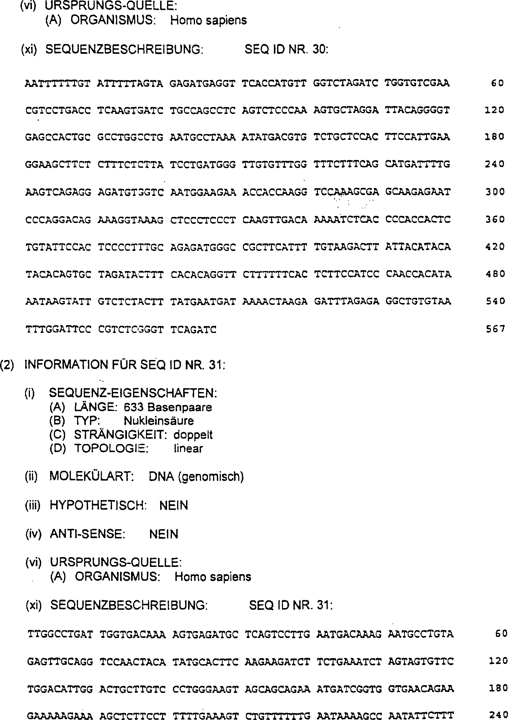


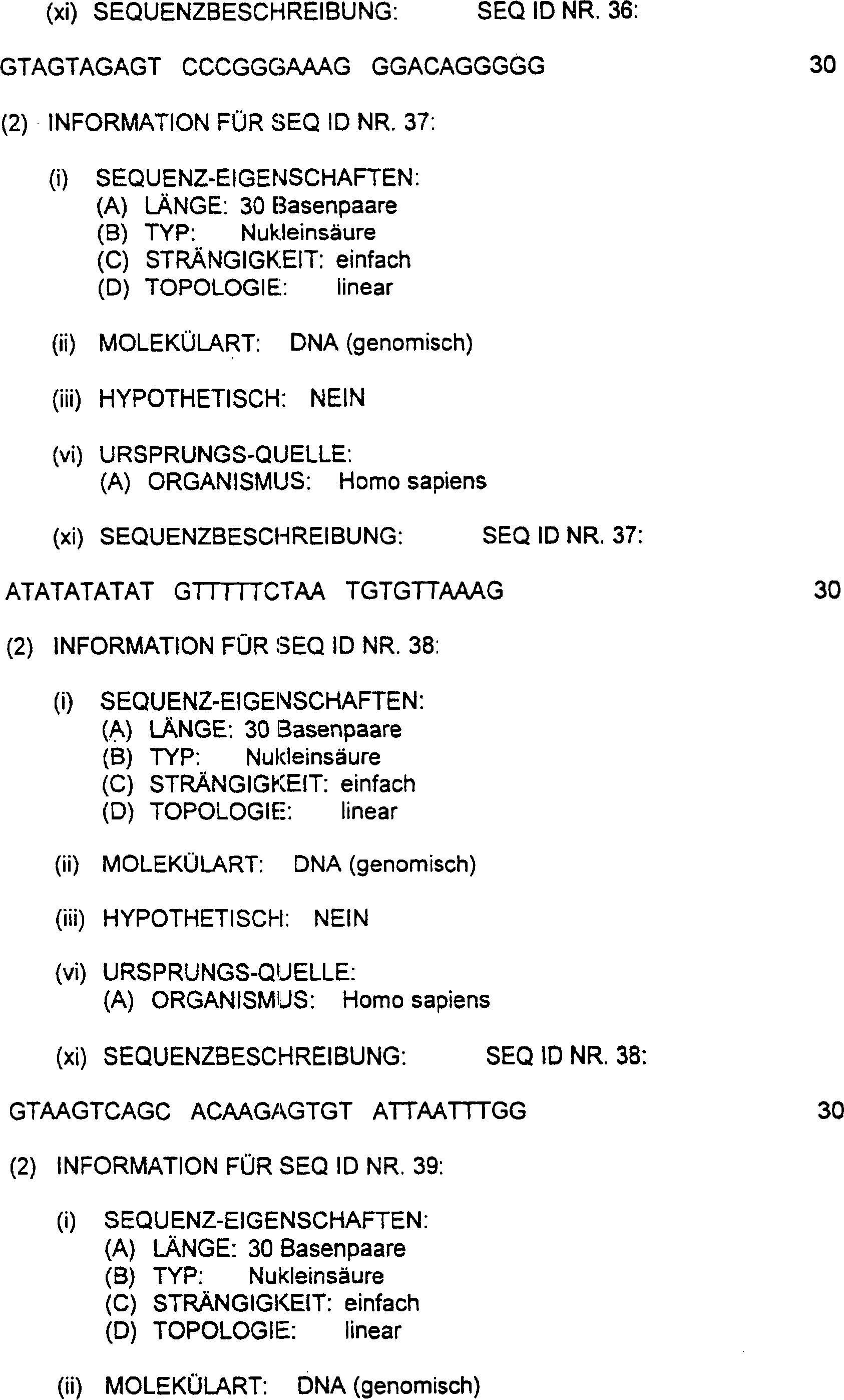


Claims (9)
Applications Claiming Priority (14)
| Application Number | Priority Date | Filing Date | Title |
|---|---|---|---|
| US348824 | 1989-05-08 | ||
| US28922194A | 1994-08-12 | 1994-08-12 | |
| US289221 | 1994-08-12 | ||
| US30026694A | 1994-09-02 | 1994-09-02 | |
| US300266 | 1994-09-02 | ||
| US30810494A | 1994-09-16 | 1994-09-16 | |
| US308104 | 1994-09-16 | ||
| US34882494A | 1994-11-29 | 1994-11-29 | |
| US40930595A | 1995-03-24 | 1995-03-24 | |
| US08/483,553 US5709999A (en) | 1994-08-12 | 1995-06-07 | Linked breast and ovarian cancer susceptibility gene |
| US480784 | 1995-06-07 | ||
| US483553 | 1995-06-07 | ||
| US08/480,784 US5693473A (en) | 1994-08-12 | 1995-06-07 | Linked breast and ovarian cancer susceptibility gene |
| US409305 | 1999-09-29 |
Publications (3)
| Publication Number | Publication Date |
|---|---|
| DE69521002D1 DE69521002D1 (en) | 2001-06-28 |
| DE69521002T2 DE69521002T2 (en) | 2001-11-15 |
| DE69521002T3 true DE69521002T3 (en) | 2010-02-25 |
Family
ID=27569596
Family Applications (1)
| Application Number | Title | Priority Date | Filing Date |
|---|---|---|---|
| DE69521002T Expired - Lifetime DE69521002T3 (en) | 1994-08-12 | 1995-08-11 | Mutations of the 17q associated ovarian and breast cancer susceptibility gene |
Country Status (16)
| Country | Link |
|---|---|
| EP (1) | EP0705903B2 (en) |
| JP (1) | JP2002503943A (en) |
| CN (1) | CN1172502A (en) |
| AT (1) | ATE201445T1 (en) |
| AU (1) | AU686004B2 (en) |
| CA (1) | CA2196797C (en) |
| DE (1) | DE69521002T3 (en) |
| DK (1) | DK0705903T3 (en) |
| ES (1) | ES2158048T5 (en) |
| FI (1) | FI970513A (en) |
| GR (1) | GR3036322T3 (en) |
| MX (1) | MX9701075A (en) |
| NO (1) | NO970624L (en) |
| NZ (1) | NZ291621A (en) |
| PT (1) | PT705903E (en) |
| WO (1) | WO1996005306A2 (en) |
Families Citing this family (61)
| Publication number | Priority date | Publication date | Assignee | Title |
|---|---|---|---|---|
| WO1995019369A1 (en) | 1994-01-14 | 1995-07-20 | Vanderbilt University | Method for detection and treatment of breast cancer |
| AU707348B2 (en) * | 1995-01-17 | 1999-07-08 | Vanderbilt University | Method for detection and treatment of breast cancer |
| US6838256B2 (en) * | 1996-02-12 | 2005-01-04 | Gene Logic Inc. | Coding sequences of the human BRCA1 gene |
| US6951721B2 (en) | 1996-02-12 | 2005-10-04 | Gene Logic Inc. | Method for determining the haplotype of a human BRCA1 gene |
| US5654155A (en) * | 1996-02-12 | 1997-08-05 | Oncormed, Inc. | Consensus sequence of the human BRCA1 gene |
| US5891857A (en) * | 1996-02-20 | 1999-04-06 | Vanderbilt University | Characterized BRCA1 and BRCA2 proteins and screening and therapeutic methods based on characterized BRCA1 and BRCA2 proteins |
| AU4586697A (en) * | 1996-09-20 | 1998-04-14 | Board Of Regents, The University Of Texas System | Compositions and methods comprising bard1 and other brca1 binding proteins |
| US6623938B2 (en) | 1997-01-21 | 2003-09-23 | Human Genome Sciences, Inc. | I-flice, a novel inhibitor of tumor necrosis factor receptor-1 and CD-95 induced apoptosis |
| US5965377A (en) * | 1997-03-24 | 1999-10-12 | Baystate Medical Center | Method for determining the presence of mutated BRCA protein |
| WO1998055650A1 (en) * | 1997-06-04 | 1998-12-10 | Rijksuniversiteit Te Leiden | A diagnostic test kit for determining a predisposition for breast and ovarian cancer, materials and methods for such determination |
| US6492109B1 (en) | 1997-09-23 | 2002-12-10 | Gene Logic, Inc. | Susceptibility mutation 6495delGC of BRCA2 |
| CA2308766C (en) | 1997-11-06 | 2007-03-20 | Oklahoma Medical Research Foundation | Diagnostic assay for cancer susceptibility |
| US6177410B1 (en) | 1997-12-05 | 2001-01-23 | Vanderbilt University | Therapeutic methods for prostate cancer |
| US6686163B2 (en) | 1998-05-06 | 2004-02-03 | Gene Logic Inc. | Coding sequence haplotype of the human BRCA1 gene |
| US7151102B2 (en) | 2000-10-30 | 2006-12-19 | Kudos Pharmaceuticals Limited | Phthalazinone derivatives |
| ES2357057T3 (en) | 2002-04-30 | 2011-04-15 | Kudos Pharmaceuticals Limited | DERIVATIVES OF FTALAZINONA. |
| HUE030806T2 (en) | 2002-05-02 | 2017-05-29 | Wyeth Holdings Llc | Calicheamicin derivative-carrier conjugates |
| GB0210121D0 (en) | 2002-05-02 | 2002-06-12 | Celltech R&D Ltd | Biological products |
| US7449464B2 (en) | 2003-03-12 | 2008-11-11 | Kudos Pharmaceuticals Limited | Phthalazinone derivatives |
| GB0305681D0 (en) | 2003-03-12 | 2003-04-16 | Kudos Pharm Ltd | Phthalazinone derivatives |
| SG150548A1 (en) | 2003-12-01 | 2009-03-30 | Kudos Pharm Ltd | Dna damage repair inhibitors for treatment of cancer |
| GB0419072D0 (en) | 2004-08-26 | 2004-09-29 | Kudos Pharm Ltd | Phthalazinone derivatives |
| KR101138862B1 (en) * | 2005-02-14 | 2012-05-14 | 삼성전자주식회사 | A polynucleotide associated with a breast cancer comprising single nucleotide polymorphism, microarray and diagnostic kit comprising the same and method for diagnosing a breast cancer using the same |
| KR101206028B1 (en) * | 2005-03-05 | 2012-11-28 | 삼성전자주식회사 | Method for diagnosing a breast cancer using a breast cancer specific polymorphic sequence, polynucleotide specific to a breast cancer and microarray immobilized with the polynucleotide |
| GB0521373D0 (en) | 2005-10-20 | 2005-11-30 | Kudos Pharm Ltd | Pthalazinone derivatives |
| IL282783B2 (en) | 2006-05-18 | 2023-09-01 | Caris Mpi Inc | System and method for determining individualized medical intervention for a disease state |
| US8768629B2 (en) | 2009-02-11 | 2014-07-01 | Caris Mpi, Inc. | Molecular profiling of tumors |
| EP2032140A1 (en) | 2006-05-31 | 2009-03-11 | Istituto di Richerche di Biologia Molecolare P. Angeletti S.p.A. | Pyrrolo[1,2-a]pyrazin-1(2h)-one and pyrrolo[1,2-d][1,2,4]triazin-1(2h)-one derivatives as inhibitors of poly(adp-ribose)polymerase(parp) |
| UY30639A1 (en) | 2006-10-17 | 2008-05-31 | Kudos Pharm Ltd | SUBSTITUTED DERIVATIVES OF 2H-FTALAZIN-1-ONA, ITS CRYSTAL FORMS, PREPARATION PROCESS AND APPLICATIONS |
| BRPI0806245B1 (en) | 2007-01-10 | 2022-01-25 | Msd Italia S.R.L. | Compounds of formula i and their uses |
| KR20100059950A (en) | 2007-09-14 | 2010-06-04 | 아스트라제네카 아베 | Phthalazinone derivatives |
| MX2010004028A (en) | 2007-10-17 | 2010-04-30 | Kudos Pharm Ltd | 4- [3- (4-cyclopropanecarbonyl-piperazine-i-carbonyl) -4 -fluoro-benzyl] -2h-phthalaz in-1-one. |
| UY31603A1 (en) | 2008-01-23 | 2009-08-31 | DERIVATIVES OF FTALAZINONA | |
| SG193842A1 (en) | 2008-08-06 | 2013-10-30 | Biomarin Pharm Inc | Dihydropyridophthalazinone inhibitors of poly(adp-ribose)polymerase (parp) |
| CN102238945B (en) | 2008-10-07 | 2014-10-29 | 阿斯利康(英国)有限公司 | Pharmaceutical formulation 514 |
| WO2010056337A2 (en) | 2008-11-12 | 2010-05-20 | Caris Mpi, Inc. | Methods and systems of using exosomes for determining phenotypes |
| TW201114756A (en) | 2009-07-15 | 2011-05-01 | Astrazeneca Ab | Phthalazinone compound |
| JP5808349B2 (en) | 2010-03-01 | 2015-11-10 | カリス ライフ サイエンシズ スウィッツァーランド ホールディングスゲーエムベーハー | Biomarkers for theranosis |
| BR112012025593A2 (en) | 2010-04-06 | 2019-06-25 | Caris Life Sciences Luxembourg Holdings | circulating biomarkers for disease |
| CN104789664A (en) * | 2015-04-02 | 2015-07-22 | 安诺优达基因科技(北京)有限公司 | Primer set, method and kit for Long-range PCR (polymerase chain reaction) detection of BRCA (breast cancer susceptibility gene) 1 and BRCA 2 |
| JP6786066B2 (en) | 2016-10-07 | 2020-11-18 | 富士通株式会社 | Family tree creation support program, family tree creation support method, and family tree creation support device |
| CN109897852A (en) * | 2018-07-25 | 2019-06-18 | 广州普世利华科技有限公司 | The gRNA of tumour related mutation gene based on C2c2, detection method, detection kit |
| JP2022532342A (en) | 2019-05-14 | 2022-07-14 | ニューベイション・バイオ・インコーポレイテッド | Anti-cancer nuclear hormone receptor targeting compound |
| CA3145644A1 (en) | 2019-07-19 | 2021-01-28 | Astrazeneca Ab | Parp1 inhibitors |
| WO2021097046A1 (en) | 2019-11-13 | 2021-05-20 | Nuvation Bio Inc. | Anti-cancer nuclear hormone receptor-targeting compounds |
| CN113388680B (en) * | 2020-03-14 | 2023-07-04 | 复旦大学附属妇产科医院 | Epithelial ovarian cancer target RET and its use in diagnosis and treatment |
| US11795158B2 (en) | 2020-06-25 | 2023-10-24 | Astrazeneca Ab | Chemical compounds |
| BR112023006337A2 (en) | 2020-10-09 | 2023-05-09 | Astrazeneca Uk Ltd | PHARMACEUTICAL PRODUCT, USE OF AN ANTI-HER2-DRUG ANTIBODY CONJUGATE OR A SELECTIVE PARP1 INHIBITOR, AND, METHOD OF TREATMENT OF CANCER |
| IL306010A (en) | 2021-03-23 | 2023-11-01 | Nuvation Bio Inc | Anti-cancer nuclear hormone receptor-targeting compounds |
| CN117580839A (en) | 2021-05-03 | 2024-02-20 | 诺维逊生物股份有限公司 | Anti-cancer nuclear hormone receptor targeting compounds |
| WO2022271547A1 (en) | 2021-06-21 | 2022-12-29 | Tesaro, Inc. | Combination treatment of cancer with a parp inhibitor and a lipophilic statin |
| TW202329936A (en) | 2021-11-18 | 2023-08-01 | 英商阿斯特捷利康英國股份有限公司 | Combination of antibody-drug conjugate and parp1 selective inhibitor |
| WO2023118085A1 (en) | 2021-12-21 | 2023-06-29 | Astrazeneca Ab | Methods of treating brain tumours and neuroblastomas |
| TW202333800A (en) | 2021-12-28 | 2023-09-01 | 英商阿斯特捷利康英國股份有限公司 | Combination of antibody-drug conjugate and rasg12c inhibitor |
| WO2023126823A1 (en) | 2021-12-28 | 2023-07-06 | Astrazeneca Uk Limited | Combination of antibody-drug conjugate and atr inhibitor |
| WO2023175483A1 (en) | 2022-03-16 | 2023-09-21 | Astrazeneca Uk Limited | A scoring method for an anti-trop2 antibody‑drug conjugate therapy |
| WO2023194530A1 (en) | 2022-04-07 | 2023-10-12 | Astrazeneca Ab | Combination therapy for treating cancer |
| WO2023194525A1 (en) | 2022-04-07 | 2023-10-12 | Astrazeneca Ab | Combination therapy for treating cancer |
| WO2023194528A1 (en) | 2022-04-07 | 2023-10-12 | Astrazeneca Ab | Combination therapy for treating cancer |
| WO2023242302A1 (en) | 2022-06-15 | 2023-12-21 | Astrazeneca Ab | Combination therapy for treating cancer |
| WO2024023750A1 (en) | 2022-07-28 | 2024-02-01 | Astrazeneca Uk Limited | Combination of antibody-drug conjugate and bispecific checkpoint inhibitor |
Family Cites Families (22)
| Publication number | Priority date | Publication date | Assignee | Title |
|---|---|---|---|---|
| NL154598B (en) | 1970-11-10 | 1977-09-15 | Organon Nv | PROCEDURE FOR DETERMINING AND DETERMINING LOW MOLECULAR COMPOUNDS AND PROTEINS THAT CAN SPECIFICALLY BIND THESE COMPOUNDS AND TEST PACKAGING. |
| US3817837A (en) | 1971-05-14 | 1974-06-18 | Syva Corp | Enzyme amplification assay |
| US3939350A (en) | 1974-04-29 | 1976-02-17 | Board Of Trustees Of The Leland Stanford Junior University | Fluorescent immunoassay employing total reflection for activation |
| US3996345A (en) | 1974-08-12 | 1976-12-07 | Syva Company | Fluorescence quenching with immunological pairs in immunoassays |
| US4277437A (en) | 1978-04-05 | 1981-07-07 | Syva Company | Kit for carrying out chemically induced fluorescence immunoassay |
| US4275149A (en) | 1978-11-24 | 1981-06-23 | Syva Company | Macromolecular environment control in specific receptor assays |
| US4376110A (en) | 1980-08-04 | 1983-03-08 | Hybritech, Incorporated | Immunometric assays using monoclonal antibodies |
| US4486530A (en) | 1980-08-04 | 1984-12-04 | Hybritech Incorporated | Immunometric assays using monoclonal antibodies |
| US4366241A (en) | 1980-08-07 | 1982-12-28 | Syva Company | Concentrating zone method in heterogeneous immunoassays |
| CA1188246A (en) | 1981-08-31 | 1985-06-04 | Jerry L. Gregory | Immobilized microbe recycle apparatus |
| NZ207394A (en) | 1983-03-08 | 1987-03-06 | Commw Serum Lab Commission | Detecting or determining sequence of amino acids |
| US4816567A (en) | 1983-04-08 | 1989-03-28 | Genentech, Inc. | Recombinant immunoglobin preparations |
| US4683202A (en) | 1985-03-28 | 1987-07-28 | Cetus Corporation | Process for amplifying nucleic acid sequences |
| US4683195A (en) | 1986-01-30 | 1987-07-28 | Cetus Corporation | Process for amplifying, detecting, and/or-cloning nucleic acid sequences |
| US4868105A (en) | 1985-12-11 | 1989-09-19 | Chiron Corporation | Solution phase nucleic acid sandwich assay |
| IE61148B1 (en) | 1988-03-10 | 1994-10-05 | Ici Plc | Method of detecting nucleotide sequences |
| ATE207075T1 (en) * | 1990-06-27 | 2001-11-15 | Univ Princeton | PROBE FOR P53 MUTANT DETECTION |
| IL103059A0 (en) | 1991-09-30 | 1993-02-21 | Boehringer Ingelheim Int | Conjugates for introducing nucleic acid into higher eucaryotic cells |
| US5252479A (en) | 1991-11-08 | 1993-10-12 | Research Corporation Technologies, Inc. | Safe vector for gene therapy |
| WO1994000764A1 (en) * | 1992-06-26 | 1994-01-06 | THE UNITED STATES OF AMERICA, represented by THE SECRETARY OF HEALTH AND HUMAN SERVICES | The met proto-oncogene and a method for predicting breast cancer progression |
| WO1995019369A1 (en) * | 1994-01-14 | 1995-07-20 | Vanderbilt University | Method for detection and treatment of breast cancer |
| AU707348B2 (en) * | 1995-01-17 | 1999-07-08 | Vanderbilt University | Method for detection and treatment of breast cancer |
-
1995
- 1995-08-11 PT PT95305605T patent/PT705903E/en unknown
- 1995-08-11 CA CA002196797A patent/CA2196797C/en not_active Expired - Lifetime
- 1995-08-11 WO PCT/US1995/010202 patent/WO1996005306A2/en not_active Application Discontinuation
- 1995-08-11 AU AU33212/95A patent/AU686004B2/en not_active Expired
- 1995-08-11 JP JP50750796A patent/JP2002503943A/en active Pending
- 1995-08-11 ES ES95305605T patent/ES2158048T5/en not_active Expired - Lifetime
- 1995-08-11 AT AT95305605T patent/ATE201445T1/en not_active IP Right Cessation
- 1995-08-11 DK DK95305605T patent/DK0705903T3/en active
- 1995-08-11 CN CN95195413.XA patent/CN1172502A/en active Pending
- 1995-08-11 NZ NZ291621A patent/NZ291621A/en not_active IP Right Cessation
- 1995-08-11 EP EP95305605A patent/EP0705903B2/en not_active Expired - Lifetime
- 1995-08-11 DE DE69521002T patent/DE69521002T3/en not_active Expired - Lifetime
- 1995-08-11 MX MX9701075A patent/MX9701075A/en unknown
-
1997
- 1997-02-07 FI FI970513A patent/FI970513A/en unknown
- 1997-02-11 NO NO970624A patent/NO970624L/en not_active Application Discontinuation
-
2001
- 2001-08-06 GR GR20010401173T patent/GR3036322T3/en not_active IP Right Cessation
Also Published As
| Publication number | Publication date |
|---|---|
| AU3321295A (en) | 1996-03-07 |
| NO970624L (en) | 1997-04-14 |
| CA2196797C (en) | 2000-10-10 |
| EP0705903B1 (en) | 2001-05-23 |
| WO1996005306A2 (en) | 1996-02-22 |
| CN1172502A (en) | 1998-02-04 |
| GR3036322T3 (en) | 2001-11-30 |
| JP2002503943A (en) | 2002-02-05 |
| DE69521002T2 (en) | 2001-11-15 |
| CA2196797A1 (en) | 1996-02-22 |
| ATE201445T1 (en) | 2001-06-15 |
| ES2158048T3 (en) | 2001-09-01 |
| PT705903E (en) | 2001-11-30 |
| EP0705903A1 (en) | 1996-04-10 |
| EP0705903B2 (en) | 2009-08-12 |
| DE69521002D1 (en) | 2001-06-28 |
| FI970513A0 (en) | 1997-02-07 |
| ES2158048T5 (en) | 2009-11-04 |
| NZ291621A (en) | 1998-09-24 |
| AU686004B2 (en) | 1998-01-29 |
| MX9701075A (en) | 1998-03-31 |
| NO970624D0 (en) | 1997-02-11 |
| FI970513A (en) | 1997-04-07 |
| WO1996005306A3 (en) | 1996-03-14 |
| DK0705903T3 (en) | 2001-08-27 |
Similar Documents
| Publication | Publication Date | Title |
|---|---|---|
| DE69521002T3 (en) | Mutations of the 17q associated ovarian and breast cancer susceptibility gene | |
| DE69519834T3 (en) | Method for detecting predisposition of ovarian and breast cancer | |
| DE69625678T3 (en) | Chromosome 13 associated breast cancer susceptibility gene BRCA2 | |
| US5747282A (en) | 17Q-linked breast and ovarian cancer susceptibility gene | |
| US5709999A (en) | Linked breast and ovarian cancer susceptibility gene | |
| US5693473A (en) | Linked breast and ovarian cancer susceptibility gene | |
| US5710001A (en) | 17q-linked breast and ovarian cancer susceptibility gene | |
| EP1260520B1 (en) | Chromosome 13-linked breast cancer susceptibility gene | |
| US5753441A (en) | 170-linked breast and ovarian cancer susceptibility gene | |
| WO1997022689A9 (en) | Chromosome 13-linked breast cancer susceptibility gene | |
| DE69433940T2 (en) | HUMAN MUTATOR GEN NMSH2 AND ITS CONNECTION TO HEREDITARIC, NON-POLYPOLOSE COLORREVALAL CARCINOMA | |
| DE69837565T2 (en) | MUTATIONS OF THE HUMAN MINK GENE, ASSOCIATED WITH ARRHYTMIA | |
| US5777093A (en) | cDNAs associated with ataxia-telangiectasia | |
| DE69936781T2 (en) | KVLQT1 - IN CONNECTION WITH 'LONG QT SYNDROME' | |
| JP3241736B2 (en) | 17q-linked breast and ovarian cancer susceptibility genes | |
| AU773601B2 (en) | Chromosome 13-linked breast cancer susceptibility gene |
Legal Events
| Date | Code | Title | Description |
|---|---|---|---|
| 8363 | Opposition against the patent | ||
| 8328 | Change in the person/name/address of the agent |
Representative=s name: VOSSIUS & PARTNER, 81675 MUENCHEN |
|
| 8366 | Restricted maintained after opposition proceedings | ||
| 8327 | Change in the person/name/address of the patent owner |
Owner name: THE UNIVERSITY OF UTAH RESEARCH FOUNDATION, SA, US |