EP0050424A1 - Method for producing an analytical antibody probe, an analytical antibody probe, and a method for analysing a sample for certain antibodies - Google Patents
Method for producing an analytical antibody probe, an analytical antibody probe, and a method for analysing a sample for certain antibodies Download PDFInfo
- Publication number
- EP0050424A1 EP0050424A1 EP81304411A EP81304411A EP0050424A1 EP 0050424 A1 EP0050424 A1 EP 0050424A1 EP 81304411 A EP81304411 A EP 81304411A EP 81304411 A EP81304411 A EP 81304411A EP 0050424 A1 EP0050424 A1 EP 0050424A1
- Authority
- EP
- European Patent Office
- Prior art keywords
- differentiated
- antigenic
- antibodies
- antigenic components
- protein
- Prior art date
- Legal status (The legal status is an assumption and is not a legal conclusion. Google has not performed a legal analysis and makes no representation as to the accuracy of the status listed.)
- Granted
Links
Images


Classifications
-
- G—PHYSICS
- G01—MEASURING; TESTING
- G01N—INVESTIGATING OR ANALYSING MATERIALS BY DETERMINING THEIR CHEMICAL OR PHYSICAL PROPERTIES
- G01N33/00—Investigating or analysing materials by specific methods not covered by groups G01N1/00 - G01N31/00
- G01N33/48—Biological material, e.g. blood, urine; Haemocytometers
- G01N33/50—Chemical analysis of biological material, e.g. blood, urine; Testing involving biospecific ligand binding methods; Immunological testing
- G01N33/53—Immunoassay; Biospecific binding assay; Materials therefor
- G01N33/543—Immunoassay; Biospecific binding assay; Materials therefor with an insoluble carrier for immobilising immunochemicals
- G01N33/54366—Apparatus specially adapted for solid-phase testing
-
- G—PHYSICS
- G01—MEASURING; TESTING
- G01N—INVESTIGATING OR ANALYSING MATERIALS BY DETERMINING THEIR CHEMICAL OR PHYSICAL PROPERTIES
- G01N33/00—Investigating or analysing materials by specific methods not covered by groups G01N1/00 - G01N31/00
- G01N33/48—Biological material, e.g. blood, urine; Haemocytometers
- G01N33/50—Chemical analysis of biological material, e.g. blood, urine; Testing involving biospecific ligand binding methods; Immunological testing
- G01N33/53—Immunoassay; Biospecific binding assay; Materials therefor
- G01N33/558—Immunoassay; Biospecific binding assay; Materials therefor using diffusion or migration of antigen or antibody
-
- G—PHYSICS
- G01—MEASURING; TESTING
- G01N—INVESTIGATING OR ANALYSING MATERIALS BY DETERMINING THEIR CHEMICAL OR PHYSICAL PROPERTIES
- G01N33/00—Investigating or analysing materials by specific methods not covered by groups G01N1/00 - G01N31/00
- G01N33/48—Biological material, e.g. blood, urine; Haemocytometers
- G01N33/50—Chemical analysis of biological material, e.g. blood, urine; Testing involving biospecific ligand binding methods; Immunological testing
- G01N33/53—Immunoassay; Biospecific binding assay; Materials therefor
- G01N33/569—Immunoassay; Biospecific binding assay; Materials therefor for microorganisms, e.g. protozoa, bacteria, viruses
- G01N33/56905—Protozoa
-
- G—PHYSICS
- G01—MEASURING; TESTING
- G01N—INVESTIGATING OR ANALYSING MATERIALS BY DETERMINING THEIR CHEMICAL OR PHYSICAL PROPERTIES
- G01N33/00—Investigating or analysing materials by specific methods not covered by groups G01N1/00 - G01N31/00
- G01N33/48—Biological material, e.g. blood, urine; Haemocytometers
- G01N33/50—Chemical analysis of biological material, e.g. blood, urine; Testing involving biospecific ligand binding methods; Immunological testing
- G01N33/53—Immunoassay; Biospecific binding assay; Materials therefor
- G01N33/569—Immunoassay; Biospecific binding assay; Materials therefor for microorganisms, e.g. protozoa, bacteria, viruses
- G01N33/56911—Bacteria
-
- G—PHYSICS
- G01—MEASURING; TESTING
- G01N—INVESTIGATING OR ANALYSING MATERIALS BY DETERMINING THEIR CHEMICAL OR PHYSICAL PROPERTIES
- G01N33/00—Investigating or analysing materials by specific methods not covered by groups G01N1/00 - G01N31/00
- G01N33/48—Biological material, e.g. blood, urine; Haemocytometers
- G01N33/50—Chemical analysis of biological material, e.g. blood, urine; Testing involving biospecific ligand binding methods; Immunological testing
- G01N33/53—Immunoassay; Biospecific binding assay; Materials therefor
- G01N33/569—Immunoassay; Biospecific binding assay; Materials therefor for microorganisms, e.g. protozoa, bacteria, viruses
- G01N33/56983—Viruses
-
- G—PHYSICS
- G01—MEASURING; TESTING
- G01N—INVESTIGATING OR ANALYSING MATERIALS BY DETERMINING THEIR CHEMICAL OR PHYSICAL PROPERTIES
- G01N33/00—Investigating or analysing materials by specific methods not covered by groups G01N1/00 - G01N31/00
- G01N33/48—Biological material, e.g. blood, urine; Haemocytometers
- G01N33/50—Chemical analysis of biological material, e.g. blood, urine; Testing involving biospecific ligand binding methods; Immunological testing
- G01N33/53—Immunoassay; Biospecific binding assay; Materials therefor
- G01N33/569—Immunoassay; Biospecific binding assay; Materials therefor for microorganisms, e.g. protozoa, bacteria, viruses
- G01N33/571—Immunoassay; Biospecific binding assay; Materials therefor for microorganisms, e.g. protozoa, bacteria, viruses for venereal disease, e.g. syphilis, gonorrhoea
Landscapes
- Health & Medical Sciences (AREA)
- Life Sciences & Earth Sciences (AREA)
- Immunology (AREA)
- Engineering & Computer Science (AREA)
- Urology & Nephrology (AREA)
- Hematology (AREA)
- Biomedical Technology (AREA)
- Chemical & Material Sciences (AREA)
- Molecular Biology (AREA)
- Medicinal Chemistry (AREA)
- Physics & Mathematics (AREA)
- Cell Biology (AREA)
- Pathology (AREA)
- Biotechnology (AREA)
- Food Science & Technology (AREA)
- General Physics & Mathematics (AREA)
- Microbiology (AREA)
- Analytical Chemistry (AREA)
- Biochemistry (AREA)
- General Health & Medical Sciences (AREA)
- Virology (AREA)
- Tropical Medicine & Parasitology (AREA)
- Peptides Or Proteins (AREA)
- Medicines Containing Antibodies Or Antigens For Use As Internal Diagnostic Agents (AREA)
- Saccharide Compounds (AREA)
Abstract
Description
- The present invention provides, in general, a method for producing an analytical antibody probe, an analytical antibody probe, and a method for analysing a sample for certain antibodies. The invention has particular value in the diagnosis of disease or allergy in patients. More particularly, the invention permits an improved method for such diagnosis, and provides an improved product for conducting such diagnosis.
- Many clinical diagnositc techniques have, as their fundamental basis, the antigen-antibody reaction. This reaction serves as a defence against micro-organisms and other foreign bodies as part of the body's normal immunological response. Detection of the presence of antigen-antibody reactions in tests performed upon serum obtained from a patient may indicate the presence or absence of antibodies in the patient's serum. A positive test for reaction of antibody with a specific antigen may indicate a presence of a corresponding disease or at least suggest that diagnostic conclusion.
- Known clinical diagnostic procedures that test for the antigen-antibody reaction in sera (serotests) may take a wide variety of forms. Some utilize antigens anchored to the surface of inert particles. In the presence of specific antibody, these particulate antigens clump visibly in an agglutination reaction. Such a procedure is widely employed in the diagnosis of syphilis and is known as the Venereal Disease Research Laboratory (VDRL) test. Other tests may involve the attachment of a fluorescent or radioactive moiety in such a way that its presence indicates that the antigen-antibody reaction has occurred. In a similar manner, enzymes have been linked as a detectable moiety - the so-called enzyme linked immunosorbent assay (ELISA).
- The hallmark of conventional serotests is that they measure a single positive or negative signal. Signals may be, for example, hemagglutination, hemagglutination inhibition, complement fixation, surface fluorescence, particle agglutination and so on. That single positive or negative signal is usually a complex and undifferentiated array of the structural components of an organism or material antigenically related to it. This complexity of the antigen test material increases the likelihood that false positive signals will be generated due to antibodies stimulated by other organisms sharing some but not all antigenic properties with the test material. In contrast the present invention enables the provision of a diagnostic method and means by which the antigen-antibody reaction may be greatly expanded as a diagnostic tool. Using the invention the presence of a specific disease, a specific stage of a disease, or an allergy in a patient may be detected by a simple procedure.
- Thus the invention provides a method for producing an analytical antibody probe for detecting the presence of antibodies specific for particular protein and/or polysaccharide material, which method comprises disposing upon a solid-state substrate in a pre-determined spatial relationship a set of differentiated antigentic components which have been derived from said protein or polysaccharide material, said set of differentiated antigentic components being selected such that at least a subset thereof is known to be reactive with said antibodies. The invention also provides an analytical antibody probe for detecting the presence of antibodies specific for particular protein and/ or polysaccharide material, which probe comprises a solid-state substrate upon which is disposed in a pre-determined spatial relationship a set of differentiated antigenic components derived from said protein or polysaccharide material, said set of differentiated antigenic components being selected such that at least a subset thereof is known to be reactive with said antibodies.
- Specific aspects of the invention are:-
- (a) a method for producing a diagnostic antibody probe, comprising, disposing upon a solid-state susbtrate a set of differentiated antigenic components of a larger set of proteins or polysaccharides in a predetermined spatial relationship, said larger set being related to a disease, disease stage or allergy under diagnosis, said set of differentiated antigenic components being selected such that at least a subset of said differentiated antigenic components is known to be reactive with antibodies present in serum obtained from a patient having the specific disease stage - or allergy; and
- (b) a diagnostic antibody probe comprising a solid-state substrate upon which is disposed in a pre-determined spatial relationship a set of differentiated antigenic components of a larger set of proteins or polysaccharides, said larger set being related to a disease, disease stage or allergy under diagnosis, said set of differentiated antigenic components being selected such that at least a subset of said differentiated antigenic components is known to be reactive with antibodies present in serum obtained from a patient having the specific disease, disease stage or allergy.
- One further aspect of the invention is a method for analysing a sample to determine the presence and distribution of antibodies specific for particular protein and/or polysaccharide material, which method comprises providing a solid-state substrate upon which is disposed in a pre- determined spatial relationship a set of differentiated antigenic components derived from said protein or polysaccharide material, said set of differentiated antigenic components being selected such that at least a subset thereof is known to be reactive with said antibodies, contacting said substrate with said sample under conditions which permit reaction of any of said antibodies in the sample with the antigenic components on the substrate, and detecting the existence and pattern of antigen-antibody reactions on the substrate.
- The invention will now be further described with reference to the accompanying drawings, in which:-
- FIGURE 1 is a photograph of a conventional SDS polyacrylamide gel showing the total protein profile of T. pallidum after staining;
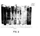
- FIGURE 2 is a photograph of an autoradiogram of twelve diagnostic strips of T. pallidum, constructed in accordance with the invention, after exposure to sera drawn from patients, eight of whom had different stages of syphilis and four of whom had exhibited false positive (BFP) on standard serological tests for syphilis;
- FIGURE 3 is a photograph of an autoradiogram of nine diagnostic strips of T. pallidum, constructed in accordance with the invention, after exposure to sera drawn from patients suffering from late syphilis and secondary syphilis;
- FIGURE 4 is a photograph of an autoradiogram of ten diagnostic strips of T. pallidum, constructed in accordance with the invention, after exposure to sera drawn from patients suffering from early latent and late latent syphilis;
- FIGURE 5 is a photograph of conventional SDS gels showing the total protein profiles of five different immunotypes of Chlamydia;
- FIGURE 6 is a photograph of an autoradiogram of five diagnostic strips of Chlamydia trachomatis, constructed in accordance with the invention, after exposure-to rabbit antiserum to Chalmydia trachomatis immuno type B;
- FIGURE 7 is a photograph of an autoradiogram of seven diagnostic strips of Toxoplasma gondii, constructed in accordance with the invention, after exposure to sera drawn from patients with chronic and acute toxoplasmosis;
- FIGURE 8 is a photograph of an autoradiogram of diagnostic strips of cytomegalovirus infected cells and cell free preparation of partially purified virus, constructed in accordance with the invention, after exposure to patient sera;
- FIGURE 9 is a photograph of an autoradiogram of diagnostic strips of rabbit kidney cells infected with Herpes simplex virus I or II, constructed in accordance with the invention, after exposure to sera drawn from patients suffering from Herpes infections;
- FIGURE 10 is a photograph of an autoradiogram of diagnostic strips of
capsular polysaccharide types - FIGURE 11 is a photograph of an autoradiogram of a diagnostic strip of bee venom, constructed in accordance with the invention, after exposure to serum drawn from a patient allergic to bee venom;
- FIGURES 12 and 13 are photographs of an autoradiogram of diagnostic strips of type L2 C. trachomatis, constructed in accordance with the invention, after reaction with sera from patients with type F infection and type D infection, respectively;
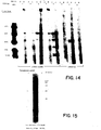
- FIGURE 14 is a photograph of an autoradiogram of diagnostic strips of T. pallidum, constructed in accordance with the invention, after exposure to sera drawn from patients suffering from syphilis and after exposure to IgG and IgM specific antisera probes;
- FIGURE 15 is a photograph of an autoradiogram of diagnostic strips of Toxoplasma gondii, constructed in accordance with the invention, after exposure to serum drawn from a patient suffering from acute toxoplasmosis and after exposure to IgG and IgM specific probes;
- FIGURE 16 is a photograph of an autoradiogram of diagnostic strips of cytomegalovirus, constructed in accordance with the invention, after exposure to a serum pool drawn from patients suffering from viral infection and after exposure to IgG and IgM specific probes;
- FIGURE 17 is a photograph of an autoradiogram of diagnostic strips of Toxoplasma gondii, constructed in accordance with the invention, when the Toxoplasma antigens were separated on a nondenaturing isoelectricfocusing gel;
- FIGURE 18 is a photograph of an autoradiogram of diagnostic strips of Toxoplasma gondii, prepared in accordance with the invention, when the Toxoplasma antigens are separated on a nondenaturing native gel system; and
- FIGURE 19 is a photograph of an autoradiogram of a diagnostic strip of cloned T. pallidum - specific antigens, constructed in accordance with the invention, after exposure to sera from patients suffering from syphilis.
- Very generally, the use of the invention for diagnosis involves a method con- prising providing a solid-state substrate upon which is disposed in a predetermined relationship a set of differentiated antigenic components of a larger set of proteins or polysaccharides. The larger set is related to the disease or allergy under diagnosis. The set of components is selected such that at least a subset of the components is known to be reactive with antibodies present in serum obtained from a patient having the specific disease or allergy. The substrate is contacted with serum obtained from the patient under diagnosis under conditions which permit reaction of antibodies in the serum with antigenic components on the substrate. The existence and pattern of antigen-antibody reactions on the substrate is then detected for correspondence with the pattern of those components on the substrate known to be reactive for the specific disease, the specific stage of the disease, or the allergy.
- The method detailed here for disease diagnosis differs markedly from conventional serotests in that what comprises a "positive" test is not the presence of one signal, but rather the appearance of a series of signals that represent antibody response to defined antigens associated with a specific pathogen or disease. The number and nature of the signals which define a disease state are defined empirically for each specific disease; all humans with this disease display the same multiple signals characteristic of that disease.
- More particularly, the invention, although based upon the antigen-antibody reaction phenomenon, take advantage of a discovery of major significance. If protein or polysaccharide material related to a specific disease, or stage of a disease, or an allergy is differentiated in such a way as to establish a spatial relationship of differentiated antigenic components, a plurality of antigen-antibody reactions may be detected upon exposure to serum from a diseased patient. The extent and pattern of such reactions, with proper differentiation, can be made highly specific to a given disease. Accordingly, a sort of molecular antigenic "fingerprint" may be established which will identify a particular disease and no other. Moreover, this molecular antigenic "fingerprint" may be designed so as to distinguish between historical antibodies, i.e., residual IgG antibodies which indicate previous exposure to a given antigen, and current antibody, i.e., recent IgM antibodies which indicate very recent exposure to a given antigen. In the case of allergy diagnosis, the antibody detected is immunoglobulin IgE.
- The method of the invention utilizes a solid-state substrate upon which is disposed in a predetermined spatial relationship a set of differentiated antigenic components of a larger set of proteins or polysaccharides. This larger group is related to the disease, the stage of the disease, or the allergy under diagnosis. It may be the pathogen itself (e.g., Herpes virus I or 110 or it may be a tissue sample (e.g., of a sarcoma tumor) or it may be an allergen such as various types of grasses or pollen. Frequently, the source of the larger set of proteins or polysaccharides will itself be antigenic for a particular disease but, because of non-specificity, cross reactivity, or other problems, is unsatisfactory for diagnostic use.
- The set of differentiated antigenic components of the larger set of proteins or polysaccharides is chosen for its specific ability to identify the disease, the stage of the disease, or allergy of interest. These components are selected such that at least a subset of the components is known to be reactive with antibodies present in serum obtained from a patient having the specific disease or allergy. The subsets are determined empirically in accordance with techniques described below. The process by which the components are differentiated depends upon the particular disease, the larger protein or polysaccharide from which the components are derived, and the particular antibodies which are to be detected (e.g., IgG, IgM, or IgE). Such-differentiation processes may include, but are not limited to, electrophoresis (SDS or native gel), isoelectricfocusing, thin layer chromatography, and centrifugation. Another differentiation process, once the nature of the subject of components is ascertained, is to produce each component of the subset separately by means of genetically modified microorganisms. Each component may then be placed separately on the substrate.
- In any case, the individual differentiated components are positioned upon a solid-state substrate such as a cellulose strip. The precise manner of attaching the components to the substrate will depend upon the nature of the components and the substrate. For example, if electrophoresis is utilized as a differentiation process, a useful transfer technique is the so-called filter affinity transfer as described by Erlich, H.A., et al., in Journ. Biol. Chem., 254:12240-12247 (1979).
- A typical solid-state substrate may be a cellulose strip to which a plurality of differentiated components of the pathogen responsible for a disease have been transferred. When the strip is exposed to a patient's serum, components which are reactive with antibodies in the serum bind to it and may be detected by any suitable assay. Before such exposure, the strip may be blank in appearance with the differentiated components not visible. Once exposed to antibodies, the antibody-antigen reactions cause the antibodies to bind to the strip in a pattern that is indicative of the present or absence of a specific disease. Through previous empirical testing, it can be readily established as to which differentiated proteins or groups of proteins on a given substrate will reactwith antibodies in the serum of a patient having the specific disease that is of interest. Thus, separate strips may be produced that are specific to, for example, Chlamydia, syphilis, and gonorrhea, respectively.
- Once the strip or solid-state substrate is produced with the empirically determined and selected pattern of differentiated antigenic components established on the substrate, it is in a form useful for clinical diagnosis. For such a use, the substrate is exposed or contacted with serum obtained from the patient under diagnosis. The conditions under which the contact occurs are established so as to permit reaction of antibodies in the serum with antigenic components on the substrate.
- Following suitable illumination steps, e.g., radioactively labelled probes specific for human immunoglobulin classes and autoradiography, the strip is examined to ascertain the pattern, if any, of antibody-antigen reactions which has developed. If the pattern corresponds to the pattern known for the particular disease to which -the strip or substrate corresponds, a positive diagnosis is obtained. Otherwise, the diagnosis is negative. Actual detection of the antigen-antibody reactions may employ other than autoradiographic assay of the type shown in FIGURE 2. For example, colorimetric assays may also be employed.
- Traditionally, radioactively labelled S. aureus protein A has been used as a probe for IgG antibodies. To distinguish between reactions with historical antibody (IgG) and new antibody (IgM), antiserum to human IgM may be labelled in a variety of ways, e.g., 1251 or fluorescein, and used as probe to detect formation of new human antibody (IgM). This provides the ability to distinguish between persons with a history of a disease who do not currently have an active form of that disease and persons with the active form of the disease. Historical antibodies will, in many cases, remain with a cured person for life.
- The present invention will be more readily understood by means of the following examples. These examples are set forth for the purpose of elaborating on the invention and are not intended to limit the invention in any way.
- FIGURE 1 is a photograph showing the total protein profile of Treponema pallidum. These proteins are separated and stained on a conventional SDS polyacrylamide gel. To obtain this profile, intact T. pallidum was suspended in an electrophoresis sample buffer comprised of 62.5 mM tris (pH 6.8), 2% sodium dodecylsulfate, and 5% mercaptoethanol. The sample was then applied to an SDS polyacrylamide gel system as described in Laemmli, U.K., Nature (London) 227:680-685 (1970). The gel was run until the tracking dye reached the bottom of the gel.
- In FIGURE 1, the left-hand column represents the T. pallidum profile, whereas the right-hand column is a system of molecular weight markers as is well known in the use of polyacrylamide gel separations. FIGURE 1 provides a base for comparison of the actual protein separation with the antigenic activity described in FIGURE 2.
- In FIGURE 2, diagnostic strips were prepared in accordance with the invention using the total protein separation of T. pallidum illustrated in FIGURE 1. To prepare the strips, the gel is overlaid with nitrocellulose paper as described by Towbin, H., Staehlin, T., and Gordon, J., PNAS (USA) 76:4350-4354 (1979). The paper is then covered with scouring pads and supported by lucite grids with numerous pores. The assembly is held together with rubber bands and is then placed in a single chamber for electrophoresis such that the surface of the gel applied directly to the paper is facing the anode. Electrophoresis is performed in an elctrode buffer comprised of 25 mM tris, 192 mM glycine, and 20% volume/volume methanol at pH 8.3. Electrophoresis is carried out for 90 minutes. The nitrocellulose at the end of this procedure contains the proteins arrayed as they have been separated according to molecular weight and is referred to as the blot.
- The blots were then soaked in a solution of 1% bovine serum albumin in a buffer comprised of 50 mM tris (pH 7.5), 0.9% sodium chloride, 0.25% gelatin, 0.2% sodium azide, and 0.1% NP 40 (TSGAN) for ten minutes at room temperature. This was to saturate all remaining reactive sites on the paper. At this point, the blots are ready for use and may be stored by freezing or other suitable means.
- Each of the strips in FIGURE 2 is a blot of the T. pallidum total protein profile after exposure to patient serum representing different stages of syphilis (8 patients) and representing non-syphilitic patients who showed false positive (BFP) in standard serological tests for syphilis (4 patients). Serum dilutions were used at 1:1000 with twelve hours at room temperature with gentle shaking. After this period of incubation with serum, the blotswere rinsed several times with TSGAN and then washed withTSGAN for 20-60 minutes at room temperature, again with gentle agitation. Then 2-4 microcuries of protein A with a specific activity of greater than 107 counts per minute/micro- gram is added in a volume of 100-200 milliliters of TSGAN and incubation continued with a gentle agitation for 60 minutes at room temperature. A similar incubation buffer system is described in Renard, J., Reiser, J., and Stark, G. R., PNAS (USA) 76: 3116-312 (1979).
- The blots were then rinsed several times with TSGAN, washed with TSGAN with gentle agitation at room temperature for 20 minutes and then rinsed several times with distilled water, dried with a hair dryer and then subjected to autoradiography. The autoradiography was on Kodak X-omat R film and DuPont Cronex intensifying screens. Autoradiography usually takes from 2-16 hours.
- Of the strips for 8 syphilitic patients shown in FIGURE 2, it may be seen that similar reaction patterns exist, particularly with respect to patients 1-5 and 7. This is true also of
patients - FIGURE 3 shows diagnostic strips of the IgG antibody response to peptides of T. pallidum from patients with late syphilis and secondary syphilis. The gels, protein samples and strips were prepared as outlined above. In FIGURE 3, the far-right strip is a system of molecular weight markers. Strips 1-8 illustrate the antibody response from patients suffering from late syphilis, while
strip 9 shows the antibody response indicative of the secondary form of the disease. - FIGURE 4 shows diagnostic strips of the IgG antibody response to peptides of T. pallidum from patients with early latent - and late latent syphilis. Again the samples were prepared as outlined above, and again the far-right column is a system of standard molecular weight gel markers.
Strips - FIGURE 5, left-hand columns 1-5, show the stained total proteins of five different immunotypes of Chlamydia.
Column 6 in FIGURE 5 is a molecular weight marker system. By standard serology, these immunotypes are non-crossreac- tive. Accordingly, a separate antiserum for clinical use must be prepared for each serotype. - FIGURE 6 shows the strips of the invention prepared in accordance with Example I after exposure to rabbit antiserum to Chlamydia trachomatis immuno type B and autoradiography. Preparations and procedures were as in Example I. All C. trachomatis immuno types have extensive cross-reaction of the major antigenic proteins. It may be seen that the left-
hand 4 strips show strong reaction whereas the strip in the far right-hand side, specific for C. psittaci, shows weak relatedness of only two antigens. This illustrates that a single C. trachomatis immunotype is a sufficient source of antigens for testing human infection with any other C. trachomatis immunotypes, and yet provides specificity in that other types of Chlamydia may be readily distinguished. - FIGURE 7 is a photograph showing various antigenic bands of Toxoplasma gondii that react with antibodies in the sera of patients with chronic and acute toxoplasmosis. To obtain the observed patterns, the toxoplasmal antigens were separated on a conventional SDS polyacrylamide gel. A sonicate of Toxoplasma was suspended in an equal volume of electrophoresis sample buffer consisting of 0.125M trizma base (pH 6.8), 2.5% sodium dodecylsulfate, and 2.5% 6-mercaptoethanol. The sample was then applied to an SDS polyacrylamide gel system as described in Laemmli, U. K., Nature (London) 227:680-685 (1970). The gel was run until the tracking dye reached the bottom of the gel.
- 1 The gel was then washed for 15 minutes in water and in two 5-minute washes of 50 mM sodium acetate, pH 7.0. The peptide components of Toxoplasma, separated by molecular weight in the SDS polyacrylamide gel, were electrophoretically transferred onto cyanogen bromide activated paper as follows. The gel was placed on a scotch bright pad covered with filter paper. A sheet of cyanogen bromide treated filter paper was laid on the gel and another sheet of filter paper and a scotch bright pad was placed on top. The assembly is placed in an E-C electroblot unit with the cyanogen bromide paper facing the anode. Electrophoresis was carried out in 50 mM sodium acetate,
pH 7, at 25 volts for one hour. - All remaining reactive sites on the cyanogen bromide paper are bound and/or inactivated by soaking the paper in a solution of 1M glycine and 1% bovine serum albumin for 0.5 to 3 hours. The paper was washed three times for 5-15 minutes each with agitation in a wash solution containing 0.1% ovalbumin, 0.1% tween 20, 0.02% sodium azide in phosphate buffered saline. The paper is then incubated at room temperature with gentle agitation for 2-3 hours in diluted human serum. The sera used in FIGURE 7 are from patients with chronic or acute toxoplasmosis. The patient's serum is diluted 1:25 in wash solution.
- After the incubation with serum, the paper is washed three times for 5-15 minutes with shaking in wash solution. The 1251 Protein A is added to the paper using a 1:200 dilution of stock (~5µg/ml, 15µci/µg) in wash solution. The protein A is iodinated using the chloramine T method as described by Erlich, H., Cohen, S., and McDevitt, H., in Cell, 13:681-689 (1978). The paper is incubated with the 1251 Protein A for 1-3 hours at room temperature with agitation. The paper is again washed as above, dried and placed under Kodak XAR-5 X-ray film for 16 hours.
-
Strips strips - The strips in FIGURE 8 illustrate the banding patterns obtained when cytomegalovirus infected cells and cell free preparation of partially purified virus are reacted with patient sera. The electrophoresis and transfer are performed as outlined in Example III. The preparation of the infected cells was done as follows.
- Two Corning 490cm2 roller bottles containing a confluent layer of passage eight human embryo lung cells were each inoculated with 2.5 mls of infected cells containing between 107 and 108 viral particles/ml. Fourteen mls of Eagle minimal essential medium plus 10% fetal calf serum was added to each bottle. The cells were incubated at 37°C. for 1.5 hours before addition of a further 93 mls medium. Seven days later the cells were trypsinised off and centrifuged down at 2,000 RPM for 5 minutes at room temperature. The resulting 1.5 mls of packed cells were resuspended in 3.5 mls of medium., frozen in dry ice and stored at -20°C. for 13 days. The free virus is contained in the supernatant from infected cells. A control flask of uninfected human embryo lung cells was also prepared.
- FIGURE 9 illustrates this Example. Rabbit kidney cells were infected with either Herpes simplex virus type I or Herpex simplex virus type II. Peptides from these infected cells were separated on 9.5% denaturing SDS polyacrylamide gels as described in Example I. Diagnostic strips derived from the gels were then cross-reacted with serum from patients suffering with type I or type II Herpes infections. FIGURE 9 is a photograph of an autoradiogram of these diagnostic strips following exposure to appropriate radioactive probes.
-
Strip 1, i.e., the far right-hand strip on the photograph, shows a standard system of molecular weight markers.Strips Strip 2 represents proteins derived from Herpes virus type I whilestrip 3 represents proteins derived from Herpes virus type II. Comparison of the strips shows that serum from patient J.K. contains IgG antibodies that react strongly with the peptides derived from Herpes virus type I infected cells, and only weakly with peptides derived from Herpes virus type II infected cells. -
Strips Strips strip 9 demonstrates the presence of IgM's.Strip 10 represents proteins derived from Herpes virus type II. Comparison of the strips shows that serum from patient L.O. contains IgG antibodies to proteins derived from Herpes virus type II infected cells. They also show L.O.'s serum reacts only weakly with protein derived from Herpes virus type I infected cells. Strips 4-7 and strips 11 and 12 are not relevant to this Example. - FIGURE 10 illustrates the reaction of immune serum with pneumococcal capsular polysaccharide of
types type 8 and less strongly withtype 4 pneumococcal polysaccharides. - FIGURE 11 illustrates the reaction of sera from a patient allergic to bee venom. The strip was prepared using intact bee venom profiled on a SDS polyacrylamide gel in accordance with the previously described procedures. Serum dilutions were 1:20 with exposure for 18 hours at room temperature with gentle shaking. The probe used for autoradiography was 1251 rabbit antihuman IgE.
- FIGURE 12 illustrates the reaction of serum from a patient with type F infection with proteins of type L2 C. trachomatis. FIGURE 13 shows, on an identical strip, the reaction of serum from a patient with type D infection with proteins of type L2. The similarities in the patterns are readily apparent. Unlike the microimmunofluorescence tests for C. trachomatis, where human infection with these immunotypes results in non-cross reactive surface antibody, these patterns show that the reaction is C. trachomatis species specific, not immunotype specific. Both strips were prepared and exposed in accordance with the procedures indicated in connection with Example I.
- The diagnostic strips of the invention can be used to distinguish between classes of immunoglobulins. FIGURE 14 illustrates how IgM and IgG from syphilis patients may be readily distinguished, and how the presence of either of these antibodies may be distinguished from sera from normal patients. In FIGURE 14, T. pallidum is separated and stained on SDS polyacrylamide gel as is described in Example I. Rabbit antiserum to human IgM was labelled with 125I and used as a probe for IgM. 125I labelled Protein A of S. aureus was used as a probe for IgG.
- The furthest right-hand column in the FIGURE 14 - photograph is a standard system of molecular weight markers well known to those skilled in the art. The remaining sixteen columns represent diagnostic strips after exposure to serum from eight different patients. The right-hand strip (A) in each pair was probed with 125I-rabbit anti-human IgM and the left-hand strip (B) with 125I-Protein A, which is specific for IgG. The first five patients (i.e., the first ten strips) illustrate sera from patients with primary syphilis. The next three patients (i.e., the remaining six strips) illustrate sera from normal humans not infected with syphilis. Strips from these last three patients show that uninfected humans have little or no IgG or IgM antibodies to antigenic proteins derived from the organism that causes syphilis. In contrast, all patients with primary syphilis have IgG and IgM antibodies to the proteins from T. pallidum. The IgG or historic antibodies are clearly distinguishable from the current IgM antibodies in all the patients with primary syphilis.
- FIGURE 15 shows that the diagnostic strips of the present invention can be used to distinguish between IgG and IgM antibodies in the sera of patients suffering from toxoplasmosis. Toxoplasma gondii were separated and run on SDS polyacrylamide gel, as is described in Example III; transfer of the gel protein pattern to the diagnostic strips is also described in that Example.
Strips strip 1. 125I labelled affinity-purified Goat antibodies to human IgM was used as a probe for IgM.Strip 3 represents well known molecular markers. - FIGURE 16 illustrates that the diagnostic strips can be used to distinguish between IgG and IgM antibodies in the sera of patients infected with cytomegalovirus. The samples and steps were prepared as outlined in Examples III and IV. Again, 1251 labelled Protein A was used as a probe for IgG and 125I labelled affinity-purified Goat antibodies to human IgM were used as a probe for IgM.
Strip 1 in FIGURE 16 shows the presence of IgG,strip 2 the presence of IgM.Strip 3 is a system of standard molecular markers. - FIGURE 17 illustrates the Toxoplasma antigen bands observed when the antigen is separated on nondenaturing isoelectricfocusing gel and sequentially incubated with patient sera and 1251 Protein A. A sonicate of Toxoplasma gondii is made 1% in noniodet P40. Nonsolubilized membranes are pelleted by centrifugation at 15,000 RPM for 2 minutes. The super- nate is pipetted directly onto the pre-run gel.
- The gel is made 5% in acrylamide, 0.0013% in bis acrylamide (T=5.1%, C=2.6%), 13% in sucrose, 2% noniodet P40, and 5% in ampholytes pH 3.5-10.0. The gel is polymerized with ammonium persulfate and TEMED for 1 hour. The gel is prerun for 1-2 hours of 30ma constant current with a voltage maximum of 1000. The anode solution is 1M phosphoric acid, the cathode 1M sodium hydroxide. The samples are added to the gel and electrophoresed for 2.5 hours at 1000volts. The separated antigens are transferred to cyanogen bromide treated paper as outlined in Example III, except that the gel is not washed with water and sodium acetate before transfer.
- In FIGURE 17, strips 1-4 show the isoelectric bands from patients suffering from toxoplasmosis.
Strips Strip 7 is a positive with a rabbit antiserum. - FIGURE 18 shows the Toxoplasma gondii banding pattern obtained when antigens are separated on a nondenaturing native gel system and sequentially incubated with patient sera and 125I Protein A. The procedure described in Example III for electrophoresis and transfer of Toxoplasma is utilized with the following modifications. The gel is made 7.5% in acrylamide, 0.2% in bis acrylamide, 2% in noniodet P40, and 75mM trizma base plus 32mM boric acid pH 8.9. The gel is polymerized with ammonium persulfate and TEMED for 20 minutes. The gel is overlayed with a stocking gel made 4% in acrylamide, 0.1% in bis acrylamide, 2% noniodet P40, and 37.5mM trizma base plus 16mM boric acid pH 8.9. This stacking gel is polymerized with ammonium persulfate and TEMED for 10 minutes. The electrode buffer for the system is 0.1% noniodet P40, 75mM trizma base, 32mM boric acid pH 8.9. The Toxoplasma sonicated organisms are made 1% in noniodet P40 and applied to the gel as in Example III.
-
Strip 1 is from an uninfected patient and therefore shows no bands characteristic of Toxoplasma antigens. Strips 2-6 are from patients suffering from various forms of toxoplasmosis; all show bands characteristic of the disease.Strip 7 is a positive control with a rabbit antiserum. - The antigenic proteins used in the invention can be products of genes derived from antigenic organisms that have been separately cloned into suitable genetically engineered host microorganisms. Expression of cloned T. pallidum DNA in E. coli illustrates such antigenic protein production.
- In this Example, Treponema pallidum were first harvested from the testicles of ten rabbits. The testicles were extensively minced in phosphate-buffered saline before the resulting extract was subjected to several cycles of differential centrifugation to remove cellular debris. The final supernatant, which contained motile and virulent T. pallidum, was further purified on a density gradient using a homogenous solution of Percoll, produced by Pharmacia Corporation, Piscataway, New Jersey 08854. Centrifugation at 20,000 RPM for 20 minutes produced a band of relatively pure, motile and virulent T. pallidum. The band was pulled from the Percoll gradient material, subjected to a dilution in phosphate-buffered saline, and then pelleted by ultra- centrifugation at 100,000 x G for 2 hours. The pellet of T. pallidum was resuspended in buffer containing tris-EDTA, pH 7.5, before treatment with the detergent Sarcosyl, (N-lauroylsarcosine) produced by Sigma Chemicals, St. Louis, Missouri 63178, to liberate the treponemal DNA. The resulting DNA-detergent extract was centrifuged to equalibrium on a cesium chloride density gradient. The treponemal DNA band was then pulled from the gradient and dialysed against Sau3A I restriction buffer minus magnesium. The dialyzed DNA was partially digested with Sau3A I restriction endonuclease using techniques well known to those skilled in the art, and then ligated to purified BamH I-cut arms of coli- phage Charon 30. Rimm, D. L, et al, Gene 12:301-309 (1980). Ligation procedures were again those well known to those skilled in the art of recombinant DNA. The T. pallidum DNA- coliphage Charon 30 construct was packaged in vitro, Blattner, F. R., et al., Science 202:1279-1284 (1978) and then used to infect E. coli strain K 802. The resulting plaques were screened for T. pallidum antigens by an in situ radioimmunoassay. Screening was done by a modification of the "Western" blotting procedure of Towbin, H., et al., PNAS USA 76:4350-4354 (1979). Nitrocellulose discs were laid over the phage plaques, and the discs allowed to absorb protein for 10-30 minutes. Little protein was absorbed from unlysed E. coli of the lawn. The nitrocellulose filters were then coated with ovalbumin by soaking for 10 minutes in 5% ovalbumin in 50 mM tris-HC1 (pH 7.5), 150 mM NaCl, 0.15% sodium azide (TSA-5%OA). The plaque blots were incubated overnight in either human secondary syphilitic sera or in normal human sera; both sera were diluted 1:300 in TSA-1%OA. Autoradiograms were prepared as described in Towbin, supra, after the blots were exposed to 125I-labelled S. aureus protein A.
- One plaque, designated Tp3A, which gave a particularly strong reaction with a secondary syphilitic serum, was used for additional transformations. Phage from plaque Tp3A were diluted and replated on E. coli CSH 18. When rescreened with three different secondary syphilitic sera, all Tp3A plaques produced autoradiograms showing positive radioactive reactions. Autoradiograms from control plaques of cloning vector Charon 30 exhibited little or no radioactivity. This demonstrated that gene products from the Tp3A transformed hosts were antigenic for antibodies in sera of syphilitic individuals.
- To further study the gene products from the Tp3A transformed hosts, a total protein lysate from the transformed hosts were submitted to SDS polyacrylamide gel electrophoresis as described in Example III. The differentiated polypeptides were then electrophoretically transferred to nitrocellulose strips as described in Example I. The strips were coated with ovalbumin and incubated with syphilitic sera. Again, autoradiograms were prepared as described in Example I after the blots were exposed to 125 I-labelled S. aureus protein A.
- FIGURE 19 is a photograph of an autoradiogram of a diagnostic strip of cloned treponemal antigenic peptides from Tp3A transformed hosts.
Strip 1 is the differentiated peptide patterns from the transformed hosts following exposure to syphilitic sera.Strip 2 is a Charon 30 control.Strip 3 shows the total T. pal'lidum protein profile after exposure to syphilitic sera.Strip 4 is standard system of molecular weight markers. - A comparison of
strip 1 withstrip 4 reveals that Tp3A genes code for at least five peptides of 41,000, 38,000, 23,000, 19,700, and 17,600 molecular weight which react specifically with syphilitic sera. The molecular weights of these cloned antigenic proteins correspond to the molecular weights of antigenic proteins of T. pallidum illustrated: instrip 3.Control strip 2 shows that lysate proteins obtained from Charon 30 transformed hosts do not react with syphilitic sera. This demonstrates that the treponemal antigenic proteins are coded for by the cloned T. pallidum DNA. - It may be seen, therefore, that the inventicn provides in one aspect a method of diagnosing for the presence of a specific disease, a specific disease stage, or allergy in a patient by which a much higher accuracy may be obtained in a very short time. The invention opens the possibility of providing physicians, in their offices or in small laboratories,. with the ability to provide quick diagnosis on the basis of a sample of a patient's serum. Long waits and possible inaccuracies which are typical of many widely used clinical diagnosis techniques are eliminated. The technique of the invention is applicable to a wide variety of diseases or allergies, merely requiring an initial series of comparison tests to ascertain and develop the empirical information necessary to select the optimum group of antigenic components and the optimum differentiation process.
- Various modifications of the invention in addition to those shown and described herein will become apparent to those skilled in the art from the foregoing description. Such modifications are intended to fall within the scope of the appended claims. In addition, the invention is not, of course, necessarily limited as an analysis tool to diagnosis.
Claims (11)
Applications Claiming Priority (4)
| Application Number | Priority Date | Filing Date | Title |
|---|---|---|---|
| US19036180A | 1980-09-24 | 1980-09-24 | |
| US190361 | 1980-09-24 | ||
| US29796281A | 1981-09-02 | 1981-09-02 | |
| US297962 | 1981-09-02 |
Publications (2)
| Publication Number | Publication Date |
|---|---|
| EP0050424A1 true EP0050424A1 (en) | 1982-04-28 |
| EP0050424B1 EP0050424B1 (en) | 1985-09-18 |
Family
ID=26886027
Family Applications (1)
| Application Number | Title | Priority Date | Filing Date |
|---|---|---|---|
| EP81304411A Expired EP0050424B1 (en) | 1980-09-24 | 1981-09-24 | Method for producing an analytical antibody probe, an analytical antibody probe, and a method for analysing a sample for certain antibodies |
Country Status (10)
| Country | Link |
|---|---|
| EP (1) | EP0050424B1 (en) |
| JP (1) | JPS57501692A (en) |
| BR (1) | BR8108820A (en) |
| CA (1) | CA1167376A (en) |
| DE (1) | DE3172347D1 (en) |
| DK (1) | DK232882A (en) |
| ES (1) | ES505713A0 (en) |
| IL (1) | IL63907A0 (en) |
| NO (1) | NO821569L (en) |
| WO (1) | WO1982001011A1 (en) |
Cited By (96)
| Publication number | Priority date | Publication date | Assignee | Title |
|---|---|---|---|---|
| EP0167335A2 (en) * | 1984-06-25 | 1986-01-08 | David F. Nicoli | Method of detecting binding reaction between ligand and anti-ligand |
| US4652518A (en) * | 1982-07-02 | 1987-03-24 | Orion Corporation, Ltd. | Diagnosing chlamydia infections with Re-lipopolysaccharide complexed to carrier or antibody thereto |
| FR2601454A1 (en) * | 1986-07-08 | 1988-01-15 | Bio Rad Laboratories | SOLID PHASE BINDING REAGENTS, THEIR PREPARATION AND NECESSARY FOR ANALYZES CONTAINING SAME |
| EP0265672A2 (en) * | 1986-09-26 | 1988-05-04 | The Regents Of The University Of California | Method for detection of gram-negative bacteria |
| US5486452A (en) * | 1981-04-29 | 1996-01-23 | Ciba-Geigy Corporation | Devices and kits for immunological analysis |
| US6103468A (en) * | 1997-10-07 | 2000-08-15 | Labatt Brewing Company Limited | Rapid two-stage polymerase chain reaction method for detection of lactic acid bacteria in beer |
| US6171788B1 (en) | 1997-01-28 | 2001-01-09 | The Regents Of The University Of California | Methods for the diagnosis, prognosis and treatment of glaucoma and related disorders |
| US6251639B1 (en) | 1999-09-13 | 2001-06-26 | Nugen Technologies, Inc. | Methods and compositions for linear isothermal amplification of polynucleotide sequences, using a RNA-DNA composite primer |
| US6277592B1 (en) | 1996-07-31 | 2001-08-21 | Purina Mills, Inc. | Porcine leptin protein, nucleic acid sequences coding therefor and uses thereof |
| US6297027B1 (en) | 1996-07-31 | 2001-10-02 | Purina Mills, Inc. | Bovine leptin protein, nucleic acid sequences coding therefor and uses thereof |
| US6475724B1 (en) | 1997-01-28 | 2002-11-05 | The Regents Of The University Of California | Nucleic acids, kits, and methods for the diagnosis, prognosis and treatment of glaucoma and related disorders |
| WO2002090506A2 (en) | 2001-05-09 | 2002-11-14 | Monsanto Technology Llc | Metabolite transporters |
| WO2003096797A2 (en) | 2002-05-15 | 2003-11-27 | Monsanto Technology Llc | Method of increasing plant organ and seed size in a plant |
| US6686156B2 (en) | 2000-06-26 | 2004-02-03 | Nugen Technologies, Inc. | Methods and compositions for transcription-based nucleic acid amplification |
| US6692918B2 (en) | 1999-09-13 | 2004-02-17 | Nugen Technologies, Inc. | Methods and compositions for linear isothermal amplification of polynucleotide sequences |
| US6794141B2 (en) | 2000-12-22 | 2004-09-21 | Arcturus Bioscience, Inc. | Nucleic acid amplification |
| US6830902B1 (en) | 1999-07-02 | 2004-12-14 | Invitrogen Corporation | Compositions and methods for enhanced sensitivity and specificity of nucleic acid synthesis |
| US6858413B2 (en) | 2000-12-13 | 2005-02-22 | Nugen Technologies, Inc. | Methods and compositions for generation of multiple copies of nucleic acid sequences and methods of detection thereof |
| WO2005060664A2 (en) | 2003-12-10 | 2005-07-07 | Monsanto Technology Llc | Stress tolerant plants and methods thereof |
| US6919189B2 (en) | 2000-12-11 | 2005-07-19 | Alexion Pharmaceuticals, Inc. | Nested oligonucleotides containing a hairpin for nucleic acid amplification |
| US7067285B2 (en) | 2001-01-25 | 2006-06-27 | Abbott Laboratories | Desaturase genes and uses thereof |
| US7138511B1 (en) | 1997-01-28 | 2006-11-21 | The Regents Of The University Of California | Nucleic acids, kits and methods for the diagnosis, prognosis and treatment of glaucoma and related disorders |
| US7273730B2 (en) | 2002-05-24 | 2007-09-25 | Invitrogen Corporation | Nested PCR employing degradable primers |
| US7285388B1 (en) | 2006-04-07 | 2007-10-23 | Merlogen, Llc | Methods for identification of alport syndrome |
| US7306906B2 (en) | 2001-09-19 | 2007-12-11 | Alexion Pharmaceuticals, Inc. | Engineered templates and their use in single primer amplification |
| EP1950305A1 (en) | 2001-05-09 | 2008-07-30 | Monsanto Technology, LLC | Tyr a genes and uses thereof |
| US7414111B2 (en) | 2001-09-19 | 2008-08-19 | Alexion Pharmaceuticals, Inc. | Engineered templates and their use in single primer amplification |
| US7456270B2 (en) | 2004-09-01 | 2008-11-25 | Abbott Laboratories | Δ6-desaturase genes and uses thereof |
| US7537886B1 (en) | 1999-06-22 | 2009-05-26 | Life Technologies Corporation | Primers and methods for the detection and discrimination of nucleic acids |
| EP2063272A1 (en) | 2005-02-07 | 2009-05-27 | Abbott Laboratories | Porcine intrinsic factor |
| EP2067783A1 (en) | 2002-10-02 | 2009-06-10 | Abbott Laboratories | Genetically engineered P30 antigen, improved antigen cocktail, and uses thereof |
| WO2009111599A2 (en) | 2008-03-06 | 2009-09-11 | Abbott Laboratories | Plasmodium malariae and plasmodium ovale genes and uses thereof |
| WO2009132089A2 (en) | 2008-04-24 | 2009-10-29 | Monsanto Technology Llc | A method to identify asian soybean rust resistance quantitative trait loci in soybean and compositions thereof |
| US7723503B2 (en) | 2002-01-30 | 2010-05-25 | Abbott Laboratories | Desaturase genes, enzymes encoded thereby, and uses thereof |
| US7736884B2 (en) | 2004-06-04 | 2010-06-15 | Fluxome Sciences A/S | Metabolically engineered Saccharomyces cells for the production of polyunsaturated fatty acids |
| WO2010068738A1 (en) | 2008-12-10 | 2010-06-17 | Dana-Farber Cancer Institute, Inc. | Mek mutations conferring resistance to mek inhibitors |
| US7771946B2 (en) | 2001-03-09 | 2010-08-10 | Nugen Technologies, Inc. | Methods, kits and compositions for single primer linear isothermal amplification of nucleic acid sequences |
| WO2010093742A1 (en) | 2009-02-11 | 2010-08-19 | Abbott Laboratories | Methods and compositions for identifying, classifying and monitoring subject having bcl-2 family inhibitor-resistant tumors and cancers |
| WO2010111220A1 (en) | 2009-03-27 | 2010-09-30 | Abbott Laboratories | Nucleotide and amino acid sequences encoding an exported protein 1 derived from plasmodium vivax and uses thereof |
| US7807370B2 (en) | 2005-08-16 | 2010-10-05 | Merlogen, Llc | Methods for identification of merle gene |
| EP2246444A1 (en) | 2004-09-14 | 2010-11-03 | The Regents of the University of Colorado, A Body Corporate | Method for treatment with bucindolol based on genetic targeting |
| WO2010132817A1 (en) | 2009-05-14 | 2010-11-18 | Cancer Prevention Pharmaceuticals, Llc | Carcinoma diagnosis and treatments, based on odc1 genotype |
| US7846666B2 (en) | 2008-03-21 | 2010-12-07 | Nugen Technologies, Inc. | Methods of RNA amplification in the presence of DNA |
| WO2011002936A2 (en) | 2009-06-30 | 2011-01-06 | Abbott Laboratories | Markers of xmrv infection and uses thereof |
| WO2011008803A2 (en) | 2009-07-17 | 2011-01-20 | Abbott Laboratories | Novel δ9-elongase for production of polyunsaturated fatty acid-enriched oils |
| US7939258B2 (en) | 2005-09-07 | 2011-05-10 | Nugen Technologies, Inc. | Nucleic acid amplification procedure using RNA and DNA composite primers |
| WO2011068863A1 (en) | 2009-12-04 | 2011-06-09 | Abbott Laboratories | Combination therapy for treating cancer and diagnostic assays for use therein |
| EP2339006A2 (en) | 1999-12-16 | 2011-06-29 | Monsanto Technology LLC | DNA constructs for expression of heterologous polypeptides in plants |
| WO2011079273A2 (en) | 2009-12-23 | 2011-06-30 | Arca Biopharma, Inc. | Methods and compositions for cardiovascular diseases and conditions |
| EP2345738A1 (en) | 2002-08-05 | 2011-07-20 | Quanta Biosciences, Inc. | Improved compositions for in vitro amplification of nucleic acids |
| WO2011106298A1 (en) | 2010-02-25 | 2011-09-01 | Dana-Farber Cancer Institute, Inc. | Braf mutations conferring resistance to braf inhibitors |
| US8030471B2 (en) | 2008-03-06 | 2011-10-04 | Abbott Laboratories | Plasmodium malariae and Plasmodium ovale genes and uses thereof |
| US8034568B2 (en) | 2008-02-12 | 2011-10-11 | Nugen Technologies, Inc. | Isothermal nucleic acid amplification methods and compositions |
| EP2380899A1 (en) | 2002-09-23 | 2011-10-26 | E. I. du Pont de Nemours and Company | Isolation and use of ryanodine receptors |
| WO2011143579A2 (en) | 2010-05-14 | 2011-11-17 | Arizona Board Of Regents On Behalf Of University Of Arizona | Cancer prevention and treatment methods based on dietary polyamine content |
| WO2011156588A1 (en) | 2010-06-09 | 2011-12-15 | Dana-Farber Cancer Institute, Inc. | A mek 1 mutation conferring resistance to raf and mek inhibitors |
| WO2012024513A2 (en) | 2010-08-18 | 2012-02-23 | Abbott Laboratories | Molecular detection of xmrv infection |
| WO2012024518A1 (en) | 2010-08-18 | 2012-02-23 | Abbott Laboratories | Molecular detection of xmrv infection |
| EP2455489A2 (en) | 2006-05-25 | 2012-05-23 | Monsanto Technology LLC | A method to identify disease resistant quantitative trait loci in soybean and compositions thereof |
| EP2476763A2 (en) | 2007-08-29 | 2012-07-18 | Monsanto Technology LLC | Methods and compositions for breeding for preferred traits |
| EP2479288A1 (en) | 2009-01-31 | 2012-07-25 | Abbott Laboratories | Markers to predict and monitor response to Aurora kinase B inhibitor therapy |
| US8288124B2 (en) | 2008-11-20 | 2012-10-16 | Abbott Laboratories | Cloning, expression and purification of recombinant porcine intrinsic factor for use in diagnostic assay |
| WO2012149193A2 (en) | 2011-04-29 | 2012-11-01 | Monsanto Technology Llc | Diagnostic molecular markers for seed lot purity traits in soybeans |
| WO2013033611A1 (en) | 2011-08-31 | 2013-03-07 | Monsanto Technology Llc | Methods and compositions for watermelon firmness |
| US8394609B2 (en) | 2001-10-23 | 2013-03-12 | Life Technologies Corporation | Primers and methods for the detection and discrimination of nucleic acids |
| EP2586787A1 (en) | 2006-04-26 | 2013-05-01 | Abbott Laboratories | DEP2 and its uses in major depressive disorder and other related disorders |
| US8465950B2 (en) | 2003-04-14 | 2013-06-18 | Nugen Technologies, Inc. | Global amplification using a randomly primed composite primer |
| EP2740350A2 (en) | 2012-12-04 | 2014-06-11 | Seminis Vegetable Seeds, Inc. | Methods and compositions for watermelon sex expression |
| EP2878192A2 (en) | 2013-11-27 | 2015-06-03 | Seminis Vegetable Seeds, Inc. | Disease resistance loci in onion |
| EP2912940A1 (en) | 2014-02-27 | 2015-09-02 | Seminis Vegetable Seeds, Inc. | Compositions and methods for peronospora resistance in spinach |
| EP2959771A1 (en) | 2014-06-27 | 2015-12-30 | Seminis Vegetable Seeds, Inc. | Methods and assays for male sterile watermelon |
| EP3005862A1 (en) | 2014-10-10 | 2016-04-13 | Seminis Vegetable Seeds, Inc. | Melon plants with improved disease tolerance |
| EP3138392A1 (en) | 2015-09-03 | 2017-03-08 | Seminis Vegetable Seeds, Inc. | Downy mildew resistant lettuce plants |
| EP3199642A1 (en) | 2016-02-01 | 2017-08-02 | Fraunhofer-Gesellschaft zur Förderung der angewandten Forschung e.V. | Plant breeding using high throughput sequencing |
| WO2017165732A1 (en) | 2016-03-24 | 2017-09-28 | Tragara Pharmaceuticals, Inc. | Treatment of cancer with tg02 |
| WO2017180417A1 (en) | 2016-04-12 | 2017-10-19 | The Regents Of The University Of Michigan | Bet protein degraders |
| US9822171B2 (en) | 2010-04-15 | 2017-11-21 | AbbVie Deutschland GmbH & Co. KG | Amyloid-beta binding proteins |
| WO2018052949A1 (en) | 2016-09-13 | 2018-03-22 | The Regents Of The University Of Michigan | Fused 1,4-diazepines as bet protein degraders |
| WO2018052945A1 (en) | 2016-09-13 | 2018-03-22 | The Regents Of The University Of Michigan | Fused 1,4-oxazepines as bet protein degraders |
| EP3300591A1 (en) | 2016-09-30 | 2018-04-04 | Seminis Vegetable Seeds, Inc. | Xanthomonas resistant brassica oleracea plants |
| US10059999B2 (en) | 2013-06-10 | 2018-08-28 | Monsanto Technology Llc | Molecular markers associated with soybean tolerance to low iron growth conditions |
| WO2019035985A1 (en) | 2017-08-18 | 2019-02-21 | Tragara Pharmaceuticals, Inc. | Polymorphic form of tg02 |
| WO2019055444A1 (en) | 2017-09-13 | 2019-03-21 | The Regents Of The University Of Michigan | Bet bromodomain protein degraders with cleavable linkers |
| US10294489B2 (en) | 2013-03-15 | 2019-05-21 | Board Of Trustees Of Southern Illinois University | Soybean resistant to cyst nematodes |
| US10464976B2 (en) | 2003-01-31 | 2019-11-05 | AbbVie Deutschland GmbH & Co. KG | Amyloid β(1-42) oligomers, derivatives thereof and antibodies thereto, methods of preparation thereof and use thereof |
| EP3632202A1 (en) | 2014-02-21 | 2020-04-08 | Syngenta Participations Ag | Genetic loci associated with increased fertility in maize |
| WO2020076660A1 (en) | 2018-10-08 | 2020-04-16 | The Regents Of The University Of Michigan | Small molecule mdm2 protein degraders |
| EP3682733A1 (en) | 2019-01-15 | 2020-07-22 | Seminis Vegetable Seeds, Inc. | Green bean plants with improved disease resistance |
| WO2020252353A1 (en) | 2019-06-12 | 2020-12-17 | Vanderbilt University | Amino acid transport inhibitors and the uses thereof |
| WO2020252336A1 (en) | 2019-06-12 | 2020-12-17 | Vanderbilt University | Dibenzylamines as amino acid transport inhibitors |
| EP3797582A1 (en) | 2019-09-26 | 2021-03-31 | Seminis Vegetable Seeds, Inc. | Lettuce plants having resistance to nasonovia ribisnigri biotype nr:1 |
| US11078540B2 (en) | 2010-03-09 | 2021-08-03 | Dana-Farber Cancer Institute, Inc. | Methods of diagnosing and treating cancer in patients having or developing resistance to a first cancer therapy |
| EP3939416A1 (en) | 2020-07-15 | 2022-01-19 | Seminis Vegetable Seeds, Inc. | Green bean plants with improved disease resistance |
| EP4193830A2 (en) | 2021-12-10 | 2023-06-14 | Seminis Vegetable Seeds, Inc. | Lettuce plants having resistance to downy mildew |
| EP4298896A1 (en) | 2022-06-21 | 2024-01-03 | Seminis Vegetable Seeds, Inc. | Novel qtls conferring resistance to cucumber mosaic virus |
| US11952572B2 (en) | 2017-08-14 | 2024-04-09 | Epizyme, Inc. | Methods of treating cancer by inhibiting SETD2 |
Families Citing this family (13)
| Publication number | Priority date | Publication date | Assignee | Title |
|---|---|---|---|---|
| US4863729A (en) * | 1984-06-20 | 1989-09-05 | Linus Pauling Institute Of Science And Medicine | Method for preparing a macromolecular monoclonal antibody composition |
| US7846733B2 (en) | 2000-06-26 | 2010-12-07 | Nugen Technologies, Inc. | Methods and compositions for transcription-based nucleic acid amplification |
| US7291477B2 (en) | 2001-07-03 | 2007-11-06 | Xenotope Diagnostics, Inc. | Method and device for trichomonas detection |
| US8017103B2 (en) | 2005-07-01 | 2011-09-13 | Board Of Regents, The University Of Texas System | Methods and compositions to diagnose trichomonas infection |
| US7439028B2 (en) | 2005-09-30 | 2008-10-21 | Board Of Regents, The University Of Texas System | Methods and compositions to correlate Trichomonas infection with prostate cancer |
| EP2289909B1 (en) | 2005-11-30 | 2014-10-29 | AbbVie Inc. | Screening method, process for purifying of non-diffusible a-beta oligomers, selective antibodies against said non-diffusible a-beta oligomers and a process for manufacturing of said antibodies |
| CN102898519B (en) | 2005-11-30 | 2015-10-28 | Abbvie公司 | Monoclonal antibody of anti-amyloid beta protein and uses thereof |
| US7803567B2 (en) | 2005-12-09 | 2010-09-28 | Board Of Regents, The University Of Texas System | Methods and compositions for detecting trichomonas in a sample contacted with fixative |
| US8455626B2 (en) | 2006-11-30 | 2013-06-04 | Abbott Laboratories | Aβ conformer selective anti-aβ globulomer monoclonal antibodies |
| US8895004B2 (en) | 2007-02-27 | 2014-11-25 | AbbVie Deutschland GmbH & Co. KG | Method for the treatment of amyloidoses |
| MY178930A (en) | 2008-10-06 | 2020-10-23 | Abbott Lab | Delta-8 desaturase genes, enzymes encoded thereby and uses thereof |
| WO2011032088A1 (en) | 2009-09-11 | 2011-03-17 | Arca Biopharma, Inc. | Polymorphisms in the pde3a gene |
| CA2808187A1 (en) | 2010-08-14 | 2012-02-23 | Abbvie Inc. | Amyloid-beta binding proteins |
Citations (7)
| Publication number | Priority date | Publication date | Assignee | Title |
|---|---|---|---|---|
| FR2233024A1 (en) * | 1973-06-15 | 1975-01-10 | Greyward Lab Interconti | Treponema haemagglutination test - using tanned avian erythrocytes as carrier for ireponema antigen |
| US3941876A (en) * | 1973-04-25 | 1976-03-02 | Gte New Ventures Corporation | In vitro method for determining allergic hypersensitivity |
| US3979509A (en) * | 1974-09-03 | 1976-09-07 | General Electric Company | Opaque layer method for detecting biological particles |
| US4094759A (en) * | 1975-01-03 | 1978-06-13 | Max-Planck-Gesellschaft Zur Forderung Der Wissenschaften E.V. | Method for simultaneous quantitative analysis of several constituents in a sample |
| US4097149A (en) * | 1975-02-03 | 1978-06-27 | Aladjem Frederick J | Quantitative protein analysis by immunodiffusion |
| GB2008747A (en) * | 1977-11-25 | 1979-06-06 | Int Diagnostic Tech | Immunoassay method |
| EP0017460A1 (en) * | 1979-04-02 | 1980-10-15 | Research Corporation | Immunofluorescent test method and substance for use therein |
Family Cites Families (3)
| Publication number | Priority date | Publication date | Assignee | Title |
|---|---|---|---|---|
| US3952091A (en) * | 1974-10-23 | 1976-04-20 | Hoffmann-La Roche Inc. | Simultaneous multiple radioimmunoassay |
| US4237224A (en) * | 1974-11-04 | 1980-12-02 | Board Of Trustees Of The Leland Stanford Jr. University | Process for producing biologically functional molecular chimeras |
| FR2353856A1 (en) * | 1976-06-02 | 1977-12-30 | Chateau Guy | TAPE INTENDED TO BE USED AS A SUPPORT FOR A REACTION FOR EXAMPLE CHEMICAL OR BIOCHEMICAL, AND ANALYSIS PROCESS IMPLEMENTING IT |
-
1981
- 1981-09-22 BR BR8108820A patent/BR8108820A/en unknown
- 1981-09-22 JP JP56503335A patent/JPS57501692A/ja active Pending
- 1981-09-22 IL IL63907A patent/IL63907A0/en unknown
- 1981-09-22 WO PCT/US1981/001272 patent/WO1982001011A1/en active Application Filing
- 1981-09-23 ES ES505713A patent/ES505713A0/en active Granted
- 1981-09-24 CA CA000386624A patent/CA1167376A/en not_active Expired
- 1981-09-24 EP EP81304411A patent/EP0050424B1/en not_active Expired
- 1981-09-24 DE DE8181304411T patent/DE3172347D1/en not_active Expired
-
1982
- 1982-05-12 NO NO821569A patent/NO821569L/en unknown
- 1982-05-24 DK DK232882A patent/DK232882A/en not_active Application Discontinuation
Patent Citations (7)
| Publication number | Priority date | Publication date | Assignee | Title |
|---|---|---|---|---|
| US3941876A (en) * | 1973-04-25 | 1976-03-02 | Gte New Ventures Corporation | In vitro method for determining allergic hypersensitivity |
| FR2233024A1 (en) * | 1973-06-15 | 1975-01-10 | Greyward Lab Interconti | Treponema haemagglutination test - using tanned avian erythrocytes as carrier for ireponema antigen |
| US3979509A (en) * | 1974-09-03 | 1976-09-07 | General Electric Company | Opaque layer method for detecting biological particles |
| US4094759A (en) * | 1975-01-03 | 1978-06-13 | Max-Planck-Gesellschaft Zur Forderung Der Wissenschaften E.V. | Method for simultaneous quantitative analysis of several constituents in a sample |
| US4097149A (en) * | 1975-02-03 | 1978-06-27 | Aladjem Frederick J | Quantitative protein analysis by immunodiffusion |
| GB2008747A (en) * | 1977-11-25 | 1979-06-06 | Int Diagnostic Tech | Immunoassay method |
| EP0017460A1 (en) * | 1979-04-02 | 1980-10-15 | Research Corporation | Immunofluorescent test method and substance for use therein |
Non-Patent Citations (4)
| Title |
|---|
| CHEMICAL ABSTRACST, vol. 92, no. 1, 7 January 1980, page 276, column 1, abstract no. 2752v COLUMBUS, OHIO (US) & SEIBUTSU BUTSURI KAGAKU, vol. 22, no. 4, 1979, pages 279-284 N. TAKAHASHI et al.: "Immunochemical detection of plasma pr oteins after two-dimensional electrophoresis" * |
| CHEMICAL ABSTRACSTS, vol. 73, no. 11 September 14,1970, page 66, column 2, abstract no. 52950m COLUMBUS, OHIO (US) & CLIN. CHIM. ACTA, vol. 28, no. 1,1970, pages 149-152 J.H. DEWAR et al.: "Immunological technique for identifying protein areas in gel slabs" * |
| CHEMICAL ABSTRACTS, vol. 92, no. 7, 18 February 1980, page 320, column 1, abstract no. 54615x COLUMBUS, OHIO (US) & J. BIOL. CHEM., vol. 254, no. 23, 1979, pages 12240-12247 H.A. ERLICH et al.: " Filter affinity transfer: a new technique for the in situ identification of proteins in gels" * |
| PROCEEDINGS OF THE NATIONAL ACADEMY OF SCIENCE, vol. 76, no. 9, September 197 9 USA H. TOWBIN et al.: "Electrophoretic transfer of proteins from polyacrylamide gels to nitrocellulose sheets: Procedure and some applications" pages 4350-4354 * |
Cited By (141)
| Publication number | Priority date | Publication date | Assignee | Title |
|---|---|---|---|---|
| US5486452A (en) * | 1981-04-29 | 1996-01-23 | Ciba-Geigy Corporation | Devices and kits for immunological analysis |
| US4652518A (en) * | 1982-07-02 | 1987-03-24 | Orion Corporation, Ltd. | Diagnosing chlamydia infections with Re-lipopolysaccharide complexed to carrier or antibody thereto |
| EP0167335A2 (en) * | 1984-06-25 | 1986-01-08 | David F. Nicoli | Method of detecting binding reaction between ligand and anti-ligand |
| EP0167335A3 (en) * | 1984-06-25 | 1987-10-14 | David F. Nicoli | Method of detecting binding reaction between ligand and anti-ligand |
| FR2601454A1 (en) * | 1986-07-08 | 1988-01-15 | Bio Rad Laboratories | SOLID PHASE BINDING REAGENTS, THEIR PREPARATION AND NECESSARY FOR ANALYZES CONTAINING SAME |
| EP0265672A2 (en) * | 1986-09-26 | 1988-05-04 | The Regents Of The University Of California | Method for detection of gram-negative bacteria |
| EP0265672A3 (en) * | 1986-09-26 | 1989-05-24 | The Regents Of The University Of California | Composition and method for detection of gram-negative bacteria |
| US6277592B1 (en) | 1996-07-31 | 2001-08-21 | Purina Mills, Inc. | Porcine leptin protein, nucleic acid sequences coding therefor and uses thereof |
| US6297027B1 (en) | 1996-07-31 | 2001-10-02 | Purina Mills, Inc. | Bovine leptin protein, nucleic acid sequences coding therefor and uses thereof |
| US6171788B1 (en) | 1997-01-28 | 2001-01-09 | The Regents Of The University Of California | Methods for the diagnosis, prognosis and treatment of glaucoma and related disorders |
| US7138511B1 (en) | 1997-01-28 | 2006-11-21 | The Regents Of The University Of California | Nucleic acids, kits and methods for the diagnosis, prognosis and treatment of glaucoma and related disorders |
| US6475724B1 (en) | 1997-01-28 | 2002-11-05 | The Regents Of The University Of California | Nucleic acids, kits, and methods for the diagnosis, prognosis and treatment of glaucoma and related disorders |
| US6103468A (en) * | 1997-10-07 | 2000-08-15 | Labatt Brewing Company Limited | Rapid two-stage polymerase chain reaction method for detection of lactic acid bacteria in beer |
| US7537886B1 (en) | 1999-06-22 | 2009-05-26 | Life Technologies Corporation | Primers and methods for the detection and discrimination of nucleic acids |
| US8043816B2 (en) | 1999-07-02 | 2011-10-25 | Life Technologies Corporation | Compositions and methods for temperature-dependent nucleic acid synthesis |
| US6830902B1 (en) | 1999-07-02 | 2004-12-14 | Invitrogen Corporation | Compositions and methods for enhanced sensitivity and specificity of nucleic acid synthesis |
| US6692918B2 (en) | 1999-09-13 | 2004-02-17 | Nugen Technologies, Inc. | Methods and compositions for linear isothermal amplification of polynucleotide sequences |
| US6251639B1 (en) | 1999-09-13 | 2001-06-26 | Nugen Technologies, Inc. | Methods and compositions for linear isothermal amplification of polynucleotide sequences, using a RNA-DNA composite primer |
| EP2944695A1 (en) | 1999-12-16 | 2015-11-18 | Monsanto Technology LLC | Novel plant expression constructs |
| EP2339006A2 (en) | 1999-12-16 | 2011-06-29 | Monsanto Technology LLC | DNA constructs for expression of heterologous polypeptides in plants |
| EP2339005A2 (en) | 1999-12-16 | 2011-06-29 | Monsanto Technology LLC | DNA constructs for expression of heterologous polypeptides in plants |
| US6686156B2 (en) | 2000-06-26 | 2004-02-03 | Nugen Technologies, Inc. | Methods and compositions for transcription-based nucleic acid amplification |
| US7294461B2 (en) | 2000-06-26 | 2007-11-13 | Nugen Technologies, Inc. | Methods and compositions for transcription-based nucleic acid amplification |
| US6919189B2 (en) | 2000-12-11 | 2005-07-19 | Alexion Pharmaceuticals, Inc. | Nested oligonucleotides containing a hairpin for nucleic acid amplification |
| USRE41365E1 (en) | 2000-12-11 | 2010-06-01 | Alexion Pharmaceuticals, Inc. | Nested oligonucleotides containing a hairpin for nucleic acid amplification |
| US8334116B2 (en) | 2000-12-13 | 2012-12-18 | Nugen Technologies, Inc. | Methods and compositions for generation of multiple copies of nucleic acid sequences and methods of detection thereof |
| US7771934B2 (en) | 2000-12-13 | 2010-08-10 | Nugen Technologies, Inc. | Methods and compositions for generation of multiple copies of nucleic acid sequences and methods of detection thereof |
| US6858413B2 (en) | 2000-12-13 | 2005-02-22 | Nugen Technologies, Inc. | Methods and compositions for generation of multiple copies of nucleic acid sequences and methods of detection thereof |
| US10036060B2 (en) | 2000-12-22 | 2018-07-31 | Life Technologies Corporation | Nucleic acid amplification |
| US6794141B2 (en) | 2000-12-22 | 2004-09-21 | Arcturus Bioscience, Inc. | Nucleic acid amplification |
| EP2333052A1 (en) | 2001-01-25 | 2011-06-15 | Abbott Laboratories | Desaturase genes and uses thereof |
| EP2325300A1 (en) | 2001-01-25 | 2011-05-25 | Abbott Laboratories | Desaturase genes and uses thereof |
| US7067285B2 (en) | 2001-01-25 | 2006-06-27 | Abbott Laboratories | Desaturase genes and uses thereof |
| US9181582B2 (en) | 2001-03-09 | 2015-11-10 | Nugen Technologies, Inc. | Compositions for amplification of RNA sequences using composite primers |
| US8071311B2 (en) | 2001-03-09 | 2011-12-06 | Nugen Technologies, Inc. | Methods and compositions for amplification of RNA sequences |
| US7771946B2 (en) | 2001-03-09 | 2010-08-10 | Nugen Technologies, Inc. | Methods, kits and compositions for single primer linear isothermal amplification of nucleic acid sequences |
| WO2002090506A2 (en) | 2001-05-09 | 2002-11-14 | Monsanto Technology Llc | Metabolite transporters |
| EP1950305A1 (en) | 2001-05-09 | 2008-07-30 | Monsanto Technology, LLC | Tyr a genes and uses thereof |
| US7414111B2 (en) | 2001-09-19 | 2008-08-19 | Alexion Pharmaceuticals, Inc. | Engineered templates and their use in single primer amplification |
| US7306906B2 (en) | 2001-09-19 | 2007-12-11 | Alexion Pharmaceuticals, Inc. | Engineered templates and their use in single primer amplification |
| US8394609B2 (en) | 2001-10-23 | 2013-03-12 | Life Technologies Corporation | Primers and methods for the detection and discrimination of nucleic acids |
| US10041117B2 (en) | 2001-10-23 | 2018-08-07 | Life Technologies Corporation | Primers and methods for the detection and discrimination of nucleic acids |
| US7723503B2 (en) | 2002-01-30 | 2010-05-25 | Abbott Laboratories | Desaturase genes, enzymes encoded thereby, and uses thereof |
| US8067674B2 (en) | 2002-01-30 | 2011-11-29 | Abbott Laboratories | Desaturase genes, enzymes encoded thereby, and uses thereof |
| WO2003096797A2 (en) | 2002-05-15 | 2003-11-27 | Monsanto Technology Llc | Method of increasing plant organ and seed size in a plant |
| US7273730B2 (en) | 2002-05-24 | 2007-09-25 | Invitrogen Corporation | Nested PCR employing degradable primers |
| EP2345738A1 (en) | 2002-08-05 | 2011-07-20 | Quanta Biosciences, Inc. | Improved compositions for in vitro amplification of nucleic acids |
| EP2380899A1 (en) | 2002-09-23 | 2011-10-26 | E. I. du Pont de Nemours and Company | Isolation and use of ryanodine receptors |
| EP2383280A1 (en) | 2002-09-23 | 2011-11-02 | E. I. du Pont de Nemours and Company | Expression system for toxic ion channels, in particular ryanodine receptors |
| EP2380900A1 (en) | 2002-09-23 | 2011-10-26 | E. I. du Pont de Nemours and Company | Isolation and use of ryanodine receptors |
| EP2067783A1 (en) | 2002-10-02 | 2009-06-10 | Abbott Laboratories | Genetically engineered P30 antigen, improved antigen cocktail, and uses thereof |
| US10464976B2 (en) | 2003-01-31 | 2019-11-05 | AbbVie Deutschland GmbH & Co. KG | Amyloid β(1-42) oligomers, derivatives thereof and antibodies thereto, methods of preparation thereof and use thereof |
| US8465950B2 (en) | 2003-04-14 | 2013-06-18 | Nugen Technologies, Inc. | Global amplification using a randomly primed composite primer |
| US9175325B2 (en) | 2003-04-14 | 2015-11-03 | Nugen Technologies, Inc. | Global amplification using a randomly primed composite primer |
| WO2005060664A2 (en) | 2003-12-10 | 2005-07-07 | Monsanto Technology Llc | Stress tolerant plants and methods thereof |
| US7736884B2 (en) | 2004-06-04 | 2010-06-15 | Fluxome Sciences A/S | Metabolically engineered Saccharomyces cells for the production of polyunsaturated fatty acids |
| US8778632B2 (en) | 2004-09-01 | 2014-07-15 | Abbott Laboratories | Δ6-desaturase genes and uses thereof |
| US7456270B2 (en) | 2004-09-01 | 2008-11-25 | Abbott Laboratories | Δ6-desaturase genes and uses thereof |
| EP2246444A1 (en) | 2004-09-14 | 2010-11-03 | The Regents of the University of Colorado, A Body Corporate | Method for treatment with bucindolol based on genetic targeting |
| EP2063272A1 (en) | 2005-02-07 | 2009-05-27 | Abbott Laboratories | Porcine intrinsic factor |
| US7790363B2 (en) | 2005-02-07 | 2010-09-07 | Abbott Laboratories Inc. | Diagnostic test for vitamin B12 |
| US7932369B2 (en) | 2005-02-07 | 2011-04-26 | Abbott Lab | Diagnostic test for vitamin B12 |
| US7807370B2 (en) | 2005-08-16 | 2010-10-05 | Merlogen, Llc | Methods for identification of merle gene |
| US7939258B2 (en) | 2005-09-07 | 2011-05-10 | Nugen Technologies, Inc. | Nucleic acid amplification procedure using RNA and DNA composite primers |
| US8852867B2 (en) | 2005-09-07 | 2014-10-07 | Nugen Technologies, Inc. | Nucleic acid amplification procedure using RNA and DNA composite primers |
| US7285388B1 (en) | 2006-04-07 | 2007-10-23 | Merlogen, Llc | Methods for identification of alport syndrome |
| EP2586787A1 (en) | 2006-04-26 | 2013-05-01 | Abbott Laboratories | DEP2 and its uses in major depressive disorder and other related disorders |
| US8933209B2 (en) | 2006-04-26 | 2015-01-13 | Abbvie Inc. | Dep2 and its uses in major depressive disorder and other related disorders |
| EP2455489A2 (en) | 2006-05-25 | 2012-05-23 | Monsanto Technology LLC | A method to identify disease resistant quantitative trait loci in soybean and compositions thereof |
| EP2455491A2 (en) | 2006-05-25 | 2012-05-23 | Monsanto Technology LLC | A method to identify disease resistant quantitative trait loci in soybean and compositions thereof |
| EP2455490A2 (en) | 2006-05-25 | 2012-05-23 | Monsanto Technology LLC | A method to identify disease resistant quantitative trait loci in soybean and compositions thereof |
| EP2476763A2 (en) | 2007-08-29 | 2012-07-18 | Monsanto Technology LLC | Methods and compositions for breeding for preferred traits |
| US8034568B2 (en) | 2008-02-12 | 2011-10-11 | Nugen Technologies, Inc. | Isothermal nucleic acid amplification methods and compositions |
| WO2009111599A2 (en) | 2008-03-06 | 2009-09-11 | Abbott Laboratories | Plasmodium malariae and plasmodium ovale genes and uses thereof |
| US8268981B2 (en) | 2008-03-06 | 2012-09-18 | Abbott Laboratories | Plasmodium malariae and plasmodium ovale genes and uses thereof |
| US8030471B2 (en) | 2008-03-06 | 2011-10-04 | Abbott Laboratories | Plasmodium malariae and Plasmodium ovale genes and uses thereof |
| US7846666B2 (en) | 2008-03-21 | 2010-12-07 | Nugen Technologies, Inc. | Methods of RNA amplification in the presence of DNA |
| WO2009132089A2 (en) | 2008-04-24 | 2009-10-29 | Monsanto Technology Llc | A method to identify asian soybean rust resistance quantitative trait loci in soybean and compositions thereof |
| US8288124B2 (en) | 2008-11-20 | 2012-10-16 | Abbott Laboratories | Cloning, expression and purification of recombinant porcine intrinsic factor for use in diagnostic assay |
| WO2010068738A1 (en) | 2008-12-10 | 2010-06-17 | Dana-Farber Cancer Institute, Inc. | Mek mutations conferring resistance to mek inhibitors |
| US9084781B2 (en) | 2008-12-10 | 2015-07-21 | Novartis Ag | MEK mutations conferring resistance to MEK inhibitors |
| EP2479288A1 (en) | 2009-01-31 | 2012-07-25 | Abbott Laboratories | Markers to predict and monitor response to Aurora kinase B inhibitor therapy |
| WO2010093742A1 (en) | 2009-02-11 | 2010-08-19 | Abbott Laboratories | Methods and compositions for identifying, classifying and monitoring subject having bcl-2 family inhibitor-resistant tumors and cancers |
| WO2010111220A1 (en) | 2009-03-27 | 2010-09-30 | Abbott Laboratories | Nucleotide and amino acid sequences encoding an exported protein 1 derived from plasmodium vivax and uses thereof |
| WO2010132817A1 (en) | 2009-05-14 | 2010-11-18 | Cancer Prevention Pharmaceuticals, Llc | Carcinoma diagnosis and treatments, based on odc1 genotype |
| WO2011002936A2 (en) | 2009-06-30 | 2011-01-06 | Abbott Laboratories | Markers of xmrv infection and uses thereof |
| WO2011002931A2 (en) | 2009-06-30 | 2011-01-06 | Abbott Laboratories | Markers of xmrv infection and uses thereof |
| WO2011008803A2 (en) | 2009-07-17 | 2011-01-20 | Abbott Laboratories | Novel δ9-elongase for production of polyunsaturated fatty acid-enriched oils |
| WO2011068863A1 (en) | 2009-12-04 | 2011-06-09 | Abbott Laboratories | Combination therapy for treating cancer and diagnostic assays for use therein |
| WO2011079273A2 (en) | 2009-12-23 | 2011-06-30 | Arca Biopharma, Inc. | Methods and compositions for cardiovascular diseases and conditions |
| US8637246B2 (en) | 2010-02-25 | 2014-01-28 | Dana-Farber Cancer Institute, Inc. | BRAF mutations conferring resistance to BRAF inhibitors |
| EP3028699A1 (en) | 2010-02-25 | 2016-06-08 | Dana-Farber Cancer Institute, Inc. | Braf mutations conferring resistance to braf inhibitors |
| WO2011106298A1 (en) | 2010-02-25 | 2011-09-01 | Dana-Farber Cancer Institute, Inc. | Braf mutations conferring resistance to braf inhibitors |
| US9279144B2 (en) | 2010-02-25 | 2016-03-08 | Dana-Farber Cancer Institute, Inc. | Screening method for BRAF inhibitors |
| US11078540B2 (en) | 2010-03-09 | 2021-08-03 | Dana-Farber Cancer Institute, Inc. | Methods of diagnosing and treating cancer in patients having or developing resistance to a first cancer therapy |
| US9822171B2 (en) | 2010-04-15 | 2017-11-21 | AbbVie Deutschland GmbH & Co. KG | Amyloid-beta binding proteins |
| WO2011143579A2 (en) | 2010-05-14 | 2011-11-17 | Arizona Board Of Regents On Behalf Of University Of Arizona | Cancer prevention and treatment methods based on dietary polyamine content |
| US9880169B2 (en) | 2010-06-09 | 2018-01-30 | Dana-Farber Cancer Institute, Inc. | MEK1 mutation conferring resistance to RAF and MEK inhibitors |
| EP3889254A1 (en) | 2010-06-09 | 2021-10-06 | Dana-Farber Cancer Institute, Inc. | A mek1 mutation conferring resistance to raf and mek inhibitors |
| US10788496B2 (en) | 2010-06-09 | 2020-09-29 | Dana-Farber Cancer Institute, Inc. | MEK1 mutation conferring resistance to RAF and MEK inhibitors |
| WO2011156588A1 (en) | 2010-06-09 | 2011-12-15 | Dana-Farber Cancer Institute, Inc. | A mek 1 mutation conferring resistance to raf and mek inhibitors |
| EP3333259A1 (en) | 2010-06-09 | 2018-06-13 | Dana Farber Cancer Institute, Inc. | A mek1 mutation conferring resistance to raf and mek inhibitors |
| US11789022B2 (en) | 2010-06-09 | 2023-10-17 | Dana-Farber Cancer Institute, Inc. | MEK1 mutation conferring resistance to RAF and MEK inhibitors |
| WO2012024513A2 (en) | 2010-08-18 | 2012-02-23 | Abbott Laboratories | Molecular detection of xmrv infection |
| WO2012024518A1 (en) | 2010-08-18 | 2012-02-23 | Abbott Laboratories | Molecular detection of xmrv infection |
| WO2012149193A2 (en) | 2011-04-29 | 2012-11-01 | Monsanto Technology Llc | Diagnostic molecular markers for seed lot purity traits in soybeans |
| WO2013033611A1 (en) | 2011-08-31 | 2013-03-07 | Monsanto Technology Llc | Methods and compositions for watermelon firmness |
| EP3517615A1 (en) | 2011-08-31 | 2019-07-31 | Seminis Vegetable Seeds, Inc. | Methods and compositions for watermelon firmness |
| EP2740350A2 (en) | 2012-12-04 | 2014-06-11 | Seminis Vegetable Seeds, Inc. | Methods and compositions for watermelon sex expression |
| EP3363284A1 (en) | 2012-12-04 | 2018-08-22 | Seminis Vegetable Seeds, Inc. | Methods and compositions for watermelon sex expression |
| US10294489B2 (en) | 2013-03-15 | 2019-05-21 | Board Of Trustees Of Southern Illinois University | Soybean resistant to cyst nematodes |
| US10059999B2 (en) | 2013-06-10 | 2018-08-28 | Monsanto Technology Llc | Molecular markers associated with soybean tolerance to low iron growth conditions |
| US10662486B2 (en) | 2013-06-10 | 2020-05-26 | Monsanto Technology Llc | Molecular markers associated with soybean tolerance to low iron growth conditions |
| EP2878192A2 (en) | 2013-11-27 | 2015-06-03 | Seminis Vegetable Seeds, Inc. | Disease resistance loci in onion |
| EP3632202A1 (en) | 2014-02-21 | 2020-04-08 | Syngenta Participations Ag | Genetic loci associated with increased fertility in maize |
| EP2912940A1 (en) | 2014-02-27 | 2015-09-02 | Seminis Vegetable Seeds, Inc. | Compositions and methods for peronospora resistance in spinach |
| EP4324930A2 (en) | 2014-02-27 | 2024-02-21 | Seminis Vegetable Seeds, Inc. | Compositions and methods for peronospora resistance in spinach |
| EP3524049A1 (en) | 2014-06-27 | 2019-08-14 | Seminis Vegetable Seeds, Inc. | Methods and assays for male sterile watermelon |
| EP2959771A1 (en) | 2014-06-27 | 2015-12-30 | Seminis Vegetable Seeds, Inc. | Methods and assays for male sterile watermelon |
| EP3005862A1 (en) | 2014-10-10 | 2016-04-13 | Seminis Vegetable Seeds, Inc. | Melon plants with improved disease tolerance |
| EP3138392A1 (en) | 2015-09-03 | 2017-03-08 | Seminis Vegetable Seeds, Inc. | Downy mildew resistant lettuce plants |
| EP3199642A1 (en) | 2016-02-01 | 2017-08-02 | Fraunhofer-Gesellschaft zur Förderung der angewandten Forschung e.V. | Plant breeding using high throughput sequencing |
| WO2017165732A1 (en) | 2016-03-24 | 2017-09-28 | Tragara Pharmaceuticals, Inc. | Treatment of cancer with tg02 |
| WO2017180417A1 (en) | 2016-04-12 | 2017-10-19 | The Regents Of The University Of Michigan | Bet protein degraders |
| WO2018052949A1 (en) | 2016-09-13 | 2018-03-22 | The Regents Of The University Of Michigan | Fused 1,4-diazepines as bet protein degraders |
| EP3858837A1 (en) | 2016-09-13 | 2021-08-04 | The Regents of The University of Michigan | Fused 1,4-diazepines as bet protein degraders |
| WO2018052945A1 (en) | 2016-09-13 | 2018-03-22 | The Regents Of The University Of Michigan | Fused 1,4-oxazepines as bet protein degraders |
| EP3300591A1 (en) | 2016-09-30 | 2018-04-04 | Seminis Vegetable Seeds, Inc. | Xanthomonas resistant brassica oleracea plants |
| US11952572B2 (en) | 2017-08-14 | 2024-04-09 | Epizyme, Inc. | Methods of treating cancer by inhibiting SETD2 |
| WO2019035985A1 (en) | 2017-08-18 | 2019-02-21 | Tragara Pharmaceuticals, Inc. | Polymorphic form of tg02 |
| WO2019055444A1 (en) | 2017-09-13 | 2019-03-21 | The Regents Of The University Of Michigan | Bet bromodomain protein degraders with cleavable linkers |
| US11267822B2 (en) | 2017-09-13 | 2022-03-08 | The Regents Of The University Of Michigan | BET bromodomain protein degraders with cleavable linkers |
| US11046703B2 (en) | 2018-10-08 | 2021-06-29 | The Regents Of The University Of Michigan | Small molecule MDM2 protein degraders |
| WO2020076660A1 (en) | 2018-10-08 | 2020-04-16 | The Regents Of The University Of Michigan | Small molecule mdm2 protein degraders |
| EP3682733A1 (en) | 2019-01-15 | 2020-07-22 | Seminis Vegetable Seeds, Inc. | Green bean plants with improved disease resistance |
| WO2020252336A1 (en) | 2019-06-12 | 2020-12-17 | Vanderbilt University | Dibenzylamines as amino acid transport inhibitors |
| WO2020252353A1 (en) | 2019-06-12 | 2020-12-17 | Vanderbilt University | Amino acid transport inhibitors and the uses thereof |
| EP3797582A1 (en) | 2019-09-26 | 2021-03-31 | Seminis Vegetable Seeds, Inc. | Lettuce plants having resistance to nasonovia ribisnigri biotype nr:1 |
| EP3939416A1 (en) | 2020-07-15 | 2022-01-19 | Seminis Vegetable Seeds, Inc. | Green bean plants with improved disease resistance |
| EP4193830A2 (en) | 2021-12-10 | 2023-06-14 | Seminis Vegetable Seeds, Inc. | Lettuce plants having resistance to downy mildew |
| EP4298896A1 (en) | 2022-06-21 | 2024-01-03 | Seminis Vegetable Seeds, Inc. | Novel qtls conferring resistance to cucumber mosaic virus |
Also Published As
| Publication number | Publication date |
|---|---|
| WO1982001011A1 (en) | 1982-04-01 |
| ES8303072A1 (en) | 1983-02-01 |
| DK232882A (en) | 1982-05-24 |
| DE3172347D1 (en) | 1985-10-24 |
| ES505713A0 (en) | 1983-02-01 |
| IL63907A0 (en) | 1981-12-31 |
| CA1167376A (en) | 1984-05-15 |
| JPS57501692A (en) | 1982-09-16 |
| BR8108820A (en) | 1982-08-24 |
| EP0050424B1 (en) | 1985-09-18 |
| NO821569L (en) | 1982-05-12 |
Similar Documents
| Publication | Publication Date | Title |
|---|---|---|
| EP0050424B1 (en) | Method for producing an analytical antibody probe, an analytical antibody probe, and a method for analysing a sample for certain antibodies | |
| Wilske et al. | Intrathecal production of specific antibodies against Borrelia burgdorferi in patients with lymphocytic meningoradiculitis (Bannwarth's syndrome) | |
| Gussenhoven et al. | LEPTO dipstick, a dipstick assay for detection of Leptospira-specific immunoglobulin M antibodies in human sera | |
| Teysseire et al. | Comparison of Western immunoblotting and microimmunofluorescence for diagnosis of Mediterranean spotted fever | |
| Araujo et al. | Antigenemia in recently acquired acute toxoplasmosis | |
| Clements et al. | Serodiagnosis of Rocky Mountain spotted fever: comparison of IgM and IgG enzyme-linked immunosorbent assays and indirect fluorescent antibody test | |
| JP3202772B2 (en) | Antigen preparation for detection of Helicobacter pylori | |
| US5187065A (en) | Method and materials for detecting lyme disease | |
| Karlsson et al. | Comparison of Western blot and enzyme-linked immunosorbent assay for diagnosis of Lyme borreliosis | |
| Erlich et al. | Identification of an antigen-specific immunoglobulin M antibody associated with acute Toxoplasma infection | |
| EP0670042A4 (en) | Immunoassays for antibodies in urine to microorganisms associated with sexually transmitted diseases. | |
| EP0456524A1 (en) | Method for assaying antibody against Chlamydia Trachomatis and diagnostic preparation for Chlamydia Trachomatis infection | |
| WO2008108510A1 (en) | Diagnostic kit for leptospirosis | |
| Herbrink et al. | Interlaboratory evaluation of indirect enzyme-linked immunosorbent assay, antibody capture enzyme-linked immunosorbent assay, and immunoblotting for detection of immunoglobulin M antibodies to Toxoplasma gondii | |
| US4740467A (en) | Methods for diagnosing syphilis | |
| Marangoni et al. | Treponema pallidum surface immunofluorescence assay for serologic diagnosis of syphilis | |
| Ljungström | Immunodiagnosis in man | |
| SCHMIDT | Solid-phase hemadsorption: a method for rapid detection of Treponema pallidum-specific IgM | |
| Karlsson | Western immunoblot and flagellum enzyme-linked immunosorbent assay for serodiagnosis of Lyme borreliosis | |
| CA2067603A1 (en) | Rapid in vitro test for helicobacter pylori using saliva | |
| Wadee et al. | Antigen capture assay for detection of a 43-kilodalton Mycobacterium tuberculosis antigen | |
| Müller et al. | Demonstration of specific 19S (IgM) antibodies in untreated and treated syphilis. Comparative studies of the 19S (IgM)-FTA test, the 19S (IgM)-TPHA test, and the solid phase haemadsorption assay. | |
| Verhofstede et al. | Comparison of six commercial enzyme linked immunosorbent assays for detecting IgM antibodies against Toxoplasma gondii. | |
| Müller et al. | Evaluation of an enzyme immunoassay for IgM antibodies to Treponema pallidum in syphilis in man. | |
| Cutler et al. | Predictive value of serology in diagnosing Lyme borreliosis. |
Legal Events
| Date | Code | Title | Description |
|---|---|---|---|
| PUAI | Public reference made under article 153(3) epc to a published international application that has entered the european phase |
Free format text: ORIGINAL CODE: 0009012 |
|
| AK | Designated contracting states |
Designated state(s): AT BE CH DE FR GB IT LU NL SE |
|
| 17P | Request for examination filed |
Effective date: 19820611 |
|
| RAP1 | Party data changed (applicant data changed or rights of an application transferred) |
Owner name: CETUS CORPORATION |
|
| RBV | Designated contracting states (corrected) |
Designated state(s): DE GB |
|
| GRAA | (expected) grant |
Free format text: ORIGINAL CODE: 0009210 |
|
| AK | Designated contracting states |
Designated state(s): DE GB |
|
| REF | Corresponds to: |
Ref document number: 3172347 Country of ref document: DE Date of ref document: 19851024 |
|
| PLBE | No opposition filed within time limit |
Free format text: ORIGINAL CODE: 0009261 |
|
| STAA | Information on the status of an ep patent application or granted ep patent |
Free format text: STATUS: NO OPPOSITION FILED WITHIN TIME LIMIT |
|
| 26N | No opposition filed | ||
| PG25 | Lapsed in a contracting state [announced via postgrant information from national office to epo] |
Ref country code: GB Effective date: 19880924 |
|
| GBPC | Gb: european patent ceased through non-payment of renewal fee | ||
| PG25 | Lapsed in a contracting state [announced via postgrant information from national office to epo] |
Ref country code: DE Effective date: 19890601 |