EP0105714A1 - Immunoassay of antigens - Google Patents
Immunoassay of antigens Download PDFInfo
- Publication number
- EP0105714A1 EP0105714A1 EP83305834A EP83305834A EP0105714A1 EP 0105714 A1 EP0105714 A1 EP 0105714A1 EP 83305834 A EP83305834 A EP 83305834A EP 83305834 A EP83305834 A EP 83305834A EP 0105714 A1 EP0105714 A1 EP 0105714A1
- Authority
- EP
- European Patent Office
- Prior art keywords
- reagent
- antibody
- antibodies
- component
- antigen
- Prior art date
- Legal status (The legal status is an assumption and is not a legal conclusion. Google has not performed a legal analysis and makes no representation as to the accuracy of the status listed.)
- Granted
Links
Images


Classifications
-
- G—PHYSICS
- G01—MEASURING; TESTING
- G01N—INVESTIGATING OR ANALYSING MATERIALS BY DETERMINING THEIR CHEMICAL OR PHYSICAL PROPERTIES
- G01N33/00—Investigating or analysing materials by specific methods not covered by groups G01N1/00 - G01N31/00
- G01N33/48—Biological material, e.g. blood, urine; Haemocytometers
- G01N33/50—Chemical analysis of biological material, e.g. blood, urine; Testing involving biospecific ligand binding methods; Immunological testing
- G01N33/53—Immunoassay; Biospecific binding assay; Materials therefor
- G01N33/563—Immunoassay; Biospecific binding assay; Materials therefor involving antibody fragments
-
- G—PHYSICS
- G01—MEASURING; TESTING
- G01N—INVESTIGATING OR ANALYSING MATERIALS BY DETERMINING THEIR CHEMICAL OR PHYSICAL PROPERTIES
- G01N33/00—Investigating or analysing materials by specific methods not covered by groups G01N1/00 - G01N31/00
- G01N33/48—Biological material, e.g. blood, urine; Haemocytometers
- G01N33/50—Chemical analysis of biological material, e.g. blood, urine; Testing involving biospecific ligand binding methods; Immunological testing
- G01N33/53—Immunoassay; Biospecific binding assay; Materials therefor
- G01N33/543—Immunoassay; Biospecific binding assay; Materials therefor with an insoluble carrier for immobilising immunochemicals
- G01N33/54306—Solid-phase reaction mechanisms
-
- G—PHYSICS
- G01—MEASURING; TESTING
- G01N—INVESTIGATING OR ANALYSING MATERIALS BY DETERMINING THEIR CHEMICAL OR PHYSICAL PROPERTIES
- G01N33/00—Investigating or analysing materials by specific methods not covered by groups G01N1/00 - G01N31/00
- G01N33/48—Biological material, e.g. blood, urine; Haemocytometers
- G01N33/50—Chemical analysis of biological material, e.g. blood, urine; Testing involving biospecific ligand binding methods; Immunological testing
- G01N33/53—Immunoassay; Biospecific binding assay; Materials therefor
- G01N33/543—Immunoassay; Biospecific binding assay; Materials therefor with an insoluble carrier for immobilising immunochemicals
- G01N33/54393—Improving reaction conditions or stability, e.g. by coating or irradiation of surface, by reduction of non-specific binding, by promotion of specific binding
-
- G—PHYSICS
- G01—MEASURING; TESTING
- G01N—INVESTIGATING OR ANALYSING MATERIALS BY DETERMINING THEIR CHEMICAL OR PHYSICAL PROPERTIES
- G01N33/00—Investigating or analysing materials by specific methods not covered by groups G01N1/00 - G01N31/00
- G01N33/48—Biological material, e.g. blood, urine; Haemocytometers
- G01N33/50—Chemical analysis of biological material, e.g. blood, urine; Testing involving biospecific ligand binding methods; Immunological testing
- G01N33/53—Immunoassay; Biospecific binding assay; Materials therefor
- G01N33/543—Immunoassay; Biospecific binding assay; Materials therefor with an insoluble carrier for immobilising immunochemicals
- G01N33/544—Immunoassay; Biospecific binding assay; Materials therefor with an insoluble carrier for immobilising immunochemicals the carrier being organic
-
- G—PHYSICS
- G01—MEASURING; TESTING
- G01N—INVESTIGATING OR ANALYSING MATERIALS BY DETERMINING THEIR CHEMICAL OR PHYSICAL PROPERTIES
- G01N33/00—Investigating or analysing materials by specific methods not covered by groups G01N1/00 - G01N31/00
- G01N33/48—Biological material, e.g. blood, urine; Haemocytometers
- G01N33/50—Chemical analysis of biological material, e.g. blood, urine; Testing involving biospecific ligand binding methods; Immunological testing
- G01N33/53—Immunoassay; Biospecific binding assay; Materials therefor
- G01N33/577—Immunoassay; Biospecific binding assay; Materials therefor involving monoclonal antibodies binding reaction mechanisms characterised by the use of monoclonal antibodies; monoclonal antibodies per se are classified with their corresponding antigens
-
- G—PHYSICS
- G01—MEASURING; TESTING
- G01N—INVESTIGATING OR ANALYSING MATERIALS BY DETERMINING THEIR CHEMICAL OR PHYSICAL PROPERTIES
- G01N33/00—Investigating or analysing materials by specific methods not covered by groups G01N1/00 - G01N31/00
- G01N33/48—Biological material, e.g. blood, urine; Haemocytometers
- G01N33/50—Chemical analysis of biological material, e.g. blood, urine; Testing involving biospecific ligand binding methods; Immunological testing
- G01N33/68—Chemical analysis of biological material, e.g. blood, urine; Testing involving biospecific ligand binding methods; Immunological testing involving proteins, peptides or amino acids
- G01N33/6854—Immunoglobulins
Definitions
- the present invention relates to methods of immunoassay of antigens, and to kits for carrying out such methods.
- the term "antigen" is to be taken to include any substance to which antibodies. can be produced, and accordingly includes within its scope haptens, which may have been rendered antigenic for the purpose of producing antibodies.
- Immunoassay.techniques rely upon the formation of a complex between the antigen being assayed and antibodies which are added as part of the immunoassay procedure. Means are provided whereby the amount of antigen:antibody complex formation is detectable.
- the insolubilised second antibody binds to the other antibody (and thus to any antigen and labelled antibody bound thereto).
- the advantage of this modification is that the liquid phase reaction (between the antigen, the labelled antibody and the other antibody) occurs rapidly, and the reaction with the insolubilised second antibody also occurs relatively quickly, thus providing a faster overall time for the assay.
- the sensitivity of the assay is critically dependent on the amount of non-specific binding which occurs.
- One of . the most important causes of non-specific binding is the solid phase itself which can, for example, sometimes bind directly to labelled antibody. Additionally, the solid phase may entrap a volume of sample containing free label. Both effects contribute to the problem of non-specific binding. The extent of such non-specific binding can be reduced by keeping the amount of solid phase to a minimum, but this in turn results in a reduction in the overall speed of the assay reaction because the speed is proportional to the amount of solid phase present.
- fragment is to be taken to refer to fragments which contain the binding region of the antibody.
- fragments may be Fab-type fragments which are defined as fragments devoid of the Fc portion, e.g. Fab, Fab' and F(ab') 2 fragments, or may be so-called "half-molecule” fragments obtained by reductive cleavage of the disulphide bonds connecting the heavy chain components in the intact antibody.
- monovalent fragments e.g. Fab, Fab' and half-molecule fragments, is preferred.
- the method of preparation of fragments of antibodies is well known in the art and will not be described further herein.
- the antibody reagents employed in. the invention may be derived from any of the various sub-classes of immunoglobulins including in particular IgG and IgM and may be raised in any of the animal species commonly used for this general purpose, e.g. sheep, goats, mice.
- the present invention relates to a method of immunoassay of an antigen in a liquid sample wherein a complex is formed between antigen contained in the said sample and two or more antibody reagents, and the said complex is bound to a solid support by non-covalent bonding, as defined herein; and the amount of complex becoming bound to the support is determined; the process employing at least one monoclonal antibody reagent.
- non-covalent bonding immunological bonding as in an antibody: antigen or antibody: hapten bond or non-immunological- bonding such as that between a specific binding protein and its ligand e.g. in the reaction of protein A and the Fc portion of an antibody or the binding between substances such as avidin and biotin..
- At least one antibody reagent employed in the method of the invention must be monoclonal, and it will generally be advantageous for the other antibody reagent(s) also to be monoclonal. It may also be advantageous for the antibody reagents.to be employed in the form of fragments. Preferably the antibody reagents will be selected so as to be directed against different, roomly-spaced, determinants of the antigen.
- the invention relates to methods of immunoassay employing the "sandwich" technique in which one of the antibody reagents complexing with the antigen is labelled.
- the invention provides a method of immunoassay of an antigen in a liquid sample which comprises incubating a mixture of:
- the component (b) may be labelled with an analytically indicatable atom or group such as a radioactive atom or group e.g. 125 I, or other methods of labelling known in the art may be used, e.g. fluorimetric labelling or enzyme labelling, which may be direct or indirect, covalent or non-covalent.
- an analytically indicatable atom or group such as a radioactive atom or group e.g. 125 I, or other methods of labelling known in the art may be used, e.g. fluorimetric labelling or enzyme labelling, which may be direct or indirect, covalent or non-covalent.
- the solid support may take the form of finely divided inert particles.or beads (e.g. latex particles) and such particles or beads may if desired be magnetic or magnetisable to facilitate the phase separation step.
- magnetic as used herein shall be taken to include within its scope ferromagnetic, paramagnetic and superparamagnetic substances.
- magnetisable shall be construed accordingly. Suitable magnetic or magnetisable solid supports are described in "Immunoassays for Clinical Chemistry” ( E d. Hunter and Corrie, Churchill Livingstone, Edinburgh (1983)) pp. 147-162; for example, particles of cellulose composite containing Fe 3 0 4 may be used. In general, any material which facilitates magnetic separation may be employed in the solid support.
- the solid support may comprise a generally rod-shaped multi-finned insert. It is important that the surface area of the solid support should be as large as possible.
- the solid phase may comprise a coating on the wall of the reaction vessel (e.g. test tube) or on the wall of a tube through which the reaction mixture passes or in which it is incubated. The phase separation step then consists simply in removing the liquid from contact with the coating. The techniques of binding the component (d) to suitable supports are well known.
- Selective binding of component (c) to component (d) may be accomplished by various methods.
- component (d) may comprise an antibody raised to reagent (c) and bound to the solid phase support by conventional means.
- Selective binding of the antibody reagent of component (d) to reagent (c) may be accomplished by raising the antibody reagents of components (b) and (c) in different animal species, such that the solid phase antibody reagent of component (d) will bind directly to reagent (c), but not to reagent (b).
- a method of -immunoassay of an antigen* in a liquid sample which comprises incubating a mixture of:
- the first animal species may be a mouse and the second animal species a rabbit.
- the animal species in which the second antibodies of reagent (d) are raised may be the same as the first animal species or may be a third animal species (e.g. a sheep).
- components (b) and (c) are of a different antibody class or sub-class, and reagent (d) has the appropriate class or sub-class specificity, selective binding may be possible even if components (b) and (c) comprise antibody reagents raised in the same species.
- the labelled antibody reagent (b) and the antibody reagent of component (c) are directed against different, roomly-spaced determinants of the antigen.
- a further method of selective binding of component (c) to component (d) comprises providing that the labelled component (b) is in the form of Fab-type fragments, and component (c) comprises intact antibodies or half-molecule fragments, these being raised in the same animal species (e.g. the.mouse) as the fragments of component (b).
- the reagent (d) comprises an antibody reagent, raised in a different animal species, to the Fc part of reagent (c).
- Protein A may be used as component (d) in this case since this will bind with the Fc portion of component (c) but not with the Fab-type fragments of component (b).
- a further method of binding of component (c) to the solid phase support comprises providing component (c) as a complex of antibody reagents with dual specificity for the antigen and for the solid phase support.
- reagent (c) may comprise two different antibody reagents in cross-linked or hybridized form such that one of the said antibody reagents selectively binds to the antigen, and the other selectively binds to reagent (d) which may in this case be another antibody reagent, or may be a hapten.
- both sets of antibody reagents of component (c) should be monoclonal since this improves the efficiency of the cross-linking or hybridisation reaction, and it is to be uhderstood that reagent (c) is a single unitary complex and not a mere admixture of the two said antibody reagents.
- component (d) may comprise a reagent Y which selectively interacts with a reagent Z provided on reagent (c).
- the reagent Z may for example be such as to render reagent (c) antigenic to reagent Y which in that case is an antibody reagent raised to reagent Z.
- reagent Z will be a hapten, but antigenic substances such as, for example, proteins, cells,_virus particles or membranes may also be employed.
- reagent (c) may be an antibody to the antigen under assay, conjugated with reagent Z (which may be, for example, fluorescein isothiocyanate (FITC), rhodamine isosthiocyanate, 2,4-dinitrofluoro-- benzene, phenyl isothiocyanate or dansyl chloride).
- reagent Z which may be, for example, fluorescein isothiocyanate (FITC), rhodamine isosthiocyanate, 2,4-dinitrofluoro-- benzene, phenyl isothiocyanate or dansyl chloride.
- reagent Z is FITC
- reagent Y in that case may be anti-FITC antibody covalently linked to the solid support.
- the antiserum Y which is an antibody reagent raised to reagent Z, may be prepared in conventional manner, for example by immunising sheep with FITC conjugated to keyhole limpet
- Coupling of the antiserum Y to the solid support may be affected for example using the method of Axen et al (Nature 214, 1302-1304 (1967).
- Reagent (c) may for example be prepared by direct reaction of reagent Z with antibody, followed by gel filtration, or reagent (c) may be prepared by linking reagent Z indirectly to the antibody using bifunctional reagents or spacer groups.
- Reagents Y and Z may also for example be a specific binding protein and the corresponding ' ligand such as for example avidin and biotin which constitute a very rapid, high affinity binding system. The use of such methods of binding offers the great advantage that the reaction can be made very rapid and complete.
- the method of the present invention may be carried out using any of the techniques employed for assays of the "sandwich" type, including the forward, fast forward, reverse and simultaneous techniques.
- the antigen is complexed with the soluble antibody reagents prior to the addition of the solid phase component.
- the labelled and unlabelled antibody reagents may be added in any order, or simultaneously, especially where the antibody reagents are directed against different, roomly-spaced determinants of the antigen.
- a variation of the "sandwich" technique described above employs two labelled antibody reagents, e.g. one carrying a fluorescing chromophore and the other carrying a chromophore which absorbs light emitted by the fluorescing chromophore. Formation of a sandwich of the two types of antibody reagents with the antigen to be assayed may cause quenching of the fluorescence and the degree of quenching can be used as a measure of the amount of antigen in the sample. In this technique, either one of the antibody reagents, bearing either the fluorescing or the quenching chromophore, may become bound to the solid support.
- Detection of complex formation is also possible by binding one or more of the antibody reagents to an enzyme.
- the other antibody reagent may be bound to a substance which undergoes a chemical change catalysed by the enzyme, or may itself be an enzyme, the formation of the complex with the antigen bringing the enzyme and substance, or the two enzymes, into close proximity producing effects which can be determined.
- an enzyme bound to a first antibody reagent may catalyse a reaction in a substance attached to the other antibody reagent or the two enzymes may catalyse sequential reactions which are interdependent in that the first produces a material essential to the second.
- the product of the first enzymatic reaction can be an allosteric activator or inhibitor of the second reaction.
- the formation of the complex in such methods may be detected using standard techniques such as colorimetry, fluorimetry,.luminescence or spectrophotometry.
- kits of reagents for carrying out the assays of the invention.
- a kit according to the present invention may comprise (i) particles or beads carrying the bound reagent (d), (ii) labelled reagent (b), (iii) reagent (c) and (iv) calibrated reference solutions.
- component (d) comprises monoclonal antibodies
- a very high capacity solid phase can be produced.
- polyclonal antibody normally contains only a small fraction of the specific antibody which is in fact required.
- a relatively large amount of polyclonal antibody has been needed in the prior techniques to provide the necessary quantity of specific antibody.
- monoclonal antibody which is essentially the specific antibody in pure form
- far less antibody is needed.
- far less solid phase is needed which, in turn, reduces the amount of non-specific binding due to the solid phase.
- the amount of specific antibody is high so that the activity of the bound antibody is not reduced, and the overall reaction rate is not lowered. In this way, the dilemma in the previously described prior art techniques is overcome: the amount of solid phase is reduced (and with it, the non-specific binding attributable thereto) without reducing the activity of the second antibody.
- a further advantage associated with the use of the high capacity solid phase made possible by the present invention is in reducing the so-called "high dose hook effect", which can result in spurious results at high antigen concentrations.
- the point at which the "hook” occurs is determined by the capacity of the solid phase.
- the "hook” can be kept out of the range normally of interest.
- antigens namely: hormones, including peptide (e.g. thyroid stimulating hormone (TSH)) or non-peptide (e.g. steroid and thyroid) hormones; proteins, such as for.example tumour markers, e.g. carcinoembryonic antigen '(CEA), human chorionic gonadotrophin (HCG), alphafeto protein; and immunoglobulins, e.g. IgE; viruses, e.g. hepatitis A, hepatitis B and non-A and non-B, allergens, bacteria, toxins, drugs and vitamins.
- TSH thyroid stimulating hormone
- non-peptide e.g. steroid and thyroid
- proteins such as for.example tumour markers, e.g. carcinoembryonic antigen '(CEA), human chorionic gonadotrophin (HCG), alphafeto protein
- immunoglobulins e.g. IgE
- viruses e.g. hepati
- fragments may be used instead of intact antibodies where this is feasible.
- the advantages associated with the use.of fragments in the methods of the invention instead of intact antibodies are as follows.
- a reduction of non-specific binding; i.e. binding between labelled component and a site (e.g. the solid phase support)-other than the antigen being assayed is still further reduced by the use of fragments as the labelled component.
- the speed of the assay is increased by the use of fragments which are smaller than intact antibody molecules.
- the smaller size produces faster reaction with the antigen being assayed.
- Monovalent fragments in contrast to intact antibodies which are bivalent, cannot form aggregates e.g. dimers or more extensive complexes with the antigen. The formation of such aggregates is undesirable in that it may interfere with binding of the complex to the solid phase support.
- the Fc fragment of the antibody is associated with the activation of the complement system and rheumatoid factor (RF) binding. It is possible that intact antibodies attached to antigen in solution could be complexed by RF present in serum with the result that subsequent complex detection could be inhibited, e.g. by the RF binding interfering with binding to the solid phase support.
- Fab type fragments eg. Fab, Fab' or F(ab') 2 which do not contain the Fc region
- the use of labelled fragments will result in a smaller complex, which will make binding to the solid phase support easier.
- the use of fragments for the solid phase antibody will also make possible the production of a very high capacity solid phase - even higher than where monoclonal intact antibodies are used.
- the use of the smallest possible labelled fragments will also facilitate the use of more than one labelled entity since steric effects will be significantly reduced. The same considera.tions will hold when fragments are employed as reagent (c) in the procedures cited.
- Monoclonal antibodies were obtained from mouse ascites fluid by the process reported by Milstein and Kohler in Nature 256 495-497 (1975). Antibodies from individual hybridoma cell lines were screened to identify those producing antibody to discrete antigenic determinants. Those having the highest affinities to TSH were selected for use in the assay. Two antibodies were selected for labelling with 125I and one for conjugation with FITC. Each of those selected exhibited an affinity for TSH of greater than 1 x 10 10 litres/mole and did not interfere with the other's binding to TSH.
- Monoclonal antibodies were labelled with 125 I using the procedure of Hunter et al, J. Immunol. Methods 50, 133-144 (1982), and purified by gel filtration on Sephacryl S-300, giving a product with an average of approximately 1 atom of 125 I per antibody molecule.
- FITC fluorescein isothiocyanate
- the 125 I-labelled monoclonal antibodies and FITC conjugated monoclonal antibody were-combined in a single reagent containing approximately 2 x 10 6 counts per minute per ml 125 I-labelled antibodies and 2.5 pg/ml FITC conjugated antibody, in a buffer system of 0.05M sodium phosphate, pH 7.4 containing 0.5% (w/v) bovine serum albumin. 0.2% (v/v) normal (non-immune) sheep serum and 0.2% (w/v) sodium azide.
- Anti-FITC was a conventional polyclonal antiserum obtained by immunising sheep with FITC conjugated to keyhole limpet haemocyanin.
- the magnetisable cellulose particles were a composite of cellulose containing approximately 50% black ferric(ous) oxide (Fe 3 0 4 ), with mean particle diameter of 3 micron (see Forrest and Rattle, "Magnetic Particle Radio immunoassay", in Immunoasays for Clinical Chemistry, p. 147-162, Ed. Hunter and Corrie, Churchill Livingstone, Edinburgh (1983).
- Anti-FITC antiserum was covalently coupled to the magnetisable cellulose following cyanogen bromide activation of the cellulose, according to the procedure of Axen et al, Nature 214, 1302-1304 (1967). The antiserum was coupled at a ratio of 2 ml antiserum to 1 gram of magnetisable solid phase.
- Anti-FITC magnetisable solid phase was diluted to a concentration of 6-8 mg/ml in sodium phosphate buffer 0.05M, pH 7.4 containing 0.25% (w/v) bovine serum albumin, 0.25% (v/v) Triton X-100, 0.8% (w/v) hydroxypropyl methyl cellulose and 0.1% (w/v) sodium azide.
- 125 I-labelled monoclonal antibodies to TSH were prepared as described in Example 1, and diluted to achieve 4 x 10 6 counts per minute per ml in a buffer system of sodium phosphate 0.05M, pH 7.4 containing 0.5% (w/v) bovine serum albumin 0.2% (v/v) normal (non-immune) sheep serum and 0.2% (w/v) sodium azide.
- Anti-rabbit IgG was a polyclonal antiserum raised in sheep. It was covalently coupled to magnetisable particles in an identical manner to the anti-FITC antiserum of Example 1, at a coupling ratio of 2 ml antiserum per gram magnetisable solid phase.
- the magnetisable particles were diluted to a concentration of 10 mg/ml in a buffer system of sodium phosphate 0.05M, pH 7.4 containing 0.25% (w/v) bovine serum albumin, 0.25% (v/v) Triton X-100 and 0.1% (w/v) sodium azide.
- F(ab') 2 fragments of mouse monoclonal anti-TSH were obtained by incubating 250 ⁇ g of thiol activated papain with 5 mg antibody in 1.5 ml of sodium acetate buffer 0.1M, pH 5.5 containing 0.003M EDTA for 18 hours at 37°C. The mixture was purified by chromatography on Sepharose protein A followed by gel filtration on Sephacryl S-200. Monovalent Fab' fragments were prepared by reacting 0.5 mg F(ab') 2 in a solution (0.6 ml) of ⁇ -mercaptoethylamine 0.1M in sodium phosphate buffer 0.lM pH 7.4 for 90 minutes at 20°C followed by purification by gel filtration on Sephacryl S-200.
- Fab' fragments were labelled with 125 I in an analogous manner to the intact monoclonal antibodies described in Example 1, giving a product with an average of greater than 1/10 atom 125 I per molecule of Fab'.
- the 125 I-labelled Fab' anti-TSH was combined in a single reagent with FITC conjugated monoclonal antibody (prepared as described in.Example 1), containing 2 x 10 6 counts per minute per ml 125I-labelled F ab' , and 2.5 pg/ml FITC conjugated antibody, in a buffer system of 0.05M sodium phosphate, pH 7.4 containing 0.5% - (w/v) bovine serum albumin, 0.2% (w/v) normal (non-immune) sheep serum and 0.2% (w/v) sodium azide.
- Solid phase reagent comprising anti-FITC polyclonal antibody covalently linked to magnetisable cellulose particles.
Abstract
Description
- The present invention relates to methods of immunoassay of antigens, and to kits for carrying out such methods.
- In this specification, the term "antigen" is to be taken to include any substance to which antibodies. can be produced, and accordingly includes within its scope haptens, which may have been rendered antigenic for the purpose of producing antibodies.
- Immunoassay.techniques rely upon the formation of a complex between the antigen being assayed and antibodies which are added as part of the immunoassay procedure. Means are provided whereby the amount of antigen:antibody complex formation is detectable.
- There are several. known methods of immunoassay employing antibodies which are labelled so as to be analytically identifiable. "Sandwich" or "two- site" techniques involve the formation of a complex between the antigen and two antibodies. A convenient method of detecting complex formation in such techniques is to provide that one antibody is labelled and the unlabelled antibody is bound to a solid phase support so that the complex can readily be isolated. Where a radioactive label is employed this technique is known as immunoradiometric assay.(IRMA). An example of such an assay is that for thyroid stimulating hormone (TSH) in human serum, in which the serum is mixed with 125I-labelled sheep anti-TSH antibodies, and with sheep anti-TSH antibodies coupled to a solid phase (e.g. particles). The TSH binds to both antibodies, and by measuring the amount of label remaining in solution or becoming bound to the solid phase (via TSH), the amount of TSH can be determined.
- One difficulty with this type of assay, is to produce sufficiently pure labelled antibody: whilst this can be done, it is a laborious procedure and hence relatively expensive. Recently, this problem has been reduced by the availability of monoclonal antibodies (as opposed to the polyclonal antibodies previously used). Pure labelled monoclonal antibodies can be made relatively easily, and as a result there has been renewed interest in the above type of technique.
- It is a feature of the technique described above that a significant incubation period is normally required to ensure that the reaction goes (so far as is possible) to completion, and this is due at least in part to the fact that the antigen in solution is required to react with antibody bound to a solid phase. With a view to reducing the incubation time, it has been proposed to modify the procedure. A labelled antibody in solution is used (as before) but together with another antibody (towards the antigen) in solution. The antibody coupled to the solid phase is, in this modification, a second antibody generated against the other (non-labelled) antibody. Thus, the labelled antibody and the other antibody in solution bind to the antigen under assay. The insolubilised second antibody binds to the other antibody (and thus to any antigen and labelled antibody bound thereto). The advantage of this modification is that the liquid phase reaction (between the antigen, the labelled antibody and the other antibody) occurs rapidly, and the reaction with the insolubilised second antibody also occurs relatively quickly, thus providing a faster overall time for the assay.
- In both the classic technique and the modified procedure described, the sensitivity of the assay is critically dependent on the amount of non-specific binding which occurs. The more non-specific binding there is, the less sensitive is the assay. One of . the most important causes of non-specific binding is the solid phase itself which can, for example, sometimes bind directly to labelled antibody. Additionally, the solid phase may entrap a volume of sample containing free label. Both effects contribute to the problem of non-specific binding. The extent of such non-specific binding can be reduced by keeping the amount of solid phase to a minimum, but this in turn results in a reduction in the overall speed of the assay reaction because the speed is proportional to the amount of solid phase present.
- We have now devised modified techniques in which significant improvements may result from the use of monoclonal antibodies. The techniques for making monoclonal antibodies are wellknown (see, for example, Galfre, G. & Milstein, C. (1981) "Preparation of Monoclonal Antibodies: Strategies and Procedures" Methods in Enzymology 73, 1-46), and will not be further described herein. `
- In the following portion of this specification, "the term "antibody reagent"; unless context dictates otherwise, is to be taken to include reference to intact antibodies and to fragments thereof. The term "fragment", as used herein, unless qualified so as to indicate otherwise, is to be taken to refer to fragments which contain the binding region of the antibody. Such fragments may be Fab-type fragments which are defined as fragments devoid of the Fc portion, e.g. Fab, Fab' and F(ab')2 fragments, or may be so-called "half-molecule" fragments obtained by reductive cleavage of the disulphide bonds connecting the heavy chain components in the intact antibody. The use of monovalent fragments, e.g. Fab, Fab' and half-molecule fragments, is preferred.
- The method of preparation of fragments of antibodies is well known in the art and will not be described further herein. The antibody reagents employed in. the invention may be derived from any of the various sub-classes of immunoglobulins including in particular IgG and IgM and may be raised in any of the animal species commonly used for this general purpose, e.g. sheep, goats, mice.
- The present invention relates to a method of immunoassay of an antigen in a liquid sample wherein a complex is formed between antigen contained in the said sample and two or more antibody reagents, and the said complex is bound to a solid support by non-covalent bonding, as defined herein; and the amount of complex becoming bound to the support is determined; the process employing at least one monoclonal antibody reagent.
- By the term "non-covalent bonding" as used herein is meant immunological bonding as in an antibody: antigen or antibody: hapten bond or non-immunological- bonding such as that between a specific binding protein and its ligand e.g. in the reaction of protein A and the Fc portion of an antibody or the binding between substances such as avidin and biotin..
- At least one antibody reagent employed in the method of the invention must be monoclonal, and it will generally be advantageous for the other antibody reagent(s) also to be monoclonal. It may also be advantageous for the antibody reagents.to be employed in the form of fragments. Preferably the antibody reagents will be selected so as to be directed against different, roomly-spaced, determinants of the antigen.
- In a preferred aspect the invention relates to methods of immunoassay employing the "sandwich" technique in which one of the antibody reagents complexing with the antigen is labelled.
- Accordingly, in a preferred aspect the invention provides a method of immunoassay of an antigen in a liquid sample which comprises incubating a mixture of:
- (a) the liquid sample;
- (b) labelled antibodies to the antigen under assay;
- (c) a reagent comprising antibodies to the antigen under assay;
- (d) a reagent capable of binding to component(c) by non-covalent bonding as defined herein, but which is not directly bindable to either component (a) or component (b), the said reagent
- (d) being bound to a solid phase support, at least one of components (b), (c) and (d) comprising monoclonal antibodies; separating the solids fraction from the liquid fraction, determining the amount of label in one of the said fractions and, therefrom, the amount of antigen present in the sample. As stated above one or more of the antibody reagents may be employed in the form of fragments.
- The component (b) may be labelled with an analytically indicatable atom or group such as a radioactive atom or group e.g. 125I, or other methods of labelling known in the art may be used, e.g. fluorimetric labelling or enzyme labelling, which may be direct or indirect, covalent or non-covalent.
- The solid support may take the form of finely divided inert particles.or beads (e.g. latex particles) and such particles or beads may if desired be magnetic or magnetisable to facilitate the phase separation step. The term "magnetic" as used herein shall be taken to include within its scope ferromagnetic, paramagnetic and superparamagnetic substances. The term "magnetisable" shall be construed accordingly. Suitable magnetic or magnetisable solid supports are described in "Immunoassays for Clinical Chemistry" (Ed. Hunter and Corrie, Churchill Livingstone, Edinburgh (1983)) pp. 147-162; for example, particles of cellulose composite containing Fe304 may be used. In general, any material which facilitates magnetic separation may be employed in the solid support.
- Alternatively, the solid support may comprise a generally rod-shaped multi-finned insert. It is important that the surface area of the solid support should be as large as possible. Instead of using particles, beads or inserts, the solid phase may comprise a coating on the wall of the reaction vessel (e.g. test tube) or on the wall of a tube through which the reaction mixture passes or in which it is incubated. The phase separation step then consists simply in removing the liquid from contact with the coating. The techniques of binding the component (d) to suitable supports are well known.
- Selective binding of component (c) to component (d) may be accomplished by various methods.
- Thus, component (d) may comprise an antibody raised to reagent (c) and bound to the solid phase support by conventional means.
- Selective binding of the antibody reagent of component (d) to reagent (c) may be accomplished by raising the antibody reagents of components (b) and (c) in different animal species, such that the solid phase antibody reagent of component (d) will bind directly to reagent (c), but not to reagent (b).
- Thus for example, there is provided a method of -immunoassay of an antigen* in a liquid sample, which comprises incubating a mixture of:
- (a) the liquid sample;
- (b) labelled antibodies to the antigen raised in a first animal species;
- (c) first antibodies to the antigen raised in.a second animal species; and
- (d) second antibodies, raised in an animal species (not being said second animal species) to said first antibodies; said second antibodies being bound to an insoluble support; at least one of the antibodies of components (b), (c) and (d) being monoclonal;
- Thus, for example, the first animal species may be a mouse and the second animal species a rabbit. The animal species in which the second antibodies of reagent (d) are raised may be the same as the first animal species or may be a third animal species (e.g. a sheep).
- Alternatively, if components (b) and (c) are of a different antibody class or sub-class, and reagent (d) has the appropriate class or sub-class specificity, selective binding may be possible even if components (b) and (c) comprise antibody reagents raised in the same species.
- Preferably, the labelled antibody reagent (b) and the antibody reagent of component (c) are directed against different, roomly-spaced determinants of the antigen.
- A further method of selective binding of component (c) to component (d) comprises providing that the labelled component (b) is in the form of Fab-type fragments, and component (c) comprises intact antibodies or half-molecule fragments, these being raised in the same animal species (e.g. the.mouse) as the fragments of component (b). In this case the reagent (d) comprises an antibody reagent, raised in a different animal species, to the Fc part of reagent (c).
- Alternatively, Protein A may be used as component (d) in this case since this will bind with the Fc portion of component (c) but not with the Fab-type fragments of component (b).
- A further method of binding of component (c) to the solid phase support comprises providing component (c) as a complex of antibody reagents with dual specificity for the antigen and for the solid phase support. Thus, reagent (c) may comprise two different antibody reagents in cross-linked or hybridized form such that one of the said antibody reagents selectively binds to the antigen, and the other selectively binds to reagent (d) which may in this case be another antibody reagent, or may be a hapten. An advantage of using a hapten is that it allows the production of a very high capacity solid phase. It is preferred that both sets of antibody reagents of component (c) should be monoclonal since this improves the efficiency of the cross-linking or hybridisation reaction, and it is to be uhderstood that reagent (c) is a single unitary complex and not a mere admixture of the two said antibody reagents.
- As a further method of binding, component (d) may comprise a reagent Y which selectively interacts with a reagent Z provided on reagent (c). The reagent Z may for example be such as to render reagent (c) antigenic to reagent Y which in that case is an antibody reagent raised to reagent Z.
- In a preferred embodiment, reagent Z will be a hapten, but antigenic substances such as, for example, proteins, cells,_virus particles or membranes may also be employed.
- Thus, for example, reagent (c) may be an antibody to the antigen under assay, conjugated with reagent Z (which may be, for example, fluorescein isothiocyanate (FITC), rhodamine isosthiocyanate, 2,4-dinitrofluoro-- benzene, phenyl isothiocyanate or dansyl chloride). When reagent Z is FITC, reagent Y in that case may be anti-FITC antibody covalently linked to the solid support. The antiserum Y, which is an antibody reagent raised to reagent Z, may be prepared in conventional manner, for example by immunising sheep with FITC conjugated to keyhole limpet-haemocyanin. Coupling of the antiserum Y to the solid support may be affected for example using the method of Axen et al (Nature 214, 1302-1304 (1967). Reagent (c) may for example be prepared by direct reaction of reagent Z with antibody, followed by gel filtration, or reagent (c) may be prepared by linking reagent Z indirectly to the antibody using bifunctional reagents or spacer groups. Reagents Y and Z may also for example be a specific binding protein and the corresponding' ligand such as for example avidin and biotin which constitute a very rapid, high affinity binding system. The use of such methods of binding offers the great advantage that the reaction can be made very rapid and complete.
- It is possible, in certain circumstances, to introduce reagent Z into reagent (c) at multiple sites, thus enhancing its reactivity with the solid phase.
- The method of the present invention may be carried out using any of the techniques employed for assays of the "sandwich" type, including the forward, fast forward, reverse and simultaneous techniques.
- Preferably, the antigen is complexed with the soluble antibody reagents prior to the addition of the solid phase component. The labelled and unlabelled antibody reagents may be added in any order, or simultaneously, especially where the antibody reagents are directed against different, roomly-spaced determinants of the antigen.
- A variation of the "sandwich" technique described above employs two labelled antibody reagents, e.g. one carrying a fluorescing chromophore and the other carrying a chromophore which absorbs light emitted by the fluorescing chromophore. Formation of a sandwich of the two types of antibody reagents with the antigen to be assayed may cause quenching of the fluorescence and the degree of quenching can be used as a measure of the amount of antigen in the sample. In this technique, either one of the antibody reagents, bearing either the fluorescing or the quenching chromophore, may become bound to the solid support.
- Detection of complex formation is also possible by binding one or more of the antibody reagents to an enzyme. The other antibody reagent may be bound to a substance which undergoes a chemical change catalysed by the enzyme, or may itself be an enzyme, the formation of the complex with the antigen bringing the enzyme and substance, or the two enzymes, into close proximity producing effects which can be determined.
- Thus, an enzyme bound to a first antibody reagent may catalyse a reaction in a substance attached to the other antibody reagent or the two enzymes may catalyse sequential reactions which are interdependent in that the first produces a material essential to the second. Alternatively, the product of the first enzymatic reaction can be an allosteric activator or inhibitor of the second reaction.
- The formation of the complex in such methods may be detected using standard techniques such as colorimetry, fluorimetry,.luminescence or spectrophotometry.
- In a further aspect the invention provides kits of reagents for carrying out the assays of the invention. Thus, for example, a kit according to the present invention may comprise (i) particles or beads carrying the bound reagent (d), (ii) labelled reagent (b), (iii) reagent (c) and (iv) calibrated reference solutions.
- The method of the invention has a number of significant advantages over known methods. Where component (d) comprises monoclonal antibodies, a very high capacity solid phase can be produced. As is known, polyclonal antibody normally contains only a small fraction of the specific antibody which is in fact required. As a result, a relatively large amount of polyclonal antibody has been needed in the prior techniques to provide the necessary quantity of specific antibody. By using monoclonal antibody, which is essentially the specific antibody in pure form, far less antibody is needed. In this way, far less solid phase is needed which, in turn, reduces the amount of non-specific binding due to the solid phase. At the same time, the amount of specific antibody is high so that the activity of the bound antibody is not reduced, and the overall reaction rate is not lowered. In this way, the dilemma in the previously described prior art techniques is overcome: the amount of solid phase is reduced (and with it, the non-specific binding attributable thereto) without reducing the activity of the second antibody.
- A further advantage associated with the use of the high capacity solid phase made possible by the present invention is in reducing the so-called "high dose hook effect", which can result in spurious results at high antigen concentrations. The point at which the "hook" occurs is determined by the capacity of the solid phase. In the present invention the "hook" can be kept out of the range normally of interest.
- The use of monoclonal reagents for the antibody reagents complexing with the antigen improves the specificity of the process, and also decreases non-specific binding.
- Whilst the method of the invention has very broad applicability, it is particularly_useful in assaying the following antigens, namely: hormones, including peptide (e.g. thyroid stimulating hormone (TSH)) or non-peptide (e.g. steroid and thyroid) hormones; proteins, such as for.example tumour markers, e.g. carcinoembryonic antigen '(CEA), human chorionic gonadotrophin (HCG), alphafeto protein; and immunoglobulins, e.g. IgE; viruses, e.g. hepatitis A, hepatitis B and non-A and non-B, allergens, bacteria, toxins, drugs and vitamins.
- In the various aspects of the invention, it is possible to use mixtures of antibody reagents, especially for the labelled component, since this increases.the specific activity of the antibody-antigen- antibody complex, and tends to counteract potential competition between polyclonal and monoclonal antibodies (see Example 1).
- In the various aspects of the invention, fragments may be used instead of intact antibodies where this is feasible. The advantages associated with the use.of fragments in the methods of the invention instead of intact antibodies are as follows.
- A reduction of non-specific binding; i.e. binding between labelled component and a site (e.g. the solid phase support)-other than the antigen being assayed is still further reduced by the use of fragments as the labelled component.
- The speed of the assay is increased by the use of fragments which are smaller than intact antibody molecules. The smaller size produces faster reaction with the antigen being assayed.
- Monovalent fragments, in contrast to intact antibodies which are bivalent, cannot form aggregates e.g. dimers or more extensive complexes with the antigen. The formation of such aggregates is undesirable in that it may interfere with binding of the complex to the solid phase support.
- If, as is generally the case in "sandwich" techniques, the labelled component is used in excess, only one of the valencies of an intact antibody will generally be saturated. This means that one valency is unemployed which reduces the efficiency of the technique.
- At high antigen levels it is possible for two antigens to bind to a single intact antibody which is subsequently detected-as a single complex. This will reduce the sensitivity of the technique. The use of monovalent fragments eliminates this effect.
- The Fc fragment of the antibody is associated with the activation of the complement system and rheumatoid factor (RF) binding. It is possible that intact antibodies attached to antigen in solution could be complexed by RF present in serum with the result that subsequent complex detection could be inhibited, e.g. by the RF binding interfering with binding to the solid phase support. The use of Fab type fragments (eg. Fab, Fab' or F(ab')2 which do not contain the Fc region) would avoid this effect.
- In processes in which the labelled component reacts first with the antigen, the use of labelled fragments will result in a smaller complex, which will make binding to the solid phase support easier. The use of fragments for the solid phase antibody will also make possible the production of a very high capacity solid phase - even higher than where monoclonal intact antibodies are used. The use of the smallest possible labelled fragments will also facilitate the use of more than one labelled entity since steric effects will be significantly reduced. The same considera.tions will hold when fragments are employed as reagent (c) in the procedures cited.
- The following non-limiting examples are intended to illustrate the present invention.
- In this assay, selective binding of the reagent (c) to the bound reagent (d) was achieved by interaction of FITC conjugated and bound anti-FITC antibodies. Two 125 I-labelled antibody reagents were employed.
- Monoclonal antibodies were obtained from mouse ascites fluid by the process reported by Milstein and Kohler in Nature 256 495-497 (1975). Antibodies from individual hybridoma cell lines were screened to identify those producing antibody to discrete antigenic determinants. Those having the highest affinities to TSH were selected for use in the assay. Two antibodies were selected for labelling with 125I and one for conjugation with FITC. Each of those selected exhibited an affinity for TSH of greater than 1 x 10¹⁰ litres/mole and did not interfere with the other's binding to TSH.
- Monoclonal antibodies were labelled with 125 I using the procedure of Hunter et al, J. Immunol.
Methods 50, 133-144 (1982), and purified by gel filtration on Sephacryl S-300, giving a product with an average of approximately 1 atom of 125 I per antibody molecule. - Conjugation of FITC to monoclonal antibody was achieved by reacting 200 pg fluorescein isothiocyanate (FITC), Sigma London Chemical Co., England, with 5 mg antibody in 1.4 ml sodium bicarbonate buffer, 0.2 M, pH 9.0, for 18 hours at room temperature. The. reaction mixture was purified by gel filtration on Sephadex G-50 superfine, giving a product incorporating an average of 6 molecules FITC per antibody molecule.
- The 125I-labelled monoclonal antibodies and FITC conjugated monoclonal antibody were-combined in a single reagent containing approximately 2 x 10⁶ counts per minute per ml 125I-labelled antibodies and 2.5 pg/ml FITC conjugated antibody, in a buffer system of 0.05M sodium phosphate, pH 7.4 containing 0.5% (w/v) bovine serum albumin. 0.2% (v/v) normal (non-immune) sheep serum and 0.2% (w/v) sodium azide.
- Anti-FITC was a conventional polyclonal antiserum obtained by immunising sheep with FITC conjugated to keyhole limpet haemocyanin. The magnetisable cellulose particles were a composite of cellulose containing approximately 50% black ferric(ous) oxide (Fe304), with mean particle diameter of 3 micron (see Forrest and Rattle, "Magnetic Particle Radio immunoassay", in Immunoasays for Clinical Chemistry, p. 147-162, Ed. Hunter and Corrie, Churchill Livingstone, Edinburgh (1983). Anti-FITC antiserum was covalently coupled to the magnetisable cellulose following cyanogen bromide activation of the cellulose, according to the procedure of Axen et al, Nature 214, 1302-1304 (1967). The antiserum was coupled at a ratio of 2 ml antiserum to 1 gram of magnetisable solid phase.
- Anti-FITC magnetisable solid phase was diluted to a concentration of 6-8 mg/ml in sodium phosphate buffer 0.05M, pH 7.4 containing 0.25% (w/v) bovine serum albumin, 0.25% (v/v) Triton X-100, 0.8% (w/v) hydroxypropyl methyl cellulose and 0.1% (w/v) sodium azide.
- Solutions of TSH in normal human serum, calibrated against the 1st International Reference Preparation of human TSH for immunoassay (68/38), were used as . standards.
- Duplicate samples were run in which 200 ul of specimen (serum) was added to 100'pl of the combined reagent containing 125I-labelled antibodies and FITC conjugated antibody and incubated for 2 hours at room temperature. 200 µl of anti-FITC magnetisable solid phase suspension was then added and incubated for 5 minutes, after mixing. The solid phase particles were separated magnetically, the liquid removed by decantation and the particles washed by addition of 1 ml buffer. Following this, the particles were again separated magnetically, the wash liquid removed by decantation and the resultant pellet counted for bound 125 I-labelled antibody. An example dose response curve is shown in Fig.l. Counts per minute bound are plotted along the vertical axis and micro international units human TSH/ml are plotted along the horizontal axis.
- In this assay, selective binding of the reagent (c) to the bound reagent (d) was achieved by using anti-TSH antibody raised in rabbits as reagent (c), bound anti-rabbit antibody as reagent (d) and one type of 125I-labelled antibody from a different animal species.
- 125I-labelled monoclonal antibodies to TSH (from mouse ascites fluid) were prepared as described in Example 1, and diluted to achieve 4 x 10⁶ counts per minute per ml in a buffer system of sodium phosphate 0.05M, pH 7.4 containing 0.5% (w/v) bovine serum albumin 0.2% (v/v) normal (non-immune) sheep serum and 0.2% (w/v) sodium azide.
- Conventional, high titre, polyclonal anti-TSH antiserum, raised in rabbits,, was diluted in the same buffer system as used for 125I-labelled monoclonal antibodies.
- Anti-rabbit IgG was a polyclonal antiserum raised in sheep. It was covalently coupled to magnetisable particles in an identical manner to the anti-FITC antiserum of Example 1, at a coupling ratio of 2 ml antiserum per gram magnetisable solid phase. The magnetisable particles were diluted to a concentration of 10 mg/ml in a buffer system of sodium phosphate 0.05M, pH 7.4 containing 0.25% (w/v) bovine serum albumin, 0.25% (v/v) Triton X-100 and 0.1% (w/v) sodium azide.
- Solutions of TSH in normal human serum, calibrated against the lst International Reference Preparation of human TSH for immunoassay (68/38), were used as standards.
- Duplicate samples were run in which 200 pl of specimen (serum) was mixed with 50 µl of 125I-labelled monoclonal antibodies and incubated for 2 hours at room temperature. 50 µl of rabbit anti- TSH antiserum solution was then added and the mixtures incubated for a further 2 hours at room temperature. Following this, 200 pl of anti-rabbit IgG magnetisable solid phase suspension was added, left for 5 minutes before mixing, then incubated for 15 minutes. The solid phase particles were then separated magnetically, the liquid removed by decantation and the particles washed by addition of 0.5 ml distilled water. The magnetic separation was then repeated, and after decantation, the resultant pellet counted'for bound 125I-labelled antibody. An example dose response curve is shown in Fig.2. Counts per minute bound are plotted along the vertical axis and micro international units human TSH/ml are plotted along the horizontal axis.
- In this assay, selective binding of reagents (c) and (d) was carried out by the same method of Example 1, with Fab' fragments being used in place of intact 125I-labelled antibody.
- F(ab')2 fragments of mouse monoclonal anti-TSH were obtained by incubating 250 µg of thiol activated papain with 5 mg antibody in 1.5 ml of sodium acetate buffer 0.1M, pH 5.5 containing 0.003M EDTA for 18 hours at 37°C. The mixture was purified by chromatography on Sepharose protein A followed by gel filtration on Sephacryl S-200. Monovalent Fab' fragments were prepared by reacting 0.5 mg F(ab')2 in a solution (0.6 ml) of β-mercaptoethylamine 0.1M in sodium phosphate buffer 0.lM pH 7.4 for 90 minutes at 20°C followed by purification by gel filtration on Sephacryl S-200.
- Fab' fragments were labelled with 125I in an analogous manner to the intact monoclonal antibodies described in Example 1, giving a product with an average of greater than 1/10 atom 125I per molecule of Fab'.
- The 125I-labelled Fab' anti-TSH was combined in a single reagent with FITC conjugated monoclonal antibody (prepared as described in.Example 1), containing 2 x 10⁶ counts per minute per ml 125I-labelled Fab' , and 2.5 pg/ml FITC conjugated antibody, in a buffer system of 0.05M sodium phosphate, pH 7.4 containing 0.5% -(w/v) bovine serum albumin, 0.2% (w/v) normal (non-immune) sheep serum and 0.2% (w/v) sodium azide.
- Prepared in the manner described in Example 1.
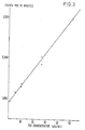
- All other reagents, and the assay protocol for determination of TSH were as described in Example 1. An example dose response curve is shown in Fig.3. Counts per ten minutes bound are plotted along the vertical axis and micro international units human TSH/ml are plotted along the horizontal axis.
separating the solids fraction from the liquid fraction of the mixture, and determining the amount of label in one of said fractions and, therefrom, the amount of antigen present in the sample.
Claims (11)
Applications Claiming Priority (4)
| Application Number | Priority Date | Filing Date | Title |
|---|---|---|---|
| GB8227826 | 1982-09-29 | ||
| GB8227828 | 1982-09-29 | ||
| GB8227826 | 1982-09-29 | ||
| GB8227828 | 1982-09-29 |
Publications (2)
| Publication Number | Publication Date |
|---|---|
| EP0105714A1 true EP0105714A1 (en) | 1984-04-18 |
| EP0105714B1 EP0105714B1 (en) | 1988-07-27 |
Family
ID=26283980
Family Applications (1)
| Application Number | Title | Priority Date | Filing Date |
|---|---|---|---|
| EP83305834A Expired EP0105714B1 (en) | 1982-09-29 | 1983-09-28 | Immunoassay of antigens |
Country Status (2)
| Country | Link |
|---|---|
| EP (1) | EP0105714B1 (en) |
| DE (1) | DE3377531D1 (en) |
Cited By (30)
| Publication number | Priority date | Publication date | Assignee | Title |
|---|---|---|---|---|
| EP0119736A2 (en) * | 1983-02-16 | 1984-09-26 | The Board Of Trustees Of The Leland Stanford Junior University | Two-site immunoassays using monoclonal antibodies of different classes or subclasses and test kits for performing same |
| EP0142301A2 (en) | 1983-10-25 | 1985-05-22 | Serono Diagnostics Limited | Methods of assay |
| EP0147848A2 (en) * | 1984-01-02 | 1985-07-10 | Roche Diagnostics GmbH | Method for the determination of a polyvalent antigen, and reagent therefor |
| DE3400027A1 (en) * | 1984-01-02 | 1985-07-18 | Boehringer Mannheim Gmbh, 6800 Mannheim | Method for the determination of a polyvalent antigen and reagent therefor |
| WO1986000641A1 (en) * | 1984-07-03 | 1986-01-30 | Technology Licence Company Limited | Monoclonal antibodies and their use |
| WO1986000645A1 (en) * | 1984-07-03 | 1986-01-30 | Technology Licence Company Limited | Monoclonal antibodies and their use |
| WO1986000644A1 (en) * | 1984-07-03 | 1986-01-30 | Technology Licence Company Limited | Monoclonal antibodies and their use |
| WO1986000646A1 (en) * | 1984-07-03 | 1986-01-30 | Technology Licence Company Limited | Monoclonal antibodies and their use |
| WO1986000642A1 (en) * | 1984-07-03 | 1986-01-30 | Technology Licence Company Limited | Monoclonal antibodies and their use |
| WO1986000643A1 (en) * | 1984-07-03 | 1986-01-30 | Technology Licence Company Limited | Monoclonal antibodies and their use |
| WO1986001807A1 (en) * | 1984-09-07 | 1986-03-27 | Technology Licence Company Limited | Monoclonal antibodies and their use |
| WO1986001805A1 (en) * | 1984-09-07 | 1986-03-27 | Technology Licence Company Limited | Monoclonal antibodies and their use |
| WO1986001808A1 (en) * | 1984-09-07 | 1986-03-27 | Technology Licence Company Limited | Monoclonal antibodies and their use |
| WO1986001804A1 (en) * | 1984-09-07 | 1986-03-27 | Technology Licence Company Limited | Monoclonal antibodies and their use |
| WO1986001806A1 (en) * | 1984-09-07 | 1986-03-27 | Technology Licence Company Limited | Monoclonal antibodies and their use |
| EP0188093A2 (en) * | 1984-12-12 | 1986-07-23 | Immunomedics, Inc. | Sandwich immunoassay |
| EP0190006A1 (en) * | 1985-01-23 | 1986-08-06 | Serono Diagnostics Partners (A Massachusetts Limited Partnership) | Methods of immunoassay |
| EP0225709A2 (en) * | 1985-10-22 | 1987-06-16 | Centocor, Inc. | Immunometric assay for high molecular weight carcinoembryonic antigen |
| EP0238353A2 (en) * | 1986-03-21 | 1987-09-23 | ARS Holding 89 N.V. | Methods of immunoassay |
| EP0249357A2 (en) * | 1986-06-10 | 1987-12-16 | ARS Holding 89 N.V. | Methods of immunoassay |
| EP0289003A2 (en) * | 1987-04-28 | 1988-11-02 | Roche Diagnostics GmbH | Immunochemical method and reagent for the determination of a polyvalent antigen in a fluid probe |
| US4935339A (en) * | 1985-05-07 | 1990-06-19 | Nichols Institute Diagnostics | Delayed solid phase immunologic assay |
| US5260194A (en) * | 1986-09-05 | 1993-11-09 | Syntex (U.S.A.) Inc. | Immunoseparating strip |
| DE4214922A1 (en) * | 1992-05-06 | 1993-11-11 | Henning Berlin Gmbh | Method for determining the amount of a ligand in a biological fluid and kit for performing such a method |
| DE4328070C1 (en) * | 1993-08-20 | 1994-11-24 | Henning Berlin Gmbh | Method for the determination of an analyte in a volume of a liquid sample, and its use for the determination of anti-TSH receptor autoantibodies in a patient's serum |
| WO1995015498A1 (en) * | 1993-12-01 | 1995-06-08 | Abbott Laboratories | Immunoassays employing generic anti-hapten antibodies and materials for use therein |
| US5587294A (en) * | 1991-07-19 | 1996-12-24 | Assay Research, Inc. | Method and kit for measuring endogenous cytokines |
| US5965379A (en) * | 1991-07-19 | 1999-10-12 | Cytimmune Sciences Inc. | Method for measuring endogenous cytokines |
| WO2002001226A1 (en) * | 2000-06-29 | 2002-01-03 | Evotec Technologies Gmbh | Competitive assay method |
| WO2005071410A1 (en) * | 2004-01-13 | 2005-08-04 | Tanox, Inc. | Detecting and quantifying host cell proteins in recombinant protein products |
Families Citing this family (1)
| Publication number | Priority date | Publication date | Assignee | Title |
|---|---|---|---|---|
| CA2547351A1 (en) | 2003-12-01 | 2005-07-07 | Dade Behring Marburg Gmbh | Homogeneous detection method |
Citations (4)
| Publication number | Priority date | Publication date | Assignee | Title |
|---|---|---|---|---|
| US4088746A (en) * | 1976-11-11 | 1978-05-09 | Bio-Rad Laboratories, Inc. | Radioimmunoassay for thyroid-stimulating hormone (TSH) |
| US4244940A (en) * | 1978-09-05 | 1981-01-13 | Bio-Rad Laboratories, Inc. | Single-incubation two-site immunoassay |
| US4297337A (en) * | 1979-04-13 | 1981-10-27 | Corning Glass Works | Solid-phase immunoassays using magnetic glass |
| US4312854A (en) * | 1978-09-13 | 1982-01-26 | Ames-Yissum Ltd. | Immunoassay for thyroid stimulating hormone employing florisil adsorbent as separating agent |
Family Cites Families (3)
| Publication number | Priority date | Publication date | Assignee | Title |
|---|---|---|---|---|
| NL7501215A (en) * | 1975-02-01 | 1976-08-03 | Akzo Nv | METHOD FOR DETERMINING AND DETERMINING AN ANTIGEN OR ANTIBODY. |
| SE8006424L (en) * | 1980-09-12 | 1982-03-13 | Jolla Cancer Res Found | PUT TO DETERMINE AN ANTIGEN IN SOLUTION |
| DE3225027A1 (en) * | 1982-07-05 | 1984-01-05 | Boehringer Mannheim Gmbh, 6800 Mannheim | IMMUNCHEMICAL MEASUREMENT METHOD |
-
1983
- 1983-09-28 EP EP83305834A patent/EP0105714B1/en not_active Expired
- 1983-09-28 DE DE8383305834T patent/DE3377531D1/en not_active Expired
Patent Citations (4)
| Publication number | Priority date | Publication date | Assignee | Title |
|---|---|---|---|---|
| US4088746A (en) * | 1976-11-11 | 1978-05-09 | Bio-Rad Laboratories, Inc. | Radioimmunoassay for thyroid-stimulating hormone (TSH) |
| US4244940A (en) * | 1978-09-05 | 1981-01-13 | Bio-Rad Laboratories, Inc. | Single-incubation two-site immunoassay |
| US4312854A (en) * | 1978-09-13 | 1982-01-26 | Ames-Yissum Ltd. | Immunoassay for thyroid stimulating hormone employing florisil adsorbent as separating agent |
| US4297337A (en) * | 1979-04-13 | 1981-10-27 | Corning Glass Works | Solid-phase immunoassays using magnetic glass |
Non-Patent Citations (8)
| Title |
|---|
| "Immunoassays for Clinical Chemistry", 1983, CHURCHILL LIVINGSTONE, pages: 147 - 162 |
| AXEN ET AL., NATURE, vol. 214, 1967, pages 1302 - 1304 |
| FORREST; RATTLE: "Immunoasays for Clinical Chemistry", 1983, CHURCHILL LIVINGSTONE, article "Magnetic Particle Radio immunoassay", pages: 147 - 162 |
| GALFRE, G.; MILSTEIN, C.: "Preparation of Monoclonal Antibodies: Strategies and Procedures", METHODS IN ENZYMOLOGY, vol. 73, 1981, pages 1 - 46 |
| HUNTER ET AL., J. IMMUNOL. METHODS, vol. 50, 1982, pages 133 - 144 |
| METHODS IN ENZYMOLOGY, vol. 73, Immunochemical Techniques, part B G. GALFRE, C. MILSTEIN "Preparation of Monoclonal Antibodies: Strategies and Procedures" pages 3-46 * |
| MILSTEIN; KOHLER, NATURE, vol. 256, 1975, pages 495 - 497 |
| NATURE, vol. 256, August 7, 1975 G. KOHLER, C. MILSTEIN "Continuous cultures of fused cells secreting antibody of predefined specifity" pages 495-497 * |
Cited By (40)
| Publication number | Priority date | Publication date | Assignee | Title |
|---|---|---|---|---|
| EP0119736A2 (en) * | 1983-02-16 | 1984-09-26 | The Board Of Trustees Of The Leland Stanford Junior University | Two-site immunoassays using monoclonal antibodies of different classes or subclasses and test kits for performing same |
| EP0119736A3 (en) * | 1983-02-16 | 1985-04-17 | The Board Of Trustees Of The Leland Stanford Junior University | Two-site immunoassays using monoclonal antibodies of different classes or subclasses and test kits for performing same |
| EP0142301A2 (en) | 1983-10-25 | 1985-05-22 | Serono Diagnostics Limited | Methods of assay |
| EP0147848A2 (en) * | 1984-01-02 | 1985-07-10 | Roche Diagnostics GmbH | Method for the determination of a polyvalent antigen, and reagent therefor |
| DE3400027A1 (en) * | 1984-01-02 | 1985-07-18 | Boehringer Mannheim Gmbh, 6800 Mannheim | Method for the determination of a polyvalent antigen and reagent therefor |
| EP0147848A3 (en) * | 1984-01-02 | 1988-06-22 | Boehringer Mannheim Gmbh | Method for the determination of a polyvalent antigen, and reagent therefor |
| WO1986000646A1 (en) * | 1984-07-03 | 1986-01-30 | Technology Licence Company Limited | Monoclonal antibodies and their use |
| WO1986000644A1 (en) * | 1984-07-03 | 1986-01-30 | Technology Licence Company Limited | Monoclonal antibodies and their use |
| WO1986000645A1 (en) * | 1984-07-03 | 1986-01-30 | Technology Licence Company Limited | Monoclonal antibodies and their use |
| WO1986000642A1 (en) * | 1984-07-03 | 1986-01-30 | Technology Licence Company Limited | Monoclonal antibodies and their use |
| WO1986000643A1 (en) * | 1984-07-03 | 1986-01-30 | Technology Licence Company Limited | Monoclonal antibodies and their use |
| WO1986000641A1 (en) * | 1984-07-03 | 1986-01-30 | Technology Licence Company Limited | Monoclonal antibodies and their use |
| WO1986001807A1 (en) * | 1984-09-07 | 1986-03-27 | Technology Licence Company Limited | Monoclonal antibodies and their use |
| WO1986001805A1 (en) * | 1984-09-07 | 1986-03-27 | Technology Licence Company Limited | Monoclonal antibodies and their use |
| WO1986001808A1 (en) * | 1984-09-07 | 1986-03-27 | Technology Licence Company Limited | Monoclonal antibodies and their use |
| WO1986001804A1 (en) * | 1984-09-07 | 1986-03-27 | Technology Licence Company Limited | Monoclonal antibodies and their use |
| WO1986001806A1 (en) * | 1984-09-07 | 1986-03-27 | Technology Licence Company Limited | Monoclonal antibodies and their use |
| EP0188093A3 (en) * | 1984-12-12 | 1987-09-30 | Immunomedics, Inc. | Sandwich immunoassay |
| EP0188093A2 (en) * | 1984-12-12 | 1986-07-23 | Immunomedics, Inc. | Sandwich immunoassay |
| US4808521A (en) * | 1985-01-23 | 1989-02-28 | Serone Diagnostics Partners | Double-labeled enzymeimmunoassay methods |
| EP0190006A1 (en) * | 1985-01-23 | 1986-08-06 | Serono Diagnostics Partners (A Massachusetts Limited Partnership) | Methods of immunoassay |
| US4935339A (en) * | 1985-05-07 | 1990-06-19 | Nichols Institute Diagnostics | Delayed solid phase immunologic assay |
| EP0225709A2 (en) * | 1985-10-22 | 1987-06-16 | Centocor, Inc. | Immunometric assay for high molecular weight carcinoembryonic antigen |
| EP0225709A3 (en) * | 1985-10-22 | 1988-09-07 | Centocor, Inc. | Immunometric assay for high molecular weight carcinoembryonic antigen |
| EP0238353A3 (en) * | 1986-03-21 | 1989-11-15 | Serono Diagnostics Partners (A Massachusetts Limited Partnership) | Methods of immunoassay |
| EP0238353A2 (en) * | 1986-03-21 | 1987-09-23 | ARS Holding 89 N.V. | Methods of immunoassay |
| EP0249357A2 (en) * | 1986-06-10 | 1987-12-16 | ARS Holding 89 N.V. | Methods of immunoassay |
| EP0249357A3 (en) * | 1986-06-10 | 1989-10-04 | Serono Diagnostics Partners (A Massachusetts Limited Partnership) | Methods of immunoassay |
| US5260194A (en) * | 1986-09-05 | 1993-11-09 | Syntex (U.S.A.) Inc. | Immunoseparating strip |
| EP0289003A2 (en) * | 1987-04-28 | 1988-11-02 | Roche Diagnostics GmbH | Immunochemical method and reagent for the determination of a polyvalent antigen in a fluid probe |
| EP0289003A3 (en) * | 1987-04-28 | 1991-04-10 | Roche Diagnostics GmbH | Immunochemical method and reagent for the determination of a polyvalent antigen in a fluid probe |
| JPS63284470A (en) * | 1987-04-28 | 1988-11-21 | ベーリンガー・マンハイム・ゲゼルシヤフト・ミツト・ベシユレンクテル・ハフツング | Quantitative measuring method of polyvalent antigen and reagent thereof and manufacture of acceptor |
| US5587294A (en) * | 1991-07-19 | 1996-12-24 | Assay Research, Inc. | Method and kit for measuring endogenous cytokines |
| US5965379A (en) * | 1991-07-19 | 1999-10-12 | Cytimmune Sciences Inc. | Method for measuring endogenous cytokines |
| US5518887A (en) * | 1992-03-30 | 1996-05-21 | Abbott Laboratories | Immunoassays empolying generic anti-hapten antibodies and materials for use therein |
| DE4214922A1 (en) * | 1992-05-06 | 1993-11-11 | Henning Berlin Gmbh | Method for determining the amount of a ligand in a biological fluid and kit for performing such a method |
| DE4328070C1 (en) * | 1993-08-20 | 1994-11-24 | Henning Berlin Gmbh | Method for the determination of an analyte in a volume of a liquid sample, and its use for the determination of anti-TSH receptor autoantibodies in a patient's serum |
| WO1995015498A1 (en) * | 1993-12-01 | 1995-06-08 | Abbott Laboratories | Immunoassays employing generic anti-hapten antibodies and materials for use therein |
| WO2002001226A1 (en) * | 2000-06-29 | 2002-01-03 | Evotec Technologies Gmbh | Competitive assay method |
| WO2005071410A1 (en) * | 2004-01-13 | 2005-08-04 | Tanox, Inc. | Detecting and quantifying host cell proteins in recombinant protein products |
Also Published As
| Publication number | Publication date |
|---|---|
| EP0105714B1 (en) | 1988-07-27 |
| DE3377531D1 (en) | 1988-09-01 |
Similar Documents
| Publication | Publication Date | Title |
|---|---|---|
| US4659678A (en) | Immunoassay of antigens | |
| EP0105714A1 (en) | Immunoassay of antigens | |
| CA1261745A (en) | Methods of assay | |
| US5108896A (en) | Simultaneous immunoassay of two analytes using dual enzyme labelled antibodies | |
| US4343896A (en) | Method and test pack for the demonstration and determination of an antigen or antibody | |
| US4143124A (en) | Antigen-antibody analysis with solid phase rf and c1q | |
| US5126241A (en) | Process for the determination of a specifically bindable substance | |
| JPH06258325A (en) | Antigen measuring method using two or three kinds of different monoclonal antibodies and test kit utilizing said monoclonal antibodies | |
| US4504585A (en) | Affinity immunoassay system | |
| JPH08101197A (en) | Immunity-chromatography assay in controlled sensitivity | |
| AU664621B2 (en) | Two step process for coating of antibodies to a solid phase | |
| JPH0421818B2 (en) | ||
| EP0207152B1 (en) | Solid phase diffusion assay | |
| US4283383A (en) | Analysis of biological fluids | |
| US4138213A (en) | Agglutination immunoassay of immune complex with RF or Clq | |
| US5366859A (en) | Radioimmunoassay method | |
| CA1088422A (en) | Double anti body-coated solid phase | |
| US5196349A (en) | Immunoassays for thyroid hormones using thyroglobulin | |
| US7252961B2 (en) | Competitive immunoassay using complexed analyte derivatives | |
| IE850007L (en) | Process and reagent for the determination of a polyvalent¹antigen | |
| IE60253B1 (en) | "Membrane affinity concentration immunoassay" | |
| JPH0572208A (en) | Immunological suppression test using monoclonal antibody | |
| WO1986002736A1 (en) | Immunoassay method for small molecules | |
| JPH06160387A (en) | Measuring method for antigen-antibody reaction | |
| Killeen et al. | High-dose hook effect in an immunoluminometric thyrotropin assay: the open-faced sandwich artefact |
Legal Events
| Date | Code | Title | Description |
|---|---|---|---|
| PUAI | Public reference made under article 153(3) epc to a published international application that has entered the european phase |
Free format text: ORIGINAL CODE: 0009012 |
|
| AK | Designated contracting states |
Designated state(s): BE CH DE FR GB IT LI NL SE |
|
| 17P | Request for examination filed |
Effective date: 19840516 |
|
| RAP1 | Party data changed (applicant data changed or rights of an application transferred) |
Owner name: SERONO DIAGNOSTICS LIMITED |
|
| GRAA | (expected) grant |
Free format text: ORIGINAL CODE: 0009210 |
|
| AK | Designated contracting states |
Kind code of ref document: B1 Designated state(s): BE CH DE FR GB IT LI NL SE |
|
| REF | Corresponds to: |
Ref document number: 3377531 Country of ref document: DE Date of ref document: 19880901 |
|
| ET | Fr: translation filed | ||
| ITF | It: translation for a ep patent filed |
Owner name: SOCIETA' ITALIANA BREVETTI S.P.A. |
|
| PLBI | Opposition filed |
Free format text: ORIGINAL CODE: 0009260 |
|
| 26 | Opposition filed |
Opponent name: BOEHRINGER MANNHEIM GMBH Effective date: 19890427 |
|
| NLR1 | Nl: opposition has been filed with the epo |
Opponent name: BOEHRINGER MANNHEIM GMBH |
|
| PLBM | Termination of opposition procedure: date of legal effect published |
Free format text: ORIGINAL CODE: 0009276 |
|
| STAA | Information on the status of an ep patent application or granted ep patent |
Free format text: STATUS: OPPOSITION PROCEDURE CLOSED |
|
| 27C | Opposition proceedings terminated |
Effective date: 19900524 |
|
| ITPR | It: changes in ownership of a european patent |
Owner name: CESSIONE;BIODATA S.P.A. |
|
| ITTA | It: last paid annual fee | ||
| NLR2 | Nl: decision of opposition | ||
| EAL | Se: european patent in force in sweden |
Ref document number: 83305834.0 |
|
| REG | Reference to a national code |
Ref country code: GB Ref legal event code: IF02 |
|
| PGFP | Annual fee paid to national office [announced via postgrant information from national office to epo] |
Ref country code: FR Payment date: 20020917 Year of fee payment: 20 |
|
| PGFP | Annual fee paid to national office [announced via postgrant information from national office to epo] |
Ref country code: SE Payment date: 20020925 Year of fee payment: 20 |
|
| PGFP | Annual fee paid to national office [announced via postgrant information from national office to epo] |
Ref country code: GB Payment date: 20020926 Year of fee payment: 20 |
|
| PGFP | Annual fee paid to national office [announced via postgrant information from national office to epo] |
Ref country code: CH Payment date: 20020927 Year of fee payment: 20 |
|
| PGFP | Annual fee paid to national office [announced via postgrant information from national office to epo] |
Ref country code: NL Payment date: 20020930 Year of fee payment: 20 Ref country code: DE Payment date: 20020930 Year of fee payment: 20 |
|
| PGFP | Annual fee paid to national office [announced via postgrant information from national office to epo] |
Ref country code: BE Payment date: 20021011 Year of fee payment: 20 |
|
| PG25 | Lapsed in a contracting state [announced via postgrant information from national office to epo] |
Ref country code: LI Free format text: LAPSE BECAUSE OF EXPIRATION OF PROTECTION Effective date: 20030927 Ref country code: GB Free format text: LAPSE BECAUSE OF EXPIRATION OF PROTECTION Effective date: 20030927 Ref country code: CH Free format text: LAPSE BECAUSE OF EXPIRATION OF PROTECTION Effective date: 20030927 |
|
| PG25 | Lapsed in a contracting state [announced via postgrant information from national office to epo] |
Ref country code: NL Free format text: LAPSE BECAUSE OF EXPIRATION OF PROTECTION Effective date: 20030928 |
|
| BE20 | Be: patent expired |
Owner name: *SERONO DIAGNOSTICS LTD Effective date: 20030928 |
|
| REG | Reference to a national code |
Ref country code: GB Ref legal event code: PE20 |
|
| REG | Reference to a national code |
Ref country code: CH Ref legal event code: PL |
|
| EUG | Se: european patent has lapsed | ||
| NLV7 | Nl: ceased due to reaching the maximum lifetime of a patent |
Effective date: 20030928 |