EP0120658A2 - Method for identifying and characterizing organisms - Google Patents
Method for identifying and characterizing organisms Download PDFInfo
- Publication number
- EP0120658A2 EP0120658A2 EP84301800A EP84301800A EP0120658A2 EP 0120658 A2 EP0120658 A2 EP 0120658A2 EP 84301800 A EP84301800 A EP 84301800A EP 84301800 A EP84301800 A EP 84301800A EP 0120658 A2 EP0120658 A2 EP 0120658A2
- Authority
- EP
- European Patent Office
- Prior art keywords
- dna
- organism
- probe
- nucleic acid
- species
- Prior art date
- Legal status (The legal status is an assumption and is not a legal conclusion. Google has not performed a legal analysis and makes no representation as to the accuracy of the status listed.)
- Granted
Links
Images


Classifications
-
- C—CHEMISTRY; METALLURGY
- C12—BIOCHEMISTRY; BEER; SPIRITS; WINE; VINEGAR; MICROBIOLOGY; ENZYMOLOGY; MUTATION OR GENETIC ENGINEERING
- C12Q—MEASURING OR TESTING PROCESSES INVOLVING ENZYMES, NUCLEIC ACIDS OR MICROORGANISMS; COMPOSITIONS OR TEST PAPERS THEREFOR; PROCESSES OF PREPARING SUCH COMPOSITIONS; CONDITION-RESPONSIVE CONTROL IN MICROBIOLOGICAL OR ENZYMOLOGICAL PROCESSES
- C12Q1/00—Measuring or testing processes involving enzymes, nucleic acids or microorganisms; Compositions therefor; Processes of preparing such compositions
- C12Q1/68—Measuring or testing processes involving enzymes, nucleic acids or microorganisms; Compositions therefor; Processes of preparing such compositions involving nucleic acids
- C12Q1/6876—Nucleic acid products used in the analysis of nucleic acids, e.g. primers or probes
- C12Q1/6888—Nucleic acid products used in the analysis of nucleic acids, e.g. primers or probes for detection or identification of organisms
-
- C—CHEMISTRY; METALLURGY
- C12—BIOCHEMISTRY; BEER; SPIRITS; WINE; VINEGAR; MICROBIOLOGY; ENZYMOLOGY; MUTATION OR GENETIC ENGINEERING
- C12Q—MEASURING OR TESTING PROCESSES INVOLVING ENZYMES, NUCLEIC ACIDS OR MICROORGANISMS; COMPOSITIONS OR TEST PAPERS THEREFOR; PROCESSES OF PREPARING SUCH COMPOSITIONS; CONDITION-RESPONSIVE CONTROL IN MICROBIOLOGICAL OR ENZYMOLOGICAL PROCESSES
- C12Q1/00—Measuring or testing processes involving enzymes, nucleic acids or microorganisms; Compositions therefor; Processes of preparing such compositions
- C12Q1/68—Measuring or testing processes involving enzymes, nucleic acids or microorganisms; Compositions therefor; Processes of preparing such compositions involving nucleic acids
- C12Q1/6813—Hybridisation assays
- C12Q1/6827—Hybridisation assays for detection of mutation or polymorphism
- C12Q1/683—Hybridisation assays for detection of mutation or polymorphism involving restriction enzymes, e.g. restriction fragment length polymorphism [RFLP]
Definitions
- the present invention relates to a method for the rapid and accurate characterization and identification of organisms, including prokaryotic and eukaryotic organisms, such as bacteria, plants, and animals.
- Prokaryotes are less complex than eukaryotes, they lack internal compart- mentalization by unit membrane systems, and lack a defined nucleus. Prokaryotic genetic information is carried in the cytoplasm on double-stranded, circular DNA; no other DNA is present in cells (except for the possible presence of phage, bacterial viruses, and cirular DNA plasmids, capable of autonomous replication). Eukaryotes, on the other hand, have a multiplicity of unit membrane systems which serve to segregate many of the functional components into specialized and isolated regions.
- DNA genetic information
- organelles mitochondria and (in photosynthetic organisms) chloroplasts.
- the replication, transcription, and translation of the eukaryotic genome occurs at either two or three distinct sites within the cell: in the nucleocytoplasmic region, in the mitochondrion, and in the chloroplast.
- prokaryotes and ' eukaryotes breaks down when a comparison of mitochondria and chloroplasts is carried out with prokaryotes: these organelles are today considered to have been derived from free-living prokaryotes, which entered into an endosymbiotic relation with primitive eukaryotes, and eventually became closely integrated with the machinery of the host cell and incapable of independent existence (see e.g., Fox, G.E. et al, Science 209:457-463 (1980), at 462; Stanier, R . Y . et al, "The Microbial World,” Fourth Edition, Prentice-Hall, Inc., 1976, at p. 86).
- the eukaryotic cell is a phylogenetic "chimera” with organelle components that are clearly prokaryotic in nature.
- the "prokaryotic- eukaryotic” dichotomy then, also has drawbacks, even as a broad classification method.
- bacterial species usually represent many strains, and a strain is considered to be a population derived from a single cell.
- Bacterial species are usually defined by describing the degree of homogeneity and diversity of attributes in representative samples of strains of species. Precise definitions of bacterial species are difficult to express because subjective limits to strain diversity within species are required to define- species boundaries. (Buchanan, R. E., International Bulletin of Bacteriological Nomenclature and Taxonomy, 15:25-32 (1965)).
- Species as applied to bacteria, has been defined as a distinct kind of organism, having certain distinguishing features, and as a group of organisms which generally bear a close resemblance to one another in the more essential features of their organization. The problem with these definitions is that they are subjective; Brenner, supra, at page 2. Species have also been defined solely on the basis of criteria such as host range, pathogenicity, ability or inability to produce gas in the fermentation of a given sugar, and rapid or delayed fermentation of sugars.
- the general method furthermore, suffers from several problems when it is used as the sole basis for defining a species, among them the number and nature of the tests to be used, whether the tests should be weighted and how, what level of similarity should be chosen to reflect relatedness, whether the same level of similarities is applicable to all groups, etc.
- a molecular approach to bacterial classification is to compare two genomes by DNA-DNA reassociation.
- a genetic definition of species includes the provision that strains of species are 70% or more related. With DNA-DNA reassociation a strain can be identified only if the radioactively labeled DNA probe and unknown DNA are from the same species. The practical application of this 70% species definition however is limited by selection of an appropriate probe. This may be overcome in part by selecting phenotypic attributes which seem to correlate with the reassociation group, but when these are used alone the DNA-DNA reassociation species definition is also applied indirectly.
- a need also exists for a method useful for identifying and distinguishing genera and species of any living organism, which can be readily and reliably used by veterinarians, plant-breeders, tox- icologists, animal breeders, entomologists and in other related areas, where such identification is necessary.
- Yet another object of the invention is to provide a method of identifying organisms such as bacteria which utilizes the organisms' genome.
- Another object of the invention is to provide a method of characterizing and identifying species and genera of pathogenic organisms in the clinical laboratory, so as to provide the capability of characterizing and identifying the cause of any given animal or plant disease.
- Still another object of the invention is to provide various products useful in the aforementioned methodologies.
- Still another object of the invention has been . .attained by providing:
- strains of species should contain structural information which is a clue to their common origin.
- the greatest amount of an organism's past history survives in semantides,'DNA and RNA, (Zuckerkandle, E. and Pauling, L., Journal of Theoretical Biology, 8:357-366 (1965)).
- restriction endonuclease digests of DNA have sets of fragments containing conserved sequences that are similar in strains of a species of organism (e.g., bacteria), but different in strains of other species of the organism; i.e., despite strain variation, enzyme specific sets of restriction fragments with high frequencies of occurrence, minimal genotypic characters, define the species. This is the essence of the invention described in EP-A-007-6123- and also that of the invention described herein.
- the present invention constitutes an extension of the concepts developed in EP-A-0076123, in that it has further been discovered that there exist sequences, in addition to those of rRNA, which are highly conserved through evolution and which may be as useful as rRNA sequences in the identification system.
- the present invention provides means for carrying out the identification and characterization techniques of EP-A-0076123, using any probe which is conserved, other than rRNA.
- the present invention also provides additional examples of methods which may be used in the identification process.
- the present inventor has also discovered that the method is general, in that it is-applicable to both eukaryotic and prokaryotic DNA, using a conserved nucleic acid probe from any organism, prokaryotic or eukaryotic, of the same or different (classic) taxonomic classification than the organism being identified.
- the invention offers an objective method of defining organisms based on conserved sequences of DNA or other genetic material in relation to known positions such as restriction endonuclease sites.
- the detection of restriction fragments containing a conserved sequence may be carried out by hybridizing or reassociating DNA segments with nucleic acid containing conserved sequence information from a probe organism.
- organism which can-be characterized (which term is meant to include “identified") by the process of the invention, it is meant to include virtually any organism which, by definition, contains DNA or RNA in its genome. In this respect it is useful to refer to a classical taxonomic scheme as a point of reference.
- Subkingdom Protozoa Phylum Protozoa (Acellular animals) subphylum plasmodroma, classes flagellata, sarcodina and sporozoa; subphylum ciliophora, class ciliata; the Subkingdom Parazoa, Phylum porifera (Sponges), class calcarea, hexactinellida, and desmospongiae; the Subkingdom Mesozoa, Phylum mesozoa; the Subkingdom Metazoa, Section Radiata, Phylum coelenterata, classes hydrozoa, scypho- zoa, anthozoa, Phylum ctenophora, classes tenta-culata and nuda; Section Protostomia Phylum platyhelmintes (flat- worms) classes tubellana, trematoda, and cestoda; Phylum .
- nemertina Phylum acanthocephala
- Phylum aschelmintles classes rotifera, gastrotricha, kinorhyncha, priapulida, nematoda and nematomorpha
- Phylum entoprocta Phylum ectoprocta, classes gymnolaemata and phylactolaemata
- Phylum phoronida Phylum braciopoda, classes inarticulata and articulata
- Phylum sipunculida Phylum echiurida
- Phylum annelida classes polychaeta, oligochaeta and hirudinea
- Phylum onychophora Phylum tardigrada
- the DNA to be analyzed is that present in the cells or in the non-compartmentalized chromosomes.
- identifying a eukaryotic organism one may either use the nuclear DNA or the organelle DNA (mitochondrial DNA or chloroplast DNA).
- DNA and/or small circular DNAs are isolated from the organism to be identified in order to analyze the conserved sequences (and possibly sequences that could be used to create a taxon below the rank of species or infrasubspecific subdivisions.)
- the DNA's are extracted by methods which are well-known to the art.
- the DNA's are analyzed to ascertain both 1) the presence and position of the conserved sequences and 2) their position relative to endonuclease restriction sites.
- the easiest way to analyze for the presence of the conserved sequences is to utilize a polynucleotide probe capable of hybridizing with the conserved DNA sequence.
- direct sequence information as obtained by chemical sequence determination and analysis thereof could also be utilized.
- the probe utilized was an rRNA information containing-probe; in this case any other probe havingconserved sequences could be used.
- the easiest way of finding a given set of endonuclease restriction sites is to cleave the DNA with the appropriate restriction enzymes. (This, indeed, is the manner taught and practised in EP-A-0076123.
- alternative methods such as sequence information coupled with known restriction site sequences, or cleavage and partial sequencing could also be used..
- DNA's are going to be cut at specific £ sites into fragments by restriction endonucleases.
- the fragments are separated according to size by a chromatographic system.
- gel chromatography was used as an example of a useful chromatographic system.
- other systems can also be used, such as'high pressure liquid chromatography, capillary zone electrophoresis, or other separation techniques.
- gel chromatography the fragments are separated, the gels are stained, as is otherwise well-known in the art, and standardized as to the fragment sizes using standards curves constructed with fragments of known sizes.
- the separated fragments may then be transferred to cellulose nitrite paper by the Southern blot technique (Southern, E.
- fragments containing the conserved sequences are then located by their capacity to hybridize with a nucleic acid probe containing conserved sequence information.
- hybridization can occur after digestion but before separation; or restriction cleavage can occur after hybridization, followed by separation of the fragments.
- the nucleic acid probe can either be non- radioactively labeled or, preferably, radioactively labeled.
- the probe can be RNA, or preferably DNA which is complementary to RN A (cDNA), either synthesized by reverse transcription or contained on a cloned-fragment, which can be labeled, for example, by nick translation.
- cDNA RN A
- synthetic oligodeoxyribonucleotides may be prepared with labeled nucleotides.
- the well-defined probe is derived from an arbitrarily chosen organism, see infra, or may be a consensus sequence.
- the hybridized fragments are detected by selectively detecting double stranded nucleic acid (non-radiolabeled probe), or visualized by, e.g., autoradiography (radiolabeled probe).
- the -size of each fragment which has been hybridized is relative to the restriction sites and is determined from the distance traveled using standard curves, as described previously.
- the amount of hybridization, the pattern of hybridization, and the sizes of the hybridized fragments, which are relative to restriction sites can be used individually or in conjunction to identify the organism.
- the genetic characterization that emerges from this technique can be readily compared to equivalent characterizations derived from at least two and up to a multiplicity of known, standard organisms, genera or species.
- the comparison can be either by visual inspection and matching of appropriate chromatographic patterns, (as in E P-A-0076123) by comparison of hybridized restriction fragment sizes, by band intensity (amount of hybridization) or by any combination thereof.
- the comparison is carried out with a one- . dimensional computer-based pattern recognition system, such as those used in point-of-sale transactions.
- restriction enzymes can be chosen for-their ability to distinguish strains within species.
- the "probe organism” used in the present invention can also be any of the aforementioned organisms; it can be either eukaryotic or prokaryotic.
- the only limitation is given by the fact that the conserved sequence-containing probe should hybridize maximally with the unknown organism's DNA.
- sequence information-containing probes There are four types of-conserved sequence information-containing probes: 1) prokaryotic probes (especially bacterial - derived), 2) eukaryotic mitochondrial probes, 3) eukaryotic chloroplast probes, and 4) eukaryotic non-organelle probes.
- prokaryotic probes especially bacterial - derived
- eukaryotic mitochondrial probes especially bacterial - derived
- eukaryotic chloroplast probes There are also four sources of DNA (to be endonuclease digested): 1) prokaryotic cellular DNA, 2) eukaryotic mitochondrial DNA, 3) eukaryotic chloroplast DNA, and 4) eukaryotic nuclear DNA.
- the following hybridization table can thus be constructed (Table 1).
- the Table shows which probes can generally be maximally hybridized with which unknown DNA. For example, one can identify a eukaryotic organism by extracting species specific mitochondrial or chloroplast DNA, endonuclease-digesting it and hybridizing the digest with either a prokaryotic probe, or with an organelle derived eukaryotic probe. In the same manner, one can identify a prokaryotic organism by extracting species-specific cellular DNA, endonuclease-digesting it, and hybridizing the digest with either a prokaryotic probe, or an organelle-derived eukaryotic RNA probe.
- a eukaryotic organism can identify a eukaryotic organism by extracting and digesting species-specific nuclear DNA, and hybridizing it with a non-organelle derived eukaryotic probe.
- Eukaryotes could be defined by one or any combination of the nuclear, mitochondria, or in some cases chloroplast systems. These cross-hybridizations are based on the fact that nucleic acid derived from eukaryotic organelles has extensive homology with evolutionarily conserved . sequences from prokaryotic nucleic acid, but that the same homologies are generally not present to such extent between nuclear-derived eukaryotic DNA and prokaryotic DNA.
- any pair of DNA to be digested and . accompanying probe is arbitrary, and will depend on the organism being identified, i.e. it will depend on the question asked.
- a prokaryotic species e.g. bacteria
- a eukaryotic cell e.g. animal or plant
- identifying a eukaryotic species (which is not infected with a prokaryote) with a prokaryotic probe
- it is best to maximize the concentration of organelle-derived DNA as for example by separating organelles from nuclei, and then extracting only organelle DNA.
- prokaryotic probe-(e.g. bacterial probe) to hybridize with prokaryotic DNA. In this manner one could detect, quantify, and identify genera, species, and strains of prokaryotic organisms.
- prokaryotic probes is derived from bacteria, and further, because of the ease and availability, from E. coli. The probe from E. coli can be used to identify any organism, especially any prokaryotic organism, most preferably a strain of any bacterial species.
- Another particularly preferred embodiment is to use eukaryotic probe derived from a given class to identify eukaryotic organisms of the same class (e.g. mammalian probe to identify mammalian organism). Most preferred is to use probe and DNA from the same subclass and/or order and/or family of organisms, (e.g. if identifying a species of mouse, it is preferred to use mouse-derived probe).
- the most sensitive and useful pair systems are those where there is less evolutionary distance or diversity between the source of the probe and the unknown DNA.
- genetic material sequence is used in this invention to denote genetic material, e.g DNA, sequences that show homology between at least two different species of plants, animals or microorganisms.
- the homology between two conserved sequences is to be such that, if one of such DNA molecules were to be detectably labelled, sufficient hybridization or annealing would occur if both single stranded DNA molecules or fragments thereof were placed together under hybridization conditions, thereby to produce a duplex of sufficient stability to be detectable by standard methodology (i.e, radiolabelling, enzyme labelling, and the like).
- EP-A-0076123 the evolutionarily conserved sequence exemplified was that of ribosomal RNA genes. This is still a highly preferred gene sequence. However, it has been discovered that other gene sequences exist which are sufficiently conserved across the evolutionary span to be useful.
- a Family of proteins is one wherein any two proteins differ from each other by less than 50% amino acid residues in their sequence.
- a Subfamily of proteins is one wherein any two proteins differ from each other by less than 20% amino acid residues in their sequence.
- An "Entry” is one wherein any two proteins differ from each other by less than 5% amino acid residues in their sequence.
- geze sequences or appropriate portions thereof which can b: used are: cytochrome C related genes, cytochrome C 3 related genes, cytochrome c l related, cytochrome b 5 relatfd, ferrodoxin related, rebredoxin related, flavodo> n related., alcohol dehydrogenase related, lactaze dehydrogenase related, peroxidase related, adenylaze kinase related, phospholipase A2 related, trzrptophan operon related, carboxypeptidase related, subtilisin related, penicillinase related, protease inhibitor related, somatotropin related, corticotropin related, lipotropin related, glucagon related, snake venom toxin related, plant toxin related, antibacterial toxin related, immunoglogulin related gene, ribosomal other than rRNA- related genes, heme carrier genes, chromosomal protein genes, fibrous protein genes,
- trp D probe within an even narrower domain (e.g., test for the presence of Enterobacteriaceae, or of Bacillus, etc.) with a trp D probe from the same order, family or genus.
- trp D probe from the same order, family or genus.
- a probe containing the conserved DNA sequence information is prepared in the same manner as the preparation of rRNA information containing probe exemplified in EP-A-0076123.
- the probe can thus be RNA, DNA or cDNA, and the like.
- the first step is extraction of the DNA from the unknown organisms.
- Nuclear DNA from eukaryotic cells can be selectively extracted by standard methodology well-known to the art (see for example, Drohan, W. et al, Biochem. Biophys. Acta, 521 (1978), 1-15, herein incorporated by reference). Because organelle DNA is small and circular, spooling techniques serve to separate the non-circular nuclear DNA from the circular, organelle-derived DNA. As a corollary, the non-spooled material contains the organelle-derived DNA which can separately be isolated by density gradient centrifugation.
- mitochondria or chloroplasts
- the purified mitochondrial (or chloroplast) fraction is used for the preparation of organelle-derived DNA while the purified nuclear fraction is used to prepare nuclear DNA.
- P rokaryotic DNA extraction is also well-known in the art.
- an unknown bacterium present in any medium such as an industrial fermentation suspension, agar medium, plant or animal tissue or sample or the like, is treated under well-known conditions designed to extract high molecular weight DNA therefrom.
- cells of the unknown organism can be suspended in extraction buffer, lysozyme added thereto, and the suspension incubated. Cell disruption can be further accelerated by addition of detergents, and/or by increase in temperature.
- Protease digestion followed by chloroform/phenol extraction and ethanol precipitation can be used to finalize the extraction of DNA.
- An alternative method of extraction which is much faster than phenol/chloroform extraction, is rapid isolation of DNA using ethanol precipitation.
- This method is preferably used to isolate DNA directly from colonies or small, liquid cultures.
- the method is described in Davis, R. W. et al: "A Manual for Genetic Engineering, Advanced Bacterial Genetics,” (hereinafter “Davis”), Cold Spring Harbor Laboratory, Cold Spring Harbor, New York, 1980, pp. 120-121, herein incorporated by reference.
- the DNA (prokaryotic or eukaryotic (nuclear or non- nuclear)) is dissolved in physiological buffer for the next step. There are a variety of possible steps to be followed after isolation of the desired DNA. One of these steps is endonuclease digestion.
- restriction endonuclease enzymes Any restriction endonuclease enzyme can be used. Preferably it is not from the same organism species as that being identified, since otherwise, the DNA may remain intact. (This may, in any event, identify the organism, since the enzymes are not expected to cut DNA from the species of their origin.) Since the organism species being characterized may be unknown, obtaining a suitable digest of fragments may entail a minimum amount of trial and error, which can routinely be carried out by those skilled in the art without undue experimentation.
- restriction endonuclease enzymes examples include Bgl I, BamH I, EcoR I, Pst I, Hind III, Bal I, H g a I, Sal I, Xba I, Sac I, Sst I, Bcl I, Xho I, Kpn I, Pvu II, Sau IIIa, or the like. See also Davis, supra, at pp. 228-230, herein incorporated by reference. A mixture of one or more endonucleases can also be used for the digestion. Normally, DNA and endonuclease are incubated together in an appropriate buffer for an appropriate period of time (ranging from 1 to 48 hours, at temperatures ranging from 25°C-65°C, preferably 37°C).
- the resulting identifying genetic characterization will depend on the type or types of endonucleases utilized, and will be endonuclease-specific. It is therefore necessary to note which enzyme or enzymes have been used for the digestion since comparative characterizations used in a catalog should have been prepared using the same enzyme or enzymes.
- An alternative step is to define endonuclease sites on the desired DNA molecules without digestion thereof, for example, by sequencing and reference to a restriction site library. Obviously, digestion is the more efficient method of noting such sites, but the method need not be limited thereto.
- the essence of the invention is the discovery that the position of conserved sequences along DNA, relative to the position of endonuclease restriction sites, forms a set which is characteristic for each .species. Thus, any technique which yields the desired information (position of the genes vis a vis position of the sites) will be useful in the invention.
- the position of the conserved sequences along the DNA molecule is best noted by use of a hybridization probe.
- This probe is allowed to anneal to restriction fragments of the unknown's DNA.
- any other method that would allow the determination of the conserved DNA sequences, such as sequencing, would also be useful.
- the hybridization probe it is preferred to first digest and separate DNA fragments according to size, and then to hybridize the separated fragments. However, it is possible to first digest and anneal DNA with a molar excess of probe and/or sequences complementary to probe and then separate the mixture.
- unknown DNA can be digested with a restriction endonuclease, denatured, and hybridized in liquid with a molar excess of one or more small detectably labeled DNA fragments or synthetic oligodeoxyribonucleotides complementary to a portion or portions of the conserved sequence of interest. Since most restriction enzymes cut fairly infrequently in DNA, in most cases the double-stranded region or regions of the hybrid will be small relative to the size of the restriction fragment.
- the hybridization reaction is conducted under conditions where only the oligodeoxyribonucleotides hybridize. The unreacted, single-stranded DNA fragments, and the DNA fragments containing the hybridized oligodeoxyribonucleotides are separated by conventional chromatographic techniques.
- the labeled DNA fragments will appear in predictibly sized fractions. It is also possible to first anneal DNA with a molar excess of probe, then digest, and then separate the mixture. When the solution is incubated for a short time period or to a low C o t, the restriction sites will be limited to the hybridized, double-stranded regions. When the solution is incubated for a long time period or to a high C o t, the unknown DNA will anneal, thus creating a labeled duplex susceptible to restriction endonuclease cleavage. Unreassociated - single-stranded tails may be removed with a nuclease such as Sl. Unpaired bases may be filled in using DNA polymerase I or T4 polymerase.
- conserved sequence information e.g. 20-, 30-, or 50- mers
- conserved sequence information e.g. 20-, 30-, or 50- mers
- those "shorter" sequences can be made synthetically or enzymatically, if desired, and may incorporate labeled nucleotides.
- Single-stranded, predigested DNA from the unknown is allowed to incubate with these shorter, highly conserved fragments and allowed to hybridize thereto. Separation would then be carried out on the digest mixture containing fragments partly annealed to the shorter labeled probes. (Thus, separation would occur after hybridization.) Separation could be by liquid chromatography, since the digest mixture would for all practical purposes behave as a mixture of essentially single-stranded fragments.
- a preferred method is to first digest, then separate, and then hybridize.
- the,incubation mixture which contains fragments of varying sizes, is preferably separated thereinto by an appropriate chromatographic method.
- gel electrophoresis high pressure liquid chromatography or capillary zone electrophoresis can be used. (Jorgenson, J.W., J. of HRC and CC, 4: 230-231 (1981)).
- Gel electrophoresis most preferred is agarose gel electrophoresis.
- the DNA digests are normally electrophoresed in an appropriate buffer, the gels are normally immersed in an ethidium bromide solution, and placed on a UV-light box to visualize standard marker fragments which may have. been added. Detectably labeled standard marker fragments may be used as well.
- the DNA fragments are transferred onto nitrocellulose filter paper or onto charge-modified nylon membranes by the method of Southern (Journal of Molecular Biology, 38:503-517 (1975)).
- the transfer can be carried out after denaturation and neutralization steps, and is usually done for long periods of time (approximately 10-20 hours) or,'alternatively by means of an electrically driven transfer from gel to paper. Instruments used to accelerate the transfer from gel to_paper are commercially available.
- the receiving nitrocellulose filter papers are then normally baked at high temperatures (60-80°C) for several hours, to bind the DNA to the filter.
- transfer can be-avoided by using the recent method of direct hybridization of Purrello, M. et al, Anal. Biochem., 128: 393-397 (1983).
- the probe utilized for the hybridization of the paper-bound DNA digest fragments is a defined nucleic acid probe preferably from or derived from a given well-defined organism or the base sequence is known.
- the probe sequence may not have a natural conterpart; i.e., it may be a consensus sequence with a base at each position that is most commonly present at that residue in a number of equivalent sequences in different species. The consensus sequence- is then generally able to form a more stable hybrid than any one of the naturally occurring sequences.
- the probe may be a synthetic oligodeoxyribonucleotide molecule made by covalently attaching individual nucleotides in a predetermined sequence. Synthetic molecules may be prepared, for example, by the triphosphate method of synthesis (Alvarado-Urbina et al, Science 214: 270-274. (1981)). The probe molecules may be of any useful size, and more than one sequence may be in the probe solution.
- RNA RNA from the probe organism (cDNA)
- cDNA probe organism
- synthetic oligodeoxyribonucleotides may be prepared with detectably labeled nucleotides, so the molecule is labeled by incorporating labeled nucleotide residues.
- the probe may be from a prokaryote, or from a eukaryote (cytoplasm-derived, or organelle derived).
- the detectable label is a radioactive label such as radioactive phosphorus (e.g., 32 p, 3 H or 14 C) or a biotin/avidin-based system.
- the nucleic acid probe may also be labeled with metal atoms. For example, uridine and cytidine nucleotides can form covalent mercury-derivatives.
- Mercurated nucleoside triphosphates are good substrates for many nucleic acid polymerases, including reverse transcriptase (Dale et al, Proceedings of the National Academy of Sciences 70:2238-2242, 1973). Direct covalent mercnration of natural nucleic acids has been described. (Dale et al, Biochemistry 14:2447-2457). Reannealing properties of mercurated polymers resemble those of the corresponding nonmercurated polymers (Dale and Ward, Biochemistry 14:2458-2469). Metal labelled probes can be detected, for example, by photo-acoustic spectroscopy, x-ray spectroscopy, e.g., x-ray fluorescence, x-ray absorbance, or photon spectroscopy.
- RNA can be extracted from whole cells or ribosomes, separated by sucrose gradient centrifugation, and the 18S and 28S fractions can be collected using known molecular weight markers.
- RNA can be extracted from whole cells or ribosomes, separated by sucrose gradient centrifugation, and the 18S and 28S fractions can be collected using known molecular weight markers.
- organelle-derived rRNA is isolated and purified from the organelle fractions in the same manner (see e.g. Van Etten, R. A. et al, Cell, 22:157-170 (1980), or Edwards, K. et al, Nucleic Acids Research, 9:2853-2869 (1981)).
- radioactively labeled probe the same is isolated from the probe organism after growth or cultivation of the organism with nutrients or in culture media containing appropriately radioactive compounds.
- probe complementary DNA
- cDNA complementary DNA
- the same is prepared by reverse transcribing isolated RNA from the probe organism, in the presence of radioactive nucleoside triphosphates (e.g., 32 P-nucleosides or 3 H-nucleosides).
- the labeled probe may also be a nick-translated DNA molecule, especially one obtained from organelle-derived whole circular DNA.
- chloroplast or mitochondrial DNA is nick-translated in the presence of radiolabel, and a labeled DNA probe is thereby obtained.
- the chloroplast labeled probe will hybridize best with chloroplast DNA, and the mitochondrial labeled probe will hybridize best with mitochondrial DNA.
- the chloroplast (or mitochondrial) nick-translated labeled probe will hybridize second best with mitochondrial (or chloroplast) DNA; it will also hybridize, albeit generally in less favorable fashion, with whole plant (or animal) DNA.
- the probe may also be obtained from eukaryotic nuclear DNA by nick-translation, although practical considerations would rule against this mode.
- a more useful approach in this embodiment is to cut out the highly conserved genes from the nuclear eukaryotic DNA (by restriction enzymes), separate the fragments, identify the gene sequences (as by hybridization), and isolate said gene sequences (as by electrophoresis).
- the isolated sequences may then be recombined into a plasmid or other vector, and after transformation of an appropriate host, cloned in 32 P-cantaini ⁇ g media.
- the transformed host is crown, and the DNA is then isolated and labeled by nick-translation; or the DNA is isolated, the sequences are cut :ut and then labeled.
- the resulting ribosomal probe will hybridize in the same instances as cDNA (see infra).
- the preferred nucleic acid probe s radioactively labeled DNA complementary to RNA from the probe organism.
- the RNA is usually messenger RN2 coding for a conserved gene and is substantially free of other RNA's such as transfer RNA (tRNA) or ribosoral RNA (rRNA) (unless rRNA is used).
- tRNA transfer RNA
- rRNA ribosoral RNA
- prokaryotic rRNA normally contains three subspecies: the so-called 5S, 16S and 23S fragments.
- the reverse transcription into cDNA can be carried out with a mixture of all three, or alternatively, with a mixture of 16S and 23S fragments. It is less preferred to carry out the reverse transciption with only one of the rRNA components, although under certain conditions this may be feasible.
- Eukaryotic rRNA normally contains two subspecies: 18S and 28S, and the reverse transcription into cDNA can be carried out with a mixture of 18S and 28S fragments or with each.
- RNA substantially free of other types of RNA
- AMV avian myeloblastosis virus
- the mixture should contain appropriate deoxynucleoside triphosphates, wherein at least one of said nucleosides is radioactively labeled, for example with 32p.
- deoxycytidine 5'-( 32 P), deoxythymidine 5 1- ( 32 P), deoxyadenine 5 1- ( 32 P ), or deoxyguanidine 5'-( 32 P) triphosphates can be used as the radioactive nucleosides. After incubation, from 30 minutes to 5 hours at 25°C-40°C, extraction with chloroform and phenol, and centrifugation as well as chromatography, the radioactively labeled fractions are pooled, and constitute the cDNA probe.
- the radioactively labeled cDNA probe containing conserved DNA information in substantially purified form i.e., free of non-labeled molecules, free of cDNA which is complementary to other types of RNA, free of proteinaceous materials as well as free of cellular components such as membranes, organelles and the like, also constitutes an aspect of the present invention.
- a preferred probe is prokaryotic labelled cDNA, most preferred being the bacterial labelled cDNA.
- the probe species can be any bacterial microorganism, such as those of the family Enterobacteriaceae, Brucella, Bacillus, Pseudomonas, Lactob ⁇ cillus, Haemophilus, Mycobacterium, Vibrio, Neisseria, Bactroides and other anaerobic groups, Legionella, and the like.
- the prokaryotic examples in the present application are limited to the use of E. coli as a bacterial prokaryotic probe organism, this aspect of the invention is by no means limited to this microorganism.
- the use of cDNA in radioactively labeled form as the probe is preferred to the use of radioactively labeled RNA because DNA has greater stability during hybridization.
- the labeled cDNA. probe should be a faithful copy of the RNA, i.e. be one wherein all nucleotide sequences of the template RNA are transcribed each time the synthesis is carried out.
- the use of a primer is essential in this respect. That the cDNA is a faithful copy can be demonstrated by the fact that it should have two properties following hybridization:
- Beljanski M. M. et al C.R. Acad. Sc Paris t 286, Serie D. p. 1825-1828 (1978), described 3 H radioactively labeled cDNA derived from E. coli rRNA.
- the cDNA in this work was not prepared with reverse transcriptase in the presence of a primer as in the present invention, but was prepared with a DNA polymerase I, using as a template rRNA which had been pre-cleaved using ribonuclease U 2 .
- the rRNA digestion product (with RNAse U 2 ) of Beljanski et al has a different base ratio from the initial rRNA, indicating a loss of bases and/or loss of short fragments.
- the probe can be seen as being derived a) from genome DNA containing conserved sequences, e.g. genes, by cloning and/or nick-translation, b) from RNA itself or c) from cDNA by reverse transcription of RNA.
- the next step in the process of the invention is the hybridization of the separated DNA digest from the unknown organism with the unlabeled or (preferably) radioactively labeled RNA or DNA probe.
- Hybridization is carried out by contacting the paper containing covalently labeled DNA digest from the unknown, with a hybridization mix containing the probe. Incubation is carried out at elevated temperatures (50-70°C) for long periods of time, filter papers are then washed to remove unbound radioactivity (if needed), air dried and readied for detection.
- An alternative, highly preferred hybridization which is much more rapid than. the one described above, is the room temperature phenol emulsion reassociation technique of Kohne, D. E. et al, Biochemistry, 16:5329-5341 (1977), which is herein incorporated by reference.
- the technique requires selective detection of the appropriately hybridized fragments.
- This detection can be carried out by taking advantage of the double - strandedness of the hybridized fragments and using a selective method therefor (for non-labeled probe), or by autoradiography or by an appropriate radiation scanner which may or may not be computerized, and which may increase the speed of detection (for labeled probe).
- the end product of the technique is an identifying genetic characterization, such as a chromatographic band pattern having peaks and troughs, or preferably, light and dark regions of various intensities, at specific locations. These locations can be readily matched to specific fragments sizes (in kilobase pairs) by introduction into the separation technique of a marker, such as EcoR I digested X bacteriophage DNA. In this manner, both the relative position of the bands to each other, as well as the absolute size of each band can be readily ascertained.
- the identifying genetic characterization for the unknown is then compared with characterizations present in a catalog or library.
- the catalog or library can consist of a book containing characterizations for at least two, and up to a virtually unlimited number of defined different organisms genera and species.
- the number of pathologically relevant bacteria that cause human disease is estimated to be about 100, so it is estimated that a standard catalog of pathogenic bacteria would contain anywhere between 50 and 150 such characterizations.
- a catalog of types of bacterial strains for epidemiological typing systems can also be included.
- the characterizations will depend on the type or types of endonuclease enzymes selected, possibly on the particular organism used as the source for the radioactively labeled probe (the probe organism), and on the composition of the conserved DNA sequence information nucleic acids utilized to prepare the probe (e.g. containing either prokaryotic rRNA 5S, 16S or 23S subtypes, or only 16S and 23S, or consensus sequences or the like).
- the catalog may, for each probe, contain a variety of enzyme-specific characterizations, with the size of each band listed, and with the relative intensity noted. As the concentration of the bound DNA bound to the filter decreases, only the most intense bands can be seen, and the size of this band or bands can thus identify species.
- the library may contain patterns.that result from the use of one type of DNA or any combination of organelle and/or nuclear DNA.
- the pattern for each DNA digest will depend on the probe composition.
- the catalog may be arranged so that. if more than one strain or species is present in the extracted sample and detected by the probe, the resulting characterization can be interpreted.
- a user can either compare the obtained characterization, e.g., band pattern, visually, or by aid of a one- dimensional, computer assisted, digital scanner programmed for recognition of patterns.
- These computer scanners are well known in the art of the time-of-sale transactions (the commonly utilized "supermarket" check-out bar code or pattern readers).
- the library or catalog is present in a computer memory both in terms of the relative characterizations for a plurality of organisms, and in terms of the absolute values of molecular weight or size of the fragments.
- the catalog comparison then consists of matching the unknown characterization with one of the characterizations present in the library by means of either one or both of the stored information elements (relative characterizations and/or absolute size elements).
- the intensity of each band when compared to a standard can also reveal the amount of bound DNA hybridized, and thus can be used to estimate the extent of the presence of an organism, for example a prokaryote in a eukaryote.
- a user wishes to further confirm the nature and identification of a given organism, such user can digest the unknown with a second, different endonuclease, and compare the resulting characterization to catalog characterizations of the organism for the second chosen endonuclease. This process can be repeated as many times as necessary to get an accurate identification. Normally, however, a single analysis with a single probe would be sufficient in most instances.
- the present invention and its variations can be used for a myriad of applications. It may be used by plant or animal breeders to correctly identify their subjects, or it may be used by clinical and microbiological laboratories to identify bacteria, parasites or fungi present in any medium, including in eukaryotic cells. In this latter use, the method is preferred to the standard microbiological assays, since it does not require microbiological assays, since it does not require isolation and growth of the microbes. In vitro growth and characterization is now either impossible for some microorganisms such as Mycobacterium leprae (agent of leprosy), impossible on standard media for some microorganisms such as the obligate intracellular bacteria (e.g.
- the present method depends on the isolation of nucleic acid and avoids these problems since it avoids conventional bacterial isolation and characterization.
- the method is expected to detect microorganisms that have not yet been conventionally described.
- the present method allows distinguishing different strains of species, and this can be useful, for example, for epidemiological typing in bacteriology.
- the method can be used by forensic laboratories to correctly and unambiguously identify plant or animal tissues in criminal investigations. It can also be used by entomologists to quickly identify insect species, when ascertaining the nature of crop infestations.
- infrasubspecific taxons such-as e.g., nitrogenase genes in plant roots, see Hennecke, H. 291 Nature 354 (1981)
- the methodology can be utilized to search for and identify the genotypes of individual strains.
- the method of this invention is preferably used for the identification of microorganisms wherever they may be found. These microorganisms may be found in physiological as well as non-physiological materials. They may be found in industrial growth media, culture broths, or the like, and may be concentrated for example by centrifugation . Preferably, the microorganisms are found in physiological media, most preferably they are found in animal sources infected therewith. In this latter embodiment, the method is used to diagnose bacterial infections in animals, most preferably in humans.
- the detection and identification of bacterial DNA with a prokaryotic probe is highly selective and occurs without hindrance, even in the presence of animal, (e.g., mammalian) DNA. If a prokaryotic probe is used, conditions can be selected which minimize hybridization with mitochondrial DNA, or mitochondrial bands can be subtracted from the pattern. The technique can thus be used in clinical laboratories, bacterial depositories, industrial fermentation laboratories, and the like.
- R factors which are transmissible plasmids mediating drug resistance.
- R-factor DNA in the unknown DNA can be tested by scanning with two different scanners: one for species and strain identification (e .g. 32 p), the other for drug resistance, or the like ( e .g. 3 H or 14 C).
- the lab can, without isolating and characterizing the microorganism, identify the genus and species, type the strain and test for drug resistance, possible toxin production or any other character or taxon below the rank of species that can be detected with a labeled nucleic acid sequence or probe, all in one experiment.
- the R-factors are universal and cross species boundaries, so that identification can be carried out in any bacterial genus or species with the same R-factor probe (see Tomkins, L. S. et al, J. Inf. Dis., 141:625-636 (1981)).
- viruses or virus-related sequences in eukaryotes or prokaryotes can also be detected and identified in conjunction with the method of the invention: Any of the viruses described in "Manual of Clinical Microbiology", 3d edition, edited by Lennette, E.H., Amer. Soc.
- Microb., 1980, 774-778 can be identified, e.g., picornaviridae, caliciviridae, reoviridae, togaviridae, orthomyxoviridae, paramyxoviridae, rhabdoviridae, retroviridae, arenaviridae, coronaviridae, bunyaviridae, parvoviridae, papovaviridae, adenoviridae, herpesviridae, vidoviridae and poxviridae.
- a viral probe hybridizes to viral related sequences in host DNA may be determined by the hybridization conditions, which can be stringent or relaxed.
- the result of the hybridization will be a band or a pattern of bands showing that there are viral sequences incorporated into the host DNA. This information may be useful in helping to predict the occurrence of cancer.
- the probe can be any labelled complementary nucleic acid probe including cloned viral sequences.
- RNA viruses for example viral RNA can be used to make a DNA with reverse transcriptase; for DNA viruses, for example, viral DNA labelled by nick translation can be used. Again multiple probes can be used, especially with different labels.
- Viral genomes are relatively small, so the precipitated nucleic acid is preferably collected by centrifugation; all of the procedures can use the total nucleic acid or the various procedures can be run separately. It is expected that viral nucleic acid can be concentrated by spooling cellular DNA to remove it before centrifugation. This can also be used to determine if the viral genome is integrated.
- the probe may be necessary and at least most preferred that the probe be from the same family, genus, or species as the unknown. Reaction conditions, stringent or relaxed, may determine whether or not a given probe hybridizes a distantly related genome.
- the probe may be cloned viral sequences that are labeled, or may be the complete genome or a portion of it.
- the technique described by Southern, supra is useful for the transfer of large DNA fragments (greater than about 0.5 kilobases) to nitrocellulose paper after alkali denaturation. This technique might be useful for DNA . viruses but not for RNA viruses. RNA has been transferred and covalently coupled to activated cellulose paper (diazobenzyloxymethyl-paper), and this can be used for RNA viruses.
- the modification of the Southern technique by Thomas can be used for the efficient transfer of RNA, and small DNA fragments to nitrocellulose paper for hybridization.
- RNA and small DNA fragments are denatured with glyoxal and dimethyl sulfoxide, and electrophoresed in agarose gel. This procedure transfers DNA fragments between 100 and 2000 nucleotides and RNA efficiently, and they are retained on the nitrocellulose paper during hybridization. This is useful for small ribosomal DNA fragments as well. So it is most preferred to divide the restriction-enzyme digested specimen and denature the nucleic acid in one portion with glyoxal. The Southern and Thomas procedures would yield a maximum amount of information.
- DS double-stranded
- SS Single-stranded
- the probe the sequence information could be converted to DS DNA
- the probe the sequence information could be converted to DS DNA
- the hybridized fragment pattern and/or the-sizes or size can be used to identify viruses.
- complementary nucleic acid probes For example, for DS DNA nick-translation can be used; for SS DNA, DNA polymerase can be used to synthesize a cDNA.
- RNA viruses are not digested by restriction endonucleases (the sequence information could be converted to DS DNA).
- the genomes of different RNA viruses are of different sizes, and some RNA viruses have more than 1 molecule in their genome. This, along with the base sequences detected by certain probes or pooled probes allows the RNA viruses to be identified.
- An example of a probe would be cDNA synthesized using viral RNA.
- kit may comprise a carrier being compartmentalized to receive in close confinement therein one or more container means, such as tubes or vials.
- container means may contain unlabeled or detectably labeled nucleic acid probe, such . as for example the radioactively labeled cDNA to RNA from the organism probe, (preferably prokaryotic cDNA in the case of a kit to identify bacteria).
- the labeled nucleic acid probe may be present in lyophilized form, or in an appropriate buffer as necessary.
- One or more container means may contain one or more endonuclease enzymes to be utilized in digesting the DNA from the unknown organism.
- the enzymes may be present by themselves or in admixtures, in lyophilized form or in appropriate buffers. Ideally, the enzymes utilized in the kit are those for which corresponding catalogs have been prepared. None stops the user, however, from preparing his or her own comparative standard at the moment of experiment. Thus, if a user suspects that an unknown is in fact of a given genus or species, he or she may prepare the identifying characteristics for the known and compare it with the characterization for the unknown. The kit may thus also contain all of the elements necessary.in order to carry out this sub-process. These elements may include one or more known organisms, (such as bacteria), or isolated DNA from known organisms.
- the kit may also contain a "catalog", defined broadly as a booklet, or book, or pamphlet, or computer tape or disk, or computer access number, or the like, having the identifying characterizations for a variety of organisms of a certain group, such as plant species, mammal species, microbe species, especially patho- . logically important bacteria, insect species or the like.
- a user would only need to prepare the characterization for the unknown organism, and then visually (or by computer) compare the obtained characterization with the characterizations in the catalog.
- the kit may also contain in one container probe RNA for probe synthesis, in another container radiolabeled deoxyribonucleoside triphosphate, and in another container primer. In this manner the user can prepare his or her own probe cDNA.
- kit may contain all of the additional elements necessary to carry out the technique of the invention, such as buffers, growth media, enzymes, pipettes, plates, nucleic acids, nucleoside triphosphates, filter paper, gel materials, transfer materials, autoradiography supplies, and the like. It may also contain antibiotic resistance sequence probes, viral probes, or other specific character probes.
- Bacterial broth cultures were centrifuged and the cells were washed with cold saline. The cells were suspended in a volume measured in ml of extraction buffer (0.15 M sodium chloride, 0.1 M EDTA, 0.03 M tris pH 8.5) approximately 10 times the gram weight of the packed cells. Lysozyme at 10 mg/ml was added to 0.5 mg/ml final concentration. The suspension was incubated at 37°C for 30 minutes. Cell disruption was completed by the addition of 25% SDS to 2.5% final concentration, and raising the temperature to 60°C for 10 minutes. After cooling in a tap water bath, mercaptoethanol was added to 1% final concentration.
- extraction buffer 0.15 M sodium chloride, 0.1 M EDTA, 0.03 M tris pH 8.5
- Pronase ® at 20 mg/ml in 0.02 M tris pH 7.4 was predigested at 37°C for 2 hours and then added to 1 mg/ml final concentration. The solution was incubated at 37°C for 18 hours.
- Phenol was prepared by mixing one liter redistilled phenol, 2.5 liters double distilled water, 270 ml saturated Tris base, 12 ml mercaptoethanol, and EDTA to 10 -3 M final concentration and allowing the mixture to separate at 4°C. The phenol was washed with wash buffer (10-IM sodium chloride, 10 -3 M EDTA, 10mM tris pH 8..5). Then an equal volume of fresh buffer was added. Mercaptoethanol was added to 0.1% final concentration.
- the solution was mixed and stored at 4°C.
- One half volume prepared phenol and one half volume chloroform was added to the lysed cell solution. This was shaken for approximately 10 minutes and centrifuged at 3,400 x g for 15 minutes. The aqueous phase was removed with an inverted 25 ml glass pipette. The extraction procedure was repeated until there was little precipitate at the interface.
- One-ninth volume 2 N sodium acetate pH 5.5 was added to the aqueous phase.
- Two times volume of 95% ethyl alcohol at -20°C was poured slowly down the side of the flask. The end of a Pasteur pipette was melted close and used to spool the precipitated DNA.
- High molecular weight DNA was dissolved in buffer (1 0 - 3 EDTA, 10 -2 M tris pH 7.4). The concentration of DNA was determined by absorbance at 260 nm using 30 micrograms per absorbance unit as conversion factor.
- EcoR I restriction endonuclease reactions were performed in 0.1 M tris-HCl pH 7.5, 0.05 M NaCl, 0.005 M MgCl 2 , and 100 micrograms per ml bovine serum albumin.
- EcoR I reaction mixtures contained 5 units of enzyme per microgram of DNA, and were incubated four hours at 37°C.
- PST I restriction endonuclease reactions were performed in 0.006 M tris-HCl pH 7.4, 0.05 M sodium chloride, 0.006 M magnesium chloride, .0.006 M 2-mercaptoethanol, and 100 micrograms per ml of bovine serum albumin.
- PST I reaction mixtures contained 2 units of enzyme per microgram DN A, and were incubated four hours at 37°C.
- DNA digests were supplemented with glycerol, to about 20%, and bromophenol blue tracking dye.
- glycerol 20 microliters of lx EcoR I buffer was added to each 20 microliter reaction mixture.
- 32 P-labeled D NA complementary to E. coli R-13 23S and 16S ribosomal RNA was synthesized using reverse transcriptase from avian myeloblastosis virus (AMV).
- the reaction mixture contained 5 microliters 0.2 M dithio- threithol, 25 microliters 1-M tris pH 8.0, 8.3 microliters 3 M potassium chloride, 40 microliters 0.1 M magnesium chloride, 70 micrograms actinomycin, 14 microliters 0.04 M dATP, 14 microliters 0.04 M dGDP, 14 microliters 0.04 M dTTP and 96.7 microliters H 2 0.
- Fragments containing ribosomal RNA gene sequences were detected by autoradiography after hybridization of the DNA on the filters to 32 P-rRNA cDNA.
- Filters were soaked in hybridization mix (3 x SSC, 0.1% SDS, 100 micrograms/ml denatured and sonrcated canine DNA, and D einhart's solution (0.2% each of bovine serum albumen, Ficoll, and polyvinyl pyrrolidine)), for 1 hour at 68°C .
- 32 P r RN A cDNA was added at 4 x 10 6 CPM/ml, and the hybridization reaction was incubated at 68°C for 48 hours. Filters were then washed in 3 x SSC, and 0.1% S D S at 15 min. intervals for 2 hours or until the wash solution contained about 3,000 cpm 32p per ml. Filters were air dried, wrapped in plastic wrap and autoradiographed approximately 1 hour with Kodak X-OMAT R film at -70°C.
- RNAse free was added to a concentration of 50ug/ml. The mixture was incubated at 37°C for 30 min.
- Reference Experiments 1-8 describe experiments carried out with rRNA-information containing probes.
- Examples 1 to 3 describe computer simulations utilizing a histone gene information-containing probe, a tryptophan operon trp D gene-information containing probe, and an a-fetoprotein gene information-containing probe, respectively.
- Acinetobacter species were selected for comparison of genera because they share certain attributes with many Pseudomonas species.
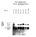
- the sizes (kilobase pairs) of fragments in EcoR I digests are: P. stutzeri 16.0, 12.0, 9.4; P. fluorescens 16.0, 10.0, 8.6, 7.8, 7.0; P. putida 24.0, 15.0, 10.0, 8.9; A. anitratus 20.0, 15.0, 12.5, 9.8, 7.8, 6.1, 5.2, 4.8, 3.8, 2.8 (size of the smallest 3 fragments not calculated); A. lwoffii 12.0, 10.0, 9.1, 7.0, 6.4, 5.7, 5.5, 5.3, 4.8, 4.4, 3.6, 3.2, 2.9 (size of the smallest 3 fragments not calculated).
- the sizes (kilobase pairs) of fragments in PST I digests are; P. stutzeri 6.7, 6.1, 5.5; P. fluorescens 10.0, 9.4, 7.8, 7.0; P. putida 10.5, 9.9, 6.8, 6.3, 4.4; A. anitratus 36.0, 28.0, 20.5, 12.0, 10.0, 5.8, 3.7, 2.6, 2.4; A. lwoffi 9.9, 8.7, 7.2, 5.7, 4.0, 3.6, 3.2, 2.7.
- PST I fragment variation occurs in strains that do not contain an EcoR I 7.6 KBP fragment; RH 151 has 10.1 and 8.2 KBP fragments, RH 809 does not contain a 9.4 KBP fragment and has a 6.0 KBP fragment, and RH 815; the type strain, does not contain a 6.6 KBP fragment.
- the patterns of hybridized fragments support the conclusion that enzyme specific, conserved sets can be used to define species. Strains of a species probably have a majority of the fragments in the conserved set. The occurrence of fragment variations in some strains does not prevent identification and may prove useful in epidemiological studies.
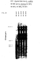
- EcoR I 7.6 KBP fragment in P. aeruginosa strains may be put into perspective by examining hybridized EcoR I fragments found in the type strains of other Pseudomonas species (FIGURE 3).
- the type strains of P. stutzeri, P. fluorescens, and P. putida do not contain a 7.6 KBP fragment, but do have EcoR I fragments of the sane size in common; P. aeruginosa and P. stutzeri each have a 9.4 KBP fragment, P. stutzeri and P. fluorescens each have a 16 KBP fragment, and P. fluorescens and P. putida each have a 10 KBP fragment.
- the sizes of the fragments are unique in the type strains of each of the 4 Pseudomonas species; and the type strain of each species has a different size range of fragments.
- restriction enzyme maps such as those availble for E. coli. Bacillus thuringiensis and B. subtilis, it is not possible to predict where enzymes cut rRNA genes, the number of copies per genome, whether there are heterologous flanking regions between genes or . gene heterogeneity.
- the E. coli rRNA cDNA probe may fail to hybridize with some restriction fragments containing. rRNA gene sequences, and if so, this reflects the evolutionary distance or diversity between the test organism and E. coli.
- the conserved nature of rRNA can be used to argue that this is not the case. However, this is a minor problem compared to the advantage of having a standard probe that can be equally applied to any unknown species.
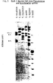
- the group represented by RH 2990 has two intensely hybridized fragments (2.6 and 2.5 KBP).
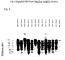
- the EcoR I data can be used to place B. subtilis strains in appropriate DNA-DNA hybridization groups. According to the DNA-DNA hybridization 70% rule, B. subtilis is actually two species. However, when the PST I data (FIGURE 6)_is considered, it is possible to think of the groups as two divergent populations related to a common ancestor or speciation event. The conclusion that B. subtilis is one species correlates with phenotypic data.
- the strains listed in Table 5 are identified as B. subtilis in Gordon, R. E. et al "The Genus Bacillus", Agriculture Handbook No. 427, Agricultural Research Service, U.S. Dept. of Agriculture, Washington, D.C.
- B. subtilis and B. polymyxa can be distinguished by E coR I data (FIGURE 9), PST I data (FIGURE 10) Bgl II data (FIGURE 11, left) and Sac I data (FIGURE 11, right). It can be concluded from the major differences in the PST I band patterns that bacillus polymyxa is in the wrong genus. While both species produce spores, they are not phenotypi-cally similar. It is reassuring that the type strain of B. polymyxa from both culture collections, ATCC and NRRL have the same band patterns. The important data, however, is that the asporogenous mutants can be identified. It is very difficult, perhaps impossible, to identify Bacillus species if they fail to form spores.
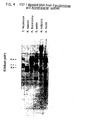
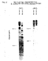
- the data (FIGURE 12) shows that S, pneumoniae can be defined by seven hybridized fragments (17.0, 8.0, 6.0, 4.0, 3.3, 2.6 and 1.8 KBP).
- the bacterial cDNA probe hybridizes poorly to two mouse DNA fragments (14.0 and 6.8 KBP).
- Hybridized fragments signal the presence of S. pneumoniae in the infected tissues. All seven bands can be seen in the heart DNA extract. They are less intense in the liver DNA extract, but all can be seen in the autoradiograph. Only the 6.0 KBP band appears in the lung DNA extract. The lesser number of bacteria in the lungs can be explained by the mouse having septicemia rather then pneumonia. The lungs showed no consolidation at autopsy.
- bacterial DNA was diluted with mouse DNA and electrophoresed. All seven bands can be seen in the autoradiograph when 0.1 micrograms of bacterial DNA is used. The 17.0, 8.0 and 6.0 KBP bands can be seen with 10 -3 pg of bacterial DNA. If the figure of 5 X 10 -3 ⁇ g DNA per 10 6 S. pneumoniae cells is used (Biochim Biophys Acta, 26:68), 10 -1 ⁇ g is equivalent to 2 X 10 7 cells. The present technique is thus useful for diagnosing infections at this level of sensitivity.
- FIGURE-9 fragments having 14.0 and 6.8 KBP.
- FIGURE 14 infra shows that the 6.8 KBP fragment contains the 28S rRNA sequences.
- the bacterial probe does not hybridize as well to mammalian ribosomal RNA gene sequences, so the bands are less intense, the system of bacterial probe and nuclear mammalian DNA is less sensitive, and selectivity for DNA from infecting prokaryotes is clearly demonstrated.
- bacterial probe was hybridized to 10 ⁇ g digested bacterial DNA per lane, no. hybridization to 10 ⁇ g digested human or mouse DNA per lane was detected on the autoradiographs when the bacterial bands were clearly seen.
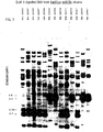
- FIGURE 13 shows that mammelian genera can be recognized with Mus musculus domesticus 18S and 28S rRNA probe, and that several species of Plus can be distinguished.
- the enzyme is PST I and the subjects and corresponding bands are as follows:
- FIGURE 14 shows that mouse and cat DNA can be distinguished by the 28S rRNA cDNA alone, and that the pattern of hybridized bands is dependent on the composition of the probe sequences.
- the enzyme is EcoR I, and the subjects and bands are as follows:
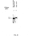
- Figure 16 shows that primates can be distinguished.
- Cell cultures have bands in common with tissue from the species of origin, and different human cell cultures can be distinguished by additions and deletions of bands.
- the enzyme is EcoR I, and the subjects and bands are as follows:
- the histone H4 gene sequence for sea urchin (Psammechinus miliaris) is shown below, where A, T, C, G represent the known nucleotides, and N represent a presently unknown position (788 base pairs).
- the region of homology for both aforementioned sequences is shown below, where asterisks denote not homologous portions.
- the first 118 base pairs have 80.5% homology and are used as a conserved DNA sequence probe in this example (sea urchin (top) base positions 449 to 567, mouse (bottom) base positions 257 to 375):
- X 84.503
- Restriction endonuclease cleavage sites were determined 'from the two sequences. A list of cleavage sites for the sea urchin and mouse sequences is shown below. Numbers indicate the 5' side of the cleavage site, unless site name is in brackets, which indicates that only the recognition site is known.
- the sea urchin and mouse sequences are compared with Hha I (GCGC) and the described probe sequence.
- the sea urchin sequence has cleavage sites at positions 295 and 497, thus creating a 202 bp fragment, which, if denatured, would hybridize with the probe sequence.
- the genetic characterization for sea urchin is 202 while that for mouse is 69 138
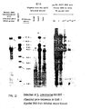
- Example 2 The same type of computer simulation as in Example 1 was carried out using trp D gene as a probe. This allows the conclusion that E. coli and Salmonella typhimurium can be distinguished by restriction fragments containing a conserved sequence.
- the E. coli trp D gene with 684 bp's is shown below:
- the trp D gene, 683 base pairs, for S. typhimurium is shown below:
- the E. coli sequence has Mbo I (GATC) sites at 135 and 564. There is a 429 bp fragment that can be detected by both Region 1 and 2 probes. The same enzyme has sites at 135, 204, 317, and 564 in the S. typhimurium sequence. A probe of the two homology regions would detect fragments of 69, 113 and 247 bp.
- This example shows the use of a region of homology in the a-fetoprotein gene sequence of human and rat, and the endonuclease MnlI (GAGG).
- the human a-fetoprotein message cDNA (1578 bp's) is as follows:
- the rat a-fetoprotein 3' end cDPZA is as follows (540 bp's):
- fragments containing a portion of the conserved sequence 24, 34, 104 and 108 bp describe the human DNA. Fragments of 24, 59, 90 and 145 describe the rat DNA. While both sequences contain the 24 bp fragment, it is the set of fragments (taxonomic characters) that is of significance.
Abstract
Description
- The present invention relates to a method for the rapid and accurate characterization and identification of organisms, including prokaryotic and eukaryotic organisms, such as bacteria, plants, and animals.
- The classification of living organisms has traditionally been done along more or less arbitrary and somewhat artificial lines. For example, the living world has been divided into two kingdoms: Plantae (plants) and Animalia (animals). This classification works well for generally familiar organisms, but becomes difficult for such organisms as unicellular ones (e.g., green flagellates, bacteria, blue-green algae), since these differ in fundamental ways from the "plants" and "animals".
- It has been suggested to simply divide organisms with respect to the internal architecture of the cell. In this scheme, all cellular organisms are either prokaryotic or eukaryotic. Prokaryotes are less complex than eukaryotes, they lack internal compart- mentalization by unit membrane systems, and lack a defined nucleus. Prokaryotic genetic information is carried in the cytoplasm on double-stranded, circular DNA; no other DNA is present in cells (except for the possible presence of phage, bacterial viruses, and cirular DNA plasmids, capable of autonomous replication). Eukaryotes, on the other hand, have a multiplicity of unit membrane systems which serve to segregate many of the functional components into specialized and isolated regions. For example, genetic information (DNA) can be found in a well- compartmentalized nucleus and also in organelles: mitochondria and (in photosynthetic organisms) chloroplasts. The replication, transcription, and translation of the eukaryotic genome occurs at either two or three distinct sites within the cell: in the nucleocytoplasmic region, in the mitochondrion, and in the chloroplast.
- The differences between prokaryotes and 'eukaryotes, however, breaks down when a comparison of mitochondria and chloroplasts is carried out with prokaryotes: these organelles are today considered to have been derived from free-living prokaryotes, which entered into an endosymbiotic relation with primitive eukaryotes, and eventually became closely integrated with the machinery of the host cell and incapable of independent existence (see e.g., Fox, G.E. et al, Science 209:457-463 (1980), at 462; Stanier, R. Y. et al, "The Microbial World," Fourth Edition, Prentice-Hall, Inc., 1976, at p. 86). For example, it has been demonstrated that DNA from mouse L cell mitochondria carrying the ribosomal RNA gene region exhibits notable sequence homologies to Escherichia coli ribosomal RNA, thus providing strong support for the endosymbiotic model (Van Etten, R. A. et al, Cell, 22:157-170 (1980)). It has also been shown that the nucleotide sequence of 23S ribosomal DNA from Zea mays chloroplast has 71% homology with 23S ribosomal DNA from E. coli (Edwards, K. and Kossel, H., Nucleic Acids-Research, 9:2853-2869 (1981)); other related work (Bonen, L. and Gray, M. W., ibid, 8:319-335 (1980)) also further . supports the general concept.
- In this model the eukaryotic cell is a phylogenetic "chimera" with organelle components that are clearly prokaryotic in nature. The "prokaryotic- eukaryotic" dichotomy then, also has drawbacks, even as a broad classification method.
- Where classification of organisms becomes more than a scientific exercise is in the identification of plants and animals for hybridization and breeding purposes, and in the accurate and reliable identification. of microorganisms which may infect so-called "higher" organisms or other media. For example, the plant- breeder, cattle breeder, or fish breeder may wish to have a quick and reliable means of identifying different species and strains of their subjects. The veterinarian, physician, or horticulturist may wish to have an accurate identification of any infectious organisms (parasites, fungi, bacteria, etc.) and viruses present in examined plant or animal tissues. The correct identification of species of these organisms and viruses is of particular importance.
- The problem can best be illustrated by referring to the identification of bacteria. Names of bacterial species usually represent many strains, and a strain is considered to be a population derived from a single cell. Bacterial species are usually defined by describing the degree of homogeneity and diversity of attributes in representative samples of strains of species. Precise definitions of bacterial species are difficult to express because subjective limits to strain diversity within species are required to define- species boundaries. (Buchanan, R. E., International Bulletin of Bacteriological Nomenclature and Taxonomy, 15:25-32 (1965)). The practical application of definitions of species to the identification of an unknown bacterial-strain requires the selection of relevant probes, such as substrates and conditions to detect phenotypic attributes, and radioactively-labeled DNA from the same species. Because of the diversity of bacterial species, a screening procedure is the primary tool used in the classical, progressive method for identification of a strain. Results of the screening procedure are then used to predict which other laboratory methods and reagents are relevant for definitive identification of the strain. Identification is ultimately based on certain phenotypic and genotypic similarities between the unidentified strain and characterized species. The challenge is to precisely define the boundaries of species, preferably in terms of a standard probe which reveals species-specific information, so that definitions of species can be directly and-equally applied to the identification of unknown strains.
- Bergey's Manual of Determinative Bacteriology (Buchanan, R. E. and Gibbons, N. E., Editors, 1974, 8th Edition, The Williams & Wilkins Company, Baltimore) provides the most comprehensive treatment of bacterial classification particularly for nomenclature, type strains, pertinent literature, and the like. It is, however, only a starting point for the identification of any species since, inter alia, it is normally out of date, and is limited in space to describing species quite-briefly. (See for example, Brenner, D. J., "Manual of Clinical Microbiology," 3rd Edition, American Society of Microbiology, Washington, D.C., 1980, pages 1-6.)
- The term "species", as applied to bacteria, has been defined as a distinct kind of organism, having certain distinguishing features, and as a group of organisms which generally bear a close resemblance to one another in the more essential features of their organization. The problem with these definitions is that they are subjective; Brenner, supra, at
page 2. Species have also been defined solely on the basis of criteria such as host range, pathogenicity, ability or inability to produce gas in the fermentation of a given sugar, and rapid or delayed fermentation of sugars. - In the 1960's, numerical bacterial taxonomy (also called computer or phenetic taxonomy) became widely used. Numerical taxonomy is based on an examination of as much of the organism's genetic potential as possible. By classifying on the basis of a large number of characteristics, it is possible to form groups of strains with a stated degree of similarity and consider them species. Tests which are valuable for the characterization of one species, however, may not be useful for the next, so this means to define species is not directly and practically applicable to the identification of unknown strains. Although this may be overcome in part by selecting attributes which seem to be species specific, when these attributes are used to identify unknown strains, the species definition is applied indirectly. See for example Brenner, supra, at pages 2-6. The general method, furthermore, suffers from several problems when it is used as the sole basis for defining a species, among them the number and nature of the tests to be used, whether the tests should be weighted and how, what level of similarity should be chosen to reflect relatedness, whether the same level of similarities is applicable to all groups, etc.
- Hugh, R. H. and Giliardi, G. L., "Manual of Clinical Microbiology," 2nd Edition, American Society for Microbiology, Washington, D.C., 1974, pages 250-269, list minimal phenotypic characters as-a means to define bacterial species that makes use of fractions of genomes. By studying a large, randomly selected sample of strains of a species, the attributes most highly conserved or common to a vast majority of the strains can be selected to define the species. The use of minimal characters is progressive and begins with a screening procedure to presumptively identify a strain, so that the appropriate additional media can be selected. Then the known conserved attributes of the species are studied with the expectation that the strain will have most of the minimal characters. Some of the minimal characters do not occur in all strains of the species. A related concept is the comparative study of the type, the neo-type, or a recognized reference strain of the species. This control is necessary because media and procedures may differ among laboratories, and it is the strain, not the procedure, that is the standard for the species.
- A molecular approach to bacterial classification is to compare two genomes by DNA-DNA reassociation. A genetic definition of species includes the provision that strains of species are 70% or more related. With DNA-DNA reassociation a strain can be identified only if the radioactively labeled DNA probe and unknown DNA are from the same species. The practical application of this 70% species definition however is limited by selection of an appropriate probe. This may be overcome in part by selecting phenotypic attributes which seem to correlate with the reassociation group, but when these are used alone the DNA-DNA reassociation species definition is also applied indirectly.
- Brenner, supra, at
page 3, states that the ideal means of identifying bacterial species would be a 'black box' which would separate genes, and instantly compare the nucleic acid sequences in a given strain with a standard pattern for every known species-something akin to mass spectrophotometric analysis.
Brenner, however, concedes that although restriction endonuclease analysis can be done to determine common sequences in isolated genes, "we are not at all close to having an appropriate black box, especially one suited for clinical laboratory use." His words could be equally applied to any species of organism. - This brief review of the prior art leads to the conclusion that there presently exists a need for a rapid, accurate, and reliable means for identifying unknown bacteria and other organisms, and to quickly classify the same, especially to identify the organism of a disease, or of a desirable biochemical reaction. The method should be generally and readily useful in clinical laboratories, should not be dependent on the number of tests done, on the subject prejudices of the clinician, nor the fortuitous or unfortuitous trial and error methods of the past. Further, a need also exists for a method useful for identifying and distinguishing genera and species of any living organism, which can be readily and reliably used by veterinarians, plant-breeders, tox- icologists, animal breeders, entomologists and in other related areas, where such identification is necessary.
- "It is therefore an object of the invention to provide a method which may be found:.to be quick, reliable and accurate of objectively identifying organisms, especially - but not limited to - microorganisms.
- Yet another object of the invention is to provide a method of identifying organisms such as bacteria which utilizes the organisms' genome.
- Another object of the invention is to provide a method of characterizing and identifying species and genera of pathogenic organisms in the clinical laboratory, so as to provide the capability of characterizing and identifying the cause of any given animal or plant disease.
- Still another object of the invention is to provide various products useful in the aforementioned methodologies.
- These and other objects of the invention, as will hereinafter become more readily apparent, have been attained by providing:
- A method of characterizing an unknown organism species which comprises determining the position of part or whole of evolutionarily conserved sequences in genetic material of said organism, relative to a known position of restriction endonuclease cleavage sites in said genetic material (other than by determining the chromatographic pattern of restriction endonuclease digested DNA from said unknown organism, which digested DNA has been hybridized or reassociated with ribosomal RNA information containing nucleic acid from or derived from a known probe organism), thereby to obtain an identifying genetic characterization of said unknown organism, and comparing said characterization with information from at least two sets of identifying genetic characterizations derived from the same conserved sequences, each of said sets defining a known organism species.
- Still another object of the invention has been . .attained by providing:
- A method of diagnosing a pathogenic organism infection in a sample which comprises identifying the organism in said sample by the aforementioned method.
- For a better understanding of the invention, and to show how it may be put into effect, reference will now be made, by way of example, to the accompanying drawings in which:
- FIGURE 1 shows the EcoR I restriction endonuclease digest of DNA isolated from strains of Pseudomonas aeruginosa, using cDNA to 16S and 23S ribosomal RNA (rRNA) of E. coli as the probe.
- FIGURE 2 shows the Pst I restriction endonuclease digest of DNA isolated from strains of P. aeruginosa, using cDNA to 16S and 23S rRNA of E. coli as the probe.
- FIGURE 3 shows the EcoR I restriction endonuclease digest of DNA isolated from species of glucose- nonfermenting, gram-negative rods, using cDNA to 16S and 23S rRNA of E. coli as the probe.
- FIGURE 4 shows the Pst I restriction endonuclease digest of DNA isolated from species of glucose- nonfermenting, gram-negative rods using cDNA to 16S and 23S rRNA of E. coli as the probe.
- FIGURE 5 shows the EcoR I restriction endonuclease digest of DNA isolated from various Bacillus subtilis strains, using cDNA to 16S and 23S rRNA of E. coli as the probe.
- FIGURE 6 shows the Pst I data for the same strains as in FIGURE 5, with the same probe.
- FIGURE 7 shows the Bgl II data for the same strains as in FIGURES 5 and 6, with the same probes
- FIGURE 8 shows the Sac I data for the same strains as in FIGURES 5-7, with the same probe.
- FIGURE 9 shows the EcoR I restriction endonuclease digest of DNA isolated from B. subtilis and B. polymyxa, using cDNA to 16S and 23S rRNA from E. coli as the probe.
- FIGURE 10 shows the Pst I data for the same strains as in FIGURE 9 with the same probe.
- FIGURE 11 shows the Bgl II and Sac I data for the same strains as in FIGURES 9 and 10, with the same probe.
- FIGURE 12 shows the detection of Streptococcus pneumoniae in EcoR I digested DNA from infected mouse tissues using cDNA from 16S and 23S rRNA from E. coli as the probe.
- FIGURE 13 shows the identification of a mouse species by comparing Pst I digests of DNA isolated from mammalian tissues, using cDNA to 18S and 28S rRNA from cytoplasmic ribosomes of Mus musculus domesticus (mouse).
- FIGURE 14 shows the EcoR I digested DNA from mouse and cat tissues hybridized with Mus musculus domesticus 28S rRNA cDNA probe.
- FIGURE 15 shows Sac I digested DNA from mammalian tissues hybridized with Mus musculus domesticus 18S and 28S rRNA cDNA probe.
- FIGURE 16 shows EcoR I digested DNA from mammalian tissues and cell cultures hybridized with Mus musculus domesticus 18S and 28S rRNA cDNA probe.
- This invention is based on the inventor's realization that, if species are discrete clusters of strains related to a common speciation event, there should be, despite divergence, a likeness shared by strains that objectively defines the species boundary; strains of species should contain structural information which is a clue to their common origin. The greatest amount of an organism's past history survives in semantides,'DNA and RNA, (Zuckerkandle, E. and Pauling, L., Journal of Theoretical Biology, 8:357-366 (1965)).
- In European Patent Application (EP-A-)-No.0076123 (herein incorporated by reference) the inventor described a system for the definition of species and characterization of organisms which makes use of information contained in ribosomal RNA genes (rRNA). Ribosomal RNA has a structural and functional role in protein synthesis (Schaup, Journal of Theoretical Biology, 70:215-224 (1978)), and the general conclusion from rRNA-DNA hybridization studies, is that the base sequences of ribosomal RNA genes are less likely to change, or are more conserved during evolution, than are the majority of other genes (Moore, R. L., Current Topics In Microbiology and Immunobiology, Vol. 64:105-128 (1974), Springer-Verlag, New York). For example, the primary structure of 16S rRNA from a number of bacterial species has been inferred from oligonucleotide analysis (Fox, G. E. et al, International Journal of Systematic Bacteriology, 27: 44-57 (1977)). There are negligible differences in the 16S oligomer catalogs of several strains of E. coli (Uchida, T. et al, Journal of Molecular Evolution, 3:63-77 (1974)); the substantial differences among species, however, can be used for a scheme of bacterial phylogeny (Fox, G.E., Science, . 209:457-463 (1980)). Different strains of a bacterial species are not necessarily identical; restriction enzyme maps show that different EcoR I sites occur in rRNA genes in two strains of E. coli (Boros, I.A. et al, Nucleic Acids Research 6:1817-1830 (1979)). Bacteria appear to share conserved rRNA gene sequences and the other sequences are variable (Fox, 1977, supra).
- The present inventor had thus discovered that restriction endonuclease digests of DNA have sets of fragments containing conserved sequences that are similar in strains of a species of organism (e.g., bacteria), but different in strains of other species of the organism; i.e., despite strain variation, enzyme specific sets of restriction fragments with high frequencies of occurrence, minimal genotypic characters, define the species. This is the essence of the invention described in EP-A-007-6123- and also that of the invention described herein.
- .The present invention constitutes an extension of the concepts developed in EP-A-0076123, in that it has further been discovered that there exist sequences, in addition to those of rRNA, which are highly conserved through evolution and which may be as useful as rRNA sequences in the identification system. In other words, the present invention provides means for carrying out the identification and characterization techniques of EP-A-0076123, using any probe which is conserved, other than rRNA. The present invention also provides additional examples of methods which may be used in the identification process. The present inventor has also discovered that the method is general, in that it is-applicable to both eukaryotic and prokaryotic DNA, using a conserved nucleic acid probe from any organism, prokaryotic or eukaryotic, of the same or different (classic) taxonomic classification than the organism being identified.
- The invention offers an objective method of defining organisms based on conserved sequences of DNA or other genetic material in relation to known positions such as restriction endonuclease sites. The detection of restriction fragments containing a conserved sequence may be carried out by hybridizing or reassociating DNA segments with nucleic acid containing conserved sequence information from a probe organism.
- By the "organism" which can-be characterized (which term is meant to include "identified") by the process of the invention, it is meant to include virtually any organism which, by definition, contains DNA or RNA in its genome. In this respect it is useful to refer to a classical taxonomic scheme as a point of reference.
- All organisms belonging to the Kingdoms Monera, Plantae and Animalia are included. For example, among those of the Kingdom Monera can be mentioned the Schizomycetes (Bacteria) of the classes myxobacteria, spirochetes, eubacteria, rickettsiae, and the cyanopytha (blue green algae). Among those of the Kingdom Plantae can be mentioned the Division Euglenophyta (Euglenoids), Division Chlorophyta (green-algae) classes chlorophyceae and charophyceae, Division Chrysophyta, classes xantho- phyceae; chrysophyseae, bacillariophyceae; Division Pyrrophyta (Dinoflagellates); Division Phaeophyta (Brown algae); Division Rhodophta (Red algae); Division Myxomycophyta (slime molds), classes myxomycetes, acrasiae, plasmodiophoreae, labyrinthuleae; Division Eumycophyta (true fungi), classes phycomycetes, ascomy- cetes, and basidomycetes; Division Bryophta, classes hepaticae, anthocerotae, and musci; Division Tracheophyta (Vascular plants), subdivisions psilopsida, lycopsyda, sphenopsida, pteropsida, spermopsida classes cycadae, ginkgoae, coniferae, gneteae and angiospermae subclasses dicotyledoneae, monocotyloedoneae. Among those of the Kingdom Animalia can be mentioned the Subkingdom Protozoa, Phylum Protozoa (Acellular animals) subphylum plasmodroma, classes flagellata, sarcodina and sporozoa; subphylum ciliophora, class ciliata; the Subkingdom Parazoa, Phylum porifera (Sponges), class calcarea, hexactinellida, and desmospongiae; the Subkingdom Mesozoa, Phylum mesozoa; the Subkingdom Metazoa, Section Radiata, Phylum coelenterata, classes hydrozoa, scypho- zoa, anthozoa, Phylum ctenophora, classes tenta-culata and nuda; Section Protostomia Phylum platyhelmintes (flat- worms) classes tubellana, trematoda, and cestoda; Phylum . nemertina; Phylum acanthocephala; Phylum aschelmintles, classes rotifera, gastrotricha, kinorhyncha, priapulida, nematoda and nematomorpha; Phylum entoprocta; Phylum ectoprocta, classes gymnolaemata and phylactolaemata; Phylum phoronida; Phylum braciopoda, classes inarticulata and articulata; Phylum mollusca (molluscs) classes amphineura, monoplacophora, gastropoda, scaphopoda, pelecypoda, and cephalopoda; Phylum sipunculida; Phylum echiurida; Phylum annelida, classes polychaeta, oligochaeta and hirudinea; Phylum onychophora; Phylum tardigrada; Phylum pentastomida; Phylum arthropoda, subphylum trylobita, subphylum chelicerata classes xiphosura, arachmida, pycnogomida, subphylum mandibulata classes crustacea, chilopoda, diplopoda, pauropoda, symphyla, insecta of the orders collembola, protura, diplura, thysanura, ephemerida, odonata, orthoptera, dermaptera, embiania, plecoptera, zoraptera, corrodentia, mallophaga, anoplura, thysasnoptera, hemiptera, neurop- tera, coleoptera, hymenoptera, mecoptera, siphonaptera, diptera, trichoptera and lepidoptera; those of the Section Deuterostomia, phylum chaetognatha, phylum echinodermata, classes crinoidea, asterordea, ophiuroi- dea, echinoidea, and holoturoidea, phylum pogonophora; phylum hemichordata, classes enteropneusta, and pterobranchia; phylum chordata, subphylum urochordata, classes ascidiaciae, thaliaceae, larvacea; subphylum cephalochordata, subphylum vertebrata, classes agnatha, chondrichthyes, osteichthyes (subclass saccopteiygii orders crossopterygii and dipnoi), amphibia, repitilia, aves and mammalia, subclass prototheria, subclass theria, orders marsupialia, insectivora, dermoptera, chiroptera, primates, edentata, pholidota, lagomorpha, rodentia, cetacea-e, carnivora, tubulidentata, probosicdea, hyracoidea, sirenia, perissodactyla and artiodactyla.
- It is understood that beyond the order, the organisms are still classified according to their families, tribes, genus and species, and even subspecies, infrasubspecific taxons, and strains or individuals. In addition, cell cultures (plant or animal), as well as viruses can also be identified. These classifications are used in this application for illustrative purposes only, and are not to be taken as exclusive. The organism is either known or unknown, most commonly the organism is an unknown being identified.
- Functionally, for the purposes of this invention, it is convenient to divide all organisms into the eukaryotes and the prokaryotes. When identifying a prokaryotic organism, the DNA to be analyzed is that present in the cells or in the non-compartmentalized chromosomes. When identifying a eukaryotic organism one may either use the nuclear DNA or the organelle DNA (mitochondrial DNA or chloroplast DNA).
- Briefly, high molecular weight DNA and/or small circular DNAs are isolated from the organism to be identified in order to analyze the conserved sequences (and possibly sequences that could be used to create a taxon below the rank of species or infrasubspecific subdivisions.) The DNA's are extracted by methods which are well-known to the art.
- The DNA's are analyzed to ascertain both 1) the presence and position of the conserved sequences and 2) their position relative to endonuclease restriction sites. The easiest way to analyze for the presence of the conserved sequences is to utilize a polynucleotide probe capable of hybridizing with the conserved DNA sequence. However, direct sequence information as obtained by chemical sequence determination and analysis thereof could also be utilized. In EP-A-0076123 the probe utilized was an rRNA information containing-probe; in this case any other probe havingconserved sequences could be used. In an analogous manner, the easiest way of finding a given set of endonuclease restriction sites is to cleave the DNA with the appropriate restriction enzymes. (This, indeed, is the manner taught and practised in EP-A-0076123. However, alternative methods, such as sequence information coupled with known restriction site sequences, or cleavage and partial sequencing could also be used..
- Most commonly DNA's are going to be cut at specific £ sites into fragments by restriction endonucleases. The fragments are separated according to size by a chromatographic system. In EP-A-0076123 gel chromatography was used as an example of a useful chromatographic system. However, other systems can also be used, such as'high pressure liquid chromatography, capillary zone electrophoresis, or other separation techniques. In using gel chromatography, the fragments are separated, the gels are stained, as is otherwise well-known in the art, and standardized as to the fragment sizes using standards curves constructed with fragments of known sizes. The separated fragments may then be transferred to cellulose nitrite paper by the Southern blot technique (Southern, E. M., Journal of Molecular Biology, 38:503-517 (1975), herein incorporated by reference), and covalently bound thereto by heating. The fragments containing the conserved sequences are then located by their capacity to hybridize with a nucleic acid probe containing conserved sequence information. Alternatively, hybridization can occur after digestion but before separation; or restriction cleavage can occur after hybridization, followed by separation of the fragments.
- The nucleic acid probe can either be non- radioactively labeled or, preferably, radioactively labeled. When radioactively labeled, the probe can be RNA, or preferably DNA which is complementary to RNA (cDNA), either synthesized by reverse transcription or contained on a cloned-fragment, which can be labeled, for example, by nick translation. Also, synthetic oligodeoxyribonucleotides may be prepared with labeled nucleotides.
- The well-defined probe is derived from an arbitrarily chosen organism, see infra, or may be a consensus sequence. Once hybridization has occurred, the hybridized fragments are detected by selectively detecting double stranded nucleic acid (non-radiolabeled probe), or visualized by, e.g., autoradiography (radiolabeled probe). The -size of each fragment which has been hybridized is relative to the restriction sites and is determined from the distance traveled using standard curves, as described previously. The amount of hybridization, the pattern of hybridization, and the sizes of the hybridized fragments, which are relative to restriction sites, can be used individually or in conjunction to identify the organism.
- The genetic characterization that emerges from this technique can be readily compared to equivalent characterizations derived from at least two and up to a multiplicity of known, standard organisms, genera or species. After a preliminary broad classification has already been carried out (using, for example, classical taxonomy), the comparison can be either by visual inspection and matching of appropriate chromatographic patterns, (as in EP-A-0076123) by comparison of hybridized restriction fragment sizes, by band intensity (amount of hybridization) or by any combination thereof. Ideally, the comparison is carried out with a one- . dimensional computer-based pattern recognition system, such as those used in point-of-sale transactions.
- The present inventor discovered that when using the aforementioned method, the genetic characterizations for organisms of the same= species are substantially similar, . with minor variations allowed for intraspecies differences due to strain variations, whereas differences between species, and differences between genera (and higher classifications) are maximal.
- The use of enzyme-specific fragment variations among strains of a species permits the typing of strains for various purposes; e.g. in the case of bacteria, for epidemiological purposes. In fact, restriction enzymes can be chosen for-their ability to distinguish strains within species.
- The "probe organism" used in the present invention, and from which is obtained the nucleic acid probe, can also be any of the aforementioned organisms; it can be either eukaryotic or prokaryotic. The only limitation is given by the fact that the conserved sequence-containing probe should hybridize maximally with the unknown organism's DNA.
- There are four types of-conserved sequence information-containing probes: 1) prokaryotic probes (especially bacterial-derived), 2) eukaryotic mitochondrial probes, 3) eukaryotic chloroplast probes, and 4) eukaryotic non-organelle probes. There are also four sources of DNA (to be endonuclease digested): 1) prokaryotic cellular DNA, 2) eukaryotic mitochondrial DNA, 3) eukaryotic chloroplast DNA, and 4) eukaryotic nuclear DNA. The following hybridization table can thus be constructed (Table 1).

- The Table shows which probes can generally be maximally hybridized with which unknown DNA. For example, one can identify a eukaryotic organism by extracting species specific mitochondrial or chloroplast DNA, endonuclease-digesting it and hybridizing the digest with either a prokaryotic probe, or with an organelle derived eukaryotic probe. In the same manner, one can identify a prokaryotic organism by extracting species-specific cellular DNA, endonuclease-digesting it, and hybridizing the digest with either a prokaryotic probe, or an organelle-derived eukaryotic RNA probe. Also, one can identify a eukaryotic organism by extracting and digesting species-specific nuclear DNA, and hybridizing it with a non-organelle derived eukaryotic probe. Eukaryotes could be defined by one or any combination of the nuclear, mitochondria, or in some cases chloroplast systems. These cross-hybridizations are based on the fact that nucleic acid derived from eukaryotic organelles has extensive homology with evolutionarily conserved . sequences from prokaryotic nucleic acid, but that the same homologies are generally not present to such extent between nuclear-derived eukaryotic DNA and prokaryotic DNA.
- The choice of any pair of DNA to be digested and . accompanying probe is arbitrary, and will depend on the organism being identified, i.e. it will depend on the question asked. For example, in detecting a prokaryotic species (e.g. bacteria) present in or together with a eukaryotic cell (e.g. animal or plant) for purposes of detecting and identifying an infecting agent, one may choose a prokaryotic probe and work under conditions where organelle-derived DNA is not extracted or only minimally extracted. In this manner one assures that interference between organelle-derived DNA and prokaryotic DNA is minimal. In identifying a eukaryotic species (which is not infected with a prokaryote) with a prokaryotic probe, it is best to maximize the concentration of organelle-derived DNA, as for example by separating organelles from nuclei, and then extracting only organelle DNA. If one wishes to identify a eukaryotic organism which has been infected with a prokaryotic organism, it is best to use a non-organelle, non-prokaryotic derived probe since it will generally not hybridize well with the DNA from the prokaryote.
- It is preferred to use a pair (DNA and probe) from the same kingdom, or same subkingdom, or same section, or same phylum, or same subphylum, or same class, or same subclass, or same order, or same family or same tribe or same genus. It is particularly preferred to use prokaryotic probe-(e.g. bacterial probe) to hybridize with prokaryotic DNA. In this manner one could detect, quantify, and identify genera, species, and strains of prokaryotic organisms. One of the most preferred prokaryotic probes is derived from bacteria, and further, because of the ease and availability, from E. coli. The probe from E. coli can be used to identify any organism, especially any prokaryotic organism, most preferably a strain of any bacterial species. Another particularly preferred embodiment is to use eukaryotic probe derived from a given class to identify eukaryotic organisms of the same class (e.g. mammalian probe to identify mammalian organism). Most preferred is to use probe and DNA from the same subclass and/or order and/or family of organisms, (e.g. if identifying a species of mouse, it is preferred to use mouse-derived probe).
- The most sensitive and useful pair systems are those where there is less evolutionary distance or diversity between the source of the probe and the unknown DNA.
- The phrase "evolutionarily conserved genetic material sequence" is used in this invention to denote genetic material, e.g DNA, sequences that show homology between at least two different species of plants, animals or microorganisms. The homology between two conserved sequences is to be such that, if one of such DNA molecules were to be detectably labelled, sufficient hybridization or annealing would occur if both single stranded DNA molecules or fragments thereof were placed together under hybridization conditions, thereby to produce a duplex of sufficient stability to be detectable by standard methodology (i.e, radiolabelling, enzyme labelling, and the like).
- In EP-A-0076123 the evolutionarily conserved sequence exemplified was that of ribosomal RNA genes. This is still a highly preferred gene sequence. However, it has been discovered that other gene sequences exist which are sufficiently conserved across the evolutionary span to be useful.
- Examples of such additional sequences are those of genes or portions thereof coding for transfer RNA, or proteins denoted as belonging to the same Superfamily, or same Family, preferably same Subfamily or even same entry in Dayhoff's "Atlas of Protein Sequence and Structure",
Volume 5,Supplement 3, 1978, NBR, 1979, pages 9-24, herein incorporated by reference. A Family of proteins is one wherein any two proteins differ from each other by less than 50% amino acid residues in their sequence. A Subfamily of proteins is one wherein any two proteins differ from each other by less than 20% amino acid residues in their sequence. An "Entry" is one wherein any two proteins differ from each other by less than 5% amino acid residues in their sequence. - Specific examples of geze sequences or appropriate portions thereof which can b: used are: cytochrome C related genes, cytochrome C3 related genes, cytochrome cl related, cytochrome b5 relatfd, ferrodoxin related, rebredoxin related, flavodo> n related., alcohol dehydrogenase related, lactaze dehydrogenase related, peroxidase related, adenylaze kinase related, phospholipase A2 related, trzrptophan operon related, carboxypeptidase related, subtilisin related, penicillinase related, protease inhibitor related, somatotropin related, corticotropin related, lipotropin related, glucagon related, snake venom toxin related, plant toxin related, antibacterial toxin related, immunoglogulin related gene, ribosomal other than rRNA- related genes, heme carrier genes, chromosomal protein genes, fibrous protein genes, and the like.
- The conservation of some of these additional DNA sequences is not as widespread throughout the animal, plant or microbiological domains as is that of the rRNA genes. (Thus the still preferred use of rRNA). This, however, does not constitute a serious impediment to their use since it may be possible to utilize such additional sequences to identify or characterize organisms within more limited ranges or subdomains. For example it may be possible to utilize trp D gene sequences from bacteria to generate a trp D bacterial probe and then use this probe to test within the bacterial domain. In fact, it may be possible to use a trp D probe within an even narrower domain (e.g., test for the presence of Enterobacteriaceae, or of Bacillus, etc.) with a trp D probe from the same order, family or genus. Thus, while the range of applicability of some of the additional probe sequences may not be as broad as that of rRNA probes, their applicability will nevertheless be quite effective within narrower domains.
- A probe containing the conserved DNA sequence information is prepared in the same manner as the preparation of rRNA information containing probe exemplified in EP-A-0076123. The probe can thus be RNA, DNA or cDNA, and the like.
- The individual steps involved in the technique will be described hereinafter broadly with reference to both eukaryotic and prokaryotic cells when applicable, or specifically for each type of cell if some difference in technique exists.
- The first step is extraction of the DNA from the unknown organisms. Nuclear DNA from eukaryotic cells can be selectively extracted by standard methodology well-known to the art (see for example, Drohan, W. et al, Biochem. Biophys. Acta, 521 (1978), 1-15, herein incorporated by reference). Because organelle DNA is small and circular, spooling techniques serve to separate the non-circular nuclear DNA from the circular, organelle-derived DNA. As a corollary, the non-spooled material contains the organelle-derived DNA which can separately be isolated by density gradient centrifugation. Alternatively, mitochondria (or chloroplasts) are separated from a mixture of disrupted cells; the purified mitochondrial (or chloroplast) fraction is used for the preparation of organelle-derived DNA while the purified nuclear fraction is used to prepare nuclear DNA. (See for example Bonen L. and Gray, M. W., Nucleic Acids Research, 8:319-335 (1980)).
- Prokaryotic DNA extraction is also well-known in the art. Thus, for example, an unknown bacterium present in any medium, such as an industrial fermentation suspension, agar medium, plant or animal tissue or sample or the like, is treated under well-known conditions designed to extract high molecular weight DNA therefrom. For example, cells of the unknown organism can be suspended in extraction buffer, lysozyme added thereto, and the suspension incubated. Cell disruption can be further accelerated by addition of detergents, and/or by increase in temperature. Protease digestion followed by chloroform/phenol extraction and ethanol precipitation can be used to finalize the extraction of DNA. An alternative method of extraction, which is much faster than phenol/chloroform extraction, is rapid isolation of DNA using ethanol precipitation. This method is preferably used to isolate DNA directly from colonies or small, liquid cultures. The method is described in Davis, R. W. et al: "A Manual for Genetic Engineering, Advanced Bacterial Genetics," (hereinafter "Davis"), Cold Spring Harbor Laboratory, Cold Spring Harbor, New York, 1980, pp. 120-121, herein incorporated by reference.
- The DNA (prokaryotic or eukaryotic (nuclear or non- nuclear)) is dissolved in physiological buffer for the next step. There are a variety of possible steps to be followed after isolation of the desired DNA. One of these steps is endonuclease digestion.
- Digestion of extracted DNA is carried out with restriction endonuclease enzymes. Any restriction endonuclease enzyme can be used. Preferably it is not from the same organism species as that being identified, since otherwise, the DNA may remain intact. (This may, in any event, identify the organism, since the enzymes are not expected to cut DNA from the species of their origin.) Since the organism species being characterized may be unknown, obtaining a suitable digest of fragments may entail a minimum amount of trial and error, which can routinely be carried out by those skilled in the art without undue experimentation. Examples of possible restriction endonuclease enzymes are Bgl I, BamH I, EcoR I, Pst I, Hind III, Bal I, Hga I, Sal I, Xba I, Sac I, Sst I, Bcl I, Xho I, Kpn I, Pvu II, Sau IIIa, or the like. See also Davis, supra, at pp. 228-230, herein incorporated by reference. A mixture of one or more endonucleases can also be used for the digestion. Normally, DNA and endonuclease are incubated together in an appropriate buffer for an appropriate period of time (ranging from 1 to 48 hours, at temperatures ranging from 25°C-65°C, preferably 37°C).
- The resulting identifying genetic characterization will depend on the type or types of endonucleases utilized, and will be endonuclease-specific. It is therefore necessary to note which enzyme or enzymes have been used for the digestion since comparative characterizations used in a catalog should have been prepared using the same enzyme or enzymes.
- An alternative step is to define endonuclease sites on the desired DNA molecules without digestion thereof, for example, by sequencing and reference to a restriction site library. Obviously, digestion is the more efficient method of noting such sites, but the method need not be limited thereto. The essence of the invention is the discovery that the position of conserved sequences along DNA, relative to the position of endonuclease restriction sites, forms a set which is characteristic for each .species. Thus, any technique which yields the desired information (position of the genes vis a vis position of the sites) will be useful in the invention.
- Also, the position of the conserved sequences along the DNA molecule is best noted by use of a hybridization probe. This probe is allowed to anneal to restriction fragments of the unknown's DNA. However, any other method that would allow the determination of the conserved DNA sequences, such as sequencing, would also be useful. When using the hybridization probe it is preferred to first digest and separate DNA fragments according to size, and then to hybridize the separated fragments. However, it is possible to first digest and anneal DNA with a molar excess of probe and/or sequences complementary to probe and then separate the mixture. For example, unknown DNA can be digested with a restriction endonuclease, denatured, and hybridized in liquid with a molar excess of one or more small detectably labeled DNA fragments or synthetic oligodeoxyribonucleotides complementary to a portion or portions of the conserved sequence of interest. Since most restriction enzymes cut fairly infrequently in DNA, in most cases the double-stranded region or regions of the hybrid will be small relative to the size of the restriction fragment. The hybridization reaction is conducted under conditions where only the oligodeoxyribonucleotides hybridize. The unreacted, single-stranded DNA fragments, and the DNA fragments containing the hybridized oligodeoxyribonucleotides are separated by conventional chromatographic techniques. The labeled DNA fragments will appear in predictibly sized fractions. It is also possible to first anneal DNA with a molar excess of probe, then digest, and then separate the mixture. When the solution is incubated for a short time period or to a low Cot, the restriction sites will be limited to the hybridized, double-stranded regions. When the solution is incubated for a long time period or to a high Cot, the unknown DNA will anneal, thus creating a labeled duplex susceptible to restriction endonuclease cleavage. Unreassociated-single-stranded tails may be removed with a nuclease such as Sl. Unpaired bases may be filled in using DNA polymerase I or T4 polymerase.
- Alternatively, one could find subsequences within the conserved sequence information (e.g. 20-, 30-, or 50- mers), which are more highly conserved than the remainder of the conserved region chosen as the probe. Those "shorter" sequences can be made synthetically or enzymatically, if desired, and may incorporate labeled nucleotides. Single-stranded, predigested DNA from the unknown is allowed to incubate with these shorter, highly conserved fragments and allowed to hybridize thereto. Separation would then be carried out on the digest mixture containing fragments partly annealed to the shorter labeled probes. (Thus, separation would occur after hybridization.) Separation could be by liquid chromatography, since the digest mixture would for all practical purposes behave as a mixture of essentially single-stranded fragments.
- As indicated, a preferred method is to first digest, then separate, and then hybridize. Thus, after endonuclease digestion, the,incubation mixture, which contains fragments of varying sizes, is preferably separated thereinto by an appropriate chromatographic method. Any method which is capable of separating nucleic acid digests according to size, and which allows the eventual hybridization with the nucleic acid probe when hybridization is the last step, can be used. For example, gel electrophoresis, high pressure liquid chromatography or capillary zone electrophoresis can be used. (Jorgenson, J.W., J. of HRC and CC, 4: 230-231 (1981)). Presently preferred is gel electrophoresis, most preferred is agarose gel electrophoresis. In this system, the DNA digests are normally electrophoresed in an appropriate buffer, the gels are normally immersed in an ethidium bromide solution, and placed on a UV-light box to visualize standard marker fragments which may have. been added. Detectably labeled standard marker fragments may be used as well.
- After separation and visualization, the DNA fragments are transferred onto nitrocellulose filter paper or onto charge-modified nylon membranes by the method of Southern (Journal of Molecular Biology, 38:503-517 (1975)). The transfer can be carried out after denaturation and neutralization steps, and is usually done for long periods of time (approximately 10-20 hours) or,'alternatively by means of an electrically driven transfer from gel to paper. Instruments used to accelerate the transfer from gel to_paper are commercially available. The receiving nitrocellulose filter papers are then normally baked at high temperatures (60-80°C) for several hours, to bind the DNA to the filter.
- Alternatively, transfer can be-avoided by using the recent method of direct hybridization of Purrello, M. et al, Anal. Biochem., 128: 393-397 (1983).
- The probe utilized for the hybridization of the paper-bound DNA digest fragments is a defined nucleic acid probe preferably from or derived from a given well-defined organism or the base sequence is known.
- Alternatively, the probe sequence may not have a natural conterpart; i.e., it may be a consensus sequence with a base at each position that is most commonly present at that residue in a number of equivalent sequences in different species. The consensus sequence- is then generally able to form a more stable hybrid than any one of the naturally occurring sequences. The probe may be a synthetic oligodeoxyribonucleotide molecule made by covalently attaching individual nucleotides in a predetermined sequence. Synthetic molecules may be prepared, for example, by the triphosphate method of synthesis (Alvarado-Urbina et al, Science 214: 270-274. (1981)). The probe molecules may be of any useful size, and more than one sequence may be in the probe solution. For example, several 20 base sequences might be used to detect several highly conserved regions in rRNA genes. It may be detectably labeled or non-labeled, preferably detectably labeled. In such case, it is either detectably labeled RNA, but preferably nick-translated labeled DNA, cloned DNA, or detectably labeled DNA which is complementary to the RNA from the probe organism (cDNA), all of which contain highly conserved DNA sequence information. Synthetic oligodeoxyribonucleotides may be prepared with detectably labeled nucleotides, so the molecule is labeled by incorporating labeled nucleotide residues. Depending on the choice of pair, the probe may be from a prokaryote, or from a eukaryote (cytoplasm-derived, or organelle derived). Most preferably, the detectable label is a radioactive label such as radioactive phosphorus (e.g., 32p, 3 H or 14C) or a biotin/avidin-based system. The nucleic acid probe may also be labeled with metal atoms. For example, uridine and cytidine nucleotides can form covalent mercury-derivatives. Mercurated nucleoside triphosphates are good substrates for many nucleic acid polymerases, including reverse transcriptase (Dale et al, Proceedings of the National Academy of Sciences 70:2238-2242, 1973). Direct covalent mercnration of natural nucleic acids has been described. (Dale et al, Biochemistry 14:2447-2457). Reannealing properties of mercurated polymers resemble those of the corresponding nonmercurated polymers (Dale and Ward, Biochemistry 14:2458-2469). Metal labelled probes can be detected, for example, by photo-acoustic spectroscopy, x-ray spectroscopy, e.g., x-ray fluorescence, x-ray absorbance, or photon spectroscopy.
- The isolation and preparation of any desired conserved DNA sequence-containing probe is within the skill of the art. For example, the isolation of rRNA from eukaryotes or prokaryotes is well-known in the art. Thus, to prepare rRNA from eukaryotic cytoplasmic ribosomes, RNA can be extracted from whole cells or ribosomes, separated by sucrose gradient centrifugation, and the 18S and 28S fractions can be collected using known molecular weight markers. (See for example, Perry, R. P. and Kelly, D. E., "Persistent Synthesis of 5S RNA When Production of 28S and 18S Ribosomal RNA is Inhibited by Low Doses of Actinomycin D," J. Cell. Physiol., 72:235-246 (1968), herein incorporated by reference). As a corollary, organelle-derived rRNA is isolated and purified from the organelle fractions in the same manner (see e.g. Van Etten, R. A. et al, Cell, 22:157-170 (1980), or Edwards, K. et al, Nucleic Acids Research, 9:2853-2869 (1981)).
- If radioactively labeled probe is used, the same is isolated from the probe organism after growth or cultivation of the organism with nutrients or in culture media containing appropriately radioactive compounds. When the probe is complementary DNA (cDNA), the same is prepared by reverse transcribing isolated RNA from the probe organism, in the presence of radioactive nucleoside triphosphates (e.g., 32P-nucleosides or 3H-nucleosides).
- The labeled probe may also be a nick-translated DNA molecule, especially one obtained from organelle-derived whole circular DNA. In this embodiment, chloroplast or mitochondrial DNA is nick-translated in the presence of radiolabel, and a labeled DNA probe is thereby obtained. The chloroplast labeled probe will hybridize best with chloroplast DNA, and the mitochondrial labeled probe will hybridize best with mitochondrial DNA. The chloroplast (or mitochondrial) nick-translated labeled probe will hybridize second best with mitochondrial (or chloroplast) DNA; it will also hybridize, albeit generally in less favorable fashion, with whole plant (or animal) DNA. The probe may also be obtained from eukaryotic nuclear DNA by nick-translation, although practical considerations would rule against this mode. A more useful approach in this embodiment is to cut out the highly conserved genes from the nuclear eukaryotic DNA (by restriction enzymes), separate the fragments, identify the gene sequences (as by hybridization), and isolate said gene sequences (as by electrophoresis). The isolated sequences may then be recombined into a plasmid or other vector, and after transformation of an appropriate host, cloned in 32P-cantaini·g media. Alternatively, the transformed host is crown, and the DNA is then isolated and labeled by nick-translation; or the DNA is isolated, the sequences are cut :ut and then labeled. The resulting ribosomal probe will hybridize in the same instances as cDNA (see infra).
- The preferred nucleic acid probe :s radioactively labeled DNA complementary to RNA from the probe organism. The RNA is usually messenger RN2 coding for a conserved gene and is substantially free of other RNA's such as transfer RNA (tRNA) or ribosoral RNA (rRNA) (unless rRNA is used). If rRNA were t: be used, prokaryotic rRNA normally contains three subspecies: the so-called 5S, 16S and 23S fragments. The reverse transcription into cDNA can be carried out with a mixture of all three, or alternatively, with a mixture of 16S and 23S fragments. It is less preferred to carry out the reverse transciption with only one of the rRNA components, although under certain conditions this may be feasible. Eukaryotic rRNA normally contains two subspecies: 18S and 28S, and the reverse transcription into cDNA can be carried out with a mixture of 18S and 28S fragments or with each.
- The pure RNA, substantially free of other types of RNA, is incubated with any reverse transcriptase capable of reverse transcribing it into cDNA, preferably with reverse transcriptase from avian myeloblastosis virus (AMV) in the presence of a primer such as calf thymus DNA hydrolysate. The mixture should contain appropriate deoxynucleoside triphosphates, wherein at least one of said nucleosides is radioactively labeled, for example with 32p. For example, deoxycytidine 5'-(32P), deoxythymidine 51-(32P), deoxyadenine 51-(32 P), or deoxyguanidine 5'-(32P) triphosphates can be used as the radioactive nucleosides. After incubation, from 30 minutes to 5 hours at 25°C-40°C, extraction with chloroform and phenol, and centrifugation as well as chromatography, the radioactively labeled fractions are pooled, and constitute the cDNA probe. The radioactively labeled cDNA probe containing conserved DNA information in substantially purified form, i.e., free of non-labeled molecules, free of cDNA which is complementary to other types of RNA, free of proteinaceous materials as well as free of cellular components such as membranes, organelles and the like, also constitutes an aspect of the present invention. A preferred probe is prokaryotic labelled cDNA, most preferred being the bacterial labelled cDNA. The probe species can be any bacterial microorganism, such as those of the family Enterobacteriaceae, Brucella, Bacillus, Pseudomonas, Lactobåcillus, Haemophilus, Mycobacterium, Vibrio, Neisseria, Bactroides and other anaerobic groups, Legionella, and the like. Although the prokaryotic examples in the present application are limited to the use of E. coli as a bacterial prokaryotic probe organism, this aspect of the invention is by no means limited to this microorganism. The use of cDNA in radioactively labeled form as the probe is preferred to the use of radioactively labeled RNA because DNA has greater stability during hybridization.
- It is important to recognize that the labeled cDNA. probe should be a faithful copy of the RNA, i.e. be one wherein all nucleotide sequences of the template RNA are transcribed each time the synthesis is carried out. The use of a primer is essential in this respect. That the cDNA is a faithful copy can be demonstrated by the fact that it should have two properties following hybridization:
- 1. The cDNA should protect 100% of labeled RNA from ribonuclease digestion; and
- 2. The labeled cDNA should specifically anneal to the RNA as shown by resistance to Sl nuclease.
- Beljanski M. M. et al, C.R. Acad. Sc Paris t 286, Serie D. p. 1825-1828 (1978), described 3H radioactively labeled cDNA derived from E. coli rRNA. The cDNA in this work was not prepared with reverse transcriptase in the presence of a primer as in the present invention, but was prepared with a DNA polymerase I, using as a template rRNA which had been pre-cleaved using ribonuclease U2. The rRNA digestion product (with RNAse U2) of Beljanski et al has a different base ratio from the initial rRNA, indicating a loss of bases and/or loss of short fragments. Thus the cDNA obtained therefrom is not a faithful copy. In addition, the use of DNA polymerase I used by Beljanski is known to favor predominance of homopolymeric over heteropolymeric transcription of rRNA (see Sarin, P. S. et al, Biochem. Biophys. Res. Comm., 59:202-214 (1974)).
- In sum, the probe can be seen as being derived a) from genome DNA containing conserved sequences, e.g. genes, by cloning and/or nick-translation, b) from RNA itself or c) from cDNA by reverse transcription of RNA.
- Normally, the next step in the process of the invention is the hybridization of the separated DNA digest from the unknown organism with the unlabeled or (preferably) radioactively labeled RNA or DNA probe. Hybridization is carried out by contacting the paper containing covalently labeled DNA digest from the unknown, with a hybridization mix containing the probe. Incubation is carried out at elevated temperatures (50-70°C) for long periods of time, filter papers are then washed to remove unbound radioactivity (if needed), air dried and readied for detection. An alternative, highly preferred hybridization, which is much more rapid than. the one described above, is the room temperature phenol emulsion reassociation technique of Kohne, D. E. et al, Biochemistry, 16:5329-5341 (1977), which is herein incorporated by reference.
- After hybridization, the technique requires selective detection of the appropriately hybridized fragments. This detection can be carried out by taking advantage of the double-strandedness of the hybridized fragments and using a selective method therefor (for non-labeled probe), or by autoradiography or by an appropriate radiation scanner which may or may not be computerized, and which may increase the speed of detection (for labeled probe). These techniques are well known to those skilled=in the art and will not be further described at this point.
- The end product of the technique is an identifying genetic characterization, such as a chromatographic band pattern having peaks and troughs, or preferably, light and dark regions of various intensities, at specific locations. These locations can be readily matched to specific fragments sizes (in kilobase pairs) by introduction into the separation technique of a marker, such as EcoR I digested X bacteriophage DNA. In this manner, both the relative position of the bands to each other, as well as the absolute size of each band can be readily ascertained. The identifying genetic characterization for the unknown is then compared with characterizations present in a catalog or library. The catalog or library can consist of a book containing characterizations for at least two, and up to a virtually unlimited number of defined different organisms genera and species. For example, the number of pathologically relevant bacteria that cause human disease is estimated to be about 100, so it is estimated that a standard catalog of pathogenic bacteria would contain anywhere between 50 and 150 such characterizations. A catalog of types of bacterial strains for epidemiological typing systems can also be included.
- The characterizations will depend on the type or types of endonuclease enzymes selected, possibly on the particular organism used as the source for the radioactively labeled probe (the probe organism), and on the composition of the conserved DNA sequence information nucleic acids utilized to prepare the probe (e.g. containing either prokaryotic rRNA 5S, 16S or 23S subtypes, or only 16S and 23S, or consensus sequences or the like). Thus, the catalog may, for each probe, contain a variety of enzyme-specific characterizations, with the size of each band listed, and with the relative intensity noted. As the concentration of the bound DNA bound to the filter decreases, only the most intense bands can be seen, and the size of this band or bands can thus identify species. Any variation or permutation of the above can of course be used for the library. Additionally, for a eukaryotic organism the library may contain patterns.that result from the use of one type of DNA or any combination of organelle and/or nuclear DNA. The pattern for each DNA digest will depend on the probe composition. The catalog may be arranged so that. if more than one strain or species is present in the extracted sample and detected by the probe, the resulting characterization can be interpreted.
- A user can either compare the obtained characterization, e.g., band pattern, visually, or by aid of a one- dimensional, computer assisted, digital scanner programmed for recognition of patterns. These computer scanners are well known in the art of the time-of-sale transactions (the commonly utilized "supermarket" check-out bar code or pattern readers). Ideally, the library or catalog is present in a computer memory both in terms of the relative characterizations for a plurality of organisms, and in terms of the absolute values of molecular weight or size of the fragments. The catalog comparison then consists of matching the unknown characterization with one of the characterizations present in the library by means of either one or both of the stored information elements (relative characterizations and/or absolute size elements). The intensity of each band when compared to a standard can also reveal the amount of bound DNA hybridized, and thus can be used to estimate the extent of the presence of an organism, for example a prokaryote in a eukaryote.
- If a user wishes to further confirm the nature and identification of a given organism, such user can digest the unknown with a second, different endonuclease, and compare the resulting characterization to catalog characterizations of the organism for the second chosen endonuclease. This process can be repeated as many times as necessary to get an accurate identification. Normally, however, a single analysis with a single probe would be sufficient in most instances.
- The present invention and its variations can be used for a myriad of applications. It may be used by plant or animal breeders to correctly identify their subjects, or it may be used by clinical and microbiological laboratories to identify bacteria, parasites or fungi present in any medium, including in eukaryotic cells. In this latter use, the method is preferred to the standard microbiological assays, since it does not require microbiological assays, since it does not require isolation and growth of the microbes. In vitro growth and characterization is now either impossible for some microorganisms such as Mycobacterium leprae (agent of leprosy), impossible on standard media for some microorganisms such as the obligate intracellular bacteria (e.g. rickettsia, chlamydia, etc), or highly dangerous (e.g. B anthracis (agent of anthrax)). The present method depends on the isolation of nucleic acid and avoids these problems since it avoids conventional bacterial isolation and characterization. The method is expected to detect microorganisms that have not yet been conventionally described. In addition, the present method allows distinguishing different strains of species, and this can be useful, for example, for epidemiological typing in bacteriology. The method can be used by forensic laboratories to correctly and unambiguously identify plant or animal tissues in criminal investigations. It can also be used by entomologists to quickly identify insect species, when ascertaining the nature of crop infestations.
- In addition, upon the conjunction of the method with the identification of infrasubspecific taxons (such-as e.g., nitrogenase genes in plant roots, see Hennecke, H. 291 Nature 354 (1981)), the methodology can be utilized to search for and identify the genotypes of individual strains.
- The method of this invention is preferably used for the identification of microorganisms wherever they may be found. These microorganisms may be found in physiological as well as non-physiological materials. They may be found in industrial growth media, culture broths, or the like, and may be concentrated for example by centrifugation. Preferably, the microorganisms are found in physiological media, most preferably they are found in animal sources infected therewith. In this latter embodiment, the method is used to diagnose bacterial infections in animals, most preferably in humans. The detection and identification of bacterial DNA with a prokaryotic probe is highly selective and occurs without hindrance, even in the presence of animal, (e.g., mammalian) DNA. If a prokaryotic probe is used, conditions can be selected which minimize hybridization with mitochondrial DNA, or mitochondrial bands can be subtracted from the pattern. The technique can thus be used in clinical laboratories, bacterial depositories, industrial fermentation laboratories, and the like.
- Of particular interest is the possibility of detecting, in addition to the species and strain identity of the infecting microorganism, the presence in the microorganism of any specific genetic sequences. For example, it is possible to detect the presence of antibiotic resistance sequences found on R factors, which are transmissible plasmids mediating drug resistance. One can add labeled R-factor DNA or cloned labeled antibiotic resistance sequences to the hybridization mixture in order to correctly determine the antibiotic resistance of the organism, (an extra band or bands would appear), or one can rehybridize the once hybridized filter in the presence of added antibiotic resistance sequence probe or probes. Alternatively one could separate the unknown DNA. into aliquots, and test the first aliquot for identification, the second for the presence of drug resistance sequences, the third for toxin genes, etc. Alternatively, one could use conserved gene information containing probe labeled with one radionuclide (e.g. 32p) in a hybridization mixture with added R-factor probe labeled with a different radionuclide (e.g. 3H or 14C). After hybridization, the presence of R-factor DNA in the unknown DNA can be tested by scanning with two different scanners: one for species and strain identification (e.g. 32p), the other for drug resistance, or the like (e.g. 3 H or 14C). In this manner the lab can, without isolating and characterizing the microorganism, identify the genus and species, type the strain and test for drug resistance, possible toxin production or any other character or taxon below the rank of species that can be detected with a labeled nucleic acid sequence or probe, all in one experiment.
- The R-factors are universal and cross species boundaries, so that identification can be carried out in any bacterial genus or species with the same R-factor probe (see Tomkins, L. S. et al, J. Inf. Dis., 141:625-636 (1981)).
- In addition, the presence of viruses or virus- related sequences in eukaryotes or prokaryotes can also be detected and identified in conjunction with the method of the invention: Any of the viruses described in "Manual of Clinical Microbiology", 3d edition, edited by Lennette, E.H., Amer. Soc. Microb., 1980, 774-778 can be identified, e.g., picornaviridae, caliciviridae, reoviridae, togaviridae, orthomyxoviridae, paramyxoviridae, rhabdoviridae, retroviridae, arenaviridae, coronaviridae, bunyaviridae, parvoviridae, papovaviridae, adenoviridae, herpesviridae, vidoviridae and poxviridae.
- A) When the viral genome is integrated into host DNA (as with DNA viruses, for example members of Papovaviridae, and RNA viruses, for example, members of Retroviridae), high molecular weight DNA is extracted from the tissue and digested with restriction endonucleases. The overall procedure is the same as used for bacteria. The choice of a viral probe again depends on the question asked, and on the extent of homology between the "probe virus" and the viral related sequences to be detected. In order to have suitable sequence homology, it may be necessary that the probe and tissue sequences are related to the same family, genus, or species of virus. In addition to the extent of conserved sequences, whether or not a viral probe hybridizes to viral related sequences in host DNA may be determined by the hybridization conditions, which can be stringent or relaxed. The result of the hybridization will be a band or a pattern of bands showing that there are viral sequences incorporated into the host DNA. This information may be useful in helping to predict the occurrence of cancer. The probe can be any labelled complementary nucleic acid probe including cloned viral sequences. For RNA viruses, for example viral RNA can be used to make a DNA with reverse transcriptase; for DNA viruses, for example, viral DNA labelled by nick translation can be used. Again multiple probes can be used, especially with different labels.
- Same general features apply equally to DNA and RNA viruses. Viral genomes are relatively small, so the precipitated nucleic acid is preferably collected by centrifugation; all of the procedures can use the total nucleic acid or the various procedures can be run separately. It is expected that viral nucleic acid can be concentrated by spooling cellular DNA to remove it before centrifugation. This can also be used to determine if the viral genome is integrated.
- For the viral probe to hybridize, it may be necessary and at least most preferred that the probe be from the same family, genus, or species as the unknown. Reaction conditions, stringent or relaxed, may determine whether or not a given probe hybridizes a distantly related genome. The probe may be cloned viral sequences that are labeled, or may be the complete genome or a portion of it.
- The technique described by Southern, supra is useful for the transfer of large DNA fragments (greater than about 0.5 kilobases) to nitrocellulose paper after alkali denaturation. This technique might be useful for DNA . viruses but not for RNA viruses. RNA has been transferred and covalently coupled to activated cellulose paper (diazobenzyloxymethyl-paper), and this can be used for RNA viruses. The modification of the Southern technique by Thomas (Thomas, P., Proc. Nat. Acad. Sci., USA, 77:5201-5205 (1980)) can be used for the efficient transfer of RNA, and small DNA fragments to nitrocellulose paper for hybridization. RNA and small DNA fragments are denatured with glyoxal and dimethyl sulfoxide, and electrophoresed in agarose gel. This procedure transfers DNA fragments between 100 and 2000 nucleotides and RNA efficiently, and they are retained on the nitrocellulose paper during hybridization. This is useful for small ribosomal DNA fragments as well. So it is most preferred to divide the restriction-enzyme digested specimen and denature the nucleic acid in one portion with glyoxal. The Southern and Thomas procedures would yield a maximum amount of information.
- B) For DNA viruses, restriction analysis can be carried out with double-stranded (DS) viral DNA's to identify viruses present. Single-stranded (SS) DNA viruses will have different genome lengths. The probe (the sequence information could be converted to DS DNA) that hybridizes, the hybridized fragment pattern and/or the-sizes or size can be used to identify viruses. There. are again a number of ways to obtain complementary nucleic acid probes. For example, for DS DNA nick-translation can be used; for SS DNA, DNA polymerase can be used to synthesize a cDNA.
- C) For RNA viruses, RNA is not digested by restriction endonucleases (the sequence information could be converted to DS DNA). The genomes of different RNA viruses are of different sizes, and some RNA viruses have more than 1 molecule in their genome. This, along with the base sequences detected by certain probes or pooled probes allows the RNA viruses to be identified. An example of a probe would be cDNA synthesized using viral RNA.
- When searching for infectious agents in specimens it is possible to search directly by extracting nucleic acid from the specimen, or by culturing first in media or cells to increase the number of agents, or by using a concentration step such as centrifugation, or by trying all approaches.
- The present invention lends itself readily to the preparation of "kits" containing the elements necessary to carry out the process. Such a kit may comprise a carrier being compartmentalized to receive in close confinement therein one or more container means, such as tubes or vials. One of said container means may contain unlabeled or detectably labeled nucleic acid probe, such . as for example the radioactively labeled cDNA to RNA from the organism probe, (preferably prokaryotic cDNA in the case of a kit to identify bacteria). The labeled nucleic acid probe may be present in lyophilized form, or in an appropriate buffer as necessary. One or more container means may contain one or more endonuclease enzymes to be utilized in digesting the DNA from the unknown organism. These enzymes may be present by themselves or in admixtures, in lyophilized form or in appropriate buffers. Ideally, the enzymes utilized in the kit are those for which corresponding catalogs have been prepared. Nothing stops the user, however, from preparing his or her own comparative standard at the moment of experiment. Thus, if a user suspects that an unknown is in fact of a given genus or species, he or she may prepare the identifying characteristics for the known and compare it with the characterization for the unknown. The kit may thus also contain all of the elements necessary.in order to carry out this sub-process. These elements may include one or more known organisms, (such as bacteria), or isolated DNA from known organisms. In addition, the kit may also contain a "catalog", defined broadly as a booklet, or book, or pamphlet, or computer tape or disk, or computer access number, or the like, having the identifying characterizations for a variety of organisms of a certain group, such as plant species, mammal species, microbe species, especially patho- . logically important bacteria, insect species or the like. In this mode, a user would only need to prepare the characterization for the unknown organism, and then visually (or by computer) compare the obtained characterization with the characterizations in the catalog. The kit may also contain in one container probe RNA for probe synthesis, in another container radiolabeled deoxyribonucleoside triphosphate, and in another container primer. In this manner the user can prepare his or her own probe cDNA.
- Finally, the kit may contain all of the additional elements necessary to carry out the technique of the invention, such as buffers, growth media, enzymes, pipettes, plates, nucleic acids, nucleoside triphosphates, filter paper, gel materials, transfer materials, autoradiography supplies, and the like. It may also contain antibiotic resistance sequence probes, viral probes, or other specific character probes.
- Having now generally described this invention, the ' same will be better understood by reference to certain reference experiments and specific examples which are included herein for purposes of illustration only and are not intended to be limiting of the invention, unless specified.
- Bacterial broth cultures were centrifuged and the cells were washed with cold saline. The cells were suspended in a volume measured in ml of extraction buffer (0.15 M sodium chloride, 0.1 M EDTA, 0.03 M tris pH 8.5) approximately 10 times the gram weight of the packed cells. Lysozyme at 10 mg/ml was added to 0.5 mg/ml final concentration. The suspension was incubated at 37°C for 30 minutes. Cell disruption was completed by the addition of 25% SDS to 2.5% final concentration, and raising the temperature to 60°C for 10 minutes. After cooling in a tap water bath, mercaptoethanol was added to 1% final concentration. Pronase® at 20 mg/ml in 0.02 M tris pH 7.4 was predigested at 37°C for 2 hours and then added to 1 mg/ml final concentration. The solution was incubated at 37°C for 18 hours. Phenol was prepared by mixing one liter redistilled phenol, 2.5 liters double distilled water, 270 ml saturated Tris base, 12 ml mercaptoethanol, and EDTA to 10-3M final concentration and allowing the mixture to separate at 4°C. The phenol was washed with wash buffer (10-IM sodium chloride, 10-3M EDTA,
10mM tris pH 8..5). Then an equal volume of fresh buffer was added. Mercaptoethanol was added to 0.1% final concentration. The solution was mixed and stored at 4°C. One half volume prepared phenol and one half volume chloroform was added to the lysed cell solution. This was shaken for approximately 10 minutes and centrifuged at 3,400 x g for 15 minutes. The aqueous phase was removed with an inverted 25 ml glass pipette. The extraction procedure was repeated until there was little precipitate at the interface. One-ninth volume 2 N sodium acetate pH 5.5 was added to the aqueous phase. Two times volume of 95% ethyl alcohol at -20°C was poured slowly down the side of the flask. The end of a Pasteur pipette was melted close and used to spool the precipitated DNA. High molecular weight DNA was dissolved in buffer (10-3EDTA, 10-2M tris pH 7.4). The concentration of DNA was determined by absorbance at 260 nm using 30 micrograms per absorbance unit as conversion factor. - . EcoR I restriction endonuclease reactions were performed in 0.1 M tris-HCl pH 7.5, 0.05 M NaCl, 0.005 M MgCl2, and 100 micrograms per ml bovine serum albumin. EcoR I reaction mixtures contained 5 units of enzyme per microgram of DNA, and were incubated four hours at 37°C. PST I restriction endonuclease reactions were performed in 0.006 M tris-HCl pH 7.4, 0.05 M sodium chloride, 0.006 M magnesium chloride, .0.006 M 2-mercaptoethanol, and 100 micrograms per ml of bovine serum albumin. PST I reaction mixtures contained 2 units of enzyme per microgram DNA, and were incubated four hours at 37°C. Usually 10 micrograms DNA was digested in a final volume of 40 microliters. Ten times concentration buffers were added. Sterile distilled water was added depending on the volume of DNA. X Bacteriophage DNA was restricted with EcoR I to provide marker bands for fragment size determinations. Usually 2 micrograms X DNA was digested with 20 units EcoR I in a final volume of 20 microliters.
- DNA digests were supplemented with glycerol, to about 20%, and bromophenol blue tracking dye. In the case of X DNA digests, 20 microliters of lx EcoR I buffer was added to each 20 microliter reaction mixture. Usually 15 microliters 75% glycerol and 5 microliters 0.5% bromophenol blue were added to each 40 microliter reaction mixture.
- 10 micrograms digested bacterial DNA or 2 micrograms digested λ DNA were loaded per well and overlaid with molten agarose. Digests were electrophoresed in 0.8% agarose with 0.02 M sodium acetate, 0.002 M EDTA, 0.018 M tris base, and 0.028 M tris HCl pH 8.05 at 35 V until the dye migrated 13 to 16 cm. Gels were then immersed in ethidium bromide (0.005 mg/ml) and placed on a UV-light box to visualize the X fragments. DNA was transferred to nitrocellulose filter paper by the method of Southern, supra. Gels were treated with denaturing solution (1.5 M sodium chloride, 0.5 M sodium hydroxide) on a rocker table for 20 min. Denaturing solution was replaced with neutralization solution (3.0 M sodium chloride, 0.5 M tris HC1, pH 7.5), and after 40 minutes the gels were checked with pH paper. Following neutralization, the gels were treated with 6 x SSC buffer (SSC = 0.15 M sodium chloride, 0.015 M sodium citrate) for 10 minutes. DNA fragments were transferred from the gel to the nitrocellulose paper by drawing 6 X SSC through the gel and nitrocellulose paper with a stack of paper towels for 15 hours. filters were placed between two sheets of 3 MM chromatography paper, wrapped in aluminum foil, shiny side out, and dried in a vacuum oven at 80°C for 4 hours.
- 32P-labeled DNA complementary to E. coli R-13 23S and 16S ribosomal RNA was synthesized using reverse transcriptase from avian myeloblastosis virus (AMV). The reaction mixture contained 5 microliters 0.2 M dithio- threithol, 25 microliters 1-M tris pH 8.0, 8.3 microliters 3 M potassium chloride, 40 microliters 0.1 M magnesium chloride, 70 micrograms actinomycin, 14 microliters 0.04 M dATP, 14 microliters 0.04 M dGDP, 14 microliters 0.04 M dTTP and 96.7 microliters H20. The following were added to a plastic tube: 137.5 microliters reaction mixture, 15 microliters calf thymus primer (10 mg/ml), 7
microliters H 20, 3 microliters rRNA (using 40 micrograms /OD unit concentration, is 2.76 micrograms/microliters), 40 microliters deoxycitydine 5'-(32 P) triphosphate (10 mCi/ml), and 13 microliters AMV polymerase (6,900 units µl. The enzymatic reaction was incubated 1.5 hours at 37°C. Then the solution was extracted in 5 ml each of chloroform and prepared phenol. After centrifugation (JS 13,600 RPM), the aqueous phase was layered directly on a Sephadex® G-50 column (1.5 x 22 cm). A plastic 10 ml pipette was used for the column. A small glass bead was placed in the tip, rubber tubing with a pinch clamp was attached, and degassed G-50 swelled in 0.05% SDS overnight was added. The aqueous phase wall allowed to flow directly into the G-50 and was then eluted with 0.05% SDS. 20 fractions at 0.5 ml each were collected in plastic vials. Tubes containing peak fractions were detected by Cerenkov counting using a 3H discriminator, counting for 0.1 min. per sample and recording total counts. Peak fractions were pooled. Aliquots were added to Aquesol® (commercially available), and the CPM of 32p per ml was determined by scintillation counting. - Fragments containing ribosomal RNA gene sequences were detected by autoradiography after hybridization of the DNA on the filters to 32P-rRNA cDNA. Filters were soaked in hybridization mix (3 x SSC, 0.1% SDS, 100 micrograms/ml denatured and sonrcated canine DNA, and Deinhart's solution (0.2% each of bovine serum albumen, Ficoll, and polyvinyl pyrrolidine)), for 1 hour at 68°C. 32 P rRNA cDNA was added at 4 x 10⁶ CPM/ml, and the hybridization reaction was incubated at 68°C for 48 hours. Filters were then washed in 3 x SSC, and 0.1% SDS at 15 min. intervals for 2 hours or until the wash solution contained about 3,000 cpm 32p per ml. Filters were air dried, wrapped in plastic wrap and autoradiographed approximately 1 hour with Kodak X-OMAT R film at -70°C.
- B. Mammalian experiments. Mus musculus domesticus (mouse) rRNA probes were synthesized from 18S and 28S, and only 28S rRNA. Nucleic acid was extracted from mouse liver and precipitated. High molecular weight DNA was spooled and removed. The remaining nucleic acid was collected by centrifugation and dissolved in buffer, 50' mM MgCl2 and 100 mM Tris pH 7.4. DNAse (RNAse free) was added to a concentration of 50ug/ml. The mixture was incubated at 37°C for 30 min. The resulting RNA was rextracted, ethanol precipitated, and dissolved in lmM sodium phosphate buffer pH 6.8
A 5 to 20% sucrose gradient in 0.1M Tris pH 7.4 and 0.01M EDTA was prepared. The sample was added and the gradients spun in anSW40 rotor 7 hr. at 35K RPM. Fractions were collected by optical density. The 18S and 28S fractions were selected by comparison to known molecular weight markers. - For all of the mammalian experiments relaxed hybridization conditions were used, 54°C. _The washing procedure, carried out at 54°C, was 3 separate washes with 3xSSC with 0.05% SDS for 15 min. each.
- Reference Experiments 1-8 describe experiments carried out with rRNA-information containing probes. Examples 1 to 3 describe computer simulations utilizing a histone gene information-containing probe, a tryptophan operon trp D gene-information containing probe, and an a-fetoprotein gene information-containing probe, respectively.
- The several strains of P. aeruginosa used in this example have the minimal phenotypic characters which identify the species (Hugh R.H., et al, in: Manual of Clinical Microbiology, 2d Ed. ASM, 1974, pp. 250-269). (Table 2). Strains of three other Pseudomonas and two Acinetobacter species were selected to compare species and genera (Table 3).

- Strains used for comparison of Pseudomonas and Acinetobacter species are listed in Table 3.

- Acinetobacter species were selected for comparison of genera because they share certain attributes with many Pseudomonas species.
- The sizes (kilobase pairs) of fragments in EcoR I digests are: P. stutzeri 16.0, 12.0, 9.4; P. fluorescens 16.0, 10.0, 8.6, 7.8, 7.0; P. putida 24.0, 15.0, 10.0, 8.9; A. anitratus 20.0, 15.0, 12.5, 9.8, 7.8, 6.1, 5.2, 4.8, 3.8, 2.8 (size of the smallest 3 fragments not calculated); A. lwoffii 12.0, 10.0, 9.1, 7.0, 6.4, 5.7, 5.5, 5.3, 4.8, 4.4, 3.6, 3.2, 2.9 (size of the smallest 3 fragments not calculated). The sizes (kilobase pairs) of fragments in PST I digests are; P. stutzeri 6.7, 6.1, 5.5; P. fluorescens 10.0, 9.4, 7.8, 7.0; P. putida 10.5, 9.9, 6.8, 6.3, 4.4; A. anitratus 36.0, 28.0, 20.5, 12.0, 10.0, 5.8, 3.7, 2.6, 2.4; A. lwoffi 9.9, 8.7, 7.2, 5.7, 4.0, 3.6, 3.2, 2.7.
- Comparison of the hybridized restriction fragments from the seven strains of P. aeruginosa leads to the conclusion that this species can be defined by an EcoR I specific set of fragments containing rRNA gene sequences, 10.1, 9.4, 7.6, and 5.9 kilobase pairs (KBP) (FIGURE 1). The 7.6 KBP EcoR I fragment occurs in 4 of the 7 strains in this sample. An analogous situation occurs among certain phenotypic characters of strains of species. The fact that the EcoR I sets of fragments from the 7 strains can be used to separate the strains into two groups prompts speculation that there may be two species with the minimal phenotypic characters of P. aeruginosa. The results of experiments in which DNA was digested with PST I (FIGURE 2) lead to the conclusion that the strain variation shown by the EcoR I 7.6 KBP fragment represents variation within the species, since there is a single conserved set of PST I fragments, 9.4, 7.1, 6.6, and 6.4 KBP, that define the species. The 9.-4 and 6.6 KBP Pst I fragments-occur in 6 of the 7 strains of P. aeruginosa; the 7.1 and 6.4 KBP PST I fragments occur in all of the strains sampled. PST I fragment variation occurs in strains that do not contain an EcoR I 7.6 KBP fragment;
RH 151 has 10.1 and 8.2 KBP fragments,RH 809 does not contain a 9.4 KBP fragment and has a 6.0 KBP fragment, andRH 815; the type strain, does not contain a 6.6 KBP fragment. The patterns of hybridized fragments support the conclusion that enzyme specific, conserved sets can be used to define species. Strains of a species probably have a majority of the fragments in the conserved set. The occurrence of fragment variations in some strains does not prevent identification and may prove useful in epidemiological studies. - The occurrence of variation, EcoR I 7.6 KBP fragment in P. aeruginosa strains, may be put into perspective by examining hybridized EcoR I fragments found in the type strains of other Pseudomonas species (FIGURE 3). The type strains of P. stutzeri, P. fluorescens, and P. putida do not contain a 7.6 KBP fragment, but do have EcoR I fragments of the sane size in common; P. aeruginosa and P. stutzeri each have a 9.4 KBP fragment, P. stutzeri and P. fluorescens each have a 16 KBP fragment, and P. fluorescens and P. putida each have a 10 KBP fragment. In general, the sizes of the fragments are unique in the type strains of each of the 4 Pseudomonas species; and the type strain of each species has a different size range of fragments. These general comments are also true for the PST I digests (FIGURE 4).
- When the fragment patterns or one strain of each of the 4 Pseudomonas and 2 Acinetobacter species are compared, it can be concluded that the species of each genus are similar, but the genera differ. The 2 Acinetobacter species have a greater range of hybridized fragment sizes than do the 4 Pseudomonas species.
- Without the aid of restriction enzyme maps such as those availble for E. coli. Bacillus thuringiensis and B. subtilis, it is not possible to predict where enzymes cut rRNA genes, the number of copies per genome, whether there are heterologous flanking regions between genes or . gene heterogeneity. The E. coli rRNA cDNA probe may fail to hybridize with some restriction fragments containing. rRNA gene sequences, and if so, this reflects the evolutionary distance or diversity between the test organism and E. coli. The conserved nature of rRNA can be used to argue that this is not the case. However, this is a minor problem compared to the advantage of having a standard probe that can be equally applied to any unknown species.
- The strains used in this study are listed in Tables 4 and 5.


- High molecular weight DNA was isolated from each of the strains. Liquid DNA-DNA hybridization data was collected using
RH 3021 andRH 2990 labeled DNAs and results are shown in Table 6.
RH 3021 andRH 2990. When restriction endonuclease analysis of ribosomal RNA genes is carried out, EcoR I digests (FIGURE 5) can be separated into two groups. The group represented byRH 3021 has two intensely hybridized fragments (2.15 and 2.1 KBP). The group represented byRH 2990 has two intensely hybridized fragments (2.6 and 2.5 KBP). The EcoR I data can be used to place B. subtilis strains in appropriate DNA-DNA hybridization groups. According to the DNA-DNA hybridization 70% rule, B. subtilis is actually two species. However, when the PST I data (FIGURE 6)_is considered, it is possible to think of the groups as two divergent populations related to a common ancestor or speciation event. The conclusion that B. subtilis is one species correlates with phenotypic data. The strains listed in Table 5 are identified as B. subtilis in Gordon, R. E. et al "The Genus Bacillus", Agriculture Handbook No. 427, Agricultural Research Service, U.S. Dept. of Agriculture, Washington, D.C. pp. 36-41. Restriction analysis can provide data which is comparable . to DNA-DNA hybridization data, or by selecting the proper enzyme, restrction analysis can adequately define species despite divergence.RH 3061 has lost PST I sites. However, the EcoR I data suggests that the strain is B. subtilis. The same is concluded from the Bgl II data (FIGURE 7) and Sac I data (FIGURE 8). -

- A Swiss mouse, Mus musculus domesticus (inbred strain), was inoculated intraperitoneally with 0.5 ml of a turbid suspension of Streptococcus pneumoniae RH 3077 (ATCC 6303). When the mouse became moribund, the heart, lungs, and liver were removed. High molecular weight DNA was isolated from these tissues,
S. pneumoniae RH 3077 and Swiss mouse organs, and the procedure for restriction endonuclease analysis of rRNA genes was carried out using EcoR I to digest the DNAs. In addition to washing the filters in 3 X SSC, they were washed for 2 X 15 minute periods in 0.3 X SSC and 0.05% SDS. Autoradiography was carried out for 48 hr. The data (FIGURE 12) shows that S, pneumoniae can be defined by seven hybridized fragments (17.0, 8.0, 6.0, 4.0, 3.3, 2.6 and 1.8 KBP). The bacterial cDNA probe hybridizes poorly to two mouse DNA fragments (14.0 and 6.8 KBP). Hybridized fragments signal the presence of S. pneumoniae in the infected tissues. All seven bands can be seen in the heart DNA extract. They are less intense in the liver DNA extract, but all can be seen in the autoradiograph. Only the 6.0 KBP band appears in the lung DNA extract. The lesser number of bacteria in the lungs can be explained by the mouse having septicemia rather then pneumonia. The lungs showed no consolidation at autopsy. In order to determine the sensitivity of the assay, bacterial DNA was diluted with mouse DNA and electrophoresed. All seven bands can be seen in the autoradiograph when 0.1 micrograms of bacterial DNA is used. The 17.0, 8.0 and 6.0 KBP bands can be seen with 10-3 pg of bacterial DNA. If the figure of 5 X 10⁻³ µg DNA per 106 S. pneumoniae cells is used (Biochim Biophys Acta, 26:68), 10-1 µg is equivalent to 2X 10⁷ cells. The present technique is thus useful for diagnosing infections at this level of sensitivity. - This reference experiment also demonstrates that the bacterial probe hybridizes with mouse-specific EcoR I fragments (see FIGURE-9, fragments having 14.0 and 6.8 KBP). These gragments correspond.to EcoR I fragments detected by mouse 18S and 28S ribosomal RNA probe (FIGURE 14 infra shows that the 6.8 KBP fragment contains the 28S rRNA sequences). The bacterial probe does not hybridize as well to mammalian ribosomal RNA gene sequences, so the bands are less intense, the system of bacterial probe and nuclear mammalian DNA is less sensitive, and selectivity for DNA from infecting prokaryotes is clearly demonstrated. In experiments where bacterial probe was hybridized to 10 µg digested bacterial DNA per lane, no. hybridization to 10 µg digested human or mouse DNA per lane was detected on the autoradiographs when the bacterial bands were clearly seen.
- These reference experiments illustrate that the concept of rRNA restriction analysis to identify organisms can be successfully applied not only to bacteria but to complex,.eukaryotic organisms.
- FIGURE 13 shows that mammelian genera can be recognized with Mus musculus domesticus 18S and 28S rRNA probe, and that several species of Plus can be distinguished. In this'figure, the enzyme is PST I and the subjects and corresponding bands are as follows:
- 1. Mus musculus melossinus (mouse) 14.5, 13.5, 2.6
- 2. Mus musculus domesticus (mouse) 13.5, 2.6
- 3. Canis familiaris (dog) 12.0
- 4. Cavia porcellus (guinea pig) 17.0, 14.0, 13.0, 8.8, 5.7, 4.7, and one band less than 3.0
- 5. Cricetulus griseus (hamster) 25.0, 4.7
- 6. Homo sapiens (human) 15.0, 5.7
- 7. Felis catus (cat) 20.0, 9.7
- 8. Ratus norvegicus (rat) 12.5
- 9. Mus musculus domesticus (mouse) 13.5, 2.6
- 10. Mus cervicolor cervicolor (mouse) 14.0, 2.7
- 11. Mus cervicolor papeus (mouse) 13.5, 2.6
- 12. Mus pahari (mouse) 13.0, 3.7
- 13. Mus cookii (mouse) 13.5, 2.6
- FIGURE 14 shows that mouse and cat DNA can be distinguished by the 28S rRNA cDNA alone, and that the pattern of hybridized bands is dependent on the composition of the probe sequences. In Figure 14 the enzyme is EcoR I, and the subjects and bands are as follows:
- 1. Mus musculus domesticus (mouse) 6.8 KBP
- 2. Felis catus (cat) 8.3 KBP
- In Figure 15 the enzyme is Sac I, and the subjects and bands are as follows:
- 1. Erythrocebus patas (patas monkey) 8.5, 3.7, <3.0
- 2. Ratus norvegicus (rat) 25.0, 9,5, 3.6, <3.0
- 3. Mus musculus domesticus (mouse) 6.8, <3.0
- 4. Felis catus (cat) 9.5, 5.3, 4.0, <3.0, <3.0
- 5. Homo sapiens (human) 10.5, <3.0
- 6. macaca muratta (inesus monkey) 9.9, <3.0 when Figure 15 (Sac I digests) is compared to the other mammalien figures it can be seen the the hybridized pattern is enzyme specific.
- Figure 16 shows that primates can be distinguished. Cell cultures have bands in common with tissue from the species of origin, and different human cell cultures can be distinguished by additions and deletions of bands. In this figure, the enzyme is EcoR I, and the subjects and bands are as follows:
- 1. Erythrocebus patas (patas monkey) >22.0, 11.0, 7.6, 2.6
- 2. Macaca mulatta (rhesus monkey) 22.0, 11.5, 7.6
- 3. Homo sapiens (human) >22.0, 22.0, 16.0, 8.1, 6.6
- . 4. M 241/88 (langur monkey cell culture) 14.0, 7.2, 5.7
- 5. HeLa (human cell culture) >8,1, 6.6
- 6. J96( human cell culture) > 22.0, 22.0, 16.0, 11.0, 8.1, . 6.6
- 7. AO.(human cell culture) 22.0, 16.0, 8.1, 6.6
- 8. X-381 (rhesus monkey) 22.0, 11.5, 7.6
- A computer simulation of the identification and characterization of two animal species (sea urchin and mouse) was carried out using a conserved DNA sequence derived from the H4 histone gene.
- The histone H4 gene sequence for sea urchin (Psammechinus miliaris) is shown below, where A, T, C, G represent the known nucleotides, and N represent a presently unknown position (788 base pairs).


- The analogous mouse H4 gene sequence is shown below (968 base pairs):
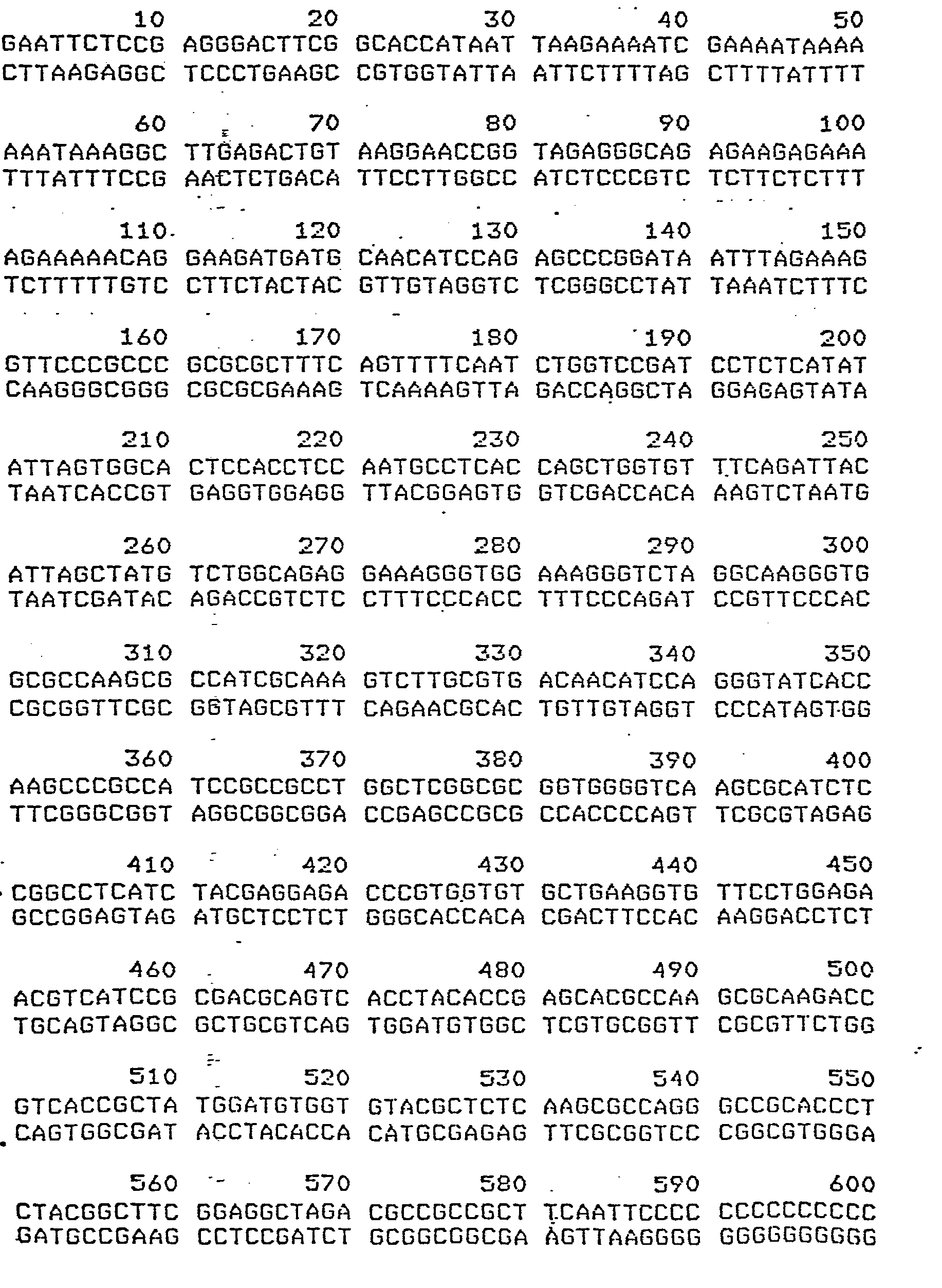

- The region of homology for both aforementioned sequences is shown below, where asterisks denote not homologous portions. Within the region shown, the first 118 base pairs have 80.5% homology and are used as a conserved DNA sequence probe in this example (sea urchin (top) base positions 449 to 567, mouse (bottom) base positions 257 to 375):X = 84.503

F( 342, 289) = ,OOOE+OO E = .000 - Restriction endonuclease cleavage sites were determined 'from the two sequences. A list of cleavage sites for the sea urchin and mouse sequences is shown below. Numbers indicate the 5' side of the cleavage site, unless site name is in brackets, which indicates that only the recognition site is known.


- The sea urchin and mouse sequences are compared with Hha I (GCGC) and the described probe sequence. The sea urchin sequence has cleavage sites at positions 295 and 497, thus creating a 202 bp fragment, which, if denatured, would hybridize with the probe sequence. Hha I (GCGC) sites in the mouse sequence, 166, 304, 311 and 380, indicate that fragments of 69 and 138 could be detected with the probe sequence.
Thus the genetic characterization for sea urchin is
202
while that for mouse is
69
138 - The same type of computer simulation as in Example 1 was carried out using trp D gene as a probe. This allows the conclusion that E. coli and Salmonella typhimurium can be distinguished by restriction fragments containing a conserved sequence.
- The E. coli trp D gene with 684 bp's is shown below:


- The trp D gene, 683 base pairs, for S. typhimurium is shown below:


- Two homology regions between both sequences were next established, where the upper sequence if for E. coli and the lower one for S.typhimurium:% = 78.390

F'( 236, 185) = OOOE+OO E = .OOO% = 80.215 F( 465, 373) = .OOOE+OO E = .OOO
- Restriction sites in both sequences are shown as follows:


- The E. coli sequence has Mbo I (GATC) sites at 135 and 564. There is a 429 bp fragment that can be detected by both
Region - Thus the identifying genetic characterization for E. coli with this probe and enzyme is
429
whereas that for S. typhimurium is
69
113
247 - This example shows the use of a region of homology in the a-fetoprotein gene sequence of human and rat, and the endonuclease MnlI (GAGG).
- The human a-fetoprotein message cDNA (1578 bp's) is as follows:


- The rat a-fetoprotein 3' end cDPZA is as follows (540 bp's):

- The homologous regions between both is as follows, (human: upper; rat: lower):Z = 85.845
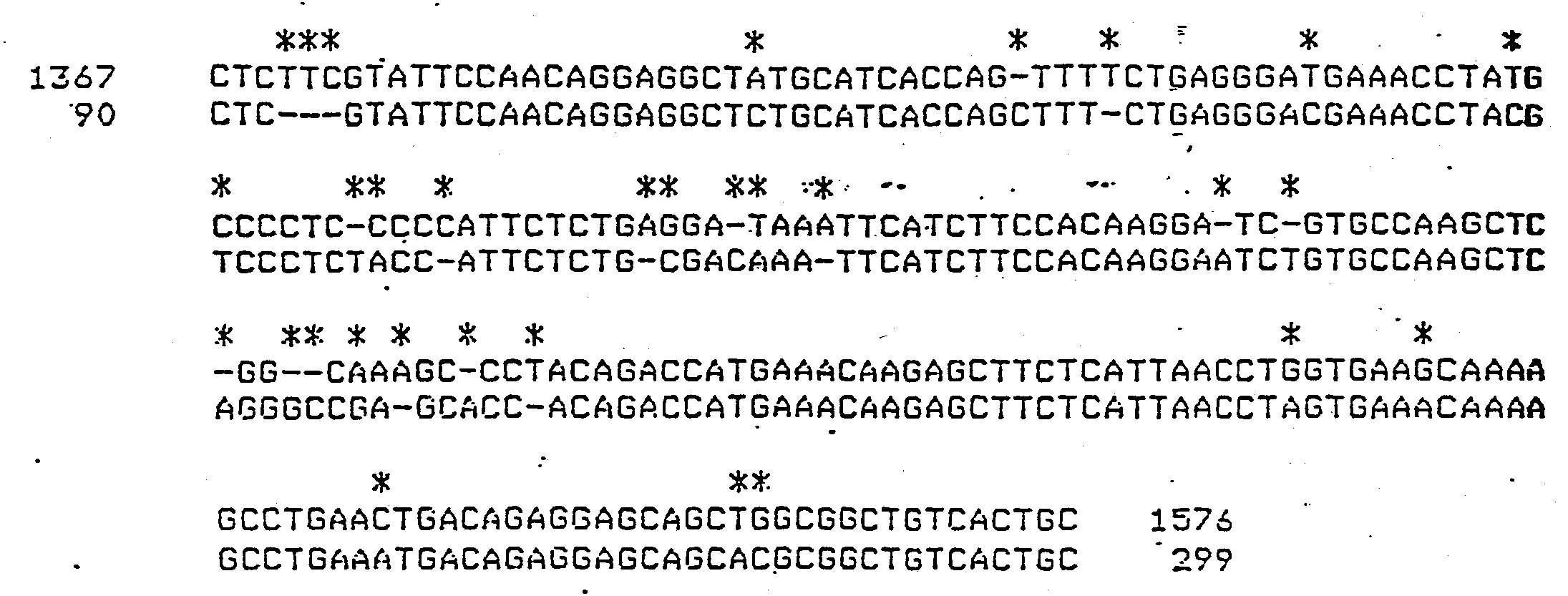
F'( 219, 188) = .OOOE+OO E = .000 - Restriction sites for both human and rat are as follows:


- It can be calculated that fragments containing a portion of the conserved sequence, 24, 34, 104 and 108 bp describe the human DNA. Fragments of 24, 59, 90 and 145 describe the rat DNA. While both sequences contain the 24 bp fragment, it is the set of fragments (taxonomic characters) that is of significance.
Claims (60)
wherein said kit also comprises a second container means containing one or more restriction endonuclease enzymes.
Applications Claiming Priority (2)
| Application Number | Priority Date | Filing Date | Title |
|---|---|---|---|
| US47743183A | 1983-03-21 | 1983-03-21 | |
| US477431 | 1983-03-21 |
Publications (4)
| Publication Number | Publication Date |
|---|---|
| EP0120658A2 true EP0120658A2 (en) | 1984-10-03 |
| EP0120658A3 EP0120658A3 (en) | 1988-01-13 |
| EP0120658B1 EP0120658B1 (en) | 1991-01-16 |
| EP0120658B2 EP0120658B2 (en) | 1998-09-02 |
Family
ID=23895887
Family Applications (1)
| Application Number | Title | Priority Date | Filing Date |
|---|---|---|---|
| EP19840301800 Expired - Lifetime EP0120658B2 (en) | 1983-03-21 | 1984-03-16 | Method for identifying and characterizing organisms |
Country Status (9)
| Country | Link |
|---|---|
| EP (1) | EP0120658B2 (en) |
| JP (1) | JP2539189B2 (en) |
| CA (1) | CA1221298A (en) |
| DE (1) | DE3483912D1 (en) |
| DK (1) | DK523484D0 (en) |
| GR (1) | GR81817B (en) |
| IE (1) | IE57467B1 (en) |
| RU (1) | RU2011681C1 (en) |
| WO (2) | WO1984003715A1 (en) |
Cited By (26)
| Publication number | Priority date | Publication date | Assignee | Title |
|---|---|---|---|---|
| EP0174226A1 (en) * | 1984-08-02 | 1986-03-12 | Institut National De La Sante Et De La Recherche Medicale (Inserm) | Probe and method for the detection of microorganisms, notably Legionella, in liquid media |
| EP0217854A1 (en) * | 1985-03-05 | 1987-04-15 | James William Blackburn | Method for the monitoring and control of microbial populations. |
| EP0250662A1 (en) * | 1986-06-25 | 1988-01-07 | The Regents Of The University Of California | Detection of mycoplasma by DNA hybridization |
| EP0229701A3 (en) * | 1986-01-10 | 1990-03-07 | Cetus Corporation | Detection of viruses by amplification and hybridization detection of viruses by amplification and hybridization |
| US5079351A (en) * | 1986-11-26 | 1992-01-07 | Cetus Corporation | Oligonucleotides and kits for detection of htlvi and htlvii viruses by hybridization |
| WO1992005277A1 (en) * | 1990-09-26 | 1992-04-02 | William Scott Davidson | Test to determine an organism's species and/or population identity by direct nucleotide sequence analysis of defined segments of its genome |
| US5176995A (en) * | 1985-03-28 | 1993-01-05 | Hoffmann-La Roche Inc. | Detection of viruses by amplification and hybridization |
| EP0553247A1 (en) * | 1990-10-17 | 1993-08-04 | The Perkin-Elmer Corporation | Method of determining a genotype by comparing the nucleotide sequence of members of a gene family and kit therefor |
| US5386022A (en) * | 1985-03-28 | 1995-01-31 | Hoffman-La Roche Inc. | Primes and probes for the amplification and detection of aids associated nucleic acids |
| US5547842A (en) * | 1986-11-24 | 1996-08-20 | Gen-Probe Incorporated | Nucleic acid probes for detection and/or quantitation of non-viral organisms |
| US5567587A (en) * | 1983-01-10 | 1996-10-22 | Gen-Probe Incorporated | Method for detecting, the presence and amount of prokaryotic organisms using specific rRNA subsequences as probes |
| US5714324A (en) * | 1983-01-10 | 1998-02-03 | Gen-Probe Incorporated | Methods for producing hybridization probes specific for rRNA subunit subsequences |
| US5851767A (en) * | 1985-03-04 | 1998-12-22 | The Regents Of The University Of California | Detection of prokaryotic organism by DNA hybridization |
| US5955261A (en) * | 1984-09-04 | 1999-09-21 | Gen-Probe Incorporated | Method for detecting the presence of group-specific viral mRNA in a sample |
| US5994059A (en) * | 1986-11-24 | 1999-11-30 | Gen-Probe Incorporated | Nucleic acid probes and methods for detecting Streptomyces enterococci |
| US6150517A (en) * | 1986-11-24 | 2000-11-21 | Gen-Probe | Methods for making oligonucleotide probes for the detection and/or quantitation of non-viral organisms |
| WO2001011088A2 (en) * | 1999-08-05 | 2001-02-15 | Evolutionary Genomics, Llc | Methods to identify evolutionarily significant changes in polynucleotide and polypeptide sequences in domesticated plants and animals |
| WO2001096603A2 (en) * | 2000-06-09 | 2001-12-20 | Evolutionary Genomics, L.L.C. | Methods to identify polynucleotide and polypeptide sequences which may be associated with physiological and medical conditions |
| US6743580B2 (en) | 1999-01-29 | 2004-06-01 | Evolutionary Genomics Llc | Methods for producing transgenic plants containing evolutionarily significant polynucleotides |
| US6866996B1 (en) | 1998-01-30 | 2005-03-15 | Evolutionary Genomics, Llc | Methods to identify polynucleotide and polypeptide sequences which may be associated with physiological and medical conditions |
| US7087742B1 (en) | 1986-11-24 | 2006-08-08 | Gen-Probe Incorporated | Oligonucleotide probes for the detection and/or quantitation of non-viral organisms |
| US7090972B1 (en) | 1986-11-24 | 2006-08-15 | Gen-Probe Incorporated | Methods for determining the presence of non-viral organisms in a sample |
| US7172863B1 (en) | 1988-12-09 | 2007-02-06 | Gen-Probe Incorporated | Nucleic acid probes and methods for detecting Neisseria gonorrhoeae |
| US7247425B2 (en) | 1998-01-30 | 2007-07-24 | Evolutionary Genomics, Llc | Methods to identify polynucleotide and polypeptide sequences which may be associated with physiological and medical conditions |
| US7252966B2 (en) | 1999-01-29 | 2007-08-07 | Evolutionary Genomics Llc | EG307 polynucleotides and uses thereof |
| US7439018B2 (en) | 1999-01-29 | 2008-10-21 | Evolutionary Genomics, Inc. | EG1117 Polynucleotides and uses thereof |
Families Citing this family (8)
| Publication number | Priority date | Publication date | Assignee | Title |
|---|---|---|---|---|
| US5614361A (en) * | 1981-09-25 | 1997-03-25 | Webster, Jr.; John A. | Method for identifying and characterizing organisms |
| US5593832A (en) * | 1983-02-28 | 1997-01-14 | Lifecodes Corporation | Method for forensic analysis |
| GB8425059D0 (en) * | 1984-10-04 | 1984-11-07 | Biotechnica Ltd | Identification technique |
| AU5698286A (en) * | 1985-01-08 | 1987-04-24 | Biotechnica Limited | Determination of identity between two organisms |
| FR2596774B1 (en) * | 1986-04-04 | 1988-07-22 | Pasteur Institut | OLIGONUCLEOTIDE PROBES AND METHODS FOR HYBRIDIZED DETECTION OF NUCLEIC ACIDS FROM BACTERIA AND OTHER LIVING BEINGS |
| WO1988000618A1 (en) * | 1986-07-10 | 1988-01-28 | Toray Industries, Inc. | Method of inspecting specimen for bacteria and probe therefor |
| FR2603047B1 (en) * | 1986-08-19 | 1990-03-02 | Vassart Gilbert | HYBRIDIZATION PROBE FOR DETECTION OF POLYMORPHIC NUCLEOTIDE SEQUENCES CONTAINED IN A HUMAN OR ANIMAL DNA SAMPLE, METHOD FOR DETECTING POLYMORPHIC DNA SEQUENCES USING SUCH A PROBE, ITS BIOLOGICAL APPLICATIONS |
| GB8716279D0 (en) * | 1987-07-10 | 1987-08-19 | Amersham Int Plc | Performing nucleic acid reactions |
Citations (3)
| Publication number | Priority date | Publication date | Assignee | Title |
|---|---|---|---|---|
| US3930956A (en) * | 1973-10-29 | 1976-01-06 | The Regents Of The University Of Michigan | Method for the genetic detection of microorganisms |
| US4358535A (en) * | 1980-12-08 | 1982-11-09 | Board Of Regents Of The University Of Washington | Specific DNA probes in diagnostic microbiology |
| WO1983001073A1 (en) * | 1981-09-25 | 1983-03-31 | Webster, John, A., Jr. | Method for identifying and characterizing organisms |
Family Cites Families (1)
| Publication number | Priority date | Publication date | Assignee | Title |
|---|---|---|---|---|
| US4396713A (en) * | 1980-11-12 | 1983-08-02 | The Regents Of The University Of Calif. | Restriction endonuclease fingerprinting of kinetoplast DNA minicircles |
-
1984
- 1984-03-14 WO PCT/US1984/000382 patent/WO1984003715A1/en unknown
- 1984-03-14 JP JP59502061A patent/JP2539189B2/en not_active Expired - Lifetime
- 1984-03-14 WO PCT/US1984/000383 patent/WO1984003716A1/en unknown
- 1984-03-16 EP EP19840301800 patent/EP0120658B2/en not_active Expired - Lifetime
- 1984-03-16 DE DE8484301800T patent/DE3483912D1/en not_active Expired - Lifetime
- 1984-03-19 GR GR74154A patent/GR81817B/el unknown
- 1984-03-20 IE IE68384A patent/IE57467B1/en not_active IP Right Cessation
- 1984-03-21 CA CA000450064A patent/CA1221298A/en not_active Expired
- 1984-11-02 DK DK523484A patent/DK523484D0/en not_active Application Discontinuation
- 1984-11-20 RU SU843813721A patent/RU2011681C1/en active
Patent Citations (4)
| Publication number | Priority date | Publication date | Assignee | Title |
|---|---|---|---|---|
| US3930956A (en) * | 1973-10-29 | 1976-01-06 | The Regents Of The University Of Michigan | Method for the genetic detection of microorganisms |
| US4358535A (en) * | 1980-12-08 | 1982-11-09 | Board Of Regents Of The University Of Washington | Specific DNA probes in diagnostic microbiology |
| US4358535B1 (en) * | 1980-12-08 | 1986-05-13 | ||
| WO1983001073A1 (en) * | 1981-09-25 | 1983-03-31 | Webster, John, A., Jr. | Method for identifying and characterizing organisms |
Cited By (57)
| Publication number | Priority date | Publication date | Assignee | Title |
|---|---|---|---|---|
| US5738989A (en) * | 1983-01-10 | 1998-04-14 | Gen-Probe Incorporated | Method for determining the sensitivity of microorganisms to anti microbial agents using ribosomal nucleic acid hybridization |
| US5567587A (en) * | 1983-01-10 | 1996-10-22 | Gen-Probe Incorporated | Method for detecting, the presence and amount of prokaryotic organisms using specific rRNA subsequences as probes |
| US5723597A (en) * | 1983-01-10 | 1998-03-03 | Gen-Probe Incorporated | Ribosomal nucleic acid probes for detecting organisms or groups of organisms |
| US5641631A (en) * | 1983-01-10 | 1997-06-24 | Gen-Probe Incorporated | Method for detecting, identifying, and quantitating organisms and viruses |
| US5928864A (en) * | 1983-01-10 | 1999-07-27 | Gen-Probe Incorporated | Method for determining the presence of organisms in a sample by detecting transfer nucleic acid |
| US5714324A (en) * | 1983-01-10 | 1998-02-03 | Gen-Probe Incorporated | Methods for producing hybridization probes specific for rRNA subunit subsequences |
| US5601984A (en) * | 1983-01-10 | 1997-02-11 | Gen-Probe Incorporated | Method for detecting, the presense or amount of a taxonomic group of organisms using specific R-RNA subsequences as probes |
| US5688645A (en) * | 1983-01-10 | 1997-11-18 | Gen-Probe Incorporated | Method for detecting, identifying, and quantitating non-viral organisms |
| US5738988A (en) * | 1983-01-10 | 1998-04-14 | Gen-Probe Incorporated | Method for detecting antimicrobial agents or unknown organisms in a sample using ribosomal probe hybridization |
| US5641632A (en) * | 1983-01-10 | 1997-06-24 | Gen-Probe Incorporated | Method for preparing rRNA for hybridization with a probe |
| US5932416A (en) * | 1983-01-10 | 1999-08-03 | Gen Probe Inc | Method for detecting the presence of RNA belonging to an organ or tissue cell-type |
| EP0174226A1 (en) * | 1984-08-02 | 1986-03-12 | Institut National De La Sante Et De La Recherche Medicale (Inserm) | Probe and method for the detection of microorganisms, notably Legionella, in liquid media |
| US4810644A (en) * | 1984-08-02 | 1989-03-07 | Institut Pasteur | Probe and a process for the detection of specific microorganisms, particularly legionellas, in environments which contain them |
| US5955261A (en) * | 1984-09-04 | 1999-09-21 | Gen-Probe Incorporated | Method for detecting the presence of group-specific viral mRNA in a sample |
| US5851767A (en) * | 1985-03-04 | 1998-12-22 | The Regents Of The University Of California | Detection of prokaryotic organism by DNA hybridization |
| EP0217854A4 (en) * | 1985-03-05 | 1989-09-19 | James William Blackburn | Method for the monitoring and control of microbial populations. |
| EP0217854A1 (en) * | 1985-03-05 | 1987-04-15 | James William Blackburn | Method for the monitoring and control of microbial populations. |
| US5386022A (en) * | 1985-03-28 | 1995-01-31 | Hoffman-La Roche Inc. | Primes and probes for the amplification and detection of aids associated nucleic acids |
| US5176995A (en) * | 1985-03-28 | 1993-01-05 | Hoffmann-La Roche Inc. | Detection of viruses by amplification and hybridization |
| EP0229701A3 (en) * | 1986-01-10 | 1990-03-07 | Cetus Corporation | Detection of viruses by amplification and hybridization detection of viruses by amplification and hybridization |
| EP0250662A1 (en) * | 1986-06-25 | 1988-01-07 | The Regents Of The University Of California | Detection of mycoplasma by DNA hybridization |
| US5693469A (en) * | 1986-11-24 | 1997-12-02 | Gen-Probe Incorporated | Nucleic acid probes and methods for detecting Escherichia coli |
| US6512105B1 (en) | 1986-11-24 | 2003-01-28 | Gen-Probe Incorporated | Methods for making oligonucleotide probes for the detection and/or quantitation of non-viral organisms |
| US5683876A (en) * | 1986-11-24 | 1997-11-04 | Gen-Probe Incorporated | Nucleic acid probes and methods for detecting Proteus mirabilis |
| US5677127A (en) * | 1986-11-24 | 1997-10-14 | Gen-Probe Incorporated | Nucleic acid probes and methods for detecting group I pseudomonas |
| US5691149A (en) * | 1986-11-24 | 1997-11-25 | Gen-Probe Incorporated | Nucleic acid probes and method for detecting Mycoplasma pneumoniae |
| US5693468A (en) * | 1986-11-24 | 1997-12-02 | Gen-Probe Incorported | Nucleic acid probes and methods for detecting chlamydia trachomatis |
| US5677128A (en) * | 1986-11-24 | 1997-10-14 | Gen-Probe Incorporated | Nucleic acid probes and methods for detecting mycobacterium |
| US5677129A (en) * | 1986-11-24 | 1997-10-14 | Gen-Probe Incorporated | Nucleic acid probes and methods for detecting legionella |
| US5714321A (en) * | 1986-11-24 | 1998-02-03 | Gen-Probe Incorporated | Nucleic acid probes and methods for detecting salmonella |
| US5674684A (en) * | 1986-11-24 | 1997-10-07 | Gen-Probe Incorporated | Nucleic acid probes and methods for detecting campylobacters |
| US5593841A (en) * | 1986-11-24 | 1997-01-14 | Gen-Probe Incorporated | Nucleic acid probes for detection and/or quantitation of non-viral organisms |
| US5547842A (en) * | 1986-11-24 | 1996-08-20 | Gen-Probe Incorporated | Nucleic acid probes for detection and/or quantitation of non-viral organisms |
| US5827651A (en) * | 1986-11-24 | 1998-10-27 | Gen-Probe Incorporated | Nucleic acid probes and methods for detecting fungi |
| US5840488A (en) * | 1986-11-24 | 1998-11-24 | Gen-Probe Incorporated | Nucleic acid probes for detection and/or quantitation of non-viral organisms |
| US7138516B1 (en) | 1986-11-24 | 2006-11-21 | Gen-Probe Incorporated | Oligonucleotide probes for the detection and/or quantitation of non-viral organisms |
| US7090972B1 (en) | 1986-11-24 | 2006-08-15 | Gen-Probe Incorporated | Methods for determining the presence of non-viral organisms in a sample |
| US7087742B1 (en) | 1986-11-24 | 2006-08-08 | Gen-Probe Incorporated | Oligonucleotide probes for the detection and/or quantitation of non-viral organisms |
| US5679520A (en) * | 1986-11-24 | 1997-10-21 | Gen-Probe Incorporated | Nucleic acid probes and methods for detecting eubacteria |
| US5958679A (en) * | 1986-11-24 | 1999-09-28 | Gen-Probe Incorporated | Nucleic acid probes and methods for detecting Enterobacter cloacae |
| US5994059A (en) * | 1986-11-24 | 1999-11-30 | Gen-Probe Incorporated | Nucleic acid probes and methods for detecting Streptomyces enterococci |
| US6150517A (en) * | 1986-11-24 | 2000-11-21 | Gen-Probe | Methods for making oligonucleotide probes for the detection and/or quantitation of non-viral organisms |
| US5079351A (en) * | 1986-11-26 | 1992-01-07 | Cetus Corporation | Oligonucleotides and kits for detection of htlvi and htlvii viruses by hybridization |
| US7172863B1 (en) | 1988-12-09 | 2007-02-06 | Gen-Probe Incorporated | Nucleic acid probes and methods for detecting Neisseria gonorrhoeae |
| WO1992005277A1 (en) * | 1990-09-26 | 1992-04-02 | William Scott Davidson | Test to determine an organism's species and/or population identity by direct nucleotide sequence analysis of defined segments of its genome |
| EP0553247A4 (en) * | 1990-10-17 | 1995-05-10 | Applied Biosystems | |
| EP0553247A1 (en) * | 1990-10-17 | 1993-08-04 | The Perkin-Elmer Corporation | Method of determining a genotype by comparing the nucleotide sequence of members of a gene family and kit therefor |
| US6866996B1 (en) | 1998-01-30 | 2005-03-15 | Evolutionary Genomics, Llc | Methods to identify polynucleotide and polypeptide sequences which may be associated with physiological and medical conditions |
| US7247425B2 (en) | 1998-01-30 | 2007-07-24 | Evolutionary Genomics, Llc | Methods to identify polynucleotide and polypeptide sequences which may be associated with physiological and medical conditions |
| US7462460B2 (en) | 1998-01-30 | 2008-12-09 | Evolutionary Genomics, Inc. | Methods for identifying agents that increase the p44 function of microtubule assembly or resistance to HCV infection |
| US6743580B2 (en) | 1999-01-29 | 2004-06-01 | Evolutionary Genomics Llc | Methods for producing transgenic plants containing evolutionarily significant polynucleotides |
| US7252966B2 (en) | 1999-01-29 | 2007-08-07 | Evolutionary Genomics Llc | EG307 polynucleotides and uses thereof |
| US7439018B2 (en) | 1999-01-29 | 2008-10-21 | Evolutionary Genomics, Inc. | EG1117 Polynucleotides and uses thereof |
| WO2001011088A2 (en) * | 1999-08-05 | 2001-02-15 | Evolutionary Genomics, Llc | Methods to identify evolutionarily significant changes in polynucleotide and polypeptide sequences in domesticated plants and animals |
| WO2001011088A3 (en) * | 1999-08-05 | 2001-12-13 | Genoplex Inc | Methods to identify evolutionarily significant changes in polynucleotide and polypeptide sequences in domesticated plants and animals |
| WO2001096603A3 (en) * | 2000-06-09 | 2002-05-23 | Evolutionary Genomics L L C | Methods to identify polynucleotide and polypeptide sequences which may be associated with physiological and medical conditions |
| WO2001096603A2 (en) * | 2000-06-09 | 2001-12-20 | Evolutionary Genomics, L.L.C. | Methods to identify polynucleotide and polypeptide sequences which may be associated with physiological and medical conditions |
Also Published As
| Publication number | Publication date |
|---|---|
| CA1221298A (en) | 1987-05-05 |
| JPS60500895A (en) | 1985-06-20 |
| IE840683L (en) | 1984-09-21 |
| WO1984003716A1 (en) | 1984-09-27 |
| DK523484A (en) | 1984-11-02 |
| EP0120658B2 (en) | 1998-09-02 |
| WO1984003715A1 (en) | 1984-09-27 |
| IE57467B1 (en) | 1993-01-27 |
| EP0120658A3 (en) | 1988-01-13 |
| DK523484D0 (en) | 1984-11-02 |
| GR81817B (en) | 1984-12-12 |
| JP2539189B2 (en) | 1996-10-02 |
| DE3483912D1 (en) | 1991-02-21 |
| EP0120658B1 (en) | 1991-01-16 |
| RU2011681C1 (en) | 1994-04-30 |
Similar Documents
| Publication | Publication Date | Title |
|---|---|---|
| EP0076123B1 (en) | Method for identifying and characterizing organisms | |
| EP0120658B1 (en) | Method for identifying and characterizing organisms | |
| US5087558A (en) | Method for identifying and characterizing organisms | |
| EP0155360B1 (en) | Method for identifying and characterizing organisms | |
| EP0155359B1 (en) | Method for identifying and characterizing organisms | |
| Ahmed et al. | Multilocus sequence typing method for identification and genotypic classification of pathogenic Leptospira species | |
| Gopaul et al. | Rapid identification of Brucella isolates to the species level by real time PCR based single nucleotide polymorphism (SNP) analysis | |
| Dijkshoorn et al. | The diversity of the genus Acinetobacter | |
| US5348854A (en) | Method for detecting prokaryotic organisms | |
| Huys et al. | Evaluation of amplified fragment length polymorphism analysis for inter-and intraspecific differentiation of Mycobacterium bovis, M. tuberculosis, and M. ulcerans | |
| Kashyap et al. | Ribotyping: A tool for molecular taxonomy | |
| Bellis et al. | Linkage disequilibrium analysis in the genetically isolated Norfolk Island population | |
| US5614361A (en) | Method for identifying and characterizing organisms | |
| Huynh et al. | Multiple locus variable number tandem repeat (VNTR) analysis (MLVA) of Brucella spp. identifies species-specific markers and insights into phylogenetic relationships | |
| Drevinek et al. | Diversity of the parB and repA genes of the Burkholderia cepacia complex and their utility for rapid identification of Burkholderia cenocepacia | |
| IE54645B1 (en) | Method for identifying and characterising organisms | |
| JP2729457B2 (en) | Kit for identification of organisms | |
| JP2514164B2 (en) | Method for identifying and characterizing organisms | |
| CN109652573A (en) | For Salmonella typhimurtum or the site VNTR, detection primer group and the determination method of its single-phase bacterium mutation parting detection | |
| Jenks | Microbial genome sequencing—beyond the double helix | |
| Lin et al. | Improved subspecies identification in clinical Mycobacterium abscessus complex isolates using whole genome sequence | |
| Basibuyuk et al. | Phylogenetic Systematics | |
| JPH0824584B2 (en) | Method for identifying and characterizing organisms | |
| Arnold | 15 Fluorescent Amplified Fragment Length Polymorphism (FAFLP) Genotyping | |
| Sharma et al. | DNA based molecular markers: concept, techniques and comparison |
Legal Events
| Date | Code | Title | Description |
|---|---|---|---|
| PUAI | Public reference made under article 153(3) epc to a published international application that has entered the european phase |
Free format text: ORIGINAL CODE: 0009012 |
|
| AK | Designated contracting states |
Designated state(s): BE DE FR GB IT LU NL |
|
| PUAL | Search report despatched |
Free format text: ORIGINAL CODE: 0009013 |
|
| AK | Designated contracting states |
Kind code of ref document: A3 Designated state(s): BE DE FR GB IT LU NL |
|
| 17P | Request for examination filed |
Effective date: 19880630 |
|
| 17Q | First examination report despatched |
Effective date: 19881104 |
|
| RAP3 | Party data changed (applicant data changed or rights of an application transferred) |
Owner name: WEBSTER, JOHN A., JR. |
|
| GRAA | (expected) grant |
Free format text: ORIGINAL CODE: 0009210 |
|
| AK | Designated contracting states |
Kind code of ref document: B1 Designated state(s): BE DE FR GB IT LU NL |
|
| REF | Corresponds to: |
Ref document number: 3483912 Country of ref document: DE Date of ref document: 19910221 |
|
| ITF | It: translation for a ep patent filed |
Owner name: BARZANO' E ZANARDO ROMA S.P.A. |
|
| ET | Fr: translation filed | ||
| PLBI | Opposition filed |
Free format text: ORIGINAL CODE: 0009260 |
|
| 26 | Opposition filed |
Opponent name: CIBA-GEIGY AG PATENTABTEILUNG Effective date: 19911016 |
|
| NLR1 | Nl: opposition has been filed with the epo |
Opponent name: CIBA-GEIGY AG |
|
| EPTA | Lu: last paid annual fee | ||
| PLAW | Interlocutory decision in opposition |
Free format text: ORIGINAL CODE: EPIDOS IDOP |
|
| PLAW | Interlocutory decision in opposition |
Free format text: ORIGINAL CODE: EPIDOS IDOP |
|
| PUAH | Patent maintained in amended form |
Free format text: ORIGINAL CODE: 0009272 |
|
| STAA | Information on the status of an ep patent application or granted ep patent |
Free format text: STATUS: PATENT MAINTAINED AS AMENDED |
|
| 27A | Patent maintained in amended form |
Effective date: 19980902 |
|
| AK | Designated contracting states |
Kind code of ref document: B2 Designated state(s): BE DE FR GB IT LU NL |
|
| NLR2 | Nl: decision of opposition | ||
| ET3 | Fr: translation filed ** decision concerning opposition | ||
| NLR3 | Nl: receipt of modified translations in the netherlands language after an opposition procedure | ||
| REG | Reference to a national code |
Ref country code: GB Ref legal event code: IF02 |
|
| PGFP | Annual fee paid to national office [announced via postgrant information from national office to epo] |
Ref country code: LU Payment date: 20021230 Year of fee payment: 20 |
|
| PGFP | Annual fee paid to national office [announced via postgrant information from national office to epo] |
Ref country code: GB Payment date: 20030204 Year of fee payment: 20 |
|
| PGFP | Annual fee paid to national office [announced via postgrant information from national office to epo] |
Ref country code: NL Payment date: 20030206 Year of fee payment: 20 |
|
| PGFP | Annual fee paid to national office [announced via postgrant information from national office to epo] |
Ref country code: FR Payment date: 20030303 Year of fee payment: 20 |
|
| PGFP | Annual fee paid to national office [announced via postgrant information from national office to epo] |
Ref country code: DE Payment date: 20030331 Year of fee payment: 20 |
|
| PGFP | Annual fee paid to national office [announced via postgrant information from national office to epo] |
Ref country code: BE Payment date: 20030409 Year of fee payment: 20 |
|
| PG25 | Lapsed in a contracting state [announced via postgrant information from national office to epo] |
Ref country code: GB Free format text: LAPSE BECAUSE OF EXPIRATION OF PROTECTION Effective date: 20040315 |
|
| PG25 | Lapsed in a contracting state [announced via postgrant information from national office to epo] |
Ref country code: NL Free format text: LAPSE BECAUSE OF EXPIRATION OF PROTECTION Effective date: 20040316 Ref country code: LU Free format text: LAPSE BECAUSE OF NON-PAYMENT OF DUE FEES Effective date: 20040316 |
|
| BE20 | Be: patent expired |
Owner name: *WEBSTER JOHN A. JR. Effective date: 20040316 |
|
| REG | Reference to a national code |
Ref country code: GB Ref legal event code: PE20 |
|
| NLV7 | Nl: ceased due to reaching the maximum lifetime of a patent |
Effective date: 20040316 |