EP0121775A1 - Novel expression plasmids and their use in the method for expressing prochymosin coding gene in E. coli - Google Patents
Novel expression plasmids and their use in the method for expressing prochymosin coding gene in E. coli Download PDFInfo
- Publication number
- EP0121775A1 EP0121775A1 EP84102451A EP84102451A EP0121775A1 EP 0121775 A1 EP0121775 A1 EP 0121775A1 EP 84102451 A EP84102451 A EP 84102451A EP 84102451 A EP84102451 A EP 84102451A EP 0121775 A1 EP0121775 A1 EP 0121775A1
- Authority
- EP
- European Patent Office
- Prior art keywords
- plasmid
- prochymosin
- coli
- gene
- dna
- Prior art date
- Legal status (The legal status is an assumption and is not a legal conclusion. Google has not performed a legal analysis and makes no representation as to the accuracy of the status listed.)
- Withdrawn
Links
- 0 C*[C@](CC[C@@](C)C1)C1=C=CC(C)CCCCC(C)C Chemical compound C*[C@](CC[C@@](C)C1)C1=C=CC(C)CCCCC(C)C 0.000 description 1
Images


Classifications
-
- C—CHEMISTRY; METALLURGY
- C12—BIOCHEMISTRY; BEER; SPIRITS; WINE; VINEGAR; MICROBIOLOGY; ENZYMOLOGY; MUTATION OR GENETIC ENGINEERING
- C12N—MICROORGANISMS OR ENZYMES; COMPOSITIONS THEREOF; PROPAGATING, PRESERVING, OR MAINTAINING MICROORGANISMS; MUTATION OR GENETIC ENGINEERING; CULTURE MEDIA
- C12N9/00—Enzymes; Proenzymes; Compositions thereof; Processes for preparing, activating, inhibiting, separating or purifying enzymes
- C12N9/14—Hydrolases (3)
- C12N9/48—Hydrolases (3) acting on peptide bonds (3.4)
- C12N9/50—Proteinases, e.g. Endopeptidases (3.4.21-3.4.25)
- C12N9/64—Proteinases, e.g. Endopeptidases (3.4.21-3.4.25) derived from animal tissue
- C12N9/6421—Proteinases, e.g. Endopeptidases (3.4.21-3.4.25) derived from animal tissue from mammals
- C12N9/6478—Aspartic endopeptidases (3.4.23)
- C12N9/6481—Pepsins (3.4.23.1; 3.4.23.2; 3.4.23.3)
-
- C—CHEMISTRY; METALLURGY
- C07—ORGANIC CHEMISTRY
- C07K—PEPTIDES
- C07K1/00—General methods for the preparation of peptides, i.e. processes for the organic chemical preparation of peptides or proteins of any length
- C07K1/107—General methods for the preparation of peptides, i.e. processes for the organic chemical preparation of peptides or proteins of any length by chemical modification of precursor peptides
- C07K1/113—General methods for the preparation of peptides, i.e. processes for the organic chemical preparation of peptides or proteins of any length by chemical modification of precursor peptides without change of the primary structure
- C07K1/1136—General methods for the preparation of peptides, i.e. processes for the organic chemical preparation of peptides or proteins of any length by chemical modification of precursor peptides without change of the primary structure by reversible modification of the secondary, tertiary or quarternary structure, e.g. using denaturating or stabilising agents
-
- C—CHEMISTRY; METALLURGY
- C12—BIOCHEMISTRY; BEER; SPIRITS; WINE; VINEGAR; MICROBIOLOGY; ENZYMOLOGY; MUTATION OR GENETIC ENGINEERING
- C12N—MICROORGANISMS OR ENZYMES; COMPOSITIONS THEREOF; PROPAGATING, PRESERVING, OR MAINTAINING MICROORGANISMS; MUTATION OR GENETIC ENGINEERING; CULTURE MEDIA
- C12N15/00—Mutation or genetic engineering; DNA or RNA concerning genetic engineering, vectors, e.g. plasmids, or their isolation, preparation or purification; Use of hosts therefor
- C12N15/09—Recombinant DNA-technology
- C12N15/63—Introduction of foreign genetic material using vectors; Vectors; Use of hosts therefor; Regulation of expression
- C12N15/70—Vectors or expression systems specially adapted for E. coli
- C12N15/71—Expression systems using regulatory sequences derived from the trp-operon
-
- C—CHEMISTRY; METALLURGY
- C12—BIOCHEMISTRY; BEER; SPIRITS; WINE; VINEGAR; MICROBIOLOGY; ENZYMOLOGY; MUTATION OR GENETIC ENGINEERING
- C12N—MICROORGANISMS OR ENZYMES; COMPOSITIONS THEREOF; PROPAGATING, PRESERVING, OR MAINTAINING MICROORGANISMS; MUTATION OR GENETIC ENGINEERING; CULTURE MEDIA
- C12N9/00—Enzymes; Proenzymes; Compositions thereof; Processes for preparing, activating, inhibiting, separating or purifying enzymes
- C12N9/14—Hydrolases (3)
- C12N9/48—Hydrolases (3) acting on peptide bonds (3.4)
- C12N9/50—Proteinases, e.g. Endopeptidases (3.4.21-3.4.25)
- C12N9/64—Proteinases, e.g. Endopeptidases (3.4.21-3.4.25) derived from animal tissue
- C12N9/6421—Proteinases, e.g. Endopeptidases (3.4.21-3.4.25) derived from animal tissue from mammals
- C12N9/6478—Aspartic endopeptidases (3.4.23)
- C12N9/6483—Chymosin (3.4.23.4), i.e. rennin
-
- C—CHEMISTRY; METALLURGY
- C12—BIOCHEMISTRY; BEER; SPIRITS; WINE; VINEGAR; MICROBIOLOGY; ENZYMOLOGY; MUTATION OR GENETIC ENGINEERING
- C12Y—ENZYMES
- C12Y304/00—Hydrolases acting on peptide bonds, i.e. peptidases (3.4)
- C12Y304/23—Aspartic endopeptidases (3.4.23)
- C12Y304/23004—Chymosin (3.4.23.4), i.e. rennin
-
- C—CHEMISTRY; METALLURGY
- C07—ORGANIC CHEMISTRY
- C07K—PEPTIDES
- C07K2319/00—Fusion polypeptide
Definitions
- This invention relates in general to Genetic Engineering, and more specifically to the production of recombinant DNA containing prochymosim coding gene, and to the utilization of the same to produce microorganisms for the volume production of prochymosin.
- Genetic Engineering refers to the in vitro technique of forming recombinant DNA containing exogenous genes and a suitable vector, selecting a DNA on the basis of the desired genetic information, and introducing the selected DNA into a suitable host microorganism, whereby the foreign genetic information becomes part of the genetic complement of the host.
- the genetically engineered microorganism prepared by this technique is capable of expressing the foreign gene in its cells under appropriate conditions of cultivation, and producing a substance (e.g. enzymes, proteins, hormones or the like) for which the gene will code.
- the enzymatic protein chymosin is also known as rennin. It is the major proteolytic protein found in the stomach of the pre-ruminant calf, and is responsible for clotting milk. Chymosin has therefore long been used for coagulating milk casein in cheese making. It has 323 amino acids and a molecular weight of 35,652.
- Prochymosin is a precursor of chymosin having 42 additional amino acids at the amino terminus of the chymosin molecule, and it is converted to chymosin under acidic conditions by losing the additional amino acids.
- Prochymosin has 365 amino acids and a molecular weight of 40,777.
- the complete amino acid sequence of prorennin has been described in B. Foltmann et al, Proc. Natl. Acad. Sci. USA, 74,231 (1977). The published sequence later proved to be incorrect at a number of amino acid sites. The revised sequence is therefore given in FIG. 1.
- chymosin Since chymosin is secreted by the mucous membrane of the abomasum of a newly born calf, it is obtained from the stomachs of calves in commercial production. There are several serious problems associated with the obtaining of chymosin from calves: this process involves a number of complicated procedures which present many difficulties when attempting to secure large amounts. It is also costly, and has not been able to meet commercial demands in recent years. For these reasons, some substitutes for chymosin have become acceptable in cheese making.
- One such substitute is a microbial rennet which is produced by Mucor pusillous, Mucor miehei or Endothia parasitica. However, this microbial rennet has moderately strong proteolytic activity and is not as suitable in cheese ripening as chymo - sin itself which has a specific yet weak proteolytic activity.
- the process of this invention employs novel expression plasmids which are formed by isolating DNA inserts having prochymosin coding genes from previously constructed recombinant plasmids, and inserting said D N A into suitable vectors.
- the expression plasmids are then used to transform susceptible microorganisms under conditions where transformation can occur.
- the microorganism is grown under conditions which will favor the harvesting of transformants which contain the recombinant expression plasmid.
- microbial cells can be repeatedly multiplied by culturing the microorganism in an appropriate growth medium, which will express the prochymosin coding gene to produce prochymosin in their cells.
- the volume production of prochymosin by microorganisms in fermentation culture will become feasible and provide calf chymosin at reasonable cost.
- One of the objects of this invention is to provide specific prochymosin genes for use in the construction of novel expression plasmids.
- Another object of this invention is to provide such novel expression plasmids and utilize them to obtain a high expression of prochymosin in microbial host cells.
- Another object of this invention is to provide methods for producing novel expression plasmids.
- Another object of this invention is to employ the E. coil tryptophan operon promoter in place of the E. coli lac operon promoter.
- Still another object of this invention is to obtain genetically engineered microorganisms which will be capable of producing substantially greater amounts of prochymosin than could be produced by the hitherto known microorganisms.
- a still further object of this invention is to provide a method for producing prochymosin using genetically engineered microorganisms.
- One more object of this invention is to provide prochymosin iteself produced according to the above-mentioned methods.
- prochymosin gene is defined as any sequence of nucleotides which will encode the prochymosin molecule. Any portion of the nuclotide sequence as described in FIG. 1 can be used.
- prochymosin in host cells and their production can be accomplished in terms of the following steps:
- pCR301 One recombinant plasmid of particular interest is referred to as pCR301, and is described in Nishimori et al., Gene, 19, 337 (1982). Descriptions of methods for constructing other plasmids carrying prochymosin cDNA may be found in Nishimori et al., J. Biochem., 90, 901 (1981) and other references previously referred to. These plasmids are useful as a source of the prochymosin genes and normally are constructed in the manner described below;
- prochymosin like protein Protein resulting from a prochymosin coding gene devoid of some tip portion of the coding sequence may be called "prochymosin like protein.”
- Prochymosin having methionine attached to its N-terminus may be called N-terminal methionyl prochymosin. Throughout the Specification, both may be referred to as prochymosin.
- this invention encompasses specific prorennin coding gene, coding sequences containing them, methods for constructing novel expression plasmids; transformed microorganisms; and prochymosin produced therefrom.
- E. coli c600 r k - m k - Cells are grown under conventional culturing conditions.
- culture media for the E. coli strain can be:
- transformation experiments are conducted in LB medium containing 50 ug/ml of ampicillin according to the method of Norgard (Gene, 3, 279 (1978).
- E. coli cells are grown to a density of from 10 to 30x10 12 cells per liter at a temperature of from 30°C to 40°C.
- vector plasmids In order to have cloned genes expressed in host cells, it is necessary to equip vector plasmids with a suitable promoter; several promoters can be used to obtain expression as long as they are compatible with the host cells. Desirably, the E. coli tryptophan operon promoter is used in this invention.
- genes are conveniently available from the primary recombinant plasmids as described previously. These plasmids contain a prorennin coding gene with variable sizes and some of them have been mapped precisely by restriction enzyme digestion. Therefore, by cutting with appropriate restriction enzymes, useful portions of the prorennin coding genes can be separated, and used as a source of DNA for a second cloning into a vector plasmid.
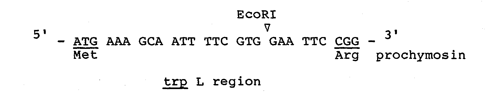
- a representative of such plasmids is pCR301. Plasmid pCR301 contains the lactose operon promoter and almost entire prochymosin gene sequence (from nucleotide #13 to #1095) only devoid of the first four codons.
- This plasmid is derived from pBR322 plasmid (F. Bolvar et al., Gene, 2, 95 (1977).
- the necleotide sequence between the initiation codon ATG and the beginning of the cloned procymosin gene is illustrated below(this sequence coresponds to the nucleotide coding for the ten N-terminal amino acids of beta-glactosidase):
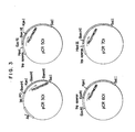
- FIG. 3 A restriction enzyme map for pCR301 is shown in FIG. 3.
- Plasmid pOCT3 the precursor of pOCT2, is a pBR322 plasmid into which a DNA fragment having the trp promoter-operator, trp L and trp E' has been integrated at its EcoRI site.
- Said trp promoter-operator portion (- 0.5 Kb) is originally extracted from plasmid p trp ED5-1 (R.A. Hallewell and S. Emtage, Gene, 9, 27 (1980)). This DNA is filled in by means of T 4 D polymerase, to which a EcoRI linker is annexed. The resulting DNA fragment is inserted into the EcoRI site of pBR 322.
- Plasmid pOCT3 was kindly provided by Professor K.
- trp operon D NA is cut out from pOCT3 by digestion with EcoRI and, once isolated, it is then put back to the EcoRI site of pOCT3 in such a manner that the cut ends become switched around, leading to pOCT2.
- the transcriptional direction of pOCT3 is opposite to that of pOCT2; it is clockwise in pOCT2 (5 1 to 3') whereas counterclockwise in pOCT3.
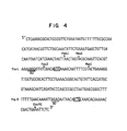
- Fig. 4 shows the DNA sequence (about 300 bases) in the vicinity of the trp promoter in pOCT2 plasmid.
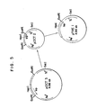
- Fig. 5 is a schematic diagram for the construction of pOCT3 from pOCT2.
- a restriction enzyme map for pOCT2 is shown in FIG. 2.
- Plasmid pTREl is derived from pOCT2 and has a single EcoRI site since the other EcoRI site near the ampicillin resistant region has been removed from pOCT2. This plasmid contains the intact trp operon part of pOCT2 as shown in FIG. 2. The construction of pTREI is accomplished in the following manner.
- Plasmid pOCT2 is partially digested with EcoRI, resulting in the cleavage of the EcoRI site near the ampicillin resistant region. The single stranded- portions are then repaired by means of DNA polymerase and the repaired ends are joined using T 4 DNA ligase to produce pTREI.
- FIG. 5 shows a schematic diagram for the construction of pTREl from pOCT2.
- Plasmid pTRLl has a DNA portion from pOCT2 bounded by EcoRI and RSaI sites. This portion comprises 360 base pairs (from an unidentified EcoR site to RSaI site in the trp L region), some of which are sequenced as shown in FIG. 4. The construction of pTRLl is accomplished in the following manner.
- Plasmid pOCT2 is cleaved with EcoRI and then partially digested with RSaI to yield a 360 base pair fragment containing the trp promoter region (trp L). This fragment is ligated with the EcoRI linker at the cut RSaI site. Digestion with EcoRI produces a fragment with cohesive ends. This fragment is then inserted into the EcoRI site of pBR322 to afford two kinds of recombinant plasmids. Plasmid pTRLI is the one wherein the transcriptional direction from the trp promoter is counter-clockwise.
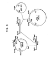
- FIG. 6 is a schematic diagram for the construction of pTRLl from pOCT2.
- Plasmid pTRPI has a DNA portion from pTREl bounded by HpaI and ClaI sites. This portion comprises about 4640 base pairs originally derived from pBR322. The plasmid has also another DNA portion from pTREl bounded by HpaI and TaqI sites. This portion comprises 32 base pairs corresponding to the trp L promoter. The construction of pTRPI is accomplished in the following manner.
- Plasmid pTREl is cleaved with HpaI and Clal to yield an about 4640 bp linear DNA fragment. Both fragments are joined together by ligation to produce pTRPl.
- a restriction enzyme map for pTRPl is shown in FIG. 2.
- FIG. 7 is a schematic diagram for the construction of pTRPl from pTREl.
- a desired DNA fragment containing the cloned prochymosin gene is extracted from a transformant, and cleaved with restriction enzymes; the vector plasmid is similarly cleaved; and the fragments are mixed and ligated.
- Plasmid pCR501 contains a DNA portion of pTREl fused to about 1,000 nucleotides from pCR301, which code for the procymosin molecule. The construction of pCR501 is accomplished in the following manner.
- Plasmid pTRE1 is cut with EcoRI and single-stranded portions are removed by means of Sl nuclease.
- the EcoRI linker (GGATTTCC/CCTTAAGG) is linked to the blunt end using T 4 DNA ligase.
- This DNA is next digested with EcoRI and SalI to produce linear DNA segment (a) (ca. 4210bp).
- Plasmid pCR301 is cut with EcoRI and KpnI to obtain DNA segment (b).
- This DNA segment is a 246 bp sequence containing the prochymosin gene fused to the residual beta-glactosidase coding gene.
- pCR301 is cut with KpnI and SalI to obtain DNA segment (c).
- This DNA is a 1025 bp sequence of the prochymosin gene including its C-terminus. Each DNA segment is ligated together by means of T 4 DNA ligase. E. coli strain C600 is transformed with the ligated DNA, and ampicillin resistant colonies (pCR501) are selected.
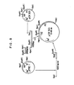
- FIG. 8 shows a schematic diagram for the construction of pCR501 from pCR301 and pTREl.
- Transformed cells carrying the plasmid will express the cloned gene and produce prochymosin under the control of the trp E promoter.
- the prochymosin produced in this manner lacks an amino acid sequence of the genuine prochymosin which corresponds to that from the 1st to the 4th amino acid at the N terminus, but contains, in its place, eight amino acids (methionine and others) resulting from the above-stated trp E promoter coding sequence as well as two amino acid residues resulting from beta-glactosidase.
- This prochymosin is therefore called prochymosin like protein.
- Plasmid pCR601 contains a DNA portion of pTRE1 and a 56 bp fragment from pTRLl fused to the same coding sequence for prochymosin present in pCR501. The construction of pCR601 is accomplished in the following manner.
- Plasmid pTREl is cut with HpaI and SalI to produce a linear 4010 bp segment (DNA (d)).
- plasmid pTRLI is cut with HpaI and EcoRI to produce a 56 bp segment (DNA (e)) containing the trp L promoter. both segments are mixed and ligated together with DNA (b) and (c) (both obtained previously from pCR301), by means of T 4 DNA ligase.
- E. coli strain c 600 is transformed with the resulting ligated DNA and ampicillin resistant colonies (pCR 601) are selected.
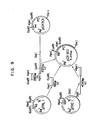
- FIG. 9 shows a schematic diagram for the construction of pCR601 from pCR301, pTREl and pTRLl.
- Transformed cells carrying the plasmid will express the cloned prochymosin gene and produce prochymosin under the control of trp L promoter.
- the prochymosin produced in this manner lacks an amino acid sequence of the genuine prochymosin which corresponds to that from the lst to the 4th amino acid at the N terminus, but contains, in its place, six amino acids (methionine and others) resulting from the above-stated trp L promoter coding sequence as well as two amino acid residues resulting from beta-glactosidase.
- This prochymosin is therefore called prochymosin like protein.
- Plasmid pCR701 contains a DNA portion of pTRPI and a synthetic nucleotide piece fused to a prochymosin coding sequence from pCR501. The construction of pCR701 is accomplished in the following manner.
- Plasmid pTRPI is cut with Hind III and Sal I to obtain liner DNA segment (f) (about 4050 bp).
- Plasmid pCR501 is cut with BamHI and SalI to obtain liner DNA segment (g) (1264 bp).
- a synthetic oligonucleotide having a 22bp is synthesized, which is the sequence of:
- This DNA segment (f) makes up for the nucleotide sequence that has been removed by digestion with Bam HI which cuts the 14th nucleotide of prochymosin gene.
- the three segments (f), (g) and (h) are ligated together by means of T 4 DNA ligase.
- E. coli c600 is transformed with the ligated DNA and ampicillin resistant colonies (pCR701) are selected.
- FIG. 10 shows a schematic diagram for the construction of pCR701 from pTRPl and pCR501.
- the SD (Shinon and Delgarno) region is located 13 nucleotides away from the ATG initiation codon preceeding the 1st codon of the prochymosin gene (see below).
- Transformed cells carrying the plasmid will express the prochymosin gene and produce prochymosin under the control of the trp L promoter.
- the prochymosin produced in this manner is N-terminal methionylprochymosin in which methionine arising from the initiation codon ATG is fused to the N ter minus of the genuine prochymosin.
- a portion of the full-growth culture (in LB medium) of E. coli cells transformed with the expression plasmids is grown in M9 broth containing 3-beta-indolylacrylic acid (15 ug/ml) and ampicillin (50 ug/ml). Cells are harvested when propagation reaches maximum. The cells are then destroyed by sonication treatment and a cell-free extract is obtained. This extract is subjected to electrophoresis in a polyacrylamide gel containing SDS. The product protein is detected by the protein blotting method. Following the above procedures or radioimmunoassay, each transformant strain containing pCR501, pCR601 or pCR701 has been tested for its ability to produce prochymosin.
- prochymosin is produced in E. coli cells carrying plasmid pCR501, pCR601 or pCR701 under the transcriptional control of the E. coli trp operon promoter and that the levels of production are substantially greater than those previously reported under the control of the E. coli lac operon. Even higher levels of production will be possible by using the same scheme in accordance with this invention and by obtaining fusions of the trp promoter closer to the initiation codon of prochymosin which preferably proceeds the tip of the prochymosin gene.
- E. coli strains prepared by processes according to this invention are exemplified by cultures deposited with the Fermentation Research Institute, Ibaraki, Japan under the Budapest Treaty, and are identified by the Accession number designated to each strain.
- TEN buffer solution is meant to be a solution containing Tris-HC1(20mM), NaCl(50mM) and EDTA(lmM) with a pH of about 7.5.
- Plasmid pOCT2 was partially digested with EcoRI by incubation at 20°C for 7.5 min in a 30 ⁇ l solution containing Plasmid (1.5 ⁇ g in 15 ⁇ l TEN), EcoRI (2.5 U, 0.5 ⁇ l TEN), EcoRI buffer (3 ⁇ l), and H 2 0 (11.5 ul). After argarose gel electrophoresis, the desired DNA was isolated (in 40 ul TEN) and this was heated at 60°C for 5 min. To this DNA solution (40 ⁇ l) was added DNA polymerase I (4 U, 4 ⁇ l TEN), 16 mM ATP (4 ⁇ l), polymerase buffer solution (6 ⁇ l), and H 2 0 (2 ⁇ l).
- the resulting mixture (a total volume of 60 ⁇ l) was incubated at 25°C for 30 min. After addition of 0.25 M EDTA solution (4 ⁇ l) followed by phenol and ether treatment, DNA was precipitated with ethanol. The precipitate was dissolved in a solution (10 ul) contaiing T 4 DNA ligase (1 U, 1 ⁇ l), 3 mM ATP (2 ⁇ l), ligase buffer solution (2 ul) and H 2 0 (5 ul), and this was incubated at 22°C for 2 h. After incubation, the DNA was precipitated with ethanol and dissolved in TEN solution. This solution was used to transform the calcium-treated E. coli C 600 strain. One of twelve colonies, there was obtained two transformants containing pTREI.
- Plasmid pOCT2 was cut with EcoRI at 37° C for 2 h in a 120 ⁇ l solution containing Plasmid (10.6 ⁇ g in 100 ⁇ l of TEN), EcoRI buffer (12 ⁇ l), and EcoRI (40 U, 8 ⁇ l of TEN). After incubation, the DNA was precipitated with ethanol and the precipitate was dissolved in 50 ⁇ l of TEN. The cut DNA was partially digested with RsaI by incubation at 20°C for 10 min in a 60 ⁇ l solution containing DNA (49 ⁇ l of TEN), BamHI buffer (6 ⁇ l), H 2 O (3 ul), and RsaI (20 U, 2 ⁇ l of TEN).
- the doubly cut DNA was precipitated with ethanol and the precipitate was dissolved in 20 ⁇ l of TEN. The dissolved solution was then applied to polyacrylamide gel electrophoresis. After electrophoresis, a 364 bp linear DNA (0.16 ⁇ g in 40 ⁇ l of TEN) was isolated. Meanwhile, EcoRI linker was kinased with T 4 polynucleotide kinase at 37°C for 1 h in a 20 ⁇ l reaction mixture containing the linker (5 ⁇ g, 5 ⁇ l TEN), ATP (3mM, 2 ⁇ l), kination buffer (2 ⁇ l), H 2 0 (9 ⁇ l), and the kinase (22U in 2 ⁇ l TEN).
- the kination mixture (20 ⁇ l) was added to the above DNA solution (35 ⁇ l), followed by addition of T 4 DNA ligase (5U, 5 ⁇ l TEN), ligase buffer (4 ⁇ l), and ATP (6mM, 7 ⁇ l).
- the total mixture (71 ⁇ l) was ligated together by incubation at 22°C for 2 h.
- the ligated DNA was precipitated with ethanol and the precipitate was dissolved in 50 ⁇ l TEN.
- EcoRI 350 U in 70 ⁇ l TEN
- EcoRI buffer (20 ⁇ l) EcoRI buffer (20 ⁇ l)
- H 2 0 65 ⁇ l
- Plasmid pBR322 was cut with EcoRI by incubation at 37°C for 1 h in a 30 ⁇ l solution containing plasmid (4.5 ⁇ g in 20 ⁇ l TEN), EcoRI buffer (3 ⁇ l), H 2 0 (3 ⁇ l), and EcoRI (20 U in 4 ⁇ l TEN). Incubation was terminated by phenol treatment and DNA was extracted with ether, precipitated with ethanol, and dissolved in 40 ⁇ l of 10 mM Tris-HC1 buffer. The dissolved solution was treated with 10 ⁇ l of MATE BAP by incubation at 65°C fr 1 h. After incubation, MATE BAT was removed by washing the incubated mixture with 20 ⁇ l of 10mM Tris-HC1 buffer. Thus the DNA segment (2) (4.2 ⁇ g) was recovered in 60 ⁇ l of 10 mM Tris-HCl buffer.
- Segment (1) (0.05 ⁇ g in 120 ⁇ l of Tris-acetate buffer) and Segment (2) (0.14 ⁇ g in 2 ⁇ l of 10 mM Tris-HCl buffer) were mixed and the mixed solution was treated with ethanol. The resulting precipitate was dissolved in 50 ⁇ l of TEN. Forty microliters of the dissolved solution was subjected to ligation using T 4 DNA ligase (0.9 U in 1 ⁇ l TEN), ligase buffer (5 ⁇ l) and ATP (6 mM, 5 ⁇ l TEN) by incubation a 51 ⁇ l solution at 22°C for 2 h. The ligated DNA was precipitated with ethanol and dissolved in 50 ⁇ l of TEN.
- Plasmid pTREl was cut with HpaI by incubation at 37°C for 1.5 h in a 40 ⁇ l solution containing Plasmid (2.46 ⁇ g in 30 ⁇ l TEN), HpaI buffer (4 ⁇ l), and HpaI (30 U in 6 ⁇ l TEN).
- the cut DNA was further cut with ClaI by adding the DNA solution to a 10 ⁇ l solution containing ClaI buffer (5 ⁇ l) and ClaI (25 U, 5 ⁇ l TEN), and by incubating at 37°C for 1.5 h.
- the doubly cut DNA was precipitated with ethanol and the precipitate was dissolved in 40 ⁇ l of TEN.
- the dissolved solution was applied to agarose gel electrophoresis. After electrophoresis, the 4640 bp linear DNA Segment (3) (2.35 ⁇ g in 80 ⁇ l TEN) was isolated.
- Plasmid pTREI was cut with HpaI at 37°C for 1.5 h in a 120 ⁇ l solution containing Plasmid (8.2 ⁇ g in 100 ⁇ l TEN), HpaI buffer (12 ⁇ l), and HpaI (57 U in 8 ⁇ l TEN).
- Plasmid 8.2 ⁇ g in 100 ⁇ l TEN
- HpaI buffer (12 ⁇ l)
- HpaI 57 U in 8 ⁇ l TEN
- To the above mixture was added a solution containing TaqI (80 U in 9 ⁇ l TEN) and HpaI buffer (12 ⁇ l), and HpaI (57 U in 8 ⁇ l TEN).
- the resulting mixture (130 ⁇ l) was incubated at 65°C for 1 h.
- the doubly cut DNA was precipitated with ethanol and the precipitate was dissolved in 40 ⁇ l TEN.
- the dissolved solution was applied to polyacrylamide gel electrophoresis. After electrophoresis, the 32 bp linear DNA segment (4) (0.054 ⁇ g in 40 ⁇ l TEN) was isolated.
- Plasmid pTREI was cleaved by incubation at 37°C for 2 h in a 120 ⁇ l reaction mixture containing Plasmid (8.2 ⁇ g in 100 ⁇ l TEN), EcoRI buffer (12 ⁇ l), H 2 0 (3 ⁇ l), and EcoRI (25 U in 5 ⁇ l TEN). After phenol extraction, ether extraction, ethanol precipitation, and dissolution in 50 ⁇ l TEN, the cut DNA was further cut with Sl nuclease at 22°C for 1 ⁇ 2 h in a 50 ⁇ l reaction mixture containing DNA (25 ⁇ l TEN), SI buffer (5 pl), H 2 0 (10 ⁇ l), and SI nuclease (100 U, 10 ⁇ l).
- the mixture (30 ⁇ l) was incubated at 22°C for 2 h.
- the product DNA was precipitated with ethanol and the precipitate was dissolved in 60 ⁇ l TEN.
- To the DNA solution (50 ⁇ l) was added EcoRI (500 U in 50 ⁇ l TEN), EcoRI buffer (20 ⁇ l), and H 2 0 (80 ⁇ l).
- the resulting mixture (200 ⁇ l) was incubated at 37°C for 2 h. Incubation was terminated by phenol treatment and DNA was extracted with ether and precipitated with ethanol.
- the precipitated DNA was cut with SalI by incubation at 37°C for 1.5 h in a 110 ⁇ l solution containing SalI (100 U, 20 ⁇ l), SalI buffer (10 ⁇ l), H 2 0 (80 ⁇ l). After incubation, the cut DNA was precipitated with ethanol and dissolved in 60 ⁇ l TEN.
- the DNA was subjected to agarose gel electrophoresis, yielding 3.5 ⁇ g of the purified linear DNA (a)(4210 bp) in 100 ⁇ l TEN.
- Plasmid pCR301 was cut with KpnI by incubation at 37°C for 1.5 h in a 120 ⁇ l solution containing Plasmid (7.3 ⁇ g in 100 ⁇ l TEN), KpnI buffer (12 ⁇ l), H 2 0 (2 ⁇ l), and KpnI (35 U, 6 ⁇ l TEN).
- EcoRI buffer solution 14 ⁇ l
- EcoRI 30 U, 6 ⁇ l TEN
- the 140 ⁇ l mixture was incubated at 37°C for 1 h.
- the doubly cut DNA was precipitated with ethanol and the precipitate was dissolved in 60 ⁇ l TEN.
- the dissolved solution was applied to polyacrylamide gel electrophoresis. After electrophoresis, the 246 bp DNA Segment (b) (0.03 ⁇ g in 4 ⁇ l TEN) was isolated.
- Plasmid pCR301 was cut with KpnI by incubation at 37°C for 1.5 h in a 120 ⁇ l solution containing Plasmid (15 ⁇ g in 100 ⁇ l TEN), KpnI buffer (12 ⁇ l), H 2 O ( 2 ⁇ l), and KpnI ( 35 U , 6 ⁇ l TEN).
- a 14 ⁇ l SalI buffer solution plus SalI (32 U, 4 ⁇ l TEN), H 2 0 (2 ⁇ l) was added to the above incubated mixture.
- the 140 ⁇ l mixture was further incubated at 37°C for 1 h.
- the doubly cut DNA was precipitated with ethanol and the precipitate was dissolved in 60 ⁇ l TEN.
- the dissolved solution was applied to agarose gel electrophoresis. After electrophoresis, the 1025 bp DNA Segment (c) (2.15 ⁇ g in 180 ⁇ l TEN) was isolated.
- the TEN buffer solution (30 ⁇ l) containing the above ligated DNA was employed for transformation of E. coli strain C600. Twenty transformants were found to contain plasmid pCR501. One of them was deposited with the FRI as E. coli C600 r k - m k - (pCR501) FERM BP 262.
- Plasmid pTREI was cut with HpaI by incubation at 37°C for 1 h in a 40 ⁇ l solution containing Plasmid (2.46 ⁇ g in 30 ⁇ l TEN), HpaI buffer (4 ⁇ l), and HpaI (35 U, 6 ⁇ l TEN). To the incubated mixture was then added a 10 ⁇ l solution containing SalI buffer (5 ⁇ l) and SalI (20 U, 5 ⁇ l TEN). The mixture (50 ⁇ l) was further incubated at 37°C for 1 h. The doubly cut DNA was precipitated with ethanol and the precipitate was dissolved in 40 ⁇ l TEN. The dissolved solution was applied to agarose gel electrophoresis. After electrophoresis, the 4010 bp Segment (d) (2.0 ⁇ g in 40 ⁇ l TEN) was isolated.
- Plasmid pTRLl was cut with HpaI by incubation at 37°C for 1.5 h in a 120 ⁇ l solution containing Plasmid (15.4 ⁇ g in 100 ⁇ l TEN), HpaI buffer (12 ⁇ l) , and HpaI (25 U, 5 ⁇ l TEN), and H 2 0 (3 ⁇ l).
- To the incubated mixture was then added a 20 ⁇ l solution containing EcoRI buffer (914 ⁇ l) and EcoRI (30 U, 6 ⁇ l TEN).
- the mixture (140 ⁇ l) was further incubated at 37°C for 1 h.
- the doubly cut DNA was precipitated with ethanol and the precipitate was dissolved in 40 ⁇ l TEN.
- the dissolved solution was applied to polyacrylamide gel electrophoresis. After electrophoresis, the 56 bp Segment (e) (0.18 ⁇ g in 40 ⁇ l TEN) was isolated.
- plasmid pCR301 was cut with KpnI and then with EcoRI to afford 0.46 ⁇ g (in 40 ⁇ l TEN) of the 246 bp DNA segment after polyacrylamide gel electrophoresis.
- plasmid pCR301 was cut with KpnI and then with SalI to afford 3.9 ⁇ g (in 40 ⁇ l TEN) of the 1025 bp DNA segment after agarose gel electrohporesis.
- the TEN buffer solution ( 20 ⁇ l) containing the above ligated DNA was used to transform E. coli C600 r k - m k - strain. Twenty seven ampicillin resistant transformants were obtained after transformation. Eight transformants were found to contain pCR601 as determined by restriction enzyme digestion. This strain E. coli C600 r k - m k - (pCR601) was deposited with the FRI as FERM BP263.
- Plasmid pTRP1 was cut with HindIII by incubation at 37°C for 1 h in a 120 ⁇ l solution containing Plasmid (25.2 g in 100 ⁇ l TEN), Hind III buffer (12 ⁇ l), and Hind III (48 U, 8 ⁇ l TEN). To the incubated mixture was added SalI (24U, 6 ⁇ l TEN) and SalI buffer (14 ⁇ l). The resulting 140 ⁇ l solution was incubated at 37°C for 2 h. The doubly cut DNA was precipitated with ethanol and the precipitate was dissolved in 60 ⁇ l TEN. The dissolved solution was applied to agarose gel electrophoresis. After electrophoresis, the 4051 bp linear DNA Segment (f) (8.2 ⁇ g in 120 ⁇ l TEN) was isolated.
- Plasmid pCR501 was cut with SalI by incubation at 37°C for 1 h in a 120 ⁇ l solution containing Plasmid (39 ⁇ g in 100 ⁇ l TEN), SalI buffer (12 ⁇ l), and SalI (32 U, 8 ⁇ l TEN).
- the cut DNA was partially digested with BamH I by addition of BamH I (25 U, 5 ⁇ l TEN) to the above mixture and by incubation at 22°C for 1 ⁇ 2 h.
- the product DNA was precipitated with ethanol and the precipitate was dissolved in 60 ⁇ l TEN. This dissolved solution was applied to agarose gel electrophoresis. After electrophoresis, the 1264 bp DNA Segment (g) (2 ⁇ g in 80 ⁇ l TEN) was isolated.
- E. coli strain harboring pCR501, pCR601 or pCR701 was preliminarily cultivated in LB at 37°C overnight.
- M9 medium (10 ml) was then transferred to M9 medium (10 ml), and cultivated for 1 h. Cultivation was continued at 37°C for additional 3 to 4 h aftr 3-beta-indolylacrylic acid (15 mg/1) was added to the medium.
- Cells were harvested by centrifugation at 15,000 rmp for about 15 min, and suspended in 3 ml of PBS buffer (150 mM NaCl in 20 mM sodium phosphate buffer, pH 7.0) which contains 1 mM PMSF (phenylmethylsulfonyl fluoride).
- PMSF phenylmethylsulfonyl fluoride
- prochymosin was immunoprecipitated and the amount of the radioactive elemnt ( 125 I ) was measured with a gamma-ray counter.
- calibration curves were prepared by varying the concentration of authentic prochymosin.
- the amount of prochymosin in the cell lysate was determined. This experiment demonstrated that the expression level of prochymosin was highest with the culture broth from E, coli containing pCR501 (about 300,000 molecules per host cell) and lowest with that from E. coli containing pCR701.
- each expression plasmid-containing host cell was evaluated to be in the order of pCR501 > PCR601 > pCR701. This.trend is well in accord with the results from the radioimmunoassay method.
- the milk-clotting activity of each bacterial extract was tested according to the method of Foltman (B. Foltman, Prochymosin and Chymosin Methods in Enzymology, 19, 42 (1972).
- the milk-clotting activity to coagulate 10 ml of skim milk within 100 sec at 30° may be defined as one chymosin unit (CU).
- CU chymosin unit
- the milk-clotting activity was measured after activation of the prochymosin so-produced (viz. conversion of bacterial prochymosin to chymosin); the activity for pCR501 was 9.3 CU/mg and 3.7 CU/mg for pCR701. It should be recognized that a bacterial extract harboring no expression plasmid displayed no milk-clotting activity at all as determined by the above testing procedures.
Abstract
Description
- This invention relates in general to Genetic Engineering, and more specifically to the production of recombinant DNA containing prochymosim coding gene, and to the utilization of the same to produce microorganisms for the volume production of prochymosin.
- The term, "Genetic Engineering" refers to the in vitro technique of forming recombinant DNA containing exogenous genes and a suitable vector, selecting a DNA on the basis of the desired genetic information, and introducing the selected DNA into a suitable host microorganism, whereby the foreign genetic information becomes part of the genetic complement of the host. The genetically engineered microorganism prepared by this technique is capable of expressing the foreign gene in its cells under appropriate conditions of cultivation, and producing a substance (e.g. enzymes, proteins, hormones or the like) for which the gene will code.
- The enzymatic protein chymosin is also known as rennin. It is the major proteolytic protein found in the stomach of the pre-ruminant calf, and is responsible for clotting milk. Chymosin has therefore long been used for coagulating milk casein in cheese making. It has 323 amino acids and a molecular weight of 35,652.
- It is recognized that two forms of chymosin (A and B) are possible. These two forms differ at amino acid codon 286 (nucleotide 857). The codon GAT in chymosin A would be codon GTT in chymosin B.
- Prochymosin (prorennin) is a precursor of chymosin having 42 additional amino acids at the amino terminus of the chymosin molecule, and it is converted to chymosin under acidic conditions by losing the additional amino acids. Prochymosin has 365 amino acids and a molecular weight of 40,777. The complete amino acid sequence of prorennin has been described in B. Foltmann et al, Proc. Natl. Acad. Sci. USA, 74,231 (1977). The published sequence later proved to be incorrect at a number of amino acid sites. The revised sequence is therefore given in FIG. 1.
- Since chymosin is secreted by the mucous membrane of the abomasum of a newly born calf, it is obtained from the stomachs of calves in commercial production. There are several serious problems associated with the obtaining of chymosin from calves: this process involves a number of complicated procedures which present many difficulties when attempting to secure large amounts. It is also costly, and has not been able to meet commercial demands in recent years. For these reasons, some substitutes for chymosin have become acceptable in cheese making. One such substitute is a microbial rennet which is produced by Mucor pusillous, Mucor miehei or Endothia parasitica. However, this microbial rennet has moderately strong proteolytic activity and is not as suitable in cheese ripening as chymo- sin itself which has a specific yet weak proteolytic activity.
- Accordingly, there is still great significance in the supply of a large amount of chymosin from a commercial point of view.
- The process of this invention employs novel expression plasmids which are formed by isolating DNA inserts having prochymosin coding genes from previously constructed recombinant plasmids, and inserting said DNA into suitable vectors. The expression plasmids are then used to transform susceptible microorganisms under conditions where transformation can occur. The microorganism is grown under conditions which will favor the harvesting of transformants which contain the recombinant expression plasmid. Once a host microorganism has been properly transformed, microbial cells can be repeatedly multiplied by culturing the microorganism in an appropriate growth medium, which will express the prochymosin coding gene to produce prochymosin in their cells. Thus, the volume production of prochymosin by microorganisms in fermentation culture will become feasible and provide calf chymosin at reasonable cost.
- References relating to this invention are U.S. Pat. No. 4,237,224 (Cohen and Boyer), Japanese Patent Application Sho 56-131,631 to T. Beppu, K. Nishimori, et al, Gene, 19,337 (1982), European Patent Application Publication No. 68691 to Celltech Ltd. and Euroean Patent Application PQblication No. 57350 to Collaborative Research Inc. Particularly, in the copending application Japanese Patent Application Sho 56-131,631 corresponding to Japanese Patent Application Kokai No. 32896/83, filed August 24, 1981 and assigned to the same assignee, an expression plasmid containing the lac UV promoter and almost entire sequence of prochymosin cDNA fued to the N-terminal amino acids of E. coli beta-glactosidase is disclosed.
- One of the objects of this invention is to provide specific prochymosin genes for use in the construction of novel expression plasmids.
- Another object of this invention is to provide such novel expression plasmids and utilize them to obtain a high expression of prochymosin in microbial host cells.
- Another object of this invention is to provide methods for producing novel expression plasmids.
- Another object of this invention is to employ the E. coil tryptophan operon promoter in place of the E. coli lac operon promoter.
- Still another object of this invention is to obtain genetically engineered microorganisms which will be capable of producing substantially greater amounts of prochymosin than could be produced by the hitherto known microorganisms.
- A still further object of this invention is to provide a method for producing prochymosin using genetically engineered microorganisms.
- One more object of this invention is to provide prochymosin iteself produced according to the above-mentioned methods.
- Throughout the Specification, "prochymosin gene" is defined as any sequence of nucleotides which will encode the prochymosin molecule. Any portion of the nuclotide sequence as described in FIG. 1 can be used.
- In accordance with a method of this invention, the expression of prochymosin in host cells and their production can be accomplished in terms of the following steps:
- 1. Specific prochymosin genes are isolated from known recombinant plasmids as described previously by digestion with appropriate restriction enzymes.
- 2. The prochymosin genes are ligated together with a vector and a translational initiation codon to form an expression plasmid. If the ligated prochymosin genes do not cover the entire coding sequence for prochymosin, an additional synthetic oligonucleotide carrying a missing portion of the coding sequence can be fused to the ligated genes. The synthetic DNA piece may also carry a translational codon preferably preceeding the tip of the prochymosin gene portion.
- 3. Suitable host cells are transformed with the expression plasmid by calcium chloride treatment.
- 4. Ampicillin-resistant transformed cells are selected.
- 5. The transformed cells are grown by standard culturing techniques.
- 6. Crude extracts of the cells are tested for the presence of prochymosin by the radio-immunoassay or protein blotting method.
- One recombinant plasmid of particular interest is referred to as pCR301, and is described in Nishimori et al., Gene, 19, 337 (1982). Descriptions of methods for constructing other plasmids carrying prochymosin cDNA may be found in Nishimori et al., J. Biochem., 90, 901 (1981) and other references previously referred to. These plasmids are useful as a source of the prochymosin genes and normally are constructed in the manner described below;
- 1. Messenger RNA(mRNA) of the prochymosin gene is isolated from the fourth calf stomach.
- 2. The mRNA is converted to double-stranded DNA by conventional means.
- 3. Synthetic linkers are attached to both ends of the double-stranded cDNA.
- 4. The cDNA molecule is integrated into a suitable vector plasmid (e.g. pBR 322).
- 5. The recombinant plasmid is then introduced into host cells by transformation.
- 6. Recognition of correct clones is accomplished by the method of colony hybridization and hybrid-arrested translation.
- 7. DNA inserts in the clones are sequenced by the method of Maxam and Gilbert.
- Protein resulting from a prochymosin coding gene devoid of some tip portion of the coding sequence may be called "prochymosin like protein." Prochymosin having methionine attached to its N-terminus may be called N-terminal methionyl prochymosin. Throughout the Specification, both may be referred to as prochymosin.
- As will be later described in detail, by reterring to preferred embodiments, it should be understood that this invention provides:
- 1. A method for expressing a prochymosin coding gene in E. coli host cells, wherein said gene is derived from transformants containing cloned prochymosin cDNA, said method comprising: isolating said gene from the transformants; attaching by ligation a transcriptional promoter and a translational initiation codon thereto to form an expression plasmid; and selecting transformed cells that have high levels of expression of prochymosin, N-terminal methionyl prochymosin or prochymosin like protein, said gene having at least from the fifth coding codon to the end of the coding sequence for prochymosin in addition to being fused to the N-terminal portion of the trp L or trp E gene.
- 2. A method for expressing a prochymosin coding gene in E. coli host cells, wherein said gene is derived from transformants containing cloned prochymosin cDNA, said method comprising:
isolating said gene from transformants; ligating onto said gene a synthetic oligonucleotide carrying a complementary portion of the prochymosin coding sequence which has been deleted from said gene; attaching thereto by ligation a transcriptional promoter and a translatinal initiation codon when not present in said synthetic oligonucleotide to form an expression plasmid; transforming host cells with the expression plasmid; and selecting transformed cells that have high levels of expression of prochymosin or N-terminal methionyl prochymosin, said synthetic oligonucleotide optionally carrying a translational initiation codon. - 3. 'A method for producing prochymosin by means of expression of a prochymosin coding gene in E. coli host cells, wherein said gene is derived from transformants containing cloned prochymosin cDNA, said method comprising; isolating said gene from the transformants; attaching by ligation a transcriptional promoter and a tranlational initiation codon thereto to form an expression plasmid; transforming host cells with the expression plasmid; selecting transformed cells that have high levels of expression of prochymosin, and cultivating the transformed cells in an appropriate nutrient medium.
- 4. An expression plasmid comprising a prochymosin coding gene and a vector operatively attached thereto, said vector being derived from pBR32 plasmid and containing a transcriptional promoter and a translational initiation codon.
- It is also to be understood that this invention encompasses specific prorennin coding gene, coding sequences containing them, methods for constructing novel expression plasmids; transformed microorganisms; and prochymosin produced therefrom.
-
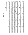
- FIG. 1 shows the complete nucleotide sequence for prochymosin coding gene.
- FIG. 2 shows restriction enzyme maps for vector plasmids used in this invention.
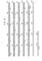
- FIG. 3 shows restriction enzyme maps for expression plasmids used in this invention.
- FIG. 4 shows the DNA sequence in the vicinity of the trp promoter in pOCT2 plasmid.
- FIG. 5 is a schematic diagram for the construction of pTREl from pOCT3 by way of pOCT2.
- FIG. 6 is a schematic diagram for the construction of pTRLl from pOCT2 and pBR322.
- FIG. 7 is a schematic diagram for the construction of pTRPl from pTREl.
- FIG. 8 is a schematic diagram for the construction of pCR501 from pCR301 and pTREl.
- FIG. 9 is a schematic diagram for the construction of pCR601 from pCR301, pTREl and pTRLl.
- FIG. 10 is a schematic diagram for the construction of pCR701 from pCR501 and pTRP1.
- Any microrganism capable of accepting and replicating the desired vector plasmids can be used but, for practical reasons, derivatives of
E. coli c 600 are used in this invention. Particularly preferred strain is E. coli c600 rk -mk - Cells are grown under conventional culturing conditions. For example, culture media for the E. coli strain can be:
- Conventionally, transformation experiments are conducted in LB medium containing 50 ug/ml of ampicillin according to the method of Norgard (Gene, 3, 279 (1978). In standard techniques, E. coli cells are grown to a density of from 10 to 30x10 12 cells per liter at a temperature of from 30°C to 40°C.
- In order to have cloned genes expressed in host cells, it is necessary to equip vector plasmids with a suitable promoter; several promoters can be used to obtain expression as long as they are compatible with the host cells. Desirably, the E. coli tryptophan operon promoter is used in this invention.
- In accordance with this invention, genes are conveniently available from the primary recombinant plasmids as described previously. These plasmids contain a prorennin coding gene with variable sizes and some of them have been mapped precisely by restriction enzyme digestion. Therefore, by cutting with appropriate restriction enzymes, useful portions of the prorennin coding genes can be separated, and used as a source of DNA for a second cloning into a vector plasmid. A representative of such plasmids is pCR301. Plasmid pCR301 contains the lactose operon promoter and almost entire prochymosin gene sequence (from nucleotide #13 to #1095) only devoid of the first four codons. This plasmid is derived from pBR322 plasmid (F. Bolvar et al., Gene, 2, 95 (1977). The necleotide sequence between the initiation codon ATG and the beginning of the cloned procymosin gene is illustrated below(this sequence coresponds to the nucleotide coding for the ten N-terminal amino acids of beta-glactosidase):

- A restriction enzyme map for pCR301 is shown in FIG. 3.
- Plasmid pOCT3, the precursor of pOCT2, is a pBR322 plasmid into which a DNA fragment having the trp promoter-operator, trp L and trp E' has been integrated at its EcoRI site. Said trp promoter-operator portion (- 0.5 Kb) is originally extracted from plasmid p trp ED5-1 (R.A. Hallewell and S. Emtage, Gene, 9, 27 (1980)). This DNA is filled in by means of T4 D polymerase, to which a EcoRI linker is annexed. The resulting DNA fragment is inserted into the EcoRI site of
pBR 322. Plasmid pOCT3 was kindly provided by Professor K. Matsubara at Osaka University, School of Medicine. The trp operon DNA is cut out from pOCT3 by digestion with EcoRI and, once isolated, it is then put back to the EcoRI site of pOCT3 in such a manner that the cut ends become switched around, leading to pOCT2. Thus the transcriptional direction of pOCT3 is opposite to that of pOCT2; it is clockwise in pOCT2 (51 to 3') whereas counterclockwise in pOCT3. Fig. 4 shows the DNA sequence (about 300 bases) in the vicinity of the trp promoter in pOCT2 plasmid. - Fig. 5 is a schematic diagram for the construction of pOCT3 from pOCT2. A restriction enzyme map for pOCT2 is shown in FIG. 2.
- Plasmid pTREl is derived from pOCT2 and has a single EcoRI site since the other EcoRI site near the ampicillin resistant region has been removed from pOCT2. This plasmid contains the intact trp operon part of pOCT2 as shown in FIG. 2. The construction of pTREI is accomplished in the following manner.
- Plasmid pOCT2 is partially digested with EcoRI, resulting in the cleavage of the EcoRI site near the ampicillin resistant region. The single stranded- portions are then repaired by means of DNA polymerase and the repaired ends are joined using T4 DNA ligase to produce pTREI.
- A restriction enzyme map for pTREI is shown in FIG. 2. Fig. 5 shows a schematic diagram for the construction of pTREl from pOCT2.
- Plasmid pTRLl has a DNA portion from pOCT2 bounded by EcoRI and RSaI sites. This portion comprises 360 base pairs (from an unidentified EcoR site to RSaI site in the trp L region), some of which are sequenced as shown in FIG. 4. The construction of pTRLl is accomplished in the following manner.
- Plasmid pOCT2 is cleaved with EcoRI and then partially digested with RSaI to yield a 360 base pair fragment containing the trp promoter region (trp L). This fragment is ligated with the EcoRI linker at the cut RSaI site. Digestion with EcoRI produces a fragment with cohesive ends. This fragment is then inserted into the EcoRI site of pBR322 to afford two kinds of recombinant plasmids. Plasmid pTRLI is the one wherein the transcriptional direction from the trp promoter is counter-clockwise.
- A restriction enzyme map for pTRLI is shown in FIG. 2. FIG. 6 is a schematic diagram for the construction of pTRLl from pOCT2.
- Plasmid pTRPI has a DNA portion from pTREl bounded by HpaI and ClaI sites. This portion comprises about 4640 base pairs originally derived from pBR322. The plasmid has also another DNA portion from pTREl bounded by HpaI and TaqI sites. This portion comprises 32 base pairs corresponding to the trp L promoter. The construction of pTRPI is accomplished in the following manner.
- Plasmid pTREl is cleaved with HpaI and Clal to yield an about 4640 bp linear DNA fragment. Both fragments are joined together by ligation to produce pTRPl. A restriction enzyme map for pTRPl is shown in FIG. 2. FIG. 7 is a schematic diagram for the construction of pTRPl from pTREl.
- Again using standard techniques as outlined previously, a desired DNA fragment containing the cloned prochymosin gene is extracted from a transformant, and cleaved with restriction enzymes; the vector plasmid is similarly cleaved; and the fragments are mixed and ligated. These steps have led to the construction of expression plasmids, pCR501, pCR601, and pCR701 which are designed to facilitate to obtain expression of prochymosin in E. coli.
- Plasmid pCR501 contains a DNA portion of pTREl fused to about 1,000 nucleotides from pCR301, which code for the procymosin molecule. The construction of pCR501 is accomplished in the following manner.
- Plasmid pTRE1 is cut with EcoRI and single-stranded portions are removed by means of Sl nuclease. The EcoRI linker (GGATTTCC/CCTTAAGG) is linked to the blunt end using T4 DNA ligase. This DNA is next digested with EcoRI and SalI to produce linear DNA segment (a) (ca. 4210bp). Plasmid pCR301 is cut with EcoRI and KpnI to obtain DNA segment (b). This DNA segment is a 246 bp sequence containing the prochymosin gene fused to the residual beta-glactosidase coding gene. Similarly, pCR301 is cut with KpnI and SalI to obtain DNA segment (c). This DNA is a 1025 bp sequence of the prochymosin gene including its C-terminus. Each DNA segment is ligated together by means of T4 DNA ligase. E. coli strain C600 is transformed with the ligated DNA, and ampicillin resistant colonies (pCR501) are selected.
- As shown in FIG. 3, analysis of pCR501 by restriction enzyme clevage has revealed that pCR501 contains the trp operon promoter and about 1,000 nucleotides coding for prochymosin (nucleotide #13 to the end of the gene, viz. 5th codon to 365th). FIG. 8 shows a schematic diagram for the construction of pCR501 from pCR301 and pTREl.
- Following the method of Maxam-Gilbert, the nucleotide sequence between the initiation ATG codon and the beginning of prochymosin gene (5th codon) has been analyzed to be:

- Transformed cells carrying the plasmid will express the cloned gene and produce prochymosin under the control of the trp E promoter.
- The prochymosin produced in this manner lacks an amino acid sequence of the genuine prochymosin which corresponds to that from the 1st to the 4th amino acid at the N terminus, but contains, in its place, eight amino acids (methionine and others) resulting from the above-stated trp E promoter coding sequence as well as two amino acid residues resulting from beta-glactosidase. This prochymosin is therefore called prochymosin like protein.
- Plasmid pCR601 contains a DNA portion of pTRE1 and a 56 bp fragment from pTRLl fused to the same coding sequence for prochymosin present in pCR501. The construction of pCR601 is accomplished in the following manner.
- Plasmid pTREl is cut with HpaI and SalI to produce a linear 4010 bp segment (DNA (d)). Next, plasmid pTRLI is cut with HpaI and EcoRI to produce a 56 bp segment (DNA (e)) containing the trp L promoter. both segments are mixed and ligated together with DNA (b) and (c) (both obtained previously from pCR301), by means of T4 DNA ligase. E. coli strain
c 600 is transformed with the resulting ligated DNA and ampicillin resistant colonies (pCR 601) are selected. - As shown in FIG. 3, analysis of pCR601 by restriction enzyme cleavage has revealed that pCR601 contains the trp L promoter and about 1,000 nucleotides coding for prochymosin (nucleotide #13 to the end of the gene, viz, 5th codon to 365th). FIG. 9 shows a schematic diagram for the construction of pCR601 from pCR301, pTREl and pTRLl.
- Following the method of Maxam-Gilbert, the nucleotide sequence between the initiation ATG codon and the beginning of prochymosn gene (5th codon) has been analyzed to be:
- Transformed cells carrying the plasmid will express the cloned prochymosin gene and produce prochymosin under the control of trp L promoter.
- The prochymosin produced in this manner lacks an amino acid sequence of the genuine prochymosin which corresponds to that from the lst to the 4th amino acid at the N terminus, but contains, in its place, six amino acids (methionine and others) resulting from the above-stated trp L promoter coding sequence as well as two amino acid residues resulting from beta-glactosidase. This prochymosin is therefore called prochymosin like protein.
- Plasmid pCR701 contains a DNA portion of pTRPI and a synthetic nucleotide piece fused to a prochymosin coding sequence from pCR501. The construction of pCR701 is accomplished in the following manner.
- Plasmid pTRPI is cut with Hind III and Sal I to obtain liner DNA segment (f) (about 4050 bp). Plasmid pCR501 is cut with BamHI and SalI to obtain liner DNA segment (g) (1264 bp). A synthetic oligonucleotide having a 22bp is synthesized, which is the sequence of:

- This DNA segment (f) makes up for the nucleotide sequence that has been removed by digestion with Bam HI which cuts the 14th nucleotide of prochymosin gene. The three segments (f), (g) and (h) are ligated together by means of T4 DNA ligase. E. coli c600 is transformed with the ligated DNA and ampicillin resistant colonies (pCR701) are selected.
- As shown in FIG. 3, analysis of pCR701 by restriction enzyme cleavage has revealed that pCR701 contains the trp L promoter and the entire prochymosin coding sequence (i.e. nucleotide #1 to the end of the gene, v12, 1st codon to 365th). FIG. 10 shows a schematic diagram for the construction of pCR701 from pTRPl and pCR501.
- As determined by the method of maxam-Gilbert, the SD (Shinon and Delgarno) region is located 13 nucleotides away from the ATG initiation codon preceeding the 1st codon of the prochymosin gene (see below).

- Transformed cells carrying the plasmid will express the prochymosin gene and produce prochymosin under the control of the trp L promoter.
- The prochymosin produced in this manner is N-terminal methionylprochymosin in which methionine arising from the initiation codon ATG is fused to the N ter minus of the genuine prochymosin.
- A portion of the full-growth culture (in LB medium) of E. coli cells transformed with the expression plasmids is grown in M9 broth containing 3-beta-indolylacrylic acid (15 ug/ml) and ampicillin (50 ug/ml). Cells are harvested when propagation reaches maximum. The cells are then destroyed by sonication treatment and a cell-free extract is obtained. This extract is subjected to electrophoresis in a polyacrylamide gel containing SDS. The product protein is detected by the protein blotting method. Following the above procedures or radioimmunoassay, each transformant strain containing pCR501, pCR601 or pCR701 has been tested for its ability to produce prochymosin. Analysis by autoradiography (Nishimori, Gene, 19, 337 (1982) has revealed that every strain gives a protein band the size of which is equal to that of the authentic prochymosin. In addition, the level of production of prochymosin has been estimated to be more than 300,000 molecules per cell. Similar results have been obtained by an analysis using radioimmunoassay.
- These results have shown that prochymosin is produced in E. coli cells carrying plasmid pCR501, pCR601 or pCR701 under the transcriptional control of the E. coli trp operon promoter and that the levels of production are substantially greater than those previously reported under the control of the E. coli lac operon. Even higher levels of production will be possible by using the same scheme in accordance with this invention and by obtaining fusions of the trp promoter closer to the initiation codon of prochymosin which preferably proceeds the tip of the prochymosin gene.
- E. coli strains prepared by processes according to this invention are exemplified by cultures deposited with the Fermentation Research Institute, Ibaraki, Japan under the Budapest Treaty, and are identified by the Accession number designated to each strain.
- This invention will be described in some detail by way of illustration and example; nevertheless, it should be understood that certain changes and modifi- cations be practiced within the scope of the invention.
- Throughout the following examples, TEN buffer solution is meant to be a solution containing Tris-HC1(20mM), NaCl(50mM) and EDTA(lmM) with a pH of about 7.5.
- Plasmid pOCT2 was partially digested with EcoRI by incubation at 20°C for 7.5 min in a 30 µl solution containing Plasmid (1.5 µg in 15 µl TEN), EcoRI (2.5 U, 0.5 µl TEN), EcoRI buffer (3 µl), and H20 (11.5 ul). After argarose gel electrophoresis, the desired DNA was isolated (in 40 ul TEN) and this was heated at 60°C for 5 min. To this DNA solution (40 µl) was added DNA polymerase I (4 U, 4 µl TEN), 16 mM ATP (4 µl), polymerase buffer solution (6 µl), and H20 (2 µl). The resulting mixture (a total volume of 60 µl) was incubated at 25°C for 30 min. After addition of 0.25 M EDTA solution (4 µl) followed by phenol and ether treatment, DNA was precipitated with ethanol. The precipitate was dissolved in a solution (10 ul) contaiing T4 DNA ligase (1 U, 1 µl), 3 mM ATP (2 µl), ligase buffer solution (2 ul) and H20 (5 ul), and this was incubated at 22°C for 2 h. After incubation, the DNA was precipitated with ethanol and dissolved in TEN solution. This solution was used to transform the calcium-treated
E. coli C 600 strain. One of twelve colonies, there was obtained two transformants containing pTREI. - Plasmid pOCT2 was cut with EcoRI at 37° C for 2 h in a 120 µl solution containing Plasmid (10.6 µg in 100 µl of TEN), EcoRI buffer (12 µl), and EcoRI (40 U, 8 µl of TEN). After incubation, the DNA was precipitated with ethanol and the precipitate was dissolved in 50 µl of TEN. The cut DNA was partially digested with RsaI by incubation at 20°C for 10 min in a 60 µl solution containing DNA (49 µl of TEN), BamHI buffer (6 µl), H2O (3 ul), and RsaI (20 U, 2 µl of TEN). The doubly cut DNA was precipitated with ethanol and the precipitate was dissolved in 20 µl of TEN. The dissolved solution was then applied to polyacrylamide gel electrophoresis. After electrophoresis, a 364 bp linear DNA (0.16 µg in 40 µl of TEN) was isolated. Meanwhile, EcoRI linker was kinased with T4 polynucleotide kinase at 37°C for 1 h in a 20 µl reaction mixture containing the linker (5 µg, 5 µl TEN), ATP (3mM, 2 µl), kination buffer (2 µl), H20 (9 µl), and the kinase (22U in 2 µl TEN). The kination mixture (20 µl) was added to the above DNA solution (35 µl), followed by addition of T4 DNA ligase (5U, 5 µl TEN), ligase buffer (4 µl), and ATP (6mM, 7 µl). The total mixture (71 µl) was ligated together by incubation at 22°C for 2 h. The ligated DNA was precipitated with ethanol and the precipitate was dissolved in 50 µl TEN. To the dissolved solution (45 µl) was added EcoRI (350 U in 70 µl TEN), EcoRI buffer (20 µl) and H20 (65 µl). The resulting mixture (200 µl) was incubated at 37°C for 3 h. Incubation was terminated by phenol treatment and DNA was extracted with ether, precipitated with ethanol, and dissolved in 40 µl of TEN. The dissolved solution was applied to polyacrylamide gel electrophoresis. After electrophoresis, the DNA segment (1) with cohesive ends (0.1 µg in 240 µl of Tris-acetate buffer) was isolated.
- Plasmid pBR322 was cut with EcoRI by incubation at 37°C for 1 h in a 30 µl solution containing plasmid (4.5 µg in 20 µl TEN), EcoRI buffer (3 µl), H20 (3 µl), and EcoRI (20 U in 4 µl TEN). Incubation was terminated by phenol treatment and DNA was extracted with ether, precipitated with ethanol, and dissolved in 40 µl of 10 mM Tris-HC1 buffer. The dissolved solution was treated with 10 µl of MATE BAP by incubation at 65°C fr 1 h. After incubation, MATE BAT was removed by washing the incubated mixture with 20 µl of 10mM Tris-HC1 buffer. Thus the DNA segment (2) (4.2 µg) was recovered in 60 µl of 10 mM Tris-HCl buffer.
- Segment (1) (0.05 µg in 120 µl of Tris-acetate buffer) and Segment (2) (0.14 µg in 2 µl of 10 mM Tris-HCl buffer) were mixed and the mixed solution was treated with ethanol. The resulting precipitate was dissolved in 50 µl of TEN. Forty microliters of the dissolved solution was subjected to ligation using T4 DNA ligase (0.9 U in 1 µl TEN), ligase buffer (5 µl) and ATP (6 mM, 5 µl TEN) by incubation a 51 µl solution at 22°C for 2 h. The ligated DNA was precipitated with ethanol and dissolved in 50 µl of TEN.
- Twenty microliters of the above ligated DNA solution was used to transform competent cells of strain
E. coli C 600. About one thousand five hundred transformants were found to be amplicillin resistant. Restriction enzyme analysis demonstrated that one out of twenty transformants contained pTRLl plasmid. - Plasmid pTREl was cut with HpaI by incubation at 37°C for 1.5 h in a 40 µl solution containing Plasmid (2.46 µg in 30 µl TEN), HpaI buffer (4 µl), and HpaI (30 U in 6 µl TEN). The cut DNA was further cut with ClaI by adding the DNA solution to a 10 µl solution containing ClaI buffer (5 µl) and ClaI (25 U, 5 µl TEN), and by incubating at 37°C for 1.5 h. The doubly cut DNA was precipitated with ethanol and the precipitate was dissolved in 40 µl of TEN. The dissolved solution was applied to agarose gel electrophoresis. After electrophoresis, the 4640 bp linear DNA Segment (3) (2.35 µg in 80 µl TEN) was isolated.
- Plasmid pTREI was cut with HpaI at 37°C for 1.5 h in a 120 µl solution containing Plasmid (8.2 µg in 100 µl TEN), HpaI buffer (12 µl), and HpaI (57 U in 8 µl TEN). To the above mixture was added a solution containing TaqI (80 U in 9 µl TEN) and HpaI buffer (12 µl), and HpaI (57 U in 8 µl TEN). To the above mixture was added a solution containing TaqI (80 U in 9 µl TEN) and HpaI buffer (1 µl). The resulting mixture (130 µl) was incubated at 65°C for 1 h. After incubation, the doubly cut DNA was precipitated with ethanol and the precipitate was dissolved in 40 µl TEN. The dissolved solution was applied to polyacrylamide gel electrophoresis. After electrophoresis, the 32 bp linear DNA segment (4) (0.054 µg in 40 µl TEN) was isolated.
- The mixture containing DNA Segment (3) (0.29 µg in 10 µl TEN), Segment (4) (0.014 µg in 10 µl TEN) was subjected to ligation using T4 ligase (3.6 U in 4 µl TEN), ligase buffer (3 µl), and ATP (6 mM, 3 µl TEN). The ligation mixture (30 µl) was incubated at 22°C for 2 h. The ligated DNA was precipitated with ethanol and the precipitate was dissolved in 40 µl TEN.
- Thirty microliters of the above TEN solution containing the ligated DNA was used to transform competent cells of strain E. coli C600. About eight hundred and forty transformants were found to be ampicillin resistant. All the transformants contained pTRPI plasmid.
- Plasmid pTREI was cleaved by incubation at 37°C for 2 h in a 120 µl reaction mixture containing Plasmid (8.2 µg in 100 µl TEN), EcoRI buffer (12 µl), H20 (3 µl), and EcoRI (25 U in 5 µl TEN). After phenol extraction, ether extraction, ethanol precipitation, and dissolution in 50 µl TEN, the cut DNA was further cut with Sl nuclease at 22°C for ½ h in a 50 µl reaction mixture containing DNA (25 µl TEN), SI buffer (5 pl), H20 (10 µl), and SI nuclease (100 U, 10 µl). Incubation was terminated by phenol treatment and DNA was extracted with ether and precipitated with ethanol. In the meantime, EcoRI linker was kinased with T4 polynucleotide kinase at 37°C for 1 h in a 20 µl reaction mixture containing the linker (5 µg, 5 µl), ATP (3mM, 2 µl), kination buffer (2 µl), H20 (9 µl) and Kinase (22 U in 2 µl TEN). The kination mixture (20 µl) containing EcoRI linker was added to the ethanol precipitated cut DNA along with ATP (3 mM, 5 µl) plus 4.5 units of T4 ligase (5 µlTEN). The mixture (30 µl) was incubated at 22°C for 2 h. The product DNA was precipitated with ethanol and the precipitate was dissolved in 60 µl TEN. To the DNA solution (50 µl) was added EcoRI (500 U in 50 µl TEN), EcoRI buffer (20 µl), and H20 (80 µl). The resulting mixture (200 µl) was incubated at 37°C for 2 h. Incubation was terminated by phenol treatment and DNA was extracted with ether and precipitated with ethanol. The precipitated DNA was cut with SalI by incubation at 37°C for 1.5 h in a 110 µl solution containing SalI (100 U, 20 µl), SalI buffer (10 µl), H20 (80 µl). After incubation, the cut DNA was precipitated with ethanol and dissolved in 60 µl TEN.
- The DNA was subjected to agarose gel electrophoresis, yielding 3.5 µg of the purified linear DNA (a)(4210 bp) in 100 µl TEN.
- Plasmid pCR301 was cut with KpnI by incubation at 37°C for 1.5 h in a 120 µl solution containing Plasmid (7.3 µg in 100 µl TEN), KpnI buffer (12 µl), H20 (2 µl), and KpnI (35 U, 6 µl TEN). To the mixture was added EcoRI buffer solution (14 µl) containing EcoRI (30 U, 6 µl TEN). The 140 µl mixture was incubated at 37°C for 1 h. The doubly cut DNA was precipitated with ethanol and the precipitate was dissolved in 60 µl TEN. The dissolved solution was applied to polyacrylamide gel electrophoresis. After electrophoresis, the 246 bp DNA Segment (b) (0.03 µg in 4 µl TEN) was isolated.
- Plasmid pCR301 was cut with KpnI by incubation at 37°C for 1.5 h in a 120 µl solution containing Plasmid (15 µg in 100 µl TEN), KpnI buffer (12 µl), H2O (2 µl), and KpnI (35 U , 6 µl TEN). A 14 µl SalI buffer solution plus SalI (32 U, 4 µl TEN), H20 (2 µl) was added to the above incubated mixture. The 140 µl mixture was further incubated at 37°C for 1 h. The doubly cut DNA was precipitated with ethanol and the precipitate was dissolved in 60 µl TEN. The dissolved solution was applied to agarose gel electrophoresis. After electrophoresis, the 1025 bp DNA Segment (c) (2.15 µg in 180 µl TEN) was isolated.
- The mixture containing DNA Segment (a) (1 µg in 30 µl TEN), Segment (b) (0.03 µg in 4 µl TEN), and Segment (c) (0.4 µg in 30 µl TEN) was lypophilized. After lypophilization, the precipitate was rinsed with ethanol. Each precipitated segment was ligated together in a 20 µl solution containing ligase buffer (4 µl), ATP (3 mM, 4 µl), H20 (1 µl) and T4 ligase (0.9 U, 1 µl). Incubation was continued for 2 h at 22°C. The ligated DNA was precipitated with thanol and dissolved in 40 µl TEN.
- The TEN buffer solution (30 µl) containing the above ligated DNA was employed for transformation of E. coli strain C600. Twenty transformants were found to contain plasmid pCR501. One of them was deposited with the FRI as E. coli C600 rk -mk - (pCR501) FERM BP 262.
- Plasmid pTREI was cut with HpaI by incubation at 37°C for 1 h in a 40 µl solution containing Plasmid (2.46 µg in 30 µl TEN), HpaI buffer (4 µl), and HpaI (35 U, 6 µl TEN). To the incubated mixture was then added a 10 µl solution containing SalI buffer (5 µl) and SalI (20 U, 5 µl TEN). The mixture (50 µl) was further incubated at 37°C for 1 h. The doubly cut DNA was precipitated with ethanol and the precipitate was dissolved in 40 µl TEN. The dissolved solution was applied to agarose gel electrophoresis. After electrophoresis, the 4010 bp Segment (d) (2.0 µg in 40 µl TEN) was isolated.
- Plasmid pTRLl was cut with HpaI by incubation at 37°C for 1.5 h in a 120 µl solution containing Plasmid (15.4 µg in 100 µl TEN), HpaI buffer (12 µl) , and HpaI (25 U, 5 µl TEN), and H20 (3 µl). To the incubated mixture was then added a 20 µl solution containing EcoRI buffer (914 µl) and EcoRI (30 U, 6 µl TEN). The mixture (140 µl) was further incubated at 37°C for 1 h. The doubly cut DNA was precipitated with ethanol and the precipitate was dissolved in 40 µl TEN. The dissolved solution was applied to polyacrylamide gel electrophoresis. After electrophoresis, the 56 bp Segment (e) (0.18 µg in 40 µl TEN) was isolated.
- By repeating the procedure of Example 4, but using 32 units of KpnI and 30 units of EcoRI, plasmid pCR301 was cut with KpnI and then with EcoRI to afford 0.46 µg (in 40 µl TEN) of the 246 bp DNA segment after polyacrylamide gel electrophoresis.
- By repeating the procedure of Example 4, but using 30 units of KpnI and 24 units of SalI, plasmid pCR301 was cut with KpnI and then with SalI to afford 3.9 µg (in 40 µl TEN) of the 1025 bp DNA segment after agarose gel electrohporesis.
- The mixture containing DNA Segment (b) (0.06 µg in 5 µl TEN) Segment 9c) (0.49 µg in 5 µl TEN), Segment (d) (0.5 µg in 10 µl TEN), and Segment (e) (0.49 µg in 5 µl TEN) was subjected to ligation using T4 ligase (5.5 U in 6 µl TEN) and ligation buffer (4.5 µl) plus ATP (6 mM, 4.5 µl). The ligated mixture (45 µl) was incubated at 22°C for 2 h. The ligated DNA was precipitated with ethanol and dissolved in 60 µl TEN.
- The TEN buffer solution ( 20 µl) containing the above ligated DNA was used to transform E. coli C600 rk -mk - strain. Twenty seven ampicillin resistant transformants were obtained after transformation. Eight transformants were found to contain pCR601 as determined by restriction enzyme digestion. This strain E. coli C600 rk -mk - (pCR601) was deposited with the FRI as FERM BP263.
- Plasmid pTRP1 was cut with HindIII by incubation at 37°C for 1 h in a 120 µl solution containing Plasmid (25.2 g in 100 µl TEN), Hind III buffer (12 µl), and Hind III (48 U, 8 µl TEN). To the incubated mixture was added SalI (24U, 6 µl TEN) and SalI buffer (14 µl). The resulting 140 µl solution was incubated at 37°C for 2 h. The doubly cut DNA was precipitated with ethanol and the precipitate was dissolved in 60 µl TEN. The dissolved solution was applied to agarose gel electrophoresis. After electrophoresis, the 4051 bp linear DNA Segment (f) (8.2 µg in 120 µl TEN) was isolated.
- Plasmid pCR501 was cut with SalI by incubation at 37°C for 1 h in a 120 µl solution containing Plasmid (39 µg in 100 µl TEN), SalI buffer (12 µl), and SalI (32 U, 8 µl TEN). The cut DNA was partially digested with BamH I by addition of BamH I (25 U, 5 µl TEN) to the above mixture and by incubation at 22°C for ½ h. The product DNA was precipitated with ethanol and the precipitate was dissolved in 60 µl TEN. This dissolved solution was applied to agarose gel electrophoresis. After electrophoresis, the 1264 bp DNA Segment (g) (2 µg in 80 µl TEN) was isolated.
- A synthetic oligonucleoide having the following sequence was obtained:

- The mixture containing DNA Segment (f) (0.68 µg in 10 µl TEN), Segment (g) (0.25 µg in 10 µl TEN), and Segment (h) (0.046 µg in 10 µl TEN) was subjected to ligation using T4 DNA ligase (1.8 U, in 2 µl TEN) and ligation buffer (4 µl) plus ATP (6 mM, 4 µl TEN). The ligated mixture DNA was precipitated with ethanol and dissolved in 50 µl TEN.
- Twenty microliters of the above ligation reaction mixture was used to transform competent cells of E. coli C600. Eight ampicillin resistant transformants were obtained and, by restriction enzyme analysis, four of these transformants were found to have pCR701. This strain E. coli C600 rk-mk- (pCR701) was deposited with the FRI as FERM BP 264.
- E. coli strain harboring pCR501, pCR601 or pCR701 was preliminarily cultivated in LB at 37°C overnight. One mililiter of the culture broth was then transferred to M9 medium (10 ml), and cultivated for 1 h. Cultivation was continued at 37°C for additional 3 to 4 h aftr 3-beta-indolylacrylic acid (15 mg/1) was added to the medium. Cells were harvested by centrifugation at 15,000 rmp for about 15 min, and suspended in 3 ml of PBS buffer (150 mM NaCl in 20 mM sodium phosphate buffer, pH 7.0) which contains 1 mM PMSF (phenylmethylsulfonyl fluoride). To this suspention was added 60 µl of 250 mM EDTA and 30 µl lysozyme (10 ml/1) and the mixture was kept at 0°C for ½ h. The resulting sheroplasts were disrupted by sonication and to this was . added 3.09 ml of 8 M urea. The cell lysate mixture was incubated at 37°C for 1 h. After centrifugation at 3000 rmp for ½ h, the supernatant solution was dialyzed against PBS. The dialysate was applied to the binding competitive radioimmunoassay (see, Nishimori et al., Gene). By radioimmunoassay prochymosin was immunoprecipitated and the amount of the radioactive elemnt (125 I) was measured with a gamma-ray counter. In the meantime, calibration curves were prepared by varying the concentration of authentic prochymosin. By comparing the count of the immunoprecipitated prochymosin with the calibration curves, the amount of prochymosin in the cell lysate was determined. This experiment demonstrated that the expression level of prochymosin was highest with the culture broth from E, coli containing pCR501 (about 300,000 molecules per host cell) and lowest with that from E. coli containing pCR701.
- Independantly, the above sonicated cell-free extract was treated with 4M urea. After centrifugation, the supernantant solution was applied to SDS-polyacrylamide gel electrophoresis, and the migrated proteins were then blotted onto nitrocellulose filters. These filters were analyzed on autoradiography as previously described in Nishimori et al., Gene. For each bacterial extract, a distinctive band corresponding to that of prochymosin was observed. By comparing the density of these bands, it was determined that E. coli containing pCR501 plasmid produced about 300,000 prochymosin molecules per cell. The ability of each expression plasmid-containing host cell to produce prochymosin was evaluated to be in the order of pCR501 > PCR601 > pCR701. This.trend is well in accord with the results from the radioimmunoassay method.
- Total sonicates of plasmid-containing bacteria were treated with 8M urea and dialyzed against 25 mM sodium bicarbonate (pH 10.0) in order to remove urea. After another dialysis against 50 mM Tris-HC1 buffer (pH 7.5), dialysates were activated by addition of 0.4 M glycin (pH 2.3) and by maintaining the solution at room temperature for 15 min.
- The milk-clotting activity of each bacterial extract was tested according to the method of Foltman (B. Foltman, Prochymosin and Chymosin Methods in Enzymology, 19, 42 (1972). In this test, the milk-clotting activity to coagulate 10 ml of skim milk within 100 sec at 30° may be defined as one chymosin unit (CU). For each bacterial extract derived from the expression plasmids, the milk-clotting activity was measured after activation of the prochymosin so-produced (viz. conversion of bacterial prochymosin to chymosin); the activity for pCR501 was 9.3 CU/mg and 3.7 CU/mg for pCR701. It should be recognized that a bacterial extract harboring no expression plasmid displayed no milk-clotting activity at all as determined by the above testing procedures.
Claims (36)


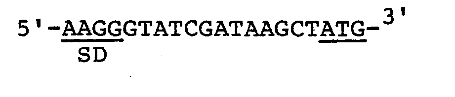
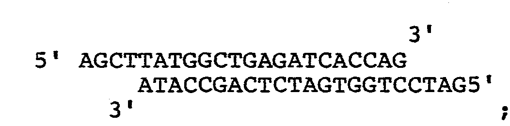
wherein said gene is derived from a recombinant plasmid containing cloned prochymosin cDNA, said method comprising:
Applications Claiming Priority (2)
| Application Number | Priority Date | Filing Date | Title |
|---|---|---|---|
| JP38439/83 | 1983-03-09 | ||
| JP58038439A JPS59166086A (en) | 1983-03-09 | 1983-03-09 | Novel development type plasmid and development of vitular prochymosin gene in escherichia coli using it |
Publications (1)
| Publication Number | Publication Date |
|---|---|
| EP0121775A1 true EP0121775A1 (en) | 1984-10-17 |
Family
ID=12525334
Family Applications (1)
| Application Number | Title | Priority Date | Filing Date |
|---|---|---|---|
| EP84102451A Withdrawn EP0121775A1 (en) | 1983-03-09 | 1984-03-07 | Novel expression plasmids and their use in the method for expressing prochymosin coding gene in E. coli |
Country Status (9)
| Country | Link |
|---|---|
| EP (1) | EP0121775A1 (en) |
| JP (1) | JPS59166086A (en) |
| AR (1) | AR241795A1 (en) |
| AU (1) | AU572827B2 (en) |
| BR (1) | BR8401049A (en) |
| DK (1) | DK136184A (en) |
| FI (1) | FI86886C (en) |
| IE (1) | IE57264B1 (en) |
| NZ (1) | NZ207414A (en) |
Cited By (68)
| Publication number | Priority date | Publication date | Assignee | Title |
|---|---|---|---|---|
| EP0123928A2 (en) * | 1983-03-31 | 1984-11-07 | Codon Genetic Engineering Laboratories | Recombinant DNA coding for a polypeptide displaying milk clotting activity |
| EP0154351A2 (en) * | 1984-03-09 | 1985-09-11 | Teruhiko Beppu | Expression plasmids useful in B. subtilis |
| US4705848A (en) * | 1986-06-02 | 1987-11-10 | International Minerals & Chemical Corp. | Isolation of bioactive, monomeric growth hormone |
| US4721673A (en) * | 1983-09-01 | 1988-01-26 | Genex Corporation | Recovery and activation process for microbially produced calf prochymosin |
| GB2200118A (en) * | 1987-01-23 | 1988-07-27 | Allelix Inc | Synthetic chymosin-and prochymosin-encoding DNA segments |
| US5082775A (en) * | 1984-05-11 | 1992-01-21 | Berlex Laboratories, Inc. | Efficient process for isolating insoluble heterologous protein using non-ionic detergents |
| EP0785273A1 (en) | 1990-11-26 | 1997-07-23 | Genetics Institute, Inc. | Paired basic amino acid converting enzyme and DNA sequence encoding it |
| US6210968B1 (en) | 1994-04-19 | 2001-04-03 | Chiron Spa | DNA molecules encoding pgp3 protein from Chlamydia trachomatis |
| WO2006069246A2 (en) | 2004-12-22 | 2006-06-29 | Ambrx, Inc. | Compositions containing, methods involving, and uses of non-natural amino acids and polypeptides |
| US7270989B2 (en) | 2000-11-06 | 2007-09-18 | Chr. Hansen A/S | Method of producing non-bovine chymosin and use hereof |
| WO2008020335A2 (en) | 2006-06-09 | 2008-02-21 | Novartis Ag | Immunogenic compositions for streptococcus agalactiae |
| US7335486B2 (en) | 2002-05-24 | 2008-02-26 | Restoragen, Inc. | Method for universal enzymatic production of bioactive peptides |
| WO2008030558A2 (en) | 2006-09-08 | 2008-03-13 | Ambrx, Inc. | Modified human plasma polypeptide or fc scaffolds and their uses |
| EP1935979A2 (en) | 1999-07-14 | 2008-06-25 | Novartis Vaccines and Diagnostics S.r.l. | Antigenic meningococcal peptides |
| WO2009067636A2 (en) | 2007-11-20 | 2009-05-28 | Ambrx, Inc. | Modified insulin polypeptides and their uses |
| WO2010011735A2 (en) | 2008-07-23 | 2010-01-28 | Ambrx, Inc. | Modified bovine g-csf polypeptides and their uses |
| WO2010037062A1 (en) | 2008-09-26 | 2010-04-01 | Ambrx, Inc. | Non-natural amino acid replication-dependent microorganisms and vaccines |
| EP2210945A2 (en) | 1998-01-14 | 2010-07-28 | Novartis Vaccines and Diagnostics S.r.l. | Neisseria meningitidis antigens |
| US7781567B2 (en) | 2002-05-24 | 2010-08-24 | Nps Pharmaceuticals, Inc. | Method for enzymatic production of GLP-2(1-33) and GLP-2(1-34) peptides |
| EP2228379A1 (en) | 2003-04-25 | 2010-09-15 | North Carolina State University | Lactobacillus acidophilus nucleic acid sequences encoding cell surface protein homologues and uses therefore |
| US7829307B2 (en) | 2003-11-21 | 2010-11-09 | Nps Pharmaceuticals, Inc. | Production of glucagon-like peptide 2 |
| EP2248822A2 (en) | 2001-07-27 | 2010-11-10 | Novartis Vaccines and Diagnostics S.r.l. | Meningococcus adhesins |
| EP2251424A1 (en) | 1999-05-19 | 2010-11-17 | Novartis Vaccines and Diagnostics S.r.l. | Antigenic neisserial peptides |
| EP2258716A2 (en) | 2002-11-22 | 2010-12-08 | Novartis Vaccines and Diagnostics S.r.l. | Multiple variants of meningococcal protein NMB1870 |
| EP2261342A2 (en) | 1998-05-01 | 2010-12-15 | Novartis Vaccines and Diagnostics, Inc. | Neisseria meningitidis antigens and compositions |
| EP2267005A1 (en) | 2003-04-09 | 2010-12-29 | Novartis Vaccines and Diagnostics S.r.l. | ADP-ribosylating toxin from Listeria monocytogenes |
| EP2270177A1 (en) | 2001-03-27 | 2011-01-05 | Novartis Vaccines and Diagnostics S.r.l. | Streptococcus pneumoniae proteins and nucleic acids |
| EP2272861A1 (en) | 1998-04-27 | 2011-01-12 | Novartis Vaccines and Diagnostics S.r.l. | Polyepitope carrier protein |
| EP2275552A2 (en) | 1999-10-29 | 2011-01-19 | Novartis Vaccines and Diagnostics S.r.l. | Neisserial antigenic peptides |
| EP2275129A2 (en) | 2000-01-17 | 2011-01-19 | Novartis Vaccines and Diagnostics S.r.l. | Outer membrane vesicle (OMV) vaccine comprising N. meningitidis serogroup B outer membrane proteins |
| EP2277896A1 (en) | 2000-10-27 | 2011-01-26 | Novartis Vaccines and Diagnostics S.r.l. | Nucleic acids and proteins from streptococcus groups A & B |
| EP2278007A1 (en) | 1999-04-30 | 2011-01-26 | Novartis Vaccines and Diagnostics S.r.l. | Conserved neisserial antigens |
| EP2278006A2 (en) | 1997-11-06 | 2011-01-26 | Novartis Vaccines and Diagnostics S.r.l. | Neisserial antigens |
| EP2279746A2 (en) | 2002-11-15 | 2011-02-02 | Novartis Vaccines and Diagnostics S.r.l. | Surface proteins in neisseria meningitidis |
| EP2284191A2 (en) | 2004-12-22 | 2011-02-16 | Ambrx, Inc. | Process for the preparation of hGH |
| WO2011024072A2 (en) | 2009-08-27 | 2011-03-03 | Novartis Ag | Hybrid polypeptides including meningococcal fhbp sequences |
| EP2298796A2 (en) | 2001-03-27 | 2011-03-23 | Novartis Vaccines and Diagnostics S.r.l. | Staphylococcus aureus proteins and nucleic acids |
| EP2302032A1 (en) | 2005-04-15 | 2011-03-30 | North Carolina State University | Methods and compositions to modulate adhesion and stress tolerance in bacteria |
| WO2011051893A1 (en) | 2009-10-27 | 2011-05-05 | Novartis Ag | Modified meningococcal fhbp polypeptides |
| EP2319863A1 (en) | 2003-06-23 | 2011-05-11 | North Carolina State University | Lactobacillus acidophilus nucleic acids encoding fructo-oligosaccharide utilization compounds and uses thereof |
| EP2335723A1 (en) | 2001-12-12 | 2011-06-22 | Novartis Vaccines and Diagnostics S.r.l. | Immunisation against chlamydia trachomatis |
| EP2348041A1 (en) | 2004-03-08 | 2011-07-27 | North Carolina State University | Lactobacillus acidophilus nucleic acid sequences encoding carbohydrate utilization-related proteins and uses therefor |
| EP2385127A1 (en) | 2005-11-25 | 2011-11-09 | Novartis Vaccines and Diagnostics S.r.l. | Chimeric, hybrid and tandem polypeptides of meningococcal NMB1870 |
| WO2011146715A1 (en) | 2010-05-19 | 2011-11-24 | North Carolina State University | Compositions and methods for the delivery of therapeutic peptides |
| WO2012024452A2 (en) | 2010-08-17 | 2012-02-23 | Ambrx, Inc. | Modified relaxin polypeptides and their uses |
| RU2473683C1 (en) * | 2011-12-15 | 2013-01-27 | Общество С Ограниченной Ответственностью "Фармапарк" | METHOD OF COMMERCIAL E.coli STRAIN CULTURE PREPARED OF BL21(DE3) STRAIN CARRYING POLYMERASE T7 RNA GENE UNDER CONTROL OF lacUV5 PROMOTER WITH INCREASED BIOMASS SYNTHESIS AND HIGHER END PROTEIN YIELD IN INCLUSION BODIES |
| EP2805965A1 (en) | 2009-12-21 | 2014-11-26 | Ambrx, Inc. | Modified porcine somatotropin polypeptides and their uses |
| EP2805964A1 (en) | 2009-12-21 | 2014-11-26 | Ambrx, Inc. | Modified bovine somatotropin polypeptides and their uses |
| US8907064B2 (en) | 2004-02-02 | 2014-12-09 | Ambrx, Inc. | Modified human four helical bundle polypeptides and their uses |
| WO2015059690A1 (en) | 2013-10-24 | 2015-04-30 | Yeda Research And Development Co. Ltd. | Polynucleotides encoding brex system polypeptides and methods of using s ame |
| EP2886551A2 (en) | 2008-02-21 | 2015-06-24 | Novartis AG | Meningococcal fhbp polypeptides |
| US9079971B2 (en) | 2007-03-30 | 2015-07-14 | Ambrx, Inc. | Modified FGF-21 polypeptides comprising non-naturally occurring amino acids |
| US9156899B2 (en) | 2008-09-26 | 2015-10-13 | Eli Lilly And Company | Modified animal erythropoietin polypeptides and their uses |
| US9175083B2 (en) | 2004-06-18 | 2015-11-03 | Ambrx, Inc. | Antigen-binding polypeptides and their uses |
| US9434778B2 (en) | 2014-10-24 | 2016-09-06 | Bristol-Myers Squibb Company | Modified FGF-21 polypeptides comprising an internal deletion and uses thereof |
| US9488660B2 (en) | 2005-11-16 | 2016-11-08 | Ambrx, Inc. | Methods and compositions comprising non-natural amino acids |
| EP3103880A1 (en) | 2008-02-08 | 2016-12-14 | Ambrx, Inc. | Modified leptin polypeptides and their uses |
| US9567386B2 (en) | 2010-08-17 | 2017-02-14 | Ambrx, Inc. | Therapeutic uses of modified relaxin polypeptides |
| CN107881162A (en) * | 2017-12-25 | 2018-04-06 | 中诺生物科技发展江苏有限公司 | A kind of method of industrialized production renin |
| WO2018142416A1 (en) | 2017-02-06 | 2018-08-09 | Yeda Research And Development Co. Ltd. | Isolated cells genetically modified to express a disarm system having an anti-phage activity and methods of producing same |
| US10266578B2 (en) | 2017-02-08 | 2019-04-23 | Bristol-Myers Squibb Company | Modified relaxin polypeptides comprising a pharmacokinetic enhancer and uses thereof |
| WO2019186570A1 (en) | 2018-03-29 | 2019-10-03 | Yeda Research And Development Co. Ltd. | Methods of disrupting a biofilm and/or preventing formation of same |
| US10590426B2 (en) | 2016-08-22 | 2020-03-17 | Board Of Trustees Of Michigan State University | Genetic control of cell size |
| WO2020056066A1 (en) | 2018-09-11 | 2020-03-19 | Ambrx, Inc. | Interleukin-2 polypeptide conjugates and their uses |
| WO2020202142A2 (en) | 2019-03-31 | 2020-10-08 | Yeda Research And Development Co. Ltd. | Anti-viral and anti-tumoral compounds |
| WO2021183832A1 (en) | 2020-03-11 | 2021-09-16 | Ambrx, Inc. | Interleukin-2 polypeptide conjugates and methods of use thereof |
| US11273202B2 (en) | 2010-09-23 | 2022-03-15 | Elanco Us Inc. | Formulations for bovine granulocyte colony stimulating factor and variants thereof |
| WO2023100189A1 (en) | 2021-12-05 | 2023-06-08 | Yeda Research And Development Co. Ltd. | Genetically modified phages and uses thereof |
Families Citing this family (1)
| Publication number | Priority date | Publication date | Assignee | Title |
|---|---|---|---|---|
| JPS60188077A (en) * | 1984-03-09 | 1985-09-25 | Teruhiko Beppu | Novel manifestation plasmid having whole sequence of calf prochymosin cdna |
Citations (3)
| Publication number | Priority date | Publication date | Assignee | Title |
|---|---|---|---|---|
| EP0057350A2 (en) * | 1981-01-16 | 1982-08-11 | Collaborative Research Inc. | Recombinant DNA means and method |
| EP0068691A2 (en) * | 1981-06-17 | 1983-01-05 | Celltech Therapeutics Limited | A process for the production of a polypeptide |
| EP0073029A2 (en) * | 1981-08-24 | 1983-03-02 | Teruhiko Beppu | Recombinant plasmid and microorganism containing same |
Family Cites Families (4)
| Publication number | Priority date | Publication date | Assignee | Title |
|---|---|---|---|---|
| JPS589687A (en) * | 1981-06-17 | 1983-01-20 | セルテク リミテッド | Production of polypeptide |
| BR8205954A (en) * | 1981-10-14 | 1983-09-13 | Unilever Nv | DNA SEQUENCE, RECOMBINANT PLASMIDIUM, BACTERIAL CULTURE AND MICROORGANISMS |
| ZA834095B (en) * | 1982-07-01 | 1984-02-29 | Genex Corp | Bovine calf chymosin |
| IE56372B1 (en) * | 1982-12-22 | 1991-07-03 | Genentech Inc | Microbially produced rennet methods for its production and plasmids used for its production |
-
1983
- 1983-03-09 JP JP58038439A patent/JPS59166086A/en active Granted
-
1984
- 1984-02-29 DK DK136184A patent/DK136184A/en not_active Application Discontinuation
- 1984-03-07 FI FI840918A patent/FI86886C/en not_active IP Right Cessation
- 1984-03-07 AU AU25357/84A patent/AU572827B2/en not_active Ceased
- 1984-03-07 NZ NZ20741484A patent/NZ207414A/en unknown
- 1984-03-07 EP EP84102451A patent/EP0121775A1/en not_active Withdrawn
- 1984-03-08 IE IE55984A patent/IE57264B1/en not_active IP Right Cessation
- 1984-03-08 BR BR8401049A patent/BR8401049A/en unknown
- 1984-03-09 AR AR29597484A patent/AR241795A1/en active
Patent Citations (3)
| Publication number | Priority date | Publication date | Assignee | Title |
|---|---|---|---|---|
| EP0057350A2 (en) * | 1981-01-16 | 1982-08-11 | Collaborative Research Inc. | Recombinant DNA means and method |
| EP0068691A2 (en) * | 1981-06-17 | 1983-01-05 | Celltech Therapeutics Limited | A process for the production of a polypeptide |
| EP0073029A2 (en) * | 1981-08-24 | 1983-03-02 | Teruhiko Beppu | Recombinant plasmid and microorganism containing same |
Non-Patent Citations (2)
| Title |
|---|
| GENE, vol. 19, 1982, (Amsterdam, NL) K. NISHIMORI et al.: "Expression of cloned calf prochymosin gene sequence in Escherichia coli" pages 337-344 * |
| THE JOURNAL OF BIOCHEMISTRY, vol. 90, 1981, (Tokyo, JP) K. NISHIMORI et al.: "Cloning in Escherichia coli of the Structural Gene of Prorennin, the Precursor of Calf Milk-Clotting Enzyme Rennin" pages 901-904 * |
Cited By (163)
| Publication number | Priority date | Publication date | Assignee | Title |
|---|---|---|---|---|
| EP0123928A2 (en) * | 1983-03-31 | 1984-11-07 | Codon Genetic Engineering Laboratories | Recombinant DNA coding for a polypeptide displaying milk clotting activity |
| EP0123928A3 (en) * | 1983-03-31 | 1986-02-05 | Codon Genetic Engineering Laboratories | Recombinant dna coding for a polypeptide displaying milk clotting activity |
| US4721673A (en) * | 1983-09-01 | 1988-01-26 | Genex Corporation | Recovery and activation process for microbially produced calf prochymosin |
| EP0154351A2 (en) * | 1984-03-09 | 1985-09-11 | Teruhiko Beppu | Expression plasmids useful in B. subtilis |
| EP0154351A3 (en) * | 1984-03-09 | 1987-08-26 | Teruhiko Beppu | Expression plasmids useful in b. subtilis |
| US5082775A (en) * | 1984-05-11 | 1992-01-21 | Berlex Laboratories, Inc. | Efficient process for isolating insoluble heterologous protein using non-ionic detergents |
| US4705848A (en) * | 1986-06-02 | 1987-11-10 | International Minerals & Chemical Corp. | Isolation of bioactive, monomeric growth hormone |
| GB2200118A (en) * | 1987-01-23 | 1988-07-27 | Allelix Inc | Synthetic chymosin-and prochymosin-encoding DNA segments |
| EP0785273A1 (en) | 1990-11-26 | 1997-07-23 | Genetics Institute, Inc. | Paired basic amino acid converting enzyme and DNA sequence encoding it |
| US7485313B2 (en) | 1994-04-19 | 2009-02-03 | Novartis Ag | Recombinant Chlamydia trachomatis pgp3 protein suitable for enzyme-linked immunoassay, and used of the protein in eliciting and diagnosing an immune response |
| US6210968B1 (en) | 1994-04-19 | 2001-04-03 | Chiron Spa | DNA molecules encoding pgp3 protein from Chlamydia trachomatis |
| US7070791B2 (en) | 1994-04-19 | 2006-07-04 | Chiron Srl | Detection of antibodies against Chlamydia trachomatis pgp3 antigen in patient sera by enzyme-linked immunoassay |
| US6649374B1 (en) | 1994-04-19 | 2003-11-18 | Chiron S.R.L. | Recombinant Chlamydia trachomatis PGP3 antigen |
| EP2278006A2 (en) | 1997-11-06 | 2011-01-26 | Novartis Vaccines and Diagnostics S.r.l. | Neisserial antigens |
| EP2278011A2 (en) | 1998-01-14 | 2011-01-26 | Novartis Vaccines and Diagnostics S.r.l. | Neisseria meningitidis antigens |
| EP2210945A2 (en) | 1998-01-14 | 2010-07-28 | Novartis Vaccines and Diagnostics S.r.l. | Neisseria meningitidis antigens |
| EP2272861A1 (en) | 1998-04-27 | 2011-01-12 | Novartis Vaccines and Diagnostics S.r.l. | Polyepitope carrier protein |
| EP2261341A2 (en) | 1998-05-01 | 2010-12-15 | Novartis Vaccines and Diagnostics, Inc. | Neisseria meningitidis antigens and compositions |
| EP2261354A2 (en) | 1998-05-01 | 2010-12-15 | Novartis Vaccines and Diagnostics, Inc. | Neisseria meningitidis antigens and compositions |
| EP2261347A2 (en) | 1998-05-01 | 2010-12-15 | Novartis Vaccines and Diagnostics, Inc. | Neisseria meningitidis antigens and compositions |
| EP2261348A2 (en) | 1998-05-01 | 2010-12-15 | Novartis Vaccines and Diagnostics, Inc. | Neisseria meningitidis antigens and compositions |
| EP2261355A2 (en) | 1998-05-01 | 2010-12-15 | Novartis Vaccines and Diagnostics, Inc. | Neisseria meningitidis antigens and compositions |
| EP2261350A2 (en) | 1998-05-01 | 2010-12-15 | Novartis Vaccines and Diagnostics, Inc. | Neisseria meningitidis antigens and compositions |
| EP2261349A2 (en) | 1998-05-01 | 2010-12-15 | Novartis Vaccines and Diagnostics, Inc. | Neisseria meningitidis antigens and compositions |
| EP2261357A2 (en) | 1998-05-01 | 2010-12-15 | Novartis Vaccines and Diagnostics, Inc. | Neisseria meningitidis antigens and compositions |
| EP2261356A2 (en) | 1998-05-01 | 2010-12-15 | Novartis Vaccines and Diagnostics, Inc. | Neisseria meningitidis antigens and compositions |
| EP2261339A2 (en) | 1998-05-01 | 2010-12-15 | Novartis Vaccines and Diagnostics, Inc. | Neisseria meningitidis antigens and compositions |
| EP2261340A2 (en) | 1998-05-01 | 2010-12-15 | Novartis Vaccines and Diagnostics, Inc. | Neisseria meningitidis antigens and compositions |
| EP2261343A2 (en) | 1998-05-01 | 2010-12-15 | Novartis Vaccines and Diagnostics, Inc. | Neisseria meningitidis antigens and compositions |
| EP2261345A2 (en) | 1998-05-01 | 2010-12-15 | Novartis Vaccines and Diagnostics, Inc. | Neisseria meningitidis antigens and compositions |
| EP2261353A2 (en) | 1998-05-01 | 2010-12-15 | Novartis Vaccines and Diagnostics, Inc. | Neisseria meningitidis antigens and compositions |
| EP2261342A2 (en) | 1998-05-01 | 2010-12-15 | Novartis Vaccines and Diagnostics, Inc. | Neisseria meningitidis antigens and compositions |
| EP2261346A2 (en) | 1998-05-01 | 2010-12-15 | Novartis Vaccines and Diagnostics, Inc. | Neisseria meningitidis antigens and compositions |
| EP2261351A2 (en) | 1998-05-01 | 2010-12-15 | Novartis Vaccines and Diagnostics, Inc. | Neisseria meningitidis antigens and compositions |
| EP2261338A2 (en) | 1998-05-01 | 2010-12-15 | Novartis Vaccines and Diagnostics, Inc. | Neisseria meningitidis antigens and compositions |
| EP2261352A2 (en) | 1998-05-01 | 2010-12-15 | Novartis Vaccines and Diagnostics, Inc. | Neisseria meningitidis antigens and compositions |
| EP2261344A2 (en) | 1998-05-01 | 2010-12-15 | Novartis Vaccines and Diagnostics, Inc. | Neisseria meningitidis antigens and compositions |
| EP2278007A1 (en) | 1999-04-30 | 2011-01-26 | Novartis Vaccines and Diagnostics S.r.l. | Conserved neisserial antigens |
| EP2290083A1 (en) | 1999-04-30 | 2011-03-02 | Novartis Vaccines and Diagnostics S.r.l. | Conserved neisserial antigens |
| EP2251424A1 (en) | 1999-05-19 | 2010-11-17 | Novartis Vaccines and Diagnostics S.r.l. | Antigenic neisserial peptides |
| EP1935979A2 (en) | 1999-07-14 | 2008-06-25 | Novartis Vaccines and Diagnostics S.r.l. | Antigenic meningococcal peptides |
| EP2275553A2 (en) | 1999-10-29 | 2011-01-19 | Novartis Vaccines and Diagnostics S.r.l. | Neisserial antigenic peptides |
| EP2275552A2 (en) | 1999-10-29 | 2011-01-19 | Novartis Vaccines and Diagnostics S.r.l. | Neisserial antigenic peptides |
| EP2275554A2 (en) | 1999-10-29 | 2011-01-19 | Novartis Vaccines and Diagnostics S.r.l. | Neisserial antigenic peptides |
| EP2975127A1 (en) | 1999-10-29 | 2016-01-20 | GlaxoSmithKline Biologicals SA | Neisserial antigenic peptides |
| EP2275551A2 (en) | 1999-10-29 | 2011-01-19 | Novartis Vaccines and Diagnostics S.r.l. | Neisserial antigenic peptides |
| EP2281571A2 (en) | 2000-01-17 | 2011-02-09 | Novartis Vaccines and Diagnostics S.r.l. | Outer membrane vesicle (omv) vaccine comprising n. meningitidids serogroup b outer membrane proteins |
| EP2281570A2 (en) | 2000-01-17 | 2011-02-09 | Novartis Vaccines and Diagnostics S.r.l. | Outer membrane vesicle (OMV) vaccine comprising n. meningitidis serogroup B outer membrane proteins |
| EP2289545A2 (en) | 2000-01-17 | 2011-03-02 | Novartis Vaccines and Diagnostics S.r.l. | Supplemented OMV vaccine against meningococcus |
| EP2275129A2 (en) | 2000-01-17 | 2011-01-19 | Novartis Vaccines and Diagnostics S.r.l. | Outer membrane vesicle (OMV) vaccine comprising N. meningitidis serogroup B outer membrane proteins |
| EP2896629A1 (en) | 2000-10-27 | 2015-07-22 | Novartis Vaccines and Diagnostics S.r.l. | Nucleic acids and proteins from streptococcus group A & B |
| EP2277894A1 (en) | 2000-10-27 | 2011-01-26 | Novartis Vaccines and Diagnostics S.r.l. | Nucleic acids and proteins from streptococcus groups A & B |
| EP2277895A1 (en) | 2000-10-27 | 2011-01-26 | Novartis Vaccines and Diagnostics S.r.l. | Nucleic acids and proteins from streptococcus groups A & B |
| EP2284181A1 (en) | 2000-10-27 | 2011-02-16 | Novartis Vaccines and Diagnostics S.r.l. | Nucleic acids and proteins from streptococcus groups A and B |
| EP2277896A1 (en) | 2000-10-27 | 2011-01-26 | Novartis Vaccines and Diagnostics S.r.l. | Nucleic acids and proteins from streptococcus groups A & B |
| EP2284182A1 (en) | 2000-10-27 | 2011-02-16 | Novartis Vaccines and Diagnostics S.r.l. | Nucleic acids and proteins from streptococcus groups A and B |
| EP2284183A1 (en) | 2000-10-27 | 2011-02-16 | Novartis Vaccines and Diagnostics S.r.l. | Nucleic acids and proteins from streptococcus groups A and B |
| US8545909B2 (en) | 2000-11-06 | 2013-10-01 | Eidgenossische Technische Hochschule Zurich | Method of producing non-bovine chymosin and use hereof |
| US7270989B2 (en) | 2000-11-06 | 2007-09-18 | Chr. Hansen A/S | Method of producing non-bovine chymosin and use hereof |
| US9307775B2 (en) | 2000-11-06 | 2016-04-12 | Chr. Hansen A/S | Method for producing non-bovine chymosin and use hereof |
| EP2298796A2 (en) | 2001-03-27 | 2011-03-23 | Novartis Vaccines and Diagnostics S.r.l. | Staphylococcus aureus proteins and nucleic acids |
| EP2270175A1 (en) | 2001-03-27 | 2011-01-05 | Novartis Vaccines and Diagnostics S.r.l. | Streptococcus pneumoniae proteins and nucleic acids |
| EP2278010A1 (en) | 2001-03-27 | 2011-01-26 | Novartis Vaccines and Diagnostics S.r.l. | Streptococcus pneumoniae proteins and nucleic acids |
| EP2278009A1 (en) | 2001-03-27 | 2011-01-26 | Novartis Vaccines and Diagnostics S.r.l. | Streptococcus pneumoniae proteins and nucleic acids |
| EP2270177A1 (en) | 2001-03-27 | 2011-01-05 | Novartis Vaccines and Diagnostics S.r.l. | Streptococcus pneumoniae proteins and nucleic acids |
| EP2314697A1 (en) | 2001-03-27 | 2011-04-27 | Novartis Vaccines and Diagnostics S.r.l. | Streptococcus pneumoniae proteins and nucleic acids |
| EP2270176A1 (en) | 2001-03-27 | 2011-01-05 | Novartis Vaccines and Diagnostics S.r.l. | Streptococcus pneumoniae proteins and nucleic acids |
| EP2278008A2 (en) | 2001-03-27 | 2011-01-26 | Novartis Vaccines and Diagnostics S.r.l. | Streptococcus pneumoniae proteins and nucleic acids |
| EP2248822A2 (en) | 2001-07-27 | 2010-11-10 | Novartis Vaccines and Diagnostics S.r.l. | Meningococcus adhesins |
| EP2335723A1 (en) | 2001-12-12 | 2011-06-22 | Novartis Vaccines and Diagnostics S.r.l. | Immunisation against chlamydia trachomatis |
| EP2335724A1 (en) | 2001-12-12 | 2011-06-22 | Novartis Vaccines and Diagnostics S.r.l. | Immunisation against chlamydia trachomatis |
| US7335486B2 (en) | 2002-05-24 | 2008-02-26 | Restoragen, Inc. | Method for universal enzymatic production of bioactive peptides |
| US8148508B2 (en) | 2002-05-24 | 2012-04-03 | Nps Pharmaceuticals, Inc. | Method for enzymatic production of GLP-2(1-33) and GLP-2(1-34) peptides |
| US7781567B2 (en) | 2002-05-24 | 2010-08-24 | Nps Pharmaceuticals, Inc. | Method for enzymatic production of GLP-2(1-33) and GLP-2(1-34) peptides |
| US7498148B2 (en) | 2002-05-24 | 2009-03-03 | Restoragen, Inc. | Method for universal enzymatic production of bioactive peptides |
| EP2279746A2 (en) | 2002-11-15 | 2011-02-02 | Novartis Vaccines and Diagnostics S.r.l. | Surface proteins in neisseria meningitidis |
| EP2258717A2 (en) | 2002-11-22 | 2010-12-08 | Novartis Vaccines and Diagnostics S.r.l. | Variant form of meningococcal NadA |
| EP2261239A2 (en) | 2002-11-22 | 2010-12-15 | Novartis Vaccines and Diagnostics S.r.l. | Multiple variants of meningococcal protein NMB1870 |
| EP2258716A2 (en) | 2002-11-22 | 2010-12-08 | Novartis Vaccines and Diagnostics S.r.l. | Multiple variants of meningococcal protein NMB1870 |
| EP2267005A1 (en) | 2003-04-09 | 2010-12-29 | Novartis Vaccines and Diagnostics S.r.l. | ADP-ribosylating toxin from Listeria monocytogenes |
| EP2228379A1 (en) | 2003-04-25 | 2010-09-15 | North Carolina State University | Lactobacillus acidophilus nucleic acid sequences encoding cell surface protein homologues and uses therefore |
| EP2392582A2 (en) | 2003-04-25 | 2011-12-07 | North Carolina State University | Lactobacillus acidophilus nucleic acid sequences encoding cell surface protein homologues and uses therefore |
| EP2392581A2 (en) | 2003-04-25 | 2011-12-07 | North Carolina State University | Lactobacillus acidophilus nucleic acid sequences encoding cell surface protein homologues and uses therefore |
| EP2322540A1 (en) | 2003-06-23 | 2011-05-18 | North Carolina State University | Lactobacillus acidophilus nucleic acids encoding fructo-oligosaccharide utilization compounds and uses thereof |
| EP2319863A1 (en) | 2003-06-23 | 2011-05-11 | North Carolina State University | Lactobacillus acidophilus nucleic acids encoding fructo-oligosaccharide utilization compounds and uses thereof |
| US7829307B2 (en) | 2003-11-21 | 2010-11-09 | Nps Pharmaceuticals, Inc. | Production of glucagon-like peptide 2 |
| US8907064B2 (en) | 2004-02-02 | 2014-12-09 | Ambrx, Inc. | Modified human four helical bundle polypeptides and their uses |
| US8906676B2 (en) | 2004-02-02 | 2014-12-09 | Ambrx, Inc. | Modified human four helical bundle polypeptides and their uses |
| US9260472B2 (en) | 2004-02-02 | 2016-02-16 | Ambrx, Inc. | Modified human four helical bundle polypeptides and their uses |
| EP2348041A1 (en) | 2004-03-08 | 2011-07-27 | North Carolina State University | Lactobacillus acidophilus nucleic acid sequences encoding carbohydrate utilization-related proteins and uses therefor |
| EP2407482A1 (en) | 2004-03-08 | 2012-01-18 | North Carolina State University | Lactobacillus acidophilus nucelic acid sequences encoding carbohydrate utilization-related proteins and uses therefor |
| EP2407481A1 (en) | 2004-03-08 | 2012-01-18 | North Carolina State University | Lactobacillus acidophilus nucelic acid sequences encoding carbohydrate utilization-related proteins and uses therefor |
| US9175083B2 (en) | 2004-06-18 | 2015-11-03 | Ambrx, Inc. | Antigen-binding polypeptides and their uses |
| EP2284191A2 (en) | 2004-12-22 | 2011-02-16 | Ambrx, Inc. | Process for the preparation of hGH |
| WO2006069246A2 (en) | 2004-12-22 | 2006-06-29 | Ambrx, Inc. | Compositions containing, methods involving, and uses of non-natural amino acids and polypeptides |
| EP2302032A1 (en) | 2005-04-15 | 2011-03-30 | North Carolina State University | Methods and compositions to modulate adhesion and stress tolerance in bacteria |
| EP2305791A1 (en) | 2005-04-15 | 2011-04-06 | North Carolina State University | Methods and compositions to modulate adhesion and stress tolerance in bacteria |
| US9488660B2 (en) | 2005-11-16 | 2016-11-08 | Ambrx, Inc. | Methods and compositions comprising non-natural amino acids |
| EP3346009A1 (en) | 2005-11-25 | 2018-07-11 | GlaxoSmithKline Biologicals S.A. | Chimeric, hybrid and tandem polypeptides of meningococcal nmb1870 |
| EP2385126A1 (en) | 2005-11-25 | 2011-11-09 | Novartis Vaccines and Diagnostics S.r.l. | Chimeric, hybrid and tandem polypeptides of meningococcal NMB1870 |
| EP2385127A1 (en) | 2005-11-25 | 2011-11-09 | Novartis Vaccines and Diagnostics S.r.l. | Chimeric, hybrid and tandem polypeptides of meningococcal NMB1870 |
| WO2008020335A2 (en) | 2006-06-09 | 2008-02-21 | Novartis Ag | Immunogenic compositions for streptococcus agalactiae |
| WO2008030558A2 (en) | 2006-09-08 | 2008-03-13 | Ambrx, Inc. | Modified human plasma polypeptide or fc scaffolds and their uses |
| US9975936B2 (en) | 2007-03-30 | 2018-05-22 | Ambrx, Inc. | Nucleic acids encoding modified FGF-21 polypeptides comprising non-naturally occurring amino acids |
| US9517273B2 (en) | 2007-03-30 | 2016-12-13 | Ambrx, Inc. | Methods of treatment using modified FGF-21 polypeptides comprising non-naturally occurring amino acids |
| US9079971B2 (en) | 2007-03-30 | 2015-07-14 | Ambrx, Inc. | Modified FGF-21 polypeptides comprising non-naturally occurring amino acids |
| US10961291B2 (en) | 2007-03-30 | 2021-03-30 | Ambrx, Inc. | Modified FGF-21 polypeptides and their uses |
| US10377805B2 (en) | 2007-03-30 | 2019-08-13 | Ambrx, Inc. | Modified FGF-21 polypeptides comprising non-naturally encoding amino acids and their uses |
| US8946148B2 (en) | 2007-11-20 | 2015-02-03 | Ambrx, Inc. | Modified insulin polypeptides and their uses |
| WO2009067636A2 (en) | 2007-11-20 | 2009-05-28 | Ambrx, Inc. | Modified insulin polypeptides and their uses |
| EP2930182A1 (en) | 2007-11-20 | 2015-10-14 | Ambrx, Inc. | Modified insulin polypeptides and their uses |
| EP3103880A1 (en) | 2008-02-08 | 2016-12-14 | Ambrx, Inc. | Modified leptin polypeptides and their uses |
| US9938333B2 (en) | 2008-02-08 | 2018-04-10 | Ambrx, Inc. | Modified leptin polypeptides and their uses |
| EP3263591A1 (en) | 2008-02-21 | 2018-01-03 | GlaxoSmithKline Biologicals S.A. | Meningococcal fhbp polypeptides |
| EP2886551A2 (en) | 2008-02-21 | 2015-06-24 | Novartis AG | Meningococcal fhbp polypeptides |
| US10138283B2 (en) | 2008-07-23 | 2018-11-27 | Ambrx, Inc. | Modified bovine G-CSF polypeptides and their uses |
| WO2010011735A2 (en) | 2008-07-23 | 2010-01-28 | Ambrx, Inc. | Modified bovine g-csf polypeptides and their uses |
| EP3225248A1 (en) | 2008-07-23 | 2017-10-04 | Ambrx, Inc. | Modified bovine g-csf polypeptides and their uses |
| EP3216800A1 (en) | 2008-09-26 | 2017-09-13 | Ambrx, Inc. | Modified animal erythropoietin polypeptides and their uses |
| US9121024B2 (en) | 2008-09-26 | 2015-09-01 | Ambrx, Inc. | Non-natural amino acid replication-dependent microorganisms and vaccines |
| US9121025B2 (en) | 2008-09-26 | 2015-09-01 | Ambrx, Inc. | Non-natural amino acid replication-dependent microorganisms and vaccines |
| US10428333B2 (en) | 2008-09-26 | 2019-10-01 | Ambrx Inc. | Non-natural amino acid replication-dependent microorganisms and vaccines |
| US9156899B2 (en) | 2008-09-26 | 2015-10-13 | Eli Lilly And Company | Modified animal erythropoietin polypeptides and their uses |
| WO2010037062A1 (en) | 2008-09-26 | 2010-04-01 | Ambrx, Inc. | Non-natural amino acid replication-dependent microorganisms and vaccines |
| US9644014B2 (en) | 2008-09-26 | 2017-05-09 | Ambrx, Inc. | Modified animal erythropoietin polypeptides and their uses |
| EP3017828A1 (en) | 2009-08-27 | 2016-05-11 | GlaxoSmithKline Biologicals SA | Hybrid polypeptides including meningococcal fhbp sequences |
| WO2011024072A2 (en) | 2009-08-27 | 2011-03-03 | Novartis Ag | Hybrid polypeptides including meningococcal fhbp sequences |
| WO2011051893A1 (en) | 2009-10-27 | 2011-05-05 | Novartis Ag | Modified meningococcal fhbp polypeptides |
| EP2805964A1 (en) | 2009-12-21 | 2014-11-26 | Ambrx, Inc. | Modified bovine somatotropin polypeptides and their uses |
| EP2805965A1 (en) | 2009-12-21 | 2014-11-26 | Ambrx, Inc. | Modified porcine somatotropin polypeptides and their uses |
| WO2011146715A1 (en) | 2010-05-19 | 2011-11-24 | North Carolina State University | Compositions and methods for the delivery of therapeutic peptides |
| US9567386B2 (en) | 2010-08-17 | 2017-02-14 | Ambrx, Inc. | Therapeutic uses of modified relaxin polypeptides |
| WO2012024452A2 (en) | 2010-08-17 | 2012-02-23 | Ambrx, Inc. | Modified relaxin polypeptides and their uses |
| US9962450B2 (en) | 2010-08-17 | 2018-05-08 | Ambrx, Inc. | Method of treating heart failure with modified relaxin polypeptides |
| US11311605B2 (en) | 2010-08-17 | 2022-04-26 | Ambrx, Inc. | Methods of treating heart failure and fibrotic disorders using modified relaxin polypeptides |
| US11439710B2 (en) | 2010-08-17 | 2022-09-13 | Ambrx, Inc. | Nucleic acids encoding modified relaxin polypeptides |
| US10702588B2 (en) | 2010-08-17 | 2020-07-07 | Ambrx, Inc. | Modified relaxin polypeptides comprising a non-naturally encoded amino acid in the A chain |
| US10253083B2 (en) | 2010-08-17 | 2019-04-09 | Ambrx, Inc. | Therapeutic uses of modified relaxin polypeptides |
| US11786578B2 (en) | 2010-08-17 | 2023-10-17 | Ambrx, Inc. | Modified relaxin polypeptides and their uses |
| US9452222B2 (en) | 2010-08-17 | 2016-09-27 | Ambrx, Inc. | Nucleic acids encoding modified relaxin polypeptides |
| US10751391B2 (en) | 2010-08-17 | 2020-08-25 | Ambrx, Inc. | Methods of treatment using modified relaxin polypeptides comprising a non-naturally encoded amino acid |
| EP4302783A2 (en) | 2010-08-17 | 2024-01-10 | Ambrx, Inc. | Modified relaxin polypeptides and their uses |
| US11273202B2 (en) | 2010-09-23 | 2022-03-15 | Elanco Us Inc. | Formulations for bovine granulocyte colony stimulating factor and variants thereof |
| RU2473683C1 (en) * | 2011-12-15 | 2013-01-27 | Общество С Ограниченной Ответственностью "Фармапарк" | METHOD OF COMMERCIAL E.coli STRAIN CULTURE PREPARED OF BL21(DE3) STRAIN CARRYING POLYMERASE T7 RNA GENE UNDER CONTROL OF lacUV5 PROMOTER WITH INCREASED BIOMASS SYNTHESIS AND HIGHER END PROTEIN YIELD IN INCLUSION BODIES |
| US10767156B2 (en) | 2013-10-24 | 2020-09-08 | Yeda Research And Development Co., Ltd. | Polynucleotides encoding BREX system polypeptides and methods of using same |
| WO2015059690A1 (en) | 2013-10-24 | 2015-04-30 | Yeda Research And Development Co. Ltd. | Polynucleotides encoding brex system polypeptides and methods of using s ame |
| US11248031B2 (en) | 2014-10-24 | 2022-02-15 | Bristol-Myers Squibb Company | Methods of treating diseases associated with fibrosis using modified FGF-21 polypeptides |
| US9434778B2 (en) | 2014-10-24 | 2016-09-06 | Bristol-Myers Squibb Company | Modified FGF-21 polypeptides comprising an internal deletion and uses thereof |
| US10377806B2 (en) | 2014-10-24 | 2019-08-13 | Bristol-Myers Squibb Company | Methods of treating diseases associated with fibrosis using modified FGF-21 polypeptides and uses thereof |
| US9631004B2 (en) | 2014-10-24 | 2017-04-25 | Bristol-Myers Squibb Company | Modified FGF-21 polypeptides comprising an internal deletion and uses thereof |
| US10189883B2 (en) | 2014-10-24 | 2019-01-29 | Bristol-Myers Squibb Company | Therapeutic uses of modified FGF-21 polypeptides |
| US10590426B2 (en) | 2016-08-22 | 2020-03-17 | Board Of Trustees Of Michigan State University | Genetic control of cell size |
| US11168329B2 (en) | 2016-08-22 | 2021-11-09 | Board Of Trustees Of Michigan State University | Genetic control of cell size |
| WO2018142416A1 (en) | 2017-02-06 | 2018-08-09 | Yeda Research And Development Co. Ltd. | Isolated cells genetically modified to express a disarm system having an anti-phage activity and methods of producing same |
| US10266578B2 (en) | 2017-02-08 | 2019-04-23 | Bristol-Myers Squibb Company | Modified relaxin polypeptides comprising a pharmacokinetic enhancer and uses thereof |
| US11185570B2 (en) | 2017-02-08 | 2021-11-30 | Bristol-Myers Squibb Company | Method of treating cardiovascular disease and heart failure with modified relaxin polypeptides |
| US11364281B2 (en) | 2017-02-08 | 2022-06-21 | Bristol-Myers Squibb Company | Modified relaxin polypeptides comprising a pharmacokinetic enhancer and pharmaceutical compositions thereof |
| CN107881162A (en) * | 2017-12-25 | 2018-04-06 | 中诺生物科技发展江苏有限公司 | A kind of method of industrialized production renin |
| WO2019186570A1 (en) | 2018-03-29 | 2019-10-03 | Yeda Research And Development Co. Ltd. | Methods of disrupting a biofilm and/or preventing formation of same |
| WO2020056066A1 (en) | 2018-09-11 | 2020-03-19 | Ambrx, Inc. | Interleukin-2 polypeptide conjugates and their uses |
| WO2020202142A2 (en) | 2019-03-31 | 2020-10-08 | Yeda Research And Development Co. Ltd. | Anti-viral and anti-tumoral compounds |
| WO2021183832A1 (en) | 2020-03-11 | 2021-09-16 | Ambrx, Inc. | Interleukin-2 polypeptide conjugates and methods of use thereof |
| WO2023100189A1 (en) | 2021-12-05 | 2023-06-08 | Yeda Research And Development Co. Ltd. | Genetically modified phages and uses thereof |
Also Published As
| Publication number | Publication date |
|---|---|
| DK136184A (en) | 1984-09-10 |
| FI840918A0 (en) | 1984-03-07 |
| FI840918A (en) | 1984-09-10 |
| JPS59166086A (en) | 1984-09-19 |
| IE840559L (en) | 1984-09-09 |
| IE57264B1 (en) | 1992-07-01 |
| BR8401049A (en) | 1984-10-16 |
| NZ207414A (en) | 1988-02-29 |
| FI86886B (en) | 1992-07-15 |
| AR241795A1 (en) | 1992-12-30 |
| JPS6240999B2 (en) | 1987-09-01 |
| AU2535784A (en) | 1984-09-13 |
| DK136184D0 (en) | 1984-02-29 |
| AU572827B2 (en) | 1988-05-19 |
| FI86886C (en) | 1992-10-26 |
Similar Documents
| Publication | Publication Date | Title |
|---|---|---|
| EP0121775A1 (en) | Novel expression plasmids and their use in the method for expressing prochymosin coding gene in E. coli | |
| FI84080C (en) | Process for producing rennin and hybrid plasmids useful therein | |
| AU600949B2 (en) | Process for the production and secretion of polypeptides | |
| CA1340381C (en) | Recombinant fusion proteins | |
| JP2569218B2 (en) | DNA encoding Met-aminopeptidase | |
| EP0075444A2 (en) | Methods and products for facile microbial expression of DNA sequences | |
| JP3878207B2 (en) | Expression plasmid controlled by osmB promoter | |
| JPH09135691A (en) | Preparation of pregrowth and growth hormone of cattle | |
| Mechulam et al. | Molecular cloning of the Escherichia coli gene for diadenosine 5', 5'''-P1, P4-tetraphosphate pyrophosphohydrolase | |
| EP0249350A1 (en) | Filamentous fungal expression systems | |
| US6472196B1 (en) | Amino acid sequence of L-phenylalanine ammonia-lyase | |
| JPS62115281A (en) | Methionine peptidase of bacterial n-terminal | |
| EP0278706B1 (en) | Recombinant plasmid for the expression of l-phenylalanine ammonia-lyase and transformed strain carrying same | |
| EP0154350A2 (en) | Novel expression plasmids containing the full cDNA sequence of calf prochymosin | |
| CA1283068C (en) | Plasmid for expression of penicillin g amidase | |
| JP2525413B2 (en) | Recombinant plasmid for expressing L-phenylalanine ammonia lyase and transformant having the plasmid | |
| US5496711A (en) | Processes for producing pre-prorennin, prorennin and rennin | |
| EP0154351A2 (en) | Expression plasmids useful in B. subtilis | |
| EP0295286B1 (en) | Method for the production of salmon growth hormone using a synthetic gene | |
| CA1200774A (en) | Expression linkers | |
| JPH03130088A (en) | Production of polypeptide by recombinant dna process | |
| JPH0728746B2 (en) | Novel plasmid, microbial cell, and method for producing human immunoglobulin G Fc region protein | |
| JPH05268958A (en) | Production of prochymosin | |
| JPH0817704B2 (en) | Method for producing foreign gene product | |
| JPS60141290A (en) | Dna fragment containing structural gene of neutral protease and production of neutral protease |
Legal Events
| Date | Code | Title | Description |
|---|---|---|---|
| PUAI | Public reference made under article 153(3) epc to a published international application that has entered the european phase |
Free format text: ORIGINAL CODE: 0009012 |
|
| AK | Designated contracting states |
Designated state(s): AT BE CH DE FR GB IT LI LU NL SE |
|
| 17P | Request for examination filed |
Effective date: 19850123 |
|
| 17Q | First examination report despatched |
Effective date: 19860616 |
|
| STAA | Information on the status of an ep patent application or granted ep patent |
Free format text: STATUS: THE APPLICATION IS DEEMED TO BE WITHDRAWN |
|
| 18D | Application deemed to be withdrawn |
Effective date: 19870227 |
|
| RIN1 | Information on inventor provided before grant (corrected) |
Inventor name: NISHIMORI, KATSUHIKO Inventor name: SHIMIZU, NORIO Inventor name: UOZUMI, TAKESHI Inventor name: BEPPU, TERUHIKO Inventor name: HIDAKA, MAKOTO Inventor name: KAWAGUCHI, YOSHIYUKI |