EP0157209A1 - Frequency processing method and apparatus for radiation image - Google Patents
Frequency processing method and apparatus for radiation image Download PDFInfo
- Publication number
- EP0157209A1 EP0157209A1 EP85102532A EP85102532A EP0157209A1 EP 0157209 A1 EP0157209 A1 EP 0157209A1 EP 85102532 A EP85102532 A EP 85102532A EP 85102532 A EP85102532 A EP 85102532A EP 0157209 A1 EP0157209 A1 EP 0157209A1
- Authority
- EP
- European Patent Office
- Prior art keywords
- radiation
- exposure dose
- read
- enhancement
- image
- Prior art date
- Legal status (The legal status is an assumption and is not a legal conclusion. Google has not performed a legal analysis and makes no representation as to the accuracy of the status listed.)
- Granted
Links
Images

Classifications
-
- H—ELECTRICITY
- H05—ELECTRIC TECHNIQUES NOT OTHERWISE PROVIDED FOR
- H05G—X-RAY TECHNIQUE
- H05G1/00—X-ray apparatus involving X-ray tubes; Circuits therefor
- H05G1/08—Electrical details
- H05G1/60—Circuit arrangements for obtaining a series of X-ray photographs or for X-ray cinematography
Definitions
- This invention relates to a frequency processing method for a radiation image for viewing, particularly for diagnostic purposes, and an apparatus for carrying out the method.
- This invention particularly relates to a method of reducing noise in a frequency processing for improving the image quality, particularly diagnostic efficiency and accuracy, of a radiation image, and an apparatus for carrying out the method.
- frequency processing as used herein is meant a processing for enhancement of frequency response, for example, an unsharp mask processing as disclosed in U.S. Patent No. 4,315,318 and Japanese Unexamined Patent Publication No. 55(1980)-87953.
- the frequency processing as described above is conducted for improving the image quality, particularly diagnostic efficiency and accuracy, of a radiation image.
- a problem arises as described below. That is, in radiation image information obtained by exposure to a radiation of low dose, radiation quantum noise becomes perceptible.
- the radiation image information is directly subjected to the processing for enhancement of frequency response, a rough image wherein noise is enhanced is obtained and the image quality, particularly diagnostic efficiency and accuracy, is adversely affected by noise.
- the degree of enhancement in the frequency processing is adjusted by an external input, for example manually, in accordance with the image recording portion of an object, the image recording method (contrasted image recording, tomography, etc.) or the like. In this case, it is impossible to cope with the effect of noise changing with the level of radiation exposure dose.
- the primary object of the present invention is to provide a frequency processing method for a radiation image wherein a frequency processing is conducted so that noise is not perceptible when the radiation exposure dose is low.
- Another object of the present invention is to provide a frequency processing method for a radiation image, which realizes an image having an improved image quality, particularly a high diagnostic efficiency and accuracy.
- the specific object of the present invention is to provide an apparatus for carrying out the frequency processing method.
- the frequency processing method for a radiation image in accordance with the present invention is characterized by correcting the degree of enhancement in the frequency processing for a radiation image so that the degree of enhancement is low when the radiation exposure dose at the time of recording an image of an object is low.
- the degree of enhancement in the frequency processing is decreased to prevent the image quality, particularly diagnostic efficiency and accuracy, from becoming low.
- the degree of enhancement when the exposure dose is low is meant that correction is conducted to decrease the degree of enhancement when the exposure dose is relatively low. Therefore, when the exposure dose becomes relatively high, the degree of enhancement is increased.
- the degree of enhancement of frequence response may be high.
- the degree of enhancement of frequency response should preferably be as high as possible.
- the exposure dose it is possible to use a method wherein the exposure dose is calculated from the information on the tube voltage and tube current of the radiation source, exposure time, distance from the focal point to the detector, or the like, at the time of image recording, or a method wherein the exposure dose is directly measured. Also, when a recorded radiation image is read out and converted into an electric image signal which is subjected to the frequency processing, the exposure dose may be detected by detecting the level of the electric image signal.
- the frequency processing method of the present invention is applicable to any system wherein a radiation image is converted into an electric signal which is subjected to image processing.
- the method of the present invention exhibits high effects particularly in a radiation image recording and reproducing system as disclosed, for example, in U.S. Patent No. 4,258,264, wherein a stimulable phosphor sheet which can form a radiation image having an improved image quality, particularly a high diagnostic efficiency and accuracy, with a low exposure dose is used. That is because the method of the present invention realizes visual reduction in quantum noise which increases particularly in the case of low exposure dose.
- a novel method of conducting image read-out in two stages in the radiation image recording and reproducing system using a stimulable phosphor sheet is proposed, for example, in Japanese Unexamined Patent Publication Nos. 58(1983)-67240, 58(1983)-67243 and 58(1983)-67244.
- the method comprises the steps of conducting read-out (hereinafter referred to as the preliminary read-out) for approximately detecting the image information by scanning the stimulable phosphor sheet carrying a radiation image stored therein by stimulating rays of a relatively low level, and then carrying out read-out (hereinafter referred to as the final read-out) for detecting the image information in detail by use of stimulating rays of a level higher than the level of the stimulating rays used in the preliminary read-out on the basis of the information obtained by the preliminary read-out.
- the exposure dose can be detected easily from the information obtained by the preliminary read-out.
- the degree of enhancement of frequency response is decreased when the exposure dose at the time of recording an image of an object is low and quantum noise becomes perceptible, noise enhancement is restricted and it is possible to obtain a visible radiation image having an improved image quality, particularly a high diagnostic efficiency and accuracy.
- Figure 1 shows the relationship between the frequence and the response in the frequency processing.
- Frequency processing conditions are changed by changing the position of a peak P along the horizontal axis, i.e. the frequency (parameter f) at which the degree of enhancement is the maximum, and the height of the peak P, i.e. the degree of enhancement (parameter 8).
- the parameter ⁇ is determined to an appropriate value in accordance with the image recording portion of an object, such as the chest or the abdomen, and the image recording method, such as contrasted image recording or tomography.
- the parameter ⁇ is further corrected in accordance with the exposure dose.
- Figure 2A shows an example of a change in the parameter ⁇ in accordance with the exposure dose.
- the parameter ⁇ for the degree of enhancement is adjusted to a predetermined value So.
- the parameter ⁇ for the degree of enhancement is decreased as the exposure dose becomes low.
- the parameter ⁇ is increased as the exposure dose becomes high. In this manner, it is possible to make noise imperceptible when the exposure dose is low and quantum noise in high, and to increase the degree of enhancement of frequency response when the exposure dose is high and quantum noise is low, thereby improving the image quality, particularly the diagnostic efficiency and accuracy.
- the change in the parameter ⁇ for frequency enhancement is not limited to the change as shown in Figure 2A. Namely, it is sufficient that the parameter be increased as the exposure dose increases (however, the parameter ⁇ need not be changed within a specific exposure dose range).
- the parameter ⁇ may be gradually changed curvilinearly as shown in Figure 2B, or may be changed along a straight line as shown in Figure 2C. In Figures 2B and 2C, the parameter ⁇ is a monotonous increasing function of the exposure dose.
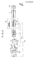
- Figure 3 shows the configuration of an apparatus for carrying out correction of the parameter S in accordance with the exposure dose.
- a stimulable phosphor sheet 3 is exposed to a radiation 2a emitted by a radiation source 2 such as an X-ray source and passing through an object 1 such as the human body to have a radiation image of the object 1 stored in the stimulable phosphor sheet 3.
- the stimulable phosphor sheet 3 carrying the radiation image stored therein is then exposed to stimulating rays 4a of a relatively low level (having energy of a level lower than the level of the energy of stimulating rays 8a used in final read-out as described later) emitted by a stimulating ray source 4 such as a laser beam source.
- a photoelectric read-out means 6 preliminary read-out
- read-out conditions such as the read-out gain and the scale factor in a photoelectric read-out means 7 for the final read-out are adjusted.
- the final read-out is conducted by scanning the stimulable phosphor sheet 3 by the stimulating rays 8a which are of a level higher than the level of the stimulating rays 4a used in the preliminary read-out and which are emitted by a stimulating ray source 8 for the final read-out.
- the image information output by the photoelectric read-out means 7 for the final read-out is sent to an image processing section 10 and subjected to image processings such as frequency processing and gradation processing for improving the image quality, particularly the diagnostic efficiency and accuracy, of an image 12 reproduced by an image reproducing apparatus 11.
- image processings such as frequency processing and gradation processing for improving the image quality, particularly the diagnostic efficiency and accuracy, of an image 12 reproduced by an image reproducing apparatus 11.
- the output 6a of the photoelectric read-out means 6 for the preliminary read-out is sent also to a frequency processing section 10A of the image processing section 10, and the parameter S is adjusted in accordance with the exposure dose as shown in Figures 2A, 2B and 2C. That is, since the output 6a of the photoelectric read-out means 6 represents the level of the radiation energy stored in the stimulable phosphor sheet 3 and the level corresponds to the radiation exposure dose, the output 6a represents a value on the horizontal axis in Figures 2A, 2B and 2C.
- the degree of enhancement (parameter ⁇ ) in the frequency processing can be adjusted on the basis of the output 6a when tables corresponding to the graphs as shown in Figures 2A, 2B and 2C are stored in the frequency processing section 10A.
- the information on the exposure dose may also be obtained without using the preliminary read-out output 6a.
- the information on the exposure dose may be directly sent from the radiation source 2 to the frequency processing section l0A as indicated by the chain line 2A.
- a drive control panel for the radiation source 2 may also be used.
- the level of the stimulating rays used in the preliminary read-out should be lower than the level of the stimulating rays used in the final read-out. That is, the effective energy of the stimulating rays which the stimulable phosphor sheet receives per unit area in the preliminary read-out should be lower than the effective energy of the stimulating rays used in the final read-out.
- a stimulating ray source for the preliminary read-cut may be positioned independently of the stimulating ray source for the final read-out as in the above-described embodiment, and the output of the former may be made lower than the output of the latter.
- the output of a single stimulating ray source such as a laser beam source may be decreased in the preliminary read-out, or the stimulating rays emitted by the stimulating ray source may be attenuated by a ND filter, an AOM, or the like positioned on the optical path.
- the beam diameter of the stimulating rays may be increased, the scanning speed of the stimulating rays may be increased, or the moving speed of the stimulable phosphor sheet may be increased in the preliminary read-out.
- preliminary read-out for example the relationship between the preliminary read-out and the final read-out, are described, for example, in Japanese Unexamined Patent Publication Nos. 58(1983)-67240, 58(1983)-67243 and 58(1983)-67244.
Abstract
Description
- This invention relates to a frequency processing method for a radiation image for viewing, particularly for diagnostic purposes, and an apparatus for carrying out the method. This invention particularly relates to a method of reducing noise in a frequency processing for improving the image quality, particularly diagnostic efficiency and accuracy, of a radiation image, and an apparatus for carrying out the method.
- By the term "frequency processing" as used herein is meant a processing for enhancement of frequency response, for example, an unsharp mask processing as disclosed in U.S. Patent No. 4,315,318 and Japanese Unexamined Patent Publication No. 55(1980)-87953.
- The frequency processing as described above is conducted for improving the image quality, particularly diagnostic efficiency and accuracy, of a radiation image. However, in the frequency processing, a problem arises as described below. That is, in radiation image information obtained by exposure to a radiation of low dose, radiation quantum noise becomes perceptible. When the radiation image information is directly subjected to the processing for enhancement of frequency response, a rough image wherein noise is enhanced is obtained and the image quality, particularly diagnostic efficiency and accuracy, is adversely affected by noise.
- The degree of enhancement in the frequency processing is adjusted by an external input, for example manually, in accordance with the image recording portion of an object, the image recording method (contrasted image recording, tomography, etc.) or the like. In this case, it is impossible to cope with the effect of noise changing with the level of radiation exposure dose.
- The primary object of the present invention is to provide a frequency processing method for a radiation image wherein a frequency processing is conducted so that noise is not perceptible when the radiation exposure dose is low.
- Another object of the present invention is to provide a frequency processing method for a radiation image, which realizes an image having an improved image quality, particularly a high diagnostic efficiency and accuracy.
- The specific object of the present invention is to provide an apparatus for carrying out the frequency processing method.
- The frequency processing method for a radiation image in accordance with the present invention is characterized by correcting the degree of enhancement in the frequency processing for a radiation image so that the degree of enhancement is low when the radiation exposure dose at the time of recording an image of an object is low.
- Namely, when the exposure dose is low and quantum noise becomes perceptible, the degree of enhancement in the frequency processing is decreased to prevent the image quality, particularly diagnostic efficiency and accuracy, from becoming low.
- By "decreasing the degree of enhancement when the exposure dose is low" is meant that correction is conducted to decrease the degree of enhancement when the exposure dose is relatively low. Therefore, when the exposure dose becomes relatively high, the degree of enhancement is increased. When the exposure dose is high, since radiation quantum noise becomes imperceptible, the degree of enhancement of frequence response may be high. In this case, from the viewpoint of improvement in the image quality, particularly diagnostic efficiency and accuracy, the degree of enhancement of frequency response should preferably be as high as possible.
- In order to detect the exposure dose, it is possible to use a method wherein the exposure dose is calculated from the information on the tube voltage and tube current of the radiation source, exposure time, distance from the focal point to the detector, or the like, at the time of image recording, or a method wherein the exposure dose is directly measured. Also, when a recorded radiation image is read out and converted into an electric image signal which is subjected to the frequency processing, the exposure dose may be detected by detecting the level of the electric image signal.
- The frequency processing method of the present invention is applicable to any system wherein a radiation image is converted into an electric signal which is subjected to image processing. However, the method of the present invention exhibits high effects particularly in a radiation image recording and reproducing system as disclosed, for example, in U.S. Patent No. 4,258,264, wherein a stimulable phosphor sheet which can form a radiation image having an improved image quality, particularly a high diagnostic efficiency and accuracy, with a low exposure dose is used. That is because the method of the present invention realizes visual reduction in quantum noise which increases particularly in the case of low exposure dose.
- A novel method of conducting image read-out in two stages in the radiation image recording and reproducing system using a stimulable phosphor sheet is proposed, for example, in Japanese Unexamined Patent Publication Nos. 58(1983)-67240, 58(1983)-67243 and 58(1983)-67244. The method comprises the steps of conducting read-out (hereinafter referred to as the preliminary read-out) for approximately detecting the image information by scanning the stimulable phosphor sheet carrying a radiation image stored therein by stimulating rays of a relatively low level, and then carrying out read-out (hereinafter referred to as the final read-out) for detecting the image information in detail by use of stimulating rays of a level higher than the level of the stimulating rays used in the preliminary read-out on the basis of the information obtained by the preliminary read-out. In this case, the exposure dose can be detected easily from the information obtained by the preliminary read-out. Of course, it is also possible to detect the exposure dose from the information obtained by the final read-out and to correct the degree of enhancement in the frequency processing on the basis of the detected exposure dose.
- In the present invention, since the degree of enhancement of frequency response is decreased when the exposure dose at the time of recording an image of an object is low and quantum noise becomes perceptible, noise enhancement is restricted and it is possible to obtain a visible radiation image having an improved image quality, particularly a high diagnostic efficiency and accuracy.
- When the exposrue dose is low and the noise level is low or when noise is reduced by superposing a plurality of images, it is possible to further improve the image quality, particularly the diagnostic efficiency and accuracy, by increasing the degree of enhancement in the frequency processing.
-
- Figure 1 is a graph showing the relationship between the frequency and the response in frequency processing,
- Figures 2A, 2B and 2C are graphs showing the relationship between the exposure dose and the degree of enhancement (S) in the frequency processing in accordance with the present invention, and
- Figure 3 is a schematic view showing the radiation image recording and reproducing system wherein an apparatus for carrying out the method of the present invention is employed.
- The present invention will hereinbelow be described in further detail with reference to the accompanying drawings. Particularly, embodiments of the present invention will be described with respect to the case where the frequency processing is applied to the radiation image recording and reproducing system wherein the preliminary read-out and the final read-out are conducted by use of a stimulable phosphor sheet.
- Figure 1 shows the relationship between the frequence and the response in the frequency processing. Frequency processing conditions are changed by changing the position of a peak P along the horizontal axis, i.e. the frequency (parameter f) at which the degree of enhancement is the maximum, and the height of the peak P, i.e. the degree of enhancement (parameter 8). Basically, the parameter β is determined to an appropriate value in accordance with the image recording portion of an object, such as the chest or the abdomen, and the image recording method, such as contrasted image recording or tomography. In the present invention, the parameter β is further corrected in accordance with the exposure dose.
- Figure 2A shows an example of a change in the parameter β in accordance with the exposure dose. When the exposure dose is within the normal range RO, the parameter β for the degree of enhancement is adjusted to a predetermined value So. When the exposure dose is within the range RL lower than the normal level, the parameter β for the degree of enhancement is decreased as the exposure dose becomes low. When the exposure dose is within the high range RH, the parameter β is increased as the exposure dose becomes high. In this manner, it is possible to make noise imperceptible when the exposure dose is low and quantum noise in high, and to increase the degree of enhancement of frequency response when the exposure dose is high and quantum noise is low, thereby improving the image quality, particularly the diagnostic efficiency and accuracy.
- The change in the parameter β for frequency enhancement is not limited to the change as shown in Figure 2A. Namely, it is sufficient that the parameter be increased as the exposure dose increases (however, the parameter β need not be changed within a specific exposure dose range). For example, the parameter β may be gradually changed curvilinearly as shown in Figure 2B, or may be changed along a straight line as shown in Figure 2C. In Figures 2B and 2C, the parameter β is a monotonous increasing function of the exposure dose.
- The parameter corresponds to β in the unsharp mask processing formula
S' = Sorg + β(Sorg - Sus) where S' denotes the frequency-processed signal, Sorg denotes the read-out output signal, Sus denotes the unsharp mask signal, and denotes the degree of enhancement, as disclosed, for example, in U.S. Patent No. 4,315,318. - Figure 3 shows the configuration of an apparatus for carrying out correction of the parameter S in accordance with the exposure dose.
- A
stimulable phosphor sheet 3 is exposed to a radiation 2a emitted by a radiation source 2 such as an X-ray source and passing through anobject 1 such as the human body to have a radiation image of theobject 1 stored in thestimulable phosphor sheet 3. Thestimulable phosphor sheet 3 carrying the radiation image stored therein is then exposed to stimulating rays 4a of a relatively low level (having energy of a level lower than the level of the energy of stimulating rays 8a used in final read-out as described later) emitted by a stimulating ray source 4 such as a laser beam source. Light 5 emitted by thestimulable phosphor sheet 3 when it is exposed to (scanned by) the stimulating rays 4a is detected by a photoelectric read-out means 6 (preliminary read-out). On the basis of the approximate image information obtained by theoutput 6a of the preliminary read-out, read-out conditions such as the read-out gain and the scale factor in a photoelectric read-out means 7 for the final read-out are adjusted. The final read-out is conducted by scanning thestimulable phosphor sheet 3 by the stimulating rays 8a which are of a level higher than the level of the stimulating rays 4a used in the preliminary read-out and which are emitted by astimulating ray source 8 for the final read-out. - The image information output by the photoelectric read-out means 7 for the final read-out is sent to an
image processing section 10 and subjected to image processings such as frequency processing and gradation processing for improving the image quality, particularly the diagnostic efficiency and accuracy, of animage 12 reproduced by an image reproducing apparatus 11. - The
output 6a of the photoelectric read-out means 6 for the preliminary read-out is sent also to a frequency processing section 10A of theimage processing section 10, and the parameter S is adjusted in accordance with the exposure dose as shown in Figures 2A, 2B and 2C. That is, since theoutput 6a of the photoelectric read-out means 6 represents the level of the radiation energy stored in thestimulable phosphor sheet 3 and the level corresponds to the radiation exposure dose, theoutput 6a represents a value on the horizontal axis in Figures 2A, 2B and 2C. The degree of enhancement (parameter β) in the frequency processing can be adjusted on the basis of theoutput 6a when tables corresponding to the graphs as shown in Figures 2A, 2B and 2C are stored in the frequency processing section 10A. - The information on the exposure dose may also be obtained without using the preliminary read-out
output 6a. For example, the information on the exposure dose may be directly sent from the radiation source 2 to the frequency processing section l0A as indicated by thechain line 2A. For this purpose, a drive control panel for the radiation source 2 may also be used. - As described above, the level of the stimulating rays used in the preliminary read-out should be lower than the level of the stimulating rays used in the final read-out. That is, the effective energy of the stimulating rays which the stimulable phosphor sheet receives per unit area in the preliminary read-out should be lower than the effective energy of the stimulating rays used in the final read-out. In order to make the level of the stimulating rays used in the preliminary read-out lower than the level of the stimulating rays in the final read-out. Alternatively, a stimulating ray source for the preliminary read-cut may be positioned independently of the stimulating ray source for the final read-out as in the above-described embodiment, and the output of the former may be made lower than the output of the latter. The output of a single stimulating ray source such as a laser beam source may be decreased in the preliminary read-out, or the stimulating rays emitted by the stimulating ray source may be attenuated by a ND filter, an AOM, or the like positioned on the optical path. Or, the beam diameter of the stimulating rays may be increased, the scanning speed of the stimulating rays may be increased, or the moving speed of the stimulable phosphor sheet may be increased in the preliminary read-out.
- Details on the preliminary read-out, for example the relationship between the preliminary read-out and the final read-out, are described, for example, in Japanese Unexamined Patent Publication Nos. 58(1983)-67240, 58(1983)-67243 and 58(1983)-67244.
Claims (8)
wherein the improvement comprises correcting said degree of enhancement in accordance with the exposure dose of said radiation so that said degree of enhancement is decreased as said exposure dose decreases.
Applications Claiming Priority (2)
| Application Number | Priority Date | Filing Date | Title |
|---|---|---|---|
| JP59043528A JPH0614168B2 (en) | 1984-03-07 | 1984-03-07 | Radiographic image frequency processing method and apparatus |
| JP43528/84 | 1984-03-07 |
Publications (2)
| Publication Number | Publication Date |
|---|---|
| EP0157209A1 true EP0157209A1 (en) | 1985-10-09 |
| EP0157209B1 EP0157209B1 (en) | 1989-07-19 |
Family
ID=12666241
Family Applications (1)
| Application Number | Title | Priority Date | Filing Date |
|---|---|---|---|
| EP85102532A Expired EP0157209B1 (en) | 1984-03-07 | 1985-03-06 | Frequency processing method and apparatus for radiation image |
Country Status (4)
| Country | Link |
|---|---|
| US (1) | US4845762A (en) |
| EP (1) | EP0157209B1 (en) |
| JP (1) | JPH0614168B2 (en) |
| DE (1) | DE3571734D1 (en) |
Cited By (2)
| Publication number | Priority date | Publication date | Assignee | Title |
|---|---|---|---|---|
| EP0193722A1 (en) * | 1985-03-04 | 1986-09-10 | General Electric Company | Compensating circuit |
| EP0387369A1 (en) * | 1989-03-14 | 1990-09-19 | Siemens Aktiengesellschaft | X-ray diagnostic device with a fluorescent storage screen |
Families Citing this family (10)
| Publication number | Priority date | Publication date | Assignee | Title |
|---|---|---|---|---|
| CA1316591C (en) * | 1987-10-20 | 1993-04-20 | Kazuhiro Hishinuma | Method and apparatus for radiation image processing and x-ray image processing |
| US5651362A (en) * | 1989-03-29 | 1997-07-29 | Fuji Photo Film Co., Ltd. | Support apparatus for use with radiation image information processing system |
| EP0415648B1 (en) * | 1989-08-31 | 1998-05-20 | Canon Kabushiki Kaisha | Image processing apparatus |
| US5172419A (en) * | 1991-03-05 | 1992-12-15 | Lumisys, Inc. | Medical image processing system |
| US5224177A (en) * | 1991-10-31 | 1993-06-29 | The University Of Chicago | High quality film image correction and duplication method and system |
| DE69326320T2 (en) * | 1993-02-11 | 2000-04-13 | Agfa Gevaert Nv | Process for fast interactive offline processing of X-ray images |
| US5440647A (en) * | 1993-04-22 | 1995-08-08 | Duke University | X-ray procedure for removing scattered radiation and enhancing signal-to-noise ratio (SNR) |
| US6041135A (en) * | 1993-06-28 | 2000-03-21 | Buytaert; Tom Guido | Fast interactive off-line processing method for radiographic images |
| US6246782B1 (en) | 1997-06-06 | 2001-06-12 | Lockheed Martin Corporation | System for automated detection of cancerous masses in mammograms |
| US7278173B2 (en) | 2004-04-08 | 2007-10-09 | Nike, Inc. | Adjustable baseball cap |
Citations (8)
| Publication number | Priority date | Publication date | Assignee | Title |
|---|---|---|---|---|
| US4258264A (en) * | 1978-07-12 | 1981-03-24 | Fuji Photo Film Co., Ltd. | Method of and apparatus for reading out a radiation image recorded in a stimulable phosphor |
| US4276473A (en) * | 1979-02-28 | 1981-06-30 | Fuji Photo Film Co., Ltd. | Gradation processing method and apparatus for radiation image recording system |
| EP0038666A1 (en) * | 1980-04-21 | 1981-10-28 | Technicare Corporation | Radiographic apparatus and method with automatic exposure control |
| US4310886A (en) * | 1978-12-26 | 1982-01-12 | Fuji Photo Film, Co. Ltd. | Image gradation processing method and apparatus for radiation image recording system |
| US4315318A (en) * | 1978-12-26 | 1982-02-09 | Fuji Photo Film Co., Ltd. | Method and apparatus for processing a radiation image |
| EP0077678A2 (en) * | 1981-10-16 | 1983-04-27 | Fuji Photo Film Co., Ltd. | Radiation image recording and read-out system |
| US4387428A (en) * | 1979-12-25 | 1983-06-07 | Fuji Photo Film Co., Ltd. | Method of and apparatus for processing a radiation image |
| US4400619A (en) * | 1979-07-11 | 1983-08-23 | Fuji Photo Film Co., Ltd. | Method of and apparatus for obtaining a radiation image by use of a stimulable phospher |
Family Cites Families (5)
| Publication number | Priority date | Publication date | Assignee | Title |
|---|---|---|---|---|
| JPS5587953A (en) * | 1978-12-26 | 1980-07-03 | Fuji Photo Film Co Ltd | Processing method of x-ray image |
| JPS5691735A (en) * | 1979-12-25 | 1981-07-24 | Fuji Photo Film Co Ltd | Method and apparatus for treating xxray image |
| JPS5889245A (en) * | 1981-11-25 | 1983-05-27 | 富士写真フイルム株式会社 | Reading out of radioactive image information |
| JPS5894843A (en) * | 1981-11-30 | 1983-06-06 | オリンパス光学工業株式会社 | Laser irradiating apparatus |
| JPS5928144A (en) * | 1982-08-09 | 1984-02-14 | Fuji Photo Film Co Ltd | Radiation picture reproducing device |
-
1984
- 1984-03-07 JP JP59043528A patent/JPH0614168B2/en not_active Expired - Lifetime
-
1985
- 1985-03-06 EP EP85102532A patent/EP0157209B1/en not_active Expired
- 1985-03-06 DE DE8585102532T patent/DE3571734D1/en not_active Expired
-
1988
- 1988-07-05 US US07/214,794 patent/US4845762A/en not_active Expired - Lifetime
Patent Citations (8)
| Publication number | Priority date | Publication date | Assignee | Title |
|---|---|---|---|---|
| US4258264A (en) * | 1978-07-12 | 1981-03-24 | Fuji Photo Film Co., Ltd. | Method of and apparatus for reading out a radiation image recorded in a stimulable phosphor |
| US4310886A (en) * | 1978-12-26 | 1982-01-12 | Fuji Photo Film, Co. Ltd. | Image gradation processing method and apparatus for radiation image recording system |
| US4315318A (en) * | 1978-12-26 | 1982-02-09 | Fuji Photo Film Co., Ltd. | Method and apparatus for processing a radiation image |
| US4276473A (en) * | 1979-02-28 | 1981-06-30 | Fuji Photo Film Co., Ltd. | Gradation processing method and apparatus for radiation image recording system |
| US4400619A (en) * | 1979-07-11 | 1983-08-23 | Fuji Photo Film Co., Ltd. | Method of and apparatus for obtaining a radiation image by use of a stimulable phospher |
| US4387428A (en) * | 1979-12-25 | 1983-06-07 | Fuji Photo Film Co., Ltd. | Method of and apparatus for processing a radiation image |
| EP0038666A1 (en) * | 1980-04-21 | 1981-10-28 | Technicare Corporation | Radiographic apparatus and method with automatic exposure control |
| EP0077678A2 (en) * | 1981-10-16 | 1983-04-27 | Fuji Photo Film Co., Ltd. | Radiation image recording and read-out system |
Cited By (3)
| Publication number | Priority date | Publication date | Assignee | Title |
|---|---|---|---|---|
| EP0193722A1 (en) * | 1985-03-04 | 1986-09-10 | General Electric Company | Compensating circuit |
| EP0387369A1 (en) * | 1989-03-14 | 1990-09-19 | Siemens Aktiengesellschaft | X-ray diagnostic device with a fluorescent storage screen |
| US5049749A (en) * | 1989-03-14 | 1991-09-17 | Siemens Aktiengesellschaft | X-ray diagnostics installation having a storage luminescent screen |
Also Published As
| Publication number | Publication date |
|---|---|
| DE3571734D1 (en) | 1989-08-24 |
| JPS60188941A (en) | 1985-09-26 |
| EP0157209B1 (en) | 1989-07-19 |
| US4845762A (en) | 1989-07-04 |
| JPH0614168B2 (en) | 1994-02-23 |
Similar Documents
| Publication | Publication Date | Title |
|---|---|---|
| CA1223980A (en) | Method of adjusting radiation image read-out apparatus | |
| US4887305A (en) | Method of adjusting read-out conditions for radiation image | |
| US4638162A (en) | Method of adjusting radiation image read-out condition | |
| US4845762A (en) | Frequency processing method and apparatus for radiation image | |
| US4950894A (en) | Radiation image read-out method | |
| EP0154131B1 (en) | Radiation image read-out and gradation processing method and apparatus | |
| EP0442468B1 (en) | Method for forming energy substraction images | |
| JPS6239842A (en) | Radiographic picture information reading method | |
| EP0691627B1 (en) | Gradation correcting method and apparatus | |
| US5060081A (en) | Method of adjusting read-out condition and/or image processing condition for radiation image | |
| EP0145982B1 (en) | Method of adjusting scale factor for radiation image | |
| US4952807A (en) | Method of adjusting radiation image read-out conditions and image processing conditions | |
| US4804841A (en) | Radiation image read-out method | |
| EP0164734A1 (en) | Radiation image read-out apparatus | |
| EP0154880B1 (en) | Method of adjusting radiation image read-out conditions and/or image processing conditions | |
| EP0189206B1 (en) | Method of adjusting radiation image read-out conditions | |
| US4806756A (en) | Method of adjusting radiation image read-out conditions | |
| US5015853A (en) | Method of recognizing irradiation field, and method of adjusting image processing conditions | |
| JP3563509B2 (en) | Radiation image reading method and radiation image reading device | |
| JP3185105B2 (en) | Radiation image processing device | |
| JP3172799B2 (en) | Chest radiographic image processing device | |
| EP0157109A1 (en) | Radiation image read-out method | |
| US4904867A (en) | Radiation image read-out method and apparatus | |
| EP0189209B1 (en) | Radiation image read-out method | |
| US4864133A (en) | Method of adjusting radiation image read-out conditions |
Legal Events
| Date | Code | Title | Description |
|---|---|---|---|
| PUAI | Public reference made under article 153(3) epc to a published international application that has entered the european phase |
Free format text: ORIGINAL CODE: 0009012 |
|
| AK | Designated contracting states |
Designated state(s): DE FR NL |
|
| 17P | Request for examination filed |
Effective date: 19850923 |
|
| 17Q | First examination report despatched |
Effective date: 19870806 |
|
| GRAA | (expected) grant |
Free format text: ORIGINAL CODE: 0009210 |
|
| AK | Designated contracting states |
Kind code of ref document: B1 Designated state(s): DE FR NL |
|
| REF | Corresponds to: |
Ref document number: 3571734 Country of ref document: DE Date of ref document: 19890824 |
|
| ET | Fr: translation filed | ||
| PLBE | No opposition filed within time limit |
Free format text: ORIGINAL CODE: 0009261 |
|
| STAA | Information on the status of an ep patent application or granted ep patent |
Free format text: STATUS: NO OPPOSITION FILED WITHIN TIME LIMIT |
|
| 26N | No opposition filed | ||
| PGFP | Annual fee paid to national office [announced via postgrant information from national office to epo] |
Ref country code: DE Payment date: 20020429 Year of fee payment: 18 |
|
| PGFP | Annual fee paid to national office [announced via postgrant information from national office to epo] |
Ref country code: FR Payment date: 20030218 Year of fee payment: 19 |
|
| PGFP | Annual fee paid to national office [announced via postgrant information from national office to epo] |
Ref country code: NL Payment date: 20030325 Year of fee payment: 19 |
|
| PG25 | Lapsed in a contracting state [announced via postgrant information from national office to epo] |
Ref country code: DE Free format text: LAPSE BECAUSE OF NON-PAYMENT OF DUE FEES Effective date: 20031001 |
|
| PG25 | Lapsed in a contracting state [announced via postgrant information from national office to epo] |
Ref country code: NL Free format text: LAPSE BECAUSE OF NON-PAYMENT OF DUE FEES Effective date: 20041001 |
|
| PG25 | Lapsed in a contracting state [announced via postgrant information from national office to epo] |
Ref country code: FR Free format text: LAPSE BECAUSE OF NON-PAYMENT OF DUE FEES Effective date: 20041130 |
|
| NLV4 | Nl: lapsed or anulled due to non-payment of the annual fee |
Effective date: 20041001 |
|
| REG | Reference to a national code |
Ref country code: FR Ref legal event code: ST |