EP0243818A2 - Bioadhesives for cell and tissue adhesion - Google Patents
Bioadhesives for cell and tissue adhesion Download PDFInfo
- Publication number
- EP0243818A2 EP0243818A2 EP87105672A EP87105672A EP0243818A2 EP 0243818 A2 EP0243818 A2 EP 0243818A2 EP 87105672 A EP87105672 A EP 87105672A EP 87105672 A EP87105672 A EP 87105672A EP 0243818 A2 EP0243818 A2 EP 0243818A2
- Authority
- EP
- European Patent Office
- Prior art keywords
- substrate
- group
- cells
- coated
- polyphenolic protein
- Prior art date
- Legal status (The legal status is an assumption and is not a legal conclusion. Google has not performed a legal analysis and makes no representation as to the accuracy of the status listed.)
- Granted
Links
Images


Classifications
-
- C—CHEMISTRY; METALLURGY
- C12—BIOCHEMISTRY; BEER; SPIRITS; WINE; VINEGAR; MICROBIOLOGY; ENZYMOLOGY; MUTATION OR GENETIC ENGINEERING
- C12N—MICROORGANISMS OR ENZYMES; COMPOSITIONS THEREOF; PROPAGATING, PRESERVING, OR MAINTAINING MICROORGANISMS; MUTATION OR GENETIC ENGINEERING; CULTURE MEDIA
- C12N5/00—Undifferentiated human, animal or plant cells, e.g. cell lines; Tissues; Cultivation or maintenance thereof; Culture media therefor
- C12N5/0068—General culture methods using substrates
-
- A—HUMAN NECESSITIES
- A61—MEDICAL OR VETERINARY SCIENCE; HYGIENE
- A61L—METHODS OR APPARATUS FOR STERILISING MATERIALS OR OBJECTS IN GENERAL; DISINFECTION, STERILISATION OR DEODORISATION OF AIR; CHEMICAL ASPECTS OF BANDAGES, DRESSINGS, ABSORBENT PADS OR SURGICAL ARTICLES; MATERIALS FOR BANDAGES, DRESSINGS, ABSORBENT PADS OR SURGICAL ARTICLES
- A61L27/00—Materials for grafts or prostheses or for coating grafts or prostheses
- A61L27/36—Materials for grafts or prostheses or for coating grafts or prostheses containing ingredients of undetermined constitution or reaction products thereof, e.g. transplant tissue, natural bone, extracellular matrix
- A61L27/38—Materials for grafts or prostheses or for coating grafts or prostheses containing ingredients of undetermined constitution or reaction products thereof, e.g. transplant tissue, natural bone, extracellular matrix containing added animal cells
-
- C—CHEMISTRY; METALLURGY
- C12—BIOCHEMISTRY; BEER; SPIRITS; WINE; VINEGAR; MICROBIOLOGY; ENZYMOLOGY; MUTATION OR GENETIC ENGINEERING
- C12N—MICROORGANISMS OR ENZYMES; COMPOSITIONS THEREOF; PROPAGATING, PRESERVING, OR MAINTAINING MICROORGANISMS; MUTATION OR GENETIC ENGINEERING; CULTURE MEDIA
- C12N2533/00—Supports or coatings for cell culture, characterised by material
- C12N2533/50—Proteins
Definitions
- Bioadhesive polyphenolic proteins refers to an adhesive that is compatible with the metabolism, growth or function of living tissues, cells, and other biologically active moieties in vitro or in vivo.
- Bioadhesive polyphenolic proteins are based on the sequence of repeating decapeptides having the formula: as described in United States Patent No. 4,585,585 entitled “Decapeptides Produced From Bioadhesive Polyphenolic Proteins”.
- Bioadhesives produced from the repeating decapeptides are ideal because they now have been found to enable cells and other biologically active moieties, such as proteins, DN A , hormones and antibiotics to attach to virtually any substrate.
- Applications in vitro include research diagnostics, cell product harvest, and cell metabolism research.
- In vivo applications include the production of confluent cell monolayers on the surface of prostheses, especially cardiovascular prostheses.
- tissue culture is the technique or process of propagating and/or supporting the metabolism of tissues or cells derived from organisms (plant or animal) in a formulated nutritive environment. Once isolated by gentle tissue dissociation, cells are incubated in nutritive media capable of supporting life functions. With few exceptions, cells require attachment to a substratum in order to perform normal metabolic functions, grow and divide. In tissue, the substratum which provides the matrix for cell growth consists of collagen, laminin, and fibronectin. In vitro, this substratum is most often plastic, although glass and microporous cellulosic filters are sometimes usea as substitutes.
- Examples of cell uses produced via tissue culture include: (1) the study of the metabolism of the cell, the metabolism of parasites (i.e., viruses, bacteria, etc.) within the cell, the interactive metabolism of different cell types (i.e., epithelial cells, fibroblasts, immuno-competent cells, thymocytes, platelets, etc.), the effect of exogenous factors on cellular metabolism, the genetic composition of cells (in vitro diagnostics); (2) the production of specific compounds, i.e., genes, proteins or other cellular components; and (3) the re-implantation of cells as for skin, corneal grafts, brain, vascular grafts, and in vitro fertilization.
- parasites i.e., viruses, bacteria, etc.
- the interactive metabolism of different cell types i.e., epithelial cells, fibroblasts, immuno-competent cells, thymocytes, platelets, etc.
- the effect of exogenous factors on cellular metabolism i.e., the genetic composition of
- confluent cell monolayers are desired on prostheses, especially cardiovascular prostheses.
- Endothelial cells normally line the lumen of such vessels and actively prevent thrombosis, which is a major problem in the management of patients with cardiovascular disease. These cells also produce basement membrane material, the matrix for further wound healing.
- these prostheses are made of Teflon , a substrate which does not promote cell attachment. No other known attachment factor mediates cell attachment to Teflon.
- bioadhesive polyphenolic proteins obviates these problems. It attaches well to a variety of substrates in the presence of water and does not fail in a continuously humid environment. Being a true adhesive, bioadhesive polyphenolic protein rapidly attaches to both substrates and a variety of cells, tissues and other biologically active moieties. It can be stored at 4 0 C for at least 10 months and at room temperature for at least 1 month without degradation or loss of function. Further, it can be synthesized by solid phase peptide synthesis, or via a genetic engineering approach, thereby permitting greater standardization of large quantities.
- tissue culture or non-tissue culture materials and substrates including plastic, glass, metals, microporous filters (cellulosic, nylon, glass fiber, polyester, polycarbonate, polyethylene 'terephthalate and other synthetic and nonsynthetic materials including other synthetic polymeric materials and products resulting from modifications made to the aforementioned synthetic polymeric materials) and synthetic or alloplastic materials that may be used in tissue or prosthetic graft procedures (e.g., mechanical heart and polytetrafluoroethylene and related vascular grafting materials).
- tissue or prosthetic graft procedures e.g., mechanical heart and polytetrafluoroethylene and related vascular grafting materials.
- a third object of the present invention is to provide the preparation or layering of substrates with bioadhesive polyphenolic proteins and the assays employed to investigate the effectiveness of such layers on the above-mentioned parameters.
- cell culture utilizes plastic substrates and, to a lesser degree, glass and microporous filters for cell attachment and propagation. More recently, physiological substrates (collagen, laminin, fibronectin, poly-D- and poly-L-lysine) have been utilized for these purposes in lieu of plastic to avoid problems inherent in cell culture at low seeding densities, using freshly isolated cells or on substrates less suitable for attachment (e.g., Teflon®).
- physiological substrates collagen, laminin, fibronectin, poly-D- and poly-L-lysine
- Bioadhesive polyphenolic proteins provide a suitable alternative because of their high binding affinity for both cells and a variety of substrates, biological and inert.
- Bioadhesive polyphenolic protein formulations have been evaluated for their efficiency in binding cells in vitro for cell culture.
- the formulations tested have included (1) 95% pure bioadhesive polyphenolic protein prepared from natural sources ("Formulation 1") and (2) 45% pure bioadhesive polyphenolic protein prepared from natural sources ("Formulation 2").
- bioadhesive polyphenol-ic proteins After preparing bioadhesive polyphenol-ic proteins according to procedures described in United States Patent No. 4,585,585, these formulations were thoroughly characterized biochemically using high performance liquid chromatography, assays for the quantitation of L-dopa, amino acid analysis and polyacrylamide gel electrophoresis.
- the composition of key amino acids in the bioadhesive polyphenolic protein formulations is given in Table 1.
- collagen comprises the majority of the remaining 55%.
- the basic unit of the bioadhesive polyphenolic protein is a decapeptide (chain of 10 amino acids) which is repeated through covalent bonds to similar decapeptides as many as 75-85 times.
- These formulations, based on bioadhesive polyphenolic protein, are stable, based on adhesive functionality, at 4°C in 5% (v/v) acetic acid, pH 2.8 for greater than 10 months.
- Extracted preparations containing 40 to 50% collagen are stable at room temperature in 5% (v/v) acetic acid, pH 2.8 or following drying onto plastic substrates for at least 2 months.
- bioadhesive polyphenolic protein to strongly attach to a variety of substrates permits the attachment, maintenance and growth of cells to surfaces that heretofore posed problems either because of their composition, their application, or the type of cell requiring attachment.
- Substrates that could be used include plastic, glass, and microporous filters (e.g., cellulosic, nylon, glass fiber, polyester, polycarbonate) for conventional cell culture research and/or cell product harvesting from bio-reactors used in batch cell culture or in genetic engineering; hollow fiber tubes for cell product harvesting; and prosthetic vascular graft materials such as polytetrafluoroethylene (Teflon@) and related materials.
- plastic, glass, and microporous filters e.g., cellulosic, nylon, glass fiber, polyester, polycarbonate
- hollow fiber tubes for cell product harvesting
- prosthetic vascular graft materials such as polytetrafluoroethylene (Teflon@) and related materials.
- the coating of substrates with bioadhesive polyphenolic protein formulations and attachment to substrates is generally performed as follows. Depending on the final concentration per square cm desired, about 1 to 2pl of sterile bioadhesive polyphenolic protein ranging from 10 to 60 ⁇ g per ⁇ l are evenly applied per cm 2 of substrate.
- the resultant film is dried rapidly by placing the substrate within a laminar flow hood. Once dried, the film is treated with 35-100% ethanol or isopropanol for rinsing and fixation and then with sterile tissue culture medium for removal of residual alcohol and non- adsorbed extraneous moieties.
- the substrate may be used immediately or dried for storage.
- Cells or other biologically active moieties to be attached to the film are adjusted to desired concentrations and added to the substrate in serum-free or serum-containing medium.
- the cells are evaluated for attachment, growth, or function, or treated according to prescribed objectives of experiments requiring the attached cells in tissues culture.
- the bioadhesive polyphenolic protein can be affixed to the biologically active moieties and then, t he resultant biologically active moieties can be affixed to the substrate.
- Viable cell counts were obtained using a dye exclusion test, where representative aliquots of cells were then re-suspended to a final concentration of 2 x 10 5 cells per ml in minimal essential medium containing 15% fetal bovine serum.
- Cells were seeded in untreated plastic tissue culture petri dishes (control) and in tissue culture dishes layered with bioadhesive polyphenolic protein. At 1, 2.5, 5, 12.5, and about 20 min., triplicate experimental and control plates were chosen at random for quantitation of unattached cells. Unattached cells were removed from plates by rinsing and counted on a hemacytometer; replicate aliquots of cells that had been used, but that had not been added to dishes, were also counted in triplicate. Data were calculated as percent of cells attached by subtracting the number of unattached cells harvested from each dish from the total number of cells plated.
- Formulation 2 is very stable upon long-term storage. When tested by amino acid analysis, the L-dopa to protein ratios remained stable for Formulation 2 after 4 months at 4 0 and -20°C; whereas, a decline of up to 25% is found under similar conditions with Formulation 1 (Table 3).
- bovine serum is the major protein constituent found in FBS. Strength of attachment was indirectly evaluated by the ability or inability to remove attached cells by trypsin from substrates to which they were attached. The concentration of bovine serum albumin employed was equivalent to that found in 0.5% to 1% FBS.
- the coating of tissue culture petri dishes with Formulation 2 of bioadhesive polyphenolic protein was accomplished as in Example 1.
- the cell line U937 is a human histiocytic lymphoma that was established from malignant cells isolated from a pleural effusion. These cells grow in suspension continuously in RPMI 1640 tissue culture medium, supplemented with 10% fetal bovine serum. U937 cells attach poorly to plastic in the presence of serum..
- Tissue culture petri dishes 35 mm dishes were coated with bioadhesive polyphenolic protein according to procedures outlined in Example 1.
- U937 cells were transferred to centrifuge tubes and prepared in the manner described in Example 1.
- Cells were seeded on plastic tissue culture dishes and on dishes coated with 100 ⁇ g of bioadhesive polyphenolic protein (Formulation 2), and evaluated in triplicate for attachment efficiency, (see Example 1) at 5, 12.5, and 20 minutes.
- the results in Table 5 (which are shown graphically in Figure 3) clearly demonstrate the effect of bioadhesive polyphenolic protein on attachment of U937 cells. As expected, the cells attached poorly to the plastic dishes which served as controls; but within 5 minutes, 75% of the cells seeded had attached to coated dishes, and within 20 minutes, 87% of the cells attached to the coated dishes.
- bovine corneal endothelial cells were seeded at a density of 250 cells per tissue culture petri dish (35 mm diameter, 9.65 cm 2 ) on either plastic, bioadhesive polyphenolic protein, collagen, laminin, poly-D-lysine, or fibronectin. The cells were allowed to grow for 5 days, at which time colony sizes (number of cells per colony) and numbers of colonies per plate were evaluated for each of the variables by staining the cells with crystal violet. Data obtained were used to determine the effect of each of these factors on attachment (number of colonies) and growth (size of colonies).
- Collagen - Collagen-coated plates were prepared by diluting 1 part cold (4 o C) collagen dispersion into 6 parts of cold 50% methanol. This-mixture was mixed vigorously for several minutes and pipetted onto a petri dish so that only the bottom of the dish was covered. Within 20 seconds, the collagen was removed by aspiration and the dish was tilted upside down at 30° against a lid to dry. Following 1 hour of drying undisturbed in a laminar flow hood, the dishes were ready for use.
- Laminin - Laminin is supplied in 1 mg quantities in 1 ml of 50 mM tris(hydroxymethyl)aminomethane in physiological saline. Following a slow thaw of laminin solution at 0 to 4°C from -20° C, 10 to 15 ⁇ g of laminin solution was pipetted into petri dishes in 0.5 ml of 0.01 M sodium phosphate buffer, pH 7.4. The dishes were dried at 37 0 C. Immediately upon drying, the dishes are prepared for use.
- Fibronectin - Fibronectin is supplied in 1 mg quantities as a lyophilized powder. Prior to use, fibronectin is allowed to equilibrate to room temperature after storage at 4 0 C. The powder is reconstituted with 1 ml sterile distilled water and allowed to stand for 30 minutes for solubilization. Ten to 20 ⁇ g of fibronectin solution is added to each dish in 0.5 ml and allowed to air dry. At this time, the dish is ready for cell seeding.
- the cells were also seeded onto Teflon® without coating with bioadhesive polyphenolic protein; and Teflon°coated with bioadhesive polyphenolic protein without seeding of cells acted as a control. Following 15 minutes of attachment, excess cells were rinsed from the vascular implant material and the vascular implants were fixed with formalin, stained with crystal violet, and dried as described in Example 4.
- the pellet is re-suspended in 900 mls of 5% acetic acid using the blender on high speed. Bioadhesive polyphenolic protein remains in the supernatant during centrifugation at 10 K rpm for 45 minutes. The approximately 1000 mls of supernatant is put into an ice bath with continual stirring. 5 mls of 2M sodium borate plus 95 mls of 5 M sodium chloride are added to the stirring supernatant. This mixture is centrifuged at 10K rpm for 15 minutes. The new supernatant is treated identically as above with the addition of four times as much 2M sodium borate and 5 M sodium chloride. Once again, the mixture is centrifuged at 10K for 15 minutes.
- Bioadhesive polyphenolic protein 45% pure (Formulation 2), was used to immobilize heparin, a mucopolysaccharide having specific anticoagulant properties, and peroxidase, a protein enzyme which oxidizes peroxide. This was done to show that other substances could be efficiently bound to plasticware via a bioadhesive polyphenolic protein intermediate.
- bioadhesive polyphenolic protein was dried onto tissue culture plasticware dishes of 2 cm 2 area for a final concentration of 3.5pg/cm2.
- the protein was washed with 100% ethanol and then twice with water as described in Example 1.
- the assay for heparin activity was performed by adding fresh human, blood to each dish at 0.5 ml per dish with incubation at 23°C. Clotting times were visually observed and recorded. The results are shown in Table 7 .
- Heparin was very effectively immobilized to plastic employing bioadhesive polyphenolic protein. No clotting was observed in 24 hours even at the lowest dose of heparin. All doses below 60 units clotted in 2 hours or less in dishes where
- heparin was attached directly to plastic. At the higher doses, sufficient heparin binds to the plastic to prevent clot formation.
- Peroxidase was immobilized in a similar manner: 5 different concentrations of peroxidase, 1, 0.5, 0.1, 0.05, 0.025 ⁇ g/dish, were added to both uncoated plastic dishes and plastic dishes coated with bioadhesive polyphenolic protein (in duplicate). As with heparin, a 0.1 M phosphate buffer wash was used to remove loosely bound enzyme.
- the assay for peroxidase involves adding a substrate mixture, peroxide plus 0-phenylene diamine (OPD) in phosphate buffered saline.
- the substrate mix, per ml contains 100 ⁇ l of peroxide (40 ⁇ l of 30% peroxide in 50 ml water) plus 100 pl O P D (10.7 mg in 8.56 ml water) and 800 ⁇ l phosphate buffered saline. 1 ml is added to each dish. After 5 minutes incubation at 23°C, 100 ⁇ l of 4 N sulfuric acid is added to stop the reaction. It is a colorimetric assay with a wavelength optimum at 490 nm. The data in duplicate is presented as absorbance units at 490 nm in Table 8.
- bioadhesive polyphenolic protein As with heparin, at the higher concentrations no enhancement is seen by employing bioadhesive polyphenolic protein, sufficient enzyme binds to plastic. At the lower concentrations, significant enhancement or recovery is seen by employing bioadhesive polyphenolic protein.
- Bioadhesive polyphenolic protein has been found to successfully serve as a substrate for tissues and cells in histology and cytology.
- 45% bioadhesive polyphenolic protein (Formulation 2) was used to affix bovine Descemet's membrane with endothelial cell preparations to glass slides.
- Whole cornea were removed from freshly killed cows and placed either in physiological saline or in 10% neutral buffered formalin. Descemet's membrane was then removed from the posterior side of the cornea by gentle peeling. The tissue was transferred to slides (pre-cleaned with 5% acetic acid) and coated with 50 ⁇ g of bioadhesive polyphenolic protein.
- Tissue preparations were then dried onto the bioadhesive polyphenolic protein at room temperature or on 55°C warming plates for twenty minutes.
- formalin fixed tissue was used, the tissue was rinsed with saline for removal of excess formalin prior to attachment to the bioadhesive polyphenolic protein.
- tissue-slide preparations were treated with formalin to fix tissues to the bioadhesive polyphenolic protein for five minutes. Tissues treated in this manner were retained on the bioadhesive polyphenolic protein for weeks in aqueous solutions.
- saline, dilute and 100% ethanol, and xylene the tissues still remained intact. In the absence of bioadhesive polyphenolic protein, adherence of tissues to slides did not even survive the first formalin treatment.
Abstract
Description
- This application is a continuation-in-part of co-pending patent application Serial No. 856,687, filed on April 25, 1986.
- This invention relates to the use of bioadhesive polyphenolic proteins to promote adhesion of cells, tissues and other biologically active moieties to a variety of substrates. In this context, "Bioadhesive" refers to an adhesive that is compatible with the metabolism, growth or function of living tissues, cells, and other biologically active moieties in vitro or in vivo. Bioadhesive polyphenolic proteins are based on the sequence of repeating decapeptides having the formula:

- The harvest of cells from tissue for maintenance and propagation in vitro by tissue culture is a major tool in medical and biochemical research: Tissue culture is the technique or process of propagating and/or supporting the metabolism of tissues or cells derived from organisms (plant or animal) in a formulated nutritive environment. Once isolated by gentle tissue dissociation, cells are incubated in nutritive media capable of supporting life functions. With few exceptions, cells require attachment to a substratum in order to perform normal metabolic functions, grow and divide. In tissue, the substratum which provides the matrix for cell growth consists of collagen, laminin, and fibronectin. In vitro, this substratum is most often plastic, although glass and microporous cellulosic filters are sometimes usea as substitutes. Examples of cell uses produced via tissue culture include: (1) the study of the metabolism of the cell, the metabolism of parasites (i.e., viruses, bacteria, etc.) within the cell, the interactive metabolism of different cell types (i.e., epithelial cells, fibroblasts, immuno-competent cells, thymocytes, platelets, etc.), the effect of exogenous factors on cellular metabolism, the genetic composition of cells (in vitro diagnostics); (2) the production of specific compounds, i.e., genes, proteins or other cellular components; and (3) the re-implantation of cells as for skin, corneal grafts, brain, vascular grafts, and in vitro fertilization.
- In recent years, collagen, laminin, and fibronectin have been extracted and purified from animal tissues and marketed to cell and tissue culture researchers as cellular adhesion promoters. Synthetic poly-D-lysine and poly-L-lysine have also been sold for such purposes. The primary reason for this is that, in vitro, substrates such as plastic or glass are biologically inert and often do not provide sufficient substrate adhesion for adequate cell or tissue attachment. Specific examples illustrative of poor attachment efficiency include primary cell isolates, cells seeded at low densities and cells seeded in continuous flow systems such as bio-reactors or hollow tube culture systems. In addition, certain substrates such as some microporous filters or Teflon® materials used for vascular grafts do not permit any cell attachment due to low surface energy.
- Although adhesion promoters have assisted with attachment problems to a significant degree, certain inadequacies are still noteworthy. First, their mode of action is based on the fact that, although physiological, these factors are not truly adhesive; they only provide a physical support and trapping for cells. Second, they are not easily utilized on a variety of substrates other than those conventionally used for cell culture (e.g., polystyrene, nitrocellulose) nor can each be used with equal effectiveness for all cell types. Third, once reconstituted, most of these factors have a shelf life of approximately 4 weeks at -20°C. Fourth, with the exception of poly-D- and poly-L-lysine, these factors must be extracted from biological sources; they cannot be synthesized within commercially acceptable costs. Fifth, a recognizable potential health hazard arises with certain cellular adhesion promoters, including, for example, the extraction of fibronectin from human blood.
- For in vivo applications, confluent cell monolayers are desired on prostheses, especially cardiovascular prostheses. Endothelial cells normally line the lumen of such vessels and actively prevent thrombosis, which is a major problem in the management of patients with cardiovascular disease. These cells also produce basement membrane material, the matrix for further wound healing. Currently, these prostheses are made of Teflon , a substrate which does not promote cell attachment. No other known attachment factor mediates cell attachment to Teflon.
- The use of bioadhesive polyphenolic proteins obviates these problems. It attaches well to a variety of substrates in the presence of water and does not fail in a continuously humid environment. Being a true adhesive, bioadhesive polyphenolic protein rapidly attaches to both substrates and a variety of cells, tissues and other biologically active moieties. It can be stored at 40C for at least 10 months and at room temperature for at least 1 month without degradation or loss of function. Further, it can be synthesized by solid phase peptide synthesis, or via a genetic engineering approach, thereby permitting greater standardization of large quantities.
- The repeating decapeptide structure of bioadhesive polyphenolic proteins derived from the marine mussel is described in United States Patent No. 4,585,585, "Decapeptides Produced from Bioadhesive Polyphenolic Proteins'. Formulations of bioadhesive polyphenolic proteins and methods for production of bioadhesive polyphenolis proteins are the subject of co-pending patent applications. Methods for the preparation of bioadhesive polyphenolic proteins are known in the art (Waite & Tanzer, 1981, Science 212, 1038).
- It is, thus, one object of the present invention to provide preparations useful as adhesive factors to promote or augment attachment efficiency, rate and/or strength of adhesion, growth and specialized function of cells to tissue culture or non-tissue culture materials and substrates including plastic, glass, metals, microporous filters (cellulosic, nylon, glass fiber, polyester, polycarbonate, polyethylene 'terephthalate and other synthetic and nonsynthetic materials including other synthetic polymeric materials and products resulting from modifications made to the aforementioned synthetic polymeric materials) and synthetic or alloplastic materials that may be used in tissue or prosthetic graft procedures (e.g., mechanical heart and polytetrafluoroethylene and related vascular grafting materials).
- A second object of the present invention is to provide preparations useful as adhesive factors to promote or augment attachment efficiency, rate and/or strength of adhesion of other biologically active moieties such as proteins, DNA, hormones and antibiotics to a variety of substrates, some of which are mentioned above.
- A third object of the present invention is to provide the preparation or layering of substrates with bioadhesive polyphenolic proteins and the assays employed to investigate the effectiveness of such layers on the above-mentioned parameters.
- The foregoing and other objects, advantages, and features of this invention are accomplished by the present invention which provides a method of affixing viable cells, tissues and other biologically active moieties such as proteins, DNA, hormones and antibiotics, to a substrate comprising:
- (1) coating a substrate with a sterile formulation comprising polyphenolic proteins containing from about 35 to 100% by weight pure bioadhesive polyphenolic protein having the repeating decapeptide unit:

- (2) drying said coating on said substrate;
- (3) fixing said coating on said substrate;
- (4) rinsing said coated substrate to remove extraneous materials not firmly attached to said substrate; and
- (5) applying viable cells, tissues or other biologically active moieties to said coated substrates, whereby said moieties become affixed to said coated substrate. Concentrations and formulations of the bioadhesive polyphenolic protein may be altered according to substrate type or the attachment requirements of the particular cell, tissue or biologically active moiety employed. When affixing viable cells, the presence of a nutritive environment is required in order for the cells to perform their normal metabolic cell functions.
- The present invention will become more readily understood by reference to the drawings which describe in detail one representative embodiment of the present invention wherein:
- Figure 1 is a graphical representation of the data generated in Example 1 showing the estimated percent of cell attachment versus time in minutes;
- Figure 2 is a graphical representation of the data generated in Example 2 showing the percent cell attachment versus time in minutes;
- Figure 3 is a graphical representation of the data generated in Example 3 showing the estimated percent of cell attachment versus time in minutes;
- Figure 4 is a graphical representation of the data generated in Example 4 showing, by bar graphs, both the number of colonies per plate and the average number of cells par colony for each of the factors evaluated; and
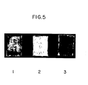
- Figure 5 is a photograph of three samples of polytetrafluoroethylene (PTFE) after staining with crystal violet: Sample 1 (the sample on the left) is PTFE coated with
Formulation 2; Sample 2 (the center sample) is PTFE seeded with cells, but not coated withFormulation 2; and Sample 3 (the sample on the right) is PTFE coated withFormulation 2 and seeded with cells. - The growth and normal metabolism of eukaryotic cells requires attachment to a substrate with the cell layer essentially extended face-to-face on the substrate. Conventionally, cell culture utilizes plastic substrates and, to a lesser degree, glass and microporous filters for cell attachment and propagation. More recently, physiological substrates (collagen, laminin, fibronectin, poly-D- and poly-L-lysine) have been utilized for these purposes in lieu of plastic to avoid problems inherent in cell culture at low seeding densities, using freshly isolated cells or on substrates less suitable for attachment (e.g., Teflon®). Bioadhesive polyphenolic proteins provide a suitable alternative because of their high binding affinity for both cells and a variety of substrates, biological and inert.
- Bioadhesive polyphenolic protein formulations have been evaluated for their efficiency in binding cells in vitro for cell culture. The formulations tested have included (1) 95% pure bioadhesive polyphenolic protein prepared from natural sources ("Formulation 1") and (2) 45% pure bioadhesive polyphenolic protein prepared from natural sources ("
Formulation 2"). - After preparing bioadhesive polyphenol-ic proteins according to procedures described in United States Patent No. 4,585,585, these formulations were thoroughly characterized biochemically using high performance liquid chromatography, assays for the quantitation of L-dopa, amino acid analysis and polyacrylamide gel electrophoresis. The composition of key amino acids in the bioadhesive polyphenolic protein formulations is given in Table 1.
-

- In
Formulation 2, collagen comprises the majority of the remaining 55%. The basic unit of the bioadhesive polyphenolic protein is a decapeptide (chain of 10 amino acids) which is repeated through covalent bonds to similar decapeptides as many as 75-85 times. These formulations, based on bioadhesive polyphenolic protein, are stable, based on adhesive functionality, at 4°C in 5% (v/v) acetic acid, pH 2.8 for greater than 10 months. Extracted preparations containing 40 to 50% collagen are stable at room temperature in 5% (v/v) acetic acid, pH 2.8 or following drying onto plastic substrates for at least 2 months. - The ability of bioadhesive polyphenolic protein to strongly attach to a variety of substrates permits the attachment, maintenance and growth of cells to surfaces that heretofore posed problems either because of their composition, their application, or the type of cell requiring attachment. Substrates that could be used include plastic, glass, and microporous filters (e.g., cellulosic, nylon, glass fiber, polyester, polycarbonate) for conventional cell culture research and/or cell product harvesting from bio-reactors used in batch cell culture or in genetic engineering; hollow fiber tubes for cell product harvesting; and prosthetic vascular graft materials such as polytetrafluoroethylene (Teflon@) and related materials. Most of these surfaces carry a net negative charge and, therefore, tend to bind tightly net positively charged materials such as bioadhesive polyphenolic proteins. Cells carry a net negative charge and, as a result, are slightly repelled from untreated surfaces while being attracted to the intermediary bioadhesive polyphenolic protein which has a net positive charge. Bioadhesive polyphenolic protein would increase attachment efficiency, attachment rate and strength of attachment. This latter parameter is critical in applications involving cell product harvesting procedures or re-implantation of cells on vascular grafts which involve the passage of fluids over cell monolayers. Moreover, cells that attach poorly following isolation from tissue or due to cell type, and cells that do not normally attach, such as blood cells and suspension tissue culture cells (histiocytic lymphomas, platelets, white and red
- blood cells, etc.) could also be attached to substrates through this intermediate.
- Furthermore, the ability of bioadhesive polyphenolic protein to strongly attach to a variety of substrates permits the attachment of many other biologically active moieties, such as DNA, proteins, hormones and antibiotics.
- The coating of substrates with bioadhesive polyphenolic protein formulations and attachment to substrates is generally performed as follows. Depending on the final concentration per square cm desired, about 1 to 2pl of sterile bioadhesive polyphenolic protein ranging from 10 to 60 µg per µl are evenly applied per cm2 of substrate. The resultant film is dried rapidly by placing the substrate within a laminar flow hood. Once dried, the film is treated with 35-100% ethanol or isopropanol for rinsing and fixation and then with sterile tissue culture medium for removal of residual alcohol and non- adsorbed extraneous moieties. The substrate may be used immediately or dried for storage. Cells or other biologically active moieties to be attached to the film are adjusted to desired concentrations and added to the substrate in serum-free or serum-containing medium. In the case of attachment of cells, at various timed intervals, the cells are evaluated for attachment, growth, or function, or treated according to prescribed objectives of experiments requiring the attached cells in tissues culture.
- Conversely, when desirable, the bioadhesive polyphenolic protein can be affixed to the biologically active moieties and then,the resultant biologically active moieties can be affixed to the substrate. The foregoing method, in greater detail, comprises the steps of:
- (1) dispersing said biologically active moieties in a serum-free solution;
- (2) admixing a sterile formulation comprising polyphenolic protein containing from about 35 to 100% by weight bioadhesive polyphenolic protein having the repeating decapeptide unit:

- (3) recovering the resulting biologically active moieties; and
- (4) affixing said recovered biologically active moieties to said substrate.
- Formulations containing either 95% bioadhesive polyphenolic protein (Formulation 1) or 45% bioadhesive polyphenolic protein (Formulation 2) were evenly layered on 35 mm tissues (9 cm2) culture plastic petri dishes at 50 µg per dish in 5% (v/v) acetic acid, dried rapidly, "fixed", and sterilized by rinsing with 100% ethanol. Dishes were then rinsed with sterile triple distilled water.
- Cells were prepared for the attachment assay as follows. Bovine corneal endothelial cells were treated with trypsin, a protease which digests cell attachment proteins, following growth in subculture in 5% C02 in air at 37°C in a humidified incubator. Cell monolayers were rinsed with serum-free medium to remove excess serum and medium that might interfere with trypsinization and incubated with 0.05% trypsin - 0.02% ethylene diamine tetraacetic acid (EDTA) for 10 minutes. Cells detached by the action of trypsin were transferred by pipette and gently centrifuged at 250 x g. Resultant pellets were re-suspended in serum-free minimal essential medium (Earle's salts) to remove any remaining serum proteins and trypsin from cell surfaces and again centrifuged.
- Viable cell counts were obtained using a dye exclusion test, where representative aliquots of cells were then re-suspended to a final concentration of 2 x 10⁵ cells per ml in minimal essential medium containing 15% fetal bovine serum. Cells were seeded in untreated plastic tissue culture petri dishes (control) and in tissue culture dishes layered with bioadhesive polyphenolic protein. At 1, 2.5, 5, 12.5, and about 20 min., triplicate experimental and control plates were chosen at random for quantitation of unattached cells. Unattached cells were removed from plates by rinsing and counted on a hemacytometer; replicate aliquots of cells that had been used, but that had not been added to dishes, were also counted in triplicate. Data were calculated as percent of cells attached by subtracting the number of unattached cells harvested from each dish from the total number of cells plated.
- Comparison of data showing percent of attachment of cells to bioadhesive formulations are found below in Table 2 and shown graphically in Figure 1.

- It can be seen that, within only 5 minutes, the attachment of cells in Formulation 2 (i.e., higher collagen content) is more than 2-fold greater than the attachment of cells to plastic. Further, at all time points, the binding capacity of cells to bioadhesive polyphenolic protein exceeds that of cells to plastic. Although results are similar for Formulation 1, other data suggest that
Formulation 2 is preferable as a cell attachment factor and tissue culture tool. -
Formulation 2 is very stable upon long-term storage. When tested by amino acid analysis, the L-dopa to protein ratios remained stable forFormulation 2 after 4 months at 40 and -20°C; whereas, a decline of up to 25% is found under similar conditions with Formulation 1 (Table 3).
- Since stability of moieties at the biochemical level is highly desirable in tissue culture systems, it was concluded that Formulation is preferred for purposes of enhancing cell attachment efficiency.
- Depending on the objectives of an experiment or assay, more or less serum may be required during and after cell attachment. The effect of serum on cell binding and strength was tested using cells in medium containing 15% bovine serum (FBS) or 0.5% bovine serum albumin (BSA). Bovine serum is the major protein constituent found in FBS. Strength of attachment was indirectly evaluated by the ability or inability to remove attached cells by trypsin from substrates to which they were attached. The concentration of bovine serum albumin employed was equivalent to that found in 0.5% to 1% FBS. The coating of tissue culture petri dishes with
Formulation 2 of bioadhesive polyphenolic protein was accomplished as in Example 1. Cells were seeded on plastic and adhesive-coated petri dishes in triplicate and the unattached cells were removed by rinsing at 2.5, 5, and 15 minutes. Unattached cells were trypsinized using 0.8 ml of 0.05% trypsin-0.02% EDTA for 10 minutes and transferred to tubes containing 0.2% FBS to inhibit further action of the trypsin on the cells. Recovered attached and unattached cells were counted using a hemacytometer and the data representing attached cells were calculated as a percentage of the total cells recovered from each of the dishes. This data is summarized in Table 4.
- As can be seen from the data in Table 4, cells attach more strongly at early time periods on bioadhesive polyphenolic protein than to plastic. It can also be seen that if FBS in the medium is replaced with bovine serum albumin, a decreased recovery of cells by trypsinization results. This is especially seen at times when cells are establishing firm anchorage (5 minutes) and beginning to flatten (15 minutes). At 15 minutes, as low as 33% and 39% recovery was achieved with BSA on plastic and adhesive-coated petri dishes as compared with 83% and 74% recovery with FBS on plastic and adhesive cultures, respectively. Visual microscopic observations of plates confirmed these findings. Other findings in this study demonstrate that the direct evaluation of cell attachment described in this example strongly correlates with indirect measurement by counting unattached cells only (see Example 1 for counting details). Figure 2 graphically illustrates these findings.
- The majority of cell types harvested from tissue following dissociation, when placed into cell culture, are capable of attaching to plastic substrates with varying degrees of efficiency. Certain cell types, however, do not attach to plastic substrates. The ability to attach such cells could be advantageous in that it would provide a means for diagnostic and research assays requiring the immobilization of these cells, and the ability to secure cells to bio-reactor filters for harvesting cell products. Furthermore, it would provide an unequivocal demonstration of the potential for bioadhesive polyphenolic protein to act as a tissue culture attachment factor.
- The cell line U937 is a human histiocytic lymphoma that was established from malignant cells isolated from a pleural effusion. These cells grow in suspension continuously in RPMI 1640 tissue culture medium, supplemented with 10% fetal bovine serum. U937 cells attach poorly to plastic in the presence of serum..
- Tissue culture petri dishes (35 mm dishes) were coated with bioadhesive polyphenolic protein according to procedures outlined in Example 1. U937 cells were transferred to centrifuge tubes and prepared in the manner described in Example 1. Cells were seeded on plastic tissue culture dishes and on dishes coated with 100 µg of bioadhesive polyphenolic protein (Formulation 2), and evaluated in triplicate for attachment efficiency, (see Example 1) at 5, 12.5, and 20 minutes. The results in Table 5 (which are shown graphically in Figure 3) clearly demonstrate the effect of bioadhesive polyphenolic protein on attachment of U937 cells. As expected, the cells attached poorly to the plastic dishes which served as controls; but within 5 minutes, 75% of the cells seeded had attached to coated dishes, and within 20 minutes, 87% of the cells attached to the coated dishes.

- The attachment of cells to a substrate is only the first requirement for establishing cells in cultures in vitro. The second, and perhaps more important, requirement is that the cells grow. With the dissociation of tissue, however, the number of cells harvested is frequently very low. Low cell seeding numbers can adversely affect the establishment of cultures because fewer cells decrease the chance of survival for attachment. This is based on simple mathematical probabilities and on the need for metabolites produced by the cells themselves (density-dependent metabolites) which are required for cell attachment and growth. When cell numbers are low, probabilities are lower that adequate numbers of cells will attach, which itself is necessary for flattening of the cells onto the substrate .from a spherical shape. Once flattened, metabolism may then ensue to further condition the medium for cell growth and division.
- To enhance the attachment and propagation of cells that either do not readily attach and/or that are seeded at low densities, various peptide and protein attachment factors have been made available commercially. These include collagen, laminin, poly-D-lysine, and fibronectin. All of these factors work on biologically inert substrates to varying degrees, depending on the cell type seeded. To compare the effectiveness of bioadhesive polyphenolic protein to these factors in permitting growth at low seeding densities, bovine corneal endothelial cells were seeded at a density of 250 cells per tissue culture petri dish (35 mm diameter, 9.65 cm2) on either plastic, bioadhesive polyphenolic protein, collagen, laminin, poly-D-lysine, or fibronectin. The cells were allowed to grow for 5 days, at which time colony sizes (number of cells per colony) and numbers of colonies per plate were evaluated for each of the variables by staining the cells with crystal violet. Data obtained were used to determine the effect of each of these factors on attachment (number of colonies) and growth (size of colonies).
- Cells and dishes coated with bioadhesive polyphenolic protein were prepared as described in Example 1. The coating of dishes with other attachment factors was effected according to procedures suggested by their manufacturers.
- Collagen - Collagen-coated plates were prepared by diluting 1 part cold (4oC) collagen dispersion into 6 parts of cold 50% methanol. This-mixture was mixed vigorously for several minutes and pipetted onto a petri dish so that only the bottom of the dish was covered. Within 20 seconds, the collagen was removed by aspiration and the dish was tilted upside down at 30° against a lid to dry. Following 1 hour of drying undisturbed in a laminar flow hood, the dishes were ready for use.
- Laminin - Laminin is supplied in 1 mg quantities in 1 ml of 50 mM tris(hydroxymethyl)aminomethane in physiological saline. Following a slow thaw of laminin solution at 0 to 4°C from -20° C, 10 to 15 µg of laminin solution was pipetted into petri dishes in 0.5 ml of 0.01 M sodium phosphate buffer, pH 7.4. The dishes were dried at 370C. Immediately upon drying, the dishes are prepared for use.
- Fibronectin - Fibronectin is supplied in 1 mg quantities as a lyophilized powder. Prior to use, fibronectin is allowed to equilibrate to room temperature after storage at 40C. The powder is reconstituted with 1 ml sterile distilled water and allowed to stand for 30 minutes for solubilization. Ten to 20 µg of fibronectin solution is added to each dish in 0.5 ml and allowed to air dry. At this time, the dish is ready for cell seeding.
- Poly-D-lysine - Poly-D-lysine is supplied in quantities of 5 mg of lyophilized powder. Prior to use, this powder is allowed to equilibrate to room temperature following storage at 4° C. Dishes are coated with 50 mg in 1 ml of sterile distilled water and allowed to stand at room temperature for 5 minutes. At that time, the solution is aspirated and the dishes are rinsed two times with 1.5 ml sterile distilled water. Following each rinse, liquid is aspirated completely. The dishes are dried and used immediately.
- The plating efficiency of the cells seeded on each of the factors is evaluated following crystal violet staining of the cells. This is achieved by first rinsing the dishes containing the colonies with serum-free medium to remove excess proteins and fixing the cells with 10% neutral buffered formalin for 10 minutes. The formalin is then removed from the plates by aspiration and 0.1% crystal violet in tapwater is then added to the plates for a period of 7 minutes. Immediately following staining, crystal violet is poured off and the cells are rinsed in a beaker of running tapwater to remove excess stain. Following complete drying of the plates, colonies on duplicates -representing each of the variables are counted; and cells in ten randomly selected colonies per plate are counted. The data obtained from this example appear in Table 6 and are graphically illustrated in Figure 4 as bar graphs. As can be seen, the number of colonies in Figure 4 on plates coated with bioadhesive polyphenolic protein (U) is matched only by poly-D-lysine (PDL). All other factors, including collagen (C), plastic (P), fibronectin (F), and laminin (L), yield poor results by comparison. Similarly, the average number of cells per colony found on plates coated with bioadhesive polyphenolic protein is matched by poly-D-lysine. Collagen, plastic, fibronectin, and laminin demonstrate poor efficiency in growth. Although no significant differences were found between bioadhesive polyphenolic protein and poly-D-lysine (possibly due to the high level of lysine found in each of these molecules), the use of bioadhesive polyphenolic protein as an attachment factor is nonetheless more advantageous as a substrate based on its ability to (1) displace water, (2) to attach to materials including metal and Teflon® (for example, prosthetic devices), (3) to be used in vivo and in vitro, and (4) to form high strength bonds based on L-dopa, hydroxylated and lysine amino acid residues.
-

- Polytetrafluoroethylene (PTFE) is a substrate commonly used for vascular implants. The major problem with the use of this material is that the seeding of vascular cells on PTFE is very difficult due to its high hydrophobicity. For many implants, a confluent cell monolayer on its surface would prevent clot formation. To test the effectiveness of bioadhesive polyphenolic protein to act as a mediator for attachment of endothelial cells to PTFE, the vascular implant material was coated with 200 µg per. CM2 of bioadhesive polyphenolic protein (Formulation 2). Five hundred thousand endothelial cells were then allowed to attach to the bioadhesive polyphenolic protein. The cells were also seeded onto Teflon® without coating with bioadhesive polyphenolic protein; and Teflon°coated with bioadhesive polyphenolic protein without seeding of cells acted as a control. Following 15 minutes of attachment, excess cells were rinsed from the vascular implant material and the vascular implants were fixed with formalin, stained with crystal violet, and dried as described in Example 4. The results of this example appear in Figure 5 and demonstrate that, although some staining can be seen on both Teflon® treated with bioadhesive polyphenolic protein without cells (Sample 1) and on Teflon® that is untreated with adhesive, but is seeded with cells (Sample 2), by far the greatest staining, or cell attachment, occurred on the treated TeflonO containing the endothelial cells (Sample 3). Thus, bioadhesive polyphenolic proteins enhance the seeding of vascular implants with endothelial cells, thereby providing a mechanism by which clot formation may be minimized or eliminated following vascular implant surgery.
- 300 grams of marine mussel, M. edulis, feet are combined with 900 mls of neutral salt buffer which contains 1 M sodium chloride, 0.05M tris(hydroxymethyl)aminomethane (pH 7.5), 1 mM phenylmethylsulfonylfluoride, 10mM N-ethylmaleimide, 0.025 M ethylenediamine tetraacetic acid and 1 mM potassium cyanide plus 9 mls of antifoam concentrate in a commercial blender on high speed and thoroughly blended, precipitating the bioadhesive polyphenolic protein. The mixture is centrifuged at 10 X rpm for 15 minutes. The pellet is re-suspended in 900 mls of 5% acetic acid using the blender on high speed. Bioadhesive polyphenolic protein remains in the supernatant during centrifugation at 10 K rpm for 45 minutes. The approximately 1000 mls of supernatant is put into an ice bath with continual stirring. 5 mls of 2M sodium borate plus 95 mls of 5 M sodium chloride are added to the stirring supernatant. This mixture is centrifuged at 10K rpm for 15 minutes. The new supernatant is treated identically as above with the addition of four times as much 2M sodium borate and 5 M sodium chloride. Once again, the mixture is centrifuged at 10K for 15 minutes. The pellet is re-suspended in the following mixture: 7.5 mls of 2M sodium borate, 50 mls of 5H sodium chloride, 50 mls of distilled water, 37.5 mls of 8M urea in 5% acetic acid, and 5.6 mls of concentrated acetic acid. The mixture is slowly stirred for approximately 16 hours. The suspension is centrifuged at 10 K rpm for 15 minutes. The supernatant is saved and dialyzed (8-12K molecular weight cut-off membranes) against 5% acetic acid for approximately 16 hours. Amino acid analysis establishes that the extract contains 45% pure bioadhesive polyphenolic protein. The purity of the extract is governed by the number of extractions effected. The yield of pure bioadhesive polyphenolic protein decreases as the number of extractions increases. All procedures described herein were conducted at 4° C.
- Using liquid chromatography, SE Sephadex resins retain polyphenolic proteins in 5.5% Guanidine hydrochloride (GuHC1) in 5% acetic acid. The protein is then eluted from the resin with a gradient of 5.5 - 20% GuHCl in acetic acid, the peak areas pooled and dialyzed against 5% acetic acid to remove the GuHCl. Storage of the proteins is most stable at 4°C in 5% acetic acid. Prior to its use as an adhesive, in vivo or in contact with live cells, bioadhesive polyphenolic proteins must be dialyzed against water to raise the pH of the solution to near neutrality and the preparation must be concentrated to between 3 and 10 mg/ml. This is accomplished using an ultrafiltration membrane with pore size exclusion limits of 30,000 or less. This is not necessary when bioadhesive polyphenolic proteins are dried onto an inert substrate prior to use.
- Bioadhesive polyphenolic protein, 45% pure (Formulation 2), was used to immobilize heparin, a mucopolysaccharide having specific anticoagulant properties, and peroxidase, a protein enzyme which oxidizes peroxide. This was done to show that other substances could be efficiently bound to plasticware via a bioadhesive polyphenolic protein intermediate.
- For both, 7 µg of bioadhesive polyphenolic protein was dried onto tissue culture plasticware dishes of 2 cm2 area for a final concentration of 3.5pg/cm2. The protein was washed with 100% ethanol and then twice with water as described in Example 1.
- Heparin was added to the dishes at 5 different concentrations: 90, 60, 30, 15 and 5 units/dish. Heparin was also dried onto untreated plastic dishes. All tests were performed in duplicate. The plates were washed with 0.1M phosphate buffer before use to remove loosely bound heparin.
- The assay for heparin activity was performed by adding fresh human, blood to each dish at 0.5 ml per dish with incubation at 23°C. Clotting times were visually observed and recorded. The results are shown in Table 7.

- Heparin was very effectively immobilized to plastic employing bioadhesive polyphenolic protein. No clotting was observed in 24 hours even at the lowest dose of heparin. All doses below 60 units clotted in 2 hours or less in dishes where
- heparin was attached directly to plastic. At the higher doses, sufficient heparin binds to the plastic to prevent clot formation.
- Peroxidase was immobilized in a similar manner: 5 different concentrations of peroxidase, 1, 0.5, 0.1, 0.05, 0.025 µg/dish, were added to both uncoated plastic dishes and plastic dishes coated with bioadhesive polyphenolic protein (in duplicate). As with heparin, a 0.1 M phosphate buffer wash was used to remove loosely bound enzyme.
- The assay for peroxidase involves adding a substrate mixture, peroxide plus 0-phenylene diamine (OPD) in phosphate buffered saline. The substrate mix, per ml, contains 100 µl of peroxide (40 µl of 30% peroxide in 50 ml water) plus 100 pl OPD (10.7 mg in 8.56 ml water) and 800 µl phosphate buffered saline. 1 ml is added to each dish. After 5 minutes incubation at 23°C, 100 µl of 4 N sulfuric acid is added to stop the reaction. It is a colorimetric assay with a wavelength optimum at 490 nm. The data in duplicate is presented as absorbance units at 490 nm in Table 8.

- As with heparin, at the higher concentrations no enhancement is seen by employing bioadhesive polyphenolic protein, sufficient enzyme binds to plastic. At the lower concentrations, significant enhancement or recovery is seen by employing bioadhesive polyphenolic protein.
- Bioadhesive polyphenolic protein has been found to successfully serve as a substrate for tissues and cells in histology and cytology. In this example, 45% bioadhesive polyphenolic protein (Formulation 2) was used to affix bovine Descemet's membrane with endothelial cell preparations to glass slides. Whole cornea were removed from freshly killed cows and placed either in physiological saline or in 10% neutral buffered formalin. Descemet's membrane was then removed from the posterior side of the cornea by gentle peeling. The tissue was transferred to slides (pre-cleaned with 5% acetic acid) and coated with 50 µg of bioadhesive polyphenolic protein. Tissue preparations were then dried onto the bioadhesive polyphenolic protein at room temperature or on 55°C warming plates for twenty minutes. When formalin fixed tissue was used, the tissue was rinsed with saline for removal of excess formalin prior to attachment to the bioadhesive polyphenolic protein. Following drying, tissue-slide preparations were treated with formalin to fix tissues to the bioadhesive polyphenolic protein for five minutes. Tissues treated in this manner were retained on the bioadhesive polyphenolic protein for weeks in aqueous solutions. Furthermore, following extensive agitation by shaking in water, saline, dilute and 100% ethanol, and xylene the tissues still remained intact. In the absence of bioadhesive polyphenolic protein, adherence of tissues to slides did not even survive the first formalin treatment.
Claims (62)


Priority Applications (1)
| Application Number | Priority Date | Filing Date | Title |
|---|---|---|---|
| AT87105672T ATE84820T1 (en) | 1986-04-25 | 1987-04-16 | ADHESIVE BIOMATERIALS FOR ADHAESION OF CELLS AND TISSUES. |
Applications Claiming Priority (4)
| Application Number | Priority Date | Filing Date | Title |
|---|---|---|---|
| US85668786A | 1986-04-25 | 1986-04-25 | |
| US856687 | 1986-04-25 | ||
| US34801 | 1987-04-03 | ||
| US07/034,801 US5108923A (en) | 1986-04-25 | 1987-04-03 | Bioadhesives for cell and tissue adhesion |
Publications (3)
| Publication Number | Publication Date |
|---|---|
| EP0243818A2 true EP0243818A2 (en) | 1987-11-04 |
| EP0243818A3 EP0243818A3 (en) | 1989-03-08 |
| EP0243818B1 EP0243818B1 (en) | 1993-01-20 |
Family
ID=26711386
Family Applications (1)
| Application Number | Title | Priority Date | Filing Date |
|---|---|---|---|
| EP87105672A Expired - Lifetime EP0243818B1 (en) | 1986-04-25 | 1987-04-16 | Bioadhesives for cell and tissue adhesion |
Country Status (9)
| Country | Link |
|---|---|
| US (1) | US5108923A (en) |
| EP (1) | EP0243818B1 (en) |
| JP (1) | JPH0779695B2 (en) |
| AU (1) | AU597647B2 (en) |
| CA (1) | CA1328237C (en) |
| DE (1) | DE3783643T2 (en) |
| DK (1) | DK171996B1 (en) |
| FI (1) | FI91037C (en) |
| NO (1) | NO174675C (en) |
Cited By (13)
| Publication number | Priority date | Publication date | Assignee | Title |
|---|---|---|---|---|
| EP0350714A2 (en) * | 1988-07-13 | 1990-01-17 | Collaborative Biomedical Products Inc. | Tissue immobilization and cell culturing system and method for affixing biologically active moieties to a substrate |
| EP0367468A1 (en) * | 1988-11-04 | 1990-05-09 | Immucor, Inc. | Method for drying mammalian cells for use in solid phase immunoassays and articles incorporating same |
| EP0435871A4 (en) * | 1988-07-05 | 1991-05-14 | Albert J Banes | Floating cell culture device and method. |
| US5049504A (en) * | 1986-11-24 | 1991-09-17 | Genex Corporation | Bioadhesive coding sequences |
| US5192663A (en) * | 1988-11-04 | 1993-03-09 | Immucor, Inc. | Article having an organic dye and a monolayer of dried mammalian cells and a method for utilizing the article |
| US5197973A (en) * | 1990-12-14 | 1993-03-30 | Creative Biomolecules, Inc. | Synthetic bioadhesive |
| US5202256A (en) * | 1984-09-13 | 1993-04-13 | Enzon Labs, Inc. | Bioadhesive precursor protein expression vectors |
| US5202236A (en) * | 1984-09-13 | 1993-04-13 | Enzon Labs Inc. | Method of producing bioadhesive protein |
| EP0537167A1 (en) * | 1990-04-17 | 1993-04-21 | Curative Technologies, Inc. | Coating prosthetic surfaces with mammalian cells |
| US5759774A (en) * | 1988-05-18 | 1998-06-02 | Cobe Laboratories, Inc. | Method of detecting circulating antibody types using dried or lyophilized cells |
| US5773222A (en) * | 1992-05-27 | 1998-06-30 | National Blood Authority | Solid phase immunological assay |
| US5817470A (en) * | 1995-03-10 | 1998-10-06 | Sociedad Biotecnologica Collico Limitada | Immobilization of antigens to solid support by the mussel adhesive polyphenolic protein and the method for use therein |
| US10101247B2 (en) | 2013-07-19 | 2018-10-16 | Rarecyte, Inc. | Solution and method for adhering suspension components to a substrate |
Families Citing this family (34)
| Publication number | Priority date | Publication date | Assignee | Title |
|---|---|---|---|---|
| AU592670B2 (en) * | 1986-08-15 | 1990-01-18 | Commonwealth Scientific And Industrial Research Organisation | Promoting cell adhesion and growth on a substrate |
| US5574134A (en) * | 1989-07-11 | 1996-11-12 | University Of Delaware | Polypeptide monomers, linearly extended and/or crosslinked forms thereof, and applications thereof |
| US5310669A (en) * | 1992-06-22 | 1994-05-10 | The Trustees Of Dartmouth College | Fullerene coated surfaces and uses thereof |
| US5955353A (en) * | 1997-05-22 | 1999-09-21 | Excorp Medical, Inc. | Hollow fiber bioreactor with an extrafilament flow plug |
| US6221425B1 (en) | 1998-01-30 | 2001-04-24 | Advanced Cardiovascular Systems, Inc. | Lubricious hydrophilic coating for an intracorporeal medical device |
| US20020022588A1 (en) * | 1998-06-23 | 2002-02-21 | James Wilkie | Methods and compositions for sealing tissue leaks |
| US6214054B1 (en) * | 1998-09-21 | 2001-04-10 | Edwards Lifesciences Corporation | Method for fixation of biological tissues having mitigated propensity for post-implantation calcification and thrombosis and bioprosthetic devices prepared thereby |
| EP1334116B1 (en) * | 1998-09-28 | 2007-04-25 | Bio Polymer Products of Sweden AB | A process of producing polyphenolic adhesive proteins |
| US7858679B2 (en) * | 2001-07-20 | 2010-12-28 | Northwestern University | Polymeric compositions and related methods of use |
| US7618937B2 (en) * | 2001-07-20 | 2009-11-17 | Northwestern University | Peptidomimetic polymers for antifouling surfaces |
| US8815793B2 (en) * | 2001-07-20 | 2014-08-26 | Northwestern University | Polymeric compositions and related methods of use |
| US6878168B2 (en) | 2002-01-03 | 2005-04-12 | Edwards Lifesciences Corporation | Treatment of bioprosthetic tissues to mitigate post implantation calcification |
| US8911831B2 (en) * | 2002-07-19 | 2014-12-16 | Northwestern University | Surface independent, surface-modifying, multifunctional coatings and applications thereof |
| US20080085554A1 (en) * | 2004-02-13 | 2008-04-10 | Norio Nakatsuji | Culture Medium for Culturing Feeder Cells for Embryonic Stem Cells Culture and the Prepared Feeder Cells |
| DE102004031258A1 (en) * | 2004-06-29 | 2006-02-09 | Jennissen, Herbert P., Prof. Dr. | Protein hybrids with polyhydroxyaromatic amino acid epitopes |
| US7732539B2 (en) * | 2006-02-16 | 2010-06-08 | National Science Foundation | Modified acrylic block copolymers for hydrogels and pressure sensitive wet adhesives |
| JP5281886B2 (en) * | 2006-04-28 | 2013-09-04 | 株式会社クラレ | Cell culture container and method for producing the same |
| CA2656681C (en) | 2006-08-04 | 2014-04-22 | Nerites Corporation | Biomimetic compounds and synthetic methods therefor |
| US8563117B2 (en) * | 2006-08-04 | 2013-10-22 | Phillip B. Messersmith | Biomimetic modular adhesive complex: materials, methods and applications therefore |
| EP2077718B2 (en) | 2006-10-27 | 2022-03-09 | Edwards Lifesciences Corporation | Biological tissue for surgical implantation |
| WO2008089032A1 (en) * | 2007-01-11 | 2008-07-24 | Northwestern University | Fouling resistant coatings and methods of making same |
| US8383092B2 (en) * | 2007-02-16 | 2013-02-26 | Knc Ner Acquisition Sub, Inc. | Bioadhesive constructs |
| US8673286B2 (en) | 2007-04-09 | 2014-03-18 | Northwestern University | DOPA-functionalized, branched, poly(aklylene oxide) adhesives |
| US9101691B2 (en) * | 2007-06-11 | 2015-08-11 | Edwards Lifesciences Corporation | Methods for pre-stressing and capping bioprosthetic tissue |
| US8357387B2 (en) * | 2007-12-21 | 2013-01-22 | Edwards Lifesciences Corporation | Capping bioprosthetic tissue to reduce calcification |
| US20110130465A1 (en) * | 2009-12-01 | 2011-06-02 | Nerites Corporation | Coatings for prevention of biofilms |
| NZ602066A (en) | 2010-03-23 | 2013-09-27 | Edwards Lifesciences Corp | Methods of conditioning sheet bioprosthetic tissue |
| US8906601B2 (en) | 2010-06-17 | 2014-12-09 | Edwardss Lifesciences Corporation | Methods for stabilizing a bioprosthetic tissue by chemical modification of antigenic carbohydrates |
| EP2637707A4 (en) | 2010-11-09 | 2014-10-01 | Kensey Nash Corp | Adhesive compounds and methods use for hernia repair |
| US9351829B2 (en) | 2010-11-17 | 2016-05-31 | Edwards Lifesciences Corporation | Double cross-linkage process to enhance post-implantation bioprosthetic tissue durability |
| US10238771B2 (en) | 2012-11-08 | 2019-03-26 | Edwards Lifesciences Corporation | Methods for treating bioprosthetic tissue using a nucleophile/electrophile in a catalytic system |
| US20160011176A1 (en) * | 2012-12-19 | 2016-01-14 | National University Corporation Tokyo Medical And Dental University | Method and device for examining myocardial toxicity and evaluating cardiomyocytes |
| WO2015009431A1 (en) * | 2013-07-19 | 2015-01-22 | Rarecyte, Inc. | Solution and method for adhering suspension components to a substrate |
| US10101248B1 (en) * | 2015-12-02 | 2018-10-16 | Rarecyte, Inc. | Solution and method for adhering suspension components to a substrate |
Citations (1)
| Publication number | Priority date | Publication date | Assignee | Title |
|---|---|---|---|---|
| US4266032A (en) * | 1979-08-10 | 1981-05-05 | Monsanto Company | Method of culturing microcarrier supported cells |
Family Cites Families (11)
| Publication number | Priority date | Publication date | Assignee | Title |
|---|---|---|---|---|
| US3910819A (en) * | 1974-02-19 | 1975-10-07 | California Inst Of Techn | Treatment of surfaces to stimulate biological cell adhesion and growth |
| DE2603319C3 (en) * | 1976-01-29 | 1979-08-23 | Boehringer Mannheim Gmbh, 6800 Mannheim | Method for the fixation of biologically active proteins on carriers |
| FR2447275A1 (en) * | 1979-01-25 | 1980-08-22 | Charbonnages Ste Chimique | LAMINATE MATERIALS BASED ON PHENOLIC RESIN AND PROCESS FOR THEIR PREPARATION |
| US4352887A (en) * | 1979-10-29 | 1982-10-05 | Albert Einstein College Of Medicine Of Yeshiva University | Method and article for culturing differentiated cells |
| US4501815A (en) * | 1979-10-29 | 1985-02-26 | Albert Einstein College Of Medicine Of Yeshiva University | Article for culturing differentiated cells |
| US4578079A (en) * | 1982-08-04 | 1986-03-25 | La Jolla Cancer Research Foundation | Tetrapeptide |
| JPS5928472A (en) * | 1982-08-09 | 1984-02-15 | Koken:Kk | Substrate for cell culture, cultivation and separation of cell using it |
| ZA839115B (en) * | 1982-12-14 | 1984-07-25 | Cpc International Inc | Continuous enzymatic process for producing maltose from starch and starch hydrolysates |
| US4496397A (en) * | 1984-03-07 | 1985-01-29 | University Of Connecticut | Process for purifying and stabilizing catechol-containing proteins and materials obtained thereby |
| US4585585A (en) * | 1984-03-07 | 1986-04-29 | University Of Connecticut Research & Development Corporation | Decapeptides produced from bioadhesive polyphenolic proteins |
| US4553974A (en) * | 1984-08-14 | 1985-11-19 | Mayo Foundation | Treatment of collagenous tissue with glutaraldehyde and aminodiphosphonate calcification inhibitor |
-
1987
- 1987-04-03 US US07/034,801 patent/US5108923A/en not_active Expired - Lifetime
- 1987-04-16 EP EP87105672A patent/EP0243818B1/en not_active Expired - Lifetime
- 1987-04-16 DE DE8787105672T patent/DE3783643T2/en not_active Expired - Fee Related
- 1987-04-21 FI FI871728A patent/FI91037C/en not_active IP Right Cessation
- 1987-04-22 DK DK205287A patent/DK171996B1/en not_active IP Right Cessation
- 1987-04-22 NO NO871665A patent/NO174675C/en unknown
- 1987-04-22 CA CA000535245A patent/CA1328237C/en not_active Expired - Fee Related
- 1987-04-23 AU AU71885/87A patent/AU597647B2/en not_active Ceased
- 1987-04-25 JP JP62102971A patent/JPH0779695B2/en not_active Expired - Lifetime
Patent Citations (1)
| Publication number | Priority date | Publication date | Assignee | Title |
|---|---|---|---|---|
| US4266032A (en) * | 1979-08-10 | 1981-05-05 | Monsanto Company | Method of culturing microcarrier supported cells |
Non-Patent Citations (2)
| Title |
|---|
| CHEMICAL ABSTRACTS, vol. 98, no. 21, 23rd May 1983, page 296, abstract no. 175113q, Columbus, Ohio, US; J.H. WAITE: "Evidence for a repeating 3,4-dihydroxphenylalanine- and hydroxyproline-containing decapeptide in the adhesive protein of the mussel, Mytilus edulis L.", & J. BIOL. CHEM. 1983, 258(5), 2911-15 * |
| NATURE, vol. 309, no. 5963, May 1984, pages 30-33, London, GB; M.D. PIERSCHBACHER et al.: "Cell attachment activity of fibronectin can be duplicated by small synthetic fragments of the molecule" * |
Cited By (19)
| Publication number | Priority date | Publication date | Assignee | Title |
|---|---|---|---|---|
| US5202256A (en) * | 1984-09-13 | 1993-04-13 | Enzon Labs, Inc. | Bioadhesive precursor protein expression vectors |
| US5202236A (en) * | 1984-09-13 | 1993-04-13 | Enzon Labs Inc. | Method of producing bioadhesive protein |
| US5049504A (en) * | 1986-11-24 | 1991-09-17 | Genex Corporation | Bioadhesive coding sequences |
| US5759774A (en) * | 1988-05-18 | 1998-06-02 | Cobe Laboratories, Inc. | Method of detecting circulating antibody types using dried or lyophilized cells |
| EP0435871A4 (en) * | 1988-07-05 | 1991-05-14 | Albert J Banes | Floating cell culture device and method. |
| EP0435871A1 (en) * | 1988-07-05 | 1991-07-10 | BANES, Albert J. | Floating cell culture device and method |
| EP0350714A2 (en) * | 1988-07-13 | 1990-01-17 | Collaborative Biomedical Products Inc. | Tissue immobilization and cell culturing system and method for affixing biologically active moieties to a substrate |
| EP0350714A3 (en) * | 1988-07-13 | 1990-07-25 | Bio-Polymers, Inc. | Tissue immobilization and cell culturing system and method for affixing biologically active moieties to a substrate |
| EP0367468A1 (en) * | 1988-11-04 | 1990-05-09 | Immucor, Inc. | Method for drying mammalian cells for use in solid phase immunoassays and articles incorporating same |
| US5030560A (en) * | 1988-11-04 | 1991-07-09 | Immucor, Inc. | Method for drying mammalian cells for use in solid phase immunoassays and articles incorporating same |
| US5192663A (en) * | 1988-11-04 | 1993-03-09 | Immucor, Inc. | Article having an organic dye and a monolayer of dried mammalian cells and a method for utilizing the article |
| AU651699B2 (en) * | 1988-11-04 | 1994-07-28 | Immucor, Inc. | Method and articles for performing solid phase immunoassays |
| EP0537167A4 (en) * | 1990-04-17 | 1993-04-28 | Curative Technologies, Inc. | Coating prosthetic surfaces with mammalian cells |
| EP0537167A1 (en) * | 1990-04-17 | 1993-04-21 | Curative Technologies, Inc. | Coating prosthetic surfaces with mammalian cells |
| US5374431A (en) * | 1990-12-14 | 1994-12-20 | Creative Biomolecules, Inc. | Synthetic bioadhesive |
| US5197973A (en) * | 1990-12-14 | 1993-03-30 | Creative Biomolecules, Inc. | Synthetic bioadhesive |
| US5773222A (en) * | 1992-05-27 | 1998-06-30 | National Blood Authority | Solid phase immunological assay |
| US5817470A (en) * | 1995-03-10 | 1998-10-06 | Sociedad Biotecnologica Collico Limitada | Immobilization of antigens to solid support by the mussel adhesive polyphenolic protein and the method for use therein |
| US10101247B2 (en) | 2013-07-19 | 2018-10-16 | Rarecyte, Inc. | Solution and method for adhering suspension components to a substrate |
Also Published As
| Publication number | Publication date |
|---|---|
| JPH0779695B2 (en) | 1995-08-30 |
| US5108923A (en) | 1992-04-28 |
| NO174675C (en) | 1994-06-22 |
| NO871665L (en) | 1987-10-26 |
| DK205287D0 (en) | 1987-04-22 |
| DK205287A (en) | 1987-10-26 |
| AU7188587A (en) | 1987-10-29 |
| FI91037B (en) | 1994-01-31 |
| NO174675B (en) | 1994-03-07 |
| EP0243818A3 (en) | 1989-03-08 |
| DE3783643D1 (en) | 1993-03-04 |
| FI91037C (en) | 1994-05-10 |
| FI871728A0 (en) | 1987-04-21 |
| NO871665D0 (en) | 1987-04-22 |
| FI871728A (en) | 1987-10-26 |
| DK171996B1 (en) | 1997-09-08 |
| EP0243818B1 (en) | 1993-01-20 |
| DE3783643T2 (en) | 1993-05-13 |
| JPS6339583A (en) | 1988-02-20 |
| CA1328237C (en) | 1994-04-05 |
| AU597647B2 (en) | 1990-06-07 |
Similar Documents
| Publication | Publication Date | Title |
|---|---|---|
| US5108923A (en) | Bioadhesives for cell and tissue adhesion | |
| US4645669A (en) | Culturing and emplacement of differentiated cells in vivo | |
| JP5562303B2 (en) | Cultured cell sheet, production method and use thereof | |
| US7815686B2 (en) | Vascularization enhanced graft constructs | |
| US7112218B2 (en) | Tissue engineered blood vessels and apparatus for their manufacture | |
| WO2002014480A9 (en) | Decellularized tissue engineered constructs and tissues | |
| AU2001284968A1 (en) | Decellularized tissue engineered constructs and tissues | |
| WO1994007432A2 (en) | Porous vascular implants | |
| Sipehia et al. | Enhanced attachment and growth of human endothelial cells derived from umbilical veins on ammonia plasma modified surfaces of PTFE and ePTFE synthetic vascular graft biomaterials | |
| CA2657013A1 (en) | Temperature-responsive microcarrier | |
| EP0905231B1 (en) | Method for increasing the stability and/or shelf-life of various substrates | |
| US5932207A (en) | Complex active ingredient for the production of biological parts, especially organs for living organisms: method for the production of the same and its use | |
| EP0788381B1 (en) | Biomaterial containing epithelial cells and use thereof as a transplant | |
| US5980888A (en) | Keratinocytes attached to microcarriers for treatment of skin wounds | |
| US4656130A (en) | Collagen coated cell growth plates | |
| EP0636184B1 (en) | Media for isolation and stabilization of cells | |
| LEE et al. | Endothelial cell seeding onto the extracellular matrix of fibroblasts for the development of a small diameter polyurethane vessel | |
| EP0350714B1 (en) | Tissue immobilization and cell culturing system and method for affixing biologically active moieties to a substrate | |
| CN109153963B (en) | Method for changing cell culture in adhesion state | |
| BERNHARD et al. | Development of a nonthrombogenic collagenous blood-prosthetic interface | |
| KR100231279B1 (en) | Biodegradable polysaccharide sponge type formulation containing tissue growth factors for osteoblast implantation to recover | |
| JPH01124465A (en) | Composition for artificial basement membrane and implantation device with artificial basement membrane |
Legal Events
| Date | Code | Title | Description |
|---|---|---|---|
| PUAI | Public reference made under article 153(3) epc to a published international application that has entered the european phase |
Free format text: ORIGINAL CODE: 0009012 |
|
| AK | Designated contracting states |
Kind code of ref document: A2 Designated state(s): AT BE CH DE ES FR GB GR IT LI LU NL SE |
|
| PUAL | Search report despatched |
Free format text: ORIGINAL CODE: 0009013 |
|
| AK | Designated contracting states |
Kind code of ref document: A3 Designated state(s): AT BE CH DE ES FR GB GR IT LI LU NL SE |
|
| 17P | Request for examination filed |
Effective date: 19890825 |
|
| 17Q | First examination report despatched |
Effective date: 19911007 |
|
| GRAA | (expected) grant |
Free format text: ORIGINAL CODE: 0009210 |
|
| RAP1 | Party data changed (applicant data changed or rights of an application transferred) |
Owner name: COLLABORATIVE BIOMEDICAL PRODUCTS INC. |
|
| AK | Designated contracting states |
Kind code of ref document: B1 Designated state(s): AT BE CH DE ES FR GB GR IT LI LU NL SE |
|
| PG25 | Lapsed in a contracting state [announced via postgrant information from national office to epo] |
Ref country code: GR Free format text: LAPSE BECAUSE OF FAILURE TO SUBMIT A TRANSLATION OF THE DESCRIPTION OR TO PAY THE FEE WITHIN THE PRESCRIBED TIME-LIMIT Effective date: 19930120 |
|
| REF | Corresponds to: |
Ref document number: 84820 Country of ref document: AT Date of ref document: 19930215 Kind code of ref document: T |
|
| ITF | It: translation for a ep patent filed |
Owner name: BARZANO' E ZANARDO MILANO S.P.A. |
|
| REF | Corresponds to: |
Ref document number: 3783643 Country of ref document: DE Date of ref document: 19930304 |
|
| PG25 | Lapsed in a contracting state [announced via postgrant information from national office to epo] |
Ref country code: ES Free format text: LAPSE BECAUSE OF FAILURE TO SUBMIT A TRANSLATION OF THE DESCRIPTION OR TO PAY THE FEE WITHIN THE PRESCRIBED TIME-LIMIT Effective date: 19930501 |
|
| ET | Fr: translation filed | ||
| PLBE | No opposition filed within time limit |
Free format text: ORIGINAL CODE: 0009261 |
|
| STAA | Information on the status of an ep patent application or granted ep patent |
Free format text: STATUS: NO OPPOSITION FILED WITHIN TIME LIMIT |
|
| 26N | No opposition filed | ||
| PGFP | Annual fee paid to national office [announced via postgrant information from national office to epo] |
Ref country code: ES Payment date: 19940428 Year of fee payment: 8 |
|
| EPTA | Lu: last paid annual fee | ||
| EAL | Se: european patent in force in sweden |
Ref document number: 87105672.7 |
|
| PGFP | Annual fee paid to national office [announced via postgrant information from national office to epo] |
Ref country code: AT Payment date: 19980415 Year of fee payment: 12 |
|
| PGFP | Annual fee paid to national office [announced via postgrant information from national office to epo] |
Ref country code: SE Payment date: 19980416 Year of fee payment: 12 |
|
| PGFP | Annual fee paid to national office [announced via postgrant information from national office to epo] |
Ref country code: NL Payment date: 19980428 Year of fee payment: 12 |
|
| PGFP | Annual fee paid to national office [announced via postgrant information from national office to epo] |
Ref country code: LU Payment date: 19980504 Year of fee payment: 12 |
|
| PGFP | Annual fee paid to national office [announced via postgrant information from national office to epo] |
Ref country code: CH Payment date: 19980505 Year of fee payment: 12 |
|
| PGFP | Annual fee paid to national office [announced via postgrant information from national office to epo] |
Ref country code: BE Payment date: 19980609 Year of fee payment: 12 |
|
| PG25 | Lapsed in a contracting state [announced via postgrant information from national office to epo] |
Ref country code: LU Free format text: LAPSE BECAUSE OF NON-PAYMENT OF DUE FEES Effective date: 19990416 Ref country code: AT Free format text: LAPSE BECAUSE OF NON-PAYMENT OF DUE FEES Effective date: 19990416 |
|
| PG25 | Lapsed in a contracting state [announced via postgrant information from national office to epo] |
Ref country code: SE Free format text: LAPSE BECAUSE OF NON-PAYMENT OF DUE FEES Effective date: 19990417 |
|
| PG25 | Lapsed in a contracting state [announced via postgrant information from national office to epo] |
Ref country code: LI Free format text: LAPSE BECAUSE OF NON-PAYMENT OF DUE FEES Effective date: 19990430 Ref country code: CH Free format text: LAPSE BECAUSE OF NON-PAYMENT OF DUE FEES Effective date: 19990430 Ref country code: BE Free format text: LAPSE BECAUSE OF NON-PAYMENT OF DUE FEES Effective date: 19990430 |
|
| BERE | Be: lapsed |
Owner name: COLLABORATIVE BIOMEDICAL PRODUCTS INC. Effective date: 19990430 |
|
| PG25 | Lapsed in a contracting state [announced via postgrant information from national office to epo] |
Ref country code: NL Free format text: LAPSE BECAUSE OF NON-PAYMENT OF DUE FEES Effective date: 19991101 |
|
| REG | Reference to a national code |
Ref country code: CH Ref legal event code: PL |
|
| NLV4 | Nl: lapsed or anulled due to non-payment of the annual fee |
Effective date: 19991101 |
|
| EUG | Se: european patent has lapsed |
Ref document number: 87105672.7 |
|
| PGFP | Annual fee paid to national office [announced via postgrant information from national office to epo] |
Ref country code: FR Payment date: 20010330 Year of fee payment: 15 |
|
| PGFP | Annual fee paid to national office [announced via postgrant information from national office to epo] |
Ref country code: DE Payment date: 20010402 Year of fee payment: 15 |
|
| PGFP | Annual fee paid to national office [announced via postgrant information from national office to epo] |
Ref country code: GB Payment date: 20010403 Year of fee payment: 15 |
|
| REG | Reference to a national code |
Ref country code: GB Ref legal event code: IF02 |
|
| PG25 | Lapsed in a contracting state [announced via postgrant information from national office to epo] |
Ref country code: GB Free format text: LAPSE BECAUSE OF NON-PAYMENT OF DUE FEES Effective date: 20020416 |
|
| PG25 | Lapsed in a contracting state [announced via postgrant information from national office to epo] |
Ref country code: DE Free format text: LAPSE BECAUSE OF NON-PAYMENT OF DUE FEES Effective date: 20021101 |
|
| GBPC | Gb: european patent ceased through non-payment of renewal fee |
Effective date: 20020416 |
|
| PG25 | Lapsed in a contracting state [announced via postgrant information from national office to epo] |
Ref country code: FR Free format text: LAPSE BECAUSE OF NON-PAYMENT OF DUE FEES Effective date: 20021231 |
|
| REG | Reference to a national code |
Ref country code: FR Ref legal event code: ST |
|
| PG25 | Lapsed in a contracting state [announced via postgrant information from national office to epo] |
Ref country code: IT Free format text: LAPSE BECAUSE OF NON-PAYMENT OF DUE FEES;WARNING: LAPSES OF ITALIAN PATENTS WITH EFFECTIVE DATE BEFORE 2007 MAY HAVE OCCURRED AT ANY TIME BEFORE 2007. THE CORRECT EFFECTIVE DATE MAY BE DIFFERENT FROM THE ONE RECORDED. Effective date: 20050416 |