EP0253270A2 - Method for diagnostic immunoassay by solid phase separation - Google Patents
Method for diagnostic immunoassay by solid phase separation Download PDFInfo
- Publication number
- EP0253270A2 EP0253270A2 EP87109740A EP87109740A EP0253270A2 EP 0253270 A2 EP0253270 A2 EP 0253270A2 EP 87109740 A EP87109740 A EP 87109740A EP 87109740 A EP87109740 A EP 87109740A EP 0253270 A2 EP0253270 A2 EP 0253270A2
- Authority
- EP
- European Patent Office
- Prior art keywords
- solid phase
- phase material
- labeled antibody
- excess
- complex
- Prior art date
- Legal status (The legal status is an assumption and is not a legal conclusion. Google has not performed a legal analysis and makes no representation as to the accuracy of the status listed.)
- Granted
Links
Images
Classifications
-
- G—PHYSICS
- G01—MEASURING; TESTING
- G01N—INVESTIGATING OR ANALYSING MATERIALS BY DETERMINING THEIR CHEMICAL OR PHYSICAL PROPERTIES
- G01N33/00—Investigating or analysing materials by specific methods not covered by groups G01N1/00 - G01N31/00
- G01N33/48—Biological material, e.g. blood, urine; Haemocytometers
- G01N33/50—Chemical analysis of biological material, e.g. blood, urine; Testing involving biospecific ligand binding methods; Immunological testing
- G01N33/53—Immunoassay; Biospecific binding assay; Materials therefor
- G01N33/536—Immunoassay; Biospecific binding assay; Materials therefor with immune complex formed in liquid phase
- G01N33/537—Immunoassay; Biospecific binding assay; Materials therefor with immune complex formed in liquid phase with separation of immune complex from unbound antigen or antibody
- G01N33/538—Immunoassay; Biospecific binding assay; Materials therefor with immune complex formed in liquid phase with separation of immune complex from unbound antigen or antibody by sorbent column, particles or resin strip, i.e. sorbent materials
Definitions
- the present invention is directed toward a method for performing a diagnostic immunoassay for the measurement of small and large proteins and analytes in biological fluids by a solid-phase separation. This method is especially suitable for use in automated systems.
- U.S. Patent 4,551,426 discloses another method to remove excess antibody in an immunoassay for digoxin.
- excess labeled antobody is removed by passing it through an affinity column that has ouabain, an analog of digoxin, immobilized on the solid phase chromatography matrix.
- the excess antibody is absorbed by the oubain.
- the chromatography elute is then examined for the labeled antibody-analyte complex.
- the present invention is directed toward a method for conducting a diagnostic immunoassay.
- the steps of the method comprise (a) forming a reaction mixture of a test sample with a molar excess of labeled antibody to form a complex of analyte present in the test sample, (b) contacting the reaction mixture with a solid phase material having immobilized thereon a compound in an amount sufficient to complex with any of the excess labeled antibody employed in step (a), (c) allowing the solid phase material and any complex of the solid phase material to settle and form a solid and liquid phase, and (d) measuring the amount of complex present in the liquid phase.
- the solid phase material is of sufficient density to rapidly sediment by gravity.
- the solid phase material has a sedimentation rate of about 5 seconds to about 2 minutes per centimeter in water and is from about 5 to about 300 microns in diameter.
- the solid phase material can be formed of any of a variety of materials, preferably, agarose, polystyrene, polyacrylamide, their derivatives or mixtures thereof.
- the solid phase material has immobilized thereon a compound capable of binding the excess labeled antibody such as a corresponding antigen or chemical analogue.
- the labeled antibody is an enzyme labeled antibody.
- the present method is particularly adapted for use in automated diagnostic apparatus where centrifugation filtration or column filtration is not possible or practical.
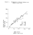
- Figure 1 is a graph of the correlation of the data obtained with the subject method versus a standard method (Abbot ⁇ -hCG 15/15).
- the present invention provides for a method whereby any excess labeled compound employed to identify a particular analyte can be readily removed to permit measurement of the analyte.
- the method is particularly adapted for use in automated systems where traditional purification or removal means are not appropriate, i.e., filtration, centrifugation or filtration columns. More preferably the subject method is employed with the diagnostic immunoassay apparatus TDxTM, a trademark of Abbott Laboratories for an automated system for the quantitation of therapeutic drug concentrations in serum or plasma based on the method of fluorescence polarization immunoassay.
- the method comprises the preparation of a reaction mixture of a biological fluid suspected of containing a known analyte, or protein with a labeled substance, antibody, capable of complexing with the particular analyte, protein or material sought to be quantified.
- the labeled substance is added to the reaction mixture in a molar excess with regard to the suspected amount of material to be quantified to assure complete complexing thereof.
- the complex After an appropriate incubation time, i.e., sufficient time to allow the labeling substance to complex with all the material sought to be quantified, the complex is measured. However, because the labeling substance is employed in a molar excess it is necessary to remove this excess prior to measuring the amount of complex formed.
- the subject method is characterized by removing the excess labeled substance by adding a solid phase material having immobilized thereon a compound capable of complexing with the labeled substance.
- the solid phase material is of such physical characteristics that it can be easily dispersed in the reaction mixture and settled out. Therefore the density and overall size of the solid phase material is such that a rapid disperse by agitation and sediment by gravity is facilitated.
- the solid phase material is present as a particle or matrix having a density greater than water.
- the solid phase material has a sedimentation rate in water of from about 5 seconds per centimeter to about 2 minutes per centimeter, more preferably from about 20 seconds per centimeter to about 1.5 minutes per centimeter.
- the size of the particle which forms the solid phase material can be from about 5 microns to about 300 microns, preferably from about 40 microns to about 120 microns in diameter.
- the sedimentation rate and size is important to assuring the solid phase material both disperses and settles readily without significant input of energy or motion to the reaction vessel containing the subject reaction mixture. This is especially important when employing automated diagnostic apparatus.
- the solid phase material can be fabricated from any number of material or synthetic materials.
- the solid phase material is manufactured from polymeric materials such as agarose, polystyrene, polyacrylamide, their derivatives or mixtures thereof.
- the solid phase is present as bead-like structures.
- the solid phase material can be delivered to the reaction vessel as a dry powder, wet slurry, tablet or a capsule.
- Immobilized on the solid phase material is a compound capable of complexing the labeled substance.
- this is a complimentary substance to the substance to be identified. Examples include an analyte, or its analog.
- the complex formed is irreversibly bonded to the solid phase material and because of its physical characteristics settles out or sediments by gravity. The total reaction mixture thus becomes separated into a solid phase containing the solid phase material complexed with any excess labeled substance and a liquid phase containing the material sought to be quantified which is complexed to the labeled substance.
- the liquid phase is then measured for amount of the material sought to be quantified, i.e., analyte or protein. Generally this is accomplished by extracting the liquid phase by syringe, suction, or other means. An appropriate immunoassay format is then employed to measure the amount of labeled substance consistent with the particular label employed.
- Typical labeling means can include enzymes, radioisotopes, chromophores, fluorophores or any substance which is capable of generating a detectable signal, either alone or in combination with other reagents. Procedures and methods for labeling and identifying the labeled complexes are well known in the art of diagnostic immunoassay as is generally discussed in L. Miles and C. Hales, Labeled Antibodies and Immunological Assay Systems , Nature 219, 187-189 (1968) and U.S. Patent 3,654,090.
- the subject method is especially useful in an automated diagnostic immunoassay apparatus because of its relatively automatic purification of the complex to be measured.
- the immunoassay method can be conducted by mixing the biological fluid to be analyzed with sufficient labeled substance and then adding this reaction mixture to a vessel containing the subject solid phase material or adding the subject solid phase material to the initial reaction mixture wherein the subject solid phase material purifies the reaction mixture of excess labeled substance without requiring additional physical or chemical treatment steps.
- the subject method avoids the necessity to centrifuge, prepare an elute from a column, or other more tedious steps to obtain the labeled substance for final measurement.
- a method for measurement of both small or large analytes in a sample fluid consisting of preincubation of excess enzyme-labeled antibody with sample such that sample is quantitatively and rapidly bound to form an analyte-enzyme-labeled antibody conjugate.
- An aliquot of this mixture is transferred to a vessel containing a solid phase material consisting of an analyte covalently coupled to a polymeric bead having a characteristic density to be easily suspended in solution yet sufficiently dense to rapidly sediment by gravity.
- the solid phase material rapidly captures the excess unconjugated enzyme-labeled antibody and sediments.
- an aliquot containing analyte-enzyme-labeled antibody is transferred to a cuvette for quantitation of enzyme using a fluorogenic substrate.
- the signal generated is proportional to the analyte concentration in the sample.
- Bovine serum albumin-digoxin was prepared as disclosed in PNAS 57:71 (1967). The product was dialyzed against a 0.1M Hepes buffer (7.5 pH) and concentrated to give a 5 mg/ml solution. An 8 ml aliquot of this solution was added to 15 ml of 300 micron agarose bead having an average diameter of 300 microns (purchased as Bio-Rad Affi-gel 15 from Bio-Rad Laboratories). The resulting agarose-bovine serum albumin-digoxin beads were washed with 4 molar guanidine-hydrochloric acid and phosphate buffered saline.
- Serum solutions containing 0, 1.0, 2.0, 3.0 and 5.0 nanograms per milliliter of digoxin were each tested following the same assay protocol as described below. A control was also run with zero digoxin and no solid phase material.
- To a 0.05 ml of serum sample was mixed 1 microliter of the enzyme labeled antibody as prepared (a), above. The reaction mixture was incubated at 34°C for ten minutes and then a 20 microliter aliquot was added to 100 microliter solution of the solid phase material (20:1 ratio) prepared from (b), above, with a 0.01 M phosphate buffer, pH 7.4. The mixture was incubated at 34° for 30 minutes with mixing.
- the mixture was allowed to briefly rest whereupon it separated into a liquid and solid phase.
- the ⁇ -galactosidase activity of the supernatant was measured by quantitating the production of fluorescein.
- the measurement was conducted with a 10 micromolar solution of di-( ⁇ -D-galactosyl) fluorescein as a substrate and an assay buffer containing 0.1 M sodium phosphate, bovine gamma globulin, and 0.1% sodium azide at a pH of 7.4.
- the measurements were as follows: The measurements obtained show that by employing the subject method a standard curve can be made to analyze serum solutions containing unknown digoxin concentrations. Also, the zero concentration measurement as compared to the control shows that the excess antibody conjugate was effectively removed by the subject method.
- hcg antibodies were immunopurified from rabbit antibodies (IgG), that were prepared by successive ammonium sulfate precipitations of anti-hcg rabbit sera, by using an hcg-immunoadsorbant.
- the hcg-immunoadsorbant was prepared by immobilizing hcg on Affi-Gel® 15 using conventional procedures that are described in the Bio-Rad literature available with Aff-Gel® 15.
- the IgG was loaded on the hcg-Affi-Gel® 15 column that had been previously washed with 4.0 M Guanidine HCl, 1.0 M acetic acid and tris-buffered saline (TBS). The column was then washed extensively with TBS and eluted with 1.0 M acetic acid. The purified hcG specific antibodies obtained in the elute were extensively dialyzed against 0.1 M sodium acetate pH 4.5.
- Fragments [F(ab ⁇ )2] of the hcg specific antibodies were prepared by digesting the said antibodies with pepsin and then isolating the F(ab ⁇ )2 fragments from the digestion mixture by column chromatography.
- F(ab ⁇ )2-galactosidase conjugates In order to prepare F(ab ⁇ )2-galactosidase conjugates the F(ab ⁇ )2 fragments were reacted with m-maleimidobenzoic acid N-hydroxysuccinimido ester (MBS). The resulting MBS derivitized fragments were purified by column chromatography and then subsequently reacted with E. coli ⁇ -galactosidase that had been previously purified by column chromatography. The F(ab ⁇ )2-galactosidase conjugates produced were isolated from unreacted galactosidase and unreacted F(ab ⁇ )2 by size exclusice chromatography.
- MBS m-maleimidobenzoic acid N-hydroxysuccinimido ester
- the hcg-solid phase reagent was prepared by coupling hcg to Tisacryl® GF-2000LS.
- Trisacryl is a porous, spherical, polyacrylamide solid phase matrix available from the LKB Corporation, Gaithersburg, Maryland. Prior to coupling with hcg the Trisacryl was activating by extensive washing with water, ethanol, acetone and dimethylformamide (DMF) and then reacting the Trisacryl with carbonyl diimidizole (CDI) in DMF. The activated Trisacryl was then coupled to hcg by the reaction of hcg with the said material in aqueous buffer (pH 10.2). Following the coupling unreacted hcg was removed from the Trisacryl by extensive washing with 1.0 M acetic acid and neutral pH buffers.
- DMF ethanol, acetone and dimethylformamide
- CDI carbonyl diimidizole
- Serum solutions containing known amounts of hcg were used to generate a standard curve according to the following protocol: 25 ⁇ l of F(ab ⁇ )2-galactosidase, synthesized in (A) above, was added to 100 ⁇ l of sample and 50 ⁇ l TDx buffer. The reaction mixture was incubated at 34 degrees C for 2 minutes 20 seconds and then 50 ⁇ l of the reaction mixture, along with 60 ⁇ l of TDx buffer, was added to a slurry that consisted of 50 ⁇ l of hcg-trisacryl, synthesized in (B) above, and 10 ⁇ l of TDx buffer. This mixture was incubated at 34 degrees C for 10 minutes with mixing.
- the mixture was allowed to briefly rest whereupon it separated into a liquid and solid phase.
- the galactosidase activity of the supernatant was measured by quantitating the production of fluorescein.
- the measurement was conducted with 10 micromolar solution of di( ⁇ -D-galactosyl) fluorescein as a substrate and an assay buffer containing 0.1 M sodium phosphate bovine gamma globulin, and 0.1% sodium azide at a pH of 7.4.
- Table 1 shows the sensitivity obtained with various lots of hcg-Trisacryl. The results indicate that the sensitivity obtained is not critically dependent on the amount of hcg coupled to the Trisacryl in the 454-50 IU/ml range.
Abstract
Description
- The present invention is directed toward a method for performing a diagnostic immunoassay for the measurement of small and large proteins and analytes in biological fluids by a solid-phase separation. This method is especially suitable for use in automated systems.
- Many diagnostic immunoassays are known which generally employ the specific binding characteristics that exist between an analyte or protein with a specific antibody tagged with some traceable substituent. One problem which has long been associated with this method is how to remove excess antibody from the biological fluid being tested for concentration of analyte in a manner whereby the analyte concentrations can be accurately measured.
- Various attempts to remove excess antibody include U.S. Patent 4,298,682 which discloses the absorption of unreacted antibody on a solid phase consisting of a polyacrylamide gel sensitized to the specific antibody.
- U.S. Patent 4,551,426 discloses another method to remove excess antibody in an immunoassay for digoxin. Here, excess labeled antobody is removed by passing it through an affinity column that has ouabain, an analog of digoxin, immobilized on the solid phase chromatography matrix. The excess antibody is absorbed by the oubain. The chromatography elute is then examined for the labeled antibody-analyte complex.
- While the above can be effective, they are all subject to improvement, especially with respect to ease in operation, handling and thereby accuracy. The subject method provides improvements over these methods and is especially adaptable to automated analysis which is not the case with many of the known methods.
- The present invention is directed toward a method for conducting a diagnostic immunoassay. The steps of the method comprise (a) forming a reaction mixture of a test sample with a molar excess of labeled antibody to form a complex of analyte present in the test sample, (b) contacting the reaction mixture with a solid phase material having immobilized thereon a compound in an amount sufficient to complex with any of the excess labeled antibody employed in step (a), (c) allowing the solid phase material and any complex of the solid phase material to settle and form a solid and liquid phase, and (d) measuring the amount of complex present in the liquid phase.
- Generally, the solid phase material is of sufficient density to rapidly sediment by gravity. Preferably the solid phase material has a sedimentation rate of about 5 seconds to about 2 minutes per centimeter in water and is from about 5 to about 300 microns in diameter. The solid phase material can be formed of any of a variety of materials, preferably, agarose, polystyrene, polyacrylamide, their derivatives or mixtures thereof.
- The solid phase material has immobilized thereon a compound capable of binding the excess labeled antibody such as a corresponding antigen or chemical analogue. Typically, the labeled antibody is an enzyme labeled antibody.
- The present method is particularly adapted for use in automated diagnostic apparatus where centrifugation filtration or column filtration is not possible or practical.
- Figure 1 is a graph of the correlation of the data obtained with the subject method versus a standard method (Abbot β-hCG 15/15).
- The present invention provides for a method whereby any excess labeled compound employed to identify a particular analyte can be readily removed to permit measurement of the analyte. The method, as outlined below, is particularly adapted for use in automated systems where traditional purification or removal means are not appropriate, i.e., filtration, centrifugation or filtration columns. More preferably the subject method is employed with the diagnostic immunoassay apparatus TDx™, a trademark of Abbott Laboratories for an automated system for the quantitation of therapeutic drug concentrations in serum or plasma based on the method of fluorescence polarization immunoassay.
- Generally, the method comprises the preparation of a reaction mixture of a biological fluid suspected of containing a known analyte, or protein with a labeled substance, antibody, capable of complexing with the particular analyte, protein or material sought to be quantified. The labeled substance is added to the reaction mixture in a molar excess with regard to the suspected amount of material to be quantified to assure complete complexing thereof.
- After an appropriate incubation time, i.e., sufficient time to allow the labeling substance to complex with all the material sought to be quantified, the complex is measured. However, because the labeling substance is employed in a molar excess it is necessary to remove this excess prior to measuring the amount of complex formed.
- The subject method is characterized by removing the excess labeled substance by adding a solid phase material having immobilized thereon a compound capable of complexing with the labeled substance. The solid phase material is of such physical characteristics that it can be easily dispersed in the reaction mixture and settled out. Therefore the density and overall size of the solid phase material is such that a rapid disperse by agitation and sediment by gravity is facilitated.
- The solid phase material is present as a particle or matrix having a density greater than water. The solid phase material has a sedimentation rate in water of from about 5 seconds per centimeter to about 2 minutes per centimeter, more preferably from about 20 seconds per centimeter to about 1.5 minutes per centimeter. The size of the particle which forms the solid phase material can be from about 5 microns to about 300 microns, preferably from about 40 microns to about 120 microns in diameter. The sedimentation rate and size is important to assuring the solid phase material both disperses and settles readily without significant input of energy or motion to the reaction vessel containing the subject reaction mixture. This is especially important when employing automated diagnostic apparatus.
- The solid phase material can be fabricated from any number of material or synthetic materials. Preferably the solid phase material is manufactured from polymeric materials such as agarose, polystyrene, polyacrylamide, their derivatives or mixtures thereof. Generally the solid phase is present as bead-like structures. The solid phase material can be delivered to the reaction vessel as a dry powder, wet slurry, tablet or a capsule.
- Immobilized on the solid phase material is a compound capable of complexing the labeled substance. Generally this is a complimentary substance to the substance to be identified. Examples include an analyte, or its analog. The complex formed is irreversibly bonded to the solid phase material and because of its physical characteristics settles out or sediments by gravity. The total reaction mixture thus becomes separated into a solid phase containing the solid phase material complexed with any excess labeled substance and a liquid phase containing the material sought to be quantified which is complexed to the labeled substance.
- The liquid phase is then measured for amount of the material sought to be quantified, i.e., analyte or protein. Generally this is accomplished by extracting the liquid phase by syringe, suction, or other means. An appropriate immunoassay format is then employed to measure the amount of labeled substance consistent with the particular label employed.
- Typical labeling means can include enzymes, radioisotopes, chromophores, fluorophores or any substance which is capable of generating a detectable signal, either alone or in combination with other reagents. Procedures and methods for labeling and identifying the labeled complexes are well known in the art of diagnostic immunoassay as is generally discussed in L. Miles and C. Hales, Labeled Antibodies and Immunological Assay Systems, Nature 219, 187-189 (1968) and U.S. Patent 3,654,090.
- The subject method is especially useful in an automated diagnostic immunoassay apparatus because of its relatively automatic purification of the complex to be measured. Particularly, the immunoassay method can be conducted by mixing the biological fluid to be analyzed with sufficient labeled substance and then adding this reaction mixture to a vessel containing the subject solid phase material or adding the subject solid phase material to the initial reaction mixture wherein the subject solid phase material purifies the reaction mixture of excess labeled substance without requiring additional physical or chemical treatment steps. Thus, the subject method avoids the necessity to centrifuge, prepare an elute from a column, or other more tedious steps to obtain the labeled substance for final measurement.
- A method for measurement of both small or large analytes in a sample fluid. The assay format consisting of preincubation of excess enzyme-labeled antibody with sample such that sample is quantitatively and rapidly bound to form an analyte-enzyme-labeled antibody conjugate. An aliquot of this mixture is transferred to a vessel containing a solid phase material consisting of an analyte covalently coupled to a polymeric bead having a characteristic density to be easily suspended in solution yet sufficiently dense to rapidly sediment by gravity. The solid phase material rapidly captures the excess unconjugated enzyme-labeled antibody and sediments. Following the capture reaction and sedimentation an aliquot containing analyte-enzyme-labeled antibody is transferred to a cuvette for quantitation of enzyme using a fluorogenic substrate. The signal generated is proportional to the analyte concentration in the sample.
- An enzyme labeled antibody of β-galactosidase conjugated to rabbit anti-digoxin F(abʹ)₂ was prepared. Rabbit antiserum against digoxin was fractionated by means of (NH₄)₂SO₄ and affinity chromatography on agarose-bovine serum albumin-oubain. The resulting digoxin specific antibody was reacted with pepsin to produce F(abʹ)₂ (antibody fragments). This was coupled to E-coli-β-galactosidase according to the procedure disclosed by Kitagawa and Aikawa, J. Biochem. 79:233 (1976). F(ab)₂-β-galactisidase was isolated by column chromatography.
- Bovine serum albumin-digoxin was prepared as disclosed in PNAS 57:71 (1967). The product was dialyzed against a 0.1M Hepes buffer (7.5 pH) and concentrated to give a 5 mg/ml solution. An 8 ml aliquot of this solution was added to 15 ml of 300 micron agarose bead having an average diameter of 300 microns (purchased as Bio-Rad Affi-gel 15 from Bio-Rad Laboratories). The resulting agarose-bovine serum albumin-digoxin beads were washed with 4 molar guanidine-hydrochloric acid and phosphate buffered saline.
- Serum solutions containing 0, 1.0, 2.0, 3.0 and 5.0 nanograms per milliliter of digoxin were each tested following the same assay protocol as described below. A control was also run with zero digoxin and no solid phase material. To a 0.05 ml of serum sample was mixed 1 microliter of the enzyme labeled antibody as prepared (a), above. The reaction mixture was incubated at 34°C for ten minutes and then a 20 microliter aliquot was added to 100 microliter solution of the solid phase material (20:1 ratio) prepared from (b), above, with a 0.01 M phosphate buffer, pH 7.4. The mixture was incubated at 34° for 30 minutes with mixing.
- The mixture was allowed to briefly rest whereupon it separated into a liquid and solid phase. The β-galactosidase activity of the supernatant was measured by quantitating the production of fluorescein. The measurement was conducted with a 10 micromolar solution of di-(β-D-galactosyl) fluorescein as a substrate and an assay buffer containing 0.1 M sodium phosphate, bovine gamma globulin, and 0.1% sodium azide at a pH of 7.4.
- The measurements were as follows:

- Specific human chorionic gonadotropin (hcg) antibodies were immunopurified from rabbit antibodies (IgG), that were prepared by successive ammonium sulfate precipitations of anti-hcg rabbit sera, by using an hcg-immunoadsorbant. The hcg-immunoadsorbant was prepared by immobilizing hcg on Affi-Gel® 15 using conventional procedures that are described in the Bio-Rad literature available with Aff-Gel® 15.
- For immunopurification the IgG was loaded on the hcg-Affi-Gel® 15 column that had been previously washed with 4.0 M Guanidine HCl, 1.0 M acetic acid and tris-buffered saline (TBS). The column was then washed extensively with TBS and eluted with 1.0 M acetic acid. The purified hcG specific antibodies obtained in the elute were extensively dialyzed against 0.1 M sodium acetate pH 4.5.
- Fragments [F(abʹ)₂] of the hcg specific antibodies were prepared by digesting the said antibodies with pepsin and then isolating the F(abʹ)₂ fragments from the digestion mixture by column chromatography.
- In order to prepare F(abʹ)₂-galactosidase conjugates the F(abʹ)₂ fragments were reacted with m-maleimidobenzoic acid N-hydroxysuccinimido ester (MBS). The resulting MBS derivitized fragments were purified by column chromatography and then subsequently reacted with E. coli β-galactosidase that had been previously purified by column chromatography. The F(abʹ)₂-galactosidase conjugates produced were isolated from unreacted galactosidase and unreacted F(abʹ)₂ by size exclusice chromatography.
- The hcg-solid phase reagent was prepared by coupling hcg to Tisacryl® GF-2000LS.
- Trisacryl is a porous, spherical, polyacrylamide solid phase matrix available from the LKB Corporation, Gaithersburg, Maryland. Prior to coupling with hcg the Trisacryl was activating by extensive washing with water, ethanol, acetone and dimethylformamide (DMF) and then reacting the Trisacryl with carbonyl diimidizole (CDI) in DMF. The activated Trisacryl was then coupled to hcg by the reaction of hcg with the said material in aqueous buffer (pH 10.2). Following the coupling unreacted hcg was removed from the Trisacryl by extensive washing with 1.0 M acetic acid and neutral pH buffers.
- Serum solutions containing known amounts of hcg were used to generate a standard curve according to the following protocol: 25 µl of F(abʹ)₂-galactosidase, synthesized in (A) above, was added to 100 µl of sample and 50 µl TDx buffer. The reaction mixture was incubated at 34 degrees C for 2 minutes 20 seconds and then 50 µl of the reaction mixture, along with 60 µl of TDx buffer, was added to a slurry that consisted of 50 µl of hcg-trisacryl, synthesized in (B) above, and 10 µl of TDx buffer. This mixture was incubated at 34 degrees C for 10 minutes with mixing. The mixture was allowed to briefly rest whereupon it separated into a liquid and solid phase. The galactosidase activity of the supernatant was measured by quantitating the production of fluorescein. The measurement was conducted with 10 micromolar solution of di(β-D-galactosyl) fluorescein as a substrate and an assay buffer containing 0.1 M sodium phosphate bovine gamma globulin, and 0.1% sodium azide at a pH of 7.4.
- Table 1 shows the sensitivity obtained with various lots of hcg-Trisacryl. The results indicate that the sensitivity obtained is not critically dependent on the amount of hcg coupled to the Trisacryl in the 454-50 IU/ml range.
- Figure 1 shows the results for fifty clinical samples (N=50) that were measured for hcg content using a reference method (Abbott β-HCG 15/15) and the subject method. A correlation coefficient of R=0.92 and a slope of 0.86 were obtained from analysis of the date shown in Figure 1. These results indicate that the subject method is accurately measuring serum levels of hcg.

Claims (8)
Priority Applications (1)
| Application Number | Priority Date | Filing Date | Title |
|---|---|---|---|
| AT87109740T ATE80948T1 (en) | 1986-07-14 | 1987-07-07 | SOLID PHASE SEPARATION DIAGNOSTIC IMMUNOTEST PROCEDURE. |
Applications Claiming Priority (2)
| Application Number | Priority Date | Filing Date | Title |
|---|---|---|---|
| US88513086A | 1986-07-14 | 1986-07-14 | |
| US885130 | 1986-07-14 |
Publications (3)
| Publication Number | Publication Date |
|---|---|
| EP0253270A2 true EP0253270A2 (en) | 1988-01-20 |
| EP0253270A3 EP0253270A3 (en) | 1989-04-26 |
| EP0253270B1 EP0253270B1 (en) | 1992-09-23 |
Family
ID=25386214
Family Applications (1)
| Application Number | Title | Priority Date | Filing Date |
|---|---|---|---|
| EP87109740A Expired - Lifetime EP0253270B1 (en) | 1986-07-14 | 1987-07-07 | Method for diagnostic immunoassay by solid phase separation |
Country Status (7)
| Country | Link |
|---|---|
| EP (1) | EP0253270B1 (en) |
| JP (1) | JPS6329248A (en) |
| AT (1) | ATE80948T1 (en) |
| AU (1) | AU595899B2 (en) |
| CA (1) | CA1301646C (en) |
| DE (1) | DE3781841T2 (en) |
| ES (1) | ES2036189T3 (en) |
Cited By (5)
| Publication number | Priority date | Publication date | Assignee | Title |
|---|---|---|---|---|
| EP0285995A2 (en) * | 1987-04-07 | 1988-10-12 | Abbott Laboratories | Diagnostic immunoassay by solid phase separation for digoxin |
| EP0345897A2 (en) * | 1988-06-06 | 1989-12-13 | ORION CORPORATION, Orion | Novel heterogeneous immunoassay |
| EP0411944A2 (en) * | 1989-08-04 | 1991-02-06 | BEHRINGWERKE Aktiengesellschaft | Heterogeneous binding assays |
| US5003054A (en) * | 1987-04-07 | 1991-03-26 | Abbott Laboratories | Ouabain triacetate derivative compounds |
| US20100184076A1 (en) * | 2003-09-02 | 2010-07-22 | Restalyst Pte Ltd | Soluble analyte detection and amplification |
Citations (7)
| Publication number | Priority date | Publication date | Assignee | Title |
|---|---|---|---|---|
| US3654090A (en) * | 1968-09-24 | 1972-04-04 | Organon | Method for the determination of antigens and antibodies |
| US3853987A (en) * | 1971-09-01 | 1974-12-10 | W Dreyer | Immunological reagent and radioimmuno assay |
| US4298687A (en) * | 1978-10-31 | 1981-11-03 | Roland Maes | Process for the determination of compounds showing among themselves specific binding affinities by the use of a solid phase |
| EP0095932A1 (en) * | 1982-05-28 | 1983-12-07 | Japan Synthetic Rubber Co., Ltd. | The use of a particulate polymer as a carrier for biological substances and the like and such substances supported on the carrier |
| EP0143274A1 (en) * | 1983-10-03 | 1985-06-05 | E.I. Du Pont De Nemours And Company | Heterogeneous immunoassay for digoxin using ouabain as a separation means |
| EP0158443A2 (en) * | 1984-03-13 | 1985-10-16 | Sekisui Kagaku Kogyo Kabushiki Kaisha | A latex for immunoserological tests and a method of producing the same |
| EP0285995A2 (en) * | 1987-04-07 | 1988-10-12 | Abbott Laboratories | Diagnostic immunoassay by solid phase separation for digoxin |
Family Cites Families (3)
| Publication number | Priority date | Publication date | Assignee | Title |
|---|---|---|---|---|
| DE3263249D1 (en) * | 1981-08-21 | 1985-05-30 | Hoffmann La Roche | Method for the determination of carcinoembryonic antigen (cea) and suitable antibody solution for the determination |
| US4794076A (en) * | 1985-09-23 | 1988-12-27 | Vxr, Inc. | Simultaneous extraction of a ligand from a sample and capture by anti-ligands therefor in ligand/anti-ligand assays |
| US4849341A (en) * | 1986-12-02 | 1989-07-18 | Proscience Corporation | Diagnostic test for Staphylococcal mastitis in cattle |
-
1987
- 1987-07-07 EP EP87109740A patent/EP0253270B1/en not_active Expired - Lifetime
- 1987-07-07 ES ES198787109740T patent/ES2036189T3/en not_active Expired - Lifetime
- 1987-07-07 AT AT87109740T patent/ATE80948T1/en not_active IP Right Cessation
- 1987-07-07 DE DE8787109740T patent/DE3781841T2/en not_active Expired - Fee Related
- 1987-07-08 AU AU75342/87A patent/AU595899B2/en not_active Ceased
- 1987-07-13 CA CA000541923A patent/CA1301646C/en not_active Expired - Fee Related
- 1987-07-14 JP JP62174026A patent/JPS6329248A/en active Pending
Patent Citations (8)
| Publication number | Priority date | Publication date | Assignee | Title |
|---|---|---|---|---|
| US3654090A (en) * | 1968-09-24 | 1972-04-04 | Organon | Method for the determination of antigens and antibodies |
| US3654090B1 (en) * | 1968-09-24 | 1982-07-20 | ||
| US3853987A (en) * | 1971-09-01 | 1974-12-10 | W Dreyer | Immunological reagent and radioimmuno assay |
| US4298687A (en) * | 1978-10-31 | 1981-11-03 | Roland Maes | Process for the determination of compounds showing among themselves specific binding affinities by the use of a solid phase |
| EP0095932A1 (en) * | 1982-05-28 | 1983-12-07 | Japan Synthetic Rubber Co., Ltd. | The use of a particulate polymer as a carrier for biological substances and the like and such substances supported on the carrier |
| EP0143274A1 (en) * | 1983-10-03 | 1985-06-05 | E.I. Du Pont De Nemours And Company | Heterogeneous immunoassay for digoxin using ouabain as a separation means |
| EP0158443A2 (en) * | 1984-03-13 | 1985-10-16 | Sekisui Kagaku Kogyo Kabushiki Kaisha | A latex for immunoserological tests and a method of producing the same |
| EP0285995A2 (en) * | 1987-04-07 | 1988-10-12 | Abbott Laboratories | Diagnostic immunoassay by solid phase separation for digoxin |
Non-Patent Citations (2)
| Title |
|---|
| MARCEL DEKKER INCORP. * |
| MARCEL DEKKER INCORP., 1984, Marcel Dekker, New York, NY (US); M.ZOUHAIR ATASSI et al, pp. 361-362< * |
Cited By (10)
| Publication number | Priority date | Publication date | Assignee | Title |
|---|---|---|---|---|
| EP0285995A2 (en) * | 1987-04-07 | 1988-10-12 | Abbott Laboratories | Diagnostic immunoassay by solid phase separation for digoxin |
| EP0285995A3 (en) * | 1987-04-07 | 1989-05-24 | Abbott Laboratories | Diagnostic immunoassay by solid phase separation for digoxin |
| US5003054A (en) * | 1987-04-07 | 1991-03-26 | Abbott Laboratories | Ouabain triacetate derivative compounds |
| EP0345897A2 (en) * | 1988-06-06 | 1989-12-13 | ORION CORPORATION, Orion | Novel heterogeneous immunoassay |
| EP0345897A3 (en) * | 1988-06-06 | 1991-05-15 | ORION CORPORATION, Orion | Novel heterogeneous immunoassay |
| EP0411944A2 (en) * | 1989-08-04 | 1991-02-06 | BEHRINGWERKE Aktiengesellschaft | Heterogeneous binding assays |
| EP0411944A3 (en) * | 1989-08-04 | 1991-10-30 | Syntex (U.S.A.) Inc. | Heterogeneous binding assays |
| US5437983A (en) * | 1989-08-04 | 1995-08-01 | Syntex (U.S.A.) Inc. | Heterogeneous binding assays |
| US20100184076A1 (en) * | 2003-09-02 | 2010-07-22 | Restalyst Pte Ltd | Soluble analyte detection and amplification |
| US9181579B2 (en) * | 2003-09-02 | 2015-11-10 | Restalyst Pte Ltd | Soluble analyte detection and amplification |
Also Published As
| Publication number | Publication date |
|---|---|
| DE3781841T2 (en) | 1993-04-15 |
| JPS6329248A (en) | 1988-02-06 |
| AU595899B2 (en) | 1990-04-12 |
| DE3781841D1 (en) | 1992-10-29 |
| EP0253270A3 (en) | 1989-04-26 |
| AU7534287A (en) | 1988-01-21 |
| ATE80948T1 (en) | 1992-10-15 |
| CA1301646C (en) | 1992-05-26 |
| EP0253270B1 (en) | 1992-09-23 |
| ES2036189T3 (en) | 1993-05-16 |
Similar Documents
| Publication | Publication Date | Title |
|---|---|---|
| US4434236A (en) | Immunoassay wherein labeled antibody is displaced from immobilized analyte-analogue | |
| US4298685A (en) | Diagnostic reagent | |
| US4273867A (en) | Method and reagent for counteracting lipemic interference | |
| US4143124A (en) | Antigen-antibody analysis with solid phase rf and c1q | |
| US5432099A (en) | Determination of ambient concentation of several analytes | |
| EP1075661B1 (en) | Ligand binding assay and kit with a separation zone for disturbing sample components | |
| US5405752A (en) | Enzyme conjugate prepared with insoluble nonoparticle | |
| JPS6343711B2 (en) | ||
| CA2117597A1 (en) | Method for non-competitive binding assays | |
| JPH01227061A (en) | Ion trapping immunoassay method and apparatus | |
| US4839299A (en) | Assay for the free portion of substances in biological fluids | |
| EP0073593A1 (en) | Size-exclusion heterogeneous immunoassay | |
| US4283383A (en) | Analysis of biological fluids | |
| US4138213A (en) | Agglutination immunoassay of immune complex with RF or Clq | |
| JP3194585B2 (en) | Two-site immunoassay for antibodies with chemiluminescent label and biotin-binding ligand | |
| US4788136A (en) | Diagnostic immunoassay by solid phase separation for digoxin | |
| US4447528A (en) | Detecting intrinsic factor blocking site antibody | |
| EP0253270B1 (en) | Method for diagnostic immunoassay by solid phase separation | |
| US5229268A (en) | Method for diagnostic immunoassay by solid phase separation | |
| Wide | [13] Use of particulate immunosorbents in radioimmunoassay | |
| JP2000229926A (en) | Homobifunctional reagent binding enzyme or the like to antibody or the like | |
| JP2994739B2 (en) | Rapid measurement of trace components | |
| EP0184701B1 (en) | A method for determining a ligand | |
| JP2807831B2 (en) | Immunoassay | |
| US5003054A (en) | Ouabain triacetate derivative compounds |
Legal Events
| Date | Code | Title | Description |
|---|---|---|---|
| PUAI | Public reference made under article 153(3) epc to a published international application that has entered the european phase |
Free format text: ORIGINAL CODE: 0009012 |
|
| AK | Designated contracting states |
Kind code of ref document: A2 Designated state(s): AT BE CH DE ES FR GB IT LI NL |
|
| PUAL | Search report despatched |
Free format text: ORIGINAL CODE: 0009013 |
|
| AK | Designated contracting states |
Kind code of ref document: A3 Designated state(s): AT BE CH DE ES FR GB IT LI NL |
|
| 17P | Request for examination filed |
Effective date: 19890929 |
|
| 17Q | First examination report despatched |
Effective date: 19910321 |
|
| ITTA | It: last paid annual fee | ||
| GRAA | (expected) grant |
Free format text: ORIGINAL CODE: 0009210 |
|
| AK | Designated contracting states |
Kind code of ref document: B1 Designated state(s): AT BE CH DE ES FR GB IT LI NL |
|
| REF | Corresponds to: |
Ref document number: 80948 Country of ref document: AT Date of ref document: 19921015 Kind code of ref document: T |
|
| REF | Corresponds to: |
Ref document number: 3781841 Country of ref document: DE Date of ref document: 19921029 |
|
| ET | Fr: translation filed | ||
| ITF | It: translation for a ep patent filed |
Owner name: MODIANO & ASSOCIATI S.R |
|
| REG | Reference to a national code |
Ref country code: ES Ref legal event code: FG2A Ref document number: 2036189 Country of ref document: ES Kind code of ref document: T3 |
|
| PGFP | Annual fee paid to national office [announced via postgrant information from national office to epo] |
Ref country code: AT Payment date: 19930614 Year of fee payment: 7 |
|
| PGFP | Annual fee paid to national office [announced via postgrant information from national office to epo] |
Ref country code: ES Payment date: 19930709 Year of fee payment: 7 Ref country code: BE Payment date: 19930709 Year of fee payment: 7 |
|
| PLBE | No opposition filed within time limit |
Free format text: ORIGINAL CODE: 0009261 |
|
| STAA | Information on the status of an ep patent application or granted ep patent |
Free format text: STATUS: NO OPPOSITION FILED WITHIN TIME LIMIT |
|
| PGFP | Annual fee paid to national office [announced via postgrant information from national office to epo] |
Ref country code: NL Payment date: 19930731 Year of fee payment: 7 |
|
| 26N | No opposition filed | ||
| PG25 | Lapsed in a contracting state [announced via postgrant information from national office to epo] |
Ref country code: AT Effective date: 19940707 |
|
| PG25 | Lapsed in a contracting state [announced via postgrant information from national office to epo] |
Ref country code: ES Free format text: LAPSE BECAUSE OF NON-PAYMENT OF DUE FEES Effective date: 19940708 |
|
| PG25 | Lapsed in a contracting state [announced via postgrant information from national office to epo] |
Ref country code: BE Effective date: 19940731 |
|
| BERE | Be: lapsed |
Owner name: ABBOTT LABORATORIES Effective date: 19940731 |
|
| PG25 | Lapsed in a contracting state [announced via postgrant information from national office to epo] |
Ref country code: NL Effective date: 19950201 |
|
| NLV4 | Nl: lapsed or anulled due to non-payment of the annual fee | ||
| PGFP | Annual fee paid to national office [announced via postgrant information from national office to epo] |
Ref country code: GB Payment date: 19960617 Year of fee payment: 10 |
|
| PGFP | Annual fee paid to national office [announced via postgrant information from national office to epo] |
Ref country code: FR Payment date: 19960715 Year of fee payment: 10 |
|
| PGFP | Annual fee paid to national office [announced via postgrant information from national office to epo] |
Ref country code: DE Payment date: 19960730 Year of fee payment: 10 |
|
| PGFP | Annual fee paid to national office [announced via postgrant information from national office to epo] |
Ref country code: CH Payment date: 19961004 Year of fee payment: 10 |
|
| PG25 | Lapsed in a contracting state [announced via postgrant information from national office to epo] |
Ref country code: GB Free format text: LAPSE BECAUSE OF NON-PAYMENT OF DUE FEES Effective date: 19970707 |
|
| PG25 | Lapsed in a contracting state [announced via postgrant information from national office to epo] |
Ref country code: LI Free format text: LAPSE BECAUSE OF NON-PAYMENT OF DUE FEES Effective date: 19970731 Ref country code: CH Free format text: LAPSE BECAUSE OF NON-PAYMENT OF DUE FEES Effective date: 19970731 |
|
| GBPC | Gb: european patent ceased through non-payment of renewal fee |
Effective date: 19970707 |
|
| REG | Reference to a national code |
Ref country code: CH Ref legal event code: PL |
|
| PG25 | Lapsed in a contracting state [announced via postgrant information from national office to epo] |
Ref country code: FR Free format text: LAPSE BECAUSE OF NON-PAYMENT OF DUE FEES Effective date: 19980331 |
|
| PG25 | Lapsed in a contracting state [announced via postgrant information from national office to epo] |
Ref country code: DE Free format text: LAPSE BECAUSE OF NON-PAYMENT OF DUE FEES Effective date: 19980401 |
|
| REG | Reference to a national code |
Ref country code: FR Ref legal event code: ST |
|
| REG | Reference to a national code |
Ref country code: ES Ref legal event code: FD2A Effective date: 19950810 |
|
| PG25 | Lapsed in a contracting state [announced via postgrant information from national office to epo] |
Ref country code: IT Free format text: LAPSE BECAUSE OF NON-PAYMENT OF DUE FEES;WARNING: LAPSES OF ITALIAN PATENTS WITH EFFECTIVE DATE BEFORE 2007 MAY HAVE OCCURRED AT ANY TIME BEFORE 2007. THE CORRECT EFFECTIVE DATE MAY BE DIFFERENT FROM THE ONE RECORDED. Effective date: 20050707 |