EP0264150A1 - Microorganisms comprising pili as carrier proteins, isolated pili, method for excreting proteins using these pili and their use - Google Patents
Microorganisms comprising pili as carrier proteins, isolated pili, method for excreting proteins using these pili and their use Download PDFInfo
- Publication number
- EP0264150A1 EP0264150A1 EP87201873A EP87201873A EP0264150A1 EP 0264150 A1 EP0264150 A1 EP 0264150A1 EP 87201873 A EP87201873 A EP 87201873A EP 87201873 A EP87201873 A EP 87201873A EP 0264150 A1 EP0264150 A1 EP 0264150A1
- Authority
- EP
- European Patent Office
- Prior art keywords
- pili
- subunit
- pilin
- pilins
- gene
- Prior art date
- Legal status (The legal status is an assumption and is not a legal conclusion. Google has not performed a legal analysis and makes no representation as to the accuracy of the status listed.)
- Granted
Links
- 0 C*CC=CCNC Chemical compound C*CC=CCNC 0.000 description 1
Images


Classifications
-
- C—CHEMISTRY; METALLURGY
- C07—ORGANIC CHEMISTRY
- C07K—PEPTIDES
- C07K14/00—Peptides having more than 20 amino acids; Gastrins; Somatostatins; Melanotropins; Derivatives thereof
- C07K14/005—Peptides having more than 20 amino acids; Gastrins; Somatostatins; Melanotropins; Derivatives thereof from viruses
-
- A—HUMAN NECESSITIES
- A61—MEDICAL OR VETERINARY SCIENCE; HYGIENE
- A61K—PREPARATIONS FOR MEDICAL, DENTAL OR TOILETRY PURPOSES
- A61K39/00—Medicinal preparations containing antigens or antibodies
- A61K39/12—Viral antigens
-
- A—HUMAN NECESSITIES
- A61—MEDICAL OR VETERINARY SCIENCE; HYGIENE
- A61K—PREPARATIONS FOR MEDICAL, DENTAL OR TOILETRY PURPOSES
- A61K39/00—Medicinal preparations containing antigens or antibodies
- A61K39/12—Viral antigens
- A61K39/145—Orthomyxoviridae, e.g. influenza virus
-
- A—HUMAN NECESSITIES
- A61—MEDICAL OR VETERINARY SCIENCE; HYGIENE
- A61K—PREPARATIONS FOR MEDICAL, DENTAL OR TOILETRY PURPOSES
- A61K39/00—Medicinal preparations containing antigens or antibodies
- A61K39/385—Haptens or antigens, bound to carriers
-
- C—CHEMISTRY; METALLURGY
- C07—ORGANIC CHEMISTRY
- C07K—PEPTIDES
- C07K14/00—Peptides having more than 20 amino acids; Gastrins; Somatostatins; Melanotropins; Derivatives thereof
- C07K14/195—Peptides having more than 20 amino acids; Gastrins; Somatostatins; Melanotropins; Derivatives thereof from bacteria
-
- C—CHEMISTRY; METALLURGY
- C07—ORGANIC CHEMISTRY
- C07K—PEPTIDES
- C07K14/00—Peptides having more than 20 amino acids; Gastrins; Somatostatins; Melanotropins; Derivatives thereof
- C07K14/435—Peptides having more than 20 amino acids; Gastrins; Somatostatins; Melanotropins; Derivatives thereof from animals; from humans
- C07K14/44—Peptides having more than 20 amino acids; Gastrins; Somatostatins; Melanotropins; Derivatives thereof from animals; from humans from protozoa
-
- C—CHEMISTRY; METALLURGY
- C07—ORGANIC CHEMISTRY
- C07K—PEPTIDES
- C07K14/00—Peptides having more than 20 amino acids; Gastrins; Somatostatins; Melanotropins; Derivatives thereof
- C07K14/435—Peptides having more than 20 amino acids; Gastrins; Somatostatins; Melanotropins; Derivatives thereof from animals; from humans
- C07K14/52—Cytokines; Lymphokines; Interferons
- C07K14/555—Interferons [IFN]
-
- C—CHEMISTRY; METALLURGY
- C07—ORGANIC CHEMISTRY
- C07K—PEPTIDES
- C07K14/00—Peptides having more than 20 amino acids; Gastrins; Somatostatins; Melanotropins; Derivatives thereof
- C07K14/435—Peptides having more than 20 amino acids; Gastrins; Somatostatins; Melanotropins; Derivatives thereof from animals; from humans
- C07K14/575—Hormones
- C07K14/60—Growth-hormone releasing factors (GH-RF) (Somatoliberin)
-
- G—PHYSICS
- G01—MEASURING; TESTING
- G01N—INVESTIGATING OR ANALYSING MATERIALS BY DETERMINING THEIR CHEMICAL OR PHYSICAL PROPERTIES
- G01N33/00—Investigating or analysing materials by specific methods not covered by groups G01N1/00 - G01N31/00
- G01N33/48—Biological material, e.g. blood, urine; Haemocytometers
- G01N33/50—Chemical analysis of biological material, e.g. blood, urine; Testing involving biospecific ligand binding methods; Immunological testing
- G01N33/53—Immunoassay; Biospecific binding assay; Materials therefor
- G01N33/569—Immunoassay; Biospecific binding assay; Materials therefor for microorganisms, e.g. protozoa, bacteria, viruses
- G01N33/56911—Bacteria
-
- A—HUMAN NECESSITIES
- A61—MEDICAL OR VETERINARY SCIENCE; HYGIENE
- A61K—PREPARATIONS FOR MEDICAL, DENTAL OR TOILETRY PURPOSES
- A61K39/00—Medicinal preparations containing antigens or antibodies
- A61K2039/51—Medicinal preparations containing antigens or antibodies comprising whole cells, viruses or DNA/RNA
- A61K2039/52—Bacterial cells; Fungal cells; Protozoal cells
- A61K2039/523—Bacterial cells; Fungal cells; Protozoal cells expressing foreign proteins
-
- A—HUMAN NECESSITIES
- A61—MEDICAL OR VETERINARY SCIENCE; HYGIENE
- A61K—PREPARATIONS FOR MEDICAL, DENTAL OR TOILETRY PURPOSES
- A61K39/00—Medicinal preparations containing antigens or antibodies
- A61K2039/555—Medicinal preparations containing antigens or antibodies characterised by a specific combination antigen/adjuvant
- A61K2039/55511—Organic adjuvants
- A61K2039/55566—Emulsions, e.g. Freund's adjuvant, MF59
-
- A—HUMAN NECESSITIES
- A61—MEDICAL OR VETERINARY SCIENCE; HYGIENE
- A61K—PREPARATIONS FOR MEDICAL, DENTAL OR TOILETRY PURPOSES
- A61K39/00—Medicinal preparations containing antigens or antibodies
- A61K2039/60—Medicinal preparations containing antigens or antibodies characteristics by the carrier linked to the antigen
- A61K2039/6031—Proteins
- A61K2039/6068—Other bacterial proteins, e.g. OMP
-
- C—CHEMISTRY; METALLURGY
- C12—BIOCHEMISTRY; BEER; SPIRITS; WINE; VINEGAR; MICROBIOLOGY; ENZYMOLOGY; MUTATION OR GENETIC ENGINEERING
- C12N—MICROORGANISMS OR ENZYMES; COMPOSITIONS THEREOF; PROPAGATING, PRESERVING, OR MAINTAINING MICROORGANISMS; MUTATION OR GENETIC ENGINEERING; CULTURE MEDIA
- C12N2760/00—MICROORGANISMS OR ENZYMES; COMPOSITIONS THEREOF; PROPAGATING, PRESERVING, OR MAINTAINING MICROORGANISMS; MUTATION OR GENETIC ENGINEERING; CULTURE MEDIA ssRNA viruses negative-sense
- C12N2760/00011—Details
- C12N2760/16011—Orthomyxoviridae
- C12N2760/16022—New viral proteins or individual genes, new structural or functional aspects of known viral proteins or genes
-
- C—CHEMISTRY; METALLURGY
- C12—BIOCHEMISTRY; BEER; SPIRITS; WINE; VINEGAR; MICROBIOLOGY; ENZYMOLOGY; MUTATION OR GENETIC ENGINEERING
- C12N—MICROORGANISMS OR ENZYMES; COMPOSITIONS THEREOF; PROPAGATING, PRESERVING, OR MAINTAINING MICROORGANISMS; MUTATION OR GENETIC ENGINEERING; CULTURE MEDIA
- C12N2760/00—MICROORGANISMS OR ENZYMES; COMPOSITIONS THEREOF; PROPAGATING, PRESERVING, OR MAINTAINING MICROORGANISMS; MUTATION OR GENETIC ENGINEERING; CULTURE MEDIA ssRNA viruses negative-sense
- C12N2760/00011—Details
- C12N2760/16011—Orthomyxoviridae
- C12N2760/16111—Influenzavirus A, i.e. influenza A virus
- C12N2760/16134—Use of virus or viral component as vaccine, e.g. live-attenuated or inactivated virus, VLP, viral protein
Definitions
- the present invention relates to microorganisms comprising pili as proteins carrying heterologous peptide material as well as pili isolated from such microorganisms.
- the invention also relates to a process for the excretion of peptides or proteins using the own mechanisms of microorganisms for the production of pili.
- the invention also relates to the use of microorganisms and pili.
- the present invention aims to resolve this deficiency and provides the means to obtain in practice such microorganisms and pili.
- the invention relates to microorganisms whose outer membrane carries pili, the composition of which has been modified by at least one change in the protein sequence of the subunit.
- composition of the pili has been modified by at least two changes in the protein sequence.
- microorganisms concerned by the invention are not critical in itself and depends solely on the possibility that certain microorganisms have of producing pili as defined above. Usually, we operate with bacteria as microorganisms.
- bacteria of the families Enterobacteriaceae, Streptococcacae, Neisseriaceae, Proteeae, Phizobiaceae, Pseudomonodaceae, Corynebacteriaceae, Mycoplasmataceae, Myxococcaceae, Actinomycetaceae, Bacteriodaceae and, preferably, bacteria are used.
- the invention also relates to pili isolated from these microorganisms by any physical, chemical or biological means. It also relates to the subunits or pilins which, by any means whatsoever, have been released and separated from the repeating polymer structure constituting the pili.
- pili also means terms such as fibrils and, fimbriae. These are polymers excreted by microorganisms and attached to the outer membrane of these microorganisms, consisting of the repetition of proteins called pilins or even subunits.
- the subunits or pilins are proteins which generally have a molecular weight of between 5 and 50 kD and which, when combined in the form of pili, form extracellular filaments from 20 to 250 ⁇ in diameter and up to several microns long.
- the pili described in the invention can be of all types such as somatic pili.
- the somatic pili which are used for the adhesion of the microorganisms to a support are used and good results have been obtained with the K88 pili from Escherichia coli which allow this bacterium to colonize the intestine of certain mammals.
- the pili obtained according to the invention have a composition different from the composition of the pili of the microorganism in the wild after at least one modification made to the protein sequence of the subunit or pilin, preferably following at least two modifications.
- Modifications of the subunit preferably relate to regions not critical to the assembly and excretion of the subunit.
- the protein obtained after modifications can be used in various applications.
- it may in particular be suitable as an enzyme, as a nutritive constituent or also in the field of animal or human health, in particular as pharmaceutical products for veterinary or human use.
- the protein obtained can be administered in any pharmaceutically acceptable form such as in particular the isolated protein, the protein linked to the pilus isolated or not from the microorganism; the microorganism itself can therefore be administered.
- Pili are highly antigenic molecules. If the peptide introduced is an epitope foreign to the pilus, the protein obtained according to the invention may have a new antigenic character and be used in immunological tests such as for diagnostics. It can also have an immunogenic character and be used as a component of a vaccine.
- the invention also makes it possible to render immunogenic peptides, which are little or not by themselves, after their introduction into the protein sequence of pilin.
- the modification of the protein sequence of the subunit or pilin can be carried out by any known means.
- One way is to modify the DNA sequence encoding the pilus subunit. These modifications are made by genetic engineering. The recombinant gene is then translated into proteins (heterologous) in a host organism.
- modification is meant any modification of wild-type DNA, leading to a modification of the protein sequence of the subunit.
- the foreign DNA can be of natural or synthetic origin. Its nucleotide sequence and its reading phase in the gene determine the native amino acid sequence incorporated in the subunit.
- the invention therefore also relates to any genetic material allowing the use of pili as carrier proteins.
- the invention further relates to the methods for making pili as carrier proteins by incorporating heterologous DNA as well as to the methods for obtaining microorganisms carrying such pili. More particularly, the invention relates to any process in which the recombinant gene makes it possible to obtain pili whose polymer structure is conserved and possibly containing the modified subunits resulting from the incorporation of heterologous DNA.
- the invention therefore relates to methods which make it possible to obtain pili the composition of which has been modified by at least one change in the protein sequence of the subunit, this or these changes resulting from the introduction into the gene coding for the subunit of a pilus, of at least one DNA fragment coding for a piptide or a heterologous protein such as a synthetic DNA fragment, a natural DNA fragment or a fragment obtained from transcription reverse of mRNA.
- heterologous DNA is meant any genetic material from a source other than the host strain in which the process of the invention is carried out.
- synthetic DNA is meant both a DNA of natural composition or structure obtained by chemical synthesis as well as DNA whose structure or composition does not exist as it is in nature.
- these changes result from the introduction into the gene coding for the pilus subunit, of at least two DNA fragments coding for a heterologous peptide or protein, these may be identical or different. .
- One of the methods is the modification of at least one region of the protein of the pilus subunit exposed to the surrounding medium, by insertion of DNA coding for a heterologous peptide or protein, into at least one region of the coding gene. for the pilus subunit corresponding to the area exposed to the surrounding medium of the protein sequence of the pilins concerned.
- the exposed regions include in particular antigenic regions of the pili, that is to say composed of epitopes inducing the production of antibodies or recognized by antibodies.
- the invention also relates to the uses of pili and pilins carrying heterologous material.
- the invention relates in particular to vaccines obtained with pili and pilins carrying heterologous material, combined or not with various additives such as adjuvants and immunostimulants.
- the invention also relates to the use of pili, pilins and microorganisms having an external membrane which carries pili, the composition of which is modified by at least one change, or by at least two changes, in the protein sequence of pilins; in particular the invention relates to the uses of these for obtaining a pharmaceutical product intended for medical and / or prophylactic treatment in human or animal health.
- the invention also relates to immunological tests obtained from pili and pilins carrying heterologous material.
- the invention relates in particular to the use of pili, pilins and microorganisms having an external membrane which carries pili, the composition of which is modified by at least one change, or by at least two changes in the protein sequence of the pilins, for the manufacture of means for the preparation of immunological tests.
- This includes, in particular, the components of diagnostic tests, analytical tests, identification galleries, biopsies, various biological examinations, analysis and assay kits, diagnostic strips, diagnostic reagents.
- the invention also includes various uses in which the pili carrying proteins or peptides are used as agents having immunostimulatory properties (allowing in particular the stimulation of the immune response from already known vaccines).
- the cloning of the operon or of the pilus gene chosen as the carrier molecule is carried out by the now conventional techniques of genetic engineering.
- In vitro cloning involves the preparation of the DNA of the organism that produces the pilus, and the cloning of restriction fragments (genomic library) in plasmids, phages or cosmids.
- so-called expression vectors containing strong transcription signals, can compensate for the absence of promoter on the cloned DNA fragment.
- In vivo cloning can be carried out in certain bacteria by means of a transducer phage or of mobilizable or transferable plasmids associated with transposons.
- the cloning vectors are introduced into a host organism by transformation (plasmids) or transfection (cosmids and phages) or conjugation (conjugative plasmids).
- pili The expression of pili is sought in a host organism which does not produce this type of pili: either a mutant of the organism, mutated in this function or, better, in a laboratory organism, such as E. coli K12 for prokaryotes.
- the screening of recombinants will be done on the basis of properties specific to pili (such as hemagglutination, immunoprecipitation, or immunological recognition on colony).
- the presence of the pili is confirmed visually by electron microscopy, or by the purification of the pili from a culture of the recombinant and their analysis on electrophoresis gel for example.
- a restriction map of the cloned fragment is established for some common enzymes. Partial restriction and multiple restriction techniques are used for this.
- the sequencing of the cloned DNA is carried out either by the so-called dideoxyribose technique developed by Sanger, or the base modification technique developed by Maxam and Gilbert.
- the gene for the subunit is positioned on the restriction map by mutagenesis techniques.
- An example of a particular implementation of this technique uses minicells. These mini-cells make it possible to demonstrate the proteins encoded by the fragment cloned on a plasmid. It is possible to induce mutations in this fragment, for example via the integration of a transposon, or in vitro by restriction, followed by digestion with an exonuclease.
- the mutations which cause the absence of pili synthesis are analyzed in their turn in the minicells: one thus associates the absence of a protein produced with the localization of the mutation.
- the absence of subunits unequivocally positions the gene for the subunit on the fragment. This portion of the fragment can be sequenced alone.
- sequence is analyzed by computer, via analysis programs which provide the essential data of: - start and end of the gene (s) - translation of the gene into protein - position of restriction sites.
- pili the genetics of which have been described to date, are structured into an operon, consisting of several genes in addition to the subunit gene. These genes code, for example, for proteins playing a role in anchoring, assembly or protection.
- the operon can be split by cloning the single gene for the subunit on one plasmid and the rest on another plasmid.
- the two vector plasmids must be compatible and carry different selection markers.
- the pili will only be synthesized when the two plasmids are present in the host organism, by intergenic complementation.
- the fragment carrying the deleted operon of the subunit gene can also be introduced into the chromosome of the host organism.
- the plasmid containing the gene for the subunit - is the only one that must be handled, - is of a reduced size compared to that of the plasmid carrying the complete operon, - contains a more limited number of restriction sites, - has been completely sequenced, which is not always the case for the entire operon.
- the position of the antigens is sought by various techniques: for example a - from synthetic pilus peptides, which are recognized by antipiliary antibodies, or which induce, after injection into an animal, the synthesis of antipiliary antibodies.
- b by fragmentation of the protein by compounds such as cyanogen bromide or enzymes (trypsin, etc.) which cleave the specific sites of the protein.
- trypsin, etc. enzymes
- Predictive data is often used to locate epitopes: a - The three-dimensional structure of the protein, if known, identifies the exposed regions.
- b - Algorithms can be used to: - research the hydrophilic regions and the hydrophobic regions of a protein: Hoop & Wood, Doolittle algorithms. The hydrophilic regions, exposed to the solvent, are all places for potential epitopes.
- variable regions of pilin are, in general, regions, the amino acid sequence of which can be modified, without compromising the manufacture of pili.
- variable regions of the pilin are cloned a short sequence of synthetic DNA at random in the pilin gene, then to select the bacteria which remain piled. The position of the insertion is determined by the sequencing of the gene. It positions variable regions of the pilin.
- pilins present more or less significant natural antigenic variations, undoubtedly allowing the organism which carries them to escape partially at least, to the immunity acquired by the host during a previous infection.
- This first analysis can be more detailed, taking into account in particular the chemical properties of variable residues, their hydrophilicity index or the size of the radical. The importance of variations between pili can thus be balanced.
- the epitopes positioned as described in the point above in 4) can be characterized by comparison of serotypes, as common or variable. This data is not essential for the successful completion of our invention, but is nevertheless very interesting. Indeed, it defines regions on the protein which are both antigenic and susceptible to modifications, which will be target regions for the introduction of foreign peptides.
- the DNA encoding the epitope is cloned, in phase, into the gene for the subunit, in one of the antigenic and variable regions established in (4) and (5) above. This DNA is either added to the gene or cloned in place of an existing fragment. In the latter case, the total size of the sub-unit may be maintained.
- the cloning strategy depends on the availability of restriction sites in the chosen region. In the absence of such a sequence, a site is created by in vitro mutagenesis, or possibly the DNA is cloned by in vitro mutagenesis techniques.
- the introduction of a single restriction site has the great advantage of creating a universal vector. In fact, any synthetic DNA possessing ends that are cohesive with the restricted sequence can henceforth be cloned into this site.
- the vector carrying the recombinant gene is introduced into the host organism capable of producing the modified pilus, in particular the organism carrying the complementation system described in 3.
- the presence of pili is demonstrated as during the screening described in (1), while antibodies specific for the cloned epitope can prove its presence.
- the recombinant pili are extracted from the cultures of the host microorganism, then purified.
- the pili are then purified by any known method, such as in particular by ultrafiltration, precipitation and chromatography.
- K88 pili are somatic pili produced by enterotoxigenic Escherichia coli. They allow them to adhere to the epithelial cells of the intestine and to colonize this environment. E. coli K88 exists in three serotypic variations: each variant has a common antigenic type, noted a, and a variable type, noted respectively b, c and d.
- a The mapping of the pilus epitopes on the one hand and the comparison of the sequences of the three serotypes ab, ac and ad on the other hand make it possible to locate antigenic and variable regions of the pili. Analysis of the DNA sequences reveals restriction sites which can be used for cloning.
- b the DNA fragment determining a recognized antigenic peptide by a given antibody is introduced by genetic manipulation into the gene for the K88 pilus subunit. The insertion site is chosen so that the heterologous peptide retains the excretion and assembly properties of the pilus.
- c - The recombinant gene is introduced into a bacterium which expresses it and assembles the pilins into pili.
- the antigenic peptide which is fused to the subunit is therefore multiplied on the pilus.
- d - The pili are then purified. They are detached from the membranes by the agitation of a suspension of crushed bacteria in a mixer. Pili and bacteria are separated by centrifugation.
- the bacterial strains used are: EC294: End I ⁇ , hsd (r , m ), sup E JM103: (Lac, Pro), Thi, strA, endA, sbcB15, hsdR ⁇ , supE, recA / F ⁇ , traD36, proAB, lac I q , LacZ ⁇ M15.
- K88abNL - E. coli K88+ strain, serotype ab received from Dr. Van Zijderveld (CDI, Lelystad, NL) -K88abUS - idem, received from Salsbury Laboratories, USA -1476 pK88ac E.
- the Escherichia coli strains with the reference 1476 (pK88ac) and K88adUS have been deposited with the N.C.I.B. (National Collections of Industrial & Marine Bacteria Ltd - Aberdeen - Scotland). These deposits were made in accordance with the Budapest Treaty.
- the Escherichia coli 1476 strain (pK88ac) was assigned the number NCIB 12346.
- the Escherichia coli K88adUS strain was given the number NCIB 12348.
- the cultures are carried out in Luria broth (LB) medium, supplemented with appropriate antibiotics at concentrations of 100 micro g / ml for ampicillin, 25 micro g / ml for tetracycline and chloramphenicol.
- LB Luria broth
- the F+ character of JM103 is tested by its sensitivity to phage M13 as described by Miller (Experiments in Molecular Genetics, Cold Spring Harbor Laboratory, 1972).
- Auxotrophies are tested by growth on minimal medium, supplemented with adequate amino acids (aa).
- beta-galactosidase Lac phenotype
- IPTG isopropyl-beta D
- X gal 5 -bromo-4-chloro-3 indolyl beta D galactopyranoside
- T4-ligase is used at 10 ⁇ 2 u / micro g of DNA for the ligation of the sticky ends and at 1 u / micro g of DNA for the ligation of blind ends.
- RNase is at 20 micro g / ml final.
- the conditions for digestion with DNase are 10 micro g DNase / micro g DNA linear for 15 min.
- the plasmids are purified by the alkaline lysate method on 5 ml of culture (mini preparation).
- the clarified lysate technique followed by a CsCl gradient is used for the purification of plasmids from a liter of culture (cf. Maniatis et al, 1982, Molecular cloning, a laboratory manual, Cold Spring Harbor Laboratory).
- the plasmids are amplified with chloramphenicol (75 micro g / ml fine which is added when the culture reaches the optical density of 0.7.
- the CaCl2 technique is used for the transformation of E. Coli (cf. Maniatis et al, 1982).
- agarose electrophoresis is carried out on horizontal devices, tris-acetate buffer (Tris 40 mM, 20 mM acetate, 1 mM EDTA pH 8.0), or tris-borate (Tris 90 mM, 90 mM boric acid, 2 mM EDTA, pH 8.0) in the presence of ethidium bromide.
- the size standard is the DNA of the lambda phage restricted by HindIII.
- the acrylamide-bisacrylamide gels (38: 2) are produced in tris-borate buffer.
- SDS-PAGE gels are at a concentration of 15% polyacrylamide.
- the electrophoreses are carried out according to the conventional technique described by Laemmli (1970 -Nature, 227, 680-685).
- the DNA fragments labeled in 5 ⁇ at one end are separated on agarose gel or on acrylamide gel and the fragments necessary for sequencing are eluted.
- the sequencing procedure described by Maxam and Gilbert (1980, Methods in Enzymology, 65, 499-560) is applied to them.
- K88 pili on bacteria adsorbed on a nitrocellulose filter is revealed by an immunological method using an anti-K88 polyclonal antiserum and anti-rabbit IgG immunoglobulins coupled with peroxidase.
- the substrate is 4-chloronaphthol.
- K88 antigenic peptides fused to beta-galactosidase follows the colony immunological method described by Stahl et al (1984, Proc. Nath. Acad. Sci., 81, 2456-2460).
- the proteins separated on SDS-PAGE gel are transferred to a nitrocellulose membrane (Transblot apparatus from BioRad); which is then subjected to an immunological test with various specific antibodies (technique known as "Western Blot”).
- the ELISA sandwich is produced according to the technique described by Engvall (1980, Methods in Enzymology, vol. 70, 419-439), using mono- or polyclonal anti-K88 antibodies.
- the pili are extracted using two techniques, one preparative which uses omnimixer, the other analytical which involves heat treatment.
- a liter of liquid culture of piled bacteria is centrifuged for 10 min at 7000 g (62.103 radian.s ⁇ 1) and the pellet is taken up in 1/40 of the initial volume of PBS urea solution (25 mM NaH2PO4, 5 mM Na2HPO4, 25 nM NaCl, 2M urea, pH 7.4).
- the extraction is carried out by stirring for 30 min in a Sorvall omnimixer, in position 5.
- the extraction product is centrifuged for 10 min at 7000 g (62.103 radian.s ⁇ 1) (JA20 - Beckman rotor).
- the supernatant constitutes the raw rasat.
- the pili can be purified by acid precipitation (1M acetic acid, pH 4.5, 1 h at room temperature) followed by chromatography on permeation gel (Sephacryl gel, Pharmacia S200). This solution constitutes the purified rasat, it can be concentrated and purified by ultrafiltration.
- the supernatant constitutes the raw rasat.
- This extraction method will be used for the first analysis and quantification of the pili that can be extracted from the cultures of the different recombinants.
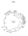
- the pili excreted by wild E. coli K88 strains, belonging to the three serotypes ab, ac and ad, and originating either from Holland (NL notation) or from the United States (US notation) were extracted and separated on SDS-PAGE gel .
- the figure shows such a gel colored with Coomassie blue.
- the pilin subunits form an electrophoretic band whose migration is that of a 26 kD protein.
- Figure 1b shows a western blot of the gel of Figure 1a.
- the polyclonal antibodies raised against the pili of serotype ac unequivocally recognize the pilins of the three serotypes.
- the K88 operon is located on a conjugate plasmid, called pK88ac.
- the HindIII fragments are cloned into the vector pBR322, restricted by HindIII and treated with phosphatase, so as to limit its recircularization.
- the nine clones have a plasmid containing a restriction fragment 12 kb in size.
- the orientation of the insert is counterclockwise for the 5 clearly positive clones and clockwise for the other 4.
- the dependence of K88ac expression on of the orientation can be explained by the presence of the cryptic promoter P1, located downstream of the HindIII site, known to determine a transcription in the anti-clockwise direction (Stuber and Bujard, 1981, Proc. Natl, Acad. Sci, 78, 167 -171).
- the strong expression of the K88 operon therefore depends on a promoter external to the cloned HindIII fragment.
- pIX102 One of the plasmids of the 5 clearly positive clones was chosen, denoted pIX102, and further manipulated.
- the size of the insert was reduced to 7 kb by partial restriction EcoR1 (plasmid pIX105).
- the EcoR1 site was replaced by a HindIII site in the plasmid pIX115, so that the K88 operon is on a HindIII fragment.
- Figures 1a and 1b, lane 5 show the proteins extracted from an E culture. coli carrying the pIX115. The subunit has the same migration as that produced by the wild-type ac strain, and is clearly recognized by anti-K88ac antibodies.
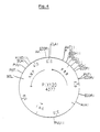
- a summary restriction map of pIX 115 is presented in Figure 2, as well as the genetic map of the K88ac operon.
- the relative position of the different genes is essentially deduced from the data in the literature (Dougan et al, 1983, J. Bacteriol., 153, 364-370) for the serotype K88ac and, by analogy with K88ac for the serotype K88ab (Mooi et al , 1983, J. Bacteriol., 154, 41-49).
- the molecular weight of different proteins produced by the K88 operon is 80, 31, 21 kD, and 26 kD for the subunit. A 25 kD protein could be encoded by the last operon gene.
- the K88ac subunit gene is on a DraI fragment, of about 1 kb.
- the fragment was cloned into the HindIII site of pBR322, in both orientations.
- the plasmid possessing the gene under the control of P1, ie the anti-clockwise orientation, is denoted pIX120.
- the tetracycline resistance gene has been deleted after restriction with BamHI and AvaI, followed by treatment with DNA polymerase in the presence of the four dNTPs and ligation. This deletion maintains an Downstream site in the plasmid pIX120.
- K88ad by transformed E. coli clones is sought by an immunological test on colony (cf. Method).
- the K88+ clones have a 12 kb insert in the anti-clockwise orientation and the plasmid is noted pAD102.
- K88ad pili by the E. coli strain (pAD102) is confirmed by an electrophoresis and Western blot gel of a crude rasat from a liquid culture (cf. FIG. 1, lane 9).
- pAD120 contains the pilin subunit structural gene of serotype K88ad.
- the anti-clockwise insertion of the insert into the HindIII site places the gene under the control of the strong promoter P1.
- K88ac (pIX120) and K88ad (pAD120) subunits were sequenced on both strands, using the technique of Maxam and Gilbert.
- the restriction sites marked with Klenow were the HinfI, DdeI and EcoR1 sites in particular.
- This figure shows that there is an open reading phase in -63 coding for an initiating ATG, terminated in 710 by a stop codon TAA.
- This reading phase codes for the pilin subunit. This is synthesized in the form of a precursor carrying a signal sequence of 21 amino acids at the amine end of the protein. This sequence allows the precursor to cross the internal membrane of the bacteria, and would then be cleaved.
- the subunit is composed of 262 amino acids forming a protein with a molecular weight (PM) deduced from 27,337, close to the 26 kD determined experimentally.
- the terminal N and C sequences deduced from the gene sequence are identical to the sequences of the protein of serotype K88ab.
- a sequence complementary to the 16S RNA is present 14 base pairs upstream of the ATG and should constitute an efficient ribosome attachment sequence (RBS).
- RBS ribosome attachment sequence
- the P1 promoter of pBR322 is underlined in FIG. 3. No other sequence which can correspond to a promoter is observed upstream of the RBS of the K88 fragment.
- Pin unique restriction sites such as EcoRV, Pst1, EcoR1, ScaI, Nsi1, Sac1, Bgl1, BsHII, HindII and Aat2.
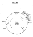
- Figure 4 shows the restriction map of the pIX120.
- the complete sequence of the gene for the K88ad subunit is given in FIG. 5. This subunit is composed of 264 residues, for a deduced molecular weight of 27,535.
- the K88ad US subunit gene contains restriction sites different from those observed in the ac gene. The presence of the AvaI, HpaI, and XhoII sites, the absence of the ScaI and SacI sites and the existence of two EcoR1 sites were retained.
- the K88ac operon is composed of 5 genes, including the subunit gene and four genes whose products are used for anchoring the pilus and for its polymerization (see Figure 2). These 5 genes were distributed over the cloning vectors pACYC184 and pBR322. These two vectors are compatible, carry different resistance markers (to chloramphenicol for pACYC184 and to ampicillin for pBR322), and are both of high copy number per cell. In addition, they have the same tetracycline resistance gene, therefore the same P1 promoter located upstream from the HindIII site.
- K88ac The complementation of K88ac is shown diagrammatically in FIG. 7. It involves the 2 plasmids pIX120 and pIX211 constructed in the following manner:
- PIX120 contains the only gene for the K88ac subunit under the control of P1 in pBR322.
- the Tc R gene was deleted to avoid the recombination of the Tc R homologous genes between the vectors pBR322 and pACYC 184 in a Rec+ environment.
- PIX211 is a plasmid pACYC184 containing the operon K88ac deleted from the subunit gene, and cloned into the HindIII site, therefore under the control of the P1 promoter.
- This plasmid was constructed in several stages: - isolation of pBR322E, which is a pBR322 devoid of EcoR1 site. It was obtained by EcoR1 restriction of pBR322, and Klenow treatment. - cloning of the operon K88 in the HindIII site of pBR322E.
- This plasmid is the equivalent of pIX115, carrying a single EcoR1 site, located in the first third of the gene for the subunit (cf. FIG. 2).
- This deletion is approximately 1000 base pairs; it covers the BstEII sites, located 50 bp before the initiator codon of the subunit, and the HindII site, located 110 bp before the stop codon.
- the RBS sequence and the complete gene of the subunit are therefore deleted, except perhaps the last codons on the 3 ⁇ side of the gene.
- the last step is the cloning of the HindIII fragment of this plasmid into the HindIII site of pACYC184. This plasmid is pIX211.
- the bacteria which contain both the plasmid pIX120 and the plasmid pIX211 are K88+.
- FIG. 1, lane 6 An extract of a crude rasat from a culture of EC294 (pIX211) (pIX120) is presented in FIG. 1, lane 6.
- the complementation system developed for K88ac can therefore be used for the ad serotype.
- the principle behind the search for epitopes is the cloning of fragments of the subunit gene into the beta-galactosidase gene.
- the presence of an antigenic peptide K88 fused with beta-galactosidase is sought by an immunological test on colony, with the specific antiserum of the pilus.
- the BstEII - Dra1 segment of pIX115, containing the subunit gene (see FIG. 2), is hydrolyzed by DNase, so as to isolate fragments from 50 to 150 base pairs.
- the fragments are filled with T4-polymerase in the presence of 4dNTP and cloned into the HindII site (blind ends) of pUR222.
- the HindII site is unique and is part of a polylinker, located in the Lac alpha fragment of beta-galactosidase.
- the plasmids are introduced into the strain JM103, which has the plasmid F ⁇ carrying the complete lactose operon, but mutated in the lac alpha region.
- the lac+ phenotype appears by complementation between the genes carried by the two plasmids, and is demonstrated on medium containing IPTG - inducer of the lactose operon - and X-gal - chromogenic group.
- the plasmids pUR222 which have inserted a K88 fragment in phase, appear blue on Xgal-IPTG medium.
- An antigenic reaction against anti-K88 antibodies was sought on approximately 1000 clones, exhibiting a light blue to dark blue coloration on X-gal. Twelve clones react very positively.
- Figure 8 shows the hydrophilicity / hydrophobicity profile of the K88ac subunit, according to the Doolittle algorithm. (Kyte and Doolittle, 1982, Mol. Biol., 157, 105-132).
- hydrophilic parts of the molecule are hatched; each constitutes a potential exposed region and antigenic site.
- the following regions have been selected: aa 16 to 29 aa 87 to 106 aa 114 to 122 aa 162 to 175 aa 211 to 222 and 245 to 255.
- Proline amino acids are likely to form angles in proteins and thereby expose neighboring regions. Prolines are present at aa 59, 72, 81, 98, 113, 164 and 254 of pilin ac. The presence of proline at positions 98, 113, 164 and 254 which are located in the immediate vicinity or in hydrophilic zones, is an additional indication favorable to the presence of an epitope.
- the K88ad subunit contains 8 proline residues, (aa 59, 72, 81, 98, 112, 163, 210 and 256). Those located at positions 98, 112, 163, 210 and 256 can be linked to hydrophilic regions.
- K88 exists in the form of three serotypes: K88ab, ac and ad. This nomenclature means that these serotypes have common antigens "a” and distinct “b", “c” and “d”. Each serotype also has "variants”.
- Figure 9 gives the amino acid sequence of the ab variants (Dyker et al, 1985, Infection and Immunity, 50, 279-283), the ac variants (Josephsen et al, 1984, FEMS Microbiol. Letters, 25, 301-306 ) and the one analyzed in this work and ad variants (Gaastra et al, 1983, FEMS Microbiol. Letters, 18, 177-183). The differences between amino acids are the only ones included.
- sequences 94 to 105, 133 to 136, and 163 to 173 differ between the variants and the serotypes. This is particularly striking for the sequence 163-173 whose composition and length are different. From an antigenic point of view, these sequences could coincide with variable antigens, which would distinguish the variants from each other.
- the sequence 163-173 (cf. Fig. 9) is particularly variable in this sense.
- Variations in amino acids can modify the local chemical properties of the sequences and in particular the hydrophilicity / hydrophobicity of the molecule.
- Figure 10 compares the profiles of the six subunits whose composition is given in Figure 9, according to the Doolittle algorithm.
- hydrophilicity indices of the hydrophilic sequences indexed in point 4b make it possible to deduce that: - the fragment limited by aa 17 to 29 is common to all the subunits and clearly hydrophilic; - fragments 96-107 and 115-123 are also very hydrophilic; in all the pili; - on the other hand, the sequences 162-175 and 211-222 have indices of variable hydrophilicity.
- serotype ab the peptide 162-175 is not hydrophilic, while the segment 211-222 is very hydrophilic.
- Table 2 shows the position of the antigenic, hydrophilic, invariable, and variable peptides of the K88ac subunit, with, for the last point, the distinction between the variability between serotypes and between variants.
- the last column is the selection of peptides, both antigenic and variable, which are three in number.
- antigenic peptides 69-92 and 83-122 could contain a variable (94-105) or common epitope, belonging to serotype a, (105-133 or 84-90 not very hydrophilic).
- Peptide 147-160 is antigenic. It corresponds to a variable sequence between serotypes and could therefore correspond to serotype C. On the other hand, the neighboring sequence 162-170 is extremely variable, in composition, length and hydrophilicity.
- the sequence 206-225 differs between serotypes and variants and its hydrophilicity is variable. It contains a continuous epitope between amino acids 217-229.
- fragments containing antigenic sequences specific to the pilus are capable of more or less significant modifications which changes the antigenic character of the molecule without altering its excretion and polymerization properties.
- the epitope chosen for this example is an epitope of the Influenza virus (Flu1). It is part of an antigenic region of the virus, at the junction between the hemagglutin HA1 and HA2.
- the peptide sequence N ⁇ -PEKQ RGIF AT is immunogenic when injected into an animal in the form of a synthetic peptide (Patent PCT / US83 / 01291, Scripps Clinic and Research Foundation).
- the DNA encoding this epitope is obtained by chemical synthesis.
- the choice of codons takes into account the preferential use of codons by E. coli.
- the sequence is shown in Figure 11.
- the ability to modify the subunit gene depends on the presence of restriction sites.
- the S2 region is accessible in the K88ad gene thanks to the HindII / HpaI (aa 207/208) and XhoII (222 and 223) restriction sites.
- HindII is located at the beginning of the variable sequence; XhoII cleaves an antigenic sequence.
- FIG. 11 The modifications of the region S1 are given in FIG. 11.
- the production of pili by the recombinant strains is firstly tested by an immunological test on colony.
- Figure 12 shows the immunological reaction obtained for the different constructions described below.
- the SacI-BssHII fragment of the K88ac pilin gene carried by pIX120 was replaced by the synthetic linker of M13tgl30, cloned in a pUC vector.
- the construction of pIX126 is carried out in two stages: the cloning of the 5 ⁇ part of the subunit gene upstream of the SacI site of pUC 130 first of all is the result of the ligation of the PvuI-SacI fragments of pUCtg 130, and SacI -Pvu I of pIX126.
- the plasmid is then restricted by BamH1, made blunt ended by the Klenow polymerase fragment, then restricted by PvuI.
- the terminal C part of the subunit gene is isolated on the BssHII / Klenow / PvuI fragment of pIX120.
- the subunit gene is reconstituted by ligation of the pVuI-BamH1 and BssHII - PvuI fragments to generate pIX126.
- the sequence of this plasmid has been checked.
- the sequence of the linker is given in FIG. 11, which shows the SacI-BamH1 fragment introduced in place of S1, and its translation into amino acids in the subunit. Note that the size of the subunit is reduced from 262 aa to 255 amino acids.
- the pIX126 is then introduced, by transformation into a strain containing the complementation system (pIX211) and the presence of pili is sought by - agglutination: weak agglutination is observed - immunological test on bacteria: the polyclonal antibody reacts positively with bacteria, but less intensely than on the control strain.
- specific K88ac monoclonal antibodies do not recognize the recombinant pili. These antibodies are specific to the common serotype "a" because they recognize both K88ab, ac and ad, and in particular the native, undenatured protein. The absence of reaction with the pilins produced by pIX126 foreshadowed structural changes in the pili.
- pili and acrylamide gel purification of pili and acrylamide gel.
- the presence of pili was sought by shaving a liquid culture of the bacteria containing pIX126 and pIX211, under the same conditions as the control bacteria carrying pIX120 and pIX211.
- the preparations are analyzed on acrylamide gel. No pilin subunit appears in the preparation of the recombinant bacteria.
- a sandwich ELISA detects on the order of 0.1 micro g of pili per ml of raw rasat from a culture of pIX126.
- influenza epitope described in point 6 in the polylinker increases the size of the 10 amino acid subunit.
- the synthetic DNA coding for the influenza epitope has cohesive ends KpnI - XbaI. It was first cloned, in phase, into the polylinker of pUC18; this plasmid induces the production of a hybrid beta-galactosidase containing the viral epitope.
- Synthetic DNA is also cloned into pIX126 to generate pIX130.
- the pIX130 introduced into the complementation strain induces the production and excretion of subunits recognized by the polyclonals, but not by the anti-K88 monoclonals. No structure identifiable with pili is however extracted from a liquid culture of this bacterium, as shown by the results of the assays by Elisa (cf. Table 3).
- the SacI site has the same reading phases in pUC18 and pIX126 (AGC code for a serine).
- pIX126 the SacI-EcoRV-Sphl-KpnI sequence was replaced by the SacI-KpnI sequence of PUC18.
- the resulting plasmid, noted pIX131, contains the influenza epitope and codes for a subunit of 260 residues.
- the pIX131 introduced into the complementation strain is recognized by the anti-K88 polyclonal antibodies in the same way as the pilus produced by the strain containing the pIX126. This sub-unit seems to be excreted but poorly if not assembled.
- the region S1 consists of 3 sequences: six aa constant (I, F, Y, G, G, L), 8 aa variables (11 in serotypes ad and ab), then a large invariable region, which covers the BssHII site.
- Ep34 appears in fig. 11. This DNA was cloned into the SacI-BssHII sites of pIX1 20 according to the conventional technique: SacI-BssH2 restriction of pIX120, and ligation if there is an excess of adapter. This plasmid is pIX128.
- the subunit synthesized by pIX128 consists of 258 amino acids.
- the concentration of recombinant pili in a raw rasat is 3 to 6 micro g / ml. (Elisa sandwich in which the antibodies adsorbed are polyclonal, see Table 3), compared to 16-20 micro g of wild pili / ml. The pili are therefore always assembled, but are in less quantity, of the order of 5 times less than non-recombinant pili.
- the recombinant subunit of pIX128 was demonstrated on a PAGE gel and on an immunoblot.
- the synthetic DNA which codes for an influenza epitope has ends compatible with the Kpn1 and XbaI sequences of the adapter cloned in pIX128.
- the next step therefore involves the cloning of this epitope in phase in the pIX128, generating the pIX136.
- PIX128 is restricted by the restriction endonucleases KpnI and XbaI and brought into contact with an excess of synthetic DNA coding for the viral epitope. After ligation, the plasmids are introduced into EC294 by transformation.
- the plasmid pIX136 induces in the strain EC294 (pIX211) the production of pili recognized by poly- and monoclonal anti-K88 antibodies (cf. FIG. 12).
- Elisa reveals the presence of pili antigens in raw rasats.
- the antigen concentration is however quite low, of the order of 1-3 micro g / ml.
- the sub-unit produced by pIX136 has a size equivalent to 268 amino acids (aa), which is 6 amino acids more than the natural sub-unit ac.
- aa 268 amino acids
- pIX136 cf. Figure 11.b
- This DNA codes for 6 invariable amino acids, located on the N-terminal side of the virus epitope. It is interesting to note that, in doing so, the cloned epitope is brought closer to the SacI site which is included in a translated antigenic sequence: the piptidic sequence LLSIFY is an epitope of the pilus.
- the pilus gene of pIX139 has the 5 ⁇ part of pIX131, up to the Xba site - including therefore the influenza epitope -, and the 3 ⁇ part, from the XbaI site of pIX128.
- the first 6 constant amino acids (aa) of the S1 region have therefore been deleted.
- the bacterium which contains both the complementation system and pIX139 is poorly recognized by polyclonal anti-pili K88 antibodies but is not recognized by monoclonals.
- the Elisa of a raw rasat of this strain detects very few antigens, around 10 to 20 ng / ml, which is at the limit of detection of this method under our conditions. No antigen is detected when monoclonals are used for the Elisa (see Table 3).
- the immunoblot of a large extract of the raw rasat reveals a weak band whose migration corresponds to that of a subunit of pilus.
- variable region of 8 amino acids of S1 can be deleted from the subunit without suppressing its assembly excretion property.
- It can be replaced by another completely new and longer sequence of 6 amino acids.
- the constant sequence located upstream of the variable region is essential for the manufacture of pilus.
- the amount of pili that can be extracted from the two recombinant strains is low; taking as reference the quantity of pili purified from a wild strain, it drops to 20% for the strain containing pIX128 and to 6% for that carrying pIX136.
- region S2 defined in point 4 is located between amino acids 208 and 231 of the pilin K88ad. The following describes the various manipulations of this region.
- S2 is interchangeable between serotypes
- the S2 region has a different composition and hydrophilicity between the serotypes ac and ad.
- a first manipulation consisted in replacing the region S2 of ad by that of ac and, conversely, of cloning S2 of ad in the gene of pilin ac.
- the plasmids pIX120 and pAD120 are cleaved by BglI. Each plasmid contains two Bgl1 sites: one is located in the ampicillin resistance gene, the other in the pilin gene, 100 nucleotides upstream of the S2 region.
- the fragment of pIX120 carrying the start of the pilin ac gene is ligated to the fragment of pAD120 carrying the 3 ⁇ end of the pilin ad gene. This ligation reconstructs an ampicillin resistance gene, and an ac-ad hybrid pilin subunit.
- This plasmid is pIX134.
- this second plasmid is pIX135.
- These plasmids are easily distinguished from the parental molecules pIX120 and pAD120 by EcoR1 restriction in particular.
- the region of the pilin ad gene containing S2 is accessible to genetic manipulation via the HindII and XhoII sites. Not directly, however, because there are 9 XhoII restriction sites in pAD120. The replacement of the HindII-XhoII fragment of the pilin gene by a synthetic DNA could therefore not be carried out in pAD120.
- XhoII has ends which are cohesive with BamH1
- a cloning strategy has been adopted in which the multiple site XhoII is replaced by a single site BamH1. Cloning involves four intermediates, derived from the vector pUC8. These intermediaries are shown in Figures 13.
- the HindII site of the pUC8 linker was previously deleted, generating pUC81. This deletion is carried out by restriction of pUC8 by the enzymes HindII and HindIII, treatment with polymerase - klenow fragment - from E. coli and ligation. This deletion maintains the Lac + character (fig. 13.a and 13.b).
- pUC 82 is the second intermediate constructed by the cloning in phase of the EcoR1-XhoII fragment of the ad gene in the EcoR1-BamH1 sites of PuC81. This insertion results in the loss of the SmaI site of the pUC81 linker, and maintains the Lac + character.
- the XhoII multiple site (GGATCT) is now replaced by a single BamH1 site (GGATCC) (Fig. 13c).
- strain EC294 (pIX211) (pAD121) gives an intense immunological response (cf. fig. 12) and synthesizes pili which can be extracted by conventional techniques.
- FIG. 15 shows a photograph of a PAGE-SDS gel of a crude rasat from the cultures producing wild and recombinant pili.
- the recombinant pilin coded by pAD121 has a slightly greater electrophoretic mobility than the wild pilin ad. This is expected since the modification of S2 reduces the size of the subunit from 264 amino acids to 258 residues.
- the antigen concentration of a crude rasat of the strain EC294 (pIX211) (pAD121) is approximately 25 micro g / ml, ie identical to that of a rasat of a non-recombinant ad strain. Contrary to what we observed for S1 (cf. the equivalent of pAD121, pIX128) the deletion of the variable region does not decrease the amount of recombinant pili that can be purified.
- the S2 region of the recombinant pilin gene offers numerous possibilities for cloning a heterologous DNA fragment thanks to the presence of the unique restriction sites kPn1, HindIII and XbaI.
- the synthetic DNA coding for the Flu1 epitope of Influenza was cloned into the KpnI and XbaI sites.
- the vector pAD121 restricted by KpnI and XbaI, is ligated in the presence of an excess of synthetic DNA Ep12.
- the sequence of this DNA is given in FIG. 14.
- the recombinant plasmid is pAD122.
- the EC294 (pIX211) (pAD122) strain reacts to anti-K88ac antibodies. This strain excretes recombinant pili which can be extracted, as shown in the PAGE and immunoblot gels of FIGS. 15a and b.
- the S2 region appears versatile and tolerant, both for its overall length and for its composition.
- the results show that a deletion of 6 residues (pAD121) or a final insertion of 2 amino acids (aa) (pAD122) maintain the assembly of the pili.
- the region S2 of pAD121 contains amino acids (aa) identical or chemically close to those present in pAD120 (cf. fig. 14). This is the case for proline (P), phenylalanine (F), valine (V), serine (S) and glutamic acid (E) which are identical, or aspartic acid (D), replaced by l glutamic acid (E).
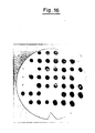
- Figure 16 which is the photograph of a filter, shows the result of an immunological test on a bacterial colony using an anti-K88 polyclonal serum as a means of detection.
- the colonies of interest are those which host (i) the unmodified plasmids (pIX120, pAD120), which serve as positive controls, (ii) the "vector" plasmids, containing a linker in place of the variable sequences S1 and S2 (pIX128 , pAD121), and finally, (iii) the plasmids which code for the pilins which express an epitope of the influenza virus (pIX136, pAD122). Their positions on the filter are, in this order:
- Two negative controls are present: - the E. coli strain containing only the plasmids pIX211, carrying the helpers genes, and pUR222. It therefore does not contain a pilin gene. It is on line 3, clones 4, 5 and 6 on the filter. - the strain containing the plasmid pACYC184, vector used for the cloning of pIX211, and pIX120. This strain therefore produces the subunit, but is unable to assemble it into pili.
- the antibody-antigen reaction observed on these negative controls is weak, and considered to be background noise.
- the bacteria which produce the unmodified pili are on the other hand highly recognized by antibodies, as are all the bacteria which express the recombinant pilins (IX128 and 136, AD121 and 122).
- the intensity of the spots which may reflect the amount of antigens present, is relatively large for each recombinant.
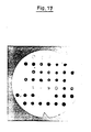
- MAb monoclonal antibody specific for K88 pili as a means of detection.
- MAb monoclonal antibody specific for K88 pili as a means of detection.
- This MAb was chosen because that it recognizes the pili intact, but very little the denatured pili dissociated into pilin. As long as its conformational epitope is conserved, this antibody distinguishes the recombinant pili from the free pilin.
- the bacteria which express the wild pilin ac or ad are recognized, as well as all the bacteria which synthesize the recombinant pilins IX128 and 136, AD121 and 122. No background noise is detected on the negative control .
- Extractions of recombinant pili modified in S1 or S2 were carried out in their concentration was estimated by Elisa.
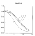
- An example of an Elisa assay is presented in FIG. 18.
- the rasats of the recombinant strains are obtained by heat treatment as described in the "Methods" section, and are adsorbed on the plate. They are revealed by an anti-K88 monoclonal antibody diluted 2 by 2 on the plate.
- the rasats analyzed in this figure are those extracted from the strains which express the genes of pIX120, pIX128 and pIX136.
- the concentrations of K88 antigens are estimated relative to a reference pili solution at 1 micro g / ml. They are respectively, taking into account the dilution factors, of: 15 micro g / ml for pIX116 8 micro g / ml for pIX128 3 micro g / ml for pIX136.
- Electron microscopy reveals pili which have the morphological characteristics of pili K88, around the strains which express the genes pIX128, pAD121 and pAD122.
- a synthetic peptide having the sequence of the Influenza epitope (sequence called Flu1) is used to immunize mice.
- the spleen cells of immunized mice are used to make antibody-producing hybridomas.
- the analysis of the antibodies makes it possible to select monoclonals which recognize the synthetic peptide with a strong affinity and do not react with the K88 pili.
- the reaction of these monoclonals against recombinant pili is then analyzed by Elisa. IX121 and IX136 pili as well as AD121 and AD122 are adsorbed on the Elisa plates, then incubated in the presence of a dilution of anti-Flu1 monoclonal antibodies.
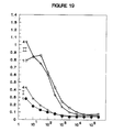
- FIG. 20 shows that the optimum response against the pilus is obtained for injections of 3 to 100 micro g of antigens.
- FIG. 21 shows that the sera contain antibodies directed against the peptide. Here the optimum response is obtained for injections of 3 to 30 micro g of antigens.
- Table 3 Concentration of K88 antigen in different crude rasats (heat purification) determined by Elisa Sandwich.
- the adsorbed antibody is an anti-K88ac polyclonal.
- the protein concentration is 50-60 micro g / ml, determined by Coomassie. These values are the average of three measurements.
- the references are purified pili of the serotypes ac for the pIX series and of the serotype ad for the pAD series, brought to the concentration of 1 micro g / ml.
- a strain of Escherichia coli containing a first plasmid pIX211 and a second plasmid pIX136 was deposited at the NCIB under the reference EC294 (pIX211) (pIX136) - (NCIB number 1234 9). This deposit was made in accordance with the Budapest Treaty.
- a second strain of Escherichia coli containing a first plasmid pIX211 and a second plasmid pAD122 was deposited at the NCIB under the reference EC294 (pIX211) (pAD122) - (NCIB number 12347). This deposit was also made in accordance with the Budapest Treaty.
- This example uses the complementation system and the vectors pIX128 and pAD121 described in example 1.
- sequence of the peptide inserted into the pilin is: Asn-Asn-Pro-His-Arg-Ile-Leu
- Flu2 It is a sequence of seven amino acids, which belongs to the influenza virus hemagglutinin. It is located in the B antigen of hemagglutinin 1 of the virus. This sequence is hereinafter called Flu2.
- a synthetic DNA coding for this peptide has been synthesized, so that it allows directional and in-phase cloning in the KpnI and XbaI sites of the vectors pIX128 and pAD121.
- the third amino acid in the epitope is a proline, and the restriction sequence KpnI GGTACC introduces an amino acid proline, coded by the triplet CCx.
- the nucleotide T in 5 ⁇ of the synthetic DNA is introduced. The codon will become CTG, and will introduce the amino acid leucine.
- the palindrome recognized by KpnI is deleted.
- the choice of the nucleotide sequence takes account of the preferential choice of codons by E. Coli. This sequence is:
- This DNA was cloned into the KpnI and XbaI sites of pIX128, generating pIX142 on the one hand, as well as into these same sites of pAD121, generating the plasmid pAD126 on the other hand.
- the cloning procedure is conventional: double restriction of the plasmid by KpnI and XbaI, followed by its purification on agarose gel, and ligation in the presence of a large excess of synthetic DNA hybridized. The screening of the recombinants is facilitated by the loss of the KpnI site. Different candidates were isolated and verified by sequencing.
- Each of the recombinant plasmids is introduced by transformation into an E. coli bacterium which contains the plasmid pIX211, EC294 (pIX211).
- this plasmid carries the genes known as "helpers" coding for the excretion and assembly of the K88 pili.
- the pilin coded by pIX142 is 264aa long (amino acids), the exact size of the pilin K88ab and ad.
- the pilin determined by pAD126 is 262 aa long, the size of the pilin native to K88ac. In a way, the expression of these genes will reveal the importance of the overall size of the pilin on its pili assembly.
- FIGs 16 and 17 clearly show that the pilins-producing bacteria IX142 and AD126 have a large amount of K88 antigens on their surface. Indeed, the colonies which contain pIX142 (line 5, clones 4, 5 and 6 and line 6, clones 1, 2 and 3) and those which host pAD126 (line 2, clones 4, 5 and 6) are highly recognized by polyclonal serum, as well as by monoclonal antibody. The epitope recognized by this monoclonal antibody is structural. Its recognition of the pilins pIX142 and pAD126 therefore indicates that they are associated in pili.
- Razats of cultures producing pilins IX142 and AD122 were analyzed by Elisa, and detected either by monoclonals or by polyclonals. It can be estimated that the concentration of pili antigens in the extracts of pIX142 is the same as that in the extracts from the reference cultures producing wild pili: an average of 18 micro g / ml of IX142 antigens are detected in three independent thermal razors, for 19 micro g / ml of IX120 antigens (see Table 4). A comparable proportion is observed in the rasats of the culture producing pili AD126.
- the pilins IX142 and AD126 are easily identified on a PAGE gel and on a western blot which uses an anti-pili serum as a means of detection. Their migration is substantially identical to that of the unmodified IX120 or AD120 subunits.
- the antigen concentration determined by an Elisa is compatible with the intensity of the band formed by the pilin on the gel.
- a peptide which reproduces the N-N-P-H-R-I-L sequence of Flu2 was synthesized and monoclonal antibodies directed specifically against this synthetic peptide were selected. By Elisa, it has been shown that one of these monoclonals reacts more against the IX142 antigen than against the IX120 control.
- the pili-fused sequence in this example is the sequence of the hormone somatostatin, also called a factor that inhibits the release of growth hormone.
- the composition of this long amino acid peptide, the composition of synthetic DNA which determines its synthesis and their characteristics are:
- the peptide contains two cysteines at positions 3 and 14 respectively, which can form a disulfide bridge, and a high proportion of aromatic (phenylalanine and tryptophan) and charged (lysine) residues.
- Synthetic DNA carries at its 5 ⁇ and 3 ⁇ ends protruding sequences complementary to the cohesive ends of the KpnI and XbaI sites respectively.
- the choice of codons while respecting the preferential choice of E. coli, makes it possible to introduce sites for restriction for PstI (CTGCAG), XmnI (GAA ---- TTC) and PvuI (CAGCTG). These sites are useful for the screening of recombinant plasmids which have integrated this insert, and offer various possibilities for manipulation of the somatostatin sequence.
- the somatostatin synthesis DNA was cloned into the vectors pIX128 (S1) and pAD121 (S2).
- the recombinant plasmids are noted pIXSM for cloning in S1 and pADSM for insertion into S2.
- the nucleotide sequence of their insert and of the regions which flank it has been confirmed.
- PIXSM as well as pADSM, were introduced into strain EC294 (pIX211) to test the assembly of recombinant pilins into pili.
- the pilin coded by pIXSM has a size of 272 amino acids, while that coded by pADSM is 270 amino acids long.
- FIG. 16 An immunological test on bacteria is presented in FIG. 16.
- the bacterial colonies which contain pIXSM (line 6, clones 4, 5 and 6) are recognized by the anti-K88 polyclonal serum. This reaction is usually less intense than that observed on the positive control, a result which suggests the presence of fewer K88 antigens on the surface of these bacteria.
- the colonies carrying pADSM are very clearly recognized by this serum, suggesting a large amount of pili attached to the bacteria (line 3, clones 1, 2 and 3).
- Table 4 Concentrations of K88 antigens (in micro g / ml) and in total proteins (in micro g / ml; lowry method) in different thermal razors determined by Elisa. The amount of K88 antigens produced by the recombinant strains is estimated relative to the purified amount of the wild strains.
- the present example is intended to show that recombinant pilins, the two variable regions of which S1 and S2 contain a sequence foreign to the pili, can be constructed and expressed. This is achieved by combining the S1 sequences of the K88ac derivatives on the same pilin with the S2 sequences of the K88ad derivatives.
- the recombinant pilins genes contain the sequence S1-Flu2 from the plasmid pIX142, and either S2-Flu1 from pAD122 (test 1) or S2-Flu2 from pAD126 (test 2).
- the pilins coded by these genes are formed from part of the pilin IX142, from their amine end up to and including the sequence S1, followed by the C-terminal sequence of the pilin AD122 (test 1) or AD126 (test 2).
- the plasmids pIX142, pAD122 and pAD126 contain two BglI restriction sites. One is located in the ampicillin resistance gene, and the second is located in the pilin gene, precisely between the regions S1 and S2 (reference is made to Figures 3, 4, 5 and 6).
- - pIX142 the BglI fragment which carries the end of the AmpR gene, the origin of replication and the start of the pilin gene, including S1-Flu2
- - pAD122 test 1
- pAD126 test 2: the BglI fragment which contains the end of the pilin gene (including S2-Flu1 or S2-Flu2) and the start of the AmpR gene.
- plasmids are respectively pIX143, derived from pAD122 (test 1), and pIX144, formed from pAD126 (test 2).
- the size of the IX143 pilin is 266 amino acids, and 262 amino acids for IX144.
- pili-bearing bacteria The production of pili-bearing bacteria is obtained after introduction of each of the plasmids pIX143 and pIX144 into the expression strain EC294 (pIX211).
- the vector sequence is written in lowercase letters.
- RBS ribosome binding site.
- P1 cryptic promoter of the vector.
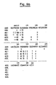
- Diagram of the intergenic complementation for K88ac the genes of the operon K88 (2), carried by pIX115, were subcloned on two vectors: pIX211, derived from pACYC184, carries the genes of the anchoring and assembly proteins pili and pIX120, derived from pBR322, containing the only gene for the subunit (1).
- P1 is a cryptic promoter of the vector.
- ORI origin of autonomous replication.
- Hydrophilicity profile of pilin K88ac according to the Doolitle algorithm.
- the particularly hydrophilic areas are hatched.
- the plasmids pIX126 (2), pIX130 (3), pIX131 (4), pIX128 (5), pIX136 (6) and pIX139 (7) are derived from the K88ac gene of pIX120 (1).
- the variable amino acids of S1 are identified by an asterisk *.
- the antibody is an anti-K88ac polyclonal. This antibody is recognized by an anti-rabbit coupled to peroxidase.
- the restriction maps of the various intermediaries which enabled the construction of pAD121 are presented in Figure 13a: pUC8, Figure 13b: pUC81, Figure 13c: pUC82, Figure 13d: pUC83, Figure 13e: pUC84 and finally Figure 13 f: pAD121.
- FIG. 14 - Manipulation of the region S2 the plasmids pAD121 (2), and pAD122 (3) are derived from the plasmid pAD120 (1).
- Figure 15a PAGE-SDS gel from a crude rasat of the strains 1 - EC294 (pAD120) (pIX211) 2 - EC294 (pAD121) (pIX211) 3 - EC294 (pAD122) (pIX211)
- Figure 16 Immunological test on various recombinant bacteria with anti-K88 serum. Three colonies isolated from each type of bacteria were transferred to the filter. With the exception of the clones of line 7 which contain pACYC184 instead of pIX211, these are EC294 (pIX211) strains carrying one of the following recombinant plasmids:
- Figure 17 The legend is the same as for Figure 16, except that the detection means is an anti-K88 monoclonal antibody.
- Figure 18 Elisa thermal shaving of the strains containing the plasmid pIX120 (1), pIX128 (2) and pIX136 (3), detected by an anti-K88 monoclonal antibody.
- the reference (R) is at 1 micro g / ml. On the abscissa is the dilution rate, on the ordinate the optical density.
- Figure 20 Humoral response of the four rats immunized with increasing concentrations of AD122 antigens, against K88 pili.
- the quantity of antigens per dose is plotted on the abscissa.
- the absorbance at dilution 10 of each serum against pili is on the ordinate.
- On the abscissa is the amount of micro g of pili injected.
- Figure 21 Same legend as Figure 20, except that the adsorbed antigen is the synthetic peptide Flu1. On the abscissa is the amount of micro g of pili injected and the absorbance at dilution 10 of each serum against the pili is ordered.
Abstract
Description
La présente invention concerne des microorganismes comportant des pili comme protéines porteuses de matériel peptidique hétérologue ainsi que les pili isolés à partir de tels microorganismes. L'invention concerne également un procédé d'excrétion de peptides ou de protéines utilisant les mécanismes propres des microorganismes pour la production de pili. L'invention concerne aussi l'utilisation des microorganismes et des pili.The present invention relates to microorganisms comprising pili as proteins carrying heterologous peptide material as well as pili isolated from such microorganisms. The invention also relates to a process for the excretion of peptides or proteins using the own mechanisms of microorganisms for the production of pili. The invention also relates to the use of microorganisms and pili.
Les mécanismes de production des pili d'Escherichia coli et plus particulièrement de sous-unités fimbriales K88 et K99 appelées dans la nouvelle nomenclature respectivement F4 et F5 sont actuellement connus et ont été décrits dans la littérature par notamment :
- B. Oudega, F.R. Mooi et F.K. de Graaf-Antonie van Leeuwenhoek 50, 1984, 569-584
- M.R. Mooi, F.K. de Graaf-Symp. Biotechnol. Res. Neth. 1983
- M. Kehoe, R. Sellwood, R. Shipley et G. Dougan - Nature Vol. 291 du 14 mai 1981
- J. Josephsen, F. Hansen, F.K. de Graaf et W. Gaastra FEMS. Microbiology Letters 25 (1984) 301-306
- D. Newman, B. Hay et Sadowski - Federation Proceedings 1985, 44 (5), p. 1438The production mechanisms of the pili of Escherichia coli and more particularly of fimbrial subunits K88 and K99 called in the new nomenclature respectively F4 and F5 are currently known and have been described in the literature by in particular:
- B. Oudega, FR Mooi and FK de Graaf-Antonie van Leeuwenhoek 50, 1984, 569-584
- MR Mooi, FK of Graaf-Symp. Biotechnol. Res. Neth. 1983
- M. Kehoe, R. Sellwood, R. Shipley and G. Dougan - Nature Vol. 291 of May 14, 1981
- J. Josephsen, F. Hansen, FK de Graaf and W. Gaastra FEMS. Microbiology Letters 25 (1984) 301-306
- D. Newman, B. Hay and Sadowski - Federation Proceedings 1985, 44 (5), p. 1438
Dans ces documents aucune information n'est cependant fournie sur l'utilisation de ces mécanismes pour l'obtention de microorganismes comportant des pili comme protéines porteuses permettant l'expression de peptides ou de protéines hétérologues.In these documents, however, no information is provided on the use of these mechanisms for obtaining microorganisms comprising pili as carrier proteins allowing the expression of heterologous peptides or proteins.
La présente invention vise à résoudre cette carence et fournit les moyens pour obtenir en pratique de tels microorganismes et pili.The present invention aims to resolve this deficiency and provides the means to obtain in practice such microorganisms and pili.
L'invention concerne à cet effet des microorganismes dont la membrane externe porte des pili dont la composition a été modifiée par au moins un changement de la séquence protéique de la sous-unité.To this end, the invention relates to microorganisms whose outer membrane carries pili, the composition of which has been modified by at least one change in the protein sequence of the subunit.
De manière préférée la composition des pili a été modifiée par au moins deux changements de la séquence protéique.Preferably, the composition of the pili has been modified by at least two changes in the protein sequence.
La nature des microorganismes concernés par l'invention n'est pas critique en elle-même et dépend uniquement de la possibilité que possèdent certains microorganismes de produire des pili tels que définis ci-avant. Habituellement, on opère avec des bactéries comme microorganismes.The nature of the microorganisms concerned by the invention is not critical in itself and depends solely on the possibility that certain microorganisms have of producing pili as defined above. Usually, we operate with bacteria as microorganisms.
Parmi celles-ci sont mises en oeuvre, de préférence, les bactéries des familles Enterobacteriaceae, Streptococcacae, Neisseriaceae, Proteeae, Phizobiaceae, Pseudomonodaceae, Corynebacteriaceae, Mycoplasmataceae, Myxococcaceae, Actinomycetaceae, Bacteriodaceae et, de manière préférée, les bactéries de genres Escherichia, Neisseria, Proteus, Bordetella, Pseudomonas, Salmonella, Klebsiella, Moraxella, Streptococcus, Serratia, Enterobacter, Agrobacterium, Rhizobium, Corynebacerium, Myxococcus, Actinomyces, Caulobacter, Bacteroides, Shigella, Mycoplasma et, de manière particulièrement préférée, les espèces Escherichia coli, Neisseria gonorrhoeae, Proteus mirabilis, Bordetella pertussis, Pseudomonas aeruginosa, Pseudomonas echinoïdes, Streptococcus pneumoniae, Neisseria meningitidis, Klebsiella pneumoniae. Enfin, de bons résultats ont été obtenus avec les souches d'Escherichia coli, en particulier les souches de sérotype K88 et K99.Among these, the bacteria of the families Enterobacteriaceae, Streptococcacae, Neisseriaceae, Proteeae, Phizobiaceae, Pseudomonodaceae, Corynebacteriaceae, Mycoplasmataceae, Myxococcaceae, Actinomycetaceae, Bacteriodaceae and, preferably, bacteria are used. , Proteus, Bordetella, Pseudomonas, Salmonella, Klebsiella, Moraxella, Streptococcus, Serratia, Enterobacter, Agrobacterium, Rhizobium, Corynebacerium, Myxococcus, Actinomyces, Caulobacter, Bacteroides, Shigella, Mycoplasma and, especially preferred, coliearea, , Proteus mirabilis, Bordetella pertussis, Pseudomonas aeruginosa, Pseudomonas echinoïdes, Streptococcus pneumoniae, Neisseria meningitidis, Klebsiella pneumoniae. Finally, good results have been obtained with the Escherichia coli strains, in particular the strains of serotype K88 and K99.
L'invention concerne également les pili isolés de ces microorganismes par tout moyen physique, chimique ou biologique. Elle concerne également les sous-unités ou pilines qui, par quelque moyen que ce soit, ont été libérées et séparées de la structure polymérique répétitive constituant les pili.The invention also relates to pili isolated from these microorganisms by any physical, chemical or biological means. It also relates to the subunits or pilins which, by any means whatsoever, have been released and separated from the repeating polymer structure constituting the pili.
Par pili en entend également les termes tels que fibrilles et, fimbriae. Ce sont des polymères excrétés par les microorganismes et attachés à la membrane externe de ces microorganismes, constitués de la répétition de protéines appelées pilines ou encore sous-unités.By pili also means terms such as fibrils and, fimbriae. These are polymers excreted by microorganisms and attached to the outer membrane of these microorganisms, consisting of the repetition of proteins called pilins or even subunits.
Les sous -unités ou pilines, dont la répétition constitue le pilus, sont des protéines qui ont généralement un poids moléculaire compris entre 5 et 50 kD et qui, lorsqu'elles sont associées sous forme de pili, forment des filaments extracellulaires de 20 à 250 Å de diamètre et pouvant atteindre plusieurs microns de long.The subunits or pilins, the repetition of which constitutes the pilus, are proteins which generally have a molecular weight of between 5 and 50 kD and which, when combined in the form of pili, form extracellular filaments from 20 to 250 Å in diameter and up to several microns long.
Les pili décrits dans l'invention peuvent être de tous types tels que des pili somatiques.The pili described in the invention can be of all types such as somatic pili.
De préférence on utilise les pili somatiques qui servent à l'adhésion des microorganismes sur un support et de bons résultats ont été obtenus avec les pili K88 d'Escherichia coli qui permettent à cette bactérie de coloniser l'intestin de certains mammifères.Preferably, the somatic pili which are used for the adhesion of the microorganisms to a support are used and good results have been obtained with the K88 pili from Escherichia coli which allow this bacterium to colonize the intestine of certain mammals.
Les pili obtenus selon l'invention ont une composition différente de la composition des pili du microorganisme à l'état sauvage suite à au moins une modification apportée à la séquence protéique de la sous-unité ou piline, de manière préférée suite à au moins deux modifications.The pili obtained according to the invention have a composition different from the composition of the pili of the microorganism in the wild after at least one modification made to the protein sequence of the subunit or pilin, preferably following at least two modifications.
Les modifications de la sous-unité portent de préférence sur les régions non critiques pour l'assemblage et l'excrétion de la sous-unité.Modifications of the subunit preferably relate to regions not critical to the assembly and excretion of the subunit.
La protéine obtenue après modifications peut être utilisée dans diverses applications. En fonction du ou des piptides introduits dans la sous-unité elle peut notamment convenir comme enzyme, comme constituant nutritif ou encore dans le domaine de la santé animale ou humaine notamment en tant que produits pharmaceutiques à usage vétérinaire ou humain. Dans ce cas, la protéine obtenue peut être administrée sous n'importe quelle forme pharmaceutiquement acceptable telle que notamment la protéine isolée, la protéine liée au pilus isolé ou non du microorganisme; le microorganisme lui-même peut donc être administré.The protein obtained after modifications can be used in various applications. Depending on the piptide (s) introduced into the subunit, it may in particular be suitable as an enzyme, as a nutritive constituent or also in the field of animal or human health, in particular as pharmaceutical products for veterinary or human use. In this case, the protein obtained can be administered in any pharmaceutically acceptable form such as in particular the isolated protein, the protein linked to the pilus isolated or not from the microorganism; the microorganism itself can therefore be administered.
Les pili sont des molécules fortement antigéniques. Si le peptide introduit est un épitope étranger au pilus, la protéine obtenue selon l'invention peut posséder un caractère antigénique nouveau et être utilisée dans des tests immunologiques tels que pour des diagnostics. Elle peut également posséder un caractère immunogénique et être utilisée comme composant d'un vaccin.Pili are highly antigenic molecules. If the peptide introduced is an epitope foreign to the pilus, the protein obtained according to the invention may have a new antigenic character and be used in immunological tests such as for diagnostics. It can also have an immunogenic character and be used as a component of a vaccine.
L'invention permet également de rendre immunogéniques des peptides, qui ne le sont que peu ou pas par eux-mêmes, après leur introduction dans la séquence protéique de la piline.The invention also makes it possible to render immunogenic peptides, which are little or not by themselves, after their introduction into the protein sequence of pilin.
La modification de la séquence protéique de la sous-unité ou piline peut être réalisée par tout moyen connu.The modification of the protein sequence of the subunit or pilin can be carried out by any known means.
Un moyen est de modifier la séquence d'ADN codant pour la sous-unité du pilus. Ces modifications sont réalisées par ingéniérie génétique. Le gène recombinant est ensuite traduit en protéines (hétérologues) dans un organisme hôte.One way is to modify the DNA sequence encoding the pilus subunit. These modifications are made by genetic engineering. The recombinant gene is then translated into proteins (heterologous) in a host organism.
Par modification on entend toute modification de l'ADN sauvage, conduisant à une modification de la séquence protéique de la sous-unité.By modification is meant any modification of wild-type DNA, leading to a modification of the protein sequence of the subunit.
On entend également le remplacement d'un fragment d'ADN sauvage par un fragment d'ADN étranger. L'ADN étranger peut être d'origine naturelle ou synthétique. Sa séquence en nucléotides et sa phase de lecture dans le gène déterminent la séquence native en acides aminés incorporés dans la sous-unité.We also mean the replacement of a wild-type DNA fragment with a foreign DNA fragment. The foreign DNA can be of natural or synthetic origin. Its nucleotide sequence and its reading phase in the gene determine the native amino acid sequence incorporated in the subunit.
L'invention concerne dès lors également tout matériel génétique permettant l'utilisation de pili comme protéines porteuses.The invention therefore also relates to any genetic material allowing the use of pili as carrier proteins.
L'invention concerne en outre les procédés permettant de réaliser des pili en tant que protéines porteuses par incorporation d'ADN hétérologue ainsi que les procédés d'obtention de microorganismes porteurs de tels pili. Plus particulièrement l'invention concerne tout procédé dans lequel le gène recombinant permet d'obtenir des pili dont la structure polymérique est conservée et contenant éventuellement les sous-unités modifiées résultant de l'incorporation de l'ADN hétérologue.The invention further relates to the methods for making pili as carrier proteins by incorporating heterologous DNA as well as to the methods for obtaining microorganisms carrying such pili. More particularly, the invention relates to any process in which the recombinant gene makes it possible to obtain pili whose polymer structure is conserved and possibly containing the modified subunits resulting from the incorporation of heterologous DNA.
L'invention concerne donc les procédés qui permettent d'obtenir des pili dont la composition a été modifiée par au moins un changement de la séquence protéique de la sous-unité, ce ou ces changements résultant de l'introduction dans le gène codant pour la sous-unité d'un pilus, d'au moins un fragment d'ADN codant pour un piptide ou une protéine hétérologue tel qu'un fragment d'ADN synthétique, un fragment d'ADN naturel ou un fragment obtenu à partir de la transcription réverse de mRNA. Par ADN hétérologue, on entend tout matériel génétique issu d'une autre source que la souche hôte dans laquelle on réalise le procédé de l'invention. Par ADN synthétique, on entend aussi bien un ADN de composition ou structure naturelle obtenu par synthèse chimique qu'un ADN dont la structure ou la composition n'existe pas tel quel dans la nature.The invention therefore relates to methods which make it possible to obtain pili the composition of which has been modified by at least one change in the protein sequence of the subunit, this or these changes resulting from the introduction into the gene coding for the subunit of a pilus, of at least one DNA fragment coding for a piptide or a heterologous protein such as a synthetic DNA fragment, a natural DNA fragment or a fragment obtained from transcription reverse of mRNA. By heterologous DNA is meant any genetic material from a source other than the host strain in which the process of the invention is carried out. By synthetic DNA is meant both a DNA of natural composition or structure obtained by chemical synthesis as well as DNA whose structure or composition does not exist as it is in nature.
De manière préférée ces changements résultent de l'introduction dans le gène codant pour la sous-unité d'un pilus, d'au moins deux fragments d'ADn codant pour un peptide ou une protéine hétérologue, ceux-ci pouvant être identiques ou différents.Preferably, these changes result from the introduction into the gene coding for the pilus subunit, of at least two DNA fragments coding for a heterologous peptide or protein, these may be identical or different. .
Un des procédés est la modification d'au moins une région de la protéine de la sous-unité formant le pilus exposée au milieu environnant, par insertion d'ADN codant pour un peptide ou une protéine hétérologue, dans au moins une zone du gène codant pour la sous-unité d'un pilus correspondant à la zone exposée au milieu environnant de la séquence protéinique des pilines concernées.One of the methods is the modification of at least one region of the protein of the pilus subunit exposed to the surrounding medium, by insertion of DNA coding for a heterologous peptide or protein, into at least one region of the coding gene. for the pilus subunit corresponding to the area exposed to the surrounding medium of the protein sequence of the pilins concerned.
Les régions exposées comprennent notamment des régions antigéniques des pili, c'est-à-dire composées d'épitopes inducteurs de la production d'anticorps ou reconnus par des anticorps.The exposed regions include in particular antigenic regions of the pili, that is to say composed of epitopes inducing the production of antibodies or recognized by antibodies.
L'invention concerne également les utilisations des pili et pilines porteurs de matériel hétérologue. Ainsi l'invention concerne notamment les vaccins obtenus avec les pili et pilines porteurs de matériel hétérologue, combinés ou non avec divers additifs tels que des adjuvants et des immunostimulants.The invention also relates to the uses of pili and pilins carrying heterologous material. Thus the invention relates in particular to vaccines obtained with pili and pilins carrying heterologous material, combined or not with various additives such as adjuvants and immunostimulants.
L'invention concerne également l'utilisation des pili, des pilines et des microorganismes ayant une membrane externe qui porte des pili, dont la composition est modifiée par au moins un changement, ou par au moins deux changements, de la séquence protéique des pilines; en particulier l'invention concerne les utilisations de ceux-ci pour l'obtention d'un produit pharmaceutique destiné au traitement médical et/ou prophylactique en santé humaine ou animale.The invention also relates to the use of pili, pilins and microorganisms having an external membrane which carries pili, the composition of which is modified by at least one change, or by at least two changes, in the protein sequence of pilins; in particular the invention relates to the uses of these for obtaining a pharmaceutical product intended for medical and / or prophylactic treatment in human or animal health.
L'invention concerne également les tests immunologiques obtenus à partir des pili et pilines porteurs de matériel hétérologue.The invention also relates to immunological tests obtained from pili and pilins carrying heterologous material.
L'invention concerne notamment l'utilisation des pili, des pilines et des microorganismes ayant une membrane externe qui porte des pili, dont la composition est modifiée par au moins un changement, ou par au moins deux changements, de la séquence protéique des pilines, pour la fabrication de moyen destiné à la préparation de tests immunologiques. Ceci inclut notamment les composants de tests diagnostiques, les tests d'analyse, les galeries d'identification, les biopsies, les divers examens biologiques, les kits d'analyse et de dosage, les bandelettes pour le diagnostic, les réactifs diagnostiques.The invention relates in particular to the use of pili, pilins and microorganisms having an external membrane which carries pili, the composition of which is modified by at least one change, or by at least two changes in the protein sequence of the pilins, for the manufacture of means for the preparation of immunological tests. This includes, in particular, the components of diagnostic tests, analytical tests, identification galleries, biopsies, various biological examinations, analysis and assay kits, diagnostic strips, diagnostic reagents.
L'invention comprend également des utilisations diverses dans lesquelles les pili porteurs de protéines ou de peptides sont utilisés en tant qu'agents ayant des propriétés immunostimulantes (permettant notamment la stimulation de la réponse immunitaire à partir de vaccins déjà connus).The invention also includes various uses in which the pili carrying proteins or peptides are used as agents having immunostimulatory properties (allowing in particular the stimulation of the immune response from already known vaccines).
Un procédé ayant donné de bons résultats fait appel aux techniques du génie génétique. Il comprend les étapes suivantes :
- (1) clonage du gène ou de l'opéron du pilus choisi comme protéine porteuse et l'expression de ce(s) gène(s) dans un organisme hôte,
- (2) détermination de la séquence du gène de la piline
- (3) alternative de la complémentation intergénique,
- (4) identification des sites antigéniques de pilus,
- (5) identification des régions variables telle que notamment par comparaison des séquences de différents sérotypes,
- (6) isolement (ou synthèse) d'un fragment d'ADN codant pour un peptide ou un épitope intéressant, caractéristique de l'organisme contre lequel on veut dresser des anticorps,
- (7) clonage de l'ADN obtenu en (6) en phase dans le gène de la sous-unité du pilus dans des sites préférentiels identifiés en (4) et (5),
- (8) introduction du gène recombinant dans un organisme et son expression en tant que pilus porteur de peptide ou de protéine hétérologues.
- (9) purification des pili porteurs de peptide ou de protéine hétérologues.
- (1) cloning of the pilus gene or operon chosen as carrier protein and the expression of this (these) gene (s) in a host organism,
- (2) determination of the pilin gene sequence
- (3) alternative to intergenic complementation,
- (4) identification of antigenic pilus sites,
- (5) identification of the variable regions such as in particular by comparison of the sequences of different serotypes,
- (6) isolation (or synthesis) of a DNA fragment coding for an interesting peptide or epitope, characteristic of the organism against which it is desired to raise antibodies,
- (7) cloning of the DNA obtained in (6) in phase in the pilus subunit gene in preferential sites identified in (4) and (5),
- (8) introduction of the recombinant gene into an organism and its expression as a pilus carrying a heterologous peptide or protein.
- (9) purification of pili carrying heterologous peptide or protein.
Ces étapes sont explicitées ci-après :These steps are explained below:
Le clonage de l'opéron ou du gène du pilus choisi comme molécule porteuse est réalisé par les techniques maintenant classiques du génie génétique.The cloning of the operon or of the pilus gene chosen as the carrier molecule is carried out by the now conventional techniques of genetic engineering.
Le clonage in vitro implique la préparation de l'ADN de l'organisme qui produit le pilus, et le clonage de fragments de restriction (banque génomique) dans des plasmides, des phages ou des cosmides.In vitro cloning involves the preparation of the DNA of the organism that produces the pilus, and the cloning of restriction fragments (genomic library) in plasmids, phages or cosmids.
L'emploi de vecteurs dits d'expression, contenant des signaux de transcription forts, peut pallier l'absence de promoteur sur le fragment de DNA cloné.The use of so-called expression vectors, containing strong transcription signals, can compensate for the absence of promoter on the cloned DNA fragment.
Le clonage in vivo peut se faire chez certaines bactéries par l'intermédiaire d'un phage transducteur ou de plasmides mobilisables ou transférables associés à des transposons.In vivo cloning can be carried out in certain bacteria by means of a transducer phage or of mobilizable or transferable plasmids associated with transposons.
Les vecteurs de clonage sont introduits dans un organisme hôte par transformation (plasmides) ou transfection (cosmides et phages) ou conjugaison (plasmides conjugatifs).The cloning vectors are introduced into a host organism by transformation (plasmids) or transfection (cosmids and phages) or conjugation (conjugative plasmids).
L'expression des pili est recherchée dans un organisme hôte ne produisant pas ce type de pili : soit un mutant de l'organisme, muté dans cette fonction ou, mieux, dans un organisme de laboratoire, tel E. coli K12 pour les procaryotes. Le criblage des recombinants se fera sur base des propriétés propres aux pili (telles que l'hemagglutination, l'immunoprécipitation, ou la reconnaissance immunologique sur colonie). La présence des pili est confirmée de visu par microscopie électronique, ou par la purification des pili d'une culture du recombinant et leur analyse sur gel d'électrophorèse par exemple.The expression of pili is sought in a host organism which does not produce this type of pili: either a mutant of the organism, mutated in this function or, better, in a laboratory organism, such as E. coli K12 for prokaryotes. The screening of recombinants will be done on the basis of properties specific to pili (such as hemagglutination, immunoprecipitation, or immunological recognition on colony). The presence of the pili is confirmed visually by electron microscopy, or by the purification of the pili from a culture of the recombinant and their analysis on electrophoresis gel for example.
Une carte de restriction du fragment cloné est établie pour quelques enzymes courants. On utilise pour cela les techniques de restriction partielle et de restrictions multiples.A restriction map of the cloned fragment is established for some common enzymes. Partial restriction and multiple restriction techniques are used for this.
Le séquençage de l'ADN cloné est réalisé soit par la technique dite du didéoxyribose développée par Sanger, soit la technique de modification des bases mise au point par Maxam et Gilbert. On positionne le gène de la sous-unité sur la carte de restriction par des techniques de mutagenèse. Un exemple d'exécution particulière de cette technique fait appel aux minicellules. Ces minicellules permettent de mettre en évidence les protéines codées par le fragment cloné sur un plasmide. Il est possible d'induire des mutations dans ce fragment, par exemple via l'intégration d'un transposon, ou in vitro par restriction, suivie de digestion par une exonucléase. Les mutations qui entraînent l'absence de synthèse de pili sont analysées à leur tour dans les minicellules : on associe ainsi l'absence d'une protéine produite avec la localisation de la mutation. L'absence de sous-unités positionne, sans équivoque, le gène de la sous-unité sur le fragment. Cette portion du fragment peut être seule séquencée.The sequencing of the cloned DNA is carried out either by the so-called dideoxyribose technique developed by Sanger, or the base modification technique developed by Maxam and Gilbert. The gene for the subunit is positioned on the restriction map by mutagenesis techniques. An example of a particular implementation of this technique uses minicells. These mini-cells make it possible to demonstrate the proteins encoded by the fragment cloned on a plasmid. It is possible to induce mutations in this fragment, for example via the integration of a transposon, or in vitro by restriction, followed by digestion with an exonuclease. The mutations which cause the absence of pili synthesis are analyzed in their turn in the minicells: one thus associates the absence of a protein produced with the localization of the mutation. The absence of subunits unequivocally positions the gene for the subunit on the fragment. This portion of the fragment can be sequenced alone.
La séquence est analysée par ordinateur, via des programmes d'analyse qui fournissent les données essentielles de :
- début et fin du (des) gène(s)
- traduction du gène en protéine
- position des sites de restriction.The sequence is analyzed by computer, via analysis programs which provide the essential data of:
- start and end of the gene (s)
- translation of the gene into protein
- position of restriction sites.
La plupart des pili dont la génétique a été décrite à ce jour sont structurés en opéron, constitué de plusieurs gènes en plus du gène de la sous-unité. Ces gènes codent par exemple pour des protéines jouant un rôle dans l'ancrage, l'assemblage ou la protection.Most pili, the genetics of which have been described to date, are structured into an operon, consisting of several genes in addition to the subunit gene. These genes code, for example, for proteins playing a role in anchoring, assembly or protection.
On peut fractionner l'opéron en clonant le seul gène de la sous-unité sur un plasmide et le reste sur un autre plasmide. Les deux plasmides vecteurs doivent être compatibles et porter des marqueurs de sélection différents. Les pili seront synthétisés seulement lorsque les deux plasmides seront présents dans l'organisme hôte, par complémentation intergénique.The operon can be split by cloning the single gene for the subunit on one plasmid and the rest on another plasmid. The two vector plasmids must be compatible and carry different selection markers. The pili will only be synthesized when the two plasmids are present in the host organism, by intergenic complementation.
Le fragment portant l'opéron délété du gène de la sous-unité peut également être introduit dans le chromosome de l'organisme hôte.The fragment carrying the deleted operon of the subunit gene can also be introduced into the chromosome of the host organism.
La complémentation intergénique a plusieurs avantages techniques fort intéressants.Intergenic complementation has several very interesting technical advantages.
Le plasmide contenant le gène de la sous-unité
- est seul à devoir être manipulé,
- est d'une taille réduite par rapport à celle du plasmide porteur de l'opéron complet,
- contient un nombre plus limité de sites de restriction,
- a été complètement séquencé, ce qui n'est pas toujours le cas pour l'entièreté de l'opéron.The plasmid containing the gene for the subunit
- is the only one that must be handled,
- is of a reduced size compared to that of the plasmid carrying the complete operon,
- contains a more limited number of restriction sites,
- has been completely sequenced, which is not always the case for the entire operon.
En disposant d'anticorps polyclonaux ou d'une batterie d'anticorps monoclonaux spécifiques des pili, on recherche la position des antigènes par différentes techniques : par exemple
a - à partir de peptides synthétiques du pilus, qui sont reconnus par des anticorps antipili, ou qui induisent, après injection à un animal, la synthèse d'anticorps antipili.
b - par fragmentation de la protéine par des composés comme le bromure de cyanogène ou des enzymes (trypsine, ...) qui clivent les sites spécifiques de la protéine. On cherche ensuite la présence d'antigènes sur les différents fragments.
c - par clonage de fragments du gène pilus en phase dans le gène de la bêta-galactosidase. Si le fragment cloné détermine un peptide antigénique, il sera reconnu sur la protéine hybride par les anticorps antipili.By having polyclonal antibodies or a battery of pili-specific monoclonal antibodies, the position of the antigens is sought by various techniques: for example
a - from synthetic pilus peptides, which are recognized by antipiliary antibodies, or which induce, after injection into an animal, the synthesis of antipiliary antibodies.
b - by fragmentation of the protein by compounds such as cyanogen bromide or enzymes (trypsin, etc.) which cleave the specific sites of the protein. We then look for the presence of antigens on the different fragments.
c - by cloning of fragments of the pilus gene in phase in the beta-galactosidase gene. If the cloned fragment determines an antigenic peptide, it will be recognized on the hybrid protein by antipili antibodies.
Ces techniques identifient des épitopes continus de la protéine.These techniques identify continuous epitopes of the protein.
Des données prédictives sont souvent utilisées pour localiser des épitopes :
a - La structure tridimensionnelle de la protéine, si elle est connue, identifie les régions exposées.
b - Des algorithmes peuvent être utilisés pour :
- rechercher les régions hydrophiles et les régions hydrophobes d'une protéine : algorithmes de Hoop & Wood, de Doolittle. Les régions hydrophiles, exposées au solvant, sont autant de lieux pour des épitopes potentiels.
- prédire la structure secondaire (hélice, feuillet et tours) d'une protéine à partir de sa seule séquence primaire : algorithmes de Chou & Fasman, de Garnier. Les épitopes sont souvent associés à des tours dans la protéine.Predictive data is often used to locate epitopes:
a - The three-dimensional structure of the protein, if known, identifies the exposed regions.
b - Algorithms can be used to:
- research the hydrophilic regions and the hydrophobic regions of a protein: Hoop & Wood, Doolittle algorithms. The hydrophilic regions, exposed to the solvent, are all places for potential epitopes.
- predict the secondary structure (propeller, sheet and turns) of a protein from its only primary sequence: algorithms by Chou & Fasman, by Garnier. Epitopes are often associated with turns in protein.
D'autres indices peuvent être relevés, comme la présence de résidus proline, un acide aminé qui provoque des "coudes", et donc des régions protubérantes dans la protéine.Other indications can be noted, such as the presence of proline residues, an amino acid which causes "bends", and therefore protruding regions in the protein.
Les régions dites "variables" de la piline sont, d'une manière générale, les régions, dont la séquence en acides aminés peut être modifiée, sans compromettre la fabrication des pili.The so-called "variable" regions of pilin are, in general, regions, the amino acid sequence of which can be modified, without compromising the manufacture of pili.
Une manière expérimentale de localiser les régions variables consiste à cloner une courte séquence d'ADN synthétique au hasard dans le gène de la piline, puis de sélectionner les bactéries qui restent piliées. La position de l'insertion est déterminée par le séquençage du gène. Il positionne des régions variables de la piline.An experimental way of locating the variable regions is to clone a short sequence of synthetic DNA at random in the pilin gene, then to select the bacteria which remain piled. The position of the insertion is determined by the sequencing of the gene. It positions variable regions of the pilin.
Une manière prédictive consiste à comparer les séquences en acides aminés des pilines dans les différents sérotypes. En effet, certains pili présentent des variations antigéniques naturelles plus ou moins importantes, permettant sans doute à l'organisme qui les porte d'échapper partiellement du moins, à l'immunité acquise par l'hôte lors d'une infection antérieure.One predictive way is to compare the amino acid sequences of pilins in different serotypes. Indeed, some pili present more or less significant natural antigenic variations, undoubtedly allowing the organism which carries them to escape partially at least, to the immunity acquired by the host during a previous infection.
En comparant les séquences en nucléotides et en acides aminés des différents variants, on peut reconnaître :
a) des régions invariables, communes à tous les sérotypes, qui seront considérées comme essentielles à la structure ou la polymérisation de la piline. La présence de mutations conservatrices est un indice supplémentaire qui accentue l'importance de ces régio la structure pili;
b) des régions variables, en composition en acides aminés et/ou en longueur. Ces régions seront considérées comme n'intervenant pas - ou peu - dans l'essemblage et la structure du pilus.By comparing the nucleotide and amino acid sequences of the different variants, we can recognize:
a) invariable regions, common to all serotypes, which will be considered essential for the structure or polymerization of the pilin. The presence of conservative mutations is an additional indication which accentuates the importance of these regions of the pili structure;
b) variable regions, in amino acid composition and / or in length. These regions will be considered as having little or no effect on the assembly and structure of the pilus.
Cette première analyse peut être plus fouillée, tenant compte notamment des propriétés chimiques des résidus variables, de leur indice d'hydrophilicité ou encore de la taille du radical. L'importance des variations entre les pili peut ainsi être pondérée.This first analysis can be more detailed, taking into account in particular the chemical properties of variable residues, their hydrophilicity index or the size of the radical. The importance of variations between pili can thus be balanced.
Les épitopes positionnés comme décrit au point ci-avant en 4) peuvent être caractérisés grâce à la comparaison de sérotypes, comme communs ou variables. Cette donnée n'est pas indispensable pour la bonne réalisation de notre invention, mais est cependant fort intéressante. En effet, elle définit sur la protéine des régions à la fois antigéniques et susceptibles de modifications, qui seront autant de régions cibles pour l'introduction de peptides étrangers.The epitopes positioned as described in the point above in 4) can be characterized by comparison of serotypes, as common or variable. This data is not essential for the successful completion of our invention, but is nevertheless very interesting. Indeed, it defines regions on the protein which are both antigenic and susceptible to modifications, which will be target regions for the introduction of foreign peptides.
Il faut disposer d'un fragment d'ADN qui détermine la synthèse d'un épitope particulier de l'organisme contre lequel on désire développer une réponse immunologique. Ce fragment est soit un fragment de restriction, soit un ADN synthétique. Cette seconde solution offre évidemment beaucoup plus de souplesse pour le clonage (cf. point 7).It is necessary to have a DNA fragment which determines the synthesis of a particular epitope of the organism against which one wishes to develop an immunological response. This fragment is either a restriction fragment or a synthetic DNA. This second solution obviously offers much more flexibility for cloning (cf. point 7).
L'ADN codant pour l'épitope est cloné, en phase, dans le gène de la sous-unité, dans une des régions antigéniques et variables établies en (4) et (5) ci-avant. Cet ADN est soit ajouté au gène, soit cloné en lieu et place d'un fragment existant. Dans ce dernier cas, la taille totale de la sous-unité pourra être maintenue.The DNA encoding the epitope is cloned, in phase, into the gene for the subunit, in one of the antigenic and variable regions established in (4) and (5) above. This DNA is either added to the gene or cloned in place of an existing fragment. In the latter case, the total size of the sub-unit may be maintained.
La stratégie de clonage dépend de la disponibilité de sites de restriction dans la région choisie. En cas d'absence d'une telle séquence, un site est créé par mutagenèse in vitro, ou éventuellement l'ADN est cloné par des techniques de mutagenèse in vitro. L'introduction d'un site de restriction unique présente le grand avantage de créer un vecteur universel. En effet, n'importe quel ADN synthétique possédant des extrémités cohésives avec la séquence restreinte pourra être dorénavant cloné dans ce site.The cloning strategy depends on the availability of restriction sites in the chosen region. In the absence of such a sequence, a site is created by in vitro mutagenesis, or possibly the DNA is cloned by in vitro mutagenesis techniques. The introduction of a single restriction site has the great advantage of creating a universal vector. In fact, any synthetic DNA possessing ends that are cohesive with the restricted sequence can henceforth be cloned into this site.
Le vecteur portant le gène recombinant est introduit dans l'organisme hôte capable de produire le pilus modifié, notamment l'organisme porteur du système de complémentation décrit en 3. La présence de pili est mise en évidence comme lors du criblage décrit en (1), alors que des anticorps spécifiques de l'épitope cloné peuvent en prouver la présence.The vector carrying the recombinant gene is introduced into the host organism capable of producing the modified pilus, in particular the organism carrying the complementation system described in 3. The presence of pili is demonstrated as during the screening described in (1), while antibodies specific for the cloned epitope can prove its presence.
Les pili recombinants sont extraits des cultures du microorganisme hôte, puis purifiés.The recombinant pili are extracted from the cultures of the host microorganism, then purified.
Deux techniques d'extraction ayant donné de bons résultats sont l'extraction mécanique et l'extraction thermique.Two extraction techniques which have given good results are mechanical extraction and thermal extraction.
Les pili sont ensuite purifiés par toute méthode connue, telles que notamment par ultrafiltration, précipitation et chromatographie.The pili are then purified by any known method, such as in particular by ultrafiltration, precipitation and chromatography.
Des exemples de réalisation particulière de l'invention sont décrits ci-après, ils illustrent l'invention sans en limiter la portée. Ces exemples sont réalisés avec les pili K88 d'Escherichia coli.Examples of particular embodiments of the invention are described below, they illustrate the invention without limiting its scope. These examples are made with K88 pili from Escherichia coli.
Les pili K88 sont des pili somatiques produits par des Escherichia coli entérotoxigéniques. Ils leur permettent d'adhérer aux cellules épithéliales de l'intestin et de coloniser ce milieu. E. coli K88 existe sous trois variations sérotypiques : chaque variant possède un type antigénique commun, noté a, et un type variable, noté respectivement b, c et d.K88 pili are somatic pili produced by enterotoxigenic Escherichia coli. They allow them to adhere to the epithelial cells of the intestine and to colonize this environment. E. coli K88 exists in three serotypic variations: each variant has a common antigenic type, noted a, and a variable type, noted respectively b, c and d.
Le procédé suivi dans cet exemple peut être décrit de la façon générale suivante :
a - La cartographie des épitopes du pilus d'une part et la comparaison des séquences des trois sérotypes ab, ac et ad d'autre part permettent de localiser des régions antigéniques et var iables du pili.
L'analyse des séquences de l'ADN fait apparaître des sites de restriction utilisables pour les clonages.
b - le fragment d'ADN déterminant un peptide antigénique reconnu par un anticorps donné est introduit par manipulation génétique dans le gène de la sous-unité du pilus K88. Le site d'insertion est choisi de façon à ce que le peptide hétérologue conserve les propriétés d'excrétion et d'assemblage du pilus.
c - Le gène recombinant est introduit dans une bactérie qui l'exprime et assemble les pilines en pili. Le peptide antigénique qui est fusionné à la sous-unité est donc multiplié sur le pilus.
d - Les pili sont ensuite purifiés. Ils sont décrochés des membranes par l'agitation d'une suspension de bactéries piliées dans un mixer. Pili et bactéries sont séparés par centrifugation.The process followed in this example can be described in the following general way:
a - The mapping of the pilus epitopes on the one hand and the comparison of the sequences of the three serotypes ab, ac and ad on the other hand make it possible to locate antigenic and variable regions of the pili.
Analysis of the DNA sequences reveals restriction sites which can be used for cloning.
b - the DNA fragment determining a recognized antigenic peptide by a given antibody is introduced by genetic manipulation into the gene for the K88 pilus subunit. The insertion site is chosen so that the heterologous peptide retains the excretion and assembly properties of the pilus.
c - The recombinant gene is introduced into a bacterium which expresses it and assembles the pilins into pili. The antigenic peptide which is fused to the subunit is therefore multiplied on the pilus.
d - The pili are then purified. They are detached from the membranes by the agitation of a suspension of crushed bacteria in a mixer. Pili and bacteria are separated by centrifugation.
Les souches bactériennes utilisées sont :
EC294 : End I⁻, hsd (r![]()
![]()
JM103 : (Lac, Pro), Thi, strA, endA, sbcB15, hsdR⁻, supE, recA/Fʹ, traD36, proAB, lac I q, LacZΔM15.
K88abNL - souche E. coli K88⁺, sérotype ab, reçue du Dr. Van Zijderveld (CDI, Lelystad, NL) -K88abUS - idem, reçue de Salsbury Laboratories, USA -1476 pK88ac souche E. coli K88⁺, sérotype ac, reçue de Salsbury Lab.
K88adNL - sérotype ad, reçue du Dr. Van Zijderveld.
K88adUS - idem, reçue de Salsbury Lab.The bacterial strains used are:
EC294: End I⁻, hsd (r ![]()
![]()
JM103: (Lac, Pro), Thi, strA, endA, sbcB15, hsdR⁻, supE, recA / Fʹ, traD36, proAB, lac I q , LacZΔM15.
K88abNL - E. coli K88⁺ strain, serotype ab, received from Dr. Van Zijderveld (CDI, Lelystad, NL) -K88abUS - idem, received from Salsbury Laboratories, USA -1476 pK88ac E. coli K88⁺ strain, serotype ac, received from Salsbury Lab.
K88adNL - serotype ad, received from Dr. Van Zijderveld.
K88adUS - ditto, received from Salsbury Lab.
Les souches d'Escherichia coli portant la référence 1476(pK88ac) et K88adUS ont été déposées à la N.C.I.B. (National Collections of Industrial & Marine Bacteria Ltd - Aberdeen - Ecosse). Ces dépôts ont été réalisés conformément au Traité de Budapest. La souche d'Escherichia coli 1476 (pK88ac) a reçu le numéro NCIB 12346. La souche d'Escherichia coli K88adUS a reçu le numéro NCIB 12348.The Escherichia coli strains with the reference 1476 (pK88ac) and K88adUS have been deposited with the N.C.I.B. (National Collections of Industrial & Marine Bacteria Ltd - Aberdeen - Scotland). These deposits were made in accordance with the Budapest Treaty. The Escherichia coli 1476 strain (pK88ac) was assigned the number NCIB 12346. The Escherichia coli K88adUS strain was given the number NCIB 12348.
Les plasmides utilisés et construits durant ce travail sont décrits dans le tableau I, par référence leur nom débute par la lettre p.The plasmids used and constructed during this work are described in Table I, by reference their name begins with the letter p.
Les cultures sont réalisées en milieu Luria broth (LB), additionné des antibiotiques appropriés aux concentrations de 100 micro g/ml pour l'ampicilline, 25 micro g/ml pour la tétracycline et le chloramphenicol. Le caractère F⁺ de JM103 est testé par sa sensibilité au phage M13 comme décrit par Miller (Experiments in Molecular Genetics, Cold Spring Harbor Laboratory, 1972). Les auxotrophies sont testées par la croissance sur milieu minimal, additionné des acides aminés (aa) adéquats.The cultures are carried out in Luria broth (LB) medium, supplemented with appropriate antibiotics at concentrations of 100 micro g / ml for ampicillin, 25 micro g / ml for tetracycline and chloramphenicol. The F⁺ character of JM103 is tested by its sensitivity to phage M13 as described by Miller (Experiments in Molecular Genetics, Cold Spring Harbor Laboratory, 1972). Auxotrophies are tested by growth on minimal medium, supplemented with adequate amino acids (aa).
La production de beta-galactosidase, phénotype Lac, est mise en évidence par la coloration bleue des colonies sur un milieu LB contenant de l'isopropyl-beta D(-)thiogalactoside (IPTG) (425 micro g/ml final) et du 5-bromo-4-chloro-3 indolyl beta D galactopyranoside (X gal) (40 micro g/ml final).The production of beta-galactosidase, Lac phenotype, is demonstrated by the blue coloration of the colonies on an LB medium containing isopropyl-beta D (-) thiogalactoside (IPTG) (425 micro g / ml final) and 5 -bromo-4-chloro-3 indolyl beta D galactopyranoside (X gal) (40 micro g / ml final).
Les enzymes de restriction, phosphatase (CIP), T4-polymerase et le fragment Klenow sont utilisés selon les recommandations des manufacturiers. La T4-ligase est employée à 10⁻² u/micro g d'ADN pour la ligation des bouts cohésifs et à 1 u/micro g d'ADN pour la ligation d'extrémités aveugles.Restriction enzymes, phosphatase (CIP), T4-polymerase and the Klenow fragment are used according to manufacturers' recommendations. T4-ligase is used at 10⁻² u / micro g of DNA for the ligation of the sticky ends and at 1 u / micro g of DNA for the ligation of blind ends.
Bal 31 à la concentration de 5.10⁻² u/ micro g d'ADN enlève 600 bp par min.
La RNase est à 20 micro g/ml finale. Les conditions de digestion par la DNase sont de 10 micro g DNase/micro g DNA linéaire pendant 15 min.RNase is at 20 micro g / ml final. The conditions for digestion with DNase are 10 micro g DNase / micro g DNA linear for 15 min.
Les plasmides sont purifiés par la méthode du lysat alcalin sur 5 ml de culture (mini préparation). La technique du lysat clarifié suivie d'un gradient de CsCl est utilisée pour la purification de plasmides à partir d'un litre de culture (cf. Maniatis et al, 1982, Molecular cloning, a laboratory manual, Cold Spring Harbor Laboratory).The plasmids are purified by the alkaline lysate method on 5 ml of culture (mini preparation). The clarified lysate technique followed by a CsCl gradient is used for the purification of plasmids from a liter of culture (cf. Maniatis et al, 1982, Molecular cloning, a laboratory manual, Cold Spring Harbor Laboratory).
Hormis le plasmide pACYC184 et ses dérivés, les plasmides sont amplifiés au chloramphenicol (75 micro g/ml fin qui est ajouté lorsque la culture atteint la densité optique de 0,7.Except for the plasmid pACYC184 and its derivatives, the plasmids are amplified with chloramphenicol (75 micro g / ml fine which is added when the culture reaches the optical density of 0.7.
La technique au CaCl₂ est utilisée pour la transformation d' E. Coli (cf. Maniatis et al, 1982).The CaCl₂ technique is used for the transformation of E. Coli (cf. Maniatis et al, 1982).
Pour l'analyse de l'ADN, les électrophorèses d'agarose sont réalisées sur des appareils horizontaux, tampon tris-acétate (Tris 40 mM, acétate 20 mM, EDTA 1 mM pH 8.0), ou tris-borate (Tris 90 mM, acide borique 90 mM, EDTA 2 mM, pH 8.0) en présence de bromure d'éthidium. Le standard de taille est l'ADN du phage lambda restreint par HindIII.For DNA analysis, agarose electrophoresis is carried out on horizontal devices, tris-acetate buffer (
Les gels d'acrylamide-bisacrylamide (38:2) sont réalisés dans du tampon tris-borate.The acrylamide-bisacrylamide gels (38: 2) are produced in tris-borate buffer.
Pour l'analyse des protéines, les gels SDS-PAGE sont à la concentration de 15 % en polyacrylamide. Les électrophorèses sont réalisées selon la technique classique décrite par Laemmli (1970 -Nature, 227, 680-685).For protein analysis, SDS-PAGE gels are at a concentration of 15% polyacrylamide. The electrophoreses are carried out according to the conventional technique described by Laemmli (1970 -Nature, 227, 680-685).
Les fragments d'ADN marqués en 5ʹ à une extrémité sont séparés sur gel d'agarose ou sur gel d'acrylamide et les fragments nécessaires au séquençage sont élués. La procédure de séquençage décrite par Maxam et Gilbert (1980, Methods in Enzymology, 65, 499-560) leur est appliquée.The DNA fragments labeled in 5ʹ at one end are separated on agarose gel or on acrylamide gel and the fragments necessary for sequencing are eluted. The sequencing procedure described by Maxam and Gilbert (1980, Methods in Enzymology, 65, 499-560) is applied to them.
La présence de pili K88 sur des bactéries adsorbées sur un filtre de nitrocellulose est révélée par une méthode immunologique en utilisant un antisérum polyclonal anti-K88 et des immunoglobulines IgG anti-lapins couplés à la peroxydase. Le substrat est le 4-chloronaphtol.The presence of K88 pili on bacteria adsorbed on a nitrocellulose filter is revealed by an immunological method using an anti-K88 polyclonal antiserum and anti-rabbit IgG immunoglobulins coupled with peroxidase. The substrate is 4-chloronaphthol.
La détection de peptides antigéniques de K88, fusionnés à la bêta-galactosidase suit la méthode immunologique sur colonie décrite par Stahl et al (1984, Proc. Nath. Acad. Sci., 81, 2456-2460).The detection of K88 antigenic peptides fused to beta-galactosidase follows the colony immunological method described by Stahl et al (1984, Proc. Nath. Acad. Sci., 81, 2456-2460).
Les protéines séparées sur gel SDS-PAGE sont transférées sur une membrane de nitrocellulose (appareillage Transblot de BioRad); qui est ensuite soumise à un test immunologique avec divers anticorps spécifiques (technique dite du "Western Blot").The proteins separated on SDS-PAGE gel are transferred to a nitrocellulose membrane (Transblot apparatus from BioRad); which is then subjected to an immunological test with various specific antibodies (technique known as "Western Blot").
L'ELISA sandwich est réalisé selon la technique décrite par Engvall (1980, Methods in Enzymology, vol. 70, 419-439), en utilisant des anticorps anti-K88 mono- ou polyclonaux.The ELISA sandwich is produced according to the technique described by Engvall (1980, Methods in Enzymology, vol. 70, 419-439), using mono- or polyclonal anti-K88 antibodies.
Les techniques utilisées pour les diverses constructions génétiques sont décrites par Maniatis et al, 1982.The techniques used for the various genetic constructions are described by Maniatis et al , 1982.
Les pili sont extraits selon deux techniques l'une préparative qui emploie l'omnimixer, l'autre analytique qui fait intervenir un traitement à la chaleur.The pili are extracted using two techniques, one preparative which uses omnimixer, the other analytical which involves heat treatment.
Un litre de culture liquide de bactéries piliées est centrifugée 10 min à 7000 g (62.10³ radian.s⁻¹) et le culot est repris dans 1/40 du volume initial de solution PBS urée (25 mM NaH₂PO₄, 5 mM Na₂HPO₄, 25 nM NaCl, 2M urea, pH 7.4).A liter of liquid culture of piled bacteria is centrifuged for 10 min at 7000 g (62.10³ radian.s⁻¹) and the pellet is taken up in 1/40 of the initial volume of PBS urea solution (25 mM NaH₂PO₄, 5 mM Na₂HPO₄, 25 nM NaCl, 2M urea, pH 7.4).
L'extraction est effectuée par une agitation 30 min dans un omnimixer Sorvall, en position 5. Le produit d'extraction est centrifugé pendant 10 min à 7000 g (62.10³ radian.s⁻¹) (rotor JA20 - Beckman). Le surnageant constitue le rasat brut.The extraction is carried out by stirring for 30 min in a Sorvall omnimixer, in
Les pili peuvent être purifiés par une précipitation acide (acide acétique 1M, pH4.5, 1 h à température ambiante) suivie d'une chromatographie sur gel de perméation (gel de séphacryl, Pharmacia S200). Cette solution constitue le rasat purifié, il peut être concentré et purifié par ultrafiltration.The pili can be purified by acid precipitation (1M acetic acid, pH 4.5, 1 h at room temperature) followed by chromatography on permeation gel (Sephacryl gel, Pharmacia S200). This solution constitutes the purified rasat, it can be concentrated and purified by ultrafiltration.
100 ml de culture sont centrifugés et le culot est remis doucement en suspension dans 10 ml de PBS (Phosphate buffered saline) préchauffé à 60°C. Les bactéries sont maintenues dans un bain à 60°C, sans agitation, pendant 20 min. puis centrifugées pendant 10 min à 7000 g (62.10³ radian.s⁻¹).100 ml of culture are centrifuged and the pellet is gently resuspended in 10 ml of PBS (phosphate buffered saline) preheated to 60 ° C. The bacteria are kept in a 60 ° C. bath, without stirring, for 20 min. then centrifuged for 10 min at 7000 g (62.10³ radian.s⁻¹).
Le surnageant constitue le rasat brut.The supernatant constitutes the raw rasat.
Cette méthode d'e xtraction sera employée pour la première analyse et la quantification des pili que l'on peut extraire des cultures des différents recombinants.This extraction method will be used for the first analysis and quantification of the pili that can be extracted from the cultures of the different recombinants.
Les pili excrétés par les souches E. coli K88 sauvages, appartenant aux trois sérotypes ab, ac et ad, et provenant soit de Hollande (notation NL) ou des Etats-Unis (notation US) ont été extraits et séparés sur gel SDS-PAGE. La figure la montre un tel gel coloré au bleu de Coomassie. Les sous-unités pilines forment une bande électrophorétique dont la migration est celle d'une protéine de 26 kD.The pili excreted by wild E. coli K88 strains, belonging to the three serotypes ab, ac and ad, and originating either from Holland (NL notation) or from the United States (US notation) were extracted and separated on SDS-PAGE gel . The figure shows such a gel colored with Coomassie blue. The pilin subunits form an electrophoretic band whose migration is that of a 26 kD protein.
La figure 1b présente un western blot du gel de la figure 1a. Les anticorps polyclonaux dressés contre les pili du sérotype ac reconnaissent sans équivoque les pilines des trois sérotypes.Figure 1b shows a western blot of the gel of Figure 1a. The polyclonal antibodies raised against the pili of serotype ac unequivocally recognize the pilins of the three serotypes.
Les protéines appliquées dans les autres puits seront décrites par la suite.The proteins applied in the other wells will be described later.
L'opéron K88 est localisé sur un plasmide conjugatif, appelé pK88ac.The K88 operon is located on a conjugate plasmid, called pK88ac.
Le plasmide pK88ac de 80 kb, transféré dans la souche d'E. coli 1476 Na1 R, a été purifié, puis restreint par l'endonuclease HindIII. Les fragments HindIII sont clonés dans le vecteur pBR322, restreint par HindIII et traités à la phosphatase, de manière à limiter sa recircularisation.The plasmid pK88ac of 80 kb, transferred into the strain of E. coli 1476 Na1 R , was purified and then restricted by the HindIII endonuclease. The HindIII fragments are cloned into the vector pBR322, restricted by HindIII and treated with phosphatase, so as to limit its recircularization.
L'insertion d'un fragment dans le site HindIII inactive la résistance à la tétracycline. Trois cents recombinants résistants à l'ampicilline et sensibles à la tétracycline (Ap RTc S) ont été testés pour la présence de pili K88 par agglutination sur une plaque de verre provoquée par un antisérum K88. Neuf donnent une réaction positive, dont cinq présentent une agglutination rapide, comparable à celle provoquée par la souche sauvage, et quatre présentent une agglutination lente.Insertion of a fragment into the HindIII site inactivates tetracycline resistance. Three hundred recombinants resistant to ampicillin and sensitive to tetracycline (Ap R Tc S ) were tested for the presence of K88 pili by agglutination on a glass plate caused by a K88 antiserum. Nine give a positive reaction, five of which show rapid agglutination, comparable to that caused by the wild strain, and four show slow agglutination.
Les neuf clones possèdent un plasmide contenant un fragment de restriction d'une taille de 12 kb. L'orientation de l'insert est antihorlogique pour les 5 clones nettement positifs et horlogique pour les 4 autres. La dépendance de l'expression de K88ac vis-à-vis de l'orientation peut s'expliquer par la présence du promoteur cryptique P1, situé en aval du site HindIII, connu pour déterminer une transcription dans le sens antihorlogique (Stuber et Bujard, 1981, Proc. Natl, Acad. Sci, 78, 167-171). La forte expression de l'opéron K88 dépend donc d'un promoteur externe au fragment HindIII cloné.The nine clones have a plasmid containing a restriction fragment 12 kb in size. The orientation of the insert is counterclockwise for the 5 clearly positive clones and clockwise for the other 4. The dependence of K88ac expression on of the orientation can be explained by the presence of the cryptic promoter P1, located downstream of the HindIII site, known to determine a transcription in the anti-clockwise direction (Stuber and Bujard, 1981, Proc. Natl, Acad. Sci, 78, 167 -171). The strong expression of the K88 operon therefore depends on a promoter external to the cloned HindIII fragment.
L'un des plasmides des 5 clones nettement positifs a été choisi, noté pIX102, et manipulé plus avant.One of the plasmids of the 5 clearly positive clones was chosen, denoted pIX102, and further manipulated.
La taille de l'insert a été réduite à 7kb par restriction partielle EcoR1 (plasmide pIX105). Le site EcoR1 a été remplacé par un site HindIII dans le plasmide pIX115, de telle sorte que l'opéron K88 soit sur un fragment HindIII.The size of the insert was reduced to 7 kb by partial restriction EcoR1 (plasmid pIX105). The EcoR1 site was replaced by a HindIII site in the plasmid pIX115, so that the K88 operon is on a HindIII fragment.
Les figures 1a et 1b, piste 5, montre les protéines extraites d'une culture d'E. coli porteuse du pIX115. La sous-unité a la même migration que celle produite par la souche ac sauvage, et est clairement reconnue par les anticorps anti-K88ac.Figures 1a and 1b,
Une carte de restriction sommaire du pIX 115 est présentée à la figure 2, ainsi que la carte génétique de l'opéron K88ac. La position relative des différents gènes est déduite essentiellement des données de la littérature (Dougan et al, 1983, J. Bacteriol., 153, 364-370) pour le sérotype K88ac et, par analogie avec K88ac pour le sérotype K88ab (Mooi et al, 1983, J. Bacteriol., 154, 41-49). Le poids moléculaire de différentes protéines produites par l'opéron K88 est de 80, 31, 21 kD, et 26 kD pour la sous-unité. Une protéine de 25 kD pourrait être codée par le dernier gène de l'opéron.A summary restriction map of pIX 115 is presented in Figure 2, as well as the genetic map of the K88ac operon. The relative position of the different genes is essentially deduced from the data in the literature (Dougan et al, 1983, J. Bacteriol., 153, 364-370) for the serotype K88ac and, by analogy with K88ac for the serotype K88ab (Mooi et al , 1983, J. Bacteriol., 154, 41-49). The molecular weight of different proteins produced by the K88 operon is 80, 31, 21 kD, and 26 kD for the subunit. A 25 kD protein could be encoded by the last operon gene.
La production d'une protéine de 17,6 kD n'a pas été prouvée par des expériences in vivo, mais déduite de la séquence de l'ADN.The production of a 17.6 kD protein has not been proven by in vivo experiments, but deduced from the DNA sequence.
Le gène de la sous-unité K88ac est sur un fragment DraI, de 1 kb environ. Le fragment a été cloné dans le site HindIII du pBR322, dans les deux orientations. Le plasmide poss édant le gène sous le contrôle de P1, soit l'orientation antihorlogique, est noté pIX120.The K88ac subunit gene is on a DraI fragment, of about 1 kb. The fragment was cloned into the HindIII site of pBR322, in both orientations. The plasmid possessing the gene under the control of P1, ie the anti-clockwise orientation, is denoted pIX120.
D'autre part, le gène de résistance à la tétracycline a été délété après restriction par BamHl et AvaI, suivie d'un traitement à la DNA polymérase en présence des quatres dNTP et ligation. Cette délétion maintient un site Aval dans le plasmide pIX120.On the other hand, the tetracycline resistance gene has been deleted after restriction with BamHI and AvaI, followed by treatment with DNA polymerase in the presence of the four dNTPs and ligation. This deletion maintains an Downstream site in the plasmid pIX120.
L'ensemble des plasmides de la souche K88ad US ont été purifiés et restreints par HindIII. Les fragments ont été séparés sur gel d'agarose 0.7 % et les fragments de 10 à 15 kb ont été électroélués et clonés dans le vecteur pBR322 restreint par HindIII et déphosphorylé à la phosphatase CIP.All the plasmids of the K88ad US strain were purified and restricted by HindIII. The fragments were separated on 0.7% agarose gel and the fragments of 10 to 15 kb were electroeluted and cloned in the vector pBR322 restricted by HindIII and dephosphorylated with phosphatase CIP.
La production de K88ad par des clones E. coli transformés est recherchée par un test immunologique sur colonie (cf. Méthode). Les clones K88⁺ possèdent un insert de 12 kb dans l'orientation antihorlogique et le plasmide est noté pAD102.The production of K88ad by transformed E. coli clones is sought by an immunological test on colony (cf. Method). The K88⁺ clones have a 12 kb insert in the anti-clockwise orientation and the plasmid is noted pAD102.
La production de pili K88ad par la souche E. coli (pAD102) est confirmée par un gel d'électrophorèse et Western blot d'un rasat brut d'une culture liquide (cf. figure 1, piste 9).The production of K88ad pili by the E. coli strain (pAD102) is confirmed by an electrophoresis and Western blot gel of a crude rasat from a liquid culture (cf. FIG. 1, lane 9).
Le fragment de 1 kb BstEII-DraI du pAD102 a été cloné dans les sites BstEII-EcoRl Klenow du pIX120, générant ainsi le plasmide pAD120. De la même manière que pIX120, le pAD120 contient le gène de structure de la sous-unité piline du sérotype K88ad. L'insertion antihorlogique de l'insert dans le site HindIII place le gène sous le contrôle du promoteur fort P1.The 1 kb BstEII-DraI fragment from pAD102 was cloned into the BstEII-EcoRl Klenow sites of pIX120, thus generating the plasmid pAD120. Like pIX120, pAD120 contains the pilin subunit structural gene of serotype K88ad. The anti-clockwise insertion of the insert into the HindIII site places the gene under the control of the strong promoter P1.
Les gènes des sous-unités K88ac (pIX120) et K88ad (pAD120) ont été séquencés sur les deux brins, par la technique de Maxam et Gilbert. Les sites de restriction marqués par la Klenow ont été les sites HinfI, DdeI et EcoR1 notamment.The genes of the K88ac (pIX120) and K88ad (pAD120) subunits were sequenced on both strands, using the technique of Maxam and Gilbert. The restriction sites marked with Klenow were the HinfI, DdeI and EcoR1 sites in particular.
La séquence complète de l'insert du pIX120, tel qu'il est cloné dans le site HindIII du pBR322 est donnée à la figure 3.The complete sequence of the pIX120 insert, as cloned into the HindIII site of pBR322 is given in FIG. 3.
Cette figure montre qu'il existe une phase de lecture ouverte en -63 codant pour un ATG initiateur, terminée en 710 par un codon stop TAA. Cette phase de lecture code pour la sous-unité piline. Celle-ci est synthétisée sous la forme d'un précurseur portant une séquence signal de 21 acides aminés à l'extrémité amine de la protéine. Cette séquence permet au précurseur de traverser la membrane interne de la bactérie, et serait ensuite clivée.This figure shows that there is an open reading phase in -63 coding for an initiating ATG, terminated in 710 by a stop codon TAA. This reading phase codes for the pilin subunit. This is synthesized in the form of a precursor carrying a signal sequence of 21 amino acids at the amine end of the protein. This sequence allows the precursor to cross the internal membrane of the bacteria, and would then be cleaved.
La sous-unité est composée de 262 acides aminés formant une protéine d'un poids moléculaire (PM) déduit de 27.337, proche des 26 kD déterminés expérimentalement. Les séquences N et C terminales déduites de la séquence du gène sont identiques aux séquences de la protéine du sérotype K88ab.The subunit is composed of 262 amino acids forming a protein with a molecular weight (PM) deduced from 27,337, close to the 26 kD determined experimentally. The terminal N and C sequences deduced from the gene sequence are identical to the sequences of the protein of serotype K88ab.
Une séquence complémentaire au RNA 16S est présente 14 paires de bases en amont de l'ATG et devrait constituer une séquence efficace d'attachement au ribosome (RBS).A sequence complementary to the 16S RNA is present 14 base pairs upstream of the ATG and should constitute an efficient ribosome attachment sequence (RBS).
Le promoteur P1 du pBR322 est souligné sur la figure 3. Aucune autre séquence qui puisse correspondre à un promoteur n'est observée en amont du RBS du fragment K88.The P1 promoter of pBR322 is underlined in FIG. 3. No other sequence which can correspond to a promoter is observed upstream of the RBS of the K88 fragment.
La position de quelques sites de restriction est également donnée. Epinglons des sites de restriction uniques tels EcoRV, Pst1, EcoR1, ScaI, Nsi1, Sac1, Bgl1, BsHII, HindII et Aat2.The position of some restriction sites is also given. Pin unique restriction sites such as EcoRV, Pst1, EcoR1, ScaI, Nsi1, Sac1, Bgl1, BsHII, HindII and Aat2.
La figure 4 montre la carte de restriction du pIX120.Figure 4 shows the restriction map of the pIX120.
La séquence complète du gène de la sous-unité K88ad est donnée à la figure 5. Cette sous-unité est composée de 264 résidus, pour un poids moléculaire déduit de 27.535.The complete sequence of the gene for the K88ad subunit is given in FIG. 5. This subunit is composed of 264 residues, for a deduced molecular weight of 27,535.
Le gène de la sous-unité K88ad US contient des sites de restriction différents de ceux observés dans le gène ac. La présence des sites AvaI, HpaI, et XhoII, l'absence des sites ScaI et SacI et l'existence de deux sites EcoR1 ont été retenues.The K88ad US subunit gene contains restriction sites different from those observed in the ac gene. The presence of the AvaI, HpaI, and XhoII sites, the absence of the ScaI and SacI sites and the existence of two EcoR1 sites were retained.
La carte de restriction du pAD120 est donnée à la figure 6.The restriction map for pAD120 is given in Figure 6.
L'opéron K88ac est composé de 5 gènes, dont le gène de la sous-unité et quatre gènes dont les produits s ervent à l'ancrage du pilus et à sa polymérisation (cf. figure 2). Ces 5 gènes ont été répartis sur les vecteurs de clonage pACYC184 et pBR322. Ces deux vecteurs sont compatibles, portent des marqueurs de résistance différents (au chloramphenicol pour pACYC184 et à l'ampicilline pour pBR322), et sont tous deux à grand nombre de copies par cellule. De plus, ils ont le même gène de résistance à la tétracycline, donc le même promoteur P1 localisé en amont du site HindIII.The K88ac operon is composed of 5 genes, including the subunit gene and four genes whose products are used for anchoring the pilus and for its polymerization (see Figure 2). These 5 genes were distributed over the cloning vectors pACYC184 and pBR322. These two vectors are compatible, carry different resistance markers (to chloramphenicol for pACYC184 and to ampicillin for pBR322), and are both of high copy number per cell. In addition, they have the same tetracycline resistance gene, therefore the same P1 promoter located upstream from the HindIII site.
La complémentation de K88ac est schématisée à la figure 7. Elle fait intervenir les 2 plasmides pIX120 et pIX211 construits de la manière suivante :The complementation of K88ac is shown diagrammatically in FIG. 7. It involves the 2 plasmids pIX120 and pIX211 constructed in the following manner:
Le pIX120 contient le seul gène de la sous-unité K88ac sous le contrôle de P1 dans le pBR322. Le gène Tc R a été délété pour éviter la recombinaison des gènes homologues Tc R entre les vecteurs pBR322 et pACYC 184 dans un environnement Rec⁺.PIX120 contains the only gene for the K88ac subunit under the control of P1 in pBR322. The Tc R gene was deleted to avoid the recombination of the Tc R homologous genes between the vectors pBR322 and pACYC 184 in a Rec⁺ environment.
Le pIX211 est un plasmide pACYC184 contenant l'opéron K88ac délété du gène de la sous-unité, et cloné dans le site HindIII, donc sous le contrôle du promoteur P1. Ce plasmide a été construit en plusieurs étapes :
- isolement du pBR322E, qui est un pBR322 dépourvu de site EcoR1. Il a été obtenu par restriction EcoR1 du pBR322, et traitement à la Klenow.
- clonage de l'operon K88 dans le site HindIII du pBR322E. Ce plasmide est l'équivalent du pIX115, portant un seul site EcoR1, localisé au premier tiers du gène de la sous-unité (cf. figure 2).
- linéarisation du plasmide par EcoR1, suivie d'une digestion de 600 à 1000 paires de bases par Bal 31. Les plasmides sont ligaturés après avoir été rendus "blunt ended" par la T4 DNA polymérase. L'importance de la délétion des recombinants est estimée par restriction BstEII, PstI et DraI (Cf. position de ces sites sur la figure 2.)
- Quelques recombinants ont servi à transformer une souche E. coli contenant le seul gène de la sous-unité cloné dans le site HindIII du vecteur pACYC184. La fabrication de pili par les clones Cm R, Ap R est testée sur colonie par méthode immunologique.
- Le plasmide qui a subi la plus grande délétion, tout en permettant la complémentation a été finalement sélectionné. Cette délétion est d'environ 1000 paires de bases; elle couvre les sites BstEII, localisé 50 bp avant le codon initiateur de la sous-unité, et le site HindII, situé 110 bp avant le codon stop. La séquence RBS et le gène complet de la sous-unité sont donc délétés, sauf peut-être les derniers codons du côté 3ʹ du gène.
- La dernière étape est le clonage du fragment HindIII de ce plasmide dans le site HindIII du pACYC184. Ce plasmide est le pIX211.PIX211 is a plasmid pACYC184 containing the operon K88ac deleted from the subunit gene, and cloned into the HindIII site, therefore under the control of the P1 promoter. This plasmid was constructed in several stages:
- isolation of pBR322E, which is a pBR322 devoid of EcoR1 site. It was obtained by EcoR1 restriction of pBR322, and Klenow treatment.
- cloning of the operon K88 in the HindIII site of pBR322E. This plasmid is the equivalent of pIX115, carrying a single EcoR1 site, located in the first third of the gene for the subunit (cf. FIG. 2).
linearization of the plasmid by EcoR1, followed by digestion of 600 to 1000 base pairs with
- Some recombinants were used to transform an E. coli strain containing the only gene for the subunit cloned into the HindIII site of the vector pACYC184. The manufacture of pili by the clones Cm R , Ap R is tested on colony by immunological method.
- The plasmid which underwent the greatest deletion, while allowing complementation, was finally selected. This deletion is approximately 1000 base pairs; it covers the BstEII sites, located 50 bp before the initiator codon of the subunit, and the HindII site, located 110 bp before the stop codon. The RBS sequence and the complete gene of the subunit are therefore deleted, except perhaps the last codons on the 3ʹ side of the gene.
- The last step is the cloning of the HindIII fragment of this plasmid into the HindIII site of pACYC184. This plasmid is pIX211.
Les bactéries qui contiennent à la fois le plasmide pIX120 et le plasmide pIX211 sont K88⁺.The bacteria which contain both the plasmid pIX120 and the plasmid pIX211 are K88⁺.
Un extrait d'un rasat brut d'une culture de EC294 (pIX211) (pIX120) est présenté à la figure 1, piste 6.An extract of a crude rasat from a culture of EC294 (pIX211) (pIX120) is presented in FIG. 1,
Les souches qui contiennent le pAD120 - porteur du gène de la sous-unité K88ad - et le pIX211 - porteur du système d'assemblage de K88ac-, fabriquent du pili K88ad, comme le montre la présence de piline ad dans un rasat brut d'une culture de cette souche (figure 1, piste 10). Le système de complémentation mis au point pour K88ac peut donc être utilisé pour le sérotype ad.The strains which contain pAD120 - carrying the K88ad subunit gene - and pIX211 - carrying the K88ac assembly system, manufacture K88ad pili, as shown by the presence of pilin ad in a raw rasat of a culture of this strain (Figure 1, lane 10). The complementation system developed for K88ac can therefore be used for the ad serotype.
Le principe de la recherche d'épitopes est le clonage de fragments du gène de la sous-unité dans le gène de la beta-galactosidase. La présence d'un peptide K88 antigénique fusionné à la beta-galactosidase est recherchée par un test immunologique sur colonie, avec l'antisérum spécifique du pilus.The principle behind the search for epitopes is the cloning of fragments of the subunit gene into the beta-galactosidase gene. The presence of an antigenic peptide K88 fused with beta-galactosidase is sought by an immunological test on colony, with the specific antiserum of the pilus.
Cette technique a été appliquée au sérotype K88ac de la manière suivante :This technique was applied to the K88ac serotype as follows:
Le segment BstEII - Dra1 du pIX115, contenant le gène de la sous-unité (cf. figu re 2), est hydrolysé par la DNase, de manière à isoler des fragments de 50 à 150 paires de bases. Les fragments sont remplis à la T4-polymérase en présence des 4dNTP et clonés dans le site HindII (extrémités aveugles) du pUR222. Le site HindII est unique et fait partie d'un polylinker, situé dans le fragment Lac alpha de la beta-galactosidase. Les plasmides sont introduits dans la souche JM103, qui possède le plasmide Fʹ porteur de l'opéron lactose complet, mais muté dans la région lac alpha. Le phénotype lac⁺ apparaît par complémentation entre les gènes portés par les deux plasmides, et est mis en évidence sur milieu contenant l'IPTG - inducteur de l'opéron lactose - et le X-gal -groupe chromogène.The BstEII - Dra1 segment of pIX115, containing the subunit gene (see FIG. 2), is hydrolyzed by DNase, so as to isolate fragments from 50 to 150 base pairs. The fragments are filled with T4-polymerase in the presence of 4dNTP and cloned into the HindII site (blind ends) of pUR222. The HindII site is unique and is part of a polylinker, located in the Lac alpha fragment of beta-galactosidase. The plasmids are introduced into the strain JM103, which has the plasmid Fʹ carrying the complete lactose operon, but mutated in the lac alpha region. The lac⁺ phenotype appears by complementation between the genes carried by the two plasmids, and is demonstrated on medium containing IPTG - inducer of the lactose operon - and X-gal - chromogenic group.
Les plasmides pUR222 qui ont inséré un fragment K88 en phase, apparaissent bleu sur milieu Xgal-IPTG. Une réaction antigénique contre les anticorps anti-K88 a été recherchée sur 1000 clones environ, présentant une coloration bleu clair à bleu foncé sur X-gal. Douze clones réagissent de manière très positive.The plasmids pUR222 which have inserted a K88 fragment in phase, appear blue on Xgal-IPTG medium. An antigenic reaction against anti-K88 antibodies was sought on approximately 1000 clones, exhibiting a light blue to dark blue coloration on X-gal. Twelve clones react very positively.
Le séquençage des inserts clonés dans les pUR222 révèle que les peptides
aa 69 à 92 (= 23 aa) = FAT ....GAS
aa 83 à 122 (= 39 aa) = IAF ....KVG
aa 147 à 160 (= 13 aa) = LLS ....IFY
aa 217 à 229 (= 12 aa) = YTD ....YAL
de K88ac sont antigéniques. Les segments contiennent donc des épitopes continus. Les trois premiers et les trois derniers acides aminés de ces fragments peptidiques sont donnés dans le code à une lettre. La numérotation des acides aminés est celle de la piline ac (référence est faite à la figure 3).Sequencing of the cloned inserts in pUR222 reveals that the peptides
aa 69 to 92 (= 23 aa) = FAT .... GAS
aa 83 to 122 (= 39 aa) = IAF .... KVG
aa 147 to 160 (= 13 aa) = LLS .... IFY
aa 217 to 229 (= 12 aa) = YTD .... YAL
of K88ac are antigenic. The segments therefore contain continuous epitopes. The first three and the last three amino acids of these peptide fragments are given in the one-letter code. The numbering of the amino acids is that of the pilin ac (reference is made to Figure 3).
La figure 8 présente le profil d'hydrophilicité/hydrophobicité de la sous-unité K88ac, d'après l'algorithme de Doolittle. (Kyte et Doolittle, 1982, Mol. Biol., 157, 105-132).Figure 8 shows the hydrophilicity / hydrophobicity profile of the K88ac subunit, according to the Doolittle algorithm. (Kyte and Doolittle, 1982, Mol. Biol., 157, 105-132).
Les parties hydrophiles de la molécule sont hachurées; chacune constitue une région exposée et un site antigénique potentiels.The hydrophilic parts of the molecule are hatched; each constitutes a potential exposed region and antigenic site.
Les peptides suivants ont été retenus
aa 17-29
aa 96-107 et aa 115-123
aa 162-170
aa 209-219 et aa 235-253The following peptides have been selected
aa 17-29
aa 96-107 and aa 115-123
aa 162-170
aa 209-219 and aa 235-253
Ils sont particulièrement hydrophiles et de grande taille.They are particularly hydrophilic and large in size.
Pour la sous-unité K88ad, ce sont les régions suivantes qui ont été retenues :
aa 16 à 29
aa 87 à 106
aa 114 à 122
aa 162 à 175
aa 211 à 222 et 245 à 255.For the K88ad subunit, the following regions have been selected:
aa 16 to 29
aa 87 to 106
aa 114 to 122
aa 162 to 175
aa 211 to 222 and 245 to 255.
Les acides aminés prolines sont susceptibles de former des angles dans les protéines et d'exposer ainsi les régions voisines. Des prolines sont présentes aux aa 59, 72, 81, 98, 113, 164 et 254 de la piline ac. La présence de proline aux positions 98, 113, 164 et 254 qui sont situées à proximité immédiate ou dans des zones hydrophiles, est une indication supplémentaire favorable à la présence d'un épitope.Proline amino acids are likely to form angles in proteins and thereby expose neighboring regions. Prolines are present at
La sous-unité K88ad contient 8 résidus prolines, (aa 59, 72, 81, 98, 112, 163, 210 et 256). Ceux situés aux positions 98, 112, 163, 210 et 256 peuvent être mis en relation avec des régions hydrophiles.The K88ad subunit contains 8 proline residues, (
La prédiction de structure secondaire de la piline K88ac, d'après l'algorithme de Chou et Fasman, montre que les régions hydrophiles retenues sont associées à des coudes dans la protéine.The secondary structure prediction of the K88ac pilin, according to the algorithm of Chou and Fasman, shows that the hydrophilic regions retained are associated with bends in the protein.
K88 existe sous la forme de trois sérotypes : K88ab, ac et ad. Cette nomenclature signifie que ces sérotypes possèdent des antigènes communs "a" et distincts "b", "c" et "d". Chaque sérotype possède en outre des "variants".K88 exists in the form of three serotypes: K88ab, ac and ad. This nomenclature means that these serotypes have common antigens "a" and distinct "b", "c" and "d". Each serotype also has "variants".
Nous comparons a) les séquences et b) les profils d'hydrophilicité de toutes ces sous-unités.We compare a) the sequences and b) the hydrophilicity profiles of all these subunits.
La figure 9 donne la séquence en acides aminés des variants ab (Dyker et al, 1985, Infection and Immunity, 50, 279-283), des variants ac (Josephsen et al, 1984, FEMS Microbiol. Letters, 25, 301-306) et celui analysé dans ce travail et d es variants ad (Gaastra et al, 1983, FEMS Microbiol. Letters, 18, 177-183). Les différences entre acides aminés sont seules reprises.Figure 9 gives the amino acid sequence of the ab variants (Dyker et al, 1985, Infection and Immunity, 50, 279-283), the ac variants (Josephsen et al, 1984, FEMS Microbiol. Letters, 25, 301-306 ) and the one analyzed in this work and ad variants (Gaastra et al, 1983, FEMS Microbiol. Letters, 18, 177-183). The differences between amino acids are the only ones included.
La relation entre les sites antigéniques et les séquences des trois sérotypes et leurs variants se base sur des postulats :The relationship between the antigenic sites and the sequences of the three serotypes and their variants is based on postulates:
(1) - les sites antigéniques "a", qui sont communs aux 3 sérotypes et à leurs variants sont localisés dans des séquences invariantes. La figure 9 montre que les régions délimitées par les acides aminés 1 à 27, 38 à 48, 50 à 59, 83 à 93, 105 à 131, 190 à 207, et 237 à 264 sont constantes dans toutes les pilines.(1) - the "a" antigenic sites, which are common to the 3 serotypes and their variants, are located in invariant sequences. Figure 9 shows that the regions delimited by
(2) - les sérotypes b, c et d doivent se situer dans des régions variables entre les 3 sérotypes mais constantes entre leurs variants. Les régions 147 à 155 (148 à 151 pour ac) et 213 à 227 (211 à 225 pour ac) sont les deux seules régions qui répondent à ces deux contraintes.(2) - serotypes b, c and d must be located in variable regions between the 3 serotypes but constant between their variants. Regions 147 to 155 (148 to 151 for ac) and 213 to 227 (211 to 225 for ac) are the only two regions that meet these two constraints.
En dehors de ces régions perturbées, seule la mutation ponctuelle à l'acide aminé 94 est variable entre les sérotypes b, c et d et communs à leurs variants.Outside these disturbed regions, only the point mutation at amino acid 94 is variable between serotypes b, c and d and common to their variants.
(3) - les séquences 94 à 105, 133 à 136, et 163 à 173 diffèrent entre les variants et les sérotypes. Ceci est particulièrement frappant pour la séquence 163-173 dont la composition et la longueur sont différentes. D'un point de vue antigénique, ces séquences pourraient coïncider avec des antigènes variables, qui distingueraient les variants entre eux. La séquence 163-173 (cf. Fig. 9) est particulièrement variable dans ce sens.(3) - the sequences 94 to 105, 133 to 136, and 163 to 173 differ between the variants and the serotypes. This is particularly striking for the sequence 163-173 whose composition and length are different. From an antigenic point of view, these sequences could coincide with variable antigens, which would distinguish the variants from each other. The sequence 163-173 (cf. Fig. 9) is particularly variable in this sense.
Les variations en acides aminés peuvent modifier les propriétés chimiques locales des séquences et en particulier l'hydrophilicité/hydrophobicité de la molécule. La figure 10 compare les profils des six sous-unités dont la composition est donnée à la figure 9, selon l'algorithme de Doolittle.Variations in amino acids can modify the local chemical properties of the sequences and in particular the hydrophilicity / hydrophobicity of the molecule. Figure 10 compares the profiles of the six subunits whose composition is given in Figure 9, according to the Doolittle algorithm.
Les indices d'hydrophilicité des séquences hydrophiles indexées au point 4b permettent de déduire que :
- le fragment limité par les aa 17 à 29 est commun à toutes les sous-unités et nettement hydrophile;
- les fragments 96-107 et 115-123 sont également très hydrophiles; dans tous les pili;
- par contre, les séquences 162-175 et 211-222 ont des indices d'hydrophilicité variable. Ainsi, pour le sérotype ab, le peptide 162-175 n'est pas hydrophile, alors que le segment 211-222 est très hydrophile.The hydrophilicity indices of the hydrophilic sequences indexed in point 4b make it possible to deduce that:
- the fragment limited by aa 17 to 29 is common to all the subunits and clearly hydrophilic;
- fragments 96-107 and 115-123 are also very hydrophilic; in all the pili;
- on the other hand, the sequences 162-175 and 211-222 have indices of variable hydrophilicity. Thus, for serotype ab, the peptide 162-175 is not hydrophilic, while the segment 211-222 is very hydrophilic.
Au contraire, dans le sérotype ad, c'est l'inverse qui est observé : 162-175 est hydrophile, et 211-222 l'est moins (d'après Doolittle) sinon pas (d'après Hoop & Wood). Le sérotype ac présente une position intermédiaire.On the contrary, in the ad serotype, the opposite is observed: 162-175 is hydrophilic, and 211-222 is less (according to Doolittle) if not (according to Hoop & Wood). The serotype ac has an intermediate position.
Le tableau 2 reprend la position des peptides antigéniques, hydrophiles, invariables, et variables de la sous-unité K88ac, avec, pour le dernier point, la distinction entre la variabilité entre sérotypes et entre variants.Table 2 shows the position of the antigenic, hydrophilic, invariable, and variable peptides of the K88ac subunit, with, for the last point, the distinction between the variability between serotypes and between variants.
La dernière colonne est la sélection des peptides à la fois antigéniques et variables, qui sont au nombre de trois.The last column is the selection of peptides, both antigenic and variable, which are three in number.
Cette séquence reste douteuse du point de vue antigénique. En effet, les peptides antigéniques 69-92 et 83-122 pourraient contenir un épitope variable (94-105), ou commun, appartenant au sérotype a, (105-133 ou 84-90 peu hydrophile).This sequence remains dubious from an antigenic point of view. Indeed, the antigenic peptides 69-92 and 83-122 could contain a variable (94-105) or common epitope, belonging to serotype a, (105-133 or 84-90 not very hydrophilic).
Le peptide 147-160 est antigénique. Il correspond à une séquence variable entre sérotype et pourrait donc correspondre au sérotype C. D'autre part, la séquence voisine 162-170 est extrêmement variable, en composition, longueur et hydrophilicité.Peptide 147-160 is antigenic. It corresponds to a variable sequence between serotypes and could therefore correspond to serotype C. On the other hand, the neighboring sequence 162-170 is extremely variable, in composition, length and hydrophilicity.
La séquence 206-225 diffère entre sérotypes et variants et son hydrophilicité est variable. Elle contient un épitope continu entre les acides aminés 217-229.The sequence 206-225 differs between serotypes and variants and its hydrophilicity is variable. It contains a continuous epitope between amino acids 217-229.
Ces fragments contenant des séquences antigéniques propres au pilus, sont susceptibles de modifications plus ou moins importantes qui change e caractère antigénique de la molécule sans en altérer les propriétés d'excrétion et de polymérisation.

L'épitope choisi pour cet exemple est un épitope du virus de l'Influenza (Flu1). Il fait partie d'une région antigénique du virus, à la jonction entre l'hémagglutine HA1 et HA2.The epitope chosen for this example is an epitope of the Influenza virus (Flu1). It is part of an antigenic region of the virus, at the junction between the hemagglutin HA1 and HA2.
La séquence peptidique
Nʹ-P E K Q ![]()
![]()
est immunogène lorsqu'elle est injectée à un animal sous forme d'un peptide synthétique (Brevet PCT/US83/01291, Scripps Clinic and Research Foundation).The peptide sequence
Nʹ-PEKQ ![]()
![]()
is immunogenic when injected into an animal in the form of a synthetic peptide (Patent PCT / US83 / 01291, Scripps Clinic and Research Foundation).
Cette séquence existe dans la souche Vic³ 75 (sérotype des virus de l'Influenza).This sequence exists in the Vic³ 75 strain (influenza virus serotype).
L'ADN codant pour cet épitope est obtenu par synthèse chimique. Le choix des codons tient compte de l'usage préférentiel des codons par E. coli. La séquence est présentée à la figure 11.The DNA encoding this epitope is obtained by chemical synthesis. The choice of codons takes into account the preferential use of codons by E. coli. The sequence is shown in Figure 11.
La possibilité de modifier le gène de la sous-unité dépend de la présence de sites de restriction. Les 2 régions définies ci-avant : de aa 147 à 170 (ac) ou 146 à 173 (ad) est notée ci-après S1 et 206 à 229 (ac) équivalent à 208 à 231 dans les sérotypes ab et ad est notée S2.The ability to modify the subunit gene depends on the presence of restriction sites. The 2 regions defined above: from aa 147 to 170 (ac) or 146 to 173 (ad) is noted below S1 and 206 to 229 (ac) equivalent to 208 to 231 in the serotypes ab and ad is noted S2 .
En se reportant aux séquences nucléotidiques des sous-unités ac (fig. 3) et ad (fig. 5), il apparaît que la région S1 est clivée par l'enzyme SacI dans le sérotype ac et EcoR1 dans le type ad. SacI clive dans la région antigénique 147-160, et EcoR1 restreint dans le segment très variable 163-173 de ad. Deux sites intéressants, BglI et BssHII, clivent non loin de S1, aux aa 175 et 178 pour ac et 177 et 180 pour ad.Referring to the nucleotide sequences of the ac (fig. 3) and ad (fig. 5) subunits, it appears that the region S1 is cleaved by the enzyme SacI in the serotype ac and EcoR1 in the ad type. SacI cleaves in the antigenic region 147-160, and EcoR1 restricted in the highly variable segment 163-173 of ad. Two interesting sites, BglI and BssHII, close not far from S1, at aa 175 and 178 for ac and 177 and 180 for ad.
La région S2 est accessible dans le gène K88ad grâce aux sites de restriction HindII/HpaI (aa 207/208) et XhoII (222 et 223). HindII est situé au début de la séquence variable; XhoII clive une séquence antigénique.The S2 region is accessible in the K88ad gene thanks to the HindII / HpaI (aa 207/208) and XhoII (222 and 223) restriction sites. HindII is located at the beginning of the variable sequence; XhoII cleaves an antigenic sequence.
Les manipulations suivantes sont réalisées pour S1 et S2 successivement.The following manipulations are carried out for S1 and S2 successively.
Les modifications de la région S1 sont données à la Figure 11. La production de pili par les souches recombinantes est tout d'abord testée par un test immunologique sur colonie. La figure 12 montre la réaction immunologique obtenue pour les différentes constructions décrites ci-dessous.The modifications of the region S1 are given in FIG. 11. The production of pili by the recombinant strains is firstly tested by an immunological test on colony. Figure 12 shows the immunological reaction obtained for the different constructions described below.
Dans le but de tester la versatilité de la région S1, le fragment SacI-BssHII du gène de la piline K88ac porté par le pIX120 a été remplacé par le linker synthétique du M13tgl30, cloné dans un vecteur pUC. La construction du pIX126 est réalisée en deux étapes : le clonage de la partie 5ʹ du gène de la sous-unité en amont du site SacI du pUC 130 tout d'abord est le résultat de la ligation des fragments PvuI-SacI du pUCtg 130, et SacI -Pvu I du pIX126. Le plasmide est ensuite restreint par BamH1, rendu blunt ended par la polymérase-fragment Klenow, puis restreint par PvuI. La partie C terminal du gène de la sous-unité est isolée sur le fragment BssHII/Klenow/PvuI du pIX120. Le gène de la sous-unité est reconstitué par la ligation des fragments pVuI-BamH1 et BssHII - PvuI pour générer le pIX126. La séquence de ce plasmide a été vérifiée. La séquence du linker est donnée à la figure 11, qui montre le fragment SacI-BamH1 introduit en lieu et place de S1, et sa traduction en acides aminés dans la sous-unité. Notons que la taille de la sous-unité est réduite de 262 aa à 255 acides aminés.In order to test the versatility of the S1 region, the SacI-BssHII fragment of the K88ac pilin gene carried by pIX120 was replaced by the synthetic linker of M13tgl30, cloned in a pUC vector. The construction of pIX126 is carried out in two stages: the cloning of the 5ʹ part of the subunit gene upstream of the SacI site of pUC 130 first of all is the result of the ligation of the PvuI-SacI fragments of pUCtg 130, and SacI -Pvu I of pIX126. The plasmid is then restricted by BamH1, made blunt ended by the Klenow polymerase fragment, then restricted by PvuI. The terminal C part of the subunit gene is isolated on the BssHII / Klenow / PvuI fragment of pIX120. The subunit gene is reconstituted by ligation of the pVuI-BamH1 and BssHII - PvuI fragments to generate pIX126. The sequence of this plasmid has been checked. The sequence of the linker is given in FIG. 11, which shows the SacI-BamH1 fragment introduced in place of S1, and its translation into amino acids in the subunit. Note that the size of the subunit is reduced from 262 aa to 255 amino acids.
Le pIX126 est ensuite introduit, par transformation dans une souche contenant le système de complémentation (pIX211) et la présence de pili est recherchée par
- agglutination : une faible agglutination est observée
- test immunologique sur bactérie : l'anticorps polyclonal réagit positivement avec les bactéries, mais de manière moins intense que sur la souche témoin. P ar contre, des anticorps monoclonaux spécifiques K88ac ne reconnaissent pas les pili recombinants. Ces anticorps sont spécifiques au sérotype commun "a" parce qu'ils reconnaissent à la fois K88ab, ac et ad, et en particulier la protéine native, non dénaturée. L'absence de réaction avec les pilines produites par pIX126 laissait présager des modifications de structure des pili.
- purification de pili et gel d'acrylamide. La présence de pili a été recherchée par rasage d'une culture liquide de la bactérie contenant pIX126 et pIX211, dans les mêmes conditions que la bactérie témoin porteuse de pIX120 et pIX211. Les préparations sont analysées sur gel d'acrylamide. Aucune sous-unité piline n'apparaît dans la préparation de la bactérie recombinante.
- un ELISA sandwich détecte de l'ordre de 0,1 micro g de pili par ml de rasat brut d'une culture de pIX126.The pIX126 is then introduced, by transformation into a strain containing the complementation system (pIX211) and the presence of pili is sought by
- agglutination: weak agglutination is observed
- immunological test on bacteria: the polyclonal antibody reacts positively with bacteria, but less intensely than on the control strain. On the other hand, specific K88ac monoclonal antibodies do not recognize the recombinant pili. These antibodies are specific to the common serotype "a" because they recognize both K88ab, ac and ad, and in particular the native, undenatured protein. The absence of reaction with the pilins produced by pIX126 foreshadowed structural changes in the pili.
- purification of pili and acrylamide gel. The presence of pili was sought by shaving a liquid culture of the bacteria containing pIX126 and pIX211, under the same conditions as the control bacteria carrying pIX120 and pIX211. The preparations are analyzed on acrylamide gel. No pilin subunit appears in the preparation of the recombinant bacteria.
- A sandwich ELISA detects on the order of 0.1 micro g of pili per ml of raw rasat from a culture of pIX126.
Les conclusions de cette première modification pourraient être que la piline modifiée est bien excrétée de la bactérie mais n'est pas assemblée en de longs filaments.The conclusions of this first modification could be that the modified pilin is well excreted from the bacteria but is not assembled into long filaments.
L'addition de l'épitope de l'Influenza décrit au point 6 dans le polylinker augmente la taille de la sous-unité de 10 acides aminés.The addition of the influenza epitope described in
L'ADN synthétique codant pour l'épitope de l'Influenza possède des extrémités cohésives KpnI - XbaI. Il a été tout d'abord cloné, en phase, dans le polylinker du pUC18; ce plasmide induit la production d'une beta-galactosidase hybride contenant l'épitope viral.The synthetic DNA coding for the influenza epitope has cohesive ends KpnI - XbaI. It was first cloned, in phase, into the polylinker of pUC18; this plasmid induces the production of a hybrid beta-galactosidase containing the viral epitope.
L'ADN synthétique est également cloné dans le pIX126 pour générer le pIX130.Synthetic DNA is also cloned into pIX126 to generate pIX130.
Le pIX130 introduit dans la souche de complémentation induit la fabrication et l'excrétion de sous-unités reconnues par les polyclonaux, mais non par les monoclonaux anti K88. Aucune structure identifiable aux pili n'est cependant extraite d'une culture liquide de cette bactérie, comme le montre les résultats des dosages par Elisa (cf. Tableau 3).The pIX130 introduced into the complementation strain induces the production and excretion of subunits recognized by the polyclonals, but not by the anti-K88 monoclonals. No structure identifiable with pili is however extracted from a liquid culture of this bacterium, as shown by the results of the assays by Elisa (cf. Table 3).
Le site SacI a les mêmes phases de lecture dans pUC18 et le pIX126 (AGC code pour une sérine). Dans le pIX126, la séquence SacI-EcoRV-Sphl-KpnI a été remplacée par la séquence SacI-KpnI du PUC18. Le plasmide qui en résulte, noté pIX131, contient l'épitope de l'influenza et code pour une sous-unité de 260 résidus.The SacI site has the same reading phases in pUC18 and pIX126 (AGC code for a serine). In pIX126, the SacI-EcoRV-Sphl-KpnI sequence was replaced by the SacI-KpnI sequence of PUC18. The resulting plasmid, noted pIX131, contains the influenza epitope and codes for a subunit of 260 residues.
Le pIX131 introduit dans la souche de complémentation est reconnu par les anticorps polyclonaux anti K88 de la même manière que le pilus produit par la souche contenant le pIX126. Cette sous-unité semble être excrétée mais mal sinon pas assemblée.The pIX131 introduced into the complementation strain is recognized by the anti-K88 polyclonal antibodies in the same way as the pilus produced by the strain containing the pIX126. This sub-unit seems to be excreted but poorly if not assembled.
Une très faible quantité d'antigènes pili (de l'ordre de 10 ng par ml) est détectée dans les rasats bruts (cf. Tableau 3).A very small quantity of pili antigens (of the order of 10 ng per ml) is detected in the raw rasats (cf. Table 3).
La comparaison des séquences en acides aminés des diverses pilines, présentée à la figure 9, montre que, schématiquement, la région S1 est constituée de 3 séquences : six aa constants (I, F, Y, G, G, L), 8 aa variables (11 dans les sérotypes ad et ab), puis une large région invariable, qui couvre le site BssHII.The comparison of the amino acid sequences of the various pilins, presented in FIG. 9, shows that, schematically, the region S1 consists of 3 sequences: six aa constant (I, F, Y, G, G, L), 8 aa variables (11 in serotypes ad and ab), then a large invariable region, which covers the BssHII site.
Ces deux régions invariables pourraient jouer un rôle dans la production du pili et leur absence expliquerait la perte de production de pili par les recombinants pIX126 et ses dérivés pIX130 et 131.These two invariable regions could play a role in the production of pili and their absence would explain the loss of pili production by the recombinants pIX126 and its derivatives pIX130 and 131.
Nous avons dès lors réintroduit ces deux régions constantes dans la séquence S1 de la manière suivante :We have therefore reintroduced these two constant regions into the S1 sequence as follows:
Un adaptateur dont les extrémités sont cohésives avec les sites SacI et BssHII et codant pour les acides aminés invariables de la région S1 a été synthétisé. La région variable est remplacée par les sites KpnI et XbaI, qui permettront le clonage futur de l'épitope.An adapter whose ends are cohesive with the SacI and BssHII sites and coding for the invariable amino acids of the S1 region was synthesized. The variable region is replaced by the KpnI and XbaI sites, which will allow future cloning of the epitope.
La séquence de cet adaptateur noté Ep34 apparaît à la fig. 11. Cet ADN a été cloné dans les sites SacI-BssHII du pIX1 20 suivant la technique classique : restriction SacI-BssH2 du pIX120, et ligation en présence d'un excès d'adaptateur. Ce plasmide est le pIX128.The sequence of this adapter noted Ep34 appears in fig. 11. This DNA was cloned into the SacI-BssHII sites of
La sous-unité synthétisée par le pIX128 est constituée de 258 acides aminés.The subunit synthesized by pIX128 consists of 258 amino acids.
Les tests immunologiques sur colonies des bactéries d'expression contenant le pIX128 sont clairement positifs, que les pili soient recherchés grâce à des anticorps polyclonaux ou monoclonaux.The immunological tests on colonies of expression bacteria containing pIX128 are clearly positive, whether the pili are sought using polyclonal or monoclonal antibodies.
La concentration en pili recombinants dans un rasat brut est de 3 à 6 micro g/ml. (Elisa sandwich dans lequel les anticorps adsorbés sont des polyclonaux, cf. tableau 3), comparés aux 16-20 micro g de pili sauvages/ml. Les pili sont donc toujours assemblés, mais sont en moindre quantité, de l'ordre de 5 fois moins que des pili non recombinants.The concentration of recombinant pili in a raw rasat is 3 to 6 micro g / ml. (Elisa sandwich in which the antibodies adsorbed are polyclonal, see Table 3), compared to 16-20 micro g of wild pili / ml. The pili are therefore always assembled, but are in less quantity, of the order of 5 times less than non-recombinant pili.
La sous-unité recombinante du pIX128 a été mise en évidence sur un gel PAGE et sur un immunoblot.The recombinant subunit of pIX128 was demonstrated on a PAGE gel and on an immunoblot.
Le DNA synthétique qui code pour un épitope de l'Influenza possède des extrémités compatibles avec les séquences Kpn1 et XbaI de l'adaptateur cloné dans le pIX128. L'étape suivante comporte donc le clonage de cet épitope en phase dans le pIX128, générant le pIX136.The synthetic DNA which codes for an influenza epitope has ends compatible with the Kpn1 and XbaI sequences of the adapter cloned in pIX128. The next step therefore involves the cloning of this epitope in phase in the pIX128, generating the pIX136.
Le pIX128 est restreint par les endonucléases de restriction KpnI et XbaI et mis en présence d'un excès du DNA synthétique codant pour l'épitope viral. Après ligation, les plasmides sont introduits dans EC294 par transformation.PIX128 is restricted by the restriction endonucleases KpnI and XbaI and brought into contact with an excess of synthetic DNA coding for the viral epitope. After ligation, the plasmids are introduced into EC294 by transformation.
Le plasmide pIX136 induit dans la souche EC294 (pIX211) la production de pili reconnus par des anticorps anti-K88 poly- et monoclonaux (cf. figure 12).The plasmid pIX136 induces in the strain EC294 (pIX211) the production of pili recognized by poly- and monoclonal anti-K88 antibodies (cf. FIG. 12).
L'Elisa révèle la présence d'antigènes pili dans les rasats bruts. La concentration en antigène est cependant assez faible, de l'ordre de 1-3 micro g/ml.Elisa reveals the presence of pili antigens in raw rasats. The antigen concentration is however quite low, of the order of 1-3 micro g / ml.
En appliquant 1 micro g de pili sur un gel PAGE et sur immunoblot, on met en évidence une sous-unité d'un poids moléculaire légèrement supérieur à celui de la sous-unité native.By applying 1 micro g of pili on a PAGE gel and on immunoblot, a subunit of a molecular weight slightly higher than that of the native subunit is highlighted.
La sous-unité produite par le pIX136 a une taille équivalente à 268 acides aminés (aa), soit 6 acides aminés de plus que la sous-unité naturelle ac. Nous avons délété le fragment d'ADN situé entre les sites SacI-KpnI du pIX136 (cf. figure 11.b). Ce DNA code pour 6 acides aminés invariables, situés du côté N-terminal de l'épitope du virus. Il est intéressant de noter que, ce faisant, l'épitope cloné est rapproché du site SacI qui est compris dans une séquence traduite antigénique : la séquence piptidique LLSIFY est un épitope du pilus.The sub-unit produced by pIX136 has a size equivalent to 268 amino acids (aa), which is 6 amino acids more than the natural sub-unit ac. We deleted the DNA fragment located between the SacI-KpnI sites of pIX136 (cf. Figure 11.b). This DNA codes for 6 invariable amino acids, located on the N-terminal side of the virus epitope. It is interesting to note that, in doing so, the cloned epitope is brought closer to the SacI site which is included in a translated antigenic sequence: the piptidic sequence LLSIFY is an epitope of the pilus.
Le gène pilus du pIX139 possède la partie 5ʹ du pIX131, jusqu'au site Xba - y compris donc l'épitope de l'Influenza -, et la partie 3ʹ, à partir du site XbaI du pIX128. Les 6 premiers acides aminés (aa) constants de la région S1 ont donc été délétés.The pilus gene of pIX139 has the 5ʹ part of pIX131, up to the Xba site - including therefore the influenza epitope -, and the 3ʹ part, from the XbaI site of pIX128. The first 6 constant amino acids (aa) of the S1 region have therefore been deleted.
La bactérie qui contient à la fois le système de complémentation et le pIX139 est faiblement reconnue par les anticorps anti-pili K88 polyclonaux mais n'est pas reconnu par les monoclonaux.The bacterium which contains both the complementation system and pIX139 is poorly recognized by polyclonal anti-pili K88 antibodies but is not recognized by monoclonals.
L'Elisa d'un rasat brut de cette souche détecte très peu d'antigènes, environ 10 à 20 ng/ml, ce qui est à la limite de détection de cette méthode dans nos conditions. Aucun antigène n'est décelé lorsque des monoclonaux sont utilisés pour l'Elisa (cf. Tableau 3).The Elisa of a raw rasat of this strain detects very few antigens, around 10 to 20 ng / ml, which is at the limit of detection of this method under our conditions. No antigen is detected when monoclonals are used for the Elisa (see Table 3).
L'immunoblot d'un large extrait du rasat brut laisse apparaître une bande peu intense dont la migration correspond à celle d'une sous-unité de pilus.The immunoblot of a large extract of the raw rasat reveals a weak band whose migration corresponds to that of a subunit of pilus.
Les conclusions suivantes ressortent des différentes manipulations de S1.
- La région variable de 8 acides aminés de S1 peut être délétée de la sous-unité sans supprimer sa propriété d'excrétion de d 'assemblage.
- Elle peut être remplacée par une autre séquence, totalement inédite et plus longue de 6 acides aminés.
- La séquence constante située en amont de la région variable est essentielle à la fabrication du pilus.
- La quantité de pili que l'on peut extraire des deux souches recombinantes est faible; en prenant comme référence la quantité de pili purifiés d'une souche sauvage, elle chute à 20 % pour la souche contenant le pIX128 et à 6 % pour celle porteuse du pIX136.The following conclusions emerge from the various manipulations of S1.
- The variable region of 8 amino acids of S1 can be deleted from the subunit without suppressing its assembly excretion property.
- It can be replaced by another completely new and longer sequence of 6 amino acids.
- The constant sequence located upstream of the variable region is essential for the manufacture of pilus.
- The amount of pili that can be extracted from the two recombinant strains is low; taking as reference the quantity of pili purified from a wild strain, it drops to 20% for the strain containing pIX128 and to 6% for that carrying pIX136.
Rappelons que la région S2 définie au point 4 se situe entre les acides aminés 208 et 231 de la piline K88ad. Ce qui suit décrit les diverses manipulations de cette région.Recall that the region S2 defined in
La région S2 a une composition et une hydrophilicité différentes entre les sérotypes ac et ad. Une première manipulation a consisté à remplacer la région S2 de ad par celle de ac et, inversément, de cloner S2 de ad dans le gène de la piline ac.The S2 region has a different composition and hydrophilicity between the serotypes ac and ad. A first manipulation consisted in replacing the region S2 of ad by that of ac and, conversely, of cloning S2 of ad in the gene of pilin ac.
Les plasmides pIX120 et pAD120 (cf. figures 4 et 6 respectivement) sont clivés par BglI. Chaque plasmide contient deux sites Bgl1 : l'une est situé dans le gène de résistance à l'ampicilline, l'autre dans le gène de la piline, 100 nucléotides en amont de la région S2. Le fragment du pIX120 porteur du début du gène de la piline ac est ligaturé au fragment du pAD120 porteur de l'extrémité 3ʹ du gène de la piline ad. Cette ligation reconstitue un gène de résistance à l'ampicilline, et une sous-unité piline hybride ac-ad. Ce plasmide est le pIX134. De la même manière, les deux autres fragments sont ligaturés pour reconstituer cette fois une piline ad-ac, ce second plasmide est le pIX135. Ces plasmides sont aisément distingués des molécules parentales pIX120 et pAD120 par restriction EcoR1 notamment.The plasmids pIX120 and pAD120 (cf. FIGS. 4 and 6 respectively) are cleaved by BglI. Each plasmid contains two Bgl1 sites: one is located in the ampicillin resistance gene, the other in the pilin gene, 100 nucleotides upstream of the S2 region. The fragment of pIX120 carrying the start of the pilin ac gene is ligated to the fragment of pAD120 carrying the 3ʹ end of the pilin ad gene. This ligation reconstructs an ampicillin resistance gene, and an ac-ad hybrid pilin subunit. This plasmid is pIX134. In the same way, the two other fragments are ligated to reconstitute this time an ad-ac pilin, this second plasmid is pIX135. These plasmids are easily distinguished from the parental molecules pIX120 and pAD120 by EcoR1 restriction in particular.
Introduits dans la souche EC294 (pIX211), ces deux plasmides induisent l'excrétion de pili que l'on peut extraire. A noter que la piline codée par le pIX134 (ac-ad) a la migration électrophorétique de la piline ac, alors que la sous-unité déterminée par le plasmide pIX135 (ad-ac) migre plus lentement sur gel PAGE-SDS, comme la sous-unité du type ad.Introduced into the strain EC294 (pIX211), these two plasmids induce the excretion of pili which can be extracted. To note that the pilin coded by pIX134 (ac-ad) has the electrophoretic migration of pilin ac, while the subunit determined by the plasmid pIX135 (ad-ac) migrates more slowly on PAGE-SDS gel, like the subunit ad type.
Le remplacement de la région S2 par un adaptateur synthétique permet d'introduire des sites de restriction multiple dans la région S2, et de tester sa versatilité.The replacement of the region S2 by a synthetic adapter makes it possible to introduce multiple restriction sites in the region S2, and to test its versatility.
Comme souligné auparavant, la région du gène de la piline ad contenant S2 est acessible aux manipulations génétiques via les sites HindII et XhoII. Pas directement cependant car il existe 9 sites de restriction XhoII dans le pAD120. Le remplacement du fragment HindII-XhoII du gène piline par un DNA synthétique ne pouvait donc être réalisé dans le pAD120. Comme XhoII possède des extrémités cohésives avec BamH1, une stratégie de clonage a été adoptée dans laquelle le site multiple XhoII est remplacé par un site unique BamH1. Le clonage fait intervenir quatre intermédiaires, dérivés du vecteur pUC8. Ces intermédiaires sont présentés aux figures 13.As pointed out previously, the region of the pilin ad gene containing S2 is accessible to genetic manipulation via the HindII and XhoII sites. Not directly, however, because there are 9 XhoII restriction sites in pAD120. The replacement of the HindII-XhoII fragment of the pilin gene by a synthetic DNA could therefore not be carried out in pAD120. As XhoII has ends which are cohesive with BamH1, a cloning strategy has been adopted in which the multiple site XhoII is replaced by a single site BamH1. Cloning involves four intermediates, derived from the vector pUC8. These intermediaries are shown in Figures 13.
- Dans le but d'utiliser le site HindII du gène de structure de la piline, le site HindII du linker du pUC8 a été préalablement délété, générant le pUC81. Cette délétion est réalisée par restriction du pUC8 par les enzymes HindII et HindIII, traitement à la polymérase - fragment klenow - de E. coli et ligation. Cette délétion maintient le caractère Lac+ (fig. 13.a et 13.b).- In order to use the HindII site of the pilin structural gene, the HindII site of the pUC8 linker was previously deleted, generating pUC81. This deletion is carried out by restriction of pUC8 by the enzymes HindII and HindIII, treatment with polymerase - klenow fragment - from E. coli and ligation. This deletion maintains the Lac + character (fig. 13.a and 13.b).
- pUC 82 est le second intermédiaire construit par le clonage en phase du fragment EcoR1-XhoII du gène ad dans les sites EcoR1-BamH1 du PuC81. Cette insertion entraîne la perte du site SmaI du linker pUC81, et maintient le caractère Lac+. Le site multiple XhoII (GGATCT) est maintenant remplacé par un site unique BamH1 (GGATCC) (Fig. 13c).pUC 82 is the second intermediate constructed by the cloning in phase of the EcoR1-XhoII fragment of the ad gene in the EcoR1-BamH1 sites of PuC81. This insertion results in the loss of the SmaI site of the pUC81 linker, and maintains the Lac + character. The XhoII multiple site (GGATCT) is now replaced by a single BamH1 site (GGATCC) (Fig. 13c).
- L'adapteur synthétique EP56, dont la séquence est donnée à la figure 14, est cloné en phase dans les sites HindII-BamH1 du pUC82. Ce pUC83 contient donc un fragment du gène piline ad, avec une nouvelle région S2 (Fig. 13d). Il faut maintenant reconstituer le gène complet, ce qui est réalisé dans les deux dernières étapes.- The synthetic adapter EP56, the sequence of which is given in FIG. 14, is cloned in phase in the HindII-BamH1 sites of pUC82. This pUC83 therefore contains a fragment of the pilin ad gene, with a new S2 region (Fig. 13d). We must now reconstruct the complete gene, which is done in the last two steps.
- Clonage du fragment XhoII-XhoII du pAD120, fragment porteur de l'extrémité 3ʹ du gène piline, en phase dans le site BamH1 du pUC83 (Fig. 13e). Ce clonage confère le caractère Lac⁻ à la souche JM103. Les deux insertions possibles du fragment sont distinguées par restriction AatII.- Cloning of the XhoII-XhoII fragment of pAD120, fragment carrying the 3ʹ end of the pilin gene, in phase in the BamH1 site of pUC83 (Fig. 13e). This cloning gives the Lac⁻ character to the JM103 strain. The two possible insertions of the fragment are distinguished by AatII restriction.
- Enfin le remplacement du fragment BssHII-AatII du pAD120 par son homologue du pUC84 permet de reconstituer un gène piline complet, recombinant. Ce plasmide est le pAD121. Sa carte de restriction apparaît à la figure 13f.- Finally, the replacement of the BssHII-AatII fragment of pAD120 with its homolog of pUC84 makes it possible to reconstitute a complete, recombinant pilin gene. This plasmid is pAD121. Its restriction map appears in Figure 13f.
La souche d'expression EC294 (pIX211)(pAD121) donne une réponse immunologique intense (cf. fig. 12) et synthétise des pili que l'on peut extraire par les techniques classiques.The expression strain EC294 (pIX211) (pAD121) gives an intense immunological response (cf. fig. 12) and synthesizes pili which can be extracted by conventional techniques.
La figure 15 montre une photographie d'un gel PAGE-SDS d'un rasat brut des cultures productrices de pili sauvages et recombinants. La piline recombinante codée par le pAD121 a une mobilité électrophorétique un peu plus grande que la piline ad sauvage. Ceci est attendu puisque la modification de S2 réduit la taille de la sous-unité de 264 acides aminés à 258 résidus.FIG. 15 shows a photograph of a PAGE-SDS gel of a crude rasat from the cultures producing wild and recombinant pili. The recombinant pilin coded by pAD121 has a slightly greater electrophoretic mobility than the wild pilin ad. This is expected since the modification of S2 reduces the size of the subunit from 264 amino acids to 258 residues.
Analysée par Western Blot, cette sous-unité est parfaitement reconnue par les anticorps polyclonaux anti-K88, comme le montre la figure 15.b.Analyzed by Western Blot, this subunit is perfectly recognized by polyclonal anti-K88 antibodies, as shown in Figure 15.b.
La concentration en antigène d'un rasat brut de la souche EC294 (pIX211)(pAD121) est d'environ 25 micro g/ml, soit identique à celle d'un rasat d'une souche ad non recombinante. Contrairement à ce que nous avons observé pour S1 (cf. l'équivalent du pAD121, le pIX128) la délétion de la région variable ne diminue pas la quantité de pili recombinants que l'on peut purifier.The antigen concentration of a crude rasat of the strain EC294 (pIX211) (pAD121) is approximately 25 micro g / ml, ie identical to that of a rasat of a non-recombinant ad strain. Contrary to what we observed for S1 (cf. the equivalent of pAD121, pIX128) the deletion of the variable region does not decrease the amount of recombinant pili that can be purified.
La région S2 du gène piline recombinant offre de nombreuses possibilités de clonage d'un fragment de DNA hétérologue grâce à la présence des sites de restriction unique kPn1, HindIII et XbaI.The S2 region of the recombinant pilin gene offers numerous possibilities for cloning a heterologous DNA fragment thanks to the presence of the unique restriction sites kPn1, HindIII and XbaI.
Dans cet exemple, le DNA synthétique codant pour l'épitope Flu1 de l'Influenza a été cloné dans les sites KpnI et XbaI.In this example, the synthetic DNA coding for the Flu1 epitope of Influenza was cloned into the KpnI and XbaI sites.
Le vecteur pAD121, restreint par KpnI et XbaI est ligaturé en présence d'un exès du DNA synthétique Ep12. La séquence de cet ADN est donnée à la figure 14. Le plasmide recombinant est le pAD122.The vector pAD121, restricted by KpnI and XbaI, is ligated in the presence of an excess of synthetic DNA Ep12. The sequence of this DNA is given in FIG. 14. The recombinant plasmid is pAD122.
Comme le prouve le test immunologique sur colonie (fig. 12), la souche EC294 (pIX211) (pAD122) réagit aux anticorps anti-K88ac. Cette souche excrète des pili recombinants que l'on peut extraire, comme le montrent les gels PAGE et immunoblot des figures 15a et b.As demonstrated by the colony immunoassay (Fig. 12), the EC294 (pIX211) (pAD122) strain reacts to anti-K88ac antibodies. This strain excretes recombinant pili which can be extracted, as shown in the PAGE and immunoblot gels of FIGS. 15a and b.
Nous avons pu estimer la quantité de pili présente dans un rasat brut à 7 micro g/ml, soit environ 3,5 fois moins que dans les rasats des pAD120 et 121 (cf. Tableau 3).We were able to estimate the amount of pili present in a raw rasat at 7 micro g / ml, that is to say approximately 3.5 times less than in the rasats of pAD120 and 121 (cf. Table 3).
La région S2 apparaît versatile et tolérante, et ceci à la fois pour sa longueur globale et pour sa composition. En effet, Les résultats montrent qu'une délétion de 6 résidus (pAD121) ou une insertion finale de 2 acides aminés (aa) (pAD122) maintiennent l'assemblage du pili. On peut remarquer que la région S2 du pAD121 contient des acides aminés (aa) identiques ou chimiquement proche de ceux présents dans le pAD120 (cf. fig. 14). C'est le cas pour la proline (P), phenylalanine (F), valine (V), sérine (S) et l'acide glutamique (E) qui sont identiques, ou l'acide aspartique (D), remplacé par l'acide glutamique (E). L'insertion de l'épitope viral dans le pAD122 change par contre complètement la composition de S2 et le maintien de la synthèse de pili prouve la grande tolérance de cette séquence. Il faut cepend ant pondérer ce résultat en rappelant que la quantité de pili purifiables est diminuée d'un facteur 3.The S2 region appears versatile and tolerant, both for its overall length and for its composition. The results show that a deletion of 6 residues (pAD121) or a final insertion of 2 amino acids (aa) (pAD122) maintain the assembly of the pili. It can be noted that the region S2 of pAD121 contains amino acids (aa) identical or chemically close to those present in pAD120 (cf. fig. 14). This is the case for proline (P), phenylalanine (F), valine (V), serine (S) and glutamic acid (E) which are identical, or aspartic acid (D), replaced by l glutamic acid (E). The insertion of the viral epitope into pAD122, on the other hand, completely changes the composition of S2 and the maintenance of the synthesis of pili proves the great tolerance of this sequence. However, this result must be weighted by recalling that the quantity of purifiable pili is reduced by a factor of 3.
La figure 16 qui est la photographie d'un filtre, montre le résultat d'un test immunologique sur colonie bactérienne qui utilise un sérum polyclonal anti-K88 comme moyen de détection. Les colonies intéressantes sont celles qui hébergent (i) les plasmides non modifiés (pIX120, pAD120), qui servent de contrôles positifs, (ii) les plasmides "vecteurs", contenant un linker en lieu et place des séquences variables S1 et S2 (pIX128, pAD121), et enfin, (iii) les plasmides qui codent pour les pilines qui expriment un épitope du virus de l'Influenza (pIX136, pAD122). Leurs positions sur le filtre sont, dans cet ordre :

Deux contrôles négatifs sont présents :
- la souche E. coli ne contenant que les plasmides pIX211, porteur des gènes helpers, et pUR222. Elle ne contient par conséquent pas de gène piline. Elle est à la ligne 3, clones 4, 5 et 6 sur le filtre.
- la souche contenant le plasmide pACYC184, vecteur utilisé pour le clonage du pIX211, et le pIX120. Cette souche produit donc la sous-unité, mais est incapable de l'assembler en pili.Two negative controls are present:
- the E. coli strain containing only the plasmids pIX211, carrying the helpers genes, and pUR222. It therefore does not contain a pilin gene. It is on
- the strain containing the plasmid pACYC184, vector used for the cloning of pIX211, and pIX120. This strain therefore produces the subunit, but is unable to assemble it into pili.
La réaction anticorps-antigènes observée sur ces contrôles négatifs est faible, et considérée comme du bruit de fond.The antibody-antigen reaction observed on these negative controls is weak, and considered to be background noise.
Les bactéries qui produisent les pili non modifiés (IX120 et AD120) sont par contre fortement reconnues par les anticorps, ainsi que toutes les bactéries qui expriment les pilines recombinantes (IX128 et 136, AD121 et 122). L'intensité des taches, qui peut refléter la quantité d'antigènes présents, est relativement importante pour chaque recombinant.The bacteria which produce the unmodified pili (IX120 and AD120) are on the other hand highly recognized by antibodies, as are all the bacteria which express the recombinant pilins (IX128 and 136, AD121 and 122). The intensity of the spots, which may reflect the amount of antigens present, is relatively large for each recombinant.
Le même test a été réalisé sur le fitre présenté à la figure 17, en utilisant cette fois un anticorps monoclonal (MAb) spécifique des pili K88 comme moyen de détection. Ce MAb a été choisi parce qu'il reconnaît le pili intact, mais très peu le pili dénaturé dissocié en piline. Dans la mesure où son épitope conformationnel est conservé, cet anticorps distingue le pili recombinant de la piline libre. Comme le montre la figure 17, les bactéries qui expriment la piline sauvage ac ou ad sont reconnues, ainsi que toutes les bactéries qui synthétisent les pilines recombinantes IX128 et 136, AD121 et 122. Aucun bruit de fond n'est détecté sur le contrôle négatif. Donc ces pilines sont effectivement associées en pili attachés à la surface de la bactérie. En admettant que l'affinité de l'anticorps soit la même pour chaque pili modifié, l'intensité de la réaction suggère une plus faible quantité d'antigènes sur les souches porteuses des plasmides pIX128 et pIX136, ainsi que sur les souches porteuses du plasmide pAD122.The same test was carried out on the filter presented in FIG. 17, this time using a monoclonal antibody (MAb) specific for K88 pili as a means of detection. This MAb was chosen because that it recognizes the pili intact, but very little the denatured pili dissociated into pilin. As long as its conformational epitope is conserved, this antibody distinguishes the recombinant pili from the free pilin. As shown in FIG. 17, the bacteria which express the wild pilin ac or ad are recognized, as well as all the bacteria which synthesize the recombinant pilins IX128 and 136, AD121 and 122. No background noise is detected on the negative control . So these pilins are effectively associated in pili attached to the surface of the bacteria. Assuming that the affinity of the antibody is the same for each modified pili, the intensity of the reaction suggests a lower quantity of antigens on the strains carrying the plasmids pIX128 and pIX136, as well as on the strains carrying the plasmid. pAD122.
Des extractions de pili recombinants modifiés en S1 ou S2 ont été réalisées en leur concentration a été estimée par Elisa. Un exemple de dosage Elisa est présenté à la figure 18. Les rasats des souches recombinantes sont obtenus par traitement thermique comme décrit dans la partie "Méthodes", et sont adsorbés sur la plaque. Ils sont révélés par un anticorps monoclonal anti-K88 dilué de 2 en 2 sur la plaque. Les rasats analysés sur cette figure sont ceux extraits des souches qui expriment les gènes du pIX120, pIX128 et pIX136. Les concentrations en antigènes K88 sont estimées par rapport à une solution de pili de référence à 1 micro g/ml. Elles sont respectivement, tenant compte des facteurs de dilutions, de :
15 micro g/ml pour pIX116
8 micro g/ml pour pIX128
3 micro g/ml pour pIX136.Extractions of recombinant pili modified in S1 or S2 were carried out in their concentration was estimated by Elisa. An example of an Elisa assay is presented in FIG. 18. The rasats of the recombinant strains are obtained by heat treatment as described in the "Methods" section, and are adsorbed on the plate. They are revealed by an anti-K88 monoclonal antibody diluted 2 by 2 on the plate. The rasats analyzed in this figure are those extracted from the strains which express the genes of pIX120, pIX128 and pIX136. The concentrations of K88 antigens are estimated relative to a reference pili solution at 1 micro g / ml. They are respectively, taking into account the dilution factors, of:
15 micro g / ml for pIX116
8 micro g / ml for pIX128
3 micro g / ml for pIX136.
Ces valeurs sont proches de celles présentées au tableau 3.These values are close to those presented in Table 3.
Le même type d'expérience a confirmé les résultats obtenus pour les souches qui contiennent les plasmides pAD120, 121 et 122 (cf. tableau 3).The same type of experiment confirmed the results obtained for the strains which contain the plasmids pAD120, 121 and 122 (cf. table 3).
La microscopie électronique met en évidence des pili qui ont les caractéristiques morphologiques du pili K88, autour des souches qui expriment les gènes pIX128, pAD121 et pAD122.Electron microscopy reveals pili which have the morphological characteristics of pili K88, around the strains which express the genes pIX128, pAD121 and pAD122.
Un peptide synthétique ayant la séquence de l'épitope de l'Influenza (séquence appelée Flu1) est utilisé pour immuniser des souris. Les cellules de la rate des souris immunisées servent à fabriquer des hybridomes producteurs d'anticorps. L'analyse des anticorps permet de sélectionner des monoclonaux qui reconnaissent le peptide de synthèse avec une forte affinité et ne réagissent pas avec le pili K88. La réaction de ces monoclonaux contre des pili recombinants est ensuite analysée par Elisa. Des pili IX121 et IX136 ainsi que AD121 et AD122 sont adsorbés sur les plaques Elisa, puis incubés en présence de dilution d'anticorps monoclonaux anti-Flu1. Deux anticorps présentent une réaction plus importante sur les pili recombinants que sur les contrôles, comme illustré à la figure 19. Par contre, ce test réalisé en adsorbant des pili dénaturés à la chaleur ne montre pas de nette différence de réactivité entre les pili porteurs de la séquence Flu1 et les contrôles.A synthetic peptide having the sequence of the Influenza epitope (sequence called Flu1) is used to immunize mice. The spleen cells of immunized mice are used to make antibody-producing hybridomas. The analysis of the antibodies makes it possible to select monoclonals which recognize the synthetic peptide with a strong affinity and do not react with the K88 pili. The reaction of these monoclonals against recombinant pili is then analyzed by Elisa. IX121 and IX136 pili as well as AD121 and AD122 are adsorbed on the Elisa plates, then incubated in the presence of a dilution of anti-Flu1 monoclonal antibodies. Two antibodies exhibit a more significant reaction on the recombinant pili than on the controls, as illustrated in FIG. 19. On the other hand, this test carried out by adsorbing denatured pili with heat does not show a clear difference in reactivity between the pili carrying the Flu1 sequence and the controls.
Ces expériences provent que (i) le peptide Flu1 fusionné aux pilines assemblées en pili est accessible aux anticorps monoclonaux et (ii) que l'antigénicité du peptide est amplifiée lorsqu'il est porté par une structure.These experiments show that (i) the Flu1 peptide fused to the pilins assembled in pili is accessible to the monoclonal antibodies and (ii) that the antigenicity of the peptide is amplified when it is carried by a structure.
15 mg de pili AD122 a été purifié par extraction mécanique d'une culture EC294 (pIX211)(pAD122). Les antigènes ont été concentrés par ultrafiltration et leur concentration et pureté ont été estimées par Elisa et gel PAGE. Ces antigènes ont servi à immuniser des rats, dans les conditions expérimentales suivantes : chaque rat a subi deux injections intra péritonéale, espacées de quinze jours, de deux ml d'émulsion contenant 1 ml d'adjuvant complet de Freund, et 1 ml d'une des solutions contenant
- du tampon de dilution
- 1 micro g de pili dans le tampon de dilution
- 3 micro g de pili dans le tampon de dilution
- 10 micro g de pili dans le tampon de dilution
- 30 micro g de pili dans le tampon de dilution
- 100 micro g de pili dans le tampon de dilution
- 300 micro g de pili dans le tampon de dilution15 mg of pili AD122 was purified by mechanical extraction from an EC294 culture (pIX211) (pAD122). The antigens were concentrated by ultrafiltration and their concentration and purity were estimated by Elisa and PAGE gel. These antigens were used to immunize rats, under the following experimental conditions: each rat underwent two intraperitoneal injections, fifteen days apart, of two ml of emulsion containing 1 ml of complete Freund's adjuvant, and 1 ml of one of the solutions containing
- dilution buffer
- 1 micro g of pili in the dilution buffer
- 3 micro g of pili in the dilution buffer
- 10 micro g of pili in the dilution buffer
- 30 micro g of pili in the dilution buffer
- 100 micro g of pili in the dilution buffer
- 300 micro g of pili in the dilution buffer
Sept groupes de quatre rats chacun ont ainsi été traités. Du sérum a été prélevé avant la première immunisation (sérum préimmun), avant la seconde injection, et quinze jours après celle-ci. Ils furent analysés par Elisa pour leur réaction sur le pili K88 et sur le peptide de synthèse conjugué à la BSA (bovine serum albumine). Aucun des sérums préimmuns de ces rats ne réagit contre les antigènes pili et Flu1. Quinze jours après la première injection par contre, il apparaît une réponse importante contre le pili, mais aucune réponse contre le peptide. Enfin, quinze jours après la seconde injection, la réponse contre le pilus se confirme, et une réaction importante contre le peptide apparaît. Ces derniers résultats sont illustrés aux figures 20 et 21, sur lesquelles sont reportées les réponses individuelles de chaque rat contre le pilus et le Flu1. Les quantités d'antigènes injectés sont en abscisse, et la valeur de la densité optique pour la dilution 10 du sérum est reprise en ordonnée. La figure 20 montre que l'optimum de la réponse contre le pilus s'obtient pour des injections de 3 à 100 micro g d'antigènes. La figure 21 prouve que les sérums contiennent des anticorps dirigés contre le peptide. Ici l'optimum de la réponse s'obtient pour des injections de 3 à 30 micro g d'antigènes.Seven groups of four rats each were thus treated. Serum was collected before the first immunization (pre-immune serum), before the second injection, and fifteen days after it. They were analyzed by Elisa for their reaction on the K88 pili and on the synthetic peptide conjugated to BSA (bovine serum albumin). None of the pre-immune sera from these rats reacts against the pili and Flu1 antigens. Fifteen days after the first injection, on the other hand, a significant response appears against the pili, but no response against the peptide. Finally, fifteen days after the second injection, the response against the pilus is confirmed, and an important reaction against the peptide appears. These latter results are illustrated in FIGS. 20 and 21, on which the individual responses of each rat against the pilus and Flu1 are reported. The quantities of antigens injected are on the abscissa, and the value of the optical density for the dilution of the serum is taken up on the ordinate. FIG. 20 shows that the optimum response against the pilus is obtained for injections of 3 to 100 micro g of antigens. FIG. 21 shows that the sera contain antibodies directed against the peptide. Here the optimum response is obtained for injections of 3 to 30 micro g of antigens.
Ce résultat prouve que l'immunisation avec un pili recombinant induit la sélection d'anticorps contre l'épitope étranger qu'il comprend ou, en d'autres termes, que l'épitope étranger fusionné au pilus est rendu immunogénique. Ce peptide est présent en très faible quantité. En effet, l'épitope Flu1 est long de 12 acides aminés, soit à peine 4,5 % de la piline AD122 totale. En injectant 10 micro g d'antigènes, on immunise en réalité avec 0,45 micro g de peptide. L'intensité de la réponse humorale contre ce peptide suggère un sérieux effet d'amplification du pouvoir immunogénique du peptide par le pilus.

Tableau 3 : Concentration en antigène K88 dans différents rasats bruts (purification à la chaleur) déterminée par Elisa Sandwich. L'anticorps adsorbé est un polyclonal anti-K88ac. La concentration en protéine est de 50-60 micro g/ml, déterminée par Coomassie. Ces valeurs sont la moyenne de trois mesures. Les références sont des pili purifiés des sérotypes ac pour la série pIX et du sérotype ad pour la série des pAD, amené à la concentration de 1 micro g/ml. Table 3 : Concentration of K88 antigen in different crude rasats (heat purification) determined by Elisa Sandwich. The adsorbed antibody is an anti-K88ac polyclonal. The protein concentration is 50-60 micro g / ml, determined by Coomassie. These values are the average of three measurements. The references are purified pili of the serotypes ac for the pIX series and of the serotype ad for the pAD series, brought to the concentration of 1 micro g / ml.
Une souche d'Escherichia coli contenant un premier plasmide pIX211 et un second plasmide pIX136 a été déposée à la NCIB sous la référence EC294 (pIX211)(pIX136) - (NCIB numéro 1234 9). Ce dépôt a été réalisé conformément au Traité de Budapest. Une seconde souche d'Escherichia coli contenant un premier plasmide pIX211 et un second plasmide pAD122 a été déposée à la NCIB sous la référence EC294 (pIX211)(pAD122) - (NCIB numéro 12347). Ce dépôt a également été réalisé conformément au Traité de Budapest.A strain of Escherichia coli containing a first plasmid pIX211 and a second plasmid pIX136 was deposited at the NCIB under the reference EC294 (pIX211) (pIX136) - (NCIB number 1234 9). This deposit was made in accordance with the Budapest Treaty. A second strain of Escherichia coli containing a first plasmid pIX211 and a second plasmid pAD122 was deposited at the NCIB under the reference EC294 (pIX211) (pAD122) - (NCIB number 12347). This deposit was also made in accordance with the Budapest Treaty.
Cet exemple utilise le système de complémentation et les vecteurs pIX128 et pAD121 décrits dans l'exemple 1.This example uses the complementation system and the vectors pIX128 and pAD121 described in example 1.
La séquence du peptide insérée dans la piline est :
Asn-Asn-Pro-His-Arg-Ile-LeuThe sequence of the peptide inserted into the pilin is:
Asn-Asn-Pro-His-Arg-Ile-Leu
Il s'agit d'une séquence de sept acides aminés, qui appartient à l'hémagglutinine du virus de l'Influenza. Elle est localisée dans l'antigène B de l'hémagglutinine 1 du virus. Cette séquence est dénommée ci-après Flu2.It is a sequence of seven amino acids, which belongs to the influenza virus hemagglutinin. It is located in the B antigen of
Un DNA synthétique codant pour ce peptide a été synthétisé, de sorte qu'il permette un clonage directionnel et en phase dans les sites KpnI et XbaI des vecteurs pIX128 et pAD121. Le troisième acide aminé de l'épitope est une proline, et la séquence de restriction KpnI GGTACC introduit un acide aminé proline, codé par le triplet CCx. Afin d'éviter la proximité de deux prolines, le nucléotide T en 5ʹ du DNA synthétique est introduit. Le codon deviendra CTG, et introduira l'acide aminé leucine. Le palindrome reconnu par KpnI est supprimé. Le choix de la séquence nucléotidique tient compte du choix préférentiel des codons par E. Coli. Cette séquence est :

Ce DNA fut cloné dans les sites KpnI et XbaI du pIX128, générant le pIX142 d'une part, ainsi que dans ces mêmes sites du pAD121, générant le plasmide pAD126 d'autre part. La procédure de clonage est classique : double restriction du plasmide par KpnI et XbaI, suivie de sa purification sur gel d'agarose, et ligation en présence d'un large excès de DNA synthétique hybridé. Le criblage des recombinants est facilité par la perte du site KpnI. Différents candidats ont été isolés et vérifiés par séquençage.This DNA was cloned into the KpnI and XbaI sites of pIX128, generating pIX142 on the one hand, as well as into these same sites of pAD121, generating the plasmid pAD126 on the other hand. The cloning procedure is conventional: double restriction of the plasmid by KpnI and XbaI, followed by its purification on agarose gel, and ligation in the presence of a large excess of synthetic DNA hybridized. The screening of the recombinants is facilitated by the loss of the KpnI site. Different candidates were isolated and verified by sequencing.
Chacun des plasmides recombinants est introduit par transformation dans une bactérie E. coli qui contient le plasmide pIX211, EC294 (pIX211). Pour rappel, ce plasmide porte les gènes dits "helpers" codant pour l'excrétion et l'assemblage du pili K88.Each of the recombinant plasmids is introduced by transformation into an E. coli bacterium which contains the plasmid pIX211, EC294 (pIX211). As a reminder, this plasmid carries the genes known as "helpers" coding for the excretion and assembly of the K88 pili.
La piline codée par le pIX142 est longue de 264aa (acides aminés), soit la taille exacte de la piline K88ab et ad. Pour sa part, la piline déterminée par le pAD126 est longue de 262 aa, soit la taille de la piline native de K88ac. D'une certaine manière, l'expression de ces gènes va révéler l'importance de la taille globale de la piline sur son assemblage en pili.The pilin coded by pIX142 is 264aa long (amino acids), the exact size of the pilin K88ab and ad. For its part, the pilin determined by pAD126 is 262 aa long, the size of the pilin native to K88ac. In a way, the expression of these genes will reveal the importance of the overall size of the pilin on its pili assembly.
Les figures 16 et 17 montrent clairement que les bactéries productrices des pilines IX142 et AD126 possèdent une grande quantité d'antigènes K88 à leur surface. En effet, les colonies qui contiennent le pIX142 (ligne 5, clones 4, 5 et 6 et ligne 6, clones 1, 2 et 3) et celles qui hébergent le pAD126 (ligne 2, clones 4, 5 et 6) sont fortement reconnues par le sérum polyclonal, aussi bien que par l'anticorps monoclonal. L'épitope reconnu par cet anticorps monoclonal est structural. Sa reconnaissance des pilines pIX142 et pAD126 indique donc qu'elles sont associées en pili.Figures 16 and 17 clearly show that the pilins-producing bacteria IX142 and AD126 have a large amount of K88 antigens on their surface. Indeed, the colonies which contain pIX142 (
Des rasats de cultures productrices des pilines IX142 et AD122 ont été analysés par Elisa, et détectés soit par des monoclonaux, soit par des polyclonaux. On peut estimer que la concentration en antigènes pili des extraits du pIX142 est la même que celle des extraits des cultures de référence productrices du pili sauvage : une moyenne de 18 micro g/ml d'antigènes IX142 sont détectés dans trois rasats thermiques indépendants, pour 19 micro g/ml d'antigènes IX120 (cf. Tableau 4). Une proportion comparable est observée dans les rasats de la culture productrice du pili AD126.Razats of cultures producing pilins IX142 and AD122 were analyzed by Elisa, and detected either by monoclonals or by polyclonals. It can be estimated that the concentration of pili antigens in the extracts of pIX142 is the same as that in the extracts from the reference cultures producing wild pili: an average of 18 micro g / ml of IX142 antigens are detected in three independent thermal razors, for 19 micro g / ml of IX120 antigens (see Table 4). A comparable proportion is observed in the rasats of the culture producing pili AD126.
Des rasats mécaniques d'une culture productrice des antigènes IX142 (60 litres de culture en fermenteur) permet de purifier 4 mg d'antigènes. Le gel PAGE de cette préparation montre que la bande protéique majeure est celle de la piline.Mechanical rasats of a culture producing the IX142 antigens (60 liters of culture in a fermenter) makes it possible to purify 4 mg of antigens. The PAGE gel of this preparation shows that the major protein band is that of pilin.
A partir d'un extrait de pili, les pilines IX142 et AD126 sont aisément identifiées sur un gel PAGE et sur un western blot qui utilise un sérum anti-pili comme moyen de détection. Leur migration est sensiblement identique à celle des sous-unités non modifiées IX120 ou AD120. La concentration en antigène déterminée par un Elisa est compatible avec l'intensité de la bande formée par la piline sur le gel.From a pili extract, the pilins IX142 and AD126 are easily identified on a PAGE gel and on a western blot which uses an anti-pili serum as a means of detection. Their migration is substantially identical to that of the unmodified IX120 or AD120 subunits. The antigen concentration determined by an Elisa is compatible with the intensity of the band formed by the pilin on the gel.
Un peptide qui reproduit la séquence N-N-P-H-R-I-L du Flu2 a été synthétisé et des anticorps monoclonaux dirigés spécifiquement contre ce peptide synthétique ont été sélectionnés. Par Elisa, on a montré qu'un de ces monoclonaux réagit plus contre l'antigène IX142 que contre le contrôle IX120.A peptide which reproduces the N-N-P-H-R-I-L sequence of Flu2 was synthesized and monoclonal antibodies directed specifically against this synthetic peptide were selected. By Elisa, it has been shown that one of these monoclonals reacts more against the IX142 antigen than against the IX120 control.
La séquence fusionnée aux pili dans cet exemple est la séquence de l'hormone somatostatine, aussi appelée facteur inhibant la libération de l'hormone de croissance. La composition de ce peptide long de 14 acides aminés, la composition du DNA synthétique qui en détermine la synthèse et leurs caractéristiques sont :

Le peptide contient deux cystéines en position 3 et 14 respectivement, qui peuvent former un pont disulfure, et une forte proportion de résidus aromatiques (phénylalanine et tryptophane) et chargés (lysine).The peptide contains two cysteines at
Le DNA synthétique porte à ses extrémités 5ʹ et 3ʹ des séquences protubérantes complémentaires aux extrémités cohésives des sites KpnI et XbaI respectivement. Le choix des codons, tout en respectant le choix préférentiel de E. coli, permet d'introduire des sites de restriction pour PstI (CTGCAG), XmnI (GAA----TTC) et PvuI (CAGCTG). Ces sites sont utiles pour le criblage des plasmides recombinants ayant intégré cet insert, et offrent diverses possibilités de manipulations de la séquence de la somatostatine.Synthetic DNA carries at its 5ʹ and 3ʹ ends protruding sequences complementary to the cohesive ends of the KpnI and XbaI sites respectively. The choice of codons, while respecting the preferential choice of E. coli, makes it possible to introduce sites for restriction for PstI (CTGCAG), XmnI (GAA ---- TTC) and PvuI (CAGCTG). These sites are useful for the screening of recombinant plasmids which have integrated this insert, and offer various possibilities for manipulation of the somatostatin sequence.
Après son clonage dans un vecteur pUC18 et sa vérification par un séquençage, le DNA de synthèse de la somatostatine a été cloné dans les vecteurs pIX128 (S1) et pAD121 (S2). Les plasmides recombinants sont notés pIXSM pour le clonage en S1 et pADSM pour l'insertion en S2. Outre la vérification des modèles de restriction, notamment pour les sites KpnI, XbaI, XmnI, PstI et PvuII, la séquence nucléotidique de leur insert et des régions qui le flanquent a été confirmée.After its cloning into a vector pUC18 and its verification by sequencing, the somatostatin synthesis DNA was cloned into the vectors pIX128 (S1) and pAD121 (S2). The recombinant plasmids are noted pIXSM for cloning in S1 and pADSM for insertion into S2. In addition to the verification of the restriction models, in particular for the KpnI, XbaI, XmnI, PstI and PvuII sites, the nucleotide sequence of their insert and of the regions which flank it has been confirmed.
Le pIXSM, ainsi que le pADSM, furent introduits dans la souche EC294 (pIX211) pour tester l'assemblage d es pilines recombinantes en pili. La piline codée par le pIXSM a une taille de 272 acides aminés, tandis que celle codée par le pADSM est longue de 270 acides aminés.PIXSM, as well as pADSM, were introduced into strain EC294 (pIX211) to test the assembly of recombinant pilins into pili. The pilin coded by pIXSM has a size of 272 amino acids, while that coded by pADSM is 270 amino acids long.
Un test immunologique sur bactérie est présenté à la figure 16. Les colonies bactériennes qui contiennent le pIXSM (ligne 6, clones 4, 5 et 6) sont reconnues par le sérum polyclonal anti-K88. Cette réaction est habituellement moins intense que celle observée sur le contrôle positif, résultat qui suggère la présence de moins d'antigènes K88 à la surface de ces bactéries. Les colonies porteuses du pADSM sont très nettement reconnues par ce sérum, suggérant une grande quantité de pili accrochés à la bactérie (ligne 3, clones 1, 2 et 3).An immunological test on bacteria is presented in FIG. 16. The bacterial colonies which contain pIXSM (
Le même test réalisé cette fois avec des monoclonaux anti-K88, qui reconnaissent la piline associée en pili, est présenté à la figure 17. Une réponse moindre est observée contre la bactérie productrice de piline IXSM. La réaction de l'anticorps monoclonal contre les antigènes ADSM est intense; cela prouve que les pilines recombinantes, contenant la séquence somatostatine, sont effectivement assemblées en pili.The same test carried out this time with anti-K88 monoclonals, which recognize the pilin associated with pili, is presented in FIG. 17. A lesser response is observed against the pilin-producing bacterium IXSM. The reaction of the monoclonal antibody against the ADSM antigens is intense; this proves that the recombinant pilins, containing the somatostatin sequence, are effectively assembled in pili.
Un Elisa dont le but est de doser la quantité d'antigène K88 dans les rasats thermiques des souches qui expriment les pilines IXSM et ADSM (pilines qui sont codées respectivement par le plasmide pIXSM et le plasmide pADSM) permet de conclure que les extraits contiennent moins de 0,2 micro g d'antigènes par ml, soit une chute à 1 % de ce qui est habituellemnt extrait des souches non modifiées (cf. Tableau 4).An Elisa, the aim of which is to assay the amount of K88 antigen in the thermal rasats of the strains which express the pilins IXSM and ADSM (pilins which are coded respectively by the plasmid pIXSM and the plasmid pADSM) allows to conclude that the extracts contain less 0.2 micro g of antigens per ml, a fall to 1% of what is usually extracted from unmodified strains (see Table 4).
Ces résultats paraissent contradictoires avec les observations des tests immunologiques sur colonies. La faible quantité d'antigènes K88 dans les extraits pourrait cependant s'expliquer si les conditions d'extraction appliquées aux pili recombinants détruisaient leur structure quaternaire.These results seem contradictory with the observations of the immunological tests on colonies. The small amount of K88 antigens in the extracts could however be explained if the extraction conditions applied to the recombinant pili destroyed their quaternary structure.
Un anticorps monoclonal spécifique à la somatostatine a donc été obtenu. Un Elisa sur des rasats de souches productrices de piline AD121 et de piline ADSM adsorbé sur une plaque, montre clairement une réaction sur le rasat ADSM, et une très faible réponse sur les contrôles négatifs. Ceci prouve que cet antigène est bien présent dans les rasats de cette souche.
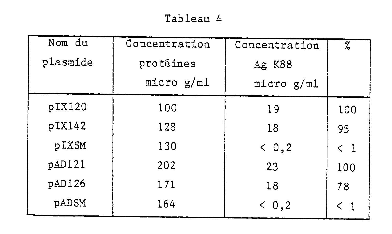
Tableau 4 : Concentrations en antigènes K88 (en micro g/ml) et en protéines totales (en micro g/ml; méthode de lowry) dans différents rasats thermiques déterminées par Elisa. La quantité d'antigènes K88 produite par les souches recombinantes est estimée par rapport à la quantité purifiée des souches sauvages. Table 4 : Concentrations of K88 antigens (in micro g / ml) and in total proteins (in micro g / ml; lowry method) in different thermal razors determined by Elisa. The amount of K88 antigens produced by the recombinant strains is estimated relative to the purified amount of the wild strains.
Le présent exemple est destiné à montrer que des pilines recombinantes dont les deux régions variables S1 et S2 contiennent une séquence étrangère aux pili, peuvent être construites et exprimées. Ceci est réalisé en combinant sur une même piline les séquences S1 des dérivés K88ac, avec les séquences S2 des dérivés K88ad.The present example is intended to show that recombinant pilins, the two variable regions of which S1 and S2 contain a sequence foreign to the pili, can be constructed and expressed. This is achieved by combining the S1 sequences of the K88ac derivatives on the same pilin with the S2 sequences of the K88ad derivatives.
Les gènes des pilines recombinantes contiennent la séquence S1-Flu2 du plasmide pIX142, et soit S2-Flu1 du pAD122 (essai 1) soit S2-Flu2 du pAD126 (essai 2). Les pilines codées par ces gènes sont formées d'une partie de la piline IX142, depuis leur extrémité amine jusqu'à et y compris la séquence S1, suivie de la séquence C-terminale de la piline AD122 (essai 1) ou AD126 (essai 2).The recombinant pilins genes contain the sequence S1-Flu2 from the plasmid pIX142, and either S2-Flu1 from pAD122 (test 1) or S2-Flu2 from pAD126 (test 2). The pilins coded by these genes are formed from part of the pilin IX142, from their amine end up to and including the sequence S1, followed by the C-terminal sequence of the pilin AD122 (test 1) or AD126 (test 2).
Comme tous les dérivés du pIX120 et du pAD120, les plasmides pIX142, pAD122 et pAD126 contiennent deux sites de restriction BglI. L'un est localisé dans le gène de résistance à l'ampicilline, et le second est situé dans le gène de la piline, précisément entre les régions S1 et S2 (référence est faite aux figures 3, 4, 5 et 6). Pour construire ces gènes recombinants, deux types de fragments sont purifiés :
- du pIX142 : le fragment BglI qui porte la fin du gène AmpR, l'origine de réplication et le début du gène de la piline, y compris S1-Flu2;
- du pAD122 (essai 1) ou pAD126 (essai 2) : le fragment BglI qui contient la fin du gène piline (y compris S2-Flu1 ou S2-Flu2) et le début du gène AmpR.Like all derivatives of pIX120 and pAD120, the plasmids pIX142, pAD122 and pAD126 contain two BglI restriction sites. One is located in the ampicillin resistance gene, and the second is located in the pilin gene, precisely between the regions S1 and S2 (reference is made to Figures 3, 4, 5 and 6). To build these recombinant genes, two types of fragments are purified:
- pIX142: the BglI fragment which carries the end of the AmpR gene, the origin of replication and the start of the pilin gene, including S1-Flu2;
- pAD122 (test 1) or pAD126 (test 2): the BglI fragment which contains the end of the pilin gene (including S2-Flu1 or S2-Flu2) and the start of the AmpR gene.
La ligation de ces deux types de fragment restaure les gènes de résistance à l'ampicilline et de la piline. Ces plasmides sont respectivement pIX143, dérivé du pAD122 (essai 1), et pIX144, formé à partir du pAD126 (essai 2).The ligation of these two types of fragment restores the genes for resistance to ampicillin and pilin. These plasmids are respectively pIX143, derived from pAD122 (test 1), and pIX144, formed from pAD126 (test 2).
La taille de la piline IX143 est de 266 acides aminés, et 262 acides aminés pour IX144.The size of the IX143 pilin is 266 amino acids, and 262 amino acids for IX144.
La production de bactéries porteuses de pili est obtenue après introduction de chacun des plasmides pIX143 et pIX144 dans la souche d'expression EC294 (pIX211).The production of pili-bearing bacteria is obtained after introduction of each of the plasmids pIX143 and pIX144 into the expression strain EC294 (pIX211).
L'ensemble des résultats expérimentaux montrent que :
- 1. Les régions variables S1 et S2 peuvent être délétées de la piline, sans compromettre la synthèse de pili.
- 2. Sur cette base, deux gènes pilines "vecteurs" dans lesquels l'insertion de DNA étranger est facilitée par la présence de sites de restriction uniques, ont été construits.
- 3. Des DNA codant pour des séquences peptidiques différentes par leur longeur et leur composition peuvent être introduites, par génie génétique, dans ces vecteurs, et déterminer des pilines recombinantes. Par complémentation intergénique, ces pilines peuvent être assemblées en pili attachés à la surface de la bactérie. Ceci est vrai pour deux régions différentes de la piline. La tolérance observée dans les exemples précédents pour chacune des régions est d'au moins de -4 à +10 pour S1 et de -6 à +6 pour S2.
- 4. Les pili recombinants peuvent être purifiés selon les mêmes méthodes que celles employées pour les pili sauvages.
- 5. Le peptide fusionné au pili est reconnu par des anticorps qui lui sont spécifiques et son antigénicité est amplifiée.
- 6. Le pili recombinant utilisé comme antigène induit deux types d'anticorps : des anticorps anti-pili, mais aussi des anticorps dirigés contre le peptide fusionné au pili. Ce peptide est rendu immunogénique. Des pili recombinants peuvent donc être utilisés dans des vaccins sous-unitaires.


- 1. The variable regions S1 and S2 can be deleted from the pilin, without compromising the synthesis of pili.
- 2. On this basis, two "vector" pilin genes in which the insertion of foreign DNA is facilitated by the presence of unique restriction sites, have been constructed.
- 3. DNA coding for peptide sequences different in length and composition can be introduced, by genetic engineering, into these vectors, and determine recombinant pilins. By intergenic complementation, these pilins can be assembled into pili attached to the surface of the bacteria. This is true for two different regions of the pilin. The tolerance observed in the previous examples for each of the regions is at least from -4 to +10 for S1 and from -6 to +6 for S2.
- 4. Recombinant pili can be purified using the same methods as those used for wild pili.
- 5. The peptide fused to the pili is recognized by antibodies which are specific to it and its antigenicity is amplified.
- 6. The recombinant pili used as antigen induces two types of antibody: anti-pili antibodies, but also antibodies directed against the peptide fused to the pili. This peptide is made immunogenic. Recombinant pili can therefore be used in subunit vaccines.
Analyse des sous-unités pilines produites par diverses souches E. coli, en gel de polyacrylamide -SDS et en Western blot.Analysis of pilin subunits produced by various E. coli strains, in polyacrylamide -SDS gel and in Western blot.
Piste R : références de PM : phosphorylase b-94KD, albumine 67KD, ovalbumine 43KD, anhydrase carbonique 30kD, inhibiteur de la trypsine 20 kD, alpha-lactalbumine 14.4.kD
- 1. rasat purifié de K88ac
- 2. rasat brut de K88ab NL
- 3. rasat brut de K88ab US
- 4. rasat brut de K88ac
- 5. rasat brut de EC294 (pIX115)
- 6. rasat brut de EC294 (pIX120)(pIX211)
- 7. rasat brut de K88ad NL
- 8. rasat brut de K88ad US
- 9. rasat brut de EC294 (pAD102)
- 10. rasat brut de EC294 (pAD120)(pIX211)
- 1. purified rasat of K88ac
- 2. gross rasat from K88ab NL
- 3. raw rasat from K88ab US
- 4. K88ac raw rasat
- 5. raw rasat of EC294 (pIX115)
- 6. raw rasat of EC294 (pIX120) (pIX211)
- 7. rasat brut from K88ad NL
- 8. K88ad US raw rasat
- 9. raw rasat of EC294 (pAD102)
- 10. raw EC294 rasat (pAD120) (pIX211)
Western blot du gel de la figure 1a.Western blot of the gel of Figure 1a.
Carte de restriction de l'opéron K88ac cloné dans le plasmide pBR322. La position des sites de restriction pour quelques enzymes courants est donnée, ainsi que la carte génétique de l'opéron. La taille des 6 protéines est donnée en kD. La protéine de 17.6 kD est putative.Restriction map of the operon K88ac cloned into the plasmid pBR322. The position of the restriction sites for some common enzymes is given, as well as the genetic map of the operon. The size of the 6 proteins is given in kD. The 17.6 kD protein is putative.
Séquence du gène de la sous-unité K88ac cloné dans le vecteur pBR322 et sa traduction en peptide, ainsi que la positi on de quelques sites de restriction remarquables.Sequence of the K88ac subunit gene cloned into the vector pBR322 and its translation into a peptide, as well as the position of a few remarkable restriction sites.
La séquence du vecteur est écrite en lettres minuscules.
RBS : ribosome binding site.
P₁ : promoteur cryptique du vecteur.The vector sequence is written in lowercase letters.
RBS: ribosome binding site.
P₁: cryptic promoter of the vector.
Carte de restriction du pIX120. La position du gène de la sous-unité K88ac (K88), du gène de résistance à l'ampicilline (AP) et de l'origine de réplication du pBR322 (Ori) est donnée.Restriction card of the pIX120. The position of the K88ac subunit gene (K88), the ampicillin resistance (AP) gene and the origin of replication of pBR322 (Ori) is given.
Séquence du gène de la sous-unité K88ad US et sa traduction en peptide ainsi que la position de quelques sites de restriction remarquables.Sequence of the K88ad US subunit gene and its translation into a peptide, as well as the position of a few remarkable restriction sites.
Carte de restriction du pAD120 avec le gène de la sous-unité K88AD, de résistance à l'ampicilline (AMP) et l'origine de réplication du plasmide (Ori).Restriction map of pAD120 with the K88AD subunit gene, ampicillin resistance (AMP) and the origin of replication of the plasmid (Ori).
Schéma de la complémentation intergénique pour K88ac : les gènes de l'opéron K88 (2), porté par le pIX115, ont été sous clonés sur deux vecteurs : pIX211, dérivé du pACYC184, porte les génes des protéines d'ancrage et d'assemblage des pili et pIX120, dérivé du pBR322, contenant le seul gène de la sous unité (1). P₁ est un promoteur cryptique du vecteur. ORI : origine de réplication autonome.Diagram of the intergenic complementation for K88ac: the genes of the operon K88 (2), carried by pIX115, were subcloned on two vectors: pIX211, derived from pACYC184, carries the genes of the anchoring and assembly proteins pili and pIX120, derived from pBR322, containing the only gene for the subunit (1). P₁ is a cryptic promoter of the vector. ORI: origin of autonomous replication.
Profil d'hydrophilicité de la piline K88ac, d'après l'algorithme de Doolitle. Les zones particulièrement hydrophiles sont hachurées.Hydrophilicity profile of pilin K88ac, according to the Doolitle algorithm. The particularly hydrophilic areas are hatched.
Comparaison des séquences protéiques des trois sérotypes K88 et de leurs variants. Seules les différences en acides aminés sont notées. Des délétions symbolisées par un "-" sont introduites pour faire correspondre au mieux les séquences peptidiques.Comparison of the protein sequences of the three K88 serotypes and their variants. Only the differences in amino acids are noted. Deletions symbolized by a "-" are introduced to best match the peptide sequences.
Comparaison des spectres d'hydrophilicité des différents sérotypes et de leurs variants suivant l'algorithme de Doolittle.Comparison of the hydrophilicity spectra of the different serotypes and their variants according to the Doolittle algorithm.
Manipulations de la région S1. Les plasmides pIX126 (2), pIX130 (3), pIX131 (4), pIX128 (5), pIX136 (6) et pIX139 (7) sont dérivés du gène K88ac du pIX120 (1). Les acides aminés variables de S₁ sont identifiés par un astérisque *.Manipulations of the S1 region. The plasmids pIX126 (2), pIX130 (3), pIX131 (4), pIX128 (5), pIX136 (6) and pIX139 (7) are derived from the K88ac gene of pIX120 (1). The variable amino acids of S₁ are identified by an asterisk *.
Test immunologique sur colonies des souches recombinantes.

L'anticorps est un polyclonal anti K88ac. Cet anticorps est reconnu par un anti-lapin couplé à la peroxydase.The antibody is an anti-K88ac polyclonal. This antibody is recognized by an anti-rabbit coupled to peroxidase.
figure 13 - Etapes de clonage permettant le remplacement de la région S2 par un adaptateur synthétique. Les cartes de restriction des différents intermédiaires qui ont permis la construction du pAD121 sont présentées à la figure 13a : pUC8, figure 13b : pUC81, figure 13c : pUC82, figure 13d : pUC83, figure 13e : pUC84 et enfin figure 13 f : pAD121. figure 13 - Cloning steps allowing the replacement of the region S2 by a synthetic adapter. The restriction maps of the various intermediaries which enabled the construction of pAD121 are presented in Figure 13a: pUC8, Figure 13b: pUC81, Figure 13c: pUC82, Figure 13d: pUC83, Figure 13e: pUC84 and finally Figure 13 f: pAD121.
figure 14 - Manipulation de la région S2 : les plasmides pAD121 (2), et pAD122 (3) sont dérivés du plasmide pAD120 (1). FIG. 14 - Manipulation of the region S2: the plasmids pAD121 (2), and pAD122 (3) are derived from the plasmid pAD120 (1).
Figure 15a - gel de PAGE-SDS d'un rasat brut des souches
1 - EC294 (pAD120)(pIX211)
2 - EC294 (pAD121)(pIX211)
3 - EC294 (pAD122)(pIX211) Figure 15a - PAGE-SDS gel from a crude rasat of the strains
1 - EC294 (pAD120) (pIX211)
2 - EC294 (pAD121) (pIX211)
3 - EC294 (pAD122) (pIX211)
Western blot des rasats bruts séparés sur le gel de la figure 15a.Western blot of the raw rasats separated on the gel of figure 15a.
figure 16 - Test immunologique sur diverses bactéries recombinantes avec un sérum anti-K88. Trois colonies isolées de chaque type de bactéries ont été transférées sur le filtre. A l'exception des clones de la ligne 7 qui contiennent le pACYC184 au lieu du pIX211, ce sont des souches EC294 (pIX211) porteuse de l'un des plasmides recombinants suivants :

Figure 17 - La légende est la même que pour la figure 16, sauf que le moyen de détection est un anticorps monoclonal anti-K88. Figure 17 - The legend is the same as for Figure 16, except that the detection means is an anti-K88 monoclonal antibody.
Figure 18 - Elisa des rasages thermiques des souches contenant le plasmide pIX120 (1), pIX128 (2) et pIX136 (3), détecté par un anticorps monoclonal anti-K88. La référence (R) est à 1 micro g/ml. En abscisse se trouve le taux de dilution, en ordonnée la densité optique. Figure 18 - Elisa thermal shaving of the strains containing the plasmid pIX120 (1), pIX128 (2) and pIX136 (3), detected by an anti-K88 monoclonal antibody. The reference (R) is at 1 micro g / ml. On the abscissa is the dilution rate, on the ordinate the optical density.
Figure 19 - Réaction sur Elisa de deux monoclonaux anti-Flu1 contre des pili IX128 (I) et (IX136 (II).
En abscisse se trouve le taux de dilution, en ordonnée la densité optique. Figure 19 - Reaction on Elisa of two anti-Flu1 monoclonals against pili IX128 (I) and (IX136 (II).
On the abscissa is the dilution rate, on the ordinate the optical density.
Figure 20 - Réponse humorale des quatre rats immunisés par des concentrations croissantes d'antigènes AD122, contre le pili K88. La quantité d'antigènes par dose est reportée en abscisse. L'absorbance à la dilution 10 de chaque sérum contre le pili est en ordonnée. En abscisse se trouve la quantité de micro g de pili injectés. Figure 20 - Humoral response of the four rats immunized with increasing concentrations of AD122 antigens, against K88 pili. The quantity of antigens per dose is plotted on the abscissa. The absorbance at
Figure 21 - Même légende que la figure 20, sinon que l'antigène adsorbé est le peptide synthétique Flu1. En abscisse se trouve la quantité de micro g de pili injectés et l'absorbance à la dilution 10 de chaque sérum contre le pili est ordonnée. Figure 21 - Same legend as Figure 20, except that the adsorbed antigen is the synthetic peptide Flu1. On the abscissa is the amount of micro g of pili injected and the absorbance at
Claims (14)
Priority Applications (1)
| Application Number | Priority Date | Filing Date | Title |
|---|---|---|---|
| AT87201873T ATE84314T1 (en) | 1986-10-13 | 1987-09-30 | MICROORGANISMS CARRYING FOREIGN PROTEIN CONTAINING PILI ISOLATED PILI, METHOD OF EXCRETING PROTEINS IN USE OF THESE PILI AND THEIR BENEFITS. |
Applications Claiming Priority (4)
| Application Number | Priority Date | Filing Date | Title |
|---|---|---|---|
| FR8614294A FR2605017B1 (en) | 1986-10-13 | 1986-10-13 | MICROORGANISMS COMPRISING PILIS AS CARRIER PROTEINS, PILI ISOLATED FROM SUCH MICROORGANISMS AND METHOD FOR EXCRETING PEPTIDES OR PROTEINS USING THE SAME. |
| FR8614294 | 1986-10-13 | ||
| FR8616610 | 1986-11-26 | ||
| FR8616610A FR2607149B1 (en) | 1986-11-26 | 1986-11-26 | MICROORGANISMS COMPRISING PILIS AS CARRIER PROTEINS, PILI ISOLATED FROM SUCH MICROORGANISMS AND METHOD FOR EXCRETING PEPTIDES OR PROTEINS USING THE SAME |
Publications (2)
| Publication Number | Publication Date |
|---|---|
| EP0264150A1 true EP0264150A1 (en) | 1988-04-20 |
| EP0264150B1 EP0264150B1 (en) | 1993-01-07 |
Family
ID=26225525
Family Applications (1)
| Application Number | Title | Priority Date | Filing Date |
|---|---|---|---|
| EP87201873A Expired - Lifetime EP0264150B1 (en) | 1986-10-13 | 1987-09-30 | Microorganisms comprising pili as carrier proteins, isolated pili, method for excreting proteins using these pili and their use |
Country Status (2)
| Country | Link |
|---|---|
| EP (1) | EP0264150B1 (en) |
| DE (1) | DE3783425T2 (en) |
Cited By (3)
| Publication number | Priority date | Publication date | Assignee | Title |
|---|---|---|---|---|
| FR2699538A1 (en) * | 1992-12-22 | 1994-06-24 | Agronomique Inst Nat Rech | Subunit of a CS31A protein capsule modified with at least one heterologous peptide, CS31A protein capsule and microorganisms carrying these subunits; methods of obtaining and using them. |
| WO1995034648A1 (en) * | 1994-06-10 | 1995-12-21 | Dade International Inc. | A method for displaying proteins |
| AU667715B2 (en) * | 1992-06-18 | 1996-04-04 | Akzo N.V. | Carrier system against GnRH |
Citations (4)
| Publication number | Priority date | Publication date | Assignee | Title |
|---|---|---|---|---|
| GB2094314A (en) * | 1981-02-05 | 1982-09-15 | Wellcome Found | Recombinant DNA |
| EP0060129A2 (en) * | 1981-03-09 | 1982-09-15 | Cetus Corporation | Vaccines, a method for making vaccines and a method for stimulating antibody production |
| EP0174759A1 (en) * | 1984-08-22 | 1986-03-19 | Connaught Laboratories Limited | Multispecific immunogenic proteins |
| WO1986002557A1 (en) * | 1984-10-31 | 1986-05-09 | Commonwealth Scientific And Industrial Research Or | Improved antigenic preparation |
-
1987
- 1987-09-30 EP EP87201873A patent/EP0264150B1/en not_active Expired - Lifetime
- 1987-09-30 DE DE8787201873T patent/DE3783425T2/en not_active Expired - Fee Related
Patent Citations (4)
| Publication number | Priority date | Publication date | Assignee | Title |
|---|---|---|---|---|
| GB2094314A (en) * | 1981-02-05 | 1982-09-15 | Wellcome Found | Recombinant DNA |
| EP0060129A2 (en) * | 1981-03-09 | 1982-09-15 | Cetus Corporation | Vaccines, a method for making vaccines and a method for stimulating antibody production |
| EP0174759A1 (en) * | 1984-08-22 | 1986-03-19 | Connaught Laboratories Limited | Multispecific immunogenic proteins |
| WO1986002557A1 (en) * | 1984-10-31 | 1986-05-09 | Commonwealth Scientific And Industrial Research Or | Improved antigenic preparation |
Non-Patent Citations (3)
| Title |
|---|
| CHEMICAL ABSTRACTS, vol. 96, no. 25, 21 juin 1982, pages 163-164, résumé no. 211670p, Columbus, Ohio, US; F.R. MOOI et al.: "Construction and characterization of mutants impaired in the biosynthesis of the K88ab antigen", & J. BACTERIOL. 1982, 150(2), 512-21 * |
| CONFERENCE PAPERS INDEX, CAMBRIDGE SCIENTIFIC ABSTRACTS, 1986, résumé no. 86007853, Washington, US; D. NEWMAN et al.: "Extracellular expression of a linear viral epitope inserted into the K88ab pilus structural subunit" * |
| MICROBIOLOGICAL REVIEWS, vol. 46, no. 2, juin 1982, pages 129-161, US; W. GAASTRA et al.: "Host-specific fimbrial adhesins of noninvasive enterotoxigenic Escherichia coli strains" * |
Cited By (7)
| Publication number | Priority date | Publication date | Assignee | Title |
|---|---|---|---|---|
| AU667715B2 (en) * | 1992-06-18 | 1996-04-04 | Akzo N.V. | Carrier system against GnRH |
| FR2699538A1 (en) * | 1992-12-22 | 1994-06-24 | Agronomique Inst Nat Rech | Subunit of a CS31A protein capsule modified with at least one heterologous peptide, CS31A protein capsule and microorganisms carrying these subunits; methods of obtaining and using them. |
| WO1994014967A2 (en) * | 1992-12-22 | 1994-07-07 | Institut National De La Recherche Agronomique (Inra) | Cs31a protein capsule subunit modified by at least one heterologous peptide, cs31a protein capsule comprising said subunit, micro-organisms having such subunits on their outer membrane, and methods for preparing and using same |
| WO1994014967A3 (en) * | 1992-12-22 | 1994-09-29 | Agronomique Inst Nat Rech | Cs31a protein capsule subunit modified by at least one heterologous peptide, cs31a protein capsule comprising said subunit, micro-organisms having such subunits on their outer membrane, and methods for preparing and using same |
| WO1995034648A1 (en) * | 1994-06-10 | 1995-12-21 | Dade International Inc. | A method for displaying proteins |
| US5516637A (en) * | 1994-06-10 | 1996-05-14 | Dade International Inc. | Method involving display of protein binding pairs on the surface of bacterial pili and bacteriophage |
| AU697865B2 (en) * | 1994-06-10 | 1998-10-22 | Sunol Molecular Corporation | A method for displaying proteins |
Also Published As
| Publication number | Publication date |
|---|---|
| DE3783425D1 (en) | 1993-02-18 |
| EP0264150B1 (en) | 1993-01-07 |
| DE3783425T2 (en) | 1993-05-13 |
Similar Documents
| Publication | Publication Date | Title |
|---|---|---|
| Ferrero et al. | Recombinant antigens prepared from the urease subunits of Helicobacter spp.: evidence of protection in a mouse model of gastric infection | |
| Eslava et al. | Pet, an autotransporter enterotoxin from enteroaggregative Escherichia coli | |
| US5871943A (en) | Immunoassay comprising a truncated pneumococcal surface protein A (PspA) | |
| EP1066375B1 (en) | $i(LACTOBACILLI) HARBORING AGGREGATION AND MUCIN BINDING GENES AS VACCINE DELIVERY VEHICLES | |
| EP0586266B1 (en) | DNA fragments coding for the Neisseria meningitidis receptor fragments | |
| JP3423712B2 (en) | Recombinant DNA sequences encoding signal peptides, selective interacting polypeptides and membrane anchoring sequences | |
| EP0712414A1 (en) | Recombinant vector containing a lipoprotein gene sequence for expressing nucleotide sequences | |
| JP2003189879A (en) | Roteinase k resistant surface protein of neisseria meningitidis | |
| JPH08505045A (en) | Method for detecting tagA gene and predisposition to peptic ulcer formation | |
| CN1541263A (en) | Improved methods for binding AcmA-type protein anchor fusions to cell-wall material of micro-organisms | |
| CA2301374A1 (en) | Polypeptide nucleic sequences exported from mycobacteria, vectors comprising same and uses for diagnosing and preventing tuberculosis | |
| Kong et al. | Regulated delayed expression of rfaH in an attenuated Salmonella enterica serovar Typhimurium vaccine enhances immunogenicity of outer membrane proteins and a heterologous antigen | |
| Monteiro‐Neto et al. | Characterization of an outer membrane protein associated with haemagglutination and adhesive properties of enteroaggregative Escherichia coli O111: H12 | |
| Wolf et al. | Characterization of CS4 and CS6 antigenic components of PCF8775, a putative colonization factor complex from enterotoxigenic Escherichia coli E8775 | |
| EA031812B1 (en) | Novel live attenuated shigella vaccine | |
| JPH11504633A (en) | Multimeric recombinant urease vaccine | |
| EP0791063B1 (en) | Carrier protein having an adjuvant effect, immunogenic complex containing same, preparation method therefor, nucleotide sequence and vaccine | |
| JP4368944B2 (en) | Helicobacter antigen for defense | |
| JP2009022299A (en) | MUTANT SHIGELLA FLEXNERI 2a | |
| EP0824548B1 (en) | Hybrid molecules between heat-labile enterotoxin and cholera toxin b subunits | |
| EP0264150B1 (en) | Microorganisms comprising pili as carrier proteins, isolated pili, method for excreting proteins using these pili and their use | |
| WO1998017804A2 (en) | A HELICOBACTER ANTIGEN (η-GLUTAMYLTRANSFERASE) AND SEQUENCES ENCODING THE SAME | |
| Dibb‐Fuller et al. | Expression of SEF17 fimbriae by Salmonella enteritidis | |
| Serushago et al. | Role of antibodies against outer-membrane proteins in murine resistance to infection with encapsulated Klebsiella pneumoniae | |
| US6899881B2 (en) | H. PYLORI FlbA POLYPEPTIDES, IMMUNOGENIC AND VACCINATING COMPOSITIONS COMPRISING H. PYLORI FlbA POLYPEPTIDES, AND COMPOSITIONS FOR DETECTION OF AN INFECTION COMPRISING H. PYLORI FlbA POLYPEPTIDES |
Legal Events
| Date | Code | Title | Description |
|---|---|---|---|
| PUAI | Public reference made under article 153(3) epc to a published international application that has entered the european phase |
Free format text: ORIGINAL CODE: 0009012 |
|
| AK | Designated contracting states |
Kind code of ref document: A1 Designated state(s): AT BE CH DE ES FR GB GR IT LI LU NL SE |
|
| 17P | Request for examination filed |
Effective date: 19881010 |
|
| 17Q | First examination report despatched |
Effective date: 19910527 |
|
| RAP1 | Party data changed (applicant data changed or rights of an application transferred) |
Owner name: SOLVAY |
|
| GRAA | (expected) grant |
Free format text: ORIGINAL CODE: 0009210 |
|
| AK | Designated contracting states |
Kind code of ref document: B1 Designated state(s): AT BE CH DE ES FR GB GR IT LI LU NL SE |
|
| PG25 | Lapsed in a contracting state [announced via postgrant information from national office to epo] |
Ref country code: SE Effective date: 19930107 Ref country code: GR Free format text: LAPSE BECAUSE OF FAILURE TO SUBMIT A TRANSLATION OF THE DESCRIPTION OR TO PAY THE FEE WITHIN THE PRESCRIBED TIME-LIMIT Effective date: 19930107 Ref country code: FR Free format text: THE PATENT HAS BEEN ANNULLED BY A DECISION OF A NATIONAL AUTHORITY Effective date: 19930107 Ref country code: AT Effective date: 19930107 |
|
| REF | Corresponds to: |
Ref document number: 84314 Country of ref document: AT Date of ref document: 19930115 Kind code of ref document: T |
|
| REF | Corresponds to: |
Ref document number: 3783425 Country of ref document: DE Date of ref document: 19930218 |
|
| ITF | It: translation for a ep patent filed |
Owner name: BARZANO' E ZANARDO MILANO S.P.A. |
|
| PG25 | Lapsed in a contracting state [announced via postgrant information from national office to epo] |
Ref country code: ES Free format text: LAPSE BECAUSE OF FAILURE TO SUBMIT A TRANSLATION OF THE DESCRIPTION OR TO PAY THE FEE WITHIN THE PRESCRIBED TIME-LIMIT Effective date: 19930418 |
|
| GBT | Gb: translation of ep patent filed (gb section 77(6)(a)/1977) |
Effective date: 19930406 |
|
| PG25 | Lapsed in a contracting state [announced via postgrant information from national office to epo] |
Ref country code: LU Free format text: LAPSE BECAUSE OF NON-PAYMENT OF DUE FEES Effective date: 19930930 Ref country code: LI Effective date: 19930930 Ref country code: GB Effective date: 19930930 Ref country code: CH Effective date: 19930930 Ref country code: BE Effective date: 19930930 |
|
| PLBE | No opposition filed within time limit |
Free format text: ORIGINAL CODE: 0009261 |
|
| STAA | Information on the status of an ep patent application or granted ep patent |
Free format text: STATUS: NO OPPOSITION FILED WITHIN TIME LIMIT |
|
| 26N | No opposition filed | ||
| BERE | Be: lapsed |
Owner name: SOLVAY Effective date: 19930930 |
|
| PG25 | Lapsed in a contracting state [announced via postgrant information from national office to epo] |
Ref country code: NL Effective date: 19940401 |
|
| NLV4 | Nl: lapsed or anulled due to non-payment of the annual fee | ||
| GBPC | Gb: european patent ceased through non-payment of renewal fee |
Effective date: 19930930 |
|
| REG | Reference to a national code |
Ref country code: CH Ref legal event code: PL |
|
| PG25 | Lapsed in a contracting state [announced via postgrant information from national office to epo] |
Ref country code: DE Effective date: 19940601 |
|
| REG | Reference to a national code |
Ref country code: FR Ref legal event code: ST |
|
| PG25 | Lapsed in a contracting state [announced via postgrant information from national office to epo] |
Ref country code: IT Free format text: LAPSE BECAUSE OF NON-PAYMENT OF DUE FEES;WARNING: LAPSES OF ITALIAN PATENTS WITH EFFECTIVE DATE BEFORE 2007 MAY HAVE OCCURRED AT ANY TIME BEFORE 2007. THE CORRECT EFFECTIVE DATE MAY BE DIFFERENT FROM THE ONE RECORDED. Effective date: 20050930 |