EP0289949A2 - Intercellular adhesion molecules, and their binding ligands - Google Patents
Intercellular adhesion molecules, and their binding ligands Download PDFInfo
- Publication number
- EP0289949A2 EP0289949A2 EP88106901A EP88106901A EP0289949A2 EP 0289949 A2 EP0289949 A2 EP 0289949A2 EP 88106901 A EP88106901 A EP 88106901A EP 88106901 A EP88106901 A EP 88106901A EP 0289949 A2 EP0289949 A2 EP 0289949A2
- Authority
- EP
- European Patent Office
- Prior art keywords
- icam
- cells
- antibody
- binding
- cell
- Prior art date
- Legal status (The legal status is an assumption and is not a legal conclusion. Google has not performed a legal analysis and makes no representation as to the accuracy of the status listed.)
- Granted
Links
Images


Classifications
-
- C—CHEMISTRY; METALLURGY
- C07—ORGANIC CHEMISTRY
- C07K—PEPTIDES
- C07K16/00—Immunoglobulins [IGs], e.g. monoclonal or polyclonal antibodies
- C07K16/18—Immunoglobulins [IGs], e.g. monoclonal or polyclonal antibodies against material from animals or humans
- C07K16/28—Immunoglobulins [IGs], e.g. monoclonal or polyclonal antibodies against material from animals or humans against receptors, cell surface antigens or cell surface determinants
- C07K16/2803—Immunoglobulins [IGs], e.g. monoclonal or polyclonal antibodies against material from animals or humans against receptors, cell surface antigens or cell surface determinants against the immunoglobulin superfamily
- C07K16/2821—Immunoglobulins [IGs], e.g. monoclonal or polyclonal antibodies against material from animals or humans against receptors, cell surface antigens or cell surface determinants against the immunoglobulin superfamily against ICAM molecules, e.g. CD50, CD54, CD102
-
- A—HUMAN NECESSITIES
- A61—MEDICAL OR VETERINARY SCIENCE; HYGIENE
- A61P—SPECIFIC THERAPEUTIC ACTIVITY OF CHEMICAL COMPOUNDS OR MEDICINAL PREPARATIONS
- A61P29/00—Non-central analgesic, antipyretic or antiinflammatory agents, e.g. antirheumatic agents; Non-steroidal antiinflammatory drugs [NSAID]
-
- A—HUMAN NECESSITIES
- A61—MEDICAL OR VETERINARY SCIENCE; HYGIENE
- A61P—SPECIFIC THERAPEUTIC ACTIVITY OF CHEMICAL COMPOUNDS OR MEDICINAL PREPARATIONS
- A61P35/00—Antineoplastic agents
-
- C—CHEMISTRY; METALLURGY
- C07—ORGANIC CHEMISTRY
- C07K—PEPTIDES
- C07K14/00—Peptides having more than 20 amino acids; Gastrins; Somatostatins; Melanotropins; Derivatives thereof
- C07K14/435—Peptides having more than 20 amino acids; Gastrins; Somatostatins; Melanotropins; Derivatives thereof from animals; from humans
- C07K14/705—Receptors; Cell surface antigens; Cell surface determinants
- C07K14/70503—Immunoglobulin superfamily
- C07K14/70525—ICAM molecules, e.g. CD50, CD54, CD102
-
- C—CHEMISTRY; METALLURGY
- C07—ORGANIC CHEMISTRY
- C07K—PEPTIDES
- C07K16/00—Immunoglobulins [IGs], e.g. monoclonal or polyclonal antibodies
- C07K16/18—Immunoglobulins [IGs], e.g. monoclonal or polyclonal antibodies against material from animals or humans
- C07K16/28—Immunoglobulins [IGs], e.g. monoclonal or polyclonal antibodies against material from animals or humans against receptors, cell surface antigens or cell surface determinants
- C07K16/2839—Immunoglobulins [IGs], e.g. monoclonal or polyclonal antibodies against material from animals or humans against receptors, cell surface antigens or cell surface determinants against the integrin superfamily
- C07K16/2845—Immunoglobulins [IGs], e.g. monoclonal or polyclonal antibodies against material from animals or humans against receptors, cell surface antigens or cell surface determinants against the integrin superfamily against integrin beta2-subunit-containing molecules, e.g. CD11, CD18
-
- G—PHYSICS
- G01—MEASURING; TESTING
- G01N—INVESTIGATING OR ANALYSING MATERIALS BY DETERMINING THEIR CHEMICAL OR PHYSICAL PROPERTIES
- G01N33/00—Investigating or analysing materials by specific methods not covered by groups G01N1/00 - G01N31/00
- G01N33/48—Biological material, e.g. blood, urine; Haemocytometers
- G01N33/50—Chemical analysis of biological material, e.g. blood, urine; Testing involving biospecific ligand binding methods; Immunological testing
- G01N33/53—Immunoassay; Biospecific binding assay; Materials therefor
- G01N33/574—Immunoassay; Biospecific binding assay; Materials therefor for cancer
- G01N33/57407—Specifically defined cancers
- G01N33/5743—Specifically defined cancers of skin, e.g. melanoma
-
- A—HUMAN NECESSITIES
- A61—MEDICAL OR VETERINARY SCIENCE; HYGIENE
- A61K—PREPARATIONS FOR MEDICAL, DENTAL OR TOILETRY PURPOSES
- A61K38/00—Medicinal preparations containing peptides
-
- C—CHEMISTRY; METALLURGY
- C07—ORGANIC CHEMISTRY
- C07K—PEPTIDES
- C07K2317/00—Immunoglobulins specific features
- C07K2317/70—Immunoglobulins specific features characterized by effect upon binding to a cell or to an antigen
- C07K2317/73—Inducing cell death, e.g. apoptosis, necrosis or inhibition of cell proliferation
Definitions
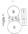
- the present invention relates to intercellular adhesion molecules such as ICAM-1 which are involved in the process through which populations of lymphocytes recognize and adhere to cellular substrates so that they may migrate to sites of inflammation and interact with cells during inflammatory reactions.
- the present invention additionally relates to ligand molecules capable of binding to such intercellular adhesion molecules, to a screening assay for these ligands, and to uses for the intercellular adhesion molecule, the ligand molecules, and the screening assay.
- Leukocytes must be able to attach to cellular substrates in order to properly defend the host against foreign invaders such as bacteria or viruses.
- An excellent review of the defense system is provided by Eisen, H.W., ( In: Microbiology , 3rd Ed., Harper & Row, Philadelphia, PA (1980), pp. 290-295 and 381-418). They must be able to attach to endothelial cells so that they can migrate from circulation to sites of ongoing inflammation. Furthermore, they must attach to antigen-presenting cells so that a normal specific immune response can occur, and finally, they must attach to appropriate target cells so that lysis of virally-infected or tumor cells can occur.
- Mac-1 is a heterodimer found on macrophages, granulocytes and large granular lymphocytes.
- LFA-1 is a heterodimer found on most lymphocytes (Springer, T.A., et al . Immunol. Rev. 68 :111-135 (1982)).
- p150,95 which has a tissue distribution similar to Mac-1 play a role in cellular adhesion (Keizer, G. et al ., Eur. J. Immunol. 15 :1142-1147 (1985)).
- leukocyte molecules were found to be members of a related family of glycoproteins (Sanchez-Madrid, F. et al ., J. Exper. Med. 158 :1785-1803 (1983); Keizer, G.D. et al ., Eur. J. Immunol. 15 :1142-1147 (1985)).
- This glycoprotein family is composed of heterodimers having one alpha chain and one beta chain. Although the alpha chain of each of the antigens differed from one another, the beta chain was found to be highly conserved (Sanchez-Madrid, F. et al ., J. Exper. Med. 158 :1785-1803 (1983)).
- the beta chain of the glycoprotein family (sometimes referred to as "CD18") was found to have a molecular weight of 95 kd whereas the alpha chains were found to vary from 150 kd to 180 kd (Springer, T., Fed. Proc. 44 :2660-2663 (1985)).
- CD18 The beta chain of the glycoprotein family
- the alpha subunits of the membrane proteins do not share the extensive homology shared by the beta subunits, close analysis of the alpha subunits of the glycoproteins has revealed that there are substantial similarities between them. Reviews of the similarities between the alpha and beta subunits of the LFA-1 related glycoproteins are provided by Sanchez-Madrid, F. et al ., ( J. Exper. Med. 158 :586-602 (1983); J. Exper. Med. 158 :1785-1803 (1983)).
- a group of individuals has been identified who are unable to express normal amounts of any member of this adhesion protein family on their leukocyte cell surface (Anderson, D.C., et al ., Fed. Proc. 44 :2671-2677 (1985); Anderson, D.C., et al ., J. Infect. Dis. 152 :668-689 (1985)). Lymphocytes from these patients displayed in vitro defects similar to normal counterparts whose LFA-1 family of molecules had been antagonized by antibodies. Furthermore, these individuals were unable to mount a normal immune response due to an inability of their cells to adhere to cellular substrates (Anderson, D.C., et al ., Fed. Proc.
- lymphocytes to maintain the health and viability of an animal requires that they be capable of adhering to other cells (such as endothelial cells).
- This adherence has been found to require cell-cell contacts which involve specific receptor molecules present on the cell surface of the lymphocytes. These receptors enable a lymphocyte to adhere to other lymphocytes or to endothelial, and other non-vascular cells.
- the cell surface receptor molecules have been found to be highly related to one another. Humans whose lymphocytes lack these cell surface receptor molecules exhibit chronic and recurring infections, as well as other clinical symptoms including defective antibody responses.
- lymphocyte adhesion is involved in the process through which foreign tissue is identified and rejected, an understanding of this process is of significant value in the fields of organ transplantation, tissue grafting, allergy and oncology.
- the present invention relates to Intercellular Adhesion Molecule-1 (ICAM-1) as well as to its functional derivatives.
- the invention additionally pertains to antibodies and fragments of antibodies capable of inhibiting the function of ICAM-1, and to other inhibitors of ICAM-1 function; and to assays capable of identifying such inhibitors.
- the invention additionally includes diagnostic and therapeutic uses for all of the above-described molecules.
- the invention includes the intercellular adhesion molecule ICAM-1 or its functional derivatives, which are substantially free of natural contaminants.
- the invention further pertains to such molecules which are additionally capable of binding to a molecule present on the surface of a lymphocyte.
- the invention further pertains to the intercellular adhesion molecule ICAM-1, and its derivatives which are detectably labeled.
- the invention additionally includes a recombinant DNA molecule capable of expressing ICAM-1 or a functional derivative thereof.
- the invention also includes a method for recovering ICAM-1 in substantially pure form which comprises the steps:
- the invention additionally includes an antibody capable of binding to a molecule selected from the group consisting of ICAM-1 and a functional derivative of ICAM-1.
- the invention also includes a hybridoma cell capable of producing such an antibody.
- the invention further includes a hybridoma cell capable of producing the monoclonal antibody R6-5-D6.
- the invention further includes a method for producing a desired hybridoma cell that produces an antibody which is capable of binding to ICAM-1, which comprises:
- the invention includes as well the hybridoma cell, and the antibody produced by the hybridoma cell, obtained by the above method.
- the invention is also directed to a method of identifying a non-immunoglobulin antagonist of intercellular adhesion which comprises:
- the invention is also directed toward a method for treating inflammation resulting from a response of the specific defense system in a mammalian subject which comprises providing to a subject in need of such treatment an amount of an anti-inflammatory agent sufficient to suppress the inflammation; wherein the anti-inflammatory agent is selected from the group consisting of: an antibody capable of binding to ICAM-1; a fragment of an antibody, the fragment being capable of binding to ICAM-1; ICAM-1; a functional derivative of ICAM-1; and a non-immunoglobulin antagonist of ICAM-1.
- the invention further includes the above-described method of treating inflammation wherein the non-immunoglobulin antagonist of ICAM-1 is a non-immunoglobulin antagonist of ICAM-1 other than LFA-1.
- the invention is also directed to a method of suppressing the metastasis of a hematopoietic tumor cell, the cell requiring a functional member of the LFA-1 family for migration, which method comprises providing to a patient in need of such treatment an amount of an anti-inflammatory agent sufficient to suppress the metastasis; wherein the anti-inflammatory agent is selected from the group consisting of: an antibody capable of binding to ICAM-1; a fragment of an antibody, the fragment being capable of binding to ICAM-1; ICAM-1; ICAM-1; a functional derivative of ICAM-1; and a non-immunoglobulin antagonist of ICAM-1.
- the invention further includes the above-described method of suppressing the metastasis of a hematopoietic tumor cell, wherein the non-immunoglobulin antagonist of ICAM-1 is a non-immunoglobulin antagonist of ICAM-1 other than LFA-1.
- the invention also includes a method of suppressing the growth of an ICAM-1-expressing tumor cell which comprises providing to a patient in need of such treatment an amount of a toxin sufficient to suppress the growth, the toxin being selected from the group consisting of a toxin-derivatized antibody capable of binding to ICAM-1; a toxin-derivatized fragment of an antibody, the fragment being capable of binding to ICAM-1; a toxin-derivatized member of the LFA-1 family of molecules; and a toxin-derivatized functional derivative of a member of the LFA-1 family of molecules.
- the invention is also directed to a method of suppressing the growth of an LFA-1-expressing tumor cell which comprises providing to a patient in need of such treatment an amount of toxin sufficient to suppress such growth, the toxin being selected from the group consisting of a toxin-derivatized ICAM-1; and a toxin-derivatized functional derivative of ICAM-1.
- the invention is further directed toward a method of diagnosing the presence and location of an inflammation resulting from a response of the specific defense system in a mammalian subject suspected of having the inflammation which comprises:
- the invention additionally provides a method of diagnosing the presence and location of an inflammation resulting from a response of the specific defense system in a mammalian subject suspected of having the inflammation which comprises:
- the invention also pertains to a method of diagnosing the presence and location of an ICAM-1-expressing tumor cell in a mammalian subject suspected of having such a cell, which comprises:
- the invention also pertains to a method of diagnosing the presence and location of an ICAM-1-expressing tumor cell in a mammalian subject suspected of having such a cell, which comprises:
- the invention also pertains to a method of diagnosing the presence and location of a tumor cell which expresses a member of the LFA-1 family of molecules in a subject suspected of having such a cell, which comprises:
- the invention also pertains to a method of diagnosing the presence and location of a tumor cell which expresses a member of the LFA-1 family of molecules in a subject suspected of having such a cell, which comprises:
- One aspect of the present invention relates to the discovery of a natural binding ligand to LFA-1.
- Molecules such as those of LFA-1 family, which are involved in the process of cellular adhesion are referred to as "adhesion molecules.”
- the natural binding ligand of the present invention is designated "Intercellular Aadhesion Molecule-1" or "ICAM-1.”
- ICAM-1 is a 76-97 Kd glycoprotein. ICAM-1 is not a heterodimer.
- the present invention is directed toward ICAM-1 and its "functional derivatives.”
- a “functional derivative” of ICAM-1 is a compound which posesses a biological activity (either functional or structural) that is substantially similar to a biological activity of ICAM-1.
- the term “functional derivatives” is intended to include the “fragments,” “variants,” “analogs,” or “chemical derivatives” of a molecule.
- a “fragment” of a molecule such as ICAM-1 is meant to refer to any polypeptide subset of the molecule.
- a “variant” of a molecule such as ICAM-1 is meant to refer to a molecule substantially similar in structure and function to either the entire molecule, or to a fragment thereof.
- a molecule is said to be “substantially similar” to another molecule if both molecules have substantially similar structures or if both molecules possess a similar biological activity.
- two molecules possess a similar activity they are considered variants as that term is used herein even if the structure of one of the molecules not found in the other, or if the sequence of amino acid residues is not identical.
- An “analog” of a molecule such as ICAM-1 is meant to refer to a molecule substantially similar in function to either the entire molecule or to a fragment thereof.
- a molecule is said to be a "chemical derivative" of another molecule when it contains additional chemical moieties not normally a part of the molecule. Such moieties may improve the molecule's solubility, absorption, biological half life, etc. The moieties may alternatively decrease the toxicity of the molecule, eliminate or attenuate any undesirable side effect of the molecule, etc. Moieties capable of mediating such effects are disclosed in Remington's Pharmaceutical Sciences (1980). "Toxin-derivatized" molecules constitute a special class of "chemical derivatives.”
- a "toxin-derivatized” molecule is a molecule (such as ICAM-1 or an antibody) which contains a toxin moiety.
- toxin moiety The binding of such a molecule to a cell brings the toxin moiety into close proximity with the cell and thereby promotes cell death.
- Any suitable toxin moiety may be employed; however, it is preferable to employ toxins such as, for example, the ricin toxin, the diphtheria toxin, radioisotopic toxins, membrane-channel-forming toxins, etc. Procedures for coupling such moieties to a molecule are well known in the art.
- an antigenic molecule such as ICAM-1, or members of the LFA-1 family of molecules are naturally expressed on the surfaces of lymphocytes.
- the introduction of such cells into an appropriate animal, as by intraperitoneal injection, etc. will result in the production of antibodies capable of binding to ICAM-1 or members of the LFA-1 family of molecules.
- the serum of such an animal may be removed and used as a source of polyclonal antibodies capable of binding these molecules. It is, however, preferable to remove splenocytes from such animals, to fuse such spleen cells with a myeloma cell line and to permit such fusion cells to form a hybridoma cell which secretes monoclonal antibodies capable of binding ICAM-1 or members of the LFA-1 family of molecules.
- the hybridoma cells obtained in the manner described above may be screened by a variety of methods to identify desired hybridoma cells that secrete antibody capable of binding either to ICAM-1 or to members of the LFA-1 family of molecules.
- such molecules are identified by their ability to inhibit the aggregation of Epstein-Barr virus-transformed cells.
- Antibodies capable of inhibiting such aggregation are then further screened to determine whether they inhibit such aggregation by binding to ICAM-1, or to a member of the LFA-1 family of molecules. Any means capable of distinguishing ICAM-1 from the LFA-1 family of molecules may be employed in such a screen.
- the antigen bound by the antibody may be analyzed as by immunoprecipitation and polyacrylamide gel electrophoresis. If the bound antigen is a member of the LFA-1 family of molecules then the immunoprecipitated antigen will be found to be a dimer, whereas if the bound antigen is ICAM-1 a single molecular weight species will have been immunoprecipitated. Alternatively, it is possible to distinguish between those antibodies which bind to members of the LFA-1 family of molecules from those which bind ICAM-1 by screening for the ability of the antibody to bind to cells such as granulocytes, which express LFA-1, but not ICAM-1.
- an antibody (known to inhibit cellular aggregation) to bind to granulocytes indicates that the antibody is capable of binding LFA-1. The absence of such binding is indicative of an antibody capable of recognizing ICAM-1.
- the ability of an antibody to bind to a cell such as a granulocyte may be detected by means commonly employed by those of ordinary skill. Such means include immunoassays, cellular agglutination, filter binding studies, antibody precipitation, etc.
- the anti-aggregation antibodies of the present invention may alternatively be identified by measuring their ability to differentially bind to cells which express ICAM-1 (such as activated endothelial cells), and their inability to bind to cells which fail to express ICAM-1.
- ICAM-1 such as activated endothelial cells
- the above assays may be modified, or performed in a different sequential order to provide a variety of potential screening assays, each of which is capable of identifying and discriminating between antibodies capable of binding to ICAM-1 versus members of the LFA-1 family of molecules.
- the anti-inflammatory agents of the present invention may be obtained by natural processes (such as, for example, by inducing an animal, plant, fungi, bacteria, etc., to produce a non-immunoglobulin antagonist of ICAM-1, or by inducing an animal to produce polyclonal antibodies capable of binding to ICAM-1): by synthetic methods (such as, for example, by using the Merrifield method for synthesizing polypeptides to synthesize ICAM-1, functional derivatives of ICAM-1, or protein antagonists of ICAM-1 (either immunoglobulin or non-immunoglobulin)); by hybridoma technology (such as, for example, to produce monoclonal antibodies capable of binding to ICAM-1): or by recombinant technology (such as, for example, to produce the anti-inflammatory agents of the present invention in diverse hosts (i.e., yeast, bacteria, fungi, cultured mammalian cells, etc.), or from recombinant plasmids or viral vectors).
- Epstein-Barr virus-transformed cells exhibit aggregation. This aggregation can be enhanced in the presence of phorbol esters. Such homotypic aggregation (i.e., aggregation involving only one cell type) was found to be blocked by anti-LFA-1 antibodies (Rothlein, R. et al ., J. Exper. Med. 163 :1132-1149 (1986)), which reference is incorporated herein by reference). Thus, the extent of LFA-1-dependent binding may be determined by assessing the extent of spontaneous or phorbol ester-dependent aggregate formation.
- Epstein-Barr virus-transformed cells may be employed in such an assay as long as the cells are capable of expressing the LFA-1 receptor molecule.
- Such cells may be prepared according to the technique of Springer, T.A. et al ., J. Exper. Med. 160 :1901-1918 (1984), which reference is herein incorporated by reference.
- any such cell may be employed in the LFA-1 dependent binding assay of the present invention, it is preferable to employ cells of the JY cell line (Terhost, C.T. et al ., Proc. Natl. Acad. Sci. USA 73 :910 (1976)).
- the cells may be cultivated in any suitable culture medium: however, it is most preferable to culture the cells in RMPI 1640 culture medium supplemented with 10% fetal calf serum and 50 ⁇ g/ml gentamycin (Gibco Laboratories, NY).
- the cells should be cultured under conditions suitable for mammalian cell proliferation (i.e., at a temperature of generally 37°C, in an atmosphere of 5% CO2, at a relative humidity of 95%, etc.).
- the novel intercellular adhesion molecule ICAM-1 was first identified and partially characterized according to the procedure of Rothlein, R. et al . ( J. Immunol. 137 :1270-1274 (1986)), which reference is herein incorporated by reference.
- monoclonal antibodies were prepared from spleen cells of mice immunized with cells from individuals genetically deficient in LFA-1 expression. Resultant antibodies were screened for their ability to inhibit the aggregation of LFA-1-expressing cells ( Figure 2).
- the ICAM-1 molecule mice were immunized with EBV-transformed B cells from LAD patients which do not express the LFA-1 antigen.
- the spleen cells from these animals were subsequently removed, fused with myeloma cells, and allowed to become monoclonal antibody producing hybridoma cells.
- EBV-transformed B cells from normal individuals which express LFA-1 were then incubated in the presence of the monoclonal antibody of the hybridoma cell in order to identify any monoclonal antibody which was capable of inhibiting the phorbol ester mediated, LFA-1 dependent, spontaneous aggregation of the EBV-transformed B cells. Since the hybridoma cells were derived from cells which had never encountered the LFA-1 antigen no monoclonal antibody to LFA-1 was produced.
- any antibody found to inhibit aggregation must be capable of binding to an antigen that, although not LFA-1, participated in the LFA-1 adhesion process.
- any method of obtaining such monoclonal antibodies may be employed, it is preferable to obtain ICAM-1-binding monoclonal antibodies by immunizing BALB/C mice using the routes and schedules described by Rothlein, R. et al . ( J. Immunol. 137 :1270-1274 (1986)) with Epstein-Barr virus-transformed peripheral blood mononuclear cells from an LFA-1-deficient individuals. Such cells are disclosed by Springer, T.A., et al ,. ( J. Exper. Med. 160 :1901-1918 (1984)).
- mice are immunized with either EBV-transformed B cells which express both ICAM-1 and LFA-1 or more preferably with TNF-activated endothelial cells which express ICAM-1 but not LFA-1.
- EBV-transformed B cells which express both ICAM-1 and LFA-1 or more preferably with TNF-activated endothelial cells which express ICAM-1 but not LFA-1.
- TNF-activated endothelial cells which express ICAM-1 but not LFA-1.
- a Balb/C mouse was sequentially immunized with JY cells and with differentiated U937 cells (ATCC CRL-1593). The spleen cells from such animals are removed, fused with myeloma cells and permitted to develop into antibody-producing hybridoma cells.
- the antibodies are screened for their ability to inhibit the LFA-1 dependent, phorbol ester induced aggregation of an EBV transformed cell line, such as JY cells, that expresses both the LFA-1 receptor and ICAM-1.
- an EBV transformed cell line such as JY cells
- antibodies capable of inhibiting such aggregation are then tested for their ability to inhibit the phorbol ester induced aggregation of a cell line, such as SKW3 (Dustin, M., et al ., J. Exper. Med.
- antibodies that are capable of binding to ICAM-1 may be identified by screening for antibodies which are capable of inhibiting the LFA-1 dependent aggregation of LFA-expression cells (such as JY cells) but are incapable of binding to cells that express LFA-1 but little or no ICAM-1 (such as normal granulocytes) or are capable of binding to cells that express ICAM-1 but not LFA-1 (such as TNF-activated endothelial cells).
- LFA-expression cells such as JY cells
- ICAM-1 such as normal granulocytes
- Another alternative is to immunoprecipitate from cells expressing ICAM-1, LFA-1, or both, using antibodies that inhibit the LFA-1 dependent aggregation of cells, such as JY cells, and through SDS-PAGE or an equivalent method determine some molecular characteristic of the molecule precipitated with the antibody. If the characteristic is the same as that of ICAM-1 then the antibody can be assumed to be an anti-ICAM-1 antibody.
- the ICAM-1 cell surface molecule was purified, and characterized.
- ICAM-1 was purified from human cells or tissue using monoclonal antibody affinity chromatography.
- a monoclonal antibody reactive with ICAM-1 is coupled to an inert column matrix. Any method of accomplishing such coupling may be employed; however, it is preferable to use the method of Oettgen, H.C. et al ., J. Biol. Chem. 259 :12034 (1984)).
- Oettgen H.C. et al ., J. Biol. Chem. 259 :12034 (1984)
- the bound ICAM-1 molecules may be eluted from the column.
- any suitable matrix can be employed, it is preferable to employ sepharose (Pharmacia) as the matrix material.
- sepharose Pharmacia
- the formation of column matrices, and their use in protein purification are well known in the art.
- the above-described assays may be used to identify compounds capable of attenuating or inhibiting the rate or extent of cellular adhesion.
- ICAM-1 is a cell surface glycoprotein expressed on non-hematopoietic cells such as vascular endothelial cells, thymic epithelial cells, certain other epithelial cells, and fibroblasts, and on hematopoietic cells such as tissue macrophages, mitogen-stimulated T lymphocyte blasts, and germinal centered B cells and dendritic cells in tonsils, lymph nodes, and Peyer's patches.
- ICAM-1 is highly expressed on vascular endothelial cells in T cell areas in lymph nodes and tonsils showing reactive hyperplasia. ICAM-1 is expressed in low amounts on peripheral blood lymphocytes.
- Phorbol ester-stimulated differentiation of some myelomonocytic cell lines greatly increases ICAM-1 expression.
- ICAM-1 is preferentially expressed at sites of inflammation, and is not generally expressed by quiescent cells.
- ICAM-1 expression on dermal fibroblasts is increased threefold to fivefold by either interleukin 1 or gamma interferon at levels of 10 U/ml over a period of 4 or 10 hours, respectively. The induction is dependent on protein and mRNA synthesis and is reversible.
- ICAM-1 displays molecular weight heterogeneity in different cell types with a molecular weight of 97 kd on fibroblasts, 114 kd on the myelomonocytic cell line U937, and 90 kd on the B lymphoblastoid cell JY.
- ICAM-1 biosynthesis has been found to involve an approximately 73 kd intracellular precursor.
- the non-N-glycosylated form resulting from tunicamycin treatment (which inhibits glycosylation) has a molecular weight of 55 kd.
- ICAM-1 isolated from phorbol ester stimulated U937 cells or from fibroblast cells yields an identical major product having a molecular weight of 60 kd after chemical deglycosylation.
- ICAM-1 monoclonal antibodies interfere with the adhesion of phytohemagglutinin blasts to LFA-1 deficient cell lines.
- Pretreatment of fibroblasts, but not lymphocytes, with monoclonal antibodies capable of binding ICAM-1 inhibits lymphocyte-fibroblast adhesion.
- Pretreatment of lymphocytes, but not fibroblasts, with antibodies against LFA-1 has also been found to inhibit lymphocyte-fibroblast adhesion.
- ICAM-1 is, thus, the binding ligand of the CD 18 complex on leukocytes. It is inducible on fibroblasts and endothelial cells in vitro by inflammatory mediators such as IL-1, gamma interferon and tumor necrosis factor in a time frame consistent with the infiltration of lymphocytes into inflammatory lesions in vivo (Dustin, M.L., et. al. , J. Immunol 137 :245-254, (1986); Prober, J.S., et. al. , J. Immunol 137 :1893-1896, (1986)).
- inflammatory mediators such as IL-1, gamma interferon and tumor necrosis factor
- ICAM-1 is expressed on non-hematopoietic cells such as vascular endothelial cells, thymic epithelial cells, other epithelial cells, and fibroblasts and on hematopoietic cells such as tissue macophages, mitogen-stimulated T lymphocyte blasts, and germinal center B-cells and dendritic cells in tonsils, lymph nodes and Peyer's patches (Dustin, M.L., et. al., J. Immunol 137 :245-254, (1986)).
- ICAM-1 is expressed on keratinocytes in benign inflammatory lesions such as allergic eczema, lichen planus, exanthema, urticaria and bullous diseases.
- ICAM-1 is present on keratinocytes from biopsies of skin lesions from various dermatological disorders and ICAM-1 expression is induced on lesions from allergic patch tests while keratinocytes from toxic patch test lesions failed to express ICAM-1.
- ICAM-1 is, therefore, a cellular substrate to which lymphocytes can attach, so that the lymphocytes may migrate to sites of inflammation and/or carry out various effector functions contributing to this inflammation. Such functions include the production of antibody, lysis of virally infected target cells, etc.
- the term "inflammation,” as used herein, is meant to include only the reactions of the specific defense system.
- the term "specific defense system” is intended to refer to that component of the immune system that reacts to the presence of specific antigens. Inflammation is said to result from a response of the specific defense system if the inflammation is caused by, mediated by, or associated with a reaction of the specific defense system.
- inflammation resulting from a response of the specific defense system examples include the response to antigens such as rubella virus, autoimmune diseases, delayed type hypersensitivity response mediated by T-cells (as seen, for example in individuals who test "positive” in the Mantaux test), etc.
- antigens such as rubella virus, autoimmune diseases, delayed type hypersensitivity response mediated by T-cells (as seen, for example in individuals who test "positive” in the Mantaux test), etc.
- ICAM-1 gene Any of a variety of procedures may be used to clone the ICAM-1 gene.
- One such method entails analyzing a shuttle vector library of cDNA inserts (derived from an ICAM-1 expressing cell) for the presence of an insert which contains the ICAM-1 gene. Such an analysis may be conducted by transfecting cells with the vector and then assaying for ICAM-1 expression.
- the preferred method for cloning this gene entails determining the amino acid sequence of the ICAM-1 molecule.
- ICAM-1 protein may be purified and analyzed by automated sequenators. Alternatively, the molecule may be fragmented as with cyanogen bromide, or with proteases such as papain, chymotrypsin or trypsin (Oike, Y.
- sequence of amino acid residues in a peptide is designated herein either through the use of their commonly employed 3-letter designations or by their single-letter designations. A listing of these 3-letter and 1-letter designations may be found in textbooks such as Biochemistry . Lehninger, A., Worth Publishers, New York, NY (1970). When such a sequence is listed vertically, the amino terminal residue is intended to be at the top of the list, and the carboxy terminal residue of the peptide is intended to be at the bottom of the list. Similarly, when listed horizontally, the amino terminus is intended to be on the left end whereas the carboxy terminus is intended to be at the right end. The residues of amino acids in a peptide may be separated by hyphens.
- amino acid sequence designated: -Gly-Ala-Ser-Phe- indicates that an Ala residue is linked to the carboxy group of Gly, and that a Ser residue is linked to the carboxy group of the Ala residue and to the amino group of a Phe residue.
- the designation further indicates that the amino acid sequence contains the tetrapeptide Gly-Ala-Ser-Phe.
- the designation is not intended to limit the amino acid sequence to this one tetrapeptide, but is intended to include (1) the tetrapeptide having one or more amino acid residues linked to either its amino or carboxy end, (2) the tetrapeptide having one or more amino acid residues linked to both its amino and its carboxy ends, (3) the tetrapeptide having no additional amino acid residues.
- peptide fragments Once one or more suitable peptide fragments have been sequenced, the DNA sequences capable of encoding them are examined. Because the genetic code is degenerate, more than one codon may be used to encode a particular amino acid (Watson, J.D., In: Molecular Biology of the Gene , 3rd Ed., W.A. Benjamin, Inc., Menlo Park, CA (1977), pp. 356-357). The peptide fragments are analyzed to identify sequences of amino acids which may be encoded by oligonucleotides having the lowest degree of degeneracy. This is preferably accomplished by identifying sequences that contain amino acids which are encoded by only a single codon.
- amino acid sequences may be encoded by only a single oligonucleotide, frequently the amino acid sequence can be encoded by any of a set of similar oligonucleotides.
- all of the members of the set contain oligonucleotides which are capable of encoding the peptide fragment and, thus, potentially contain the same nucleotide sequence as the gene which encodes the peptide fragment, only one member of the set contains a nucleotide sequence that is identical to the nucleotide sequence of this gene.
- this member is present within the set, and is capable of hybridizing to DNA even in the presence of the other members of the set, it is possible to employ the unfractionated set of oligonucleotides in the same manner in which one would employ a single oligonucleotide to clone the gene that encodes the peptide.
- oligonucleotide or set of oligonucleotides which have a nucleotide sequence that is complementary to the oligonucleotide sequence or set of sequences that is capable of encoding the peptide fragment.
- a suitable oligonucleotide, or set of oligonucleotides which is capable of encoding a fragment of the ICAM-1 gene (or which is complementary to such an oligonucleotide, or set of oligonucleotides) is identified (using the above-described procedure), synthesized, and hybridized, by means well known in the art, against a DNA or, more preferably, a cDNA preparation derived from human cells which are capable of expressing ICAM-1 gene sequences.
- Techniques of nucleic acid hybridization are disclosed by Maniatis, T. et al ., In: Molecular Cloning, a Laboratory Manual , Coldspring Harbor, NY (1982), and by Haymes, B.D.
- the source of DNA or cDNA used will preferably have been enriched for ICAM-1 sequences. Such enrichment can most easily be obtained from cDNA obtained by extracting RNA from cells cultured under conditions which induce ICAM-1 synthesis (such as U937 grown in the presence of phorbol esters, etc.).
- a library of expression vectors is prepared by cloning DNA or, more preferably cDNA, from a cell capable of expressing ICAM-1 into an expression vector.
- the library is then screened for members capable of expressing a protein which binds to anti-ICAM-1 antibody, and which has a nucleotide sequence that is capable of encoding polypeptides that have the same amino acid sequence as ICAM-1 or fragments of ICAM-1.
- the cloned ICAM-1 gene obtained through the methods described above, may be operably linked to an expression vector, and introduced into bacterial, or eukaryotic cells to produce ICAM-1 protein. Techniques for such manipulations are disclosed by Maniatis, T. et al ., supra , and are well known in the art.
- the above-described assay capable of measuring LFA-1 dependent aggregation, may be employed to identify agents which act as antagonists to inhibit the extent of LFA-1 dependent aggregation.
- agents which act as antagonists to inhibit the extent of LFA-1 dependent aggregation.
- Such antagonists may act by impairing the ability of LFA-1 or of ICAM-1 to mediate aggregation.
- agents include immunoglobulins such as an antibody capable of binding to either LFA-1 or ICAM-1.
- non-immunoglobulin (i.e., chemical) agents may be examined, using the above-described assay, to determine whether they are antagonists of LFA-1 aggregation.
- Monoclonal antibodies to members of the CD 18 complex inhibit many adhesion dependent functions of leukocytes including binding to endothelium (Haskard, D., et al ., J. Immunol. 137 :2901-2906 (1986)), homotypic adhesions (Rothlein, R., et al ., J. Exp. Med. 163 :1132-1149 (1986)), antigen and mitogen induced proliferation of lymphocytes (Davignon, D., et al ., Proc. Natl. Acad. Sci., USA 78 :4535-4539 (1981)), antibody formation (Fischer, A., et al ., J. Immunol.
- lymphocytes are capable of continually monitoring an animal for the presence of foreign antigens. Although these processes are normally desirable, they are also the cause of organ transplant rejection, tissue graft rejection and many autoimmune diseases. Hence, any means capable of attenuating or inhibiting cellular adhesion would be highly desirable in recipients of organ transplants tissue grafts or autoimmune patients.
- Monoclonal antibodies capable of binding to ICAM-1 are highly suitable as anti-inflammatory agents in a mammalian subject. Significantly, such agents differ from general anti-inflammatory agents in that they are capable of selectively inhibiting adhesion, and do not offer other side effects such as nephrotoxicity which are found with conventional agents. Monoclonal antibodies capable of binding to ICAM-1 can therefore be used to prevent organ or tissue rejection, or modify autoimmune responses without the fear of such side effects, in the mammalian subject.
- monoclonal antibodies capable of recognizing ICAM-1 may permit one to perform organ transplants even between individuals having HLA mismatch.
- a composition containing a monoclonal antibody against ICAM-1 may be administered to a patient experiencing delayed type hypersensitivity reaction.
- a compositions might be provided to a individual who had been in contact with antigens such as poison ivy, poison oak, etc.
- the monoclonal antibody capable of binding to ICAM-1 is administered to a patient in conjunction with an antigen in order to prevent a subsequent inflammatory reaction.
- the co-administration of an antigen and an ICAM-1-binding monoclonal antibody may temporarily tolerize an individual to subsequent presentation of that antigen.
- LFA-1 LFA-1's natural ligand
- ICAM-1 antagonism of LFA-1's natural ligand, ICAM-1
- the ability of antibodies against ICAM-1 to inhibit inflammation provides the basis for their therapeutic use in the treatment of chronic inflammatory diseases and autoimmune diseases such as lupus erythematosus, autoimmune thyroiditis, experimental allergic encephalomyelitis (EAE), multiple sclerosis, some forms of diabetes Reynaud's syndrome, rheumatoid arthritis, etc.
- EAE experimental allergic encephalomyelitis
- Such antibodies may also be employed as a therapy in the treatment of psoriasis.
- the monoclonal antibodies capable of binding to ICAM-1 may be employed in the treatment of those diseases currently treatable through steroid therapy.
- monoclonal antibodies capable of binding to ICAM-1 may be employed as a means of imaging or visualizing the sites of infection and inflammation in a patient.
- the monoclonal antibodies are detectably labeled, through the use of radioisotopes, affinity labels (such as biotin, avidin, etc.) fluorescent labels, paramagnetic atoms, etc. Procedures for accomplishing such labeling are well known to the art.
- Clinical application of antibodies in diagnostic imaging are reviewed by Grossman, H.B., Urol. Clin. North Amer. 13 :465-474 (1986)), Unger, E.C. et al ., Invest. Radiol. 20 :693-700 (1985)), and Khaw, B.A. et al ., Science 209 :295-297 (1980)).
- the presence of inflammation may also be detected through the use of binding ligands, such as mRNA, cDNA, or DNA which bind to ICAM-1 gene sequences, or to ICAM-1 mRNA sequences, of cells which express ICAM-1.
- binding ligands such as mRNA, cDNA, or DNA which bind to ICAM-1 gene sequences, or to ICAM-1 mRNA sequences, of cells which express ICAM-1.
- the detection of foci of such detectably labeled antibodies is indicative of a site of inflammation or tumor development.
- this examination for inflammation is done by removing samples of tissue or blood and incubating such samples in the presence of the detectably labeled antibodies.
- this technique is done in a non-invasive manner through the use of magnetic imaging, fluorography, etc.
- Such a diagnostic test may be employed in monitoring organ transplant recipients for early signs of potential tissue rejection.
- Such assays may also be conducted in efforts to determine an individual's predilection to rheumatoid arthritis or other chronic inflammatory diseases.
- Immune responses to therapeutic or diagnostic agents such as, for example, bovine insulin, interferon, tissue-type plasminogen activator or murine monoclonal antibodies substantially impair the therapeutic or diagnostic value of such agents, and can, in fact, causes diseases such as serum sickness.
- therapeutic or diagnostic agents such as, for example, bovine insulin, interferon, tissue-type plasminogen activator or murine monoclonal antibodies
- Such a situation can be remedied through the use of the antibodies of the present invention.
- such antibodies would be administered in combination with the therapeutic or diagnostic agent.
- the addition of the antibodies prevents the recipient from recognizing the agent, and therefore prevents the recipient from initiating an immune response against it.
- the absence of such an immune response results in the ability of the patient to receive additional administrations of the therapeutic or diagnostic agent.
- ICAM-1 is a binding partner of LFA-1.
- ICAM-1 or its functional derivatives may be employed interchangeably with antibodies capable of binding to LFA-1 in the treatment of disease.
- such molecules may be employed to inhibit inflammation, organ rejection, graft rejection, etc.
- ICAM-1, or its functional derivatives may be used in the same manner as anti-ICAM antibodies to decrease the immunogenicity of therapeutic or diagnostic agents.
- ICAM-1 may be used to block the metastasis or proliferation of tumor cells which express either ICAM-1 or LFA-1 on their surfaces.
- a variety of methods may be used to accomplish such a goal. For example, the migration of hematopoietic cells requires LFA-1-ICAM-1 binding. Antagonists of such binding therefore suppress this migration and block the metastasis of tumor cells of leukocyte lineage.
- toxin-derivatized molecules capable of binding either ICAM-1 or a member of the LFA-1 family of molecules, may be administered to a patient. When such toxin-derivatized molecules bind to tumor cells that express ICAM-1 or a member of the LFA-1 family of molecules, the presence of the toxin kills the tumor cell thereby inhibiting the proliferation of the tumor.
- ICAM-1-dependent adhesion can be inhibited by non-immunoglobulin antagonists which are capable of binding to either ICAM-1 or to LFA-1.
- a non-immunoglobulin antagonist of ICAM-1 is LFA-1.
- An example of a non-immunoglobulin antagonist which binds to LFA-1 is ICAM-1.
- additional non-immunoglobulin antagonists can be identified and purified.
- Non-immunoglobulin antagonists of ICAM-1 dependent adhesion may be used for the same purpose as antibodies to LFA-1 or antibodies to ICAM-1.
- the therapeutic effects of ICAM-1 may be obtained by providing to a patient the entire ICAM-1 molecule, or any therapeutically active peptide fragments thereof.
- ICAM-1 and its functional derivatives may be obtained either synthetically, through the use of recombinant DNA technology, or by proteolysis.
- the therapeutic advantages of ICAM-1 may be augmented through the use of functional derivatives of ICAM-1 possessing additional amino acid residues added to enhance coupling to carrier or to enhance the activity of the ICAM-1.
- the scope of the present invention is further intended to include functional derivatives of ICAM-1 which lack certain amino acid residues, or which contain altered amino acid residues, so long as such derivatives exhibit the capacity to affect cellular adhesion.
- Both the antibodies of the present invention and the ICAM-1 molecule disclosed herein are said to be "substantially free of natural contaminants” if preparations which contain them are substantially free of materials with which these products are normally and naturally found.
- the present invention extends to antibodies, and biologically active fragments thereof, (whether polyclonal or monoclonal) which are capable of binding to ICAM-1.
- Such antibodies may be produced either by an animal, or by tissue culture, or recombinant DNA means.
- the dosage of administered agent will vary depending upon such factors as the patient's age, weight, height, sex, general medical condition, previous medical history, etc. In general, it is desirable to provide the recipient with a dosage of antibody which is in the range of from about 1 pg/kg to 10 mg/kg (body weight of patient), although a lower or higher dosage may be administered.
- ICAM-1 molecules or their functional derivatives When providing ICAM-1 molecules or their functional derivatives to a patient, it is preferable to administer such molecules in a dosage which also ranges from about 1 pg/kg to 10 mg/kg (body weight of patient) although a lower or higher dosage may also be administered. As discussed below, the therapeutically effective dose can be lowered if the anti-ICAM-1 antibody is co-administered with an anti-LFA-1 antibody. As used herein, two antibodies (or their functional derivatives) are said to be co-administered to a patient if they have been administered to the patient in such proximity of time that both compounds can be detected in the patient's serum.
- Both the antibody capable of binding to ICAM-1 and ICAM 1 itself may be administered to patients intravenously, intramuscularly, subcutaneously, enterally, or parenterally.
- the administration may be by continuous infusion, or by single or multiple boluses.
- the anti-inflammatory agents of the present invention are intended to be provided to recipient subjects in an amount sufficient to suppress inflammation.
- An amount is said to be sufficient to "suppress" inflammation if the dosage, route of administration, etc. of the agent are sufficient to attenuate or prevent inflammation.
- the anti-inflammatory agents of the present invention may be provided either prior to the onset of inflammation (so as to suppress the anticipated inflammation) or after the initiation of inflammation.
- a composition is said to be "pharmacologically acceptable” if its administration can be tolerated by a recipient patient.
- Such an agent is said to be administered in a "therapeutically effective amount” if the amount administered is physiologically significant.
- An agent is physiologically significant if its presence results in a detectable change in the physiology of a recipient patient.
- the antibody and ICAM-1 molecules of the present invention can be formulated according to known methods to prepare pharmaceutically useful compositions, whereby these materials, or their functional derivatives, are combined in an mixture with a pharmaceutically acceptable carrier vehicle.
- Suitable vehicles and their formulation, inclusive of other human proteins, e.g., human serum albumin, are described, for example, in Remington's Pharmaceutical Sciences (16th ed., Osol, A., Ed., Mack, Easton PA (1980)).
- a pharmaceutically acceptable composition suitable for effective administration such compositions will contain an effective amount of antI-ICAM antibody or ICAM-1 molecule, or their functional derivatives, together with a suitable amount of carrier vehicle.
- Control release preparations may be achieved through the use of polymers to complex or absorb antI-ICAM-1 antibody or ICAM-1, or their functional derivatives.
- the controlled delivery may be exercised by selecting appropriate macromolecules (for example polyesters, polyamino acids, polyvinyl, pyrrolidone, ethylenevinylacetate, methylcellulose, carboxymethylcellulose, or protamine, sulfate) and the concentration of macromolecules as well as the methods of incorporation in order to control release.
- Another possible method to control the duration of action by controlled release preparations is to incorporate anti-ICAM-1 antibody or ICAM-1 molecules, or their functional derivatives, into particles of a polymeric material such as polyesters, polyamino acids, hydrogels, poly(lactic acid) or ethylene vinylacetate copolymers.
- a polymeric material such as polyesters, polyamino acids, hydrogels, poly(lactic acid) or ethylene vinylacetate copolymers.
- microcapsules prepared, for example, by coacervation techniques or by interfacial polymerization, for example, hydroxymethylcellulose or gelatine-microcapsules and poly(methylmethacylate) microcapsules, respectively, or in colloidal drug delivery systems, for example, liposomes, albumin microspheres, microemulsions, nanoparticles, and nanocapsules or in macroemulsions.
- colloidal drug delivery systems for example, liposomes, albumin microspheres, microemulsions, nanoparticles, and nanocapsules or in macroemulsions.
- the EBV-transformed and hybridoma cells of the present invention were maintained in RMPI 1640 culture medium, supplemented with 20 mM L-glutamine, 50 ⁇ g/ml gentamicin, and 10% fetal bovine (or fetal calf) sera.
- Cells were cultured at 37°C in a 5% CO2, 95% air humidity atmosphere.
- Epstein-Barr virus (EBV) transformants 106 T cell depleted peripheral blood mononuclear cells/ml in RPMI 1640 medium supplemented with 20% fetal calf serum (FCS), and 50 ⁇ g/ml gentamicin were incubated for 16 hours with EBV-containing supernatant of B95-8 cells (Thorley-Lawson, D.A. et al ., J. Exper. Med. 146 :495 (1977)). Cells in 0.2 ml aliquot were placed in 10 microtiter wells. Medium was replaced with RPMI 1640 medium (supplemented with 20% fetal calf serum and 50 ⁇ g/ml gentamicin) until cell growth was noted.
- FCS fetal calf serum
- PHA Phytohemagglutinin
- aggregation assays were employed. Cell lines used in such assays were washed two times with RPMI 1640 medium containing 5 mM Hepes buffer (Sigma Chemical Co., St. Louis) and resuspended to a concentration of 2 x 10⁶ cells/ml. Added to flat-bottomed, 96-well microtiter plates (No.
- 3596 Costar, Cambridge, MA were 50 ⁇ l of appropriate monoclonal antibody supernatant or 50 ⁇ l of complete medium with or without purified monoclonal antibodies, 50 ⁇ l of complete medium containing 200 ng/ml of the phorbol ester phorbol myristate acetate (PMA) and 100 ⁇ l of cells at a concentration of 2 x 10⁶ cells/ml in complete medium. This yielded a final concentration of 50 ng/ml PMA and 2 x 10⁵ cells/well. Cells were allowed to settle spontaneously, and the degree of aggregation was scored at various time points.
- PMA phorbol ester phorbol myristate acetate
- Scores ranged from 0 to 5+, where 0 indicated that essentially no cells were in clusters: 1+ indicated that less than 10% of the cells were in aggregates; 2+ indicated that less than 50% of the cells were aggregated; 3+ indicated that up to 100% of the cells were in small, loose clusters; 4+ indicated that up to 100% of the cells were aggregated in larger clusters; and 5+ indicated that 100% of the cells were in large, very compact aggregates.
- reagents and cells were added to 5 ml polystyrene tubes in the same order as above. Tubes were placed in a rack on a gyratory shaker at 37°C.
- Percent aggregation was determined by the following equation: The number of input cells in the above formula is the number of cells per ml in a control tube containing only cells and complete medium that had not been incubated. The number of free cells in the above equation equals the number of non-aggregated cells per ml from experimental tubes. The above procedures were described by Rothlein, R., et al ., J. Exper. Med. 163 :1132-1149 (1986).
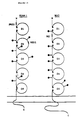
- Example 2 The qualitative aggregation assay described in Example 2 was performed using the Epstein-Barr transformed cell line JY. Upon addition of PMA to the culture medium in the microtiter plates, aggregation of cells was observed. Time lapse video recordings showed that the JY cells on the bottom of the microtiter wells were motile and exhibited active membrane ruffling and pseudopodia movement. Contact between the pseudopodia of neighboring cells often resulted in cell-cell adherence. If adherence was sustained, the region of cell contact moved to the uropod. Contact could be maintained despite vigorous cell movements and the tugging of the cells in opposite directions. The primary difference between PMA-treated and untreated cells appeared to be in the stability of these contacts once they were formed. With PMA, clusters of cells developed, growing in size as additional cells adhered at their periphery.
- EBV-transformed lymphoblastoid cells were prepared from patients in the manner described in Example 1. Such cells were screened against monoclonal antibodies capable of recognizing LFA-1 and the cells were found to be LFA-1 deficient.
- Example 2 The qualitative aggregation assay described in Example 2 was employed, using the LFA-1 deficient cells described above. Such cells failed to spontaneously aggregate, even in the presence of PMA.
- the LFA-1 deficient cells of Example 5 were labeled with carboxyfluorescein diacetate (Patarroyo, M. et al ., Cell. Immunol. 63 :237-248 (1981)).
- the labeled cells were mixed in a ratio of 1:10 with autologous or JY cells and the percentage of fluorescein-labeled cells in aggregates was determined according to the procedure of Rothlein, R. et al ., J. Exper. Med. 163 :1132-1149 (1986).
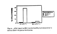
- the LFA-1 deficient cells were found to be capable of coaggregating with LFA-1 expressing cells ( Figure 4).
- Monoclonal antibodies capable of binding to ICAM-1 were isolated according to the method of Rothlein, R. et al ., J. Immunol. 137 :1270-1274 (1986), which reference has been incorporated by reference herein.
- 3 BALB/C mice were immunized intraperitoneally with EBV-transformed peripheral blood mononuclear cells from an LFA-1-deficient individual (Springer, T.A. et al ., J. Exper. Med. 160 :1901 (1984)). Approximately 107 cells in 1 ml RPMI 1640 medium was used for each immunization.
- mice were given an additional 107 cells in 0.15 ml medium (intravenously).
- Isolated spleen cells from the above-described animals were fused with P3X73Ag8.653 myeloma cells at a ratio of 4:1 according to the protocol of Galfre, G. et al ., Nature 266 :550 (1977). Aliquots of the resulting hybridoma cells were introduced into 96-well microtiter plates. The hybridoma supernatants were screened for inhibition of aggregation, and one inhibitory hybridoma (of over 600 wells tested) was cloned and subcloned by limiting dilution. This subclone was designated RR1/1.1.1 (hereinafter designated "RR1/1").
- Monoclonal antibody RR1/1 was consistently found to inhibit PMA-stimulated aggregation of the LFA-1 expressing cell line JY.
- the RR1/1 monoclonal antibody inhibited aggregation equivalently, or slightly less than some monoclonal antibodies to the LFA-1 alpha or beta subunits.
- control monoclonal antibody against HLA which is abundantly expressed on JY cells, did not inhibit aggregation.
- the antigen bound by monoclonal antibody RR1/1 is defined as the intercellular adhesion molecule-1 (ICAM-1).
- IAM-1 intercellular adhesion molecule-1
- Lysates were centrifuged at 10,000 x g for 10 minutes and precleared with 50 ⁇ l of a 50% suspension of CNBr-activated, glycine-quenched Sepharose Cl-4B for 1 hour at 4°C.
- One milliliter of lysate was immunoprecipitated with 20 ⁇ l of a 50% suspension of monoclonal antibody RR1/1 coupled to Sepharose Cl-4B (1 mg/ml) overnight at 4°C (Springer, T.A. et al ., J. Exper. Med. 160 :1901 (1984)).
- Sepharose-bound monoclonal antibody was prepared using CNBr-activation of Sepharose CL-4B in carbonate buffer according to the method of March, S.
- Example 2 In order to determine the effect of monoclonal antibody RR1/1 on PHA-lymphoblast aggregation, the quantitative aggregation assay described in Example 2 was employed. Thus, T cell blast cells were stimulated for 4 days with PHA, thoroughly washed, then cultured for 6 days in the presence of IL-2 conditioned medium. PHA was found to be internalized during this 6-day culture, and did not contribute to the aggregation assay. In three different assays with different T cell blast preparations, ICAM-1 monoclonal antibodies consistently inhibited aggregation (Table 2).
- a negative number indicates percent enhancement of aggregation.
- e Aggregation was measured 1 hr after the simultaneous addition of monoclonal antibody and PMA.
- Cells were pelleted at 200 x G for 1 min. incubated at 37°C for 15 min. gently resuspended, and shaken for 45 min. at 100 rpm.
- f cells were pretreated with PMA for 4 hr at 37°C. After monoclonal antibody was added, the tubes were incubated at 37°C stationary for 20 min. and shaken at 75 rpm for 100 min.
- LFA-1 monoclonal antibodies were consistently more inhibitory than ICAM-1 monoclonal antibodies, whereas HLA-A, B and LFA-3 monoclonal antibodies were without effect. These results indicate that of the monoclonal antibodies tested, only those capable of binding to LFA-1 or ICAM-1 were capable of inhibiting cellular adhesion.
- mice were immunized intraperitoneally (i.p.) with 0.5 mls of 2 x 10⁷ JY cells in RPMI medium 103 days and 24 days prior to fusion. On day 4 and 3 prior to fusion, mice were immunized i.p. with 107 cells of PMA differentiated U937 cells in 0.5 ml of RPMI medium.
- U937 cells (ATCC CRL-1593) were differentiated by incubating them at 5 x 10⁵/ml in RPMI with 10% Fetal Bovine Serum, 1% glutamine and 50 ⁇ g/ml gentamyin (complete medium) containing 2 ng/ml phorbol-12-myristate acetate (PMA) in a sterile polypropylene container. On the third day of this incubation, one-half of the volume of medium was withdrawn and replaced with fresh complete medium containing PMA. On day 4, cells were removed, washed and prepared for immunization.
- PMA phorbol-12-myristate acetate
- Spleen cells from the immunized mice were fused with P3x63 Ag8.653 myeloma cells at a 4:1 ratio according to Galfre et al ., ( Nature 266 :550 (1977)). After the fusion, cells were plated in a 96 well flat bottomed microtiter plates at 105 spleen cells/well.
- a radioimmune assay was developed.
- purified RR1/1 was iodinated using iodogen to a specific activity of 10 uCi/ ⁇ g.
- Endothelial cells were grown in 96 well plates and treated as described for each experiment. The plates were cooled at 4°C by placing in a cold room for 0.5-1 hr, not immediately on ice. The monolayers were washed 3x with cold complete media and then incubated 30 m at 4°C with 125 I RR1/1. The monolayers were then washed 3x with complete media. The bound 125 I was released using 0.1 N NaOH and counted.
- the specific activity of the 125 I RR1/1 was adjusted using unlabeled RR1/1 to obtain a linear signal over the range of antigen densities encountered in this study. Non-specific binding was determined in the presence of a thousand fold excess of unlabeled RR1/1 and was subtracted from total binding to yield the specific binding.
- ICAM-1 expression measured using the above described radioimmune assay, is increased on human umbilical vein endothelial cells (HUVEC) and human saphenous vein endothelial cells (HSVEC) by IL-1, TNF, LPS and IFN gamma (Table 3).
- Saphenous vein endothelial cells were used in this study to confirm the results from umbilical vein endothelial cells in cultured large vein endothelial cells derived from adult tissue.
- the basal expression of ICAM-1 is 2 fold higher on saphenous vein endothelial cells than on umbilical vein endothelial cells.
- IL-1 alpha, TNF and LPS were the most potent inducers and IL-1 was less potent on a weight basis and also at saturating concentrations for the response (Table 3).
- IL-1 beta at 100 ng/ml increased ICAM-1 expression by 9 fold on HUVEC and 7.3 fld on HSVEC with half-maximal increase occuring at 15 ng/ml.
- rTNF at 50 ng/ml increased ICAM-1 expression 16 fold on HUVEC and 11 fold on HSVEC with half maximal effects at 0.5 ng/ml.
- Interferon-gamma produced a significant increase in ICAM-1 expression of 5.2 fold on HUVEC or 3.5 fold on HSVEC at 10,000 U/ml.
- the effect of LPS at 10 ⁇ g/ml was similar in magnitude to that of rTNF. Pairwise combinations of these mediators resulted in additive or slightly less than additive effects on ICAM-1 expression (Table 3).
- Cross-titration of rTNF with rIL-1 beta or rIFN gamma showed no synergism between these at suboptimal or optimal concentrations.
- Upregulation of ICAM-1 expression on HVEC and HSVEC- HUVEC or HSVEC were seeded into 96 well plates at 1:3 from a confluent monolayer and allowed to grow to confluence. Cells were then treated with the indicated materials or media for 16 hr and the RIA done as in methods. All points were done in quadruplicate.
- the kinetics of interleukin 1 and gamma interferon effects on ICAM-1 expression on dermal fibroblasts were determined using the 125I goat anti-mouse IgG binding assay of Dustin, M.L. et al . ( J. Immunol. 137 :245-254 (1986); which reference is herein incorporated by reference).
- human dermal fibroblasts were grown in a 96-well microtiter plate to a density of 2-8 x 10⁴ cells/well (0.32 cm2). The cells were washed twice with RPMI 1640 medium supplemented as described in Example 1.
- the cells were additionally washed once with Hanks Balanced Salt Solution (HBSS), 10 mM HEPES, 0.05% NaN3 and 10% heat-inactivated fetal bovine serum. Washing with this binding buffer was done at 4°C. To each well was added 50 ⁇ l of the above-described binding buffer and 50 ⁇ l of the appropriate hybridoma supernatant with X63 and W6/32 as the negative and positive controls, respectively. After incubation for 30 minutes at 4°C, with gentle agitation, the wells were washed twice with binding buffer, and the second antibody 125 I-goat anti-mouse IgG, was added at 50 nCi in 100 ⁇ l.
- HBSS Hanks Balanced Salt Solution
- 10 mM HEPES 10 mM HEPES
- NaN3 10% heat-inactivated fetal bovine serum
- the 125 I-goat anti-mouse antibody was prepared by using Iodogen (Pierce) according to the method of Fraker, P.J. et al . ( Biochem. Biophys. Res. Commun. 80 :849 (1978)). After 30 minutes at 4°C, the wells were washed twice with 200 ⁇ l of binding buffer and the cell layer was solubilized by adding 100 ⁇ l of 0.1 N NaOH. This and a 100 ⁇ l wash were counted in a Beckman 5500 gamma counter. The specific counts per minute bound was calculated as (cpm with monoclonal antibody]-[cpm with X63]. All steps, including induction with specific reagents, were carried out in quadruplicate.
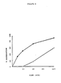
- the dose response curves for induction of ICAM-1 by recombinant mouse and human interleukin and for recombinant human gamma interferon are shown in Figure 7.
- Gamma interferon and interleukin 1 were found to have similar concentration dependencies with nearly identical effects at 1 ng/ml.
- the human and mouse recombinant interleukin 1 also have similar curves, but are much less effective than human interleukin 1 preparations in inducing ICAM-1 expression.
- Cyclohexamide an inhibitor of protein synthesis, and actinomycin D, an inhibitor of mRNA synthesis, abolish the effects of both interleukin 1 and gamma interferon on ICAM-1 expression on fibroblasts (Table 4). Furthermore, tunicamycin, an inhibitor of N-linked glycosylation, only inhibited the interleukin 1 effect by 43%. These results indicate that protein and mRNA synthesis, but not N-linked glycosylation, are required for interleukin 1 and gamma interferon-stimulated increases in ICAM-1 expression.
- a Human fibroblasts were grown to a density of 8 x 10⁴ cells/0.32 cm2 well. Treatments were carried out in a final volume of 50 ul containing the indicated reagents. Cycloheximide, actinomycin D, and tunicamycin were added at 20 ug/ml, 10 uM, and 2 ug/ml, respectively, at the same time as the cytokines. All points are means of quadruplicate wells ⁇ SD.
- the sections were sequentially incubated with biotinylated horse anti-mouse IgG and avidin-biotinylated peroxidase complexes.
- the sections were finally dipped in a solution containing 3-amino-9-ethyl-carbazole (Aldrich Chemical Co., Inc., Milwaukee, WI) to develop a color reaction.
- the sections were then fixed in 4% formaldehyde for 5 minutes and were counterstained with hematoxylin. Controls included sections incubated with unrelated monoclonal antibodies instead of the RR1/1 antibody.
- ICAM-1 was found to have a distribution most similar to that of the major histocompatibility complex (MHC) Class II antigens.
- MHC major histocompatibility complex
- the vascular endothelial staining was more intense in the interfollicular (paracortical) areas in lymph nodes, tonsils, and Peyer's patches as compared with vessels in kidney, liver, and normal skin. In the liver, the staining was mostly restricted to sinusoidal lining cells; the hepatocytes and the endothelial cells lining most of the portal veins and arteries were not stained.
- the staining pattern was focal and predominantly dendritic. Thymocytes were not stained. In the peripheral lymphoid tissue, the germinal center cells of the secondary lymphoid follicles were intensely stained. In some lymphoid follicles, the staining pattern was mostly dendritic, with no recognizable staining of lymphocytes. Faint staining of cells in the mantle zone was also observed. In addition, dendritic cells with cytoplasmic extensions (interdigitating reticulum cells) and a small number of lymphocytes in the interfollicular or paracortical areas stained with the ICAM-1 binding antibody.
- cytoplasmic extensions interdigitating reticulum cells
- epithelial cells The staining of epithelial cells was consistently seen in the mucosa of the tonsils. Although hepatocytes, bile duct epithelium, intestinal epithelial cells, and tubular epithelial cells in kidney did not stain in most circumstances, sections of normal kidney tissue obtained from a nephrectomy specimen with renal cell carcinoma showed staining of many proximal tubular cells for ICAM-1. These tubular epithelial cells also stained with an anti-HLA-DR binding antibody.
- ICAM-1 is expressed on non-hematopoietic cells such as vascular endothelial cells and on hematopoietic cells such as tissue macrophages and mitogen-stimulated T lymphocyte blasts. ICAM-1 was found to be expressed in low amounts on peripheral blood lymphocytes.
- ICAM-1 was purified from human cells or tissue using monoclonal antibody affinity chromatography. Monoclonal antibody, RR1/1, reactive with ICAM-1 was first purified, and coupled to an inert column matrix. This antibody is described by Rothlein, R. et al . J. Immunol. 137 :1270-1274 (1986), and Dustin, M.L. et al . ( J. Immunol. 137 :245 (1986). ICAM-1 was solubilized from cell membranes by lysing the cells in a non-ionic detergent, Triton X-100, at a near neutral pH.
- Triton X-100 Triton X-100
- the cell lysate containing solubilized ICAM-1 was then passed through pre-columns designed to remove materials that bind nonspecifically to the column matrix material, and then through the monoclonal antibody column matrix, allowing the ICAM-1 to bind to the antibody.
- the antibody column was then washed with a series of detergent wash buffers of increasing pH up to pH 11.0. During these washes ICAM-1 remained bound to the antibody matrix, while non-binding and weakly binding contaminants were removed.
- the bound ICAM-1 was then specifically eluted from the column by applying a detergent buffer of pH 12.5.
- the anti-ICAM-1 monoclonal antibody RR1/1 was purified from the ascites fluid of hybridoma-bearing mice, or from hybridoma culture supernates by standard techniques of ammonium sulfate precipitation and protein A affinity chromatography (Ey et al ., Immunochem. 15 :429 (1978)).
- the purified IgG, or rat IgG was covalently coupled to Sepharose CL-4B (Pharmacia, Upsala, Sweden) using a modification of the method of March et al . ( Anal. Biochem. 60 :149 (1974)).
- Sepharose CL-4B was washed in distilled water, activated with 40 mg/ml CNBr in 5 M K2HPO4 (pH approximately 12) for 5 minutes, and then washed extensively with 0.1 mM HCl at 4°C.
- the filtered, activated Sepharose was resuspended with an equal volume of purified antibody (2-10 mg/ml in 0.1 M NaHCO3, 0.1 M NaCl).
- the suspension was incubated for 18 hours at 4°C with gentle end-over-end rotation. The supernatant was then monitored for unbound antibody by absorbance at 280 nm, and remaining reactive sites on the activated Sepharose were saturated by adding glycine to 0.05 M. Coupling efficiency was usually greater than 90%.
- Tween 40 polyoxyethylene sorbitan monopalmitate
- the homogenate was extracted using three strokes of a Dounce or, more preferably, a Teflon Potter Elvejhem homogenizer, and then centrifuged at 1000 x g for 15 minutes. The supernatant was retained and the pellet was re-extracted with 200 ml of 2.5% Tween 40 in Tris-saline. After centrifugation at 1000 x g for 15 minutes, the supernatants from both extractions were combined and centrifuged at 150,000 x g for 1 hour to pellet the membranes. The membranes were washed by resuspending in 200 ml Tris-saline, centrifuged at 150,000 x g for 1 hour.
- the membrane pellet was resuspended in 200 ml Tris-saline and was homogenized with a motorized homogenizer and Teflon pestle until the suspension was uniformly turbid. The volume was then brought up to 900 ml with Tris-saline, and N-lauroyl sarcosine was added to a final concentration of 1%. After stirring at 4°C for 30 minutes, insoluble material in the detergent lysate was removed by centrifugation at 150,000 x g for 1 hour. Triton X-100 was then added to the supernatant to a final concentration of 2%, and the lysate was stirred at 4°C for 1 hour.
- the EBV-transformed B-lymphoblastoid cell line JY was grown in RPMI-1640 containing 10% fetal calf serum (FCS) and 10 mM HEPES to an approximate density of 0.8 - 1.0 x 10⁶ cells/ml.
- FCS fetal calf serum
- HEPES fetal calf serum
- PMA phorbol 12-myristate 13-acetate
- Sodium vanadate (50 uM) was also added to the cultures during this time.
- the cells were pelleted by centrifugation at 500 x g for 10 minutes, and washed twice in Hank's Balanced Salt Solution (HBSS) by resuspension and centrifugation.
- HBSS Hank's Balanced Salt Solution
- the cells (approximately 5 g per 5 liters of culture) were lysed in 50 ml of lysis buffer (0.14 M NaCl, 50 mM Tris pH 8.0, 1% Triton X-100, 0.2 U/ml aprotinin, 1 mM PMSF, 50 uM sodium vanadate) by stirring at 4°C for 30 minutes. Unlysed nuclei and insoluble debris were removed by centrifugation at 10,000 x g for 15 minutes, followed by centrifugation of the supernatant at 150,000 x g for 1 hour, and filtration of the supernatant through Whatman 3mm filter paper.
- One liter of the detergent lysate of human spleen was loaded at a flow rate of 0.5-1.0 ml per minute.
- the two pre-columns were used to remove non-specifically binding material from the lysate before passage through the RR1/1-Sepharose column.
- the column of RR1/1-Sepharose and bound ICAM-1 was washed sequentially at a flow rate of 1 ml/minute with a minimum of 5 column volumes each of the following: 1) lysis buffer, 2) 20 mM Tris pH 8.0/0.14 M NaCl/0.1% Triton X-100, 3) 20 mM glycine pH 10.0/0.1% Triton X-100, and 4) 50 mM triethylamine pH 11.0/0.1% Triton X-100. All wash buffers contained 1 mM PMSF and 0.2 U/ml aprotinin.
- ICAM-1 was eluted with 5 column volumes of elution buffer (50 mM triethylamine/0.1% Triton X-100/pH 12.5 at 4°C) at a flow rate of 1 ml/3 minutes.
- the eluted ICAM-1 was collected in 1 ml fractions and immediately neutralized by the addition of 0.1 ml of 1 M Tris, pH 6.7.
- Fractions containing ICAM-1 were identified by SDS-polyacrylamide electrophoresis of 10 ⁇ l aliquots (Springer et al ., J. Exp. Med. 160 :1901 (1984)), followed by silver staining (Morrissey, J.H., Anal. Biochem.
- the bulk of the ICAM-1 eluted in approximately 1 column volume and was greater than 90% pure as judged from silver-stained electropherograms (a small amount of IgG, leeched from the affinity matrix, was the major contaminant).
- the fractions containing ICAM-1 were pooled and concentrated approximately 20-fold using Centricon-30 microconcentrators (Amicon, Danvers, MA).
- the purified ICAM-1 was quantitated by Lowry protein assay of an ethanol-precipitated aliquot of the pool: approximately 500 ⁇ g of pure ICAM-1 were produced from the 200 g of human spleen.
- ICAM-1 Approximately 200 ⁇ g of purified ICAM-1 was subjected to a second stage purification by preparative SDS-polyacrylamide gel electrophoresis. The band representing ICAM-1 was visualized by soaking the gel in 1 M KCl. The gel region which contained ICAM-1 was then excised and electroeluted according to the method of Hunkapiller et al ., Meth. Enzymol. 91 :227-236 (1983). The purified protein was greater than 98% pure as judged by SDS-PAGE and silver staining.
- ICAM-1 for use in functional studies was purified from detergent lysates of JY cells as described above, but on a smaller scale (a 1 ml column of RR1/1-Sepharose), and with the following modifications. All solutions contained 50 uM sodium vanadate. After washing the column with pH 11.0 buffer containing 0.1% Triton X-100, the column was washed again with five column volumes of the same buffer containing 1% n-octyl-beta-D-glucopyranoside (octylglucoside) in place of 0.1% Triton X-100.
- the octylglucoside detergent displaces the Triton X-100 bound to the ICAM-1, and unlike Triton X-100, can be subsequently removed by dialysis.
- the ICAM-1 was then eluted with pH 12.5 buffer containing 1% octylglucoside in place of 0.1% Triton X-100, and was analyzed and concentrated as described above.
- ICAM-1 purified from human spleen migrates in SDS-polyacrylamide gels as a broad band of M r of 72,000 to 91,000.
- ICAM-1 purified from JY cells also migrates as a broad band of M r of 76,500 to 97,000.
- the non-glycosylated precursor has a M r of 55,000 (Dustin et al .).
- the protein purified from either JY cells or human spleen retains its antigenic activity as evidenced by its ability to re-bind to the original affinity column, and by immunoprecipitation with RR1/1-Sepharose and SDS-polyacrylamide electrophoresis.
- peptide fragments of ICAM-1 approximately 200 ⁇ g was reduced with 2 mM dithiothreitol/2% SDS, followed by alkylation with 5 mM iodoacetic acid.
- the protein was precipitated with ethanol, and redissolved in 0.1 M NH4CO3/0.1 mM CaCl2/0.1% zwittergent 3-14 (Calbiochem), and digested with 1% w/w trypsin at 37°C for 4 hours, followed by an additional digestion with 1% trypsin for 12 hours at 37°C.
- the tryptic peptides were purified by reverse-phase HPLC using a 0.4 x 15 cm C4 column (Vydac).
- the peptides were eluted with a linear gradient of 0% to 60% acetonitrile in 0.1% trifluoroacetic acid. Selected peptides were subjected to sequence analysis on a gas phase microsequenator (Applied Biosystems). The sequence information obtained from this study is shown in Table 5.
- the gene for ICAM-1 may be cloned using any of a variety of procedures. For example, the amino acid sequence information obtained through the sequencing of the tryptic fragments of ICAM-1 (Table 5) can be used to identify an oligonucleotide sequence which would correspond to the ICAM-1 gene. Alternatively, the ICAM-1 gene can be cloned using anti-ICAM-1 antibody to detect clones which produce ICAM-1.
- oligonucleotides can be identified, each of which would be capable of encoding the ICAM-1 tryptic peptides.
- the probability that a particular oligonucleotide will, in fact, constitute the actual ICAM-1 encoding sequence can be estimated by considering abnormal base pairing relationships and the frequency with which a particular codon is actually used (to encode a particular amino acid) in eukaryotic cells.
- Such "codon usage rules" are disclosed by Lathe, R., et al ., J. Molec. Biol.
- the oligonucleotide, or set of oligonucleotides, containing the theoretical "most probable" sequence capable of encoding the ICAM-1 fragments is used to identify the sequence of a complementary oligonucleotide or set of oligonucleotides which is capable of hybridizing to the "most probably" sequence, or set of sequences.
- An oligonucleotide containing such a complementary sequence can be employed as a probe to identify and isolate the ICAM-1 gene (Maniatis, T., et al ., Molecular Cloning A Laboratory Manual , Cold Spring Harbor Press, Cold Spring Harbor, NY (1982).
- ICAM-1 gene As described in Section C, supra , it is possible to clone the ICAM-1 gene from eukaryotic DNA preparations suspected of containing this gene. To identify and clone the gene which encodes the ICAM-1 protein, a DNA library is screened for its ability to hybridize with the oligonucleotide probes described above. Because it is likely that there will be only two copies of the gene for ICAM-1 in a normal diploid cell, and because it is possible that the ICAM-1 gene may have large non-transcribed intervening sequences (introns) whose cloning is not desired, it is preferable to isolate ICAM-1-encoding sequences from a cDNA library prepared from the mRNA of an ICAM-1-producing cell, rather than from genomic DNA.
- Suitable DNA, or cDNA preparations are enzymatically cleaved, or randomly sheared, and ligated into recombinant vectors. The ability of these recombinant vectors to hybridize to the above-described oligonucleotide probes is then measured. Procedures for hybridization are disclosed, for example, in Maniatis, T., Molecular Cloning A Laboratory Manual , Cold Spring Harbor Press, Cold Spring Harbor, NY (1982) or in Haymes, B.T., et al ., Nucleic Acid Hybridization a Practical Approach , IRL Press, Oxford, England (1985). Vectors found capable of such hybridization are then analyzed to determine the extent and nature of the ICAM-1 sequences which they contain. Based purely on statistical considerations, a gene such as that which encodes the ICAM-1 molecule could be unambiguously identified (via hybridization screening) using an oligonucleotide probe having only 18 nucleotides.
- the actual identification of ICAM-1 peptide sequences permits the identification of a theoretical "most probable" DNA sequence, or a set of such sequences, capable of encoding such a peptide.
- a DNA molecule or set of DNA molecules, capable of functioning as a probe to identify and isolate the ICAM-1 gene.
- ICAM-1 peptide sequences of Table 5 the sequence of the "most probable" sequence of an oligonucleotide capable of encoding the AA and J peptides was determined (Tables 6 and 7, respectively). Oligonucleotides complementary to these sequences were synthesized and purified for use as probes to isolate ICAM-1 gene sequences. Suitable size-selected cDNA libraries were generated from the poly(A)+ RNA of both PMA-induced HL-60 cells and from PS-stimulated umbilical vein endothelial cells. A size-selected cDNA library was prepared using poly(A)+ RNA from PMA-induced HL-60 cells according to the method of Gubler, U., et al . (( Gene 25 :263-269 (1983)) and Corbi, A., et al . ( EMBO J. 6 :4023-4028 (1987)), which references are herein incorporated by reference.
- a size-selected cDNA library was prepared using poly(A)+ RNA from umbilical vein endothelial cells which had been stimulated for 4 hours with PS 5 ⁇ g/ml.
- the RNA was extracted by homogenizing the cells in 4 M guanidinium isothiocyanate and subjecting the supernatant to ultracentrifugation through a CsCl gradient (Chirgwin, J.M., et al ., Biochem. 18 :5294-5299 (1979)).
- Poly(A)+ RNA was isolated from the mixture of total RNA species through the use of oligo (dT)-cellulose chromatography (Type 3, Collaborative Research) (Aviv, H., et al ., Proc. Natl. Acad. Sci. (USA) 69 :1408-1412 (1972).
- First strand cDNA was synthesized using 8 ⁇ g of poly(A)+ RNA, avian myeloblastosis virus reverse transcriptase (Life Sciences) and an oligo(dT) primer.
- the DNA-RNA hybrid was digested with RNase H (BRL) and the second strand was synthesized using DNA polymerase I (New England Biolabs).
- the product was methylated with Eco R1 methylase (New England Biolabs), blunt end ligated to Eco R1 linkers (New England Biolabs), digested with Eco R1 and size selected on a low melting point agarose gel.
- cDNA greater than 500bp were ligated to ⁇ gt10 which had previously been Eco R1 digested and dephosphorylated (Stratagene) The product of the ligation was then packaged (Stratagene gold).
- the umbilical vein endothelial cell and HL-60 cDNA libraries were then plated at 20,000 PFU/150mm plate. Recombinant DNA was transferred in duplicate to nitrocellulose filters, denatured in 0.5 M NaOH/1.5M NaCl, neutralized in 1M Tris, pH7.5/1.5M NaCl and baked at 80°C for 2 hours (Benton, W.D., et al ., Science 196 :180-182 (1977)). Filters were prehybridized and hybridized in 5X SSC containing 5X Denhardt's solution, 50 mM NaPO4 and 1 ⁇ g/ml salmon sperm DNA. Prehybridization was carried out at 45° for 1 hour.
- Hybridization was carried out using 32bp (′5-TTGGGCTGGTCACAGGAGGTGGAGCAGGTGAC) or 47bp (5′-GAGGTGTTCTCAAACAGCTCCAGGCCCTGGGGCCGCAGGTCCAGCTC) anti-sense oligonucleotides based, in the manner discussed above, on the ICAM-1 tryptic peptides J and AA, respectively (Table 6 and 7) (Lathe, R., J. Molec. Biol. , 183 :1-12 (1985)). Oligonucleotides were end labeled with ⁇ -(32P)ATP using T4 polynucleotide kinase and conditions recommended by the manufacturer (New England Biolabs). Following overnight hybridization the filters were washed twice with 2X SSC/0.1% SDS for 30 minutes at 45°C. Phages were isolated from those plaques which exhibited hybridization, and were purified by successive replating and rescreening.
- the gene for ICAM-1 may alternatively be cloned through the use of anti-ICAM-1 antibody.
- DNA, or more preferably cDNA is extracted and purified from a cell which is capable of expressing ICAM-1.
- the purified cDNA is fragmentized (by shearing, endonuclease digestion, etc.) to prduct a pool of DNA or cDNA fragments.
- DNA or cDNA fragments from this pool are then cloned into an expression vector in order to produce a genomic library of expression vectors whose members each contain a unique cloned DNA or cDNA fragment.
- Phage DNA from positive clones were digested with Eco R1 and examined by Southern analysis using a cDNA from one clone as a probe. Maximal size cDNA inserts which cross-hybridized were subcloned into the Eco R1 site of plasmid vector pGEM 4Z (Promega). HL-60 subclones containing the cDNA in both orientations were deleted by exonuclease III digestion (Henikoff, S., Gene 28 :351-359 (1984)) according to the manufacturers recommendations (Erase-a-Base, Promega). Progressively deleted cDNAS were then cloned and subjected to dideoxynucleotide chain termination sequencing (Sanger, F.
- HL-60 cDNA 5′ and coding regions were sequenced completely on both strands and the 3′ region was sequenced approximately 70% on both strands.
- a representative endothelial cDNA was sequenced over most of its length by shotgun cloning of 4bp-recognition restriction enzyme fragments.
- the cDNA sequence of one HL-60 and one endothelial cell cDNA was established (Figure 8).
- the 3023 bp sequence contains a short 5′ untranslated region and a 1.3 kb 3′ untranslated region with a consensus polyadenylation signal at position 2966.
- the longest open reading frame begins with the first ATG at position 58 and ends with a TGA terminating triplet at position 1653.
- Identity between the translated amino acid sequence and sequences determined from 8 different tryptic peptides totaling 91 amino acids confirmed that authentic ICAM-1 cDNA clones had been isolated.
- the amino acid sequences of ICAM-1 tryptic peptides are shown in Table 8.
- Hydrophobicity analysis suggests the presence of a 27 residue signal sequence.
- the assignment of the +1 glutamine is consistent with our inability to obtain N-terminal sequence on 3 different ICAM-1 protein preparations; glutamine may cyclize to pyroglumatic acid, resulting in a blocked N-terminus.
- the translated sequence from 1 to 453 is predominantly hydrophilic followed by a 24 residue hydrophobic putative transmembrane domain. The transmembrane domain is immediately followed by several charged residues contained within a 27 residue putative cytoplasmic domain.
- the predicted size of the mature polypeptide chain is 55,219 daltons, in excellent agreement with the observed size of 55,000 for deglycosylated ICAM-1 (Dustin, M.L., et al ., J. Immunol. 137 :245-254 (1986)).
- Eight N-linked glycosylation sites are predicted. Absence of asparagine in the tryptic peptide sequences of 2 of these sites confirm their glycosylation and their extracellular orientation. Assuming 2,500 daltons per high mannose N-linked carbohydrate, a size of 75,000 daltons is predicted for the ICAM-1 precursor, compared to the observed six of 73,000 daltons (Dustin, M.L., et al ., J.
- ICAM-1 is a heavily glycosylated but otherwise typical integral membrane protein.
- ICAM-1 is an Integrin-Binding Member of the Immunoglobulin Supergene family
- ICAM-1 is a ligand of an integrin, it was unexpected that it would be a member of the immunoglobulin supergene family. However, inspection of the ICAM-1 sequence shows that it fulfills all criteria proposed for membership in the immunoglobulin supergene family. These criteria are discussed in turn below.
- ICAM-1 The entire extracellular domain of ICAM-1 is constructed from 5 homologous immunoglobulin-like domains which are shown aligned in Figure 9A. Domains 1-4 are 88, 97, 99, and 99 residues long, respectively and thus are of typical Ig domain size; domain 5 is truncated within 68 residues. Searches of the NBRF data base using the FASTP program revealed significant homologies with members of the immunoglobulin supergene family including IgM and IgG C domains, T cell receptor ⁇ subunit variable domain, and alpha 1 beta glycoprotein (Fig. 9B-D).
- the amino acid sequence of ICAM-1 was compared with the amino acid sequences of other members of the immununoglobulin supergene family.
- V and C domains Three types of Ig superfamily domains, V, C1, and C2 have been differentiated. Both V and C domains are constructed from 2 ⁇ -sheets linked together by the intradomain disulfide bond; V domains contain 9 anti-parallel ⁇ -strands while C domains have 7. Constant domains were divided into the C1- and C2- sets based on characteristic residues shown in Figure 9A.
- the C1-set includes proteins involved in antigen recognition.
- the C2-set includes several Fc receptors and proteins involved in call adhesion including CD2, LFA-3, MAG, and NCAM.
- ICAM-1 domains were found to be most strongly homologous with domains of the C2-set placing ICAM-1 in this set; this is reflected in stronger similarity to conserved residues in C2 than C1 domains as shown for ⁇ -strands B-F in Figure 9. Also, ICAM-1 domains aligned much better with ⁇ -strands A and G of C2 domains than with these strands in V and C1 domains, allowing good alignments across the entire C2 domain strength. Alignments with C2 domains from NCAM, MAG, and alpha 1- ⁇ glycoprotein are shown in Figures 9B and 9C; identity ranged from 28 to 33%. Alignments with a T cell receptor V ⁇ 27% identity and IgM C domain 3 34% identity are also shown ( Figures 9B, 9D).
- cysteines One of the most important characteristics of immunoglobulin domains is the disulfide-bonded cysteines bridging the B and F ⁇ strands which stabilizes the ⁇ sheet sandwich; in ICAM-1 the cysteines are conserved in all cases except in strand f of domain 4 where a leucine is found which may face into the sandwich and stabilize the contact as proposed for some other V- and C2-sets domains.
- the distance between the cysteines (43, 50, 52, and 37 residues) is as described for the C2-set.
- endothelial cell ICAM-1 was subjected to SDS-PAGE under reducing and non-reducing conditions. Endothelial cell ICAM-1 was used because it shows less glycosylation heterogeneity than JY or hairy cell splenic ICAM-1 and allows greater sensitivity to shifts in M r . ICAM-1 was, therefore, purified from 16 hour LPS (5 ⁇ g/ml) stimulated umbilical vein endothelial cell cultures by immunoaffinity chromatography as described above.
- Acetone precipitated ICAM-1 was resuspended in sample buffer (Laemmli, U.K., Nature 227 :680-685 (1970)) with 0.25% 2-mercaptoethanol or 25 mM iodoacetamide and brought to 100°C for 5 min. The samples were subjected to SDS-PAGE 4670 and silver staining 4613. Endothelial cell ICAM-1 had an apparent M r of 100 Kd under reducing conditions and 96 Kd under non-reducing conditions strongly suggesting the presence of intrachain disulfides in native ICAM-1.
- ICAM-1 was found to be most strongly homologous with the NCAM and MAG glycoproteins of the C2 set. This is of particular interest since both NCAM and MAG mediate cell-cell adhesion.
- NCAM is important in neuron-neuron and neuro-muscular interactions (Cunningham, B.A., et al. , Science 236 :799-806 (1987)), while MAG is important in neuron-oligodendrocyte and oligodendrocyte-oligodendrocyte interactions during myelination (Poltorak, M., et al. , J. Cell Biol. 105 :1893-1899 (1987)).
- NCAM and MAG The cell surface expression of NCAM and MAG is developmentally regulated during nervous system formation and myelination, respectively, in analogy to the regulated induction of ICAM-1 in inflammation (Springer, T.A., et al. , Ann. Rev. Immunol. 5 :223-252 (1987)). ICAM-1, NCAM (Cunningham, B.A., et al , Science 236 :799-806 (1987)), and MAG (Salzer, J.L., et al. , J. Cell. Biol .
- ICAM-1 aligns over its entire length including the transmembrane and cytoplasmic domains with MAG with 21% identity; the same % identity is found comparing the 5 domains of ICAM-1 and NCAM-1.
- Figure 10 A diagrammatic comparison of the secondary structures of ICAM-1 and MAG is shown in Figure 10. Domain by domain comparisons show that the level of homology between domains within the ICAM-1 and NCAM molecules (x ⁇ s.d.
- ICAM-1 functions as a ligand for LFA-1 in lymphocyte interactions with a number of different cell types. Lymphocytes bind to ICAM-1 incorporated in artificial membrane bilayers, and this requires LFA-1 on the lymphocyte, directly demonstrating LFA-1 interaction with ICAM-1 (Marlin, S.D., et al. , cell 51 :813-819 (1987)). LFA-1 is a leukocyte integrin and has no immunoglobulin-like features. Leukocyte integrins comprise one integrin subfamily.
- the other two subfamilies mediate cell-matrix interactions and recognize the sequence RGD within their ligands which include fibronectin, vitronectin, collagen, and fibrinogen (Hynes, R.O., Cell 48 :549-554 (1987); Ruoslahti, E., et al. , Science 238 :491-497 (1987)).
- the leukocyte integrins are only expressed on leukocytes, are involved in cell-cell interactions, and the only known ligands are ICAM-1 and iC3b, a fragment of the complement component C3 which shows no immunoglobulin-like features and is recognized by Mac-1 (Kishimoto, T.K., et al.
- ICAM-1 is the first example of a member of the immunoglobulin supergene family which binds to an integrin. Although both of these families play an important role in cell adhesion, interaction between them had not previously been expected. In contrast, interactions within the immunoglobulin gene superfamily are quite common. It is quite possible that further examples of interactions between the integrin and immunoglobulin families will be found.
- LFA-1 recognizes a ligand distinct from ICAM-1 (Springer, T.A., et al. , Ann. Rev. Immunol. 5 :223-252 (1987)), and the leukocyte integrin Mac-1 recognizes a ligand distinct from C3bi in neutrophil-neutrophil adhesion (Anderson, D.C., et al.
- MAG-containing vesicles bind to neurites which are MAG, and thus MAG must be capable of heterophilic interaction with a distinct receptor (Poltorak, M., et al. , J. Cell Biol. 105 :1893-1899 (1987)).
- NCAM's role in neural-neural and neural-muscular cell interactions has been suggested to be due to homophilic NCAM-NCAM interactions (Cunningham, B.A., et al. , Science 236 :799-806 (1987)).
- the important role of MAG in interactions between adjacent turning loops of Schwann cells enveloping axons during myelin sheath formation might be due to interaction with a distinct receptor, or due to homophilic MAG-MAG interactions.
- the homology with NCAM and the frequent occurrence of domain-domain interactions within the immunoglobulin supergene family raises the possibility that ICAM-1 could engage in homophilic interactions as well as ICAM-1-LFA-1 heterophilic interactions.
- homologous MAG molecule contains an RGD sequence between domains 1 and 2 (Poltorak, M., et al. , J. Cell Biol. 105 :1893-1899 (1987); Salzer, J.L., et al. , J. Cell. Biol. 104 :957-965 (1987)).
- Southern blots were performed using a 5 ⁇ g of genomic DNA extracted from three cell lines: BL2, a Burkitt lymphoma cell line (a gift from Dr. Gilbert Lenoir); JY and Er-LCL, EBV transformed B-lymphoblastoid cell lines.
- the DNAs were digested with 5X the manufacturers recommended quantity of Bam H1 and Eco R1 endonucleases (New England Biolabs). Following electrophoresis through a 0.8% agarose gel, the DNAs were transferred to a nylon membrane (Zeta Probe, BioRad). The filter was prehybridized and hybridized following standard procedures using ICAM cDNA from HL-60 labeled with ⁇ -(32P)d XTP's by random priming (Boehringer Mannheim). Northern blots were performed using 20 ⁇ g of total RNA or 6 ⁇ g of poly(A)+ RNA.
- an "expression vector” is a vector which (due to the presence of appropriate transcriptional and/or translational control sequences) is capable of expressing a DNA (or cDNA) molecule which has been cloned into the vector and of thereby producing a polypeptide or protein. Expression of the cloned sequences occurs when the expression vector is introduced into an appropriate host cell. If a prokaryotic expression vector is employed, then the appropriate host cell would be any prokaryotic cell capable of expressing the cloned sequences. Similarly, if a eukaryotic expression vector is employed, then the appropriate host cell would be any eukaryotic cell capable of expressing the cloned sequences.
- cDNA from a cell which is capable of expressing ICAM-1 in order to produce a prokaryotic genomic expression vector library.
- Procedures for preparing cDNA and for producing a genomic library are disclosed by Maniatis, T., et al. ( Molecular Cloning: A Laboratory Manual , Cold Spring Harbor Press, Cold Spring Harbor, NY (1982)).
- the above-described expression vector genomic library is used to create a bank of host cells (each of which contains one member of the library).
- the expression vector may be introduced into the host cell by any of a variety of means (i.e., transformation, transfection, protoplast fusion, electroporation, etc.).
- the bank of expression vector-containing cells is clonally propagated, and its members are individually assayed (using an immunoassay) to determine whether they produce a protein capable of binding to anti-ICAM-1 antibody.
- the expression vectors of those cells which produce a protein capable of binding to anti-ICAM-1 antibody are then further analyzed to determine whether they express (and thus contain) the entire ICAM-1 gene, whether they express (and contain) only a fragment of the ICAM-1 gene, or whether they express (and contain) a gene whose product, though immunologically related to ICAM-1, is not ICAM-1.
- an analysis may be performed by any convenient means, it is preferable to determine the nucleotide sequence of the DNA or cDNA fragment which had been cloned into the expression vector. Such nucleotide sequences are then examined to determine whether they are capable of encoding polypeptides having the same amino acid sequence as the tryptic digestion fragments of ICAM-1 (Table 5).
- An expression vector which contains a DNA or cDNA molecule which encodes the ICAM-1 gene may, thus, be recognized by: (i) the ability to direct the expression of a protein which is capable of binding to anti-ICAM-1 antibody; and (ii) the presence of a nucleotide sequence which is capable of encoding each of the tryptic fragments of ICAM-1.
- the cloned DNA molecule of such an expression vector may be removed from the expression vector and isolated in pure form.
- ICAM-1 In cells, ICAM-1 normally functions as a surface protein associated with the cell membrane. Therefore, the function of purified ICAM-1 was tested after the molecule was reconstituted into artificial lipid membranes (liposomes, or vesicles) by dissolving the protein in detergent-solubilized lipids, followed by the removal of the detergent by dialysis. ICAM-1 purified from JY cells and eluted in the detergent octylglucoside as described above was reconstituted into vesicles, and the ICAM-1 containing vesicles were fused to glass coverslips or plastic culture wells to allow the detection of cells binding to the protein.
- Vesicles were prepared by the method of Gay et al . ( J. Immunol. 136 :2026 (1986)). Briefly, egg phosphatidylcholine and cholesterol were dissolved in chloroform and mixed in a molar ratio of 7:2. The lipid mixture was dried to a thin film while rotating under a stream of nitrogen gas, and was then lyophilized for 1 hour to remove all traces of chloroform. The lipid film was then dissolved in 1% octylglucoside/0.14 M NaCl/20 mM Tris (pH 7.2) to a final concentration of phosphatidylcholine of 0.1 mM.
- Planar membranes were prepared by the method of Brian et al ., Proc. Natl. Acad. Sci. 81 :6159 (1984). Glass coverslips (11 mm in diameter) were boiled for 15 minutes in a 1:6 dilution of 7X detergent (Linbro), washed overnight in distilled water, soaked in 70% ethanol, and air dried. An 80 ul drop of vesicle suspension containing either ICAM-1 or glycophorin was placed in the bottom of wells in a 24-well cluster plate, and the prepared glass coverslips were gently floated on top. After 20-30 minutes incubation at room temperature, the wells were filled with HBSS, and the coverslips were inverted to bring the planar membrane face up. The wells were then washed extensively with HBSS to remove unbound vesicles. The planar membrane surface was never exposed to air.
- vesicles containing ICAM-1 were found to bind directly to the plastic surface of multi-well tissue culture plates, and retain functional activity as evidenced by specific cell binding. Such vesicles are hereinafter referred to as "plastic-bound vesicles" (PBV) since the nature of the lipid vesicles bound to the plastic has not been determined.
- Plastic-bound vesicles were prepared by adding 30 ⁇ l of vesicle suspension directly to the bottom of wells in 96-well tissue culture trays (Falcon), followed by incubation and washing as described for planar membranes.
- T-lymphocytes from normal controls and a Leukocyte Adhesion Deficiency (LAD) patient whose cells fail to express LFA-1 were prepared by culturing peripheral blood mononuclear cells with 1 ⁇ g/ml Concanavalin-A (Con-A) in RPMI-1640 plus 20% FCS at 5 x 10⁵ cells/ml for 3 days. The cells were then washed twice with RPMI and once with 5 mM methyl-alpha-D-mannopyranoside to remove residual lectin from the cell surface. The cells were grown in RPMI/20% FCS containing 1 ng/ml recombinant IL-2, and were used between 10 and 22 days after the initiation of culture.
- LAD Leukocyte Adhesion Deficiency
- 51Cr-labeled cells (5 x 10⁵ EBV-transformants in planar membrane assays; 1 x 10⁵ EBV-transformants or SKW-3 cells, 2 x 10⁵ Con-A blasts in PBV assays) were centrifuged for 2 minutes at 25 x g onto planar membranes or PBV, followed by incubation at 4°C, 22°C, or 37°C for one hour. After incubation, unbound cells were removed by eight cycles of filling and aspiration with buffer pre-equilibrated to the appropriate temperature. Bound cells were quantitated by solubilization of well contents with 0.1 N NaOH/1% Triton X-100 and counting in a gamma counter. Percent cell binding was determined by dividing cpm from bound cells by input cell-associated cpm. In planar membrane assays, input cpm were corrected for the ratio of the surface area of coverslips compared to the surface area of the culture wells.
- the binding was specific since the cells bound very poorly to control planar membranes or vesicles containing equivalent amounts of another human cell surface glycoprotein, glycophorin.
- LFA-1 positive EBV-transformants and Con-A blasts bound, while their LFA-1 negative counterparts failed to bind to any significant extent, demonstrating that the binding was dependent on the presence of LFA-1 on the cells.
- the binding of cells to ICAM-1 in artificial membranes also displayed two other characteristics of the LFA-1 dependent adhesion system: temperature dependence and a requirement for divalent cations.
- Con-A blasts bound to ICAM-1 in PBV most effectively at 37°C, partially at 22°C, and very poorly at 4°C.
- the binding was completely dependent on the presence of divalent cations.
- Mg2+ alone was sufficient for maximal cell binding, while Ca2+ alone produced very low levels of binding.
- Mg2+ at one-tenth of the normal concentration combined with Ca2+ was synergistic and produced maximal binding.
- ICAM-1 is a specific ligand for the LFA-1-dependent adhesion system.
- the staining pattern for ICAM-1 on keratinocytes from biopsies taken four hours after application of the hapten was usually in small clusters.
- ICAM-1 was expressed on the surface of the majority of the keratinocytes, no difference being seen between the center and periphery of the lesion.
- the intensity of the staining decreased as the keratinocytes approached the stratum corneum. This was found in biopsies taken from both the center and the periphery of the lesions.
- the patch test was positive (infiltration, erythema and vesicles). No difference in ICAM-1 expression was observed when different haptens were applied on sensitive individuals.
- ICAM-1 was also expressed on some mononuclear cells and endothelial cells at the site of the lesion.
- HLA-DR The expression of HLA-DR on keratinocytes in the allergic skin lesions was less frequent than that of ICAM-1. None of the subjects studied had lesions with keratinocytes that stained positively for HLA-DR up to 24 hours after the application of the hapten. In fact, only four biopsy samples had keratinocytes that expressed HLA-DR and no biopsy had keratinocytes that was positive for HLA-DR and not ICAM-1 (Table 10).
- the toxic patch test lesion induced with croton oil or sodium lauryl sulfate had keratinocytes that displayed little if any ICAM-1 on their surfaces at all time points tested (Table 11).
- the toxic patch test lesion induced with croton oil or sodium lauryl sulfate had keratinocytes that displayed little if any ICAM-1 on their surfaces at all time points tested (Table 11).
- the optimum time point for ICAM-1 expression in the allergic patch test subjects only one of the 14 toxic patch test subjects had keratinocytes expressing ICAM-1 in their lesions.
- ICAM-1 is expressed in immune-based inflammation and not in toxic based inflammation, and thus the expression of ICAM-1 may be used to distinguish between immuno based and toxic based inflammation, such as acute renal failure in kidney transplant patients where it is difficult to determine whether failure is due to rejection or nephrotoxicity of the immuno-suppressive therapeutic agent. Renal biopsy and assessment of upregulation of ICAM-1 expression will allow differentiation of immune based rejection and non-immune based toxicity reaction.
- ICAM-1 The expression of ICAM-1 on keratinocytes from skin biopsies of patients with exanthema and urticaria was less pronounced. Only four out of the seven patients tested with these diseases had keratinocytes that expressed ICAM-1 at the site of the lesion. HLA-DR expression was only found on one patient and this was in conjunction with ICAM-1.
- ICAM-1 ICAM-1 in skin biopsies from 5 patients with psoriasis were studied before the initiation and periodically during a course of PUVA treatment. Biopsies were obtained from 5 patients with classical psoriasis confirmed by histology. Biopsies were taken sequentially before and during indicated time of PUVA treatment. PUVA was given 3 to 4 times weekly. Biopsies were taken from the periphery of the psoriatic plaques in five patients and, in addition biopsies were taken from clinically normal skin in four of the patients.
- Fresh skin biopsy specimens were frozen and stored in liquid nitrogen. Six micron cryostat sections were air dried overnight at room temperature, fixed in acetone for 10 minutes and either stained immediately or wrapped in aluminum foil and stored at -80°C until staining.
- Staining was accomplished in the following manner. Sections were incubated with monoclonal antibodies and stained by a three stage immunoperoxidase method (Stein, H., et. al. , Adv. Cancer Res 42 :67-147, (1984)), using a diaminobenzidine H2O2, substrate. Tonsils and lymph nodes were used as positive control for anti-ICAM-1 and HLA-DR staining. Tissue stained in the absence of primary antibody were negative controls.
- the monoclonal antibodies against HLA-DR were purchased from Becton Dickinson (Mountainview, California).
- the monoclonal anti-ICAM-1 antibody was R6-5-D6.
- Peroxidase-conjugated rabbit anti-mouse Ig and peroxidase-conjugated swine anti-rabbit Ig were purchased from DAKAPATTS, Copenhagen, Denmark.
- Diaminobenzidine-tetrahydrochloride were obtained from Sigma (St. Louis, Mo.).
- the density of the mononuclear cell infiltrate correlated with the amount of ICAM-1 expression on keratinocytes. This pertained to both a decreased number of mononuclear cells in lesions during PUVA treatment when ICAM-1 expression also waned and an increased number of mononuclear cells during PUVA treatment when ICAM-1 expression on keratinocytes was more prominent. Endothelial cells and dermal mononuclear cells are also ICAM-1-positive. In clinically normal skin, ICAM-1 expression was confined to endothelial cells with no labelling of keratinocytes.
- HLA-DR HLA-DR on keratinocytes was variable. There was no HLA-DR positive biopsy that was not also ICAM-1 positive.
- ICAM-1 on keratinocytes corresponded to the density of the mononuclear cellular infiltrate of the dermis.
- ICAM-1 on keratinocytes of psoriatic lesions correlates with the clinical severity of the lesion as well as with the size of the dermal infiltrate.
- ICAM-1 plays a central role in psoriasis and inhibition of its expression and/or inhibition of its interaction with the CD 18 complex on mononuclear cells will be an effective treatment of the disease.
- monitoring ICAM-1 expression on keratinocytes will be an effective tool for diagnosis and prognosis, as well as evaluating the course of therapy of psoriasis.
- ICAM-1 Unlike lesions from benign cutaneous conditions, the expression of ICAM-1 on keratinocytes from malignant skin lesions was much more variable (Table 14). Of the 23 cutaneous T-cell lymphomas studied, ICAM-1 positive keratinocytes were identified in only 14 cases. There was a tendency for keratinocytes from biopsies of mycosis fungoides lesions to lose their ICAM-1 expression with progression of the disease to more advanced stages. However, ICAM-1 expression was observed on a varying proportion of the mononuclear cell infiltrate from most of the cutaneous T cell lymphoma lesions. Among the remaining lymphomas studied, four of eight had keratinocytes that expressed ICAM-1. Of the 29 patients with malignant cutaneous diseases examined, 5 had keratinocytes that expressed HLA-DR without expressing ICAM-1 (Table 14).
- Human peripheral blood mononuclear cells are induced to proliferate by the presence and recognition of antigens or mitogens. Certain molecules, such as the mitogen, concanavalin A, or the T-cell-binding antibody OKT3, cause a non-specific proliferation of peripheral blood mononuclear cells to occur.
- Human peripheral blood mononuclear cells are heterogeneous in that they are composed of subpopulations of cells which are capable of recognizing specific antigens. When a pe:ß blood mononuclear cell which is capable of recognizing a particular specific antigen, encounters the antigen, the proliferation of that subpopulation of mononuclear cell is induced. Tetanus toxoid and keyhole limpet hemocyanin are examples of antigens which are recognized by subpopulations of peripheral mononuclear cells but are not recognized by all peripheral mononuclear cells in sensitized individuals.
- anti-ICAM-1 monoclonal antibody R6-5-D6 to inhibit proliferative responses of human peripheral blood mononuclear cells in systems known to require cell-cell adhesions was tested.
- Peripheral blood mononuclear cells were purified on Ficoll-Paque (Pharmacia) gradients as per the manufacturer's instructions. Following collection of the interface, the cells were washed three times with RPMI 1640 medium, and cultured in flat-bottomed 96-well microtiter plates at a concentration of 106 cells/ml in RPMI 1640 medium supplemented with 10% fetal bovine serum, 2mM glutamine, and gentamicin (50 ⁇ g/ml).
- Antigen either the T-cell mitogen, concanavalin A (0.25 ⁇ g/ml); the T-cell-binding antibody, OKT3 (0.001 ⁇ g/ml); keyhole limpet hemocyanin (10 g/ml) or tetanus toxoid (1:100 dilution from source) were added to cells which were cultured as described above in either the presence or absence of anti-ICAM antibody (R6-5-D6; final concentration of 5 g/ml). Cells were cultured for 3.5 days (concanavalin A experiment), 2.5 days (OKT3 experiment), or 5.5 days (keyhole limpet hemocyanin and tetanus toxoid experiments) before the assays were terminated.
- anti-ICAM-1 antibody inhibits proliferative responses to the non-specific T-cell mitogen, ConA; the non-specific T-cell associated antigen, OKT-3; and the specific antigens, keyhole limpet hemocyanin and tetanus toxoid, in mononuclear cells.
- the inhibition by anti-ICAM-1 antibody was comparable to that of anti-LFA-1 antibody suggesting that ICAM-1 is a functional ligand of LFA-1 and that antagonism of ICAM-1 will inhibit specific defense system responses.
- ICAM-1 is necessary for effective cellular interactions during an immune response mediated through LFA-1-dependent cell adhesion.
- the induction of ICAM-1 during immune responses or inflammatory disease allows for the interaction of leukocytes with each other and with endothelial cells.
- lymphocytes from two unrelated indivduals are cultured in each others presence, blast transformation and cell proliferation of the lymphocytes are observed.
- This response, of one population of lymphocytes to the presence of a second population of lymphocytes is known as a mixed lymphocyte reaction (MLR), and is analogous to the response of lymphocytes to the addition of mitogens ( Immunology The Science of Self-Nonself Discrimination , Klein, J., John Wiley & Sons, NY (1982), pp 453-458).
- RPMI-1640 culture medium containing 0.5% of gentamicin, 1 mM L-glutamine (GIBCO) and 5% heat inactivated (56°C, 30 min.) human AB sera (Flow Laboratories) (hereafter referred to as RPMI-culture medium).
- Mouse anti-ICAM-1 (R6-5-D6) was used in these experiments. All monoclonal antibodies (prepared from ascites by Jackson ImmunoResearch Laboratories, Boston, MA) were used as purified IgG preparations.
- PBMC Peripheral blood mononuclear cells
- Stimulator cells from a separate donor were irradiated at 1000 R and cultured with the responder cells at the same concentration. The total volume per culture was 0.2 ml. Controls included responder cells alone as well as stimulator cells alone.
- the culture plates were incubated at 37°C in a 5% CO2-humidified air atmosphere for 5 days. The wells were pulsed with 0.5 uCi of tritiated thymidine (3HT) (New England Nuclear) for the last 18 hours of culture. In some cases a two-way MLR was performed. The protocol was the same except that the second donor's cells were not inactivated by irradiation.
- 3HT tritiated thymidine
- the cells were harvested onto glass fiber filters using an automated multiple sample harvester (Skatron, Norway), rinsing with water and methanol.
- the filters were oven dried and counted in Aquasol in a Beckman (LS-3801) liquid scintillation counter. Results are reported as the Mean CPM ⁇ standard error of 6 individual cultures.
- Table 16 shows that purified anti-ICAM-1 monoclonal antibodies inhibited the MLR in a dose dependent manner with significant suppression apparent at 20 ng/ml. Purified mouse IgG had little or no suppressive effect. Suppression of the MLR by the anti-ICAM-1 monoclonal antibody occurs when the antibody is added within the first 24 hours of culture (Table 17).
- ICAM-1 monoclonal antibodies have therapeutic utility in acute graft rejection.
- ICAM-1 monoclonal antibodies also have therapeutic utility in related immune mediated disorders dependent on LFA-1/ICAM-1 regulated cell to cell interactions.
- ICAM-1 inhibit the mixed lymphocyte reaction (MLR) when added during the first 24 hours of the reaction. Furthermore, ICAM-1 becomes upregulated on human peripheral blood monocytes during in vitro culture.
- MLR mixed lymphocyte reaction
- ICAM-1 is not expressed on resting human peripheral blood lymphocytes or monocytes. ICAM-1 is up-regulated on the monocytes of cultured cells alone or cells co-cultured with unrelated donor cells in a mixed lymphocyte reaction using conventional flow cytometric analyses ( Figure 20). This up regulation of ICAM-1 on monocytes can be used as an indicator of inflammation, particularly if ICAM-1 is expressed on fresh monocytes of individuals with acute or chronic inflammation.
- the MLR is inhibited by anti-ICAM-1 antibody.
- the MLR can also be inhibited by the anti-LFA-1 antibody.
- an MLR assay (performed as described in Example 27) was conducted in the presence of various concentrations of the two antibodies.
- kidney transplantation was performed essentially as follows. Heterotropic renal allografts were performed in 3-5 kg Cynomolgus monkeys, essentially as described by Marquet (Marquet et al. , Medical Primatology , Part II, Basel, Karger, p. 125 (1972)) after induction of anesthesia with valium and ketamine. End-to-side anastomoses of donor renal vessels on a patch of aorta or vena cava were constructed using 7-0 Prolene suture. The donor ureter was spatulated and implanted into the bladder by the extravesical approach (Taguchi, Y., et al. , in Dausset et al.
- Renal function was evaluated by weekly or biweekly serum creatinine determinations.
- frequent allograft biopsies were obtained for histopathologic examination and complete autopsies were performed on all nonsurviving recipients.
- bilateral nephrectomy was performed at the time of transplantation and subsequent uremic death was considered the end point of allograft survival.
- unilateral native nephrectomy and contralateral ureteral ligation were performed at the time of transplantation.
- Monoclonal antibody R6-5-D6 was administered daily for 12 days starting two days prior to transplant at a dose of 1-2 mg/kg/day. Serum levels of creatinine were periodically tested to monitor rejection. The effect of anti-ICAM-1 antibody on the immune system's rejection of the allogeneic kidneys is shown in Table 15.
Abstract
Description
- This Application is a continuation-in-part of United States Patent Application Serial Nos. 045,963 (filed on May 4, 1987), 115,798 (filed on November 2, 1987) and 155,943 (filed on February 16, 1988). This invention was made in part with Government support. The Government has certain rights in this invention.
- The present invention relates to intercellular adhesion molecules such as ICAM-1 which are involved in the process through which populations of lymphocytes recognize and adhere to cellular substrates so that they may migrate to sites of inflammation and interact with cells during inflammatory reactions. The present invention additionally relates to ligand molecules capable of binding to such intercellular adhesion molecules, to a screening assay for these ligands, and to uses for the intercellular adhesion molecule, the ligand molecules, and the screening assay.
- Leukocytes must be able to attach to cellular substrates in order to properly defend the host against foreign invaders such as bacteria or viruses. An excellent review of the defense system is provided by Eisen, H.W., (In: Microbiology, 3rd Ed., Harper & Row, Philadelphia, PA (1980), pp. 290-295 and 381-418). They must be able to attach to endothelial cells so that they can migrate from circulation to sites of ongoing inflammation. Furthermore, they must attach to antigen-presenting cells so that a normal specific immune response can occur, and finally, they must attach to appropriate target cells so that lysis of virally-infected or tumor cells can occur.
- Recently, leukocyte surface molecules involved in mediating such attachments were identified using hybridoma technology. Briefly, monoclonal antibodies directed against human T-cells (Davignon, D. et al., Proc. Natl. Acad. Sci. USA 78:4535-4539 (1981)) and mouse spleen cells (Springer, T. et al. Eur. J. Immunol. 9:301-306 (1979)) were identified which bound to leukocyte surfaces and inhibited the attachment related functions described above (Springer, T. et al., Fed. Proc. 44:2660-2663 (1985)). The molecules identified by those antibodies were called Mac-1 and Lymphocyte Function-associated Antigen-1 (LFA-1). Mac-1 is a heterodimer found on macrophages, granulocytes and large granular lymphocytes. LFA-1 is a heterodimer found on most lymphocytes (Springer, T.A., et al. Immunol. Rev. 68:111-135 (1982)). These two molecules, plus a third molecule, p150,95 (which has a tissue distribution similar to Mac-1) play a role in cellular adhesion (Keizer, G. et al., Eur. J. Immunol. 15:1142-1147 (1985)).
- The above-described leukocyte molecules were found to be members of a related family of glycoproteins (Sanchez-Madrid, F. et al., J. Exper. Med. 158:1785-1803 (1983); Keizer, G.D. et al., Eur. J. Immunol. 15:1142-1147 (1985)). This glycoprotein family is composed of heterodimers having one alpha chain and one beta chain. Although the alpha chain of each of the antigens differed from one another, the beta chain was found to be highly conserved (Sanchez-Madrid, F. et al., J. Exper. Med. 158:1785-1803 (1983)). The beta chain of the glycoprotein family (sometimes referred to as "CD18") was found to have a molecular weight of 95 kd whereas the alpha chains were found to vary from 150 kd to 180 kd (Springer, T., Fed. Proc. 44:2660-2663 (1985)). Although the alpha subunits of the membrane proteins do not share the extensive homology shared by the beta subunits, close analysis of the alpha subunits of the glycoproteins has revealed that there are substantial similarities between them. Reviews of the similarities between the alpha and beta subunits of the LFA-1 related glycoproteins are provided by Sanchez-Madrid, F. et al., (J. Exper. Med. 158:586-602 (1983); J. Exper. Med. 158:1785-1803 (1983)).
- A group of individuals has been identified who are unable to express normal amounts of any member of this adhesion protein family on their leukocyte cell surface (Anderson, D.C., et al., Fed. Proc. 44:2671-2677 (1985); Anderson, D.C., et al., J. Infect. Dis. 152:668-689 (1985)). Lymphocytes from these patients displayed in vitro defects similar to normal counterparts whose LFA-1 family of molecules had been antagonized by antibodies. Furthermore, these individuals were unable to mount a normal immune response due to an inability of their cells to adhere to cellular substrates (Anderson, D.C., et al., Fed. Proc. 44:2671-2677 (1985); Anderson, D.C., et al., J. Infect. Dis. 152:668-689 (1985)). These data show that immune reactions are mitigated when lymphocytes are unable to adhere in a normal fashion due to the lack of functional adhesion molecules of the LFA-1 family.
- Thus, in summary, the ability of lymphocytes to maintain the health and viability of an animal requires that they be capable of adhering to other cells (such as endothelial cells). This adherence has been found to require cell-cell contacts which involve specific receptor molecules present on the cell surface of the lymphocytes. These receptors enable a lymphocyte to adhere to other lymphocytes or to endothelial, and other non-vascular cells. The cell surface receptor molecules have been found to be highly related to one another. Humans whose lymphocytes lack these cell surface receptor molecules exhibit chronic and recurring infections, as well as other clinical symptoms including defective antibody responses.
- Since lymphocyte adhesion is involved in the process through which foreign tissue is identified and rejected, an understanding of this process is of significant value in the fields of organ transplantation, tissue grafting, allergy and oncology.
- The present invention relates to Intercellular Adhesion Molecule-1 (ICAM-1) as well as to its functional derivatives. The invention additionally pertains to antibodies and fragments of antibodies capable of inhibiting the function of ICAM-1, and to other inhibitors of ICAM-1 function; and to assays capable of identifying such inhibitors. The invention additionally includes diagnostic and therapeutic uses for all of the above-described molecules.
- In detail, the invention includes the intercellular adhesion molecule ICAM-1 or its functional derivatives, which are substantially free of natural contaminants. The invention further pertains to such molecules which are additionally capable of binding to a molecule present on the surface of a lymphocyte.
- The invention further pertains to the intercellular adhesion molecule ICAM-1, and its derivatives which are detectably labeled.
- The invention additionally includes a recombinant DNA molecule capable of expressing ICAM-1 or a functional derivative thereof.
- The invention also includes a method for recovering ICAM-1 in substantially pure form which comprises the steps:
- (a) solubilizing ICAM-1 from the membranes of cells expressing ICAM-1, to form a solubilized ICAM-1 preparation,
- b) introducing the solubilized ICAM-1 preparation to an affinity matrix, the matrix containing antibody capable of binding to ICAM-1,
- (c) permitting the ICAM-1 to bind to the antibody of the affinity matrix,
- (d) removing from the matrix any compound incapable of binding to the antibody and
- (e) recovering the ICAM-1 in substantially pure form by eluting the ICAM-1 from the matrix.
- The invention additionally includes an antibody capable of binding to a molecule selected from the group consisting of ICAM-1 and a functional derivative of ICAM-1. The invention also includes a hybridoma cell capable of producing such an antibody.
- The invention further includes a hybridoma cell capable of producing the monoclonal antibody R6-5-D6.
- The invention further includes a method for producing a desired hybridoma cell that produces an antibody which is capable of binding to ICAM-1, which comprises:
- (A) immunizing an animal with a cell expressing ICAM-1,
- (B) fusing the spleen cells of the animal with a myeloma cell line,
- (C) permitting the fused spleen and myeloma cells to form antibody secreting hybridoma cells, and
- (D) screening the hybridoma cells for the desired hybridoma cell that is capable of producing an antibody capable of binding to ICAM-1.
- The invention includes as well the hybridoma cell, and the antibody produced by the hybridoma cell, obtained by the above method.
- The invention is also directed to a method of identifying a non-immunoglobulin antagonist of intercellular adhesion which comprises:
- (A) incubating a non-immunoglobulin agent capable of being an antagonist of intercellular adhesion with a lymphocyte preparation, the lymphocyte preparation containing a plurality of cells capable of aggregating;
- (B) examining the lymphocyte preparation to determine whether the presence of the agent inhibits the aggregation of the cells of the lymphocyte preparation; wherein inhibition of the aggregation identifies the agent as an antagonist of intercellular adhesion.
- The invention is also directed toward a method for treating inflammation resulting from a response of the specific defense system in a mammalian subject which comprises providing to a subject in need of such treatment an amount of an anti-inflammatory agent sufficient to suppress the inflammation; wherein the anti-inflammatory agent is selected from the group consisting of: an antibody capable of binding to ICAM-1; a fragment of an antibody, the fragment being capable of binding to ICAM-1; ICAM-1; a functional derivative of ICAM-1; and a non-immunoglobulin antagonist of ICAM-1.
- The invention further includes the above-described method of treating inflammation wherein the non-immunoglobulin antagonist of ICAM-1 is a non-immunoglobulin antagonist of ICAM-1 other than LFA-1.
- The invention is also directed to a method of suppressing the metastasis of a hematopoietic tumor cell, the cell requiring a functional member of the LFA-1 family for migration, which method comprises providing to a patient in need of such treatment an amount of an anti-inflammatory agent sufficient to suppress the metastasis; wherein the anti-inflammatory agent is selected from the group consisting of: an antibody capable of binding to ICAM-1; a fragment of an antibody, the fragment being capable of binding to ICAM-1; ICAM-1; ICAM-1; a functional derivative of ICAM-1; and a non-immunoglobulin antagonist of ICAM-1.
- The invention further includes the above-described method of suppressing the metastasis of a hematopoietic tumor cell, wherein the non-immunoglobulin antagonist of ICAM-1 is a non-immunoglobulin antagonist of ICAM-1 other than LFA-1.
- The invention also includes a method of suppressing the growth of an ICAM-1-expressing tumor cell which comprises providing to a patient in need of such treatment an amount of a toxin sufficient to suppress the growth, the toxin being selected from the group consisting of a toxin-derivatized antibody capable of binding to ICAM-1; a toxin-derivatized fragment of an antibody, the fragment being capable of binding to ICAM-1; a toxin-derivatized member of the LFA-1 family of molecules; and a toxin-derivatized functional derivative of a member of the LFA-1 family of molecules.
- The invention is also directed to a method of suppressing the growth of an LFA-1-expressing tumor cell which comprises providing to a patient in need of such treatment an amount of toxin sufficient to suppress such growth, the toxin being selected from the group consisting of a toxin-derivatized ICAM-1; and a toxin-derivatized functional derivative of ICAM-1.
- The invention is further directed toward a method of diagnosing the presence and location of an inflammation resulting from a response of the specific defense system in a mammalian subject suspected of having the inflammation which comprises:
- (a) administering to the subject a composition containing a detectably labeled binding ligand capable of identifying a cell which expresses ICAM-1, and
- (b) detecting the binding ligand.
- The invention additionally provides a method of diagnosing the presence and location of an inflammation resulting from a response of the specific defense system in a mammalian subject suspected of having the inflammation which comprises:
- (a) incubating a sample of tissue of the subject with a composition containing a detectably labeled binding ligand capable of identifying a cell which expresses ICAM-1, and
- (b) detecting the binding ligand.
- The invention also pertains to a method of diagnosing the presence and location of an ICAM-1-expressing tumor cell in a mammalian subject suspected of having such a cell, which comprises:
- (a) administering to the subject a composition containing a detectably labeled binding ligand capable of binding to ICAM-1, the ligand being selected from the group consisting of an antibody and a fragment of an antibody, the fragment being capable of binding to ICAM-1, and
- (b) detecting the binding ligand.
- The invention also pertains to a method of diagnosing the presence and location of an ICAM-1-expressing tumor cell in a mammalian subject suspected of having such a cell, which comprises:
- (a) incubating a sample of tissue of the subject with a composition containing a detectably labeled binding ligand capable of binding ICAM-1, the ligand being selected from group consisting of antibody and a fragment of an antibody, the fragment being capable of binding to ICAM-1, and
- (b) detecting the binding ligand.
- The invention also pertains to a method of diagnosing the presence and location of a tumor cell which expresses a member of the LFA-1 family of molecules in a subject suspected of having such a cell, which comprises:
- (a) administering to the subject a composition containing a detectably labeled binding ligand capable of binding to a member of the LFA-1 family of molecules, the ligand being selected from the group consisting of ICAM-1 and a functional derivative of ICAM-1, and
- (b) detecting the binding ligand.
- The invention also pertains to a method of diagnosing the presence and location of a tumor cell which expresses a member of the LFA-1 family of molecules in a subject suspected of having such a cell, which comprises:
- (a) incubating a sample of tissue of the subject in the presence of a detectably labeled binding ligand capable of binding to a member of the LFA-1 family of molecules, the ligand being selected from the group consisting of ICAM-1 and a functional derivative of ICAM-1, and
- (b) detecting the binding ligand which is bound to a member of the LFA-1 family of molecules present in the sample of tissue.
-
- Figure 1 shows in diagrammatic form the adhesion between a normal and an LFA-1 deficient cell.
- Figure 2 shows in diagrammatic form the process of normal/normal cell adhesion.
- Figure 3 shows the kinetics of cellular aggregation in the absence (X) or presence of 50 ng/ml of PMA (0).
- Figure 4 shows coaggregation between LFA-1⁻ and LFA-1⁺ cells. Carboxyfluorescein diacetate labeled EBV-transformed cells (10⁴) as designated in the figure were mixed with 10⁵ unlabeled autologous cells (solid bars) or JY cells (open bars) in the presence of PMA. After 1.5 h the labeled cells, in aggregates or free, were enumerated using the qualitative assay of Example 2. The percentage of labeled cells in aggregates is shown. One representative experiment of two is shown.
- Figure 5 shows the immunoprecipitation of ICAM-1 and LFA-1 from JY cells. Triton X-100 lysates of JY cells (
lanes 1 and 2) or control lysis buffer (lanes 3 and 4) were immunoprecipitated with antibody capable of binding to ICAM-1 (lanes 1 and 3) or antibodies capable of binding to LFA-1 (lanes 2 and 4). Panel A shows results under reducing conditions; Panel B shows results obtained under non-reducing conditions. Molecular weight standards were run in lane S. - Figure 6 shows the kinetics of IL-1 and gamma interferon effects on ICAM-1 expression on human dermal fibroblasts. Human dermal fibroblasts were grown to a density of 8 x 10⁴ cells/0.32 cm² well. IL-1 (10 U/ml, closed circles) or recombinant gamma interferon (10 U/ml, open squares) was added, and at the indicated time, the assay was cooled to 4°C and an indirect binding assay was performed. The standard deviation did not exceed 10%.
- Figure 7 shows the concentration dependence of IL-1 and gamma interferon effects on ICAM-1. Human dermal fibroblasts were grown to a density of 8 x 10⁴ cells/0.32 cm²/well. IL-2 (open circle), recombinant human IL-1 (open square), recombinant mouse IL-1 (solid square), recombinant human gamma interferon (solid circles), and recombinant beta interferon (open triangle) were added at the indicated dilution and were incubated for 4 hours (IL-1) or 16 hours (beta and gamma interferon). The indicated results are the means from quadruplicate determinations; standard deviation did not exceed 10%.
- Figure 8 shows the nucleotide and amino acid sequence of ICAM-1 cDNA. The first ATG is at position 58. Translated sequences corresponding to ICAM-1 tryptic peptides are underlined. The hydrophobic putative signal peptide and transmembrane sequences have a bold underline. N-linked glycosylation sites are boxed. The polyadenylation signal AATAAA at position 2976 is over-lined. The sequence shown is for the HL-60 cDNA clone. The endothelial cell cDNA was sequenced over most of its length and showed only minor differences.
- Figure 9 shows the ICAM-1 homologous domains and relationship to the immunoglobulin supergene family. (A) Alignment of 5 homologous domains (D1-5). Two or more identical residues which aligned are boxed. Residues conserved 2 or more times in NCAM domains, as well as resides conserved in domains of the sets C2 and C1 were aligned with the ICAM-1 internal repeats. The location of the predicted β strands in the ICAM-1 domain is marked with bars and lower case letters above the alignments, and the known location of β-strands in immunoglobulin C domains is marked with bars and capital letters below the alignment. The position of the putative disulfide bridge within ICAM-1 domains is indicated by S-S. (B-D) Alignment of protein domains homologous to ICAM-1 domains; proteins were initially aligned by searching NBRF databases using the FASTP program. The protein sequences are MAG, NCAM, T cell receptor α subunit V domain, IgMµ chain and α-1-B-glycoprotein.
- Figure 10 shows a diagrammatic comparison of the secondary structures of ICAM-1 and MAG.
- Figure 11 shows LFA-1-positive EBV-transformed B-lymphoblastoid cells binding to ICAM-1 in planar membranes.
- Figure 12 shows LFA-1-positive T lymphoblasts and T lymphoma cells bind to ICAM-1 in plastic-bound vesicles.
- Figure 13 shows the inhibition of binding of JY B-lymphoblastoid cell binding to ICAM-1 in plastic-bound vesicles by pretreatment of cells or vesicles with monoclonal antibodies.
- Figure 14 shows the effect of temperature on binding of T-lymphoblasts to ICAM-1 in plastic-bound vesicles.
- Figure 15 shows divalent cation requirements for binding of T-lymphoblasts to ICAM-1 in plastic-bound vesicles.
- Figure 16 shows the effect of anti-adhesion antibodies on the ability of peripheral blood mononuclear cells to proliferate in response to the recognition of the T-cell associated antigen OKT3. "OKT3" indicates the addition of antigen.
- Figure 17 shows the effect of anti-adhesion antibodies on the ability of peripheral blood mononuclear cells to proliferate in response to the recognition of the non-specific T-cell mitogen, concanavalin A. "CONA" indicates the addition of concanavalin A.
- Figure 18 shows the effect of anti-adhesion antibodies on the ability of peripheral blood mononuclear cells to proliferate in response to the recognition of the keyhole limpet hemocyanin antigen. The addition of keyhole limpet hemocyanin to the cells is indicated by "KLH."
- Figure 19 shows the effect of anti-adhesion antibodies on the ability of peripheral blood mononuclear cells to proliferate in response to the recognition of the tetanus toxoid antigen. The addition of tetanus toxoid antigen to the cells is indicated by "AGN."
- Figure 20 shows the expression of ICAM-1 on human peripheral monocytes. (A) Responder cells not cultured; (B) Responder x Stimulator cells not cultured: (C) Responder cells cultured for 24 hours; (D) Responder x Stimulator cells cultured for 24 hours. (legend: No monoclonal antibody; Mouse Immunoglobulin; Mouse anti-ICAM-1).
- One aspect of the present invention relates to the discovery of a natural binding ligand to LFA-1. Molecules such as those of LFA-1 family, which are involved in the process of cellular adhesion are referred to as "adhesion molecules."
- The natural binding ligand of the present invention is designated "Intercellular Aadhesion Molecule-1" or "ICAM-1." ICAM-1 is a 76-97 Kd glycoprotein. ICAM-1 is not a heterodimer. The present invention is directed toward ICAM-1 and its "functional derivatives." A "functional derivative" of ICAM-1 is a compound which posesses a biological activity (either functional or structural) that is substantially similar to a biological activity of ICAM-1. The term "functional derivatives" is intended to include the "fragments," "variants," "analogs," or "chemical derivatives" of a molecule. A "fragment" of a molecule such as ICAM-1, is meant to refer to any polypeptide subset of the molecule. A "variant" of a molecule such as ICAM-1 is meant to refer to a molecule substantially similar in structure and function to either the entire molecule, or to a fragment thereof. A molecule is said to be "substantially similar" to another molecule if both molecules have substantially similar structures or if both molecules possess a similar biological activity. Thus, provided that two molecules possess a similar activity, they are considered variants as that term is used herein even if the structure of one of the molecules not found in the other, or if the sequence of amino acid residues is not identical. An "analog" of a molecule such as ICAM-1 is meant to refer to a molecule substantially similar in function to either the entire molecule or to a fragment thereof. As used herein, a molecule is said to be a "chemical derivative" of another molecule when it contains additional chemical moieties not normally a part of the molecule. Such moieties may improve the molecule's solubility, absorption, biological half life, etc. The moieties may alternatively decrease the toxicity of the molecule, eliminate or attenuate any undesirable side effect of the molecule, etc. Moieties capable of mediating such effects are disclosed in Remington's Pharmaceutical Sciences (1980). "Toxin-derivatized" molecules constitute a special class of "chemical derivatives." A "toxin-derivatized" molecule is a molecule (such as ICAM-1 or an antibody) which contains a toxin moiety. The binding of such a molecule to a cell brings the toxin moiety into close proximity with the cell and thereby promotes cell death. Any suitable toxin moiety may be employed; however, it is preferable to employ toxins such as, for example, the ricin toxin, the diphtheria toxin, radioisotopic toxins, membrane-channel-forming toxins, etc. Procedures for coupling such moieties to a molecule are well known in the art.
- An antigenic molecule such as ICAM-1, or members of the LFA-1 family of molecules are naturally expressed on the surfaces of lymphocytes. Thus, the introduction of such cells into an appropriate animal, as by intraperitoneal injection, etc., will result in the production of antibodies capable of binding to ICAM-1 or members of the LFA-1 family of molecules. If desired, the serum of such an animal may be removed and used as a source of polyclonal antibodies capable of binding these molecules. It is, however, preferable to remove splenocytes from such animals, to fuse such spleen cells with a myeloma cell line and to permit such fusion cells to form a hybridoma cell which secretes monoclonal antibodies capable of binding ICAM-1 or members of the LFA-1 family of molecules.
- The hybridoma cells, obtained in the manner described above may be screened by a variety of methods to identify desired hybridoma cells that secrete antibody capable of binding either to ICAM-1 or to members of the LFA-1 family of molecules. In a preferred screening assay, such molecules are identified by their ability to inhibit the aggregation of Epstein-Barr virus-transformed cells. Antibodies capable of inhibiting such aggregation are then further screened to determine whether they inhibit such aggregation by binding to ICAM-1, or to a member of the LFA-1 family of molecules. Any means capable of distinguishing ICAM-1 from the LFA-1 family of molecules may be employed in such a screen. Thus, for example, the antigen bound by the antibody may be analyzed as by immunoprecipitation and polyacrylamide gel electrophoresis. If the bound antigen is a member of the LFA-1 family of molecules then the immunoprecipitated antigen will be found to be a dimer, whereas if the bound antigen is ICAM-1 a single molecular weight species will have been immunoprecipitated. Alternatively, it is possible to distinguish between those antibodies which bind to members of the LFA-1 family of molecules from those which bind ICAM-1 by screening for the ability of the antibody to bind to cells such as granulocytes, which express LFA-1, but not ICAM-1. The ability of an antibody (known to inhibit cellular aggregation) to bind to granulocytes indicates that the antibody is capable of binding LFA-1. The absence of such binding is indicative of an antibody capable of recognizing ICAM-1. The ability of an antibody to bind to a cell such as a granulocyte may be detected by means commonly employed by those of ordinary skill. Such means include immunoassays, cellular agglutination, filter binding studies, antibody precipitation, etc.
- The anti-aggregation antibodies of the present invention may alternatively be identified by measuring their ability to differentially bind to cells which express ICAM-1 (such as activated endothelial cells), and their inability to bind to cells which fail to express ICAM-1. As will be readily appreciated by those of ordinary skill, the above assays may be modified, or performed in a different sequential order to provide a variety of potential screening assays, each of which is capable of identifying and discriminating between antibodies capable of binding to ICAM-1 versus members of the LFA-1 family of molecules.
- The anti-inflammatory agents of the present invention may be obtained by natural processes (such as, for example, by inducing an animal, plant, fungi, bacteria, etc., to produce a non-immunoglobulin antagonist of ICAM-1, or by inducing an animal to produce polyclonal antibodies capable of binding to ICAM-1): by synthetic methods (such as, for example, by using the Merrifield method for synthesizing polypeptides to synthesize ICAM-1, functional derivatives of ICAM-1, or protein antagonists of ICAM-1 (either immunoglobulin or non-immunoglobulin)); by hybridoma technology (such as, for example, to produce monoclonal antibodies capable of binding to ICAM-1): or by recombinant technology (such as, for example, to produce the anti-inflammatory agents of the present invention in diverse hosts (i.e., yeast, bacteria, fungi, cultured mammalian cells, etc.), or from recombinant plasmids or viral vectors). The choice of which method to employ will depend upon factors such as convenience, desired yield, etc. It is not necessary to employ only one of the above-described methods, processes, or technologies to produce a particular anti-inflammatory agent; the above-described processes, methods, and technologies may be combined in order to obtain a particular anti-inflammatory agent.
- Many Epstein-Barr virus-transformed cells exhibit aggregation. This aggregation can be enhanced in the presence of phorbol esters. Such homotypic aggregation (i.e., aggregation involving only one cell type) was found to be blocked by anti-LFA-1 antibodies (Rothlein, R. et al., J. Exper. Med. 163:1132-1149 (1986)), which reference is incorporated herein by reference). Thus, the extent of LFA-1-dependent binding may be determined by assessing the extent of spontaneous or phorbol ester-dependent aggregate formation.
- An agent which interferes with LFA-1-dependent aggregation can be identified through the use of an assay capable of determining whether the agent interferes with either the spontaneous, or the phorbol ester-dependent aggregation of Epstein-Barr virus-transformed cells. Most Epstein-Barr virus-transformed cells may be employed in such an assay as long as the cells are capable of expressing the LFA-1 receptor molecule. Such cells may be prepared according to the technique of Springer, T.A. et al., J. Exper. Med. 160:1901-1918 (1984), which reference is herein incorporated by reference. Although any such cell may be employed in the LFA-1 dependent binding assay of the present invention, it is preferable to employ cells of the JY cell line (Terhost, C.T. et al., Proc. Natl. Acad. Sci. USA 73:910 (1976)). The cells may be cultivated in any suitable culture medium: however, it is most preferable to culture the cells in RMPI 1640 culture medium supplemented with 10% fetal calf serum and 50 µg/ml gentamycin (Gibco Laboratories, NY). The cells should be cultured under conditions suitable for mammalian cell proliferation (i.e., at a temperature of generally 37°C, in an atmosphere of 5% CO₂, at a relative humidity of 95%, etc.).
- Human individuals have been identified whose lymphocytes lack the family of LFA-1 receptor molecules (Anderson, DC. et al., Fed. Proc. 44:2671-2677 (1985); Anderson, D.C. et al., J. Infect. Dis. 152:668-689 (1985)). Such individuals are said to suffer from Leukocyte Adhesion Deficiency (LAD). EBV-transformed cells of such individuals fail to aggregate either spontaneously or in the presence of phorbol esters in the above-described aggregation assay. When such cells are mixed with LFA-1-expressing cells aggregation was observed (Rothlein, R. et al., J. Exper. Med. 163:1132-1149 (1986)) (Figure 1). Importantly, these aggregates failed to form if these cells were incubated in the presence of anti-LFA-1 antibodies. Thus, although the aggregation required LFA-1, the ability of LFA-1-deficient cells to form aggregates with LFA-1- containing cells indicated that the LFA-1 binding partner was not LFA-1 but was rather a previously undiscovered cellular adhesion molecule. Figure 1 shows the mechanism of cellular adhesion.
- The novel intercellular adhesion molecule ICAM-1 was first identified and partially characterized according to the procedure of Rothlein, R. et al. (J. Immunol. 137:1270-1274 (1986)), which reference is herein incorporated by reference. To detect the ICAM-1 molecule, monoclonal antibodies were prepared from spleen cells of mice immunized with cells from individuals genetically deficient in LFA-1 expression. Resultant antibodies were screened for their ability to inhibit the aggregation of LFA-1-expressing cells (Figure 2). In detail, the ICAM-1 molecule, mice were immunized with EBV-transformed B cells from LAD patients which do not express the LFA-1 antigen. The spleen cells from these animals were subsequently removed, fused with myeloma cells, and allowed to become monoclonal antibody producing hybridoma cells. EBV-transformed B cells from normal individuals which express LFA-1 were then incubated in the presence of the monoclonal antibody of the hybridoma cell in order to identify any monoclonal antibody which was capable of inhibiting the phorbol ester mediated, LFA-1 dependent, spontaneous aggregation of the EBV-transformed B cells. Since the hybridoma cells were derived from cells which had never encountered the LFA-1 antigen no monoclonal antibody to LFA-1 was produced. Hence, any antibody found to inhibit aggregation must be capable of binding to an antigen that, although not LFA-1, participated in the LFA-1 adhesion process. Although any method of obtaining such monoclonal antibodies may be employed, it is preferable to obtain ICAM-1-binding monoclonal antibodies by immunizing BALB/C mice using the routes and schedules described by Rothlein, R. et al. (J. Immunol. 137:1270-1274 (1986)) with Epstein-Barr virus-transformed peripheral blood mononuclear cells from an LFA-1-deficient individuals. Such cells are disclosed by Springer, T.A., et al,. (J. Exper. Med. 160:1901-1918 (1984)).
- In a preferred method for the generation and detection of antibodies capable of binding to ICAM-1, mice are immunized with either EBV-transformed B cells which express both ICAM-1 and LFA-1 or more preferably with TNF-activated endothelial cells which express ICAM-1 but not LFA-1. In a most preferred method for generating hybridoma cells which produce antI-ICAM-I antibodies, a Balb/C mouse was sequentially immunized with JY cells and with differentiated U937 cells (ATCC CRL-1593). The spleen cells from such animals are removed, fused with myeloma cells and permitted to develop into antibody-producing hybridoma cells. The antibodies are screened for their ability to inhibit the LFA-1 dependent, phorbol ester induced aggregation of an EBV transformed cell line, such as JY cells, that expresses both the LFA-1 receptor and ICAM-1. As shown by Rothlein, R., et al., (J. Immunol. 137:1270-1274 (1987)), antibodies capable of inhibiting such aggregation are then tested for their ability to inhibit the phorbol ester induced aggregation of a cell line, such as SKW3 (Dustin, M., et al., J. Exper. Med. 165:672-692 (1987)) whose ability to spontaneously aggregate in the presence of a phorbol ester is inhibited by antibody capable of binding LFA-1 but is not inhibited by anti-ICAM-1 anti-bodies. Antibodies capable of inhibiting the phorbol ester induced aggregation of cells such as JY cells, but incapable of inhibiting the phorbol ester induced aggregation of cells such as SKW3 cells are probably anti-ICAM-1 antibodies. Alternatively, antibodies that are capable of binding to ICAM-1 may be identified by screening for antibodies which are capable of inhibiting the LFA-1 dependent aggregation of LFA-expression cells (such as JY cells) but are incapable of binding to cells that express LFA-1 but little or no ICAM-1 (such as normal granulocytes) or are capable of binding to cells that express ICAM-1 but not LFA-1 (such as TNF-activated endothelial cells). Another alternative is to immunoprecipitate from cells expressing ICAM-1, LFA-1, or both, using antibodies that inhibit the LFA-1 dependent aggregation of cells, such as JY cells, and through SDS-PAGE or an equivalent method determine some molecular characteristic of the molecule precipitated with the antibody. If the characteristic is the same as that of ICAM-1 then the antibody can be assumed to be an anti-ICAM-1 antibody.
- Using monoclonal antibodies prepared in the manner described above, the ICAM-1 cell surface molecule was purified, and characterized. ICAM-1 was purified from human cells or tissue using monoclonal antibody affinity chromatography. In such a method, a monoclonal antibody reactive with ICAM-1 is coupled to an inert column matrix. Any method of accomplishing such coupling may be employed; however, it is preferable to use the method of Oettgen, H.C. et al., J. Biol. Chem. 259:12034 (1984)). When a cellular lysate is passed through the matrix the ICAM-1 molecules present are adsorbed and retained by the matrix. By altering the pH or the ion concentration of the column, the bound ICAM-1 molecules may be eluted from the column. Although any suitable matrix can be employed, it is preferable to employ sepharose (Pharmacia) as the matrix material. The formation of column matrices, and their use in protein purification are well known in the art.
- In a manner understood by those of ordinary skill, the above-described assays may be used to identify compounds capable of attenuating or inhibiting the rate or extent of cellular adhesion.
- ICAM-1 is a cell surface glycoprotein expressed on non-hematopoietic cells such as vascular endothelial cells, thymic epithelial cells, certain other epithelial cells, and fibroblasts, and on hematopoietic cells such as tissue macrophages, mitogen-stimulated T lymphocyte blasts, and germinal centered B cells and dendritic cells in tonsils, lymph nodes, and Peyer's patches. ICAM-1 is highly expressed on vascular endothelial cells in T cell areas in lymph nodes and tonsils showing reactive hyperplasia. ICAM-1 is expressed in low amounts on peripheral blood lymphocytes. Phorbol ester-stimulated differentiation of some myelomonocytic cell lines greatly increases ICAM-1 expression. Thus, ICAM-1 is preferentially expressed at sites of inflammation, and is not generally expressed by quiescent cells. ICAM-1 expression on dermal fibroblasts is increased threefold to fivefold by either
interleukin 1 or gamma interferon at levels of 10 U/ml over a period of 4 or 10 hours, respectively. The induction is dependent on protein and mRNA synthesis and is reversible. - ICAM-1 displays molecular weight heterogeneity in different cell types with a molecular weight of 97 kd on fibroblasts, 114 kd on the myelomonocytic cell line U937, and 90 kd on the B lymphoblastoid cell JY. ICAM-1 biosynthesis has been found to involve an approximately 73 kd intracellular precursor. The non-N-glycosylated form resulting from tunicamycin treatment (which inhibits glycosylation) has a molecular weight of 55 kd.
- ICAM-1 isolated from phorbol ester stimulated U937 cells or from fibroblast cells yields an identical major product having a molecular weight of 60 kd after chemical deglycosylation. ICAM-1 monoclonal antibodies interfere with the adhesion of phytohemagglutinin blasts to LFA-1 deficient cell lines. Pretreatment of fibroblasts, but not lymphocytes, with monoclonal antibodies capable of binding ICAM-1 inhibits lymphocyte-fibroblast adhesion. Pretreatment of lymphocytes, but not fibroblasts, with antibodies against LFA-1 has also been found to inhibit lymphocyte-fibroblast adhesion.
- ICAM-1 is, thus, the binding ligand of the
CD 18 complex on leukocytes. It is inducible on fibroblasts and endothelial cells in vitro by inflammatory mediators such as IL-1, gamma interferon and tumor necrosis factor in a time frame consistent with the infiltration of lymphocytes into inflammatory lesions in vivo (Dustin, M.L., et. al., J. Immunol 137:245-254, (1986); Prober, J.S., et. al., J. Immunol 137:1893-1896, (1986)). Further ICAM-1 is expressed on non-hematopoietic cells such as vascular endothelial cells, thymic epithelial cells, other epithelial cells, and fibroblasts and on hematopoietic cells such as tissue macophages, mitogen-stimulated T lymphocyte blasts, and germinal center B-cells and dendritic cells in tonsils, lymph nodes and Peyer's patches (Dustin, M.L., et. al., J. Immunol 137:245-254, (1986)). ICAM-1 is expressed on keratinocytes in benign inflammatory lesions such as allergic eczema, lichen planus, exanthema, urticaria and bullous diseases. Allergic skin reactions provoked by the application of a hapten on the skin to which the patient is allergic also revealed a heavy ICAM-1 expression on the keratinocytes. On the other hand toxic patches on the skin did not reveal ICAM-1 expression on the keratinocytes. ICAM-1 is present on keratinocytes from biopsies of skin lesions from various dermatological disorders and ICAM-1 expression is induced on lesions from allergic patch tests while keratinocytes from toxic patch test lesions failed to express ICAM-1. - ICAM-1 is, therefore, a cellular substrate to which lymphocytes can attach, so that the lymphocytes may migrate to sites of inflammation and/or carry out various effector functions contributing to this inflammation. Such functions include the production of antibody, lysis of virally infected target cells, etc. The term "inflammation," as used herein, is meant to include only the reactions of the specific defense system. As used herein, the term "specific defense system" is intended to refer to that component of the immune system that reacts to the presence of specific antigens. Inflammation is said to result from a response of the specific defense system if the inflammation is caused by, mediated by, or associated with a reaction of the specific defense system. Examples of inflammation resulting from a response of the specific defense system include the response to antigens such as rubella virus, autoimmune diseases, delayed type hypersensitivity response mediated by T-cells (as seen, for example in individuals who test "positive" in the Mantaux test), etc.
- Any of a variety of procedures may be used to clone the ICAM-1 gene. One such method entails analyzing a shuttle vector library of cDNA inserts (derived from an ICAM-1 expressing cell) for the presence of an insert which contains the ICAM-1 gene. Such an analysis may be conducted by transfecting cells with the vector and then assaying for ICAM-1 expression. The preferred method for cloning this gene entails determining the amino acid sequence of the ICAM-1 molecule. To accomplish this task ICAM-1 protein may be purified and analyzed by automated sequenators. Alternatively, the molecule may be fragmented as with cyanogen bromide, or with proteases such as papain, chymotrypsin or trypsin (Oike, Y. et al., J. Biol. Chem. 257:9751-9758 (1982); Liu, C. et al., Int. J. Pept. Protein Res. 21:209-215 (1983)). Although it is possible to determine the entire amino acid sequence of ICAM-1, it is preferable to determine the sequence of peptide fragments of the molecule. If the peptides are greater than 10 amino acids long, the sequence information is generally sufficient to permit one to clone a gene such as the gene for ICAM-1.
- The sequence of amino acid residues in a peptide is designated herein either through the use of their commonly employed 3-letter designations or by their single-letter designations. A listing of these 3-letter and 1-letter designations may be found in textbooks such as Biochemistry. Lehninger, A., Worth Publishers, New York, NY (1970). When such a sequence is listed vertically, the amino terminal residue is intended to be at the top of the list, and the carboxy terminal residue of the peptide is intended to be at the bottom of the list. Similarly, when listed horizontally, the amino terminus is intended to be on the left end whereas the carboxy terminus is intended to be at the right end. The residues of amino acids in a peptide may be separated by hyphens. Such hyphens are intended solely to facilitate the presentation of a sequence. As a purely illustrative example, the amino acid sequence designated:
-Gly-Ala-Ser-Phe-
indicates that an Ala residue is linked to the carboxy group of Gly, and that a Ser residue is linked to the carboxy group of the Ala residue and to the amino group of a Phe residue. The designation further indicates that the amino acid sequence contains the tetrapeptide Gly-Ala-Ser-Phe. The designation is not intended to limit the amino acid sequence to this one tetrapeptide, but is intended to include (1) the tetrapeptide having one or more amino acid residues linked to either its amino or carboxy end, (2) the tetrapeptide having one or more amino acid residues linked to both its amino and its carboxy ends, (3) the tetrapeptide having no additional amino acid residues. - Once one or more suitable peptide fragments have been sequenced, the DNA sequences capable of encoding them are examined. Because the genetic code is degenerate, more than one codon may be used to encode a particular amino acid (Watson, J.D., In: Molecular Biology of the Gene, 3rd Ed., W.A. Benjamin, Inc., Menlo Park, CA (1977), pp. 356-357). The peptide fragments are analyzed to identify sequences of amino acids which may be encoded by oligonucleotides having the lowest degree of degeneracy. This is preferably accomplished by identifying sequences that contain amino acids which are encoded by only a single codon. Although occasionally such amino acid sequences may be encoded by only a single oligonucleotide, frequently the amino acid sequence can be encoded by any of a set of similar oligonucleotides. Importantly, whereas all of the members of the set contain oligonucleotides which are capable of encoding the peptide fragment and, thus, potentially contain the same nucleotide sequence as the gene which encodes the peptide fragment, only one member of the set contains a nucleotide sequence that is identical to the nucleotide sequence of this gene. Because this member is present within the set, and is capable of hybridizing to DNA even in the presence of the other members of the set, it is possible to employ the unfractionated set of oligonucleotides in the same manner in which one would employ a single oligonucleotide to clone the gene that encodes the peptide.
- In a manner exactly analogous to that described above, one may employ an oligonucleotide (or set of oligonucleotides) which have a nucleotide sequence that is complementary to the oligonucleotide sequence or set of sequences that is capable of encoding the peptide fragment.
- A suitable oligonucleotide, or set of oligonucleotides which is capable of encoding a fragment of the ICAM-1 gene (or which is complementary to such an oligonucleotide, or set of oligonucleotides) is identified (using the above-described procedure), synthesized, and hybridized, by means well known in the art, against a DNA or, more preferably, a cDNA preparation derived from human cells which are capable of expressing ICAM-1 gene sequences. Techniques of nucleic acid hybridization are disclosed by Maniatis, T. et al., In: Molecular Cloning, a Laboratory Manual, Coldspring Harbor, NY (1982), and by Haymes, B.D. et al., In: Nucleic Acid Hybrization, a Practical Approach, IRL Press, Washington, DC (1985), which references are herein incorporated by reference. The source of DNA or cDNA used will preferably have been enriched for ICAM-1 sequences. Such enrichment can most easily be obtained from cDNA obtained by extracting RNA from cells cultured under conditions which induce ICAM-1 synthesis (such as U937 grown in the presence of phorbol esters, etc.).
- Techniques such as, or similar to, those described above have successfully enabled the cloning of genes for human aldehyde dehydrogenases (Hsu, L.C. et al., Proc. Natl. Acad. Sci. USA 82:3771-3775 (1985)), fibronectin (Suzuki, S. et al., Eur. Mol. Biol. Organ. J. 4:2519-2524 (1985)), the human estrogen receptor gene (Walter, P. et al., Proc. Natl. Acad. Sci. USA 82:7889-7893 (1985)), tissue-type plasminogen activator (Pennica, D. et al., Nature 301:214-221 (1983)) and human term placental alkaline phosphatase complementary DNA (Kam, W. et. al., Proc. Natl. Acad. Sci. USA 82:8715-8719 (1985)).
- In a preferred alternative way of cloning the ICAM-1 gene, a library of expression vectors is prepared by cloning DNA or, more preferably cDNA, from a cell capable of expressing ICAM-1 into an expression vector. The library is then screened for members capable of expressing a protein which binds to anti-ICAM-1 antibody, and which has a nucleotide sequence that is capable of encoding polypeptides that have the same amino acid sequence as ICAM-1 or fragments of ICAM-1.
- The cloned ICAM-1 gene, obtained through the methods described above, may be operably linked to an expression vector, and introduced into bacterial, or eukaryotic cells to produce ICAM-1 protein. Techniques for such manipulations are disclosed by Maniatis, T. et al., supra, and are well known in the art.
- The above-described assay, capable of measuring LFA-1 dependent aggregation, may be employed to identify agents which act as antagonists to inhibit the extent of LFA-1 dependent aggregation. Such antagonists may act by impairing the ability of LFA-1 or of ICAM-1 to mediate aggregation. Thus, such agents include immunoglobulins such as an antibody capable of binding to either LFA-1 or ICAM-1. Additionally, non-immunoglobulin (i.e., chemical) agents may be examined, using the above-described assay, to determine whether they are antagonists of LFA-1 aggregation.
- Monoclonal antibodies to members of the
CD 18 complex inhibit many adhesion dependent functions of leukocytes including binding to endothelium (Haskard, D., et al., J. Immunol. 137:2901-2906 (1986)), homotypic adhesions (Rothlein, R., et al., J. Exp. Med. 163:1132-1149 (1986)), antigen and mitogen induced proliferation of lymphocytes (Davignon, D., et al., Proc. Natl. Acad. Sci., USA 78:4535-4539 (1981)), antibody formation (Fischer, A., et al., J. Immunol. 136:3198-3203 (1986)), and effector functions of all leukocytes such as lytic activity of cytotoxic T cells (Krensky, A.M., et al., J. Immunol. 132:2180-2182 (1984)), macrophages (Strassman, G., et al., J. Immunol. 136:4328-4333 (1986)), and all cells involved in antibody-dependent cellular cytotoxicity reactions (Kohl, S., et al., J. Immunol. 133:2972-2978 (1984)). In all of the above functions, the antibodies inhibit the ability of the leukocyte to adhere to the appropriate cellular substrate which in turn inhibits the final outcome. - As discussed above, the binding of ICAM-1 molecules to the members of LFA-1 family of molecules is of central importance in cellular adhesion. Through the process of adhesion, lymphocytes are capable of continually monitoring an animal for the presence of foreign antigens. Although these processes are normally desirable, they are also the cause of organ transplant rejection, tissue graft rejection and many autoimmune diseases. Hence, any means capable of attenuating or inhibiting cellular adhesion would be highly desirable in recipients of organ transplants tissue grafts or autoimmune patients.
- Monoclonal antibodies capable of binding to ICAM-1 are highly suitable as anti-inflammatory agents in a mammalian subject. Significantly, such agents differ from general anti-inflammatory agents in that they are capable of selectively inhibiting adhesion, and do not offer other side effects such as nephrotoxicity which are found with conventional agents. Monoclonal antibodies capable of binding to ICAM-1 can therefore be used to prevent organ or tissue rejection, or modify autoimmune responses without the fear of such side effects, in the mammalian subject.
- Importantly, the use of monoclonal antibodies capable of recognizing ICAM-1 may permit one to perform organ transplants even between individuals having HLA mismatch.
- Since ICAM-1 molecules are expressed mostly at sites of inflammation, such as those sites involved in delayed type hypersensitivity reaction, antibodies (especially monoclonal antibodies) capable of binding to ICAM-1 molecules have therapeutic potential in the attenuation or elimination of such reactions. This potential therapeutic use may be exploited in either of two manners. First, a composition containing a monoclonal antibody against ICAM-1 may be administered to a patient experiencing delayed type hypersensitivity reaction. For example, such compositions might be provided to a individual who had been in contact with antigens such as poison ivy, poison oak, etc. In the second embodiment, the monoclonal antibody capable of binding to ICAM-1 is administered to a patient in conjunction with an antigen in order to prevent a subsequent inflammatory reaction. Thus, the co-administration of an antigen and an ICAM-1-binding monoclonal antibody may temporarily tolerize an individual to subsequent presentation of that antigen.
- Since LAD patients that lack LFA-1 do not mount an inflammatory response, it is believed that antagonism of LFA-1's natural ligand, ICAM-1, will also inhibit an inflammatory response. The ability of antibodies against ICAM-1 to inhibit inflammation provides the basis for their therapeutic use in the treatment of chronic inflammatory diseases and autoimmune diseases such as lupus erythematosus, autoimmune thyroiditis, experimental allergic encephalomyelitis (EAE), multiple sclerosis, some forms of diabetes Reynaud's syndrome, rheumatoid arthritis, etc. Such antibodies may also be employed as a therapy in the treatment of psoriasis. In general, the monoclonal antibodies capable of binding to ICAM-1 may be employed in the treatment of those diseases currently treatable through steroid therapy.
- Since ICAM-1 is expressed mostly at sites of inflammation, monoclonal antibodies capable of binding to ICAM-1 may be employed as a means of imaging or visualizing the sites of infection and inflammation in a patient. In such a use, the monoclonal antibodies are detectably labeled, through the use of radioisotopes, affinity labels (such as biotin, avidin, etc.) fluorescent labels, paramagnetic atoms, etc. Procedures for accomplishing such labeling are well known to the art. Clinical application of antibodies in diagnostic imaging are reviewed by Grossman, H.B., Urol. Clin. North Amer. 13:465-474 (1986)), Unger, E.C. et al., Invest. Radiol. 20:693-700 (1985)), and Khaw, B.A. et al., Science 209:295-297 (1980)).
- The presence of inflammation may also be detected through the use of binding ligands, such as mRNA, cDNA, or DNA which bind to ICAM-1 gene sequences, or to ICAM-1 mRNA sequences, of cells which express ICAM-1. Techniques for performing such hybridization assays are described by Maniatais, T. (supra).
- The detection of foci of such detectably labeled antibodies is indicative of a site of inflammation or tumor development. In one embodiment, this examination for inflammation is done by removing samples of tissue or blood and incubating such samples in the presence of the detectably labeled antibodies. In a preferred embodiment, this technique is done in a non-invasive manner through the use of magnetic imaging, fluorography, etc. Such a diagnostic test may be employed in monitoring organ transplant recipients for early signs of potential tissue rejection. Such assays may also be conducted in efforts to determine an individual's predilection to rheumatoid arthritis or other chronic inflammatory diseases.
- Immune responses to therapeutic or diagnostic agents such as, for example, bovine insulin, interferon, tissue-type plasminogen activator or murine monoclonal antibodies substantially impair the therapeutic or diagnostic value of such agents, and can, in fact, causes diseases such as serum sickness. Such a situation can be remedied through the use of the antibodies of the present invention. In this embodiment, such antibodies would be administered in combination with the therapeutic or diagnostic agent. The addition of the antibodies prevents the recipient from recognizing the agent, and therefore prevents the recipient from initiating an immune response against it. The absence of such an immune response results in the ability of the patient to receive additional administrations of the therapeutic or diagnostic agent.
- ICAM-1 is a binding partner of LFA-1. As such, ICAM-1 or its functional derivatives may be employed interchangeably with antibodies capable of binding to LFA-1 in the treatment of disease. Thus, in solubilized form, such molecules may be employed to inhibit inflammation, organ rejection, graft rejection, etc. ICAM-1, or its functional derivatives may be used in the same manner as anti-ICAM antibodies to decrease the immunogenicity of therapeutic or diagnostic agents.
- ICAM-1, its functional derivatives, and its antagonists may be used to block the metastasis or proliferation of tumor cells which express either ICAM-1 or LFA-1 on their surfaces. A variety of methods may be used to accomplish such a goal. For example, the migration of hematopoietic cells requires LFA-1-ICAM-1 binding. Antagonists of such binding therefore suppress this migration and block the metastasis of tumor cells of leukocyte lineage. Alternatively, toxin-derivatized molecules, capable of binding either ICAM-1 or a member of the LFA-1 family of molecules, may be administered to a patient. When such toxin-derivatized molecules bind to tumor cells that express ICAM-1 or a member of the LFA-1 family of molecules, the presence of the toxin kills the tumor cell thereby inhibiting the proliferation of the tumor.
- ICAM-1-dependent adhesion can be inhibited by non-immunoglobulin antagonists which are capable of binding to either ICAM-1 or to LFA-1. One example of a non-immunoglobulin antagonist of ICAM-1 is LFA-1. An example of a non-immunoglobulin antagonist which binds to LFA-1 is ICAM-1. Through the use of the above-described assays, additional non-immunoglobulin antagonists can be identified and purified. Non-immunoglobulin antagonists of ICAM-1 dependent adhesion may be used for the same purpose as antibodies to LFA-1 or antibodies to ICAM-1.
- The therapeutic effects of ICAM-1 may be obtained by providing to a patient the entire ICAM-1 molecule, or any therapeutically active peptide fragments thereof.
- ICAM-1 and its functional derivatives may be obtained either synthetically, through the use of recombinant DNA technology, or by proteolysis. The therapeutic advantages of ICAM-1 may be augmented through the use of functional derivatives of ICAM-1 possessing additional amino acid residues added to enhance coupling to carrier or to enhance the activity of the ICAM-1. The scope of the present invention is further intended to include functional derivatives of ICAM-1 which lack certain amino acid residues, or which contain altered amino acid residues, so long as such derivatives exhibit the capacity to affect cellular adhesion.
- Both the antibodies of the present invention and the ICAM-1 molecule disclosed herein are said to be "substantially free of natural contaminants" if preparations which contain them are substantially free of materials with which these products are normally and naturally found.
- The present invention extends to antibodies, and biologically active fragments thereof, (whether polyclonal or monoclonal) which are capable of binding to ICAM-1. Such antibodies may be produced either by an animal, or by tissue culture, or recombinant DNA means.
- In providing a patient with antibodies, or fragments thereof, capable of binding to ICAM-1, or when providing ICAM-1 (or a fragment, variant, or derivative thereof) to a recipient patient, the dosage of administered agent will vary depending upon such factors as the patient's age, weight, height, sex, general medical condition, previous medical history, etc. In general, it is desirable to provide the recipient with a dosage of antibody which is in the range of from about 1 pg/kg to 10 mg/kg (body weight of patient), although a lower or higher dosage may be administered. When providing ICAM-1 molecules or their functional derivatives to a patient, it is preferable to administer such molecules in a dosage which also ranges from about 1 pg/kg to 10 mg/kg (body weight of patient) although a lower or higher dosage may also be administered. As discussed below, the therapeutically effective dose can be lowered if the anti-ICAM-1 antibody is co-administered with an anti-LFA-1 antibody. As used herein, two antibodies (or their functional derivatives) are said to be co-administered to a patient if they have been administered to the patient in such proximity of time that both compounds can be detected in the patient's serum.
- Both the antibody capable of binding to ICAM-1 and
ICAM 1 itself may be administered to patients intravenously, intramuscularly, subcutaneously, enterally, or parenterally. When administering antibody or ICAM-1 by injection, the administration may be by continuous infusion, or by single or multiple boluses. - The anti-inflammatory agents of the present invention are intended to be provided to recipient subjects in an amount sufficient to suppress inflammation. An amount is said to be sufficient to "suppress" inflammation if the dosage, route of administration, etc. of the agent are sufficient to attenuate or prevent inflammation.
- The anti-inflammatory agents of the present invention may be provided either prior to the onset of inflammation (so as to suppress the anticipated inflammation) or after the initiation of inflammation.
- A composition is said to be "pharmacologically acceptable" if its administration can be tolerated by a recipient patient. Such an agent is said to be administered in a "therapeutically effective amount" if the amount administered is physiologically significant. An agent is physiologically significant if its presence results in a detectable change in the physiology of a recipient patient.
- The antibody and ICAM-1 molecules of the present invention can be formulated according to known methods to prepare pharmaceutically useful compositions, whereby these materials, or their functional derivatives, are combined in an mixture with a pharmaceutically acceptable carrier vehicle. Suitable vehicles and their formulation, inclusive of other human proteins, e.g., human serum albumin, are described, for example, in Remington's Pharmaceutical Sciences (16th ed., Osol, A., Ed., Mack, Easton PA (1980)). In order to form a pharmaceutically acceptable composition suitable for effective administration, such compositions will contain an effective amount of antI-ICAM antibody or ICAM-1 molecule, or their functional derivatives, together with a suitable amount of carrier vehicle.
- Additional pharmaceutical methods may be employed to control the duration of action. Control release preparations may be achieved through the use of polymers to complex or absorb antI-ICAM-1 antibody or ICAM-1, or their functional derivatives. The controlled delivery may be exercised by selecting appropriate macromolecules (for example polyesters, polyamino acids, polyvinyl, pyrrolidone, ethylenevinylacetate, methylcellulose, carboxymethylcellulose, or protamine, sulfate) and the concentration of macromolecules as well as the methods of incorporation in order to control release. Another possible method to control the duration of action by controlled release preparations is to incorporate anti-ICAM-1 antibody or ICAM-1 molecules, or their functional derivatives, into particles of a polymeric material such as polyesters, polyamino acids, hydrogels, poly(lactic acid) or ethylene vinylacetate copolymers. Alternatively, instead of incorporating these agents into polymeric particles, it is possible to entrap these materials in microcapsules prepared, for example, by coacervation techniques or by interfacial polymerization, for example, hydroxymethylcellulose or gelatine-microcapsules and poly(methylmethacylate) microcapsules, respectively, or in colloidal drug delivery systems, for example, liposomes, albumin microspheres, microemulsions, nanoparticles, and nanocapsules or in macroemulsions. Such techniques are disclosed in Remington's Pharmaceutical Sciences (1980).
- Having now generally described the invention, the same will be more readily understood through reference to the following examples which are provided by way of illustration, and are not intended to be limiting of the present invention, unless specified.
- In general, the EBV-transformed and hybridoma cells of the present invention were maintained in RMPI 1640 culture medium, supplemented with 20 mM L-glutamine, 50 µg/ml gentamicin, and 10% fetal bovine (or fetal calf) sera. Cells were cultured at 37°C in a 5% CO₂, 95% air humidity atmosphere.
- To establish Epstein-Barr virus (EBV) transformants, 10⁶ T cell depleted peripheral blood mononuclear cells/ml in RPMI 1640 medium supplemented with 20% fetal calf serum (FCS), and 50 µg/ml gentamicin were incubated for 16 hours with EBV-containing supernatant of B95-8 cells (Thorley-Lawson, D.A. et al., J. Exper. Med. 146:495 (1977)). Cells in 0.2 ml aliquot were placed in 10 microtiter wells. Medium was replaced with RPMI 1640 medium (supplemented with 20% fetal calf serum and 50 µg/ml gentamicin) until cell growth was noted. Cells grew in most wells and were expanded in the same medium. Phytohemagglutinin (PHA) blasts were established at 10⁶ cells/ml in RPMI 1640 medium (supplemented with 20% fetal calf serum) containing a 1:800 dilution of PHA-P (Difco Laboratories, Inc., Detroit, MI). PHA lines were expanded with interleukin 2 (IL-2)-conditioned medium and pulsed weekly with PHA (Cantrell, D.A. et al., J. Exper. Med. 158:1895 (1983)). The above procedure was disclosed by Springer, T. et al., J. Exper. Med. 160:1901-1918 (1984), which reference is herein incorporated by reference. Cells obtained through the above procedure are then screened with anti-LFA-1 antibodies to determine whether they express the LFA-1 antigen. Such antibodies are disclosed by Sanchez-Madrid, F. et al., J. Exper. Med. 158:1785 (1983).
- In order to assess the extent of cellular adhesion, aggregation assays were employed. Cell lines used in such assays were washed two times with RPMI 1640 medium containing 5 mM Hepes buffer (Sigma Chemical Co., St. Louis) and resuspended to a concentration of 2 x 10⁶ cells/ml. Added to flat-bottomed, 96-well microtiter plates (No. 3596: Costar, Cambridge, MA) were 50 µl of appropriate monoclonal antibody supernatant or 50 µl of complete medium with or without purified monoclonal antibodies, 50 µl of complete medium containing 200 ng/ml of the phorbol ester phorbol myristate acetate (PMA) and 100 µl of cells at a concentration of 2 x 10⁶ cells/ml in complete medium. This yielded a final concentration of 50 ng/ml PMA and 2 x 10⁵ cells/well. Cells were allowed to settle spontaneously, and the degree of aggregation was scored at various time points. Scores ranged from 0 to 5+, where 0 indicated that essentially no cells were in clusters: 1+ indicated that less than 10% of the cells were in aggregates; 2+ indicated that less than 50% of the cells were aggregated; 3+ indicated that up to 100% of the cells were in small, loose clusters; 4+ indicated that up to 100% of the cells were aggregated in larger clusters; and 5+ indicated that 100% of the cells were in large, very compact aggregates. In order to obtain a more quantitative estimate of cellular adhesion, reagents and cells were added to 5 ml polystyrene tubes in the same order as above. Tubes were placed in a rack on a gyratory shaker at 37°C. After 1 hour at approximately 200 rpm, 10 µl of the cell suspension was placed in a hemocytometer and the number of free cells was quantitated. Percent aggregation was determined by the following equation:

- The qualitative aggregation assay described in Example 2 was performed using the Epstein-Barr transformed cell line JY. Upon addition of PMA to the culture medium in the microtiter plates, aggregation of cells was observed. Time lapse video recordings showed that the JY cells on the bottom of the microtiter wells were motile and exhibited active membrane ruffling and pseudopodia movement. Contact between the pseudopodia of neighboring cells often resulted in cell-cell adherence. If adherence was sustained, the region of cell contact moved to the uropod. Contact could be maintained despite vigorous cell movements and the tugging of the cells in opposite directions. The primary difference between PMA-treated and untreated cells appeared to be in the stability of these contacts once they were formed. With PMA, clusters of cells developed, growing in size as additional cells adhered at their periphery.
- As a second means of measuring adhesion, the quantitative assay described in Example 2 was used. Cell suspensions were shaken at 200 rpm for 2 hours, transferred to a hemocytometer, and cells not in aggregates were enumerated. In the absence of PMA, 42% (SD = 20%, N = 6) of JY cells were in aggregates after 2 hours, while JY cells incubated under identical conditions with 50 ng/ml of PMA had 87% (SD = 8%, N = 6) of the cells in aggregates. Kinetic studies of aggregation showed that PMA enhanced the rate and magnitude of aggregation at all time points tested (Figure 3).
- To examine the effects of anti-LFA-1 monoclonal antibodies on PMA-induced cellular aggregation, such antibodies were added to cells incubated in accordance with the qualitative aggregation assay of Example 2. The monoclonal antibodies were found to inhibit the formation of aggregates of cells either in the presence or absence of PMA. Both the F(ab′)₂ and Fab′ fragments of monoclonal antibodies against the alpha chain of LFA-1 were capable of inhibiting cellular aggregation. Whereas essentially 100% of cells formed aggregates in the absence of anti-LFA-1 antibody, less than 20% of the cells were found to be in aggregates when antibody was added. The results of this experiment were described by Rothlein, R. et al. (J. Exper. Med. 163:1132-1149 (1986).
- EBV-transformed lymphoblastoid cells were prepared from patients in the manner described in Example 1. Such cells were screened against monoclonal antibodies capable of recognizing LFA-1 and the cells were found to be LFA-1 deficient.
- The qualitative aggregation assay described in Example 2 was employed, using the LFA-1 deficient cells described above. Such cells failed to spontaneously aggregate, even in the presence of PMA.
- The LFA-1 deficient cells of Example 5 were labeled with carboxyfluorescein diacetate (Patarroyo, M. et al., Cell. Immunol. 63:237-248 (1981)). The labeled cells were mixed in a ratio of 1:10 with autologous or JY cells and the percentage of fluorescein-labeled cells in aggregates was determined according to the procedure of Rothlein, R. et al., J. Exper. Med. 163:1132-1149 (1986). The LFA-1 deficient cells were found to be capable of coaggregating with LFA-1 expressing cells (Figure 4).
- To determine whether LFA-1 was important only in forming aggregates or in their maintenance, antibodies capable of binding to LFA-1 were added to the preformed aggregates described above. The addition of antibody was found to strongly disrupt the preformed aggregation. Time lapse video recording confirmed that addition of the monoclonal antibodies to preformed aggregates began to cause disruption within 2 hours (Table 1). After addition of monoclonal antibodies against LFA-1, pseudopodial movements and changes in shape of individual cells within aggregates continued unchanged. Individual cells gradually disassociated from the periphery of the aggregate; by 8 hours cells were mostly dispersed. By video time lapse, the disruption of preformed aggregates by LFA-1 monoclonal antibodies appeared equiva!ent to the aggregation process in the absence of LFA-1 monoclonal antibody running backwards in time.

- LFA-1 dependent adhesions between cytotoxic T cells and targets require the presence of magnesium (Martz, E. J. Cell. Biol. 84:584-598 (1980)). PMA-induced JY cell aggregation was tested for divalent cation dependence. JY cells failed to aggregate (using the assay of Example 2) in medium free of calcium or magnesium ions. The addition of divalent magnesium supported aggregation at concentrations as low as 0.3 mM. Addition of calcium ions alone had little effect. Calcium ions, however, were found to augment the ability of magnesium ions to support PMA-induced aggregation. When 1.25 mM calcium ions were added to the medium, magnesium ion concentrations as low as 0.02 millimolar were found to support aggregation. These data show that the LFA-1 dependent aggregation of cells requires magnesium ions, and that calcium ions, though insufficient of themselves, can synergize with magnesium ions to permit aggregation.
- Monoclonal antibodies capable of binding to ICAM-1 were isolated according to the method of Rothlein, R. et al., J. Immunol. 137:1270-1274 (1986), which reference has been incorporated by reference herein. Thus, 3 BALB/C mice were immunized intraperitoneally with EBV-transformed peripheral blood mononuclear cells from an LFA-1-deficient individual (Springer, T.A. et al., J. Exper. Med. 160:1901 (1984)). Approximately 10⁷ cells in 1 ml RPMI 1640 medium was used for each immunization. The immunizations were administered 45, 29, and 4 days before spleen cells were removed from the mice in order to produce the desired hybridoma cell lines. On
day 3 before the removal of the spleen cells, the mice were given an additional 10⁷ cells in 0.15 ml medium (intravenously). - Isolated spleen cells from the above-described animals were fused with P3X73Ag8.653 myeloma cells at a ratio of 4:1 according to the protocol of Galfre, G. et al., Nature 266:550 (1977). Aliquots of the resulting hybridoma cells were introduced into 96-well microtiter plates. The hybridoma supernatants were screened for inhibition of aggregation, and one inhibitory hybridoma (of over 600 wells tested) was cloned and subcloned by limiting dilution. This subclone was designated RR1/1.1.1 (hereinafter designated "RR1/1").
- Monoclonal antibody RR1/1 was consistently found to inhibit PMA-stimulated aggregation of the LFA-1 expressing cell line JY. The RR1/1 monoclonal antibody inhibited aggregation equivalently, or slightly less than some monoclonal antibodies to the LFA-1 alpha or beta subunits. In contrast, control monoclonal antibody against HLA, which is abundantly expressed on JY cells, did not inhibit aggregation. The antigen bound by monoclonal antibody RR1/1 is defined as the intercellular adhesion molecule-1 (ICAM-1).
- In order to determine the nature of ICAM-1, and particularly to determine whether ICAM-1 was distinct from LFA-1, cell proteins were immunoprecipitated using monoclonal antibody RR1/1. The immunoprecipitation was performed according to the method of Rothlein, R. et al. (J. Immunol. 137:1270-1274 (1986)). JY cells were lysed at 5 x 10⁷ cells/ml in 1% Triton X-100, 0.14 m NaCl, 10 mM Tris, pH 8.0 with freshly added 1 mM phenylmethylsulfonylfluoride, 0.2 units per ml trypsin inhibitor aprotinin (lysis buffer) for 20 minutes at 4°C. Lysates were centrifuged at 10,000 x g for 10 minutes and precleared with 50 µl of a 50% suspension of CNBr-activated, glycine-quenched Sepharose Cl-4B for 1 hour at 4°C. One milliliter of lysate was immunoprecipitated with 20 µl of a 50% suspension of monoclonal antibody RR1/1 coupled to Sepharose Cl-4B (1 mg/ml) overnight at 4°C (Springer, T.A. et al., J. Exper. Med. 160:1901 (1984)). Sepharose-bound monoclonal antibody was prepared using CNBr-activation of Sepharose CL-4B in carbonate buffer according to the method of March, S. et al. (Anal. Biochem. 60:149 (1974)). Washed immunoprecipitates were subjected to SDS-PAGE and silver staining according to the procedure of Morrissey, J.H. Anal. Biochem. 117:307 (1981).
- After elution of proteins with SDS sample buffer (Ho, M.K. et al., J. Biol. Chem. 258:636 (1983)) at 100°C, the samples were divided in half and subjected to electrophoresis (SDS-8% PAGE) under reducing (Figure 5A) or nonreducing conditions (Figure 5B). Bands having molecular weights of 50 kd and 25 kd corresponded to the heavy and light chains of immunoglobulins from the monoclonal antibody Sepharose (Figure 5A, lane 3). Variable amounts of other bands in the 25-50 kd weight range were also observed, but were not seen in precipitates from hairy leukemia cells, which yielded only a 90 kd molecular weight band. The 177 kd alpha subunit and 95 kd beta subunit of LFA-1 were found to migrate differently from ICAM-1 under both reducing (Figure 5A, lane 2) and nonreducing (Figure 5B, lane 2) conditions.
- In order to determine the effect of monoclonal antibody RR1/1 on PHA-lymphoblast aggregation, the quantitative aggregation assay described in Example 2 was employed. Thus, T cell blast cells were stimulated for 4 days with PHA, thoroughly washed, then cultured for 6 days in the presence of IL-2 conditioned medium. PHA was found to be internalized during this 6-day culture, and did not contribute to the aggregation assay. In three different assays with different T cell blast preparations, ICAM-1 monoclonal antibodies consistently inhibited aggregation (Table 2).

- aAggregation of PHA-induced lymphoblasts stimulated with 50 ng/ml PMA was quantitated indirectly by microscopically counting the number of nonaggregated cells as described in Example 2.
- bPercent inhibition relative to cells treated with PMA and X63 monoclonal antibody.
- cAggregation was measured 1 hr after the simultaneous addition of monoclonal antibody and PMA. Cells were shaken at 175 rpm.
- dA negative number indicates percent enhancement of aggregation.
- eAggregation was measured 1 hr after the simultaneous addition of monoclonal antibody and PMA. Cells were pelleted at 200 x G for 1 min. incubated at 37°C for 15 min. gently resuspended, and shaken for 45 min. at 100 rpm.
- fcells were pretreated with PMA for 4 hr at 37°C. After monoclonal antibody was added, the tubes were incubated at 37°C stationary for 20 min. and shaken at 75 rpm for 100 min.
- LFA-1 monoclonal antibodies were consistently more inhibitory than ICAM-1 monoclonal antibodies, whereas HLA-A, B and LFA-3 monoclonal antibodies were without effect. These results indicate that of the monoclonal antibodies tested, only those capable of binding to LFA-1 or ICAM-1 were capable of inhibiting cellular adhesion.
- A Balb/C mouse was immunized intraperitoneally (i.p.) with 0.5 mls of 2 x 10⁷ JY cells in RPMI medium 103 days and 24 days prior to fusion. On
day - U937 cells (ATCC CRL-1593) were differentiated by incubating them at 5 x 10⁵/ml in RPMI with 10% Fetal Bovine Serum, 1% glutamine and 50 µg/ml gentamyin (complete medium) containing 2 ng/ml phorbol-12-myristate acetate (PMA) in a sterile polypropylene container. On the third day of this incubation, one-half of the volume of medium was withdrawn and replaced with fresh complete medium containing PMA. On
day 4, cells were removed, washed and prepared for immunization. - Spleen cells from the immunized mice were fused with P3x63 Ag8.653 myeloma cells at a 4:1 ratio according to Galfre et al., (Nature 266:550 (1977)). After the fusion, cells were plated in a 96 well flat bottomed microtiter plates at 10⁵ spleen cells/well.
- After one week, 50 µl of supernatant were screened in the qualitative aggregation assay of Example 2 using both JY and SKW-3 as aggregating cell lines. Cells from supernatants inhibiting JY cell aggregation but not SKW-3 were selected and cloned 2 times utilizing limiting dilution.
- This experiment resulted in the identification and cloning of three separate hybridoma lines which produced anti-ICAM-1 monoclonal antibodies. The antibodies produced by these hybridoma lines were IgG2a, IgG2b, and IgM, respectively. The hybridoma cell line which produced the IgG2a anti-ICAM-1 antibody was given the designation R6′5′D6′E9′B2. The antibody produced by the preferred hybridoma cell line was designated R6′5′D6′E9′B2 (herein referred to as "R6-5-D6"). Hybridoma cell line R6′5′D6′E9′B2 was deposited with the American Type Culture Collection on October 30, 1987 and given the designation ATCC HB 9580. The deposit was made under the terms of the Budapest Treaty.
- In order to measure ICAM-1 expression, a radioimmune assay was developed. In this assay, purified RR1/1 was iodinated using iodogen to a specific activity of 10 uCi/µg. Endothelial cells were grown in 96 well plates and treated as described for each experiment. The plates were cooled at 4°C by placing in a cold room for 0.5-1 hr, not immediately on ice. The monolayers were washed 3x with cold complete media and then incubated 30 m at 4°C with 125I RR1/1. The monolayers were then washed 3x with complete media. The bound 125I was released using 0.1 N NaOH and counted. The specific activity of the 125I RR1/1 was adjusted using unlabeled RR1/1 to obtain a linear signal over the range of antigen densities encountered in this study. Non-specific binding was determined in the presence of a thousand fold excess of unlabeled RR1/1 and was subtracted from total binding to yield the specific binding.
- ICAM-1 expression, measured using the above described radioimmune assay, is increased on human umbilical vein endothelial cells (HUVEC) and human saphenous vein endothelial cells (HSVEC) by IL-1, TNF, LPS and IFN gamma (Table 3). Saphenous vein endothelial cells were used in this study to confirm the results from umbilical vein endothelial cells in cultured large vein endothelial cells derived from adult tissue. The basal expression of ICAM-1 is 2 fold higher on saphenous vein endothelial cells than on umbilical vein endothelial cells. Exposure of umbilical vein endothelial cell to recombinant IL-1 alpha, IL-1 beta, and TNF gamma increase ICAM-1 expression 10-20 fold. IL-1 alpha, TNF and LPS were the most potent inducers and IL-1 was less potent on a weight basis and also at saturating concentrations for the response (Table 3). IL-1 beta at 100 ng/ml increased ICAM-1 expression by 9 fold on HUVEC and 7.3 fld on HSVEC with half-maximal increase occuring at 15 ng/ml. rTNF at 50 ng/ml increased ICAM-1 expression 16 fold on HUVEC and 11 fold on HSVEC with half maximal effects at 0.5 ng/ml. Interferon-gamma produced a significant increase in ICAM-1 expression of 5.2 fold on HUVEC or 3.5 fold on HSVEC at 10,000 U/ml. The effect of LPS at 10 µg/ml was similar in magnitude to that of rTNF. Pairwise combinations of these mediators resulted in additive or slightly less than additive effects on ICAM-1 expression (Table 3). Cross-titration of rTNF with rIL-1 beta or rIFN gamma showed no synergism between these at suboptimal or optimal concentrations.
- Since LPS increased ICAM-1 expression on endothelial cells at levels sometimes found in culture media, the possibility that the basal ICAM-1 expression might be due to LPS was examined. When several serum batchs were tested it was found that low endotoxin serum gave lower ICAM-1 basal expression by 25%. All the results reported here were for endothelial cells grown in low endotoxin serum. However, inclusion of the LPS neutralizing antibiotic polymyxin B at 10 µg/ml decreased ICAM-1 expression only an additional 25% (Table 3). The increase in ICAM-1 expression on treatment with IL-1 or TNF was not effected by the presence of 10 µg/ml polymyxin B which is consistent with the low endotoxin levels in these preparations (Table 3).

- Upregulation of ICAM-1 expression on HVEC and HSVEC- HUVEC or HSVEC were seeded into 96 well plates at 1:3 from a confluent monolayer and allowed to grow to confluence. Cells were then treated with the indicated materials or media for 16 hr and the RIA done as in methods. All points were done in quadruplicate.
- The kinetics of
interleukin 1 and gamma interferon effects on ICAM-1 expression on dermal fibroblasts were determined using the ¹²⁵I goat anti-mouse IgG binding assay of Dustin, M.L. et al. (J. Immunol. 137:245-254 (1986); which reference is herein incorporated by reference). To perform this binding assay, human dermal fibroblasts were grown in a 96-well microtiter plate to a density of 2-8 x 10⁴ cells/well (0.32 cm²). The cells were washed twice with RPMI 1640 medium supplemented as described in Example 1. The cells were additionally washed once with Hanks Balanced Salt Solution (HBSS), 10 mM HEPES, 0.05% NaN₃ and 10% heat-inactivated fetal bovine serum. Washing with this binding buffer was done at 4°C. To each well was added 50 µl of the above-described binding buffer and 50 µl of the appropriate hybridoma supernatant with X63 and W6/32 as the negative and positive controls, respectively. After incubation for 30 minutes at 4°C, with gentle agitation, the wells were washed twice with binding buffer, and the second antibody 125I-goat anti-mouse IgG, was added at 50 nCi in 100 µl. The 125I-goat anti-mouse antibody was prepared by using Iodogen (Pierce) according to the method of Fraker, P.J. et al. (Biochem. Biophys. Res. Commun. 80:849 (1978)). After 30 minutes at 4°C, the wells were washed twice with 200 µl of binding buffer and the cell layer was solubilized by adding 100 µl of 0.1 N NaOH. This and a 100 µl wash were counted in a Beckman 5500 gamma counter. The specific counts per minute bound was calculated as (cpm with monoclonal antibody]-[cpm with X63]. All steps, including induction with specific reagents, were carried out in quadruplicate. - The effect of
interleukin 1 with a half-life for ICAM-1 induction of 2 hours was more rapid than that of gamma interferon with a half-life of 3.75 hours (Figure 6). The time course of return to resting levels of ICAM-1 appeared to depend upon the cell cycle or rate of cell growth. In quiescent cells,interleukin 1 and gamma interferon effects are stable for 2-3 days, whereas in log phase cultures, ICAM-1 expression is nearbaseline 2 days after the removal of these inducing agents. - The dose response curves for induction of ICAM-1 by recombinant mouse and human interleukin and for recombinant human gamma interferon, are shown in Figure 7. Gamma interferon and
interleukin 1 were found to have similar concentration dependencies with nearly identical effects at 1 ng/ml. The human and mouserecombinant interleukin 1 also have similar curves, but are much less effective thanhuman interleukin 1 preparations in inducing ICAM-1 expression. - Cyclohexamide, an inhibitor of protein synthesis, and actinomycin D, an inhibitor of mRNA synthesis, abolish the effects of both
interleukin 1 and gamma interferon on ICAM-1 expression on fibroblasts (Table 4). Furthermore, tunicamycin, an inhibitor of N-linked glycosylation, only inhibited theinterleukin 1 effect by 43%. These results indicate that protein and mRNA synthesis, but not N-linked glycosylation, are required forinterleukin 1 and gamma interferon-stimulated increases in ICAM-1 expression.
- aHuman fibroblasts were grown to a density of 8 x 10⁴ cells/0.32 cm² well. Treatments were carried out in a final volume of 50 ul containing the indicated reagents. Cycloheximide, actinomycin D, and tunicamycin were added at 20 ug/ml, 10 uM, and 2 ug/ml, respectively, at the same time as the cytokines. All points are means of quadruplicate wells ± SD.
- Histochemical studies were performed on frozen tissue of human organs to determine the distribution of ICAM-1 in thymus, lymph nodes, intestine, skin, kidney, and liver. To perform such an analysis, frozen tissue sections (4 um thick) of normal human tissues were fixed in acetone for 10 minutes and stained with the monoclonal antibody, RR1/1 by an immunoperoxidase technique which employed the avidin-biotin complex method (Vector Laboratories, Burlingame, CA) described by Cerf-Bensussan, N. et al. (J. Immunol. 130:2615 (1983)). After incubation with the antibody, the sections were sequentially incubated with biotinylated horse anti-mouse IgG and avidin-biotinylated peroxidase complexes. The sections were finally dipped in a solution containing 3-amino-9-ethyl-carbazole (Aldrich Chemical Co., Inc., Milwaukee, WI) to develop a color reaction. The sections were then fixed in 4% formaldehyde for 5 minutes and were counterstained with hematoxylin. Controls included sections incubated with unrelated monoclonal antibodies instead of the RR1/1 antibody.
- ICAM-1 was found to have a distribution most similar to that of the major histocompatibility complex (MHC) Class II antigens. Most of the blood vessels (both small and large) in all tissues showed staining of endothelial cells with ICAM-1 antibody. The vascular endothelial staining was more intense in the interfollicular (paracortical) areas in lymph nodes, tonsils, and Peyer's patches as compared with vessels in kidney, liver, and normal skin. In the liver, the staining was mostly restricted to sinusoidal lining cells; the hepatocytes and the endothelial cells lining most of the portal veins and arteries were not stained.
- In the thymic medulla, diffuse staining of large cells and a dendritic staining pattern was observed. In the cortex, the staining pattern was focal and predominantly dendritic. Thymocytes were not stained. In the peripheral lymphoid tissue, the germinal center cells of the secondary lymphoid follicles were intensely stained. In some lymphoid follicles, the staining pattern was mostly dendritic, with no recognizable staining of lymphocytes. Faint staining of cells in the mantle zone was also observed. In addition, dendritic cells with cytoplasmic extensions (interdigitating reticulum cells) and a small number of lymphocytes in the interfollicular or paracortical areas stained with the ICAM-1 binding antibody.
- Cells resembling macrophages were stained in the lymph nodes and lamina propria of the small intestine. Fibroblast-like cells (spindle-shaped cells) and dendritic cells scattered in the stroma of most of the organs studied stained with the ICAM-1 binding antibody. No staining was discerned in the Langerhans/indeterminant cells in the epidermis. No staining was observed in smooth muscle tissue.
- The staining of epithelial cells was consistently seen in the mucosa of the tonsils. Although hepatocytes, bile duct epithelium, intestinal epithelial cells, and tubular epithelial cells in kidney did not stain in most circumstances, sections of normal kidney tissue obtained from a nephrectomy specimen with renal cell carcinoma showed staining of many proximal tubular cells for ICAM-1. These tubular epithelial cells also stained with an anti-HLA-DR binding antibody.
- In summary, ICAM-1 is expressed on non-hematopoietic cells such as vascular endothelial cells and on hematopoietic cells such as tissue macrophages and mitogen-stimulated T lymphocyte blasts. ICAM-1 was found to be expressed in low amounts on peripheral blood lymphocytes.
- ICAM-1 was purified from human cells or tissue using monoclonal antibody affinity chromatography. Monoclonal antibody, RR1/1, reactive with ICAM-1 was first purified, and coupled to an inert column matrix. This antibody is described by Rothlein, R. et al. J. Immunol. 137:1270-1274 (1986), and Dustin, M.L. et al. (J. Immunol. 137:245 (1986). ICAM-1 was solubilized from cell membranes by lysing the cells in a non-ionic detergent, Triton X-100, at a near neutral pH. The cell lysate containing solubilized ICAM-1 was then passed through pre-columns designed to remove materials that bind nonspecifically to the column matrix material, and then through the monoclonal antibody column matrix, allowing the ICAM-1 to bind to the antibody. The antibody column was then washed with a series of detergent wash buffers of increasing pH up to pH 11.0. During these washes ICAM-1 remained bound to the antibody matrix, while non-binding and weakly binding contaminants were removed. The bound ICAM-1 was then specifically eluted from the column by applying a detergent buffer of pH 12.5.
- The anti-ICAM-1 monoclonal antibody RR1/1 was purified from the ascites fluid of hybridoma-bearing mice, or from hybridoma culture supernates by standard techniques of ammonium sulfate precipitation and protein A affinity chromatography (Ey et al., Immunochem. 15:429 (1978)). The purified IgG, or rat IgG (Sigma Chemical Co., St. Louis, MO) was covalently coupled to Sepharose CL-4B (Pharmacia, Upsala, Sweden) using a modification of the method of March et al. (Anal. Biochem. 60:149 (1974)). Briefly, Sepharose CL-4B was washed in distilled water, activated with 40 mg/ml CNBr in 5 M K₂HPO₄ (pH approximately 12) for 5 minutes, and then washed extensively with 0.1 mM HCl at 4°C. The filtered, activated Sepharose was resuspended with an equal volume of purified antibody (2-10 mg/ml in 0.1 M NaHCO₃, 0.1 M NaCl). The suspension was incubated for 18 hours at 4°C with gentle end-over-end rotation. The supernatant was then monitored for unbound antibody by absorbance at 280 nm, and remaining reactive sites on the activated Sepharose were saturated by adding glycine to 0.05 M. Coupling efficiency was usually greater than 90%.
- All procedures were done at 4°C. Frozen human spleen (200 g fragments) from patients with hairy cell leukemia were thawed on ice in 200 ml Tris-saline (50 mM Tris, 0.14 M NaCl, pH 7.4 at 4°C) containing 1 mM phenylmethylsulfonylfluoride (PMSF), 0.2 U/ml aprotinin, and 5 mM iodoacetamide. The tissue was cut into small pieces, and homogenized at 4°C with a Tekmar power homogenizer. The volume was then brought to 300 ml with Tris-saline, and 100 ml of 10% Tween 40 (polyoxyethylene sorbitan monopalmitate) in Tris-saline was added to achieve a final concentration of 2.5
% Tween 40. - To prepare membranes, the homogenate was extracted using three strokes of a Dounce or, more preferably, a Teflon Potter Elvejhem homogenizer, and then centrifuged at 1000 x g for 15 minutes. The supernatant was retained and the pellet was re-extracted with 200 ml of 2.5
% Tween 40 in Tris-saline. After centrifugation at 1000 x g for 15 minutes, the supernatants from both extractions were combined and centrifuged at 150,000 x g for 1 hour to pellet the membranes. The membranes were washed by resuspending in 200 ml Tris-saline, centrifuged at 150,000 x g for 1 hour. The membrane pellet was resuspended in 200 ml Tris-saline and was homogenized with a motorized homogenizer and Teflon pestle until the suspension was uniformly turbid. The volume was then brought up to 900 ml with Tris-saline, and N-lauroyl sarcosine was added to a final concentration of 1%. After stirring at 4°C for 30 minutes, insoluble material in the detergent lysate was removed by centrifugation at 150,000 x g for 1 hour. Triton X-100 was then added to the supernatant to a final concentration of 2%, and the lysate was stirred at 4°C for 1 hour. - The EBV-transformed B-lymphoblastoid cell line JY was grown in RPMI-1640 containing 10% fetal calf serum (FCS) and 10 mM HEPES to an approximate density of 0.8 - 1.0 x 10⁶ cells/ml. To increase the cell surface expression of ICAM-1, phorbol 12-myristate 13-acetate (PMA) was added at 25 ng/ml for 8-12 hours before harvesting the cells. Sodium vanadate (50 uM) was also added to the cultures during this time. The cells were pelleted by centrifugation at 500 x g for 10 minutes, and washed twice in Hank's Balanced Salt Solution (HBSS) by resuspension and centrifugation. The cells (approximately 5 g per 5 liters of culture) were lysed in 50 ml of lysis buffer (0.14 M NaCl, 50 mM Tris pH 8.0, 1% Triton X-100, 0.2 U/ml aprotinin, 1 mM PMSF, 50 uM sodium vanadate) by stirring at 4°C for 30 minutes. Unlysed nuclei and insoluble debris were removed by centrifugation at 10,000 x g for 15 minutes, followed by centrifugation of the supernatant at 150,000 x g for 1 hour, and filtration of the supernatant through Whatman 3mm filter paper.
- For large scale purification of ICAM-1 to be used in structural studies, a column of 10 ml of RR1/1-Sepharose CL-4B (coupled at 2.5 mg of antibody/ml of gel), and two 10 ml pre-columns of CNBr-activated, glycine-quenched Sepharose CL-4B, and rat-IgG coupled to Sepharose CL-4B (2mg/ml) were used. The columns were connected in series, and pre-washed with 10 column volumes of lysis buffer, 10 column volumes of pH 12.5 buffer (50 mM triethylamine, 0.1% Triton X-100, pH 12.5 at 4°C), followed by equilibration with 10 column volumes of lysis buffer. One liter of the detergent lysate of human spleen was loaded at a flow rate of 0.5-1.0 ml per minute. The two pre-columns were used to remove non-specifically binding material from the lysate before passage through the RR1/1-Sepharose column.
- After loading, the column of RR1/1-Sepharose and bound ICAM-1 was washed sequentially at a flow rate of 1 ml/minute with a minimum of 5 column volumes each of the following: 1) lysis buffer, 2) 20 mM Tris pH 8.0/0.14 M NaCl/0.1% Triton X-100, 3) 20 mM glycine pH 10.0/0.1% Triton X-100, and 4) 50 mM triethylamine pH 11.0/0.1% Triton X-100. All wash buffers contained 1 mM PMSF and 0.2 U/ml aprotinin. After washing, the remaining bound ICAM-1 was eluted with 5 column volumes of elution buffer (50 mM triethylamine/0.1% Triton X-100/pH 12.5 at 4°C) at a flow rate of 1 ml/3 minutes. The eluted ICAM-1 was collected in 1 ml fractions and immediately neutralized by the addition of 0.1 ml of 1 M Tris, pH 6.7. Fractions containing ICAM-1 were identified by SDS-polyacrylamide electrophoresis of 10 µl aliquots (Springer et al., J. Exp. Med. 160:1901 (1984)), followed by silver staining (Morrissey, J.H., Anal. Biochem. 117:307 (1981)). Under these conditions, the bulk of the ICAM-1 eluted in approximately 1 column volume and was greater than 90% pure as judged from silver-stained electropherograms (a small amount of IgG, leeched from the affinity matrix, was the major contaminant). The fractions containing ICAM-1 were pooled and concentrated approximately 20-fold using Centricon-30 microconcentrators (Amicon, Danvers, MA). The purified ICAM-1 was quantitated by Lowry protein assay of an ethanol-precipitated aliquot of the pool: approximately 500 µg of pure ICAM-1 were produced from the 200 g of human spleen.
- Approximately 200 µg of purified ICAM-1 was subjected to a second stage purification by preparative SDS-polyacrylamide gel electrophoresis. The band representing ICAM-1 was visualized by soaking the gel in 1 M KCl. The gel region which contained ICAM-1 was then excised and electroeluted according to the method of Hunkapiller et al., Meth. Enzymol. 91:227-236 (1983). The purified protein was greater than 98% pure as judged by SDS-PAGE and silver staining.
- ICAM-1 for use in functional studies was purified from detergent lysates of JY cells as described above, but on a smaller scale (a 1 ml column of RR1/1-Sepharose), and with the following modifications. All solutions contained 50 uM sodium vanadate. After washing the column with pH 11.0 buffer containing 0.1% Triton X-100, the column was washed again with five column volumes of the same buffer containing 1% n-octyl-beta-D-glucopyranoside (octylglucoside) in place of 0.1% Triton X-100. The octylglucoside detergent displaces the Triton X-100 bound to the ICAM-1, and unlike Triton X-100, can be subsequently removed by dialysis. The ICAM-1 was then eluted with pH 12.5 buffer containing 1% octylglucoside in place of 0.1% Triton X-100, and was analyzed and concentrated as described above.
- ICAM-1 purified from human spleen migrates in SDS-polyacrylamide gels as a broad band of Mr of 72,000 to 91,000. ICAM-1 purified from JY cells also migrates as a broad band of Mr of 76,500 to 97,000. These Mr are within the reported range for ICAM-1 immunoprecipitated from different cell sources: Mr=90,000 for JY cells, 114,000 on the myelomonocytic cell line U937, and 97,000 on fibroblasts (Dustin et al., J. Immunol. 137:245 (1986)). This wide range in Mr has been attributed to an extensive, but variable degree of glycosylation. The non-glycosylated precursor has a Mr of 55,000 (Dustin et al.). The protein purified from either JY cells or human spleen retains its antigenic activity as evidenced by its ability to re-bind to the original affinity column, and by immunoprecipitation with RR1/1-Sepharose and SDS-polyacrylamide electrophoresis.
- To produce peptide fragments of ICAM-1, approximately 200 µg was reduced with 2 mM dithiothreitol/2% SDS, followed by alkylation with 5 mM iodoacetic acid. The protein was precipitated with ethanol, and redissolved in 0.1 M NH₄CO₃/0.1 mM CaCl₂/0.1% zwittergent 3-14 (Calbiochem), and digested with 1% w/w trypsin at 37°C for 4 hours, followed by an additional digestion with 1% trypsin for 12 hours at 37°C. The tryptic peptides were purified by reverse-phase HPLC using a 0.4 x 15 cm C4 column (Vydac). The peptides were eluted with a linear gradient of 0% to 60% acetonitrile in 0.1% trifluoroacetic acid. Selected peptides were subjected to sequence analysis on a gas phase microsequenator (Applied Biosystems). The sequence information obtained from this study is shown in Table 5.

- The gene for ICAM-1 may be cloned using any of a variety of procedures. For example, the amino acid sequence information obtained through the sequencing of the tryptic fragments of ICAM-1 (Table 5) can be used to identify an oligonucleotide sequence which would correspond to the ICAM-1 gene. Alternatively, the ICAM-1 gene can be cloned using anti-ICAM-1 antibody to detect clones which produce ICAM-1.
- Using the genetic code (Watson, J.D., In: Molecular Biology of the Gene, 3rd Ed., W.A. Benjamin, Inc., Menlo Park, CA (1977)), one or more different oligonucleotides can be identified, each of which would be capable of encoding the ICAM-1 tryptic peptides. The probability that a particular oligonucleotide will, in fact, constitute the actual ICAM-1 encoding sequence can be estimated by considering abnormal base pairing relationships and the frequency with which a particular codon is actually used (to encode a particular amino acid) in eukaryotic cells. Such "codon usage rules" are disclosed by Lathe, R., et al., J. Molec. Biol. 183:1-12 (1985). Using the "codon usage rules" of Lathe, a single oligonucleotide, or a set of oligonucleotides, that contains a theoretical "most probable" nucleotide sequence (i.e. the nucleotide sequence having the lowest redundancy) capable of encoding the ICAM-1 tryptic peptide sequences is identified.
- The oligonucleotide, or set of oligonucleotides, containing the theoretical "most probable" sequence capable of encoding the ICAM-1 fragments is used to identify the sequence of a complementary oligonucleotide or set of oligonucleotides which is capable of hybridizing to the "most probably" sequence, or set of sequences. An oligonucleotide containing such a complementary sequence can be employed as a probe to identify and isolate the ICAM-1 gene (Maniatis, T., et al., Molecular Cloning A Laboratory Manual, Cold Spring Harbor Press, Cold Spring Harbor, NY (1982).
- As described in Section C, supra, it is possible to clone the ICAM-1 gene from eukaryotic DNA preparations suspected of containing this gene. To identify and clone the gene which encodes the ICAM-1 protein, a DNA library is screened for its ability to hybridize with the oligonucleotide probes described above. Because it is likely that there will be only two copies of the gene for ICAM-1 in a normal diploid cell, and because it is possible that the ICAM-1 gene may have large non-transcribed intervening sequences (introns) whose cloning is not desired, it is preferable to isolate ICAM-1-encoding sequences from a cDNA library prepared from the mRNA of an ICAM-1-producing cell, rather than from genomic DNA. Suitable DNA, or cDNA preparations are enzymatically cleaved, or randomly sheared, and ligated into recombinant vectors. The ability of these recombinant vectors to hybridize to the above-described oligonucleotide probes is then measured. Procedures for hybridization are disclosed, for example, in Maniatis, T., Molecular Cloning A Laboratory Manual, Cold Spring Harbor Press, Cold Spring Harbor, NY (1982) or in Haymes, B.T., et al., Nucleic Acid Hybridization a Practical Approach, IRL Press, Oxford, England (1985). Vectors found capable of such hybridization are then analyzed to determine the extent and nature of the ICAM-1 sequences which they contain. Based purely on statistical considerations, a gene such as that which encodes the ICAM-1 molecule could be unambiguously identified (via hybridization screening) using an oligonucleotide probe having only 18 nucleotides.
- Thus, in summary, the actual identification of ICAM-1 peptide sequences permits the identification of a theoretical "most probable" DNA sequence, or a set of such sequences, capable of encoding such a peptide. By constructing an oligonucleotide complementary to this theoretical sequence (or by constructing a set of oligonucleotides complementary to the set of "most probable" oligonucleotides), one obtains a DNA molecule (or set of DNA molecules), capable of functioning as a probe to identify and isolate the ICAM-1 gene.
- Using the ICAM-1 peptide sequences of Table 5, the sequence of the "most probable" sequence of an oligonucleotide capable of encoding the AA and J peptides was determined (Tables 6 and 7, respectively). Oligonucleotides complementary to these sequences were synthesized and purified for use as probes to isolate ICAM-1 gene sequences. Suitable size-selected cDNA libraries were generated from the poly(A)⁺ RNA of both PMA-induced HL-60 cells and from PS-stimulated umbilical vein endothelial cells. A size-selected cDNA library was prepared using poly(A)⁺ RNA from PMA-induced HL-60 cells according to the method of Gubler, U., et al. ((Gene 25:263-269 (1983)) and Corbi, A., et al. (EMBO J. 6:4023-4028 (1987)), which references are herein incorporated by reference.
- A size-selected cDNA library was prepared using poly(A)⁺ RNA from umbilical vein endothelial cells which had been stimulated for 4 hours with
PS 5 µg/ml. The RNA was extracted by homogenizing the cells in 4 M guanidinium isothiocyanate and subjecting the supernatant to ultracentrifugation through a CsCl gradient (Chirgwin, J.M., et al., Biochem. 18:5294-5299 (1979)). Poly(A)⁺ RNA was isolated from the mixture of total RNA species through the use of oligo (dT)-cellulose chromatography (Type 3, Collaborative Research) (Aviv, H., et al., Proc. Natl. Acad. Sci. (USA) 69:1408-1412 (1972).


- First strand cDNA was synthesized using 8 µg of poly(A)⁺ RNA, avian myeloblastosis virus reverse transcriptase (Life Sciences) and an oligo(dT) primer. The DNA-RNA hybrid was digested with RNase H (BRL) and the second strand was synthesized using DNA polymerase I (New England Biolabs). The product was methylated with Eco R1 methylase (New England Biolabs), blunt end ligated to Eco R1 linkers (New England Biolabs), digested with Eco R1 and size selected on a low melting point agarose gel. cDNA greater than 500bp were ligated to λgt10 which had previously been Eco R1 digested and dephosphorylated (Stratagene) The product of the ligation was then packaged (Stratagene gold).
- The umbilical vein endothelial cell and HL-60 cDNA libraries were then plated at 20,000 PFU/150mm plate. Recombinant DNA was transferred in duplicate to nitrocellulose filters, denatured in 0.5 M NaOH/1.5M NaCl, neutralized in 1M Tris, pH7.5/1.5M NaCl and baked at 80°C for 2 hours (Benton, W.D., et al., Science 196:180-182 (1977)). Filters were prehybridized and hybridized in 5X SSC containing 5X Denhardt's solution, 50 mM NaPO₄ and 1 µg/ml salmon sperm DNA. Prehybridization was carried out at 45° for 1 hour.
- Hybridization was carried out using 32bp (′5-TTGGGCTGGTCACAGGAGGTGGAGCAGGTGAC) or 47bp (5′-GAGGTGTTCTCAAACAGCTCCAGGCCCTGGGGCCGCAGGTCCAGCTC) anti-sense oligonucleotides based, in the manner discussed above, on the ICAM-1 tryptic peptides J and AA, respectively (Table 6 and 7) (Lathe, R., J. Molec. Biol., 183:1-12 (1985)). Oligonucleotides were end labeled with γ-(³²P)ATP using T4 polynucleotide kinase and conditions recommended by the manufacturer (New England Biolabs). Following overnight hybridization the filters were washed twice with 2X SSC/0.1% SDS for 30 minutes at 45°C. Phages were isolated from those plaques which exhibited hybridization, and were purified by successive replating and rescreening.
- The gene for ICAM-1 may alternatively be cloned through the use of anti-ICAM-1 antibody. DNA, or more preferably cDNA, is extracted and purified from a cell which is capable of expressing ICAM-1. The purified cDNA is fragmentized (by shearing, endonuclease digestion, etc.) to prduct a pool of DNA or cDNA fragments. DNA or cDNA fragments from this pool are then cloned into an expression vector in order to produce a genomic library of expression vectors whose members each contain a unique cloned DNA or cDNA fragment.
- Phage DNA from positive clones were digested with Eco R1 and examined by Southern analysis using a cDNA from one clone as a probe. Maximal size cDNA inserts which cross-hybridized were subcloned into the Eco R1 site of plasmid vector pGEM 4Z (Promega). HL-60 subclones containing the cDNA in both orientations were deleted by exonuclease III digestion (Henikoff, S., Gene 28:351-359 (1984)) according to the manufacturers recommendations (Erase-a-Base, Promega). Progressively deleted cDNAS were then cloned and subjected to dideoxynucleotide chain termination sequencing (Sanger, F. et al., Proc. Natl. Acad. Sci. (USA) 74:5463-5467 (1977)) according to the manufacturers recommendations (Sequenase, U.S. Biochemical). The HL-60
cDNA 5′ and coding regions were sequenced completely on both strands and the 3′ region was sequenced approximately 70% on both strands. A representative endothelial cDNA was sequenced over most of its length by shotgun cloning of 4bp-recognition restriction enzyme fragments. - The cDNA sequence of one HL-60 and one endothelial cell cDNA was established (Figure 8). The 3023 bp sequence contains a short 5′ untranslated region and a 1.3
kb 3′ untranslated region with a consensus polyadenylation signal at position 2966. The longest open reading frame begins with the first ATG at position 58 and ends with a TGA terminating triplet at position 1653. Identity between the translated amino acid sequence and sequences determined from 8 different tryptic peptides totaling 91 amino acids (underlined in figure 8) confirmed that authentic ICAM-1 cDNA clones had been isolated. The amino acid sequences of ICAM-1 tryptic peptides are shown in Table 8.
- Hydrophobicity analysis (Kyte, J., et al., J. Molec. Biol., 157:105-132 (1982)) suggests the presence of a 27 residue signal sequence. The assignment of the +1 glutamine is consistent with our inability to obtain N-terminal sequence on 3 different ICAM-1 protein preparations; glutamine may cyclize to pyroglumatic acid, resulting in a blocked N-terminus. The translated sequence from 1 to 453 is predominantly hydrophilic followed by a 24 residue hydrophobic putative transmembrane domain. The transmembrane domain is immediately followed by several charged residues contained within a 27 residue putative cytoplasmic domain.
- The predicted size of the mature polypeptide chain is 55,219 daltons, in excellent agreement with the observed size of 55,000 for deglycosylated ICAM-1 (Dustin, M.L., et al., J. Immunol. 137:245-254 (1986)). Eight N-linked glycosylation sites are predicted. Absence of asparagine in the tryptic peptide sequences of 2 of these sites confirm their glycosylation and their extracellular orientation. Assuming 2,500 daltons per high mannose N-linked carbohydrate, a size of 75,000 daltons is predicted for the ICAM-1 precursor, compared to the observed six of 73,000 daltons (Dustin, M.L., et al., J. Immunol. 137:245-254 (1986)). After conversion of high mannose to complex carbohydrate, the mature ICAM-1 glycoprotein is 76 to 114 kd, depending on cell type (Dustin, M.L., et al., J. Immunol. 137:245-254 (1986)). Thus ICAM-1 is a heavily glycosylated but otherwise typical integral membrane protein.
- Alignment of ICAM-1 internal repeats was performed using the Microgenie protein alignment program (Queen, C., et al., Nucl. Acid Res., 12:581-599 (1984)) followed by inspection. Alignment of ICAM-1 to IgM, N-CAM and MAG was carried out using Microgenie and the ALIGN program (Dayhoff, M.O., et al., Meth. Enzymol. 91:524-545 (1983)). Four protein sequence databases, maintained by the National Biomedical Research Foundation, were searched for protein sequence similarities using the FASTP program of Williams and Pearson (Lipman, D.J., et al., Science 227:1435-1439 (1985)).
- Since ICAM-1 is a ligand of an integrin, it was unexpected that it would be a member of the immunoglobulin supergene family. However, inspection of the ICAM-1 sequence shows that it fulfills all criteria proposed for membership in the immunoglobulin supergene family. These criteria are discussed in turn below.
- The entire extracellular domain of ICAM-1 is constructed from 5 homologous immunoglobulin-like domains which are shown aligned in Figure 9A. Domains 1-4 are 88, 97, 99, and 99 residues long, respectively and thus are of typical Ig domain size;
domain 5 is truncated within 68 residues. Searches of the NBRF data base using the FASTP program revealed significant homologies with members of the immunoglobulin supergene family including IgM and IgG C domains, T cell receptor α subunit variable domain, andalpha 1 beta glycoprotein (Fig. 9B-D). - Using the above information, the amino acid sequence of ICAM-1 was compared with the amino acid sequences of other members of the immununoglobulin supergene family.
- Three types of Ig superfamily domains, V, C1, and C2 have been differentiated. Both V and C domains are constructed from 2 β-sheets linked together by the intradomain disulfide bond; V domains contain 9 anti-parallel β-strands while C domains have 7. Constant domains were divided into the C1- and C2- sets based on characteristic residues shown in Figure 9A. The C1-set includes proteins involved in antigen recognition. The C2-set includes several Fc receptors and proteins involved in call adhesion including CD2, LFA-3, MAG, and NCAM. ICAM-1 domains were found to be most strongly homologous with domains of the C2-set placing ICAM-1 in this set; this is reflected in stronger similarity to conserved residues in C2 than C1 domains as shown for β-strands B-F in Figure 9. Also, ICAM-1 domains aligned much better with β-strands A and G of C2 domains than with these strands in V and C1 domains, allowing good alignments across the entire C2 domain strength. Alignments with C2 domains from NCAM, MAG, and alpha 1-β glycoprotein are shown in Figures 9B and 9C; identity ranged from 28 to 33%. Alignments with a T cell receptor Vα 27% identity and
IgM C domain 3 34% identity are also shown (Figures 9B, 9D). - One of the most important characteristics of immunoglobulin domains is the disulfide-bonded cysteines bridging the B and F β strands which stabilizes the β sheet sandwich; in ICAM-1 the cysteines are conserved in all cases except in strand f of
domain 4 where a leucine is found which may face into the sandwich and stabilize the contact as proposed for some other V- and C2-sets domains. The distance between the cysteines (43, 50, 52, and 37 residues) is as described for the C2-set. - To test for the presence of intrachain disulfide bonds in ICAM-1, endothelial cell ICAM-1 was subjected to SDS-PAGE under reducing and non-reducing conditions. Endothelial cell ICAM-1 was used because it shows less glycosylation heterogeneity than JY or hairy cell splenic ICAM-1 and allows greater sensitivity to shifts in Mr. ICAM-1 was, therefore, purified from 16 hour LPS (5 µg/ml) stimulated umbilical vein endothelial cell cultures by immunoaffinity chromatography as described above. Acetone precipitated ICAM-1 was resuspended in sample buffer (Laemmli, U.K., Nature 227:680-685 (1970)) with 0.25% 2-mercaptoethanol or 25 mM iodoacetamide and brought to 100°C for 5 min. The samples were subjected to SDS-PAGE 4670 and silver staining 4613. Endothelial cell ICAM-1 had an apparent Mr of 100 Kd under reducing conditions and 96 Kd under non-reducing conditions strongly suggesting the presence of intrachain disulfides in native ICAM-1.
- Use of the primary sequence to predict secondary structure (Chou, P.Y., et al., Biochem. 13:211-245 (1974)) showed the 7 expected β-strands in each ICAM-1 domain, labeled a-g in Figure 9A upper, exactly fulfilling the prediction for an immunoglobulin domain and corresponding to the positions of strands A-H in immunoglobulins (Figure 9A, lower).
Domain 5 lacks the A and C strands but since these form edges of the sheets the sheets could still form, perhaps with strand D taking the place of strand C as proposed for some other C2 domains; and the characteristic disulfide bond between the B and F strands would be unaffected. Thus, the criteria for domain size, sequence homology, conserve cysteines forming the putative intradomain disulfide bond, presence of disulfide bonds, and predicted β sheet structure are all met for inclusion of ICAM-1 in the immunoglobulin supergene family. - ICAM-1 was found to be most strongly homologous with the NCAM and MAG glycoproteins of the C2 set. This is of particular interest since both NCAM and MAG mediate cell-cell adhesion. NCAM is important in neuron-neuron and neuro-muscular interactions (Cunningham, B.A., et al., Science 236:799-806 (1987)), while MAG is important in neuron-oligodendrocyte and oligodendrocyte-oligodendrocyte interactions during myelination (Poltorak, M., et al., J. Cell Biol. 105:1893-1899 (1987)). The cell surface expression of NCAM and MAG is developmentally regulated during nervous system formation and myelination, respectively, in analogy to the regulated induction of ICAM-1 in inflammation (Springer, T.A., et al., Ann. Rev. Immunol. 5:223-252 (1987)). ICAM-1, NCAM (Cunningham, B.A., et al, Science 236:799-806 (1987)), and MAG (Salzer, J.L., et al., J. Cell. Biol. 104:957-965 (1987)) are similar in overall structure as well as homologous, since each is an integral membrane glycoprotein constructed from 5 C2 domains forming the N-terminal extracellular region, although in NCAM some additional non-Ig-like sequence is present between the last C2 domain and the transmembrane domain. ICAM-1 aligns over its entire length including the transmembrane and cytoplasmic domains with MAG with 21% identity; the same % identity is found comparing the 5 domains of ICAM-1 and NCAM-1. A diagrammatic comparison of the secondary structures of ICAM-1 and MAG is shown in Figure 10. Domain by domain comparisons show that the level of homology between domains within the ICAM-1 and NCAM molecules (x ± s.d. 21 ± 2.8% and 18.6 ± 3.8%, respectively) is the same as the level of homology comparing ICAM-1 domains to NCAM and MAG domains (20.4 ± 3.7 and 21.9 ± 2.7, respectively). Although there is evidence for alternative splicing in the C-terminal regions of NCAM (Cunningham, B.A., et al., Science 236:799-806 (1987): Barthels, D., et al., EMBO J. 6:907-914 (1987)) and MAG (Lai, C., et al., Proc. Natl. Acad. Sci. (USA) 84:4377-4341 (1987)), no evidence for this has been found in the sequencing of endothelial or HL-60 ICAM-1 clones or in studies on the ICAM-1 protein backbone and precursor in a variety of cell types (Dustin, M.L., et al., J. Immunol. 137:245-254 (1986)).
- ICAM-1 functions as a ligand for LFA-1 in lymphocyte interactions with a number of different cell types. Lymphocytes bind to ICAM-1 incorporated in artificial membrane bilayers, and this requires LFA-1 on the lymphocyte, directly demonstrating LFA-1 interaction with ICAM-1 (Marlin, S.D., et al., cell 51:813-819 (1987)). LFA-1 is a leukocyte integrin and has no immunoglobulin-like features. Leukocyte integrins comprise one integrin subfamily. The other two subfamilies mediate cell-matrix interactions and recognize the sequence RGD within their ligands which include fibronectin, vitronectin, collagen, and fibrinogen (Hynes, R.O., Cell 48:549-554 (1987); Ruoslahti, E., et al., Science 238:491-497 (1987)). The leukocyte integrins are only expressed on leukocytes, are involved in cell-cell interactions, and the only known ligands are ICAM-1 and iC3b, a fragment of the complement component C3 which shows no immunoglobulin-like features and is recognized by Mac-1 (Kishimoto, T.K., et al., In: Leukocyte Typing III, McMichael, M. (ed.), Springer-Verlag, New York (1987); Springer, T.A., et al., Ann. Rev. Immunol. 5:223-252 (1987); Anderson, D.C., et al., Ann. Rev. Med. 38:175-194 (1987)). Based upon sequence analysis, possible peptides within the ICAM-1 sequence recognized by LFA-1 are shown in Table 9.
- -L-R-G-E-K-E-L-
-R-G-E-K-E-L-K-R-E-P-
-L-R-G-E-K-E-L-K-R-E-P-A-V-G-E-P-A-E-
-P-R-G-G-S-
-P-G-N-N-R-K-
-Q-E-D-S-Q-P-M-
-T-P-E-R-V-E-L-A-P-L-P-S-
-R-R-D-H-H-G-A-N-F-S-
-D-L-R-P-Q-G-L-E-
- ICAM-1 is the first example of a member of the immunoglobulin supergene family which binds to an integrin. Although both of these families play an important role in cell adhesion, interaction between them had not previously been expected. In contrast, interactions within the immunoglobulin gene superfamily are quite common. It is quite possible that further examples of interactions between the integrin and immunoglobulin families will be found. LFA-1 recognizes a ligand distinct from ICAM-1 (Springer, T.A., et al., Ann. Rev. Immunol. 5:223-252 (1987)), and the leukocyte integrin Mac-1 recognizes a ligand distinct from C3bi in neutrophil-neutrophil adhesion (Anderson, D.C., et al., Ann. Rev. Med. 38:175-194 (1987)). Furthermore, purified MAG-containing vesicles bind to neurites which are MAG, and thus MAG must be capable of heterophilic interaction with a distinct receptor (Poltorak, M., et al., J. Cell Biol. 105:1893-1899 (1987)).
- NCAM's role in neural-neural and neural-muscular cell interactions has been suggested to be due to homophilic NCAM-NCAM interactions (Cunningham, B.A., et al., Science 236:799-806 (1987)). The important role of MAG in interactions between adjacent turning loops of Schwann cells enveloping axons during myelin sheath formation might be due to interaction with a distinct receptor, or due to homophilic MAG-MAG interactions. The homology with NCAM and the frequent occurrence of domain-domain interactions within the immunoglobulin supergene family raises the possibility that ICAM-1 could engage in homophilic interactions as well as ICAM-1-LFA-1 heterophilic interactions. However, binding of B lymphoblast cells which co-express similar densities of LFA-1 and ICAM-1 to ICAM-1 in artificial or cellular monolayers can be completely inhibited by pretreatment of the B lymphoblast with LFA-1 MAb, while adherence is unaffected by B lymphoblast pretreatment with ICAM-1 MAb. Pretreatment of the monolayer with ICAM-1 Mab completely abolishes binding (Dustin, M.L., et al., J. Immunol. 137:245-254 (1986); Marlin, S.D., et al., cell 51:813-819 (1987)). These findings show that if ICAM-1 homophilic interactions occur at all, they must be much weaker than heterophilic interaction with LFA-1.
- The possibility that the leukocyte integrins recognize ligands in a fundamentally different way is consistent with the presence of a 180 residue sequence in their α subunits which may be important in ligand binding and which is not present in the RGD-recognizing integrins (Corbi, A., et al. (EMBO J. 6:4023-4028 (1987)). Although Mac-1 has been proposed to recognize RGD sequence present in iC3b 5086, there is no RGD sequence in ICAM-1 (Fig. 8). This is in agreement with the failure of the fibronectin peptide GRGDSP and the control peptide GRGESP to inhibit ICAM-1-LFA-1 adhesion (Marlin, S.D., et al., cell 51:813-819 (1987)). However, related sequences such as PRGGS and RGEKE are present in ICAM-1 in regions predicted to loop between β strands a and b of
domain 2 and c and d ofdomain 2, respectively (Fig. 9), and thus may be accessible for recognition. It is of interest that the homologous MAG molecule contains an RGD sequence betweendomains 1 and 2 (Poltorak, M., et al., J. Cell Biol. 105:1893-1899 (1987); Salzer, J.L., et al., J. Cell. Biol. 104:957-965 (1987)). - Southern blots were performed using a 5 µg of genomic DNA extracted from three cell lines: BL2, a Burkitt lymphoma cell line (a gift from Dr. Gilbert Lenoir); JY and Er-LCL, EBV transformed B-lymphoblastoid cell lines.
- The DNAs were digested with 5X the manufacturers recommended quantity of Bam H1 and Eco R1 endonucleases (New England Biolabs). Following electrophoresis through a 0.8% agarose gel, the DNAs were transferred to a nylon membrane (Zeta Probe, BioRad). The filter was prehybridized and hybridized following standard procedures using ICAM cDNA from HL-60 labeled with α-(³²P)d XTP's by random priming (Boehringer Mannheim). Northern blots were performed using 20 µg of total RNA or 6 µg of poly(A)⁺ RNA. RNA was denatured and electrophoresed through a 1% agarose-formaldehyde gel and electrotransferred to Zeta Probe. Filters were prehybridized and hybridized as described previously (Staunton, D.E., et al. Embo J. 6:3695-3701 (1987)) using the HL-60 cDNA probe of ³²P-labeled oligonucleotide probes (described above).
- The Southern blots using the 3 kb cDNA probe and genomic DNA digested with Bam H1 and Eco R1 showed single predominant hybridizing fragments of 20 and 8 kb, respectively, suggesting a single gene and suggesting that most of the coding information is present within 8 kb. In blots of 3 different cell lines there is no evidence of restriction fragment polymorphism.
- An "expression vector" is a vector which (due to the presence of appropriate transcriptional and/or translational control sequences) is capable of expressing a DNA (or cDNA) molecule which has been cloned into the vector and of thereby producing a polypeptide or protein. Expression of the cloned sequences occurs when the expression vector is introduced into an appropriate host cell. If a prokaryotic expression vector is employed, then the appropriate host cell would be any prokaryotic cell capable of expressing the cloned sequences. Similarly, if a eukaryotic expression vector is employed, then the appropriate host cell would be any eukaryotic cell capable of expressing the cloned sequences. Importantly, since eukaryotic DNA may contain intervening sequences, and since such sequences cannot be correctly processed in prokaryotic cells, it is preferable to employ cDNA from a cell which is capable of expressing ICAM-1 in order to produce a prokaryotic genomic expression vector library. Procedures for preparing cDNA and for producing a genomic library are disclosed by Maniatis, T., et al. (Molecular Cloning: A Laboratory Manual, Cold Spring Harbor Press, Cold Spring Harbor, NY (1982)).
- The above-described expression vector genomic library is used to create a bank of host cells (each of which contains one member of the library). The expression vector may be introduced into the host cell by any of a variety of means (i.e., transformation, transfection, protoplast fusion, electroporation, etc.). The bank of expression vector-containing cells is clonally propagated, and its members are individually assayed (using an immunoassay) to determine whether they produce a protein capable of binding to anti-ICAM-1 antibody.
- The expression vectors of those cells which produce a protein capable of binding to anti-ICAM-1 antibody are then further analyzed to determine whether they express (and thus contain) the entire ICAM-1 gene, whether they express (and contain) only a fragment of the ICAM-1 gene, or whether they express (and contain) a gene whose product, though immunologically related to ICAM-1, is not ICAM-1. Although such an analysis may be performed by any convenient means, it is preferable to determine the nucleotide sequence of the DNA or cDNA fragment which had been cloned into the expression vector. Such nucleotide sequences are then examined to determine whether they are capable of encoding polypeptides having the same amino acid sequence as the tryptic digestion fragments of ICAM-1 (Table 5).
- An expression vector which contains a DNA or cDNA molecule which encodes the ICAM-1 gene may, thus, be recognized by: (i) the ability to direct the expression of a protein which is capable of binding to anti-ICAM-1 antibody; and (ii) the presence of a nucleotide sequence which is capable of encoding each of the tryptic fragments of ICAM-1. The cloned DNA molecule of such an expression vector may be removed from the expression vector and isolated in pure form.
- In cells, ICAM-1 normally functions as a surface protein associated with the cell membrane. Therefore, the function of purified ICAM-1 was tested after the molecule was reconstituted into artificial lipid membranes (liposomes, or vesicles) by dissolving the protein in detergent-solubilized lipids, followed by the removal of the detergent by dialysis. ICAM-1 purified from JY cells and eluted in the detergent octylglucoside as described above was reconstituted into vesicles, and the ICAM-1 containing vesicles were fused to glass coverslips or plastic culture wells to allow the detection of cells binding to the protein.
- Vesicles were prepared by the method of Gay et al. (J. Immunol. 136:2026 (1986)). Briefly, egg phosphatidylcholine and cholesterol were dissolved in chloroform and mixed in a molar ratio of 7:2. The lipid mixture was dried to a thin film while rotating under a stream of nitrogen gas, and was then lyophilized for 1 hour to remove all traces of chloroform. The lipid film was then dissolved in 1% octylglucoside/0.14 M NaCl/20 mM Tris (pH 7.2) to a final concentration of phosphatidylcholine of 0.1 mM. Approximately 10 µg of purified ICAM-1, or human glycophorin (Sigma Chemical Co., St. Louis, MO) as a control membrane glycoprotein, was added to each ml of dissolved lipids. The protein-lipid-detergent solution was then dialyzed at 4°C against 3 changes of 200 volumes of 20 mM Tris/0.14 M NaCl, pH 7.2, and one change of HBSS.
- Planar membranes were prepared by the method of Brian et al., Proc. Natl. Acad. Sci. 81:6159 (1984). Glass coverslips (11 mm in diameter) were boiled for 15 minutes in a 1:6 dilution of 7X detergent (Linbro), washed overnight in distilled water, soaked in 70% ethanol, and air dried. An 80 ul drop of vesicle suspension containing either ICAM-1 or glycophorin was placed in the bottom of wells in a 24-well cluster plate, and the prepared glass coverslips were gently floated on top. After 20-30 minutes incubation at room temperature, the wells were filled with HBSS, and the coverslips were inverted to bring the planar membrane face up. The wells were then washed extensively with HBSS to remove unbound vesicles. The planar membrane surface was never exposed to air.
- In the course of experiments with planar membrane fused to glass surfaces, vesicles containing ICAM-1 were found to bind directly to the plastic surface of multi-well tissue culture plates, and retain functional activity as evidenced by specific cell binding. Such vesicles are hereinafter referred to as "plastic-bound vesicles" (PBV) since the nature of the lipid vesicles bound to the plastic has not been determined. Plastic-bound vesicles were prepared by adding 30 µl of vesicle suspension directly to the bottom of wells in 96-well tissue culture trays (Falcon), followed by incubation and washing as described for planar membranes.
- Cell adhesion assays using planar membranes or plastic-bound vesicles were both done in essentially the same way, except that the cell numbers and volumes for PBV assays were reduced to one-fifth that used in planar membrane assays.
- T-lymphocytes from normal controls and a Leukocyte Adhesion Deficiency (LAD) patient whose cells fail to express LFA-1 (Anderson, D.C. et al., J. Infect. Dis. 152:668 (1985)) were prepared by culturing peripheral blood mononuclear cells with 1 µg/ml Concanavalin-A (Con-A) in RPMI-1640 plus 20% FCS at 5 x 10⁵ cells/ml for 3 days. The cells were then washed twice with RPMI and once with 5 mM methyl-alpha-D-mannopyranoside to remove residual lectin from the cell surface. The cells were grown in RPMI/20% FCS containing 1 ng/ml recombinant IL-2, and were used between 10 and 22 days after the initiation of culture.
- To detect cell binding to planar membranes or PBV, Con-A blasts, the T-lymphoma SKW-3, and the EBV-transformed B-lymphoblastoid cell lines JY (LFA-1 positive) and LFA-1 deficient lymphoblastoid cell line (BBN) (derived from
patient 1, Springer, T.A. et al., J. Exper. Med. 160:1901-1918 (1986) were radiolabeled by incubation of 1 x 10⁷ cells in 1 ml of RPMI-1640/10% FCS with 100 uCi of Na⁵¹CrO₄ for 1 hour at 37°C, followed by four washes with RPMI-1640 to remove unincorporated label. In monoclonal antibody blocking experiments, cells or plastic-bound vesicles were pretreated with 20 µg/ml of purified antibody in RPMI-1640/10% FCS at 4°C for 30 minutes, followed by 4 washes to remove unbound antibody. In experiments on the effects of divalent cations on cell binding, the cells were washed once with Ca²⁺, Mg²⁺-free HBSS plus 10% dialyzed FCS, and CaCl and MgCl were added to the indicated concentrations. In all experiments, cells and planar membranes or PBV were pre-equilibrated at the appropriate temperature (4°C, 22°C, or 37°C) in the appropriate assay buffer. - To measure cell binding to purified ICAM-1, ⁵¹Cr-labeled cells (5 x 10⁵ EBV-transformants in planar membrane assays; 1 x 10⁵ EBV-transformants or SKW-3 cells, 2 x 10⁵ Con-A blasts in PBV assays) were centrifuged for 2 minutes at 25 x g onto planar membranes or PBV, followed by incubation at 4°C, 22°C, or 37°C for one hour. After incubation, unbound cells were removed by eight cycles of filling and aspiration with buffer pre-equilibrated to the appropriate temperature. Bound cells were quantitated by solubilization of well contents with 0.1 N NaOH/1% Triton X-100 and counting in a gamma counter. Percent cell binding was determined by dividing cpm from bound cells by input cell-associated cpm. In planar membrane assays, input cpm were corrected for the ratio of the surface area of coverslips compared to the surface area of the culture wells.
- In these assays, EBV-transformed B-lymphoblastoid cells, SKW-3 T-lymphoma cells, and Con-A T-lymphoblasts bound specifically to ICAM-1 in artificial membranes (Figures 11 and 12). The binding was specific since the cells bound very poorly to control planar membranes or vesicles containing equivalent amounts of another human cell surface glycoprotein, glycophorin. Furthermore, LFA-1 positive EBV-transformants and Con-A blasts bound, while their LFA-1 negative counterparts failed to bind to any significant extent, demonstrating that the binding was dependent on the presence of LFA-1 on the cells.
- Both the specificity of cell binding and the dependence on cellular LFA-1 were confirmed in monoclonal antibody blocking experiments (Figure 13). The binding of JY cells could be inhibited by 97% when the ICAM-1-containing PBV were pretreated with anti-ICAM-1 monoclonal antibody RR1/1. Pretreatment of the cells with the same antibody had little effect. Conversely, the anti-LFA-1 monoclonal antibody TS1/18 inhibited binding by 96%, but only when the cells, but not the PBV, were pretreated. A control antibody TS2/9 reactive with LFA-3 (a different lymphocyte surface antigen) had no significant inhibitory effect when either cells or PBV were pretreated. This experiment demonstrates that ICAM-1 itself in the artificial membranes, and not some minor contaminant, mediates the observed cellular adhesion and that the adhesion is dependent on LFA-1 on the binding cell.
- The binding of cells to ICAM-1 in artificial membranes also displayed two other characteristics of the LFA-1 dependent adhesion system: temperature dependence and a requirement for divalent cations. As shown in Figure 14, Con-A blasts bound to ICAM-1 in PBV most effectively at 37°C, partially at 22°C, and very poorly at 4°C. As shown in Figure 15, the binding was completely dependent on the presence of divalent cations. At physiological concentrations, Mg²⁺ alone was sufficient for maximal cell binding, while Ca²⁺ alone produced very low levels of binding. However, Mg²⁺ at one-tenth of the normal concentration combined with Ca²⁺ was synergistic and produced maximal binding.
- In summary, the specificity of cell binding to purified ICAM-1 incorporated into artificial membranes, the specific inhibition with monoclonal antibodies, and the temperature and divalent cation requirements demonstrate that ICAM-1 is a specific ligand for the LFA-1-dependent adhesion system.
- Skin biopsies of five normal individuals were studied for their expression of ICAM-1 and HLA-DR. It was found that while the endothelial cells in some blood vessels usually expressed ICAM-1, there was no ICAM-1 expressed on keratinocytes from normal skin. No staining for HLA-DR on any keratinocyte from normal skin biopsies was observed. The kinetics of expression of ICAM-1 and class II antigens were then studied on cells in biopsies of allergic and toxic skin lesions. It was found that one-half of the six subjects studied had keratinocytes which expressed ICAM-1, four hours after application of the hapten (Table 10). There was an increase in the percentage of individuals expressing ICAM-1 on their keratinocytes with time of exposure to the hapten as well as an increase in the intensity of staining indicating more ICAM-1 expression per keratinocyte up to 48 hours. In fact, at this time point a proportion of keratinocytes in all biopsies stained positively for ICAM-1. At 72 hours (24 hours after the hapten was removed), seven of the eight subjects still had ICAM-1 expressed on their keratinyocytes while the expression of ICAM-1 on one subject waned between 48 and 72 hours.

- Histologically, the staining pattern for ICAM-1 on keratinocytes from biopsies taken four hours after application of the hapten was usually in small clusters. At 48 hours, ICAM-1 was expressed on the surface of the majority of the keratinocytes, no difference being seen between the center and periphery of the lesion. The intensity of the staining decreased as the keratinocytes approached the stratum corneum. This was found in biopsies taken from both the center and the periphery of the lesions. Also at this time point, the patch test was positive (infiltration, erythema and vesicles). No difference in ICAM-1 expression was observed when different haptens were applied on sensitive individuals. In addition to keratinocytes, ICAM-1 was also expressed on some mononuclear cells and endothelial cells at the site of the lesion.
- The expression of HLA-DR on keratinocytes in the allergic skin lesions was less frequent than that of ICAM-1. None of the subjects studied had lesions with keratinocytes that stained positively for HLA-DR up to 24 hours after the application of the hapten. In fact, only four biopsy samples had keratinocytes that expressed HLA-DR and no biopsy had keratinocytes that was positive for HLA-DR and not ICAM-1 (Table 10).
- In contrast to the allergic patch test lesion, the toxic patch test lesion induced with croton oil or sodium lauryl sulfate had keratinocytes that displayed little if any ICAM-1 on their surfaces at all time points tested (Table 11). In fact, at 48 hours after the patch application, which was the optimum time point for ICAM-1 expression in the allergic patch test subjects, only one of the 14 toxic patch test subjects had keratinocytes expressing ICAM-1 in their lesions. Also in contrast to the allergic patch test biopsies, there was no HLA-DR expressed on keratinocytes of toxic patch test lesions.
- These data indicate that ICAM-1 is expressed in immune-based inflammation and not in toxic based inflammation, and thus the expression of ICAM-1 may be used to distinguish between immuno based and toxic based inflammation, such as acute renal failure in kidney transplant patients where it is difficult to determine whether failure is due to rejection or nephrotoxicity of the immuno-suppressive therapeutic agent. Renal biopsy and assessment of upregulation of ICAM-1 expression will allow differentiation of immune based rejection and non-immune based toxicity reaction.

- Cells from skin biopsies of lesions from patients with various types of inflammatory skin diseases were studied for their expression of ICAM-1 and HLA-DR. A proportion of keratinocytes in biopsies of allergic contact eczema, pemphigoid/pemphigus and lichen planus expressed ICAM-1. Lichen planus biopsies showed the most intense staining with a pattern similar to or even stronger than that seen in the 48-hour allergic patch test biopsies (Table 12). Consistent with results seen in the allergic patch test, the most intensive ICAM-1 staining was seen at sites of heavy mononuclear cell infiltration. Furthermore, 8 out of the 11 Lichen planus biopsies tested were positive for HLA-DR expression on keratinocytes.
- The expression of ICAM-1 on keratinocytes from skin biopsies of patients with exanthema and urticaria was less pronounced. Only four out of the seven patients tested with these diseases had keratinocytes that expressed ICAM-1 at the site of the lesion. HLA-DR expression was only found on one patient and this was in conjunction with ICAM-1.
- Endothelial cells and a proportion of the mononuclear cell infiltrate from all the benign inflammatory skin diseases tested expressed ICAM-1 to a varying extent.

- The expression of ICAM-1 in skin biopsies from 5 patients with psoriasis were studied before the initiation and periodically during a course of PUVA treatment. Biopsies were obtained from 5 patients with classical psoriasis confirmed by histology. Biopsies were taken sequentially before and during indicated time of PUVA treatment. PUVA was given 3 to 4 times weekly. Biopsies were taken from the periphery of the psoriatic plaques in five patients and, in addition biopsies were taken from clinically normal skin in four of the patients.
- Fresh skin biopsy specimens were frozen and stored in liquid nitrogen. Six micron cryostat sections were air dried overnight at room temperature, fixed in acetone for 10 minutes and either stained immediately or wrapped in aluminum foil and stored at -80°C until staining.
- Staining was accomplished in the following manner. Sections were incubated with monoclonal antibodies and stained by a three stage immunoperoxidase method (Stein, H., et. al., Adv. Cancer Res 42:67-147, (1984)), using a diaminobenzidine H₂O₂, substrate. Tonsils and lymph nodes were used as positive control for anti-ICAM-1 and HLA-DR staining. Tissue stained in the absence of primary antibody were negative controls.
- The monoclonal antibodies against HLA-DR were purchased from Becton Dickinson (Mountainview, California). The monoclonal anti-ICAM-1 antibody was R6-5-D6. Peroxidase-conjugated rabbit anti-mouse Ig and peroxidase-conjugated swine anti-rabbit Ig were purchased from DAKAPATTS, Copenhagen, Denmark. Diaminobenzidine-tetrahydrochloride were obtained from Sigma (St. Louis, Mo.).
- The results of the study show that the endothelial cells in some blood vessels express ICAM-1 in both diseased and normal skin, but the intensity of staining and the number of blood vessels expressing ICAM-1 was increased in the psoriatic skin lesions. Moreover, the pattern of expression of ICAM-1 in keratinocytes of untreated psoriatic skin lesions from the five patients varied from only small clusters of cells staining to many keratinocytes being stained. During the course of PUVA treatment, the ICAM-1 expression on 2 of the patients (
patients 2 and 3) showed marked reduction which preceded or was concurrent with clinical remission (Table 13).Patients - Of note was the observation that the density of the mononuclear cell infiltrate correlated with the amount of ICAM-1 expression on keratinocytes. This pertained to both a decreased number of mononuclear cells in lesions during PUVA treatment when ICAM-1 expression also waned and an increased number of mononuclear cells during PUVA treatment when ICAM-1 expression on keratinocytes was more prominent. Endothelial cells and dermal mononuclear cells are also ICAM-1-positive. In clinically normal skin, ICAM-1 expression was confined to endothelial cells with no labelling of keratinocytes.
- The expression of HLA-DR on keratinocytes was variable. There was no HLA-DR positive biopsy that was not also ICAM-1 positive.
- In summary, these results show that before treatment, ICAM-1 expression is pronounced on the keratinocytes and correlate to a dense mononuclear cellular infiltrate. During PUVA treatment a pronounced decrease of the ICAM-1 staining is seen to parallel the clinical improvement. Histologically the dermal infiltrate also diminished. When a clinical relapse was obvious during treatment, the expression of ICAM-1 on the keratinocytes increased, as well as the density of the dermal infiltrate. When a clinical remission was seen during treatment, there was a concurrent decrease in ICAM-1 staining on the keratinocytes as well as decrease in the dermal infiltrate. Thus the expression of ICAM-1 on keratinocytes corresponded to the density of the mononuclear cellular infiltrate of the dermis. These data show that clinical response to PUVA treatment resulted in a pronounced decrease of ICAM-1 expression on keratinocytes parallel to a more moderate decline of the mononuclear cells. This indicates that ICAM-1 expression on keratinocytes is responsible for initiating and maintaining the dermal infiltrate and that PUVA treatment down regulates ICAM-1 which in turn mitigates the dermal infiltrate and the inflammatory response. The data also indicates that there was variable HLA-DR expression on keratinocytes during PUVA treatment.
- The expression of ICAM-1 on keratinocytes of psoriatic lesions correlates with the clinical severity of the lesion as well as with the size of the dermal infiltrate. Thus ICAM-1 plays a central role in psoriasis and inhibition of its expression and/or inhibition of its interaction with the
CD 18 complex on mononuclear cells will be an effective treatment of the disease. Furthermore, monitoring ICAM-1 expression on keratinocytes will be an effective tool for diagnosis and prognosis, as well as evaluating the course of therapy of psoriasis.
- Unlike lesions from benign cutaneous conditions, the expression of ICAM-1 on keratinocytes from malignant skin lesions was much more variable (Table 14). Of the 23 cutaneous T-cell lymphomas studied, ICAM-1 positive keratinocytes were identified in only 14 cases. There was a tendency for keratinocytes from biopsies of mycosis fungoides lesions to lose their ICAM-1 expression with progression of the disease to more advanced stages. However, ICAM-1 expression was observed on a varying proportion of the mononuclear cell infiltrate from most of the cutaneous T cell lymphoma lesions. Among the remaining lymphomas studied, four of eight had keratinocytes that expressed ICAM-1. Of the 29 patients with malignant cutaneous diseases examined, 5 had keratinocytes that expressed HLA-DR without expressing ICAM-1 (Table 14).

- Human peripheral blood mononuclear cells are induced to proliferate by the presence and recognition of antigens or mitogens. Certain molecules, such as the mitogen, concanavalin A, or the T-cell-binding antibody OKT3, cause a non-specific proliferation of peripheral blood mononuclear cells to occur.
- Human peripheral blood mononuclear cells are heterogeneous in that they are composed of subpopulations of cells which are capable of recognizing specific antigens. When a pe:ß blood mononuclear cell which is capable of recognizing a particular specific antigen, encounters the antigen, the proliferation of that subpopulation of mononuclear cell is induced. Tetanus toxoid and keyhole limpet hemocyanin are examples of antigens which are recognized by subpopulations of peripheral mononuclear cells but are not recognized by all peripheral mononuclear cells in sensitized individuals.
- The ability of anti-ICAM-1 monoclonal antibody R6-5-D6 to inhibit proliferative responses of human peripheral blood mononuclear cells in systems known to require cell-cell adhesions was tested.
- Peripheral blood mononuclear cells were purified on Ficoll-Paque (Pharmacia) gradients as per the manufacturer's instructions. Following collection of the interface, the cells were washed three times with RPMI 1640 medium, and cultured in flat-bottomed 96-well microtiter plates at a concentration of 106 cells/ml in RPMI 1640 medium supplemented with 10% fetal bovine serum, 2mM glutamine, and gentamicin (50 µg/ml).
- Antigen, either the T-cell mitogen, concanavalin A (0.25 µg/ml); the T-cell-binding antibody, OKT3 (0.001 µg/ml); keyhole limpet hemocyanin (10 g/ml) or tetanus toxoid (1:100 dilution from source) were added to cells which were cultured as described above in either the presence or absence of anti-ICAM antibody (R6-5-D6; final concentration of 5 g/ml). Cells were cultured for 3.5 days (concanavalin A experiment), 2.5 days (OKT3 experiment), or 5.5 days (keyhole limpet hemocyanin and tetanus toxoid experiments) before the assays were terminated.
- Eighteen hours prior to the termination of the assay, 2.5 µCi of ³H-thymidine was added to the cultures. Cellular proliferation was assayed by measuring the incorporation of thymidine into DNA by the peripheral blood mononuclear cells. Incorporated thymidine was collected and counted in a liquid scintillation counter (Merluzzi et al., J. Immunol. 139:166-168 (1987)). The results of these experiments are shown in Figure 16 (concanavalin A experiment), Figure 17 (OKT3 experiment), Figure 18 (keyhole limpet hemocyanin experiment), and Figure 19 (tetanus toxoid experiment).
- It was found that anti-ICAM-1 antibody inhibits proliferative responses to the non-specific T-cell mitogen, ConA; the non-specific T-cell associated antigen, OKT-3; and the specific antigens, keyhole limpet hemocyanin and tetanus toxoid, in mononuclear cells. The inhibition by anti-ICAM-1 antibody was comparable to that of anti-LFA-1 antibody suggesting that ICAM-1 is a functional ligand of LFA-1 and that antagonism of ICAM-1 will inhibit specific defense system responses.
- As discussed above, ICAM-1 is necessary for effective cellular interactions during an immune response mediated through LFA-1-dependent cell adhesion. The induction of ICAM-1 during immune responses or inflammatory disease allows for the interaction of leukocytes with each other and with endothelial cells.
- When lymphocytes from two unrelated indivduals are cultured in each others presence, blast transformation and cell proliferation of the lymphocytes are observed. This response, of one population of lymphocytes to the presence of a second population of lymphocytes, is known as a mixed lymphocyte reaction (MLR), and is analogous to the response of lymphocytes to the addition of mitogens (Immunology The Science of Self-Nonself Discrimination, Klein, J., John Wiley & Sons, NY (1982), pp 453-458).
- Experiments were performed to determine the effect of anti-ICAM monoclonal antibodies on the human MLR. These experiments were conducted as follows. Peripheral blood was obtained from normal, healthy donors by venipuncture. The blood was collected in heparinized tubes and diluted 1:1 at room temperature with Puck's G (GIBCO) balanced salt solution (BSS). The blood mixture (20 ml) was layered over 15 ml of a Ficoll/Hypaque density gradient (Pharmacia, density = 1.078, room temperature) and centrifuged at 1000 x g for 20 minutes. The interface was then collected and washed 3X in Puck's G. The cells were counted on a hemacytometer and resuspended in RPMI-1640 culture medium (GIBCO) containing 0.5% of gentamicin, 1 mM L-glutamine (GIBCO) and 5% heat inactivated (56°C, 30 min.) human AB sera (Flow Laboratories) (hereafter referred to as RPMI-culture medium).
- Mouse anti-ICAM-1 (R6-5-D6) was used in these experiments. All monoclonal antibodies (prepared from ascites by Jackson ImmunoResearch Laboratories, Boston, MA) were used as purified IgG preparations.
- Peripheral blood mononuclear cells (PBMC) were cultured in medium at 6.25 x 10⁵ cells/ml in Linbro round-bottomed microliter plates (#76-013-05). Stimulator cells from a separate donor were irradiated at 1000 R and cultured with the responder cells at the same concentration. The total volume per culture was 0.2 ml. Controls included responder cells alone as well as stimulator cells alone. The culture plates were incubated at 37°C in a 5% CO₂-humidified air atmosphere for 5 days. The wells were pulsed with 0.5 uCi of tritiated thymidine (³HT) (New England Nuclear) for the last 18 hours of culture. In some cases a two-way MLR was performed. The protocol was the same except that the second donor's cells were not inactivated by irradiation.
- The cells were harvested onto glass fiber filters using an automated multiple sample harvester (Skatron, Norway), rinsing with water and methanol. The filters were oven dried and counted in Aquasol in a Beckman (LS-3801) liquid scintillation counter. Results are reported as the Mean CPM ± standard error of 6 individual cultures.
- Table 16 shows that purified anti-ICAM-1 monoclonal antibodies inhibited the MLR in a dose dependent manner with significant suppression apparent at 20 ng/ml. Purified mouse IgG had little or no suppressive effect. Suppression of the MLR by the anti-ICAM-1 monoclonal antibody occurs when the antibody is added within the first 24 hours of culture (Table 17).


- In summary, the ability of antibody against ICAM-1 to inhibit the MLR shows that ICAM-1 monoclonal antibodies have therapeutic utility in acute graft rejection. ICAM-1 monoclonal antibodies also have therapeutic utility in related immune mediated disorders dependent on LFA-1/ICAM-1 regulated cell to cell interactions.
- The experiments described here show that the addition of monoclonal antibodies to ICAM-1 inhibit the mixed lymphocyte reaction (MLR) when added during the first 24 hours of the reaction. Furthermore, ICAM-1 becomes upregulated on human peripheral blood monocytes during in vitro culture.
- Furthermore, it was found that ICAM-1 is not expressed on resting human peripheral blood lymphocytes or monocytes. ICAM-1 is up-regulated on the monocytes of cultured cells alone or cells co-cultured with unrelated donor cells in a mixed lymphocyte reaction using conventional flow cytometric analyses (Figure 20). This up regulation of ICAM-1 on monocytes can be used as an indicator of inflammation, particularly if ICAM-1 is expressed on fresh monocytes of individuals with acute or chronic inflammation.
- ICAM-1's specificity for activated monocytes and the ability of antibody against ICAM-1 to inhibit an MLR suggest that ICAM-1 monoclonal antibodies may have diagnostic and therapeutic potential in acute graft rejection and related immune mediated disorders requiring cell to cell interactions.
- As shown in Example 27, the MLR is inhibited by anti-ICAM-1 antibody. The MLR can also be inhibited by the anti-LFA-1 antibody. In order to determine whether the combined administration of anti-ICAM-1 and anti-LFA-1 antibodies would have an enhanced, or synergistic effect, an MLR assay (performed as described in Example 27) was conducted in the presence of various concentrations of the two antibodies.
- This MLR assay revealed that the combination of anti-ICAM-1 plus anti-LFA-1, at concentrations where neither antibody alone dramatically inhibits the MLR, is significantly more potent in inhibiting the MLR response (Table 18). This result indicates that therapies which involve the co-administration of anti-ICAM-1 antibody (or fragments thereof) and anti-LFA-1 antibody (of fragments thereof) have the capacity to provide an improved anti-inflammatory therapy. Such an improved therapy permits the administration of lower doses of antibody than would otherwise be therapeutically effective, and has importance in circumstances where high concentrations of individual antibodies induce an anti-idiotypic response.

- In order to demonstrate the effect of anti-ICAM-1 antibody in suppressing the rejection of an allogeneic transplanted organ, Cynomolgus monkeys were transplanted with allogeneic kidneys according to the method described by Cosimi et al. (Transplant. Proc. 13:499-503 (1981)) with the modification that valium and ketamine were used as anesthesia.
- Thus, the kidney transplantation was performed essentially as follows. Heterotropic renal allografts were performed in 3-5 kg Cynomolgus monkeys, essentially as described by Marquet (Marquet et al., Medical Primatology, Part II, Basel, Karger, p. 125 (1972)) after induction of anesthesia with valium and ketamine. End-to-side anastomoses of donor renal vessels on a patch of aorta or vena cava were constructed using 7-0 Prolene suture. The donor ureter was spatulated and implanted into the bladder by the extravesical approach (Taguchi, Y., et al., in Dausset et al. (eds.), in: Advances in Transplantation, Baltimore, Williams & Wilkins, p. 393 (1968)). Renal function was evaluated by weekly or biweekly serum creatinine determinations. In addition, frequent allograft biopsies were obtained for histopathologic examination and complete autopsies were performed on all nonsurviving recipients. In most recipients, bilateral nephrectomy was performed at the time of transplantation and subsequent uremic death was considered the end point of allograft survival. In some recipients, unilateral native nephrectomy and contralateral ureteral ligation were performed at the time of transplantation. When allograft rejection occurred, the ligature on the autologous ureter was then removed resulting in restoration of normal renal function and the opportunity to continue immunologic monitoring of the recipient animal.
- Monoclonal antibody R6-5-D6 was administered daily for 12 days starting two days prior to transplant at a dose of 1-2 mg/kg/day. Serum levels of creatinine were periodically tested to monitor rejection. The effect of anti-ICAM-1 antibody on the immune system's rejection of the allogeneic kidneys is shown in Table 15.

- The results show that R6-5-D6 was effective in prolonging the lives of monkeys receiving allogenic kidney transplants.
- While the invention has been described in connection with specific embodiments thereof, it will be understood that it is capable of further modifications and this application is intended to cover any variations, uses, or adaptations of the invention following, in general, the principles of the invention and including such departures from the present disclosure as come within known or customary practice within the art to which the invention pertains and as may be applied to the essential features hereinbefore set forth as follows in the scope of the appended claims.
Claims (36)
- ICAM-1, or a functional derivative thereof, substantially free of natural contaminants.
- 2. The ICAM-1 of claim 1, wherein said ICAM-1 is additionally capable of binding to a molecule present on the surface of a lymphocyte.
- 3. The ICAM-1 molecule of claim 2, wherein said molecule additionally contains at least one polypeptide selected from the group consisting of:(a) -V-T-C-S-T-S-C-D-Q-P-K;(b) -X-G-S-V-L-V-T-C-S-T-S-C-D-Q-P-K;(c) -L-L-G-I-E-T-P-L;(d) -F-L-T-V-Y-X-T;(e) -V-E-L-A-P-L-P;(f) -E-L-D-L-R-P-Q-G-L-E-L-F-E;(g) -L-N-P-T-V-T-Y-G-X-D-S-F-S-A-K;(h) -S-F-P-A-P-N-V;(i) -L-R-G-E-K-E-L;(j) -R-G-E-K-E-L-K-R-E-P;(k) -L-R-G-E-K-E-L-K-R-E-P-A-V-G-E-P-A-E;(l) -P-R-G-G-S;(m) -P-G-N-N-R-K;(n) -Q-E-D-S-Q-P-M;(o) -T-P-E-R-V-E-L-A-P-L-P-S;(p) -R-R-D-H-H-G-A-N-F-S; and(q) -D-L-R-P-Q-G-L-E.
- 4. A functional derivative of the ICAM-1 of claim 2 wherein said functional derivative is a fragment of ICAM-1 which is capable of binding to a molecule present on the surface of a lymphocyte.
- 5. A functional derivative of the ICAM-1 of claim 2 wherein said functional derivative is a variant of ICAM-1 which is capable of binding to a molecule present on the surface of a lymphocyte.
- 6. A functional derivative of the ICAM-1 of claim 2 wherein said functional derivative is an analog of ICAM-1 which is capable of binding to a molecule present on the surface of a lymphocyte.
- 7. A functional derivative of the ICAM-1 of claim 2 wherein said functional derivative is a chemical derivative of ICAM-1 which is capable of binding to a molecule present on the surface of a lymphocyte.
- 8. A recombinant DNA molecule capable of expressing ICAM-1 or a functional derivative thereof.
- 9. The DNA molecule of claim 8, wherein said ICAM-1 or said functional derivative thereof is capable of encoding at least one polypeptide selected from the group consisting of:(a) -V-T-C-S-T-S-C-D-Q-P-K;(b) -X-G-S-V-L-V-T-C-S-T-S-C-D-Q-P-K;(c) -L-L-G-I-E-T-P-L;(d) -F-L-T-V-Y-X-T;(e) -V-E-L-A-P-L-P;(f) -E-L-D-L-R-P-Q-G-L-E-L-F-E;(g) -L-N-P-T-V-T-Y-G-X-D-S-F-S-A-K;(h) -S-F-P-A-P-N-V;(i) -L-R-G-E-K-E-L;(j) -R-G-E-K-E-L-K-R-E-P;(k) -L-R-G-E-K-E-L-K-R-E-P-A-V-G-E-P-A-E;(l) -P-R-G-G-S;(m) -P-G-N-N-R-K;(n) -Q-E-D-S-Q-P-M;(o) -T-P-E-R-V-E-L-A-P-L-P-S;(p) -R-R-D-H-H-G-A-N-F-S; and(q) -D-L-R-P-Q-G-L-E.
- 10. A method for recovering ICAM-1 in substantially pure form which comprises the steps:(a) solubilizing ICAM-1 from the membranes of cells expressing ICAM-1, to form a solubilized ICAM-1 preparation,(b) introducing said solubilized ICAM-1 preparation to an affinity matrix, said matrix containing immobilized antibody capable of binding to ICAM-1,(c) permitting said ICAM-1 to bind to said antibody of said affinity matrix,(d) removing from said matrix any compound incapable of binding to said antibody and(e) recovering said ICAM-1 in substantially pure form by eluting said ICAM-1 from said matrix.
- 11. The method of claim 10 wherein said method additionally comprises the steps:(f) purifying said recovered ICAM-1 of step (e) by preparative gel electrophoresis, and(g) eluting said recovered ICAM-1 from a gel employed in step (f).
- 12. The ICAM-1 produced by the method of any one of claims 10-11.
- 13. An antibody capable of binding to a molecule selected from the group consisting of ICAM-1, and a functional derivative of ICAM-1.
- 14. The antibody of claim 13, wherein said antibody is a monoclonal antibody.
- 15. The monoclonal antibody of claim 14, which is R6-5-D6.
- 16. The antibody of claim 13 wherein said molecule is capable of binding to a receptor present on the surface of a lymphocyte, and wherein the binding of said antibody to said molecule impairs the ability of said molecule to bind to said receptor molecule of said lymphocyte.
- 17. The antibody of claim 16, wherein said antibody is a monoclonal antibody.
- 18. The antibody of any one of claims 13-17, in labeled form.
- 19. A hybridoma cell capable of producing the monoclonal antibody of any one of claims 14, 15, and 17.
- 20. The hybridoma cell capable of producing the monoclonal antibody R6-5-D6, said cell being ATCC HB 9580.
- 21. A fragment of the antibody of any one of claims 13-17, said fragment being capable of binding said molecule.
- 22. A method for producing a desired hybridoma cell that produces an antibody which is capable of binding to ICAM-1, or its functional derivative, which comprises the steps:(A) immunizing an animal with a cell expressing ICAM-1,(B) fusing the spleen cells of said animal with a myeloma cell line,(C) permitting the fused spleen and myeloma cells to form antibody secreting hybridoma cells, and(D) screening said hybridoma cells for said desired hybridoma cell that is capable of producing an antibody capable of binding to ICAM-1.
- 23. The method of claim 22, wherein in step (A) said animal is immunized with a cell expressing ICAM-1 but not expressing LFA-1, and wherein said screening step (D) comprises the steps:(1) incubating the antibody secreted from any of said hybridoma cells with a lymphocyte preparation, said lymphocyte preparation containing a plurality of cells capable of aggregating,(2) examining said secreted antibody for the capacity to inhibit the aggregation of said cells of said lymphocyte preparation, and(3) selecting as said desired hybridoma cell a hybridoma cell that produces an antibody capable of inhibiting said aggregation of said cells of said lymphocyte preparation.
- 24. The method of claim 22 wherein said screening step (D) comprises the steps:(1) incubating the antibody secreted from any of said hybridoma cells with: a lymphocyte preparation containing a plurality of cells having the characteristics of:
(a) being capable of aggregating, and
(b) being incapable of aggregating in the presence of an antibody capable of binding ICAM-1,(2) verifying that said antibody secreted from said hybridoma cell does not bind to a member of the LFA-1 family of molecules,(3) selecting as said desired hybridoma cell a hybridoma cell that produces an antibody capable of inhibiting the spontaneous aggregation of the cells of said lymphocyte preparation, and(4) verifying that said antibody selected from said hybridoma cell does not bind to a member of the LFA-1 family of molecules. - 25. The hybridoma cell obtained from the method of any one of claims 22-24.
- 26. The antibody produced by the hybridoma cell of claim 25.
- 27. A method of identifying a non-immunoglobulin antagonist of intercellular adhesion which comprises:(A) incubating a non-immunoglobulin agent capable of being an antagonist of intercellular adhesion with a lymphocyte preparation, said lymphocyte preparation containing a plurality of cells capable of aggregating,(B) examining said lymphocyte preparation to determine whether the presence of said agent inhibits the aggregation of said cells of said lymphocyte preparation; wherein inhibition of said aggregation identifies said agent as an antagonist of intercellular adhesion.
- 28. A method of diagnosing the presence and location of an ICAM-1-expression tumor cell in a mammalian subject suspected of having such a cell, which comprises:a) administering to said subject a composition containing a detectably labeled binding ligand capable of binding to ICAM-1, said ligand being selected from the group consisting of an antibody and a fragment of an antibody, said fragment being capable of binding to ICAM-1, andb) detecting said binding ligand.
- 29. A method of diagnosing the presence and location of inflammation resulting from a response of the specific defence system in a mammalian subject suspected of having said inflammation which comprisesa) incubating a sample of tissue of said subject with a composition containing a detectably labeled binding ligand capable of identifying a cell which expresses ICAM-1, andb) detecting said binding ligand.
- 30. The method of any one of claims 28 or 29, wherein said binding ligand is bound to ICAM-1 present in said sample of tissue.
- 31. The binding ligand of any one of claims 28 or 29, wherein said binding ligand is selected from the group consisting of: a monoclonal antibody capable of binding to ICAM-1; and a fragment of said monoclonal antibody, said fragment being capable of binding to ICAM-1.
- 32. The binding ligand of claim 31, wherein said monoclonal antibody is the monoclonal antibody R6-5-D6.
- 33. A method of diagnosing the presence and location of a tumor cell which expresses a member of the LFA-1 family of molecules in a subject suspected of having such a cell, which comprises:a) administering to said subject a composition containing a detectably labeled binding ligand capable of binding to a member of the LFA-1 family of molecules, said ligand being selected from the group consisting of ICAM-1 and a functional derivative of ICAM-1 andb) detecting said binding ligand.
- 34. A method of diagnosing the presence and location of a tumor cell which expresses a member of the LFA-1 family of molecules in a subject suspected of having such a cell, which comprises:a) incubating a sample of tissue of said subject in the presence of a composition containing a detectably labeled binding ligand capable of binding to a member of the LFA-1 family of molecules, said ligand being selected from the group consisting of ICAM-1 and a functional derivative of ICAM-1 andb) detecting said binding ligand which is bound to a member of the LFA-1 family of molecules present in said sample of tissue.
- 35. A pharmaceutical composition comprising ICAM-1, a functional derivative of ICAM-1, an antibody or toxin derivitized antibody capable of binding to a molecule selected from ICAM-1 and a functional derivative of ICAM-1, a fragment of an antibody or toxin derivitized fragment of an antibody, said fragment being capable of binding to a molecule selected from ICAM-1 and a functional derivative of ICAM-1, or a non-immunoglobulin antagonist of ICAM-1, together with an inert pharmaceutical carrier.
- 36. ICAM-1, a functional derivative of ICAM-1, an antibody or toxin derivitized antibody capable of binding to a molecule selected from ICAM-1 and a functional derivative of ICAM-1, a fragment of an antibody or toxin derivitized fragment of an antibody, said fragment being capable of binding to a molecule selected from ICAM-1 and a functional derivative of ICAM-1, or a non-immunoglobulin antagonist of ICAM-1, for use in the preparation of a pharmaceutical composition for treating inflammation resulting from a response of the specific defense system of a mammalian subject, or for supressing the growth of an ICAM-1 expressing tumor or an LFA-1 expressing tumor.
Applications Claiming Priority (6)
| Application Number | Priority Date | Filing Date | Title |
|---|---|---|---|
| US4596387A | 1987-05-04 | 1987-05-04 | |
| US45963 | 1987-05-04 | ||
| US115798 | 1987-11-01 | ||
| US11579887A | 1987-11-02 | 1987-11-02 | |
| US15594388A | 1988-02-16 | 1988-02-16 | |
| US155943 | 1993-11-19 |
Publications (3)
| Publication Number | Publication Date |
|---|---|
| EP0289949A2 true EP0289949A2 (en) | 1988-11-09 |
| EP0289949A3 EP0289949A3 (en) | 1990-09-26 |
| EP0289949B1 EP0289949B1 (en) | 1995-10-04 |
Family
ID=27366801
Family Applications (1)
| Application Number | Title | Priority Date | Filing Date |
|---|---|---|---|
| EP88106901A Expired - Lifetime EP0289949B1 (en) | 1987-05-04 | 1988-04-29 | Intercellular adhesion molecules, and their binding ligands |
Country Status (16)
| Country | Link |
|---|---|
| EP (1) | EP0289949B1 (en) |
| JP (2) | JP2763030B2 (en) |
| AT (1) | ATE128727T1 (en) |
| AU (1) | AU1551888A (en) |
| CA (1) | CA1341399C (en) |
| DE (1) | DE3854536T2 (en) |
| DK (1) | DK174629B1 (en) |
| ES (1) | ES2080044T3 (en) |
| FI (1) | FI882037A (en) |
| GR (1) | GR3018298T3 (en) |
| HK (1) | HK1003117A1 (en) |
| IE (1) | IE69976B1 (en) |
| IL (1) | IL86246A (en) |
| MX (1) | MX9203033A (en) |
| NZ (2) | NZ237036A (en) |
| PT (1) | PT87390B (en) |
Cited By (55)
| Publication number | Priority date | Publication date | Assignee | Title |
|---|---|---|---|---|
| WO1990003400A1 (en) * | 1988-09-28 | 1990-04-05 | Dana-Farber Cancer Institute | Intercellular adhesion molecules, and their binding ligands |
| EP0362531A1 (en) * | 1988-09-01 | 1990-04-11 | Bayer Corporation | A human rhinovirus receptor protein that inhibits virus infectivity |
| EP0365837A2 (en) * | 1988-09-28 | 1990-05-02 | Dana Farber Cancer Institute | Intercellular adhesion molecules, and their binding ligands |
| EP0379904A1 (en) * | 1989-01-24 | 1990-08-01 | Bayer Corporation | A soluble molecule related to but distinct from ICAM-1 |
| EP0387668A1 (en) * | 1989-03-09 | 1990-09-19 | Center For Blood Research Laboratories, Inc. | Intercellular adhesion molecule - 2 and its binding ligands |
| EP0387701A1 (en) * | 1989-03-09 | 1990-09-19 | Boehringer Ingelheim Pharmaceuticals Inc. | Use of intercellular adhesion molecules, and their binding ligands in the treatment of asthma |
| EP0391088A2 (en) * | 1989-03-16 | 1990-10-10 | Center For Blood Research Laboratories, Inc. | Use of functional derivatives of the intercellular adhesion molecule ICAM-1 in anti-viral therapy |
| WO1990013316A1 (en) * | 1989-04-28 | 1990-11-15 | Baylor College Of Medicine | Dissemination of hiv-1 infected cells |
| WO1990013281A2 (en) * | 1989-04-28 | 1990-11-15 | Baylor College Of Medicine | Method of suppressing hiv infection |
| EP0429542A1 (en) * | 1988-08-15 | 1991-06-05 | Brigham & Womens Hospital | Leukocyte adhesion inhibitor. |
| EP0512301A1 (en) * | 1991-05-03 | 1992-11-11 | Boehringer Ingelheim Pharmaceuticals Inc. | Method for detecting inflammation |
| EP0528931A1 (en) * | 1990-04-27 | 1993-03-03 | Celltech Limited | Humanized chimeric anti-icam-1 antibodies, methods of preparation and use |
| US5235049A (en) * | 1989-01-24 | 1993-08-10 | Molecular Therapeutics, Inc. | Nucleic acid sequences encoding a soluble molecule (SICAM-1) related to but distinct from ICAM-1 |
| WO1994013312A1 (en) * | 1992-12-15 | 1994-06-23 | The Board Of Trustees Of The Leland Stanford Junior University | Mucosal vascular addressin, dna and expression |
| DE4335273A1 (en) * | 1993-10-15 | 1995-04-20 | Univ Ludwigs Albert | Peptides for tumor therapy |
| US5475091A (en) * | 1987-05-04 | 1995-12-12 | The Dana Farber Cancer Institute | R6-5-D6, an antibody which binds intercellular adhesion molecule-1 |
| US5506126A (en) * | 1988-02-25 | 1996-04-09 | The General Hospital Corporation | Rapid immunoselection cloning method |
| US5512442A (en) * | 1992-06-09 | 1996-04-30 | Jalkanen; Sirpa | Detection of vascular adhesion protein-1 (VAP-1) |
| US5525487A (en) * | 1992-01-27 | 1996-06-11 | Icos Corporation | DNA encoding I-CAM related protein |
| US5532127A (en) * | 1992-01-27 | 1996-07-02 | Icos Corporation | Assay for 1-CAM related protein expression |
| US5589453A (en) * | 1988-09-01 | 1996-12-31 | Molecular Therapeutics, Inc. | Human rhinovirus receptor protein (ICAM-1) that inhibits rhinovirus attachment and infectivity |
| US5597567A (en) * | 1991-10-04 | 1997-01-28 | The United States Of America As Represented By The Department Of Health And Human Services | Blocking cell adhesion molecules and treating animals with ocular inflammation |
| US5602307A (en) * | 1992-08-12 | 1997-02-11 | Baylor College Of Medicine | Non-human animal having predefined allele of a cellular adhesion gene |
| EP0757697A1 (en) * | 1994-04-12 | 1997-02-12 | Boehringer Ingelheim Pharmaceuticals Inc. | Use of agents which block intercellular adhesion molecule/receptor interaction in the treatment of respiratory viral infection |
| US5622700A (en) * | 1992-08-21 | 1997-04-22 | Genentech, Inc. | Method for treating a LFA-1-mediated disorder |
| US5663293A (en) * | 1992-01-27 | 1997-09-02 | Icos Corporation | ICAM-related protein |
| US5674982A (en) * | 1990-07-20 | 1997-10-07 | Bayer Corporation | Multimeric form of human rhinovirus receptor protein |
| US5679346A (en) * | 1989-02-21 | 1997-10-21 | Dana-Farber Cancer Institute | Methods of blocking adhesion with anti-lami-3 antibody |
| US5686582A (en) * | 1990-07-20 | 1997-11-11 | Bayer Corporation | Multimeric forms of human rhinovirus receptor protein |
| US5700658A (en) * | 1992-01-27 | 1997-12-23 | Icos Corporation | ICAM-4 materials and methods |
| US5702917A (en) * | 1992-01-27 | 1997-12-30 | Icos Corporation | Polynucleotides encoding human ICAM-4 |
| US5708141A (en) * | 1992-05-11 | 1998-01-13 | Corvas International, Inc. | Neutrophil inhibitors |
| US5770686A (en) * | 1992-01-27 | 1998-06-23 | Icos Corporation | ICAM-related protein fragments |
| US5773218A (en) * | 1992-01-27 | 1998-06-30 | Icos Corporation | Method to identify compounds which modulate ICAM-related protein interactions |
| US5837822A (en) * | 1992-01-27 | 1998-11-17 | Icos Corporation | Humanized antibodies specific for ICAM related protein |
| US5849699A (en) * | 1989-01-24 | 1998-12-15 | Bayer Corporation | Soluble molecule related to but distinct from ICAM-1 |
| US5891841A (en) * | 1991-06-11 | 1999-04-06 | The Center For Blood Research, Inc. | Methods of using intercellular adhesion molecule-3 (ICAM-3), antibodies thereto, and soluble fragments thereof |
| US5914112A (en) * | 1996-01-23 | 1999-06-22 | Genentech, Inc. | Anti-CD18 antibodies in stroke |
| US5989843A (en) * | 1992-01-27 | 1999-11-23 | Icos Corporation | Methods for identifying modulators of protein kinase C phosphorylation of ICAM-related protein |
| US6040176A (en) * | 1992-01-27 | 2000-03-21 | Icos Corporation | Antibodies to ICAM-related protein |
| US6051231A (en) * | 1988-09-01 | 2000-04-18 | Bayer Corporation | Antiviral methods and prepations |
| US6096862A (en) * | 1990-07-20 | 2000-08-01 | Bayer Corporation | Multimeric antiviral agent |
| US6107461A (en) * | 1990-07-20 | 2000-08-22 | Bayer Corporation | Multimeric forms of human rhinovirus receptor and fragments thereof, and method of use |
| US6143298A (en) * | 1988-09-01 | 2000-11-07 | Bayer Corporation | Soluble truncated forms of ICAM-1 |
| US6218525B1 (en) | 1988-02-25 | 2001-04-17 | The General Hospital Corporation | Nucleic acid encoding CD28 |
| EP1102601A1 (en) * | 1998-08-04 | 2001-05-30 | The Trustees Of The University Of Pennsylvania | Targeting and prolonging association of drugs to the luminal surface of the pulmonary vascular endothelial cells |
| WO2001051084A1 (en) * | 2000-01-14 | 2001-07-19 | Genentech, Inc. | Diagnosis and treatment of hepatic inflammatory disorders by inhibiting the binding of lfa-1 to icam-1 |
| US6326004B1 (en) | 1988-09-01 | 2001-12-04 | Bayer Corporation | Antiviral methods using fragments of human rhinovirus receptor (ICAM-1) |
| US6436403B1 (en) | 1989-03-16 | 2002-08-20 | Center For Blood Research, Inc. | Pharmaceutical composition of the intercellular adhesion molecule ICAM-1 for use in anti-viral prophylactic therapy |
| US6582698B1 (en) | 1999-03-19 | 2003-06-24 | Genentech, Inc. | Treatment method |
| US6652855B1 (en) | 1999-03-19 | 2003-11-25 | Genentech, Inc. | Treatment of LFA-1 associated disorders with increasing doses of LFA-1 antagonist |
| US6818743B1 (en) | 1992-01-27 | 2004-11-16 | Icos Corporation | I-CAM related protein |
| US7361344B2 (en) | 1996-01-23 | 2008-04-22 | Genentech, Inc. | Co-administration of a thrombolytic and an-anti-CD18 antibody in stroke |
| US7674466B2 (en) | 1999-08-05 | 2010-03-09 | The Trustees Of The University Of Pennsylvania | Targeting and prolonging association of drugs to the luminal surface of the pulmonary vascular endothelial cells using antibodies that bind to ICAM-1 |
| WO2010084100A1 (en) | 2009-01-20 | 2010-07-29 | Transgene Sa | Soluble icam-1 as biomarker for prediction of therapeutic response |
Families Citing this family (8)
| Publication number | Priority date | Publication date | Assignee | Title |
|---|---|---|---|---|
| ATE114972T1 (en) * | 1987-11-02 | 1994-12-15 | Baylor College Medicine | USE OF ICAM-1 OR ITS FUNCTIONAL DERIVATIVES TO TREAT NON-SPECIFIC INFLAMMATION. |
| US5081034A (en) * | 1988-11-14 | 1992-01-14 | Brigham & Women's Hospital | Cloned genes which encode elam-1 |
| WO1991018010A1 (en) * | 1990-05-15 | 1991-11-28 | Swinburne Limited | Inhibition of viral infection using intercellular adhesion molecule-1-like peptides and/or analogues thereof |
| WO1991018011A1 (en) * | 1990-05-15 | 1991-11-28 | Swinburne Limited | Inhibition of cell adhesion using intercellular adhesion molecule-1-like peptides and/or analogues thereof |
| US6130202A (en) * | 1990-07-20 | 2000-10-10 | Bayer Corporation | Antiviral methods |
| US20030035798A1 (en) | 2000-08-16 | 2003-02-20 | Fang Fang | Humanized antibodies |
| JP2005522192A (en) | 2001-07-19 | 2005-07-28 | パーラン セラピューティクス, インコーポレイテッド | Multimeric proteins and methods of making and using multimeric proteins |
| CA2825641C (en) * | 2011-02-02 | 2016-10-11 | Medipost Co., Ltd. | Use of icam-1 for prevention or treatment of neurological diseases |
-
1988
- 1988-04-29 ES ES88106901T patent/ES2080044T3/en not_active Expired - Lifetime
- 1988-04-29 EP EP88106901A patent/EP0289949B1/en not_active Expired - Lifetime
- 1988-04-29 DE DE3854536T patent/DE3854536T2/en not_active Expired - Lifetime
- 1988-04-29 AT AT88106901T patent/ATE128727T1/en not_active IP Right Cessation
- 1988-05-02 FI FI882037A patent/FI882037A/en not_active Application Discontinuation
- 1988-05-02 JP JP63109818A patent/JP2763030B2/en not_active Expired - Lifetime
- 1988-05-03 DK DK198802394A patent/DK174629B1/en not_active IP Right Cessation
- 1988-05-03 IE IE132288A patent/IE69976B1/en not_active IP Right Cessation
- 1988-05-03 PT PT87390A patent/PT87390B/en not_active IP Right Cessation
- 1988-05-03 IL IL8624688A patent/IL86246A/en not_active IP Right Cessation
- 1988-05-03 CA CA000565749A patent/CA1341399C/en not_active Expired - Lifetime
- 1988-05-03 AU AU15518/88A patent/AU1551888A/en not_active Withdrawn
- 1988-05-04 NZ NZ237036A patent/NZ237036A/en unknown
- 1988-05-04 NZ NZ224490A patent/NZ224490A/en unknown
-
1992
- 1992-06-19 MX MX9203033A patent/MX9203033A/en unknown
-
1995
- 1995-12-05 GR GR950403416T patent/GR3018298T3/en unknown
-
1997
- 1997-08-21 JP JP9240418A patent/JP2874861B2/en not_active Expired - Lifetime
-
1998
- 1998-03-16 HK HK98102149A patent/HK1003117A1/en not_active IP Right Cessation
Non-Patent Citations (9)
| Title |
|---|
| CELL, vol. 51, 04 December 1987, Cell Press, Cambridge, MA (US); S.D. MARIN et al., pp. 813-819# * |
| Cell. Vol. 51, December 4, 1987, pages 813-819, Cell Press, Cambridge, US, S.D. MARLIN et al.: "Purified intercellular adhesion molecule-1 (ICAM-1) is a ligand for lymphocyte function-associated antigen 1(LFA-1)", whole document. * |
| Dissertation Abstracts International, Vol. 47, No. 7, January 1987, page 2774-B, Ann Arbor, Michigan, US, J.E. TOMASSINI: "Isolation, characterization and cloning of the cellular receptor for the major group of human rhinoviruses", Abstract. * |
| JOURNAL OF IMMUNOLOGY, vol. 137, no. 1, 01 July 1986, American Association of Immunologists, Baltimore, MD (US); M.L. DUSTIN et al., pp. 245-254# * |
| Journal of Immunology, Vol. 137, No. 1, July 1, 1986, pages 245-254, The American Association of Immunologists, Baltimore, US, M.L. DUSTIN et al.: "Induction by IL 1 and interferon gamma: tissue distribution, biochemistry, and function of a natural adherence molecule (ICAM-1)1", page 250, column 1, lines 13-16. * |
| Journal of Immunology, Vol. 137, No. 4, August 15, 1986, pages 1270-1274; Am. Ass. of Imm., US, R. ROTHLEIN et al.: "A human intercellular adhesion molecule (ICAM-1) distinct from LFA-11", whole document. * |
| Journal of Virology, Vol. 58, No. 2, May 1986, pages 290-295; Am. Soc. for Microbiol., Washington, DC, US, J.E. TOMASSINI et al.: "Isolation of a receptor protein involved in attachment of human rhinoviruses", whole document. * |
| Nature, Vol. 331, 18th February 1988, pages 624-627, D. SIMMONS et al.: "ICAM, an adhesion ligand of LFA-1, is homologous to the neural cell adhesion molecule NCAM", whole document. * |
| Proc. Natl. Acad. Sci., USA, Vol. 83, November 1986, pages 8718-8722; W.A. SEWELL et al.: "Molecular cloning of the human T-lymphocyte surface CD2(T11) antigen", whole document. * |
Cited By (83)
| Publication number | Priority date | Publication date | Assignee | Title |
|---|---|---|---|---|
| US5475091A (en) * | 1987-05-04 | 1995-12-12 | The Dana Farber Cancer Institute | R6-5-D6, an antibody which binds intercellular adhesion molecule-1 |
| US6218525B1 (en) | 1988-02-25 | 2001-04-17 | The General Hospital Corporation | Nucleic acid encoding CD28 |
| US5849898A (en) * | 1988-02-25 | 1998-12-15 | The General Hospital Corporation | CD40 coding sequences |
| US5830731A (en) * | 1988-02-25 | 1998-11-03 | The General Hospital Corporation | Cloning vector, polylinker and methods |
| US5506126A (en) * | 1988-02-25 | 1996-04-09 | The General Hospital Corporation | Rapid immunoselection cloning method |
| EP0429542A4 (en) * | 1988-08-15 | 1991-12-11 | Brigham And Women's Hospital | Leukocyte adhesion inhibitor |
| EP0429542A1 (en) * | 1988-08-15 | 1991-06-05 | Brigham & Womens Hospital | Leukocyte adhesion inhibitor. |
| US5589453A (en) * | 1988-09-01 | 1996-12-31 | Molecular Therapeutics, Inc. | Human rhinovirus receptor protein (ICAM-1) that inhibits rhinovirus attachment and infectivity |
| US6326004B1 (en) | 1988-09-01 | 2001-12-04 | Bayer Corporation | Antiviral methods using fragments of human rhinovirus receptor (ICAM-1) |
| US6514936B1 (en) | 1988-09-01 | 2003-02-04 | Bayer Corporation | Antiviral methods using human rhinovirus receptor (ICAM-1) |
| US7132395B1 (en) | 1988-09-01 | 2006-11-07 | Bayer Pharmaceuticals Corporation | Antiviral methods using human rhinovirus receptor (ICAM-1) |
| US6143298A (en) * | 1988-09-01 | 2000-11-07 | Bayer Corporation | Soluble truncated forms of ICAM-1 |
| US6051231A (en) * | 1988-09-01 | 2000-04-18 | Bayer Corporation | Antiviral methods and prepations |
| EP0362531A1 (en) * | 1988-09-01 | 1990-04-11 | Bayer Corporation | A human rhinovirus receptor protein that inhibits virus infectivity |
| WO1990003400A1 (en) * | 1988-09-28 | 1990-04-05 | Dana-Farber Cancer Institute | Intercellular adhesion molecules, and their binding ligands |
| EP0606518A1 (en) * | 1988-09-28 | 1994-07-20 | Dana Farber Cancer Institute | Intercellular adhesion molecules and their binding ligands |
| EP0365837A2 (en) * | 1988-09-28 | 1990-05-02 | Dana Farber Cancer Institute | Intercellular adhesion molecules, and their binding ligands |
| EP0365837A3 (en) * | 1988-09-28 | 1991-01-09 | Dana Farber Cancer Institute | Intercellular adhesion molecules, and their binding ligands |
| US5235049A (en) * | 1989-01-24 | 1993-08-10 | Molecular Therapeutics, Inc. | Nucleic acid sequences encoding a soluble molecule (SICAM-1) related to but distinct from ICAM-1 |
| US5849699A (en) * | 1989-01-24 | 1998-12-15 | Bayer Corporation | Soluble molecule related to but distinct from ICAM-1 |
| EP0379904A1 (en) * | 1989-01-24 | 1990-08-01 | Bayer Corporation | A soluble molecule related to but distinct from ICAM-1 |
| US5776707A (en) * | 1989-02-21 | 1998-07-07 | Dana-Farber Cancer Institute | Method for identifying and isolating cells expressing leukocyte adhesion molecule-1 |
| US5679346A (en) * | 1989-02-21 | 1997-10-21 | Dana-Farber Cancer Institute | Methods of blocking adhesion with anti-lami-3 antibody |
| EP0387701A1 (en) * | 1989-03-09 | 1990-09-19 | Boehringer Ingelheim Pharmaceuticals Inc. | Use of intercellular adhesion molecules, and their binding ligands in the treatment of asthma |
| EP0387668A1 (en) * | 1989-03-09 | 1990-09-19 | Center For Blood Research Laboratories, Inc. | Intercellular adhesion molecule - 2 and its binding ligands |
| EP0391088A2 (en) * | 1989-03-16 | 1990-10-10 | Center For Blood Research Laboratories, Inc. | Use of functional derivatives of the intercellular adhesion molecule ICAM-1 in anti-viral therapy |
| EP0745852A1 (en) * | 1989-03-16 | 1996-12-04 | The Center For Blood Research | Use of functional derivatives of the intercellular adhesion molecule ICAM-1 in anti-viral therapy |
| US6436403B1 (en) | 1989-03-16 | 2002-08-20 | Center For Blood Research, Inc. | Pharmaceutical composition of the intercellular adhesion molecule ICAM-1 for use in anti-viral prophylactic therapy |
| US6797270B1 (en) | 1989-03-16 | 2004-09-28 | Center For Blood Research, Inc. | Functional derivatives of the intercellular adhesion molecule ICAM-1 in anti-viral therapy |
| EP0391088A3 (en) * | 1989-03-16 | 1991-10-02 | Center For Blood Research Laboratories, Inc. | Use of functional derivatives of the intercellular adhesion molecule icam-1 in anti-viral therapy |
| WO1990013281A2 (en) * | 1989-04-28 | 1990-11-15 | Baylor College Of Medicine | Method of suppressing hiv infection |
| WO1990013316A1 (en) * | 1989-04-28 | 1990-11-15 | Baylor College Of Medicine | Dissemination of hiv-1 infected cells |
| WO1990013281A3 (en) * | 1989-04-28 | 2004-04-22 | Baylor College Medicine | Method of suppressing hiv infection |
| EP0528931A4 (en) * | 1990-04-27 | 1993-04-28 | Celltech Limited | Humanized chimeric anti-icam-1 antibodies, methods of preparation and use |
| EP0528931A1 (en) * | 1990-04-27 | 1993-03-03 | Celltech Limited | Humanized chimeric anti-icam-1 antibodies, methods of preparation and use |
| US5686581A (en) * | 1990-07-20 | 1997-11-11 | Bayer Corporation | Multimeric form of human rhinovirus receptor protein |
| US5686582A (en) * | 1990-07-20 | 1997-11-11 | Bayer Corporation | Multimeric forms of human rhinovirus receptor protein |
| US5674982A (en) * | 1990-07-20 | 1997-10-07 | Bayer Corporation | Multimeric form of human rhinovirus receptor protein |
| US5871733A (en) * | 1990-07-20 | 1999-02-16 | Bayer Corporation | Multimeric forms of human rhinovirus receptor protein |
| US6096862A (en) * | 1990-07-20 | 2000-08-01 | Bayer Corporation | Multimeric antiviral agent |
| US6107461A (en) * | 1990-07-20 | 2000-08-22 | Bayer Corporation | Multimeric forms of human rhinovirus receptor and fragments thereof, and method of use |
| EP0512301A1 (en) * | 1991-05-03 | 1992-11-11 | Boehringer Ingelheim Pharmaceuticals Inc. | Method for detecting inflammation |
| US5472849A (en) * | 1991-05-03 | 1995-12-05 | Boehringer Ingelheim Pharmaceuticals, Inc. | Method for detecting inflammation |
| US5891841A (en) * | 1991-06-11 | 1999-04-06 | The Center For Blood Research, Inc. | Methods of using intercellular adhesion molecule-3 (ICAM-3), antibodies thereto, and soluble fragments thereof |
| US5597567A (en) * | 1991-10-04 | 1997-01-28 | The United States Of America As Represented By The Department Of Health And Human Services | Blocking cell adhesion molecules and treating animals with ocular inflammation |
| US6100383A (en) * | 1992-01-27 | 2000-08-08 | Icos Corporation | Fusion proteins comprising ICAM-R polypeptides and immunoglobulin constant regions |
| US6153395A (en) * | 1992-01-27 | 2000-11-28 | Icos Corporation | ICAM-related protein |
| US5837822A (en) * | 1992-01-27 | 1998-11-17 | Icos Corporation | Humanized antibodies specific for ICAM related protein |
| US5773218A (en) * | 1992-01-27 | 1998-06-30 | Icos Corporation | Method to identify compounds which modulate ICAM-related protein interactions |
| US5770686A (en) * | 1992-01-27 | 1998-06-23 | Icos Corporation | ICAM-related protein fragments |
| US5869262A (en) * | 1992-01-27 | 1999-02-09 | Icos Corporation | Method for monitoring an inflammatory disease state by detecting circulating ICAM-R |
| US6818743B1 (en) | 1992-01-27 | 2004-11-16 | Icos Corporation | I-CAM related protein |
| US5880268A (en) * | 1992-01-27 | 1999-03-09 | Icos Corporation | Modulators of the interaction between ICAM-R and αd /CD18 |
| US5702917A (en) * | 1992-01-27 | 1997-12-30 | Icos Corporation | Polynucleotides encoding human ICAM-4 |
| US5525487A (en) * | 1992-01-27 | 1996-06-11 | Icos Corporation | DNA encoding I-CAM related protein |
| US5989843A (en) * | 1992-01-27 | 1999-11-23 | Icos Corporation | Methods for identifying modulators of protein kinase C phosphorylation of ICAM-related protein |
| US6040176A (en) * | 1992-01-27 | 2000-03-21 | Icos Corporation | Antibodies to ICAM-related protein |
| US5700658A (en) * | 1992-01-27 | 1997-12-23 | Icos Corporation | ICAM-4 materials and methods |
| US5532127A (en) * | 1992-01-27 | 1996-07-02 | Icos Corporation | Assay for 1-CAM related protein expression |
| US5811517A (en) * | 1992-01-27 | 1998-09-22 | Icos Corporation | ICAM-related protein variants |
| US5663293A (en) * | 1992-01-27 | 1997-09-02 | Icos Corporation | ICAM-related protein |
| US5708141A (en) * | 1992-05-11 | 1998-01-13 | Corvas International, Inc. | Neutrophil inhibitors |
| US5512442A (en) * | 1992-06-09 | 1996-04-30 | Jalkanen; Sirpa | Detection of vascular adhesion protein-1 (VAP-1) |
| US6066321A (en) * | 1992-06-09 | 2000-05-23 | Jalkanen; Sirpa | Method for antagonizing vascular adhesion protein-1 (VAP-1)-mediated binding of endothelial cells to lymphocytes |
| US5580780A (en) * | 1992-06-09 | 1996-12-03 | Jalkanen; Sirpa | Vascular adhesion protein-(VAP-1) and VAP-1-specific antibodies |
| US5602307A (en) * | 1992-08-12 | 1997-02-11 | Baylor College Of Medicine | Non-human animal having predefined allele of a cellular adhesion gene |
| US5622700A (en) * | 1992-08-21 | 1997-04-22 | Genentech, Inc. | Method for treating a LFA-1-mediated disorder |
| WO1994013312A1 (en) * | 1992-12-15 | 1994-06-23 | The Board Of Trustees Of The Leland Stanford Junior University | Mucosal vascular addressin, dna and expression |
| WO1995010533A1 (en) * | 1993-10-15 | 1995-04-20 | KLINIKUM DER ALBERT-LUDWIGS-UNIVERSITäT FREIBURG | Peptides for tumour therapy |
| DE4335273A1 (en) * | 1993-10-15 | 1995-04-20 | Univ Ludwigs Albert | Peptides for tumor therapy |
| EP0757697A1 (en) * | 1994-04-12 | 1997-02-12 | Boehringer Ingelheim Pharmaceuticals Inc. | Use of agents which block intercellular adhesion molecule/receptor interaction in the treatment of respiratory viral infection |
| EP0757697A4 (en) * | 1994-04-12 | 2000-05-17 | Boehringer Ingelheim Pharma | Use of agents which block intercellular adhesion molecule/receptor interaction in the treatment of respiratory viral infection |
| US7361344B2 (en) | 1996-01-23 | 2008-04-22 | Genentech, Inc. | Co-administration of a thrombolytic and an-anti-CD18 antibody in stroke |
| US5914112A (en) * | 1996-01-23 | 1999-06-22 | Genentech, Inc. | Anti-CD18 antibodies in stroke |
| US7655230B2 (en) | 1996-01-23 | 2010-02-02 | Genentech, Inc. | Co-administration of a tissue plasminogen activator, anti-CD11b antibody and anti-CD18 antibody in stroke |
| EP1102601A1 (en) * | 1998-08-04 | 2001-05-30 | The Trustees Of The University Of Pennsylvania | Targeting and prolonging association of drugs to the luminal surface of the pulmonary vascular endothelial cells |
| EP1102601A4 (en) * | 1998-08-04 | 2002-12-04 | Univ Pennsylvania | Targeting and prolonging association of drugs to the luminal surface of the pulmonary vascular endothelial cells |
| US6652855B1 (en) | 1999-03-19 | 2003-11-25 | Genentech, Inc. | Treatment of LFA-1 associated disorders with increasing doses of LFA-1 antagonist |
| US6582698B1 (en) | 1999-03-19 | 2003-06-24 | Genentech, Inc. | Treatment method |
| US7364734B2 (en) | 1999-03-19 | 2008-04-29 | Genentech, Inc. | Treatment of LFA-1 associated disorders with increasing doses of LFA-1 antagonist |
| US7674466B2 (en) | 1999-08-05 | 2010-03-09 | The Trustees Of The University Of Pennsylvania | Targeting and prolonging association of drugs to the luminal surface of the pulmonary vascular endothelial cells using antibodies that bind to ICAM-1 |
| WO2001051084A1 (en) * | 2000-01-14 | 2001-07-19 | Genentech, Inc. | Diagnosis and treatment of hepatic inflammatory disorders by inhibiting the binding of lfa-1 to icam-1 |
| WO2010084100A1 (en) | 2009-01-20 | 2010-07-29 | Transgene Sa | Soluble icam-1 as biomarker for prediction of therapeutic response |
Also Published As
| Publication number | Publication date |
|---|---|
| FI882037A (en) | 1988-11-05 |
| HK1003117A1 (en) | 1998-10-09 |
| IL86246A (en) | 1994-06-24 |
| JPH10101699A (en) | 1998-04-21 |
| AU1551888A (en) | 1988-11-10 |
| DK239488D0 (en) | 1988-05-03 |
| IL86246A0 (en) | 1988-11-15 |
| MX9203033A (en) | 1992-07-01 |
| EP0289949A3 (en) | 1990-09-26 |
| PT87390B (en) | 1993-11-30 |
| FI882037A0 (en) | 1988-05-02 |
| JP2763030B2 (en) | 1998-06-11 |
| IE881322L (en) | 1988-11-04 |
| DE3854536D1 (en) | 1995-11-09 |
| ATE128727T1 (en) | 1995-10-15 |
| EP0289949B1 (en) | 1995-10-04 |
| JPH01110700A (en) | 1989-04-27 |
| NZ224490A (en) | 1991-11-26 |
| PT87390A (en) | 1989-05-31 |
| CA1341399C (en) | 2002-11-19 |
| IE69976B1 (en) | 1996-10-16 |
| ES2080044T3 (en) | 1996-02-01 |
| GR3018298T3 (en) | 1996-03-31 |
| JP2874861B2 (en) | 1999-03-24 |
| NZ237036A (en) | 1997-09-22 |
| DE3854536T2 (en) | 1996-03-07 |
| DK174629B1 (en) | 2003-07-28 |
| DK239488A (en) | 1988-11-05 |
Similar Documents
| Publication | Publication Date | Title |
|---|---|---|
| EP0289949B1 (en) | Intercellular adhesion molecules, and their binding ligands | |
| AU679506B2 (en) | Intercellular adhesion molecules, and their binding ligands | |
| US5475091A (en) | R6-5-D6, an antibody which binds intercellular adhesion molecule-1 | |
| EP0365837B1 (en) | Intercellular adhesion molecules, and their binding ligands | |
| RU2130782C1 (en) | Dna recombinant molecule encoding jcam-3 molecule, jcam-3 adhesion molecule, antibody able to bind with such molecule, pharmaceutical composition | |
| US5831036A (en) | Soluble fragments of human intercellular adhesion molecule-1 | |
| US5629162A (en) | Method of identifying agents which modulate ICAM-3 binding to LFA-1 | |
| AU629189B2 (en) | Intercellular adhesion molecules and their binding ligands | |
| JP3778922B2 (en) | Intercellular adhesion molecule and its binding ligand | |
| US20090035321A1 (en) | Intercellular adhesion molecules and their binding ligands | |
| DK176020B1 (en) | Inter-cellular adhesion molecules - used for producing antibodies for use as antiinflammatory agents, to modify immune responses or as antitumour agents | |
| IE19960275A1 (en) | Intercellular adhesion molecules, and their binding ligands | |
| IE83840B1 (en) | Intercellular adhesion molecules, and their binding ligands | |
| NZ244853A (en) | Antibodies and fragments to icam-1, pharmaceutical composition |
Legal Events
| Date | Code | Title | Description |
|---|---|---|---|
| PUAI | Public reference made under article 153(3) epc to a published international application that has entered the european phase |
Free format text: ORIGINAL CODE: 0009012 |
|
| AK | Designated contracting states |
Kind code of ref document: A2 Designated state(s): AT BE CH DE ES FR GB GR IT LI LU NL SE |
|
| 17P | Request for examination filed |
Effective date: 19891003 |
|
| PUAL | Search report despatched |
Free format text: ORIGINAL CODE: 0009013 |
|
| AK | Designated contracting states |
Kind code of ref document: A3 Designated state(s): AT BE CH DE ES FR GB GR IT LI LU NL SE |
|
| 17Q | First examination report despatched |
Effective date: 19911122 |
|
| GRAA | (expected) grant |
Free format text: ORIGINAL CODE: 0009210 |
|
| AK | Designated contracting states |
Kind code of ref document: B1 Designated state(s): AT BE CH DE ES FR GB GR IT LI LU NL SE |
|
| REF | Corresponds to: |
Ref document number: 128727 Country of ref document: AT Date of ref document: 19951015 Kind code of ref document: T |
|
| REF | Corresponds to: |
Ref document number: 3854536 Country of ref document: DE Date of ref document: 19951109 |
|
| ITF | It: translation for a ep patent filed |
Owner name: STUDIO CONS. BREVETTUALE S.R.L. |
|
| REG | Reference to a national code |
Ref country code: ES Ref legal event code: FG2A Ref document number: 2080044 Country of ref document: ES Kind code of ref document: T3 |
|
| ET | Fr: translation filed | ||
| REG | Reference to a national code |
Ref country code: GR Ref legal event code: FG4A Free format text: 3018298 |
|
| PLBE | No opposition filed within time limit |
Free format text: ORIGINAL CODE: 0009261 |
|
| STAA | Information on the status of an ep patent application or granted ep patent |
Free format text: STATUS: NO OPPOSITION FILED WITHIN TIME LIMIT |
|
| 26N | No opposition filed | ||
| REG | Reference to a national code |
Ref country code: GB Ref legal event code: IF02 |
|
| PGFP | Annual fee paid to national office [announced via postgrant information from national office to epo] |
Ref country code: SE Payment date: 20070412 Year of fee payment: 20 Ref country code: LU Payment date: 20070412 Year of fee payment: 20 |
|
| PGFP | Annual fee paid to national office [announced via postgrant information from national office to epo] |
Ref country code: NL Payment date: 20070413 Year of fee payment: 20 Ref country code: CH Payment date: 20070413 Year of fee payment: 20 Ref country code: AT Payment date: 20070413 Year of fee payment: 20 |
|
| PGFP | Annual fee paid to national office [announced via postgrant information from national office to epo] |
Ref country code: DE Payment date: 20070423 Year of fee payment: 20 |
|
| PGFP | Annual fee paid to national office [announced via postgrant information from national office to epo] |
Ref country code: ES Payment date: 20070427 Year of fee payment: 20 |
|
| PGFP | Annual fee paid to national office [announced via postgrant information from national office to epo] |
Ref country code: BE Payment date: 20070502 Year of fee payment: 20 |
|
| PGFP | Annual fee paid to national office [announced via postgrant information from national office to epo] |
Ref country code: GB Payment date: 20070426 Year of fee payment: 20 |
|
| PGFP | Annual fee paid to national office [announced via postgrant information from national office to epo] |
Ref country code: IT Payment date: 20070626 Year of fee payment: 20 |
|
| BE20 | Be: patent expired |
Owner name: *DANA FARBER CANCER INSTITUTE Effective date: 20080429 |
|
| PGFP | Annual fee paid to national office [announced via postgrant information from national office to epo] |
Ref country code: FR Payment date: 20070416 Year of fee payment: 20 |
|
| REG | Reference to a national code |
Ref country code: GB Ref legal event code: PE20 Expiry date: 20080428 |
|
| PG25 | Lapsed in a contracting state [announced via postgrant information from national office to epo] |
Ref country code: NL Free format text: LAPSE BECAUSE OF EXPIRATION OF PROTECTION Effective date: 20080429 |
|
| PGFP | Annual fee paid to national office [announced via postgrant information from national office to epo] |
Ref country code: GR Payment date: 20070420 Year of fee payment: 20 |
|
| REG | Reference to a national code |
Ref country code: CH Ref legal event code: PL |
|
| EUG | Se: european patent has lapsed | ||
| NLV7 | Nl: ceased due to reaching the maximum lifetime of a patent |
Effective date: 20080429 |
|
| REG | Reference to a national code |
Ref country code: ES Ref legal event code: FD2A Effective date: 20080430 |
|
| PG25 | Lapsed in a contracting state [announced via postgrant information from national office to epo] |
Ref country code: ES Free format text: LAPSE BECAUSE OF EXPIRATION OF PROTECTION Effective date: 20080430 |
|
| PG25 | Lapsed in a contracting state [announced via postgrant information from national office to epo] |
Ref country code: GB Free format text: LAPSE BECAUSE OF EXPIRATION OF PROTECTION Effective date: 20080428 |