EP0334086A2 - Enteral feeding system utilizing gastrointestinal myoelectrography - Google Patents
Enteral feeding system utilizing gastrointestinal myoelectrography Download PDFInfo
- Publication number
- EP0334086A2 EP0334086A2 EP89103862A EP89103862A EP0334086A2 EP 0334086 A2 EP0334086 A2 EP 0334086A2 EP 89103862 A EP89103862 A EP 89103862A EP 89103862 A EP89103862 A EP 89103862A EP 0334086 A2 EP0334086 A2 EP 0334086A2
- Authority
- EP
- European Patent Office
- Prior art keywords
- feeding tube
- bolus
- gastrointestinal tract
- patient
- feeding
- Prior art date
- Legal status (The legal status is an assumption and is not a legal conclusion. Google has not performed a legal analysis and makes no representation as to the accuracy of the status listed.)
- Granted
Links
Images

Classifications
-
- A—HUMAN NECESSITIES
- A61—MEDICAL OR VETERINARY SCIENCE; HYGIENE
- A61M—DEVICES FOR INTRODUCING MEDIA INTO, OR ONTO, THE BODY; DEVICES FOR TRANSDUCING BODY MEDIA OR FOR TAKING MEDIA FROM THE BODY; DEVICES FOR PRODUCING OR ENDING SLEEP OR STUPOR
- A61M25/00—Catheters; Hollow probes
-
- A—HUMAN NECESSITIES
- A61—MEDICAL OR VETERINARY SCIENCE; HYGIENE
- A61B—DIAGNOSIS; SURGERY; IDENTIFICATION
- A61B5/00—Measuring for diagnostic purposes; Identification of persons
- A61B5/42—Detecting, measuring or recording for evaluating the gastrointestinal, the endocrine or the exocrine systems
-
- A—HUMAN NECESSITIES
- A61—MEDICAL OR VETERINARY SCIENCE; HYGIENE
- A61B—DIAGNOSIS; SURGERY; IDENTIFICATION
- A61B5/00—Measuring for diagnostic purposes; Identification of persons
-
- A—HUMAN NECESSITIES
- A61—MEDICAL OR VETERINARY SCIENCE; HYGIENE
- A61B—DIAGNOSIS; SURGERY; IDENTIFICATION
- A61B5/00—Measuring for diagnostic purposes; Identification of persons
- A61B5/24—Detecting, measuring or recording bioelectric or biomagnetic signals of the body or parts thereof
- A61B5/316—Modalities, i.e. specific diagnostic methods
- A61B5/389—Electromyography [EMG]
- A61B5/392—Detecting gastrointestinal contractions
-
- A—HUMAN NECESSITIES
- A61—MEDICAL OR VETERINARY SCIENCE; HYGIENE
- A61J—CONTAINERS SPECIALLY ADAPTED FOR MEDICAL OR PHARMACEUTICAL PURPOSES; DEVICES OR METHODS SPECIALLY ADAPTED FOR BRINGING PHARMACEUTICAL PRODUCTS INTO PARTICULAR PHYSICAL OR ADMINISTERING FORMS; DEVICES FOR ADMINISTERING FOOD OR MEDICINES ORALLY; BABY COMFORTERS; DEVICES FOR RECEIVING SPITTLE
- A61J15/00—Feeding-tubes for therapeutic purposes
- A61J15/0003—Nasal or oral feeding-tubes, e.g. tube entering body through nose or mouth
-
- A—HUMAN NECESSITIES
- A61—MEDICAL OR VETERINARY SCIENCE; HYGIENE
- A61J—CONTAINERS SPECIALLY ADAPTED FOR MEDICAL OR PHARMACEUTICAL PURPOSES; DEVICES OR METHODS SPECIALLY ADAPTED FOR BRINGING PHARMACEUTICAL PRODUCTS INTO PARTICULAR PHYSICAL OR ADMINISTERING FORMS; DEVICES FOR ADMINISTERING FOOD OR MEDICINES ORALLY; BABY COMFORTERS; DEVICES FOR RECEIVING SPITTLE
- A61J15/00—Feeding-tubes for therapeutic purposes
- A61J15/0026—Parts, details or accessories for feeding-tubes
- A61J15/0069—Tubes feeding directly to the intestines, e.g. to the jejunum
-
- A—HUMAN NECESSITIES
- A61—MEDICAL OR VETERINARY SCIENCE; HYGIENE
- A61J—CONTAINERS SPECIALLY ADAPTED FOR MEDICAL OR PHARMACEUTICAL PURPOSES; DEVICES OR METHODS SPECIALLY ADAPTED FOR BRINGING PHARMACEUTICAL PRODUCTS INTO PARTICULAR PHYSICAL OR ADMINISTERING FORMS; DEVICES FOR ADMINISTERING FOOD OR MEDICINES ORALLY; BABY COMFORTERS; DEVICES FOR RECEIVING SPITTLE
- A61J15/00—Feeding-tubes for therapeutic purposes
- A61J15/0026—Parts, details or accessories for feeding-tubes
- A61J15/008—Sensor means, e.g. for sensing reflux, acidity or pressure
- A61J15/0088—Sensor means, e.g. for sensing reflux, acidity or pressure for sensing parameters related to the device
-
- A—HUMAN NECESSITIES
- A61—MEDICAL OR VETERINARY SCIENCE; HYGIENE
- A61B—DIAGNOSIS; SURGERY; IDENTIFICATION
- A61B2562/00—Details of sensors; Constructional details of sensor housings or probes; Accessories for sensors
- A61B2562/02—Details of sensors specially adapted for in-vivo measurements
- A61B2562/0209—Special features of electrodes classified in A61B5/24, A61B5/25, A61B5/283, A61B5/291, A61B5/296, A61B5/053
- A61B2562/0215—Silver or silver chloride containing
Definitions
- gastrointestinal feeding is the preferred route of nutrient delivery with either the stomach or the upper portion of the small intestine being the two areas of major importance.
- Proper positioning of the feeding end of an enteral feeding tube in the desired area of the gastrointestinal tract has always been a problem. Even after proper positioning of the feeding end of a feeding tube in either the stomach or the small intestine, it is possible that the feeding end of the tube may unknowingly migrate from the selected area whereupon the patient may be subject to a risky feeding situation.
- a common method of initially positioning and then monitoring the proper positioning of the feeding end of such a gastrointestinal feeding tube has been by the use of X-ray. To repeatedly verify proper placement in this manner is not only cumbersome and expensive, but it also subjects the patient to unnecessary X-ray exposure.
- the basic functions of the gastrointestinal system are to mechanically transport foodstuff, chemically break down complex food ingredients, and to absorb processed foodstuff into the blood.
- Each of the noted primary organs possesses a muscle coat which contracts and propels the foodstuff along the system (peristalsis). This muscle contraction is controlled by nerve tissue via the movement of calcium and other ions from inside the cell to outside the cell and vice versa. This effect begins at a specific anatomical region called a pacemaker and propagates through the muscle mass of that organ.
- a complete cycle consists of depolarization, hyperpolarization and repolarization of the cell wall.
- the ion concentrations undergo increases and decreases during the cycle with each of the foregoing organs being characterized by its own cyclic frequency.
- This ion movement causes a chemical interaction at the surface of applicants' feeding tube electrodes whereby electrical potentials are created, voltage being the unit of measure for the difference between the two sources of electrical potential, applicants' electrodes.
- Applicants' feeding tube electrodes therefore detect any cyclic change in electrical voltage at their location in a patient's gastrointestinal tract.
- Somers depends on the digestive activities in the stomach where foodstuff is chemically broken down by pepsin and hydrochloric acid.
- the acid is produced in the stomach which is, therefore, normally a zone of high acid concentration relative to the esophagus and the small intestine due to the esophageal and pyloric sphincters at the entrance to and exit from the stomach, respectively.
- Acidity is measured in pH units which is the concentration of the hydrogen ion.
- a pH electrode undergoes a reaction which is dependent on the concentration of hydrogen ions adjacent thereto, which reaction produces an electrical potential.
- the second potential source needed to measure a voltage must be provided by a reference electrode. It is obvious, therefore, that the pH system of Somers is affected to a much greater degree by gastrointestinal contents than is applicants' myoelectrography system disclosed herein, which myoelectrography system thus provides much more accurate results.
- the present invention is directed to an apparatus which aids in initially positioning a feeding tube in a desired location in a patient's gastrointestinal tract and which provides continuous assurance that nutrients are being delivered into that preselected area.
- This apparatus thus provides a new advanced level of clinical patient care.
- an automatic feeding system such as a pump or a flow-regulating clamp, is operably connected to the feeding tube of the present invention, any migratory movement of the feeding end thereof out of its desired position in the gastrointestinal tract is automatically detected and monitored, whereupon the feeding system is automatically shut down until the feeding end of the feeding tube has been repositioned to its proper location in the patient's gastrointestinal tract.
- myoelectric signals For instance, in normal human beings, it is known that myoelectric signals originate in the stomach area at a rate of three per minute and in the duodenum of the small intestine at a rate of eleven per minute. Other areas of the gastrointestinal tract, such as the esophagus, the distal-most post-pyloric portion of the small intestine, and the colon, would produce different frequency signals.
- gastrointestinal myoelectrography Continuous detection and monitoring of these myoelectric depolarization signals at the distal feeding end of a feeding tube, a technology known as gastrointestinal myoelectrography, provides immediate notification of changes in the positioning thereof. Further, gastrointestinal myoelectrography may be able to determine changes in the functioning of any of these areas of the gastrointestinal tract by detecting minor variations in the aforesaid depolarization signal frequencies, for instance, to automatically determine and control the rate of operation of a feeding system and also to automatically introduce suitable selected nutrients; to automatically monitor post-surgical patients for return of gastrointestinal motility; and/or to differentiate between absorption disorders and rhythm disorders during diagnostic procedures.
- the present invention is directed to a feeding tube which is provided at its distal end, the feeding end thereof, with one or more electrodes which detect such myoelectric signals.
- the invention may also be characterized by an amplifier/filter module to which the detected signals are fed, by a monitor which receives the signals from the module, and by a feeding system which may be controlled by the monitor.
- An object of the present invention is to provide a new and improved system for initially positioning and then monitoring the position of a feeding tube in a patient's gastrointestinal tract by detecting and monitoring myoelectric signals generated therein, which signals may also be used to control an enteral nutrient feeding system.
- Another object of the present invention is to provide a new and improved feeding tube of the type having a bolus at its distal end and a Y-connector at its outer end, wherein the bolus has one or more spaced apart electrodes provided thereon with leads from the electrodes passing through the feeding tube to a monitor.
- FIG. 1 The system 10 includes an elongated, flexible, feeding tube 12,12a having a suitable Y-connector 14,14a provided on its proximal end and a bolus 16,16a provided at its distal feeding end, which bolus 16,16a is provided with voltage-sensing (myoelectric signals) electrodes 18′ and 18 ⁇ (FIGS.
- a module 20 for amplifying the detected myoelectric signals and for filtering out undesired frequencies a monitor 22 capable of processing the detected signals; and, in certain instances, a controllable feeding device 24 such as an infusion pump or a flow-regulating clamp.
- FIG. 1 A nasal-enteral feeding arrangement into the upper portion or duodenum 26 of a patient's small intestine 28, as shown in full line, is characterized by the feeding tube 12 which extends through the patient's esophagus 30 and through the patient's stomach 32 with the bolus 16 being positioned in the patient's duodenum 26.
- the Y-connector 14 is provided on the proximal end of the feeding tube 12 and sheathed leads 34′ and 34 ⁇ , which are connected to the electrodes 18′ and 18 ⁇ by a connector 19′ and by a connector for lead 34 ⁇ which is not visible in FIG. 2, extend through the feeding tube 12 (FIGS.
- a separate passage or lumen 60 may be provided in the feeding tube 12,12a for the electrode leads 34′ and 34 ⁇ .
- An abdominal stoma feeding arrangement passing transcutaneously into the patient's stomach 32, as shown in broken line in FIG. 1, is characterized by the feeding tube 12a which extends through a stoma 46 provided in the abdominal wall 48 with the bolus 16a being positioned in the stomach 32.
- Adjustable retaining members 50 and 52 are provided on the feeding tube 12a at internal and external ends of the stoma 46, respectively, as an aid in preventing undesirable movement of the feeding tube 12a relative thereto.
- the Y-connector 14a is provided on the proximal end of the feeding tube 12a and sheathed electrode leads 34a′ and 34a ⁇ extend outwardly of the Y-connector 12a through an arm 36a thereof for connection to the amplifier/filter module 20.
- the bolus 16 of the nasal-enteral feeding tube 12 may, if desired, be positioned in the stomach 32, the remaining small bowel, or the colon (not shown) if the myoelectric signals of these areas of a patient's gastrointestinal tract are to be monitored.
- the bolus 16a of the abdominal stoma feeding tube 12a may, if desired, be positioned in the duodenum 26 or even in the patient's remaining small bowel or colon. As illustrated in FIG.
- a feeding tube 12 which extends through a patient's nasal passage 62 and which has three spaced electrode means provided thereon, an esophageal electrode means 64, a stomach electrode means 66, and a small bowel electrode means 68.
- the amplifier/filter module 20 which filters out all frequencies outside the range of physiological interest as well as 60 hertz noise, will amplify the detected signal to a magnitude acceptable to the monitor 22 and will also provide impedance matching between the feeding tube electrodes 18′ and 18 ⁇ and the monitor 22.
- the module 20 is suitably connected to the monitor 22, as at 38 and the monitor 22 may be suitably connected to the feeding device 24, as at 40.
- the monitor 22 is an electronic device which is adapted to process the detected electrophysiological or myoelectric signals received from the module 20 in such a way that the unique properties of the various segments of the enteral tract's electrical activity can be readily identified. Once the parameters of the signals are determined, they may be compared to selected standards by the programming of the monitor 22.
- the monitor program will also contain a logic path such that the results of the comparison may be used to control the feeding device 24 whereby the feeding device 24 may be directed to maintain its current feeding rate setting or to increase/decrease the rate of nutrient feeding or even to select a different nutrient.
- This unique system 10 thus provides a solution to the problem of overloading a patient's gastrointestinal tract. Feeding is normally started at a low rate of infusion which is periodically increased until the patient's nutrient needs are met or the patient exhibits discomfort or other symptoms. However, this system 10 allows digestive problems to be recognized and prevented by regulating the rate of feeding prior to gastric overload and the onset of more harmful physical symptoms such as cramps, nausea, vomiting, diarrhea, gastric reflux, or aspiration pneumonia.
- the feeding device 24 may be connected to another arm 42,42a of the Y-connector 14,14a by a tube set 44,44a.
- the present system 10 provides a direct, continuous, and reliable method of overcoming this problem as the monitor 22 may be programmed to not only regulate a feeding rate but also to sound an alarm or otherwise alert an observer while it may also simultaneously cease the feeding should the bolus 16,16a migrate away from its proper feeding position or if a malady such as dysrhythmia occurs.
- the monitor 22 may also possess display and storage devices such as a CRT display, a chart recorder and/or a magnetic tape recorder, whereby to provide a record of the detected signals.
- the electrodes 18′ and 18 ⁇ of the preferred embodiment are in the form of coils of fine metal wire wrapped around the bolus 16 in spaced-apart relationship and adjacent to nutrient exit ports 54 and 56 provided in the bolus 16.
- the metal wire for the electrodes 18′ and 18 ⁇ should have good electrical conductivity such as is provided by gold, silver and platinum wire, for instance.
- Another suitable wound-coil electrode would be a silver/silver chloride electrode formed of silver wire coated with a chloride salt of silver.
- the wound coil configuration presents a conducting surface all around the circumference of the bolus 16 while maintaining flexibility in that the individual coils can deform and deflect in conformance with changes in the shape of the bolus 16, as during the intubation process.
- Electrodes besides wound-coil electrodes may be used in the gastrointestinal locating/feeding system 10 of the present invention.
- the multiple lumen feeding tube arrangement provides means for detecting myoelectric signals in desired locations in a patient's gastrointestinal tract while simultaneously introducing liquid nutrients at the site of detection.
- Two pathways are provided in the tube 12, a fluid channel 58 communicating throughout the length of the tube 12 and providing a means for the liquid nutrient to move from its source, the feeding device 24, to the bolus nutrient exit ports 54 and 56 and a separate channel or lumen 60 separate from the fluid path 58 but still inside the tube 12 and providing a means for electrical communication between the electrodes 18′ and 18 ⁇ and the module 20 by passage or the sheathed leads 34′ and 34 ⁇ therethrough.
Abstract
Description
- In many patients, gastrointestinal feeding is the preferred route of nutrient delivery with either the stomach or the upper portion of the small intestine being the two areas of major importance. Proper positioning of the feeding end of an enteral feeding tube in the desired area of the gastrointestinal tract has always been a problem. Even after proper positioning of the feeding end of a feeding tube in either the stomach or the small intestine, it is possible that the feeding end of the tube may unknowingly migrate from the selected area whereupon the patient may be subject to a risky feeding situation. A common method of initially positioning and then monitoring the proper positioning of the feeding end of such a gastrointestinal feeding tube has been by the use of X-ray. To repeatedly verify proper placement in this manner is not only cumbersome and expensive, but it also subjects the patient to unnecessary X-ray exposure.
- One attempt to improve this situation is disclosed in U.S. Patent No. 4,381,011 to Somers, dated April 26, 1983, wherein the pH or acidity of certain portions of a gastrointestinal tract are monitored by a pH measuring device positioned on the end of a feeding tube. However, as the pH may vary as the feeding process proceeds and may also be affected by extraneous factors, the monitored results may be seriously deficient as to the desired accuracy thereof. X-rays, etc. would probably be necessary for back-up purposes.
- Further as to Somers and applicants' disclosure herein, the basic functions of the gastrointestinal system, the primary organs being the esophagus, the stomach, and the small intestine, are to mechanically transport foodstuff, chemically break down complex food ingredients, and to absorb processed foodstuff into the blood. Each of the noted primary organs possesses a muscle coat which contracts and propels the foodstuff along the system (peristalsis). This muscle contraction is controlled by nerve tissue via the movement of calcium and other ions from inside the cell to outside the cell and vice versa. This effect begins at a specific anatomical region called a pacemaker and propagates through the muscle mass of that organ. A complete cycle consists of depolarization, hyperpolarization and repolarization of the cell wall. The ion concentrations undergo increases and decreases during the cycle with each of the foregoing organs being characterized by its own cyclic frequency. This ion movement causes a chemical interaction at the surface of applicants' feeding tube electrodes whereby electrical potentials are created, voltage being the unit of measure for the difference between the two sources of electrical potential, applicants' electrodes. Applicants' feeding tube electrodes therefore detect any cyclic change in electrical voltage at their location in a patient's gastrointestinal tract.
- Somers, on the other hand, depends on the digestive activities in the stomach where foodstuff is chemically broken down by pepsin and hydrochloric acid. The acid is produced in the stomach which is, therefore, normally a zone of high acid concentration relative to the esophagus and the small intestine due to the esophageal and pyloric sphincters at the entrance to and exit from the stomach, respectively. Acidity is measured in pH units which is the concentration of the hydrogen ion. Thus, a pH electrode undergoes a reaction which is dependent on the concentration of hydrogen ions adjacent thereto, which reaction produces an electrical potential. However, in pH systems the second potential source needed to measure a voltage must be provided by a reference electrode. It is obvious, therefore, that the pH system of Somers is affected to a much greater degree by gastrointestinal contents than is applicants' myoelectrography system disclosed herein, which myoelectrography system thus provides much more accurate results.
- The present invention is directed to an apparatus which aids in initially positioning a feeding tube in a desired location in a patient's gastrointestinal tract and which provides continuous assurance that nutrients are being delivered into that preselected area. This apparatus thus provides a new advanced level of clinical patient care. If an automatic feeding system, such as a pump or a flow-regulating clamp, is operably connected to the feeding tube of the present invention, any migratory movement of the feeding end thereof out of its desired position in the gastrointestinal tract is automatically detected and monitored, whereupon the feeding system is automatically shut down until the feeding end of the feeding tube has been repositioned to its proper location in the patient's gastrointestinal tract.
- It has been determined that different areas of a human being's anatomy including the gastrointestinal tract are characterized by electrical signals of different frequencies generated by muscle activity at such areas, such muscle-generated signals being known as myoelectric signals. For instance, in normal human beings, it is known that myoelectric signals originate in the stomach area at a rate of three per minute and in the duodenum of the small intestine at a rate of eleven per minute. Other areas of the gastrointestinal tract, such as the esophagus, the distal-most post-pyloric portion of the small intestine, and the colon, would produce different frequency signals. Continuous detection and monitoring of these myoelectric depolarization signals at the distal feeding end of a feeding tube, a technology known as gastrointestinal myoelectrography, provides immediate notification of changes in the positioning thereof. Further, gastrointestinal myoelectrography may be able to determine changes in the functioning of any of these areas of the gastrointestinal tract by detecting minor variations in the aforesaid depolarization signal frequencies, for instance, to automatically determine and control the rate of operation of a feeding system and also to automatically introduce suitable selected nutrients; to automatically monitor post-surgical patients for return of gastrointestinal motility; and/or to differentiate between absorption disorders and rhythm disorders during diagnostic procedures.
- The present invention is directed to a feeding tube which is provided at its distal end, the feeding end thereof, with one or more electrodes which detect such myoelectric signals. The invention may also be characterized by an amplifier/filter module to which the detected signals are fed, by a monitor which receives the signals from the module, and by a feeding system which may be controlled by the monitor.
- An object of the present invention is to provide a new and improved system for initially positioning and then monitoring the position of a feeding tube in a patient's gastrointestinal tract by detecting and monitoring myoelectric signals generated therein, which signals may also be used to control an enteral nutrient feeding system.
- Another object of the present invention is to provide a new and improved feeding tube of the type having a bolus at its distal end and a Y-connector at its outer end, wherein the bolus has one or more spaced apart electrodes provided thereon with leads from the electrodes passing through the feeding tube to a monitor.
-
- FIG. 1 is a diagramatic view of a locating/feeding system embodying the invention with a nasal-enteral feeding tube having a bolus located in a patient's small intestine being shown in full line and with an abdominal stoma feeding tube having a bolus located in the stomach being shown in broken line;
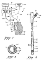
- FIG. 2 is an enlarged, front elevational view, partially in longitudinal section, of a feeding tube/bolus/Y-connector assembly embodying the invention;
- FIG. 3 is a diagramatic view of a nasal-catheter arrangement having three electrode means positioned respectively in the esophagus, the stomach, and the small intestine;
- FIG. 4 is a further enlarged, side elevational view, partially in longitudinal section, of a bolus having a modified placement of electrodes thereon;
- FIG. 5 is an enlarged transverse sectional view taken generally along line 5-5 of FIG. 4; and
- FIG. 6 is a transverse sectional view taken generally along line 6-6 of FIG. 4.
- Referring now to the drawings, two possible forms of a gastrointestinal locating/
feeding system 10 embodying the invention for the feeding of liquid nutrients into preselected areas of the gastrointestinal tract are shown in FIG. 1. Thesystem 10 includes an elongated, flexible,feeding tube connector 14,14a provided on its proximal end and abolus bolus electrodes 18′ and 18˝ (FIGS. 2 and 4); amodule 20 for amplifying the detected myoelectric signals and for filtering out undesired frequencies; amonitor 22 capable of processing the detected signals; and, in certain instances, acontrollable feeding device 24 such as an infusion pump or a flow-regulating clamp. - Two of several possible forms of the
system 10 are illustrated in FIG. 1. A nasal-enteral feeding arrangement into the upper portion orduodenum 26 of a patient'ssmall intestine 28, as shown in full line, is characterized by thefeeding tube 12 which extends through the patient'sesophagus 30 and through the patient'sstomach 32 with thebolus 16 being positioned in the patient'sduodenum 26. The Y-connector 14 is provided on the proximal end of thefeeding tube 12 and sheathed leads 34′ and 34˝, which are connected to theelectrodes 18′ and 18˝ by aconnector 19′ and by a connector forlead 34˝ which is not visible in FIG. 2, extend through the feeding tube 12 (FIGS. 2 and 3) and out of the Y-connector 14 through anarm 36 thereof for connection to the amplifier/filter module 20. As is best shown in FIG. 5, a separate passage orlumen 60 may be provided in thefeeding tube stomach 32, as shown in broken line in FIG. 1, is characterized by thefeeding tube 12a which extends through astoma 46 provided in theabdominal wall 48 with thebolus 16a being positioned in thestomach 32. Adjustable retainingmembers feeding tube 12a at internal and external ends of thestoma 46, respectively, as an aid in preventing undesirable movement of thefeeding tube 12a relative thereto. The Y-connector 14a is provided on the proximal end of thefeeding tube 12a and sheathed electrode leads 34a′ and 34a˝ extend outwardly of the Y-connector 12a through an arm 36a thereof for connection to the amplifier/filter module 20. - Although not shown in the drawings, it is noted that the
bolus 16 of the nasal-enteral feeding tube 12 may, if desired, be positioned in thestomach 32, the remaining small bowel, or the colon (not shown) if the myoelectric signals of these areas of a patient's gastrointestinal tract are to be monitored. Likewise, it is noted that thebolus 16a of the abdominalstoma feeding tube 12a may, if desired, be positioned in theduodenum 26 or even in the patient's remaining small bowel or colon. As illustrated in FIG. 3, there may be instances where it is desirable for diagnostic or treatment purposes to have afeeding tube 12 which extends through a patient'snasal passage 62 and which has three spaced electrode means provided thereon, an esophageal electrode means 64, a stomach electrode means 66, and a small bowel electrode means 68. - The amplifier/
filter module 20, which filters out all frequencies outside the range of physiological interest as well as 60 hertz noise, will amplify the detected signal to a magnitude acceptable to themonitor 22 and will also provide impedance matching between thefeeding tube electrodes 18′ and 18˝ and themonitor 22. Themodule 20 is suitably connected to themonitor 22, as at 38 and themonitor 22 may be suitably connected to thefeeding device 24, as at 40. Themonitor 22 is an electronic device which is adapted to process the detected electrophysiological or myoelectric signals received from themodule 20 in such a way that the unique properties of the various segments of the enteral tract's electrical activity can be readily identified. Once the parameters of the signals are determined, they may be compared to selected standards by the programming of themonitor 22. The monitor program will also contain a logic path such that the results of the comparison may be used to control thefeeding device 24 whereby thefeeding device 24 may be directed to maintain its current feeding rate setting or to increase/decrease the rate of nutrient feeding or even to select a different nutrient. Thisunique system 10 thus provides a solution to the problem of overloading a patient's gastrointestinal tract. Feeding is normally started at a low rate of infusion which is periodically increased until the patient's nutrient needs are met or the patient exhibits discomfort or other symptoms. However, thissystem 10 allows digestive problems to be recognized and prevented by regulating the rate of feeding prior to gastric overload and the onset of more harmful physical symptoms such as cramps, nausea, vomiting, diarrhea, gastric reflux, or aspiration pneumonia. Thefeeding device 24 may be connected to anotherarm connector 14,14a by atube set - Another major problem of gastrointestinal feeding is that of initially positioning the feeding end of the
tube bolus present system 10 provides a direct, continuous, and reliable method of overcoming this problem as themonitor 22 may be programmed to not only regulate a feeding rate but also to sound an alarm or otherwise alert an observer while it may also simultaneously cease the feeding should thebolus monitor 22 may also possess display and storage devices such as a CRT display, a chart recorder and/or a magnetic tape recorder, whereby to provide a record of the detected signals. - With reference to FIGS. 2 and 4, it is noted that the
electrodes 18′ and 18˝ of the preferred embodiment are in the form of coils of fine metal wire wrapped around thebolus 16 in spaced-apart relationship and adjacent tonutrient exit ports bolus 16. The metal wire for theelectrodes 18′ and 18˝ should have good electrical conductivity such as is provided by gold, silver and platinum wire, for instance. Another suitable wound-coil electrode would be a silver/silver chloride electrode formed of silver wire coated with a chloride salt of silver. The wound coil configuration presents a conducting surface all around the circumference of thebolus 16 while maintaining flexibility in that the individual coils can deform and deflect in conformance with changes in the shape of thebolus 16, as during the intubation process. - However, it is noted that many other forms of electrodes besides wound-coil electrodes may be used in the gastrointestinal locating/
feeding system 10 of the present invention. - The multiple lumen feeding tube arrangement, as is best illustrated in FIGS. 4 and 5, provides means for detecting myoelectric signals in desired locations in a patient's gastrointestinal tract while simultaneously introducing liquid nutrients at the site of detection. Two pathways are provided in the
tube 12, afluid channel 58 communicating throughout the length of thetube 12 and providing a means for the liquid nutrient to move from its source, thefeeding device 24, to the bolusnutrient exit ports lumen 60 separate from thefluid path 58 but still inside thetube 12 and providing a means for electrical communication between theelectrodes 18′ and 18˝ and themodule 20 by passage or the sheathed leads 34′ and 34˝ therethrough. - While there has been shown and described several possible embodiments of the invention, it will be obvious to those skilled in the art that changes and modifications may be made without departing from the invention, and it is intended by the appended claims to cover all such changes and modifications as fall within the true spirit and scope of the invention.
- Where technical features mentioned in any claim are followed by reference signs, those reference signs have been included for the sole purpose of increasing the intelligibility of the claims and accordingly, such reference signs do not have any limiting effect on the scope of each element identified by way of example by such reference signs.
Claims (10)
Priority Applications (1)
| Application Number | Priority Date | Filing Date | Title |
|---|---|---|---|
| AT89103862T ATE83648T1 (en) | 1988-03-23 | 1989-03-06 | INTESTINAL NUTRITIONAL SYSTEM USING GASTROINTESTINAL MUSCLE ELECTROGRAPHY. |
Applications Claiming Priority (2)
| Application Number | Priority Date | Filing Date | Title |
|---|---|---|---|
| US172055 | 1980-07-25 | ||
| US07/172,055 US4921481A (en) | 1988-03-23 | 1988-03-23 | Enteral feeding system utilizing gastrointestinal myoelectrography |
Publications (3)
| Publication Number | Publication Date |
|---|---|
| EP0334086A2 true EP0334086A2 (en) | 1989-09-27 |
| EP0334086A3 EP0334086A3 (en) | 1990-02-28 |
| EP0334086B1 EP0334086B1 (en) | 1992-12-23 |
Family
ID=22626181
Family Applications (1)
| Application Number | Title | Priority Date | Filing Date |
|---|---|---|---|
| EP89103862A Expired - Lifetime EP0334086B1 (en) | 1988-03-23 | 1989-03-06 | Enteral feeding system utilizing gastrointestinal myoelectrography |
Country Status (9)
| Country | Link |
|---|---|
| US (1) | US4921481A (en) |
| EP (1) | EP0334086B1 (en) |
| JP (1) | JP2795882B2 (en) |
| KR (1) | KR970009720B1 (en) |
| AT (1) | ATE83648T1 (en) |
| AU (1) | AU611531B2 (en) |
| CA (1) | CA1326264C (en) |
| DE (1) | DE68903973T2 (en) |
| ES (1) | ES2037295T3 (en) |
Cited By (7)
| Publication number | Priority date | Publication date | Assignee | Title |
|---|---|---|---|---|
| WO1992017150A1 (en) * | 1991-04-05 | 1992-10-15 | Deborah Jill Colson | A combined naso-gastric feeding tube and electrode |
| EP0582400A1 (en) * | 1992-08-05 | 1994-02-09 | Smiths Industries Public Limited Company | Medico-surgical assemblies |
| GB2328159A (en) * | 1997-08-13 | 1999-02-17 | Geo L White Limited | Animal Feeding apparatus and feed |
| WO2000069376A1 (en) * | 1999-05-18 | 2000-11-23 | Silhouette Medical Inc. | Surgical weight control device |
| WO2008072150A1 (en) * | 2006-12-13 | 2008-06-19 | Koninklijke Philips Electronics N. V. | Feeding tube |
| US7468060B2 (en) | 1998-02-19 | 2008-12-23 | Respiratory Diagnostic, Inc. | Systems and methods for treating obesity and other gastrointestinal conditions |
| EP3501481A1 (en) * | 2017-12-03 | 2019-06-26 | Envizion Medical Ltd. | Feeding tube with electromagnetic sensor |
Families Citing this family (56)
| Publication number | Priority date | Publication date | Assignee | Title |
|---|---|---|---|---|
| JPH0641547Y2 (en) * | 1989-12-12 | 1994-11-02 | 株式会社スギノマシン | Feeding tube |
| NL9401180A (en) | 1994-07-18 | 1996-03-01 | Draeger Med Electronics Bv | Catheter with multiple sensors at a distance from each other. |
| AR004329A1 (en) * | 1995-11-28 | 1998-11-04 | Abbott Lab | ARRANGEMENT FOR ENTERAL FEEDING INCLUDING A FEEDING TUBE THAT HAS A PROXIMAL END AND A DISTAL END. |
| US5690691A (en) * | 1996-05-08 | 1997-11-25 | The Center For Innovative Technology | Gastro-intestinal pacemaker having phased multi-point stimulation |
| SE9803375D0 (en) * | 1998-10-06 | 1998-10-06 | A & Science Invest Ab | Method and apparatus for measuring intestinal potential difference |
| JP2004505748A (en) | 2000-08-23 | 2004-02-26 | ミクロニックス ピーティーワイ リミテッド | Catheter position display device and use thereof |
| US6535764B2 (en) * | 2001-05-01 | 2003-03-18 | Intrapace, Inc. | Gastric treatment and diagnosis device and method |
| US20050143784A1 (en) | 2001-05-01 | 2005-06-30 | Imran Mir A. | Gastrointestinal anchor with optimal surface area |
| US7689284B2 (en) * | 2001-05-01 | 2010-03-30 | Intrapace, Inc. | Pseudounipolar lead for stimulating a digestive organ |
| US7702394B2 (en) | 2001-05-01 | 2010-04-20 | Intrapace, Inc. | Responsive gastric stimulator |
| US7643887B2 (en) * | 2001-05-01 | 2010-01-05 | Intrapace, Inc. | Abdominally implanted stimulator and method |
| US7979127B2 (en) * | 2001-05-01 | 2011-07-12 | Intrapace, Inc. | Digestive organ retention device |
| US20080065169A1 (en) * | 2001-05-01 | 2008-03-13 | Intrapace, Inc. | Endoscopic Instrument for Engaging a Device |
| US9668690B1 (en) | 2001-05-01 | 2017-06-06 | Intrapace, Inc. | Submucosal gastric implant device and method |
| US7020531B1 (en) | 2001-05-01 | 2006-03-28 | Intrapace, Inc. | Gastric device and suction assisted method for implanting a device on a stomach wall |
| US7756582B2 (en) | 2001-05-01 | 2010-07-13 | Intrapace, Inc. | Gastric stimulation anchor and method |
| US7616996B2 (en) * | 2005-09-01 | 2009-11-10 | Intrapace, Inc. | Randomized stimulation of a gastrointestinal organ |
| US7747322B2 (en) * | 2001-05-01 | 2010-06-29 | Intrapace, Inc. | Digestive organ retention device |
| GB2384993B (en) * | 2002-02-06 | 2004-05-12 | David Alan Burt | Medical surgeons detection aid |
| US20050015132A1 (en) * | 2003-04-16 | 2005-01-20 | Itzhak Kronzon | Combined transesophageal echocardiography and transesophageal cardioversion probe |
| US20060070334A1 (en) * | 2004-09-27 | 2006-04-06 | Blue Hen, Llc | Sidewall plank for constructing a trailer and associated trailer sidewall construction |
| US7976518B2 (en) | 2005-01-13 | 2011-07-12 | Corpak Medsystems, Inc. | Tubing assembly and signal generator placement control device and method for use with catheter guidance systems |
| WO2006078958A2 (en) * | 2005-01-21 | 2006-07-27 | Virginia Technologies, Inc | Energy efficient therapeutic pulse generator system |
| US7509175B2 (en) | 2006-08-03 | 2009-03-24 | Intrapace, Inc. | Method and devices for stimulation of an organ with the use of a transectionally placed guide wire |
| US20090018606A1 (en) * | 2005-10-12 | 2009-01-15 | Intrapace, Inc. | Methods and Devices for Stimulation of an Organ with the Use of a Transectionally Placed Guide Wire |
| US20070149850A1 (en) * | 2005-12-22 | 2007-06-28 | Spivey James T | Endoscope endcap attachment tool |
| US7771396B2 (en) * | 2006-03-22 | 2010-08-10 | Ethicon Endo-Surgery, Inc. | Intubation device for enteral feeding |
| US7803137B2 (en) * | 2006-03-22 | 2010-09-28 | Ethicon Endo-Surgery, Inc. | Intubation system for use with an endoscope |
| US20070239171A1 (en) * | 2006-03-30 | 2007-10-11 | Ethicon Endo-Surgery, Inc. | Medical snaring device |
| US20080097179A1 (en) * | 2006-07-03 | 2008-04-24 | Russo Susan H | Feeding tube system with reflux fluid sensing |
| US8285399B2 (en) * | 2006-11-16 | 2012-10-09 | Koninklijke Philips Electronics N.V. | Present invention is directed to a feeding tube in particular for total parental nutrition and/or medicine dosing |
| US7794425B2 (en) * | 2006-12-21 | 2010-09-14 | Kimberly-Clark Worldwide, Inc. | Gastro-esophageal reflux control system and pump |
| US9295395B2 (en) | 2007-03-02 | 2016-03-29 | Art Healthcare Ltd. | Interactive NGT system |
| WO2008124644A1 (en) | 2007-04-05 | 2008-10-16 | Velomedix, Inc | Automated therapy system and method |
| WO2008154800A1 (en) * | 2007-06-20 | 2008-12-24 | Yuanming Luo | Multifunctional catheter for detecting respiration and ecg signals |
| US20090316925A1 (en) * | 2008-06-20 | 2009-12-24 | Leonard Eisenfeld | Electronic stethoscope system |
| RU2548138C2 (en) * | 2008-08-28 | 2015-04-10 | Конинклейке Филипс Электроникс Н.В. | Application, device and method of obtaining physiological signals by feeding tube |
| WO2010115194A1 (en) | 2009-04-03 | 2010-10-07 | Intrapace, Inc. | Feedback systems and methods for communicating diagnostic and/or treatment signals to enhance obesity treatments |
| WO2012045030A2 (en) | 2010-10-01 | 2012-04-05 | Intrapace, Inc. | Feedback systems and methods to enhance obstructive and other obesity treatments, optionally using multiple sensors |
| EP2720605B1 (en) | 2011-06-14 | 2020-03-11 | Gravitas Medical Inc. | Apparatus for guiding medical care based on detected gastric function |
| WO2014109846A1 (en) | 2013-01-14 | 2014-07-17 | Corpak Medsystems, Inc. | Bridle delivery system, method, and apparatus for securing nasal tubes |
| WO2015120285A1 (en) | 2014-02-06 | 2015-08-13 | Theranova, Llc | Devices and methods to measure gastric residual volume |
| EP3193814B1 (en) | 2014-09-04 | 2018-08-22 | Corpak Medsystems, Inc. | Gastric systems, apparatus, and methods for use with enteral feeding |
| AU2015100063B4 (en) * | 2015-01-22 | 2015-08-20 | Oppong-Addae, George MR | The Gastro-Mechanical Analyser |
| CA2983615A1 (en) | 2015-05-05 | 2016-11-10 | Corpak Medsystems, Inc. | A threaded connector port cleaning system, method, and apparatus |
| CA2986462A1 (en) * | 2015-05-20 | 2016-11-24 | Gravitas Medical, Inc. | Methods and apparatus for guiding medical care based on sensor data from the gastrointestinal tract |
| CN106214495A (en) * | 2016-07-21 | 2016-12-14 | 刘卫辉 | Jejunal nutrition and functional assessment pipe |
| US10322068B2 (en) * | 2016-12-12 | 2019-06-18 | Art Healthcare Ltd. | Systems and methods for automatic management of reflux during enteral feeding |
| EP3554430A1 (en) | 2016-12-16 | 2019-10-23 | Avent, Inc. | Medical plug system and apparatus |
| US11786141B2 (en) | 2019-03-04 | 2023-10-17 | Avent, Inc. | System, method, and apparatus for detecting tube misplacement in a patient's airway |
| US11839723B2 (en) | 2019-03-04 | 2023-12-12 | Avent, Inc. | System, method, and apparatus for detecting tube misplacement in a patient's airway |
| US11590320B2 (en) | 2019-04-04 | 2023-02-28 | Avent, Inc. | Two-in-one catheter and signal generating apparatus |
| US11273288B2 (en) | 2019-04-08 | 2022-03-15 | Avent, Inc. | System and method for medical device position guidance |
| US11602280B2 (en) | 2019-04-08 | 2023-03-14 | Avent, Inc. | In-scale flexible display for medical device position guidance |
| US11517217B2 (en) | 2019-04-08 | 2022-12-06 | Avent, Inc. | In-scale tablet display for medical device position guidance |
| CN115887232B (en) * | 2023-03-09 | 2023-10-10 | 四川大学华西医院 | Nose intestinal canal device based on noninvasive real-time magnetic positioning |
Citations (4)
| Publication number | Priority date | Publication date | Assignee | Title |
|---|---|---|---|---|
| US3043309A (en) * | 1959-09-29 | 1962-07-10 | Avco Corp | Method of performing intestinal intubation |
| US3888237A (en) * | 1973-10-15 | 1975-06-10 | Olympus Optical Co | Ph-measuring device used in an endoscope |
| US4134405A (en) * | 1977-01-10 | 1979-01-16 | Smit Julie A | Catheter and intestine tube and method of using the same |
| US4381011A (en) * | 1981-05-04 | 1983-04-26 | Somers 3Rd Lewis S | Enteral feeding apparatus and method |
Family Cites Families (9)
| Publication number | Priority date | Publication date | Assignee | Title |
|---|---|---|---|---|
| US2729211A (en) * | 1950-07-07 | 1956-01-03 | Peter Josef | Device for examining the condition of the stomach |
| US3411507A (en) * | 1964-04-01 | 1968-11-19 | Medtronic Inc | Method of gastrointestinal stimulation with electrical pulses |
| US3480003A (en) * | 1967-02-03 | 1969-11-25 | Battelle Development Corp | Apparatus for measuring esophageal motility |
| US3860000A (en) * | 1973-07-12 | 1975-01-14 | Lear Siegler Inc | Medical apparatus and method for feeding and aspirating |
| US4213466A (en) * | 1978-08-11 | 1980-07-22 | Harvard College, President And Fellows | Monitoring myoelectric signals |
| US4476872A (en) * | 1980-03-07 | 1984-10-16 | The Kendall Company | Esophageal probe with disposable cover |
| FR2530462A1 (en) * | 1982-07-26 | 1984-01-27 | Dumas Yves | Automatically controlled feeding pump |
| US4683890A (en) * | 1985-12-23 | 1987-08-04 | Brunswick Manufacturing Co., Inc. | Method and apparatus for controlled breathing employing internal and external electrodes |
| US4769014A (en) * | 1987-06-02 | 1988-09-06 | Superior Biosystems Inc. | Gastroenteric feeding tube for endoscopic placement |
-
1988
- 1988-03-23 US US07/172,055 patent/US4921481A/en not_active Expired - Lifetime
-
1989
- 1989-02-22 AU AU30213/89A patent/AU611531B2/en not_active Ceased
- 1989-03-06 ES ES198989103862T patent/ES2037295T3/en not_active Expired - Lifetime
- 1989-03-06 EP EP89103862A patent/EP0334086B1/en not_active Expired - Lifetime
- 1989-03-06 DE DE8989103862T patent/DE68903973T2/en not_active Expired - Fee Related
- 1989-03-06 AT AT89103862T patent/ATE83648T1/en not_active IP Right Cessation
- 1989-03-20 KR KR1019890003432A patent/KR970009720B1/en not_active IP Right Cessation
- 1989-03-22 CA CA000594524A patent/CA1326264C/en not_active Expired - Fee Related
- 1989-03-22 JP JP1070263A patent/JP2795882B2/en not_active Expired - Fee Related
Patent Citations (4)
| Publication number | Priority date | Publication date | Assignee | Title |
|---|---|---|---|---|
| US3043309A (en) * | 1959-09-29 | 1962-07-10 | Avco Corp | Method of performing intestinal intubation |
| US3888237A (en) * | 1973-10-15 | 1975-06-10 | Olympus Optical Co | Ph-measuring device used in an endoscope |
| US4134405A (en) * | 1977-01-10 | 1979-01-16 | Smit Julie A | Catheter and intestine tube and method of using the same |
| US4381011A (en) * | 1981-05-04 | 1983-04-26 | Somers 3Rd Lewis S | Enteral feeding apparatus and method |
Cited By (15)
| Publication number | Priority date | Publication date | Assignee | Title |
|---|---|---|---|---|
| WO1992017150A1 (en) * | 1991-04-05 | 1992-10-15 | Deborah Jill Colson | A combined naso-gastric feeding tube and electrode |
| EP0582400A1 (en) * | 1992-08-05 | 1994-02-09 | Smiths Industries Public Limited Company | Medico-surgical assemblies |
| GB2328159A (en) * | 1997-08-13 | 1999-02-17 | Geo L White Limited | Animal Feeding apparatus and feed |
| US7468060B2 (en) | 1998-02-19 | 2008-12-23 | Respiratory Diagnostic, Inc. | Systems and methods for treating obesity and other gastrointestinal conditions |
| WO2000069376A1 (en) * | 1999-05-18 | 2000-11-23 | Silhouette Medical Inc. | Surgical weight control device |
| US7326207B2 (en) | 1999-05-18 | 2008-02-05 | Curon Medical, Inc. | Surgical weight control device |
| US7947038B2 (en) | 1999-05-18 | 2011-05-24 | Mederi Therapeutics Inc. | Obesity treatment system including inflatable balloon structures with micropores for transport of liquid |
| US8740894B2 (en) | 1999-05-18 | 2014-06-03 | Mederi Therapeutics Inc. | Surgical weight control systems and methods |
| WO2008072150A1 (en) * | 2006-12-13 | 2008-06-19 | Koninklijke Philips Electronics N. V. | Feeding tube |
| CN101557790B (en) * | 2006-12-13 | 2013-03-13 | 皇家飞利浦电子股份有限公司 | Feeding tube |
| US9675265B2 (en) | 2006-12-13 | 2017-06-13 | Koninklijke Philips N.V. | Feeding tube |
| EP3501481A1 (en) * | 2017-12-03 | 2019-06-26 | Envizion Medical Ltd. | Feeding tube with electromagnetic sensor |
| US10993887B2 (en) | 2017-12-03 | 2021-05-04 | Envizion Medical Ltd. | Feeding tube with electromagnetic sensor |
| US11559470B2 (en) | 2017-12-03 | 2023-01-24 | Envizion Medical Ltd. | Feeding tube with electromagnetic sensor |
| US11766385B2 (en) | 2017-12-03 | 2023-09-26 | Envizion Medical Ltd. | Feeding tube with electromagnetic sensor |
Also Published As
| Publication number | Publication date |
|---|---|
| ES2037295T3 (en) | 1993-06-16 |
| AU611531B2 (en) | 1991-06-13 |
| ATE83648T1 (en) | 1993-01-15 |
| EP0334086A3 (en) | 1990-02-28 |
| DE68903973T2 (en) | 1993-05-19 |
| CA1326264C (en) | 1994-01-18 |
| JPH01284253A (en) | 1989-11-15 |
| EP0334086B1 (en) | 1992-12-23 |
| AU3021389A (en) | 1989-09-28 |
| JP2795882B2 (en) | 1998-09-10 |
| KR970009720B1 (en) | 1997-06-17 |
| DE68903973D1 (en) | 1993-02-04 |
| US4921481A (en) | 1990-05-01 |
| KR890014139A (en) | 1989-10-21 |
Similar Documents
| Publication | Publication Date | Title |
|---|---|---|
| US4921481A (en) | Enteral feeding system utilizing gastrointestinal myoelectrography | |
| US20230181095A1 (en) | Methods and apparatus for guiding medical care based on sensor data from the gastrointestinal tract | |
| US11529514B2 (en) | Obstructive sleep apnea treatment devices, systems and methods | |
| US5833625A (en) | Ambulatory reflux monitoring system | |
| US5479935A (en) | Ambulatory reflux monitoring system | |
| US11081222B2 (en) | Obstructive sleep apnea treatment screening methods | |
| US11471685B2 (en) | Obstructive sleep apnea treatment devices, systems and methods | |
| US9474468B2 (en) | Method and device positioning a linear array of electrodes in a patient's respiratory airways | |
| JP2710864B2 (en) | Percutaneously placed electrical intestinal pacemaker | |
| US7052474B2 (en) | Pharyngoesophageal monitoring systems | |
| US20130225946A1 (en) | Device, apparatus and method for obtaining physiological signals by way of a feeding tube | |
| WO2003092495A1 (en) | Intracavitary impedance measuring probe | |
| EP2037802B1 (en) | Method and apparatus for measuring activity in the peripheral nervous system | |
| KR20140025424A (en) | Multi-functional catheter | |
| US20210030629A1 (en) | System and Method for Acoustic Sensing to Verify Proper Nasogastric Tube Placement | |
| JP2022518485A (en) | A device that measures congestion in the gastrointestinal tract | |
| AU1059097A (en) | Enteral feeding tube system used to assist in tube placement | |
| US20210361934A1 (en) | System and Method for Muscle Stimulation and/or Impedance Measurement to Verify Proper Tube Placement | |
| EP1119289B1 (en) | Apparatus for measuring of intestinal potential difference | |
| EP3052011B1 (en) | Obstructive sleep apnea treatment screening methods |
Legal Events
| Date | Code | Title | Description |
|---|---|---|---|
| PUAI | Public reference made under article 153(3) epc to a published international application that has entered the european phase |
Free format text: ORIGINAL CODE: 0009012 |
|
| AK | Designated contracting states |
Kind code of ref document: A2 Designated state(s): AT BE CH DE ES FR GB IT LI NL SE |
|
| PUAL | Search report despatched |
Free format text: ORIGINAL CODE: 0009013 |
|
| AK | Designated contracting states |
Kind code of ref document: A3 Designated state(s): AT BE CH DE ES FR GB IT LI NL SE |
|
| 17P | Request for examination filed |
Effective date: 19900821 |
|
| 17Q | First examination report despatched |
Effective date: 19911014 |
|
| GRAA | (expected) grant |
Free format text: ORIGINAL CODE: 0009210 |
|
| AK | Designated contracting states |
Kind code of ref document: B1 Designated state(s): AT BE CH DE ES FR GB IT LI NL SE |
|
| REF | Corresponds to: |
Ref document number: 83648 Country of ref document: AT Date of ref document: 19930115 Kind code of ref document: T |
|
| ET | Fr: translation filed | ||
| REF | Corresponds to: |
Ref document number: 68903973 Country of ref document: DE Date of ref document: 19930204 |
|
| ITF | It: translation for a ep patent filed |
Owner name: MODIANO & ASSOCIATI S.R.L. |
|
| REG | Reference to a national code |
Ref country code: ES Ref legal event code: FG2A Ref document number: 2037295 Country of ref document: ES Kind code of ref document: T3 |
|
| PLBE | No opposition filed within time limit |
Free format text: ORIGINAL CODE: 0009261 |
|
| STAA | Information on the status of an ep patent application or granted ep patent |
Free format text: STATUS: NO OPPOSITION FILED WITHIN TIME LIMIT |
|
| 26N | No opposition filed | ||
| EAL | Se: european patent in force in sweden |
Ref document number: 89103862.2 |
|
| REG | Reference to a national code |
Ref country code: GB Ref legal event code: IF02 |
|
| PGFP | Annual fee paid to national office [announced via postgrant information from national office to epo] |
Ref country code: GB Payment date: 20050207 Year of fee payment: 17 Ref country code: AT Payment date: 20050207 Year of fee payment: 17 |
|
| PGFP | Annual fee paid to national office [announced via postgrant information from national office to epo] |
Ref country code: NL Payment date: 20050209 Year of fee payment: 17 |
|
| PGFP | Annual fee paid to national office [announced via postgrant information from national office to epo] |
Ref country code: FR Payment date: 20050302 Year of fee payment: 17 |
|
| PGFP | Annual fee paid to national office [announced via postgrant information from national office to epo] |
Ref country code: SE Payment date: 20050303 Year of fee payment: 17 |
|
| PGFP | Annual fee paid to national office [announced via postgrant information from national office to epo] |
Ref country code: ES Payment date: 20050311 Year of fee payment: 17 Ref country code: CH Payment date: 20050311 Year of fee payment: 17 |
|
| PGFP | Annual fee paid to national office [announced via postgrant information from national office to epo] |
Ref country code: DE Payment date: 20050331 Year of fee payment: 17 |
|
| PGFP | Annual fee paid to national office [announced via postgrant information from national office to epo] |
Ref country code: BE Payment date: 20050412 Year of fee payment: 17 |
|
| PG25 | Lapsed in a contracting state [announced via postgrant information from national office to epo] |
Ref country code: GB Free format text: LAPSE BECAUSE OF NON-PAYMENT OF DUE FEES Effective date: 20060306 Ref country code: AT Free format text: LAPSE BECAUSE OF NON-PAYMENT OF DUE FEES Effective date: 20060306 |
|
| PG25 | Lapsed in a contracting state [announced via postgrant information from national office to epo] |
Ref country code: SE Free format text: LAPSE BECAUSE OF NON-PAYMENT OF DUE FEES Effective date: 20060307 Ref country code: ES Free format text: LAPSE BECAUSE OF NON-PAYMENT OF DUE FEES Effective date: 20060307 |
|
| PG25 | Lapsed in a contracting state [announced via postgrant information from national office to epo] |
Ref country code: LI Free format text: LAPSE BECAUSE OF NON-PAYMENT OF DUE FEES Effective date: 20060331 Ref country code: CH Free format text: LAPSE BECAUSE OF NON-PAYMENT OF DUE FEES Effective date: 20060331 Ref country code: BE Free format text: LAPSE BECAUSE OF NON-PAYMENT OF DUE FEES Effective date: 20060331 |
|
| PGFP | Annual fee paid to national office [announced via postgrant information from national office to epo] |
Ref country code: IT Payment date: 20060331 Year of fee payment: 18 |
|
| PG25 | Lapsed in a contracting state [announced via postgrant information from national office to epo] |
Ref country code: NL Free format text: LAPSE BECAUSE OF NON-PAYMENT OF DUE FEES Effective date: 20061001 |
|
| PG25 | Lapsed in a contracting state [announced via postgrant information from national office to epo] |
Ref country code: DE Free format text: LAPSE BECAUSE OF NON-PAYMENT OF DUE FEES Effective date: 20061003 |
|
| REG | Reference to a national code |
Ref country code: CH Ref legal event code: PL |
|
| EUG | Se: european patent has lapsed | ||
| GBPC | Gb: european patent ceased through non-payment of renewal fee |
Effective date: 20060306 |
|
| NLV4 | Nl: lapsed or anulled due to non-payment of the annual fee |
Effective date: 20061001 |
|
| REG | Reference to a national code |
Ref country code: FR Ref legal event code: ST Effective date: 20061130 |
|
| REG | Reference to a national code |
Ref country code: ES Ref legal event code: FD2A Effective date: 20060307 |
|
| BERE | Be: lapsed |
Owner name: *ABBOTT LABORATORIES Effective date: 20060331 |
|
| PG25 | Lapsed in a contracting state [announced via postgrant information from national office to epo] |
Ref country code: FR Free format text: LAPSE BECAUSE OF NON-PAYMENT OF DUE FEES Effective date: 20060331 |
|
| PG25 | Lapsed in a contracting state [announced via postgrant information from national office to epo] |
Ref country code: IT Free format text: LAPSE BECAUSE OF NON-PAYMENT OF DUE FEES Effective date: 20070306 |