EP0336731A2 - Method of amplifying and detecting nucleic acid sequences - Google Patents
Method of amplifying and detecting nucleic acid sequences Download PDFInfo
- Publication number
- EP0336731A2 EP0336731A2 EP89303351A EP89303351A EP0336731A2 EP 0336731 A2 EP0336731 A2 EP 0336731A2 EP 89303351 A EP89303351 A EP 89303351A EP 89303351 A EP89303351 A EP 89303351A EP 0336731 A2 EP0336731 A2 EP 0336731A2
- Authority
- EP
- European Patent Office
- Prior art keywords
- template
- nucleic acid
- oligonucleotides
- complementary
- ligation
- Prior art date
- Legal status (The legal status is an assumption and is not a legal conclusion. Google has not performed a legal analysis and makes no representation as to the accuracy of the status listed.)
- Granted
Links
Images


Classifications
-
- C—CHEMISTRY; METALLURGY
- C12—BIOCHEMISTRY; BEER; SPIRITS; WINE; VINEGAR; MICROBIOLOGY; ENZYMOLOGY; MUTATION OR GENETIC ENGINEERING
- C12Q—MEASURING OR TESTING PROCESSES INVOLVING ENZYMES, NUCLEIC ACIDS OR MICROORGANISMS; COMPOSITIONS OR TEST PAPERS THEREFOR; PROCESSES OF PREPARING SUCH COMPOSITIONS; CONDITION-RESPONSIVE CONTROL IN MICROBIOLOGICAL OR ENZYMOLOGICAL PROCESSES
- C12Q1/00—Measuring or testing processes involving enzymes, nucleic acids or microorganisms; Compositions therefor; Processes of preparing such compositions
- C12Q1/68—Measuring or testing processes involving enzymes, nucleic acids or microorganisms; Compositions therefor; Processes of preparing such compositions involving nucleic acids
- C12Q1/6844—Nucleic acid amplification reactions
- C12Q1/6862—Ligase chain reaction [LCR]
-
- A—HUMAN NECESSITIES
- A61—MEDICAL OR VETERINARY SCIENCE; HYGIENE
- A61P—SPECIFIC THERAPEUTIC ACTIVITY OF CHEMICAL COMPOUNDS OR MEDICINAL PREPARATIONS
- A61P7/00—Drugs for disorders of the blood or the extracellular fluid
-
- C—CHEMISTRY; METALLURGY
- C12—BIOCHEMISTRY; BEER; SPIRITS; WINE; VINEGAR; MICROBIOLOGY; ENZYMOLOGY; MUTATION OR GENETIC ENGINEERING
- C12Q—MEASURING OR TESTING PROCESSES INVOLVING ENZYMES, NUCLEIC ACIDS OR MICROORGANISMS; COMPOSITIONS OR TEST PAPERS THEREFOR; PROCESSES OF PREPARING SUCH COMPOSITIONS; CONDITION-RESPONSIVE CONTROL IN MICROBIOLOGICAL OR ENZYMOLOGICAL PROCESSES
- C12Q1/00—Measuring or testing processes involving enzymes, nucleic acids or microorganisms; Compositions therefor; Processes of preparing such compositions
- C12Q1/68—Measuring or testing processes involving enzymes, nucleic acids or microorganisms; Compositions therefor; Processes of preparing such compositions involving nucleic acids
- C12Q1/6844—Nucleic acid amplification reactions
- C12Q1/6858—Allele-specific amplification
Definitions
- This invention relates to a method of amplifying nucleic acid sequences, and further relates to a method for distinguishing single base differences on nucleic acid templates.
- Saiki, R.K., et al., Science 230:1350 (1985) describes enzymatic amplification of a-globin genomic sequences, termed the polymerase chain reaction, by providing two oligonucleotide primers flanking the region to be amplified, annealing the primers to strands of denatured genomic DNA and extending them with the Klenow fragment of E. coli DNA polymerase 1 and deoxyribonucleosidetriphosphates.
- U.S. Patent 4,683,202 to Mullis further describes the polymerase chain reaction method.
- U.S. Patent 4,683,194 to Saiki, et al. describes a method of detecting a specific polymorphic restriction site in a nucleic acid sequence by hybridizing the sequence to an oligonucleotide probe complementary to the sequence and spanning the restriction site which is labeled at the end of the probe nearer to the restriction site.
- the nucleic acid sequence and probe hybridized thereto are then digested with a restriction endonuclease, labeled and unlabeled oligomer fragments are separated, and labeled oligonucleotides are detected.
- U.S. Patent 4,683,195 to Mullis et al. describes a process for amplifying nucleic acid sequences using primers and polymerization agents and a process for detecting the presence or absence of a specific nucleic acid sequence. Separate strands of a nucleic acid sample are treated with an excess of two different oligonucleotide primers, and primers are extended to form complementary extension products which act as templates for synthesizing the desired nucleic acid sequence. The amplified sequence is then detected by adding a labeled probe capable of hybridizing to the sequence being detected.
- the present invention is useful in diagnostic applications because it can be used first, as a means for amplifying given nucleic acid sequences from nucleic acid templates so that the template sequences can be more readily detected.
- the sequence is hybridized to at least one pair of oligonucleotide strands (substrates) which flank both sides of a target base of interest wherein a designated end base of one of the oligonucleotide strands of a pair is complementary to the target base and that designated end base forms a ligation juncture with an end of the other substrate of the pair.
- the former oligonucleotide strand substrate is, in effect, a probe which hybridizes to the target base and adjacent nucleic acid sequence and also simultaneously ligates with the latter oligonucleotide strand substrate designed to flank the opposite side of the target base and adjacent sequence only if the designated end base of the probe is complementary to the target base on the template.
- the invention features a method for detecting single base or base pair mutations in at least one specific nucleic acid sequence contained in a sample of nucleic acid template wherein each nucleic acid consists of either one strand or two separate complementary strands, comprising: treating the strands with at least one of two sets of two different relatively short oligonucleotides each of which sets (i) is complementary to and therefore will hybridize to at least a portion of the template sequence of at least one of the strands and (ii) flanks both sides of a designated target base contained within said specific nucleic acid sequence such that one of the two short oligonucleotides in each set of oligonucleotides contains a base at one of its ends that is complementary to one of the target bases of the template under conditions such that the two short oligonucleotides of each of the sets will mutually ligate if the target base exists in the template, the ligation occurring between the terminal base of one of the complementary oligonucleotides which
- the invention further features a method for amplifying at least one specific nucleic acid sequence in a sample containing a nucleic acid or a mixture of nucleic acids comprised of single or complementary nucleic acid strands, wherein the sample is suspected of containing the sequence or sequences comprising (a) treating the strands with at least one of two pairs of oligonucleotides which sets are complementary to at least one the specific nucleic acid sequence and flank at least one target base of a single stranded template or at least one target base pair defining a blunt end contained in the nucleic acid sequence or sequences wherein one end nucleotide of one of the oligonucleotides of at least one of the pairs is complementary to the target base or one of the bases of the target base pairs under conditions such that the end nucleotide will mutually ligate with an end of the other oligonucleotide of the pair to form a ligation product which is complementary to the specific nucleic acid sequence only if the target base or
- the method of the present invention can be used both to amplify any particular nucleic acid sequence template and distinguish and detect single base differences on nucleic acid templates.
- the present invention is therefore useful to diagnose various genetic disorders that result from point mutations such as sickle cell anemia.
- signal amplification is necessary in order to detect the limited number of DNA template copies in genomic DNA. For example, there are only about one million copies of a single gene in 10 ug of DNA. Further, depending on the particular diagnostic procedure performed, only a limited amount of DNA might be available.
- short oligonucleotide strands are synthesized complementary to and flanking both directions of a target base on a nucleic acid template.
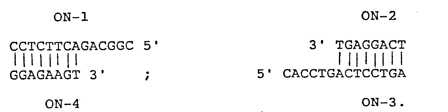
- These relatively short oligonucleotides are generally denoted ON-1, ON-2, ON-3, and ON-4 as illustrated in Fig. 1.
- the flanking oligonucleotides may be synthesized according to methods well known in the art.
- flanking oligonucleotides must be sufficiently long to contain enough of a unique base pattern to cause the flanking oligonucleotides to hybridize or anneal to only the portions of the nucleic acid template which flank the target base pair of interest.
- the exact lengths of the flanking oligonucleotides depends on various factors, including the temperature of the reaction, pH conditions, and complexity of the target sequence. In general, flanking oligonucleotides comprising about 4 to about 100 nucleotides form sufficiently stable hybrid complexes at higher reaction temperatures. In most instances, flanking oligonucleotides used in accordance with the present invention preferably comprise about 8 to about 20 nucleotides.
- nucleic acid template which is either single or double stranded. If the nucleic acid template comprises two strands, it is necessary to first separate the strands prior to commencing ligation amplification of the present invention.
- One method of separation of strands of nucleic acid involves heating the nucleic acid until it is completely denatured. Such denaturation is usually carried out at temperatures ranging from about 80° to 105° C for times ranging from about 1 to 10 minutes. Separation of nucleic acid strands may also be induced by enzymes such as the helicase enzymes and Rec A.
- the specific nucleic acid sequence to be amplified may be only a fraction of a larger molecule or it can be a discrete molecule.
- the sequence to be amplified can also be a part of a larger mixture of molecules or fractions thereof.
- the starting nucleic acid may also contain more than one desired sequence containing target bases which may be the same or different, such that all such sequences can be simultaneously amplified. Samples comprising more than one nucleic acid with more than one desired target sequence can also be amplified by the methods of the present invention.
- RNA is used as an initial template, it can be first copied into a DNA molecule by an enzyme such as reverse transcriptase.
- the template is the ⁇ a normal beta globin allele with target base pair * A-T * ; in Fig. 2, the template is the p s sickle cell allele with target base pair * T-A * .
- the particular templates and flanking oligonucleotides shown in Figs. 1 and 2 only illustrate the present invention and in no way limit the invention to the detection or amplification of alleles of the human beta globin gene.
- Flanking oligonucleotides ON-2 and ON-4 are 8-nucleotide long oligonucleotides and ON-1 and ON-3 are 14-nucleotide long oligonucleotides.
- the method of the present invention requires a symmetry similar to that shown in Fig. 1, namely, the 3' flanking oligonucleotides, ON-2 and ON-4, are of the same length and the 5' flanking oligonucleotides, ON-1 and ON-3, are the same length.
- Either the two 3' or two 5' flanking oligonucleotides can be the longer length, or all four flanking oligonucleotides can be the same length.
- the 5' ends of the 3' flanking oligonucleotides must, however, be phosphorylated and those ends can also be optionally labeled with, for example, radioactive phosphorous.
- the 5' flanking oligonucleotides (ON-1 and ON-3) are not usually phosphorylated to prevent self ligation and template-independent ligation.
- the target base pair of interest can be either A-T as in Fig. 1, T-A, G-C, or C-G. When the template is single-stranded, the target base can therefore be any of the four nucleotides.
- flanking oligonucleotide complementary region can thus contain more than one base or base pair difference which enables the methods of this invention to be used for discrimination and detection of sequences containing multiple base differences.
- the target base pair define a blunt end as seen in Fig. 1, as opposed to a staggered end.
- flanking oligonucleotide substrates are complementary to a nucleic acid sequence harboring the position of possible mutation on either strand. Further, with double-stranded templates, one of the terminal bases of one of the two 3' flanking oligonucleotides and one of the terminal bases of one of the two 5' flanking oligonucleotides to be ligated must be complementary to the respective target bases. Thus, as shown in Fig.
- the base at the 5' end of one of the 3' flanking oligonucleotides (base T, ON-2) is complementary to the target base A of the (+) template strand
- the nucleotide at the 3' end of one of the 5' flanking oligonucleotides (base A, ON-3) is complementary to the target base T of the (-) template strand.
- the base pair G-C directly to right of illustrative target base pair A-T in Fig. 1 can also be a target base pair.
- the 3' terminal nucleotide of one of the 5' flanking oligonucleotides (base C, ON-1) is complementary to the target base G of the ( + ) template strand
- the 5' terminal nucleotide of one of the 3' flanking oligonucleotides (base G, ON-4) is complementary to the target base C of the (-) template strand.
- nucleotides in positions other than those at the ends of flanking oligonucleotides comprising the ultimate ligation junction can be considered as target nucleotides in the practice of the present invention.
- 3' flanking oligonucleotide ON-2 is synthesized so that it is complementary to the (+) or upper strand of the template and its 5' end contains base T which is complementary to target base A of the ( + ) strand, and the remainder of the flanking oligonucleotide extends outwardly from the 5' end base.
- 5' flanking oligonucleotide ON-1 is synthesized so that it is complementary to the (+) or upper strand of the template and is oriented such that the base at its 3' end C, is complementary to the base of the ( + ) strand on the 3' side of the target base on the template. The remainder of the flanking oligonucleotide extends outwardly in the 5' direction.
- 5' flanking oligonucleotide ON-3 is complementary to the (-) or lower strand of the template such that base A at its 3' end is complementary to the target base T of the (-) strand and the remainder of ON-3 extends outwardly from the target base.
- 3' flanking oligonucleotide ON-4 is complementary to the (-) or lower strand of the template and is oriented such that base G at the 5' end of ON-4 is complementary to the base of the (-) strand that is on the 5' side of the target base. The remainder of ON-4 extends outwardly in the 3' direction.
- the bases on the 5' end of ON-2 and 3' end of ON-3 are complementary to the target base pair.
- ON-2 is complementary to ON-3, except for differences in length.
- ON-1 is complementary to ON-4, except for differences in length.
- ON-1 is complementary to ON-4. Therefore in cases where double-stranded nucleic acid template is to be used, the flanking oligonucleotides can be double-stranded, for example: Such double-stranded oligonucleotides must be treated as described above to separate the strands prior to any amplification ligation reaction.
- polynucleotide ligase ligates ON-1 and ON-2, and/or ON-3 and ON-4 at the target bases of ON-2 and ON-3 to form significant amounts of longer ligated products only if the correct complementary target bases are present at the target site of the template.
- Any suitable polynucleotide ligase can be used in the practice of this invention, for example, bacteriophage T4 induced DNA ligase, and E. coli DNA ligase.
- the two oligonucleotides in each pair anneal to the beta globin template at adjacent positions such that the 3' end of one oligonucleotide (ON-1 or ON-3) and the 5' end of the other (ON-s2 or ON-4) form a ligatable junction.
- the target nucleotide on the template base pairs with either the 3' end nucleotide or the 5' end nucleotide of the substrate depending on which strand, (+) or (-), serves as the template for ligation.
- the success or failure of a given ligation amplification procedure can be detected by labeling the flanking oligonucleotides with radioactive or fluorescent labels.
- radioactive or fluorescent labels For example, 32p , 35 S or any other suitable radionuclide may be used as well as other suitable compounds such as fluorescein or rhodamine or their derivatives.
- Ligation amplification product can also be ascertained via any subsequent manipulation that allows distinction between longer ligation product and the shorter precursors such as gel electrophoresis chromatography or hybridization.
- polyacrylamide gel electrophoresis can be used to separate the ligated product from the flanking oligonucleotide reactants according to their sizes.
- the herein described ligation amplifications can be accomplished in a linear or exponential manner as illustrated in the following examples.
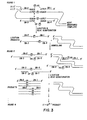
- the steps for exponential ligation-amplification are shown in Fig. 3.
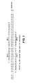
- the DNA sequences of synthetic oligonucleotide substrates and templates used in the examples are given below in Table 1.
- HP19S and H ⁇ 19A are unique 19 nucleotide long oligonucleotides that are complementary to the noncoding strand of the beta globin gene and serve as oligonucleotide templates for ligation of ON-1/ON-2 pairs.
- H ⁇ 23S' and H ⁇ 23A', 23 nucleotides long are complementary to the coding strand of the beta globin gene. These two nucleotides serve as templates for ligation of ON-3/ON-4 pairs.
- ON-1/ON-s2 and ON-1/ON-a2 pairs both hybridize to the coding (+) strand, ON-s2 and ON-a2 differ by a single nucleotide at their 5' ends, with ON-s2 specifying for the sickle cell allele.
- ON-s3/ON-4 and ON-a3/ON-4 pairs hybridize to the noncoding strand with ON-s3 and ON-a3 differing by a single nucleotide at their 3' ends.
- a set comprising at least one of the pairs of flanking oligonucleotide substrates ON-1/ON-a2 and ON-a3/ON-4 are used for the normal beta globin sequence and at least one of the pairs ON-1/ON-s2 and ON-s3/ON-4 are used for detecting the sickle cell sequence.
- template dependent ligation of either the ON-1/ON-2 or ON-3/ON-4 pairs forms a 22 base product with a sequence complementary to the other substrate pairs.
- ON-1-ON-a2 product would be complementary to ON-a3/ON-4 substrates and ON-s3-ON-4 product would be complementary to ON-1/ON-s2 substrates.
- the ligation products can then serve as template for the ligation of the other pair of flanking oligonucleotide substrates thereby producing more templates for further ligation. If the initial ligation step does not take place as a result of base pair mismatch, subsequent amplification steps will not give rise to a detectable ligation signal.
- the 22 nucleotide long ligation product Once the 22 nucleotide long ligation product is formed, it stays hybridized to its template. Denaturation separates the duplex to yield two new templates. If denaturation is accomplished by boiling, the ligase in the reaction mixture is inactivated. Fresh enzyme must therefore be added to initiate the next round of amplification.
- Plasmid and genomic DNA template reactions are initiated following 5-10 minutes of boiling, cooling on ice and the addition of ligase (1 U; BRL). Unless otherwise indicated, all ligation reactions are withdrawn for electrophoresis and optionally treated with calf intestinal alkaline phosphatase (1-5 U) for 1-2 hours.
- Linear amplification ligation of nucleic acid sequences is carried out by repeatedly using the original nucleic acid template (oligo, plasmid or genomic DNA) in each round of amplification. Only one flanking oligonucleotide set complementary to the appropriate oligonucleotide template to one strand of duplex plasmid or genomic DNA template is used. For example, either the ON-1 and ON-2, or ON-3 and ON-4 set is used in the amplification rounds but not both.
- Each round of ligation is initiated by adding 1 unit of ligase to the reaction mixture on ice.
- the mixture is incubated for about 30 minutes at 30°C, or for a longer time if template concentration is low.
- the first round, and all subsequent rounds, is terminated by quickly heating the reaction mixture to 100°C for 5 minutes to inactivate the ligase and dissociate the ligated product strand (ON-1 -ON-2) from the template. This frees the template for subsequent amplification rounds.
- the reaction mixture is centrifuged for 10 seconds and immediately cooled to about 0°C. At this point, there is approximately one ON-1 -ON-2 ligation product present for each copy of the template.
- Another unit of enzyme is added to the reaction mixture to initiate a second round of ligation. Because a molar excess of flanking oligonucleotides ON-1 and ON-2 are added to the initial reaction mixture, more flanking oligonucleotide substrated are not added.
- the reaction mixture is then incubated as in the first round, boiled to dissociate template from the ligation product, and cooled. At this point, there are approximately 2 copies of ON-1 -ON-2 ligation product present for each copy of template.
- Fresh enzyme is added to initiate another round. The steps of incubating, boiling to dissociate template from product, cooling, and adding fresh enzyme are repeated for as many rounds as necessary to produce a detectable signal.
- ON-1 and/or ON-2 can be labeled with radioactive or nonradioactive labels and separated from the reactants by gel electrophoresis, such that ligation product is determined by detecting the presence of label in a position in the gel corresponding to the expected product size.
- ON-a2, ON-s2, and ON-4 are phosphorylated at their 5' ends with either radioactive (y [ 32 P] ATP 6000 Ci/mmol; New England Nuclear) or nonradioactive ATP via a kinase reaction.
- the unlabeled 5' phosphorylated substrates are used following incubation in a boiling water bath for 20 minutes to inactive kinase.
- Internal radioactively phosphorylated oligonucleotide products are separated from unreacted ATP and other reaction products with anion exchange chromatography (DE-52 cellulose-Whatman). The radioactive phosphorylation reaction yields products with approximately 10 8- 10 9 cpm/pmol specific activity.
- Detection and quantitation of ligation product by polyacrylamide gel electrophoresis is carried out by mixing samples from ligation reactions with Ficoll loading buffer and subjecting the mixture to electrophoresis in TBE (89 mM Tris, 89 mM Borate, 2 mM EDTA) in 200/0 polyacrylamide (Bio-Rad) urea (7M) gel at 600 V. for 1.5 hours. The gel is then wrapped in Saran Wrap and autoradiographed between two Lightning Plus intensifier screens (DuPont) overnight. Quantitation of ligation product is obtained either from densitometric measurements of the autoradiograph or directly from the gel via an AMBIS Radioisotope Scanning System II (Automated Microbiology System, Inc.). In the latter instance the polyacrylamide gel is first fixed in a 5% acetic acid solution for 10 minutes.
- the ON-1 - ON-2 ligation product can also be detected by hybridizing the product containing solution to a substrate containing an immobilized complementary DNA or RNA sequence under conditions whereby the ON-1 -ON-2 ligation product will hybridize to the complementary sequence, but neither ON-1 nor ON-2 will. Bound label indicates presence of ligation product.
- the ligated product can also be detected in a subsequent round of ligation employing a radiolabeled counterpart substrate set (ON-3/ON-4). In the case where ligated ON-3/ON-4 is to be detected, labeled ON-1/ON-2 would be used.
- the product level after N-number of rounds of linear amplification-ligation is equal to N x Q where Q is the efficiency of the ligation.
- Q ranges from 0 to 1, where 1 is equivalent to 100% ligation.
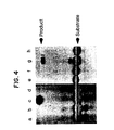
- ON-1/ON-s2 * and ON-s3/ON-4 * were ligated linearly on oligonucleotide templates using the above methodology.
- ON-s2 * and ON-s4 * are the same as ON-s2 and ON-4 except that they are only 6 nucleotides long. 10 pmoles of 32 P-5' pohsphorylated ON-s2 * and unlabeled 5' OH-ON-1 (lanes a-d) or 32 P-5' phosphorylated ON-4 * and unlabeled 5' OH-ON-s3 (lanes e-h) were ligated on 19 base long or 23 base long oligonucleotide template respectively at 30°C for 30 minutes with 1 U.
- Fig. 4 shows the results of the ligation amplification reactions described below.
- 1 pmole of the following oligonucleotide template was used: H ⁇ 19S (c); H ⁇ 19A (d); Hp23S' (g); and H ⁇ 23A'(h).
- the method of the present invention can also be used to exponentially amplify nucleic acid sequences.
- the major difference between exponential amplification ligation and the above-described linear amplification ligation is that all four flanking oligonucleotides (ON-1/ON-2, and ON-3/ON-4) are present as substrates during each reaction round.
- the ligation product of either flanking oligonucleotide set serves as the template for the other flanking oligonucleotide set in subsequent amplification rounds as shown in Fig. 3.
- Radioactively labeled substrate for detection can be included in the starting materials or can be added in the last round.
- Each round of exponential ligation is started by adding one unit of enzyme to the reaction mixture on ice.
- the reaction mixture with the added enzyme is incubated for about 30 minutes at 30° C or for a longer period of time if template concentration is low.
- the reaction mixture is quickly brought to boiling temperature for 5 minutes to inactivate the enzyme and dissociate the product strands from the template strands. Then the reaction mixture is centrifuged for 10 seconds and immediately cooled to about 0°C.
- T4 ligase Another unit of T4 ligase is then added to initiate the second round of amplification.
- the mixture is then incubated, boiled, and cooled as above. At this point there are 3 copies of template present for each copy present at the beginning of the reaction. All subsequent rounds of amplification are carried out by the same sequence of steps for as many times needed in order to produce a detectable signal.
- the progress of the amplification may be monitored by withdrawing from the reaction mixture an approximately 10 ⁇ l aliquot of reaction mixture during predetermined rounds.
- the withdrawn material is heat inactivated and is then combined with 2 to 3 ⁇ l of loading buffer. 2 ⁇ l of this buffered mixture is then loaded onto a 200/0 polyacrylamide gel and the gel is electrophoresed for one hour at 600V and then autoradiographed overnight with two DuPont Lightening-Plus intensifier screens.
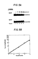
- Fig. 5A is an autoradiogram showing the results of monitoring each round of reaction for each template.
- the ligation reaction was specific for the HR23S' template containing the sickle cell sequence.
- the flanking oligonucleotide substrates were not able to ligate in the presence of the H ⁇ 23A' template.
- Fig. 5B shows a logarithmic plot of amplification folds as a function of the number of rounds of ligation amplification performed. A straight line was obtained with a calculated efficiency of 0.98.
- the ligation product of the preceding round serves effectively as the template for further oligonucleotide ligation.
- the methods of the present invention can also be used to detect plasmid and genomic DNA sequences as illustrated in the following example.
- Templates for amplification ligation were derived from plasmids pH ⁇ a and pH ⁇ s which contain respectively an approximately 4.4kbp normal ( ⁇ a ) and sickle cell ( ⁇ s ) beta globin gene insert. All DNA preparations were performed according to a modified Triton X-100 procedure followed by Proteinase K and RNase treatment.
- the plasmid DNA was treated with restriction enzyme (Bam HI and/or Taq I) and Exonuclease III (Exo III) prior to serving as template in ligation reactions as follows: pH ⁇ a and pH ⁇ b were separately digested with Bam HI (5 U/pmol) for 2 hours at 37° C and terminated in a boiling water bath for 5 minutes. Exo III (100 U/pmol) was then added to the reaction mixture with DTT to make a final concentration of 1 mM. The mixture was incubated 4 hours at 37°C. Phenol-chloroform extraction was performed for final purification.
- restriction enzyme Bam HI and/or Taq I
- Exo III Exonuclease III
- Fig. 6 shows the results of the ligation amplification.
- Product in lanes a-c resulted from reactions not including template
- product in lanes d-f resulted from reactions containing pH ⁇ s template
- product in lanes g-i resulted from reactions containing pH ⁇ a template.
- Reaction products in lanes a, d, and g were withdrawn and analyzed after 10 rounds
- products in lanes b, e, and h were analyzed after 12 rounds
- products in lanes c, f, and i were analyzed after 14 rounds.
- Beta thalassemia major DNA was prepared from EBV transformed lymphocytes in culture (GM 2267 cells from NIGMS Human Genetic Mutant Cell Repository, Camden, N.J.). Thalassemia DNA was subsequently isolated from cultured cells. All DNA preparations were performed according to a modified Triton X-100 procedure followed by Proteinase K and RNAse treatment.
- Genomic DNA (5 ⁇ g) was digested with Taq I restriction enzyme (10 U/ ⁇ g) (Boehringer Mannheim) overnight at 65°C and followed by Bam HI digestion (10 U./ ⁇ g) (Bethesda Research Laboratory) for 8 hours at 37°C. Subsequently Exo III nuclease (100 U./pmol) was added to the reaction mixture along with DTT to make 1 mM final concentration and the reaction was incubated for 5 hours at 37°C. Phenol-chloroform extraction was carried out for final purification.
- Ligation conditions for genomic DNA and for plasmid DNA are identical except that the total volume of the reaction for the genomic DNA sample is 200 ⁇ l.
- Genomic DNA from thalassemia, sickle cell, and normal DNA were amplified using 200 nM of the following sets of flanking oligonucleotides: 32 P-5' labeled ON-s2 (10 6 cpm)/ 32 P-5' labeled ON-4 (10 6 cpm)/5' OH-ON-1/5' OH-ON-s3; and 32 P-5' labeled ON-2 (10 6 cpm)/ 32 P-5' labeled ON-4 (10 6 cpm)/5' OH-ON-1/5' OH-ON-3.
- 5 ⁇ g of genomic DNA contains approximately 500,000 molecules of homozygous single copy gene.
- 5 ug of DNA When used as a template in a 200 ⁇ l reaction mixture, 5 ug of DNA is equivalent to 2.5 x 10- 14 M with respect to template. This concentration is well below the apparent K m of T4 DNA ligase for ligation template. The enzyme activity is therefore expected to be slowed considerably.
- early rounds of the ligation were incubated for a longer time period. The ligation time for initial rounds was 5 hours. With subsequent rounds, the incubation time was gradually reduced to 30 minutes.
- Figs. 7A and 7B show the results of 70-75 rounds of exponential amplification using these templates and flanking substrates.
- Fig. 7A contains amplification product resulting from the ⁇ s flanking oligonucleotide set;
- Fig. 7B contains amplification product resulting from the p a flanking oligonucleotide set.
- Lanes a and b of each autoradiogram contain reaction product using thalassemia genomic DNA template; lanes c and d contain product from sickle cell genomic DNA template, and lanes e and f contain product using normal beta globin genomic DNA template.
- reaction mixture was incubated 3 hours at 30° C and the reaction was terminated by boiling.
- a second round of amplification was initiated by adding another 0.5 U. of enzyme.
- the reaction mixture was again incubated 3 hours.
- Three rounds of ligation amplification were performed.
- the resulting products were then electrophoresed and autoradiographed as described above.
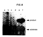
- Fig. 8 shows the resulting autoradiogram.
- Lane a contains reaction product using ⁇ -thalassemia template
- lane b contains reaction product using sickle cell template
- lane c contains reaction product using normal beta globin template. Positive controls were performed with 0.01, 1, and 10 fmoles of H ⁇ 19S template (lanes d-f respectively).
- Lane b shows the expected product
- lanes a and c show an expected lack of product.
- the methods of the present invention can also be used as a means to detect amplification product from other nucleic acid amplification methods such as the polymerase chain reaction.
- Amplification reactions were performed with Polymerase Chain Reaction Kit (Gene Amp Kit, Perkin-Elmer Cetus) including 2.5 U of Thermus aquaticus DNA polymerase and 2.5 ⁇ M of oligonucleotide primers BGP1 and BGP2,19 base long synthetic oligodeoxyribonucleotides which anneal to the beta globin gene at positions 256 nucleotides apart.
- the enriched 294 base pair fragment contains the P globin sequence of interest.
- the membrane was first washed in room temperature with 6X SSC three times for 30 minutes. Subsequent wash in TMACI solution for 1 hour at 59°C removed all mismatch hybridization.
- TMACI 50 mM Tris, pH 8.0, 3 M tetramethylammonium chloride, 2 mM EDTA, 0.25% SDS. Exponential ligation amplification was then performed on 20 ⁇ l samples of the PCR enriched sequences for 4 rounds with either 200 nM of ⁇ s substrates (ON-1/ON-s2 and ON-s3/ON-4) or 200 nM of p a substrates (ON-1/ON-a2 and ON-a3/ON-4) in a 100 ⁇ l of total reaction volume using T4 DNA ligase. 50 ⁇ l from each samples was analyzed by 20% polyacrylamide gel electrophoresis.
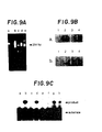
- Ethidium bromide staining of the 1.50/ 0 agarose gel shows a single 294 base pair band (Fig.9A, lanes c-e).
- Lanes of Fig. 9A are: (a)- control; (b)- ⁇ thalassemia; (c)- homozygous sickle cell trait; (d) -normal a globin; and (e) -heterozygous sickle cell trait.
- PCR amplification of thalassemia DNA (Panel A lane b) amplifies some nonspecific DNA's but shows no 294 base pair band.
- Oligonucleotide hybridization analysis of the PCR enriched samples confirms that the enriched DNA's indeed contain beta globin sequence of interest (Fig. 9B).
- Competition hybridization of the filter as described in Nozari, G., et al., Gene 43:23 (1986) and in copending application Serial No. 071,210, of R. Bruce Wallace, filed July 8, 1987 with either radioactively labelled Hp19S and unlabelled H ⁇ 19A (Fig. 9B, panel a) or labelled H ⁇ 19A and unlabelled HP19S (Fig.
- PCR enriched 256 base pair fragments ideally serve as template in the ligation analysis since they are relatively abundant which overcomes the ligase kinetic constraints (i.e., high Km for template).
- Ligation amplification performed on these 294 base pair templates (Fig. 9C templates: a,e - ⁇ thalassemia; b,f - Homozygous sickle cell disease; c,g-normal ⁇ globin; d,h -heterozygous sickle cell trait) with ⁇ s
- Fig. 9C, lanes a-d) flanking oligonucleotide substrates showed positive ligation for sickle cell disease and sickle cell trait samples (Fig. 9C, lanes b and d).
- Ligation amplification with p a (Fig. 9C, lanes e-h) flanking oligonucleotide substrates is likewise positive for normal and sickle cell trait samples (Fig. 9C, lanes g and h
Abstract
Description
- This invention relates to a method of amplifying nucleic acid sequences, and further relates to a method for distinguishing single base differences on nucleic acid templates.
- Methods are known for enzymatically joining portions of nucleic acids together. Weiss, B., et al., J. Biol. Chem. 245(17):4543 (1968) describe joining two segments of an interrupted strand in a DNA duplex using T4-polynucleotide (DNA) ligase. Sgaramella, V., et al., Proc. Natl. Acad. Sci. U.S.A. 67(3):1468 (1970) teach use of T4-polynucleotide (DNA) ligase to join blunt ends of DNA duplexes. Nath, K., et al., J. Biol. Chem. 249(12) :3680 (1974) describe using DNA ligase to attach polyribonucleotides to polydeoxyribonucleo- tides. Higgins, N.P., et al., Methods in Enzymology 68:50 (1979) provides a review of DNA-joining enzymes. Harrison, B., et al., Nucleic Acids Research 12:8235 (1984) teach an enhanced ligation of oligonucleotides and polynucleotides by T4 RNA ligase in polymer solutions.
- With respect to amplification, Saiki, R.K., et al., Science 230:1350 (1985) describes enzymatic amplification of a-globin genomic sequences, termed the polymerase chain reaction, by providing two oligonucleotide primers flanking the region to be amplified, annealing the primers to strands of denatured genomic DNA and extending them with the Klenow fragment of E. coli
DNA polymerase 1 and deoxyribonucleosidetriphosphates. U.S. Patent 4,683,202 to Mullis further describes the polymerase chain reaction method. - U.S. Patent 4,683,194 to Saiki, et al., describes a method of detecting a specific polymorphic restriction site in a nucleic acid sequence by hybridizing the sequence to an oligonucleotide probe complementary to the sequence and spanning the restriction site which is labeled at the end of the probe nearer to the restriction site. The nucleic acid sequence and probe hybridized thereto are then digested with a restriction endonuclease, labeled and unlabeled oligomer fragments are separated, and labeled oligonucleotides are detected.
- U.S. Patent 4,683,195 to Mullis et al. describes a process for amplifying nucleic acid sequences using primers and polymerization agents and a process for detecting the presence or absence of a specific nucleic acid sequence. Separate strands of a nucleic acid sample are treated with an excess of two different oligonucleotide primers, and primers are extended to form complementary extension products which act as templates for synthesizing the desired nucleic acid sequence. The amplified sequence is then detected by adding a labeled probe capable of hybridizing to the sequence being detected.
- The present invention is useful in diagnostic applications because it can be used first, as a means for amplifying given nucleic acid sequences from nucleic acid templates so that the template sequences can be more readily detected. In order to amplify a given nucleic acid sequence, the sequence is hybridized to at least one pair of oligonucleotide strands (substrates) which flank both sides of a target base of interest wherein a designated end base of one of the oligonucleotide strands of a pair is complementary to the target base and that designated end base forms a ligation juncture with an end of the other substrate of the pair. The former oligonucleotide strand substrate is, in effect, a probe which hybridizes to the target base and adjacent nucleic acid sequence and also simultaneously ligates with the latter oligonucleotide strand substrate designed to flank the opposite side of the target base and adjacent sequence only if the designated end base of the probe is complementary to the target base on the template.
- While there exists the possibility of ligation under mismatched conditions in which the target base for which the designated end base is to be complementary is not present in a given template, it has been found, according to the present invention, that such ligation can be suppressed so that a base mismatch on either side of the ligation junction reduces the efficiency of ligation between the two oligonucleotide strands and serves as the theoretical basis to distinguish single base differences on a nucleic acid template. The methods of the present invention can therefore also be used as a diagnostic technique to detect single base or base pair mutations in nucleic acid sequences. This further aspect of the present invention is therefore useful to diagnose genetic diseases, such as sickle cell anemia, hemophilia, and color blindness which result from a single point mutation on a critical structure gene.
- In general, the invention features a method for detecting single base or base pair mutations in at least one specific nucleic acid sequence contained in a sample of nucleic acid template wherein each nucleic acid consists of either one strand or two separate complementary strands, comprising: treating the strands with at least one of two sets of two different relatively short oligonucleotides each of which sets (i) is complementary to and therefore will hybridize to at least a portion of the template sequence of at least one of the strands and (ii) flanks both sides of a designated target base contained within said specific nucleic acid sequence such that one of the two short oligonucleotides in each set of oligonucleotides contains a base at one of its ends that is complementary to one of the target bases of the template under conditions such that the two short oligonucleotides of each of the sets will mutually ligate if the target base exists in the template, the ligation occurring between the terminal base of one of the complementary oligonucleotides which is complementary to one of the target bases and a terminal base of another complementary oligonucleotide of the same set.
- The invention further features a method for amplifying at least one specific nucleic acid sequence in a sample containing a nucleic acid or a mixture of nucleic acids comprised of single or complementary nucleic acid strands, wherein the sample is suspected of containing the sequence or sequences comprising (a) treating the strands with at least one of two pairs of oligonucleotides which sets are complementary to at least one the specific nucleic acid sequence and flank at least one target base of a single stranded template or at least one target base pair defining a blunt end contained in the nucleic acid sequence or sequences wherein one end nucleotide of one of the oligonucleotides of at least one of the pairs is complementary to the target base or one of the bases of the target base pairs under conditions such that the end nucleotide will mutually ligate with an end of the other oligonucleotide of the pair to form a ligation product which is complementary to the specific nucleic acid sequence only if the target base or target base pair is present in the sample; (b) treating the sample under conditions to separate ligation products from their templates if the target base or target base pairs to be detected are present in the sample; and (c) determining whether ligation has occurred. Other advantages and features of the invention will be apparent from the following description of the preferred embodiments thereof, and from the claims.
-
- Fig. 1 illustrates a 35 base pair length template of the normal human (pa) beta globin gene in the region of the allelic base pair DNA polymorphism associated with sickle cell anemia and four oligodeoxyribonucleotide substrate strands complementary to the normal beta globin allele of the gene to be used for ligation amplification.
- Fig. 2 illustrates a 35 base pair length template of the human sickle cell (βs) beta globin gene in the region of the allelic base pair DNA polymorphism which is associated with sickle cell anemia and four oligodeoxyribonucleotide substrate strands complementary to the sickle cell allele of the gene to be used for ligation amplification.
- Fig. 3 is a schematic diagram of template directed exponential ligation amplification.
- Fig. 4 is a photograph of an autoradiogram of product of linear amplification ligation of βs flanking oligonucleotide substrates on sickle cell (lanes c and g) and normal beta globin (lanes d and h) oligonucleotide templates.
- Fig. 5A is a photograph of autoradiograms of reaction products from six rounds of exponential amplification ligation of flanking oligonucleotide substrates designed to be complementary to the sickle. cell gene sequence using sickle cell (HP23S') and normal (Ha23A') oligonucleotide template.
- Fig. 5B shows a logarthmic plot of fold of amplification versus number of exponential amplification ligation rounds performed.
- Fig. 6 is a photograph of an autoradiogram containing ligation amplification product from 10-14 exponential rounds of ligation amplification using templates from plasmids containing normal (lanes g-i) and sickle cell (lanes d-f) beta globin gene using flanking oligonucleotide substrates designed to be complementary to the sickle cell beta globin gene sequence (Fig. 2) and normal beta globin gene sequence (Fig. 1).
- Figs. 7A and 7B are photographs of autoradiograms resulting from amplification of human genomic DNA using P thalassemia (lanes a, b), sickle cell (lanes c, d), and normal 0 globin (lanes e, f) DNA template. Fig. 7A resulted from ligating as flanking oligonucleotides; Fig. 7B resulted from ligating βa flanking oligonucleotides.
- Fig. 8 is a photograph of an autoradiogram containing linear ligation amplification product using a pair of βs flanking oligonucleotides and human genomic DNA template from β-thalassemia (lane a), sickle cell (lane b), and normal β globin (lane c). Lanes d-f are controls.
- Fig. 9A is a photograph of an ethidium bromide stained agarose gel containing PCR amplified 294 base pair fragments of alleles at the human beta globin gene.
- Figs. 9B(a) and 9B(b) are photographs of autoradiograms of PCR products from the Fig. 9A gel transferred to nylon membranes and hybridized with radiolabeled Hp19S (panel a) and radiolabeled Hβ19A (panel b) probes.
- Fig. 9C is a photograph of an autoradiogram of ligation amplification products using PCR enriched sequences as templates.
- The method of the present invention can be used both to amplify any particular nucleic acid sequence template and distinguish and detect single base differences on nucleic acid templates. The present invention is therefore useful to diagnose various genetic disorders that result from point mutations such as sickle cell anemia. In other diagnostic applications, signal amplification is necessary in order to detect the limited number of DNA template copies in genomic DNA. For example, there are only about one million copies of a single gene in 10 ug of DNA. Further, depending on the particular diagnostic procedure performed, only a limited amount of DNA might be available.
- According to the methods of the present invention, short oligonucleotide strands are synthesized complementary to and flanking both directions of a target base on a nucleic acid template. These relatively short oligonucleotides (flanking oligonucleotides or substrates) are generally denoted ON-1, ON-2, ON-3, and ON-4 as illustrated in Fig. 1. The flanking oligonucleotides may be synthesized according to methods well known in the art.
- The lengths of the flanking oligonucleotides must be sufficiently long to contain enough of a unique base pattern to cause the flanking oligonucleotides to hybridize or anneal to only the portions of the nucleic acid template which flank the target base pair of interest. The exact lengths of the flanking oligonucleotides depends on various factors, including the temperature of the reaction, pH conditions, and complexity of the target sequence. In general, flanking oligonucleotides comprising about 4 to about 100 nucleotides form sufficiently stable hybrid complexes at higher reaction temperatures. In most instances, flanking oligonucleotides used in accordance with the present invention preferably comprise about 8 to about 20 nucleotides.
- The methods of the present invention may be carried out with nucleic acid template which is either single or double stranded. If the nucleic acid template comprises two strands, it is necessary to first separate the strands prior to commencing ligation amplification of the present invention. One method of separation of strands of nucleic acid involves heating the nucleic acid until it is completely denatured. Such denaturation is usually carried out at temperatures ranging from about 80° to 105° C for times ranging from about 1 to 10 minutes. Separation of nucleic acid strands may also be induced by enzymes such as the helicase enzymes and Rec A. It is also possible to remove one of two strands by digesting with appropriate restriction endonuclease followed by Exonuclease III (Exo III) or Lambda Exonuclease as described by Wallace, R.B., et al. Science 209:1396 (1980).
- The specific nucleic acid sequence to be amplified may be only a fraction of a larger molecule or it can be a discrete molecule. The sequence to be amplified can also be a part of a larger mixture of molecules or fractions thereof. The starting nucleic acid may also contain more than one desired sequence containing target bases which may be the same or different, such that all such sequences can be simultaneously amplified. Samples comprising more than one nucleic acid with more than one desired target sequence can also be amplified by the methods of the present invention.
- Any source of template nucleic acid can be employed to practice the present invention as long as it contains or is suspected to contain the specific nucleic acid sequence desired. For example, plasmids, natural DNA or RNA, from bacteria, yeast, viruses, plants or animals, or cloned DNA or RNA can be used. Genomic DNA or total RNA may also be extracted from human blood and tissue by a variety of techniques. Synthetic oligodeoxyribonucleotides may also be used. When RNA is used as an initial template, it can be first copied into a DNA molecule by an enzyme such as reverse transcriptase.
- As seen in Fig. 1, the template is the βa normal beta globin allele with target base pair *A-T*; in Fig. 2, the template is the ps sickle cell allele with target base pair *T-A*. It must be understood that the particular templates and flanking oligonucleotides shown in Figs. 1 and 2 only illustrate the present invention and in no way limit the invention to the detection or amplification of alleles of the human beta globin gene.
- As shown in Fig. 1, the 5'-3' (upper) strand of the template is denoted (+); the 3'-5' (lower) strand is denoted (-). Flanking oligonucleotides ON-2 and ON-4 are 8-nucleotide long oligonucleotides and ON-1 and ON-3 are 14-nucleotide long oligonucleotides. The method of the present invention requires a symmetry similar to that shown in Fig. 1, namely, the 3' flanking oligonucleotides, ON-2 and ON-4, are of the same length and the 5' flanking oligonucleotides, ON-1 and ON-3, are the same length. Either the two 3' or two 5' flanking oligonucleotides can be the longer length, or all four flanking oligonucleotides can be the same length. The 5' ends of the 3' flanking oligonucleotides (ON-2 and ON-4) must, however, be phosphorylated and those ends can also be optionally labeled with, for example, radioactive phosphorous. The 5' flanking oligonucleotides (ON-1 and ON-3) are not usually phosphorylated to prevent self ligation and template-independent ligation. In the case where the nucleic acid template to be amplified is double stranded, the target base pair of interest can be either A-T as in Fig. 1, T-A, G-C, or C-G. When the template is single-stranded, the target base can therefore be any of the four nucleotides.
- The methods of the present invention are not, however, limited to the above-described instances involving single base or single base pair differences. The flanking oligonucleotide complementary region can thus contain more than one base or base pair difference which enables the methods of this invention to be used for discrimination and detection of sequences containing multiple base differences.
- It is necessary that in the case of double-stranded nucleic acid templates the target base pair define a blunt end as seen in Fig. 1, as opposed to a staggered end.
- The pairs of flanking oligonucleotide substrates are complementary to a nucleic acid sequence harboring the position of possible mutation on either strand. Further, with double-stranded templates, one of the terminal bases of one of the two 3' flanking oligonucleotides and one of the terminal bases of one of the two 5' flanking oligonucleotides to be ligated must be complementary to the respective target bases. Thus, as shown in Fig. 1, the base at the 5' end of one of the 3' flanking oligonucleotides (base T, ON-2) is complementary to the target base A of the (+) template strand, and the nucleotide at the 3' end of one of the 5' flanking oligonucleotides (base A, ON-3) is complementary to the target base T of the (-) template strand. Thus, of the four end bases of any given pair of flanking oligonucleotides, two positioned at the ultimate ends of the sequences, and two positioned such that they will be ligated to each other, one of the end bases of the potential ligating pair of bases must be complementary to the target base in order for detectable ligation to occur.
- The base pair G-C directly to right of illustrative target base pair A-T in Fig. 1 can also be a target base pair. In such a case, the 3' terminal nucleotide of one of the 5' flanking oligonucleotides (base C, ON-1) is complementary to the target base G of the ( + ) template strand, and the 5' terminal nucleotide of one of the 3' flanking oligonucleotides (base G, ON-4) is complementary to the target base C of the (-) template strand.
- Likewise, nucleotides in positions other than those at the ends of flanking oligonucleotides comprising the ultimate ligation junction can be considered as target nucleotides in the practice of the present invention.
- As seen in Fig. 1, 3' flanking oligonucleotide ON-2 is synthesized so that it is complementary to the (+) or upper strand of the template and its 5' end contains base T which is complementary to target base A of the ( + ) strand, and the remainder of the flanking oligonucleotide extends outwardly from the 5' end base. 5' flanking oligonucleotide ON-1 is synthesized so that it is complementary to the (+) or upper strand of the template and is oriented such that the base at its 3' end C, is complementary to the base of the ( + ) strand on the 3' side of the target base on the template. The remainder of the flanking oligonucleotide extends outwardly in the 5' direction.
- 5' flanking oligonucleotide ON-3 is complementary to the (-) or lower strand of the template such that base A at its 3' end is complementary to the target base T of the (-) strand and the remainder of ON-3 extends outwardly from the target base. 3' flanking oligonucleotide ON-4 is complementary to the (-) or lower strand of the template and is oriented such that base G at the 5' end of ON-4 is complementary to the base of the (-) strand that is on the 5' side of the target base. The remainder of ON-4 extends outwardly in the 3' direction. Thus, the bases on the 5' end of ON-2 and 3' end of ON-3 are complementary to the target base pair.
- When double-stranded nucleic acid template is used, symmetry requires that ON-2 be complementary to ON-3, except for differences in length. Likewise ON-1 is complementary to ON-4, except for differences in length. As seen in Fig. 1, ON-1 is complementary to ON-4. Therefore in cases where double-stranded nucleic acid template is to be used, the flanking oligonucleotides can be double-stranded, for example:
- At appropriate temperature and assay conditions polynucleotide ligase ligates ON-1 and ON-2, and/or ON-3 and ON-4 at the target bases of ON-2 and ON-3 to form significant amounts of longer ligated products only if the correct complementary target bases are present at the target site of the template. Any suitable polynucleotide ligase can be used in the practice of this invention, for example, bacteriophage T4 induced DNA ligase, and E. coli DNA ligase.
- It has been found that inclusion of at least 200 mM NaCI in amplification reaction mixtures almost completely supppresses mismatch ligation. Therefore, with reference to Fig. 1, if the target site of the template DNA contains a nucleotide which is not complementary to 5' end of ON-2 or the 3' end of ON-3, the mismatch of nucleotides prevents any significant ligation which would have caused formation of a 22 base long product. The absence of such ligation product thereby indicates that the target bases of interest are not present on the particular template. For example, if the template in Fig. 1 were the sickle cell (0s) gene in which bases A and T are interchanged at the target base site, there would be no ligation of either ON-2/ON-1 or ON-3/ON-4 as depicted in Figure 1. An expected 22 nucleotide amplification ligation product would thus not be formed.
- With respect to the specific flanking oligonucleotides shown in Fig. 2, the two oligonucleotides in each pair anneal to the beta globin template at adjacent positions such that the 3' end of one oligonucleotide (ON-1 or ON-3) and the 5' end of the other (ON-s2 or ON-4) form a ligatable junction. The target nucleotide on the template base pairs with either the 3' end nucleotide or the 5' end nucleotide of the substrate depending on which strand, (+) or (-), serves as the template for ligation.
- The success or failure of a given ligation amplification procedure can be detected by labeling the flanking oligonucleotides with radioactive or fluorescent labels. For example, 32p, 35S or any other suitable radionuclide may be used as well as other suitable compounds such as fluorescein or rhodamine or their derivatives. Ligation amplification product can also be ascertained via any subsequent manipulation that allows distinction between longer ligation product and the shorter precursors such as gel electrophoresis chromatography or hybridization. For example, polyacrylamide gel electrophoresis can be used to separate the ligated product from the flanking oligonucleotide reactants according to their sizes. Some of these techniques will be fully described in the examples below.
- According to the method of the present invention, the herein described ligation amplifications can be accomplished in a linear or exponential manner as illustrated in the following examples. The steps for exponential ligation-amplification are shown in Fig. 3. The DNA sequences of synthetic oligonucleotide substrates and templates used in the examples are given below in Table 1.

- HP19S and Hβ19A are unique 19 nucleotide long oligonucleotides that are complementary to the noncoding strand of the beta globin gene and serve as oligonucleotide templates for ligation of ON-1/ON-2 pairs. Hβ23S' and Hβ23A', 23 nucleotides long, are complementary to the coding strand of the beta globin gene. These two nucleotides serve as templates for ligation of ON-3/ON-4 pairs.
- While the ON-1/ON-s2 and ON-1/ON-a2 pairs both hybridize to the coding (+) strand, ON-s2 and ON-a2 differ by a single nucleotide at their 5' ends, with ON-s2 specifying for the sickle cell allele. Similarly, the ON-s3/ON-4 and ON-a3/ON-4 pairs hybridize to the noncoding strand with ON-s3 and ON-a3 differing by a single nucleotide at their 3' ends.
- For the detection of either the pa (normal) or βs (sickle cell) allele, a set comprising at least one of the pairs of flanking oligonucleotide substrates ON-1/ON-a2 and ON-a3/ON-4 are used for the normal beta globin sequence and at least one of the pairs ON-1/ON-s2 and ON-s3/ON-4 are used for detecting the sickle cell sequence.
- In the below-described ligation amplification reactions, template dependent ligation of either the ON-1/ON-2 or ON-3/ON-4 pairs forms a 22 base product with a sequence complementary to the other substrate pairs. For example, ON-1-ON-a2 product would be complementary to ON-a3/ON-4 substrates and ON-s3-ON-4 product would be complementary to ON-1/ON-s2 substrates. According to one aspect of the present invention, the ligation products can then serve as template for the ligation of the other pair of flanking oligonucleotide substrates thereby producing more templates for further ligation. If the initial ligation step does not take place as a result of base pair mismatch, subsequent amplification steps will not give rise to a detectable ligation signal.
- Once the 22 nucleotide long ligation product is formed, it stays hybridized to its template. Denaturation separates the duplex to yield two new templates. If denaturation is accomplished by boiling, the ligase in the reaction mixture is inactivated. Fresh enzyme must therefore be added to initiate the next round of amplification.
- All oligodeoxyribonucleotide ligation reactions on oligonucleotide templates were carried out, unless otherwise indicated, in 50 mM Tris HCI (pH 7.6), 10 mM MgC12, 1 mM DTT (dithiothreitol), 1 mM ATP, 200 mM NaCI and 5% PEG (polyethyleneglycol) with 5-10 µm of each substrate oligonucleotide mixed with a small amount of radioactively labeled 3' oligonucleotide substrate (100,000-300,000 cpm) and the indicated amount of template.
- Plasmid and genomic DNA template reactions are initiated following 5-10 minutes of boiling, cooling on ice and the addition of ligase (1 U; BRL). Unless otherwise indicated, all ligation reactions are withdrawn for electrophoresis and optionally treated with calf intestinal alkaline phosphatase (1-5 U) for 1-2 hours. Linear amplification ligation of nucleic acid sequences is carried out by repeatedly using the original nucleic acid template (oligo, plasmid or genomic DNA) in each round of amplification. Only one flanking oligonucleotide set complementary to the appropriate oligonucleotide template to one strand of duplex plasmid or genomic DNA template is used. For example, either the ON-1 and ON-2, or ON-3 and ON-4 set is used in the amplification rounds but not both.
- To linearly amplify a sequence of the 5' strand of the human sickle cell (βs) Ø-globin gene as shown in Fig. 2, the above reaction mixture further containing 1 mM ON-1, 1 mM 5' phosphorylated ON-a2, and 0.1 pmoles of ps (HØ19S) template was prepared. If double-stranded template is used, it must be denatured by boiling the reaction mixture at about 90-100°C, for 10 minutes.
- Each round of ligation is initiated by adding 1 unit of ligase to the reaction mixture on ice. The mixture is incubated for about 30 minutes at 30°C, or for a longer time if template concentration is low.
- The first round, and all subsequent rounds, is terminated by quickly heating the reaction mixture to 100°C for 5 minutes to inactivate the ligase and dissociate the ligated product strand (ON-1 -ON-2) from the template. This frees the template for subsequent amplification rounds. Before the next round, the reaction mixture is centrifuged for 10 seconds and immediately cooled to about 0°C. At this point, there is approximately one ON-1 -ON-2 ligation product present for each copy of the template.
- Another unit of enzyme is added to the reaction mixture to initiate a second round of ligation. Because a molar excess of flanking oligonucleotides ON-1 and ON-2 are added to the initial reaction mixture, more flanking oligonucleotide substrated are not added. The reaction mixture is then incubated as in the first round, boiled to dissociate template from the ligation product, and cooled. At this point, there are approximately 2 copies of ON-1 -ON-2 ligation product present for each copy of template.
- Fresh enzyme is added to initiate another round. The steps of incubating, boiling to dissociate template from product, cooling, and adding fresh enzyme are repeated for as many rounds as necessary to produce a detectable signal.
- Detection of the ON-1 -ON-2 ligation product can be accomplished in several ways. ON-1 and/or ON-2 can be labeled with radioactive or nonradioactive labels and separated from the reactants by gel electrophoresis, such that ligation product is determined by detecting the presence of label in a position in the gel corresponding to the expected product size.
- ON-a2, ON-s2, and ON-4 are phosphorylated at their 5' ends with either radioactive (y [32P] ATP 6000 Ci/mmol; New England Nuclear) or nonradioactive ATP via a kinase reaction. The unlabeled 5' phosphorylated substrates are used following incubation in a boiling water bath for 20 minutes to inactive kinase. Internal radioactively phosphorylated oligonucleotide products are separated from unreacted ATP and other reaction products with anion exchange chromatography (DE-52 cellulose-Whatman). The radioactive phosphorylation reaction yields products with approximately 108-109 cpm/pmol specific activity.
- Detection and quantitation of ligation product by polyacrylamide gel electrophoresis is carried out by mixing samples from ligation reactions with Ficoll loading buffer and subjecting the mixture to electrophoresis in TBE (89 mM Tris, 89 mM Borate, 2 mM EDTA) in 200/0 polyacrylamide (Bio-Rad) urea (7M) gel at 600 V. for 1.5 hours. The gel is then wrapped in Saran Wrap and autoradiographed between two Lightning Plus intensifier screens (DuPont) overnight. Quantitation of ligation product is obtained either from densitometric measurements of the autoradiograph or directly from the gel via an AMBIS Radioisotope Scanning System II (Automated Microbiology System, Inc.). In the latter instance the polyacrylamide gel is first fixed in a 5% acetic acid solution for 10 minutes.
- The ON-1 -ON-2 ligation product can also be detected by hybridizing the product containing solution to a substrate containing an immobilized complementary DNA or RNA sequence under conditions whereby the ON-1 -ON-2 ligation product will hybridize to the complementary sequence, but neither ON-1 nor ON-2 will. Bound label indicates presence of ligation product. The ligated product can also be detected in a subsequent round of ligation employing a radiolabeled counterpart substrate set (ON-3/ON-4). In the case where ligated ON-3/ON-4 is to be detected, labeled ON-1/ON-2 would be used.
- The product level after N-number of rounds of linear amplification-ligation is equal to N x Q where Q is the efficiency of the ligation. Q ranges from 0 to 1, where 1 is equivalent to 100% ligation.
- ON-1/ON-s2* and ON-s3/ON-4* were ligated linearly on oligonucleotide templates using the above methodology. ON-s2* and ON-s4* are the same as ON-s2 and ON-4 except that they are only 6 nucleotides long. 10 pmoles of 32P-5' pohsphorylated ON-s2* and unlabeled 5' OH-ON-1 (lanes a-d) or 32P-5' phosphorylated ON-4* and unlabeled 5' OH-ON-s3 (lanes e-h) were ligated on 19 base long or 23 base long oligonucleotide template respectively at 30°C for 30 minutes with 1 U. of T4 DNA ligase in 10 µl reaction volume. Fig. 4 shows the results of the ligation amplification reactions described below. Samples in lanes a and e not treated with enzyme and samples in lanes b and f, without template, serve as negative controls. 1 pmole of the following oligonucleotide template was used: HØ19S (c); Hβ19A (d); Hp23S' (g); and Hβ23A'(h).
- As seen in Fig. 4, the expected ligation involving sickle cell template and sickle cell substrates occurred (lanes c and g); ligation of normal (A) beta globin substrates did not occur on the sickle cell template (lanes d and h).
- The method of the present invention can also be used to exponentially amplify nucleic acid sequences. The major difference between exponential amplification ligation and the above-described linear amplification ligation is that all four flanking oligonucleotides (ON-1/ON-2, and ON-3/ON-4) are present as substrates during each reaction round. The ligation product of either flanking oligonucleotide set serves as the template for the other flanking oligonucleotide set in subsequent amplification rounds as shown in Fig. 3. Radioactively labeled substrate for detection can be included in the starting materials or can be added in the last round.
- As is also the case for linear amplification, an estimation of the number of amplification rounds needed to produce a detectable signal must be made before carrying out the exponential amplification. After N rounds, the product formation equals (1 + Q)N-1 where Q is ligation efficiency. The following table indicates the relative amounts of ligation products present after N cycles, assuming 100% efficiency at each cycle.

- Each round of exponential ligation is started by adding one unit of enzyme to the reaction mixture on ice. In the first and each subsequent round of amplification, the reaction mixture with the added enzyme is incubated for about 30 minutes at 30° C or for a longer period of time if template concentration is low. At the end of each round, the reaction mixture is quickly brought to boiling temperature for 5 minutes to inactivate the enzyme and dissociate the product strands from the template strands. Then the reaction mixture is centrifuged for 10 seconds and immediately cooled to about 0°C.
- Another unit of T4 ligase is then added to initiate the second round of amplification. The mixture is then incubated, boiled, and cooled as above. At this point there are 3 copies of template present for each copy present at the beginning of the reaction. All subsequent rounds of amplification are carried out by the same sequence of steps for as many times needed in order to produce a detectable signal.
- The progress of the amplification may be monitored by withdrawing from the reaction mixture an approximately 10 µl aliquot of reaction mixture during predetermined rounds. The withdrawn material is heat inactivated and is then combined with 2 to 3 µl of loading buffer. 2 µl of this buffered mixture is then loaded onto a 200/0 polyacrylamide gel and the gel is electrophoresed for one hour at 600V and then autoradiographed overnight with two DuPont Lightening-Plus intensifier screens.
- Using these methods and the reaction mixture described in Example 1, 6.67 nM of oligonucleotide template, Hβ23S' and Hβ23A', was amplified with T4 DNA ligase and 2.33 µM of each flanking oligonucleotide substrate: 32P-5' labeled ON-s2 (106 cpm); 32P-5' labeled ON-4 (106 cpm); 5' OH-ON-1; and 5' OH-ON-s3. 1 U of T4 DNA ligase was used to initiate each round. Six rounds of ligation amplification were carried out.
- Fig. 5A is an autoradiogram showing the results of monitoring each round of reaction for each template. In rounds 2-6, the ligation reaction was specific for the HR23S' template containing the sickle cell sequence. The flanking oligonucleotide substrates were not able to ligate in the presence of the Hβ23A' template.
- Products of the exponential ligation method approximately double with each round. Fig. 5B shows a logarithmic plot of amplification folds as a function of the number of rounds of ligation amplification performed. A straight line was obtained with a calculated efficiency of 0.98. Thus, in each round, the ligation product of the preceding round serves effectively as the template for further oligonucleotide ligation.
- The methods of the present invention can also be used to detect plasmid and genomic DNA sequences as illustrated in the following example.
- Templates for amplification ligation were derived from plasmids pHβa and pHβs which contain respectively an approximately 4.4kbp normal (βa) and sickle cell (βs) beta globin gene insert. All DNA preparations were performed according to a modified Triton X-100 procedure followed by Proteinase K and RNase treatment.
- The plasmid DNA was treated with restriction enzyme (Bam HI and/or Taq I) and Exonuclease III (Exo III) prior to serving as template in ligation reactions as follows: pHøa and pHβb were separately digested with Bam HI (5 U/pmol) for 2 hours at 37° C and terminated in a boiling water bath for 5 minutes. Exo III (100 U/pmol) was then added to the reaction mixture with DTT to make a final concentration of 1 mM. The mixture was incubated 4 hours at 37°C. Phenol-chloroform extraction was performed for final purification.
- 1 nm of each of the two resulting templates (pHØS and pHβa) was then amplified exponentially using T4 DNA ligase and the reaction mixtures and methods described in Examples 1 and 3 except that the total volume of each reaction was 100 µl. 500 nM of each of the following flanking oligonucleotide substrates was used: 32P-5' labeled ON-s2 (106 cpm), 32P-5' labeled ON-4 (106 cpm), 5' OH-ON-1, and 5' OH-ON-s3. After the 10th, 12th, and 14th amplification rounds, a 4 µl aliquot was withdrawn from the reaction mixture and analyzed by electrophoresis and autoradiography.
- Fig. 6 shows the results of the ligation amplification. Product in lanes a-c resulted from reactions not including template; product in lanes d-f resulted from reactions containing pHβs template; and product in lanes g-i resulted from reactions containing pHøa template. Reaction products in lanes a, d, and g were withdrawn and analyzed after 10 rounds; products in lanes b, e, and h were analyzed after 12 rounds; and products in lanes c, f, and i were analyzed after 14 rounds.
- No detectable ligation product is seen from the reactions involving pHβa which contained the normal beta globin sequence (g-i) or in lanes without any template (a-c). The pHβs sickle cell template was significantly amplified, with the detectable signal noticeably increasing between 10 (lane d), 12 (lane e), and 14 (lane f) amplification rounds.
- For the 1 nM plasmid template used in the reactions, approximately 10 rounds of ligation amplification were necessary to generate a detectable signal corresponding to approximately a 50-fold amplification. In contrast, only about 3-6 rounds of amplification are needed to amplify oligonucleotide template of the same concentration to obtain the same fold amplification.
- Normal (βa/βa), sickle cell disease (βs/βs), and sickle cell trait (βa/βs) genomic DNA samples were isolated from blood specimens of appropriate donors. Beta thalassemia major DNA was prepared from EBV transformed lymphocytes in culture (GM 2267 cells from NIGMS Human Genetic Mutant Cell Repository, Camden, N.J.). Thalassemia DNA was subsequently isolated from cultured cells. All DNA preparations were performed according to a modified Triton X-100 procedure followed by Proteinase K and RNAse treatment.
- Genomic DNA (5 µg) was digested with Taq I restriction enzyme (10 U/µg) (Boehringer Mannheim) overnight at 65°C and followed by Bam HI digestion (10 U./µg) (Bethesda Research Laboratory) for 8 hours at 37°C. Subsequently Exo III nuclease (100 U./pmol) was added to the reaction mixture along with DTT to make 1 mM final concentration and the reaction was incubated for 5 hours at 37°C. Phenol-chloroform extraction was carried out for final purification.
- Ligation conditions for genomic DNA and for plasmid DNA are identical except that the total volume of the reaction for the genomic DNA sample is 200 µl.
- Genomic DNA from thalassemia, sickle cell, and normal DNA were amplified using 200 nM of the following sets of flanking oligonucleotides: 32P-5' labeled ON-s2 (106 cpm)/32P-5' labeled ON-4 (106 cpm)/5' OH-ON-1/5' OH-ON-s3; and 32P-5' labeled ON-2 (106 cpm)/32P-5' labeled ON-4 (106 cpm)/5' OH-ON-1/5' OH-ON-3.
- 5 µg of genomic DNA contains approximately 500,000 molecules of homozygous single copy gene. When used as a template in a 200 µl reaction mixture, 5 ug of DNA is equivalent to 2.5 x 10⁻¹⁴M with respect to template. This concentration is well below the apparent Km of T4 DNA ligase for ligation template. The enzyme activity is therefore expected to be slowed considerably. To overcome this kinetic constraint, early rounds of the ligation were incubated for a longer time period. The ligation time for initial rounds was 5 hours. With subsequent rounds, the incubation time was gradually reduced to 30 minutes. (Rounds 1-5 = 5 hours; round 6-10 = 4 hours; rounds 11-15 = 3 hours; rounds 16-20 = 2 hours; round 20-30 = 1 hour; and rounds 30 + = 30 minutes.) Each round however was not allowed to exceed 5 hours in order to avoid accumulation of blunt end products.
- Figs. 7A and 7B show the results of 70-75 rounds of exponential amplification using these templates and flanking substrates. Fig. 7A contains amplification product resulting from the βs flanking oligonucleotide set; Fig. 7B contains amplification product resulting from the pa flanking oligonucleotide set. Lanes a and b of each autoradiogram contain reaction product using thalassemia genomic DNA template; lanes c and d contain product from sickle cell genomic DNA template, and lanes e and f contain product using normal beta globin genomic DNA template. Template in lanes a, c, and e was amplified 70 rounds; template in lanes b, d, and f was amplified 75 rounds. The expected product bands appear in lanes c and d of Fig. 7A (sickle cell template and flanking oligonucleotides) and in lanes e and f of Fig. 7B (normal beta globin template and flanking oligonucleotides). Ø-thalassemia DNA (lanes a and b) and mismatched genomic DNA and substrates both showed absence of signal.
- This example thus demonstrates that the ligation amplification methods of the present invention can be used as a diagnostic methodology on genomic DNA samples for sickle cell anemia.
- 5 µg of a-thalassemia sickle cell, and normal genomic DNA was each digested with Bam HI/Taq I and Exo III nuclease as described in Examples 4 and 5. Linear amplification was performed as described in Example 1. Flanking oligonucleotides used were 100 fmoles of 32P-5' labeled ON-s2 used directly from T4 kinase labeling reaction, without further purification, with DE-52 chromatography, and 100 fmoles of ON-1. The flanking oligonucleotides were separately combined with the three templates in the presence of 200 mM NaCI and 0.5 U of T4 DNA ligase in total reaction volumes of 10 µl. The reaction mixture was incubated 3 hours at 30° C and the reaction was terminated by boiling. A second round of amplification was initiated by adding another 0.5 U. of enzyme. The reaction mixture was again incubated 3 hours. Three rounds of ligation amplification were performed. The resulting products were then electrophoresed and autoradiographed as described above.
- Fig. 8 shows the resulting autoradiogram. Lane a contains reaction product using β-thalassemia template, lane b contains reaction product using sickle cell template, and lane c contains reaction product using normal beta globin template. Positive controls were performed with 0.01, 1, and 10 fmoles of Hβ19S template (lanes d-f respectively). Lane b shows the expected product, whereas lanes a and c show an expected lack of product.
- The methods of the present invention can also be used as a means to detect amplification product from other nucleic acid amplification methods such as the polymerase chain reaction.
- Amplification reactions were performed with Polymerase Chain Reaction Kit (Gene Amp Kit, Perkin-Elmer Cetus) including 2.5 U of Thermus aquaticus DNA polymerase and 2.5 µM of oligonucleotide primers BGP1 and BGP2,19 base long synthetic oligodeoxyribonucleotides which anneal to the beta globin gene at positions 256 nucleotides apart. The enriched 294 base pair fragment contains the P globin sequence of interest. 2 µg of the following genomic DNA were used as templates after being treated as described above: β thalassemia, homozygous sickle cell (βs/βs), normal P globin (βa/βa), and heterozygous sickle cell trait (βa/βs). 30 rounds of amplification gave approximately a 5 x 10⁵-fold amplification of the target sequence. 10 µl aliquots of the PCR enriched genomic DNA sequences were electrophoresed in 1.5% agarose gel at 60 V for 5 hours.
- The electrophoretically separated products were then transferred to Genetran nylon membranes with 20X SSC (1X SSC = 150 mM NaCI and 15 mM Na citrate) according to the method of Southern, E.M., J. Mol. Biol. 98:503-517 (1975).
- The Genetran membranes were directly hybridized to 32P-5' labeled oligonucleotide probes HB19S or HB19A, in 5X SSPE (1X SSPE = 10 mM sodium phosphate pH 7.0, 0.18 M NaCI, and 1 mM EDTA), 10/0-NaDodS04, 10 µg/ml of Homomix RNA, and 106 cpm/ml of labeled oligonucleotide with 10 fold excess unlabelled competitor at 47° C for 2 hours. The membrane was first washed in room temperature with 6X SSC three times for 30 minutes. Subsequent wash in TMACI solution for 1 hour at 59°C removed all mismatch hybridization. (TMACI = 50 mM Tris, pH 8.0, 3 M tetramethylammonium chloride, 2 mM EDTA, 0.25% SDS). Exponential ligation amplification was then performed on 20 µl samples of the PCR enriched sequences for 4 rounds with either 200 nM of βs substrates (ON-1/ON-s2 and ON-s3/ON-4) or 200 nM of pa substrates (ON-1/ON-a2 and ON-a3/ON-4) in a 100 µl of total reaction volume using T4 DNA ligase. 50 µl from each samples was analyzed by 20% polyacrylamide gel electrophoresis.
- Ethidium bromide staining of the 1.50/0 agarose gel shows a single 294 base pair band (Fig.9A, lanes c-e). Lanes of Fig. 9A are: (a)- control; (b)- β thalassemia; (c)- homozygous sickle cell trait; (d) -normal a globin; and (e) -heterozygous sickle cell trait. PCR amplification of thalassemia DNA (Panel A lane b) amplifies some nonspecific DNA's but shows no 294 base pair band.
- Oligonucleotide hybridization analysis of the PCR enriched samples (samples from lanes b-e, Fig. 9A, correspond to lanes 1-4, Fig. 9B) immobilized on Genetran nylon membrane filter confirms that the enriched DNA's indeed contain beta globin sequence of interest (Fig. 9B). Competition hybridization of the filter as described in Nozari, G., et al., Gene 43:23 (1986) and in copending application Serial No. 071,210, of R. Bruce Wallace, filed July 8, 1987 with either radioactively labelled Hp19S and unlabelled Hβ19A (Fig. 9B, panel a) or labelled Hβ19A and unlabelled HP19S (Fig. 9B, panel b) correctly identifies sickle cell disease (βs/βs, Fig. 9B,
lane 2, panel a), normal (βa/βa, Fig. 9B,lane 3, panel 2), and sickle cell trait (βa/βs, Fig. 9B,lane 4, panels aand b) DNA's. - The PCR enriched 256 base pair fragments ideally serve as template in the ligation analysis since they are relatively abundant which overcomes the ligase kinetic constraints (i.e., high Km for template). Ligation amplification performed on these 294 base pair templates (Fig. 9C templates: a,e -β thalassemia; b,f - Homozygous sickle cell disease; c,g-normal β globin; d,h -heterozygous sickle cell trait) with βs (Fig. 9C, lanes a-d) flanking oligonucleotide substrates showed positive ligation for sickle cell disease and sickle cell trait samples (Fig. 9C, lanes b and d). Ligation amplification with pa (Fig. 9C, lanes e-h) flanking oligonucleotide substrates is likewise positive for normal and sickle cell trait samples (Fig. 9C, lanes g and h).
- This example demonstrates that ligation amplification can be used as a detection methodology coupled to other sequence amplification techniques.
- Other modifications of the above described embodiments of the invention which are obvious to those of skill in molecular biology and related disciplines are intended to be within the scope of the following claims.
Claims (29)
Applications Claiming Priority (2)
| Application Number | Priority Date | Filing Date | Title |
|---|---|---|---|
| US17837788A | 1988-04-06 | 1988-04-06 | |
| US178377 | 1988-04-06 |
Publications (4)
| Publication Number | Publication Date |
|---|---|
| EP0336731A2 true EP0336731A2 (en) | 1989-10-11 |
| EP0336731A3 EP0336731A3 (en) | 1990-02-28 |
| EP0336731B1 EP0336731B1 (en) | 1994-05-11 |
| EP0336731B2 EP0336731B2 (en) | 2004-01-07 |
Family
ID=22652312
Family Applications (1)
| Application Number | Title | Priority Date | Filing Date |
|---|---|---|---|
| EP89303351A Expired - Lifetime EP0336731B2 (en) | 1988-04-06 | 1989-04-05 | Method of amplifying and detecting nucleic acid sequences |
Country Status (6)
| Country | Link |
|---|---|
| EP (1) | EP0336731B2 (en) |
| JP (1) | JP2954232B2 (en) |
| AU (1) | AU630087B2 (en) |
| CA (1) | CA1341584C (en) |
| DE (1) | DE68915202T3 (en) |
| NZ (1) | NZ228580A (en) |
Cited By (43)
| Publication number | Priority date | Publication date | Assignee | Title |
|---|---|---|---|---|
| EP0357011A2 (en) * | 1988-08-30 | 1990-03-07 | Abbott Laboratories | Detection and amplification of target nucleic acid sequences |
| WO1993020227A1 (en) * | 1992-03-31 | 1993-10-14 | Abbott Laboratories | Method of multiplex ligase chain reaction |
| WO1995020679A1 (en) * | 1994-01-26 | 1995-08-03 | Hybridon, Inc. | Method of detecting sub-ppb levels of oligonucleotides in biological fluids |
| WO1995027078A1 (en) * | 1994-04-04 | 1995-10-12 | Ciba Corning Diagnostics Corp. | Hibridization-ligation assays for the detection of specific nucleic acid sequences |
| WO1997027317A1 (en) * | 1996-01-23 | 1997-07-31 | Affymetrix, Inc. | Nucleic acid analysis techniques |
| US5952201A (en) * | 1994-11-07 | 1999-09-14 | Landegren; Ulf | Method of preparing oligonucleotide probes or primers, vector therefor and use thereof |
| US6100099A (en) * | 1994-09-06 | 2000-08-08 | Abbott Laboratories | Test strip having a diagonal array of capture spots |
| US6235472B1 (en) | 1994-02-16 | 2001-05-22 | Ulf Landegren | Nucleic acid detecting reagent |
| US6596856B2 (en) | 1998-07-31 | 2003-07-22 | Affymetrix, Inc. | Nucleic acid labeling compounds |
| US6649339B1 (en) | 1999-08-20 | 2003-11-18 | Baxter Aktiengesellschaft | Method for producing a quality assured biological sample and composition containing the same |
| WO2004042057A1 (en) * | 2002-11-07 | 2004-05-21 | Daiichi Pure Chemicals Co., Ltd. | Method of detecting gene mutation |
| US6864059B2 (en) | 1996-01-23 | 2005-03-08 | Affymetrix, Inc. | Biotin containing C-glycoside nucleic acid labeling compounds |
| US6965020B2 (en) | 1996-01-23 | 2005-11-15 | Affymetrix, Inc. | Nucleic acid labeling compounds |
| US7014994B1 (en) | 1999-03-19 | 2006-03-21 | Cornell Research Foundation,Inc. | Coupled polymerase chain reaction-restriction-endonuclease digestion-ligase detection reaction process |
| US7026117B2 (en) | 1990-02-16 | 2006-04-11 | Orchid Biosciences, Inc. | Method for determining specific nucleotide variations |
| US7282327B2 (en) | 1996-01-23 | 2007-10-16 | Affymetrix, Inc. | Nucleic acid labeling compounds |
| US7291463B2 (en) | 1996-01-23 | 2007-11-06 | Affymetrix, Inc. | Nucleic acid labeling compounds |
| US7423143B2 (en) | 1996-01-23 | 2008-09-09 | Affymetrix. Inc. | Nucleic acid labeling compounds |
| US7468243B2 (en) | 2001-03-12 | 2008-12-23 | Affymetrix, Inc. | 2-aminopyrimidin-4-one nucleic acid labeling compounds |
| US7687236B2 (en) | 2000-10-11 | 2010-03-30 | Applied Biosystems, Llc | Fluorescent nucleobase conjugates having anionic linkers |
| US7687247B1 (en) | 1989-06-01 | 2010-03-30 | Life Technologies Corporation | Process for controlling contamination of nucleic acid amplification reactions |
| EP2246438A1 (en) | 2001-07-12 | 2010-11-03 | Illumina, Inc. | Multiplex nucleic acid reactions |
| WO2011089393A1 (en) | 2010-01-23 | 2011-07-28 | Trillion Genomics Limited | Detection of target nucleic acids based on oligonucleotide hybridization and chemical ligation |
| US8148065B1 (en) * | 1988-04-06 | 2012-04-03 | City Of Hope | Ligation amplification of nucleic acid sequences |
| WO2013082164A1 (en) | 2011-11-28 | 2013-06-06 | Life Technologies Corporation | Enhanced ligation reactions |
| US8828688B2 (en) | 2010-05-27 | 2014-09-09 | Affymetrix, Inc. | Multiplex amplification methods |
| WO2016003814A1 (en) | 2014-06-30 | 2016-01-07 | Illumina, Inc. | Methods and compositions using one-sided transposition |
| WO2016040602A1 (en) | 2014-09-11 | 2016-03-17 | Epicentre Technologies Corporation | Reduced representation bisulfite sequencing using uracil n-glycosylase (ung) and endonuclease iv |
| US9850536B2 (en) | 2000-02-07 | 2017-12-26 | Illumina, Inc. | Multiplex nucleic acid reactions |
| US10041066B2 (en) | 2013-01-09 | 2018-08-07 | Illumina Cambridge Limited | Sample preparation on a solid support |
| US10246746B2 (en) | 2013-12-20 | 2019-04-02 | Illumina, Inc. | Preserving genomic connectivity information in fragmented genomic DNA samples |
| US10246705B2 (en) | 2011-02-10 | 2019-04-02 | Ilumina, Inc. | Linking sequence reads using paired code tags |
| WO2019195225A1 (en) | 2018-04-02 | 2019-10-10 | Illumina, Inc. | Compositions and methods for making controls for sequence-based genetic testing |
| US10443087B2 (en) | 2014-06-13 | 2019-10-15 | Illumina Cambridge Limited | Methods and compositions for preparing sequencing libraries |
| WO2019204229A1 (en) | 2018-04-20 | 2019-10-24 | Illumina, Inc. | Methods of encapsulating single cells, the encapsulated cells and uses thereof |
| US10457936B2 (en) | 2011-02-02 | 2019-10-29 | University Of Washington Through Its Center For Commercialization | Massively parallel contiguity mapping |
| EP3594362A1 (en) | 2012-10-23 | 2020-01-15 | Illumina, Inc. | Method and systems for determining haplotypes in a sample |
| US10557133B2 (en) | 2013-03-13 | 2020-02-11 | Illumina, Inc. | Methods and compositions for nucleic acid sequencing |
| US11085036B2 (en) | 2018-10-26 | 2021-08-10 | Illumina, Inc. | Modulating polymer beads for DNA processing |
| WO2021202403A1 (en) | 2020-03-30 | 2021-10-07 | Illumina, Inc. | Methods and compositions for preparing nucleic acid libraries |
| US11180752B2 (en) | 2018-02-13 | 2021-11-23 | Illumina, Inc. | DNA sequencing using hydrogel beads |
| US11555218B2 (en) | 2014-11-05 | 2023-01-17 | Illumina Cambridge Limited | Sequencing from multiple primers to increase data rate and density |
| US11873480B2 (en) | 2014-10-17 | 2024-01-16 | Illumina Cambridge Limited | Contiguity preserving transposition |
Families Citing this family (73)
| Publication number | Priority date | Publication date | Assignee | Title |
|---|---|---|---|---|
| CA1323293C (en) * | 1987-12-11 | 1993-10-19 | Keith C. Backman | Assay using template-dependent nucleic acid probe reorganization |
| US5449602A (en) * | 1988-01-13 | 1995-09-12 | Amoco Corporation | Template-directed photoligation |
| EP0379559B1 (en) * | 1988-06-24 | 1996-10-23 | Amgen Inc. | Method and reagents for detecting nucleic acid sequences |
| US5683896A (en) | 1989-06-01 | 1997-11-04 | Life Technologies, Inc. | Process for controlling contamination of nucleic acid amplification reactions |
| KR950013953B1 (en) * | 1990-01-26 | 1995-11-18 | 애보트 래보라토리즈 | Method of amplifying target nucleic acids applicable to ligase chain reactions |
| US5494810A (en) * | 1990-05-03 | 1996-02-27 | Cornell Research Foundation, Inc. | Thermostable ligase-mediated DNA amplifications system for the detection of genetic disease |
| CA2049879A1 (en) * | 1990-08-30 | 1992-03-01 | Keith C. Backman | Controlled initial target-dependent production of templates for ligase chain reaction |
| US5869252A (en) * | 1992-03-31 | 1999-02-09 | Abbott Laboratories | Method of multiplex ligase chain reaction |
| US5616464A (en) * | 1994-12-27 | 1997-04-01 | Naxcor | Nucleic acid sequence detection employing amplification probes |
| US6495676B1 (en) | 1993-04-13 | 2002-12-17 | Naxcor | Nucleic acid sequence detection employing probes comprising non-nucleosidic coumarin derivatives as polynucleotide-crosslinking agents |
| US6277570B1 (en) | 1993-04-13 | 2001-08-21 | Naxcor | Nucleic acid sequence detection employing probes comprising non-nucleosidic coumarin derivatives as polynucleotide-crosslinking agents |
| US5767259A (en) * | 1994-12-27 | 1998-06-16 | Naxcor | Oligonucleotides containing base-free linking groups with photoactivatable side chains |
| US6077510A (en) * | 1995-01-06 | 2000-06-20 | Regents Of The University Of California | Borna disease viral sequences, diagnostics and therapeutics for nervous system diseases |
| US6015660A (en) * | 1995-01-06 | 2000-01-18 | The Regents Of The University Of California | Borna disease viral sequences, diagnostics and therapeutics for nervous system diseases |
| US6113905A (en) * | 1995-01-06 | 2000-09-05 | The Regents Of The University Of California | Borna disease viral sequences, diagnostics and therapeutics for nervous system diseases |
| US5801155A (en) | 1995-04-03 | 1998-09-01 | Epoch Pharmaceuticals, Inc. | Covalently linked oligonucleotide minor grove binder conjugates |
| US6312894B1 (en) | 1995-04-03 | 2001-11-06 | Epoch Pharmaceuticals, Inc. | Hybridization and mismatch discrimination using oligonucleotides conjugated to minor groove binders |
| EP0880598A4 (en) | 1996-01-23 | 2005-02-23 | Affymetrix Inc | Nucleic acid analysis techniques |
| US6127121A (en) | 1998-04-03 | 2000-10-03 | Epoch Pharmaceuticals, Inc. | Oligonucleotides containing pyrazolo[3,4-D]pyrimidines for hybridization and mismatch discrimination |
| US20060275782A1 (en) | 1999-04-20 | 2006-12-07 | Illumina, Inc. | Detection of nucleic acid reactions on bead arrays |
| EP1196630B2 (en) | 1999-04-20 | 2018-10-17 | Illumina, Inc. | Detection of nucleic acid reactions on bead arrays |
| US8080380B2 (en) | 1999-05-21 | 2011-12-20 | Illumina, Inc. | Use of microfluidic systems in the detection of target analytes using microsphere arrays |
| US8481268B2 (en) | 1999-05-21 | 2013-07-09 | Illumina, Inc. | Use of microfluidic systems in the detection of target analytes using microsphere arrays |
| US7205105B2 (en) | 1999-12-08 | 2007-04-17 | Epoch Biosciences, Inc. | Real-time linear detection probes: sensitive 5′-minor groove binder-containing probes for PCR analysis |
| US7955794B2 (en) | 2000-09-21 | 2011-06-07 | Illumina, Inc. | Multiplex nucleic acid reactions |
| US8076063B2 (en) | 2000-02-07 | 2011-12-13 | Illumina, Inc. | Multiplexed methylation detection methods |
| EP1967595A3 (en) | 2000-02-16 | 2008-12-03 | Illumina, Inc. | Parallel genotyping of multiple patient samples |
| WO2002057491A2 (en) | 2000-10-24 | 2002-07-25 | The Board Of Trustees Of The Leland Stanford Junior University | Direct multiplex characterization of genomic dna |
| WO2004059289A2 (en) | 2001-05-22 | 2004-07-15 | Epicentre Technologies | Target-dependent transcription using deletion mutants of n4 rna polymerase |
| US20040259100A1 (en) | 2003-06-20 | 2004-12-23 | Illumina, Inc. | Methods and compositions for whole genome amplification and genotyping |
| EP1636337A4 (en) | 2003-06-20 | 2007-07-04 | Illumina Inc | Methods and compositions for whole genome amplification and genotyping |
| US7348146B2 (en) | 2003-10-02 | 2008-03-25 | Epoch Biosciences, Inc. | Single nucleotide polymorphism analysis of highly polymorphic target sequences |
| PT1687609E (en) | 2003-10-28 | 2015-03-02 | Epoch Biosciences Inc | Fluorescent probes for dna detection by hybridization with improved sensitivity and low background |
| JP2007538236A (en) | 2004-05-21 | 2007-12-27 | アトノミックス アクティーゼルスカブ | Surface acoustic wave sensor containing hydrogel |
| US8383345B2 (en) | 2008-09-12 | 2013-02-26 | University Of Washington | Sequence tag directed subassembly of short sequencing reads into long sequencing reads |
| EP2971069B1 (en) | 2013-03-13 | 2018-10-17 | Illumina, Inc. | Methods and systems for aligning repetitive dna elements |
| EP3080268A1 (en) | 2013-12-09 | 2016-10-19 | Illumina, Inc. | Methods and compositions for targeted nucleic acid sequencing |
| KR102001554B1 (en) | 2014-01-16 | 2019-07-18 | 일루미나, 인코포레이티드 | Amplicon preparation and sequencing on solid supports |
| US9677132B2 (en) | 2014-01-16 | 2017-06-13 | Illumina, Inc. | Polynucleotide modification on solid support |
| US20150353989A1 (en) | 2014-06-09 | 2015-12-10 | Illumina Cambridge Limited | Sample preparation for nucleic acid amplification |
| US20160085910A1 (en) | 2014-09-18 | 2016-03-24 | Illumina, Inc. | Methods and systems for analyzing nucleic acid sequencing data |
| ES2846730T3 (en) | 2015-03-31 | 2021-07-29 | Illumina Cambridge Ltd | Concatemerization on the surface of molds |
| EP3280821B1 (en) | 2015-04-07 | 2023-12-06 | Polyskope Labs | Detection of one or more pathogens |
| WO2017006108A1 (en) | 2015-07-06 | 2017-01-12 | Illumina Cambridge Limited | Sample preparation for nucleic acid amplification |
| DK3377226T3 (en) | 2016-03-28 | 2021-04-26 | Illumina Inc | Micro rows in several levels |
| WO2018018008A1 (en) | 2016-07-22 | 2018-01-25 | Oregon Health & Science University | Single cell whole genome libraries and combinatorial indexing methods of making thereof |
| US10934584B2 (en) | 2017-04-23 | 2021-03-02 | Illumina, Inc. | Compositions and methods for improving sample identification in indexed nucleic acid libraries |
| AU2018259202B2 (en) | 2017-04-23 | 2022-03-24 | Illumina Cambridge Limited | Compositions and methods for improving sample identification in indexed nucleic acid libraries |
| US10995369B2 (en) | 2017-04-23 | 2021-05-04 | Illumina, Inc. | Compositions and methods for improving sample identification in indexed nucleic acid libraries |
| EP4293122A3 (en) | 2017-06-07 | 2024-01-24 | Oregon Health & Science University | Single cell whole genome libraries for methylation sequencing |
| US20200202977A1 (en) | 2017-07-31 | 2020-06-25 | Illumina, Inc. | Sequencing system with multiplexed biological sample aggregation |
| SG11201911871TA (en) | 2017-08-01 | 2020-01-30 | Illumina Inc | Hydrogel beads for nucleotide sequencing |
| EP4269618A3 (en) | 2018-06-04 | 2024-01-10 | Illumina, Inc. | Methods of making high-throughput single-cell transcriptome libraries |
| SG11202102700TA (en) | 2018-11-30 | 2021-04-29 | Illumina Inc | Analysis of multiple analytes using a single assay |
| AU2020232618A1 (en) | 2019-03-01 | 2021-04-08 | Illumina, Inc. | High-throughput single-nuclei and single-cell libraries and methods of making and of using |
| AU2020312787A1 (en) | 2019-07-12 | 2021-06-17 | Illumina Cambridge Limited | Nucleic acid library preparation using electrophoresis |
| BR112021012755A2 (en) | 2019-07-12 | 2022-04-26 | Illumina Cambridge Ltd | Compositions and methods for preparing nucleic acid sequencing libraries using crispr/cas9 immobilized on a solid support |
| CN114008199A (en) | 2019-12-19 | 2022-02-01 | 伊路敏纳公司 | High throughput single cell libraries and methods of making and using the same |
| CN115916967A (en) | 2020-05-12 | 2023-04-04 | 伊鲁米纳公司 | Production of nucleic acids having modified bases using recombinant terminal deoxynucleotidyl transferases |
| WO2021252617A1 (en) | 2020-06-09 | 2021-12-16 | Illumina, Inc. | Methods for increasing yield of sequencing libraries |
| CN115997033A (en) | 2020-06-30 | 2023-04-21 | 伊鲁米纳公司 | Catalytic control sequencing by synthetic generation of traceless DNA |
| CA3190588A1 (en) | 2020-08-06 | 2022-02-10 | Illumina, Inc. | Preparation of rna and dna sequencing libraries using bead-linked transposomes |
| AU2021329302A1 (en) | 2020-08-18 | 2023-02-16 | Illumina Cambridge Limited | Sequence-specific targeted transposition and selection and sorting of nucleic acids |
| WO2022053610A1 (en) | 2020-09-11 | 2022-03-17 | Illumina Cambridge Limited | Methods of enriching a target sequence from a sequencing library using hairpin adaptors |
| CA3208854A1 (en) | 2021-02-04 | 2022-08-11 | Illumina, Inc. | Long indexed-linked read generation on transposome bound beads |
| CA3214278A1 (en) | 2021-03-29 | 2022-10-06 | Illumina, Inc | Improved methods of library preparation |
| WO2022212280A1 (en) | 2021-03-29 | 2022-10-06 | Illumina, Inc. | Compositions and methods for assessing dna damage in a library and normalizing amplicon size bias |
| CA3214584A1 (en) | 2021-03-30 | 2022-10-06 | Illumina, Inc. | Improved methods of isothermal complementary dna and library preparation |
| BR112023019945A2 (en) | 2021-03-31 | 2023-11-14 | Illumina Cambridge Ltd | METHODS FOR PREPARING SEQUENCING LIBRARIES BY DIRECTIONAL TAGGING USING TRANSPOSON-BASED TECHNOLOGY WITH UNIQUE MOLECULAR IDENTIFIERS FOR ERROR CORRECTION |
| WO2023196572A1 (en) | 2022-04-07 | 2023-10-12 | Illumina Singapore Pte. Ltd. | Altered cytidine deaminases and methods of use |
| WO2024073047A1 (en) | 2022-09-30 | 2024-04-04 | Illumina, Inc. | Cytidine deaminases and methods of use in mapping modified cytosine nucleotides |
| WO2024073043A1 (en) | 2022-09-30 | 2024-04-04 | Illumina, Inc. | Methods of using cpg binding proteins in mapping modified cytosine nucleotides |
| WO2024069581A1 (en) | 2022-09-30 | 2024-04-04 | Illumina Singapore Pte. Ltd. | Helicase-cytidine deaminase complexes and methods of use |
Citations (6)
| Publication number | Priority date | Publication date | Assignee | Title |
|---|---|---|---|---|
| EP0123513A1 (en) * | 1983-04-22 | 1984-10-31 | AMERSHAM INTERNATIONAL plc | Method of detecting mutations in DNA |
| EP0130515A2 (en) * | 1983-07-05 | 1985-01-09 | Molecular Diagnostics, Inc. | Testing DNA samples for particular nucleotide sequences |
| EP0185494A2 (en) * | 1984-12-13 | 1986-06-25 | Applied Biosystems, Inc. | Detection of specific sequences in nucleic acids |
| EP0201184B1 (en) * | 1985-03-28 | 1992-12-16 | F. Hoffmann-La Roche Ag | Process for amplifying nucleic acid sequences |
| EP0320308B1 (en) * | 1987-12-11 | 1993-11-03 | Abbott Laboratories | Method for detecting a target nucleic acid sequence |
| EP0246864B1 (en) * | 1986-05-19 | 1994-07-13 | Bio-Rad Laboratories, Inc. | Hybridisation probes |
Family Cites Families (3)
| Publication number | Priority date | Publication date | Assignee | Title |
|---|---|---|---|---|
| ZA862334B (en) * | 1985-03-28 | 1987-11-25 | Cetus Corp | Process for amplifying,detecting and/or cloning nucleic acid sequences |
| US4802969A (en) * | 1988-04-28 | 1989-02-07 | Eastman Kodak Company | Gel plate assembly for electrophoresis |
| EP0379559B1 (en) * | 1988-06-24 | 1996-10-23 | Amgen Inc. | Method and reagents for detecting nucleic acid sequences |
-
1989
- 1989-03-23 CA CA000594606A patent/CA1341584C/en active Active
- 1989-04-03 NZ NZ228580A patent/NZ228580A/en unknown
- 1989-04-05 EP EP89303351A patent/EP0336731B2/en not_active Expired - Lifetime
- 1989-04-05 DE DE68915202T patent/DE68915202T3/en not_active Expired - Lifetime
- 1989-04-05 AU AU32457/89A patent/AU630087B2/en not_active Expired
- 1989-04-06 JP JP1087843A patent/JP2954232B2/en not_active Expired - Lifetime
Patent Citations (6)
| Publication number | Priority date | Publication date | Assignee | Title |
|---|---|---|---|---|
| EP0123513A1 (en) * | 1983-04-22 | 1984-10-31 | AMERSHAM INTERNATIONAL plc | Method of detecting mutations in DNA |
| EP0130515A2 (en) * | 1983-07-05 | 1985-01-09 | Molecular Diagnostics, Inc. | Testing DNA samples for particular nucleotide sequences |
| EP0185494A2 (en) * | 1984-12-13 | 1986-06-25 | Applied Biosystems, Inc. | Detection of specific sequences in nucleic acids |
| EP0201184B1 (en) * | 1985-03-28 | 1992-12-16 | F. Hoffmann-La Roche Ag | Process for amplifying nucleic acid sequences |
| EP0246864B1 (en) * | 1986-05-19 | 1994-07-13 | Bio-Rad Laboratories, Inc. | Hybridisation probes |
| EP0320308B1 (en) * | 1987-12-11 | 1993-11-03 | Abbott Laboratories | Method for detecting a target nucleic acid sequence |
Non-Patent Citations (5)
| Title |
|---|
| GENE. vol. 76, no. 2, February 1989, AMSTERDAM NL pages 245 - 254; D.Y.Wu et al.: "Specificity of the nick-closing activity of bacteriophage T4 DNA ligase" * |
| GENOMICS vol. 2, no. 4, 1988, SAN DIEGO US pages 273 - 279; M.H.Skolnick et al.: "Simultaneous analysis of multiple polymorphic loci using amplified sequence polymorphisms (ASPS)" * |
| GENOMICS vol. 4, no. 4, 1989, SAN DIEGO US pages 560 - 569; D.Y.Wu et al.: "The ligation amplification reaction (LAR) Amplification of specific DNA sequences using sequential rounds of template-dependent ligation " * |
| NATURE vol. 324, 13 November 1986, LONDON UK pages 163 - 166; R.K.SAIKI ET AL.: "Analysis of enzymatically amplified beta-globin and HLA-DQalfa DNA with allele-specific oligonucleotide probes" * |
| SCIENCE vol. 241, 26 August 1988, WASHINGTON US pages 1077 - 1080; U.Landegren et al.: "A ligase-mediated gene detection technique" * |
Cited By (63)
| Publication number | Priority date | Publication date | Assignee | Title |
|---|---|---|---|---|
| US8148065B1 (en) * | 1988-04-06 | 2012-04-03 | City Of Hope | Ligation amplification of nucleic acid sequences |
| EP0357011A2 (en) * | 1988-08-30 | 1990-03-07 | Abbott Laboratories | Detection and amplification of target nucleic acid sequences |
| EP0357011B1 (en) * | 1988-08-30 | 1994-08-31 | Abbott Laboratories | Detection and amplification of target nucleic acid sequences |
| US7687247B1 (en) | 1989-06-01 | 2010-03-30 | Life Technologies Corporation | Process for controlling contamination of nucleic acid amplification reactions |
| US7026117B2 (en) | 1990-02-16 | 2006-04-11 | Orchid Biosciences, Inc. | Method for determining specific nucleotide variations |
| US7132235B2 (en) | 1990-02-16 | 2006-11-07 | Orchid Cellmark Inc. | Reagent kit for determining specific nucleotide variations |
| WO1993020227A1 (en) * | 1992-03-31 | 1993-10-14 | Abbott Laboratories | Method of multiplex ligase chain reaction |
| US6210898B1 (en) | 1992-03-31 | 2001-04-03 | Abbott Laboratories | Method of performing immunochromatography |
| WO1995020679A1 (en) * | 1994-01-26 | 1995-08-03 | Hybridon, Inc. | Method of detecting sub-ppb levels of oligonucleotides in biological fluids |
| US6235472B1 (en) | 1994-02-16 | 2001-05-22 | Ulf Landegren | Nucleic acid detecting reagent |
| WO1995027078A1 (en) * | 1994-04-04 | 1995-10-12 | Ciba Corning Diagnostics Corp. | Hibridization-ligation assays for the detection of specific nucleic acid sequences |
| US6100099A (en) * | 1994-09-06 | 2000-08-08 | Abbott Laboratories | Test strip having a diagonal array of capture spots |
| US5952201A (en) * | 1994-11-07 | 1999-09-14 | Landegren; Ulf | Method of preparing oligonucleotide probes or primers, vector therefor and use thereof |
| US6864059B2 (en) | 1996-01-23 | 2005-03-08 | Affymetrix, Inc. | Biotin containing C-glycoside nucleic acid labeling compounds |
| US6965020B2 (en) | 1996-01-23 | 2005-11-15 | Affymetrix, Inc. | Nucleic acid labeling compounds |
| US7282327B2 (en) | 1996-01-23 | 2007-10-16 | Affymetrix, Inc. | Nucleic acid labeling compounds |
| US7291463B2 (en) | 1996-01-23 | 2007-11-06 | Affymetrix, Inc. | Nucleic acid labeling compounds |
| US7423143B2 (en) | 1996-01-23 | 2008-09-09 | Affymetrix. Inc. | Nucleic acid labeling compounds |
| WO1997027317A1 (en) * | 1996-01-23 | 1997-07-31 | Affymetrix, Inc. | Nucleic acid analysis techniques |
| US6596856B2 (en) | 1998-07-31 | 2003-07-22 | Affymetrix, Inc. | Nucleic acid labeling compounds |
| US6844433B2 (en) | 1998-07-31 | 2005-01-18 | Affymetrix, Inc. | Nucleic acid labeling compounds |
| US7179905B2 (en) | 1998-07-31 | 2007-02-20 | Affymetrix, Inc. | Nucleic acid labeling compounds |
| US7491818B2 (en) | 1998-07-31 | 2009-02-17 | Affymetrix, Inc. | Nucleic acid labeling compounds |
| US7014994B1 (en) | 1999-03-19 | 2006-03-21 | Cornell Research Foundation,Inc. | Coupled polymerase chain reaction-restriction-endonuclease digestion-ligase detection reaction process |
| US6649339B1 (en) | 1999-08-20 | 2003-11-18 | Baxter Aktiengesellschaft | Method for producing a quality assured biological sample and composition containing the same |
| US10837059B2 (en) | 2000-02-07 | 2020-11-17 | Illumina, Inc. | Multiplex nucleic acid reactions |
| US9850536B2 (en) | 2000-02-07 | 2017-12-26 | Illumina, Inc. | Multiplex nucleic acid reactions |
| US7687236B2 (en) | 2000-10-11 | 2010-03-30 | Applied Biosystems, Llc | Fluorescent nucleobase conjugates having anionic linkers |
| US8026057B2 (en) | 2001-03-12 | 2011-09-27 | Affymetrix, Inc. | Nucleic acid labeling compounds |
| US8497064B2 (en) | 2001-03-12 | 2013-07-30 | Affymetrix, Inc. | Nucleic acid labelling compounds |
| US8076072B2 (en) | 2001-03-12 | 2011-12-13 | Affymetrix, Inc. | Nucleic acid labeling compounds |
| US7468243B2 (en) | 2001-03-12 | 2008-12-23 | Affymetrix, Inc. | 2-aminopyrimidin-4-one nucleic acid labeling compounds |
| EP2246438A1 (en) | 2001-07-12 | 2010-11-03 | Illumina, Inc. | Multiplex nucleic acid reactions |
| US9677127B2 (en) | 2002-11-07 | 2017-06-13 | Sekisui Medical Co., Ltd. | Method of detecting gene mutation |
| WO2004042057A1 (en) * | 2002-11-07 | 2004-05-21 | Daiichi Pure Chemicals Co., Ltd. | Method of detecting gene mutation |
| WO2011089393A1 (en) | 2010-01-23 | 2011-07-28 | Trillion Genomics Limited | Detection of target nucleic acids based on oligonucleotide hybridization and chemical ligation |
| US11261485B2 (en) | 2010-05-27 | 2022-03-01 | Affymetrix, Inc. | Multiplex amplification methods |
| US8828688B2 (en) | 2010-05-27 | 2014-09-09 | Affymetrix, Inc. | Multiplex amplification methods |
| US9816134B2 (en) | 2010-05-27 | 2017-11-14 | Affymetrix, Inc. | Multiplex amplification methods |
| US10457936B2 (en) | 2011-02-02 | 2019-10-29 | University Of Washington Through Its Center For Commercialization | Massively parallel contiguity mapping |
| US10246705B2 (en) | 2011-02-10 | 2019-04-02 | Ilumina, Inc. | Linking sequence reads using paired code tags |
| WO2013082164A1 (en) | 2011-11-28 | 2013-06-06 | Life Technologies Corporation | Enhanced ligation reactions |
| EP3594362A1 (en) | 2012-10-23 | 2020-01-15 | Illumina, Inc. | Method and systems for determining haplotypes in a sample |
| US10988760B2 (en) | 2013-01-09 | 2021-04-27 | Illumina Cambridge Limited | Sample preparation on a solid support |
| US10041066B2 (en) | 2013-01-09 | 2018-08-07 | Illumina Cambridge Limited | Sample preparation on a solid support |
| US10557133B2 (en) | 2013-03-13 | 2020-02-11 | Illumina, Inc. | Methods and compositions for nucleic acid sequencing |
| US11319534B2 (en) | 2013-03-13 | 2022-05-03 | Illumina, Inc. | Methods and compositions for nucleic acid sequencing |
| US10246746B2 (en) | 2013-12-20 | 2019-04-02 | Illumina, Inc. | Preserving genomic connectivity information in fragmented genomic DNA samples |
| US11149310B2 (en) | 2013-12-20 | 2021-10-19 | Illumina, Inc. | Preserving genomic connectivity information in fragmented genomic DNA samples |
| US10443087B2 (en) | 2014-06-13 | 2019-10-15 | Illumina Cambridge Limited | Methods and compositions for preparing sequencing libraries |
| US11299765B2 (en) | 2014-06-13 | 2022-04-12 | Illumina Cambridge Limited | Methods and compositions for preparing sequencing libraries |
| US10577603B2 (en) | 2014-06-30 | 2020-03-03 | Illumina, Inc. | Methods and compositions using one-sided transposition |
| US10968448B2 (en) | 2014-06-30 | 2021-04-06 | Illumina, Inc. | Methods and compositions using one-sided transposition |
| WO2016003814A1 (en) | 2014-06-30 | 2016-01-07 | Illumina, Inc. | Methods and compositions using one-sided transposition |
| WO2016040602A1 (en) | 2014-09-11 | 2016-03-17 | Epicentre Technologies Corporation | Reduced representation bisulfite sequencing using uracil n-glycosylase (ung) and endonuclease iv |
| US11873480B2 (en) | 2014-10-17 | 2024-01-16 | Illumina Cambridge Limited | Contiguity preserving transposition |
| US11555218B2 (en) | 2014-11-05 | 2023-01-17 | Illumina Cambridge Limited | Sequencing from multiple primers to increase data rate and density |
| US11180752B2 (en) | 2018-02-13 | 2021-11-23 | Illumina, Inc. | DNA sequencing using hydrogel beads |
| WO2019195225A1 (en) | 2018-04-02 | 2019-10-10 | Illumina, Inc. | Compositions and methods for making controls for sequence-based genetic testing |
| WO2019204229A1 (en) | 2018-04-20 | 2019-10-24 | Illumina, Inc. | Methods of encapsulating single cells, the encapsulated cells and uses thereof |
| US11359226B2 (en) | 2018-04-20 | 2022-06-14 | Illumina, Inc. | Contiguity particle formation and methods of use |
| US11085036B2 (en) | 2018-10-26 | 2021-08-10 | Illumina, Inc. | Modulating polymer beads for DNA processing |
| WO2021202403A1 (en) | 2020-03-30 | 2021-10-07 | Illumina, Inc. | Methods and compositions for preparing nucleic acid libraries |
Also Published As
| Publication number | Publication date |
|---|---|
| DE68915202D1 (en) | 1994-06-16 |
| DE68915202T3 (en) | 2004-07-08 |
| AU3245789A (en) | 1989-10-12 |
| EP0336731B2 (en) | 2004-01-07 |
| EP0336731B1 (en) | 1994-05-11 |
| JP2954232B2 (en) | 1999-09-27 |
| JPH0223899A (en) | 1990-01-26 |
| CA1341584C (en) | 2008-11-18 |
| DE68915202T2 (en) | 1994-09-29 |
| EP0336731A3 (en) | 1990-02-28 |
| NZ228580A (en) | 1990-10-26 |
| AU630087B2 (en) | 1992-10-22 |
Similar Documents
| Publication | Publication Date | Title |
|---|---|---|
| EP0336731B1 (en) | Method of amplifying and detecting nucleic acid sequences | |
| Wu et al. | The ligation amplification reaction (LAR)—amplification of specific DNA sequences using sequential rounds of template-dependent ligation | |
| US4656127A (en) | Method of detecting mutations in DNA and RNA | |
| EP0359789B1 (en) | Amplification and detection of nucleic acid sequences | |
| US5589330A (en) | High-throughput screening method for sequence or genetic alterations in nucleic acids using elution and sequencing of complementary oligonucleotides | |
| EP0549107B1 (en) | Method for producing a polynucleotide for use in single primer amplification and phosphorothioate-containing oligonucleotides as primers in nucleic acid amplification | |
| EP0333465B1 (en) | Mutation detection by competitive oligonucleotide priming | |
| JP2622327B2 (en) | Nucleic acid sequence amplification means | |
| AU697642B2 (en) | High throughput screening method for sequences or genetic alterations in nucleic acids | |
| US5783406A (en) | Allelic ladders for short tandem repeat loci | |
| JP2788034B2 (en) | Amplification method for polynucleotide assay | |
| EP0304184B1 (en) | Assay for polynucleotides employing oligonucleotides to eliminate undesirable cross reactions | |
| EP0738779A2 (en) | Direct cloning of PCR amplified nucleic acids | |
| JPH0838198A (en) | Amplification of long chain nucleic acid sequence by pcr | |
| EP0327429A2 (en) | Labeling by simultaneous ligation and restriction | |
| WO1998003673A1 (en) | High fidelity detection of nucleic acid differences by ligase detection reaction | |
| JPH067166A (en) | Method of cloning nucleic acid sequence | |
| EP0370719A2 (en) | Extended nucleotide sequences | |
| EP0473155A2 (en) | Controlled initial target-dependent production of templates for ligase chain reaction | |
| Carvalho et al. | Optimization of a multiplex minisequencing protocol for population studies and medical genetics | |
| JP4601830B2 (en) | Coupled polymerase chain reaction-restriction endonuclease digestion-ligase detection reaction method | |
| US8148065B1 (en) | Ligation amplification of nucleic acid sequences | |
| Mathew | Protocols in human molecular genetics |
Legal Events
| Date | Code | Title | Description |
|---|---|---|---|
| PUAI | Public reference made under article 153(3) epc to a published international application that has entered the european phase |
Free format text: ORIGINAL CODE: 0009012 |
|
| AK | Designated contracting states |
Kind code of ref document: A2 Designated state(s): DE FR GB IT |
|
| PUAL | Search report despatched |
Free format text: ORIGINAL CODE: 0009013 |
|
| AK | Designated contracting states |
Kind code of ref document: A3 Designated state(s): DE FR GB IT |
|
| 17P | Request for examination filed |
Effective date: 19900802 |
|
| 17Q | First examination report despatched |
Effective date: 19920318 |
|
| GRAA | (expected) grant |
Free format text: ORIGINAL CODE: 0009210 |
|
| AK | Designated contracting states |
Kind code of ref document: B1 Designated state(s): DE FR GB IT |
|
| REF | Corresponds to: |
Ref document number: 68915202 Country of ref document: DE Date of ref document: 19940616 |
|
| ET | Fr: translation filed | ||
| ITF | It: translation for a ep patent filed |
Owner name: MODIANO & ASSOCIATI S.R.L. |
|
| PLBI | Opposition filed |
Free format text: ORIGINAL CODE: 0009260 |
|
| 26 | Opposition filed |
Opponent name: ABBOTT LABORATORIES Effective date: 19950210 |
|
| PLBF | Reply of patent proprietor to notice(s) of opposition |
Free format text: ORIGINAL CODE: EPIDOS OBSO |
|
| PLAW | Interlocutory decision in opposition |
Free format text: ORIGINAL CODE: EPIDOS IDOP |
|
| APAC | Appeal dossier modified |
Free format text: ORIGINAL CODE: EPIDOS NOAPO |
|
| APAE | Appeal reference modified |
Free format text: ORIGINAL CODE: EPIDOS REFNO |
|
| APAC | Appeal dossier modified |
Free format text: ORIGINAL CODE: EPIDOS NOAPO |
|
| REG | Reference to a national code |
Ref country code: GB Ref legal event code: IF02 |
|
| APBU | Appeal procedure closed |
Free format text: ORIGINAL CODE: EPIDOSNNOA9O |
|
| PUAH | Patent maintained in amended form |
Free format text: ORIGINAL CODE: 0009272 |
|
| STAA | Information on the status of an ep patent application or granted ep patent |
Free format text: STATUS: PATENT MAINTAINED AS AMENDED |
|
| 27A | Patent maintained in amended form |
Effective date: 20040107 |
|
| AK | Designated contracting states |
Kind code of ref document: B2 Designated state(s): DE FR GB IT |
|
| ET3 | Fr: translation filed ** decision concerning opposition | ||
| APAH | Appeal reference modified |
Free format text: ORIGINAL CODE: EPIDOSCREFNO |
|
| PGFP | Annual fee paid to national office [announced via postgrant information from national office to epo] |
Ref country code: DE Payment date: 20080411 Year of fee payment: 20 |
|
| PLAB | Opposition data, opponent's data or that of the opponent's representative modified |
Free format text: ORIGINAL CODE: 0009299OPPO |
|
| PGFP | Annual fee paid to national office [announced via postgrant information from national office to epo] |
Ref country code: IT Payment date: 20080428 Year of fee payment: 20 |
|
| PGFP | Annual fee paid to national office [announced via postgrant information from national office to epo] |
Ref country code: FR Payment date: 20080414 Year of fee payment: 20 |
|
| PGFP | Annual fee paid to national office [announced via postgrant information from national office to epo] |
Ref country code: GB Payment date: 20080409 Year of fee payment: 20 |
|
| REG | Reference to a national code |
Ref country code: GB Ref legal event code: PE20 Expiry date: 20090404 |
|
| PG25 | Lapsed in a contracting state [announced via postgrant information from national office to epo] |
Ref country code: GB Free format text: LAPSE BECAUSE OF EXPIRATION OF PROTECTION Effective date: 20090404 |