EP0347039A2 - Method for discriminating between intact and damaged cells in a sample - Google Patents
Method for discriminating between intact and damaged cells in a sample Download PDFInfo
- Publication number
- EP0347039A2 EP0347039A2 EP89304754A EP89304754A EP0347039A2 EP 0347039 A2 EP0347039 A2 EP 0347039A2 EP 89304754 A EP89304754 A EP 89304754A EP 89304754 A EP89304754 A EP 89304754A EP 0347039 A2 EP0347039 A2 EP 0347039A2
- Authority
- EP
- European Patent Office
- Prior art keywords
- sample
- cells
- nucleic acid
- acid dye
- lds
- Prior art date
- Legal status (The legal status is an assumption and is not a legal conclusion. Google has not performed a legal analysis and makes no representation as to the accuracy of the status listed.)
- Granted
Links
Images

Classifications
-
- C—CHEMISTRY; METALLURGY
- C12—BIOCHEMISTRY; BEER; SPIRITS; WINE; VINEGAR; MICROBIOLOGY; ENZYMOLOGY; MUTATION OR GENETIC ENGINEERING
- C12Q—MEASURING OR TESTING PROCESSES INVOLVING ENZYMES, NUCLEIC ACIDS OR MICROORGANISMS; COMPOSITIONS OR TEST PAPERS THEREFOR; PROCESSES OF PREPARING SUCH COMPOSITIONS; CONDITION-RESPONSIVE CONTROL IN MICROBIOLOGICAL OR ENZYMOLOGICAL PROCESSES
- C12Q1/00—Measuring or testing processes involving enzymes, nucleic acids or microorganisms; Compositions therefor; Processes of preparing such compositions
- C12Q1/02—Measuring or testing processes involving enzymes, nucleic acids or microorganisms; Compositions therefor; Processes of preparing such compositions involving viable microorganisms
-
- G—PHYSICS
- G01—MEASURING; TESTING
- G01N—INVESTIGATING OR ANALYSING MATERIALS BY DETERMINING THEIR CHEMICAL OR PHYSICAL PROPERTIES
- G01N15/00—Investigating characteristics of particles; Investigating permeability, pore-volume, or surface-area of porous materials
- G01N15/10—Investigating individual particles
- G01N15/14—Electro-optical investigation, e.g. flow cytometers
- G01N15/1456—Electro-optical investigation, e.g. flow cytometers without spatial resolution of the texture or inner structure of the particle, e.g. processing of pulse signals
- G01N15/1459—Electro-optical investigation, e.g. flow cytometers without spatial resolution of the texture or inner structure of the particle, e.g. processing of pulse signals the analysis being performed on a sample stream
-
- G—PHYSICS
- G01—MEASURING; TESTING
- G01N—INVESTIGATING OR ANALYSING MATERIALS BY DETERMINING THEIR CHEMICAL OR PHYSICAL PROPERTIES
- G01N21/00—Investigating or analysing materials by the use of optical means, i.e. using sub-millimetre waves, infrared, visible or ultraviolet light
- G01N21/62—Systems in which the material investigated is excited whereby it emits light or causes a change in wavelength of the incident light
- G01N21/63—Systems in which the material investigated is excited whereby it emits light or causes a change in wavelength of the incident light optically excited
- G01N21/64—Fluorescence; Phosphorescence
- G01N21/6428—Measuring fluorescence of fluorescent products of reactions or of fluorochrome labelled reactive substances, e.g. measuring quenching effects, using measuring "optrodes"
-
- G—PHYSICS
- G01—MEASURING; TESTING
- G01N—INVESTIGATING OR ANALYSING MATERIALS BY DETERMINING THEIR CHEMICAL OR PHYSICAL PROPERTIES
- G01N33/00—Investigating or analysing materials by specific methods not covered by groups G01N1/00 - G01N31/00
- G01N33/48—Biological material, e.g. blood, urine; Haemocytometers
- G01N33/50—Chemical analysis of biological material, e.g. blood, urine; Testing involving biospecific ligand binding methods; Immunological testing
- G01N33/53—Immunoassay; Biospecific binding assay; Materials therefor
- G01N33/569—Immunoassay; Biospecific binding assay; Materials therefor for microorganisms, e.g. protozoa, bacteria, viruses
- G01N33/56966—Animal cells
- G01N33/56972—White blood cells
-
- G—PHYSICS
- G01—MEASURING; TESTING
- G01N—INVESTIGATING OR ANALYSING MATERIALS BY DETERMINING THEIR CHEMICAL OR PHYSICAL PROPERTIES
- G01N15/00—Investigating characteristics of particles; Investigating permeability, pore-volume, or surface-area of porous materials
- G01N15/10—Investigating individual particles
- G01N15/14—Electro-optical investigation, e.g. flow cytometers
-
- G—PHYSICS
- G01—MEASURING; TESTING
- G01N—INVESTIGATING OR ANALYSING MATERIALS BY DETERMINING THEIR CHEMICAL OR PHYSICAL PROPERTIES
- G01N15/00—Investigating characteristics of particles; Investigating permeability, pore-volume, or surface-area of porous materials
- G01N15/10—Investigating individual particles
- G01N2015/1006—Investigating individual particles for cytology
-
- G—PHYSICS
- G01—MEASURING; TESTING
- G01N—INVESTIGATING OR ANALYSING MATERIALS BY DETERMINING THEIR CHEMICAL OR PHYSICAL PROPERTIES
- G01N15/00—Investigating characteristics of particles; Investigating permeability, pore-volume, or surface-area of porous materials
- G01N15/10—Investigating individual particles
- G01N15/14—Electro-optical investigation, e.g. flow cytometers
- G01N2015/1402—Data analysis by thresholding or gating operations performed on the acquired signals or stored data
-
- G—PHYSICS
- G01—MEASURING; TESTING
- G01N—INVESTIGATING OR ANALYSING MATERIALS BY DETERMINING THEIR CHEMICAL OR PHYSICAL PROPERTIES
- G01N15/00—Investigating characteristics of particles; Investigating permeability, pore-volume, or surface-area of porous materials
- G01N15/10—Investigating individual particles
- G01N15/14—Electro-optical investigation, e.g. flow cytometers
- G01N2015/1477—Multiparameters
-
- G—PHYSICS
- G01—MEASURING; TESTING
- G01N—INVESTIGATING OR ANALYSING MATERIALS BY DETERMINING THEIR CHEMICAL OR PHYSICAL PROPERTIES
- G01N21/00—Investigating or analysing materials by the use of optical means, i.e. using sub-millimetre waves, infrared, visible or ultraviolet light
- G01N21/62—Systems in which the material investigated is excited whereby it emits light or causes a change in wavelength of the incident light
- G01N21/63—Systems in which the material investigated is excited whereby it emits light or causes a change in wavelength of the incident light optically excited
- G01N21/64—Fluorescence; Phosphorescence
- G01N21/6428—Measuring fluorescence of fluorescent products of reactions or of fluorochrome labelled reactive substances, e.g. measuring quenching effects, using measuring "optrodes"
- G01N2021/6439—Measuring fluorescence of fluorescent products of reactions or of fluorochrome labelled reactive substances, e.g. measuring quenching effects, using measuring "optrodes" with indicators, stains, dyes, tags, labels, marks
-
- Y—GENERAL TAGGING OF NEW TECHNOLOGICAL DEVELOPMENTS; GENERAL TAGGING OF CROSS-SECTIONAL TECHNOLOGIES SPANNING OVER SEVERAL SECTIONS OF THE IPC; TECHNICAL SUBJECTS COVERED BY FORMER USPC CROSS-REFERENCE ART COLLECTIONS [XRACs] AND DIGESTS
- Y10—TECHNICAL SUBJECTS COVERED BY FORMER USPC
- Y10S—TECHNICAL SUBJECTS COVERED BY FORMER USPC CROSS-REFERENCE ART COLLECTIONS [XRACs] AND DIGESTS
- Y10S436/00—Chemistry: analytical and immunological testing
- Y10S436/80—Fluorescent dyes, e.g. rhodamine
Definitions
- This invention relates to a method for discriminating between intact and damaged cells in a sample, and more particularly, relates to a method for discriminating between contact and damaged cells in a peripheral blood sample, wherein the intact and damaged cells are discriminately stained by a nucleic acid stain.
- Flow cytometers which are more generally described in U.S. Pat. Nos. 4,661,913, 4,284,412 and 3,826,364, and in an article by Seaberg et al ., Sci. Am., 234:108 (1976), have been used to identify different populations of leukocytes in a heterogeneous sample by detecting multiple independent parameters on individual cells that pass through the sensing region. Typically, these parameters include forward light scatter (FLS, which is a measure of relative particle size), orthogonal light scatter (OLS, which is a measure of relative granularity) and fluorescence.
- FLS forward light scatter
- OLS orthogonal light scatter
- Fluorescence may be measured from cells that incorporate a nucleic acid or other vital stain or may be measured from cells bearing surface markers which are labelled with monoclonal antibodies (MAbs) that are conjugated directly or indirectly to fluorochromes as described, for example, in U.S. Pat. No. 4,520,110. By combining and comparing these parameters, the various leukocyte components may be distinguished.
- MAbs monoclonal antibodies
- U.S. Pat. No. 4,727,020 provides one example of how a flow cytometer operates and may be used to identify leukocyte subpopulations.
- Unlysed whole blood was treated with one MAb conjugated to phycoerythrin (PE) specific for CD4+ T cells and and a second MAb conjugated to fluoroscein isothiocyanate (FITC) specific for CD8+ T cells.
- PE phycoerythrin
- FITC fluoroscein isothiocyanate
- a nucleic acid dye, LDS-751 (Exciton) was added to identify nucleated leukocytes.
- the labelled cells then were analyzed by flow cytometry.
- a state was set for LDS-751+ cells (i.e. , for nucleated leukocytes, thereby excluding erythrocytes and platelets). The method allowed separation of leukocyte subpopulations by comparing the various parameters measured.
- FDA fluoroscein diacetate
- PI propidium iodide
- FLS on a flow cytometer for example, may be used to discriminate between intact and damaged cells but only if the cells in the sample are derived from a homogeneous population. Cells from a heterogeneous population cannot be so distinguished because of variations in cell size and light scattering properties.
- the present invention comprises a method to discriminate between intact and damaged cells in a body fluid sample.
- the method comprises the steps of: 1) taking a body fluid sample from an individual; 2) adding a nucleic acid dye to said sample; and 3) analyzing the cells in said sample in an automated instrument capable of passing said cells through a sensing region substantially one at a time and capable of detecting and recording both fluorescence and light scattered by individual cells.
- the body fluid sample comprises peripheral whole blood wherein the erythrocytes are lysed, and the nucleic acid dye comprises a stain with a preference for DNA and which also distinguished between damaged and intact cells based on the amount of fluorescence emitted.
- this method also is particularly useful when combined with other methods used to identify cell types (e.g. , when fluorescently labelled MAbs are used). Examples of such other methods include those described in U.S. Pat. No. 4,599,304 for the monitoring of activated cells.
- the peak emission spectra of each of the fluorochromes used to label the MAbs and of the nucleic acid dye must be sufficiently distinct so as not to overlap.
- a kit comprising a set of containers separately containing a nucleic acid dye and one or MAbs also is disclosed.
- the present invention comprises a method for the discrimination between and identification of cell types in a body fluid.
- the body fluid is erythrocyte depleted peripheral blood; however, other body fluids that may be sampled include peritoneal, spinal and brain fluids, as well as urine.
- cell suspensions of bone marrow, lymph node, liver, spleen and thymus may be used.
- the method comprises the steps of:
- the method may be broadened to include a step wherein MAbs are added to the sample prior to staining with the nucleic acid dye.
- One or more MAbs may be used to detect cell surface antigens or the cells in the body fluid sample.
- the method is not limited by the MAb used or the cell surface antigen to be detected; however, certain pairs of MAbs are useful in the practice of this invention and include those pairs described in U.S. Serial No. 126,333, filed November 30, 1987.
- the MAbs may be directly or indirectly labelled by one or more fluorochromes by methods known to those skilled in the art. It is preferable that the MAbs are directly labelled.
- the peak emission spectra of the fluorochromes and the nucleic acid dye all be distinguishable. Further, it is desirable, when a single laser source is used in the automated instrument, to have all the fluorescent materials excitable at substantially the same wavelength (e.g. , 488nm when an argon laser is used). If a dual laser source is used, the excitation spectra may differ.
- the nucleic acid dye has a preference for DNA which has different fluorescence intensities for intact and damaged cells.
- non-nucleated cells such as erythroyctes and platelets, will not be stained (or will be stained minimally) such that nucleated cells (e.g., leukocytes) can be distinguished from non-nucleated cells based upon the intensity of fluorescence.
- the dye is LDS-751; however, other dyes having a preference for DNA also may be used such as those disclosed in U.S. Pat. No. 4,544,546.
- the amount of dye used and the time the cells are stained with the dye should be sufficient to avoid differential staining as a result of kinetic phenomena. Twenty-four hours is more than sufficient time.
- the instrument comprises a flow cytometer with a single laser source, Preferably, the single laser source is an argon laser tuned to 488nm.
- the flow cytometer may further comprise data storage and analyses means such as a personal computer with software sufficient to list, store and analyze at least five parameters of data in various paired combinations. Examples of flow cytometers useful in the practice of this invention include FACS 440TM FACScanTM and FACStarTM (all commercially available from Becton Dickinson Immunocytometry Systems (BDIS)). Examples of personal computers and appropriate software include Consort 30 data management systems (BDIS) and FACScan Research or Paint-A-GateTM Software (BDIS).
- Peripheral blood of healthy individuals was collected by venipuncture into vacutainer tubes containing EDTA(K3) as anticoagulant (Becton Dickinson). Erythrocytes were lysed with NH4Cl. One volume of blood was mixed with 15 volumes of 1% NH4Cl in H2O(pH 7.3) and gently mixed. Cells were lysed for 3-5 minutes, and centrifuged at 200g for 5 minutes at room temperature. The pellet was resuspended in a volume of RPMI 1640 (Whittaker) 14 times larger than the original blood volume and centrifuged at 200g for 5 minutes.
- RPMI 1640 hittaker
- a stock solution of LDS 751 was made by dissolving 0.2 mg in 1 ml of methanol.
- a working solution containing 0.5 ml of the stock solution was diluted into. a final volume of 50 ml PBS containing 0.1% azide.
- Ten ul of the LDS-751 working solution was added to the paraformaldehyde fixed cells and kept overnight before flow cytometric analysis. The cell suspension could be kept at least two weeks without changes in the light scattering properties or fading of the immunofluorescence signals on the cells.
- the NH4Cl lysed cells were resuspended in RPMI to a final concentration of 1 x 10⁶/ml. Before analysis, the cells were kept one hour in the RPMI solution in order to obtain optimal light scattering properties of the cells.
- the FDA stock solution was made by dissolving 5 mg FDA in 1 ml of acetone.
- the FDA working solution contained 5 ul of the stock solution diluted into a final volume of 5 ml PBS containing 0.1% azide.
- the MAbs FITC labelled Leu-Ml, BDIS) and PE labelled CD11b were used. With this combination NK-cells and different maturational stages of monocytes and neutrophilic granulocytes could be identified.
- the MAbs FITC labelled CD20 (Anti-Leu-16, BDIS) and PE labelled CD5 (Anti-Leu 1, BDIS) also were used.
- Flow cytometric analyzes were performed on a FACScanTM flow cytometer.
- This instrument used an air-cooled Argon ion laser as a light source.
- the laser operated at 488 nm with an intensity of 15 mW.
- the laser was focused on the cell stream by means of a prismatic expander and a planoconvex lens providing a 20 um x 64 um elliptical beam.
- the optical measurements were performed in a quartz flowcell with a 430um x 180um rectangular flowchannel. A stable flow was achieved by pressurizing sheat and sample flow. Measurements were performed at a sample flow rate of 60 ul/min.
- the light scattered in forward direction was collected with a spherical lens provided with a rectangular beam stop (collecting angles 1-10 degrees) and detected by a solid state silicon detector.
- Orthogonal scattered and fluorescence light was collected by a lens (H 94, 1.22 NA) coupled to the flowcell with an optical gel (collecting angles between 23° and 157°).
- the light was directed to 4 photomultiplier tubes using appropriate optical filter combinations.
- the fluorescence signals were collected through a 530 nm band pass filter for the FITC signals, a 585 nm band pass filter for the PE signals and a 650 nm longpass filter for the fluorescent light obtained from LDS 751.
- the scattered light was directed to a photomultiplier using a Brewster angle beam splitter.
- the five parameters were digitized and stored in memory by Direct Memory Access in the list mode with Consort 30TM. Each measurement contained 22,000 cells. Data acquisition was performed with the FAcscan Research Software.
- PBL were obtained by lysing the erythrocytes with NH4CL as described. These cells were labelled with PE and FITC labeled MAbs following standard immunofluorescence procedures. After washing, the cells were fixed in 1% paraformaldehyde and the nucleic acid dye, LDS-751, then was added to the cells.
- the instrument threshold was set at a low level on the forward angle light scattering channel in order to include platelets and cell debris in the analysis. See FIG. 1A.
- the major leukocyte cell populations including lymphocytes, monocytes, granulocytes, and the esonophils (L,M,N,EO respectively, FIG. 1A) could be distinguished from the platelets and cell debris by forward and orthogonal light scattering signals.
- FIG. 1B a population of intermediate staining cells with significant light scattering was identified. See Population I, FIG. 1B. This population included all of the intact cells as identified by forward and right angle light scattering. See FIG. 1D. The population with the highest amount of LDS-751 fluorescence, DA in FIG. 1B, had low to intermediate amounts of forward and orthogonal light scattering. See FIG. 1C. The proportion of events within Population DA varied from sample to sample and differed with changes in preparation procedures. The low light scattering signals and the sample to sample variations suggested that these events were cell debris. This was confirmed by sorting the cell populations identified in FIG. 1B. The cells in Population I were intact cells by microscopic examination. The cells represented within DA were damaged cells, bare nuclei, or aggregated platelets. The particles identified within Population P were platelets and erythrocytes. This latter population had light scattering characteristics shown in FIG. 1E.
- FIG. 1C and D A comparison of FIG. 1C and D would suggest that the use of light scattering alone would include some damage cells with the intact cell populations. The contribution of damaged cells to the populations identified solely by light scattering was determined for twenty normal donors (Table I). TABLE I Enumeration of Percentage of Intact Nucleated Cells of 20 Donors Determined by LDS-751 Fluorescence a MIN MAX MEAN S.D.
- b Cells occurring the region indicated by "I" in FIG. 1B (% is obtained by dividing their number by the total number of nucleated cells ( i.e. , I + DA) x 100%).
- the percent of intact cells was determined in the light scattering regions typical for lymphocytes, monocytes, neutrophil granulocytes, and eosinophilic granulocytes (FIG. 1D: L,M,N,EO respectively).
- This table indicates that only a minority of cells (mean 34% intact nucleated cells) survived the sample preparation protocol.
- a significant fraction of these damaged cells could not be removed based on light scattering characteristics only. For example, within the lymphocyte light scattering state (FIG. 1D: L) an average of only 88% of the cells were intact based on LDS-751 fluorescence.
- FIG. 1E erythrocytes which survived the lysis protocol could appear in the light scattering region typical for nucleated cells.
- LDS-751 can be used to stain viable leukocytes. This permits the correlation of LDS-751 staining with conventional dyes which assess viability in unfixed samples.
- PBL were prepared using NH4Cl without subsequent fixation.
- the light scattering characteristics of this sample are shown in FIG. 2A.
- the cells that are identified as viable using FDA can also be distinguished based on LDS-751 fluorescence. See FIG. 2C. Greater than 98% of the cells were identified as positive by. both FDA and LDS 751 fluorescence.
- LDS 751 The excitation of LDS 751 at 488 nm and its far red emission permits the combination of this dye with both FITC and PE immunofluorescence reagents. Spectral compensation must be used between the dyes to correct for PE emission entering the LDS channel (10% subtraction), however, only a minimal correction must be made in the other direction (2% subtraction). No compensation was required between the FITC and LDS-751 channels.
- FIG. 4 The advantage of using LDS 751 to identify damaged cells during lymphocyte subpopulation analysis is demonstrated in FIG. 4.
- CD5(PE) T-cells and a subpopulation of B-cells
- CD20(FITC) B Cells
- the intact cells were identified in the correlation between LDS-751 and forward light scatter. See FIG. 4B.
- the relatively infrequent population of CD5+/CD20 ⁇ , CD5+/CD20+ and CD5 ⁇ /CD20+ lymphocytes, FIG. 4E can be identified by further gating on the forward and orthogonal light scattering. See FIG. 4D. By gating only on light scattering without identifying intact cells, the proportion of double labelled CD5+, CD20+ cells was increased by 50%. See FIG 4C.
- FIG. 5 and 6 A second example illustrating the importance of combining immunofluorescence with the identification of contact cells is shown in FIG. 5 and 6.
- the cells in this example were labelled with CD15(FITC) and CD11b(PE). These different cell types labelled (i.e. , monocytes, granulocytes and NK cells) can be distinguished based on quantitive differences in antibody labelling and on the differences in their light scattering characteristics.
- the CD15 MAb is known to agglutinate neutrophils since it is of the IgM isotype and the number of antigen sites on the target cells is high. This agglutination can be observed by comparing the relative frequency of the neutrophils, FIG. 5A and FIG. 1A, since these samples were obtained from the same cell preparation.
- FIG. 5B (Population I), could be distinguished from the more brightly labelled larger cells identified in Population AG. These agglutinated cells comprising 20% the intact cell population had light scattering characteristics which were off scale in FIG. 5C.
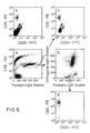
- the immunofluorescence of these cell populations is shown in FIG. 6.
- the agglutinated cells have the highest binding of both CD15 and CD11b. See FIG. 6C. These data indicate that the intensity of staining LDS-751 could also be used to identify doublets or agglutinated cells within the sample.
- the discrimination between cell population based on CD15 and CD11b binding was more easily observed when the damage cells and doublets were removed from the sample by getting the list mode file.
- the multiple populations identified by immunofluorescence were also recognizable by light scattering.
- the population which are mapped by the immunofluorescences have corresponding light scattering characteristics L,M,N,EO).
- the neutrophils (N) and esonophils (EO) were separated by both forward angle light scattering and by the intensity of CD15 fluorescence.
- the monocytes (M) were distinguished from the lymphocytes (L) both by their light scattering properties and CD11b expression.
- the NK cells (included in the L Population) expressed CD11b but did not express CD15 as did the monocytes (M). The identity of these cell populations has been confirmed by sorting the cells for morphological analysis.
- a kit comprising containers separately containing a nucleic acid dye and one or more MAbs may be used in the practice of this invention.
- one container in the kit will contain LDS-751.
- Other containers in the kit may include separately any of the MAbs previously described in the above examples. It will be appreciated by those skilled in the art that the MAbs may be labelled before inclusion in the kit or separate containers containing the fluorochromes may be included for independent labelling.
Abstract
Description
- This invention relates to a method for discriminating between intact and damaged cells in a sample, and more particularly, relates to a method for discriminating between contact and damaged cells in a peripheral blood sample, wherein the intact and damaged cells are discriminately stained by a nucleic acid stain.
- The detection and identification of cell types in the hematopoietic system has long been a useful research and clinical tool. A number of automated methods exist to aid the researcher and clinician. Among those methods include flow cytometry and fluorescence microscopy. In recent years, the former method has become increasingly sophisticated and has become generally accepted as a tool to aid in the identification of or discrimination between cell types and between various functional and/or maturational subsets within a cell type.
- Flow cytometers, which are more generally described in U.S. Pat. Nos. 4,661,913, 4,284,412 and 3,826,364, and in an article by Herzenberg et al., Sci. Am., 234:108 (1976), have been used to identify different populations of leukocytes in a heterogeneous sample by detecting multiple independent parameters on individual cells that pass through the sensing region. Typically, these parameters include forward light scatter (FLS, which is a measure of relative particle size), orthogonal light scatter (OLS, which is a measure of relative granularity) and fluorescence. Fluorescence may be measured from cells that incorporate a nucleic acid or other vital stain or may be measured from cells bearing surface markers which are labelled with monoclonal antibodies (MAbs) that are conjugated directly or indirectly to fluorochromes as described, for example, in U.S. Pat. No. 4,520,110. By combining and comparing these parameters, the various leukocyte components may be distinguished.
- U.S. Pat. No. 4,727,020 provides one example of how a flow cytometer operates and may be used to identify leukocyte subpopulations. Unlysed whole blood was treated with one MAb conjugated to phycoerythrin (PE) specific for CD4⁺ T cells and and a second MAb conjugated to fluoroscein isothiocyanate (FITC) specific for CD8⁺ T cells. A nucleic acid dye, LDS-751 (Exciton), was added to identify nucleated leukocytes. The labelled cells then were analyzed by flow cytometry. A state was set for LDS-751⁺ cells (i.e., for nucleated leukocytes, thereby excluding erythrocytes and platelets). The method allowed separation of leukocyte subpopulations by comparing the various parameters measured.
- One problem inherent in any method that makes use of fluorescently labelled MAbs and/or nucleic acid dyes, however, is the propensity of the labels to indiscriminantly bind to damaged cells or cell debris in a sample. This problem is compounded by the fact that in order to prepare cells for labelling, the cells must undergo several preparation techniques, all of which increase the number and proportion of cells that become damaged or ruptured in any one sample. These damaged cells and associated cell debris and their accompanying fluorescence must be discriminated against in the overall sample in order to thoroughly examine the remaining intact cells. Depending upon the method used to prepare the cells in any one sample and even between samples using the same method, sample preparation can introduce a significant amount of variation into the system. As a result, method used for immunofluorescence analysis of cells could mis-identify damaged cells as part of the subpopulations of interest.
- A variety of techniques exist for determining whether a cell in a sample is intact or damaged. Viable, intact cells can be distinguished from dead cells by using either fluoroscein diacetate (FDA) or propidium iodide (PI). In these methods, the sample is treated with either FDA or PI and then examined for fluorescence. Cells that stain with FDA are considered "viable"; cells that stain with PI are considered dead." The methods are limited, however, in that the cells cannot be fixed if the stains are to be used to identify viable cells. Another limitation on the use of FDA is that it brightly fluoresces so that it overwhelms the immunofluorescence signals from other stains, such as FITC and PE, rendering them unreadable.
- Other methods to detect intact cells that do not make use of such dyes also exist. FLS, on a flow cytometer for example, may be used to discriminate between intact and damaged cells but only if the cells in the sample are derived from a homogeneous population. Cells from a heterogeneous population cannot be so distinguished because of variations in cell size and light scattering properties.
- Each of the above-described methods suffers from some defect that makes it inapplicable as a general method for discrimination between damaged and intact cells in a heterogeneous sample. As a result, there is no single method that allows the researcher or clinician to examine a heterogeneous cell sample from an individual and to discriminate between damaged and intact cells therein.
- The present invention comprises a method to discriminate between intact and damaged cells in a body fluid sample. The method comprises the steps of: 1) taking a body fluid sample from an individual; 2) adding a nucleic acid dye to said sample; and 3) analyzing the cells in said sample in an automated instrument capable of passing said cells through a sensing region substantially one at a time and capable of detecting and recording both fluorescence and light scattered by individual cells.
- In the preferred embodiment, the body fluid sample comprises peripheral whole blood wherein the erythrocytes are lysed, and the nucleic acid dye comprises a stain with a preference for DNA and which also distinguished between damaged and intact cells based on the amount of fluorescence emitted. It will be appreciated by those skilled in the art that this method also is particularly useful when combined with other methods used to identify cell types (e.g., when fluorescently labelled MAbs are used). Examples of such other methods include those described in U.S. Pat. No. 4,599,304 for the monitoring of activated cells. In such cases, it will be further appreciated that the peak emission spectra of each of the fluorochromes used to label the MAbs and of the nucleic acid dye must be sufficiently distinct so as not to overlap. Similarly, it is desirable that all of the fluorescent labels be excitable at the same wavelength. This will allow the use of a single laser source in the flow cytometer, as opposed to having a dual laser source as described in U.S. Pat. No. 4,727,020.
- A kit comprising a set of containers separately containing a nucleic acid dye and one or MAbs also is disclosed.
-
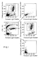
- FIG. 1 comprises several correlation of "dot" plots of OLS vs. FLS (A,C-E) and log LDS-751 fluorescence vs. FLS (B) for fixed PBL (peripheral blood leukocytes) from erythrocyte depleted whole blood;
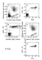
- FIG. 2 comprises several dot plots of OLS vs. FLS (A,D), log LDS-751 fluorescence vs. FLS (B) and vs. log FDA fluorescence (C,E) the PBL were not fixed;
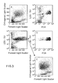
- FIG. 3 comprises several dot plots as in FIG. 2 for cells prepared from the same individual as in FIG. 2, but the cells were held for 48 hours prior to staining with LDS-751 and FDA;
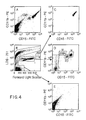
- FIG. 4 comprises several dot plots of log CD5(PE) fluorescence vs. CD20(FITC) fluorescence (A,C,E), log LDS-751 fluorescence vs. FLS (B) and FLS vs. OLS (D) for erythrocyte depleted whole blood which had been reacted with these two MAbs;
- FIG. 5 comprises several dot plots as in FIG. 1 but wherein the cells also were stained with CD11b(PE) and CD15(FITC); and
- FIG. 6 comprises several dot plots of log CD11b(PE) fluorescence vs. log CD15(FITC) fluorescence (A,C-E) and log LDS-751 fluorescence vs. FLS (B) for the fixed PBL from erythrocyte depleted whole blood as shown in FIG. 5.
- The present invention comprises a method for the discrimination between and identification of cell types in a body fluid. Preferably, the body fluid is erythrocyte depleted peripheral blood; however, other body fluids that may be sampled include peritoneal, spinal and brain fluids, as well as urine. In addition, cell suspensions of bone marrow, lymph node, liver, spleen and thymus may be used.
- The method comprises the steps of:
- 1) taking a body fluid sample from an individual;
- 2) adding a fluorescent nucleic acid dye to said sample; and
- 3) analyzing the cells in said sample in an automated instrument capable of passing said cells through a sensing region substantially one at a time and capable of detecting and recording both fluorescence and light scattered by each of the cells that pass through the sensing region.
- The method may be broadened to include a step wherein MAbs are added to the sample prior to staining with the nucleic acid dye. One or more MAbs may be used to detect cell surface antigens or the cells in the body fluid sample. The method is not limited by the MAb used or the cell surface antigen to be detected; however, certain pairs of MAbs are useful in the practice of this invention and include those pairs described in U.S. Serial No. 126,333, filed November 30, 1987. The MAbs may be directly or indirectly labelled by one or more fluorochromes by methods known to those skilled in the art. It is preferable that the MAbs are directly labelled. It also is preferable that the peak emission spectra of the fluorochromes and the nucleic acid dye all be distinguishable. Further, it is desirable, when a single laser source is used in the automated instrument, to have all the fluorescent materials excitable at substantially the same wavelength (e.g., 488nm when an argon laser is used). If a dual laser source is used, the excitation spectra may differ.
- Desirably, the nucleic acid dye has a preference for DNA which has different fluorescence intensities for intact and damaged cells. In this manner, non-nucleated cells, such as erythroyctes and platelets, will not be stained (or will be stained minimally) such that nucleated cells (e.g., leukocytes) can be distinguished from non-nucleated cells based upon the intensity of fluorescence. In the preferred embodiment, the dye is LDS-751; however, other dyes having a preference for DNA also may be used such as those disclosed in U.S. Pat. No. 4,544,546. The amount of dye used and the time the cells are stained with the dye should be sufficient to avoid differential staining as a result of kinetic phenomena. Twenty-four hours is more than sufficient time.
- A variety of devices may meet the requirements of the automated instrument. Desirably, the instrument comprises a flow cytometer with a single laser source, Preferably, the single laser source is an argon laser tuned to 488nm. The flow cytometer may further comprise data storage and analyses means such as a personal computer with software sufficient to list, store and analyze at least five parameters of data in various paired combinations. Examples of flow cytometers useful in the practice of this invention include FACS 440™ FACScan™ and FACStar™ (all commercially available from Becton Dickinson Immunocytometry Systems (BDIS)). Examples of personal computers and appropriate software include Consort 30 data management systems (BDIS) and FACScan Research or Paint-A-Gate™ Software (BDIS).
- It will be appreciated by those skilled in the art that a fluorescence microscope may be substituted for the automated instrument. In this case, manual counting and identification of cells is required.
- Peripheral blood of healthy individuals was collected by venipuncture into vacutainer tubes containing EDTA(K₃) as anticoagulant (Becton Dickinson). Erythrocytes were lysed with NH₄Cl. One volume of blood was mixed with 15 volumes of 1% NH₄Cl in H₂O(pH 7.3) and gently mixed. Cells were lysed for 3-5 minutes, and centrifuged at 200g for 5 minutes at room temperature. The pellet was resuspended in a volume of RPMI 1640 (Whittaker) 14 times larger than the original blood volume and centrifuged at 200g for 5 minutes. This washing step was repeated twice and the cells were finally resuspended in phosphate buffered saline containing 1% bovine serum albumin, 1O⁴IE penicillin/ml, 100 u/ml streptomycin and 20 mM Hepes (pH 7.3) (PBS). The cell concentration of the remaining peripheral blood leukocytes (PBL) was adjusted to 1 x 10⁷/ml,
- Where MAbs were added to the sample, twenty ul of a pretitered MAb was added to 100 ul of cell suspension. After incubating for 20 minutes on ice, the cells were washed twice with 2ml of the PBS solution of 4°C. The staining procedure was repeated for a second MAb, if any. The pellet of the immunofluorescent labelled cells was resuspended in 1 ml of 1% paraformaldehyde in PBS.
- A stock solution of
LDS 751 was made by dissolving 0.2 mg in 1 ml of methanol. A working solution containing 0.5 ml of the stock solution was diluted into. a final volume of 50 ml PBS containing 0.1% azide. Ten ul of the LDS-751 working solution was added to the paraformaldehyde fixed cells and kept overnight before flow cytometric analysis. The cell suspension could be kept at least two weeks without changes in the light scattering properties or fading of the immunofluorescence signals on the cells. - In the experiments correlating the fluorescence signals obtained from LDS-751 and FDA, the NH₄Cl lysed cells were resuspended in RPMI to a final concentration of 1 x 10⁶/ml. Before analysis, the cells were kept one hour in the RPMI solution in order to obtain optimal light scattering properties of the cells. The FDA stock solution was made by dissolving 5 mg FDA in 1 ml of acetone. The FDA working solution contained 5 ul of the stock solution diluted into a final volume of 5 ml PBS containing 0.1% azide. Fifteen minutes before analysis, 30 ul of the FDA working solution was added to 1 ml of the cell suspension together with
LDS 751 in a final concentrations of 0.2 ug/ml for the fresh and o.1 ug/ml for the 48 hours old samples respectively. - The MAbs FITC labelled Leu-Ml, BDIS) and PE labelled CD11b (Anti-Leu-15, BDIS) were used. With this combination NK-cells and different maturational stages of monocytes and neutrophilic granulocytes could be identified. The MAbs FITC labelled CD20 (Anti-Leu-16, BDIS) and PE labelled CD5 (Anti-Leu 1, BDIS) also were used.
- Flow cytometric analyzes were performed on a FACScan™ flow cytometer. This instrument used an air-cooled Argon ion laser as a light source. The laser operated at 488 nm with an intensity of 15 mW. The laser was focused on the cell stream by means of a prismatic expander and a planoconvex lens providing a 20 um x 64 um elliptical beam. The optical measurements were performed in a quartz flowcell with a 430um x 180um rectangular flowchannel. A stable flow was achieved by pressurizing sheat and sample flow. Measurements were performed at a sample flow rate of 60 ul/min.
- The light scattered in forward direction was collected with a spherical lens provided with a rectangular beam stop (collecting angles 1-10 degrees) and detected by a solid state silicon detector. Orthogonal scattered and fluorescence light was collected by a lens (H 94, 1.22 NA) coupled to the flowcell with an optical gel (collecting angles between 23° and 157°). The light was directed to 4 photomultiplier tubes using appropriate optical filter combinations. The fluorescence signals were collected through a 530 nm band pass filter for the FITC signals, a 585 nm band pass filter for the PE signals and a 650 nm longpass filter for the fluorescent light obtained from
LDS 751. The scattered light was directed to a photomultiplier using a Brewster angle beam splitter. - The five parameters were digitized and stored in memory by Direct Memory Access in the list mode with Consort 30™. Each measurement contained 22,000 cells. Data acquisition was performed with the FAcscan Research Software.
- Cell sorting was performed on a FACStar™ flow cytometer. For each population l0,000 cells were sorted into RPMI containing 10% Fetal Calf Serum (FCS). The sorted cells were spun down for 5 minutes at 200g and resuspended in 100 ul RPMI containing 10% FCS. Cytospin preparations were made with a Shandon Cyto-centrifuge (Southern Product Ltd., Astmoor, England). The slides were stained with Wright Stain and examined with a light microscope.
- PBL were obtained by lysing the erythrocytes with NH₄CL as described. These cells were labelled with PE and FITC labeled MAbs following standard immunofluorescence procedures. After washing, the cells were fixed in 1% paraformaldehyde and the nucleic acid dye, LDS-751, then was added to the cells.
- The instrument threshold was set at a low level on the forward angle light scattering channel in order to include platelets and cell debris in the analysis. See FIG. 1A. The major leukocyte cell populations including lymphocytes, monocytes, granulocytes, and the esonophils (L,M,N,EO respectively, FIG. 1A) could be distinguished from the platelets and cell debris by forward and orthogonal light scattering signals.
- In the correlation between the forward light scattering and LDS-751 fluorescence, FIG. 1B, a population of intermediate staining cells with significant light scattering was identified. See Population I, FIG. 1B. This population included all of the intact cells as identified by forward and right angle light scattering. See FIG. 1D. The population with the highest amount of LDS-751 fluorescence, DA in FIG. 1B, had low to intermediate amounts of forward and orthogonal light scattering. See FIG. 1C. The proportion of events within Population DA varied from sample to sample and differed with changes in preparation procedures. The low light scattering signals and the sample to sample variations suggested that these events were cell debris. This was confirmed by sorting the cell populations identified in FIG. 1B. The cells in Population I were intact cells by microscopic examination. The cells represented within DA were damaged cells, bare nuclei, or aggregated platelets. The particles identified within Population P were platelets and erythrocytes. This latter population had light scattering characteristics shown in FIG. 1E.
- A comparison of FIG. 1C and D would suggest that the use of light scattering alone would include some damage cells with the intact cell populations. The contribution of damaged cells to the populations identified solely by light scattering was determined for twenty normal donors (Table I).
TABLE I Enumeration of Percentage of Intact Nucleated Cells of 20 Donors Determined by LDS-751 Fluorescence a MIN MAX MEAN S.D. Intact Nucleated Cells b 13 66 34 14.6 Light Scattering gate for c Intact Lymphocytes 78 99 88 7.1 Intact Monocytes 8 88 56 21.4 Intact Neutrophils 47 91 76 11.4 Intact Eosinophils 11 78 41 16.7 a PBL obtained by NH₄Cl lysing, fixed in 1% paraformaldehyde and stained with LDS-751. b Cells occurring the region indicated by "I" in FIG. 1B (% is obtained by dividing their number by the total number of nucleated cells (i.e., I + DA) x 100%). c Number of intact nucleated cells occurring in the light scattering gates typical for cell types as indicated in FIG. 1D divided by the number of cells occurring in identical gates set in FIG. 1A x 100%. - To determine the percentage of cells which might interfere with the immunofluorescence, the percent of intact cells was determined in the light scattering regions typical for lymphocytes, monocytes, neutrophil granulocytes, and eosinophilic granulocytes (FIG. 1D: L,M,N,EO respectively). This table indicates that only a minority of cells (mean 34% intact nucleated cells) survived the sample preparation protocol. In addition, a significant fraction of these damaged cells could not be removed based on light scattering characteristics only. For example, within the lymphocyte light scattering state (FIG. 1D: L) an average of only 88% of the cells were intact based on LDS-751 fluorescence. It should be noted that erythrocytes which survived the lysis protocol could appear in the light scattering region typical for nucleated cells. (FIG. 1E). By using the additional fluorescence parameter the cells were confined to Population P, FIG. 1B since they did not stain with LDS-751 and therefore could be distinguished from nucleated cells.
- The extension of this technique to unfixed, viable samples added further confirmation that the LDS-751 was able to discriminate intact from damaged cells. LDS-751 can be used to stain viable leukocytes. This permits the correlation of LDS-751 staining with conventional dyes which assess viability in unfixed samples.
- PBL were prepared using NH₄Cl without subsequent fixation. The light scattering characteristics of this sample are shown in FIG. 2A. The cells that are identified as viable using FDA can also be distinguished based on LDS-751 fluorescence. See FIG. 2C. Greater than 98% of the cells were identified as positive by. both FDA and
LDS 751 fluorescence. - In a similar experiment, the unfixed cells were held for 48 hours after the NH₄Cl lysis in order to increase the dead cells in the sample before staining with LDS-751 and FDA. FIG. 3. At this time, only 71% of the cells were viable based on FDA and LDS-751 fluorescence. By gating on the LDS-751 and light scattering, FIG. 3B, essentially all of the viable cells are included (0.9%) and only a few (8.6%) were dead based on FDA fluorescence. These data confirm that LDS-751 discriminates intact from damaged cells both in a viable state and after fixation.
- The excitation of
LDS 751 at 488 nm and its far red emission permits the combination of this dye with both FITC and PE immunofluorescence reagents. Spectral compensation must be used between the dyes to correct for PE emission entering the LDS channel (10% subtraction), however, only a minimal correction must be made in the other direction (2% subtraction). No compensation was required between the FITC and LDS-751 channels. - The advantage of using
LDS 751 to identify damaged cells during lymphocyte subpopulation analysis is demonstrated in FIG. 4. In this example the erythrocyte lysed, whole blood was reacted with CD5(PE) (T-cells and a subpopulation of B-cells) and with CD20(FITC) (B Cells). The intact cells were identified in the correlation between LDS-751 and forward light scatter. See FIG. 4B. The relatively infrequent population of CD5⁺/CD20⁻, CD5⁺/CD20⁺ and CD5⁻/CD20⁺ lymphocytes, FIG. 4E, can be identified by further gating on the forward and orthogonal light scattering. See FIG. 4D. By gating only on light scattering without identifying intact cells, the proportion of double labelled CD5⁺, CD20⁺ cells was increased by 50%. See FIG 4C. - A second example illustrating the importance of combining immunofluorescence with the identification of contact cells is shown in FIG. 5 and 6. The cells in this example were labelled with CD15(FITC) and CD11b(PE). These different cell types labelled (i.e., monocytes, granulocytes and NK cells) can be distinguished based on quantitive differences in antibody labelling and on the differences in their light scattering characteristics. The CD15 MAb is known to agglutinate neutrophils since it is of the IgM isotype and the number of antigen sites on the target cells is high. This agglutination can be observed by comparing the relative frequency of the neutrophils, FIG. 5A and FIG. 1A, since these samples were obtained from the same cell preparation. It is evident that fewer neutrophils were observed by light scattering in the sample which was reacted with the CD15 MAb. It could be shown that the cells are still within the sample. As shown before, the intect cells, FIG 5B (Population I), could be distinguished from the more brightly labelled larger cells identified in Population AG. These agglutinated cells comprising 20% the intact cell population had light scattering characteristics which were off scale in FIG. 5C.
- The immunofluorescence of these cell populations is shown in FIG. 6. The agglutinated cells have the highest binding of both CD15 and CD11b. See FIG. 6C. These data indicate that the intensity of staining LDS-751 could also be used to identify doublets or agglutinated cells within the sample.
- The discrimination between cell population based on CD15 and CD11b binding was more easily observed when the damage cells and doublets were removed from the sample by getting the list mode file. The multiple populations identified by immunofluorescence were also recognizable by light scattering. In comparing FIG. 5D and 6D, the population which are mapped by the immunofluorescences have corresponding light scattering characteristics L,M,N,EO). The neutrophils (N) and esonophils (EO) were separated by both forward angle light scattering and by the intensity of CD15 fluorescence. The monocytes (M) were distinguished from the lymphocytes (L) both by their light scattering properties and CD11b expression. The NK cells (included in the L Population) expressed CD11b but did not express CD15 as did the monocytes (M). The identity of these cell populations has been confirmed by sorting the cells for morphological analysis.
- A kit comprising containers separately containing a nucleic acid dye and one or more MAbs may be used in the practice of this invention. In the preferred embodiment, one container in the kit will contain LDS-751. Other containers in the kit may include separately any of the MAbs previously described in the above examples. It will be appreciated by those skilled in the art that the MAbs may be labelled before inclusion in the kit or separate containers containing the fluorochromes may be included for independent labelling.
- All publications and patent applications mentioned in this specification are indicative of the level of skill of those skilled in the art to which this invention pertains. All publications and patent applications are herein incorporated by reference to the same extent as if each individual publication or patent application was specifically and individually indicated to be incorporated by reference.
- It will be apparent to one of ordinary skill in the art that many changes and modifications can be made in the invention without departing from the spirit or scope of the appended claims.
Claims (15)
Priority Applications (1)
| Application Number | Priority Date | Filing Date | Title |
|---|---|---|---|
| AT89304754T ATE97500T1 (en) | 1988-06-13 | 1989-05-10 | METHOD FOR DISCRIMINATION BETWEEN INTACT AND DAMAGED CELLS IN A SAMPLE. |
Applications Claiming Priority (2)
| Application Number | Priority Date | Filing Date | Title |
|---|---|---|---|
| US206454 | 1988-06-13 | ||
| US07/206,454 US5057413A (en) | 1988-06-13 | 1988-06-13 | Method for discriminating between intact and damaged cells in a sample |
Publications (3)
| Publication Number | Publication Date |
|---|---|
| EP0347039A2 true EP0347039A2 (en) | 1989-12-20 |
| EP0347039A3 EP0347039A3 (en) | 1990-10-24 |
| EP0347039B1 EP0347039B1 (en) | 1993-11-18 |
Family
ID=22766462
Family Applications (1)
| Application Number | Title | Priority Date | Filing Date |
|---|---|---|---|
| EP89304754A Expired - Lifetime EP0347039B1 (en) | 1988-06-13 | 1989-05-10 | Method for discriminating between intact and damaged cells in a sample |
Country Status (6)
| Country | Link |
|---|---|
| US (1) | US5057413A (en) |
| EP (1) | EP0347039B1 (en) |
| JP (1) | JP2620810B2 (en) |
| AT (1) | ATE97500T1 (en) |
| DE (1) | DE68910730T2 (en) |
| ES (1) | ES2061987T3 (en) |
Cited By (5)
| Publication number | Priority date | Publication date | Assignee | Title |
|---|---|---|---|---|
| EP0513762A1 (en) * | 1991-05-14 | 1992-11-19 | Toa Medical Electronics Co., Ltd. | Reagent and method for analyzing cells in urine |
| EP0608987A1 (en) * | 1993-01-26 | 1994-08-03 | Becton, Dickinson and Company | Method for detecting rare events |
| EP0685994A1 (en) * | 1993-02-25 | 1995-12-13 | Abbott Laboratories | Multipurpose reagent system for rapid lysis of whole blood samples |
| WO2009082218A1 (en) * | 2007-12-20 | 2009-07-02 | Nederlandse Organisatie Voor Toegepast-Natuurwetenschappelijk Onderzoek Tno | Real-time method for the detection of viable micro-organisms |
| EP3906395A4 (en) * | 2019-01-04 | 2022-10-12 | GenNext Technologies, Inc. | In vivo radical dosimetry and in vivo hydroxyl radical protein foot-printing |
Families Citing this family (53)
| Publication number | Priority date | Publication date | Assignee | Title |
|---|---|---|---|---|
| IE76732B1 (en) * | 1990-08-07 | 1997-11-05 | Becton Dickinson Co | One step test for absolute counts |
| JP2941041B2 (en) * | 1990-11-16 | 1999-08-25 | シスメックス株式会社 | Classification of leukocytes by flow cytometry |
| US5610027A (en) * | 1992-10-30 | 1997-03-11 | Micro-Med, Inc. | Microphoto lysis-anlaysis process to measure cell characteristics |
| US5532139A (en) * | 1992-10-30 | 1996-07-02 | Micro-Med, Inc. | Micro lysis-analysis process to measure cell characteristics and diagnose diseases |
| US5955295A (en) * | 1992-10-30 | 1999-09-21 | Micro-Med, Inc. | Micro lysis-analysis process to measure cell characteristics and diagnose diseases |
| WO1994016102A1 (en) * | 1993-01-06 | 1994-07-21 | Boehringer Mannheim Corporation | Fluorescent detection of nonviable cells |
| US5436134A (en) * | 1993-04-13 | 1995-07-25 | Molecular Probes, Inc. | Cyclic-substituted unsymmetrical cyanine dyes |
| US5534416A (en) * | 1993-04-13 | 1996-07-09 | Molecular Probes, Inc. | Fluorescent viability assay using cyclic-substituted unsymmetrical cyanine dyes |
| US5445946A (en) * | 1993-04-13 | 1995-08-29 | Molecular Probes, Inc. | Intravacuolar stains for yeast and other fungi |
| US5545535A (en) * | 1993-04-13 | 1996-08-13 | Molecular Probes, Inc. | Fluorescent assay for bacterial gram reaction |
| US5437980A (en) * | 1993-05-17 | 1995-08-01 | Molecular Probes, Inc. | Phenanthridium dye staining of nucleic acids in living cells |
| DE69521006T2 (en) * | 1994-10-20 | 2001-09-20 | Sysmex Corp | Reagent and method for analyzing solids in urine |
| US5879900A (en) * | 1994-12-15 | 1999-03-09 | Abbott Laboratories | Method for simultaneous analysis of cell viability, nucleated red blood cells and white blood cell differentials |
| US6329158B1 (en) * | 1995-09-15 | 2001-12-11 | Becton Dickinson And Company | Use of dimly fluorescing nucleic acid dyes in the identification of nucleated cells |
| ATE472725T1 (en) * | 1995-12-15 | 2010-07-15 | Abbott Lab | METHOD FOR SIMULTANEOUS ANALYSIS OF CELL VIABILITY, NUCLATED RED BLOOD CELLS AND LEUKOCYTE DIFFERENTIAL |
| US6819411B1 (en) | 1997-01-31 | 2004-11-16 | Xy, Inc. | Optical apparatus |
| US6149867A (en) | 1997-12-31 | 2000-11-21 | Xy, Inc. | Sheath fluids and collection systems for sex-specific cytometer sorting of sperm |
| US6911313B2 (en) * | 1998-02-06 | 2005-06-28 | Sysmex Corporation | Process for discriminating and counting erythroblasts |
| FR2782730B1 (en) * | 1998-08-25 | 2002-05-17 | Biocom Sa | CELL SEPARATION PROCESS FOR THE ISOLATION OF PATHOGENIC CELLS, PARTICULARLY RARE CANCERES, EQUIPMENT AND REAGENT FOR IMPLEMENTING THE PROCESS AND APPLICATION OF THE PROCESS |
| AU768616C (en) * | 1999-02-19 | 2004-12-16 | Idexx Laboratories, Inc. | High numerical aperture flow cytometer and method of using same |
| US6673568B1 (en) | 1999-10-25 | 2004-01-06 | Genprime, Inc. | Method and apparatus for prokaryotic and eukaryotic cell quantitation |
| US6787302B2 (en) * | 1999-10-25 | 2004-09-07 | Genprime, Inc. | Method and apparatus for prokaryotic and eukaryotic cell quantitation |
| US7208265B1 (en) | 1999-11-24 | 2007-04-24 | Xy, Inc. | Method of cryopreserving selected sperm cells |
| ATE386815T1 (en) * | 2000-01-06 | 2008-03-15 | Caliper Life Sciences Inc | METHODS AND SYSTEMS FOR MONITORING INTRACELLULAR BINDING REACTIONS |
| WO2002043574A2 (en) | 2000-11-29 | 2002-06-06 | Xy, Inc. | System to separate frozen-thawed spermatozoa into x-chromosome bearing and y-chromosome bearing populations |
| US7713687B2 (en) | 2000-11-29 | 2010-05-11 | Xy, Inc. | System to separate frozen-thawed spermatozoa into x-chromosome bearing and y-chromosome bearing populations |
| US6793642B2 (en) | 2001-05-07 | 2004-09-21 | Biomed Solutions, Llc | Flow cytometer |
| US6403378B1 (en) * | 2001-04-26 | 2002-06-11 | Guava Technologies, Inc. | Cell viability assay reagent |
| WO2002092161A1 (en) * | 2001-05-10 | 2002-11-21 | Biophan, Llc | Miniaturized particle analyzer |
| JP4595067B2 (en) | 2002-08-01 | 2010-12-08 | エックスワイ,エルエルシー | Low-pressure sperm cell separation system |
| US8486618B2 (en) | 2002-08-01 | 2013-07-16 | Xy, Llc | Heterogeneous inseminate system |
| JP2005535346A (en) | 2002-08-15 | 2005-11-24 | エックスワイ,インコーポレイテッド | High resolution flow cytometer |
| US7169548B2 (en) | 2002-09-13 | 2007-01-30 | Xy, Inc. | Sperm cell processing and preservation systems |
| BRPI0408857B1 (en) | 2003-03-28 | 2018-09-11 | Inguran Llc | apparatus, methods and processes for separating particles and for providing sex-separated animal sperm |
| AU2004242121B2 (en) | 2003-05-15 | 2010-06-24 | Xy, Llc. | Efficient haploid cell sorting for flow cytometer systems |
| US7776529B2 (en) | 2003-12-05 | 2010-08-17 | Life Technologies Corporation | Methine-substituted cyanine dye compounds |
| BRPI0509485A (en) | 2004-03-29 | 2007-09-11 | Monsanto Technology Llc | sperm suspensions for use in insemination |
| EP2269617B1 (en) | 2004-07-22 | 2016-04-27 | Inguran, LLC | Process for enriching a population of sperm cells |
| WO2006124816A1 (en) | 2005-05-11 | 2006-11-23 | Molecular Probes, Inc. | Fluorescent chemical compounds having high selectivity for double stranded dna, and methods for their use |
| WO2010068812A1 (en) * | 2008-12-10 | 2010-06-17 | Abqmr, Inc. | Nuclear magnetic resonance apparatus, methods and associated technology |
| GB201005939D0 (en) * | 2010-04-09 | 2010-05-26 | Biostatus Ltd | Method of analysing a cell or other biological material containing nucleic acid |
| US9476812B2 (en) | 2010-04-21 | 2016-10-25 | Dna Electronics, Inc. | Methods for isolating a target analyte from a heterogeneous sample |
| US9428547B2 (en) | 2010-04-21 | 2016-08-30 | Dna Electronics, Inc. | Compositions for isolating a target analyte from a heterogeneous sample |
| US20110262989A1 (en) | 2010-04-21 | 2011-10-27 | Nanomr, Inc. | Isolating a target analyte from a body fluid |
| US8841104B2 (en) | 2010-04-21 | 2014-09-23 | Nanomr, Inc. | Methods for isolating a target analyte from a heterogeneous sample |
| US10000557B2 (en) | 2012-12-19 | 2018-06-19 | Dnae Group Holdings Limited | Methods for raising antibodies |
| US9599610B2 (en) | 2012-12-19 | 2017-03-21 | Dnae Group Holdings Limited | Target capture system |
| US9995742B2 (en) | 2012-12-19 | 2018-06-12 | Dnae Group Holdings Limited | Sample entry |
| US9804069B2 (en) | 2012-12-19 | 2017-10-31 | Dnae Group Holdings Limited | Methods for degrading nucleic acid |
| US9551704B2 (en) | 2012-12-19 | 2017-01-24 | Dna Electronics, Inc. | Target detection |
| US9434940B2 (en) | 2012-12-19 | 2016-09-06 | Dna Electronics, Inc. | Methods for universal target capture |
| EP3165899A1 (en) * | 2015-11-04 | 2017-05-10 | Siemens Healthcare Diagnostics GmbH | Method for the detection of neutrophils extracellular cases |
| EP4314253A2 (en) | 2021-03-25 | 2024-02-07 | Iovance Biotherapeutics, Inc. | Methods and compositions for t-cell coculture potency assays and use with cell therapy products |
Citations (3)
| Publication number | Priority date | Publication date | Assignee | Title |
|---|---|---|---|---|
| EP0170345A2 (en) * | 1984-03-28 | 1986-02-05 | TECHNICON INSTRUMENTS CORPORATION(a Delaware corporation) | Flow cytometry |
| EP0193356A2 (en) * | 1985-02-25 | 1986-09-03 | Becton Dickinson and Company | Method for analysis of subpopulations of blood cells |
| EP0219309A1 (en) * | 1985-10-11 | 1987-04-22 | Smithkline Beecham Corporation | Methods and reagents for performing analyses of subpopulations of particles |
Family Cites Families (11)
| Publication number | Priority date | Publication date | Assignee | Title |
|---|---|---|---|---|
| US3826364A (en) * | 1972-05-22 | 1974-07-30 | Univ Leland Stanford Junior | Particle sorting method and apparatus |
| US4094745A (en) * | 1973-06-22 | 1978-06-13 | John Scholefield | Method of staining microscopic organisms |
| US4284412A (en) * | 1979-07-13 | 1981-08-18 | Ortho Diagnostics, Inc. | Method and apparatus for automated identification and enumeration of specified blood cell subclasses |
| US4544546A (en) * | 1980-04-21 | 1985-10-01 | Abbott Laboratories | Fluorescent nucleic acid stains |
| US4520110A (en) * | 1981-10-06 | 1985-05-28 | The Board Of Trustees Of The Leland Stanford Junior University | Fluorescent immunoassay employing a phycobiliprotein labeled ligand or receptor |
| JPS59184862A (en) * | 1983-04-05 | 1984-10-20 | ベクトン・ディッキンソン・アンド・カンパニ− | Method and device for discriminating plurality of subsidiarygroup of cell in sample |
| US4585736A (en) * | 1983-10-18 | 1986-04-29 | The United States Of America As Represented By The United States Department Of Energy | Flow cytometric measurement of total DNA and incorporated halodeoxyuridine |
| US4661913A (en) * | 1984-09-11 | 1987-04-28 | Becton, Dickinson And Company | Apparatus and method for the detection and classification of articles using flow cytometry techniques |
| JPH0663973B2 (en) * | 1985-08-07 | 1994-08-22 | 東ソー株式会社 | Fluorescence detector used for immunoreaction measurement |
| ZA867698B (en) * | 1985-10-11 | 1987-07-29 | Smithkline Beckman Corp | Methods and reagents for performing subset analysis |
| US5016283A (en) * | 1985-11-04 | 1991-05-14 | Cell Analysis Systems, Inc. | Methods and apparatus for immunoploidy analysis |
-
1988
- 1988-06-13 US US07/206,454 patent/US5057413A/en not_active Expired - Lifetime
-
1989
- 1989-05-10 AT AT89304754T patent/ATE97500T1/en not_active IP Right Cessation
- 1989-05-10 EP EP89304754A patent/EP0347039B1/en not_active Expired - Lifetime
- 1989-05-10 DE DE68910730T patent/DE68910730T2/en not_active Expired - Fee Related
- 1989-05-10 ES ES89304754T patent/ES2061987T3/en not_active Expired - Lifetime
- 1989-06-13 JP JP1150348A patent/JP2620810B2/en not_active Expired - Lifetime
Patent Citations (3)
| Publication number | Priority date | Publication date | Assignee | Title |
|---|---|---|---|---|
| EP0170345A2 (en) * | 1984-03-28 | 1986-02-05 | TECHNICON INSTRUMENTS CORPORATION(a Delaware corporation) | Flow cytometry |
| EP0193356A2 (en) * | 1985-02-25 | 1986-09-03 | Becton Dickinson and Company | Method for analysis of subpopulations of blood cells |
| EP0219309A1 (en) * | 1985-10-11 | 1987-04-22 | Smithkline Beecham Corporation | Methods and reagents for performing analyses of subpopulations of particles |
Non-Patent Citations (1)
| Title |
|---|
| Review of Scientific Instruments 55(9), 1375-1400 (1984) * |
Cited By (8)
| Publication number | Priority date | Publication date | Assignee | Title |
|---|---|---|---|---|
| EP0513762A1 (en) * | 1991-05-14 | 1992-11-19 | Toa Medical Electronics Co., Ltd. | Reagent and method for analyzing cells in urine |
| US5693484A (en) * | 1991-05-14 | 1997-12-02 | Toa Medical Electronics Co., Ltd. | Method of classifying and counting cells in urine |
| EP0608987A1 (en) * | 1993-01-26 | 1994-08-03 | Becton, Dickinson and Company | Method for detecting rare events |
| EP0685994A1 (en) * | 1993-02-25 | 1995-12-13 | Abbott Laboratories | Multipurpose reagent system for rapid lysis of whole blood samples |
| EP0685994A4 (en) * | 1993-02-25 | 2001-10-31 | Abbott Lab | Multipurpose reagent system for rapid lysis of whole blood samples |
| WO2009082218A1 (en) * | 2007-12-20 | 2009-07-02 | Nederlandse Organisatie Voor Toegepast-Natuurwetenschappelijk Onderzoek Tno | Real-time method for the detection of viable micro-organisms |
| US8986946B2 (en) | 2007-12-20 | 2015-03-24 | Nederlandse Organisatie Voor Toegepast-Natuurwetenschappelijk Onderzoek Tno | Real-time method for the detection of viable micro-organisms |
| EP3906395A4 (en) * | 2019-01-04 | 2022-10-12 | GenNext Technologies, Inc. | In vivo radical dosimetry and in vivo hydroxyl radical protein foot-printing |
Also Published As
| Publication number | Publication date |
|---|---|
| EP0347039B1 (en) | 1993-11-18 |
| ATE97500T1 (en) | 1993-12-15 |
| ES2061987T3 (en) | 1994-12-16 |
| EP0347039A3 (en) | 1990-10-24 |
| JP2620810B2 (en) | 1997-06-18 |
| US5057413A (en) | 1991-10-15 |
| DE68910730T2 (en) | 1994-05-19 |
| JPH02103464A (en) | 1990-04-16 |
| DE68910730D1 (en) | 1993-12-23 |
Similar Documents
| Publication | Publication Date | Title |
|---|---|---|
| EP0347039B1 (en) | Method for discriminating between intact and damaged cells in a sample | |
| EP0193356B1 (en) | Method for analysis of subpopulations of blood cells | |
| US5879900A (en) | Method for simultaneous analysis of cell viability, nucleated red blood cells and white blood cell differentials | |
| US5776709A (en) | Method for preparation and analysis of leukocytes in whole blood | |
| EP0470810B1 (en) | One step test for absolute counts | |
| CA1340170C (en) | Method for analysis of cellular components of a fluid | |
| Terstappen et al. | Discriminating between damaged and intact cells in fixed flow cytometric samples | |
| CA2207396C (en) | Method for rapid and simultaneous analysis of nucleated red blood cells | |
| EP0132064B1 (en) | Method for elimination of interference of selected cell populations in analytic cytology | |
| US5175109A (en) | Reagent for classifying leukocytes by flow cytometry | |
| US6900023B1 (en) | Method for classifying and counting leukocytes | |
| US6159740A (en) | Method and apparatus for screening obscured or partially obscured cells | |
| US5260192A (en) | Method and apparatus for screening cells or formed bodies with populations expressing selected characteristics utilizing at least one sensing parameter | |
| EP0559208B1 (en) | Method for preparation and analysis of leukocytes in whole blood utilizing flow cytometry | |
| EP0866960B1 (en) | Method for simultaneous analysis of cell viability, nucleated red blood cells and white blood cell differential | |
| EP0259833B1 (en) | Reagent and method for classifying leukocytes by flow cytometry | |
| AU635020B2 (en) | Method and apparatus for screening cells or formed bodies with populations expressing selected characteristics utilizing at least one sensing parameter | |
| WO1997021994A9 (en) | Method for simultaneous analysis of cell viability, nucleated red blood cells and white blood cell differential | |
| US20090246805A1 (en) | Method for classifying and counting basophils | |
| Van Pham | Flow Cytometry Data Analysis |
Legal Events
| Date | Code | Title | Description |
|---|---|---|---|
| PUAI | Public reference made under article 153(3) epc to a published international application that has entered the european phase |
Free format text: ORIGINAL CODE: 0009012 |
|
| AK | Designated contracting states |
Kind code of ref document: A2 Designated state(s): AT BE CH DE ES FR GB GR IT LI LU NL SE |
|
| 17P | Request for examination filed |
Effective date: 19900214 |
|
| PUAL | Search report despatched |
Free format text: ORIGINAL CODE: 0009013 |
|
| AK | Designated contracting states |
Kind code of ref document: A3 Designated state(s): AT BE CH DE ES FR GB GR IT LI LU NL SE |
|
| 17Q | First examination report despatched |
Effective date: 19920821 |
|
| GRAA | (expected) grant |
Free format text: ORIGINAL CODE: 0009210 |
|
| AK | Designated contracting states |
Kind code of ref document: B1 Designated state(s): AT BE CH DE ES FR GB GR IT LI LU NL SE |
|
| PG25 | Lapsed in a contracting state [announced via postgrant information from national office to epo] |
Ref country code: SE Effective date: 19931118 Ref country code: LI Effective date: 19931118 Ref country code: CH Effective date: 19931118 Ref country code: AT Effective date: 19931118 |
|
| REF | Corresponds to: |
Ref document number: 97500 Country of ref document: AT Date of ref document: 19931215 Kind code of ref document: T |
|
| ITF | It: translation for a ep patent filed |
Owner name: BUGNION S.P.A. |
|
| REF | Corresponds to: |
Ref document number: 68910730 Country of ref document: DE Date of ref document: 19931223 |
|
| REG | Reference to a national code |
Ref country code: GR Ref legal event code: FG4A Free format text: 3009843 |
|
| ET | Fr: translation filed | ||
| REG | Reference to a national code |
Ref country code: CH Ref legal event code: PL |
|
| PG25 | Lapsed in a contracting state [announced via postgrant information from national office to epo] |
Ref country code: LU Free format text: LAPSE BECAUSE OF NON-PAYMENT OF DUE FEES Effective date: 19940531 |
|
| PLBE | No opposition filed within time limit |
Free format text: ORIGINAL CODE: 0009261 |
|
| STAA | Information on the status of an ep patent application or granted ep patent |
Free format text: STATUS: NO OPPOSITION FILED WITHIN TIME LIMIT |
|
| 26N | No opposition filed | ||
| REG | Reference to a national code |
Ref country code: ES Ref legal event code: FG2A Ref document number: 2061987 Country of ref document: ES Kind code of ref document: T3 |
|
| PGFP | Annual fee paid to national office [announced via postgrant information from national office to epo] |
Ref country code: ES Payment date: 19980526 Year of fee payment: 10 |
|
| PGFP | Annual fee paid to national office [announced via postgrant information from national office to epo] |
Ref country code: GR Payment date: 19980529 Year of fee payment: 10 |
|
| PGFP | Annual fee paid to national office [announced via postgrant information from national office to epo] |
Ref country code: NL Payment date: 19980531 Year of fee payment: 10 |
|
| PGFP | Annual fee paid to national office [announced via postgrant information from national office to epo] |
Ref country code: BE Payment date: 19980714 Year of fee payment: 10 |
|
| PG25 | Lapsed in a contracting state [announced via postgrant information from national office to epo] |
Ref country code: ES Free format text: LAPSE BECAUSE OF NON-PAYMENT OF DUE FEES Effective date: 19990511 |
|
| PG25 | Lapsed in a contracting state [announced via postgrant information from national office to epo] |
Ref country code: GR Free format text: LAPSE BECAUSE OF NON-PAYMENT OF DUE FEES Effective date: 19990531 Ref country code: BE Free format text: LAPSE BECAUSE OF NON-PAYMENT OF DUE FEES Effective date: 19990531 |
|
| BERE | Be: lapsed |
Owner name: BECTON DICKINSON AND CY Effective date: 19990531 |
|
| PG25 | Lapsed in a contracting state [announced via postgrant information from national office to epo] |
Ref country code: NL Free format text: LAPSE BECAUSE OF NON-PAYMENT OF DUE FEES Effective date: 19991201 |
|
| NLV4 | Nl: lapsed or anulled due to non-payment of the annual fee |
Effective date: 19991201 |
|
| REG | Reference to a national code |
Ref country code: ES Ref legal event code: FD2A Effective date: 20010503 |
|
| REG | Reference to a national code |
Ref country code: GB Ref legal event code: IF02 |
|
| PGFP | Annual fee paid to national office [announced via postgrant information from national office to epo] |
Ref country code: FR Payment date: 20020417 Year of fee payment: 14 |
|
| PGFP | Annual fee paid to national office [announced via postgrant information from national office to epo] |
Ref country code: GB Payment date: 20020501 Year of fee payment: 14 |
|
| PGFP | Annual fee paid to national office [announced via postgrant information from national office to epo] |
Ref country code: DE Payment date: 20020520 Year of fee payment: 14 |
|
| PG25 | Lapsed in a contracting state [announced via postgrant information from national office to epo] |
Ref country code: GB Free format text: LAPSE BECAUSE OF NON-PAYMENT OF DUE FEES Effective date: 20030510 |
|
| PG25 | Lapsed in a contracting state [announced via postgrant information from national office to epo] |
Ref country code: DE Free format text: LAPSE BECAUSE OF NON-PAYMENT OF DUE FEES Effective date: 20031202 |
|
| GBPC | Gb: european patent ceased through non-payment of renewal fee |
Effective date: 20030510 |
|
| PG25 | Lapsed in a contracting state [announced via postgrant information from national office to epo] |
Ref country code: FR Free format text: LAPSE BECAUSE OF NON-PAYMENT OF DUE FEES Effective date: 20040130 |
|
| REG | Reference to a national code |
Ref country code: FR Ref legal event code: ST |
|
| PG25 | Lapsed in a contracting state [announced via postgrant information from national office to epo] |
Ref country code: IT Free format text: LAPSE BECAUSE OF NON-PAYMENT OF DUE FEES;WARNING: LAPSES OF ITALIAN PATENTS WITH EFFECTIVE DATE BEFORE 2007 MAY HAVE OCCURRED AT ANY TIME BEFORE 2007. THE CORRECT EFFECTIVE DATE MAY BE DIFFERENT FROM THE ONE RECORDED. Effective date: 20050510 |