EP0362531A1 - A human rhinovirus receptor protein that inhibits virus infectivity - Google Patents
A human rhinovirus receptor protein that inhibits virus infectivity Download PDFInfo
- Publication number
- EP0362531A1 EP0362531A1 EP89115358A EP89115358A EP0362531A1 EP 0362531 A1 EP0362531 A1 EP 0362531A1 EP 89115358 A EP89115358 A EP 89115358A EP 89115358 A EP89115358 A EP 89115358A EP 0362531 A1 EP0362531 A1 EP 0362531A1
- Authority
- EP
- European Patent Office
- Prior art keywords
- receptor
- hrv
- protein
- detergent
- cells
- Prior art date
- Legal status (The legal status is an assumption and is not a legal conclusion. Google has not performed a legal analysis and makes no representation as to the accuracy of the status listed.)
- Granted
Links
Images
Classifications
-
- C—CHEMISTRY; METALLURGY
- C07—ORGANIC CHEMISTRY
- C07K—PEPTIDES
- C07K14/00—Peptides having more than 20 amino acids; Gastrins; Somatostatins; Melanotropins; Derivatives thereof
- C07K14/435—Peptides having more than 20 amino acids; Gastrins; Somatostatins; Melanotropins; Derivatives thereof from animals; from humans
- C07K14/705—Receptors; Cell surface antigens; Cell surface determinants
- C07K14/70503—Immunoglobulin superfamily
- C07K14/70525—ICAM molecules, e.g. CD50, CD54, CD102
-
- A—HUMAN NECESSITIES
- A61—MEDICAL OR VETERINARY SCIENCE; HYGIENE
- A61P—SPECIFIC THERAPEUTIC ACTIVITY OF CHEMICAL COMPOUNDS OR MEDICINAL PREPARATIONS
- A61P11/00—Drugs for disorders of the respiratory system
- A61P11/02—Nasal agents, e.g. decongestants
-
- A—HUMAN NECESSITIES
- A61—MEDICAL OR VETERINARY SCIENCE; HYGIENE
- A61P—SPECIFIC THERAPEUTIC ACTIVITY OF CHEMICAL COMPOUNDS OR MEDICINAL PREPARATIONS
- A61P31/00—Antiinfectives, i.e. antibiotics, antiseptics, chemotherapeutics
- A61P31/12—Antivirals
-
- A—HUMAN NECESSITIES
- A61—MEDICAL OR VETERINARY SCIENCE; HYGIENE
- A61P—SPECIFIC THERAPEUTIC ACTIVITY OF CHEMICAL COMPOUNDS OR MEDICINAL PREPARATIONS
- A61P31/00—Antiinfectives, i.e. antibiotics, antiseptics, chemotherapeutics
- A61P31/12—Antivirals
- A61P31/14—Antivirals for RNA viruses
-
- A—HUMAN NECESSITIES
- A61—MEDICAL OR VETERINARY SCIENCE; HYGIENE
- A61P—SPECIFIC THERAPEUTIC ACTIVITY OF CHEMICAL COMPOUNDS OR MEDICINAL PREPARATIONS
- A61P31/00—Antiinfectives, i.e. antibiotics, antiseptics, chemotherapeutics
- A61P31/12—Antivirals
- A61P31/14—Antivirals for RNA viruses
- A61P31/16—Antivirals for RNA viruses for influenza or rhinoviruses
-
- A—HUMAN NECESSITIES
- A61—MEDICAL OR VETERINARY SCIENCE; HYGIENE
- A61K—PREPARATIONS FOR MEDICAL, DENTAL OR TOILETRY PURPOSES
- A61K38/00—Medicinal preparations containing peptides
Definitions
- the present invention relates to the isolation of proteins from animal cells, particularly mammalian cells, that bind to human rhinovirus (HRV). More particularly, the invention relates to the isolation of HRV receptor proteins that can bind to HRV and thereby block the infectivity of the virus. This property can serve as a basis for inhibiting the initiation or the spread of HRV infections, better known as the common cold.
- HRV human rhinovirus
- picornaviruses such as poliovirus, coxsacchie virus, and rhinoviruses
- specific binding to host cells has been demonstrated.
- competition experiments it has been demonstrated that some of these receptors are distinct from one another in that the saturation of the receptor of one virus had no effect on the binding of a second virus.
- Rhinoviruses form the largest family of picornaviruses, with 115 distinct serotypes identified to date. A large fraction of rhinoviruses (estimated to be 80%) appear to bind to a common receptor on human cells. [Abraham, G., and Colonno, R. J., Many rhinovirus serotypes share the same cellular receptor, J. of Virology 51 , 340-345 (1984).] In 1985, the isolation of a monoclonal antibody that appeared to be directed against the major rhinovirus receptor was described. [Colonno, R.J., Callahan, P.L., and Long, W. J., Isolation of a monoclonal antibody that blocks attachment of the major group of human rhinoviruses, J.
- a therapeutic agent effective against HRV infection might be the receptor itself, or more specifically, the virus binding domain of the receptor.
- a protein, protein fragment, or peptide that comprises the virus binding domain could block the ability of virus to bind to host cells by occupying (blocking) the receptor binding cleft on the virus.
- virus mutations that adversely affect binding of the molecule would adversely affect binding of the receptor, and would thus be deleterious or lethal for the virus; therefore, the likelihood of drug-resistant mutants would be very low. Furthermore, such a molecule would be human, lowering the likelihood of being antigenic in humans.
- the human rhinovirus (HRV) major receptor can be isolated as a water soluble preparation which exhibits the desired property of binding to HRV capsids and substantially reducing infectivity of the virus.
- the preparation is in the form of detergent-complexed glycoprotein isolated from animal cells, preferably mammalian cells, that express the HRV major receptor.
- the purified receptor protein is characterized as follows. It is a glycoprotein with an apparent molecular weight of 95,000 daltons and includes the binding site for HRV.
- the glycoprotein contains 6-7 asparagine-linked oligosaccharide chains and exists in the preparation in the form of a detergent micelle-bound protein.
- the HRV major receptor preparation of the present invention can be obtained by extraction of appropriate animal cells that are known to express the HRV major receptor with a nonionic detergent, followed by immunopurification.
- Many human cell lines express the receptor, such as HeLa and WI38. Any of these human sources of HRV receptor can be extracted. Particularly useful are HeLa cells.
- non-human mammalian transfectant cell lines that express the HRV receptor are known or can be prepared which provide another useful source of the receptor.
- transfectant cell lines as described in European Patent Application Publication No. 0 319 815 provide a ready source of receptor, particularly those secondary transfectants that have been selected for overexpression of receptor.
- Other animal cells as are known in the art or developed hereafter, such as insect tissue culture cells that have been tranfected with the gene and express the receptor, can also be used.
- any nonionic detergent can be used for the extraction provided the native conformation of the protein receptor is not destroyed. Denaturation of the receptor can be determined by monitoring the ability of the extracted protein to inhibit virus infectivity or by sensitivity to proteolysis. It has been determined that the receptor can be denatured by heating at 60°C for 30 minutes or by treatment with 1% SDS indicating that care need be taken to maintain the native conformation of the HRV binding site.
- alkyl polyoxyethylene ethers such as Brij
- alkylphenyl polyoxyethelene ethers such as Triton X-100 and Nonidet P-40
- acyl polyoxyethylene sorbitan esters such as Tween
- beta-D-alkyl glucosides Triton X-100 being considered particularly preferred.
- the key step in the purification of the receptor is fractionation with highly selective anti-receptor antibody.
- the most ready means to obtain such an antibody is by monoclonal techniques. It is particularly preferred to produce mouse monoclonal antibodies by generating hybridoma cell lines from fusion of murine myeloma cells and mouse transfectant cells expressing the HRV receptor. Further details are available in European Patent Application Publication No. 0 319 815. After binding the detergent-glycoprotein complexes obtained from the cell extract to the selected monoclonal antibody, complexes bound to antibody are separated from the remainder of the mixture. Thereafter, detergent-receptor complexes bound to antibody are dissociated, taking steps to again prevent denaturation, and the resulting water soluble receptor preparation isolated.
- Appropriate conditions for dissociating detergent-receptor complexes from the antibody can be determined empirically and can be expected to vary somewhat from antibody to antibody. Dissociation by raising pH has been found in some cases to be most effective with low pH or high salt conditions being operable but producing lower protein yields.
- Such intermediary steps comprise adsorbing the detergent extracted protein complexes to a lectin capable of binding HRV receptor, separating absorbed complexes from the remainder of the mixture, and dissociating such complexes for subsequent treatment with antibody.
- the selection of lectin and dissociating conditions is usually empirical. It has been found that the HRV receptor binds suitably to wheat germ agglutinin lectin and is dissociated effectively by washing with a solution of N-acetyl glucosamine.
- oligosaccharides on the receptor protein are not completely characterized, and because the receptor protein can be glycosylated differently on different cell types (e.g., mouse cell transfectants), other lectins would be expected also to be suitable.
- the selection of an appropriate alternative to wheat germ agglutinin and/or eluting agent can be left to the ordinary skill in the art.
- the resulting preparation can be treated with proteolytic agents such as proteases, e.g., trypsin, to produce smaller glycoprotein fragments that retain the ability to bind and reduce infectivity of HRV.
- proteases e.g., trypsin
- peptide fragments can be cleaved from a terminal region of the glycoprotein, e.g., the C-terminus, to yield glycoprotein fragments that retain HRV binding.
- Such glycoprotein fragments can, for example, have apparent molecular weights of between about 80,000 daltons and about 95,000 daltons. Smaller fragments which retain the HRV binding domain of the receptor are also considered to be within the scope of the present invention.
- the receptor preparation of the present invention has been shown to inhibit the infectivity of the virus, presumably by binding to the HRV capsid to block its ability then to bind and infect human cells. Such an observation indicates that the receptor preparation will be useful in reducing the infection of host human cells in vivo by contacting the virus with the preparation under conditions favorable to binding with the virus.
- a therapeutic form would be that of an aqueous solution of the receptor in the presence of nonionic detergent to maintain the receptor in solution and in its native conformation. Detergents with lower critical micelle concentrations, such as the alkyl polyoxyethylene ether Brij 58, would be preferred in order to reduce the concentration of the detergent in the therapeutic solution.
- the receptor preparation can be administered in vivo by appropriate contact with those areas of the body susceptible to infection by HRV, e.g., by intranasal spray.
- HRV serotypes were tested. HRV 4, 11, 17 and 89 serotypes (major class) were inhibited by the virus, whereas HRV 1a and 2 (minor class) were not.
- the results described above indicate that the purified HRR can block the infectivity of rhinoviruses belonging to the major receptor class of rhinoviruses.
- the infectivity inhibition property of the receptor protein is correlated with its ability to bind to the virus, and is presumed to act by blocking the receptor binding site on the virus. This property of the receptor is manifested at low concentrations of the receptor protein, and indicates a high affinity of the receptor for the virus.
- the significance of these results is that the purified, soluble receptor could be used to inhibit the initiation or the spread of rhinovirus infections in vivo.
- the purified protein also provides a source of material from which smaller protein fragments and peptides could be derived which have the same activity as the intact receptor.
- ICAM interleukin-1
- LFA-1 lymphocyte-function associated antigen-1
- the complete amino acid sequence of the rhinovirus receptor is now known.
- the determination of this amino acid sequence which is a partial chemical structure of this molecule, provides the ability to design and produce large amount of receptor protein, fragments, functional domains, and truncated versions, and analogs of receptor protein, and peptides that have inhibitory activity towards rhinovirus and coxsackie A virus infection.
- the complete amino acid sequence also provides information needed for biophysical and biochemical studies of rhinovirus-receptor interaction which will lead to the identification of crucial molecular contacts, which can be used for design of novel inhibitory molecules.
- ICAM immunoglobulin supergene family that maps to chromosome 19, ( Eur . J . Immunol ., 15 , 103-106 (1984) and since other picornaviruses, such as poliovirus and coxsackie virus, bind to receptors whose genes are located on chromosome 19, it is possible that ICAM can be used as a basis for the development of therapeutics to counter infections by those other picornaviruses as well. It is possible that ICAM or fragments thereof would be useful directly as therapeutics for other viruses and inflammatory diseases. Alternatively, knowledge of ICAM structure will be useful in the identification of the receptors of those viruses.
- ICAM-1 is closely related to two adhesion proteins of the adult nervous system, neural cell adhesion molecule (NCAM) and myelin-associated glycoprotein (MAG) and a family of epithelial cell molecules including CEA, NCA, TM-CEA, and the pregnancy-specific B1-glycoproteins.
- NCAM, MAG and ICAM-1 each have five immunoglobulin-like domains, see Dustin et al "Supergene Families Meet In The Immune System", Commentary, Elsevier Publications, Cambridge, 1988.
- the relationship of the picornaviruses and the supergene family of ICAM, NCAM and MAG provide the basis of developing proteins, protein fragments, functional domains, analogs and mixtures thereof for inhibiting infectivity of this class of viruses.
- Soluble forms of biologically active host cell protein could be used to inhibit virus infection, in contrast to the cell membrane bound receptor protein that normally facilitates the infection.
- Soluble forms of biologically active receptor protein, protein fragments, functional domains or analogs could include use of detergents as described supra. Alternatively, elimination of the C-terminus could render the protein(s) soluble.
- a biologically active tryptic fragment is a mixture of two species, one with an apparent molecular weight of 83,000 daltons and one of 90,000 daltons (relative to HRR of 95Kd). The N-terminus of both species is blocked, indicating that they start from residue 1 of the intact HRR molecule, and peptides are removed from C-termius: the largest possible fragment would be from residue 1 to residue 488.
- soluble fragments could include the entire extracellular domain (up to a.a. 480) or could include either/or both distinct parts of the extracellular domain (a.a. 1-200; 200-460) of the amino acid sequence of the receptor protein. It is further anticipated that smaller peptide fragments may provide biologically active analogs for inhibiting virus infection.
- a full length cDNA clone of the HRR will be isolated from a cDNA library of He1 or other cells expressing the receptor by screening with oligonucleotides made from the published sequence of ICAM-1. Construction and expression of domain fragments of the HRR will be achieved using established recombinant DNA methodologies (Fisher et al, Nature , 331 , 76-78 (1988); Hussey et al, Nature , 331 , 78-81 (1988); Deen et al, Nature , 331 , 82-86 (1988).
- a soluble extracellular domain will be made by cleaving a cDNA clone of the HRR coding sequence with ThaI which cuts at position 37 in the signal peptide region and at position 1415, 12 amino acids before the start of the transmembrane domain.
- Synthetic oligonucleotide linkers will be added in a stepwise fashion to the 5′ and 3′ ends of the molecule to restore the signal peptide and initiator ATG at the N termnus and to introduce an in frame translational stop codon at the C-terminus.
- the position of the stop codon may be varied to produce alternative truncated forms of the molecule.
- different infrequently cutting restriction enzymes will be used to insert stop codons in other regions of the molecule.
- Restriction enzyme sites will be included at the ends of the linkers to allow directional cloning into a variety of expression vectors. Oligonucleotide site directed mutagenesis, using conventional methods, will be used to introduce restriction enzyme sites where no convenient naturally occurring sites exist. Additionally, the polymerase chain reaction (PCA) technique will be used to produce specific DNA fragments encoding domains and other sub-regions of the molecule.
- PCA polymerase chain reaction
- the approach described above will also be used to produce additional subfragments of the receptor such as the five immunoglobulin-like domains (residues 1-88, 89-185, 186-284, 285-385, 386-453, Staunton et al, Cell , 52 , 925-933 (1988).
- appropriate signal sequences to direct protein secretion for the expression system being used will be included.
- Various expression systems will be used including viral promoters in mammalian cells (Cate et al, Cell , 45 , 685-698 (1986), insect cells (Smith et al Pros. Acad. Sci. U.S.A ., 82 , 8404-8408 (1985); and E .
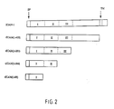
- tICAM's A series of cDNA's (tICAM's, or truncated ICAM's) were constructed from ICAM-1 cDNA to contain premature stop codons at amino acid positions 454, 284, or 185 of the mature protein in order to produce secreted proteins progressively truncated from the C-terminus.
- the positions of the truncations were selected based on the predicted borders of the transmembrane domain (tICAM(1-453)), immunoglobulin-like domains 1+2+3 (tICAM (1-283)), and immunogobulin-like domains 1+2 (tICAM(1-183)) and immunoglobulin-like domain 1 (tICAM(1-88).
- the protein products of these genes are diagramed in figure 2.
- Stable transfectants were generated by transfecting the same cDNA's mixed with the gene for a selectable marker (thymidine kinase for mouse L cells dihydrofolate reductase for CHO cells) into mouse Ltk- cells or hamster CHO(dhfr-) cells and subjected to drug selection (HAT selection for Ltk- cells and methatrexate for CHO (dhfr-) cells).
- a selectable marker thymidine kinase for mouse L cells dihydrofolate reductase for CHO cells
- tICAM(1-88) has been expressed in E Coli using the OmpA secretion vector of Inoue.
- the OmpA signal peptide is fused to the N-terminus of mature ICAM-1 protein.
- tICAM(1-88) and tICAM(1-183) have been placed into the OmpA vector; E Coli transformed with these vectors express protein products of the expected size as detected by western blotting of SDS-PAGE gels of cell extracts with anti-peptide antibodies to a sequence within domain 1 of ICAM-1.
- Blocking studies with the panel of 6 MAbs to ICAM-1 indicate that there are two distinct epitopes defined by these antiboides, one defined by c78.4 (containing c78.1, c78.2, c92.1, and c92.5).
- Immunoprecipitation studies with proteolytic fragments of ICAM-1 and with in vitro translations of truncated ICAM-1 cDNA's indicate that both of these epitopes are contained within the first Ig-like domain.
- Additional biologically active fragments will be evaluated utilizing overlapping sets of synthetic peptides of 10-20 residues corresponding to part or all of the HRR protein.
- the peptides will be made and individually tested for the ability to inhibit virus binding to receptor.
- peptide fragments could be direct copies of a portion of the rhinovirus receptor, or could contain sequences from non-contiguous regions of the receptor.
- ICAM has been predicted, based on homology to NCAM, to be a member of the immunoglobulin gene superfamily.
- the immunoglobulin-like domains in ICAM would have the basic "immunoglobulin fold", as has been shown for two other members of this family, beta-2-microglobulin and the HLA-A2 alpha-3 domain.
- This fold consists of a "beta-barrel" conformation consisting of two antiparallel beta-pleated sheets, one composed of three and one composed of four beta strands; a disulfide bond between two cysteine residues (separated by approximately 60 amino acids along the chain) connects the two sheets (Williams, A. F., Immun .
- Such protruding structures may be of particular interest in the rhinovirus receptor, since the receptor binding site on the virus capsid is proposed to be in a recessed cavity.
- Such turns and loops could be predicted based on a beta-barrel structure and produced as synthetic peptides with addition of novel cysteine residues at the N- and C-terminus of the peptides; a disulfide bond would then be formed between such residues on the same peptide to close the loop covalently (in contrast to the native protein, wherein the loop would be closed by noncovalent interactions between the adjacent beta-strands).
- Such peptides would have a conformation more analogous to the conformation in the native protein than a simple linear peptide, and would be tested for virus-binding activity.
- Site-directed antibodies directed against specific portion of the HRR could be produced by making synthetic peptides corresponding to selected regions of the protein, coupling such peptides to larger carrier proteins, and immunizing rabbits or other animals with such conjugates by standard methodology.
- Such antibodies could be tested for the ability to inhibit virus binding; inhibition with a subset of such antibodies would direct attention to specific domains or parts of domains.
- Specific reactive groups on some amino acid residues on the receptor protein can be chemically modified under non-denaturing conditions. As a consequence of the modification of some residues virus-binding ability may be lost.
- the modification of some amino acid residues may be correlated with loss of binding activity, implicating those groups in recognition. This would direct attention towards a specific part of the molecule or a specific amino acid residue as playing a specific role in virus binding. Such residues could then be experimentally modified in in vitro mutagenesis experiments.
- oligonucleotide site directed mutagenesis For example, pools of mutants produced by saturation mutagenesis will be screened by the method of Peterson and Seed ( Cell , 54 , 65-72 (1988), using either HRV14 or monoclonal antibody/complement killing as the negative selection, and a rabbit polyclonal antibody as the positive selection. Synthetic peptides corresponding to regions of the molecule identified in this way will be made and tested for virus binding and the ability to reduce infectivity.
- HRR or fragments of it may have application in the disruption of interactions between ICAM and LFA-1, which could be useful for the treatment of inflammation.
- Peptides derived from the known capsid proteins of rhinovirus could be useful for the disruption of interactions between ICAM and LFA-1, which could be useful for the treatment of inflammation.
- Carbohydrate groups that are not nec for biological activity will be removed to enhance production of peptides in bacteria.
- Site-directed mutagenesis of cystines may be useful to limit refolding to biologically active conformations.
Abstract
Description
- The present invention relates to the isolation of proteins from animal cells, particularly mammalian cells, that bind to human rhinovirus (HRV). More particularly, the invention relates to the isolation of HRV receptor proteins that can bind to HRV and thereby block the infectivity of the virus. This property can serve as a basis for inhibiting the initiation or the spread of HRV infections, better known as the common cold.
- In order to infect host cells, viruses must bind to and then enter cells to initiate an infection. Since 1959, evidence has accumulated in the literature indicating that the presence of specific binding sites (receptors) on host cells could be a major determinant of tissue tropism of certain viruses. [Holland, J.J., and McLaren, L.C., The mammalian cell-virus relationship. II. Absorption, reception, and eclipse of poliovirus by HeLa cells, J. Exp. Med. 109, 487-504 (1959). Holland, J.J., Receptor affinities as major determinants of enterovirus tissue tropisms in humans, Virology 15, 312-326 (1961). ] Among picornaviruses such as poliovirus, coxsacchie virus, and rhinoviruses, specific binding to host cells has been demonstrated. By competition experiments, it has been demonstrated that some of these receptors are distinct from one another in that the saturation of the receptor of one virus had no effect on the binding of a second virus. [Lonberg-Holm, K, Crowell, R.L., and Philipson, L. Unrelated animal viruses share receptors, Nature 259, 679-681 (1976)].
- Rhinoviruses form the largest family of picornaviruses, with 115 distinct serotypes identified to date. A large fraction of rhinoviruses (estimated to be 80%) appear to bind to a common receptor on human cells. [Abraham, G., and Colonno, R. J., Many rhinovirus serotypes share the same cellular receptor, J. of Virology 51, 340-345 (1984).] In 1985, the isolation of a monoclonal antibody that appeared to be directed against the major rhinovirus receptor was described. [Colonno, R.J., Callahan, P.L., and Long, W. J., Isolation of a monoclonal antibody that blocks attachment of the major group of human rhinoviruses, J. of Virology 57, 7-12 (1986).] It inhibited infection of cells with the appropriate serotypes of rhinovirus and it inhibited binding of radiolabeled rhinovirus to cells. This group subsequently reported that the monoclonal antibody bound to a protein with an apparent molecular weight of 90,000 daltons. Tomassini, J.E., and Colonno, R.J., Isolation of a receptor protein involved in attachment of human rhinoviruses, J. of Virology 58, 290-295 (1986).] This monoclonal antibody has been utilized in clinical trials with primates and humans and is understood to provide some protection against rhinovirus infection.
- There are several other reports of attempts at therapeutic intervention in rhinovirus infections. Intranasal application of interferon in humans has been attempted. [Douglas, R.M., et al., Prophylactic efficacy of intranasal alpha2-interferon against rhinovirus infections in the family setting, The New England J. of Medicine, 314, 65-75 (1986).] In this case, significant reduction in the severity of the infection was found, although nosebleeds were observed as a side-effect. Also, several analogs of disoxaril ("WIN" compounds) that reduce the infectivity of a number of picornaviruses (with widely varying effectiveness, depending on the serotype) have been tested in tissue culture and in some animal models. [Fox, M.P., Otto, M.J., and McKinlay, M.A., Antimicrob. Ag. and
Chemotherapy 30, 110-116 (1986).] These compounds appear to inhibit replication at a step subsequent to receptor binding, probably at some step of virus uncoating. The atomic coordinates of the binding sites of these compounds within the viral capsid of the serotype HRV14 have been determined by x-ray crystallography, and are located in a hydrophobic pocket present in each protomeric unit of the capsid. [Smith, T.J., et al., The site of attachment in human rhinovirus 14 for antiviral agents that inhibit uncoating, Science 233, 1286-1293 (1986).] The specific function of the binding pocket, if any, is unknown, but drug-resistant mutants with single amino acid interchanges in this region arise at high frequency and are viable. [Badger, J. et al., Structural analysis of a series of antiviral agents complexed with human rhinovirus 14, PNAS 85, 3304-3308 (1988).] This result calls into question the efficacy of such compounds as drugs. The production of anti-peptide antibodies in rabbits has been reported using peptides derived from amino acid sequence of the viral capsid proteins that line the "receptor canyon" of HRV14. [McCray, J., and Werner, G., Different rhinovirus serotypes neutralized by antipeptide antibodies, Nature 329:736-738 (1987).] While the titers of these sera are quite low, cross-serotype protection of cells in tissue culture from rhinovirus infection was demonstrated, raising the possibility of a vaccine. - It is an object of the present invention to isolate an HRV receptor protein from cells having the property of blocking HRV infection. Given the high affinity the virus has for its receptor, it was hypothesized that a therapeutic agent effective against HRV infection might be the receptor itself, or more specifically, the virus binding domain of the receptor. A protein, protein fragment, or peptide that comprises the virus binding domain could block the ability of virus to bind to host cells by occupying (blocking) the receptor binding cleft on the virus. Furthermore, since such a molecule would make some or all of the molecular contacts with the virus capsid that the receptor does, virus mutations that adversely affect binding of the molecule would adversely affect binding of the receptor, and would thus be deleterious or lethal for the virus; therefore, the likelihood of drug-resistant mutants would be very low. Furthermore, such a molecule would be human, lowering the likelihood of being antigenic in humans.
- It has been found that the human rhinovirus (HRV) major receptor can be isolated as a water soluble preparation which exhibits the desired property of binding to HRV capsids and substantially reducing infectivity of the virus. The preparation is in the form of detergent-complexed glycoprotein isolated from animal cells, preferably mammalian cells, that express the HRV major receptor. The purified receptor protein is characterized as follows. It is a glycoprotein with an apparent molecular weight of 95,000 daltons and includes the binding site for HRV. The glycoprotein contains 6-7 asparagine-linked oligosaccharide chains and exists in the preparation in the form of a detergent micelle-bound protein.
- In general terms, the HRV major receptor preparation of the present invention can be obtained by extraction of appropriate animal cells that are known to express the HRV major receptor with a nonionic detergent, followed by immunopurification. Many human cell lines express the receptor, such as HeLa and WI38. Any of these human sources of HRV receptor can be extracted. Particularly useful are HeLa cells. Furthermore, non-human mammalian transfectant cell lines that express the HRV receptor are known or can be prepared which provide another useful source of the receptor. In particular, transfectant cell lines as described in European Patent Application Publication No. 0 319 815 provide a ready source of receptor, particularly those secondary transfectants that have been selected for overexpression of receptor. Other animal cells as are known in the art or developed hereafter, such as insect tissue culture cells that have been tranfected with the gene and express the receptor, can also be used.
- Essentially any nonionic detergent can be used for the extraction provided the native conformation of the protein receptor is not destroyed. Denaturation of the receptor can be determined by monitoring the ability of the extracted protein to inhibit virus infectivity or by sensitivity to proteolysis. It has been determined that the receptor can be denatured by heating at 60°C for 30 minutes or by treatment with 1% SDS indicating that care need be taken to maintain the native conformation of the HRV binding site. Examples of useful non-ionic detergents are the alkyl polyoxyethylene ethers (such as Brij), alkylphenyl polyoxyethelene ethers (such as Triton X-100 and Nonidet P-40), acyl polyoxyethylene sorbitan esters (such as Tween), and beta-D-alkyl glucosides, with Triton X-100 being considered particularly preferred.
- The key step in the purification of the receptor is fractionation with highly selective anti-receptor antibody. The most ready means to obtain such an antibody is by monoclonal techniques. It is particularly preferred to produce mouse monoclonal antibodies by generating hybridoma cell lines from fusion of murine myeloma cells and mouse transfectant cells expressing the HRV receptor. Further details are available in European Patent Application Publication No. 0 319 815. After binding the detergent-glycoprotein complexes obtained from the cell extract to the selected monoclonal antibody, complexes bound to antibody are separated from the remainder of the mixture. Thereafter, detergent-receptor complexes bound to antibody are dissociated, taking steps to again prevent denaturation, and the resulting water soluble receptor preparation isolated. Appropriate conditions for dissociating detergent-receptor complexes from the antibody can be determined empirically and can be expected to vary somewhat from antibody to antibody. Dissociation by raising pH has been found in some cases to be most effective with low pH or high salt conditions being operable but producing lower protein yields.
- It is preferable to perform an intermediary purification before purification with antibody. Such intermediary steps comprise adsorbing the detergent extracted protein complexes to a lectin capable of binding HRV receptor, separating absorbed complexes from the remainder of the mixture, and dissociating such complexes for subsequent treatment with antibody. The selection of lectin and dissociating conditions is usually empirical. It has been found that the HRV receptor binds suitably to wheat germ agglutinin lectin and is dissociated effectively by washing with a solution of N-acetyl glucosamine. Because the oligosaccharides on the receptor protein are not completely characterized, and because the receptor protein can be glycosylated differently on different cell types (e.g., mouse cell transfectants), other lectins would be expected also to be suitable. The selection of an appropriate alternative to wheat germ agglutinin and/or eluting agent can be left to the ordinary skill in the art.
- The resulting preparation can be treated with proteolytic agents such as proteases, e.g., trypsin, to produce smaller glycoprotein fragments that retain the ability to bind and reduce infectivity of HRV. For example, peptide fragments can be cleaved from a terminal region of the glycoprotein, e.g., the C-terminus, to yield glycoprotein fragments that retain HRV binding. Such glycoprotein fragments can, for example, have apparent molecular weights of between about 80,000 daltons and about 95,000 daltons. Smaller fragments which retain the HRV binding domain of the receptor are also considered to be within the scope of the present invention.
- The receptor preparation of the present invention has been shown to inhibit the infectivity of the virus, presumably by binding to the HRV capsid to block its ability then to bind and infect human cells. Such an observation indicates that the receptor preparation will be useful in reducing the infection of host human cells in vivo by contacting the virus with the preparation under conditions favorable to binding with the virus. A therapeutic form would be that of an aqueous solution of the receptor in the presence of nonionic detergent to maintain the receptor in solution and in its native conformation. Detergents with lower critical micelle concentrations, such as the alkyl polyoxyethylene ether Brij 58, would be preferred in order to reduce the concentration of the detergent in the therapeutic solution. The receptor preparation can be administered in vivo by appropriate contact with those areas of the body susceptible to infection by HRV, e.g., by intranasal spray.
- The present invention will now be illustrated, but is not intended to be limited, by the following examples.
-
- (1) Human cells (for example, Hela) or mouse L-cell transfectants (for example, the cell lines described in European Patent Application Publication No. 0 319 815,
"Transfectant Cell Lines Which Express the Major Human Rhinovirus Receptor") were grown up in large numbers as cellular monolayers in standard tissue culture medium (Dulbecco's modified essential medium containing l0% fetal bovine serum; transfectant cells were maintained in the same medium containing HAT (hypoxanthanine/aminoptherin/thymidine) to maintain selective pressure for the selectable marker (Herpes TK). Cells were solubilized for 1 hour at 4°C in a physiological buffer (Phosphate-buffered saline) containing a nonionic detergent (for example, Triton X-100) (T buffer) and a cocktail of protease inhibitors (aprotinin, leupeprin at 10 µg/ml, EDTA at 1 mM) to prevent proteolytic degradation of the receptor. Insoluble material was removed by filtration through a 0.22 µ filter. - (2) The extract was absorbed onto an affinity resin containing Wheat Germ Agglutinin (WGA) (Sigma Chemical Co., St. Louis, MO, USA) crosslinked to Sepharose for 18 hours at 4°C with gentle mixing (2 ml packed resin, containing 5 mg WGA/ml resin, per 10⁹ cells). The affinity resin was then washed extensively with buffer to remove unbound glycoproteins and eluted with the competing monosaccharide N-acetyl glucosamine (0.3M N-acetyl glucosamine in T buffer) for 1 hour at room temperature.
- (3) The WGA-Sepharose eluant is then absorbed to an affinity resin to which purified monoclonal antibody to the HRR has been coupled (e.g., ATCC HB 9a594, referred to in the European Patent Application Publication No. 0 319 815). The monoclonal antibody IgG was purified by ammonium sulfate precipitation [Parham, P., Meth. Enzymol. 92:110-138 (1983)], followed by affinity chromatography on either protein A Sepharose [Ey, P.L., et al., Immunochem. 15:429-436 (1978)] or an Abx column [J.T. Baker Co., Phillipsburg, NJ, USA] following the procedure described by the manufacturer. Monoclonal IgG affinity resin is prepared by coupling IgG to cyanogen bromide-activated Sepharose [Parham, P., supra].
After adding 10 ug/ml human transferrin to block adsorption of transferrin receptor to the resin, the eluant is incubated at 4°C for 18 hours with the resin with mixing (40-200 µl of resin, containing 5 mg IgG/ml resin, per 10⁹ cells), washed extensively with T buffer to remove unbound proteins, and then eluted under nondenaturing conditions with a high pH buffer (0.05 M diethanolamine (pH 11.5) with 0.1% Triton X-100) for 1 hour at room temperature. The eluant is removed, neutralized by the addition of 0.2 volumes of 1 M HEPES (pH 7.2), and dialysed against three changes of a physiological buffer containing a small amount of nonionic detergent to maintain the solubility of the receptor (0.01 M HEPES, 0.150 M NaCl, 0.001 M CaCl₂, 0.1% Triton X-100, pH 7.5).
The receptor may be further purified by velocity sedimentation through sucrose gradients to remove a group of minor high molecular weight (>200,000 daltons) contaminants. The receptor preparation is layered on top of a 15-35% sucrose gradient (total volume about 4.5 ml, and centrifuged at 300,000 X g for 18 hours at 4°C. Fractions are collected from the gradient and fractions containing the rhinovirus receptor, which sediments about 1/3 of the way down the gradient, are pooled, concentrated (if necessary), and dialysed. - (4) The resultant preparation from Hela cells was found to contain a glycoprotein with an apparent molecular weight of 95,000 daltons. From mouse transfectant cells, a protein of the same molecular weight but of greater heterogenity (upon analysis by SDS-PAGE) was isolated. The isolated protein has been shown to comprise the rhinovirus receptor by:
- (a) Immunoprecipitation from ¹²⁵I-surface labeled Hela cells and mouse transfectants expressing the human rhinovirus receptor with a monoclonal antibody that inhibits rhinovirus binding to cells.
- (b) Immunoprecitation of purified, ¹²⁵I-labeled receptor with the ATCC HB 9594 monoclonal antibody.
- (5) A tryptic fragment was prepared by digesting the receptor with 1% (wt E/wt receptor protein) trypsin for 1 hour at 37°C. The reaction mixture was applied to a GF-450 gel filtration column (Dupont) equilibrated in N buffer and the proteolytic fragment separated from the enzyme. Analysis of the resultant fragments by SDS-PAGE indicated a mixture of a 90,000 dalton and an 83,000 dalton fragment of the receptor. These fragments eluted in the same position on a gel filtration column as intact receptor, suggesting that it is bound to a detergent micelle. Amino acid sequencing of the fragments yielded no sequence, indicating that they, like the intact receptor, have a blocked N-terminus, and further indicating that peptides lost from the 90,000 and 83,000 dalton fragments are from the C-terminus of the protein.
-
- (1) The purity of the receptor preparation was assessed by SDS-PAGE followed by silver staining. Quantitation of protein was determined by comparing silver stained protein with a series of standard proteins of known amount on SDS-PAGE and confirmed by amino acid analysis, assuming a protein molecular weight of 50,000 daltons (determined by determining the apparent molecular weight on SDS-PAGE of deglycosylated receptor).
- (2) The protein was shown to be a glycoprotein containing 6-7 asparagine-linked oligosaccharide chains by digestion of core-glycosylated receptor with endoglycosidase H. Upon gel filtration, the receptor eluted with a volume consistent with a protein molecular weight of 250,000 daltons. This data, along with evidence from chemical cross-linking experiments indicating the receptor is a monomer, are consistent with the receptor behaving like a protein bound to a detergent micelle.
- (3) The purified receptor protein was shown to bind to rhinovirus in vitro. When incubated for 30 minutes at 34°C with 1 µg/ml HRV14 or HRV3, unlabeled, ¹²⁵I-labeled, and ³⁵S-cysteine metabolically labeled HRR could be shown to associate with virus by sedimentation in sucrose gradients or by pelleting in a high speed centrifuge. This binding could be shown to be specific by competing the binding of radiolabeled receptor with unlabeled receptor. The in vitro reaction had the same temperature-dependency as in vivo: receptor bound to the virus at 37°C but not at 4°C.
- (4) The receptor was shown to inhibit infectivity of rhinovirus by incubating HRR with virus (under the same conditions as described above in which binding could be demonstrated) and then testing the resultant mixtures for infectivity by a standard limiting dilution infectivity assay. A Hela cell suspension was prepared by detaching with 0.03% EDTA/PBS for 10 minutes, and the cells washed in 2% FBS/DMEM (I medium) with 10 mM HEPES and adjusted to a concentration of 1.1 x 10⁷ cells/ml. Virus or virus-receptor mixtures were serially diluted in I medium, and 20 µl of virus was mixed with 180 µl of cells and incubated for 60 minutes at room temperature. The mixture was then diluted with 9 volumes of I medium and plated out into 8-10 wells of a 96 well tissue culture plate (approximately 200 µl/well), and cultured at 34°C for 5 days. Cultures were then scored by CPE (cytopathic effect) and the titer of the original stock determined by the following formula:
# dead wells/10 X 50 X dilution factor = PFU/ml - The results are shown in the Table below.
Table Virus HRR (M/L) Virus Titer (PFV/ml) HRV14 0 2 x 10⁷ HRV14 6.6 x 10⁻⁹ 3.5 x 10⁶ HRV14 2 x 10⁻⁸ 4.5 x 10⁶ HRV14 6.6 x 10⁻⁸ 2 x 10⁶ HRV14 2 x 10⁻⁷ 3 x 10⁴ HRV3 0 2.5 x 10⁶ HRV3 6.6 x 10⁻⁹ 3 x 10⁵ HRV3 2 x 10⁻⁸ 3.5 x 10⁵ HRV3 6.6 x 10⁻⁸ 3.5 x 10⁴ HRV3 2 x 10⁻⁷ 5 x 10³ - Additional HRV serotypes were tested. HRV 4, 11, 17 and 89 serotypes (major class) were inhibited by the virus, whereas HRV 1a and 2 (minor class) were not.
- The results described above indicate that the purified HRR can block the infectivity of rhinoviruses belonging to the major receptor class of rhinoviruses. The infectivity inhibition property of the receptor protein is correlated with its ability to bind to the virus, and is presumed to act by blocking the receptor binding site on the virus. This property of the receptor is manifested at low concentrations of the receptor protein, and indicates a high affinity of the receptor for the virus. The significance of these results is that the purified, soluble receptor could be used to inhibit the initiation or the spread of rhinovirus infections in vivo. The purified protein also provides a source of material from which smaller protein fragments and peptides could be derived which have the same activity as the intact receptor.
- Figure 1. Amino acid sequence of ICAM (minus signal sequence). Sequences obtained from peptide fragments of HRR are indicated as dotted or dashed lines under corresponding sequence of ICAM; dashed means confidently assigned peptide sequences, dotted means ambiguous assignments, and xx means incorrect determinations of ambiguous assignments. The numbers under peptide sequences indicate code name of protein sequencing experiment.
- Purified protein was then subjected to limited or complete proteolytic degradation, peptides were purified by either reverse-phase chromatography, gel filtration, or SDS-PAGE, and then subjected to automated protein sequencing. These sequences were used to search protein sequence (NRFB and MIPSX) and DNA sequence (Genbank) databases. A match of all known peptide sequences determined from HRR protein was made. (Intercellular Adhesion Molecule-1 Simmons et al, "ICAM, An Adhesion Ligand of LFA-1, Is Homologous To The Neural Cell Adhesion Molecule of NCAM", Nature, 331,, 624-627 (1988)). ICAM was under investigation by other researchers because of its role in the adhesion of T lymphocytes to a variety of different cell types. It is hypothesized that ICAM (present on fibroblasts, epithelial cells, leukocytes, and endothelial cells) interacts with a structure called LFA-1 (lymphocyte-function associated antigen-1) present on the surface of T lymphocytes, and is thereby responsible for the adhesion to these cell types.
- We had determined the sequence of 106 amino acids of the rhinovirus receptor, and all 106 matched exactly the sequence of ICAM (out of a total of 507 amino acids predicted for the ICAM sequence). Other biochemical information supports the identity of HRR with ICAM. First, the primary mRNA translation product synthesized in an in vitro translation system has an apparent molecular weight of 55,000 daltons which is the same as ICAM. Secondly, the HRR protein species found in cells poisoned with tunicamycin, a specific inhibitor of asparagine-linked glycosylation, has an apparent molecular weight of 54,000 daltons, consistent with the removal of a signal sequence from the N-terminus of the protein. Third, partial digestion of core-glycosylated HRR protein indicates the presence of seven asparagine-linked carbohydrate groups, consistent with the presence of eight potential carbohydrate acceptor sequences (N-S/T) in the amino acid sequence of ICAM. Finally, the chromosome map position of HRR was determined to be human chromosome 19, identical to that determined for ICAM.
- Since the complete nucleotide and amino acid sequence of ICAM has been determined, and there is substantial, if not overwhelming evidence that ICAM and the HRR are the same or very similar molecules, the complete amino acid sequence of the rhinovirus receptor is now known. The determination of this amino acid sequence, which is a partial chemical structure of this molecule, provides the ability to design and produce large amount of receptor protein, fragments, functional domains, and truncated versions, and analogs of receptor protein, and peptides that have inhibitory activity towards rhinovirus and coxsackie A virus infection. The complete amino acid sequence also provides information needed for biophysical and biochemical studies of rhinovirus-receptor interaction which will lead to the identification of crucial molecular contacts, which can be used for design of novel inhibitory molecules.
- Since the ICAM molecule is a member of the immunoglobulin supergene family that maps to chromosome 19, (Eur. J. Immunol., 15, 103-106 (1984) and since other picornaviruses, such as poliovirus and coxsackie virus, bind to receptors whose genes are located on chromosome 19, it is possible that ICAM can be used as a basis for the development of therapeutics to counter infections by those other picornaviruses as well. It is possible that ICAM or fragments thereof would be useful directly as therapeutics for other viruses and inflammatory diseases. Alternatively, knowledge of ICAM structure will be useful in the identification of the receptors of those viruses. Further, ICAM-1 is closely related to two adhesion proteins of the adult nervous system, neural cell adhesion molecule (NCAM) and myelin-associated glycoprotein (MAG) and a family of epithelial cell molecules including CEA, NCA, TM-CEA, and the pregnancy-specific B1-glycoproteins. NCAM, MAG and ICAM-1 each have five immunoglobulin-like domains, see Dustin et al "Supergene Families Meet In The Immune System", Commentary, Elsevier Publications, Cambridge, 1988. The relationship of the picornaviruses and the supergene family of ICAM, NCAM and MAG provide the basis of developing proteins, protein fragments, functional domains, analogs and mixtures thereof for inhibiting infectivity of this class of viruses.
- Knowledge of the amino acid sequence, and information about the ICAM protein coupled with the knowledge of HHR and rhinovirus provide the basis for the following approaches to design protein fragments and analogs for treatment of rhinovirus infection and for treatment of inflammation.
- Soluble forms of biologically active host cell protein could be used to inhibit virus infection, in contrast to the cell membrane bound receptor protein that normally facilitates the infection. Soluble forms of biologically active receptor protein, protein fragments, functional domains or analogs could include use of detergents as described supra. Alternatively, elimination of the C-terminus could render the protein(s) soluble. A biologically active tryptic fragment is a mixture of two species, one with an apparent molecular weight of 83,000 daltons and one of 90,000 daltons (relative to HRR of 95Kd). The N-terminus of both species is blocked, indicating that they start from
residue 1 of the intact HRR molecule, and peptides are removed from C-termius: the largest possible fragment would be fromresidue 1 to residue 488. The downward shift in apparent molecular weight relative to intact HRR indicates a loss of > 5,000 daltons, or 45 amino acid residues, which would plae the new C-termini of fragments at positions proximal (N-terminal) to the transmembrane segment. - Examples of soluble fragments could include the entire extracellular domain (up to a.a. 480) or could include either/or both distinct parts of the extracellular domain (a.a. 1-200; 200-460) of the amino acid sequence of the receptor protein. It is further anticipated that smaller peptide fragments may provide biologically active analogs for inhibiting virus infection.
- A full length cDNA clone of the HRR will be isolated from a cDNA library of He1 or other cells expressing the receptor by screening with oligonucleotides made from the published sequence of ICAM-1. Construction and expression of domain fragments of the HRR will be achieved using established recombinant DNA methodologies (Fisher et al, Nature, 331, 76-78 (1988); Hussey et al, Nature, 331, 78-81 (1988); Deen et al, Nature, 331, 82-86 (1988). A soluble extracellular domain will be made by cleaving a cDNA clone of the HRR coding sequence with ThaI which cuts at position 37 in the signal peptide region and at position 1415, 12 amino acids before the start of the transmembrane domain. Synthetic oligonucleotide linkers will be added in a stepwise fashion to the 5′ and 3′ ends of the molecule to restore the signal peptide and initiator ATG at the N termnus and to introduce an in frame translational stop codon at the C-terminus. The position of the stop codon may be varied to produce alternative truncated forms of the molecule. Similarly, different infrequently cutting restriction enzymes will be used to insert stop codons in other regions of the molecule. Restriction enzyme sites will be included at the ends of the linkers to allow directional cloning into a variety of expression vectors. Oligonucleotide site directed mutagenesis, using conventional methods, will be used to introduce restriction enzyme sites where no convenient naturally occurring sites exist. Additionally, the polymerase chain reaction (PCA) technique will be used to produce specific DNA fragments encoding domains and other sub-regions of the molecule.
- The approach described above will also be used to produce additional subfragments of the receptor such as the five immunoglobulin-like domains (residues 1-88, 89-185, 186-284, 285-385, 386-453, Staunton et al, Cell, 52, 925-933 (1988). In this case appropriate signal sequences to direct protein secretion for the expression system being used will be included. Various expression systems will be used including viral promoters in mammalian cells (Cate et al, Cell, 45, 685-698 (1986), insect cells (Smith et al Pros. Acad. Sci. U.S.A., 82, 8404-8408 (1985); and E. coli (Skerra and Pluckthun, Science, 240, 1038-1041 (1988). Subfragments of the receptor produced in the above manner will be tested for the ability to bind major rhinovirus serotypes and to reduce virus infectivity. Expression of the extra-cellular domain as described above will also be used to derive sufficient quantities of the soluble receptor for structural studies such as X-ray crystallography.
- Structural studies utilizing enzymatic and chemical fragmentation of nonreduced ICAM-1 have mapped three disulfide bonds out of the total of 7 potential pairs and have tentatively mapped two adidtional disulfide bonds. These results indicate disulfide bonds between C108 and C159, between C210 and C263, and between C305 and C344; cleavage at M64 with CNBr indicates that C21 and C25 pair with C65 and C69, and model building based on the Iq-like fold indicates pairing C21 to C65 and C25 to C69. These data provide evidence to support a structural model of ICAM-1 with three N-terminal Ig-like domains (see figure 2.)
- A series of cDNA's (tICAM's, or truncated ICAM's) were constructed from ICAM-1 cDNA to contain premature stop codons at amino acid positions 454, 284, or 185 of the mature protein in order to produce secreted proteins progressively truncated from the C-terminus. The positions of the truncations were selected based on the predicted borders of the transmembrane domain (tICAM(1-453)), immunoglobulin-
like domains 1+2+3 (tICAM (1-283)), and immunogobulin-like domains 1+2 (tICAM(1-183)) and immunoglobulin-like domain 1 (tICAM(1-88). The protein products of these genes are diagramed in figure 2. They were constructed by Polymerase Chain Reactions (PCR) using 5′ and 3′ oligonuceotide primers that overlap the ICAM-2 coding sequence and contain restriction enzyme sites; the 5′ primer contained an additional EcoR1 site and the 3′ primers contained an additional translation stop codon and a BamI site. These DNA's were directionally cloned into the Bluescript-SK vector (Strategene), cut out with a HindIII/Xba digest. These genes and a control full length ICAM-1 cDNA were then directionally cloned into the expression vector CDM8 (Seed, et. al.) using the HindIII site at the 5′ end and the Xba site at the 3′ end of the gene. These plamids were transfected into COS cells using the DEAE-dextran technique and the cells cultured 72 hr. before assay. Surface expression was monitored by FACS using indirect immunofluorescence and a monoclonal antibody specific for ICAM-1. Secretion of ICAM-1 into the medium was monitored by metabolic labeling of cells for 7 hr. with 35 S cysteine followed by immunoabsorption of the culture supernatants with a monoclonal anti-ICAM-1-sepharose resin. The FACS analysis clearly showed surface expression of ICAM-1 in cells transfected with full-length ICAM-1; cells transfected with the CMS8 vector alone or with tICAM (1-453) showed no surface expression. When the material isolated from the metabolically-labeled culture supernatants were analysed by SDS-PAGE followed by fluorography, no ICAM-1 was observed in control or full0length ICAM-1 transfectants, while and 80,000 dalton species was secreted by tICAM(1-453) transfectants, a 65,000 dalton protein was secreted by tICAM (1-283) transfectants, and a 43,000 dalton protein was secreted by tICAM (1-184) transfectants. When the same material was stained for protein by silver staining, it was apparent that the tICAM(1-453) was substantially pure. Stable transfectants were generated by transfecting the same cDNA's mixed with the gene for a selectable marker (thymidine kinase for mouse L cells dihydrofolate reductase for CHO cells) into mouse Ltk- cells or hamster CHO(dhfr-) cells and subjected to drug selection (HAT selection for Ltk- cells and methatrexate for CHO (dhfr-) cells). Surviving cells were cloned and culture supernatants from these cells were screened by a radioimmune assay in which MAb c78.5 was absorbed to microtiter dishes, purified ICAM-1 or culture supernatants incubated with the MAb-coated dishes, and then bound ICAM-1 detected by incubation with 125-labeled MAb c78.4. Several L cell transfectants and one CHO cell transfectant secreting tICAM(1-453) and L cells expressing tICAM(1-183) were obtained. Expression was confirmed by metabolic labeling of cells followed by immunoabsorption of culture supernatants as described above. tICAM(1-88) has been expressed in E Coli using the OmpA secretion vector of Inoue. In this system, the OmpA signal peptide is fused to the N-terminus of mature ICAM-1 protein. tICAM(1-88) and tICAM(1-183) have been placed into the OmpA vector; E Coli transformed with these vectors express protein products of the expected size as detected by western blotting of SDS-PAGE gels of cell extracts with anti-peptide antibodies to a sequence withindomain 1 of ICAM-1. - Blocking studies with the panel of 6 MAbs to ICAM-1 (all of which inhibit virus binding to ICAM-1) indicate that there are two distinct epitopes defined by these antiboides, one defined by c78.4 (containing c78.1, c78.2, c92.1, and c92.5). Immunoprecipitation studies with proteolytic fragments of ICAM-1 and with in vitro translations of truncated ICAM-1 cDNA's indicate that both of these epitopes are contained within the first Ig-like domain.
- In vitro virus-binding studies utilizing radiolabeled tICAM(1-453) and purified rhinovirus have indicated that it can bind to rhinovirus in solution.
- Additional biologically active fragments will be evaluated utilizing overlapping sets of synthetic peptides of 10-20 residues corresponding to part or all of the HRR protein. The peptides will be made and individually tested for the ability to inhibit virus binding to receptor.
- These peptide fragments could be direct copies of a portion of the rhinovirus receptor, or could contain sequences from non-contiguous regions of the receptor.
- ICAM has been predicted, based on homology to NCAM, to be a member of the immunoglobulin gene superfamily. One would expect that the immunoglobulin-like domains in ICAM would have the basic "immunoglobulin fold", as has been shown for two other members of this family, beta-2-microglobulin and the HLA-A2 alpha-3 domain. This fold consists of a "beta-barrel" conformation consisting of two antiparallel beta-pleated sheets, one composed of three and one composed of four beta strands; a disulfide bond between two cysteine residues (separated by approximately 60 amino acids along the chain) connects the two sheets (Williams, A. F., Immun. Today 8, 298-303 (1987). Two of the disulfide bonds, those corresponding to domains 2 (C110-C161) and 3 (C212-C265), have been experimentally determined by us, providing support for the model. This model for the structure provides a basis for designing unique analogs that could mimic the virus binding site and be useful as receptor blockers. Each pair of antiparallel beta strands in the beta-barrel is linked by a hairpin turn of variable size; such turns or loops that protrude from secondary structures are often found to play roles in recognition of ligands (Lezczynski and Rose, Science. 224, 849-855 (1986). Such protruding structures may be of particular interest in the rhinovirus receptor, since the receptor binding site on the virus capsid is proposed to be in a recessed cavity. Using the sequence of the HRR, such turns and loops could be predicted based on a beta-barrel structure and produced as synthetic peptides with addition of novel cysteine residues at the N- and C-terminus of the peptides; a disulfide bond would then be formed between such residues on the same peptide to close the loop covalently (in contrast to the native protein, wherein the loop would be closed by noncovalent interactions between the adjacent beta-strands). Such peptides would have a conformation more analogous to the conformation in the native protein than a simple linear peptide, and would be tested for virus-binding activity.
- Method of localizing the region or domain of the molecule responsible for virus-binding activity. Site-directed antibodies directed against specific portion of the HRR (predicted from a working model based on an immunoglobulin fold) could be produced by making synthetic peptides corresponding to selected regions of the protein, coupling such peptides to larger carrier proteins, and immunizing rabbits or other animals with such conjugates by standard methodology. Such antibodies could be tested for the ability to inhibit virus binding; inhibition with a subset of such antibodies would direct attention to specific domains or parts of domains.
- Specific reactive groups on some amino acid residues on the receptor protein can be chemically modified under non-denaturing conditions. As a consequence of the modification of some residues virus-binding ability may be lost. By the use of radioactive tracers in the modifying reagent, the modification of some amino acid residues may be correlated with loss of binding activity, implicating those groups in recognition. This would direct attention towards a specific part of the molecule or a specific amino acid residue as playing a specific role in virus binding. Such residues could then be experimentally modified in in vitro mutagenesis experiments. As an example, it has been found that labelling HRR with radioactive Bolton/Hunter reagent (an N-hydroxysuccinimide ester, which specifically modifies N-termini and lysine residues) substantially reduces its ability to bind to rhinovirus.
- Determination of the three dimensional structure of the virus-binding domain of the HRR by X-ray crystallography and/or Nuclear Magnetic Resonance. Using the three-dimensional coordinates of HRV14 (from the Brookhaven Data Bank), find the optimal "docking" of the two molecules by computer graphics methodology. The structure of the "docked" complex could then be used to refine and improve the properties of the protein or peptide fragment of the receptor. Examples of such improvements would be: (1) increasing the affinity of virus-binding reaction; (2) producing a smaller molecule; and (3) deleting or damaging other regions of the molecule, such as that needed for binding to LFA-1. If the binding site for LFA-1 is on a different domain, the domain could be deleted. Alternatively, if the binding site for LFA-1 is on the virus-binding domain, site directed mutagenesis of specific amino acids could be used to inhibit the ability to binding.
- Key residues of the receptor involved in virus binding will be determined by oligonucleotide site directed mutagenesis. For example, pools of mutants produced by saturation mutagenesis will be screened by the method of Peterson and Seed (Cell, 54, 65-72 (1988), using either HRV14 or monoclonal antibody/complement killing as the negative selection, and a rabbit polyclonal antibody as the positive selection. Synthetic peptides corresponding to regions of the molecule identified in this way will be made and tested for virus binding and the ability to reduce infectivity.
- Pharmaceutical preparations of proteins, protein fragments, functinal domain and analogs have an application in a plurality or diseases. With the knowledge that HRV and LFA-1 both bind to ICAM it is anticipated that analogs of ICAM could be designed that bind to rhinovirus and thereby inhibit rhinovirus infection, but which do not disrupt the interaction of ICAM and LFA-1. Alternatively, mitogenesis of selected residues (amino acids) will be made based on structural predictions and biochemical structure.
- Again with the knowledge that ICAM and HRR are the same molecule, it is anticipated that it may have application in fragments, functional domains or analogs of LFA-1 could be utilized to disrupt interactions between HRR and rhinovirus and thereby treat rhinovirus infections.
- HRR or fragments of it may have application in the disruption of interactions between ICAM and LFA-1, which could be useful for the treatment of inflammation.
- Peptides derived from the known capsid proteins of rhinovirus could be useful for the disruption of interactions between ICAM and LFA-1, which could be useful for the treatment of inflammation. Carbohydrate groups that are not nec for biological activity will be removed to enhance production of peptides in bacteria.
- Site-directed mutagenesis of cystines may be useful to limit refolding to biologically active conformations.
Claims (10)
Applications Claiming Priority (6)
| Application Number | Priority Date | Filing Date | Title |
|---|---|---|---|
| US23957188A | 1988-09-01 | 1988-09-01 | |
| US26242888A | 1988-10-25 | 1988-10-25 | |
| US262428 | 1988-10-25 | ||
| US39066289A | 1989-08-10 | 1989-08-10 | |
| US390662 | 1995-02-17 | ||
| US239571 | 1999-01-29 |
Publications (2)
| Publication Number | Publication Date |
|---|---|
| EP0362531A1 true EP0362531A1 (en) | 1990-04-11 |
| EP0362531B1 EP0362531B1 (en) | 1999-11-10 |
Family
ID=27399251
Family Applications (1)
| Application Number | Title | Priority Date | Filing Date |
|---|---|---|---|
| EP89115358A Expired - Lifetime EP0362531B1 (en) | 1988-09-01 | 1989-08-19 | A human rhinovirus receptor protein that inhibits virus infectivity |
Country Status (15)
| Country | Link |
|---|---|
| US (1) | US7132395B1 (en) |
| EP (1) | EP0362531B1 (en) |
| JP (1) | JP3253064B2 (en) |
| AT (1) | ATE186552T1 (en) |
| AU (1) | AU637324B2 (en) |
| CA (1) | CA1339193C (en) |
| DE (1) | DE68929096T2 (en) |
| DK (1) | DK174095B1 (en) |
| ES (1) | ES2141076T3 (en) |
| FI (1) | FI894065A (en) |
| GR (1) | GR3032456T3 (en) |
| IL (1) | IL91454A (en) |
| NO (1) | NO893373L (en) |
| NZ (1) | NZ230474A (en) |
| PT (1) | PT91570B (en) |
Cited By (36)
| Publication number | Priority date | Publication date | Assignee | Title |
|---|---|---|---|---|
| EP0391088A2 (en) * | 1989-03-16 | 1990-10-10 | Center For Blood Research Laboratories, Inc. | Use of functional derivatives of the intercellular adhesion molecule ICAM-1 in anti-viral therapy |
| EP0462184A1 (en) * | 1989-03-09 | 1991-12-27 | Dana Farber Cancer Institute | Method of treating viral infections using lfa-1 |
| EP0468257A1 (en) * | 1990-07-20 | 1992-01-29 | Bayer Corporation | Multimeric form of human rhinovirus receptor protein |
| EP0488061A2 (en) * | 1990-11-28 | 1992-06-03 | Center For Blood Research Laboratories, Inc. | The MAC-1 binding site of ICAM-1 |
| US5151267A (en) * | 1988-07-15 | 1992-09-29 | University Of Saskatchewan | Bovine herpesvirus type 1 polypeptides and vaccines |
| WO1994011400A1 (en) * | 1992-11-18 | 1994-05-26 | Helsinki University Licensing Ltd. Oy | Peptides from human icam-2 and from human icam-1 and their analogs for use in therapy and diagnosis |
| WO1996003142A1 (en) * | 1994-07-26 | 1996-02-08 | Danbiosyst Uk Limited | Drug delivery composition for the nasal administration of antiviral agents |
| US5525487A (en) * | 1992-01-27 | 1996-06-11 | Icos Corporation | DNA encoding I-CAM related protein |
| US5532127A (en) * | 1992-01-27 | 1996-07-02 | Icos Corporation | Assay for 1-CAM related protein expression |
| US5589453A (en) * | 1988-09-01 | 1996-12-31 | Molecular Therapeutics, Inc. | Human rhinovirus receptor protein (ICAM-1) that inhibits rhinovirus attachment and infectivity |
| US5663293A (en) * | 1992-01-27 | 1997-09-02 | Icos Corporation | ICAM-related protein |
| WO1997032596A1 (en) * | 1996-03-06 | 1997-09-12 | Boehringer Ingelheim Pharmaceuticals, Inc. | Intercellular adhesion molecule powder formulation |
| US5674982A (en) * | 1990-07-20 | 1997-10-07 | Bayer Corporation | Multimeric form of human rhinovirus receptor protein |
| US5686582A (en) * | 1990-07-20 | 1997-11-11 | Bayer Corporation | Multimeric forms of human rhinovirus receptor protein |
| US5770686A (en) * | 1992-01-27 | 1998-06-23 | Icos Corporation | ICAM-related protein fragments |
| US5773218A (en) * | 1992-01-27 | 1998-06-30 | Icos Corporation | Method to identify compounds which modulate ICAM-related protein interactions |
| US5837822A (en) * | 1992-01-27 | 1998-11-17 | Icos Corporation | Humanized antibodies specific for ICAM related protein |
| US5858989A (en) * | 1988-07-15 | 1999-01-12 | University Of Saskatchewan | Vaccines comprising nucleotide sequences encoding bovine herpesvirus type 1 g1, g111 and gIV |
| US5891841A (en) * | 1991-06-11 | 1999-04-06 | The Center For Blood Research, Inc. | Methods of using intercellular adhesion molecule-3 (ICAM-3), antibodies thereto, and soluble fragments thereof |
| US5948758A (en) * | 1987-02-26 | 1999-09-07 | Dana Faber Cancer Institute Inc. | Methods for treating various disease states by reducing adhesion of leukocytes of target cells |
| US5989843A (en) * | 1992-01-27 | 1999-11-23 | Icos Corporation | Methods for identifying modulators of protein kinase C phosphorylation of ICAM-related protein |
| US6040176A (en) * | 1992-01-27 | 2000-03-21 | Icos Corporation | Antibodies to ICAM-related protein |
| US6051231A (en) * | 1988-09-01 | 2000-04-18 | Bayer Corporation | Antiviral methods and prepations |
| US6107461A (en) * | 1990-07-20 | 2000-08-22 | Bayer Corporation | Multimeric forms of human rhinovirus receptor and fragments thereof, and method of use |
| US6143298A (en) * | 1988-09-01 | 2000-11-07 | Bayer Corporation | Soluble truncated forms of ICAM-1 |
| US6326004B1 (en) | 1988-09-01 | 2001-12-04 | Bayer Corporation | Antiviral methods using fragments of human rhinovirus receptor (ICAM-1) |
| US6391452B1 (en) | 1997-07-18 | 2002-05-21 | Bayer Corporation | Compositions for nasal drug delivery, methods of making same, and methods of removing residual solvent from pharmaceutical preparations |
| US6818743B1 (en) | 1992-01-27 | 2004-11-16 | Icos Corporation | I-CAM related protein |
| US7087245B2 (en) | 1995-06-07 | 2006-08-08 | Bomberger David C | ICAM-1 formulation having controlled-size microparticles |
| US7132395B1 (en) | 1988-09-01 | 2006-11-07 | Bayer Pharmaceuticals Corporation | Antiviral methods using human rhinovirus receptor (ICAM-1) |
| US8080562B2 (en) | 2008-04-15 | 2011-12-20 | Sarcode Bioscience Inc. | Crystalline pharmaceutical and methods of preparation and use thereof |
| US8084047B2 (en) | 2005-05-17 | 2011-12-27 | Sarcode Bioscience Inc. | Compositions and methods for treatment of eye disorders |
| US8378105B2 (en) | 2009-10-21 | 2013-02-19 | Sarcode Bioscience Inc. | Crystalline pharmaceutical and methods of preparation and use thereof |
| US9085553B2 (en) | 2012-07-25 | 2015-07-21 | SARcode Bioscience, Inc. | LFA-1 inhibitor and methods of preparation and polymorph thereof |
| US9216174B2 (en) | 2003-11-05 | 2015-12-22 | Sarcode Bioscience Inc. | Modulators of cellular adhesion |
| US10960087B2 (en) | 2007-10-19 | 2021-03-30 | Novartis Ag | Compositions and methods for treatment of diabetic retinopathy |
Families Citing this family (6)
| Publication number | Priority date | Publication date | Assignee | Title |
|---|---|---|---|---|
| NO900155L (en) * | 1989-01-24 | 1990-07-25 | Molecular Therapeutics Inc | A RELIABLE MOLECULE RELATED TO BUT DIFFERENT FROM ICAM-1. |
| WO1991018010A1 (en) * | 1990-05-15 | 1991-11-28 | Swinburne Limited | Inhibition of viral infection using intercellular adhesion molecule-1-like peptides and/or analogues thereof |
| US6130202A (en) * | 1990-07-20 | 2000-10-10 | Bayer Corporation | Antiviral methods |
| AU710965B2 (en) * | 1992-06-22 | 1999-09-30 | Bayer Corporation | Multimeric forms of human rhinovirus receptor protein |
| SG11201806868TA (en) | 2016-02-25 | 2018-09-27 | Applied Biological Laboratories Inc | Compositions and methods for protecting against airborne pathogens and irritants |
| US20170360815A1 (en) | 2016-02-25 | 2017-12-21 | Applied Biological Laboratories, Inc. | Compositions and methods for protecting against airborne pathogens and irritants |
Citations (4)
| Publication number | Priority date | Publication date | Assignee | Title |
|---|---|---|---|---|
| EP0169146A2 (en) * | 1984-07-20 | 1986-01-22 | Merck & Co. Inc. | Monoclonal antibodies directed against the cellular receptor of human rhinovirus |
| EP0289949A2 (en) * | 1987-05-04 | 1988-11-09 | Dana Farber Cancer Institute | Intercellular adhesion molecules, and their binding ligands |
| EP0365837A2 (en) | 1988-09-28 | 1990-05-02 | Dana Farber Cancer Institute | Intercellular adhesion molecules, and their binding ligands |
| EP0319815B1 (en) * | 1987-12-08 | 1994-08-31 | Miles Inc. | Transfectant cell lines which express the major human rhinovirus receptor |
Family Cites Families (64)
| Publication number | Priority date | Publication date | Assignee | Title |
|---|---|---|---|---|
| US4261928A (en) | 1977-08-05 | 1981-04-14 | Sterling Drug Inc. | 2-Benzoyl-8-(2-chloro-4-methoxyphenoxy)-1-phenyl-1-octanone |
| US4209526A (en) | 1977-08-05 | 1980-06-24 | Sterling Drug Inc. | Antiviral arylenedioxyalkyl substituted pyrazoles |
| US4171365A (en) | 1977-08-05 | 1979-10-16 | Sterling Drug Inc. | Antiviral aryloxyalkylpyrazoles |
| FR2432170A1 (en) | 1978-06-12 | 1980-02-22 | Corning Glass Works | METHOD FOR MEASURING ANTIGEN RECEPTORS ON CELL MEMBRANES |
| US4234725A (en) | 1979-10-24 | 1980-11-18 | Sterling Drug Inc. | 4-[6-(2-Chloro-4-methoxyphenoxy)hexyl]-3,5-diethyl-1-[4-(4-morpholinyl)-1-oxobutyl]-1H-pyrazole |
| US4232161A (en) | 1979-10-24 | 1980-11-04 | Sterling Drug Inc. | 4-[6-(2-Chloro-4-methoxyphenoxy)hexyl]-3,5-diethyl-1-(2-pyridinyl)-1H-pyrazole |
| US5179017A (en) | 1980-02-25 | 1993-01-12 | The Trustees Of Columbia University In The City Of New York | Processes for inserting DNA into eucaryotic cells and for producing proteinaceous materials |
| US4427653A (en) | 1981-01-26 | 1984-01-24 | President And Fellows Of Harvard College | Method of making monoclonal antibodies |
| US4372976A (en) | 1981-08-07 | 1983-02-08 | Sterling Drug Inc. | Novel aryl-aliphatic ketone and its use as an antiviral agent |
| US4451476A (en) | 1982-12-13 | 1984-05-29 | Sterling Drug Inc. | Isoxazoles as antiviral agents |
| US4843087A (en) | 1983-08-29 | 1989-06-27 | Sterling Drug Inc. | Di-heterocyclic compounds and their use as antiviral agents |
| CA1299176C (en) | 1985-07-02 | 1992-04-21 | Guy Dominic Diana | Process for preparing isoxazole and furan derivatives |
| JPS6181798A (en) | 1984-07-23 | 1986-04-25 | ベクトン・デイツキンソン・アンド・カンパニ− | Recovery of cell receptor |
| DE3505148A1 (en) | 1985-02-15 | 1986-10-30 | Boehringer Ingelheim International GmbH, 6507 Ingelheim | POLYPEPTIDES OF THE RHINOVIRUS STEM HRV2 AND THE DNA MOLECUES CODING THEREFORE |
| EP0227604A3 (en) | 1985-12-23 | 1989-01-18 | Sandoz Ag | Use of oligopeptides in the treatment of viral infections |
| EP0261403A3 (en) | 1986-08-23 | 1988-04-13 | BOEHRINGER INGELHEIM INTERNATIONAL GmbH | Polypeptide of rhinovirus strain hrv89, and dna molecule encoding it |
| CA1326940C (en) | 1987-02-26 | 1994-02-08 | Michael Dustin | Purification of lfa-3 |
| US5304636A (en) | 1987-04-14 | 1994-04-19 | Boehringer Ingelheim International Gmbh | Receptor for the human rhinovirus minor group |
| DE3712678A1 (en) | 1987-04-14 | 1988-10-27 | Boehringer Ingelheim Int | RECEPTOR OF THE SMALL RHINOVIRUS RECEPTOR GROUP |
| US5603932A (en) | 1987-04-14 | 1997-02-18 | Boehringer Ingelheim International Gmbh | Receptor of the minor human rhinovirus receptor group |
| US5831036A (en) | 1987-05-04 | 1998-11-03 | Dana Farber Cancer Institute | Soluble fragments of human intercellular adhesion molecule-1 |
| AU629189B2 (en) | 1987-05-04 | 1992-10-01 | Dana-Farber Cancer Institute | Intercellular adhesion molecules and their binding ligands |
| US5284931A (en) | 1987-05-04 | 1994-02-08 | Dana Farber Cancer Institute | Intercellular adhesion molecules, and their binding ligands |
| US4956281A (en) | 1987-06-03 | 1990-09-11 | Biogen, Inc. | DNA sequences, recombinant DNA molecules and processes for producing lymphocyte function associated antigen-3 |
| US5109123A (en) | 1988-06-14 | 1992-04-28 | Dana Farber Cancer Institute | Alteration of ability of soluble CD4 fragments to bind HIV |
| ATE114972T1 (en) | 1987-11-02 | 1994-12-15 | Baylor College Medicine | USE OF ICAM-1 OR ITS FUNCTIONAL DERIVATIVES TO TREAT NON-SPECIFIC INFLAMMATION. |
| US5081228A (en) | 1988-02-25 | 1992-01-14 | Immunex Corporation | Interleukin-1 receptors |
| US5395929A (en) | 1987-12-15 | 1995-03-07 | Dana Farber Cancer Institute | Isolated nucleic acid encoding the alpha subunit of the human leukocyte adhesion receptor |
| ES2075016T3 (en) | 1988-08-23 | 1995-10-01 | Dana Farber Cancer Inst Inc | THE ALPHA SUBUNIT OF THE RECEPTOR OF ADHERENCE TO LEUKOCYTES LFA-1. |
| US6051231A (en) | 1988-09-01 | 2000-04-18 | Bayer Corporation | Antiviral methods and prepations |
| EP0362531B1 (en) | 1988-09-01 | 1999-11-10 | Bayer Corporation | A human rhinovirus receptor protein that inhibits virus infectivity |
| ZA896668B (en) * | 1988-09-01 | 1990-06-27 | Molecular Therapeutics Inc | A human rhinovirus receptor protein that inhibits virus infectivity |
| US6514936B1 (en) * | 1988-09-01 | 2003-02-04 | Bayer Corporation | Antiviral methods using human rhinovirus receptor (ICAM-1) |
| US6143298A (en) | 1988-09-01 | 2000-11-07 | Bayer Corporation | Soluble truncated forms of ICAM-1 |
| US5372933A (en) | 1988-10-03 | 1994-12-13 | The Scripps Research Institute | Polypeptides that mimic receptor-induced binding sites, and methods of using same |
| US5235049A (en) | 1989-01-24 | 1993-08-10 | Molecular Therapeutics, Inc. | Nucleic acid sequences encoding a soluble molecule (SICAM-1) related to but distinct from ICAM-1 |
| PT92900A (en) | 1989-01-24 | 1990-07-31 | EXPRESSION VECTOR SYSTEM FOR THE PRODUCTION OF QUIMERIC MONOCLONAL ANTIBODIES | |
| NO900155L (en) | 1989-01-24 | 1990-07-25 | Molecular Therapeutics Inc | A RELIABLE MOLECULE RELATED TO BUT DIFFERENT FROM ICAM-1. |
| CA2008368C (en) | 1989-01-24 | 2003-04-08 | Jeffrey Greve | Soluble molecule related to but distinct from icam-1 |
| ATE79270T1 (en) | 1989-03-09 | 1992-08-15 | Boehringer Ingelheim Pharma | USE OF INTERCELLULAR ADHAESION MOLECULES AND THEIR BINDING LIGANDS IN THE TREATMENT OF ASTHMA. |
| HU217792B (en) | 1989-03-16 | 2000-04-28 | Dana Farber Cancer Institute | Method for preparing intercellular adhesion molecule (icam-1) and pharmaceutical compositions containing them |
| ATE146968T1 (en) | 1989-03-16 | 1997-01-15 | Blood Res Center | USE OF FUNCTIONAL DERIVATIVES OF THE INTERCELLULAR ADHESION MOLECULE ICAM-1 IN AN ANTIVIRUS THERAPY |
| US5324510A (en) | 1989-09-01 | 1994-06-28 | Boehringer Ingelheim Pharmaceuticals, Inc. | Use of antibodies to intercellular adhesion molecule-1 (ICAM-1) in the treatment of asthma |
| EP0459577A3 (en) | 1990-06-01 | 1992-08-05 | Merck & Co. Inc. | Microbially expressed portions of a monoclonal antibody block rhinovirus attachment to cell receptors |
| US5349053A (en) | 1990-06-01 | 1994-09-20 | Protein Design Labs, Inc. | Chimeric ligand/immunoglobulin molecules and their uses |
| US5871733A (en) | 1990-07-20 | 1999-02-16 | Bayer Corporation | Multimeric forms of human rhinovirus receptor protein |
| ES2134762T3 (en) | 1990-07-20 | 1999-10-16 | Bayer Ag | MULTIMERICAL FORMS OF HUMAN RHINOVIRUS RECEPTOR PROTEINS. |
| US6130202A (en) * | 1990-07-20 | 2000-10-10 | Bayer Corporation | Antiviral methods |
| US6107461A (en) | 1990-07-20 | 2000-08-22 | Bayer Corporation | Multimeric forms of human rhinovirus receptor and fragments thereof, and method of use |
| US5686581A (en) | 1990-07-20 | 1997-11-11 | Bayer Corporation | Multimeric form of human rhinovirus receptor protein |
| EP0546077A1 (en) | 1990-08-27 | 1993-06-16 | Chiron Corporation | Cd18 peptide medicaments for the treatment of disease |
| US5288854A (en) | 1990-11-28 | 1994-02-22 | Center For Blood Research, Inc. | Functional derivatives of ICAM-1 which are substantially capable of binding to LFA-1 but are substantially incapable of binding to MAC-1 |
| CA2074825C (en) | 1990-12-14 | 2005-04-12 | Daniel J. Capon | Chimeric chains for receptor-associated signal transduction pathways |
| EP0510483A1 (en) | 1991-04-22 | 1992-10-28 | Boehringer Ingelheim Pharmaceuticals Inc. | Method for the detection of viruses |
| US5223396A (en) | 1991-05-03 | 1993-06-29 | Boehringer Ingelheim Pharmaceuticals, Inc. | Method for detecting organ transplant rejection |
| US5240694A (en) | 1991-09-23 | 1993-08-31 | University Of Virginia | Combined antiviral and antimediator treatment of common colds |
| WO1993006865A1 (en) | 1991-10-04 | 1993-04-15 | The United States of America, represented by The Secretary, Department of Health & Human Services | Treatment of ocular inflammation by blockage of cell adhesion molecules |
| WO1993014776A1 (en) | 1992-01-27 | 1993-08-05 | Icos Corporation | Icam-related protein |
| US5532127A (en) | 1992-01-27 | 1996-07-02 | Icos Corporation | Assay for 1-CAM related protein expression |
| US5525487A (en) | 1992-01-27 | 1996-06-11 | Icos Corporation | DNA encoding I-CAM related protein |
| ES2049691T3 (en) | 1992-03-13 | 2000-10-16 | Monsanto Co | PRODUCTION OF RECOMBINANT PROTEINS USING VIRUS HERPES PROMOTERS AND VP16 TRANSACTIVATORS. |
| WO1994000485A1 (en) | 1992-06-22 | 1994-01-06 | Miles Inc. | Multimeric forms of human rhinovirus receptor protein |
| US5580969A (en) | 1992-07-24 | 1996-12-03 | The United States Of America As Represented By The Secretary Of The Navy | Antisense oligonucleotides directed against human ICAM-I RNA |
| US5879712A (en) | 1995-06-07 | 1999-03-09 | Sri International | Method for producing drug-loaded microparticles and an ICAM-1 dosage form so produced |
-
1989
- 1989-08-19 EP EP89115358A patent/EP0362531B1/en not_active Expired - Lifetime
- 1989-08-19 AT AT89115358T patent/ATE186552T1/en not_active IP Right Cessation
- 1989-08-19 ES ES89115358T patent/ES2141076T3/en not_active Expired - Lifetime
- 1989-08-19 DE DE68929096T patent/DE68929096T2/en not_active Expired - Lifetime
- 1989-08-22 NO NO89893373A patent/NO893373L/en unknown
- 1989-08-25 AU AU40271/89A patent/AU637324B2/en not_active Ceased
- 1989-08-25 CA CA000609473A patent/CA1339193C/en not_active Expired - Fee Related
- 1989-08-28 IL IL9145489A patent/IL91454A/en not_active IP Right Cessation
- 1989-08-29 NZ NZ230474A patent/NZ230474A/en unknown
- 1989-08-30 FI FI894065A patent/FI894065A/en not_active Application Discontinuation
- 1989-08-30 PT PT91570A patent/PT91570B/en not_active IP Right Cessation
- 1989-08-31 DK DK198904312A patent/DK174095B1/en not_active IP Right Cessation
- 1989-09-01 JP JP22730189A patent/JP3253064B2/en not_active Expired - Lifetime
-
1994
- 1994-09-30 US US08/316,384 patent/US7132395B1/en not_active Expired - Fee Related
-
2000
- 2000-01-25 GR GR20000400150T patent/GR3032456T3/en not_active IP Right Cessation
Patent Citations (4)
| Publication number | Priority date | Publication date | Assignee | Title |
|---|---|---|---|---|
| EP0169146A2 (en) * | 1984-07-20 | 1986-01-22 | Merck & Co. Inc. | Monoclonal antibodies directed against the cellular receptor of human rhinovirus |
| EP0289949A2 (en) * | 1987-05-04 | 1988-11-09 | Dana Farber Cancer Institute | Intercellular adhesion molecules, and their binding ligands |
| EP0319815B1 (en) * | 1987-12-08 | 1994-08-31 | Miles Inc. | Transfectant cell lines which express the major human rhinovirus receptor |
| EP0365837A2 (en) | 1988-09-28 | 1990-05-02 | Dana Farber Cancer Institute | Intercellular adhesion molecules, and their binding ligands |
Non-Patent Citations (31)
| Title |
|---|
| ABRAHAM, G.; COLONNO, R.J.: "Many rhinovirus serotypes share the same cellular receptor", J. OF VIROLOGY, vol. 51, 1984, pages 340 - 345 |
| BADGER, J. ET AL.: "Structural analysis of a series of antiviral agents complexed with human rhinovirus 14", PNAS, vol. 85, 1988, pages 3304 - 3308 |
| CATE ET AL., CELL, vol. 45, 1986, pages 685 - 698 |
| Cell, Vol. 51, 4th December 1987, pages 813-819, Cell Press, S.D. MARLIN et al.: "Purified intercellular adhesion molecule-1 (ICAM-1) is a ligand for lymphocyte function-associated antigen 1 (LFA-1)", whole article. * |
| Cell, Vol. 52, 25th March 1988, pages 925-933, Cell Press, D.E. STAUNTON et al.: "Primary structure of ICAM-1 demonstrates interaction between members of the immunoglobulin and integrin supergene families", whole article. * |
| COLONNO, R.J.; CALLAHAN, P.L.; LONG, W.J.: "Isolation of a monoclonal antibody that blocks attachment of the major group of human rhinoviruses", J. OF VIROLOGY, vol. 57, 1986, pages 7 - 12 |
| DEEN ET AL., NATURE, vol. 331, 1988, pages 82 - 86 |
| DOUGLAS, R.M.: "Prophylactic efficacy of intranasal alpha2-interferon against rhinovirus infections in the family setting", THE NEW ENGLAND J. OF MEDICINE, vol. 314, 1986, pages 65 - 75 |
| DUSTIN ET AL.: "Commentary", 1988, ELSEVIER PUBLICATIONS, article "Supergene Families Meet In The Immune System" |
| EUR. J. IMMUNOL., vol. 15, 1984, pages 103 - 106 |
| EY, P.L. ET AL., IMMUNOCHEM., vol. 15, 1978, pages 429 - 436 |
| FISHER ET AL., NATURE, vol. 331, 1988, pages 76 - 78 |
| FOX, M.P.; OTTO, M.J.; MCKINLAY, M.A., ANTIMICROB. AG. AND CHEMOTHERAPY, vol. 30, 1986, pages 110 - 116 |
| HOLLAND, J.J.: "Receptor affinities as major determinants of enterovirus tissue tropisms in humans", VIROLOGY, vol. 15, 1961, pages 312 - 326 |
| HOLLAND, J.J.; MCLAREN, L.C.: "The mammalian cell-virus relationship. II. Absorption, reception, and eclipse of poliovirus by HeLa cells", J. EXP. MED., vol. 109, 1959, pages 487 - 504 |
| HUSSEY ET AL., NATURE, vol. 331, 1988, pages 78 - 81 |
| J. OF VIROLOGY, vol. 58, 1986, pages 290 - 295 |
| Journal of Virology, Vol. 58, No. 2, May 1986, pages 290-295, American Society for Microbiology, J.E. TOMASSINI et al.: "Isolation of a receptor protein involved in attachment of human rhinoviruses", whole article. * |
| LEZCZYNSKI; ROSE, SCIENCE, vol. 224, 1986, pages 849 - 855 |
| MCCRAY, J.; WERNER, G.: "Different rhinovirus serotypes neutralized by antipeptide antibodies", NATURE, vol. 329, 1987, pages 736 - 738 |
| Nature, Vol. 331, 18th February 1988, pages 624-627, D. SIMMONS et al.: "ICAM, an adhesion ligand of LFA-1, is homologous to the neural cell adhesion molecule NCAM", whole article. * |
| PARHAM, P., METH. ENZYMOL., vol. 92, 1983, pages 110 - 138 |
| PETERSON; SEED, CELL, vol. 54, 1988, pages 65 - 72 |
| SIMMONS ET AL.: "ICAM, An Adhesion Ligand of LFA-1, Is Homologous To The Neural Cell Adhesion Molecule of NCAM", NATURE, vol. 331, 1988, pages 624 - 627 |
| SKERRA; PLUCKTHUN, SCIENCE, vol. 240, 1988, pages 1038 - 1041 |
| SMITH ET AL., PROS. ACAD. SCI. U.S.A., vol. 82, 1985, pages 8404 - 8408 |
| SMITH, T.J. ET AL.: "The site of attachment in human rhinovirus 14 for antiviral agents that inhibit uncoating", SCIENCE, vol. 233, 1986, pages 1286 - 1293 |
| STAUNTON ET AL., CELL, vol. 52, 1988, pages 925 - 933 |
| The Journal of Immunology, Vol. 137, No. 4, 15th August 1986, pages 1270-1274, The American Association of Immunologists, R. ROTHLEIN et al.: "A human intercellular adhesion molecule (ICAM-1) distinct from LFA-1". * |
| TOMASSINI, J.E.; COLONNO, R.J.: "Isolation of a receptor protein involved in attachment of human rhinoviruses", J. OF VIROLOGY, vol. 58, 1986, pages 290 - 295 |
| WILLIAMS, A. F., IMMUN. TODAY, vol. 8, 1987, pages 298 - 303 |
Cited By (70)
| Publication number | Priority date | Publication date | Assignee | Title |
|---|---|---|---|---|
| US5948758A (en) * | 1987-02-26 | 1999-09-07 | Dana Faber Cancer Institute Inc. | Methods for treating various disease states by reducing adhesion of leukocytes of target cells |
| US5151267A (en) * | 1988-07-15 | 1992-09-29 | University Of Saskatchewan | Bovine herpesvirus type 1 polypeptides and vaccines |
| US5879895A (en) * | 1988-07-15 | 1999-03-09 | University Of Saskatchewan | Recombinant bovine herpesvirus type 1 polypeptides and immunoassays |
| US5858989A (en) * | 1988-07-15 | 1999-01-12 | University Of Saskatchewan | Vaccines comprising nucleotide sequences encoding bovine herpesvirus type 1 g1, g111 and gIV |
| US6051231A (en) * | 1988-09-01 | 2000-04-18 | Bayer Corporation | Antiviral methods and prepations |
| US6514936B1 (en) | 1988-09-01 | 2003-02-04 | Bayer Corporation | Antiviral methods using human rhinovirus receptor (ICAM-1) |
| US6326004B1 (en) | 1988-09-01 | 2001-12-04 | Bayer Corporation | Antiviral methods using fragments of human rhinovirus receptor (ICAM-1) |
| US7132395B1 (en) | 1988-09-01 | 2006-11-07 | Bayer Pharmaceuticals Corporation | Antiviral methods using human rhinovirus receptor (ICAM-1) |
| US6143298A (en) * | 1988-09-01 | 2000-11-07 | Bayer Corporation | Soluble truncated forms of ICAM-1 |
| US5589453A (en) * | 1988-09-01 | 1996-12-31 | Molecular Therapeutics, Inc. | Human rhinovirus receptor protein (ICAM-1) that inhibits rhinovirus attachment and infectivity |
| EP0462184A4 (en) * | 1989-03-09 | 1992-01-08 | Dana Farber Cancer Institute | Method of treating viral infections using lfa-1 |
| EP0462184A1 (en) * | 1989-03-09 | 1991-12-27 | Dana Farber Cancer Institute | Method of treating viral infections using lfa-1 |
| EP0391088A2 (en) * | 1989-03-16 | 1990-10-10 | Center For Blood Research Laboratories, Inc. | Use of functional derivatives of the intercellular adhesion molecule ICAM-1 in anti-viral therapy |
| EP0391088A3 (en) * | 1989-03-16 | 1991-10-02 | Center For Blood Research Laboratories, Inc. | Use of functional derivatives of the intercellular adhesion molecule icam-1 in anti-viral therapy |
| EP0987329A2 (en) * | 1990-07-20 | 2000-03-22 | Bayer Corporation | Multimeric form of human rhinovirus receptor protein |
| US5871733A (en) * | 1990-07-20 | 1999-02-16 | Bayer Corporation | Multimeric forms of human rhinovirus receptor protein |
| US5674982A (en) * | 1990-07-20 | 1997-10-07 | Bayer Corporation | Multimeric form of human rhinovirus receptor protein |
| US5686581A (en) * | 1990-07-20 | 1997-11-11 | Bayer Corporation | Multimeric form of human rhinovirus receptor protein |
| US5686582A (en) * | 1990-07-20 | 1997-11-11 | Bayer Corporation | Multimeric forms of human rhinovirus receptor protein |
| EP0468257A1 (en) * | 1990-07-20 | 1992-01-29 | Bayer Corporation | Multimeric form of human rhinovirus receptor protein |
| EP0987329A3 (en) * | 1990-07-20 | 2004-03-03 | Bayer Corporation | Multimeric form of human rhinovirus receptor protein |
| US6107461A (en) * | 1990-07-20 | 2000-08-22 | Bayer Corporation | Multimeric forms of human rhinovirus receptor and fragments thereof, and method of use |
| US6096862A (en) * | 1990-07-20 | 2000-08-01 | Bayer Corporation | Multimeric antiviral agent |
| EP0488061A2 (en) * | 1990-11-28 | 1992-06-03 | Center For Blood Research Laboratories, Inc. | The MAC-1 binding site of ICAM-1 |
| EP0488061A3 (en) * | 1990-11-28 | 1992-08-26 | Center For Blood Research Laboratories, Inc. | The mac-1 binding site of icam-1 |
| US5891841A (en) * | 1991-06-11 | 1999-04-06 | The Center For Blood Research, Inc. | Methods of using intercellular adhesion molecule-3 (ICAM-3), antibodies thereto, and soluble fragments thereof |
| US5880268A (en) * | 1992-01-27 | 1999-03-09 | Icos Corporation | Modulators of the interaction between ICAM-R and αd /CD18 |
| US5837822A (en) * | 1992-01-27 | 1998-11-17 | Icos Corporation | Humanized antibodies specific for ICAM related protein |
| US5770686A (en) * | 1992-01-27 | 1998-06-23 | Icos Corporation | ICAM-related protein fragments |
| US5869262A (en) * | 1992-01-27 | 1999-02-09 | Icos Corporation | Method for monitoring an inflammatory disease state by detecting circulating ICAM-R |
| US5532127A (en) * | 1992-01-27 | 1996-07-02 | Icos Corporation | Assay for 1-CAM related protein expression |
| US5989843A (en) * | 1992-01-27 | 1999-11-23 | Icos Corporation | Methods for identifying modulators of protein kinase C phosphorylation of ICAM-related protein |
| US6040176A (en) * | 1992-01-27 | 2000-03-21 | Icos Corporation | Antibodies to ICAM-related protein |
| US5525487A (en) * | 1992-01-27 | 1996-06-11 | Icos Corporation | DNA encoding I-CAM related protein |
| US6818743B1 (en) | 1992-01-27 | 2004-11-16 | Icos Corporation | I-CAM related protein |
| US5663293A (en) * | 1992-01-27 | 1997-09-02 | Icos Corporation | ICAM-related protein |
| US6100383A (en) * | 1992-01-27 | 2000-08-08 | Icos Corporation | Fusion proteins comprising ICAM-R polypeptides and immunoglobulin constant regions |
| US5773218A (en) * | 1992-01-27 | 1998-06-30 | Icos Corporation | Method to identify compounds which modulate ICAM-related protein interactions |
| US5811517A (en) * | 1992-01-27 | 1998-09-22 | Icos Corporation | ICAM-related protein variants |
| US6153395A (en) * | 1992-01-27 | 2000-11-28 | Icos Corporation | ICAM-related protein |
| WO1994011400A1 (en) * | 1992-11-18 | 1994-05-26 | Helsinki University Licensing Ltd. Oy | Peptides from human icam-2 and from human icam-1 and their analogs for use in therapy and diagnosis |
| GB2305606B (en) * | 1994-07-26 | 1998-08-05 | Danbiosyst Uk | Drug delivery composition for the nasal administration of ICAM-1 |
| GB2305606A (en) * | 1994-07-26 | 1997-04-16 | Danbiosyst Uk | Drug delivery composition for the nasal administration of antiviral agents |
| WO1996003142A1 (en) * | 1994-07-26 | 1996-02-08 | Danbiosyst Uk Limited | Drug delivery composition for the nasal administration of antiviral agents |
| US7087245B2 (en) | 1995-06-07 | 2006-08-08 | Bomberger David C | ICAM-1 formulation having controlled-size microparticles |
| WO1997032596A1 (en) * | 1996-03-06 | 1997-09-12 | Boehringer Ingelheim Pharmaceuticals, Inc. | Intercellular adhesion molecule powder formulation |
| US6391452B1 (en) | 1997-07-18 | 2002-05-21 | Bayer Corporation | Compositions for nasal drug delivery, methods of making same, and methods of removing residual solvent from pharmaceutical preparations |
| US9248126B2 (en) | 2003-11-05 | 2016-02-02 | Sarcode Bioscience Inc. | Modulators of cellular adhesion |
| US9216174B2 (en) | 2003-11-05 | 2015-12-22 | Sarcode Bioscience Inc. | Modulators of cellular adhesion |
| US9045458B2 (en) | 2005-05-17 | 2015-06-02 | Sarcode Bioscience Inc. | Compositions and methods for treatment |
| US9051297B2 (en) | 2005-05-17 | 2015-06-09 | Sarcode Bioscience Inc. | Compositions and methods for treatment |
| US8592450B2 (en) | 2005-05-17 | 2013-11-26 | Sarcode Bioscience Inc. | Compositions and methods for treatment of eye disorders |
| US8758776B2 (en) | 2005-05-17 | 2014-06-24 | Sarcode Bioscience Inc. | Compositions and methods for treatment |
| US8771715B2 (en) | 2005-05-17 | 2014-07-08 | Sarcode Bioscience Inc. | Compositions and methods for treatment |
| US10188641B2 (en) | 2005-05-17 | 2019-01-29 | Sarcode Bioscience Inc. | Compositions and methods for treatment |
| US8084047B2 (en) | 2005-05-17 | 2011-12-27 | Sarcode Bioscience Inc. | Compositions and methods for treatment of eye disorders |
| US8168655B2 (en) | 2005-05-17 | 2012-05-01 | Sarcode Bioscience Inc. | Compositions and methods for treatment of eye disorders |
| US9045457B2 (en) | 2005-05-17 | 2015-06-02 | Sarcode Bioscience Inc. | Compositions and methods for treatment |
| US10960087B2 (en) | 2007-10-19 | 2021-03-30 | Novartis Ag | Compositions and methods for treatment of diabetic retinopathy |
| US8871935B2 (en) | 2008-04-15 | 2014-10-28 | Sarcode Bioscience Inc. | Crystalline pharmaceutical and methods of preparation and use thereof |
| US8080562B2 (en) | 2008-04-15 | 2011-12-20 | Sarcode Bioscience Inc. | Crystalline pharmaceutical and methods of preparation and use thereof |
| US8367701B2 (en) | 2008-04-15 | 2013-02-05 | Sarcode Bioscience Inc. | Crystalline pharmaceutical and methods of preparation and use thereof |
| US11028077B2 (en) | 2008-04-15 | 2021-06-08 | Novartis Pharmaceuticals Corporation | Crystalline pharmaceutical and methods of preparation and use thereof |
| US8927574B2 (en) | 2009-10-21 | 2015-01-06 | Sarcode Bioscience Inc. | Crystalline pharmaceutical and methods of preparation and use thereof |
| US9353088B2 (en) | 2009-10-21 | 2016-05-31 | Sarcode Bioscience Inc. | Crystalline pharmaceutical and methods of preparation and use thereof |
| US9890141B2 (en) | 2009-10-21 | 2018-02-13 | Sarcode Bioscience Inc. | Crystalline pharmaceutical and methods of preparation and use thereof |
| US8378105B2 (en) | 2009-10-21 | 2013-02-19 | Sarcode Bioscience Inc. | Crystalline pharmaceutical and methods of preparation and use thereof |
| US9085553B2 (en) | 2012-07-25 | 2015-07-21 | SARcode Bioscience, Inc. | LFA-1 inhibitor and methods of preparation and polymorph thereof |
| US10214517B2 (en) | 2012-07-25 | 2019-02-26 | Sarcode Bioscience Inc. | LFA-1 inhibitor and methods of preparation and polymorph thereof |
| US10906892B2 (en) | 2012-07-25 | 2021-02-02 | Novartis Pharmaceuticals Corporation | LFA-1 inhibitor and methods of preparation and polymorph thereof |
Also Published As
| Publication number | Publication date |
|---|---|
| AU4027189A (en) | 1990-03-08 |
| FI894065A0 (en) | 1989-08-30 |
| DK174095B1 (en) | 2002-06-10 |
| ES2141076T3 (en) | 2000-03-16 |
| DE68929096D1 (en) | 1999-12-16 |
| NO893373L (en) | 1990-03-02 |
| JP3253064B2 (en) | 2002-02-04 |
| FI894065A (en) | 1990-03-02 |
| US7132395B1 (en) | 2006-11-07 |
| NZ230474A (en) | 1993-03-26 |
| IL91454A (en) | 1995-08-31 |
| JPH02238892A (en) | 1990-09-21 |
| DK431289D0 (en) | 1989-08-31 |
| PT91570B (en) | 1995-05-31 |
| PT91570A (en) | 1990-03-30 |
| IL91454A0 (en) | 1990-04-29 |
| AU637324B2 (en) | 1993-05-27 |
| GR3032456T3 (en) | 2000-05-31 |
| CA1339193C (en) | 1997-08-05 |
| EP0362531B1 (en) | 1999-11-10 |
| DE68929096T2 (en) | 2000-05-11 |
| NO893373D0 (en) | 1989-08-22 |
| DK431289A (en) | 1990-03-02 |
| ATE186552T1 (en) | 1999-11-15 |
Similar Documents
| Publication | Publication Date | Title |
|---|---|---|
| EP0362531B1 (en) | A human rhinovirus receptor protein that inhibits virus infectivity | |
| US5589453A (en) | Human rhinovirus receptor protein (ICAM-1) that inhibits rhinovirus attachment and infectivity | |
| US5698520A (en) | Peptide related to human programmed cell death and DNA encoding the same | |
| JP2997053B2 (en) | Proteins produced by human lymphocytes, DNA sequences encoding those proteins and their pharmaceutical and biological uses | |
| JP3662250B2 (en) | Novel glycoform of soluble complement receptor 1 | |
| EP0502892B1 (en) | THE HUMAN C3b/C4b RECEPTOR (CR1) | |
| EP0675200B1 (en) | A FAS ligand, a fragment thereof and DNA encoding the same | |
| JP3484189B2 (en) | Human C3b / C4b receptor (CR1) | |
| JP2003159088A (en) | Recombinant thrombin receptor and related pharmaceutical | |
| IE84069B1 (en) | Recombinant thrombin receptor and related pharmaceuticals | |
| JP2003089654A (en) | Cd2-binding domain of lymphocyte function associated antigen 3 | |
| JPH0372430A (en) | Antivirus drug using functional derivative of inter cellular adherent molecule | |
| JPH05501398A (en) | Recombinantly produced human cell membrane cofactor protein (MCP) | |
| US6051231A (en) | Antiviral methods and prepations | |
| US6326004B1 (en) | Antiviral methods using fragments of human rhinovirus receptor (ICAM-1) | |
| US6897295B1 (en) | Antibodies and fragments thereof to Fas ligand and Fas ligand derived polypeptides | |
| Vasudevan et al. | Characterization of the rat m3 muscarinic acetylcholine receptor produced in insect cells infected with recombinant baculovirus | |
| AU711586B2 (en) | Mocarhagin, a cobra venom protease, and therapeutic uses thereof | |
| US5648465A (en) | Cloning and expression of neurocan, a chondroitin sulfate proteoglycan | |
| Lineberger et al. | Antibodies that block rhinovirus attachment map to domain 1 of the major group receptor | |
| US6066321A (en) | Method for antagonizing vascular adhesion protein-1 (VAP-1)-mediated binding of endothelial cells to lymphocytes | |
| JPH07502495A (en) | Methods for improving allograft or xenograft tolerance by administering LFA-3 or CD2 binding proteins of specific species | |
| EP0988316B1 (en) | Methods and compositions for diagnosis and treatment of iron overload diseases and iron deficiency diseases | |
| JP2718827B2 (en) | Secreted Mac-2-binding glycoprotein | |
| US5514787A (en) | DNA sequences encoding human membrane cofactor protein (MCP) |
Legal Events
| Date | Code | Title | Description |
|---|---|---|---|
| PUAI | Public reference made under article 153(3) epc to a published international application that has entered the european phase |
Free format text: ORIGINAL CODE: 0009012 |
|
| 17P | Request for examination filed |
Effective date: 19890819 |
|
| AK | Designated contracting states |
Kind code of ref document: A1 Designated state(s): AT BE CH DE ES FR GB GR IT LI LU NL SE |
|
| 17Q | First examination report despatched |
Effective date: 19920218 |
|
| RAP1 | Party data changed (applicant data changed or rights of an application transferred) |
Owner name: MILES INC. |
|
| RAP1 | Party data changed (applicant data changed or rights of an application transferred) |
Owner name: BAYER CORPORATION |
|
| RAP1 | Party data changed (applicant data changed or rights of an application transferred) |
Owner name: BAYER CORPORATION |
|
| RAP1 | Party data changed (applicant data changed or rights of an application transferred) |
Owner name: BAYER CORPORATION |
|
| GRAG | Despatch of communication of intention to grant |
Free format text: ORIGINAL CODE: EPIDOS AGRA |
|
| GRAG | Despatch of communication of intention to grant |
Free format text: ORIGINAL CODE: EPIDOS AGRA |
|
| GRAH | Despatch of communication of intention to grant a patent |
Free format text: ORIGINAL CODE: EPIDOS IGRA |
|
| GRAH | Despatch of communication of intention to grant a patent |
Free format text: ORIGINAL CODE: EPIDOS IGRA |
|
| GRAA | (expected) grant |
Free format text: ORIGINAL CODE: 0009210 |
|
| AK | Designated contracting states |
Kind code of ref document: B1 Designated state(s): AT BE CH DE ES FR GB GR IT LI LU NL SE |
|
| REF | Corresponds to: |
Ref document number: 186552 Country of ref document: AT Date of ref document: 19991115 Kind code of ref document: T |
|
| REG | Reference to a national code |
Ref country code: CH Ref legal event code: EP |
|
| REG | Reference to a national code |
Ref country code: CH Ref legal event code: NV Representative=s name: E. BLUM & CO. PATENTANWAELTE |
|
| REF | Corresponds to: |
Ref document number: 68929096 Country of ref document: DE Date of ref document: 19991216 |
|
| ITF | It: translation for a ep patent filed |
Owner name: ING. C. GREGORJ S.P.A. |
|
| ET | Fr: translation filed | ||
| REG | Reference to a national code |
Ref country code: ES Ref legal event code: FG2A Ref document number: 2141076 Country of ref document: ES Kind code of ref document: T3 |
|
| PLBE | No opposition filed within time limit |
Free format text: ORIGINAL CODE: 0009261 |
|
| STAA | Information on the status of an ep patent application or granted ep patent |
Free format text: STATUS: NO OPPOSITION FILED WITHIN TIME LIMIT |
|
| 26N | No opposition filed | ||
| REG | Reference to a national code |
Ref country code: GB Ref legal event code: IF02 |
|
| REG | Reference to a national code |
Ref country code: CH Ref legal event code: PFA Owner name: BAYER CORPORATION Free format text: BAYER CORPORATION#100 BAYER ROAD#PITTSBURGH, PA 15205-9741 (US) -TRANSFER TO- BAYER CORPORATION#100 BAYER ROAD#PITTSBURGH, PA 15205-9741 (US) |
|
| PGFP | Annual fee paid to national office [announced via postgrant information from national office to epo] |
Ref country code: LU Payment date: 20090129 Year of fee payment: 20 Ref country code: ES Payment date: 20090126 Year of fee payment: 20 Ref country code: AT Payment date: 20090102 Year of fee payment: 20 |
|
| PGFP | Annual fee paid to national office [announced via postgrant information from national office to epo] |
Ref country code: DE Payment date: 20090202 Year of fee payment: 20 Ref country code: NL Payment date: 20090124 Year of fee payment: 20 |
|
| PGFP | Annual fee paid to national office [announced via postgrant information from national office to epo] |
Ref country code: GR Payment date: 20090129 Year of fee payment: 20 Ref country code: GB Payment date: 20090129 Year of fee payment: 20 Ref country code: CH Payment date: 20090126 Year of fee payment: 20 |
|
| PGFP | Annual fee paid to national office [announced via postgrant information from national office to epo] |
Ref country code: BE Payment date: 20090219 Year of fee payment: 20 |
|
| BE20 | Be: patent expired |
Owner name: *BAYER CORP. Effective date: 20090819 |
|
| PGFP | Annual fee paid to national office [announced via postgrant information from national office to epo] |
Ref country code: IT Payment date: 20090119 Year of fee payment: 20 Ref country code: SE Payment date: 20090128 Year of fee payment: 20 |
|
| REG | Reference to a national code |
Ref country code: CH Ref legal event code: PL |
|
| REG | Reference to a national code |
Ref country code: GB Ref legal event code: PE20 Expiry date: 20090818 |
|
| NLV7 | Nl: ceased due to reaching the maximum lifetime of a patent |
Effective date: 20090819 |
|
| EUG | Se: european patent has lapsed | ||
| REG | Reference to a national code |
Ref country code: ES Ref legal event code: FD2A Effective date: 20090820 |
|
| PGFP | Annual fee paid to national office [announced via postgrant information from national office to epo] |
Ref country code: FR Payment date: 20081230 Year of fee payment: 20 |
|
| PG25 | Lapsed in a contracting state [announced via postgrant information from national office to epo] |
Ref country code: GB Free format text: LAPSE BECAUSE OF EXPIRATION OF PROTECTION Effective date: 20090818 Ref country code: NL Free format text: LAPSE BECAUSE OF EXPIRATION OF PROTECTION Effective date: 20090819 |
|
| PG25 | Lapsed in a contracting state [announced via postgrant information from national office to epo] |
Ref country code: ES Free format text: LAPSE BECAUSE OF EXPIRATION OF PROTECTION Effective date: 20090820 |