EP0404682A2 - Method and apparatus for the sequencing of a polynucleotide - Google Patents
Method and apparatus for the sequencing of a polynucleotide Download PDFInfo
- Publication number
- EP0404682A2 EP0404682A2 EP90401763A EP90401763A EP0404682A2 EP 0404682 A2 EP0404682 A2 EP 0404682A2 EP 90401763 A EP90401763 A EP 90401763A EP 90401763 A EP90401763 A EP 90401763A EP 0404682 A2 EP0404682 A2 EP 0404682A2
- Authority
- EP
- European Patent Office
- Prior art keywords
- polynucleotide
- substrate
- probe
- atomic diameter
- analysis
- Prior art date
- Legal status (The legal status is an assumption and is not a legal conclusion. Google has not performed a legal analysis and makes no representation as to the accuracy of the status listed.)
- Granted
Links
Images

Classifications
-
- G—PHYSICS
- G01—MEASURING; TESTING
- G01Q—SCANNING-PROBE TECHNIQUES OR APPARATUS; APPLICATIONS OF SCANNING-PROBE TECHNIQUES, e.g. SCANNING PROBE MICROSCOPY [SPM]
- G01Q30/00—Auxiliary means serving to assist or improve the scanning probe techniques or apparatus, e.g. display or data processing devices
- G01Q30/04—Display or data processing devices
Definitions
- the present invention relates to a method and a device for sequencing a polynucleotide such as a gene.
- chromosomes which are made up of long chains of nucleic acids having the following general structure: where P is the phosphoryl group, S is a ribose in ribonucleic acids (RNA) or a deoxyribose in deoxyribonucleic acids (DNA), and where the groups B i are nitrogen bases.
- All these acids therefore consist of a sequence of nucleotides having either a purine base (adenine or guanine) or a pyrimidine base (cytosine or uracil in RNA and cytosine or thymine in DNA).
- the sequencing of a nucleic acid fragment is the operation consisting in determining the succession of the bases of the nucleotides of which it is made up.
- This sequencing is particularly useful, in particular for better knowledge of diseases of genetic origin, but also in all fields of genetic engineering whose applications are, as we know, more and more numerous.
- the main sequencing methods used are manual methods derived either from the SANGER method or from the MAXAM-GILBERT method.
- One of these automatic processes uses fluorescent markers characteristic of each base.
- the strand which one wishes to sequence is cut as before, and the fragments thus obtained are migrated in a single electrophoresis gel illuminated with an argon laser. It suffices therefore to record the passage of each fragment, the color of this fragment to deduce the base to which it corresponds.
- the strand to be sequenced is cloned, cut into fragments and an electrophoresis process.
- the present invention aims to overcome these drawbacks by providing an automatic method for sequencing a polynucleotide which does not require cloning of this fragment, which does not require electrophoresis methods, and which is also non-destructive, which presents the advantage that it can be repeated several times for verification purposes.
- the invention firstly relates to a method for automatic sequencing of a polynucleotide such as a gene, characterized in that said polynucleotide is placed on a substrate whose surface roughness is the order of an atomic diameter, that a point analysis of the surface of the substrate carrying said polynucleotide is carried out with a resolution less than an atomic diameter, that the shape of each successive base of said polynucleotide is reconstructed from the data collected during the analysis, that we recognize this form, and that we memorize the information thus obtained.
- the method according to the invention is therefore based on direct detection of the shape of each successive base of the fragment to be sequenced, on the determination of this shape, and on its automatic recognition.
- Thymine bases are distinguished from cytosine bases by the presence, in the first, of the methyl radical placed in C5.
- the guanine bases are distinguished from the adenine bases by the presence, in the former, of a nitrogen atom placed at C2.
- the method according to the invention is therefore a purely physical method thereby eliminating the drawbacks of traditional methods.
- the aforementioned point analysis is carried out by scanning said surface with a resolution less than an atomic diameter using a probe, for example the probe of an effect microscope tunnel or atomic force, capable of detecting a difference in level with respect to the surface less than an atomic diameter, and by reconstructing the shape of each base successively encountered during the scan from the data of this scan.
- a probe for example the probe of an effect microscope tunnel or atomic force, capable of detecting a difference in level with respect to the surface less than an atomic diameter
- the tunneling microscope therefore consists in causing an X and Y scan of the surface to be analyzed using a probe, for example using piezoelectric devices, and during this scanning, move the probe vertically in Z, also using a piezoelectric device, so as to obtain a current of constant intensity.
- the control voltage to obtain this constant intensity is therefore representative of the unevenness encountered during scanning.
- the polynucleotide can be placed on the substrate by migrating it by potential gradient in a solution bathing this substrate.
- the functional group is actually grafted at the end of a predetermined sequence. synthesized itself at the end of the polynucleotide to be sequenced itself.
- This speed depends on the fluid used. It will for example be greater with a gas, such as air, than with a liquid such as, for example, water.
- the polynucleotide is placed in a groove in the substrate which will be used for its analysis.
- One end of the polynucleotide is secured, for example with the substrate.
- the groove is covered with a lamella, so as to constitute a fluid flow channel.
- a funnel-shaped part comprising an opening allowing the passage of the fluid, is placed at the inlet of the channel to allow the injection of the fluid.
- the fluid is injected for a period of time sufficient to obtain the desired elongation. Once the desired elongation is reached, the funnel-shaped part and the coverslip are removed and the sequencing operations are continued.
- a single-strand polynucleotide will be used for sequencing in order to make shape recognition easier.
- sequence synthesized between the end of the polynucleotide and the functional group fulfills a certain number of functions.
- one starts from a double-stranded polynucleotide it also allows a specific cleavage of one of the strands by a suitable restriction enzyme.
- the polynucleotide, the synthesized sequence and the functional group can either be deposited directly on the substrate in a precise location using a micro-pipette, or hung directly via the functional groups by soaking the substrate in a solution containing a large number of identical polynucleotides.
- the microdrop is for example placed in a groove a few microns wide, and having a depth of about 100 nanometers.
- the groove is filled with a conductive solution.
- each end is an electrode connected to a voltage source, so that an electric field of suitable polarity is established in the solution between the electrodes.
- the mobile functional group attached to the polynucleotide forms an electrostatic or covalent bond, and therefore remains attached to the fixed functional group. After this attachment, the rest of the polynucleotide continues to stretch under the effect of the electric field.
- a specific restriction enzyme of a strand of the attachment chain is then made to act in order to cut one of these. Maintaining the difference in potential, the migration of the detached single-strand polynucleotide is continued until it is completely removed from the single-strand remaining attached to the substrate.
- the solution is then slowly evaporated either by micro-suction of the solvent, or by heating, or by using the two methods.
- the present invention also relates to a device for automatic sequencing of a polynucleotide such as a gene, characterized in that it comprises in combination a substrate whose surface roughness is of the order of an atomic diameter, means for punctual analysis of said surface having a resolution less than an atomic diameter, means for reconstructing the shapes of the successive bases of said polynucleotide from the data of the analysis, means for recognizing said shapes, and means for memorizing the information thus obtained.
- the analysis means may in particular comprise a probe capable of detecting a difference in level with respect to said surface less than an atomic diameter, and scanning means for causing said probe to scan said surface with a resolution less than an atomic diameter, for example the probe and the scanning means of a tunnel effect microscope.
- coarser analysis means for example having a resolution of the order of ten nanometers, could be used in the first place to locate the polynucleotide on the substrate.
- the substrate is preferably a crystalline substrate such as graphite having a substantially flawless surface, and may include a groove to accommodate the polynucleotide and facilitate scanning.
- the substrate may also include marks engraved at predetermined intervals along the groove.
- the substrate may also include means for hooking and spreading the polynucleotide to be sequenced.
- the means of reconstitution and recognition of shape can in a particular mode of the invention comprise a processing unit arranged to reconstitute the surface in three dimensions, and means of recognition of three-dimensional shapes.
- the shape reconstitution and recognition means comprise a processing unit arranged to reconstitute a plan image of the surface, image processing means, and image recognition means.
- one consequently forms from the data of the analysis of the surface, and of the polynucleotide which it supports, a plan image of this surface (which can of course consist only of a succession of pixels in a computer memory), we process this image so as to isolate the atoms from the successive bases of the polynucleotide by eliminating the atoms from the substrate as well as those of deoxyribose groups (or ribose groups in the case of RNA) and those of phosphoryl groups.
- the images of the successive bases having thus been reconstructed, they can be recognized automatically.
- the subject of the invention is also a process for obtaining a macromolecule characterized in that it comprises the steps consisting in: - sequence a polynucleotide by a method according to the present invention, - identify a gene of this polynucleotide corresponding to said macromolecule, - fabricate the identified gene, using the elements obtained during the step of sequencing the polynucleotide.
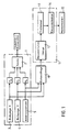
- Figure 1 shows in general at 1 a tunnel effect microscope.
- This microscope comprises a probe 2 constituted by a needle, the end of which forms a monoatomic point and the movements of which are ensured by means of piezoelectric crystals P X , P Y and P Z.
- the crystals P X and P Y are controlled respectively by a unit 3 for scanning in X, and a unit 4 for scanning in Y.
- the piezoelectric crystal P Z is controlled by a servo-control 5 tending to keep constant the current which circulates between the probe 2 and either the substrate or the polynucleotide 16 (FIG. 2) when a potential difference is applied between this probe and the substrate or the polynucleotide.
- the X, Y and Z coordinates of the end of the probe 2 are sampled in a sampler 10, the output of which is applied to the input of a processing unit 11.
- the processing unit 11 essentially consists of a module 12 for reconstituting the image, a module for processing images 13, and a recognition module 14.
- the module 12 makes it possible, from the sampled data, to reconstruct a plan image of the surface of the substrate and of the nucleic acid fragment which is deposited there.
- the processing unit 13 aims to eliminate from the image thus obtained, the image of the atoms of the substrate and of the atoms of the phosphoryl and ribose or deoxyribose groups so that only the purine or pyrimidine nuclei remain on the image as well as the atoms of the marker optionally used to distinguish the two bases of the purine type from one another as well as the two bases of the pyrimidine type.
- This can be achieved using an image processing program as there are in many technical fields.
- the recognition module 14 has the function of successively recognizing each base.
- the result thus obtained is stored in a storage unit 15 which therefore contains, at the end of the operation, the base sequence of the polynucleotide sequenced by the method according to the invention.
- the substrate 6 is constituted by a graphite crystal obtained for example by epitaxy, so that its surface 17 has no defect.
- a groove 18 is formed on the surface 17 of the substrate 6 by techniques known in microelectronics.
- marks 19 are engraved at regular intervals along the groove also by known means.
- the substrate also comprises a well (not shown) at each end of the groove 18 in order to immerse two electrodes making it possible to create an electric field in a solution bathing these wells and the groove.
- the groove may for example have a width of approximately 2 microns for a depth of approximately 100 nanometers.
- the bottom of the groove is doped in the form of a strip perpendicular to the groove and a width of about 100 nonometers, either of an electrostatic charge, or of functional groups having a high reactivity with the grafted functional groups. at the end of the polynucleotide.
- the electrostatic charge can be obtained either by removing electrons produced directly with a tunneling microscope, or by depositing a molecular layer of a molecule or depositing a charged macromolecule or a cluster of charged molecules.
- a monomolecular layer of general shape CH3 (CH2) n-2 COOH with n between 16 and 22 will be deposited, this monomolecular layer receiving functional groups inserted at the surface (Langmuir-Blotgett technique).
- a micro-drop 21 is deposited in the groove 18 upstream of the barrier 20 in the direction of migration, in a solution bathing the groove.
- This microdrop contains the polynucleotide to be sequenced 22 at one end of which has been synthesized a fragment 23 of known sequence, and a functional group 24 positively charged.
- the microdrop moves towards the barrier 20 which, in the present case is positively charged.
- the group 24 is therefore retained upstream of the barrier 20, the polynucleotide 22 continuing to stretch downstream of this barrier.
- the procedure is as described above, in order to separate the two strands, and the solution bathing the groove is evaporated.
- the end of the probe 2 is then brought in the vicinity of the bottom of the groove, and with this probe a primary scan in X is carried out over the width of the groove 18, and a secondary scan in Y while keeping the distance of the constant probe 2 tip at the surface.
- X, Y and Z are sampled as the scanning progresses with a precision which, in the current state of the art can be of the order of 0.1 angstroms, and the data thus obtained are entered into the processing unit.
- an image such as that shown in FIG. 5 is obtained and can be recognized by the shape recognition module 14.
- the method according to the invention therefore makes it possible to analyze extremely quickly a long fragment of nucleic acid.
- the processing of the data collected can be carried out in real time, in which case with the current shape recognition techniques the scanning must be relatively slow, but another possibility consists in carrying out the processing only in deferred time, in which case the analysis can be performed at high speed.
Abstract
Description
La présente invention concerne un procédé et un dispositif de séquençage d'un polynucléotide tel qu'un gène.The present invention relates to a method and a device for sequencing a polynucleotide such as a gene.
On sait que l'information génétique propre à chaque être vivant, son génome, est contenue dans ses chromosomes, lesquels sont constitués de longues chaînes d'acides nucléiques ayant la structure générale suivante :

Tous ces acides sont donc constitués par un enchaînement de nucléotides possédant soit une base purique (adénine ou guanine), soit une une base pyrimidique (cytosine ou l'uracile dans l'ARN et cytosine ou thymine dans l'ADN).All these acids therefore consist of a sequence of nucleotides having either a purine base (adenine or guanine) or a pyrimidine base (cytosine or uracil in RNA and cytosine or thymine in DNA).
Le séquençage d'un fragment d'acide nucléique est l'opération consistant à déterminer la succession des bases des nucléotides dont il est constitué.The sequencing of a nucleic acid fragment is the operation consisting in determining the succession of the bases of the nucleotides of which it is made up.
Ce séquençage est particulièrement utile, notamment pour une meilleure connaissance des maladies d'origine génétique, mais également dans tous les domaines du génie génétique dont les applications sont, on le sait, de plus en plus nombreuses.This sequencing is particularly useful, in particular for better knowledge of diseases of genetic origin, but also in all fields of genetic engineering whose applications are, as we know, more and more numerous.
Tous les procédés de séquençage actuellement connus, même s'ils sont fiables, présentent l'inconvénient de relever des techniques du génie génétique, et par conséquent d'être toujours très lents et fastidieux, ce qui est un handicap considérable lorsque l'on songe que le génome humain par exemple, c'est-à-dire l'ensemble de ses gènes, est constitué d'environ trois milliards de nucléotides.All the sequencing methods currently known, even if they are reliable, have the disadvantage of being subject to genetic engineering techniques, and consequently of being always very slow and tedious, which is a considerable handicap when one thinks that the human genome for example, that is to say all of its genes, is made up of approximately three billion nucleotides.
Les principales méthodes de séquençage utilisées sont des méthodes manuelles dérivées soit de la méthode de SANGER soit de la méthode MAXAM-GILBERT.The main sequencing methods used are manual methods derived either from the SANGER method or from the MAXAM-GILBERT method.
Ces procédés sont maintenant bien au point. Mais si des kits de séquençage fondés sur ces principes existent dans le commerce, ils sont néanmoins lourds à mettre en oeuvre, lents à réaliser du fait de l'étape d'électrophorèse. Ils présentent en outre l'inconvénient de nécessiter la multiplication du fragment que l'on souhaite séquencer afin d'en obtenir une quantité suffisante pour l'ensemble des opérations à réaliser.These processes are now well developed. But if sequencing kits based on these principles exist commercially, they are nevertheless cumbersome to implement, slow to produce due to the electrophoresis step. They also have the drawback of requiring the multiplication of the fragment which it is desired to sequence in order to obtain a quantity sufficient for all of the operations to be carried out.
On a donc cherché à automatiser les procédés de séquençage.We therefore sought to automate the sequencing processes.
L'un de ces procédés automatiques utilise des marqueurs fluorescents caractéristiques de chaque base.One of these automatic processes uses fluorescent markers characteristic of each base.
On découpe comme précédemment le brin que l'on souhaite séquencer, et on fait migrer les fragments ainsi obtenus dans un seul gel d'électrophorèse éclairé à l'aide d'un laser à argon. Il suffit par conséquent d'enregister au passage de chaque fragment, la couleur de ce fragment pour en déduire la base à laquelle il correspond.The strand which one wishes to sequence is cut as before, and the fragments thus obtained are migrated in a single electrophoresis gel illuminated with an argon laser. It suffices therefore to record the passage of each fragment, the color of this fragment to deduce the base to which it corresponds.
Toutefois, on doit là encore procéder à un clonage du brin à séquencer, à son découpage en fragments et à un procédé d'électrophorèse.However, once again, the strand to be sequenced is cloned, cut into fragments and an electrophoresis process.
La présente invention vise à pallier ces inconvénients en fournissant un procédé automatique de séquençage d'un polynucléotide qui ne nécessite pas de clonage de ce fragment, qui s'affranchit des procédés d'électrophorèse, et qui soit en outre non destructif, ce qui présente l'avantage qu'il peut être répété plusieurs fois à des fins de vérification.The present invention aims to overcome these drawbacks by providing an automatic method for sequencing a polynucleotide which does not require cloning of this fragment, which does not require electrophoresis methods, and which is also non-destructive, which presents the advantage that it can be repeated several times for verification purposes.
A cet effet, l'invention a tout d'abord pour objet un procédé de séquençage automatique d'un polynucléotide tel qu'un gène, caractérisé par le fait que l'on dispose ledit polynucléotide sur un substrat dont la rugosité de surface est de l'ordre d'un diamètre atomique, que l'on effectue une analyse ponctuelle de la surface du substrat portant ledit polynucléotide avec une résolution inférieure à un diamètre atomique, que l'on reconstitue la forme de chaque base successive dudit polynucléotide à partir des données recueillies au cours de l'analyse, que l'on reconnait ladite forme, et que l'on mémorise l'information ainsi obtenue.To this end, the invention firstly relates to a method for automatic sequencing of a polynucleotide such as a gene, characterized in that said polynucleotide is placed on a substrate whose surface roughness is the order of an atomic diameter, that a point analysis of the surface of the substrate carrying said polynucleotide is carried out with a resolution less than an atomic diameter, that the shape of each successive base of said polynucleotide is reconstructed from the data collected during the analysis, that we recognize this form, and that we memorize the information thus obtained.
Le procédé selon l'invention repose par conséquent sur une détection directe de la forme de chaque base successive du fragment à séquencer, sur la détermination de cette forme, et sur sa reconnaissance automatique.The method according to the invention is therefore based on direct detection of the shape of each successive base of the fragment to be sequenced, on the determination of this shape, and on its automatic recognition.
La distinction entre un noyau purique et un noyau pyrimidique ne présente pas de difficulté puisqu'un noyau purique se présente sous la forme du cycle à neuf atomes ci-dessous:


Les bases thymine se distinguent des bases cytosine par la présence, dans les premières, du radical méthyle placé en C₅. De même, les bases guanine se distinguent des bases adénine par la présence, dans les premières, d'un atome d'azote placé en C₂.Thymine bases are distinguished from cytosine bases by the presence, in the first, of the methyl radical placed in C₅. Likewise, the guanine bases are distinguished from the adenine bases by the presence, in the former, of a nitrogen atom placed at C₂.
On pourra également, afin de rendre cette distinction plus aisée, ajouter un marqueur caractéristique sur l'une des deux bases de type purique et l'une des deux bases de type pyrimidique du polynucléotide à séquencer.It will also be possible, in order to make this distinction easier, to add a characteristic marker on one of the two bases of purine type and one of the two bases of pyrimidine type of the polynucleotide to be sequenced.
Dans le cas d'un fragment d'acide désoxyribonucléique on pourra ainsi marquer soit les bases adénine soit les bases thymine d'une part, et soit les bases cytosine soit les bases guanine d'autre part.In the case of a deoxyribonucleic acid fragment, it will thus be possible to label either the adenine bases or the thymine bases on the one hand, and either the cytosine bases or the guanine bases on the other hand.
Le procédé selon l'invention est donc un procédé purement physique éliminant par voie de conséquence les inconvénients des procédés traditionnels.The method according to the invention is therefore a purely physical method thereby eliminating the drawbacks of traditional methods.
Dans un mode de réalisation particulier de l'invention, l'analyse ponctuelle précitée s'effectue en balayant ladite surface avec une résolution inférieure à un diamètre atomique à l'aide d'une sonde, par exemple la sonde d'un microscope à effet tunnel ou à force atomique, apte à détecter une dénivellation par rapport à la surface inférieure à un diamètre atomique, et en reconstituant la forme de chaque base successivement rencontrée au cours du balayage à partir des données de ce balayage.In a particular embodiment of the invention, the aforementioned point analysis is carried out by scanning said surface with a resolution less than an atomic diameter using a probe, for example the probe of an effect microscope tunnel or atomic force, capable of detecting a difference in level with respect to the surface less than an atomic diameter, and by reconstructing the shape of each base successively encountered during the scan from the data of this scan.
Le microscope à effet tunnel est connu. On n'en rappellera donc que brièvement le principe de fonctionnement.The tunnel effect microscope is known. We therefore only briefly recall the operating principle.
Si l'on approche une sonde suffisamment fine à proximité immédiate d'une surface, et si l'on applique une différence de potentiel entre la sonde et la surface, il nait un courant électrique par effet tunnel, ce courant électrique dépendant très fortement de la densité électronique au niveau de la pointe de la sonde, c'est-à-dire de la distance entre cette pointe et la surface.If we approach a sufficiently fine probe in the immediate vicinity of a surface, and if we apply a potential difference between the probe and the surface, there is an electric current by tunnel effect, this electric current depending very strong electronic density at the tip of the probe, that is to say the distance between this tip and the surface.
Le microscope à effet tunnel consiste par conséquent à provoquer un balayage en X et Y de la surface à analyser à l'aide d'une sonde, par exemple à l'aide de dispositifs piézo-électriques, et au cours de ce balayage, à déplacer la sonde verticalement en Z, également à l'aide d'un dispositif piézo-électrique, de manière à obtenir un courant d'intensité constante. La tension d'asservissement pour obtenir cette intensité constante est donc représentative des dénivellations rencontrées au cours du balayage.The tunneling microscope therefore consists in causing an X and Y scan of the surface to be analyzed using a probe, for example using piezoelectric devices, and during this scanning, move the probe vertically in Z, also using a piezoelectric device, so as to obtain a current of constant intensity. The control voltage to obtain this constant intensity is therefore representative of the unevenness encountered during scanning.
Il est bien entendu possible de balayer à l'aide de la sonde l'ensemble du substrat sur lequel est disposé le fragment à séquencer.It is of course possible to scan with the aid of the probe the entire substrate on which the fragment to be sequenced is placed.
Toutefois, afin d'obtenir une analyse plus rapide, il est possible de piloter le balayage en X et en Y, de manière à suivre ce fragment, par exemple en arrêtant le balayage primaire à une certaine distance telle que 100 nm ou 1 micron après le début d'un parcours monotone sur la surface du substrat, qui peut être constitué par un cristal présentant une surface sensiblement sans défaut.However, in order to obtain a faster analysis, it is possible to control the scanning in X and in Y, so as to follow this fragment, for example by stopping the primary scanning at a certain distance such as 100 nm or 1 micron after the start of a monotonous course on the surface of the substrate, which may consist of a crystal having a substantially flawless surface.
Il est encore possible plus simplement, de disposer le fragment dans une rainure formée dans le substrat, et d'effectuer l'analyse uniquement sur la largeur de cette rainure.It is even more possible, to arrange the fragment in a groove formed in the substrate, and to carry out the analysis only over the width of this groove.
On peut notamment disposer le polynucléotide sur le substrat en le faisant migrer par gradient de potentiel dans une solution baignant ce substrat.In particular, the polynucleotide can be placed on the substrate by migrating it by potential gradient in a solution bathing this substrate.
On peut dans ce cas, afin d'étaler le polynucléotide pour faciliter la reconnaissance de forme, accrocher une de ses extrémités en un point du substrat, par exemple en greffant à cette extrémité et sur le substrat des groupes fonctionnels qui, soit portent des charges électrostatiques, soit sont susceptibles de former une liaison covalentes.In this case, in order to spread the polynucleotide to facilitate shape recognition, it is possible to hang one of its ends at a point on the substrate, for example by grafting on this end and onto the substrate functional groups which either carry charges electrostatic either are likely to form a covalent bond.
De préférence dans les deux cas on greffe en fait le groupement fonctionnel à l'extrémité d'une séquence prédéterminée synthétisée elle-même à l'extrémité du polynucléotide à séquencer proprement dit.Preferably in both cases, the functional group is actually grafted at the end of a predetermined sequence. synthesized itself at the end of the polynucleotide to be sequenced itself.
On peut aussi greffer un groupe fonctionnel de charge opposée à l'extrémité libre du polynucléotide.It is also possible to graft a functional group with a charge opposite to the free end of the polynucleotide.
Il est également possible d'effectuer l'élongation et la migration du polynucléotide, en le plaçant dans le courant d'un fluide dont on contrôle la vitesse de déplacement.It is also possible to carry out the elongation and the migration of the polynucleotide, by placing it in the stream of a fluid whose speed of movement is controlled.
Cette vitesse dépend du fluide utilisé. Elle sera par exemple plus importante avec un gaz, comme de l'air, qu'avec un liquide comme par exemple de l'eau.This speed depends on the fluid used. It will for example be greater with a gas, such as air, than with a liquid such as, for example, water.
Il s'agit de communiquer au polynucléotide une énergie cinétique suffisante pour étaler le polynucléotide, tout en minimisant les risques de sa fragmentation.This involves communicating sufficient kinetic energy to the polynucleotide to spread the polynucleotide, while minimizing the risks of its fragmentation.
Le polynucléotide est placé dans une rainure du substrat qui va servir à son analyse.The polynucleotide is placed in a groove in the substrate which will be used for its analysis.
Une des extrémités du polynucléotide est solidarisée, par exemple avec le substrat. La rainure est couverte avec une lamelle, de façon à constituer un canal d'écoulement du fluide.One end of the polynucleotide is secured, for example with the substrate. The groove is covered with a lamella, so as to constitute a fluid flow channel.
Une pièce en forme d'entonnoir, comportant une ouverture permettant le passage du fluide est placée à l'entrée du canal pour permettre l'injection du fluide.A funnel-shaped part, comprising an opening allowing the passage of the fluid, is placed at the inlet of the channel to allow the injection of the fluid.
On procède à l'injection du fluide pendant une période de temps suffisante pour obtenir l'élongation désirée. Une fois l'élongation désirée atteinte, on enlève la pièce en forme d'entonnoir et la lamelle et l'on poursuit les opérations de séquençage.The fluid is injected for a period of time sufficient to obtain the desired elongation. Once the desired elongation is reached, the funnel-shaped part and the coverslip are removed and the sequencing operations are continued.
Il est également possible d'effectuer l'élongation du polynucléotide par centrifugation.It is also possible to elongate the polynucleotide by centrifugation.
On utilisera de préférence pour le séquençage un polynucléotide monobrin afin de rendre plus aisée la reconnaissance de forme.Preferably, a single-strand polynucleotide will be used for sequencing in order to make shape recognition easier.
On peut à cet effet partir d'un polynucléotide double-brin, et après accrochage de son extrémité séparer les deux brins, couper l'un des brins à son extrémité, et poursuivre la migration de ce dernier brin jusqu 'à son éloignement complet du brin restant accroché.We can for this purpose start from a double-stranded polynucleotide, and after hooking its end separate the two strands, cut one of the strands at its end, and continue the migration of this last strand until it is completely removed from the strand still hanging.
La séquence synthétisée entre l'extrémité du polynucléotide et le groupement fonctionnel répond à un certain nombre de fonctions.The sequence synthesized between the end of the polynucleotide and the functional group fulfills a certain number of functions.
Tout d'abord elle permet la reconnaissance du sens de lecture de 5′ vers 3′ ou inversement.First of all it allows the recognition of the direction of reading from 5 ′ to 3 ′ or vice versa.
Par ailleurs elle permet l'identification du premier nucléotide significatif du brin à lire.Furthermore, it allows the identification of the first significant nucleotide of the strand to be read.
Elle permet également une liaison covalente avec le groupe fonctionnel d'accrochage greffé à son extrémité.It also allows a covalent bond with the attachment functional group grafted at its end.
Dans le cas où l'on part d'un polynucléotide double brin, elle permet en outre une coupure spécifique d'un des brins par un enzyme de restriction adapté.In the case where one starts from a double-stranded polynucleotide, it also allows a specific cleavage of one of the strands by a suitable restriction enzyme.
Enfin, elle permet une certaine flexibilité entre le groupe fonctionnel et le polynucléotide.Finally, it allows a certain flexibility between the functional group and the polynucleotide.
Le polynucléotide, la séquence synthétisée et le groupe fonctionnel peuvent être soit déposés directement sur le substrat en un emplacement précis à l'aide d'une micro-pipette, soit accrochés directement par l'intermédiaire des groupes fonctionnels par trempage du substrat dans une solution contenant un grand nombre de polynucléotides identiques.The polynucleotide, the synthesized sequence and the functional group can either be deposited directly on the substrate in a precise location using a micro-pipette, or hung directly via the functional groups by soaking the substrate in a solution containing a large number of identical polynucleotides.
Dans le premier cas la micro-goutte est par exemple disposée dans une rainure de quelques microns de largeur, et possédant une profondeur d'environ 100 nanomètres.In the first case, the microdrop is for example placed in a groove a few microns wide, and having a depth of about 100 nanometers.
La rainure est remplie d'une solution conductrice.The groove is filled with a conductive solution.
A chaque extrémité se trouve une électrode reliée à une source de tension, de telle sorte qu'un champ électrique de polarité convenable s'établisse dans la solution entre les électrodes.At each end is an electrode connected to a voltage source, so that an electric field of suitable polarity is established in the solution between the electrodes.
Sous l'effet du champ électrique, l'ensemble constitué par le polynucléotide (chargé au préalable en conséquence), la séquence d'accrochage et le groupe fonctionnel, migre vers le pôle de signe opposé à la polarisation du polynucléotide.Under the effect of the electric field, the assembly constituted by the polynucleotide (charged beforehand accordingly), the attachment sequence and the functional group, migrates towards the pole of sign opposite to the polarization of the polynucleotide.
Au passage de la barrière constituée des groupes fonctionnels fixés sur le substrat, le groupe fonctionnel mobile fixé au polynucléotide forme une liaison électrostatique ou covalente, et reste par conséquent accroché au groupe fonctionnel fixe. Après cet accrochage, le reste du polynucléotide continue à s'étirer sous l'effet du champ électrique.When the barrier consisting of the functional groups attached to the substrate passes, the mobile functional group attached to the polynucleotide forms an electrostatic or covalent bond, and therefore remains attached to the fixed functional group. After this attachment, the rest of the polynucleotide continues to stretch under the effect of the electric field.
Une fois l'élongation terminée, et dans le cas où l'on est parti d'un polynucléotide double brin, on chauffe de manière à dénaturer les liaisons de ce double brin, les deux monobrins en résultant restant accrochés au groupe fonctionnel.Once the elongation is complete, and in the case where one has started with a double-stranded polynucleotide, it is heated so as to denature the bonds of this double-strand, the two resulting single strands remaining attached to the functional group.
On fait agir ensuite un enzyme de restriction spécifique d'un brin de la chaîne d'attache afin de couper un de ceux-ci. En maintenant la différence de potentiel, on poursuit la migration du polynucléotide monobrin détaché jusqu'à son éloignement complet du monobrin restant attaché au substrat.A specific restriction enzyme of a strand of the attachment chain is then made to act in order to cut one of these. Maintaining the difference in potential, the migration of the detached single-strand polynucleotide is continued until it is completely removed from the single-strand remaining attached to the substrate.
Dans le cas d'un accrochage direct sur la surface, on utilise le même mécanisme d'élongation, mais on évite la difficulté consistant à placer de façon très précise la micro-goutte sur le substrat.In the case of direct attachment to the surface, the same elongation mechanism is used, but the difficulty of placing the microdrop on the substrate very precisely is avoided.
Par contre il est nécessaire de multiplier au préalable le polynucléotide à séquencer afin d'obtenir une probabilité d'accrochage raisonnable des groupes fonctionnels liés au substrat d'une part et au polynucléotide d'autre part.On the other hand, it is necessary to multiply beforehand the polynucleotide to be sequenced in order to obtain a reasonable probability of attachment of the functional groups linked to the substrate on the one hand and to the polynucleotide on the other hand.
Dans les deux cas on évapore ensuite lentement la solution soit par micro-succion du solvant, soit par chauffage, soit en utilisant les deux procédés.In both cases, the solution is then slowly evaporated either by micro-suction of the solvent, or by heating, or by using the two methods.
La présente invention a également pour objet un dispositif de séquençage automatique d'un polynucléotide tel qu'un gène, caractérisé par le fait qu'il comprend en combinaison un substrat dont la rugosité de surface est de l'ordre d'un diamètre atomique, des moyens d'analyse ponctuelle de ladite surface ayant une résolution inférieure à un diamètre atomique, des moyens pour reconstituer les formes des bases successives dudit polynucléotide à partir des données de l'analyse, des moyens pour reconnaître lesdites formes, et des moyens pour mémoriser l'information ainsi obtenue.The present invention also relates to a device for automatic sequencing of a polynucleotide such as a gene, characterized in that it comprises in combination a substrate whose surface roughness is of the order of an atomic diameter, means for punctual analysis of said surface having a resolution less than an atomic diameter, means for reconstructing the shapes of the successive bases of said polynucleotide from the data of the analysis, means for recognizing said shapes, and means for memorizing the information thus obtained.
Les moyens d'analyse peuvent notamment comprendre un sonde apte à détecter une dénivellation par rapport à ladite surface inférieure à un diamètre atomique, et des moyens de balayage pour amener ladite sonde à balayer ladite surface avec une résolution inférieure à un diamètre atomique, par exemple la sonde et les moyens de balayage d'un microscope à effet tunnel.The analysis means may in particular comprise a probe capable of detecting a difference in level with respect to said surface less than an atomic diameter, and scanning means for causing said probe to scan said surface with a resolution less than an atomic diameter, for example the probe and the scanning means of a tunnel effect microscope.
Bien entendu, des moyens d'analyse plus grossiers, ayant par exemple une résolution de l'ordre de la dizaine de nanomètres, pourront être utilisés en premier lieu pour localiser le polynucléotide sur le substrat.Of course, coarser analysis means, for example having a resolution of the order of ten nanometers, could be used in the first place to locate the polynucleotide on the substrate.
Comme on l'a vu ci-dessus, le substrat est de préférence un substrat cristallin tel que du graphite présentant une surface sensiblement sans défaut, et peut comporter une rainure pour loger le polynucléotide et faciliter le balayage.As seen above, the substrate is preferably a crystalline substrate such as graphite having a substantially flawless surface, and may include a groove to accommodate the polynucleotide and facilitate scanning.
Le substrat peut également comporter des repères gravés à des intervalles prédéterminés le long de la rainure.The substrate may also include marks engraved at predetermined intervals along the groove.
Ces repères permettent d'effectuer successivement plusieurs cycles de balayage, chaque cycle couvrant une longueur donnée de la rainure, et de se recaler d'un cycle à l'autre.These marks make it possible to carry out successively several scanning cycles, each cycle covering a given length of the groove, and to readjust from one cycle to another.
Le substrat peut également comprendre des moyens d'accrochage et d'étalement du polynucléotide à séquencer.The substrate may also include means for hooking and spreading the polynucleotide to be sequenced.
Les moyens de reconstitution et de reconnaissance de forme peuvent dans un mode particulier de l'invention comprendre une unité de traitement agencée pour reconstituer la surface en trois dimensions, et des moyens de reconnaissance de formes tridimensionnelles.The means of reconstitution and recognition of shape can in a particular mode of the invention comprise a processing unit arranged to reconstitute the surface in three dimensions, and means of recognition of three-dimensional shapes.
On connaît des procédés permettant de reconstituer une surface en trois dimensions à partir des coordonnées dans ces trois dimensions d'un ensemble de points de cette surface. On connaît également des moyens de reconnaissance de formes tridimensionnelles, (les sous-unités ribose et base restent dans une configuration spatiale rigide bien déterminée).Methods are known for reconstructing a surface in three dimensions from the coordinates in these three dimensions of a set of points of this surface. There are also known means for recognizing three-dimensional shapes (the ribose and base subunits remain in a well-defined rigid spatial configuration).
Dans un autre mode de réalisation, les moyens de reconstitution et de reconnaissance de forme comprennent une unité de traitement agencée pour reconstituer une image en plan de la surface, des moyens de traitement d'images, et des moyens de reconnaissance d'images.In another embodiment, the shape reconstitution and recognition means comprise a processing unit arranged to reconstitute a plan image of the surface, image processing means, and image recognition means.
Dans ce cas, on forme par conséquent à partir des données de l'analyse de la surface, et du polynucléotide qu'elle supporte, une image en plan de cette surface (qui peut bien entendu n'être constituée que d'une succession de pixels dans la mémoire d'un ordinateur), on traite cette image de manière à isoler les atomes des bases successives du polynucléotide en éliminant les atomes du substrat ainsi que ceux des groupements désoxyribose (ou des groupements ribose dans le cas d'ARN) et ceux des groupements phosphoryle. Les images des bases successives ayant ainsi été reconstituées, on peut les reconnaître automatiquement.In this case, one consequently forms from the data of the analysis of the surface, and of the polynucleotide which it supports, a plan image of this surface (which can of course consist only of a succession of pixels in a computer memory), we process this image so as to isolate the atoms from the successive bases of the polynucleotide by eliminating the atoms from the substrate as well as those of deoxyribose groups (or ribose groups in the case of RNA) and those of phosphoryl groups. The images of the successive bases having thus been reconstructed, they can be recognized automatically.
L'invention a aussi pour objet un procédé d'obtention d'une macromolécule caractérisé par le fait qu'il comporte les étapes consistant à :
- séquencer un polynucléotide par un procédé selon la présente invention,
- identifier un gène de ce polynucléotide correspondant à ladite macromolécule,
- fabriquer le gène identifié, en utilisant les éléments obtenus lors de l'étape de séquençage du polynucléotide.The subject of the invention is also a process for obtaining a macromolecule characterized in that it comprises the steps consisting in:
- sequence a polynucleotide by a method according to the present invention,
- identify a gene of this polynucleotide corresponding to said macromolecule,
- fabricate the identified gene, using the elements obtained during the step of sequencing the polynucleotide.
On décrira maintenant à titre d'exemple non limitatif un mode de réalisation particulier de l'invention en référence aux dessins annexés dans lesquels :
- -La figure 1 est un schéma d'ensemble du dispositif selon l'invention,
- -La figure 2 est une vue schématique en perspective du substrat, du polynucléotide et de la sonde.
- -La figure 3 est une vue de dessus à très grande échelle de la rainure du substrat et du polynucléotide.
- -La figure 4 représente l'enregistrement effectué par le microscope à effet tunnel, et
- -La figure 5 représente l'image obtenue après traitement du signal délivré par le microscope.
- FIG. 1 is an overall diagram of the device according to the invention,
- FIG. 2 is a schematic perspective view of the substrate, the polynucleotide and the probe.
- FIG. 3 is a top view on a very large scale of the groove of the substrate and of the polynucleotide.
- FIG. 4 represents the recording carried out by the tunnel effect microscope, and
- FIG. 5 represents the image obtained after processing the signal delivered by the microscope.
La figure 1 représente d'une manière générale en 1 un microscope à effet tunnel. Ce microscope comprend une sonde 2 constituée par une aiguille dont l'extrémité forme une pointe monoatomique et dont les déplacements sont assurés au moyen de cristaux piézo-électriques PX, PY et PZ.Figure 1 shows in general at 1 a tunnel effect microscope. This microscope comprises a probe 2 constituted by a needle, the end of which forms a monoatomic point and the movements of which are ensured by means of piezoelectric crystals P X , P Y and P Z.
Les cristaux PX et PY sont commandés respectivement par une unité 3 de balayage en X, et une unité 4 de balayage en Y.The crystals P X and P Y are controlled respectively by a unit 3 for scanning in X, and a
Le cristal piézo-électrique PZ est commandé par un asservissement 5 tendant à maintenir constant le courant qui circule entre la sonde 2 et soit le substrat soit le polynucléotide 16 (figure 2) lorsqu'une différence de potentiel est appliquée entre cette sonde et le substrat ou le polynucléotide.The piezoelectric crystal P Z is controlled by a servo-control 5 tending to keep constant the current which circulates between the probe 2 and either the substrate or the polynucleotide 16 (FIG. 2) when a potential difference is applied between this probe and the substrate or the polynucleotide.
Les coordonnées X, Y et Z de l'extrémité de la sonde 2 sont échantillonnées dans un échantillonneur 10 dont la sortie est appliquée en entrée d'une unité de traitement 11.The X, Y and Z coordinates of the end of the probe 2 are sampled in a
L'unité de traitement 11 se compose essentiellement d'un module 12 de reconstitution de l'image, d'un module de traitement d'images 13, et d'un module de reconnaissance 14.The
Le module 12 permet à partir des données échantillonnées de reconstituer une image en plan de la surface du substrat et du fragment d'acide nucléique qui y est déposé.The
L'unité de traitement 13 vise à éliminer de l'image ainsi obtenue, l'image des atomes du substrat et des atomes des groupements phosphoryle et ribose ou désoxyribose de sorte que ne restent sur l'image que les noyaux puriques ou pyrimidiques ainsi que les atomes du marqueur éventuellement utilisé pour distinguer les deux bases de type purique l'une de l'autre ainsi que les deux bases de type pyrimidique. Ceci peut être obtenu à l'aide d'un programme de traitement d'images comme il en existe dans de nombreux domaines techniques.The
Enfin le module 14 de reconnaissance a pour fonction de reconnaitre successivement chaque base.Finally, the recognition module 14 has the function of successively recognizing each base.
Le résultat ainsi obtenu est mémorisé dans une unité de mémorisation 15 qui contient par conséquent à la fin de l'opération, la séquence des bases du polynucléotide séquencé par le procédé selon l'invention.The result thus obtained is stored in a
Dans le cas présent, le substrat 6 est constitué par un cristal de graphite obtenu par exemple par épitaxie, de sorte que sa surface 17 ne présente pas de défaut.In the present case, the substrate 6 is constituted by a graphite crystal obtained for example by epitaxy, so that its
Une rainure 18 est formée à la surface 17 du substrat 6 par les techniques connues en microélectronique.A
En outre des repères 19 sont gravés à intervalles réguliers le long de la rainure également par des moyens connus.In addition, marks 19 are engraved at regular intervals along the groove also by known means.
Le substrat comporte par ailleurs un puits (non représenté) à chaque extrémité de la rainure 18 afin d'immerger deux électrodes permettant de créer un champ électrique dans une solution baignant ces puits et la rainure.The substrate also comprises a well (not shown) at each end of the
La rainure peut par exemple présenter une largeur d'environ 2 microns pour une profondeur d'environ 100 nanomètres.The groove may for example have a width of approximately 2 microns for a depth of approximately 100 nanometers.
Enfin le fond de la rainure est dopé sous la forme d'une bande perpendiculaire à la rainure et d'une largeur d'environ 100 nonomètres, soit d'une charge électrostatique, soit de groupes fonctionnels présentant une forte réactivité avec les groupes fonctionnels greffés en extrémité du polynucléotide.Finally, the bottom of the groove is doped in the form of a strip perpendicular to the groove and a width of about 100 nonometers, either of an electrostatic charge, or of functional groups having a high reactivity with the grafted functional groups. at the end of the polynucleotide.
L'obtention de la charge électrostatique peut être obtenue soit par arrachement d'électrons réalisés directement avec un microscope à effet tunnel, soit par le dépôt d'une couche moléculaire d'une molécule ou le dépôt d'une macromolécule chargée ou d'un amas de molécules chargées.The electrostatic charge can be obtained either by removing electrons produced directly with a tunneling microscope, or by depositing a molecular layer of a molecule or depositing a charged macromolecule or a cluster of charged molecules.
On déposera par exemple une couche monomoléculaire de forme générale CH₃(CH₂)n-2COOH avec n compris entre 16 et 22, cette couche monomoléculaire recevant des groupes fonctionnels insérés en surface (technique de Langmuir - Blotgett).For example, a monomolecular layer of general shape CH₃ (CH₂) n-2 COOH with n between 16 and 22 will be deposited, this monomolecular layer receiving functional groups inserted at the surface (Langmuir-Blotgett technique).
Une micro-goutte 21 est déposée dans la rainure 18 en amont de la barrière 20 dans le sens de migration, dans une solution baignant la rainure.A micro-drop 21 is deposited in the
Cette micro-goutte contient le polynucléotide à séquencer 22 à une extrémité duquel a été synthétisé un fragment 23 de séquence connue, et un groupement fonctionnel 24 chargé positivement.This microdrop contains the polynucleotide to be sequenced 22 at one end of which has been synthesized a
Du fait du champ électrique règnant dans la solution qui baigne la rainure, la micro-goutte se déplace vers la barrière 20 qui, dans le cas présent est chargée positivement.Due to the electric field prevailing in the solution which bathes the groove, the microdrop moves towards the
Le groupement 24 est par conséquent retenu en amont de la barrière 20, le polynucléotide 22 continuant à s'étirer en aval de cette barrière.The
Dans le cas où le polynucléotide est un double brin, on procède comme décrit précédemment, afin de séparer les deux brins, et on évapore la solution baignant la rainure.In the case where the polynucleotide is a double strand, the procedure is as described above, in order to separate the two strands, and the solution bathing the groove is evaporated.
On amène alors l'extrémité de la sonde 2 au voisinage du fond de la rainure, et on opère avec cette sonde un balayage primaire en X sur la largeur de la rainure 18, et un balayage secondaire en Y en maintenant constante la distance de la pointe de la sonde 2 à la surface.The end of the probe 2 is then brought in the vicinity of the bottom of the groove, and with this probe a primary scan in X is carried out over the width of the
On obtient alors un enregistrement du type de celui représenté à la figure 4.We then obtain a record of the type shown in FIG. 4.
On échantillonne au fur et à mesure du balayage les valeurs de X, Y et Z avec une précision qui, dans l'état actuel de la technique peut être de l'ordre de 0,1 angström, et on entre les données ainsi obtenues dans l'unité de traitement.The values of X, Y and Z are sampled as the scanning progresses with a precision which, in the current state of the art can be of the order of 0.1 angstroms, and the data thus obtained are entered into the processing unit.
Après traitement on obtient une image telle que celle représentée à la figure 5 et peut être reconnue par le module 14 de reconnaissance de forme.After processing, an image such as that shown in FIG. 5 is obtained and can be recognized by the shape recognition module 14.
Le procédé selon l'invention permet par conséquent d'analyser extrèmement rapidement un long fragment d'acide nucléique. Le traitement des données recueillies peut s'effectuer en temps réel, auquel cas avec les techniques actuelles de reconnaissance de formes le balayage doit être relativement lent, mais une autre possibilité consiste à ne réaliser le traitement qu'en temps différé, auquel cas l'analyse peut être effectuée à grande vitesse.The method according to the invention therefore makes it possible to analyze extremely quickly a long fragment of nucleic acid. The processing of the data collected can be carried out in real time, in which case with the current shape recognition techniques the scanning must be relatively slow, but another possibility consists in carrying out the processing only in deferred time, in which case the analysis can be performed at high speed.
Enfin on notera que ce procédé est non destructif et que l'on peut par conséquent réaliser autant de contrôles qu'on le souhaite sur le même échantillon.Finally, it will be noted that this process is non-destructive and that it is therefore possible to carry out as many checks as desired on the same sample.
Diverses variantes et modifications peuvent bien entendu être apportées à la description qui précède sans sortir pour autant du cadre ni de l'esprit de l'invention.Various variants and modifications can of course be made to the above description without departing from the scope or the spirit of the invention.
Claims (21)
- séquencer un polynucléotide par un procédé selon l'une quelconque des revendications 1 à 12,
- identifier un gène de ce polynucléotide correspondant à la dite macromolécule,
- fabriquer le gène identifié, en utilisant les éléments obtenus lors de l'étape de séquençage du polynucléotide.21. Method for obtaining a macromolecule characterized in that it comprises the steps consisting in:
- sequence a polynucleotide by a method according to any one of claims 1 to 12,
- identify a gene of this polynucleotide corresponding to said macromolecule,
- fabricate the identified gene, using the elements obtained during the step of sequencing the polynucleotide.
Applications Claiming Priority (2)
| Application Number | Priority Date | Filing Date | Title |
|---|---|---|---|
| FR8908284 | 1989-06-21 | ||
| FR8908284A FR2648913B1 (en) | 1989-06-21 | 1989-06-21 | METHOD AND DEVICE FOR SEQUENCING A POLYNUCLEOTIDE |
Publications (3)
| Publication Number | Publication Date |
|---|---|
| EP0404682A2 true EP0404682A2 (en) | 1990-12-27 |
| EP0404682A3 EP0404682A3 (en) | 1991-07-24 |
| EP0404682B1 EP0404682B1 (en) | 1995-04-12 |
Family
ID=9382990
Family Applications (1)
| Application Number | Title | Priority Date | Filing Date |
|---|---|---|---|
| EP90401763A Expired - Lifetime EP0404682B1 (en) | 1989-06-21 | 1990-06-21 | Method and apparatus for the sequencing of a polynucleotide |
Country Status (5)
| Country | Link |
|---|---|
| EP (1) | EP0404682B1 (en) |
| JP (1) | JPH03128459A (en) |
| AT (1) | ATE121194T1 (en) |
| DE (1) | DE69018523T2 (en) |
| FR (1) | FR2648913B1 (en) |
Cited By (6)
| Publication number | Priority date | Publication date | Assignee | Title |
|---|---|---|---|---|
| EP0511662A1 (en) * | 1991-04-30 | 1992-11-04 | Matsushita Electric Industrial Co., Ltd. | Scanning probe microscope, molecular processing method using the scanning probe microscope and DNA base arrangement detecting method |
| WO2004036591A1 (en) * | 2002-10-17 | 2004-04-29 | Intel Corporation | Model-based fusion of scanning probe microscopic images for detection and identification of molecular structures |
| US6931326B1 (en) | 2000-06-26 | 2005-08-16 | Genaissance Pharmaceuticals, Inc. | Methods for obtaining and using haplotype data |
| US7058517B1 (en) | 1999-06-25 | 2006-06-06 | Genaissance Pharmaceuticals, Inc. | Methods for obtaining and using haplotype data |
| US7606403B2 (en) | 2002-10-17 | 2009-10-20 | Intel Corporation | Model-based fusion of scanning probe microscopic images for detection and identification of molecular structures |
| US7736818B2 (en) | 2004-12-27 | 2010-06-15 | Inphase Technologies, Inc. | Holographic recording medium and method of making it |
Citations (1)
| Publication number | Priority date | Publication date | Assignee | Title |
|---|---|---|---|---|
| EP0397416A1 (en) * | 1989-05-08 | 1990-11-14 | AMERSHAM INTERNATIONAL plc | Imaging apparatus and method |
-
1989
- 1989-06-21 FR FR8908284A patent/FR2648913B1/en not_active Expired - Fee Related
-
1990
- 1990-06-21 JP JP2163980A patent/JPH03128459A/en active Pending
- 1990-06-21 EP EP90401763A patent/EP0404682B1/en not_active Expired - Lifetime
- 1990-06-21 DE DE69018523T patent/DE69018523T2/en not_active Expired - Fee Related
- 1990-06-21 AT AT90401763T patent/ATE121194T1/en not_active IP Right Cessation
Patent Citations (1)
| Publication number | Priority date | Publication date | Assignee | Title |
|---|---|---|---|---|
| EP0397416A1 (en) * | 1989-05-08 | 1990-11-14 | AMERSHAM INTERNATIONAL plc | Imaging apparatus and method |
Non-Patent Citations (5)
| Title |
|---|
| JOURNAL OF APPLIED PHYSICS, vol. 61, no. 2, 15 janvier 1987, pages R1-R23, American Institute of Physics, New York, NY, US; P.K. HANSMA et al.: "Scanning tunneling microscopy" * |
| JOURNAL OF VACUUM SCIENCE & TECHNOLOGY/SECTION A, vol. 6, no. 3, part II, second series, mai/juin 1988, pages 2089-2092, American Vacuum Society, Woodbury, NY, US; O. MARTI et al.: "Atomic force microscopy and scanning tunneling microscopy with a combination atomic force microscope/scanning tunneling microscope" * |
| PROCEEDINGS OF THE ANNUAL INTERNATIONAL CONFERENCE OF THE IEEE ENGINEERING IN MEDICINE & BIOLOGY SOCIETY,New Orleans, LA (US), 04-07 novembre 1988, part 2/4, "Modeling and Biological Interfaces", vol. 10, IEEE, New York, NY (US); S. HAMEROFF et al., pp. 1009-1011# * |
| PROCEEDINGS OF THE ANNUAL INTERNATIONAL CONFERENCE OF THE IEEE ENGINEERING IN MEDICINE AND BIOLOGY SOCIETY, New Orleans, Louisiana, 4-7 novembre 1988, part 2/4: Modeling and biological interfaces", vol. 10, pages 1009-1011, IEEE, New York, US; S. HAMEROFF et al.: "Scanning tunneling microscopy (STM) applications to molecular electronics" * |
| SOVIET TECHNICAL PHYSICS LETTERS, vol. 13, no. 8, août 1987, pages 391-393, American Institute of Physics, New York, US; S.I. VASIL'EV et al.: "Scanning tunneling microscope for studying structurally nonuniform surfaces" * |
Cited By (9)
| Publication number | Priority date | Publication date | Assignee | Title |
|---|---|---|---|---|
| EP0511662A1 (en) * | 1991-04-30 | 1992-11-04 | Matsushita Electric Industrial Co., Ltd. | Scanning probe microscope, molecular processing method using the scanning probe microscope and DNA base arrangement detecting method |
| US5363697A (en) * | 1991-04-30 | 1994-11-15 | Matsushita Electric Industrial Co., Ltd. | Scanning probe microscope, molecular processing method using the scanning probe microscope and DNA base arrangement detecting method |
| US5730940A (en) * | 1991-04-30 | 1998-03-24 | Matsushita Electric Industrial Co., Ltd. | Scanning probe microscope and molecular processing method using the scanning probe microscope |
| US7058517B1 (en) | 1999-06-25 | 2006-06-06 | Genaissance Pharmaceuticals, Inc. | Methods for obtaining and using haplotype data |
| US6931326B1 (en) | 2000-06-26 | 2005-08-16 | Genaissance Pharmaceuticals, Inc. | Methods for obtaining and using haplotype data |
| WO2004036591A1 (en) * | 2002-10-17 | 2004-04-29 | Intel Corporation | Model-based fusion of scanning probe microscopic images for detection and identification of molecular structures |
| US7606403B2 (en) | 2002-10-17 | 2009-10-20 | Intel Corporation | Model-based fusion of scanning probe microscopic images for detection and identification of molecular structures |
| US8934683B2 (en) | 2002-10-17 | 2015-01-13 | Intel Corporation | Model-based fusion of scanning probe microscopic images for detection and identification of molecular structures |
| US7736818B2 (en) | 2004-12-27 | 2010-06-15 | Inphase Technologies, Inc. | Holographic recording medium and method of making it |
Also Published As
| Publication number | Publication date |
|---|---|
| FR2648913A1 (en) | 1990-12-28 |
| JPH03128459A (en) | 1991-05-31 |
| DE69018523T2 (en) | 1996-01-11 |
| DE69018523D1 (en) | 1995-05-18 |
| FR2648913B1 (en) | 1991-10-04 |
| ATE121194T1 (en) | 1995-04-15 |
| EP0404682B1 (en) | 1995-04-12 |
| EP0404682A3 (en) | 1991-07-24 |
Similar Documents
| Publication | Publication Date | Title |
|---|---|---|
| US10473639B1 (en) | Control of enzyme translocation in nanopore sequencing | |
| US5612181A (en) | Method for arranging a polynucleotide on a substrate for point-by-point analysis of the bases thereof | |
| EP1851331B1 (en) | Method and device for separating molecular targets in a complex mixture | |
| US9400259B2 (en) | Method of making a microbead array with attached biomolecules | |
| JP5030249B2 (en) | DNA sequencing method | |
| JP3685119B2 (en) | Biomolecule recovery method | |
| EP0744028A1 (en) | Highly specific surfaces for biological reactions, method of preparation and utilization | |
| US20020008871A1 (en) | Method and device for detecting optical properties, especially luminescence reactions and refraction behavior of molecules which are directly or indirectly bound on a support | |
| JP2001507441A (en) | Microelectrophoresis chip for moving and separating nucleic acids and other charged molecules | |
| CA2160031A1 (en) | Rapid process for identifying a dna sequence and use thereof for sequencing and diagnostic | |
| US20110100820A1 (en) | Triple function electrodes | |
| EP1574837A1 (en) | Method and apparatus for sequencing polymers through tunneling conductance variation detection | |
| US20020086318A1 (en) | Direct DNA sequencing with a transcription protein and a nanometer scale electrometer | |
| EP0404682B1 (en) | Method and apparatus for the sequencing of a polynucleotide | |
| WO1996024689A9 (en) | Method and apparatus for determining the sequence of polynucleotides | |
| FR2737574A1 (en) | APPARATUS FOR PARALLEL MACROMOLECULE ALIGNMENT AND USE | |
| WO2021226291A1 (en) | Single-biomolecule-bridged sequencing biosensors and storage devices and methods for same | |
| US9914966B1 (en) | Apparatus and methods for analysis of biomolecules using high frequency alternating current excitation | |
| FR2799988A1 (en) | Enhancing electrophoretic separation of similar chemical or biological compounds, by applying alternating field tuned for selective promotion of molecular mobility | |
| EP1007734A1 (en) | Method for characterising nucleic acid duplex | |
| US8494782B1 (en) | Rapid polymer sequencer | |
| EP0882233B1 (en) | Method and device for treatment by complexing of a liquid medium | |
| EP0478450B1 (en) | Method of rapid sequencing of linear and ordered biological sequences | |
| FR2882564A1 (en) | Polypeptide/nucleic acid and polypeptide targets analysis in complex mixture, comprises setting targets to analyze probes, eliminating probe and separating targets and probes, which are bound by specific bond in complex probe target | |
| CA2417534A1 (en) | Method for analysing nucleic acids |
Legal Events
| Date | Code | Title | Description |
|---|---|---|---|
| PUAI | Public reference made under article 153(3) epc to a published international application that has entered the european phase |
Free format text: ORIGINAL CODE: 0009012 |
|
| AK | Designated contracting states |
Kind code of ref document: A2 Designated state(s): AT BE CH DE DK ES FR GB GR IT LI LU NL SE |
|
| PUAL | Search report despatched |
Free format text: ORIGINAL CODE: 0009013 |
|
| AK | Designated contracting states |
Kind code of ref document: A3 Designated state(s): AT BE CH DE DK ES FR GB GR IT LI LU NL SE |
|
| 17P | Request for examination filed |
Effective date: 19911018 |
|
| 17Q | First examination report despatched |
Effective date: 19931018 |
|
| GRAA | (expected) grant |
Free format text: ORIGINAL CODE: 0009210 |
|
| AK | Designated contracting states |
Kind code of ref document: B1 Designated state(s): AT BE CH DE DK ES FR GB GR IT LI LU NL SE |
|
| PG25 | Lapsed in a contracting state [announced via postgrant information from national office to epo] |
Ref country code: IT Free format text: LAPSE BECAUSE OF FAILURE TO SUBMIT A TRANSLATION OF THE DESCRIPTION OR TO PAY THE FEE WITHIN THE PRE;WARNING: LAPSES OF ITALIAN PATENTS WITH EFFECTIVE DATE BEFORE 2007 MAY HAVE OCCURRED AT ANY TIME BEFORE 2007. THE CORRECT EFFECTIVE DATE MAY BE DIFFERENT FROM THE ONE RECORDED.SCRIBED TIME-LIMIT Effective date: 19950412 Ref country code: ES Free format text: THE PATENT HAS BEEN ANNULLED BY A DECISION OF A NATIONAL AUTHORITY Effective date: 19950412 Ref country code: DK Effective date: 19950412 Ref country code: GR Free format text: LAPSE BECAUSE OF FAILURE TO SUBMIT A TRANSLATION OF THE DESCRIPTION OR TO PAY THE FEE WITHIN THE PRESCRIBED TIME-LIMIT Effective date: 19950412 Ref country code: AT Effective date: 19950412 |
|
| REF | Corresponds to: |
Ref document number: 121194 Country of ref document: AT Date of ref document: 19950415 Kind code of ref document: T |
|
| REF | Corresponds to: |
Ref document number: 69018523 Country of ref document: DE Date of ref document: 19950518 |
|
| PG25 | Lapsed in a contracting state [announced via postgrant information from national office to epo] |
Ref country code: LU Free format text: LAPSE BECAUSE OF NON-PAYMENT OF DUE FEES Effective date: 19950630 |
|
| GBT | Gb: translation of ep patent filed (gb section 77(6)(a)/1977) |
Effective date: 19950606 |
|
| PG25 | Lapsed in a contracting state [announced via postgrant information from national office to epo] |
Ref country code: SE Effective date: 19950712 |
|
| PLBE | No opposition filed within time limit |
Free format text: ORIGINAL CODE: 0009261 |
|
| STAA | Information on the status of an ep patent application or granted ep patent |
Free format text: STATUS: NO OPPOSITION FILED WITHIN TIME LIMIT |
|
| 26N | No opposition filed | ||
| PGFP | Annual fee paid to national office [announced via postgrant information from national office to epo] |
Ref country code: GB Payment date: 19960613 Year of fee payment: 7 |
|
| PGFP | Annual fee paid to national office [announced via postgrant information from national office to epo] |
Ref country code: FR Payment date: 19960616 Year of fee payment: 7 |
|
| PGFP | Annual fee paid to national office [announced via postgrant information from national office to epo] |
Ref country code: CH Payment date: 19960625 Year of fee payment: 7 |
|
| PGFP | Annual fee paid to national office [announced via postgrant information from national office to epo] |
Ref country code: NL Payment date: 19960630 Year of fee payment: 7 |
|
| PGFP | Annual fee paid to national office [announced via postgrant information from national office to epo] |
Ref country code: BE Payment date: 19960711 Year of fee payment: 7 |
|
| PGFP | Annual fee paid to national office [announced via postgrant information from national office to epo] |
Ref country code: DE Payment date: 19960726 Year of fee payment: 7 |
|
| PG25 | Lapsed in a contracting state [announced via postgrant information from national office to epo] |
Ref country code: GB Free format text: LAPSE BECAUSE OF NON-PAYMENT OF DUE FEES Effective date: 19970621 |
|
| PG25 | Lapsed in a contracting state [announced via postgrant information from national office to epo] |
Ref country code: CH Free format text: LAPSE BECAUSE OF NON-PAYMENT OF DUE FEES Effective date: 19970630 Ref country code: BE Effective date: 19970630 Ref country code: LI Free format text: LAPSE BECAUSE OF NON-PAYMENT OF DUE FEES Effective date: 19970630 |
|
| BERE | Be: lapsed |
Owner name: FOURMENTIN-GUILBERT JEAN ERNEST RAYMOND Effective date: 19970630 |
|
| PG25 | Lapsed in a contracting state [announced via postgrant information from national office to epo] |
Ref country code: NL Effective date: 19980101 |
|
| GBPC | Gb: european patent ceased through non-payment of renewal fee |
Effective date: 19970621 |
|
| REG | Reference to a national code |
Ref country code: CH Ref legal event code: PL |
|
| PG25 | Lapsed in a contracting state [announced via postgrant information from national office to epo] |
Ref country code: FR Free format text: LAPSE BECAUSE OF NON-PAYMENT OF DUE FEES Effective date: 19980227 |
|
| NLV4 | Nl: lapsed or anulled due to non-payment of the annual fee |
Effective date: 19980101 |
|
| PG25 | Lapsed in a contracting state [announced via postgrant information from national office to epo] |
Ref country code: DE Free format text: LAPSE BECAUSE OF NON-PAYMENT OF DUE FEES Effective date: 19980303 |
|
| REG | Reference to a national code |
Ref country code: FR Ref legal event code: ST |
|
| REG | Reference to a national code |
Ref country code: FR Ref legal event code: ST |