EP0515194A2 - Assay methods utilizing induced luminescence - Google Patents
Assay methods utilizing induced luminescence Download PDFInfo
- Publication number
- EP0515194A2 EP0515194A2 EP92304630A EP92304630A EP0515194A2 EP 0515194 A2 EP0515194 A2 EP 0515194A2 EP 92304630 A EP92304630 A EP 92304630A EP 92304630 A EP92304630 A EP 92304630A EP 0515194 A2 EP0515194 A2 EP 0515194A2
- Authority
- EP
- European Patent Office
- Prior art keywords
- analyte
- medium
- photosensitizer
- chemiluminescent compound
- sbp
- Prior art date
- Legal status (The legal status is an assumption and is not a legal conclusion. Google has not performed a legal analysis and makes no representation as to the accuracy of the status listed.)
- Granted
Links
- 0 [Al]C(*CCO1)=C1[Al] Chemical compound [Al]C(*CCO1)=C1[Al] 0.000 description 2
- FCWUEGSBHNYMPZ-UHFFFAOYSA-N C1OC(C2C=C(C=C(C=CC=C3)C3=C3)C3=CC2)=C(C2C=C(C=C(C=CC=C3)C3=C3)C3=CC2)SC1 Chemical compound C1OC(C2C=C(C=C(C=CC=C3)C3=C3)C3=CC2)=C(C2C=C(C=C(C=CC=C3)C3=C3)C3=CC2)SC1 FCWUEGSBHNYMPZ-UHFFFAOYSA-N 0.000 description 1
- UXBZYQXALUQLGT-UHFFFAOYSA-N C1OC=C(c2c(ccc3c4c(cc5)ccc3)c4c5cc2)OC1 Chemical compound C1OC=C(c2c(ccc3c4c(cc5)ccc3)c4c5cc2)OC1 UXBZYQXALUQLGT-UHFFFAOYSA-N 0.000 description 1
Images


Classifications
-
- G—PHYSICS
- G01—MEASURING; TESTING
- G01N—INVESTIGATING OR ANALYSING MATERIALS BY DETERMINING THEIR CHEMICAL OR PHYSICAL PROPERTIES
- G01N33/00—Investigating or analysing materials by specific methods not covered by groups G01N1/00 - G01N31/00
- G01N33/48—Biological material, e.g. blood, urine; Haemocytometers
- G01N33/50—Chemical analysis of biological material, e.g. blood, urine; Testing involving biospecific ligand binding methods; Immunological testing
- G01N33/53—Immunoassay; Biospecific binding assay; Materials therefor
- G01N33/543—Immunoassay; Biospecific binding assay; Materials therefor with an insoluble carrier for immobilising immunochemicals
- G01N33/54313—Immunoassay; Biospecific binding assay; Materials therefor with an insoluble carrier for immobilising immunochemicals the carrier being characterised by its particulate form
- G01N33/54346—Nanoparticles
-
- C—CHEMISTRY; METALLURGY
- C07—ORGANIC CHEMISTRY
- C07D—HETEROCYCLIC COMPOUNDS
- C07D265/00—Heterocyclic compounds containing six-membered rings having one nitrogen atom and one oxygen atom as the only ring hetero atoms
- C07D265/28—1,4-Oxazines; Hydrogenated 1,4-oxazines
- C07D265/30—1,4-Oxazines; Hydrogenated 1,4-oxazines not condensed with other rings
-
- C—CHEMISTRY; METALLURGY
- C07—ORGANIC CHEMISTRY
- C07D—HETEROCYCLIC COMPOUNDS
- C07D279/00—Heterocyclic compounds containing six-membered rings having one nitrogen atom and one sulfur atom as the only ring hetero atoms
- C07D279/10—1,4-Thiazines; Hydrogenated 1,4-thiazines
- C07D279/12—1,4-Thiazines; Hydrogenated 1,4-thiazines not condensed with other rings
-
- C—CHEMISTRY; METALLURGY
- C07—ORGANIC CHEMISTRY
- C07D—HETEROCYCLIC COMPOUNDS
- C07D327/00—Heterocyclic compounds containing rings having oxygen and sulfur atoms as the only ring hetero atoms
- C07D327/02—Heterocyclic compounds containing rings having oxygen and sulfur atoms as the only ring hetero atoms one oxygen atom and one sulfur atom
- C07D327/06—Six-membered rings
-
- G—PHYSICS
- G01—MEASURING; TESTING
- G01N—INVESTIGATING OR ANALYSING MATERIALS BY DETERMINING THEIR CHEMICAL OR PHYSICAL PROPERTIES
- G01N33/00—Investigating or analysing materials by specific methods not covered by groups G01N1/00 - G01N31/00
- G01N33/48—Biological material, e.g. blood, urine; Haemocytometers
- G01N33/50—Chemical analysis of biological material, e.g. blood, urine; Testing involving biospecific ligand binding methods; Immunological testing
- G01N33/53—Immunoassay; Biospecific binding assay; Materials therefor
- G01N33/536—Immunoassay; Biospecific binding assay; Materials therefor with immune complex formed in liquid phase
- G01N33/542—Immunoassay; Biospecific binding assay; Materials therefor with immune complex formed in liquid phase with steric inhibition or signal modification, e.g. fluorescent quenching
-
- G—PHYSICS
- G01—MEASURING; TESTING
- G01N—INVESTIGATING OR ANALYSING MATERIALS BY DETERMINING THEIR CHEMICAL OR PHYSICAL PROPERTIES
- G01N33/00—Investigating or analysing materials by specific methods not covered by groups G01N1/00 - G01N31/00
- G01N33/48—Biological material, e.g. blood, urine; Haemocytometers
- G01N33/50—Chemical analysis of biological material, e.g. blood, urine; Testing involving biospecific ligand binding methods; Immunological testing
- G01N33/53—Immunoassay; Biospecific binding assay; Materials therefor
- G01N33/543—Immunoassay; Biospecific binding assay; Materials therefor with an insoluble carrier for immobilising immunochemicals
- G01N33/5436—Immunoassay; Biospecific binding assay; Materials therefor with an insoluble carrier for immobilising immunochemicals with ligand physically entrapped within the solid phase
-
- G—PHYSICS
- G01—MEASURING; TESTING
- G01N—INVESTIGATING OR ANALYSING MATERIALS BY DETERMINING THEIR CHEMICAL OR PHYSICAL PROPERTIES
- G01N33/00—Investigating or analysing materials by specific methods not covered by groups G01N1/00 - G01N31/00
- G01N33/48—Biological material, e.g. blood, urine; Haemocytometers
- G01N33/50—Chemical analysis of biological material, e.g. blood, urine; Testing involving biospecific ligand binding methods; Immunological testing
- G01N33/53—Immunoassay; Biospecific binding assay; Materials therefor
- G01N33/569—Immunoassay; Biospecific binding assay; Materials therefor for microorganisms, e.g. protozoa, bacteria, viruses
- G01N33/56966—Animal cells
- G01N33/56977—HLA or MHC typing
-
- G—PHYSICS
- G01—MEASURING; TESTING
- G01N—INVESTIGATING OR ANALYSING MATERIALS BY DETERMINING THEIR CHEMICAL OR PHYSICAL PROPERTIES
- G01N33/00—Investigating or analysing materials by specific methods not covered by groups G01N1/00 - G01N31/00
- G01N33/48—Biological material, e.g. blood, urine; Haemocytometers
- G01N33/50—Chemical analysis of biological material, e.g. blood, urine; Testing involving biospecific ligand binding methods; Immunological testing
- G01N33/58—Chemical analysis of biological material, e.g. blood, urine; Testing involving biospecific ligand binding methods; Immunological testing involving labelled substances
- G01N33/582—Chemical analysis of biological material, e.g. blood, urine; Testing involving biospecific ligand binding methods; Immunological testing involving labelled substances with fluorescent label
-
- G—PHYSICS
- G01—MEASURING; TESTING
- G01N—INVESTIGATING OR ANALYSING MATERIALS BY DETERMINING THEIR CHEMICAL OR PHYSICAL PROPERTIES
- G01N33/00—Investigating or analysing materials by specific methods not covered by groups G01N1/00 - G01N31/00
- G01N33/48—Biological material, e.g. blood, urine; Haemocytometers
- G01N33/50—Chemical analysis of biological material, e.g. blood, urine; Testing involving biospecific ligand binding methods; Immunological testing
- G01N33/58—Chemical analysis of biological material, e.g. blood, urine; Testing involving biospecific ligand binding methods; Immunological testing involving labelled substances
- G01N33/585—Chemical analysis of biological material, e.g. blood, urine; Testing involving biospecific ligand binding methods; Immunological testing involving labelled substances with a particulate label, e.g. coloured latex
- G01N33/587—Nanoparticles
Definitions
- This invention relates to methods, compositions and kits for determining an analyte in a sample.
- this invention relates to specific binding assays which do not require a separation step.
- a first consideration is the signal response to changes in the concentration of analyte.
- a second consideration is the ease with which the protocol for the assay may be carried out.
- a third consideration is the variation in interference from sample to sample. Ease of preparation and purification of the reagents, availability of equipment, ease of automation and interaction with material of interest are some of the additional considerations in developing a useful assay.
- One broad category of techniques involves the use of a receptor which can specifically bind to a particular spacial and polar organization of a labeled ligand as a function of the presence of an analyte.
- the observed effect of binding by the receptor will depend upon the label.
- the binding of the receptor merely provides for a differentiation in molecular weight between bound and unbound labeled ligand.
- the binding of the receptor will facilitate separation of bound labeled ligand from free labeled ligand or it may affect the nature of the signal obtained from the label so that the signal varies with the amount of receptor bound to labeled ligand.
- the receptor is labeled and the ligand unlabeled.
- both the receptor and ligand are labeled or different receptors are labeled with two different labels, whereupon the labels interact when in close proximity and the amount of ligand present affects the degree to which the labels of the receptor may interact.
- Homogeneous immunoassays have previously been described for small molecules. These assays include SYVA's FRAT® assay, EMIT® assay, enzyme channeling immunoassay, and fluorescence energy transfer immunoassay (FETI); enzyme inhibitor immunoassays (Hoffman LaRoche and Abbott Laboratories): fluorescence polarization immunoassay (Dandlicker), among others. All of these methods have limited sensitivity, and only a few including FETI and enzyme channeling, are suitable for large multiepitopic analytes.
- Luminescent compounds such as fluorescent compounds and chemiluminescent compounds
- find wide application in the assay field because of their ability to emit light.
- luminescers have been utilized as labels in assays such as nucleic acid assays and immunoassays.
- a member of a specific binding pair is conjugated to a luminescer and various protocols are employed.
- the luminescer conjugate can be partitioned between a solid phase and a liquid phase in relation to the amount of analyte in a sample suspected of containing the analyte. By measuring the luminescence of either of the phases, one can relate the level of luminescence observed to a concentration of the analyte in the sample.
- Particles such as liposomes and erythrocyte ghosts, have been utilized as carriers of encapsulated water soluble materials.
- liposomes have been employed to encapsulate biologically active material for a variety of uses, such as drug delivery systems wherein a medicament is entrapped during liposome preparation and then administered to the patient to be treated.
- Particles such as latex beads and liposomes
- an enzyme may be entrapped in the aqueous phase of a liposome labelled with an antibody or antigen.
- the liposomes are caused to release the enzyme in the presence of a sample and complement.
- Antibody- or antigen-labelled liposomes having water soluble fluorescent or non-fluorescent dyes encapsulated within an aqueous phase or lipid soluble dyes dissolved in the lipid bilayer of the lipid vesicle, have also been utilized to assay for analytes capable of entering into an immunochemical reaction with the surface bound antibody or antigen.
- Detergents have been used to release the dyes from the aqueous phase of the liposomes.
- Chemiluminescent labels offer exceptional sensitivity in ligand binding assays, but one or more chemical activation steps are usually needed. Fluorescent labels do not have this deficiency but are less sensitive.
- Chemiluminescent labels have been described for immunoassays and nucleic acid assays where a group, which is covalently bound to a binding partner, on chemical activation emits light.
- a nucleic and assay kit utilizing an acridinium ester is sold by Genprobe (Pace2 system®, San Diego, CA) and MagicLite® immunoassay kits using this type of label are sold by CIBA-GEIGY (Basel, Switzerland).
- Genprobe Pace2 system®, San Diego, CA
- MagicLite® immunoassay kits using this type of label are sold by CIBA-GEIGY (Basel, Switzerland).
- Energy transfer from a labeled nucleic acid probe to a fluorescent acceptor bound to a second probe has been described by Heller, et al ., I and II, infra , for a sandwich nucleic acid assay.
- Maggio I infra , discusses a similar procedure for immunoassays. Transfer of energy from a luminescer covalently bound to a polynucleotide to an intercalated fluorophore was mentioned by Heller, et al. , IV, infra . Transfer from an intercalated dye to a fluorescer on a polynucleotide was described recently by Cardullo, et al ., infra . Further McCapra, infra , has described the use of photosensitizers as labels where the photosensitizer activates oxygen to its singlet state, which in turn reacts with a compound that on heating produces light.
- McCapra discloses specific binding assays that utilize a sensitizer as a label.
- the sensitizers include any moiety which, when stimulated by excitation with radiation of one or more wavelengths or other chemical or physical stimulus (e.g., electron transfer, electrolysis, electroluminescence or energy transfer) will achieve an excited state which (a) upon interaction with molecular oxygen will produce singlet molecular oxygen, or (b) upon interaction with a leuco dye will assume a reduced form that can be returned to its original unexcited state by interaction with molecular oxygen resulting in the production of hydrogen peroxide. Either interaction with the excited sensitizer will, with the addition of reagents, produce a detectible signal.
- chemical or physical stimulus e.g., electron transfer, electrolysis, electroluminescence or energy transfer
- a light-emitting polynucleotide hybridization diagnostic method is described in European Patent Application No. 0,070,687 (Heller, et al . II).
- European Patent Application No. 0,232,967 discusses methods and compositions for performing assays for target polynucleotide strands.
- the methods include contacting a sample with a reagent that includes a first and a second polynucleotide probe.
- the first and second probes are capable of assuming a first position wherein the probes are bound to each other and a second position wherein the probes are bound to a target.
- the probes include label moieties capable of interacting to produce a signal indicative of the probes being in one of the two positions.
- European Patent Application No. 0,315,364 describes an immunochemical assay to determine the presence or concentration of antigen or antibodies in a fluid.
- the assay comprises (a) forming a ternary complex of a first labeled antibody or antigen, a second labeled antibody or antigen, and the antigen or antibody to be determined, and (b) detecting a signal produced in the presence of at least one substrate, by an interaction between the first label and the second label, enhanced by their proximity to each other bound to the antigenic substance.
- U.S. Patent No. 4,226,993 (Buckler, et al .) describes immuno-functionalized phthalhydrazides, which are useful as intermediates in the synthesis of chemiluminescent phthalhydrazide-labeled conjugates.
- the conjugates are useful as reagents in specific binding assays for determining ligands or their specific binding partners in liquid media.
- U.S. Patent No. 4,193,983 (Ullman, et al .III) discloses labeled liposome particle compositions and immunoassays therewith.
- O'Connell, et al., Clin . Chem ., (1985) 31 (9), 1424-1426 discloses a colorimetric immunoassay for digoxin utilizing large, unilamellar phospholipid vesicles having dye entrapped in the aqueous phase of the liposome.
- Patent Nos. 3,850,578 (McConnell); 4,483,921 (Yaverbaum); and 4,483,929 (Szoka) disclose immunoreactive liposome reagents in which antigen or antibody is bound to the surface of lipid vesicles.
- U.S. Patent No. 4,311,712 discloses a process for preparing a freeze dried liposome mixture.
- U.S. Patent No. 4,588,578 discloses a method for the preparation of monophasic lipid vesicles and the use of such vesicles for drug delivery systems.
- U.S. Patent No. 4,576,912 discloses a method of enhancing the fluorescent level of an immunoassay using certain long-chain carriers tagged with a plurality of fluorophores.
- European Patent Application 0 352 713 (Schaap) describes a method and compositions providing enhanced chemiluminescence from 1,2-dioxetanes.
- U.S. Patent No. 4,959,182 discloses a method and compositions providing enhanced chemiluminescence from 1,2-dioxetanes.
- WO 89/06226 (Edwards, et al .) discusses the synthesis of 1,2-dioxetanes and intermediates therefor.
- European Patent Application 0 144 914 (Alberella, et al .) describes a hybridization assay employing labeled pairs of hybrid binding reagents.
- the present invention is directed to methods for determining an analyte.
- One aspect of the invention is a method for determining an analyte where the method comprises treating a medium suspected of containing an analyte to form an intrinsically metastable species.
- the species is capable of diffusing in the medium and of reacting selectively with a substance in the medium capable of reacting with the metastable species brought into close proximity to the species by virtue of the presence of the analyte.
- the method further comprises determining whether the species has reacted with the substance, the reaction thereof indicating the amount of analyte in the medium.
- Another embodiment of the invention is an improvement in an assay for an analyte in a liquid medium.
- the assay comprises the steps of treating a medium suspected of containing the analyte to form a specific binding pair (sbp) complex in relation to the presence of the the analyte and determining whether the complex is formed.
- the improvement comprises combining with the medium (1) a photosensitizer associated with a member of a specific binding pair and (2) a chemiluminescent compound associated with an sbp member wherein the amount of light emitted from the chemiluminescent compound upon activation of the photosensitizer is related to the amount of analyte in the medium.
- Another embodiment of a method in accordance with the present invention comprises treating a medium suspected of containing an analyte under conditions such that the analyte, if present, causes a photosensitizer, and a chemiluminescent compound to come into close proximity.
- a photosensitizer if present, causes a photosensitizer, and a chemiluminescent compound to come into close proximity.
- singlet oxygen produced by the photosensitizer can activate the chemiluminescent compound, which subsequently produces light or luminescence.
- the amount of light produced is related to the amount of analyte in the medium.
- a method of the present invention for determining an analyte comprises as a first step providing a combination comprising a medium suspected of containing an analyte, a photosensitizer associated with a specific binding pair (sbp) member and a suspendible particle comprising a chemiluminescent compound.
- the suspendible particle has an (sbp) member bound thereto.
- the combination is treated to excite the photosensitizer, which is capable in its excited state of activating oxygen to a singlet state.
- the combination is then examined for the amount of luminescence emitted. The amount of such luminescence is related to the amount of analyte in the medium.
- the chemiluminescent compound is associated with an sbp member and the suspendible particle comprises a photosensitizer and has an sbp member bound thereto.
- Another embodiment is a method for determining an analyte wherein a combination is provided.
- the combination comprises a medium suspected of containing an analyte, a photosensitizer associated with a first sbp member and a chemiluminescent compound associated with a second sbp member.
- the photosensitizer is then excited and is capable of activating oxygen to a singlet state, which singlet oxygen activates the chemiluminescent compound when brought in close proximity to the photosensitizer.
- the luminescence emitted from the combination is related to the amount of analyte.
- Another embodiment is a method for determining an analyte.
- the method comprises combining in an aqueous medium a sample suspected of containing an analyte, a first suspendible particle having a chemiluminescent compound incorporated therein and an sbp member bound thereto, and a second suspendible particle having incorporated therein a photosensitizer capable of activating oxygen to its singlet state where the particle has an sbp member bound thereto.
- the medium is then irradiated to produce the singlet state of oxygen and the amount of luminescence emitted from the medium is measured. The amount of such luminescence is related to the amount of analyte in the medium.
- compositions comprising a suspendible particle having incorporated therein a chemiluminescent compound where the particle has an sbp member bound thereto.
- the composition can further comprise a suspendible particle having a photosensitizer incorporated therein.
- kits comprising in packaged combination a composition that includes (1) a suspendible particle having a chemiluminescent compound where the particle has an sbp member bound thereto, and (2) a photosensitizer.
- the kit can further include a composition comprising a second suspendible particle comprising a photosensitizer where the particle has an sbp member bound thereto.
- the kit comprises (1) a chemiluminescent compound associated with a first sbp member and (2) a photosensitizer capable in its excited state of activating oxygen to its singlet state associated with a second sbp member.
- Another embodiment of the present invention comprises providing (1) a medium suspected of containing the analyte, (2) a label reagent comprising a first specific binding pair (sbp) member associated with a photochemically activatable chemiluminescent compound wherein the first sbp member is capable of binding to the analyte or a second sbp member to form a complex related to the presence of the analyte, photochemically activating the chemiluminescent compound, and detecting the amount of luminescence generated by the chemiluminescent compound, the amount thereof being related to the amount of analyte in the medium.
- a label reagent comprising a first specific binding pair (sbp) member associated with a photochemically activatable chemiluminescent compound wherein the first sbp member is capable of binding to the analyte or a second sbp member to form a complex related to the presence of the analyte, photochemically activating the chemilum
- a method of the present invention for determining an analyte comprises as a first step combining in an assay medium (1) a medium suspected of containing an analyte, and (2) a label reagent comprising a member of a specific binding pair (sbp member) bound to a photochemically activatable chemiluminescent compound under conditions wherein an sbp member complex involving the label reagent is formed in relation to the presence of analyte in the medium.
- the assay medium is irradiated with light to activate the photochemically activatable chemiluminescent compound.
- the assay medium is examined for a light emission. The presence or intensity of such signal light emission is related to the amount of analyte in said medium.
- An energy acceptor is included in the medium where the energy from the chemiluminescent compound is capable of activating the energy acceptor.
- Another embodiment is a method for determining an analyte.
- the method comprises combining in an aqueous medium either simultaneously or wholly or partially sequentially (1) a medium suspected of containing an analyte, (2) a label reagent comprising a first member of a specific binding pair (sbp member) bound to a compound capable of chemiluminescence upon reaction with light or singlet oxygen, and (3) an insolubilized reagent comprising an energy acceptor bound to, or capable of becoming bound to, a second sbp member.
- Conditions are chosen wherein an sbp member complex involving the label reagent is formed in relation to the presence of analyte in the medium and energy from the chemiluminescent compound is capable of activating the energy acceptor.
- the compound is activated with light or singlet oxygen.
- the assay medium is examined for luminescence, the presence or intensity thereof being related to the amount of analyte in the medium.
- Another embodiment of the invention is a method for determining an analyte where the method comprises: (a) combining in an assay medium either simultaneously or wholly or partially sequentially (1) a medium suspected of containing an analyte, (2) a label reagent comprising a first member of a specific binding pair (sbp member) bound to a compound capable of chemiluminescence upon reaction with singlet oxygen, (3) a singlet oxygen generator, and (4) a reagent comprising an energy acceptor bound to, or capable of becoming bound to, a second sbp member, under conditions wherein an sbp member complex involving the label reagent is formed in relation to the presence of analyte in the medium and energy from the chemiluminescent compound is capable of activating the energy acceptor, (b) activating the singlet oxygen generator, and (c) examining the assay medium for a signal, the presence or intensity thereof being related to the amount of analyte in the medium.
- Another embodiment of the present invention is a method for determining a polynucleotide analyte, where the method comprising (a) combining in an assay medium, (1) a sample suspected of containing a polynucleotide analyte and (2) a label reagent comprising a photochemically activatable chemiluminescent compound bound to a polynucleotide, at least a portion of which is capable of hybridizing with said polynucleotide analyte, under conditions wherein the label reagent hybridizes with the polynucleotide analyte if present, (b) irradiating the assay medium to activate the photochemically activatable chemiluminescent compound, and (c) measuring the luminescence of the medium.
- the amount of luminescence is related to the amount of analyte in the sample.
- composition comprising a photochemically activatable chemiluminescent compound bound to an sbp member.
- a kit comprising such a composition.
- Fig. 1 is a graphic depiction of the results of an assay for Vitamin B12.
- Fig. 2 is a graphic depiction of the results of an assay for digoxin.
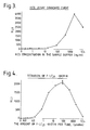
- Fig. 3 is a graphic depiction of the results of an assay for HCG in accordance with the present invention.
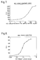
- Fig. 4 is a graphic depiction of the results of a test in accordance with the present invention.
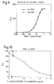
- Fig. 5 is a graphic depiction of the results of an assay for TSH in accordance with the present invention.
- Fig. 6 is a portion of the graphic depiction of Fig. 5.
- Fig. 7 is a graphic depiction of the results of another assay for HCG in accordance with the present invention.
- Fig. 8 is a graphic depiction of the results of a DNA hybrid detection assay.
- Fig. 9 is a graphic depiction of the results of detection of a synthetic target.
- Fig. 10 is a graphic depiction of a total triiodothyronine assay.
- the present invention is directed to methods for determining an analyte.
- One aspect of the invention is a method for determining an analyte where the method comprises treating a medium suspected of containing an analyte to form an intrinsically metastable.species.
- the species is capable of diffusing in the medium and of reacting selectively with a substance in the medium capable of reacting with the metastable species brought into close proximity to the species by virtue of the presence of the analyte.
- the method further comprises determining whether the species has reacted with the substance, the reaction thereof indicating the amount of analyte in the medium.
- the metastable species is an excited state.
- the metastable species has a lifetime of less than one millisecond, usually less that 100 microseconds, more usually less than 10 microseconds.
- the metastable species is diffusive in the medium, i.e., it is produced at one site and migrates to another site from the site of formation where it can transfer energy or react with a molecule at that site.
- the metastable species may be any reactive intermediate such as a radical ion, nitrene, carbene, from the group trans-cyclohexene, ⁇ -lactone, trimethylene methane and the like.
- excited singlet states such as singlet oxygen, triplet states, and dioxetanes including dioxetanones and dioxetane diones.
- Triplet states are generally formed by combining an appropriate sensitizer such as, e.g., pyrene with an energy acceptor such as an anthracene.
- dibromoanthracene can act as an energy acceptor which assumes a triplet state.
- the triplet state can proceed to transfer its energy to another molecule and initiate a detectible photochemical reaction such as the production of light.
- Dioxetanes such as dioxetane diones are formed from reaction of active molecules with singlet oxygen or hydrogen peroxide. For example, appropriate oxalates and hydrogen peroxide form dioxetane diones.
- Enzymes such as horse radish peroxidase can generate radical cations or singlet oxygen that likewise are metastable and can react with another molecule to give a detectible signal.
- the presence of a specific binding pair complex can be determined by causing a metastable species to be produced by one member of the complex whereupon it can interact selectively with another member of the complex without interacting with that member when it is not within the complex.
- a composition comprising a photosensitizer and a ligand, receptor or polynucleotide binds in an assay to a composition comprising a chemiluminescent compound and a ligand, receptor or polynucleotide.
- the chemiluminescent compound can react with singlet oxygen and the product formed decomposes with emission of light.
- the singlet oxygen is generated by the photosensitizer usually by irradiation of the photosensitizer.
- Singlet oxygen produced by the photosensitizer that is not bound to the composition comprising a chemiluminescent compound is unable to reach the chemiluminescent compound before undergoing decay (t 1 ⁇ 2 is about two microseconds in water).
- the composition comprising a photosensitizer that becomes bound to the composition comprising the chemiluminescent compound produces singlet oxygen that reacts with the chemiluminescent compound because such singlet oxygen can survive the short distance now realized between the photosensitizer and the chemiluminescent compound.
- the shortness of the distance results from the presence of an analyte in the sample.
- a portion of the distance traveled by the singlet oxygen is through an organic medium where the singlet oxygen has a much longer lifetime, namely, greater than about one hundred microseconds.
- the analyte must modulate the binding between the composition comprising the photosensitizer and the composition comprising the chemiluminescent compound.
- at least one of the chemiluminescent compound and the photosensitizer is associated with a surface, particularly where the surface comprises suspendible particles.
- McCapra's assay utilizes a reactant which is capable of specifically binding with a complex of the analyte and specific binding material to which the sensitizer is conjugated, to form a sensitizer conjugate-reactant complex.
- a quenching moiety is attached to the reactant.
- the quenching moiety When brought into close proximity to the sensitizer, the quenching moiety reduces or quenches the signal produced as a result of the excitation of bound sensitizer or reduces or quenches the transfer of electrons or energy for the excited sensitizer to an intermediate species (i.e., molecular oxygen or a leucodye).
- an intermediate species i.e., molecular oxygen or a leucodye.
- analyte is related to the luminescence of the decaying dioxetans. McCapra then refers to U.S. Patent Nos. 4,220,450 and 4,277,437, which are described above.
- This quenching assay format described by McCapra involves only the quenching of the excited sensitizer and does not encompass the singlet oxygen activation of a chemiluminescent compound associated with a specific binding member.
- McCapra describes the transfer of energy from a chemiluminescent moiety to excite a sensitizer in a polynucleotide probe assay.
- This description by McCapra is totally distinct from the present invention, which involves the bringing together in close proximity, by virtue of an analyte being present, a photosensitizer and a chemiluminescent compound where the excited photosensitizer produces singlet oxygen, which in turn activates the chemiluminescent compound.
- a group that can be photochemically activated to a luminescent product is used as a label.
- the group is associated with a member of a specific binding pair and this reagent is utilized as a labeled reagent in assays for detection of an analyte.
- Photochemical activation can be achieved by irradiating the group with light or by reaction singlet with the group.
- a sensitizer is used to assist photoactivation where activation is by singlet oxygen.
- the sensitizer absorbs light and the thus formed excited sensitizer activates oxygen and the singlet oxygen reacts with the label to give a metastable luminescent intermediate.
- the group used as a label may include any chemiluminescent compound that undergoes photoactivation with or without sensitization and preferably is a group that is activated by reaction with singlet oxygen.
- the labels of the present invention can be used in both homogeneous and heterogeneous assay protocols to determine an analyte. Desirably, no addition of chemical reagents is required to activate the chemiluminescent compound with or without a sensitizer and energy acceptor. In a homogeneous protocol all the reagents are combined and the medium is irradiated to activate the chemiluminescent compound.
- the components are provided in combination and the light produced as a function of activation of oxygen by the sensitizer will be a function of analyte concentration.
- the methods of the present invention can be carried out without heating the medium to produce light. Consequently, the assay of the present invention can be conducted at a constant temperature.
- Analyte--the compound or composition to be detected can be comprised of a member of a specific binding pair (sbp) and may be a ligand, which is monovalent (monoepitopic) or polyvalent (polyepitopic), usually antigenic or haptenic, and is a single compound or plurality of compounds which share at least one common epitopic or determinant site.
- the analyte can be a part of a cell such as bacteria or a cell bearing a blood group antigen such as A, B, D, etc., or an HLA antigen or a microorganism, e.g., bacterium, fungus, protozoan, or virus.
- the polyvalent ligand analytes will normally be poly(amino acids), i.e., polypeptides and proteins, polysaccharides, nucleic acids, and combinations thereof. Such combinations include components of bacteria, viruses, chromosomes, genes, mitochondria, nuclei, cell membranes and the like.
- the polyepitopic ligand analytes to which the subject invention can be applied will have a molecular weight of at least about 5,000, more usually at least about 10,000.
- the poly(amino acid) category the poly(amino acids) of interest will generally be from about 5,000 to 5,000,000 molecular weight, more usually from about 20,000 to 1,000,000 molecular weight; among the hormones of interest, the molecular weights will usually range from about 5,000 to 60,000 molecular weight.
- proteins may be considered as to the family of proteins having similar structural features, proteins having particular biological functions, proteins related to specific microorganisms, particularly disease causing microorganisms, etc.
- proteins include, for example, immunoglobulins, cytokines, enzymes, hormones, cancer antigens, nutritional markers, tissue specific antigens, etc.
- a number of proteins found in the human plasma are important clinically and include: Prealbumin Albumin ⁇ 1-Lipoprotein ⁇ 1-Antitrypsin ⁇ 1-Glycoprotein Transcortin 4.6S-Postalbumin Tryptophan-poor ⁇ 1-glycoprotein ⁇ 1X-Glycoprotein Thyroxin-binding globulin Inter- ⁇ -trypsin-inhibitor Gc-globulin (Gc 1-1) (Gc 2-1) (Gc 2-2) Haptoglobin (Hp 1-1) (Hp 2-1) (Hp 2-2) Ceruloplasmin Cholinesterase ⁇ 2-Lipoprotein(s) Myoglobin C-Reactive Protein ⁇ 2-Macroglobulin ⁇ 2-HS-glycoprotein Zn- ⁇ 2-glycoprotein ⁇ 2-Neuramino-glycoprotein Erythropoietin ⁇ -lipoprotein Transferrin Hemopexin Fibrinogen Plasminogen ⁇ 2-g
- Important blood clotting factors include:
- Important protein hormones include:
- Parathyroid hormone (parathromone) Thyrocalcitonin Insulin Glucagon Relaxin Erythropoietin Melanotropin (melancyte-stimulating) hormone; intermedin) Somatotropin (growth hormone) Corticotropin (adrenocorticotropic hormone) Thyrotropin Follicle-stimulating hormone Luteinizing hormone (interstitial cell-stimulating hormone) Luteomammotropic hormone (luteotropin, prolactin Gonadotropin (chorionic gonadotropin)
- PSA CEA a-fetoprotein Acid phosphatase CA19.9 CA125
- Oxytocin Vasopressin Releasing factors CRF, LRF, TRF, Somatotropin-RF, GRF, FSH-RF, PIF, MIF
- polymeric materials of interest are mucopolysaccharides and polysaccharides.
- Illustrative microorganisms include:
- the monoepitopic ligand analytes will generally be from about 100 to 2,000 molecular weight, more usually from 125 to 1,000 molecular weight.
- the analytes include drugs, metabolites, pesticides, pollutants, and the like. Included among drugs of interest are the alkaloids.
- alkaloids include morphine alkaloids, which includes morphine, codeine, heroin, dextromethorphan, their derivatives and metabolites; cocaine alkaloids, which include cocaine and benzyl ecgonine, their derivatives and metabolites; ergot alkaloids, which include the diethylamide of lysergic acid; steroid alkaloids; iminazoyl alkaloids; quinazoline alkaloids; isoquinoline alkaloids; quinoline alkaloids, which include quinine and quinidine; diterpene alkaloids, their derivatives and metabolites.
- the next group of drugs includes steroids, which includes the estrogens, androgens, andreocortical steroids, bile acids, cardiotonic glycosides and aglycones, which includes digoxin and digoxigenin, saponins and sapogenins, their derivatives and metabolites. Also included are the steroid mimetic substances, such as diethylstilbestrol.
- lactams having from 5 to 6 annular members, which include the barbituates, e.g. phenobarbital and secobarbital, diphenylhydantonin, primidone, ethosuximide, and their metabolites.
- the next group of drugs is aminoalkylbenzenes, with alkyl of from 2 to 3 carbon atoms, which includes the amphetamines; catecholamines, which includes ephedrine, L-dopa, epinephrine; narceine; papaverine; and metabolites of the above.
- the next group of drugs is benzheterocyclics which include oxazepam, chlorpromazine, tegretol, their derivatives and metabolites, the heterocyclic rings being azepines, diazepines and phenothiazines.
- the next group of drugs is purines, which includes theophylline, caffeine, their metabolites and derivatives.
- the next group of drugs includes those derived from marijuana, which includes cannabinol and tetrahydrocannabinol.
- the next group of drugs is the hormones such as thyroxine, cortisol, triiodothyronine, testosterone, estradiol, estrone, progestrone, polypeptides such as angiotensin, LHRH, and immunosuppresants such as cyclosporin, FK506, mycophenolic acid, and so forth.
- hormones such as thyroxine, cortisol, triiodothyronine, testosterone, estradiol, estrone, progestrone, polypeptides such as angiotensin, LHRH, and immunosuppresants such as cyclosporin, FK506, mycophenolic acid, and so forth.
- the next group of drugs includes the vitamins such as A, B, e.g. B12, C, D, E and K, folic acid, thiamine.
- prostaglandins which differ by the degree and sites of hydroxylation and unsaturation.
- the next group of drugs is the tricyclic antidepressants, which include imipramine, dismethylimipramine, amitriptyline, nortriptyline, protriptyline, trimipramine, chlomipramine, doxepine, and desmethyldoxepin,
- the next group of drugs are the anti-neoplastics, which include methotrexate.
- antibiotics which include penicillin, chloromycetin, actinomycetin, tetracycline, terramycin, the metabolites and derivatives.
- the next group of drugs is the nucleosides and nucleotides, which include ATP, NAD, FMN, adenosine, guanosine, thymidine, and cytidine with their appropriate sugar and phosphate substituents.
- the next group of drugs is miscellaneous individual drugs which include methadone, meprobamate, serotonin, meperidine, lidocaine, procainamide, acetylprocainamide, propranolol, griseofulvin, valproic acid, butyrophenones, antihistamines, chloramphenicol, anticholinergic drugs, such as atropine, their metabolites and derivatives.
- Metabolites related to diseased states include spermine, galactose, phenylpyruvic acid, and porphyrin Type 1.
- the next group of drugs is aminoglycosides, such as gentamicin, kanamicin, tobramycin, and amikacin.
- pesticides of interest are polyhalogenated biphenyls, phosphate esters, thiophosphates, carbamates, polyhalogenated sulfenamides, their metabolites and derivatives.
- the molecular weights will generally range from 10,000 to 2X10⁸ , more usually from 10,000 to 106.
- immunoglobulins IgA, IgG, IgE and IgM
- the molecular weights will generally vary from about 160,000 to about 106.
- Enzymes will normally range from about 10,000 to 1,000,000 in molecular weight.
- Natural receptors vary widely, generally being at least about 25,000 molecular weight and may be 106 or higher molecular weight, including such materials as avidin, DNA, RNA, thyroxine binding globulin, thyroxine binding prealbumin, transcortin, etc.
- analyte further includes polynucleotide analytes such as those polynucleotides defined below. These include m-RNA, r-RNA, t-RNA, DNA, DNA-RNA duplexes, etc.
- polynucleotide analytes such as those polynucleotides defined below. These include m-RNA, r-RNA, t-RNA, DNA, DNA-RNA duplexes, etc.
- receptors that are polynucleotide binding agents, such as, for example, restriction enzymes, activators, repressors, nucleases, polymerases, histones, repair enzymes, chemotherapeutic agents, and the like.
- the analyte may be a molecule found directly in a sample such as a body fluid from a host.
- the sample can be examined directly or may be pretreated to render the analyte more readily detectible.
- the analyte of interest may be determined by detecting an agent probative of the analyte of interest such as a specific binding pair member complementary to the analyte of interest, whose presence will be detected only when the analyte of interest is present in a sample.
- the agent probative of the analyte becomes the analyte that is detected in an assay.
- the body fluid can be, for example, urine, blood, plasma, serum, saliva, semen, stool, sputum, cerebral spinal fluid, tears, mucus, and the like.
- sbp member one of two different molecules, having an area on the surface or in a cavity which specifically binds to and is thereby defined as complementary with a particular spatial and polar organization of the other molecule.
- the members of the specific binding pair are referred to as ligand and receptor (antiligand).
- ligand and receptor antiligand
- Polynucleotide--a compound or composition which is a polymeric nucleotide having in the natural state about 50 to 500,000 or more nucleotides and having in the isolated state about 15 to 50,000 or more nucleotides, usually about 15 to 20,000 nucleotides, more frequently 15 to 10,000 nucleotides.
- the polynucleotide includes nucleic acids from any source in purified or unpurified form, naturally occurring or synthetically produced, including DNA (dsDNA and ssDNA) and RNA, usually DNA, and may be t-RNA, m-RNA, r-RNA, mitochondrial DNA and RNA, chloroplast DNA and RNA, DNA-RNA hybrids, or mixtures thereof, genes, chromosomes, plasmids, the genomes of biological material such as microorganisms, e.g., bacteria, yeasts, viruses, viroids, molds, fungi, plants, animals, humans, and fragments thereof, and the like.
- DNA dsDNA and ssDNA
- RNA usually DNA, and may be t-RNA, m-RNA, r-RNA, mitochondrial DNA and RNA, chloroplast DNA and RNA, DNA-RNA hybrids, or mixtures thereof, genes, chromosomes, plasmids, the genomes of biological material such as microorganisms,
- Ligand Analog--a modified ligand an organic radical or analyte analog, usually of a molecular weight greater than 100, which can compete with the analogous ligand for a receptor, the modification providing means to join a ligand analog to another molecule.
- the ligand analog will usually differ from the ligand by more than replacement of a hydrogen with a bond which links the ligand analog to a hub or label, but need not.
- the ligand analog can bind to the receptor in a manner similar to the ligand.
- the analog could be, for example, an antibody directed against the idiotype of an antibody to the ligand.
- Receptor any compound or composition capable of recognizing a particular spatial and polar organization of a molecule, e.g., epitopic or determinant site.
- Illustrative receptors include naturally occurring receptors, e.g., thyroxine binding globulin, antibodies, enzymes, Fab fragments, lectins, nucleic acids, protein A, complement component C1q, and the like.
- Specific Binding the specific recognition of one of two different molecules for the other compared to substantially less recognition of other molecules.
- the molecules have areas on their surfaces or in cavities giving rise to specific recognition between the two molecules.
- Specific binding are antibody-antigen interactions, enzyme - substrate interactions, polynucleotide interactions, and so forth.
- Non-Specific Binding--non-covalent binding between molecules that is relatively independent of specific surface structures may result from several factors including hydrophobic interactions between molecules.
- Antibody--an immunoglobulin which specifically binds to and is thereby defined as complementary with a particular spatial and polar organization of another molecule.
- the antibody can be monoclonal or polyclonal and can be prepared by techniques that are well known in the art such as immunization of a host and collection of sera (polyclonal) or by preparing continuous hybrid cell lines and collecting the secreted protein (monoclonal), or by cloning and expressing nucleotide sequences or mutagenized versions thereof coding at least for the amino acid sequences required for specific binding of natural antibodies.
- Antibodies may include a complete immunoglobulin or fragment thereof, which immunoglobulins include the various classes and isotypes, such as IgA, IgD, IgE, IgG1, IgG2a, IgG2b and IgG3, IgM, etc. Fragments thereof may include Fab, Fv and F(ab′)2, Fab′, and the like. In addition, aggregates, polymers, and conjugates of immunoglobulins or their fragments can be used where appropriate so long as binding affinity for a particular molecule is maintained.
- Lower Alkyl--alkyl containing from 1 to 5 carbon atoms such as, e.g., methyl, ethyl, propyl, butyl, isopropyl, isobutyl, pentyl, isopentyl, etc.
- Aralkyl--an organic radical having an alkyl group to which is attached an aryl group e.g., benzyl, phenethyl, 3-phenylpropyl, 1-naphthylethyl, etc.
- Alkoxy--an alkyl radical attached to the remainder of a molecule by an oxygen atom, e.g., methoxy, ethoxy, etc.
- Aryloxy--an aryl radical attached to the remainder of a molecule by an oxygen atom e.g., phenoxy, naphthoxy, etc.
- Aralkoxy--an aralkyl radical attached to the remainder of a molecule by an oxygen atom e.g., benzoxy, 1-naphthylethoxy, etc.
- Arylthio--an aryl radical attached to the remainder of a molecule by a sulfur atom e.g., phenylthio, naphthylthio, etc.
- Electron-Donating Group--a substituent which when bound to a molecule is capable of polarizing the molecule such that the electron-donating group becomes electron poor and positively charged relative to another portion of the molecule, i.e., has reduced electron density.
- Such groups may be, by way of illustration and not limitation, amines, ethers, thioethers, phosphines, hydroxy, oxyanions, mercaptans and their anions, sulfides, etc.
- the predominant atom is carbon (C) but may also be oxygen (O), nitrogen (N), sulfur (S), phosphorus (P), wherein the O, N, S, or P, if present, are bound to carbon or one or more of each other or to hydrogen or a metal atom to form various functional groups, such as, for example, carboxylic acids, alcohols, thiols, carboxamides, carbamates, carboxylic acid esters, sulfonic acids, sulfonic acid esters, phosphoric acids, phosphoric acid esters, ureas, carbamates, phosphoramides, sulfonamides, ethers, sulfides, thioethers, olefins, acetylenes, amines, ketones, aldehydes, nitriles, and the like.
- functional groups such as, for example, carboxylic acids, alcohols, thiols, carboxamides, carbamates, carboxylic acid esters, s
- organic radicals or groups are alkyl, alkylidine, aryl, aralkyl, and alkyl, aryl, and aralkyl substituted with one or more of the aforementioned functionalities.
- the linking group will vary depending upon the nature of the molecules, i.e., photosensitizer, chemiluminescent compound, sbp member or molecule associated with or part of a particle, being linked. Functional groups that are normally present or are introduced on a photosensitizer or chemiluminescent compound will be employed for linking these materials to an sbp member or a particle such as a lipophilic component of a liposome or oil droplet, latex particle, silicon particle, metal sol, or dye crystallite.
- carbonyl functionalities will find use, both oxocarbonyl, e.g., aldehyde and non-oxocarbonyl (including nitrogen and sulfur analogs) e.g., carboxy, amidine, amidate, thiocarboxy and thionocarboxy.
- oxocarbonyl e.g., aldehyde
- non-oxocarbonyl including nitrogen and sulfur analogs
- oxo include active halogen, diazo, mercapto, olefin, particularly activated olefin, amino, phosphoro and the like.
- a description of linking groups may be found in U.S. Patent No. 3,817,837, which disclosure is incorporated herein by reference.
- the linking groups may vary from a bond to a chain of from 1 to 100 atoms, usually from about 1 to 70 atoms, preferably 1 to 50 atoms more preferably 1 to 20 atoms, each independently selected from the group normally consisting of carbon, oxygen, sulfur, nitrogen and phosphorous.
- the number of heteroatoms in the linking groups will normally range from about 0 to 20, usually from about 1 to 15, more preferably 2 to 6.
- the atoms in the chain may be substituted with atoms other than hydrogen in a manner similiar to that described for the substituent having from 1 to 50 atoms.
- the length of a particular linking group can be selected arbitrarily to provide for convenience of synthesis and the incorporation of any desired group such as an energy acceptor, fluorophor, group for analysis of intersystem crossing such as a heavy atom, and the like.
- the linking groups may be aliphatic or aromatic, although with diazo groups, aromatic groups will usually be involved.
- oxygen will normally be present as oxo or oxy, bonded to carbon, sulfur, nitrogen or phosphorous, nitrogen will normally be present as nitro, nitroso or amino, normally bonded to carbon, oxygen, sulfur or phosphorous; sulfur would be analogous to oxygen; while phosphorous will be bonded to carbon, sulfur, oxygen or nitrogen, usually as phosphonate and phosphate mono- or diester.
- Common functionalities in forming a covalent bond between the linking group and the molecule to be conjugated are alkylamine, amidine, thioamide, ether, urea, thiourea, guanidine, azo, thioether and carboxylate, sulfonate, and phosphate esters, amides and thioesters.
- the photosensitizer and chemilumenscent compound will have a non-oxocarbonyl group including nitrogen and sulfur analogs, a phosphate group, an amino group, alkylating agent such as halo or tosylalkyl, oxy (hydroxyl or the sulfur analog, mercapto) oxocarbonyl (e.g., aldehyde or ketone), or active olefin such as a vinyl sulfone or ⁇ , ⁇ -unsaturated ester.
- alkylating agent such as halo or tosylalkyl
- oxy (hydroxyl or the sulfur analog, mercapto) oxocarbonyl e.g., aldehyde or ketone
- active olefin such as a vinyl sulfone or ⁇ , ⁇ -unsaturated ester.
- a Group or Functionality Imparting Hydrophilicity or Water Solubility-- is a hydrophilic functionality, which increases wettablility of solids with water and the solubility in water of compounds to which it is bound.
- Such functional group or functionality can be a substituent having 1 to 50 or more atoms and can include a sulfonate, sulfate, phosphate, amidine, phosphonate, carboxylate, hydroxyl particularly polyols, amine, ether, amide, and the like.
- Illustrative functional groups are carboxyalkyl, sulfonoxyalkyl, CONHOCH2COOH, CO-(glucosamine), sugars, dextran, cyclodextrin, SO2NHCH2COOH, SO3H, CONHCH2CH2SO3H, PO3H2, OPO3H2, hydroxyl, carboxyl, ketone, and combinations thereof. Most of the above functionalities can also be utilized as attaching groups, which permit attachment of the photosensitizer or chemiluminescent compound to an sbp member or a support.
- a Group or Functionality Imparting Lipophilicity or Lipid Solubility-- is a lipophilic functionality, which decreases the wettability of surfaces by water and the solubility in water of compounds to which it is bound.
- Such functional group or functionality can contain 1 to 50 or more atoms, usually carbon atoms substituted with hydrogen or halogen and can include alkyl, alkylidene, aryl and aralkyl.
- the lipophilic group or functionality will normally have one to six straight or branched chain aliphatic groups of at least 6 carbon atoms, more usually at least 10 carbon atoms, and preferably at least 12 carbon atoms, usually not more than 30 carbon atoms.
- the aliphatic group may be bonded to rings of from 5 to 6 members, which may be alicyclic, heterocyclic, or aromatic.
- Photosensitizer--a sensitizer for generation of singlet oxygen usually by excitation with light.
- the photosensitizer can be photoactivatable (e.g., dyes and aromatic compounds) or chemiactivated (e.g., enzymes and metal salts).
- the photosensitizer When excited by light the photosensitizer is usually a compound comprised of covalently bonded atoms, usually with multiple conjugated double or triple bonds.
- the compound should absorb light in the wavelength range of 200-1100 nm, usually 300-1000 nm, preferably 450-950 nm, with an extinction coefficient at its absorbance maximum greater than 500 M ⁇ 1cm ⁇ 1, preferably at least 5000 M ⁇ 1cm ⁇ 1, more preferably at least 50,000 M ⁇ 1cm ⁇ 1 at the excitation wavelength.
- the lifetime of an excited state produced following absorption of light in the absence of oxygen will usually be at least 100 nsec, preferably at least 1 msec. In general, the lifetime must be sufficiently long to permit energy transfer to oxygen, which will normally be present at concentrations in the range of 10 ⁇ 5 to 10 ⁇ 3M depending on the medium.
- the sensitizer will have a high intersystem crossing yield. That is, photoexcitation of a sensitizer will produce the long lived state (usually triplet) with an efficiency of at least 10%, desirably at least 40%, preferably greater than 80%.
- the photosensitizer will usually be at most weakly fluorescent under the assay conditions (quantum yield usually less that 0.5, preferably less that 0.1).
- Photosensitizers that are to be excited by light will be relatively photostable and will not react efficiently with singlet oxygen.
- Most structural features are present in most useful sensitizers.
- Most sensitizers have at least one and frequently three or more conjugated double or triple bonds held in a rigid, frequently aromatic structure. They will frequently contain at least one group that accelerates intersystem crossing such as a carbonyl or imine group or a heavy atom selected from rows 3-6 of the periodic table, especially iodine or bromine, or they may have extended aromatic structures.
- Typical sensitizers include acetone, benzophenone, 9-thioxanthone, eosin, 9,10-dibromoanthracene, methylene blue, metallo-porphyrins, such as hematoporphyrin, phthalocyanines, chlorophylls, rose bengal, buckminsterfullerene, etc., and derivatives of these compounds having substituents of 1 to 50 atoms for rendering such compounds more lipophilic or more hydrophilic and/or as attaching groups for attachment, for example, to an sbp member.
- Examples of other photosensitizers that may be utilized in the present invention are those that have the above properties and are enumerated in N.J. Turro, "Molecular Photochemistry", page 132, W.A. Benjamin Inc., N.Y. 1965.
- the photosensitizers are preferably relatively non-polar to assure dissolution into a lipophilic member when the photosensitizer is incorporated in an oil droplet, liposome, latex particle, etc.
- the photosensitizers useful in this invention are also intended to include other substances and compositions that can produce singlet oxygen with or, less preferably, without activation by an external light source.
- compositions can, for example, be included in particles to which is bound an sbp member and used in the assay method wherein hydrogen peroxide is included as an ancillary reagebly, chloroperoxidase is bound to a surface and molybdate is incorporated in the aqueous phase of a liposome.
- photosensitizers are compounds that are not true sensitizers but which on excitation by heat, light, or chemical activation will release a molecule of singlet oxygen.
- the best known members of this class of compounds includes the endoperoxides such as 1,4-biscarboxyethyl-1,4-naphthalene endoperoxide, 9,10-diphenylanthracene-9,10-endoperoxide and 5,6,11,12-tetraphenyl naphthalene 5,12-endoperoxide. Heating or direct absorption of light by these compounds releases singlet oxygen.
- Support or Surface--a surface comprised of a porous or non-porous water insoluble material.
- the surface can have any one of a number of shapes, such as strip, rod, particle, including bead, and the like.
- the surface can be hydrophilic or capable of being rendered hydrophilic and includes inorganic powders such as silica, magnesium sulfate, and alumina; natural polymeric materials, particularly cellulosic materials and materials derived from cellulose, such as fiber containing papers, e.g., filter paper, chromatographic paper, etc.; synthetic or modified naturally occurring polymers, such as nitrocellulose, cellulose acetate, poly (vinyl chloride), polyacrylamide, cross linked dextran, agarose, polyacrylate, polyethylene, polypropylene, poly(4-methylbutene), polystyrene, polymethacrylate, poly(ethylene terephthalate), nylon, poly(vinyl butyrate), etc.; either used by themselves or in conjunction with other materials; glass
- Binding of sbp members to the support or surface may be accomplished by well-known techniques, commonly available in the literature. See, for example, “Immobilized Enzymes,” Ichiro Chibata, Halsted Press, New York (1978) and Cuatrecasas, J. Biol. Chem. , 245:3059 (1970).
- the surface will usually be polyfunctional or be capable of being polyfunctionalized or be capable of binding an oligonucleotide, an sbp member, a photosensitizer, and/or a chemiluminescent compound through specific or non-specific covalent or non-covalent interactions.
- a wide variety of functional groups are available or can be incorporated. Functional groups include carboxylic acids, aldehydes, amino groups, cyano groups, ethylene groups, hydroxyl groups, mercapto groups and the like.
- the manner of linking a wide variety of compounds to surfaces is well known and is amply illustrated in the literature. See for example Cautrecasas, J. Biol . Chem . 245 ,3059 (1970).
- the length of a linking group to the oligonucleotide or sbp member may vary widely, depending upon the nature of the compound being linked, the effect of the distance between the compound being linked and the surface on the specific binding properties and the like.
- the particle may be organic or inorganic, swellable or non-swellable, porous or non-porous, having any density, but preferably of a density approximating water, generally from about 0.7 to about 1.5g/ml, preferably suspendible in water, and composed of material that can be transparent, partially transparent, or opaque.
- the particles may or may not have a charge, and when they are charged, they are preferably negative.
- the particles may be solid (e.g., polymer, metal, glass, organic and inorganic such as minerals, salts and diatoms), oil droplets (e.g., hydrocarbon, fluorocarbon, silicon fluid), or vesicles (e.g., synthetic such as phospholipid or natural such as cells and organelles).
- the particles may be latex particles or other particles comprised of organic or inorganic polymers; lipid bilayers, e.g., liposomes, phospholipid vesicles; oil droplets; silicon particles; metal sols; cells; and dye crystallites.
- the organic particles will normally be polymers, either addition or condensation polymers, which are readily dispersible in the assay medium.
- the organic particles will also be adsorptive or functionalizable so as to bind at their surface, either directly or indirectly, an sbp member and to bind at their surface or incorporate within their volume a photosensitizer or a chemiluminescent compound.
- the particles can be derived from naturally occurring materials, naturally occurring materials which are synthetically modified and synthetic materials. Natural or synthetic assemblies such as lipid bilayers, e.g., liposomes and non-phospholipid vesicles, are preferred.
- organic polymers of particular interest are polysaccharides, particularly cross-linked polysaccharides, such as agarose, which is available as Sepharose, dextran, available as Sephadex and Sephacryl, cellulose, starch, and the like; addition polymers, such as polystyrene, polyacrylamide, homopolymers and copolymers of derivatives of acrylate and methacrylate, particularly esters and amides having free hydroxyl functionalities including hydrogels, and the like.
- Inorganic polymers include silicones, glasses, available as Bioglas, and the like. Sols include gold, selenium, and other metals. Particles may also be dispersed water insoluble dyes such as porphyrins, phthalocyanines, etc., which may also act as photosensitizers. Particles may also include diatoms, cells, viral particles, magnetosomes, cell nuclei and the like.
- the particle size may be varied by breaking larger particles into smaller particles by mechanical means, such as grinding, sonication, agitation, etc.
- the particles will usually be polyfunctional or be capable of being polyfunctionalized or be capable of being bound to an sbp member, photosensitizer, or chemiluminescent compound through specific or non-specific covalent or non-covalent interactions.
- a wide variety of functional groups are available or can be incorporated. Exemplary functional groups include carboxylic acids, aldehydes, amino groups, cyano groups, ethylene groups, hydroxyl groups, mercapto groups and the like.
- the manner of linking is well known and is amply illustrated in the literature. See for example Cautrecasas, J. Biol . Chem ., 245 :3059 (1970).
- the length of a linking group may vary widely, depending upon the nature of the compound being linked, the nature of the particle, the effect of the distance between the compound being linked and the particle on the binding of sbp members and the analyte and the like.
- the photosensitizer and/or chemiluminescent compound can be chosen to dissolve in or noncovalently bind to the surface of the particles.
- these compounds will preferably be hydrophobic to reduce their ability to dissociate from the particle and thereby cause both compounds to associate with the same particle.
- This possibly can be further reduced by utilizing particles of only one composition that are associated with either the photosensitizer or chemiluminescent compound or by using two types of particles that differ in composition so as to favor association of the photosensitizer with one type of particle and association of the chemiluminescent compound with the other type of particle.
- the number of photosensitizer or chemiluminescent molecules associated with each particle will on the average usually be at least one and may be sufficiently high that the particle consists entirely of photosensitizer or chemiluminescer molecules.
- the preferred number of molecules will be selected empirically to provide the highest signal to background in the assay. In some cases this will be best achieved by associating a multiplicity of different photosensitizer molecules to particles.
- the photosensitizer or chemiluminescent compound to sbp member ratio in the particles should be at least 1, preferably at least 100 to 1, and most preferably over 1,000 to 1.
- Oil Droplets--are fluid particles comprised of a lipophilic compound coated and stabilized with an emulsifier that is an amphiphilic molecule such as, for example, phospholipids, sphingomyelin, albumin and the like.
- the phospholipids are based upon aliphatic carboxylic acid esters of aliphatic polyols, where at least one hydroxylic group is substituted with a carboxylic acid ester of from about 8 to 36, more usually of from about 10 to 20 carbon atoms, which may have from 0 to 3, more usually from 0 to 1 site of ethylenic unsaturation and at least 1, normally only 1, hydroxyl group substituted with phosphate to form a phosphate ester.
- the phosphate group may be further substituted with small aliphatic compounds which are of di or higher functionality, generally having hydroxyl or amino groups.
- the oil droplets can be made in accordance with conventional procedures by combining the appropriate lipophilic compounds with a surfactant, anionic, cationic or nonionic, where the surfactant is present in from about 0.1 to 5, more usually from about 0.1 to 2 weight percent of the mixture and subjecting the mixture in an aqueous medium to agitation, such as sonication or vortexing.
- a surfactant anionic, cationic or nonionic
- the surfactant is present in from about 0.1 to 5, more usually from about 0.1 to 2 weight percent of the mixture and subjecting the mixture in an aqueous medium to agitation, such as sonication or vortexing.
- Illustrative lipophilic compounds include hydrocarbon oils, halocarbons including fluorocarbons, alkyl phthalates, trialkyl phosphates, triglycerides, etc.
- An sbp member will usually be adsorbed to the surface of the oil droplet or bonded directly or indirectly to a surface component of the oil droplet.
- the sbp member may be incorporated into the liquid particles either during or after the preparation of the liquid particles.
- the sbp member will normally be present in from about 0.5 to 100, more usually 1 to 90, frequently from about 5 to 80 and preferably from about 50 to 100 mole percent of the molecules present on the surface of the particle.
- amphiphilic compounds which may be utilized for stabilizing oil droplets: phosphatidyl ethanolamine, phosphatidyl choline, phosphatidyl serine, dimyristoylphosphatidyl choline, egg phosphatidyl choline, diapalmitoylphosphatidyl choline, phosphatidic acid, cardiolipin, lecithin, galactocerebroside, sphingomyelin, dicetylphosphate, phosphatidyl inositol, 2-trihexadecylammoniumethylamine, 1,3-bis(octadecyl phosphate)-propanol, stearoyloxyethylene phosphate, phospholipids, dialkylphosphates, sodium dodecyl sulfate, cationic detergents, anionic detergents, proteins such as albumin, non-ionic detergents, etc.
- alkylbenzenes having alkyl groups of from 6 to 20 carbon atoms, usually mixtures of alkyl groups, which may be straight or branched chain, and having a carboxyl group, an hydroxylic group, a polyoxy alkylene group (alkylene of from 2 to 3 carbon atoms), carboxylic group, sulfonic acid group, or amino group.
- Aliphatic fatty acids may be used which will normally be of from about 10 to 36, more usually of from about 12 to 20 carbon atoms.
- fatty alcohols having the carbon limits indicated for the fatty acids, fatty amines of similar carbon limitations and various steroids may also find use.
- the oil droplets can comprise a fluorocarbon oil or a silicone oil (silicon particle). Such droplets are described by Giaever in U.S. Patents Nos. 4,634,681 and 4,619,904 (the disclosures of which are incorporated herein in their entirety). These droplets are formed by dispersing a fluorocarbon oil or silicone oil in an aqueous phase.
- the droplets are prepared by placing a small amount of the selected oil (generally, such oils are commercially available) in a container with a larger amount of the aqueous phase. The liquid system is subjected to agitation to bring about emulsification and then centrifuged. The homogeneous phase is removed and the residual droplets are resuspended in an aqueous buffered medium. The above centrifugation and decantation steps can be repeated one or more times before the droplets are utilized.
- Sbp members can be bound to the droplets in a number of ways. As described by Giaever, the particular sbp member, particularly a proteinoceous sbp member, can be coated on the droplets by introducing an excess of the sbp member into the aqueous medium prior to or after the emulsification step. Washing steps are desirable to remove excess sbp member.
- Functionalization of the oil introduces functionalities described above for linking to sbp members. Such functionalities can also be employed to link the droplets to a photosensitizer or a chemiluminescent compound.
- the photosensitizer or chemiluminescent compound will frequently be chosen to be soluble in the oil phase of the oil droplet and will not be covalently bound.
- the oil is a fluorocarbon
- a fluorinated photosensitizer or chemiluminescent compound will often be more soluble than the corresponding unfluorinated derivation.
- the liposomes have a diameter that is at least about 20 nm and not more than about 20 microns, usually at least about 40 nm and less than about 10 microns.
- the diameter of the liposomes will be less than about two microns so as to limit settling or floatation.
- the outer shell of a liposome consists of an amphiphilic bilayer that encloses a volume of water or an aqueous solution. Liposomes with more than one bilayer are referred to as multilamellar vesicles. Liposomes with only one bilayer are called unilamellar vesicles. Multilamellar vesicles are preferred in the present invention when using a lipophilic photosensitizer or chemiluminescent compound because of their ability to incorporate larger quantities of these materials than unilamellar vesicles.
- the amphiphilic bilayer is frequently comprised of phospholipids.
- Phospholipids employed in preparing particles utilizable in the present invention can be any phospholipid or phospholipid mixture found in natural membranes including lecithin, or synthetic glyceryl phosphate diesters of saturated or unsaturated 12-carbon or 24-carbon linear fatty acids wherein the phosphate can be present as a monoester, or as an ester of a polar alcohol such as ethanolamine, choline, inositol, serine, glycerol and the like.
- Particularly preferred phospholipids include L- ⁇ -palmitoyl oleoyl-phosphatidylcholine (POPC), palmitoyl oleoylphosphatidyl-glycerol (POPG), L- ⁇ -dioleoylphosphatidylglycerol, L- ⁇ (dioleoyl)-phosphatidyl ethanolamine (DOPE) and L- ⁇ (dioleoyl)-phosphatidyl ⁇ -(4-(N-maleimidomethyl)-cyclohexane-1-carboxyamido)ethanol (DOPE-MCC).
- POPC L- ⁇ -palmitoyl oleoyl-phosphatidylcholine
- POPG palmitoyl oleoylphosphatidyl-glycerol
- DOPE L- ⁇ -dioleoylphosphatidyl ethanolamine
- DOPE-MCC L- ⁇ (diole
- the phospholipids in the bilayer may be supplemented with cholesterol and may be replaced with other amphiphilic compounds that have a polar head group, usually charged, and a hydrophobic portion usually comprised of two linear hydrocarbon chains.
- substitutents include dialkylphosphate, dialkoxypropylphosphates wherein the alkyl groups have linear chains of 12-20 carbon atoms, N-(2,3-di(9-(Z)-octa-decenyloxy))-prop-1-yl-N,N,N-trimethyl-ammonium chloride (DOTMA), as disclosed in U.S. Patent No. 4,897,355 issued on January 30, 1990, which is hereby incorporated herein by reference, sphingomyelin, cardiolipin, and the like.
- DOTMA N-(2,3-di(9-(Z)-octa-decenyloxy)
- Liposomes utilized in the present invention preferably have a high negative charge density to stabilize the suspension and to prevent spontaneous aggregation.
- the liposomes should be capable of binding to an sbp member and be capable of having a photosensitizer or chemiluminescent compound associated with either the aqueous or the nonaqueous phase.
- the liposomes utilized in the present invention will usually have sbp members bound to the outer surface of the lipid vesicle.
- Liposomes may be produced by a variety of methods including hydration and mechanical dispersion of dried phospholipid or phospholipid substitute in an aqueous solution. Liposomes prepared in this manner have a variety of dimensions, compositions and behaviors. One method of reducing the heterogeneity and inconsistency of behavior of mechanically dispersed liposomes is by sonication. Such a method decreases the average liposome size. Alternatively, extrusion is usable as a final step during the production of the liposomes.
- U.S. Patent 4,529,561 discloses a method of extruding liposomes under pressure through a uniform pore-size membrane to improve size uniformity.
- Preparation of liposomes containing a hydrophobic or amphiphilic photosensitizer or a chemiluminescent compound dissolved in the lipid bilayer can be carried out in a variety of methods, including a method described by Olsen, et al ., Biochemica et Biophysica Acta , 557 (9), 1979. Briefly, a mixture of lipids containing the appropriate compound in an organic solvent such as chloroform is dried to a thin film on the walls of a glass vessel. The lipid film is hydrated in an appropriate buffer by shaking or vortexing. Thereafter, the lipid suspension is extruded through a series of polycarbonate filter membranes having successively smaller pore sizes.
- the liposomes can be purified by, for example, gel filtration, such as through a column of Sephacryl S-1000. The column can be eluted with buffer and the liposomes collected. Storage in the cold prolongs shelf-life of the liposomes produced by this method.
- the photosensitizer or chemiluminescent compound can be added to the liquid suspension following preparation of the liposomes.
- Labeling of droplets and liposomes will often involve, for example, inclusion of thiol or maleimide or biotin groups on the molecules comprising the lipid bilayer.
- Photosensitizers, chemiluminescent molecules or sbp members may then be bound to the surface by reaction of the particles with one of these materials that is bound to a sulfhydryl reactive reagent, a sulfhydryl group, or avidin, respectively.
- Sulfhydryl reactive groups include alkylating reagents such as bromoacetamide and maleimide.
- Sbp members can be attracted to the surface of the liposome particles by weak hydrophobic interactions, however such interactions are not generally sufficient to withstand the shear force encountered during incubation and washing. It is preferable to covalently bond sbp members to a liposome particle that has been functionalized, for example by use of DOPE-MCC, as shown above, by combining said liposome with the selected sbp member functionalized with a mercaptan group.
- the sbp member is an antibody, it may be reacted with S-acetyl-mercaptosuccinic anhydride (SAMSA) and hydrolyzed to provide a sulfhydryl modified antibody.
- SAMSA S-acetyl-mercaptosuccinic anhydride
- Latex Particles--"Latex signifies a particulate water suspendible water insoluble polymeric material usually having particle dimensions of 20 nm to 20 mm, more preferably 100 to 1000 nm in diameter.
- the latex is frequently a substituted polyethylene such as: polystyrene-butadiene, polyacrylamide polystyrene, polystyrene with amino groups, poly-acrylic acid, polymethacrylic acid, acrylonitrile-butadiene, styrene copolymers, polyvinyl acetate-acrylate, polyvinyl pyrridine, vinyl-chloride acrylate copolymers, and the like.
- Non-crosslinked polymers of styrene and carboxylated styrene or styrene functionalized with other active groups such as amino, hydroxyl, halo and the like are preferred. Frequently, copolymers of substituted styrenes with dienes such as butadiene will be used.
- the association of the photosensitizer or chemiluminescent compound with latex particles utilized in the present invention may involve incorporation during formation of the particles by polymerization but will usually involve incorporation into preformed particles, usually by noncovalent dissolution into the particles. Usually a solution of the chemiluminescent compound or sensitizer will be employed.
- Solvents that may be utilized include alcohols, including ethanol, ethylene glycol and benzyl alcohol; amides such as dimethyl formamide, formamide, acetamide and tetramethyl urea and the like; sulfoxides such as dimethyl sulfoxide and sulfolane; and ethers such as carbitol, ethyl carbitol, dimethoxy ethane and the like, and water.
- the use of solvents having high boiling points in which the particles are insoluble permits the use of elevated temperatures to facilitate dissolution of the compounds into the particles and are particularly suitable.
- the solvents may be used singly or in combination.
- Particularly preferred solvents for incorporating photosensitizer are those that will not quench the triplet excited state of the photosensitizer either because of their intrinsic properties or because they can subsequently be removed from the particles by virtue of their ability to be dissolved in a solvent such as water that is insoluble in the particles.
- Aromatic solvents are preferred, and generally solvents that are soluble in the particle.
- a solvent should be selected that does not interfere with the luminescence because of their intrinsic properties or ability to be removed from the particles.
- aromatic solvents will also be preferred.
- Typical aromatic solvents include dibutylphthalate, benzonitrile, naphthonitrile, dioctylterephthalate, dichlorobenzene, diphenylether, dimethoxybenzene, etc.
- a preferred technique comprises suspending the selected latex particles in a liquid medium in which the photosensitizer or chemiluminescent compound has at least limited solubility.
- concentrations of the photosensitizer and chemiluminescent compound in the liquid media will be selected to provide particles that have the highest efficiency of singlet oxygen formation and highest quantum yield of emission from the chemiluminescent compound in the media but less concentrated solutions will sometimes be prefered. Distortion or dissolution of the particles in the solvent can be prevented by adding a miscible cosolvent in which the particles are insoluble.
- the temperature employed during the procedure will be chosen to maximize the singlet oxygen formation ability of the photosensitizer labeled particles and the quantum yield of the chemiluminescent compound particles with the proviso that the particles should not melt or become aggregated at the selected temperature. Elevated temperatures are normally employed. The temperatures for the procedure will generally range from 20°C to 200°C, more usually from 50°C to 170°C. It has been observed that some compounds that are nearly insoluble at room temperature, are soluble in, for example, low molecular weight alcohols, such as ethanol and ethylene glycol and the like, at elevated temperatures. Carboxylated modified latex particles have been shown to tolerate low molecular weight alcohols at such temperatures.
- An sbp member may be physically adsorbed on the surface of the latex particle or may be covalently bonded to the particle.
- the binding may in certain cases be unable to endure particle-to-particle shear forces encountered during incubation and washings. Therefore, it may be preferable to covalently bond sbp members to the latex particles under conditions that will minimize adsorption. This may be accomplished by chemically activating the surface of the latex.
- the N-hydroxysuccinimide ester of surface carboxyl groups can be formed and the activated particles to reduce nonspecific binding of assay components to the particle surface, are then contacted with a linker having amino groups that will react with the ester groups or directly with an sbp member that has an amino group.
- the linker will usually be selected to reduce nonspecific binding of assay components to the particle surface and will preferably provide suitable functionality for both attachment to the latex particle and attachment of the sbp member.
- Suitable materials include maleimidated aminodextran (MAD), polylysine, aminosaccharides, and the like. MAD can be prepared as described by Hubert, et al ., Proc. Natl. Acad. Sci ., 75 (7), 3143, 1978.
- MAD is first attached to carboxyl-containing latex particles using a water soluble carbodiimide, for example, 1-(3-dimethylaminopropyl)-3-ethyl carbodiimide.
- a water soluble carbodiimide for example, 1-(3-dimethylaminopropyl)-3-ethyl carbodiimide.
- the coated particles are then equilibrated in reagents to prevent nonspecific binding.
- reagents include proteins such as bovine gamma globulin (BGG), and detergent, such as Tween 20, TRITON X-100 and the like.
- a sbp member having a sulfhydryl group, or suitably modified to introduce a sulfhydryl group, is then added to a suspension of the particles, whereupon a covalent bond is formed between the sbp member and the MAD on the particles. Any excess unreacted sbp member can then be removed by washing.
- a heavy metal i.e., a metal of atomic number greater than 20
- a Group IB metal e.g., gold or silver or chalcogens such as selenium or tellurium.
- Metal sol particles are described, for example, by Leuvering in U.S. Patent No. 4,313,734, the disclosure of which is incorporated herein by reference in its entirety.
- sols include colloidal aqueous dispersion of a metal, metal compound, or polymer nuclei coated with a metal or metal compound.
- the metal sols may be of metals or metal compounds, such as metal oxides, metal hydroxides and metal salts or of polymer nuclei coated with metals or metal compounds.
- metals are platinum, gold, silver mercury, lead, palladium, and copper
- metal compounds are silver iodide, silver bromide, copper hydrous oxide, iron oxide, iron hydroxide or hydrous oxide, aluminum hydroxide or hydrous oxide, chromium hydroxide or hydrous oxide, vanadium oxide, arsenic sulphide, manganese hydroxide, lead sulphide, mercury sulphide, barium sulphate and titanium dioxide.
- the metals or metal compounds useful may be readily demonstrated by means of known techniques.
- sols comprised of dispersed particles consisting of polymer nuclei coated with the above mentioned metals or metal compounds. These particles have similar properties as the dispersed phase of pure metals or metal compounds, but size, density and metal contact can be optimally combined.
- the metal sol particles may be prepared in a large number of ways which are in themselves known.
- for the preparation of a gold sol Leuvering refers to an article by G. Frens in Nature Physical Science 241 , 20 (1973).
- the metal sol particles can be modified to contain various functional groups as described above for linking to an sbp member or a photosensitizer or a chemiluminescent compound.
- polymeric bonding agents can be used to coat the particles such as polymers containing thiol groups that bond strongly to many heavy metals or silylating agents that can bond and form polymeric coatings as, for example, by reaction of metal particles with trialkoxy aminoalkylsilanes as described in EPO Patent Appl. 84400952.2 by Advanced Magnetics for coating magnetic particles.
- Dye Crystallites--microcrystals of pure or mixed solid water insoluble dyes preferably dyes that can serve as the photosensitizers described above.
- the dye crystallites useful in the present invention have a size range of 20 nm to 20 ⁇ m.
- Gribnau describes colloidal dye particles and aqueous dispersions of a hydrophobic dye or pigment, which may have an immunochemically reactive component directly or indirectly attached.
- the dye particles are prepared in general by dispersing a dye in water and then centrifuging. A dye pellet is obtained and resuspended in water, to which glass beads are added. This suspension is rolled for several days at room temperature. The liquid is decanted and centrifuged, and the dye particles are obtained after aspiration of the liquid.
- Another method for preparing dye crystallites is by slow addition of a solution of the dye in a water miscible solvent to water. Another method is by sonication of a suspension of the solid dye in water.
- Binding of sbp members to the dye particles can be achieved by direct or indirect adsorption or covalent chemical attachment.
- the latter is governed by the presence of suitable functional groups in any coating material and in the dye.
- functional groups can be introduced onto the surface of a dye crystallite by coupling a compound containing a diazotized aromatic amino group and the desired functional group to a phenolic or anilino group of the dye.
- the dye crystallite can be activated by a carbodiimide and coupled to a primary amino component.
- Aliphatic primary amino groups and hydroxyl groups can be activated, for example, by cyanogen bromide or halogen-substituted di-or tri-azines, after which attachment with a primary amino component or with, for example, a component containing a -SH, or - OH or group can take place.
- Use can also be made of bifunctional reactive compounds.
- glutaraldehyde can be used for the mutual coupling of primary amino components of the dye and an sbp member, and, for example, a hetero-bifunctional reagent such as N-succinimidyl 3-(2-pyridyldithio) propionate can be employed for the coupling of a primary amino component to a component containing a thiol group.
- a hetero-bifunctional reagent such as N-succinimidyl 3-(2-pyridyldithio) propionate
- the intermediate decomposes spontaneously without heating or addition of ancillary reagents following its formation. However, addition of a reagent after formation of the intermediate or the use of elevated temperature to accelerate decomposition will be required for some chemiluminescent compounds.
- the chemiluminescent compounds are usually electron rich compounds that react with singlet oxygen, frequently with formation of dioxetanes or dioxetanones.
- Exemplary of such compounds are enol ethers, enamines, 9-alkylidenexanthans, 9-alkylidene-N-alkylacridans, aryl vinyl ethers, dioxenes, arylimidazoles and lucigenin.
- Other chemiluminescent compounds give intermediates upon reaction with singlet oxygen, which subsequently react with another reagent with light emission.
- Exemplary compounds are hydrazides such as luminol and oxalate esters.
- the chemiluminescent compounds of interest will generally emit at wavelengths above 300 nanometers and usually above 400nm. Compounds that alone or together with a fluorescent molecule emit light at wavelengths beyond the region where serum components absorb light will be of particular use in the present invention.
- the fluorescence of serum drops off rapidly above 500 nm and becomes relatively unimportant above 550 nm. Therefore, when the analyte is in serum, chemiluminescent compounds that emit light above 550nm, preferably above 600 nm are of particular interest.
- the chemiluminescent compounds do not absorb light used to excite the photosensitizer. Since it will generally be preferable to excite the sensitizer with light wavelengths longer than 500 nm, it will therefore be desirable that light absorption by the chemiluminescent compound be very low above 500nm.
- a long wavelength emitter such as a pyrene, bound to the chemiluminescent compound
- a fluorescent molecule can be included in the medium containing the chemiluminescent compound. Preferred fluorescent molecules will be excited by the activated chemiluminescent compound and emit at a wavelength longer than the emission wavelength of the chemiluminescent compound, usually greater that 550 nm. It is usually also desirable that the fluorescent molecules do not absorb at the wavelengths of light used to activate the photosensitizer.
- these dyes act as acceptors in energy transfer processes and preferably have high fluorescent quantum yields and do not react rapidly with singlet oxygen. They can be incorporated into particles simultaneously with the incorporation of the chemiluminescent compound into the particles.
- the electron rich olefins generally have an electron donating group in conjugation with the olefin: wherein A is an electron donating group such as, for example, N(D)2, OD, p-[C6H4N(D)2]2, furanyl, N-alkylimidazole, N-alkylpyrrolyl, 2-indolyl, etc., where D can, for example, be alkyl or aryl, and A is either bound directly to the olefinic carbon or bound by the intermediacy of other conjugated double bonds.
- A is an electron donating group such as, for example, N(D)2, OD, p-[C6H4N(D)2]2, furanyl, N-alkylimidazole, N-alkylpyrrolyl, 2-indolyl, etc.
- D can, for example, be alkyl or aryl
- A is either bound directly to the olefinic carbon or bound by the intermediacy of other
- the other valencies of the C atoms in olefin 1 are substitutents of 1 to 50 atoms, which may be taken together to form one or more rings, which are fused or unfused, e.g., cycloalkyl, phenyl, 7-norbornyl, naphthyl, anthracyl, acridanyl, adamantyl, and so forth.
- Enol ethers of use in this invention generally have the structure: wherein the D1's are taken independently and are selected from the group consisting of H and substituents of 1 to 50 atoms, preferably, aryl, hydroxyaryl, aminoaryl, t-alkyl, H, alkoxy, heteroaryl, etc., and may be taken together with one or both of the carbon atoms to form a ring such as a cycloalkene, adamantylidene, 7-norbornylidene and the like, and D2 is preferably alkyl or aryl.
- Vinyl sulfides of use in this invention generally include the above mentioned enol ethers wherein the oxygen atom is replaced by a sulfur atom.
- Enamines of use in this invention generally have the structure: wherein D3 may be independently alkyl or aryl and the remaining substituents on the olefin are selected from the group consisting of H and substituents of 1 to 50 atoms, preferably aryl, hydroxyaryl, aminoaryl, t-alkyl, H, alkoxy, heteroaryl, etc.
- Exemplary enamines by way of illustration and not limitation are dialkylaminovinylpyrene: dialkylaminovinyl-9,10-diphenylanthracene and so forth.
- 9-Alkylidene-N-alkylacridans generally have the structure: wherein D4 is alkyl and the remaining substituents on the olefin are selected from the group consisting of H and substituents of 1 to 50 atoms, preferably aryl, alkoxyaryl, aminoaryl, t-alkyl, H, alkoxy, heteroaryl, etc., and may be taken together to form a ring such as, for example, adamantyl, cyclopentyl, 7-norbornyl, and the like.
- Exemplary acridans by way of illustration and not limitation are substituted 9-benzylidene-10-methyl acridan, and 9-diphenylmethylene-10-methyl acridan described by Singer, et al ., J. Am. Chem. Soc . 102 : 3823(1983), and McCapra, Chem. Comm. 944(1977), 9-methylidene-10-methylacridans described by E. White, Chem. Letters 1491(1979).
- the dioxetane is spontaneously converted to a hydroperoxide whereupon basic pH is required to reform the dioxetane and permit decomposition and light emission.
- chemiluminescent compounds is 2,3-dihydro-1,4-phthalazinediones.
- the most popular compound is luminol, which is the 5-amino compound.
- Other members of the family include substituted 6-amino, 5-amino-6,7,8-trimethoxy and the dimethylamino[ca]benz analog. These compounds are oxidized by singlet oxygen in a multistep reaction that results in decomposition with formation of a phthalate derivative and light emission.
- chemiluminescent compounds Another family of chemiluminescent compounds is the 2,4,5-triphenylimidazoles, with lophine as the common name for the parent compound.
- Chemiluminescent analogs include para-dimethylamino and -methoxy substituents.
- the next group of chemiluminescent compounds includes bis arylene compounds including the bis-9,9′-acridylidene and the 10,10′-dimethyl derivative thereof described by Singer, J. Org. Chem. 41 :2685(1976), lucigenin, and bis-9,9′-xanthylidine.
- the term "photoactivatable” includes “photochemically activatable”.
- PACC's that are preferred in the present invention are those that react with singlet oxygen to form dioxetanes or dioxetanones. The latter are usually electron rich olefins.
- Exemplary of such electron rich olefins are enol ethers, enamines, 9-alkylidene-N-alkylacridans, arylvinylethers, dioxenes, arylimidazoles, 9-alkylidene-xanthanes and lucigenin.
- Other compounds that are included in the term "PACC" include luminol and other phthalhydrazides and chemiluminescent compounds that are protected from undergoing a chemiluminescent reaction by virtue of their being protected by a photochemically labile protecting group, such compounds including, for example, firefly luciferin, aquaphorin, luminol, etc.
- the PACC's of interest will preferably emit at a wavelength above 300 nanometers, preferably above 500 nanometers, and more preferably above 550 nm.
- Compounds that absorb and emit light at wavelengths beyond the region where the sample components contribute significantly to light absorption will be of particular use in the present invention.
- the absorbance of serum drops off rapidly above 500 nm and becomes insignificant above 600 nm; therefore, chemiluminescent compounds that emit light above 600 nm are of particular interest.
- chemiluminescent compounds that absorb at shorter wavelengths are useful when interference absorbance of the sample is absent or when a photosensitizer is used that absorbs longer wavelength light and thereupon activates the chemiluminescent compound.
- PACC's of this invention are used as labels and are associated with an sbp member.
- the PACC's 1 below generally do not contain allylic CH or NH groups.
- the electron rich olefins suitable as the PACC's of the present invention are within the scope of the "Chemiluminescent Compound" in the present Section.
- the PACC's usually have no atom bearing a hydrogen atom that is directly attached to the olefin unless that atom is at a position which cannot accommodate a double bond such as at a bridgehead position of a small bicyclic ring system.
- the more preferred olefins are those that yield a dioxetane that decays rapidly at room temperature (less than 60 minutes, preferably less than 5 minutes, desirably less than 30 sec).
- the dioxetanes may be luminescent alone or in conjunction with a fluorescent energy acceptor.
- enol ethers suitable is PACC's of the present invention are also within the scope of the "Chemiluminescent Compound" in the present Section.
- Particularly useful enol ether compounds are those with an aryl on the same carbon as the ether where the aryl ring is substituted with an electron donating group at a position that imparts fluorescence.
- the electron donating group can be, for example, m-hydroxyphenyl, m-dimethylamino-phenyl, 1-(5-aminonaphthyl), pyryl, 1-(3-hydroxypyryl), anthracyl, chrysyl, etc.
- One or both groups at the 2-position can be aryl where the ketone formed by replacing the 1-carbon with oxygen is fluorescent, for example, ⁇ -naphthyl, or 2-anthryl.
- exemplary enol ethers by way of illustration and not limitation are: wherein X1 is H, OC(O)CH3, OCH3, OH, described by Bronstein, et al ., in U.S. Patent No. 4,956,477; described by Bronstein, et al ., in U.S. Patent No. 4,956,477; wherein X2 is H, OH, N(CH3)2 or OCH3, described by P. Schaap, J. Am. Chem. Soc .
- enamines suitable as PACC's of the present invention are also within the scope of the "Chemiluminescent Compound" in the present Section.
- particularly useful enamines by way of example and not limitation, are the above enol ethers 35 with N(CH3)2 in place of OCH3.
- a further example is
- the 9-alkylidene-N-alkylacridans suitable as PACC's are also within the scope of the "Chemiluminescent Compound" in the present Section. Particularly useful are those of Formula (7) wherein the remaining substituents on the olefin are phenyl, naphthyl, phenanthryl, m-methoxyphenyl, dimethylamino, trityl, methoxy, N-morpholeno and may be taken together to form a ring such as, for example, adamantyl, N-methyacridanylide, xanthanylidine, 1-(3,4-benzo-5-hydrofurylidene), and the like.
- 9-alkylidine-N-alkylacridans useful as labels in the present invention are, by way of illustration and not limitation: described by Singer, J.Org. Chem. 41 , 2685, (1976); described by E. White, Chem. Letters ; 12, 1491 (1979); wherein R4 and R5 are independently H or phenyl, described by Singer, et al ., J. Am. Chem. Soc. 102 : 3823, (1983); described by McCapra, Chem. Comm ; N24, 944 (1977) and Singer, et al ., ibid ; described by McCapra, ibid ; described by McCapra, ibid ;
- 9-Alkylidene-xanthanes useful as PACC's of the present invention generally have the structure: where the substituents on the olefin are selected from the group consisting of H and substituents of 1 to 50 atoms, preferably, phenyl, naphthyl, phenanthryl, m-methoxyphenyl, dimethylamino, trityl, methoxy, N-morpholeno and may be taken together to form a ring such as, for example, adamantyl, N-methyacridanylide, xanthanylidine, 1-(3,4-benzo-5-hydrofurylidene), and the like, for example, 9-phenyl-methylidene-xanthene.
- substituents on the olefin are selected from the group consisting of H and substituents of 1 to 50 atoms, preferably, phenyl, naphthyl, phenanthryl, m
- luminol is the 5-amino compound.
- Other members of the family include the 5-amino-6,7,8-trimethoxy and the dimethylamino[ca]benz analog. It should be noted that luminol has been used as a label in assays; however, excitation of the luminol has been accomplished chemically, not by photoactivation as is the case in the present invention.
- These compounds are oxidized to the 1,4-phthalazinidiones by singlet oxygen and under subsequent reaction with superoxide or hydrogen peroxide undergo decomposition with light emission.
- PACC's Another family of PACC's is the 2,4,5-triphenyl-imidazoles, with lophine as the common name for the parent product.
- Chemiluminescent analogs include para-dimethylamino and -methoxy substituents.
- Protected Chemiluminescent Compound -- are those PACC's that have a photo-removable protecting group. When the protecting group is removed by photoactivation, the compound emits light spontaneously or upon activation with components that are present in the assay medium.
- Photo-removable protecting groups include, by way of example and not limitation, o-nitrobenzyl, o-acylphenylethyl, alkoxybenzyl, and the like, usually attached to a heteroatom of the PACC.
- Exemplary of PACC's that can form part of the protected chemiluminescent compounds are those electron rich olefins mentioned above.
- Particular examples, by way of illustration, of protected chemiluminescent compounds in accordance with this aspect of the invention are:
- any necessary reagents will be present prior to photoactivation or photolysis.
- Such reagents can include enzymes and substrates for such enzymes. In some cases controlling the pH is all that is required.
- Other agents are alkaline phosphatase, horse radish peroxidase, hydrogen peroxide, etc.
- reagents upon selection of any appropriate protected chemiluminescent compound guided by the above principles. For example, in the case of compound 1 firefly luciferase and its luciferin substrate may be used. In the case of compound 2 , it is only necessary to maintain a basic medium (pH > 9). In the case of 2A , hydrogen peroxide and horse radish peroxidase may be used.
- ancillary Materials--Various ancillary materials will frequently be employed in the assay in accordance with the present invention.
- buffers will normally be present in the assay medium, as well as stabilizers for the assay medium and the assay components.
- proteins may be included, such as albumins, organic solvents such as formamide, quaternary ammonium salts, polycations such as dextran sulfate, surfactants, particularly non-ionic surfactants, binding enhancers, e.g., polyalkylene glycols, or the like.
- the photosensitizer is activated chemically rather than by irradiation, hydrogen peroxide will often be included as an ancillary reagent.
- a fluorescent molecule may be employed.
- Energy Acceptor referred to herein also as fluorescent energy acceptor.
- a chromophore having substantial absorption higher than 310 nm, normally higher than 350 nm, and preferably higher than about 400 nm.
- the choice of the energy acceptor will also be governed by the particular PACC.
- the energy acceptor should be capable of absorbing light emitted by the PACC.
- the absorption maximum of the energy acceptor should be at similar wavelength as the emission maximum of the chemiluminescent compound.
- a high extinction coefficient is desirable, usually in excess of 10, preferably in excess of 103, and particularly preferred in excess of 10 4 .
- the energy acceptor is a fluorescent compound or fluorescer but energy acceptors that have very weak or no fluorescence are also useful.
- fluorescers having a number of the desirable properties described above are the xanthene dyes, which include the fluoresceins derived from 3,6-dihydroxy-9-phenyl-xanthhydrol and rosamines and rhodamines, derived from 3,6-diamino-9-phenylxanthhydrol.
- the rhodamines and fluoresceins have a 9-o-carboxyphenyl group, and are derivatives of 9-o-carboxyphenyl- xanthhydrol.
- Another group of fluorescent compounds are the naphthylamines, having an amino group in the alpha or beta position, usually alpha position. Included among the naphthylamino compounds are 1-dimethylaminonaphthyl-5-sulfonate, 1-anilino-8-naphthalene sulfonate and 2-p-toluidinyl-6-naphthalene sulfonate.
- Dye precursors that are activated to react with proteins include 3-phenyl-7-isocyanatocoumarin; acridines such as 9-isothiocyanatoacridine; N-(p-(2-benzoxazolyl)phenyl)maleimide; benzoxadiazoles such as 4-chloro-7-nitrobenzo-2-oxa-1,3-diazole; stilbenes, such as 4-dimethylamino-4′-isothiocyanatosti l bene and 4-dimethylamino- 4′-maleimidostilbene.
- the energy acceptor may be bound to a second polynucleotide probe that can bind near to the chemiluminescent compound labeled probe or the energy acceptor may be unbound and freely dispersed in solution.
- Energy acceptors which are non-fluorescent can include any of a wide variety of azo dyes, cyanine dyes, 4,5-dimethoxyfluorescein, formazans, indophenols, and the like.
- One aspect of the present invention is a method for determining an analyte.
- the method comprises treating a medium suspected of containing an analyte under conditions such that the analyte, if present, affects the amount of a photosensitizer and a chemiluminescent compound that can come into close proximity wherein the short-lived singlet oxygen generated by the photosensitizer can react with the chemiluminescent compound prior to its spontaneous decay.
- the method further comprises measuring the intensity of luminescence produced by the chemiluminescent compound. The intensity of luminescence produced is related to the amount of analyte in the medium.
- the chemiluminescent compound is capable of activation by singlet oxygen, and the photosensitizer catalyzes the formation of singlet oxygen usually in response to photoexcitation followed by energy transfer to molecular oxygen. Often a surface will be brought into close proximity with the photosensitizer and chemiluminescent compound, wherein the surface will preferably be the surface of suspendible particles.
- the product formed by the activation of the chemiluminescent compound decomposes, preferably spontaneously, with emission of light.
- the method is predicated on an analyte causing or inhibiting molecules of the photosensitizer and the chemiluminescent compound to be closer to each other than their average distance in the bulk solution of the assay medium. This partitioning will depend upon the amount of analyte present in the sample to be analyzed.
- the photosensitizer molecules that do not become associated with the chemiluminescent compound produce singlet oxygen that is unable to reach the chemiluminescent compound before undergoing decay in the aqueous medium.
- the photosensitizer and the chemiluminescent compound come in close proximity with each other in response to the amount of the analyte, the singlet oxygen produced upon irradiation of the photosensitizer can activate the chemiluminescent compound before undergoing decay. Because numerous chemiluminescent compound molecules and/or photosensitizer molecules can be associated with a surface and because diffusion is restricted at a surface, the presence of a surface in conjunction with the photosensitizer and chemiluminescent compound can increase the efficiency of, or action of, singlet oxygen with the chemiluminescent compound prior to decay.
- the use of a surface that is brought in proximity of the chemiluminescent compound and photosensitizer as a function of the presence of analytes is, therefore, preferred.
- the subject assay provides for a convenient method for detecting and measuring a wide variety of analytes in a simple, efficient, reproducible manner, which can employ simple equipment for measuring the amount of light produced during the reaction.
- the amount of photosensitizer that comes in close proximity to the chemiluminescent compound is affected by the presence of analyte by virtue of the photosensitizer and chemiluminescent compound each being associated with an sbp member. This may be accomplished in a number of ways and the term "associated with” is defined thereby.
- the photosensitizer and chemiluminescent compound may contain functionalities for covalent attachment to sbp members or the sbp members, or groups capable of binding to sbp members, may contain functionalities for attaching to the photosensitizer and/or chemiluminescent compound.
- the attachment may be accomplished by a direct bond between the two molecules or a linking group can be employed between an sbp member and the photosensitizer or chemiluminescent compound.
- either or both of the photosensitizer and chemiluminescent compound can be bound to surfaces or incorporated in particles, to which are also attached sbp members.
- each of the sbp members is capable of binding directly or indirectly to the analyte or an assay component whose concentration is affected by the presence of the analyte.
- the photosensitizer and chemiluminescent compound can be incorporated into particles by virtue of being soluble in at least one phase of the particles, in which case the photosensitizer and chemiluminescent compound will be at much higher concentration within the particle than in the bulk assay medium.
- the photosensitizer and chemiluminescent compound are covalently bound to particles, the photosensitizer and chemiluminescent compound or the particles, or components thereof, are functionalized to provide a means of attaching the photosensitizer and chemiluminescent compounds and the particles.
- the photosensitizer and chemiluminescent compound can be attached to one or more long hydrocarbon chains, each generally having at least 10 to 30 carbon atoms. If the particles are droplets of a fluorocarbon, the photosensitizer or chemiluminescent compound incorporated into these particles may be fluorinated to enhance solubility and reduce exchange into other particles bound with the other label, and the hydrocarbon chain used for linking will preferably be replaced with a fluorocarbon chain.
- the photosensitizer and chemiluminescent compound can be bound to a polysiloxane.
- the particle is a liposome and it is desired to retain the photosensitizer or chemiluminescent compound in the aqueous phase of the liposome, it will be preferred to use photosensitizers or chemiluminescent compounds that are highly polar or charged.
- the chemiluminescent compound may be bound to a sbp member that is capable of binding directly or indirectly to the analyte or to an assay component whose concentration is affected by the presence of the analyte.
- the term "capable of binding directly or indirectly” means that the designated entity can bind specifically to the entity (directly) or can bind specifically to a specific binding pair member or to a complex of two or more sbp members which is capable of binding the other entity (indirectly).
- the surface generally has an sbp member bound to it.
- the chemiluminescent compound is associated with the surface, usually within a suspendible particle.
- This sbp member is generally capable of binding directly or indirectly to the analyte or a receptor for the analyte.
- a sandwich assay protocol results.
- one of the sbp members associated with the photosensitizer or chemiluminescent compound can bind both the analyte and an analyte analog, a competitive assay protocol can result.
- the attachment to a surface or incorporation in a particle of the chemiluminescent compound is governed generally by the same principles described above for the attachment to, or the incorporation into, a particle of the photosensitizer.
- the photosensitizer is usually caused to activate the chemiluminescent compound by irradiating the medium containing the above reactants.
- the medium must be irradiated with light having a wavelength with energy sufficient to convert the photosensitizer to an excited state and thereby render it capable of activating molecular oxygen to singlet oxygen.
- the excited state for the photosensitizer capable of exciting molecular oxygen is generally a triplet state which is more than about 20, usually at least 23, Kcal/mol more energetic than the photosensitizer ground state.
- the medium is irradiated with light having a wavelength of about 450 to 950 nm although shorter wavelengths can be used, for example, 230-950 nm.
- the luminescence produced may be measured in any convenient manner such as photographically, visually or photometrically to determine the amount thereof, which is related to the amount of analyte in the medium.
- the photosensitizer may be excite by irradiation with light of a wavelength that is efficiently absorbed by the photosensitizer
- other means of excitation may be used as for example by energy transfer from an excited state of an energy donor such as a second photosensitizer.
- a second photosensitizer When a second photosensitizer is used, wavelengths of light can be used which are inefficiently absorbed by the photosensitizer but efficiently absorbed by the second photosensitizer.
- the second photosensitizer may be bound to an assay component that is associated, or becomes associated, with the first photosensitizer, for example, bound to a surface or incorporated in the particle having the first photosensitizer.
- a second photosensitizer When a second photosensitizer is employed it will usually have a lowest energy triplet state at higher energy than the lowest energy triplet state of the first photosensitizer.
- the 632.6 nm emission line of a helium-neon laser is an inexpensive light source for excitation.
- Photosensitizers with absorption maxima in the region of about 620 to about 650 nm are compatible with the emission line of a helium-neon laser and are, therefore, particularly useful in the present invention.
- Another aspect of the invention is a method for determining an analyte, which comprises: (a) providing in combination (1) a medium suspected of containing analyte, (2) a photosensitizer capable in its excited state of activating oxygen to a singlet state, the photosensitizer being associated with a sbp member, and (3) a suspendible particle comprising a chemiluminescent compound wherein the particle has an sbp member bound thereto, (b) treating the combination with light to excite the photosensitizer and (c) examining the combination for the amount of luminescence emitted therefrom.
- the amount of luminescence emitted is related to the amount of analyte in the medium.
- the chemiluminescent compound is incorporated in a suspendible particle and the sbp member is bound to the particle. More preferably, the photosensitizer is incorporated in a second suspendible particle which has an sbp member bound to it.
- the method and compositions of the invention may be adapted to most assays involving sbp members such as ligand-receptor; e.g., antigen-antibody reactions; polynucleotide binding assays, and so forth.
- the assays may be homogeneous or heterogeneous, competitive or noncompetitive.
- the assay components, chemiluminescent compound and photosensitizer can be utilized in a number of ways with (1) a surface, when employed, (2) nucleic acid or receptor and (3) nucleic acid or ligand.
- the association may involve covalent or non-covalent bonds. Those skilled in the art will be able to choose appropriate associations depending on the particular assay desired in view of the foregoing and the following illustrative discussion.
- the sample may be pretreated if necessary to remove unwanted materials.
- the reaction for a noncompetitive sandwich type assay can involve an sbp member, (e.g., an antibody, nucleic acid probe, receptor or ligand) complementary to the analyte and associated with a chemiluminescent compound; a photosensitizer associated with an sbp member, (e.g., antibody, nucleic acid probe, receptor or ligand) that is also complementary to the analyte; the sample of interest; and any ancillary reagents required.
- an sbp member e.g., an antibody, nucleic acid probe, receptor or ligand
- a photosensitizer associated with an sbp member e.g., antibody, nucleic acid probe, receptor or ligand
- one or both of the photosensitizer and the chemiluminescent compound are incorporated in particles to which the sbp members are attached.
- one sbp member can be a derivative of the analyte and the other sbp member can be complementary to the analyte, e.g., an antibody.
- the components may be combined either simultaneously or wholly or partially sequentially.
- the ability of singlet oxygen produced by an activated photosensitizer to react with the chemiluminescent compound is governed by the binding of an sbp member to the analyte.
- the presence or amount of analyte can be determined by measuring the amount of light emitted upon activation of the photosensitizer by irradiation, heating or addition of a chemical reagent, preferably by irradiation. Both the binding reaction and detection of the extent thereof can be carried out in a homogeneous solution without separation. This is an advantage of the present invention over prior art methods utilizing chemiluminescence.
- the assay components comprise a sample suspected of containing an analyte which is an sbp member; an sbp member bound to a support, which may be either a non-dispersible surface or a particle having associated with it one member of a group consisting of the chemiluminescent compound and the photosensitizer; and an sbp member having the other member of the group associated with it wherein the sbp members can independently, either directly or indirectly, bind the analyte or a receptor for the analyte.
- These components are generally combined either simultaneously or wholly or partially sequentially.
- the surface or particles are then separated from the liquid phase and either the separated phase or the liquid phase is subjected to conditions for activating the photosensitizer, usually by irradiating the particular phase in question, and measuring the amount of light emitted.
- the binding reactions in an assay for the analyte will normally be carried out in an aqueous medium at a moderate pH, generally that which provides optimum assay sensitivity.
- the activation of the photosensitizer will also be carried out in an aqueous medium.
- non-aqueous media such as, e.g., acetonitrile, acetone, toluene, benzonitrile, etc. and aqueous media with pH values that are very high, i.e., greater than 10.0, or very low, i.e., less than 4.0, usually very high, can be used.
- the assay can be performed either without separation (homogeneous) or with separation (heterogeneous) of any of the assay components or products.
- the aqueous medium may be solely water or may include from 0.01 to 80 volume percent of a cosolvent but will usually include less than 40% of a cosolvent when an sbp member is used that is a protein.
- the pH for the medium of the binding reaction will usually be in the range of about 4 to 11, more usually in the range of about 5 to 10, and preferably in the range of about 6.5 to 9.5. When the pH is not changed during the generation of singlet oxygen the pH will usually be a compromise between optimum binding of the binding members and the pH optimum for the production of signal and the stability of other reagents of the assay.
- a step involving the addition of an alkaline reagent can be inserted between the binding reaction and generation of singlet oxygen and/or signal production.
- the elevated pH will be greater than 10, usually 10-14.
- non-aqueous solvents may also be used as mentioned above, the main consideration being that the solvent not react efficiently with singlet oxygen.
- buffers may be used to achieve the desired pH and maintain the pH during an assay.
- Illustrative buffers include borate, phosphate, carbonate, tris, barbital and the like.
- the particular buffer employed is not critical to this invention, but in an individual assay one or another buffer may be preferred.
- Moderate temperatures are normally employed for carrying out the binding reactions of proteinaceous ligands and receptors in the assay and usually constant temperature, preferably, 25° to 40°, during the period of the measurement.
- Incubation temperatures for the binding reaction will normally range from about 5° to 45°C, usually from about 15° to 40°C, more usually 25° to 40°C.
- higher temperatures will frequently be used, usually 20° to 90°, more usually 35° to 75°C.
- Temperatures during measurements, that is, generation of singlet oxygen and light detection will generally range from about 20° to 100°, more usually from about 25° to 50°C, more usually 25° to 40°C.
- the concentration of analyte which may be assayed will generally vary from about 10 ⁇ 4 to below 10 ⁇ 16 M, more usually from about 10 ⁇ 6 to 10 ⁇ 14 M. Considerations, such as whether the assay is qualitative, semiquantitative or quantitative, the particular detection technique the concentration of the analyte of interest, and the maximum desired incubation times will normally determine the concentrations of the various reagents.
- concentrations of the various reagents in the assay medium will generally be determined by the concentration range of interest of the analyte
- concentration of each of the reagents will normally be determined empirically to optimize the sensitivity of the assay over the range. That is, a variation in concentration of the analyte which is of significance should provide an accurately measurable signal difference.
- the concentration of the sbp members will depend on the analyte concentration, the desired rate of binding, and the degree that the sbp members bind nonspecifically.
- the sbp members will be present in at least the lowest expected analyte concentration, preferably at least the highest analyte concentration expected, and for noncompetitive assays the concentrations may be 10 to 106 times the highest analyte concentration but usually less than 10 ⁇ 4 M, preferably less than 10 ⁇ 6 M, frequently between 10 ⁇ 11 and 10 ⁇ 7 M.
- the amount of photosensitizer or chemiluminescent compound associated with a sbp member will usually be at least one molecule per sbp member and may be as high as 105, usually at least 10-104 when the photosensitizer or chemiluminescent molecule is incorporated in a particle.
- the order of addition may be varied widely, there will be certain preferences depending on the nature of the assay.
- the simplest order of addition is to add all the materials simultaneously.
- the reagents can be combined wholly or partially sequentially.
- an incubation step may be involved after the reagents are combined, generally ranging from about 30 seconds to 6 hours, more usually from about 2 minutes to 1 hour before the sensitizer is caused to generate singlet oxygen and the light emission is measured.
- a first set of specific binding pair members that are complementary to and/or homologous with the analyte are combined with the analyte followed by the addition of specific binding pair members complementary to the first specific binding pair members, each associated with a different member of the group consisting of a photosensitizer and a chemiluminescent compound.
- the assay mixture, or a separated component thereof, is then irradiated and the light emission is measured.
- a homogeneous assay after all of the reagents have been combined, they can be incubated, if desired. Then, the combination is irradiated and the resulting light emitted is measured. The emitted light is related to the amount of the analyte in the sample tested.
- the amounts of the reagents of the invention employed in a homogeneous assay depend on the nature of the analyte. Generally, the homogeneous assay of the present invention exhibits an increased sensitivity over known assays such as the EMIT® assay. This advantage results primarily because of the improved signal to noise ratio obtained in the present method.
- the chemiluminescent compound 9-benzal-10-methylacridan is covalently linked to an antibody for thyroid stimulating hormone (TSH) (Reagent 1).
- TSH thyroid stimulating hormone
- the 9-benzal-10-methylacridan functionalized with a N-hydroxysuccinimidyl ester of a carboxyl group attached to the phenyl ring, reacts with the amino groups of the antibody.
- the linking group is a carboxamide.
- the photosensitizer utilized is rose bengal, which is covalently bound to latex particles having an average dimension of 0.5 micron.
- the latex particles and rose bengal are covalently bound to each other by means of chloromethyl groups on the latex to provide an ester linking group as described in J. Am. Chem.
- the latex particle is further linked to a second antibody for TSH by means of N-hydroxysuccinimidyl ester groups on the latex (Reagent 2).
- Both of the antibodies employed are monoclonal antibodies prepared by standard hybrid cell line technology. One antibody recognizes the ⁇ -subunit of TSH and the other recognizes the ⁇ -subunit of TSH.
- a serum sample suspected of containing TSH is obtained from a patient. Fifty microliters of the sample is combined in a 500 mL aqueous medium, buffered with Tris buffer at pH 8.0, with Reagent 1 and Reagent 2 above.
- Reagent 1 and Reagent 2 are sufficient to provide concentrations of each antibody of about 10 ⁇ 6 molar.
- the reaction mixture is then incubated for a period of one hour at 25°C and then irradiated for 30 seconds with 560 nm light.
- the light intensity emitted following the irradiation is measured and the total light energy detected over a period of 30 seconds is compared with values obtained in a similar procedure with samples having known concentrations of TSH to determine the concentration of TSH in the unknown.
- the latex particles are separated from the medium and placed in an aqueous buffered medium of pH 9.5, similarly irradiated, and the amount of light emitted from the system is measured as before.
- Reagent 2 is rose bengal covalently linked to the second antibody and no latex particle is employed.
- Reagent 2 is rose bengal covalently linked to the second antibody and Reagent 1 is 9-benzal-10-methylacridan covalently bound to latex particles, to which the first antibody is covalently attached.
- Reagent 1 is as described immediately above, Reagent 2 is rose bengal covalently linked to latex particles, to which avidin is covalently attached, and a third reagent (Reagent 2A) that is the second antibody covalently linked to biotin is employed. Reagent 1 and the third reagent are combined with sample and incubated. Then, an excess of Reagent 2 is added and the remaining procedure is as described above.
- a chloroperoxidase is substituted for the rose bengal.
- the irradiation step is replaced by addition of sodium bromide and hydrogen peroxide to produce singlet oxygen. Light emitted is measured as described above.
- a first set of oil droplets (Reagent 3) is prepared from a solution of the photosensitizer, chlorophyll, in mineral oil in accordance with Giaever, supra .
- the oil droplets which range from 0.1 to 2 microns in diameter, are functionalized and linked to a monoclonal antibody for human chronic gonadotropin (HCG).
- HCG human chronic gonadotropin
- the chlorophyll is lipophilic and is therefore dissolved irreversibly in the lipophilic oil droplet.
- a second set of oil droplets (Reagent 4) is prepared in a similar manner.
- the oil droplet is covalently linked to a second monoclonal antibody for HCG, which recognizes a different portion of the HCG molecule than that recognized by the first monoclonal antibody referred to above.
- 9-(Diphenylmethylidine)-N-methylacridan irreversibly dissolved in the lipophilic oil droplet by including a N,N-didodecylcarboxamide group bound to one of the phenyl groups of the acridan.
- the monoclonal antibodies are prepared by standard hybrid cell line technology. A urine sample suspected of containing HCG (50 microliters) is combined with excess quantities of Reagent 3 and Reagent 4 in an aqueous buffered medium (500 ⁇ L) at pH 7.5.
- the medium is incubated at 25°C for a period of 20 minutes.
- the medium, without separation, is irradiated at 633 nm using a He/Ne laser for a period of one minute and the light emitted is measured as described above.
- the amount of light is compared with the amount emitted from samples containing known amounts of HCG and the amount of HCG in the unknown sample is determined by comparing the values. In this way a convenient and sensitive homogeneous immunoassay for HCG is conducted.
- Reagent 3 has antibody for fluorescein in place of the antibody for HCG and an additional reagent (Reagent 3A) has the HCG antibody covalently linked to fluorescein.
- Reagent 4 has avidin in place of the second HCG antibody and a fourth reagent (Reagent 4A) has the second antibody covalently linked to biotin.
- Reagent 4A and Reagent 3A are combined with sample and incubated. Thereafter, Reagents 3 and 4 are added and incubated. The remainder of the above assay procedure is then carried out.
- one set of liposomes (Reagent 5) (0.2 micron in diameter) is formed by high pressure extrusion of a phospholipid suspension in pH 7.4 buffer through a 0.2 micron pore membrane using a commercially available instrument designed for such purpose.
- a thyroxine analog is covalently linked to the liposome by first forming mercaptoacetamide groups on the liposome by reaction of phosphatidylethanolamine in the liposome with an active ester of methyl carboxymethyl disulfide followed by reaction with dithioerythritol. Bromoacetyl thyroxine is then allowed to react with the sulfhydrylated liposomes.
- a metallo-porphyrin dye is dissolved in the lipophilic portion of the liposome.
- Another set of liposomes (Reagent 6) is utilized to attach a monoclonal antibody for thyroxine.
- the antibody is attached covalently by means similar to the attachment of thyroxine.
- An enamine (2-dimethylaminomethylenepyran) is covalently linked by means of a carboxamide linking group to the surface of the liposome.
- Reagent 5 and Reagent 6 are combined in an aqueous buffered assay medium (500 ⁇ L) of pH 8.0 together with a serum sample suspected of containing thyroxine that contains anilinonaphthalene sulfonic acid to displace thyroxine from binding proteins (50 micro-liters) and accept energy from the oxygen-activated pyran to provide for long wave length emission.
- the assay medium is then incubated at room temperature for 1 hour.
- the medium is irradiated, without a separation step, at 650nm for a period of 1 minute and the resulting emitted light is measured by means of a luminometer.
- the value obtained is compared with values obtained by conducting a similar assay with known amounts of thyroxine. In this way the amount of thyroxine in the serum sample is quantitated.
- Reagent 6 has avidin in place of antibody for thyroxine.
- An additional reagent (Reagent 6A) has antibody for thyroxine covalently linked to biotin.
- Reagent 5 has antibody for fluorescein in place of thyroxine and an additional reagent (Reagent 5) has thyroxine linked covalently to fluorescein.
- Reagents 5A and 6A are combined with sample and incubated. Then, Reagent 5 and 6 are added, the mixture is incubated, and the remainder of the assay procedure is followed.
- the metallo-porphyrin dye is replaced by potassium molybdate that is incorporated into the aqueous phase of the liposomes.
- the irradiation step is replaced by addition of hydrogen peroxide to produce singlet oxygen. Light emitted is measured as described above.
- 2-hydroxyethyl 9,10-dibromoanthracene is formed into a dye crystallite in a manner similar to that described by Gribnau.
- a monoclonal antibody that recognizes hepatitis B surface antigen is covalently attached to the dye crystallite by means of a carbamate linking group.
- a latex particle is treated to immobilize a second antibody for hepatitis B surface antigen by means of covalent attachment.
- the latex particle is further covalently linked to 9-(benzal-9H-xanthene) by means of an amide linking group.
- the dye crystallite has a particle size of on the average 2 microns and the latex particles have an average diameter of 0.2 microns.
- the monoclonal antibodies are prepared by standard hybrid cell line technology.
- a serum sample from a patient suspected of having hepatitis (50 ⁇ L) is combined in an aqueous assay medium (500 ⁇ L) at pH 7.0 with an excess of the dye crystallite and latex particles described above.
- the assay medium is then incubated at room temperature for a period of 30 minutes and then irradiated at 350 nm with a xenon light source for a period of 1 minute.
- the presence of hepatitis B surface antigen in the sample causes the dye crystallite and latex particles to come into close proximity by virtue of the immune binding of the respective antibodies with the antigen.
- the 9,10-dibromoanthracene Upon irradiation of the medium the 9,10-dibromoanthracene is excited and converts molecular oxygen to singlet oxygen.
- the singlet oxygen reacts with the xanthene to give a dioxetane, which spontaneously decays at room temperature to produce light.
- the light is measured photometrically and the amount of light over a certain threshold level indicates the presence of hepatitis B surface antigen in the sample.
- Irradiation of the medium is conducted at room temperature and the assay is conducted in a homogeneous manner to yield an assay for hepatitis B surface antigen.
- Eu(TTA)3 is dissolved in the latex particles so as to cause the excited xanthene derivative to transfer its energy to the Eu(TTA)3, which emits above 600 nm. This avoids serum interference that would otherwise occur because of absorption by serum components of light emitted directly from the xanthene luminescence.
- the assay is for the determination of a particular blood group antigen on the surface of a red blood cell, namely, an A group antigen.
- Latex particles prepared as described above having a particle size of 150-500 nm are utilized.
- the latex particles have an antibody for the A group antigen covalently linked to the latex particle.
- the particles also have the enolether, 2-p-dimethylaminophenyl-3-phenyl-5,6-dihydrodioxene, dissolved in the latex.
- This latex particle reagent is combined in the aqueous medium (500 ⁇ l) with whole blood (100 ⁇ l) and 1x10 ⁇ 4M photosensitizer, which is a hydrophobic photosensitizer, namely, phenazine-2-carboxylic acid.
- the hydrophobic dye intercalates into the red cells present in the sample.
- the medium is incubated at 25°C for a period of 10 minutes and then irradiated at 400-450nm with a tungsten source for a period of 30 seconds.
- the light emitted from the medium is measured and compared to the amount of light obtained in samples known to be free of A group antigen red blood cells. Thus, the amount of light over a threshold level indicates the presence of the A blood group antigen.
- the analyte is an HLA antigen on the surface of a cell.
- a sample (50 ⁇ L) suspected of containing the HLA antigen is combined in an aqueous assay medium (500 ⁇ L) buffered at pH 7.4.
- a chemiluminescent compound is also added to the assay medium. The chemiluminescent compound is chosen so that it will intercalate into the cell containing the HLA antigen.
- the chemiluminescent compound is an acridan, namely, 9-(dodecylphenylmethylidine-1-)N-(2-sulfonoxyethyl) acridan.
- the assay medium is incubated at room temperature for a period of 1 hour and then irradiated at 500-550 nm with a tungsten-halogen light source for a period of 1 minute.
- the light emitted from the assay medium is measured and the amount of light above a threshold level is indicative of the presence of the HLA antigen.
- compositions comprising a suspendible particle of 0.04 to 4000 nanometer average diameter comprising a chemiluminescent compound.
- the chemiluminescent compound may be covalently bound to the particle matrix or may be dissolved in the matrix or dissolved in a solvent that is dissolved in the matrix.
- the particles will preferably be polymeric or be oil droplets or vesicles such as liposomes. Where the particle is a liposome, the chemiluminescent compound will be associated with the lipid bilayer or dissolved in the aqueous interior of the liposome.
- the particle will have an spb member bound to it. Preferably, sbp bound particles will be 100 to 1000 mm in average diameter.
- compositions comprised of two complementary sbp members bound to each other wherein one is associated with a photosensitizer and one is associated with a chemiluminescent compound.
- Another aspect of the present invention is a method providing a combination comprising (1) a medium suspected of containing the analyte and (2) a label reagent comprising a first specific binding pair (sbp) member associated with a PACC. Conditions are chosen such that an sbp member complex is formed in relation to the presence of the analyte.
- the first sbp member may be capable of binding to the analyte or a second sbp member to form a complex related to the presence of the analyte.
- the method further comprises photochemically activating the PACC and detecting the amount of luminescence generated by the PACC. The amount of luminescence is related to the amount of analyte in the medium.
- the PACC is photo-chemically activated by irradiation with light, which can directly activate the PACC or can activate a photosensitizer, which, upon irradiation, causes molecular oxygen to be converted to singlet oxygen.
- the singlet oxygen then activates the PACC.
- the photosensitizer may be bound to a ligand, may be free in solution, or may be bound to a surface depending on the nature of the assay.
- the product formed by the activation of the PACC with singlet oxygen may decompose spontaneously with emission of light or may be further treated physically or chemically to emit light.
- a photosensitizer can be employed in a number of different approaches to produce singlet oxygen, which activates the PACC.
- the photosensitizer can be employed directly, not being bound to an sbp member.
- a relatively large concentration of the photosensitizer is employed, usually at least 10 ⁇ 7M, preferably at least 10 ⁇ 6 M, most preferably at least 10 ⁇ 5 M.
- the amount of photosensitizer employed in this approach is that sufficient to produce a concentration of singlet oxygen that results in activation of the PACC.
- This approach can be used when the emission of the PACC is substantially modified when a complex is formed or when the amount of PACC in the medium is affected by the presence of the analyte. This will usually be accomplished by separating bound and unbound label.
- the emission of the PACC in the complex can be modified by an energy acceptor, which is brought into close proximity to the PACC by binding the energy acceptor to the complex formed as a result of the analyte being present.
- the energy acceptor is fluorescent
- the wavelength of the emission is usually modified.
- the energy acceptor is not fluorescent, the chemiluminescence will usually be quenched when the PACC becomes bound to the complex.
- a photosensitizer and the PACC are partitioned between the bulk solution of the assay medium and a surface as described in the present application. This partitioning will depend upon the amount of analyte present in the sample to be analyzed and the photosensitizer will usually be bound to an sbp member.
- the photosensitizer molecules that do not become associated with the surface produce singlet oxygen which is unable to reach the PACC before undergoing decay in the aqueous medium.
- the photosensitizer and the PACC become associated with the surface by virtue of the formation of a complex resulting from the presence of the analyte
- the singlet oxygen produced upon irradiation of the photosensitizer can activate the PACC before undergoing decay.
- the amount of photosensitizer employed can be considerably less than in the previously described use of the photosensitizer not involving an sbp member bound to the photosensitizer.
- the subject assay provides for a convenient method for detecting and measuring a wide variety of analytes in a simple, efficient, reproducible manner, which can employ visual inspection or conventional equipment for measuring the amount of light produced during the reaction.
- the photosensitizer When the photosensitizer is caused to become associated with the surface as the result of the presence of analyte, it will usually be associated with an sbp member. This may be accomplished in a number of ways.
- the photosensitizer may contain a functionality for attachment to an sbp member or the sbp member may contain the functionality for attaching to the photosensitizer.
- the attachment may be accomplished by a direct bond between the two molecules or a linking group can be employed between the sbp member and the photosensitizer.
- the photosensitizer can be bound to or incorporated in a particle, to which is also attached an sbp member. In both cases the sbp member is capable of binding to the analyte.
- the photosensitizer can be incorporated into the particle by virtue of being soluble in at least one phase of the particle.
- the photosensitizer may be bound to the particle when it is not incorporated into the particle.
- the photosensitizer or the particle, or component thereof, is functionalized to provide a means of attaching the photosensitizer and the particle.
- the photosensitizer can be bound to the particle by attachment to a long hydrocarbon chain that is compatible with the particle composition. Frequently, at least one, and preferably two, hydrocarbon chains are employed having 8 to 20 or more carbon atoms.
- the photosensitizer may be fluorinated to enhance solubility and reduce exchange and the hydrocarbon chain used for linking will preferably be replaced with a fluorocarbon chain.
- the photosensitizer may be bound to a polysiloxane. Usually, it will be desirable to minimize the charge and polarity of the photosensitizer so that it resides within the non-aqueous portion of the particle. As mentioned above, the approach involving photosensitizer bound to an sbp member is described fully in the present application.
- the PACC is "associated with an sbp member", and thus functions in the present invention as a label.
- the term "associated with an sbp member” includes the following: Usually, the association of the PACC and sbp member is through covalent binding.
- this label reagent can further comprise a suspendible particle to which the PACC is bound or in which the PACC is non-covalently incorporated. The suspendible particle will also have the sbp member bound to it. This sbp member is generally capable of binding to the analyte or to an sbp member capable of binding to the analyte.
- a sandwich assay protocol results.
- the sbp member bound to the PACC can also be analogous to the analyte, in which case a competitive assay protocol can result.
- the photosensitizer serves to activate the PACC when the medium containing the above reactants is irradiated.
- the medium is irradiated with light having a wavelength of sufficient energy to convert the photosensitizer to an excited state and render it capable of activating molecular oxygen to singlet oxygen.
- the photosensitizer concentration may be very low, frequently 10 ⁇ 6 to 10 ⁇ 12 M or lower.
- the photosensitizer is unbound it will frequently be at a concentration sufficient to absorb an appreciable amount of light, usually at least 0.1%, preferably between 1 and 80%.
- the excited state of the photosensitizer will usually be the lowest triplet state and is at least 25 Kcal/mole, preferably at least 23 Kcal/mole, more energetic than the ground state.
- the medium is irradiated with light having a wavelength of about 300 to 1200 nm usually 450 to 950, preferably 550 to 800 nm.
- the period of irradiation will depend on the lifetime of the activated PACC, the light intensity and the desired emission intensity. For short-lived activated PACC's the period may be less than a second, usually about a millisecond but may be as short as a microsecond where an intense flashlamp or laser is used.
- the irradiation period can be longer and a less intense steady light source can be used.
- the integrated light intensity over the period of irradiation should be sufficient to excite at least 0.1% of the photosensitizer molecules, preferably at least 30%, and most preferably every, photosensitizer molecule will be excited at least once.
- the luminescence or light produced in any of the above approaches can be measured visually, photographically, actinometrically, spectrophotometrically or by any other convenient means to determine the amount thereof, which is related to the amount of analyte in the medium.
- a helium-neon laser is an inexpensive light source for excitation at 632.6 nm.
- Photosensitizers that absorb light at this wavelength are compatible with the emission line of a helium-neon laser and are, therefore, particularly useful in the present invention.
- Other light sources include, for example, other lasers such as Argon, YAG, He/Cd, and ruby; photodiodes; mercury, sodium and xenon vapor lamps; incandescent lamps such as tungsten and tungsten/halogen; and flashlamps.
- Another aspect of the invention is a method for determining an analyte, which comprises (a) combining in an assay medium either simultaneously or wholly or partially sequentially (1) a medium suspected of containing an analyte, (2) a label reagent comprising a first member of a specific binding pair (sbp member) bound to a compound capable of chemiluminescence upon reaction with singlet oxygen, and (3) an insolubilized reagent comprising a fluorescent energy acceptor bound to, or capable of becoming bound to, a second sbp member under conditions wherein an sbp member complex involving said label reagent is formed in relation to the presence of analyte in said medium and energy from said chemiluminescent compound is capable of activating said fluorescent energy acceptor, (b) activating said compound with light, and (c) examining said assay medium for a signal, the presence or intensity thereof being related to the amount of analyte in said medium.
- the method and compositions of the invention may be adapted to most assays involving sbp members such as ligand-receptor, e.g., antigen-antibody reactions, polynucleotide binding assays, and so forth.
- the assays may be homogeneous or heterogeneous, competitive or sandwich. In a homogeneous assay approach, the sample may be pretreated if necessary to remove unwanted materials.
- the immunological reaction for a sandwich type assay usually involves an sbp member, e.g., an antibody, that is complementary to the analyte and bound to the PACC, a second sbp member, e.g., antibody, that is also complementary to the analyte, and the sample of interest.
- the PACC may be associated with an sbp member that is analogous to, usually a derivative of, the analyte or with an sbp member complementary to the analyte, e.g., an antibody.
- the PACC may be utilized either by itself or associated with an energy acceptor.
- the ability of luminescence produced by an activated PACC to activate the energy acceptor may be governed by the binding of the two sbp members or may be the consequence of a relatively high concentration of the energy acceptor in the vicinity of the activated PACC, wherein the concentration will usually be at least micromolar, usually at least millimolar.
- concentration when the PACC is bound to an sbp member, its concentration, by contrast, may be quite low, often 10 ⁇ 5 to 10 ⁇ 15, usually 10 ⁇ 8 to 10 ⁇ 14 and detection of the extent thereof may be carried out in a homogeneous solution without separation provided only that the luminescent intensity is modified when the PACC is in a binding complex.
- a sample suspected of containing an analyte which is an sbp member
- a reagent that is comprised of a complementary sbp member bound to a support, which may be a surface or a particle having the PACC.
- An energy acceptor associated with another sbp member may also be employed wherein the sbp member is either complementary to (sandwich) or analogous to (competitive) the analyte.
- an assay for the analyte will normally be carried out in an aqueous buffered medium at a moderate pH, generally that which provides optimum assay sensitivity.
- the assay can be performed either without separation (homogeneous) or with separation (heterogeneous) of any of the assay components or products.
- the aqueous medium may be solely water or may include from 0.01 to 80 or more volume percent of a cosolvent.
- the pH for the medium will usually be in the range of about 4 to 13, more usually in the range of about 5 to 10, and preferably in the range of about 6.5 to 9.5.
- the pH will usually be a compromise between optimum binding of the binding members and the pH optimum for other reagents of the assay such as members of the signal producing system.
- the activated PACC may require a certain pH range in order to decay to produce luminescence.
- buffers may be used to achieve the desired Ph and maintain the pH during the determination.
- Illustrative buffers include borate, phosphate, carbonate, tris, barbital and the like.
- the particular buffer employed is not critical to this invention, but in an individual assay one or another buffer may be preferred.
- Moderate temperatures are normally employed for carrying out the assay and usually constant temperature, preferably, room temperature, during the period of the measurement.
- Incubation temperatures will normally range from about 5° to 99°C, usually from about 15° to 70°C, more usually 20 to 45°C.
- Temperatures during measurements will generally range from about 10° to 70°C, more usually from about 20° to 45°C, more usually 20° to 25°C.
- the activated PACC may require heating up to 100°C in order to decay to produce luminescence.
- the concentration of analyte which may be assayed will generally vary from about 10 ⁇ 5 to 10 ⁇ 17 M, more usually from about 10 ⁇ 6 to 10 ⁇ 14 M. Considerations, such as whether the assay is qualitative, semiquantitative or quantitative, the particular detection technique and the concentration of the analyte of interest will normally determine the concentrations of the various reagents.
- concentrations of the various reagents in the assay medium will generally be determined by the concentration range of interest of the analyte, the final concentration of each of the reagents will normally be determined empirically to optimize the sensitivity of the assay over the range. That is, a variation in concentration of the analyte which is of significance should provide an accurately measurable signal difference.
- the order of addition may be varied widely, there will be certain preferences depending on the nature of the assay.
- the simplest order of addition particularly for a homogeneous assay is to add all the materials simultaneously.
- the reagents can be combined wholly or partially sequentially.
- an incubation step may be involved after the reagents are combined, generally ranging from about 30 seconds to 6 hours, more usually from about 2 minutes to 1 hour.
- a homogeneous assay after all of the reagents have been combined, they can be incubated, if desired. Then, the combination is irradiated and the resulting light emitted is measured. The emitted light is related to the amount of the analyte in the sample tested.
- the amounts of the reagents of the invention employed in a homogeneous assay depend on the nature of the analyte.
- components are combined in a predominantly aqueous medium of pH 6 to 8.5.
- kits useful for conveniently performing the assay method of the invention for determining the presence or amount of an analyte in a sample suspected of containing the analyte can be provided in packaged combination, in the same or separate containers, so that the ratio of the reagents provides for substantial optimization of the method and assay.
- the reagents may each be in separate containers or various reagents can be combined in one or more containers depending on the cross-reactivity and stability of the reagents.
- kits comprises (1) a composition comprising a suspendible particle comprising a chemiluminescent compound, the particle having an sbp member bound to it, and (2) a photosensitizer.
- the photosensitizer can be attached to an sbp member or it can be associated with a particle, to which an sbp member is bound.
- the kit can further include other separately packaged reagents for conducting an assay including ancillary reagents, and so forth.
- kits in accordance with the present invention comprises in packaged combination a chemiluminescent compound associated with a first sbp member and a photosensitizer capable in its excited state of activating oxygen to its singlet state associated with a second sbp member.
- kits comprises (1) a composition comprising a PACC bound to an sbp member.
- the kit can also include one or more additional sbp member reagents and optionally a photosensitizer.
- An energy acceptor can be attached to an sbp member to form a reagent or it can be provided by itself as a reagent.
- An sbp member that is bound to a surface can also be included.
- the kit can further include other separately packaged reagents for conducting an assay including ancillary reagents, and so forth.
- All monoclonal antibodies were produced by standard hybrid cell technology. Briefly, the appropriate immunogen was injected into a host, usually a mouse or other suitable animal, and after a suitable period of time the spleen cells from the host were obtained. Alternatively, unsensitized cells from the host were isolated and directly sensitized with the immunogen in vitro . Hybrid cells were formed by fusing the above cells with an appropriate myeloma cell line and culturing the fused cells. The antibodies produced by the cultured hybrid cells were screened for their binding affinity to the particular antigen, e.g. TSH or HCG. A number of screening techniques were employed such as, for example, ELISA screens. Selected fusions were then recloned.
- B12-LC25-F conjugate with a spacer arm of 25-atoms long was prepared by introducing carboxylic groups in the B12 molecule by reacting with the B12 molecule with methylisocyanatoacetate (which reacts with the hydroxyl group of ribose in the B12 molecule forming a stable carbamate bond), followed by base hydrolysis of methyl ester to generate the carboxylic group.
- the introduced carboxylic group at the 5′-OH site was converted into the NHS ester of B12 and then reacted with the fluorescein amine F-LC21-NH2 to generate the final product B12-LC25-F.
- the monoclonal anti-intrinsic factor antibodies (Ab IF ) were prepared by standard hybrid cell technology based on that reported by Kohler and Milstein in Nature 256(1975) 495-497.
- nC10 Ultra Diagnostics Corporation, Seattle, WA 98105.
- nC10 A 2.1 mM solution of nC10 was prepared in benzyl alcohol.
- Ethylene glycol (16 mL) was placed in a 20 mL glass vial and warmed to 100° on a laboratory hot plate.
- Benzyl alcohol (1.6 mL) was added and the mixture stirred magnetically.
- Stock latex suspension (2 mL, 38 nm carboxylate modified latex containing 10% solids) was added and the mixture allowed to equilibrate for 3 to 4 minutes.
- the nC10 solution (0.4 mL) was added slowly in 100 ⁇ L aliquots. Heating at 100° was continued for 5 minutes; then the mixture was allowed to cool to room temperature.
- nC10 A 2.1 mM solution of nC10 was prepared in benzyl alcohol.
- Ethylene glycol 80 mL was placed in a 125 mL Erlenmeyer flask and warmed to 110° on a laboratory hot plate.
- Benzyl alcohol 8 mL was added and the mixture stirred magnetically.
- the nC10 solution (2 mL) was added followed immediately by stock latex suspension (10 mL, 175 nm carboxylate modified latex containing 10% solids). Heating was continued at 100° to 110° for 10 minutes while stirring vigorously. The flask was then placed in a room temperature water bath to cool.
- the mixture was diluted with an equal volume of ethanol and immediately centrifuged at 15,000 rpm (Sorval, SA 600 rotor) for two hours.
- the faintly blue supernatant was decanted and the pellet resuspended in 50% aqueous ethanol (20 mL) using a bath sonicator to disperse the particles.
- Centrifugation was repeated, 15,000 rpm 1 hour.
- the supernatants were decanted and the pellet resuspended in water.
- the pellets were resuspended in water to a final volume of 20 mL.
- a 10 mM solution of BA-C18 was prepared in benzonitrile.
- Ethylene glycol 80 mL was placed in a 125 mL Erlenmeyer flask and warmed to 100° on a laboratory hot plate.
- Benzonitrile 9 mL was added and the mixture stirred magnetically.
- the BA-C18 solution (1 mL) was added followed immediately by stock latex suspension (10 mL, 175 nm latex containing 10% solids described above). Heating was continued at 100° for 5 minutes while stirring vigorously. The flask was then placed in a room temperature water bath to cool.
- the mixture was diluted with an equal volume of 50% aqueous ethanol and immediately centrifuged at 15,000 rpm (Sorval, SA 600 rotor) for a total of 4 hours.
- the supernatant was decanted and the pellet resuspended in aqueous ethanol using a bath sonicator to disperse the particles. Centrifugation was repeated, 15,000 rpm 1 hour.
- the supernatants were decanted and the pellet resuspended in water. Following a final centrifugation, the pellets were resuspended in water to a final volume of 20 mL.
- the assay was performed by mixing 100 ⁇ l of B12 calibrators (diluted from an original 10 ⁇ g/ml B12 stock solution 10 ⁇ M KCN) in assay buffer (0.05M KPi, 0.1% BSA, pH 7.5, BSA was free of B12 and B12 binder) with 50 ml of 2.2 ng/ml B12-LC25-F and 50 ⁇ l of premixed 88 ng/ml intrinsic factor (IF), 8.8 ⁇ g/ml Ab IF -biotin.
- assay buffer 0.05M KPi, 0.1% BSA, pH 7.5, BSA was free of B12 and B12 binder
- This reagent was prepared in three successive steps by preparing (1) F-NHS, (2) F-LC9-NH2, and (3) Dig-LC9-F.
- the column was washed with the starting buffer until all of the bis-(3-aminopropyl) methyl amine was removed (monitored with TNBSA reaction).
- the column was washed with D-H2O to remove the salts. Finally, the column was stripped with 0.8 M NH4OH. The ammonium hydroxide was removed by lyophilization. After checking the purity, the product was stored desiccated at -20°C.
- the solvents were removed from the purified material by rotovap, and the product was resolubilized into a minimum volume of methanol/DMF (70:30) and then centrifuged to remove the insoluble materials (silica gel). The last step was performed to remove most of the silica gel, which may be solubilized and co-eluted with the product during the purification.
- the product was stored in methanol/DMF (70:30) solvent system at -10°C to -20°C.
- the concentration of the product was determined by A490 from a standard curve constructed using known amounts of 6-carboxyfluorescein.
- the assay was performed by mixing 50 ml of digoxin calibrators in serum (human TSH free normal serum) with 50 ⁇ l of 1.74 ng/ml Digoxin-LC9-F conjugate in assay buffer (0.05 M Tris-HCl, 0.05 M NaPi, 0.15 M NaCl, 2 mg/ml BSA, 0.2 mg/ml BGG/pH 8.2) and 50 ⁇ l of 160 ng/ml Ab Dig -biotin in the same buffer as above.
- assay buffer 0.05 M Tris-HCl, 0.05 M NaPi, 0.15 M NaCl, 2 mg/ml BSA, 0.2 mg/ml BGG/pH 8.2
- the BA-C18 was synthesized by the following procedure: All reagents were obtained from Aldrich Chemical Co., except as indicated.
- reaction mixture was diluted with water and extracted from the reaction solution with methylene chloride.
- methylene chloride extracts were dried over sodium sulfate.
- Singlet oxygen generator dye was from ULTRA diagnostic Corporation.
- EDAC, SATA, HCG, and BSA were from Sigma Co.
- the Ab1(TSH)-biotin and Ab2(HCG)-biotin and the Ab2(TSH)-F were prepared by procedures similar to those described in Example 1, paragraph II (see also US Serial Number 07/389,659 filed on August 4, 1989 equivalent to European Patent Publication No. 411,945 published February 6, 1991, the relevant portions of which are incorporated herein by reference).
- the anti-HCG antibody labeled oil droplets were prepared by transferring 1 ml of 10 mg/ml anti-HCG- ⁇ (12A8, IgG) in a glass tube containing 50 ⁇ l of 5 mM BA-C18 in dibutyl phthalate.
- the protein was in 0.05 M NaPi, 0.15M NaCl/pH 7.6.
- the oil was emulsified by sonication and simultaneous continuous mechanical mixing. It should be noted that high energy sonicators were required for preparing smaller oil droplets. The sonication was performed for 7 minutes using Bronson sonicator (running water at room temperature was used as coolant).
- the unbound protein was removed by adding 25% Na2SO4 solution to get 8% to 9% final concentration and then centrifuging (microfuge, setting 8 for 10 min.). The washing step was performed three times (the centrifugation speed should be adjusted such that the oil droplets are separated but do not coalesce). After the final wash the particles were suspended in 1 ml of 0.05 M NaPi, 0.15 NaCl, 4 mg/ml BSA/pH7.6 and sonicated again for three minutes. The final volume of the preparation was adjusted to 5 ml and sucrose was added to 2% final concentration. These Ab1(HCG)-OD/BA-C18 particles were stored at 4°C. The oil droplets prepared by the method described above were heterogenous in size with a average diameter of 1-5 ⁇ .
- the liposomes were prepared by methanol dilution method. Typically a mixture of lipids: Cholesterol (2.0 mg), DPPC (Avanti Polar Lipids, Alabaster, Ala.) (23.8), DPPG (Avanti Polar Lipids, Alabaster, Ala.) (6.5 mg), maleimide-DPPE (Molecular Probe, Eugene, Ore.) (0.5 mg) and nC10 (0.5 mg were dissolved in warm methanol (200 ⁇ l) and then added into 2 ml of vortexing buffer-B (0.05 m NaPi, 0.05M NaCl, 5mM EDTA/pH 6.0).
- Cholesterol 2.0 mg
- DPPC Advanti Polar Lipids, Alabaster, Ala.
- DPPG Advanti Polar Lipids, Alabaster, Ala.
- maleimide-DPPE Molecular Probe, Eugene, Ore.
- nC10 0.5 mg were dissolved in warm methanol (200 ⁇
- the suspension then was passed through a ( 1.5 x 20 cm ) Sephadex G-25 column in buffer-B.
- the pooled liposome containing fractions were centrifuged by microfuge to remove any large particles, when necessary.
- the maleimide-containing liposomes were slowly added into stirred succinylated avidin-SH (prepared as described below) solution in buffer-B. After flushing with argon this mixture was mixed gently (no stirring bar) overnight at 4°C.
- the excess maleimide groups were blocked with 2 mM mercaptosuccinic acid (in reaction volume) for 30 min. at 4°C followed by the addition of iodoacetic acid to a final 5 mM concentration to block the excess thiol groups (30 min at 4°C).
- the reaction mixture was then concentrated to 2.5-3 ml by means of a Centriprep-30® device and the uncoupled avidin molecules were removed by gel filtration on a ( 1.5 x 50 cm ) Sepharose-2B column in
- Carboxylated polystyrene beads 38 nm (see Example 1, paragraph IIIB) were stained nC10. EDAC/sulfo-NHS conjugation chemistry was used to couple the Ab F to these polystyrene beads. Typically, 10 ml of 0.02 M Napi containing 5 mg/ml nC10 stained carboxylated polystyrene beads and 11 mg/ml sulfo-NHS (pH adjusted to 5.5) was mixed with 1 ml of freshly prepared solution of EDAC (200 mg/ml) in D-H2O.
- the beads were centrifuged to remove the excess EDAC (since EDAC causes microaggregation of these beads, it was possible to pellet them with conventional centrifuges, e.g., Sorval using SA-600 rotor at 15000 rpm).
- the pelleted beads were resuspended in 3 ml of 0.005M NaPi/pH5.8 and then transferred into a stirred protein solution (15m of 0.02M Borax, 0.08 M NaCl, 2 mg/ml 3Gl IgG (Ab F ), 8 mg/ml BSA/pH 8.9). The mixture was gently shaken (no stirring) overnight at 4°C.
- the remaining reactive groups on the beads were blocked with 0.083 M glycine and 15 mg/ml BSA/pH8.9 at 4°C for 60 minutes.
- the uncoupled proteins were removed by successive washing with 0.05M NaPi, 0.15M NaCl/pH7.6,.
- the final pellet was resuspended in the washing buffer, sonicated, and stored as is at 4°C.
- the final size of these beads was 140 nm.
- Carboxylated latex beads (0.175 ⁇ ) were stained with the BA-C18. (See Example 1, paragraph IIIC.) EDAC/sulfo-NHS conjugation chemistry was employed for conjugating the avidin to these particles. The activation of the beads by sulfo-NHS/EDAC was performed in the same way as it was described for Ab F -PB/nC10 preparation above. The activated beads (100 mg) were centrifuged to remove excess EDAC, then resuspended in 2.5 ml of 0.005 M NaPi/pH 5.8 and transferred into a stirred avidin solution (15 ml of 0.025 M Borax, 1.33 mg/ml avidin/pH9.1). The mixture then was mixed gently at 4°C overnight.
- the avidin on the beads was succinylated by adding 20 ul of 1M succinic anhydride in DMF (60-fold molar excess over avidin) and incubating further at 4°C for 1 hour.
- the beads were blocked with 7 mg/ml BSA (the final concentration in the reaction mixture) for 60 min. at 4°C.
- the beads were washed three times with 0.05 M NaPi, 0.15 M NaCl/pH7.6 by centrifugation and stored in 10 ml of washing buffer. In the last step the beads were sonicated to obtain monodispersed particles. The size of the beads did not change significantly after protein labeling ( ⁇ 190nm).
- the Avidin-PB/BA-C18 was stored in washing buffer at 4°C.
- 6-carboxyfluorescein Kodak; biotin-NHS ester, Pierce Chemical Co.; 4,9-dioxa-1,12-dodecane diamine, Aldrich Chemical Co.; dry DMF distilled from calcium oxide; Ag-MP-1 (Cl ⁇ ) anion exchange resin, BioRad Laboratories.
- 6-Carboxy fluorescein ( VII 1 ) (10g, 26.6 mmole) was dissolved in dry DMF (25 ml). N-hydroxysuccinimide (3.22g, 28 mmole) was added as a solid to the DMF solution and allowed to dissolve. The mixture was then cooled in an iced bath. Dicyclohexyl carbodiimide (5.8g, 28 mmole) was dissolved in dry DMF (10 ml) and added all at once to the cold DMF solution. The mixture was stirred at ice bath temperature for 30 min. and then allowed to come to room temperature. The course of the reaction was followed by tlc (10% MeOH-CH2Cl2 containing 1% acetic acid). After 3 hours, formation of the fluorescein NHS ester VII2 was complete.
- 4,9-Dioxa-1,12-dodecane diamine (25.5g, 125 mmole) was diluted with dry DMF (10 ml).
- the fluorescein NHS ester reaction mixture was cooled in ice under an argon atmosphere and the diamine solution added dropwise over a period of 5 minutes. The cooling bath was removed and stirring was continued at room temperature. The course of the reaction was followed by tlc using the above system. When the reaction was judged complete, the mixture was diluted with water (100 ml) and cooled in ice to precipitate dicyclohexylurea which was removed by filtration.
- the filtrate was slurried with AG-MP-1(C1 ⁇ ) anion exchange resin and poured into a chromatography column.
- the resin was washed with 50% aqueous methanol until free diamine could no longer be detected by ninhydrin.
- the resin was then eluted with 0.1 N hydrochloric acid in 50% aqueous methanol.
- Fluorescein amine hydrochloride eluted first followed by 6-carboxy-fluorescein. Pure fractions were pooled and taken to dryness on the rotovap. After drying under high vacuum 3.4g of pure fluorescein amine hydrochloride VII3 was recovered.
- Fluorescein amine hydrochloride VII3 (350 mg, 0.61 mmole) was dissolved in dry DMF (15 ml). Triethylamine (300 ml) was added followed by biotin NHS ester (445 mg, 0.8 mmole). The course of the reaction was followed by tlc (MeOH-CH2Cl2-acetic acid-water, 20:78:1:1). When the reaction was judged complete, DMF was removed on the rotovap. The residue was dissolved in methanol (10 ml) and slurried with silica gel (10g).
- HCG assay was performed by first mixing HCG (varying amounts), Ab1(HCG)-OD/BA-C18 and Ab2 (HCG)-biotin together. After incubation at room temperature for 1 hour, excess amount of Avidin-Lip/nC10 was added and the incubation continued at room temperature for an additional 30 minutes. Finally, the tubes were illuminated for 1 minute each and the luminescence measured for 20 seconds.
- the test was performed by mixing 50 mL of avidin-PB/BA-C18 ( 2 x 10¹¹ beads/ml), 50 mL of Ab F -PB/nC10 ( 5 x 10¹² beads/ml) and 100 mL of biotin-LC21-F (varying amounts) in 0.05 NaPi, 0.15 M NaCl, 4 mg/ml BSA/pH 7.6. This mixture was incubated at room temperature for 1.5 hours with shaking in the dark. Finally, each tube was illuminated with halogen lamp source (fitted with 650 nm cut off filter) for 1 minute after which the light output was measured for 20 seconds by integration of the light intensity in a Turner 20e luminometer. The results are summarized in Fig. 4.
- the TSH assay was performed by mixing 200 ⁇ L of TSH at varying concentrations in 0.05 m NaPi, 0.15 m NaCl, 4 mg/ml BSA/pH 7.6 with 50 ⁇ L of 4 ⁇ g/ml Ab1 (TSH)-biotin and 4 ⁇ g/ml Ab2(TSH)-F. These mixtures were incubated at room temperature for 1.5 hours. Then, 100 ⁇ L of PBS containing 1010 Avidin-PB/BA-C18 beads and 2.5x10¹¹ Ab F -PB/nC10 beads were added into each tube. The incubation was continued at room temperature for another 1.5 hours.
- each tube was illuminated with a halogen lamp source (fitted with 650 nm cut off filter) after which the light output was measured by integrating the light intensity for 20 seconds (Fig. 5 and Fig. 6) in a Turner 20e luminometer.
- a halogen lamp source fitted with 650 nm cut off filter
- Acceptor dye stained oil droplets Ab1(HCG)-OD/BA-C18 (described in Example 3, Part II).
- the assay was performed by mixing 50 ⁇ l of sample buffer (0.05 M NaPi, 0.15 M NaCl, 4 mg/ml BSA, 4 mg/ml Dextran-SO4 (T500), 20% sucrose/pH 7.6) containing varying amounts of HCG with 50 ⁇ l of 4 ⁇ g/ml anti-HCG-biotin in assay buffer (0.05 M NaPi, 0.15 M NaCl, 4 mg/ml BSA/pH 7.6) and 50 ⁇ l Ab1(HCG)-OD/BA-C18 reagent containing 5 x 10⁸ oil droplets. This mixture was incubated for one hour at room temperature in the dark.
- the target sequence was selected from the Escherichia coli K12 DNAJ gene (J.C.A. Bardwell, K. Tilly, E Craig, J. King, M. Zylicz, and C. Georgopoulos, J. Biol. Chem. 261 ; 1782-1785 (1986)).
- a 50mer with the sequence shown was prepared by the phosphite triester approach using a Biosearch 8750, including a biotin incorporated at the 5′-end using Biotin-ON TM Phosphoramidite (Clontech, Palo Alto, CA).
- the probe was a complementary 30mer including a fluorescein (F) attached to a modified C, having the sequence:
- the fluorescein was introduced by incorporating the modified nucleotide N4-LCA-5-methyldeoxycytidine CEDT Phosphoramidite (American Bionetics, Hayward, CA) and subsequently labeling with the N-hydroxysuccinimide ester of 5-carboxyfluorescein, using a 200-fold molar excess of ester in pH 9.0 NaHCO3 containing 30% (v/v) DMF.
- the crude product was purified by polyacrylamide gel electrophoresis.
- Avidin-PB/BA-C18 and Ab F -PB/nC10 beads were the same as described in earlier Examples.
- a standard curve for the target was prepared by serially diluting it in 50 mM NaH2PO4, 600 mM NaCl, pH 7.5, containing 4 g/L bovine serum albumin and 10 mg/L calf thymus DNA as carriers. Aliquots (5 ⁇ L) were added to 4.8 pmoles of fluorescein-labeled probe in 5 ⁇ L of the same buffer in a 12 x 75 polypropylene test tube. The mixtures were covered and heated to 72°C for 10 min to ensure complete hybridization.
- the mixture was incubated overnight, and purified using preparative electrophoresis (12% Polyacrylamide gel and 7M urea in TBE (Tris Boric Acid EDTA) buffer: 89 mM tris (Tris(hydroxymethyl)-aminomethane) base, 89 mM boric acid, 2.5 mM Na2 EDTA (ethylenediaminetetraacetic acid), pH 8.3) at 400 volts for 4 hours.
- the biotinylated oligonucleotide was excised from the gel and eluted with 3 mL of 0.1M NH4CO3 at 37°C overnight.
- the material was again purified on a Sep-Pak (a C-18 column, Waters # 51910), vacuum dried and suspended in double distilled water.
- the yield of the biotinylated probe was 34.8 nmoles (58%).
- the fluoresceinated probe was obtained in 41% yield.
- Tris Tris(hydroxymethyl)aminomethane
- KCl Tris(hydroxymethyl)aminomethane
- pH 8.3 containing 5 mg/mL dextran sulfate, 0.5M NaCl, 1 mg/mL BSA (Bovine Serum Albumin) and 25 ⁇ g/mL calf thymus DNA.
- BSA Bovine Serum Albumin
- the avidin beads were prepared by loading 0.175 ⁇ carboxylated polystyrene beads with benzalacridan and the beads were decorated, using EDAC (1-ethyl-3-(3-dimethylaminopropyl) carbodiimide hydrochloride, Sigma Chemical Co. #E-6383) chemistry, with avidin.
- Benzalacridan was prepared by adding 0.413 mL of 1.6 M n-butyl lithium solution in hexane to 10-dimethoxyphosphinyl-9-methyl acridan (Monatsh Chem. 114 3, 1988) in anhydrous tetrahydrofuran (THF) at -78°C (acetone/dry ice bath) under argon.
- THF tetrahydrofuran
- a solution of the above amide in THF was added 20 minutes after the addition of the t-butyl lithium.
- the reaction solution was permitted to warm to room temperature overnight.
- the product was then isolated by TLC (Silica gel-3:7-ethyl acetate/hexane). The isolated product was analyzed by mass spectrum analysis and NMR, and stored in the dark.
- the antifluorescein beads were prepared by loading 0.04 ⁇ polystyrene carboxylated beads with tetra-n-decyl-Al-phthalocyanine (Ultra Diagnostics, Lot #GR11-82) and the beads were decorated with antifluorescein antibody using EDAC chemistry.
- Target E. coli genomic DNA.
- Primers 100 nM each of 5′-biotinylated 24-mer and nonbiotinylated 18-mer Cycling Conditions: One cycle:
- a solution prepared by adding 10 ⁇ L of the above bead suspension to dioxane (990 ⁇ L) was found to have an absorbance of 0.11 at 660 nm, corresponding to 2.6 ⁇ moles of chlorophyll-a in one gram of beads.
- the beads were removed by filtration, and the concentration of the oxygenation product was determined spectrophotometrically at 400 nm. The rate was found to be 3.0 nmoles of oxygenation product per min.
- the suspension was heated to 100°C. 9.0 mL of a benzyl alcohol solution which is 1.67 mM in chlorophyll-a and 3.33 mM in tetrabutyl squarate [1,3 bis(4-dibutylaminophenyl)squarate] was added slowly over 3 min to the suspension. The heating was continued for 7 min, then the suspension was cooled to room temperature in a water bath.
- the benzyl alcohol suspension was added to cold ethanol (120 mL). The mixture was centrifuged and the supernatant discarded. The pellet was resuspended in 50% ethanol in water and the suspension was centrifuged. The same resuspension and centrifugation procedure was repeated in 5% ethanol in water (30 mL).
- the concentration of the tetrabutyl squarate in the beads was determined spectrophotometrically as described above for the chlorophyll-a beads. It was found to be 44 ⁇ M dye in the beads.
- the concentration of EuTTA in the beads was determined spectrophotometrically as above, and was found to be 0.07M. Because the concentration of dioxene can't be determined in the presence of EuTTA, it was measured in beads which were loaded with dioxene only, under the same conditions. The concentration was found to be 0.016M.
- a suspension of beads (25 ⁇ g) in phosphate buffer (0.5 mL, 20 mM phosphate, 50 mM NaCl, 0.1% Tween 20, pH 7.0) was mixed with a solution of 2 ⁇ M aluminum phthalocyanine tetrasulfonate (0.5 mL) in the same buffer.
- the mixture was illuminated for one minute with a 125w tungsten-halogen lamp equipped with a 610 nm long pass filter. Following illumination, the mixture was placed in a Turner TD-20e luminometer, and the luminescence was measured for 20 sec. The intensity was found to be 327 RLU (relative light unit)/sec.
- the wavelength of the emitted light was measured using Perkin-Elmer 650-40 scanning spectrofluorimeter.
- the major emission peak was centered near 615 nm.
- the suspension was incubated for 20 min at room temperature.
- the beads were centrifuged for 20 min. at 15,000 rpm in Sorvall SA-600 rotor at 4°C. The supernatant was discarded.
- the beads were then resuspended in 1.2 mL 5 mM sodium phosphate, pH 5.8, and the suspension was sonicated to redisperse beads.
- the beads were slowly added to 4.8 mL of a stirring solution containing 1.7 mg/mL IgG (monoclonal anti-fluorescein) + 6.7 mg/mL BSA + 17 mM borax, pH 9.2, and mixed gently overnight at 4°C.
- IgG monoclonal anti-fluorescein
- the beads were resuspended in 0.6 mL 0.005 M sodium phosphate, pH 5.8. The suspension was sonicated to resuspend beads. The beads were again slowly added to 3 mL of a stirring solution containing 1.33 mg/mL avidin-D (Vector) + 17 mM borax, pH 9.2, and mixed gently overnight at 4°C. 0.004 mL 1 M succinic anhydride in DMF was added . The suspension was incubated for 1 h at 4°C with gentle mixing. 0.4 mL 50 mg/mL BSA in 10 mM sodium phosphate + 150 mM NaCl, pH 7.0 was added. The suspension was allowed to mix gently for 3 h at 4°C.
- the beads were centrifuged for 30 min at 15,000 rpm. The supernatant was discarded. The beads were resuspended in 3 mL 50 mM sodium phosphate + 150 mM NaCl, pH 7.6. The suspension was sonicated. The centrifugation, resuspension and sonification steps were repeated for a total of three spins. After the third spin, the beads were resuspended in 2.25 mL 50 mM sodium phosphate + 150 mM NaCl, pH 7.6. The suspension was sonicated and stored at 4°C.
- Antifluorescein-EDAC 40 nm, loaded with chlorophyll-a/squarate.
Abstract
Description
- This invention relates to methods, compositions and kits for determining an analyte in a sample. In particular, this invention relates to specific binding assays which do not require a separation step.
- The clinical diagnostic field has seen a broad expansion in recent years, both as to the variety of materials (analytes) that may be readily and accurately determined, as well as the methods for the determination. Convenient, reliable and non-hazardous means for detecting the presence of low concentrations of materials in liquids is desired. In clinical chemistry these materials may be present in body fluids in concentrations below 10⁻¹² molar. The difficulty of detecting low concentrations of these materials is enhanced by the relatively small sample sizes that can be utilized.
- In developing an assay there are many considerations. One consideration is the signal response to changes in the concentration of analyte. A second consideration is the ease with which the protocol for the assay may be carried out. A third consideration is the variation in interference from sample to sample. Ease of preparation and purification of the reagents, availability of equipment, ease of automation and interaction with material of interest are some of the additional considerations in developing a useful assay.
- One broad category of techniques involves the use of a receptor which can specifically bind to a particular spacial and polar organization of a labeled ligand as a function of the presence of an analyte. The observed effect of binding by the receptor will depend upon the label. In some instances the binding of the receptor merely provides for a differentiation in molecular weight between bound and unbound labeled ligand. In other instances the binding of the receptor will facilitate separation of bound labeled ligand from free labeled ligand or it may affect the nature of the signal obtained from the label so that the signal varies with the amount of receptor bound to labeled ligand. A further variation is that the receptor is labeled and the ligand unlabeled. Alternatively, both the receptor and ligand are labeled or different receptors are labeled with two different labels, whereupon the labels interact when in close proximity and the amount of ligand present affects the degree to which the labels of the receptor may interact.
- There is a continuing need for new and accurate techniques that can be adapted for a wide spectrum of different ligands or be used in specific cases where other methods may not be readily adaptable.
- Homogeneous immunoassays have previously been described for small molecules. These assays include SYVA's FRAT® assay, EMIT® assay, enzyme channeling immunoassay, and fluorescence energy transfer immunoassay (FETI); enzyme inhibitor immunoassays (Hoffman LaRoche and Abbott Laboratories): fluorescence polarization immunoassay (Dandlicker), among others. All of these methods have limited sensitivity, and only a few including FETI and enzyme channeling, are suitable for large multiepitopic analytes.
- Luminescent compounds, such as fluorescent compounds and chemiluminescent compounds, find wide application in the assay field because of their ability to emit light. For this reason, luminescers have been utilized as labels in assays such as nucleic acid assays and immunoassays. For example, a member of a specific binding pair is conjugated to a luminescer and various protocols are employed. The luminescer conjugate can be partitioned between a solid phase and a liquid phase in relation to the amount of analyte in a sample suspected of containing the analyte. By measuring the luminescence of either of the phases, one can relate the level of luminescence observed to a concentration of the analyte in the sample.
- Particles, such as liposomes and erythrocyte ghosts, have been utilized as carriers of encapsulated water soluble materials. For example, liposomes have been employed to encapsulate biologically active material for a variety of uses, such as drug delivery systems wherein a medicament is entrapped during liposome preparation and then administered to the patient to be treated.
- Particles, such as latex beads and liposomes, have also been utilized in assays. For example, in homogeneous assays an enzyme may be entrapped in the aqueous phase of a liposome labelled with an antibody or antigen. The liposomes are caused to release the enzyme in the presence of a sample and complement. Antibody- or antigen-labelled liposomes, having water soluble fluorescent or non-fluorescent dyes encapsulated within an aqueous phase or lipid soluble dyes dissolved in the lipid bilayer of the lipid vesicle, have also been utilized to assay for analytes capable of entering into an immunochemical reaction with the surface bound antibody or antigen. Detergents have been used to release the dyes from the aqueous phase of the liposomes.
- Chemiluminescent labels offer exceptional sensitivity in ligand binding assays, but one or more chemical activation steps are usually needed. Fluorescent labels do not have this deficiency but are less sensitive.
- Chemiluminescent labels have been described for immunoassays and nucleic acid assays where a group, which is covalently bound to a binding partner, on chemical activation emits light. A nucleic and assay kit utilizing an acridinium ester is sold by Genprobe (Pace2 system®, San Diego, CA) and MagicLite® immunoassay kits using this type of label are sold by CIBA-GEIGY (Basel, Switzerland). Energy transfer from a labeled nucleic acid probe to a fluorescent acceptor bound to a second probe has been described by Heller, et al., I and II, infra, for a sandwich nucleic acid assay. Maggio I, infra, discusses a similar procedure for immunoassays. Transfer of energy from a luminescer covalently bound to a polynucleotide to an intercalated fluorophore was mentioned by Heller, et al., IV, infra. Transfer from an intercalated dye to a fluorescer on a polynucleotide was described recently by Cardullo, et al., infra. Further McCapra, infra, has described the use of photosensitizers as labels where the photosensitizer activates oxygen to its singlet state, which in turn reacts with a compound that on heating produces light.
- European Patent Application No. 0,345,776 (McCapra) discloses specific binding assays that utilize a sensitizer as a label. The sensitizers include any moiety which, when stimulated by excitation with radiation of one or more wavelengths or other chemical or physical stimulus (e.g., electron transfer, electrolysis, electroluminescence or energy transfer) will achieve an excited state which (a) upon interaction with molecular oxygen will produce singlet molecular oxygen, or (b) upon interaction with a leuco dye will assume a reduced form that can be returned to its original unexcited state by interaction with molecular oxygen resulting in the production of hydrogen peroxide. Either interaction with the excited sensitizer will, with the addition of reagents, produce a detectible signal.
- European Patent Application No. 0,070,685 (Heller, et al. I) describes a homogeneous nucleic acid hybridization diagnostic by non-radiative energy transfer.
- A light-emitting polynucleotide hybridization diagnostic method is described in European Patent Application No. 0,070,687 (Heller, et al. II).
- European Patent Application No. 0,232,967 (Morrison I) discusses methods and compositions for performing assays for target polynucleotide strands. The methods include contacting a sample with a reagent that includes a first and a second polynucleotide probe. The first and second probes are capable of assuming a first position wherein the probes are bound to each other and a second position wherein the probes are bound to a target. The probes include label moieties capable of interacting to produce a signal indicative of the probes being in one of the two positions.
- European Patent Application No. 0,315,364 describes an immunochemical assay to determine the presence or concentration of antigen or antibodies in a fluid. The assay comprises (a) forming a ternary complex of a first labeled antibody or antigen, a second labeled antibody or antigen, and the antigen or antibody to be determined, and (b) detecting a signal produced in the presence of at least one substrate, by an interaction between the first label and the second label, enhanced by their proximity to each other bound to the antigenic substance.
- European Patent Application No. 0,229,943 (Heller, et al. III) describes fluorescent Stokes shift probes for a polynucleotide hybridization assays.
- U.S. Patent No. 4,226,993 (Buckler, et al.) describes immuno-functionalized phthalhydrazides, which are useful as intermediates in the synthesis of chemiluminescent phthalhydrazide-labeled conjugates. The conjugates are useful as reagents in specific binding assays for determining ligands or their specific binding partners in liquid media.
- U.S. Patent Nos. 4,380,580 and 4,383,031 (Boguslaski, et al. I and Boguslaski, et al. II) respectively describe heterogeneous and homogeneous chemiluminescent specific binding assays.
- U.S. Patent No. 4,220,450 (Maggio I) discusses chemically induced fluorescence immunoassays.
- U.S. Patent No. 4,652,533 (Jolley) describes a method of solid phase immunoassay incorporating a luminescent label.
- U.S. Patent No. 4,277,437 (Maggio II) discloses kits for carrying out chemically induced fluorescence immunoassays.
- Heller, et al. (IV), describe chemiluminescent and fluorescent probes for DNA hybridization systems in "Rapid Detection and Identification of Infectious Agents" (1985) Academic Press, Inc., pages 245-257.
- Hara, et al., describe an immunoassay using a metal-complex compound as a chemiluminescent catalyst in Bull. Chem. Soc. Jpn. (1984) 57:3009-3010.
- Kuschnir, et al., describe photosensitized chemiluminescence of luminol in 6-aminophthalazine-1,4-(2H3H)-dione in Chemical Communications (1969) 193.
- The detection of nucleic acid hybridization by non-radiative fluorescence residence energy transfer is described by Cardullo, et al., in Proc. Natl. Acad. Sci. U.S.A. (1988) 85:8790-8794.
- Morrison, et al. describe a solution-phased detection of polynucleotides using interactive fluorescent labels and competitive hybridization in Analytical Biochemistry (1989) 183:231-244.
- Zomer, et al. describe chemiluminogenic labels in Analytica Chemica Acta (1989) 227:11-19.
- Morrison II discusses time-resolved detection of energy transfer: theory and application to immunoassays in Analytical Biochemistry (1988) 174:101-120.
- U.S. Patent No. 4,299,916 (Litman, et al. I) describes preferential signal production on a surface in immunoassays.
- U.S. Patent No. 4,233,402 (Maggio, et al.) describes reagents and methods employing channeling.
- U.S. Patent No. 4,261,968 (Ullman, et al. I) describes fluorescence quenching with immunological pairs in immunoassays.
- U.S. Patent No. 4,318,707 (Litman, et al. II) discusses a macromolecular fluorescent quencher particle in specific receptor assays.
- U.S. Patent No. 4,650,770 (Liu, et al.) discusses energy absorbing particle quenching in light-emitting competitive protein binding assays.
- U.S. Patent No. 4,654,300 (Zuk, et al.) describes a fluorescent microbead quenching assay.
- U.S. Patent No. 4,174,384 (Ullman, et al. II) describes fluorescence quenching with immunological pairs in immunoassays.
- U.S. Patent No. 4,193,983 (Ullman, et al.III) discloses labeled liposome particle compositions and immunoassays therewith.
- U.S. Patent Nos. 4,199,559 and 3,996,345 (Ullman, et al. IV and V) describes fluoroescence quenching with immunological pairs in immunoassays.
- O'Connell, et al., Clin. Chem., (1985) 31(9), 1424-1426 discloses a colorimetric immunoassay for digoxin utilizing large, unilamellar phospholipid vesicles having dye entrapped in the aqueous phase of the liposome.
- Patent Nos. 3,850,578 (McConnell); 4,483,921 (Yaverbaum); and 4,483,929 (Szoka) disclose immunoreactive liposome reagents in which antigen or antibody is bound to the surface of lipid vesicles.
- U.S. Patent Nos. 4,529,561 (Hunt, et al.); 4,522,803 (Lenk, et al.); and 4,485,054 (Mezei, et al.) disclose a variety of methods for preparing lipid vesicles.
- U.S. Patent No. 4,311,712 (Evans, et al.) discloses a process for preparing a freeze dried liposome mixture.
- U.S. Patent No. 4,588,578 (Fountain, et al.) discloses a method for the preparation of monophasic lipid vesicles and the use of such vesicles for drug delivery systems.
- U.S. Patent No. 4,576,912 discloses a method of enhancing the fluorescent level of an immunoassay using certain long-chain carriers tagged with a plurality of fluorophores.
- U.S. Patent No. 4,891,324 describes a particle with luminescer for assays.
- Selective killing of T lymphocytes by phototoxic liposomes is described by Yemu, et al. (1987) Proc. Natl. Acad. Sci. USA, 84: 246-250.
- Mew, et al. in J. of Immunology, 130(3): 1473-1477 (1983) discloses photoimmunotherapy: treatment of animal tumors with tumor-specific monoclonal antibody-hematoporphyrin conjugates.
- Optical microscopic observation of single small molecules is discussed by Hirschfeld (1976) Applied Optics, 15(12): 3135-3139.
-
European Patent Application 0 322 926 (McCapra, et al.) describes assays utilizing improved chemiluminescent esters, thioesters, and amides. -
European Patent Application 0 352 713 (Schaap) describes a method and compositions providing enhanced chemiluminescence from 1,2-dioxetanes. - U.S. Patent No. 4,978,614 (Bronstein) discloses a method of detecting a substance using enzymatically-induced decomposition of dioxetanes.
- Bronstein, et al. (U.S. Patent No. 4,956,477), discuss the synthesis of 1,2-dioxetanes.
- Schaap, et al., describe enhanced luminescence from 1,2-dioxetanes through energy transfer to tethered fluorescers in WO 90/07511.
- U.S. Patent No. 4,959,182 (Schaap) discloses a method and compositions providing enhanced chemiluminescence from 1,2-dioxetanes.
- U.S. Patent No. 4,962,192 (Schaap) and 4,857,652 (Schaap) describe
chemiluminescent 1,2-dioxetane compounds. - Bronstein, et al., describe a
method using chemiluminescent 1,2-dioxetanes in U.S. Patent No. 4,931,223. - The purification of stable water-soluble dioxetanes is disclosed in WO 90/02742 (Edwards, et al.).
- Novel Chemiluminescent fused polycyclic ring-containing 1,2-dioxetanes and assays in which they are used are described in WO 90/00164 (Edwards, et al.)
- WO 89/06226 (Edwards, et al.) discusses the synthesis of 1,2-dioxetanes and intermediates therefor.
- Neckers, et al., describe polymer-bound photosensitizing catalysts in U.S. Patent No. 4,315,998.
- A method of detecting a substance using enzymatically-induced decomposition of dioxetanes is discussed in WO 88/00695 (Bronstein, et al.).
-
European Patent Application 0 324 202 (Zomer, et al.) discloses acridinium compounds as chemiluminogenic labels. -
European Patent Application 0 144 914 (Alberella, et al.) describes a hybridization assay employing labeled pairs of hybrid binding reagents. -
European Patent Application 0 401 001 (Urdea, et al.) describes chemiluminescent double-triggered 1,2-dioxetanes. - The present invention is directed to methods for determining an analyte.
- One aspect of the invention is a method for determining an analyte where the method comprises treating a medium suspected of containing an analyte to form an intrinsically metastable species. The species is capable of diffusing in the medium and of reacting selectively with a substance in the medium capable of reacting with the metastable species brought into close proximity to the species by virtue of the presence of the analyte. The method further comprises determining whether the species has reacted with the substance, the reaction thereof indicating the amount of analyte in the medium.
- Another embodiment of the invention is an improvement in an assay for an analyte in a liquid medium. The assay comprises the steps of treating a medium suspected of containing the analyte to form a specific binding pair (sbp) complex in relation to the presence of the the analyte and determining whether the complex is formed. The improvement comprises combining with the medium (1) a photosensitizer associated with a member of a specific binding pair and (2) a chemiluminescent compound associated with an sbp member wherein the amount of light emitted from the chemiluminescent compound upon activation of the photosensitizer is related to the amount of analyte in the medium.
- Another embodiment of a method in accordance with the present invention comprises treating a medium suspected of containing an analyte under conditions such that the analyte, if present, causes a photosensitizer, and a chemiluminescent compound to come into close proximity. As a result, singlet oxygen produced by the photosensitizer can activate the chemiluminescent compound, which subsequently produces light or luminescence. The amount of light produced is related to the amount of analyte in the medium.
- In another embodiment a method of the present invention for determining an analyte comprises as a first step providing a combination comprising a medium suspected of containing an analyte, a photosensitizer associated with a specific binding pair (sbp) member and a suspendible particle comprising a chemiluminescent compound. The suspendible particle has an (sbp) member bound thereto. The combination is treated to excite the photosensitizer, which is capable in its excited state of activating oxygen to a singlet state. The combination is then examined for the amount of luminescence emitted. The amount of such luminescence is related to the amount of analyte in the medium. Alternatively, the chemiluminescent compound is associated with an sbp member and the suspendible particle comprises a photosensitizer and has an sbp member bound thereto.
- Another embodiment is a method for determining an analyte wherein a combination is provided. The combination comprises a medium suspected of containing an analyte, a photosensitizer associated with a first sbp member and a chemiluminescent compound associated with a second sbp member. The photosensitizer is then excited and is capable of activating oxygen to a singlet state, which singlet oxygen activates the chemiluminescent compound when brought in close proximity to the photosensitizer. The luminescence emitted from the combination is related to the amount of analyte.
- Another embodiment is a method for determining an analyte. The method comprises combining in an aqueous medium a sample suspected of containing an analyte, a first suspendible particle having a chemiluminescent compound incorporated therein and an sbp member bound thereto, and a second suspendible particle having incorporated therein a photosensitizer capable of activating oxygen to its singlet state where the particle has an sbp member bound thereto. The medium is then irradiated to produce the singlet state of oxygen and the amount of luminescence emitted from the medium is measured. The amount of such luminescence is related to the amount of analyte in the medium.
- Another embodiment of the present invention involves compositions comprising a suspendible particle having incorporated therein a chemiluminescent compound where the particle has an sbp member bound thereto. The composition can further comprise a suspendible particle having a photosensitizer incorporated therein.
- Another embodiment of the invention concerns kits comprising in packaged combination a composition that includes (1) a suspendible particle having a chemiluminescent compound where the particle has an sbp member bound thereto, and (2) a photosensitizer. The kit can further include a composition comprising a second suspendible particle comprising a photosensitizer where the particle has an sbp member bound thereto.
- In another embodiment, the kit comprises (1) a chemiluminescent compound associated with a first sbp member and (2) a photosensitizer capable in its excited state of activating oxygen to its singlet state associated with a second sbp member.
- Another embodiment of the present invention comprises providing (1) a medium suspected of containing the analyte, (2) a label reagent comprising a first specific binding pair (sbp) member associated with a photochemically activatable chemiluminescent compound wherein the first sbp member is capable of binding to the analyte or a second sbp member to form a complex related to the presence of the analyte, photochemically activating the chemiluminescent compound, and detecting the amount of luminescence generated by the chemiluminescent compound, the amount thereof being related to the amount of analyte in the medium.
- In another embodiment a method of the present invention for determining an analyte comprises as a first step combining in an assay medium (1) a medium suspected of containing an analyte, and (2) a label reagent comprising a member of a specific binding pair (sbp member) bound to a photochemically activatable chemiluminescent compound under conditions wherein an sbp member complex involving the label reagent is formed in relation to the presence of analyte in the medium. The assay medium is irradiated with light to activate the photochemically activatable chemiluminescent compound. The assay medium is examined for a light emission. The presence or intensity of such signal light emission is related to the amount of analyte in said medium. An energy acceptor is included in the medium where the energy from the chemiluminescent compound is capable of activating the energy acceptor.
- Another embodiment is a method for determining an analyte. The method comprises combining in an aqueous medium either simultaneously or wholly or partially sequentially (1) a medium suspected of containing an analyte, (2) a label reagent comprising a first member of a specific binding pair (sbp member) bound to a compound capable of chemiluminescence upon reaction with light or singlet oxygen, and (3) an insolubilized reagent comprising an energy acceptor bound to, or capable of becoming bound to, a second sbp member. Conditions are chosen wherein an sbp member complex involving the label reagent is formed in relation to the presence of analyte in the medium and energy from the chemiluminescent compound is capable of activating the energy acceptor. The compound is activated with light or singlet oxygen. The assay medium is examined for luminescence, the presence or intensity thereof being related to the amount of analyte in the medium.
- Another embodiment of the invention is a method for determining an analyte where the method comprises: (a) combining in an assay medium either simultaneously or wholly or partially sequentially (1) a medium suspected of containing an analyte, (2) a label reagent comprising a first member of a specific binding pair (sbp member) bound to a compound capable of chemiluminescence upon reaction with singlet oxygen, (3) a singlet oxygen generator, and (4) a reagent comprising an energy acceptor bound to, or capable of becoming bound to, a second sbp member, under conditions wherein an sbp member complex involving the label reagent is formed in relation to the presence of analyte in the medium and energy from the chemiluminescent compound is capable of activating the energy acceptor, (b) activating the singlet oxygen generator, and (c) examining the assay medium for a signal, the presence or intensity thereof being related to the amount of analyte in the medium.
- Another embodiment of the present invention is a method for determining a polynucleotide analyte, where the method comprising (a) combining in an assay medium, (1) a sample suspected of containing a polynucleotide analyte and (2) a label reagent comprising a photochemically activatable chemiluminescent compound bound to a polynucleotide, at least a portion of which is capable of hybridizing with said polynucleotide analyte, under conditions wherein the label reagent hybridizes with the polynucleotide analyte if present, (b) irradiating the assay medium to activate the photochemically activatable chemiluminescent compound, and (c) measuring the luminescence of the medium. The amount of luminescence is related to the amount of analyte in the sample.
- Another embodiment of the invention is a composition comprising a photochemically activatable chemiluminescent compound bound to an sbp member. Another embodiment of the invention is a kit comprising such a composition.
- Embodiments of the invention are described below by way of example only and with reference to the accompanying drawings of which:
- Fig. 1 is a graphic depiction of the results of an assay for Vitamin B₁₂.
- Fig. 2 is a graphic depiction of the results of an assay for digoxin.
- Fig. 3 is a graphic depiction of the results of an assay for HCG in accordance with the present invention.
- Fig. 4 is a graphic depiction of the results of a test in accordance with the present invention.
- Fig. 5 is a graphic depiction of the results of an assay for TSH in accordance with the present invention.
- Fig. 6 is a portion of the graphic depiction of Fig. 5.
- Fig. 7 is a graphic depiction of the results of another assay for HCG in accordance with the present invention.
- Fig. 8 is a graphic depiction of the results of a DNA hybrid detection assay.
- Fig. 9 is a graphic depiction of the results of detection of a synthetic target.
- Fig. 10 is a graphic depiction of a total triiodothyronine assay.
- The present invention is directed to methods for determining an analyte.
- One aspect of the invention is a method for determining an analyte where the method comprises treating a medium suspected of containing an analyte to form an intrinsically metastable.species. The species is capable of diffusing in the medium and of reacting selectively with a substance in the medium capable of reacting with the metastable species brought into close proximity to the species by virtue of the presence of the analyte. The method further comprises determining whether the species has reacted with the substance, the reaction thereof indicating the amount of analyte in the medium.
- Generally, the metastable species is an excited state. The metastable species has a lifetime of less than one millisecond, usually less that 100 microseconds, more usually less than 10 microseconds. The metastable species is diffusive in the medium, i.e., it is produced at one site and migrates to another site from the site of formation where it can transfer energy or react with a molecule at that site.
- The metastable species may be any reactive intermediate such as a radical ion, nitrene, carbene, from the group trans-cyclohexene, α-lactone, trimethylene methane and the like. Of particular interest are excited singlet states such as singlet oxygen, triplet states, and dioxetanes including dioxetanones and dioxetane diones. Triplet states are generally formed by combining an appropriate sensitizer such as, e.g., pyrene with an energy acceptor such as an anthracene. For example, dibromoanthracene can act as an energy acceptor which assumes a triplet state. The triplet state can proceed to transfer its energy to another molecule and initiate a detectible photochemical reaction such as the production of light. Dioxetanes such as dioxetane diones are formed from reaction of active molecules with singlet oxygen or hydrogen peroxide. For example, appropriate oxalates and hydrogen peroxide form dioxetane diones. Enzymes such as horse radish peroxidase can generate radical cations or singlet oxygen that likewise are metastable and can react with another molecule to give a detectible signal.
- The presence of a specific binding pair complex can be determined by causing a metastable species to be produced by one member of the complex whereupon it can interact selectively with another member of the complex without interacting with that member when it is not within the complex.
- In one aspect of the present invention a composition comprising a photosensitizer and a ligand, receptor or polynucleotide binds in an assay to a composition comprising a chemiluminescent compound and a ligand, receptor or polynucleotide. The chemiluminescent compound can react with singlet oxygen and the product formed decomposes with emission of light. The singlet oxygen is generated by the photosensitizer usually by irradiation of the photosensitizer. Singlet oxygen produced by the photosensitizer that is not bound to the composition comprising a chemiluminescent compound is unable to reach the chemiluminescent compound before undergoing decay (t½ is about two microseconds in water). The composition comprising a photosensitizer that becomes bound to the composition comprising the chemiluminescent compound produces singlet oxygen that reacts with the chemiluminescent compound because such singlet oxygen can survive the short distance now realized between the photosensitizer and the chemiluminescent compound. The shortness of the distance results from the presence of an analyte in the sample. Preferably, a portion of the distance traveled by the singlet oxygen is through an organic medium where the singlet oxygen has a much longer lifetime, namely, greater than about one hundred microseconds. The analyte must modulate the binding between the composition comprising the photosensitizer and the composition comprising the chemiluminescent compound. Usually, at least one of the chemiluminescent compound and the photosensitizer is associated with a surface, particularly where the surface comprises suspendible particles.
- The above approach involving singlet oxygen excitation of a chemiluminescent compound in close proximity to a photosensitizer is to be distinguished from McCapra, supra. At page 4, lines 38-46, of McCapra, there is described an assay conducted in a quenching format. McCapra's assay utilizes a reactant which is capable of specifically binding with a complex of the analyte and specific binding material to which the sensitizer is conjugated, to form a sensitizer conjugate-reactant complex. A quenching moiety is attached to the reactant. When brought into close proximity to the sensitizer, the quenching moiety reduces or quenches the signal produced as a result of the excitation of bound sensitizer or reduces or quenches the transfer of electrons or energy for the excited sensitizer to an intermediate species (i.e., molecular oxygen or a leucodye). In this quenching format, the presence of analyte is related to the luminescence of the decaying dioxetans. McCapra then refers to U.S. Patent Nos. 4,220,450 and 4,277,437, which are described above.
- This quenching assay format described by McCapra involves only the quenching of the excited sensitizer and does not encompass the singlet oxygen activation of a chemiluminescent compound associated with a specific binding member.
- Furthermore on page 14, lines 35-36, McCapra describes the transfer of energy from a chemiluminescent moiety to excite a sensitizer in a polynucleotide probe assay. This description by McCapra is totally distinct from the present invention, which involves the bringing together in close proximity, by virtue of an analyte being present, a photosensitizer and a chemiluminescent compound where the excited photosensitizer produces singlet oxygen, which in turn activates the chemiluminescent compound.
- In another aspect of the present invention a group that can be photochemically activated to a luminescent product is used as a label. The group is associated with a member of a specific binding pair and this reagent is utilized as a labeled reagent in assays for detection of an analyte. Photochemical activation can be achieved by irradiating the group with light or by reaction singlet with the group. Preferably, a sensitizer is used to assist photoactivation where activation is by singlet oxygen. Usually the sensitizer absorbs light and the thus formed excited sensitizer activates oxygen and the singlet oxygen reacts with the label to give a metastable luminescent intermediate. The group used as a label may include any chemiluminescent compound that undergoes photoactivation with or without sensitization and preferably is a group that is activated by reaction with singlet oxygen. The labels of the present invention can be used in both homogeneous and heterogeneous assay protocols to determine an analyte. Desirably, no addition of chemical reagents is required to activate the chemiluminescent compound with or without a sensitizer and energy acceptor. In a homogeneous protocol all the reagents are combined and the medium is irradiated to activate the chemiluminescent compound.
- In the assay protocol the components are provided in combination and the light produced as a function of activation of oxygen by the sensitizer will be a function of analyte concentration. Advantageously, the methods of the present invention can be carried out without heating the medium to produce light. Consequently, the assay of the present invention can be conducted at a constant temperature.
- Before proceeding further with a description of the specific embodiments of the present invention, a number of terms will be defined and described in detail.
- Analyte--the compound or composition to be detected. The analyte can be comprised of a member of a specific binding pair (sbp) and may be a ligand, which is monovalent (monoepitopic) or polyvalent (polyepitopic), usually antigenic or haptenic, and is a single compound or plurality of compounds which share at least one common epitopic or determinant site. The analyte can be a part of a cell such as bacteria or a cell bearing a blood group antigen such as A, B, D, etc., or an HLA antigen or a microorganism, e.g., bacterium, fungus, protozoan, or virus.
- The polyvalent ligand analytes will normally be poly(amino acids), i.e., polypeptides and proteins, polysaccharides, nucleic acids, and combinations thereof. Such combinations include components of bacteria, viruses, chromosomes, genes, mitochondria, nuclei, cell membranes and the like.
- For the most part, the polyepitopic ligand analytes to which the subject invention can be applied will have a molecular weight of at least about 5,000, more usually at least about 10,000. In the poly(amino acid) category, the poly(amino acids) of interest will generally be from about 5,000 to 5,000,000 molecular weight, more usually from about 20,000 to 1,000,000 molecular weight; among the hormones of interest, the molecular weights will usually range from about 5,000 to 60,000 molecular weight.
- A wide variety of proteins may be considered as to the family of proteins having similar structural features, proteins having particular biological functions, proteins related to specific microorganisms, particularly disease causing microorganisms, etc. Such proteins include, for example, immunoglobulins, cytokines, enzymes, hormones, cancer antigens, nutritional markers, tissue specific antigens, etc.
- The following are classes of proteins related by structure:
protamines
histones
albumins
globulins
scleroproteins
phosphoproteins
mucoproteins
chromoproteins
lipoproteins
nucleoproteins
glycoproteins
T-cell receptors
proteoglycans
HLA
unclassified proteins, e.g., somatotropin, prolactin, insulin, pepsin - A number of proteins found in the human plasma are important clinically and include:
Prealbumin
Albumin
α₁-Lipoprotein
α₁-Antitrypsin
α₁-Glycoprotein
Transcortin
4.6S-Postalbumin
Tryptophan-poor
α₁-glycoprotein
α₁X-Glycoprotein
Thyroxin-binding globulin
Inter-α-trypsin-inhibitor
Gc-globulin
(Gc 1-1)
(Gc 2-1)
(Gc 2-2)
Haptoglobin
(Hp 1-1)
(Hp 2-1)
(Hp 2-2)
Ceruloplasmin
Cholinesterase
α₂-Lipoprotein(s)
Myoglobin
C-Reactive Protein
α₂-Macroglobulin
α₂-HS-glycoprotein
Zn-α₂-glycoprotein
α₂-Neuramino-glycoprotein
Erythropoietin
β-lipoprotein
Transferrin
Hemopexin
Fibrinogen
Plasminogen
β₂-glycoprotein I
β₂-glycoprotein II
Immunoglobulin G
(IgG) or γG-globulin
Mol. formula:
γ2κ₂ or γ₂λ₂
Immunoglobulin A (IgA)
or γA-globulin
Mol. formula:
(α₂κ₂)n or (α₂κ₂)n
Immunoglobulin M
(IgM) or γM-globulin
Mol. formula:
(µ₂κ₂)⁵ or (µ₂λ₂)⁵
Immunoglobulin D (IgD)
or γD-Globulin (γD)
Mol. formula:
(δ₂κ₂) or δ₂λ₂)
Immunoglobulin E (IgE)
or γE-Globulin (γE)
Mol. formula:
(ε₂κ₂) or (ε₂λ₂)
Free κ and λ light chains
Complement factors:
C′l
C′lq
C′lr
C′ls
C′2
C′3
β₁A
α₂D
C′4
C′5
C′6
C′7
C′8
C′9 - Important blood clotting factors include:

- Important protein hormones include:
- Parathyroid hormone
(parathromone)
Thyrocalcitonin
Insulin
Glucagon
Relaxin
Erythropoietin
Melanotropin
(melancyte-stimulating)
hormone; intermedin)
Somatotropin
(growth hormone)
Corticotropin
(adrenocorticotropic hormone)
Thyrotropin
Follicle-stimulating hormone
Luteinizing hormone
(interstitial cell-stimulating
hormone)
Luteomammotropic hormone
(luteotropin, prolactin
Gonadotropin
(chorionic gonadotropin) - Secretin
Gastrin
Angiotensin I and II
Bradykinin
Human placental lactogen - IL I
IL II
IL VI
EGF
TNF
NGF - PSA
CEA
a-fetoprotein
Acid phosphatase
CA19.9
CA125 - alkaline phosphatase
myoglobin
CPK-MB
calcitonin
Myelin basic protein - Oxytocin
Vasopressin
Releasing factors (RF)
CRF, LRF, TRF, Somatotropin-RF,
GRF, FSH-RF, PIF, MIF - Other polymeric materials of interest are mucopolysaccharides and polysaccharides.
- Illustrative microorganisms include:

- The monoepitopic ligand analytes will generally be from about 100 to 2,000 molecular weight, more usually from 125 to 1,000 molecular weight. The analytes include drugs, metabolites, pesticides, pollutants, and the like. Included among drugs of interest are the alkaloids. Among the alkaloids are morphine alkaloids, which includes morphine, codeine, heroin, dextromethorphan, their derivatives and metabolites; cocaine alkaloids, which include cocaine and benzyl ecgonine, their derivatives and metabolites; ergot alkaloids, which include the diethylamide of lysergic acid; steroid alkaloids; iminazoyl alkaloids; quinazoline alkaloids; isoquinoline alkaloids; quinoline alkaloids, which include quinine and quinidine; diterpene alkaloids, their derivatives and metabolites.
- The next group of drugs includes steroids, which includes the estrogens, androgens, andreocortical steroids, bile acids, cardiotonic glycosides and aglycones, which includes digoxin and digoxigenin, saponins and sapogenins, their derivatives and metabolites. Also included are the steroid mimetic substances, such as diethylstilbestrol.
- The next group of drugs is lactams having from 5 to 6 annular members, which include the barbituates, e.g. phenobarbital and secobarbital, diphenylhydantonin, primidone, ethosuximide, and their metabolites.
- The next group of drugs is aminoalkylbenzenes, with alkyl of from 2 to 3 carbon atoms, which includes the amphetamines; catecholamines, which includes ephedrine, L-dopa, epinephrine; narceine; papaverine; and metabolites of the above.
- The next group of drugs is benzheterocyclics which include oxazepam, chlorpromazine, tegretol, their derivatives and metabolites, the heterocyclic rings being azepines, diazepines and phenothiazines.
- The next group of drugs is purines, which includes theophylline, caffeine, their metabolites and derivatives.
- The next group of drugs includes those derived from marijuana, which includes cannabinol and tetrahydrocannabinol.
- The next group of drugs is the hormones such as thyroxine, cortisol, triiodothyronine, testosterone, estradiol, estrone, progestrone, polypeptides such as angiotensin, LHRH, and immunosuppresants such as cyclosporin, FK506, mycophenolic acid, and so forth.
- The next group of drugs includes the vitamins such as A, B, e.g. B₁₂, C, D, E and K, folic acid, thiamine.
- The next group of drugs is prostaglandins, which differ by the degree and sites of hydroxylation and unsaturation.
- The next group of drugs is the tricyclic antidepressants, which include imipramine, dismethylimipramine, amitriptyline, nortriptyline, protriptyline, trimipramine, chlomipramine, doxepine, and desmethyldoxepin,
- The next group of drugs are the anti-neoplastics, which include methotrexate.
- The next group of drugs is antibiotics, which include penicillin, chloromycetin, actinomycetin, tetracycline, terramycin, the metabolites and derivatives.
- The next group of drugs is the nucleosides and nucleotides, which include ATP, NAD, FMN, adenosine, guanosine, thymidine, and cytidine with their appropriate sugar and phosphate substituents.
- The next group of drugs is miscellaneous individual drugs which include methadone, meprobamate, serotonin, meperidine, lidocaine, procainamide, acetylprocainamide, propranolol, griseofulvin, valproic acid, butyrophenones, antihistamines, chloramphenicol, anticholinergic drugs, such as atropine, their metabolites and derivatives.
- Metabolites related to diseased states include spermine, galactose, phenylpyruvic acid, and
porphyrin Type 1. - The next group of drugs is aminoglycosides, such as gentamicin, kanamicin, tobramycin, and amikacin.
- Among pesticides of interest are polyhalogenated biphenyls, phosphate esters, thiophosphates, carbamates, polyhalogenated sulfenamides, their metabolites and derivatives.
- For receptor analytes, the molecular weights will generally range from 10,000 to

- The term analyte further includes polynucleotide analytes such as those polynucleotides defined below. These include m-RNA, r-RNA, t-RNA, DNA, DNA-RNA duplexes, etc. The term analyte also includes receptors that are polynucleotide binding agents, such as, for example, restriction enzymes, activators, repressors, nucleases, polymerases, histones, repair enzymes, chemotherapeutic agents, and the like.
- The analyte may be a molecule found directly in a sample such as a body fluid from a host. The sample can be examined directly or may be pretreated to render the analyte more readily detectible. Furthermore, the analyte of interest may be determined by detecting an agent probative of the analyte of interest such as a specific binding pair member complementary to the analyte of interest, whose presence will be detected only when the analyte of interest is present in a sample. Thus, the agent probative of the analyte becomes the analyte that is detected in an assay. The body fluid can be, for example, urine, blood, plasma, serum, saliva, semen, stool, sputum, cerebral spinal fluid, tears, mucus, and the like.
- Member of a Specific Binding Pair ("sbp member") -- one of two different molecules, having an area on the surface or in a cavity which specifically binds to and is thereby defined as complementary with a particular spatial and polar organization of the other molecule. The members of the specific binding pair are referred to as ligand and receptor (antiligand). These will usually be members of an immunological pair such as antigen-antibody, although other specific binding pairs such as biotin-avidin, hormones-hormone receptors, nucleic acid duplexes, IgG-protein A, polynucleotide pairs such as DNA-DNA, DNA-RNA, and the like are not immunological pairs but are included in the invention and the definition of sbp member.
- Polynucleotide--a compound or composition which is a polymeric nucleotide having in the natural state about 50 to 500,000 or more nucleotides and having in the isolated state about 15 to 50,000 or more nucleotides, usually about 15 to 20,000 nucleotides, more frequently 15 to 10,000 nucleotides. The polynucleotide includes nucleic acids from any source in purified or unpurified form, naturally occurring or synthetically produced, including DNA (dsDNA and ssDNA) and RNA, usually DNA, and may be t-RNA, m-RNA, r-RNA, mitochondrial DNA and RNA, chloroplast DNA and RNA, DNA-RNA hybrids, or mixtures thereof, genes, chromosomes, plasmids, the genomes of biological material such as microorganisms, e.g., bacteria, yeasts, viruses, viroids, molds, fungi, plants, animals, humans, and fragments thereof, and the like.
- Ligand--any organic compound for which a receptor naturally exists or can be prepared.
- Ligand Analog--a modified ligand, an organic radical or analyte analog, usually of a molecular weight greater than 100, which can compete with the analogous ligand for a receptor, the modification providing means to join a ligand analog to another molecule. The ligand analog will usually differ from the ligand by more than replacement of a hydrogen with a bond which links the ligand analog to a hub or label, but need not. The ligand analog can bind to the receptor in a manner similar to the ligand. The analog could be, for example, an antibody directed against the idiotype of an antibody to the ligand.
- Receptor ("antiligand")--any compound or composition capable of recognizing a particular spatial and polar organization of a molecule, e.g., epitopic or determinant site. Illustrative receptors include naturally occurring receptors, e.g., thyroxine binding globulin, antibodies, enzymes, Fab fragments, lectins, nucleic acids, protein A, complement component C1q, and the like.
- Specific Binding--the specific recognition of one of two different molecules for the other compared to substantially less recognition of other molecules. Generally, the molecules have areas on their surfaces or in cavities giving rise to specific recognition between the two molecules. Exemplary of specific binding are antibody-antigen interactions, enzyme - substrate interactions, polynucleotide interactions, and so forth.
- Non-Specific Binding--non-covalent binding between molecules that is relatively independent of specific surface structures. Non-specific binding may result from several factors including hydrophobic interactions between molecules.
- Antibody--an immunoglobulin which specifically binds to and is thereby defined as complementary with a particular spatial and polar organization of another molecule. The antibody can be monoclonal or polyclonal and can be prepared by techniques that are well known in the art such as immunization of a host and collection of sera (polyclonal) or by preparing continuous hybrid cell lines and collecting the secreted protein (monoclonal), or by cloning and expressing nucleotide sequences or mutagenized versions thereof coding at least for the amino acid sequences required for specific binding of natural antibodies. Antibodies may include a complete immunoglobulin or fragment thereof, which immunoglobulins include the various classes and isotypes, such as IgA, IgD, IgE, IgG1, IgG2a, IgG2b and IgG3, IgM, etc. Fragments thereof may include Fab, Fv and F(ab′)₂, Fab′, and the like. In addition, aggregates, polymers, and conjugates of immunoglobulins or their fragments can be used where appropriate so long as binding affinity for a particular molecule is maintained.
- Alkyl--a monovalent branched or unbranched radical derived from an aliphatic hydrocarbon by removal of one H atom; includes both lower alkyl and upper alkyl.
- Lower Alkyl--alkyl containing from 1 to 5 carbon atoms such as, e.g., methyl, ethyl, propyl, butyl, isopropyl, isobutyl, pentyl, isopentyl, etc.
- Upper Alkyl--alkyl containing more than 6 carbon atoms, usually 6 to 20 carbon atoms, such as, e.g., hexyl, heptyl, octyl, etc.
- Alkylidene--a divalent organic radical derived from an aliphatic hydrocarbon, such as, for example, ethylidene, in which 2 hydrogen atoms are taken from the same carbon atom.
- Aryl--an organic radical derived from an aromatic hydrocarbon by the removal of one atom and containing one or more aromatic rings, usually one to four aromatic rings, such as, e.g., phenyl (from benzene), naphthyl (from naphthalene), etc.
- Aralkyl--an organic radical having an alkyl group to which is attached an aryl group, e.g., benzyl, phenethyl, 3-phenylpropyl, 1-naphthylethyl, etc.
- Alkoxy--an alkyl radical attached to the remainder of a molecule by an oxygen atom, e.g., methoxy, ethoxy, etc.
- Aryloxy--an aryl radical attached to the remainder of a molecule by an oxygen atom, e.g., phenoxy, naphthoxy, etc.
- Aralkoxy--an aralkyl radical attached to the remainder of a molecule by an oxygen atom, e.g., benzoxy, 1-naphthylethoxy, etc.
- Substituted--means that a hydrogen atom of a molecule has been replaced by another atom, which may be a single atom such as a halogen, etc., or part of a group of atoms forming a functionality such as a substituent having from 1 to 50 atoms (other than the requisite hydrogen atoms necessary to satisfy the valencies of such atoms), which atoms are independently selected from the group consisting of carbon, oxygen, nitrogen, sulfur and phosphorus, and which may or may not be bound to one or more metal atoms.
- Alkylthio--an alkyl radical attached to the remainder of a molecule by a sulfur atom, e.g., methylthio, ethylthio, etc.
- Arylthio--an aryl radical attached to the remainder of a molecule by a sulfur atom, e.g., phenylthio, naphthylthio, etc.
- Electron-Donating Group--a substituent which when bound to a molecule is capable of polarizing the molecule such that the electron-donating group becomes electron poor and positively charged relative to another portion of the molecule, i.e., has reduced electron density. Such groups may be, by way of illustration and not limitation, amines, ethers, thioethers, phosphines, hydroxy, oxyanions, mercaptans and their anions, sulfides, etc.
- A substituent having from 1 to 50 atoms (other than the requisite hydrogen atoms necessary to satisfy the valencies of such atoms), which atoms are independently selected from the group consisting of carbon, oxygen, nitrogen, sulfur and phosphorus--an organic radical; the organic radical has 1 to 50 atoms other than the requisite number of hydrogen atoms necessary to satisfy the valencies of the atoms in the radical. Generally, the predominant atom is carbon (C) but may also be oxygen (O), nitrogen (N), sulfur (S), phosphorus (P), wherein the O, N, S, or P, if present, are bound to carbon or one or more of each other or to hydrogen or a metal atom to form various functional groups, such as, for example, carboxylic acids, alcohols, thiols, carboxamides, carbamates, carboxylic acid esters, sulfonic acids, sulfonic acid esters, phosphoric acids, phosphoric acid esters, ureas, carbamates, phosphoramides, sulfonamides, ethers, sulfides, thioethers, olefins, acetylenes, amines, ketones, aldehydes, nitriles, and the like. Illustrative of such organic radicals or groups, by way of illustration and not limitation, are alkyl, alkylidine, aryl, aralkyl, and alkyl, aryl, and aralkyl substituted with one or more of the aforementioned functionalities.
- Linking Group--the covalent linkage between molecules. The linking group will vary depending upon the nature of the molecules, i.e., photosensitizer, chemiluminescent compound, sbp member or molecule associated with or part of a particle, being linked. Functional groups that are normally present or are introduced on a photosensitizer or chemiluminescent compound will be employed for linking these materials to an sbp member or a particle such as a lipophilic component of a liposome or oil droplet, latex particle, silicon particle, metal sol, or dye crystallite.
- For the most part, carbonyl functionalities will find use, both oxocarbonyl, e.g., aldehyde and non-oxocarbonyl (including nitrogen and sulfur analogs) e.g., carboxy, amidine, amidate, thiocarboxy and thionocarboxy.
- Alternative functionalities of oxo include active halogen, diazo, mercapto, olefin, particularly activated olefin, amino, phosphoro and the like. A description of linking groups may be found in U.S. Patent No. 3,817,837, which disclosure is incorporated herein by reference.
- The linking groups may vary from a bond to a chain of from 1 to 100 atoms, usually from about 1 to 70 atoms, preferably 1 to 50 atoms more preferably 1 to 20 atoms, each independently selected from the group normally consisting of carbon, oxygen, sulfur, nitrogen and phosphorous. The number of heteroatoms in the linking groups will normally range from about 0 to 20, usually from about 1 to 15, more preferably 2 to 6. The atoms in the chain may be substituted with atoms other than hydrogen in a manner similiar to that described for the substituent having from 1 to 50 atoms. As a general rule, the length of a particular linking group can be selected arbitrarily to provide for convenience of synthesis and the incorporation of any desired group such as an energy acceptor, fluorophor, group for analysis of intersystem crossing such as a heavy atom, and the like. The linking groups may be aliphatic or aromatic, although with diazo groups, aromatic groups will usually be involved.
- When heteroatoms are present, oxygen will normally be present as oxo or oxy, bonded to carbon, sulfur, nitrogen or phosphorous, nitrogen will normally be present as nitro, nitroso or amino, normally bonded to carbon, oxygen, sulfur or phosphorous; sulfur would be analogous to oxygen; while phosphorous will be bonded to carbon, sulfur, oxygen or nitrogen, usually as phosphonate and phosphate mono- or diester.
- Common functionalities in forming a covalent bond between the linking group and the molecule to be conjugated are alkylamine, amidine, thioamide, ether, urea, thiourea, guanidine, azo, thioether and carboxylate, sulfonate, and phosphate esters, amides and thioesters.
- For the most part, the photosensitizer and chemilumenscent compound will have a non-oxocarbonyl group including nitrogen and sulfur analogs, a phosphate group, an amino group, alkylating agent such as halo or tosylalkyl, oxy (hydroxyl or the sulfur analog, mercapto) oxocarbonyl (e.g., aldehyde or ketone), or active olefin such as a vinyl sulfone or α, β-unsaturated ester. These functionalities will be linked to amine groups, carboxyl groups, active olefins, alkylating agents, e.g., bromoacetyl. Where an amine and carboxylic acid or its nitrogen derivative or phophoric acid are linked, amides, amidines and phosphoramides will be formed. Where mercaptan and activated olefin are linked, thioethers will be formed. Where a mercaptan and an alkylating agent are linked, thioethers will be formed. Where aldehyde and an amine are linked under reducing conditions, an alkylamine will be formed. Where a carboxylic acid or phosphate acid and an alcohol are linked, esters will be formed.
- A Group or Functionality Imparting Hydrophilicity or Water Solubility--is a hydrophilic functionality, which increases wettablility of solids with water and the solubility in water of compounds to which it is bound. Such functional group or functionality can be a substituent having 1 to 50 or more atoms and can include a sulfonate, sulfate, phosphate, amidine, phosphonate, carboxylate, hydroxyl particularly polyols, amine, ether, amide, and the like. Illustrative functional groups are carboxyalkyl, sulfonoxyalkyl, CONHOCH₂COOH, CO-(glucosamine), sugars, dextran, cyclodextrin, SO₂NHCH₂COOH, SO₃H, CONHCH₂CH₂SO₃H, PO₃H₂, OPO₃H₂, hydroxyl, carboxyl, ketone, and combinations thereof. Most of the above functionalities can also be utilized as attaching groups, which permit attachment of the photosensitizer or chemiluminescent compound to an sbp member or a support.
- A Group or Functionality Imparting Lipophilicity or Lipid Solubility--is a lipophilic functionality, which decreases the wettability of surfaces by water and the solubility in water of compounds to which it is bound. Such functional group or functionality can contain 1 to 50 or more atoms, usually carbon atoms substituted with hydrogen or halogen and can include alkyl, alkylidene, aryl and aralkyl. The lipophilic group or functionality will normally have one to six straight or branched chain aliphatic groups of at least 6 carbon atoms, more usually at least 10 carbon atoms, and preferably at least 12 carbon atoms, usually not more than 30 carbon atoms. The aliphatic group may be bonded to rings of from 5 to 6 members, which may be alicyclic, heterocyclic, or aromatic. Photosensitizer--a sensitizer for generation of singlet oxygen usually by excitation with light. The photosensitizer can be photoactivatable (e.g., dyes and aromatic compounds) or chemiactivated (e.g., enzymes and metal salts). When excited by light the photosensitizer is usually a compound comprised of covalently bonded atoms, usually with multiple conjugated double or triple bonds. The compound should absorb light in the wavelength range of 200-1100 nm, usually 300-1000 nm, preferably 450-950 nm, with an extinction coefficient at its absorbance maximum greater than 500 M⁻¹cm⁻¹, preferably at least 5000 M⁻¹cm⁻¹, more preferably at least 50,000 M⁻¹cm⁻¹ at the excitation wavelength. The lifetime of an excited state produced following absorption of light in the absence of oxygen will usually be at least 100 nsec, preferably at least 1 msec. In general, the lifetime must be sufficiently long to permit energy transfer to oxygen, which will normally be present at concentrations in the range of 10⁻⁵ to 10⁻³M depending on the medium. The sensitizer excited state will usually have a different spin quantum number (S) than its ground state and will usually be a triplet (S=1) when, as is usually the case, the ground state is a singlet (S=O). Preferably, the sensitizer will have a high intersystem crossing yield. That is, photoexcitation of a sensitizer will produce the long lived state (usually triplet) with an efficiency of at least 10%, desirably at least 40%, preferably greater than 80%. The photosensitizer will usually be at most weakly fluorescent under the assay conditions (quantum yield usually less that 0.5, preferably less that 0.1).
- Photosensitizers that are to be excited by light will be relatively photostable and will not react efficiently with singlet oxygen. Several structural features are present in most useful sensitizers. Most sensitizers have at least one and frequently three or more conjugated double or triple bonds held in a rigid, frequently aromatic structure. They will frequently contain at least one group that accelerates intersystem crossing such as a carbonyl or imine group or a heavy atom selected from rows 3-6 of the periodic table, especially iodine or bromine, or they may have extended aromatic structures. Typical sensitizers include acetone, benzophenone, 9-thioxanthone, eosin, 9,10-dibromoanthracene, methylene blue, metallo-porphyrins, such as hematoporphyrin, phthalocyanines, chlorophylls, rose bengal, buckminsterfullerene, etc., and derivatives of these compounds having substituents of 1 to 50 atoms for rendering such compounds more lipophilic or more hydrophilic and/or as attaching groups for attachment, for example, to an sbp member. Examples of other photosensitizers that may be utilized in the present invention are those that have the above properties and are enumerated in N.J. Turro, "Molecular Photochemistry", page 132, W.A. Benjamin Inc., N.Y. 1965.
- The photosensitizers are preferably relatively non-polar to assure dissolution into a lipophilic member when the photosensitizer is incorporated in an oil droplet, liposome, latex particle, etc.
- The photosensitizers useful in this invention are also intended to include other substances and compositions that can produce singlet oxygen with or, less preferably, without activation by an external light source. Thus, for example, molybdate (MoO₄=) salts and chloroperoxidase and myeloperoxidase plus bromide or chloride ion (Kanofsky, J. Biol. Chem. (1983)259 5596) have been shown to catalyze the conversion of hydrogen peroxide to singlet oxygen and water. Either of these compositions can, for example, be included in particles to which is bound an sbp member and used in the assay method wherein hydrogen peroxide is included as an ancillary reagebly, chloroperoxidase is bound to a surface and molybdate is incorporated in the aqueous phase of a liposome. Also included within the scope of the invention as photosensitizers are compounds that are not true sensitizers but which on excitation by heat, light, or chemical activation will release a molecule of singlet oxygen. The best known members of this class of compounds includes the endoperoxides such as 1,4-biscarboxyethyl-1,4-naphthalene endoperoxide, 9,10-diphenylanthracene-9,10-endoperoxide and 5,6,11,12-
tetraphenyl naphthalene 5,12-endoperoxide. Heating or direct absorption of light by these compounds releases singlet oxygen. - Support or Surface--a surface comprised of a porous or non-porous water insoluble material. The surface can have any one of a number of shapes, such as strip, rod, particle, including bead, and the like. The surface can be hydrophilic or capable of being rendered hydrophilic and includes inorganic powders such as silica, magnesium sulfate, and alumina; natural polymeric materials, particularly cellulosic materials and materials derived from cellulose, such as fiber containing papers, e.g., filter paper, chromatographic paper, etc.; synthetic or modified naturally occurring polymers, such as nitrocellulose, cellulose acetate, poly (vinyl chloride), polyacrylamide, cross linked dextran, agarose, polyacrylate, polyethylene, polypropylene, poly(4-methylbutene), polystyrene, polymethacrylate, poly(ethylene terephthalate), nylon, poly(vinyl butyrate), etc.; either used by themselves or in conjunction with other materials; glass available as Bioglass, ceramics, metals, and the like. Natural or synthetic assemblies such as liposomes, phospholipid vesicles, and cells can also be employed.
- Binding of sbp members to the support or surface may be accomplished by well-known techniques, commonly available in the literature. See, for example, "Immobilized Enzymes," Ichiro Chibata, Halsted Press, New York (1978) and Cuatrecasas, J. Biol. Chem., 245:3059 (1970).
- The surface will usually be polyfunctional or be capable of being polyfunctionalized or be capable of binding an oligonucleotide, an sbp member, a photosensitizer, and/or a chemiluminescent compound through specific or non-specific covalent or non-covalent interactions. A wide variety of functional groups are available or can be incorporated. Functional groups include carboxylic acids, aldehydes, amino groups, cyano groups, ethylene groups, hydroxyl groups, mercapto groups and the like. The manner of linking a wide variety of compounds to surfaces is well known and is amply illustrated in the literature. See for example Cautrecasas, J. Biol. Chem. 245,3059 (1970). The length of a linking group to the oligonucleotide or sbp member may vary widely, depending upon the nature of the compound being linked, the effect of the distance between the compound being linked and the surface on the specific binding properties and the like.
- Particles--particles of at least about 20 nm and not more than about 20 microns, usually at least about 40 nm and less than about 10 microns, preferably from about 0.10 to 2.0 microns diameter, normally having a volume of less than 1 picoliter. The particle may be organic or inorganic, swellable or non-swellable, porous or non-porous, having any density, but preferably of a density approximating water, generally from about 0.7 to about 1.5g/ml, preferably suspendible in water, and composed of material that can be transparent, partially transparent, or opaque. The particles may or may not have a charge, and when they are charged, they are preferably negative. The particles may be solid (e.g., polymer, metal, glass, organic and inorganic such as minerals, salts and diatoms), oil droplets (e.g., hydrocarbon, fluorocarbon, silicon fluid), or vesicles (e.g., synthetic such as phospholipid or natural such as cells and organelles). The particles may be latex particles or other particles comprised of organic or inorganic polymers; lipid bilayers, e.g., liposomes, phospholipid vesicles; oil droplets; silicon particles; metal sols; cells; and dye crystallites.
- The organic particles will normally be polymers, either addition or condensation polymers, which are readily dispersible in the assay medium. The organic particles will also be adsorptive or functionalizable so as to bind at their surface, either directly or indirectly, an sbp member and to bind at their surface or incorporate within their volume a photosensitizer or a chemiluminescent compound.
- The particles can be derived from naturally occurring materials, naturally occurring materials which are synthetically modified and synthetic materials. Natural or synthetic assemblies such as lipid bilayers, e.g., liposomes and non-phospholipid vesicles, are preferred. Among organic polymers of particular interest are polysaccharides, particularly cross-linked polysaccharides, such as agarose, which is available as Sepharose, dextran, available as Sephadex and Sephacryl, cellulose, starch, and the like; addition polymers, such as polystyrene, polyacrylamide, homopolymers and copolymers of derivatives of acrylate and methacrylate, particularly esters and amides having free hydroxyl functionalities including hydrogels, and the like. Inorganic polymers include silicones, glasses, available as Bioglas, and the like. Sols include gold, selenium, and other metals. Particles may also be dispersed water insoluble dyes such as porphyrins, phthalocyanines, etc., which may also act as photosensitizers. Particles may also include diatoms, cells, viral particles, magnetosomes, cell nuclei and the like.
- Where the particles are commercially available, the particle size may be varied by breaking larger particles into smaller particles by mechanical means, such as grinding, sonication, agitation, etc.
- The particles will usually be polyfunctional or be capable of being polyfunctionalized or be capable of being bound to an sbp member, photosensitizer, or chemiluminescent compound through specific or non-specific covalent or non-covalent interactions. A wide variety of functional groups are available or can be incorporated. Exemplary functional groups include carboxylic acids, aldehydes, amino groups, cyano groups, ethylene groups, hydroxyl groups, mercapto groups and the like. When covalent attachment of a sbp member, chemiluminescent compound or photosensitizer to the particle is employed, the manner of linking is well known and is amply illustrated in the literature. See for example Cautrecasas, J. Biol. Chem., 245:3059 (1970). The length of a linking group may vary widely, depending upon the nature of the compound being linked, the nature of the particle, the effect of the distance between the compound being linked and the particle on the binding of sbp members and the analyte and the like.
- The photosensitizer and/or chemiluminescent compound can be chosen to dissolve in or noncovalently bind to the surface of the particles. In this case these compounds will preferably be hydrophobic to reduce their ability to dissociate from the particle and thereby cause both compounds to associate with the same particle. This possibly can be further reduced by utilizing particles of only one composition that are associated with either the photosensitizer or chemiluminescent compound or by using two types of particles that differ in composition so as to favor association of the photosensitizer with one type of particle and association of the chemiluminescent compound with the other type of particle.
- The number of photosensitizer or chemiluminescent molecules associated with each particle will on the average usually be at least one and may be sufficiently high that the particle consists entirely of photosensitizer or chemiluminescer molecules. The preferred number of molecules will be selected empirically to provide the highest signal to background in the assay. In some cases this will be best achieved by associating a multiplicity of different photosensitizer molecules to particles. Usually, the photosensitizer or chemiluminescent compound to sbp member ratio in the particles should be at least 1, preferably at least 100 to 1, and most preferably over 1,000 to 1.
- Oil Droplets--are fluid particles comprised of a lipophilic compound coated and stabilized with an emulsifier that is an amphiphilic molecule such as, for example, phospholipids, sphingomyelin, albumin and the like.
- The phospholipids are based upon aliphatic carboxylic acid esters of aliphatic polyols, where at least one hydroxylic group is substituted with a carboxylic acid ester of from about 8 to 36, more usually of from about 10 to 20 carbon atoms, which may have from 0 to 3, more usually from 0 to 1 site of ethylenic unsaturation and at least 1, normally only 1, hydroxyl group substituted with phosphate to form a phosphate ester. The phosphate group may be further substituted with small aliphatic compounds which are of di or higher functionality, generally having hydroxyl or amino groups.
- The oil droplets can be made in accordance with conventional procedures by combining the appropriate lipophilic compounds with a surfactant, anionic, cationic or nonionic, where the surfactant is present in from about 0.1 to 5, more usually from about 0.1 to 2 weight percent of the mixture and subjecting the mixture in an aqueous medium to agitation, such as sonication or vortexing. Illustrative lipophilic compounds include hydrocarbon oils, halocarbons including fluorocarbons, alkyl phthalates, trialkyl phosphates, triglycerides, etc.
- An sbp member will usually be adsorbed to the surface of the oil droplet or bonded directly or indirectly to a surface component of the oil droplet. The sbp member may be incorporated into the liquid particles either during or after the preparation of the liquid particles. The sbp member will normally be present in from about 0.5 to 100, more usually 1 to 90, frequently from about 5 to 80 and preferably from about 50 to 100 mole percent of the molecules present on the surface of the particle.
- The following is a list, by way of illustration and not limitation, of amphiphilic compounds, which may be utilized for stabilizing oil droplets: phosphatidyl ethanolamine, phosphatidyl choline, phosphatidyl serine, dimyristoylphosphatidyl choline, egg phosphatidyl choline, diapalmitoylphosphatidyl choline, phosphatidic acid, cardiolipin, lecithin, galactocerebroside, sphingomyelin, dicetylphosphate, phosphatidyl inositol, 2-trihexadecylammoniumethylamine, 1,3-bis(octadecyl phosphate)-propanol, stearoyloxyethylene phosphate, phospholipids, dialkylphosphates, sodium dodecyl sulfate, cationic detergents, anionic detergents, proteins such as albumin, non-ionic detergents, etc.
- Other compounds may also be used which have lipophilic groups and which have been described previously. For the most part, these compounds will be alkylbenzenes, having alkyl groups of from 6 to 20 carbon atoms, usually mixtures of alkyl groups, which may be straight or branched chain, and having a carboxyl group, an hydroxylic group, a polyoxy alkylene group (alkylene of from 2 to 3 carbon atoms), carboxylic group, sulfonic acid group, or amino group. Aliphatic fatty acids may be used which will normally be of from about 10 to 36, more usually of from about 12 to 20 carbon atoms. Also, fatty alcohols having the carbon limits indicated for the fatty acids, fatty amines of similar carbon limitations and various steroids may also find use.
- The oil droplets can comprise a fluorocarbon oil or a silicone oil (silicon particle). Such droplets are described by Giaever in U.S. Patents Nos. 4,634,681 and 4,619,904 (the disclosures of which are incorporated herein in their entirety). These droplets are formed by dispersing a fluorocarbon oil or silicone oil in an aqueous phase. The droplets are prepared by placing a small amount of the selected oil (generally, such oils are commercially available) in a container with a larger amount of the aqueous phase. The liquid system is subjected to agitation to bring about emulsification and then centrifuged. The homogeneous phase is removed and the residual droplets are resuspended in an aqueous buffered medium. The above centrifugation and decantation steps can be repeated one or more times before the droplets are utilized.
- Sbp members can be bound to the droplets in a number of ways. As described by Giaever, the particular sbp member, particularly a proteinoceous sbp member, can be coated on the droplets by introducing an excess of the sbp member into the aqueous medium prior to or after the emulsification step. Washing steps are desirable to remove excess sbp member. Functionalization of the oil introduces functionalities described above for linking to sbp members. Such functionalities can also be employed to link the droplets to a photosensitizer or a chemiluminescent compound. On the other hand, the photosensitizer or chemiluminescent compound will frequently be chosen to be soluble in the oil phase of the oil droplet and will not be covalently bound. When the oil is a fluorocarbon, a fluorinated photosensitizer or chemiluminescent compound will often be more soluble than the corresponding unfluorinated derivation.
- Other oil droplets described by Giaever also find use in the present invention.
- Liposomes--microvesicles of approximately spherical shape and are one of the preferred materials for use in the present invention. The liposomes have a diameter that is at least about 20 nm and not more than about 20 microns, usually at least about 40 nm and less than about 10 microns. Preferably, the diameter of the liposomes will be less than about two microns so as to limit settling or floatation.
- The outer shell of a liposome consists of an amphiphilic bilayer that encloses a volume of water or an aqueous solution. Liposomes with more than one bilayer are referred to as multilamellar vesicles. Liposomes with only one bilayer are called unilamellar vesicles. Multilamellar vesicles are preferred in the present invention when using a lipophilic photosensitizer or chemiluminescent compound because of their ability to incorporate larger quantities of these materials than unilamellar vesicles. The amphiphilic bilayer is frequently comprised of phospholipids. Phospholipids employed in preparing particles utilizable in the present invention can be any phospholipid or phospholipid mixture found in natural membranes including lecithin, or synthetic glyceryl phosphate diesters of saturated or unsaturated 12-carbon or 24-carbon linear fatty acids wherein the phosphate can be present as a monoester, or as an ester of a polar alcohol such as ethanolamine, choline, inositol, serine, glycerol and the like. Particularly preferred phospholipids include L-α-palmitoyl oleoyl-phosphatidylcholine (POPC), palmitoyl oleoylphosphatidyl-glycerol (POPG), L-α-dioleoylphosphatidylglycerol, L-α(dioleoyl)-phosphatidyl ethanolamine (DOPE) and L-α(dioleoyl)-phosphatidyl β-(4-(N-maleimidomethyl)-cyclohexane-1-carboxyamido)ethanol (DOPE-MCC).
- The phospholipids in the bilayer may be supplemented with cholesterol and may be replaced with other amphiphilic compounds that have a polar head group, usually charged, and a hydrophobic portion usually comprised of two linear hydrocarbon chains. Examples of such substitutents include dialkylphosphate, dialkoxypropylphosphates wherein the alkyl groups have linear chains of 12-20 carbon atoms, N-(2,3-di(9-(Z)-octa-decenyloxy))-prop-1-yl-N,N,N-trimethyl-ammonium chloride (DOTMA), as disclosed in U.S. Patent No. 4,897,355 issued on January 30, 1990, which is hereby incorporated herein by reference, sphingomyelin, cardiolipin, and the like.
- Liposomes utilized in the present invention preferably have a high negative charge density to stabilize the suspension and to prevent spontaneous aggregation.
- For use in the present invention the liposomes should be capable of binding to an sbp member and be capable of having a photosensitizer or chemiluminescent compound associated with either the aqueous or the nonaqueous phase. The liposomes utilized in the present invention will usually have sbp members bound to the outer surface of the lipid vesicle.
- Liposomes may be produced by a variety of methods including hydration and mechanical dispersion of dried phospholipid or phospholipid substitute in an aqueous solution. Liposomes prepared in this manner have a variety of dimensions, compositions and behaviors. One method of reducing the heterogeneity and inconsistency of behavior of mechanically dispersed liposomes is by sonication. Such a method decreases the average liposome size. Alternatively, extrusion is usable as a final step during the production of the liposomes. U.S. Patent 4,529,561 discloses a method of extruding liposomes under pressure through a uniform pore-size membrane to improve size uniformity.
- Preparation of liposomes containing a hydrophobic or amphiphilic photosensitizer or a chemiluminescent compound dissolved in the lipid bilayer can be carried out in a variety of methods, including a method described by Olsen, et al., Biochemica et Biophysica Acta, 557(9), 1979. Briefly, a mixture of lipids containing the appropriate compound in an organic solvent such as chloroform is dried to a thin film on the walls of a glass vessel. The lipid film is hydrated in an appropriate buffer by shaking or vortexing. Thereafter, the lipid suspension is extruded through a series of polycarbonate filter membranes having successively smaller pore sizes. For example, 2.0, 1.0, 0.8, 0.6, 0.4, and 0.2 microns. Repeated filtration through any of the filters, and in particular through the smallest filter, is desirable. The liposomes can be purified by, for example, gel filtration, such as through a column of Sephacryl S-1000. The column can be eluted with buffer and the liposomes collected. Storage in the cold prolongs shelf-life of the liposomes produced by this method. Alternatively the photosensitizer or chemiluminescent compound can be added to the liquid suspension following preparation of the liposomes.
- Labeling of droplets and liposomes will often involve, for example, inclusion of thiol or maleimide or biotin groups on the molecules comprising the lipid bilayer. Photosensitizers, chemiluminescent molecules or sbp members may then be bound to the surface by reaction of the particles with one of these materials that is bound to a sulfhydryl reactive reagent, a sulfhydryl group, or avidin, respectively. Sulfhydryl reactive groups include alkylating reagents such as bromoacetamide and maleimide.
- Sbp members can be attracted to the surface of the liposome particles by weak hydrophobic interactions, however such interactions are not generally sufficient to withstand the shear force encountered during incubation and washing. It is preferable to covalently bond sbp members to a liposome particle that has been functionalized, for example by use of DOPE-MCC, as shown above, by combining said liposome with the selected sbp member functionalized with a mercaptan group. For example, if the sbp member is an antibody, it may be reacted with S-acetyl-mercaptosuccinic anhydride (SAMSA) and hydrolyzed to provide a sulfhydryl modified antibody.
- Latex Particles--"Latex" signifies a particulate water suspendible water insoluble polymeric material usually having particle dimensions of 20 nm to 20 mm, more preferably 100 to 1000 nm in diameter. The latex is frequently a substituted polyethylene such as: polystyrene-butadiene, polyacrylamide polystyrene, polystyrene with amino groups, poly-acrylic acid, polymethacrylic acid, acrylonitrile-butadiene, styrene copolymers, polyvinyl acetate-acrylate, polyvinyl pyrridine, vinyl-chloride acrylate copolymers, and the like. Non-crosslinked polymers of styrene and carboxylated styrene or styrene functionalized with other active groups such as amino, hydroxyl, halo and the like are preferred. Frequently, copolymers of substituted styrenes with dienes such as butadiene will be used.
- The association of the photosensitizer or chemiluminescent compound with latex particles utilized in the present invention may involve incorporation during formation of the particles by polymerization but will usually involve incorporation into preformed particles, usually by noncovalent dissolution into the particles. Usually a solution of the chemiluminescent compound or sensitizer will be employed. Solvents that may be utilized include alcohols, including ethanol, ethylene glycol and benzyl alcohol; amides such as dimethyl formamide, formamide, acetamide and tetramethyl urea and the like; sulfoxides such as dimethyl sulfoxide and sulfolane; and ethers such as carbitol, ethyl carbitol, dimethoxy ethane and the like, and water. The use of solvents having high boiling points in which the particles are insoluble permits the use of elevated temperatures to facilitate dissolution of the compounds into the particles and are particularly suitable. The solvents may be used singly or in combination. Particularly preferred solvents for incorporating photosensitizer are those that will not quench the triplet excited state of the photosensitizer either because of their intrinsic properties or because they can subsequently be removed from the particles by virtue of their ability to be dissolved in a solvent such as water that is insoluble in the particles. Aromatic solvents are preferred, and generally solvents that are soluble in the particle. For incorporating chemiluminescent compounds in particles a solvent should be selected that does not interfere with the luminescence because of their intrinsic properties or ability to be removed from the particles. Frequently, aromatic solvents will also be preferred. Typical aromatic solvents include dibutylphthalate, benzonitrile, naphthonitrile, dioctylterephthalate, dichlorobenzene, diphenylether, dimethoxybenzene, etc.
- Except when the photosensitizer or chemiluminescent compound is to be covalently bound to the particles, it will usually be preferable to use electronically neutral photosensitizers or chemiluminescent compounds. It is preferable that the liquid medium selected does not soften the polymer beads to the point of stickiness. A preferred technique comprises suspending the selected latex particles in a liquid medium in which the photosensitizer or chemiluminescent compound has at least limited solubility. Preferably, the concentrations of the photosensitizer and chemiluminescent compound in the liquid media will be selected to provide particles that have the highest efficiency of singlet oxygen formation and highest quantum yield of emission from the chemiluminescent compound in the media but less concentrated solutions will sometimes be prefered. Distortion or dissolution of the particles in the solvent can be prevented by adding a miscible cosolvent in which the particles are insoluble.
- Generally, the temperature employed during the procedure will be chosen to maximize the singlet oxygen formation ability of the photosensitizer labeled particles and the quantum yield of the chemiluminescent compound particles with the proviso that the particles should not melt or become aggregated at the selected temperature. Elevated temperatures are normally employed. The temperatures for the procedure will generally range from 20°C to 200°C, more usually from 50°C to 170°C. It has been observed that some compounds that are nearly insoluble at room temperature, are soluble in, for example, low molecular weight alcohols, such as ethanol and ethylene glycol and the like, at elevated temperatures. Carboxylated modified latex particles have been shown to tolerate low molecular weight alcohols at such temperatures.
- An sbp member may be physically adsorbed on the surface of the latex particle or may be covalently bonded to the particle. In cases wherein the sbp member is only weakly bound to the surface of the latex particle, the binding may in certain cases be unable to endure particle-to-particle shear forces encountered during incubation and washings. Therefore, it may be preferable to covalently bond sbp members to the latex particles under conditions that will minimize adsorption. This may be accomplished by chemically activating the surface of the latex. For example, the N-hydroxysuccinimide ester of surface carboxyl groups can be formed and the activated particles to reduce nonspecific binding of assay components to the particle surface, are then contacted with a linker having amino groups that will react with the ester groups or directly with an sbp member that has an amino group. The linker will usually be selected to reduce nonspecific binding of assay components to the particle surface and will preferably provide suitable functionality for both attachment to the latex particle and attachment of the sbp member. Suitable materials include maleimidated aminodextran (MAD), polylysine, aminosaccharides, and the like. MAD can be prepared as described by Hubert, et al., Proc. Natl. Acad. Sci., 75(7), 3143, 1978.
- In one method, MAD is first attached to carboxyl-containing latex particles using a water soluble carbodiimide, for example, 1-(3-dimethylaminopropyl)-3-ethyl carbodiimide. The coated particles are then equilibrated in reagents to prevent nonspecific binding. Such reagents include proteins such as bovine gamma globulin (BGG), and detergent, such as
Tween 20, TRITON X-100 and the like. A sbp member having a sulfhydryl group, or suitably modified to introduce a sulfhydryl group, is then added to a suspension of the particles, whereupon a covalent bond is formed between the sbp member and the MAD on the particles. Any excess unreacted sbp member can then be removed by washing. - Metal Sols--are those particles comprised of a heavy metal, i.e., a metal of atomic number greater than 20 such as a Group IB metal, e.g., gold or silver or chalcogens such as selenium or tellurium.
- Metal sol particles are described, for example, by Leuvering in U.S. Patent No. 4,313,734, the disclosure of which is incorporated herein by reference in its entirety. Such sols include colloidal aqueous dispersion of a metal, metal compound, or polymer nuclei coated with a metal or metal compound.
- The metal sols may be of metals or metal compounds, such as metal oxides, metal hydroxides and metal salts or of polymer nuclei coated with metals or metal compounds. Examples of such metals are platinum, gold, silver mercury, lead, palladium, and copper, and of such metal compounds are silver iodide, silver bromide, copper hydrous oxide, iron oxide, iron hydroxide or hydrous oxide, aluminum hydroxide or hydrous oxide, chromium hydroxide or hydrous oxide, vanadium oxide, arsenic sulphide, manganese hydroxide, lead sulphide, mercury sulphide, barium sulphate and titanium dioxide. In general, the metals or metal compounds useful may be readily demonstrated by means of known techniques.
- It is sometimes advantageous to use sols comprised of dispersed particles consisting of polymer nuclei coated with the above mentioned metals or metal compounds. These particles have similar properties as the dispersed phase of pure metals or metal compounds, but size, density and metal contact can be optimally combined.
- The metal sol particles may be prepared in a large number of ways which are in themselves known. For example, for the preparation of a gold sol Leuvering refers to an article by G. Frens in Nature Physical Science 241, 20 (1973).
- The metal sol particles can be modified to contain various functional groups as described above for linking to an sbp member or a photosensitizer or a chemiluminescent compound. For example, polymeric bonding agents can be used to coat the particles such as polymers containing thiol groups that bond strongly to many heavy metals or silylating agents that can bond and form polymeric coatings as, for example, by reaction of metal particles with trialkoxy aminoalkylsilanes as described in EPO Patent Appl. 84400952.2 by Advanced Magnetics for coating magnetic particles.
- Dye Crystallites--microcrystals of pure or mixed solid water insoluble dyes, preferably dyes that can serve as the photosensitizers described above. The dye crystallites useful in the present invention have a size range of 20 nm to 20 µm.
- One method for preparing dye crystallites is described in U.S. Patent No. 4,373,932 (Gribnau, et al.), the disclosure of which is incorporated herein by reference in its entirety. Gribnau describes colloidal dye particles and aqueous dispersions of a hydrophobic dye or pigment, which may have an immunochemically reactive component directly or indirectly attached. The dye particles are prepared in general by dispersing a dye in water and then centrifuging. A dye pellet is obtained and resuspended in water, to which glass beads are added. This suspension is rolled for several days at room temperature. The liquid is decanted and centrifuged, and the dye particles are obtained after aspiration of the liquid.
- Another method for preparing dye crystallites is by slow addition of a solution of the dye in a water miscible solvent to water. Another method is by sonication of a suspension of the solid dye in water.
- Binding of sbp members to the dye particles can be achieved by direct or indirect adsorption or covalent chemical attachment. The latter is governed by the presence of suitable functional groups in any coating material and in the dye. For example, functional groups can be introduced onto the surface of a dye crystallite by coupling a compound containing a diazotized aromatic amino group and the desired functional group to a phenolic or anilino group of the dye.
- Where the dye has a carboxyl group, the dye crystallite can be activated by a carbodiimide and coupled to a primary amino component. Aliphatic primary amino groups and hydroxyl groups can be activated, for example, by cyanogen bromide or halogen-substituted di-or tri-azines, after which attachment with a primary amino component or with, for example, a component containing a -SH, or - OH or group can take place. Use can also be made of bifunctional reactive compounds. For example, glutaraldehyde can be used for the mutual coupling of primary amino components of the dye and an sbp member, and, for example, a hetero-bifunctional reagent such as N-succinimidyl 3-(2-pyridyldithio) propionate can be employed for the coupling of a primary amino component to a component containing a thiol group.
- Chemiluminescent Compound--a substance that undergoes a chemical reaction with singlet oxygen to form a metastable intermediate that can decompose with the simultaneous or subsequent emission of light within the wavelength range of 250 to 1200 nm. Emission will usually occur without the presence of an energy acceptor or catalyst to cause decomposition and light emission. Preferably, the intermediate decomposes spontaneously without heating or addition of ancillary reagents following its formation. However, addition of a reagent after formation of the intermediate or the use of elevated temperature to accelerate decomposition will be required for some chemiluminescent compounds. The chemiluminescent compounds are usually electron rich compounds that react with singlet oxygen, frequently with formation of dioxetanes or dioxetanones. Exemplary of such compounds are enol ethers, enamines, 9-alkylidenexanthans, 9-alkylidene-N-alkylacridans, aryl vinyl ethers, dioxenes, arylimidazoles and lucigenin. Other chemiluminescent compounds give intermediates upon reaction with singlet oxygen, which subsequently react with another reagent with light emission. Exemplary compounds are hydrazides such as luminol and oxalate esters.
- The chemiluminescent compounds of interest will generally emit at wavelengths above 300 nanometers and usually above 400nm. Compounds that alone or together with a fluorescent molecule emit light at wavelengths beyond the region where serum components absorb light will be of particular use in the present invention. The fluorescence of serum drops off rapidly above 500 nm and becomes relatively unimportant above 550 nm. Therefore, when the analyte is in serum, chemiluminescent compounds that emit light above 550nm, preferably above 600 nm are of particular interest. In order to avoid autosensitization of the chemiluminescent compound, it is preferable that the chemiluminescent compounds do not absorb light used to excite the photosensitizer. Since it will generally be preferable to excite the sensitizer with light wavelengths longer than 500 nm, it will therefore be desirable that light absorption by the chemiluminescent compound be very low above 500nm.
- Where long wave length emission from the chemiluminescent compound is desired, a long wavelength emitter such as a pyrene, bound to the chemiluminescent compound can be used. Alternatively, a fluorescent molecule can be included in the medium containing the chemiluminescent compound. Preferred fluorescent molecules will be excited by the activated chemiluminescent compound and emit at a wavelength longer than the emission wavelength of the chemiluminescent compound, usually greater that 550 nm. It is usually also desirable that the fluorescent molecules do not absorb at the wavelengths of light used to activate the photosensitizer. Examples of useful dyes include rhodamine, ethidium, dansyl, Eu(fod)₃, Eu(TTA)₃, Ru(bpy)₃⁺⁺ (wherein bpy = 2,2′-dipyridyl, etc. In general these dyes act as acceptors in energy transfer processes and preferably have high fluorescent quantum yields and do not react rapidly with singlet oxygen. They can be incorporated into particles simultaneously with the incorporation of the chemiluminescent compound into the particles.
- The electron rich olefins generally have an electron donating group in conjugation with the olefin:

wherein A is an electron donating group such as, for example, N(D)₂, OD, p-[C₆H₄N(D)₂]₂, furanyl, N-alkylimidazole, N-alkylpyrrolyl, 2-indolyl, etc., where D can, for example, be alkyl or aryl, and A is either bound directly to the olefinic carbon or bound by the intermediacy of other conjugated double bonds. The other valencies of the C atoms inolefin 1 are substitutents of 1 to 50 atoms, which may be taken together to form one or more rings, which are fused or unfused, e.g., cycloalkyl, phenyl, 7-norbornyl, naphthyl, anthracyl, acridanyl, adamantyl, and so forth. - Enol ethers of use in this invention generally have the structure:

wherein the D₁'s are taken independently and are selected from the group consisting of H and substituents of 1 to 50 atoms, preferably, aryl, hydroxyaryl, aminoaryl, t-alkyl, H, alkoxy, heteroaryl, etc., and may be taken together with one or both of the carbon atoms to form a ring such as a cycloalkene, adamantylidene, 7-norbornylidene and the like, and D₂ is preferably alkyl or aryl. Exemplary enol ethers, by way of illustration and not limitation, are 2,3-diaryl-4,5-dihydrodioxenes:
where X = O,S, or ND₂ and Ar and Ar′ are aryl including substituted aryl wherein at least one substituent is present as amino, ether or hydroxyl group. -

-

-

- Vinyl sulfides of use in this invention generally include the above mentioned enol ethers wherein the oxygen atom is replaced by a sulfur atom.
- Enamines of use in this invention generally have the structure:

wherein D₃ may be independently alkyl or aryl and the remaining substituents on the olefin are selected from the group consisting of H and substituents of 1 to 50 atoms, preferably aryl, hydroxyaryl, aminoaryl, t-alkyl, H, alkoxy, heteroaryl, etc. - Exemplary enamines by way of illustration and not limitation are
dialkylaminovinylpyrene:
dialkylaminovinyl-9,10-diphenylanthracene
and so forth. - 9-Alkylidene-N-alkylacridans generally have the structure:

wherein D₄ is alkyl and the remaining substituents on the olefin are selected from the group consisting of H and substituents of 1 to 50 atoms, preferably aryl, alkoxyaryl, aminoaryl, t-alkyl, H, alkoxy, heteroaryl, etc., and may be taken together to form a ring such as, for example, adamantyl, cyclopentyl, 7-norbornyl, and the like. Exemplary acridans by way of illustration and not limitation are substituted 9-benzylidene-10-methyl acridan, and 9-diphenylmethylene-10-methyl acridan described by Singer, et al., J. Am. Chem. Soc.102: 3823(1983), and McCapra, Chem. Comm. 944(1977),
9-methylidene-10-methylacridans described by E. White, Chem. Letters 1491(1979). - Dioxetanes formed by the reaction of singlet oxygen with the chemiluneral structure where the substituents on the carbon (C) atoms are those present on the corresponding olefin:

some of which decompose spontaneously, others by heating and/or by catalysis usually by an electron rich energy acceptor, with the emission of light. For some cases the dioxetane is spontaneously converted to a hydroperoxide whereupon basic pH is required to reform the dioxetane and permit decomposition and light emission. - Another family of chemiluminescent compounds is 2,3-dihydro-1,4-phthalazinediones. The most popular compound is luminol, which is the 5-amino compound. Other members of the family include substituted 6-amino, 5-amino-6,7,8-trimethoxy and the dimethylamino[ca]benz analog. These compounds are oxidized by singlet oxygen in a multistep reaction that results in decomposition with formation of a phthalate derivative and light emission.
- Another family of chemiluminescent compounds is the 2,4,5-triphenylimidazoles, with lophine as the common name for the parent compound. Chemiluminescent analogs include para-dimethylamino and -methoxy substituents.
- The next group of chemiluminescent compounds includes bis arylene compounds including the bis-9,9′-acridylidene and the 10,10′-dimethyl derivative thereof described by Singer, J. Org. Chem. 41:2685(1976), lucigenin, and bis-9,9′-xanthylidine.
- Other chemiluminescent compounds that satisfy the requirements given above may be found in European Patent Application 0,345,776 published December 13, 1989.
- Photochemically Activatable Chemiluminescent Compound (PACC)--a substance that undergoes a chemical reaction upon direct or sensitized excitation by light or upon reaction with singlet oxygen to form a metastable reaction product that is capable of decomposition with the simultaneous or subsequent emission of light, usually within the wavelength range of 250 to 1200 nm. The term "photoactivatable" includes "photochemically activatable". PACC's that are preferred in the present invention are those that react with singlet oxygen to form dioxetanes or dioxetanones. The latter are usually electron rich olefins. Exemplary of such electron rich olefins are enol ethers, enamines, 9-alkylidene-N-alkylacridans, arylvinylethers, dioxenes, arylimidazoles, 9-alkylidene-xanthanes and lucigenin. Other compounds that are included in the term "PACC" include luminol and other phthalhydrazides and chemiluminescent compounds that are protected from undergoing a chemiluminescent reaction by virtue of their being protected by a photochemically labile protecting group, such compounds including, for example, firefly luciferin, aquaphorin, luminol, etc.
- The PACC's of interest will preferably emit at a wavelength above 300 nanometers, preferably above 500 nanometers, and more preferably above 550 nm. Compounds that absorb and emit light at wavelengths beyond the region where the sample components contribute significantly to light absorption will be of particular use in the present invention. The absorbance of serum drops off rapidly above 500 nm and becomes insignificant above 600 nm; therefore, chemiluminescent compounds that emit light above 600 nm are of particular interest. However, chemiluminescent compounds that absorb at shorter wavelengths are useful when interference absorbance of the sample is absent or when a photosensitizer is used that absorbs longer wavelength light and thereupon activates the chemiluminescent compound.
- PACC's of this invention are used as labels and are associated with an sbp member.
- The PACC's 1 below generally do not contain allylic CH or NH groups.
- The electron rich olefins suitable as the PACC's of the present invention are within the scope of the "Chemiluminescent Compound" in the present Section. The PACC's usually have no atom bearing a hydrogen atom that is directly attached to the olefin unless that atom is at a position which cannot accommodate a double bond such as at a bridgehead position of a small bicyclic ring system. The more preferred olefins are those that yield a dioxetane that decays rapidly at room temperature (less than 60 minutes, preferably less than 5 minutes, desirably less than 30 sec). The dioxetanes may be luminescent alone or in conjunction with a fluorescent energy acceptor.
- The enol ethers suitable is PACC's of the present invention are also within the scope of the "Chemiluminescent Compound" in the present Section. Particularly useful enol ether compounds are those with an aryl on the same carbon as the ether where the aryl ring is substituted with an electron donating group at a position that imparts fluorescence. The electron donating group can be, for example, m-hydroxyphenyl, m-dimethylamino-phenyl, 1-(5-aminonaphthyl), pyryl, 1-(3-hydroxypyryl), anthracyl, chrysyl, etc. One or both groups at the 2-position can be aryl where the ketone formed by replacing the 1-carbon with oxygen is fluorescent, for example, β-naphthyl, or 2-anthryl. Exemplary enol ethers by way of illustration and not limitation are:


wherein X₁ is H, OC(O)CH₃, OCH₃, OH, described by Bronstein, et al., in U.S. Patent No. 4,956,477;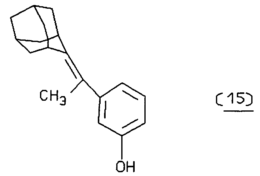
described by Bronstein, et al., in U.S. Patent No. 4,956,477;
wherein X₂ is H, OH, N(CH₃)₂ or OCH₃, described by P. Schaap, J. Am. Chem. Soc. 104: 3504 (1982) and P. Schaap, Photochem. and Photobiology 30:35 (1979);
wherein X₂ is defined above, X₃ is H, OCH₃, or N(CH₃)₂, described by P. Schaap, Report to U.S. Army Research Office, October 15, 1986, and S.D. Gagnon, Ph.D. Thesis, Wayne State University (1982);
wherein X₄ = O, S, CH₃N, or PhN and Y = O, S, or CH₃N and Ph = phenyl, described by P. Schaap, Report to Office of Naval Research, March 17, 1987;
described by P. Schaap, Report to U.S. Army Research, October 15, 1986; - The enamines suitable as PACC's of the present invention are also within the scope of the "Chemiluminescent Compound" in the present Section. Examples of particularly useful enamines, by way of example and not limitation, are the above enol ethers 35 with N(CH₃)₂ in place of OCH₃. A further example is
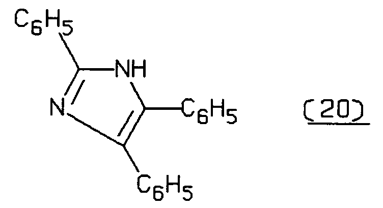
- The 9-alkylidene-N-alkylacridans suitable as PACC's are also within the scope of the "Chemiluminescent Compound" in the present Section. Particularly useful are those of Formula (7) wherein the remaining substituents on the olefin are phenyl, naphthyl, phenanthryl, m-methoxyphenyl, dimethylamino, trityl, methoxy, N-morpholeno and may be taken together to form a ring such as, for example, adamantyl, N-methyacridanylide, xanthanylidine, 1-(3,4-benzo-5-hydrofurylidene), and the like. Particular examples of 9-alkylidine-N-alkylacridans useful as labels in the present invention are, by way of illustration and not limitation:

described by Singer, J.Org. Chem. 41, 2685, (1976);
described by E. White, Chem. Letters; 12, 1491 (1979);
wherein R₄ and R₅ are independently H or phenyl, described by Singer, et al., J. Am. Chem. Soc. 102: 3823, (1983);
described by McCapra, Chem. Comm; N24, 944 (1977) and Singer, et al., ibid;
described by McCapra, ibid;
described by McCapra, ibid; - The relevant portions of the above references are incorporated herein by reference.
- 9-Alkylidene-xanthanes useful as PACC's of the present invention generally have the structure:
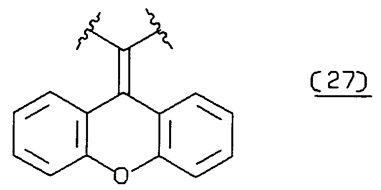
where the substituents on the olefin are selected from the group consisting of H and substituents of 1 to 50 atoms, preferably, phenyl, naphthyl, phenanthryl, m-methoxyphenyl, dimethylamino, trityl, methoxy, N-morpholeno and may be taken together to form a ring such as, for example, adamantyl, N-methyacridanylide, xanthanylidine, 1-(3,4-benzo-5-hydrofurylidene), and the like, for example, 9-phenyl-methylidene-xanthene. - Another family of PACC's is 2,3-dihydro-1,4-phthalazinediones. The most popular compound is luminol, which is the 5-amino compound. Other members of the family include the 5-amino-6,7,8-trimethoxy and the dimethylamino[ca]benz analog. It should be noted that luminol has been used as a label in assays; however, excitation of the luminol has been accomplished chemically, not by photoactivation as is the case in the present invention. These compounds are oxidized to the 1,4-phthalazinidiones by singlet oxygen and under subsequent reaction with superoxide or hydrogen peroxide undergo decomposition with light emission.
- Another family of PACC's is the 2,4,5-triphenyl-imidazoles, with lophine as the common name for the parent product. Chemiluminescent analogs include para-dimethylamino and -methoxy substituents.
- Other PACC's that satisfy the requirements given above may be found in European Patent Application No. 0,345,776 published on December 13, 1989.
- Protected Chemiluminescent Compound -- are those PACC's that have a photo-removable protecting group. When the protecting group is removed by photoactivation, the compound emits light spontaneously or upon activation with components that are present in the assay medium. Photo-removable protecting groups include, by way of example and not limitation, o-nitrobenzyl, o-acylphenylethyl, alkoxybenzyl, and the like, usually attached to a heteroatom of the PACC. Exemplary of PACC's that can form part of the protected chemiluminescent compounds are those electron rich olefins mentioned above. Particular examples, by way of illustration, of protected chemiluminescent compounds in accordance with this aspect of the invention are:


- In the case of protected chemiluminescent compounds any necessary reagents will be present prior to photoactivation or photolysis. Such reagents can include enzymes and substrates for such enzymes. In some cases controlling the pH is all that is required. Other agents are alkaline phosphatase, horse radish peroxidase, hydrogen peroxide, etc. Those skilled in the art will appreciate additional reagents upon selection of any appropriate protected chemiluminescent compound guided by the above principles. For example, in the case of
compound 1 firefly luciferase and its luciferin substrate may be used. In the case of compound 2, it is only necessary to maintain a basic medium (pH > 9). In the case of 2A, hydrogen peroxide and horse radish peroxidase may be used. - Ancillary Materials--Various ancillary materials will frequently be employed in the assay in accordance with the present invention. For example, buffers will normally be present in the assay medium, as well as stabilizers for the assay medium and the assay components. Frequently, in addition to these additives, proteins may be included, such as albumins, organic solvents such as formamide, quaternary ammonium salts, polycations such as dextran sulfate, surfactants, particularly non-ionic surfactants, binding enhancers, e.g., polyalkylene glycols, or the like. When the photosensitizer is activated chemically rather than by irradiation, hydrogen peroxide will often be included as an ancillary reagent. When it is desired to shift the emission wavelength of the chemiluminescent compound to longer wavelength or catalyse the decomposition of its oxygen-activated form, a fluorescent molecule may be employed.
- Wholly or Partially Sequentially--when the sample and various agents utilized in the present invention are combined other than concomitantly (simultaneously), one or more may be combined with one or more of the remaining agents to form a subcombination. Each subcombination can then be subjected to one or more steps of the present method. Thus, each of the subcombinations can be incubated under conditions to achieve one or more of the desired results.
- Energy Acceptor -- referred to herein also as fluorescent energy acceptor. A chromophore having substantial absorption higher than 310 nm, normally higher than 350 nm, and preferably higher than about 400 nm. The choice of the energy acceptor will also be governed by the particular PACC. The energy acceptor should be capable of absorbing light emitted by the PACC. Preferably, the absorption maximum of the energy acceptor should be at similar wavelength as the emission maximum of the chemiluminescent compound. A high extinction coefficient is desirable, usually in excess of 10, preferably in excess of 10³, and particularly preferred in excess of 10⁴.
- A number of different molecules useful as the energy acceptor are described by Ullman, et al. I, II, IV and V, at columns 8 and 9, the relevant portions of which are incorporated herein by reference. Generally, the energy acceptor is a fluorescent compound or fluorescer but energy acceptors that have very weak or no fluorescence are also useful.
- One group of fluorescers having a number of the desirable properties described above are the xanthene dyes, which include the fluoresceins derived from 3,6-dihydroxy-9-phenyl-xanthhydrol and rosamines and rhodamines, derived from 3,6-diamino-9-phenylxanthhydrol. The rhodamines and fluoresceins have a 9-o-carboxyphenyl group, and are derivatives of 9-o-carboxyphenyl- xanthhydrol.
- These compounds are commercially available with substituents on the phenyl group which can be used as the site for bonding or as the bonding functionality. For example, amino and isothiocyanate substituted fluorescein compounds are available.
- Another group of fluorescent compounds are the naphthylamines, having an amino group in the alpha or beta position, usually alpha position. Included among the naphthylamino compounds are 1-dimethylaminonaphthyl-5-sulfonate, 1-anilino-8-naphthalene sulfonate and 2-p-toluidinyl-6-naphthalene sulfonate.
- Dye precursors that are activated to react with proteins include 3-phenyl-7-isocyanatocoumarin; acridines such as 9-isothiocyanatoacridine; N-(p-(2-benzoxazolyl)phenyl)maleimide; benzoxadiazoles such as


4-chloro-7-nitrobenzo-2-oxa-1,3-diazole; stilbenes, such as 4-dimethylamino-4′-isothiocyanatostilbene and 4-dimethylamino- 4′-maleimidostilbene. Other dyes that can be further functionalized to combine with an sbp member include acridine orange, 7-(p-methoxybenzylamino)-4-nitrobenzo-2-oxa-1,3-diazole;
N,N′-dioctadecyloxacarbocyanine p-toluenesulfonate; pyrenes, such as 8-hydroxy-1,3,6-pyrenetrisulfonic acid, and 1-pyrenebutyric acid, merocyanine 540, rose bengal, as well as other readily available fluorescing molecules. - For polynucleotide assays a large variety of energy acceptors can be used, but those that have enhanced fluorescence when bound to double stranded nucleic acids are preferred. Dyes that intercalate such as ethidium bromide and acridine orange are typical examples. A ruthenium derivative developed by Barton (J. Am. Chem. Soc. (1990) 112:4960) is particularly attractive because it is dramatically switched on when intercalated. Alternatively, the energy acceptor may be bound to a second polynucleotide probe that can bind near to the chemiluminescent compound labeled probe or the energy acceptor may be unbound and freely dispersed in solution.
- Energy acceptors which are non-fluorescent can include any of a wide variety of azo dyes, cyanine dyes, 4,5-dimethoxyfluorescein, formazans, indophenols, and the like.
- One aspect of the present invention is a method for determining an analyte. The method comprises treating a medium suspected of containing an analyte under conditions such that the analyte, if present, affects the amount of a photosensitizer and a chemiluminescent compound that can come into close proximity wherein the short-lived singlet oxygen generated by the photosensitizer can react with the chemiluminescent compound prior to its spontaneous decay. The method further comprises measuring the intensity of luminescence produced by the chemiluminescent compound. The intensity of luminescence produced is related to the amount of analyte in the medium. The chemiluminescent compound is capable of activation by singlet oxygen, and the photosensitizer catalyzes the formation of singlet oxygen usually in response to photoexcitation followed by energy transfer to molecular oxygen. Often a surface will be brought into close proximity with the photosensitizer and chemiluminescent compound, wherein the surface will preferably be the surface of suspendible particles. The product formed by the activation of the chemiluminescent compound decomposes, preferably spontaneously, with emission of light.
- The method is predicated on an analyte causing or inhibiting molecules of the photosensitizer and the chemiluminescent compound to be closer to each other than their average distance in the bulk solution of the assay medium. This partitioning will depend upon the amount of analyte present in the sample to be analyzed. The photosensitizer molecules that do not become associated with the chemiluminescent compound produce singlet oxygen that is unable to reach the chemiluminescent compound before undergoing decay in the aqueous medium. However, when the photosensitizer and the chemiluminescent compound come in close proximity with each other in response to the amount of the analyte, the singlet oxygen produced upon irradiation of the photosensitizer can activate the chemiluminescent compound before undergoing decay. Because numerous chemiluminescent compound molecules and/or photosensitizer molecules can be associated with a surface and because diffusion is restricted at a surface, the presence of a surface in conjunction with the photosensitizer and chemiluminescent compound can increase the efficiency of, or action of, singlet oxygen with the chemiluminescent compound prior to decay. The use of a surface that is brought in proximity of the chemiluminescent compound and photosensitizer as a function of the presence of analytes is, therefore, preferred. The subject assay provides for a convenient method for detecting and measuring a wide variety of analytes in a simple, efficient, reproducible manner, which can employ simple equipment for measuring the amount of light produced during the reaction.
- The amount of photosensitizer that comes in close proximity to the chemiluminescent compound is affected by the presence of analyte by virtue of the photosensitizer and chemiluminescent compound each being associated with an sbp member. This may be accomplished in a number of ways and the term "associated with" is defined thereby. The photosensitizer and chemiluminescent compound may contain functionalities for covalent attachment to sbp members or the sbp members, or groups capable of binding to sbp members, may contain functionalities for attaching to the photosensitizer and/or chemiluminescent compound. The attachment may be accomplished by a direct bond between the two molecules or a linking group can be employed between an sbp member and the photosensitizer or chemiluminescent compound. In another embodiment either or both of the photosensitizer and chemiluminescent compound can be bound to surfaces or incorporated in particles, to which are also attached sbp members. In both cases each of the sbp members is capable of binding directly or indirectly to the analyte or an assay component whose concentration is affected by the presence of the analyte. The photosensitizer and chemiluminescent compound can be incorporated into particles by virtue of being soluble in at least one phase of the particles, in which case the photosensitizer and chemiluminescent compound will be at much higher concentration within the particle than in the bulk assay medium. When the photosensitizer and chemiluminescent compound are covalently bound to particles, the photosensitizer and chemiluminescent compound or the particles, or components thereof, are functionalized to provide a means of attaching the photosensitizer and chemiluminescent compounds and the particles. For particles that are oil droplets or liposomes the photosensitizer and chemiluminescent compound can be attached to one or more long hydrocarbon chains, each generally having at least 10 to 30 carbon atoms. If the particles are droplets of a fluorocarbon, the photosensitizer or chemiluminescent compound incorporated into these particles may be fluorinated to enhance solubility and reduce exchange into other particles bound with the other label, and the hydrocarbon chain used for linking will preferably be replaced with a fluorocarbon chain. For silicon fluid particles the photosensitizer and chemiluminescent compound can be bound to a polysiloxane. In order to maximize the number of photosensitizer or chemiluminescent compound molecules per particle, it will usually be desirable to minimize the charge and polarity of the photosensitizer or chemiluminescent compound so that it resides within the non-aqueous portion of the particle. When the particle is a liposome and it is desired to retain the photosensitizer or chemiluminescent compound in the aqueous phase of the liposome, it will be preferred to use photosensitizers or chemiluminescent compounds that are highly polar or charged.
- No matter how the photosensitizer and chemiluminescent compound are bound, it is critical that neither compound is capable of dissociating from its sbp member and becoming associated with the sbp member bound to the other member of the photosensitizer and chemiluminescent compound pair during the course of the assay. Thus, dissociation of these compounds from their respective sbp members must be slow relative to the time required for the assay.
- The chemiluminescent compound may be bound to a sbp member that is capable of binding directly or indirectly to the analyte or to an assay component whose concentration is affected by the presence of the analyte. The term "capable of binding directly or indirectly" means that the designated entity can bind specifically to the entity (directly) or can bind specifically to a specific binding pair member or to a complex of two or more sbp members which is capable of binding the other entity (indirectly). The surface generally has an sbp member bound to it. Preferably, the chemiluminescent compound is associated with the surface, usually within a suspendible particle. This sbp member is generally capable of binding directly or indirectly to the analyte or a receptor for the analyte. When the sbp members associated with the photosensitizer and the chemiluminescent compound are both capable of binding to the analyte, a sandwich assay protocol results. When one of the sbp members associated with the photosensitizer or chemiluminescent compound can bind both the analyte and an analyte analog, a competitive assay protocol can result. The attachment to a surface or incorporation in a particle of the chemiluminescent compound is governed generally by the same principles described above for the attachment to, or the incorporation into, a particle of the photosensitizer.
- The photosensitizer is usually caused to activate the chemiluminescent compound by irradiating the medium containing the above reactants. The medium must be irradiated with light having a wavelength with energy sufficient to convert the photosensitizer to an excited state and thereby render it capable of activating molecular oxygen to singlet oxygen. The excited state for the photosensitizer capable of exciting molecular oxygen is generally a triplet state which is more than about 20, usually at least 23, Kcal/mol more energetic than the photosensitizer ground state. Preferably, the medium is irradiated with light having a wavelength of about 450 to 950 nm although shorter wavelengths can be used, for example, 230-950 nm. The luminescence produced may be measured in any convenient manner such as photographically, visually or photometrically to determine the amount thereof, which is related to the amount of analyte in the medium.
- Although it will usually be preferable to excite the photosensitizer by irradiation with light of a wavelength that is efficiently absorbed by the photosensitizer, other means of excitation may be used as for example by energy transfer from an excited state of an energy donor such as a second photosensitizer. When a second photosensitizer is used, wavelengths of light can be used which are inefficiently absorbed by the photosensitizer but efficiently absorbed by the second photosensitizer. The second photosensitizer may be bound to an assay component that is associated, or becomes associated, with the first photosensitizer, for example, bound to a surface or incorporated in the particle having the first photosensitizer. When a second photosensitizer is employed it will usually have a lowest energy triplet state at higher energy than the lowest energy triplet state of the first photosensitizer.
- The 632.6 nm emission line of a helium-neon laser is an inexpensive light source for excitation. Photosensitizers with absorption maxima in the region of about 620 to about 650 nm are compatible with the emission line of a helium-neon laser and are, therefore, particularly useful in the present invention.
- Another aspect of the invention is a method for determining an analyte, which comprises: (a) providing in combination (1) a medium suspected of containing analyte, (2) a photosensitizer capable in its excited state of activating oxygen to a singlet state, the photosensitizer being associated with a sbp member, and (3) a suspendible particle comprising a chemiluminescent compound wherein the particle has an sbp member bound thereto, (b) treating the combination with light to excite the photosensitizer and (c) examining the combination for the amount of luminescence emitted therefrom. The amount of luminescence emitted is related to the amount of analyte in the medium. Preferably, the chemiluminescent compound is incorporated in a suspendible particle and the sbp member is bound to the particle. More preferably, the photosensitizer is incorporated in a second suspendible particle which has an sbp member bound to it.
- The method and compositions of the invention may be adapted to most assays involving sbp members such as ligand-receptor; e.g., antigen-antibody reactions; polynucleotide binding assays, and so forth. The assays may be homogeneous or heterogeneous, competitive or noncompetitive. The assay components, chemiluminescent compound and photosensitizer, can be utilized in a number of ways with (1) a surface, when employed, (2) nucleic acid or receptor and (3) nucleic acid or ligand. The association may involve covalent or non-covalent bonds. Those skilled in the art will be able to choose appropriate associations depending on the particular assay desired in view of the foregoing and the following illustrative discussion.
- In a homogeneous assay approach, the sample may be pretreated if necessary to remove unwanted materials. The reaction for a noncompetitive sandwich type assay can involve an sbp member, (e.g., an antibody, nucleic acid probe, receptor or ligand) complementary to the analyte and associated with a chemiluminescent compound; a photosensitizer associated with an sbp member, (e.g., antibody, nucleic acid probe, receptor or ligand) that is also complementary to the analyte; the sample of interest; and any ancillary reagents required. Preferably, one or both of the photosensitizer and the chemiluminescent compound are incorporated in particles to which the sbp members are attached. In a competitive protocol one sbp member can be a derivative of the analyte and the other sbp member can be complementary to the analyte, e.g., an antibody. In either protocol the components may be combined either simultaneously or wholly or partially sequentially. The ability of singlet oxygen produced by an activated photosensitizer to react with the chemiluminescent compound is governed by the binding of an sbp member to the analyte. Hence, the presence or amount of analyte can be determined by measuring the amount of light emitted upon activation of the photosensitizer by irradiation, heating or addition of a chemical reagent, preferably by irradiation. Both the binding reaction and detection of the extent thereof can be carried out in a homogeneous solution without separation. This is an advantage of the present invention over prior art methods utilizing chemiluminescence.
- In a heterogeneous assay approach, the assay components comprise a sample suspected of containing an analyte which is an sbp member; an sbp member bound to a support, which may be either a non-dispersible surface or a particle having associated with it one member of a group consisting of the chemiluminescent compound and the photosensitizer; and an sbp member having the other member of the group associated with it wherein the sbp members can independently, either directly or indirectly, bind the analyte or a receptor for the analyte. These components are generally combined either simultaneously or wholly or partially sequentially. The surface or particles are then separated from the liquid phase and either the separated phase or the liquid phase is subjected to conditions for activating the photosensitizer, usually by irradiating the particular phase in question, and measuring the amount of light emitted.
- The binding reactions in an assay for the analyte will normally be carried out in an aqueous medium at a moderate pH, generally that which provides optimum assay sensitivity. Preferably, the activation of the photosensitizer will also be carried out in an aqueous medium. However, when a separation step is employed, non-aqueous media such as, e.g., acetonitrile, acetone, toluene, benzonitrile, etc. and aqueous media with pH values that are very high, i.e., greater than 10.0, or very low, i.e., less than 4.0, usually very high, can be used. As explained above, the assay can be performed either without separation (homogeneous) or with separation (heterogeneous) of any of the assay components or products.
- The aqueous medium may be solely water or may include from 0.01 to 80 volume percent of a cosolvent but will usually include less than 40% of a cosolvent when an sbp member is used that is a protein. The pH for the medium of the binding reaction will usually be in the range of about 4 to 11, more usually in the range of about 5 to 10, and preferably in the range of about 6.5 to 9.5. When the pH is not changed during the generation of singlet oxygen the pH will usually be a compromise between optimum binding of the binding members and the pH optimum for the production of signal and the stability of other reagents of the assay. When elevated pH's are required for signal production, a step involving the addition of an alkaline reagent can be inserted between the binding reaction and generation of singlet oxygen and/or signal production. Usually the elevated pH will be greater than 10, usually 10-14. For heterogenous assays non-aqueous solvents may also be used as mentioned above, the main consideration being that the solvent not react efficiently with singlet oxygen.
- Various buffers may be used to achieve the desired pH and maintain the pH during an assay. Illustrative buffers include borate, phosphate, carbonate, tris, barbital and the like. The particular buffer employed is not critical to this invention, but in an individual assay one or another buffer may be preferred.
- Moderate temperatures are normally employed for carrying out the binding reactions of proteinaceous ligands and receptors in the assay and usually constant temperature, preferably, 25° to 40°, during the period of the measurement. Incubation temperatures for the binding reaction will normally range from about 5° to 45°C, usually from about 15° to 40°C, more usually 25° to 40°C. Where binding of nucleic acids occur in the assay, higher temperatures will frequently be used, usually 20° to 90°, more usually 35° to 75°C. Temperatures during measurements, that is, generation of singlet oxygen and light detection, will generally range from about 20° to 100°, more usually from about 25° to 50°C, more usually 25° to 40°C.
- The concentration of analyte which may be assayed will generally vary from about 10⁻⁴ to below 10⁻¹⁶ M, more usually from about 10⁻⁶ to 10⁻¹⁴ M. Considerations, such as whether the assay is qualitative, semiquantitative or quantitative, the particular detection technique the concentration of the analyte of interest, and the maximum desired incubation times will normally determine the concentrations of the various reagents.
- In competitives assays, while the concentrations of the various reagents in the assay medium will generally be determined by the concentration range of interest of the analyte, the final concentration of each of the reagents will normally be determined empirically to optimize the sensitivity of the assay over the range. That is, a variation in concentration of the analyte which is of significance should provide an accurately measurable signal difference.
- The concentration of the sbp members will depend on the analyte concentration, the desired rate of binding, and the degree that the sbp members bind nonspecifically. Usually, the sbp members will be present in at least the lowest expected analyte concentration, preferably at least the highest analyte concentration expected, and for noncompetitive assays the concentrations may be 10 to 10⁶ times the highest analyte concentration but usually less than 10⁻⁴ M, preferably less than 10⁻⁶ M, frequently between 10⁻¹¹ and 10⁻⁷ M. The amount of photosensitizer or chemiluminescent compound associated with a sbp member will usually be at least one molecule per sbp member and may be as high as 10⁵, usually at least 10-10⁴ when the photosensitizer or chemiluminescent molecule is incorporated in a particle.
- While the order of addition may be varied widely, there will be certain preferences depending on the nature of the assay. The simplest order of addition is to add all the materials simultaneously. Alternatively, the reagents can be combined wholly or partially sequentially. When the assay is competitive, it will often be desirable to add the analyte analog after combining the sample and an sbp member capable of binding the analyte. Optionally, an incubation step may be involved after the reagents are combined, generally ranging from about 30 seconds to 6 hours, more usually from about 2 minutes to 1 hour before the sensitizer is caused to generate singlet oxygen and the light emission is measured.
- In a particularly preferred order of addition, a first set of specific binding pair members that are complementary to and/or homologous with the analyte are combined with the analyte followed by the addition of specific binding pair members complementary to the first specific binding pair members, each associated with a different member of the group consisting of a photosensitizer and a chemiluminescent compound. The assay mixture, or a separated component thereof, is then irradiated and the light emission is measured.
- In a homogeneous assay after all of the reagents have been combined, they can be incubated, if desired. Then, the combination is irradiated and the resulting light emitted is measured. The emitted light is related to the amount of the analyte in the sample tested. The amounts of the reagents of the invention employed in a homogeneous assay depend on the nature of the analyte. Generally, the homogeneous assay of the present invention exhibits an increased sensitivity over known assays such as the EMIT® assay. This advantage results primarily because of the improved signal to noise ratio obtained in the present method.
- The following assays are provided by way of illustration and not limitation to enable one skilled in the art to appreciate the scope of the present invention and to practice-the invention without undue experimentation. It will be appreciated that the choice of analytes, photosensitizers, chemiluminescent compounds, surfaces, particles and reaction conditions will be suggested to those skilled in the art in view of the disclosure herein in the examples that follow.
- In one embodiment of the invention the chemiluminescent compound 9-benzal-10-methylacridan, is covalently linked to an antibody for thyroid stimulating hormone (TSH) (Reagent 1). The 9-benzal-10-methylacridan, functionalized with a N-hydroxysuccinimidyl ester of a carboxyl group attached to the phenyl ring, reacts with the amino groups of the antibody. The linking group is a carboxamide. The photosensitizer utilized is rose bengal, which is covalently bound to latex particles having an average dimension of 0.5 micron. The latex particles and rose bengal are covalently bound to each other by means of chloromethyl groups on the latex to provide an ester linking group as described in J. Am. Chem. Soc., 97: 3741 (1975). The latex particle is further linked to a second antibody for TSH by means of N-hydroxysuccinimidyl ester groups on the latex (Reagent 2). Both of the antibodies employed are monoclonal antibodies prepared by standard hybrid cell line technology. One antibody recognizes the α-subunit of TSH and the other recognizes the β-subunit of TSH. In conducting the assay a serum sample suspected of containing TSH is obtained from a patient. Fifty microliters of the sample is combined in a 500 mL aqueous medium, buffered with Tris buffer at pH 8.0, with
Reagent 1 and Reagent 2 above. The amounts ofReagent 1 and Reagent 2 are sufficient to provide concentrations of each antibody of about 10⁻⁶ molar. The reaction mixture is then incubated for a period of one hour at 25°C and then irradiated for 30 seconds with 560 nm light. The light intensity emitted following the irradiation is measured and the total light energy detected over a period of 30 seconds is compared with values obtained in a similar procedure with samples having known concentrations of TSH to determine the concentration of TSH in the unknown. Alternatively, following incubation, the latex particles are separated from the medium and placed in an aqueous buffered medium of pH 9.5, similarly irradiated, and the amount of light emitted from the system is measured as before. - In an alternative based on the above, Reagent 2 is rose bengal covalently linked to the second antibody and no latex particle is employed. In still another alternative approach based on the above, Reagent 2 is rose bengal covalently linked to the second antibody and
Reagent 1 is 9-benzal-10-methylacridan covalently bound to latex particles, to which the first antibody is covalently attached. In still another alternative based on the above,Reagent 1 is as described immediately above, Reagent 2 is rose bengal covalently linked to latex particles, to which avidin is covalently attached, and a third reagent (Reagent 2A) that is the second antibody covalently linked to biotin is employed.Reagent 1 and the third reagent are combined with sample and incubated. Then, an excess of Reagent 2 is added and the remaining procedure is as described above. - In another alternative based on the above, a chloroperoxidase is substituted for the rose bengal. In this assay the irradiation step is replaced by addition of sodium bromide and hydrogen peroxide to produce singlet oxygen. Light emitted is measured as described above.
- In another embodiment in accordance with the present invention, a first set of oil droplets (Reagent 3) is prepared from a solution of the photosensitizer, chlorophyll, in mineral oil in accordance with Giaever, supra. The oil droplets, which range from 0.1 to 2 microns in diameter, are functionalized and linked to a monoclonal antibody for human chronic gonadotropin (HCG). The chlorophyll is lipophilic and is therefore dissolved irreversibly in the lipophilic oil droplet. A second set of oil droplets (Reagent 4) is prepared in a similar manner. In this set of droplets the oil droplet is covalently linked to a second monoclonal antibody for HCG, which recognizes a different portion of the HCG molecule than that recognized by the first monoclonal antibody referred to above. 9-(Diphenylmethylidine)-N-methylacridan irreversibly dissolved in the lipophilic oil droplet by including a N,N-didodecylcarboxamide group bound to one of the phenyl groups of the acridan. The monoclonal antibodies are prepared by standard hybrid cell line technology. A urine sample suspected of containing HCG (50 microliters) is combined with excess quantities of Reagent 3 and Reagent 4 in an aqueous buffered medium (500 µL) at pH 7.5. The medium is incubated at 25°C for a period of 20 minutes. The medium, without separation, is irradiated at 633 nm using a He/Ne laser for a period of one minute and the light emitted is measured as described above. The amount of light is compared with the amount emitted from samples containing known amounts of HCG and the amount of HCG in the unknown sample is determined by comparing the values. In this way a convenient and sensitive homogeneous immunoassay for HCG is conducted.
- In an alternative approach based on the above, Reagent 3 has antibody for fluorescein in place of the antibody for HCG and an additional reagent (Reagent 3A) has the HCG antibody covalently linked to fluorescein. Reagent 4 has avidin in place of the second HCG antibody and a fourth reagent (Reagent 4A) has the second antibody covalently linked to biotin. In the assay Reagent 4A and Reagent 3A are combined with sample and incubated. Thereafter, Reagents 3 and 4 are added and incubated. The remainder of the above assay procedure is then carried out.
- In another embodiment of the present invention, one set of liposomes (Reagent 5) (0.2 micron in diameter) is formed by high pressure extrusion of a phospholipid suspension in pH 7.4 buffer through a 0.2 micron pore membrane using a commercially available instrument designed for such purpose. A thyroxine analog is covalently linked to the liposome by first forming mercaptoacetamide groups on the liposome by reaction of phosphatidylethanolamine in the liposome with an active ester of methyl carboxymethyl disulfide followed by reaction with dithioerythritol. Bromoacetyl thyroxine is then allowed to react with the sulfhydrylated liposomes. A metallo-porphyrin dye is dissolved in the lipophilic portion of the liposome. Another set of liposomes (Reagent 6) is utilized to attach a monoclonal antibody for thyroxine. The antibody is attached covalently by means similar to the attachment of thyroxine. An enamine (2-dimethylaminomethylenepyran) is covalently linked by means of a carboxamide linking group to the surface of the liposome.
Reagent 5 and Reagent 6 are combined in an aqueous buffered assay medium (500 µL) of pH 8.0 together with a serum sample suspected of containing thyroxine that contains anilinonaphthalene sulfonic acid to displace thyroxine from binding proteins (50 micro-liters) and accept energy from the oxygen-activated pyran to provide for long wave length emission. The assay medium is then incubated at room temperature for 1 hour. The medium is irradiated, without a separation step, at 650nm for a period of 1 minute and the resulting emitted light is measured by means of a luminometer. The value obtained is compared with values obtained by conducting a similar assay with known amounts of thyroxine. In this way the amount of thyroxine in the serum sample is quantitated. - In an alternative approach based on the above, Reagent 6 has avidin in place of antibody for thyroxine. An additional reagent (Reagent 6A) has antibody for thyroxine covalently linked to biotin.
Reagent 5 has antibody for fluorescein in place of thyroxine and an additional reagent (Reagent 5) has thyroxine linked covalently to fluorescein. In the assay Reagents 5A and 6A are combined with sample and incubated. Then,Reagent 5 and 6 are added, the mixture is incubated, and the remainder of the assay procedure is followed. - In another alternative based on the above, the metallo-porphyrin dye is replaced by potassium molybdate that is incorporated into the aqueous phase of the liposomes. In this assay the irradiation step is replaced by addition of hydrogen peroxide to produce singlet oxygen. Light emitted is measured as described above.
- In another embodiment 2-hydroxyethyl 9,10-dibromoanthracene is formed into a dye crystallite in a manner similar to that described by Gribnau. A monoclonal antibody that recognizes hepatitis B surface antigen is covalently attached to the dye crystallite by means of a carbamate linking group. A latex particle is treated to immobilize a second antibody for hepatitis B surface antigen by means of covalent attachment. The latex particle is further covalently linked to 9-(benzal-9H-xanthene) by means of an amide linking group. The dye crystallite has a particle size of on the average 2 microns and the latex particles have an average diameter of 0.2 microns. The monoclonal antibodies are prepared by standard hybrid cell line technology. A serum sample from a patient suspected of having hepatitis (50 µL) is combined in an aqueous assay medium (500 µL) at pH 7.0 with an excess of the dye crystallite and latex particles described above. The assay medium is then incubated at room temperature for a period of 30 minutes and then irradiated at 350 nm with a xenon light source for a period of 1 minute. The presence of hepatitis B surface antigen in the sample causes the dye crystallite and latex particles to come into close proximity by virtue of the immune binding of the respective antibodies with the antigen. Upon irradiation of the medium the 9,10-dibromoanthracene is excited and converts molecular oxygen to singlet oxygen. The singlet oxygen reacts with the xanthene to give a dioxetane, which spontaneously decays at room temperature to produce light. The light is measured photometrically and the amount of light over a certain threshold level indicates the presence of hepatitis B surface antigen in the sample. Irradiation of the medium is conducted at room temperature and the assay is conducted in a homogeneous manner to yield an assay for hepatitis B surface antigen.
- Preferably, in this assay Eu(TTA)₃ is dissolved in the latex particles so as to cause the excited xanthene derivative to transfer its energy to the Eu(TTA)₃, which emits above 600 nm. This avoids serum interference that would otherwise occur because of absorption by serum components of light emitted directly from the xanthene luminescence.

- In another embodiment the assay is for the determination of a particular blood group antigen on the surface of a red blood cell, namely, an A group antigen. Latex particles prepared as described above having a particle size of 150-500 nm are utilized. The latex particles have an antibody for the A group antigen covalently linked to the latex particle. The particles also have the enolether, 2-p-dimethylaminophenyl-3-phenyl-5,6-dihydrodioxene, dissolved in the latex. This latex particle reagent is combined in the aqueous medium (500 µl) with whole blood (100 µl) and 1x10⁻⁴M photosensitizer, which is a hydrophobic photosensitizer, namely, phenazine-2-carboxylic acid. The hydrophobic dye intercalates into the red cells present in the sample. The medium is incubated at 25°C for a period of 10 minutes and then irradiated at 400-450nm with a tungsten source for a period of 30 seconds. The light emitted from the medium is measured and compared to the amount of light obtained in samples known to be free of A group antigen red blood cells. Thus, the amount of light over a threshold level indicates the presence of the A blood group antigen.
- In another embodiment of the present invention the analyte is an HLA antigen on the surface of a cell. A sample (50 µL) suspected of containing the HLA antigen is combined in an aqueous assay medium (500 µL) buffered at pH 7.4. To the medium is also added an excess amount of a reagent which is an antibody for the HLA antigen covalently coupled to eosin by means of a thioether linking group. A chemiluminescent compound is also added to the assay medium. The chemiluminescent compound is chosen so that it will intercalate into the cell containing the HLA antigen. The chemiluminescent compound is an acridan, namely, 9-(dodecylphenylmethylidine-1-)N-(2-sulfonoxyethyl) acridan. The assay medium is incubated at room temperature for a period of 1 hour and then irradiated at 500-550 nm with a tungsten-halogen light source for a period of 1 minute. The light emitted from the assay medium is measured and the amount of light above a threshold level is indicative of the presence of the HLA antigen.
- The present invention further encompasses compositions comprising a suspendible particle of 0.04 to 4000 nanometer average diameter comprising a chemiluminescent compound. The chemiluminescent compound may be covalently bound to the particle matrix or may be dissolved in the matrix or dissolved in a solvent that is dissolved in the matrix. The particles will preferably be polymeric or be oil droplets or vesicles such as liposomes. Where the particle is a liposome, the chemiluminescent compound will be associated with the lipid bilayer or dissolved in the aqueous interior of the liposome. The particle will have an spb member bound to it. Preferably, sbp bound particles will be 100 to 1000 mm in average diameter. Also encompassed are compositions comprised of two complementary sbp members bound to each other wherein one is associated with a photosensitizer and one is associated with a chemiluminescent compound.
- Another aspect of the present invention is a method providing a combination comprising (1) a medium suspected of containing the analyte and (2) a label reagent comprising a first specific binding pair (sbp) member associated with a PACC. Conditions are chosen such that an sbp member complex is formed in relation to the presence of the analyte. For example, the first sbp member may be capable of binding to the analyte or a second sbp member to form a complex related to the presence of the analyte. The method further comprises photochemically activating the PACC and detecting the amount of luminescence generated by the PACC. The amount of luminescence is related to the amount of analyte in the medium.
- Generally, the PACC is photo-chemically activated by irradiation with light, which can directly activate the PACC or can activate a photosensitizer, which, upon irradiation, causes molecular oxygen to be converted to singlet oxygen. The singlet oxygen then activates the PACC. The photosensitizer may be bound to a ligand, may be free in solution, or may be bound to a surface depending on the nature of the assay. The product formed by the activation of the PACC with singlet oxygen may decompose spontaneously with emission of light or may be further treated physically or chemically to emit light.
- A photosensitizer can be employed in a number of different approaches to produce singlet oxygen, which activates the PACC. The photosensitizer can be employed directly, not being bound to an sbp member. In this approach a relatively large concentration of the photosensitizer is employed, usually at least 10⁻⁷M, preferably at least 10⁻⁶ M, most preferably at least 10⁻⁵ M. Generally, the amount of photosensitizer employed in this approach is that sufficient to produce a concentration of singlet oxygen that results in activation of the PACC. This approach can be used when the emission of the PACC is substantially modified when a complex is formed or when the amount of PACC in the medium is affected by the presence of the analyte. This will usually be accomplished by separating bound and unbound label. Where a separation is not desired, the emission of the PACC in the complex can be modified by an energy acceptor, which is brought into close proximity to the PACC by binding the energy acceptor to the complex formed as a result of the analyte being present. Where the energy acceptor is fluorescent, the wavelength of the emission is usually modified. When the energy acceptor is not fluorescent, the chemiluminescence will usually be quenched when the PACC becomes bound to the complex.
- In a further embodiment of the present invention a photosensitizer and the PACC are partitioned between the bulk solution of the assay medium and a surface as described in the present application. This partitioning will depend upon the amount of analyte present in the sample to be analyzed and the photosensitizer will usually be bound to an sbp member. The photosensitizer molecules that do not become associated with the surface produce singlet oxygen which is unable to reach the PACC before undergoing decay in the aqueous medium. However, when both the photosensitizer and the PACC become associated with the surface by virtue of the formation of a complex resulting from the presence of the analyte, the singlet oxygen produced upon irradiation of the photosensitizer can activate the PACC before undergoing decay. In this method the amount of photosensitizer employed can be considerably less than in the previously described use of the photosensitizer not involving an sbp member bound to the photosensitizer.
- As can be seen, the subject assay provides for a convenient method for detecting and measuring a wide variety of analytes in a simple, efficient, reproducible manner, which can employ visual inspection or conventional equipment for measuring the amount of light produced during the reaction.
- When the photosensitizer is caused to become associated with the surface as the result of the presence of analyte, it will usually be associated with an sbp member. This may be accomplished in a number of ways. The photosensitizer may contain a functionality for attachment to an sbp member or the sbp member may contain the functionality for attaching to the photosensitizer. The attachment may be accomplished by a direct bond between the two molecules or a linking group can be employed between the sbp member and the photosensitizer. In another embodiment the photosensitizer can be bound to or incorporated in a particle, to which is also attached an sbp member. In both cases the sbp member is capable of binding to the analyte. The photosensitizer can be incorporated into the particle by virtue of being soluble in at least one phase of the particle. The photosensitizer may be bound to the particle when it is not incorporated into the particle. For this purpose the photosensitizer or the particle, or component thereof, is functionalized to provide a means of attaching the photosensitizer and the particle. For particles that are oil droplets or lipid bilayers, the photosensitizer can be bound to the particle by attachment to a long hydrocarbon chain that is compatible with the particle composition. Frequently, at least one, and preferably two, hydrocarbon chains are employed having 8 to 20 or more carbon atoms.
- If the particle is a droplet of a fluorocarbon, the photosensitizer may be fluorinated to enhance solubility and reduce exchange and the hydrocarbon chain used for linking will preferably be replaced with a fluorocarbon chain. For silicon particles the photosensitizer may be bound to a polysiloxane. Usually, it will be desirable to minimize the charge and polarity of the photosensitizer so that it resides within the non-aqueous portion of the particle. As mentioned above, the approach involving photosensitizer bound to an sbp member is described fully in the present application.
- As mentioned above, the PACC is "associated with an sbp member", and thus functions in the present invention as a label. As used herein, the term "associated with an sbp member" includes the following: Usually, the association of the PACC and sbp member is through covalent binding. However, this label reagent can further comprise a suspendible particle to which the PACC is bound or in which the PACC is non-covalently incorporated. The suspendible particle will also have the sbp member bound to it. This sbp member is generally capable of binding to the analyte or to an sbp member capable of binding to the analyte. When another sbp member is utilized and is also capable of binding to the analyte, a sandwich assay protocol results. The sbp member bound to the PACC can also be analogous to the analyte, in which case a competitive assay protocol can result.
- When a photosensitizer is used, the photosensitizer serves to activate the PACC when the medium containing the above reactants is irradiated. The medium is irradiated with light having a wavelength of sufficient energy to convert the photosensitizer to an excited state and render it capable of activating molecular oxygen to singlet oxygen. When bound to an sbp member, the photosensitizer concentration may be very low, frequently 10⁻⁶ to 10⁻¹² M or lower. When the photosensitizer is unbound it will frequently be at a concentration sufficient to absorb an appreciable amount of light, usually at least 0.1%, preferably between 1 and 80%.
- The excited state of the photosensitizer will usually be the lowest triplet state and is at least 25 Kcal/mole, preferably at least 23 Kcal/mole, more energetic than the ground state. Generally, the medium is irradiated with light having a wavelength of about 300 to 1200 nm usually 450 to 950, preferably 550 to 800 nm. The period of irradiation will depend on the lifetime of the activated PACC, the light intensity and the desired emission intensity. For short-lived activated PACC's the period may be less than a second, usually about a millisecond but may be as short as a microsecond where an intense flashlamp or laser is used. For longer-lived activated PACC's the irradiation period can be longer and a less intense steady light source can be used. In general, the integrated light intensity over the period of irradiation should be sufficient to excite at least 0.1% of the photosensitizer molecules, preferably at least 30%, and most preferably every, photosensitizer molecule will be excited at least once.
- The luminescence or light produced in any of the above approaches can be measured visually, photographically, actinometrically, spectrophotometrically or by any other convenient means to determine the amount thereof, which is related to the amount of analyte in the medium.
- A helium-neon laser is an inexpensive light source for excitation at 632.6 nm. Photosensitizers that absorb light at this wavelength are compatible with the emission line of a helium-neon laser and are, therefore, particularly useful in the present invention. Other light sources include, for example, other lasers such as Argon, YAG, He/Cd, and ruby; photodiodes; mercury, sodium and xenon vapor lamps; incandescent lamps such as tungsten and tungsten/halogen; and flashlamps.
- Another aspect of the invention is a method for determining an analyte, which comprises (a) combining in an assay medium either simultaneously or wholly or partially sequentially (1) a medium suspected of containing an analyte, (2) a label reagent comprising a first member of a specific binding pair (sbp member) bound to a compound capable of chemiluminescence upon reaction with singlet oxygen, and (3) an insolubilized reagent comprising a fluorescent energy acceptor bound to, or capable of becoming bound to, a second sbp member under conditions wherein an sbp member complex involving said label reagent is formed in relation to the presence of analyte in said medium and energy from said chemiluminescent compound is capable of activating said fluorescent energy acceptor, (b) activating said compound with light, and (c) examining said assay medium for a signal, the presence or intensity thereof being related to the amount of analyte in said medium.
- The method and compositions of the invention may be adapted to most assays involving sbp members such as ligand-receptor, e.g., antigen-antibody reactions, polynucleotide binding assays, and so forth. The assays may be homogeneous or heterogeneous, competitive or sandwich. In a homogeneous assay approach, the sample may be pretreated if necessary to remove unwanted materials. The immunological reaction for a sandwich type assay usually involves an sbp member, e.g., an antibody, that is complementary to the analyte and bound to the PACC, a second sbp member, e.g., antibody, that is also complementary to the analyte, and the sample of interest. In a competitive protocol the PACC may be associated with an sbp member that is analogous to, usually a derivative of, the analyte or with an sbp member complementary to the analyte, e.g., an antibody.
- The PACC may be utilized either by itself or associated with an energy acceptor. The ability of luminescence produced by an activated PACC to activate the energy acceptor may be governed by the binding of the two sbp members or may be the consequence of a relatively high concentration of the energy acceptor in the vicinity of the activated PACC, wherein the concentration will usually be at least micromolar, usually at least millimolar. When the PACC is bound to an sbp member, its concentration, by contrast, may be quite low, often 10⁻⁵ to 10⁻¹⁵, usually 10⁻⁸ to 10⁻¹⁴ and detection of the extent thereof may be carried out in a homogeneous solution without separation provided only that the luminescent intensity is modified when the PACC is in a binding complex.
- In a heterogeneous assay approach, a sample suspected of containing an analyte, which is an sbp member, is combined with a reagent that is comprised of a complementary sbp member bound to a support, which may be a surface or a particle having the PACC. An energy acceptor associated with another sbp member may also be employed wherein the sbp member is either complementary to (sandwich) or analogous to (competitive) the analyte. These materials are generally combined either simultaneously or wholly or partially sequentially. The support is then separated from the liquid phase and either the solid phase or the liquid phase is examined for the presence of luminescent energy, usually by irradiating the particular phase in question and measuring the amount of light emitted.
- An assay for the analyte will normally be carried out in an aqueous buffered medium at a moderate pH, generally that which provides optimum assay sensitivity. As explained above, the assay can be performed either without separation (homogeneous) or with separation (heterogeneous) of any of the assay components or products.
- The aqueous medium may be solely water or may include from 0.01 to 80 or more volume percent of a cosolvent. The pH for the medium will usually be in the range of about 4 to 13, more usually in the range of about 5 to 10, and preferably in the range of about 6.5 to 9.5. The pH will usually be a compromise between optimum binding of the binding members and the pH optimum for other reagents of the assay such as members of the signal producing system. For example, the activated PACC may require a certain pH range in order to decay to produce luminescence.
- Various buffers may be used to achieve the desired Ph and maintain the pH during the determination. Illustrative buffers include borate, phosphate, carbonate, tris, barbital and the like. The particular buffer employed is not critical to this invention, but in an individual assay one or another buffer may be preferred.
- Moderate temperatures are normally employed for carrying out the assay and usually constant temperature, preferably, room temperature, during the period of the measurement. Incubation temperatures will normally range from about 5° to 99°C, usually from about 15° to 70°C, more usually 20 to 45°C. Temperatures during measurements will generally range from about 10° to 70°C, more usually from about 20° to 45°C, more usually 20° to 25°C. In some instances the activated PACC may require heating up to 100°C in order to decay to produce luminescence.
- The concentration of analyte which may be assayed will generally vary from about 10⁻⁵ to 10⁻¹⁷ M, more usually from about 10⁻⁶ to 10⁻¹⁴ M. Considerations, such as whether the assay is qualitative, semiquantitative or quantitative, the particular detection technique and the concentration of the analyte of interest will normally determine the concentrations of the various reagents.
- While the concentrations of the various reagents in the assay medium will generally be determined by the concentration range of interest of the analyte, the final concentration of each of the reagents will normally be determined empirically to optimize the sensitivity of the assay over the range. That is, a variation in concentration of the analyte which is of significance should provide an accurately measurable signal difference.
- While the order of addition may be varied widely, there will be certain preferences depending on the nature of the assay. The simplest order of addition particularly for a homogeneous assay is to add all the materials simultaneously. Alternatively, the reagents can be combined wholly or partially sequentially. Optionally, an incubation step may be involved after the reagents are combined, generally ranging from about 30 seconds to 6 hours, more usually from about 2 minutes to 1 hour.
- In a homogeneous assay after all of the reagents have been combined, they can be incubated, if desired. Then, the combination is irradiated and the resulting light emitted is measured. The emitted light is related to the amount of the analyte in the sample tested. The amounts of the reagents of the invention employed in a homogeneous assay depend on the nature of the analyte.
- The following assays are provided by way of illustration and not limitation to enable one skilled in the art to appreciate the scope of the present invention and to practice the invention without undue experimentation. It will be appreciated that the choice of analytes, photosensitizers, PACC's, surfaces, particles and reaction conditions will be suggested to those skilled in the art in view of the disclosure herein and the examples that follow.
- In the following assays, components are combined in a predominantly aqueous medium of pH 6 to 8.5.
- A. In an assay for HCG a urine sample is combined with (1) antibody to HCG bound to 1 micron latex particles and (2) an antibody against a separate non-overlapping epitope of HCG bound to the PACC 4. After incubation of the suspension for 30 minutes, the particles are separated from the medium by centrifugation, washed and irradiated with 300 nm light. The intensity of light emitted following the irradiation is related to the amount of HCG in the sample. The separated particles can be irradiated directly, or suspended in an aqueous or non-aqueous solvent prior to irradiation.
- B. In a procedure for the assay of HCG in serum similar to A, the latex beads that are used are stained with the photosensitizer, copper phthalocyanine, and, following incubation, are separated from the medium and then irradiated with 600 nm light. The intensity of the emitted light is related to the amount of HCG in the sample.
- C. In an assay for digoxin in serum, the PACC, luminol, conjugated to digoxigenin is incubated with the sample in a polystyrene well that has been stained with tetradecylphthalocyanine. The surface of the well is coated with an antibody to digoxin. After incubation for 30 minutes and removal of the assay medium, the well is irradiated with 600 nm light. It is preferable to add an alkaline solution prior to irradiation (pH 10-12). The intensity of the light emitted following irradiation is inversely related to the concentration of digoxin in the sample.
- D. In an assay for albumin in urine, the sample is combined with (1) an antibody to albumin labeled with the PACC and (2) an antibody to albumin directed against a non-overlapping albumin epitope that is labeled with energy acceptor,

- E. In an assay for thyroxine in serum, the sample is combined with thyroxine bound to latex beads, into which has been incorporated the
PACC 10 wherein X₂ is N(CH₃)₂. Antibody to thyroxine bound to the sensitizer, rose bengal, is included in the assay medium. Following incubation for 10 minutes, the medium is irradiated with 550 nm light and the light emitted following termination of the irradiation is measured. The emitted light intensity is inversely related to the amount of thyroxine in the sample. - F. In an assay for a target polynucleotide sequence in a sample containing DNA, a 25-base oligonucleotide complementary with the target sequence and conjugated to PACC 23 which is 9-phenylmethylidene xanthane is mixed with the sample. The medium is then heated to 75°C and cooled to 55°C to permit hybridization of the oligonucleotide to any target sequence present. The energy acceptor and intercalator, acridine orange, and the photosensitizer, phthalocyanine tetrasulfonic acid, are present in the hybridization solution or may be added following completion of the hybridization reaction. The solution is then irradiated with 600 nm light and the intensity of light emitted by the acridine orange (about 500 nm) following termination of the irradiation is measured. The light intensity is directly related to the presence of the target sequence.
- G. In an assay for a target polynucleotide sequence in a sample containing DNA, a 25-base oligonucleotide complementary to the target sequence and labeled with the photosensitizer, phthalocyanine, is caused to hybridize to the target as in F above. A compound that binds to the minor groove of double stranded DNA, Hoechst dye 33258, which is attached to
PACC 20, N-methyl-9-phenylmethylidene acridan, is included in the medium or added following hybridization. The medium is then irradiated with 600 nm light and the light emitted following termination of the irradiation is measured. The emitted light intensity is directly related to the amount of target sequence present in the sample. - H. In an assay for hepatitis B surface antigen (HBsAg) in serum, the sample is combined with (1) 150 nm latex particles stained with the PACC, and coated with antibodies to HBsAg, and (2) 150 nm latex beads stained with the photosensitizer tetraphenyl porphyrin, and coated with antibodies to HBsAg. After incubation of the mixture for one hour, the suspension is irradiated with 550 nm light. Following termination of the irradiation, the emitted light intensity is measured and related to the amount of HBsAg in the sample.
- I. In an assay for HBsAg in whole blood, procedure H is followed except that the latex beads containing the PACC are replaced with latex beads stained with PACC 23, which is 9-phenylmethylidene-xanthane and the energy acceptor -

- Another aspect of the present invention relates to kits useful for conveniently performing the assay method of the invention for determining the presence or amount of an analyte in a sample suspected of containing the analyte. To enhance the versatility of the subject invention, the reagents can be provided in packaged combination, in the same or separate containers, so that the ratio of the reagents provides for substantial optimization of the method and assay. The reagents may each be in separate containers or various reagents can be combined in one or more containers depending on the cross-reactivity and stability of the reagents.
- One type of the kit comprises (1) a composition comprising a suspendible particle comprising a chemiluminescent compound, the particle having an sbp member bound to it, and (2) a photosensitizer. The photosensitizer can be attached to an sbp member or it can be associated with a particle, to which an sbp member is bound. The kit can further include other separately packaged reagents for conducting an assay including ancillary reagents, and so forth.
- Another embodiment of a kit in accordance with the present invention comprises in packaged combination a chemiluminescent compound associated with a first sbp member and a photosensitizer capable in its excited state of activating oxygen to its singlet state associated with a second sbp member.
- Another type of a kit comprises (1) a composition comprising a PACC bound to an sbp member. The kit can also include one or more additional sbp member reagents and optionally a photosensitizer. An energy acceptor can be attached to an sbp member to form a reagent or it can be provided by itself as a reagent. An sbp member that is bound to a surface can also be included. The kit can further include other separately packaged reagents for conducting an assay including ancillary reagents, and so forth.
- The invention is demonstrated further by the following illustrative examples. Parts and percentages used herein are by weight unless otherwise specified. Temperatures are in degrees centigrade (°C).
- AbF - Mouse monoclonal antibody to fluorescein.
AbIF- Mouse monoclonal antibody to intrinsic factor.
B₁₂-LC₂₅-F - Vitamin B₁₂ linked to carboxy fluorescein (F) by means of a linking group 25 atoms in chain length, namely,
HN(CH₂)₆NHCOCH₂OCH₂CONH(CH₂)₆NHCOCH₂NHCO
BA-C₁₈ - 4-(N,N-Dioctadecylcarboxamidomethoxy)benzal-9-methyl-acridan
t-Bu - tert-butyl
TFA - trifluoroacetic acid
Biotin-LC₂₁-F
BSA - Bovine serum albumin
Chl-a - Chlorophyll-a

D-H₂O - dionized water
DPPC - dipalmitoylphosphatidyl choline
DPPG - dipalmitoylphosphatidyl glycerol
DPPE - dipalmitoylphosphatidyl ethanolamine
EDAC - 1-Ethyl-3-(3-Dimethylaminopropyl) carbodiimide hydrochloride.
F - Fluorescein
F-NHS - the N-hydroxy succinimide ester of 6-carboxy fluorescein
F-LC₂₁NH₂ - carboxyfluorescein to which is bound the linking group HN(CH₂)₆NHCOCH₂OCH₂CONH(CH₂)₆NH₂
HCG - Human chorionic gonadotropin
Lip - Liposome
nC₁₀ - tetra-(n-decyl)phthalocyanin aluminum chloride complex.
OD - Oil droplet
OD/BA-C₁₈ - Oil droplets containing BA-C₁₈
PB - Polystyrene beads
PB/BA-C₁₈ - PB containing BA-C₁₈
PB/nC₁₀ - PB containing tetra-(n-decyl)aluminum phthalocyanin
PBS - phosphate buffered saline 0.02M NaPi, 0.14 M NaCl/pH 7.2
Pi - Phosphate
Sulfo-NHS-Sulfo-N-hydroxysuccinamide
TSH - Thyroid stimulating hormone or thyrotropic hormone
SATA - S-acetylthioglycolic acid N-hydroxysuccinimide ester
RLU - Relative light units.
Ab₁ (HCG) - monoclonal anti-HCGβ antibody (12A8)
Ab₂ (HCG) - monoclonal anti-HCGα antibody (9D7)
Ab₁ (TSH) - monoclonal anti-TSHβ₁ antibody (35)
Ab₂ (TSH) - monoclonal anti-TSHβ₂ antibody (9G3)
(Note: For Ab₁ (TSH) and Ab₂ (TSH), Ab₁ and Ab₂ are antibodies to the β chain of TSH, for two different epitopes).
NHS - N-hydroxysuccinimide
IF - intrinsic factor
DMSO - dimethyl sulfoxide
avidin-PB/BA-C₁₈ - PB/BA-C₁₈ covalently bound to avidin
AbF-PB/nC₁₀ - PB/nC₁₀ coated with AbF
DMF - dimethyl formamide
DCC - dicyclohexylcarbodiimide
TEA - triethylamine
TLC - thin layer chromatography
TNBSA - 2,4,6-trinitrobenzenesulfonic acid
Dig-CMO - O-carboxymethyloxime of digoxigenin-3-one
BGG - bovine gamma globulin
Biotin-LC₇-NHS - sulfosuccinimidyl-6-(biotinamido)-hexanoate - All monoclonal antibodies were produced by standard hybrid cell technology. Briefly, the appropriate immunogen was injected into a host, usually a mouse or other suitable animal, and after a suitable period of time the spleen cells from the host were obtained. Alternatively, unsensitized cells from the host were isolated and directly sensitized with the immunogen in vitro. Hybrid cells were formed by fusing the above cells with an appropriate myeloma cell line and culturing the fused cells. The antibodies produced by the cultured hybrid cells were screened for their binding affinity to the particular antigen, e.g. TSH or HCG. A number of screening techniques were employed such as, for example, ELISA screens. Selected fusions were then recloned.
- B₁₂-LC₂₅-F conjugate with a spacer arm of 25-atoms long (see below) was prepared by introducing carboxylic groups in the B₁₂ molecule by reacting with the B₁₂ molecule with methylisocyanatoacetate (which reacts with the hydroxyl group of ribose in the B₁₂ molecule forming a stable carbamate bond), followed by base hydrolysis of methyl ester to generate the carboxylic group. The introduced carboxylic group at the 5′-OH site was converted into the NHS ester of B₁₂ and then reacted with the fluorescein amine F-LC₂₁-NH₂ to generate the final product B₁₂-LC₂₅-F.
-

1.2g (0.0056 mols) of the monoprotected diamine 1C1 was dissolved in 25 mL of anhydrous dichloromethane and to this stirring solution was added 0.64g (0.0056 mols) diglycolic anhydride 1A1 and the reaction was left 5 hr at ambient temperature. The reaction mixture was concentrated and extracted with 50 mL water, 50 mL ethyl acetate. The organic phase was washed with 0.1N HCl (50 mL), water (
¹H NMR 100 MHz (CD₃OD) δ 4.0,3.85(2S,4,O-CH₂-CO) w 3.0,2.82 (2t,4,NCH₂), δ 1.4 (S, CH₃) - 500mg (1.33 mmols) of 5-carboxyfluorescein, 1A3, (dried 80° over P₂O₅, in vacuo 0.05 mm, 16 hr.) was dissolved in 20 ml anhydrous dimethylformamide. To the stirring solution was added 280 mg (1.46 mmols) of 1-ethyl-3-dimethylaminopropylcarbodiimide and 168 mg (1.46 mmols) of N-hydroxysuccinimide. After stirring for 16 hr., 400 mg (1.85 mmols) of mono t-
Boc 1,6-diaminohexane was added, and the mixture was stirred for an additional 4 hours at ambient temperature. The resulting mixture was concentrated to a thick solution and dissolved in 1:9 methanol-ethylacetate (100 mL) and extracted with water (
methanol/dichloromethane, concentrated, and the residue was dissolved in the minimum of methanol and added dropwise into water. The mixture was then centrifuged and the solid dried in vacuo, yielding 83% of 1A4. - 3.5g (0.0064 mols) of
mono tBoc 1,6-diaminohexyl-5-carbonyl fluorescein 1A4 (dried 90°C, over P₂O₅ in vacuo O.05 mm 16hr.) was treated with 40 ml dichloromethane/trifluoroacetic acid 1/1. The solution was swirled in an ice bath and after 5 minutes the solvent was evaporated and the crude residue was dried in vacuo overnight. - 42 mg of mono-t-
Boc 1,6-diaminohexylglycolic acid amide 1A2, 22 mg (0.190 mmols) N-hydroxysuccinimide, and 36.4 mg (0.190 mmols) 1-ethyl-3-(3-dimethylaminopropyl) carbodiimide were combined in 4 mL anhydrous dichloromethane and stirred at ambient temperature 16 hours. The solution of the activated acid was added dropwise into a stirring solution of 75 mg (0.127 mmols) of the above 1,6-diaminohexyl-5-carboxyl fluorescein derivative, 10 ml anhydrous dimethylformamide and 66 ml triethylamine. After 1 hour the reaction was taken up in water and extracted with ethyl acetate. The organic phase was washed with water (

gel GF plate 10% methanol, 0.5% acetic acid in dichloromethane. The pure band was isolated, extracted with methanol/dichloromethane, concentrated and dried in vacuo. The residue was taken up into 4 mL methanol and added dropwise to swirling 0.1N HCl (8mL), centrifuged and dried, 84 mg 1A5, 84% yield.
¹H NMR 500 MHz(CD₃OD) δ8.4(d,1,ArH,J=.71Hz) δ 8.17 (dd,1,ArH,J=8.0Hz) δ 7.3(d,1,ArH,J=8.0Hz) δ 4.04 (d,4,O-CH₂-CO) δ 1.41 (s,9,3CH₃) - Anal. Calc for C₄₂H₅₂N₄O₁₁
- : C, 63. 7;H,6.8;N,7.0
- Found
- : C,63.56;H,6.64;N,7.0
- To the above tBoc-21-atom long chain amine of 5-carboxyfluorescein (dried 80°C over P₂O₅ in vacuo, 0.05 mm) was added 5 ml trifluroacetic acid at ambient temperature. After 5 min the acid was evaporated and then dried 80°C in vacuo to yield the trifluoracetic acid salt 1A6.
FAB-MS C₃₇H₄₄N₄O (M+H)⁺ 689 -


- 25 mg (0.0185 mmols) of vitamin B₁₂(1B1) (dried 48
hours 80°, 10 µ) was dissolved in 1.25 ml anhydrous dimethylsulfoxide. To this stirring solution was added 21.2 mg (0.185 mmols) methyl isocyanatoacetate and reaction was then left 24 hours at ambient temperature. The reaction mixture was added dropwise into a 10 ml stirring ethylacetate solution. The precipitated product was centrifuged, then resuspended in a minimum amount methanol and reprecipitated. This material was purified on WhatmanPLC₁₈F preparative plate 1000 µ 20x20 cm eluant 2:8 isopropanol-water containing 0.5 ml acetic acid + 1 g NaCl per 100 ml. Rf=.625 Isolation from absorbant gave the methyl ester 1B2.
(+) -FABM5 (C₆₇H₉₃CoN₁₅O₁₇P)mw 1469.6; (M+H) 1470, (M-CN) + 1444 - 40 mg (0.0272 mmols) of the B₁₂O5′ - carbonyl glycine methyl ester 1B2 was taken up in 2.5 ml 2/1 methanol-water, pH adjusted to 9.5 with ammonium hydroxide and stirred for 16 hours at ambient temperature.
tlc analysis Whatman KC₁₈F
2:8 isopropanol-water, 0.5% acetic acid, 1% NaCL
Rf ≃ .79 - The reaction mixture was concentrated to dryness and the product isolated using two
Whatman PLC₁₈F plates 1000 µ, 20x20 cm eluant same as above. The pure band was extracted with methanol, the extract was concentrated, dissolved in 2:7 methanol-dichloromethane, filtered and concentrated. The residue was dissolved in 4 ml methanol and added dropwise to 4 ml ethyl ether. The precipitate was centrifuged and dried at 60°C, over P₂O₅, 0.5 mm, 16 hours, to yield 28 mg product 1B3.
(+) -FAB-MS (C₆₆H₉₁CoN₁₅O₁₇P) mw 1455. 7, (M+H)⁺ 1456, (M-CN)⁺ 1431 - 15 mg (0.013 mmols) of B₁₂O5′-carbonyl glycine 1B3, 3.66 mg (0.016 mmols) N,N-dicyclohexyl carbodiimide, 1.95 mg (0.017 mmols) N-hydroxy succinimide were combined with 5 ml anhydrous dimethyl formamide and stirred at ambient temperature for 16 hours. The reaction mixture was added dropwise to a stirring solution of 13.6 mg (0.17 mmols) 21-atom long chain amine of 5-carboxyfluorescein 1A6 in 2.02 mg (0.02 mmols) triethylamine in 4 ml dry dimethylformamide, and stirred 12 hours at ambient temperature.
- The crude reaction was concentrated under vacuum at 40°C and residue taken up in 2 ml methanol and precipitated with 6 ml ethyl ether and centrifuged. The solid was purified on
Whatman PLC₁₈F plate 1000 µ 20x20 cm, eluant 7:3 methanol-water, 0.5% acetic acid ₂Rf∼ .78, 37% yield 1B4.
( + ) -FAB-MS (C₁₀₃H₁₃₃CoN₁₉O₂₅P) , (M+H)⁺ 2125; (M-CN)²¹ UV/max 360 (ε 26,600), 50 (ε 57,000), 550(ε 9500). - The monoclonal anti-intrinsic factor antibodies (AbIF) were prepared by standard hybrid cell technology based on that reported by Kohler and Milstein in Nature 256(1975) 495-497.
- Using biotin-LC₇-NHS from Pierce Chemical Co., Rockford, Ill., three different levels of biotinylations (AbIF:biotin in reaction mixture = 1:10, 1:50, or 1:200) were performed. The AbIF was in 0.05 M NaPi, 0.05 M NaCl/pH = 7.8 at [IgG]=2.5 mg/ml. To this solution DMSO (1% of the total volume) containing the required amount of biotin-LC₇-NHS was added and then the solution was incubated overnight at 4°C. Finally, the reaction mixtures were purified on Sephadex® G25 and extensively dialyzed against 0.05 M NaPi, 0.02% NaN₃/pH = 7.2. The biotinylated anti-intrinsic factor antibodies were stored frozen.
- 175 nm carboxylate modified latex: Bangs Laboratories, Inc. Carmel, IN 46032.
- 38 nm carboxylate modified latex: Duke Scientific Corporation, Palo Alto, CA 94303.
- Ethylene glycol, benzyl alcohol, benzonitrile Aldrich Chemical Co.
- Sephadex® G-25: Pharmacia.
- nC₁₀: Ultra Diagnostics Corporation, Seattle, WA 98105.
- 1. 38 nm diameter particles.
- A 2.1 mM solution of nC₁₀ was prepared in benzyl alcohol. Ethylene glycol (16 mL) was placed in a 20 mL glass vial and warmed to 100° on a laboratory hot plate. Benzyl alcohol (1.6 mL) was added and the mixture stirred magnetically. Stock latex suspension (2 mL, 38 nm carboxylate modified latex containing 10% solids) was added and the mixture allowed to equilibrate for 3 to 4 minutes. The nC₁₀ solution (0.4 mL) was added slowly in 100 µL aliquots. Heating at 100° was continued for 5 minutes; then the mixture was allowed to cool to room temperature. After cooling, the mixture was applied to a column of Sephadex G-25 (


- 2. 175 nm diameter particles.
- A 2.1 mM solution of nC₁₀ was prepared in benzyl alcohol. Ethylene glycol (80 mL) was placed in a 125 mL Erlenmeyer flask and warmed to 110° on a laboratory hot plate. Benzyl alcohol (8 mL) was added and the mixture stirred magnetically. The nC₁₀ solution (2 mL) was added followed immediately by stock latex suspension (10 mL, 175 nm carboxylate modified latex containing 10% solids). Heating was continued at 100° to 110° for 10 minutes while stirring vigorously. The flask was then placed in a room temperature water bath to cool. After cooling, the mixture was diluted with an equal volume of ethanol and immediately centrifuged at 15,000 rpm (Sorval,
SA 600 rotor) for two hours. The faintly blue supernatant was decanted and the pellet resuspended in 50% aqueous ethanol (20 mL) using a bath sonicator to disperse the particles. Centrifugation was repeated, 15,000rpm 1 hour. The supernatants were decanted and the pellet resuspended in water. Following a final centrifugation, the pellets were resuspended in water to a final volume of 20 mL. - 3. Binding of the beads to AbF is described in Example 3, paragraph V.
- A 10 mM solution of BA-C₁₈ was prepared in benzonitrile. Ethylene glycol (80 mL) was placed in a 125 mL Erlenmeyer flask and warmed to 100° on a laboratory hot plate. Benzonitrile (9 mL) was added and the mixture stirred magnetically. The BA-C₁₈ solution (1 mL) was added followed immediately by stock latex suspension (10 mL, 175 nm latex containing 10% solids described above). Heating was continued at 100° for 5 minutes while stirring vigorously. The flask was then placed in a room temperature water bath to cool. After cooling, the mixture was diluted with an equal volume of 50% aqueous ethanol and immediately centrifuged at 15,000 rpm (Sorval,
SA 600 rotor) for a total of 4 hours. The supernatant was decanted and the pellet resuspended in aqueous ethanol using a bath sonicator to disperse the particles. Centrifugation was repeated, 15,000rpm 1 hour. The supernatants were decanted and the pellet resuspended in water. Following a final centrifugation, the pellets were resuspended in water to a final volume of 20 mL. - Binding of the beads to avidin is described in Example 3, paragraph VI.
- The assay was performed by mixing 100 µl of B₁₂ calibrators (diluted from an original 10 µg/ml
B₁₂ stock solution 10 µM KCN) in assay buffer (0.05M KPi, 0.1% BSA, pH 7.5, BSA was free of B₁₂ and B₁₂ binder) with 50 ml of 2.2 ng/ml B₁₂-LC₂₅-F and 50 µl of premixed 88 ng/ml intrinsic factor (IF), 8.8 µg/ml AbIF-biotin. These mixtures were incubated at room temperature for 15 minutes in the dark; then 0.75 ml of 0.05 Tris-HCl, 0.05 NaPi, 0.15 M NaCl, 0.1% Triton X-100/pH 8.2 buffer containing 10¹⁰ beads Avidin-PB/BA-C₁₈ and
- Anti-digoxin monoclonal antibodies (AbDig) were prepared according to standard hybrid cell line technology and antibodies were purified by immobilized Protein A. Then the AbDig-biotin was prepared by mixing the antibody (about 2 - 2.5 mg/mL in 0.05 M NaPi, 0.05 M NaCl/pH 7.8) and biotin-LC₇-NHS (Pierce Chemical Co., Rockford, III.) (first solubilized in DMF and a small aliquot used for the reaction) together and incubating for three hours at 4°C. In the reaction mixture, the molar ratio of the reactants was antibody:Biotin-LC₇-NHS= 1:25. The uncoupled biotin was removed by Sephadex G-25 column. The final conjugate was stored in 0.05 M NaPi, 0.001% Thimerosal/pH = 7.4 at 4°C or frozen.
- This reagent was prepared in three successive steps by preparing (1) F-NHS, (2) F-LC₉-NH₂, and (3) Dig-LC₉-F.
- To 2 mL of 100 mg/mL 6-carboxyfluorescein and 30.6 mg/mL of NHS in DMF, 0.4 mL of 275 mg/mL DCC was added. The mixture was stirred overnight at room temperature in the dark. The formed dicyclohexylurea was removed by filtration. The formation of F-NHS was checked by TLC on silica plates, using CH₂Cl₂: methanol:acetic acid = 85:15:1 solvent system. DMF was removed by rotovap, and the product (F-NHS) was dried further under reduced pressure and stored desiccated at 4°C.
- To 1.5 mL of a solution of bis-(3-aminopropyl)-methylamine (LC₉) (Aldrich Chemical Co., Milwaukee, Wis.) in DMF was added, 1.2 mL of 125 mg/mL F-NHS in DMF followed by incubation at room temperature overnight with stirring in the dark. The molar ratio of F-NHS:LC₉ was 1:40. Then, the reaction mixture was diluted 1/20 with 0.5 M NaPi/pH 5.0, the pH of the mixture was adjusted to 5.0 by addition of phosphoric acid (1.0 M) and the whole mixture was loaded onto a (

- A solution containing 23.05 mg (0.05 mmoles) of Dig-CMO prepared as described in U.S. Patent No. 4,039,385, Example 2, the disclosure of which is incorporated herein by reference, 50.35 mg (0.1 mmoles) F-LC₉-NH₂ and 19.2 mg (0.1 mmoles) EDAC in 1.5 mL of DMF/DMSO (5:1) solvent was stirred overnight at room temperature in the dark. The Dig-LC₉-F and Dig-CMO (if any left unreacted) were precipitated out by adding 3 mL of D-H₂O, filtered out, and the solvent discarded. The filtered material was resolubilized in a solvent system consisting of CH₂Cl₂:methanol:acetic acid = 60:40:5 and was loaded onto a (

- The assay was performed by mixing 50 ml of digoxin calibrators in serum (human TSH free normal serum) with 50 µl of 1.74 ng/ml Digoxin-LC₉-F conjugate in assay buffer (0.05 M Tris-HCl, 0.05 M NaPi, 0.15 M NaCl, 2 mg/ml BSA, 0.2 mg/ml BGG/pH 8.2) and 50 µl of 160 ng/ml AbDig-biotin in the same buffer as above. These mixtures were incubated at room temperature for 15 minutes, then 0.75 ml of .05 M Tris HCl, 0.05 M NaPi, 0.15 M NaCl/pH 8.2 buffer containing 10¹⁰ acceptor beads (Avidin-PB/BA-C₁₈) and

- A. The BA-C₁₈ was synthesized by the following procedure:
All reagents were obtained from Aldrich Chemical Co., except as indicated. - To a 100 mL solution of freshly distilled tetrahydrofuran (THF) was added p-formylphenoxyacetic acid (K & K Labs) (MW 180, .540 g, 3.0 mmol), triethyl amine (MW 101.19, .333 g, 3.3 mmol) and trimethylacetyl chloride (MW 120.57, .398 g, 3.3 mmol). After 20 minutes of heating at reflux, dioctadecylamine (Fluke) (MW 522.01, 1.723 g, 3.3 mmol) was added. The reaction was refluxed overnight.
- The following day, the reaction mixture was diluted with water and extracted from the reaction solution with methylene chloride. The methylene chloride extracts were dried over sodium sulfate.
- To 10-dimethoxyphosphinyl-9-methyl acridan (Monatshi Chem. 114 3 (1988) (MW 303.27, 0.100 g, 3.3 mmol) in anhydrous THF was added 0.413 mL of 1.6 M n-butyl lithium solution in hexane at -78°C (acetone/dry ice bath) under argon. Upon the addition of the n-butyl lithium solution, the solution appeared yellow. A solution of the above amide in THF was added 20 minutes after the addition of n-butyl lithium. The reaction solution was permitted to warm to room temperature overnight.
- The following day the product was isolated by TLC (Silica gel-3:7-ethylacetate/hexane). The isolated product was analyzed by mass spectrum analysis and NMR and stored in the dark.
- B. Singlet oxygen generator dye (nC₁₀) was from ULTRA diagnostic Corporation. EDAC, SATA, HCG, and BSA were from Sigma Co. The Ab₁(TSH)-biotin and Ab₂(HCG)-biotin and the Ab₂(TSH)-F were prepared by procedures similar to those described in Example 1, paragraph II (see also US Serial Number 07/389,659 filed on August 4, 1989 equivalent to European Patent Publication No. 411,945 published February 6, 1991, the relevant portions of which are incorporated herein by reference).
- A luminometer from Turner Designs (model 20e) was used.
- The anti-HCG antibody labeled oil droplets (OD) were prepared by transferring 1 ml of 10 mg/ml anti-HCG-β (12A8, IgG) in a glass tube containing 50 µl of 5 mM BA-C₁₈ in dibutyl phthalate. The protein was in 0.05 M NaPi, 0.15M NaCl/pH 7.6. The oil was emulsified by sonication and simultaneous continuous mechanical mixing. It should be noted that high energy sonicators were required for preparing smaller oil droplets. The sonication was performed for 7 minutes using Bronson sonicator (running water at room temperature was used as coolant). The unbound protein was removed by adding 25% Na₂SO₄ solution to get 8% to 9% final concentration and then centrifuging (microfuge, setting 8 for 10 min.). The washing step was performed three times (the centrifugation speed should be adjusted such that the oil droplets are separated but do not coalesce). After the final wash the particles were suspended in 1 ml of 0.05 M NaPi, 0.15 NaCl, 4 mg/ml BSA/pH7.6 and sonicated again for three minutes. The final volume of the preparation was adjusted to 5 ml and sucrose was added to 2% final concentration. These Ab₁(HCG)-OD/BA-C₁₈ particles were stored at 4°C. The oil droplets prepared by the method described above were heterogenous in size with a average diameter of 1-5 µ.
- The liposomes were prepared by methanol dilution method. Typically a mixture of lipids: Cholesterol (2.0 mg), DPPC (Avanti Polar Lipids, Alabaster, Ala.) (23.8), DPPG (Avanti Polar Lipids, Alabaster, Ala.) (6.5 mg), maleimide-DPPE (Molecular Probe, Eugene, Ore.) (0.5 mg) and nC₁₀ (0.5 mg were dissolved in warm methanol (200 µl) and then added into 2 ml of vortexing buffer-B (0.05 m NaPi, 0.05M NaCl, 5mM EDTA/pH 6.0). The suspension then was passed through a (


- Avidin was reacted with SATA (5 moles per mole of avidin) (10 mg/ml avidin in 1M NaPi/pH7.4) overnight at 4°C. To the same solution succinic anhydride in DMF (50 moles per mole of avidin) was added (total DMF was less than 1% of the reaction volume), and the solution was incubated for 2 hours. The pH of the reaction mixture was kept at 7.4 by addition of 0.5M Na₂HPO₄. The protected thiol groups (thioester) were liberated with hydroxylamine (0.1M, at pH 7.0) for 1 hour at room temperature. Finally, the excess small molecular weight molecules were removed by use of a

- Carboxylated polystyrene beads (38 nm) (see Example 1, paragraph IIIB) were stained nC₁₀.
EDAC/sulfo-NHS conjugation chemistry was used to couple the AbF to these polystyrene beads. Typically, 10 ml of 0.02 M Napi containing 5 mg/ml nC₁₀ stained carboxylated polystyrene beads and 11 mg/ml sulfo-NHS (pH adjusted to 5.5) was mixed with 1 ml of freshly prepared solution of EDAC (200 mg/ml) in D-H₂O. After incubating at room temperature (in the dark) for 25 minutes, the beads were centrifuged to remove the excess EDAC (since EDAC causes microaggregation of these beads, it was possible to pellet them with conventional centrifuges, e.g., Sorval using SA-600 rotor at 15000 rpm). The pelleted beads were resuspended in 3 ml of 0.005M NaPi/pH5.8 and then transferred into a stirred protein solution (15m of 0.02M Borax, 0.08 M NaCl, 2 mg/ml 3Gl IgG (AbF), 8 mg/ml BSA/pH 8.9). The mixture was gently shaken (no stirring) overnight at 4°C. The remaining reactive groups on the beads, if any, were blocked with 0.083 M glycine and 15 mg/ml BSA/pH8.9 at 4°C for 60 minutes. The uncoupled proteins were removed by successive washing with 0.05M NaPi, 0.15M NaCl/pH7.6,. The final pellet was resuspended in the washing buffer, sonicated, and stored as is at 4°C. The final size of these beads was 140 nm. - Carboxylated latex beads (0.175 µ) were stained with the BA-C₁₈. (See Example 1, paragraph IIIC.) EDAC/sulfo-NHS conjugation chemistry was employed for conjugating the avidin to these particles. The activation of the beads by sulfo-NHS/EDAC was performed in the same way as it was described for AbF-PB/nC₁₀ preparation above. The activated beads (100 mg) were centrifuged to remove excess EDAC, then resuspended in 2.5 ml of 0.005 M NaPi/pH 5.8 and transferred into a stirred avidin solution (15 ml of 0.025 M Borax, 1.33 mg/ml avidin/pH9.1). The mixture then was mixed gently at 4°C overnight. The avidin on the beads was succinylated by adding 20 ul of 1M succinic anhydride in DMF (60-fold molar excess over avidin) and incubating further at 4°C for 1 hour. The beads were blocked with 7 mg/ml BSA (the final concentration in the reaction mixture) for 60 min. at 4°C. Finally the beads were washed three times with 0.05 M NaPi, 0.15 M NaCl/pH7.6 by centrifugation and stored in 10 ml of washing buffer. In the last step the beads were sonicated to obtain monodispersed particles. The size of the beads did not change significantly after protein labeling (∼190nm). The Avidin-PB/BA-C₁₈ was stored in washing buffer at 4°C.
-

- 6-carboxyfluorescein, Kodak; biotin-NHS ester, Pierce Chemical Co.; 4,9-dioxa-1,12-dodecane diamine, Aldrich Chemical Co.; dry DMF distilled from calcium oxide; Ag-MP-1 (Cl⁻) anion exchange resin, BioRad Laboratories.
- 6-Carboxy fluorescein (VII 1) (10g, 26.6 mmole) was dissolved in dry DMF (25 ml). N-hydroxysuccinimide (3.22g, 28 mmole) was added as a solid to the DMF solution and allowed to dissolve. The mixture was then cooled in an iced bath. Dicyclohexyl carbodiimide (5.8g, 28 mmole) was dissolved in dry DMF (10 ml) and added all at once to the cold DMF solution. The mixture was stirred at ice bath temperature for 30 min. and then allowed to come to room temperature. The course of the reaction was followed by tlc (10% MeOH-CH₂Cl₂ containing 1% acetic acid). After 3 hours, formation of the fluorescein NHS ester VII2 was complete.
- 4,9-Dioxa-1,12-dodecane diamine (25.5g, 125 mmole) was diluted with dry DMF (10 ml). The fluorescein NHS ester reaction mixture was cooled in ice under an argon atmosphere and the diamine solution added dropwise over a period of 5 minutes. The cooling bath was removed and stirring was continued at room temperature. The course of the reaction was followed by tlc using the above system. When the reaction was judged complete, the mixture was diluted with water (100 ml) and cooled in ice to precipitate dicyclohexylurea which was removed by filtration.
- The filtrate was slurried with AG-MP-1(C1⁻) anion exchange resin and poured into a chromatography column. The resin was washed with 50% aqueous methanol until free diamine could no longer be detected by ninhydrin. The resin was then eluted with 0.1 N hydrochloric acid in 50% aqueous methanol. Fluorescein amine hydrochloride eluted first followed by 6-carboxy-fluorescein. Pure fractions were pooled and taken to dryness on the rotovap. After drying under high vacuum 3.4g of pure fluorescein amine hydrochloride VII3 was recovered.
- Fluorescein amine hydrochloride VII3 (350 mg, 0.61 mmole) was dissolved in dry DMF (15 ml). Triethylamine (300 ml) was added followed by biotin NHS ester (445 mg, 0.8 mmole). The course of the reaction was followed by tlc (MeOH-CH₂Cl₂-acetic acid-water, 20:78:1:1). When the reaction was judged complete, DMF was removed on the rotovap. The residue was dissolved in methanol (10 ml) and slurried with silica gel (10g). The slurry was dried on the rotovap to a free flowing powder which was slurried in dichloromethane and applied to the top of a silica gel column (

- An HCG assay was performed by first mixing HCG (varying amounts), Ab₁(HCG)-OD/BA-C₁₈ and Ab₂ (HCG)-biotin together. After incubation at room temperature for 1 hour, excess amount of Avidin-Lip/nC₁₀ was added and the incubation continued at room temperature for an additional 30 minutes. Finally, the tubes were illuminated for 1 minute each and the luminescence measured for 20 seconds.
- Combine:
50 µl of HCG at varying conc. in sample buffer, (0.05 M NaPi, 0.15 M NaCl, 04% BSA, 20% sucrose 4 mg/ml dextran sulfate (T-500) pH 7.5)
50 µl of 4 µg/ml Ab₂(HCG)-biotin in assay buffer (0.05 M NaPi, 0.15 M NaCl, 0.4% BSA, pH 7.5), and
50 µl of Ab₁(HCG)-OD/BA-C₁₈ reagent (
Incubate 1 hr at room temperature with shaking in the dark - Add:
50 µl of

Incubate 30 min. at room temperature with shaking in the dark.
Illuminate for 1 min. with a halogen lamp (120 mW light output when using a 650 nm cut-off filter). Then, with light source off, measure emitted light intensity for 20 seconds. The results are summarized in Fig. 3. - The test was performed by mixing 50 mL of avidin-PB/BA-C₁₈ (


- The TSH assay was performed by mixing 200 µL of TSH at varying concentrations in 0.05 m NaPi, 0.15 m NaCl, 4 mg/ml BSA/pH 7.6 with 50 µL of 4 µg/ml Ab₁ (TSH)-biotin and 4 µg/ml Ab₂(TSH)-F. These mixtures were incubated at room temperature for 1.5 hours. Then, 100 µL of PBS containing 10¹⁰ Avidin-PB/BA-C₁₈ beads and 2.5x10¹¹ AbF-PB/nC₁₀ beads were added into each tube. The incubation was continued at room temperature for another 1.5 hours. Finally, each tube was illuminated with a halogen lamp source (fitted with 650 nm cut off filter) after which the light output was measured by integrating the light intensity for 20 seconds (Fig. 5 and Fig. 6) in a Turner 20e luminometer.
- Acceptor dye stained oil droplets = Ab₁(HCG)-OD/BA-C₁₈ (described in Example 3, Part II).
- Ab₂(HCG)-biotin - prepared similarly to Example 2, Part I, from the NHS derivative of biotin purchased from Pierce Chemical Co.
- Strepavidin-T680 - from Ultralite Diagnosties Co., strepavidin labeled with water soluble analog of the nC₁₀ dye described above.
- The assay was performed by mixing 50 µl of sample buffer (0.05 M NaPi, 0.15 M NaCl, 4 mg/ml BSA, 4 mg/ml Dextran-SO₄ (T500), 20% sucrose/pH 7.6) containing varying amounts of HCG with 50 µl of 4 µg/ml anti-HCG-biotin in assay buffer (0.05 M NaPi, 0.15 M NaCl, 4 mg/ml BSA/pH 7.6) and 50 µl Ab₁(HCG)-OD/BA-C₁₈ reagent containing

- The target sequence was selected from the Escherichia coli K12 DNAJ gene (J.C.A. Bardwell, K. Tilly, E Craig, J. King, M. Zylicz, and C. Georgopoulos, J. Biol. Chem. 261; 1782-1785 (1986)). A 50mer with the sequence shown was prepared by the phosphite triester approach using a Biosearch 8750, including a biotin incorporated at the 5′-end using Biotin-ONTM Phosphoramidite (Clontech, Palo Alto, CA).
- Sequence:
GCGGGCGAAGGTGAAGCGGGCGAGCATGGCGCACCGGCAGGCGATCTGTA. The probe was a complementary 30mer including a fluorescein (F) attached to a modified C, having the sequence:
- The fluorescein was introduced by incorporating the modified nucleotide N⁴-LCA-5-methyldeoxycytidine CEDT Phosphoramidite (American Bionetics, Hayward, CA) and subsequently labeling with the N-hydroxysuccinimide ester of 5-carboxyfluorescein, using a 200-fold molar excess of ester in pH 9.0 NaHCO₃ containing 30% (v/v) DMF. The crude product was purified by polyacrylamide gel electrophoresis. Avidin-PB/BA-C₁₈ and AbF-PB/nC₁₀ beads were the same as described in earlier Examples.
- A standard curve for the target was prepared by serially diluting it in 50 mM NaH₂PO₄, 600 mM NaCl, pH 7.5, containing 4 g/L bovine serum albumin and 10 mg/L calf thymus DNA as carriers. Aliquots (5 µL) were added to 4.8 pmoles of fluorescein-labeled probe in 5 µL of the same buffer in a


- 50 nmoles of
desalted 5′-amino-modified oligonucleotide (30-mer)
was dissolved in 0.5 mL of 0.1M carbonate buffer, pH 9.0. A solution of
succinimidyl-6-(biotinamido)hexanoate (Pierce #21336-G): 125 µL of 120 mg/mL in DMF was added to the buffer solution. The mixture was incubated overnight, and purified using preparative electrophoresis (12% Polyacrylamide gel and 7M urea in TBE (Tris Boric Acid EDTA) buffer: 89 mM tris (Tris(hydroxymethyl)-aminomethane) base, 89 mM boric acid, 2.5 mM Na₂ EDTA (ethylenediaminetetraacetic acid), pH 8.3) at 400 volts for 4 hours. The biotinylated oligonucleotide was excised from the gel and eluted with 3 mL of 0.1M NH₄CO₃ at 37°C overnight. The material was again purified on a Sep-Pak (a C-18 column, Waters # 51910), vacuum dried and suspended in double distilled water. - The yield of the biotinylated probe was 34.8 nmoles (58%).
- A solution of 50 nmoles of middle cytosine (at position 14) amino modified oligonucleotide (30-mer)

in a mixture of 0.1M NaHCO₃ buffer, pH 9.6 (190 µL) and acetonitrile (120 µL) was prepared. 200 µL of 0.1M solution of 5/6 carboxyfluorescein succinimidyl ester (Molecular Probes, #01112) in DMF was added to the buffer solution. The mixture was incubated overnight at room temperature. The remaining steps are the same as those described above for the biotinylated probe. - The fluoresceinated probe was obtained in 41% yield.
- 10 mM Tris
(Tris(hydroxymethyl)aminomethane), 50 mM KCl, pH 8.3 containing 5 mg/mL dextran sulfate, 0.5M NaCl, 1 mg/mL BSA (Bovine Serum Albumin) and 25 µg/mL calf thymus DNA. - 40 µL of assay buffer containing fluoresceinated probe (1.5 pmole) and biotinylated probe (1.2 pmole) was added to 10 µL of serially diluted synthetic target DNA (65-mer) in assay buffer. The mixture was hybridized for 5 minutes at 55°C. Beads (20 µg of avidin beads and 30 µg of antifluorescein beads, each in 50 µL of assay buffer) were added. The solution containing the beads was incubated for 15 min at 55°C.
- The avidin beads were prepared by loading 0.175 µ carboxylated polystyrene beads with benzalacridan and the beads were decorated, using EDAC (1-ethyl-3-(3-dimethylaminopropyl) carbodiimide hydrochloride, Sigma Chemical Co. #E-6383) chemistry, with avidin.
- Benzalacridan was prepared by adding 0.413 mL of 1.6 M n-butyl lithium solution in hexane to 10-dimethoxyphosphinyl-9-methyl acridan (Monatsh Chem. 114 3, 1988) in anhydrous tetrahydrofuran (THF) at -78°C (acetone/dry ice bath) under argon. Upon the addition of the t-butyl lithium solution, a solution of the above amide in THF was added 20 minutes after the addition of the t-butyl lithium. The reaction solution was permitted to warm to room temperature overnight. The product was then isolated by TLC (Silica gel-3:7-ethyl acetate/hexane). The isolated product was analyzed by mass spectrum analysis and NMR, and stored in the dark.
- The antifluorescein beads were prepared by loading 0.04 µ polystyrene carboxylated beads with tetra-n-decyl-Al-phthalocyanine (Ultra Diagnostics, Lot #GR11-82) and the beads were decorated with antifluorescein antibody using EDAC chemistry.
- Signal was read by illuminating the solution containing beads for 60 sec with a halogen lamp equipped with a 650 nm cut-off filter. The luminescence was measured for 20 sec. The results are summarized in Table 1, and shown in Figure 9.
- Target: E. coli genomic DNA.
Primers: 100 nM each of 5′-biotinylated 24-mer
and nonbiotinylated 18-mer
Cycling Conditions:
One cycle: - 94°C:
- 3 min
- 59°C:
- 1 min
- 72°C:
- 1.5 min
- 94°C:
- 1 min
- 59°C:
- 1.5 min
- 72°C:
- 2 min
- 10 µL of 1.5 pmoles of fluoresceinated probe in assay buffer as prepared according to the procedure of Example 6 was added to 20 µL of PCR product at various initial target concentrations. The mixture was denatured at 95°C for 5 min, then hybridized at 55°C for 5 min. Beads (20 µg of benzal acridan and 30 µg of Al-phthalocyanine beads in 50 µL assay buffer) were added. The solution containing beads was incubated for 15 minutes at 55°C. Signal was read following the procedure as described in Example 6. The results are summarized in Table 2.
- 175 nm Carboxylate modified latex from Bangs Laboratories.
Ethylene glycol, ethoxy ethanol, benzyl alcohol, chlorophyll-a: Aldrich.
Europium (III) theonyl trifluoroacetonate (EuTTA): Kodak.
Trioctyl phosphine oxide (TOPO): Aldrich.
Dioxene [1-(4-dimethylaminophenyl)-6-phenyl 1,4 dioxene]: Prepared by a modification of a procedure described in: K. A. Zaklika, T. Kissel, A. L. Thayer, P.A. Burns and P. Schaap: Photochem. Photobiol., 30, 35-44 (1979). - A solution of chlorphyll-a in benzyl alcohol (1.0 mL, 0.6 mM) was added to 8.0 mL of benzyl alcohol at 105°C. A suspension of carboxylate modified latex, 175 nm size, in water (10%, 1.0 mL) was added to the benzyl alcohol solution. The mixture was stirred for 5 min at 105°C, and cooled to room temperature. Ethanol (10.0 mL) was added and the mixture centrifuged. The pellet was resuspended in a 1:1 ethanol-water mixture (10.0 mL) and the suspension centrifuged. The same resuspension and centrifugation procedure was repeated in water (10.0 mL), and the pellet was resuspended in water (1.8 mL).
- A solution prepared by adding 10 µL of the above bead suspension to dioxane (990 µL) was found to have an absorbance of 0.11 at 660 nm, corresponding to 2.6 µmoles of chlorophyll-a in one gram of beads.
- A mixture of chlorphyll-a beads (200 µg)


- A suspension of carboxylated latex beads (175 nm size, 10% solids in water, 30.0 mL) was centrifuged. The supernatant was discarded and the pellet was resuspended in ethylene glycol (60.0 mL). The suspension was heated to 100°C. 9.0 mL of a benzyl alcohol solution which is 1.67 mM in chlorophyll-a and 3.33 mM in tetrabutyl squarate [1,3 bis(4-dibutylaminophenyl)squarate] was added slowly over 3 min to the suspension. The heating was continued for 7 min, then the suspension was cooled to room temperature in a water bath. The benzyl alcohol suspension was added to cold ethanol (120 mL). The mixture was centrifuged and the supernatant discarded. The pellet was resuspended in 50% ethanol in water and the suspension was centrifuged. The same resuspension and centrifugation procedure was repeated in 5% ethanol in water (30 mL).
- The concentration of the tetrabutyl squarate in the beads was determined spectrophotometrically as described above for the chlorophyll-a beads. It was found to be 44 µM dye in the beads.
- Twenty-five µL of a 5 mM solution of ADPA in ethanol were added to suspension of beads (100 µg) in phosphate buffer, pH 7.0 (20 mM, containing 50 mM NaCl). The mixture was irradiated as above, using a 610 nm long pass filter. The rate of singlet oxygen formation was calculated from the rate of the decrease in absorbance (at 400 nm) of the ADPA. It was found that the beads generated

- 20 mL of 175 nm carboxylated latex beads (l0% suspension in water) was added to ethoxy ethanol (20.0 mL). The mixture was heated to 90°C. 20 mL of a solution which is 10 mM dioxene, 20 mM EuTTA and 60 mM TOPO in ethoxy ethanol were added to the mixture. The heating was continued for 7 min at a temperature up to 97°C. The mixture was cooled to room temperature. Ethanol (40.0 mL) was added and the mixture was centrifuged. The pellet was resuspended in 80% ethanol and the suspension was centrifuged. The resuspension and centrifugation procedure was repeated in 10% ethanol (36 mL).
- The concentration of EuTTA in the beads was determined spectrophotometrically as above, and was found to be 0.07M.
Because the concentration of dioxene can't be determined in the presence of EuTTA, it was measured in beads which were loaded with dioxene only, under the same conditions. The concentration was found to be 0.016M. - A suspension of beads (25 µg) in phosphate buffer (0.5 mL, 20 mM phosphate, 50 mM NaCl, 0.1
% Tween 20, pH 7.0) was mixed with a solution of 2 µM aluminum phthalocyanine tetrasulfonate (0.5 mL) in the same buffer. The mixture was illuminated for one minute with a 125w tungsten-halogen lamp equipped with a 610 nm long pass filter. Following illumination, the mixture was placed in a Turner TD-20e luminometer, and the luminescence was measured for 20 sec. The intensity was found to be 327 RLU (relative light unit)/sec. - The wavelength of the emitted light was measured using Perkin-Elmer 650-40 scanning spectrofluorimeter. The major emission peak was centered near 615 nm.
- 73.6 mg sulfo-NHS (N-hydroxysulfosuccinimide, Pierce Chemical Co. #24510 G) was dissolved in 6 mL of a suspension of 4 mg/mL carboxylate-modified 40 nm polystyrene beads (dyed with chlorophyll-a and tetrabutyl squarate) in water. 136 uL 0.5 M Na₂HPO₄ was added. PH was adjusted to 5.2. 136 uL additional water was added. 130.4 mg EDAC (1-ethyl-3-(3-dimethylaminopropyl) carbodiimide hydrochloride, Sigma Chemical Co. #E-6383) in 454 µL water was slowly added to stirring bead suspension. The suspension was incubated for 20 min at room temperature. The beads were centrifuged for 20 min. at 15,000 rpm in Sorvall SA-600 rotor at 4°C. The supernatant was discarded. The beads were then resuspended in 1.2
mL 5 mM sodium phosphate, pH 5.8, and the suspension was sonicated to redisperse beads. The beads were slowly added to 4.8 mL of a stirring solution containing 1.7 mg/mL IgG (monoclonal anti-fluorescein) + 6.7 mg/mL BSA + 17 mM borax, pH 9.2, and mixed gently overnight at 4°C. 800 uL 2 M glycine was added which was then followed by 2.8mL 50 mg/mL BSA in 0.1 M borax to the bead suspension. The suspension was sonciated and allowed to mix gently for 3 h at 4°C. The beads were centrifuged for 30 min at 15,000 rpm. The supernatant was discarded. The beads were resuspended in 3mL 50 mM sodium phosphate + 150 mM NaCl, pH 7.6, and the suspension was sonciated. The centrifugation, resuspension and sonification steps were repeated for a total of three spins. After the third spin, beads were resuspended in 2.4mL 50 mM sodium phosphate + 150 mM NaCl, pH 7.6. The resulting suspension was sonicated and stored at 4°C. - 4.4 mg sulfo-NHS was dissolved in 0.4 mL of a suspension of 25 mg/mL carboxylate-modified 175 nm polystyrene beads (dyed with dioxene/EuTTA/TOPO) in water. 0.0160 mL 0.25 M Na₂HPO₄ was added. 8 mg EDAC, dissolved in 0.030 mL water, was added slowly to vortexing bead suspension. The suspension was incubated for 20 min at room temperature. The beads were centrifuged 20 min at 15,000 rpm in Sorvall SA-600 rotor at 4°C. The supernatant was discarded.
- The beads were resuspended in 0.6 mL 0.005 M sodium phosphate, pH 5.8. The suspension was sonicated to resuspend beads. The beads were again slowly added to 3 mL of a stirring solution containing 1.33 mg/mL avidin-D (Vector) + 17 mM borax, pH 9.2, and mixed gently overnight at 4°C. 0.004 mL 1 M succinic anhydride in DMF was added . The suspension was incubated for 1 h at 4°C with gentle mixing. 0.4
mL 50 mg/mL BSA in 10 mM sodium phosphate + 150 mM NaCl, pH 7.0 was added. The suspension was allowed to mix gently for 3 h at 4°C. The beads were centrifuged for 30 min at 15,000 rpm. The supernatant was discarded. The beads were resuspended in 3mL 50 mM sodium phosphate + 150 mM NaCl, pH 7.6. The suspension was sonicated. The centrifugation, resuspension and sonification steps were repeated for a total of three spins. After the third spin, the beads were resuspended in 2.25mL 50 mM sodium phosphate + 150 mM NaCl, pH 7.6. The suspension was sonicated and stored at 4°C. - 0.075M barbital, 0.2M NaCl, 0.4% BSA, 1.25% mouse IgG, 10 mg/mL dextran sulfate (MW 500,000), 1.0 mg/mL dextran T-500, 10 µg/mL aggregated IgG.
-
- Avidin-EDAC, 175 nm, loaded with dioxene/EuTTA/TOPO.
- Antifluorescein-EDAC, 40 nm, loaded with chlorophyll-a/squarate.
- 50 µL of 8-anilino-1-naphthalene sulfonic acid, ammonium salt (Sigma, A-3125) solution in assay buffer (0.75 mg/mL) was added to 50 µL of T₃ standard or sample. 100 µL of assay buffer was added. 50 µL of biotinylated anti-T₃ (70 ng/mL) in assay buffer was added. The tracer, T₃-LC₂₁-Fl (1.8 ng/mL)
T₃-LC₂₁-Fl
in assay buffer (50 µL) was added. The mixture was incubated for 15 minutes at 37°C. 500 µL of a suspension of sensitizer beads (50 µg) and acceptor beads (6.25 µg) in assay buffer were added, and the mixture was incubated for 15 minutes at 37°C. The "stop solution" (50 µL) (10 µM fluorescein, 0.5 mM biotin) was added. - Signal was read by halogen lamp with a 610 nm cut-off filter, one minute illumination, 20 sec measurement.
- The results are summarized in Figure 10. The luminescence signal was plotted as a function of T₃ concentration. Signal modulation was 94% with 8.5 ng/mL T₃. At 0.5 ng/mL the signal modulation was 38%.
- Although the foregoing invention has been described in some detail by way of illustration and example for purposes of clarity and understanding, it will be obvious that certain changes or modifications may be practiced within the scope of the appended claims.


then 35 cycles:
Claims (27)
- A method for determining an analyte, said method comprising:
treating a medium suspected of containing an analyte to form an intrinsically metastable species, said species being capable of diffusing in said medium and of reacting preferentially with a substance in said medium that is brought into close proximity to said species by virtue of the presence of said analyte and
determining whether said species has reacted with said substance, the reaction thereof indicating the amount of analyte in said medium. - In any assay for an analyte in a liquid medium, said assay comprising the steps of treating a medium suspected of containing said analyte to form a specific binding pair (sbp) complex if said analyte is present and determining whether said complex is formed, the improvement which comprises combining with said medium (1) a photosensitizer associated with a member of a specific binding pair (sbp) and (2) a chemiluminescent compound associated with an sbp member wherein the amount of light emitted from said chemiluminescent compound upon activation of said photosensitizer is related to the amount of analyte in said medium.
- A method for determining an analyte, said method comprising:
treating a medium suspected of containing an analyte under conditions such that said analyte, if present, causes a photosensitizer and a chemiluminescent compound to come into close proximity such that singlet oxygen generated by said photosensitizer can activate said chemiluminescent compound, which subsequently produces light and
measuring said light, the amount thereof being related to the amount of analyte in said medium. - The method of Claim 3 wherein said chemiluminescent compound contains an olefin group and one or more electron donating substituents in conjugation with said olefin group.
- The method of Claim 3 wherein said chemiluminescent compound is selected from the group consisting of 9-alkylidene-N-methyl acridans, enolethers, and enamines.
- The method of Claim 3 wherein at least said photosensitizer or said chemiluminescent compound is associated with a suspendible particle comprising said surface.
- The method of Claim 3 wherein a surface is also caused to come into close proximity and the surface is on a suspendible particle and said particle is selected from the group consisting of latex particles, lipid bilayers, oil droplets, silica particles, metal sols, and dye crystallites.
- The method of Claim 3 wherein said photosensitizer is a dye selected from the group consisting of methylene blue, rose bengal, porphyrins, and phthalocyanines.
- The method of Claim 3 wherein each of said photosensitizer and said chemiluminescent compound has a specific binding pair (sbp) member associated therewith and said sbp members is independently selected from the group consisting of ligands, receptors, polynucleotides, and polynucleotide binding agents.
- A method for determining an analyte which comprises:(a) providing in combination (1) a medium suspected of containing an analyte, (2) a photosensitizer capable in its excited state of activating oxygen to a singlet state, said photosensitizer associated with a specific binding pair (sbp) member, and (3) a suspendible particulate material comprising a chemiluminescent compound, said particulate material having bound thereto an sbp member,(b) treating said combination with light to excite said photosensitizer, and(c) examining said combination for the amount of luminescence emitted therefrom, the amount of said luminescence being related to the amount of analyte in said medium.
- A method for determining an analyte, said method comprising providing in combination (1) a medium suspected of containing an analyte, (2) a photosensitizer associated with a first sbp member and a chemiluminescent compound associated with a second sbp member,(b) treating said combination with light to excite said photosensitizer, and(c) examining said combination for the amount of luminescence emitted from said medium, the amount of said luminescence being related to the amount of analyte in said medium.
- A method for determining an analyte which comprises:(a) combining in an aqueous medium (1) a sample suspected of containing an analyte, (2) a first suspendible particle having a chemiluminescent compound incorporated therein, said particle having bound thereto an sbp member, and (3) a second suspendible particle having incorporated therein a photosensitizer capable of activating oxygen to it singlet state, said particle having bound thereto a specific binding pair (sbp) member.(b) irradiating said medium to produce said singlet state of oxygen, and(c) measuring the amount of luminescence emitted from said medium, the amount of said luminescence being related to the amount of analyte in said medium.
- A kit comprising in packaged combination a chemiluminescent compound associated with a first sbp member and a photosensitizer capable in its excited state of activating oxygen to its singlet state associated with a second sbp member.
- The kit of Claim 13 said packaged combination comprising: (1) a composition comprising a suspendible particle comprising a chemiluminescent compound, said particle having bound thereto a specific binding pair (sbp) member, and (2) a photosensitizer which is not in said composition and is capable in its excited state of activating oxygen to its singlet state.
- The kit of Claim 14 comprising a composition comprising a second suspendible particle comprising said photosensitizer, said second particle having bound thereto a sbp member.
- A method for determining an analyte, said method comprising:
providing in combination (1) a medium suspected of containing said analyte, (2) a label reagent comprising a first specific binding pair (sbp) member associated with a photochemically activatable chemiluminescent compound (PACC) wherein said first sbp member is capable of binding to said analyte or to a second sbp member to form a complex related to the presence of said analyte,
photochemically activating said PACC, and
detecting the amount of luminescence generated by said pACC, the amount thereof being related to the amount of analyte in said medium. - The method of Claim 16 wherein said photochemical activation comprises reaction of said PACC with singlet oxygen.
- The method of Claim 17 wherein said singlet oxygen is generated by activation of a photosensitizer.
- The method of Claim 16 wherein said analyte and said first sbp member are each independently selected from the group consisting of ligands, receptors, and polynucleotides.
- The method of Claim 16 wherein said PACC contains an olefin group and one or more electron donating substituents in conjugation with said olefin group.
- The method of Claim 16 wherein said PACC is selected from the group consisting of 9-alkylidene acridans, enol ethers, 9-alkylidene xanthanes and enamines.
- A method for determining an analyte, said method comprising:
combining in an assay medium (1) a medium suspected of containing an analyte, and (2) a label reagent comprising a member of a specific binding pair (sbp member) bound to a photochemically activatable chemiluminescent compound (PACC) under conditions wherein an sbp member complex involving said label reagent is formed in relation to the presence of analyte in said medium,
irradiating said assay medium with light to activate said PACC, and
examining said assay medium for a signal, the presence or intensity thereof being related to the amount of analyte in said medium. - The method of Claim 22 wherein an energy acceptor is included in said medium and energy from said PACC is capable of activating said energy acceptor.
- A method for determining an analyte, said method comprising:
combining in an assay medium either simultaneously or wholly or partially sequentially (1) a medium suspected of containing an analyte, (2) a label reagent comprising a first member of a specific binding pair (sbp member) bound to a compound capable of chemiluminescence upon reaction with singlet oxygen ("said compound"), and (3) an insolubilized reagent comprising a second sbp member under conditions wherein an sbp member complex involving said label reagent and said insolubilized reagent is formed in relation to the presence of analyte in said medium,
separating said assay medium and said insolubilized reagent,
activating said compound in said medium or on said insolubilized reagent with light, and
examining said assay medium or said reagent for a signal, the presence or intensity thereof being related to the amount of analyte in said medium. - A method for determining an analyte, said method comprising:
combining in an assay medium either simultaneously or wholly or partially sequentially (1) a medium suspected of containing an analyte, (2) a label reagent comprising a first member of a specific binding pair (sbp member) bound to a compound capable of chemiluminescence upon reaction with singlet oxygen ("said compound"), (3) a singlet oxygen generator, and (4) a reagent comprising a fluorescent energy acceptor bound to, or capable of becoming bound to, a second sbp member, under conditions wherein an sbp member complex involving said label reagent and said energy acceptor is formed in relation to the presence of analyte in said medium and energy from said compound is capable of activating said fluorescent energy acceptor,
activating said singlet oxygen generator, and examining said assay medium for a signal, the presence or intensity thereof being related to the amount of analyte in said medium. - A composition comprising a photochemically activated chemiluminescent compound (PACC) associated with a member of a specific binding pair.
- A kit comprising in packaged combination: (1) a composition comprising a photochemically activatable chemiluminescent compound (PACC), having bound thereto a specific binding pair (sbp) member, and (2) a photosensitizer which is not in said composition.
Priority Applications (3)
| Application Number | Priority Date | Filing Date | Title |
|---|---|---|---|
| EP99121547A EP0984281B1 (en) | 1991-05-22 | 1992-05-21 | A homogeneous assay for determining analyte |
| EP99121551A EP0984282B1 (en) | 1991-05-22 | 1992-05-21 | Particles having an incorporated composition comprising a chemiluminescent compound |
| EP03028362A EP1418430A3 (en) | 1991-05-22 | 1992-05-21 | A homogeneous assay for determining analyte |
Applications Claiming Priority (4)
| Application Number | Priority Date | Filing Date | Title |
|---|---|---|---|
| US704569 | 1991-05-22 | ||
| US07/704,569 US6251581B1 (en) | 1991-05-22 | 1991-05-22 | Assay method utilizing induced luminescence |
| US718490 | 1991-06-20 | ||
| US07/718,490 US5340716A (en) | 1991-06-20 | 1991-06-20 | Assay method utilizing photoactivated chemiluminescent label |
Related Child Applications (2)
| Application Number | Title | Priority Date | Filing Date |
|---|---|---|---|
| EP99121547A Division EP0984281B1 (en) | 1991-05-22 | 1992-05-21 | A homogeneous assay for determining analyte |
| EP99121551A Division EP0984282B1 (en) | 1991-05-22 | 1992-05-21 | Particles having an incorporated composition comprising a chemiluminescent compound |
Publications (3)
| Publication Number | Publication Date |
|---|---|
| EP0515194A2 true EP0515194A2 (en) | 1992-11-25 |
| EP0515194A3 EP0515194A3 (en) | 1993-10-20 |
| EP0515194B1 EP0515194B1 (en) | 2001-10-31 |
Family
ID=27107346
Family Applications (4)
| Application Number | Title | Priority Date | Filing Date |
|---|---|---|---|
| EP03028362A Ceased EP1418430A3 (en) | 1991-05-22 | 1992-05-21 | A homogeneous assay for determining analyte |
| EP99121551A Expired - Lifetime EP0984282B1 (en) | 1991-05-22 | 1992-05-21 | Particles having an incorporated composition comprising a chemiluminescent compound |
| EP92304630A Expired - Lifetime EP0515194B1 (en) | 1991-05-22 | 1992-05-21 | Assay methods utilizing induced luminescence |
| EP99121547A Expired - Lifetime EP0984281B1 (en) | 1991-05-22 | 1992-05-21 | A homogeneous assay for determining analyte |
Family Applications Before (2)
| Application Number | Title | Priority Date | Filing Date |
|---|---|---|---|
| EP03028362A Ceased EP1418430A3 (en) | 1991-05-22 | 1992-05-21 | A homogeneous assay for determining analyte |
| EP99121551A Expired - Lifetime EP0984282B1 (en) | 1991-05-22 | 1992-05-21 | Particles having an incorporated composition comprising a chemiluminescent compound |
Family Applications After (1)
| Application Number | Title | Priority Date | Filing Date |
|---|---|---|---|
| EP99121547A Expired - Lifetime EP0984281B1 (en) | 1991-05-22 | 1992-05-21 | A homogeneous assay for determining analyte |
Country Status (10)
| Country | Link |
|---|---|
| EP (4) | EP1418430A3 (en) |
| JP (1) | JP3892912B2 (en) |
| AT (3) | ATE350665T1 (en) |
| AU (1) | AU657134B2 (en) |
| CA (1) | CA2069145C (en) |
| DE (3) | DE69233674T2 (en) |
| DK (1) | DK0984282T3 (en) |
| ES (3) | ES2204050T3 (en) |
| IL (1) | IL101945A (en) |
| PT (1) | PT984282E (en) |
Cited By (54)
| Publication number | Priority date | Publication date | Assignee | Title |
|---|---|---|---|---|
| WO1994003812A1 (en) * | 1992-07-31 | 1994-02-17 | Syntex (U.S.A.) Inc. | Photoactivatable chemiluminescent matrices |
| WO1995006877A1 (en) * | 1993-09-03 | 1995-03-09 | Behringwerke Ag | Fluorescent oxygen channeling immunoassays |
| WO1995014928A1 (en) * | 1993-11-22 | 1995-06-01 | Behringwerke Ag | Metal chelate containing compositions for use in chemiluminescent assays |
| WO1997001763A1 (en) * | 1995-06-29 | 1997-01-16 | Chiron Diagnostics Corporation | Non-separation specific binding chemiluminescent assay |
| EP0793970A1 (en) * | 1996-03-08 | 1997-09-10 | Rotkreuzstiftung Zentrallaboratorium Blutspendedienst Srk | Photodynamic inactivation of viruses |
| WO1997041442A1 (en) * | 1996-05-01 | 1997-11-06 | Behringwerke Aktiengesellschaft | Chemiluminescent compositions and their use in the detection of hydrogen peroxide |
| EP0924522A2 (en) * | 1997-12-19 | 1999-06-23 | Dade Behring Marburg GmbH | Detection and determination of solid phase associated factors |
| US6187594B1 (en) | 1997-12-19 | 2001-02-13 | Dade Behring Marburg Gmbh | Method and diagnostic agent for hemostasis diagnosis |
| US6198950B1 (en) | 1998-03-13 | 2001-03-06 | Dade Behring Marburg Gmbh | Method for determining analytes in body fluids and also a new implantable measurement device |
| EP1086118A1 (en) * | 1998-06-12 | 2001-03-28 | Biotech Australia Pty. Limited | Vitamin b 12? derivatives and methods for their preparation |
| US6251581B1 (en) | 1991-05-22 | 2001-06-26 | Dade Behring Marburg Gmbh | Assay method utilizing induced luminescence |
| US6303325B1 (en) | 1998-05-29 | 2001-10-16 | Dade Behring Inc. | Method for detecting analytes |
| US6346384B1 (en) * | 2000-03-27 | 2002-02-12 | Dade Behring Inc. | Real-time monitoring of PCR using LOCI |
| US6365346B1 (en) | 1998-02-18 | 2002-04-02 | Dade Behring Inc. | Quantitative determination of nucleic acid amplification products |
| US6406667B1 (en) | 1998-02-18 | 2002-06-18 | Dade Behring Marburg Gmbh | Chemiluminescent compositions for use in detection of multiple analytes |
| US6451311B2 (en) | 1999-12-22 | 2002-09-17 | Dade Behring Marburg Gmbh | Anti-procalcitonin antibodies and the preparation and use thereof |
| US6562946B2 (en) | 1999-12-22 | 2003-05-13 | Dade Behring Marburg Gmbh | Human procalcitonin and the preparation and use thereof |
| WO2006128553A1 (en) | 2005-05-09 | 2006-12-07 | Dade Behring Marburg Gmbh | Binding partners of the placental growth factor, especially antibodies directed against the placental growth factor, production and use thereof |
| US7179660B1 (en) | 2000-03-06 | 2007-02-20 | Dade Behring Marburg Gmbh | Carriers coated with polysaccharides, their preparation and use |
| US20080213797A1 (en) * | 2007-03-01 | 2008-09-04 | Abbott Laboratories | Immunoassays exhibiting a reduction in prozone phenomena |
| EP1983061A1 (en) | 2007-04-17 | 2008-10-22 | Siemens Healthcare Diagnostics Products GmbH | Method for determining the reaction lag phase of an analyte-dependent reaction |
| US7622558B2 (en) | 2004-08-24 | 2009-11-24 | Siemens Healthcare Diagnostics Products Gmbh | Antibodies which are directed against the Marburg I polymorphism of factor VII-activating protease (FSAP), and their preparation and use |
| EP2168985A1 (en) | 2008-09-30 | 2010-03-31 | Siemens Healthcare Diagnostics Products GmbH | Antibodies for determining the prothrombin fragment F2/F1+2 in a homogenous immunoassay |
| US7691647B2 (en) | 2005-01-21 | 2010-04-06 | Siemens Healthcare Diagnostics Inc. | Compositions for use as a signal generation component and methods of using same |
| EP2177624A1 (en) * | 2008-10-02 | 2010-04-21 | Siemens Healthcare Diagnostics Products GmbH | Blood coagulation assays |
| US7772179B2 (en) * | 2005-04-21 | 2010-08-10 | Ariel-University Research And Development Company Ltd. | Photodynamic therapy using chemiluminescence and a ligand-photosensitiser conjugate |
| US7867781B2 (en) | 2000-12-22 | 2011-01-11 | Siemens Healthcare Diagnostics Products Gmbh | Detection methods |
| EP2341349A1 (en) | 2000-05-04 | 2011-07-06 | Siemens Healthcare Diagnostics Products GmbH | Methods for detection of multiple analytes |
| EP2395354A1 (en) | 2010-06-10 | 2011-12-14 | Siemens Healthcare Diagnostics Products GmbH | Homogenous activity test for determining enzymatic reactions |
| EP2471945A1 (en) | 2010-12-30 | 2012-07-04 | Siemens Healthcare Diagnostics Products GmbH | Method for determining coagulation inhibitors |
| US8399209B2 (en) | 2003-12-01 | 2013-03-19 | Siemens Healthcare Diagnostics Products Gmbh | Homogeneous detection method |
| EP2708892A1 (en) | 2012-09-18 | 2014-03-19 | Siemens Healthcare Diagnostics Products GmbH | Method for the determination of platelet associated analytes |
| EP2728355A1 (en) * | 2012-11-05 | 2014-05-07 | Technische Universität Dresden | Method and system for direct, radiation-free energy transfer across liquid phase boundaries |
| EP2790019A1 (en) | 2013-04-10 | 2014-10-15 | Siemens Healthcare Diagnostics Products GmbH | High dose hook detection |
| WO2015042256A1 (en) * | 2013-09-18 | 2015-03-26 | Caliper Life Sciences | In-vivo reactive species imaging |
| WO2015063498A3 (en) * | 2013-10-30 | 2015-07-16 | Mast Group Limited | Nucleic acid probe with single fluorophore label bound to internal cytosine for use in loop mediated isothermal amplification |
| EP2919013A1 (en) | 2014-03-11 | 2015-09-16 | Siemens Healthcare Diagnostics Products GmbH | Method for the detection of modulators of GPIb thrombin interaction |
| EP2955517A1 (en) | 2014-06-10 | 2015-12-16 | Siemens Healthcare Diagnostics Products GmbH | Process for the stabilisation of body fluid samples by the addition of detergent |
| EP2972245A4 (en) * | 2013-03-15 | 2016-11-23 | Siemens Healthcare Diagnostics Inc | Heterogeneous luminescent oxygen channeling immunoassays |
| EP2027453B1 (en) * | 2006-05-09 | 2017-03-15 | Beckman Coulter, Inc. | Nonseparation assay methods |
| US9693721B2 (en) | 2008-03-28 | 2017-07-04 | Dexcom, Inc. | Polymer membranes for continuous analyte sensors |
| WO2018106945A1 (en) * | 2016-12-07 | 2018-06-14 | Progenity Inc. | Gastrointestinal tract detection methods, devices and systems |
| EP3413051A1 (en) | 2017-06-09 | 2018-12-12 | Siemens Healthcare Diagnostics Products GmbH | Activation test for diagnosing heparin-induced thrombocytopenia |
| EP3438667A1 (en) | 2017-08-02 | 2019-02-06 | Siemens Healthcare Diagnostics Products GmbH | Binding test for the diagnosis of heparin induced thrombocytopenia |
| EP3514541A1 (en) | 2018-01-17 | 2019-07-24 | Siemens Healthcare Diagnostics Products GmbH | Method of quantitative determination of a therapeutical tnf-alpha inhibitor |
| CN110736737A (en) * | 2018-07-18 | 2020-01-31 | 博阳生物科技(上海)有限公司 | microsphere composition for chemiluminescence detection and application thereof |
| CN111795960A (en) * | 2020-08-10 | 2020-10-20 | 齐齐哈尔大学 | Molecular platform for detecting different forms of iodine by spectrometry and colorimetry, and preparation method and application thereof |
| CN112240936A (en) * | 2019-07-19 | 2021-01-19 | 博阳生物科技(上海)有限公司 | Application of donor reagent in diagnosis of myocardial damage of main body |
| US10976319B2 (en) | 2011-09-23 | 2021-04-13 | Siemens Healthcare Diagnostics Inc. | Cell response assay for cancer and methods of producing and using same |
| CN113495064A (en) * | 2020-03-19 | 2021-10-12 | 财团法人工业技术研究院 | Detection reagent, detection device and method for detecting primary amide compound |
| CN116429752A (en) * | 2023-02-13 | 2023-07-14 | 科美博阳诊断技术(上海)有限公司 | AFP detection kit and application method thereof |
| EP4239338A1 (en) | 2022-03-03 | 2023-09-06 | Siemens Healthcare Diagnostics Products GmbH | Global test for determining the status of the blood coagulation system |
| EP4325224A1 (en) | 2022-08-18 | 2024-02-21 | Siemens Healthcare Diagnostics Products GmbH | Thrombocyte activation test for diagnosing heparin-induced thrombocytopenia |
| US11953503B2 (en) | 2018-01-17 | 2024-04-09 | Siemens Healthcare Diagnostics Products Gmbh | Method for quantitatively determining a therapeutic TNF-alpha inhibitor |
Families Citing this family (22)
| Publication number | Priority date | Publication date | Assignee | Title |
|---|---|---|---|---|
| US6703248B1 (en) * | 1999-12-15 | 2004-03-09 | Dade Behring Marburg Gmbh | Particles for diagnostic and therapeutic use |
| EP1271133B1 (en) * | 2000-02-28 | 2008-05-28 | Daiichi Pure Chemicals Co., Ltd. | Method of measurement based on fluorescence energy transfer using a long-lived fluorescence donor |
| CA2441146C (en) * | 2001-03-16 | 2011-08-09 | University Of Utah Research Foundation | Fluorescent cobalamins and uses thereof |
| EP1553408A4 (en) | 2002-07-08 | 2005-08-17 | Tetsuo Nagano | Fluorescent probe |
| US6764819B2 (en) * | 2002-07-16 | 2004-07-20 | Emp Biotech Gmbh | Method for chemiluminescent detection |
| US7696245B2 (en) | 2003-03-28 | 2010-04-13 | Sekisui Medical Co., Ltd. | Fluorescent probe for zinc |
| JP2005194244A (en) | 2004-01-09 | 2005-07-21 | Shigenobu Yano | Zinc ion fluorescence sensor |
| DE102004042124B4 (en) * | 2004-08-30 | 2006-07-06 | Dade Behring Marburg Gmbh | Homogeneous method for the determination of an analyte in the presence of an excess of cross-reacting substances |
| RU2007114042A (en) * | 2004-09-17 | 2008-10-27 | Чирон Корпорейшн (Us) | HOMOGENEOUS LUMINESCENT METHOD OF ANALYSIS IN THE APPROXIMATION, MEDIATED BY A TREVALENT METAL |
| US7682839B2 (en) * | 2005-03-14 | 2010-03-23 | Lumigen, Inc. | Methods using novel chemiluminescent labels |
| GB0520436D0 (en) | 2005-10-07 | 2005-11-16 | Photobiotics Ltd | Biological materials and uses thereof |
| US7790401B2 (en) * | 2007-08-06 | 2010-09-07 | Siemens Healthcare Diagnostics | Methods for detection of immunosuppressant drugs |
| US11730407B2 (en) | 2008-03-28 | 2023-08-22 | Dexcom, Inc. | Polymer membranes for continuous analyte sensors |
| US8682408B2 (en) | 2008-03-28 | 2014-03-25 | Dexcom, Inc. | Polymer membranes for continuous analyte sensors |
| JP5419775B2 (en) * | 2010-03-30 | 2014-02-19 | 日本電信電話株式会社 | Substrate for measurement, and biochemical bond formation and biochemical bond amount measuring method using the same |
| RU2473885C2 (en) * | 2011-02-02 | 2013-01-27 | Государственное образовательное учреждение высшего профессионального образования "Самарский государственный университет" | Quantitative analysis of thiourea and sodium fluorescein simultaneously present in formation water |
| DE102011007546A1 (en) | 2011-04-15 | 2012-10-18 | Humboldt-Universität Zu Berlin | Measuring device for measuring singlet oxygen luminescence |
| AU2015236288A1 (en) * | 2014-03-26 | 2016-09-15 | Siemens Healthcare Diagnostics Inc. | Luminescent oxygen channeling immunoassay utilizing three antibodies and methods of production and use thereof |
| WO2019043553A1 (en) * | 2017-08-31 | 2019-03-07 | 3M Innovative Properties Company | Machine-readable periodic pattern identification |
| CN108676555B (en) * | 2018-05-28 | 2020-11-20 | 青岛农业大学 | Europium-containing ionic liquid red light material, and preparation method and application thereof |
| CN116859057A (en) * | 2019-12-31 | 2023-10-10 | 科美诊断技术(苏州)有限公司 | Myoglobin homogeneous detection kit and application thereof |
| WO2022235065A1 (en) * | 2021-05-07 | 2022-11-10 | Seegene, Inc. | Method and system for determining magnetic bead concentration in suspension |
Citations (6)
| Publication number | Priority date | Publication date | Assignee | Title |
|---|---|---|---|---|
| AU517292B2 (en) * | 1978-06-20 | 1981-07-23 | Syva Co. | Fluorescence quenching system |
| EP0070685A2 (en) * | 1981-07-17 | 1983-01-26 | Amoco Corporation | Homogeneous nucleic acid hybridization diagnostics by non-radiative energy transfer |
| EP0275139A2 (en) * | 1987-01-07 | 1988-07-20 | Syntex (U.S.A.) Inc. | Assay method using particles with associated fluorescer |
| EP0345776A2 (en) * | 1988-06-08 | 1989-12-13 | London Diagnostics, Inc. | Assays utilizing sensitizer-induced production of detectable signals |
| WO1991003479A1 (en) * | 1989-09-06 | 1991-03-21 | Tropix, Inc. | Synthesis of stable, water-soluble chemiluminescent 1,2-dioxetanes and intermediates therefor |
| EP0476545A1 (en) * | 1990-09-14 | 1992-03-25 | Tosoh Corporation | Method of and kit for immunoassay |
Family Cites Families (7)
| Publication number | Priority date | Publication date | Assignee | Title |
|---|---|---|---|---|
| US3720622A (en) * | 1970-01-02 | 1973-03-13 | American Cyanamid Co | Generation of light from the decomposition of dioxetaneones in the presence of a fluorescer |
| CA1273552A (en) * | 1985-12-23 | 1990-09-04 | Michael J. Heller | Fluorescent stokes shift probes for polynucleotide hybridization assays |
| US5616729A (en) * | 1986-07-17 | 1997-04-01 | Board Of Governors Of Wayne State University | Enhanced chemiluminescence from 1,2-dioxetanes through energy transfer to tethered fluorescers |
| US4959182A (en) * | 1986-07-17 | 1990-09-25 | Board Of Governors Of Wayne State University | Method and compositions providing enhanced chemiluminescence from 1,2-dioxetanes |
| US5004565A (en) * | 1986-07-17 | 1991-04-02 | The Board Of Governors Of Wayne State University | Method and compositions providing enhanced chemiluminescence from 1,2-dioxetanes |
| US4881191A (en) * | 1987-01-13 | 1989-11-14 | Hewlett-Packard Company | Multichannel decimation/interpolation filter |
| WO1991005605A1 (en) | 1989-10-10 | 1991-05-02 | Kosak Kenneth M | Cyclodextrin labels for immunoassay and biochemical analysis |
-
1992
- 1992-05-21 IL IL101945A patent/IL101945A/en not_active IP Right Cessation
- 1992-05-21 ES ES99121551T patent/ES2204050T3/en not_active Expired - Lifetime
- 1992-05-21 DE DE69233674T patent/DE69233674T2/en not_active Expired - Lifetime
- 1992-05-21 ES ES92304630T patent/ES2168092T3/en not_active Expired - Lifetime
- 1992-05-21 DE DE69233146T patent/DE69233146T2/en not_active Expired - Lifetime
- 1992-05-21 ES ES99121547T patent/ES2280110T3/en not_active Expired - Lifetime
- 1992-05-21 AT AT99121547T patent/ATE350665T1/en not_active IP Right Cessation
- 1992-05-21 DE DE69232161T patent/DE69232161T2/en not_active Expired - Lifetime
- 1992-05-21 AT AT99121551T patent/ATE246360T1/en active
- 1992-05-21 EP EP03028362A patent/EP1418430A3/en not_active Ceased
- 1992-05-21 CA CA002069145A patent/CA2069145C/en not_active Expired - Lifetime
- 1992-05-21 EP EP99121551A patent/EP0984282B1/en not_active Expired - Lifetime
- 1992-05-21 AT AT92304630T patent/ATE208039T1/en not_active IP Right Cessation
- 1992-05-21 EP EP92304630A patent/EP0515194B1/en not_active Expired - Lifetime
- 1992-05-21 DK DK99121551T patent/DK0984282T3/en active
- 1992-05-21 EP EP99121547A patent/EP0984281B1/en not_active Expired - Lifetime
- 1992-05-21 PT PT99121551T patent/PT984282E/en unknown
- 1992-05-21 AU AU17068/92A patent/AU657134B2/en not_active Expired
- 1992-05-22 JP JP13103992A patent/JP3892912B2/en not_active Expired - Lifetime
Patent Citations (6)
| Publication number | Priority date | Publication date | Assignee | Title |
|---|---|---|---|---|
| AU517292B2 (en) * | 1978-06-20 | 1981-07-23 | Syva Co. | Fluorescence quenching system |
| EP0070685A2 (en) * | 1981-07-17 | 1983-01-26 | Amoco Corporation | Homogeneous nucleic acid hybridization diagnostics by non-radiative energy transfer |
| EP0275139A2 (en) * | 1987-01-07 | 1988-07-20 | Syntex (U.S.A.) Inc. | Assay method using particles with associated fluorescer |
| EP0345776A2 (en) * | 1988-06-08 | 1989-12-13 | London Diagnostics, Inc. | Assays utilizing sensitizer-induced production of detectable signals |
| WO1991003479A1 (en) * | 1989-09-06 | 1991-03-21 | Tropix, Inc. | Synthesis of stable, water-soluble chemiluminescent 1,2-dioxetanes and intermediates therefor |
| EP0476545A1 (en) * | 1990-09-14 | 1992-03-25 | Tosoh Corporation | Method of and kit for immunoassay |
Non-Patent Citations (4)
| Title |
|---|
| JOURNAL OF ORGANIC CHEMISTRY vol. 41, no. 16, 1976, pages 2685 - 2688 LEE ET AL. 'Chemiluminescence from the reaction of singlet oxygen with 10,10'-dimethyl-9,9'-biacridylidene. A reactive 1,2-dioxetane' * |
| JOURNAL OF THE AMERICAN CHEMICAL SOCIETY vol. 102, no. 11, 21 May 1980, pages 3823 - 3829 LEE ET AL. 'Structural effects on the intramolecular electron transfer induced decomposition of a series of 1,2-dioxetanes derived from 9-alkylidene-10-methylacridans' * |
| PHOTOCHEMISTRY AND PHOTOBIOLOGY vol. 36, no. 3, September 1982, OXFORD pages 359 - 365 SELIGER ET AL. 'Chemiluminescence of benzo[a]pyrene-7,8-diol' * |
| 'Rapid detection and identification of infectious agents' 1985 , ACADEMIC PRESS INC Heller et al.: 'Chemiluminescent and fluorescent probes for DNA hybridization systems' page 245 - page 256 * |
Cited By (103)
| Publication number | Priority date | Publication date | Assignee | Title |
|---|---|---|---|---|
| US5811311A (en) * | 1991-05-22 | 1998-09-22 | Dade Behring Marburg Gmbh | Metal chelate containing compositions for use in chemiluminescent assays |
| US7229842B2 (en) | 1991-05-22 | 2007-06-12 | Dade Behring Marburg Gmbh | Metal chelate containing compositions for use in chemiluminescent assays |
| US6916667B2 (en) * | 1991-05-22 | 2005-07-12 | Dade Behring Marburg Gmbh | Metal chelate containing compositions for use in chemiluminescent assays |
| US6180354B1 (en) | 1991-05-22 | 2001-01-30 | Dade Behring Marburg Gmbh | Metal chelate containing compositions for use in chemiluminescent assays |
| US6251581B1 (en) | 1991-05-22 | 2001-06-26 | Dade Behring Marburg Gmbh | Assay method utilizing induced luminescence |
| US6340599B1 (en) | 1991-05-22 | 2002-01-22 | Dade Behring Marburg Gmbh | Metal chelate containing compositions for use in chemiluminescent assays |
| US6406913B1 (en) | 1991-05-22 | 2002-06-18 | Dade Behring Marburg Gmbh | Assay method utilizing induced luminescence |
| US6692975B2 (en) | 1991-05-22 | 2004-02-17 | Dade Behring Marburg Gmbh | Metal chelate containing compositions for use in chemiluminescent assays |
| US5618732A (en) * | 1992-07-31 | 1997-04-08 | Behringwerke Ag | Method of calibration with photoactivatable chemiluminescent matrices |
| US5709994A (en) * | 1992-07-31 | 1998-01-20 | Syntex (U.S.A.) Inc. | Photoactivatable chemiluminescent matrices |
| WO1994003812A1 (en) * | 1992-07-31 | 1994-02-17 | Syntex (U.S.A.) Inc. | Photoactivatable chemiluminescent matrices |
| US5807675A (en) * | 1993-09-03 | 1998-09-15 | Behringwerke Ag | Fluorescent oxygen channeling immunoassays |
| WO1995006877A1 (en) * | 1993-09-03 | 1995-03-09 | Behringwerke Ag | Fluorescent oxygen channeling immunoassays |
| WO1995014928A1 (en) * | 1993-11-22 | 1995-06-01 | Behringwerke Ag | Metal chelate containing compositions for use in chemiluminescent assays |
| EP1091211A1 (en) * | 1993-11-22 | 2001-04-11 | Dade Behring Marburg GmbH | Metal chelate containing compositions for use in chemiluminescent assays |
| US5783453A (en) * | 1995-06-29 | 1998-07-21 | Chiron Diagnostics Corporation | Non-separation specific binding chemiluminescent assay |
| WO1997001763A1 (en) * | 1995-06-29 | 1997-01-16 | Chiron Diagnostics Corporation | Non-separation specific binding chemiluminescent assay |
| US5866316A (en) * | 1996-03-08 | 1999-02-02 | Rotkreuzstiftung Zentrallaboratorium Blutspendedienst Srk | Photodynamic inactivation of enveloped viruses using buckminsterfullerene |
| EP0793970A1 (en) * | 1996-03-08 | 1997-09-10 | Rotkreuzstiftung Zentrallaboratorium Blutspendedienst Srk | Photodynamic inactivation of viruses |
| WO1997041442A1 (en) * | 1996-05-01 | 1997-11-06 | Behringwerke Aktiengesellschaft | Chemiluminescent compositions and their use in the detection of hydrogen peroxide |
| US6187594B1 (en) | 1997-12-19 | 2001-02-13 | Dade Behring Marburg Gmbh | Method and diagnostic agent for hemostasis diagnosis |
| EP0924522A3 (en) * | 1997-12-19 | 2000-06-07 | Dade Behring Marburg GmbH | Detection and determination of solid phase associated factors |
| US6750032B2 (en) | 1997-12-19 | 2004-06-15 | Dade Behring Marburg Gmbh | Method and diagnostic agent for hemostasis diagnosis |
| EP0924522A2 (en) * | 1997-12-19 | 1999-06-23 | Dade Behring Marburg GmbH | Detection and determination of solid phase associated factors |
| US6632599B1 (en) | 1997-12-19 | 2003-10-14 | Dade Behring Marburg Gmbh | Detection and determination of solid phase-associated factors |
| US6482653B1 (en) | 1997-12-19 | 2002-11-19 | Dade Behring Marburg Gmbh | Method and diagnostic agent for hemostasis diagnosis |
| US6573054B2 (en) * | 1998-02-18 | 2003-06-03 | Dade Behring Inc. | Quantitative determination of nucleic acid amplification products |
| US6365346B1 (en) | 1998-02-18 | 2002-04-02 | Dade Behring Inc. | Quantitative determination of nucleic acid amplification products |
| US6406667B1 (en) | 1998-02-18 | 2002-06-18 | Dade Behring Marburg Gmbh | Chemiluminescent compositions for use in detection of multiple analytes |
| US6198950B1 (en) | 1998-03-13 | 2001-03-06 | Dade Behring Marburg Gmbh | Method for determining analytes in body fluids and also a new implantable measurement device |
| US6303325B1 (en) | 1998-05-29 | 2001-10-16 | Dade Behring Inc. | Method for detecting analytes |
| EP1086118A4 (en) * | 1998-06-12 | 2002-05-02 | Biotech Australia Pty Ltd | Vitamin b 12? derivatives and methods for their preparation |
| EP1086118A1 (en) * | 1998-06-12 | 2001-03-28 | Biotech Australia Pty. Limited | Vitamin b 12? derivatives and methods for their preparation |
| US6451311B2 (en) | 1999-12-22 | 2002-09-17 | Dade Behring Marburg Gmbh | Anti-procalcitonin antibodies and the preparation and use thereof |
| US6905687B2 (en) | 1999-12-22 | 2005-06-14 | Dade Behring Marburg Gmbh | Human procalcitonin and the preparation and use thereof |
| US7338770B2 (en) | 1999-12-22 | 2008-03-04 | Dade Behring Marburg Gmbh | Human procalcitonin and the preparation and use thereof |
| US6562946B2 (en) | 1999-12-22 | 2003-05-13 | Dade Behring Marburg Gmbh | Human procalcitonin and the preparation and use thereof |
| US7179660B1 (en) | 2000-03-06 | 2007-02-20 | Dade Behring Marburg Gmbh | Carriers coated with polysaccharides, their preparation and use |
| US6346384B1 (en) * | 2000-03-27 | 2002-02-12 | Dade Behring Inc. | Real-time monitoring of PCR using LOCI |
| EP2341349A1 (en) | 2000-05-04 | 2011-07-06 | Siemens Healthcare Diagnostics Products GmbH | Methods for detection of multiple analytes |
| US7867781B2 (en) | 2000-12-22 | 2011-01-11 | Siemens Healthcare Diagnostics Products Gmbh | Detection methods |
| US8628933B2 (en) | 2003-12-01 | 2014-01-14 | Siemens Healthcare Diagnostics Products Gmbh | Homogeneous detection method |
| US8399209B2 (en) | 2003-12-01 | 2013-03-19 | Siemens Healthcare Diagnostics Products Gmbh | Homogeneous detection method |
| US7951910B2 (en) | 2004-08-24 | 2011-05-31 | Siemens Healthcare Diagnostics Products Gmbh | Peptides with the marburg I polymorphism of factor VII-activating protease (FSAP) and their preparation and uses |
| US7622558B2 (en) | 2004-08-24 | 2009-11-24 | Siemens Healthcare Diagnostics Products Gmbh | Antibodies which are directed against the Marburg I polymorphism of factor VII-activating protease (FSAP), and their preparation and use |
| US7691647B2 (en) | 2005-01-21 | 2010-04-06 | Siemens Healthcare Diagnostics Inc. | Compositions for use as a signal generation component and methods of using same |
| US7772179B2 (en) * | 2005-04-21 | 2010-08-10 | Ariel-University Research And Development Company Ltd. | Photodynamic therapy using chemiluminescence and a ligand-photosensitiser conjugate |
| US9469687B2 (en) | 2005-05-09 | 2016-10-18 | Siemens Healthcare Diagnostics Products Gmbh | Binding partners of the placental growth factor, especially antibodies directed against the placental growth factor, and production and use thereof |
| EP2818479A1 (en) | 2005-05-09 | 2014-12-31 | Siemens Healthcare Diagnostics Products GmbH | Binding partner of the placental growth factor, in particular antibodies opposed to the placental growth factor, production and use of same |
| US9453071B2 (en) | 2005-05-09 | 2016-09-27 | Siemens Healthcare Diagnostics Products Gmbh | Binding partners of the placental growth factor, especially antibodies directed against the placental growth factor, and production and use thereof |
| EP2399933A1 (en) | 2005-05-09 | 2011-12-28 | Siemens Healthcare Diagnostics Products GmbH | Binding partner of the placental growth factor, in particular antibodies opposed to the placental growth factor, production and use of same |
| WO2006128553A1 (en) | 2005-05-09 | 2006-12-07 | Dade Behring Marburg Gmbh | Binding partners of the placental growth factor, especially antibodies directed against the placental growth factor, production and use thereof |
| EP2027453B1 (en) * | 2006-05-09 | 2017-03-15 | Beckman Coulter, Inc. | Nonseparation assay methods |
| US20080213797A1 (en) * | 2007-03-01 | 2008-09-04 | Abbott Laboratories | Immunoassays exhibiting a reduction in prozone phenomena |
| EP1983061A1 (en) | 2007-04-17 | 2008-10-22 | Siemens Healthcare Diagnostics Products GmbH | Method for determining the reaction lag phase of an analyte-dependent reaction |
| DE102007017906A1 (en) | 2007-04-17 | 2008-10-23 | Dade Behring Marburg Gmbh | Method for determining the reaction lag phase in an analyte-dependent reaction |
| US9693721B2 (en) | 2008-03-28 | 2017-07-04 | Dexcom, Inc. | Polymer membranes for continuous analyte sensors |
| US11147483B2 (en) | 2008-03-28 | 2021-10-19 | Dexcom, Inc. | Polymer membranes for continuous analyte sensors |
| EP2168985A1 (en) | 2008-09-30 | 2010-03-31 | Siemens Healthcare Diagnostics Products GmbH | Antibodies for determining the prothrombin fragment F2/F1+2 in a homogenous immunoassay |
| DE102008049601A1 (en) | 2008-09-30 | 2010-04-01 | Siemens Healthcare Diagnostics Products Gmbh | Antibody for the determination of the prothrombin fragment F2 / F1 + 2 in a homogeneous immunoassay |
| US8304205B2 (en) | 2008-10-02 | 2012-11-06 | Siemens Healthcare Diagnostics Products Gmbh | Blood coagulation assays |
| EP2177625A1 (en) * | 2008-10-02 | 2010-04-21 | Siemens Healthcare Diagnostics Products GmbH | Blood coagulation assays |
| EP2177624A1 (en) * | 2008-10-02 | 2010-04-21 | Siemens Healthcare Diagnostics Products GmbH | Blood coagulation assays |
| US8945825B2 (en) | 2010-06-10 | 2015-02-03 | Siemens Healthcare Diagnostics Products Gmbh | Homogeneous activity test for determining enzymatic reactions |
| EP2395354A1 (en) | 2010-06-10 | 2011-12-14 | Siemens Healthcare Diagnostics Products GmbH | Homogenous activity test for determining enzymatic reactions |
| EP2395355A1 (en) | 2010-06-10 | 2011-12-14 | Siemens Healthcare Diagnostics Products GmbH | Homogenous activity test for determining enzymatic reactions |
| EP2471945A1 (en) | 2010-12-30 | 2012-07-04 | Siemens Healthcare Diagnostics Products GmbH | Method for determining coagulation inhibitors |
| US8889370B2 (en) | 2010-12-30 | 2014-11-18 | Siemens Healthcare Diagnostics Products Gmbh | Method of determining inhibitors of coagulation |
| EP2471946A1 (en) | 2010-12-30 | 2012-07-04 | Siemens Healthcare Diagnostics Products GmbH | Method for determining coagulation inhibitors |
| US10976319B2 (en) | 2011-09-23 | 2021-04-13 | Siemens Healthcare Diagnostics Inc. | Cell response assay for cancer and methods of producing and using same |
| EP2708892A1 (en) | 2012-09-18 | 2014-03-19 | Siemens Healthcare Diagnostics Products GmbH | Method for the determination of platelet associated analytes |
| EP2728355A1 (en) * | 2012-11-05 | 2014-05-07 | Technische Universität Dresden | Method and system for direct, radiation-free energy transfer across liquid phase boundaries |
| EP2972245A4 (en) * | 2013-03-15 | 2016-11-23 | Siemens Healthcare Diagnostics Inc | Heterogeneous luminescent oxygen channeling immunoassays |
| US10371643B2 (en) | 2013-03-15 | 2019-08-06 | Siemens Healthcare Diagnostics Inc. | Luminescent oxygen channeling immunoassays |
| EP2790019A1 (en) | 2013-04-10 | 2014-10-15 | Siemens Healthcare Diagnostics Products GmbH | High dose hook detection |
| US10124072B2 (en) | 2013-09-18 | 2018-11-13 | Caliper Life Sciences, Inc. | In-vivo reactive species imaging |
| US11040118B2 (en) | 2013-09-18 | 2021-06-22 | Caliper Life Sciences, Inc. | In-vivo reactive species imaging |
| WO2015042256A1 (en) * | 2013-09-18 | 2015-03-26 | Caliper Life Sciences | In-vivo reactive species imaging |
| GB2524603A (en) * | 2013-10-30 | 2015-09-30 | Mast Group Ltd | Nucleic acid probe |
| AU2020294316B2 (en) * | 2013-10-30 | 2023-02-02 | Mast Group Limited | Nucleic acid probe with single fluorophore label bound to internal cytosine for use in loop mediated isothermal amplification |
| US11111523B2 (en) | 2013-10-30 | 2021-09-07 | Mast Group Limited | Nucleic acid probe with single fluorophore label bound to internal cytosine for use in loop mediated isothermal amplification |
| WO2015063498A3 (en) * | 2013-10-30 | 2015-07-16 | Mast Group Limited | Nucleic acid probe with single fluorophore label bound to internal cytosine for use in loop mediated isothermal amplification |
| US10281480B2 (en) | 2014-03-11 | 2019-05-07 | Siemens Healthcare Diagnostics Products Gmbh | Method for detecting modulators of GPIb-thrombin interaction |
| EP2919013A1 (en) | 2014-03-11 | 2015-09-16 | Siemens Healthcare Diagnostics Products GmbH | Method for the detection of modulators of GPIb thrombin interaction |
| EP2955517A1 (en) | 2014-06-10 | 2015-12-16 | Siemens Healthcare Diagnostics Products GmbH | Process for the stabilisation of body fluid samples by the addition of detergent |
| WO2018106945A1 (en) * | 2016-12-07 | 2018-06-14 | Progenity Inc. | Gastrointestinal tract detection methods, devices and systems |
| CN110402097A (en) * | 2016-12-07 | 2019-11-01 | 普罗根尼蒂公司 | Gastrointestinal tract detection method, device and system |
| EP3413051A1 (en) | 2017-06-09 | 2018-12-12 | Siemens Healthcare Diagnostics Products GmbH | Activation test for diagnosing heparin-induced thrombocytopenia |
| US11262357B2 (en) | 2017-06-09 | 2022-03-01 | Siemens Healthcare Diagnostics Products Gmbh | Activation assay for the diagnosis of a heparin-induced thrombocytopenia |
| US11320439B2 (en) | 2017-08-02 | 2022-05-03 | Siemens Healthcare Diagnostics Products Gmbh | Binding assay for the diagnosis of a heparin-induced thrombocytopenia |
| EP3438667A1 (en) | 2017-08-02 | 2019-02-06 | Siemens Healthcare Diagnostics Products GmbH | Binding test for the diagnosis of heparin induced thrombocytopenia |
| CN111630384A (en) * | 2018-01-17 | 2020-09-04 | 德国西门子医学诊断产品有限公司 | Method for the quantitative determination of therapeutic TNF-alpha inhibitors |
| US11953503B2 (en) | 2018-01-17 | 2024-04-09 | Siemens Healthcare Diagnostics Products Gmbh | Method for quantitatively determining a therapeutic TNF-alpha inhibitor |
| US20200400681A1 (en) * | 2018-01-17 | 2020-12-24 | Siemens Healthcare Diagnostics Products Gmbh | Method for quantitatively determining a therapeutic tnf-alpha inhibitor |
| WO2019141614A1 (en) | 2018-01-17 | 2019-07-25 | Siemens Healthcare Diagnostics Products Gmbh | Method for quantitatively determining a therapeutic tnf-alpha inhibitor |
| EP3514541A1 (en) | 2018-01-17 | 2019-07-24 | Siemens Healthcare Diagnostics Products GmbH | Method of quantitative determination of a therapeutical tnf-alpha inhibitor |
| CN110736737A (en) * | 2018-07-18 | 2020-01-31 | 博阳生物科技(上海)有限公司 | microsphere composition for chemiluminescence detection and application thereof |
| CN112240936A (en) * | 2019-07-19 | 2021-01-19 | 博阳生物科技(上海)有限公司 | Application of donor reagent in diagnosis of myocardial damage of main body |
| CN113495064A (en) * | 2020-03-19 | 2021-10-12 | 财团法人工业技术研究院 | Detection reagent, detection device and method for detecting primary amide compound |
| CN111795960A (en) * | 2020-08-10 | 2020-10-20 | 齐齐哈尔大学 | Molecular platform for detecting different forms of iodine by spectrometry and colorimetry, and preparation method and application thereof |
| EP4239338A1 (en) | 2022-03-03 | 2023-09-06 | Siemens Healthcare Diagnostics Products GmbH | Global test for determining the status of the blood coagulation system |
| EP4325224A1 (en) | 2022-08-18 | 2024-02-21 | Siemens Healthcare Diagnostics Products GmbH | Thrombocyte activation test for diagnosing heparin-induced thrombocytopenia |
| CN116429752A (en) * | 2023-02-13 | 2023-07-14 | 科美博阳诊断技术(上海)有限公司 | AFP detection kit and application method thereof |
Also Published As
| Publication number | Publication date |
|---|---|
| AU657134B2 (en) | 1995-03-02 |
| EP0515194A3 (en) | 1993-10-20 |
| ATE350665T1 (en) | 2007-01-15 |
| ES2168092T3 (en) | 2002-06-01 |
| EP0984281A2 (en) | 2000-03-08 |
| ES2280110T3 (en) | 2007-09-01 |
| IL101945A (en) | 1998-02-08 |
| DE69233674D1 (en) | 2007-02-15 |
| JP3892912B2 (en) | 2007-03-14 |
| AU1706892A (en) | 1992-11-26 |
| EP0984281A3 (en) | 2000-06-07 |
| CA2069145A1 (en) | 1992-11-23 |
| JPH05180773A (en) | 1993-07-23 |
| IL101945A0 (en) | 1992-12-30 |
| DE69233674T2 (en) | 2007-10-18 |
| DE69232161T2 (en) | 2002-05-16 |
| DE69233146T2 (en) | 2004-04-29 |
| PT984282E (en) | 2003-12-31 |
| ATE246360T1 (en) | 2003-08-15 |
| DK0984282T3 (en) | 2003-11-17 |
| EP0984282A3 (en) | 2000-06-07 |
| ES2204050T3 (en) | 2004-04-16 |
| DE69233146D1 (en) | 2003-09-04 |
| CA2069145C (en) | 2007-07-31 |
| EP1418430A2 (en) | 2004-05-12 |
| EP0984282A2 (en) | 2000-03-08 |
| EP0984281B1 (en) | 2007-01-03 |
| EP0984282B1 (en) | 2003-07-30 |
| EP1418430A3 (en) | 2004-09-01 |
| EP0515194B1 (en) | 2001-10-31 |
| ATE208039T1 (en) | 2001-11-15 |
| DE69232161D1 (en) | 2001-12-06 |
Similar Documents
| Publication | Publication Date | Title |
|---|---|---|
| EP0984281B1 (en) | A homogeneous assay for determining analyte | |
| US6251581B1 (en) | Assay method utilizing induced luminescence | |
| US5340716A (en) | Assay method utilizing photoactivated chemiluminescent label | |
| US5709994A (en) | Photoactivatable chemiluminescent matrices | |
| US6406667B1 (en) | Chemiluminescent compositions for use in detection of multiple analytes | |
| EP0730738B1 (en) | Metal chelate containing compositions for use in chemiluminescent assays | |
| EP0716746B1 (en) | Fluorescent oxygen channeling immunoassays | |
| CA1312822C (en) | Particle with luminescer for assays | |
| US20130210003A1 (en) | Compositions for use in detection of multiple analytes | |
| US20040175696A1 (en) | Amplified luminescent homogeneous immunoassay | |
| CA2425483C (en) | Assay method utilizing induced luminescence |
Legal Events
| Date | Code | Title | Description |
|---|---|---|---|
| PUAI | Public reference made under article 153(3) epc to a published international application that has entered the european phase |
Free format text: ORIGINAL CODE: 0009012 |
|
| AK | Designated contracting states |
Kind code of ref document: A2 Designated state(s): AT BE CH DE DK ES FR GB GR IT LI LU NL PT SE |
|
| PUAL | Search report despatched |
Free format text: ORIGINAL CODE: 0009013 |
|
| AK | Designated contracting states |
Kind code of ref document: A3 Designated state(s): AT BE CH DE DK ES FR GB GR IT LI LU NL PT SE |
|
| 17P | Request for examination filed |
Effective date: 19940228 |
|
| 17Q | First examination report despatched |
Effective date: 19950706 |
|
| RAP1 | Party data changed (applicant data changed or rights of an application transferred) |
Owner name: BEHRINGWERKE AKTIENGESELLSCHAFT |
|
| GRAG | Despatch of communication of intention to grant |
Free format text: ORIGINAL CODE: EPIDOS AGRA |
|
| GRAG | Despatch of communication of intention to grant |
Free format text: ORIGINAL CODE: EPIDOS AGRA |
|
| RAP1 | Party data changed (applicant data changed or rights of an application transferred) |
Owner name: DADE BEHRING MARBURG GMBH |
|
| GRAG | Despatch of communication of intention to grant |
Free format text: ORIGINAL CODE: EPIDOS AGRA |
|
| GRAG | Despatch of communication of intention to grant |
Free format text: ORIGINAL CODE: EPIDOS AGRA |
|
| GRAH | Despatch of communication of intention to grant a patent |
Free format text: ORIGINAL CODE: EPIDOS IGRA |
|
| GRAH | Despatch of communication of intention to grant a patent |
Free format text: ORIGINAL CODE: EPIDOS IGRA |
|
| GRAA | (expected) grant |
Free format text: ORIGINAL CODE: 0009210 |
|
| AK | Designated contracting states |
Kind code of ref document: B1 Designated state(s): DE ES FR GB IT |
|
| REF | Corresponds to: |
Ref document number: 208039 Country of ref document: AT Date of ref document: 20011115 Kind code of ref document: T |
|
| REG | Reference to a national code |
Ref country code: CH Ref legal event code: EP |
|
| RBV | Designated contracting states (corrected) |
Designated state(s): DE ES FR GB IT |
|
| REF | Corresponds to: |
Ref document number: 69232161 Country of ref document: DE Date of ref document: 20011206 |
|
| REG | Reference to a national code |
Ref country code: GB Ref legal event code: IF02 |
|
| REG | Reference to a national code |
Ref country code: CH Ref legal event code: PK |
|
| REG | Reference to a national code |
Ref country code: ES Ref legal event code: FG2A Ref document number: 2168092 Country of ref document: ES Kind code of ref document: T3 |
|
| PLBE | No opposition filed within time limit |
Free format text: ORIGINAL CODE: 0009261 |
|
| STAA | Information on the status of an ep patent application or granted ep patent |
Free format text: STATUS: NO OPPOSITION FILED WITHIN TIME LIMIT |
|
| 26N | No opposition filed | ||
| REG | Reference to a national code |
Ref country code: FR Ref legal event code: CD |
|
| PGFP | Annual fee paid to national office [announced via postgrant information from national office to epo] |
Ref country code: FR Payment date: 20110530 Year of fee payment: 20 Ref country code: ES Payment date: 20110627 Year of fee payment: 20 |
|
| PGFP | Annual fee paid to national office [announced via postgrant information from national office to epo] |
Ref country code: GB Payment date: 20110512 Year of fee payment: 20 |
|
| PGFP | Annual fee paid to national office [announced via postgrant information from national office to epo] |
Ref country code: IT Payment date: 20110525 Year of fee payment: 20 |
|
| PGFP | Annual fee paid to national office [announced via postgrant information from national office to epo] |
Ref country code: DE Payment date: 20110718 Year of fee payment: 20 |
|
| REG | Reference to a national code |
Ref country code: DE Ref legal event code: R071 Ref document number: 69232161 Country of ref document: DE |
|
| REG | Reference to a national code |
Ref country code: DE Ref legal event code: R071 Ref document number: 69232161 Country of ref document: DE |
|
| REG | Reference to a national code |
Ref country code: GB Ref legal event code: PE20 Expiry date: 20120520 |
|
| PG25 | Lapsed in a contracting state [announced via postgrant information from national office to epo] |
Ref country code: DE Free format text: LAPSE BECAUSE OF EXPIRATION OF PROTECTION Effective date: 20120522 |
|
| PG25 | Lapsed in a contracting state [announced via postgrant information from national office to epo] |
Ref country code: GB Free format text: LAPSE BECAUSE OF EXPIRATION OF PROTECTION Effective date: 20120520 |
|
| REG | Reference to a national code |
Ref country code: ES Ref legal event code: FD2A Effective date: 20140827 |
|
| PG25 | Lapsed in a contracting state [announced via postgrant information from national office to epo] |
Ref country code: ES Free format text: LAPSE BECAUSE OF EXPIRATION OF PROTECTION Effective date: 20120522 |