EP0617286B1 - Biospecific solid phase carrier - Google Patents
Biospecific solid phase carrier Download PDFInfo
- Publication number
- EP0617286B1 EP0617286B1 EP19940850024 EP94850024A EP0617286B1 EP 0617286 B1 EP0617286 B1 EP 0617286B1 EP 19940850024 EP19940850024 EP 19940850024 EP 94850024 A EP94850024 A EP 94850024A EP 0617286 B1 EP0617286 B1 EP 0617286B1
- Authority
- EP
- European Patent Office
- Prior art keywords
- microparticles
- different
- molecules
- fluorescent
- fluorescence
- Prior art date
- Legal status (The legal status is an assumption and is not a legal conclusion. Google has not performed a legal analysis and makes no representation as to the accuracy of the status listed.)
- Expired - Lifetime
Links
Images


Classifications
-
- G—PHYSICS
- G01—MEASURING; TESTING
- G01N—INVESTIGATING OR ANALYSING MATERIALS BY DETERMINING THEIR CHEMICAL OR PHYSICAL PROPERTIES
- G01N33/00—Investigating or analysing materials by specific methods not covered by groups G01N1/00 - G01N31/00
- G01N33/48—Biological material, e.g. blood, urine; Haemocytometers
- G01N33/50—Chemical analysis of biological material, e.g. blood, urine; Testing involving biospecific ligand binding methods; Immunological testing
- G01N33/53—Immunoassay; Biospecific binding assay; Materials therefor
- G01N33/543—Immunoassay; Biospecific binding assay; Materials therefor with an insoluble carrier for immobilising immunochemicals
- G01N33/54313—Immunoassay; Biospecific binding assay; Materials therefor with an insoluble carrier for immobilising immunochemicals the carrier being characterised by its particulate form
-
- G—PHYSICS
- G01—MEASURING; TESTING
- G01N—INVESTIGATING OR ANALYSING MATERIALS BY DETERMINING THEIR CHEMICAL OR PHYSICAL PROPERTIES
- G01N33/00—Investigating or analysing materials by specific methods not covered by groups G01N1/00 - G01N31/00
- G01N33/48—Biological material, e.g. blood, urine; Haemocytometers
- G01N33/50—Chemical analysis of biological material, e.g. blood, urine; Testing involving biospecific ligand binding methods; Immunological testing
- G01N33/58—Chemical analysis of biological material, e.g. blood, urine; Testing involving biospecific ligand binding methods; Immunological testing involving labelled substances
- G01N33/582—Chemical analysis of biological material, e.g. blood, urine; Testing involving biospecific ligand binding methods; Immunological testing involving labelled substances with fluorescent label
Definitions
- This invention relates to a biospecific assay method which allows the simultaneous determination of a plurality of analytes in the same sample.
- Microparticles can be employed as a solid phase carrier in various bioaffinity assays. In such assays it is often useful or necessary to perform a plurality of simultaneous assay reactions, thus eliminating repetition of parallel assay steps.
- One possibility to perform multiparameter analysis is offered by the use of microparticles as a solid phase carrier (Soini E., US 5,028,545). In such a system specific probes are linked to said particles, whereafter particles embodying different specificities may be mixed. The mixture can thus be used as a solid phase carrier in bioaffinity assays.
- When taking measurements after the completion of the reaction it is essential to be able to identify as many particle categories with different specificities as possible. For the purpose of identification such properties as the size of the particles (McHugh, T.M.
- bioaffinity reactions two biological molecules are capable of binding with great accuracy even in the presence of other molecules.
- Such molecules capable of binding to each other are e.g. antibodies and their corresponding antigens, single-stranded DNA molecules and their corresponding nucleic acid sequences, and receptors and the molecules that specifically bind to them. All these reactions may be given the common denomination of bioaffinity reactions.
- Bioaffinity reactions are exploited in various practical applications and in assays in research.
- a known biomolecule which may be purified from biological material (e.g. an antibody) or synthesized chemically (e.g. an oligonucleotide DNA) can be labelled in such a manner as to allow detection after the completion of the bioaffinity reaction.
- biological material e.g. an antibody
- synthesized chemically e.g. an oligonucleotide DNA
- bioaffinity reactions include immunoassays and nucleic acid hybridization assays, which are used to assay various components from e.g. blood. Assays based on receptors as biological reactants are important in the search for new medicinal substances.
- Assays performed on blood or its components may be divided in groups relevant to certain diseases or medical operations.
- the analytes belonging to these groups so-called panels, could be assayed simultaneously in the same sample. This would enable economies to be made in the costs involved in testing, which would result in an increase in the number of tests that could be performed, and consequently in more reliable diagnoses.
- Multiparameter assays would also require considerably less work in performing the tests, and would consequently yield faster and more reliable results.
- Natural objects for assays employing DNA as the biological reactant would be viruses and inherited diseases, which are caused by mutations in cellular DNA. Especially inherited diseases may present multiple DNA mutations, and it would be imperative in the diagnosis for inherited diseases to be able to determine a number of DNA mutations simultaneously in the same sample. This would be especially useful in the search for carriers of these mutations in large studies covering whole populations.
- the gene domain will often have to be amplified from the sample before the identification of the domain or the mutations.
- Several gene domains may be amplified simultaneously in the same reaction. Multiparameter assays may be advantageously applied to the analysis of such reaction mixtures containing several gene domains.
- labels can be used whose signals, after the completion of the bioaffinity reaction, can be resolved on the basis of e.g. wavelength or the decay time of the signal.
- Such assays have predominantly employed different radioactive isotopes (Morgan C.R., Proc. Soc. Exp. Biol. Med. 1966; 123: 230 - 233; Wians, F.H., Dev, J., Powell, M.M., Heald, J.I., Clin. Chem. 1986; 32: 887 - 890), enzyme labels (Dean, K.J., Thompson, S.G., Burg, J.F., Buckler, R.T., Clin. Chem.
- the energy levels of the electrons of the said rare metals are the best suited to absorb the excitation energy, and on the other hand, energy transitions which decrease long-lived fluorescence are slight in the complexes of these earth metals.
- the energy transitions to the ground state in these earth metals result in fluorescence consisting of several narrow bands of different wavelengths. Some electronic transitions are however, on transition to the ground state, preferred compared to others, and consequently the majority of the observed fluorescence has components of distinct wavelength.
- the effect of the minor components on measurement results can be easily eliminated by computational methods.
- a solid phase carrier can be divided into domains each containing a probe of a determined specificity; different solid phase carriers can be used simultaneously in the assay, or the solid phase carrier can be distributed in different categories on the basis of a label (Soini, E., US 5,028,545). In the last example each solid phase category has a probe of different specificity bound to the surface.
- This invention relates to a biospecific multiparameter assay method employing microparticles distributed in different categories and representing different analytes, in which method the microparticles are labelled with a fluorescent molecule (1) and in which method microparticles of different categories are coated with bioaffinity reactants A binding different analytes.
- the microparticles of different categories are mixed, the sample to be analyzed is added, and finally the bioaffinity reactants B labelled with a fluorescent molecule (2) are added either immediately or after the completion of the reaction between the analyte molecule and the bioaffinity reactant A.
- the fluorescent molecules (1, 2) are excited and the fluorescent emissions are quantitated for the identification of the microparticle category and for the measurement of the contents of different analytes.
- the method according to this invention is characterized by the labelling of the microparticles and of the bioaffinity reactants B with different molecules emitting long-lived fluorescence (1, 2).
- biospecific multiparameter assay method is performed either in one step with all the components of the reaction present simultaneously, or in two steps, in which case labelled bioaffinity reactant B is added after the completion of the reaction between bioaffinity reactant A and the analyte molecule.
- time-resolved fluorescence i.e. a molecule emitting long-lived fluorescence
- a molecule emitting long-lived fluorescence is used for the identification of the microparticle category as well as for the labelling of the bioaffinity reactant B.
- This invention employs time-resolved fluorescence (Soini, E. and Lövgren, T., CRC Critical Reviews in Analytical Chemistry 1987; 18(2): 105 - 154) for the labelling of the said particles or other type of solid phase carrier.
- Time-resolved fluorescence allows the elimination of the short-lived background fluorescence due to the natural fluorescence of different components of the system. Consequently, when the particles are labelled with different concentrations of the time-resolved label, an extremely low concentration of label can be detected.
- the sensitivity and the good linearity in the measurement of the time-resolved fluorescence allow the measurement of the time-resolved label over an extremely wide concentration range.
- Figure 2 shows the dose-response curve of europium where the standards (EuCl 3 ) were made out in a fluorescence-enhancing solution and the fluorescence was measured over 1 second with a time-resolved fluorometer (1230 Fluorometer, LKB-Wallac, Turku, Finland).
- a wide concentration range on the other hand enables the identification of a plurality of different categories when differentiating the solid phase carriers.
- Time-resolved fluorescence technology also allows the use of labels emitting radiation of different wavelengths.
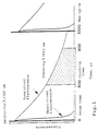
- FIG. 3 shows the wavelength, delay and intensity profiles of the emission from a mixture of PTA-chelated Eu +3 , Tb +3 , Sm +3 and Dy +3 .
- the solid phase carrier can be distributed into categories on the basis of either time-resolved labels fluorescing at different wavelengths, on the basis of different concentrations of time-resolved label, on the basis of simultaneous use of time-resolved and short-lived fluorescent labels, or a combination of these. For instance, by using two time-resolved labels fluorescing at different wavelengths, in ten different concentrations, for the identification of the categories of solid phase carrier, one hundred different solid phase carrier categories can be identified.
- solid phase carrier categories can be incorporated in one multiparameter assay by using microparticles containing fluorescent lanthanide chelates.
- Different fluorescent molecules can be linked to different or the same microparticles. It is advantageous to label the same microparticles with many different fluorescent molecules because the combinations of the different fluorescent chelates and/or the combinations of their concentrations will result in a great number of possible combinations.
- the solid phase carrier categories are identified by measuring the fluorescence intensity of the lanthanide chelate/chelates contained in the individual microparticles by using time-resolved fluorescence.
- the result of the commonly known specific bioaffinity reaction performed on the particle is determined by means of the fluorescent lanthanide chelate label from the identified microparticle for the measurement of the labelled bioaffinity reactant; a different lanthanide from the one used for the identification measurement is now employed.
- particles with a diameter of ⁇ 1 mm are used as solid phase carriers.
- the label(s) used for the identification of the microparticles can be linked to the microparticles during their manufacture by e.g. copolymerization.
- the label(s) can be chemically linked to the surface of the microparticles after the manufacturing process.
- Multiparameter assay could be typically applied to the assessment of the thyroid function in the human by simultaneously measuring in e.g. a blood sample the concentrations of thyroptropin (TSH), thyroxine (T3), free thyroxine (T4) and thyroglobulin (TG).
- TSH thyroptropin
- T3 thyroxine
- T4 free thyroxine
- TG thyroglobulin
- TSH and TG assays are non-competitive while T3 and free T4 assays are competitive.
- 1 x Tb particles are coated with a monoclonal TSH- specific antibody, 5 x TB particles with an anti-T3 antibody, 10 x Tb particles with an anti- free T4 antibody, and 15 x Tb particles with a monoclonal TG-specific antibody, respectively.
- the following bioaffinity reactants labelled with a fluorescent Eu chelate are used: Eu labelled monoclonal TSH-specific antibody, Eu labelled T3 derivative, Eu labelled T4 analogue, and Eu labelled monoclonal TG-specific antibody.
- the mixture of the particle categories; the sample, and the reactants labelled with the Eu chelate are allowed to react simultaneously in a one-step assay. After incubation the particle categories are identified on the basis of their concentration of Tb chelate, and the Eu concentration of individual identified particles is measured. Time-resolved fluorescence is used for both identification and measurement.
- the concentration of each analyte (TSH, T3, free T4 and TG) is calculated from the measured Eu concentrations.
- TSH, T3, free T4 and TG concentration of each analyte
- concentration of each analyte is calculated from the measured Eu concentrations.
- time-resolved fluorescence measurement either a flow cytometer, time-resolved microscope or time-resolved microfluorometer or other measuring instruments based on time-resolved technology are used (US 5,028,545; Xu, Y.Y. et al., Clin. Chem. 1992; 38/10: 2038 - 2043; Seveus, L. et al., Cytometry 1992; 13: 329 - 338).
- the multiparameter assay can be used for the diagnosis of an inherited disease by simultaneously detecting at the gene level a plurality of mutations associated with the disease. For instance, in the diagnosis of Duchenne muscular dystrophy (DMD) nine different deletion possibilities, all disease-associated, must be accurately identified in the gene.
- the multiparameter assay is performed in the following manner: the gene region to be examined is amplified by e.g.
- PCR polymerase chain reaction
- eighteen (nine normal and nine mutated alleles) particle categories identical in size and properties are used, which categories can be identified on the basis of the concentrations of the fluorescent Tb chelate they contain (1x, 2x, 4x, 8x, 16x, 32x, 64x, 128x, 256x, 521x, 1024x, 2048x, 4096x, 8192x, 16384x, 32768x, 65536x, 131072xTb).
- a nucleic acid probe bioaffinity reactant specifically identifying one of the aforementioned eighteen mutations, is immobilized onto the surface of each microparticle category.
- a mixture is prepared from the particle categories, which mixture contains all eighteen particle categories required for the detection of the mutations in DMD.
- sample and nine different nucleic acid probes are required.
- the category of individual particles is identified on the basis of the intensity of their Tb fluorescence, and the Eu concentration of individual particles is measured.
- the multiparameter assay thus allows the detection of all known nine mutations associated with DMD.
- Time-resolved fluorescence measurements are performed with either a flow cytometer, time-resolved fluorescence microscope or microfluorometer (US 5,028,545; Xu, Y.Y. et al., Clin, Chem. 1992; 38/10: 2038 - 2043; Seveus, L. et al., Cytometry 1992; 13: 329 - 338).
Description
- This invention relates to a biospecific assay method which allows the simultaneous determination of a plurality of analytes in the same sample.
- Microparticles can be employed as a solid phase carrier in various bioaffinity assays. In such assays it is often useful or necessary to perform a plurality of simultaneous assay reactions, thus eliminating repetition of parallel assay steps. One possibility to perform multiparameter analysis is offered by the use of microparticles as a solid phase carrier (Soini E., US 5,028,545). In such a system specific probes are linked to said particles, whereafter particles embodying different specificities may be mixed. The mixture can thus be used as a solid phase carrier in bioaffinity assays. When taking measurements after the completion of the reaction it is essential to be able to identify as many particle categories with different specificities as possible. For the purpose of identification such properties as the size of the particles (McHugh, T.M. et al., J. Immunol. Methods 1986; 95: 57 - 61) may be utilized, or coloured particles (Streefkerk J.G., Kors, N., Boden, D. Protides Biol. Fluids 1976; 24: 811 - 814) or particles labelled with various fluorescent molecules (Dean, K.J. et al. Clin. Chem. 1983; 29: 1051 - 1056) may be employed.
- In bioaffinity reactions two biological molecules are capable of binding with great accuracy even in the presence of other molecules. Such molecules capable of binding to each other are e.g. antibodies and their corresponding antigens, single-stranded DNA molecules and their corresponding nucleic acid sequences, and receptors and the molecules that specifically bind to them. All these reactions may be given the common denomination of bioaffinity reactions.
- Bioaffinity reactions are exploited in various practical applications and in assays in research. A known biomolecule, which may be purified from biological material (e.g. an antibody) or synthesized chemically (e.g. an oligonucleotide DNA) can be labelled in such a manner as to allow detection after the completion of the bioaffinity reaction. At present the major applications of bioaffinity reactions include immunoassays and nucleic acid hybridization assays, which are used to assay various components from e.g. blood. Assays based on receptors as biological reactants are important in the search for new medicinal substances.
- Assays performed on blood or its components may be divided in groups relevant to certain diseases or medical operations. In multiparameter assays the analytes belonging to these groups, so-called panels, could be assayed simultaneously in the same sample. This would enable economies to be made in the costs involved in testing, which would result in an increase in the number of tests that could be performed, and consequently in more reliable diagnoses. Multiparameter assays would also require considerably less work in performing the tests, and would consequently yield faster and more reliable results.
- Natural objects for assays employing DNA as the biological reactant would be viruses and inherited diseases, which are caused by mutations in cellular DNA. Especially inherited diseases may present multiple DNA mutations, and it would be imperative in the diagnosis for inherited diseases to be able to determine a number of DNA mutations simultaneously in the same sample. This would be especially useful in the search for carriers of these mutations in large studies covering whole populations.
- Because of the minute amount of the DNA to be analyzed the gene domain will often have to be amplified from the sample before the identification of the domain or the mutations. Several gene domains may be amplified simultaneously in the same reaction. Multiparameter assays may be advantageously applied to the analysis of such reaction mixtures containing several gene domains.
- In multiparameter assays labels can be used whose signals, after the completion of the bioaffinity reaction, can be resolved on the basis of e.g. wavelength or the decay time of the signal. Such assays have predominantly employed different radioactive isotopes (Morgan C.R., Proc. Soc. Exp. Biol. Med. 1966; 123: 230 - 233; Wians, F.H., Dev, J., Powell, M.M., Heald, J.I., Clin. Chem. 1986; 32: 887 - 890), enzyme labels (Dean, K.J., Thompson, S.G., Burg, J.F., Buckler, R.T., Clin. Chem. 1983; 29: 1051 - 1056; Bates, D.I., Bailey, W.R., International Patent Application WO89/06802), and labels emitting short-lived fluorescence. Such labels however often present problems due to overlapping of the wavelengths of the signals to be measured (isotopes, fluorescent molecules), to the proximity of the excitation and emission wavelengths (fluorescent molecules), to different optimal conditions (enzyme labels) or to background fluorescence of the material (labels emitting short-lived fluorescence). Such systems have at best allowed only two simultaneous determinations.
- Systems using label technology based on time-resolved fluorescence take advantage of the long-lived fluorescence of rare earth metals (Figure 1; Soini, E. and Lövgren, T., CRC Critical Reviews in Analytical Chemistry 1987; 18(2): 105 - 154). The metal is complexed to an organic molecule capable of absorbing energy during the excitation of the complex. On the other hand, the said ligand must also be able to release the excitation energy to the metal ion in the complex. The most useful earth metals in time-resolved labels are Eu, Sm, Tb and Dy. The energy levels of the electrons of the said rare metals are the best suited to absorb the excitation energy, and on the other hand, energy transitions which decrease long-lived fluorescence are slight in the complexes of these earth metals. The energy transitions to the ground state in these earth metals result in fluorescence consisting of several narrow bands of different wavelengths. Some electronic transitions are however, on transition to the ground state, preferred compared to others, and consequently the majority of the observed fluorescence has components of distinct wavelength. On simultaneous measurement in the bioaffinity assays of the reactants containing these time-resolved labels the effect of the minor components on measurement results can be easily eliminated by computational methods. In multiparameter assays using time-resolved labels as many as four different biomolecules have been measured simultaneously with required sensitivity (Xu, Y.Y., Pettersson, K., Blomberg, K., Hemmilä, I., Mikola, H. and Lövgren, T., Clin. Chem. 1992; 38/10: 2038 - 2043).
- An alternative possibility as the principle for multiparameter assays is available in the use of different solid phase carriers for the distinguishing of specific probes in bioaffinity assays. A solid phase carrier can be divided into domains each containing a probe of a determined specificity; different solid phase carriers can be used simultaneously in the assay, or the solid phase carrier can be distributed in different categories on the basis of a label (Soini, E., US 5,028,545). In the last example each solid phase category has a probe of different specificity bound to the surface. The application of this alternative principle to multiparameter assays presents several advantages because each component of the multiparameter system can be manufactured separately, and then combined. This is important from the point of view of production technology.
- This invention relates to a biospecific multiparameter assay method employing microparticles distributed in different categories and representing different analytes, in which method the microparticles are labelled with a fluorescent molecule (1) and in which method microparticles of different categories are coated with bioaffinity reactants A binding different analytes. In the said method the microparticles of different categories are mixed, the sample to be analyzed is added, and finally the bioaffinity reactants B labelled with a fluorescent molecule (2) are added either immediately or after the completion of the reaction between the analyte molecule and the bioaffinity reactant A. The fluorescent molecules (1, 2) are excited and the fluorescent emissions are quantitated for the identification of the microparticle category and for the measurement of the contents of different analytes. The method according to this invention is characterized by the labelling of the microparticles and of the bioaffinity reactants B with different molecules emitting long-lived fluorescence (1, 2).
- The said biospecific multiparameter assay method is performed either in one step with all the components of the reaction present simultaneously, or in two steps, in which case labelled bioaffinity reactant B is added after the completion of the reaction between bioaffinity reactant A and the analyte molecule.
- According to one useful variant method time-resolved fluorescence, i.e. a molecule emitting long-lived fluorescence, is used for the identification of the microparticle category as well as for the labelling of the bioaffinity reactant B.
-
- Figure 1 shows the fluorescense as function of time for long-lived and short-lived fluorescence
- Figure 2 shows the dose-response curve of europium where the standards (EuCl3) were made out in a fluorescence-enhancing solution and the fluorescence was measured over 1 second with a time-resolved fluorometer
- Figure 3 shows the wavelength, delay and intensity profiles of the emission from a mixture of PTA-chelated Eu+3, Tb+3, Sm+3 and Dy+3
-
- This invention employs time-resolved fluorescence (Soini, E. and Lövgren, T., CRC Critical Reviews in Analytical Chemistry 1987; 18(2): 105 - 154) for the labelling of the said particles or other type of solid phase carrier. Time-resolved fluorescence allows the elimination of the short-lived background fluorescence due to the natural fluorescence of different components of the system. Consequently, when the particles are labelled with different concentrations of the time-resolved label, an extremely low concentration of label can be detected. The sensitivity and the good linearity in the measurement of the time-resolved fluorescence allow the measurement of the time-resolved label over an extremely wide concentration range. Figure 2 shows the dose-response curve of europium where the standards (EuCl3) were made out in a fluorescence-enhancing solution and the fluorescence was measured over 1 second with a time-resolved fluorometer (1230 Fluorometer, LKB-Wallac, Turku, Finland). A wide concentration range on the other hand enables the identification of a plurality of different categories when differentiating the solid phase carriers. Time-resolved fluorescence technology also allows the use of labels emitting radiation of different wavelengths. (Saarma, M., Järvekulg, L., Hemmilä, I., Siitari, H., & Sinijärv, R., Journal of Virological Methods 1989; 23: 47 - 54; Xu, Y.Y., Pettersson, K., Blomberg, K., Hemmilä, I., Mikola, H. and Lövgren, T., Clin. Chem. 1992; 38/10: 2038 - 2043; Iitiä, A., Liukkonen, L. and Siitari, H., Molecular and Cellular Probes 1992; 6: 505 - 512). This invention also makes use of the combination of the concentrations of these labels emitting different wavelengths, in the labelling of the particles or other types of solid phase carriers.
- When using the microparticle as a solid phase carrier in the identification of different analytes in a multiparameter assay only one or a few labels are needed for the detection of the actual analyte. When the label technology is based on time-resolved fluorescence which allows the resolution of several labels, the use of a categorizable solid phase carrier will free one or more labels for use in the identification of the categories of the solid carrier. Figure 3 shows the wavelength, delay and intensity profiles of the emission from a mixture of PTA-chelated Eu+3, Tb+3, Sm+3 and Dy+3.
- The solid phase carrier can be distributed into categories on the basis of either time-resolved labels fluorescing at different wavelengths, on the basis of different concentrations of time-resolved label, on the basis of simultaneous use of time-resolved and short-lived fluorescent labels, or a combination of these. For instance, by using two time-resolved labels fluorescing at different wavelengths, in ten different concentrations, for the identification of the categories of solid phase carrier, one hundred different solid phase carrier categories can be identified.
- An extremely great number of identifiable solid phase carrier categories can be incorporated in one multiparameter assay by using microparticles containing fluorescent lanthanide chelates. Different fluorescent molecules can be linked to different or the same microparticles. It is advantageous to label the same microparticles with many different fluorescent molecules because the combinations of the different fluorescent chelates and/or the combinations of their concentrations will result in a great number of possible combinations. The solid phase carrier categories are identified by measuring the fluorescence intensity of the lanthanide chelate/chelates contained in the individual microparticles by using time-resolved fluorescence. After identification, the result of the commonly known specific bioaffinity reaction performed on the particle is determined by means of the fluorescent lanthanide chelate label from the identified microparticle for the measurement of the labelled bioaffinity reactant; a different lanthanide from the one used for the identification measurement is now employed. In multiparameter assays particles with a diameter of < 1 mm are used as solid phase carriers.
- The label(s) used for the identification of the microparticles can be linked to the microparticles during their manufacture by e.g. copolymerization. Alternatively, the label(s) can be chemically linked to the surface of the microparticles after the manufacturing process.
- The present invention will be described in more detail by means of the following non-restrictive examples.
- Multiparameter assay could be typically applied to the assessment of the thyroid function in the human by simultaneously measuring in e.g. a blood sample the concentrations of thyroptropin (TSH), thyroxine (T3), free thyroxine (T4) and thyroglobulin (TG). In this example four particle categories of identical size and properties, labelled for the purpose of the identification of the particle categories with different terbium chelate concentrations (1 x Tb, for the TSH assay; 5 x Tb, for the T3 assay; 10 x Tb, for the free T4 assay, and 15 x Tb, for the TG assay). During the production phase the particle categories are kept separate until it has been confirmed that they are functional in the said assays, at which point they are mixed for the multiparameter assay. TSH and TG assays are non-competitive while T3 and free T4 assays are competitive. 1 x Tb particles are coated with a monoclonal TSH- specific antibody, 5 x TB particles with an anti-T3 antibody, 10 x Tb particles with an anti- free T4 antibody, and 15 x Tb particles with a monoclonal TG-specific antibody, respectively. In the multiparameter assay the following bioaffinity reactants labelled with a fluorescent Eu chelate are used: Eu labelled monoclonal TSH-specific antibody, Eu labelled T3 derivative, Eu labelled T4 analogue, and Eu labelled monoclonal TG-specific antibody. In the multiparameter assay the mixture of the particle categories; the sample, and the reactants labelled with the Eu chelate are allowed to react simultaneously in a one-step assay. After incubation the particle categories are identified on the basis of their concentration of Tb chelate, and the Eu concentration of individual identified particles is measured. Time-resolved fluorescence is used for both identification and measurement. The concentration of each analyte (TSH, T3, free T4 and TG) is calculated from the measured Eu concentrations. In time-resolved fluorescence measurement either a flow cytometer, time-resolved microscope or time-resolved microfluorometer or other measuring instruments based on time-resolved technology are used (US 5,028,545; Xu, Y.Y. et al., Clin. Chem. 1992; 38/10: 2038 - 2043; Seveus, L. et al., Cytometry 1992; 13: 329 - 338).
- The multiparameter assay can be used for the diagnosis of an inherited disease by simultaneously detecting at the gene level a plurality of mutations associated with the disease. For instance, in the diagnosis of Duchenne muscular dystrophy (DMD) nine different deletion possibilities, all disease-associated, must be accurately identified in the gene. The multiparameter assay is performed in the following manner: the gene region to be examined is amplified by e.g. the PCR method (PCR = polymerase chain reaction), whereafter eighteen (nine normal and nine mutated alleles) particle categories identical in size and properties are used, which categories can be identified on the basis of the concentrations of the fluorescent Tb chelate they contain (1x, 2x, 4x, 8x, 16x, 32x, 64x, 128x, 256x, 521x, 1024x, 2048x, 4096x, 8192x, 16384x, 32768x, 65536x, 131072xTb). In the production step a nucleic acid probe (bioaffinity reactant) specifically identifying one of the aforementioned eighteen mutations, is immobilized onto the surface of each microparticle category. For the multiparameter assay a mixture is prepared from the particle categories, which mixture contains all eighteen particle categories required for the detection of the mutations in DMD. In the assay proper the said particle mixture, sample and nine different nucleic acid probes, all labelled with a fluorescent Eu chelate, are required. After incubation the category of individual particles is identified on the basis of the intensity of their Tb fluorescence, and the Eu concentration of individual particles is measured.
- The multiparameter assay thus allows the detection of all known nine mutations associated with DMD. Time-resolved fluorescence measurements are performed with either a flow cytometer, time-resolved fluorescence microscope or microfluorometer (US 5,028,545; Xu, Y.Y. et al., Clin, Chem. 1992; 38/10: 2038 - 2043; Seveus, L. et al., Cytometry 1992; 13: 329 - 338).
- A specialist in the field appreciates that the different applications of the said invention may vary within the scope of the claims. It will be appreciated that the methods of the present invention can be incorporated in the form of a variety of embodiments, only a few of which are disclosed herein. It will be apparent to the artisan that other embodiments exist. Thus, the described embodiments are illustrative and should not be construed as restrictive.
Claims (7)
- A biospecific multiparameter assay method using microparticles distributed into different categories and representing different analytes, in which method microparticles belonging to different categories are labelled with a fluorescent molecule (1) and in which method the microparticles belonging to different categories are coated with bioaffinity reactants A binding different analytes, in which methodcharacterized bymicroparticles belonging to different categories are mixed and the sample to be analyzed is added to the mixture, and bioaffinity reactants B labelled with a fluorescent molecule (2) are added to the mixture either immediately or after the completion of the reaction between the analyte molecule and the bioaffinity reactant A,the fluorescent molecules (1, 2) are excited, and the fluorescent emissions are quantitated for the identification of the microparticle category and for the measurement of the contents of different analytes,the labelling of the microparticles and of the bioaffinity reactants B with different fluorescent molecules emitting long-lived fluorescence (1, 2).
- The method according to claim 1, characterized by the use of two or more fluorescent molecules (1a, 1b...) emitting long-lived fluorescence for the identification of the microparticle category in such a manner that the different molecules (1a, 1b...) are located on the same or on different microparticles.
- The method according to claim 1, characterized by the occurrence of at least one of the molecules emitting long-lived fluorescence (1, 1a, 1b...) in several concentrations on the microparticles.
- The method according to claim 1, characterized by additionally labelling the microparticles with a molecule emitting short-lived fluorescence (3), which may occur on the same microparticles labelled with one or more molecules emitting long-lived fluorescence (1, 1a, 1b...) or on different microparticles.
- The method according to claim 4, characterized by the use of the molecule emitting short-lived fluorescence, in different concentrations.
- The method according to claims 1, characterized by Eu, Tb, Sm or Dy chelates being those molecules (1, 2) emitting long-lived fluorescence.
- The method according to claims 1, characterized by the diameter of the microparticles being less than 1 mm.
Applications Claiming Priority (2)
| Application Number | Priority Date | Filing Date | Title |
|---|---|---|---|
| FI931198 | 1993-03-18 | ||
| FI931198A FI93781C (en) | 1993-03-18 | 1993-03-18 | Biospecific multiparametric assay method |
Publications (3)
| Publication Number | Publication Date |
|---|---|
| EP0617286A2 EP0617286A2 (en) | 1994-09-28 |
| EP0617286A3 EP0617286A3 (en) | 1995-07-26 |
| EP0617286B1 true EP0617286B1 (en) | 1999-12-22 |
Family
ID=8537581
Family Applications (1)
| Application Number | Title | Priority Date | Filing Date |
|---|---|---|---|
| EP19940850024 Expired - Lifetime EP0617286B1 (en) | 1993-03-18 | 1994-02-16 | Biospecific solid phase carrier |
Country Status (5)
| Country | Link |
|---|---|
| EP (1) | EP0617286B1 (en) |
| JP (1) | JPH06317593A (en) |
| DE (1) | DE69422210T2 (en) |
| ES (1) | ES2139728T3 (en) |
| FI (1) | FI93781C (en) |
Cited By (51)
| Publication number | Priority date | Publication date | Assignee | Title |
|---|---|---|---|---|
| US7724786B2 (en) | 2003-06-06 | 2010-05-25 | The General Hospital Corporation | Process and apparatus for a wavelength tuning source |
| US7733497B2 (en) | 2003-10-27 | 2010-06-08 | The General Hospital Corporation | Method and apparatus for performing optical imaging using frequency-domain interferometry |
| US7742173B2 (en) | 2006-04-05 | 2010-06-22 | The General Hospital Corporation | Methods, arrangements and systems for polarization-sensitive optical frequency domain imaging of a sample |
| US7782464B2 (en) | 2006-05-12 | 2010-08-24 | The General Hospital Corporation | Processes, arrangements and systems for providing a fiber layer thickness map based on optical coherence tomography images |
| US7796270B2 (en) | 2006-01-10 | 2010-09-14 | The General Hospital Corporation | Systems and methods for generating data based on one or more spectrally-encoded endoscopy techniques |
| US7809225B2 (en) | 2004-07-02 | 2010-10-05 | The General Hospital Corporation | Imaging system and related techniques |
| US7843572B2 (en) | 2005-09-29 | 2010-11-30 | The General Hospital Corporation | Method and apparatus for optical imaging via spectral encoding |
| US7872757B2 (en) | 2002-01-24 | 2011-01-18 | The General Hospital Corporation | Apparatus and method for ranging and noise reduction of low coherence interferometry LCI and optical coherence tomography OCT signals by parallel detection of spectral bands |
| US7889348B2 (en) | 2005-10-14 | 2011-02-15 | The General Hospital Corporation | Arrangements and methods for facilitating photoluminescence imaging |
| US7898656B2 (en) | 2008-04-30 | 2011-03-01 | The General Hospital Corporation | Apparatus and method for cross axis parallel spectroscopy |
| US7920271B2 (en) | 2006-08-25 | 2011-04-05 | The General Hospital Corporation | Apparatus and methods for enhancing optical coherence tomography imaging using volumetric filtering techniques |
| US7933021B2 (en) | 2007-10-30 | 2011-04-26 | The General Hospital Corporation | System and method for cladding mode detection |
| US7949019B2 (en) | 2007-01-19 | 2011-05-24 | The General Hospital | Wavelength tuning source based on a rotatable reflector |
| US7982879B2 (en) | 2006-02-24 | 2011-07-19 | The General Hospital Corporation | Methods and systems for performing angle-resolved fourier-domain optical coherence tomography |
| US7995210B2 (en) | 2004-11-24 | 2011-08-09 | The General Hospital Corporation | Devices and arrangements for performing coherence range imaging using a common path interferometer |
| US8018598B2 (en) | 2004-05-29 | 2011-09-13 | The General Hospital Corporation | Process, system and software arrangement for a chromatic dispersion compensation using reflective layers in optical coherence tomography (OCT) imaging |
| US8040608B2 (en) | 2007-08-31 | 2011-10-18 | The General Hospital Corporation | System and method for self-interference fluorescence microscopy, and computer-accessible medium associated therewith |
| US8045177B2 (en) | 2007-04-17 | 2011-10-25 | The General Hospital Corporation | Apparatus and methods for measuring vibrations using spectrally-encoded endoscopy |
| US8050747B2 (en) | 2001-05-01 | 2011-11-01 | The General Hospital Corporation | Method and apparatus for determination of atherosclerotic plaque type by measurement of tissue optical properties |
| US8054468B2 (en) | 2003-01-24 | 2011-11-08 | The General Hospital Corporation | Apparatus and method for ranging and noise reduction of low coherence interferometry LCI and optical coherence tomography OCT signals by parallel detection of spectral bands |
| US8081316B2 (en) | 2004-08-06 | 2011-12-20 | The General Hospital Corporation | Process, system and software arrangement for determining at least one location in a sample using an optical coherence tomography |
| US8097864B2 (en) | 2009-01-26 | 2012-01-17 | The General Hospital Corporation | System, method and computer-accessible medium for providing wide-field superresolution microscopy |
| US8115919B2 (en) | 2007-05-04 | 2012-02-14 | The General Hospital Corporation | Methods, arrangements and systems for obtaining information associated with a sample using optical microscopy |
| US8145018B2 (en) | 2006-01-19 | 2012-03-27 | The General Hospital Corporation | Apparatus for obtaining information for a structure using spectrally-encoded endoscopy techniques and methods for producing one or more optical arrangements |
| US8175685B2 (en) | 2006-05-10 | 2012-05-08 | The General Hospital Corporation | Process, arrangements and systems for providing frequency domain imaging of a sample |
| US8174702B2 (en) | 2003-01-24 | 2012-05-08 | The General Hospital Corporation | Speckle reduction in optical coherence tomography by path length encoded angular compounding |
| US8208995B2 (en) | 2004-08-24 | 2012-06-26 | The General Hospital Corporation | Method and apparatus for imaging of vessel segments |
| USRE43875E1 (en) | 2004-09-29 | 2012-12-25 | The General Hospital Corporation | System and method for optical coherence imaging |
| US8351665B2 (en) | 2005-04-28 | 2013-01-08 | The General Hospital Corporation | Systems, processes and software arrangements for evaluating information associated with an anatomical structure by an optical coherence ranging technique |
| USRE44042E1 (en) | 2004-09-10 | 2013-03-05 | The General Hospital Corporation | System and method for optical coherence imaging |
| US8593619B2 (en) | 2008-05-07 | 2013-11-26 | The General Hospital Corporation | System, method and computer-accessible medium for tracking vessel motion during three-dimensional coronary artery microscopy |
| US8838213B2 (en) | 2006-10-19 | 2014-09-16 | The General Hospital Corporation | Apparatus and method for obtaining and providing imaging information associated with at least one portion of a sample, and effecting such portion(s) |
| US8861910B2 (en) | 2008-06-20 | 2014-10-14 | The General Hospital Corporation | Fused fiber optic coupler arrangement and method for use thereof |
| US8896838B2 (en) | 2010-03-05 | 2014-11-25 | The General Hospital Corporation | Systems, methods and computer-accessible medium which provide microscopic images of at least one anatomical structure at a particular resolution |
| US8922781B2 (en) | 2004-11-29 | 2014-12-30 | The General Hospital Corporation | Arrangements, devices, endoscopes, catheters and methods for performing optical imaging by simultaneously illuminating and detecting multiple points on a sample |
| US8937724B2 (en) | 2008-12-10 | 2015-01-20 | The General Hospital Corporation | Systems and methods for extending imaging depth range of optical coherence tomography through optical sub-sampling |
| US8965487B2 (en) | 2004-08-24 | 2015-02-24 | The General Hospital Corporation | Process, system and software arrangement for measuring a mechanical strain and elastic properties of a sample |
| US9069130B2 (en) | 2010-05-03 | 2015-06-30 | The General Hospital Corporation | Apparatus, method and system for generating optical radiation from biological gain media |
| US9087368B2 (en) | 2006-01-19 | 2015-07-21 | The General Hospital Corporation | Methods and systems for optical imaging or epithelial luminal organs by beam scanning thereof |
| US9176319B2 (en) | 2007-03-23 | 2015-11-03 | The General Hospital Corporation | Methods, arrangements and apparatus for utilizing a wavelength-swept laser using angular scanning and dispersion procedures |
| US9186067B2 (en) | 2006-02-01 | 2015-11-17 | The General Hospital Corporation | Apparatus for applying a plurality of electro-magnetic radiations to a sample |
| US9254089B2 (en) | 2008-07-14 | 2016-02-09 | The General Hospital Corporation | Apparatus and methods for facilitating at least partial overlap of dispersed ration on at least one sample |
| US9282931B2 (en) | 2000-10-30 | 2016-03-15 | The General Hospital Corporation | Methods for tissue analysis |
| US9295391B1 (en) | 2000-11-10 | 2016-03-29 | The General Hospital Corporation | Spectrally encoded miniature endoscopic imaging probe |
| US9330092B2 (en) | 2011-07-19 | 2016-05-03 | The General Hospital Corporation | Systems, methods, apparatus and computer-accessible-medium for providing polarization-mode dispersion compensation in optical coherence tomography |
| US9341783B2 (en) | 2011-10-18 | 2016-05-17 | The General Hospital Corporation | Apparatus and methods for producing and/or providing recirculating optical delay(s) |
| US9351642B2 (en) | 2009-03-12 | 2016-05-31 | The General Hospital Corporation | Non-contact optical system, computer-accessible medium and method for measurement at least one mechanical property of tissue using coherent speckle technique(s) |
| US9375158B2 (en) | 2007-07-31 | 2016-06-28 | The General Hospital Corporation | Systems and methods for providing beam scan patterns for high speed doppler optical frequency domain imaging |
| US9441948B2 (en) | 2005-08-09 | 2016-09-13 | The General Hospital Corporation | Apparatus, methods and storage medium for performing polarization-based quadrature demodulation in optical coherence tomography |
| US9510758B2 (en) | 2010-10-27 | 2016-12-06 | The General Hospital Corporation | Apparatus, systems and methods for measuring blood pressure within at least one vessel |
| US9557154B2 (en) | 2010-05-25 | 2017-01-31 | The General Hospital Corporation | Systems, devices, methods, apparatus and computer-accessible media for providing optical imaging of structures and compositions |
Families Citing this family (29)
| Publication number | Priority date | Publication date | Assignee | Title |
|---|---|---|---|---|
| US6716394B2 (en) | 1998-08-11 | 2004-04-06 | Caliper Technologies Corp. | DNA sequencing using multiple fluorescent labels being distinguishable by their decay times |
| EP1104491A4 (en) * | 1998-08-11 | 2003-01-29 | Caliper Techn Corp | Methods and systems for sequencing dna by distinguishing the decay times of fluorescent probes |
| WO2001035078A1 (en) * | 1999-11-09 | 2001-05-17 | Raytheon Company | Method and apparatus for performing cell analysis based on simultaneous multiple marker emissions from neoplasia (casmmen) |
| AU2001249738A1 (en) * | 2000-03-31 | 2001-10-15 | Glaxo Group Limited | Method and reagents for investigating functional molecular interactions |
| GB0601183D0 (en) * | 2006-01-20 | 2006-03-01 | Perkinelmer Ltd | Improvements in and relating to imaging |
| JP5524487B2 (en) | 2006-02-01 | 2014-06-18 | ザ ジェネラル ホスピタル コーポレイション | A method and system for emitting electromagnetic radiation to at least a portion of a sample using a conformal laser treatment procedure. |
| EP1988825B1 (en) | 2006-02-08 | 2016-12-21 | The General Hospital Corporation | Arrangements and systems for obtaining information associated with an anatomical sample using optical microscopy |
| US20080206804A1 (en) * | 2007-01-19 | 2008-08-28 | The General Hospital Corporation | Arrangements and methods for multidimensional multiplexed luminescence imaging and diagnosis |
| US10534129B2 (en) | 2007-03-30 | 2020-01-14 | The General Hospital Corporation | System and method providing intracoronary laser speckle imaging for the detection of vulnerable plaque |
| WO2010090837A2 (en) | 2009-01-20 | 2010-08-12 | The General Hospital Corporation | Endoscopic biopsy apparatus, system and method |
| US11490826B2 (en) | 2009-07-14 | 2022-11-08 | The General Hospital Corporation | Apparatus, systems and methods for measuring flow and pressure within a vessel |
| EP2575598A2 (en) | 2010-05-25 | 2013-04-10 | The General Hospital Corporation | Apparatus, systems, methods and computer-accessible medium for spectral analysis of optical coherence tomography images |
| US10285568B2 (en) | 2010-06-03 | 2019-05-14 | The General Hospital Corporation | Apparatus and method for devices for imaging structures in or at one or more luminal organs |
| WO2013029047A1 (en) | 2011-08-25 | 2013-02-28 | The General Hospital Corporation | Methods, systems, arrangements and computer-accessible medium for providing micro-optical coherence tomography procedures |
| US9629528B2 (en) | 2012-03-30 | 2017-04-25 | The General Hospital Corporation | Imaging system, method and distal attachment for multidirectional field of view endoscopy |
| WO2013177154A1 (en) | 2012-05-21 | 2013-11-28 | The General Hospital Corporation | Apparatus, device and method for capsule microscopy |
| US9968261B2 (en) | 2013-01-28 | 2018-05-15 | The General Hospital Corporation | Apparatus and method for providing diffuse spectroscopy co-registered with optical frequency domain imaging |
| WO2014120791A1 (en) | 2013-01-29 | 2014-08-07 | The General Hospital Corporation | Apparatus, systems and methods for providing information regarding the aortic valve |
| WO2014121082A1 (en) | 2013-02-01 | 2014-08-07 | The General Hospital Corporation | Objective lens arrangement for confocal endomicroscopy |
| EP2967491B1 (en) | 2013-03-15 | 2022-05-11 | The General Hospital Corporation | A transesophageal endoscopic system for determining a mixed venous oxygen saturation of a pulmonary artery |
| WO2014186353A1 (en) | 2013-05-13 | 2014-11-20 | The General Hospital Corporation | Detecting self-interefering fluorescence phase and amplitude |
| EP3021735A4 (en) | 2013-07-19 | 2017-04-19 | The General Hospital Corporation | Determining eye motion by imaging retina. with feedback |
| WO2015009932A1 (en) | 2013-07-19 | 2015-01-22 | The General Hospital Corporation | Imaging apparatus and method which utilizes multidirectional field of view endoscopy |
| ES2893237T3 (en) | 2013-07-26 | 2022-02-08 | Massachusetts Gen Hospital | Apparatus with a laser arrangement using optical scattering for applications in optical coherence tomography in the Fourier domain |
| US9733460B2 (en) | 2014-01-08 | 2017-08-15 | The General Hospital Corporation | Method and apparatus for microscopic imaging |
| WO2015116986A2 (en) | 2014-01-31 | 2015-08-06 | The General Hospital Corporation | System and method for facilitating manual and/or automatic volumetric imaging with real-time tension or force feedback using a tethered imaging device |
| WO2015153982A1 (en) | 2014-04-04 | 2015-10-08 | The General Hospital Corporation | Apparatus and method for controlling propagation and/or transmission of electromagnetic radiation in flexible waveguide(s) |
| EP3171766B1 (en) | 2014-07-25 | 2021-12-29 | The General Hospital Corporation | Apparatus for in vivo imaging and diagnosis |
| JP6924780B2 (en) * | 2016-06-09 | 2021-08-25 | ラジオメーター・トゥルク・オサケユキチュア | Background blocker for binding assay |
Family Cites Families (3)
| Publication number | Priority date | Publication date | Assignee | Title |
|---|---|---|---|---|
| SE454781B (en) * | 1986-10-17 | 1988-05-30 | Wallac Oy | HYBRIDIZATION PROCEDURE FOR THE DETECTION OF POLYNUCLEOTIDE SEQUENCE |
| SE458968B (en) * | 1987-06-16 | 1989-05-22 | Wallac Oy | BIOSPECIFIC ANALYTICAL PROCEDURE FOR MULTIPLE ANALYTICS WHICH DO NOT INCLUDE PARTICULAR COATING AND LABELING WITH FLUORESCING LABEL SUBSTANCES |
| WO1992012255A1 (en) * | 1990-12-28 | 1992-07-23 | Abbott Laboratories | Simultaneous determination of multiple analytes using a time-resolved heterogeneous chemiluminescence assay |
-
1993
- 1993-03-18 FI FI931198A patent/FI93781C/en active
-
1994
- 1994-02-16 DE DE1994622210 patent/DE69422210T2/en not_active Expired - Fee Related
- 1994-02-16 EP EP19940850024 patent/EP0617286B1/en not_active Expired - Lifetime
- 1994-02-16 ES ES94850024T patent/ES2139728T3/en not_active Expired - Lifetime
- 1994-03-17 JP JP4704294A patent/JPH06317593A/en active Pending
Cited By (82)
| Publication number | Priority date | Publication date | Assignee | Title |
|---|---|---|---|---|
| US9282931B2 (en) | 2000-10-30 | 2016-03-15 | The General Hospital Corporation | Methods for tissue analysis |
| US9295391B1 (en) | 2000-11-10 | 2016-03-29 | The General Hospital Corporation | Spectrally encoded miniature endoscopic imaging probe |
| US8050747B2 (en) | 2001-05-01 | 2011-11-01 | The General Hospital Corporation | Method and apparatus for determination of atherosclerotic plaque type by measurement of tissue optical properties |
| US7872757B2 (en) | 2002-01-24 | 2011-01-18 | The General Hospital Corporation | Apparatus and method for ranging and noise reduction of low coherence interferometry LCI and optical coherence tomography OCT signals by parallel detection of spectral bands |
| US8054468B2 (en) | 2003-01-24 | 2011-11-08 | The General Hospital Corporation | Apparatus and method for ranging and noise reduction of low coherence interferometry LCI and optical coherence tomography OCT signals by parallel detection of spectral bands |
| US8559012B2 (en) | 2003-01-24 | 2013-10-15 | The General Hospital Corporation | Speckle reduction in optical coherence tomography by path length encoded angular compounding |
| US8174702B2 (en) | 2003-01-24 | 2012-05-08 | The General Hospital Corporation | Speckle reduction in optical coherence tomography by path length encoded angular compounding |
| US9226665B2 (en) | 2003-01-24 | 2016-01-05 | The General Hospital Corporation | Speckle reduction in optical coherence tomography by path length encoded angular compounding |
| US8416818B2 (en) | 2003-06-06 | 2013-04-09 | The General Hospital Corporation | Process and apparatus for a wavelength tuning source |
| US7995627B2 (en) | 2003-06-06 | 2011-08-09 | The General Hospital Corporation | Process and apparatus for a wavelength tuning source |
| US7724786B2 (en) | 2003-06-06 | 2010-05-25 | The General Hospital Corporation | Process and apparatus for a wavelength tuning source |
| US7733497B2 (en) | 2003-10-27 | 2010-06-08 | The General Hospital Corporation | Method and apparatus for performing optical imaging using frequency-domain interferometry |
| US8705046B2 (en) | 2003-10-27 | 2014-04-22 | The General Hospital Corporation | Method and apparatus for performing optical imaging using frequency-domain interferometry |
| US9377290B2 (en) | 2003-10-27 | 2016-06-28 | The General Hospital Corporation | Method and apparatus for performing optical imaging using frequency-domain interferometry |
| US8355138B2 (en) | 2003-10-27 | 2013-01-15 | The General Hospital Corporation | Method and apparatus for performing optical imaging using frequency-domain interferometry |
| US7969578B2 (en) | 2003-10-27 | 2011-06-28 | The General Hospital Corporation | Method and apparatus for performing optical imaging using frequency-domain interferometry |
| US8384909B2 (en) | 2003-10-27 | 2013-02-26 | The General Hospital Corporation | Method and apparatus for performing optical imaging using frequency-domain interferometry |
| US8018598B2 (en) | 2004-05-29 | 2011-09-13 | The General Hospital Corporation | Process, system and software arrangement for a chromatic dispersion compensation using reflective layers in optical coherence tomography (OCT) imaging |
| US7809225B2 (en) | 2004-07-02 | 2010-10-05 | The General Hospital Corporation | Imaging system and related techniques |
| US8676013B2 (en) | 2004-07-02 | 2014-03-18 | The General Hospital Corporation | Imaging system using and related techniques |
| US7925133B2 (en) | 2004-07-02 | 2011-04-12 | The General Hospital Corporation | Imaging system and related techniques |
| US8369669B2 (en) | 2004-07-02 | 2013-02-05 | The General Hospital Corporation | Imaging system and related techniques |
| US9226660B2 (en) | 2004-08-06 | 2016-01-05 | The General Hospital Corporation | Process, system and software arrangement for determining at least one location in a sample using an optical coherence tomography |
| US8081316B2 (en) | 2004-08-06 | 2011-12-20 | The General Hospital Corporation | Process, system and software arrangement for determining at least one location in a sample using an optical coherence tomography |
| US8965487B2 (en) | 2004-08-24 | 2015-02-24 | The General Hospital Corporation | Process, system and software arrangement for measuring a mechanical strain and elastic properties of a sample |
| US9254102B2 (en) | 2004-08-24 | 2016-02-09 | The General Hospital Corporation | Method and apparatus for imaging of vessel segments |
| US8208995B2 (en) | 2004-08-24 | 2012-06-26 | The General Hospital Corporation | Method and apparatus for imaging of vessel segments |
| USRE44042E1 (en) | 2004-09-10 | 2013-03-05 | The General Hospital Corporation | System and method for optical coherence imaging |
| USRE45512E1 (en) | 2004-09-29 | 2015-05-12 | The General Hospital Corporation | System and method for optical coherence imaging |
| USRE43875E1 (en) | 2004-09-29 | 2012-12-25 | The General Hospital Corporation | System and method for optical coherence imaging |
| US7995210B2 (en) | 2004-11-24 | 2011-08-09 | The General Hospital Corporation | Devices and arrangements for performing coherence range imaging using a common path interferometer |
| US8922781B2 (en) | 2004-11-29 | 2014-12-30 | The General Hospital Corporation | Arrangements, devices, endoscopes, catheters and methods for performing optical imaging by simultaneously illuminating and detecting multiple points on a sample |
| US8351665B2 (en) | 2005-04-28 | 2013-01-08 | The General Hospital Corporation | Systems, processes and software arrangements for evaluating information associated with an anatomical structure by an optical coherence ranging technique |
| US9326682B2 (en) | 2005-04-28 | 2016-05-03 | The General Hospital Corporation | Systems, processes and software arrangements for evaluating information associated with an anatomical structure by an optical coherence ranging technique |
| US9441948B2 (en) | 2005-08-09 | 2016-09-13 | The General Hospital Corporation | Apparatus, methods and storage medium for performing polarization-based quadrature demodulation in optical coherence tomography |
| US7843572B2 (en) | 2005-09-29 | 2010-11-30 | The General Hospital Corporation | Method and apparatus for optical imaging via spectral encoding |
| US7847949B2 (en) | 2005-09-29 | 2010-12-07 | The General Hospital Corporation | Method and apparatus for optical imaging via spectral encoding |
| US7872759B2 (en) | 2005-09-29 | 2011-01-18 | The General Hospital Corporation | Arrangements and methods for providing multimodality microscopic imaging of one or more biological structures |
| US9304121B2 (en) | 2005-09-29 | 2016-04-05 | The General Hospital Corporation | Method and apparatus for optical imaging via spectral encoding |
| US8384907B2 (en) | 2005-09-29 | 2013-02-26 | The General Hospital Corporation | Method and apparatus for optical imaging via spectral encoding |
| US8760663B2 (en) | 2005-09-29 | 2014-06-24 | The General Hospital Corporation | Method and apparatus for optical imaging via spectral encoding |
| US8928889B2 (en) | 2005-09-29 | 2015-01-06 | The General Hospital Corporation | Arrangements and methods for providing multimodality microscopic imaging of one or more biological structures |
| US8289522B2 (en) | 2005-09-29 | 2012-10-16 | The General Hospital Corporation | Arrangements and methods for providing multimodality microscopic imaging of one or more biological structures |
| US9513276B2 (en) | 2005-09-29 | 2016-12-06 | The General Hospital Corporation | Method and apparatus for optical imaging via spectral encoding |
| US8149418B2 (en) | 2005-09-29 | 2012-04-03 | The General Hospital Corporation | Method and apparatus for optical imaging via spectral encoding |
| US7889348B2 (en) | 2005-10-14 | 2011-02-15 | The General Hospital Corporation | Arrangements and methods for facilitating photoluminescence imaging |
| US7796270B2 (en) | 2006-01-10 | 2010-09-14 | The General Hospital Corporation | Systems and methods for generating data based on one or more spectrally-encoded endoscopy techniques |
| US8818149B2 (en) | 2006-01-19 | 2014-08-26 | The General Hospital Corporation | Spectrally-encoded endoscopy techniques, apparatus and methods |
| US9087368B2 (en) | 2006-01-19 | 2015-07-21 | The General Hospital Corporation | Methods and systems for optical imaging or epithelial luminal organs by beam scanning thereof |
| US9516997B2 (en) | 2006-01-19 | 2016-12-13 | The General Hospital Corporation | Spectrally-encoded endoscopy techniques, apparatus and methods |
| US8145018B2 (en) | 2006-01-19 | 2012-03-27 | The General Hospital Corporation | Apparatus for obtaining information for a structure using spectrally-encoded endoscopy techniques and methods for producing one or more optical arrangements |
| US9186066B2 (en) | 2006-02-01 | 2015-11-17 | The General Hospital Corporation | Apparatus for applying a plurality of electro-magnetic radiations to a sample |
| US9186067B2 (en) | 2006-02-01 | 2015-11-17 | The General Hospital Corporation | Apparatus for applying a plurality of electro-magnetic radiations to a sample |
| US7982879B2 (en) | 2006-02-24 | 2011-07-19 | The General Hospital Corporation | Methods and systems for performing angle-resolved fourier-domain optical coherence tomography |
| US7742173B2 (en) | 2006-04-05 | 2010-06-22 | The General Hospital Corporation | Methods, arrangements and systems for polarization-sensitive optical frequency domain imaging of a sample |
| US9364143B2 (en) | 2006-05-10 | 2016-06-14 | The General Hospital Corporation | Process, arrangements and systems for providing frequency domain imaging of a sample |
| US8175685B2 (en) | 2006-05-10 | 2012-05-08 | The General Hospital Corporation | Process, arrangements and systems for providing frequency domain imaging of a sample |
| US7782464B2 (en) | 2006-05-12 | 2010-08-24 | The General Hospital Corporation | Processes, arrangements and systems for providing a fiber layer thickness map based on optical coherence tomography images |
| US7920271B2 (en) | 2006-08-25 | 2011-04-05 | The General Hospital Corporation | Apparatus and methods for enhancing optical coherence tomography imaging using volumetric filtering techniques |
| US8838213B2 (en) | 2006-10-19 | 2014-09-16 | The General Hospital Corporation | Apparatus and method for obtaining and providing imaging information associated with at least one portion of a sample, and effecting such portion(s) |
| US7949019B2 (en) | 2007-01-19 | 2011-05-24 | The General Hospital | Wavelength tuning source based on a rotatable reflector |
| US9176319B2 (en) | 2007-03-23 | 2015-11-03 | The General Hospital Corporation | Methods, arrangements and apparatus for utilizing a wavelength-swept laser using angular scanning and dispersion procedures |
| US8045177B2 (en) | 2007-04-17 | 2011-10-25 | The General Hospital Corporation | Apparatus and methods for measuring vibrations using spectrally-encoded endoscopy |
| US8115919B2 (en) | 2007-05-04 | 2012-02-14 | The General Hospital Corporation | Methods, arrangements and systems for obtaining information associated with a sample using optical microscopy |
| US9375158B2 (en) | 2007-07-31 | 2016-06-28 | The General Hospital Corporation | Systems and methods for providing beam scan patterns for high speed doppler optical frequency domain imaging |
| US8040608B2 (en) | 2007-08-31 | 2011-10-18 | The General Hospital Corporation | System and method for self-interference fluorescence microscopy, and computer-accessible medium associated therewith |
| US7933021B2 (en) | 2007-10-30 | 2011-04-26 | The General Hospital Corporation | System and method for cladding mode detection |
| US7898656B2 (en) | 2008-04-30 | 2011-03-01 | The General Hospital Corporation | Apparatus and method for cross axis parallel spectroscopy |
| US8593619B2 (en) | 2008-05-07 | 2013-11-26 | The General Hospital Corporation | System, method and computer-accessible medium for tracking vessel motion during three-dimensional coronary artery microscopy |
| US8861910B2 (en) | 2008-06-20 | 2014-10-14 | The General Hospital Corporation | Fused fiber optic coupler arrangement and method for use thereof |
| US9254089B2 (en) | 2008-07-14 | 2016-02-09 | The General Hospital Corporation | Apparatus and methods for facilitating at least partial overlap of dispersed ration on at least one sample |
| US8937724B2 (en) | 2008-12-10 | 2015-01-20 | The General Hospital Corporation | Systems and methods for extending imaging depth range of optical coherence tomography through optical sub-sampling |
| US8097864B2 (en) | 2009-01-26 | 2012-01-17 | The General Hospital Corporation | System, method and computer-accessible medium for providing wide-field superresolution microscopy |
| US9351642B2 (en) | 2009-03-12 | 2016-05-31 | The General Hospital Corporation | Non-contact optical system, computer-accessible medium and method for measurement at least one mechanical property of tissue using coherent speckle technique(s) |
| US9408539B2 (en) | 2010-03-05 | 2016-08-09 | The General Hospital Corporation | Systems, methods and computer-accessible medium which provide microscopic images of at least one anatomical structure at a particular resolution |
| US8896838B2 (en) | 2010-03-05 | 2014-11-25 | The General Hospital Corporation | Systems, methods and computer-accessible medium which provide microscopic images of at least one anatomical structure at a particular resolution |
| US9081148B2 (en) | 2010-03-05 | 2015-07-14 | The General Hospital Corporation | Systems, methods and computer-accessible medium which provide microscopic images of at least one anatomical structure at a particular resolution |
| US9069130B2 (en) | 2010-05-03 | 2015-06-30 | The General Hospital Corporation | Apparatus, method and system for generating optical radiation from biological gain media |
| US9557154B2 (en) | 2010-05-25 | 2017-01-31 | The General Hospital Corporation | Systems, devices, methods, apparatus and computer-accessible media for providing optical imaging of structures and compositions |
| US9510758B2 (en) | 2010-10-27 | 2016-12-06 | The General Hospital Corporation | Apparatus, systems and methods for measuring blood pressure within at least one vessel |
| US9330092B2 (en) | 2011-07-19 | 2016-05-03 | The General Hospital Corporation | Systems, methods, apparatus and computer-accessible-medium for providing polarization-mode dispersion compensation in optical coherence tomography |
| US9341783B2 (en) | 2011-10-18 | 2016-05-17 | The General Hospital Corporation | Apparatus and methods for producing and/or providing recirculating optical delay(s) |
Also Published As
| Publication number | Publication date |
|---|---|
| FI93781C (en) | 1995-05-26 |
| EP0617286A2 (en) | 1994-09-28 |
| DE69422210D1 (en) | 2000-01-27 |
| EP0617286A3 (en) | 1995-07-26 |
| DE69422210T2 (en) | 2000-04-20 |
| FI931198A0 (en) | 1993-03-18 |
| ES2139728T3 (en) | 2000-02-16 |
| JPH06317593A (en) | 1994-11-15 |
| FI931198A (en) | 1994-09-19 |
| FI93781B (en) | 1995-02-15 |
Similar Documents
| Publication | Publication Date | Title |
|---|---|---|
| EP0617286B1 (en) | Biospecific solid phase carrier | |
| EP0154734B1 (en) | Immediate ligand detection assay, a test kit and its formation | |
| Harma et al. | Europium nanoparticles and time-resolved fluorescence for ultrasensitive detection of prostate-specific antigen | |
| US5567627A (en) | Method and composition for the simultaneous and discrete analysis of multiple analytes | |
| US5756011A (en) | Detecting or quantifying multiple analytes using labelling techniques | |
| Bock | The new era of automated immunoassay | |
| US8956823B2 (en) | Anti-antibody reagent | |
| JP3021038B2 (en) | Detection or quantification of multiple analytes using labeling technology | |
| US6551788B1 (en) | Particle-based ligand assay with extended dynamic range | |
| JP2010175355A (en) | Automatic analyzer | |
| JPH0319948B2 (en) | ||
| JPH06242117A (en) | Measuring reagent and measuring method for nucleic acid and measuring reagent kit for nucleic acid | |
| JP6647392B2 (en) | Simultaneous analysis of multiple targets using multiple metal nanotags | |
| CA2138536C (en) | Chemiluminescent assay for dsdna antibodies | |
| Soini | Biospecific assays with time-resolved fluorescence detection | |
| Woodhead et al. | Magic Lite design and development | |
| WO1998054578A1 (en) | Chemiluminescent hemoglobin assay | |
| RU2710262C1 (en) | Biological analysis method | |
| JP2004536042A (en) | Phycoerythrin labeled thyronine analogs and assays using said labeled analogs | |
| Ekins | Competitive, noncompetitive and multianalyte microspot immunoassays | |
| JPS6281566A (en) | Quantification method by measurement of fluorescent intensity of fine particle | |
| US20230384297A1 (en) | Electrothermal flow-enhanced electrochemical magneto-immunosensor | |
| Soini | Biospecific assays with time-resolved | |
| CN116068184A (en) | Application method of immunoassay kit and detection system thereof | |
| CN117604072A (en) | Homogeneous phase time-resolved fluoroimmunoassay method based on CRISPR/Cas13a |
Legal Events
| Date | Code | Title | Description |
|---|---|---|---|
| PUAI | Public reference made under article 153(3) epc to a published international application that has entered the european phase |
Free format text: ORIGINAL CODE: 0009012 |
|
| AK | Designated contracting states |
Kind code of ref document: A2 Designated state(s): DE ES FR GB IT SE |
|
| PUAL | Search report despatched |
Free format text: ORIGINAL CODE: 0009013 |
|
| AK | Designated contracting states |
Kind code of ref document: A3 Designated state(s): DE ES FR GB IT SE |
|
| 17P | Request for examination filed |
Effective date: 19950828 |
|
| 17Q | First examination report despatched |
Effective date: 19980804 |
|
| GRAG | Despatch of communication of intention to grant |
Free format text: ORIGINAL CODE: EPIDOS AGRA |
|
| GRAG | Despatch of communication of intention to grant |
Free format text: ORIGINAL CODE: EPIDOS AGRA |
|
| GRAH | Despatch of communication of intention to grant a patent |
Free format text: ORIGINAL CODE: EPIDOS IGRA |
|
| GRAH | Despatch of communication of intention to grant a patent |
Free format text: ORIGINAL CODE: EPIDOS IGRA |
|
| GRAA | (expected) grant |
Free format text: ORIGINAL CODE: 0009210 |
|
| AK | Designated contracting states |
Kind code of ref document: B1 Designated state(s): DE ES FR GB IT SE |
|
| PG25 | Lapsed in a contracting state [announced via postgrant information from national office to epo] |
Ref country code: SE Free format text: THE PATENT HAS BEEN ANNULLED BY A DECISION OF A NATIONAL AUTHORITY Effective date: 19991222 |
|
| ITF | It: translation for a ep patent filed |
Owner name: JACOBACCI & PERANI S.P.A. |
|
| REF | Corresponds to: |
Ref document number: 69422210 Country of ref document: DE Date of ref document: 20000127 |
|
| ET | Fr: translation filed | ||
| REG | Reference to a national code |
Ref country code: ES Ref legal event code: FG2A Ref document number: 2139728 Country of ref document: ES Kind code of ref document: T3 |
|
| PLBE | No opposition filed within time limit |
Free format text: ORIGINAL CODE: 0009261 |
|
| STAA | Information on the status of an ep patent application or granted ep patent |
Free format text: STATUS: NO OPPOSITION FILED WITHIN TIME LIMIT |
|
| 26N | No opposition filed | ||
| REG | Reference to a national code |
Ref country code: GB Ref legal event code: IF02 |
|
| PGFP | Annual fee paid to national office [announced via postgrant information from national office to epo] |
Ref country code: IT Payment date: 20060228 Year of fee payment: 13 |
|
| PGFP | Annual fee paid to national office [announced via postgrant information from national office to epo] |
Ref country code: FR Payment date: 20070131 Year of fee payment: 14 Ref country code: ES Payment date: 20080226 Year of fee payment: 15 |
|
| PGFP | Annual fee paid to national office [announced via postgrant information from national office to epo] |
Ref country code: GB Payment date: 20080227 Year of fee payment: 15 |
|
| PGFP | Annual fee paid to national office [announced via postgrant information from national office to epo] |
Ref country code: DE Payment date: 20080331 Year of fee payment: 15 |
|
| REG | Reference to a national code |
Ref country code: FR Ref legal event code: ST Effective date: 20081031 |
|
| PG25 | Lapsed in a contracting state [announced via postgrant information from national office to epo] |
Ref country code: FR Free format text: LAPSE BECAUSE OF NON-PAYMENT OF DUE FEES Effective date: 20080229 |
|
| PG25 | Lapsed in a contracting state [announced via postgrant information from national office to epo] |
Ref country code: IT Free format text: LAPSE BECAUSE OF NON-PAYMENT OF DUE FEES Effective date: 20070216 |
|
| GBPC | Gb: european patent ceased through non-payment of renewal fee |
Effective date: 20090216 |
|
| PG25 | Lapsed in a contracting state [announced via postgrant information from national office to epo] |
Ref country code: DE Free format text: LAPSE BECAUSE OF NON-PAYMENT OF DUE FEES Effective date: 20090901 |
|
| REG | Reference to a national code |
Ref country code: ES Ref legal event code: FD2A Effective date: 20090217 |
|
| PG25 | Lapsed in a contracting state [announced via postgrant information from national office to epo] |
Ref country code: GB Free format text: LAPSE BECAUSE OF NON-PAYMENT OF DUE FEES Effective date: 20090216 |
|
| PG25 | Lapsed in a contracting state [announced via postgrant information from national office to epo] |
Ref country code: ES Free format text: LAPSE BECAUSE OF NON-PAYMENT OF DUE FEES Effective date: 20090217 |