EP0624381A2 - A peel-away endoscopic retrograde, cholangio pancreatography catheter and a method for using the same - Google Patents
A peel-away endoscopic retrograde, cholangio pancreatography catheter and a method for using the same Download PDFInfo
- Publication number
- EP0624381A2 EP0624381A2 EP94303458A EP94303458A EP0624381A2 EP 0624381 A2 EP0624381 A2 EP 0624381A2 EP 94303458 A EP94303458 A EP 94303458A EP 94303458 A EP94303458 A EP 94303458A EP 0624381 A2 EP0624381 A2 EP 0624381A2
- Authority
- EP
- European Patent Office
- Prior art keywords
- catheter
- guide wire
- endoscope
- common bile
- bile duct
- Prior art date
- Legal status (The legal status is an assumption and is not a legal conclusion. Google has not performed a legal analysis and makes no representation as to the accuracy of the status listed.)
- Granted
Links
Images


Classifications
-
- A—HUMAN NECESSITIES
- A61—MEDICAL OR VETERINARY SCIENCE; HYGIENE
- A61M—DEVICES FOR INTRODUCING MEDIA INTO, OR ONTO, THE BODY; DEVICES FOR TRANSDUCING BODY MEDIA OR FOR TAKING MEDIA FROM THE BODY; DEVICES FOR PRODUCING OR ENDING SLEEP OR STUPOR
- A61M25/00—Catheters; Hollow probes
- A61M25/0067—Catheters; Hollow probes characterised by the distal end, e.g. tips
- A61M25/0068—Static characteristics of the catheter tip, e.g. shape, atraumatic tip, curved tip or tip structure
- A61M25/007—Side holes, e.g. their profiles or arrangements; Provisions to keep side holes unblocked
-
- A—HUMAN NECESSITIES
- A61—MEDICAL OR VETERINARY SCIENCE; HYGIENE
- A61M—DEVICES FOR INTRODUCING MEDIA INTO, OR ONTO, THE BODY; DEVICES FOR TRANSDUCING BODY MEDIA OR FOR TAKING MEDIA FROM THE BODY; DEVICES FOR PRODUCING OR ENDING SLEEP OR STUPOR
- A61M25/00—Catheters; Hollow probes
- A61M25/01—Introducing, guiding, advancing, emplacing or holding catheters
- A61M25/06—Body-piercing guide needles or the like
- A61M25/0662—Guide tubes
- A61M25/0668—Guide tubes splittable, tear apart
-
- A—HUMAN NECESSITIES
- A61—MEDICAL OR VETERINARY SCIENCE; HYGIENE
- A61M—DEVICES FOR INTRODUCING MEDIA INTO, OR ONTO, THE BODY; DEVICES FOR TRANSDUCING BODY MEDIA OR FOR TAKING MEDIA FROM THE BODY; DEVICES FOR PRODUCING OR ENDING SLEEP OR STUPOR
- A61M25/00—Catheters; Hollow probes
- A61M25/0067—Catheters; Hollow probes characterised by the distal end, e.g. tips
- A61M25/0082—Catheter tip comprising a tool
- A61M2025/0096—Catheter tip comprising a tool being laterally outward extensions or tools, e.g. hooks or fibres
-
- A—HUMAN NECESSITIES
- A61—MEDICAL OR VETERINARY SCIENCE; HYGIENE
- A61M—DEVICES FOR INTRODUCING MEDIA INTO, OR ONTO, THE BODY; DEVICES FOR TRANSDUCING BODY MEDIA OR FOR TAKING MEDIA FROM THE BODY; DEVICES FOR PRODUCING OR ENDING SLEEP OR STUPOR
- A61M2202/00—Special media to be introduced, removed or treated
- A61M2202/04—Liquids
- A61M2202/0403—Gall; Bile
Definitions
- This invention relates to a splittable or peel-away ERCP catheter and more particularly to a method of using a peel-away ERCP catheter the distal end of which has been inserted into the common bile or pancreatic duct via an endoscope which has been guided through the esophagus and stomach and into the duodenum of a patient.
- a sheath or catheter that is longitudinally splittable for ease of removal from the human body.
- a splittable catheter permits the removal of a catheter after a guide wire or other material has been placed therethrough without disturbing the guide wire.
- Various splittable sheaths and catheters exist which have been designed to split longitudinally along the body to peel-away from a catheter, guide wire or other inserted material.
- U.S. Patent No. 4,776,846 to Wells discloses an apparatus and method for preparing a catheter tube which has a longitudinal line of weakness extending axially therealong as a place for preferential splitting.
- the method includes co-extruding two materials into the tubular cross-sectional shape. One of the materials forms a surface line along the otherwhich extends readily to a level where the thickness of the tubing cross-section thereat is less. When the co-extruded extra material is removed, a grove is in the surface of the tube, which groove permits easy tearing therealong.
- U. S. Patent No. 4,983,168 to Moorehead discloses a layered peel-away hollow sheath, for temporarily creating a passageway into a desired body site of a medical patient for placement of one end of an indwelling device at the body site through the sheath.
- the sheath wall comprises at least two layers, the inside layer being cylindrical and the outside layer comprising two semi-cylindrical segments defining opposed axially directed slits or slots therebetween which comprise tear lines such that the sheath manually tears axially along the single layer tear lines into two pieces for removal of the sheath from around the indwelling device.
- U.S. Patent No. 4,801,294 to Okada discloses a catheter for nasogastric intubation comprising a plastic sheath tube and a fixing means of said tube at nose, said tube having a longitudinal tear-off line over the full length thereof and having elasticity and rigidity slightly larger than those of said catheter, said catheter being inserted in said plastic sheath tube slidably.
- U.S. Patent No. 3,550,591 to McGregor discloses an intravenous catheter unit which provides a needle which can be separated from the catheter after the end of the catheter positioned inside the vein to prevent the needle from tearing or damaging the catheter.
- the hollow needle is provided with a slit which extends along its entire length.
- the needle is provided with means of the causing the needle to hinge open to widen the slit enough to pass the catheter therethrough. In this way the needle may be separated from the catheter after the catheter is placed inside the vein.
- U.S. Patent No. 4,781,690 to Oshada, et al. discloses a guiding tube for introducing rodlike medical instruments such as a catheter, which can be easily splitand removed.
- the guiding tube comprises a main body provided with a longitudinal slit and formed of a mixture of first and second materials and a stripe detachably embedded liquid-tight in the slit of the main body and formed of the third material which exhibits a good bonding for the second material but a poor bonding for the first material.
- U.S. Patent No. 4,747,833 to Kousai, et al. discloses a medical instrument-guiding tube for guiding a catheter or other rod-like medical instrument into a blood vessel.
- This guiding tube comprises a hollow- tube body and at least one linear body integrally joined to the tube body along the longitudinal direction of the tube body.
- the plastic resin forming the tube body has a poor compatibi lity with that of the linear body. Tube body and the linear body are engaged together through complementary concave convex arrangement which can be disengaged with reasonable force.
- U.S. Patent No. 4,883,468 to Kousai, et al. discloses a medical instrument introduction cannula which is useful as a guide means for introducing and indwelling a rod-like material instrument such as a catheter and a guide wire.
- This cannula is removed after use from the medical instrument by being split.
- This cannula is formed of a hollow tubular body having a strip member consisting of material different from the other portion of the tubular body, which extends over almost the entire length of the tubular body.
- the strip member has a weld line along the length of the strip member, or consists of a resin which exhibits a good bonding property only to one of the other components forming the tubular body.
- the tubular body can be split by way of the weld line or the removal of the strip member having such a bonding property.
- the method of manufacturing the medical instrument introduction cannula by a two-color extruder is also disclosed.
- U.S. Patent No. 4,997,424 to Little relates to an introducer slitterfor slitting an introducer tube portion having, for example, a catheter extending therethrough and into a body vessel while the introducer tube is moved rearwardly relative to the catheter to facilitate separating the introducer from the catheter without having to slide the introducer tube portion over the proximal end of the catheter.
- U.S. Patent No. 5,195,978 to Schiffer discloses a rapid exchange over-the-wire catheter which is provided with one or more breakaway elements for progressively exposing the guide wire from the proximal end toward the distal end of the catheter in a simple and efficient manner.
- the breakaway element may be formed as a longitudinally aligned pull strip provided in the catheter guide wire lumen or as one or more linearly arrayed tubular breakaway segments in the catheter shaft or as a combination of both features.
- U.S. Patent No. 4,813,929 to Semrad discloses a method and assembly of components for effecting closed chest thoracostomy.
- a guide wire is removed and a chest tube is introduced through a pleural access catheter, which is split off and removed from the chest tube.
- Figures 5 and 6 of the Semrad reference show a splittable access catheter having knobs that when grasped and pulled in a direction away from the body of the catheter will cause the catheter to split.
- U.S. Patent Nos. 4,243,050 and 4,345,606 to Littleford disclose an apparatus and related method for implanting pacemaker electrodes and similar devices within a patient including an introducer with a tapered end adapted to extend into the patient's body, and a hollow, tubular sleeve.
- the sleeve is perforated to form a weakened line.
- the sleeve may be peeled slightly away at the flange to form tabs which may be used to grasp and peel away the sleeve while removing the same from the vein.
- U.S. patent No. 4,166,469 to Littleford discloses an apparatus and method for inserting an electrode within a patient with a minimal amount of incision which includes the use of an introducer sleeve which is severed or severable along the length thereof.
- the introducer sleeve is withdrawn from the patient while the electrode is moving through, severing the sleeve wall along the entire length thereof to enable the removal of the sleeve over the connector plug of the electrode.
- U.S. Patent No. 4,306,562 to Osborne discloses a flexible cannula comprising material which tears readily in a longitudinal direction and thus can be easily removed by pulling tabs on opposite sides of the cannula apart from the catheter or other device that has been inserted into the body.
- the Osborne cannula readily tears in a longitudinal direction along the length of the structure due to the longitudinal orientation of the material from which it is constructed.
- None of the above disclosed references disclose a peel-away ERCP catheter or a method of using the same for gaining access to the pancreatic or common bile duct. Additionally, none of the above listed references disclose the use of an endoscope having a lumen, the peel-away catheter being inserted therethrough, for introducing a contrast media or a guide wire into the common bile or pancreatic ducts. Further, none of the above disclosed references teach a method for easily removing a long-line catheter from the common bile or pancreatic duct without disturbing the placement of a guide wire therein.
- One embodiment of the present invention might include a peel-away ERCP catheter for use in introducing materials into the common bile or pancreatic ducts of the human body.
- An endoscope having a channel at the proximal end and an opening at the distal end and a passage therebetween is introduced into the gastroentral tract of a patient through the patient's mouth and is located such that the opening of the endoscope is adjacent the papilla of vater.
- a peel-away ERCP catheter including two longitudinal slits cut into the outer surface of the catheter body and additionally including two tabs connected at the proximal end of the catheter between the longitudinal slits, may be passed through the lumen of the endoscope until the distal tip of the catheter emerges from the opening of the endoscope.
- the distal tip of the catheter may then be guided into the ampulla of vater and into either the common bile or pancreatic duct.
- a guide wire may be run through the catheter and into the desired duct.
- the catheter may then be peeled-away from the guide wire by applying a pulling force to the tabs causing the catheter to tear along the longitudinal slits.
- the catheter may then be removed without disturbing the placement of the guide wire within the desired duct.
- One object of the present invention is to provide a peel-away catheter having two longitudinal grooves cut into the outer surface of the catheter and two pull tabs connected between the grooves for facilitating the tearing and removal of the catheter without disturbing the placement of a guide wire therethrough.
- FIGS. 1-8 A preferred embodiment of a peel-away endoscopic retrograde cholangio pancreatography (ERCP) catheter and a method for using the same are shown in FIGS. 1-8.
- ERCP cholangio pancreatography
- FIG. 1 there is shown a peel-away ERCP catheter 10 in accordance with one embodiment of the present invention.
- the tubular portion 15 of the peel-away catheter 10 of the present invention has approximately a uniform thickness and diameter except at its distal end 16 where there is a slight taper.
- the proximal end 20 of the cannula is slit longitudinally producing two open ended slits 21 and 22 at opposite sides of the tubular structure, thus creating two tabs 23 and 24 which are attached to knobs 25 and 26 by being inserted between the screw 30 and 31 and socket 32 and 33 portions of the knobs, as shall be discussed further in connection with FIG. 1A.
- the catheter 10 includes an introducer hub 34, having a passage therethrough, which is attached at the proximal end 20 of the catheter 10 to provide a guide for the deposit and insertion of materials through the lumen of catheter 10. Additionally, fluoro- scopic stripes 40 are provided to aid in the locating and positioning of the catheter tip within the bile or pancreatic ducts.
- the length A of the catheter body, from the V-shaped slits to the distal tip is approximately 200 centimeters.
- length B of FIG. 1 is approximately 15 centimeters
- length C is approximately 10 cm
- Length D is approximately 5 cm.
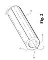
- the outer surface and wall of catheter 10 of the present invention includes two longitudinal grooves 11 and 12, located on opposite sides of the tubular body 15.
- FIG. 1A is a cross-sectional view showing a representative one of the tabs 23 attached to one of the knobs 25 which comprises a screw 30 and socket 32 combination.
- the screw 30 is screwed into the socket 32 with the end of the tab 23 in the socket.
- the force between the screw 30 and socket 32 maintains the end of the tab in position.
- the knobs 25 and 26 are used to aid in gripping the device when it is split apart.
- the thickness of the structure from the inner surface 14 to the outer surface 13 is 0.013 inch with a tolerance if plus or minus 0.002 inch.
- the total outer diameter of the catheter should be small enough to pass through the lumen of an endoscope.
- the outer diameter of the peel-away catheter 10 is preferably 0.072 inch with a tolerance of plus orminus 0.002 inch.
- the longitudinal grooves 11 and 12 located along the outer surface 13 of the tubular body 15 of the peel-away catheter 10 are cut half way through the wall 18 of the tubular body 15. In the preferred embodiment, the longitudinal grooves 11 and 12 are 0.006 inch deep.
- Longitudinal grooves 11 and 12 of FIG. 2 are designed to line up with the V-cut open ended slits 21 and 22 of FIG. 1.
- the tubular body 15 will tear along the longitudinal grooves 11 and 12 resulting in two portions (70 and 71 of FIGS. 5 and 7), each of which is attached to a knob 25 or 26.
- Teflon As is detailed in the Osborne patent, cited above, it is possible to cause Teflon to have a longitudinal orientation, thus facilitating longitudinal tearing of a sheath or catheter.
- the catheter it has been found that for catheters having the length necessary to extend through the gastroentral tract and into the common bile duct, the catheter must be much longer and heavier than the longitudinally oriented sheath used in connection with the vascular system, as is shown in the Osborne patent. The added length and weight of long-line catheters has been found to prevent a clean longitudinal tear down the entire length of the catheter.
- the catheter of FIG. 1 is made from radiopaque Teflon, the Dupont trademark for polytetraflouroethylene, which is extruded in a known fashion.
- the distal end of the catheter extruded in a known fashion.
- the distal end of the catheter 10 is provided with a number of cylindrical marking bands 40, as shown in FIG. 1. These bands 40 are applied in any of a number of commercially known manners.
- the last 1 cm of the distal end 16 of the tubing is tapered. This taper aids in guiding the finished catheter through the lumen of an endoscope. After which, the tubing is cut to the desired length, which is 205 centimeters in the preferred embodiment.
- the two grooves (70 and 71) are cut along the the entire length of the tubing. The last five centimeters of tubing at the non-tapered end is split along the grooves 70 and 71. This results in the flaps or tabs 23 and 24 being created. These tabs 23 and 24 are then trimmed to about 2.5 cm.
- the introducer hub 34 is attached at the proximal end 20 of the tubing. Again, 0.039 mandrel wire is inserted into the catheter tubing, and the hub 34 is slid over the mandrel wire and into the catheter until the hub is flush with the bottom of the V-shaped open ended slits 21 and 22. The mandrel wire is then removed. Finally, 14 Fr.
- duodenal tubing is placed over the end of each tab 23 and 24 and a plastic thumbscrew is inserted into the tubing, as is illustrated in connection with FIG. 1A.
- the catheter is sterilized and packaged with a sterilized stylet wire in a hermetically sealed package.
- FIG. 3 there is shown a top explanatory view showing the insertion of an endoscope into a human body.
- an endoscope 59 has been inserted into the mouth of the patient 50 and guided down the esophagus 51, through the stomach 57 and into the duodenum 54.
- the endoscope 59 of the present invention has a passage, or lumen, running longitudinally through the endoscope beginning from a channel located through the proximal end of the endoscope and terminating at an opening 65 at the distal end of the endoscope.
- the endoscope is guided into the duodenum 54 until the opening 65 is in close proximity to the papilla of vater 53, located between the sphincter of Oddi 61, which leads to the to the common bile duct 67 and the pancreatic duct 69.
- the channel at the proximal end of the endoscope 59 remains outside the mouth of the patient 50.
- a peel-away ERCP catheter having an outer diameter smaller than the inner diameter of the endoscope lumen, is inserted into the proximal opening of endoscope 59 (FIG. 4A) and is passed through the lumen of the endoscope until the tapered tip at the distal end 16 of the catheter emerges from the opening 65 atthe distal end of the endoscope.
- the outer diameter of the peel-away catheter is designed to be 5.5 French in one preferred embodiment so as to move freely through the endoscope.
- the distal tip of the catheter 10 may be guided between through the papilliary orifice of the papilla of vater 53, adjacent the sphincter of oddi 61 and into the common bile duct 67, as is shown in FIG. 4B.
- a contrast media such as a radiopaque dye
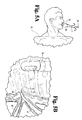
- the catheter may be removed from the endoscope as is shown in FIGS. 5A and 5B.
- a healthcare provider may remove the catheter by gripping the knobs of the peel-away catheter 10 and pulling them apart or in a downward direction. The resulting force on the knobs causes the catheter to tear along the longitudinal grooves while simultaneously pulling the catheter from the channel of the endoscope 59, FIG. 5B. Additionally, the catheter body is torn into two separate halves 70 and 71, and the introducer hub 34 can now be removed.
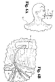
- a guide wire may be passed through the catheter 10 into the common bile duct 67, as shown in FIGS. 6Aand 6B.
- the guide wire 75 is inserted into the introducer hub 34 and is then run through the tubular body 15 where it exits the distal end 16 of the catheter into the common bi le duct 67.
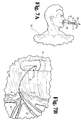
- the catheter may be easily removed in the same manner as explained in connection with FIGS. 5A and 5B, without disturbing the placement of the guide wire 75 within the bile duct 67.
- a healthcare provider need only hold the wire at the channel of the endoscope while a second person peels the catheter away from the guide wire 75.
- the guide wire does not move when the catheter is removed from the patient 50.
- peeling the catheter away from the guide wire results in much less friction on the wire than is produced using unsplit catheters, so that the guide wire is more likely to remain in the common bile duct 67 without employing the pushing and pulling technique, described above, for keeping the guide wire within the common bile duct 67.
- the introducer hub 34 is then slid off of the guide wire at the proximal end of the wire.
- FIG. 8A shows the introduction of a second catheter 10' into the channel of the endoscope 59.
- the second catheter 10' may be run over the guide wire 75 located through the lumen of the endoscope and present within the common bile or pancreatic duct. Additionally, FIG. 8B shows the distal end 16' of the catheter 10' used to push a biliary stent 73 along the guide wire 75 and into the common bile duct. Once the stent is in place, the second catheter and the guide wire 75 may be removed simultaneously without peeling the catheter away or the catheter 10' may be peeled-away, leaving the guide wire in place.
- This second catheter may be a regular 320 cm, 6 Fr., catheter, if the guide wire is to be removed with the catheter, or it may be a second peel-away catheter in accordance with the present invention. The peel-away nature of the catheter of the present invention prevents the difficulties in maintaining the guide wire 75 within the bile or pancreatic duct found in the prior systems.
Abstract
Description
- This invention relates to a splittable or peel-away ERCP catheter and more particularly to a method of using a peel-away ERCP catheter the distal end of which has been inserted into the common bile or pancreatic duct via an endoscope which has been guided through the esophagus and stomach and into the duodenum of a patient.
- In certain medical applications it is useful to provide a sheath or catheter that is longitudinally splittable for ease of removal from the human body. For example, the use of a splittable catheter permits the removal of a catheter after a guide wire or other material has been placed therethrough without disturbing the guide wire. Various splittable sheaths and catheters exist which have been designed to split longitudinally along the body to peel-away from a catheter, guide wire or other inserted material.
- U.S. Patent No. 4,776,846 to Wells discloses an apparatus and method for preparing a catheter tube which has a longitudinal line of weakness extending axially therealong as a place for preferential splitting. The method includes co-extruding two materials into the tubular cross-sectional shape. One of the materials forms a surface line along the otherwhich extends readily to a level where the thickness of the tubing cross-section thereat is less. When the co-extruded extra material is removed, a grove is in the surface of the tube, which groove permits easy tearing therealong.
- U. S. Patent No. 4,983,168 to Moorehead discloses a layered peel-away hollow sheath, for temporarily creating a passageway into a desired body site of a medical patient for placement of one end of an indwelling device at the body site through the sheath. The sheath wall comprises at least two layers, the inside layer being cylindrical and the outside layer comprising two semi-cylindrical segments defining opposed axially directed slits or slots therebetween which comprise tear lines such that the sheath manually tears axially along the single layer tear lines into two pieces for removal of the sheath from around the indwelling device.
- U.S. Patent No. 4,801,294 to Okada discloses a catheter for nasogastric intubation comprising a plastic sheath tube and a fixing means of said tube at nose, said tube having a longitudinal tear-off line over the full length thereof and having elasticity and rigidity slightly larger than those of said catheter, said catheter being inserted in said plastic sheath tube slidably.
- U.S. Patent No. 3,550,591 to McGregor discloses an intravenous catheter unit which provides a needle which can be separated from the catheter after the end of the catheter positioned inside the vein to prevent the needle from tearing or damaging the catheter. The hollow needle is provided with a slit which extends along its entire length. In addition the needle is provided with means of the causing the needle to hinge open to widen the slit enough to pass the catheter therethrough. In this way the needle may be separated from the catheter after the catheter is placed inside the vein.
- U.S. Patent No. 4,781,690 to Oshada, et al. discloses a guiding tube for introducing rodlike medical instruments such as a catheter, which can be easily splitand removed. The guiding tube comprises a main body provided with a longitudinal slit and formed of a mixture of first and second materials and a stripe detachably embedded liquid-tight in the slit of the main body and formed of the third material which exhibits a good bonding for the second material but a poor bonding for the first material.
- U.S. Patent No. 4,747,833 to Kousai, et al. discloses a medical instrument-guiding tube for guiding a catheter or other rod-like medical instrument into a blood vessel. This guiding tube comprises a hollow- tube body and at least one linear body integrally joined to the tube body along the longitudinal direction of the tube body. The plastic resin forming the tube body has a poor compatibi lity with that of the linear body. Tube body and the linear body are engaged together through complementary concave convex arrangement which can be disengaged with reasonable force.
- U.S. Patent No. 4,883,468 to Kousai, et al. discloses a medical instrument introduction cannula which is useful as a guide means for introducing and indwelling a rod-like material instrument such as a catheter and a guide wire. This cannula is removed after use from the medical instrument by being split. This cannula is formed of a hollow tubular body having a strip member consisting of material different from the other portion of the tubular body, which extends over almost the entire length of the tubular body. The strip member has a weld line along the length of the strip member, or consists of a resin which exhibits a good bonding property only to one of the other components forming the tubular body. The tubular body can be split by way of the weld line or the removal of the strip member having such a bonding property. The method of manufacturing the medical instrument introduction cannula by a two-color extruder is also disclosed.
- U.S. Patent No. 4,997,424 to Little relates to an introducer slitterfor slitting an introducer tube portion having, for example, a catheter extending therethrough and into a body vessel while the introducer tube is moved rearwardly relative to the catheter to facilitate separating the introducer from the catheter without having to slide the introducer tube portion over the proximal end of the catheter.
- U.S. Patent No. 5,195,978 to Schiffer discloses a rapid exchange over-the-wire catheter which is provided with one or more breakaway elements for progressively exposing the guide wire from the proximal end toward the distal end of the catheter in a simple and efficient manner. The breakaway element may be formed as a longitudinally aligned pull strip provided in the catheter guide wire lumen or as one or more linearly arrayed tubular breakaway segments in the catheter shaft or as a combination of both features.
- U.S. Patent No. 4,813,929 to Semrad discloses a method and assembly of components for effecting closed chest thoracostomy. A guide wire is removed and a chest tube is introduced through a pleural access catheter, which is split off and removed from the chest tube. Figures 5 and 6 of the Semrad reference show a splittable access catheter having knobs that when grasped and pulled in a direction away from the body of the catheter will cause the catheter to split.
- U.S. Patent Nos. 4,243,050 and 4,345,606 to Littleford disclose an apparatus and related method for implanting pacemaker electrodes and similar devices within a patient including an introducer with a tapered end adapted to extend into the patient's body, and a hollow, tubular sleeve. The sleeve is perforated to form a weakened line. The sleeve may be peeled slightly away at the flange to form tabs which may be used to grasp and peel away the sleeve while removing the same from the vein.
- U.S. patent No. 4,166,469 to Littleford discloses an apparatus and method for inserting an electrode within a patient with a minimal amount of incision which includes the use of an introducer sleeve which is severed or severable along the length thereof. The introducer sleeve is withdrawn from the patient while the electrode is moving through, severing the sleeve wall along the entire length thereof to enable the removal of the sleeve over the connector plug of the electrode.
- U.S. Patent No. 4,306,562 to Osborne discloses a flexible cannula comprising material which tears readily in a longitudinal direction and thus can be easily removed by pulling tabs on opposite sides of the cannula apart from the catheter or other device that has been inserted into the body. The Osborne cannula readily tears in a longitudinal direction along the length of the structure due to the longitudinal orientation of the material from which it is constructed.
- Additionally, it is possible to introduce a standard, non-splittable catheter into the gastroentral tract, via an endoscope, until it is adjacent to the bile duct. However, after a guide wire has been introduced through the catheter, difficulties arise when attempting to remove the catheter. For example, the catheter must be pulled over the entire length of the guide wire by a health care provider, while a second health care provider attempts to maintain the guide wire in the bile duct. This results in a tug-of-war caused by the alternating of the pulling of small increments of the catheter out of the channel of the endoscope and the pushing of the guide wire back into the bile duct after a catheter has been pulled.
- None of the above disclosed references disclose a peel-away ERCP catheter or a method of using the same for gaining access to the pancreatic or common bile duct. Additionally, none of the above listed references disclose the use of an endoscope having a lumen, the peel-away catheter being inserted therethrough, for introducing a contrast media or a guide wire into the common bile or pancreatic ducts. Further, none of the above disclosed references teach a method for easily removing a long-line catheter from the common bile or pancreatic duct without disturbing the placement of a guide wire therein. There is a need for a method of introducing a dye, guide wire or stent into the common bile or pancreatic duct through a peel-away ERCP catheter introduced into the human body via an endoscope. There is additionally a need for a method of easily removing a catheter inserted into the common bile or pancreatic duct without disturbing the placement of a guide wire therein.
- One embodiment of the present invention might include a peel-away ERCP catheter for use in introducing materials into the common bile or pancreatic ducts of the human body. An endoscope having a channel at the proximal end and an opening at the distal end and a passage therebetween is introduced into the gastroentral tract of a patient through the patient's mouth and is located such that the opening of the endoscope is adjacent the papilla of vater. A peel-away ERCP catheter including two longitudinal slits cut into the outer surface of the catheter body and additionally including two tabs connected at the proximal end of the catheter between the longitudinal slits, may be passed through the lumen of the endoscope until the distal tip of the catheter emerges from the opening of the endoscope. The distal tip of the catheter may then be guided into the ampulla of vater and into either the common bile or pancreatic duct. A guide wire may be run through the catheter and into the desired duct. The catheter may then be peeled-away from the guide wire by applying a pulling force to the tabs causing the catheter to tear along the longitudinal slits. The catheter may then be removed without disturbing the placement of the guide wire within the desired duct.
- One object of the present invention is to provide a peel-away catheter having two longitudinal grooves cut into the outer surface of the catheter and two pull tabs connected between the grooves for facilitating the tearing and removal of the catheter without disturbing the placement of a guide wire therethrough.
- Further objects and advantages of the present invention may be discerned by persons of ordinary skill in the art after reviewing the following written description and accompanying figures.
-
- FIG. 1 is a side view of the peel-away ERCP catheter of the present invention.
- FIG. 1A is an enlarged fragmentary view of a portion of the structure of FIG. 1.
- FIG. 2 is a perspective view of a portion of the tubing from which the peel-away catheter of FIG. 1 is made.
- FIG. 3 is a top view of a human showing the insertion of an endoscope into the mouth of a patient with the distal end of the endoscope being brought into close proximity with the papilla of vater which leads to the common bile and pancreatic ducts.
- FIG. 4A is a view of the peel-away catheter of the present invention after being inserted through the lumen of the endoscope of FIG. 3.
- FIG. 4B is an enlarged view of the distal end of the peel-away catheter, shown exiting an opening in the distal end of the endoscope, which has been inserted into the common bile duct.
- FIG. 5A shows the peel-away catheter after it has been peeled away and removed from the lumen of the endoscope.
- FIG. 5B shows the tip of the endoscope and papilla of vater after the catheter of FIG. 4B has been peeled-away and removed from the papilliary orifice.
- FIG. 6A shows the peel-away catheter of the present invention having a guide wire therethrough.
- FIG. 6B shows the guide wire protruding from the distal end of the peel-away catheter located within the common bile duct of a human body.
- FIG. 7A shows the peeling away and removal of the peel-away catheter which leaves the guide wire of FIG. 6B positioned within the common bile duct.
- FIG. 7B shows the guide wire positioned within the common bile duct.
- FIG. 8A shows the introduction of a second peel-away catheter over the guide wire located within the endoscope of FIG. 3.
- FIG. 8B shows a stent being pushed by a peel-away catheter along a guide wire, the distal end of the guide wire being located within the common bile duct.
- For the purposes of promoting an understanding of the principles of the invention, reference will now be made to the embodiment illustrated in the drawings and specific language will be used to describe the same. It will nevertheless be understood that no limitation of the scope of the invention is thereby intended, such alterations and further modifications in the illustrated device, and such further applications of the principles of the invention as illustrated therein being contemplated as would normally occur to one skilled in the art to which the invention relates.
- A preferred embodiment of a peel-away endoscopic retrograde cholangio pancreatography (ERCP) catheter and a method for using the same are shown in FIGS. 1-8. Referring now to FIG. 1, there is shown a peel-away
ERCP catheter 10 in accordance with one embodiment of the present invention. Thetubular portion 15 of the peel-awaycatheter 10 of the present invention has approximately a uniform thickness and diameter except at itsdistal end 16 where there is a slight taper. Theproximal end 20 of the cannula is slit longitudinally producing two open endedslits tabs screw 30 and 31 andsocket 32 and 33 portions of the knobs, as shall be discussed further in connection with FIG. 1A. - The
catheter 10 includes anintroducer hub 34, having a passage therethrough, which is attached at theproximal end 20 of thecatheter 10 to provide a guide for the deposit and insertion of materials through the lumen ofcatheter 10. Additionally, fluoro-scopic stripes 40 are provided to aid in the locating and positioning of the catheter tip within the bile or pancreatic ducts. In the preferred embodiment ofcatheter 10 the length A of the catheter body, from the V-shaped slits to the distal tip is approximately 200 centimeters. To provide the proper perspective, length B of FIG. 1 is approximately 15 centimeters, length C is approximately 10 cm and Length D is approximately 5 cm. Additionally, as will be shown in FIG. 2, the outer surface and wall ofcatheter 10 of the present invention includes twolongitudinal grooves tubular body 15. - FIG. 1A is a cross-sectional view showing a representative one of the
tabs 23 attached to one of theknobs 25 which comprises ascrew 30 andsocket 32 combination. Thescrew 30 is screwed into thesocket 32 with the end of thetab 23 in the socket. The force between thescrew 30 andsocket 32 maintains the end of the tab in position. Theknobs - Referring now to FIG. 2, there is shown a perspective view of a portion of the
tube body 15 of the peel-awaycatheter 10. In one preferred embodiment of the present invention, the thickness of the structure from theinner surface 14 to theouter surface 13 is 0.013 inch with a tolerance if plus or minus 0.002 inch. The total outer diameter of the catheter should be small enough to pass through the lumen of an endoscope. The outer diameter of the peel-awaycatheter 10 is preferably 0.072 inch with a tolerance of plus orminus 0.002 inch. Additionally, thelongitudinal grooves outer surface 13 of thetubular body 15 of the peel-awaycatheter 10 are cut half way through thewall 18 of thetubular body 15. In the preferred embodiment, thelongitudinal grooves Longitudinal grooves slits knobs tubular body 15 will tear along thelongitudinal grooves knob - As is detailed in the Osborne patent, cited above, it is possible to cause Teflon to have a longitudinal orientation, thus facilitating longitudinal tearing of a sheath or catheter. However, it has been found that for catheters having the length necessary to extend through the gastroentral tract and into the common bile duct, the catheter must be much longer and heavier than the longitudinally oriented sheath used in connection with the vascular system, as is shown in the Osborne patent. The added length and weight of long-line catheters has been found to prevent a clean longitudinal tear down the entire length of the catheter. Rather, it has been found that attempting to tear longitudinally oriented teflon extruded to a sufficient length for a catheter of the present invention will result in non-longitudinal tearing at some point along the length of the catheter. Thus, providing longitudinal grooves along the length of the the catheter is preferred to ensure that the catheter tears longitudinally down its entire length.
- The catheter of FIG. 1 is made from radiopaque Teflon, the Dupont trademark for polytetraflouroethylene, which is extruded in a known fashion. The distal end of the catheter extruded in a known fashion. The distal end of the
catheter 10 is provided with a number ofcylindrical marking bands 40, as shown in FIG. 1. Thesebands 40 are applied in any of a number of comercially known manners. The last 1 cm of thedistal end 16 of the tubing is tapered. This taper aids in guiding the finished catheter through the lumen of an endoscope. After which, the tubing is cut to the desired length, which is 205 centimeters in the preferred embodiment. Additionally, the two grooves (70 and 71) are cut along the the entire length of the tubing. The last five centimeters of tubing at the non-tapered end is split along thegrooves tabs tabs introducer hub 34 is attached at theproximal end 20 of the tubing. Again, 0.039 mandrel wire is inserted into the catheter tubing, and thehub 34 is slid over the mandrel wire and into the catheter until the hub is flush with the bottom of the V-shaped open endedslits tab - A method for using the peel-away ERCP catheter of the preferred embodiment will now be explained in connection with FIGS. 3-7. Referring now to FIG. 3, there is shown a top explanatory view showing the insertion of an endoscope into a human body. In the present invention, an
endoscope 59 has been inserted into the mouth of thepatient 50 and guided down theesophagus 51, through thestomach 57 and into theduodenum 54. Theendoscope 59 of the present invention has a passage, or lumen, running longitudinally through the endoscope beginning from a channel located through the proximal end of the endoscope and terminating at anopening 65 at the distal end of the endoscope. - The endoscope is guided into the duodenum 54 until the
opening 65 is in close proximity to the papilla ofvater 53, located between the sphincter ofOddi 61, which leads to the to thecommon bile duct 67 and thepancreatic duct 69. The channel at the proximal end of theendoscope 59 remains outside the mouth of thepatient 50. - In accordance with one method of use of the catheter of the present invention, once the endoscope is in proper position, a peel-away ERCP catheter having an outer diameter smaller than the inner diameter of the endoscope lumen, is inserted into the proximal opening of endoscope 59 (FIG. 4A) and is passed through the lumen of the endoscope until the tapered tip at the
distal end 16 of the catheter emerges from theopening 65 atthe distal end of the endoscope. The outer diameter of the peel-away catheter is designed to be 5.5 French in one preferred embodiment so as to move freely through the endoscope. Additionally, the distal tip of thecatheter 10 may be guided between through the papilliary orifice of the papilla ofvater 53, adjacent the sphincter ofoddi 61 and into thecommon bile duct 67, as is shown in FIG. 4B. Afterthedistal end 16 of thecatheter 10 is in position in thecommon bile duct 67, it is possible to inject a contrast media, such as a radiopaque dye, into the bile duct. - After the contrast media has been injected into the common bile duct the catheter may be removed from the endoscope as is shown in FIGS. 5A and 5B. A healthcare provider may remove the catheter by gripping the knobs of the peel-away
catheter 10 and pulling them apart or in a downward direction. The resulting force on the knobs causes the catheter to tear along the longitudinal grooves while simultaneously pulling the catheter from the channel of theendoscope 59, FIG. 5B. Additionally, the catheter body is torn into twoseparate halves introducer hub 34 can now be removed. - Additionally, once the peel-away ERCP catheter of the present invention has been inserted into the bile duct, through the
endoscope 59, a guide wire may be passed through thecatheter 10 into thecommon bile duct 67, as shown in FIGS. 6Aand 6B. Theguide wire 75 is inserted into theintroducer hub 34 and is then run through thetubular body 15 where it exits thedistal end 16 of the catheter into the commonbi le duct 67. - Referring now to FIGS. 7A and 7B, the catheter may be easily removed in the same manner as explained in connection with FIGS. 5A and 5B, without disturbing the placement of the
guide wire 75 within thebile duct 67. A healthcare provider need only hold the wire at the channel of the endoscope while a second person peels the catheter away from theguide wire 75. Thus, the guide wire does not move when the catheter is removed from thepatient 50. Additionally, peeling the catheter away from the guide wire results in much less friction on the wire than is produced using unsplit catheters, so that the guide wire is more likely to remain in thecommon bile duct 67 without employing the pushing and pulling technique, described above, for keeping the guide wire within thecommon bile duct 67. Theintroducer hub 34 is then slid off of the guide wire at the proximal end of the wire. - Additionally, once the guide wire has been placed into the common bile duct 67 (or in the pancreatic duct 69) and the first peel-away catheter has been peeled and removed, it may be desirable to place a stent into the common bile or pancreatic duct. As such, an indwelling stent, such as that shown in U. S. Patent No. 5,052,998, that document incorporated herein by reference, may be placed over the
guide wire 75 and pushed into the desired duct using a second catheter, which is additionally run over theguide wire 75. FIG. 8Ashows the introduction of a second catheter 10' into the channel of theendoscope 59. The second catheter 10' may be run over theguide wire 75 located through the lumen of the endoscope and present within the common bile or pancreatic duct. Additionally, FIG. 8B shows the distal end 16' of the catheter 10' used to push abiliary stent 73 along theguide wire 75 and into the common bile duct. Once the stent is in place, the second catheter and theguide wire 75 may be removed simultaneously without peeling the catheter away or the catheter 10' may be peeled-away, leaving the guide wire in place. This second catheter may be a regular 320 cm, 6 Fr., catheter, if the guide wire is to be removed with the catheter, or it may be a second peel-away catheter in accordance with the present invention. The peel-away nature of the catheter of the present invention prevents the difficulties in maintaining theguide wire 75 within the bile or pancreatic duct found in the prior systems. - While the invention has been illustrated and described in detail in the drawings and foregoing description, the same is to be considered as illustrative and not restrictive in character, it being understood that only the preferred embodiment has been shown and described and that all changes and modifications that come within the spirit of the invention are desired to be protected.
Claims (7)
Applications Claiming Priority (2)
| Application Number | Priority Date | Filing Date | Title |
|---|---|---|---|
| US08/061,854 US5320602A (en) | 1993-05-14 | 1993-05-14 | Peel-away endoscopic retrograde cholangio pancreatography catheter and a method for using the same |
| US61854 | 1993-05-14 |
Publications (3)
| Publication Number | Publication Date |
|---|---|
| EP0624381A2 true EP0624381A2 (en) | 1994-11-17 |
| EP0624381A3 EP0624381A3 (en) | 1995-09-13 |
| EP0624381B1 EP0624381B1 (en) | 2002-07-24 |
Family
ID=22038572
Family Applications (1)
| Application Number | Title | Priority Date | Filing Date |
|---|---|---|---|
| EP94303458A Expired - Lifetime EP0624381B1 (en) | 1993-05-14 | 1994-05-13 | A peel-away endoscopic retrograde, cholangio pancreatography catheter |
Country Status (10)
| Country | Link |
|---|---|
| US (1) | US5320602A (en) |
| EP (1) | EP0624381B1 (en) |
| JP (1) | JP2540025B2 (en) |
| AT (1) | ATE220934T1 (en) |
| AU (1) | AU671192B2 (en) |
| CA (1) | CA2121773C (en) |
| DE (1) | DE69431019T2 (en) |
| DK (1) | DK0624381T3 (en) |
| ES (1) | ES2179061T3 (en) |
| PT (1) | PT624381E (en) |
Cited By (3)
| Publication number | Priority date | Publication date | Assignee | Title |
|---|---|---|---|---|
| GB2409646A (en) * | 2003-11-13 | 2005-07-06 | Mohamed Osman Abdelatti | A nasogastric tube introducer |
| DE102005044468A1 (en) * | 2005-09-16 | 2007-03-29 | Riek, Siegfried, Dr. Med. | Medical instrument |
| US8394063B2 (en) | 2005-09-16 | 2013-03-12 | Siegfried Riek | Medical instrument |
Families Citing this family (121)
| Publication number | Priority date | Publication date | Assignee | Title |
|---|---|---|---|---|
| US6666883B1 (en) | 1996-06-06 | 2003-12-23 | Jacques Seguin | Endoprosthesis for vascular bifurcation |
| US6096009A (en) * | 1996-09-13 | 2000-08-01 | Boston Scientific Corporation | Guidewire and catheter locking device and method |
| US5921971A (en) * | 1996-09-13 | 1999-07-13 | Boston Scientific Corporation | Single operator exchange biliary catheter |
| US6007522A (en) | 1996-09-13 | 1999-12-28 | Boston Scientific Corporation | Single operator exchange biliary catheter |
| US6520951B1 (en) | 1996-09-13 | 2003-02-18 | Scimed Life Systems, Inc. | Rapid exchange catheter with detachable hood |
| US6346093B1 (en) | 1996-09-13 | 2002-02-12 | Scimed Life Systems, Inc. | Single operator exchange biliary catheter with common distal lumen |
| US6582401B1 (en) | 1996-09-13 | 2003-06-24 | Scimed Life Sytems, Inc. | Multi-size convertible catheter |
| US6606515B1 (en) | 1996-09-13 | 2003-08-12 | Scimed Life Systems, Inc. | Guide wire insertion and re-insertion tools and methods of use |
| US6951572B1 (en) | 1997-02-20 | 2005-10-04 | Endologix, Inc. | Bifurcated vascular graft and method and apparatus for deploying same |
| US6174330B1 (en) | 1997-08-01 | 2001-01-16 | Schneider (Usa) Inc | Bioabsorbable marker having radiopaque constituents |
| US6340367B1 (en) | 1997-08-01 | 2002-01-22 | Boston Scientific Scimed, Inc. | Radiopaque markers and methods of using the same |
| US5980564A (en) * | 1997-08-01 | 1999-11-09 | Schneider (Usa) Inc. | Bioabsorbable implantable endoprosthesis with reservoir |
| US6245103B1 (en) | 1997-08-01 | 2001-06-12 | Schneider (Usa) Inc | Bioabsorbable self-expanding stent |
| US6626939B1 (en) * | 1997-12-18 | 2003-09-30 | Boston Scientific Scimed, Inc. | Stent-graft with bioabsorbable structural support |
| US6080141A (en) * | 1997-12-22 | 2000-06-27 | Becton, Dickinson And Company | Splittable tubular medical device and method for manufacture |
| US6326396B1 (en) | 1998-11-20 | 2001-12-04 | Alteon, Inc. | Glucose and lipid lowering compounds |
| US6733523B2 (en) * | 1998-12-11 | 2004-05-11 | Endologix, Inc. | Implantable vascular graft |
| US6187036B1 (en) | 1998-12-11 | 2001-02-13 | Endologix, Inc. | Endoluminal vascular prosthesis |
| ATE303107T1 (en) | 1998-12-11 | 2005-09-15 | Endologix Inc | ENDOLUMINAL VASCULAR PROSTHESIS |
| US6660030B2 (en) | 1998-12-11 | 2003-12-09 | Endologix, Inc. | Bifurcation graft deployment catheter |
| US8034100B2 (en) | 1999-03-11 | 2011-10-11 | Endologix, Inc. | Graft deployment system |
| US6261316B1 (en) | 1999-03-11 | 2001-07-17 | Endologix, Inc. | Single puncture bifurcation graft deployment system |
| US6277108B1 (en) * | 1999-06-04 | 2001-08-21 | Medamicus, Inc. | Introducer with location marker |
| DE60037731T2 (en) * | 1999-06-05 | 2009-01-15 | Wilson-Cook Medical Inc. | Markers for a medical endoscopic device |
| US6440161B1 (en) | 1999-07-07 | 2002-08-27 | Endologix, Inc. | Dual wire placement catheter |
| US6458076B1 (en) | 2000-02-01 | 2002-10-01 | 5 Star Medical | Multi-lumen medical device |
| US7811250B1 (en) * | 2000-02-04 | 2010-10-12 | Boston Scientific Scimed, Inc. | Fluid injectable single operator exchange catheters and methods of use |
| US6589262B1 (en) | 2000-03-31 | 2003-07-08 | Medamicus, Inc. | Locking catheter introducing system |
| US6663598B1 (en) | 2000-05-17 | 2003-12-16 | Scimed Life Systems, Inc. | Fluid seal for endoscope |
| US6533759B1 (en) * | 2000-06-01 | 2003-03-18 | Robert Watson | Flash chamber with a self closing valve for use with a catheter |
| DE60042705D1 (en) * | 2000-07-13 | 2009-09-17 | Wilson Cook Medical Inc | MARKING SYSTEM FOR MEDICAL INSTRUMENT |
| US6582390B1 (en) * | 2000-11-08 | 2003-06-24 | Endovascular Technologies, Inc. | Dual lumen peel-away sheath introducer |
| US6764484B2 (en) * | 2001-03-30 | 2004-07-20 | Scimed Life Systems, Inc. | C-channel to o-channel converter for a single operator exchange biliary catheter |
| US6827718B2 (en) | 2001-08-14 | 2004-12-07 | Scimed Life Systems, Inc. | Method of and apparatus for positioning and maintaining the position of endoscopic instruments |
| US20030078473A1 (en) * | 2001-10-23 | 2003-04-24 | Scimed Life Systems, Inc. | Cone tip biliary catheter and method of use |
| DE10158289A1 (en) * | 2001-11-20 | 2003-05-28 | Biotronik Mess & Therapieg | Device for implanting catheters |
| US6979319B2 (en) * | 2001-12-31 | 2005-12-27 | Cardiac Pacemakers, Inc. | Telescoping guide catheter with peel-away outer sheath |
| WO2003101347A1 (en) * | 2002-05-31 | 2003-12-11 | Wilson-Cook Medical Inc. | Stent introducer apparatus |
| US7534223B2 (en) | 2002-10-08 | 2009-05-19 | Boston Scientific Scimed, Inc. | Catheter with formed guide wire ramp |
| US7037293B2 (en) | 2002-11-15 | 2006-05-02 | Boston Scientific Scimed, Inc. | Rapid exchange catheter with depressable channel |
| US6893393B2 (en) * | 2003-02-19 | 2005-05-17 | Boston Scientific Scimed., Inc. | Guidewire locking device and method |
| AU2003227198A1 (en) * | 2003-03-25 | 2004-10-18 | Olympus Corporation | Guide wire |
| WO2005000096A2 (en) * | 2003-06-05 | 2005-01-06 | Hydrocision, Inc. | Disposable endoscope and method of making a disposable endoscope |
| US8206320B2 (en) * | 2003-07-31 | 2012-06-26 | Cook Medical Technologies Llc | System and method for introducing multiple medical devices |
| US7621880B2 (en) * | 2003-09-05 | 2009-11-24 | Vance Products Incorporated | Double ended wire guide |
| US7025721B2 (en) * | 2004-01-29 | 2006-04-11 | Boston Scientific Scimed, Inc. | Endoscope channel cap |
| US20050182387A1 (en) * | 2004-02-13 | 2005-08-18 | Cardiac Pacemakers, Inc. | Peel-away catheter shaft |
| US7854731B2 (en) | 2004-03-18 | 2010-12-21 | C. R. Bard, Inc. | Valved catheter |
| US8083728B2 (en) | 2004-03-18 | 2011-12-27 | C. R. Bard, Inc. | Multifunction adaptor for an open-ended catheter |
| US7594910B2 (en) | 2004-03-18 | 2009-09-29 | C. R. Bard, Inc. | Catheter connector |
| US7594911B2 (en) | 2004-03-18 | 2009-09-29 | C. R. Bard, Inc. | Connector system for a proximally trimmable catheter |
| US7377915B2 (en) | 2004-04-01 | 2008-05-27 | C. R. Bard, Inc. | Catheter connector system |
| WO2005107843A1 (en) | 2004-04-30 | 2005-11-17 | C.R. Bard, Inc. | Valved sheath introducer for venous cannulation |
| WO2006041971A1 (en) * | 2004-10-06 | 2006-04-20 | Cook Incorporated | Wireguide with indicia |
| US8926564B2 (en) | 2004-11-29 | 2015-01-06 | C. R. Bard, Inc. | Catheter introducer including a valve and valve actuator |
| US9597483B2 (en) | 2004-11-29 | 2017-03-21 | C. R. Bard, Inc. | Reduced-friction catheter introducer and method of manufacturing and using the same |
| US8932260B2 (en) | 2004-11-29 | 2015-01-13 | C. R. Bard, Inc. | Reduced-friction catheter introducer and method of manufacturing and using the same |
| US8403890B2 (en) | 2004-11-29 | 2013-03-26 | C. R. Bard, Inc. | Reduced friction catheter introducer and method of manufacturing and using the same |
| JP2008523908A (en) * | 2004-12-15 | 2008-07-10 | ウィルソン−クック・メディカル・インコーポレーテッド | Minimally invasive medical device with a spiral pattern for displaying travel distance |
| US8480629B2 (en) | 2005-01-28 | 2013-07-09 | Boston Scientific Scimed, Inc. | Universal utility board for use with medical devices and methods of use |
| US8371307B2 (en) | 2005-02-08 | 2013-02-12 | Koninklijke Philips Electronics N.V. | Methods and devices for the treatment of airway obstruction, sleep apnea and snoring |
| US8096303B2 (en) | 2005-02-08 | 2012-01-17 | Koninklijke Philips Electronics N.V | Airway implants and methods and devices for insertion and retrieval |
| US8109941B2 (en) * | 2005-02-28 | 2012-02-07 | Boston Scientific Scimed, Inc. | Distal release retrieval assembly and related methods of use |
| JP2006246982A (en) * | 2005-03-09 | 2006-09-21 | Pentax Corp | Filling for endoscope |
| BRPI0609069A8 (en) | 2005-04-28 | 2018-01-02 | St Jude Medical Atrial Fibrillation Div Inc | CATHETER BODY OR PROTECTIVE COVER |
| AU2006239222B2 (en) * | 2005-04-28 | 2009-05-14 | St. Jude Medical, Atrial Fibrillation Division, Inc. | Peelable atraumatic tip and body for a catheter or sheath |
| US7875019B2 (en) | 2005-06-20 | 2011-01-25 | C. R. Bard, Inc. | Connection system for multi-lumen catheter |
| US8702720B2 (en) | 2006-05-03 | 2014-04-22 | Cook Medical Technologies Llc | Tassel tip wire guide |
| US7604627B2 (en) * | 2006-05-11 | 2009-10-20 | Kourosh Kojouri | Nasopharyngeal sheath for nasogastric intubation |
| CA2654907A1 (en) * | 2006-06-09 | 2007-12-21 | Hilbert D. Brown | Endoscopic apparatus having an expandable balloon delivery system |
| US8475360B2 (en) * | 2006-06-09 | 2013-07-02 | Cook Medical Technologies Llc | Endoscopic apparatus having an expandable balloon delivery system |
| US7934506B2 (en) * | 2006-06-21 | 2011-05-03 | Koninklijke Philips Electronics N.V. | System and method for temporary tongue suspension |
| US20080051911A1 (en) * | 2006-08-23 | 2008-02-28 | Wilson-Cook Medical Inc. | Stent with antimicrobial drainage lumen surface |
| WO2008085712A1 (en) | 2007-01-03 | 2008-07-17 | Boston Scientific Limited | Method and apparatus for biliary access and stone retrieval |
| US20080167628A1 (en) * | 2007-01-05 | 2008-07-10 | Boston Scientific Scimed, Inc. | Stent delivery system |
| US8523931B2 (en) | 2007-01-12 | 2013-09-03 | Endologix, Inc. | Dual concentric guidewire and methods of bifurcated graft deployment |
| ES2592719T3 (en) | 2007-02-12 | 2016-12-01 | Boston Scientific Limited | Endoscope Cap |
| US20090062769A1 (en) * | 2007-04-13 | 2009-03-05 | Boston Scientific Scimed, Inc. | Rapid exchange catheter converter |
| US20080264993A1 (en) * | 2007-04-24 | 2008-10-30 | Khrys Schulte | Article for Retaining Components of an Endoscopic Retrograde Cholangio-Pancreatography Delivery System |
| CN101903062A (en) | 2007-10-19 | 2010-12-01 | C·R·巴德股份有限公司 | Introducer including shaped distal region |
| US20090105806A1 (en) * | 2007-10-23 | 2009-04-23 | Endologix, Inc | Stent |
| US8388521B2 (en) | 2008-05-19 | 2013-03-05 | Boston Scientific Scimed, Inc. | Integrated locking device with active sealing |
| US8343041B2 (en) | 2008-05-19 | 2013-01-01 | Boston Scientific Scimed, Inc. | Integrated locking device with passive sealing |
| US8221494B2 (en) | 2008-02-22 | 2012-07-17 | Endologix, Inc. | Apparatus and method of placement of a graft or graft system |
| US8236040B2 (en) | 2008-04-11 | 2012-08-07 | Endologix, Inc. | Bifurcated graft deployment systems and methods |
| US20110098528A1 (en) * | 2008-04-11 | 2011-04-28 | Lumenis Ltd. | Fibers and tips thereof used with devices |
| EP2293838B1 (en) | 2008-07-01 | 2012-08-08 | Endologix, Inc. | Catheter system |
| US8444577B2 (en) | 2009-01-05 | 2013-05-21 | Cook Medical Technologies Llc | Medical guide wire |
| WO2010127040A1 (en) | 2009-04-28 | 2010-11-04 | Endologix, Inc. | Apparatus and method of placement of a graft or graft system |
| US10772717B2 (en) | 2009-05-01 | 2020-09-15 | Endologix, Inc. | Percutaneous method and device to treat dissections |
| JP2012525239A (en) | 2009-05-01 | 2012-10-22 | エンドロジックス、インク | Transcutaneous methods and devices for treating dissociation (priority information and incorporation by reference) |
| WO2010151825A1 (en) | 2009-06-26 | 2010-12-29 | C. R. Bard, Inc. | Proximally trimmable catheter including pre-attached bifurcation and related methods |
| US8491646B2 (en) | 2009-07-15 | 2013-07-23 | Endologix, Inc. | Stent graft |
| WO2011017123A2 (en) | 2009-07-27 | 2011-02-10 | Endologix, Inc. | Stent graft |
| US20110218549A1 (en) * | 2010-03-05 | 2011-09-08 | Boston Scientific Neuromodulation Corporation | Systems and methods for making and using a trial stimulation system having an electrical connector disposed on a trial stimulation lead |
| US9510857B2 (en) * | 2010-03-10 | 2016-12-06 | Boston Scientific Neuromodulation Corporation | System and method for making and using a lead introducer for an implantable electrical stimulation system |
| US20110224681A1 (en) * | 2010-03-15 | 2011-09-15 | Boston Scientific Neuromodulation Corporation | System and method for making and using a splitable lead introducer for an implantable electrical stimulation system |
| US20110230893A1 (en) * | 2010-03-19 | 2011-09-22 | Boston Scientific Neuromodulation Corporation | Systems and methods for making and using electrical stimulation systems having multi-lead-element lead bodies |
| JP6261339B2 (en) | 2010-11-02 | 2018-01-17 | エンドロジックス、インク | Apparatus and method for placement of a graft or graft system |
| US9675487B2 (en) | 2010-11-17 | 2017-06-13 | Cook Medical Technologies Llc | Prosthesis deployment system for vascular repair |
| WO2012068298A1 (en) | 2010-11-17 | 2012-05-24 | Endologix, Inc. | Devices and methods to treat vascular dissections |
| US8657866B2 (en) | 2010-12-22 | 2014-02-25 | Cook Medical Technologies Llc | Emergency vascular repair prosthesis deployment system |
| WO2012118901A1 (en) | 2011-03-01 | 2012-09-07 | Endologix, Inc. | Catheter system and methods of using same |
| US10849771B2 (en) | 2011-06-27 | 2020-12-01 | Boston Scientific Scimed, Inc. | Stent delivery systems and methods for making and using stent delivery systems |
| EP2747705B1 (en) | 2011-08-22 | 2017-06-28 | Cook Medical Technologies LLC | Emergency vessel repair prosthesis deployment system |
| WO2014158723A1 (en) | 2013-03-13 | 2014-10-02 | Boston Scientific Neuromodulation Corporation | System and method for making and using a lead introducer for an implantable electrical stimulation system |
| CN105517625A (en) | 2013-09-06 | 2016-04-20 | 波士顿科学神经调制公司 | Lead introducer for implantable electrical stimulation system |
| EP3041566B1 (en) | 2013-09-06 | 2019-05-29 | Boston Scientific Neuromodulation Corporation | Lead introducer for an implantable electrical stimulation system |
| US9604050B2 (en) | 2014-02-20 | 2017-03-28 | Boston Scientific Neuromodulation Corporation | Systems and methods for percutaneously implanting into a patient a paddle lead of an electrical stimulation system |
| US10272227B2 (en) | 2014-11-07 | 2019-04-30 | C. R. Bard, Inc. | Connection system for tunneled catheters |
| WO2016094068A1 (en) | 2014-12-09 | 2016-06-16 | Cook Medical Technologies Llc | Two pronged handle |
| US9931109B2 (en) | 2015-02-13 | 2018-04-03 | Boston Scientific Neuromodulation Corporation | Retractor and tools for implantation of electrical stimulation leads and methods of using and manufacture |
| WO2016176211A1 (en) | 2015-04-28 | 2016-11-03 | Boston Scientific Neuromodulation Corporation | Systems and methods for making and using a lead introducer with a seal for an electrical stimulation system |
| JP2018524025A (en) | 2015-06-30 | 2018-08-30 | エンドロジックス、インク | Lock assembly for coupling guidewire to delivery system |
| US10905853B2 (en) | 2017-01-17 | 2021-02-02 | DePuy Synthes Products, Inc. | System and method for delivering a catheter |
| CN115813455A (en) | 2017-08-11 | 2023-03-21 | 波士顿科学国际有限公司 | Biopsy cap for endoscope |
| US11896782B2 (en) | 2017-08-23 | 2024-02-13 | C. R. Bard, Inc. | Priming and tunneling system for a retrograde catheter assembly |
| US11737656B2 (en) * | 2018-06-01 | 2023-08-29 | PatCom Medical Inc. | Catheter and tube introducer |
| US11490790B2 (en) * | 2018-07-18 | 2022-11-08 | Cook Medical Technologies Llc | Device for shielding endoscopic optics with a fluid barrier |
| EP3883644A2 (en) | 2019-02-19 | 2021-09-29 | Boston Scientific Neuromodulation Corporation | Lead introducers and systems and methods including the lead introducers |
| US11413454B2 (en) * | 2019-06-25 | 2022-08-16 | St. Jude Medical, Cardiology Division, Inc. | Delivery device having a deflectable and peelable mapping guide sheath for his bundle pacing |
Citations (7)
| Publication number | Priority date | Publication date | Assignee | Title |
|---|---|---|---|---|
| US4306562A (en) * | 1978-12-01 | 1981-12-22 | Cook, Inc. | Tear apart cannula |
| EP0162982A2 (en) * | 1983-11-14 | 1985-12-04 | Cook Incorporated | Catheter sheath |
| WO1988008726A1 (en) * | 1987-05-12 | 1988-11-17 | Ernst Foerster | Device for endoscopic transpapillary exploration of the bile duct system |
| EP0382974A1 (en) * | 1989-01-23 | 1990-08-22 | C.R. Bard, Inc. | Braided guide wire and method for the use thereof |
| US4983168A (en) * | 1989-01-05 | 1991-01-08 | Catheter Technology Corporation | Medical layered peel away sheath and methods |
| EP0522735A1 (en) * | 1991-06-28 | 1993-01-13 | Daig Corporation | Locking dilator for peel away introducer sheath |
| WO1993005841A1 (en) * | 1991-09-19 | 1993-04-01 | Baxter International Inc. | Fully exchangeable over-the-wire catheter with rip seam and gated side port |
Family Cites Families (22)
| Publication number | Priority date | Publication date | Assignee | Title |
|---|---|---|---|---|
| US3382872A (en) * | 1965-06-07 | 1968-05-14 | Melvin L. Rubin | Venous catheter and needle |
| US3550591A (en) * | 1968-08-19 | 1970-12-29 | George Kessler | Intravenous catheter unit |
| DE2626179A1 (en) * | 1976-06-11 | 1977-12-22 | Karl Storz | RECTOSCOPE, IN PARTICULAR FOR AIR INFLATION AND METHOD OF APPLYING THE SAME |
| US4243050A (en) * | 1977-12-13 | 1981-01-06 | Littleford Philip O | Method for inserting pacemaker electrodes and the like |
| US4166469A (en) * | 1977-12-13 | 1979-09-04 | Littleford Philip O | Apparatus and method for inserting an electrode |
| US4345606A (en) * | 1977-12-13 | 1982-08-24 | Littleford Philip O | Split sleeve introducers for pacemaker electrodes and the like |
| USRE31855F1 (en) * | 1978-12-01 | 1986-08-19 | Tear apart cannula | |
| US4411654A (en) * | 1981-04-30 | 1983-10-25 | Baxter Travenol Laboratories, Inc. | Peelable catheter with securing ring and suture sleeve |
| US4412832A (en) * | 1981-04-30 | 1983-11-01 | Baxter Travenol Laboratories, Inc. | Peelable catheter introduction device |
| US4596559A (en) * | 1984-11-02 | 1986-06-24 | Fleischhacker John J | Break-away handle for a catheter introducer set |
| US4631054A (en) * | 1984-11-16 | 1986-12-23 | Kim Il G | Apparatus and process for naso-intestinal intubation |
| JPS62101261A (en) * | 1985-10-28 | 1987-05-11 | テルモ株式会社 | Tube for introducing medical device and medical device introducing assembly equipped therewith |
| JPH0337638Y2 (en) * | 1985-12-09 | 1991-08-08 | ||
| JPS62221368A (en) * | 1986-03-20 | 1987-09-29 | テルモ株式会社 | Needle for introducing medical instrument |
| US4776846A (en) * | 1987-02-06 | 1988-10-11 | Becton, Dickinson And Company | Splittable catheter composite material and process |
| US4813929A (en) * | 1987-02-19 | 1989-03-21 | Neal Semrad | Chest tube device and method of inserting device |
| US4883468A (en) * | 1987-04-08 | 1989-11-28 | Terumo Kabushiki Kaisha | Medical tool introduction cannula and method of manufacturing the same |
| US4773394A (en) * | 1987-10-14 | 1988-09-27 | Reichstein Irving P | Upper gastrointestinal endoscope intubator |
| US4997424A (en) * | 1989-04-05 | 1991-03-05 | Medamicus, Inc. | Catheter introducer and introducer slitter |
| US5052998A (en) * | 1990-04-04 | 1991-10-01 | Zimmon David S | Indwelling stent and method of use |
| US5078701A (en) * | 1990-10-05 | 1992-01-07 | Bissell Medical Products, Inc. | Wire guided intestinal catheter |
| US5195978A (en) * | 1991-12-11 | 1993-03-23 | Baxter International Inc. | Rapid exchange over-the-wire catheter with breakaway feature |
-
1993
- 1993-05-14 US US08/061,854 patent/US5320602A/en not_active Expired - Lifetime
-
1994
- 1994-04-20 CA CA002121773A patent/CA2121773C/en not_active Expired - Lifetime
- 1994-04-21 AU AU60621/94A patent/AU671192B2/en not_active Expired
- 1994-05-13 DE DE69431019T patent/DE69431019T2/en not_active Expired - Fee Related
- 1994-05-13 AT AT94303458T patent/ATE220934T1/en not_active IP Right Cessation
- 1994-05-13 EP EP94303458A patent/EP0624381B1/en not_active Expired - Lifetime
- 1994-05-13 PT PT94303458T patent/PT624381E/en unknown
- 1994-05-13 ES ES94303458T patent/ES2179061T3/en not_active Expired - Lifetime
- 1994-05-13 JP JP6099428A patent/JP2540025B2/en not_active Expired - Fee Related
- 1994-05-13 DK DK94303458T patent/DK0624381T3/en active
Patent Citations (7)
| Publication number | Priority date | Publication date | Assignee | Title |
|---|---|---|---|---|
| US4306562A (en) * | 1978-12-01 | 1981-12-22 | Cook, Inc. | Tear apart cannula |
| EP0162982A2 (en) * | 1983-11-14 | 1985-12-04 | Cook Incorporated | Catheter sheath |
| WO1988008726A1 (en) * | 1987-05-12 | 1988-11-17 | Ernst Foerster | Device for endoscopic transpapillary exploration of the bile duct system |
| US4983168A (en) * | 1989-01-05 | 1991-01-08 | Catheter Technology Corporation | Medical layered peel away sheath and methods |
| EP0382974A1 (en) * | 1989-01-23 | 1990-08-22 | C.R. Bard, Inc. | Braided guide wire and method for the use thereof |
| EP0522735A1 (en) * | 1991-06-28 | 1993-01-13 | Daig Corporation | Locking dilator for peel away introducer sheath |
| WO1993005841A1 (en) * | 1991-09-19 | 1993-04-01 | Baxter International Inc. | Fully exchangeable over-the-wire catheter with rip seam and gated side port |
Cited By (4)
| Publication number | Priority date | Publication date | Assignee | Title |
|---|---|---|---|---|
| GB2409646A (en) * | 2003-11-13 | 2005-07-06 | Mohamed Osman Abdelatti | A nasogastric tube introducer |
| GB2409646B (en) * | 2003-11-13 | 2006-02-08 | Mohamed Osman Abdelatti | A nasogastric tube introducer |
| DE102005044468A1 (en) * | 2005-09-16 | 2007-03-29 | Riek, Siegfried, Dr. Med. | Medical instrument |
| US8394063B2 (en) | 2005-09-16 | 2013-03-12 | Siegfried Riek | Medical instrument |
Also Published As
| Publication number | Publication date |
|---|---|
| JPH07144023A (en) | 1995-06-06 |
| AU6062194A (en) | 1994-11-24 |
| DE69431019D1 (en) | 2002-08-29 |
| JP2540025B2 (en) | 1996-10-02 |
| EP0624381B1 (en) | 2002-07-24 |
| ATE220934T1 (en) | 2002-08-15 |
| AU671192B2 (en) | 1996-08-15 |
| PT624381E (en) | 2002-12-31 |
| EP0624381A3 (en) | 1995-09-13 |
| ES2179061T3 (en) | 2003-01-16 |
| CA2121773A1 (en) | 1994-11-15 |
| DE69431019T2 (en) | 2003-03-20 |
| US5320602A (en) | 1994-06-14 |
| CA2121773C (en) | 1999-07-06 |
| DK0624381T3 (en) | 2002-11-18 |
Similar Documents
| Publication | Publication Date | Title |
|---|---|---|
| EP0624381B1 (en) | A peel-away endoscopic retrograde, cholangio pancreatography catheter | |
| EP0652782B1 (en) | Catheter emplacement apparatus | |
| AU652891B2 (en) | Lead introducer with mechanical opening valve | |
| EP1028775B1 (en) | Medical Introducing device with flared sheath end | |
| CA1223829A (en) | Sheath | |
| US5328480A (en) | Vascular wire guiode introducer and method of use | |
| US5873854A (en) | Method for percutaneous insertion of catheters | |
| US5125904A (en) | Splittable hemostatic valve and sheath and the method for using the same | |
| US7837671B2 (en) | Slittable and peelable sheaths and methods for making and using them | |
| EP1278571B1 (en) | Introducer device for catheters o.t.l. with reversible sleeve | |
| JP7321174B2 (en) | Guide wire retainer | |
| US5098392A (en) | Locking dilator for peel away introducer sheath | |
| US20090187147A1 (en) | Apparatus and method for achieving micropuncture | |
| EP0631793A1 (en) | Splittable hemostatic valve and method of use with a splittable introducer sheath | |
| US20050273087A1 (en) | Catheter guide wire insertion tool | |
| US20080082056A1 (en) | Introducer assembly and method therefor | |
| CN217430637U (en) | Needle and system for accessing the vascular system of a patient | |
| US9604030B2 (en) | Valves and hubs for tubular devices and methods for making and using them | |
| US20050085746A1 (en) | Retractable sheath introducer | |
| EP0280528A2 (en) | Catheter introducer | |
| US20160331941A1 (en) | Dilator |
Legal Events
| Date | Code | Title | Description |
|---|---|---|---|
| PUAI | Public reference made under article 153(3) epc to a published international application that has entered the european phase |
Free format text: ORIGINAL CODE: 0009012 |
|
| AK | Designated contracting states |
Kind code of ref document: A2 Designated state(s): AT BE CH DE DK ES FR GB GR IE IT LI LU MC NL PT SE |
|
| PUAL | Search report despatched |
Free format text: ORIGINAL CODE: 0009013 |
|
| AK | Designated contracting states |
Kind code of ref document: A3 Designated state(s): AT BE CH DE DK ES FR GB GR IE IT LI LU MC NL PT SE |
|
| 17P | Request for examination filed |
Effective date: 19960220 |
|
| 17Q | First examination report despatched |
Effective date: 19980429 |
|
| RTI1 | Title (correction) |
Free format text: A PEEL-AWAY ENDOSCOPIC RETROGRADE, CHOLANGIO PANCREATOGRAPHY CATHETER |
|
| GRAG | Despatch of communication of intention to grant |
Free format text: ORIGINAL CODE: EPIDOS AGRA |
|
| GRAG | Despatch of communication of intention to grant |
Free format text: ORIGINAL CODE: EPIDOS AGRA |
|
| GRAH | Despatch of communication of intention to grant a patent |
Free format text: ORIGINAL CODE: EPIDOS IGRA |
|
| GRAH | Despatch of communication of intention to grant a patent |
Free format text: ORIGINAL CODE: EPIDOS IGRA |
|
| GRAA | (expected) grant |
Free format text: ORIGINAL CODE: 0009210 |
|
| AK | Designated contracting states |
Kind code of ref document: B1 Designated state(s): AT BE CH DE DK ES FR GB GR IE IT LI LU MC NL PT SE |
|
| REF | Corresponds to: |
Ref document number: 220934 Country of ref document: AT Date of ref document: 20020815 Kind code of ref document: T |
|
| REG | Reference to a national code |
Ref country code: GB Ref legal event code: FG4D |
|
| REG | Reference to a national code |
Ref country code: CH Ref legal event code: EP |
|
| REG | Reference to a national code |
Ref country code: IE Ref legal event code: FG4D |
|
| REF | Corresponds to: |
Ref document number: 69431019 Country of ref document: DE Date of ref document: 20020829 |
|
| REG | Reference to a national code |
Ref country code: CH Ref legal event code: NV Representative=s name: AMMANN PATENTANWAELTE AG BERN |
|
| ET | Fr: translation filed | ||
| REG | Reference to a national code |
Ref country code: DK Ref legal event code: T3 |
|
| REG | Reference to a national code |
Ref country code: GR Ref legal event code: EP Ref document number: 20020403513 Country of ref document: GR |
|
| REG | Reference to a national code |
Ref country code: PT Ref legal event code: SC4A Free format text: AVAILABILITY OF NATIONAL TRANSLATION Effective date: 20021022 |
|
| REG | Reference to a national code |
Ref country code: ES Ref legal event code: FG2A Ref document number: 2179061 Country of ref document: ES Kind code of ref document: T3 |
|
| PLBE | No opposition filed within time limit |
Free format text: ORIGINAL CODE: 0009261 |
|
| STAA | Information on the status of an ep patent application or granted ep patent |
Free format text: STATUS: NO OPPOSITION FILED WITHIN TIME LIMIT |
|
| 26N | No opposition filed |
Effective date: 20030425 |
|
| PGFP | Annual fee paid to national office [announced via postgrant information from national office to epo] |
Ref country code: LU Payment date: 20080508 Year of fee payment: 15 Ref country code: ES Payment date: 20080520 Year of fee payment: 15 Ref country code: CH Payment date: 20080425 Year of fee payment: 15 |
|
| PGFP | Annual fee paid to national office [announced via postgrant information from national office to epo] |
Ref country code: AT Payment date: 20080407 Year of fee payment: 15 |
|
| PGFP | Annual fee paid to national office [announced via postgrant information from national office to epo] |
Ref country code: PT Payment date: 20080409 Year of fee payment: 15 Ref country code: MC Payment date: 20080408 Year of fee payment: 15 Ref country code: BE Payment date: 20080606 Year of fee payment: 15 Ref country code: IT Payment date: 20080519 Year of fee payment: 15 |
|
| PGFP | Annual fee paid to national office [announced via postgrant information from national office to epo] |
Ref country code: SE Payment date: 20080505 Year of fee payment: 15 Ref country code: NL Payment date: 20080410 Year of fee payment: 15 |
|
| PGFP | Annual fee paid to national office [announced via postgrant information from national office to epo] |
Ref country code: GR Payment date: 20080411 Year of fee payment: 15 |
|
| PGFP | Annual fee paid to national office [announced via postgrant information from national office to epo] |
Ref country code: IE Payment date: 20090423 Year of fee payment: 16 |
|
| PGFP | Annual fee paid to national office [announced via postgrant information from national office to epo] |
Ref country code: DE Payment date: 20090529 Year of fee payment: 16 |
|
| REG | Reference to a national code |
Ref country code: PT Ref legal event code: MM4A Free format text: LAPSE DUE TO NON-PAYMENT OF FEES Effective date: 20091113 |
|
| BERE | Be: lapsed |
Owner name: *WILSON-COOK MEDICAL INC. Effective date: 20090531 |
|
| PG25 | Lapsed in a contracting state [announced via postgrant information from national office to epo] |
Ref country code: MC Free format text: LAPSE BECAUSE OF NON-PAYMENT OF DUE FEES Effective date: 20090531 |
|
| REG | Reference to a national code |
Ref country code: CH Ref legal event code: PL |
|
| PG25 | Lapsed in a contracting state [announced via postgrant information from national office to epo] |
Ref country code: LI Free format text: LAPSE BECAUSE OF NON-PAYMENT OF DUE FEES Effective date: 20090531 Ref country code: CH Free format text: LAPSE BECAUSE OF NON-PAYMENT OF DUE FEES Effective date: 20090531 Ref country code: AT Free format text: LAPSE BECAUSE OF NON-PAYMENT OF DUE FEES Effective date: 20090513 |
|
| NLV4 | Nl: lapsed or anulled due to non-payment of the annual fee |
Effective date: 20091201 |
|
| PG25 | Lapsed in a contracting state [announced via postgrant information from national office to epo] |
Ref country code: NL Free format text: LAPSE BECAUSE OF NON-PAYMENT OF DUE FEES Effective date: 20091201 |
|
| REG | Reference to a national code |
Ref country code: FR Ref legal event code: ST Effective date: 20100129 |
|
| PG25 | Lapsed in a contracting state [announced via postgrant information from national office to epo] |
Ref country code: PT Free format text: LAPSE BECAUSE OF NON-PAYMENT OF DUE FEES Effective date: 20091113 |
|
| PG25 | Lapsed in a contracting state [announced via postgrant information from national office to epo] |
Ref country code: FR Free format text: LAPSE BECAUSE OF NON-PAYMENT OF DUE FEES Effective date: 20090602 |
|
| PGFP | Annual fee paid to national office [announced via postgrant information from national office to epo] |
Ref country code: FR Payment date: 20080424 Year of fee payment: 15 |
|
| PG25 | Lapsed in a contracting state [announced via postgrant information from national office to epo] |
Ref country code: GR Free format text: LAPSE BECAUSE OF NON-PAYMENT OF DUE FEES Effective date: 20091202 Ref country code: BE Free format text: LAPSE BECAUSE OF NON-PAYMENT OF DUE FEES Effective date: 20090531 |
|
| REG | Reference to a national code |
Ref country code: ES Ref legal event code: FD2A Effective date: 20090514 |
|
| PG25 | Lapsed in a contracting state [announced via postgrant information from national office to epo] |
Ref country code: ES Free format text: LAPSE BECAUSE OF NON-PAYMENT OF DUE FEES Effective date: 20090514 |
|
| REG | Reference to a national code |
Ref country code: IE Ref legal event code: MM4A |
|
| PG25 | Lapsed in a contracting state [announced via postgrant information from national office to epo] |
Ref country code: IT Free format text: LAPSE BECAUSE OF NON-PAYMENT OF DUE FEES Effective date: 20090513 |
|
| PG25 | Lapsed in a contracting state [announced via postgrant information from national office to epo] |
Ref country code: IE Free format text: LAPSE BECAUSE OF NON-PAYMENT OF DUE FEES Effective date: 20100513 Ref country code: LU Free format text: LAPSE BECAUSE OF NON-PAYMENT OF DUE FEES Effective date: 20090513 Ref country code: DE Free format text: LAPSE BECAUSE OF NON-PAYMENT OF DUE FEES Effective date: 20101201 |
|
| PG25 | Lapsed in a contracting state [announced via postgrant information from national office to epo] |
Ref country code: SE Free format text: LAPSE BECAUSE OF NON-PAYMENT OF DUE FEES Effective date: 20090514 |
|
| PGFP | Annual fee paid to national office [announced via postgrant information from national office to epo] |
Ref country code: DK Payment date: 20130424 Year of fee payment: 20 Ref country code: GB Payment date: 20130425 Year of fee payment: 20 |
|
| REG | Reference to a national code |
Ref country code: DK Ref legal event code: EUP Effective date: 20140513 |
|
| REG | Reference to a national code |
Ref country code: GB Ref legal event code: PE20 Expiry date: 20140512 |
|
| PG25 | Lapsed in a contracting state [announced via postgrant information from national office to epo] |
Ref country code: GB Free format text: LAPSE BECAUSE OF EXPIRATION OF PROTECTION Effective date: 20140512 |