EP0688536A1 - Multifunktionales Instrument für die Ultraschall-Chirurgie - Google Patents
Multifunktionales Instrument für die Ultraschall-Chirurgie Download PDFInfo
- Publication number
- EP0688536A1 EP0688536A1 EP94104568A EP94104568A EP0688536A1 EP 0688536 A1 EP0688536 A1 EP 0688536A1 EP 94104568 A EP94104568 A EP 94104568A EP 94104568 A EP94104568 A EP 94104568A EP 0688536 A1 EP0688536 A1 EP 0688536A1
- Authority
- EP
- European Patent Office
- Prior art keywords
- applicator
- ultrasound
- instrument according
- multifunctional instrument
- surgery
- Prior art date
- Legal status (The legal status is an assumption and is not a legal conclusion. Google has not performed a legal analysis and makes no representation as to the accuracy of the status listed.)
- Granted
Links
Images


Classifications
-
- A—HUMAN NECESSITIES
- A61—MEDICAL OR VETERINARY SCIENCE; HYGIENE
- A61B—DIAGNOSIS; SURGERY; IDENTIFICATION
- A61B18/00—Surgical instruments, devices or methods for transferring non-mechanical forms of energy to or from the body
- A61B18/04—Surgical instruments, devices or methods for transferring non-mechanical forms of energy to or from the body by heating
- A61B18/042—Surgical instruments, devices or methods for transferring non-mechanical forms of energy to or from the body by heating using additional gas becoming plasma
-
- A—HUMAN NECESSITIES
- A61—MEDICAL OR VETERINARY SCIENCE; HYGIENE
- A61B—DIAGNOSIS; SURGERY; IDENTIFICATION
- A61B17/00—Surgical instruments, devices or methods, e.g. tourniquets
- A61B17/22—Implements for squeezing-off ulcers or the like on the inside of inner organs of the body; Implements for scraping-out cavities of body organs, e.g. bones; Calculus removers; Calculus smashing apparatus; Apparatus for removing obstructions in blood vessels, not otherwise provided for
- A61B17/22004—Implements for squeezing-off ulcers or the like on the inside of inner organs of the body; Implements for scraping-out cavities of body organs, e.g. bones; Calculus removers; Calculus smashing apparatus; Apparatus for removing obstructions in blood vessels, not otherwise provided for using mechanical vibrations, e.g. ultrasonic shock waves
- A61B17/22012—Implements for squeezing-off ulcers or the like on the inside of inner organs of the body; Implements for scraping-out cavities of body organs, e.g. bones; Calculus removers; Calculus smashing apparatus; Apparatus for removing obstructions in blood vessels, not otherwise provided for using mechanical vibrations, e.g. ultrasonic shock waves in direct contact with, or very close to, the obstruction or concrement
-
- A—HUMAN NECESSITIES
- A61—MEDICAL OR VETERINARY SCIENCE; HYGIENE
- A61B—DIAGNOSIS; SURGERY; IDENTIFICATION
- A61B18/00—Surgical instruments, devices or methods for transferring non-mechanical forms of energy to or from the body
- A61B18/04—Surgical instruments, devices or methods for transferring non-mechanical forms of energy to or from the body by heating
- A61B18/12—Surgical instruments, devices or methods for transferring non-mechanical forms of energy to or from the body by heating by passing a current through the tissue to be heated, e.g. high-frequency current
- A61B18/14—Probes or electrodes therefor
- A61B18/1482—Probes or electrodes therefor having a long rigid shaft for accessing the inner body transcutaneously in minimal invasive surgery, e.g. laparoscopy
-
- A—HUMAN NECESSITIES
- A61—MEDICAL OR VETERINARY SCIENCE; HYGIENE
- A61B—DIAGNOSIS; SURGERY; IDENTIFICATION
- A61B18/00—Surgical instruments, devices or methods for transferring non-mechanical forms of energy to or from the body
- A61B18/04—Surgical instruments, devices or methods for transferring non-mechanical forms of energy to or from the body by heating
- A61B18/12—Surgical instruments, devices or methods for transferring non-mechanical forms of energy to or from the body by heating by passing a current through the tissue to be heated, e.g. high-frequency current
- A61B18/14—Probes or electrodes therefor
- A61B18/1402—Probes for open surgery
-
- A—HUMAN NECESSITIES
- A61—MEDICAL OR VETERINARY SCIENCE; HYGIENE
- A61B—DIAGNOSIS; SURGERY; IDENTIFICATION
- A61B17/00—Surgical instruments, devices or methods, e.g. tourniquets
- A61B17/00234—Surgical instruments, devices or methods, e.g. tourniquets for minimally invasive surgery
- A61B2017/00238—Type of minimally invasive operation
- A61B2017/00261—Discectomy
-
- A—HUMAN NECESSITIES
- A61—MEDICAL OR VETERINARY SCIENCE; HYGIENE
- A61B—DIAGNOSIS; SURGERY; IDENTIFICATION
- A61B17/00—Surgical instruments, devices or methods, e.g. tourniquets
- A61B17/34—Trocars; Puncturing needles
- A61B17/3417—Details of tips or shafts, e.g. grooves, expandable, bendable; Multiple coaxial sliding cannulas, e.g. for dilating
- A61B17/3421—Cannulas
- A61B2017/3445—Cannulas used as instrument channel for multiple instruments
-
- A—HUMAN NECESSITIES
- A61—MEDICAL OR VETERINARY SCIENCE; HYGIENE
- A61M—DEVICES FOR INTRODUCING MEDIA INTO, OR ONTO, THE BODY; DEVICES FOR TRANSDUCING BODY MEDIA OR FOR TAKING MEDIA FROM THE BODY; DEVICES FOR PRODUCING OR ENDING SLEEP OR STUPOR
- A61M2202/00—Special media to be introduced, removed or treated
- A61M2202/08—Lipoids
Definitions
- the invention relates to a multifunctional instrument for ultrasound surgery in accordance with the preamble of patent claim 1.
- Ultrasound surgery is a method for tissue-selective dissections.
- the high-frequency surgeon is a method with which all tissues (except bones) can be cut largely non-selectively. Ultrasound surgery lacks the hemostasis effect. In contrast, high-frequency surgery can be used very well for hemostasis.
- the combined use of ultrasound surgery and high-frequency surgery can complement the properties of both methods, in particular in endoscopic operations, and thereby improve the surgical technique or even enable applications that are not possible or not possible with ultrasound surgery or high-frequency surgery alone are.
- tissue-specific selectivity of the dissection with ultrasound results on the one hand from the different tensile strength and the relative water content of the different tissues and on the other hand from the power density of the ultrasound in the tissue.
- Tissues with low tensile strength and a relatively high water content eg parenchyma
- Fabrics with high tear strength and / or low water content eg connective tissue, stroma
- Ultrasound dissection is therefore particularly suitable for the selective, anatomically correct preparation of tissue structures, e.g. for unmasking organs or vessels from enveloping adipose tissue, for fragmentation or aspiration of unwanted or pathological tissue (e.g.
- adipose tissue or tumors for resection of parenchymatous organs
- parenchymatous organs e.g. liver
- suitable ultrasound applicators are available as well as the correct application and dosage of the power density of ultrasound, selective dissection or fragmentation and aspiration of tissues can be used in almost all surgical disciplines.
- two problems hinder the use of ultrasound dissection, particularly in endoscopic operations. On the one hand, this is the lack of hemostasis effect of the ultrasound, in particular during the resection of parenchymatous organs, and on the other hand, the insufficient or even missing cutting effect of the ultrasound in connective and / or supporting tissue-containing tissue structures. These two effects are missing in ultrasound surgery on the other hand, achieved very well with high-frequency surgery. Avoiding difficulties of the type mentioned therefore enables the combined use of ultrasound surgery and high-frequency surgery.
- the simplest technique for the combined use of ultrasound surgery and high-frequency surgery is that the surgeon alternately uses a special instrument for ultrasound surgery and another special instrument for high-frequency surgery.
- This technique has the advantage that both instruments can be optimally designed for the respective purpose, but unfortunately it also has the disadvantage that the intraoperative change of the instruments is uncomfortable and time-consuming, especially with endoscopic use.
- This object is achieved by the subject matter of claim 1.
- Advantageous embodiments of the invention are the subject of the dependent claims.
- an applicator for ultrasound surgery and at least one applicator for high-frequency surgery can be arranged at the distal end of a combined instrument in such a way that Both ultrasound surgery and high-frequency surgery and / or laser surgery can be used conveniently and in a time-saving manner without changing instruments. Instruments combined in this way can be adapted to the respective application.
- a multifunctional instrument can be used, on the one hand, with an applicator tip designed for the respective application for ultrasound dissection and, on the other hand, with a monopolar or bipolar hook probe, such as is used for laparoscopic cholecystectomy, for cutting and / or coagulation or hemostasis using HF Current and / or be equipped with a laser probe for separating or vaporizing tissue structures and / or for coagulating tissues or for hemostasis.
- the hook probe can also be used in a known manner for the blunt preparation of tissues.
- Such multifunctional instruments should be ergonomically and safety-related designed so that each function can be used as optimally as possible. For example, it can be useful if a function is optimally available that is to be used.
- An instrument according to the invention can accordingly be designed such that, for example, a hook probe that can be used for blunt dissection, cutting and / or coagulation can be displaced relative to the distal end or to the probe tip of the ultrasound applicator in such a way that it is at the same, larger or smaller distance from the tissue than that distal end of the ultrasound applicator. In this way, depending on requirements, either the HF current applicator, for example the hook probe, or the probe tip of the ultrasound applicator can be brought into an optimal working position.
- the distal end of a laser applicator such as a laser probe, can be fixed relative to the distal end of the ultrasound applicator or the RF current applicator so that the focus of the laser for cutting or coagulating is outside the area of the distal end of the ultrasound applicator or the HF current applicator, so that the ultrasound applicator or the HF current applicator can be used as a spacer or focusing or defocusing aids during laser surgery.
- a multifunctional instrument according to the invention can also be equipped with an argon plasma coagulation probe as an HF current applicator.
- multifunctional instruments according to the invention can additionally be equipped with devices for suction and / or rinsing.
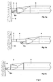
- FIG. 1 schematically shows an embodiment of a multifunctional instrument according to the invention for ultrasound surgery and for high-frequency surgery or laser surgery.
- An electroacoustic transducer (not visible) is in the handle 1 of this instrument in a manner known per se to which the ultrasound applicator 2 is acoustically coupled.
- the ultrasound applicator consists, in a manner known per se, of an impedance transformer, the distal end 3 of which is brought into contact with the tissue to be fragmented either directly or by connecting a special applicator tip (not shown). Special applicator tips are used for ergonomic and / or acoustic adaptation of the distal end of the ultrasound applicator to the respective surgical method.
- extension pieces can be connected between the electroacoustic transducer and impedance transformer and / or between the impedance transformer and applicator tip (not shown), which guide the ultrasound to the distal end 3 of the instrument.
- the impedance transformer including any necessary extension pieces is rod-shaped. 7
- a protective tube 4 can be arranged coaxially around these rod-shaped parts of the instrument, the inner diameter of which is larger than the outer diameter of the rod-shaped parts and protects these rod-shaped parts against damage and / or against direct contact.
- An embodiment of a protective tube 4 is shown in Fig. 1d.
- An applicator device 9 is arranged axially displaceably over this protective tube 4 and has at least one applicator 6 at its distal end, for example an electrode for cutting and / or coagulating with HF current or a hook electrode for blunt preparation, cutting and / or coagulating or an argon Plasma coagulation probe or a laser probe 15 carries.
- a connection part 11 is arranged at the proximal end of the applicator device 9, to which the media required in each case, such as HF current, argon and / or laser, are connected.
- connection 12 for the electroacoustic transducer 13 for suction and 14 for rinsing At the proximal end of the handle 1 of the ultrasound applicator 2 there are connections 12 for the electroacoustic transducer 13 for suction and 14 for rinsing.
- the connecting part 11 is ergonomically designed so that it can be used, for example, with the thumb of the hand, which encloses and holds the handle 1, for axially displacing the applicator device 9 on the protective tube 4.
- corrugation 12 can improve the grip.
- the applicator device 9 of the exemplary embodiment of an instrument according to the invention shown schematically in FIG. 1a is shown separately in FIGS. 1b and 1c from two different perspectives or directions.
- the applicator device 9, as shown in FIG. 1 can be designed such that it can be completely removed from the ultrasound applicator 2, so that this applicator device 9 can or may not be combined with the multifunctional instrument according to the invention.
- This also has the advantage that one of differently equipped application devices can be plugged onto the ultrasound applicator and used as required. Exemplary embodiments of different application devices are further described below (FIGS. 2, 3, 4, 5) and are described in more detail.
- the applicator device preferably consists of electrically insulating material.
- An electrical line (not shown) is provided in this applicator device for supplying the HF current from the proximal connection part 11 to the various HF current applicators 6.
- the applicator device can be equipped with a guide channel (not shown) through which these probes can be inserted from proximal to distal.
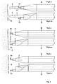
- the HF current applicator is a monopolar spherical coagulation electrode 6a.
- the distal end 3 of the ultrasound applicator and the coagulation electrode 6a can be positioned at the same distance from the tissue surface 8 or in such a way that the distal End 3 of the ultrasound applicator, as shown in FIG. 2b, is closer to the tissue 8 than the coagulation electrode 6a, or the coagulation electrode 6a, as shown in FIG. 2c, is closer to the tissue 8 than the distal end of the ultrasound applicator 3 In this way, ultrasound dissection and RF coagulation can be used alternately.
- FIG. 3 schematically shows an exemplary embodiment of a multifunctional instrument according to the invention for ultrasound surgery and for high-frequency surgery.
- the HF current applicator is an argon plasma coagulation probe 6b in this exemplary embodiment.
- the distal end 3 of the ultrasound applicator and the Aron plasma coagulation probe 6b can be positioned at the same distance from the tissue surface 8 or so that the distal end 3 of the ultrasound applicator is closer to the tissue 8 than the argon plasma coagulation probe 6b, or the argon plasma coagulation probe 6b is closer to the tissue 8 than the distal end of the ultrasound applicator 3.
- These positions are with this Embodiment not shown in the drawing, because they correspond in principle to those in FIGS. 2 and 4. In this way, ultrasound dissection and argon-plasma coagulation can be used alternately.
- FIG. 4 schematically shows an exemplary embodiment of a multifunctional instrument according to the invention for ultrasound surgery and for high-frequency surgery.
- the HF current applicator is a hook probe 6c, as is used for example for laparoscopic cholecystectomy.
- FIGS. 4c and 4cc are closer to the tissue 8 than the hook probe 6c, or the hook probe 6c, as shown in FIGS. 4c and 4cc, is closer to the tissue 8 as the distal end of the ultrasound applicator 3.
- ultrasound dissection and blunt dissection or cutting and / or coagulation with HF current can be used alternately.
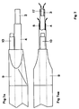
- 5 shows an exemplary embodiment of a multifunctional instrument according to the invention for ultrasound surgery and for laser surgery.
- the laser probe 15 is inserted from proximal to distal through the applicator device 9 and fixed in this applicator device 9.
- the laser probe 15 can be set up at such a distance f from the tissue surface 8 that the focus F of the laser L is in the plane of the Contact surface 16 of the distal end 3 of the ultrasound applicator 2 is located.
- the distal end 3 of the ultrasound applicator 2 can be used as a spacer for the laser application for the purpose of tissue vaporization or for tissue separation.
- the applicator device 9 with the laser probe 15 is displaced from this position proximally or distally on the protective tube 4 of the ultrasound applicator, the laser L is defocused on the tissue surface and thus the power density is reduced, thereby favoring the vaporization effect of the laser of the coagulation effect can be changed.
- FIG. 6 shows a schematic representation of an exemplary embodiment of a multifunctional instrument according to the invention in accordance with the previous exemplary embodiments, which is additionally equipped with a device 18 for automatically advancing or retracting the applicator device 9 with the respective HF current applicator and / or laser applicator.
- This device 18 is called the actuator below.
- This actuator can work, for example, electromagnetically, pneumatically or hydraulically.
- Such actuators are known per se (G. Farin: Pneumatically Controlled Bipolar Cutting Instrument, in Endoscopic Surgery and Allied Technologies, Issue 2, 1993, pages 97 to 101, Verlag Thieme Stuttgart) and are therefore not described in more detail here.
- the advancing or retracting of the applicator device can be triggered automatically by the activation signals of the high-frequency surgical device laser device or ultrasound device connected to the instrument.
- an activation signal "Cutting with HF current” can control the actuator in such a way that a hook electrode, as shown in FIG. 4c, is automatically advanced into the working position.
- the actuator can for example be arranged on or in the handle 1 of the instrument.
- the exemplary embodiment according to FIG. 6 serves to explain the principle of the automatic displacement of the electrosurgical or laser applicators relative to the ultrasound applicator.
- This displacement of the electrosurgical or laser applicators relative to the ultrasound applicator can also be realized in another way, for example in such a way that the applicator device is a fixed or exchangeable component of the instrument according to the invention and the electrosurgical or laser applicators themselves can be moved manually or automatically by actuators relative to the ultrasound applicator.
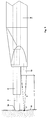
- Fig. 7 shows a more detailed representation of a known suction and rinsing function of the instrument.
- an ultrasound applicator of any type is provided, which is designed as a tube, the lumen 17 of which can be used as a suction channel from the distal end 3 to the connection 14 for suction.
- the gap 5 that can be used for rinsing between the ultrasound applicator 2 and the protective tube 4 has already been described above.
- adjustability of the applicators is provided by a displacement in the longitudinal direction.
- the control unit in exemplary embodiments of the invention can contain a logic circuit with a microprocessor, the program of which enables the automatic settings for the intended functions.
Abstract
Description
- Die Erfindung betrifft ein multifunktionales Instrument für die Ultraschall-Chirurgie entsprechend den Oberbegriff des Patentanspruchs 1.
Die Ultraschall-Chirurgie ist eine Methode für gewebeselektive Dissektionen. Die Hochfrequenz-Chirurige ist dagegen eine Methode, mit der alle Gewebe (außer Knochen) weitgehend unselektiv geschnitten werden können. Der Ultraschall-Chirurgie fehlt der Hämostaseeffekt. Die Hochfrequenz-Chirurgie kann dagegen sehr gut zur Hämostase angewendet werden. Die kombinierte Anwendung der Ultraschall-Chirurgie und der Hochfrequenz-Chirurgie kann insbesondere bei endoskopischen Operationen die Eigenschaften beider Methoden ergänzen und hierdurch die Operationstechnik verbessern oder gar Anwendungen ermöglichen, die mit der Ultraschall-Chirurgie oder der Hochfrequenz-Chirurgie allein nicht gut oder gar nicht möglich sind.
Die gewebespezifische Selektivität der Dissektion mit Ultraschall resultiert einerseits aus der unterschiedlichen Reißfestigkeit sowie dem relativen Wassergehalt der verschiedenen Gewebe und andererseits aus der Leistungsdichte des Ultraschalls im Gewebe. Gewebe mit geringer Reißfestigkeit und relativ hohem Wassergehalt (z.B. Parenchym) können bereits mit geringer Leistungsdichte des Ultraschalls fragmentiert werden. Gewebe mit hoher Reißfestigkeit und/oder geringem Wassergehalt (z.B. Bindegewebe, Stroma) erfordern eine relativ hohe Leistungsdichte des Ultraschalls. Die Ultraschall-Dissektion eignet sich somit insbesondere zur selektiven, anatomiegerechten Präparation von Gewebestrukturen, z.B. zur Demaskierung von Organen oder Gefäßen von umhüllendem Fettgewebe, zur Fragmentierung oder Aspiration unerwünschter oder pathologischer Gewebe (z.B. Fettgewebe oder Tumore) sowie zur Resektion parenchymatöser Organe (z.B. Leber). Bei Vorhandensein geeigneter Ultraschall-Applikatoren sowie richtiger Applikation und Dosierung der Leistungsdichte des Ultraschalls ist die selektive Dissektion bzw. Fragmentierung und Aspiration von Geweben in fast allen chirugischen Disziplinen anwendbar. Allerdings behindern zwei Probleme die Anwendung der Ultraschall-Dissektion insbesondere bei endoskopischen Operationen. Dies ist einerseits der fehlende Hämostaseeffekt des Ultraschalls insbesondere während der Resektion parenchymatöser Organe und andererseits der unzureichende oder gar fehlende Schneideeffekt des Ultraschalls in binde- und/oder stützgewebehaltigen Gewebestrukturen. Diese beiden bei der Ultraschall-Chirurgie fehlenden Effekte werden dagegen sehr gut mit der Hochfrequenz-Chirurgie erreicht. Eine Vermeidung von Schwierigkeiten der genannten Art ermöglicht deshalb die kombinierte Anwendung der Ultraschall-Chirurgie und der Hochfrequenz-Chirurgie. - Für die kombinierte Anwendung der Ultraschall-Chirurgie und der Hochfrequenz-Chirurgie stehen verschiedene Techniken zur Verfügung.
- Die einfachste Technik der kombinierten Anwendung von Ultraschall-Chirurgie und Hochfrequenz-Chirurgie besteht darin, daß der Operateur abwechselnd ein spezielles Instrument für die Ultraschall-Chirurgie und ein anderes spezielles Instrument für die Hochfrequenz-Chirurgie anwendet. Diese Technik hat zwar den Vorteil, daß beide Instrumente optimal für den jeweiligen Verwendungszweck gestaltet sein können aber leider auch den Nachteil, daß der intraoperative Wechsel der Instrumente insbesondere bei endoskopischer Anwendung unbequem und zeitaufwendig ist.
- Eine beispielsweise aus US-A-4,931,047 bekannte Technik, bei welcher ein intraoperativer Instrumentenwechsel vermieden wird, besteht darin, die Ultraschall-Chirurgie und die Hochfrequenz-Chirurgie über die selbe Applikatorspitze eines multifunktionalen Instruments anzuwenden. Hierbei kann der Ultraschall und der HF-Strom gleichzeitig oder abwechselnd appliziert werden. Diese Technik kann jedoch insofern problematisch sein, als während der HF-Strom-Koagulation elektrische Lichtbogen bzw. Funken zwischen der Applikatorspitze und dem Gewebe entstehen können, wodurch eine Funkenerosion und damit Beschädigung der Oberfläche der Applikatorspitze verursacht werden kann. Außerdem kann koaguliertes Gewebe an der Oberfläche der Applikatorspitze festkleben und die Ultraschall-Dissektion behindern oder gar verhindern. Ein weiteres Problem dieser Technik besteht darin, daß die Form der für die Ultraschall-Dissektion optimierten Applikatorspitze zum Schneiden mittels HF-Strom nur bedingt geeignet ist.
- Es ist Aufgabe der Erfindung, ein multifunktionales Instrument zu entwickeln, welches einerseits für die selektive Dissektion biologischer Gewebe mit Ultraschall und andererseits für die Blutstillung und/oder zum unselektiven Schneiden auch solcher Gewebe geeignet ist, welche mit Ultraschall nicht geschnitten bzw. fragmentiert werden können, mit welchem Instrument Probleme der genannten Art möglichst weitgehend vermieden werden können. Diese Aufgabe wird erfindungsgemäß durch den Gegenstand des Anspruchs 1 gelöst. Vorteilhafte Ausgestaltungen der Erfindung sind Gegenstand der Unteransprüche.
- Bei einem derartigen Instrument können ein Applikator für die Ultraschall-Chirurgie und mindestens ein Applikator für die Hochfrequenz-Chirurgie zum Beispiel zum Schneiden und/oder Koagulieren und/oder ein Applikator für die Laser-Chirurgie so am distalen Ende eines kombinierten Instruments angeordnet werden, daß sowohl die Ultraschall-Chirurgie als auch die Hochfrequenz-Chirurgie und/oder Laser-Chirurgie ohne Instrumentenwechsel bequem und zeitsparend angewendet werden können. Derartig kombinierte Instrumente können dem jeweiligen Anwendungszweck entsprechend angepaßt sein. So kann beispielsweise ein multifunktionales Instrument einerseits mit einer für den jeweiligen Anwendungszweck gestalteten Applikatorspitze für die Ultraschall-Dissektion sowie andererseits mit einer monopolaren oder bipolaren Hakensonde, wie sie beispielsweise für die laparoskopische Cholezystektomie verwendet wird, zum Schneiden und/oder Koagulieren bzw. Blutstillen mittels HF-Strom und/oder mit einer Laser-Sonde zum Trennen bzw. Vaporisieren von Gewebestrukturen und/oder zum Koagulieren von Geweben bzw. zum Blutstillen ausgestattet sein. Die Hakensonde kann außerdem in bekannter Weise auch zum stumpfen Präparieren von Geweben angewendet werden.
- Derartige multifunktionale Instrumente sollten ergonomisch und sicherheitstechnisch so gestaltet sein, daß jede Funktion möglichst optimal angewendet werden kann. So kann es z.B. zweckmäßig sein, wenn jeweils eine Funktion optimal zur Verfügung steht, welche benutzt werden soll. Ein erfindungsgemäßes Instrument kann dementsprechend so gestaltet sein, daß beispielsweise eine zum stumpfen Präparieren, Schneiden und/oder Koagulieren verwendbare Hakensonde relativ zum distalen Ende bzw. zur Sondenspitze des Ultraschallapplikators derart verschiebbar ist, daß sie gleichen, größeren oder kleineren Abstand zum Gewebe hat als das distale Ende des Ultraschall-Applikators. Auf diese Weise kann, je nach Bedarf, entweder der HF-Strom-Applikator, beispielsweise die Hakensonde, oder die Sondenspitze des Ultraschall-Applikators in optimale Arbeitsstellung gebracht werden. Das distale Ende eines Laser-Applikators, beispielsweise einer Laser-Sonde, kann relativ zum distalen Ende des Ultraschall-Applikators oder des HF-Strom-Applikators so fixiert werden, daß der Fokus des Lasers zum Schneiden oder zum Koagulieren außerhalb des Bereichs des distalen Endes des Ultraschall-Applikators oder des HF-Strom-Applikators liegt, so daß der Ultraschall-Applikator oder der HF-Strom-Applikator während der Laser-Chirurgie als Abstandhalter bzw. Fokussier- oder Defokussierhilfen benutzt werden können. Als HF-Strom-Applikator kann ein erfindungsgemäßes multifunktionales Instrument auch mit einer Argon-Plasma-Koagulations-Sonde ausgestattet sein.
- Außerdem können erfindungsgemäße multifunktionale Instrumente zusätzlich mit Einrichtungen zum Saugen und/oder Spülen ausgestattet sein.
- In einer vorteilhaften Weiterentwicklung können die erfindungsgemäßen multifunktionalen Instrumente mit einer Einrichtung ausgestattet sein, welche ein automatisches Vorschieben der HF-Strom-Applikatoren und/oder der Laser-Applikatoren in Arbeitsstellung bzw. ein automatisches Zurückziehen der HF-Strom-Applikatoren und/oder Laser-Applikatoren in Ruhestellung, beispielsweise in Abhängigkeit von Aktivierungssignalen für den Schneide- und/oder Koagulationsmodus oder für den Laser- oder Ultraschallmodus und/oder für den Spül- und/oder Saugmodus ermöglicht. Anhand der Zeichnung sollen Ausführungsbeispiele der Erfindung detaillierter beschrieben werden. Es zeigen:
- Fig. 1 eine schematische Darstellung des prinzipiellen Aufbaus eines erfindungsgemäßen multifunktionalen Instruments für die Ultraschall-Chirurgie und für die Hochfrequenz-Chirurgie.
- Fig. 2 eine schematische Darstellung eines Ausführungsbeispiels des erfindungsgemäßen multifunktionalen Instruments für die Ultraschall-Chirurgie und zum Koagulieren mittels HF-Strom über eine Koagulationselektrode.
- Fig. 3 eine schematische Darstellung eines Ausführungsbeispiels des erfindungsgemäßen multifunktionalen Instruments für die Ultraschall-Chirurgie und zum Koagulieren mittels HF-Strom über eine Argon-Plasma-Koagulations-Sonde.
- Fig. 4 eine schematische Darstellung eines Ausführungsbeispiels des erfindungsgemäßen multifunktionalen Instruments für die Ultraschall-Chirurgie und zum Schneiden und Koagulieren mittels HF-Strom sowie zum stumpfen Präparieren.
- Fig. 5 eine schematische Darstellung eines Ausführungsbeispiels des erfindungsgemäßen multifunktionalen Instruments für die Ultraschall-Chirurgie, welches mit einer Laser-Sonde zum Schneiden oder Koagulieren ausgestattet ist.
- Fig. 6 eine schematische Darstellung eines Ausführungsbeispiels eines erfindungsgemäßen multifunktionalen Instruments entsprechend den vorhergehenden Ausführungsbeispielen, welches zusätzlich mit mindestens einer Einrichtung zum automatischen Vorschieben bzw. Zurückziehen der HF-Strom-Applikatoren und/oder des Laser-Applikators ausgestattet ist.
- Fig. 7 eine schematische Darstellung der Saug- und Spülfunktionen eines erfindungsgemäßen multifunktionalen Instruments.
- In Fig. 1 ist ein Ausführungsbeispiel eines erfindungsgemäßen multifunktionalen Instruments für die Ultraschall-Chirurgie und für die Hochfrequenz-Chirurgie oder Laser-Chirurgie schematisch dargestellt. Im Griff 1 dieses Instruments ist in an sich bekannter Weise ein elektroakustischer Wandler (nicht sichtbar) vorhanden, an welchen der Ultraschall-Applikator 2 akustisch angekoppelt ist. Der Ultraschall-Applikator besteht in an sich bekannter Weise aus einem Impedanztransformator, dessen distales Ende 3 direkt oder durch Vorschalten eines speziellen Applikator-Tips(nicht dargestellt) mit dem zu fragmentierenden Gewebe in Kontakt gebracht wird. Spezielle Applikator-Tips dienen zur ergonomischen und/oder akustischen Anpassung des distalen Endes des Ultraschall-Applikators an die jeweilige Operationsmethode.
Für Laparoskopische Anwendungen können zwischen elektroakustischem Wandler und Impedanztransformator und/oder zwischen Impedanztransformator und Applikator-Tip in bekannter Weise (Cuschieri) Verlängerungsstücke geschaltet sein (nicht dargestellt), welche den Ultraschall zum distalen Ende 3 des Instruments hin leiten. Bei dem in Fig. 1a dargestellten Ausführungsbeispiel ist der Impedanztransformator inklusive eventuell erforderlicher Verlängerungsstücke stabförmig gestaltet. Um diese stabförmig gestalteten Teile des Instruments kann, wie in Fig. 7 detaillierter dargestellt, koaxial ein Schutzrohr 4 angeordnet sein, dessen Innendurchmesser größer ist als der Außendurchmesser der stabförmigen Teile, welches diese stabförmigen Teile gegen Beschädigung und/oder gegen direkte Berührung schützt. Ein Ausführungsbeispiel eines Schutzrohrs 4 ist in Fig. 1d dargestellt. Der in Fig. 7 dargestellte Spalt 5 zwischen den stabförmigen Teilen und dem Schutzrohr kann in bekannter Weise zum Zuleiten von Spülflüssigkeiten genutzt werden. Über dieses Schutzrohr 4 wird eine Applikatoreinrichtung 9 axial verschiebbar angeordnet, welche an ihrem distalen Ende mindestens einen Applikator 6, beispielsweise eine Elektrode zum Schneiden und/oder Koagulieren mit HF-Strom oder eine Hakenelektrode zum stumpfen Präparieren, Schneiden und/oder Koagulieren oder eine Argon-Plasma-Koagulations-Sonde oder eine Laser-Sonde 15 trägt. Am proximalen Ende der Applikatoreinrichtung 9 ist in diesem Ausführungsbeispiel ein Anschlußteil 11 angeordnet, an welchem die jeweils erforderlichen Medien, wie beispielsweise HF-Strom, Argon und/oder Laser angeschlossen werden. Am proximalen Ende des Griffs 1 des Ultraschallapplikators 2 sind Anschlüsse 12 für den elektroakustischen Wandler 13 zum Saugen und 14 zum Spülen angeordnet. Das Anschlußteil 11 ist ergonomisch so gestaltet, daß es beispielsweise mit dem Daumen der Hand, welche den Griff 1 umschließt und hält, zum axialen Verschieben der Applikatoreinrichtung 9 auf dem Schutzrohr 4 benutzt werden kann. Hierzu kann eine Riffelung 12 die Griffigkeit verbessern. - Die Applikatoreinrichtung 9 des in Fig. 1a schematisch dargestellten Ausführungsbeispiels eines erfindungsgemäßen Instruments ist in Fig. 1b und 1c separat aus zwei verschiedenen Perspektiven bzw. Richtungen betrachtet dargestellt. In einer vorteilhaften Ausführung kann die Applikatoreinrichtung 9, wie in Fig. 1 dargestellt, so gestaltet sein, daß sie komplett vom Ultraschallapplikator 2 abnehmbar ist, so daß diese Applikatoreinrichtung 9 bei Bedarf mit dem erfindungsgemäßen multifunktionalen Instrument kombiniert werden kann oder auch nicht. Dies hat außerdem den Vorteil, daß je nach Bedarf eine von verschieden bestückten Applikationseinrichtungen auf den Ultraschallapplikator aufgesteckt und angewendet werden kann. Ausführungsbeispiele verschiedenen Applikationseinrichtungen werden weiter unter dargestellt (Fig. 2,3,4,5) und detaillierter beschrieben.
- Die Applikatoreinrichtung besteht vorzugsweise aus elektrisch isolierendem Material. Zur Zuleitung des HF-Stroms vom proximalen Anschlußteil 11 zu den verschiedenen HF-Stromapplikatoren 6 ist eine elektrische Leitung (nicht dargestellt) in dieser Applikatoreinrichtung vorhanden. Für die Verwendung von Argon-Plasma-Koagulations-Sonden oder Laser-Sonden kann die Applikatoreinrichtung mit einem Führungskanal (nicht dargestellt) ausgestattet sein, durch welchen diese Sonden von proximal nach distal eingeführt werden können.
- In Fig. 2 ist das distale Ende eines Ausführungsbeispiels eines erfindungsgemäßen multifunktionalen Instruments für die Ultraschall-Chirurgie und für die Hochfrequenz-Chirurgie schematisch dargestellt. Der HF-Strom-Applikator ist in diesem Ausführungsbeispiel eine monopolare kugelförmige Koagulationselektrode 6a. Durch axiales Verschieben der Applikatoreinrichtung 9 auf dem Schutzrohr 4 des Ultraschall-Applikators 2 kann das distale Ende 3 des Ultraschall-Applikators und die Koagulationselektrode 6a, wie in Fig. 2a dargestellt, in gleiche Entfernung zur Gewebeoberfläche 8 oder so positioniert werden, daß das distale Ende 3 des Ultraschall-Applikators , wie in Fig. 2b dargestellt, dem Gewebe 8 näher ist als die Koagulationselektrode 6a,oder die Koagulationselektrode 6a, wie in Fig. 2c dargestellt, dem Gewebe 8 näher ist als das distale Ende des Ultraschall-Applikators 3. Auf diese Weise können Ultraschalldissektion und HF-Koagulation abwechselnd angewendet werden.
- In Fig. 3 ist ein Ausführungsbeispiel eines erfindungsgemäßen multifunktionalen Instruments für die Ultraschall-Chirurgie und für die Hochfrequenz-Chirurgie schematisch dargestellt. Der HF-Strom-Applikator ist in diesem Ausführungsbeispiel eine Argon-Plasma-Koagulations-Sonde 6b. Durch axiales Verschieben der Applikatoreinrichtung 9 auf dem Schutzrohr 4 des Ultraschall-Applikators 2 kann das distale Ende 3 des Ultraschall-Applikators und die Aron-Plasma-Koagulations-Sonde 6b in gleiche Entfernung zur Gewebeoberfläche 8 oder so positioniert werden, daß das distale Ende 3 des Ultraschall-Applikators dem Gewebe 8 näher ist als die Argon-Plasma-Koagulations-Sonde 6b,oder die Argon-Plasma-Koagulations-Sonde 6b dem Gewebe 8 näher ist als das distale Ende des Ultraschall-Applikators 3. Diese Positionierungen sind bei diesem Ausführungsbeispiel nicht zeichnerisch dargestellt, weil sie prinzipiell den in den Fig. 2 sowie 4 entsprechen. Auf diese Weise können Ultraschalldissektion und Argon-Plasma-Koagulation abwechselnd angewendet werden.
- In Fig. 4 ist ein Ausführungsbeispiel eines erfindungsgemäßen multifunktionalen Instruments für die Ulraschall-Chirurgie und für die Hochfrequenz-Chirurgie schematisch dargestellt. Der HF-Strom-Applikator ist in diesem Ausführungsbeispiel eine Hakensonde 6c, wie sie beispielsweise für die laparoskopische Cholezystektomie angewendet wird. Durch axiales Verschieben der Applikatoreinrichtung 9 auf dem Schutzrohr 4 des Ultraschall-Applikators 2 kann das distale Ende des Ultraschall-Applikators 3 und die Hakensonde 6c, wie in Fig. 2a bzw. 4a dargestellt, in gleiche Entfernung zur Gewebeoberfläche 8 oder so positioniert werden, daß das distale Ende 3 des Ultraschall-Applikators, wie in Fig. 4b bzw. 4bb dargestellt, dem Gewebe 8 näher ist als die Hakensonde 6c,oder die Hakensonde 6c, wie in Fig. 4c bzw. 4cc dargestellt, dem Gewebe 8 näher ist als das distale Ende des Ultraschall-Applikators 3. Auf diese Weise können Ultraschalldissektion und stumpfes Präparieren oder Schneiden und/oder Koagulieren mit HF-Strom abwechselnd angewendet werden.
In Fig. 5 ist ein Ausführungsbeispiel eines erfindungsgemäßen multifunktionalen Instruments für die Ultraschall-Chirurgie und für die Laser-Chirurgie schematisch dargestellt. Die Laser-Sonde 15 ist von proximal nach distal durch die Applikatoreinrichtung 9 eingeführt und in dieser Applikatoreinrichtung 9 fixiert. Durch axiales Verschieben der Applikatoreinrichtng 9 nach proximal auf dem Schutzrohr 4 des Ultraschall-Applikators 2 kann die Laser-Sonde 15 in einer derartigen Entfernung f zur Gewebeoberfläche 8 eingerichtet werden, dap der Fokus F des Lasers L in der Ebene der Kontaktfläche 16 des distalen Endes 3 des Ultraschall-Applikators 2 liegt. Auf diese Weise kann das distale Ende 3 des Ultraschall-Applikators 2 als Distanzhalter für die Laserapplikation zum Zwecke der Gewebevaporisation bzw. zum Gewebetrennen angewendet werden. Wird die Applikatoreinrichtung 9 mit der Laser-Sonde 15 aus dieser Position heraus nach proximal oder nach distal auf dem Schutzrohr 4 des Ultraschall-Applikators verschoben, so wird der Laser L auf der Gewebeoberfläche defokussiert und damit die Leistungsdichte reduziert, wodurch der Vaporisationseffekt des Lasers zugunsten des Koagulationseffekts verändert werden kann. - Fig. 6 zeigt eine schematische Darstellung eines Ausführungsbeispiels eines erfindungsgemäßen multifunktionalen Instruments entsprechend den vorhergehenden Ausführungsbeispielen, welches zusätzlich mit einer Einrichtung 18 zum automatischen Vorschieben bzw. Zurückziehen der Applikatoreinrichtung 9 mit dem jeweiligen HF-Strom-Applikator und/oder Laser-Applikator ausgestattet ist. Diese Einrichtung 18 wird im folgenden Aktor genannt. Dieser Aktor kann beispielsweise elektromagnetisch, pneumatisch oder hydraulisch arbeiten. Derartige Aktoren sind an sich bekannt (G. Farin: Pneumatically Controlled Bipolar Cutting Instrument, in Endoscopic Surgery und Allied Technologies, Heft 2, 1993, Seiten 97 bis 101, Verlag Thieme Stuttgart) und deswegen hier nicht detaillierter beschrieben. Das Vorschieben oder Zurückziehen der Applikatoreinrichtung kann automatisch durch die Aktivierungssignale des jeweils an das Instrument angeschlossenen Hochfrequenz-Chirurgiegerät Laser-Gerät oder Ultraschall-Gerät ausgelöst werden. So kann beispielsweise ein Aktivierungssignal "Schneiden mit HF-Strom" den Aktor so steuern, daß eine Hakenelektrode, wie in Fig. 4c dargestellt, automatisch in Arbeitsstellung vorgeschogen wird. Der Aktuator kann beispielsweise am oder im Griff 1 des Instruments angeordnet sein.
- Das Ausführungsbeispiel entsprechend Fig. 6 dient zur Erläuterung des Prinzips des automatischen Verschiebens der HF-chirurgischen bzw. der Laser-Applikatoren relativ zum Ultraschall-Applikator. Dieses Verschieben der HF-chirurgischen bzw. der Laser-Applikatoren relativ zum Ultraschall-Applikator kann auch auf andere Weise realisiert werden, beispielsweise derart, daß die Applikatoreinrichtung fester oder austauschbarer Bestandteil des erfindungsgemäßen Instruments ist und die HF-chirurgischen bzw. der Laser-Applikatoren selber manuell oder automatisch durch Aktoren relativ zum Ultraschall-Applikator verschiebbar sind.
- Fig. 7 zeigt eine detailliertere Darstellung einer an sich bekannten Saug- und Spülfunktion des Instruments. Bei diesem Ausführungsbeispiel ist neben einem HF-Strom-Applikator 10 an sich beliebiger Art ein Ultraschall-Applikator vorgesehen, der als Rohr ausgebildet ist, dessen Lumen 17 vom distalen Ende 3 bis zum Anschluß 14 zum Saugen als Saugkanal verwendbar ist. Der zum Spülen verwendbare Spalt 5 zwischen dem Ultraschall-Applikator 2 und dem Schutzrohr 4 wurde bereits oben beschrieben.
- Bei den dargestellten und beschriebenen Ausführungsbeispielen ist eine Verstellbarkeit der Applikatoren durch eine Verschiebung in Längsrichtung vorgesehen. In gewissen Fällen ist es jedoch vorteilhaft, die Applikatoren in einer Revolvereinheit vorzusehen, die um ihre Längsachse drehbar an dem Instrument angeordnet ist. Wenn beispielsweise drei Applikatoren vorgesehen sind und wenn sich zunächst das distale Ende 3 des Ultraschall-Applikators in der Arbeitsstellung befindet, kann das distale Ende eines anderen Applikators automatisch in die gleiche Arbeitsstellung gebracht werden, indem der erste Applikator zunächst zurückgezogen wird und dann die Revolvereinheit so gedreht wird, daß der zweite oder dritte Applikator nach einem geeigneten Vorschub zur Erzielung eines vorherbestimmten Abstands zu dem Gewebe in eine geeignete Arbeitsstellung gebracht werden kann, wobei das Instrument als solches nicht bewegt werden muß. Wie bei dem erwähnten bekannten pneumatisch gesteuerten multifunktionalen Instrument kann die Steuereinheit bei Ausführungsbeispielen der Erfindung eine logische Schaltung mit einem Mikroprozessor enthalten, dessen Programm die automatischen Einstellungen für die vorgesehenen Funktionen ermöglicht.
Claims (10)
- Multifunktionales Instrument für die Ultraschall-Chirurgie, mit einem Applikator (2) zur Zufuhr von Ultraschall für eine Dissektion von biologischem Gewebe und mit einer Einrichtung zum Schneiden und/oder Koagulieren des Gewebes, dadurch gekennzeichnet, daß an dem Instrument mindestens ein von dem Ultraschall-Applikator (2) getrennter HF-Strom-Applikator (6) oder ein Laser-Applikator (15) in einem seitlichen Abstand vom distalen Ende (3) des Ultraschall-Applikators (2) verstellbar angeordnet ist.
- Multifunktionales Instrument nach Anspruch 1, dadurch gekennzeichnet, daß jeder Applikator (2,6,15) in Längsrichtung verschiebbar in einem gemeinsamen Applikatorgehäuse (9) angeordnet ist.
- Multifunktionales Instrument nach Anspruch 1 oder 2, dadurch gekennzeichnet, daß die Applikatoren (2,6,15) in einer Revolvereinheit vorgesehen sind, die um ihre Längsachse drehbar an dem Instrument angeordnet ist.
- Multifunktionales Instrument nach einem der vorhergehenden Ansprüche, dadurch gekennzeichnet, daß mindestens ein Aktor (18) vorgesehen ist, mit dem ein Applikator (2,6,15) automatisch in eine vorherbestimmte Stellung relativ zum distalen Ende des Ultraschall-Applikators (3) verstellbar ist.
- Multifunktionales Instrument nach Anspruch 4, dadurch gekennzeichnet, daß der Aktor (18) eine elektrisch, pneumatisch oder hydraulisch betätigbare Antriebseinrichtung aufweist.
- Multifunktionales Instrument nach einem der vorhergehenden Ansprüche, dadurch gekennzeichnet, daß der Ultraschall-Applikator (3) gegen den bzw. die HF-Strom-Applikatoren (6) elektrisch isoliert ist.
- Multifunktionales Instrument nach einem der vorhergehenden Ansprüche, dadurch gekennzeichnet, daß der HF-Strom-Applikator (6) als Tasthaken für die Cholezystektomie ausgebildet ist.
- Multifunktionales Instrument nach Anspruch 1, dadurch gekennzeichnet, daß der HF-Strom-Applikator (6) eine monopolare Koagulationselektrode ist.
- Multifunktionales Instrument nach einem der Ansprüche 1 bis 6, dadurch gekennzeichnet, daß der HF-Strom-Applikator (6b) eine Argon-Plasma-Koagulations-Sonde ist.
- Multifunktionales Instrument nach einem der vorhergehenden Ansprüche, dadurch gekennzeichnet, daß ein Saugkanal (17) und/oder ein Spülkanal (5) vorgesehen ist, um mittels des Ultraschalls fragmentiertes Gewebe abzusaugen und/oder Spülflüssigkeit in das Operationsfeld einzuleiten.
Priority Applications (3)
| Application Number | Priority Date | Filing Date | Title |
|---|---|---|---|
| EP94104568A EP0688536B1 (de) | 1994-03-23 | 1994-03-23 | Multifunktionales Instrument für die Ultraschall-Chirurgie |
| DE59409469T DE59409469D1 (de) | 1994-03-23 | 1994-03-23 | Multifunktionales Instrument für die Ultraschall-Chirurgie |
| US08/408,311 US5776092A (en) | 1994-03-23 | 1995-03-22 | Multifunctional surgical instrument |
Applications Claiming Priority (1)
| Application Number | Priority Date | Filing Date | Title |
|---|---|---|---|
| EP94104568A EP0688536B1 (de) | 1994-03-23 | 1994-03-23 | Multifunktionales Instrument für die Ultraschall-Chirurgie |
Publications (2)
| Publication Number | Publication Date |
|---|---|
| EP0688536A1 true EP0688536A1 (de) | 1995-12-27 |
| EP0688536B1 EP0688536B1 (de) | 2000-08-02 |
Family
ID=8215797
Family Applications (1)
| Application Number | Title | Priority Date | Filing Date |
|---|---|---|---|
| EP94104568A Expired - Lifetime EP0688536B1 (de) | 1994-03-23 | 1994-03-23 | Multifunktionales Instrument für die Ultraschall-Chirurgie |
Country Status (3)
| Country | Link |
|---|---|
| US (1) | US5776092A (de) |
| EP (1) | EP0688536B1 (de) |
| DE (1) | DE59409469D1 (de) |
Cited By (12)
| Publication number | Priority date | Publication date | Assignee | Title |
|---|---|---|---|---|
| DE19718708A1 (de) * | 1997-05-02 | 1998-11-05 | Gunther Dr Burgard | Hämorrhoidenresektionsinstrument |
| WO1999003406A1 (de) * | 1997-07-14 | 1999-01-28 | Erbe Elektromedizin Gmbh | Präparationsinstrument |
| WO1999013784A1 (de) * | 1997-09-17 | 1999-03-25 | Laser- Und Medizin-Technologie Gmbh Berlin | Vorrichtung zur revaskularisation von muskelgewebe |
| US5944715A (en) | 1996-06-20 | 1999-08-31 | Gyrus Medical Limited | Electrosurgical instrument |
| US6004319A (en) | 1995-06-23 | 1999-12-21 | Gyrus Medical Limited | Electrosurgical instrument |
| US6013076A (en) | 1996-01-09 | 2000-01-11 | Gyrus Medical Limited | Electrosurgical instrument |
| US6015406A (en) | 1996-01-09 | 2000-01-18 | Gyrus Medical Limited | Electrosurgical instrument |
| US6027501A (en) | 1995-06-23 | 2000-02-22 | Gyrus Medical Limited | Electrosurgical instrument |
| US6090106A (en) | 1996-01-09 | 2000-07-18 | Gyrus Medical Limited | Electrosurgical instrument |
| US6093186A (en) | 1996-12-20 | 2000-07-25 | Gyrus Medical Limited | Electrosurgical generator and system |
| WO2006119892A1 (en) * | 2005-05-09 | 2006-11-16 | Erbe Elektromedizin Gmbh | Endoscopic-surgery apparatus for argon-plasma coagulation (apc) |
| DE102009025013B4 (de) * | 2009-06-16 | 2019-08-29 | Fraunhofer-Gesellschaft zur Förderung der angewandten Forschung e.V. | Automatisiertes Instrumentenwechselsystem für die minimal-invasive Chirurgie |
Families Citing this family (165)
| Publication number | Priority date | Publication date | Assignee | Title |
|---|---|---|---|---|
| US6780180B1 (en) | 1995-06-23 | 2004-08-24 | Gyrus Medical Limited | Electrosurgical instrument |
| US6293942B1 (en) | 1995-06-23 | 2001-09-25 | Gyrus Medical Limited | Electrosurgical generator method |
| US7452358B2 (en) * | 1996-01-05 | 2008-11-18 | Thermage, Inc. | RF electrode assembly for handpiece |
| US6350276B1 (en) * | 1996-01-05 | 2002-02-26 | Thermage, Inc. | Tissue remodeling apparatus containing cooling fluid |
| US7189230B2 (en) * | 1996-01-05 | 2007-03-13 | Thermage, Inc. | Method for treating skin and underlying tissue |
| US6340354B1 (en) * | 1996-05-17 | 2002-01-22 | Christopher L Rambin | Automated compulsory blood extraction system |
| US6565561B1 (en) | 1996-06-20 | 2003-05-20 | Cyrus Medical Limited | Electrosurgical instrument |
| GB9612993D0 (en) | 1996-06-20 | 1996-08-21 | Gyrus Medical Ltd | Electrosurgical instrument |
| US5899915A (en) * | 1996-12-02 | 1999-05-04 | Angiotrax, Inc. | Apparatus and method for intraoperatively performing surgery |
| US6102926A (en) | 1996-12-02 | 2000-08-15 | Angiotrax, Inc. | Apparatus for percutaneously performing myocardial revascularization having means for sensing tissue parameters and methods of use |
| US6010476A (en) * | 1996-12-02 | 2000-01-04 | Angiotrax, Inc. | Apparatus for performing transmyocardial revascularization |
| US5910150A (en) | 1996-12-02 | 1999-06-08 | Angiotrax, Inc. | Apparatus for performing surgery |
| US6051008A (en) | 1996-12-02 | 2000-04-18 | Angiotrax, Inc. | Apparatus having stabilization members for percutaneously performing surgery and methods of use |
| US6120520A (en) | 1997-05-27 | 2000-09-19 | Angiotrax, Inc. | Apparatus and methods for stimulating revascularization and/or tissue growth |
| DE19758703B4 (de) * | 1997-01-07 | 2007-02-22 | Storz Endoskop Gmbh | Multifunktionales endoskopisches Operationsgerät |
| GB2327352A (en) | 1997-07-18 | 1999-01-27 | Gyrus Medical Ltd | Electrosurgical instrument |
| US7278994B2 (en) * | 1997-07-18 | 2007-10-09 | Gyrus Medical Limited | Electrosurgical instrument |
| AU733337B2 (en) | 1997-07-18 | 2001-05-10 | Gyrus Medical Limited | An electrosurgical instrument |
| GB9807303D0 (en) | 1998-04-03 | 1998-06-03 | Gyrus Medical Ltd | An electrode assembly for an electrosurgical instrument |
| US7494488B2 (en) * | 1998-05-28 | 2009-02-24 | Pearl Technology Holdings, Llc | Facial tissue strengthening and tightening device and methods |
| US6432101B1 (en) * | 1998-05-28 | 2002-08-13 | Pearl Technology Holdings, Llc | Surgical device for performing face-lifting using electromagnetic radiation |
| US6203540B1 (en) * | 1998-05-28 | 2001-03-20 | Pearl I, Llc | Ultrasound and laser face-lift and bulbous lysing device |
| US6117152A (en) * | 1999-06-18 | 2000-09-12 | Ethicon Endo-Surgery, Inc. | Multi-function ultrasonic surgical instrument |
| US6235024B1 (en) * | 1999-06-21 | 2001-05-22 | Hosheng Tu | Catheters system having dual ablation capability |
| US6558379B1 (en) | 1999-11-18 | 2003-05-06 | Gyrus Medical Limited | Electrosurgical system |
| EP1110509A1 (de) * | 1999-12-21 | 2001-06-27 | Tomaso Vercellotti | Chirurgische Vorrichtung für Knochenchirurgie |
| ES2270896T3 (es) | 1999-12-30 | 2007-04-16 | Pearl Technology Holdings, Llc | Dispositivo de estiramiento facial. |
| DE10021529A1 (de) | 2000-05-03 | 2001-11-15 | Celon Ag Medical Instruments | Von Hand betätigbarer Ultraschallzertrümmerer zum Zerkleinern oder Entfernen von menschlichem oder tierischem Gewebe |
| AU2001273421A1 (en) | 2000-07-13 | 2002-01-30 | Bioheart, Inc. | Deployment system for myocardial cellular material |
| US20050075630A1 (en) * | 2000-08-01 | 2005-04-07 | Dfine, Inc. | Voltage threshold ablation apparatus |
| US6413256B1 (en) * | 2000-08-01 | 2002-07-02 | Csaba Truckai | Voltage threshold ablation method and apparatus |
| US7744595B2 (en) * | 2000-08-01 | 2010-06-29 | Arqos Surgical, Inc. | Voltage threshold ablation apparatus |
| DE60113150T2 (de) * | 2000-12-15 | 2006-06-29 | Sherwood Services Ag | Elektrochirurgischer elektroden-schutzkragen |
| US7846096B2 (en) | 2001-05-29 | 2010-12-07 | Ethicon Endo-Surgery, Inc. | Method for monitoring of medical treatment using pulse-echo ultrasound |
| US20030032898A1 (en) * | 2001-05-29 | 2003-02-13 | Inder Raj. S. Makin | Method for aiming ultrasound for medical treatment |
| HU227124B1 (en) * | 2001-09-14 | 2010-07-28 | Egis Gyogyszergyar Nyilvanosan | Polymorphs of 1-pyrrole derivative, intermediate for the preparation of atorvastatin |
| DE10147145C2 (de) | 2001-09-25 | 2003-12-18 | Kunz Reiner | Multifunktionsinstrument für die mikroinvasive Chirurgie |
| US6648839B2 (en) * | 2002-02-28 | 2003-11-18 | Misonix, Incorporated | Ultrasonic medical treatment device for RF cauterization and related method |
| US6736814B2 (en) * | 2002-02-28 | 2004-05-18 | Misonix, Incorporated | Ultrasonic medical treatment device for bipolar RF cauterization and related method |
| US9216053B2 (en) * | 2002-03-05 | 2015-12-22 | Avent, Inc. | Elongate member providing a variation in radiopacity |
| US20090024124A1 (en) * | 2005-07-14 | 2009-01-22 | Lefler Amy | Methods for treating the thoracic region of a patient's body |
| US7819869B2 (en) * | 2004-11-15 | 2010-10-26 | Kimberly-Clark Inc. | Methods of treating the sacroilac region of a patient's body |
| US9949789B2 (en) | 2002-03-05 | 2018-04-24 | Avent, Inc. | Methods of treating the sacroiliac region of a patient's body |
| US20070156136A1 (en) * | 2002-03-05 | 2007-07-05 | Neil Godara | Methods of treating the sacroiliac region of a patient's body |
| US20060259026A1 (en) * | 2005-05-05 | 2006-11-16 | Baylis Medical Company Inc. | Electrosurgical treatment method and device |
| US9364281B2 (en) * | 2002-03-05 | 2016-06-14 | Avent, Inc. | Methods for treating the thoracic region of a patient's body |
| US11291496B2 (en) | 2002-03-05 | 2022-04-05 | Avent, Inc. | Methods of treating the sacroiliac region of a patient's body |
| US6736835B2 (en) * | 2002-03-21 | 2004-05-18 | Depuy Acromed, Inc. | Early intervention spinal treatment methods and devices for use therein |
| US20030212390A1 (en) * | 2002-05-07 | 2003-11-13 | Chen Peter C. | System for operating an ablation generator with dual energy source |
| CA2493556C (en) | 2002-07-25 | 2012-04-03 | Thomas L. Ii Buchman | Electrosurgical pencil with drag sensing capability |
| US6747218B2 (en) | 2002-09-20 | 2004-06-08 | Sherwood Services Ag | Electrosurgical haptic switch including snap dome and printed circuit stepped contact array |
| US8613744B2 (en) | 2002-09-30 | 2013-12-24 | Relievant Medsystems, Inc. | Systems and methods for navigating an instrument through bone |
| US8808284B2 (en) | 2008-09-26 | 2014-08-19 | Relievant Medsystems, Inc. | Systems for navigating an instrument through bone |
| US7258690B2 (en) | 2003-03-28 | 2007-08-21 | Relievant Medsystems, Inc. | Windowed thermal ablation probe |
| US6907884B2 (en) | 2002-09-30 | 2005-06-21 | Depay Acromed, Inc. | Method of straddling an intraosseous nerve |
| US8361067B2 (en) | 2002-09-30 | 2013-01-29 | Relievant Medsystems, Inc. | Methods of therapeutically heating a vertebral body to treat back pain |
| US7244257B2 (en) | 2002-11-05 | 2007-07-17 | Sherwood Services Ag | Electrosurgical pencil having a single button variable control |
| US8057468B2 (en) | 2002-12-17 | 2011-11-15 | Bovie Medical Corporation | Method to generate a plasma stream for performing electrosurgery |
| US7316682B2 (en) * | 2002-12-17 | 2008-01-08 | Aaron Medical Industries, Inc. | Electrosurgical device to generate a plasma stream |
| FR2849781B1 (fr) * | 2003-01-14 | 2005-03-25 | Edap S A | Sonde de therapie |
| US7235072B2 (en) | 2003-02-20 | 2007-06-26 | Sherwood Services Ag | Motion detector for controlling electrosurgical output |
| US7104985B2 (en) * | 2003-03-06 | 2006-09-12 | Martinelli Michael A | Apparatus and method for causing selective necrosis of abnormal cells |
| US7090654B2 (en) * | 2003-03-28 | 2006-08-15 | Sherwood Services Ag | Catheter with occlusion resistant tip |
| US7776005B2 (en) * | 2003-03-28 | 2010-08-17 | Covidien Ag | Triple lumen catheter with occlusion resistant tip |
| US7141035B2 (en) * | 2003-03-28 | 2006-11-28 | Sherwood Services Ag | Catheter with occlusion resistant tip |
| US20040206365A1 (en) * | 2003-03-31 | 2004-10-21 | Knowlton Edward Wells | Method for treatment of tissue |
| ES2379172T3 (es) | 2003-05-01 | 2012-04-23 | Covidien Ag | Dispositivo coagulador de succión con sonda de disección. |
| US8308708B2 (en) | 2003-07-15 | 2012-11-13 | Abbott Cardiovascular Systems Inc. | Deployment system for myocardial cellular material |
| CA2545101A1 (en) * | 2003-11-06 | 2005-05-26 | Misonix Incorporated | Rf cauterization and ultrasonic ablation instrument with multi-hole collar and electrode mounting sleeve |
| WO2005046498A1 (en) * | 2003-11-14 | 2005-05-26 | Lina Medical Cml Aps | Length adjustable electro-surgical pencil with suction means |
| US7879033B2 (en) | 2003-11-20 | 2011-02-01 | Covidien Ag | Electrosurgical pencil with advanced ES controls |
| US7503917B2 (en) | 2003-11-20 | 2009-03-17 | Covidien Ag | Electrosurgical pencil with improved controls |
| US7156842B2 (en) * | 2003-11-20 | 2007-01-02 | Sherwood Services Ag | Electrosurgical pencil with improved controls |
| US20050137656A1 (en) * | 2003-12-23 | 2005-06-23 | American Environmental Systems, Inc. | Acoustic-optical therapeutical devices and methods |
| US7223267B2 (en) * | 2004-02-06 | 2007-05-29 | Misonix, Incorporated | Ultrasonic probe with detachable slidable cauterization forceps |
| US7806839B2 (en) | 2004-06-14 | 2010-10-05 | Ethicon Endo-Surgery, Inc. | System and method for ultrasound therapy using grating lobes |
| CA2593731C (en) | 2004-11-04 | 2015-01-27 | Covidien Ag | Catheter insertion apparatus |
| US8221404B2 (en) * | 2005-03-24 | 2012-07-17 | Arqos Surgical, Inc. | Electrosurgical ablation apparatus and method |
| ITMI20051172A1 (it) * | 2005-06-21 | 2006-12-22 | Fernando Bianchetti | "dispositivo chirurgico piezoelettrico e metodo per la preparazione di sito implantare" |
| US7500974B2 (en) * | 2005-06-28 | 2009-03-10 | Covidien Ag | Electrode with rotatably deployable sheath |
| EP1909674B1 (de) * | 2005-07-14 | 2017-12-06 | Avent, Inc. | Elektrochirurgische vorrichtung und verfahren |
| US7828794B2 (en) | 2005-08-25 | 2010-11-09 | Covidien Ag | Handheld electrosurgical apparatus for controlling operating room equipment |
| US20070260240A1 (en) | 2006-05-05 | 2007-11-08 | Sherwood Services Ag | Soft tissue RF transection and resection device |
| US8814870B2 (en) | 2006-06-14 | 2014-08-26 | Misonix, Incorporated | Hook shaped ultrasonic cutting blade |
| US7864365B2 (en) * | 2006-06-15 | 2011-01-04 | Sharp Laboratories Of America, Inc. | Methods and systems for segmenting a digital image into regions |
| US8083735B2 (en) | 2006-11-17 | 2011-12-27 | Genii, Inc. | Compact electrosurgery apparatuses |
| US8506565B2 (en) | 2007-08-23 | 2013-08-13 | Covidien Lp | Electrosurgical device with LED adapter |
| CA2704740C (en) * | 2007-10-09 | 2016-05-17 | Transpharma Ltd. | Magnetic patch coupling |
| WO2009072108A2 (en) | 2007-12-05 | 2009-06-11 | Syneron Medical Ltd. | A disposable electromagnetic energy applicator and method of using it |
| US8235987B2 (en) | 2007-12-05 | 2012-08-07 | Tyco Healthcare Group Lp | Thermal penetration and arc length controllable electrosurgical pencil |
| DE202009017814U1 (de) | 2008-01-17 | 2010-07-01 | Syneron Medical Ltd. | Haarentfernungsgerät für die persönliche Anwendung |
| US20120022512A1 (en) * | 2008-01-24 | 2012-01-26 | Boris Vaynberg | Device, apparatus, and method of adipose tissue treatment |
| KR20100115748A (ko) | 2008-01-24 | 2010-10-28 | 시네론 메디컬 리미티드 | 지방 조직 치료 장치, 기기, 및 방법 |
| US8597292B2 (en) | 2008-03-31 | 2013-12-03 | Covidien Lp | Electrosurgical pencil including improved controls |
| US8663218B2 (en) | 2008-03-31 | 2014-03-04 | Covidien Lp | Electrosurgical pencil including improved controls |
| US8636733B2 (en) | 2008-03-31 | 2014-01-28 | Covidien Lp | Electrosurgical pencil including improved controls |
| EP2319447B1 (de) | 2008-03-31 | 2012-08-22 | Applied Medical Resources Corporation | Elektrochirurgisches Instrument mit durch einen die Kraft regulierenden Mechanismus betätigbaren Backen |
| US8162937B2 (en) | 2008-06-27 | 2012-04-24 | Tyco Healthcare Group Lp | High volume fluid seal for electrosurgical handpiece |
| US20120022504A1 (en) * | 2008-09-11 | 2012-01-26 | Syneron Medical Ltd. | Device, apparatus, and method of adipose tissue treatment |
| US8778003B2 (en) | 2008-09-21 | 2014-07-15 | Syneron Medical Ltd | Method and apparatus for personal skin treatment |
| US10028753B2 (en) | 2008-09-26 | 2018-07-24 | Relievant Medsystems, Inc. | Spine treatment kits |
| AU2009296474B2 (en) | 2008-09-26 | 2015-07-02 | Relievant Medsystems, Inc. | Systems and methods for navigating an instrument through bone |
| US8231620B2 (en) | 2009-02-10 | 2012-07-31 | Tyco Healthcare Group Lp | Extension cutting blade |
| WO2010113147A1 (en) * | 2009-04-01 | 2010-10-07 | Syneron Medical Ltd. | A method and apparatus for liposuction |
| MX2011013811A (es) * | 2009-06-26 | 2012-02-21 | Tyco Healthcare | Sistema de cateterizacion. |
| US8465471B2 (en) | 2009-08-05 | 2013-06-18 | Rocin Laboratories, Inc. | Endoscopically-guided electro-cauterizing power-assisted fat aspiration system for aspirating visceral fat tissue within the abdomen of a patient |
| US8348929B2 (en) | 2009-08-05 | 2013-01-08 | Rocin Laboratories, Inc. | Endoscopically-guided tissue aspiration system for safely removing fat tissue from a patient |
| US20110306950A1 (en) * | 2010-06-10 | 2011-12-15 | Cucin Robert L | Coaxially-Driven Tissue Aspiration Instruments |
| US9649143B2 (en) * | 2009-09-23 | 2017-05-16 | Bovie Medical Corporation | Electrosurgical system to generate a pulsed plasma stream and method thereof |
| CA2715857A1 (en) | 2009-09-30 | 2011-03-30 | Tyco Healthcare Group Lp | Medical catheter having a design providing low recirculation and reversibility |
| US8795265B2 (en) | 2010-01-28 | 2014-08-05 | Bovie Medical Corporation | Electrosurgical apparatus to generate a dual plasma stream and method thereof |
| ES2664081T3 (es) | 2010-10-01 | 2018-04-18 | Applied Medical Resources Corporation | Sistema electro-quirúrgico con un amplificador de radio frecuencia y con medios para la adaptación a la separación entre electrodos |
| US9144453B2 (en) | 2010-11-08 | 2015-09-29 | Bovie Medical Corporation | Multi-mode electrosurgical apparatus |
| US9770285B2 (en) | 2010-11-08 | 2017-09-26 | Bovie Medical Corporation | System and method for identifying and controlling an electrosurgical apparatus |
| US9095333B2 (en) | 2012-07-02 | 2015-08-04 | Bovie Medical Corporation | Systems and methods of discriminating between argon and helium gases for enhanced safety of medical devices |
| US9060765B2 (en) | 2010-11-08 | 2015-06-23 | Bovie Medical Corporation | Electrosurgical apparatus with retractable blade |
| US8998899B2 (en) | 2010-11-08 | 2015-04-07 | Bovie Medical Corporation | Multi-button electrosurgical apparatus |
| US9387269B2 (en) | 2011-01-28 | 2016-07-12 | Bovie Medical Corporation | Cold plasma jet hand sanitizer |
| US8323280B2 (en) | 2011-03-21 | 2012-12-04 | Arqos Surgical, Inc. | Medical ablation system and method of use |
| EP2709726B1 (de) * | 2011-05-19 | 2015-11-04 | Alma Lasers Ltd | Vorrichtung zur gleichzeitigen behandlung mit ultraschallenergie und rf wärmeenergie |
| US9204918B2 (en) | 2011-09-28 | 2015-12-08 | RELIGN Corporation | Medical ablation system and method of use |
| US8747343B2 (en) | 2011-09-30 | 2014-06-10 | Covidien Lp | Hemodialysis catheter with improved side opening design |
| US9072867B2 (en) | 2011-09-30 | 2015-07-07 | Covidien Lp | Catheter with external flow channel |
| US9247983B2 (en) | 2011-11-14 | 2016-02-02 | Arqos Surgical, Inc. | Medical instrument and method of use |
| US10390877B2 (en) | 2011-12-30 | 2019-08-27 | Relievant Medsystems, Inc. | Systems and methods for treating back pain |
| US8790361B2 (en) | 2012-05-23 | 2014-07-29 | Depuy Mitek, Llc | Methods and devices for cutting and removing tissue from a body |
| US10588691B2 (en) | 2012-09-12 | 2020-03-17 | Relievant Medsystems, Inc. | Radiofrequency ablation of tissue within a vertebral body |
| WO2014043697A2 (en) | 2012-09-17 | 2014-03-20 | Omniguide, Inc. | Devices and methods for laser surgery |
| US9155862B2 (en) | 2012-09-28 | 2015-10-13 | Covidien Lp | Symmetrical tip acute catheter |
| CA3093398C (en) | 2012-11-05 | 2022-05-24 | Relievant Medsystems, Inc. | Systems and methods for creating curved paths through bone and modulating nerves within the bone |
| US8968301B2 (en) | 2012-12-31 | 2015-03-03 | Tdm Surgitech, Inc. | Apparatus, systems and methods for tissue dissection and modification |
| US10045761B2 (en) | 2012-12-31 | 2018-08-14 | Tdm Surgitech, Inc. | Systems, apparatus and methods for tissue dissection |
| US9579090B1 (en) * | 2013-02-27 | 2017-02-28 | The Administrators Of The Tulane Educational Fund | Surgical instrument with multiple instrument interchangeability |
| US10004556B2 (en) | 2013-05-10 | 2018-06-26 | Corinth MedTech, Inc. | Tissue resecting devices and methods |
| BR112015032550A2 (pt) * | 2013-06-27 | 2018-11-21 | J Weber Paul | aparato, sistemas e métodos para a dissecção e modificação de tecidos |
| US9724151B2 (en) | 2013-08-08 | 2017-08-08 | Relievant Medsystems, Inc. | Modulating nerves within bone using bone fasteners |
| DE102014201859A1 (de) * | 2014-02-03 | 2015-08-06 | Olympus Winter & Ibe Gmbh | Elektrochirurgisches Instrument |
| JP6573663B2 (ja) | 2014-05-16 | 2019-09-11 | アプライド メディカル リソーシーズ コーポレイション | 電気外科的システム |
| AU2015266619B2 (en) | 2014-05-30 | 2020-02-06 | Applied Medical Resources Corporation | Electrosurgical instrument for fusing and cutting tissue and an electrosurgical generator |
| EP3236870B1 (de) | 2014-12-23 | 2019-11-06 | Applied Medical Resources Corporation | Bipolarer elektrochirurgischer versiegeler und trenner |
| USD748259S1 (en) | 2014-12-29 | 2016-01-26 | Applied Medical Resources Corporation | Electrosurgical instrument |
| EP3250141B1 (de) | 2015-01-28 | 2023-10-11 | Apyx Medical Corporation | Elektrochirurgische kaltplasmavorrichtung mit applikator mit gebogener spitze |
| WO2016168381A1 (en) | 2015-04-13 | 2016-10-20 | Bazoberry Carlos Fernando | Radiofrequency denervation needle and method |
| US9681913B2 (en) | 2015-04-21 | 2017-06-20 | RELIGN Corporation | Arthroscopic devices and methods |
| US9585675B1 (en) | 2015-10-23 | 2017-03-07 | RELIGN Corporation | Arthroscopic devices and methods |
| US9603656B1 (en) | 2015-10-23 | 2017-03-28 | RELIGN Corporation | Arthroscopic devices and methods |
| WO2017096112A1 (en) | 2015-12-02 | 2017-06-08 | Bovie Medical Corporation | Mixing cold plasma beam jets with atmosphere |
| US10022140B2 (en) | 2016-02-04 | 2018-07-17 | RELIGN Corporation | Arthroscopic devices and methods |
| EP3426140A4 (de) | 2016-03-11 | 2019-10-30 | Relign Corporation | Arthroskopische vorrichtungen und verfahren |
| US11510730B2 (en) | 2016-03-26 | 2022-11-29 | Paul Joseph Weber | Apparatus and methods for minimally invasive dissection and modification of tissues |
| US10603101B2 (en) | 2016-03-26 | 2020-03-31 | Paul Joseph Weber | Apparatus, systems and methods for minimally invasive dissection of tissues |
| US10893899B2 (en) | 2016-03-26 | 2021-01-19 | Paul Weber | Apparatus and systems for minimally invasive dissection of tissues |
| US10595889B2 (en) | 2016-04-11 | 2020-03-24 | RELIGN Corporation | Arthroscopic devices and methods |
| US11172953B2 (en) | 2016-04-11 | 2021-11-16 | RELIGN Corporation | Arthroscopic devices and methods |
| EP3245971A1 (de) | 2016-05-16 | 2017-11-22 | Omniguide Inc. | Multifunktionshandstücke für energiebasierte chirurgie |
| US10918433B2 (en) | 2016-09-27 | 2021-02-16 | Apyx Medical Corporation | Devices, systems and methods for enhancing physiological effectiveness of medical cold plasma discharges |
| US11426231B2 (en) | 2017-01-11 | 2022-08-30 | RELIGN Corporation | Arthroscopic devices and methods |
| BR112019015648A2 (pt) | 2017-01-30 | 2020-03-31 | Apyx Medical Corporation | Aparelho eletrocirúrgico com eixo flexível |
| US11065023B2 (en) | 2017-03-17 | 2021-07-20 | RELIGN Corporation | Arthroscopic devices and methods |
| US11877788B2 (en) | 2017-05-30 | 2024-01-23 | Apyx Medical Corporation | Electrosurgical apparatus with robotic tip |
| CA3111558A1 (en) | 2018-09-05 | 2020-03-12 | Applied Medical Resources Corporation | Electrosurgical generator control system |
| CN109431598B (zh) * | 2018-10-24 | 2020-06-02 | 中聚科技股份有限公司 | 一种双光纤激光治疗装置 |
| US11696796B2 (en) | 2018-11-16 | 2023-07-11 | Applied Medical Resources Corporation | Electrosurgical system |
| AU2020346827A1 (en) | 2019-09-12 | 2022-03-31 | Relievant Medsystems, Inc. | Systems and methods for tissue modulation |
| US11564732B2 (en) | 2019-12-05 | 2023-01-31 | Covidien Lp | Tensioning mechanism for bipolar pencil |
Citations (4)
| Publication number | Priority date | Publication date | Assignee | Title |
|---|---|---|---|---|
| EP0310431A2 (de) * | 1987-09-30 | 1989-04-05 | Valleylab, Inc. | Vorrichtung zur Gewebezerkleinerung und/oder Hämostase |
| WO1991013593A1 (en) * | 1990-03-16 | 1991-09-19 | Beacon Laboratories, Inc. | Dual mode electrosurgical pencil |
| EP0463363A2 (de) * | 1990-05-25 | 1992-01-02 | Edward H. Phillips | Werkzeug für laparaskopische Chirurgie |
| EP0547772A1 (de) * | 1991-12-16 | 1993-06-23 | Dexide, Inc. | Laparoskopie-Instrument |
Family Cites Families (2)
| Publication number | Priority date | Publication date | Assignee | Title |
|---|---|---|---|---|
| US5015227A (en) * | 1987-09-30 | 1991-05-14 | Valleylab Inc. | Apparatus for providing enhanced tissue fragmentation and/or hemostasis |
| US5306238A (en) * | 1990-03-16 | 1994-04-26 | Beacon Laboratories, Inc. | Laparoscopic electrosurgical pencil |
-
1994
- 1994-03-23 DE DE59409469T patent/DE59409469D1/de not_active Expired - Lifetime
- 1994-03-23 EP EP94104568A patent/EP0688536B1/de not_active Expired - Lifetime
-
1995
- 1995-03-22 US US08/408,311 patent/US5776092A/en not_active Expired - Lifetime
Patent Citations (4)
| Publication number | Priority date | Publication date | Assignee | Title |
|---|---|---|---|---|
| EP0310431A2 (de) * | 1987-09-30 | 1989-04-05 | Valleylab, Inc. | Vorrichtung zur Gewebezerkleinerung und/oder Hämostase |
| WO1991013593A1 (en) * | 1990-03-16 | 1991-09-19 | Beacon Laboratories, Inc. | Dual mode electrosurgical pencil |
| EP0463363A2 (de) * | 1990-05-25 | 1992-01-02 | Edward H. Phillips | Werkzeug für laparaskopische Chirurgie |
| EP0547772A1 (de) * | 1991-12-16 | 1993-06-23 | Dexide, Inc. | Laparoskopie-Instrument |
Cited By (16)
| Publication number | Priority date | Publication date | Assignee | Title |
|---|---|---|---|---|
| US6056746A (en) | 1995-06-23 | 2000-05-02 | Gyrus Medical Limited | Electrosurgical instrument |
| US6027501A (en) | 1995-06-23 | 2000-02-22 | Gyrus Medical Limited | Electrosurgical instrument |
| US6004319A (en) | 1995-06-23 | 1999-12-21 | Gyrus Medical Limited | Electrosurgical instrument |
| US6013076A (en) | 1996-01-09 | 2000-01-11 | Gyrus Medical Limited | Electrosurgical instrument |
| US6090106A (en) | 1996-01-09 | 2000-07-18 | Gyrus Medical Limited | Electrosurgical instrument |
| US6015406A (en) | 1996-01-09 | 2000-01-18 | Gyrus Medical Limited | Electrosurgical instrument |
| US5944715A (en) | 1996-06-20 | 1999-08-31 | Gyrus Medical Limited | Electrosurgical instrument |
| US6093186A (en) | 1996-12-20 | 2000-07-25 | Gyrus Medical Limited | Electrosurgical generator and system |
| DE19718708A1 (de) * | 1997-05-02 | 1998-11-05 | Gunther Dr Burgard | Hämorrhoidenresektionsinstrument |
| WO1998049950A1 (de) | 1997-05-02 | 1998-11-12 | Gunther Burgard | Hämorrhoidenresektionsinstrument |
| DE19718708C2 (de) * | 1997-05-02 | 2002-07-18 | Gunther Burgard | Resektionsinstrument |
| WO1999003406A1 (de) * | 1997-07-14 | 1999-01-28 | Erbe Elektromedizin Gmbh | Präparationsinstrument |
| US6348051B1 (en) | 1997-07-14 | 2002-02-19 | Erbe Elektromedizin Gmbh | Preparation instruments |
| WO1999013784A1 (de) * | 1997-09-17 | 1999-03-25 | Laser- Und Medizin-Technologie Gmbh Berlin | Vorrichtung zur revaskularisation von muskelgewebe |
| WO2006119892A1 (en) * | 2005-05-09 | 2006-11-16 | Erbe Elektromedizin Gmbh | Endoscopic-surgery apparatus for argon-plasma coagulation (apc) |
| DE102009025013B4 (de) * | 2009-06-16 | 2019-08-29 | Fraunhofer-Gesellschaft zur Förderung der angewandten Forschung e.V. | Automatisiertes Instrumentenwechselsystem für die minimal-invasive Chirurgie |
Also Published As
| Publication number | Publication date |
|---|---|
| DE59409469D1 (de) | 2000-09-07 |
| EP0688536B1 (de) | 2000-08-02 |
| US5776092A (en) | 1998-07-07 |
Similar Documents
| Publication | Publication Date | Title |
|---|---|---|
| EP0688536B1 (de) | Multifunktionales Instrument für die Ultraschall-Chirurgie | |
| EP2007293B1 (de) | Endoskopisches multifunktions-chirurgiegerät | |
| DE19537897A1 (de) | Multifunktionales chirurgisches Instrument | |
| EP0871405B1 (de) | Bipolares hochfrequenz-chirurgieinstrument | |
| EP1816970B1 (de) | Vorrichtung zur resektion und/oder ablation von organischem gewebe mittels hochfrequenzstrom | |
| DE69629501T2 (de) | Schlingenelektroden für elektrokauterisationssonden zum einsatz bei einem resektoskop | |
| DE19730127C2 (de) | Präparierinstrument | |
| DE69826368T2 (de) | Kapazitiv angekoppeltes, schnurloses, elektrochirurgisches Instrument | |
| EP1740106B1 (de) | Applikator für die wasserstrahl-chirurgie | |
| DE4242143C2 (de) | Hochfrequenzchirurgisches Handinstrument | |
| DE102005021304A1 (de) | Endoskopische Chirurgieeinrichtung für eine Argon-Plasma-Koagulation (APC) | |
| DE102006027873A1 (de) | Endoskopisches Multifunktions-Chirurgiegerät | |
| DE19751108A1 (de) | Elektrochirurgisches Operationswerkzeug | |
| WO2007141337A2 (de) | Vorrichtung zum schneiden und koagulieren von gewebe | |
| WO2002100283A1 (de) | Elektrochirurgische vorrichtung | |
| EP1527743A2 (de) | Medizingerät für die Elektrotomie | |
| DE3707921A1 (de) | Medizinisches behandlungsgeraet | |
| DE102004017469A1 (de) | Hochfrequenzmesser | |
| DE19528440A1 (de) | Chirurgisches Schneidinstrument | |
| WO2006018083A1 (de) | Elektrochirurgisches instrument | |
| WO1981001648A1 (en) | Device for effecting a sampling of a conical shaped fragment of tissue of the cervix uteri | |
| EP0280972A1 (de) | Handstück für eine Flüssigkeitsstrahl-Schneideinrichtung | |
| DE102021122097A1 (de) | Elektrochirurgische Vorrichtung | |
| EP1752107A1 (de) | Medizinisches Instrument | |
| DE112017003593T5 (de) | Ultrapolare elektrochirurgische Schneidenanordnung und ultrapolare elektrochirurgische Stifte mit Argonstrahlsystem |
Legal Events
| Date | Code | Title | Description |
|---|---|---|---|
| PUAI | Public reference made under article 153(3) epc to a published international application that has entered the european phase |
Free format text: ORIGINAL CODE: 0009012 |
|
| 17P | Request for examination filed |
Effective date: 19950303 |
|
| AK | Designated contracting states |
Kind code of ref document: A1 Designated state(s): DE FR GB IT NL |
|
| GRAG | Despatch of communication of intention to grant |
Free format text: ORIGINAL CODE: EPIDOS AGRA |
|
| 17Q | First examination report despatched |
Effective date: 19990917 |
|
| GRAG | Despatch of communication of intention to grant |
Free format text: ORIGINAL CODE: EPIDOS AGRA |
|
| GRAH | Despatch of communication of intention to grant a patent |
Free format text: ORIGINAL CODE: EPIDOS IGRA |
|
| RIC1 | Information provided on ipc code assigned before grant |
Free format text: 7A 61B 17/32 A, 7A 61B 18/12 B |
|
| GRAH | Despatch of communication of intention to grant a patent |
Free format text: ORIGINAL CODE: EPIDOS IGRA |
|
| GRAA | (expected) grant |
Free format text: ORIGINAL CODE: 0009210 |
|
| AK | Designated contracting states |
Kind code of ref document: B1 Designated state(s): DE FR GB IT NL |
|
| GBT | Gb: translation of ep patent filed (gb section 77(6)(a)/1977) |
Effective date: 20000814 |
|
| REF | Corresponds to: |
Ref document number: 59409469 Country of ref document: DE Date of ref document: 20000907 |
|
| ET | Fr: translation filed | ||
| ITF | It: translation for a ep patent filed |
Owner name: STUDIO TORTA S.R.L. |
|
| PG25 | Lapsed in a contracting state [announced via postgrant information from national office to epo] |
Ref country code: GB Free format text: LAPSE BECAUSE OF NON-PAYMENT OF DUE FEES Effective date: 20010323 |
|
| PLBE | No opposition filed within time limit |
Free format text: ORIGINAL CODE: 0009261 |
|
| STAA | Information on the status of an ep patent application or granted ep patent |
Free format text: STATUS: NO OPPOSITION FILED WITHIN TIME LIMIT |
|
| 26N | No opposition filed | ||
| GBPC | Gb: european patent ceased through non-payment of renewal fee |
Effective date: 20010323 |
|
| PGFP | Annual fee paid to national office [announced via postgrant information from national office to epo] |
Ref country code: IT Payment date: 20110323 Year of fee payment: 18 Ref country code: NL Payment date: 20110330 Year of fee payment: 18 |
|
| PGFP | Annual fee paid to national office [announced via postgrant information from national office to epo] |
Ref country code: FR Payment date: 20110414 Year of fee payment: 18 |
|
| PGFP | Annual fee paid to national office [announced via postgrant information from national office to epo] |
Ref country code: DE Payment date: 20110530 Year of fee payment: 18 |
|
| REG | Reference to a national code |
Ref country code: NL Ref legal event code: V1 Effective date: 20121001 |
|
| REG | Reference to a national code |
Ref country code: FR Ref legal event code: ST Effective date: 20121130 |
|
| REG | Reference to a national code |
Ref country code: DE Ref legal event code: R119 Ref document number: 59409469 Country of ref document: DE Effective date: 20121002 |
|
| PG25 | Lapsed in a contracting state [announced via postgrant information from national office to epo] |
Ref country code: FR Free format text: LAPSE BECAUSE OF NON-PAYMENT OF DUE FEES Effective date: 20120402 |
|
| PG25 | Lapsed in a contracting state [announced via postgrant information from national office to epo] |
Ref country code: IT Free format text: LAPSE BECAUSE OF NON-PAYMENT OF DUE FEES Effective date: 20120323 |
|
| PG25 | Lapsed in a contracting state [announced via postgrant information from national office to epo] |
Ref country code: NL Free format text: LAPSE BECAUSE OF NON-PAYMENT OF DUE FEES Effective date: 20121001 |
|
| PG25 | Lapsed in a contracting state [announced via postgrant information from national office to epo] |
Ref country code: DE Free format text: LAPSE BECAUSE OF NON-PAYMENT OF DUE FEES Effective date: 20121002 |