EP0762122A1 - Streptavadin and biotin-based optical solid-phase biosensor - Google Patents
Streptavadin and biotin-based optical solid-phase biosensor Download PDFInfo
- Publication number
- EP0762122A1 EP0762122A1 EP96112608A EP96112608A EP0762122A1 EP 0762122 A1 EP0762122 A1 EP 0762122A1 EP 96112608 A EP96112608 A EP 96112608A EP 96112608 A EP96112608 A EP 96112608A EP 0762122 A1 EP0762122 A1 EP 0762122A1
- Authority
- EP
- European Patent Office
- Prior art keywords
- streptavidin
- biotinylated
- antibodies
- bound
- analyte
- Prior art date
- Legal status (The legal status is an assumption and is not a legal conclusion. Google has not performed a legal analysis and makes no representation as to the accuracy of the status listed.)
- Withdrawn
Links
Images


Classifications
-
- G—PHYSICS
- G01—MEASURING; TESTING
- G01N—INVESTIGATING OR ANALYSING MATERIALS BY DETERMINING THEIR CHEMICAL OR PHYSICAL PROPERTIES
- G01N33/00—Investigating or analysing materials by specific methods not covered by groups G01N1/00 - G01N31/00
- G01N33/48—Biological material, e.g. blood, urine; Haemocytometers
- G01N33/50—Chemical analysis of biological material, e.g. blood, urine; Testing involving biospecific ligand binding methods; Immunological testing
- G01N33/53—Immunoassay; Biospecific binding assay; Materials therefor
- G01N33/543—Immunoassay; Biospecific binding assay; Materials therefor with an insoluble carrier for immobilising immunochemicals
- G01N33/54366—Apparatus specially adapted for solid-phase testing
- G01N33/54373—Apparatus specially adapted for solid-phase testing involving physiochemical end-point determination, e.g. wave-guides, FETS, gratings
Definitions
- the present invention relates to an optical biosensor for the detection of dissolved molecules (hereinafter referred to as analytes) which can be labeled with a fluorescent dye and for which a biomolecule specifically recognizing them (referred to below as the receptor) exists.
- analytes dissolved molecules
- the receptor a biomolecule specifically recognizing them
- This is a solid phase sensor with fluorescent dye, which allows the determination of its presence and amount via an energy transfer process to a molecule to be detected and labeled with a second fluorescent dye.
- the determination of unlabeled analytes is also possible via a displacement or a sandwich reaction.
- Immunoassays are generally based on the ability of a receptor molecule, for example an antibody, to specifically recognize the structure and molecular organization of a ligand molecule, be it defined by non-polar and / or polar interactions, and to bind this molecule very specifically in such a way.
- Immunoassays are carried out using various methods. These include the use of various labeling techniques, which are often aimed at quantifying the analyte using radioactive, enzyme-coupled and fluorescent markers (EF Ulman, PL Khanna, Methods in Enzymology, 74 (1981) 28-60).
- a special case of the latter method is the radiationless fluorescence energy transfer (Förster energy transfer or resonant energy transfer, RET), with which the relative geometric position of two fluorescent dyes can be measured, provided the mutual distance is at most a few nm.
- RET radiationless fluorescence energy transfer
- the direct interaction of a receptor / ligand pair can therefore be demonstrated directly (L. Stryer, Annual Reviews in Biochemistry 47 (1978) 819-846). This principle has been mentioned several times in the technology of immunoassays and biosensors (SM Barnard, DR Walt, Science 251 , 927 (1991), EP 150 905, DE 3938598).
- the present invention relates to the immobilization of biomolecules in molecularly thin, well-ordered layer structures which are particularly suitable for an optical biosensor with the Förster energy transfer as the detection principle.
- the immobilization of receptors on a solid phase is of crucial importance for biosensors.
- proteins are mostly bound to surfaces by adsorption or ionic or hydrophobic interactions, or they are coupled via covalent bonds using auxiliary reagents.
- the activation of glass by 3-aminopropyl-triethoxysilane and the subsequent binding of protein with glutardialdehyde with reduction of the resulting Schiff base by sodium borohydride should be mentioned as a classic example of the latter procedure.
- the reproducibility of the immobilization is also very bad.
- Förster energy transfer it has an unfavorable effect that the distance between the energy donor and the acceptor varies irregularly depending on the local surface condition, which generally leads to an increase in the system-related measurement inaccuracy.
- One way to solve the system-related problems mentioned above is to coat the support with molecularly well-defined films. This can be done by coating the support with a thin film-forming copolymer which, in addition to structure-forming units, contains comonomers with reactive groups in the side chain which are capable of covalently binding the protein to be immobilized, such as e.g. described in DE 43 19 037.
- a disadvantage of this process is that the number of reactive groups is generally limited by the limited proportion of reactive monomers in the polymer. As a result, the occupancy density of the receptors on the surface is often too low.
- biotinylated receptors are bound to the layer described under c), which in turn can immobilize antibodies by means of a specific recognition reaction.
- the present invention accordingly relates to the immobilization of biomolecules, in particular receptors or antibodies, onto a solid phase, the immobilization being permanent and directed in nature and a high occupancy density of the surface being achieved with the receptor.
- the binding of the analyte is detected by Förster energy transfer and is reproducible by a molecularly well-defined mutual arrangement of energy donor and acceptor and its concentration dependence is regular.
- the surface is passivated at the same time against unspecific adsorption of proteins. Accordingly, these requirements are met in the present invention and the above-mentioned problems are solved by the immobilization of organic and biological components in molecularly well-defined layers using the natural system biotin / streptavidin.
- Streptavidin is a protein with four binding sites for biotin (vitamin H). It can therefore serve as a matrix for coupling biotinylated biomolecules. Since the binding constant of biotin with streptavidin is ⁇ 10 15 M -1 , the binding of biotin to streptavidin is almost irreversible.
- the invention is characterized above all by the use of polycations and polyanions to build up the multilayer.
- This invention is realized by covering a transparent solid support, usually float glass or quartz glass or organic polymers such as polyester, polycarbonate or polyethylene terephthalate or other transparent, non-fluorescent solids, with a multilayer by means of the self-assembly (SA) technique consecutive physisorption of anionic and cationic polymers.
- SA self-assembly
- the polycations are biotinylated with biotin-N-hydroxysuccinimide ester or other reactive esters at 20-80 mol%, preferably 30-70 mol%, particularly preferably 40-60 Mol%, based on the number of equivalents of cationic groups, according to the process described in EP-A 0 472 990.
- Polycations which are suitable for the invention are, for example, polylysine, polyallylamine, polyvinylamine, poly (4-vinylpyridine), polyacrylamide, polymethacrylamide, polyarginine, polyasparagine, polyglutamine, polyethyleneimine and copolymers of the underlying monomers, preferably polylysine and polyallylamine.
- N atoms are present as ammonium groups and carry 2 or 3 H atoms
- biotinylatable polycations polylysine, polyarginine, polyglutamine, polyasparagine, polyacrylamide, polymethacrylamide, polyallylamine and copolymers of the underlying monomers, preferably polylysine and polyallylamine.
- polyanions examples include polystyrene sulfonate (PSS), polyacrylic acid, polymethacrylic acid, poly (2-acrylamido-2-methyl-1-propanesulfonic acid), polyvinyl sulfonic acid, polyvinyl sulfate, dextran sulfate, cellulose sulfate and copolymers of the underlying monomers, preferably polystyrene sulfonate.
- Counter cations in the polyanions are, for example, H + , Na + , K + , NH 4 + , preferably Na + or K + .
- the proportion of the biotinylated cation equivalents can be adapted to the desired requirements via the stoichiometry.
- the streptavidin After the streptavidin has been immobilized on the biotinylated polymer surface, the streptavidin with its as yet unoccupied binding sites now serves as a matrix for the connection of further biotinylated biomolecules as receptors, in particular for the connection of antibodies.
- biotinylated protein A or biotinylated protein G to the streptavidin matrix.
- the biotinylation of protein A using N-hydroxysuccinimide or via other reactive esters is easily possible and familiar to the person skilled in the art.
- Protein A is a protein from the cell wall of the bacterium Staphylococcus aureus.
- Optical solid phase biosensors of this type can be used, for example, in the form of test strips.
- Another embodiment of the immunosensor involves the connection of biotinylated antibodies to the streptavidin matrix.
- the antibodies can be biotinylated using biotin-N-hydroxysuccinimide or other reactive esters.
- biotinylation of antibodies which is well known to the person skilled in the art, has the disadvantage that the biotinylation is not region-specific.
- Some of the IgG molecules are also biotinylated at or near the antigen binding site, so that they are sterically blocked and the immunological activity of the antibodies and thus the sensitivity of the sensor decrease.
- biotin derivatives with hydrazide reactive groups are used which react with oxidized antibodies.
- the carbohydrates located on the F c part of the IgGs are cleaved in a reaction known to the person skilled in the art (glycol cleavage) and aldehydes are generated.
- the biotin groups are thus linked region-specifically to the F c part of the antibodies.
- the biotinylated antibodies are coupled to the streptavidin matrix and complete the test strip surface of the immunosensor without the need for coupling via biotinylated protein A.
- the test strip is now able to recognize and quantify the analyte provided with a suitable acceptor dye F2 (e.g. rhodamine isothiocyanate) by interaction of the receptor, in particular the antibody, with the analyte.
- F2 e.g. rhodamine isothiocyanate
- the analyte is detected by simply bringing the coated carrier into contact with the solution in which a molecule is suspected to be an analyte (sample solution) and subsequent fluorescence measurement.
- the fluorescence of the donor (F1) and the acceptor (F2) dye is measured.
- the intensity of the acceptor fluorescence will increase due to the Förster energy transfer and that of the donor will decrease compared to the unbound state.
- the test strip is first equilibrated with an acceptably labeled analog of the analyte in question. In this state, the acceptor fluorescence of F2 then outweighs the donor fluorescence of F1.
- the Förster energy transfer will be interrupted after the displacement reaction, so that an increase in the donor fluorescence of F1 and a decrease in the acceptor fluorescence of F2 signalize the binding of the unlabeled analyte. In both cases, the change in acceptor and donor fluorescence is clearly related to the concentration of the analyte.
- the Förster energy transfer can be measured in conventional fluorescence spectrometers, but also in specially designed devices for an energy transfer immunosensor. The concentration of the analyte in the analysis liquid can then be determined by means of suitable calibration curves.
- a specifically binding molecule labeled with the fluorescent dye F2 is added in known amount to an analysis solution in which the analyte to be detected is suspected competed.
- the concentration of the analyte to be detected is then measured by the dependence of the fluorescence intensities on F2 or on F1 or the ratio of the two intensities.
- Another embodiment of the immunosensor according to the invention includes the connection of the donor dye F1 to the receptor, preferably the antibody, preferably via hydrazide reactive groups to oxidized antibodies in the form described above.
- the efficiency of Förster energy transfer by reducing the average distance between donor (F1) and acceptor (F2) dye, which can be used to increase the sensitivity of the test method.
- the measurement shows that the surface is covered with an interface layer of organic material that has grown regularly in the various preparation steps and that the preparation remains molecularly smooth in each step.
- the carrier cleaning was carried out according to a standard procedure (W. Kern, DA Puotinen, RCA Review, 31 (1970), 187)
- the carrier was placed in a solution of biotinylated polylysine hydrobromide (PLB) (5 mg / 10 ml in 0.4 M NaCl) for 20 minutes and then washed and dried again.
- PLB biotinylated polylysine hydrobromide
- the carrier coated with protein A was immersed in a rabbit IgG solution (specificity: anti-mouse IgG, 5 10 -7 mol / l, PBS buffer as above) and washed three times with the pure buffer. The carrier was kept in PBS buffer until the antigen was bound.
- FIG. 2 shows X-ray reflectograms which were measured between the different adsorption steps during the preparation of a test strip (silicon substrate) (dry sample).
- FIG. 3 shows a representative result for a preparation which was produced using the technology presented here.
- Samples were prepared on float glass as described above. 10 mg / ml BSA was dissolved in PBS buffer. This protein solution was used to prepare a dilution series of the RITC-labeled antigen. In this series of dilutions the concentration of the antigen was between 2.5 10 -9 and 5 10 -6 mol / l.
- the carriers were placed in the solution of a specific antigen concentration for about 40 minutes and then washed 5 times for about 1 minute in a PBS buffer (buffer composition as above) mixed with 0.1% TWEEN-20. The carriers were blown dry in a stream of nitrogen and stored in the dark at 4 ° C. until measured.
- supports were coated in the same sequence up to and including the streptavidin layer (but without protein A and IgG layers) and placed in an antigen-containing solution of the dilution series for about 40 minutes and, as above described, washed and dried.
- sample and reference carriers were measured both in a conventional fluorescence spectrometer and in a specially designed device for measuring the fluorescence energy transfer in the dry state.
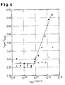
- Figure 4 shows representative results.
Abstract
Description
Die vorliegende Erfindung betrifft einen optischen Biosensor zur Detektion von gelösten Molekülen (im folgenden Analyten genannt), die sich mit einem Fluoreszenzfarbstoff markieren lassen und für die ein diese spezifisch erkennendes Biomolekül (im folgenden Rezeptor genannt) existiert. Es handelt sich hierbei um einen Festphasensensor mit Fluoreszenzfarbstoff, der über einen Energietransferprozeß auf ein zu detektierendes, mit einem zweiten Fluoreszenzfarbstoff markiertes Molekül die Bestimmung von dessen Anwesenheit und Menge erlaubt. Über eine Verdrängungs- oder eine Sandwich-Reaktion ist die Bestimmung auch von unmarkierten Analyten möglich.The present invention relates to an optical biosensor for the detection of dissolved molecules (hereinafter referred to as analytes) which can be labeled with a fluorescent dye and for which a biomolecule specifically recognizing them (referred to below as the receptor) exists. This is a solid phase sensor with fluorescent dye, which allows the determination of its presence and amount via an energy transfer process to a molecule to be detected and labeled with a second fluorescent dye. The determination of unlabeled analytes is also possible via a displacement or a sandwich reaction.
Es gibt verschiedene Methoden, Analyten, wie Hormone, Enzyme, andere Proteine, Kohlenhydrate, Nukleinsäuren, pharmakologische Wirkstoffe, Toxine und andere, in flüssigen Proben biologischen Ursprungs nachzuweisen. Unter den bekannten Methoden ragen speziell Immunoassays und damit verwandte Verfahren als eine empfindliche Nachweismethode zur Bestimmung sehr kleiner Mengen organischer Substanzen heraus. Immunoassay-Methoden beruhen allgemein auf der Fähigkeit eines Rezeptormoleküls, beispielsweise eines Antikörpers, die Struktur und molekulare Organisation eines Ligandenmoleküls, sei sie durch unpolare und/oder polare Wechselwirkungen definiert, spezifisch zu erkennen und dieses Molekül auf derartige Weise ganz spezifisch zu binden.There are various methods for detecting analytes, such as hormones, enzymes, other proteins, carbohydrates, nucleic acids, pharmacological agents, toxins and others, in liquid samples of biological origin. Among the known methods, immunoassays and related methods stand out as a sensitive detection method for the determination of very small amounts of organic substances. Immunoassay methods are generally based on the ability of a receptor molecule, for example an antibody, to specifically recognize the structure and molecular organization of a ligand molecule, be it defined by non-polar and / or polar interactions, and to bind this molecule very specifically in such a way.
Immunoassays werden mit verschiedenen Methoden durchgeführt. Dazu zählen der Einsatz verschiedener Markierungstechniken, die oft auf eine Quantifizierung des Analyten mittels radioaktiver, enzymgekoppelter und fluoreszierender Marker abzielen (E. F. Ulman, P. L. Khanna, Methods in Enzymology, 74 (1981) 28-60). Als Sonderfall der letztgenannten Methode kann der strahlungslose Fluoreszenz-Energietransfer (Förster-Energietransfer oder Resonanter Energietransfer, RET) betrachtet werden, mit dem die relative geometrische Position zweier Fluoreszenzfarbstoffe vermessen werden kann, sofern der gegenseitige Abstand höchstens wenige nm beträgt. Damit kann daher die unmittelbare Wechselwirkung eines Rezeptor/Ligand-Paares direkt nachgewiesen werden (L. Stryer, Annual Reviews in Biochemistry 47 (1978) 819-846). In der Technik der Immunoassays und der Biosensorik hat dieses Prinzip mehrfach Erwähnung gefunden (S. M. Barnard, D. R. Walt, Science 251, 927 (1991), EP 150 905, DE 3938598).Immunoassays are carried out using various methods. These include the use of various labeling techniques, which are often aimed at quantifying the analyte using radioactive, enzyme-coupled and fluorescent markers (EF Ulman, PL Khanna, Methods in Enzymology, 74 (1981) 28-60). A special case of the latter method is the radiationless fluorescence energy transfer (Förster energy transfer or resonant energy transfer, RET), with which the relative geometric position of two fluorescent dyes can be measured, provided the mutual distance is at most a few nm. The direct interaction of a receptor / ligand pair can therefore be demonstrated directly (L. Stryer, Annual Reviews in Biochemistry 47 (1978) 819-846). This principle has been mentioned several times in the technology of immunoassays and biosensors (SM Barnard, DR Walt, Science 251 , 927 (1991), EP 150 905, DE 3938598).
Darüber hinaus betrifft die vorliegende Erfindung die Immobilisierung von Biomolekülen in molekular dünnen, wohlgeordneten Schichtstrukturen, die für einen optischen Biosensor mit dem Förster-Energietransfer als Detektionsprinzip besonders geeignet sind. Die Immobilisierung von Rezeptoren an eine Festphase ist für Biosensoren von entscheidender Bedeutung. Nach derzeitiger Technik werden Proteine meist adsorptiv über ionische oder hydrophobe Wechselwirkungen an Oberflächen gebunden oder sie werden über kovalente Bindungen unter Verwendung von Hilfsreagenzien angekoppelt. Als ein mittlerweile klassisch zu nennendes Beispiel für letztere Vorgehensweise sei die Aktivierung von Glas durch 3-Aminopropyl-triethoxysilan und die anschließende Bindung von Protein mit Glutardialdehyd unter Reduktion der entstehenden Schiff-Base durch Natriumborhydrid zu nennen. Einen Überblick über bei Immunoassays benutzte Verfahren findet man beispielsweise in P. Tijssen, "Practice and Theory of Enzyme Immunoassays", S. 297-328 (Elsevier, Amsterdam 1987). Für biotechnologische Verfahren sind daneben Einkapselungsverfahren für Enzyme in permeablen Polymeren oder Membranen gebräuchlich.In addition, the present invention relates to the immobilization of biomolecules in molecularly thin, well-ordered layer structures which are particularly suitable for an optical biosensor with the Förster energy transfer as the detection principle. The immobilization of receptors on a solid phase is of crucial importance for biosensors. According to current technology, proteins are mostly bound to surfaces by adsorption or ionic or hydrophobic interactions, or they are coupled via covalent bonds using auxiliary reagents. The activation of glass by 3-aminopropyl-triethoxysilane and the subsequent binding of protein with glutardialdehyde with reduction of the resulting Schiff base by sodium borohydride should be mentioned as a classic example of the latter procedure. An overview of methods used in immunoassays can be found, for example, in P. Tijssen, "Practice and Theory of Enzyme Immunoassays", pp. 297-328 (Elsevier, Amsterdam 1987). Encapsulation processes for enzymes in permeable polymers or membranes are also common for biotechnological processes.
Während die adsorptiv arbeitenden Verfahren den Nachteil der mangelnden Stabilität der Proteinimmobilisierung besitzen, erfordert die kovalente Anbindung mit Kopplungs- oder Aktivierungsreagenzien oft eine relativ hohe Anzahl von Prozeßschritten, die Verwendung von hochreinen, teilweise instabilen Reagenzien oder die Anwendung von Umsetzungsbedingungen, unter denen nicht alle Proteine stabil sind. Die meisten gängigen Immobilisierungstechniken haben das Problem gemeinsam, daß die Rezeptoren nicht regiospezifisch gebunden werden, sodaß die für Folgereaktionen wichtigen Molekülteile bei einem hohen Prozentsatz der Rezeptoren sterisch blockiert sind. Auch ist der Wirkungsgrad der Proteinimmobilisierung oft mangelhaft, entweder durch Proteindenaturierung oder durch eine zu geringe Belegung der Oberfläche mit Proteinen. Weiterhin sind einige Aktivierungsreagenzien zur Quervernetzung fähig, wodurch sich schlecht definierte Oberflächen ergeben. Die Reproduzierbarkeit der Immobilisierung wird damit ebenfalls sehr schlecht. Für die Quantifizierung der Analyten-Konzentration mittels Förster-Energietransfer wirkt sich dabei ungünstig aus, daß der Abstand zwischen Energie-Donor und -Akzeptor je nach lokaler Oberflächenbeschaffenheit irregulär variiert, was generell eine Erhöhung der systembedingten Meßungenauigkeit mit sich bringt.While the adsorptive processes have the disadvantage of the lack of stability of the protein immobilization, the covalent attachment with coupling or activation reagents often requires a relatively high number of process steps, the use of highly pure, partially unstable reagents or the use of reaction conditions, under which not all proteins are stable. Most common immobilization techniques have the problem in common that the receptors are not bound region-specifically, so that the parts of the molecule which are important for subsequent reactions are sterically blocked in a high percentage of the receptors. The efficiency of protein immobilization is also often inadequate, either due to protein denaturation or due to insufficient coverage of the surface with proteins. Furthermore, some activation reagents are capable of cross-linking, which results in poorly defined surfaces. The reproducibility of the immobilization is also very bad. For the quantification of the analyte concentration by Förster energy transfer, it has an unfavorable effect that the distance between the energy donor and the acceptor varies irregularly depending on the local surface condition, which generally leads to an increase in the system-related measurement inaccuracy.
Eine Möglichkeit, die oben genannten systembedingten Probleme zu lösen ist es, den Träger mit molekular wohldefinierten Filmen zu beschichten. Dies kann dadurch geschehen, daß der Träger mit einem dünnen filmbildenden Copolymer überzogen wird, welches neben strukturbildenden Einheiten Comonomere mit Reaktivgruppen in der Seitenkette enthält, welche zur kovalenten Bindung des zu immobilisierenden Proteins in der Lage sind, wie z.B. in DE 43 19 037 beschrieben. Nachteilig bei diesem Verfahren ist, daß die Anzahl der Reaktivgruppen in der Regel beschränkt ist durch den begrenzten Anteil der Reaktivmonomere im Polymer. Die Belegungsdichte der Rezeptoren auf der Oberfläche ist dadurch oftmals zu gering.One way to solve the system-related problems mentioned above is to coat the support with molecularly well-defined films. This can be done by coating the support with a thin film-forming copolymer which, in addition to structure-forming units, contains comonomers with reactive groups in the side chain which are capable of covalently binding the protein to be immobilized, such as e.g. described in DE 43 19 037. A disadvantage of this process is that the number of reactive groups is generally limited by the limited proportion of reactive monomers in the polymer. As a result, the occupancy density of the receptors on the surface is often too low.
Ein weiterer Nachteil bei der Konzeption von Biosensoren ist die oft unspezifische Wechselwirkung von Proteinen mit der Festphasenoberfläche. Diese führt zur Adsorption mittels hydrophober oder ionischer Interaktionen, ist unerwünscht, führt zu nicht reproduzierbaren Ergebnissen und zur Verringerung der Meßgenauigkeit.Another disadvantage when designing biosensors is the often non-specific interaction of proteins with the solid phase surface. This leads to adsorption by means of hydrophobic or ionic interactions, is undesirable, leads to non-reproducible results and reduces the measuring accuracy.
Die Erfindung betrifft einen optischen Festphasenbiosensor mit Biomolekülen als Rezeptoren zur spezifischen Erkennung von Analyten unter Ausnutzung des Förster-Energietransfers zwischen zwei Fluoreszenzfarbstoffen F1 und F2, bestehend aus
- a) einem transparenten Träger,
- b) einer daraufliegenden Multischicht, die abwechselnd aus Polyanionen und Polykationen besteht und als oberste Schicht ein biotinyliertes Polykation enthält, wobei der Grad der Biotinylierung 20-80 Mol-%, bevorzugt 30-70 Mol-%, besonders bevorzugt 40-60 Mol-%, bezogen auf die Zahl der Äquivalente kationischer Gruppen, beträgt,
- c) einer Abdeckung der obersten biotinylierten kationischen Schicht durch Streptavidin, welches an diese biotinylierte Schicht gebunden wird,
- d) weiteren biotinylierten Biomolekülen als Rezeptoren, bevorzugt Antikörpern, die mit einem Fluoreszenzfarbstoff F2 markierte Analyten binden können, wobei der Fluoreszenzfarbstoff F1 an die polyionischen Basisschichten, an das Streptavidin oder an die weiteren Antikörper bindenden Biomoleküle oder an die Antikörper gebunden sein kann.
- a) a transparent support,
- b) a multilayer thereon, which alternately consists of polyanions and polycations and contains a biotinylated polycation as the top layer, the degree of biotinylation being 20-80 mol%, preferably 30-70 mol%, particularly preferably 40-60 mol% , based on the number of equivalents of cationic groups,
- c) covering the uppermost biotinylated cationic layer by streptavidin, which is bound to this biotinylated layer,
- d) further biotinylated biomolecules as receptors, preferably antibodies, which can bind analytes labeled with a fluorescent dye F2, the fluorescent dye F1 adhering to the polyionic base layers, may be bound to the streptavidin or to the further antibody-binding biomolecules or to the antibodies.
In einer bevorzugten Ausführungsform sind an die unter c) beschriebene Schicht biotinylierte Rezeptoren gebunden, die ihrerseits Antikörper durch eine spezifische Erkennungsreaktion immobilisieren können.In a preferred embodiment, biotinylated receptors are bound to the layer described under c), which in turn can immobilize antibodies by means of a specific recognition reaction.
Gegenstand der vorliegenden Erfindung ist demnach die Immobilisierung von Biomolekülen, insbesondere von Rezeptoren oder Antikörpern, an eine Festphase, wobei die Immobilisierung dauerhafter und gerichteter Natur ist und eine hohe Belegungsdichte der Oberfläche mit dem Rezeptor erreicht wird. Die Detektion der Bindung des Analyten erfolgt durch Förster-Energietransfer und ist durch eine molekular wohldefinierte gegenseitige Anordnung von Energie-Donor und -Akzeptor reproduzierbar und in ihrer Konzentrationsabhängigkeit regulär. Die Oberfläche wird gleichzeitig gegen unspezifische Adsorption von Proteinen passiviert. In der vorliegenden Erfindung wird demnach diesen Anforderungen nachgekommen und werden die oben angeführten Probleme gelöst durch die Immobilisierung organischer und biologischer Komponenten in molekular wohldefinierten Schichten unter Verwendung des natürlichen Systems Biotin/Streptavidin. Streptavidin ist ein Protein mit vier Bindungsstellen für Biotin (Vitamin H). Es kann deshalb als Matrix für die Ankopplung biotinylierter Biomoleküle dienen. Da die Bindungskonstante von Biotin mit Streptavidin ∼ 1015 M-1 beträgt, ist die Bindung von Biotin an Streptavidin nahezu irreversibel.The present invention accordingly relates to the immobilization of biomolecules, in particular receptors or antibodies, onto a solid phase, the immobilization being permanent and directed in nature and a high occupancy density of the surface being achieved with the receptor. The binding of the analyte is detected by Förster energy transfer and is reproducible by a molecularly well-defined mutual arrangement of energy donor and acceptor and its concentration dependence is regular. The surface is passivated at the same time against unspecific adsorption of proteins. Accordingly, these requirements are met in the present invention and the above-mentioned problems are solved by the immobilization of organic and biological components in molecularly well-defined layers using the natural system biotin / streptavidin. Streptavidin is a protein with four binding sites for biotin (vitamin H). It can therefore serve as a matrix for coupling biotinylated biomolecules. Since the binding constant of biotin with streptavidin is ∼ 10 15 M -1 , the binding of biotin to streptavidin is almost irreversible.
Die Erfindung ist vor allem gekennzeichnet durch die Verwendung von Polykationen und Polyanionen zum Aufbau der Multischicht.The invention is characterized above all by the use of polycations and polyanions to build up the multilayer.
Realisiert wird diese Erfindung durch die Belegung eines transparenten festen Trägers, in der Regel Floatglas oder Quarzglas oder organische Polymere wie z.B. Polyester, Polycarbonat oder Polyethylenterephthalat oder andere transparente, nichtfluoreszierende Festkörper, mit einer Multischicht mittels der Self-Assembly-(SA-)Technik durch konsekutive Physisorption von anionischen und kationischen Polymeren. Dies Verfahren ist in EP 472 990 ausführlich beschrieben. In der vorliegenden Erfindung wird als letzte Schicht der Multischicht ein Polykation, welches an Aminogruppen biotinyliert ist, physisorbiert. Die Biotinylierung der Polykationen erfolgt mit Biotin-N-Hydroxysuccinimidester oder anderen reaktiven Estern zu 20-80 Mol-%, bevorzugt zu 30-70 Mol-%, besonders bevorzugt zu 40-60 Mol-%, bezogen auf die Zahl der Äquivalente kationischer Gruppen, gemäß dem in EP-A 0 472 990 beschriebenen Verfahren. Polykationen, die für die Erfindung geeignet sind, sind beispielsweise Polylysin, Polyallylamin, Polyvinylamin, Poly-(4-vinylpyridin), Polyacrylamid, Polymethacrylamid, Polyarginin, Polyasparagin, Polyglutamin, Polyethylenimin und Copolymere der zugrundeliegenden Monomere, bevorzugt Polylysin und Polyallylamin. Diese Polykationen, bei denen beispielsweise N-Atome als Ammoniumgruppen vorliegen, die 2 oder 3 H-Atome tragen, können als Gegenionen beispielsweise tragen: Halogenid, wie Chlorid oder Bromid, Sulfat, Hydrogensulfat, Nitrat, Nitrit, Carbonat, Hydrogencarbonat, Phosphat, Hydrogenphosphat, aliphatische Carbonsäureanionen, wie Formiat, Acetat, Trifluoracetat, Trichloracetat. Als biotinylierbare Polykationen kommen erfindungsgemäß in Frage: Polylysin, Polyarginin, Polyglutamin, Polyasparagin, Polyacrylamid, Polymethacrylamid, Polyallylamin und Copolymere der zugrundeliegenden Monomere, bevorzugt Polylysin und Polyallylamin. Polyanionen sind beispielsweise Polystyrolsulfonat (PSS), Polyacrylsäure, Polymethacrylsäure, Poly-(2-acrylamido-2-methyl-1-propansulfonsäure), Polyvinylsulfonsäure, Polyvinylsulfat, Dextransulfat, Cellulosesulfat und Copolymere der zugrundeliegenden Monomere, bevorzugt Polystyrolsulfonat. Gegenkationen in den Polyanionen sind beispielsweise H+, Na+, K+, NH4 +, bevorzugt Na+ oder K+. Der Anteil der biotinylierten Kationäquivalente kann über die Stöchometrie den gewünschten Erfordernissen angepaßt werden. Durch die Inkubation mit Streptavidin wird an die biotinylierte Polymerschicht Streptavidin gebunden. Die Oberfläche ist dann nahezu vollständig und, wie Fluoreszenzexperimente zeigen, lückenlos mit Streptavidin belegt. Ein solches System weist die oben geforderten Anforderungen und Vorteile für einen Biosensor auf. Zum einen wird die Biosensoroberfläche durch eine möglichst dichte Bedeckung mit dem Protein Streptavidin gegen unspezifische Wechselwirkungen abgeschirmt und zum anderen eine universelle Bindungsmatrix für eine Funktionalisierung der Festkörper-Grenzfläche zur Verwendung als Biosensor zur Verfügung gestellt. Durch die Markierung des Proteins Streptavidin mit einem Fluoreszenzfarbstoff F1 (z. B. mit Fluorescein-isothiocyanat), welche dem Fachmann hinlänglich bekannt ist, wird der Donorfarbstoff für den Förster-Energietransfer zur Verfügung gestellt.This invention is realized by covering a transparent solid support, usually float glass or quartz glass or organic polymers such as polyester, polycarbonate or polyethylene terephthalate or other transparent, non-fluorescent solids, with a multilayer by means of the self-assembly (SA) technique consecutive physisorption of anionic and cationic polymers. This method is described in detail in EP 472 990. In the present invention, as the last layer of the multilayer, a polycation which is biotinylated on amino groups is physisorbed. The polycations are biotinylated with biotin-N-hydroxysuccinimide ester or other reactive esters at 20-80 mol%, preferably 30-70 mol%, particularly preferably 40-60 Mol%, based on the number of equivalents of cationic groups, according to the process described in EP-
Nach der Immobilisierung des Streptavidins an die biotinylierte Polymeroberfläche, dient nun das Streptavidin mit seinen noch nicht belegten Bindungsstellen als Matrix für die Anbindung von weiteren biotinylierten Biomolekülen als Rezeptoren, insbesondere für die Anbindung von Antikörpern. Mehrere bevorzugte Varianten zur Anbindung von Antikörpern sind möglich. So ist eine Ausführungsform der vorliegenden Erfindung die der Ankopplung von biotinylierten Protein A oder biotinyliertem Protein G an die Streptavidinmatrix. Die Biotinylierung von Protein A mittels N-Hydroxysuccinimid oder über andere reaktive Ester ist leicht möglich und dem Fachmann geläufig. Protein A ist ein Protein aus der Zellwand des Bakteriums Staphylococcus aureus. Es ist in der Lage, Immunglobuline vom Typ IgG spezifisch an deren Fc-Teil zu binden. Diese Methode hat zudem den Vorteil der regiospezifischen Immobilisierung von Antikörpern. Der Fab-Teil der Antikörper bleibt frei. Eine Verminderung von immunologischer Aktivität durch Blockierung der Antigenbindungsstelle erfolgt nicht. Alternativ zur Bindung von F1 an das Streptavidin kann F1 auch an die soeben beschriebenen weiteren Biomoleküle gebunden werden; die Bindung an das Streptavidin ist jedoch bevorzugt.After the streptavidin has been immobilized on the biotinylated polymer surface, the streptavidin with its as yet unoccupied binding sites now serves as a matrix for the connection of further biotinylated biomolecules as receptors, in particular for the connection of antibodies. Several preferred Variants for the connection of antibodies are possible. One embodiment of the present invention is that of coupling biotinylated protein A or biotinylated protein G to the streptavidin matrix. The biotinylation of protein A using N-hydroxysuccinimide or via other reactive esters is easily possible and familiar to the person skilled in the art. Protein A is a protein from the cell wall of the bacterium Staphylococcus aureus. It is able to specifically bind immunoglobulins of the IgG type to their F c part. This method also has the advantage of region-specific immobilization of antibodies. The F ab part of the antibodies remains free. There is no reduction in immunological activity by blocking the antigen binding site. As an alternative to binding F1 to streptavidin, F1 can also be bound to the further biomolecules just described; however, binding to the streptavidin is preferred.
Optische Festphasenbiosensoren dieser Art können beispielsweise in Form von Teststreifen eingesetzt werden.Optical solid phase biosensors of this type can be used, for example, in the form of test strips.
Eine andere Ausführungsform des Immunosensors beinhaltet die Anbindung von biotinylierten Antikörpern an die Streptavidinmatrix. Hierbei sind wiederum zwei erfindungsmäßige Ausführungen denkbar. Zum einen kann die Biotinylierung der Antikörper mittels Biotin-N-Hydroxysuccinimid oder über andere reaktive Ester erfolgen. Eine solche dem Fachmann wohlbekannte Form der Biotinylierung von Antikörpern hat den Nachteil, daß die Biotinylierung nicht regiospezifisch erfolgt. Ein Teil der IgG-Moleküle wird auch an oder nahe der Antigenbindungsstelle biotinyliert, so daß eine sterische Blockierung derselben erfolgt und die immunologische Aktivität der Antikörper und damit die Sensitivität des Sensors abnimmt. In einer anderen erfindungsmäßigen Ausführungsform benutzt man Biotinderivate mit Hydrazidreaktivgruppen, die mit oxidierten Antikörpern reagieren. Bei der Oxidation der Antikörper werden die sich am Fc-Teil der IgGs befindlichen Kohlenhydrate in einer dem Fachmann bekannten Reaktion gespalten (Glykolspaltung) und Aldehyde erzeugt. Diese reagieren mit den Hydrazidgruppen der Biotinderivate unter Ausbildung von Hydrazonen. Die Biotingruppen sind dadurch regiospezifisch an den Fc-Teil der Antikörper angebunden. Die biotinylierten Antikörper werden an die Streptavidinmatrix gekoppelt und vervollständigen die Teststreifenoberfläche des Immunosensors, ohne daß eine Kopplung über biotinyliertes Protein A notwendig wird.Another embodiment of the immunosensor involves the connection of biotinylated antibodies to the streptavidin matrix. Here again, two embodiments according to the invention are conceivable. On the one hand, the antibodies can be biotinylated using biotin-N-hydroxysuccinimide or other reactive esters. Such a form of biotinylation of antibodies, which is well known to the person skilled in the art, has the disadvantage that the biotinylation is not region-specific. Some of the IgG molecules are also biotinylated at or near the antigen binding site, so that they are sterically blocked and the immunological activity of the antibodies and thus the sensitivity of the sensor decrease. In another embodiment according to the invention, biotin derivatives with hydrazide reactive groups are used which react with oxidized antibodies. When the antibodies are oxidized, the carbohydrates located on the F c part of the IgGs are cleaved in a reaction known to the person skilled in the art (glycol cleavage) and aldehydes are generated. These react with the hydrazide groups of the biotin derivatives to form hydrazones. The biotin groups are thus linked region-specifically to the F c part of the antibodies. The biotinylated antibodies are coupled to the streptavidin matrix and complete the test strip surface of the immunosensor without the need for coupling via biotinylated protein A.
Der Teststreifen ist nun in der Lage, den mit einem geeigneten Akzeptorfarbstoff F2 (z. B. Rhodamin-isothiocyanat) versehenen Analyten durch Wechselwirkung des Rezeptors, insbesondere des Antikörpers, mit dem Analyten zu erkennen und zu quantifizieren. Der Nachweis des Analyten erfolgt durch einfaches Inkontaktbringen des beschichteten Trägers mit der Lösung, in der ein Molekül als Analyt vermutet wird (Probenlösung) und anschließender Fluoreszenzmessung. Es wird die Fluoreszenz des Donor- (F1) und des Akzeptorfarbstoffs (F2) gemessen. Befindet sich in der Testflüssigkeit (Probenlösung) ein mit dem Akzeptorfarbstoff F2 markierter Analyt, so wird nach dessen spezifischer Bindung an die immobilisierten Antikörper aufgrund des Förster-Energietransfers die Intensität der Akzeptorfluoreszenz zunehmen und die des Donors gegenüber dem ungebundenen Zustand abnehmen. Soll alternativ unmarkierter Analyt über eine Verdrängungsreaktion bestimmt werden, so wird der Teststreifen zuerst mit einem akzeptormarkierten Analog des betreffenden Analyten äquilibriert. In diesem Zustand überwiegt dann die Akzeptorfluoreszenz von F2 die Donorfluoreszenz von F1. Kommt anschließend unmarkierter Analyt aus der Testflüssigkeit mit dem äquilibrierten Teststreifen in Kontakt, so wird nach der Verdrängungsreaktion der Förster-Energietransfer unterbrochen sein, so daß eine Zunahme der Donorfluoreszenz von F1 und eine Abnahme der Akzeptorfluoreszenz von F2 die Bindung des unmarkierten Analyten signalisiert. In beiden Fällen hängt die Veränderung von Akzeptor- und Donorfluoreszenz in eindeutiger Weise mit der Konzentration des Analyten zusammen. Der Förster-Energietransfer läßt sich in üblichen Fluoreszenzspektrometern, aber auch in speziell ausgelegten Geräten für einen Energietransfer-Immunosensor, messen. Mittels geeigneter Kalibrationskurven läßt sich dann die Konzentration des Analyten in der Analysenflüssigkeit ermitteln. In einer weiteren Ausführungsform wird einer Analysenlösung, in der der zu detektierende Analyt vermutet wird, ein mit dem Fluoreszenzfarbstoff F2 markiertes, spezifisch bindendes Molekül in bekannter Menge zugesetzt, welches bei Vorhandensein das zu detektierenden Analyten mit diesem um die Bindung an der obersten Schicht des Biosensor kompetitiert. Die Konzentration des zu detektierenden Analyten wird sodann durch die Abhängigkeit der Fluoreszenzintensitäten von F2 bzw. von F1 oder dem Verhältnis der beiden Intensitäten gemessen.The test strip is now able to recognize and quantify the analyte provided with a suitable acceptor dye F2 (e.g. rhodamine isothiocyanate) by interaction of the receptor, in particular the antibody, with the analyte. The analyte is detected by simply bringing the coated carrier into contact with the solution in which a molecule is suspected to be an analyte (sample solution) and subsequent fluorescence measurement. The fluorescence of the donor (F1) and the acceptor (F2) dye is measured. If there is an analyte labeled with the acceptor dye F2 in the test liquid (sample solution), after its specific binding to the immobilized antibodies, the intensity of the acceptor fluorescence will increase due to the Förster energy transfer and that of the donor will decrease compared to the unbound state. Alternatively, if unlabeled analyte is to be determined via a displacement reaction, the test strip is first equilibrated with an acceptably labeled analog of the analyte in question. In this state, the acceptor fluorescence of F2 then outweighs the donor fluorescence of F1. If unlabeled analyte from the test liquid then comes into contact with the equilibrated test strip, the Förster energy transfer will be interrupted after the displacement reaction, so that an increase in the donor fluorescence of F1 and a decrease in the acceptor fluorescence of F2 signalize the binding of the unlabeled analyte. In both cases, the change in acceptor and donor fluorescence is clearly related to the concentration of the analyte. The Förster energy transfer can be measured in conventional fluorescence spectrometers, but also in specially designed devices for an energy transfer immunosensor. The concentration of the analyte in the analysis liquid can then be determined by means of suitable calibration curves. In a further embodiment, a specifically binding molecule, labeled with the fluorescent dye F2, is added in known amount to an analysis solution in which the analyte to be detected is suspected competed. The concentration of the analyte to be detected is then measured by the dependence of the fluorescence intensities on F2 or on F1 or the ratio of the two intensities.
Eine andere erfindungsmäßige Ausführungsform des Immunosensors beinhaltet die Anbindung des Donorfarbstoffes F1 an den Rezeptor, bevorzugt den Antikörper, vorzugsweise über Hydrazidreaktivgruppen an oxidierte Antikörper in der oben beschriebenen Form. Bei dieser Ausführungsform wird die Effizienz des Förster-Energietransfers durch eine Verringerung des durchschnittlichen Abstandes zwischen Donor(F1)- und Akzeptor(F2)-Farbstoff gesteigert, was zu einer Erhöhung der Sensitivität des Testverfahrens genutzt werden kann.Another embodiment of the immunosensor according to the invention includes the connection of the donor dye F1 to the receptor, preferably the antibody, preferably via hydrazide reactive groups to oxidized antibodies in the form described above. In this embodiment, the efficiency of Förster energy transfer by reducing the average distance between donor (F1) and acceptor (F2) dye, which can be used to increase the sensitivity of the test method.
Die Erfindung wird durch die folgenden Abbildungen näher erläutert:The invention is illustrated by the following figures:
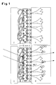
Figur 1 zeigt schematisch, und in etwa maßstabsgerecht, den Multischichtaufbau der Teststreifen für den Förster-Energietransfer-Immunosensor (oben), sowie das Detektionsprinzip (unten).
- Figur 1:
- Schematischer Aufbau und Funktion des Immonunosensors. Die molekularen Komponenten sind in etwa maßstäblich gezeichnet. Der Träger I ist viel dicker als dargestellt.
- Oben:
- Teststreifen, bestehend aus transparentem Träger I (z.B. Glas), einer Polyelektrolyt-Multischicht II (zur Vereinfachung der Abbildung ist nur die letzte biotinylierte Schicht gezeigt), einer Schicht mit F1 fluoreszenzmarkierten Streptavidins III, einer Schicht biotinyliertem Protein A IV und dem darauf immobilisierten Antikörper V.
- Unten:
- Nach Benetzung mit Testflüssigkeit hängt die Wellenlänge der beobachteten Fluoreszenz nach Anregung des an Streptavidin gekoppelten Fluoreszenzfarbstoffs F1 davon ab, ob direkte Emission vorliegt (punktierter Pfeil) oder ob die Anregungsenergie nach Energietransfer (RET = gestrichelter Pfeil) auf den markierten Analyten rotverschoben emittiert wird (durchgezogener Pfeil). Das Verhältnis von rotverschobener zu direkter Emissionsintensität ist in eindeutiger Weise abhängig von der Anzahl gebundener Analytmoleküle pro Flächeneinheit.
- Figure 1 :
- Schematic structure and function of the immune sensor. The molecular components are roughly drawn to scale. The carrier I is much thicker than shown.
- Above:
- Test strips, consisting of transparent support I (e.g. glass), a polyelectrolyte multilayer II (only the last biotinylated layer is shown to simplify the illustration), a layer with F1 fluorescent-labeled streptavidins III, a layer of biotinylated protein A IV and the antibody immobilized on it V.
- Below:
- After wetting with test liquid, the wavelength of the observed fluorescence after excitation of the fluorescent dye F1 coupled to streptavidin depends on whether there is direct emission (dotted arrow) or whether the excitation energy after energy transfer (RET = dashed arrow) is emitted on the marked analytes with a red shift (solid line) Arrow). The ratio of red-shifted to direct emission intensity is clearly dependent on the number of bound analyte molecules per unit area.
Figur 2 zeigt Röntgen-Reflektogramme der Schichtstruktur in verschiedenen Stadien der Präparation.
- Figur 2:
- Röntgen-Reflektogramme der Schichtstruktur (Trägermaterial : Silizium) in verschiedenen Stadien der Präparation. Die verschiedenen Datensätze wurden um jeweils einen Faktor 100 gegeneinander verschoben. Die Kurven zeigen von unten nach oben zunächst die Polyelektrolyt-Multischicht, dann dieselbe, belegt mit Streptavidin, anschließend noch belegt mit biotinyliertem Protein A und zum Schluß versehen mit Antikörper IgG. Auf der Abszisse ist der Impulstransfer Qz in Å-1 und auf der Ordinate die Röntgen-Intensität I aufgetragen.
- Figure 2 :
- X-ray reflectograms of the layer structure (substrate: silicon) in different stages of the preparation. The different data sets were shifted against each other by a factor of 100. The curves first show the from bottom to top Polyelectrolyte multilayer, then the same, coated with streptavidin, then coated with biotinylated protein A and finally provided with antibody IgG. The pulse transfer Q z in Å -1 is plotted on the abscissa and the X-ray intensity I is plotted on the ordinate.
Die Messung zeigt für das vorliegende Beispiel, daß die Oberfläche mit einer in den verschiedenen Präparationsschritten regulär gewachsenen Grenzflächenschicht organischen Materials belegt ist und in jedem Schritt die Präparation molekular glatt bleibt. Dies bedeutet insbesondere, daß die Oberflächenfunktionalisierung nicht zur Belegung der Festphasengrenzfläche mit lateral inhomogenen Strukturen führt, d.h. eine Tröpfchenbildung findet selbst auf der Längenskala von Nanometern nicht statt.For the present example, the measurement shows that the surface is covered with an interface layer of organic material that has grown regularly in the various preparation steps and that the preparation remains molecularly smooth in each step. In particular, this means that the surface functionalization does not lead to the solid phase interface being covered with laterally inhomogeneous structures, i.e. droplet formation does not take place even on the length scale of nanometers.
Figur 3 zeigt die Ergebnisse von ELISA-Messungen an Sensoroberflächen, die analog zu dem in Figur 1 angedeuteten und in Figur 2 mittels Röntgenreflektivitätsmessungen protokolliertem Verfahren hergestellt wurden.
- Figur 3:
- Titration verschiedener Sensoroberflächen mit Antigen. Die spezifische Bindung wurde mittels ELISA nachgewiesen. Einem Sensor, der nach der hier beschriebenen Technik präpariert wurde (schwarze Karos), ist ein Präparat gegenübergestellt, bei dem IgG elektrostatisch an eine PSS-Schicht adsorbiert wurde (weiße Karos). Auf der Abszisse ist die Antigenkonzentration cAG in mol/l und auf der Ordinate die optische Dichte A bei λ = 414 nm angegeben.
- Figure 3 :
- Titration of different sensor surfaces with antigen. The specific binding was verified by ELISA. A sensor that was prepared using the technique described here (black squares) is juxtaposed with a preparation in which IgG was electrostatically adsorbed onto a PSS layer (white squares). The antigen concentration c AG in mol / l is shown on the abscissa and the optical density A at λ = 414 nm on the ordinate.
Figur 4 zeigt die Ergebnisse einer Biosensor-Messung an einer Probe, die wie die in Figur 3 beschriebene Probe hergestellt wurde.
- Figur 4:
- Titration einer Sensoroberfläche mit Antigen (schwarze Karos). Die spezifische Bindung wurde mittels Energietransfer nachgewiesen. Das Antigen lag in ca. 2facher Verdünnung im Kulturüberstand vor. Kontrolle (weiße Karos): Antigenadsorption an Teststreifen, der nur bis zur Streptavidinschicht aufgebaut war (ohne Protein A und Rezeptorschicht). Die Abszisse zeigt die Konzentration cAG im Kulturüberstand in mol/l. Die Ordinate zeigt das Intensitätsverhältnis der Fluoreszenzen der Fluoreszenzfarbstoffe F1 und F2 bei 577 und 530 nm.
- Figure 4 :
- Titration of a sensor surface with antigen (black checks). The specific binding was verified by means of energy transfer. The antigen was present in the culture supernatant in a 2-fold dilution. Control (white squares): Antigen adsorption on test strips that were only built up to the streptavidin layer (without protein A and receptor layer). The abscissa shows the concentration c AG in the culture supernatant in mol / l. The ordinate shows the intensity ratio of the Fluorescence of the fluorescent dyes F1 and F2 at 577 and 530 nm.
- Polymere: Polymers :
-
Polystyrolsulfonat, Natriumsalz (PSS), MW = 70.000, Aldrich
Polyallylamin-hydrochlorid (PAH), MW = 50.000―60.000, Aldrich
Polyethylenimin (PEI), MW =50.000, 50%ige Lösung in H2O, Aldrich
Poly-L-Lysin-hydrobromid (PL), MW < 50.000, Bachem-BiochemicaPolystyrene sulfonate, sodium salt (PSS), MW = 70,000, Aldrich
Polyallylamine hydrochloride (PAH), MW = 50,000-60,000, Aldrich
Polyethyleneimine (PEI), MW = 50,000, 50% solution in H 2 O, Aldrich
Poly-L-lysine hydrobromide (PL), MW <50,000, Bachem-Biochemica
Das PSS wurde in wässriger Lösung in einem Dialyseschlauch VISKING27/32 der Firma Roth zwei Tage gegen Reinstwasser dialysiert und anschließend gefriergetrocknet.
- Proteine:
- Streptavidin, Boehringer-Mannheim
Protein A, Pharmacia
Kaninchen IgG, polyclonal, Spezifität: Anti Maus IgG,
Immunol. Institut Univ. Mainz
Bovine Serum Albumin (BSA), Sigma - Antigene:
- Maus-IgG, monoclonal, Kulturüberstand, Immunol. Institut Univ. Mainz
Horseradish-Peroxidase-(HPO-) gekoppeltes Maus-IgG, affinitätsgereinigt, Jackson Immuno-Research Laboratories, Dianova, U.S.A. - Fluorophore:
- Rhodamin-B-Isothiocyanat (RITC), Sigma
Fluorescein-Isothiocyanat Isomer I (FITC), Sigma
- Proteins :
- Streptavidin, Boehringer-Mannheim
Protein A, Pharmacia
Rabbit IgG, polyclonal, specificity: anti mouse IgG,
Immunol. Institute Univ. Mainz
Bovine Serum Albumin (BSA), Sigma - Antigens:
- Mouse IgG, monoclonal, culture supernatant, immunol. Institute Univ. Mainz
Horseradish peroxidase (HPO) coupled mouse IgG, affinity purified, Jackson Immuno-Research Laboratories, Dianova, USA - Fluorophores:
- Rhodamine B isothiocyanate (RITC), Sigma
Fluorescein isothiocyanate isomer I (FITC), Sigma
Die Markierung des Streptavidin betrug durchschnittlich 1,4 FITC pro Protein-Molekül und die des Maus-IgG durchschnittlich 3 RITC pro Protein-Molekül.
- Biotinaktivester:
- Biotinamidocaproyl-N-Hydroxysuccinimidester, Sigma
- Biotin active :
- Biotinamidocaproyl-N-hydroxysuccinimide ester, Sigma
Zur Biotinylierung von Protein A wurden der Aktivester und das Protein in einem molekularen Verhältnis von 12:1 eingewogen.
- Detergens:
- Polyoxyethylensorbitan-Monolaurat (TWEEN-20), Sigma
- PBS-Puffer:
- Natriumdihydrogenphosphat, Monohydrat, p.a., Merck
Dinatriumhydrogenphosphat, p.a., Merck
Natriumchlorid, p.a., Merck - Citrat-Puffer:
- Dinatriumhydrogenphosphat, p.a., Merck
Zitronensäuremonohydrat, Sigma
Kaliumchlorid, p.a., Merck
Magnesiumchlorid, Hexahydrat, p.a., Merck - ABTS:
- 2,2'-Azino-Bis(3-Ethyl-Benzthiazolin)-Sulfonsäure, Sigma
- Glassubstrat:
- (3812) mm2 große und (1―1,2) mm dicke Objektträger, Fa. Gebr. Rettberg GmbH.

- Detergent :
- Polyoxyethylene sorbitan monolaurate (TWEEN-20), Sigma
- PBS buffer:
- Sodium dihydrogen phosphate, monohydrate, pa, Merck
Disodium hydrogen phosphate, pa, Merck
Sodium chloride, pa, Merck - Citrate buffer:
- Disodium hydrogen phosphate, pa, Merck
Citric acid monohydrate, Sigma
Potassium chloride, pa, Merck
Magnesium chloride, hexahydrate, pa, Merck - ABTS:
- 2,2'-azino-bis (3-ethyl-benzothiazoline) sulfonic acid, Sigma
- Glass substrate:
- (38 12) mm 2 large and (1―1.2) mm thick slides, Gebr. Rettberg GmbH.
Die Trägerreinigung erfolgte entsprechend einer Standardprozedur (W. Kern, D. A. Puotinen, RCA Review, 31 (1970), 187)The carrier cleaning was carried out according to a standard procedure (W. Kern, DA Puotinen, RCA Review, 31 (1970), 187)
Alle Lösungen wurden mit destilliertem Wasser angesetzt. Die wasserbenetzten Träger wurden für 30 Minuten in PEI-Lösung (verdünnt auf 2,2 mg/ml) eingestellt und anschließend dreimal für jeweils ca. 30 Sekunden in 10 ml Wasser gewaschen und anschließend in einem schwachen Stickstoffstrom trocken geblasen. Anschließend wurde die Probe für ca. 20 Minuten in eine PSS-Lösung (20 mg PSS in 10 ml einer 2 M NaCl-Lösung) gestellt und wie oben beschrieben gewaschen und getrocknet. Der Träger wurde für weitere 20 Minuten in eine PAH-Lösung (20 mg PAH in 10 ml 2 M NaCl-Lösung) eingestellt und wieder gewaschen und getrocknet. Wie oben beschrieben wurden eine weitere PSS-, eine PAH- und wiederum eine PSS-Schicht an den Träger adsorbiert. Zur Funktionalisierung mit Biotin wurde der Träger in eine Lösung von biotinyliertem Polylysin-hydrobromid (PLB) (5 mg/10 ml in 0,4 M NaCl) für 20 Minuten eingestellt und danach wieder gewaschen und getrocknet. Die beschichteten Träger wurden bis zur Verwendung bei einer Temperatur von 4° C aufbewahrt.All solutions were made up with distilled water. The water-wetted supports were placed in PEI solution (diluted to 2.2 mg / ml) for 30 minutes and then washed three times for approx. 30 seconds each in 10 ml of water and then blown dry in a weak stream of nitrogen. The sample was then placed in a PSS solution (20 mg PSS in 10 ml of a 2 M NaCl solution) for about 20 minutes and washed and dried as described above. The carrier was placed in a PAH solution (20 mg PAH in 10 ml 2 M NaCl solution) for a further 20 minutes and washed and dried again. As described above, a further PSS, a PAH and again a PSS layer were adsorbed onto the support. For functionalization with biotin, the carrier was placed in a solution of biotinylated polylysine hydrobromide (PLB) (5 mg / 10 ml in 0.4 M NaCl) for 20 minutes and then washed and dried again. The coated supports were stored at a temperature of 4 ° C until use.
PLB-beschichtete Träger wurden für ca. 30 Minuten in eine FITC-Streptavidinlösung (10-7 mol/l FITC-Streptavidin in 10 mM PBS-Puffer, pH=7,2, 150 mM NaCl) gestellt, dreimal mit 10 ml Wasser gewaschen und anschließend für weitere 40 Minuten in eine Lösung von biotinyliertem Protein A (5![]()
![]()
![]()
![]()
Figur 2 zeigt Röntgen-Reflektogramme, die bei der Präparation eines Teststreifens (Trägermaterial Silizium) zwischen den verschiedenen Adsorptionsschritten gemessen wurden (trockene Probe). Diese Ergebnisse belegen, daß sich beim Adsorptionsprozeß eine kohärente, auf molekularer Längenskala gleichmäßige Schichtstruktur ausbildet, daß die Schichtdickeninkremente jeweils den molekularen Dimensionen der Absorbentien entsprechen und daß die Oberfläche nach jedem Adsorptionsschritt molekular glatt bleibt. Eine Ausnahme bildet die terminale Antikörperschicht, die wegen der elongierten Molekülform des Antikörpers und der regioselektiven Bindung durch Protein A einen hohen Anteil von wässrigem Puffer enthält. Diese Schicht kollabiert beim Trocknungsprozeß während der Messung und erscheint danach wesentlich dünner als dies die molekularen Dimensionen erwarten lassen. Die experimentellen Daten aus Figur 2 sind in Tabelle 1 quantitativ ausgewertet.
Proben wurden, wie oben beschrieben, auf Silizium präpariert. In PBS-Puffer mit 0.1% TWEEN-20 wurden 10 mg/ml BSA gelöst. Diese Proteinlösung wurde für ELISA-Messungen zur Herstellung einer Verdünnungsreihe des HPO-markierten Antigens (Maus-IgG-HPO) verwendet, dessen Konzentration zwischen 10-13 und 10-7 mol/l betrug. Nach Inkubation mit dem Antigen wurden die Proben in Citrat-Puffer mit 3 g/l ABTS und 0.0075 % H2O2 entwickelt und vermessen. Figur 3 zeigt ein repräsentatives Ergebnis für ein Präparat, das nach der hier vorgestellten Technologie hergestellt wurde. Dem gegenübergestellt ist die Titration eines Präparats, bei dem IgG lediglich elektrostatisch an eine Silizium-Grenzfläche adsorbiert wurde, die mit einer molekular dünnen Schicht von PSS bedeckt war. Deutlich wird in der Abbildung die höhere Empfindlichkeit, bessere Linearität und geringere unspezifische Adsorption sichtbar, die das nach der neuen Technik hergestellte Präparat auszeichnen.Samples were prepared on silicon as described above. 10 mg / ml BSA were dissolved in PBS buffer with 0.1% TWEEN-20. This protein solution was used for ELISA measurements to prepare a dilution series of the HPO-labeled antigen (mouse IgG-HPO), the concentration of which was between 10 -13 and 10 -7 mol / l. After incubation with the antigen, the samples were developed and measured in citrate buffer with 3 g / l ABTS and 0.0075% H 2 O 2 . FIG. 3 shows a representative result for a preparation which was produced using the technology presented here. This is contrasted with the titration of a preparation in which IgG was only electrostatically adsorbed on a silicon interface that was covered with a molecularly thin layer of PSS. The figure clearly shows the higher sensitivity, better linearity and lower non-specific adsorption, which characterize the preparation manufactured using the new technology.
Proben wurden, wie oben beschrieben, auf Floatglas präpariert. In PBS-Puffer wurden 10 mg/ml BSA gelöst. Diese Proteinlösung wurde zur Herstellung einer Verdünnungsreihe des RITC-markierten Antigens verwendet. In dieser Verdünnungsreihe betrug die Konzentration des Antigens zwischen 2,5![]()
![]()
![]()
![]()
Zur Quantifizierung der unspezifischen Wechselwirkung des Antigens mit dem Substrat wurden Träger in der gleichen Sequenz bis einschließlich zur Streptavidinschicht (aber ohne Protein A- und IgG-Schichten) beschichtet und für ca. 40 Minuten in jeweils eine antigenhaltige Lösung der Verdünnungsreihe gestellt und, wie oben beschrieben, gewaschen und getrocknet.To quantify the non-specific interaction of the antigen with the substrate, supports were coated in the same sequence up to and including the streptavidin layer (but without protein A and IgG layers) and placed in an antigen-containing solution of the dilution series for about 40 minutes and, as above described, washed and dried.
Die Proben- und Referenzträger wurden sowohl in einem herkömmlichen Fluoreszenzspektrometer als auch einem speziell ausgelegten Gerät zur Messung des Fluoreszenzenergietransfer im trockenen Zustand vermessen. Figur 4 zeigt repräsentative Ergebnisse.The sample and reference carriers were measured both in a conventional fluorescence spectrometer and in a specially designed device for measuring the fluorescence energy transfer in the dry state. Figure 4 shows representative results.
Claims (7)
Applications Claiming Priority (2)
| Application Number | Priority Date | Filing Date | Title |
|---|---|---|---|
| DE1995130078 DE19530078A1 (en) | 1995-08-16 | 1995-08-16 | Optical solid phase biosensor based on streptavidin and biotin |
| DE19530078 | 1995-08-16 |
Publications (1)
| Publication Number | Publication Date |
|---|---|
| EP0762122A1 true EP0762122A1 (en) | 1997-03-12 |
Family
ID=7769594
Family Applications (1)
| Application Number | Title | Priority Date | Filing Date |
|---|---|---|---|
| EP96112608A Withdrawn EP0762122A1 (en) | 1995-08-16 | 1996-08-05 | Streptavadin and biotin-based optical solid-phase biosensor |
Country Status (4)
| Country | Link |
|---|---|
| EP (1) | EP0762122A1 (en) |
| JP (1) | JPH0954094A (en) |
| CA (1) | CA2183204A1 (en) |
| DE (1) | DE19530078A1 (en) |
Cited By (10)
| Publication number | Priority date | Publication date | Assignee | Title |
|---|---|---|---|---|
| WO1998055864A2 (en) * | 1997-06-06 | 1998-12-10 | Biotez Berlin-Buch Gmbh | Surfaces coated with streptavidin/avidin |
| WO1999045142A2 (en) * | 1998-03-03 | 1999-09-10 | november Aktiengesellschaft Gesellschaft für Molekulare Medizin | Agent and method for detecting chemical substances |
| WO2000065352A1 (en) * | 1999-04-28 | 2000-11-02 | Eidgenossisch Technische Hochschule Zurich | Polyionic coatings in analytic and sensor devices |
| WO2000077496A1 (en) * | 1999-06-14 | 2000-12-21 | November Aktiengesellschaft Gesellschaft Fuer Molekulare Medizin | Method and device for identifying a polymer |
| DE10035451A1 (en) * | 2000-07-19 | 2002-02-07 | November Ag Molekulare Medizin | Method and device for identifying a polymer sequence |
| CN100339712C (en) * | 2002-09-13 | 2007-09-26 | 日立化成工业株式会社 | Fixation carrier and solid phase |
| US7316845B2 (en) | 1997-04-21 | 2008-01-08 | California Institute Of Technology | Multifunctional polymeric tissue coatings |
| US7855068B2 (en) * | 2002-04-25 | 2010-12-21 | Semibio Holdings Limited | Methods and kits for detecting a target cell |
| WO2020095083A1 (en) | 2018-11-05 | 2020-05-14 | Toray Films Europe | Fluorescence spectroscopic method |
| CN113533282A (en) * | 2021-07-15 | 2021-10-22 | 中国科学院苏州生物医学工程技术研究所 | Biotin quantitative determination method based on homogeneous phase time-resolved fluorescence |
Families Citing this family (9)
| Publication number | Priority date | Publication date | Assignee | Title |
|---|---|---|---|---|
| GB2335490B (en) * | 1998-03-20 | 2003-05-14 | Ortho Clinical Diagnostics | An assay surface that permits an analyte releasiing step |
| NL1014816C2 (en) * | 2000-03-31 | 2001-10-02 | Tno | Reversibly binding receptor to sensor surface, useful e.g. for identifying or isolating specific ligands, by attaching to coupling agent that binds to antibody on the surface |
| AU2001246943A1 (en) * | 2000-03-31 | 2001-10-30 | Nederlandse Organisatie Voor Toegepast- Natuurwetenschappelijk Onderzoek Tno | Method for determination of binding with natural receptors |
| DE10148102B4 (en) * | 2001-09-28 | 2006-03-30 | Biotez Berlin-Buch Gmbh | Biochip for the quantitative photometric detection of biomolecules, its use and method for the quantitative detection of biomolecules by photometric detection of a color reaction |
| JP2009265062A (en) * | 2008-04-30 | 2009-11-12 | Yasuro Niitome | Analytical chip, method for manufacturing therefor, and analysis method therefor |
| JP2013120120A (en) * | 2011-12-07 | 2013-06-17 | Konica Minolta Medical & Graphic Inc | Test strip for lateral flow type chromatography, and method for detecting or quantifying analyte using the same |
| WO2014132692A1 (en) * | 2013-02-28 | 2014-09-04 | 株式会社村田製作所 | Measuring device and manufacturing method for same |
| JP6422075B2 (en) * | 2014-09-24 | 2018-11-14 | 太陽誘電株式会社 | Composition and motor protein device using the composition |
| CN108351351B (en) * | 2015-11-09 | 2021-10-29 | 生物辐射实验室股份有限公司 | Assays using avidin and biotin |
Citations (4)
| Publication number | Priority date | Publication date | Assignee | Title |
|---|---|---|---|---|
| WO1989009408A1 (en) * | 1988-03-29 | 1989-10-05 | Ares-Serono Research & Development Limited Partner | Method of assay |
| EP0405578A2 (en) * | 1989-06-29 | 1991-01-02 | Nippon Shoji Kabushiki Kaisha | Enzyme immunoassay for antigen and solid phase used therefor |
| EP0429907A2 (en) * | 1989-11-21 | 1991-06-05 | Bayer Ag | Optical biosensor |
| EP0561239A1 (en) * | 1992-03-18 | 1993-09-22 | Bayer Ag | Optical solid-phase biosensor, with fluorescence labeled polyionic layers |
-
1995
- 1995-08-16 DE DE1995130078 patent/DE19530078A1/en not_active Withdrawn
-
1996
- 1996-08-05 EP EP96112608A patent/EP0762122A1/en not_active Withdrawn
- 1996-08-13 JP JP22945496A patent/JPH0954094A/en active Pending
- 1996-08-13 CA CA 2183204 patent/CA2183204A1/en not_active Abandoned
Patent Citations (4)
| Publication number | Priority date | Publication date | Assignee | Title |
|---|---|---|---|---|
| WO1989009408A1 (en) * | 1988-03-29 | 1989-10-05 | Ares-Serono Research & Development Limited Partner | Method of assay |
| EP0405578A2 (en) * | 1989-06-29 | 1991-01-02 | Nippon Shoji Kabushiki Kaisha | Enzyme immunoassay for antigen and solid phase used therefor |
| EP0429907A2 (en) * | 1989-11-21 | 1991-06-05 | Bayer Ag | Optical biosensor |
| EP0561239A1 (en) * | 1992-03-18 | 1993-09-22 | Bayer Ag | Optical solid-phase biosensor, with fluorescence labeled polyionic layers |
Cited By (16)
| Publication number | Priority date | Publication date | Assignee | Title |
|---|---|---|---|---|
| US7316845B2 (en) | 1997-04-21 | 2008-01-08 | California Institute Of Technology | Multifunctional polymeric tissue coatings |
| WO1998055864A3 (en) * | 1997-06-06 | 1999-03-04 | Biotez Berlin Buch Gmbh | Surfaces coated with streptavidin/avidin |
| WO1998055864A2 (en) * | 1997-06-06 | 1998-12-10 | Biotez Berlin-Buch Gmbh | Surfaces coated with streptavidin/avidin |
| WO1999045142A2 (en) * | 1998-03-03 | 1999-09-10 | november Aktiengesellschaft Gesellschaft für Molekulare Medizin | Agent and method for detecting chemical substances |
| WO1999045142A3 (en) * | 1998-03-03 | 1999-10-28 | November Ag Molekulare Medizin | Agent and method for detecting chemical substances |
| US6468752B1 (en) | 1998-03-03 | 2002-10-22 | November Aktiengesellschaft Gesellschaft Fur Molekulare Medizin | Agent and method for detecting chemical substances |
| WO2000065352A1 (en) * | 1999-04-28 | 2000-11-02 | Eidgenossisch Technische Hochschule Zurich | Polyionic coatings in analytic and sensor devices |
| US7067253B1 (en) | 1999-06-14 | 2006-06-27 | november Aktiengesellschaft Gesellschaft für Molekulare Medizin | Method and device for identifying a polymer |
| WO2000077496A1 (en) * | 1999-06-14 | 2000-12-21 | November Aktiengesellschaft Gesellschaft Fuer Molekulare Medizin | Method and device for identifying a polymer |
| DE10035451C2 (en) * | 2000-07-19 | 2002-12-05 | November Ag Molekulare Medizin | Method and device for identifying a polymer sequence |
| DE10035451A1 (en) * | 2000-07-19 | 2002-02-07 | November Ag Molekulare Medizin | Method and device for identifying a polymer sequence |
| US7855068B2 (en) * | 2002-04-25 | 2010-12-21 | Semibio Holdings Limited | Methods and kits for detecting a target cell |
| CN100339712C (en) * | 2002-09-13 | 2007-09-26 | 日立化成工业株式会社 | Fixation carrier and solid phase |
| WO2020095083A1 (en) | 2018-11-05 | 2020-05-14 | Toray Films Europe | Fluorescence spectroscopic method |
| US11733097B2 (en) | 2018-11-05 | 2023-08-22 | Toray Films Europe | Fluorescence spectroscopic method using polyester composition containing additive to prevent oxidative degradation, and substrate, optical filter, security document, and sensor device containing the polyester composition |
| CN113533282A (en) * | 2021-07-15 | 2021-10-22 | 中国科学院苏州生物医学工程技术研究所 | Biotin quantitative determination method based on homogeneous phase time-resolved fluorescence |
Also Published As
| Publication number | Publication date |
|---|---|
| DE19530078A1 (en) | 1997-02-20 |
| JPH0954094A (en) | 1997-02-25 |
| CA2183204A1 (en) | 1997-02-17 |
Similar Documents
| Publication | Publication Date | Title |
|---|---|---|
| EP0762122A1 (en) | Streptavadin and biotin-based optical solid-phase biosensor | |
| EP0462376B1 (en) | Conjugate recovery binding assays | |
| EP0561239B1 (en) | Optical solid-phase biosensor, with fluorescence labeled polyionic layers | |
| EP0429907B1 (en) | Optical biosensor | |
| Di Nardo et al. | A fluorescent immunochromatographic strip test using Quantum Dots for fumonisins detection | |
| DE69635607T2 (en) | LIPOSOM-REINFORCED IMMUNOAGGREGATION TEST AND TEST DEVICE | |
| DE69719742T2 (en) | BIOSENSOR DEVICE AND METHOD | |
| DE69731306T2 (en) | METHOD AND KIT FOR DETERMINING AN ANALYSIS BY MEANS OF MASS ASSESSMENT | |
| EP0397113B1 (en) | Method of detecting specific binding substances in body fluids | |
| EP0363942B1 (en) | Method for the determination of a specific binding substance | |
| US7718388B2 (en) | Universal biosensor and methods of use | |
| JP2010512537A (en) | Indirect lateral flow sandwich assay | |
| DE69930404T2 (en) | METHOD FOR THE DETECTION OF ANALYTES | |
| US5650333A (en) | Immunoassay method and kit using superaggregated complex | |
| JPH05209886A (en) | Simultaneous calibration heterogeneous immunoassay | |
| KR101705480B1 (en) | Ultra-high Sensitivity Biosensor based on 2-Dimensional Chromatography | |
| Garcinuno et al. | Development of a fluoroimmunosensor for theophylline using immobilised antibody | |
| JP4545869B2 (en) | Method for measuring physiologically active sample substance using porous filter | |
| Park et al. | Mixed self-assembly of polydiacetylenes for highly specific and sensitive strip biosensors | |
| JPH05223817A (en) | Determination method of substance bond- able immunologically | |
| EP1090298B1 (en) | Improved binding assays through multiepitope analysis | |
| JP5065355B2 (en) | Method for measuring physiologically active sample substance using porous filter | |
| EP0653065A1 (en) | Separation method | |
| Chen et al. | 20Multiplexed Immunoassays | |
| DE102007037068A1 (en) | Detecting analytes of biological samples, comprises providing reversible binding partner, adding biological sample and binding analyte to reversibly immobilized analyte binder, adding stripping buffer, detecting analyte in stripping buffer |
Legal Events
| Date | Code | Title | Description |
|---|---|---|---|
| PUAI | Public reference made under article 153(3) epc to a published international application that has entered the european phase |
Free format text: ORIGINAL CODE: 0009012 |
|
| AK | Designated contracting states |
Kind code of ref document: A1 Designated state(s): BE DE FR GB IT LU NL SE |
|
| STAA | Information on the status of an ep patent application or granted ep patent |
Free format text: STATUS: THE APPLICATION HAS BEEN WITHDRAWN |
|
| 18W | Application withdrawn |
Withdrawal date: 19970821 |