EP0772436B1 - Incorporation of biologically active molecules into bioactive glasses - Google Patents
Incorporation of biologically active molecules into bioactive glasses Download PDFInfo
- Publication number
- EP0772436B1 EP0772436B1 EP95927435A EP95927435A EP0772436B1 EP 0772436 B1 EP0772436 B1 EP 0772436B1 EP 95927435 A EP95927435 A EP 95927435A EP 95927435 A EP95927435 A EP 95927435A EP 0772436 B1 EP0772436 B1 EP 0772436B1
- Authority
- EP
- European Patent Office
- Prior art keywords
- biologically active
- glass
- carrier
- active molecules
- gel
- Prior art date
- Legal status (The legal status is an assumption and is not a legal conclusion. Google has not performed a legal analysis and makes no representation as to the accuracy of the status listed.)
- Expired - Lifetime
Links
- 239000005313 bioactive glass Substances 0.000 title claims description 19
- 238000010348 incorporation Methods 0.000 title description 21
- VYPSYNLAJGMNEJ-UHFFFAOYSA-N Silicium dioxide Chemical compound O=[Si]=O VYPSYNLAJGMNEJ-UHFFFAOYSA-N 0.000 claims abstract description 167
- 239000011521 glass Substances 0.000 claims abstract description 122
- 239000011159 matrix material Substances 0.000 claims abstract description 78
- 239000000377 silicon dioxide Substances 0.000 claims abstract description 75
- 238000000034 method Methods 0.000 claims abstract description 71
- 238000013270 controlled release Methods 0.000 claims abstract description 43
- 239000000969 carrier Substances 0.000 claims abstract description 25
- 230000008569 process Effects 0.000 claims abstract description 16
- 238000004519 manufacturing process Methods 0.000 claims abstract description 6
- 239000000203 mixture Substances 0.000 claims description 80
- XLYOFNOQVPJJNP-UHFFFAOYSA-N water Substances O XLYOFNOQVPJJNP-UHFFFAOYSA-N 0.000 claims description 58
- 239000008187 granular material Substances 0.000 claims description 49
- 238000007654 immersion Methods 0.000 claims description 46
- OKKJLVBELUTLKV-UHFFFAOYSA-N Methanol Chemical compound OC OKKJLVBELUTLKV-UHFFFAOYSA-N 0.000 claims description 45
- 229910001868 water Inorganic materials 0.000 claims description 43
- 239000003102 growth factor Substances 0.000 claims description 40
- 108090000623 proteins and genes Proteins 0.000 claims description 38
- 102000004169 proteins and genes Human genes 0.000 claims description 37
- 238000001035 drying Methods 0.000 claims description 36
- 239000007943 implant Substances 0.000 claims description 34
- 239000011575 calcium Substances 0.000 claims description 30
- -1 silicon alkoxide Chemical class 0.000 claims description 29
- DLYUQMMRRRQYAE-UHFFFAOYSA-N tetraphosphorus decaoxide Chemical compound O1P(O2)(=O)OP3(=O)OP1(=O)OP2(=O)O3 DLYUQMMRRRQYAE-UHFFFAOYSA-N 0.000 claims description 29
- 229910052791 calcium Inorganic materials 0.000 claims description 28
- 239000011148 porous material Substances 0.000 claims description 26
- 102000004887 Transforming Growth Factor beta Human genes 0.000 claims description 24
- 108090001012 Transforming Growth Factor beta Proteins 0.000 claims description 24
- ZRKFYGHZFMAOKI-QMGMOQQFSA-N tgfbeta Chemical group C([C@H](NC(=O)[C@H](C(C)C)NC(=O)CNC(=O)[C@H](CCC(O)=O)NC(=O)[C@H](CCCNC(N)=N)NC(=O)[C@H](CC(N)=O)NC(=O)[C@H](CC(C)C)NC(=O)[C@H]([C@@H](C)O)NC(=O)[C@H](CCC(O)=O)NC(=O)[C@H]([C@@H](C)O)NC(=O)[C@H](CC(C)C)NC(=O)CNC(=O)[C@H](C)NC(=O)[C@H](CO)NC(=O)[C@H](CCC(N)=O)NC(=O)[C@@H](NC(=O)[C@H](C)NC(=O)[C@H](C)NC(=O)[C@@H](NC(=O)[C@H](CC(C)C)NC(=O)[C@@H](N)CCSC)C(C)C)[C@@H](C)CC)C(=O)N[C@@H]([C@@H](C)O)C(=O)N[C@@H](C(C)C)C(=O)N[C@@H](CC=1C=CC=CC=1)C(=O)N[C@@H](C)C(=O)N1[C@@H](CCC1)C(=O)N[C@@H]([C@@H](C)O)C(=O)N[C@@H](CC(N)=O)C(=O)N[C@@H](CCC(O)=O)C(=O)N[C@@H](C)C(=O)N[C@@H](CC=1C=CC=CC=1)C(=O)N[C@@H](CCCNC(N)=N)C(=O)N[C@@H](C)C(=O)N[C@@H](CC(C)C)C(=O)N1[C@@H](CCC1)C(=O)N1[C@@H](CCC1)C(=O)N[C@@H](CCCNC(N)=N)C(=O)N[C@@H](CCC(O)=O)C(=O)N[C@@H](CCCNC(N)=N)C(=O)N[C@@H](CO)C(=O)N[C@@H](CCCNC(N)=N)C(=O)N[C@@H](CC(C)C)C(=O)N[C@@H](CC(C)C)C(O)=O)C1=CC=C(O)C=C1 ZRKFYGHZFMAOKI-QMGMOQQFSA-N 0.000 claims description 24
- 230000004580 weight loss Effects 0.000 claims description 24
- 229910052681 coesite Inorganic materials 0.000 claims description 20
- 229910052906 cristobalite Inorganic materials 0.000 claims description 20
- 229940079593 drug Drugs 0.000 claims description 20
- 239000003814 drug Substances 0.000 claims description 20
- 229910052682 stishovite Inorganic materials 0.000 claims description 20
- 229910052905 tridymite Inorganic materials 0.000 claims description 20
- 230000000975 bioactive effect Effects 0.000 claims description 19
- 239000002243 precursor Substances 0.000 claims description 17
- 229910052710 silicon Inorganic materials 0.000 claims description 17
- 230000032683 aging Effects 0.000 claims description 16
- 239000010703 silicon Substances 0.000 claims description 16
- 230000007547 defect Effects 0.000 claims description 15
- OYPRJOBELJOOCE-UHFFFAOYSA-N Calcium Chemical compound [Ca] OYPRJOBELJOOCE-UHFFFAOYSA-N 0.000 claims description 14
- 239000003242 anti bacterial agent Substances 0.000 claims description 14
- 239000011248 coating agent Substances 0.000 claims description 12
- 238000000576 coating method Methods 0.000 claims description 12
- 239000002253 acid Substances 0.000 claims description 11
- 239000007788 liquid Substances 0.000 claims description 11
- 230000003115 biocidal effect Effects 0.000 claims description 10
- 159000000007 calcium salts Chemical class 0.000 claims description 8
- 238000012545 processing Methods 0.000 claims description 8
- 238000003756 stirring Methods 0.000 claims description 8
- 239000008367 deionised water Substances 0.000 claims description 7
- 229910021641 deionized water Inorganic materials 0.000 claims description 7
- 238000013268 sustained release Methods 0.000 claims description 7
- 239000012730 sustained-release form Substances 0.000 claims description 7
- 229940121363 anti-inflammatory agent Drugs 0.000 claims description 6
- 239000002260 anti-inflammatory agent Substances 0.000 claims description 6
- 230000004071 biological effect Effects 0.000 claims description 6
- 238000002156 mixing Methods 0.000 claims description 6
- 239000012300 argon atmosphere Substances 0.000 claims description 5
- 238000011049 filling Methods 0.000 claims description 5
- XUIMIQQOPSSXEZ-UHFFFAOYSA-N Silicon Chemical compound [Si] XUIMIQQOPSSXEZ-UHFFFAOYSA-N 0.000 claims description 4
- 150000001875 compounds Chemical class 0.000 claims description 4
- 210000003722 extracellular fluid Anatomy 0.000 claims description 4
- 238000002513 implantation Methods 0.000 claims description 4
- MYPYJXKWCTUITO-KIIOPKALSA-N chembl3301825 Chemical compound O([C@@H]1[C@@H](O)[C@H](O)[C@@H](CO)O[C@H]1OC1=C2C=C3C=C1OC1=CC=C(C=C1Cl)[C@@H](O)[C@H](C(N[C@@H](CC(N)=O)C(=O)N[C@H]3C(=O)N[C@H]1C(=O)N[C@H](C(N[C@H](C3=CC(O)=CC(O)=C3C=3C(O)=CC=C1C=3)C(O)=O)=O)[C@H](O)C1=CC=C(C(=C1)Cl)O2)=O)NC(=O)[C@@H](CC(C)C)NC)[C@H]1C[C@](C)(N)C(O)[C@H](C)O1 MYPYJXKWCTUITO-KIIOPKALSA-N 0.000 claims description 3
- BHEPBYXIRTUNPN-UHFFFAOYSA-N hydridophosphorus(.) (triplet) Chemical compound [PH] BHEPBYXIRTUNPN-UHFFFAOYSA-N 0.000 claims description 3
- 238000011065 in-situ storage Methods 0.000 claims description 3
- 230000002459 sustained effect Effects 0.000 claims description 2
- 230000000202 analgesic effect Effects 0.000 claims 2
- 238000002360 preparation method Methods 0.000 abstract description 4
- 239000000499 gel Substances 0.000 description 94
- 239000000463 material Substances 0.000 description 71
- 210000000988 bone and bone Anatomy 0.000 description 63
- 229960003165 vancomycin Drugs 0.000 description 63
- MYPYJXKWCTUITO-LYRMYLQWSA-N vancomycin Chemical compound O([C@@H]1[C@@H](O)[C@H](O)[C@@H](CO)O[C@H]1OC1=C2C=C3C=C1OC1=CC=C(C=C1Cl)[C@@H](O)[C@H](C(N[C@@H](CC(N)=O)C(=O)N[C@H]3C(=O)N[C@H]1C(=O)N[C@H](C(N[C@@H](C3=CC(O)=CC(O)=C3C=3C(O)=CC=C1C=3)C(O)=O)=O)[C@H](O)C1=CC=C(C(=C1)Cl)O2)=O)NC(=O)[C@@H](CC(C)C)NC)[C@H]1C[C@](C)(N)[C@H](O)[C@H](C)O1 MYPYJXKWCTUITO-LYRMYLQWSA-N 0.000 description 62
- 108010059993 Vancomycin Proteins 0.000 description 61
- MYPYJXKWCTUITO-UHFFFAOYSA-N vancomycin Natural products O1C(C(=C2)Cl)=CC=C2C(O)C(C(NC(C2=CC(O)=CC(O)=C2C=2C(O)=CC=C3C=2)C(O)=O)=O)NC(=O)C3NC(=O)C2NC(=O)C(CC(N)=O)NC(=O)C(NC(=O)C(CC(C)C)NC)C(O)C(C=C3Cl)=CC=C3OC3=CC2=CC1=C3OC1OC(CO)C(O)C(O)C1OC1CC(C)(N)C(O)C(C)O1 MYPYJXKWCTUITO-UHFFFAOYSA-N 0.000 description 61
- 239000000243 solution Substances 0.000 description 49
- 101710162629 Trypsin inhibitor Proteins 0.000 description 38
- 229940122618 Trypsin inhibitor Drugs 0.000 description 38
- 239000002753 trypsin inhibitor Substances 0.000 description 38
- 102000046299 Transforming Growth Factor beta1 Human genes 0.000 description 29
- 101800002279 Transforming growth factor beta-1 Proteins 0.000 description 29
- 230000015572 biosynthetic process Effects 0.000 description 26
- 239000000523 sample Substances 0.000 description 25
- ODINCKMPIJJUCX-UHFFFAOYSA-N Calcium oxide Chemical compound [Ca]=O ODINCKMPIJJUCX-UHFFFAOYSA-N 0.000 description 24
- 230000000694 effects Effects 0.000 description 24
- 239000008279 sol Substances 0.000 description 23
- 229910052588 hydroxylapatite Inorganic materials 0.000 description 22
- XYJRXVWERLGGKC-UHFFFAOYSA-D pentacalcium;hydroxide;triphosphate Chemical compound [OH-].[Ca+2].[Ca+2].[Ca+2].[Ca+2].[Ca+2].[O-]P([O-])([O-])=O.[O-]P([O-])([O-])=O.[O-]P([O-])([O-])=O XYJRXVWERLGGKC-UHFFFAOYSA-D 0.000 description 22
- 210000004027 cell Anatomy 0.000 description 19
- 239000002131 composite material Substances 0.000 description 17
- QORWJWZARLRLPR-UHFFFAOYSA-H tricalcium bis(phosphate) Chemical compound [Ca+2].[Ca+2].[Ca+2].[O-]P([O-])([O-])=O.[O-]P([O-])([O-])=O QORWJWZARLRLPR-UHFFFAOYSA-H 0.000 description 16
- QTBSBXVTEAMEQO-UHFFFAOYSA-N Acetic acid Chemical compound CC(O)=O QTBSBXVTEAMEQO-UHFFFAOYSA-N 0.000 description 15
- 206010017076 Fracture Diseases 0.000 description 14
- 239000000292 calcium oxide Substances 0.000 description 14
- 238000003786 synthesis reaction Methods 0.000 description 14
- 230000035876 healing Effects 0.000 description 13
- 239000010410 layer Substances 0.000 description 13
- 230000011164 ossification Effects 0.000 description 13
- 241000906034 Orthops Species 0.000 description 12
- 238000006243 chemical reaction Methods 0.000 description 12
- 238000000338 in vitro Methods 0.000 description 12
- 235000012239 silicon dioxide Nutrition 0.000 description 12
- 102000007350 Bone Morphogenetic Proteins Human genes 0.000 description 11
- 108010007726 Bone Morphogenetic Proteins Proteins 0.000 description 11
- 150000004703 alkoxides Chemical class 0.000 description 11
- 229940088710 antibiotic agent Drugs 0.000 description 11
- 229940112869 bone morphogenetic protein Drugs 0.000 description 11
- 239000001506 calcium phosphate Substances 0.000 description 11
- 230000005764 inhibitory process Effects 0.000 description 11
- 230000000278 osteoconductive effect Effects 0.000 description 11
- 108091003079 Bovine Serum Albumin Proteins 0.000 description 10
- 239000004793 Polystyrene Substances 0.000 description 10
- 229920002223 polystyrene Polymers 0.000 description 10
- 239000007787 solid Substances 0.000 description 10
- 241000894006 Bacteria Species 0.000 description 9
- 208000010392 Bone Fractures Diseases 0.000 description 9
- 210000002805 bone matrix Anatomy 0.000 description 9
- 230000002138 osteoinductive effect Effects 0.000 description 9
- 238000001228 spectrum Methods 0.000 description 9
- 229940098773 bovine serum albumin Drugs 0.000 description 8
- 229910000389 calcium phosphate Inorganic materials 0.000 description 8
- 235000011010 calcium phosphates Nutrition 0.000 description 8
- 238000005266 casting Methods 0.000 description 8
- 230000006870 function Effects 0.000 description 8
- 238000001879 gelation Methods 0.000 description 8
- 230000002188 osteogenic effect Effects 0.000 description 8
- 210000001519 tissue Anatomy 0.000 description 8
- DQWPFSLDHJDLRL-UHFFFAOYSA-N triethyl phosphate Chemical compound CCOP(=O)(OCC)OCC DQWPFSLDHJDLRL-UHFFFAOYSA-N 0.000 description 8
- LFQSCWFLJHTTHZ-UHFFFAOYSA-N Ethanol Chemical compound CCO LFQSCWFLJHTTHZ-UHFFFAOYSA-N 0.000 description 7
- 239000007983 Tris buffer Substances 0.000 description 7
- KHMFYFVXTICBEL-UHFFFAOYSA-N [4-(4-fluorophenyl)phenyl]boronic acid Chemical compound C1=CC(B(O)O)=CC=C1C1=CC=C(F)C=C1 KHMFYFVXTICBEL-UHFFFAOYSA-N 0.000 description 7
- 239000002245 particle Substances 0.000 description 7
- 229910052698 phosphorus Inorganic materials 0.000 description 7
- 239000000126 substance Substances 0.000 description 7
- 108090000723 Insulin-Like Growth Factor I Proteins 0.000 description 6
- 102000013275 Somatomedins Human genes 0.000 description 6
- 238000003556 assay Methods 0.000 description 6
- 239000004068 calcium phosphate ceramic Substances 0.000 description 6
- 230000001186 cumulative effect Effects 0.000 description 6
- 239000012071 phase Substances 0.000 description 6
- NBIIXXVUZAFLBC-UHFFFAOYSA-K phosphate Chemical compound [O-]P([O-])([O-])=O NBIIXXVUZAFLBC-UHFFFAOYSA-K 0.000 description 6
- 238000012360 testing method Methods 0.000 description 6
- 238000011282 treatment Methods 0.000 description 6
- 102000008186 Collagen Human genes 0.000 description 5
- 108010035532 Collagen Proteins 0.000 description 5
- BOTDANWDWHJENH-UHFFFAOYSA-N Tetraethyl orthosilicate Chemical compound CCO[Si](OCC)(OCC)OCC BOTDANWDWHJENH-UHFFFAOYSA-N 0.000 description 5
- 238000010521 absorption reaction Methods 0.000 description 5
- 210000002449 bone cell Anatomy 0.000 description 5
- 230000008468 bone growth Effects 0.000 description 5
- 229920001436 collagen Polymers 0.000 description 5
- 239000000284 extract Substances 0.000 description 5
- 230000001976 improved effect Effects 0.000 description 5
- 238000001727 in vivo Methods 0.000 description 5
- 210000000963 osteoblast Anatomy 0.000 description 5
- 229920001817 Agar Polymers 0.000 description 4
- 108010049931 Bone Morphogenetic Protein 2 Proteins 0.000 description 4
- 102100024506 Bone morphogenetic protein 2 Human genes 0.000 description 4
- 238000001157 Fourier transform infrared spectrum Methods 0.000 description 4
- 229910019142 PO4 Inorganic materials 0.000 description 4
- 239000008272 agar Substances 0.000 description 4
- 239000012620 biological material Substances 0.000 description 4
- BRPQOXSCLDDYGP-UHFFFAOYSA-N calcium oxide Chemical compound [O-2].[Ca+2] BRPQOXSCLDDYGP-UHFFFAOYSA-N 0.000 description 4
- 210000000845 cartilage Anatomy 0.000 description 4
- 230000007423 decrease Effects 0.000 description 4
- 230000003247 decreasing effect Effects 0.000 description 4
- LOKCTEFSRHRXRJ-UHFFFAOYSA-I dipotassium trisodium dihydrogen phosphate hydrogen phosphate dichloride Chemical compound P(=O)(O)(O)[O-].[K+].P(=O)(O)([O-])[O-].[Na+].[Na+].[Cl-].[K+].[Cl-].[Na+] LOKCTEFSRHRXRJ-UHFFFAOYSA-I 0.000 description 4
- 239000003937 drug carrier Substances 0.000 description 4
- 238000002149 energy-dispersive X-ray emission spectroscopy Methods 0.000 description 4
- 238000001704 evaporation Methods 0.000 description 4
- 230000008020 evaporation Effects 0.000 description 4
- 239000012530 fluid Substances 0.000 description 4
- 229910052500 inorganic mineral Inorganic materials 0.000 description 4
- 238000003760 magnetic stirring Methods 0.000 description 4
- 238000012423 maintenance Methods 0.000 description 4
- 239000011707 mineral Substances 0.000 description 4
- 239000002953 phosphate buffered saline Substances 0.000 description 4
- 239000002344 surface layer Substances 0.000 description 4
- 239000000725 suspension Substances 0.000 description 4
- 108010049870 Bone Morphogenetic Protein 7 Proteins 0.000 description 3
- 102100022544 Bone morphogenetic protein 7 Human genes 0.000 description 3
- UXVMQQNJUSDDNG-UHFFFAOYSA-L Calcium chloride Chemical compound [Cl-].[Cl-].[Ca+2] UXVMQQNJUSDDNG-UHFFFAOYSA-L 0.000 description 3
- FAPWRFPIFSIZLT-UHFFFAOYSA-M Sodium chloride Chemical compound [Na+].[Cl-] FAPWRFPIFSIZLT-UHFFFAOYSA-M 0.000 description 3
- 241000191967 Staphylococcus aureus Species 0.000 description 3
- IQFYYKKMVGJFEH-XLPZGREQSA-N Thymidine Chemical compound O=C1NC(=O)C(C)=CN1[C@@H]1O[C@H](CO)[C@@H](O)C1 IQFYYKKMVGJFEH-XLPZGREQSA-N 0.000 description 3
- 229940035676 analgesics Drugs 0.000 description 3
- 230000008901 benefit Effects 0.000 description 3
- 239000001110 calcium chloride Substances 0.000 description 3
- 229910001628 calcium chloride Inorganic materials 0.000 description 3
- 230000015556 catabolic process Effects 0.000 description 3
- 230000008859 change Effects 0.000 description 3
- 239000003153 chemical reaction reagent Substances 0.000 description 3
- 238000006731 degradation reaction Methods 0.000 description 3
- 230000003111 delayed effect Effects 0.000 description 3
- 230000018109 developmental process Effects 0.000 description 3
- 238000009792 diffusion process Methods 0.000 description 3
- 238000009826 distribution Methods 0.000 description 3
- 238000002474 experimental method Methods 0.000 description 3
- 239000007789 gas Substances 0.000 description 3
- PCHJSUWPFVWCPO-UHFFFAOYSA-N gold Chemical compound [Au] PCHJSUWPFVWCPO-UHFFFAOYSA-N 0.000 description 3
- 230000012010 growth Effects 0.000 description 3
- 230000001939 inductive effect Effects 0.000 description 3
- 208000015181 infectious disease Diseases 0.000 description 3
- 150000002500 ions Chemical class 0.000 description 3
- 210000002901 mesenchymal stem cell Anatomy 0.000 description 3
- 229910052751 metal Inorganic materials 0.000 description 3
- 239000002184 metal Substances 0.000 description 3
- 239000000041 non-steroidal anti-inflammatory agent Substances 0.000 description 3
- 230000006911 nucleation Effects 0.000 description 3
- 238000010899 nucleation Methods 0.000 description 3
- 239000010452 phosphate Substances 0.000 description 3
- 108090000765 processed proteins & peptides Proteins 0.000 description 3
- 239000000047 product Substances 0.000 description 3
- 230000035755 proliferation Effects 0.000 description 3
- 239000012488 sample solution Substances 0.000 description 3
- 229960001866 silicon dioxide Drugs 0.000 description 3
- 239000012086 standard solution Substances 0.000 description 3
- 230000004936 stimulating effect Effects 0.000 description 3
- 239000000758 substrate Substances 0.000 description 3
- 230000001225 therapeutic effect Effects 0.000 description 3
- 230000017423 tissue regeneration Effects 0.000 description 3
- 229910000391 tricalcium phosphate Inorganic materials 0.000 description 3
- 235000019731 tricalcium phosphate Nutrition 0.000 description 3
- 229940078499 tricalcium phosphate Drugs 0.000 description 3
- YNJBWRMUSHSURL-UHFFFAOYSA-N trichloroacetic acid Chemical compound OC(=O)C(Cl)(Cl)Cl YNJBWRMUSHSURL-UHFFFAOYSA-N 0.000 description 3
- XNWFRZJHXBZDAG-UHFFFAOYSA-N 2-METHOXYETHANOL Chemical compound COCCO XNWFRZJHXBZDAG-UHFFFAOYSA-N 0.000 description 2
- VTYYLEPIZMXCLO-UHFFFAOYSA-L Calcium carbonate Chemical compound [Ca+2].[O-]C([O-])=O VTYYLEPIZMXCLO-UHFFFAOYSA-L 0.000 description 2
- BHPQYMZQTOCNFJ-UHFFFAOYSA-N Calcium cation Chemical compound [Ca+2] BHPQYMZQTOCNFJ-UHFFFAOYSA-N 0.000 description 2
- 239000006144 Dulbecco’s modified Eagle's medium Substances 0.000 description 2
- 238000002965 ELISA Methods 0.000 description 2
- 102000004190 Enzymes Human genes 0.000 description 2
- 108090000790 Enzymes Proteins 0.000 description 2
- 102000018233 Fibroblast Growth Factor Human genes 0.000 description 2
- 108050007372 Fibroblast Growth Factor Proteins 0.000 description 2
- 238000005033 Fourier transform infrared spectroscopy Methods 0.000 description 2
- CEAZRRDELHUEMR-URQXQFDESA-N Gentamicin Chemical compound O1[C@H](C(C)NC)CC[C@@H](N)[C@H]1O[C@H]1[C@H](O)[C@@H](O[C@@H]2[C@@H]([C@@H](NC)[C@@](C)(O)CO2)O)[C@H](N)C[C@@H]1N CEAZRRDELHUEMR-URQXQFDESA-N 0.000 description 2
- 229930182566 Gentamicin Natural products 0.000 description 2
- TWRXJAOTZQYOKJ-UHFFFAOYSA-L Magnesium chloride Chemical compound [Mg+2].[Cl-].[Cl-] TWRXJAOTZQYOKJ-UHFFFAOYSA-L 0.000 description 2
- CSNNHWWHGAXBCP-UHFFFAOYSA-L Magnesium sulfate Chemical compound [Mg+2].[O-][S+2]([O-])([O-])[O-] CSNNHWWHGAXBCP-UHFFFAOYSA-L 0.000 description 2
- 102100026632 Mimecan Human genes 0.000 description 2
- KKCBUQHMOMHUOY-UHFFFAOYSA-N Na2O Inorganic materials [O-2].[Na+].[Na+] KKCBUQHMOMHUOY-UHFFFAOYSA-N 0.000 description 2
- 101800002327 Osteoinductive factor Proteins 0.000 description 2
- 229930182555 Penicillin Natural products 0.000 description 2
- JGSARLDLIJGVTE-MBNYWOFBSA-N Penicillin G Chemical compound N([C@H]1[C@H]2SC([C@@H](N2C1=O)C(O)=O)(C)C)C(=O)CC1=CC=CC=C1 JGSARLDLIJGVTE-MBNYWOFBSA-N 0.000 description 2
- WCUXLLCKKVVCTQ-UHFFFAOYSA-M Potassium chloride Chemical compound [Cl-].[K+] WCUXLLCKKVVCTQ-UHFFFAOYSA-M 0.000 description 2
- UIIMBOGNXHQVGW-UHFFFAOYSA-M Sodium bicarbonate Chemical compound [Na+].OC([O-])=O UIIMBOGNXHQVGW-UHFFFAOYSA-M 0.000 description 2
- 230000001133 acceleration Effects 0.000 description 2
- 239000003377 acid catalyst Substances 0.000 description 2
- 239000013543 active substance Substances 0.000 description 2
- 239000003463 adsorbent Substances 0.000 description 2
- 210000001909 alveolar process Anatomy 0.000 description 2
- 238000004458 analytical method Methods 0.000 description 2
- 239000007864 aqueous solution Substances 0.000 description 2
- 230000009286 beneficial effect Effects 0.000 description 2
- 210000001124 body fluid Anatomy 0.000 description 2
- 239000010839 body fluid Substances 0.000 description 2
- 239000008366 buffered solution Substances 0.000 description 2
- 239000006227 byproduct Substances 0.000 description 2
- ZCCIPPOKBCJFDN-UHFFFAOYSA-N calcium nitrate Chemical compound [Ca+2].[O-][N+]([O-])=O.[O-][N+]([O-])=O ZCCIPPOKBCJFDN-UHFFFAOYSA-N 0.000 description 2
- 238000004113 cell culture Methods 0.000 description 2
- 230000024245 cell differentiation Effects 0.000 description 2
- 210000001612 chondrocyte Anatomy 0.000 description 2
- 230000007797 corrosion Effects 0.000 description 2
- 238000005260 corrosion Methods 0.000 description 2
- 230000008021 deposition Effects 0.000 description 2
- 230000004069 differentiation Effects 0.000 description 2
- 238000010828 elution Methods 0.000 description 2
- 238000005538 encapsulation Methods 0.000 description 2
- 230000035194 endochondral ossification Effects 0.000 description 2
- 238000005516 engineering process Methods 0.000 description 2
- 239000012091 fetal bovine serum Substances 0.000 description 2
- 239000000945 filler Substances 0.000 description 2
- 238000004108 freeze drying Methods 0.000 description 2
- 229960002518 gentamicin Drugs 0.000 description 2
- 239000002241 glass-ceramic Substances 0.000 description 2
- 238000006460 hydrolysis reaction Methods 0.000 description 2
- 238000003018 immunoassay Methods 0.000 description 2
- 238000011534 incubation Methods 0.000 description 2
- 230000002401 inhibitory effect Effects 0.000 description 2
- 238000002347 injection Methods 0.000 description 2
- 239000007924 injection Substances 0.000 description 2
- 230000003993 interaction Effects 0.000 description 2
- 230000007774 longterm Effects 0.000 description 2
- 239000013335 mesoporous material Substances 0.000 description 2
- 230000009871 nonspecific binding Effects 0.000 description 2
- 230000004820 osteoconduction Effects 0.000 description 2
- 230000004819 osteoinduction Effects 0.000 description 2
- 210000004663 osteoprogenitor cell Anatomy 0.000 description 2
- 229940049954 penicillin Drugs 0.000 description 2
- BASFCYQUMIYNBI-UHFFFAOYSA-N platinum Chemical compound [Pt] BASFCYQUMIYNBI-UHFFFAOYSA-N 0.000 description 2
- 239000000843 powder Substances 0.000 description 2
- 230000001737 promoting effect Effects 0.000 description 2
- 230000009257 reactivity Effects 0.000 description 2
- 230000009467 reduction Effects 0.000 description 2
- 230000010076 replication Effects 0.000 description 2
- 238000011160 research Methods 0.000 description 2
- 230000004044 response Effects 0.000 description 2
- 238000000926 separation method Methods 0.000 description 2
- 239000000741 silica gel Substances 0.000 description 2
- 229910002027 silica gel Inorganic materials 0.000 description 2
- 239000012890 simulated body fluid Substances 0.000 description 2
- 239000011780 sodium chloride Substances 0.000 description 2
- 238000003980 solgel method Methods 0.000 description 2
- 238000000527 sonication Methods 0.000 description 2
- 241000894007 species Species 0.000 description 2
- UCSJYZPVAKXKNQ-HZYVHMACSA-N streptomycin Chemical compound CN[C@H]1[C@H](O)[C@@H](O)[C@H](CO)O[C@H]1O[C@@H]1[C@](C=O)(O)[C@H](C)O[C@H]1O[C@@H]1[C@@H](NC(N)=N)[C@H](O)[C@@H](NC(N)=N)[C@H](O)[C@H]1O UCSJYZPVAKXKNQ-HZYVHMACSA-N 0.000 description 2
- 230000035882 stress Effects 0.000 description 2
- 229920002994 synthetic fiber Polymers 0.000 description 2
- 239000003104 tissue culture media Substances 0.000 description 2
- LENZDBCJOHFCAS-UHFFFAOYSA-N tris Chemical compound OCC(N)(CO)CO LENZDBCJOHFCAS-UHFFFAOYSA-N 0.000 description 2
- 238000003260 vortexing Methods 0.000 description 2
- RDEIXVOBVLKYNT-VQBXQJRRSA-N (2r,3r,4r,5r)-2-[(1s,2s,3r,4s,6r)-4,6-diamino-3-[(2r,3r,6s)-3-amino-6-(1-aminoethyl)oxan-2-yl]oxy-2-hydroxycyclohexyl]oxy-5-methyl-4-(methylamino)oxane-3,5-diol;(2r,3r,4r,5r)-2-[(1s,2s,3r,4s,6r)-4,6-diamino-3-[(2r,3r,6s)-3-amino-6-(aminomethyl)oxan-2-yl]o Chemical compound OS(O)(=O)=O.O1C[C@@](O)(C)[C@H](NC)[C@@H](O)[C@H]1O[C@@H]1[C@@H](O)[C@H](O[C@@H]2[C@@H](CC[C@@H](CN)O2)N)[C@@H](N)C[C@H]1N.O1C[C@@](O)(C)[C@H](NC)[C@@H](O)[C@H]1O[C@@H]1[C@@H](O)[C@H](O[C@@H]2[C@@H](CC[C@H](O2)C(C)N)N)[C@@H](N)C[C@H]1N.O1[C@H](C(C)NC)CC[C@@H](N)[C@H]1O[C@H]1[C@H](O)[C@@H](O[C@@H]2[C@@H]([C@@H](NC)[C@@](C)(O)CO2)O)[C@H](N)C[C@@H]1N RDEIXVOBVLKYNT-VQBXQJRRSA-N 0.000 description 1
- 102000002260 Alkaline Phosphatase Human genes 0.000 description 1
- 108020004774 Alkaline Phosphatase Proteins 0.000 description 1
- 108010081589 Becaplermin Proteins 0.000 description 1
- BVKZGUZCCUSVTD-UHFFFAOYSA-M Bicarbonate Chemical compound OC([O-])=O BVKZGUZCCUSVTD-UHFFFAOYSA-M 0.000 description 1
- 208000006386 Bone Resorption Diseases 0.000 description 1
- 102100028728 Bone morphogenetic protein 1 Human genes 0.000 description 1
- 108090000654 Bone morphogenetic protein 1 Proteins 0.000 description 1
- 229930186147 Cephalosporin Natural products 0.000 description 1
- 102000005598 Chondroitin Sulfate Proteoglycans Human genes 0.000 description 1
- 108010059480 Chondroitin Sulfate Proteoglycans Proteins 0.000 description 1
- 102000004127 Cytokines Human genes 0.000 description 1
- 108090000695 Cytokines Proteins 0.000 description 1
- 101150021185 FGF gene Proteins 0.000 description 1
- 108010010803 Gelatin Proteins 0.000 description 1
- WQZGKKKJIJFFOK-GASJEMHNSA-N Glucose Natural products OC[C@H]1OC(O)[C@H](O)[C@@H](O)[C@@H]1O WQZGKKKJIJFFOK-GASJEMHNSA-N 0.000 description 1
- 102000018997 Growth Hormone Human genes 0.000 description 1
- 108010051696 Growth Hormone Proteins 0.000 description 1
- 102000002265 Human Growth Hormone Human genes 0.000 description 1
- 108010000521 Human Growth Hormone Proteins 0.000 description 1
- 239000000854 Human Growth Hormone Substances 0.000 description 1
- 206010061218 Inflammation Diseases 0.000 description 1
- 102000048143 Insulin-Like Growth Factor II Human genes 0.000 description 1
- 108090001117 Insulin-Like Growth Factor II Proteins 0.000 description 1
- 102000016921 Integrin-Binding Sialoprotein Human genes 0.000 description 1
- 108010028750 Integrin-Binding Sialoprotein Proteins 0.000 description 1
- FYYHWMGAXLPEAU-UHFFFAOYSA-N Magnesium Chemical compound [Mg] FYYHWMGAXLPEAU-UHFFFAOYSA-N 0.000 description 1
- JLVVSXFLKOJNIY-UHFFFAOYSA-N Magnesium ion Chemical compound [Mg+2] JLVVSXFLKOJNIY-UHFFFAOYSA-N 0.000 description 1
- 206010073713 Musculoskeletal injury Diseases 0.000 description 1
- 241000772415 Neovison vison Species 0.000 description 1
- 102000004067 Osteocalcin Human genes 0.000 description 1
- 108090000573 Osteocalcin Proteins 0.000 description 1
- 206010031252 Osteomyelitis Diseases 0.000 description 1
- 102000009890 Osteonectin Human genes 0.000 description 1
- 108010077077 Osteonectin Proteins 0.000 description 1
- 102000004264 Osteopontin Human genes 0.000 description 1
- 108010081689 Osteopontin Proteins 0.000 description 1
- 208000006735 Periostitis Diseases 0.000 description 1
- 108010038512 Platelet-Derived Growth Factor Proteins 0.000 description 1
- 102000010780 Platelet-Derived Growth Factor Human genes 0.000 description 1
- 102000007056 Recombinant Fusion Proteins Human genes 0.000 description 1
- 108010008281 Recombinant Fusion Proteins Proteins 0.000 description 1
- DBMJMQXJHONAFJ-UHFFFAOYSA-M Sodium laurylsulphate Chemical compound [Na+].CCCCCCCCCCCCOS([O-])(=O)=O DBMJMQXJHONAFJ-UHFFFAOYSA-M 0.000 description 1
- 241000295644 Staphylococcaceae Species 0.000 description 1
- 102000009618 Transforming Growth Factors Human genes 0.000 description 1
- 108010009583 Transforming Growth Factors Proteins 0.000 description 1
- 229910052770 Uranium Inorganic materials 0.000 description 1
- 208000027418 Wounds and injury Diseases 0.000 description 1
- 238000009825 accumulation Methods 0.000 description 1
- 230000002378 acidificating effect Effects 0.000 description 1
- 230000001154 acute effect Effects 0.000 description 1
- 238000007605 air drying Methods 0.000 description 1
- 150000001298 alcohols Chemical class 0.000 description 1
- 238000005275 alloying Methods 0.000 description 1
- 150000001412 amines Chemical class 0.000 description 1
- 230000000844 anti-bacterial effect Effects 0.000 description 1
- 229910052586 apatite Inorganic materials 0.000 description 1
- 238000001479 atomic absorption spectroscopy Methods 0.000 description 1
- 239000011324 bead Substances 0.000 description 1
- 238000005452 bending Methods 0.000 description 1
- 230000027455 binding Effects 0.000 description 1
- 229920002988 biodegradable polymer Polymers 0.000 description 1
- 239000004621 biodegradable polymer Substances 0.000 description 1
- 238000006065 biodegradation reaction Methods 0.000 description 1
- 239000005312 bioglass Substances 0.000 description 1
- 230000033558 biomineral tissue development Effects 0.000 description 1
- 239000006161 blood agar Substances 0.000 description 1
- 230000010478 bone regeneration Effects 0.000 description 1
- 230000024279 bone resorption Effects 0.000 description 1
- 108010009896 bone resorption factor Proteins 0.000 description 1
- 239000000872 buffer Substances 0.000 description 1
- 239000007853 buffer solution Substances 0.000 description 1
- 229910000019 calcium carbonate Inorganic materials 0.000 description 1
- 229940003871 calcium ion Drugs 0.000 description 1
- 229910001424 calcium ion Inorganic materials 0.000 description 1
- 230000022159 cartilage development Effects 0.000 description 1
- 239000003054 catalyst Substances 0.000 description 1
- 230000004663 cell proliferation Effects 0.000 description 1
- 108091092356 cellular DNA Proteins 0.000 description 1
- 230000001413 cellular effect Effects 0.000 description 1
- 229940124587 cephalosporin Drugs 0.000 description 1
- 150000001780 cephalosporins Chemical class 0.000 description 1
- 239000000919 ceramic Substances 0.000 description 1
- 238000012512 characterization method Methods 0.000 description 1
- 230000035605 chemotaxis Effects 0.000 description 1
- 238000002512 chemotherapy Methods 0.000 description 1
- 230000001684 chronic effect Effects 0.000 description 1
- 230000011382 collagen catabolic process Effects 0.000 description 1
- 238000004737 colorimetric analysis Methods 0.000 description 1
- 238000011109 contamination Methods 0.000 description 1
- 238000013267 controlled drug release Methods 0.000 description 1
- 230000000875 corresponding effect Effects 0.000 description 1
- 238000005336 cracking Methods 0.000 description 1
- 230000009089 cytolysis Effects 0.000 description 1
- 230000006378 damage Effects 0.000 description 1
- 230000001934 delay Effects 0.000 description 1
- 238000002716 delivery method Methods 0.000 description 1
- 238000009795 derivation Methods 0.000 description 1
- 230000006866 deterioration Effects 0.000 description 1
- 238000011161 development Methods 0.000 description 1
- 239000000539 dimer Substances 0.000 description 1
- ZPWVASYFFYYZEW-UHFFFAOYSA-L dipotassium hydrogen phosphate Chemical compound [K+].[K+].OP([O-])([O-])=O ZPWVASYFFYYZEW-UHFFFAOYSA-L 0.000 description 1
- 229910000396 dipotassium phosphate Inorganic materials 0.000 description 1
- 201000010099 disease Diseases 0.000 description 1
- 208000037265 diseases, disorders, signs and symptoms Diseases 0.000 description 1
- 238000010494 dissociation reaction Methods 0.000 description 1
- 230000005593 dissociations Effects 0.000 description 1
- 230000009977 dual effect Effects 0.000 description 1
- 230000004821 effect on bone Effects 0.000 description 1
- 239000003792 electrolyte Substances 0.000 description 1
- 230000013020 embryo development Effects 0.000 description 1
- 210000002889 endothelial cell Anatomy 0.000 description 1
- 230000002708 enhancing effect Effects 0.000 description 1
- 230000007613 environmental effect Effects 0.000 description 1
- 210000002919 epithelial cell Anatomy 0.000 description 1
- 238000011156 evaluation Methods 0.000 description 1
- 102000013373 fibrillar collagen Human genes 0.000 description 1
- 108060002894 fibrillar collagen Proteins 0.000 description 1
- 239000012634 fragment Substances 0.000 description 1
- 230000000855 fungicidal effect Effects 0.000 description 1
- 239000008273 gelatin Substances 0.000 description 1
- 229920000159 gelatin Polymers 0.000 description 1
- 235000019322 gelatine Nutrition 0.000 description 1
- 235000011852 gelatine desserts Nutrition 0.000 description 1
- 238000010353 genetic engineering Methods 0.000 description 1
- 239000008103 glucose Substances 0.000 description 1
- ZBKIUFWVEIBQRT-UHFFFAOYSA-N gold(1+) Chemical compound [Au+] ZBKIUFWVEIBQRT-UHFFFAOYSA-N 0.000 description 1
- 239000000122 growth hormone Substances 0.000 description 1
- 230000003394 haemopoietic effect Effects 0.000 description 1
- 238000003306 harvesting Methods 0.000 description 1
- 230000036541 health Effects 0.000 description 1
- 239000000833 heterodimer Substances 0.000 description 1
- 210000004124 hock Anatomy 0.000 description 1
- 230000003054 hormonal effect Effects 0.000 description 1
- 239000000017 hydrogel Substances 0.000 description 1
- 230000002163 immunogen Effects 0.000 description 1
- 238000011532 immunohistochemical staining Methods 0.000 description 1
- 238000007901 in situ hybridization Methods 0.000 description 1
- 230000006698 induction Effects 0.000 description 1
- 230000004054 inflammatory process Effects 0.000 description 1
- 230000028709 inflammatory response Effects 0.000 description 1
- 208000014674 injury Diseases 0.000 description 1
- 238000011081 inoculation Methods 0.000 description 1
- 239000002054 inoculum Substances 0.000 description 1
- 238000011835 investigation Methods 0.000 description 1
- 239000007791 liquid phase Substances 0.000 description 1
- 238000005567 liquid scintillation counting Methods 0.000 description 1
- 230000033001 locomotion Effects 0.000 description 1
- 210000004072 lung Anatomy 0.000 description 1
- 239000011777 magnesium Substances 0.000 description 1
- 229910052749 magnesium Inorganic materials 0.000 description 1
- 229910001629 magnesium chloride Inorganic materials 0.000 description 1
- 229910052943 magnesium sulfate Inorganic materials 0.000 description 1
- 238000005259 measurement Methods 0.000 description 1
- 239000002609 medium Substances 0.000 description 1
- 239000000155 melt Substances 0.000 description 1
- 238000002844 melting Methods 0.000 description 1
- 230000008018 melting Effects 0.000 description 1
- 230000009681 mesenchymal cell proliferation Effects 0.000 description 1
- 229910044991 metal oxide Inorganic materials 0.000 description 1
- 150000004706 metal oxides Chemical class 0.000 description 1
- NQMRYBIKMRVZLB-UHFFFAOYSA-N methylamine hydrochloride Chemical compound [Cl-].[NH3+]C NQMRYBIKMRVZLB-UHFFFAOYSA-N 0.000 description 1
- 230000005012 migration Effects 0.000 description 1
- 238000013508 migration Methods 0.000 description 1
- 230000004048 modification Effects 0.000 description 1
- 238000012986 modification Methods 0.000 description 1
- 230000001002 morphogenetic effect Effects 0.000 description 1
- 230000008450 motivation Effects 0.000 description 1
- 229960000808 netilmicin Drugs 0.000 description 1
- ZBGPYVZLYBDXKO-HILBYHGXSA-N netilmycin Chemical compound O([C@@H]1[C@@H](N)C[C@H]([C@@H]([C@H]1O)O[C@@H]1[C@]([C@H](NC)[C@@H](O)CO1)(C)O)NCC)[C@H]1OC(CN)=CC[C@H]1N ZBGPYVZLYBDXKO-HILBYHGXSA-N 0.000 description 1
- 231100000252 nontoxic Toxicity 0.000 description 1
- 230000003000 nontoxic effect Effects 0.000 description 1
- 230000000399 orthopedic effect Effects 0.000 description 1
- 230000001582 osteoblastic effect Effects 0.000 description 1
- 210000002997 osteoclast Anatomy 0.000 description 1
- 239000011236 particulate material Substances 0.000 description 1
- VSIIXMUUUJUKCM-UHFFFAOYSA-D pentacalcium;fluoride;triphosphate Chemical compound [F-].[Ca+2].[Ca+2].[Ca+2].[Ca+2].[Ca+2].[O-]P([O-])([O-])=O.[O-]P([O-])([O-])=O.[O-]P([O-])([O-])=O VSIIXMUUUJUKCM-UHFFFAOYSA-D 0.000 description 1
- 230000003239 periodontal effect Effects 0.000 description 1
- 210000003460 periosteum Anatomy 0.000 description 1
- 230000026731 phosphorylation Effects 0.000 description 1
- 238000006366 phosphorylation reaction Methods 0.000 description 1
- 108010017843 platelet-derived growth factor A Proteins 0.000 description 1
- 229910052697 platinum Inorganic materials 0.000 description 1
- 229920001606 poly(lactic acid-co-glycolic acid) Polymers 0.000 description 1
- 229920000642 polymer Polymers 0.000 description 1
- 238000006116 polymerization reaction Methods 0.000 description 1
- 229920001184 polypeptide Polymers 0.000 description 1
- 239000001103 potassium chloride Substances 0.000 description 1
- 238000001556 precipitation Methods 0.000 description 1
- 102000004196 processed proteins & peptides Human genes 0.000 description 1
- 230000035752 proliferative phase Effects 0.000 description 1
- 150000003180 prostaglandins Chemical class 0.000 description 1
- 239000012460 protein solution Substances 0.000 description 1
- 230000008439 repair process Effects 0.000 description 1
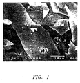
- 238000001878 scanning electron micrograph Methods 0.000 description 1
- 238000004626 scanning electron microscopy Methods 0.000 description 1
- 230000035945 sensitivity Effects 0.000 description 1
- 229910052814 silicon oxide Inorganic materials 0.000 description 1
- 239000002356 single layer Substances 0.000 description 1
- 150000003384 small molecules Chemical class 0.000 description 1
- 229910000030 sodium bicarbonate Inorganic materials 0.000 description 1
- 239000001488 sodium phosphate Substances 0.000 description 1
- 229910000162 sodium phosphate Inorganic materials 0.000 description 1
- 239000011343 solid material Substances 0.000 description 1
- 238000002798 spectrophotometry method Methods 0.000 description 1
- 230000001954 sterilising effect Effects 0.000 description 1
- 238000004659 sterilization and disinfection Methods 0.000 description 1
- 239000011550 stock solution Substances 0.000 description 1
- 229960005322 streptomycin Drugs 0.000 description 1
- QAOWNCQODCNURD-UHFFFAOYSA-L sulfate group Chemical group S(=O)(=O)([O-])[O-] QAOWNCQODCNURD-UHFFFAOYSA-L 0.000 description 1
- 230000019635 sulfation Effects 0.000 description 1
- 238000005670 sulfation reaction Methods 0.000 description 1
- 238000005211 surface analysis Methods 0.000 description 1
- 238000001356 surgical procedure Methods 0.000 description 1
- 230000002195 synergetic effect Effects 0.000 description 1
- 230000002194 synthesizing effect Effects 0.000 description 1
- 230000008685 targeting Effects 0.000 description 1
- 230000002123 temporal effect Effects 0.000 description 1
- LFQCEHFDDXELDD-UHFFFAOYSA-N tetramethyl orthosilicate Chemical compound CO[Si](OC)(OC)OC LFQCEHFDDXELDD-UHFFFAOYSA-N 0.000 description 1
- 230000025366 tissue development Effects 0.000 description 1
- 230000009466 transformation Effects 0.000 description 1
- 230000001052 transient effect Effects 0.000 description 1
- 238000011269 treatment regimen Methods 0.000 description 1
- RYFMWSXOAZQYPI-UHFFFAOYSA-K trisodium phosphate Chemical compound [Na+].[Na+].[Na+].[O-]P([O-])([O-])=O RYFMWSXOAZQYPI-UHFFFAOYSA-K 0.000 description 1
- 238000009281 ultraviolet germicidal irradiation Methods 0.000 description 1
- 210000000689 upper leg Anatomy 0.000 description 1
- 230000003827 upregulation Effects 0.000 description 1
- 239000003981 vehicle Substances 0.000 description 1
- 238000005303 weighing Methods 0.000 description 1
Images


Classifications
-
- A—HUMAN NECESSITIES
- A61—MEDICAL OR VETERINARY SCIENCE; HYGIENE
- A61K—PREPARATIONS FOR MEDICAL, DENTAL OR TOILETRY PURPOSES
- A61K9/00—Medicinal preparations characterised by special physical form
- A61K9/20—Pills, tablets, discs, rods
- A61K9/2004—Excipients; Inactive ingredients
- A61K9/2009—Inorganic compounds
-
- A—HUMAN NECESSITIES
- A61—MEDICAL OR VETERINARY SCIENCE; HYGIENE
- A61F—FILTERS IMPLANTABLE INTO BLOOD VESSELS; PROSTHESES; DEVICES PROVIDING PATENCY TO, OR PREVENTING COLLAPSING OF, TUBULAR STRUCTURES OF THE BODY, e.g. STENTS; ORTHOPAEDIC, NURSING OR CONTRACEPTIVE DEVICES; FOMENTATION; TREATMENT OR PROTECTION OF EYES OR EARS; BANDAGES, DRESSINGS OR ABSORBENT PADS; FIRST-AID KITS
- A61F2/00—Filters implantable into blood vessels; Prostheses, i.e. artificial substitutes or replacements for parts of the body; Appliances for connecting them with the body; Devices providing patency to, or preventing collapsing of, tubular structures of the body, e.g. stents
- A61F2/02—Prostheses implantable into the body
- A61F2/28—Bones
-
- A—HUMAN NECESSITIES
- A61—MEDICAL OR VETERINARY SCIENCE; HYGIENE
- A61K—PREPARATIONS FOR MEDICAL, DENTAL OR TOILETRY PURPOSES
- A61K38/00—Medicinal preparations containing peptides
- A61K38/04—Peptides having up to 20 amino acids in a fully defined sequence; Derivatives thereof
- A61K38/14—Peptides containing saccharide radicals; Derivatives thereof, e.g. bleomycin, phleomycin, muramylpeptides or vancomycin
-
- A—HUMAN NECESSITIES
- A61—MEDICAL OR VETERINARY SCIENCE; HYGIENE
- A61K—PREPARATIONS FOR MEDICAL, DENTAL OR TOILETRY PURPOSES
- A61K38/00—Medicinal preparations containing peptides
- A61K38/16—Peptides having more than 20 amino acids; Gastrins; Somatostatins; Melanotropins; Derivatives thereof
- A61K38/17—Peptides having more than 20 amino acids; Gastrins; Somatostatins; Melanotropins; Derivatives thereof from animals; from humans
- A61K38/18—Growth factors; Growth regulators
- A61K38/1841—Transforming growth factor [TGF]
-
- A—HUMAN NECESSITIES
- A61—MEDICAL OR VETERINARY SCIENCE; HYGIENE
- A61K—PREPARATIONS FOR MEDICAL, DENTAL OR TOILETRY PURPOSES
- A61K38/00—Medicinal preparations containing peptides
- A61K38/16—Peptides having more than 20 amino acids; Gastrins; Somatostatins; Melanotropins; Derivatives thereof
- A61K38/55—Protease inhibitors
- A61K38/57—Protease inhibitors from animals; from humans
-
- A—HUMAN NECESSITIES
- A61—MEDICAL OR VETERINARY SCIENCE; HYGIENE
- A61K—PREPARATIONS FOR MEDICAL, DENTAL OR TOILETRY PURPOSES
- A61K9/00—Medicinal preparations characterised by special physical form
- A61K9/14—Particulate form, e.g. powders, Processes for size reducing of pure drugs or the resulting products, Pure drug nanoparticles
- A61K9/16—Agglomerates; Granulates; Microbeadlets ; Microspheres; Pellets; Solid products obtained by spray drying, spray freeze drying, spray congealing,(multiple) emulsion solvent evaporation or extraction
- A61K9/1605—Excipients; Inactive ingredients
- A61K9/1611—Inorganic compounds
-
- A—HUMAN NECESSITIES
- A61—MEDICAL OR VETERINARY SCIENCE; HYGIENE
- A61L—METHODS OR APPARATUS FOR STERILISING MATERIALS OR OBJECTS IN GENERAL; DISINFECTION, STERILISATION OR DEODORISATION OF AIR; CHEMICAL ASPECTS OF BANDAGES, DRESSINGS, ABSORBENT PADS OR SURGICAL ARTICLES; MATERIALS FOR BANDAGES, DRESSINGS, ABSORBENT PADS OR SURGICAL ARTICLES
- A61L27/00—Materials for grafts or prostheses or for coating grafts or prostheses
- A61L27/28—Materials for coating prostheses
- A61L27/30—Inorganic materials
- A61L27/306—Other specific inorganic materials not covered by A61L27/303 - A61L27/32
-
- A—HUMAN NECESSITIES
- A61—MEDICAL OR VETERINARY SCIENCE; HYGIENE
- A61L—METHODS OR APPARATUS FOR STERILISING MATERIALS OR OBJECTS IN GENERAL; DISINFECTION, STERILISATION OR DEODORISATION OF AIR; CHEMICAL ASPECTS OF BANDAGES, DRESSINGS, ABSORBENT PADS OR SURGICAL ARTICLES; MATERIALS FOR BANDAGES, DRESSINGS, ABSORBENT PADS OR SURGICAL ARTICLES
- A61L27/00—Materials for grafts or prostheses or for coating grafts or prostheses
- A61L27/50—Materials characterised by their function or physical properties, e.g. injectable or lubricating compositions, shape-memory materials, surface modified materials
- A61L27/54—Biologically active materials, e.g. therapeutic substances
-
- C—CHEMISTRY; METALLURGY
- C03—GLASS; MINERAL OR SLAG WOOL
- C03C—CHEMICAL COMPOSITION OF GLASSES, GLAZES OR VITREOUS ENAMELS; SURFACE TREATMENT OF GLASS; SURFACE TREATMENT OF FIBRES OR FILAMENTS MADE FROM GLASS, MINERALS OR SLAGS; JOINING GLASS TO GLASS OR OTHER MATERIALS
- C03C4/00—Compositions for glass with special properties
- C03C4/0007—Compositions for glass with special properties for biologically-compatible glass
-
- A—HUMAN NECESSITIES
- A61—MEDICAL OR VETERINARY SCIENCE; HYGIENE
- A61F—FILTERS IMPLANTABLE INTO BLOOD VESSELS; PROSTHESES; DEVICES PROVIDING PATENCY TO, OR PREVENTING COLLAPSING OF, TUBULAR STRUCTURES OF THE BODY, e.g. STENTS; ORTHOPAEDIC, NURSING OR CONTRACEPTIVE DEVICES; FOMENTATION; TREATMENT OR PROTECTION OF EYES OR EARS; BANDAGES, DRESSINGS OR ABSORBENT PADS; FIRST-AID KITS
- A61F2/00—Filters implantable into blood vessels; Prostheses, i.e. artificial substitutes or replacements for parts of the body; Appliances for connecting them with the body; Devices providing patency to, or preventing collapsing of, tubular structures of the body, e.g. stents
- A61F2/02—Prostheses implantable into the body
- A61F2/30—Joints
- A61F2/30767—Special external or bone-contacting surface, e.g. coating for improving bone ingrowth
-
- A—HUMAN NECESSITIES
- A61—MEDICAL OR VETERINARY SCIENCE; HYGIENE
- A61F—FILTERS IMPLANTABLE INTO BLOOD VESSELS; PROSTHESES; DEVICES PROVIDING PATENCY TO, OR PREVENTING COLLAPSING OF, TUBULAR STRUCTURES OF THE BODY, e.g. STENTS; ORTHOPAEDIC, NURSING OR CONTRACEPTIVE DEVICES; FOMENTATION; TREATMENT OR PROTECTION OF EYES OR EARS; BANDAGES, DRESSINGS OR ABSORBENT PADS; FIRST-AID KITS
- A61F2/00—Filters implantable into blood vessels; Prostheses, i.e. artificial substitutes or replacements for parts of the body; Appliances for connecting them with the body; Devices providing patency to, or preventing collapsing of, tubular structures of the body, e.g. stents
- A61F2/02—Prostheses implantable into the body
- A61F2/30—Joints
- A61F2002/30001—Additional features of subject-matter classified in A61F2/28, A61F2/30 and subgroups thereof
- A61F2002/30667—Features concerning an interaction with the environment or a particular use of the prosthesis
- A61F2002/30677—Means for introducing or releasing pharmaceutical products, e.g. antibiotics, into the body
-
- A—HUMAN NECESSITIES
- A61—MEDICAL OR VETERINARY SCIENCE; HYGIENE
- A61L—METHODS OR APPARATUS FOR STERILISING MATERIALS OR OBJECTS IN GENERAL; DISINFECTION, STERILISATION OR DEODORISATION OF AIR; CHEMICAL ASPECTS OF BANDAGES, DRESSINGS, ABSORBENT PADS OR SURGICAL ARTICLES; MATERIALS FOR BANDAGES, DRESSINGS, ABSORBENT PADS OR SURGICAL ARTICLES
- A61L2300/00—Biologically active materials used in bandages, wound dressings, absorbent pads or medical devices
- A61L2300/20—Biologically active materials used in bandages, wound dressings, absorbent pads or medical devices containing or releasing organic materials
- A61L2300/252—Polypeptides, proteins, e.g. glycoproteins, lipoproteins, cytokines
-
- A—HUMAN NECESSITIES
- A61—MEDICAL OR VETERINARY SCIENCE; HYGIENE
- A61L—METHODS OR APPARATUS FOR STERILISING MATERIALS OR OBJECTS IN GENERAL; DISINFECTION, STERILISATION OR DEODORISATION OF AIR; CHEMICAL ASPECTS OF BANDAGES, DRESSINGS, ABSORBENT PADS OR SURGICAL ARTICLES; MATERIALS FOR BANDAGES, DRESSINGS, ABSORBENT PADS OR SURGICAL ARTICLES
- A61L2300/00—Biologically active materials used in bandages, wound dressings, absorbent pads or medical devices
- A61L2300/40—Biologically active materials used in bandages, wound dressings, absorbent pads or medical devices characterised by a specific therapeutic activity or mode of action
- A61L2300/402—Anaestetics, analgesics, e.g. lidocaine
-
- A—HUMAN NECESSITIES
- A61—MEDICAL OR VETERINARY SCIENCE; HYGIENE
- A61L—METHODS OR APPARATUS FOR STERILISING MATERIALS OR OBJECTS IN GENERAL; DISINFECTION, STERILISATION OR DEODORISATION OF AIR; CHEMICAL ASPECTS OF BANDAGES, DRESSINGS, ABSORBENT PADS OR SURGICAL ARTICLES; MATERIALS FOR BANDAGES, DRESSINGS, ABSORBENT PADS OR SURGICAL ARTICLES
- A61L2300/00—Biologically active materials used in bandages, wound dressings, absorbent pads or medical devices
- A61L2300/40—Biologically active materials used in bandages, wound dressings, absorbent pads or medical devices characterised by a specific therapeutic activity or mode of action
- A61L2300/404—Biocides, antimicrobial agents, antiseptic agents
- A61L2300/406—Antibiotics
-
- A—HUMAN NECESSITIES
- A61—MEDICAL OR VETERINARY SCIENCE; HYGIENE
- A61L—METHODS OR APPARATUS FOR STERILISING MATERIALS OR OBJECTS IN GENERAL; DISINFECTION, STERILISATION OR DEODORISATION OF AIR; CHEMICAL ASPECTS OF BANDAGES, DRESSINGS, ABSORBENT PADS OR SURGICAL ARTICLES; MATERIALS FOR BANDAGES, DRESSINGS, ABSORBENT PADS OR SURGICAL ARTICLES
- A61L2300/00—Biologically active materials used in bandages, wound dressings, absorbent pads or medical devices
- A61L2300/40—Biologically active materials used in bandages, wound dressings, absorbent pads or medical devices characterised by a specific therapeutic activity or mode of action
- A61L2300/41—Anti-inflammatory agents, e.g. NSAIDs
-
- A—HUMAN NECESSITIES
- A61—MEDICAL OR VETERINARY SCIENCE; HYGIENE
- A61L—METHODS OR APPARATUS FOR STERILISING MATERIALS OR OBJECTS IN GENERAL; DISINFECTION, STERILISATION OR DEODORISATION OF AIR; CHEMICAL ASPECTS OF BANDAGES, DRESSINGS, ABSORBENT PADS OR SURGICAL ARTICLES; MATERIALS FOR BANDAGES, DRESSINGS, ABSORBENT PADS OR SURGICAL ARTICLES
- A61L2300/00—Biologically active materials used in bandages, wound dressings, absorbent pads or medical devices
- A61L2300/40—Biologically active materials used in bandages, wound dressings, absorbent pads or medical devices characterised by a specific therapeutic activity or mode of action
- A61L2300/412—Tissue-regenerating or healing or proliferative agents
- A61L2300/414—Growth factors
-
- A—HUMAN NECESSITIES
- A61—MEDICAL OR VETERINARY SCIENCE; HYGIENE
- A61L—METHODS OR APPARATUS FOR STERILISING MATERIALS OR OBJECTS IN GENERAL; DISINFECTION, STERILISATION OR DEODORISATION OF AIR; CHEMICAL ASPECTS OF BANDAGES, DRESSINGS, ABSORBENT PADS OR SURGICAL ARTICLES; MATERIALS FOR BANDAGES, DRESSINGS, ABSORBENT PADS OR SURGICAL ARTICLES
- A61L2300/00—Biologically active materials used in bandages, wound dressings, absorbent pads or medical devices
- A61L2300/60—Biologically active materials used in bandages, wound dressings, absorbent pads or medical devices characterised by a special physical form
- A61L2300/602—Type of release, e.g. controlled, sustained, slow
-
- A—HUMAN NECESSITIES
- A61—MEDICAL OR VETERINARY SCIENCE; HYGIENE
- A61L—METHODS OR APPARATUS FOR STERILISING MATERIALS OR OBJECTS IN GENERAL; DISINFECTION, STERILISATION OR DEODORISATION OF AIR; CHEMICAL ASPECTS OF BANDAGES, DRESSINGS, ABSORBENT PADS OR SURGICAL ARTICLES; MATERIALS FOR BANDAGES, DRESSINGS, ABSORBENT PADS OR SURGICAL ARTICLES
- A61L2430/00—Materials or treatment for tissue regeneration
- A61L2430/02—Materials or treatment for tissue regeneration for reconstruction of bones; weight-bearing implants
Definitions
- the present invention relates to the incorporation of biologically active molecules into the matrix of glass, in particular bioactive glass, using a sol-gel-derived process of production.
- Musculoskeletal injuries have a substantial impact on the health and quality of life of millions of Americans. Delayed healing of and non-unions of fractures represent a continuous orthopaedic challenge.
- the conventional way of treating these problems is to use bone plates or screws in combination with autologous bone grafting.
- autogenous bone graft As a natural composite material, autogenous bone graft has been shown to have both osteoconductive and osteoinductive properties. In addition, it is a sterile, non-immunogenic and non-toxic material, which has the ability to be fully incorporated into the fracture site. Notwithstanding the long duration for their activity to develop, autogenous bone grafts are the gold standard by which synthetic composites are compared. Given that there is also a limited supply and harvest site morbidity of autogenous bone graft material, there is significant motivation to develop synthetic composites. To date, no synthetic bone graft substitutes have fully achieved the properties of autogenous bone graft.
- Osteogenesis which is the process of bone formation, involves both osteoconduction and osteoinduction.
- Osteoconduction is the process in which differentiated bone-forming cells produce a bone matrix upon an existing substrate. Materials that promote this process are considered osteoconductive.
- Osteoinduction is the process by which undifferentiated mesenchymal precursor cells are transformed into differentiated bone forming cells. Factors or materials that promote this process are considered to be osteoinductive.
- Growth factors delivered by biologically active controlled release carriers have the potential for improved fracture healing and lower morbidity, thereby resulting in improved patient care and a decrease in the overall costs associated with fracture care.
- the delivery of antibiotics by such carriers will help reduce the incidence of infections, which can further contribute to delays in healing.
- the incorporation of anti-inflammatory agents and analgesics will help control inflammation, which can also delay the healing process, and contribute to patient comfort during the healing process.
- the controlled release of such materials regardless of the bioactivity of the carrier would represent a distinct advantage over current delivery methods and assist fixation of implants.
- the ideal synthetic graft would be a scaffolding material that would stimulate bone tissue to grow in place of the scaffold as it degrades.
- Synthetic materials intended as bone graft substitutes should have mechanical and other properties similar to those of bone, and should be biocompatible with the surrounding tissues. In order to provide a union across the fracture site they must serve not only as scaffolding materials but also, similarly to native bone, have a stimulatory effect on bone tissue regeneration.
- the currently used synthetic bone graft materials are considered osteoconductive in that they elicit the formation of the bone matrix at their surfaces. Furthermore, they lead to a contiguous interface with bone or are replaced by bone tissue. Such properties suggest a chemical interaction between these bioactive materials and the bone environment. Cells existing in the bone matrix environment exhibit a beneficial response to these materials.
- CPCs Calcium phosphate ceramics
- Bioactive glass are capable of forming a hydroxyapatite layer on their surface that mimics the mineral phase of bone.
- the most commonly used calcium phosphate ceramics include: hydroxyapatite (HA), in either dense or porous forms, and ⁇ -tricalcium phosphate ( ⁇ -TCP). Hydroxyapatite is of limited effectiveness as a grafting material. When HA particulate material in porous and dense form was evaluated as a grafting material in the alveolar ridge it was found that fibrous encapsulation formed in perosseous sites. Migration of the particles was also found to be a problem. (Ducheyne P., J. Biomed. Mater. Res. (1987) 21(A2 Suppl) :219.) Further, HA cannot be used as a scaffolding material since its rate of degradation is slow. (Cornell et al., Clin. Orthop . (1992) 297; and Radin et al., J. Biomed Mater. Res. (1993) 27 :35-45.)
- ⁇ -TCP is a biodegradable material which is osteoconductive.
- its degradation rate has been found to be too fast to serve as an effective synthetic graft material in load-bearing situations. (Damien et al., supra .)
- clinical evaluations and applications of the HA and ⁇ -TCP materials, either dense or porous, have demonstrated that both materials are limited by a lack of controlled rate of reactivity.
- Bioactive glasses were first found to bond to living bone by Dr. Larry Hench in the late 1960's. Since that time, more than ten groups around the world have shown that glasses containing SiO 2 , CaO, P 2 O , Na 2 O and other smaller amounts of oxides in various compositions bond to bone.
- glasses containing SiO 2 , CaO, P 2 O , Na 2 O and other smaller amounts of oxides in various compositions bond to bone. Ducheyne P., J. Biomed Mater. Res . (1987) 21(A2 Suppl) :219; Hench, L.L., Ann. N. Y. Acad. Sci. (1988) 523: 54; Andersson et al., J. Biomed Mater. Res . (1991) 25 :1019-1030; Andersson et al., J.
- Dr. Hench's 45S5 bioactive glass has been the most extensively studied of the bioactive glass-ceramics. Its composition by weight % is: 45% SiO 2 , 24.5% CaO, 6% P 2 O 5 and 24.5% Na 2 O.
- the following parameters are important for bone-bioactive synthetic grafts: controlled resorption and reactivity, immersion induced transformation of the synthetic materials' surface into a biologically-equivalent hydroxyapatite-like mineral, relatively large surface area, and porosity (to create a network for osteoblastic activity).
- Bioactive glass can potentially be tailored to fit these parameters.
- the following requirements are important for a successful delivery system for biologically active molecules: 1) controlled release of the molecules; 2) delivery of adequate amounts of the molecules; 3) rapid growth of bone tissue into the carrier; 4) biocompatibility, osteoconductivity, and osteoinductivity of the implant material; and 5) resorption of the carrier once bone tissue has completely formed.
- Carriers made of ⁇ -TCP, or nonsoluble collagen have been moderately successful when combined with bone morphogenetic protein in attaining good acceleration of bone tissue healing.
- these systems have not been able to produce a measurable, controlled release of growth factor for time spans approaching those needed for bone tissue regeneration to span large bone filling defects.
- large amounts of growth factors i.e. greater than 50 milligrams, were required to fill defects greater than three (3) centimeters.
- WO 92/07554 reports a material which can be implanted in living tissue which has a biodegradation rate matching the rate at which the tissue regenerates. It is reported that the material may include an active substance providing an extended therapeutical effect.
- the material includes a calcium phosphate, biodegradable oxide or polyoxide, and an active substance having amine groupings such as netilmicin and/or gentamicin in sulphate form.
- composition for stimulating bone growth comprising at least one of FGF, TGF- ⁇ , IGF-II, PDGF, and their biologically active mutants and fragments, or bone extracts with corresponding activity, or bone extracts with BMP activity, and a suitable application material.
- United Kingdom Patent Application GB 2255907 A reports a delivery system for biologically active growth and morphogenetic factors comprising a solid adsorbent selected for its specific affinity for the factor and the factor adsorbed thereon.
- porous hydroxyapatite is specified as the solid adsorbent.
- U.S. Pat. No. 4,869,906 describes a resorbable porous tricalcium phosphate in which the pores are sealed with a filler mixture of antibiotic and a filler.
- U.S. Pat. Nos. 5,108,436 and 5,207,710 describe stress-bearing prostheses having a porous region in combination with an osteogenic factor extract or a purified osteogenic inductive protein, optionally in combination with a TGF- ⁇ cofactor, in a pharmaceutically acceptable carrier.
- the carrier is either a collagen composition or a ceramic.
- the osteogenic factor extract is dispersed in the porous region.
- Other procedures for combining the stress-bearing member with the osteoconductive material including coating, saturation, applying vacuum force to get the material into the pores, and air-drying or freeze-drying the material onto the member.
- the pharmaceutically acceptable carriers preferably include a matrix that is capable of providing a structure for developing bone and cartilage.
- Some preferred pharmaceutically acceptable carriers listed include collagen, hydroxyapatite, tricalcium phosphate, and bioactive glass. However, there is no description of a preparation containing bioactive glass as a pharmaceutically acceptable carrier.
- U.S. Pat. No. 4,772,203 describes implants having a core and a matrix, with the matrix being at least partially resorbable.
- the resorbable matrix is one or both of bioactive and osteogenesis-inducing. Tricalcium phosphate, hydroxylapatite [sic], and bioactive glass are listed as such matrixes. It is further stated that if a resorbable matrix is employed, it is further possible to embed antibiotics in the latter.
- U.S. Pat. No. 4,976,736 describes biomaterials useful for orthopedic and dental applications having a base portion of calcium carbonate and a surface layer of a synthetic phosphate such as hydroxyapatite.
- a synthetic phosphate such as hydroxyapatite.
- One advantage asserted for hydroxyapatite is absorbency.
- antibiotics or growth factors can be introduced into the pore cavities of the implant or attached, respectively.
- the antibiotic or growth factor can be intermixed with a preferably biodegradable polymer and injected or vacuum infiltrated into the porosity of the phosphate surfaced material.
- U.S. Pat. No. 4,563,350 describes a composition suitable for inductive bone implants comprising a purified form of osteogenic factor in admixture with a carrier having a percentage of non-fibrillar collagen.
- the factor is added to the collagen either in solution or gelatin form and stirred in dilute mineral acid for 1-2 hours at approximately 4°C. The material is then dialyzed and lyophilized.
- Japanese Laid-Open Patent Publication No. 5253286 describes a bone restoring material comprising Ca-containing glass powder and or crystallized glass powder, an aqueous solution composed mainly of phosphate, and a medical substance in release-controlled form.
- the medical substance is described as being in particulate form and can be coated with materials capable of oppressing the releasing of the substance temporarily.
- a carrier providing for the controlled release of biologically active molecules is needed.
- Such materials which are additionally osteoconductive and/or osteoinductive are also needed.
- Bioactive glasses are osteoconductive but are usually formed by combining the different oxides in a platinum crucible and melting the mixture at a temperature of 1300-1400°C. This is the melt-derived, or conventional, method of obtaining bioactive glasses. Such temperatures, however, would destroy the function of most biologically active molecules during preparation.
- Sol-gel processing Another method which can be used to synthesize bioactive glass is that of sol-gel processing.
- Sol-gel synthesis of glasses is achieved by combining a metal alkoxide precursor, such as tetraethylorthosilane (TEOS, Si(OC 2 H 5 ) 4 in the case of silica), with water and an acid catalyst to produce a hydrolysis reaction with consequent polymerization of the metal alkoxide species and production of a gel.
- TEOS tetraethylorthosilane
- Si(OC 2 H 5 ) 4 in the case of silica
- This gel will consist mostly of the metal oxide when dried and will attain the consistency of glass.
- the incorporated proteins maintained their functionality. However, the focus of such procedures was the immobilization of the protein within the sol-gel in a manner which retains the protein of interest within the gel.
- the sol-gel material functions as a sensor, very small molecules, such as glucose, can pass through the pores for assay.
- the incorporation within the sol-gel provides for repeated use of the protein. Release of the protein from the sol-gel was not desired and would actually be counter to maintenance of long-term activity.
- alkali-free bioactive sol-gel compositions based on SiO 2 , CaO, and P 2 O 5 are described. Compositions ranges are 44-86, 4-46, and 3-15 weight percent, respectively. Calcium nitrate was used as the calcium source, and tetraethoxysilane (TEOS) was used as the silicon alkoxide source.
- TEOS tetraethoxysilane
- the process described utilizes temperatures around 600-800°C, and an acid to water volume ratio of 1/6. Such a process is totally incompatible with the incorporation of biological molecules.
- U.S. Pat. No. 5,292,801 discloses the permanent entrapment of one or more reagents in a sol-gel glass support as a means for obtaining an interaction between the trapped reagents and at least one diffusible solute or component in an adjacent liquid or gas phase.
- U.S. Pat No. 5,200, 334 describes a porous, transparent sol-gel glass comprised of silicon dioxide having a biological molecule permanently encapsulated within its pores.
- U.S. Pat. No. 4,772,203 describes implants for bone and tooth root prostheses comprised of a core and a matrix.
- the matrix can comprise bioglass and can have particles embedded therein.
- the present invention is directed to controlled-release carriers.
- biologically active molecules are incorporated throughout the matrix of a silica-based glass.
- a derivation of the sol-gel technique facilitates such incorporation without negatively affecting subsequent activity of the molecules.
- the release of the biological molecules from the carrier is effected primarily by diffusion through the pore structure.
- the glass contains oxides in addition to silicon, the release of biological molecules is effected by diffusion and reaction when immersed in fluids such as, for example, body fluids.
- the sol-gel derived technique allows extensive control of the glass ultrastructure and, thus, further control over the timing and quantity of release of the biologically active molecules, such as drugs or growth factors.
- Such carriers can be both osteoconductive and osteoinductive through the formation of a calcium phosphate surface layer (i.e. bioactive) and the release of protein factors that attract and stimulate mesenchymal cells to differentiate into bone forming cells on the carrier surface, as well as increase the proliferation of osteoblasts in the local area.
- the net effect can be the acceleration of bone tissue regeneration and reduction in the incidence of infection in the area adjacent to the solgel/biologically active molecule carrier composite, making the same particularly attractive as implants.
- the bioactive composite materials have a synergistic effect in promoting bone formation and, as such, can serve as an acceptable substitute for autogenous bone graft material.
- the present invention relates to a carrier for controlled release of biologically active molecules over time comprising silica-based glass having biologically active molecules incorporated throughout the matrix of the glass.
- the present invention relates to a method for preparing silica-based glass having biologically active molecules incorporated in the matrix comprising reacting a silicon metal alkoxide with water and methanol in a molar ratio of from about 6:1 to 20:1 water/alkoxide, adjusting the pH to a value between 1 and 4.5, adding the biologically active molecule, allowing the mixture to gel and age at temperatures from 0°C up to 40°C, and then drying the aged gel at temperatures from about 15°C to 40°C.
- the present invention relates to a method for preparing silica-based glass having biologically active molecules incorporated in the matrix comprising reacting a silicon metal alkoxide and other alkoxides with water and methanol, adjusting the pH to a value between 1 and 4.5, adding the biologically active molecule, allowing the mixture to gel and age at temperatures from 0°C up to 40°C, and then drying the aged gel at temperatures from 15°C to 40°C.
- the present invention relates to a method for preparing silica-based glass having biologically active molecules incorporated in the matrix comprising reacting a silicon metal alkoxide with water, adjusting the pH to a value between 1 and 4.5, adding a calcium salt and, optionally, a phosphorous pentoxide, adding the biologically active molecule, allowing the mixture to gel and age at temperatures from 0°C up to 40°C, and then drying the aged gel at temperatures from 15°C to 40°C.
- the present invention relates to a method for preparing pure silica glass having biological molecules incorporated in the matrix comprising reacting a silicon metal alkoxide with water and methanol in a molar ratio of about 10:1 water/alkoxide, a methanol/alkoxide molar ratio of about 1:1, adjusting the pH to a value between 1.5 and 3, adding the biologically active molecule, allowing the mixture to gel and age at temperatures from 0°C to 40°C, and then drying the aged gel at temperatures from 15°C to 40°C.
- the present invention can be used in a method for delivering biological molecules to a bony defect comprising implanting a material comprising a controlled-release carrier of silica-based glass having biological molecules incorporated within the matrix of the glass in the bony defect.
- the present invention can be used in a method for delivering antibiotics in situ comprising contacting a sample with silica-based glass having antibiotics incorporated within the matrix of the glass.
- the present invention relates to a use of a controlled-release carrier comprising silica-based glass having a porons matrix and biologically active molecules incorporated throughout said matrix in the manufacture of a carrier for delivering biological molecules to a bony defect.
- the present invention relates to a method for preparing a controlled-release carrier comprising silica-based glass having a porous matrix and biologically active molecules incorporated in said matrix comprising combining a silicon alkoxide and calcium alkoxide and mixing under an argon atmosphere for up to 15 minutes without any water, alcohol, or acid, being added.
- the biologically active molecules are then added to the mixture in acid and the mixture is allowed to gel and age at temperatures from 0°C to 40°C, and then dried at temperatures from 15°C to 40°C until a weight loss of from 50 percent to 80 percent is achieved.
- the present invention relates to a pre-treated carrier comprising silica-based glass having biologically active molecules incorporated throughout the matrix of the glass.
- the carrier has been treated by immersion in- a solution containing ions typical for interstitial fluid for a period of up to about seven days prior to use.
- the present invention relates to an improved implant for filling a bony defect.
- the improved implant comprises a coating of a silica-based glass having biologically active molecules incorporated throughout the matrix of the glass.
- the present invention relates to a composition for varying release rates of biologically active molecules comprising granules of carriers for controlled release of biologically active molecules over time comprising silica-based glass having biologically active molecules incorporated throughout the matrix of the glass.
- granules of different sizes in the range from 500 ⁇ m to 5 mm are included.
- the present invention relates to a composition
- a composition comprising different populations of granules of carriers for controlled release of different biologically active molecules over time.
- the composition comprises silica-based glass having biologically active molecules incorporated throughout the matrix of the glass, each population having a different biologically active molecule incorporated therein.
- proteins and other biologically active molecules can be incorporated into silica-based glass carriers in a way that leads to sustained release of the added molecules and does not destroy their function.
- a controlled release delivery system can be used in implant materials, for example, to fill in bony defects, including defects larger than three centimeters without requiring an excessive quantity of growth factors.
- Such a controlled release delivery system also finds use in other applications with site-specific targeting needs such as, for example, chemotherapy.
- the carriers can be synthesized under sterile conditions or can be sterilized subsequently using conventional sterilization methods.
- Controlled-release carriers embodying the invention comprising antibiotics can be used in tissue culture for preventing contamination, particular that which develops upon consumption of antibiotic added with medium, by contacting the carrier with the culture through, for example, immersion.
- Controlled release carriers embodying the invention comprising growth factors, in particular bone growth factors, can be used to test the effect of the continual, controlled release of different factors on bone cells in vitro. It is also contemplated that such carriers can be used for the development of immortal bone cell lines in vitro.
- Sol-gel derived processing can be done at low temperatures -- i.e. 40°C or below -- and low pH. Both of these conditions can be important for maintaining the functionality of biologically active molecules incorporated into the sol-gel matrix.
- sol-gel derived processing include the following: 1) a sol, which is a suspension of colloidal size particles, is in liquid form before it gels; 2) the whole reaction can be done at room temperature; and 3) the microporosity of sol-gel glasses can be controlled by, for example, varying water content, timing of proton addition, proton concentration, aging time, and drying time.
- the pore sizes achievable with sol-gel processing in general are in the nanometer range.
- controlled-release carrier refers to carriers for biologically active molecules, as defined below, which provide for the release of the biologically active molecules over time when immersed in solutions containing, for example, ions typical for interstitial fluid.
- solutions containing, for example, ions typical for interstitial fluid An example of such a solution is simulated physiologic solution (SPS), used in some of the examples below.
- SPS simulated physiologic solution
- tissue culture medium is tissue culture medium.
- bioactive refers to a bone bioactive material having a calcium phosphate rich layer present, or which develops during appropriate in vitro or in vivo conditions.
- pure silica gel having a porous hydrated layer is able to induce a carbonated hydroxyapatite layer when soaked in a simulated body fluid containing calcium and phosphate ions.
- Pure silica hydrogels produced using TEOS and drying temperatures of around 400°C were immersed in simulated body fluids having different magnesium, calcium, and phosphate ions.
- silicon-based refers to the inclusion of a silicon oxide in the composition of the glass. Other oxides may also be present.
- biologically active molecules are defined as those organic molecules having an effect in a biological system, whether such system is in vitro, in vivo, or in situ.
- Biologically active molecules include, but are not limited to, the following categories: growth factors, preferably bone growth factors, cytokines, antibiotics, anti-inflammatory agents, analgesics, and other drugs.
- type as used hereinafter in reference to biologically active molecules refers to biologically active molecules of the previously listed categories, as well as specific compounds, i.e. vancomycin, TGF- ⁇ , etc. These specific compounds can be in the same or different categories.
- matrix includes the solid framework of the bioactive glass structure itself, as well as the pores.
- incorporated within said matrix denotes that the molecules are incorporated throughout the glass network.
- bone defect refers to regions necessitating repair including, but not limited to, fractures, areas of wear and tear, holes resulting from removal of screws and pins, replacements, periodontal applications, and deterioration of bone due to old age or disease.
- the term "implant" refers to a material for filling bony defects as described above.
- the implant preferably comprises a silica-based glass further comprising calcium.
- the implant can be in the form of granules, discs, blocks, or monoliths, and can comprise the controlled release carrier or simply be coated with the carrier.
- the implant can also comprise porous materials for use in bone surgery such as porous hydroxyapatite or, as described in WO 94/04657, porous bioactive glass.
- the term also includes prosthetic devices which, according to the invention, can have a coating, or partial covering, of glass or bioactive glass having biologically active molecules incorporated within the matrix. Examples of such prosthetic devices include, but are not limited to, hip and joint prostheses.
- the implant can comprise a "cocktail" providing for a combination of materials and/or release rates.
- the cocktail can include a population of granules of different sizes, all containing the same type of biologically active molecules. Alternatively, granules containing different types of biologically active molecules can be combined. The granules in such a cocktail can be the same size or different sizes, thereby providing for the release of different molecules at different rates. For example, a cocktail including antibiotics, anti-inflammatory agents, and growth factors can be prepared.
- each implant material can contain two or more types of biologically active molecules as defined herein. This can be effected by simultaneous addition of the molecules into the solution.
- implants containing one or more biologically active molecules can be prepared and then these implants can, themselves, be coated with, or incorporated throughout a solution containing one or more different types of biologically active molecules, and/or at different concentrations.
- antibiotic includes bactericidal, fungicidal, and infection-preventing drugs which are substantially water-soluble such as, for example, gentamicin, vancomycin, penicillin, and cephalosporins.
- growth factors includes growth factors identified as having osteogenic or osteoinductive properties. Included among the many factors identified with the control of bone formation are platelet derived growth factors (PDGF), the transforming growth factors (TGF- ⁇ ), insulin-like growth factors (IGFs), fibroblast growth factors (FGFs), and the bone morphogenetic proteins (BMPs). These growth factors are present at the site of fracture healing in vivo and are produced at the time of injury through platelet lysis (PDGF and TGF- ⁇ ) and by the resorption of bone matrix (TGF- ⁇ and BMPs). The individual factors will be discussed in more detail below.
- PDGF platelet derived growth factors
- TGF- ⁇ transforming growth factors
- IGFs insulin-like growth factors
- FGFs fibroblast growth factors
- BMPs bone morphogenetic proteins
- contacting includes, but is not limited to, contacting the carrier with the sample for which release of the biologically active molecules is targeted through, for example, immersion, implantation, and embedding.
- osteogenic and osteoconductive growth factors have spawned the search for new graft substances obtained through genetic engineering concepts.
- the controlled delivery of these recombinant molecules is important. Growth factors with known effect on bone tissue must be delivered at the site in sufficient doses to stimulate healing. Glasses synthesized following a room temperature sol-gel-derived procedure are outstanding candidate materials for the controlled release of such osteoinductive molecules.
- the processing of the glass allows one to control the ultrastructure of the glass such that the timing and quantity of release are tailored to fit the specific therapeutic needs.
- these glasses can be osteoconductive, thereby providing a substrate for bone tissue development.
- IGF Insulin-like growth factor
- Platelet derived growth factor a polypeptide of approximately 30kD in molecular weight, exists as a dimer composed of two A subunits or two B subunits or as a heterodimer of an A and a B subunit, creating three separate forms of PDGF. These subunits are the products of two separate genes. While all three forms are found in bone matrix, only PDGF AA is made and secreted by bone cells in vitro. PDGF BB has been found to be the most active of the three forms. (Mohan et al, supra.)
- PDGF has been shown to have bone resorbing activity in vitro ; a number of investigators have reported increased bone resorption in response to administration of physiological doses of PDGF. (Tashjian et al., Endrocrinology (1982) 111 :118-124.) Additionally, PDGF has been shown to increase osteoprogenitor cell replication.
- TGF- ⁇ Transforming growth factor-beta
- TGF- ⁇ is a family of molecules which may have bone promoting properties for fractures.
- TGF- ⁇ is a homodimeric peptide with a molecular weight of 25 kD. The most abundant sources of this molecule are platelets and bone.
- This multifunctional peptide has a broad range of cellular activities, including control of the proliferation and expression of the differentiated phenotype of several cell types specific to bone, among them mesenchymal precursor cells, chondrocytes, osteoblasts, and osteoclasts.
- TGF- ⁇ 1 and 2 have been isolated from bone in approximately a 4:1 ratio.
- In vivo studies based on both immunohistochemical staining and in situ hybridization have demonstrated the synthesis of TGF- ⁇ by both chondrocytes and osteoblasts and the accumulation of TGF- ⁇ in models of endochondral ossification. (Joyce et al., Orthop Clin. North Am. (1990) 21(1) :199; and Joyce et al., J. Cell Biol .
- BMP bone morphogenetic protein
- BMP-2A and BMP-7 have been expressed as recombinant proteins both of which have been shown to clearly induce the entire cartilage and bone formation process seen with bone-derived BMP solutions.
- BMP-2A Gackle et al., Clin. Orthop . (1993) 317
- Wozney et al. Science (1988) 242(4885) :1528
- Yasko et al. J. Bone Joint Surg . ⁇ Am> (Aug. 1992) 74(7) :1111 and J. Bone Joint. Surg.
- TMOS tetramethylorthosilane
- Alcohol preferably methanol
- Alcohol can be added at an alcohol/TMOS molar ratio of from about 0:1 to about 1:1.
- Acetic acid (0.1 N) or HCl (0.1 N) can be used as a catalyst for the hydrolysis reaction, and is added to maintain the desired pH, as disclosed below.
- Calcium methoxyethoxide (20% solution in methoxyethanol, Gelest Inc., Tullytown, PA) can be used as a calcium alkoxide source.
- Calcium methoxyethoxide (CME) is added in an amount sufficient to result in a final percentage of up to 40 % by weight calcium oxide upon drying of the gel.
- Triethyl phosphate can be used as a phosphorous pentoxide source.
- Triethyl phosphate (TEP) can be added to achieve a final concentration of phosphorous pentoxide, P 2 O 5 , up to 10 % by weight upon drying. Weight percentages throughout are calculated based upon the reactions going to completion and complete drying.
- the water, TMOS, and acid are mixed using sonication in an ice bath, or magnetic stirring, or a combination of both.
- TMOS, calcium alkoxide and additional alkoxides, if any are preferably mixed under non-aqueous conditions under an argon atmosphere using either magnetic stirring or sonication for up to about one hour.
- a calcium salt can be used in place of a calcium alkoxide. It has been found that the use of a calcium salt can extend the time to gelation, thereby affording a longer time for incorporation of the biologically active molecules and, concomitantly, a homogenous distribution of the biologically active molecules. An extended time to gelation is also helpful for the coating of implant materials with the carrier.
- the calcium salt is CaCl 2 . The calcium salt is added in an amount sufficient to result in a final percentage of up to 40 % by weight calcium oxide upon drying of the gel.
- the biologically active molecules to be incorporated retain their biological activities after treatment in moderate to highly acidic conditions, an amount of acid necessary to maintain acidity in a range of pH from 1-4.5, preferably 1.5-3, prior to, or during, incorporation of biologically active molecules is used.
- the biologically active molecules to be incorporated are suitably added at concentrations resulting in final concentrations ranging from 0.0001 to 10 % by weight of the glass.
- Glasses with compositions of silicon in the range of 60-100% (by weight) with the remainder as other oxides can be prepared.
- the liquid sol can be cast into a polystyrene container.
- the sol is aged and allowed to gel in a sealed container. Aging can take from about one (1) day to about four (4) weeks. Drying can be performed for a time of from about 1 to about 14 days.
- sol-gel derived glass In order for sol-gel derived glass to be an effective carrier for biologically active molecules, the process should be carried out at a low temperature (about 2-40°C) and, in the case of pure silica glass, the acidity of the sol should be between pH 1 and 4.5. Temperature, sol pH, % calcium content, water to TMOS molar ratio and other factors affect the gelling time of the sol. However, when incorporating biologically active molecules, the gelling time of the sol should allow enough time in the liquid state to enable the addition of the solution of biologically active molecules for incorporation, cast, and homogeneously mix the sol. Gelation occurs when enough cross-links have formed such that the network spans the length of the container. Gross observation reveals little or no movement of the cast material upon inversion.
- a lower pH increases the gelling time.
- a higher calcium content decreases gelling time.
- a higher gelling time is desirable in order to see more of the sol-gel reactions going to completion, thus ending with a final material with less porosity and smaller pore size.
- Less porosity also means a more mechanically strong material with longer times of protein release.
- greater porosity may be desirable, for example, achieving a more rapid release of molecules, or a more rapid degradation of the carrier. Larger pore sizes facilitate the release of larger molecules through diffusion.
- a lower temperature also increases gelling times. To achieve lower temperatures, the reaction is then carried out in an ice-cooled water bath. A higher water content will decrease gelling time for most metal alkoxides, although the porosity may stay high due to increased water evaporation from the material. Conditions are selected such that gelation optimally occurs within a period ranging from at least about 30 minutes to about 48 hours for incorporating biologically active molecules. Gelation can be performed at temperatures ranging from 0°C to 40°C.
- Aging of the sol-gel occurs after casting and is performed by keeping the casting container sealed. Sol-gel reactions continue unimpeded during this period. Aging can be performed at temperatures ranging from 0°C to 40°C. Longer aging times (of up to 1 month) result in a more mechanically strong material, which undergoes less cracking than materials that have been aged for lesser time periods. Aging at a lower temperature, such as 4°C, also extends the gelling time.
- Drying temperature and time can also affect the final material characteristics.
- a fast rate of drying can produce cracks in the final material.
- the final material loses about 50-80% of its weight between casting and final drying due to evaporation of water, and alcohols produced as by-products of the reaction. Drying is performed at temperatures ranging from about 15°C to 40°C by unsealing the casting container, and can be performed at atmospheric pressure, or pressures lower than atmospheric.
- the sol-gel carriers can be treated by immersion at the time of production such that the intitial higher release phase has taken place before actual use in the patient.
- a sol-gel derived silica-based matrix-vancomycin composite was synthesized employing a room-temperature, low acidity, low alcohol concentration procedure. Vancomycin was selected as the drug to be released due to its proven efficacy against gram positive cocci, especially staphylococci, which is a major cause of osteomyelitis. Vancomycin is a water soluble (up to 100 mg/ml) tricyclic glyceropeptide of approximately 3,300 molecular weight.
- the material was prepared as follows: 19.6 ml tetramethylorthosilicate (TMOS, Aldrich, St. Louis, MO, U.S.A.), 14.2 ml water, 5.2 methanol and 0.01 ml of 1N HCl was sonicated in a glass beaker in an ice bath for 30 minutes. Then, 4 ml of the sol was cast into 23 mm diameter polystyrene vials (Sarstedt, Princeton, NJ) and 1 ml of 10 mg/ml vancomycin HCL (Lederle, Carolina, Puerto Rico) was added to the sols in the vials and the samples were mixed. The same amount of water, i.e. 1 ml, was added to control samples.
- TMOS tetramethylorthosilicate
- the total H 2 O/TMOS ratio was 10:1.
- the methanol/TMOS ratio was 1:1.
- the amount of incorporated vancomycin to sample weight was about 1%.
- the vials were sealed with airtight caps, gelled, aged, and dried at room temperature. Time to gelation varied from 15 to 25 hours. Addition of the vancomycin solutions did not change significantly the time to gelation.
- the sols were exposed to air for drying. During drying, evaporation of liquid from the gel pore network resulted in weight loss and shrinking of the gels. The weight loss continued up to 2 weeks. Drying was considered to be complete when the weight loss reached 75-78%. The significant weight loss and shrinking did not produce visible cracks.
- the resulting products were transparent monoliths in a shape of 11 mm diameter and 8 mm high cylinders weighing 1.1 gram. The density of the dried gel material was equal to 1.5 g/cm 3 . Since 10 mg vancomycin was incorporated into each of the discs, the vancomycin content in the material was 0.91%. There is no reason to expect that other water-soluble antibiotics will behave any differently.
- the synthesized vancomycin composite was immersed into a simulated physiological solution (SPS) with ion content similar to that of plasma as disclosed previously.
- SPS sample surface area to volume
- the sol-gel derived silica based matrix-vancomycin composites with varying vancomycin content were synthesized.
- the sols were prepared as disclosed above in Example 1.
- 1.2 ml of the sol were cast into 23 mm diameter polystyrene vials.
- the cast sols were divided into two groups and 0.3 ml of solutions with different vancomycin concentrations were added to the cast sols of both groups in order to keep the same H 2 O/TMOS molar ratio of 10:1.
- the amounts of the incorporated vancomycin were 10 and 20 mg for groups 1 and 2, respectively.
- the percentage of vancomycin to sample weight was equal to 2.8 and 5.5%, respectively.
- the sols were gelled, aged, and dried to about 75% weight loss.
- Ultrastructure parameters of the sols such as specific surface area (SSA), average pore size (PS), and pore volume (PV) of the dried sols were determined using the monolayer gas absorption technique (multipoint B.E.T., Quantachrome). The measured values were as follows:
- the data show that the amount of released vancomycin increased with the amount of incorporated drug.
- the released amount appears to be a function of the incorporated quantity (at conditions otherwise equal).
- the drug release profile over time appears to be similar for different concentration. Specifically, the drug release started right after immersion, reached a maximum by 3 days, then gradually decreased.
- the standard disc susceptibility test technique was applied (See Lorian, supra. ).
- the sample SPS solutions with vancomycin released during immersion were tested and compared with standard solutions of vancomycin in SPS with concentrations ranging from 100 to 10,000 ⁇ g/ml. Concentrations of the sample SPS solutions with vancomycin released during immersion were measured using the Fluorescent Polarizing Immunoassay described previously.
- the concentration-zone of inhibition relationship for the sample solutions of vancomycin released from the silica-based matrix showed a close fit to that of the standard solutions.
- Figure 6 shows zone of bacteria inhibition sizes versus immersion time and concentration of vancomycin incorporated into the sol-gel derived silica matrix. Open bars represent vancomycin at 1 mg. Solid bars represent vancomycin at 10 mg. Cross-hatched bars represent vancomycin at 20 mg. The data demonstrate that vancomycin released from the sol-gel matrix was effective to inhibit the bacteria growth up to three (3) weeks (at 20 mg). The measured zone of inhibition sizes appear to increase with concentration of incorporated vancomycin, reflecting larger quantities of released vancomycin.
- Trypsin Inhibitor is a protein with molecular weight of 21 kD.
- the sol-gel/protein composite contains 1-10 mg Trypsin Inhibitor (TI) per 150-200 mg disc. Protein elution was measurable in samples with 2 mg or greater of protein per disc.
- the procedure used to synthesize 1 gram (by dry weight) of the sol-gel derived glass was as follows: 2.48 ml of TMOS (Aldrich, St. Louis, MO) was combined with 2 ml DI water and 0.68 ml of methanol in a 30 ml beaker and mixed for 5 minutes using magnetic stirring. This resulted in an H 2 O/TMOS molar ratio of 10:1 and a methanol/TMOS molar ratio of 1:1. Then, 0.01 ml of 1N HCl was added to catalyze the sol-gel reaction. This results in a clear one phase solution which is stirred for 15 minutes.
- TMOS Aldrich, St. Louis, MO
- the sol was cast in 0.8 ml volumes into polystyrene containers and the trypsin inhibitor solution was added in a volume of 0.2 ml of 0.1 N acetic acid solution with protein concentration in the range of 1-10 mg/ml.
- the solution was mixed with vortexing and the containers were capped. Gelling occurred within 1-4 days after casting.
- the capped sol was allowed to gel and age for times ranging between 1 day to 2 weeks depending on the desired porosity at room temperature.
- the sol-gel was allowed to dry at either room temperature or 37°C by uncapping the casting container. Any liquid produced was decanted off the solid. A higher drying temperature increased the porosity and rate of shrinkage. After drying, the resulting solid had lost 60-70% of its original weight due to evaporation of water and alcohol (methanol is a by-product of the sol-gel reactions).
- the resulting solid material was a porous three-dimensional network/polymer of silica which releases incorporated biomolecules in a controlled release fashion.
- the various processing parameters are depicted in Table II. Sample designations are of the Formula SxxxCxxPxx (date cast), where "Sxxx” is the calculated % silica, “Cxx” is the calculated % calcium oxide, and “Pxx” is the calculated % phosphorous pentoxide.
- the date cast is presented as the "last two digits of the year. month. day.”
- the initial release kinetic studies were carried out by immersing the sol-gel/protein composite (100 mg sol-gel/l mg protein for each sample) in deionized water inside containers which were siliconized in order to reduce protein binding.
- the protein content in water was measured at different time points. The water was replaced fresh after each time period.
- the collected fluid that had been in contact with the sol-gel/protein composite was analyzed for protein content using a colloidal gold/spectrophotometric method (Integrated Separation Systems, Natick, MA, Stoscheck et al., Anal. Biochem ., (1987) 160:301-305) with sensitivity down to 0.5 ⁇ g/ml.
- Table III The results are depicted in Table III below.
- the numbers in the table represent protein release in ⁇ g of trypsin inhibitor after immersion in DI water. Results for each time point are provided together with cumulative protein release.
- trypsin inhibitor was continually released from all samples for a period of at least seven (7) weeks.
- Sol-gel derived glass with a composition of 100% silica and water/TMOS molar ratio of 15:1 was synthesized and its bioactivity tested in vitro in SPS by measuring changes in calcium-ion concentration.
- a 5 gram sample was made by combining 12.38 ml of TMOS with 8.87 ml of DI water and sonicating for 5 minutes in an ice cooled bath. To this mixture, 8.87 ml of 0.1 N Acetic Acid was added and the mixture sonicated for an additional 15 minutes. Then, 4.43 ml sodium phosphate (.01 M, pH 7) buffer was added and the mixture sonicated for one minute.
- the liquid sol was cast as 3 ml samples.
- the pH of the sol as cast was 4.5.
- the gelling time was approximately 2 hours. Aging of the samples was done at room temperature, for 1 day. Samples were dried for 3 days at 37°C and weighed about 500 mg.
- Samples in the form of discs approximately 1 cm in diameter and 4 mm high (1.76 cm 2 SA) were then immersed into SPS (17.6 ml) for a sample surface area to immersion solution volume ratio of 0.1 cm -1 .
- Samples were immersed for two weeks with constant stirring at 37°C.
- SPS concentration of calcium normally averages 100 ppm.
- the average concentration of calcium in the retrieved SPS averaged 25 ppm. This indicates that calcium was consumed by the glass from solution, most likely by forming a calcium phosphate layer on its surface.
- Sol-gel samples were then immersed into 12 ml of SPS to test for calcium phosphate surface layer formation. Samples were retrieved after being immersed 5 days in SPS with constant stirring at 37°C. The samples were viewed using scanning electron microscopy (SEM) and surface analysis was performed using energy dispersive x-ray analysis (EDXA). The surface of the samples contained nodules 1-3 ⁇ m in diameter ( Figure 1) that, when analyzed with EDXA ( Figure 2), contained high proportions of calcium and phosphorous. This indicates the formation of calcium phosphate nucleation sites as a precursor to calcium phosphate layer formation.
- SEM scanning electron microscopy
- EDXA energy dispersive x-ray analysis
- TGF- ⁇ 1 Recombinant human transforming growth factor beta
- the sol-gel was synthesized by combining 5 ml TMOS with 5.4 ml of water, and methanol, for a water/TMOS/methanol molar ratio of 9:1:1, and magnetically stirring for 5 minutes at room temperature. 10 ⁇ L of 1N HCl was added to the mixture and the sol was stirred for 30 minutes. Then, 0.9 ml of the sol was cast into a polystyrene container and 0.1 ml of TGF- ⁇ solution with 1% bovine serum albumin (BSA) to prevent non-specific binding of the growth factor to the casting container was added.
- BSA bovine serum albumin
- TGF- ⁇ Different quantities of TGF- ⁇ were added to each sample cast ranging from 0.5 ⁇ g to 2 ⁇ g of TGF- ⁇ for each solution added with 1% BSA. Samples were aged for 3 days at 37°C and dried at 37°C until they had lost approximately 50% of their "as cast" weight.
- Sol-gel derived silica glass was synthesized by mixing and stirring TMOS, DI water, and 1N HCl in a molar ratio of 1:10:0.001. Then, 0.9 ml of the sol was cast into 15 ml diameter polystyrene vials and 0.1 ml of a solution containing either 0.5 or 1 ⁇ g of TGF- ⁇ 1 was added to the sol samples.
- TGF- ⁇ 1 (Celtrix Lab., Inc.) was prepared according to instructions supplied.
- TGF- ⁇ 1 TGF- ⁇ 1
- BSA bovine serum albumin
- the resulting TGF- ⁇ 1 solutions were stored in 1.5 ml microcentrifuge tubes (USA Scientific) at -70°C until use.
- the pH of the sols at the time of casting was measured to be 1.7.
- the vials were sealed and the sols were allowed to gel (15 hours) and age (24 hours) in an incubator at 37°C. Then, the gels were dried to either 50 or 70% weight loss resulting in transparent glass disks about 10 mm in diameter.
- silica glass/TGF- ⁇ 1 composites were prepared as follows: 1 ⁇ g dose: disks and particles dried to 50% weight loss, and particles dried to 70% weight loss; 0.5 ⁇ g dose: particles dried to 50% weight loss.
- An additional six silica glass disks containing 0.5 ⁇ g TGF- ⁇ 1 and two controls (without TGF- ⁇ 1) were prepared and dried to 57% weight loss. The disks had uniform dimensions with an average diameter of 10.17 mm and an average height of 4.93 mm.
- TGF- ⁇ 1 The release of TGF- ⁇ 1 from the sol-gel derived silica glass particles and disks was measured by immersion in 1 ml of sterile phosphate buffered saline (PBS) containing 1% BSA. Prior to immersion all the samples were sterilized by UV irradiation. The BSA prevents the non-specific binding of TGF- ⁇ 1 to the immersion reservoir. Concentration was determined using an enzyme linked immunosorbent assay (ELISA).
- ELISA enzyme linked immunosorbent assay
- the amount of active TGF- ⁇ 1 released from the sol-gel materials was assessed using the MvlLu mink lung epithelial cell inhibition assay. This assay determines TGF- ⁇ 1 activity based on its inhibition of MvlLu cell proliferation as measured by [ 3 H]-thymidine incorporation. Jennings et al., "Comparison of the biological activities of TGF beta 1 and TGF-beta 2: Differential activity in endothelial cells", J. Cell Physiol. 137:167-172 1988. Confluent MvlLu cells (ATCC) were lifted from tissue culture flasks using Cell Dissociation Solution (Specialty Media) and plated in Coming 24-well polystyrene tissue culture dishes.
- DMEM Dulbecco's Modified Eagle's Medium
- FBS fetal bovine serum
- Peptomycin Sigma Cell Culture
- TGF- ⁇ 1 of known picomolar (pM) concentrations as well as lyophilized 1% BSA in 10 mM HCl to serve as the control.
- concentrations ranged from 0.1 pM to 10.0 pM and were added in triplicate. Aliquots from sample solutions, i.e. containing TGF- ⁇ 1 released from silica glass upon immersion, were diluted into the same range of concentrations and also added to the cells. The dishes were incubated for additional 24 hours.
- TGF- ⁇ 1 standard and sample solutions After treatment with TGF- ⁇ 1 standard and sample solutions, the wells were aspirated and the cells labeled for 2 hours with 1 ⁇ Ci/ml of [ 3 H]-thymidine (NEN Research Products) in 1 ml of tissue culture medium. At the conclusion of the incubation period, the relative levels of radioactivity incorporated into cellular DNA were assessed. Each well was washed with 1 ml of PBS, pH 7.4, followed by 10 minutes of treatment with trichloroacetic acid (TCA) to precipitate all unincorporated [ 3 H]-thymidine. Following TCA precipitation, the cells were washed twice with PBS and solubilized with 2% sodium dodecyl sulfate by shaking at room temperature for 2 hours.
- TCA trichloroacetic acid
- Radioisotope incorporation into each sample was determined by liquid scintillation counting of a 200 ⁇ l aliquots in 5 ml of ICN Ecolume scintillation fluid using a Beckman LS1800 Liquid Scintillation Counter. Duplicate counts were performed for each sample.
- the sol-gel technology used to synthesize glass/TGF- ⁇ 1 composites does not alter the biological functionality of TGF- ⁇ 1.
- the carriers showed a sustained release of therapeutic quantities of an osteoinductive growth factor in this biologically active form.
- a sol-gel derived silica-based glass containing Ca and P was synthesized by mixing the three alkoxides TMOS, CME, and TEP under an argon atmosphere and stirring the mixture for 5 minutes using a magnetic stirrer.
- a glass having a final composition of about 70% SiO 2 , 25% CaO, and 5% P 2 O 5 (percent dry weight) was prepared by mixing 3,47 ml TMOS, 8.4 ml CME, and 0.24 ml TEP. Then, 1.1 ml of the sol was cast per 15 mm diameter polystyrene vial and 0.38 ml of 0.1 N acetic acid was added to each of the sols to mimick the conditions for incorporation of proteins. The gels were sealed, aged for three days at room temperature, and dried at 37°C to 50% weight loss.
- the microstructure of the Ca-P containing silica-based glass prepared was characterized using surface area analysis (Autosorb-1, Quantachrome). Prior to the analysis, the samples were outgassed at 30°C.
- the material pore structure can be characterized by the shape of absorption isotherms, i.e. plots representing changes in the absorbed gas (N 2 ) volume vs. relative pressure P/P 0 .
- the isotherm for the Ca-P containing silica-based glass is depicted in Figure 12.
- the shape of the isotherm is characteristic of a mesoporous material, as defined in the Manual on Using a Surface Analyzer Autosorbl , Quantachrome Corp., pp. II-4-46, 1992
- Mesoporous materials are defined therein as materials having an intermediate pore size, or pores in the size range of greater than 20 angstroms and less than 500 angstroms.
- SSA, PV, and mean pore size were determined to be 331 m 2 /g, 0.97 cc/g, and 58.4 angstroms, respectively.
- the ability of the synthesized Ca-P containing glass to form a surface HA layer was assayed after immersion in SPS for one week.
- the samples were analyzed prior to and after immersion using FTIR.
- the FTIR spectra of the samples before and after one week of immersion are presented in Figure 13.
- the absorption bands in the spectrum before immersion (bottom spectrum) are characteristic of silica-gel. After immersion, a doublet of bands appeared at 603 and 580 cm -1 (upper spectrum). These bands indicate the formation of HA on the glass surface, thereby indicating bioactive behavior.
- Sol-gel derived glass containing Si, Ca, and P was synthesized by combining TMOS (3.47 ml), CME (8.40 ml) and TEP (0.24 ml), to produce a composition of 70% SiO 2 , 25% CaO and 5% P 2 O 5 , under an argon atmosphere and mixing for 15 minutes.
- TMOS 3.47 ml
- CME 8.40 ml
- TEP 0.24 ml
- Protein release studies were performed by immersing 500 mg of the granules in 1 ml of 50 mM tris buffer solution (pH 7.3 at 37°C). Solutions were replaced completely after each time point varying from 1 hour to seven weeks. Samples were immersed in 1 ml plain tris solution at 37°C with constant shaking. For each time point measured (1 hr, 2 hrs, 4 hrs, 24 hrs, 48 hrs, 72 hrs, 96 hrs, 1 week, 2wks, 3wks, and 4 wks) the solution was exchanged for fresh tris solution and the protein concentrations were measured using a colloidal gold assay. The protein concentrations in the solutions were measured using a gold colloidal assay (Integrated Separation Systems, Natick MA). Cumulative protein release from the Ca-P containing glass is represented in Figure 14.
- the FTIR spectra of glass of composition (1) prior to and after immersion in SPS for one week are represented in Figure 15.
- the spectrum of the sample prior to immersion shows absorption bands of silica and proteins in the lower (below 1200 cm -1 ) and higher (above 1200 cm -1 ) energy regions, respectively.
- a doublet of bands, located at 562 and 603 cm -1 appeared in the spectrum after immersion (upper spectrum).
- the doublet, characteristic of the P-O bending mode of vibration, indicates formation of a hydroxyapatite (HA) layer on the surface of sol-gel derived glass. Formation of the HA layer was also detected on the glass of composition (1) without TI.
- Compositions (2) and (3) also showed formation of the HA layer.
- a bioactive, sol-gel derived glass having a final composition of about 70% SiO 2 , 20% CaO, and 5% P 2 O 5 (by weight) was synthesized as follows: 9.3 ml TMOS was combined with 5.8 ml deionized water (DI) and stirred in an icy ultrasonic bath for 10 minutes. Then, 15 ⁇ l of 1 N HCl was added, and the stirring continued until the mixture became clear. The pH measured 3.0. The mixture was then switched to magnetic stirring. While the mixture was continuously being stirred, 3.08 g of CaCl 2 dissolved in 5 ml DI water and 2 ml of TEP were added dropwise to the mixture.
- DI deionized water
- the total H 2 O/TMOS ratio was about 10; the acid/H 2 O volume ratio was about 0.0015.
- the sol was cast into polystyrene vials 17 mm in diameter, 1 ml per vial, sealed, and allowed to gel at room temperature. Time to gelation was about one hour. The gels were then aged for 10 days and dried to a weight loss of about 70% of as-cast weight, both occurring at room temperature.
- the resultant glass discs were 8 mm in diameter, 4 mm high, and were transparent, indicating homogenous distribution of the alloying oxides. No cracks were observed in the obtained monoliths.
- a bioactive sol-gel derived glass is prepared as in Example 15, with the addition of biologically active molecules either before or after the sol is cast.
- the biologically active molecules are added, for instance, as disclosed in Examples 5, 10, or 11 above.
Abstract
Description
- The present invention relates to the incorporation of biologically active molecules into the matrix of glass, in particular bioactive glass, using a sol-gel-derived process of production.
- Musculoskeletal injuries have a substantial impact on the health and quality of life of millions of Americans. Delayed healing of and non-unions of fractures represent a continuous orthopaedic challenge. The conventional way of treating these problems is to use bone plates or screws in combination with autologous bone grafting.
- As a natural composite material, autogenous bone graft has been shown to have both osteoconductive and osteoinductive properties. In addition, it is a sterile, non-immunogenic and non-toxic material, which has the ability to be fully incorporated into the fracture site. Notwithstanding the long duration for their activity to develop, autogenous bone grafts are the gold standard by which synthetic composites are compared. Given that there is also a limited supply and harvest site morbidity of autogenous bone graft material, there is significant motivation to develop synthetic composites. To date, no synthetic bone graft substitutes have fully achieved the properties of autogenous bone graft.
- Enhancing the rate and probability of fracture healing and the promotion of bone formation and healing of delayed and non-union fractures are of great clinical significance. (NIH/AAOS sponsored workshop. Bone Formation and Bone Regeneration. Tampa, FL: American Academy of Orthopaedic Surgeons, 1993). The large population of patients with delayed unions and non-unions of bone, the large direct medical costs, and the societal costs related to their long term disability, highlight the need for effective and improved methods of treatment.
- Advances in materials science and the identification of osteogenic and osteoinductive growth factors have invited the investigation of newer alternatives for autogenous bone grafting. Osteogenesis, which is the process of bone formation, involves both osteoconduction and osteoinduction. Osteoconduction is the process in which differentiated bone-forming cells produce a bone matrix upon an existing substrate. Materials that promote this process are considered osteoconductive. Osteoinduction is the process by which undifferentiated mesenchymal precursor cells are transformed into differentiated bone forming cells. Factors or materials that promote this process are considered to be osteoinductive.
- Growth factors delivered by biologically active controlled release carriers have the potential for improved fracture healing and lower morbidity, thereby resulting in improved patient care and a decrease in the overall costs associated with fracture care. Similarly, the delivery of antibiotics by such carriers, either alone or in addition to growth factors, will help reduce the incidence of infections, which can further contribute to delays in healing. In fractures involving, for example, the spine, the incorporation of anti-inflammatory agents and analgesics will help control inflammation, which can also delay the healing process, and contribute to patient comfort during the healing process. Additionally, the controlled release of such materials regardless of the bioactivity of the carrier would represent a distinct advantage over current delivery methods and assist fixation of implants.
- The ideal synthetic graft would be a scaffolding material that would stimulate bone tissue to grow in place of the scaffold as it degrades. (Damien et al., J. Applied Biomater. (1991) 2:187-20.) Synthetic materials intended as bone graft substitutes should have mechanical and other properties similar to those of bone, and should be biocompatible with the surrounding tissues. In order to provide a union across the fracture site they must serve not only as scaffolding materials but also, similarly to native bone, have a stimulatory effect on bone tissue regeneration.
- The currently used synthetic bone graft materials are considered osteoconductive in that they elicit the formation of the bone matrix at their surfaces. Furthermore, they lead to a contiguous interface with bone or are replaced by bone tissue. Such properties suggest a chemical interaction between these bioactive materials and the bone environment. Cells existing in the bone matrix environment exhibit a beneficial response to these materials.
- The materials studied most for use as synthetic grafts have been calcium phosphate ceramics and bioactive glasses. Calcium phosphate ceramics (CPCs) are very similar in composition to the mineral phase of bone. Bioactive glass are capable of forming a hydroxyapatite layer on their surface that mimics the mineral phase of bone.
- The most commonly used calcium phosphate ceramics include: hydroxyapatite (HA), in either dense or porous forms, and β-tricalcium phosphate (β-TCP). Hydroxyapatite is of limited effectiveness as a grafting material. When HA particulate material in porous and dense form was evaluated as a grafting material in the alveolar ridge it was found that fibrous encapsulation formed in perosseous sites. Migration of the particles was also found to be a problem. (Ducheyne P., J. Biomed. Mater. Res. (1987) 21(A2 Suppl):219.) Further, HA cannot be used as a scaffolding material since its rate of degradation is slow. (Cornell et al., Clin. Orthop. (1992) 297; and Radin et al., J. Biomed Mater. Res. (1993) 27:35-45.)
- β-TCP, on the other hand, is a biodegradable material which is osteoconductive. However, its degradation rate has been found to be too fast to serve as an effective synthetic graft material in load-bearing situations. (Damien et al., supra.) Thus, clinical evaluations and applications of the HA and β-TCP materials, either dense or porous, have demonstrated that both materials are limited by a lack of controlled rate of reactivity.
- Bioactive glasses were first found to bond to living bone by Dr. Larry Hench in the late 1960's. Since that time, more than ten groups around the world have shown that glasses containing SiO2, CaO, P2O , Na2O and other smaller amounts of oxides in various compositions bond to bone. (Ducheyne P., J. Biomed Mater. Res. (1987) 21(A2 Suppl):219; Hench, L.L., Ann. N. Y. Acad. Sci. (1988) 523:54; Andersson et al., J. Biomed Mater. Res. (1991) 25:1019-1030; Andersson et al., J. Non-Cryst Solids (1991) 129:145-151; Boone et al., J. Biomed Mater. Res. (1989) 23(A2 Suppl):183; Ducheyne et al., Clin. Orthop. Rel. Res. (1992) 76:102-114; Hench, L.L., J. Biomed Mater. Res. (1989) 23:685-703; Kokubo, T., Biomaterials (1991) 12(2):155; and Rawlings, R.D., J. Mater. Sci. Letters (1992) 11:1340-1343.)
- Bioactive glass-ceramics undergo surface corrosion reactions when exposed to body fluids. These corrosion reactions form a silica-rich surface layer. This layer serves as a nucleation site for the deposition of calcium phosphate, which evolves into a thick hydroxyapatite layer. When in contact with bone forming cells, this layer will form the basis of the chemical bond between the glass and the bone matrix. (Ducheyne, supra; Hench (1988), supra; and Hench, (1989), supra.) Dr. Hench's 45S5 bioactive glass has been the most extensively studied of the bioactive glass-ceramics. Its composition by weight % is: 45% SiO2, 24.5% CaO, 6% P2O5 and 24.5% Na2O.
- In U.S. Pat. No. 5,204,106 45S5 glass in particulate form in a narrow size range was described as being an effective bone graft substitute in the alveolar ridge model and as being well incorporated into the surrounding bone. The glass granules were described as causing the upregulation of osteoprogenitor cells to osteoblasts that actively lay down bone tissue. (Schepers et al., J. Oral Rehabil. (1991) 18:439-452.)
- The following parameters are important for bone-bioactive synthetic grafts: controlled resorption and reactivity, immersion induced transformation of the synthetic materials' surface into a biologically-equivalent hydroxyapatite-like mineral, relatively large surface area, and porosity (to create a network for osteoblastic activity). Bioactive glass can potentially be tailored to fit these parameters. In addition, the following requirements are important for a successful delivery system for biologically active molecules: 1) controlled release of the molecules; 2) delivery of adequate amounts of the molecules; 3) rapid growth of bone tissue into the carrier; 4) biocompatibility, osteoconductivity, and osteoinductivity of the implant material; and 5) resorption of the carrier once bone tissue has completely formed. (Lucas et al., J. Biomed Mater. Res. (1989) 23(A1 Suppl) :23.) No delivery system currently available meets all of these criteria. (Damien et al., supra; and Cornell and Lane, Clin. Orth. Rel. Res. (1992) 277:297-311.) Certainly, no delivery system results in controlled delivery.
- Attempts have been made to try to improve calcium phosphate ceramics by using them as delivery vehicles for bone growth factors. To date, there has been no success in incorporating growth factors into calcium phosphate ceramics in a way that will lead to a sustained release of the added growth factor. Mostly, one achieves a "burst" release, which is a rapid initial release of most of the material over a short period of time. (Campbell et al., Trans. Orthop. Res. Soc., 40:775, 1994.)
- Carriers made of β-TCP, or nonsoluble collagen, have been moderately successful when combined with bone morphogenetic protein in attaining good acceleration of bone tissue healing. (Damen et al., J. Dental Res. (1989) 68:1355-1359) However, these systems have not been able to produce a measurable, controlled release of growth factor for time spans approaching those needed for bone tissue regeneration to span large bone filling defects. In one study, large amounts of growth factors, i.e. greater than 50 milligrams, were required to fill defects greater than three (3) centimeters. (Johnson et al., Clin. Orthop. (1992) 277:229237.)
- In most of the systems studied with osteoconductive materials used as carriers, the method of incorporation has been that of simple immersion of the material into a growth factor solution. The growth factor is then adsorbed either onto the material surface or into the pore structure, but is then quickly released upon immersion in an aqueous solution in a burst effect. (Campbell et al., Trans. Orthop. Res. Soc., 40:775, 1994.)
- Published application WO 92/07554 reports a material which can be implanted in living tissue which has a biodegradation rate matching the rate at which the tissue regenerates. It is reported that the material may include an active substance providing an extended therapeutical effect. The material includes a calcium phosphate, biodegradable oxide or polyoxide, and an active substance having amine groupings such as netilmicin and/or gentamicin in sulphate form.
- Published application WO 93/05823 reports a composition for stimulating bone growth comprising at least one of FGF, TGF-β, IGF-II, PDGF, and their biologically active mutants and fragments, or bone extracts with corresponding activity, or bone extracts with BMP activity, and a suitable application material.
- United Kingdom Patent Application GB 2255907 A reports a delivery system for biologically active growth and morphogenetic factors comprising a solid adsorbent selected for its specific affinity for the factor and the factor adsorbed thereon. In one embodiment, porous hydroxyapatite is specified as the solid adsorbent.
- U.S. Pat. No. 4,869,906 describes a resorbable porous tricalcium phosphate in which the pores are sealed with a filler mixture of antibiotic and a filler.
- U.S. Pat. Nos. 5,108,436 and 5,207,710 describe stress-bearing prostheses having a porous region in combination with an osteogenic factor extract or a purified osteogenic inductive protein, optionally in combination with a TGF-β cofactor, in a pharmaceutically acceptable carrier. The carrier is either a collagen composition or a ceramic. The osteogenic factor extract is dispersed in the porous region. Other procedures for combining the stress-bearing member with the osteoconductive material including coating, saturation, applying vacuum force to get the material into the pores, and air-drying or freeze-drying the material onto the member. It is further described that the pharmaceutically acceptable carriers preferably include a matrix that is capable of providing a structure for developing bone and cartilage. Some preferred pharmaceutically acceptable carriers listed include collagen, hydroxyapatite, tricalcium phosphate, and bioactive glass. However, there is no description of a preparation containing bioactive glass as a pharmaceutically acceptable carrier.
- U.S. Pat. No. 4,772,203 describes implants having a core and a matrix, with the matrix being at least partially resorbable. The resorbable matrix is one or both of bioactive and osteogenesis-inducing. Tricalcium phosphate, hydroxylapatite [sic], and bioactive glass are listed as such matrixes. It is further stated that if a resorbable matrix is employed, it is further possible to embed antibiotics in the latter.
- U.S. Pat. No. 4,976,736 describes biomaterials useful for orthopedic and dental applications having a base portion of calcium carbonate and a surface layer of a synthetic phosphate such as hydroxyapatite. One advantage asserted for hydroxyapatite is absorbency. It is further described that antibiotics or growth factors can be introduced into the pore cavities of the implant or attached, respectively. Alternatively, the antibiotic or growth factor can be intermixed with a preferably biodegradable polymer and injected or vacuum infiltrated into the porosity of the phosphate surfaced material.
- Gombotz et al., J. App. Biomat., (1994) 5:141-150 describe the incorporation of transforming growth factor-β into a composite implant made from poly(lactic-co-glycolic acid) and demineralized bone matrix. It is reported that the implants exhibited an inflammatory response with little mineralization or bone formation. Similar results were reported in Meikle et al., Biomaterials, (1994) 15(7):513-521 with poly DL-lactide-co-glycolide discs having bone matrix extract incorporated therein.
- U.S. Pat. No. 4,563,350 describes a composition suitable for inductive bone implants comprising a purified form of osteogenic factor in admixture with a carrier having a percentage of non-fibrillar collagen. The factor is added to the collagen either in solution or gelatin form and stirred in dilute mineral acid for 1-2 hours at approximately 4°C. The material is then dialyzed and lyophilized.
- Japanese Laid-Open Patent Publication No. 5253286 describes a bone restoring material comprising Ca-containing glass powder and or crystallized glass powder, an aqueous solution composed mainly of phosphate, and a medical substance in release-controlled form. The medical substance is described as being in particulate form and can be coated with materials capable of oppressing the releasing of the substance temporarily.
- As can be seen from the foregoing, a carrier providing for the controlled release of biologically active molecules is needed. Such materials which are additionally osteoconductive and/or osteoinductive are also needed.
- Bioactive glasses are osteoconductive but are usually formed by combining the different oxides in a platinum crucible and melting the mixture at a temperature of 1300-1400°C. This is the melt-derived, or conventional, method of obtaining bioactive glasses. Such temperatures, however, would destroy the function of most biologically active molecules during preparation.
- Another method which can be used to synthesize bioactive glass is that of sol-gel processing. Sol-gel synthesis of glasses is achieved by combining a metal alkoxide precursor, such as tetraethylorthosilane (TEOS, Si(OC2H5)4 in the case of silica), with water and an acid catalyst to produce a hydrolysis reaction with consequent polymerization of the metal alkoxide species and production of a gel. This gel will consist mostly of the metal oxide when dried and will attain the consistency of glass.
- Several investigators have reported the incorporation of proteins into a sol-gel-type glass produced using silicon alkoxide precursors and water with a maintenance of function. Braun et al., J. of Non-Crystalline Solids, (1992) 147 and 148:739-743; Yamanaka et al., Chemistry of Materials, (1992) 4(3):495-497; Ellerby et al., Science, (1992) 255:1113-1115; and Avnir et al., Encapsulation of Organic Molecules and Enzymes, Ch. 27, pp385-404, American Chemical Society (1992). Methods for synthesizing low temperature, low alcohol, low proton-concentration sol-gels for enzyme incorporation are described. The incorporated proteins maintained their functionality. However, the focus of such procedures was the immobilization of the protein within the sol-gel in a manner which retains the protein of interest within the gel. When the sol-gel material functions as a sensor, very small molecules, such as glucose, can pass through the pores for assay. The incorporation within the sol-gel provides for repeated use of the protein. Release of the protein from the sol-gel was not desired and would actually be counter to maintenance of long-term activity.
- In U.S. Pat. No. 5,074,916, alkali-free bioactive sol-gel compositions based on SiO2, CaO, and P2O5 are described. Compositions ranges are 44-86, 4-46, and 3-15 weight percent, respectively. Calcium nitrate was used as the calcium source, and tetraethoxysilane (TEOS) was used as the silicon alkoxide source. However, the process described utilizes temperatures around 600-800°C, and an acid to water volume ratio of 1/6. Such a process is totally incompatible with the incorporation of biological molecules.
- U.S. Pat. No. 5,292,801 discloses the permanent entrapment of one or more reagents in a sol-gel glass support as a means for obtaining an interaction between the trapped reagents and at least one diffusible solute or component in an adjacent liquid or gas phase.
- Published application WO 94/04657 describes a porous, bioactive glass substrate that permits the in vitro formation of bone tissue.
- U.S. Pat No. 5,200, 334 describes a porous, transparent sol-gel glass comprised of silicon dioxide having a biological molecule permanently encapsulated within its pores.
- U.S. Pat. No. 4,772,203 describes implants for bone and tooth root prostheses comprised of a core and a matrix. The matrix can comprise bioglass and can have particles embedded therein.
- Schepers et al. J. Oral Rehabil. (1991) 18: 439-452 describes the preparation of granules of bioactive glass.
- Cornell et al. J. of Orthopaedic Research, (1993) 11(5): 619-626 describes hydroxyapatite beads impregnated with gentamicin sulfate.
- The present invention is directed to controlled-release carriers. In the carriers according to the invention, biologically active molecules are incorporated throughout the matrix of a silica-based glass. We have found that a derivation of the sol-gel technique facilitates such incorporation without negatively affecting subsequent activity of the molecules. In the case of pure silica glass, the release of the biological molecules from the carrier is effected primarily by diffusion through the pore structure. In the instance the glass contains oxides in addition to silicon, the release of biological molecules is effected by diffusion and reaction when immersed in fluids such as, for example, body fluids.
- The sol-gel derived technique allows extensive control of the glass ultrastructure and, thus, further control over the timing and quantity of release of the biologically active molecules, such as drugs or growth factors. Such carriers can be both osteoconductive and osteoinductive through the formation of a calcium phosphate surface layer (i.e. bioactive) and the release of protein factors that attract and stimulate mesenchymal cells to differentiate into bone forming cells on the carrier surface, as well as increase the proliferation of osteoblasts in the local area. The net effect can be the acceleration of bone tissue regeneration and reduction in the incidence of infection in the area adjacent to the solgel/biologically active molecule carrier composite, making the same particularly attractive as implants. The bioactive composite materials have a synergistic effect in promoting bone formation and, as such, can serve as an acceptable substitute for autogenous bone graft material.
- In one aspect, the present invention relates to a carrier for controlled release of biologically active molecules over time comprising silica-based glass having biologically active molecules incorporated throughout the matrix of the glass.
- In another aspect, the present invention relates to a method for preparing silica-based glass having biologically active molecules incorporated in the matrix comprising reacting a silicon metal alkoxide with water and methanol in a molar ratio of from about 6:1 to 20:1 water/alkoxide, adjusting the pH to a value between 1 and 4.5, adding the biologically active molecule, allowing the mixture to gel and age at temperatures from 0°C up to 40°C, and then drying the aged gel at temperatures from about 15°C to 40°C.
- In another aspect, the present invention relates to a method for preparing silica-based glass having biologically active molecules incorporated in the matrix comprising reacting a silicon metal alkoxide and other alkoxides with water and methanol, adjusting the pH to a value between 1 and 4.5, adding the biologically active molecule, allowing the mixture to gel and age at temperatures from 0°C up to 40°C, and then drying the aged gel at temperatures from 15°C to 40°C.
- In another aspect, the present invention relates to a method for preparing silica-based glass having biologically active molecules incorporated in the matrix comprising reacting a silicon metal alkoxide with water, adjusting the pH to a value between 1 and 4.5, adding a calcium salt and, optionally, a phosphorous pentoxide, adding the biologically active molecule, allowing the mixture to gel and age at temperatures from 0°C up to 40°C, and then drying the aged gel at temperatures from 15°C to 40°C.
- In another aspect, the present invention relates to a method for preparing pure silica glass having biological molecules incorporated in the matrix comprising reacting a silicon metal alkoxide with water and methanol in a molar ratio of about 10:1 water/alkoxide, a methanol/alkoxide molar ratio of about 1:1, adjusting the pH to a value between 1.5 and 3, adding the biologically active molecule, allowing the mixture to gel and age at temperatures from 0°C to 40°C, and then drying the aged gel at temperatures from 15°C to 40°C.
- The present invention can be used in a method for delivering biological molecules to a bony defect comprising implanting a material comprising a controlled-release carrier of silica-based glass having biological molecules incorporated within the matrix of the glass in the bony defect.
- The present invention can be used in a method for delivering antibiotics in situ comprising contacting a sample with silica-based glass having antibiotics incorporated within the matrix of the glass.
- In another aspect, the present invention relates to a use of a controlled-release carrier comprising silica-based glass having a porons matrix and biologically active molecules incorporated throughout said matrix in the manufacture of a carrier for delivering biological molecules to a bony defect.
- In another aspect, the present invention relates to a method for preparing a controlled-release carrier comprising silica-based glass having a porous matrix and biologically active molecules incorporated in said matrix comprising combining a silicon alkoxide and calcium alkoxide and mixing under an argon atmosphere for up to 15 minutes without any water, alcohol, or acid, being added. The biologically active molecules are then added to the mixture in acid and the mixture is allowed to gel and age at temperatures from 0°C to 40°C, and then dried at temperatures from 15°C to 40°C until a weight loss of from 50 percent to 80 percent is achieved.
- In another aspect, the present invention relates to a pre-treated carrier comprising silica-based glass having biologically active molecules incorporated throughout the matrix of the glass. The carrier has been treated by immersion in- a solution containing ions typical for interstitial fluid for a period of up to about seven days prior to use.
- In another aspect, the present invention relates to an improved implant for filling a bony defect. The improved implant comprises a coating of a silica-based glass having biologically active molecules incorporated throughout the matrix of the glass.
- In another aspect, the present invention relates to a composition for varying release rates of biologically active molecules comprising granules of carriers for controlled release of biologically active molecules over time comprising silica-based glass having biologically active molecules incorporated throughout the matrix of the glass. To effect the varying release rate, granules of different sizes in the range from 500 µm to 5 mm are included.
- In another aspect, the present invention relates to a composition comprising different populations of granules of carriers for controlled release of different biologically active molecules over time. The composition comprises silica-based glass having biologically active molecules incorporated throughout the matrix of the glass, each population having a different biologically active molecule incorporated therein.
- Further and preferred characteristics of the present invention are set our in the claims.
-
- Figure 1 depicts a scanning electron micrograph of silica-based glass immersed in simulated physiological solution.
- Figure 2 depicts energy dispersive x-ray analysis of a nodule detected on silica-based glass immersed in simulated physiological solution.
- Figure 3 depicts the release of vancomycin, over time, from granules and discs of pure silica glass immersed in a simulated physiological solution.
- Figure 4 depicts the effect of concentration on vancomycin release versus time.
- Figure 5 depicts a comparison of the zones of bacteria inhibition of vancomycin dissolved in simulated physiological solution and vancomycin released from pure silica glass.
- Figure 6 depicts the zone of bacteria inhibition size versus immersion time and concentration of vancomycin released from pure silica glass.
- Figures 7 a and b depict the relationship between trypsin inhibitor concentration and release through 7 weeks and 4 weeks, respectively.
- Figure 8 depicts the effect of incorporated content, 0.5 vs. 1.0 µg, on the cumulative release of active TGF-β1 from granules dried to a 50% weight loss.
- Figure 9 depicts the effect of the degree of drying, 50 vs. 70% weight loss, from granules loaded with 1.0 µg TGF-β1.
- Figure 10 depicts the effect of SA/V, granules vs. disks, loaded with 1 µg TGF-β1 and dried to 50% weight loss.
- Figure 11 depicts release of active TGF-β1, per time period and cumulative, from disks loaded with .5µg and dried to 57% weight loss (n=3).
- Figure 12 depicts an absorption isotherm of silica-based glass containing other oxides.
- Figure 13 depicts FTIR spectra of silica-based glass containing other oxides before (lower spectrum) and after (upper spectrum) immersion in SPS.
- Figure 14 depicts the release of trypsin inhibitor from sol-gels containing Ca and P.
- Figure 15 is an FTIR spectrum of a Ca-P sol-gel containing trypsin inhibitor before and after immersion in a tris buffered electrolyte solution.
-
- Utilizing the method according to the present invention, proteins and other biologically active molecules can be incorporated into silica-based glass carriers in a way that leads to sustained release of the added molecules and does not destroy their function. Such a controlled release delivery system can be used in implant materials, for example, to fill in bony defects, including defects larger than three centimeters without requiring an excessive quantity of growth factors. Such a controlled release delivery system also finds use in other applications with site-specific targeting needs such as, for example, chemotherapy. The carriers can be synthesized under sterile conditions or can be sterilized subsequently using conventional sterilization methods.
- Controlled-release carriers embodying the invention comprising antibiotics can be used in tissue culture for preventing contamination, particular that which develops upon consumption of antibiotic added with medium, by contacting the carrier with the culture through, for example, immersion.
- Controlled release carriers embodying the invention comprising growth factors, in particular bone growth factors, can be used to test the effect of the continual, controlled release of different factors on bone cells in vitro. It is also contemplated that such carriers can be used for the development of immortal bone cell lines in vitro.
- Sol-gel derived processing can be done at low temperatures -- i.e. 40°C or below -- and low pH. Both of these conditions can be important for maintaining the functionality of biologically active molecules incorporated into the sol-gel matrix.
- The advantages of sol-gel derived processing include the following: 1) a sol, which is a suspension of colloidal size particles, is in liquid form before it gels; 2) the whole reaction can be done at room temperature; and 3) the microporosity of sol-gel glasses can be controlled by, for example, varying water content, timing of proton addition, proton concentration, aging time, and drying time. The pore sizes achievable with sol-gel processing in general are in the nanometer range. During the liquid phase of the reaction, proteins and other biologically active molecules can be added to the liquid sol before it gels. These molecules then become encased in the solid matrix. Because of the controllable microporosity, a subsequent controlled release of molecule is achieved.
- As used herein, "controlled-release carrier" refers to carriers for biologically active molecules, as defined below, which provide for the release of the biologically active molecules over time when immersed in solutions containing, for example, ions typical for interstitial fluid. An example of such a solution is simulated physiologic solution (SPS), used in some of the examples below. SPS is made by dissolving reagent grade NaCl, KCl, NaHCO3, K2HPO4, CaCl2, MgCl2, and MgSO4 in a 0.05 M Tris[hydroxymethly]aminomethane hydrochloride (tris) buffered solution (pH 7.3 at 37°C) resulting in ionic concentrations similar to plasma: Na+=142mM, K+=5mM, Ca+2=2.5mM, Mg+2=1.5mM, HCO3 -=27mM, HPO4 -2=1mM, and 0.5mM SO4 -2. Another example is tissue culture medium.
- As used herein, "bioactive" refers to a bone bioactive material having a calcium phosphate rich layer present, or which develops during appropriate in vitro or in vivo conditions. As observed by Pereira et al., J. of Biomed. Mat. Res., (1994) 28:693-698, pure silica gel having a porous hydrated layer is able to induce a carbonated hydroxyapatite layer when soaked in a simulated body fluid containing calcium and phosphate ions. Pure silica hydrogels produced using TEOS and drying temperatures of around 400°C were immersed in simulated body fluids having different magnesium, calcium, and phosphate ions. It was reported that apatite nucleation induction periods were decreased with the addition of small amounts of calcium and phosphate ions to the fluids, as well as increase in pH. Li et al., J. Appl. Biomater., (1993) 4:221-229 and Li et al., J. Amer. Ceram. Soc., (1993) 75:2094-2097 .
- As used herein, "silica-based" refers to the inclusion of a silicon oxide in the composition of the glass. Other oxides may also be present.
- As used herein, "biologically active molecules" are defined as those organic molecules having an effect in a biological system, whether such system is in vitro, in vivo, or in situ. Biologically active molecules include, but are not limited to, the following categories: growth factors, preferably bone growth factors, cytokines, antibiotics, anti-inflammatory agents, analgesics, and other drugs. The term "type" as used hereinafter in reference to biologically active molecules refers to biologically active molecules of the previously listed categories, as well as specific compounds, i.e. vancomycin, TGF-β, etc. These specific compounds can be in the same or different categories.
- The term "matrix" includes the solid framework of the bioactive glass structure itself, as well as the pores. The phrase "incorporated within said matrix" denotes that the molecules are incorporated throughout the glass network.
- The term "bony defect" refers to regions necessitating repair including, but not limited to, fractures, areas of wear and tear, holes resulting from removal of screws and pins, replacements, periodontal applications, and deterioration of bone due to old age or disease.
- The term "implant" refers to a material for filling bony defects as described above. The implant preferably comprises a silica-based glass further comprising calcium. The implant can be in the form of granules, discs, blocks, or monoliths, and can comprise the controlled release carrier or simply be coated with the carrier. The implant can also comprise porous materials for use in bone surgery such as porous hydroxyapatite or, as described in WO 94/04657, porous bioactive glass. The term also includes prosthetic devices which, according to the invention, can have a coating, or partial covering, of glass or bioactive glass having biologically active molecules incorporated within the matrix. Examples of such prosthetic devices include, but are not limited to, hip and joint prostheses.
- The implant can comprise a "cocktail" providing for a combination of materials and/or release rates. The cocktail can include a population of granules of different sizes, all containing the same type of biologically active molecules. Alternatively, granules containing different types of biologically active molecules can be combined. The granules in such a cocktail can be the same size or different sizes, thereby providing for the release of different molecules at different rates. For example, a cocktail including antibiotics, anti-inflammatory agents, and growth factors can be prepared.
- It is also contemplated that two or more types of biologically active molecules can be contained in each implant material as defined herein. This can be effected by simultaneous addition of the molecules into the solution. Alternatively, implants containing one or more biologically active molecules can be prepared and then these implants can, themselves, be coated with, or incorporated throughout a solution containing one or more different types of biologically active molecules, and/or at different concentrations.
- The term "antibiotic" includes bactericidal, fungicidal, and infection-preventing drugs which are substantially water-soluble such as, for example, gentamicin, vancomycin, penicillin, and cephalosporins.
- The term "growth factors" includes growth factors identified as having osteogenic or osteoinductive properties. Included among the many factors identified with the control of bone formation are platelet derived growth factors (PDGF), the transforming growth factors (TGF-β), insulin-like growth factors (IGFs), fibroblast growth factors (FGFs), and the bone morphogenetic proteins (BMPs). These growth factors are present at the site of fracture healing in vivo and are produced at the time of injury through platelet lysis (PDGF and TGF-β) and by the resorption of bone matrix (TGF-β and BMPs). The individual factors will be discussed in more detail below.
- The term "contacting" includes, but is not limited to, contacting the carrier with the sample for which release of the biologically active molecules is targeted through, for example, immersion, implantation, and embedding.
- The identification of osteogenic and osteoconductive growth factors has spawned the search for new graft substances obtained through genetic engineering concepts. The controlled delivery of these recombinant molecules, however, is important. Growth factors with known effect on bone tissue must be delivered at the site in sufficient doses to stimulate healing. Glasses synthesized following a room temperature sol-gel-derived procedure are outstanding candidate materials for the controlled release of such osteoinductive molecules. The processing of the glass allows one to control the ultrastructure of the glass such that the timing and quantity of release are tailored to fit the specific therapeutic needs. In addition, these glasses can be osteoconductive, thereby providing a substrate for bone tissue development.
- The effects of the growth factors when exogenously applied to in vitro and in vivo experimental models of bone formation have demonstrated their biological properties. (Cornel et al., supra; and Mohan et al., supra.) Consequently, any material which affords the sustained delivery of such factors is beneficial. Although most of the previous studies clearly demonstrate the osteogenic and osteoinductive effects of these proteins, the precise biological properties of these growth factors with respect to the degree of bone formation is greatly influenced by the following: the environmental conditions of the experimental model, the timing, method and dose of growth factor delivery, the hormonal milieau and the synergy between the various growth factors. Thus, the present invention provides a method which can elucidate the effects of these growth factors.
- From a developmental point of view, the formation of bone occurs in a series of discrete steps. Initially there is a proliferative phase followed by cellular differentiation and deposition of a collagenous matrix which in itself influences subsequent expression of bone proteins. (NIH/AAOS sponsored workshop, supra.) Some workers view collagenous matrix synthesis as a series of temporal events in which there is an initial collagenous phase followed by a rise in alkaline phosphatase activity and the expression of osteonectin, bone sialoprotein and osteocalcin. Osteopontin expression and synthesis has been further dissected temporally in terms of sulfation, phosphorylation and molecular size. Aside from the proteins listed above, other studies have shown that at least two forms of chondroitin sulfate proteoglycan are also synthesized by the osteoblast. These parameters can all be measured by methods well known in the art. Some growth factors are detailed below.
- Insulin-like growth factor (IGF) I and II are made by bone cells as well as by other tissues throughout the body. They are found in bone matrix and have presumably been secreted by bone cells. (Canalis et al., Calcified Tissue Int. (1993) 53:S90-S93; and Canalis et al., J. Bone Miner. Res. (1993) 8:S237.) In vitro, IGFs have been shown to increase bone collagen and matrix synthesis, to increase osteoblast-precursor replication and decrease bone collagen degradation. (Hock et al., Endocrinology (1988) 122(1):254); and Mccarthy et al., Endocrinology (1989) 124(1):301.)
- Growth hormone is thought to act through IGF in stimulating bone growth, but it has also been shown to have local effects on mesenchymal cell proliferation and differentiation. (Downes et al., J. Mater. Sci.:Mater. Med., (1991) 2:176-180; and Silbermann, M., Biomaterials, (1990) 11:47-49.) Human growth hormone has two molecular weight species, one of 20,000 and the dominant species of 22,000.
- Platelet derived growth factor (PDGF), a polypeptide of approximately 30kD in molecular weight, exists as a dimer composed of two A subunits or two B subunits or as a heterodimer of an A and a B subunit, creating three separate forms of PDGF. These subunits are the products of two separate genes. While all three forms are found in bone matrix, only PDGF AA is made and secreted by bone cells in vitro. PDGF BB has been found to be the most active of the three forms. (Mohan et al, supra.)
- PDGF has been shown to have bone resorbing activity in vitro; a number of investigators have reported increased bone resorption in response to administration of physiological doses of PDGF. (Tashjian et al., Endrocrinology (1982) 111:118-124.) Additionally, PDGF has been shown to increase osteoprogenitor cell replication.
- Transforming growth factor-beta (TGF-β) is a family of molecules which may have bone promoting properties for fractures. TGF-β is a homodimeric peptide with a molecular weight of 25 kD. The most abundant sources of this molecule are platelets and bone. This multifunctional peptide has a broad range of cellular activities, including control of the proliferation and expression of the differentiated phenotype of several cell types specific to bone, among them mesenchymal precursor cells, chondrocytes, osteoblasts, and osteoclasts. (Beck et al., J Bone Miner. Res. (1991) 6(9):961; Joyce et al., Orthop Clin. North Am. (1990) 21(1):199; and Joyce et al., J. Cell Biol. (1990) 110(6) :2195.) Although it exists in several distinct forms, two of these, TGF-β1 and 2, have been isolated from bone in approximately a 4:1 ratio. In vivo studies based on both immunohistochemical staining and in situ hybridization have demonstrated the synthesis of TGF-β by both chondrocytes and osteoblasts and the accumulation of TGF-β in models of endochondral ossification. (Joyce et al., Orthop Clin. North Am. (1990) 21(1):199; and Joyce et al., J. Cell Biol. (1990) 110(6) :2195.) In a study in which TGF-
β - The implantation of a bone morphogenetic protein (BMP) solution leads to a series of developmental processes including chemotaxis, proliferation, and differentiation, which result in the transient formation of cartilage and its replacement by living bone tissue complete with hematopoietic marrow. (Urist, M.R., Science (1965) 150:893-899.) Several newly discovered factors, BMP-1 through 7, and osteoinductive factor (OIF) have been implicated in the BMP process. BMP-2 through 7 are all members of the TGF-β superfamily of molecules and are closely related to two factors Vgl and DPP which are involved in a variety of developmental processes during embryogenesis. Both BMP-2A and BMP-7 have been expressed as recombinant proteins both of which have been shown to clearly induce the entire cartilage and bone formation process seen with bone-derived BMP solutions. (Wozney, J.M., Prog. Growth Factor Res. (1989) 1(4):267.) At the present time, two BMPs: BMP-2A (Gerhart et al., Clin. Orthop. (1993) 317; Wozney et al., Science (1988) 242(4885):1528; and Yasko et al., J. Bone Joint Surg. <Am> (Aug. 1992) 74(7):1111 and J. Bone Joint. Surg. <Am> (1992) 74(5):659) and BMP-7 (Sampath et al., J. Biol. Chem. (1992) 267(28):20352) (also known as OP-1) have been demonstrated to increase bone formation at extraosseous sites, and to enhance fracture healing. (Gerhart et al., Clin. Orthop. (1993) 317.) Purified BMP has been utilized in femoral and tibial non-unions in uncontrolled clinical trials. (Johnson et al., Clin. Orthop. (1988) 230:257-265; Johnson et al., Clin. Orthop. (1988) 236:249-257; and Johnson et al., Clin. Orthop. (1990) 234.)
- Current state of knowledge suggests that the local growth factors most likely to increase fracture healing significantly are PDGF, TGF-β and BMP-2.
- Maintenance of function of growth factors after incorporation within the silica-based glasses can be tested using the aforementioned techniques for determining bone differentiation. The maintenance of function of antibiotics can be ascertained using standard disc susceptibility tests such as are described in Antibiotics in Laboratory Medicine. 3rd edition, V. Lorian, ed.,
chapter 2, Williams and Wilkins, Baltimore, Md., 1991. Function of incorporated anti-inflammatory agents and analgesics can be ascertained by, for example, testing for inhibition of prostaglandin synthesis in cell culture. - Pure silica and calcium containing glasses have been synthesized with biologically active molecules incorporated therein. Briefly, a silicon alkoxide precursor, preferably tetramethylorthosilane (TMOS), in pure solution is combined with deionized water and stirred by magnetic or ultrasonic means. The water to TMOS molar ratio affects porosity and specific surface area of the gels, which, in turn, affect bioactivity. As both increase, so can bioactivity. To increase both, water is provided in amounts exceeding stoichiometric, or in an H2O/TMOS molar ratio ranging from about 6:1 to about 20:1. In a preferred embodiment the molar ratio of H2O/TMOS is 10:1. Alcohol, preferably methanol, can be added at an alcohol/TMOS molar ratio of from about 0:1 to about 1:1. Acetic acid (0.1 N) or HCl (0.1 N) can be used as a catalyst for the hydrolysis reaction, and is added to maintain the desired pH, as disclosed below.
- Calcium methoxyethoxide (20% solution in methoxyethanol, Gelest Inc., Tullytown, PA) can be used as a calcium alkoxide source. Calcium methoxyethoxide (CME) is added in an amount sufficient to result in a final percentage of up to 40 % by weight calcium oxide upon drying of the gel. Triethyl phosphate can be used as a phosphorous pentoxide source. Triethyl phosphate (TEP) can be added to achieve a final concentration of phosphorous pentoxide, P2O5, up to 10 % by weight upon drying. Weight percentages throughout are calculated based upon the reactions going to completion and complete drying. The water, TMOS, and acid are mixed using sonication in an ice bath, or magnetic stirring, or a combination of both. When a calcium alkoxide is present, the TMOS, calcium alkoxide and additional alkoxides, if any, are preferably mixed under non-aqueous conditions under an argon atmosphere using either magnetic stirring or sonication for up to about one hour.
- Alternatively, a calcium salt can be used in place of a calcium alkoxide. It has been found that the use of a calcium salt can extend the time to gelation, thereby affording a longer time for incorporation of the biologically active molecules and, concomitantly, a homogenous distribution of the biologically active molecules. An extended time to gelation is also helpful for the coating of implant materials with the carrier. In a preferred embodiment, the calcium salt is CaCl2. The calcium salt is added in an amount sufficient to result in a final percentage of up to 40 % by weight calcium oxide upon drying of the gel.
- Since the biologically active molecules to be incorporated retain their biological activities after treatment in moderate to highly acidic conditions, an amount of acid necessary to maintain acidity in a range of pH from 1-4.5, preferably 1.5-3, prior to, or during, incorporation of biologically active molecules is used.
- The biologically active molecules to be incorporated are suitably added at concentrations resulting in final concentrations ranging from 0.0001 to 10 % by weight of the glass.
- Glasses with compositions of silicon in the range of 60-100% (by weight) with the remainder as other oxides can be prepared. The liquid sol can be cast into a polystyrene container. The sol is aged and allowed to gel in a sealed container. Aging can take from about one (1) day to about four (4) weeks. Drying can be performed for a time of from about 1 to about 14 days.
- In order for sol-gel derived glass to be an effective carrier for biologically active molecules, the process should be carried out at a low temperature (about 2-40°C) and, in the case of pure silica glass, the acidity of the sol should be between
pH 1 and 4.5. Temperature, sol pH, % calcium content, water to TMOS molar ratio and other factors affect the gelling time of the sol. However, when incorporating biologically active molecules, the gelling time of the sol should allow enough time in the liquid state to enable the addition of the solution of biologically active molecules for incorporation, cast, and homogeneously mix the sol. Gelation occurs when enough cross-links have formed such that the network spans the length of the container. Gross observation reveals little or no movement of the cast material upon inversion. - A lower pH increases the gelling time. A higher calcium content decreases gelling time. A higher gelling time is desirable in order to see more of the sol-gel reactions going to completion, thus ending with a final material with less porosity and smaller pore size. Less porosity also means a more mechanically strong material with longer times of protein release. However, there are instances when greater porosity may be desirable, for example, achieving a more rapid release of molecules, or a more rapid degradation of the carrier. Larger pore sizes facilitate the release of larger molecules through diffusion.
- A lower temperature also increases gelling times. To achieve lower temperatures, the reaction is then carried out in an ice-cooled water bath. A higher water content will decrease gelling time for most metal alkoxides, although the porosity may stay high due to increased water evaporation from the material. Conditions are selected such that gelation optimally occurs within a period ranging from at least about 30 minutes to about 48 hours for incorporating biologically active molecules. Gelation can be performed at temperatures ranging from 0°C to 40°C.
- Aging of the sol-gel occurs after casting and is performed by keeping the casting container sealed. Sol-gel reactions continue unimpeded during this period. Aging can be performed at temperatures ranging from 0°C to 40°C. Longer aging times (of up to 1 month) result in a more mechanically strong material, which undergoes less cracking than materials that have been aged for lesser time periods. Aging at a lower temperature, such as 4°C, also extends the gelling time.
- Drying temperature and time can also affect the final material characteristics. A fast rate of drying can produce cracks in the final material. The final material loses about 50-80% of its weight between casting and final drying due to evaporation of water, and alcohols produced as by-products of the reaction. Drying is performed at temperatures ranging from about 15°C to 40°C by unsealing the casting container, and can be performed at atmospheric pressure, or pressures lower than atmospheric.
- As is evident from Figures 3, 7, 8, and 14, the release kinetics of the biologically active molecules in the early stages of immersion, i.e. from about one day to seven days, is higher than those in the later stages. At about seven days after immersion, a major change in the slope of the curves is observed, representing a major change in rate of release. The early higher release is not a "burst" effect as previously reported by several authors (cited above). This higher early release is advantageous when a dual treatment regimen is imposed -- an acute treatment at a high dose, followed by a "chronic" lower dose. In cases when a steady state release is desired right from the onset of the medical treatment, i.e., a release without major changes in rate, the sol-gel carriers can be treated by immersion at the time of production such that the intitial higher release phase has taken place before actual use in the patient.
- A sol-gel derived silica-based matrix-vancomycin composite was synthesized employing a room-temperature, low acidity, low alcohol concentration procedure. Vancomycin was selected as the drug to be released due to its proven efficacy against gram positive cocci, especially staphylococci, which is a major cause of osteomyelitis. Vancomycin is a water soluble (up to 100 mg/ml) tricyclic glyceropeptide of approximately 3,300 molecular weight.
- The material was prepared as follows: 19.6 ml tetramethylorthosilicate (TMOS, Aldrich, St. Louis, MO, U.S.A.), 14.2 ml water, 5.2 methanol and 0.01 ml of 1N HCl was sonicated in a glass beaker in an ice bath for 30 minutes. Then, 4 ml of the sol was cast into 23 mm diameter polystyrene vials (Sarstedt, Princeton, NJ) and 1 ml of 10 mg/ml vancomycin HCL (Lederle, Carolina, Puerto Rico) was added to the sols in the vials and the samples were mixed. The same amount of water, i.e. 1 ml, was added to control samples. The total H2O/TMOS ratio was 10:1. The methanol/TMOS ratio was 1:1. The amount of incorporated vancomycin to sample weight was about 1%. The vials were sealed with airtight caps, gelled, aged, and dried at room temperature. Time to gelation varied from 15 to 25 hours. Addition of the vancomycin solutions did not change significantly the time to gelation.
- After aging for 2 weeks in the sealed containers, the sols were exposed to air for drying. During drying, evaporation of liquid from the gel pore network resulted in weight loss and shrinking of the gels. The weight loss continued up to 2 weeks. Drying was considered to be complete when the weight loss reached 75-78%. The significant weight loss and shrinking did not produce visible cracks. The resulting products were transparent monoliths in a shape of 11 mm diameter and 8 mm high cylinders weighing 1.1 gram. The density of the dried gel material was equal to 1.5 g/cm3. Since 10 mg vancomycin was incorporated into each of the discs, the vancomycin content in the material was 0.91%. There is no reason to expect that other water-soluble antibiotics will behave any differently.
- For the in vitro vancomycin elution study, a part of the monoliths was crushed, ground, and sieved to obtain either small granules in a size range from about 500-700 µm, or large granules of about 5 x 5 x 2 mm. The rest of the monoliths were tested as discs.
- The synthesized vancomycin composite was immersed into a simulated physiological solution (SPS) with ion content similar to that of plasma as disclosed previously. To determine the effect of the sample surface area to volume (SA/V) ratio, the material used for the immersion experiments was shaped as follows: small granules of 500-700 µm (SA/V approximately 10 mm-1), large granules 5 x 5 x 2 mm (SA/V = 1.5 mm-1), discs 11 mm diameter x 4 mm (SA/V = 0.85 mm-1), and half-cylinders 5.5 mm x 4 mm (SA/V = 1.2 mm-1).
- All the samples were immersed at the same vancomycin content in sample/solution ratios equal to 1 mg vancomycin per 1 ml. The immersed samples were incubated at 37°C for time periods ranging from about 1 hour to about 3 weeks. The solutions were totally exchanged at the following time periods: 1 hour and 1, 3, 7, 14, and 21 days.
- The released vancomycin concentrations were measured using an automated Fluorescent Polarizing Immunoassay system (TDxR system, Abbott Diagnostics, Irving TX). The results of the vancomycin release assay are presented in Figure 3 and summarized in Table I below. In Figure 3, open circles represent the small granules. Open triangles represent the large granules. Open squares represent the 11 mm diameter discs. Open inverted triangles represent the 5.5 mm diameter discs.
Sample Release Time (days) %, Released/Incorporated Vancomycin Small Granules 6 100 Large Granules 21 55 Disc (SA/V = 1.2 mm-1) 21 48 - As indicated by the foregoing data, the vancomycin release rate was affected by the material shape, i.e. the material surface area to volume ratio. Specifically, the vancomycin release from the small granules was very rapid and most of the incorporated vancomycin was released during the first day of immersion. In contrast, the large granules (SA/V = 1.5 mm-1) and discs (SA/V = 1.1 or 0.8 mm-1) showed a continuous vancomycin release, which started at one hour, gradually increased to a maximum, then slowly decreased, tailing-off up to 3 weeks later. The maximum vancomycin release was measured during the period of immersion between three days and one week.
- These findings indicate that the SA/V ratio can affect the release of materials. Combination of the materials of varying shape, i.e. varying SA/V ratio, can provide a controlled vancomycin release which starts upon immersion and continues for up to one month.
- The sol-gel derived silica based matrix-vancomycin composites with varying vancomycin content were synthesized. The sols were prepared as disclosed above in Example 1. Then, 1.2 ml of the sol were cast into 23 mm diameter polystyrene vials. The cast sols were divided into two groups and 0.3 ml of solutions with different vancomycin concentrations were added to the cast sols of both groups in order to keep the same H2O/TMOS molar ratio of 10:1. The amounts of the incorporated vancomycin were 10 and 20 mg for
groups - Ultrastructure parameters of the sols such as specific surface area (SSA), average pore size (PS), and pore volume (PV) of the dried sols were determined using the monolayer gas absorption technique (multipoint B.E.T., Quantachrome). The measured values were as follows:
- SSA, m2/g545
- PS, nm1.8
- PV, cc/g0.45 The obtained sol-gel derived discs, 11 mm diameter x 2 mm, with SA/V ratio equal to 1 mm-1, were subjected to vancomycin release study as disclosed above in Example 2. The discs were immersed into 5 ml SPS. The vancomycin content in sample (total weight of vancomycin) to solution volume ratios (Wv/V) were 2 and 4 for
-
- The data show that the amount of released vancomycin increased with the amount of incorporated drug. Thus, the released amount appears to be a function of the incorporated quantity (at conditions otherwise equal). However, the drug release profile over time appears to be similar for different concentration. Specifically, the drug release started right after immersion, reached a maximum by 3 days, then gradually decreased.
- The SPS solutions with varying contents of vancomycin, released from the sol-gel derived silica-based matrix from the experiments described in Examples 2 and 3, were tested for susceptibility of Staphylococcus aureus bacteria to the released drug. The standard disc susceptibility test technique was applied (See Lorian, supra.). The sample SPS solutions with vancomycin released during immersion were tested and compared with standard solutions of vancomycin in SPS with concentrations ranging from 100 to 10,000 µg/ml. Concentrations of the sample SPS solutions with vancomycin released during immersion were measured using the Fluorescent Polarizing Immunoassay described previously. Single, twenty µl aliquots of each solution (either standard or sample) were deposited onto 1/2 inch filter paper discs (#740-E, Schleicher & Schnell, Keene, NH). The drug solution impregnated discs were then dried and stored in a desiccator at 4°C. A blood agar plate inoculated with Staphylococcus aureus (ATCC 25923) was obtained from the Microbiology Laboratory, Hospital of the University of Pennsylvania. A 1.5 x 10⁸ CFU/ml suspension of bacteria in 0.45% saline was created to match a McFarland Equivalence Turbidity Standard 0.5 (Remel, Lienexa, KA). Mueller-Hinton agar plates, 15 x 100 mm (Model 01-620, Remel, Lienexa, KA) were inoculated with 10 µl of the Staphylococcus aureus suspension by streaking with a sterile swab soaked in the suspension over the entire agar surface to ensure an even distribution of inoculum (standard inoculation procedure). A vancomycin impregnated disc was placed in the center of each agar plate. The agar plates were then incubated in a humidified air environment in a single-chamber, water-jacked incubator (Model 3159, Forma Scientific, Marrietta, OH) at 37°C, for 24 hours. Zones of bacteria inhibition were measured using a caliper with a precision of 0.1 mm. The data are presented in Figures 5 and 6.
- The measured zone of inhibition sizes plotted against vancomycin concentration in a logarithmic scale of vancomycin released from sol-gel, as disclosed in Example 3, are presented in Figure 5. In Figure 5, open circles represent vancomycin dissolved in SPS. Closed circles represent vancomycin released from the sol-gel carrier.
- The discs, impregnated with 30 µg of vancomycin, either dissolved in SPS or released from the silica-based matrix, exhibited a zone size greater than 12 mm. According to the Zone Diameter Interpretive Standards (Lorian, supra, Tab. 2.1.), a zone of that size indicates that bacteria are susceptible to the material, and the equivalent minimum inhibitory concentration breakpoint is less than 4 µg/ml. The concentration-zone of inhibition relationship for the sample solutions of vancomycin released from the silica-based matrix showed a close fit to that of the standard solutions.
- Figure 6 shows zone of bacteria inhibition sizes versus immersion time and concentration of vancomycin incorporated into the sol-gel derived silica matrix. Open bars represent vancomycin at 1 mg. Solid bars represent vancomycin at 10 mg. Cross-hatched bars represent vancomycin at 20 mg. The data demonstrate that vancomycin released from the sol-gel matrix was effective to inhibit the bacteria growth up to three (3) weeks (at 20 mg). The measured zone of inhibition sizes appear to increase with concentration of incorporated vancomycin, reflecting larger quantities of released vancomycin.
- The foregoing experiments demonstrated the following: incorporation of vancomycin into the silica-based matrix using the sol-gel technology provides a controlled drug release over time, starting upon immersion (and thus implantation) and continuing for at least 3 weeks; and the employed room temperature, low acidity, low alcohol concentration sol-gel procedure did not alter the vancomycin properties since vancomycin released from the sol-gel derived material is as effective in inhibiting bacteria as vancomycin solutions that were not obtained from a sol-gel carrier.
- Sol-gel derived glass discs with trypsin-inhibitor incorporated inside their matrix have been successfully synthesized. Trypsin Inhibitor (SIGMA) is a protein with molecular weight of 21 kD. The sol-gel/protein composite contains 1-10 mg Trypsin Inhibitor (TI) per 150-200 mg disc. Protein elution was measurable in samples with 2 mg or greater of protein per disc.
- The procedure used to synthesize 1 gram (by dry weight) of the sol-gel derived glass was as follows: 2.48 ml of TMOS (Aldrich, St. Louis, MO) was combined with 2 ml DI water and 0.68 ml of methanol in a 30 ml beaker and mixed for 5 minutes using magnetic stirring. This resulted in an H2O/TMOS molar ratio of 10:1 and a methanol/TMOS molar ratio of 1:1. Then, 0.01 ml of 1N HCl was added to catalyze the sol-gel reaction. This results in a clear one phase solution which is stirred for 15 minutes. The sol was cast in 0.8 ml volumes into polystyrene containers and the trypsin inhibitor solution was added in a volume of 0.2 ml of 0.1 N acetic acid solution with protein concentration in the range of 1-10 mg/ml. The solution was mixed with vortexing and the containers were capped. Gelling occurred within 1-4 days after casting. The capped sol was allowed to gel and age for times ranging between 1 day to 2 weeks depending on the desired porosity at room temperature. After aging, the sol-gel was allowed to dry at either room temperature or 37°C by uncapping the casting container. Any liquid produced was decanted off the solid. A higher drying temperature increased the porosity and rate of shrinkage. After drying, the resulting solid had lost 60-70% of its original weight due to evaporation of water and alcohol (methanol is a by-product of the sol-gel reactions).
- The resulting solid material was a porous three-dimensional network/polymer of silica which releases incorporated biomolecules in a controlled release fashion. The various processing parameters are depicted in Table II. Sample designations are of the Formula SxxxCxxPxx (date cast), where "Sxxx" is the calculated % silica, "Cxx" is the calculated % calcium oxide, and "Pxx" is the calculated % phosphorous pentoxide. The date cast is presented as the "last two digits of the year. month. day." The trypsin inhibitor content and form of the sample is indicated in the basic Formula TI=X-[sample shape] where "X" is the amount of trypsin inhibitor in mg per sample. The effect of pH on gelling time is apparent from Table II.
Processing Parometers Units Sampk (Laboratory Designation) S100 (94.4.18) S100 (94.4.18) S100 (94.4.18) S100 (94.4.18) S100 (94.4.18) TI=amount of trypsin inhibitor in mg TI=2-[granules] TI=3-[granules] TI=5-[granules] TI=7.5-[granules] TI=10-[granules] Water/ TMOS Ratio 10 10 10 10 10 Methanol/ TMOS Ratio 1 1 1 1 1 Acid Catalyst (amount and concentration) ml and N 0.1 ml 1 N HCl 0.1 ml 1 N HCl 0.1 ml 1 N HCl 0.1 ml 1 N HCl 0.1 ml 1 N HCl % Silica calculated 100 100 100 100 100 % Calcium Oxide calculated 0 0 0 0 0 % Phosphorous Pentoxide calculated 0 0 0 0 0 pH as cast 23 2.4 2.8 3 3.2 Gelling Time hours 168 144 120 72 24 Volume cast ml 1 1 1 1 1 Weight as cast mg 1003.3 1009.2 1007 997.2 960.9 Total Protein in sample mg 2 3 5 7.5 10 Aging Time days 7 7 7 7 7 Drying Time days 5 5 5 5 5 Drying Temperature (degrees centigrade) C 20 20 20 20 20 Weight after drying mg 384.8 390.2 382.5 391.4 363 % Weight loss % 61.6 61.3 62 60.8 62.1 - The initial release kinetic studies were carried out by immersing the sol-gel/protein composite (100 mg sol-gel/l mg protein for each sample) in deionized water inside containers which were siliconized in order to reduce protein binding. The protein content in water was measured at different time points. The water was replaced fresh after each time period. The collected fluid that had been in contact with the sol-gel/protein composite was analyzed for protein content using a colloidal gold/spectrophotometric method (Integrated Separation Systems, Natick, MA, Stoscheck et al., Anal. Biochem., (1987) 160:301-305) with sensitivity down to 0.5 µg/ml. The results are depicted in Table III below.
- The numbers in the table represent protein release in µg of trypsin inhibitor after immersion in DI water. Results for each time point are provided together with cumulative protein release.

- The protein release kinetics of the samples and results listed in Table II are depicted in Figure 7b. Protein release was measured for a period of four weeks. "T12" (open squares) represents TI=2 from the table. "T13" (open circles) represents TI=3 from the table. "TI5" (filled squares) represents TI=5 and T17.5 (filled circles) represents TI=7.5. "
TI 10" (filled square within open square) represents TI=10 sample S100 (94.4.18). All samples were in the form of granules having a diameter less than about 2 mm. - As can be seen from Figure 7a, trypsin inhibitor was continually released from all samples for a period of at least seven (7) weeks.
- Sol-gel derived glass with a composition of 100% silica and water/TMOS molar ratio of 15:1 was synthesized and its bioactivity tested in vitro in SPS by measuring changes in calcium-ion concentration. A 5 gram sample was made by combining 12.38 ml of TMOS with 8.87 ml of DI water and sonicating for 5 minutes in an ice cooled bath. To this mixture, 8.87 ml of 0.1 N Acetic Acid was added and the mixture sonicated for an additional 15 minutes. Then, 4.43 ml sodium phosphate (.01 M, pH 7) buffer was added and the mixture sonicated for one minute. The liquid sol was cast as 3 ml samples. The pH of the sol as cast was 4.5. The gelling time was approximately 2 hours. Aging of the samples was done at room temperature, for 1 day. Samples were dried for 3 days at 37°C and weighed about 500 mg.
- Samples in the form of discs approximately 1 cm in diameter and 4 mm high (1.76 cm2 SA) were then immersed into SPS (17.6 ml) for a sample surface area to immersion solution volume ratio of 0.1 cm-1. Samples were immersed for two weeks with constant stirring at 37°C. SPS concentration of calcium normally averages 100 ppm. After 2 weeks of immersion of samples, the average concentration of calcium in the retrieved SPS averaged 25 ppm. This indicates that calcium was consumed by the glass from solution, most likely by forming a calcium phosphate layer on its surface.
- Sol-gel samples with a composition of 65% SiO2, 30% CaO and 5% P2O5, by weight, were made by combining 1.61 ml TMOS, 5.04
ml 20% calcium methoxyethoxide solution in methoxyethanol, and 0.12 ml triethyl phosphate, and magnetically stirring for 5 minutes at 4°C. To this mixture, 1 ml 0.1 N HCl was added to mimick the conditions for incorporation of proteins, and stirred for an additional minute, for a water/TMOS molar ratio of 5.13. Four, 1 ml samples were cast and the sol gelled in about 5 minutes. These samples were aged for 3 days, and then dried for 4 days at room temperature. Samples after drying weighed about 600 mg. - Sol-gel samples were then immersed into 12 ml of SPS to test for calcium phosphate surface layer formation. Samples were retrieved after being immersed 5 days in SPS with constant stirring at 37°C. The samples were viewed using scanning electron microscopy (SEM) and surface analysis was performed using energy dispersive x-ray analysis (EDXA). The surface of the samples contained nodules 1-3 µm in diameter (Figure 1) that, when analyzed with EDXA (Figure 2), contained high proportions of calcium and phosphorous. This indicates the formation of calcium phosphate nucleation sites as a precursor to calcium phosphate layer formation.
- Recombinant human transforming growth factor beta (TGF-β1) was incorporated into pure silica sol-gel glass. The sol-gel was synthesized by combining 5 ml TMOS with 5.4 ml of water, and methanol, for a water/TMOS/methanol molar ratio of 9:1:1, and magnetically stirring for 5 minutes at room temperature. 10 µL of 1N HCl was added to the mixture and the sol was stirred for 30 minutes. Then, 0.9 ml of the sol was cast into a polystyrene container and 0.1 ml of TGF-β solution with 1% bovine serum albumin (BSA) to prevent non-specific binding of the growth factor to the casting container was added. Different quantities of TGF-β were added to each sample cast ranging from 0.5 µg to 2 µg of TGF-β for each solution added with 1% BSA. Samples were aged for 3 days at 37°C and dried at 37°C until they had lost approximately 50% of their "as cast" weight.
- Sol-gel derived silica glass was synthesized by mixing and stirring TMOS, DI water, and 1N HCl in a molar ratio of 1:10:0.001. Then, 0.9 ml of the sol was cast into 15 ml diameter polystyrene vials and 0.1 ml of a solution containing either 0.5 or 1 µg of TGF-β1 was added to the sol samples. TGF-β1 (Celtrix Lab., Inc.) was prepared according to instructions supplied. Briefly, aliquots from a 2.347 µg/µl TGF-β1 stock solution were resuspended in 10 mM HCl following lyophilization in 1% bovine serum albumin (BSA) (Sigma Chemical Co.). The resulting TGF-β1 solutions were stored in 1.5 ml microcentrifuge tubes (USA Scientific) at -70°C until use. The pH of the sols at the time of casting was measured to be 1.7. The vials were sealed and the sols were allowed to gel (15 hours) and age (24 hours) in an incubator at 37°C. Then, the gels were dried to either 50 or 70% weight loss resulting in transparent glass disks about 10 mm in diameter. Part of the samples of each group was crushed to produce granules in a size range from about 500 to about 1000 µm. A total of 4 sets of silica glass/TGF-β1 composites were prepared as follows: 1 µg dose: disks and particles dried to 50% weight loss, and particles dried to 70% weight loss; 0.5 µg dose: particles dried to 50% weight loss. An additional six silica glass disks containing 0.5µg TGF-β1 and two controls (without TGF-β1) were prepared and dried to 57% weight loss. The disks had uniform dimensions with an average diameter of 10.17 mm and an average height of 4.93 mm.
- The release of TGF-β1 from the sol-gel derived silica glass particles and disks was measured by immersion in 1 ml of sterile phosphate buffered saline (PBS) containing 1% BSA. Prior to immersion all the samples were sterilized by UV irradiation. The BSA prevents the non-specific binding of TGF-β1 to the immersion reservoir. Concentration was determined using an enzyme linked immunosorbent assay (ELISA).
- The amount of active TGF-β1 released from the sol-gel materials was assessed using the MvlLu mink lung epithelial cell inhibition assay. This assay determines TGF-β1 activity based on its inhibition of MvlLu cell proliferation as measured by [3H]-thymidine incorporation. Jennings et al., "Comparison of the biological activities of
TGF beta 1 and TGF-beta 2: Differential activity in endothelial cells", J. Cell Physiol. 137:167-172 1988. Confluent MvlLu cells (ATCC) were lifted from tissue culture flasks using Cell Dissociation Solution (Specialty Media) and plated in Coming 24-well polystyrene tissue culture dishes. Cells were plated at a density 4.0x10⁴ cells/well in 1 ml of Dulbecco's Modified Eagle's Medium (DMEM) supplemented with 1% fetal bovine serum (FBS) (Hyclone) and 50 µg each of penicillin and streptomycin (Sigma Cell Culture). Dishes were incubated at 37°C and 5% CO2 for 24 hours to allow the cells to adhere to the bottom of the wells. - Following incubation, the wells were aspirated and treated with media containing TGF-β1 of known picomolar (pM) concentrations as well as lyophilized 1% BSA in 10 mM HCl to serve as the control. The concentrations ranged from 0.1 pM to 10.0 pM and were added in triplicate. Aliquots from sample solutions, i.e. containing TGF-β1 released from silica glass upon immersion, were diluted into the same range of concentrations and also added to the cells. The dishes were incubated for additional 24 hours.
- After treatment with TGF-β1 standard and sample solutions, the wells were aspirated and the cells labeled for 2 hours with 1 µCi/ml of [3H]-thymidine (NEN Research Products) in 1 ml of tissue culture medium. At the conclusion of the incubation period, the relative levels of radioactivity incorporated into cellular DNA were assessed. Each well was washed with 1 ml of PBS, pH 7.4, followed by 10 minutes of treatment with trichloroacetic acid (TCA) to precipitate all unincorporated [3H]-thymidine. Following TCA precipitation, the cells were washed twice with PBS and solubilized with 2% sodium dodecyl sulfate by shaking at room temperature for 2 hours. Radioisotope incorporation into each sample was determined by liquid scintillation counting of a 200 µl aliquots in 5 ml of ICN Ecolume scintillation fluid using a Beckman LS1800 Liquid Scintillation Counter. Duplicate counts were performed for each sample.
- The effect of various parameters such as the concentration of incorporated TGF-β1, the degree of drying, and the surface area to volume ratio, on the cumulative release of biologically active TGF-β1 vs. time is depicted in Figures 8, 9, and 10. A sustained release of biologically active TGF-β1 over a 7 day period, with maximum release occurring at 3 days, was observed for the various group samples. The amount of the released TGF-β1 depended on the processing parameters. Specifically, the released amount increased with the concentration incorporated and decreased with the degree of drying. The release also depended on the material shape, i.e. SA/V ratio. With an increase in the incorporated content from 0.5 to 1 µg the amount released from granules increased from 3.4 to 10.5 ng after 7 days of immersion (Fig. 8). The cumulative release also increased significantly with a reduction in a degree of drying from 70 to 50% (Fig. 9). Moreover, the release from small granules was 3 times greater than that from disks due to a significant increase in the SA/V ratio from 1.1 to 10 mm-1 (Fig. 10). Thus, among the experimental groups the largest released amount of 10.5 ng, equal to 1% of the incorporated amount, was observed in the case of samples loaded with 1.0 µg TGF-β1, dried to 50% weight loss, and crushed to granules in a size range from 500 to 1000 µm. The measurements, conducted in triplicate, confirmed a sustained release of biologically active TGF-β1 over 7 days of immersion (Fig. 11).
- The sol-gel technology used to synthesize glass/TGF-β1 composites does not alter the biological functionality of TGF-β1. The carriers showed a sustained release of therapeutic quantities of an osteoinductive growth factor in this biologically active form.
- A sol-gel derived silica-based glass containing Ca and P was synthesized by mixing the three alkoxides TMOS, CME, and TEP under an argon atmosphere and stirring the mixture for 5 minutes using a magnetic stirrer. A glass having a final composition of about 70% SiO2, 25% CaO, and 5% P2O5 (percent dry weight) was prepared by mixing 3,47 ml TMOS, 8.4 ml CME, and 0.24 ml TEP. Then, 1.1 ml of the sol was cast per 15 mm diameter polystyrene vial and 0.38 ml of 0.1 N acetic acid was added to each of the sols to mimick the conditions for incorporation of proteins. The gels were sealed, aged for three days at room temperature, and dried at 37°C to 50% weight loss.
- The microstructure of the Ca-P containing silica-based glass prepared was characterized using surface area analysis (Autosorb-1, Quantachrome). Prior to the analysis, the samples were outgassed at 30°C. The material pore structure can be characterized by the shape of absorption isotherms, i.e. plots representing changes in the absorbed gas (N2) volume vs. relative pressure P/P0. The isotherm for the Ca-P containing silica-based glass is depicted in Figure 12. The shape of the isotherm is characteristic of a mesoporous material, as defined in the Manual on Using a Surface Analyzer Autosorbl, Quantachrome Corp., pp. II-4-46, 1992
- Mesoporous materials are defined therein as materials having an intermediate pore size, or pores in the size range of greater than 20 angstroms and less than 500 angstroms. SSA, PV, and mean pore size were determined to be 331 m2/g, 0.97 cc/g, and 58.4 angstroms, respectively.
- The ability of the synthesized Ca-P containing glass to form a surface HA layer was assayed after immersion in SPS for one week. The samples were analyzed prior to and after immersion using FTIR. The FTIR spectra of the samples before and after one week of immersion are presented in Figure 13. The absorption bands in the spectrum before immersion (bottom spectrum) are characteristic of silica-gel. After immersion, a doublet of bands appeared at 603 and 580 cm-1 (upper spectrum). These bands indicate the formation of HA on the glass surface, thereby indicating bioactive behavior.
- Sol-gel derived glass containing Si, Ca, and P was synthesized by combining TMOS (3.47 ml), CME (8.40 ml) and TEP (0.24 ml), to produce a composition of 70% SiO2, 25% CaO and 5% P2O5, under an argon atmosphere and mixing for 15 minutes. For each sample, 0.75 ml of the alkoxide mixture was cast in a polystyrene container and then mixed by vortexing with 0.25 ml of 0.1 N Acetic acid/protein solution (TI concentration= 2, 3, 4, 5 mg). The water/TMOS ratio was 10:1 and the gelling of all the samples occurred in under 1 minute. Aging took place over 3 days in sealed containers at room temperature. The samples were dried to 50% of their as-cast weight at 37°C by uncapping their containers. After drying, the samples were crushed to produce granules in a size range of about 100-1000 µm in diameter.
- Protein release studies were performed by immersing 500 mg of the granules in 1 ml of 50 mM tris buffer solution (pH 7.3 at 37°C). Solutions were replaced completely after each time point varying from 1 hour to seven weeks. Samples were immersed in 1 ml plain tris solution at 37°C with constant shaking. For each time point measured (1 hr, 2 hrs, 4 hrs, 24 hrs, 48 hrs, 72 hrs, 96 hrs, 1 week, 2wks, 3wks, and 4 wks) the solution was exchanged for fresh tris solution and the protein concentrations were measured using a colloidal gold assay. The protein concentrations in the solutions were measured using a gold colloidal assay (Integrated Separation Systems, Natick MA). Cumulative protein release from the Ca-P containing glass is represented in Figure 14.
- A comparison with Figure 7 reveals that a sustained release over long immersion time periods was observed for both types of glass compositions. In both cases, a somewhat rapid release was observed up to immersion times between 4 and 7 days, after which a more gradual release was observed. The released amount depends on the TI concentration incorporated, i.e. with a greater TI content in the glass matrix, the amount released was greater. A 10% release from the silica glass matrix was measured after 6 weeks of immersion, while a 5% release from the Ca-P containing glass was obtained after 4 weeks of immersion. The addition of Ca and P to the sol-gel derived silica glass did not significantly affect the TI release profile and the released amount.
- Samples were synthesized in a similar way as described in Example 12 above, except that the proportion of alkoxides was changed to achieve three different compositions:
- (1) 70% SiO2, 25% CaO and 5% P2O5
- (2) 87% SiO2, 10% CaO and 3% P2O5
- (3) 94% SiO2, 5% CaO and 1% P2O5 Composition (1) was synthesized both with and without 4 mg of TI, the two other compositions were synthesized without TI. The samples were immersed as 25 mg of granules into 25 ml of SPS solution (a tris buffered solution with electrolyte concentrations similar to plasma) at 37°C with constant shaking. At the end of the immersion period (either 1, 3, or 7 days) the SPS solution was pipetted out and then analyzed for Ca and PO4 using atomic absorption spectrometry and colorimetry. The granules after immersion were analyzed after immersion using FTIR for the presence of P-O bend peaks at around 600 cm-1.
-
- The FTIR spectra of glass of composition (1) prior to and after immersion in SPS for one week are represented in Figure 15. The spectrum of the sample prior to immersion (lower spectrum) shows absorption bands of silica and proteins in the lower (below 1200 cm-1) and higher (above 1200 cm-1) energy regions, respectively. A doublet of bands, located at 562 and 603 cm-1, appeared in the spectrum after immersion (upper spectrum). The doublet, characteristic of the P-O bending mode of vibration, indicates formation of a hydroxyapatite (HA) layer on the surface of sol-gel derived glass. Formation of the HA layer was also detected on the glass of composition (1) without TI. Compositions (2) and (3) also showed formation of the HA layer.
- A bioactive, sol-gel derived glass having a final composition of about 70% SiO2, 20% CaO, and 5% P2O5 (by weight) was synthesized as follows: 9.3 ml TMOS was combined with 5.8 ml deionized water (DI) and stirred in an icy ultrasonic bath for 10 minutes. Then, 15 µl of 1 N HCl was added, and the stirring continued until the mixture became clear. The pH measured 3.0. The mixture was then switched to magnetic stirring. While the mixture was continuously being stirred, 3.08 g of CaCl2 dissolved in 5 ml DI water and 2 ml of TEP were added dropwise to the mixture. The total H2O/TMOS ratio was about 10; the acid/H2O volume ratio was about 0.0015. The sol was cast into
polystyrene vials 17 mm in diameter, 1 ml per vial, sealed, and allowed to gel at room temperature. Time to gelation was about one hour. The gels were then aged for 10 days and dried to a weight loss of about 70% of as-cast weight, both occurring at room temperature. The resultant glass discs were 8 mm in diameter, 4 mm high, and were transparent, indicating homogenous distribution of the alloying oxides. No cracks were observed in the obtained monoliths. - A bioactive sol-gel derived glass is prepared as in Example 15, with the addition of biologically active molecules either before or after the sol is cast. The biologically active molecules are added, for instance, as disclosed in Examples 5, 10, or 11 above.
- The foregoing examples are meant to illustrate the invention and not to limit it in any way. Those skilled in the art will recognize that modifications can be made which are within the scope of the invention as set forth in the appended claims.
Claims (65)
- A controlled-release carrier comprising silica-based glass having a porous matrix and biologically active molecules incorporated throughout the matrix of the said glass, wherein:the said glass is bioactive;the said glass comprises from 60 to 100% SiO2 by weight; andthe said porous matrix comprises pore sizes in the nanometer range.
- A controlled-release carrier according to claim 1 comprising silica-based glass having a porous matrix and biologically active molecules incorporated throughout said matrix of said glass wherein said glass comprises:from 60 to 100 % SiO2;up to 40% CaO; andup to 10% P2O5; andfrom 0.0001 to 10% biologically active molecules by weight.
- A controlled-release carrier according to any one of claims 1 and 2 comprising silica-based glass having a porous matrix and biologically active molecules incorporated throughout such matrix of said glass wherein said glass comprises;said carrier being produced by a process utilizing conditions which preserve the biological activity of said molecules.from 60 to 80% SiO2;up to 40% CaO; andup to 10% P2O5, by weight,
- A carrier according to claim 3 wherein said conditions comprise processing temperatures less than 40°C and a pH from 1 to 4.5
- A carrier according to claim 4 wherein said process is according to claim 38.
- A controlled-release carrier according to any one of the claims 1 to 5 comprising silica-based glass having a porous matrix and biologically active molecules incorporated throughout said matrix of said glass wherein said glass comprises from 60 to 100% SiO2 by weight, and said carrier is produced by a process utilizing a silicon alkoxide precursor and water under conditions which preserve the biological activity of said molecules and promote the sustained release of said biologically active molecule upon immersion in solution.
- A carrier according to claim 6 wherein said conditions comprise processing temperatures less than 40°C, a pH of from 1 to 4.5, and a water to silicon alkoxide precursor molar ratio from 6:1 to 20:1.
- A carrier according to claim 6 wherein said process is according to claim 37.
- A controlled-release carrier according to any one of claims 1 to 8 comprising silica-based glass having a porous matrix and biologically active molecules incorporated throughout said matrix, said carrier having been immersed in a solution with an ion content similar to interstitial fluid for a period of up to about seven days.
- A carrier according to any one of claim 1 to 9 wherein said biologically active molecules comprise from 0.0001 to 10% of said carrier by weight.
- A carrier according to any one of claims 1 to 10 wherein said biologically active molecules comprise a drug.
- A carrier according to claim 11 wherein said drug comprises an antibiotic.
- A carrier according to claim 12 wherein said antibiotic is vancomycin.
- A carrier according to claim 11 wherein said drug is a growth factor.
- A carrier according to claim 14 wherein said growth factor is TGF-β.
- A carrier according to claim 11 wherein said drug is an anti-inflammatory agent.
- A carrier according to claim 11 wherein said drug is an analgesic.
- A carrier according to any one of claims 1 to 10 wherein said biologically active molecule comprises a protein.
- A carrier according to any one of claims 1 to 18 wherein said glass is in granular form.
- A carrier according to claim 19 wherein said granules are less than 2mm in diameter.
- A carrier according to any one of claims 1 to 18 wherein said glass is in disc form.
- A carrier according to claim 21 wherein said discs are about 11mm in diameter and 4mm in height.
- A carrier accordingly to claim 21 wherein said discs are about 5.5mm in diameter and 8mm in height.
- A controlled-release carrier according to any one of claims 1 to 23 comprising silica-based glass having a porous matrix and at least two different types of biologically active molecules incorporated within said matrix of said glass.
- A carrier according to claim 24 wherein said carrier is a compound carrier comprising granules and a coating, said granules containing a first type of biologically active molecules incorporated therein, and said coating containing a second type of biologically active molecules incorporated therein.
- A compound controlled-release carrier according to claim 24 or claim 25 comprising silica-based glass having a porous matrix, said carrier comprising granules and a coating, said granules containing biologically active molecules at a first concentration incorporated therein, and said coating containing said biologically active molecules at second concentration incorporated therein.
- A carrier according to any one of claims 1 to 18 wherein said carrier is in the form of a coating on an implant for filling a bony defect.
- A carrier according to claim 27 wherein said implant is in granular form.
- A carrier according to claim 27 wherein said implant is a prosthetic device.
- An implant having a coating on at least a portion of said implant, said coating comprising a controlled-release carrier comprising a silica-based glass having a porous matrix and biologically active molecules incorporated throughout said matrix, wherein:the said glass is bioactive;the said glass comprises from 60 to 100% SiO2 by weight; andthe said porous matrix comprises pore sizes in the nanometer range.
- An implant according to claim 30 wherein said bioactive glass is in granular form.
- An implant according to claim 30 or 31 wherein said implant comprises a prosthetic device.
- A composition comprising granules of controlled-release carriers which carriers are according to any one of claims 1 to 19 wherein said composition comprises granules in the size range of from 500 µm to 700 µm.
- A composition comprising granules of controlled-release carriers which carriers are according to any one of claims 1 to 19 wherein said composition comprises granules in the size range of from 500 µm to 5mm in diameter.
- A composition comprising granules of controlled-release carriers which carriers are according to any of claims 1 to 19 wherein said composition comprises at least two populations of granules, each of said populations having a different type of biologically active molecules incorporated therein.
- A composition according to claim 35 further wherein at least one of said populations comprises granules of a different size range than said other populations.
- A method for preparing a controlled-release carrier comprising silica-based glass having a porous matrix and biologically active molecules incorporated throughout said matrix of the said glass, the glass being bioactive and comprising 60 to 100% SiO2 by weight and the porous matrix comprising pore sizes in the nanometer range, the method comprising the steps of:a) combining a silicon alkoxide precursor with deionized water to form a first mixture;b) adding acid to form a second mixture having a pH in the range of from 1 to 4.5;c) adding said biologically active molecules to said mixture of step (b) while maintaining the pH within the range of from 1 to 4.5 to form a further mixture, said further mixture having a water/precursor molar ratio of from 6:1 to 20:1, and being in the form of a biologically active molecules-containing-liquid sol;d) allowing the said liquid sol mixture of step (c) to form a gel at a temperature of from 0°C to 40°C;e) aging the gel at a temperature of from 0°C to 40°C for from about one day to about four weeks; andf) drying the aged gel at a temperature of from 15°C to 40°C until a weight loss of from 50% to 80% is observed in said gel.
- A method for preparing a controlled-release carrier comprising silica-based glass haying a porous matrix and biologically active molecules incorporated throughout said matrix of the said glass, the glass being bioactive and comprising 60 to 100% SiO2 by weight and the porous matrix comprising pore sizes in the nanometer range, the method comprising the steps of:a) combining a silicon alkoxide precursor and a calcium alkoxide or a phosphorous alkoxide, or both, to form a first mixture;b) adding said biologically active molecules in an acid solution to said first mixture to form a second mixture having a pH in the range of from 1 to 4.5;c) allowing the second mixture to form a gel at a temperature of from 0°C to 40°C;d) aging the gel at a temperature of from 0°C to 40°C for from about one day to about four weeks; ande) drying the aged gel at a temperature of from 15°C to 40°C until a weight loss of from 50% to 80% is observed in said gel.
- A method according to claim 38 wherein said oxides are present in the following weight percentages upon drying:
- A method for preparing a controlled-release carrier comprising silica-based glass having a porous matrix and biologically active molecules incorporated throughout said matrix of the said glass, the glass being bioactive and comprising 60 to 100% SiO2 by weight and the porous matrix comprising pore sizes in the nanometer range, the method comprising the steps of:a) combining a silicon alkoxide precursor with deionized water and methanol in a molar ratio of methanol/precursor of about 1:1 to form a first mixture;b) adding acid to form a second mixture having a pH in the range of from 1 to 4.5;c) adding said biologically active molecules to said second mixture while maintaining the pH within the range of from 1 to 4.5 to form a third mixture, said third mixture having a water/precursor molar ratio of from 6:1 to 20:1;d) allowing the third mixture to form a gel at a temperature of from 0°C to 40°C;e) aging the gel at a temperature of from 0°C to 40°C for from about one day to about four weeks; andf) drying the aged gel at a temperature of from 15°C to 40°C until a weight loss of from 50% to 80% is observed in said gel.
- A method for preparing a controlled-release canier comprising silica-based glass having a porous matrix and biologically active molecules incorporated throughout said matrix of the said glass, the glass being bioactive and comprising 60 to 100% SiO2 by weight and the porous matrix comprising pore sizes in the nanometer range, the method comprising the steps of;a) combining a silicon alkoxide precursor and a calcium alkoxide under an argon atmosphere and mixing for up to fifteen minutes to form a first mixture;b) adding said biologically active molecules to said first mixture under aqueous conditions within the pH range of from 1 to 4.5 to form a second mixture;c) allowing the second mixture to form a gel at a temperature of from 0°C to 40°C;d) aging the gel at a temperature of from 0°C to 40°C for from about one day to about four weeks; ande) drying the aged gel at a temperature of from 15°C to 40°C until a weight loss of from 50% to 80% is observed in said gel.from 60 to 100% silicon;up to 40% calcium; andup to 10% phosphorous.
- A method according to claim 41 further comprising the addition of phosphorous alkoxide in the step a).
- A method for preparing a controlled-release carrier comprisipg silica-based glass having a porous matrix and biologically active molecules incorporated throughout said matrix of the said glass, the glass being bioactive and comprising 60 to 100% SiO2 by weight and the porous matrix comprising pore sizes in the nanometer range, the method comprising the steps of:a) combining a silicon alkoxide precursor with deionized water to form a first mixture;b) adding acid to form a second mixture having a pH in the range of from 1 to 4.5;c) adding a calcium salt to said second mixture while stirring to form a third mixture;d) adding said biologically active molecules to said third mixture while maintaining the pH within the range of from 1 to 4.5 to form a fourth mixture, said fourth mixture having a water/precursor molar ratio of from 6:1 to 20:1;e) allowing the fourth mixture to form a gel at a temperature of from 0°C to 40°C;f) aging the gel at a temperature of from 0°C to 40°C for from about one day to about four weeks; andg) drying the aged gel at-a temperature of from 15°C to 40°C until a weight loss of from 50% to 80% is, observed in said gel.
- A method according to claim 43 further comprising the addition of phosphorous alkoxide. in step c).
- A method according to claim 44 wherein said oxides are present in the following weight percentages upon drying:from 60 to 100% silicon;up to 40% calcium; andup to 10% phosphorous.
- A method according to any one of claims 37 to 45 wherein said silicon alkoxide precursor is tetramethylorthosilane.
- A method according to any one of claims 37 to 46 wherein said biologically active molecules comprise from 0.0001 to 10% of said carrier weight.
- A method according to any one of claims 37 to 47 wherein said biologically active molecules comprise a drug.
- A method according to claim 48 wherein said drug comprises an antibiotic.
- A method according to claim 49 wherein said antibiotic is vancomycin.
- A method of according to any one of claims 37 to 50 wherein said biologically active molecule comprises a protein.
- A method according to claim 48 wherein said drug is a growth factor.
- A method according to claim 52 wherein said growth factor is TGF-β.
- A method according to claim 48 wherein said drug is an anti-inflammatory agent.
- A method according to claim 48 wherein said drug is an analgesic.
- A controlled-release carrier according to any one of claims 1 to 36 for delivering biological molecules to a bony defect comprising implanting said carrier into said defect.
- A carrier according to claim 56 wherein said carrier is in the form of a coating on an implant for filling said bony defect.
- A carrier according to claim 57 wherein said implant is in granular form.
- A carrier according to any one of claims 56 to 58 wherein said implant is a prosthetic device.
- A carrier according to any one of claims 56 to 59 wherein said biologically active molecules are delivered without major changes in rate upon implantation.
- A carrier according to claim 60 wherein said carrier is immersed in a solution with ion content similar to interstitial fluid for a period of up to about seven days prior to implementation.
- A controlled-release carrier according to any one of claims 1 to 36 for sustained delivery of an antibiotic in situ comprising contacting the controlled-release carrier comprising silica-based glass having a porous matrix and at least one antibiotic incorporated in said matrix with a sample.
- Use of a controlled-release carrier comprising silica-based glass having a porous matrix and biologically active molecules incorporated throughout said matrix said carrier being according to any one of claims 1 to 36 in the manufacture of a carrier for delivering biological molecules to a bony defect.
- Use according to claim 63 wherein the carrier is in the form of an implant.
- Use according to claim 64 wherein the implant is a prosthetic device.
Applications Claiming Priority (7)
| Application Number | Priority Date | Filing Date | Title |
|---|---|---|---|
| US477585 | 1990-02-09 | ||
| US28105594A | 1994-07-27 | 1994-07-27 | |
| US281055 | 1994-07-27 | ||
| US40604795A | 1995-03-17 | 1995-03-17 | |
| US406047 | 1995-03-17 | ||
| US08/477,585 US5591453A (en) | 1994-07-27 | 1995-06-07 | Incorporation of biologically active molecules into bioactive glasses |
| PCT/US1995/009401 WO1996003117A1 (en) | 1994-07-27 | 1995-07-26 | Incorporation of biologically active molecules into bioactive glasses |
Publications (3)
| Publication Number | Publication Date |
|---|---|
| EP0772436A1 EP0772436A1 (en) | 1997-05-14 |
| EP0772436A4 EP0772436A4 (en) | 1998-04-15 |
| EP0772436B1 true EP0772436B1 (en) | 2003-03-26 |
Family
ID=27403198
Family Applications (1)
| Application Number | Title | Priority Date | Filing Date |
|---|---|---|---|
| EP95927435A Expired - Lifetime EP0772436B1 (en) | 1994-07-27 | 1995-07-26 | Incorporation of biologically active molecules into bioactive glasses |
Country Status (11)
| Country | Link |
|---|---|
| US (1) | US5591453A (en) |
| EP (1) | EP0772436B1 (en) |
| JP (1) | JPH10503210A (en) |
| AT (1) | ATE235230T1 (en) |
| AU (1) | AU3147095A (en) |
| CA (1) | CA2195680A1 (en) |
| DE (1) | DE69530086T2 (en) |
| ES (1) | ES2194919T3 (en) |
| IL (1) | IL114770A (en) |
| TW (1) | TW448051B (en) |
| WO (1) | WO1996003117A1 (en) |
Families Citing this family (124)
| Publication number | Priority date | Publication date | Assignee | Title |
|---|---|---|---|---|
| US7968110B2 (en) * | 1992-02-11 | 2011-06-28 | Merz Aesthetics, Inc. | Tissue augmentation material and method |
| US6537574B1 (en) | 1992-02-11 | 2003-03-25 | Bioform, Inc. | Soft tissue augmentation material |
| US7060287B1 (en) * | 1992-02-11 | 2006-06-13 | Bioform Inc. | Tissue augmentation material and method |
| US6673335B1 (en) * | 1992-07-08 | 2004-01-06 | Nektar Therapeutics | Compositions and methods for the pulmonary delivery of aerosolized medicaments |
| US6582728B1 (en) * | 1992-07-08 | 2003-06-24 | Inhale Therapeutic Systems, Inc. | Spray drying of macromolecules to produce inhaleable dry powders |
| CA2145418A1 (en) * | 1992-09-29 | 1994-04-14 | John S. Patton | Pulmonary delivery of active fragments of parathyroid hormone |
| US6022748A (en) | 1997-08-29 | 2000-02-08 | Sandia Corporation - New Mexico Regents Of The University Of California | Sol-gel matrices for direct colorimetric detection of analytes |
| US20030113273A1 (en) * | 1996-06-17 | 2003-06-19 | Patton John S. | Methods and compositions for pulmonary delivery of insulin |
| ES2218543T3 (en) * | 1994-03-07 | 2004-11-16 | Nektar Therapeutics | PROCEDURE AND PREPARATION FOR THE ADMINISTRATION OF INSULIN BY PULMONARY ROUTE. |
| ATE299892T1 (en) * | 1994-05-18 | 2005-08-15 | Nektar Therapeutics | METHODS AND COMPOSITIONS FOR DRY POWDER MEDICATION FROM INTERFERONS |
| US5817327A (en) * | 1994-07-27 | 1998-10-06 | The Trustees Of The University Of Pennsylvania | Incorporation of biologically active molecules into bioactive glasses |
| US6290991B1 (en) | 1994-12-02 | 2001-09-18 | Quandrant Holdings Cambridge Limited | Solid dose delivery vehicle and methods of making same |
| US6224913B1 (en) | 1996-05-09 | 2001-05-01 | The Trustees Of The University Of Pennsylvania | Conditioning of bioactive glass surfaces in protein containing solutions |
| US5830480A (en) * | 1996-05-09 | 1998-11-03 | The Trustees Of The University Of Pennsylvania | Stabilization of sol-gel derived silica-based glass |
| US6416774B1 (en) * | 1996-05-09 | 2002-07-09 | The Trustees Of The University Of Pennsyvania | Hollow bone mineral-like calcium phosphate particles |
| ATE301102T1 (en) * | 1996-05-29 | 2005-08-15 | Delsitech Oy | SOLUBLE OXIDES FOR BIOLOGICAL APPLICATIONS |
| US6764690B2 (en) | 1996-05-29 | 2004-07-20 | Delsitech Oy | Dissolvable oxides for biological applications |
| US5964807A (en) * | 1996-08-08 | 1999-10-12 | Trustees Of The University Of Pennsylvania | Compositions and methods for intervertebral disc reformation |
| US5834008A (en) | 1996-09-19 | 1998-11-10 | U.S. Biomaterials Corp. | Composition and method for acceleration of wound and burn healing |
| US6756060B1 (en) | 1996-09-19 | 2004-06-29 | Usbiomaterials Corp. | Anti-inflammatory and antimicrobial uses for bioactive glass compositions |
| US6517860B1 (en) | 1996-12-31 | 2003-02-11 | Quadrant Holdings Cambridge, Ltd. | Methods and compositions for improved bioavailability of bioactive agents for mucosal delivery |
| US9155496B2 (en) | 1997-03-04 | 2015-10-13 | Dexcom, Inc. | Low oxygen in vivo analyte sensor |
| US5945360A (en) * | 1997-03-28 | 1999-08-31 | Johns Manville International, Inc. | Biosoluble pot and marble-derived fiberglass |
| US5874101A (en) * | 1997-04-14 | 1999-02-23 | Usbiomaterials Corp. | Bioactive-gel compositions and methods |
| WO1998047547A1 (en) | 1997-04-22 | 1998-10-29 | Washington Research Foundation | Trap-coated bone grafts and prostheses |
| US20030035778A1 (en) * | 1997-07-14 | 2003-02-20 | Robert Platz | Methods and compositions for the dry powder formulation of interferon |
| US6077987A (en) | 1997-09-04 | 2000-06-20 | North Shore-Long Island Jewish Research Institute | Genetic engineering of cells to enhance healing and tissue regeneration |
| US6565885B1 (en) | 1997-09-29 | 2003-05-20 | Inhale Therapeutic Systems, Inc. | Methods of spray drying pharmaceutical compositions |
| US6309623B1 (en) * | 1997-09-29 | 2001-10-30 | Inhale Therapeutic Systems, Inc. | Stabilized preparations for use in metered dose inhalers |
| US20060165606A1 (en) | 1997-09-29 | 2006-07-27 | Nektar Therapeutics | Pulmonary delivery particles comprising water insoluble or crystalline active agents |
| US5972384A (en) * | 1997-10-01 | 1999-10-26 | University Of Maryland, Baltimore | Use of biologically active glass as a drug delivery system |
| US6090399A (en) * | 1997-12-11 | 2000-07-18 | Rohm And Haas Company | Controlled release composition incorporating metal oxide glass comprising biologically active compound |
| FI980032A0 (en) * | 1998-01-09 | 1998-01-09 | Heikki Matti Eemeli Rauvala | It is possible to use a heparin binding, for example an outer molecule |
| US6696258B1 (en) * | 1998-01-20 | 2004-02-24 | Drexel University | Mesoporous materials and methods of making the same |
| US6423343B1 (en) * | 1998-01-23 | 2002-07-23 | Usbiomaterials Corporation | Bioactive glass treatment of inflammation in skin conditions |
| US5958430A (en) * | 1998-02-20 | 1999-09-28 | Battelle Memorial Institute | Thin film composition with biological substance and method of making |
| GB9808052D0 (en) | 1998-04-17 | 1998-06-17 | Secr Defence | Implants for administering substances and methods of producing implants |
| US5968253A (en) * | 1998-07-31 | 1999-10-19 | Norian Corporation | Calcium phosphate cements comprising antimicrobial agents |
| CN100528231C (en) * | 1998-09-10 | 2009-08-19 | 美国生物材料公司 | Anti-inflammatory and antimicrobial uses for bioactive glass compositions |
| US6383519B1 (en) * | 1999-01-26 | 2002-05-07 | Vita Special Purpose Corporation | Inorganic shaped bodies and methods for their production and use |
| EP1185247A4 (en) | 1999-04-29 | 2008-09-17 | Usbiomaterials Corp | Anti-inflammatory bioactive glass particulates |
| CA2371914A1 (en) * | 1999-05-20 | 2000-11-30 | Russell A. Giordano | Polymer re-inforced anatomically accurate bioactive prostheses |
| WO2000076486A1 (en) | 1999-06-14 | 2000-12-21 | Imperial College Innnovations | Silver-containing, sol-gel derived bioglass compositions |
| US7118921B1 (en) | 1999-06-24 | 2006-10-10 | Mcmaster University | Incorporation and applications of biomolecular interactions within a carrier |
| AU775800B2 (en) * | 1999-06-24 | 2004-08-19 | Christopher V. W. Hogue | Incorporation and applications of biomolecular interactions within a carrier |
| US6458162B1 (en) * | 1999-08-13 | 2002-10-01 | Vita Special Purpose Corporation | Composite shaped bodies and methods for their production and use |
| US6303183B1 (en) | 1999-11-08 | 2001-10-16 | Aos Holding Company | Anti-microbial porcelain enamel coating |
| US6579533B1 (en) * | 1999-11-30 | 2003-06-17 | Bioasborbable Concepts, Ltd. | Bioabsorbable drug delivery system for local treatment and prevention of infections |
| US20040009228A1 (en) * | 1999-11-30 | 2004-01-15 | Pertti Tormala | Bioabsorbable drug delivery system for local treatment and prevention of infections |
| AUPQ573300A0 (en) * | 2000-02-21 | 2000-03-16 | Australian Nuclear Science & Technology Organisation | Controlled release ceramic particles, compositions thereof, processes of preparation and methods of use |
| US7758888B2 (en) * | 2000-04-21 | 2010-07-20 | Sol-Gel Technologies Ltd. | Composition exhibiting enhanced formulation stability and delivery of topical active ingredients |
| EP1335693A2 (en) | 2000-04-21 | 2003-08-20 | Sol-Gel Technologies Ltd. | Composition exhibiting enhanced formulation stability and delivery of topical active ingredients |
| US8404217B2 (en) * | 2000-05-10 | 2013-03-26 | Novartis Ag | Formulation for pulmonary administration of antifungal agents, and associated methods of manufacture and use |
| ES2525087T5 (en) | 2000-05-10 | 2018-06-28 | Novartis Ag | Phospholipid-based powders for drug administration |
| US7871598B1 (en) | 2000-05-10 | 2011-01-18 | Novartis Ag | Stable metal ion-lipid powdered pharmaceutical compositions for drug delivery and methods of use |
| WO2002009674A2 (en) * | 2000-07-28 | 2002-02-07 | Inhale Therapeutic Systems, Inc. | Methods and compositions to upregulate, redirect or limit immune responses to bioactive compounds |
| US7485318B2 (en) * | 2001-04-04 | 2009-02-03 | Delsitech Oy | Biodegradable carrier and method for preparation thereof |
| US20030031694A1 (en) * | 2001-04-20 | 2003-02-13 | 3M Innovative Properties Company | Controlled release particles |
| WO2003029479A2 (en) * | 2001-08-10 | 2003-04-10 | Regents Of The University Of California | Sensitive and rapid detection of pathogenic organisms and toxins using fluorescent polymeric lipids |
| EP1446086A1 (en) * | 2001-11-08 | 2004-08-18 | Sol-Gel Technologies Ltd. | Compositions containing oils having a specific gravity higher than the specific gravity of water |
| SI1458360T1 (en) | 2001-12-19 | 2011-08-31 | Novartis Ag | Pulmonary delivery of aminoglycosides |
| US20030220692A1 (en) * | 2002-02-09 | 2003-11-27 | Shapiro Irving M. | Preparations of nucleus pulposus cells and methods for their generation, identification, and use |
| US8364229B2 (en) | 2003-07-25 | 2013-01-29 | Dexcom, Inc. | Analyte sensors having a signal-to-noise ratio substantially unaffected by non-constant noise |
| US8858434B2 (en) | 2004-07-13 | 2014-10-14 | Dexcom, Inc. | Transcutaneous analyte sensor |
| US7613491B2 (en) | 2002-05-22 | 2009-11-03 | Dexcom, Inc. | Silicone based membranes for use in implantable glucose sensors |
| AU2003251899A1 (en) * | 2002-07-10 | 2004-01-23 | University Of Florida | Sol-gel derived bioactive glass polymer composite |
| US20050008990A1 (en) * | 2003-02-26 | 2005-01-13 | Therics, Inc. | Method and system for repairing endosseous implants, such as with a bone graft implant |
| WO2004081222A2 (en) * | 2003-03-14 | 2004-09-23 | Sol-Gel Technologies Ltd. | Agent-encapsulating micro- and nanoparticles, methods for preparation of same and products containing same |
| EP1617808A4 (en) | 2003-04-25 | 2011-11-02 | Penn State Res Found | Method and system for systemic delivery of growth arresting, lipid-derived bioactive compounds |
| US9763609B2 (en) | 2003-07-25 | 2017-09-19 | Dexcom, Inc. | Analyte sensors having a signal-to-noise ratio substantially unaffected by non-constant noise |
| CA2533406C (en) * | 2003-07-31 | 2013-01-22 | Sol-Gel Technologies Ltd. | Microcapsules loaded with active ingredients and a method for their preparation |
| US7591801B2 (en) | 2004-02-26 | 2009-09-22 | Dexcom, Inc. | Integrated delivery device for continuous glucose sensor |
| US9135402B2 (en) | 2007-12-17 | 2015-09-15 | Dexcom, Inc. | Systems and methods for processing sensor data |
| US7920906B2 (en) | 2005-03-10 | 2011-04-05 | Dexcom, Inc. | System and methods for processing analyte sensor data for sensor calibration |
| US9247900B2 (en) | 2004-07-13 | 2016-02-02 | Dexcom, Inc. | Analyte sensor |
| US7189263B2 (en) | 2004-02-03 | 2007-03-13 | Vita Special Purpose Corporation | Biocompatible bone graft material |
| WO2005079427A2 (en) * | 2004-02-17 | 2005-09-01 | Waters Investments Limited | Porous hybrid monolith materials with organic groups removed from the surface |
| US8808228B2 (en) | 2004-02-26 | 2014-08-19 | Dexcom, Inc. | Integrated medicament delivery device for use with continuous analyte sensor |
| KR20070001145A (en) * | 2004-02-27 | 2007-01-03 | 델시테크 오와이 | Method for preparing adjustably bioresorbable sol-gel derived sio2 |
| US9220595B2 (en) | 2004-06-23 | 2015-12-29 | Orthovita, Inc. | Shapeable bone graft substitute and instruments for delivery thereof |
| US20060270922A1 (en) | 2004-07-13 | 2006-11-30 | Brauker James H | Analyte sensor |
| GB0420016D0 (en) | 2004-09-09 | 2004-10-13 | Leuven K U Res & Dev | Controlled release oral delivery system |
| EA012083B1 (en) * | 2005-02-03 | 2009-08-28 | Синвеншен Аг | Drug delivery materials, process for manufacturing thereof and implantant comprising said material |
| WO2006115805A2 (en) * | 2005-04-27 | 2006-11-02 | The Trustees Of The University Of Pennsylvania | Xerogel films for the controlled release of pharmaceutically active molecules |
| US20070031342A1 (en) * | 2005-06-22 | 2007-02-08 | Nektar Therapeutics | Sustained release microparticles for pulmonary delivery |
| KR101158162B1 (en) * | 2005-08-02 | 2012-06-19 | 솔-겔 테크놀로지스 리미티드 | Metal oxide coating of water insoluble ingredients |
| CA2623888C (en) | 2005-09-27 | 2013-08-06 | Sol-Gel Technologies Ltd. | Methods for crop protection |
| US9757061B2 (en) | 2006-01-17 | 2017-09-12 | Dexcom, Inc. | Low oxygen in vivo analyte sensor |
| US20070184087A1 (en) | 2006-02-06 | 2007-08-09 | Bioform Medical, Inc. | Polysaccharide compositions for use in tissue augmentation |
| US20070254038A1 (en) * | 2006-04-26 | 2007-11-01 | The Trustees Of The University Of Pennsylvania | Microspheroidal Controlled Release Of Biomolecules |
| US8343514B2 (en) | 2006-06-06 | 2013-01-01 | Universidad Autonome Metropolitana | Sol-gel nanostructured titania reservoirs for use in the controlled release of drugs in the central nervous system and method of synthesis |
| EP2040765A2 (en) | 2006-06-29 | 2009-04-01 | Orthovita, INC. | Bioactive bone graft substitute |
| WO2008093346A2 (en) | 2007-02-01 | 2008-08-07 | Sol-Gel Technologies Ltd. | Compositions for topical application comprising a peroxide and retinoid |
| BRPI0808160A2 (en) * | 2007-02-01 | 2014-09-23 | Sol Gel Technologies Ltd | PROCESS FOR COATING A SOLID, WATER-INSOLATED PRIVATE MATERIAL WITH A METAL OXIDE, COATED PARTICULATED MATERIALS, METHODS TO TREAT A SURFACE CONDITION ON AN INDIVIDUAL, AND TO PREVENT, REDUCE, OR ELIMINATE, OR ELIMINATE, USE OF COATED PRIVATE MATERIAL |
| FI20070174A0 (en) * | 2007-02-28 | 2007-02-28 | Delsitech Oy | A process for the preparation of silica compositions, silica compositions and their uses |
| US20200037874A1 (en) | 2007-05-18 | 2020-02-06 | Dexcom, Inc. | Analyte sensors having a signal-to-noise ratio substantially unaffected by non-constant noise |
| US20080306444A1 (en) | 2007-06-08 | 2008-12-11 | Dexcom, Inc. | Integrated medicament delivery device for use with continuous analyte sensor |
| CN101802175B (en) | 2007-07-12 | 2014-03-12 | 迪斯克基因公司 | Human disc tissue |
| EP4159114B1 (en) | 2007-10-09 | 2024-04-10 | DexCom, Inc. | Integrated insulin delivery system with continuous glucose sensor |
| US8417312B2 (en) | 2007-10-25 | 2013-04-09 | Dexcom, Inc. | Systems and methods for processing sensor data |
| US8290559B2 (en) | 2007-12-17 | 2012-10-16 | Dexcom, Inc. | Systems and methods for processing sensor data |
| US20090324695A1 (en) * | 2008-05-30 | 2009-12-31 | Paul Ducheyne | Biocompatible polymer ceramic composite matrices |
| FR2933703B1 (en) * | 2008-07-11 | 2012-08-17 | Commissariat Energie Atomique | NANOPOROUS DETECTORS OF MONOCYCLIC AROMATIC COMPOUNDS AND OTHER POLLUTANTS |
| CN102405042A (en) | 2009-05-04 | 2012-04-04 | 普西维达公司 | Porous silicon drug-eluting particles |
| US9399086B2 (en) * | 2009-07-24 | 2016-07-26 | Warsaw Orthopedic, Inc | Implantable medical devices |
| EP2629780A4 (en) | 2010-10-20 | 2014-10-01 | 206 Ortho Inc | Implantable polymer for bone and vascular lesions |
| US9320601B2 (en) | 2011-10-20 | 2016-04-26 | 206 Ortho, Inc. | Method and apparatus for treating bone fractures, and/or for fortifying and/or augmenting bone, including the provision and use of composite implants |
| US11291483B2 (en) | 2010-10-20 | 2022-04-05 | 206 Ortho, Inc. | Method and apparatus for treating bone fractures, and/or for fortifying and/or augmenting bone, including the provision and use of composite implants |
| US11207109B2 (en) | 2010-10-20 | 2021-12-28 | 206 Ortho, Inc. | Method and apparatus for treating bone fractures, and/or for fortifying and/or augmenting bone, including the provision and use of composite implants, and novel composite structures which may be used for medical and non-medical applications |
| US11058796B2 (en) | 2010-10-20 | 2021-07-13 | 206 Ortho, Inc. | Method and apparatus for treating bone fractures, and/or for fortifying and/or augmenting bone, including the provision and use of composite implants, and novel composite structures which may be used for medical and non-medical applications |
| US10525169B2 (en) | 2010-10-20 | 2020-01-07 | 206 Ortho, Inc. | Method and apparatus for treating bone fractures, and/or for fortifying and/or augmenting bone, including the provision and use of composite implants, and novel composite structures which may be used for medical and non-medical applications |
| US11484627B2 (en) | 2010-10-20 | 2022-11-01 | 206 Ortho, Inc. | Method and apparatus for treating bone fractures, and/or for fortifying and/or augmenting bone, including the provision and use of composite implants, and novel composite structures which may be used for medical and non-medical applications |
| US10525168B2 (en) | 2010-10-20 | 2020-01-07 | 206 Ortho, Inc. | Method and apparatus for treating bone fractures, and/or for fortifying and/or augmenting bone, including the provision and use of composite implants, and novel composite structures which may be used for medical and non-medical applications |
| AU2011323524B2 (en) | 2010-11-01 | 2016-12-08 | Eyepoint Pharmaceuticals Us, Inc. | Bioerodible silicon-based devices for delivery of therapeutic agents |
| US8551525B2 (en) * | 2010-12-23 | 2013-10-08 | Biostructures, Llc | Bone graft materials and methods |
| US10835495B2 (en) | 2012-11-14 | 2020-11-17 | W. R. Grace & Co.-Conn. | Compositions containing a biologically active material and a non-ordered inorganic oxide material and methods of making and using the same |
| US9687465B2 (en) | 2012-11-27 | 2017-06-27 | Sol-Gel Technologies Ltd. | Compositions for the treatment of rosacea |
| CN105025928A (en) | 2013-03-15 | 2015-11-04 | 普西维达公司 | Bioerodible silicon-based compositions for delivery of therapeutic agents |
| JP2016525379A (en) * | 2013-05-23 | 2016-08-25 | 206 オーソ,インコーポレーテッド | Methods and apparatus for treating fractures and / or for reinforcing and / or augmenting bone, including the provision and use of composite implants |
| FR3006195B1 (en) * | 2013-06-03 | 2018-03-02 | Centre National De La Recherche Scientifique | IMPLANT WITH CONTROLLED POROSITY |
| US10195305B2 (en) | 2015-03-24 | 2019-02-05 | Orthovita, Inc. | Bioactive flowable wash-out resistant bone graft material and method for production thereof |
| CA3065666C (en) | 2017-06-01 | 2024-04-09 | Fundacio Institut De Bioenginyeria De Catalunya (Ibec) | Controllable ion release calcium particles, method for producing same and use thereof |
| US11331022B2 (en) | 2017-10-24 | 2022-05-17 | Dexcom, Inc. | Pre-connected analyte sensors |
| AU2018354120A1 (en) | 2017-10-24 | 2020-04-23 | Dexcom, Inc. | Pre-connected analyte sensors |
Citations (7)
| Publication number | Priority date | Publication date | Assignee | Title |
|---|---|---|---|---|
| US4772203A (en) * | 1984-05-08 | 1988-09-20 | Krupp Gmbh | Implant and method for using such implant |
| US4849378A (en) * | 1985-02-25 | 1989-07-18 | University Of Florida | Ultraporous gel monoliths having predetermined pore sizes and their production |
| US4851150A (en) * | 1984-02-27 | 1989-07-25 | University Of Florida | Drying control chemical additives for rapid production of large sol-gel derived silicon, boron and sodium containing monoliths |
| US5074916A (en) * | 1990-05-18 | 1991-12-24 | Geltech, Inc. | Alkali-free bioactive sol-gel compositions |
| EP0462608A1 (en) * | 1990-06-20 | 1991-12-27 | THERA Patent GmbH & Co. KG Gesellschaft für industrielle Schutzrechte | Implantable drug depot material |
| EP0528201A2 (en) * | 1991-07-22 | 1993-02-24 | Osaka Gas Co., Ltd. | Uniform inorganic microspheres and production thereof |
| US5207710A (en) * | 1988-09-29 | 1993-05-04 | Collagen Corporation | Method for improving implant fixation |
Family Cites Families (21)
| Publication number | Priority date | Publication date | Assignee | Title |
|---|---|---|---|---|
| DE222498C (en) * | ||||
| DE248351C (en) * | ||||
| DE3134728A1 (en) * | 1981-09-02 | 1983-03-10 | Jürgen Dr.med. 5024 Brauweiler Eitenmüller | Material for bone implants, process for its production and use |
| US4563350A (en) * | 1984-10-24 | 1986-01-07 | Collagen Corporation | Inductive collagen based bone repair preparations |
| US4859525A (en) * | 1985-02-25 | 1989-08-22 | University Of Florida | Method for low temperature processing of lightweight SiC/SiO2 composites and products |
| DE3613213A1 (en) * | 1986-04-18 | 1987-10-22 | Merck Patent Gmbh | TRICALCIUMPHOSPHATE FOR IMPLANTATION MATERIALS |
| US5344654A (en) * | 1988-04-08 | 1994-09-06 | Stryker Corporation | Prosthetic devices having enhanced osteogenic properties |
| US5108436A (en) * | 1988-09-29 | 1992-04-28 | Collagen Corporation | Implant fixation |
| US5296026A (en) * | 1988-12-02 | 1994-03-22 | Monroe Eugene A | Phosphate glass cement |
| US4976736A (en) * | 1989-04-28 | 1990-12-11 | Interpore International | Coated biomaterials and methods for making same |
| FR2651439B1 (en) * | 1989-09-06 | 1994-09-23 | Fbfc International Sa Nv | BIOREACTIVE MATERIAL FOR PROSTHESIS OR COMPOSITE IMPLANTS. |
| US5071655A (en) * | 1990-01-12 | 1991-12-10 | Baylink David J | Pharmaceutical combination for treatment of bone-wasting diseases |
| US5371018A (en) * | 1990-01-23 | 1994-12-06 | Yissum, Research Development Company Of The Hebrew University Of Jerusalem | Qualitative and quantitative processes for reactive chemicals in liquids using doped sol-gel glasses |
| IL93134A (en) * | 1990-01-23 | 1997-11-20 | Yissum Res Dev Co | Doped sol-gel glasses for obtaining chemical interactions |
| WO1991017777A2 (en) * | 1990-05-22 | 1991-11-28 | University Of Florida | Injectable bioactive glass compositions and methods for tissue reconstruction |
| FR2668367B1 (en) * | 1990-10-26 | 1994-05-13 | Centre Nal Recherc Scientifique | IMPLANTABLE MEDICINE WITH PROLONGED EFFECT. |
| ZA923086B (en) * | 1991-04-29 | 1993-10-28 | South African Medical Research | A delivery system for biologicaly active growth or morphogenetic factors and a method for preparing such delivery system |
| US5200334A (en) * | 1991-08-13 | 1993-04-06 | The Regents Of The University Of California | Sol-gel encapsulated enzyme |
| JPH05253286A (en) * | 1992-03-12 | 1993-10-05 | Makoto Otsuka | Bone-restoring material |
| JP3310291B2 (en) * | 1992-08-13 | 2002-08-05 | ザ・トラスティーズ・オブ・ザ・ユニバーシティ・オブ・ペンシルバニア | Bioactive substance template for in vitro synthesis of bone tissue |
| NO940913L (en) * | 1993-03-26 | 1994-09-27 | Bristol Myers Squibb Co | Controlled Release Preparations of Biologically Active TGF |
-
1995
- 1995-06-07 US US08/477,585 patent/US5591453A/en not_active Expired - Fee Related
- 1995-07-26 AT AT95927435T patent/ATE235230T1/en not_active IP Right Cessation
- 1995-07-26 WO PCT/US1995/009401 patent/WO1996003117A1/en active IP Right Grant
- 1995-07-26 ES ES95927435T patent/ES2194919T3/en not_active Expired - Lifetime
- 1995-07-26 DE DE69530086T patent/DE69530086T2/en not_active Expired - Fee Related
- 1995-07-26 EP EP95927435A patent/EP0772436B1/en not_active Expired - Lifetime
- 1995-07-26 AU AU31470/95A patent/AU3147095A/en not_active Abandoned
- 1995-07-26 JP JP8505948A patent/JPH10503210A/en not_active Ceased
- 1995-07-26 CA CA002195680A patent/CA2195680A1/en not_active Abandoned
- 1995-07-27 IL IL11477095A patent/IL114770A/en not_active IP Right Cessation
- 1995-07-28 TW TW084107852A patent/TW448051B/en not_active IP Right Cessation
Patent Citations (7)
| Publication number | Priority date | Publication date | Assignee | Title |
|---|---|---|---|---|
| US4851150A (en) * | 1984-02-27 | 1989-07-25 | University Of Florida | Drying control chemical additives for rapid production of large sol-gel derived silicon, boron and sodium containing monoliths |
| US4772203A (en) * | 1984-05-08 | 1988-09-20 | Krupp Gmbh | Implant and method for using such implant |
| US4849378A (en) * | 1985-02-25 | 1989-07-18 | University Of Florida | Ultraporous gel monoliths having predetermined pore sizes and their production |
| US5207710A (en) * | 1988-09-29 | 1993-05-04 | Collagen Corporation | Method for improving implant fixation |
| US5074916A (en) * | 1990-05-18 | 1991-12-24 | Geltech, Inc. | Alkali-free bioactive sol-gel compositions |
| EP0462608A1 (en) * | 1990-06-20 | 1991-12-27 | THERA Patent GmbH & Co. KG Gesellschaft für industrielle Schutzrechte | Implantable drug depot material |
| EP0528201A2 (en) * | 1991-07-22 | 1993-02-24 | Osaka Gas Co., Ltd. | Uniform inorganic microspheres and production thereof |
Non-Patent Citations (4)
| Title |
|---|
| CORNELL ET AL.: "Treatment of Experimental Osteomyelitis with Antibiotic-Impregnated Bone Graft Substitute", J. OF ORTHOPAEDIC RESEARCH, vol. 11, no. 5, 1993, pages 619 - 626 * |
| DOWNES ET AL.: "Growth-hormone loaded bioactive ceramics", JOURNAL OF MATERIAL SCIENCE: MATERIALS IN MEDICINE, vol. 2, 1991, pages 176 - 180, XP000915849 * |
| OTSUKA ET AL.: "A novel skeletal drug delivery system for antibiotic drugs using self-setting bioactive bone cement based on CaO-SiO2-P2O5 glass", BIOCERAMICS, vol. 5, 1992, pages 241 - 248 * |
| SCHEPERS ET AL.: "Bioactive glass particulate material as a filler for bone lesions", J. ORAL REHABIL., vol. 18, 1991, pages 439 - 452, XP000915889 * |
Also Published As
| Publication number | Publication date |
|---|---|
| ATE235230T1 (en) | 2003-04-15 |
| TW448051B (en) | 2001-08-01 |
| CA2195680A1 (en) | 1996-02-08 |
| JPH10503210A (en) | 1998-03-24 |
| DE69530086D1 (en) | 2003-04-30 |
| AU3147095A (en) | 1996-02-22 |
| WO1996003117A1 (en) | 1996-02-08 |
| US5591453A (en) | 1997-01-07 |
| IL114770A (en) | 2002-12-01 |
| EP0772436A1 (en) | 1997-05-14 |
| ES2194919T3 (en) | 2003-12-01 |
| DE69530086T2 (en) | 2004-03-04 |
| EP0772436A4 (en) | 1998-04-15 |
| IL114770A0 (en) | 1995-11-27 |
Similar Documents
| Publication | Publication Date | Title |
|---|---|---|
| EP0772436B1 (en) | Incorporation of biologically active molecules into bioactive glasses | |
| US5874109A (en) | Incorporation of biological molecules into bioactive glasses | |
| Hum et al. | Bioactive glasses as carriers for bioactive molecules and therapeutic drugs: a review | |
| Radin et al. | Calcium phosphate ceramic coatings as carriers of vancomycin | |
| Verron et al. | Calcium phosphate biomaterials as bone drug delivery systems: a review | |
| Takechi et al. | Effects of added antibiotics on the basic properties of anti‐washout‐type fast‐setting calcium phosphate cement | |
| Saravanapavan et al. | Binary CaO–SiO 2 gel‐glasses for biomedical applications | |
| JP5882997B2 (en) | Bone graft system | |
| EP0616814A1 (en) | Compositions for controlled release of biologically active TGF - beta | |
| JP2000513714A (en) | Sol-gel derived silica-based glass | |
| KR20070030174A (en) | Bone grafting material, comprising a porous carrier and at least one pyrrolindone, a method for its production and an implant | |
| AU2008320842B2 (en) | Process for producing particles loaded with growth factors, and the particles obtained in this way | |
| US20190117734A1 (en) | System and Method for Multiphasic Release of Growth Factors | |
| US6224913B1 (en) | Conditioning of bioactive glass surfaces in protein containing solutions | |
| US20150283300A1 (en) | Bioactive glasses with surface immobilized peptides and uses thereof | |
| Fernandes et al. | Calcium sulfate as a scaffold for bone tissue engineering: a descriptive review | |
| EP2836195B1 (en) | System and method for multiphasic release of growth factors | |
| US7223414B1 (en) | Silica-calcium phosphate bioactive composite for improved synthetic graft resorbability and tissue regeneration | |
| US20040131678A1 (en) | Resorbable bone cement containing active agents | |
| Zhang et al. | Dual-functional biomaterials for bone regeneration and infection control | |
| Nayak et al. | Bioceramic materials in bone-implantable drug delivery systems: A review | |
| CN115025291A (en) | Functional bone repair composite material and preparation method and application thereof | |
| Ahmed et al. | EFFECT OF CALCIUM PHOSPHATE SURFACE LAYER ON DRUG BINDING AND RELEASE FROM BIOACTIVE GLASS: ATR-FTIR AND SEM-EDX STUDIES | |
| Damien et al. | 29 Enhancement of the Bioactivity of Orthopaedic Biomaterials: Role of Growth Factors, lon Substitution and Implant | |
| El-Ghannam | Tissue engineering scaffold for sequential release of vancomycin and rhBMP2 to treat bone infections |
Legal Events
| Date | Code | Title | Description |
|---|---|---|---|
| PUAI | Public reference made under article 153(3) epc to a published international application that has entered the european phase |
Free format text: ORIGINAL CODE: 0009012 |
|
| 17P | Request for examination filed |
Effective date: 19970226 |
|
| AK | Designated contracting states |
Kind code of ref document: A1 Designated state(s): AT BE CH DE DK ES FR GB GR IE IT LI LU MC NL PT SE |
|
| A4 | Supplementary search report drawn up and despatched |
Effective date: 19980227 |
|
| AK | Designated contracting states |
Kind code of ref document: A4 Designated state(s): AT BE CH DE DK ES FR GB GR IE IT LI LU MC NL PT SE |
|
| 17Q | First examination report despatched |
Effective date: 20000824 |
|
| GRAH | Despatch of communication of intention to grant a patent |
Free format text: ORIGINAL CODE: EPIDOS IGRA |
|
| GRAH | Despatch of communication of intention to grant a patent |
Free format text: ORIGINAL CODE: EPIDOS IGRA |
|
| GRAA | (expected) grant |
Free format text: ORIGINAL CODE: 0009210 |
|
| AK | Designated contracting states |
Designated state(s): AT BE CH DE DK ES FR GB GR IE IT LI LU MC NL PT SE |
|
| PG25 | Lapsed in a contracting state [announced via postgrant information from national office to epo] |
Ref country code: GR Free format text: LAPSE BECAUSE OF FAILURE TO SUBMIT A TRANSLATION OF THE DESCRIPTION OR TO PAY THE FEE WITHIN THE PRESCRIBED TIME-LIMIT Effective date: 20030326 |
|
| REG | Reference to a national code |
Ref country code: GB Ref legal event code: FG4D |
|
| REG | Reference to a national code |
Ref country code: CH Ref legal event code: EP |
|
| BECA | Be: change of holder's address |
Owner name: THE *TRUSTIES OF THE UNIVERSITY OF PENNSYLVANIA316 Effective date: 20030326 |
|
| BECN | Be: change of holder's name |
Owner name: THE *TRUSTIES OF THE UNIVERSITY OF PENNSYLVANIA Effective date: 20030326 |
|
| REF | Corresponds to: |
Ref document number: 69530086 Country of ref document: DE Date of ref document: 20030430 Kind code of ref document: P |
|
| REG | Reference to a national code |
Ref country code: IE Ref legal event code: FG4D |
|
| REG | Reference to a national code |
Ref country code: CH Ref legal event code: PFA Owner name: THE TRUSTEES OF THE UNIVERSITY OF PENNSYLVANIA Free format text: THE UNIVERSITY OF PENNSYLVANIA#SUITE 300, 3700 MARKET STREET#PHILADELPHIA, PA 19104-3147 (US) -TRANSFER TO- THE TRUSTEES OF THE UNIVERSITY OF PENNSYLVANIA#3160 CHESTNUT STREET SUITE 200#PHILADELPHIA, PA 19104 (US) Ref country code: CH Ref legal event code: NV Representative=s name: PATENTANWAELTE FELDMANN & PARTNER AG |
|
| RAP2 | Party data changed (patent owner data changed or rights of a patent transferred) |
Owner name: THE TRUSTIES OF THE UNIVERSITY OF PENNSYLVANIA |
|
| PG25 | Lapsed in a contracting state [announced via postgrant information from national office to epo] |
Ref country code: PT Free format text: LAPSE BECAUSE OF FAILURE TO SUBMIT A TRANSLATION OF THE DESCRIPTION OR TO PAY THE FEE WITHIN THE PRESCRIBED TIME-LIMIT Effective date: 20030626 Ref country code: DK Free format text: LAPSE BECAUSE OF FAILURE TO SUBMIT A TRANSLATION OF THE DESCRIPTION OR TO PAY THE FEE WITHIN THE PRESCRIBED TIME-LIMIT Effective date: 20030626 |
|
| RAP2 | Party data changed (patent owner data changed or rights of a patent transferred) |
Owner name: THE TRUSTEES OF THE UNIVERSITY OF PENNSYLVANIA |
|
| REG | Reference to a national code |
Ref country code: SE Ref legal event code: TRGR |
|
| PG25 | Lapsed in a contracting state [announced via postgrant information from national office to epo] |
Ref country code: LU Free format text: LAPSE BECAUSE OF NON-PAYMENT OF DUE FEES Effective date: 20030726 Ref country code: AT Free format text: LAPSE BECAUSE OF NON-PAYMENT OF DUE FEES Effective date: 20030726 |
|
| PG25 | Lapsed in a contracting state [announced via postgrant information from national office to epo] |
Ref country code: SE Free format text: LAPSE BECAUSE OF NON-PAYMENT OF DUE FEES Effective date: 20030727 |
|
| PG25 | Lapsed in a contracting state [announced via postgrant information from national office to epo] |
Ref country code: IE Free format text: LAPSE BECAUSE OF NON-PAYMENT OF DUE FEES Effective date: 20030728 |
|
| PG25 | Lapsed in a contracting state [announced via postgrant information from national office to epo] |
Ref country code: MC Free format text: LAPSE BECAUSE OF NON-PAYMENT OF DUE FEES Effective date: 20030731 |
|
| NLT2 | Nl: modifications (of names), taken from the european patent patent bulletin |
Owner name: THE TRUSTIES OF THE UNIVERSITY OF PENNSYLVANIA |
|
| NLT2 | Nl: modifications (of names), taken from the european patent patent bulletin |
Owner name: THE TRUSTEES OF THE UNIVERSITY OF PENNSYLVANIA |
|
| REG | Reference to a national code |
Ref country code: GB Ref legal event code: FG4C Free format text: NOTIFICATION HAS NOW BEEN RECEIVED FROM THE EUROPEAN PATENT OFFICE THAT THE NAME OF THE APPLICANTS SHOULD HAVE BEEN RECORDED AS: THE TRUSTEES OF THE UNIVERSITY OF PENNSYLVANIA THIS CORRECTION WILL BE PUBLISHED IN THE EUROPEAN PATENT BULLETIN NO. 03/28 OF 20030709. |
|
| ET | Fr: translation filed | ||
| PLBE | No opposition filed within time limit |
Free format text: ORIGINAL CODE: 0009261 |
|
| STAA | Information on the status of an ep patent application or granted ep patent |
Free format text: STATUS: NO OPPOSITION FILED WITHIN TIME LIMIT |
|
| PG25 | Lapsed in a contracting state [announced via postgrant information from national office to epo] |
Ref country code: NL Free format text: LAPSE BECAUSE OF NON-PAYMENT OF DUE FEES Effective date: 20040201 |
|
| EUG | Se: european patent has lapsed | ||
| 26N | No opposition filed |
Effective date: 20031230 |
|
| NLV4 | Nl: lapsed or anulled due to non-payment of the annual fee |
Effective date: 20040201 |
|
| REG | Reference to a national code |
Ref country code: IE Ref legal event code: MM4A |
|
| PGFP | Annual fee paid to national office [announced via postgrant information from national office to epo] |
Ref country code: CH Payment date: 20040818 Year of fee payment: 10 |
|
| PGFP | Annual fee paid to national office [announced via postgrant information from national office to epo] |
Ref country code: ES Payment date: 20040825 Year of fee payment: 10 |
|
| PGFP | Annual fee paid to national office [announced via postgrant information from national office to epo] |
Ref country code: BE Payment date: 20040909 Year of fee payment: 10 |
|
| PG25 | Lapsed in a contracting state [announced via postgrant information from national office to epo] |
Ref country code: ES Free format text: LAPSE BECAUSE OF NON-PAYMENT OF DUE FEES Effective date: 20050727 |
|
| PG25 | Lapsed in a contracting state [announced via postgrant information from national office to epo] |
Ref country code: LI Free format text: LAPSE BECAUSE OF NON-PAYMENT OF DUE FEES Effective date: 20050731 Ref country code: CH Free format text: LAPSE BECAUSE OF NON-PAYMENT OF DUE FEES Effective date: 20050731 Ref country code: BE Free format text: LAPSE BECAUSE OF NON-PAYMENT OF DUE FEES Effective date: 20050731 |
|
| PGFP | Annual fee paid to national office [announced via postgrant information from national office to epo] |
Ref country code: GB Payment date: 20050824 Year of fee payment: 11 |
|
| PGFP | Annual fee paid to national office [announced via postgrant information from national office to epo] |
Ref country code: FR Payment date: 20050825 Year of fee payment: 11 Ref country code: DE Payment date: 20050825 Year of fee payment: 11 |
|
| REG | Reference to a national code |
Ref country code: CH Ref legal event code: PL |
|
| PG25 | Lapsed in a contracting state [announced via postgrant information from national office to epo] |
Ref country code: GB Free format text: LAPSE BECAUSE OF NON-PAYMENT OF DUE FEES Effective date: 20060726 |
|
| PGFP | Annual fee paid to national office [announced via postgrant information from national office to epo] |
Ref country code: IT Payment date: 20060731 Year of fee payment: 12 |
|
| REG | Reference to a national code |
Ref country code: ES Ref legal event code: FD2A Effective date: 20050727 |
|
| PG25 | Lapsed in a contracting state [announced via postgrant information from national office to epo] |
Ref country code: DE Free format text: LAPSE BECAUSE OF NON-PAYMENT OF DUE FEES Effective date: 20070201 |
|
| GBPC | Gb: european patent ceased through non-payment of renewal fee |
Effective date: 20060726 |
|
| REG | Reference to a national code |
Ref country code: FR Ref legal event code: ST Effective date: 20070330 |
|
| BERE | Be: lapsed |
Owner name: THE *TRUSTIES OF THE UNIVERSITY OF PENNSYLVANIA Effective date: 20050731 |
|
| PG25 | Lapsed in a contracting state [announced via postgrant information from national office to epo] |
Ref country code: FR Free format text: LAPSE BECAUSE OF NON-PAYMENT OF DUE FEES Effective date: 20060731 |
|
| PG25 | Lapsed in a contracting state [announced via postgrant information from national office to epo] |
Ref country code: IT Free format text: LAPSE BECAUSE OF NON-PAYMENT OF DUE FEES Effective date: 20070726 |