EP0796111B1 - Verfahren zur in-vivo-diagnostik mittels nir-strahlung unter verwendung von cyaninfarbstoffen - Google Patents
Verfahren zur in-vivo-diagnostik mittels nir-strahlung unter verwendung von cyaninfarbstoffen Download PDFInfo
- Publication number
- EP0796111B1 EP0796111B1 EP95935348A EP95935348A EP0796111B1 EP 0796111 B1 EP0796111 B1 EP 0796111B1 EP 95935348 A EP95935348 A EP 95935348A EP 95935348 A EP95935348 A EP 95935348A EP 0796111 B1 EP0796111 B1 EP 0796111B1
- Authority
- EP
- European Patent Office
- Prior art keywords
- dimethyl
- sulphobutyl
- oxoethyl
- amino
- dihydro
- Prior art date
- Legal status (The legal status is an assumption and is not a legal conclusion. Google has not performed a legal analysis and makes no representation as to the accuracy of the status listed.)
- Expired - Lifetime
Links
- 239000000975 dye Substances 0.000 title claims abstract description 62
- 238000001727 in vivo Methods 0.000 title claims abstract description 15
- ANRHNWWPFJCPAZ-UHFFFAOYSA-M thionine Chemical compound [Cl-].C1=CC(N)=CC2=[S+]C3=CC(N)=CC=C3N=C21 ANRHNWWPFJCPAZ-UHFFFAOYSA-M 0.000 title claims abstract 7
- 230000005855 radiation Effects 0.000 title claims description 19
- 238000002405 diagnostic procedure Methods 0.000 title 1
- 150000003839 salts Chemical class 0.000 claims abstract description 14
- 125000003178 carboxy group Chemical group [H]OC(*)=O 0.000 claims abstract description 10
- XLYOFNOQVPJJNP-UHFFFAOYSA-N water Substances O XLYOFNOQVPJJNP-UHFFFAOYSA-N 0.000 claims abstract description 9
- 125000000217 alkyl group Chemical group 0.000 claims abstract description 8
- 125000003118 aryl group Chemical group 0.000 claims abstract description 8
- 229920006395 saturated elastomer Polymers 0.000 claims abstract description 5
- 229910052760 oxygen Inorganic materials 0.000 claims abstract description 4
- 229910052717 sulfur Inorganic materials 0.000 claims abstract description 4
- 125000004453 alkoxycarbonyl group Chemical group 0.000 claims abstract description 3
- 125000004181 carboxyalkyl group Chemical group 0.000 claims abstract description 3
- 125000003545 alkoxy group Chemical group 0.000 claims abstract 2
- 125000003710 aryl alkyl group Chemical group 0.000 claims abstract 2
- -1 11-carboxy-2-oxo-1,4,7,10-tetraaza-4,7,10-tri(carboxymethyl)-1-undecyl Chemical group 0.000 claims description 53
- 125000004432 carbon atom Chemical group C* 0.000 claims description 19
- 239000000126 substance Substances 0.000 claims description 17
- 125000002924 primary amino group Chemical group [H]N([H])* 0.000 claims description 16
- 159000000000 sodium salts Chemical class 0.000 claims description 15
- LYCAIKOWRPUZTN-UHFFFAOYSA-N Ethylene glycol Chemical compound OCCO LYCAIKOWRPUZTN-UHFFFAOYSA-N 0.000 claims description 12
- KBPLFHHGFOOTCA-UHFFFAOYSA-N caprylic alcohol Natural products CCCCCCCCO KBPLFHHGFOOTCA-UHFFFAOYSA-N 0.000 claims description 12
- 125000002887 hydroxy group Chemical group [H]O* 0.000 claims description 9
- 229920002307 Dextran Polymers 0.000 claims description 6
- 239000002202 Polyethylene glycol Substances 0.000 claims description 6
- 108010039918 Polylysine Proteins 0.000 claims description 6
- 239000004372 Polyvinyl alcohol Substances 0.000 claims description 6
- 239000003795 chemical substances by application Substances 0.000 claims description 6
- 125000004435 hydrogen atom Chemical group [H]* 0.000 claims description 6
- WGCNASOHLSPBMP-UHFFFAOYSA-N hydroxyacetaldehyde Natural products OCC=O WGCNASOHLSPBMP-UHFFFAOYSA-N 0.000 claims description 6
- TVMXDCGIABBOFY-UHFFFAOYSA-N n-Octanol Natural products CCCCCCCC TVMXDCGIABBOFY-UHFFFAOYSA-N 0.000 claims description 6
- 229920001223 polyethylene glycol Polymers 0.000 claims description 6
- 229920000656 polylysine Polymers 0.000 claims description 6
- 229920002451 polyvinyl alcohol Polymers 0.000 claims description 6
- 125000002867 asparto group Chemical group 0.000 claims description 5
- LWERNWVFSUZXQJ-UHFFFAOYSA-N CC1(CCCC(O)=O)C2=CC=CC=C2[N+](CCCCS(O)(=O)=O)=C1C=CC=CC=CC=C1N(CCCCS([O-])(=O)=O)C2=CC=CC=C2C1(C)CCCC(O)=O Chemical compound CC1(CCCC(O)=O)C2=CC=CC=C2[N+](CCCCS(O)(=O)=O)=C1C=CC=CC=CC=C1N(CCCCS([O-])(=O)=O)C2=CC=CC=C2C1(C)CCCC(O)=O LWERNWVFSUZXQJ-UHFFFAOYSA-N 0.000 claims description 4
- 239000012634 fragment Substances 0.000 claims description 4
- 125000001309 chloro group Chemical group Cl* 0.000 claims description 3
- 238000009826 distribution Methods 0.000 claims description 3
- 229910052801 chlorine Inorganic materials 0.000 claims description 2
- 239000003085 diluting agent Substances 0.000 claims description 2
- 125000002496 methyl group Chemical group [H]C([H])([H])* 0.000 claims description 2
- 239000002671 adjuvant Substances 0.000 claims 1
- BDRTVPCFKSUHCJ-UHFFFAOYSA-N molecular hydrogen;potassium Chemical class [K].[H][H] BDRTVPCFKSUHCJ-UHFFFAOYSA-N 0.000 claims 1
- 125000006353 oxyethylene group Chemical group 0.000 claims 1
- 239000000546 pharmaceutical excipient Substances 0.000 claims 1
- BDHFUVZGWQCTTF-UHFFFAOYSA-N sulfonic acid Chemical group OS(=O)=O BDHFUVZGWQCTTF-UHFFFAOYSA-N 0.000 claims 1
- 238000000034 method Methods 0.000 abstract description 24
- 206010028980 Neoplasm Diseases 0.000 abstract description 15
- 238000010521 absorption reaction Methods 0.000 abstract description 10
- 239000008280 blood Substances 0.000 abstract description 4
- 238000003745 diagnosis Methods 0.000 abstract description 4
- 238000005192 partition Methods 0.000 abstract description 4
- 210000004369 blood Anatomy 0.000 abstract description 3
- 230000008569 process Effects 0.000 abstract description 3
- 229920002521 macromolecule Polymers 0.000 abstract 2
- 229910006069 SO3H Inorganic materials 0.000 abstract 1
- 125000000816 ethylene group Chemical group [H]C([H])([*:1])C([H])([H])[*:2] 0.000 abstract 1
- HGASFNYMVGEKTF-UHFFFAOYSA-N octan-1-ol;hydrate Chemical compound O.CCCCCCCCO HGASFNYMVGEKTF-UHFFFAOYSA-N 0.000 abstract 1
- 125000000020 sulfo group Chemical group O=S(=O)([*])O[H] 0.000 abstract 1
- OKKJLVBELUTLKV-UHFFFAOYSA-N Methanol Chemical compound OC OKKJLVBELUTLKV-UHFFFAOYSA-N 0.000 description 27
- 150000001875 compounds Chemical class 0.000 description 23
- QGKMIGUHVLGJBR-UHFFFAOYSA-M (4z)-1-(3-methylbutyl)-4-[[1-(3-methylbutyl)quinolin-1-ium-4-yl]methylidene]quinoline;iodide Chemical compound [I-].C12=CC=CC=C2N(CCC(C)C)C=CC1=CC1=CC=[N+](CCC(C)C)C2=CC=CC=C12 QGKMIGUHVLGJBR-UHFFFAOYSA-M 0.000 description 18
- 238000001514 detection method Methods 0.000 description 10
- 230000009102 absorption Effects 0.000 description 9
- MOFVSTNWEDAEEK-UHFFFAOYSA-M indocyanine green Chemical compound [Na+].[O-]S(=O)(=O)CCCCN1C2=CC=C3C=CC=CC3=C2C(C)(C)C1=CC=CC=CC=CC1=[N+](CCCCS([O-])(=O)=O)C2=CC=C(C=CC=C3)C3=C2C1(C)C MOFVSTNWEDAEEK-UHFFFAOYSA-M 0.000 description 9
- RTZKZFJDLAIYFH-UHFFFAOYSA-N Diethyl ether Chemical compound CCOCC RTZKZFJDLAIYFH-UHFFFAOYSA-N 0.000 description 8
- 229960004657 indocyanine green Drugs 0.000 description 7
- 238000002360 preparation method Methods 0.000 description 7
- 239000000047 product Substances 0.000 description 7
- 239000000243 solution Substances 0.000 description 7
- QTBSBXVTEAMEQO-UHFFFAOYSA-N Acetic acid Chemical compound CC(O)=O QTBSBXVTEAMEQO-UHFFFAOYSA-N 0.000 description 6
- KFZMGEQAYNKOFK-UHFFFAOYSA-N Isopropanol Chemical compound CC(C)O KFZMGEQAYNKOFK-UHFFFAOYSA-N 0.000 description 6
- 239000000460 chlorine Substances 0.000 description 6
- 230000005284 excitation Effects 0.000 description 6
- FAPWRFPIFSIZLT-UHFFFAOYSA-M Sodium chloride Chemical compound [Na+].[Cl-] FAPWRFPIFSIZLT-UHFFFAOYSA-M 0.000 description 5
- 238000004587 chromatography analysis Methods 0.000 description 5
- 238000004163 cytometry Methods 0.000 description 5
- 239000003480 eluent Substances 0.000 description 5
- 239000003504 photosensitizing agent Substances 0.000 description 5
- CSCPPACGZOOCGX-UHFFFAOYSA-N Acetone Chemical compound CC(C)=O CSCPPACGZOOCGX-UHFFFAOYSA-N 0.000 description 4
- AUNGANRZJHBGPY-SCRDCRAPSA-N Riboflavin Chemical compound OC[C@@H](O)[C@@H](O)[C@@H](O)CN1C=2C=C(C)C(C)=CC=2N=C2C1=NC(=O)NC2=O AUNGANRZJHBGPY-SCRDCRAPSA-N 0.000 description 4
- 238000004458 analytical method Methods 0.000 description 4
- 230000015572 biosynthetic process Effects 0.000 description 4
- 238000006243 chemical reaction Methods 0.000 description 4
- 239000013078 crystal Substances 0.000 description 4
- 150000002148 esters Chemical class 0.000 description 4
- GNBHRKFJIUUOQI-UHFFFAOYSA-N fluorescein Chemical compound O1C(=O)C2=CC=CC=C2C21C1=CC=C(O)C=C1OC1=CC(O)=CC=C21 GNBHRKFJIUUOQI-UHFFFAOYSA-N 0.000 description 4
- 238000003384 imaging method Methods 0.000 description 4
- 238000000338 in vitro Methods 0.000 description 4
- 238000002372 labelling Methods 0.000 description 4
- 238000002428 photodynamic therapy Methods 0.000 description 4
- 235000018102 proteins Nutrition 0.000 description 4
- 102000004169 proteins and genes Human genes 0.000 description 4
- 108090000623 proteins and genes Proteins 0.000 description 4
- 238000006862 quantum yield reaction Methods 0.000 description 4
- 150000003254 radicals Chemical class 0.000 description 4
- 238000003756 stirring Methods 0.000 description 4
- WFDIJRYMOXRFFG-UHFFFAOYSA-N Acetic anhydride Chemical compound CC(=O)OC(C)=O WFDIJRYMOXRFFG-UHFFFAOYSA-N 0.000 description 3
- 208000026310 Breast neoplasm Diseases 0.000 description 3
- ZLMJMSJWJFRBEC-UHFFFAOYSA-N Potassium Chemical class [K] ZLMJMSJWJFRBEC-UHFFFAOYSA-N 0.000 description 3
- HEMHJVSKTPXQMS-UHFFFAOYSA-M Sodium hydroxide Chemical compound [OH-].[Na+] HEMHJVSKTPXQMS-UHFFFAOYSA-M 0.000 description 3
- ZMANZCXQSJIPKH-UHFFFAOYSA-N Triethylamine Chemical compound CCN(CC)CC ZMANZCXQSJIPKH-UHFFFAOYSA-N 0.000 description 3
- 210000004204 blood vessel Anatomy 0.000 description 3
- 230000037396 body weight Effects 0.000 description 3
- 238000004519 manufacturing process Methods 0.000 description 3
- 239000000203 mixture Substances 0.000 description 3
- 238000011580 nude mouse model Methods 0.000 description 3
- 150000004032 porphyrins Chemical class 0.000 description 3
- 239000002244 precipitate Substances 0.000 description 3
- 239000011734 sodium Substances 0.000 description 3
- 239000011780 sodium chloride Substances 0.000 description 3
- 238000003786 synthesis reaction Methods 0.000 description 3
- 125000003396 thiol group Chemical group [H]S* 0.000 description 3
- OCJBOOLMMGQPQU-UHFFFAOYSA-N 1,4-dichlorobenzene Chemical compound ClC1=CC=C(Cl)C=C1 OCJBOOLMMGQPQU-UHFFFAOYSA-N 0.000 description 2
- XKRFYHLGVUSROY-UHFFFAOYSA-N Argon Chemical compound [Ar] XKRFYHLGVUSROY-UHFFFAOYSA-N 0.000 description 2
- 206010006187 Breast cancer Diseases 0.000 description 2
- UJKPHYRXOLRVJJ-MLSVHJFASA-N CC(O)C1=C(C)/C2=C/C3=N/C(=C\C4=C(CCC(O)=O)C(C)=C(N4)/C=C4\N=C(\C=C\1/N\2)C(C)=C4C(C)O)/C(CCC(O)=O)=C3C Chemical class CC(O)C1=C(C)/C2=C/C3=N/C(=C\C4=C(CCC(O)=O)C(C)=C(N4)/C=C4\N=C(\C=C\1/N\2)C(C)=C4C(C)O)/C(CCC(O)=O)=C3C UJKPHYRXOLRVJJ-MLSVHJFASA-N 0.000 description 2
- KAXZLIZWNRTKFX-UHFFFAOYSA-N CC1(C)C2=CC(C(OC)=O)=CC=C2[N+](CCCCS(O)(=O)=O)=C1C=CC=CC=CC=C(C(C)(C)C1=C2)N(CCCCS([O-])(=O)=O)C1=CC=C2C(OC)=O Chemical compound CC1(C)C2=CC(C(OC)=O)=CC=C2[N+](CCCCS(O)(=O)=O)=C1C=CC=CC=CC=C(C(C)(C)C1=C2)N(CCCCS([O-])(=O)=O)C1=CC=C2C(OC)=O KAXZLIZWNRTKFX-UHFFFAOYSA-N 0.000 description 2
- AUNGANRZJHBGPY-UHFFFAOYSA-N D-Lyxoflavin Natural products OCC(O)C(O)C(O)CN1C=2C=C(C)C(C)=CC=2N=C2C1=NC(=O)NC2=O AUNGANRZJHBGPY-UHFFFAOYSA-N 0.000 description 2
- JXPFKVIVJGRTNL-UHFFFAOYSA-N NC(C1=[N+](CCCCS([O-])(=O)=O)C2=CC=CC=C2C1)=O Chemical compound NC(C1=[N+](CCCCS([O-])(=O)=O)C2=CC=CC=C2C1)=O JXPFKVIVJGRTNL-UHFFFAOYSA-N 0.000 description 2
- 239000007983 Tris buffer Substances 0.000 description 2
- 150000001412 amines Chemical class 0.000 description 2
- 239000012736 aqueous medium Substances 0.000 description 2
- DSJXIQQMORJERS-AGGZHOMASA-M bacteriochlorophyll a Chemical compound C1([C@@H](C(=O)OC)C(=O)C2=C3C)=C2N2C3=CC([C@H](CC)[C@H]3C)=[N+]4C3=CC3=C(C(C)=O)C(C)=C5N3[Mg-2]42[N+]2=C1[C@@H](CCC(=O)OC\C=C(/C)CCC[C@H](C)CCC[C@H](C)CCCC(C)C)[C@H](C)C2=C5 DSJXIQQMORJERS-AGGZHOMASA-M 0.000 description 2
- 230000008033 biological extinction Effects 0.000 description 2
- 230000017531 blood circulation Effects 0.000 description 2
- 210000001124 body fluid Anatomy 0.000 description 2
- 239000010839 body fluid Substances 0.000 description 2
- 125000002057 carboxymethyl group Chemical group [H]OC(=O)C([H])([H])[*] 0.000 description 2
- 239000000969 carrier Substances 0.000 description 2
- 239000002872 contrast media Substances 0.000 description 2
- 229940117389 dichlorobenzene Drugs 0.000 description 2
- 201000010099 disease Diseases 0.000 description 2
- 208000037265 diseases, disorders, signs and symptoms Diseases 0.000 description 2
- 238000001035 drying Methods 0.000 description 2
- 238000001917 fluorescence detection Methods 0.000 description 2
- 238000000799 fluorescence microscopy Methods 0.000 description 2
- 238000004108 freeze drying Methods 0.000 description 2
- 239000008187 granular material Substances 0.000 description 2
- RKJUIXBNRJVNHR-UHFFFAOYSA-N indolenine group Chemical group N1=CCC2=CC=CC=C12 RKJUIXBNRJVNHR-UHFFFAOYSA-N 0.000 description 2
- NOESYZHRGYRDHS-UHFFFAOYSA-N insulin Chemical compound N1C(=O)C(NC(=O)C(CCC(N)=O)NC(=O)C(CCC(O)=O)NC(=O)C(C(C)C)NC(=O)C(NC(=O)CN)C(C)CC)CSSCC(C(NC(CO)C(=O)NC(CC(C)C)C(=O)NC(CC=2C=CC(O)=CC=2)C(=O)NC(CCC(N)=O)C(=O)NC(CC(C)C)C(=O)NC(CCC(O)=O)C(=O)NC(CC(N)=O)C(=O)NC(CC=2C=CC(O)=CC=2)C(=O)NC(CSSCC(NC(=O)C(C(C)C)NC(=O)C(CC(C)C)NC(=O)C(CC=2C=CC(O)=CC=2)NC(=O)C(CC(C)C)NC(=O)C(C)NC(=O)C(CCC(O)=O)NC(=O)C(C(C)C)NC(=O)C(CC(C)C)NC(=O)C(CC=2NC=NC=2)NC(=O)C(CO)NC(=O)CNC2=O)C(=O)NCC(=O)NC(CCC(O)=O)C(=O)NC(CCCNC(N)=N)C(=O)NCC(=O)NC(CC=3C=CC=CC=3)C(=O)NC(CC=3C=CC=CC=3)C(=O)NC(CC=3C=CC(O)=CC=3)C(=O)NC(C(C)O)C(=O)N3C(CCC3)C(=O)NC(CCCCN)C(=O)NC(C)C(O)=O)C(=O)NC(CC(N)=O)C(O)=O)=O)NC(=O)C(C(C)CC)NC(=O)C(CO)NC(=O)C(C(C)O)NC(=O)C1CSSCC2NC(=O)C(CC(C)C)NC(=O)C(NC(=O)C(CCC(N)=O)NC(=O)C(CC(N)=O)NC(=O)C(NC(=O)C(N)CC=1C=CC=CC=1)C(C)C)CC1=CN=CN1 NOESYZHRGYRDHS-UHFFFAOYSA-N 0.000 description 2
- 238000010253 intravenous injection Methods 0.000 description 2
- 238000009607 mammography Methods 0.000 description 2
- 239000013307 optical fiber Substances 0.000 description 2
- MHYFEEDKONKGEB-UHFFFAOYSA-N oxathiane 2,2-dioxide Chemical compound O=S1(=O)CCCCO1 MHYFEEDKONKGEB-UHFFFAOYSA-N 0.000 description 2
- 230000002165 photosensitisation Effects 0.000 description 2
- 235000019192 riboflavin Nutrition 0.000 description 2
- 229960002477 riboflavin Drugs 0.000 description 2
- 239000002151 riboflavin Substances 0.000 description 2
- 239000002904 solvent Substances 0.000 description 2
- 239000007858 starting material Substances 0.000 description 2
- 125000000542 sulfonic acid group Chemical group 0.000 description 2
- JOXIMZWYDAKGHI-UHFFFAOYSA-N toluene-4-sulfonic acid Chemical compound CC1=CC=C(S(O)(=O)=O)C=C1 JOXIMZWYDAKGHI-UHFFFAOYSA-N 0.000 description 2
- LENZDBCJOHFCAS-UHFFFAOYSA-N tris Chemical compound OCC(N)(CO)CO LENZDBCJOHFCAS-UHFFFAOYSA-N 0.000 description 2
- HXOYWCSTHVTLOW-UHFFFAOYSA-N (2,2-dimethyl-1,3-dioxolan-4-yl)methanamine Chemical compound CC1(C)OCC(CN)O1 HXOYWCSTHVTLOW-UHFFFAOYSA-N 0.000 description 1
- REQITBSTXJRKPX-UHFFFAOYSA-N (2,5-dioxopyrrolidin-1-yl) 2-(2,3,3-trimethylindol-5-yl)acetate Chemical compound C1=C2C(C)(C)C(C)=NC2=CC=C1CC(=O)ON1C(=O)CCC1=O REQITBSTXJRKPX-UHFFFAOYSA-N 0.000 description 1
- NWZSZGALRFJKBT-KNIFDHDWSA-N (2s)-2,6-diaminohexanoic acid;(2s)-2-hydroxybutanedioic acid Chemical compound OC(=O)[C@@H](O)CC(O)=O.NCCCC[C@H](N)C(O)=O NWZSZGALRFJKBT-KNIFDHDWSA-N 0.000 description 1
- KSZFSNZOGAXEGH-BYPYZUCNSA-N (2s)-5-amino-2-(methylamino)-5-oxopentanoic acid Chemical compound CN[C@H](C(O)=O)CCC(N)=O KSZFSNZOGAXEGH-BYPYZUCNSA-N 0.000 description 1
- 125000000954 2-hydroxyethyl group Chemical group [H]C([*])([H])C([H])([H])O[H] 0.000 description 1
- MHIITNFQDPFSES-UHFFFAOYSA-N 25,26,27,28-tetrazahexacyclo[16.6.1.13,6.18,11.113,16.019,24]octacosa-1(25),2,4,6,8(27),9,11,13,15,17,19,21,23-tridecaene Chemical class N1C(C=C2C3=CC=CC=C3C(C=C3NC(=C4)C=C3)=N2)=CC=C1C=C1C=CC4=N1 MHIITNFQDPFSES-UHFFFAOYSA-N 0.000 description 1
- QOXOZONBQWIKDA-UHFFFAOYSA-N 3-hydroxypropyl Chemical group [CH2]CCO QOXOZONBQWIKDA-UHFFFAOYSA-N 0.000 description 1
- SXIFAEWFOJETOA-UHFFFAOYSA-N 4-hydroxy-butyl Chemical group [CH2]CCCO SXIFAEWFOJETOA-UHFFFAOYSA-N 0.000 description 1
- KAQNISRSBHMTHY-UHFFFAOYSA-N 5-methyl-6-oxoheptanoic acid Chemical compound CC(=O)C(C)CCCC(O)=O KAQNISRSBHMTHY-UHFFFAOYSA-N 0.000 description 1
- 102000009027 Albumins Human genes 0.000 description 1
- 108010088751 Albumins Proteins 0.000 description 1
- 108010003118 Bacteriochlorophylls Proteins 0.000 description 1
- KYFCVNMIPZPYTM-UHFFFAOYSA-N CC1(C)C2=CC(CC(O)=O)=CC=C2[N+](CCCCS(O)(=O)=O)=C1C=CC=CC=CC=C1N(CCCCS([O-])(=O)=O)C2=CC=C(CC(O)=O)C=C2C1(C)C Chemical compound CC1(C)C2=CC(CC(O)=O)=CC=C2[N+](CCCCS(O)(=O)=O)=C1C=CC=CC=CC=C1N(CCCCS([O-])(=O)=O)C2=CC=C(CC(O)=O)C=C2C1(C)C KYFCVNMIPZPYTM-UHFFFAOYSA-N 0.000 description 1
- KSFOVUSSGSKXFI-GAQDCDSVSA-N CC1=C/2NC(\C=C3/N=C(/C=C4\N\C(=C/C5=N/C(=C\2)/C(C=C)=C5C)C(C=C)=C4C)C(C)=C3CCC(O)=O)=C1CCC(O)=O Chemical compound CC1=C/2NC(\C=C3/N=C(/C=C4\N\C(=C/C5=N/C(=C\2)/C(C=C)=C5C)C(C=C)=C4C)C(C)=C3CCC(O)=O)=C1CCC(O)=O KSFOVUSSGSKXFI-GAQDCDSVSA-N 0.000 description 1
- TZLXZKZGEUUOCA-UHFFFAOYSA-N CCC(C1)C1=N Chemical compound CCC(C1)C1=N TZLXZKZGEUUOCA-UHFFFAOYSA-N 0.000 description 1
- OYPRJOBELJOOCE-UHFFFAOYSA-N Calcium Chemical compound [Ca] OYPRJOBELJOOCE-UHFFFAOYSA-N 0.000 description 1
- 206010009944 Colon cancer Diseases 0.000 description 1
- 229920000858 Cyclodextrin Polymers 0.000 description 1
- MYMOFIZGZYHOMD-UHFFFAOYSA-N Dioxygen Chemical compound O=O MYMOFIZGZYHOMD-UHFFFAOYSA-N 0.000 description 1
- 229910052688 Gadolinium Inorganic materials 0.000 description 1
- DGAQECJNVWCQMB-PUAWFVPOSA-M Ilexoside XXIX Chemical compound C[C@@H]1CC[C@@]2(CC[C@@]3(C(=CC[C@H]4[C@]3(CC[C@@H]5[C@@]4(CC[C@@H](C5(C)C)OS(=O)(=O)[O-])C)C)[C@@H]2[C@]1(C)O)C)C(=O)O[C@H]6[C@@H]([C@H]([C@@H]([C@H](O6)CO)O)O)O.[Na+] DGAQECJNVWCQMB-PUAWFVPOSA-M 0.000 description 1
- 206010061218 Inflammation Diseases 0.000 description 1
- 102000004877 Insulin Human genes 0.000 description 1
- 108090001061 Insulin Proteins 0.000 description 1
- CKLJMWTZIZZHCS-REOHCLBHSA-N L-aspartic acid Chemical compound OC(=O)[C@@H](N)CC(O)=O CKLJMWTZIZZHCS-REOHCLBHSA-N 0.000 description 1
- KDXKERNSBIXSRK-YFKPBYRVSA-N L-lysine Chemical compound NCCCC[C@H](N)C(O)=O KDXKERNSBIXSRK-YFKPBYRVSA-N 0.000 description 1
- WHXSMMKQMYFTQS-UHFFFAOYSA-N Lithium Chemical compound [Li] WHXSMMKQMYFTQS-UHFFFAOYSA-N 0.000 description 1
- KDXKERNSBIXSRK-UHFFFAOYSA-N Lysine Natural products NCCCCC(N)C(O)=O KDXKERNSBIXSRK-UHFFFAOYSA-N 0.000 description 1
- 239000004472 Lysine Substances 0.000 description 1
- FYYHWMGAXLPEAU-UHFFFAOYSA-N Magnesium Chemical compound [Mg] FYYHWMGAXLPEAU-UHFFFAOYSA-N 0.000 description 1
- 241001465754 Metazoa Species 0.000 description 1
- 208000037273 Pathologic Processes Diseases 0.000 description 1
- 206010034972 Photosensitivity reaction Diseases 0.000 description 1
- 229920005654 Sephadex Polymers 0.000 description 1
- 239000012507 Sephadex™ Substances 0.000 description 1
- VYPSYNLAJGMNEJ-UHFFFAOYSA-N Silicium dioxide Chemical compound O=[Si]=O VYPSYNLAJGMNEJ-UHFFFAOYSA-N 0.000 description 1
- VMHLLURERBWHNL-UHFFFAOYSA-M Sodium acetate Chemical compound [Na+].CC([O-])=O VMHLLURERBWHNL-UHFFFAOYSA-M 0.000 description 1
- 229930006000 Sucrose Natural products 0.000 description 1
- CZMRCDWAGMRECN-UGDNZRGBSA-N Sucrose Chemical compound O[C@H]1[C@H](O)[C@@H](CO)O[C@@]1(CO)O[C@@H]1[C@H](O)[C@@H](O)[C@H](O)[C@@H](CO)O1 CZMRCDWAGMRECN-UGDNZRGBSA-N 0.000 description 1
- 239000004098 Tetracycline Substances 0.000 description 1
- 230000035508 accumulation Effects 0.000 description 1
- 238000009825 accumulation Methods 0.000 description 1
- 230000002378 acidificating effect Effects 0.000 description 1
- DPKHZNPWBDQZCN-UHFFFAOYSA-N acridine orange free base Chemical compound C1=CC(N(C)C)=CC2=NC3=CC(N(C)C)=CC=C3C=C21 DPKHZNPWBDQZCN-UHFFFAOYSA-N 0.000 description 1
- 230000002776 aggregation Effects 0.000 description 1
- 238000004220 aggregation Methods 0.000 description 1
- 150000001408 amides Chemical class 0.000 description 1
- 208000003455 anaphylaxis Diseases 0.000 description 1
- 229940040526 anhydrous sodium acetate Drugs 0.000 description 1
- 230000000259 anti-tumor effect Effects 0.000 description 1
- 239000007864 aqueous solution Substances 0.000 description 1
- 229910052786 argon Inorganic materials 0.000 description 1
- 238000000149 argon plasma sintering Methods 0.000 description 1
- 210000001367 artery Anatomy 0.000 description 1
- 150000005840 aryl radicals Chemical class 0.000 description 1
- 235000003704 aspartic acid Nutrition 0.000 description 1
- 230000003143 atherosclerotic effect Effects 0.000 description 1
- DZBUGLKDJFMEHC-UHFFFAOYSA-N benzoquinolinylidene Natural products C1=CC=CC2=CC3=CC=CC=C3N=C21 DZBUGLKDJFMEHC-UHFFFAOYSA-N 0.000 description 1
- OJVABJMSSDUECT-UHFFFAOYSA-L berberin sulfate Chemical compound [O-]S([O-])(=O)=O.C1=C2CC[N+]3=CC4=C(OC)C(OC)=CC=C4C=C3C2=CC2=C1OCO2.C1=C2CC[N+]3=CC4=C(OC)C(OC)=CC=C4C=C3C2=CC2=C1OCO2 OJVABJMSSDUECT-UHFFFAOYSA-L 0.000 description 1
- OQFSQFPPLPISGP-UHFFFAOYSA-N beta-carboxyaspartic acid Natural products OC(=O)C(N)C(C(O)=O)C(O)=O OQFSQFPPLPISGP-UHFFFAOYSA-N 0.000 description 1
- 230000005540 biological transmission Effects 0.000 description 1
- 210000000481 breast Anatomy 0.000 description 1
- 239000000872 buffer Substances 0.000 description 1
- 239000011575 calcium Substances 0.000 description 1
- 229910052791 calcium Inorganic materials 0.000 description 1
- 201000011510 cancer Diseases 0.000 description 1
- 150000001732 carboxylic acid derivatives Chemical class 0.000 description 1
- 230000000747 cardiac effect Effects 0.000 description 1
- 150000001768 cations Chemical class 0.000 description 1
- 210000000170 cell membrane Anatomy 0.000 description 1
- 230000008859 change Effects 0.000 description 1
- 239000003153 chemical reaction reagent Substances 0.000 description 1
- 235000017168 chlorine Nutrition 0.000 description 1
- 238000011097 chromatography purification Methods 0.000 description 1
- 238000004140 cleaning Methods 0.000 description 1
- 238000003271 compound fluorescence assay Methods 0.000 description 1
- 238000001816 cooling Methods 0.000 description 1
- 239000012043 crude product Substances 0.000 description 1
- 229940097362 cyclodextrins Drugs 0.000 description 1
- 231100000433 cytotoxic Toxicity 0.000 description 1
- 230000001472 cytotoxic effect Effects 0.000 description 1
- 230000003111 delayed effect Effects 0.000 description 1
- 239000003599 detergent Substances 0.000 description 1
- 238000011161 development Methods 0.000 description 1
- 230000018109 developmental process Effects 0.000 description 1
- 238000011982 device technology Methods 0.000 description 1
- 239000000032 diagnostic agent Substances 0.000 description 1
- 229940039227 diagnostic agent Drugs 0.000 description 1
- 238000002059 diagnostic imaging Methods 0.000 description 1
- 230000004069 differentiation Effects 0.000 description 1
- 238000009792 diffusion process Methods 0.000 description 1
- 239000003814 drug Substances 0.000 description 1
- 229940079593 drug Drugs 0.000 description 1
- 230000000694 effects Effects 0.000 description 1
- 239000003792 electrolyte Substances 0.000 description 1
- 230000008030 elimination Effects 0.000 description 1
- 238000003379 elimination reaction Methods 0.000 description 1
- 238000011156 evaluation Methods 0.000 description 1
- 230000029142 excretion Effects 0.000 description 1
- 239000000706 filtrate Substances 0.000 description 1
- 238000001914 filtration Methods 0.000 description 1
- 238000000684 flow cytometry Methods 0.000 description 1
- 239000012530 fluid Substances 0.000 description 1
- ZFKJVJIDPQDDFY-UHFFFAOYSA-N fluorescamine Chemical compound C12=CC=CC=C2C(=O)OC1(C1=O)OC=C1C1=CC=CC=C1 ZFKJVJIDPQDDFY-UHFFFAOYSA-N 0.000 description 1
- 238000013534 fluorescein angiography Methods 0.000 description 1
- 238000002073 fluorescence micrograph Methods 0.000 description 1
- 239000007850 fluorescent dye Substances 0.000 description 1
- 125000002485 formyl group Chemical class [H]C(*)=O 0.000 description 1
- 230000006870 function Effects 0.000 description 1
- UIWYJDYFSGRHKR-UHFFFAOYSA-N gadolinium atom Chemical compound [Gd] UIWYJDYFSGRHKR-UHFFFAOYSA-N 0.000 description 1
- 229960003569 hematoporphyrin Drugs 0.000 description 1
- IKDUDTNKRLTJSI-UHFFFAOYSA-N hydrazine monohydrate Substances O.NN IKDUDTNKRLTJSI-UHFFFAOYSA-N 0.000 description 1
- 229940127121 immunoconjugate Drugs 0.000 description 1
- 230000036512 infertility Effects 0.000 description 1
- 230000004054 inflammatory process Effects 0.000 description 1
- 238000002347 injection Methods 0.000 description 1
- 239000007924 injection Substances 0.000 description 1
- 229940125396 insulin Drugs 0.000 description 1
- 238000001990 intravenous administration Methods 0.000 description 1
- 238000001499 laser induced fluorescence spectroscopy Methods 0.000 description 1
- 239000007788 liquid Substances 0.000 description 1
- 229910052744 lithium Inorganic materials 0.000 description 1
- 210000004185 liver Anatomy 0.000 description 1
- 230000003908 liver function Effects 0.000 description 1
- 230000004807 localization Effects 0.000 description 1
- 238000004020 luminiscence type Methods 0.000 description 1
- 210000001365 lymphatic vessel Anatomy 0.000 description 1
- 239000011777 magnesium Substances 0.000 description 1
- 229910052749 magnesium Inorganic materials 0.000 description 1
- 238000002595 magnetic resonance imaging Methods 0.000 description 1
- 239000002609 medium Substances 0.000 description 1
- 201000001441 melanoma Diseases 0.000 description 1
- DZVCFNFOPIZQKX-LTHRDKTGSA-M merocyanine Chemical compound [Na+].O=C1N(CCCC)C(=O)N(CCCC)C(=O)C1=C\C=C\C=C/1N(CCCS([O-])(=O)=O)C2=CC=CC=C2O\1 DZVCFNFOPIZQKX-LTHRDKTGSA-M 0.000 description 1
- 230000002503 metabolic effect Effects 0.000 description 1
- 230000004060 metabolic process Effects 0.000 description 1
- WCYWZMWISLQXQU-UHFFFAOYSA-N methyl Chemical compound [CH3] WCYWZMWISLQXQU-UHFFFAOYSA-N 0.000 description 1
- 239000000693 micelle Substances 0.000 description 1
- UDWRJSSBFBSTOO-LZQZHQDYSA-N n-[(1e,3e)-5-phenyliminopenta-1,3-dienyl]aniline Chemical compound C=1C=CC=CC=1N\C=C\C=C\C=NC1=CC=CC=C1 UDWRJSSBFBSTOO-LZQZHQDYSA-N 0.000 description 1
- 229930014626 natural product Natural products 0.000 description 1
- 230000007935 neutral effect Effects 0.000 description 1
- 229910052757 nitrogen Inorganic materials 0.000 description 1
- 239000012454 non-polar solvent Substances 0.000 description 1
- 230000003287 optical effect Effects 0.000 description 1
- 238000012634 optical imaging Methods 0.000 description 1
- 210000000056 organ Anatomy 0.000 description 1
- 210000003695 paranasal sinus Anatomy 0.000 description 1
- 230000009054 pathological process Effects 0.000 description 1
- 229960003330 pentetic acid Drugs 0.000 description 1
- 230000010412 perfusion Effects 0.000 description 1
- 210000005259 peripheral blood Anatomy 0.000 description 1
- 239000011886 peripheral blood Substances 0.000 description 1
- 230000035699 permeability Effects 0.000 description 1
- 239000008194 pharmaceutical composition Substances 0.000 description 1
- JOVOSQBPPZZESK-UHFFFAOYSA-N phenylhydrazine hydrochloride Chemical compound Cl.NNC1=CC=CC=C1 JOVOSQBPPZZESK-UHFFFAOYSA-N 0.000 description 1
- 229940038531 phenylhydrazine hydrochloride Drugs 0.000 description 1
- 231100000760 phototoxic Toxicity 0.000 description 1
- 208000007578 phototoxic dermatitis Diseases 0.000 description 1
- 231100000018 phototoxicity Toxicity 0.000 description 1
- 239000000843 powder Substances 0.000 description 1
- 229950003776 protoporphyrin Drugs 0.000 description 1
- 239000011541 reaction mixture Substances 0.000 description 1
- 238000011160 research Methods 0.000 description 1
- 238000012216 screening Methods 0.000 description 1
- 230000035945 sensitivity Effects 0.000 description 1
- 239000000741 silica gel Substances 0.000 description 1
- 229910002027 silica gel Inorganic materials 0.000 description 1
- 229910052708 sodium Inorganic materials 0.000 description 1
- 239000007787 solid Substances 0.000 description 1
- 230000003595 spectral effect Effects 0.000 description 1
- 238000010186 staining Methods 0.000 description 1
- 125000005504 styryl group Chemical group 0.000 description 1
- 239000005720 sucrose Substances 0.000 description 1
- 125000004964 sulfoalkyl group Chemical group 0.000 description 1
- BDHFUVZGWQCTTF-UHFFFAOYSA-M sulfonate Chemical compound [O-]S(=O)=O BDHFUVZGWQCTTF-UHFFFAOYSA-M 0.000 description 1
- 230000009885 systemic effect Effects 0.000 description 1
- 235000019364 tetracycline Nutrition 0.000 description 1
- 150000003522 tetracyclines Chemical class 0.000 description 1
- 229940040944 tetracyclines Drugs 0.000 description 1
- YNHJECZULSZAQK-UHFFFAOYSA-N tetraphenylporphyrin Chemical class C1=CC(C(=C2C=CC(N2)=C(C=2C=CC=CC=2)C=2C=CC(N=2)=C(C=2C=CC=CC=2)C2=CC=C3N2)C=2C=CC=CC=2)=NC1=C3C1=CC=CC=C1 YNHJECZULSZAQK-UHFFFAOYSA-N 0.000 description 1
- 231100000419 toxicity Toxicity 0.000 description 1
- 230000001988 toxicity Effects 0.000 description 1
- 238000002604 ultrasonography Methods 0.000 description 1
- 238000009827 uniform distribution Methods 0.000 description 1
- 238000012800 visualization Methods 0.000 description 1
Images

Classifications
-
- A—HUMAN NECESSITIES
- A61—MEDICAL OR VETERINARY SCIENCE; HYGIENE
- A61K—PREPARATIONS FOR MEDICAL, DENTAL OR TOILETRY PURPOSES
- A61K49/00—Preparations for testing in vivo
-
- A—HUMAN NECESSITIES
- A61—MEDICAL OR VETERINARY SCIENCE; HYGIENE
- A61K—PREPARATIONS FOR MEDICAL, DENTAL OR TOILETRY PURPOSES
- A61K49/00—Preparations for testing in vivo
- A61K49/001—Preparation for luminescence or biological staining
- A61K49/0013—Luminescence
- A61K49/0017—Fluorescence in vivo
- A61K49/0019—Fluorescence in vivo characterised by the fluorescent group, e.g. oligomeric, polymeric or dendritic molecules
- A61K49/0021—Fluorescence in vivo characterised by the fluorescent group, e.g. oligomeric, polymeric or dendritic molecules the fluorescent group being a small organic molecule
- A61K49/0032—Methine dyes, e.g. cyanine dyes
-
- A—HUMAN NECESSITIES
- A61—MEDICAL OR VETERINARY SCIENCE; HYGIENE
- A61K—PREPARATIONS FOR MEDICAL, DENTAL OR TOILETRY PURPOSES
- A61K49/00—Preparations for testing in vivo
- A61K49/001—Preparation for luminescence or biological staining
- A61K49/0013—Luminescence
- A61K49/0017—Fluorescence in vivo
- A61K49/0019—Fluorescence in vivo characterised by the fluorescent group, e.g. oligomeric, polymeric or dendritic molecules
- A61K49/0021—Fluorescence in vivo characterised by the fluorescent group, e.g. oligomeric, polymeric or dendritic molecules the fluorescent group being a small organic molecule
-
- A—HUMAN NECESSITIES
- A61—MEDICAL OR VETERINARY SCIENCE; HYGIENE
- A61K—PREPARATIONS FOR MEDICAL, DENTAL OR TOILETRY PURPOSES
- A61K49/00—Preparations for testing in vivo
- A61K49/001—Preparation for luminescence or biological staining
- A61K49/0013—Luminescence
- A61K49/0017—Fluorescence in vivo
- A61K49/005—Fluorescence in vivo characterised by the carrier molecule carrying the fluorescent agent
-
- C—CHEMISTRY; METALLURGY
- C09—DYES; PAINTS; POLISHES; NATURAL RESINS; ADHESIVES; COMPOSITIONS NOT OTHERWISE PROVIDED FOR; APPLICATIONS OF MATERIALS NOT OTHERWISE PROVIDED FOR
- C09B—ORGANIC DYES OR CLOSELY-RELATED COMPOUNDS FOR PRODUCING DYES, e.g. PIGMENTS; MORDANTS; LAKES
- C09B23/00—Methine or polymethine dyes, e.g. cyanine dyes
- C09B23/02—Methine or polymethine dyes, e.g. cyanine dyes the polymethine chain containing an odd number of >CH- or >C[alkyl]- groups
- C09B23/08—Methine or polymethine dyes, e.g. cyanine dyes the polymethine chain containing an odd number of >CH- or >C[alkyl]- groups more than three >CH- groups, e.g. polycarbocyanines
- C09B23/086—Methine or polymethine dyes, e.g. cyanine dyes the polymethine chain containing an odd number of >CH- or >C[alkyl]- groups more than three >CH- groups, e.g. polycarbocyanines more than five >CH- groups
-
- C—CHEMISTRY; METALLURGY
- C09—DYES; PAINTS; POLISHES; NATURAL RESINS; ADHESIVES; COMPOSITIONS NOT OTHERWISE PROVIDED FOR; APPLICATIONS OF MATERIALS NOT OTHERWISE PROVIDED FOR
- C09B—ORGANIC DYES OR CLOSELY-RELATED COMPOUNDS FOR PRODUCING DYES, e.g. PIGMENTS; MORDANTS; LAKES
- C09B69/00—Dyes not provided for by a single group of this subclass
- C09B69/10—Polymeric dyes; Reaction products of dyes with monomers or with macromolecular compounds
- C09B69/105—Polymeric dyes; Reaction products of dyes with monomers or with macromolecular compounds containing a methine or polymethine dye
Definitions
- the invention relates to the use of cyanine dyes for the preparation of agents for a method for in vivo diagnostics by means of near infrared radiation (NIR radiation) using water-soluble dyes with certain photophysical and pharmacochemical properties as a contrast medium in fluorescence and Transillumination diagnostics in the NIR area, new ones Dyes and pharmaceutical compositions containing them.
- NIR radiation near infrared radiation
- Disease detection is essential Part of it depends on how successful information is about structures and their changes from the primary inaccessible deeper layers of tissue gain. This can be in addition to buttons, exposure or Doting through modern imaging techniques, such as X-ray, magnetic resonance imaging or Ultrasound diagnostics happen.
- the diagnostician Since biological tissue has a relatively high permeability for long-wave light in the wavelength range from 650 to 1000 nm, the diagnostician has a completely different method for imaging tissue. The fact that near-infrared light can penetrate tissue up to several centimeters is used in transillumination imaging. Up to now, this technique has allowed the diagnosis of inflammation of the paranasal sinuses and the sinuses or the detection of fluid accumulations or blood in near-surface tissue regions (Beuthan J., Müller G .; infrared diaphanoscopy, Med. Tech. 1 (1992) 13-17).
- the fluorescence radiation emitted after irradiation with near-infrared light can also provide tissue-specific information. This so-called autofluorescence is used to distinguish atherosclerotic from normal tissue (Henry, PD et al., Laser-Induced Autofluorescence of Human Arteries, Circ. Res. 63 (1988) 1053-59).
- the main problem when using near infrared Radiation is the extremely strong scatter of the Light so that even with different photophysical Properties are a sharply defined object only blurred from its surroundings.
- the problem increases and increases with increasing distance from the surface can be the main limiting factor in both transillumination as well as in the detection of Fluorescence radiation can be viewed.
- Suitable fluorescent dyes contribute to the diseased tissue (especially tumors) and enrich a specific one Have absorption and emission behavior.
- Absorption of the dye caused a change in the (Scattered) incoming light or by the Excitation radiation-induced fluorescence is detected and provides the actual tissue-specific Information.
- dyes for in-vivo diagnostics in humans are tracking in the blood with photometric methods for the detection of distribution spaces, blood flow or metabolic and excretion functions, and the visualization of transparent structures of the eye (ophthalmology).
- Preferred dyes for these applications are indocyanine green and fluorescein (Googe, JM et al., Intraoperative Fluorescein Angiography; Ophthalmology , 100 , (1993), 1167-70).
- Indocyanine green (Cardiogreen) is used to measure liver function, cardiac output and stroke volume, as well as organ and peripheral blood flow ( I. Med. 24 (1993) 10-27) and tested as a contrast agent for tumor detection. Indocyanine green binds 100% to albumin and is released in the liver. The fluorescence quantum yield is low in an aqueous environment. The LD 50 (0.84 mmol / kg) is sufficiently high, however strong anaphylactic reactions can be caused. Indocyanine green is unstable in solution and cannot be applied in saline media as it will precipitate.
- the photosensitizers (including hematoporphyrin derivatives, photophrin II, benzoporphyrins, tetraphenylporphyrins, chlorines, phthalocyanines) designed for use in photodynamic therapy (PDT) have been used for the localization and imaging of tumors (Bonnett R .; New photosensitizers for the photodynamic therapy of tumors, SPIE Vol. 2078 (1994)).
- the compounds listed have the common disadvantage that they have only moderate absorption in the wavelength range from 650 to 1200 nm.
- the phototoxicity required for PDT is disruptive for purely diagnostic purposes. Further writings on this topic: US-PS 4945239 ; WO 84/04665 , WO 90/10219 , DE-OS 4136769 , DE-PS 2910760 .
- DE-OS 4136769 describes an apparatus for fluorescence detection of tissue areas enriched with fluorescent substances.
- the substances are bacteriochlorophyll and derivatives, as well as naphthalocyanines. These structures are characterized by absorptions in the range of 700-800 nm with extinction coefficients of up to 70,000 1 mol -1 cm -1 .
- the compounds listed here have the ability to generate singlet oxygen by irradiation and thereby have a cytotoxic effect (photosensitizers for photodynamic therapy). This photosensitizing effect is highly undesirable for a pure, ineffective diagnostic.
- WO 84/04665 describes an in vivo method for fluorescence detection of tumors using the photosensitizers hematoporphyrin and derivative (Hp and HpD), uro- and copro- and protoporphyrin and numerous meso-substituted porphyrins, as well as the dyes riboflavin, fluorescein, acridine Orange, berberine sulfate and tetracyclines.
- the substances mentioned do not meet the above-mentioned photophysical and pharmacochemical requirements.
- the in Folli et al. dye used to prepare the antibody conjugates is described in Terpetschnig et al., Journal of Fluorescence, Vol. 3, No. 3, 1993, pp. 153ff.
- the Dyes are used in in vitro methods, namely fluorescence assays.
- WO 89/10758 describes fluorescent compounds with lipophilic residues.
- the connections are poorly water-soluble, but readily soluble in fat so that they are built into the cell membrane can be and remain there.
- WO 91/18006 describes fluorescent porphyrins and phthalocyanines.
- Cyanine dyes and related polymethine dyes are also used as components of photographic layers. Such dyes do not require luminescent properties. Cyanine dyes with luminescence (fluorescence) properties have been synthesized for use in fluorescence microscopy and flow cytometry and coupled to biomolecules, e.g. B. Compounds with iodoacetyl groups as specific markers for sulfhydryl groups of proteins (Waggoner, AS et al .; Cyanine dye Labeling Reagents for Sulfhydryl Groups, Cytometry, 10 , (1989), 3-10). In this way, proteins are labeled and isolated. Further literature examples: Cytometry 12 (1990) 723-30; Anal. Lett. 25: 415-28 (1992); Bioconjugate Chem. 4 (1993) 105-11.
- DE-OS 39 12 046 from Waggoner AS describes a method for labeling biomolecules with cyanine and related, such as merocyanine and styryl, dyes which contain at least one sulfonate or sulfonic acid group.
- the cited document relates to a one-stage and two-stage labeling method in an aqueous medium, a covalent reaction between dye and amine, hydroxyl, aldehyde or sulfhydryl group taking place on proteins or other biomolecules.
- DE-OS 3828360 relates to a method for labeling anti-tumor antibodies, especially melanoma and colon carcinoma-specific antibodies, with fluorescein and indocyanine green for ophthalmic goals.
- the binding of indocyanine green to biomolecules is not covalent (dye-antibody combination, mixture).
- the invention is therefore based on the object To create methods for in vivo diagnostics, which the Overcomes disadvantages of the prior art.
- the compounds used according to the invention are distinguished by the fact that they absorb and fluoresce in the wavelength range from 650 to 1200 nm, absorption coefficients of approximately 100,000 1 mol -1 cm -1 and higher and, if fluorescence is desired, fluorescence quantum yields greater than 5%, a have sufficient water solubility, compatibility and in vitro and in vivo as well as photostability. They will be eliminated as completely as possible in the shortest possible time.
- the compounds used according to the invention are easily synthetic and inexpensive in as few reaction stages as possible from commercially available starting materials.
- the tissues are used when performing the Method for in vivo diagnosis of one or more of the Substances according to the general formula V z. B. by intravenous injection and light from the visible to near infrared range from 650 to 1200 nm irradiated. The radiation not absorbed and the Fluorescence radiation is applied individually or both at the same time or both registered against each other with a time delay. Out A synthetic image is generated from the data obtained.
- the acquisition of fluorescence images can vary Methods are done. Those are preferred Methods in which the tissue is irradiated over a large area and the fluorescence information is locally resolved by Recording is shown with a CCD camera or the tissue areas to be imaged with an optical Optical fiber bundled light beam scanned and the received signals arithmetically converted into an image become.
- the light becomes narrow-band in the area of the Absorption maxima or fluorescence excitation wavelengths of the compounds according to the invention irradiated.
- the non-absorbed radiation can also in the described way detected and the signals obtained are processed.
- Beam and observation angles are according to the anatomical Conditions and the optimal contrast of the case selectable for case.
- the sensitivity of the Method By subtracting the images in front of and after the dye is added, the sensitivity of the Method to improve further.
- By evaluating the Time course of the dye-related changes useful additional information obtained for diagnostics become.
- the compounds used in the invention of the general formula V cover by the variable structural nature of the dye system F a wide Excitation and emission wavelength range. It there is the possibility of products with excitation wavelengths to get that a special source of inspiration, z. B. correspond to a diode laser and thus to one predefined measuring apparatus or technical component are adjusted.
- Cyanine dye performed fluorescent light a nude mouse with a CCD camera a fluorescence intensity, which was a thousand times higher than in an appropriately dosed porphyrin.
- the method described using the Compounds according to the invention are particularly suitable for pictorial representation of not pathologically changed Tissues, systemic diseases, tumors, blood vessels, artherosclerotic plaques and perfusion and Diffusion.
- the compounds used according to the invention are based on introduced into the tissue in different ways. Intravenous administration is particularly preferred Dyes.
- the dosage can be according to the purpose of the application be very different.
- the goal is a recognizable one Dye concentration in the diagnosed Tissue area, usually a concentration of 1-100 ⁇ g / ml sufficient in tissue or body fluids is.
- This goal is achieved by direct injection into small ones Body cavities or small blood or lymphatic vessels in the generally by administering 0.1-100 mg of the relevant dye contained in 0.1 to 10 ml Carrier liquid reached. Are preferred in this Trap 1 to 10 mg of dye.
- intravenous injections are usually higher doses (greater than or equal to 100 mg) is required.
- the upper limit of the Dosage is only due to the tolerance of the given substances and preparations.
- the invention relates Compounds of claims 1-4 whose n-octanol / water partition coefficient is less than 2 (n-octanol / 0.01 M TRIS buffer with 0.9% sodium chloride) pH 7.4 adjusted, both phases against each other saturated).
- alkyl, aryl or Aralkyl radical with hydroxyl groups for example 2-hydroxyethyl, 2-hydroxypropyl, 3-hydroxypropyl, 4-hydroxybutyl, 2,3-dihydroxypropyl-, 1,3-dihydroxyprop-2-yl-, Tris (hydroxymethyl) methyl, 1,3,4-trihydroxybut-2-yl-glucosyl, 4- (1,2-dihydroxyethyl) phenyl- or 2,4-, 2,5-, 3,5- or 3,4-dihydroxyphenyl residues.
- hydroxyl groups for example 2-hydroxyethyl, 2-hydroxypropyl, 3-hydroxypropyl, 4-hydroxybutyl, 2,3-dihydroxypropyl-, 1,3-dihydroxyprop-2-yl-, Tris (hydroxymethyl) methyl, 1,3,4-trihydroxybut-2-yl-glucosyl, 4- (1,2-dihydroxyethyl) phenyl- or 2,4-, 2,5-, 3,5- or 3,4-dihydroxypheny
- An alkyl, aryl or aralkyl radical with 1-3 Carboxy groups can be, for example, a carboxymethyl, Carboxyethyl, carboxypropyl, carboxybutyl, 1,2-dicarboxyethyl, 1,3-dicarboxypropyl, 3,5-dicarboxyphenyl, 3,4-dicarboxyphenyl, 2,4-dicarboxyphenyl or 4- (1,2-dicarboxyethyl) phenyl residue his.
- a sulfoalkyl radical is preferably a 2-sulfoethyl, 3-sulfopropyl and a 4-sulfobutyl residue understand.
- the dyes used according to the invention absorb in the spectral range from 650 nm to 1200 nm.
- the absorption coefficients of the compounds are approximately 100,000 1 mol -1 cm -1 and larger, based on a dye molecule.
- the fluorescence quantum yields are greater than 5% for dyes that are used for fluorescence imaging.
- the present invention relates to cyanine dyes of the general formula V.
- Q is a fragment or is where R 30 represents a hydrogen atom, a hydroxyl group, a carboxy group, an alkoxy radical having 1 to 4 carbon atoms or a chlorine atom
- b represents an integer 2 or 3
- Formula V has the alkyl, aryl or aralkyl radicals with hydroxyl or carboxy groups previously as preferably mentioned meanings.
- An essential property of the compounds according to the invention and the compounds used in the invention is that it has a n-octanol / water partition coefficient of less than 2.0 have marked hydrophilicity (partition coefficient n-octanol / 0.01 M TRIS buffer with 0.9% sodium chloride, adjusted to pH 7.4, both phases against each other saturated).
- the compounds have no pronounced, for a pure diagnostic drug undesirable photosensitizing or phototoxic properties. they are continue to be well tolerated and are eliminated.
- the protein affinity of a group of the preferred Connections are low, the pharmacokinetic behavior corresponds approximately to that of, for example, insulin or Sucrose.
- the tolerance of the substances is very high.
- Substances with LD 50 values greater than 0.5 mmol / kg body weight based on a single dye molecule are particularly preferred.
- the inventive and used according to the invention Compounds are characterized by a high in vitro and In-vivo and photostability. When left standing an aqueous solution in day-exposed rooms are at the most preferred compounds after 2 days 98% and after 12 days 70% unchanged.
- cyanine dyes that absorb with high extinction coefficients at wavelengths from 650 to 1200 nm and fluoresce with high efficiency.
- the cyanine dyes according to the invention and those used according to the invention are predominantly synthesized based on methods known from the literature, for example FM Hamer in The Cyanine Dyes and Related Compounds , John Wiley and Sons, New York, 1964; Cytometry, 10 (1989) 3-10; 11 (1990) 418-430; 12 (1990) 723-30; Bioconjugate Chem. 4 (1993) 105-11, Anal. Biochem. 217 (1994) 197-204; Tetrahedron 45 (1989) 4845-66, European Patent Appl. 0 591 820 A1.
- the substances can have very different properties to have.
- the molecular weight can be between a few hundred to be greater than 100,000.
- the substances can be neutral or electrically charged.
- Salts of acidic dyes with physiologically compatible Bases such as sodium, methylglutamine, lysine or salts Lithium, calcium, magnesium, gadolinium as cations.
- the subject of the present invention is the use of general cyanine dyes Formula V for the production of agents for in vitro diagnostics using NIR radiation.
- the present invention furthermore relates to an object also diagnostic agents which compounds the general formula V included.
- agents are according to those known to those skilled in the art Methods prepared, if necessary using conventional Auxiliaries and / or carriers as well as diluents and like. These include physiologically acceptable ones Electrolytes, buffers, detergents and substances for Adjustment of the osmolality as well as to improve the Stability and solubility, such as Cyclodextrins. By the ones used in pharmacy Measures is taken for the sterility of the preparations production and especially before application to care.
- the indotricarbocyanine dye is prepared in 30 minutes. Heat 5.0 g (13.6 mmol) of indolenine and 1.9 g (6.8 mmol) of glutaconaldehyde dianil hydrochloride in 100 ml of acetic anhydride with the addition of 25 ml of conc. Acetic acid and 2.3 g (27.6 mmol) of anhydrous sodium acetate to 120 ° C. The precipitate obtained after adding 500 ml of ether is purified by chromatography (in 1.0 g portions, RP-18, LiChroprep, 15-25 ⁇ , MeOH: H 2 O as eluent) and finally freeze-dried. 2.5 g (45%) of product are obtained. Analysis: Ber. C 60.13 H 6.28 N 3.42 O 19.54 S 7.83 Na 2.81 Gef. C 59.90 H 6.34 N 3.39 S 7.72 Na 2.78
- An anesthetized, tumor-bearing nude mouse (Swiss-Nude, tumor LS 174 T on the right rear flank) was treated with 2- [7- [1,3-dihydro-3,3-dimethyl-5- (methoxycarbonyl) -1- (4-sulfobutyl ) -2 H -indol-2-ylidene] -1,3,5-heptatrienyl] -3,3-dimethyl-5- (methoxycarbonyl) -1- (4-sulfobutyl) -3 H indolium, sodium salt in a dose of 3.8 ⁇ mol / kg body weight applied iv.
- the laser-induced fluorescence images were taken before and at different times after substance application with a fluorescence imager (physical-technical Federal Institute, Berlin Charlottenburg).
- the excitation took place with monochromatic laser light at 740 nm by decoupling the radiation over a Optical fiber system.
- Excitation radiation below 740 nm was removed by an edge filter and that laser-induced fluorescent light above 740 nm with a CCD camera (Charge Coupled Device) and the data is saved as black and white images.
- CCD camera Charge Coupled Device
Description

Q ein Fragment


wobei R30 für ein Wasserstoffatom, eine Hydroxygruppe, eine Carboxygruppe, einen Alkoxyrest mit 1 bis 4 Kohlenstoffatomen oder ein Chloratom steht, b eine ganze Zahl 2 oder 3 bedeutet,
X und Y unabhängig voneinander für ein Fragment -O-, -S-, -CH=CH- oder -C(CH2R32)(CH2R33)- stehen, R20 bis R29, R32 und R33 unabhängig voneinander für ein Wasserstoffatom, eine Hydroxygruppe, einen Carboxy-, einen Sulfonsäure-Rest oder einen Carboxyalkyl-, Alkoxycarbonyl oder Alkoxyoxoalkyl-Rest mit bis zu 10 C-Atomen oder ein Sulfoalkylrest mit bis zu 4 C-Atomen
oder für ein Polylysin,Polyethylenglycol,Methoxypolyethylenglycol, Polyvinylalkohol, Dextran, Carboxydextran oder eine kaskadenpolymerartige Struktur, oder für einen Rest der allgemeinen Formel VI
mit der Maßgabe, daß bei der Bedeutung von X und Y gleich O, S, -CH=CH- oder -C(CH3)2- mindestens einer der Reste R20 bis R29 einem Polylysin, Polyethylenglycol, Methoxypolyethylenglycol, Polyvinylalkohol, Dextran, Carboxydextran oder einer kaskadenpolymerartigen Struktur, oder für einen Rest der allgemeinen Formel VI entspricht,
wobei
o und s gleich 0 sind oder unabhängig voneinander für eine ganze Zahl von 1 bis 6 stehen, q und v unabhängig voneinander für 0 oder 1 stehen,
R34 ein Wasserstoffatom oder einen Methylrest darstellt,
R35 ein Alkylrest mit 3 bis 6 C-Atomen, welcher 2 bis n-1 Hydroxygruppen aufweist, wobei n die Anzahl der C-Atome ist, oder ein mit 1 bis 3 Carboxygruppen substituierter Alkylrest mit 1 bis 6 C-Atomen, Arylrest mit 6 bis 9 C-Atomen oder Arylalkylrest mit 7 bis 15 C-Atomen, oder ein Rest der allgemeinen Formel IIId oder IIIe

unter der Maßgabe, daß q für 1 steht, oder ein Polylysin,Polyethylenglycol, Methoxypolyethylenglycol Polyvinylalkohol, Dextran, Carboxydextran oder eine kaskadenpolymerartige Struktur bedeutet,
oder R20 und R21, R21 und R22, R22 und R23, R24 und R25, R25 und R26 oder R26 und R27 zusammen mit den zwischen ihnen liegenden Kohlenstoffatomen einen 5- oder 6-gliedrigen aromatischen oder gesättigten annelierten Ring bilden, sowie deren physiologisch verträgliche Salze, wobei die Cyaninfarbstoffe eine durch einen n-Octanol/Wasser-Verteilungskoeffizienten von kleiner 2,0 gekennzeichnete Hydrophilie besitzen.
Zu einer Lösung von 0,5 g (0,51 mmol) des Disuccinimidylesters in 5 ml DMF werden 0,16 g (1,22 mmol) Asparaginsäure in 1 ml DMF gegeben. Das Reaktionsgemisch wird 48 h bei Raumtemp. gerührt und das Produkt durch Zugabe von Ether ausgefällt. Reinigung an RP-18 (LiChroprep, 15-25µ, H2O:MeOH 99:1 bis 1:1) und anschließende Gefriertrocknung, sowie 24 stdg. Trocknung bei 50°C/0,01 mbar ergibt 0,27 g (51%) Produkt.
| Analyse: | ||||||
| Ber. | C 54,43 | H 5,54 | N 5,40 | O 24,68 | S 6,18 | K 3,77 |
| Gef. | C 54,04 | H 5,81 | N 5,22 | S 6,13 | K 3,85 |
Zu einer Lösung von 0,21 g (0,51 mmol) Diethylentriaminpentaessigsäuremonoethylestermonoanhydrid in 20 ml DMF und 0,2 ml Triethylamin werden unter Rühren 0,27 g (0,45 mmol)des Hydrazids gegeben, das Gemisch wird 48 h bei Raumtemp. gerührt. Nach Filtration wird das Lösungsmittel bei 0,2 mbar verdampft, der Rückstand mit CH2Cl2 verrührt, abfiltriert und im Hochvakuum getrocknet. Das erhaltene Produkt wird in 5 ml 3M wäßriger NaOH 4 h bei Raumtemp. gerührt, dann wird mit halbkonz. HCl auf pH 2,0 eingestellt, 1 ml Isopropanol zugefügt und die nach 18-stdg. Stehen bei 4°C erhaltenen Kristalle abgesaugt und im Hochvakuum 24 h bei 60 °C getrocknet, Ausbeute 0,23 g (52%)als dunkles, rot schimmerndes Granulat.
| Analyse | Ber. | C 59,32 | H 6,60 | N 9,88 | O 20,96 | S 3,23 |
| Gef. | C 54,15 | H 6,70 | N 9,50 | S 3,19 |
mittlere Molmasse ber.: 10771, gef.: 10820
mittlere Molmasse ber.: 5701, gef.: 5795
Man erhält 9,6 g (83%) bräunliche Kristalle, welche in 60 ml Dichlorbenzol suspendiert und nach Zugabe von 11,6 g (85 mmol) 1,4-Butansulton 8 h auf 150°C erhitzt werden. Nach Abkühlen auf Raumtemp. werden 200 ml Aceton zugegeben und der entstandene Niederschlag abfiltriert. Dieser wird in Ether suspendiert und nach 18-stdg. Rühren erneut abfiltriert und an der Ölpumpe getrocknet. Man erhält 10,7 g (70%) 3-(3-Carboxypropyl)-2,3-dimethyl-1-(4-Sulfobutyl)-3H-indolenin, welches chromatographisch gereinigt wird (RP-18, LiChroprep, 15-25 µ, MeOH:H2O als Eluens).
| Analyse: | ||||||
| Ber. | C 60,13 | H 6,28 | N 3,42 | O 19,54 | S 7,83 | Na 2,81 |
| Gef. | C 59,90 | H 6,34 | N 3,39 | S 7,72 | Na 2,78 |
Das ausgefallene Pulver wird chromatographisch gereinigt (RP-18, LiChroprep, 15-25 µ, MeOH:H2O als Eluens), gefriergetrocknet und 24 h bei 50°C/0,01 mbar getrocknet, Ausbeute 0,32 g (37%).
| Analyse: | ||||||
| Ber. | C 56,70 | H 6,45 | N 5,88 | O 20,14 | S 6,73 | K 4,10 |
| Gef. | C 56,39 | H 6,88 | N 5,67 | S 6,58 | K 3,93 |
A-E: rechtslaterale Aufnahmen, F: posteriore Aufnahme
Claims (4)
- Cyaninfarbstoffe der allgemeinen Formel Vwobei
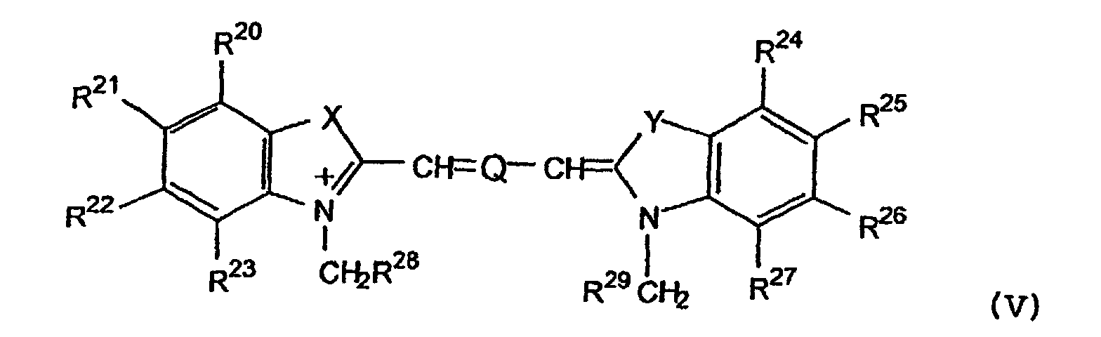
Q ein Fragmentoder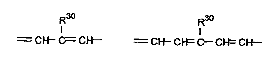 ist,
ist,
wobei R30 für ein Wasserstoffatom, eine Hydroxygruppe, eine Carboxygruppe, einen Alkoxyrest mit 1 bis 4 Kohlenstoffatomen oder ein Chloratom steht, b eine ganze Zahl 2 oder 3 bedeutet,
X und Y unabhängig voneinander für ein Fragment -O-, -S-, -CH=CH- oder -(CH2R32)(CH2R33)- stehen,
R20 bis R29, R32 und R33 unabhängig voneinander für ein Wasserstoffatom, eine Hydroxygruppe, einen Carboxy-, einen Sulfonsäure-Rest oder einen Carboxyalkyl-, Alkoxycarbonyl-, oder Alkoxyoxoalkyl-Rest mit bis zu 10 C-Atomen oder ein Sulfoalkylrest mit bis zu 4 C-Atomen,
oder für ein Polylysin, Polyethylenglycol, Methoxypolyethylenglycol, Polyvinylalkohol, Dextran, Carboxydextran oder eine kaskadenpolymerartige Struktur,
oder für einen Rest der allgemeinen Formel VI
mit der Maßgabe, daß bei der Bedeutung von X und Y gleich O, S, -CH=CH- oder -C(CH3)2mindestens einer der Reste R20 bis R29 einem Polylysin, Polyethylenglycol, Methoxypolyethylenglycol, Polyvinylalkohol, Dextran, Carboxydextran oder einer kaskadenpolymerartigen Struktur oder der allgemeinen Formel VI entspricht,
wobei
o und s gleich 0 sind oder unabhängig voneinander für eine ganze Zahl von 1 bis 6 stehen,
q und v unabhängig voneinander für 0 oder 1 stehen,
R34 ein Wasserstoffatom oder einen Methylrest darstellt,
R35 ein Alkylrest mit 3 bis 6 C-Atomen, welcher 2 bis n-1 Hydroxygruppen aufweist, wobei n die Anzahl der C-Atome ist, oder ein mit 1 bis 3 Carboxygruppen substituierter Alkylrest mit 1 bis 6 C-Atomen, Arylrest mit 6 bis 9 C-Atomen oder Arylalkylrest mit 7 bis 15 C-Atomen, oder ein Rest der allgemeinen Formel IIId oder IIIeist,
unter der Maßgabe, daß q für 1 steht,
oder ein Polylysin, Polyethylenglycol, Methoxypolyethylenglycol, Polyvinylalkohol, Dextran, Carboxydextran oder eine kaskadenpolymerartige Struktur bedeutet, oder
R20 und R21, R21 und R22, R22 und R23, R24 und R25, R25 und R26, oder R26 und R27 zusammen mit den zwischen ihnen liegenden Kohlenstoffatomen einen 5- oder 6-gliedrigen aromatischen oder gesättigten annelierten Ring bilden,
sowie deren physiologisch verträgliche Salze,
wobei die Cyaninfarbstoffe eine durch einen n-Octanol/Wasser-Verteilungskoeffizienten von kleiner 2,0 gekennzeichnete Hydrophilie besitzen. - Cyaninfarbstoff nach Anspruch 1, nämlich5-[2-[(1,2-Dicarboxyethyl)amino]-2-oxoethyl]-2-[7-[5-[2-[(1,2-dicarboxyethyl)amino]-2-oxoethyl]-1,3-dihydro-3,3-dimethyl-1-(4-sulfobutyl)-2H-indol-2-yliden]-1,3,5-heptatrienyl]-3,3-dimethyl-1-(4-sulfobutyl)-3H-indolium, inneres Salz, Monokaliumsalz,2-[7-[5-[2-[(11-Carboxy-2-oxo-1,4,7,10-tetraaza-4,7,10-tri(carboxymethyl)-1-undecyl)amino]-2-oxoethyl]-1,3-dihydro-3,3-dimethyl-1-ethyl-2H-indol-2-yliden]-1,3,5-heptatrienyl]-3,3-dimethyl-1-(4-sulfobutyl)-3H-indolium, inneres Salz,2-[7-[1,3-Dihydro-3,3-dimethyl-5-[2-[(Methoxypolyoxyethylen)-amino]-2-oxoethyl]-1-(4-sulfobutyl)-2Hindol-2-yliden]-1,3,5-heptatrienyl]-3,3-dimethyl-5-[2-[(methoxypolyoxyethylen)amino]-2-oxoethyl]-1-(4-sulfobutyl)-3H-indolium, Natriumsalz,2-[7-[1,3-Dihydro-3,3-dimethyl-1-(4-sulfobutyl)-2Hindol-2-yliden]-1,3,5-heptatrienyl]-3,3-dimethyl-5-(Methoxypolyoxyethylen)aminocarbonyl-1-(4-sulfobutyl)-3H-indolium, Natriumsalz3- (3-Carboxypropyl)-2-[7-[3-(3-carboxypropyl)-1,3-dihydro-3-methyl-1-(4-sulfobutyl)-2H-indol-2-yliden]-1,3,5-heptatrienyl]-3-methyl-1-(4-sulfobutyl)-3Hindolium, Natriumsalz,2-[7-[1,3-Dihydro-5-[2-[(2,3-dihydroxypropyl)amino]-2-oxoethyl] -3,3-dimethyl-1-(4-sulfobutyl) -2H-indol-2-yliden]-1,3,5-heptatrienyl]-5-[2-[(2,3-dihydroxypropyl)amino]-2-oxoethyl]-3,3-dimethyl-1-(4-sulfobutyl)-3H-indolium, Natriumsalz
- Verwendung von Cyaninfarbstoffen nach Anspruch 1 oder 2 zur Herstellung von Mitteln für die In-vivo-Diagnostik mittels NIR-Strahlung.
- Mittel zur In-vivo-Diagnostik, dadurch gekennzeichnet, daß es mindestens einen der Cyaninfarbstoffe nach Anspruch 1 oder 2 zusammen mit den üblichen Hilfs- und Trägerstoffen sowie Verdünnungsmitteln enthält.
Priority Applications (2)
| Application Number | Priority Date | Filing Date | Title |
|---|---|---|---|
| EP01250366A EP1181940B1 (de) | 1994-12-07 | 1995-10-10 | Verfahren zur In-vivo-Diagnostik mittels NIR-Strahlung |
| DK01250366T DK1181940T3 (da) | 1994-12-07 | 1995-10-10 | Fremgangsmåde til in-vivo diagnostik ved hjælp af NIR-stråling |
Applications Claiming Priority (3)
| Application Number | Priority Date | Filing Date | Title |
|---|---|---|---|
| DE4445065A DE4445065A1 (de) | 1994-12-07 | 1994-12-07 | Verfahren zur In-vivo-Diagnostik mittels NIR-Strahlung |
| DE4445065 | 1994-12-07 | ||
| PCT/DE1995/001465 WO1996017628A1 (de) | 1994-12-07 | 1995-10-10 | Verfahren zur in-vivo-diagnostik mittels nir-strahlung |
Related Child Applications (1)
| Application Number | Title | Priority Date | Filing Date |
|---|---|---|---|
| EP01250366A Division EP1181940B1 (de) | 1994-12-07 | 1995-10-10 | Verfahren zur In-vivo-Diagnostik mittels NIR-Strahlung |
Publications (2)
| Publication Number | Publication Date |
|---|---|
| EP0796111A1 EP0796111A1 (de) | 1997-09-24 |
| EP0796111B1 true EP0796111B1 (de) | 2003-04-23 |
Family
ID=6536117
Family Applications (2)
| Application Number | Title | Priority Date | Filing Date |
|---|---|---|---|
| EP01250366A Expired - Lifetime EP1181940B1 (de) | 1994-12-07 | 1995-10-10 | Verfahren zur In-vivo-Diagnostik mittels NIR-Strahlung |
| EP95935348A Expired - Lifetime EP0796111B1 (de) | 1994-12-07 | 1995-10-10 | Verfahren zur in-vivo-diagnostik mittels nir-strahlung unter verwendung von cyaninfarbstoffen |
Family Applications Before (1)
| Application Number | Title | Priority Date | Filing Date |
|---|---|---|---|
| EP01250366A Expired - Lifetime EP1181940B1 (de) | 1994-12-07 | 1995-10-10 | Verfahren zur In-vivo-Diagnostik mittels NIR-Strahlung |
Country Status (18)
| Country | Link |
|---|---|
| US (10) | US6083485A (de) |
| EP (2) | EP1181940B1 (de) |
| JP (4) | JPH10510250A (de) |
| KR (2) | KR100312939B1 (de) |
| CN (1) | CN1089008C (de) |
| AT (2) | ATE285254T1 (de) |
| AU (1) | AU709152B2 (de) |
| CA (1) | CA2205906C (de) |
| DE (3) | DE4445065A1 (de) |
| DK (2) | DK1181940T3 (de) |
| ES (2) | ES2236131T3 (de) |
| HU (1) | HUT77378A (de) |
| IL (4) | IL141384A (de) |
| NO (2) | NO319741B1 (de) |
| NZ (1) | NZ294568A (de) |
| PT (2) | PT1181940E (de) |
| WO (1) | WO1996017628A1 (de) |
| ZA (1) | ZA959707B (de) |
Cited By (8)
| Publication number | Priority date | Publication date | Assignee | Title |
|---|---|---|---|---|
| WO2011025950A2 (en) | 2009-08-28 | 2011-03-03 | Visen Medical, Inc. | Systems and methods for tomographic imaging in diffuse media using a hybrid inversion technique |
| WO2011038006A1 (en) | 2009-09-22 | 2011-03-31 | Visen Medical, Inc. | Systems and methods for virtual index-matching of diffusive media |
| WO2014062716A1 (en) | 2012-10-15 | 2014-04-24 | Visen Medical, Inc. | Systems, methods, and apparatus for imaging of diffuse media featuring cross-modality weighting of fluorescent and bioluminescent sources |
| US8927719B2 (en) | 2010-10-20 | 2015-01-06 | Li-Cor, Inc. | Cyanine dyes and their conjugates |
| WO2015103420A1 (en) | 2013-12-31 | 2015-07-09 | Memorial Sloan Kettering Cancer Center | Systems, methods, and apparatus for multichannel imaging of fluorescent sources in real time |
| WO2016100340A1 (en) | 2014-12-15 | 2016-06-23 | Memorial Sloan Kettering Cancer Center | Cyclic peptides with enhanced nerve-binding selectivity, nanoparticles bound with said cyclic peptides, and use of same for real-time in vivo nerve tissue imaging |
| WO2017106525A1 (en) | 2015-12-15 | 2017-06-22 | Memorial Sloan Kettering Cancer Center | Imaging systems and methods for tissue differentiation, e.g., for intraoperative visualization |
| WO2018102372A1 (en) | 2016-11-30 | 2018-06-07 | Memorial Sloan Kettering Cancer Center | Inhibitor-functionalized ultrasmall nanoparticles and methods thereof |
Families Citing this family (177)
| Publication number | Priority date | Publication date | Assignee | Title |
|---|---|---|---|---|
| DE4445065A1 (de) | 1994-12-07 | 1996-06-13 | Diagnostikforschung Inst | Verfahren zur In-vivo-Diagnostik mittels NIR-Strahlung |
| DE19539409C2 (de) * | 1995-10-11 | 1999-02-18 | Diagnostikforschung Inst | Kontrastmittel für die Nahinfrarot-Diagnostik |
| US5804389A (en) * | 1995-12-29 | 1998-09-08 | Phanos Technologies, Inc. | Method for detecting abnormal epithelial cell shedding |
| US5709845A (en) * | 1996-05-13 | 1998-01-20 | Rajagopalan; Raghavan | Tricyclic functional dyes for contrast enhancement in optical imaging |
| US5688966A (en) * | 1996-07-26 | 1997-11-18 | E. I. Du Pont De Nemours And Company | Compounds and method for synthesizing sulfoindocyanine dyes |
| DE19649971A1 (de) | 1996-11-19 | 1998-05-28 | Diagnostikforschung Inst | Optische Diagnostika zur Diagnostik neurodegenerativer Krankheiten mittels Nahinfrarot-Strahlung (NIR-Strahlung) |
| US6228344B1 (en) | 1997-03-13 | 2001-05-08 | Mallinckrodt Inc. | Method of measuring physiological function |
| US6280703B1 (en) | 1997-03-13 | 2001-08-28 | Mallinckrodt Inc. | Simultaneous multimodal measurement of physiological function |
| DE19717904A1 (de) * | 1997-04-23 | 1998-10-29 | Diagnostikforschung Inst | Säurelabile und enzymatisch spaltbare Farbstoffkonstrukte zur Diagnostik mit Nahinfrarotlicht und zur Therapie |
| GB9712524D0 (en) | 1997-06-16 | 1997-08-20 | Nycomed Imaging As | Method |
| US6393315B1 (en) * | 1997-09-12 | 2002-05-21 | Communaute Europeenne | Detecting and mapping of inflamed zones in a living tissue |
| WO1999013916A2 (en) * | 1997-09-18 | 1999-03-25 | Nycomed Imaging As | Methods and compositions for medical imaging |
| US6685895B1 (en) | 1997-12-17 | 2004-02-03 | Ethicon, Inc. | Method and apparatus for processing device with reduced occlusion |
| US6133445A (en) | 1997-12-17 | 2000-10-17 | Carnegie Mellon University | Rigidized trimethine cyanine dyes |
| US6645430B1 (en) | 1997-12-17 | 2003-11-11 | Ethicon, Inc. | Method and apparatus for processing device with fluid submersion |
| US6596232B1 (en) | 1997-12-17 | 2003-07-22 | Ethicon, Inc. | Device processing apparatus and method having positive pressure with two partitions to minimize leakage |
| ES2331671T3 (es) * | 1998-04-03 | 2010-01-12 | Mallinckrodt Inc. | Bioconjugados no covalentes utiles para diagnostico y terapia. |
| US7250517B2 (en) * | 1998-04-08 | 2007-07-31 | Ewald A. Terpetschnig | Luminescent compounds |
| US7411068B2 (en) * | 1998-04-08 | 2008-08-12 | Terpetschnig Ewald A | Luminescent compounds |
| US20030235846A1 (en) * | 1998-04-08 | 2003-12-25 | Terpetschnig Ewald A. | Luminescent compounds |
| US20030176663A1 (en) * | 1998-05-11 | 2003-09-18 | Eidgenossische Technische Hochscule | Specific binding molecules for scintigraphy |
| US6083486A (en) * | 1998-05-14 | 2000-07-04 | The General Hospital Corporation | Intramolecularly-quenched near infrared fluorescent probes |
| US6592847B1 (en) * | 1998-05-14 | 2003-07-15 | The General Hospital Corporation | Intramolecularly-quenched near infrared flourescent probes |
| US20030180221A1 (en) * | 1998-09-18 | 2003-09-25 | Schering Ag | Near infrared fluorescent contrast agent and fluorescence imaging |
| JP2000095758A (ja) * | 1998-09-18 | 2000-04-04 | Schering Ag | 近赤外蛍光造影剤および蛍光造影方法 |
| US7547721B1 (en) | 1998-09-18 | 2009-06-16 | Bayer Schering Pharma Ag | Near infrared fluorescent contrast agent and fluorescence imaging |
| US6652836B2 (en) | 1998-10-15 | 2003-11-25 | Fluoroprobe, Inc. | Method for viewing tumor tissue located within a body cavity |
| US6284223B1 (en) * | 1998-10-15 | 2001-09-04 | Fluoroprobe, Inc. | Method for viewing tumor tissue located within a body cavity |
| US6299860B1 (en) | 1998-10-15 | 2001-10-09 | Fluoro Probe, Inc. | Method for viewing diseased tissue located within a body cavity |
| SE9804328D0 (sv) * | 1998-12-15 | 1998-12-15 | Christer Busch | Contrast agent for facilitating ocular identification and inspection of lymph nodes |
| EP1131099A2 (de) * | 1999-01-15 | 2001-09-12 | Light Sciences Corporation | Nichtinvasiven vaskulären therapie |
| US6602274B1 (en) * | 1999-01-15 | 2003-08-05 | Light Sciences Corporation | Targeted transcutaneous cancer therapy |
| JP2002534483A (ja) * | 1999-01-15 | 2002-10-15 | ライト サイエンシーズ コーポレイション | 代謝性骨障害または骨転移のための治療的組成物 |
| CA2360229C (en) * | 1999-02-12 | 2007-05-08 | Alexei Ivanovich Trushin | Method for diagnosing proliferation regions and device for realising the same |
| DE19917713A1 (de) * | 1999-04-09 | 2000-10-19 | Diagnostikforschung Inst | Kurzkettige Peptid-Farbstoffkonjugate als Konstrastmittel für die optische Diagnostik |
| US6217848B1 (en) * | 1999-05-20 | 2001-04-17 | Mallinckrodt Inc. | Cyanine and indocyanine dye bioconjugates for biomedical applications |
| US20030114434A1 (en) * | 1999-08-31 | 2003-06-19 | James Chen | Extended duration light activated cancer therapy |
| DE19948650A1 (de) * | 1999-09-29 | 2001-07-19 | Diagnostikforschung Inst | Galenische Formulierungen |
| EP1267935A2 (de) * | 2000-01-12 | 2003-01-02 | Light Sciences Corporation | Behandlung von augenerkrankungen |
| US6180087B1 (en) | 2000-01-18 | 2001-01-30 | Mallinckrodt Inc. | Tunable indocyanine dyes for biomedical applications |
| US7790144B2 (en) | 2000-01-18 | 2010-09-07 | Mallinckrodt Inc. | Receptor-avid exogenous optical contrast and therapeutic agents |
| US6939532B2 (en) | 2000-01-18 | 2005-09-06 | Mallinckrodt, Inc. | Versatile hydrophilic dyes |
| US6395257B1 (en) | 2000-01-18 | 2002-05-28 | Mallinckrodt Inc. | Dendrimer precursor dyes for imaging |
| US7198778B2 (en) * | 2000-01-18 | 2007-04-03 | Mallinckrodt Inc. | Tumor-targeted optical contrast agents |
| US6180086B1 (en) * | 2000-01-18 | 2001-01-30 | Mallinckrodt Inc. | Hydrophilic cyanine dyes |
| US20080233050A1 (en) * | 2000-01-18 | 2008-09-25 | Mallinckrodt Inc. | Diagnostic and therapeutic optical agents |
| JP2003524018A (ja) * | 2000-02-24 | 2003-08-12 | アイトゲネーシシェ テクニシェ ホッホシューレ チューリッヒ | フィブロネクチンのed‐bドメインに特異的な抗体、前記抗体を含む複合体、および血管形成を検出および治療するためのその使用 |
| US7575761B2 (en) * | 2000-06-30 | 2009-08-18 | Novartis Pharma Ag | Spray drying process control of drying kinetics |
| US20020197700A1 (en) * | 2000-09-07 | 2002-12-26 | Schering Ag | Receptor of the EDb-fibronectin domains |
| US7597878B2 (en) * | 2000-09-19 | 2009-10-06 | Li-Cor, Inc. | Optical fluorescent imaging |
| DE10046215B4 (de) * | 2000-09-19 | 2004-04-15 | Institut für Chemo- und Biosensorik Münster e.V. i.Ins. | Fluorochrome und deren Verwendung |
| ATE315066T1 (de) * | 2000-09-19 | 2006-02-15 | Li Cor Inc | Cyaninfarbstoffe |
| NO20004795D0 (no) | 2000-09-26 | 2000-09-26 | Nycomed Imaging As | Peptidbaserte forbindelser |
| EP1801165B1 (de) * | 2000-09-29 | 2012-08-01 | Life Technologies Corporation | Modifizierte Carbocyanin-Farbstoffe und ihre Konjugate |
| WO2002026891A1 (en) * | 2000-09-29 | 2002-04-04 | Molecular Probes, Inc. | Modified carbocyanine dyes and their conjugates |
| US6663847B1 (en) * | 2000-10-13 | 2003-12-16 | Mallinckrodt Inc. | Dynamic organ function monitoring agents |
| US6656451B1 (en) * | 2000-10-16 | 2003-12-02 | Mallinckrodt, Inc. | Indole compounds as novel dyes for organ function monitoring |
| US6716413B1 (en) * | 2000-10-16 | 2004-04-06 | Mallinckrodt, Inc. | Indole compounds as tissue-specific exogenous optical agents |
| US6733744B1 (en) * | 2000-10-16 | 2004-05-11 | Mallinckrodt Inc. | Indole compounds as minimally invasive physiological function monitoring agents |
| US20070092450A1 (en) * | 2000-10-16 | 2007-04-26 | Mallinckrodt Inc. | Tissue-specific exogenous optical agents |
| US6669926B1 (en) * | 2000-10-16 | 2003-12-30 | Mallinckrodt, Inc. | Hydrophilic light absorbing indole compounds for determination of physiological function in critically ill patients |
| US20040180809A1 (en) * | 2000-10-16 | 2004-09-16 | Mallinckrodt Inc. | Tissue-specific exogenous optical agents |
| US6673334B1 (en) * | 2000-10-16 | 2004-01-06 | Mallinkcrodt, Inc. | Light sensitive compounds for instant determination of organ function |
| US7556797B2 (en) | 2000-10-16 | 2009-07-07 | Mallinckrodt Inc. | Minimally invasive physiological function monitoring agents |
| WO2002038190A2 (en) | 2000-10-27 | 2002-05-16 | Beth Israel Deaconess Medical Center | Non-isotopic detection of osteoblastic activity in vivo using modified bisphosphonates |
| US7383076B2 (en) * | 2000-11-27 | 2008-06-03 | The General Hospital Corporation | Fluorescence-mediated molecular tomography |
| US6984373B2 (en) | 2000-12-23 | 2006-01-10 | Dyax Corp. | Fibrin binding moieties useful as imaging agents |
| US20030044353A1 (en) * | 2001-01-05 | 2003-03-06 | Ralph Weissleder | Activatable imaging probes |
| AU2006200164B2 (en) * | 2001-04-09 | 2007-03-08 | Oncofluor, Inc. | Method for viewing tumor tissue located within a body cavity |
| US20030031627A1 (en) * | 2001-07-31 | 2003-02-13 | Mallinckrodt Inc. | Internal image antibodies for optical imaging and therapy |
| US20030105300A1 (en) | 2001-10-17 | 2003-06-05 | Mallinckrodt Inc. | Tumor targeted photodiagnostic-phototherapeutic agents |
| US6761878B2 (en) * | 2001-10-17 | 2004-07-13 | Mallinckrodt, Inc. | Pathological tissue detection and treatment employing targeted benzoindole optical agents |
| US20030152577A1 (en) * | 2002-02-07 | 2003-08-14 | Mallinckrodt Inc. | Dye-bioconjugates for simultaneous optical diagnostic and therapeutic applications |
| US7794693B2 (en) | 2002-03-01 | 2010-09-14 | Bracco International B.V. | Targeting vector-phospholipid conjugates |
| US20050100963A1 (en) | 2002-03-01 | 2005-05-12 | Dyax Corporation | KDR and VEGF/KDR binding peptides and their use in diagnosis and therapy |
| AU2003213730A1 (en) | 2002-03-01 | 2003-09-16 | Bracco International B.V. | Kdr and vegf/kdr binding peptides and their use in diagnosis and therapy |
| US7261876B2 (en) | 2002-03-01 | 2007-08-28 | Bracco International Bv | Multivalent constructs for therapeutic and diagnostic applications |
| US8623822B2 (en) | 2002-03-01 | 2014-01-07 | Bracco Suisse Sa | KDR and VEGF/KDR binding peptides and their use in diagnosis and therapy |
| US20060134001A1 (en) * | 2002-07-12 | 2006-06-22 | Frangioni John V | Conjugated infrared fluorescent substances for detection of cell death |
| US20040132092A1 (en) * | 2003-01-03 | 2004-07-08 | Stetson Christopher M. | Determining the density of functional moieties on polymer reagents |
| US7226577B2 (en) | 2003-01-13 | 2007-06-05 | Bracco Imaging, S. P. A. | Gastrin releasing peptide compounds |
| EP1585791B1 (de) * | 2003-01-24 | 2008-09-24 | Bayer Schering Pharma Aktiengesellschaft | Hydrophile, thiolreaktive cyaninfarbstoffe und deren konjugate mit biomolekülen für fluoreszenzdiagnose |
| WO2004078778A2 (en) | 2003-03-03 | 2004-09-16 | Dyax Corp. | PEPTIDES THAT SPECIFICALLY BIND HGF RECEPTOR (cMet) AND USES THEREOF |
| WO2005000218A2 (en) | 2003-05-31 | 2005-01-06 | Washington University | Macrocyclic cyanine and indocyanine bioconjugates provide improved biomedical applications |
| US7803624B2 (en) * | 2003-09-30 | 2010-09-28 | Cytyc Corporation | Automated cytological sample classification |
| US7750163B2 (en) * | 2003-10-31 | 2010-07-06 | GE Healthcare U.K. Limited | Cyanine dye labelling reagents |
| WO2005082423A2 (en) * | 2003-11-18 | 2005-09-09 | Beth Israel Deaconess Medical Center | Serum albumin conjugated to fluorescent substances for imaging |
| JP2005145921A (ja) * | 2003-11-19 | 2005-06-09 | Konica Minolta Medical & Graphic Inc | 診断用蛍光造影剤及び蛍光造影診断方法 |
| JP2005170812A (ja) * | 2003-12-09 | 2005-06-30 | Konica Minolta Medical & Graphic Inc | 蛍光造影剤及び蛍光造影方法 |
| US7682602B2 (en) * | 2003-12-19 | 2010-03-23 | Konica Minolta Medical & Graphic, Inc. | Near-infrared fluorescent contrast medium |
| JP4487180B2 (ja) * | 2004-03-18 | 2010-06-23 | ソニー株式会社 | 情報生成装置及び情報生成方法 |
| PL1765863T3 (pl) * | 2004-06-16 | 2012-05-31 | Ge Healthcare As | Związki oparte na peptydach |
| WO2006047452A2 (en) * | 2004-10-25 | 2006-05-04 | Anaspec, Inc. | Reactive 1,3’-crosslinked carbocyanines and their bioconjugates |
| ITMI20050328A1 (it) | 2005-03-03 | 2006-09-04 | Univ Degli Studi Milano | Composti peptidomimetrici e preparazione di derivati biologicamente attivi |
| US20100055040A1 (en) * | 2005-06-21 | 2010-03-04 | Periasamy Muthunadar P | Optical Imaging Contrast Agents |
| US8227621B2 (en) * | 2005-06-30 | 2012-07-24 | Li-Cor, Inc. | Cyanine dyes and methods of use |
| WO2007028118A2 (en) * | 2005-09-02 | 2007-03-08 | Visen Medical, Inc. | Nicotinic acid and picolinic acid derived near-infrared fluorophores |
| CA2621137C (en) * | 2005-09-02 | 2014-07-29 | Visen Medical, Inc. | Biocompatible n,n-disubstituted sulfonamide-containing fluorescent dye labels |
| DK1937676T3 (en) * | 2005-09-02 | 2017-03-06 | Visen Medical Inc | Biocompatible fluorescent imaging compounds |
| DK1792949T3 (da) * | 2005-12-05 | 2010-12-06 | Dyomics Gmbh | Hydrofil markør på basis af diastereoisomere cyaniner |
| DE102006029454A1 (de) * | 2005-12-05 | 2007-06-06 | Dyomics Gmbh | Hydrophile Marker auf Basis von diasteromeren |
| US20070148094A1 (en) * | 2005-12-22 | 2007-06-28 | Uzgiris Egidijus E | Polymeric imaging agents and medical imaging methods |
| EP1973575B1 (de) | 2005-12-22 | 2019-07-24 | Visen Medical, Inc. | Biokompatibele fluoreszierende metalloxide nanopartikel |
| WO2007088051A2 (en) | 2006-01-31 | 2007-08-09 | Bayer Schering Pharma Aktiengesellschaft | Modulation of mdl-1 activity for treatment of inflammatory disease |
| EP1815870A1 (de) * | 2006-02-01 | 2007-08-08 | DKFZ Deutsches Krebsforschungszentrum | Mit Metalchelaten verbundene Cyaninfarbstoffe zur bimodalen darstellenden Diagnose |
| WO2007097318A1 (ja) * | 2006-02-23 | 2007-08-30 | Adeka Corporation | 診断用マーカー |
| US20100022449A1 (en) * | 2006-03-09 | 2010-01-28 | Mallinckrodt Inc. | Receptor-avid exogenous optical contrast and therapeutic agents |
| US8921124B2 (en) | 2006-03-31 | 2014-12-30 | Wako Pure Chemical Industries, Ltd. | Pyrazole-based cyanine dye |
| US20070281363A1 (en) * | 2006-04-13 | 2007-12-06 | Ewald Terpetschnig | Luminescent compounds |
| US20080076188A1 (en) * | 2006-06-19 | 2008-03-27 | Patsenker Leonid D | Luminescent compounds |
| WO2008006006A1 (en) | 2006-07-03 | 2008-01-10 | Beth Israel Deaconess Medical Center, Inc. | Histology methods |
| US8155730B2 (en) | 2006-10-24 | 2012-04-10 | The Research Foundation Of State University Of New York | Composition, method, system, and kit for optical electrophysiology |
| EP2097438B1 (de) | 2006-12-11 | 2015-08-26 | Bracco Imaging S.p.A | Fibrinbindende peptidkonjugate für diagnostische und therapeutische anwendungen |
| DE102007002726A1 (de) * | 2007-01-18 | 2008-07-31 | Bayer Schering Pharma Aktiengesellschaft | Neue Kaskaden-Polymer-Komplexe, Verfahren zu ihrer Herstellung und diese enthaltende pharmazeutische Mittel |
| WO2008094637A2 (en) | 2007-01-30 | 2008-08-07 | Seta Biomedicals. Llc | Luminescent compounds |
| KR101444712B1 (ko) | 2007-01-31 | 2014-09-26 | 와코 쥰야꾸 고교 가부시키가이샤 | 게놈 dna 단편의 증폭 또는 결실의 검출방법 |
| JP5643514B2 (ja) | 2007-02-09 | 2014-12-17 | ビセン メディカル, インコーポレイテッド | ポリシクロ染料およびその使用 |
| WO2008109832A2 (en) * | 2007-03-08 | 2008-09-12 | Visen Medical, Inc. | Viable near-infrared fluorochrome labeled cells and methods of making and using same |
| JP2008261784A (ja) * | 2007-04-13 | 2008-10-30 | Olympus Corp | 生体観察方法 |
| EP2005970A1 (de) | 2007-06-22 | 2008-12-24 | Berlin Science Partners GmbH i.V. | Bildgebende Diagnostik durch Kombination von Kontrastmitteln |
| EP2197340A4 (de) | 2007-09-19 | 2015-10-21 | Oncofluor Inc | Verfahren zur darstellung und behandlung von organen und geweben |
| DE102007059752A1 (de) | 2007-12-10 | 2009-06-18 | Bayer Schering Pharma Aktiengesellschaft | Funktionalisierte, feste Polymernanopartikel enthaltend Epothilone |
| US9097667B2 (en) * | 2007-12-14 | 2015-08-04 | Biotium, Inc. | Fluorescent compounds |
| US8253725B2 (en) * | 2007-12-28 | 2012-08-28 | St. Jude Medical, Atrial Fibrillation Division, Inc. | Method and system for generating surface models of geometric structures |
| ES2658852T3 (es) | 2008-01-18 | 2018-03-12 | Visen Medical, Inc. | Agentes de imaginología fluorescentes |
| US20090214436A1 (en) | 2008-02-18 | 2009-08-27 | Washington University | Dichromic fluorescent compounds |
| WO2009104741A1 (ja) | 2008-02-22 | 2009-08-27 | 和光純薬工業株式会社 | β-グルカン及び/又はエンドトキシン測定用基質並びに測定方法 |
| EP2147684A1 (de) | 2008-07-22 | 2010-01-27 | Bracco Imaging S.p.A | Selektiv gegen Metallproteasen wirkende Diagnosemittel |
| US7951959B2 (en) | 2008-09-17 | 2011-05-31 | Thermo Fisher Scientific (Milwaukee) LLC | Hydrophilic labels for biomolecules |
| WO2010049042A2 (en) * | 2008-10-27 | 2010-05-06 | Sony Corporation | A dye comprising a chromophore to which an acyloin group is attached |
| WO2010057944A1 (en) * | 2008-11-20 | 2010-05-27 | Ge Healthcare As | Dye conjugate imaging agents |
| US8864821B2 (en) * | 2008-11-26 | 2014-10-21 | Visen Medical, Inc. | Methods and compositions for identifying subjects at risk of developing stent thrombosis |
| US20100143258A1 (en) * | 2008-12-05 | 2010-06-10 | General Electric Company | Tumor margin imaging agents |
| GB0823315D0 (en) | 2008-12-22 | 2009-01-28 | Ge Healthcare As | Fluorescent Probes |
| JP5500875B2 (ja) * | 2009-01-30 | 2014-05-21 | キヤノン株式会社 | 新規化合物、該新規化合物を用いたプローブ及び該新規化合物もしくは該プローブを用いた蛍光イメージング用造影剤 |
| RU2011138096A (ru) | 2009-03-19 | 2013-04-27 | Дженерал Электрик Компани | Агенты для оптической визуализации |
| CN102459469A (zh) * | 2009-04-17 | 2012-05-16 | 利康股份有限公司 | 用取代的花青染料进行荧光成像 |
| DE102010022110A1 (de) * | 2010-05-31 | 2011-12-01 | LMU Universität München Department Chemie | Cyaninfarbstoffe als Kontrastmittel zur Unterstützung der Augenchirurgie |
| US8623324B2 (en) | 2010-07-21 | 2014-01-07 | Aat Bioquest Inc. | Luminescent dyes with a water-soluble intramolecular bridge and their biological conjugates |
| ES2732832T3 (es) * | 2010-07-30 | 2019-11-26 | Smartdyelivery Gmbh | Procedimiento de medición para determinar una función de un órgano |
| GB201015806D0 (en) | 2010-09-21 | 2010-10-27 | Ge Healthcare As | Vascular imaging agents |
| US10053447B2 (en) | 2010-12-21 | 2018-08-21 | Pierce Biotechnology, Inc | Fluorescent compounds |
| CN110204478A (zh) | 2011-05-09 | 2019-09-06 | 文森医学公司 | 碳酸酐酶靶向剂及其使用方法 |
| US8889884B1 (en) | 2011-07-14 | 2014-11-18 | Pierce Biotechnology, Inc. | Phosphine derivatives of fluorescent compounds |
| US9249307B2 (en) | 2011-08-16 | 2016-02-02 | Pierce Biotechnology, Inc. | Benzocyanine compounds |
| GB201116733D0 (en) | 2011-09-28 | 2011-11-09 | Ge Healthcare As | Peptide margin imaging agents |
| EP2806781B1 (de) | 2012-01-23 | 2018-03-21 | Washington University | Brillenbildgebungssysteme und -verfahren |
| US9751868B2 (en) | 2012-02-28 | 2017-09-05 | Pierce Biotechnology, Inc. | Benzocyanine compounds |
| WO2013130761A1 (en) | 2012-03-02 | 2013-09-06 | Pierce Biotechnology, Inc. | Indole derivatives as labeling dye for biomolecule |
| WO2013141332A1 (ja) | 2012-03-22 | 2013-09-26 | 和光純薬工業株式会社 | インターカレーターを用いた変異遺伝子の識別検出方法 |
| JPWO2013141331A1 (ja) | 2012-03-22 | 2015-08-03 | 和光純薬工業株式会社 | マイクロサテライト領域を有するdnaの検出方法 |
| EP2831264B1 (de) | 2012-03-30 | 2019-07-10 | VisEn Medical, Inc. | Bakterielle bildgebungsmittel und verfahren zur verwendung davon |
| JP6398093B2 (ja) * | 2012-04-13 | 2018-10-03 | ベイカー ハート アンド ダイアベーツ インスティテュート | アテローム斑の検出 |
| US9592307B2 (en) | 2012-07-20 | 2017-03-14 | Canon Kabushiki Kaisha | Compound and photoacoustic imaging contrast medium containing the compound |
| EP2885006B1 (de) | 2012-08-15 | 2018-08-08 | VisEn Medical, Inc. | Prostataspezifische antigene und verfahren zu ihrer verwendung zur abbildung von prostatakrebs |
| EP2850078B1 (de) | 2012-08-28 | 2018-04-25 | Pierce Biotechnology, Inc. | Benzopyryliumverbindungen |
| WO2014144708A1 (en) | 2013-03-15 | 2014-09-18 | The Regents Of The University Of California | Peptides having reduced toxicity that stimulate cholesterol efflux |
| EP2970342B1 (de) | 2013-03-15 | 2024-01-24 | VisEn Medical, Inc. | Substituiertes silaxanthenium-rot für nahinfrarot-fluorchrome zur in-vitro- und in-vivo-bildgebung und -erkennung |
| AU2014228808C1 (en) | 2013-03-15 | 2018-12-20 | Visen Medical, Inc. | 4,4-disubstituted cyclohexyl bridged heptamethine cyanine dyes and uses thereof |
| US9353092B2 (en) | 2013-06-27 | 2016-05-31 | University Of Notre Dame Du Lac | Synthesis and use of croconaine compounds |
| WO2015038968A1 (en) | 2013-09-13 | 2015-03-19 | The General Hospital Corporation | Activatable fibrin-binding probes |
| US11001562B2 (en) | 2013-10-31 | 2021-05-11 | Beth Israel Deaconess Medical Center | Near-infrared fluorescent nerve contrast agents and methods of use thereof |
| WO2015066290A1 (en) | 2013-10-31 | 2015-05-07 | Beth Israel Deaconess Medical Center | Near-infrared fluorescent contrast bioimaging agents and methods of use thereof |
| US10022189B2 (en) | 2013-12-16 | 2018-07-17 | Stryker Sustainability Solutions, Inc. | Apparatus and method for cleaning an instrument |
| KR20150145468A (ko) | 2014-06-19 | 2015-12-30 | 김영미 | 숯 성형물의 제조방법 및 이에 의해 제조된 숯 성형물 |
| UA109751C2 (uk) * | 2014-09-26 | 2015-09-25 | Спосіб визначення гідрогелю в біологічних тканинах | |
| WO2016179350A1 (en) | 2015-05-06 | 2016-11-10 | Washington University | Compounds having rd targeting motifs and methods of use thereof |
| US9670318B2 (en) | 2015-05-28 | 2017-06-06 | Miltenyi Biotec Gmbh | Bright fluorochromes based on multimerization of fluorescent dyes |
| EP3098269B1 (de) | 2015-05-28 | 2022-04-06 | Miltenyi Biotec B.V. & Co. KG | Auf der multimerisierung von fluoreszenzfarbstoffen auf verzweigten polyethergerüsten basierende helle fluorchrome |
| US20190137396A1 (en) * | 2016-04-28 | 2019-05-09 | National University Corporation Nagoya University | Fluorescent probe, method for detecting fluorescence, and method for using fluorescent probe |
| KR101855395B1 (ko) * | 2016-09-26 | 2018-05-09 | 부경대학교 산학협력단 | 근적외선을 이용한 폐종양 스크리닝 방법 |
| EP3412303A1 (de) | 2017-06-08 | 2018-12-12 | Medizinische Universität Innsbruck | Verbessertes pharmakokinetik- und cholecystokinin-2-rezeptor (cck2r)-zielverfahren für diagnose und therapie |
| US11091646B2 (en) | 2017-08-14 | 2021-08-17 | Seta Biomedicals, Llc | Luminescent squaraine rotaxane compounds |
| DK3969063T3 (da) | 2019-05-13 | 2023-09-25 | Bracco Imaging Spa | Modificerede cyaninfarvestoffer og konjugater deraf |
| KR20210069945A (ko) | 2019-12-04 | 2021-06-14 | 삼성전자주식회사 | 생체정보 추산 장치 및 방법 |
| EP4072598A4 (de) | 2019-12-13 | 2024-02-21 | Washington University St Louis | Nahinfrarot-fluoreszierende farbstoffe, formulierungen und zugehörige verfahren |
| EP4015004A1 (de) | 2020-12-18 | 2022-06-22 | Phi Pharma SA | Proteoglykanspezifische verzweigte peptide |
Family Cites Families (29)
| Publication number | Priority date | Publication date | Assignee | Title |
|---|---|---|---|---|
| DE1522421A1 (de) * | 1967-01-17 | 1969-07-31 | Agfa Gevaert Ag | Optisch sensibilisiertes lichtempfindliches Material |
| JPS5552750A (en) * | 1978-10-12 | 1980-04-17 | Mochida Pharm Co Ltd | Laser knife* which can be detected* of tumor portion |
| JPS55116259A (en) * | 1979-03-01 | 1980-09-06 | Fuji Photo Film Co Ltd | Microimmunoassay method |
| US4541438A (en) * | 1983-06-02 | 1985-09-17 | The Johns Hopkins University | Localization of cancerous tissue by monitoring infrared fluorescence emitted by intravenously injected porphyrin tumor-specific markers excited by long wavelength light |
| JP3038339B2 (ja) * | 1988-05-02 | 2000-05-08 | ザイナクシス・テクノロジーズ・インコーポレーテッド | バイオ粒子の表面膜に対してバイオアフェクティング物質を結合する化合物 |
| DE3828360A1 (de) * | 1988-08-20 | 1990-02-22 | Stanowsky Alexander Dr | Farbstoff-markierter antitumor-antikoerper und verfahren zu seiner herstellung |
| DE3912046B4 (de) * | 1988-09-02 | 2004-03-25 | Carnegie Mellon University | Verfahren zum Markieren einer Komponente einer wäßrigen Flüssigkeit und lumineszenzphotostabiles Reaktionsprodukt |
| US5153112A (en) * | 1988-09-05 | 1992-10-06 | Konica Corporation | Method of processing silver halide photographic materials |
| SE8900612D0 (sv) * | 1989-02-22 | 1989-02-22 | Jonas Johansson | Vaevnadskarakterisering utnyttjande ett blodfritt fluorescenskriterium |
| US4945239A (en) * | 1989-03-29 | 1990-07-31 | Center For Innovative Technology | Early detection of breast cancer using transillumination |
| US5098379A (en) * | 1990-01-10 | 1992-03-24 | Rochester Medical Corporation | Catheter having lubricated outer sleeve and methods for making and using same |
| EP0529002B1 (de) * | 1990-05-15 | 2001-10-04 | Hyperion, Inc. | Fluoreszierende porphyrin- und fluoreszierende phthalocyanin-polyethylenglykol-, polyol- und saccharidderivate als fluoreszierende sonden |
| US5880287A (en) * | 1990-05-15 | 1999-03-09 | Hyperion, Inc. | Polyoxyhydrocarbyl related products and methods for fluorescence assays |
| WO1992000748A1 (en) * | 1990-07-06 | 1992-01-23 | Enzon, Inc. | Poly(alkylene oxide) amino acid copolymers and drug carriers and charged copolymers based thereon |
| DE4136769A1 (de) * | 1991-11-05 | 1993-05-06 | Humboldt-Universitaet Zu Berlin, O-1086 Berlin, De | Verfahren zum diagnostizieren von mit fluoreszierenden substanzen angereicherten, insbesondere tumoroesen gewebebereichen |
| US5298379A (en) * | 1992-06-30 | 1994-03-29 | Eastman Kodak Company | Radiation sensitive element with absorber dye to enhance spectral sensitivity range |
| DE4323368C2 (de) * | 1993-07-13 | 1998-01-15 | Domschke Wolfram | Verwendung einer Markersubstanz zur Unterscheidung von verändertem Gewebe in einer Umgebung von unverändertem Gewebe |
| US6238931B1 (en) * | 1993-09-24 | 2001-05-29 | Biosite Diagnostics, Inc. | Fluorescence energy transfer in particles |
| DE4445065A1 (de) * | 1994-12-07 | 1996-06-13 | Diagnostikforschung Inst | Verfahren zur In-vivo-Diagnostik mittels NIR-Strahlung |
| DE19539409C2 (de) * | 1995-10-11 | 1999-02-18 | Diagnostikforschung Inst | Kontrastmittel für die Nahinfrarot-Diagnostik |
| JP3169383B2 (ja) * | 1996-01-16 | 2001-05-21 | ルミゲン インク | ホスファターゼ酵素で化学発光を生成させるための化合物、組成物および方法 |
| DE19649971A1 (de) * | 1996-11-19 | 1998-05-28 | Diagnostikforschung Inst | Optische Diagnostika zur Diagnostik neurodegenerativer Krankheiten mittels Nahinfrarot-Strahlung (NIR-Strahlung) |
| DE19717904A1 (de) * | 1997-04-23 | 1998-10-29 | Diagnostikforschung Inst | Säurelabile und enzymatisch spaltbare Farbstoffkonstrukte zur Diagnostik mit Nahinfrarotlicht und zur Therapie |
| US7175953B2 (en) * | 1999-04-09 | 2007-02-13 | Institute Fuer Diagnostik Forschung | Short-warp peptide-dye conjugate as contrast agent for optical diagnostic |
| US6630570B1 (en) * | 1999-04-09 | 2003-10-07 | Insitut für Diagnostikforschung GmbH | Short-chain peptide-dye conjugates as contrast media for optical diagnosis |
| EP1088559A3 (de) * | 1999-09-29 | 2002-10-02 | INSTITUT FÜR DIAGNOSTIKFORSCHUNG GmbH AN DER FREIEN UNIVERSITÄT BERLIN | Galenische Formulierungen |
| JP2003261464A (ja) * | 2002-03-07 | 2003-09-16 | Fuji Photo Film Co Ltd | 近赤外蛍光造影剤および蛍光造影法 |
| US20060239916A1 (en) * | 2005-01-07 | 2006-10-26 | Kai Licha | Use of cyanine dyes for the diagnosis of proliferative diseases |
| EP1745739A3 (de) * | 2005-07-14 | 2009-04-22 | Bundesrepublik Deutschland, vertr. d.d. Bundes- ministerium f. Wirtschaft- und Technologie, dieses vertr. d.d. Präs. d. Phys.-Techn. Bundesanstalt | Optische Darstellung von rheumatischer Arthritis |
-
1994
- 1994-12-07 DE DE4445065A patent/DE4445065A1/de not_active Ceased
-
1995
- 1995-10-10 CA CA002205906A patent/CA2205906C/en not_active Expired - Fee Related
- 1995-10-10 DE DE59510658T patent/DE59510658D1/de not_active Expired - Lifetime
- 1995-10-10 AU AU37409/95A patent/AU709152B2/en not_active Expired
- 1995-10-10 JP JP8517228A patent/JPH10510250A/ja active Pending
- 1995-10-10 AT AT01250366T patent/ATE285254T1/de active
- 1995-10-10 HU HU9701797A patent/HUT77378A/hu not_active Application Discontinuation
- 1995-10-10 EP EP01250366A patent/EP1181940B1/de not_active Expired - Lifetime
- 1995-10-10 US US08/849,369 patent/US6083485A/en not_active Expired - Lifetime
- 1995-10-10 CN CN95196624A patent/CN1089008C/zh not_active Expired - Lifetime
- 1995-10-10 DE DE59510982T patent/DE59510982D1/de not_active Expired - Lifetime
- 1995-10-10 ES ES01250366T patent/ES2236131T3/es not_active Expired - Lifetime
- 1995-10-10 DK DK01250366T patent/DK1181940T3/da active
- 1995-10-10 NZ NZ294568A patent/NZ294568A/xx not_active IP Right Cessation
- 1995-10-10 DK DK95935348T patent/DK0796111T3/da active
- 1995-10-10 AT AT95935348T patent/ATE238071T1/de active
- 1995-10-10 KR KR1019970703597A patent/KR100312939B1/ko not_active IP Right Cessation
- 1995-10-10 PT PT01250366T patent/PT1181940E/pt unknown
- 1995-10-10 ES ES95935348T patent/ES2198446T3/es not_active Expired - Lifetime
- 1995-10-10 PT PT95935348T patent/PT796111E/pt unknown
- 1995-10-10 EP EP95935348A patent/EP0796111B1/de not_active Expired - Lifetime
- 1995-10-10 KR KR1020017002925A patent/KR100340290B1/ko not_active IP Right Cessation
- 1995-10-10 WO PCT/DE1995/001465 patent/WO1996017628A1/de active IP Right Grant
- 1995-11-15 ZA ZA959707A patent/ZA959707B/xx unknown
- 1995-11-16 IL IL141384A patent/IL141384A/en not_active IP Right Cessation
- 1995-11-16 IL IL11601895A patent/IL116018A0/xx not_active IP Right Cessation
-
1997
- 1997-06-02 NO NO19972509A patent/NO319741B1/no not_active IP Right Cessation
-
2000
- 2000-03-06 US US09/518,947 patent/US6258340B1/en not_active Expired - Lifetime
-
2001
- 2001-02-12 IL IL14138401A patent/IL141384A0/xx active IP Right Grant
- 2001-05-07 US US09/850,660 patent/US7025949B2/en not_active Expired - Lifetime
- 2001-05-14 JP JP2001143906A patent/JP3836687B2/ja not_active Expired - Lifetime
-
2002
- 2002-06-26 US US10/180,272 patent/US6926885B2/en not_active Expired - Lifetime
- 2002-12-30 IL IL15374502A patent/IL153745A0/xx unknown
-
2003
- 2003-02-19 US US10/368,997 patent/US6913743B2/en not_active Expired - Lifetime
-
2004
- 2004-12-16 US US11/013,596 patent/US20050106106A1/en not_active Abandoned
-
2005
- 2005-03-29 US US11/093,082 patent/US20050169844A1/en not_active Abandoned
- 2005-04-26 JP JP2005128309A patent/JP2005232190A/ja not_active Withdrawn
- 2005-07-01 NO NO20053239A patent/NO20053239D0/no not_active Application Discontinuation
-
2006
- 2006-03-22 US US11/387,200 patent/US7445767B2/en not_active Expired - Lifetime
- 2006-03-22 US US11/387,367 patent/US7655217B2/en not_active Expired - Fee Related
-
2009
- 2009-09-24 JP JP2009219451A patent/JP2010065036A/ja active Pending
-
2010
- 2010-01-26 US US12/693,749 patent/US20100129293A1/en not_active Abandoned
Cited By (14)
| Publication number | Priority date | Publication date | Assignee | Title |
|---|---|---|---|---|
| WO2011025950A2 (en) | 2009-08-28 | 2011-03-03 | Visen Medical, Inc. | Systems and methods for tomographic imaging in diffuse media using a hybrid inversion technique |
| WO2011038006A1 (en) | 2009-09-22 | 2011-03-31 | Visen Medical, Inc. | Systems and methods for virtual index-matching of diffusive media |
| US9248203B2 (en) | 2010-10-20 | 2016-02-02 | Li-Cor, Inc. | Fluorescent imaging with substituted cyanine dyes |
| US8927719B2 (en) | 2010-10-20 | 2015-01-06 | Li-Cor, Inc. | Cyanine dyes and their conjugates |
| US9089603B2 (en) | 2010-10-20 | 2015-07-28 | Li-Cor, Inc. | Fluorescent imaging with substituted cyanine dyes |
| US9408924B2 (en) | 2010-10-20 | 2016-08-09 | Li-Cor, Inc. | Bioconjugates of cyanine dyes |
| WO2014062716A1 (en) | 2012-10-15 | 2014-04-24 | Visen Medical, Inc. | Systems, methods, and apparatus for imaging of diffuse media featuring cross-modality weighting of fluorescent and bioluminescent sources |
| US10743768B2 (en) | 2012-10-15 | 2020-08-18 | Visen Medical, Inc. | Systems, methods, and apparatus for imaging of diffuse media featuring cross-modality weighting of fluorescent and bioluminescent sources |
| US11903675B2 (en) | 2012-10-15 | 2024-02-20 | Visen Medical, Inc. | Systems, methods, and apparatus for imaging of diffuse media featuring cross-modality weighting of fluorescent and bioluminescent sources |
| WO2015103420A1 (en) | 2013-12-31 | 2015-07-09 | Memorial Sloan Kettering Cancer Center | Systems, methods, and apparatus for multichannel imaging of fluorescent sources in real time |
| WO2016100340A1 (en) | 2014-12-15 | 2016-06-23 | Memorial Sloan Kettering Cancer Center | Cyclic peptides with enhanced nerve-binding selectivity, nanoparticles bound with said cyclic peptides, and use of same for real-time in vivo nerve tissue imaging |
| US10940216B2 (en) | 2014-12-15 | 2021-03-09 | Cornell University | Cyclic peptides with enhanced nerve-binding selectively, nanoparticles bound with said cyclic peptides, and use of same for real-time in vivo nerve tissue imaging |
| WO2017106525A1 (en) | 2015-12-15 | 2017-06-22 | Memorial Sloan Kettering Cancer Center | Imaging systems and methods for tissue differentiation, e.g., for intraoperative visualization |
| WO2018102372A1 (en) | 2016-11-30 | 2018-06-07 | Memorial Sloan Kettering Cancer Center | Inhibitor-functionalized ultrasmall nanoparticles and methods thereof |
Also Published As
Similar Documents
| Publication | Publication Date | Title |
|---|---|---|
| EP0796111B1 (de) | Verfahren zur in-vivo-diagnostik mittels nir-strahlung unter verwendung von cyaninfarbstoffen | |
| DE69911034T2 (de) | Im nahinfrarotbereich fluoreszierende kontrastmittel und fluoreszenzbildgebung | |
| DE60133626T2 (de) | Hydrophile zyaninfarbstoffe | |
| US7468177B2 (en) | Hydrophilic light absorbing compositions for determination of physiological function in critically ill patients | |
| US20070140962A1 (en) | Light Sensitive Compounds for Instant Determination of Organ Function | |
| DE60106389T2 (de) | Vielseitige hydrophile farbstoffe | |
| US20090263327A1 (en) | Minimally invasive physiological function monitoring agents | |
| EP1088559A2 (de) | Galenische Formulierungen | |
| AU779026B2 (en) | Antibody dye conjugates for binding to target structures of angiogenesis in order to intraoperatively depict tumor peripheries | |
| DE19948650A1 (de) | Galenische Formulierungen |
Legal Events
| Date | Code | Title | Description |
|---|---|---|---|
| PUAI | Public reference made under article 153(3) epc to a published international application that has entered the european phase |
Free format text: ORIGINAL CODE: 0009012 |
|
| AK | Designated contracting states |
Kind code of ref document: A1 Designated state(s): AT BE CH DE DK ES FR GB GR IE IT LI LU MC NL PT SE |
|
| 17P | Request for examination filed |
Effective date: 19970521 |
|
| 17Q | First examination report despatched |
Effective date: 20000413 |
|
| GRAG | Despatch of communication of intention to grant |
Free format text: ORIGINAL CODE: EPIDOS AGRA |
|
| RTI1 | Title (correction) |
Free format text: IN-VIVO DIAGNOSTIC PROCESS BY NEAR INFRARED RADIATION USING CYANINE DYES |
|
| RTI1 | Title (correction) |
Free format text: IN-VIVO DIAGNOSTIC PROCESS BY NEAR INFRARED RADIATION USING CYANINE DYES |
|
| GRAG | Despatch of communication of intention to grant |
Free format text: ORIGINAL CODE: EPIDOS AGRA |
|
| GRAH | Despatch of communication of intention to grant a patent |
Free format text: ORIGINAL CODE: EPIDOS IGRA |
|
| GRAH | Despatch of communication of intention to grant a patent |
Free format text: ORIGINAL CODE: EPIDOS IGRA |
|
| GRAA | (expected) grant |
Free format text: ORIGINAL CODE: 0009210 |
|
| RAP1 | Party data changed (applicant data changed or rights of an application transferred) |
Owner name: INSTITUT FUER DIAGNOSTIKFORSCHUNG GMBH AN DER FREI |
|
| AK | Designated contracting states |
Designated state(s): AT BE CH DE DK ES FR GB GR IE IT LI LU MC NL PT SE |
|
| REG | Reference to a national code |
Ref country code: GB Ref legal event code: FG4D Free format text: NOT ENGLISH |
|
| REG | Reference to a national code |
Ref country code: CH Ref legal event code: EP |
|
| REF | Corresponds to: |
Ref document number: 59510658 Country of ref document: DE Date of ref document: 20030528 Kind code of ref document: P |
|
| REG | Reference to a national code |
Ref country code: IE Ref legal event code: FG4D Free format text: GERMAN |
|
| GBT | Gb: translation of ep patent filed (gb section 77(6)(a)/1977) | ||
| REG | Reference to a national code |
Ref country code: DK Ref legal event code: T3 |
|
| REG | Reference to a national code |
Ref country code: SE Ref legal event code: TRGR |
|
| REG | Reference to a national code |
Ref country code: GR Ref legal event code: EP Ref document number: 20030402855 Country of ref document: GR |
|
| REG | Reference to a national code |
Ref country code: PT Ref legal event code: SC4A Free format text: AVAILABILITY OF NATIONAL TRANSLATION Effective date: 20030722 |
|
| REG | Reference to a national code |
Ref country code: ES Ref legal event code: FG2A Ref document number: 2198446 Country of ref document: ES Kind code of ref document: T3 |
|
| ET | Fr: translation filed | ||
| PLBE | No opposition filed within time limit |
Free format text: ORIGINAL CODE: 0009261 |
|
| STAA | Information on the status of an ep patent application or granted ep patent |
Free format text: STATUS: NO OPPOSITION FILED WITHIN TIME LIMIT |
|
| 26N | No opposition filed |
Effective date: 20040126 |
|
| PGFP | Annual fee paid to national office [announced via postgrant information from national office to epo] |
Ref country code: LU Payment date: 20121031 Year of fee payment: 18 |
|
| PGFP | Annual fee paid to national office [announced via postgrant information from national office to epo] |
Ref country code: DK Payment date: 20121025 Year of fee payment: 18 |
|
| PGFP | Annual fee paid to national office [announced via postgrant information from national office to epo] |
Ref country code: BE Payment date: 20121025 Year of fee payment: 18 Ref country code: IE Payment date: 20121025 Year of fee payment: 18 |
|
| PGFP | Annual fee paid to national office [announced via postgrant information from national office to epo] |
Ref country code: ES Payment date: 20121026 Year of fee payment: 18 |
|
| PGFP | Annual fee paid to national office [announced via postgrant information from national office to epo] |
Ref country code: MC Payment date: 20130918 Year of fee payment: 19 Ref country code: PT Payment date: 20130410 Year of fee payment: 19 |
|
| PGFP | Annual fee paid to national office [announced via postgrant information from national office to epo] |
Ref country code: AT Payment date: 20130919 Year of fee payment: 19 |
|
| BERE | Be: lapsed |
Owner name: *INSTITUT FUR DIAGNOSTIKFORSCHUNG G.M.B.H. AN DER Effective date: 20131031 |
|
| REG | Reference to a national code |
Ref country code: DK Ref legal event code: EBP Effective date: 20131031 |
|
| REG | Reference to a national code |
Ref country code: IE Ref legal event code: MM4A |
|
| PG25 | Lapsed in a contracting state [announced via postgrant information from national office to epo] |
Ref country code: BE Free format text: LAPSE BECAUSE OF NON-PAYMENT OF DUE FEES Effective date: 20131031 |
|
| PG25 | Lapsed in a contracting state [announced via postgrant information from national office to epo] |
Ref country code: DK Free format text: LAPSE BECAUSE OF NON-PAYMENT OF DUE FEES Effective date: 20131031 Ref country code: IE Free format text: LAPSE BECAUSE OF NON-PAYMENT OF DUE FEES Effective date: 20131010 |
|
| REG | Reference to a national code |
Ref country code: ES Ref legal event code: FD2A Effective date: 20141107 |
|
| PG25 | Lapsed in a contracting state [announced via postgrant information from national office to epo] |
Ref country code: ES Free format text: LAPSE BECAUSE OF NON-PAYMENT OF DUE FEES Effective date: 20131011 |
|
| PGFP | Annual fee paid to national office [announced via postgrant information from national office to epo] |
Ref country code: FR Payment date: 20141017 Year of fee payment: 20 Ref country code: SE Payment date: 20141029 Year of fee payment: 20 Ref country code: GR Payment date: 20141030 Year of fee payment: 20 Ref country code: GB Payment date: 20141027 Year of fee payment: 20 Ref country code: DE Payment date: 20141029 Year of fee payment: 20 Ref country code: CH Payment date: 20141027 Year of fee payment: 20 |
|
| PGFP | Annual fee paid to national office [announced via postgrant information from national office to epo] |
Ref country code: NL Payment date: 20141026 Year of fee payment: 20 |
|
| PGFP | Annual fee paid to national office [announced via postgrant information from national office to epo] |
Ref country code: IT Payment date: 20141028 Year of fee payment: 20 |
|
| REG | Reference to a national code |
Ref country code: PT Ref legal event code: MM4A Free format text: LAPSE DUE TO NON-PAYMENT OF FEES Effective date: 20150410 |
|
| PG25 | Lapsed in a contracting state [announced via postgrant information from national office to epo] |
Ref country code: MC Free format text: LAPSE BECAUSE OF NON-PAYMENT OF DUE FEES Effective date: 20141031 |
|
| REG | Reference to a national code |
Ref country code: AT Ref legal event code: MM01 Ref document number: 238071 Country of ref document: AT Kind code of ref document: T Effective date: 20141010 |
|
| PG25 | Lapsed in a contracting state [announced via postgrant information from national office to epo] |
Ref country code: LU Free format text: LAPSE BECAUSE OF NON-PAYMENT OF DUE FEES Effective date: 20131010 Ref country code: PT Free format text: LAPSE BECAUSE OF NON-PAYMENT OF DUE FEES Effective date: 20150410 |
|
| PG25 | Lapsed in a contracting state [announced via postgrant information from national office to epo] |
Ref country code: AT Free format text: LAPSE BECAUSE OF NON-PAYMENT OF DUE FEES Effective date: 20141010 |
|
| REG | Reference to a national code |
Ref country code: DE Ref legal event code: R071 Ref document number: 59510658 Country of ref document: DE |
|
| REG | Reference to a national code |
Ref country code: NL Ref legal event code: MK Effective date: 20151009 |
|
| REG | Reference to a national code |
Ref country code: CH Ref legal event code: PL |
|
| REG | Reference to a national code |
Ref country code: PT Ref legal event code: MM4A Free format text: MAXIMUM VALIDITY LIMIT REACHED Effective date: 20151010 |
|
| REG | Reference to a national code |
Ref country code: GB Ref legal event code: PE20 Expiry date: 20151009 |
|
| REG | Reference to a national code |
Ref country code: SE Ref legal event code: EUG |
|
| PG25 | Lapsed in a contracting state [announced via postgrant information from national office to epo] |
Ref country code: GB Free format text: LAPSE BECAUSE OF EXPIRATION OF PROTECTION Effective date: 20151009 |
|
| REG | Reference to a national code |
Ref country code: GR Ref legal event code: MA Ref document number: 20030402855 Country of ref document: GR Effective date: 20151011 |
|
| PG25 | Lapsed in a contracting state [announced via postgrant information from national office to epo] |
Ref country code: PT Free format text: LAPSE BECAUSE OF EXPIRATION OF PROTECTION Effective date: 20151019 |
|
| PG25 | Lapsed in a contracting state [announced via postgrant information from national office to epo] |
Ref country code: PT Free format text: LAPSE BECAUSE OF EXPIRATION OF PROTECTION Effective date: 20141010 |