EP0874242A1 - Device and apparatus for the simultaneous detection of multiple analytes - Google Patents
Device and apparatus for the simultaneous detection of multiple analytes Download PDFInfo
- Publication number
- EP0874242A1 EP0874242A1 EP98303019A EP98303019A EP0874242A1 EP 0874242 A1 EP0874242 A1 EP 0874242A1 EP 98303019 A EP98303019 A EP 98303019A EP 98303019 A EP98303019 A EP 98303019A EP 0874242 A1 EP0874242 A1 EP 0874242A1
- Authority
- EP
- European Patent Office
- Prior art keywords
- substrate
- analyte
- ligands
- biological
- reaction
- Prior art date
- Legal status (The legal status is an assumption and is not a legal conclusion. Google has not performed a legal analysis and makes no representation as to the accuracy of the status listed.)
- Granted
Links
- WGCSJKKDPVPKTF-BRFYHDHCSA-N CN(C)N(C)[C@H](C1)C1NN(C)OC Chemical compound CN(C)N(C)[C@H](C1)C1NN(C)OC WGCSJKKDPVPKTF-BRFYHDHCSA-N 0.000 description 1
Images


Classifications
-
- G—PHYSICS
- G01—MEASURING; TESTING
- G01N—INVESTIGATING OR ANALYSING MATERIALS BY DETERMINING THEIR CHEMICAL OR PHYSICAL PROPERTIES
- G01N33/00—Investigating or analysing materials by specific methods not covered by groups G01N1/00 - G01N31/00
- G01N33/48—Biological material, e.g. blood, urine; Haemocytometers
- G01N33/50—Chemical analysis of biological material, e.g. blood, urine; Testing involving biospecific ligand binding methods; Immunological testing
- G01N33/53—Immunoassay; Biospecific binding assay; Materials therefor
- G01N33/543—Immunoassay; Biospecific binding assay; Materials therefor with an insoluble carrier for immobilising immunochemicals
- G01N33/551—Immunoassay; Biospecific binding assay; Materials therefor with an insoluble carrier for immobilising immunochemicals the carrier being inorganic
- G01N33/552—Glass or silica
-
- G—PHYSICS
- G01—MEASURING; TESTING
- G01N—INVESTIGATING OR ANALYSING MATERIALS BY DETERMINING THEIR CHEMICAL OR PHYSICAL PROPERTIES
- G01N33/00—Investigating or analysing materials by specific methods not covered by groups G01N1/00 - G01N31/00
- G01N33/48—Biological material, e.g. blood, urine; Haemocytometers
- G01N33/50—Chemical analysis of biological material, e.g. blood, urine; Testing involving biospecific ligand binding methods; Immunological testing
- G01N33/53—Immunoassay; Biospecific binding assay; Materials therefor
- G01N33/543—Immunoassay; Biospecific binding assay; Materials therefor with an insoluble carrier for immobilising immunochemicals
Definitions
- This invention relates to a device and apparatus for the simultaneous detection of multiple analytes.
- Automation of analytical methods has generally focused on batch and random access analysers, where multiple analysis on individual test samples is performed using sequential individual test methods. This necessitates the use of multiple packs of individual test kits.
- analysis requires employment of several types of equipment, e.g. clinical chemistry analysers, HPLC, GCMS, automated immunoassay instruments or atomic absorption instruments.
- a multi-analyte system should involve a means of providing simultaneous analysis of several analytes in a test sample. This analysis should provide results which identify individual analytes and enable the quantitation of each individual analyte in that test sample.
- a method of multi-analyte analysis is often claimed but the given criteria are generally not both fulfilled.
- a typical substrate contains a plurality of individual test reaction sites each possessing a different binding ligand.
- the test sample contacts each of the reaction zones and thereafter a range of detection techniques is implemented to identify the analyte present. It is important that the detection method used enables quantitation of each individual analyte.
- the general protocol is: irradiation of first sites, incubation of irradiated substrate with a first ligand to be immobilised, washing to remove loosely bound ligand, blocking unreacted sites activated by the irradiation step, and irradiation of regions where the second biological ligand is to be immobilised, with subsequent steps repeated as for the first ligand.
- the spatial resolution is dictated by controlling the site of irradiation, either by controlling the site of irradiation by means of a coherent UV light source from a laser or by a number of physical masks and an incoherent light source. This makes the task of immobilising a plurality of biological ligands a time-consuming process.
- Another disadvantage of the photolithographic approach is the need for expensive physical masks or a laser light source. Further, there is a high degree of non-specific binding.
- WO-A-9516204 describes a photolithographic approach to reducing the problems associated with high non-specific binding.
- the surface linking molecule was avidin and the photolabile molecule was photobiotin or a derivative thereof. Whilst reduced non-specific binding is claimed, this technique still requires the time-consuming sequences outlined above. Immobilisation of a plurality of 20 separate biological ligands would require a total of 80 steps, assuming the basic requirement of irradiation, binding, blocking and washing steps for each separate ligand to be immobilised.
- a cross-linker used in many publications has been glutaraldehyde.
- This linker presents many disadvantages, including the tendency of proteins to cross-link which is likely to alter the function of the protein.
- a further disadvantage is that the coupling procedure should include a reduction step which is time-consuming and potentially very hazardous, e.g. if sodium cyanoborohydride is used as the reducing agent.
- Heterobifunctional linkers have been used but in many cases these involve the need for free sulphydryl groups on the protein to be bound. This necessitates modification of the protein prior to immobilisation.
- Multi-analyte assays have been available for antibiotics, for example. These are largely based on microbial inhibition assays, where an antibiotic present in the sample inhibits bacterial growth and forms a zone of clearance which is proportional to the concentration of the antibiotic present in the sample. However, this method cannot provide any indication as to the identity of the antibiotic, or an accurate determination of its concentration. Microbial inhibition methods are also very slow, the complete process taking several days.
- the Triage device is for the simultaneous detection of a panel of seven drugs of abuse in human urine. Each device is only capable of analysing one urine sample. At the end of the procedure, the operator visually examines each of the drug-specific test zones for the presence of a red bar. All steps of the assay protocol must be performed manually by the operator. There is also no hard-copy of the test result available.
- the Advisor device is similar in its application to that of the Triage device.
- the Advisor device screens for five different classes of drugs of abuse.
- the device operates using agglutination assay principles, with individual channels for each drug. All steps of the assay protocol are performed by the operator. Negative samples have agglutinated particles, whereas positive drug samples provide an unaggregated pattern of particles.
- dark substrates such as silicon give so-called black body effects.
- a dark substrate may absorb the incident excitation light energy, thus diminishing light emission from the fluorophore.
- a device for performing multi-analyte assays comprises a substrate and a multiplicity of discrete reaction sites each bearing a ligand covalently bound to the substrate, and in which the surface of the substrate between the reaction sites is inert with respect to analyte.
- This invention thus provides a solid state, multi-analyte device which exhibits little or no non-specific binding.
- a device of the invention may be prepared by activating the surface of a suitable substrate, and applying an array of ligands on to discrete sites on the surface. If desired, the other active areas may be blocked.
- the ligands may be applied in an aqueous system, and it is preferred if the other areas are rendered hydrophobic.
- the ligands may be bound to the substrate via a linker. In particular, it is preferred that the activated surface is reacted successively with an organosilane, a bifunctional linker and the ligand.
- bifunctional cross-linkers have been used to provide highly efficient coupling chemistries between organosilanes covalently immobilised on microfabricated silicon or ceramic substrates. Biological ligands can thus be immobilised in multi-analyte arrays.
- test reagent kits removes the need for multiple individual test reagent kits and also several instrument types, thereby facilitating the simultaneous detection of multiple analyses on a single sample.
- the invention provides a single integrated analyser capable of providing simultaneous detection of a wide range of chemistries.
- test reagents may be supplied in a combined format for a particular panel of analytes.
- a plurality of biological ligands is immobilised in a spatially-defined pattern of spots or lines by means of microfluidic dispensing of the ligand onto a chemically-activated substrate.
- the biological ligand is covalently attached to the substrate.
- the coupling efficiency of the biological ligand can be such that the chemical reaction is completed within a few minutes.
- the immobilisation procedure can ensure that the biological ligand retains its biological activity both in the short term and in the long term.
- This invention also provides an integrated analyser system for the simultaneous detection of a wide range of analytes in a multi-analyte format.
- the analyser system is designed for maximum end-user convenience, with the capability of obtaining multi-analyte identification and quantitation from each test sample.
- the preferred analyser system is a combination of a X-Y translational platform, a sample handling unit, liquid handling/flow control means, a temperature-controlled dark box, a CCD camera, and image-processing software.
- the platform may be associated with a stepper motor, to achieve a positioned accuracy of, say, 1 0 ⁇ m, for positioning device(s) at each stage of the analytical procedure.
- the substrate that is used in a device of this invention may be, for example, of silicon, quartz, glass or ceramic.
- Ceramic substrates aluminium oxide
- a ceramic substrate may be manufactured to provide a range of grain sizes (1 to 30 ⁇ m).
- the preferred particle size of the ceramic substrate used in this invention is less than 20 ⁇ m, preferably less than 10 ⁇ m.
- the reduced particle size imparts much improved surface uniformity which in turn provides enhanced performance of biological assays.
- Other important features of ceramic substrates include surface topography tolerance, porosity, vacuum-tightness and zero absorption of water.
- the preferred ceramic material consists of 94% alumina (Al 2 O 3 ) with a particle size in the range of 4-8 ⁇ m.
- the material is vacuum-tight, and has a surface topography of 0.6 to 0.8 ⁇ m when ground.
- the surface uniformity can be improved by a polishing process, to yield a surface with variation of 0.4-0.5 ⁇ m.
- a further improvement is achieved by lapping and polishing, to yield a surface with a variability of 0.05-0.1 ⁇ m.
- Suitable silicon substrates are produced with an oxide film of an exact thickness, e.g. a tolerance of ⁇ 2nm for a 100 nm oxide film.
- the oxide film may be 50-500 nm thick, preferably less than 200 nm, more preferably in the region of 100 nm.
- the substrate may be formed as part of a solid-state microfabricated micromachined device, developed for a wide range of panel tests for veterinary and clinical diagnostics applications.
- Each solid-state test device has an array of reaction regions.
- Each reaction region is specific for an individual analyte.
- the reaction region may be in the form of a spot, channel, dimple, pit, well or chamber.
- the reaction regions are manufactured by immobilising biological molecules onto the substrate.
- a device of the invention is up to 1 cm 2 in area.
- the area of each reaction site will usually be less than 1 mm 2 .
- the solid substrate is preferably fabricated to provide an intricate network of ports, chambers, channels, wells, dimples etc. It may also be advantageous to create pillars within the channel or well. Such irregularities can help to achieve maximum surface area interactions between bound biological ligands and test reagents, greatly reducing the incubation times for competitive immunoassays and sandwich immunoassays alike.
- the surface may also be microfabricated to incorporate nanolitre to microlitre mixing chambers/reservoirs/channels.
- the silicon surface is first oxidised to form an oxide layer.
- a layer of photoresist is then deposited from which the desired pattern is created.
- the photoresist is removed.
- the silicon is then etched, e.g. using HF, and oxide film removed. Finally, the oxide film is grown uniformly over the entire silicon wafer.
- step (i) a silicon wafer 1 is oxidised to provide an oxide layer 2; in step (ii), a photoresist layer 3 is deposited; in step (iii), the application of light provides a patterned oxide layer; in step (iv), the photoresist is removed; in step (v), the wafer 1 is etched; in step (vi), the oxide film is removed; and in step (vii), a continuous oxide film 2a is reformed.
- Covalent immobilisation of the biological ligands is preferred. Passive adsorption interactions may be used, but are susceptible to changes in pH, temperature and ionic strength, and may in some instances result in release of weakly-bound ligands during incubation and washing steps, thus contributing to poor assay reproducibility. It is of course desirable that the biological ligand retains maximum activity, after the immobilisation procedure.
- the surface of the substrates Prior to any chemical activation, the surface of the substrates should be thoroughly cleaned.
- the first step preferably involves cleaning of the surface by sonication in an alkaline detergent, followed by exhaustive washing with double-deionised water.
- the substrates are then treated with a chromic acid solution.
- the chromic acid solution both further cleans the surface and opens surface epoxide groups, as shown in Fig. 2.
- the epoxide groups may also be opened by other means, e.g. sonication for 1 hour.
- a sequence of reactions comprises the use of an organosilane, then a (hetero)bifunctional cross-linker Z-R-Y, to form a highly reactive intermediate, and finally a functionalised ligand, to give covalent immobilisation.
- Organosilanes may be chosen to provide either a reactive terminal group, capable of forming a covalent bond with a biological molecule, or a less reactive moiety such as NH 2 where further activation with a bifunctional linker is necessary to provide an appropriate end group.
- Organosilanes possessing terminal electrophilic functional groups do not require activation with a bifunctional cross-linker, since biological ligands can be immobilised covalently through nucleophilic groups on the biological ligand.
- any of a multitude of bifunctional cross-linkers may then be used to provide a very reactive chemical group through which a biological molecule or ligand can be covalently attached.
- This invention includes the use of bifunctional linkers which can be used in the mass production of chemically-activated substrates and are sufficiently stable to permit long-term storage prior to covalent attachment of the biological molecule or binder ligand.
- Preferred linkers are inert to normal atmospheric conditions whilst also being sufficiently reactive to form covalent bonds with functional groups of the biological ligand to be immobilised in a very short time period (typically ⁇ 10 minutes).
- the bifunctional linker may be, for example, phosgene, thiophosgene, N,N-disuccinimidyl carbonate, xylylenediamine, 1 ,6-diaminohexane, 1,12-diaminododecane, 1,6-diisocyanatohexane, 1,12-diisocyanatododecane, 1,4-phenylenedithioisocyanate, cyanuric chloride, terephthaldehyde, p-nitrobenzoyl chloride, sulfanilic acid, 2-fluoromethylpyridinium p-toluenesulfonate, 3-aminophenylboronic acid, p-bromophenylboronic acid, diethyl pyrocarbonate, ethyl chloroformate, p-bromoaniline, p-bromophenyl hydrazide, p-bro
- a photolabile cross-linker may be used to react with an organosilane having a nucleophilic or electrophilic terminal group.
- the cross-linker is, for example, the N-hydroxysuccinimide of p-azidobenzoic acid or p-aminobenzophenone.
- the diameter of the latex particles is preferably less than 500 nm, and more preferably less than 150 nm.
- the latex particles may be incubated at a concentration of approximately 0.5 to 1 % w/v with substrates modified with the appropriate organosilane, with or without the presence of a bifunctional linker as described above.
- Fig. 4 shows two reaction schemes for the immobilisation of latex. Either may be followed by activation of the latex with a second linker, and immobilisation of a biological molecule, or direct covalent immobilisation of, say, an antibody.
- polystyrene latex particles An alternative to the use of polystyrene latex particles is the covalent immobilisation of biological ligands to polyethylene glycol derivatives already anchored on a silanated substrate.
- PEG derivatives with two electrophilic groups such as epoxy or carbonylimidazole are reacted with a silane having a terminal -NH 2 group, such as APTES, on a substrate of choice.
- a suitable reaction sequence is shown in Fig. 5.
- Organosilanes that are suitable for direct biological ligand attachment may possess halide, epoxy, isocyanato, aldehyde or tosylate functional groups.
- Organosilanes possessing electrophilic groups e.g., glycidoxy
- Organosilanes possessing electrophilic groups also have the advantage of being less susceptible to polymerisation during the silanation procedure due to the absence of nucleophilic groups available for attacking the methoxy or ethoxy function of the organosilane. Therefore, the substrate surface should not contain polymerised organosilane.
- the chemistry of the surfaces provides a means of achieving spatial resolution by virtue of the rapid kinetics of the formation of covalent bonds between the surface chemical functional group and a suitable chemical group present in a sterically favourable position on the biological molecule to be immobilised.
- the biological molecule is preferably presented to the surface of the substrate by a microfluidic dispenser in the form of an individual droplet or series of droplets which form a line.
- the volume dispensed is of the order of 1 to 100 nl, preferably less than 50 nl, e.g. closer to 10 nl.
- the rapidity of the formation of the covalent bonds is such that covalent immobilisation is achieved in minutes, before the dispensed droplet or line evaporates on the surface.
- the positional accuracy to which the droplet or line of liquid is delivered should have a tolerance of ⁇ 20 ⁇ m.
- the present invention overcomes the problems associated with conventional photolithography, by enabling the formation of spatially distinct spots of biological ligands, with no requirement for UV light or physical masks.
- spatial resolution may be achieved by microdispensing techniques.
- An important factor is the rapid kinetics of the covalent coupling reaction, to ensure highly efficient coupling of the biological ligand in a spatially distinct region, eliminating the lateral digression of the immobilised biological ligand.
- Unreacted chemical moieties on the substrate may then be blocked, e.g. using blocking molecules known to those skilled in the art.
- Suitable such molecules include proteins such as casein, bovine serum albumin, lactalbumin etc. or low molecular weight blockers such as glycine, glutamine etc.
- Photolabile linkers can also be used.
- the organosilane on the surface of the substrate is reacted in the dark with a photolabile linker (e.g. benzophenone, arylazides etc.)
- a photolabile linker e.g. benzophenone, arylazides etc.
- the surface is then spotted with the biological ligands as desired, and covalent attachment is achieved following a short period of irradiation with UV light or a longer period with visible light.
- the remaining regions of the substrate surface are blocked, using blockers similar to those described molecules as above, in the presence of UV or visible light.
- the substrate-immobilised biological molecules may be stabilised, e.g. by incubation in a sugar solution (e.g. trehalose) for a short time (1 hour), followed by drying at 37°C for 16 hours.
- the stabilised substrates may then be sealed in foil pouches with desiccant and stored.
- the immobilised biological molecules are stable for more than 6 or 12 months, e.g. up to and beyond 2 years when stored at + 2 to + 8°C.
- the devices may be placed in a range of different carriers which incorporate features which control the efficiency of mixing of test reagents.
- the flow of liquid test reagents may be achieved by capillary attraction, centrifugal force, vacuum force or electroosmotic flow.
- the use of electroosmotic flow may avoid the need for valves, so that no moving mechanical parts are used.
- Closed channels may be formed by bonding a glass plate to the microfabricated surface.
- the biological molecules are covalently immobilised on the surface prior to bonding the glass plate.
- Many bonding procedures e.g. anodic bonding, involve elevated temperatures that may destroy a biological molecule. Therefore, bonding techniques should be non-denaturing, to immobilised biological molecules.
- One suitable method is indirect bonding, e.g. where the wafer is bonded to a glass plate by a suitable glue, e.g. epoxy glue.
- the devices may then be placed in a suitable carrier.
- a suitable carrier Various such carriers are illustrated in Figs. 7 and 8.
- the dimensions of the device shown in Fig. 8 are 48.62 mm x 48.62 mm, including wells having an internal diameter of 10 mm and an external diameter of 12.82 mm.
- the centre-to-centre spacing of the wells is 15.36 mm.
- the devices may incorporate features to enhance mixing of test reagents, samples etc. This is illustrated in Figure 9, where the device includes a reagent addition site 11, reagent channels 12, and test reaction sites 13.
- the device includes reagent reservoirs 21, a delivery manifold 22, reagent delivery channel test sites 23, and a waste reservoir 24.
- Fig. 11 shows a 4-channel test structure with similar parts.
- This invention provides a completely integrated system for the simultaneous, quantitative detection of analytes of a wide range of molecular weights, structural diversity and polarity. Analyte panels are available as appropriate for clinical/veterinary diagnosis or drug screening.
- binding ligands are chosen accordingly. This is within the skill and knowledge of those in the art. Suitable analytes include:
- one panel is for the detection of sulphonamide antibiotics.
- This invention provides a method for the simultaneous quantitative identification of, say, up to 20 individual sulphonamides.
- Other examples include cardiac, fertility and infectious disease panels.
- sample matrices that may be tested include serum, plasma, urine, bile, faeces, tissue, water and feed.
- the volume of sample required is very low, typically ⁇ 1.5 ⁇ l/analyte.
- the test reagents e.g. enzyme-labelled antibodies, enzyme-labelled haptens, fluorescently-labelled antibodies or fluorescently-labelled haptens, may be all contained in a single reagent reservoir, dramatically reducing the liquid-handling requirements.
- sandwich assays e.g. of Luteinising Hormone, Follicle-Stimulating Hormone, prolactin, Thyroid-Stimulating Hormone etc
- the sample is added along with an assay buffer and incubated for a short period which is typically less than 30, and preferably less than 10, minutes.
- the cocktail of labelled detecting antibodies is added and incubated for a further period of time. This period is again typically less than 30, and preferably less than 10, minutes.
- the device is then washed, to remove any unbound label, and the signal quantified.
- rheumatoid factor interference may be achieved by contacting the test sample to an area of immobilised immunoglobulin, for example prior to the test sample contacting the reaction region.
- HAMA Human Anti-Mouse Antibodies
- these antibodies can cause severe problems in the performance of assays utilising monoclonal mouse antibodies.
- the traditional solution is to include expensive additives in test reagents to counteract the problem.
- ligands may be provided over part of the device, that bind contaminants. This is especially valuable where defined spreading is allowed on the surface of the device, e.g. in channels. The capability of removing components that interfere enhances the accuracy of the results generated.
- the detection labels may also be immobilised on the surface of dendrimer molecules.
- the dendrimer molecules are polymeric in nature, synthesised by the repetitive coupling of small building molecules. They are commercially available from Aldrich Chemicals in a range of molecular weights with a choice of terminal functional groups e.g. NH 2 or COOH. Heterobifunctional linkers can then be used in conjunction with conventional coupling chemistry to prepare the detecting label conjugates.
- a small dendrimer preferably no more than 16 surface groups
- a large dendrimer typically more than 64 surface groups).
- the small molecular weight hapten (less than 1,000 Dalton) is coupled to the chemical groups on the small dendrimer followed by covalent attachment of the detecting label.
- the dendrimer conjugate may be purified by dialysis and gel permeation chromatography.
- the test reagents contain multiple components (e.g. enzyme-labelled antibodies, fluorescent-labelled antibodies, latex-immobilised antibodies, dendrimer antibody-fluorophore conjugates, dendrimer antibody-fluorophore conjugates, dendrimer antibody-enzyme conjugates, enzyme-labelled haptens, fluorescent-labelled haptens, etc) as appropriate for particular panels of tests.
- the panels of tests possible are very diverse and can be chosen on the basis of clinical diagnosis (or veterinary diagnosis) as appropriate. For example, a desirable panel is for the detection of infectious diseases (e.g. hepatitis, HIV, syphilis, etc). Other panels include fertility hormones, cardiac markers, allergy proteins, etc. As well as clinical parameters, there is also the ability to detect large panels of drug residues.
- the present invention permits the identification of individual compounds such as antibiotics. For example, a quantitative result can be obtained for up to 20 antibiotics on a device of surface area of 1 cm 2 simultaneously, in a time frame of minutes typically, with a sensitivity superior to that for HPLC/GCMS methods and comparable to that for conventional single parameter enzyme immunoassays. This approach may be easily extended to anabolic steroids, beta-agonists, beta-blockers, pesticides, therapeutic drugs etc.
- the detection system is preferably a charge-coupled device (CCD) camera equipped to measure both fluorescent and chemiluminescent light. Briefly, the CCD camera collects the light signal generated from the test areas on the microfabricated device and converts this into relative light units (RLUs).
- CCD charge-coupled device
- Fluorescent-based detection systems may be read directly, using appropriate optical filters for the labelling fluorophore.
- a suitable chemiluminescent reagent is luminol, which can be analysed at a wavelength of 433-445 nm. Chemiluminescence may also be observed, based on detecting alkaline phosphatase-labelled biological molecules using 1,2-dioxetane.
- this invention preferably utilises a chemiluminescent detection system, using a CCD.
- a back-illuminated camera is preferred, to improve the capture efficiency at the wavelength of the light generated by the chemiluminescent light reaction (approximately 433-445 nm in the case of luminol).
- the whole system may be operated by a personal computer where a specifically designed programme controls the X-Y table, dispenser unit, sample handling, temperature control, incubation times and the CCD camera.
- Figures 12-14 show the organisation of such a system.
- Fig. 12 illustrates schematically the interaction of a personal computer (PC) having two control units 31,32.
- Unit 31 is in communication with a CCD imaging system represented at 33.
- Unit 32 is in communication with a dispenser unit and an X-Y translation table with sample tray represented at 34 and 35, respectively.
- Fig. 13 is a schematic representation of the X-Y translation table. This drawing shows a sample platform 41 mounted on a linear actuator 42. X-Y translation is under the control of stepper motors 43,44 connected to respective drives 45,46. Translation is limited by "home position" sensors 47,48.
- the sensitivity of labelled biological molecules and certain unlabelled biological molecules to light may make it necessary to perform the assays in the absence of light.
- the absence of light is achieved by constructing the case in a light-tight manner.
- the light-tight environment is also preferably temperature-controlled, e.g. within ⁇ 0.2°C or, preferably, ⁇ 0.1°C, to ensure satisfactory assay precision and accuracy.
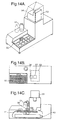
- Fig. 14 shows the apparatus in perspective, part plan and cut-away side views.
- Fig. 14A shows reagent storage container 51, a light-tight door 52 and a camera body 53 with a removable cover 54.
- the major part of the camera can be external to the casing.
- the camera lens is placed in an aperture in the casing.
- Fig. 14B shows in outline the samples on a sample tray 55 and, in outline, waste area 56 and an imaging area 57.
- the camera 53 is positioned over these areas.
- Fig. 14C shows, in addition to the container 51, camera 53, X-Y table 41 and stepper motor 43, a dispenser pump 58.
- the design of the system shown in Fig. 14 is based on 3x3 sample rack holders of which 20 can be held at any one time. This means that, if 20 individual reaction regions are located on each 1 cm 2 of microfabricated device, a total of 3600 analyses may be performed simultaneously on a single sample. Alternatively, 180 samples may be analysed simultaneously for 20 different test parameters.
- the analyte may be labelled.
- the ligand may also be labelled, allowing analysis by fractional occupancy.
- ceramic substrates (1 cm x 1 cm) were ultrasonically cleaned using an alkaline detergent (RBS35, 5% v/v) followed by double deionised water and then placed in 6M HCI for 16 hours. The chips were then placed in chromic acid for 1 hour in an ultrasonic bath.
- alkaline detergent RBS35, 5% v/v
- the substrates were washed exhaustively with double deionised water and acetone and then dried in an oven at 120°C for 2 hours. Following this pretreatment, the substrates were silanated using the organosilane ⁇ -glycidoxypropyl trimethoxysilane (10% v/v) in anhydrous toluene, 4-dimethylaminopyridine (1.25 g/L) and triethylamine (1% v/v). This mixture was refluxed for 4 hours and then left overnight at room temperature. The substrates were washed with toluene and acetone before curing for 4 hours at 120°C.
- the substrates were placed in containers and stored at room temperature until required for spotting of sulphonamide antibodies.
- the sulphonamide antibodies were spotted using a BIODOT XY3000 dispenser.
- the 12 sulphonamides assayed were sulphadoxine, sulphamethizole, sulphachloropyridazine, sulphamethoxypyridazine, sulphamerazine, sulphapyridine, sulphisoxazole, sulphathiazole, sulphamethazine, sulphaquinoxaline, sulphadimethoxine, and sulphadiazine.
- Dispensed volumes of approx. 20 nl were employed for each sulphonamide antibody.
- the 12 sulphonamide antibodies which formed 12 discrete areas on the 1 cm 2 substrate were incubated for 2 hours at 37°C.
- the substrates were washed with phosphate-buffered saline (PBS) (pH 7.2) containing 2% casein (w/v) and then blocked in same buffer overnight at +2-80C. After washing with PBS containing PEG300 (0.05% v/v), the devices were placed in a carrier.
- PBS phosphate-buffered saline
- Multi-sulphonamide standards 200 ⁇ l
- a cocktail of sulphonamide horseradish peroxidase conjugates 100 ⁇ l
- the standards contained 5, 10, 50 and 100 ng/ml for each of the 12 sulphonamides.
- a multi-analyte assay was performed for 3 hormones of large molecular weight, i.e. Prolactin (PL), Follicle-Stimulating Hormone (FSH) and Luteinising Hormone (LH).
- PL Prolactin
- FSH Follicle-Stimulating Hormone
- LH Luteinising Hormone
- Example 1 The chemical pretreatment and silanation procedures were exactly as described in Example 1. Individual PL, FSH or LH monoclonal antibodies (approx. 20 nl antibody dispensed) were immobilised on discrete areas of the chemically modified substrate. The multi-analyte assays were performed on both silicon and ceramic substrates with an epoxide surface as described in Example 1.
- 150 ⁇ l of a multiple LH/PL/FSH serum-based standard and 150 ⁇ l of a diluent assay buffer were added to the device and incubated for 15 minutes at room temperature.
- 300 ⁇ l of a single conjugate cocktail of LH-HRPO/PL-HRPO/FSH-HRPO conjugates was added and incubated for 15 minutes.
- the devices were washed to remove excess reagents and the chemiluminescent reagent [luminol (1.4 mM)/urea hydrogen peroxide (9.6 mM)] was introduced.
- the devices were imaged using a CCD camera with an exposure time of up to 4 minutes. Standard curves for each of the hormones were plotted after the images were processed.
- Example 11 In contrast to Example 1, a multi-sulphonamide assay has also been conducted using microchannels.
- the device is illustrated in Figure 11.
- the reagent addition reservoirs 21 are 2 mm x 2 mm, and 300 ⁇ m deep (vol. 1.2 ⁇ l)
- the channels 23 are each 5 mm long, 200 ⁇ m wide and 100 ⁇ m deep (vol. 100 nl)
- the reservoir 24 is 1.9 mm x 8.6 mm, and 300 ⁇ m deep (vol. 4.9 ⁇ l).
- Example 1 The chemical modification of the surface was performed as described in Example 1. Antibody was added to each of the channels and incubated for 2 hours at 37°C. The substrates were then blocked and washed as for Example 1.
- a multi-sulphonamide standard 200 ⁇ l
- sulphonamide horseradish pexoxidase conjugates 100 ⁇ l
- the standards contained 10 or 100 ng/ml of all the sulphonamides as appropriate.
- chemiluminescent reagent [luminol (1.4 mM)/urea hydrogen peroxide (9.6 mM)] was added.
- the devices were imaged using a CCD camera with an exposure time of up to 4 minutes.
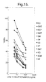
- the % B/Bo values for the 4 sulphonamide curves are given in Table 1.
- Sulphonamide %B/Bo 0 ng/ml 10 ng/ml 100 ng/ml Sulphamethazine 100 14 7 Sulphamethoxypyridazine 100 28 15 Sulphaquinoxaline 100 56 24 Sulphamerazine 100 40 22
- Mouse IgG was detected using anti-mouse HRPO conjugate using chemiluminescent detection by CCD camera.
- the silicon or ceramic substrates having immobilised benzophenone photolabile linker should not bind mouse IgG when reacted in the absence of light. However, non-specific binding is occurring, since approximately 80% of the grey mean RLU achieved when the mouse IgG binding is performed under UV light is due to passive binding interactions.
- An array of biological molecules immobilised through covalent interactions according to this invention is demonstrably more distinct.
- ceramic substrates silanated with APTES were reacted with a dihydrazide linker.
- Sulphonamide antibodies were treated with sodium periodate to render them reactive towards the hydrazide linker.
- Control sulphonamide antibodies were simply dialysed against sodium acetate buffer pH 5.5.
- the RLU results are shown in Tables 4 (not treated) and 5 (treated by periodate method). The results clearly show that the hydrazide linker has been successfully linked to the ceramic surface.
- the control antibodies (no periodate activation) gave very poor standard curves when compared to the covalently immobilised sulphonamide antibodies.
- the covalent method results in superior results compared to the passive method.
- the results for the passive method can be increased approximately 2-fold by acid pretreatment of the sulphonamide antibodies, but this is still inferior to the covalent approach.
- the atomic composition results show a very good conversion of the parent silicon and ceramic substrates with APTES organosilane, with good reproducibility in surface composition indicated for the two areas tested on each sample.
- the FITC-labelled substrates showed 70% and 77% labelling of silicon and ceramic respectively.
- the performance of ceramic provided superior results to silicon using this fluorescent detection method. Further, the problems of the dark body effect on silicon using fluorescence may be solved by employing chemiluminescence as the detection method. In a comparison between identical assays for FSH performed on silicon and ceramic, using chemiluminescent detection, the former required an exposure time 2-fold longer than that for ceramic to achieve the same RLU value.
Abstract
Description
| Sulphonamide | %B/Bo | ||
| 0 ng/ | 10 ng/ | 100 ng/ | |
| Sulphamethazine | |||
| 100 | 14 | 7 | |
| | 100 | 28 | 15 |
| | 100 | 56 | 24 |
| | 100 | 40 | 22 |
| Mouse IgG | Irradiation By UV lamp (10 minutes) | Grey Mean (RLU) | |
| √ | √ | 22368 | 24022 |
| √ | X | 17586 | 20531 |
| [Biotin-LC-NHS] | APTES Substrate RLU (1 second exposure) | Control Substrate RLU (1 second exposure) |
| 0 | 2,448 | |
| 100 µg/ml | 8,881 | NS |
| 500 µg/ml | 7,922 | NS |
| NS = No fluorescent signal detected. |
| SULPHONAMIDE | 0 ng/ml | 10 ng/ml | % B/Bo | 100 ng/ml | % B/Bo | |
| Sulphadoxine | 2825 | 1456 | 51 | NS | - | |
| Sulphamethizole | 3531 | 1257 | 36 | NS | - | |
| Sulpachloropyridazine | 6476 | 1585 | 24 | NS | - | |
| Sulphamethoxypyridazine | 1099 | 928 | 84 | NS | - | |
| Sulphamerazine | 2177 | 1137 | 52 | 1224 | 56 | |
| Sulphasoxazole | 4879 | 1108 | 23 | NS | - | |
| Sulphathiazole | 2932 | 814 | 28 | NS | - | |
| Sulphamethazine | NS | NS | - | NS | - | |
| Sulphaquinoxaline | 1041 | 828 | 79 | 968 | 93 | |
| Sulphadimethoxine | 804 | NS | - | 781 | 97 |
| SULPHONAMIDE | 0 ng/ml | 10 ng/ml | % B/Bo | 100 ng/ml | % B/Bo | |
| Sulphadoxine | 10520 | 3240 | 31 | 2135 | 20 | |
| Sulphamethizole | 17141 | 5689 | 33 | 4882 | 28 | |
| Sulphachloropyridazine | 24944 | 7565 | 30 | 2096 | 8 | |
| Sulphamethoxypyridazine | 14082 | 10509 | 74 | 5687 | 40 | |
| Sulphamerazine | 12594 | 5521 | 43 | 3240 | 26 | |
| Sulphasoxazole | 24419 | 6686 | 27 | 2270 | 9 | |
| Sulphathiazole | 14279 | 4602 | 32 | 2353 | 16 | |
| Sulphamethazine | 3644 | 2810 | 77 | 2213 | 61 | |
| Sulphaquinoxaline | 10575 | 6112 | 58 | 5588 | 53 | |
| Sulphadimethoxine | 5526 | 2554 | 46 | 1983 | 36 |
| Sulphonamide | Percentage of Sulphonamide Antibody Binding | |
| Covalent Interactions | Passive Interactions | |
| Sulphadoxine | 73.1 | 26.9 |
| Sulphamethizole | 79.4 | 20.6 |
| Sulphachloropyridazine | 74.0 | 26.0 |
| Sulphamethoxypyridazine | 92.2 | 7.8 |
| Sulphamerazine | 82.7 | 17.3 |
| Sulphasoxazole | 80.0 | 20.0 |
| Sulphathiazole | 79.4 | 20.6 |
| Sulphamethazine | - | - |
| Sulphaquinoxoline | 90.2 | 9.8 |
| Sulphadimethoxine | 85.4 | 14.6 |
| Mean | 81.8 | 18.2 |
| Sulphonamide | RLU | |||
| Covalent | Passive | |||
| 0 | 10 ng/ml | 0 | 10 ng/ml | |
| Sulphadoxine | 32756 | 11131 | 1950 | 904 |
| Sulphamethizole | 39020 | 11132 | 2782 | 1359 |
| Sulphachloropyridazine | 39632 | 8434 | 4410 | 1051 |
| Sulphamethoxypyridazine | 29489 | 13408 | 1793 | 770 |
| Sulphamerazine | 28455 | 11077 | 2011 | 988 |
| Sulphisoxazole | 38486 | 5774 | 4083 | 1031 |
| Sulphathiazole | 28837 | 8087 | 2010 | 675 |
| Sulphamethazine | 11331 | 7535 | 802 | 574 |
| Sulphaquinoxaline | 13838 | 8716 | 951 | 548 |
| Sulphadimethoxine | 13062 | 5832 | 910 | 581 |
| Covalent Immobilisation: Direct spotting on to glycidoxy silane surface. Passive Immobilisation: Direct spotting on to dichlorodimethylsilane-reacted surface. |
| Sample | Area | C | O | Si | Al | N | Cl | |
| Silicon substrate | ||||||||
| 1 | 21.9 | 52.7 | 25.4 | - | - | - | - | |
| (untreated) | 2 | 23.0 | 51.0 | 26.0 | - | - | - | - |
| | 1 | 55.5 | 23.3 | 10.5 | - | 10.2 | 0.5 | - |
| silanated with | 2 | 55.6 | 22.5 | 10.9 | - | 10.5 | 0.5 | - |
| | 1 | 51.3 | 25.6 | 13.0 | - | 7.2 | 2.9 | - |
| 2 | 52.5 | 25.0 | 12.3 | - | 7.5 | 2.7 | - | |
| | 1 | 58.7 | 25.0 | 9.6 | - | 6.1 | 0.5 | - |
| substrate treated with FITC | 2 | 58.8 | 24.7 | 9.8 | - | 6.0 | 0.8 | - |
| | 1 | 27.2 | 46.3 | 11.3 | 13.3 | - | 1.4 | 0.5 |
| (untreated) | 2 | 27.1 | 46.6 | 9.6 | 14.8 | - | 1.1 | 0.7 |
| | 1 | 47.0 | 31.6 | 13.4 | 2.6 | 5.4 | - | - |
| silanated with APTES | 2 | 45.9 | 31.7 | 13.7 | 3.3 | 5.3 | - | - |
| | 1 | 52.0 | 29.3 | 11.3 | 2.4 | 5.0 | - | - |
| substrate treated with FITC | 2 | 51.0 | 30.6 | 11.7 | 2.3 | 4.5 | - | - |
| Substrate | CCD Exposure Time (sec) | Grey Mean | Number of Stripped FITC Molecules/ | |
| Side | ||||
| 1 | Side 2 | |||
| Ceramic | 0.1 | 7483 | 7063 | 4.548x10¹⁵ |
| Silicon | 10 | 753 | 612 | 1.412x10¹⁶ |
| [Prolactin] Standard | RLU | |
| Silicon Substrate | Ceramic Substrate | |
| 550 MIU/ml | 814 | 8594 |
| 2200 MIU/ml | 799 | 16735 |
Claims (21)
- A solid state device for performing multi-analyte assays, comprising a substrate and a multiplicity of discrete reaction sites each bearing a ligand covalently bonded to the substrate, wherein the surface of the substrate between the reaction sites is inert with respect to analyte.
- A device according to claim 1, wherein the surface of the substrate is non-uniform, whereby an enhanced signal can be obtained from ligand-analyte interaction.
- A device according to claim 2, which comprises an array of reaction channels, ridges, pillars, spots, chambers, dimples, wells or pits.
- A device according to any preceding claim, wherein the substrate is of ceramic, glass, quartz or silicon.
- A device according to any preceding claim, which has area of less than 1 cm2.
- A device according to any preceding claim, wherein the area of each reaction site is less than 1 mm2.
- A device according to any preceding claim, obtainable by a process of activating the surface of the substrate, and applying an array of ligands on to discrete areas on the surface.
- A device according to claim 7, wherein the process additionally comprises blocking other active areas.
- A device according to claim 7 or claim 8, wherein the process comprises rendering the other areas hydrophobic, and the ligands are applied in water.
- A device according to any of claims 7 to 9, wherein application of the ligands comprises an initial step of contacting the activated surface with an organosilane.
- A device according to claim 10, wherein the organosilane has the formula (RO)3Si-(CH2)n-X, wherein each R is a hydrocarbyl group, n is an integer, and X is a functional group.
- A device according to claim 10 or claim 11, wherein the process includes the use of a bifunctional cross-linker to facilitate covalent attachment of biological ligands to the organosilane.
- A device according to any of claims 10 to 12, wherein a photolabile cross-linker is used to react with the organosilane having a nucleophilic or electrophilic terminal group.
- A device according to any of claims 1 to 8, obtainable by immobilisation of the ligands on the surface derivatised with macromolecules such as polystyrene latex particles, dendrimers or polyethylene glycol containing chemical groups which facilitate covalent attachment of the ligands.
- A device according to any preceding claim, which additionally comprises ligands that bind materials whose presence interferes with assaying an analyte.
- Use of a device according to any preceding claim, for a multi-analyte assay.
- Use according to claim 16, which comprises observing the array of sites at which an analyte is bound and/or not bound, and correlating that information with the ligands.
- Use according to claim 16 or claim 17, which comprises imaging with a Charge-Coupled Device (CCD) of a back-illuminated type suitable for the detection of light from a chemiluminescent light reaction where the wavelength of light to be detected is less than 450 nm.
- Use according to any of claims 16 to 18, wherein the analytes are selected from antibiotics, hormones, markers of cardiac damage, markers of infectious disease, allergy markers, drugs of abuse enzymes, viruses, nucleotides and peptides.
- Use according to any of claims 16 to 19, wherein the analytes are in a sample of whole blood, serum, plasma, urine, faeces, bile, tissue or feed.
- A system for multi-analyte analysis, comprising a device according to any of claims 1 to 15, a translational platform, a sample-handling system, a liquid reagent delivery system, a temperature-controlled dark box, a CCD camera and image-processing apparatus.
Priority Applications (1)
| Application Number | Priority Date | Filing Date | Title |
|---|---|---|---|
| EP19980303019 EP0874242B2 (en) | 1997-04-21 | 1998-04-20 | Device and apparatus for the simultaneous detection of multiple analytes |
Applications Claiming Priority (3)
| Application Number | Priority Date | Filing Date | Title |
|---|---|---|---|
| EP97302707 | 1997-04-21 | ||
| EP97302707 | 1997-04-21 | ||
| EP19980303019 EP0874242B2 (en) | 1997-04-21 | 1998-04-20 | Device and apparatus for the simultaneous detection of multiple analytes |
Publications (3)
| Publication Number | Publication Date |
|---|---|
| EP0874242A1 true EP0874242A1 (en) | 1998-10-28 |
| EP0874242B1 EP0874242B1 (en) | 2005-02-23 |
| EP0874242B2 EP0874242B2 (en) | 2009-06-03 |
Family
ID=26147400
Family Applications (1)
| Application Number | Title | Priority Date | Filing Date |
|---|---|---|---|
| EP19980303019 Expired - Lifetime EP0874242B2 (en) | 1997-04-21 | 1998-04-20 | Device and apparatus for the simultaneous detection of multiple analytes |
Country Status (1)
| Country | Link |
|---|---|
| EP (1) | EP0874242B2 (en) |
Cited By (30)
| Publication number | Priority date | Publication date | Assignee | Title |
|---|---|---|---|---|
| WO1999017120A1 (en) * | 1997-09-26 | 1999-04-08 | Becton, Dickinson And Company | Preparing conjugates using polyethylene glycol linkers |
| FR2777355A1 (en) * | 1998-04-10 | 1999-10-15 | Bio Merieux | PROCESS FOR FIXING A BIOLOGICAL MOLECULE ON THE SURFACE OF A SUPPORT CONSISTING OF SILICA OR METAL OXIDE |
| US6341182B1 (en) | 1997-09-11 | 2002-01-22 | Randox Laboratories Ltd. | Method and apparatus for analyzing an image |
| WO2003087823A1 (en) * | 2002-04-12 | 2003-10-23 | Micronas Gmbh | Method for immobilizing molecules on surfaces |
| WO2004017070A2 (en) * | 2002-07-22 | 2004-02-26 | Micronas Holding Gmbh | Sensor surface comprising an improved signal-noise ratio |
| US6720157B2 (en) | 2000-02-23 | 2004-04-13 | Zyomyx, Inc. | Chips having elevated sample surfaces |
| EP1454177A1 (en) * | 2001-12-11 | 2004-09-08 | Autogenomics, Inc. | Multi-substrate biochip unit |
| US6905816B2 (en) | 2000-11-27 | 2005-06-14 | Intelligent Medical Devices, Inc. | Clinically intelligent diagnostic devices and methods |
| EP1770429A2 (en) * | 2001-12-11 | 2007-04-04 | Autogenomics, Inc. | Multi-substrate biochip unit |
| KR100585548B1 (en) * | 1997-04-21 | 2007-07-06 | 란독스 래브러토리스 리미티드 | Device for simultaneous detection of multiple analytes |
| US7276283B2 (en) | 2004-03-24 | 2007-10-02 | Wisconsin Alumni Research Foundation | Plasma-enhanced functionalization of carbon-containing substrates |
| EP1481094B1 (en) * | 2002-02-28 | 2009-09-30 | febit holding GmbH | Increasing the sensitivity and specificity of nucleic acid chip hybridization tests |
| US7723126B2 (en) | 2004-03-24 | 2010-05-25 | Wisconsin Alumni Research Foundation | Plasma-enhanced functionalization of inorganic oxide surfaces |
| US7776571B2 (en) | 2000-12-12 | 2010-08-17 | Autogenomics, Inc. | Multi-substrate biochip unit |
| EP2287617A2 (en) | 2005-06-17 | 2011-02-23 | Randox Laboratories Ltd. | Method for diagnosing neuro-degenerative disease |
| US8029902B2 (en) | 2006-12-11 | 2011-10-04 | Wisconsin Alumni Research Foundation | Plasma-enhanced functionalization of substrate surfaces with quaternary ammonium and quaternary phosphonium groups |
| WO2012084915A1 (en) | 2010-12-23 | 2012-06-28 | Randox Laboratories Ltd | Multi-analyte microarrays using tag-specific antibodies and tag-anchored antibodies |
| CN102640000A (en) * | 2009-09-15 | 2012-08-15 | 国立鲁昂大学 | Improved method for a highly sensitive detection and quantification of biomolecules using secondary ion mass spectrometry (SIMS) |
| EP2500421A1 (en) | 2011-03-15 | 2012-09-19 | Randox Laboratories Ltd. | Glutathione S-transferase Omega 1 wild type specific antibody |
| US8962342B2 (en) | 2007-06-06 | 2015-02-24 | Beckton, Dickinson And Company | Near-infrared dyes as surface enhanced raman scattering reporters |
| EP3056518A1 (en) | 2015-02-11 | 2016-08-17 | Randox Laboratories Ltd. | Detection of ah-7921 |
| WO2017085509A1 (en) * | 2015-11-18 | 2017-05-26 | Randox Laboratories Ltd | Improvements relating to substrates for the attachment of molecules |
| CN111795960A (en) * | 2020-08-10 | 2020-10-20 | 齐齐哈尔大学 | Molecular platform for detecting different forms of iodine by spectrometry and colorimetry, and preparation method and application thereof |
| WO2021069524A1 (en) | 2019-10-08 | 2021-04-15 | Randox Laboratories Ltd | Xbp1 isoform multiplex assay |
| WO2022058526A1 (en) | 2020-09-18 | 2022-03-24 | Randox Laboratories Ltd | Methods to determine coronavirus infectivity status |
| WO2022106355A1 (en) | 2020-11-20 | 2022-05-27 | Randox Laboratories Ltd | Methods for use in preventative healthcare |
| WO2022136254A1 (en) | 2020-12-24 | 2022-06-30 | Randox Laboratories Ltd | Pharmaceutical profiler array for use in personalised healthcare |
| WO2022207863A1 (en) | 2021-03-31 | 2022-10-06 | Randox Laboratories Ltd | Coronavirus assay |
| WO2022207873A1 (en) | 2021-03-31 | 2022-10-06 | Randox Laboratories Ltd | Detection assay |
| WO2023208993A1 (en) | 2022-04-29 | 2023-11-02 | Randox Laboratories Ltd | Biomarkers of diabetic nephropathy |
Citations (5)
| Publication number | Priority date | Publication date | Assignee | Title |
|---|---|---|---|---|
| EP0127438A1 (en) * | 1983-05-25 | 1984-12-05 | National Research Development Corporation | Diagnostic device incorporating a biochemical ligand |
| FR2693740A1 (en) * | 1992-07-17 | 1994-01-21 | Univ Joseph Fourier | Chemiluminescent tissue or cell analysis - using peroxidase labelling, with signal detection by photon microscope and camera for high sensitivity |
| WO1995016204A1 (en) * | 1993-12-07 | 1995-06-15 | University Court Of The University Of Glasgow | Surface-patterned device |
| US5432099A (en) * | 1987-08-06 | 1995-07-11 | Multilyte Limited | Determination of ambient concentation of several analytes |
| EP0768530A1 (en) * | 1995-10-16 | 1997-04-16 | Nippon Paint Co., Ltd. | Process for assaying biological substance |
Family Cites Families (1)
| Publication number | Priority date | Publication date | Assignee | Title |
|---|---|---|---|---|
| JPS5510546A (en) * | 1978-07-11 | 1980-01-25 | Asahi Glass Co Ltd | Immunity sensor |
-
1998
- 1998-04-20 EP EP19980303019 patent/EP0874242B2/en not_active Expired - Lifetime
Patent Citations (5)
| Publication number | Priority date | Publication date | Assignee | Title |
|---|---|---|---|---|
| EP0127438A1 (en) * | 1983-05-25 | 1984-12-05 | National Research Development Corporation | Diagnostic device incorporating a biochemical ligand |
| US5432099A (en) * | 1987-08-06 | 1995-07-11 | Multilyte Limited | Determination of ambient concentation of several analytes |
| FR2693740A1 (en) * | 1992-07-17 | 1994-01-21 | Univ Joseph Fourier | Chemiluminescent tissue or cell analysis - using peroxidase labelling, with signal detection by photon microscope and camera for high sensitivity |
| WO1995016204A1 (en) * | 1993-12-07 | 1995-06-15 | University Court Of The University Of Glasgow | Surface-patterned device |
| EP0768530A1 (en) * | 1995-10-16 | 1997-04-16 | Nippon Paint Co., Ltd. | Process for assaying biological substance |
Non-Patent Citations (2)
| Title |
|---|
| HOOK D J ET AL: "SILANIZATION OF RADIO FREQUENCY GLOW DISCHARGE MODIFIED EXPANDED POLY(TETRAFLUOROETHYLENE) USING (AMINOPROPYL)TRIETHOXYSILANE", LANGMUIR, vol. 7, no. 1, 1991, pages 142 - 151, XP000197703 * |
| PARSONS R G ET AL: "MULTIANALYTE ASSAY SYSTEM DEVELOPED FOR DRUGS OF ABUSE", CLINICAL CHEMISTRY, vol. 39, no. 9, 1993, pages 1899 - 1903, XP000676428 * |
Cited By (51)
| Publication number | Priority date | Publication date | Assignee | Title |
|---|---|---|---|---|
| KR100585548B1 (en) * | 1997-04-21 | 2007-07-06 | 란독스 래브러토리스 리미티드 | Device for simultaneous detection of multiple analytes |
| US6341182B1 (en) | 1997-09-11 | 2002-01-22 | Randox Laboratories Ltd. | Method and apparatus for analyzing an image |
| WO1999017120A1 (en) * | 1997-09-26 | 1999-04-08 | Becton, Dickinson And Company | Preparing conjugates using polyethylene glycol linkers |
| FR2777355A1 (en) * | 1998-04-10 | 1999-10-15 | Bio Merieux | PROCESS FOR FIXING A BIOLOGICAL MOLECULE ON THE SURFACE OF A SUPPORT CONSISTING OF SILICA OR METAL OXIDE |
| WO1999053321A1 (en) * | 1998-04-10 | 1999-10-21 | Bio Merieux | Method for fixing a biological molecule on a support surface |
| US6660533B2 (en) | 1998-04-10 | 2003-12-09 | Bio Merieux | Attaching a biological molecule to a support surface |
| US6720157B2 (en) | 2000-02-23 | 2004-04-13 | Zyomyx, Inc. | Chips having elevated sample surfaces |
| US6905816B2 (en) | 2000-11-27 | 2005-06-14 | Intelligent Medical Devices, Inc. | Clinically intelligent diagnostic devices and methods |
| US7622250B2 (en) | 2000-11-27 | 2009-11-24 | Intelligent Medical Devices, Inc. | Clinically intelligent diagnostic devices and methods |
| US7998679B2 (en) | 2000-11-27 | 2011-08-16 | Intelligent Medical Devices, Inc. | Devices and methods for diagnosis of susceptibility to diseases and disorders |
| US8883417B2 (en) | 2000-11-27 | 2014-11-11 | Intelligent Medical Devices, Inc. | Clinically intelligent diagnostic methods utilizing micromixers disposed in wells |
| US7566533B2 (en) | 2000-11-27 | 2009-07-28 | Intelligent Medical Devices, Inc. | Clinically intelligent diagnostic devices and methods |
| US7776571B2 (en) | 2000-12-12 | 2010-08-17 | Autogenomics, Inc. | Multi-substrate biochip unit |
| EP1454177A1 (en) * | 2001-12-11 | 2004-09-08 | Autogenomics, Inc. | Multi-substrate biochip unit |
| EP1454177A4 (en) * | 2001-12-11 | 2006-03-22 | Autogenomics Inc | Multi-substrate biochip unit |
| EP1770429A2 (en) * | 2001-12-11 | 2007-04-04 | Autogenomics, Inc. | Multi-substrate biochip unit |
| EP1770429A3 (en) * | 2001-12-11 | 2007-04-11 | Autogenomics, Inc. | Multi-substrate biochip unit |
| EP1481094B1 (en) * | 2002-02-28 | 2009-09-30 | febit holding GmbH | Increasing the sensitivity and specificity of nucleic acid chip hybridization tests |
| WO2003087823A1 (en) * | 2002-04-12 | 2003-10-23 | Micronas Gmbh | Method for immobilizing molecules on surfaces |
| WO2004017070A3 (en) * | 2002-07-22 | 2004-05-27 | Micronas Holding Gmbh | Sensor surface comprising an improved signal-noise ratio |
| WO2004017070A2 (en) * | 2002-07-22 | 2004-02-26 | Micronas Holding Gmbh | Sensor surface comprising an improved signal-noise ratio |
| US7723126B2 (en) | 2004-03-24 | 2010-05-25 | Wisconsin Alumni Research Foundation | Plasma-enhanced functionalization of inorganic oxide surfaces |
| US7276283B2 (en) | 2004-03-24 | 2007-10-02 | Wisconsin Alumni Research Foundation | Plasma-enhanced functionalization of carbon-containing substrates |
| EP2293074A2 (en) | 2005-06-17 | 2011-03-09 | Randox Laboratories Ltd. | Method for diagnosing neuro-degenerative disease |
| EP2287617A2 (en) | 2005-06-17 | 2011-02-23 | Randox Laboratories Ltd. | Method for diagnosing neuro-degenerative disease |
| EP2293073A2 (en) | 2005-06-17 | 2011-03-09 | Randox Laboratories Ltd. | Method for diagnosing neuro-degenerative disease |
| EP2339348A2 (en) | 2005-06-17 | 2011-06-29 | Randox Laboratories Ltd. | Method for diagnosing neuro-degenerative disease |
| EP2287618A2 (en) | 2005-06-17 | 2011-02-23 | Randox Laboratories Ltd. | Method for diagnosing neuro-degenerative disease |
| EP2293075A2 (en) | 2005-06-17 | 2011-03-09 | Randox Laboratories Ltd. | Method for diagnosing neuro-degenerative disease |
| EP3031823A1 (en) | 2005-06-17 | 2016-06-15 | Randox Laboratories Ltd. | Method of diagnosing neurodegenerative disease |
| US8029902B2 (en) | 2006-12-11 | 2011-10-04 | Wisconsin Alumni Research Foundation | Plasma-enhanced functionalization of substrate surfaces with quaternary ammonium and quaternary phosphonium groups |
| US8962342B2 (en) | 2007-06-06 | 2015-02-24 | Beckton, Dickinson And Company | Near-infrared dyes as surface enhanced raman scattering reporters |
| US9546957B2 (en) | 2007-06-06 | 2017-01-17 | Becton, Dickinson And Company | Near-infrared dyes as surface enhanced raman scattering reporters |
| CN102640000B (en) * | 2009-09-15 | 2016-01-20 | 碧欧西蒙斯科技公司 | Adopt improving one's methods of secondary ion mass spectrometry (SIMS) high-sensitivity detection and quantitative biomolecule |
| CN102640000A (en) * | 2009-09-15 | 2012-08-15 | 国立鲁昂大学 | Improved method for a highly sensitive detection and quantification of biomolecules using secondary ion mass spectrometry (SIMS) |
| WO2012084915A1 (en) | 2010-12-23 | 2012-06-28 | Randox Laboratories Ltd | Multi-analyte microarrays using tag-specific antibodies and tag-anchored antibodies |
| EP2500421A1 (en) | 2011-03-15 | 2012-09-19 | Randox Laboratories Ltd. | Glutathione S-transferase Omega 1 wild type specific antibody |
| EP3056518A1 (en) | 2015-02-11 | 2016-08-17 | Randox Laboratories Ltd. | Detection of ah-7921 |
| US11383216B2 (en) | 2015-11-18 | 2022-07-12 | Randox Laboratories, Ltd. | Relating to substrates for the attachment of molecules |
| US10279332B2 (en) | 2015-11-18 | 2019-05-07 | Randox Laboratories Ltd. | Substrates for the attachment of molecules |
| WO2017085509A1 (en) * | 2015-11-18 | 2017-05-26 | Randox Laboratories Ltd | Improvements relating to substrates for the attachment of molecules |
| US11396003B2 (en) | 2015-11-18 | 2022-07-26 | Randox Laboratories, Ltd. | Relating to substrates for the attachment of molecules |
| WO2021069524A1 (en) | 2019-10-08 | 2021-04-15 | Randox Laboratories Ltd | Xbp1 isoform multiplex assay |
| CN111795960B (en) * | 2020-08-10 | 2022-08-09 | 齐齐哈尔大学 | Molecular platform for detecting different forms of iodine by spectrometry and colorimetry, and preparation method and application thereof |
| CN111795960A (en) * | 2020-08-10 | 2020-10-20 | 齐齐哈尔大学 | Molecular platform for detecting different forms of iodine by spectrometry and colorimetry, and preparation method and application thereof |
| WO2022058526A1 (en) | 2020-09-18 | 2022-03-24 | Randox Laboratories Ltd | Methods to determine coronavirus infectivity status |
| WO2022106355A1 (en) | 2020-11-20 | 2022-05-27 | Randox Laboratories Ltd | Methods for use in preventative healthcare |
| WO2022136254A1 (en) | 2020-12-24 | 2022-06-30 | Randox Laboratories Ltd | Pharmaceutical profiler array for use in personalised healthcare |
| WO2022207863A1 (en) | 2021-03-31 | 2022-10-06 | Randox Laboratories Ltd | Coronavirus assay |
| WO2022207873A1 (en) | 2021-03-31 | 2022-10-06 | Randox Laboratories Ltd | Detection assay |
| WO2023208993A1 (en) | 2022-04-29 | 2023-11-02 | Randox Laboratories Ltd | Biomarkers of diabetic nephropathy |
Also Published As
| Publication number | Publication date |
|---|---|
| EP0874242B2 (en) | 2009-06-03 |
| EP0874242B1 (en) | 2005-02-23 |
Similar Documents
| Publication | Publication Date | Title |
|---|---|---|
| US6498010B1 (en) | Method for making a device for the simultaneous detection of multiple analytes | |
| EP0874242B1 (en) | Device and apparatus for the simultaneous detection of multiple analytes | |
| JP4209978B2 (en) | Polyethylene glycol derivatized biomolecules and their use in heterogeneous detection methods | |
| US20060223113A1 (en) | Immobilization of binding agents | |
| US20060286550A1 (en) | Substrates for isolating, reacting and microscopically analyzing materials | |
| JPH03506078A (en) | Equipment used in chemical test methods | |
| US6861251B2 (en) | Translucent solid matrix assay device for microarray analysis | |
| JP2009122088A (en) | Method for detection or quantification of biological molecule using colloidal silica particle containing light-absorbing substance | |
| US6221674B1 (en) | Process for the application of reagent spots | |
| Seurynck-Servoss et al. | Surface chemistries for antibody microarrays | |
| US8158342B2 (en) | Method for the identification of human immunodeficiency virus related antibodies in blood | |
| AU1364300A (en) | Quantitative determination of analytes in a heterogeneous system | |
| JP2004125462A (en) | Biochip | |
| JP2000065832A (en) | Filter type biological specific reaction measurement carrier and measurement using it | |
| US20100021930A1 (en) | Application of surface plasmon resonance technology to maternal serum screening for congenital birth defects | |
| MXPA98003102A (en) | Device and device for the simultaneous detection of multip analytes | |
| CN113945713A (en) | Biochip for joint detection of multiple tumor markers and preparation and application thereof | |
| JPH10506996A (en) | Binding matrices and analytical elements for simultaneous analysis of multiple analytes |
Legal Events
| Date | Code | Title | Description |
|---|---|---|---|
| PUAI | Public reference made under article 153(3) epc to a published international application that has entered the european phase |
Free format text: ORIGINAL CODE: 0009012 |
|
| AK | Designated contracting states |
Kind code of ref document: A1 Designated state(s): AT BE CH CY DE DK ES FI FR GB GR IE IT LI LU MC NL PT SE |
|
| AX | Request for extension of the european patent |
Free format text: AL;LT;LV;MK;RO;SI |
|
| RIN1 | Information on inventor provided before grant (corrected) |
Inventor name: BENCHIKH, EL OUARD Inventor name: MCCONNELL, ROBERT IVAN Inventor name: LAMONT, JOHN VICTOR Inventor name: FITZGERALD, STEPHEN PETER |
|
| 17P | Request for examination filed |
Effective date: 19981201 |
|
| AKX | Designation fees paid |
Free format text: AT BE CH CY DE DK ES FI FR GB GR IE IT LI LU MC NL PT SE |
|
| AXX | Extension fees paid |
Free format text: AL PAYMENT 19981201;LT PAYMENT 19981201;LV PAYMENT 19981201;MK PAYMENT 19981201;RO PAYMENT 19981201;SI PAYMENT 19981201 |
|
| 17Q | First examination report despatched |
Effective date: 20010426 |
|
| GRAP | Despatch of communication of intention to grant a patent |
Free format text: ORIGINAL CODE: EPIDOSNIGR1 |
|
| GRAS | Grant fee paid |
Free format text: ORIGINAL CODE: EPIDOSNIGR3 |
|
| RBV | Designated contracting states (corrected) |
Designated state(s): AT BE CH CY DE DK ES FI FR GR IE IT LI LU MC NL PT SE |
|
| GRAA | (expected) grant |
Free format text: ORIGINAL CODE: 0009210 |
|
| AK | Designated contracting states |
Kind code of ref document: B1 Designated state(s): AT BE CH CY DE DK ES FI FR GR IE IT LI LU MC NL PT SE |
|
| AX | Request for extension of the european patent |
Extension state: AL LT LV MK RO SI |
|
| PG25 | Lapsed in a contracting state [announced via postgrant information from national office to epo] |
Ref country code: FI Free format text: LAPSE BECAUSE OF FAILURE TO SUBMIT A TRANSLATION OF THE DESCRIPTION OR TO PAY THE FEE WITHIN THE PRESCRIBED TIME-LIMIT Effective date: 20050223 |
|
| REG | Reference to a national code |
Ref country code: CH Ref legal event code: EP |
|
| REG | Reference to a national code |
Ref country code: IE Ref legal event code: FG4D |
|
| REF | Corresponds to: |
Ref document number: 69829089 Country of ref document: DE Date of ref document: 20050331 Kind code of ref document: P |
|
| PG25 | Lapsed in a contracting state [announced via postgrant information from national office to epo] |
Ref country code: LU Free format text: LAPSE BECAUSE OF NON-PAYMENT OF DUE FEES Effective date: 20050420 Ref country code: CY Free format text: LAPSE BECAUSE OF FAILURE TO SUBMIT A TRANSLATION OF THE DESCRIPTION OR TO PAY THE FEE WITHIN THE PRESCRIBED TIME-LIMIT Effective date: 20050420 |
|
| REG | Reference to a national code |
Ref country code: SE Ref legal event code: TRGR |
|
| PG25 | Lapsed in a contracting state [announced via postgrant information from national office to epo] |
Ref country code: MC Free format text: LAPSE BECAUSE OF NON-PAYMENT OF DUE FEES Effective date: 20050430 |
|
| REG | Reference to a national code |
Ref country code: GR Ref legal event code: EP Ref document number: 20050401355 Country of ref document: GR |
|
| PG25 | Lapsed in a contracting state [announced via postgrant information from national office to epo] |
Ref country code: DK Free format text: LAPSE BECAUSE OF FAILURE TO SUBMIT A TRANSLATION OF THE DESCRIPTION OR TO PAY THE FEE WITHIN THE PRESCRIBED TIME-LIMIT Effective date: 20050523 |
|
| REG | Reference to a national code |
Ref country code: CH Ref legal event code: NV Representative=s name: ISLER & PEDRAZZINI AG |
|
| REG | Reference to a national code |
Ref country code: PT Ref legal event code: SC4A Free format text: AVAILABILITY OF NATIONAL TRANSLATION Effective date: 20050427 |
|
| LTIE | Lt: invalidation of european patent or patent extension |
Effective date: 20050223 |
|
| REG | Reference to a national code |
Ref country code: ES Ref legal event code: FG2A Ref document number: 2238750 Country of ref document: ES Kind code of ref document: T3 |
|
| PLBI | Opposition filed |
Free format text: ORIGINAL CODE: 0009260 |
|
| PLBI | Opposition filed |
Free format text: ORIGINAL CODE: 0009260 |
|
| ET | Fr: translation filed | ||
| 26 | Opposition filed |
Opponent name: SCHOTT AG Effective date: 20051123 |
|
| 26 | Opposition filed |
Opponent name: OXFORD GENE TECHNOLOGY IP LIMITED Effective date: 20051123 Opponent name: SCHOTT AG Effective date: 20051123 |
|
| NLR1 | Nl: opposition has been filed with the epo |
Opponent name: SCHOTT AG |
|
| NLR1 | Nl: opposition has been filed with the epo |
Opponent name: OXFORD GENE TECHNOLOGY IP LIMITED Opponent name: SCHOTT AG |
|
| PGFP | Annual fee paid to national office [announced via postgrant information from national office to epo] |
Ref country code: AT Payment date: 20060412 Year of fee payment: 9 |
|
| PLAX | Notice of opposition and request to file observation + time limit sent |
Free format text: ORIGINAL CODE: EPIDOSNOBS2 |
|
| PLAF | Information modified related to communication of a notice of opposition and request to file observations + time limit |
Free format text: ORIGINAL CODE: EPIDOSCOBS2 |
|
| PLBB | Reply of patent proprietor to notice(s) of opposition received |
Free format text: ORIGINAL CODE: EPIDOSNOBS3 |
|
| REG | Reference to a national code |
Ref country code: CH Ref legal event code: PCAR Free format text: ISLER & PEDRAZZINI AG;POSTFACH 1772;8027 ZUERICH (CH) |
|
| PG25 | Lapsed in a contracting state [announced via postgrant information from national office to epo] |
Ref country code: AT Free format text: LAPSE BECAUSE OF NON-PAYMENT OF DUE FEES Effective date: 20070420 |
|
| PGFP | Annual fee paid to national office [announced via postgrant information from national office to epo] |
Ref country code: BE Payment date: 20080616 Year of fee payment: 11 |
|
| PGFP | Annual fee paid to national office [announced via postgrant information from national office to epo] |
Ref country code: GR Payment date: 20080313 Year of fee payment: 11 |
|
| PUAH | Patent maintained in amended form |
Free format text: ORIGINAL CODE: 0009272 |
|
| STAA | Information on the status of an ep patent application or granted ep patent |
Free format text: STATUS: PATENT MAINTAINED AS AMENDED |
|
| 27A | Patent maintained in amended form |
Effective date: 20090603 |
|
| AK | Designated contracting states |
Kind code of ref document: B2 Designated state(s): AT BE CH CY DE DK ES FI FR GR IE IT LI LU MC NL PT SE |
|
| AX | Request for extension of the european patent |
Extension state: AL LT LV MK RO SI |
|
| REG | Reference to a national code |
Ref country code: CH Ref legal event code: AEN Free format text: AUFRECHTERHALTUNG DES PATENTES IN GEAENDERTER FORM |
|
| NLR2 | Nl: decision of opposition |
Effective date: 20090603 |
|
| REG | Reference to a national code |
Ref country code: SE Ref legal event code: RPEO |
|
| NLR3 | Nl: receipt of modified translations in the netherlands language after an opposition procedure | ||
| REG | Reference to a national code |
Ref country code: ES Ref legal event code: DC2A Date of ref document: 20090821 Kind code of ref document: T5 |
|
| PLAB | Opposition data, opponent's data or that of the opponent's representative modified |
Free format text: ORIGINAL CODE: 0009299OPPO |
|
| REG | Reference to a national code |
Ref country code: DE Ref legal event code: 8581 |
|
| PG25 | Lapsed in a contracting state [announced via postgrant information from national office to epo] |
Ref country code: GR Free format text: LAPSE BECAUSE OF FAILURE TO SUBMIT A TRANSLATION OF THE DESCRIPTION OR TO PAY THE FEE WITHIN THE PRESCRIBED TIME-LIMIT Effective date: 20090904 |
|
| REG | Reference to a national code |
Ref country code: DE Ref legal event code: 8581 |
|
| PG25 | Lapsed in a contracting state [announced via postgrant information from national office to epo] |
Ref country code: BE Free format text: LAPSE BECAUSE OF NON-PAYMENT OF DUE FEES Effective date: 20090430 |
|
| REG | Reference to a national code |
Ref country code: FR Ref legal event code: PLFP Year of fee payment: 19 |
|
| PGFP | Annual fee paid to national office [announced via postgrant information from national office to epo] |
Ref country code: PT Payment date: 20160419 Year of fee payment: 19 |
|
| PGFP | Annual fee paid to national office [announced via postgrant information from national office to epo] |
Ref country code: SE Payment date: 20160803 Year of fee payment: 19 |
|
| PGFP | Annual fee paid to national office [announced via postgrant information from national office to epo] |
Ref country code: NL Payment date: 20170426 Year of fee payment: 20 |
|
| PGFP | Annual fee paid to national office [announced via postgrant information from national office to epo] |
Ref country code: IE Payment date: 20170428 Year of fee payment: 20 Ref country code: CH Payment date: 20170427 Year of fee payment: 20 Ref country code: DE Payment date: 20170426 Year of fee payment: 20 |
|
| PGFP | Annual fee paid to national office [announced via postgrant information from national office to epo] |
Ref country code: ES Payment date: 20170501 Year of fee payment: 20 |
|
| REG | Reference to a national code |
Ref country code: FR Ref legal event code: PLFP Year of fee payment: 20 |
|
| PGFP | Annual fee paid to national office [announced via postgrant information from national office to epo] |
Ref country code: FR Payment date: 20170918 Year of fee payment: 20 Ref country code: IT Payment date: 20170927 Year of fee payment: 20 |
|
| PG25 | Lapsed in a contracting state [announced via postgrant information from national office to epo] |
Ref country code: PT Free format text: LAPSE BECAUSE OF NON-PAYMENT OF DUE FEES Effective date: 20171020 Ref country code: SE Free format text: LAPSE BECAUSE OF NON-PAYMENT OF DUE FEES Effective date: 20170421 |
|
| REG | Reference to a national code |
Ref country code: DE Ref legal event code: R071 Ref document number: 69829089 Country of ref document: DE |
|
| REG | Reference to a national code |
Ref country code: NL Ref legal event code: MK Effective date: 20180419 |
|
| REG | Reference to a national code |
Ref country code: CH Ref legal event code: PL |
|
| REG | Reference to a national code |
Ref country code: IE Ref legal event code: MK9A |
|
| PG25 | Lapsed in a contracting state [announced via postgrant information from national office to epo] |
Ref country code: IE Free format text: LAPSE BECAUSE OF EXPIRATION OF PROTECTION Effective date: 20180420 Ref country code: PT Free format text: LAPSE BECAUSE OF EXPIRATION OF PROTECTION Effective date: 20180503 |
|
| REG | Reference to a national code |
Ref country code: ES Ref legal event code: FD2A Effective date: 20200904 |
|
| PG25 | Lapsed in a contracting state [announced via postgrant information from national office to epo] |
Ref country code: ES Free format text: LAPSE BECAUSE OF EXPIRATION OF PROTECTION Effective date: 20180421 |