EP0915991B1 - Nucleic acid amplification method based on ramification-extension (ram) and in vitro transcription - Google Patents
Nucleic acid amplification method based on ramification-extension (ram) and in vitro transcription Download PDFInfo
- Publication number
- EP0915991B1 EP0915991B1 EP97935208A EP97935208A EP0915991B1 EP 0915991 B1 EP0915991 B1 EP 0915991B1 EP 97935208 A EP97935208 A EP 97935208A EP 97935208 A EP97935208 A EP 97935208A EP 0915991 B1 EP0915991 B1 EP 0915991B1
- Authority
- EP
- European Patent Office
- Prior art keywords
- nucleic acid
- amplification
- probe
- target nucleic
- probes
- Prior art date
- Legal status (The legal status is an assumption and is not a legal conclusion. Google has not performed a legal analysis and makes no representation as to the accuracy of the status listed.)
- Expired - Lifetime
Links
- 238000003199 nucleic acid amplification method Methods 0.000 title claims abstract description 125
- 238000013518 transcription Methods 0.000 title description 7
- 230000035897 transcription Effects 0.000 title description 7
- 238000000338 in vitro Methods 0.000 title 1
- 239000000523 sample Substances 0.000 claims abstract description 203
- 150000007523 nucleic acids Chemical class 0.000 claims abstract description 128
- 230000003321 amplification Effects 0.000 claims abstract description 122
- 102000039446 nucleic acids Human genes 0.000 claims abstract description 122
- 108020004707 nucleic acids Proteins 0.000 claims abstract description 122
- 238000000034 method Methods 0.000 claims abstract description 77
- 238000001514 detection method Methods 0.000 claims abstract description 48
- 108020005187 Oligonucleotide Probes Proteins 0.000 claims abstract description 6
- 239000002751 oligonucleotide probe Substances 0.000 claims abstract description 6
- 108020004414 DNA Proteins 0.000 claims description 76
- 108091032973 (ribonucleotides)n+m Proteins 0.000 claims description 58
- 239000002773 nucleotide Substances 0.000 claims description 44
- 125000003729 nucleotide group Chemical group 0.000 claims description 44
- 230000000295 complement effect Effects 0.000 claims description 39
- 102000053602 DNA Human genes 0.000 claims description 17
- 108020004682 Single-Stranded DNA Proteins 0.000 claims description 16
- 239000003795 chemical substances by application Substances 0.000 claims description 16
- 230000001419 dependent effect Effects 0.000 claims description 14
- 108010014303 DNA-directed DNA polymerase Proteins 0.000 claims description 11
- 102000016928 DNA-directed DNA polymerase Human genes 0.000 claims description 11
- 108090000626 DNA-directed RNA polymerases Proteins 0.000 claims description 11
- 102000004163 DNA-directed RNA polymerases Human genes 0.000 claims description 11
- 238000006073 displacement reaction Methods 0.000 claims description 8
- 238000005406 washing Methods 0.000 claims description 8
- 108091028043 Nucleic acid sequence Proteins 0.000 claims description 6
- 230000000694 effects Effects 0.000 claims description 5
- 108091028733 RNTP Proteins 0.000 claims description 3
- 239000000376 reactant Substances 0.000 claims description 3
- 108010065868 RNA polymerase SP6 Proteins 0.000 claims description 2
- 238000007899 nucleic acid hybridization Methods 0.000 claims description 2
- 101000686777 Escherichia phage T7 T7 RNA polymerase Proteins 0.000 claims 1
- 241001468001 Salmonella virus SP6 Species 0.000 claims 1
- 108010028263 bacteriophage T3 RNA polymerase Proteins 0.000 claims 1
- 239000011324 bead Substances 0.000 abstract description 15
- 238000009396 hybridization Methods 0.000 abstract description 15
- 230000005298 paramagnetic effect Effects 0.000 abstract description 8
- 108090000623 proteins and genes Proteins 0.000 abstract description 8
- 239000003446 ligand Substances 0.000 abstract description 7
- 241000700605 Viruses Species 0.000 abstract description 5
- 230000002159 abnormal effect Effects 0.000 abstract description 5
- 244000000010 microbial pathogen Species 0.000 abstract description 4
- 238000003752 polymerase chain reaction Methods 0.000 description 55
- 239000000047 product Substances 0.000 description 22
- 238000003556 assay Methods 0.000 description 21
- 239000000203 mixture Substances 0.000 description 20
- 210000001519 tissue Anatomy 0.000 description 17
- 238000006243 chemical reaction Methods 0.000 description 16
- 239000012634 fragment Substances 0.000 description 15
- 230000000692 anti-sense effect Effects 0.000 description 13
- 108010061982 DNA Ligases Proteins 0.000 description 12
- QKNYBSVHEMOAJP-UHFFFAOYSA-N 2-amino-2-(hydroxymethyl)propane-1,3-diol;hydron;chloride Chemical compound Cl.OCC(N)(CO)CO QKNYBSVHEMOAJP-UHFFFAOYSA-N 0.000 description 11
- 102000012410 DNA Ligases Human genes 0.000 description 10
- 239000002299 complementary DNA Substances 0.000 description 10
- 108020003589 5' Untranslated Regions Proteins 0.000 description 9
- 108060002716 Exonuclease Proteins 0.000 description 9
- 102000003960 Ligases Human genes 0.000 description 9
- 108090000364 Ligases Proteins 0.000 description 9
- 239000000872 buffer Substances 0.000 description 9
- 239000003153 chemical reaction reagent Substances 0.000 description 9
- 102000013165 exonuclease Human genes 0.000 description 9
- 230000035945 sensitivity Effects 0.000 description 9
- 229920000642 polymer Polymers 0.000 description 8
- 230000008901 benefit Effects 0.000 description 7
- ZMMJGEGLRURXTF-UHFFFAOYSA-N ethidium bromide Chemical compound [Br-].C12=CC(N)=CC=C2C2=CC=C(N)C=C2[N+](CC)=C1C1=CC=CC=C1 ZMMJGEGLRURXTF-UHFFFAOYSA-N 0.000 description 7
- 229960005542 ethidium bromide Drugs 0.000 description 7
- 230000035772 mutation Effects 0.000 description 7
- 210000002966 serum Anatomy 0.000 description 7
- 238000012360 testing method Methods 0.000 description 7
- YBJHBAHKTGYVGT-ZKWXMUAHSA-N (+)-Biotin Chemical group N1C(=O)N[C@@H]2[C@H](CCCCC(=O)O)SC[C@@H]21 YBJHBAHKTGYVGT-ZKWXMUAHSA-N 0.000 description 6
- TWRXJAOTZQYOKJ-UHFFFAOYSA-L Magnesium chloride Chemical compound [Mg+2].[Cl-].[Cl-] TWRXJAOTZQYOKJ-UHFFFAOYSA-L 0.000 description 6
- 238000012408 PCR amplification Methods 0.000 description 6
- 208000004333 pleomorphic adenoma Diseases 0.000 description 6
- 229920002401 polyacrylamide Polymers 0.000 description 6
- KCXVZYZYPLLWCC-UHFFFAOYSA-N EDTA Chemical compound OC(=O)CN(CC(O)=O)CCN(CC(O)=O)CC(O)=O KCXVZYZYPLLWCC-UHFFFAOYSA-N 0.000 description 5
- 238000011161 development Methods 0.000 description 5
- IJGRMHOSHXDMSA-UHFFFAOYSA-N Atomic nitrogen Chemical compound N#N IJGRMHOSHXDMSA-UHFFFAOYSA-N 0.000 description 4
- 108091003079 Bovine Serum Albumin Proteins 0.000 description 4
- 208000035473 Communicable disease Diseases 0.000 description 4
- 108091034117 Oligonucleotide Proteins 0.000 description 4
- 230000015572 biosynthetic process Effects 0.000 description 4
- 229940098773 bovine serum albumin Drugs 0.000 description 4
- 238000010586 diagram Methods 0.000 description 4
- -1 e.g. Proteins 0.000 description 4
- 238000011534 incubation Methods 0.000 description 4
- 239000012139 lysis buffer Substances 0.000 description 4
- 230000000717 retained effect Effects 0.000 description 4
- 238000005096 rolling process Methods 0.000 description 4
- 238000013207 serial dilution Methods 0.000 description 4
- WSFSSNUMVMOOMR-UHFFFAOYSA-N Formaldehyde Chemical compound O=C WSFSSNUMVMOOMR-UHFFFAOYSA-N 0.000 description 3
- 208000026350 Inborn Genetic disease Diseases 0.000 description 3
- 108010006785 Taq Polymerase Proteins 0.000 description 3
- 229960002685 biotin Drugs 0.000 description 3
- 235000020958 biotin Nutrition 0.000 description 3
- 239000011616 biotin Substances 0.000 description 3
- 210000004369 blood Anatomy 0.000 description 3
- 239000008280 blood Substances 0.000 description 3
- 230000009089 cytolysis Effects 0.000 description 3
- 238000001502 gel electrophoresis Methods 0.000 description 3
- 239000012678 infectious agent Substances 0.000 description 3
- 208000015181 infectious disease Diseases 0.000 description 3
- 229910001629 magnesium chloride Inorganic materials 0.000 description 3
- 238000007885 magnetic separation Methods 0.000 description 3
- 239000012188 paraffin wax Substances 0.000 description 3
- 239000002245 particle Substances 0.000 description 3
- 238000002264 polyacrylamide gel electrophoresis Methods 0.000 description 3
- 238000002360 preparation method Methods 0.000 description 3
- 239000011541 reaction mixture Substances 0.000 description 3
- 108091008146 restriction endonucleases Proteins 0.000 description 3
- 239000000126 substance Substances 0.000 description 3
- 102000040650 (ribonucleotides)n+m Human genes 0.000 description 2
- 208000030507 AIDS Diseases 0.000 description 2
- 108010017826 DNA Polymerase I Proteins 0.000 description 2
- 102000004594 DNA Polymerase I Human genes 0.000 description 2
- 102000004190 Enzymes Human genes 0.000 description 2
- 108090000790 Enzymes Proteins 0.000 description 2
- 229910021380 Manganese Chloride Inorganic materials 0.000 description 2
- GLFNIEUTAYBVOC-UHFFFAOYSA-L Manganese chloride Chemical compound Cl[Mn]Cl GLFNIEUTAYBVOC-UHFFFAOYSA-L 0.000 description 2
- 206010028980 Neoplasm Diseases 0.000 description 2
- 101710086015 RNA ligase Proteins 0.000 description 2
- FAPWRFPIFSIZLT-UHFFFAOYSA-M Sodium chloride Chemical compound [Na+].[Cl-] FAPWRFPIFSIZLT-UHFFFAOYSA-M 0.000 description 2
- 108010090804 Streptavidin Proteins 0.000 description 2
- 238000004458 analytical method Methods 0.000 description 2
- 150000001718 carbodiimides Chemical class 0.000 description 2
- 238000011109 contamination Methods 0.000 description 2
- ATDGTVJJHBUTRL-UHFFFAOYSA-N cyanogen bromide Chemical compound BrC#N ATDGTVJJHBUTRL-UHFFFAOYSA-N 0.000 description 2
- SUYVUBYJARFZHO-RRKCRQDMSA-N dATP Chemical compound C1=NC=2C(N)=NC=NC=2N1[C@H]1C[C@H](O)[C@@H](COP(O)(=O)OP(O)(=O)OP(O)(O)=O)O1 SUYVUBYJARFZHO-RRKCRQDMSA-N 0.000 description 2
- SUYVUBYJARFZHO-UHFFFAOYSA-N dATP Natural products C1=NC=2C(N)=NC=NC=2N1C1CC(O)C(COP(O)(=O)OP(O)(=O)OP(O)(O)=O)O1 SUYVUBYJARFZHO-UHFFFAOYSA-N 0.000 description 2
- RGWHQCVHVJXOKC-SHYZEUOFSA-J dCTP(4-) Chemical compound O=C1N=C(N)C=CN1[C@@H]1O[C@H](COP([O-])(=O)OP([O-])(=O)OP([O-])([O-])=O)[C@@H](O)C1 RGWHQCVHVJXOKC-SHYZEUOFSA-J 0.000 description 2
- HAAZLUGHYHWQIW-KVQBGUIXSA-N dGTP Chemical compound C1=NC=2C(=O)NC(N)=NC=2N1[C@H]1C[C@H](O)[C@@H](COP(O)(=O)OP(O)(=O)OP(O)(O)=O)O1 HAAZLUGHYHWQIW-KVQBGUIXSA-N 0.000 description 2
- NHVNXKFIZYSCEB-XLPZGREQSA-N dTTP Chemical compound O=C1NC(=O)C(C)=CN1[C@@H]1O[C@H](COP(O)(=O)OP(O)(=O)OP(O)(O)=O)[C@@H](O)C1 NHVNXKFIZYSCEB-XLPZGREQSA-N 0.000 description 2
- 238000002405 diagnostic procedure Methods 0.000 description 2
- 230000029087 digestion Effects 0.000 description 2
- 201000010099 disease Diseases 0.000 description 2
- 208000037265 diseases, disorders, signs and symptoms Diseases 0.000 description 2
- 238000005516 engineering process Methods 0.000 description 2
- 208000016361 genetic disease Diseases 0.000 description 2
- 230000002068 genetic effect Effects 0.000 description 2
- 238000011065 in-situ storage Methods 0.000 description 2
- 239000003112 inhibitor Substances 0.000 description 2
- 238000011835 investigation Methods 0.000 description 2
- 238000002955 isolation Methods 0.000 description 2
- 238000007834 ligase chain reaction Methods 0.000 description 2
- 239000007788 liquid Substances 0.000 description 2
- 239000011565 manganese chloride Substances 0.000 description 2
- 238000007857 nested PCR Methods 0.000 description 2
- 229910052757 nitrogen Inorganic materials 0.000 description 2
- 244000052769 pathogen Species 0.000 description 2
- 230000001717 pathogenic effect Effects 0.000 description 2
- 102000004169 proteins and genes Human genes 0.000 description 2
- 229920002477 rna polymer Polymers 0.000 description 2
- 238000011896 sensitive detection Methods 0.000 description 2
- 239000000243 solution Substances 0.000 description 2
- 239000006228 supernatant Substances 0.000 description 2
- ZDSRFXVZVHSYMA-CMOCDZPBSA-N (2s)-2-[[(2s)-2-[[(2s)-2-[[(2s)-2-amino-3-(4-hydroxyphenyl)propanoyl]amino]-3-(4-hydroxyphenyl)propanoyl]amino]-4-carboxybutanoyl]amino]pentanedioic acid Chemical compound C([C@H](N)C(=O)N[C@@H](CC=1C=CC(O)=CC=1)C(=O)N[C@@H](CCC(O)=O)C(=O)N[C@@H](CCC(O)=O)C(O)=O)C1=CC=C(O)C=C1 ZDSRFXVZVHSYMA-CMOCDZPBSA-N 0.000 description 1
- BHNQPLPANNDEGL-UHFFFAOYSA-N 2-(4-octylphenoxy)ethanol Chemical compound CCCCCCCCC1=CC=C(OCCO)C=C1 BHNQPLPANNDEGL-UHFFFAOYSA-N 0.000 description 1
- 241000894006 Bacteria Species 0.000 description 1
- 108020004638 Circular DNA Proteins 0.000 description 1
- 230000004544 DNA amplification Effects 0.000 description 1
- SHIBSTMRCDJXLN-UHFFFAOYSA-N Digoxigenin Natural products C1CC(C2C(C3(C)CCC(O)CC3CC2)CC2O)(O)C2(C)C1C1=CC(=O)OC1 SHIBSTMRCDJXLN-UHFFFAOYSA-N 0.000 description 1
- 238000002965 ELISA Methods 0.000 description 1
- 241000701832 Enterobacteria phage T3 Species 0.000 description 1
- 108091034120 Epstein–Barr virus-encoded small RNA Proteins 0.000 description 1
- LFQSCWFLJHTTHZ-UHFFFAOYSA-N Ethanol Chemical compound CCO LFQSCWFLJHTTHZ-UHFFFAOYSA-N 0.000 description 1
- 241000233866 Fungi Species 0.000 description 1
- 208000005176 Hepatitis C Diseases 0.000 description 1
- 206010020460 Human T-cell lymphotropic virus type I infection Diseases 0.000 description 1
- 241000714260 Human T-lymphotropic virus 1 Species 0.000 description 1
- 241000701044 Human gammaherpesvirus 4 Species 0.000 description 1
- 108700005443 Microbial Genes Proteins 0.000 description 1
- 108010086093 Mung Bean Nuclease Proteins 0.000 description 1
- 241000186359 Mycobacterium Species 0.000 description 1
- 101710163270 Nuclease Proteins 0.000 description 1
- CTQNGGLPUBDAKN-UHFFFAOYSA-N O-Xylene Chemical compound CC1=CC=CC=C1C CTQNGGLPUBDAKN-UHFFFAOYSA-N 0.000 description 1
- 108010066717 Q beta Replicase Proteins 0.000 description 1
- 208000002903 Thalassemia Diseases 0.000 description 1
- 241000589500 Thermus aquaticus Species 0.000 description 1
- ZMZDMBWJUHKJPS-UHFFFAOYSA-M Thiocyanate anion Chemical compound [S-]C#N ZMZDMBWJUHKJPS-UHFFFAOYSA-M 0.000 description 1
- 108010001244 Tli polymerase Proteins 0.000 description 1
- 229920004890 Triton X-100 Polymers 0.000 description 1
- 239000013504 Triton X-100 Substances 0.000 description 1
- 230000005856 abnormality Effects 0.000 description 1
- 239000002253 acid Substances 0.000 description 1
- 150000007513 acids Chemical class 0.000 description 1
- 239000003242 anti bacterial agent Substances 0.000 description 1
- 229940088710 antibiotic agent Drugs 0.000 description 1
- 238000013459 approach Methods 0.000 description 1
- 230000009286 beneficial effect Effects 0.000 description 1
- 230000001413 cellular effect Effects 0.000 description 1
- 238000012512 characterization method Methods 0.000 description 1
- 239000013043 chemical agent Substances 0.000 description 1
- 230000000052 comparative effect Effects 0.000 description 1
- 230000002860 competitive effect Effects 0.000 description 1
- 239000000356 contaminant Substances 0.000 description 1
- 238000001816 cooling Methods 0.000 description 1
- 238000012864 cross contamination Methods 0.000 description 1
- 238000012258 culturing Methods 0.000 description 1
- 208000031513 cyst Diseases 0.000 description 1
- 230000007423 decrease Effects 0.000 description 1
- 238000004925 denaturation Methods 0.000 description 1
- 230000036425 denaturation Effects 0.000 description 1
- 230000014670 detection of bacterium Effects 0.000 description 1
- 230000010460 detection of virus Effects 0.000 description 1
- 238000012631 diagnostic technique Methods 0.000 description 1
- QONQRTHLHBTMGP-UHFFFAOYSA-N digitoxigenin Natural products CC12CCC(C3(CCC(O)CC3CC3)C)C3C11OC1CC2C1=CC(=O)OC1 QONQRTHLHBTMGP-UHFFFAOYSA-N 0.000 description 1
- SHIBSTMRCDJXLN-KCZCNTNESA-N digoxigenin Chemical compound C1([C@@H]2[C@@]3([C@@](CC2)(O)[C@H]2[C@@H]([C@@]4(C)CC[C@H](O)C[C@H]4CC2)C[C@H]3O)C)=CC(=O)OC1 SHIBSTMRCDJXLN-KCZCNTNESA-N 0.000 description 1
- VHJLVAABSRFDPM-QWWZWVQMSA-N dithiothreitol Chemical compound SC[C@@H](O)[C@H](O)CS VHJLVAABSRFDPM-QWWZWVQMSA-N 0.000 description 1
- 230000002255 enzymatic effect Effects 0.000 description 1
- 108010052305 exodeoxyribonuclease III Proteins 0.000 description 1
- 230000007614 genetic variation Effects 0.000 description 1
- 238000010438 heat treatment Methods 0.000 description 1
- 208000002672 hepatitis B Diseases 0.000 description 1
- ZMZDMBWJUHKJPS-UHFFFAOYSA-N hydrogen thiocyanate Natural products SC#N ZMZDMBWJUHKJPS-UHFFFAOYSA-N 0.000 description 1
- 206010020718 hyperplasia Diseases 0.000 description 1
- 230000002390 hyperplastic effect Effects 0.000 description 1
- 230000001900 immune effect Effects 0.000 description 1
- 230000002458 infectious effect Effects 0.000 description 1
- 238000011901 isothermal amplification Methods 0.000 description 1
- 150000002540 isothiocyanates Chemical class 0.000 description 1
- 238000005304 joining Methods 0.000 description 1
- 238000002372 labelling Methods 0.000 description 1
- 150000002605 large molecules Chemical class 0.000 description 1
- 210000001165 lymph node Anatomy 0.000 description 1
- 229920002521 macromolecule Polymers 0.000 description 1
- ADKOXSOCTOWDOP-UHFFFAOYSA-L magnesium;aluminum;dihydroxide;trihydrate Chemical compound O.O.O.[OH-].[OH-].[Mg+2].[Al] ADKOXSOCTOWDOP-UHFFFAOYSA-L 0.000 description 1
- 230000014759 maintenance of location Effects 0.000 description 1
- 238000004519 manufacturing process Methods 0.000 description 1
- 239000000463 material Substances 0.000 description 1
- 238000005259 measurement Methods 0.000 description 1
- 244000005700 microbiome Species 0.000 description 1
- 201000010225 mixed cell type cancer Diseases 0.000 description 1
- 208000029638 mixed neoplasm Diseases 0.000 description 1
- 238000002156 mixing Methods 0.000 description 1
- 238000012544 monitoring process Methods 0.000 description 1
- 230000009826 neoplastic cell growth Effects 0.000 description 1
- 239000002777 nucleoside Substances 0.000 description 1
- 229940124276 oligodeoxyribonucleotide Drugs 0.000 description 1
- 239000013610 patient sample Substances 0.000 description 1
- 150000008300 phosphoramidites Chemical class 0.000 description 1
- 238000006116 polymerization reaction Methods 0.000 description 1
- 230000002265 prevention Effects 0.000 description 1
- 230000002285 radioactive effect Effects 0.000 description 1
- 230000010076 replication Effects 0.000 description 1
- 238000003757 reverse transcription PCR Methods 0.000 description 1
- 238000011309 routine diagnosis Methods 0.000 description 1
- 238000012216 screening Methods 0.000 description 1
- 208000007056 sickle cell anemia Diseases 0.000 description 1
- 239000011780 sodium chloride Substances 0.000 description 1
- SUKJFIGYRHOWBL-UHFFFAOYSA-N sodium hypochlorite Chemical compound [Na+].Cl[O-] SUKJFIGYRHOWBL-UHFFFAOYSA-N 0.000 description 1
- KSAVQLQVUXSOCR-UHFFFAOYSA-M sodium lauroyl sarcosinate Chemical compound [Na+].CCCCCCCCCCCC(=O)N(C)CC([O-])=O KSAVQLQVUXSOCR-UHFFFAOYSA-M 0.000 description 1
- 238000010186 staining Methods 0.000 description 1
- 239000001226 triphosphate Substances 0.000 description 1
- 235000011178 triphosphate Nutrition 0.000 description 1
- 108010068794 tyrosyl-tyrosyl-glutamyl-glutamic acid Proteins 0.000 description 1
- 238000011144 upstream manufacturing Methods 0.000 description 1
- 230000003612 virological effect Effects 0.000 description 1
- 239000000304 virulence factor Substances 0.000 description 1
- 230000007923 virulence factor Effects 0.000 description 1
- 238000012800 visualization Methods 0.000 description 1
- 239000011534 wash buffer Substances 0.000 description 1
- 239000008096 xylene Substances 0.000 description 1
Images


Classifications
-
- C—CHEMISTRY; METALLURGY
- C12—BIOCHEMISTRY; BEER; SPIRITS; WINE; VINEGAR; MICROBIOLOGY; ENZYMOLOGY; MUTATION OR GENETIC ENGINEERING
- C12Q—MEASURING OR TESTING PROCESSES INVOLVING ENZYMES, NUCLEIC ACIDS OR MICROORGANISMS; COMPOSITIONS OR TEST PAPERS THEREFOR; PROCESSES OF PREPARING SUCH COMPOSITIONS; CONDITION-RESPONSIVE CONTROL IN MICROBIOLOGICAL OR ENZYMOLOGICAL PROCESSES
- C12Q1/00—Measuring or testing processes involving enzymes, nucleic acids or microorganisms; Compositions therefor; Processes of preparing such compositions
- C12Q1/68—Measuring or testing processes involving enzymes, nucleic acids or microorganisms; Compositions therefor; Processes of preparing such compositions involving nucleic acids
- C12Q1/6844—Nucleic acid amplification reactions
- C12Q1/686—Polymerase chain reaction [PCR]
-
- C—CHEMISTRY; METALLURGY
- C12—BIOCHEMISTRY; BEER; SPIRITS; WINE; VINEGAR; MICROBIOLOGY; ENZYMOLOGY; MUTATION OR GENETIC ENGINEERING
- C12Q—MEASURING OR TESTING PROCESSES INVOLVING ENZYMES, NUCLEIC ACIDS OR MICROORGANISMS; COMPOSITIONS OR TEST PAPERS THEREFOR; PROCESSES OF PREPARING SUCH COMPOSITIONS; CONDITION-RESPONSIVE CONTROL IN MICROBIOLOGICAL OR ENZYMOLOGICAL PROCESSES
- C12Q1/00—Measuring or testing processes involving enzymes, nucleic acids or microorganisms; Compositions therefor; Processes of preparing such compositions
- C12Q1/68—Measuring or testing processes involving enzymes, nucleic acids or microorganisms; Compositions therefor; Processes of preparing such compositions involving nucleic acids
- C12Q1/6813—Hybridisation assays
- C12Q1/6816—Hybridisation assays characterised by the detection means
-
- C—CHEMISTRY; METALLURGY
- C12—BIOCHEMISTRY; BEER; SPIRITS; WINE; VINEGAR; MICROBIOLOGY; ENZYMOLOGY; MUTATION OR GENETIC ENGINEERING
- C12Q—MEASURING OR TESTING PROCESSES INVOLVING ENZYMES, NUCLEIC ACIDS OR MICROORGANISMS; COMPOSITIONS OR TEST PAPERS THEREFOR; PROCESSES OF PREPARING SUCH COMPOSITIONS; CONDITION-RESPONSIVE CONTROL IN MICROBIOLOGICAL OR ENZYMOLOGICAL PROCESSES
- C12Q1/00—Measuring or testing processes involving enzymes, nucleic acids or microorganisms; Compositions therefor; Processes of preparing such compositions
- C12Q1/68—Measuring or testing processes involving enzymes, nucleic acids or microorganisms; Compositions therefor; Processes of preparing such compositions involving nucleic acids
- C12Q1/6813—Hybridisation assays
- C12Q1/6834—Enzymatic or biochemical coupling of nucleic acids to a solid phase
-
- C—CHEMISTRY; METALLURGY
- C12—BIOCHEMISTRY; BEER; SPIRITS; WINE; VINEGAR; MICROBIOLOGY; ENZYMOLOGY; MUTATION OR GENETIC ENGINEERING
- C12Q—MEASURING OR TESTING PROCESSES INVOLVING ENZYMES, NUCLEIC ACIDS OR MICROORGANISMS; COMPOSITIONS OR TEST PAPERS THEREFOR; PROCESSES OF PREPARING SUCH COMPOSITIONS; CONDITION-RESPONSIVE CONTROL IN MICROBIOLOGICAL OR ENZYMOLOGICAL PROCESSES
- C12Q1/00—Measuring or testing processes involving enzymes, nucleic acids or microorganisms; Compositions therefor; Processes of preparing such compositions
- C12Q1/68—Measuring or testing processes involving enzymes, nucleic acids or microorganisms; Compositions therefor; Processes of preparing such compositions involving nucleic acids
- C12Q1/6844—Nucleic acid amplification reactions
- C12Q1/6862—Ligase chain reaction [LCR]
-
- C—CHEMISTRY; METALLURGY
- C12—BIOCHEMISTRY; BEER; SPIRITS; WINE; VINEGAR; MICROBIOLOGY; ENZYMOLOGY; MUTATION OR GENETIC ENGINEERING
- C12Q—MEASURING OR TESTING PROCESSES INVOLVING ENZYMES, NUCLEIC ACIDS OR MICROORGANISMS; COMPOSITIONS OR TEST PAPERS THEREFOR; PROCESSES OF PREPARING SUCH COMPOSITIONS; CONDITION-RESPONSIVE CONTROL IN MICROBIOLOGICAL OR ENZYMOLOGICAL PROCESSES
- C12Q1/00—Measuring or testing processes involving enzymes, nucleic acids or microorganisms; Compositions therefor; Processes of preparing such compositions
- C12Q1/68—Measuring or testing processes involving enzymes, nucleic acids or microorganisms; Compositions therefor; Processes of preparing such compositions involving nucleic acids
- C12Q1/6876—Nucleic acid products used in the analysis of nucleic acids, e.g. primers or probes
- C12Q1/6888—Nucleic acid products used in the analysis of nucleic acids, e.g. primers or probes for detection or identification of organisms
- C12Q1/689—Nucleic acid products used in the analysis of nucleic acids, e.g. primers or probes for detection or identification of organisms for bacteria
-
- Y—GENERAL TAGGING OF NEW TECHNOLOGICAL DEVELOPMENTS; GENERAL TAGGING OF CROSS-SECTIONAL TECHNOLOGIES SPANNING OVER SEVERAL SECTIONS OF THE IPC; TECHNICAL SUBJECTS COVERED BY FORMER USPC CROSS-REFERENCE ART COLLECTIONS [XRACs] AND DIGESTS
- Y10—TECHNICAL SUBJECTS COVERED BY FORMER USPC
- Y10T—TECHNICAL SUBJECTS COVERED BY FORMER US CLASSIFICATION
- Y10T436/00—Chemistry: analytical and immunological testing
- Y10T436/14—Heterocyclic carbon compound [i.e., O, S, N, Se, Te, as only ring hetero atom]
- Y10T436/142222—Hetero-O [e.g., ascorbic acid, etc.]
- Y10T436/143333—Saccharide [e.g., DNA, etc.]
Definitions
- the present disclosure relates to assays and kits for carrying out said assays for the rapid, automated detection of infectious pathogenic agents and normal and abnormal genes.
- PCR polymerase chain reaction
- LCR ligase chain reaction
- One of the most difficult problems is preparation of the target nucleic acid prior to carrying out its amplification and detection. This process is time and labor intensive and, thus, generally unsuitable for a clinical setting, where rapid and accurate results are required.
- Another problem, especially for PCR and SDA, is that conditions for amplifying the target nucleic acid for subsequent detection and optional quantitation vary with each test, i.e., there are no constant conditions favoring test standardization. This latter problem is especially critical for the quantitation of a target nucleic acid by competitive PCR and for the simultaneous detection of multiple target nucleic acids.
- Circumvention of the aforementioned problems would allow for development of rapid standardized assays, utilizing the various techniques mentioned above, that would be particularly useful in performing epidemiologic investigations, as well as in the clinical laboratory setting for detecting pathogenic microorganisms and viruses in a patient sample.
- Such microorganisms cause infectious diseases that represent a major threat to human health.
- the development of standardized and automated analytical techniques and kits therefor, based on rapid nd sensitive identification of target nucleic acids specific for an infectious disease agent would provide advantages over techniques involving immunologic or culture detection of bacteria and viruses.
- Reagents may be designed to be specific for a particular organism or for a range of related organisms. These reagents could be utilized to directly assay microbial genes conferring resistance to various antibiotics and virulence factors resulting in disease. Development of rapid standardized analytical techniques will aid in the selection of the proper treatment.
- assays having a moderate degree of sensitivity may suffice, e.g., in initial screening tests.
- great sensitivity is required, e.g. , the detection of the HIV genome in infected blood may require finding the virus nucleic acid sequences present in a sample of one part per 10 to 100,000 human genome equivalents (Harper et al. , Proc. Nat'l. Acad. Sci., USA 83 :772, 1986).
- HIV infectious virus
- HTLV-I hepatitis B
- hepatitis C hepatitis C
- the HIV genome can be detected in a blood sample using PCR techniques, either as an RNA molecule representing the free viral particle or as a DNA molecule representing the integrated provirus (Ou et al , Science 239: 295, 1988; Murakawa et al., DNA 7 :287, 1988).
- WO9535390 discloses a method for detecting a target nucleic acid in a sample comprising
- the methods referred to above are relatively complex procedures that, as noted, suffer from drawbacks making them difficult to use in the clinical diagnostic laboratory for routine diagnosis and epidemiological studies of infectious diseases and genetic abnormalities. All of the methods described involve amplification of the target nucleic acid to be detected. The extensive time and labor required for target nucleic acid preparation, as well as variability in amplification templates (e.g., the specific target nucleic acid whose detection is being measured) and conditions, render such procedures unsuitable for standardization and automation required in a clinical laboratory setting.
- EP 0 359 789 describes a method for detecting a target nucleic acid in a sample, avoiding DNA amplification such as PCR.
- a primer comprising a promoter sequence is hybridised to a target nucleic acid.
- a portion of the target nucleic acid is removed by 3'-5'exonuclease, whereupon the removed portion is replaced by a new sequence, using the promoter sequence of the hybridised primer as template, resulting in a double stranded promoter, enabling transcription of the target nucleic acid.
- the present is directed to the development of rapid, sensitive assays useful for the detection and monitoring of pathogenic organisms, as well as the detection of abnormal genes in an individual. Moreover, the methodology of the present disclosure can be readily standardized and automated for use in the clinical laboratory setting.
- An improved method which allows for rapid, sensitive and standardized detection and quantitation of nucleic acids from pathogenic microorganisms from samples from patients with infectious diseases has now been developed.
- the improved methodology also allows for rapid and sensitive detection and quantitation of genetic variations in nucleic acids in samples from patients with genetic diseases or neoplasia.
- the method simplifies the target nucleic acid isolation procedure, which can be performed in microtubes, microchips or micro-well plates, if desired.
- the method allows for isolation, amplification and detection of nucleic acid sequences corresponding to the target nucleic acid of interest to be carried out in the same sample receptacle, e.g. , tube or micro-well plate.
- the method also allows for standardization of conditions, because only a single circularisable generic amplification probe may be utilized in the present method for detecting a variety of target nucleic acids, thus allowing efficient multiplex amplification.
- the method allows the direct detection of RNA by probe amplification without the need for DNA template production.
- the amplification probes which in the method may be covalently joined end to end, form a contiguous ligated amplification sequence.
- the assembly of the amplifiable DNA by ligation increases specificity, and makes possible the detection of a single mutation in a target.
- This ligated amplification sequence rather than the target nucleic acid, is amplified, allowing for substantially the same amplification conditions to he used for a variety of different infectious agents and, thus, leading to more controlled and consistent results being obtained.
- multiple infectious agents in a single sample may be detected using the multiplex amplification methodology disclosed.
- Additional advantages of the present disclosure include the ability to automate the protocol of the method disclosed, which is important in performing routine assays, especially in the clinical laboratory.
- a target nucleic acid is hybridized to a circularisable amplification probe.
- the probe is designed to contain generic sequences ( i.e ., not target nucleic acid specific) and specific sequences complementary to a nucleotide sequence in the target nucleic acid.
- the specific sequences of the probe are complementary to adjacent regions of the target nucleic acid, and thus do not overlap one another.
- the 3' and 5' ends of the circularisable probe are joined together using a ligating agent to form a contiguous ligated amplification sequence.
- the ligating agent may be an enzyme, e.g. , DNA ligase or a chemical.
- the detection of the target nucleic acid in the original sample is determined by detection of the ligated amplification sequence.
- the ligated amplification sequence (not the target nucleic acid) is amplified for detection. Unligated probes, as well as the target nucleic acid, are not amplified in this technique.
- the amplification probe is a single amplification probe that hybridizes to the target such that its 3' and 5' ends are juxtaposed. The ends are then ligated by DNA ligase to form a covalently linked circular probe that can be identified by amplification.
- the present disclosure is directed towards simplified sample preparation and generic amplification systems for use in clinical assays to detect and monitor pathogenic microorganisms in a test sample, as well as to detect abnormal genes in an individual.
- Generic amplification systems are described for clinical use that combine ligation/amplification techniques with transcription for detecting and measuring nucleic acids in a sample.
- the present method uses the ramification-extension amplification method (RAM).
- the advantages of the present method include (1) suitability for clinical laboratory settings, (2) ability to obtain controlled and consistent (standardizable) results, (3) ability to quantitate nucleic acids in a particular sample, (4) ability to simultaneously detect and quantitate multiple target nucleic acids in a test sample, (5) ability to sensitively and efficiently detect nucleic acids in serum samples and in situ , and (6) ability to detect a single mutation in a target.
- the complete protocol of the presently disclosed method may be easily automated, making it useful for routine diagnostic testing in a clinical laboratory setting. With the use of RAM, an isothermal amplification can be achieved.
- the present method incorporates magnetic separation, utilizing paramagnetic particles, beads or spheres that have been coated with a ligand binding moiety that recognizes and binds to ligand present on an oligonucleotide capture probe, described below, to isolate a target nucleic acid (DNA or RNA) from a clinical sample in order to facilitate its detection.
- a target nucleic acid DNA or RNA
- the probes may be synthesized from nucleoside triphosphates by known automated oligonucleotide synthetic techniques, e.g. , via standard phosphoramidite technology utilizing a nucleic acid synthesizer. Such synthesizers are available, e.g ., from Applied Biosystems, Inc. (Foster City. CA).
- the next step in the present method involves treating the complex from step (a) with a ligating agent that will join the two probes.
- the ligating agent may be an enzyme, e.g ., DNA or RNA ligase, or a chemical agent, e.g ., cyanogen bromide or a carbodiimide. This serves to join the 5' end to the 3' end of the circularisable amplification probe to form a contiguous functional single-stranded oligonucleotide molecule, referred to herein as a ligated amplification sequence. Any of the amplification probes which remain unligated after treatment are not amplified in subsequent steps in the method.
- the probes may be ligated using a suitable ligating agent, such as a DNA or RNA ligase.
- the ligating agent may be a chemical, such as cyanogen bromide or a carbodiimide (Sokolova et al ., FEBS Lett. 232:153-155. 1988).
- Preferred DNA ligases include T 4 DNA ligase and the thermostable Taq DNA ligase.
- the advantage of using the Taq DNA ligase is that it is active at elevated temperatures (65-72°C). Joining the oligonucleotide probes at such elevated temperatures decreases non-specific ligation.
- the ligation step is carried out for 30-60 minutes at an elevated temperature (about 65-72°C), after which time any unligated amplification probe may be, optionally, removed under denaturing conditions.
- Denaturation is performed after the ligation step by adding TE Buffer (10mM Tris-HCl pH 7.5, 0.1 mM EDTA) to the mixture. The temperature of the mixture is then raised to about 92-95°C for about 1-5 minutes to denature the hybridized nucleic acid. This treatment separates the target nucleic acid from the hybridized ligated amplification sequences.
- TE Buffer 10mM Tris-HCl pH 7.5, 0.1 mM EDTA
- Another step in the process is detection of the ligated amplification sequence, which indicates the presence of the target nucleic acid in the original test sample.
- An amplification system is used to amplify the ligated amplification sequence for detection.
- the amplification probe is designed so that complementary regions of the probe that are hybridizable to the target nucleic acid sequence are located at each end of the probe (as described in Nilsson et al., 1994, Science 265 :2085-2088).
- a linking agent such as a ligase enzyme.
- This circular molecule may then serve as a template during an amplification step, e.g. using primers such as those depicted in Fig. 1.
- the circular molecule is amplified by RAM, as described hereinbelow.
- the probe can be used to detect different genotypes of a pathogen, e.g. different genotypes of HCV from serum specimens.
- Genotype specific probes can be designed, based on published HCV sequences (Stuyver et al., 1993, J. Gen. Virol. 74: 1093-1102), such that a mutation in the target nucleic acid is detectable since such a mutation would interfere with (1) proper hybridization of the probe to the target nucleic acid and (2) subsequent ligation of the probe into a circular molecule. Because of the nature of the circularized probe, as discussed below, unligated probes may be removed under stringent washing conditions.
- the single, full length, ligation-dependent circularizable probe affords greater efficiency of the detection and amplification of the target nucleic acid sequence. Due to the helical nature of double-stranded nucleic acid molecules, circularized probes are wound around the target nucleic acid strand. As a result of the ligation step, the probe may be covalently bound to the target molecule by means of catenation. This results in immobilization of the probe on the target molecule, forming a hybrid molecule that is substantially resistant to stringent washing conditions. This results in significant reduction of non-specific signals during the assay, lower background noise and an increase in the specificity of the assay.
- the present ligation-dependent amplification methods involve amplification of the ligated probe(s) rather than the target nucleic acid.
- the ligated probe is a closed circular molecule, it has no free ends susceptible to exonuclease digestion.
- probe ligation i.e. circularization
- treatment of the reaction mixture with an exonuclease provides a "clean-up" step and thus reduces background and carryover contamination by digesting unligated probes or linear DNA fragments but not closed circular molecules.
- the covalently linked circular molecules remain intact for subsequent amplification and detection.
- exonuclease In conventional PCR, the use of exonuclease to eliminate single stranded primers or carryover DNA fragments poses the risk that target nucleic acid will also be degraded. The present invention does not suffer this risk because target nucleic acid is not amplified.
- the exonuclease is exonuclease III, exonuclease VII, mung bean nuclease or nuclease BAL-31. Exonuclease is added to the reaction after ligation and prior to amplification, and incubated, for example at 37°C for thirty minutes.
- a first probe is selected to hybridize to a region of the target.
- a second probe is selected such that its and 5' termini hybridize to regions of the target that are adjacent but not overlapping with the 5' and 3' termini of the first probe.
- Two ligation events are then required to provide a covalently closed circular probe.
- two ligases e.g. an enzymatic and a chemical ligase, to covalently close the probe, the order of the ligations can be controlled. This embodiment is particularly useful to identify two nearby mutations in a single target.
- the circularized probe can also be amplified and detected by the generation of a large polymer.
- the polymer is generated through the rolling circle extension of primer 1 along the circularized probe and displacement of downstream sequence. This step produces a single stranded DNA containing multiple units.
- primer 2 can bind to the single stranded DNA polymer and extend simultaneously, resulting in displacement of downstream primers by upstream primers.
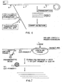
- the resulting circular molecule may be conveniently amplified by the ramification-extension amplification method (RAM), as depicted in Fig. 6.
- RAM ramification-extension amplification method
- Amplification of the circularized probe by RAM adds still further advantages to the methods of the present invention by allowing up to a million-fold amplification of the circularized probe under isothermal conditions.
- RAM is illustrated in Fig. 19.
- the single, full length, ligation dependent circularizable probe useful for RAM contains regions at its 3' and 5' termini that are hybridizable to adjacent but not contiguous regions of the target molecule.
- the circularizable probe is designed to contain a 5' region that is complementary to and hybridizable to a portion of the target nucleic acid, and a 3' region that is complementary to and hybridizable to a portion of the target nucleic acid adjacent to the portion of the target that is complementary to the 5' region of the probe.
- the 5' and 3' regions of the circularizable probe may each be from about 20 to about 35 nucleotides in length. In a preferred embodiment, the 5' and 3' regions of the circularizable probe are about 25 nucleotides in length.
- the circularizable probe further contains a region designated as the linker region.
- the linker region is from about 30 to about 60 nucleotides in length.
- the linker region is composed of a generic sequence that is neither complementary nor hybridizable to the target sequence.
- the circularizable probe suitable for amplification by RAM is utilized in the present method with one or more capture/amplification probes, as described hereinabove.
- the circularizable probe hybridizes with the target nucleic acid, its 5' and 3' termini become juxtaposed. Ligation with a linking agent results in the formation of a closed circular molecule.
- Amplification of the closed circular molecule is effected by adding a first extension primer (Ext-primer 1) to the reaction.
- Ext-primer 1 is complementary to and hybridizable to a portion of the linker region of the circularizable probe, and is preferably from about 15 to about 30 nucleotides in length.
- Ext-primer 1 is extended by adding sufficient concentrations of dNTPs and a DNA polymerase to extend the primer around the closed circular molecule. After one revolution of the circle, i.e., when the DNA polymerase reaches the Ext-primer 1 binding site, the polymerase displaces the primer and its extended sequence. The polymerase thus continuously "rolls over" the closed circular probe to produce a long single strand DNA, as shown in Figure 6.
- the polymerase useful for amplification of the circularized probe by RAM may be any polymerase that lacks 3' - 5' exonuclease activity, that has strand displacement activity, and that is capable of primer extension of at least about 1000 bases.
- Exo- Klenow fragment of DNA polymerase, Thermococcus litoralis DNA polymerase (Vent (exo - ) DNA polymerase, New England Biolabs) and phi29 polymerase (Blanco et al., 1994, Proc. Natl. Acad. Sci. USA 91 :12198) are preferred polymerases.
- Thermus aquaticus (Taq) DNA polymerase is also useful in accordance with the present invention. Contrary to reports in the literature, it has been found in accordance with the present invention that Taq DNA polymerase has strand displacement activity.
- Ext-primer 1 results in a long single stranded DNA of repeating units having a sequence complementary to the sequence of the circularizable probe.
- the single stranded DNA may be up to 10Kb, and for example may contain from about 20 to about 100 units, with each unit equal in length to the length of the circularizable probe for example about 100 bases.
- detection may be performed at this step if the target is abundant or the single stranded DNA is long.
- the long single stranded DNA may be detected at this stage by visualizing the resulting product as a large molecule on a polyacrylamide gel.
- a second extension primer (Ext-primer 2) is added.
- Ext-primer 2 is preferably from about 15 to about 30 nucleotides in length.
- Ext-primer 2 is identical to a portion of the linker region that does not overlap the portion of the linker region to which Ext-primer 1 is complementary.
- each repeating unit of the long single stranded DNA contains a binding site to which Ext-primer 2 hybridizes. Multiple copies of the Ext-primer 2 thus bind to the long single stranded DNA, as depicted in Fig. 6, and are extended by the DNA polymerase.
- the primer extension products displace downstream primers with their corresponding extension products to produce multiple displaced single stranded DNA molecules, as shown in Fig. 6.
- the displaced single strands contain binding sites for Ext-primer 1 and thus serve as templates for further primer extension reactions to produce the multiple ramification molecule shown in Fig. 6.
- the reaction comes to an end when all DNA becomes double stranded.
- the DNA amplified by RAM is then detected by methods known in the art for detection of DNA. Because RAM results in exponential amplification, the resulting large quantities of DNA can be conveniently detected, for example by gel electrophoresis and visualization for example with ethidium bromide. Because the RAM extension products differ in size depending upon the number of units extended from the closed circular DNA, the RAM products appear as a smear or ladder when electrophoresed.
- the circularizable probe is designed to contain a unique restriction site, and the RAM products are digested with the corresponding restriction endonuclease to provide a large amount of a single sized fragment of one unit length i.e., the length of the circularizable probe.
- the fragment can be easily detected by gel electrophoresis as a single band.
- a ligand such as biotin or digoxigenin can be incorporated during primer extension and the ligand-labeled single stranded product can be detected as described hereinabove.
- the RAM extension products can be detected by other methods known in the art, including, for example, ELISA.
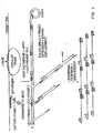
- the Ext-2 primer (and thus the identical portion of the linker region of the circularizable probe) is designed to contain a promoter sequence for a DNA-dependent RNA polymerase.
- RNA polymerases and corresponding promoter sequences are known in the art, and disclosed for example by Milligan et al. (1987) Nucleic Acid Res. 15 :8783. in a preferred embodiment the RNA polymerase is bacteriophage T3. T7. or SP6 RNA polymerase. Addition of the Ext-primer 2 containing the promoter sequence the corresponding RNA polymerase and rNTPs, allows hybridization of Ext-primer 2 to the growing single-stranded DNA to form a functional promoter and transcription of the downstream sequence into multiple copies of RNA.
- RNA can be detected by methods known to one of ordinary skill in the art, for example, polyacrylamide gel electrophoresis, radioactive or nonradioactive labeling and detection methods (Boehringer Mannheim), or the Sharp detection assay (Digene, Md.). Detection of the RNA indicates the presence of the target nucleic acid.
- Ext-primer 1 and the corresponding part of the linker region of the circular probe are designed to have a DNA-dependent RNA polymerase promoter sequence incorporated therein.
- a functional promoter is formed and the circularized probe acts as a template for RNA transcription upon the addition of RNA polymerase and rNTPs.
- the downstream primer and its RNA sequence are displaced by the RNA polymerase, and a large RNA polymer can be made.
- the RNA polymer may be detected as described hereinabove.
- the circular probe can be cleaved into a single stranded DNA by adding a restriction enzyme such as EcoRI. The restriction site is incorporated into the 5' end of extension primer 1, as shown in Fig. 7.
- Reagents for use in practicing the present disclosure may be provided individually or may be packaged in kit form. Reagents required for ligation (e.g. , DNA ligase) and, possibly, amplification may be included. Additional reagents also may be included for use in quantitative detection of the amplified ligated amplification sequence, e.g. , control templates such as an oligodeoxyribonucleotide corresponding to nanovariant RNA. Further, kits may include reagents for the in situ detection of target nucleic acid sequences e.g. in tissue samples. The kits containing circular probes may also include exonuclease for carryover prevention.
- Reagents required for ligation e.g. , DNA ligase
- Additional reagents also may be included for use in quantitative detection of the amplified ligated amplification sequence, e.g. , control templates such as an oligodeoxyribonucleotide corresponding to nanovariant RNA.
- the arrangement of the reagents within containers of the kit will depend on the specific reagents involved. Each reagent can be packaged in an individual container, but various combinations may also be possible.
- a pair of amplication probes and two capture/amplification probes were used to assay for and detect HCV RNA in a sample thereby increasing the capture efficiency of the assay.
- Capture/Amp-probe-1 (all oligomers described in this Example are designated "(HCV A)" having SEQ ID NO. 22 and Capture/Amp-probe-1A (HCV A) having SEQ ID NO. 23 are designed and synthesized such that their 5' termini are biotinylated and the 3' region of the probes comprises sequences complementary to and hybridizable with sequences in the 5'UTR of HCV RNA.
- the generic nucleotide sequence at the 5' region of the probes that are not hybridizable to the target nucleic acid sequence are designed and synthesized to have random sequences and a GC content of, at least, 60% and such that they exhibit minimal secondary structure e.g . hairpin or foldback structures.
- Capture/Amp-probe-1 which is biotinylated at the 5' terminus, is a 45 nucleotide DNA oligomer, such that nucleotides 5 to 45 in the 3' region, are complementary to and hybridizable with sequences in the 5'UTR of the target HCV RNA, and that the oligomer has the following nucleotide sequence (also listed below as SEQ ID NO. 22):
- Capture/Amp-probe-1A which is also biotinylated at the 5' terminus, is also a 45 nucleotide DNA oligomer, such that nucleotides 5 to 45 in the 3' region are complementary to and hybridizable with sequences in the 5'UTR of HCV RNA that are immediately adjacent to the region of the 5'UTR of the HCV RNA hybridizable with Capture/Amp-probe-1 (HCV A) and such that the oligomer has the following nucleotide sequence (also listed below as SEQ ID NO. 23):
- the two amplification probes Amp-probe-2 (HCV A) and Amp-probe-2A (HCV A) each contain a nucleotide sequence complementary to and hybridizable with the conserved 5'UTR of HCV RNA.
- Amp-probe-2 (HCV A) is a 51 nucleotide oligomer such that nucleotides 1 to 30 in the 5' region are complementary to and hybridizable with sequences in the 5'UTR of HCV RNA, and that the nucleotides 34 to 51 at the 3' terminus bind to and hybridize with PCR primer-3 and such that the oligomer has the following nucleotide sequence (also listed below as SEQ ID NO. 24):
- Amp-probe-2A is a 69 nucleotide oligomer such that nucleotides 40 to 69 in the 3' region are complementary to and hybridizable with sequences in the 5'UTR of HCV RNA genome immediately adjacent to the portion of the HVC RNA genome hybridizable to nucleotides 1-30 of Amp-probe-2 (HCV A) and such that the nucleotides 1 to 18 at the 5' terminus bind to and hybridize with PCR primer-4 and such that nucleotides 19 to 36 at the 5' terminus bind to and hybridize with PCR primer-5, and such that the oligomer has the following nucleotide sequence (also listed below as SEQ ID NO. 25):
- HCV A ligation-amplification sequence
- Primer-3 used for the first series of PCR amplification of the ligated amplification sequence, SEQ ID NO. 26 (HCV A), and which has a length of 18 nucleotides, is complementary to sequence comprising nucleotides 34 to 51 at the 3' terminus of Amp-probe-2 (HCV A), and is, therefore, also complementary to the sequence comprising nucleotides 103 to 120 of the ligated amplification sequence, SEQ ID NO. 26 (HCV A), and has the sequence (also listed below as SEQ ID NO. 27):
- Primer-4 used for the first series of PCR amplification of the ligated amplification sequence (HCV A), SEQ ID NO. 26, and which has a length of 18 nucleotides, is complementary to the sequence comprising nucleotides 1-18 at the 5' terminus of the Amp-probe-2A (HCV A), and is, therefore, also complementary to the sequence comprising nucleotides 1 to 18 of the ligated amplification sequence, SEQ ID NO. 26 (HCV A), and has the sequence (also listed below as SEQ ID NO. 28):
- Primer-5 a DNA oligomer of 18 nucleotides is used for the second series of PCR amplification of the ligated amplification sequence (HCV A), SEQ ID NO. 26, such that the primer is complementary to the sequence comprising nucleotides 19-36 of the Amp-probe-2A (HCV A), and is, therefore, also hybridizable to the sequence comprising nucleotides 19-36 of the ligated amplification sequence SEQ ID NO. 26 (HCV A), and has the sequence (also listed below as SEQ ID NO. 29):
- the assay utilizing the above probes and primers was used to detect HCV RNA in 24 human serum samples that were stored at -70°C until use.
- 180 ⁇ l serum sample was added to concentrated lysis buffer (prepared by condensing 250 ⁇ l of the lysis solution containing 5M GnSCN, 0.5% bovine serum albumin, 80mM EDTA, 400mM Tris HCl (pH 7.5), and 0.5% Nonidet P-40 so that the mixture of serum and lysis buffer would have a final concentration of 5M GnSCN) mixed well and incubated for 1 hour at 37°C to release the target RNA from HCV particles.
- concentrated lysis buffer prepared by condensing 250 ⁇ l of the lysis solution containing 5M GnSCN, 0.5% bovine serum albumin, 80mM EDTA, 400mM Tris HCl (pH 7.5), and 0.5% Nonidet P-40 so that the mixture of serum and lysis buffer would have a final concentration of 5M GnSCN
- hybridization buffer [0.5% bovine serum albumin, 80mM EDTA. 400 mM Tris-Hcl (pH 7.5), 0.5% Nonidet-P40] with 10 10 molecules each of amplification probes, Amp-probe-2 (HCV A) and Amp-probe-2A (HCV A) oligomers, and 10 11 molecules each of capture/amplification probes, Capture/Amp-probe-1 (HCV A) and Capture/Amp-probe-1A (HCV A).
- the addition of the hybridization buffer reduced the concentration of the guanidium isothiocyanate (GnSCN) from 5M to 2M to allow the hybridization to occur.
- GnSCN guanidium isothiocyanate
- the mixture was incubated at 37°C for 1 hour to let the various probes hybridize with the target RNA, whereupon 30 ⁇ l of streptavidin-coated paramagnetic beads (Promega) were added to the hybridization mixture before incubation at 37° C for 20 minutes to allow ligand binding.
- the beads were washed with 150 ⁇ l of 2M GnSCN to eliminate any free probes, proteins, extraneous nucleic acids and potential PCR inhibitors from the hybridization mixture; this was followed by the removal of the GnSCN by washing twice with 150 ⁇ l ligase buffer [66mM Tris-Hcl (pH 7.5) 1mM DTT, 1mM ATP, 0.5% Nonidet P-40 and 1mM MnCl 2 ]. At each wash-step. A magnetic separation of the bound complex from the supernatant was effected.
- the amplification probes, Amp-probe-2 (HCV A) and Amp-probe-2A (HCV A), bound to target RNA were then covalently joined to create the ligated amplification sequence (HCV A) that was utilized as a template for PCR amplification.
- the hybrid complex was resuspended in 20 ⁇ l ligase buffer with 5 units of T 4 DNA ligase (Boehringer) and incubated for 1 hour at 37°C for the ligation reaction.
- first PCR reaction 10 ⁇ l of the ligated mixture, including the beads, was added to 20 ⁇ l of PCR mixture [0.06 ⁇ M each of Primer-3 and Primer-4, 1.5 Units Taq DNA Polymerase, 0.2 mM each of dATP, dCTP. dGTP and dTTP, 1.5 mM MgCl 2 , 10mM Tris-HCl (pH 8.3) 50mM KCl] and the mixture incubated at 95°C for 30 seconds, 55°C for 30 seconds and 72°C for 1 minute, for 35 cycles.
- PCR mixture [0.06 ⁇ M each of Primer-3 and Primer-4, 1.5 Units Taq DNA Polymerase, 0.2 mM each of dATP, dCTP. dGTP and dTTP, 1.5 mM MgCl 2 , 10mM Tris-HCl (pH 8.3) 50mM KCl] and the mixture incubated at 95°C for 30 seconds, 55°C for 30 seconds and 72°
- the product was transferred to a second PCR mixture [same components as the first PCR mixture except that Primer-4 was substituted with Primer-5] for "the second PCR reaction" (a semi-nested PCR approach to increase the sensitivity of the assay) carried out under the same conditions as the first PCR reaction.
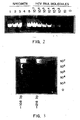
- 10 ⁇ l of the products of the second reaction were electrophoresed on a 6% polyacrylamide gel, stained with ethidium bromide and visualized under ultraviolet light.
- the assay is quantitative over, at least, a range of 10 2 to 10 5 target molecules.
- the method in this Example employs the two capture/amplification probes Capture/Amp-probe-1 (HCV A) and Capture/Amp-probe-1 A (HCV A) described in comparative example 1 and a single amplification probe, Amp-probe-2 (HCV C) (all oligomers described in this Example are designated "(HCV C)" that hybridizes to the target nucleic acid and circularizes upon ligation of its free termini as shown in Fig. 1.
- Amp-probe-2 is a 108 nucleotide amplification probe, also referred to as an amplification sequence, such that nucleotides 1-26 in the 5' terminus of the oligomer are complementary to and hybridizable to a sequence in the 5'UTR of the target HCV RNA (indicated by (a) in Fig. 1) and such that nucleotides 83-108 at the 3' terminus of the oligomer are complementary to and hybridizable to a sequence in the 5'UTR of the target HCV RNA (indicated by (b) in Fig. 1).
- HCV C The sequence of Amp-probe-2 (HCV C) is given as follows (also listed as SEQ ID NO. 31):
- Primer-3 (SEQ ID NO. 27), used for the first series ofPCR amplification of the ligated and circularized Amp-probe-2 (HCV C), is an 18 nucleotide long oligomer that is complementary to the sequence comprising nucleotides 27 to 45 of Amp-probe-2 (HCV C).
- Primer-4 (SEQ ID NO. 28), also used for the first series of PCR amplification of the ligated and circularized Amp-probe-2, is a 18 nucleotide long oligomer that is complementary to the sequence comprising nucleotides 46 to 63 of Amp-probe-2 (HCV C).
- the assay is significantly quantitative at least over a range of 10 4 to 10 7 target molecules.
- This example demonstrates the ability of Klenow fragment of DNA polymerase to displace downstream strands and produce a polymer.
- a synthetic DNA target was detected by mixing 10 12 molecules of phosphorylated circularizable probe having SEQ ID NO:31 with 10 13 molecules of synthetic HCV DNA target in 10 ⁇ l of 1X ligation buffer, heating at 65°C for two minutes, and cooling to room temperature for ten minutes.
- One ⁇ l of ligase was added to the mix and incubated at 37°C for one hour, followed by addition of 10 13 molecules of 32 P-labeled Ext-primer having SEQ ID NO:27.
- the mixture was heated to 100°C for five minutes and then cooled to room temperature for twenty minutes. Forty ⁇ l of Klenow mix and dNTPs were added to the reaction and incubated at 37°C.
- LD-PCR utilizing a circularized probe was performed to detect Epstein Barr virus early RNA (EBER-1) in salivary benign mixed tumors (BMT).
- EBER-1 Epstein Barr virus early RNA
- Six specimens of BMT and adjacent parotid tissue, and three specimens of normal parotid tissue (two removed from cysts and one from a hyperplastic lymph node) were snap frozen in embedding medium for frozen tissue specimens (OCT, Miles, Inc., Elkhart, In.) and liquid nitrogen, and stored at -70°C.
- the corresponding formalin fixed paraffin embedded (FFPE) blocks of tissue were obtained and studied in parallel to the fresh tissue. All tissue was sectioned on a microtome, the blade of which was cleaned with 10% Chlorox between cases to avoid cross contamination.
- FFPE tissues were deparafinized by incubating at 60°C for 10 minutes with 1 ml xylene (Sigma), which was subsequently removed by two washes with absolute ethanol. These specimens were dried by placing on a hot block at 65 °C for 30 minutes.
- Deparaffinized tissue was lysed by incubation at 100°C for 30 minutes, then 65 °C for 30 minutes in 250 ⁇ l of lysis buffer: 5M guanidium thiocyanate (GTC)(Fluka), 0.5% bovine serum albumin (Sigma), 80 mM EDTA, 400 mM Tris HCl (pH 7.5), and 0.5% sodium-N-lauroylsarcosine (Sigma).
- GTC guanidium thiocyanate
- bovine serum albumin Sigma
- 80 mM EDTA 400 mM Tris HCl (pH 7.5)
- sodium-N-lauroylsarcosine Sigma
- Two capture/amplification probes designed to flank the region of EBER-I were used to capture target RNA.
- the sequences for capture probe 1 (SED ID NO : 40) and capture/amplification probe 2 (SEQ ID NO:41) are shown in Table 4.
- the circular amplification probe (SEQ ID NO:42) was designed with 3' and 5' regions complementary to the chosen target sequence (Table 4). Interposed between these two regions is a noncomplementary linker sequence. This circular amplification probe circularized upon target hybridization in such a manner as to juxtapose the 5' and 3' ends. Seminested PCR was performed using primer pairs directed at this linker sequence, also shown in Table 4.
- Circular Amp PROBE5'tcaccacccgggacttgtacccgggacTGTCTGTGTATCTGCTAACCAAGAGCAA CTACACGAATTCTCGATTAGGTTACTGCgggaagacaaccacagacaccgttcc-3' (SEQ ID No. 42) 1st PCR primer pairs: GTTAGCAGATACACAGAC (sense SEQ ID NO. 27) CAAGAGCAACTACACGAA (antisense SEQ ID NO. 28) 2ND PCR primer pairs: GTTAGCAGATACACAGAC (sense SEQ ID NO. 27) TTCTCGATTAGGTTACTG (antisense SEQ ID NO. 29) (lower case - complementary to EBER-1, upper case - generically designed)
- LD-PCR was performed as follows. Briefly, 80 ⁇ l of lysis mixture were added to 120 ⁇ l of hybridization buffer (0.5% bovine serum albumin, 80 mM EDTA, 400 MM Tris-HCl (pH 7.5), and 0.5% sodium-N-lauroyisarcosine (Sigma) which contained 10 10 molecules of phosphorylated target probe, and 10 11 molecules of capture probe 1 and capture probe 2. Addition of the hybridization buffer reduced the GnSCN concentration from 5 M to 2 M to allow hybridization to occur. This mixture was incubated for one hour to allow the formation of hybrids, consisting of two DNA capture/amplification probes and one DNA circular amplification probe hybridized on the target RNA.
- hybridization buffer 0.5% bovine serum albumin, 80 mM EDTA, 400 MM Tris-HCl (pH 7.5), and 0.5% sodium-N-lauroyisarcosine (Sigma) which contained 10 10 molecules of phosphorylated target probe, and 10 11 molecules of capture probe 1 and capture
- streptavidin-coated paramagnetic beads (Promega) were added to the mixture and incubated at 37°C for 20 minutes to allow the hybrids to bond to the bead surface.
- the beads were washed twice with 150 ⁇ l of washing buffer (10 mM Tris HCl (pH 7.5), 0.5% Nonidet P-40, and 1.5 mM MgCl 2 and 50 mM KCl) to remove nonhybridized probes as well as potential inhibitors of PCR (GTC, proteins) and potential sources of nonspecific PCR products (cellular nucleic acids).
- the beads were drawn to the wall of the assay tube by placing the tube on a Magnetic Separation Stand (Promega), enabling the supernatant to be removed by aspiration.
- the first PCR reaction was incubated at 94°C for 30 seconds, 55°C for 30 seconds, and 72°C for 1 minute for 35 cycles in a GeneAmp PCR system 9600 thermocycler (Perkin Elmer, CT). After the first PCR, 5 ul of each reaction mixture were transferred into a 25 ul second PCR mixture containing the same components except that 0.66 ⁇ M of PCR primer 1 and 0.66 ⁇ M of PCR primer 3 were used for seminested PCR, which increases signal detection sensitivity without compromising amplification specificity. Extension of PCR primer along the covalently circularized probe results in the generation of a large multi-unit polymer (rolling circle polymerization).
- EBER-1 sequences were detected in six of eight parotid samples. Of the six pleomorphic adenomas studied, four were positive for EBER-1. Of the two cases in which EBER was not detected in the tumor, sequences were present within surrounding parotid tissue. The detection of EBER-1 sequences within corresponding formalin-fixed paraffin embedded tissue was considerably less sensitive - only two of eight specimens were positive.
- the present results with ligation dependent PCR utilizing a circular probe demonstrate the presence of EBV-related sequences within the majority of pleomorphic adenomas studied.
- the present method exhibits a markedly increased detection rate relative to standard PCR for the detection of EBV DNA as performed by Taira et al. (1992) J. of Otorhinolaryngol Soc. Jap. 95: 860.
- the 3' and 5' ends of a circularizable probe hybridized to the target sequence, resulting in juxtaposition. The justaposed sequences were then ligated, resulting in a circularized covalently linked probe that was locked onto the target sequence and thus resistant to stringent washes.
- PCR on the circular probe produced a rolling circle polymer, which was digested into monomeric units and visualized on a gel.
Abstract
Description
- The present disclosure relates to assays and kits for carrying out said assays for the rapid, automated detection of infectious pathogenic agents and normal and abnormal genes.
- A number of techniques have been developed recently to meet the demands for rapid and accurate detection of infectious agents such as viruses, bacteria and fungi, and detection of normal and abnormal genes. Such techniques, which generally involve the amplification and detection (and subsequent measurement) of minute amounts of target nucleic acids (either DNA or RNA) in a test sample, include inter alia the polymerase chain reaction (PCR) (Saiki, et al., Science 230:1350, 1985; Saiki et al., Science 239:487, 1988; PCR Technology, Henry A. Erlich, ed., Stockton Press, 1989; Patterson et al., Science 260:976, 1993), ligase chain reaction (LCR) (Barany, Proc. Natl. Acad. Sci. USA 88:189, 1991), strand displacement amplification (SDA) (Walker et al., Nucl. Acids Res. 20:1691, 1992), Qβ replicase amplification (QβRA) (Wu et al., Proc. Natl. Acad. Sci. USA 89:1 1769, 1992; Lomeli et al., Clin. Chem. 35:1826, 1989) and self-sustained replication (3SR) (Guatelli et al., Proc. Natl. Acad. Sci. USA 87: 1874-1878, 1990). While all of these techniques are powerful tools for the detection and identification of minute amounts of a target nucleic acid in a sample, they all suffer from various problems, which have prevented their general applicability in the clinical laboratory setting for use in routine diagnostic techniques.
- One of the most difficult problems is preparation of the target nucleic acid prior to carrying out its amplification and detection. This process is time and labor intensive and, thus, generally unsuitable for a clinical setting, where rapid and accurate results are required. Another problem, especially for PCR and SDA, is that conditions for amplifying the target nucleic acid for subsequent detection and optional quantitation vary with each test, i.e., there are no constant conditions favoring test standardization. This latter problem is especially critical for the quantitation of a target nucleic acid by competitive PCR and for the simultaneous detection of multiple target nucleic acids.
- Circumvention of the aforementioned problems would allow for development of rapid standardized assays, utilizing the various techniques mentioned above, that would be particularly useful in performing epidemiologic investigations, as well as in the clinical laboratory setting for detecting pathogenic microorganisms and viruses in a patient sample. Such microorganisms cause infectious diseases that represent a major threat to human health. The development of standardized and automated analytical techniques and kits therefor, based on rapid nd sensitive identification of target nucleic acids specific for an infectious disease agent would provide advantages over techniques involving immunologic or culture detection of bacteria and viruses.
- Reagents may be designed to be specific for a particular organism or for a range of related organisms. These reagents could be utilized to directly assay microbial genes conferring resistance to various antibiotics and virulence factors resulting in disease. Development of rapid standardized analytical techniques will aid in the selection of the proper treatment.
- In some cases, assays having a moderate degree of sensitivity (but high specificity) may suffice, e.g., in initial screening tests. In other cases, great sensitivity (as well as specificity) is required, e.g., the detection of the HIV genome in infected blood may require finding the virus nucleic acid sequences present in a sample of one part per 10 to 100,000 human genome equivalents (Harper et al., Proc. Nat'l. Acad. Sci., USA 83:772, 1986).
- Blood contaminants, including inter alia, HIV, HTLV-I, hepatitis B and hepatitis C, represent a serious threat to transfusion patients and the development of routine diagnostic tests involving the nucleic acids of these agents for the rapid and sensitive detection of such agents would be of great benefit in the clinical diagnostic agree laboratory. For example, the HIV genome can be detected in a blood sample using PCR techniques, either as an RNA molecule representing the free viral particle or as a DNA molecule representing the integrated provirus (Ou et al, Science 239:295, 1988; Murakawa et al., DNA 7:287, 1988).
- In addition, epidemiologic investigations using classical culturing techniques have indicated that disseminated Mycobacterium avium-intracellulaire (MAI) infection is a complication of late-stage Acquired Immunodeficiency Syndrome (AIDS) in children and adults. The precise extent of the problem is not clear, however, since current cultural methods for detecting mycobacteria are cumbersome, slow and of questionable sensitivity. Thus, it would be desirable and highly beneficial to devise a rapid, sensitive and specific technique for MAl detection in order to provide a definitive picture of the involvement in HIV-infected and other immunosuppressed individuals. Such studies must involve molecular biological methodologies, based on detection of a target nucleic acid, which have routinely been shown to be more sensitive than standard culture systems (Boddinghaus et al., J. Clin. Med. 28:1751, 1990).
- Other applications for such techniques include detection and characterization of single gene genetic disorders in individuals and in populations (see, e.g., Landergren et al., Science 241: 1077, 1988 which discloses a ligation technique for detecting single gene defects, including point mutations). Such techniques should be capable of clearly distinguishing single nucleotide differences (point mutations) that can result in disease (e.g., sickle cell anemia) as well as deleted or duplicated genetic sequences (e.g., thalassemia).
- WO9535390 discloses a method for detecting a target nucleic acid in a sample comprising
- (a) contacting the nucleic acid in the sample under conditions that allow nucleic acid hybridization between complementary sequences in nucleic acids with oligonucleotide probes, the oligonucleotide probes further comprising a circularizable amplification probe having 3' and 5' regions that are complementary to adjacent but not overlapping sequences in the target nucleic acid, said 3' and 5' regions separated by a generic region that is neither complementary nor hybridizable to a nucleotide sequence in the target nucleic acid, such that a complex is formed comprising the target nucleic acid and the circularizable probe, wherein the circularizable probe is bound on its 3' and 5' ends to adjacent but not overlapping sequences in the target nucleic acid;
- (b) separating the complex from unbound reactants and washing the complex;
- (c) ligating the 3' and 5' ends of the circularizable probe with a ligating agent that joins nucleotide sequences such that a circular amplification probe is formed;
- (d) amplifying the circular amplification probe by contacting the complex with a first extension primer that is complementary and hybridizable to the generic portion of the circular amplification probe and a second extension primer that is substantially identical to the generic portion of the circular amplification probe that does not overlap with the portion of the generic region to which the first extension primer binds, dNTPs, and a DNA polymerase having strand displacement activity, under conditions whereby the first extension primer is extended around the circular probe for multiple revolutions to form a single stranded DNA of repeating units complementary to the sequence of the circular probe, multiple copies of the second extension primer hybridize to complementary regions of the single stranded DNA and are extended by the DNA polymerase to provide extension products; and
- (e) detecting the amplified DNA, wherein detection thereof indicates the presence of the target nucleic acid in the sample.
- The methods referred to above are relatively complex procedures that, as noted, suffer from drawbacks making them difficult to use in the clinical diagnostic laboratory for routine diagnosis and epidemiological studies of infectious diseases and genetic abnormalities. All of the methods described involve amplification of the target nucleic acid to be detected. The extensive time and labor required for target nucleic acid preparation, as well as variability in amplification templates (e.g., the specific target nucleic acid whose detection is being measured) and conditions, render such procedures unsuitable for standardization and automation required in a clinical laboratory setting.
- EP 0 359 789 describes a method for detecting a target nucleic acid in a sample, avoiding DNA amplification such as PCR. A primer comprising a promoter sequence is hybridised to a target nucleic acid. A portion of the target nucleic acid is removed by 3'-5'exonuclease, whereupon the removed portion is replaced by a new sequence, using the promoter sequence of the hybridised primer as template, resulting in a double stranded promoter, enabling transcription of the target nucleic acid.
- The present is directed to the development of rapid, sensitive assays useful for the detection and monitoring of pathogenic organisms, as well as the detection of abnormal genes in an individual. Moreover, the methodology of the present disclosure can be readily standardized and automated for use in the clinical laboratory setting.
- An improved method, which allows for rapid, sensitive and standardized detection and quantitation of nucleic acids from pathogenic microorganisms from samples from patients with infectious diseases has now been developed. The improved methodology also allows for rapid and sensitive detection and quantitation of genetic variations in nucleic acids in samples from patients with genetic diseases or neoplasia.
- This method provides several advantages over prior art methods. The method simplifies the target nucleic acid isolation procedure, which can be performed in microtubes, microchips or micro-well plates, if desired. The method allows for isolation, amplification and detection of nucleic acid sequences corresponding to the target nucleic acid of interest to be carried out in the same sample receptacle, e.g., tube or micro-well plate. The method also allows for standardization of conditions, because only a single circularisable generic amplification probe may be utilized in the present method for detecting a variety of target nucleic acids, thus allowing efficient multiplex amplification. The method allows the direct detection of RNA by probe amplification without the need for DNA template production. The amplification probes, which in the method may be covalently joined end to end, form a contiguous ligated amplification sequence. The assembly of the amplifiable DNA by ligation increases specificity, and makes possible the detection of a single mutation in a target. This ligated amplification sequence, rather than the target nucleic acid, is amplified, allowing for substantially the same amplification conditions to he used for a variety of different infectious agents and, thus, leading to more controlled and consistent results being obtained. In addition, multiple infectious agents in a single sample may be detected using the multiplex amplification methodology disclosed.
- Additional advantages of the present disclosure include the ability to automate the protocol of the method disclosed, which is important in performing routine assays, especially in the clinical laboratory.
- A target nucleic acid is hybridized to a circularisable amplification probe. The probe is designed to contain generic sequences (i.e., not target nucleic acid specific) and specific sequences complementary to a nucleotide sequence in the target nucleic acid. The specific sequences of the probe are complementary to adjacent regions of the target nucleic acid, and thus do not overlap one another. Subsequently, the 3' and 5' ends of the circularisable probe are joined together using a ligating agent to form a contiguous ligated amplification sequence. The ligating agent may be an enzyme, e.g., DNA ligase or a chemical. Following washing and removal of unbound reactants and other materials in the sample, the detection of the target nucleic acid in the original sample is determined by detection of the ligated amplification sequence. The ligated amplification sequence (not the target nucleic acid) is amplified for detection. Unligated probes, as well as the target nucleic acid, are not amplified in this technique. The amplification probe is a single amplification probe that hybridizes to the target such that its 3' and 5' ends are juxtaposed. The ends are then ligated by DNA ligase to form a covalently linked circular probe that can be identified by amplification.
-
- Fig. 1 is a schematic diagram showing various components used to detect a target nucleic acid e.g. HCV RNA, employing two capture/amplification probes, each containing a bound biotin moiety, and a single amplification probe that circularizes upon hybridization to the target nucleic acid and ligation of free termini.
- Fig. 2 is a photograph of ethidium bromide stained DNA depicting PCR amplified probes used to detect HCV RNA in a sample. The amount of HCV RNA in the sample is determined by comparing sample band densities to those of standard serial dilutions of HCV transcripts.
- Fig. 3 is a photograph of ethidium bromide stained DNA depicting PCR amplified single, full length ligation-dependent and circularizable probes used to detect HCV RNA in a sample. The amount of HCV RNA in the sample is determined by comparing sample band densities to those of standard serial dilutions of HCV transcripts.
- Fig. 4 is a schematic diagram of an embodiment of RAM in which a T3 promoter has been incorporated into Ext-
primer 2. allowing amplification of the circular probe by transcription. - Fig. 5 provides a polyacrylamide gel depicting the amplification of a circular probe by extension of Ext-
primer 1. - Fig. 6 is a schematic diagram of amplification of a circularized probe by the ramification-extension amplification method (RAM).
- Fig. 7 is a diagram of a RAM assay in which an RNA polymerase promoter sequence is incorporated into the primer.
- The present disclosure is directed towards simplified sample preparation and generic amplification systems for use in clinical assays to detect and monitor pathogenic microorganisms in a test sample, as well as to detect abnormal genes in an individual. Generic amplification systems are described for clinical use that combine ligation/amplification techniques with transcription for detecting and measuring nucleic acids in a sample. The present method uses the ramification-extension amplification method (RAM). The advantages of the present method include (1) suitability for clinical laboratory settings, (2) ability to obtain controlled and consistent (standardizable) results, (3) ability to quantitate nucleic acids in a particular sample, (4) ability to simultaneously detect and quantitate multiple target nucleic acids in a test sample, (5) ability to sensitively and efficiently detect nucleic acids in serum samples and in situ, and (6) ability to detect a single mutation in a target. Moreover, the complete protocol of the presently disclosed method may be easily automated, making it useful for routine diagnostic testing in a clinical laboratory setting. With the use of RAM, an isothermal amplification can be achieved.
- The present method incorporates magnetic separation, utilizing paramagnetic particles, beads or spheres that have been coated with a ligand binding moiety that recognizes and binds to ligand present on an oligonucleotide capture probe, described below, to isolate a target nucleic acid (DNA or RNA) from a clinical sample in order to facilitate its detection.
- The probes may be synthesized from nucleoside triphosphates by known automated oligonucleotide synthetic techniques, e.g., via standard phosphoramidite technology utilizing a nucleic acid synthesizer. Such synthesizers are available, e.g., from Applied Biosystems, Inc. (Foster City. CA).
- If the probes are to be ligated together, the next step in the present method involves treating the complex from step (a) with a ligating agent that will join the two probes. The ligating agent may be an enzyme, e.g., DNA or RNA ligase, or a chemical agent, e.g., cyanogen bromide or a carbodiimide. This serves to join the 5' end to the 3' end of the circularisable amplification probe to form a contiguous functional single-stranded oligonucleotide molecule, referred to herein as a ligated amplification sequence. Any of the amplification probes which remain unligated after treatment are not amplified in subsequent steps in the method.
- The probes may be ligated using a suitable ligating agent, such as a DNA or RNA ligase. Alternatively, the ligating agent may be a chemical, such as cyanogen bromide or a carbodiimide (Sokolova et al., FEBS Lett. 232:153-155. 1988). Preferred DNA ligases include T4 DNA ligase and the thermostable Taq DNA ligase. The advantage of using the Taq DNA ligase is that it is active at elevated temperatures (65-72°C). Joining the oligonucleotide probes at such elevated temperatures decreases non-specific ligation. Preferably, the ligation step is carried out for 30-60 minutes at an elevated temperature (about 65-72°C), after which time any unligated amplification probe may be, optionally, removed under denaturing conditions.
- Denaturation is performed after the ligation step by adding TE Buffer (10mM Tris-HCl pH 7.5, 0.1 mM EDTA) to the mixture. The temperature of the mixture is then raised to about 92-95°C for about 1-5 minutes to denature the hybridized nucleic acid. This treatment separates the target nucleic acid from the hybridized ligated amplification sequences.
- Another step in the process is detection of the ligated amplification sequence, which indicates the presence of the target nucleic acid in the original test sample.
- An amplification system is used to amplify the ligated amplification sequence for detection.
- The amplification probe is designed so that complementary regions of the probe that are hybridizable to the target nucleic acid sequence are located at each end of the probe (as described in Nilsson et al., 1994, Science 265:2085-2088). When the probe hybridizes with the target, its termini are placed adjacent to each other, resulting in the formation of a closed circular molecule upon ligation with a linking agent such as a ligase enzyme. This circular molecule may then serve as a template during an amplification step, e.g. using primers such as those depicted in Fig. 1. The circular molecule is amplified by RAM, as described hereinbelow.
- For example, the probe, described above, can be used to detect different genotypes of a pathogen, e.g. different genotypes of HCV from serum specimens. Genotype specific probes can be designed, based on published HCV sequences (Stuyver et al., 1993, J. Gen. Virol. 74: 1093-1102), such that a mutation in the target nucleic acid is detectable since such a mutation would interfere with (1) proper hybridization of the probe to the target nucleic acid and (2) subsequent ligation of the probe into a circular molecule. Because of the nature of the circularized probe, as discussed below, unligated probes may be removed under stringent washing conditions.
- The single, full length, ligation-dependent circularizable probe, as utilized in the method, affords greater efficiency of the detection and amplification of the target nucleic acid sequence. Due to the helical nature of double-stranded nucleic acid molecules, circularized probes are wound around the target nucleic acid strand. As a result of the ligation step, the probe may be covalently bound to the target molecule by means of catenation. This results in immobilization of the probe on the target molecule, forming a hybrid molecule that is substantially resistant to stringent washing conditions. This results in significant reduction of non-specific signals during the assay, lower background noise and an increase in the specificity of the assay.
- Another embodiment of the present disclosure provides a method of reducing carryover contamination and background in amplification methods utilizing circular probes. The present ligation-dependent amplification methods, unlike conventional amplification methods, involve amplification of the ligated probe(s) rather than the target nucleic acid. When the ligated probe is a closed circular molecule, it has no free ends susceptible to exonuclease digestion. After probe ligation, i.e. circularization, treatment of the reaction mixture with an exonuclease provides a "clean-up" step and thus reduces background and carryover contamination by digesting unligated probes or linear DNA fragments but not closed circular molecules. The covalently linked circular molecules remain intact for subsequent amplification and detection. In conventional PCR, the use of exonuclease to eliminate single stranded primers or carryover DNA fragments poses the risk that target nucleic acid will also be degraded. The present invention does not suffer this risk because target nucleic acid is not amplified. In a preferred embodiment, the exonuclease is exonuclease III, exonuclease VII, mung bean nuclease or nuclease BAL-31. Exonuclease is added to the reaction after ligation and prior to amplification, and incubated, for example at 37°C for thirty minutes.
- It is further contemplated to use multiple probes which can be ligated to form a single covalently closed circular probe. For example, a first probe is selected to hybridize to a region of the target. A second probe is selected such that its and 5' termini hybridize to regions of the target that are adjacent but not overlapping with the 5' and 3' termini of the first probe. Two ligation events are then required to provide a covalently closed circular probe. By using two ligases, e.g. an enzymatic and a chemical ligase, to covalently close the probe, the order of the ligations can be controlled. This embodiment is particularly useful to identify two nearby mutations in a single target.
- The circularized probe can also be amplified and detected by the generation of a large polymer. The polymer is generated through the rolling circle extension of
primer 1 along the circularized probe and displacement of downstream sequence. This step produces a single stranded DNA containing multiple units. - As shown therein,
primer 2 can bind to the single stranded DNA polymer and extend simultaneously, resulting in displacement of downstream primers by upstream primers. - According to the present disclosure utilizing a ligation dependent circularizable probe, the resulting circular molecule may be conveniently amplified by the ramification-extension amplification method (RAM), as depicted in Fig. 6. Amplification of the circularized probe by RAM adds still further advantages to the methods of the present invention by allowing up to a million-fold amplification of the circularized probe under isothermal conditions. RAM is illustrated in Fig. 19.
- The single, full length, ligation dependent circularizable probe useful for RAM contains regions at its 3' and 5' termini that are hybridizable to adjacent but not contiguous regions of the target molecule. The circularizable probe is designed to contain a 5' region that is complementary to and hybridizable to a portion of the target nucleic acid, and a 3' region that is complementary to and hybridizable to a portion of the target nucleic acid adjacent to the portion of the target that is complementary to the 5' region of the probe. The 5' and 3' regions of the circularizable probe may each be from about 20 to about 35 nucleotides in length. In a preferred embodiment, the 5' and 3' regions of the circularizable probe are about 25 nucleotides in length. The circularizable probe further contains a region designated as the linker region. In a preferred embodiment the linker region is from about 30 to about 60 nucleotides in length. The linker region is composed of a generic sequence that is neither complementary nor hybridizable to the target sequence.
- The circularizable probe suitable for amplification by RAM is utilized in the present method with one or more capture/amplification probes, as described hereinabove. When the circularizable probe hybridizes with the target nucleic acid, its 5' and 3' termini become juxtaposed. Ligation with a linking agent results in the formation of a closed circular molecule.
- Amplification of the closed circular molecule is effected by adding a first extension primer (Ext-primer 1) to the reaction. Ext-
primer 1 is complementary to and hybridizable to a portion of the linker region of the circularizable probe, and is preferably from about 15 to about 30 nucleotides in length. Ext-primer 1 is extended by adding sufficient concentrations of dNTPs and a DNA polymerase to extend the primer around the closed circular molecule. After one revolution of the circle, i.e., when the DNA polymerase reaches the Ext-primer 1 binding site, the polymerase displaces the primer and its extended sequence. The polymerase thus continuously "rolls over" the closed circular probe to produce a long single strand DNA, as shown in Figure 6. - The polymerase useful for amplification of the circularized probe by RAM may be any polymerase that lacks 3' - 5' exonuclease activity, that has strand displacement activity, and that is capable of primer extension of at least about 1000 bases. (Exo-)Klenow fragment of DNA polymerase, Thermococcus litoralis DNA polymerase (Vent (exo-) DNA polymerase, New England Biolabs) and phi29 polymerase (Blanco et al., 1994, Proc. Natl. Acad. Sci. USA 91:12198) are preferred polymerases. Thermus aquaticus (Taq) DNA polymerase is also useful in accordance with the present invention. Contrary to reports in the literature, it has been found in accordance with the present invention that Taq DNA polymerase has strand displacement activity.
- Extension of Ext-
primer 1 by the polymerase results in a long single stranded DNA of repeating units having a sequence complementary to the sequence of the circularizable probe. The single stranded DNA may be up to 10Kb, and for example may contain from about 20 to about 100 units, with each unit equal in length to the length of the circularizable probe for example about 100 bases. As an alternative to RAM, detection may be performed at this step if the target is abundant or the single stranded DNA is long. For example, the long single stranded DNA may be detected at this stage by visualizing the resulting product as a large molecule on a polyacrylamide gel. - In the next step of amplification by RAM, a second extension primer (Ext-primer 2) is added. Ext-
primer 2 is preferably from about 15 to about 30 nucleotides in length. Ext-primer 2 is identical to a portion of the linker region that does not overlap the portion of the linker region to which Ext-primer 1 is complementary. Thus each repeating unit of the long single stranded DNA contains a binding site to which Ext-primer 2 hybridizes. Multiple copies of the Ext-primer 2 thus bind to the long single stranded DNA, as depicted in Fig. 6, and are extended by the DNA polymerase. The primer extension products displace downstream primers with their corresponding extension products to produce multiple displaced single stranded DNA molecules, as shown in Fig. 6. The displaced single strands contain binding sites for Ext-primer 1 and thus serve as templates for further primer extension reactions to produce the multiple ramification molecule shown in Fig. 6. The reaction comes to an end when all DNA becomes double stranded. - The DNA amplified by RAM is then detected by methods known in the art for detection of DNA. Because RAM results in exponential amplification, the resulting large quantities of DNA can be conveniently detected, for example by gel electrophoresis and visualization for example with ethidium bromide. Because the RAM extension products differ in size depending upon the number of units extended from the closed circular DNA, the RAM products appear as a smear or ladder when electrophoresed. In another embodiment, the circularizable probe is designed to contain a unique restriction site, and the RAM products are digested with the corresponding restriction endonuclease to provide a large amount of a single sized fragment of one unit length i.e., the length of the circularizable probe. The fragment can be easily detected by gel electrophoresis as a single band. Alternatively, a ligand such as biotin or digoxigenin can be incorporated during primer extension and the ligand-labeled single stranded product can be detected as described hereinabove.
- The RAM extension products can be detected by other methods known in the art, including, for example, ELISA.
- The Ext-2 primer (and thus the identical portion of the linker region of the circularizable probe) is designed to contain a promoter sequence for a DNA-dependent RNA polymerase. RNA polymerases and corresponding promoter sequences are known in the art, and disclosed for example by Milligan et al. (1987) Nucleic Acid Res. 15:8783. in a preferred embodiment the RNA polymerase is bacteriophage T3. T7. or SP6 RNA polymerase. Addition of the Ext-
primer 2 containing the promoter sequence the corresponding RNA polymerase and rNTPs, allows hybridization of Ext-primer 2 to the growing single-stranded DNA to form a functional promoter and transcription of the downstream sequence into multiple copies of RNA. This embodiment of the invention is illustrated in Fig. 4. In this embodiment, both RAM and transcription operate to produce significant amplification of the probe. The RNA can be detected by methods known to one of ordinary skill in the art, for example, polyacrylamide gel electrophoresis, radioactive or nonradioactive labeling and detection methods (Boehringer Mannheim), or the Sharp detection assay (Digene, Md.). Detection of the RNA indicates the presence of the target nucleic acid. - In another embodiment, Ext-
primer 1 and the corresponding part of the linker region of the circular probe are designed to have a DNA-dependent RNA polymerase promoter sequence incorporated therein. Thus when Ext-primer 1 binds the circularized probe, a functional promoter is formed and the circularized probe acts as a template for RNA transcription upon the addition of RNA polymerase and rNTPs. The downstream primer and its RNA sequence are displaced by the RNA polymerase, and a large RNA polymer can be made. The RNA polymer may be detected as described hereinabove. Alternatively, the circular probe can be cleaved into a single stranded DNA by adding a restriction enzyme such as EcoRI. The restriction site is incorporated into the 5' end ofextension primer 1, as shown in Fig. 7. - Reagents for use in practicing the present disclosure may be provided individually or may be packaged in kit form. Reagents required for ligation (e.g., DNA ligase) and, possibly, amplification may be included. Additional reagents also may be included for use in quantitative detection of the amplified ligated amplification sequence, e.g., control templates such as an oligodeoxyribonucleotide corresponding to nanovariant RNA. Further, kits may include reagents for the in situ detection of target nucleic acid sequences e.g. in tissue samples. The kits containing circular probes may also include exonuclease for carryover prevention.
- The arrangement of the reagents within containers of the kit will depend on the specific reagents involved. Each reagent can be packaged in an individual container, but various combinations may also be possible.
- The present disclosure is illustrated with the following examples, which are not intended to limit the scope of the disclosure.
- A pair of amplication probes and two capture/amplification probes were used to assay for and detect HCV RNA in a sample thereby increasing the capture efficiency of the assay.
- The capture/amplification probes Capture/Amp-probe-1 (HCV A) (all oligomers described in this Example are designated "(HCV A)" having SEQ ID NO. 22 and Capture/Amp-probe-1A (HCV A) having SEQ ID NO. 23 are designed and synthesized such that their 5' termini are biotinylated and the 3' region of the probes comprises sequences complementary to and hybridizable with sequences in the 5'UTR of HCV RNA.
- The generic nucleotide sequence at the 5' region of the probes that are not hybridizable to the target nucleic acid sequence are designed and synthesized to have random sequences and a GC content of, at least, 60% and such that they exhibit minimal secondary structure e.g. hairpin or foldback structures.
- Capture/Amp-probe-1 (HCV A) which is biotinylated at the 5' terminus, is a 45 nucleotide DNA oligomer, such that
nucleotides 5 to 45 in the 3' region, are complementary to and hybridizable with sequences in the 5'UTR of the target HCV RNA, and that the oligomer has the following nucleotide sequence (also listed below as SEQ ID NO. 22):
- Capture/Amp-probe-1A (HCV A) which is also biotinylated at the 5' terminus, is also a 45 nucleotide DNA oligomer, such that
nucleotides 5 to 45 in the 3' region are complementary to and hybridizable with sequences in the 5'UTR of HCV RNA that are immediately adjacent to the region of the 5'UTR of the HCV RNA hybridizable with Capture/Amp-probe-1 (HCV A) and such that the oligomer has the following nucleotide sequence (also listed below as SEQ ID NO. 23):
- The two amplification probes Amp-probe-2 (HCV A) and Amp-probe-2A (HCV A) each contain a nucleotide sequence complementary to and hybridizable with the conserved 5'UTR of HCV RNA.
- Amp-probe-2 (HCV A) is a 51 nucleotide oligomer such that
nucleotides 1 to 30 in the 5' region are complementary to and hybridizable with sequences in the 5'UTR of HCV RNA, and that the nucleotides 34 to 51 at the 3' terminus bind to and hybridize with PCR primer-3 and such that the oligomer has the following nucleotide sequence (also listed below as SEQ ID NO. 24):
- Amp-probe-2A (HCV A) is a 69 nucleotide oligomer such that nucleotides 40 to 69 in the 3' region are complementary to and hybridizable with sequences in the 5'UTR of HCV RNA genome immediately adjacent to the portion of the HVC RNA genome hybridizable to nucleotides 1-30 of Amp-probe-2 (HCV A) and such that the
nucleotides 1 to 18 at the 5' terminus bind to and hybridize with PCR primer-4 and such that nucleotides 19 to 36 at the 5' terminus bind to and hybridize with PCR primer-5, and such that the oligomer has the following nucleotide sequence (also listed below as SEQ ID NO. 25):
- End to end ligation of the two probes provides a 120 nucleotide ligated product, the ligation-amplification sequence (HCV A) that serves as a detectable sequence for HCV as well as a template for amplification reactions, and has the sequence (also listed below as SEQ ID NO. 26):

- Primer-3, used for the first series of PCR amplification of the ligated amplification sequence, SEQ ID NO. 26 (HCV A), and which has a length of 18 nucleotides, is complementary to sequence comprising nucleotides 34 to 51 at the 3' terminus of Amp-probe-2 (HCV A), and is, therefore, also complementary to the
sequence comprising nucleotides 103 to 120 of the ligated amplification sequence, SEQ ID NO. 26 (HCV A), and has the sequence (also listed below as SEQ ID NO. 27): - 5' - GTTAGCAGAT ACACAGAC - 3'
- Primer-4, used for the first series of PCR amplification of the ligated amplification sequence (HCV A), SEQ ID NO. 26, and which has a length of 18 nucleotides, is complementary to the sequence comprising nucleotides 1-18 at the 5' terminus of the Amp-probe-2A (HCV A), and is, therefore, also complementary to the
sequence comprising nucleotides 1 to 18 of the ligated amplification sequence, SEQ ID NO. 26 (HCV A), and has the sequence (also listed below as SEQ ID NO. 28): - 5' - CAAGAGCAAC TACACGAA -3'
- Primer-5, a DNA oligomer of 18 nucleotides is used for the second series of PCR amplification of the ligated amplification sequence (HCV A), SEQ ID NO. 26, such that the primer is complementary to the sequence comprising nucleotides 19-36 of the Amp-probe-2A (HCV A), and is, therefore, also hybridizable to the sequence comprising nucleotides 19-36 of the ligated amplification sequence SEQ ID NO. 26 (HCV A), and has the sequence (also listed below as SEQ ID NO. 29):
- 5' - TTCTCGATTA GGTTACTG - 3'
- The assay utilizing the above probes and primers was used to detect HCV RNA in 24 human serum samples that were stored at -70°C until use. For the assay, 180 µl serum sample was added to concentrated lysis buffer (prepared by condensing 250µl of the lysis solution containing 5M GnSCN, 0.5% bovine serum albumin, 80mM EDTA, 400mM Tris HCl (pH 7.5), and 0.5% Nonidet P-40 so that the mixture of serum and lysis buffer would have a final concentration of 5M GnSCN) mixed well and incubated for 1 hour at 37°C to release the target RNA from HCV particles. 80µl of the lysis mixture was then transferred to 120µl of hybridization buffer [0.5% bovine serum albumin, 80mM EDTA. 400 mM Tris-Hcl (pH 7.5), 0.5% Nonidet-P40] with 1010 molecules each of amplification probes, Amp-probe-2 (HCV A) and Amp-probe-2A (HCV A) oligomers, and 1011 molecules each of capture/amplification probes, Capture/Amp-probe-1 (HCV A) and Capture/Amp-probe-1A (HCV A). The addition of the hybridization buffer reduced the concentration of the guanidium isothiocyanate (GnSCN) from 5M to 2M to allow the hybridization to occur. The mixture was incubated at 37°C for 1 hour to let the various probes hybridize with the target RNA, whereupon 30 µl of streptavidin-coated paramagnetic beads (Promega) were added to the hybridization mixture before incubation at 37° C for 20 minutes to allow ligand binding. Next, the beads were washed with 150µl of 2M GnSCN to eliminate any free probes, proteins, extraneous nucleic acids and potential PCR inhibitors from the hybridization mixture; this was followed by the removal of the GnSCN by washing twice with 150µl ligase buffer [66mM Tris-Hcl (pH 7.5) 1mM DTT, 1mM ATP, 0.5% Nonidet P-40 and 1mM MnCl2]. At each wash-step. A magnetic separation of the bound complex from the supernatant was effected.
- The amplification probes, Amp-probe-2 (HCV A) and Amp-probe-2A (HCV A), bound to target RNA were then covalently joined to create the ligated amplification sequence (HCV A) that was utilized as a template for PCR amplification. The hybrid complex was resuspended in 20µl ligase buffer with 5 units of T4 DNA ligase (Boehringer) and incubated for 1 hour at 37°C for the ligation reaction. For the subsequent PCR reaction referred to hereafter as the "first PCR reaction", 10µl of the ligated mixture, including the beads, was added to 20µl of PCR mixture [0.06 µM each of Primer-3 and Primer-4, 1.5 Units Taq DNA Polymerase, 0.2 mM each of dATP, dCTP. dGTP and dTTP, 1.5 mM MgCl2, 10mM Tris-HCl (pH 8.3) 50mM KCl] and the mixture incubated at 95°C for 30 seconds, 55°C for 30 seconds and 72°C for 1 minute, for 35 cycles. After the first PCR reaction, 5µl of the product was transferred to a second PCR mixture [same components as the first PCR mixture except that Primer-4 was substituted with Primer-5] for "the second PCR reaction" (a semi-nested PCR approach to increase the sensitivity of the assay) carried out under the same conditions as the first PCR reaction. 10 µl of the products of the second reaction were electrophoresed on a 6% polyacrylamide gel, stained with ethidium bromide and visualized under ultraviolet light.
- To establish the sensitivity and the specificity of the method, 10-fold serial dilutions of synthetic HCV RNA in HCV-negative serum were assayed according to the protocol described above, so that the concentration of HCV RNA ranged from 10 to 107 molecules/reaction. After ligation and amplification, the PCR products were separated by polyacrylamide gel electrophoresis, stained with ethidium bromide and visualized under ultra violet light. The results, shown in Fig. 2 clearly indicate the specificity of the method. In the absence of HCV RNA there is no signal, indicating that probes must capture the target RNA in order to generate a PCR product. As few as 100 molecules of HCV RNA/sample were detectable with the semi-nested PCR method (Fig. 2), indicating that the sensitivity of the method is at least comparable to that of conventional RT-PCR (Clementi et al., 1993, PCR 2: 191-196).
- Further, relative amounts of the PCR product represented by the intensity of the bands as visualized in Fig. 2, were proportional to the quantity of the target RNA (HCV RNA transcripts). Therefore, the assay is quantitative over, at least, a range of 102 to 105 target molecules.
- To determine the increased capture efficiency afforded by two capture probes, 32P-labelled target HCV RNA was assayed for capture and retention on paramagnetic beads using Capture/Amp-probe-1 (HCV A) or Capture/Amp-probe-1A (HCV A) or both. The capture was estimated by the amount of radioactivity retained on the paramagnetic beads after extensive washes with 2M-GnSCN buffer and the ligase buffer. Results showed that 25.7% of labelled HCV RNA was retained on the beads when captured by Capture/Amp-probe-1 (HCV A) alone, 35.8% retained with Capture/Amp-probe-1A (HCV A) alone and 41.5% of the target RNA was retained when both the capture probes were used. Therefore the double-capture method was more efficient than the use of a single capture probe.
- The method in this Example employs the two capture/amplification probes Capture/Amp-probe-1 (HCV A) and Capture/Amp-probe-1 A (HCV A) described in comparative example 1 and a single amplification probe, Amp-probe-2 (HCV C) (all oligomers described in this Example are designated "(HCV C)" that hybridizes to the target nucleic acid and circularizes upon ligation of its free termini as shown in Fig. 1.
- Amp-probe-2 (HCV C) is a 108 nucleotide amplification probe, also referred to as an amplification sequence, such that nucleotides 1-26 in the 5' terminus of the oligomer are complementary to and hybridizable to a sequence in the 5'UTR of the target HCV RNA (indicated by (a) in Fig. 1) and such that nucleotides 83-108 at the 3' terminus of the oligomer are complementary to and hybridizable to a sequence in the 5'UTR of the target HCV RNA (indicated by (b) in Fig. 1). Moreover, when the probe hybridizes with the target HCV RNA, the 3' and 5' termini of the probe are placed immediately adjacent to each other (Fig. 1), resulting in the formation of a closed circular molecule upon ligation with a linking agent, such as DNA ligase. The sequence of Amp-probe-2 (HCV C) is given as follows (also listed as SEQ ID NO. 31):

- Primer-3 (SEQ ID NO. 27), used for the first series ofPCR amplification of the ligated and circularized Amp-probe-2 (HCV C), is an 18 nucleotide long oligomer that is complementary to the sequence comprising nucleotides 27 to 45 of Amp-probe-2 (HCV C).
- Primer-4 (SEQ ID NO. 28), also used for the first series of PCR amplification of the ligated and circularized Amp-probe-2, is a 18 nucleotide long oligomer that is complementary to the sequence comprising nucleotides 46 to 63 of Amp-probe-2 (HCV C).
- The hybridization of the two capture/amplification probes and the amplification probe to target HCV RNA, circularization of the amplification probe upon ligation of its termini and amplification of the probe sequences was carried out as described in Example 1, except that primers -3 and -4. only, were utilized in a single PCR amplification step, the second PCR step was omitted, and that Amp-probe-2 (HCV C) (SEQ ID NO.31) was substituted for the pair of amplification probes, Amp-probe-2 (HCV A) (SEQ ID NO. 24) and Amp-probe-2A (HCV A) (SEQ ID NO. 25) utilized in Example 5.
- To establish the sensitivity and the specificity of the method, 10-fold serial dilutions of synthetic HCV RNA in HCV-negative serum were assayed according to the method to provide standard concentrations of HCV RNA ranging from 103 to 107 molecules/sample. After ligation and amplification, the PCR products were separated by polyacrylamide gel electrophoresis, stained with ethidium bromide and visualized under ultra-violet light.
- The results, (Fig. 3 (-): control, no sample), indicate the specificity of the method. The assay is highly specific; in the absence of target HCV RNA there is no visible signal indicating that probes must capture the target RNA in order to generate a PCR product. As seen in Fig. 3, as few as 104 molecules of HCV RNA/sample were clearly detectable.
- Further relative amounts of the PCR product represented by the intensity of the bands (Fig. 3), were proportional to the quantity of the target RNA (HCV RNA transcripts). Therefore, the assay is significantly quantitative at least over a range of 104 to 107 target molecules.
- This example demonstrates the ability of Klenow fragment of DNA polymerase to displace downstream strands and produce a polymer.
- A synthetic DNA target was detected by mixing 1012 molecules of phosphorylated circularizable probe having SEQ ID NO:31 with 1013 molecules of synthetic HCV DNA target in 10 µl of 1X ligation buffer, heating at 65°C for two minutes, and cooling to room temperature for ten minutes. One µl of ligase was added to the mix and incubated at 37°C for one hour, followed by addition of 1013 molecules of 32P-labeled Ext-primer having SEQ ID NO:27. The mixture was heated to 100°C for five minutes and then cooled to room temperature for twenty minutes. Forty µl of Klenow mix and dNTPs were added to the reaction and incubated at 37°C. Ten µl aliquots were removed at 0, 1,2 and 3 hours and examined on an 8% polyacrylamide gel. The results are shown in Fig. 5. The left lanes depict results in the absence of ligase. The right lanes depict extension after ligation. Bands ranging from 105 to 600 bases can be visualized in the right lanes. The results demonstrate that Klenow is able to extend from the Ext-primer, displace the downstream strand, and generate polymers.
- LD-PCR utilizing a circularized probe was performed to detect Epstein Barr virus early RNA (EBER-1) in salivary benign mixed tumors (BMT). Six specimens of BMT and adjacent parotid tissue, and three specimens of normal parotid tissue (two removed from cysts and one from a hyperplastic lymph node) were snap frozen in embedding medium for frozen tissue specimens (OCT, Miles, Inc., Elkhart, In.) and liquid nitrogen, and stored at -70°C. The corresponding formalin fixed paraffin embedded (FFPE) blocks of tissue were obtained and studied in parallel to the fresh tissue. All tissue was sectioned on a microtome, the blade of which was cleaned with 10% Chlorox between cases to avoid cross contamination. Two to three sections of each specimen were placed in a 1.5 ml microcentrifuge tube. FFPE tissues were deparafinized by incubating at 60°C for 10 minutes with 1 ml xylene (Sigma), which was subsequently removed by two washes with absolute ethanol. These specimens were dried by placing on a hot block at 65 °C for 30 minutes. Deparaffinized tissue was lysed by incubation at 100°C for 30 minutes, then 65 °C for 30 minutes in 250 µl of lysis buffer: 5M guanidium thiocyanate (GTC)(Fluka), 0.5% bovine serum albumin (Sigma), 80 mM EDTA, 400 mM Tris HCl (pH 7.5), and 0.5% sodium-N-lauroylsarcosine (Sigma). Fresh frozen tissue was lysed by incubation at 37°C for 60 minutes in the same lysis buffer. The lysed specimens were stored at -20°C until use.
- Two capture/amplification probes designed to flank the region of EBER-I were used to capture target RNA. The sequences for capture probe 1 (SED ID NO : 40) and capture/amplification probe 2 (SEQ ID NO:41) are shown in Table 4. The circular amplification probe (SEQ ID NO:42) was designed with 3' and 5' regions complementary to the chosen target sequence (Table 4). Interposed between these two regions is a noncomplementary linker sequence. This circular amplification probe circularized upon target hybridization in such a manner as to juxtapose the 5' and 3' ends. Seminested PCR was performed using primer pairs directed at this linker sequence, also shown in Table 4.
TABLE 4 Sequences of Capture Probes, Amplifiable Circular Target Probe, and PCR Primers EBER-Cap/Anap-1 5'Biotin-AAGAgtctcctccctagcaaaacctctagggcagcgtaggtcctg-3' (SEQ ID No. 40) EBER-Cap/Amp-2 5'Biotin AAGAggatcaaaacatgcggaccaccagctggtacttgaccgaag-3' (SEQ ID No. 41) Circular Amp PROBE5'tcaccacccgggacttgtacccgggacTGTCTGTGTATCTGCTAACCAAGAGCAA CTACACGAATTCTCGATTAGGTTACTGCgggaagacaaccacagacaccgttcc-3' (SEQ ID No. 42) 1st PCR primer pairs: GTTAGCAGATACACAGAC (sense SEQ ID NO. 27) CAAGAGCAACTACACGAA (antisense SEQ ID NO. 28) 2ND PCR primer pairs: GTTAGCAGATACACAGAC (sense SEQ ID NO. 27) TTCTCGATTAGGTTACTG (antisense SEQ ID NO. 29) (lower case - complementary to EBER-1, upper case - generically designed) - LD-PCR was performed as follows. Briefly, 80 µl of lysis mixture were added to 120 µl of hybridization buffer (0.5% bovine serum albumin, 80 mM EDTA, 400 MM Tris-HCl (pH 7.5), and 0.5% sodium-N-lauroyisarcosine (Sigma) which contained 1010 molecules of phosphorylated target probe, and 1011 molecules of
capture probe 1 and captureprobe 2. Addition of the hybridization buffer reduced the GnSCN concentration from 5 M to 2 M to allow hybridization to occur. This mixture was incubated for one hour to allow the formation of hybrids, consisting of two DNA capture/amplification probes and one DNA circular amplification probe hybridized on the target RNA. Thirty µl of streptavidin-coated paramagnetic beads (Promega) were added to the mixture and incubated at 37°C for 20 minutes to allow the hybrids to bond to the bead surface. The beads were washed twice with 150 µl of washing buffer (10 mM Tris HCl (pH 7.5), 0.5% Nonidet P-40, and 1.5 mM MgCl2 and 50 mM KCl) to remove nonhybridized probes as well as potential inhibitors of PCR (GTC, proteins) and potential sources of nonspecific PCR products (cellular nucleic acids). During each wash, the beads were drawn to the wall of the assay tube by placing the tube on a Magnetic Separation Stand (Promega), enabling the supernatant to be removed by aspiration. The 3' and 5' ends of the circular amplification probes hybridized directly adjacent to each other on the target RNA, were covalently linked, and hence circularized by incubation at 37°C for 1 hour with 20 µl ligase solution (66 mM Tris HCl (pH 7.5), 1 mM dithiothreitol, 1 mM ATP, 1 mM MnCl2 and 5 units of T4 DNA ligase (Boerhinger)). Ten µl of the ligation reaction mixture, including paramagnetic beads, were transferred to 20 µl of a PCR mixture containing 0.66 µM of PCR primer, 0.5 units Taq DNA polymerase, 0.2 mM dATP, 0.2 mM dCTP, 0.2 mM dGTP, 0.2 mM dTTP, 1.5 mM Mg2, and 10mM Tris-HCl (pH 8.3) and 50 mM KCl. - The first PCR reaction was incubated at 94°C for 30 seconds, 55°C for 30 seconds, and 72°C for 1 minute for 35 cycles in a GeneAmp PCR system 9600 thermocycler (Perkin Elmer, CT). After the first PCR, 5 ul of each reaction mixture were transferred into a 25 ul second PCR mixture containing the same components except that 0.66 µM of
PCR primer 1 and 0.66 µM ofPCR primer 3 were used for seminested PCR, which increases signal detection sensitivity without compromising amplification specificity. Extension of PCR primer along the covalently circularized probe results in the generation of a large multi-unit polymer (rolling circle polymerization). In fact, without digestion into monomeric units, the PCR polymer product cannot migrate into the polyacrylamide gel. Ten ul of the second PCR reaction were digested with restriction endonuclease EcoRI in the presence of 50mM NaCl, 100mM Tris-HCl (pH 7.5), 10 mM MgCl2, 0.025% Triton X-100, and analyzed by gel electrophoresis through a 6% polyacrylamide gel and visualized by ultraviolet fluorescence after staining with ethidium bromide. The presence of a 90 base-pair band (second PCR product) and a 108 base-pair product (1st PCR) are considered as a positive result. The results are summarized in Table 5.TABLE 5 EBV early RNA (EBER-1) detected by LD-PCR Case Parotid tissue (frozen) Pleomorphic Adenoma (frozen) FFPE 1 positive none positive 2 negative none negative 3 negative none ND 4 ND positive negative 5 positive positive negative 6 positive positive positive 7 positive negative negative 8 positive positive negative 9 positive negative negative Note - Case Cases 3-8 contained pleomorphic adenoma. FFPE - formalin fixed paraffin embedded tissue. Frozen-tissue snap frozen in liquid nitrogen. ND - not done as tissue not available. - In sum, EBER-1 sequences were detected in six of eight parotid samples. Of the six pleomorphic adenomas studied, four were positive for EBER-1. Of the two cases in which EBER was not detected in the tumor, sequences were present within surrounding parotid tissue. The detection of EBER-1 sequences within corresponding formalin-fixed paraffin embedded tissue was considerably less sensitive - only two of eight specimens were positive.
- In summary, the present results with ligation dependent PCR utilizing a circular probe demonstrate the presence of EBV-related sequences within the majority of pleomorphic adenomas studied. The present method exhibits a markedly increased detection rate relative to standard PCR for the detection of EBV DNA as performed by Taira et al. (1992) J. of Otorhinolaryngol Soc. Jap. 95: 860. In the present method, the 3' and 5' ends of a circularizable probe hybridized to the target sequence, resulting in juxtaposition. The justaposed sequences were then ligated, resulting in a circularized covalently linked probe that was locked onto the target sequence and thus resistant to stringent washes. PCR on the circular probe produced a rolling circle polymer, which was digested into monomeric units and visualized on a gel. The use of ligation dependent PCR with a circular probe, followed by detection by amplification of the probe by the rolling circle model, resulted in tremendous sensitivity of target detection in fresh frozen tissue.
-
- (1) GENERAL INFORMATION:
- (i) APPLICANT: Zhang, David Y., Brandwein, Margaret, Hsiuh, Terence C.H.
- (ii) TITLE OF INVENTION: NUCLEIC ACID AMPLIFICATION METHOD: RAMIFICATION-EXTENSION AMPLIFICATION METHOD (RAM)
- (iii) NUMBER OF SEQUENCES: 42
- (iv) CORRESPONDENCE ADDRESS:
- (A) ADDRESSEE: Brumbaugh, Graves, Donohue & Raymond
- (B) STREET: 30 Rockefeller Plaza
- (C) CITY: New York
- (D) STATE: NY
- (E) COUNTRY: USA
- (F) ZIP: 10112-0228
- (v) COMPUTER READABLE FORM:
- (A) MEDIUM TYPE: Floppy disk
- (B) COMPUTER: IBM PC compatible
- (C) OPERATING SYSTEM: PC-DOS/MS-DOS
- (D) SOFTWARE: Patent In Release #1.0, Version #1.25
- (vi) CURRENT APPLICATION DATA:
- (A) APPLICATION NUMBER:
- (B) FILING DATE:
- (C) CLASSIFICATION:
- (viii) ATTORNEY/AGENT INFORMATION:
- (A) NAME: MacLeod, Janet M.
- (B) REGISTRATION NUMBER: 35,263
- (C) REFERENCE/DOCKET NUMBER: 29545-A-PCT/USA-B
- (ix) TELECOMMUNICATION INFORMATION:
- (A) TELEPHONE: 212-408-2597
- (B) TELEFAX: 212-765-2519
- (2) INFORMATION FOR SEQ ID NO:1:
- (i) SEQUENCE CHARACTERISTICS:
- (A) LENGTH: 59 base pairs
- (B) TYPE: nucleic acid
- (C) STRANDEDNESS: single
- (D) TOPOLOGY: linear
- (ii) MOLECULE TYPE: DNA (genomic)
- (ix) FEATURE:
- (A) NAME/KEY: misc_feature
- (B) LOCATION: 1..59
- (xi) SEQUENCE DESCRIPTION: SEQ ID NO:1:
- CCATCTTCCT GCTAATTTTA AGACCTGGTA ACAGGATTTC CCCGGGAATT CAAGCTTGG 59
- (i) SEQUENCE CHARACTERISTICS:
- (2) INFORMATION FOR SEQ ID NO:2:
- (i) SEQUENCE CHARACTERISTICS:
- (A) LENGTH: 92 base pairs
- (B) TYPE: nucleic acid
- (C) STRANDEDNESS: single
- (D) TOPOLOGY: linear
- (ii) MOLECULE TYPE: DNA (genomic)
- (ix) FEATURE:
- (A) NAME/KEY: misc_feature
- (B) LOCATION: 1..92
- (xi) SEQUENCE DESCRIPTION: SEQ ID NO:2:

- (i) SEQUENCE CHARACTERISTICS:
- (2) INFORMATION FOR SEQ ID NO:3:
- (i) SEQUENCE CHARACTERISTICS:
- (A) LENGTH: 151 base pairs
- (B) TYPE: nucleic acid
- (C) STRANDEDNESS: single
- (D) TOPOLOGY: linear
- (ii) MOLECULE TYPE: DNA (genomic)
- (ix) FEATURE:
- (A) NAME/KEY: misc_feature
- (B) LOCATION: 1..151
- (xi) SEQUENCE DESCRIPTION: SEQ ID NO:3:

- (i) SEQUENCE CHARACTERISTICS:
- (2) INFORMATION FOR SEQ ID NO:4:
- (i) SEQUENCE CHARACTERISTICS:
- (A) LENGTH: 90 base pairs
- (B) TYPE: nucleic acid
- (C) STRANDEDNESS: single
- (D) TOPOLOGY: linear
- (ii) MOLECULE TYPE: DNA (genomic)
- (ix) FEATURE:
- (A) NAME/KEY: misc_feature
- (B) LOCATION: 1..90
- (xi) SEQUENCE DESCRIPTION: SEQ ID NO:4:

- (i) SEQUENCE CHARACTERISTICS:
- (2) INFORMATION FOR SEQ ID NO:5:
- (i) SEQUENCE CHARACTERISTICS:
- (A) LENGTH: 21 base pairs
- (B) TYPE: nucleic acid
- (C) STRANDEDNESS: single
- (D) TOPOLOGY: linear
- (ii) MOLECULE TYPE: DNA (genomic)
- (ix) FEATURE:
- (A) NAME/KEY: misc_feature
- (B) LOCATION: 1..21
- (xi) SEQUENCE DESCRIPTION: SEQ ID NO:5:
- CAAGCTTGAA TTCCCGGGGA A 21
- (i) SEQUENCE CHARACTERISTICS:
- (2) INFORMATION FOR SEQ ID NO:6:
- (i) SEQUENCE CHARACTERISTICS:
- (A) LENGTH: 20 base pairs
- (B) TYPE: nucleic acid
- (C) STRANDEDNESS: single
- (D) TOPOLOGY: linear
- (ii) MOLECULE TYPE: DNA (genomic)
- (ix) FEATURE:
- (A) NAME/KEY: misc_feature
- (B) LOCATION: 1..20
- (xi) SEQUENCE DESCRIPTION: SEQ ID NO:6:
- GGGTTGACCC GGCTAGATCC 20
- (i) SEQUENCE CHARACTERISTICS:
- (2) INFORMATION FOR SEQ ID NO:7:
- (i) SEQUENCE CHARACTERISTICS:
- (A) LENGTH: 54 base pairs
- (B) TYPE: nucleic acid
- (C) STRANDEDNESS: single
- (D) TOPOLOGY: linear
- (ii) MOLECULE TYPE: DNA (genomic)
- (ix) FEATURE:
- (A) NAME/KEY: misc_feature
- (B) LOCATION: 1..54
- (xi) SEQUENCE DESCRIPTION: SEQ ID NO:7:
- CAGGCTTATC CCGAAGTGCC TGGTAACAGG ATTTCCCCGG GAATTCAAGC TTGG 54
- (i) SEQUENCE CHARACTERISTICS:
- (2) INFORMATION FOR SEQ ID NO:8:
- (i) SEQUENCE CHARACTERISTICS:
- (A) LENGTH: 91 base pairs
- (B) TYPE: nucleic acid
- (C) STRANDEDNESS: single
- (D) TOPOLOGY: linear
- (ii) MOLECULE TYPE: DNA (genomic)
- (ix) FEATURE:
- (A) NAME/KEY: misc_feature
- (B) LOCATION: 1..91
- (xi) SEQUENCE DESCRIPTION: SEQ ID NO:8:

- (i) SEQUENCE CHARACTERISTICS:
- (2) INFORMATION FOR SEQ ID NO:9:
- (i) SEQUENCE CHARACTERISTICS:
- (A) LENGTH: 145 base pairs
- (B) TYPE: nucleic acid
- (C) STRANDEDNESS: single
- (D) TOPOLOGY: linear
- (ii) MOLECULE TYPE: DNA (genomic)
- (ix) FEATURE:
- (A) NAME/KEY: misc_feature
- (B) LOCATION: 1..145
- (xi) SEQUENCE DESCRIPTION: SEQ ID NO:9:

- (i) SEQUENCE CHARACTERISTICS:
- (2) INFORMATION FOR SEQ ID NO:10:
- (i) SEQUENCE CHARACTERISTICS:
- (A) LENGTH: 56 base pairs
- (B) TYPE: nucleic acid
- (C) STRANDEDNESS: single
- (D) TOPOLOGY: linear
- (ii) MOLECULE TYPE: DNA (genomic)
- (ix) FEATURE:
- (A) NAME/KEY: misc_feature
- (B) LOCATION: 1..56
- (xi) SEQUENCE DESCRIPTION: SEQ ID NO:10:
- GAAGACATGC ATCCCGTGGT CCTGGTAACA GGATTTCCCC GGGAATTCAA GCTTGG 56
- (i) SEQUENCE CHARACTERISTICS:
- (2) INFORMATION FOR SEQ ID NO:11:
- (i) SEQUENCE CHARACTERISTICS:
- (A) LENGTH: 90 base pairs
- (B) TYPE: nucleic acid
- (C) STRANDEDNESS: single
- (D) TOPOLOGY: linear
- (ii) MOLECULE TYPE: DNA (genomic)
- (ix) FEATURE:
- (A) NAME/KEY: misc_feature
- (B) LOCATION: 1..90
- (xi) SEQUENCE DESCRIPTION: SEQ ID NO:11:

- (i) SEQUENCE CHARACTERISTICS:
- (2) INFORMATION FOR SEQ ID NO:12:
- (i) SEQUENCE CHARACTERISTICS:
- (A) LENGTH: 146 base pairs
- (B) TYPE: nucleic acid
- (C) STRANDEDNESS: single
- (D) TOPOLOGY: linear
- (ii) MOLECULE TYPE: DNA (genomic)
- (ix) FEATURE:
- (A) NAME/KEY: misc_feature
- (B) LOCATION: 1..146
- (xi) SEQUENCE DESCRIPTION: SEQ ID NO:12:

- (i) SEQUENCE CHARACTERISTICS:
- (2) INFORMATION FOR SEQ ID NO:13:
- (i) SEQUENCE CHARACTERISTICS:
- (A) LENGTH: 56 base pairs
- (B) TYPE: nucleic acid
- (C) STRANDEDNESS: single
- (D) TOPOLOGY: linear
- (ii) MOLECULE TYPE: DNA (genomic)
- (ix) FEATURE:
- (A) NAME/KEY: misc_feature
- (B) LOCATION: 1..56
- (xi) SEQUENCE DESCRIPTION: SEQ ID NO:13:
- AAAGACATGC ATCCCGTGGT CCTGGTAACA GGATTTCCCC GGGAATTCAA GCTTGG 56
- (i) SEQUENCE CHARACTERISTICS:
- (2) INFORMATION FOR SEQ ID NO:14:
- (i) SEQUENCE CHARACTERISTICS:
- (A) LENGTH: 90 base pairs ,
- (B) TYPE: nucleic acid
- (C) STRANDEDNESS: single
- (D) TOPOLOGY: linear
- (ii) MOLECULE TYPE: DNA (genomic)
- (ix) FEATURE:
- (A) NAME/KEY: misc_feature
- (B) LOCATION: 1..90
- (xi) SEQUENCE DESCRIPTION: SEQ ID NO:14:

- (i) SEQUENCE CHARACTERISTICS:
- (2) INFORMATION FOR SEQ ID NO:15:
- (i) SEQUENCE CHARACTERISTICS:
- (A) LENGTH: 146 base pairs
- (B) TYPE: nucleic acid
- (C) STRANDEDNESS: single
- (D) TOPOLOGY: linear
- (ii) MOLECULE TYPE: DNA (genomic)
- (ix) FEATURE:
- (A) NAME/KEY: misc_feature
- (B) LOCATION: 1..146
- (xi) SEQUENCE DESCRIPTION: SEQ ID NO:15:

- (i) SEQUENCE CHARACTERISTICS:
- (2) INFORMATION FOR SEQ ID NO:16:
- (i) SEQUENCE CHARACTERISTICS:
- (A) LENGTH: 55 base pairs
- (B) TYPE: nucleic acid
- (C) STRANDEDNESS: single
- (D) TOPOLOGY: linear
- (ii) MOLECULE TYPE: DNA (genomic)
- (ix) FEATURE:
- (A) NAME/KEY: misc_feature
- (B) LOCATION: 1..55
- (xi) SEQUENCE DESCRIPTION: SEQ ID NO:16:
- GCAGACCACT ATGGCTCTCC CTGGTAACAG GATTTCCCCG GGAATTCAAG CTTGG 55
- (i) SEQUENCE CHARACTERISTICS:
- (2) INFORMATION FOR SEQ ID NO:17:
- (i) SEQUENCE CHARACTERISTICS:
- (A) LENGTH: 90 base pairs
- (B) TYPE: nucleic acid
- (C) STRANDEDNESS: single
- (D) TOPOLOGY: linear
- (ii) MOLECULE TYPE: DNA (genomic)
- (ix) FEATURE:
- (A) NAME/KEY: misc_feature
- (B) LOCATION: 1..90
- (xi) SEQUENCE DESCRIPTION: SEQ ID NO:17:

- (i) SEQUENCE CHARACTERISTICS:
- (2) INFORMATION FOR SEQ ID NO:18:
- (i) SEQUENCE CHARACTERISTICS:
- (A) LENGTH: 145 base pairs
- (B) TYPE: nucleic acid
- (C) STRANDEDNESS: single
- (D) TOPOLOGY: linear
- (ii) MOLECULE TYPE: DNA (genomic)
- (ix) FEATURE:
- (A) NAME/KEY: misc_feature
- (B) LOCATION: 1..145
- (xi) SEQUENCE DESCRIPTION: SEQ ID NO:18:

- (i) SEQUENCE CHARACTERISTICS:
- (2) INFORMATION FOR SEQ ID NO:19:
- (i) SEQUENCE CHARACTERISTICS:
- (A) LENGTH: 47 base pairs
- (B) TYPE: nucleic acid
- (C) STRANDEDNESS: single
- (D) TOPOLOGY: linear
- (ii) MOLECULE TYPE: DNA (genomic)
- (ix) FEATURE:
- (A) NAME/KEY: misc_feature
- (B) LOCATION: 1..47
- (xi) SEQUENCE DESCRIPTION: SEQ ID NO:19:
- GGTGAAATTG CTGCCATTGT CTGTATGTTG TCTGTGTATC TGCTAAC 47
- (i) SEQUENCE CHARACTERISTICS:
- (2) INFORMATION FOR SEQ ID NO:20:
- (i) SEQUENCE CHARACTERISTICS:
- (A) LENGTH: 65 base pairs
- (B) TYPE: nucleic acid
- (C) STRANDEDNESS: single
- (D) TOPOLOGY: linear
- (ii) MOLECULE TYPE: DNA (genomic)
- (ix) FEATURE:
- (A) NAME/KEY: misc_feature
- (B) LOCATION: 1..65
- (xi) SEQUENCE DESCRIPTION: SEQ ID NO:20:

- (i) SEQUENCE CHARACTERISTICS:
- (2) INFORMATION FOR SEQ ID NO:21:
- (i) SEQUENCE CHARACTERISTICS:
- (A) LENGTH: 112 base pairs
- (B) TYPE: nucleic acid
- (C) STRANDEDNESS: single
- (D) TOPOLOGY: linear
- (ii) MOLECULE TYPE: DNA (genomic)
- (ix) FEATURE:
- (A) NAME/KEY: misc_feature
- (B) LOCATION: 1..112
- (xi) SEQUENCE DESCRIPTION: SEQ ID NO:21:

- (i) SEQUENCE CHARACTERISTICS:
- (2) INFORMATION FOR SEQ ID NO:22:
- (i) SEQUENCE CHARACTERISTICS:
- (A) LENGTH: 45 base pairs
- (B) TYPE: nucleic acid
- (C) STRANDEDNESS: single
- (D) TOPOLOGY: linear
- (ii) MOLECULE TYPE: DNA (genomic)
- (ix) FEATURE:
- (A) NAME/KEY: misc_feature
- (B) LOCATION: 1..45
- (xi) SEQUENCE DESCRIPTION: SEQ ID NO:22:
- AAGAGCGTGA AGACAGTAGT TCCTCACAGG GGAGTGATTC ATGGT 45
- (i) SEQUENCE CHARACTERISTICS:
- (2) INFORMATION FOR SEQ ID NO:23:
- (i) SEQUENCE CHARACTERISTICS:
- (A) LENGTH: 45 base pairs
- (B) TYPE: nucleic acid
- (C) STRANDEDNESS: single
- (D) TOPOLOGY: linear
- (ii) MOLECULE TYPE: DNA (genomic)
- (ix) FEATURE:
- (A) NAME/KEY: misc_feature
- (B) LOCATION: 1..45
- (xi) SEQUENCE DESCRIPTION: SEQ ID NO:23:
- AAGACCCAAC ACTACTCGGC TAGCAGTCTT GCGGGGGCAC GCCCA 45
- (i) SEQUENCE CHARACTERISTICS:
- (2) INFORMATION FOR SEQ ID NO:24:
- (i) SEQUENCE CHARACTERISTICS:
- (A) LENGTH: 51 base pairs
- (B) TYPE: nucleic acid
- (C) STRANDEDNESS: single
- (D) TOPOLOGY: linear
- (ii) MOLECULE TYPE: DNA (genomic)
- (ix) FEATURE:
- (A) NAME/KEY: misc_feature
- (B) LOCATION: 1..51
- (xi) SEQUENCE DESCRIPTION: SEQ ID NO:24:
- ACTCACCGGT TCCGCAGACC ACTATGGCTC GTTGTCTGTG TATCTGCTAA C 51
- (i) SEQUENCE CHARACTERISTICS:
- (2) INFORMATION FOR SEQ ID NO:25:
- (i) SEQUENCE CHARACTERISTICS:
- (A) LENGTH: 69 base pairs
- (B) TYPE: nucleic acid
- (C) STRANDEDNESS: single
- (D) TOPOLOGY: linear
- (ii) MOLECULE TYPE: DNA (genomic)
- (ix) FEATURE:
- (A) NAME/KEY: misc_feature
- (B) LOCATION: 1..69
- (xi) SEQUENCE DESCRIPTION: SEQ ID NO:25:

- (i) SEQUENCE CHARACTERISTICS:
- (2) INFORMATION FOR SEQ ID NO:26:
- (i) SEQUENCE CHARACTERISTICS:
- (A) LENGTH: 120 base pairs
- (B) TYPE: nucleic acid
- (C) STRANDEDNESS: single
- (D) TOPOLOGY: linear
- (ii) MOLECULE TYPE: DNA (genomic)
- (ix) FEATURE:
- (A) NAME/KEY: misc_feature
- (B) LOCATION: 1..120
- (xi) SEQUENCE DESCRIPTION: SEQ ID NO:26:

- (i) SEQUENCE CHARACTERISTICS:
- (2) INFORMATION FOR SEQ ID NO:27:
- (i) SEQUENCE CHARACTERISTICS:
- (A) LENGTH: 18 base pairs
- (B) TYPE: nucleic acid
- (C) STRANDEDNESS: single
- (D) TOPOLOGY: linear
- (ii) MOLECULE TYPE: DNA (genomic)
- (ix) FEATURE:
- (A) NAME/KEY: misc_feature
- (B) LOCATION: 1..18
- (xi) SEQUENCE DESCRIPTION: SEQ ID NO:27:
- GTTAGCAGAT ACACAGAC 18
- (i) SEQUENCE CHARACTERISTICS:
- (2) INFORMATION FOR SEQ ID NO:28:
- (i) SEQUENCE CHARACTERISTICS:
- (A) LENGTH: 18 base pairs
- (B) TYPE: nucleic acid
- (C) STRANDEDNESS: single
- (D) TOPOLOGY: linear
- (ii) MOLECULE TYPE: DNA (genomic)
- (ix) FEATURE:
- (A) NAME/KEY: misc_feature
- (B) LOCATION: 1..18
- (xi) SEQUENCE DESCRIPTION: SEQ ID NO:28:
- CAAGAGCAAC TACACGAA 18
- (i) SEQUENCE CHARACTERISTICS:
- (2) INFORMATION FOR SEQ ID NO:29:
- (i) SEQUENCE CHARACTERISTICS:
- (A) LENGTH: 18 base pairs
- (B) TYPE: nucleic acid
- (C) STRANDEDNESS: single
- (D) TOPOLOGY: linear
- (ii) MOLECULE TYPE: DNA (genomic)
- (ix) FEATURE:
- (A) NAME/KEY: misc_feature
- (B) LOCATION: 1..18
- (xi) SEQUENCE DESCRIPTION: SEQ ID NO:29:
- TTCTCGATTA GGTTACTG 18
- (i) SEQUENCE CHARACTERISTICS:
- (2) INFORMATION FOR SEQ ID NO:30:
- (i) SEQUENCE CHARACTERISTICS:
- (A) LENGTH: 100 base pairs
- (B) TYPE: nucleic acid
- (C) STRANDEDNESS: single
- (D) TOPOLOGY: linear
- (ii) MOLECULE TYPE: DNA (genomic)
- (ix) FEATURE:
- (A) NAME/KEY: misc_feature
- (B) LOCATION: 1..100
- (xi) SEQUENCE DESCRIPTION: SEQ ID NO:30:

- (i) SEQUENCE CHARACTERISTICS:
- (2) INFORMATION FOR SEQ ID NO:31:
- (i) SEQUENCE CHARACTERISTICS:
- (A) LENGTH: 108 base pairs
- (B) TYPE: nucleic acid
- (C) STRANDEDNESS: single
- (D) TOPOLOGY: linear
- (ii) MOLECULE TYPE: DNA (genomic)
- (ix) FEATURE:
- (A) NAME/KEY: misc_feature
- (B) LOCATION: 1..108
- (xi) SEQUENCE DESCRIPTION: SEQ ID NO:31:

- (i) SEQUENCE CHARACTERISTICS:
- (2) INFORMATION FOR SEQ ID NO:32:
- (i) SEQUENCE CHARACTERISTICS:
- (A) LENGTH: 19 base pairs
- (B) TYPE: nucleic acid
- (C) STRANDEDNESS: single
- (D) TOPOLOGY: linear
- (ii) MOLECULE TYPE: cDNA
- (iii) HYPOTHETICAL: NO
- (iv) ANTISENSE: NO
- (v) FRAGMENT TYPE:
- (vi) ORIGINAL SOURCE:
- (xi) SEQUENCE DESCRIPTION: SEQ ID NO:32:
- GCGACACTCC ACCATAGAT 19
- (i) SEQUENCE CHARACTERISTICS:
- (2) INFORMATION FOR SEQ ID NO:33:
- (i) SEQUENCE CHARACTERISTICS:
- (A) LENGTH: 19 base pairs
- (B) TYPE: nucleic acid
- (C) STRANDEDNESS: single
- (D) TOPOLOGY: linear
- (ii) MOLECULE TYPE: CDNA
- (iii) HYPOTHETICAL: NO
- (iv) ANTISENSE: NO
- (v) FRAGMENT TYPE:
- (vi) ORIGINAL SOURCE:
- (xi) SEQUENCE DESCRIPTION: SEQ ID NO:33:
- GCTCATGGTG CACGGTCTA 19
- (i) SEQUENCE CHARACTERISTICS:
- (2) INFORMATION FOR SEQ ID NO:34:
- (i) SEQUENCE CHARACTERISTICS:
- (A) LENGTH: 25 base pairs
- (B) TYPE: nucleic acid
- (C) STRANDEDNESS: single
- (D) TOPOLOGY: linear
- (ii) MOLECULE TYPE: cDNA
- (iii) HYPOTHETICAL: NO
- (iv) ANTISENSE: NO
- (v) FRAGMENT TYPE:
- (vi) ORIGINAL SOURCE:
- (xi) SEQUENCE DESCRIPTION: SEQ ID NO:34:
- CTTCTACAAT GAGCTGCGTG TGGCT 25
- (i) SEQUENCE CHARACTERISTICS:
- (2) INFORMATION FOR SEQ ID NO:35:
- (i) SEQUENCE CHARACTERISTICS:
- (A) LENGTH: 25 base pairs
- (B) TYPE: nucleic acid
- (C) STRANDEDNESS: single
- (D) TOPOLOGY: linear
- (ii) MOLECULE TYPE: cDNA
- (iii) HYPOTHETICAL: NO
- (iv) ANTISENSE: NO
- (v) FRAGMENT TYPE:
- (vi) ORIGINAL SOURCE:
- (xi) SEQUENCE DESCRIPTION: SEQ ID NO:35:
- CGCTCATTGC CAATGGTGAT GACCT 25
- (i) SEQUENCE CHARACTERISTICS:
- (2) INFORMATION FOR SEQ ID NO:36:
- (i) SEQUENCE CHARACTERISTICS:
- (A) LENGTH: 19 base pairs
- (B) TYPE: nucleic acid
- (C) STRANDEDNESS: single
- (D) TOPOLOGY: linear
- (ii) MOLECULE TYPE: cDNA
- (iii) HYPOTHETICAL: NO
- (iv) ANTISENSE: NO
- (v) FRAGMENT TYPE:
- (vi) ORIGINAL SOURCE:
- (xi) SEQUENCE DESCRIPTION: SEQ ID NO:36:
- CTGTGAGGAA CTACTGTCT 19
- (i) SEQUENCE CHARACTERISTICS:
- (2) INFORMATION FOR SEQ ID NO:37:
- (i) SEQUENCE CHARACTERISTICS:
- (A) LENGTH: 19 base pairs
- (B) TYPE: nucleic acid
- (C) STRANDEDNESS: single
- (D) TOPOLOGY: linear
- (ii) MOLECULE TYPE: cDNA
- (iii) HYPOTHETICAL: NO
- (iv) ANTISENSE: NO
- (v) FRAGMENT TYPE:
- (vi) ORIGINAL SOURCE:
- (xi) SEQUENCE DESCRIPTION: SEQ ID NO:37:
- ACTCGCAAGC ACCCTATCA 19
- (i) SEQUENCE CHARACTERISTICS:
- (2) INFORMATION FOR SEQ ID NO:38:
- (i) SEQUENCE CHARACTERISTICS:
- (A) LENGTH: 20 base pairs
- (B) TYPE: nucleic acid
- (C) STRANDEDNESS: single
- (D) TOPOLOGY: linear
- (ii) MOLECULE TYPE: cDNA
- (iii) HYPOTHETICAL: NO
- (iv) ANTISENSE: NO
- (v) FRAGMENT TYPE:
- (vi) ORIGINAL SOURCE:
- (xi) SEQUENCE DESCRIPTION: SEQ ID NO:38:
- AAGGCCAACC GCGAGAAGAT 20
- (i) SEQUENCE CHARACTERISTICS:
- (2) INFORMATION FOR SEQ ID NO:39:
- (i) SEQUENCE CHARACTERISTICS:
- (A) LENGTH: 18 base pairs
- (B) TYPE: nucleic acid
- (C) STRANDEDNESS: single
- (D) TOPOLOGY: linear
- (ii) MOLECULE TYPE: cDNA
- (iii) HYPOTHETICAL: NO
- (iv) ANTISENSE: NO
- (v) FRAGMENT TYPE:
- (vi) ORIGINAL SOURCE:
- (xi) SEQUENCE DESCRIPTION: SEQ ID NO:39:
- TCACGCACGA TTTCCCGC 18
- (i) SEQUENCE CHARACTERISTICS:
- (2) INFORMATION FOR SEQ ID NO:40:
- (i) SEQUENCE CHARACTERISTICS:
- (A) LENGTH: 45 base pairs
- (B) TYPE: nucleic acid
- (C) STRANDEDNESS: single
- (D) TOPOLOGY: linear
- (ii) MOLECULE TYPE: cDNA
- (iii) HYPOTHETICAL: NO
- (iv) ANTISENSE: NO
- (v) FRAGMENT TYPE:
- (vi) ORIGINAL SOURCE:
- (xi) SEQUENCE DESCRIPTION: SEQ ID NO:40:
- AAGAGTCTCC TCCCTAGCAA AACCTCTAGG GCAGCGTAGG TCCTG 45
- (i) SEQUENCE CHARACTERISTICS:
- (2) INFORMATION FOR SEQ ID NO:41:
- (i) SEQUENCE CHARACTERISTICS:
- (A) LENGTH: 45 base pairs
- (B) TYPE: nucleic acid
- (C) STRANDEDNESS: single
- (D) TOPOLOGY: linear
- (ii) MOLECULE TYPE: cDNA
- (iii) HYPOTHETICAL: NO
- (iv) ANTISENSE: NO
- (v) FRAGMENT TYPE:
- (vi) ORIGINAL SOURCE:
- (xi) SEQUENCE DESCRIPTION: SEQ ID NO:41:
- AAGAGGATCA AAACATGCGG ACCACCAGCT GGTACTTGAC CGAAG 45
- (i) SEQUENCE CHARACTERISTICS:
- (2) INFORMATION FOR SEQ ID NO:42:
- (i) SEQUENCE CHARACTERISTICS:
- (A) LENGTH: 109 base pairs
- (B) TYPE: nucleic acid
- (C) STRANDEDNESS: single
- (D) TOPOLOGY: linear
- (ii) MOLECULE TYPE: cDNA
- (iii) HYPOTHETICAL: NO
- (iv) ANTISENSE: NO
- (v) FRAGMENT TYPE:
- (vi) ORIGINAL SOURCE:
- (xi) SEQUENCE DESCRIPTION: SEQ ID NO:42:


- (i) SEQUENCE CHARACTERISTICS:
Claims (3)
- A method for detecting a target nucleic acid in a sample comprising:(a) contacting the nucleic acid in the sample under conditions that allow nucleic acid hybridization between complementary sequences in nucleic acids with oligonucleotide probes, the oligonucleotide probes further comprising a circularizable amplification probe having 3' and 5' regions that are complementary to adjacent but not overlapping sequences in the target nucleic acid, said 3' and 5' regions separated by a generic region that is neither complementary nor hybridizable to a nucleotide sequence in the target nucleic acid, such that a complex is formed comprising the target nucleic acid and the circularizable probe, wherein the circularizable probe is bound on its 3' and 5' ends to adjacent but not overlapping sequences in the target nucleic acid;(b) separating the complex from unbound reactants and washing the complex;(c) ligating the 3' and 5' ends of the circularizable probe with a ligating agent that joins nucleotide sequences such that a circular amplification probe is formed;(d) amplifying the circular amplification probe by contacting the complex with a first extension primer that is complementary and hybridizable to the generic portion of the circular amplification probe and a second extension primer that is substantially identical to the generic portion of the circular amplification probe that does not overlap with the portion of the generic region to which the first extension primer binds, dNTPs, and a DNA polymerase having strand displacement activity, under conditions whereby the first extension primer is extended around the circular probe for multiple revolutions to form a single stranded DNA of repeating units complementary to the sequence of the circular probe, multiple copies of the second extension primer hybridize to complementary regions of the single stranded DNA and are extended by the DNA polymerase to provide extension products; and(e) detecting the amplified nucleic acid, wherein detection thereof indicates the presence of the target nucleic acid in the sample, characterized in that the second extension primer further contains a promoter sequence for a DNA-dependent RNA polymerase, and step (d) further comprises the addition of a DNA-dependent RNA polymerase and rNTPs under conditions whereby an RNA copy of the single stranded DNA is made, and wherein step (e) comprises detection of the RNA.
- The method of claim 1, wherein said DNA-dependent RNA polymerase is bacteriophage T3 RNA polymerase, bacteriophage T7 RNA polymerase, or bacteriophage SP6 RNA polymerase.
- The method of claim 1, further comprising the steps of:(1) preparing a tissue sample from a histological specimen to be analyzed for the presence of the target nucleic acid; and(2) washing the sample;before step (a) of claim 1.
Applications Claiming Priority (3)
| Application Number | Priority Date | Filing Date | Title |
|---|---|---|---|
| US08/690,494 US5942391A (en) | 1994-06-22 | 1996-07-31 | Nucleic acid amplification method: ramification-extension amplification method (RAM) |
| US690494 | 1996-07-31 | ||
| PCT/US1997/013391 WO1998004746A1 (en) | 1996-07-31 | 1997-07-30 | Nucleic acid amplification method: ramification-extension amplification method (ram) |
Publications (3)
| Publication Number | Publication Date |
|---|---|
| EP0915991A1 EP0915991A1 (en) | 1999-05-19 |
| EP0915991A4 EP0915991A4 (en) | 2002-10-25 |
| EP0915991B1 true EP0915991B1 (en) | 2006-09-13 |
Family
ID=24772690
Family Applications (1)
| Application Number | Title | Priority Date | Filing Date |
|---|---|---|---|
| EP97935208A Expired - Lifetime EP0915991B1 (en) | 1996-07-31 | 1997-07-30 | Nucleic acid amplification method based on ramification-extension (ram) and in vitro transcription |
Country Status (7)
| Country | Link |
|---|---|
| US (3) | US5942391A (en) |
| EP (1) | EP0915991B1 (en) |
| JP (1) | JP2001514483A (en) |
| AT (1) | ATE339515T1 (en) |
| DE (1) | DE69736668T2 (en) |
| ES (1) | ES2273371T3 (en) |
| WO (1) | WO1998004746A1 (en) |
Cited By (2)
| Publication number | Priority date | Publication date | Assignee | Title |
|---|---|---|---|---|
| US7989166B2 (en) | 2005-04-12 | 2011-08-02 | In Situ Rcp A/S | Circle probes and their use in the identification of biomolecules |
| US8080393B2 (en) | 2005-04-12 | 2011-12-20 | Olink Ab | Methods for production of oligonucleotides |
Families Citing this family (251)
| Publication number | Priority date | Publication date | Assignee | Title |
|---|---|---|---|---|
| US6261808B1 (en) | 1992-08-04 | 2001-07-17 | Replicon, Inc. | Amplification of nucleic acid molecules via circular replicons |
| WO1994003624A1 (en) * | 1992-08-04 | 1994-02-17 | Auerbach Jeffrey I | Methods for the isothermal amplification of nucleic acid molecules |
| US7135312B2 (en) | 1993-04-15 | 2006-11-14 | University Of Rochester | Circular DNA vectors for synthesis of RNA and DNA |
| US6593086B2 (en) * | 1996-05-20 | 2003-07-15 | Mount Sinai School Of Medicine Of New York University | Nucleic acid amplification methods |
| US20070269799A9 (en) * | 1994-06-22 | 2007-11-22 | Zhang David Y | Nucleic acid amplification methods |
| US5942391A (en) * | 1994-06-22 | 1999-08-24 | Mount Sinai School Of Medicine | Nucleic acid amplification method: ramification-extension amplification method (RAM) |
| USRE43097E1 (en) | 1994-10-13 | 2012-01-10 | Illumina, Inc. | Massively parallel signature sequencing by ligation of encoded adaptors |
| JP3974941B2 (en) * | 1995-11-21 | 2007-09-12 | イェール ユニバーシティ | Amplification and detection of single molecule segments |
| US5854033A (en) * | 1995-11-21 | 1998-12-29 | Yale University | Rolling circle replication reporter systems |
| AU704750B2 (en) | 1995-12-05 | 1999-05-06 | Jorn Erland Koch | A cascade nucleic acid amplification reaction |
| US20020150921A1 (en) * | 1996-02-09 | 2002-10-17 | Francis Barany | Detection of nucleic acid sequence differences using the ligase detection reaction with addressable arrays |
| US6852487B1 (en) * | 1996-02-09 | 2005-02-08 | Cornell Research Foundation, Inc. | Detection of nucleic acid sequence differences using the ligase detection reaction with addressable arrays |
| EP0912761A4 (en) * | 1996-05-29 | 2004-06-09 | Cornell Res Foundation Inc | Detection of nucleic acid sequence differences using coupled ligase detection and polymerase chain reactions |
| US20040229221A1 (en) * | 1997-05-08 | 2004-11-18 | Trustees Of Columbia University In The City Of New York | Method to detect mutations in a nucleic acid using a hybridization-ligation procedure |
| AU736321B2 (en) | 1997-05-23 | 2001-07-26 | Lynx Therapeutics, Inc. | System and apparatus for sequential processing of analytes |
| US7026112B1 (en) | 1998-03-04 | 2006-04-11 | Akzo Nobel N.V. | Oligonucleotides for the amplification and detection of Epstein Barr Virus (EBV) nucleic acid |
| CA2325468C (en) * | 1998-03-25 | 2008-11-18 | Ulf Landegren | Rolling circle replication of padlock probes |
| US8063190B2 (en) * | 1998-06-02 | 2011-11-22 | Tom Robin Caine Boyde | Nucleic acid-linked conjugates and methods for making and using them |
| EP2045334A1 (en) | 1998-06-24 | 2009-04-08 | Illumina, Inc. | Decoding of array sensors with microspheres |
| US6218118B1 (en) * | 1998-07-09 | 2001-04-17 | Agilent Technologies, Inc. | Method and mixture reagents for analyzing the nucleotide sequence of nucleic acids by mass spectrometry |
| WO2000004193A1 (en) * | 1998-07-20 | 2000-01-27 | Yale University | Method for detecting nucleic acids using target-mediated ligation of bipartite primers |
| AU5570699A (en) * | 1998-08-17 | 2000-03-06 | Packard Bioscience Company | Rolling circle-based analysis of polynucleotide sequence |
| JP3267576B2 (en) * | 1998-11-09 | 2002-03-18 | 三光純薬株式会社 | Method for forming probe polymer |
| MXPA01005267A (en) * | 1998-11-27 | 2002-04-24 | Synaptics Uk Ltd | Position sensor. |
| CA2394800A1 (en) * | 1998-12-15 | 2000-06-22 | Molecular Staging Inc. | A method of amplification of a circularised nucleic acid probe |
| US6830884B1 (en) * | 1998-12-15 | 2004-12-14 | Molecular Staging Inc. | Method of amplification |
| US7033753B1 (en) | 1999-01-15 | 2006-04-25 | University Of Rochester | Compositions and methods for nonenzymatic ligation of oligonucleotides and detection of genetic polymorphisms |
| GB9904991D0 (en) * | 1999-03-05 | 1999-04-28 | Univ Nottingham | Genetic screening |
| CN1350591A (en) * | 1999-03-18 | 2002-05-22 | 埃克西库恩公司 | One step sample preparation and detection of nucleic acids in complex biological samples |
| US6506594B1 (en) * | 1999-03-19 | 2003-01-14 | Cornell Res Foundation Inc | Detection of nucleic acid sequence differences using the ligase detection reaction with addressable arrays |
| WO2000062036A1 (en) * | 1999-04-12 | 2000-10-19 | Nanogen/Becton Dickinson Partnership | Amplification and separation of nucleic acid sequences using strand displacement amplification and bioelectronic microchip technology |
| US6326173B1 (en) | 1999-04-12 | 2001-12-04 | Nanogen/Becton Dickinson Partnership | Electronically mediated nucleic acid amplification in NASBA |
| US20060275782A1 (en) | 1999-04-20 | 2006-12-07 | Illumina, Inc. | Detection of nucleic acid reactions on bead arrays |
| US20030207295A1 (en) * | 1999-04-20 | 2003-11-06 | Kevin Gunderson | Detection of nucleic acid reactions on bead arrays |
| EP1196632A2 (en) * | 1999-05-07 | 2002-04-17 | Yale University | Multiple tag analysis |
| EP2360270B1 (en) | 1999-05-20 | 2016-11-09 | Illumina, Inc. | Combinatorial decoding of random nucleic acid arrays |
| DE60045350D1 (en) | 1999-06-01 | 2011-01-20 | Baylor College Medicine | COMPOSITIONS AND METHODS FOR THE THERAPEUTIC APPLICATION OF A SEQUENCE ASSOCIATED WITH THE GEN ATONAL |
| US6964847B1 (en) * | 1999-07-14 | 2005-11-15 | Packard Biosciences Company | Derivative nucleic acids and uses thereof |
| ATE320443T1 (en) * | 1999-07-15 | 2006-04-15 | Qiagen Gmbh | METHOD FOR SEPARATING PARTICLE SUBSTRATES FROM A SOLUTION BY MINIMIZING PARTICLE LOSS |
| WO2001025481A1 (en) | 1999-10-04 | 2001-04-12 | Olympus Optical Co., Ltd. | Method of detecting nucleic acid |
| US7118860B2 (en) | 1999-10-29 | 2006-10-10 | Stratagene California | Methods for detection of a target nucleic acid by capture |
| US6528254B1 (en) | 1999-10-29 | 2003-03-04 | Stratagene | Methods for detection of a target nucleic acid sequence |
| US7534568B2 (en) | 1999-10-29 | 2009-05-19 | Hologic Inc. | Methods for detection of a target nucleic acid by forming a cleavage structure with a cleavage resistant probe |
| US6248535B1 (en) * | 1999-12-20 | 2001-06-19 | University Of Southern California | Method for isolation of RNA from formalin-fixed paraffin-embedded tissue specimens |
| EP1259643B1 (en) * | 2000-02-07 | 2008-10-15 | Illumina, Inc. | Nucleic acid detection methods using universal priming |
| AU2001238067B2 (en) | 2000-02-07 | 2007-01-25 | Illumina, Inc. | Nucleic acid detection methods using universal priming |
| US7955794B2 (en) | 2000-09-21 | 2011-06-07 | Illumina, Inc. | Multiplex nucleic acid reactions |
| US7582420B2 (en) | 2001-07-12 | 2009-09-01 | Illumina, Inc. | Multiplex nucleic acid reactions |
| US8076063B2 (en) | 2000-02-07 | 2011-12-13 | Illumina, Inc. | Multiplexed methylation detection methods |
| EP1130113A1 (en) * | 2000-02-15 | 2001-09-05 | Johannes Petrus Schouten | Multiplex ligation dependent amplification assay |
| WO2001061043A2 (en) | 2000-02-16 | 2001-08-23 | Illumina, Inc. | Parallel genotyping of multiple patient samples |
| US7205129B1 (en) | 2000-02-28 | 2007-04-17 | Qiagen Gmbh | Method for reducing artifacts in nucleic acid amplification |
| US20020155427A1 (en) * | 2000-04-03 | 2002-10-24 | Cohenford Menashi A. | Detection and typing of human papillomavirus using PNA probes |
| US6936443B2 (en) * | 2000-04-03 | 2005-08-30 | Cytyc Corporation | Detection and typing of human papillomavirus using PNA probes |
| US6368801B1 (en) * | 2000-04-12 | 2002-04-09 | Molecular Staging, Inc. | Detection and amplification of RNA using target-mediated ligation of DNA by RNA ligase |
| JP5508654B2 (en) | 2000-04-14 | 2014-06-04 | コーネル・リサーチ・ファンデーション・インコーポレイテッド | Method for designing positionable arrays for detection of nucleic acid sequence differences using ligase detection reactions |
| US6291187B1 (en) | 2000-05-12 | 2001-09-18 | Molecular Staging, Inc. | Poly-primed amplification of nucleic acid sequences |
| CA2410950A1 (en) * | 2000-05-30 | 2001-12-06 | Hans-Michael Wenz | Methods for detecting target nucleic acids using coupled ligation and amplification |
| US6531283B1 (en) | 2000-06-20 | 2003-03-11 | Molecular Staging, Inc. | Protein expression profiling |
| US6420144B1 (en) | 2000-06-28 | 2002-07-16 | Salus Therapeutics, Inc. | Method for automated molecular cloning |
| US6323009B1 (en) | 2000-06-28 | 2001-11-27 | Molecular Staging, Inc. | Multiply-primed amplification of nucleic acid sequences |
| AU2001271722B2 (en) | 2000-06-30 | 2006-04-13 | Qiagen, Gmbh | Signal amplification with lollipop probes |
| US6858412B2 (en) | 2000-10-24 | 2005-02-22 | The Board Of Trustees Of The Leland Stanford Junior University | Direct multiplex characterization of genomic DNA |
| US6861222B2 (en) | 2000-11-09 | 2005-03-01 | Yale University | Nucleic acid detection using structured probes |
| AR031640A1 (en) * | 2000-12-08 | 2003-09-24 | Applied Research Systems | ISOTHERMAL AMPLIFICATION OF NUCLEIC ACIDS IN A SOLID SUPPORT |
| JP2004526430A (en) * | 2001-01-15 | 2004-09-02 | サイティック コーポレーション | Nucleic acid extraction solution and its use |
| GB0101397D0 (en) * | 2001-01-19 | 2001-03-07 | Amersham Pharm Biotech Uk Ltd | Suppression of non-specific nucleic acid amplication |
| GB0104163D0 (en) * | 2001-02-20 | 2001-04-11 | Crime Solutions Ltd | Security system |
| US6573051B2 (en) * | 2001-03-09 | 2003-06-03 | Molecular Staging, Inc. | Open circle probes with intramolecular stem structures |
| EP1262563A3 (en) * | 2001-05-31 | 2003-02-05 | Takara Bio Inc. | Method for immobilizing DNA on a surface |
| ATE432981T1 (en) * | 2001-07-26 | 2009-06-15 | Stratagene California | MULTI-SITE MUTAGENesis |
| US20050037353A1 (en) * | 2001-10-15 | 2005-02-17 | Zhang David Y. | Highly sensitive methods for protein detection in proteomics |
| US6977148B2 (en) * | 2001-10-15 | 2005-12-20 | Qiagen Gmbh | Multiple displacement amplification |
| US7361489B2 (en) * | 2001-10-15 | 2008-04-22 | Mount Sinai School Of Medicine Of New York University | Nucleic acid amplification methods |
| US7297485B2 (en) * | 2001-10-15 | 2007-11-20 | Qiagen Gmbh | Method for nucleic acid amplification that results in low amplification bias |
| US6617137B2 (en) * | 2001-10-15 | 2003-09-09 | Molecular Staging Inc. | Method of amplifying whole genomes without subjecting the genome to denaturing conditions |
| US7632671B2 (en) * | 2001-11-29 | 2009-12-15 | Sun-Wing Tong | Molecular detection and assay by electrobiochip micro-array |
| CA2467814A1 (en) * | 2001-12-03 | 2003-06-12 | Illumina, Inc. | Multiplexed methylation detection methods |
| AU2002358353A1 (en) * | 2001-12-28 | 2003-07-30 | Keygene N.V. | Discrimination and detection of target nucleotide sequences using mass spectrometry |
| US8395769B2 (en) * | 2002-01-10 | 2013-03-12 | Chemimage Corporation | Method for analysis of pathogenic microorganisms using raman spectroscopic techniques |
| EP2128248B2 (en) | 2002-02-01 | 2017-01-11 | Life Technologies Corporation | Oligonucleotide compositions with enhanced efficiency |
| US20030166282A1 (en) | 2002-02-01 | 2003-09-04 | David Brown | High potency siRNAS for reducing the expression of target genes |
| US20060009409A1 (en) | 2002-02-01 | 2006-01-12 | Woolf Tod M | Double-stranded oligonucleotides |
| US8030000B2 (en) | 2002-02-21 | 2011-10-04 | Alere San Diego, Inc. | Recombinase polymerase amplification |
| US7399590B2 (en) | 2002-02-21 | 2008-07-15 | Asm Scientific, Inc. | Recombinase polymerase amplification |
| EP2428588B1 (en) | 2002-02-21 | 2016-08-10 | Alere San Diego, Inc. | Recombinase Polymerase Amplification |
| US20030175828A1 (en) * | 2002-03-15 | 2003-09-18 | Lazar James G. | Signal amplification by Hybrid Capture |
| US20030236205A1 (en) * | 2002-06-21 | 2003-12-25 | Zhang David Y. | Hybridization signal amplification method (HSAM) nanostructures for diagnostic and therapeutic uses |
| JP4570565B2 (en) | 2002-07-15 | 2010-10-27 | ボード・オブ・リージエンツ,ザ・ユニバーシテイ・オブ・テキサス・システム | Screening of combinatorial protein libraries by periplasmic expression |
| US20060099224A1 (en) | 2002-08-12 | 2006-05-11 | David Kirn | Methods and compositions concerning poxviruses and cancer |
| US20040086892A1 (en) * | 2002-11-06 | 2004-05-06 | Crothers Donald M. | Universal tag assay |
| AU2003295085A1 (en) * | 2002-12-02 | 2004-06-23 | Solexa Limited | Recovery of original template |
| US20060094016A1 (en) * | 2002-12-02 | 2006-05-04 | Niall Gormley | Determination of methylation of nucleic acid sequences |
| US20040115643A1 (en) * | 2002-12-12 | 2004-06-17 | Lizardi Paul M. | Thermodynamic equilibrium extension of primers |
| US20040121338A1 (en) * | 2002-12-19 | 2004-06-24 | Alsmadi Osama A. | Real-time detection of rolling circle amplification products |
| US9487823B2 (en) | 2002-12-20 | 2016-11-08 | Qiagen Gmbh | Nucleic acid amplification |
| US7955795B2 (en) * | 2003-06-06 | 2011-06-07 | Qiagen Gmbh | Method of whole genome amplification with reduced artifact production |
| US8043834B2 (en) | 2003-03-31 | 2011-10-25 | Qiagen Gmbh | Universal reagents for rolling circle amplification and methods of use |
| US20040248103A1 (en) * | 2003-06-04 | 2004-12-09 | Feaver William John | Proximity-mediated rolling circle amplification |
| US20040248104A1 (en) * | 2003-06-05 | 2004-12-09 | Zohar Yakhini | Methods and reagents for profiling quantities of nucleic acids |
| PL1633888T3 (en) * | 2003-06-17 | 2009-10-30 | Keygene Nv | Means and method for the detection of target nucleotide sequences using ligation assays with improved oligonucleotide probe pairs |
| US20050059054A1 (en) | 2003-07-25 | 2005-03-17 | Richard Conrad | Methods and compositions for preparing RNA from a fixed sample |
| US20050059024A1 (en) | 2003-07-25 | 2005-03-17 | Ambion, Inc. | Methods and compositions for isolating small RNA molecules |
| US20050266417A1 (en) * | 2003-09-12 | 2005-12-01 | Francis Barany | Methods for identifying target nucleic acid molecules |
| AU2003295732A1 (en) * | 2003-11-21 | 2005-07-14 | Mount Sinai School Of Medicine Of New York University | Nucleic acid amplification methods |
| US7473773B2 (en) * | 2004-01-07 | 2009-01-06 | Third Wave Technologies, Inc. | Determination of hepatitis C virus genotype |
| US7252946B2 (en) * | 2004-01-27 | 2007-08-07 | Zoragen, Inc. | Nucleic acid detection |
| US7622281B2 (en) * | 2004-05-20 | 2009-11-24 | The Board Of Trustees Of The Leland Stanford Junior University | Methods and compositions for clonal amplification of nucleic acid |
| US7566532B1 (en) | 2004-07-02 | 2009-07-28 | Quest Diagnostics Investments Incorporated | Methods for detecting retroviruses |
| US7309589B2 (en) | 2004-08-20 | 2007-12-18 | Vironix Llc | Sensitive detection of bacteria by improved nested polymerase chain reaction targeting the 16S ribosomal RNA gene and identification of bacterial species by amplicon sequencing |
| ES2345993T3 (en) | 2004-09-14 | 2010-10-07 | The Regents Of The University Of Colorado, A Body Corporate | METHOD FOR TREATMENT WITH BUCINDOLOL BASED ON GENETIC ADDRESSING. |
| EP1838847B1 (en) * | 2005-01-07 | 2016-06-15 | Monsanto Invest B.V. | Plant virus designated tomato torrado virus |
| WO2006076650A2 (en) * | 2005-01-12 | 2006-07-20 | Applera Corporation | Compositions, methods, and kits for selective amplification of nucleic acids |
| US8309303B2 (en) | 2005-04-01 | 2012-11-13 | Qiagen Gmbh | Reverse transcription and amplification of RNA with simultaneous degradation of DNA |
| WO2007096702A2 (en) | 2005-07-25 | 2007-08-30 | Asm Scientific, Inc. | Methods for multiplexing recombinase polymerase amplification |
| AU2006287441B2 (en) * | 2005-09-07 | 2012-09-06 | Sillajen Biotherapeutics, Inc. | Systemic treatment of metastatic and/or systemically-disseminated cancers using GM-CSF-expressing poxviruses |
| US8980246B2 (en) | 2005-09-07 | 2015-03-17 | Sillajen Biotherapeutics, Inc. | Oncolytic vaccinia virus cancer therapy |
| EP1762627A1 (en) | 2005-09-09 | 2007-03-14 | Qiagen GmbH | Method for the activation of a nucleic acid for performing a polymerase reaction |
| GB0522310D0 (en) * | 2005-11-01 | 2005-12-07 | Solexa Ltd | Methods of preparing libraries of template polynucleotides |
| WO2007061284A1 (en) | 2005-11-22 | 2007-05-31 | Plant Research International B.V. | Multiplex nucleic acid detection |
| GB0524069D0 (en) * | 2005-11-25 | 2006-01-04 | Solexa Ltd | Preparation of templates for solid phase amplification |
| WO2007063807A1 (en) * | 2005-11-29 | 2007-06-07 | Olympus Corporation | Method of analyzing change in primary structure of nucleic acid |
| WO2008019162A2 (en) | 2006-01-18 | 2008-02-14 | University Of Chicago | Compositions and methods related to staphylococcal bacterium proteins |
| EP1975254A4 (en) * | 2006-01-20 | 2009-12-02 | Olympus Corp | Method of detecting nucleotide sequence with an intramolecular probe |
| US20080057499A1 (en) * | 2006-02-06 | 2008-03-06 | Affymetrix, Inc. | Methods for high specificity whole genome amplification and hybridization |
| WO2007092538A2 (en) * | 2006-02-07 | 2007-08-16 | President And Fellows Of Harvard College | Methods for making nucleotide probes for sequencing and synthesis |
| EP2021503A1 (en) * | 2006-03-17 | 2009-02-11 | Solexa Ltd. | Isothermal methods for creating clonal single molecule arrays |
| EP3088533B1 (en) | 2006-05-04 | 2018-01-17 | Alere San Diego, Inc. | Recombinase polymerase amplification |
| JP2009536825A (en) * | 2006-05-11 | 2009-10-22 | ジーンオーム サイエンシーズ、インク. | 100% sequence match detection method for variable genome |
| EP1878803A1 (en) * | 2006-07-10 | 2008-01-16 | Wageningen Universiteit | Methods of detecting root knot nematodes |
| CN100460519C (en) * | 2006-08-29 | 2009-02-11 | 中国科学院南海海洋研究所 | Reagent box for detecting prawn infectious subcutaneous and hemopoietic tissue necrosis virus and method thereof |
| US8481023B2 (en) | 2006-09-15 | 2013-07-09 | Ottawa Hospital Research Institute | Oncolytic rhabdovirus |
| US20080242560A1 (en) * | 2006-11-21 | 2008-10-02 | Gunderson Kevin L | Methods for generating amplified nucleic acid arrays |
| DK2102239T3 (en) | 2006-11-30 | 2012-05-29 | Res Dev Foundation | IMPROVED IMMUNOGLOBULIN LIBRARIES |
| EP1939303A1 (en) | 2006-12-27 | 2008-07-02 | Wageningen Universiteit | Method for detecting cyst nematodes |
| EP2121983A2 (en) | 2007-02-02 | 2009-11-25 | Illumina Cambridge Limited | Methods for indexing samples and sequencing multiple nucleotide templates |
| WO2008096146A1 (en) | 2007-02-07 | 2008-08-14 | Solexa Limited | Preparation of templates for methylation analysis |
| NL1033431C2 (en) | 2007-02-20 | 2008-08-21 | Expressive Res Bv | Determination of quality characteristics for agricultural or horticultural products. |
| DK2155789T3 (en) | 2007-05-01 | 2013-10-21 | Res Dev Foundation | Immunoglobulin Fc libraries |
| US9689031B2 (en) | 2007-07-14 | 2017-06-27 | Ionian Technologies, Inc. | Nicking and extension amplification reaction for the exponential amplification of nucleic acids |
| AU2009226248B8 (en) | 2008-03-17 | 2014-10-02 | Stichting Genetwister Ip | Expression-linked gene discovery |
| JP2009268665A (en) * | 2008-05-07 | 2009-11-19 | Canon Inc | Inhalation device |
| US9650668B2 (en) | 2008-09-03 | 2017-05-16 | Nabsys 2.0 Llc | Use of longitudinally displaced nanoscale electrodes for voltage sensing of biomolecules and other analytes in fluidic channels |
| US8262879B2 (en) | 2008-09-03 | 2012-09-11 | Nabsys, Inc. | Devices and methods for determining the length of biopolymers and distances between probes bound thereto |
| US20110262477A1 (en) | 2008-10-06 | 2011-10-27 | University Of Chicago | Compositions and Methods Related to Bacterial EAP, EMP, and/or ADSA Proteins |
| WO2010068102A1 (en) | 2008-12-10 | 2010-06-17 | Monsanto Invest N.V. | Novel plant virus |
| DE102008062372B3 (en) * | 2008-12-17 | 2010-06-17 | Medizinische Hochschule Hannover | Detecting conjugate and method of analysis |
| CN102325900A (en) | 2008-12-19 | 2012-01-18 | 安莎种子控股有限公司 | Method of breeding cysdv-resistant cucumber plants |
| AU2010271116B2 (en) | 2009-04-03 | 2015-08-13 | University Of Chicago | Compositions and methods related to Protein A (SpA) variants |
| CA2760439A1 (en) | 2009-04-30 | 2010-11-04 | Good Start Genetics, Inc. | Methods and compositions for evaluating genetic markers |
| JP5847076B2 (en) | 2009-05-20 | 2016-01-20 | バイオサイト インコーポレイテッド | DNA glycosylase / lyase and AP endonuclease substrate |
| US9057097B2 (en) | 2009-06-05 | 2015-06-16 | Alere San Diego Inc. | Recombinase polymerase amplification reagents and kits |
| EP2449104B1 (en) | 2009-06-29 | 2014-06-04 | Luminex Corporation | Chimeric primers with hairpin conformations and methods of using same |
| US9409983B2 (en) | 2009-07-23 | 2016-08-09 | The Board Of Trustess Of The University Of Illinois | Methods and compositions involving PBEF inhibitors for lung inflammation conditions and diseases |
| EP2294912A1 (en) | 2009-09-04 | 2011-03-16 | Wageningen Universiteit | Method for increasing the level of zeaxanthin in a plant line, method for selecting a plant or part thereof, including a seed and tuber, and use thereof |
| US9512481B2 (en) | 2009-09-11 | 2016-12-06 | The Regents Of The University Of Colorado, A Body Corporate | Polymorphisms in the PDE3A gene |
| JP5879266B2 (en) | 2009-09-14 | 2016-03-08 | シラジェン バイオセラピューティクス インコーポレイテッド | Combined cancer therapy with oncolytic vaccinia virus |
| WO2011049439A1 (en) | 2009-10-19 | 2011-04-28 | Universiteit Twente | Method for selecting bone forming mesenchymal stem cells |
| BR112012013664B1 (en) | 2009-12-10 | 2020-11-10 | Turnstone Limited Partnership | oncolitic rhabdovirus |
| CA2822747A1 (en) | 2009-12-23 | 2011-06-30 | Arca Biopharma, Inc. | Use of s-(6-nitro-oxi-hexahydro-furo[3,2-b]thioacetate in the treatment of cardiovascular disorders associated with oxide synthase dysfunction |
| JP5926194B2 (en) | 2010-01-18 | 2016-05-25 | ユニベルシテイト ユトレヒト ホールディング ビー.ブイ. | Means and method for distinguishing between FECV and FIPV |
| JP2013523818A (en) | 2010-04-05 | 2013-06-17 | ザ・ユニバーシティー・オブ・シカゴ | Compositions and methods relating to protein A (SpA) antibodies as enhancers of immune responses |
| US20110262989A1 (en) * | 2010-04-21 | 2011-10-27 | Nanomr, Inc. | Isolating a target analyte from a body fluid |
| WO2011162612A1 (en) | 2010-06-25 | 2011-12-29 | Wageningen Universiteit | Method for modulating the level of phosphorylation of starch in a plant line, method for selecting a plant or part thereof, including a seed and tuber, and use thereof |
| US8821894B2 (en) | 2010-07-02 | 2014-09-02 | The University Of Chicago | Compositions and methods related to protein A (SpA) variants |
| CN103168235B (en) | 2010-07-16 | 2016-01-13 | 阿姆斯特丹自由大学及阿姆斯特丹自由大学医疗中心基金会 | For analyzing the method for the existence of disease markers in experimenter's blood sample |
| EP2614074A1 (en) | 2010-09-09 | 2013-07-17 | The University of Chicago | Methods and compositions involving protective staphylococcal antigens |
| US8715933B2 (en) * | 2010-09-27 | 2014-05-06 | Nabsys, Inc. | Assay methods using nicking endonucleases |
| WO2012049279A1 (en) | 2010-10-14 | 2012-04-19 | Universitaet Des Saarlandes | MEANS AND METHODS APPLYING SINGLE NUCLEOTIDE PRIMER EXTENSION WITH ION PAIR-, REVERSED-PHASE HPLC (SIRPH) FOR THE DIAGNOSIS OF SNPs |
| US8859201B2 (en) | 2010-11-16 | 2014-10-14 | Nabsys, Inc. | Methods for sequencing a biomolecule by detecting relative positions of hybridized probes |
| US9163281B2 (en) | 2010-12-23 | 2015-10-20 | Good Start Genetics, Inc. | Methods for maintaining the integrity and identification of a nucleic acid template in a multiplex sequencing reaction |
| CA2824277C (en) | 2011-01-04 | 2021-08-31 | Jennerex, Inc. | Generation of antibodies to tumor antigens and generation of tumor specific complement dependent cytotoxicity by administration of oncolytic vaccinia virus |
| EP2665833B1 (en) | 2011-01-17 | 2017-04-19 | Life Technologies Corporation | Workflow for detection of ligands using nucleic acids |
| WO2012109133A1 (en) | 2011-02-07 | 2012-08-16 | Research Development Foundation | Engineered immunoglobulin fc polypeptides |
| WO2012109574A2 (en) | 2011-02-11 | 2012-08-16 | Nabsys, Inc. | Assay methods using dna binding proteins |
| WO2012158238A2 (en) | 2011-02-28 | 2012-11-22 | University Of Iowa Research Foundation | Anti-müllerian hormone changes in pregnancy and prediction ofadverse pregnancy outcomes and gender |
| WO2012120377A2 (en) | 2011-03-08 | 2012-09-13 | King Abdullah University Of Science And Technology | Molecular biomarker set for early detection of ovarian cancer |
| NL2006378C2 (en) | 2011-03-11 | 2012-09-12 | Univ Wageningen | Tyclv resistance. |
| CN103547920B (en) | 2011-03-18 | 2016-08-17 | 高等教育、科学研究及疾病护理协会 | Analyze the method that the blood sample of experimenter exists disease markers |
| WO2012136653A1 (en) | 2011-04-08 | 2012-10-11 | Novvac Aps | Proteins and nucleic acids useful in vaccines targeting staphylococcus aureus |
| US9567649B2 (en) | 2011-04-28 | 2017-02-14 | Jørn Erland Koch | System for identification of microorganism and detection of infectious disease |
| US8945588B2 (en) | 2011-05-06 | 2015-02-03 | The University Of Chicago | Methods and compositions involving protective staphylococcal antigens, such as EBH polypeptides |
| WO2012165943A1 (en) | 2011-05-27 | 2012-12-06 | Vereniging Voor Christelijk Hoger Onderwijs, Wetenschappelijk Onderzoek En Patiëntenzorg | A method of analysing a blood sample of a subject for the presence of an infectious disease marker |
| CA2872045A1 (en) | 2011-06-08 | 2012-12-13 | Children's Hospital Of Eastern Ontario Research Institute Inc. | Compositions and methods for glioblastoma treatment |
| DK2729580T3 (en) | 2011-07-08 | 2015-12-14 | Keygene Nv | SEQUENCE BASED genotyping BASED ON OLIGONUKLEOTIDLIGERINGSASSAYS |
| WO2013010074A1 (en) | 2011-07-13 | 2013-01-17 | Primeradx, Inc. | Multimodal methods for simultaneous detection and quantification of multiple nucleic acids in a sample |
| WO2013022342A1 (en) | 2011-08-09 | 2013-02-14 | Vereniging Voor Christelijk Hoger Onderwijs, Wetenschappelijk Onderzoek En Patiëntenzorg | A method of analysing a blood sample of a subject for the presence of a foetal disease or condition marker |
| WO2013036799A2 (en) | 2011-09-09 | 2013-03-14 | Fred Hutchinson Cancer Research Center | Methods and compositions involving nkg2d inhibitors and cancer |
| US9352312B2 (en) | 2011-09-23 | 2016-05-31 | Alere Switzerland Gmbh | System and apparatus for reactions |
| EP2766388A1 (en) | 2011-10-12 | 2014-08-20 | Møller, Niels Iversen | Peptides derived from campylobacter jejuni and their use in vaccination |
| US9228233B2 (en) | 2011-10-17 | 2016-01-05 | Good Start Genetics, Inc. | Analysis methods |
| US20140349858A1 (en) | 2011-12-22 | 2014-11-27 | Ibis Bioscience, Inc. | Amplification of a sequence from a ribonucleic acid |
| WO2013116698A2 (en) | 2012-02-02 | 2013-08-08 | Invenra, Inc. | High throughput screen for biologically active polypeptides |
| US8209130B1 (en) | 2012-04-04 | 2012-06-26 | Good Start Genetics, Inc. | Sequence assembly |
| US9193994B2 (en) * | 2012-04-20 | 2015-11-24 | Samsung Electronics Co., Ltd. | Polynucleotide and use thereof |
| ES2806945T3 (en) | 2012-04-26 | 2021-02-19 | Univ Chicago | Staphylococcal coagulase antigens and methods for their use |
| CA2876159C (en) | 2012-06-08 | 2022-09-06 | Ionian Technologies, Inc. | Nucleic acid amplifications |
| WO2014045254A2 (en) | 2012-09-23 | 2014-03-27 | Erasmus University Medical Center Rotterdam | Human betacoronavirus lineage c and identification of n-terminal dipeptidyl peptidase as its virus receptor |
| US9914966B1 (en) | 2012-12-20 | 2018-03-13 | Nabsys 2.0 Llc | Apparatus and methods for analysis of biomolecules using high frequency alternating current excitation |
| EP2956550B1 (en) | 2013-01-18 | 2020-04-08 | Nabsys 2.0 LLC | Enhanced probe binding |
| US10125373B2 (en) | 2013-01-22 | 2018-11-13 | Arizona Board Of Regents On Behalf Of Arizona State University | Geminiviral vector for expression of rituximab |
| MX2015010783A (en) | 2013-02-21 | 2016-06-21 | Children S Hospital Of Eastern Ontario Res Inst Inc | Vaccine composition. |
| US10114015B2 (en) | 2013-03-13 | 2018-10-30 | Meso Scale Technologies, Llc. | Assay methods |
| KR20230169418A (en) | 2013-03-13 | 2023-12-15 | 메소 스케일 테크놀러지즈, 엘엘시 | Improved assay methods |
| EP2971159B1 (en) | 2013-03-14 | 2019-05-08 | Molecular Loop Biosolutions, LLC | Methods for analyzing nucleic acids |
| KR102536833B1 (en) | 2013-08-28 | 2023-05-26 | 벡톤 디킨슨 앤드 컴퍼니 | Massively parallel single cell analysis |
| US20150098940A1 (en) | 2013-10-03 | 2015-04-09 | Oklahoma Medical Research Foundation | Biomarkers for Systemic Lupus Erythematosus Disease Activity, and Intensity and Flare |
| US10851414B2 (en) | 2013-10-18 | 2020-12-01 | Good Start Genetics, Inc. | Methods for determining carrier status |
| CN111118125B (en) * | 2013-11-26 | 2023-06-30 | 杭州联川基因诊断技术有限公司 | Method for purifying PCR product |
| US20160368952A1 (en) | 2013-12-03 | 2016-12-22 | Evaxion Biotech Aps | Proteins and nucleic acids useful in vaccines targeting staphylococcus aureus |
| AU2014362227B2 (en) | 2013-12-11 | 2021-05-13 | Accuragen Holdings Limited | Compositions and methods for detecting rare sequence variants |
| US11859246B2 (en) | 2013-12-11 | 2024-01-02 | Accuragen Holdings Limited | Methods and compositions for enrichment of amplification products |
| US11286519B2 (en) | 2013-12-11 | 2022-03-29 | Accuragen Holdings Limited | Methods and compositions for enrichment of amplification products |
| WO2015175530A1 (en) | 2014-05-12 | 2015-11-19 | Gore Athurva | Methods for detecting aneuploidy |
| CA2948547A1 (en) | 2014-05-15 | 2015-11-19 | Meso Scale Technologies, Llc. | Assay methods and kits for detecting an analyte of interest |
| GB2528647A (en) * | 2014-07-10 | 2016-02-03 | Momentum Bioscience Ltd | Detecting viable microorganisms |
| US11408024B2 (en) | 2014-09-10 | 2022-08-09 | Molecular Loop Biosciences, Inc. | Methods for selectively suppressing non-target sequences |
| EP3224595A4 (en) | 2014-09-24 | 2018-06-13 | Good Start Genetics, Inc. | Process control for increased robustness of genetic assays |
| US20180169211A1 (en) | 2014-11-13 | 2018-06-21 | Evaxion Biotech Aps | Peptides derived from acinetobacter baumannii and their use in vaccination |
| EP3224362A4 (en) | 2014-11-26 | 2018-06-06 | The Regents of The University of California | Therapeutic compositions comprising transcription factors and methods of making and using the same |
| CA3010579A1 (en) * | 2015-01-06 | 2016-07-14 | Good Start Genetics, Inc. | Screening for structural variants |
| EP3244918A2 (en) | 2015-01-12 | 2017-11-22 | Evaxion Biotech ApS | Proteins and nucleic acids useful in vaccines targeting klebsiella pneumoniae |
| AU2016219511B2 (en) | 2015-02-09 | 2020-11-12 | Research Development Foundation | Engineered immunoglobulin Fc polypeptides displaying improved complement activation |
| EP3070178B1 (en) | 2015-03-20 | 2020-02-12 | Albert-Ludwigs-Universität Freiburg | Method for diagnosing breast cancer |
| EP3317295B1 (en) | 2015-07-04 | 2022-05-18 | Evaxion Biotech A/S | Proteins and nucleic acids useful in vaccines targeting pseudomonas aeruginosa |
| US10526408B2 (en) | 2015-08-28 | 2020-01-07 | Research Development Foundation | Engineered antibody FC variants |
| AU2016334233B2 (en) * | 2015-10-09 | 2023-01-05 | Accuragen Holdings Limited | Methods and compositions for enrichment of amplification products |
| AU2016352829B2 (en) | 2015-11-10 | 2022-11-10 | Vilmorin & Cie | Resistance to ToLCNDV in squash |
| EP3419654B1 (en) | 2016-02-22 | 2022-04-27 | Evaxion Biotech A/S | Proteins and nucleic acids useful in vaccines targeting staphylococcus aureus |
| US11427866B2 (en) | 2016-05-16 | 2022-08-30 | Accuragen Holdings Limited | Method of improved sequencing by strand identification |
| JP6966052B2 (en) | 2016-08-15 | 2021-11-10 | アキュラーゲン ホールディングス リミテッド | Compositions and Methods for Detecting Rare Sequence Variants |
| EP3529381B1 (en) * | 2016-10-19 | 2022-07-06 | Gen-Probe Incorporated | Compositions and methods for detecting or quantifying hepatitis c virus |
| US11718648B2 (en) | 2017-01-05 | 2023-08-08 | Evaxion Biotech A/S | Vaccines targeting Pseudomonas aeruginosa |
| CN110462062A (en) | 2017-01-26 | 2019-11-15 | 俄克拉荷马医学研究基金会 | Systemic loupus erythematosus Disease Activity, intensity and the biomarker of acute attack |
| US11597965B2 (en) * | 2017-10-06 | 2023-03-07 | 10X Genomics, Inc. | RNA templated ligation |
| US20210095341A1 (en) * | 2017-12-22 | 2021-04-01 | The University Of Chicago | Multiplex 5mc marker barcode counting for methylation detection in cell free dna |
| EP3775272A4 (en) | 2018-04-02 | 2021-12-29 | Progenity, Inc. | Methods, systems, and compositions for counting nucleic acid molecules |
| EP3807420A1 (en) | 2018-06-12 | 2021-04-21 | Keygene N.V. | Nucleic acid amplification method |
| GB201812192D0 (en) | 2018-07-26 | 2018-09-12 | Ttp Plc | Variable temperature reactor, heater and control circuit for the same |
| CN112770776A (en) | 2018-07-30 | 2021-05-07 | 瑞德库尔有限责任公司 | Method and system for sample processing or analysis |
| WO2020081204A1 (en) | 2018-10-18 | 2020-04-23 | Oklahoma Medical Research Foundation | Biomarkers for a systemic lupus erythematosus (sle) disease activity immune index that characterizes disease activity |
| WO2020083904A1 (en) | 2018-10-22 | 2020-04-30 | Evaxion Biotech Aps | Vaccines targeting m. catharrhalis |
| CN113474466A (en) | 2019-02-21 | 2021-10-01 | 主基因有限公司 | Polyploid genotyping |
| US20220143168A1 (en) | 2019-02-27 | 2022-05-12 | Evaxion Biotech A/S | Vaccines targeting H. influenzae |
| EP3947718A4 (en) | 2019-04-02 | 2022-12-21 | Enumera Molecular, Inc. | Methods, systems, and compositions for counting nucleic acid molecules |
| CA3139791A1 (en) * | 2019-05-31 | 2020-12-03 | 10X Genomics, Inc. | Method of detecting target nucleic acid molecules |
| ES2960366T3 (en) | 2019-08-09 | 2024-03-04 | Univ Freiburg Albert Ludwigs | Method to diagnose breast cancer |
| EP4087593A1 (en) | 2020-01-06 | 2022-11-16 | Evaxion Biotech A/S | Vaccines targeting neisseria gonorrhoeae |
| AU2021345359A1 (en) | 2020-09-21 | 2023-05-11 | Progenity, Inc. | Compositions and methods for isolation of cell-free dna |
| AU2022307747A1 (en) | 2021-07-05 | 2024-01-25 | Evaxion Biotech A/S | Vaccines targeting neisseria gonorrhoeae |
| US20240010742A1 (en) | 2022-06-10 | 2024-01-11 | Research Development Foundation | Engineered fcriib selective igg1 fc variants and uses thereof |
Family Cites Families (38)
| Publication number | Priority date | Publication date | Assignee | Title |
|---|---|---|---|---|
| US4957858A (en) * | 1986-04-16 | 1990-09-18 | The Salk Instute For Biological Studies | Replicative RNA reporter systems |
| US4786600A (en) * | 1984-05-25 | 1988-11-22 | The Trustees Of Columbia University In The City Of New York | Autocatalytic replication of recombinant RNA |
| US5118605A (en) * | 1984-10-16 | 1992-06-02 | Chiron Corporation | Polynucleotide determination with selectable cleavage sites |
| US4883750A (en) * | 1984-12-13 | 1989-11-28 | Applied Biosystems, Inc. | Detection of specific sequences in nucleic acids |
| US4925785A (en) * | 1986-03-07 | 1990-05-15 | Biotechnica Diagnostics, Inc. | Nucleic acid hybridization assays |
| US5449602A (en) * | 1988-01-13 | 1995-09-12 | Amoco Corporation | Template-directed photoligation |
| DE68908054T2 (en) * | 1988-01-21 | 1994-03-10 | Genentech Inc | REINFORCEMENT AND DETECTION OF NUCLEIC ACID SEQUENCES. |
| US4988617A (en) * | 1988-03-25 | 1991-01-29 | California Institute Of Technology | Method of detecting a nucleotide change in nucleic acids |
| WO1990001065A1 (en) * | 1988-07-26 | 1990-02-08 | Genelabs Incorporated | Rna and dna amplification techniques |
| US5185243A (en) | 1988-08-25 | 1993-02-09 | Syntex (U.S.A.) Inc. | Method for detection of specific nucleic acid sequences |
| US5118801A (en) * | 1988-09-30 | 1992-06-02 | The Public Health Research Institute | Nucleic acid process containing improved molecular switch |
| US5112734A (en) * | 1989-05-26 | 1992-05-12 | Gene-Trak Systems | Target-dependent synthesis of an artificial gene for the synthesis of a replicatable rna |
| US5200314A (en) * | 1990-03-23 | 1993-04-06 | Chiron Corporation | Polynucleotide capture assay employing in vitro amplification |
| CA2046713A1 (en) * | 1990-10-16 | 1992-04-17 | Richard M. Martinelli | Amplification of midivariant dna templates |
| EP0519053B1 (en) * | 1990-12-31 | 1997-10-29 | Promega Corporation | Nucleic acid amplification with dna-dependent rna polymerase activity of rna replicases |
| JP3080178B2 (en) * | 1991-02-18 | 2000-08-21 | 東洋紡績株式会社 | Method for amplifying nucleic acid sequence and reagent kit therefor |
| JP3085409B2 (en) * | 1991-03-29 | 2000-09-11 | 東洋紡績株式会社 | Method for detecting target nucleic acid sequence and reagent kit therefor |
| DK0726962T3 (en) * | 1991-12-23 | 2006-10-09 | Chiron Corp | Set of HIV probes for use in solution-phase sandwich hybridization assays |
| US5834202A (en) | 1992-08-04 | 1998-11-10 | Replicon, Inc. | Methods for the isothermal amplification of nucleic acid molecules |
| WO1994003624A1 (en) * | 1992-08-04 | 1994-02-17 | Auerbach Jeffrey I | Methods for the isothermal amplification of nucleic acid molecules |
| US6261808B1 (en) | 1992-08-04 | 2001-07-17 | Replicon, Inc. | Amplification of nucleic acid molecules via circular replicons |
| US5422252A (en) * | 1993-06-04 | 1995-06-06 | Becton, Dickinson And Company | Simultaneous amplification of multiple targets |
| KR960703174A (en) * | 1993-06-09 | 1996-06-19 | 미안 알렉 | MAGNETIC CYCLE REACTION |
| US5601978A (en) | 1993-09-03 | 1997-02-11 | Abbott Laboratories | Oligonucleotides and methods for the detection of chlamydia trachomatis |
| NL9301957A (en) * | 1993-11-11 | 1995-06-01 | U Gene Research Bv | Method for identifying microorganisms, and useful tools. |
| US5523204A (en) * | 1993-12-10 | 1996-06-04 | Becton Dickinson And Company | Detection of nucleic acids in cells by strand displacement amplification |
| SE9400522D0 (en) | 1994-02-16 | 1994-02-16 | Ulf Landegren | Method and reagent for detecting specific nucleotide sequences |
| US5876924A (en) * | 1994-06-22 | 1999-03-02 | Mount Sinai School Of Medicine | Nucleic acid amplification method hybridization signal amplification method (HSAM) |
| US5942391A (en) * | 1994-06-22 | 1999-08-24 | Mount Sinai School Of Medicine | Nucleic acid amplification method: ramification-extension amplification method (RAM) |
| WO1995035390A1 (en) * | 1994-06-22 | 1995-12-28 | Mount Sinai School Of Medicine Of The City University Of New York | Ligation-dependent amplification for the detection of infectious pathogens and abnormal genes |
| US5616465A (en) | 1995-08-09 | 1997-04-01 | The Regents Of The University Of California | Detection and isolation of nucleic acid sequences using competitive hybridization probes |
| JP3974941B2 (en) | 1995-11-21 | 2007-09-12 | イェール ユニバーシティ | Amplification and detection of single molecule segments |
| US5854033A (en) | 1995-11-21 | 1998-12-29 | Yale University | Rolling circle replication reporter systems |
| WO2000004193A1 (en) | 1998-07-20 | 2000-01-27 | Yale University | Method for detecting nucleic acids using target-mediated ligation of bipartite primers |
| CA2342837A1 (en) | 1998-09-15 | 2000-03-23 | Yale University | Artificial long terminal repeat vectors |
| EP1114184A2 (en) | 1998-09-15 | 2001-07-11 | Yale University | Molecular cloning using rolling circle amplification |
| CA2367400A1 (en) | 1999-04-06 | 2000-10-12 | Yale University | Fixed address analysis of sequence tags |
| US6383754B1 (en) | 1999-08-13 | 2002-05-07 | Yale University | Binary encoded sequence tags |
-
1996
- 1996-07-31 US US08/690,494 patent/US5942391A/en not_active Expired - Lifetime
-
1997
- 1997-07-30 AT AT97935208T patent/ATE339515T1/en not_active IP Right Cessation
- 1997-07-30 WO PCT/US1997/013391 patent/WO1998004746A1/en active IP Right Grant
- 1997-07-30 JP JP50912298A patent/JP2001514483A/en active Pending
- 1997-07-30 ES ES97935208T patent/ES2273371T3/en not_active Expired - Lifetime
- 1997-07-30 DE DE69736668T patent/DE69736668T2/en not_active Expired - Lifetime
- 1997-07-30 EP EP97935208A patent/EP0915991B1/en not_active Expired - Lifetime
-
1999
- 1999-04-23 US US09/299,217 patent/US6569647B1/en not_active Expired - Lifetime
-
2002
- 2002-12-04 US US10/309,438 patent/US6855523B2/en not_active Expired - Lifetime
Cited By (2)
| Publication number | Priority date | Publication date | Assignee | Title |
|---|---|---|---|---|
| US7989166B2 (en) | 2005-04-12 | 2011-08-02 | In Situ Rcp A/S | Circle probes and their use in the identification of biomolecules |
| US8080393B2 (en) | 2005-04-12 | 2011-12-20 | Olink Ab | Methods for production of oligonucleotides |
Also Published As
| Publication number | Publication date |
|---|---|
| EP0915991A1 (en) | 1999-05-19 |
| JP2001514483A (en) | 2001-09-11 |
| US5942391A (en) | 1999-08-24 |
| ATE339515T1 (en) | 2006-10-15 |
| US6569647B1 (en) | 2003-05-27 |
| ES2273371T3 (en) | 2007-05-01 |
| US6855523B2 (en) | 2005-02-15 |
| WO1998004746A1 (en) | 1998-02-05 |
| EP0915991A4 (en) | 2002-10-25 |
| DE69736668D1 (en) | 2006-10-26 |
| US20030190604A1 (en) | 2003-10-09 |
| DE69736668T2 (en) | 2007-09-13 |
Similar Documents
| Publication | Publication Date | Title |
|---|---|---|
| EP0915991B1 (en) | Nucleic acid amplification method based on ramification-extension (ram) and in vitro transcription | |
| US5876924A (en) | Nucleic acid amplification method hybridization signal amplification method (HSAM) | |
| US6593086B2 (en) | Nucleic acid amplification methods | |
| US20030175706A1 (en) | Nucleic acid amplification methods | |
| WO1998004746A9 (en) | Nucleic acid amplification method: ramification-extension amplification method (ram) | |
| EP0717782A1 (en) | Ligation-dependent amplification for the detection of infectious pathogens and abnormal genes | |
| AU2002220132A1 (en) | Nucleic acid amplification methods | |
| AU741141B2 (en) | Specific and sensitive method for detecting nucleic acids | |
| USRE38442E1 (en) | Nucleic acid amplification method hybridization signal amplification method (HSAM) | |
| US7361489B2 (en) | Nucleic acid amplification methods | |
| WO2005061722A1 (en) | Nucleic acid amplification methods | |
| AU2002348438A1 (en) | Nucleic acid amplification methods |
Legal Events
| Date | Code | Title | Description |
|---|---|---|---|
| PUAI | Public reference made under article 153(3) epc to a published international application that has entered the european phase |
Free format text: ORIGINAL CODE: 0009012 |
|
| 17P | Request for examination filed |
Effective date: 19990224 |
|
| AK | Designated contracting states |
Kind code of ref document: A1 Designated state(s): AT BE CH DE DK ES FI FR GB GR IE IT LI LU MC NL PT SE |
|
| RAP1 | Party data changed (applicant data changed or rights of an application transferred) |
Owner name: HSUIH, TERENCE Owner name: BRANDWEIN, MARGARET Owner name: ZHANG, DAVID Y. |
|
| RIN1 | Information on inventor provided before grant (corrected) |
Inventor name: HSUIH, TERENCE Inventor name: BRANDWEIN, MARGARET Inventor name: ZHANG, DAVID Y. |
|
| A4 | Supplementary search report drawn up and despatched | ||
| AK | Designated contracting states |
Kind code of ref document: A4 Designated state(s): AT BE CH DE DK ES FI FR GB GR IE IT LI LU MC NL PT SE |
|
| A4 | Supplementary search report drawn up and despatched |
Effective date: 20021025 |
|
| 17Q | First examination report despatched |
Effective date: 20030310 |
|
| GRAP | Despatch of communication of intention to grant a patent |
Free format text: ORIGINAL CODE: EPIDOSNIGR1 |
|
| RTI1 | Title (correction) |
Free format text: NUCLEIC ACID AMPLIFICATION METHOD BASED ON RAMIFICATION-EXTENSION (RAM) AND IN VITRO TRANSCRIPTION |
|
| GRAS | Grant fee paid |
Free format text: ORIGINAL CODE: EPIDOSNIGR3 |
|
| GRAA | (expected) grant |
Free format text: ORIGINAL CODE: 0009210 |
|
| AK | Designated contracting states |
Kind code of ref document: B1 Designated state(s): AT BE CH DE DK ES FI FR GB GR IE IT LI LU MC NL PT SE |
|
| PG25 | Lapsed in a contracting state [announced via postgrant information from national office to epo] |
Ref country code: NL Free format text: LAPSE BECAUSE OF FAILURE TO SUBMIT A TRANSLATION OF THE DESCRIPTION OR TO PAY THE FEE WITHIN THE PRESCRIBED TIME-LIMIT Effective date: 20060913 Ref country code: LI Free format text: LAPSE BECAUSE OF FAILURE TO SUBMIT A TRANSLATION OF THE DESCRIPTION OR TO PAY THE FEE WITHIN THE PRESCRIBED TIME-LIMIT Effective date: 20060913 Ref country code: IT Free format text: LAPSE BECAUSE OF FAILURE TO SUBMIT A TRANSLATION OF THE DESCRIPTION OR TO PAY THE FEE WITHIN THE PRE;WARNING: LAPSES OF ITALIAN PATENTS WITH EFFECTIVE DATE BEFORE 2007 MAY HAVE OCCURRED AT ANY TIME BEFORE 2007. THE CORRECT EFFECTIVE DATE MAY BE DIFFERENT FROM THE ONE RECORDED.SCRIBED TIME-LIMIT Effective date: 20060913 Ref country code: FI Free format text: LAPSE BECAUSE OF FAILURE TO SUBMIT A TRANSLATION OF THE DESCRIPTION OR TO PAY THE FEE WITHIN THE PRESCRIBED TIME-LIMIT Effective date: 20060913 Ref country code: CH Free format text: LAPSE BECAUSE OF FAILURE TO SUBMIT A TRANSLATION OF THE DESCRIPTION OR TO PAY THE FEE WITHIN THE PRESCRIBED TIME-LIMIT Effective date: 20060913 Ref country code: BE Free format text: LAPSE BECAUSE OF FAILURE TO SUBMIT A TRANSLATION OF THE DESCRIPTION OR TO PAY THE FEE WITHIN THE PRESCRIBED TIME-LIMIT Effective date: 20060913 Ref country code: AT Free format text: LAPSE BECAUSE OF FAILURE TO SUBMIT A TRANSLATION OF THE DESCRIPTION OR TO PAY THE FEE WITHIN THE PRESCRIBED TIME-LIMIT Effective date: 20060913 |
|
| REG | Reference to a national code |
Ref country code: GB Ref legal event code: FG4D |
|
| REG | Reference to a national code |
Ref country code: CH Ref legal event code: EP |
|
| RAP2 | Party data changed (patent owner data changed or rights of a patent transferred) |
Owner name: HAMILTON THORNE BIOSCIENCES, INC |
|
| REG | Reference to a national code |
Ref country code: IE Ref legal event code: FG4D |
|
| RIN2 | Information on inventor provided after grant (corrected) |
Inventor name: TERENCE HSUIH Inventor name: MARGARET BRANDWEIN Inventor name: DAVID Y. ZHANG |
|
| REF | Corresponds to: |
Ref document number: 69736668 Country of ref document: DE Date of ref document: 20061026 Kind code of ref document: P |
|
| NLT2 | Nl: modifications (of names), taken from the european patent patent bulletin |
Owner name: HAMILTON THORNE BIOSCIENCES, INC Effective date: 20061018 |
|
| PG25 | Lapsed in a contracting state [announced via postgrant information from national office to epo] |
Ref country code: SE Free format text: LAPSE BECAUSE OF FAILURE TO SUBMIT A TRANSLATION OF THE DESCRIPTION OR TO PAY THE FEE WITHIN THE PRESCRIBED TIME-LIMIT Effective date: 20061213 Ref country code: DK Free format text: LAPSE BECAUSE OF FAILURE TO SUBMIT A TRANSLATION OF THE DESCRIPTION OR TO PAY THE FEE WITHIN THE PRESCRIBED TIME-LIMIT Effective date: 20061213 |
|
| PG25 | Lapsed in a contracting state [announced via postgrant information from national office to epo] |
Ref country code: PT Free format text: LAPSE BECAUSE OF FAILURE TO SUBMIT A TRANSLATION OF THE DESCRIPTION OR TO PAY THE FEE WITHIN THE PRESCRIBED TIME-LIMIT Effective date: 20070226 |
|
| NLV1 | Nl: lapsed or annulled due to failure to fulfill the requirements of art. 29p and 29m of the patents act | ||
| REG | Reference to a national code |
Ref country code: CH Ref legal event code: PL |
|
| ET | Fr: translation filed | ||
| REG | Reference to a national code |
Ref country code: ES Ref legal event code: FG2A Ref document number: 2273371 Country of ref document: ES Kind code of ref document: T3 |
|
| PLBE | No opposition filed within time limit |
Free format text: ORIGINAL CODE: 0009261 |
|
| STAA | Information on the status of an ep patent application or granted ep patent |
Free format text: STATUS: NO OPPOSITION FILED WITHIN TIME LIMIT |
|
| 26N | No opposition filed |
Effective date: 20070614 |
|
| PG25 | Lapsed in a contracting state [announced via postgrant information from national office to epo] |
Ref country code: MC Free format text: LAPSE BECAUSE OF NON-PAYMENT OF DUE FEES Effective date: 20070731 Ref country code: GR Free format text: LAPSE BECAUSE OF FAILURE TO SUBMIT A TRANSLATION OF THE DESCRIPTION OR TO PAY THE FEE WITHIN THE PRESCRIBED TIME-LIMIT Effective date: 20061214 |
|
| PG25 | Lapsed in a contracting state [announced via postgrant information from national office to epo] |
Ref country code: IE Free format text: LAPSE BECAUSE OF NON-PAYMENT OF DUE FEES Effective date: 20070730 |
|
| PG25 | Lapsed in a contracting state [announced via postgrant information from national office to epo] |
Ref country code: LU Free format text: LAPSE BECAUSE OF NON-PAYMENT OF DUE FEES Effective date: 20070730 |
|
| PGFP | Annual fee paid to national office [announced via postgrant information from national office to epo] |
Ref country code: FR Payment date: 20100812 Year of fee payment: 14 Ref country code: DE Payment date: 20100823 Year of fee payment: 14 |
|
| PGFP | Annual fee paid to national office [announced via postgrant information from national office to epo] |
Ref country code: GB Payment date: 20100831 Year of fee payment: 14 |
|
| PGFP | Annual fee paid to national office [announced via postgrant information from national office to epo] |
Ref country code: ES Payment date: 20100823 Year of fee payment: 14 |
|
| GBPC | Gb: european patent ceased through non-payment of renewal fee |
Effective date: 20110730 |
|
| REG | Reference to a national code |
Ref country code: FR Ref legal event code: ST Effective date: 20120330 |
|
| PG25 | Lapsed in a contracting state [announced via postgrant information from national office to epo] |
Ref country code: FR Free format text: LAPSE BECAUSE OF NON-PAYMENT OF DUE FEES Effective date: 20110801 Ref country code: DE Free format text: LAPSE BECAUSE OF NON-PAYMENT OF DUE FEES Effective date: 20120201 |
|
| REG | Reference to a national code |
Ref country code: DE Ref legal event code: R119 Ref document number: 69736668 Country of ref document: DE Effective date: 20120201 |
|
| PG25 | Lapsed in a contracting state [announced via postgrant information from national office to epo] |
Ref country code: GB Free format text: LAPSE BECAUSE OF NON-PAYMENT OF DUE FEES Effective date: 20110730 |
|
| REG | Reference to a national code |
Ref country code: ES Ref legal event code: FD2A Effective date: 20121207 |
|
| PG25 | Lapsed in a contracting state [announced via postgrant information from national office to epo] |
Ref country code: ES Free format text: LAPSE BECAUSE OF NON-PAYMENT OF DUE FEES Effective date: 20110731 |