-
The present invention relates to the field of detection and typing of Helicobacter
infection in clinical samples from humans and other mammals. The present invention
relates more particularly to bovine and porcine 16 rDNA polynucleotide sequences as
well as their use in diagnostic applications.
-
In the 19th century, gastric spiral organisms were described for the first time in
different animals (Rappin, 1881; Bizzozero, 1893; Salomon, 1896). Salomon
observed spiral organisms in the stomachs of dogs, cats and the brown Norwegian rat
but not in humans, monkeys, pigs, mice, pigeons, crows and cattle (Salomon, 1896). It
was only in 1984 that a renewed interest emerged for similar organisms after the
isolation of Helicobacter pylori from the human stomach (Marshall & Warren, 1984).
The association of H. pylori with chronic gastritis, peptic ulceration and gastric cancer
(Cover & Blaser, 1992; Blaser et al., 1991; Parsonnet et al., 1991) resulted in
intensive research worldwide. Various Helicobacter species were isolated from the
gastrointestinal tract of different animals. To date, the genus Helicobacter consists of
18 different species (On, 1996; Franklin et al., 1996; Mendes et al., 1996; Jalava et al.,
1997; Trivett-Moore et al., 1997; Shen et al., 1997) and constitutes together with the
genera Wolinella, Campylobacter and Arcobacter, the epsilon subdivision of the
Proteobacteria, also known as rRNA superfamily VI (Vandamme et al., 1991).
-
In 1992, two different groups almost simultaneously reported the presence of helically
shaped bacteria in the abomasum of calves and adult cattle based on histological data
(Günther & Schulze, 1992; Haringsma & Mouwen, 1992). Both groups described
large numbers of spiral-shaped bacteria in the gastric crypts of the pyloric region and
considered them as putative Helicobacter species. Further indirect evidence of the
presence of Helicobacter-like organisms in adult cattle and calves was given by
serological studies. Seidel et al. (1996) found significant titers of antibodies against H.
pylori epitopes in the serum of calves after absorption with Campylobucter jejuni,
Wolinella succinogenes, Escherichia coli and Proteus mirabilis strains. One report
described a bactericidal activity of bovine serum, colostrum and milk against H.
pylori (Korhonen et al., 1995). In vitro isolation of these organisms has not been
successful so far (Jelinski et al., 1995; Braun et al., 1997) and the taxonomic status of
these putative Helicobacter-like bacteria is unknown.
-
The pathogenic role of H. pylori led to speculations about the association of bovine
Helicobacter-like bacteria with abomasal ulcer disease, although no conclusive
evidence has been provided to date (Günther & Schulze, 1992 ; Haringsma &
Mouwen, 1992). Other bacteria such as Campylobacter species and Clostridium
perfringens have also been studied in association with the occurrence of abomasal
lesions (Al Mashat & Taylor, 1980; Mills et al., 1990, Jelinski et al., 1995).
-
Within the genus Helicobacter, a phylogenetic subgroup of morphologically similar
bacteria can be distinguished. These bacteria, characterized by their long and tightly
coiled (gastrospirillum-like) appearance, have been observed in gastric biopsies of
humans, cats, lemurs, dogs, pigs and exotic carnivores (Dent et al., 1987; Lee et al.,
1988; O'Rourke et al., 1992; Hänninen et al., 1996; Jalava et al., 1997; Queiroz et al.,
1990; Eaton et al., 1993; Jakob et al., 1997). Three species with this morphology (H.
felis, H. bizzozeronii, H. salomonis) have been isolated and characterised from gastric
samples of cats and dogs (Paster et al., 1991; Hänninen et al., 1996; Jalava et al.,
1997).
-
The observation of gastrospirillum-like organisms in humans was described for the
first time in 1987 by Dent et al (Dent et al., 1987). Although initially referred to as
"Gastrospirillum hominis" (McNulty et al., 1989), this organism was later renamed
"Helicobacter heilmannii" as 16S rDNA sequence analysis revealed that these human
gastrospirilla belonged to the genus Helicobacter (O'Rourke et al., 1992; Solnick et
al., 1993). From these results, it also became clear that there were at least two
different types of "Helicobacter heilmannii", referred to as type 1 and type 2. This
observation was based on a 3.5 % sequence difference, suggesting that the two
sequences represented two different species. The first isolation of a "Helicobacter
heilmannii" - like bacterium from humans was recently reported by Andersen et al.
(1996).
-
In pigs, gastrospirillum-like bacteria were observed in the antral pits and at the
mucosal surface of the stomach (Quieroz et al.,1990) and have provisionally been
named "Gastrospirillum suis" (Mendes et al., 1990). Histopathological studies
associated this bacterium with pyloric lymphonodular gastritis (Mendes et al., 1991)
and gastric ulcer disease of the pars oesophagea in pigs (Barbosa et al., 1995, Quieroz
et al., 1996). Although in vitro cultivation of "Gastrospirillum suis" has been
unsuccessful (Queiroz et al., 1990), in vivo cultivation in mice and rats has been
reported (Moura et al., 1993; Mendes et al., 1996). In one case, a Helicobacter was
isolated from the faeces of swine (Seymour et al., 1994) which was later characterised
as Helicobacter pametensis (Dewhirst et al., 1994). Other members of rRNA
superfamily VI, Campylobacter hyointestinalis subsp. Lawsonii (On et al., 1995),
Arcobacter butzleri and Arcobacter cryaerophilus (Suarez et al., 1997), have also
been isolated from the stomach of swine.
-
As "Gastrospirillum suis" remains unculturable, an official species designation is
impossible according to the guidelines of the International Code of Nomenclature of
Bacteria which are stating the necessity of a broad range of phenotypic and
phylogenetic data. Murray and Schleifer (1994) anticipated this problem, and
proposed a provisional status to record the properties of putative taxa of prokaryotes.
This proposal was implemented in 1995 by the International Committee on
Systematic Bacteriology by the introduction of the provisional status Candidatus for
the description of uncultivable organisms based upon genomic data and to a certain
extent structural, metabolic, reproductive and environmental characteristics (Murray
and Stackebrandt, 1995).
-
It is an aim of the present invention to provide new Helicobacter nucleotide sequences
of the 16S rRNA coding gene.
-
It is also an aim of the present invention to provide new probes and primers for
detection of Helicobacter species.
-
It is also an aim of the present invention to provide methods and kits for detection
and/or typing of Helicobacter species present in cattle and pigs.
-
It is further an aim of the present invention to provide methods and kits for detection
of zoönoses in human samples.
-
It is also an aim of the present invention to provide new nucleotide sequences for
studying and detecting the occurrence of pathogenic Helicobacter strains in mammals,
more particularly in cattle and pigs.
-
All the aims of the present invention are met by the following embodiments.
-
According to one embodiment, the present invention relates to an isolated 16S rDNA
Helicobacter polynucleic acid sequence selected from any of the following
- (a) a sequence represented in any of SEQ ID NO 1 or 2, or the RNA version
thereof,
- (b) a sequence which hybridizes under stringent conditions to any of the
sequences set out in (a).
-
-
The term "16S ribosomal polynucleic acid sequences" as used in the present invention
refers to 16S rRNA or 16S rDNA polynucleic acid sequences.
-
According to a first aspect of the present invention, seven abomasal biopsies of adult
cattle were sampled from different Belgian and Dutch farms. In all samples the
presence of Helicobacter-like organisms was demonstrated by biochemical,
immunohistochemical and electronmicroscopical data. Bacterial 16S rDNA was
amplified from each sample by PCR and sequences were determined either by direct
or indirect sequence analysis. Pairwise comparisons revealed all sequences to be more
than 99 % homologous. Phylogenetic analysis placed the organism, corresponding to
the reference sequence R2XA, within the genus Helicobacter. A diagnostic PCR-assay
was designed, differentiating the bovine 16S rDNA sequences from those of 15
different Helicobacter strains and Wolinella succinogenes. These results indicated the
corresponding organism to represent a single taxon. The low similarity level towards
H. bilis (92.8 %), its closest validly named neighbour, strongly suggests that this
novel taxon indeed is a novel Helicobacter species. An in situ hybridisation procedure
associated the bovine sequences to the Helicobacter-like organisms in the abomasum.
-
According to a second aspect, the present invention relates to new Helicobacter
sequences from pigs. Stomachs of five slaughterhouse pigs originating from different
Belgian and Dutch farms were selected based on the presence of "Gastrospirillum
suis" -like bacteria as demonstrated by biochemical, immunohistochemical and
electronmicroscopical data. Using broad range primers, bacterial 16S rDNA was
amplified by PCR and five Helicobacter-like sequences were determined either by
direct or indirect sequence analysis. An intersequence homology of 99.7 % was
observed, suggesting that the sequences originated from strains belonging to a single
species. Phylogenetic analysis of the consensus sequence placed the organism within
the genus Helicobacter, where it formed a distinct subgroup together with other
gastrospirillum-like bacteria (H. felis, H. bizzozeronii, H. salomonis, "H heilmannii"
type 1 and type 2). Diagnostic PCR-primers and a probe were developed,
differentiating the porcine sequences from all known Helicobacters. These results
indicate that the porcine sequences represent a single taxon within the genus
Helicobacter. The low similarity level towards H. salomonis (96.6 %), its closest
validly named neighbour, strongly suggests that this novel taxon indeed is a novel
Helicobacter species. In situ hybridisation experiments linked the reference sequence
to the "Gastrospirillum suis"-like bacteria. On the basis of these results, the name
"Candidatus Helicobacter suis" for this new gastric Helicobacter from pigs is
proposed.
-
These sequences are commonly characterized by the fact that they can be used to
study and most probably detect pathogenic Helicobacter strains in mammals, more
particularly in cattle and pigs. Such pathogenic strains cause for instance gastric ulcers
and chronic gastritis.
-
The present invention relates more particularly to an isolated polynucleic acid
sequence as defined above represented by any of SEQ ID NO 1 or 2 or 15 to 24.
-
The present invention also relates to an isolated polynluceic acid sequence as defined
above which is more than 92.8%, preferably more than 93.5%, more preferably more
than 95% and most preferably more than 97.5% homologous to SEQ ID NO 1. Other
preferred ranges of homology include 93, 94, 94.5, 95.5, 96, 96.5, 97, 98, 98.5, 99 or
99.5%.
-
Sequences which have a homology of more than 92.8% to SEQ ID NO 1 are
considered to belong to the same group of organisms as the one where SEQ ID NO 1
has been derived from.
-
According to the present invention, the homologies of SEQ ID NO 1 were calculated
by means of the GENESCAN program (Applied Maths bvba, Risquons-toutstraat 38,
B-8511 Kortrijk, Belgium).
-
The term "homology" refers to a sequence identity as calculated by the above-given
program.
-
SEQ ID NO 2 is 99.5% homologous to the closest found sequence. Sequences of
more than 99.5% homology compared to SEQ ID NO 2 are also within the scope of
the present invention.
-
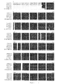
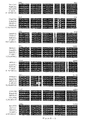
Preferred sequences according to the present invention are set out in Figures 1, 2, 4
and 5: SEQ ID NO 1 to 2 and 15 to 24. Also unique parts and fragments of these
sequences are part of the present invention. Preferred unique parts are set out in Table
2.
-
Since SEQ ID NO 2 shows 96.6% homology to its closest found validly named
neighbour, the use of sequences of more than 96.6% homology to SEQ ID NO 1 for
identification or typing of Helicobacter species is also within the scope of the present
invention. Preferably sequences of more than 97%, 97.5%, 98%, 98.5%, 99% or
99.5% homology to SEQ ID NO 2 are used for this goal.
-
According to another embodiment, the present invention relates to a part of an
isolated polynucleic acid as defined above, more particularly part or a fragment of
SEQ ID NO 1 or 2, wherein said part is unique to the polynucleic acid sequence it is
derived from.
-
According to the present invention, the term "unique" implies that at least one
nucleotide of the fragment or part is different from a nucleotide present at the same
nucleotide position in a known 16S rRNA sequence or the corresponding gene. Such a
nucleotide can be deduced theoretically by looking at an alignment of the new
sequences of this invention with other closely related Helicobacter 16S rDNA gene
nucleotide sequences (see Figures 1, 2, 4 and 5). Said type of nucleotides are unique
to the sequence they are derived from. These fragments are thus not part of any
known 16S rRNA or gene sequence encoding the same. The fragments according to
this embodiment of the present invention may be of any length between 10 to the
maximum number of nucleotides of SEQ ID NO 1 or 2 or its variants. Preferred
lengths are 10, 15, 20, 25, 30, 35, 40, 45, 50, 55, 60, etc. nucleotides.
-
According to another embodiment, the present invention relates to a probe which
specifically hybridizes to a polynucleic acid sequence as defined above.
-
Preferred probes are given in Table 2. Probe R628f is a preferred "Candidatus
Helicobacter bovis" specific probe. Probe V100f is a preferred "Candidatus
Helicobacter suis" specific probe. Other suitable probes may be derived from a visual
inspection of the alignment shown in Figure 1 or 2.
-
According to another embodiment, the present invention relates to a primer which
specifically amplifies a polynucleic acid sequence as defined above.
-
Preferred primers according to the present invention are given in Table 2. Primers
R574f and R832r are preferred "Candidatus Helicobacter bovis" specific primers and
are suited for a specific PCR and in situ hybridisation assays. Primers V832f and
V1621r are preferred "Candidatus Helicobacter suis" specific primers for a specific
PCR and in situ hybridisation assays. Other suitable primers according to the present
invention may be derived from a visual inspection of the alignment shown in Figure 1
or 2.
-
Relying on the principles well kown in the art, the skilled man will be able to select
primers that allow specific amplification of SEQ ID NO 1 or 2 or the claimed variants
thereof under given or experimental conditions, such as temperature, buffer
composition, polymerase chain reaction cycle etc. Likewise the skilled man will be
able to select probes that specifically hybridize to either SEQ ID NO 1 or 2 or the
claimed variants under given experimental conditions such as temperature, buffer
composition etc. Having chosen primers and/or probes, the skilled man will
furthermore be able to assess the efficacy of these primers or probes without undue
experimentation. It is also obvious that the skilled man may chose to combine more
than one primer pair or more than one probe to carry out the method defined above.
-
In some cases, one may not wish to detect all SEQ ID NO 1 or 2 variants as specified
above, for instance if one intends to detect alleles found in a certain geographic
region.
-
According to another embodiment, the present invention relates to a method for
detection and/or typing of Helicobacter strains present in a biological sample
comprising hybridizing the 16S rRNA or 16S rDNA target region polynucleotides of
said Helicobacter strains present in said biological sample with at least one probe as
defined above.
-
Preferably said method may be used to study and detect the occurrence of pathogenic
Helicobacter strains.
-
According to another embodiment, the present invention relates to a method for
detection and/or typing of Helicobacter strains present in a biological sample
comprising specifically amplifying the 16S rRNA or 16S rDNA target region
polynucleotides of said Helicobacter strains present in said biological sample with at
least one primer as defined above.
-
Preferably said method may be used to study and detect the occurrence of pathogenic
Helicobacter strains.
-
A preferred embodiment according to the present invention involves a method for
detection and/or typing of Helicobacter strains present in a biological sample
comprising first amplifying a specifc target region encompassed in or comprising the
16S rRNA region of said Helicobacter strains present in said biological sample and
subsequently hybridizing the 16S rRNA or 16S rDNA target region polynucleotides
of said Helicobacter strains present in said biological sample with at least one (or
more than one) probe as defined above.
-
Different techniques can be applied to perform the methods of the present invention.
These techniques may comprise immobilizing the target polynucleic acids, possibly
after amplification, on a solid support and performing a hybridization with labelled
oligonucleotide probes of the present invention. Alternatively, said probes may be
immobilized on a solid support and hybridization may be performed with labelled
target polynucleic acids, possibly after amplification (i.e. a reverse hybridization).
-
A preferred method according to the present invention is an in situ hybridisation
assay (see Examples section).
-
The well-known technique of Southern blotting is one example of a hybridization
assay that can be used to perform the methods of the present invention. Another
example of a hybridization technique is the DNA enzyme immuno assay (DEIA).
According to this method, PCR products are generated by a primer set, of which
either the forward or the reverse primer contain biotin at the 5' end. This allows
binding of the biotinylated amplimers to streptavidin-coated microtiter wells. PCR
products are denatured by sodium hydroxide, which allows removal of the non-biotinylated
strand. Specific digoxigenin (DIG)-labelled oligonucleotide probes are
hybridized to the single-stranded immobilized PCR product and hybrids are detected
by enzyme-labelled conjugate and colorimetric methods.
-
A convenient reverse hybridization technique is the LiPA assay. The LiPA uses
oligonucleotide probes immobilized as parallel lines on a solid support strip (Stuyver
et al. 1993; international patent application WO 94/12670). This approach is
particularly advantageous since it is fast and simple to perform.
-
It is to be understood that any other type of hybridization assay or hybridization
format using any of the selected probes as described further in the invention, is also
covered by the present invention.
-
According to another embodiment , the present invention relates to a diagnostic kit for
detection and/or typing of Helicobacter strains comprising:
- at least one probe as defined above and/or,
- at least one primer as defined above.
-
According to another embodiment, the present invention relates to a medicament
comprising a polynucleic acid sequence as defined above.
-
According to another embodiment, the present invention relates to a polynucleic acid
sequence as defined above for use as a medicament.
-
The following definitions and explanations will permit a better understanding of the
present invention.
-
The target material in the samples to be analysed may either be DNA or RNA, e.g.
genomic DNA, messenger RNA, viral RNA or amplified versions thereof. These
molecules are in this application also termed "polynucleic acids" or
"polynucleotides". More particularly, the target material according to the present
invention will be 16S ribosomal RNA or DNA or amplified versions thereof.
-
Well-known extraction and purification procedures are available for the isolation of
RNA or DNA from a sample (e.g. in Sambrook et al.,1989).
-
The term "probe" according to the present invention refers to a single-stranded
oligonucleotide which is designed to specifically hybridize to "Candidatus
Helicobacter bovis or suis" polynucleic acids.
-
The term "primer" refers to a single stranded oligonucleotide sequence capable of
acting as a point of initiation for synthesis of a primer extension product which is
complementary to the nucleic acid strand to be copied. The length and the sequence of
the primer must be such that they allow to prime the synthesis of the extension
products. Preferably the primer is about 5-50 nucleotides long. Specific length and
sequence will depend on the complexity of the required DNA or RNA targets, as well
as on the conditions at which the primer is used, such as temperature and ionic
strength. It is to be understood that the primers of the present invention may be used
as probes and vice versa, provided that the experimental conditions are adapted.
-
The expression "suitable primer pair" in this invention refers to a pair of primers
allowing specific amplification of a "Candidatus Helicobacter bovis or suis"
polynucleic acid fragment.
-
The term "target region" of a probe or a primer according to the present invention is a
sequence within the "Candidatus Helicobacter bovis or suis" polynucleic acids to
which the probe or the primer is completely complementary or partially
complementary (i.e. with some degree of mismatch). It is to be understood that the
complement of said target sequence is also a suitable target sequence in some cases.
-
"Specific hybridization" of a probe to a target region of respectively the "Candidatus
Helicobacter bovis" or Candidatus Helicobacter suis" polynucleic acids means that
said probe forms a duplex with part of this region or with the entire region under the
experimental conditions used, and that under those conditions said probe does not
form a duplex with other regions of the polynucleic acids present in the sample to be
analysed.
-
"Specific hybridization" of a primer to a target region of respectively the "Candidatus
Helicobacter bovis" or "Candidatus Helicobacter suis" polynucleic acids means that,
during the amplification step, said primer forms a duplex with part of this region or
with the entire region under the experimental conditions used, and that under those
conditions said primer does not form a duplex with other regions of the polynucleic
acids present in the sample to be analysed. It is to be understood that "duplex" as
used hereby, means a duplex that will lead to specific amplification.
-
"Specific amplification" of a fragment of respectively the "Candidatus Helicobacter
bovis" or Candidatus Helicobacter suis" polynucleic acids means amplification of the
fragment for which the primers were designed, and not of any other fragment of the
polynucleic acids present in a sample.
-
The fact that amplification primers do not have to match exactly with the
corresponding target sequence in the template to warrant proper amplification is
amply documented in the literature (Kwok et al., 1990). However, when the primers
are not completely complementary to their target sequence, it should be taken into
account that the amplified fragments will have the sequence of the primers and not of
the target sequence. Primers may be labelled with a label of choice (e.g. biotine).
The amplification method used can be either polymerase chain reaction (PCR; Saiki et
al., 1988), ligase chain reaction (LCR; Landgren et al., 1988; Wu & Wallace, 1989;
Barany, 1991), nucleic acid sequence-based amplification (NASBA; Guatelli et al.,
1990; Compton, 1991), transcription-based amplification system (TAS; Kwoh et al.,
1989), strand displacement amplification (SDA; Duck, 1990) or amplification by
means of Qß replicase (Lomeli et al., 1989) or any other suitable method to amplify
nucleic acid molecules known in the art.
-
Preferably, the probes of the invention are about 5 to 50 nucleotides long, more
preferably from about 10 to 25 nucleotides. Particularly preferred lengths of probes
include 10, 11, 12, 13, 14, 15, 16, 17, 18, 19, 20, 21, 22, 23, 24 or 25 nucleotides. The
nucleotides as used in the present invention may be ribonucleotides,
deoxyribonucleotides and modified nucleotides such as inosine or nucleotides
containing modified groups which do not essentially alter their hybridization
characteristics.
-
Probe and primer sequences are represented throughout the specification as single
stranded DNA oligonucleotides from the 5' to the 3' end. It is obvious to the man
skilled in the art that any of the below-specified probes can be used as such, or in their
complementary form, or in their RNA form (wherein T is replaced by U).
-
The probes according to the invention can be prepared by cloning of recombinant
plasmids containing inserts including the corresponding nucleotide sequences, if need
be by excision of the latter from the cloned plasmids by use of the adequate nucleases
and recovering them, e.g. by fractionation according to molecular weight. The probes
according to the present invention can also be synthesized chemically, for instance by
the conventional phospho-triester method.
-
The oligonucleotides used as primers or probes may also comprise nucleotide
analogues such as phosphorothiates (Matsukura et al., 1987), alkylphosphorothiates
(Miller et al., 1979) or peptide nucleic acids (Nielsen et al., 1991; Nielsen et al., 1993)
or may contain intercalating agents (Asseline et al., 1984). As most other variations
or modifications introduced into the original DNA sequences of the invention these
variations will necessitate adaptions with respect to the conditions under which the
oligonucleotide should be used to obtain the required specificity and sensitivity.
However, the eventual results of hybridization will be essentially the same as those
obtained with the unmodified oligonucleotides. The introduction of these
modifications may be advantageous in order to positively influence characteristics
such as hybridization kinetics, reversibility of the hybrid-formation, biological
stability of the oligonucleotide molecules, etc.
-
The term "solid support" can refer to any substrate to which an oligonucleotide probe
can be coupled, provided that it retains its hybridization characteristics and provided
that the background level of hybridization remains low. Usually the solid substrate
will be a microtiter plate, a membrane (e.g. nylon or nitrocellulose) or a microsphere
(bead) or a chip. Prior to application to the membrane or fixation it may be
convenient to modify the nucleic acid probe in order to facilitate fixation or improve
the hybridization efficiency. Such modifications may encompass homopolymer
tailing, coupling with different reactive groups such as aliphatic groups, NH2 groups,
SH groups, carboxylic groups, or coupling with biotin, haptens or proteins.
-
The term "labelled" refers to the use of labelled nucleic acids. Labelling may be
carried out by the use of labelled nucleotides incorporated during the polymerase step
of the amplification such as illustrated by Saiki et al. (1988) or Bej et al. (1990) or
labelled primers, or by any other method known to the person skilled in the art. The
nature of the label may be isotopic (32P, 35S, etc.) or non-isotopic (biotin, digoxigenin,
etc.).
-
The "biological sample" may be for instance cultured Helicobacter strains, gastric,
abomasal stomachs, omasal stomachs, reticulum and rumen, or duodenal biopsies
(fresh or parafine material), faeces, saliva, mouth mucosa, gastric juice or urine.
Preferably these samples may be taken from piglets, pigs, humans, calves, cattle, etc.
-
For designing probes with desired characteristics, the following useful guidelines
known to the person skilled in the art can be applied.
-
Because the extent and specificity of hybridization reactions such as those described
herein are affected by a number of factors, manipulation of one or more of those
factors will determine the exact sensitivity and specificity of a particular probe,
whether perfectly complementary to its target or not. The importance and effect of
various assay conditions are explained further herein.
-
**The stability of the [probe : target] nucleic acid hybrid should be chosen to
be compatible with the assay conditions. This may be accomplished by avoiding long
AT-rich sequences, by terminating the hybrids with G:C base pairs, and by designing
the probe with an appropriate Tm. The beginning and end points of the probe should
be chosen so that the length and %GC result in a Tm about 2-10EC higher than the
temperature at which the final assay will be performed. The base composition of the
probe is significant because G-C base pairs exhibit greater thermal stability as
compared to A-T base pairs due to additional hydrogen bonding. Thus, hybridization
involving complementary nucleic acids of higher G-C content will be more stable at
higher temperatures.
-
**Conditions such as ionic strength and incubation temperature under which a
probe will be used should also be taken into account when designing a probe. It is
known that the degree of hybridization will increase as the ionic strength of the
reaction mixture increases, and that the thermal stability of the hybrids will increase
with increasing ionic strength. On the other hand, chemical reagents, such as
formamide, urea, DMSO and alcohols, which disrupt hydrogen bonds, will increase
the stringency of hybridization. Destabilization of the hydrogen bonds by such
reagents can greatly reduce the Tm. In general, optimal hybridization for synthetic
oligonucleotide probes of about 10-50 bases in length occurs approximately SEC
below the melting temperature for a given duplex. Incubation at temperatures below
the optimum may allow mismatched base sequences to hybridize and can therefore
result in reduced specificity.
-
**It is desirable to have probes which hybridize only under conditions of high
stringency. Under high stringency conditions only highly complementary nucleic acid
hybrids will form; hybrids without a sufficient degree of complementarity will not
form. Accordingly, the stringency of the assay conditions determines the amount of
complementarity needed between two nucleic acid strands forming a hybrid. The
degree of stringency is chosen such as to maximize the difference in stability between
the hybrid formed with the target and the non-target nucleic acid.
-
**Regions in the target DNA or RNA which are known to form strong internal
structures inhibitory to hybridization are less preferred. Likewise, probes with
extensive self-complementarity should be avoided. As explained above, hybridization
is the association of two single strands of complementary nucleic acids to form a
hydrogen bonded double strand. It is implicit that if one of the two strands is wholly
or partially involved in a hybrid that it will be less able to participate in formation of a
new hybrid. There can be intramolecular and intermolecular hybrids formed within
the molecules of one type of probe if there is sufficient self complementarity. Such
structures can be avoided through careful probe design. By designing a probe so that a
substantial portion of the sequence of interest is single stranded, the rate and extent of
hybridization may be greatly increased. Computer programs are available to search
for this type of interaction. However, in certain instances, it may not be possible to
avoid this type of interaction.
-
**Standard hybridization and wash conditions are disclosed in the Examples
section. Other conditions are for instance 3X SSC (Sodium Saline Citrate), 20%
deionized FA (Formamide) at 50EC. Other solutions (SSPE (Sodium saline
phosphate EDTA), TMAC (Tetramethyl ammonium Chloride), etc.) and temperatures
can also be used provided that the specificity and sensitivity of the probes is
maintained. When needed, slight modifications of the probes in length or in sequence
have to be carried out to maintain the specificity and sensitivity required under the
given circumstances.
-
The term "hybridization buffer" means a buffer allowing a hybridization reaction
between the probes and the polynucleic acids present in the sample, or the amplified
products, under the appropriate stringency conditions.
-
The term "wash solution" means a solution enabling washing of the hybrids formed
under the appropriate stringency conditions.
-
The Examples as set out below only serve to illustrate the present invention. The
contents of all references referred to in this text are hereby incorporated by reference.
FIGURE AND TABLE LEGENDS
-
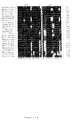
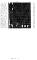
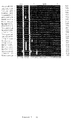
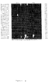
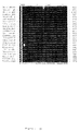
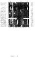
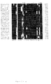
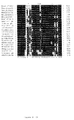
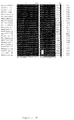
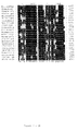
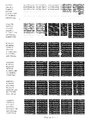
- Figure 1 represents an alignment of the reference sequence R2XA of "Candidatus
Helicobacter bovis" (SEQ ID NO 1) with sequences of strains belonging to the
epsilon subdivision (Table 3) which were retrieved from the EMBL data library and
were aligned with reference sequence R2XA
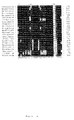
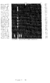
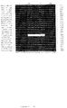
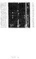
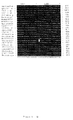
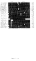
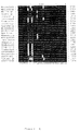
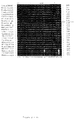
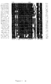
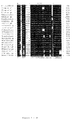
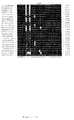
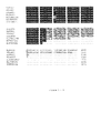
- Figure 2 represents an alignment of the reference sequence V2BXA of "Candidatus
Helicobacter suis" (SEQ ID NO 2) sequence with the sequences of strains belonging
to the same phylogenetic lineage (Table 3).
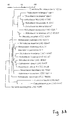
- Figure 3A represents a phylogenetic tree based on the phylogenetic analysis of
"Candidates Helicobacter bovis" as set out in the Examples section. The scale bar
represents a one % difference in nucleotide sequences as determined by measuring
the length of horizontal lines connecting any two species.
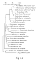
- Figure 3B represents a a phylogenetic tree based on the phylogenetic analysis of
"Candidatus Helicobacter suis" as set out in the Examples section. The scale bar
represents a one % difference in nucleotide sequences as determined by measuring the
length of horizontal lines connecting any two species.
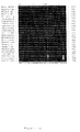
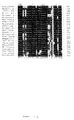
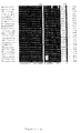
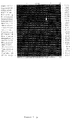
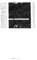
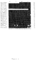
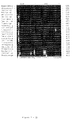
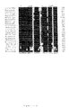
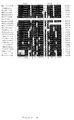
- Figure 4 represents an alignment of the different "Candidatus Helicobacter bovis"
sequences. The reference sequence is R2XA001 (SEQ ID NO 1). The other sequences
are R5XE001 (SEQ ID NO 15), R3XA001 (SEQ ID NO 16), R6XA001 (SEQ ID NO
17), R13D001INV (SEQ ID NO 18), R27TOTAAL (SEQ ID NO 19) and
R28TOTAAL (SEQ ID NO 20).
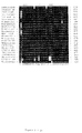
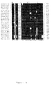
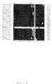
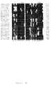
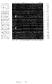
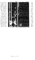
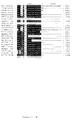
- Figure 5 represents an alignment of the different "Candidatus Helicobacter suis"
sequences. The reference sequence is RBXA001 (SEQ ID NO 2). The other
sequences are 4AXA001 (SEQ ID NO 21), 6W06001 (SEQ ID NO 22), V14D001
(SEQ ID NO 23), V19DINV001 (SEQ ID NO 24).
-
EXAMPLES
Material and Methods
Samples from cattle
-
Seven abomasal stomachs from clinically healthy slaughterhouse cattle originating
from different Belgian and Dutch farms, were selected. The stomachs were opened
longitudinally along the greater curvatura and rinsed gently with tap water. Two small
mucosal fragments were taken from each stomach, one near the torus pyloricus and
one in the fundic region, and were tested for urease activity (CUTest, Temmler
Pharma) for h at 37 °C. Three mucosal biopsies from the pyloric region were taken
for immunohistochemistry and in situ hybridisation and placed into 4 % buffered
formaline for 24 hours. For electronmicroscopy, a pyloric sample was taken from the
same region and fixed in cacodylate buffer (0.1 M, pH 7.0) containing 5 %
glutaraldehyde and 0.15 % (wt/vol) ruthenium red. From each stomach a mucosal
fragment was also taken for PCR analysis, placed into sterile PBS and frozen in liquid
nitrogen. Special care was taken during sampling to avoid cross-contamination.
Samples from pigs
-
Stomachs from 5 healthy slaughterhouse pigs were selected, all originating from
different farms in Belgium and the Netherlands. The stomachs were opened
longitudinally along the greater curvatura and rinsed gently with tap water. A small
mucosal fragment was taken from each stomach near the torus pyloricus and placed
into an urease test tube (CUTest, Temmler Pharma) for 2 hours at 37 °C. Mucosal
biopsies from the antral part of the stomach were taken along the curvatura major
(n=2) and the curvatura minor (n=2) for immunohistochemical evaluation and placed
into 4 % buffered formalin. For electronmicroscopy, samples were taken from the
same places and fixed in 0.1 M cacodylate buffer (pH 7.0) containing 5%
glutaraldehyde and 0.15 % (wt/vol) ruthenium red. Of each stomach a mucosal
fragment was also taken for PCR, placed into sterile PBS and frozen in liquid
nitrogen. Special care was taken during sampling to avoid cross-contamination.
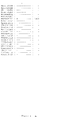
Reference strains for "Candidatus Helicobacter bovis"
-
A total of 15 Helicobacter strains and 1 Wolinella succinogenes strain were used to
test the specificity of the "Candidatus Helicobacter bovis" specific PCR (Table 1).
Strains were grown on a 5 % Mueller-Hinton blood agar and incubated at 37 °C in a
microaerophilic atmosphere containing approximately 5 % O2, 3.5 % CO2, 7.5 % H2
and 84 % N2. Bacteriological purity was checked by plating and Gram-staining.
Reference strains for "Candidatus Helicobacter suis"
-
A total of 15 Helicobacter strains were used to test the specificity of the "Candidatus
Helicobacter suis"-specific PCR assay (Table 1). Strains were grown on a 5%
Mueller-Hinton blood agar and incubated at 37 °C in a microaerobic atmosphere
containing approximately 5 % O2, 3.5 % CO2, 7.5 % H2 and 84 % N2. Bacteriological
purity was checked by plating and Gram-staining.
Immunohistochemistry
-
Immunohistochemical staining was performed to assess the presence of Helicobacter-like
organisms. Formalin-fixed samples were dehydrated and paraffin-embedded.
Sections of 4 µm were made of the paraffin-embedded tissues and were placed on 3-aminopropyltriethoxysilane-coated
slides (APES, Sigma-Aldrich) and dried overnight
at 60 °C. After dewaxing with xylene and rehydration in graded series with ethanol
and distilled water, sections were placed in citrate buffer (0.1M with 2% ureum) and
were boiled (1 x 3 min, 2 x 5 min) in a 800 Watt microwave oven (Whirlpool M611)
to elicit antigen retrieval. Slides were then incubated with 12 % hydrogenperoxide in
methanol (30 min) in order to block endogenous peroxidase activity. Thereafter the
slides were pre-incubated with 30 % normal goat serum in PBS for 30 min to reduce
non-specific antibody binding. A mouse polyclonal antibody directed against H.
pylori (DAKO), diluted 1/20 in PBS, was incubated overnight at 21 °C in a moist
chamber. The sections were washed and incubated with biotinylated swine anti-rabbit
immunoglobulins at 21 °C for 30 min and after rinsing covered with peroxidase
conjugated streptavidin-biotin-complex (ABC). Peroxidase activity was developed
using H2O2 with diaminobenzidine (DAB) as a chromogen (Fast DAB Tablet Set,
Sigma-Aldrich). Subsequently, the sections were counterstained with Mayer's
hematoxylin and mounted. As a negative control, the primary antibody was replaced
with fetal calf serum in Tris-HCl buffer (pH 7.6). As a positive control, a section of a
mouse stomach experimentally infected with Helicobacter pylori LMG 7539T was
used.
Transmission electronmicroscopy
-
For "Candidatus Helicobacter bovis", three different pyloric samples were selected
for electronmicroscopic evaluation based upon the high presence of Helicobacter-like
organisms in the corresponding immunostained sections.
-
For "Candidatus Helicobacter suis", two different antral biopsies were selected for
electronmicroscopic evaluation based on the high presence of gastrospirillum-like
organisms in the corresponding immunostained slides.
-
After dehydration in a graded series of acetone washes, the samples were embedded
in Spurr low-viscosity resin. Ultrathin sections were poststained with uranyl acetate
and lead citrate and examined with an electronmicroscope (Phillips 201 TEM) at an
accelerating voltage of 60 kV.
DNA-extraction
-
DNA was isolated from the scrapings of the gastric biopsies and from the reference
strains by lysis with guanidinium isothiocyanate and DNA was bound to silica
particles according to the method of Boom et al. (1990).
Primers and PCR amplification of 16S rDNA
-
Broadrange primers H33f, H61f and H1368r were selected from rRNA superfamily
VI (Helicobacter, Campylobacter, Arcobacter, Wolinella) specific regions of the 16S
rRNA gene (Table 2).
-
The use of broad range primer 1492RPL was suggested by Weissburg et al. (1991). A
genus Helicobacter-specific primer H274f was adapted from primer 274r described
by Dewhirst et al. (1994) (Table 2). Primer combinations H33F-H1368r, H274f-1492RPL
and H61f-1492RPL were used to amplify a ~ 1.3-Kb, ~ 1.2-Kb and a ~1.4-Kb
fragment of "Candidatus Helicobacter suis" respectively.
-
PCR reactions were performed in a volume of 50 pl containing 10mM Tris HCl (pH
8.3), 50 mM KCl, 3.5 mM MgCl 2, 200 pM of each deoxynucleoside triphosphate, 1.5
U of AmpliTaq Gold (Perkin-Elmer, Roche Molecular Systems) and 25 pmol of both
forward and reverse primer (Eurogentec). Reactions were covered with mineral oil
and PCR was performed in a Biomed-60 thermocycler under the following conditions:
9 min preincubation at 94 °C to activate AmpliTaq Gold, followed by 50 cycles of 30
s at 94 °C, 45 s at 55 °C and 45 s at 72 °C. Final extension was performed for 5 min at
72 °C. DNA-extractions of Helicobacter acinonychis LMG 12684T and Helicobacter
mustelae LMG 8776 were used as positive controls.
Analysis of amplified samples
-
PCR products were separated on 1 % agarose gels and stained with ethidium bromide.
-
DNA-extractions of H. acinonychis LMG 12684T and H. mustelae LMG 8776 were
used as positive controls.
-
In order to determine whether PCR products were derived from Helicobacter-like
organisms, the desired DNA-bands were cut from the gels, diluted 1/2 in distilled
water and sequenced using the H33f and H1368r 5'-Indocarbocyanin (Cy5) for
"Candidatus Helicobacter bovis" and respectively H61f and 1492RPL
Indocarbocyanin (Cy5) labeled for "Candidatus Helicobacter suis". Partial sequences
were screened for homologous sequences using the NCBI GENINFO ® BLAST
Network service (http://www.ncbi.nlm.nih.gov/BLAST/) (Altshul et al., 1997).
DNA cloning and sequence analysis for "Candidatus Helicobacter bovis"
-
PCR amplimers comprising the 16S rDNA-sequences derived from four different
stomach samples (R2, R3, R5, R6) were each cloned into plasmid vector pGEM-T
(Promega Biotech) according to the manufacturer's instructions and transformed into
Escherichia coli JM109 using standard procedures. Plasmids were purified using the
Easy Prep Plasmid Preparation Kit (Pharmacia Biotech). Sequences were determined
by the T7-sequencing system (Pharmacia Biotech). Two primers flanking the multiple
cloning sites (T7, SP6) as well as internal primers H390f and H1053r were used
(Table 2). The sequence derived from the clone of the R2 sample (R2XA) was used as
reference sequence. This sequence was has been asigned Genbank Accession No.
AF127028. Sequence analysis was performed with the PCGene software
(Intelligenetics)
-
PCR amplicons of three other gastric samples (R13, R27, R28) were sequenced
without prior cloning (referred to below as direct sequence analysis).
DNA cloning and sequencing for "Candidatus Helicobacter suis"
-
PCR amplimers comprising the 16S rDNA-sequences from 2 different stomachs
(V2B, V4A) were cloned into plasmid vector pGEM-T (Promega Biotech) according
to the manufacturer's instructions and transformed into Escherichia coli JM109 using
standard procedures. Plasmids were purified using the Easy Prep Plasmid Prep Kit
(Pharmacia Biotech). Sequences were determined by the T7-sequencing system
(Pharmacia Biotech). Two primers flanking the multiple cloning sites (T7 and SP6) as
well as internal primers H390f and H1053r were used (Table 2). Sequence analysis
was performed with the PCGene software (Intelligenetics). A reference sequence was
determined based on its high length and was compared to the new sequencve and the
other derived sequences, to check its integrity (see Figures 4 and 5). The reference
sequence V2BXA was assigned Genbank Accession No. AF127028.
-
PCR amplicons of three other gastric samples (V5, V14, V19) were sequenced
without prior cloning (referred to below as direct sequence analysis).
Phylogenetic analysis for "Candidatus Helicobacter bovis"
-
Phylogenetic analysis was performed using the GeneCompar 2.0 software package
(Applied Maths). Sequences of strains belonging to the epsilon subdivision (Table 3)
were retrieved from the EMBL data library and were aligned with reference sequence
R2XA. A similarity matrix was constructed from the aligned sequences and was
corrected for multiple base changes by the method of Jukes & Cantor (1969).
Unknown bases and gaps were not considered in the numerical analysis. A
phylogenetic tree was constructed using the neighbour-joining method of Saitou &
Nei (1987).
Phylogenetic analysis for "Candidatus Helicobacter suis"
-
Phylogenetic analysis was performed using the GeneCompar 2.0 software package
(Applied Maths). All five "Candidatus Helicobacter suis" sequences and the
sequences of strains belonging to the same phylogenetic lineage (Table 3) were
aligned. Using the neighbour-joining method, a phylogenetic tree and corresponding
similarity matrix was constructed. Unknown bases and gaps were not considered in
the numerical analysis
"Candidatus Helicobacter bovis" specific PCR-assay
-
"Candidatus Helicobacter bovis" specific oligonucleotides R574f and R832r (Table
2), were selected from variable rDNA regions of the sequences determined by direct
and indirect sequence analysis. These primers comprised a 259 bp 16S rDNA-fragment
and were used to develop a specific PCR and an in situ hybridisation
procedure. Within this fragment an internal "Candidatus Helicobacter bovis" specific
probe R628f (Table 2) was selected for southern blot hybridisation purposes.
-
PCR reactions were performed in a volume of 50 µl containing 10 mM Tris HCl (pH
8.3), 50 mM KCl, 2.5 mM MgCl 2, 200 µM of each deoxynucleoside triphosphate, 1.5
U of AmpliTaq Gold, and 25 pmol of both forward and reverse primer. PCR
amplification was performed under the following conditions: 9 min preincubation at
94 °C to activate AmpliTaq Gold, followed by 40 cycles of 30 s at 94 °C, 45 s at 60
°C and 90 s at 72 °C. Final extension was performed for 5 min at 72 °C. All gastric
DNA-extracts were tested with this PCR. For positive controls, plasmid DNA was
used from the cloned 16S rDNA fragments (R2XA). As a negative control a DNA-extract
was used from an abomasum lacking of Helicobacter-like organisms.
-
Specificity of the "Candidatus Helicobacter bovis" specific oligonucleotides R574f
and R832r was tested by PCR using DNA-extracts of 15 different Helicobacter strains
and a Wolinella succinogenes strain (Table 1).
-
PCR products were separated on 2 % agarose gels, stained with ethidium bromide and
transferred to Hybond N+ (Amersham) by electro-elution. Southern blot hybridisation
was performed with the [γ32P] ATP labelled probe R628f (Table 2) according to
standard procedures (Amersham Pharmacia Biotech). In order to ensure the specificity
of the probe hybridisation, blots were washed twice with 0.1 x SSC + 0.1 % SDS at
55°C.
"Candidatus Helicobacter suis" specific diagnostic PCR-assay and Southern blot
hybridisation
-
"Candidatus Helicobacter suis"- specific primers (V832f and V1261r) were selected
from variable rDNA regions of the sequences determined by direct and indirect
sequence analysis, comprising a ~0.4-Kb 16S rDNA-fragment. Within this fragment a
"Candidatus Helicobacter suis"-specific probe V1000f (Table 2) was selected for
hybridisation purposes. PCR reactions were performed in a volume of 50 pl
containing 10 mM Tris HCl (pH 8.3), 50 mM KCl, 2.5 mM MgCl 2, 200 µM of each
deoxynucleoside triphosphate, 1.5 U of AmpliTaq Gold (Perkin-Elmer), and 25 pmol
of both forward and reverse primer (Eurogentec). PCR amplification was performed
under the following conditions: 9 min preincubation at 94 °C to activate AmpliTaq
Gold, followed by 40 cycles of 30 s at 94 °C, 45 s at 60 °C and 90 s at 72 °C. Final
extension was performed for 5 min at 72 °C. As a positive control, plasmid DNA was
used from the cloned 16S rDNA fragments (V2B, V4A). As a negative control DNA
extracted from the stomach of a gnotobiotic piglet was used.
-
To test the specificity of the primers, PCR was also performed on DNA-extracts of 15
different Helicobacter species. (Table 1).
-
PCR products were separated on 2% agarose gels, stained with ethidium bromide and
transferred to Hybond N+ (Amersham) by electro-blotting. Southern blot
hybridisation was performed with the [γ32P] ATP-labelled probe V1000f according to
standard procedures (Amersham Pharmacia Biotech). In order to ensure the specificity
of the probe hybridisation, blots were washed twice with 0.1 x SSC + 0.1 % SDS at
55°C.
In situ hybridisation for "Candidatus Helicobacter bovis"
-
In order to make the link between the "Candidatus Helicobacter bovis" specific probe
and the bacterial spiral cells observed in the tissue sections, an in situ hybridisation
procedure was performed on the formalin fixed and paraffin embedded pyloric
samples of each animal. A 259-base digoxigenin-labeled probe was synthezised using
the "PCR Dig Probe Synthesis Kit" (Boehringer Mannheim) in combination with the
"Candidatus Helicobacter bovis" specific primers R574f and R832r (Table 2). PCR
conditions were identical to those described in the diagnostic PCR assay. The
resulting PCR product was purified using the "High Pure PCR Product Purification
Kit" (Boehringer Mannheim) following manufacturer's instructions.
-
To avoid RNA'se activity, all glassware was heated at 180°C for 3 hours. Further
precautions included the use of RNA'se-free water, and the use of sterile disposable
materials whenever possible. Sections of the paraffin-embedded tissues (4 µm thick)
were mounted on RNA'se-free, APES-coated slides (Sigma-Aldrich) and fixed by
heating for 1 hour at 60 °C. The sections were deparaffinized in xylene (2x5 min),
rehydrated through graded ethanol, and washed twice in PBS for 5 min each.
Sections were then treated with proteinase K (DAKO) for 15 min each at 37 °C in a
humidified chamber. The enzyme was inactivated by treatment with 0.2 % glycine in
PBS for 3 min. Sections were washed twice in PBS for 5 min each, dehydrated in
graded ethanol and air dried. Tissues were circumlined with a DAKO Pen (DAKO) to
avoid liquid spillage during further processing and to ensure an efficient sealing of the
coverslip. For the hybridisation step, sections were covered with 5 to 15 pl solution,
containing 5 ng/µl labeled probe in 50 % deionized formamide, 2x SCC, 10 % dextran
sulfate, 0.25 µg/µl yeast t-RNA, 0.5 µg/µl heat denatured salmon sperm DNA, and lx
Denhart's solution. Sections were covered with a piece of coverslip to avoid
evaporation. To denature the probe, sections were heated for 10 min at 95 °C and
chilled on ice for 10 min. Slides were then hybridised overnight at 37 °C in a
humidified chamber. To remove the unbound probe, the coverslips were removed and
the sections were washed in 2x SCC and 1x SCC at room temperature for 10 min each
followed by two washes of 0.3x SCC at 40 °C for 10 min and at room temperature for
10 min, respectively.
-
All steps involving the immunological detection of the hybridised probe were
performed at room temperature. The sections were treated first for 30 minutes in
Buffer 1 (100mM Tris HCl, 150 mM NaCI, pH 7.5) containing 2 % normal goat
serum and 0.3 % Triton X-100. An incubation step followed for 3 hours with diluted
(1:30 in the same solution) anti-digoxigenin antibodies conjugated to horse-radish
peroxidase (DAKO). Unbound antibodies were washed gently on a shaker with
Buffer 1 followed by Buffer 2 (100 mM Tris HCl, 100 mM NaCI, 50 mM MgCl2, pH
9.5) for 15 min each. To optimize the detection level, the "Tyramid Signal
Amplification System" (NEN Life Science Products) was applied on each section,
following manufacturer's instructions. The hybridised probe was then visualized,
using H2O2 with diaminobenzidine as a chromogen (Fast DAB Tablet Set, Sigma-Aldrich).
Thereafter the sections were counterstained with Mayer's hematoxylin and
mounted.
In situ hybridisation for "Candidatus Helicobacter suis"
-
To link the derived sequence to the corresponding organism, an in situ hybridisation
procedure was performed on the formalin fixed and paraffin embedded pyloric
samples of each animal. A ~0.4 Kb digoxigenin-labeled probe was synthezised using
the "PCR Dig Probe Synthesis Kit" (Boehringer Mannheim) in combination with the
"Candidatus Helicobacter suis"-specific primers V832f and V1261r (Table 2). The
rest of the method was performed as mentioned above for "Candidatus Helicobacter
bovis".
Nucleotide sequence accession numbers
-
Accession numbers of the 16S rDNA gene sequences used for the phylogenetic
analysis are listed in table 3.
-
The 16S rDNA nucleotide sequence of "Candidatus Helicobacter bovis" has been
deposited in the Genbank database under accession number AF127027.
-
The 16S rDNA nucleotide sequence of "Candidatus Helicobacter suis" has been
deposited in the Genbank database under accession number AF127028.
Results
Urease activity and immunohistochemical evaluation for the cattle samples
-
Urease activity was observed in all pyloric samples (7/7). In the fundic samples,
urease activity was absent (0/7). Spiral immunostained organisms were observed in
the pyloric samples of all animals. The highest concentration was seen in the most
distal pyloric samples. They were mostly situated in the mucus layer and in the lumen
of the proximal part of the gastric crypts where they formed small clusters. In some
samples, coccoid organisms, were observed between the spiral bacteria, which also
crossreacted with the H. pylori polyclonal antibodies. In the positive control only
Helicobacter pylori - like bacteria were stained while in the negative controls no
staining was observed.
Urease activity and Immunohistochemical evaluation for the pig samples
-
Tightly coiled immunostained spiral organisms, morphologically similar to
"Gastrospirillum suis" (Queiroz et al., 1990) were observed in all stomachs (5/5),
which was consistent with the presence of urease activity (5/5). The gastrospirillum-like
organisms were seen laying separately or in small clusters with a patchy
distribution over the sample, and were found mostly in the superficial part of the
gastric crypts. Some bacteria revealed bipolar immunostained flagellae.
Immunostained coccoid-like organisms were also observed in the pyloric crypts. In
the positive control, only Helicobacter pylori-like bacteria were stained while in the
negative controls no labeling was observed.
Transmission electronmicroscopy for "Candidatus Helicobacter bovis"
-
Large groups of multiple spiral bacteria were seen within the crypts of the gastric
mucosa. There was no obvious cell association between the bacteria and the gastric
cells, neither were there any intracellular bacterial inclusions. The bacteria were
helical-shaped and had 1-3 complete spiral turns per cell with a wavelength of
approximately 750 nm. Cells were 1 - 2.5 µm long and 0.3 µm wide. At least four
flagelles were seen at one end. It was unclear whether these flagellae were uni- or
bipolar, neither could the presence or absence of a flagella sheath be noted.
Transmission electronmicroscopy for "Candidatus Helicobacter suis"
-
Within the gastric crypts of the antral region, longitudinal and transversal sections of
spiral organisms could be seen. All bacteria had the same characteristic tightly coiled
appearance, typical of Helicobacters with the gastrospirillum morphology. The length
of cells varied from 2.5 to 3.5 pm and they were approximately 0.6 pm wide. Multiple
complete spiral turns with a wavelength of ± 600 nm were seen in all longitudinal
sections. As only few longitudinal sections of the bacteria were obtained, the number
and implantation of the flagellae could not be studied although partial fragments
were observed. The bacteria were not seen intracellularly nor was there any obvious
cell association with the surrounding epithelial cells. The presence or absence of a
flagella sheath could not be noted.
Amplification, cloning and sequencing of Helicobacter-like 16S rDNA fragments
from cattle samples
-
PCR amplification of the 16S rRNA gene using the H33f and H1368r primers,
produced a fragment of the expected size range (± 1.3 Kbp) in all seven samples
examined. Partial direct sequence analysis of four of these bands (R2, R3, R5, R6)
and subsequent database comparison (BLAST) confirmed the PCR products to be
Helicobacter-like 16S rDNA fragments. Four PCR products (R2, R3, R5, R6) were
cloned followed by partial screening. In one clone a Clostridium-like 16S rDNA
fragment was found. In all other clones Helicobacter-like fragments were inserted.
The 16S rDNA sequences of four clones derived from different animals (R2XA,
R3XA, R5XE, R6XA), were determined. Additional sequences of three other samples
(R13, R27, R28) were characterized by direct sequence analysis using the primers
H33f, H1368r, H390f and H1053r.
Amplification, cloning and sequencing of Helicobacter-like 16S rDNA fragments
from pig samples
-
Several combinations of PCR primers yielded sequences of the expected size. The
length of these amplified fragments varied between 1.2 Kb (H274f-1492RPL) and 1.4
Kb (H61f-1492RPL). The latter primer combination was used to examine all samples.
The 16S rDNA sequences of two different clones were determined (V2BXA,
V4AXA). Additional sequences of 3 other samples (V5, V14, V19) were determined
by direct sequence analysis.
Sequences and phylogenetic analysis for cattle samples
-
Sequence length varied from 1267 to 1335 basepairs. Pairwise comparisons between
these 7 sequences revealed a sequence homology of more than 99 %. One reference
sequence (R2XA) of 1335 bp (see Figure 1: SEQ ID NO 1) was selected for
phylogenetic evaluation. A similarity matrix based on comparisons of 16S rRNA
sequences of 23 strains representing all validly named Helicobacter species,
"Helicobacter heilmannii" (type1, type2), Campylobacter jejuni, Arcobacter
cryaerophilus and Wollinella succinogenes was calculated. By this analysis it was
shown that the sequences of the bovine Helicobacter-like organisms form a distinct
group within the genus Helicobacter with Helicobacter bilis as closest taxonomic
relative (level of similarity 92.8 %). The reference sequence was clearly distinct from
sequences belonging to other superfamily VI genera, as shown by a 85.6, 85.1 % and
89.7 % homology with Campylobacter jejuni, Arcobacter butzleri and Wolinella
succinogenes respectively. A phylogenetic tree based on this analysis is shown in Fig.
3A.
Sequences and phylogenetic analysis for pig samples
-
The 5 sequences that were determined had lengths varying from 1345 to 1421
basepairs. Pairwise comparisons between 1345 bp consensus fragments of these
sequences, revealed a minimum homology of 97.7 %. One reference sequence of 1421
bp, obtained from PCR product 2BXA (see Figure 2: SEQ ID NO 2), was used for
phylogenetic analysis. A similarity matrix was calculated based on comparisons of
16S rDNA sequences of all Helicobacter species, "Helicobacter heilmannii" type 1
and type 2, Campylobacter jejuni, Arcobacter butzleri and Wollinella succinogenes
(Table 3). In this analysis, the sequence of the porcine gastrospirillum-like organism
formed a distinct subgroup within the Helicobacter lineage together with other
gastrospirilla: Helicobacter felis, H. bizzozeronii, H. salomonis, "H. heilmannii" type
1 and type 2. The sequence was highly similar to that of "H. heilmannii" type 1 (level
of similarity 99.5 %). The similarity level of other gastrospirillum-like bacteria, H.
felis, H. bizzozeronii, H. salomonis and H. heilmanni type 2 was 96.4 %, 96.5 %, 96.6
% and 96.8 % respectively. The reference sequence was clearly distinct from
sequences belonging to other superfamily VI-genera, as shown by a 86.2 %, 84.7 %
and 89.6 % homology with Campylobacter jejuni, Arcobacter butzleri and Wolinella
succinogenes respectively.
-
A phylogenetic tree based on this analysis is shown in Figure 3B.
Diagnostic PCR-assay for "Candidatus Helicobacter bovis"
-
A 259 base fragment was produced for all seven stomach samples with primer pair
R574f-R832r. All PCR products crosshybridised with the R628f probe after southern
blot hybridisation. No amplification product was obtained using DNA preparations
from any of the Helicobacter strains, nor from the bovine Wolinella succinogenes
strain (Table 1). The positive control yielded a ~0.3 Kb product as expected. There
was no DNA-amplification using the negative control material.
"Candidatus Helicobacter suis"-specific PCR and Southern blot hybridisation
-
Amplification of Helicobacter DNA using the primers V832f and V1261r produced a
433-base fragment from all five stomach samples. All PCR products hybridised with
the V1000f probe after Southern blot hybridisation. No amplification product was
obtained using DNA preparations from any of the Helicobacter strains including H.
felis, H. bizzozeronii and H. salomonis (Table 1), nor from the negative control. PCR
with the cloned reference material (2BXA) yielded a ~0.4 Kb product as expected.
In situ hybridisation for "Candidatus Helicobacter bovis"
-
In situ hybridisation of the bovine Helicobacter-like bacteria with the "Candidatus
Helicobacter bovis"- specific probe was seen in sections from all (7/7) stomachs.
These bacteria were observed as darkbrown spiral organisms, organised in small
clusters, situated in the gastric crypts of the pyloric part of the abomasal stomach. Not
all spiral bacteria were stained. Sometimes a faint background, seen as fine stained
strings, was observed in the surrounding cells. This background staining was also
observed in the H. pylori-infected mouse stomach which was used as a negative
control. The H. pylori cells in this control though did not hybridise with the
"Candidatus Helicobacter bovis"- specific probe.
In situ hybridisation for "Candidatus Helicobacter suis"
-
In situ hybridisation of "Gastrospirillum suis"-like bacteria with the "Candidatus
Helicobacter suis"- specific probe was seen in sections from all (5/5) stomachs.
Bacteria were observed as darkbrown spiral organisms in the superficial mucus layer
and the gastric crypts. In some cases, helical organisms located deeply in the crypts,
were weakly labeled or were negative. Sometimes a faint background, seen as fine
stained strings, was observed in the surrounding cells. This background staining was
also observed in the H. pylori-infected mouse stomach which was used as a negative
control. The H. pylori cells in this control though did not hybridise with the
"Candidatus Helicobacter suis"- specific probe.
REFERENCES
-
Rappin, J. (1881). Contribution à l'étude de bactéries de la bouche à l'état
normal. Ph.D. thesis. Collège de France, Nantes.
-
Bizzozero, G. (1893). Ueber die schlauchformigen Drüsen des
Magendarmkanals und die Beziehungen ihres Epithels zu dem Oberflächenepithel der
Schleimhaut. Arch Mikrosk Anat 42, 82.
-
Salomon, H. (1896). Ueber das Spirillum des Säugentiermagens und sein
Verhalten zu den Belegzellen. Zentralbl Bakt 19 , 433.
-
Marshall, B.J. & Warren, J.R. (1984). Unidentified curved bacilli in the
stomach of patients with gastritis and peptic ulceration. Lancet 1(8390), 1311-1315.
-
Cover, T.L. & Blaser M.J. (1992). Helicobacter pylori and gastroduodenal
disease. Annu Rev Med 43, 135-145.
-
Blaser, M.J., Perez-Perez, G.I., Lindenbaum, J., Schneidman, D., Van
Deventer, G., Marin-Sorensen, M. & Weinstein, W.M. (1991). Association of
infection due to Helicobacter pylori with specific upper gastrointestinal pathology.
Rev Infect Dis 13 Suppl 8, S704-708.
-
Parsonnet, J., Friedman, G.D., Vandersteen, D.P., Chang, Y., Vogelman,
J.H., Orentreich, N. & Sibley, R.K. (1991). Helicobacter pylori infection and the
risk of gastric carcinoma [see comments]. N Engl J Med 325, 1127-1131.
-
On, S.L. (1996). Identification methods for campylobacters, Helicobacters, and
related organisms. Clin Microbiol Rev 9, 405-422.
-
Franklin, C.L., Beckwith, C.S., Livingston, R.S., Riley, L.K., Gibson, S.V.,
Besch-Williford, C.L. & Hook, R.R., Jr. (1996). Isolation of a novel Helicobacter
species, Helicobacter cholecystus sp. nov., from the gallbladders of Syrian hamsters
with cholangiofibrosis and centrilobular pancreatitis. J Clin Microbiol 34, 2952-2958.
-
Mendes, E.N., Queiroz, D.M., Dewhirst, F.E., Paster, B.J., Moura, S.B. &
Fox, J.G. (1996). Helicobacter trogontum sp. nov., isolated from the rat intestine. Int
J Syst Bacterial 46, 916-921.
-
Jalava, K., Kaartinen, M., Utriainen, M., Happonen, I. & Hanninen, M.L.
(1997). Helicobacter salomonis sp. nov., a canine gastric Helicobacter sp. related to
Helicobacter felis and Helicobacter bizzozeronii. hit J Syst Bacteriol 47, 975-982.
-
Trivett-Moore, N.L., Rawlinson, W.D., Yuen, M. & Gilbert, G.L. (1997). Helicobacter westmeadii sp. nov., a new species isolated from blood cultures of two
AIDS patients. J Clin Microbiol 35, 1144-1150.
-
Shen, Z., Fox, J.G., Dewhirst, F.E., Paster, B.J., Foltz, C.J., Yan, L.,
Shames, B. & Perry, L. (1997). Helicobacter rodentium sp. nov., an urease-negative
Helicobacter species isolated from laboratory mice. Int J Syst Bacteriol 47, 627-634.
-
Vandamme, P., Falsen, E., Rossau, R., Hoste, B., Segers, P., Tytgat, R. &
De Ley, J. (1991). Revision of Campylobacter, Helicobacter, and Wolinella
taxonomy: emendation of generic descriptions and proposal of Arcobacter gen. nov.
Int J Syst Bacteriol 41, 88-103.
-
Dent, J.C., McNulty, C.A.M., Uff, J.C., Wilkinson, S.P. & Gear, M.W.L.
(1987). Spiral organisms in the gastric antrum [Letter]. Lancet 2 (8550), 96.
-
Günther, H. & Schulze, F. (1992). Histologische Untersuchungen zum
Vorkommen von Campylobacter-ahnlich geformten Keimen im Labmagen von
Kälbern. Zentralbl Veterinarmed B 39, 737-745.
-
Haringsma, P.C. & Mouwen, J.M. (1992). Mogelijke betekenis van
spirilvormige bacteriën bij het ontstaan van lebmaagzweren bij het volwassen rund.
Tijdschr Diergeneeskd 117, 485-487.
-
Seidel, K.E., Kampschulte, J., Lehn, N. & Bauer, J. (1996). Helicobacter pylori: Antikörper in Seren von Schweinen und Kälbern. Berl Munch Tierarztl Wschr
109, 434-439.
-
Korhonen, H., Syväoja, E.-L., Ahola-Lutila, H., Sivelä, S., Kopola, S., Husu,
J. & Kosunen, T.U. (1995). Bactericidal effect of bovine normal and immune serum,
colostrum and milk against Helicobacter pylori. J Appl Bacteriol 78, 655-662.
-
Jelinski, M.D., Ribble, C.S., Chirino-Trejo, M., Clark, E.G. & Janzen, E.D.
(1995). The relationship between the presence of Helicobacter pylori, Clostridium
perfringens type A, Campylobacter spp., or fungi and fatal abomasal ulcers in
unweaned beef calves. Can Vet J 36, 379-382.
-
Braun, U., Anliker, H., Corboz, L. & Ossent, P. (1997). Untersuchungen über
das Vorkommen von spiralformigen Bakterien im Labmagen das Rindes. Schweiz
Arch Tierheilkd 139, 507-516.
-
Al-Mashat, R.R. & Taylor, D.J. (1980). Campylobacter spp. in enteric lesions
in cattle. Vet Rec 107, 31-34.
-
Mills, K.W., Johnson, J.L., Jensen, R.L., Woodard, L.F. & Doster, A.F.
(1990). Laboratory findings associated with abomasal ulcers/tympany in range calves.
J Vet Diagn Invest 2, 208-212.
-
Boom, R., Sol, C.J.A., Salimans, M.M.M., Jansen, C.L., Wertheim van
Dillen, P.M.E. & van der Noorda J. (1990). Rapid and simple method for
purification of nucleic acids. J Clin Microbiol 28, 495-503.
-
Altschul, S.F., Madden, T.L., Schäffer, A.A., Zheng Zhang, J.Z., Miller W.
& Lipman D.J. (1997). "Gapped BLAST and PSI-BLAST: a new generation of
protein database search programs". Nucleic Acids Res 25, 3389-3402.
-
McNulty, C.A., Dent, J.C., Curry, A., Uff, J.S., Ford, G.A., Gear, M.W. &
Wilkinson, S.P. (1989). New spiral bacterium in gastric mucosa [see comments]. J
Clin Pathol 42, 585-591.
-
Solnick, J.V., O'Rourke, J., Lee, A., Paster, B.J., Dewhirst F.E. &
Tompkins L.S. (1993). An uncultured gastric spiral organism is a newly identified
Helicobacter in humans. J Infect Dis 168, 379-385.
-
Andersen, L.P., Norgaard, A., Holck, S., Blom, J. & Elsborg, L. (1996).
Isolation of a "Helicobacter heilmanii"-like organism from the human stomach
[letter]. Fur J Clin Microbiol Infect Dis 15, 95-96.
-
Jukes, T. & Cantor, C. (1969). Evolution of protein molecules. In Mammalian
protein metabolism, vol 3. Edited by H. Munro. New York: Academic Press.
-
Saitou, N. & Nei, M. (1987). The neighbor-joining method: a new method for
reconstructing phylogenetic trees. Mol Biol Evol 4, 406-425.
-
Wolin, M.J. & Jacobs, N.J. (1961). Cytochrome-producing anaerobic vibrio,
Vibrio succinogenes, sp. nov. J Bacteriol 81, 911-917.
-
Paster, B.J. & Canale-Parola, E. (1985). Treponema saccharophilum sp. nov.,
a large pectinolytic spirochete from the bovine rumen. Appl Environ Microbiol 50,
212-219.
-
Stanton, T.B. & Canale-Parola, E. (1980). Treponema bryantii sp.nov., a
rumen spirochete that interacts with cellulolytic bacteria. Archiv Microbiol 127, 145-156.
-
Lee, A., Hazell, S.L., O'Rourke, J. & Kouprach, S. (1988). Isolation of a
spiral-shaped bacterium from the cat stomach. Infect Immun 56, 2843-2850.
-
O'Rourke, J., Solnick, J., Lee, A. & Tompkins, L. (1992). Helicobacter
heilmanii (previously Gastrospirillum), a new species of Helicobacter in humans and
animals [abstract]. J Med Science 101, 31.
-
Hänninen, M.-L., Happonen, I., Saari, S. & Jalava, K. (1996). Culture and
characteristics of Helicobacter bizzozeronii, a new canine gastric Helicobacter sp
[published erratum appears in Int J Syst Bacteriol 1996 Jul;46(3):839 ]. Int J Syst
Bacteriol 46, 160-166.
-
Queiroz, D.M.M., Rocha, G.A., Mendes, E.N., Lage, A.P., Carvalho, A.C.T.
& Barbosa, A.J.A. (1990). A spiral microorganism in the stomach of pigs. Vet
Microbiol 24, 199-204.
-
Mendes, E.N., Queiroz, D.M.M., Rocha, G.A., Moura, S.B., Leite, V.H.R. &
Fonseca, M.E.F. (1990). Ultrastructure of a spiral micro-organism from pig gastric
mucosa ("Gastrospirillum suis"). J Med Microbiol 33, 61-66.
-
Mendes, E.N., Queiroz, D.M.M., Rocha, G.A., Nogueira, A.M., Carvalho,
A.C., Lage, A.P. & Barbosa, A.J. (1991). Histopathological study of porcine gastric
mucosa with and without a spiral bacterium ("Gastrospirillum suis"). J Med
Microbiol 35, 345-348.
-
Barbosa, A.J., Silva, J.C., Nogueira, A.M., Paulino, Junior, E. & Miranda,
C.R. (1995). Higher incidence of Gastrospirillum sp. in swine with gastric ulcer of
the pars oesophagea. Vet Pathol 32, 134-139.
-
Queiroz, D.M.M., Rocha, G.A., Mendes, E.N., De Moura, S.B., De Oliveira,
A.M. & Miranda, D. (1996). Association between Helicobacter and gastric ulcer
disease of the pars esophagea in swine [see comments]. Gastroenterology 111, 19-27.
-
Moura, S.B., Queiroz, D.M., Mendes, E.N., Nogueira, A.M. & Rocha, G.A.
(1993). The inflammatory response of the gastric mucosa of mice experimentally
infected with "Gastrospirillum suis". J Med Microbiol 39, 64-68.
-
Mendes, E.N., Queiroz, D.M.M., Coimbra, R.S., Moura, S.B., Barbosa, A.J.
& Rocha G.A. (1996). Experimental infection of Wistar rats with "Gastrospirillum
suis". J Med Microbiol 44, 105-109.
-
Seymour, C., Lewis, R.G., Kim, M., Gagnon, D.F., Fox, J.G., Dewhirst, F.E.
& Paster, B.J. (1994). Isolation of Helicobacter strains from wild bird and swine
feces. Appl Env Microbiol 60, 1025-1028.
-
Dewhirst, F.E., Seymour, C., Fraser, G.J., Paster, B.J. & Fox, J.G. (1994).
Phylogeny of Helicobacter isolates from bird and swine feces and description of
Helicobacter pametensis sp.nov. Int J Syst Bacteriol 44, 553-560.
-
On, S.L.W., Bloch, B., Holmes, B., Hoste, B. & Vandamme P. (1995). Campylobacter hyointestinalis subsp. Lawsonii subsp. nov. isolated from the porcine
stomach, and an emended description of Campylobacter hyointestinalis, Int J Syst
Bacteriol 45, 767-774.
-
Suarez, D.L., Wesley, I.V. & Larson, D.J. (1997). Detection of Arcobacter
species in gastric samples from swine. Vet Microbiol 57, 325-336
-
Eaton, K.A., Radin, M.J., Kramer, L., Wack, R., Sherdino, R., Krakowka,
S., Fox, J.G. & Morgan, D.R. (1993). Epizootic gastritis associated with gastric
spiral bacilli in cheetas (Acinonyx jubatus). Vet Pathol 30, 55-63.
-
Jakob, W., Stolte, M., Valentin, A. & Schroder, H.D. (1997). Demonstration
of Helicobacter pylori-like organisms in the gastric mucosa of captive exotic
carnivores. J Comp Pathol 116, 21-33.
-
Stackebrandt, E. & Goebel, B.M. (1994). Taxonomic note: a place for DNA-DNA
reassociation and 16S rRNA sequence analysis in the present species definition
in bacteriology. Int J Sys Bacter 44, 846-849.
-
Murray, R.G., Stackebrandt, E. (1995) Taxonomic note: implementation of
the provisional status Candidatus for incompletely described procaryotes. Int J Syst
Bacteriol 45, 186-187.
-
Murray, R.G.E. & Schleifer, K.H. (1994). Taxonomic notes: a proposal for
recording the properties of putative taxa of procaryotes. Int J Syst Bacteriol 44, 174-176.
-
Stuyver L, Rossau R, Wyseur A, et al. (1993)
Typing of hepatitis C virus
isolates and characterization of new subtypes using a line probe assay. J. Gen. Virol.
74 : 1093-1102.
-
Sambrook J, Fritsch E, Maniatis T (1989)
Molecular cloning: a laboratory
manual. Cold Spring Harbor Laboratory Press, Cold Spring Harbor, NY.
-
Matsukura M, Shinozuka K, Zon G, Mitsuya H, Reitz M, Cohen J,
Broder S (1987) Phosphorothioate analogs of oligodeoxynucleotides : inhibitors of
replication and cytopathic effects of human immunodeficiency virus. Proc. Natl.
Acad. Sci. USA 84(21):7706-10.
-
Miller P, Yano J, Yano E, Carroll C, Jayaram K, Ts'o P (1979)
Nonionic
nucleic acid analogues. Synthesis and characterization of dideoxyribonucleoside
methylphosphonates. Biochemistry 18(23):513-43.
-
Nielsen P, Egholm M, Berg R, Buchardt O (1991)
Sequence-selective
recognition of DNA by strand displacement with a thymine-substituted polyamide.
Science 254(5037):1497-500.
-
Nielsen P, Egholm M, Berg R, Buchardt O (1993)
Sequence specific
inhibition of DNA restriction enzyme cleavage by PNA. Nucleic-Acids-Res. 21(2):197-200.
-
Asseline U, Delarue M, Lancelot G, Toulme F, Thuong N (1984)
Nucleic
acid-binding molecules with high affinity and base sequence specificity : intercalating
agents covalently linked to oligodeoxynucleotides. Proc. Natl. Acad. Sci. USA
81(11):3297-301.
| Bacterial strains used for the evaluation of the "Candidatus Helicobacter bovis" specific PCR |
|
Taxon
|
Source
|
Collection N° or Strain
|
|
Helicobacter acinonychis
|
Cheetah gastric mucosa |
LMG 12684T |
|
Helicobacter cinaedi
|
Human feces |
LMG 7543 T |
| Helicobacter sp. strain CLO-3 |
Human rectal swab |
LMG 7792 |
|
Helicobacter fennelliae
|
Human feces |
LMG 11759 |
|
Helicobacter pametensis
|
Tern feces |
LMG 12678 T |
| Helicobacter sp. strain Bird B |
Bird feces |
LMG 12679 |
| Helicobacter sp. strain Bird C |
Bird feces |
LMG 13642 |
|
Helicobacter hepaticus
|
Murine liver |
LMG 16316T |
|
Helicobacter pullorum
|
Chicken lower bowel |
LMG 16318 |
|
Helicobacter mustelae
|
Ferret gastric mucosa |
LMG 18044 T |
|
Helicobacter canis
|
Canine feces |
LMG 18086 T |
|
Helicobacter muridarum
|
Murine intestinal mucosa |
LMG 14378 T |
|
Helicobacter bizzozeronii
|
Canine gastric mucosa |
Strain 12A |
|
Helicobacter salonionis
|
Canine gastric mucosa |
CCUG 37845 T |
|
Helicobacter felis
|
Feline gastric mucosa |
CCUG 28539 T |


| Sources and accession numbers of strains used for phylogenetic analysis. |
|
Taxon
|
Source
|
Genbank Accession N°
|
| "Gastrospirillum hominis" type 1 |
Human gastric mucosa |
L10079 |
| "Gastrospirillum hominis" type 2 |
Human gastric mucosa |
L10080 |
|
Helicobacter acinonychis
|
Cheetahgastric mucosa |
M88148 |
|
Helicobacter bilis
|
Murine liver |
U18766 |
|
Helicobacter bizzozeronii
|
Canine gastric mucosa |
Y09404 |
|
Helicobacter canis
|
Canine feces |
L13464 |
|
Helicobacter cholecystus
|
Murine liver |
U46129 |
|
Helicobacter cinaedi
|
Human feces |
M88150 |
|
Helicobacter felis
|
Feline gastric mucosa |
M57398 |
|
Helicobacter fennelliae
|
Human feces |
M88154 |
|
Helicobacter hepaticus
|
Murine liver |
U07574 |
|
Helicobacter muridarum
|
Murine intestinal mucosa |
M80205 |
|
Helicobacter mustelae
|
Ferret gastric mucosa |
M35048 |
|
Helicobacter nemestrinae
|
Macaque gastric mucosa |
X67854 |
|
Helicobacter pametensis
|
Swine feces |
M88155 |
|
Helicobacter pullorum
|
Broiler chicken cecum |
L36141 |
|
Helicobacter pylori
|
Human gastric mucosa |
M88157 |
|
Helicobacter salomonis
|
Canine gastric mucosa |
Y09405 |
|
Helicobacter trogontum
|
Rat colon mucosa |
U65103 |
|
Helicobacter rodentium
|
Murine intestinal mucosa |
U96297 |
|
Arcobacter butzleri
|
Human |
L14626 |
|
Campylobacter jejuni
|
Human feces |
L14630 |
|
Wolinella succinogenes
|
Cattle abomasal mucosa |
M88159 |
Annex to the application documents - subsquently filed sequences listing
-