EP1054064A1 - Adenovirus derived gene delivery vehicles comprising at least one element of adenovirus type 35 - Google Patents
Adenovirus derived gene delivery vehicles comprising at least one element of adenovirus type 35 Download PDFInfo
- Publication number
- EP1054064A1 EP1054064A1 EP00201738A EP00201738A EP1054064A1 EP 1054064 A1 EP1054064 A1 EP 1054064A1 EP 00201738 A EP00201738 A EP 00201738A EP 00201738 A EP00201738 A EP 00201738A EP 1054064 A1 EP1054064 A1 EP 1054064A1
- Authority
- EP
- European Patent Office
- Prior art keywords
- cells
- adenovirus
- digested
- gene delivery
- sequences
- Prior art date
- Legal status (The legal status is an assumption and is not a legal conclusion. Google has not performed a legal analysis and makes no representation as to the accuracy of the status listed.)
- Granted
Links
- 241000701161 unidentified adenovirus Species 0.000 title claims abstract description 195
- 238000001476 gene delivery Methods 0.000 title claims description 68
- 108090000623 proteins and genes Proteins 0.000 claims abstract description 132
- 230000010076 replication Effects 0.000 claims abstract description 19
- 101710145505 Fiber protein Proteins 0.000 claims abstract description 16
- 230000010415 tropism Effects 0.000 claims abstract description 11
- 241000700605 Viruses Species 0.000 claims description 200
- 230000014509 gene expression Effects 0.000 claims description 74
- 239000000835 fiber Substances 0.000 claims description 64
- 230000003472 neutralizing effect Effects 0.000 claims description 54
- 150000007523 nucleic acids Chemical class 0.000 claims description 51
- 102000039446 nucleic acids Human genes 0.000 claims description 50
- 108020004707 nucleic acids Proteins 0.000 claims description 50
- CXURGFRDGROIKG-UHFFFAOYSA-N 3,3-bis(chloromethyl)oxetane Chemical compound ClCC1(CCl)COC1 CXURGFRDGROIKG-UHFFFAOYSA-N 0.000 claims description 36
- 238000000034 method Methods 0.000 claims description 29
- 239000002773 nucleotide Substances 0.000 claims description 22
- 125000003729 nucleotide group Chemical group 0.000 claims description 21
- 238000004519 manufacturing process Methods 0.000 claims description 20
- 230000006801 homologous recombination Effects 0.000 claims description 14
- 238000002744 homologous recombination Methods 0.000 claims description 14
- 241000251477 Chimaera Species 0.000 claims description 12
- 241000701124 Human adenovirus 35 Species 0.000 claims description 11
- 101710173835 Penton protein Proteins 0.000 claims description 6
- 101710094396 Hexon protein Proteins 0.000 claims description 5
- 238000012258 culturing Methods 0.000 claims description 5
- 238000003306 harvesting Methods 0.000 claims description 5
- 239000000546 pharmaceutical excipient Substances 0.000 claims description 5
- 239000001963 growth medium Substances 0.000 claims description 4
- 239000008194 pharmaceutical composition Substances 0.000 claims description 4
- 241000702421 Dependoparvovirus Species 0.000 claims description 2
- 102000004169 proteins and genes Human genes 0.000 abstract description 65
- NTIZESTWPVYFNL-UHFFFAOYSA-N Methyl isobutyl ketone Chemical compound CC(C)CC(C)=O NTIZESTWPVYFNL-UHFFFAOYSA-N 0.000 abstract description 28
- 108090000565 Capsid Proteins Proteins 0.000 abstract description 11
- 102100023321 Ceruloplasmin Human genes 0.000 abstract description 11
- 230000027455 binding Effects 0.000 abstract description 4
- 230000004048 modification Effects 0.000 abstract description 4
- 238000012986 modification Methods 0.000 abstract description 4
- 238000011160 research Methods 0.000 abstract description 3
- 210000001035 gastrointestinal tract Anatomy 0.000 abstract 1
- 210000004072 lung Anatomy 0.000 abstract 1
- 230000000241 respiratory effect Effects 0.000 abstract 1
- 210000004027 cell Anatomy 0.000 description 417
- 239000012634 fragment Substances 0.000 description 214
- 241001135569 Human adenovirus 5 Species 0.000 description 184
- 239000013598 vector Substances 0.000 description 154
- 239000013612 plasmid Substances 0.000 description 107
- 241000282414 Homo sapiens Species 0.000 description 74
- 239000000499 gel Substances 0.000 description 48
- 238000004806 packaging method and process Methods 0.000 description 47
- 210000002966 serum Anatomy 0.000 description 45
- 238000001890 transfection Methods 0.000 description 41
- TWRXJAOTZQYOKJ-UHFFFAOYSA-L Magnesium chloride Chemical compound [Mg+2].[Cl-].[Cl-] TWRXJAOTZQYOKJ-UHFFFAOYSA-L 0.000 description 40
- 208000015181 infectious disease Diseases 0.000 description 36
- 238000002474 experimental method Methods 0.000 description 33
- 102000004190 Enzymes Human genes 0.000 description 31
- 108090000790 Enzymes Proteins 0.000 description 31
- 239000002609 medium Substances 0.000 description 31
- 239000006144 Dulbecco’s modified Eagle's medium Substances 0.000 description 29
- 108010043121 Green Fluorescent Proteins Proteins 0.000 description 28
- 102000004144 Green Fluorescent Proteins Human genes 0.000 description 28
- 230000000120 cytopathologic effect Effects 0.000 description 28
- 239000005090 green fluorescent protein Substances 0.000 description 28
- 230000003321 amplification Effects 0.000 description 27
- 210000003995 blood forming stem cell Anatomy 0.000 description 27
- 238000006386 neutralization reaction Methods 0.000 description 27
- 238000003199 nucleic acid amplification method Methods 0.000 description 24
- 230000000875 corresponding effect Effects 0.000 description 23
- 230000029087 digestion Effects 0.000 description 23
- 239000000047 product Substances 0.000 description 23
- 108091026890 Coding region Proteins 0.000 description 22
- 210000004443 dendritic cell Anatomy 0.000 description 22
- 230000028993 immune response Effects 0.000 description 22
- 239000012091 fetal bovine serum Substances 0.000 description 21
- 238000001415 gene therapy Methods 0.000 description 21
- 238000000746 purification Methods 0.000 description 21
- 229910001629 magnesium chloride Inorganic materials 0.000 description 20
- 238000010361 transduction Methods 0.000 description 20
- 230000026683 transduction Effects 0.000 description 20
- 238000003780 insertion Methods 0.000 description 19
- 230000037431 insertion Effects 0.000 description 19
- 239000002245 particle Substances 0.000 description 19
- 210000001519 tissue Anatomy 0.000 description 19
- 102100031573 Hematopoietic progenitor cell antigen CD34 Human genes 0.000 description 18
- 101000777663 Homo sapiens Hematopoietic progenitor cell antigen CD34 Proteins 0.000 description 18
- 230000012010 growth Effects 0.000 description 18
- 239000000203 mixture Substances 0.000 description 18
- 239000011543 agarose gel Substances 0.000 description 16
- 238000010367 cloning Methods 0.000 description 16
- 108060001084 Luciferase Proteins 0.000 description 15
- 239000005089 Luciferase Substances 0.000 description 15
- 108091034117 Oligonucleotide Proteins 0.000 description 15
- 238000003556 assay Methods 0.000 description 15
- 230000000694 effects Effects 0.000 description 15
- 238000012163 sequencing technique Methods 0.000 description 15
- 101710185720 Putative ethidium bromide resistance protein Proteins 0.000 description 14
- 238000012217 deletion Methods 0.000 description 14
- 230000037430 deletion Effects 0.000 description 14
- 238000006243 chemical reaction Methods 0.000 description 13
- 239000000427 antigen Substances 0.000 description 12
- 108091007433 antigens Proteins 0.000 description 12
- 102000036639 antigens Human genes 0.000 description 12
- 239000012298 atmosphere Substances 0.000 description 12
- 239000000284 extract Substances 0.000 description 12
- 238000011534 incubation Methods 0.000 description 12
- 238000002255 vaccination Methods 0.000 description 12
- NKDFYOWSKOHCCO-YPVLXUMRSA-N 20-hydroxyecdysone Chemical compound C1[C@@H](O)[C@@H](O)C[C@]2(C)[C@@H](CC[C@@]3([C@@H]([C@@](C)(O)[C@H](O)CCC(C)(O)C)CC[C@]33O)C)C3=CC(=O)[C@@H]21 NKDFYOWSKOHCCO-YPVLXUMRSA-N 0.000 description 11
- 108700019146 Transgenes Proteins 0.000 description 11
- JLCPHMBAVCMARE-UHFFFAOYSA-N [3-[[3-[[3-[[3-[[3-[[3-[[3-[[3-[[3-[[3-[[3-[[5-(2-amino-6-oxo-1H-purin-9-yl)-3-[[3-[[3-[[3-[[3-[[3-[[5-(2-amino-6-oxo-1H-purin-9-yl)-3-[[5-(2-amino-6-oxo-1H-purin-9-yl)-3-hydroxyoxolan-2-yl]methoxy-hydroxyphosphoryl]oxyoxolan-2-yl]methoxy-hydroxyphosphoryl]oxy-5-(5-methyl-2,4-dioxopyrimidin-1-yl)oxolan-2-yl]methoxy-hydroxyphosphoryl]oxy-5-(6-aminopurin-9-yl)oxolan-2-yl]methoxy-hydroxyphosphoryl]oxy-5-(6-aminopurin-9-yl)oxolan-2-yl]methoxy-hydroxyphosphoryl]oxy-5-(6-aminopurin-9-yl)oxolan-2-yl]methoxy-hydroxyphosphoryl]oxy-5-(6-aminopurin-9-yl)oxolan-2-yl]methoxy-hydroxyphosphoryl]oxyoxolan-2-yl]methoxy-hydroxyphosphoryl]oxy-5-(5-methyl-2,4-dioxopyrimidin-1-yl)oxolan-2-yl]methoxy-hydroxyphosphoryl]oxy-5-(4-amino-2-oxopyrimidin-1-yl)oxolan-2-yl]methoxy-hydroxyphosphoryl]oxy-5-(5-methyl-2,4-dioxopyrimidin-1-yl)oxolan-2-yl]methoxy-hydroxyphosphoryl]oxy-5-(5-methyl-2,4-dioxopyrimidin-1-yl)oxolan-2-yl]methoxy-hydroxyphosphoryl]oxy-5-(6-aminopurin-9-yl)oxolan-2-yl]methoxy-hydroxyphosphoryl]oxy-5-(6-aminopurin-9-yl)oxolan-2-yl]methoxy-hydroxyphosphoryl]oxy-5-(4-amino-2-oxopyrimidin-1-yl)oxolan-2-yl]methoxy-hydroxyphosphoryl]oxy-5-(4-amino-2-oxopyrimidin-1-yl)oxolan-2-yl]methoxy-hydroxyphosphoryl]oxy-5-(4-amino-2-oxopyrimidin-1-yl)oxolan-2-yl]methoxy-hydroxyphosphoryl]oxy-5-(6-aminopurin-9-yl)oxolan-2-yl]methoxy-hydroxyphosphoryl]oxy-5-(4-amino-2-oxopyrimidin-1-yl)oxolan-2-yl]methyl [5-(6-aminopurin-9-yl)-2-(hydroxymethyl)oxolan-3-yl] hydrogen phosphate Polymers Cc1cn(C2CC(OP(O)(=O)OCC3OC(CC3OP(O)(=O)OCC3OC(CC3O)n3cnc4c3nc(N)[nH]c4=O)n3cnc4c3nc(N)[nH]c4=O)C(COP(O)(=O)OC3CC(OC3COP(O)(=O)OC3CC(OC3COP(O)(=O)OC3CC(OC3COP(O)(=O)OC3CC(OC3COP(O)(=O)OC3CC(OC3COP(O)(=O)OC3CC(OC3COP(O)(=O)OC3CC(OC3COP(O)(=O)OC3CC(OC3COP(O)(=O)OC3CC(OC3COP(O)(=O)OC3CC(OC3COP(O)(=O)OC3CC(OC3COP(O)(=O)OC3CC(OC3COP(O)(=O)OC3CC(OC3COP(O)(=O)OC3CC(OC3COP(O)(=O)OC3CC(OC3COP(O)(=O)OC3CC(OC3COP(O)(=O)OC3CC(OC3CO)n3cnc4c(N)ncnc34)n3ccc(N)nc3=O)n3cnc4c(N)ncnc34)n3ccc(N)nc3=O)n3ccc(N)nc3=O)n3ccc(N)nc3=O)n3cnc4c(N)ncnc34)n3cnc4c(N)ncnc34)n3cc(C)c(=O)[nH]c3=O)n3cc(C)c(=O)[nH]c3=O)n3ccc(N)nc3=O)n3cc(C)c(=O)[nH]c3=O)n3cnc4c3nc(N)[nH]c4=O)n3cnc4c(N)ncnc34)n3cnc4c(N)ncnc34)n3cnc4c(N)ncnc34)n3cnc4c(N)ncnc34)O2)c(=O)[nH]c1=O JLCPHMBAVCMARE-UHFFFAOYSA-N 0.000 description 11
- 238000004458 analytical method Methods 0.000 description 11
- 238000010276 construction Methods 0.000 description 11
- 108091008146 restriction endonucleases Proteins 0.000 description 10
- 210000001179 synovial fluid Anatomy 0.000 description 10
- 108010014303 DNA-directed DNA polymerase Proteins 0.000 description 9
- 102000016928 DNA-directed DNA polymerase Human genes 0.000 description 9
- 241000598171 Human adenovirus sp. Species 0.000 description 9
- 108010019653 Pwo polymerase Proteins 0.000 description 9
- 238000010790 dilution Methods 0.000 description 9
- 239000012895 dilution Substances 0.000 description 9
- 101100364969 Dictyostelium discoideum scai gene Proteins 0.000 description 8
- 101100364971 Mus musculus Scai gene Proteins 0.000 description 8
- 230000000295 complement effect Effects 0.000 description 8
- 238000005516 engineering process Methods 0.000 description 8
- 230000006870 function Effects 0.000 description 8
- 230000036961 partial effect Effects 0.000 description 8
- 239000013641 positive control Substances 0.000 description 8
- 238000012360 testing method Methods 0.000 description 8
- 108020004414 DNA Proteins 0.000 description 7
- YQYJSBFKSSDGFO-UHFFFAOYSA-N Epihygromycin Natural products OC1C(O)C(C(=O)C)OC1OC(C(=C1)O)=CC=C1C=C(C)C(=O)NC1C(O)C(O)C2OCOC2C1O YQYJSBFKSSDGFO-UHFFFAOYSA-N 0.000 description 7
- 206010028980 Neoplasm Diseases 0.000 description 7
- 210000001744 T-lymphocyte Anatomy 0.000 description 7
- AIYUHDOJVYHVIT-UHFFFAOYSA-M caesium chloride Chemical compound [Cl-].[Cs+] AIYUHDOJVYHVIT-UHFFFAOYSA-M 0.000 description 7
- 208000037265 diseases, disorders, signs and symptoms Diseases 0.000 description 7
- 210000005260 human cell Anatomy 0.000 description 7
- 238000001727 in vivo Methods 0.000 description 7
- 230000000670 limiting effect Effects 0.000 description 7
- 239000006166 lysate Substances 0.000 description 7
- 239000003550 marker Substances 0.000 description 7
- 108020003175 receptors Proteins 0.000 description 7
- 102000005962 receptors Human genes 0.000 description 7
- 206010039073 rheumatoid arthritis Diseases 0.000 description 7
- 230000005100 tissue tropism Effects 0.000 description 7
- 231100000331 toxic Toxicity 0.000 description 7
- 230000002588 toxic effect Effects 0.000 description 7
- 230000003612 virological effect Effects 0.000 description 7
- IAZDPXIOMUYVGZ-UHFFFAOYSA-N Dimethylsulphoxide Chemical compound CS(C)=O IAZDPXIOMUYVGZ-UHFFFAOYSA-N 0.000 description 6
- 238000012408 PCR amplification Methods 0.000 description 6
- FAPWRFPIFSIZLT-UHFFFAOYSA-M Sodium chloride Chemical compound [Na+].[Cl-] FAPWRFPIFSIZLT-UHFFFAOYSA-M 0.000 description 6
- 108020005202 Viral DNA Proteins 0.000 description 6
- 238000001514 detection method Methods 0.000 description 6
- 230000036039 immunity Effects 0.000 description 6
- 210000003734 kidney Anatomy 0.000 description 6
- 239000013642 negative control Substances 0.000 description 6
- 239000013605 shuttle vector Substances 0.000 description 6
- 210000002437 synoviocyte Anatomy 0.000 description 6
- 231100000419 toxicity Toxicity 0.000 description 6
- 230000001988 toxicity Effects 0.000 description 6
- 230000009466 transformation Effects 0.000 description 6
- 238000012384 transportation and delivery Methods 0.000 description 6
- 238000011144 upstream manufacturing Methods 0.000 description 6
- 102100031585 ADP-ribosyl cyclase/cyclic ADP-ribose hydrolase 1 Human genes 0.000 description 5
- 229920000936 Agarose Polymers 0.000 description 5
- 241000251188 Holocephali Species 0.000 description 5
- 241000282412 Homo Species 0.000 description 5
- 101000777636 Homo sapiens ADP-ribosyl cyclase/cyclic ADP-ribose hydrolase 1 Proteins 0.000 description 5
- 101000835093 Homo sapiens Transferrin receptor protein 1 Proteins 0.000 description 5
- 241000282577 Pan troglodytes Species 0.000 description 5
- 208000005228 Pericardial Effusion Diseases 0.000 description 5
- 238000012300 Sequence Analysis Methods 0.000 description 5
- 102100026144 Transferrin receptor protein 1 Human genes 0.000 description 5
- 150000001413 amino acids Chemical group 0.000 description 5
- 238000000137 annealing Methods 0.000 description 5
- 210000000612 antigen-presenting cell Anatomy 0.000 description 5
- 239000000872 buffer Substances 0.000 description 5
- 230000007423 decrease Effects 0.000 description 5
- 230000004069 differentiation Effects 0.000 description 5
- 201000010099 disease Diseases 0.000 description 5
- 239000012737 fresh medium Substances 0.000 description 5
- 230000002607 hemopoietic effect Effects 0.000 description 5
- 101150026546 hsa gene Proteins 0.000 description 5
- 210000000987 immune system Anatomy 0.000 description 5
- 238000002955 isolation Methods 0.000 description 5
- 210000004912 pericardial fluid Anatomy 0.000 description 5
- 108090000765 processed proteins & peptides Proteins 0.000 description 5
- 239000000523 sample Substances 0.000 description 5
- 239000000725 suspension Substances 0.000 description 5
- 230000001225 therapeutic effect Effects 0.000 description 5
- 238000004448 titration Methods 0.000 description 5
- 238000013518 transcription Methods 0.000 description 5
- 230000035897 transcription Effects 0.000 description 5
- 238000001262 western blot Methods 0.000 description 5
- SGKRLCUYIXIAHR-AKNGSSGZSA-N (4s,4ar,5s,5ar,6r,12ar)-4-(dimethylamino)-1,5,10,11,12a-pentahydroxy-6-methyl-3,12-dioxo-4a,5,5a,6-tetrahydro-4h-tetracene-2-carboxamide Chemical compound C1=CC=C2[C@H](C)[C@@H]([C@H](O)[C@@H]3[C@](C(O)=C(C(N)=O)C(=O)[C@H]3N(C)C)(O)C3=O)C3=C(O)C2=C1O SGKRLCUYIXIAHR-AKNGSSGZSA-N 0.000 description 4
- 101100268670 Caenorhabditis elegans acc-3 gene Proteins 0.000 description 4
- 102000004127 Cytokines Human genes 0.000 description 4
- 108090000695 Cytokines Proteins 0.000 description 4
- 102000053602 DNA Human genes 0.000 description 4
- 101000934338 Homo sapiens Myeloid cell surface antigen CD33 Proteins 0.000 description 4
- 241001465754 Metazoa Species 0.000 description 4
- 102100025243 Myeloid cell surface antigen CD33 Human genes 0.000 description 4
- 230000015572 biosynthetic process Effects 0.000 description 4
- 210000000234 capsid Anatomy 0.000 description 4
- 238000005119 centrifugation Methods 0.000 description 4
- 230000008859 change Effects 0.000 description 4
- 239000003795 chemical substances by application Substances 0.000 description 4
- 229960003722 doxycycline Drugs 0.000 description 4
- 210000002889 endothelial cell Anatomy 0.000 description 4
- 238000009585 enzyme analysis Methods 0.000 description 4
- 239000013604 expression vector Substances 0.000 description 4
- 238000000605 extraction Methods 0.000 description 4
- 238000000684 flow cytometry Methods 0.000 description 4
- 230000001939 inductive effect Effects 0.000 description 4
- 230000002401 inhibitory effect Effects 0.000 description 4
- 230000003993 interaction Effects 0.000 description 4
- 230000001404 mediated effect Effects 0.000 description 4
- 239000012528 membrane Substances 0.000 description 4
- 244000052769 pathogen Species 0.000 description 4
- 230000001717 pathogenic effect Effects 0.000 description 4
- YBYRMVIVWMBXKQ-UHFFFAOYSA-N phenylmethanesulfonyl fluoride Chemical compound FS(=O)(=O)CC1=CC=CC=C1 YBYRMVIVWMBXKQ-UHFFFAOYSA-N 0.000 description 4
- 238000001556 precipitation Methods 0.000 description 4
- 238000002360 preparation method Methods 0.000 description 4
- 102000004196 processed proteins & peptides Human genes 0.000 description 4
- 238000002415 sodium dodecyl sulfate polyacrylamide gel electrophoresis Methods 0.000 description 4
- 239000000243 solution Substances 0.000 description 4
- 238000010186 staining Methods 0.000 description 4
- 239000006228 supernatant Substances 0.000 description 4
- 229930101283 tetracycline Natural products 0.000 description 4
- 238000010257 thawing Methods 0.000 description 4
- NWXMGUDVXFXRIG-WESIUVDSSA-N (4s,4as,5as,6s,12ar)-4-(dimethylamino)-1,6,10,11,12a-pentahydroxy-6-methyl-3,12-dioxo-4,4a,5,5a-tetrahydrotetracene-2-carboxamide Chemical compound C1=CC=C2[C@](O)(C)[C@H]3C[C@H]4[C@H](N(C)C)C(=O)C(C(N)=O)=C(O)[C@@]4(O)C(=O)C3=C(O)C2=C1O NWXMGUDVXFXRIG-WESIUVDSSA-N 0.000 description 3
- 108091003079 Bovine Serum Albumin Proteins 0.000 description 3
- KCXVZYZYPLLWCC-UHFFFAOYSA-N EDTA Chemical compound OC(=O)CN(CC(O)=O)CCN(CC(O)=O)CC(O)=O KCXVZYZYPLLWCC-UHFFFAOYSA-N 0.000 description 3
- 241000588724 Escherichia coli Species 0.000 description 3
- 241000701959 Escherichia virus Lambda Species 0.000 description 3
- 238000012413 Fluorescence activated cell sorting analysis Methods 0.000 description 3
- 101000946889 Homo sapiens Monocyte differentiation antigen CD14 Proteins 0.000 description 3
- 206010061218 Inflammation Diseases 0.000 description 3
- 108700018351 Major Histocompatibility Complex Proteins 0.000 description 3
- 102100035877 Monocyte differentiation antigen CD14 Human genes 0.000 description 3
- 229920002873 Polyethylenimine Polymers 0.000 description 3
- 241000700159 Rattus Species 0.000 description 3
- 241001068263 Replication competent viruses Species 0.000 description 3
- 230000004913 activation Effects 0.000 description 3
- 108010045649 agarase Proteins 0.000 description 3
- 210000001776 amniocyte Anatomy 0.000 description 3
- 230000008901 benefit Effects 0.000 description 3
- 230000033228 biological regulation Effects 0.000 description 3
- 210000004369 blood Anatomy 0.000 description 3
- 239000008280 blood Substances 0.000 description 3
- 210000001185 bone marrow Anatomy 0.000 description 3
- 201000011510 cancer Diseases 0.000 description 3
- 239000006285 cell suspension Substances 0.000 description 3
- 230000001413 cellular effect Effects 0.000 description 3
- 229940098124 cesium chloride Drugs 0.000 description 3
- 239000013599 cloning vector Substances 0.000 description 3
- 239000013601 cosmid vector Substances 0.000 description 3
- 229960003964 deoxycholic acid Drugs 0.000 description 3
- KXGVEGMKQFWNSR-LLQZFEROSA-N deoxycholic acid Chemical compound C([C@H]1CC2)[C@H](O)CC[C@]1(C)[C@@H]1[C@@H]2[C@@H]2CC[C@H]([C@@H](CCC(O)=O)C)[C@@]2(C)[C@@H](O)C1 KXGVEGMKQFWNSR-LLQZFEROSA-N 0.000 description 3
- 230000001419 dependent effect Effects 0.000 description 3
- 238000000502 dialysis Methods 0.000 description 3
- 239000013613 expression plasmid Substances 0.000 description 3
- 238000012239 gene modification Methods 0.000 description 3
- 230000005017 genetic modification Effects 0.000 description 3
- 235000013617 genetically modified food Nutrition 0.000 description 3
- 230000003308 immunostimulating effect Effects 0.000 description 3
- 230000001976 improved effect Effects 0.000 description 3
- 230000002779 inactivation Effects 0.000 description 3
- 230000001965 increasing effect Effects 0.000 description 3
- 230000004054 inflammatory process Effects 0.000 description 3
- 102000006495 integrins Human genes 0.000 description 3
- 108010044426 integrins Proteins 0.000 description 3
- 238000011068 loading method Methods 0.000 description 3
- 210000001616 monocyte Anatomy 0.000 description 3
- 238000010899 nucleation Methods 0.000 description 3
- 239000013600 plasmid vector Substances 0.000 description 3
- 239000002244 precipitate Substances 0.000 description 3
- 239000002243 precursor Substances 0.000 description 3
- 230000000644 propagated effect Effects 0.000 description 3
- 230000002829 reductive effect Effects 0.000 description 3
- 238000007790 scraping Methods 0.000 description 3
- 238000000926 separation method Methods 0.000 description 3
- 210000000329 smooth muscle myocyte Anatomy 0.000 description 3
- 239000011780 sodium chloride Substances 0.000 description 3
- 210000000130 stem cell Anatomy 0.000 description 3
- 230000020382 suppression by virus of host antigen processing and presentation of peptide antigen via MHC class I Effects 0.000 description 3
- 238000004114 suspension culture Methods 0.000 description 3
- 108010066082 tartrate-sensitive acid phosphatase Proteins 0.000 description 3
- 230000002463 transducing effect Effects 0.000 description 3
- 238000012546 transfer Methods 0.000 description 3
- 230000001131 transforming effect Effects 0.000 description 3
- 229960005486 vaccine Drugs 0.000 description 3
- 108700026758 Adenovirus hexon capsid Proteins 0.000 description 2
- 101001073212 Arabidopsis thaliana Peroxidase 33 Proteins 0.000 description 2
- 241000894006 Bacteria Species 0.000 description 2
- UXVMQQNJUSDDNG-UHFFFAOYSA-L Calcium chloride Chemical compound [Cl-].[Cl-].[Ca+2] UXVMQQNJUSDDNG-UHFFFAOYSA-L 0.000 description 2
- 241000283707 Capra Species 0.000 description 2
- 208000024172 Cardiovascular disease Diseases 0.000 description 2
- HEDRZPFGACZZDS-UHFFFAOYSA-N Chloroform Chemical compound ClC(Cl)Cl HEDRZPFGACZZDS-UHFFFAOYSA-N 0.000 description 2
- 108020004705 Codon Proteins 0.000 description 2
- -1 HSV-TK Proteins 0.000 description 2
- 101710088172 HTH-type transcriptional regulator RipA Proteins 0.000 description 2
- 108010068250 Herpes Simplex Virus Protein Vmw65 Proteins 0.000 description 2
- 101001123325 Homo sapiens Peroxisome proliferator-activated receptor gamma coactivator 1-beta Proteins 0.000 description 2
- 108010001336 Horseradish Peroxidase Proteins 0.000 description 2
- 241001135567 Human adenovirus 40 Species 0.000 description 2
- GRRNUXAQVGOGFE-UHFFFAOYSA-N Hygromycin-B Natural products OC1C(NC)CC(N)C(O)C1OC1C2OC3(C(C(O)C(O)C(C(N)CO)O3)O)OC2C(O)C(CO)O1 GRRNUXAQVGOGFE-UHFFFAOYSA-N 0.000 description 2
- 206010061598 Immunodeficiency Diseases 0.000 description 2
- 108090000174 Interleukin-10 Proteins 0.000 description 2
- 108090000978 Interleukin-4 Proteins 0.000 description 2
- KFZMGEQAYNKOFK-UHFFFAOYSA-N Isopropanol Chemical compound CC(C)O KFZMGEQAYNKOFK-UHFFFAOYSA-N 0.000 description 2
- 102000003960 Ligases Human genes 0.000 description 2
- 108090000364 Ligases Proteins 0.000 description 2
- 238000000134 MTT assay Methods 0.000 description 2
- 231100000002 MTT assay Toxicity 0.000 description 2
- 241000282560 Macaca mulatta Species 0.000 description 2
- HRNLUBSXIHFDHP-UHFFFAOYSA-N N-(2-aminophenyl)-4-[[[4-(3-pyridinyl)-2-pyrimidinyl]amino]methyl]benzamide Chemical compound NC1=CC=CC=C1NC(=O)C(C=C1)=CC=C1CNC1=NC=CC(C=2C=NC=CC=2)=N1 HRNLUBSXIHFDHP-UHFFFAOYSA-N 0.000 description 2
- 241000283973 Oryctolagus cuniculus Species 0.000 description 2
- 102100028961 Peroxisome proliferator-activated receptor gamma coactivator 1-beta Human genes 0.000 description 2
- 108700008625 Reporter Genes Proteins 0.000 description 2
- 229930006000 Sucrose Natural products 0.000 description 2
- CZMRCDWAGMRECN-UGDNZRGBSA-N Sucrose Chemical compound O[C@H]1[C@H](O)[C@@H](CO)O[C@@]1(CO)O[C@@H]1[C@H](O)[C@@H](O)[C@H](O)[C@@H](CO)O1 CZMRCDWAGMRECN-UGDNZRGBSA-N 0.000 description 2
- 230000024932 T cell mediated immunity Effects 0.000 description 2
- 210000000173 T-lymphoid precursor cell Anatomy 0.000 description 2
- 108091023040 Transcription factor Proteins 0.000 description 2
- 102000040945 Transcription factor Human genes 0.000 description 2
- 239000007983 Tris buffer Substances 0.000 description 2
- 229920004890 Triton X-100 Polymers 0.000 description 2
- 108090000631 Trypsin Proteins 0.000 description 2
- 102000004142 Trypsin Human genes 0.000 description 2
- 101710162629 Trypsin inhibitor Proteins 0.000 description 2
- 229940122618 Trypsin inhibitor Drugs 0.000 description 2
- 102100040247 Tumor necrosis factor Human genes 0.000 description 2
- 108700005077 Viral Genes Proteins 0.000 description 2
- 108010067390 Viral Proteins Proteins 0.000 description 2
- 239000012190 activator Substances 0.000 description 2
- 230000004075 alteration Effects 0.000 description 2
- 239000002870 angiogenesis inducing agent Substances 0.000 description 2
- 229940124599 anti-inflammatory drug Drugs 0.000 description 2
- 230000006907 apoptotic process Effects 0.000 description 2
- 210000000988 bone and bone Anatomy 0.000 description 2
- 210000002798 bone marrow cell Anatomy 0.000 description 2
- 239000001110 calcium chloride Substances 0.000 description 2
- 229910001628 calcium chloride Inorganic materials 0.000 description 2
- 210000000845 cartilage Anatomy 0.000 description 2
- 238000004113 cell culture Methods 0.000 description 2
- 230000030833 cell death Effects 0.000 description 2
- 230000010261 cell growth Effects 0.000 description 2
- 239000003153 chemical reaction reagent Substances 0.000 description 2
- 230000005757 colony formation Effects 0.000 description 2
- 238000012790 confirmation Methods 0.000 description 2
- 230000002596 correlated effect Effects 0.000 description 2
- 230000037029 cross reaction Effects 0.000 description 2
- 230000006378 damage Effects 0.000 description 2
- 230000007812 deficiency Effects 0.000 description 2
- 230000002950 deficient Effects 0.000 description 2
- 238000011161 development Methods 0.000 description 2
- 230000018109 developmental process Effects 0.000 description 2
- 208000035475 disorder Diseases 0.000 description 2
- 229940079593 drug Drugs 0.000 description 2
- 239000003814 drug Substances 0.000 description 2
- 238000010828 elution Methods 0.000 description 2
- 108010030074 endodeoxyribonuclease MluI Proteins 0.000 description 2
- 239000003623 enhancer Substances 0.000 description 2
- 210000004700 fetal blood Anatomy 0.000 description 2
- 238000011049 filling Methods 0.000 description 2
- MHMNJMPURVTYEJ-UHFFFAOYSA-N fluorescein-5-isothiocyanate Chemical compound O1C(=O)C2=CC(N=C=S)=CC=C2C21C1=CC=C(O)C=C1OC1=CC(O)=CC=C21 MHMNJMPURVTYEJ-UHFFFAOYSA-N 0.000 description 2
- PGBHMTALBVVCIT-VCIWKGPPSA-N framycetin Chemical compound N[C@@H]1[C@@H](O)[C@H](O)[C@H](CN)O[C@@H]1O[C@H]1[C@@H](O)[C@H](O[C@H]2[C@@H]([C@@H](N)C[C@@H](N)[C@@H]2O)O[C@@H]2[C@@H]([C@@H](O)[C@H](O)[C@@H](CN)O2)N)O[C@@H]1CO PGBHMTALBVVCIT-VCIWKGPPSA-N 0.000 description 2
- 230000002068 genetic effect Effects 0.000 description 2
- 230000028996 humoral immune response Effects 0.000 description 2
- 230000004727 humoral immunity Effects 0.000 description 2
- GRRNUXAQVGOGFE-NZSRVPFOSA-N hygromycin B Chemical compound O[C@@H]1[C@@H](NC)C[C@@H](N)[C@H](O)[C@H]1O[C@H]1[C@H]2O[C@@]3([C@@H]([C@@H](O)[C@@H](O)[C@@H](C(N)CO)O3)O)O[C@H]2[C@@H](O)[C@@H](CO)O1 GRRNUXAQVGOGFE-NZSRVPFOSA-N 0.000 description 2
- 229940097277 hygromycin b Drugs 0.000 description 2
- 230000002163 immunogen Effects 0.000 description 2
- 230000005847 immunogenicity Effects 0.000 description 2
- 238000000338 in vitro Methods 0.000 description 2
- 208000033065 inborn errors of immunity Diseases 0.000 description 2
- 230000006698 induction Effects 0.000 description 2
- 230000000977 initiatory effect Effects 0.000 description 2
- 238000011031 large-scale manufacturing process Methods 0.000 description 2
- 239000010410 layer Substances 0.000 description 2
- 210000005210 lymphoid organ Anatomy 0.000 description 2
- 238000002826 magnetic-activated cell sorting Methods 0.000 description 2
- 238000005259 measurement Methods 0.000 description 2
- 238000002844 melting Methods 0.000 description 2
- 230000008018 melting Effects 0.000 description 2
- 108020004999 messenger RNA Proteins 0.000 description 2
- 231100000989 no adverse effect Toxicity 0.000 description 2
- 230000008520 organization Effects 0.000 description 2
- 239000008188 pellet Substances 0.000 description 2
- 210000003819 peripheral blood mononuclear cell Anatomy 0.000 description 2
- BASFCYQUMIYNBI-UHFFFAOYSA-N platinum Chemical compound [Pt] BASFCYQUMIYNBI-UHFFFAOYSA-N 0.000 description 2
- 230000008488 polyadenylation Effects 0.000 description 2
- 208000028529 primary immunodeficiency disease Diseases 0.000 description 2
- 230000008569 process Effects 0.000 description 2
- 238000012545 processing Methods 0.000 description 2
- 230000035755 proliferation Effects 0.000 description 2
- 238000002731 protein assay Methods 0.000 description 2
- 238000010814 radioimmunoprecipitation assay Methods 0.000 description 2
- 230000006798 recombination Effects 0.000 description 2
- 238000005215 recombination Methods 0.000 description 2
- 230000001105 regulatory effect Effects 0.000 description 2
- 230000004044 response Effects 0.000 description 2
- 210000001525 retina Anatomy 0.000 description 2
- 230000002441 reversible effect Effects 0.000 description 2
- 238000013207 serial dilution Methods 0.000 description 2
- 241000894007 species Species 0.000 description 2
- 238000010561 standard procedure Methods 0.000 description 2
- 239000005720 sucrose Substances 0.000 description 2
- LENZDBCJOHFCAS-UHFFFAOYSA-N tris Chemical compound OCC(N)(CO)CO LENZDBCJOHFCAS-UHFFFAOYSA-N 0.000 description 2
- 239000012588 trypsin Substances 0.000 description 2
- 239000002753 trypsin inhibitor Substances 0.000 description 2
- 108091032973 (ribonucleotides)n+m Proteins 0.000 description 1
- ZRPAUEVGEGEPFQ-UHFFFAOYSA-N 2-[4-[2-(2,3-dihydro-1H-inden-2-ylamino)pyrimidin-5-yl]pyrazol-1-yl]-1-(2,4,6,7-tetrahydrotriazolo[4,5-c]pyridin-5-yl)ethanone Chemical compound C1C(CC2=CC=CC=C12)NC1=NC=C(C=N1)C=1C=NN(C=1)CC(=O)N1CC2=C(CC1)NN=N2 ZRPAUEVGEGEPFQ-UHFFFAOYSA-N 0.000 description 1
- QKNYBSVHEMOAJP-UHFFFAOYSA-N 2-amino-2-(hydroxymethyl)propane-1,3-diol;hydron;chloride Chemical compound Cl.OCC(N)(CO)CO QKNYBSVHEMOAJP-UHFFFAOYSA-N 0.000 description 1
- 208000030507 AIDS Diseases 0.000 description 1
- 208000010370 Adenoviridae Infections Diseases 0.000 description 1
- 108010057360 Adenovirus E1 Proteins Proteins 0.000 description 1
- 108010056962 Adenovirus E4 Proteins Proteins 0.000 description 1
- 206010060931 Adenovirus infection Diseases 0.000 description 1
- HJCMDXDYPOUFDY-WHFBIAKZSA-N Ala-Gln Chemical compound C[C@H](N)C(=O)N[C@H](C(O)=O)CCC(N)=O HJCMDXDYPOUFDY-WHFBIAKZSA-N 0.000 description 1
- 102000002260 Alkaline Phosphatase Human genes 0.000 description 1
- 108020004774 Alkaline Phosphatase Proteins 0.000 description 1
- 102400000068 Angiostatin Human genes 0.000 description 1
- 108010079709 Angiostatins Proteins 0.000 description 1
- 102000010565 Apoptosis Regulatory Proteins Human genes 0.000 description 1
- 108010063104 Apoptosis Regulatory Proteins Proteins 0.000 description 1
- 208000010392 Bone Fractures Diseases 0.000 description 1
- 102000012421 C-Type Natriuretic Peptide Human genes 0.000 description 1
- 101800000060 C-type natriuretic peptide Proteins 0.000 description 1
- 102100024217 CAMPATH-1 antigen Human genes 0.000 description 1
- 102000013925 CD34 antigen Human genes 0.000 description 1
- 108050003733 CD34 antigen Proteins 0.000 description 1
- 210000004366 CD4-positive T-lymphocyte Anatomy 0.000 description 1
- 101150013553 CD40 gene Proteins 0.000 description 1
- 108010065524 CD52 Antigen Proteins 0.000 description 1
- 102100035793 CD83 antigen Human genes 0.000 description 1
- 241000219357 Cactaceae Species 0.000 description 1
- 101100275473 Caenorhabditis elegans ctc-3 gene Proteins 0.000 description 1
- 101100148606 Caenorhabditis elegans pst-1 gene Proteins 0.000 description 1
- 208000021479 Cardiovascular injury Diseases 0.000 description 1
- 241000725585 Chicken anemia virus Species 0.000 description 1
- 206010010356 Congenital anomaly Diseases 0.000 description 1
- 102000000311 Cytosine Deaminase Human genes 0.000 description 1
- 108010080611 Cytosine Deaminase Proteins 0.000 description 1
- 102000052510 DNA-Binding Proteins Human genes 0.000 description 1
- 230000004568 DNA-binding Effects 0.000 description 1
- 101710096438 DNA-binding protein Proteins 0.000 description 1
- 241000238557 Decapoda Species 0.000 description 1
- 101710199711 Early E1A protein Proteins 0.000 description 1
- 201000011001 Ebola Hemorrhagic Fever Diseases 0.000 description 1
- 108010067770 Endopeptidase K Proteins 0.000 description 1
- 102400001047 Endostatin Human genes 0.000 description 1
- 108010079505 Endostatins Proteins 0.000 description 1
- 208000007209 Erythropoietic Porphyria Diseases 0.000 description 1
- 241000620209 Escherichia coli DH5[alpha] Species 0.000 description 1
- 208000009386 Experimental Arthritis Diseases 0.000 description 1
- 208000001948 Farber Lipogranulomatosis Diseases 0.000 description 1
- 108010039471 Fas Ligand Protein Proteins 0.000 description 1
- 102000018233 Fibroblast Growth Factor Human genes 0.000 description 1
- 108050007372 Fibroblast Growth Factor Proteins 0.000 description 1
- 102100027842 Fibroblast growth factor receptor 3 Human genes 0.000 description 1
- 208000015872 Gaucher disease Diseases 0.000 description 1
- 108700028146 Genetic Enhancer Elements Proteins 0.000 description 1
- 208000010055 Globoid Cell Leukodystrophy Diseases 0.000 description 1
- WQZGKKKJIJFFOK-GASJEMHNSA-N Glucose Natural products OC[C@H]1OC(O)[C@H](O)[C@@H](O)[C@@H]1O WQZGKKKJIJFFOK-GASJEMHNSA-N 0.000 description 1
- 241000436806 Goose adenovirus 5 Species 0.000 description 1
- 108010017213 Granulocyte-Macrophage Colony-Stimulating Factor Proteins 0.000 description 1
- 102100039620 Granulocyte-macrophage colony-stimulating factor Human genes 0.000 description 1
- 241000696272 Gull adenovirus Species 0.000 description 1
- 102000006354 HLA-DR Antigens Human genes 0.000 description 1
- 108010058597 HLA-DR Antigens Proteins 0.000 description 1
- 241000700721 Hepatitis B virus Species 0.000 description 1
- 208000005176 Hepatitis C Diseases 0.000 description 1
- 101710121996 Hexon protein p72 Proteins 0.000 description 1
- 101000946856 Homo sapiens CD83 antigen Proteins 0.000 description 1
- 101000917148 Homo sapiens Fibroblast growth factor receptor 3 Proteins 0.000 description 1
- 101001138480 Homo sapiens Olfactory receptor 5AC2 Proteins 0.000 description 1
- 101000914496 Homo sapiens T-cell antigen CD7 Proteins 0.000 description 1
- 101000716102 Homo sapiens T-cell surface glycoprotein CD4 Proteins 0.000 description 1
- 101000934341 Homo sapiens T-cell surface glycoprotein CD5 Proteins 0.000 description 1
- 101000611183 Homo sapiens Tumor necrosis factor Proteins 0.000 description 1
- 241001428587 Human adenovirus 16 Species 0.000 description 1
- 101001049954 Human adenovirus C serotype 2 Early 4 ORF6 protein Proteins 0.000 description 1
- 241001428586 Human adenovirus D8 Species 0.000 description 1
- 101150083678 IL2 gene Proteins 0.000 description 1
- 208000029462 Immunodeficiency disease Diseases 0.000 description 1
- 102100021244 Integral membrane protein GPR180 Human genes 0.000 description 1
- 102000014150 Interferons Human genes 0.000 description 1
- 108010050904 Interferons Proteins 0.000 description 1
- 108010002352 Interleukin-1 Proteins 0.000 description 1
- 102400000025 Interleukin-1 receptor type 1, soluble form Human genes 0.000 description 1
- 101800000542 Interleukin-1 receptor type 1, soluble form Proteins 0.000 description 1
- 102400000027 Interleukin-1 receptor type 2, soluble form Human genes 0.000 description 1
- 101800001003 Interleukin-1 receptor type 2, soluble form Proteins 0.000 description 1
- 108010065805 Interleukin-12 Proteins 0.000 description 1
- 108090000176 Interleukin-13 Proteins 0.000 description 1
- 108010002386 Interleukin-3 Proteins 0.000 description 1
- 108090001005 Interleukin-6 Proteins 0.000 description 1
- 108010063738 Interleukins Proteins 0.000 description 1
- 102000015696 Interleukins Human genes 0.000 description 1
- UETNIIAIRMUTSM-UHFFFAOYSA-N Jacareubin Natural products CC1(C)OC2=CC3Oc4c(O)c(O)ccc4C(=O)C3C(=C2C=C1)O UETNIIAIRMUTSM-UHFFFAOYSA-N 0.000 description 1
- 208000028226 Krabbe disease Diseases 0.000 description 1
- ZDXPYRJPNDTMRX-VKHMYHEASA-N L-glutamine Chemical compound OC(=O)[C@@H](N)CCC(N)=O ZDXPYRJPNDTMRX-VKHMYHEASA-N 0.000 description 1
- 229930182816 L-glutamine Natural products 0.000 description 1
- 101150007425 L4 gene Proteins 0.000 description 1
- 102000043129 MHC class I family Human genes 0.000 description 1
- 108091054437 MHC class I family Proteins 0.000 description 1
- 102000043131 MHC class II family Human genes 0.000 description 1
- 108091054438 MHC class II family Proteins 0.000 description 1
- 241000701076 Macacine alphaherpesvirus 1 Species 0.000 description 1
- 101710125418 Major capsid protein Proteins 0.000 description 1
- 201000005505 Measles Diseases 0.000 description 1
- 208000008955 Mucolipidoses Diseases 0.000 description 1
- 206010072928 Mucolipidosis type II Diseases 0.000 description 1
- 206010056886 Mucopolysaccharidosis I Diseases 0.000 description 1
- 206010028095 Mucopolysaccharidosis IV Diseases 0.000 description 1
- 241001529936 Murinae Species 0.000 description 1
- 102000018745 NF-KappaB Inhibitor alpha Human genes 0.000 description 1
- 108010052419 NF-KappaB Inhibitor alpha Proteins 0.000 description 1
- 208000014060 Niemann-Pick disease Diseases 0.000 description 1
- 102000008299 Nitric Oxide Synthase Human genes 0.000 description 1
- 108010021487 Nitric Oxide Synthase Proteins 0.000 description 1
- 102000004459 Nitroreductase Human genes 0.000 description 1
- 101710163270 Nuclease Proteins 0.000 description 1
- 108091028043 Nucleic acid sequence Proteins 0.000 description 1
- 102100020806 Olfactory receptor 5AC2 Human genes 0.000 description 1
- 241000243985 Onchocerca volvulus Species 0.000 description 1
- 108700026244 Open Reading Frames Proteins 0.000 description 1
- 101150064009 PLLP gene Proteins 0.000 description 1
- ISWSIDIOOBJBQZ-UHFFFAOYSA-N Phenol Chemical compound OC1=CC=CC=C1 ISWSIDIOOBJBQZ-UHFFFAOYSA-N 0.000 description 1
- 102000011755 Phosphoglycerate Kinase Human genes 0.000 description 1
- 108090000608 Phosphoric Monoester Hydrolases Proteins 0.000 description 1
- 102000004160 Phosphoric Monoester Hydrolases Human genes 0.000 description 1
- 235000014676 Phragmites communis Nutrition 0.000 description 1
- 101100395821 Pichia angusta HSA2 gene Proteins 0.000 description 1
- 241001505332 Polyomavirus sp. Species 0.000 description 1
- 239000004743 Polypropylene Substances 0.000 description 1
- 206010036182 Porphyria acute Diseases 0.000 description 1
- 208000008425 Protein deficiency Diseases 0.000 description 1
- 108020005067 RNA Splice Sites Proteins 0.000 description 1
- 239000012980 RPMI-1640 medium Substances 0.000 description 1
- 206010037742 Rabies Diseases 0.000 description 1
- 208000035415 Reinfection Diseases 0.000 description 1
- 241000283984 Rodentia Species 0.000 description 1
- 102100036202 Serum amyloid P-component Human genes 0.000 description 1
- 108020004682 Single-Stranded DNA Proteins 0.000 description 1
- 108091081024 Start codon Proteins 0.000 description 1
- 108091008874 T cell receptors Proteins 0.000 description 1
- 102000016266 T-Cell Antigen Receptors Human genes 0.000 description 1
- 108700026226 TATA Box Proteins 0.000 description 1
- 239000004098 Tetracycline Substances 0.000 description 1
- 101001099217 Thermotoga maritima (strain ATCC 43589 / DSM 3109 / JCM 10099 / NBRC 100826 / MSB8) Triosephosphate isomerase Proteins 0.000 description 1
- 239000007984 Tris EDTA buffer Substances 0.000 description 1
- 239000013504 Triton X-100 Substances 0.000 description 1
- 108060008682 Tumor Necrosis Factor Proteins 0.000 description 1
- 102100031988 Tumor necrosis factor ligand superfamily member 6 Human genes 0.000 description 1
- 102100040245 Tumor necrosis factor receptor superfamily member 5 Human genes 0.000 description 1
- 108010019530 Vascular Endothelial Growth Factors Proteins 0.000 description 1
- 102000005789 Vascular Endothelial Growth Factors Human genes 0.000 description 1
- 208000027418 Wounds and injury Diseases 0.000 description 1
- 208000003152 Yellow Fever Diseases 0.000 description 1
- 238000002835 absorbance Methods 0.000 description 1
- 230000009471 action Effects 0.000 description 1
- 208000011589 adenoviridae infectious disease Diseases 0.000 description 1
- 108700025771 adenovirus penton Proteins 0.000 description 1
- 239000002671 adjuvant Substances 0.000 description 1
- 210000004381 amniotic fluid Anatomy 0.000 description 1
- 238000004873 anchoring Methods 0.000 description 1
- 238000010171 animal model Methods 0.000 description 1
- 239000005557 antagonist Substances 0.000 description 1
- 230000001772 anti-angiogenic effect Effects 0.000 description 1
- 230000001093 anti-cancer Effects 0.000 description 1
- 239000002260 anti-inflammatory agent Substances 0.000 description 1
- 230000000692 anti-sense effect Effects 0.000 description 1
- 230000000840 anti-viral effect Effects 0.000 description 1
- 206010003246 arthritis Diseases 0.000 description 1
- FZCSTZYAHCUGEM-UHFFFAOYSA-N aspergillomarasmine B Natural products OC(=O)CNC(C(O)=O)CNC(C(O)=O)CC(O)=O FZCSTZYAHCUGEM-UHFFFAOYSA-N 0.000 description 1
- 230000009286 beneficial effect Effects 0.000 description 1
- 244000309466 calf Species 0.000 description 1
- 239000002775 capsule Substances 0.000 description 1
- 230000022159 cartilage development Effects 0.000 description 1
- 230000015556 catabolic process Effects 0.000 description 1
- 230000032823 cell division Effects 0.000 description 1
- 230000006037 cell lysis Effects 0.000 description 1
- 238000012512 characterization method Methods 0.000 description 1
- 235000013351 cheese Nutrition 0.000 description 1
- 239000007795 chemical reaction product Substances 0.000 description 1
- 238000003501 co-culture Methods 0.000 description 1
- 239000000470 constituent Substances 0.000 description 1
- 238000011109 contamination Methods 0.000 description 1
- 201000008230 cutaneous porphyria Diseases 0.000 description 1
- 210000001151 cytotoxic T lymphocyte Anatomy 0.000 description 1
- 238000006731 degradation reaction Methods 0.000 description 1
- 238000006471 dimerization reaction Methods 0.000 description 1
- 230000003292 diminished effect Effects 0.000 description 1
- 230000003467 diminishing effect Effects 0.000 description 1
- 230000006806 disease prevention Effects 0.000 description 1
- 230000008030 elimination Effects 0.000 description 1
- 238000003379 elimination reaction Methods 0.000 description 1
- 230000003511 endothelial effect Effects 0.000 description 1
- 238000001976 enzyme digestion Methods 0.000 description 1
- 230000000925 erythroid effect Effects 0.000 description 1
- 238000012869 ethanol precipitation Methods 0.000 description 1
- 230000000763 evoking effect Effects 0.000 description 1
- 238000011124 ex vivo culture Methods 0.000 description 1
- 239000004744 fabric Substances 0.000 description 1
- 238000007667 floating Methods 0.000 description 1
- 238000001943 fluorescence-activated cell sorting Methods 0.000 description 1
- 239000012909 foetal bovine serum Substances 0.000 description 1
- 230000004927 fusion Effects 0.000 description 1
- 108020001507 fusion proteins Proteins 0.000 description 1
- 102000037865 fusion proteins Human genes 0.000 description 1
- 239000008103 glucose Substances 0.000 description 1
- 239000006481 glucose medium Substances 0.000 description 1
- 239000010931 gold Substances 0.000 description 1
- 229910052737 gold Inorganic materials 0.000 description 1
- 239000003102 growth factor Substances 0.000 description 1
- 210000002443 helper t lymphocyte Anatomy 0.000 description 1
- 239000000185 hemagglutinin Substances 0.000 description 1
- 208000005252 hepatitis A Diseases 0.000 description 1
- 208000002672 hepatitis B Diseases 0.000 description 1
- 238000012203 high throughput assay Methods 0.000 description 1
- 238000013537 high throughput screening Methods 0.000 description 1
- 230000005745 host immune response Effects 0.000 description 1
- 206010020718 hyperplasia Diseases 0.000 description 1
- 230000001900 immune effect Effects 0.000 description 1
- 230000009851 immunogenic response Effects 0.000 description 1
- 238000009169 immunotherapy Methods 0.000 description 1
- 230000006872 improvement Effects 0.000 description 1
- 238000010348 incorporation Methods 0.000 description 1
- 230000002458 infectious effect Effects 0.000 description 1
- 206010022000 influenza Diseases 0.000 description 1
- 238000001802 infusion Methods 0.000 description 1
- 230000005764 inhibitory process Effects 0.000 description 1
- 238000002347 injection Methods 0.000 description 1
- 239000007924 injection Substances 0.000 description 1
- 208000014674 injury Diseases 0.000 description 1
- 238000011081 inoculation Methods 0.000 description 1
- 229940047124 interferons Drugs 0.000 description 1
- 229940047122 interleukins Drugs 0.000 description 1
- 230000003834 intracellular effect Effects 0.000 description 1
- 238000010255 intramuscular injection Methods 0.000 description 1
- 239000007927 intramuscular injection Substances 0.000 description 1
- 208000028867 ischemia Diseases 0.000 description 1
- 239000007788 liquid Substances 0.000 description 1
- 230000007774 longterm Effects 0.000 description 1
- 238000012423 maintenance Methods 0.000 description 1
- 201000004792 malaria Diseases 0.000 description 1
- 210000004962 mammalian cell Anatomy 0.000 description 1
- 210000001161 mammalian embryo Anatomy 0.000 description 1
- 238000013507 mapping Methods 0.000 description 1
- 239000000463 material Substances 0.000 description 1
- 201000001441 melanoma Diseases 0.000 description 1
- 230000003458 metachromatic effect Effects 0.000 description 1
- 239000011325 microbead Substances 0.000 description 1
- 239000003068 molecular probe Substances 0.000 description 1
- 238000012544 monitoring process Methods 0.000 description 1
- 210000005087 mononuclear cell Anatomy 0.000 description 1
- 208000020460 mucolipidosis II alpha/beta Diseases 0.000 description 1
- 230000035772 mutation Effects 0.000 description 1
- 230000004719 natural immunity Effects 0.000 description 1
- 230000017074 necrotic cell death Effects 0.000 description 1
- 108020001162 nitroreductase Proteins 0.000 description 1
- 230000030147 nuclear export Effects 0.000 description 1
- 208000003177 ocular onchocerciasis Diseases 0.000 description 1
- 230000011164 ossification Effects 0.000 description 1
- 230000002018 overexpression Effects 0.000 description 1
- 230000007170 pathology Effects 0.000 description 1
- 210000005259 peripheral blood Anatomy 0.000 description 1
- 239000011886 peripheral blood Substances 0.000 description 1
- 229910052697 platinum Inorganic materials 0.000 description 1
- 238000003752 polymerase chain reaction Methods 0.000 description 1
- 229920001155 polypropylene Polymers 0.000 description 1
- 230000032361 posttranscriptional gene silencing Effects 0.000 description 1
- 230000035935 pregnancy Effects 0.000 description 1
- 230000037452 priming Effects 0.000 description 1
- 230000001737 promoting effect Effects 0.000 description 1
- 230000005855 radiation Effects 0.000 description 1
- 239000002464 receptor antagonist Substances 0.000 description 1
- 229940044551 receptor antagonist Drugs 0.000 description 1
- 238000011084 recovery Methods 0.000 description 1
- 230000009467 reduction Effects 0.000 description 1
- 230000003362 replicative effect Effects 0.000 description 1
- 230000000717 retained effect Effects 0.000 description 1
- 238000012552 review Methods 0.000 description 1
- 102220104838 rs371407163 Human genes 0.000 description 1
- 238000010079 rubber tapping Methods 0.000 description 1
- 238000005070 sampling Methods 0.000 description 1
- 238000012216 screening Methods 0.000 description 1
- 239000002356 single layer Substances 0.000 description 1
- 210000003491 skin Anatomy 0.000 description 1
- 238000001228 spectrum Methods 0.000 description 1
- 210000000278 spinal cord Anatomy 0.000 description 1
- 230000010473 stable expression Effects 0.000 description 1
- 239000007858 starting material Substances 0.000 description 1
- 230000004936 stimulating effect Effects 0.000 description 1
- 230000000638 stimulation Effects 0.000 description 1
- 238000010254 subcutaneous injection Methods 0.000 description 1
- 239000007929 subcutaneous injection Substances 0.000 description 1
- 239000000126 substance Substances 0.000 description 1
- 238000006467 substitution reaction Methods 0.000 description 1
- NCEXYHBECQHGNR-QZQOTICOSA-N sulfasalazine Chemical compound C1=C(O)C(C(=O)O)=CC(\N=N\C=2C=CC(=CC=2)S(=O)(=O)NC=2N=CC=CC=2)=C1 NCEXYHBECQHGNR-QZQOTICOSA-N 0.000 description 1
- 229960001940 sulfasalazine Drugs 0.000 description 1
- NCEXYHBECQHGNR-UHFFFAOYSA-N sulfasalazine Natural products C1=C(O)C(C(=O)O)=CC(N=NC=2C=CC(=CC=2)S(=O)(=O)NC=2N=CC=CC=2)=C1 NCEXYHBECQHGNR-UHFFFAOYSA-N 0.000 description 1
- 239000013589 supplement Substances 0.000 description 1
- 238000001356 surgical procedure Methods 0.000 description 1
- 230000002195 synergetic effect Effects 0.000 description 1
- 210000005222 synovial tissue Anatomy 0.000 description 1
- 238000012385 systemic delivery Methods 0.000 description 1
- 101150061166 tetR gene Proteins 0.000 description 1
- 229960002180 tetracycline Drugs 0.000 description 1
- 235000019364 tetracycline Nutrition 0.000 description 1
- 108700020534 tetracycline resistance-encoding transposon repressor Proteins 0.000 description 1
- 150000003522 tetracyclines Chemical class 0.000 description 1
- 230000005030 transcription termination Effects 0.000 description 1
- 238000013519 translation Methods 0.000 description 1
- 230000014616 translation Effects 0.000 description 1
- 238000002054 transplantation Methods 0.000 description 1
- 230000001960 triggered effect Effects 0.000 description 1
- 241000701447 unidentified baculovirus Species 0.000 description 1
- 108700026220 vif Genes Proteins 0.000 description 1
- 230000006648 viral gene expression Effects 0.000 description 1
- 239000013603 viral vector Substances 0.000 description 1
- 230000001018 virulence Effects 0.000 description 1
- 238000005406 washing Methods 0.000 description 1
- XLYOFNOQVPJJNP-UHFFFAOYSA-N water Substances O XLYOFNOQVPJJNP-UHFFFAOYSA-N 0.000 description 1
- 244000000057 wild-type pathogen Species 0.000 description 1
Images


Classifications
-
- C—CHEMISTRY; METALLURGY
- C12—BIOCHEMISTRY; BEER; SPIRITS; WINE; VINEGAR; MICROBIOLOGY; ENZYMOLOGY; MUTATION OR GENETIC ENGINEERING
- C12N—MICROORGANISMS OR ENZYMES; COMPOSITIONS THEREOF; PROPAGATING, PRESERVING, OR MAINTAINING MICROORGANISMS; MUTATION OR GENETIC ENGINEERING; CULTURE MEDIA
- C12N15/00—Mutation or genetic engineering; DNA or RNA concerning genetic engineering, vectors, e.g. plasmids, or their isolation, preparation or purification; Use of hosts therefor
- C12N15/09—Recombinant DNA-technology
- C12N15/63—Introduction of foreign genetic material using vectors; Vectors; Use of hosts therefor; Regulation of expression
- C12N15/79—Vectors or expression systems specially adapted for eukaryotic hosts
- C12N15/85—Vectors or expression systems specially adapted for eukaryotic hosts for animal cells
- C12N15/86—Viral vectors
- C12N15/861—Adenoviral vectors
-
- C—CHEMISTRY; METALLURGY
- C07—ORGANIC CHEMISTRY
- C07K—PEPTIDES
- C07K14/00—Peptides having more than 20 amino acids; Gastrins; Somatostatins; Melanotropins; Derivatives thereof
- C07K14/005—Peptides having more than 20 amino acids; Gastrins; Somatostatins; Melanotropins; Derivatives thereof from viruses
-
- A—HUMAN NECESSITIES
- A61—MEDICAL OR VETERINARY SCIENCE; HYGIENE
- A61P—SPECIFIC THERAPEUTIC ACTIVITY OF CHEMICAL COMPOUNDS OR MEDICINAL PREPARATIONS
- A61P43/00—Drugs for specific purposes, not provided for in groups A61P1/00-A61P41/00
-
- C—CHEMISTRY; METALLURGY
- C12—BIOCHEMISTRY; BEER; SPIRITS; WINE; VINEGAR; MICROBIOLOGY; ENZYMOLOGY; MUTATION OR GENETIC ENGINEERING
- C12N—MICROORGANISMS OR ENZYMES; COMPOSITIONS THEREOF; PROPAGATING, PRESERVING, OR MAINTAINING MICROORGANISMS; MUTATION OR GENETIC ENGINEERING; CULTURE MEDIA
- C12N15/00—Mutation or genetic engineering; DNA or RNA concerning genetic engineering, vectors, e.g. plasmids, or their isolation, preparation or purification; Use of hosts therefor
- C12N15/09—Recombinant DNA-technology
- C12N15/63—Introduction of foreign genetic material using vectors; Vectors; Use of hosts therefor; Regulation of expression
- C12N15/79—Vectors or expression systems specially adapted for eukaryotic hosts
- C12N15/85—Vectors or expression systems specially adapted for eukaryotic hosts for animal cells
- C12N15/86—Viral vectors
-
- A—HUMAN NECESSITIES
- A61—MEDICAL OR VETERINARY SCIENCE; HYGIENE
- A61K—PREPARATIONS FOR MEDICAL, DENTAL OR TOILETRY PURPOSES
- A61K39/00—Medicinal preparations containing antigens or antibodies
- A61K2039/51—Medicinal preparations containing antigens or antibodies comprising whole cells, viruses or DNA/RNA
- A61K2039/515—Animal cells
- A61K2039/5154—Antigen presenting cells [APCs], e.g. dendritic cells or macrophages
-
- A—HUMAN NECESSITIES
- A61—MEDICAL OR VETERINARY SCIENCE; HYGIENE
- A61K—PREPARATIONS FOR MEDICAL, DENTAL OR TOILETRY PURPOSES
- A61K48/00—Medicinal preparations containing genetic material which is inserted into cells of the living body to treat genetic diseases; Gene therapy
-
- C—CHEMISTRY; METALLURGY
- C07—ORGANIC CHEMISTRY
- C07K—PEPTIDES
- C07K2319/00—Fusion polypeptide
-
- C—CHEMISTRY; METALLURGY
- C07—ORGANIC CHEMISTRY
- C07K—PEPTIDES
- C07K2319/00—Fusion polypeptide
- C07K2319/70—Fusion polypeptide containing domain for protein-protein interaction
- C07K2319/74—Fusion polypeptide containing domain for protein-protein interaction containing a fusion for binding to a cell surface receptor
-
- C—CHEMISTRY; METALLURGY
- C12—BIOCHEMISTRY; BEER; SPIRITS; WINE; VINEGAR; MICROBIOLOGY; ENZYMOLOGY; MUTATION OR GENETIC ENGINEERING
- C12N—MICROORGANISMS OR ENZYMES; COMPOSITIONS THEREOF; PROPAGATING, PRESERVING, OR MAINTAINING MICROORGANISMS; MUTATION OR GENETIC ENGINEERING; CULTURE MEDIA
- C12N2710/00—MICROORGANISMS OR ENZYMES; COMPOSITIONS THEREOF; PROPAGATING, PRESERVING, OR MAINTAINING MICROORGANISMS; MUTATION OR GENETIC ENGINEERING; CULTURE MEDIA dsDNA viruses
- C12N2710/00011—Details
- C12N2710/10011—Adenoviridae
- C12N2710/10311—Mastadenovirus, e.g. human or simian adenoviruses
- C12N2710/10322—New viral proteins or individual genes, new structural or functional aspects of known viral proteins or genes
-
- C—CHEMISTRY; METALLURGY
- C12—BIOCHEMISTRY; BEER; SPIRITS; WINE; VINEGAR; MICROBIOLOGY; ENZYMOLOGY; MUTATION OR GENETIC ENGINEERING
- C12N—MICROORGANISMS OR ENZYMES; COMPOSITIONS THEREOF; PROPAGATING, PRESERVING, OR MAINTAINING MICROORGANISMS; MUTATION OR GENETIC ENGINEERING; CULTURE MEDIA
- C12N2710/00—MICROORGANISMS OR ENZYMES; COMPOSITIONS THEREOF; PROPAGATING, PRESERVING, OR MAINTAINING MICROORGANISMS; MUTATION OR GENETIC ENGINEERING; CULTURE MEDIA dsDNA viruses
- C12N2710/00011—Details
- C12N2710/10011—Adenoviridae
- C12N2710/10311—Mastadenovirus, e.g. human or simian adenoviruses
- C12N2710/10341—Use of virus, viral particle or viral elements as a vector
- C12N2710/10343—Use of virus, viral particle or viral elements as a vector viral genome or elements thereof as genetic vector
-
- C—CHEMISTRY; METALLURGY
- C12—BIOCHEMISTRY; BEER; SPIRITS; WINE; VINEGAR; MICROBIOLOGY; ENZYMOLOGY; MUTATION OR GENETIC ENGINEERING
- C12N—MICROORGANISMS OR ENZYMES; COMPOSITIONS THEREOF; PROPAGATING, PRESERVING, OR MAINTAINING MICROORGANISMS; MUTATION OR GENETIC ENGINEERING; CULTURE MEDIA
- C12N2710/00—MICROORGANISMS OR ENZYMES; COMPOSITIONS THEREOF; PROPAGATING, PRESERVING, OR MAINTAINING MICROORGANISMS; MUTATION OR GENETIC ENGINEERING; CULTURE MEDIA dsDNA viruses
- C12N2710/00011—Details
- C12N2710/10011—Adenoviridae
- C12N2710/10311—Mastadenovirus, e.g. human or simian adenoviruses
- C12N2710/10341—Use of virus, viral particle or viral elements as a vector
- C12N2710/10345—Special targeting system for viral vectors
-
- C—CHEMISTRY; METALLURGY
- C12—BIOCHEMISTRY; BEER; SPIRITS; WINE; VINEGAR; MICROBIOLOGY; ENZYMOLOGY; MUTATION OR GENETIC ENGINEERING
- C12N—MICROORGANISMS OR ENZYMES; COMPOSITIONS THEREOF; PROPAGATING, PRESERVING, OR MAINTAINING MICROORGANISMS; MUTATION OR GENETIC ENGINEERING; CULTURE MEDIA
- C12N2810/00—Vectors comprising a targeting moiety
- C12N2810/50—Vectors comprising as targeting moiety peptide derived from defined protein
- C12N2810/60—Vectors comprising as targeting moiety peptide derived from defined protein from viruses
- C12N2810/6009—Vectors comprising as targeting moiety peptide derived from defined protein from viruses dsDNA viruses
- C12N2810/6018—Adenoviridae
Definitions
- the present invention relates to the field of gene therapy, in particular gene therapy involving elements derived from viruses, more in particular elements of adenoviruses.
- Adenoviruses have been proposed as suitable vehicles to deliver genes to the host.
- adenoviruses that make them particularly useful for the development of gene-transfer vectors for human gene therapy:
- adenoviral vectors as gene delivery vehicles.
- a serotype is defined on the basis of its immunological distinctiveness as determined by quantitative neutralization with animal antisera (horse, rabbit). If neutralization shows a certain degree of cross-reaction between two viruses, distinctiveness of serotype is assumed if A) the hemagglutinins are unrelated, as shown by lack of cross-reaction on hemagglutination-inhibition, or B) substantial biophysical/ biochemical differences in DNA exist (Francki et al, 1991).
- adenovirus serotypes 5 and type 2 Ad5 and Ad2
- Ad5 and Ad2 immunologically related serotypes.
- these two serotypes are also the most extensively studied for use in human gene therapy. from other (adeno) viruses, as long as one replaces an element which could lead to immunity against such a gene delivery vehicle by an element of adenovirus 35 or a functional homologue thereof, which has less of such a drawback and which preferably avoids such a drawback.
- a gene delivery vehicle is any vehicle that is capable of delivering a nucleic acid of interest to a host cell.
- adenovirus 35 or a functional equivalent of such an element, which must have a beneficial effect regarding the immune response against such a vehicle.
- all other elements making up the vehicle can be any elements known in the art or developed in the art, as long as together they are capable of delivering said nucleic acid of interest.
- the person skilled in the art can use and/or produce any adenoviral products or production systems that can or have been applied in the adenoviral field.
- the products of the invention can be made in the packaging cells useable for e.g. adenovirus 5, typically the vectors based on adenovirus 35 can be produced and/or used in the same manner as those of other adenoviruses e.g. adenovirus 2 and/or 5.
- adenovirus 5 typically the vectors based on adenovirus 35 can be produced and/or used in the same manner as those of other adenoviruses e.g. adenovirus 2 and/or 5.
- PCT/NL99/00235 and PCT/NL96/00244 are good overview of the possibilities of minimal vectors, packaging systems, intracellular amplification, vector and plasmid based systems.
- Non-viral delivery systems can also be provided with elements according to the invention as can viral delivery systems.
- Gene delivery vehicles typically contain a nucleic acid of interest.
- a nucleic acid of interest can be a gene or a functional part of a gene (wherein a gene is any nucleic acid which can be expressed) or a precursor of a gene or a transcribed gene on any nucleic acid level (DNA and/or RNA: double or single stranded).
- Genes of interest are well known in the art and typically include those encoding therapeutic proteins such as TPA, EPO, cytokines, antibodies or derivatives thereof, etc. An overview of therapeutic proteins to be applied in gene therapy are listed below.
- antagonists of inflammation promoting cytokines may be used, for example IL-1RA(receptor antagonist) and soluble receptors like sIL-1RI, sIL-1RII, sTNFRI and sTNFRII.
- Growth and/or immune response inhibiting genes such as ceNOS, Bcl3, cactus and I ⁇ B ⁇ , ⁇ or ⁇ and apoptosis inducing proteins like the VP3 protein of chicken anemia virus may also be used.
- suicide genes like HSV-TK, cytosine deaminase, nitroreductase and linamerase may be used.
- a nucleic acid of interest may also be a nucleic acid which can hybridise with a nucleic acid sequence present in the host cell thereby inhibiting expression or transcription or translation of said nucleic acid. It may also block through cosuppression.
- a nucleic acid of interest is any nucleic acid that one may wish to provide a cell with in order to induce a response by that cell, which response may be production of a protein, inhibition of such production, apoptosis, necrosis, proliferation, differentation etc.
- the present invention is the first to disclose adenovirus 35 or a functional homologue thereof for therapeutical use, therefor the invention also provides an adenovirus serotype 35 or a functional homologue thereof or a chimaeric virus derived therefrom, or a gene delivery vehicle based on said virus its homologue or its chimaera for use as a pharmaceutical.
- the serotype of the present invention, adenovirus type 35 is in itself known in the art. It is an uncommon group B adenovirus that was isolated from patients with acquired immunodeficiency syndrome and other immunodeficiency disorders (Flomenberg et al ., 1987; De Jong et al ., 1983).
- Ad 35 has been shown to differ from the more fully characterized subgroup C (including Ad2 and Ad5) with respect to pathogenic properties (Basler et al ., 1996). It has been suggested that this difference may be correlated with differences in the E3 region of the Ad35 genome (Basler et al ., 1996).
- the DNA of Ad35 has been partially cloned and mapped (Kang et al ., 1989a and b; Valderrama-Leon et al ., 1985).
- B type adenovirus serotypes such as 34 and 35 have a different E3 region than other serotypes. Typically this region is involved in suppressing immune response to adenoviral products.
- said elements involved in avoiding or diminishing immune response comprise adenovirus 35 E3 expression products or the genes encoding them or functional equivalents of either or both.
- the invention also provides a gene delivery vehicle according to the invention whereby the elements comprise at least one adenovirus 35 capsid protein or functional part thereof, such as fiber, penton and/or hexon proteins or a gene encoding at least one of them. It is not necessary that a whole protein relevant for immune response is of adenovirus 35 (or a functional homologue thereof) origin. It is very well possible to insert a part of an adenovirus fiber, penton or hexon protein into another fiber, penton or hexon. Thus chimaeric proteins are obtained.
- the invention provides a gene delivery according to the invention, which is a chimaera of adenovirus 35 with at least one other adenovirus. In this way one can also modify the resulting virus in other aspects then the immune response alone.
- the invention e.g. provides a gene delivery vehicle according to the invention which has a different tropism than adenovirus 35.
- the tropism should be altered preferably such that the gene delivery vehicle is delivered preferentially to a subset of the host's cells, i.e. the target cells.
- Changes in tropism and other changes which can also be applied in the present invention of adenoviral or other gene delivery vehicles are disclosed in applicant's copending applications (nos. 98204482.8, 99200624.7 and 98202297.2) incorporated herein by reference.
- the present application also provides any and all building blocks necessary and/or useful to get to the gene delivery vehicles and/or the chimaeras, etc. of the present invention.
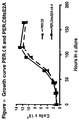
- packaging cells such as PER.C6 (ECACC deposit number 96022940) or cells based thereon, but adapted for Ad35 or a functional homologue thereof; it also includes any nucleic acids encoding functional parts of adenovirus 35 or a functional homologue thereof, such as helper constructs and packaging constructs, as well as vectors comprising genes of interest and e.g. an ITR, etc.
- the invention also provides a nucleic acid encoding at least a functional part of a gene delivery vehicle according to the invention, or a virus, homologue or chimaera thereof according to the invention.
- such elements which encode functions that will end up in the resulting gene delivery vehicle must comprise or be encoded by a nucleic acid encoding at least one of the adenovirus serotype 35 elements or a functional equivalent thereof, responsible for avoiding or deminishing neutralising activity against adenoviral elements by the host to which the gene is to be delivered.
- the gene of interest would be present on the same nucleic acid which means that such a nucleic acid has such a gene or that it has a site for introducing a gene of interest therein.
- such a nucleic acid also comprises at least one ITR and if it is a nucleic acid to be packaged also a packaging signal.
- PCT/NL96/00244 A set of further improvements in the field of producing adenoviral gene delivery vehicles is applicant's plasmid system disclosed in PCT/NL99/00235 mentioned herein before. This system works in one embodiment as a homologous recombination of an adapter plasmid and a longer plasmid, together comprising all elements of the nucleic acid to be incorporated in the gene delivery vehicle. These methods can also be applied to the presently invented gene delivery vehicles and their building elements.
- the invention also provides a nucleic acid according to the invention further comprising a region of nucleotides designed or useable for homologous recombination, preferably as part of at least one set of two nucleic acids comprising a nucleic acid according to the invention, whereby said set of nucleic acids is capable of a single homologous recombination event with each other, which leads to a nucleic acid encoding a functional gene delivery vehicle.
- both empty packaging cells in which the vector to be packaged to make a gene delivery vehicle according to the invention still has to be introduced or produced
- cells comprising a vector according to the invention to be packaged are provided.
- the invention also encompasses a cell comprising a nucleic acid according to the invention or a set of nucleic acids according to the invention, preferably a cell which complements the necessary elements for adenoviral replication which are absent from the nucleic acid according to be packaged, or from a set of nucleic acids according to the invention.
- E1-deleted adenovirus 35 vectors are not capable of replication on cells that provide adenovirus 5 proteins in trans .
- the invention therefore further provides a cell capable of providing adenovirus 35 E1 proteins in trans.
- a cell is typically a human cell derived from the retina or the kidney.
- Embryonal cells such as amniocytes, have been shown to be particularly suited for the generation of an E1 complementing cell line.
- Serotype specific complementation by E1 proteins can be due to one or more protein(s) encoded by the E1 region. It is therefor essential that at least the serotype specific protein is provided in trans in the complementing cell line.
- the non-serotype specific E1 proteins essential for effective complementation of an E1-deleted adenovirus can be derived from other adenovirus serotpyes.
- at least an E1 protein from the E1B region of adenovirus 35 is provided in trans to complement E1-deleted adenovirus 35 based vectors.
- nucleic acid encoding the one or more serotype specific E1-proteins is introduced into the PER.C6 cell or a cell originating from a PER.C6 cell (ECACC deposit number 96022940), or a similar packaging cell complementing with elements from Ad 35 or a functional homologue thereof.
- a method for producing a gene delivery vehicle according to the invention comprising expressing a nucleic acid according to the invention in a cell according to the invention and harvesting the resulting gene delivery vehicle.
- the above refers to the filling of the empty packaging cell with the relevant nucleic acids.
- the format of the filled cell is of course also part of the present invention, which provides a method for producing a gene delivery vehicle according to the invention, comprising culturing a filled packaging cell (producer cell) according to the invention in a suitable culture medium and harvesting the resulting gene delivery vehicle.
- the resulting gene delivery vehicles obtainable by any method according to the invention are of course also part of the present invention, particularly also a gene delivery vehicle according to the invention, which is derived from a chimaera of an adenovirus and an integrating virus.
- adenoviral gene delivery vehicles do not integrate into the host genome normally.
- chimaeras which do have that capability.
- Such chimaeras have been disclosed in our copending application PCT/NL98/00731 incorporated herein by reference.
- chimaeras which can also be combined with the above are chimaeras (be it in swapping whole proteins or parts thereof or both) which have altered tropism.
- a very good example thereof is a chimaera of Ad 35 and Ad 16, possibly with elements from for instance Ad 2 or Ad 5, wherein the tropism determining part of Ad 16 or a functional equivalent thereof is used to direct the gene delivery vehicle to synoviocytes and/or smooth muscle cells (see our copending applications nos. 98204482.8 and 99200624.7) incorporated herein by reference).
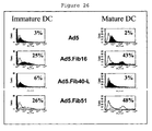
- Dendritic cells (DC) and hemopoietic stem cells (HSC) are not easily transduced with Ad2 or Ad5 derived gene delivery vehicles.
- the present invention provides gene delivery vehicles that posses increased transduction capacity of DC and HSC cells.
- Such gene delivery vehicles at least comprises the tissue tropism determining part of an Ad35 adenovirus.
- the invention therefore further provides the use of a tissue tropism determining part of an adenovirus 35 capsid for transducing dendritic cells and/or hemopoietic stem cells.
- Other B-type adenoviruses are also suited.
- a tissue tropism determining part comprises at least the knob and/or the shaft of a fiber protein.
- Such knowledge can be used to devise chimearic proteins comprising such amino acid sequences.
- Such chimaeric proteins are therefor also part of the invention.
- DC cells are very efficient antigen presenting cells.
- the immune system of the host can be triggered to toward specific antigens.
- antigens can be encoded by nucleic acid delivered to the DC or by the proteins of the gene delivery vehicle it self.
- the present invention therefor also provides a gene delivery vehicle with the capacity to evade to host immune system as a vaccine.
- the vector being capable to evade the immune system long enough to efficiently find it target cells and at the same time capable of delivering specific antigens to antigen presenting cells thereby allowing the induction and/or stimulation of an efficient immune responses toward the specific antigen(s).
- the gene delivery vehicle may comprise proteins and/or nucleic encoding such proteins capable of modulating an immune response.
- the invention therefore further provides a vaccine comprising a gene delivery vehicle of the invention.
- the invention further provides an adenovirus vector with the capacity to efficiently transduce DC and/or HSC, the vehicle comprising at least a tissue tropism determing part of serotype 35 adenvirus.
- the invention further provides the use of such delivery vehicles for the transduction of HSC and/or DC cells.
- tissue tropisms are found among other adenoviruses of serotype B, particularly in serotype 11 and are also part of the invention.
- Such gene delivery vehicles are therefor also part of the invention.
- the gene delivery vehicles according to the invention can be used to deliver genes or nucleic acids of interest to host cells. This will typically be a pharmaceutical use. Such a use is included in the present invention. Compositions suitable for such a use are also part of the present invention.
- the amount of gene delivery vehicle that needs to be present per dose or per infection (m.o.i) will depend on the condition to be treated, the route of administration (typically parenteral) the subject and the efficiency of infection, etc. Dose finding studies are well known in the art and those already performed with other (adenoviral) gene delivery vehicles can typically be used as guides to find suitable doses of the gene delivery vehicles according to the invention.
- the invention also provides a pharmaceutical formulation comprising a gene delivery vehicle according to the invention and a suitable excipient, as well as a pharmaceutical formulation comprising an adenovirus, a chimaera thereof, or a functional homologue thereof according to the invention and a suitable excipient.
- the present invention provides the use of at least elements of a serotype and functional homologues thereof of adenovirus which are very suitable as gene therapy vectors.
- the present invention also discloses an automated high-throughput screening of all known adenovirus serotypes against sera from many individuals. Surprisingly, no neutralizing ability was found in any of the sera that were evaluated against one particular serotype, adenovirus 35 (Ad35). This makes the serotype of the present invention extremely useful as a vector system for gene therapy in man.
- Such vector system is capable of efficiently transferring genetic material to a human cell without the inherent problem of pre-exisiting immunity.
- a virus is produced using an adenoviral vector (typically a plasmid, a cosmid or baculovirus vector).
- adenoviral vector typically a plasmid, a cosmid or baculovirus vector.
- the invention also provides adenovirus derived vectors that have been rendered replication defective by deletion or inactivation of the E1 region.
- a gene of interest can be inserted at for instance the site of E1 of the original adenovirus from which the vector is derived.
- the adenoviruses may contain deletions in the E1 region and insertions of heterologous genes linked either or not to a promoter. Furthermore, the adenoviruses may contain deletions in the E2, E3 or E4 regions and insertions of heterologous genes linked to a promoter. In these cases, E2 and/or E4 complementing cell lines are required to generate recombinant adenoviruses.
- Ad35 serotype itself for the preparation of recombinant adenoviruses to be used in gene therapy.
- elements derived from the serotype of the present invention in such recombinant adenoviruses.
- One may for instance develop a chimaeric adenovirus that combines desirable properties from different serotypes.
- Some serotypes have a somewhat limited host range, but have the benefit of being less immunogenic, some are the other way round. Some have a problem of being of a limited virulence, but have a broad host range and/or a reduced immunogenicity.
- the invention provides a chimaeric adenovirus comprising at least a part of the adenovirus genome of the present serotype, providing it with absence of pre-existing immunity, and at least a part of the adenovirus genome from another adenovirus serotype resulting in a chimaeric adenovirus.
- the chimaeric adenovirus produced is such that it combines the absence of pre-existing immunity of the serotype of the present invention, to other characteristics of another serotype.
- characteristics may be temperature stability, assembly, anchoring, redirected infection, production yield, redirected or improved infection, stability of the DNA in the target cell, etc.
- a packaging cell will generally be needed in order to produce sufficient amount of adenoviruses.
- several cell lines are available. These include but are not limited to the known cell lines PER.C6 (ECACC deposit number 96022940), 911, 293, and E1 A549.
- An important feature of the present invention is the means to produce the adenovirus.
- Such a cell is usually called a packaging cell.
- the invention thus also provides a packaging cell for producing an adenovirus (a gene delivery vehicle) according to the invention, comprising in trans all elements necessary for adenovirus production not present on the adenoviral vector according to the invention.
- vector and packaging cell have to be adapted to one another in that they have all the necessary elements, but that they do not have overlapping elements which lead to replication competent virus by recombination.
- the invention also provides a kit of parts comprising a packaging cell according to the invention and a recombinant vector according the invention whereby there is essentially no sequence overlap leading to recombination resulting in the production of replication competent adenovirus between said cell and said vector.
- the invention provides methods for producing adenovirus, which upon application will escape pre-existing humoral immunity, comprising providing a vector with elements derived from an adenovirus serotype against which virtually no natural immunity exists and transfecting said vector in a packaging cell according to the invention and allowing for production of viral particles.
- this invention describes the use of the adenovirus serotype of the present invention to overcome natural existing or induced, neutralising host activity towards adenoviruses administered in vivo for therapeutic applications.
- the need for a new serotype is stressed by observations that 1) repeated systemic delivery of recombinant adenovirus serotype 5 is unsuccessful due to formation of high titers of neutralising antibodies against the recombinant adenovirus serotype 5 (Schulick et al, 1997), and 2) pre-existing or humoral immunity is widespread in the population.
- this invention provides the use of gene delivery vehicles of the invention or the use of adenovirus serotype 35 for vaccination purposes. Such use prevents at least in part undesired immune responses of the host.
- undesired immune responses are evoking an immune response against the gene delivery vehicle or adenovirus serotype 35 and/or boosting of an immune response against the gene delivery vehicle or adenovirus serotype 35.
- alternating use is made of Ad vectors belonging to different subgroups. This aspect of the invention therefore circumvents the inability to repeat the administration of an adenovirus for gene therapy purposes.
- a panel of 100 individuals was selected. Volunteers (50% male, 50% female) were healthy individuals between 20 and 60 years old with no restriction for race. All volunteers signed an informed consent form. People professionally involved in adenovirus research were excluded.
- Serum was thawed and heat-inactivated at 56°C for 10 minutes and then aliquotted to prevent repeated cycles of freeze/thawing. Part was used to make five steps of twofold dilutions in medium (DMEM, Gibco BRL) in a quantity enough to fill out approximately 70 96-well plates. Aliquots of undiluted and diluted sera were pipetted in deep well plates (96-well format) and using a programmed platemate dispensed in 100 ⁇ l aliquots into 96-well plates.
- DMEM medium
- S1/2 to S8/2 in columns 1 and 6 represent 1x diluted sera and Sx/4, Sx/8, Sx/16 and Sx/32 the twofold serial dilutions.
- the last plates also contained four wells filled with 100 ⁇ l foetal calf serum as a negative control. Plates were kept at -20°C until further use.
- Prototypes of all known human adenoviruses were inoculated on T25 flasks seeded with PER.C6 cells (ECACC deposit number 96022940) (Fallaux et al ., 1998) and harvested upon full CPE. After freeze/thawing 1-2 ml of the crude lysates was used to inoculate a T80 flask with PER.C6 cells (ECACC deposit number 96022940) and virus was harvested at full CPE. The timeframe between inoculation and occurrence of CPE as well as the amount of virus needed to re-infect a new culture, differed between serotypes.
- Adenovirus stocks were prepared by freeze/thawing and used to inoculate 3-4 T175 cm 2 three-layer flasks with PER.C6 cells (ECACC deposit number 96022940). Upon occurrence of CPE, cells were harvested by tapping the flask, pelleted and virus was isolated and purified by a two step CsCl gradient as follows. Cell pellets were dissolved in 50 ml 10 mM NaPO 4 buffer (pH 7.2) and frozen at -20°C. After thawing at 37°C, 5.6 ml sodium deoxycholate (5% w/v) was added. The solution was mixed gently and incubated for 5-15 minutes at 37°C to completely lyse the cells.
- the virus band is isolated after which a second purification using a 1M Tris/HCl buffered continues gradient of 1.33 gr/ml of cesiumchloride was performed. The virus was then centrifuged for 17 hours at 55000 rpm at 10°C. The virus band is isolated and sucrose (50 % w/v) is added to a final concentration of 1%. Excess cesiumchloride is removed by dialysis (three times 1 hr at RT) in dialysis slides (Slide-a-lizer, cut off 10000 kDa, Pierce, USA) against 1.5 ltr PBS supplemented with CaCl 2 (0.9 mM), MgCl 2 (0.5mM) and an increasing concentration of sucrose (1, 2, 5%). After dialysis, the virus is removed from the slide-a-lizer after which it is aliquoted in portions of 25 and 100 ⁇ l upon which the virus is stored at -85°C.
- HPLC high-pressure liquid chromatograph
- Viruses were eluted using an NaCl gradient ranging from 0 to 600 mM. As depicted in table I, the NaCl concentration by which the viruses were eluted differed significantly among serotypes.
- adenovirus type 8 and 40 were grown on 911-E4 cells (He et al ., 1998). Purified stocks contained between 5x10 10 and 5x10 12 virus particles/ml (VP/ml; see table I).
- Adenoviruses were titrated on PER.C6 cells (ECACC deposit number 96022940) to determine the amount of virus necessary to obtain full CPE in five days, the length of the neutralisation assay.
- 100 ⁇ l medium was dispensed into each well of 96-well plates.
- 25 ⁇ l of adenovirus stocks prediluted 10 4 , 10 5 , 10 6 or 10 7 times were added to column 2 of a 96-well plate and mixed by pipetting up and down 10 times. Then 25 ⁇ l was brought from column 2 to column 3 and again mixed. This was repeated until column 11 after which 25 ⁇ l from column 11 was discarded. This way serial dilutions in steps of 5 were obtained starting off from a prediluted stock.
- 3x10 4 PER.C6 cells (ECACC deposit number 96022940) were added in a 100 ⁇ l volume and the dates were incubated at 37 °C, 5% CO 2 for five or six days. CPE was monitored microscopically. The method of Reed and Muensch was used to calculate the cell culture inhibiting dose 50% (CCID50).
- 96-well plates with diluted human serum samples were thawed at 37 °C, 5% CO 2 .
- Adenovirus stocks diluted to 200 CCID50 per 50 ⁇ l were prepared and 50 ⁇ l aliquots were added to columns 1-11 of the plates with serum. Plates were incubated for 1 hour at 37°C, 5% CO 2 .
- 50 ⁇ l PER.C6 cells ECACC deposit number 96022940
- 6x10 5 /ml were dispensed in all wells and incubated for 1 day at 37 °C, 5% CO 2 .
- Supernatant was removed using fresh pipet tips for each row and 200 ⁇ l fresh medium was added to all wells to avoid toxic effects of the serum.
- Plates were incubated for another 4 days at 37 °C, 5% CO 2 .
- parallel control plates were set up in duplo with diluted positive control sera generated in rabbits and specific for each serotype to be tested in rows A and B and with negative control serum (FCS) in rows C and D.
- FCS negative control serum
- E-H a titration was performed as described above with steps of five times dilutions starting with 200 CCID50 of each virus to be tested.
- On day 5 one of the control plates was analysed microscopically and with the MTT assay. The experimental titer was calculated from the control titration plate observed microscopically.
- CPE CPE was found to be complete, i.e. the first dilution in the control titration experiment analysed by MTT shows clear cell death, all assay plates were processed. If not, the assay was allowed to proceed for one or more days until full CPE was apparent after which all plates were processed. In most cases the assay was terminated at day 5. For Ad1, 5, 33, 39, 42 and 43 the assay was left for six days and for Ad2 for eight days.
- a serum sample is regarded to be non-neutralising when at the highest serum concentration a maximum protection is seen of 40% compared to the controls without serum.
- recombinant E1-deleted adenoviruses based on Ad35 or one of the other above mentioned serotypes have an important advantage compared to recombinant vectors based on Ad5 with respect to clearence of the viruses by neutralising antibodies.
- Ad5-based vectors that have (parts of) the capsid proteins involved in immunogenic response of the host replaced by the corresponding (parts of) the capsid proteins of Ad35 or one of the other serotypes will be less, or even not, neutralised by the vast majority of human sera.
- the VP/CCID50 ratio calculated from the virus particles per ml and the CCID50 obtained for each virus in the experiments was highly variable and ranged from 0.4 to 5 log. This is probably caused by different infection efficiencies of PER.C6 cells (ECACC deposit number 96022940) and by differences in replication efficiency of the viruses. Furthermore, differences in batch qualities may play a role.
- a high VP/CCID50 ratio means that more virus was put in the wells to obtain CPE in 5 days. As a consequence the outcome of the neutralisation study might be biased since more (inactive) virus particles could shield the antibodies. To check whether this phenomenon had taken place, the VP/CCID50 ratio was plotted against the percentage of serum samples found positive in the assay ( Figure 2). The graph clearly shows that there is no negative correlation between the amount of viruses in the assay and neutralisation in serum.
- Ad5 plasmid vectors for the production of recombinant viruses and easy manipulation of adenoviral genes
- wild-type human adenovirus type 5 (Ad5) DNA was treated with Klenow enzyme in the presence of excess dNTPs. After inactivation of the Klenow enzyme and purification by phenol/chloroform extraction followed by ethanol precipitation, the DNA was digested with BamHI. This DNA preparation was used without further purification in a ligation reaction with pBr322 derived vector DNA prepared as follows: pBr322 DNA was digested with EcoRV and BamHI, dephosphorylated by treatment with TSAP enzyme (Life Technologies) and purified on LMP agarose gel (SeaPlaque GTG). After transformation into competent E.coli DH5 ⁇ (Life Techn.) and analysis of ampiciline resistant colonies, one clone was selected that showed a digestion pattern as expected for an insert extending from the BamHI site in Ad5 to the right ITR.
- Ad5 wild-type human adenovirus type 5
- pBr/Ad.Bam-rITR was digested with BamHI and SalI.
- the vector fragment including the adenovirus insert was isolated in LMP agarose (SeaPlaque GTG) and ligated to a 4.8 kb SalI-BamHI fragment obtained from wt Ad5 DNA and purified with the Geneclean II kit (Bio 101, Inc.).
- One clone was chosen and the integrity of the Ad5 sequences was determined by restriction enzyme analysis.
- Clone pBr/Ad.Sal-rITR contains adeno type 5 sequences from the SalI site at bp 16746 up to and including the rITR (missing the most 3' G residue).
- wt Adeno type 5 DNA was digested with ClaI and BamHI, and the 20.6 kb fragment was isolated from gel by electro-elution.
- pBr322 was digested with the same enzymes and purified from agarose gel by Geneclean. Both fragments were ligated and transformed into competent DH5 ⁇ .
- the resulting clone pBr/Ad.Cla-Bam was analysed by restriction enzyme digestion and shown to contain an insert with adenovirus sequences from bp 919 to 21566.
- Clone pBr/Ad.Cla-Bam was linearized with EcoRI (in pBr322) and partially digested with AflII. After heat inactivation of AflII for 20' at 65°C the fragment ends were filled in with Klenow enzyme.
- the DNA was then ligated to a blunt double stranded oligo linker containing a PacI site (5'-AATTGTC TTAATTAA CCGCTTAA-3'). This linker was made by annealing the following two oligonucleotides: 5'-AATTGTCTTAATTAACCGC-3' and 5'-AATTGCGGTTAATTAAGAC-3', followed by blunting with Klenow enzyme.
- the ligations were digested with an excess PacI enzyme to remove concatameres of the oligo.
- One clone that was found to contain the PacI site and that had retained the large adeno fragment was selected and sequenced at the 5' end to verify correct insertion of the PacI linker in the (lost) AflII site.
- pBr/Ad.Bam-rITR was digested with ClaI and treated with nuclease Bal31 for varying lengths of time (2', 5', 10' and 15'). The extend of nucleotide removal was followed by separate reactions on pBr322 DNA (also digested at the ClaI site), using identical buffers and conditions. Bal31 enzyme was inactivated by incubation at 75 °C for 10', the DNA was precipitated and resuspended in a smaller volume TE buffer.
- DNAs were further treated with T4 DNA polymerase in the presence of excess dNTPs. After digestion of the (control) pBr322 DNA with SalI, satisfactory degradation ( ⁇ 150 bp) was observed in the samples created for 10' or 15'.
- the 10' or 15' treated pBr/Ad.Bam-rITR samples were then ligated to the above described blunted PacI linkers (See pBr/Ad.AflII-Bam). Ligations were purified by precipitation, digested with excess PacI and separated from the linkers on an LMP agarose gel. After religation, DNAs were transformed into competent DH5 ⁇ and colonies analyzed.
- clones were selected that showed a deletion of approximately the desired length and these were further analyzed by T-track sequencing (T7 sequencing kit, Pharmacia Biotech). Two clones were found with the PacI linker inserted just downstream of the rITR. After digestion with PacI, clone #2 has 28 bp and clone #8 has 27 bp attached to the ITR.
- Cosmid vector pWE15 (Clontech) was used to clone larger Ad5 inserts.
- a linker containing a unique PacI site was inserted in the EcoRI sites of pWE15 creating pWE.pac.
- the double stranded PacI oligo as described for pBr/Ad.AflII-BamHI was used but now with its EcoRI protruding ends.
- the following fragments were then isolated by electro-elution from agarose gel: pWE.pac digested with PacI, pBr/AflII-Bam digested with PacI and BamHI and pBr/Ad.Bam-rITR#2 digested with BamHI and PacI.
- pWE/Ad.AflII-rITR contains all adenovirus type 5 sequences from bp 3534 (AflII site) up to and including the right ITR (missing the most 3' G residue).
- Adeno 5 wt DNA was treated with Klenow enzyme in the presence of excess dNTPs and subsequently digested with SalI. Two of the resulting fragments, designated left ITR-Sal(9.4) and Sal(16.7)-right ITR, respectively, were isolated in LMP agarose (Seaplaque GTG).
- pBr322 DNA was digested with EcoRV and SalI and treated with phosphatase (Life Technologies). The vector fragment was isolated using the Geneclean method (BIO 101, Inc.) and ligated to the Ad5 SalI fragments. Only the ligation with the 9.4 kb fragment gave colonies with an insert. After analysis and sequencing of the cloning border a clone was chosen that contained the full ITR sequence and extended to the SalI site at bp 9462.
- pBr/Ad.lITR-Sal(16.7) (ECACC deposit P97082118)
- pBr/Ad.lITR-Sal(9.4) is digested with SalI and dephosphorylated (TSAP, Life Technologies).
- TSAP dephosphorylated
- pBr/Ad.Cla-Bam was linearized with BamHI and partially digested with SalI.
- a 7.3 kb SalI fragment containing adenovirus sequences from 9462-16746 was isolated in LMP agarose gel and ligated to the SalI-digested pBr/Ad.lITR-Sal(9.4) vector fragment.
- pWE.pac was digested with ClaI and 5' protruding ends were filled using Klenow enzyme. The DNA was then digested with PacI and isolated from agarose gel. pWE/AflII-rITR was digested with EcoRI and after treatment with Klenow enzyme digested with PacI. The large 24 kb fragment containing the adenoviral sequences was isolated from agarose gel and ligated to the ClaI-digested and blunted pWE.pac vector using the Ligation Express tm kit from Clontech. After transformation of Ultracompetent XL10-Gold cells from Stratagene, clones were identified that contained the expected insert. pWE/AflII-EcoRI containes Ad5 sequences from bp 3534-27336.
- the 3' ITR an the vector pWE/Ad.AflII-rITR does not include the terminal G-nucleotide. Furthermore, the PacI site is located almost 30 bp from the right ITR. Both these characteristics may decrease the efficiency of virus generation due to inefficient initiation of replication at the 3' ITR. Note that during virus generation the left ITR in the adapter plasmid is intact and enables replication of the virus DNA after homologous recombination.
- the pWE/Ad.AflII-rITR was modified as follows: construct pBr/Ad.Bam-rITRpac#2 was first digested with PacI and then partially digested with AvrII and the 17.8 kb vector containing fragment was isolated and dephophorylated using SAP enzyme (Boehringer Mannheim). This fragment lacks the adenosequences from nucleotide 35464 to the 3'ITR. Using DNA from pWE/Ad.AflII-rITR as template and the primers ITR-EPH:
- adenoviral sequences flanking the E2A coding region at the left and the right site were amplified from the plasmid pBr/Ad.Sal.rITR (ECACC deposit P97082119) in a PCR reaction with the Expand PCR system (Boehringer) according to the manufacturers protocol. The following primers were used: Right flanking sequences (corresponding Ad5 nucleotides 24033 to 25180):
- the amplified DNA fragment was digested with SnaBI and NsiI (NsiI site is generated in the primer ⁇ E2A.DBP-start, underlined).
- the amplified DNA was digested with BamHI and NsiI (NsiI site is generated in the primer ⁇ E2A.DBP-stop, underlined).
- the digested DNA fragments were ligated into SnaBI/BamHI digested pBr/Ad.Sal-rITR. Sequencing confirmed the exact replacement of the DBP coding region with a unique NsiI site in plasmid pBr/Ad.Sal-rITR ⁇ E2A.
- the unique NsiI site can be used to introduce an expression cassette for a gene to be transduced by the recombinant vector.
- the deletion of the E2A coding sequences was performed such that the splice acceptor sites of the 100K encoding L4-gene at position 24048 in the top strand was left intact.
- the poly adenylation signals of the original E2A-RNA and L3-RNAs at the left hand site of the E2A coding sequences were left intact. This ensures proper expression of the L3-genes and the gene encoding the 100K L4-protein during the adenovirus life cycle.
- the plasmid pWE/Ad.AflII-rITR ⁇ E2A was generated.
- the plasmid pBr/Ad.Sal-rITR ⁇ E2A was digested with BamHI and SpeI.
- the 3.9-Kb fragment in which the E2A coding region was replaced by the unique NsiI site was isolated.
- the pWE/Ad.AflII-rITR was digested with BamHI and SpeI.
- the fragments were ligated and packaged using ⁇ phage-packaging extracts according to the manufacturer protocol (Stratagene), yielding the plasmid pWE/Ad.AflII-rITR ⁇ E2A.
- This cosmid clone can be used to generate adenoviral vectors that are deleted for E2A by cotransfection of PacI digested DNA together with digested adapter plasmids onto packaging cells that express functional E2A gene product.
- the absence of sequence overlap between the recombinant adenovirus and E1 sequences in the packaging cell line is essential for safe, RCA-free generation and propagation of new recombinant viruses.
- the adapter plasmid pMLPI.TK (described in PCT/NL96/00244) is an example of an adapter plasmid designed for use according to the invention in combination with the improved packaging cell lines of the invention. This plasmid was used as the starting material to make a new vector in which nucleic acid molecules comprising specific promoter and gene sequences can be easily exchanged.
- PCR fragment was generated from pZip ⁇ Mo+PyF101(N - ) template DNA (described in PCT/NL96/00195) with the following primers: LTR-1: 5'-CTG TAC GTA CCA GTG CAC TGG CCT AGG CAT GGA AAA ATA CAT AAC TG-3' and LTR-2: 5'-GCG GAT CCT TCG AAC CAT GGT AAG CTT GGT ACC GCT AGC GTT AAC CGG GCG ACT CAG TCA ATC G-3'.
- Pwo DNA polymerase (Boehringer Mannheim) was used according to manufacturers protocol with the following temperature cycles: once 5' at 95°C; 3' at 55°C; and 1' at 72°C, and 30 cycles of 1' at 95°C, 1' at 60°C, 1' at 72°C, followed by once 10' at 72°C.
- the PCR product was then digested with BamHI and ligated into pMLP10 (Levrero et al ., 1991) vector digested with PvuII and BamHI, thereby generating vector pLTR10.
- This vector contains adenoviral sequences from bp 1 up to bp 454 followed by a promoter consisting of a part of the Mo-MuLV LTR having its wild-type enhancer sequences replaced by the enhancer from a mutant polyoma virus (PyF101).
- the promoter fragment was designated L420.
- the coding region of the murine HSA gene was inserted.
- pLTR10 was digested with BstBI followed by Klenow treatment and digestion with NcoI.
- the HSA gene was obtained by PCR amplification on pUC18-HSA (Kay et al ., 1990) using the following primers: HSA1, 5'-GCG CCA CCA TGG GCA GAG CGA TGG TGG C-3' and HSA2, 5'-GTT AGA TCT AAG CTT GTC GAC ATC GAT CTA CTA ACA GTA GAG ATG TAG AA-3'.
- the 269 bp amplified fragment was subcloned in a shuttle vector using the NcoI and BglII sites. Sequencing confirmed incorporation of the correct coding sequence of the HSA gene, but with an extra TAG insertion directly following the TAG stop codon.
- the coding region of the HSA gene, including the TAG duplication was then excised as a NcoI(sticky)-SalI(blunt) fragment and cloned into the 3.5 kb NcoI(sticky)/BstBI(blunt) fragment from pLTR10, resulting in pLTR-HSA10.
- pLTR-HSA10 was digested with EcoRI and BamHI after which the fragment containing the left ITR, packaging signal, L420 promoter and HSA gene was inserted into vector pMLPI.TK digested with the same enzymes and thereby replacing the promoter and gene sequences.
- SnaBI and AvrII can be combined with HpaI, NheI, KpnI, HindIII to exchange promoter sequences, while the latter sites can be combined with the ClaI or BamHI sites 3' from HSA coding region to replace genes in this construct.
- pAd/L420-HSA was digested with AvrII and BglII followed by treatment with Klenow to obtain blunt ends.
- the 5.1 kb fragment with pBr322 vector and adenoviral sequences was isolated and ligated to a blunt 1570 bp fragment from pcDNA1/amp (Invitrogen) obtained by digestion with HhaI and AvrII followed by treatment with T4 DNA polymerase.
- This adapter plasmid was named pAd5/CLIP.
- this plasmid was partially digested with EcoRI and the linear fragment was isolated. An oligo of the sequence 5' TTAAGTCGAC-3' was annealed to itself resulting in a linker with a SalI site and EcoRI overhang. The linker was ligated to the partially digested pAd5/Clip vector and clones were selected that had the linker inserted in the EcoRI site 23 bp upstream of the left adenovirus ITR in pAd5/Clip resulting in pAd5/CArchitectl.
- the EcoRI site in pAd5/Clip has been changed to a PacI site by insertion of a linker of the sequence 5'-AATTGTCTTAATTAACCGCAATT-3'.
- the pAd5/Clip vector was partially digested with EcoRI, dephosphorylated and ligated to the PacI linker with EcoRI overhang.
- the ligation mixture was digested with PacI to remove concatamers, isolated from agarose gel and religated.
- the resulting vector was named pAd5/Clippac.
- the vector pAd5/L420-HSA was also modified to create a SalI or PacI site upstream of the left ITR.
- pAd5/L420-HSA was digested with EcoRI and ligated to the above described PacI linker.
- the ligation mixture was digested with PacI and religated after isolation of the linear DNA from agarose gel to remove concatamerised linkers. This resulted in adapter plasmid pAd5/L420-HSApac.
- pAd5/L420-HSAsal was digested with ScaI and BsrGI and the vector fragment was ligated to the 0.3 kb fragment isolated after digestion of pAd5/C redesign with the same enzymes.
- pAd5/L420-HSApac was digested with AvrII and BglII.
- the vector fragment was ligated to a linker oligonucleotide digested with the same restriction enzymes.
- the linker was made by annealing oligos of the following sequence:
- the annealed linkers were digested with AvrII and BglII and separated from small ends by column purification (Qiaquick nucleotide removal kit) according to manufacterers recommendations. The linker was then ligated to the AvrII/BglII digested pAd5/L420-HSApac fragment. A clone, named AdMire, was selected that had the linker incorporated and was sequenced to check the integrity of the insert.
- AdMire Adapter plasmid AdMire enables easy insertion of complete expression cassettes.
- An adapter plasmid containing the human CMV promoter that mediates high expression levels in human cells was constructed as follows: pAd5/L420-HSApac was digested with AvrII and 5' protruding ends were filled in using Klenow enzyme. A second digestion with HindIII resulted in removal of the L420 promoter sequences. The vector fragment was isolated and ligated to a PCR fragment containing the CMV promoter sequence. This PCR fragment was obtained after amplification of CMV sequences from pCMVLacI (Stratagene) with the following primers:
- the PCR fragment was first digested with Pst1 (underlined in CMVplus) after which the 3'-protruding ends were removed by treatment with T4 DNA polymerase. Then the DNA was digested with HindIII (underlined in CMVminA) and ligated into the above described pAd5/L420-HSApac vector fragment digested with AvrII and HindIII. The resulting plasmid was named pAd5/CMV-HSApac. This plasmid was then digested with HindIII and BamHI and the vector fragment was isolated and ligated to the polylinker sequence obtained after digestion of AdMire with HindIII and BglII.
- Adapter plasmid pAdApt contains nucleotides - 735 to +95 of the human CMV promoter (Boshart et al., 1985).
- a second version of this adapter plasmid containing a SalI site in place of the PacI site upstream of the left ITR was made by inserting the 0.7 kb ScaI-BsrGI fragment from pAd5/CArchitectl into pAdApt digested with ScaI and partially digested with BsrGI. This clone was named pAdApt.sal.
- RCA free recombinant adenoviruses can be generated very efficiently using the above described adapter plasmids and the pWe/Ad.AflII-rITR or pWE/Ad.AflII-rITrsp constructs.
- the adapter plasmid containing the desired transgene in the desired expression cassette is digested with suitable enzymes to liberate the insert from vector sequences at the 3' and/or at the 5' end.
- the adenoviral complementation plasmids pWE/Ad.AflII-rITR or pWE/Ad.AflII-rITRsp are digested with PacI to liberate the adeno sequences from the vector plasmids.
- PacI As a non-limiting example the generation of AdApt-LacZ is described.
- Adapter plasmid pAdApt-LacZ was generated as follows.
- the E.coli LacZ gene was amplified from the plasmid pMLP.nlsLacZ (EP 95-202 213) by PCR with the primers 5'-GGGGTGGCCAGGGTACCTCTAGGCTTTTGCAA-3' and 5'-GGGGGGATCCATAAACAAGTTCAGAATCC-3'.
- the PCR reaction was performed with Ex Tag (Takara) according to the suppliers protocol at the following amplification program: 5 minutes 94°C, 1 cycle; 45 seconds 94°C and 30 seconds 60°C and 2 minutes 72°C, 5 cycles; 45 seconds 94°C and 30 seconds 65°C and 2 minutes 72°C, 25 cycles; 10 minutes 72; 45 seconds 94°C and 30 seconds 60°C and 2 minutes 72°C, 5 cycles, I cycle.
- the PCR product was subsequently digested with Kpn1 and BamHI and the digested DNA fragment was ligated into KpnI/BamHI digested pcDNA3 (Invitrogen), giving rise to pcDNA3.nlsLacZ. Construct pcDNA3.nlsLacZ was then digested with KpnI and BamHI and the 3 kb LacZ fragment was isolated from gel using the geneclean spin kit (Bio 101, Inc.).
- pAdApt was also digested with KpnI and BamHI and the linear vector fragment was isolated from gel as above. Both isolated fragments were ligated and one clone containing the LacZ insert was selected. Construct pAdApt-LacZ was digested with SalI, purified by the geneclean spin kit and subsequently digested with PacI. pWE/Ad.AflII-rITRsp was digested with PacI. Both digestion mixtures were treated for 30' by 65 °C to inactivate the enzymes. Samples were put on gel to estimate the concentration.
- PER.C6 cells 2.5x10 6 PER.C6 cells (ECACC deposit number 96022940) were seeded in T25 flasks in DMEM with 10% FCS and 10mM MgCl. The next day four microgram of each plasmid was transfected into PER.C6 cells (ECACC deposit number 96022940) using lipofectamine transfection reschreib (Life Technologies Inc.) according to instructions of the manufacturer. The next day the medium was replaced by fresh culture medium and cells were further cultured at 37° C, 10% CO 2 . Again 24 hrs. later cells were trypsinised, seeded into T80 flasks and cultured at 37°C, 10% CO 2 .
- Full CPE was obtained 6 days after seeding in the T80 flask.Cells were harvested in the medium and subjected to one freeze/thaw cycle. The crude lysate obtained this way was used to plaque purify the mixture of viruses. Ten plaques were picked, expanded in a 24 well plate and tested for LacZ expression following infection of A549 cells. Viruses from all ten plaques expressed LacZ.
- Neutralising antibodies in human serum are mainly directed to the hexon protein and to a lesser extend to the penton protein.
- Hexon proteins from different serotypes show highly variable regions present in loops that are predicted to be exposed at the outside of the virus (Athappilly et al., 1994; J. Mol. Biol. 242, 430-455). Most type specific epitopes have been mapped to these highly variable regions (Toogood et al., 1989; J. Gen Virol. 70, 3203-3214).
- replacement of (part of ) the hexon sequences with corresponding sequences from a different serotype is an effective strategy to circumvent (pre-existing) neutralising antibodies to Ad5.
- Hexon coding sequences of adenovirus serotype 5 are located between nucleotides 18841 and 21697.
- This subclone, coded pBr/Ad.Eco-PmeI was generated by first digesting plasmid pBr322 with EcoRI and EcoRV and inserting the 14 kb PmeI-EcoRI fragment from pWE/Ad.AflII-Eco.
- this shuttle vector a deletion was made of a 1430 bp SanDI fragment by digestion with SanDI and religation to give pBr/Ad.Eco-PmeI ⁇ SanDI. The removed fragment contains unique SpeI and MunI sites. From pBr/Ad.Eco-PmeI ⁇ SanDI the adenovirus serotype 5 DNA encoding hexon was deleted.
- hexon flanking sequences were PCR amplified and linked together thereby generating unique restriction sites replacing the hexon coding region.
- four different oligonucleotides were required: ⁇ hex1- ⁇ hex4.
- the amplified DNA product of ⁇ 1100 bp obtained with oligonucleotides ⁇ hex1 and ⁇ hex2 was digested with BamHI and FseI.
- the amplified DNA product of ⁇ 1600 bp obtained with oligonucleotides ⁇ hex3 and ⁇ hex4 was digested with BamHI and SbfI.
- These digested PCR fragments were subsequently purified from agarose gel and in a tri-part ligation reaction using T4 ligase enzyme linked to pBr/Ad.Eco-PmeI ⁇ SanDI digested with FseI and SbfI.
- the resulting construct was coded pBr/Ad.Eco-Pme ⁇ Hexon. This construct was sequenced in part to confirm the correct nucleotide sequence and the presence of unique restriction sites MunI and SpeI.
- pBr/Ad.Eco-Pme ⁇ Hexon serves as a shuttle vector to introduce heterologous hexon sequences amplified from virus DNA from different serotypes using primers that introduce the unique restriction sites MunI and SpeI at the 5' and 3' ends of the hexon sequences respectively.
- Ad5-based vectors that contain hexon sequences from the serotypes to which healthy individuals have no, or very low, titers of NAB the hexon sequences of Ad35, Ad34, Ad26 and Ad48 were amplified using the following primers:
- primers were designed using the sequences of published hexon coding regions (for example hexon sequences of Ad2, Ad3, Ad4, Ad5, Ad7, Ad16, Ad40 and Ad41 can be obtained at Genbank). Degenerated nucleotides were incorporated at positions that show variation between serotypes.
- PCR products were digested with SpeI and MunI and cloned into the pBr/Ad.Eco-Pme ⁇ Hexon construct digested with the same enzymes.
- hexon modified sequences were subsequently introduced in the construct pWE/Ad.AflII-rITR by exchange of the AscI fragment generating pWE/Ad.AflII-rITRHexXX where XX stands for the serotype used to amplify hexon sequences.
- the pWE/Ad.AflII-rITRHexXX constructs are then used to make viruses in the same manner as described above for Ad5 recombinant viruses.
- the adenovirus type 5 penton gene is located between sequences 14156 and 15869.
- Penton base is the adenovirus capsid protein that mediates internalisation of the virus into the target cell. At least some serotypes (type C and B) have been shown to achieve this by interaction of an RGD sequence in penton with integrins on the cell surface.
- type F adenoviruses do not have an RGD sequence and for most viruses of the A and D group the penton sequence is not known. Therefor, penton may be involved in target cell specificity. Furthermore, as a capsid protein, the penton protein is involved in the immunogenicity of the adenovirus (Gahery-Segard et al ., 1998). Therefor, replacement of Ad5 penton sequences with penton sequences from serotypes to which no or low titers of NAB exist in addition to replacement of the hexon sequences will prevent clearence of the adenoviral vector more efficiently than replacement of hexon alone. Replacement of penton sequences may also affect infection specificity.
- a shuttle vector for penton sequences was made by insertion of the 7.2 kb NheI-EcoRV fragment from construct pWE/Ad.AflII-EcoRI into pBr322 digested with the same enzymes.
- the resulting vector was named pBr/XN.
- Ad5 penton sequences were deleted and replaced by unique restriction sites that are then used to introduce new penton sequences from other serotypes.
- the left flanking sequences of penton in pBr/XN were PCR amplified using the following primers:
- DP5-R has an BamHI site (underlined) for ligation to the right flanking sequence and also introduces a unique BsrGI site (bold face) at the 5'-end of the former Ad5 penton region.
- the right flanking sequence was amplified using:
- DP3-F has an BamHI site (underlined) for ligation to the left flanking sequence and also introduces a unique AflII site (bold face) at the 3'-end of the former Ad5 penton region.
- Amplified pcr products were digested with BfrI and BsrGI and cloned into pBr/Ad. ⁇ penton digested with the same enzymes.
- pBr/Ad. ⁇ penton generated constructs named pBr/Ad.pentonXX where XX represents the number of the serotype corresponding to the serotype used to amplify the inserted penton sequences.
- XX represents the number of the serotype corresponding to the serotype used to amplify the inserted penton sequences.
- the new penton sequences were introduced in the a pWE/Ad.AfllII-rITR vector having a modified hexon.
- penton sequences from Ad35 were introduced in the construct pWE/Ad.AflII-rITRHex35 by exchange of the common FseI fragment.
- Other combinations of penton and hexon sequences were also made.
- Viruses with modified hexon and penton sequences were made as described above using cotransfection with an adapter plasmid on PER.C6 cells (ECACC deposit number 96022940).
- penton sequences were introduced in the pWE/Ad.AflII-rITR construct. The latter constructs contain only a modified penton and viruses generated from these constructs will be used to study the contribution of penton sequences to the neutralisation of adenoviruses and also for analysis of possible changes in infection efficiency and specificity.
- Adenovirus infection is mediated by two capsid proteins fiber and penton. Binding of the virus to the cells is achieved by interaction of the protruding fiber protein with a receptor on the cell surface. Internalisation then takes place after interaction of the penton protein with integrins on the cell surface. At least some adenovirus from subgroup C and B have been shown to use a different receptor for cell binding and therefor have different infection efficiencies on different cell types. Thus it is possible to change the infection spectrum of adenoviruses by changing the fiber in the capsid.
- the fiber coding sequence of adenovirus serotype 5 is located between nucleotides 31042 and 32787.
- pBr/Ad.Bam-rITR construct pBr/Ad.Bam-rITR.
- pBr322 plasmid DNA was digested with Ndel after which protruding ends were filled using Klenow enzym. This pBr322 plasmid was then religated, digested with NdeI and transformed into E.coli DH5 ⁇ .
- the obtained pBr/ ⁇ NdeI plasmid was digested with ScaI and SalI and the resulting 3198 bp vector fragment was ligated to the 15349 bp ScaI-SalI fragment derived from pBr/Ad.BamrITR, resulting in plasmid pBr/Ad.Bam-rITR ⁇ NdeI which hence contained a unique NdeI site.
- a PCR was performed with oligonucleotides
- both a NdeI (bold face) and a NsiI restriction site (underlined) were introduced to facilitate cloning of the amplified fiber DNAs.
- Amplification consisted of 25 cycles of each 45 sec. at 94°C, 1 min. at 60°C, and 45 sec. at 72°C.
- the PCR reaction contained 25 pmol of oligonucleotides NY-up or NY-down, 2mM dNTP, PCR buffer with 1.5 mM MgCl 2 , and 1 unit of Elongase heat stable polymerase (Gibco, The Netherlands).
- One-tenth of the PCR product was run on an agarose gel which demonstrated that the expected DNA fragment of ⁇ 2200 bp was amplified.
- This PCR fragment was subsequently purified using Geneclean kit system (Bio101 Inc.). Then, both the construct pBr/Ad.Bam-rITR ⁇ NdeI as well as the PCR product were digested with restriction enzymes NdeI and SbfI. The PCR fragment was subsequently cloned using T4 ligase enzyme into the NdeI and SbfI digested pBr/Ad.Bam-rITR ⁇ NdeI, generating pBr/Ad.BamR ⁇ Fib.
- This plasmid allows insertion of any PCR amplified fiber sequence through the unique NdeI and NsiI sites that are inserted in place of the removed fiber sequence.
- Viruses can be generated by a double homologous recombination in packaging cells described in patent No. PCT/NL96/00244 using an adapter plasmid, construct pBr/Ad.AflII-EcoRI digested with PacI and EcoRI and a pBr/Ad.BamR ⁇ Fib construct in which heterologous fiber sequences have been inserted.
- the construct pBr/Ad.BamR ⁇ Fib was modified to generate a PacI site flanking the right ITR.
- pBr/Ad.BamR ⁇ Fib was digested with AvrII and the 5 kb adenofragment was isolated and introduced into the vector pBr/Ad.Bam-rITR.pac#8 described above replacing the corresponding AvrII fragment.
- the resulting construct was named pBr/Ad.BamR ⁇ Fib.pac.
- the fiber modified right hand adenovirus clone is introduced into a large cosmid clone as described above for pWE/Ad.AflII-rITR.
- a large cosmid clone allows generation of adenovirus by only one homologous recombination.
- Ad5-based viruses with modified fibers have been made and described (nos. 98204482.8 and 99200624.7).
- this invention are combined with the desired fiber sequences to generate viruses which infect the target cell of choice very efficiently.
- viruses which infect the target cell of choice very efficiently.
- smooth muscle cells, endothelial cells or synoviocytes all from human origin are very well infected with Ad5 based viruses with a fiber from subgroup B viruses especially adenovirus type 16.
- Ad35 DNA was isolated from a purified virus batch as follows. To 100 ⁇ l of virus stock (Ad35: 3.26x10 12 VP/ml) 10 ⁇ l 10x DNAse buffer (130 mM Tris-HCl pH7.5; 1,2 M CaCl 2 ; 50mM MgCl 2 ) was added. After addition of 10 ⁇ l 10mgr/ml DNAse I (Roche Diagnostics) the mixture was incubated for 1 hr. at 37°C. Following addition of 2.5 ⁇ l 0.5M EDTA, 3.2 ⁇ l 20% SDS and 1.5 ⁇ l ProteinaseK (Roche Diagnostics; 20mgr/ml) samples were incubated at 50°C for 1 hr. Next, the viral DNA was isolated using the Geneclean spin kit (Bio101 Inc.) according to the manufacters instructions. DNA was eluted from the spin column with 25 ⁇ l sterile MilliQ water.
- Geneclean spin kit Bio101 Inc.
- Ad35 DNA was digested with EcoRI and the three fragments (approximately 22.3 (A), 7.3 (B) and 6 kb (C)) were isolated from gel using the Geneclean kit (Bio101, Inc.).
- pBr322 was digested with EcoRI or with EcoRI and EcoRV and digested fragments were isolated from gel and dephosphorylated with Tsap enzyme (Gibco BRL).
- the 6 kb Ad35 C fragment was ligated to the pBr322xEcoRI fragment and the ITR-containing Ad35 fragment (EcoRI-B) was ligated to the pBr322xEcoRI/EcoRV fragment.
- Ligations were incubated at 16°C overnight and transformed into DH5 ⁇ competent bacteria (Life Techn.). Minipreps of obtained colonies were analysed for correct insertion of the Ad35 fragments by restriction analysis. Both the 6 kb and the 7.3 kb Ad35 fragment were found to be correctly inserted in pBr322.
- the 6kb fragment was isolated in both orientations pBr/Ad35-Eco6.0 + and pBr/Ad35-Eco6.0 - whereby the + stands for 5' to 3' orientation relative to pBr322.
- the clone with the 7.3 kb Ad35 B insert, named pBr/Ad35-Eco7.3 was partially sequenced to check correct ligation of the 3' ITR. It was found that the ITR had the sequence 5'- CATCATCAAT...-3' in the lower strand. Then pBr/Ad35-Eco7.3 was extended to the 5' end by insertion of the 6kb Ad35 fragment.
- pBr/Ad35-Eco7.3 was digested with EcoRI and dephosphorylated. The fragment was isolated from gel and ligated to the 6kb Ad35 EcoRI fragment. After transformation clones were tested for correct orientation of the insert and one clone was selected, named pBr/Ad35-Eco13.3.
- This clone is then extended with the ⁇ 5.4 kb SalI D fragment obtained after digestion of wt Ad35 with SalI.
- the SalI site in the pBr322 backbone is removed by partial digestion of pBr/Ad35-Eco13.3 with SalI, filling in of the sticky ends by Klenow treatment and religation.
- One clone is selected that contains a single SalI site in the adenoviral insert.
- This clone, named pBr ⁇ sal /Ad35-Eco13.3 is then linearised with AatII which is present in the pBr322 backbone and ligated to a SalI linker with AatII complementary ends.
- the DNA is then digested with excess SalI and the linear fragment is isolated and ligated to the 5.4 kb SalI-D fragment from Ad35.
- One clone is selected that contains the SalI fragment inserted in the correct orientation in pBr/Ad35-Eco13.3.
- the resulting clone, pBr/Ad35.Sal2-rITR contains the 3' ⁇ 17 kb of Ad35 including the right ITR.
- a NotI site flanking the right ITR is introduced by PCR.
- Ad35 EcoRI-A fragment of 22.3 kb was also cloned in pBr322xEcoRI/EcoRV.
- this clone is extended to the 5' end by insertion of an approximately 5 kb Ad35 fragment 5' from the first SalI in Ad35 in such a way that a NotI restriction site is created at the 5' end of the Ad35 by insertion of a linker.
- This clone named pBr/Ad35.pIX-EcoA does not contain the left end sequences (ITR, packaging sequences and E1) and at the 3' end it has approximately 3.5 kb overlap with clone pBr/Ad35.Sal2-rITR.
- Ad35 was digested with SalI and the left end B fragment of ⁇ 9.4 kb was isolated.
- pBr322 was digested with EcoRV and SalI, isolated from gel and dephosphorylated with Tsap enzyme. Both fragments are ligated and clones with correct insertion and correct sequence of the left ITR are selected.
- a NotI site flanking the left ITR is introduced by PCR. From this clone the E1 sequences are deleted and replaced by a polylinker sequence using PCR. The polylinker sequence is used to introduce an expression cassette for a gene of choice.
- Ad35 clones are generated by transfection of PER.C6 cells with the adapter plasmid, pBr/Ad35.pIX-EcoA and pBr/Ad35.Sal2-rITR as shown in figure 3. Homologous recombination gives rise to recombinant viruses.
- NA neutralizing activity
- NA neutralizing activity
- adenoviruses were propagated, purified and tested for neutralization in the CPE-inhibition assay as described in example 1.
- adenovirus serotypes 7B, 11, 14, 18 and 44/1876 were tested for neutralization. These viruses were found to be neutralized in respectively 59, 13, 30, 98 and 54 % of the sera.
- Ad11 is neutralized with a relatively low frequency.
- Adenovirus serotypes 2 and 5 were again neutralized in a high percentage of human sera. Furthermore, some of the serotypes that were neutralized in a low percentage of sera in the first screen are neutralized in a higher percentage of sera from the UK, e.g. Ad26 (7% vs. 30%), Ad28 (13% vs. 50%), Ad34 (5% vs. 27%) and Ad48 (8% vs. 32%). Neutralizing activity against Ad11 and Ad49 that were found in a relatively low percentage of sera in the first screen, are found in an even lower percentage of sera in this second screen (13% vs. 5% and 20% vs. 11% respectively).
- Serotype Ad35 that was not neutralized in any of the sera in the first screen, was now found to be neutralized in a low percentage (8%) of sera from the UK. The prevalence of NA in human sera from the UK is the lowest to serotypes Ad11 and Ad35.
- Ad11 Another B-group adenovirus, Ad11 is also neutralized in a low percentage of human sera (average 11% in sera from 5 different locations).
- Adenovirus type 5 is neutralized in 56% of the human sera obtained from 5 different locations.
- D-group serotype 49 is also neutralized with relatively low frequency in samples from Europe and from one location of the US (average 14%).
- the serum is plated in five different dilutions ranging from 4x to 64x diluted.
- Ad49 this was 5%. Therefore, not only is the frequency of NA to Ad35, Ad11 and Ad49 much lower as compared to Ad5, but of the sera that do contain NA to these viruses, the vast majority has low titers.
- Adenoviral vectors based on Ad11,Ad35 or Ad49 have therefore a clear advantage over Ad5 based vectors when used as gene therapy vehicles or vaccination vectors in vivo or in any application where infection efficiency is hampered by neutralizing activity.
- Ad35 viruses were propagated on PER.C6 cells and DNA was isolated as described in example 4. The total sequence was generated by Qiagen Sequence Services (Qiagen GmbH, Germany). Total viral DNA was sheared by sonification and the ends of the DNA were made blunt by T4 DNA polymerase.
- Sheared blunt fragments were size fractionated on agarose gels and gel slices corresponding to DNA fragments of 1.8 to 2.2 kb were obtained.
- DNA was purified from the gel slices by the QIAquick gel extraction protocol and subcloned into a shotgun library of pUC19 plasmid cloning vectors. An array of clones in 96-wells plates covering the target DNA 8 (+/-2) times was used to generate the total sequence. Sequencing was performed on Perkin-Elmer 9700 thermo cyclers using BigDyeTerminator chemistry and AmpliTaq FS DNA polymerase followed by purification of sequencing reactions using QIAGEN DyeEx 96 technology.
- Sequencing reaction products were then subjected to automated separation and detection of fragments on ABI 377 XL 96 lane sequencers. Initial sequence contig sequence and gaps were filled in by primer walking reads on the target DNA or by direct sequencing of PCR products. The ends of the virus turned out to be absent in the shotgun library, most probably due to cloning difficulties resulting from the amino acids of pTP that remain bound to the ITR sequences after proteinase K digestion of the viral DNA.
- Ad35 may have the typical end sequence and the differences obtained in sequencing directly on the viral DNA are due to artefacts correlated with run-off sequence runs and the presence of residual amino acids of pTP.
- Ad35 with corrected terminal sequences is given in Figure 6.
- Ad5 genbank # M72360
- Ad7 partial sequence Genbank # X03000
- the organization of the virus is identical to the general organization of most human adenoviruses, especially the subgroup B viruses.
- the total length of the genome is 34794 basepairs.
- a functional plasmid-based vector system to generate recombinant adenoviral vectors comprises the following components:
- adapter plasmid pAdApt ( Figure 7; described in example 2) was first modified to obtain adapter plasmids that contain extended polylinkers and that have convenient unique restriction sites flanking the left ITR and the adenovirus sequence at the 3' end to enable liberation of the adenovirus insert from plasmid vector sequences. Construction of these plasmids is described below in detail: Adapter plasmid pAdApt (Example 2) was digested with SalI and treated with Shrimp Alkaline Phosphatase to reduce religation.
- a linker composed of the following two phosphorylated and annealed oligo's: ExSalPacF 5' - TCG ATG GCA AAC AGC TAT TAT GGG TAT TAT GGG TTC GAA TTA ATT AA- 3'; and ExSalPacR 5' - TCG ATT AAT TAA TTC GAA CCC ATA ATA CCC ATA ATA GCT GTT TGC CA- 3'; was directly ligated into the digested construct, thereby replacing the SalI restriction site by Pi-PspI, SwaI and PacI. This construct was named pADAPT+ExSalPac linker.
- part of the left ITR of pAdApt was amplified by PCR using the following primers: PCLIPMSF: 5'- CCC CAA TTG GTC GAC CAT CAT CAA TAA TAT ACC TTA TTT TGG -3' and pCLIPBSRGI: 5'- GCG AAA ATT GTC ACT TCC TGT G - 3'.
- the amplified fragment was digested with MunI and BsrGI and cloned into pAd5/Clip (see Example 2), which was partially digested with EcoRI and after purification digested with BsrGI, thereby re-inserting the left ITR and packaging signal.
- the construct was digested with ScaI and SgrAI and an 800 bp fragment was isolated from gel and ligated into ScaI/SgrAI digested pADAPT+ExSalPac linker.
- the resulting construct named pIPspSalAdapt, was digested with SalI, dephosphorylated, and ligated to the phosphorylated ExSalPacF/ExSalPacR doublestranded linker mentioned above.
- a clone in which the PacI site was closest to the ITR was identified by restriction analysis and sequences were confirmed by sequence analysis.
- pIPspAdapt This novel pAdApt construct, termed pIPspAdapt ( Figure 8) thus harbors two ExSalPac linkers containing recognition sequences for PacI, PI-PspI and BstBI, which surround the adenoviral part of the adenoviral adapter construct, and which can be used to linearize the plasmid DNA prior to cotransfection with adenoviral helper fragments.
- pIPspAdapt was first digested with EcoRI and dephosphorylated.
- pIPspAdapt1 The plasmid containing the polylinker in the order 5' HindIII, KpnI, AgeI, EcoRI, AscI, SalI, EcoRV, ClaI, NotI, NheI, HpaI, BamHI and XbaI was termed pIPspAdapt1 ( Figure 9) while the plasmid containing the polylinker in the order HindIII, KpnI, AgeI, NotI, ClaI, EcoRV, SalI, AscI, EcoRI, NheI, HpaI, BamHI and XbaI was termed pIPspAdapt2.
- a linker composed of the following two oligonucleotides was designed, to reverse the polylinker of pIPspAdapt: HindXba+ 5'-AGC TCT AGA GGA TCC GTT AAC GCT AGC GAA TTC ACC GGT ACC AAG CTT A-3'; HindXba- 5'-CTA GTA AGC TTG GTA CCG GTG AAT TCG CTA GCG TTA ACG GAT CCT CTA G-3'.
- This linker was ligated into HindIII/XbaI digested pIPspAdapt and the correct construct was isolated.
- Adapter plasmids based on Ad35 were then constructed as follows:
- the left ITR and packaging sequence corresponding to Ad35 wt sequences nucleotides 1 to 464 were amplified by PCR on wtAd35 DNA using the following primers:
- Amplification introduces a PacI site at the 5' end and an AvrII site at the 3' end of the sequence.
- LTI Platinum Pfx DNA polymerase enzyme
- primers at 0.6 ⁇ M and with DMSO added to a final concentration of 3%.
- Amplification program was as follows: 2 min. at 94°C, (30 sec. 94°C, 30 sec. at 56°C, 1 min. at 68°C) for 30 cycles, followed by 10 min. at 68°C.
- the PCR product was purified using a pcr purification kit (LTI) according to the manufacturers instructions and digested with PacI and AvrII. The digested fragment was then purified from gel using the geneclean kit (Bio 101, Inc.).
- LTI pcr purification kit
- Ad5-based adapter plasmid pIPspAdApt-3 ( Figure 10) was digested with AvrII and then partially with PacI and the 5762 bp fragment was isolated in an LMP agarose gel slice and ligated with the abovementioned PCR fragment digested with the same enzymes and transformed into electrocompetent DH10B cells (LTI).
- the resulting clone is named pIPspAdApt3-Ad35lITR.
- sequence of this fragment corresponds to nucl. 3401 to 4669 of wtAd35 ( Figure 6) and contains 1.3 kb of sequences starting directly 3' from the E1B 55k coding sequence.
- Amplification and purification was done as described above for the fragment containing the left ITR and packaging sequence.
- the PCR fragment was then digested with PacI and subcloned into pNEB193 vector (New England Biolabs) digested with SmaI and PacI. The integrity of the sequence of the resulting clone was checked by sequence analysis.
- pNEB/Ad35pF3R4 was then digested with BglII and PacI and the Ad35 insert was isolated from gel using the QIAExII kit (Qiagen).
- pIPspAdApt3-Ad35lITR was digested with BglII and then partially with PacI.
- the 3624 bp fragment (containing vector sequences, the Ad35 ITR and packaging sequences as well as the CMV promoter, multiple cloning region and polyA signal), was also isolated using the QIAExII kit (Qiagen).
- Figure 13 presents the various steps undertaken to construct the cosmid clone containing Ad35 sequences from bp 3401 to 34794 (end of the right ITR) that are described in detail below.
- a first PCR fragment (pIX-NdeI) was generated using the following primer set:
- DNA polymerase Pwo (Roche) was used according to manufacters instructions, however, with an endconcentration of 0.6 ⁇ M of both primers and using 50 ngr wt Ad35 DNA as template.
- Amplification was done as follows: 2 min. at 94 °C, 30 cycles of 30 sec. at 94 °C, 30 sec. at 65 °C and 1 min. 45 sec. at 72 °C, followed by 8 min. at 68 °C.
- a last incubation with 1 unit superTaq polymerase (HT Biotechnology LTD) for 10 min. at 72 °C was performed.
- the 3370 bp amplified fragment contains Ad35 sequences from bp 3401 to 6772 with a NotI site added to the 5' end.
- a second PCR fragment (NdeI-rITR) was generated using the following primers:
- Amplification was done with pfx DNA polymerase (LTI) according to manufacturer's instructions but with 0.6 ⁇ M of both primers and 3% DMSO using 10 ngr. of wtAd35 DNA as template.
- the program was as follows: 3 min. at 94 °C and 5 cycles of 30 sec. at 94 °C, 45 sec. at 40 °C, 2 min.45 sec. at 68 °C followed by 25 cycles of 30 sec. at 94 °C, 30 sec. at 60 °C, 2 min.45 sec. at 68 °C.
- a last incubation with 1 unit superTag polymerase for 10 min. at 72 °C was performed.
- the 1.6 kb amplified fragment ranging from nucl. 33178 to the end of the right ITR of Ad35, was purified using the PCR purification kit ( LTI).
- Both purified PCR fragments were ligated into the PCR2.1 vector of the TA-cloning kit (Invitrogen) and transformed into STBL-2 competent cells (LTI). Clones containing the expected insert were sequenced to confirm correct amplification. Next, both fragments were excised from the vector by digestion with NotI and NdeI and purified from gel using the geneclean kit (BIO 101, Inc.). Cosmid vector pWE15 (Clontech) was digested with NotI, dephosphorylated and also purified from gel. These three fragments were ligated and transformed into STBL2 competent cells (LTI). One of the correct clones that contained both PCR fragments was then digested with NdeI and the linear fragment was purified from gel using the geneclean kit.
- Ad35 wtDNA was digested with NdeI and the 26.6 kb fragment was purified from LMP gel using agarase enzym (Roche) according to the manufacturers instructions. These fragments were ligated together and packaged using ⁇ phage packaging extracts (Stratagene) according to the manufacturer's protocol. After infection into STBL-2 cells, colonies were grown on plates and analyzed for presence of the complete insert. One clone with the large fragment inserted in the correct orientation and having the correct restriction patterns after independent digestions with three enzymes (NcoI, PvuII and ScaI) was selected. This clone is named pWE.Ad35.pIX-rITR. It contains the Ad35 sequences from bp 3401 to the end and is flanked by NotI sites ( Figure 14).
- Wild type Ad35 virus can be grown on PER.C6 packaging cells to very high titers. However, whether the Ad5-E1 region that is present in PER.C6 is able to complement E1-deleted Ad35 recombinant viruses is unknown. To test this, PER.C6 cells were cotransfected with the above described adapter plasmid pAdApt35.LacZ and the large backbone fragment pWE.Ad35.pIX-rITR. First, pAdApt35.LacZ was digested with PacI and pWE.Ad35.pIX-rITR was digested with NotI.
- each construct was mixed with DMEM (LTI) and transfected into PER.C6 cells, seeded at a density of 5x10 6 cells in a T25 flask the day before, using Lipofectamin (LTI) according to the manufacturers instructions.
- DMEM fetal calf serum
- LTI Lipofectamin
- 6 ⁇ gr of PacI digested pWE.Ad35.pIX-rITR DNA was cotransfected with a 6.7 kb NheI fragment isolated from Ad35 wt DNA containing the left end of the viral genome including the E1 region.
- the next day medium (DMEM with 10% FBS and 10mM MgCl 2 ) was refreshed and cells were further incubated.
- DMEM fetal calf serum
- cells were trypsinized and transferred to T80 flasks.
- the positive control flask showed CPE at five days following the transfection, showing that the pWE.Ad35.pIX-rITR construct is functional at least in the presence of Ad35-E1 proteins.
- the transfection with the Ad35 LacZ adapter plasmid and pWE.Ad35.pIX-rITR did not give rise to CPE. These cells were harvested in the medium at day 10 and freeze/thawed once to release virus from the cells.
- Ad5-E1 proteins are not, or not well enough, capable of complementing Ad35 recombinant viruses, however, it may be that the sequence overlap of the adapter plasmid and the pWE.Ad35.pIX-rITR backbone plasmid is not large enough to efficiently recombine and give rise to a recombinant virus genome.
- the positive control transfection was done with a 6.7 kb left end fragment and therefore the sequence overlap was about 3.5 kb.
- the adapter plasmid and the pWE.Ad35.pIX-rITR fragment have a sequence overlap of 1.3 kb.
- the DNA was digested with SalI to remove possible intact template sequences.
- a transfection with the digested PCR product alone served as a negative control.
- CPE occurred in the cells transfected with the PCR product and the Ad35 pIX-rITR fragment, and not in the negative control.
- Ad35.E1 proteins are necessary to generate recombinant Ad35 based vectors from plasmid DNA on Ad5 complementing cell lines.
- Ad35 E1 proteins in PER.C6 are not capable of complementing Ad35 recombinant viruses efficiently , Ad35 E1 proteins have to be expressed in Ad5 complementing cells (e.g. PER.C6) or a new packaging cell line expressing Ad35 E1 proteins has to be made, starting from either diploid primary human cells or established cell lines not expressing adenovirus E1 proteins.
- Ad35 E1 region was cloned in expression plasmids as described below.
- Ad35 E1 region from bp 468 to bp 3400 was amplified from wtAd35 DNA using the following primer set:
- This PCR introduces a KpnI and EcoRI site at the 5' end and a SbfI and XbaI site at the 3' end.
- PCR product was purified by a PCR purification kit (LTI) and digested with KpnI and XbaI.
- the digested PCR fragment was then ligated to the expression vector pRSVhbvNeo (see below), also digested with KpnI and XbaI. Ligations were transformed into competent STBL-2 cells (LTI) according to manufacturers instructions and colonies were analysed for the correct insertion of Ad35E1 sequences into the polylinker in between the RSV promoter and HBV polyA.
- pRSV.Ad35-E1 The resulting clone was named pRSV.Ad35-E1 ( Figure 15).
- the Ad35 sequences in pRSV.Ad35-E1 were checked by sequence analysis.
- pRSVhbvNeo was generated as follows: pRc-RSV (Invitrogen) was digested with PvuII, dephosphorylated with TSAP enzyme (LTI) and the 3 kb vector fragment was isolated in low melting point agarose (LMP). Plasma pPGKneopA ( Figure 16; described in WO96/35798, was digested with SspI completely to linearise the plasmid and facilitate partial digestion with PvuII.
- pRSVpNeo was linearised with ScaI and further digested with EcoRI partially and the 3704 bp fragment containing the PGKneo cassette and the vector sequences were isolated from gel as above.
- Amplification was done with Elongase enzyme (LTI) according to the manufacturers instructions with the following conditions: 30 seconds at 94°C, then 5 cycles of 45 seconds at 94 °C, 1 minute at 42 °C and 1 minute 68 °C, followed by 30 cycles of 45 seconds at 94 °C, 1 minute at 65 °C and 1 minute at 68 °C, followed by 10 minutes at 68 °C.
- the 625 bp PCR fragment was then purified using the Qiaquick PCR purification kit, digested with EcoRI and XbaI and purified from gel using the Geneclean kit.
- the three isolated fragments were ligated and transformed into DH5 ⁇ competent cells (LTI) to give the construct pRSVhbvNeo ( Figure 18).
- pRSVhbvNeo Figure 18
- the transcription regulatory regions of the RSV expression cassette and the neomycine selection marker are modified to reduce overlap with adenoviral vectors that often contain CMV and SV40 transcription regulatory sequences.
- PER.C6 cells were seeded at a density of 5x10 6 cells in a T25 flask and the next day transfected with a DNA mixture containing:
- Transfection was done using Lipofectamine according to the manufacturers instructions. Five hours after addition of the transfection mixture to the cells, medium was removed and replaced by fresh medium. After two days cells were transferred to T80 flasks and further cultured. One week post-transfection 1 ml of the medium was added to A549 cells and the following day cells were stained for LacZ expression. Blue cells were clearly visible after two hours of staining indicating that recombinant LacZ expressing viruses were produced. The cells were further cultured but no clear appearance of CPE was noted. However, after 12 days clumps of cells appeared in the monolayer and 18 days following transfection cells were detached.
- Ad35 recombinant viruses escape neutralization in human serum containing neutralizing activity to Ad5 viruses.
- AdApt35.LacZ viruses were then used to investigate infection in the presence of serum that contains neutralizing activity to Ad5 viruses.
- Purified Ad5-based LacZ virus served as a positive control for NA.
- PER.C6 cells were seeded in a 24-wells plate at a density of 2x10 5 cells/well.
- a human serum sample with high neutralizing activity to Ad5 was diluted in culture medium in five steps of five times dilutions.
- 0.5 ml of diluted serum was then mixed with 4x10 6 virus particles AdApt5.LacZ virus in 0.5 ml medium and after 30 minutes of incubation at 37 °C, 0,5 ml of the mixture was added to PER.C6 cells in duplicate.
- AdApt35.LacZ viruses 0.5 ml of the diluted serum samples were mixed with 0.5 ml crude lysate containing AdApt35.LacZ virus and after incubation 0.5 ml of this mixture was added to PER.C6 cells in duplo.
- Virus samples incubated in medium without serum was used as a positive control for infection. After two hours of infection at 37 °C, medium was added to reach a final volume of 1 ml and cells were further incubated. Two days after infection cells were stained for LacZ activity. The results are shown in Table II. From these results it is clear that whereas AdApt5.LacZ viruses are efficiently neutralized, AdApt35.LacZ viruses remain infectious irrespective of the presence of human serum. This proofs that recombinant Ad35-based viruses escape neutralization in human sera that contain NA to Ad5-based viruses.
- example 3 we have described the generation of a library of Ad5 based adenoviruses harboring fiber proteins of other serotypes.
- this library we here describe the identification of fiber-modified adenoviruses that show improved infection of hemopoietic stem cells.
- HSC hemopoietic stem cells
- diseases that are amendable for genetic modification of HSC include Hurlers disease, Hunters disease, Sanfilippos disease, Morquios disease, Gaucher disease, Farbers disease, Niemann-Pick disease, Krabbe disease, Metachromatic Leucodistrophy, I-cell disease, severe immunodeficiency syndrome, Jak-3 deficiency, Fucosidose deficiency, thallasemia, and erythropoietic porphyria.
- hemopoietic disorders also strategies to prevent or treat aquired immunodeficiency syndrome (AIDS) and hemopoietic cancers are based on the genetic modification of HSCs or cells derived from the HSCs such as CD4 positive T lymphocytes in case of AIDS.
- AIDS immunodeficiency syndrome
- CD4 positive T lymphocytes in case of AIDS.
- the examples listed above thus aim at introducing DNA into the HSC in order to complement on a genetic level for a gene and protein deficiency.
- the DNA to be introduced into the HSC can be anti-viral genes or suicide genes.
- HSCs adenoviral vectors
- tissue engeneering In this area it is important to drive differentiation of HSCs to specific lineages.
- Some, non-limiting, examples are ex vivo bone formation, cartilage formation, skin formation, as well as the generation of T-cell precursors or endothelial cell precursors.
- the generation of bone, cartilage or skin in bioreactors can be used for transplantation after bone fractures or spinal cord lessions or severe burn injuries.
- transduced cells can also directly be re-infused into a patient.
- the formation of large numbers of endothelial cell precursor from HSCs is of interest since these endothelial precursos cells can home, after re-infusion, to sites of cardiovascular injury such as ischemia.
- the formation of large numbers of T-cells from HSCs is of interest since these T-cell precursors can be primed, ex vivo, to eradicate certain targets in the human body after reinfusion of the primed T-cells.
- Preferred targets in the human body can be tumours or virus infected cells.
- TF-1 erythroidleukemia, ATCC CRL-2003
- all chimaeric viruses generated were tested on human primary stroma cells and human HSCs.
- Human TF-1 cell were routinly maintained in DMEM suplemented with 10% FCS and 50 ng/ ml IL-3 (Sandoz, Basel, Switzerland).
- Human primary fibroblast-like stroma isolated from a bone marrow aspirate, is routinly maintained in DMEM/ 10% FCS. Stroma was seeded at a concentration of 1x10 5 cells per well of 24-well plates.
- TF-1 cells show that chimeric adenoviruses carrying a fiber from serotypes 16, 35, or 51 (all derived from adenovirus subgroup B) have preferred infection characteristics as compared to Ad5 (subgroup C), Ad5.Fib17 (subgroup D), or Ad5.Fib40-L (subgroup F).
- Primary human stroma was tested since these cells are commonly used as a "feeder" cell to allow proliferation and maintenance of HSCs under ex vivo culture conditions.
- none of the fiber chimeric adenoviruses were able to efficiently transduce human primary stroma (Figure 20).
- CD34 + cells were isolated from mononuclear cell preparation using a MACS laboratory separation system (Miltenyi Biotec) using the protocol supplied by the manufacturer. Of the CD34 + cells, 2x10 5 were seeded in a volume of 150 ⁇ l DMEM (no serum; Gibco, Gaitherburg, MD) and 10 ⁇ l of chimeric adenovirus (to give a final virus particles/cell ratio of 1000) was added.
- the chimeric adenoviruses tested were Ad5, Ad5.Fib16, Ad5.Fib35, Ad5Fib17, Ad5.Fib51 all containing Green fluorescent protein (GFP) as a marker.
- Cells were incubated for 2 hours in a humidified atmosphere of 10% CO 2 at 37°C.
- the transduction efficiency was determined by FACS analysis while monitoring in distinct sub populations the frequency of GFP expressing cells as well as the intensity of GFP per individual cell.
- the results of this experiment shown in figure 21, demonstrates that adenovirus serotype 5 or the chimeric adenovirus Ad5.Fib17 does not infect CD34 + Lin - cells as witnessed by the absence of GFP expression.
- the chimeric viruses carrying the fiber molecule of serotypes 16, 51, or 35 high percentages of GFP positive cells are scored in this cell population.
- Specificity for CD34 + Lin is demonstrated since little GFP expression is observed in CD34 + cells that are also expressing CD33, CD38, and CD71.
- Figure 23 shows an alignment of the Ad5 fiber with the chimeric B-group fiber proteins derived from Ad16, 35 and 51.By determining the number of cells recovered after the transduction procedure the toxicity of adenovirus can be determined.
- the recovery of the amount of CD34+ cells as well as the amount of CD34 + Lin - ( Figure 24) demonstrates that a 2 hour exposure to 1000 adenovirus particles did not have an effect on the number of cells recovered.
- Ad5/fiber35 chimeric vector with cell type specificity for Dendritic cells An Ad5/fiber35 chimeric vector with cell type specificity for Dendritic cells
- Dendritic cells are antigen presenting cells (APC), specialized to initiate a primary immune response and able to boost a memory type of immune response.
- API antigen presenting cells
- MHC Major Histocompatibility Complex
- mature DC being less effective in antigen capture and processing, perform much better at stimulating naive and memory CD4 + and CD8 + T cells, due to the high expression of MHC molecules and co-stimulatory molecules at their cell surface.
- the immature DCs mature in vivo after uptake of antigen, travel to the T-cell areas in the lymphoid organs, and prime T-cell
- DCs are the cells responsible for triggering an immune response there has been a long standing interest in loading DCs with immunostimulatory proteins, peptides or the genes encoding these proteins to trigger the immune system.
- the applications for this strategy are in the field of cancer treatment as well as in the field of vaccination.
- anti-cancer strategies have focussed primarily on ex vivo loading of DCs with antigen (protein or peptide). These studies have revealed that this procedure resulted in in induction of cytotoxic T cell activity.
- the antigens used to load the cells are generally identified as being tumor specific. Some, non-limiting, examples of such antigens are GP100, mage, or Mart-1 for melanoma.
- a "crippled" pathogen is presented to the immune system via the action of the antigen presenting cells, i.e. the immature DCs.
- Well-known examples of disease prevention via vaccination strategies include Hepatitis A,B, and C, influenza, rabies, yellow fever, measles. Besides these well-known vaccination programs, research programs for treatment of malaria, ebola, river blindness, HIV and many other diseases are being developed.
- pathogens are considered to dangerous for the generation of a "crippled” pathogen vaccine. This latter thus calls for the isolation and characterization of proteins of each pathogen which is able to mount a "full blown” immune response thus resulting in complete protection upon challenge with wild type pathogen.
- At least two distinct criteria have to be met 1) the isolation of large numbers of DCs which can be isolated, manipulated, and reinfused into a patient, making the procedure autologous. To date, it is possible to obtain such large quantities of immature DCs from cultured peripheral blood monocytes from any given donor. 2) a vector which can transduce DCs efficiently such that the DNA encoding for an immunostimulatory protein can be delivered. The latter is extremely important since it has become clear that the time required for DCs to travel to the lymphoid organs is such that most proteins or peptides are already released from the DCs resulting in incomplete immune priming.
- DCs are terminally differentiated and thus non-dividing cells
- recombinant adenoviral vectors are being considered for delivering the DNA encoding for antigens to DCs.
- this adenovirus should have a high affinity for dendritic cells but also should not be recognized by neutralizing antibodies of the host such that in vivo transduction of DCs can be accomplished. This latter would omit the need for ex vivo manipulations of DCs but would result in a medical procedure identical to the vaccination programs which are currently in place, i.e. intramuscular or subcutaneous injection predominantly.
- DC transduced by adenoviral vectors encoding an immunogenic protein may be ideally suited to serve as natural adjuvants for immunotherapy and vaccination
- PBMC Human PBMC from healthy donors were isolated through Ficoll-Hypaque density centrifugation. Monocytes were isolated from PBMC by enrichement for CD14 + cells using staining with FITC labeled anti-human CD 14 monoclonal antibody (Becton Dickinson), anti FITC microbeads and MACS separation columns (Miltenyi Biotec).
- This procedure usually results in a population of cells that are ⁇ 90 % CD14 + as analysed by FACS.
- Cells were placed in culture using RPMI-1640 medium (Gibco) containing 10% Foetal Bovine Serum (Gibco), 200 ng/ml rhu GM-CSF (R&D/ITK diagnostics, 100 ng/ml rhu IL-4 (R&D/ITK diagnostics) and cultured for 7 days with feeding of the cultures with fresh medium containing cytokines on alternate days.
- the immature DC resulting from this procedure after 7 days express a phenotype CD83 - ,CD14 low or CD14 - ,HLA-DR + , as was demonstrated by FACS analysis.
- Immature DC are matured by culturing the cells in medium containing 100 ng/ml TNF-a for 3 days, where after they expressed CD83 on their cell surface.
- Virus tested was adenovirus serotype 5 (Ad5), and the fiber chimeric viruses based on Ad5: Ad5.Fib12, Ad5.Fib16, Ad5.Fib28, Ad5.Fib32, Ad5.Fib40-L (long fiber of serotype 40), Ad5.Fib49, and Ad5.Fib51 (where Fibxx stands for the serotype of which the fiber molecule is derived). these viruses are derived from subgroup C, A, B, D, D, F, D, and B respectively.
- Ad5 based vectors carrying a fiber from a alternative adenovirus derived from subgroup B predominantly fiber of 35, 51, 16, and 11 are superior to Ad5 for transducing human DCs.
- the adenoviruses disclosed herein are also very suitable in vaccination of animals.
- DCs derived from mouse and chimpanzee to identify whether these viruses can be used in these animal models. This latter in particular since the receptor for human adenovirus derived from subgroup B is unknown to date and therefore it is unknown whether this protein is conserved among species.
- immature DCs were seeded at a density of 10 5 cells per well of 24-well plates. Cells were subsequently exposed for 48 hours to 1000 virus particles per cell of Ad5, Ad5Fib16, and Ad5.Fib51 in case of mouse DC and Ad5, and Ad.Fib35 in case of chimpanzee DCs (see figure 29).
- the mouse experiment was performed with viruses carrying luciferase as a marker and demonstrated approximately 10-50 fold increased luciferase activity as compared to Ad5.
- the chimanzee DCs were infected with the GFP viruses and were analysed using a flow cytometer. These results, also shown in figure 29, demonstrate that Ad5 (3%) transduces chimpanzee DCs very poorly as compared to Ad5.Fib35 (66.5%).
- Ad35 and Ad11 are B-group viruses and are classified as viruses belonging to DNA homology cluster 2 (Wadell, 1984).
- the adapter plasmid pAdApt35IP1 generated in Example 7 is modified as follows. Construct pAdApt35IP1 is digested with AvrII and then partially with PacI. The digestion mixture is separated on gel and the 4.4 kb fragment containing the expression cassette and the vector backbone is isolated using the geneclean kit (BIO 101, Inc.). Then a PCR amplification is performed on wtAd11 DNA using the primers 35F1 and 35R2 (see Example 7) using Pwo DNA polymerase according to the manufacturers instructions.
- the obtained PCR fragment of 0.5 kb is purified using the PCR purification kit (LTI) and ligated to the above prepared fragment of pAdApt35IP1.
- pAdApt11-35IP1 is digested with BglII and partially with PacI.
- the obtained fragments are separated on gel and the 3.6 kb fragment containing the vector sequences, the 5' adenovirus fragment and the expression cassette is purified from gel as above.
- a PCR fragment is generated using primers 35F3 and 35R4 (see Example 7) on wtAd11 DNA.
- Ad11 sequences in stead of Ad35 sequences. Correct amplification of PCR amplified Ad11 sequences, is verified by comparison of the sequence in this clone with the corresponding sequence of Ad11 DNA. The latter is obtained by direct sequencing on Ad11 DNA using the indicated PCR primers.
- the large cosmid clone containing the Ad11 backbone is generated as follows.
- a PCR fragment is amplified on Ad11 DNA using the primers 35F5 and 35R6 with Pwo DNA polymerase as described in Example 7 for Ad35 DNA.
- the PCR fragment is then purified using the PCR purification kit (LTI) and digested with NotI and NdeI.
- the resulting 3.1 kb fragment is isolated from gel using the geneclean kit (Bio 101, Inc.).
- a second PCR fragment is then generated on Ad11 DNA using the primers 35F7 and 35R8 (see Example 7) with Pwo DNA polymerase according to the manufacturers instructions and purified using the PCR purification kit (LTI).
- This amplified fragment is also digested with NdeI and NotI and the resulting 1.6 kb fragment is purified from gel as above.
- the two digested PCR fragments are then ligated together with cosmid vector pWE15, previously digested with NotI and dephosphorylated using Tsap enzyme (LTI) according to manufacturers instructions.
- Tsap enzyme Tsap enzyme
- One clone is selected that has one copy of both fragments inserted.
- Correct clones are selected by analytical NotI digestion that gives a fragment of 4.7 kb. Confirmation is obtained by a PCR reaction using primers 35F5 and 35R8 that gives a fragment of the same size.
- the correct clone is then linearized with NdeI and isolated from gel.
- wtAd11 DNA is digested with NdeI and the large 27 kb fragment is isolated from Low melting point agarose gel using agarase enzyme (Roche) according to the manufacturers instructions. Both fragments are then ligated and packaged using ⁇ phage packaging extracts (Stratagene) according to the manufacturers protocol. After infection into STBL-2 cells (LTI) colonies are grown on plates and analysed for the presence of the complete insert.
- a plasmid-based system consisting of an adapter plasmid suitable for insertion of foreign genes and a large helper fragment containing the viral backbone is generated.
- Recombinant Ad11-based viruses are made using the methods described inhere for Ad35-based recombinant viruses.
- Ad35 is also neutralized with a low frequency and with low titers in groups of patients that are candidates for treatment with gene therapy.
- paired serum and pericardial fluid (PF) samples were obtained from patients with heart faillure. These were tested against Ad5 and Ad35 using the neutralization assay described in Example 1. The results confirmed the previous data with samples from healthy volunteers. 70% of the serum samples contained NA to Ad5 and 4% to Ad35. In the pericardial fluid samples the titers were lower resulting in a total of 40% with NA to Ad5 and none to Ad35. There was a good correlation between NA in PF and serum i.e. there were no positive PF samples whithout NA in the paired serum sample. These results show that non-neutralized vectors based on Ad35 are preferred over Ad5 vectors for treatment of cardio-vascular diseases.
- the vector may be based on the genome of the non-neutralized serotype or may be based on Ad5 (or another serotype) though displaying at least the major capsid proteins (hexon, penton and optionally fiber) of the non-neutralized serotype.
- the molecular determinant underlying arthritis is not yet known but both T-cell disfunction and imbalanced growth factor production in joints is known to cause inflammation and hyperplasia of synovial tissue.
- the synoviocytes start to proliferate and invade the cartilage and bone which leads to destruction of these tissues.
- Current treatment starts (when in an early stage) with administration of anti-inflammatory drugs (anti-TNF, IL1-RA, IL-10) and/or conventional drugs (e.g. MTX, sulfasalazine).
- anti-TNF anti-inflammatory drugs
- IL1-RA IL1-RA
- IL-10 conventional drugs
- MTX sulfasalazine
- an alternative or additional option is treatment via gene therapy where an adenoviral vector is delivered directly into the joints of patients and expresses an anti-inflammatory drug or a suicide gene.
- Ad5-based vectors carrying a marker gene can transduce synoviocytes. Whether in the human situation adenoviral delivery is hampered by the presence of NA is not known.
- SF samples were obtained from a panel of 53 random selected patients suffering from rheumatoid arthritis (RA).
- Adenovirus type 5 was found to be neutralized in 72% of the SF samples. Most of these samples contain high titers of NA as also the highest dilution of the SF sample that was tested (64x) neutralized Ad5 viruses. This means that adenoviral vector delivery to the synoviocytes in the joints of RA patients will be very inefficient. Moreover, since the titers in the SF are so high it is doubtfull whether lavage of the joints prior to vector injection will remove enough of the NA. Of the other serotypes that were tested Ad35 was shown to be neutralized in only 4% of the samples. Therefore, these data confirm the results obtained in serum samples from healthy patients and show that for treatment of rheumatoid arthritis Ad35-based vectors or chimeric vectors displaying at least some of the capsid proteins from Ad35 are preferred vectors.
- Example 4 describes the generation of the Ad35 subclone pBr/Ad35.Eco13.3.
- This clone contains Ad35 sequences from bp 21943 to the end of the right ITR cloned into the EcoRI and EcoRV sites of pBr322.
- pBr/Ad35.Eco13.3 was digested with AatII and SnaBI and the large vector -containing fragment was isolated from gel using the QIAEX II gel extraction kit (Qiagen).
- Ad35 wt DNA was digested with PacI and SnaBI and the 4.6 kb fragment was isolated as above. This fragment was then ligated to a double-stranded (ds) linker containing a PacI and an AatII overhang. This linker was obtained after annealing the following oligonucleotides:
- the ligation mix containing the ds linker and the PacI-SnaBI Ad35 fragment was separated from unligated linker on a LMP gel. The 4.6 kb band was cut out the gel, molten at 65 °C, and then ligated to the purified pBr/Ad35.Eco13.3 vector fragment digested with AatII and SnaBI. Ligations were transformed into electrocompetent DH10B cells (Life Technologies Inc.). The resulting clone, pBr/Ad35.Pac-rITR, contained Ad35 sequences from the PacI site at bp 18137 upto the right ITR.
- a unique restriction site was introduced at the 3' end of the right ITR to be able to free the ITR from vector sequences.
- a PCR fragment was used that covers Ad35 sequences from the NdeI site at bp 33165 to the right ITR having the restriction sites SwaI, NotI and EcoRI attached to the rITR.
- the PCR fragment was generated using primers 35F7 and 35R8 (described in example 7). After purification, the PCR fragment was cloned into the AT cloning vector (Invitrogen) and sequenced to verify correct amplification.
- the correct amplified clone was then digested with EcoRI, blunted with Klenow enzym and subsequently digested with NdeI and the PCR fragment was isolated.
- the NdeI in the pBr vector in pBr/Ad35.Pac-rITR was removed as follows: A pBr322 vector from which the NdeI site was removed by digestion with NdeI, Klenow treatment and religation, was digested with AatII and NheI. The vector fragment was isolated in LMP gel and ligated to the 16.7 kb Ad35 AatII-NheI fragment from pBr/Ad35.Pac-rITR that was also isolated in an LMP gel.
- pBr/Ad35.Pac-rITR. ⁇ NdeI This generated pBr/Ad35.Pac-rITR. ⁇ NdeI.
- pBr/Ad35.Pac-rITR. ⁇ NdeI was digested with NheI, the ends were filled in using Klenow enzym and the DNA was then digested with NdeI. The large fragment containing the vector and Ad35 sequences was isolated. Ligation of this vector fragment and the PCR fragment resulted in pBr/Ad35.PRn.
- E2A, E3, E4 or hexon can be manipulated.
- promoter sequences that drive for instance the E4 proteins or the E2 can be mutated or deleted and exchanged for heterologous promoters.
- Adenoviruses infect human cells with different efficiencies. Infection is accomplished by a two step process involving: 1. the fiber proteins that mediate binding of the virus to specific receptors on the cells, and 2. the penton proteins that mediate internalization by interaction of for example the RGD sequence to integrins present on the cell surface.
- the cellular receptor for the fiber protein is not known.
- There are striking differences in infection efficiency of human cells of subgroup B viruses compared to subgroup C viruses like Ad5 see WO 00/03029 and EP 99200624.7). Even within one subgroup infection efficiencies of certain human cells may differ between various serotypes.
- the fiber of Ad16 when present on an Ad5-based recombinant virus infects primary endothelial cells, smooth muscle cells and synoviocytes of human and rhesus monkey origin better than Ad5 chimeric viruses carrying the fiber of Ad35 or Ad51.
- Ad35-based viruses it may be necessary to change the fiber protein for a fiber protein of a different serotype.
- the technology for such fiber chimeras is described for Ad5-based viruses in Example 3, and is below examplified for Ad35 viruses.
- This PCR amplification introduces an unique BsiWI site in the tail of the fiber gene.
- PCR introduces a unique NheI site in the place of the fiber sequences.
- PCR amplification is done with Pwo DNA polymerase (Roche) according to the manufacturers instructions. After amplification the PCR products are purified using a PCR purification kit and the fragments are digested with BamHI and ligated together. The 2 kb ligated fragments are purified from gel and cloned in the PCR Script Amp vector (Stratagene). Correct amplification is checked by sequencing. The PCR fragment is then excised as a MluI/NdeI fragment and cloned in pBr/Ad35.PRn digested with the same enzymes.
- fiber 16 sequences are amplified using the following degenerate primers: 5'- CCK GTS TAC CCG TAC GAA GAT GAA AGC-3' and 5'-CCG GCT AGC TCA GTC ATC TTC TCT GAT ATA-3'.
- Amplified sequences are then digested with BsiWI and NheI and cloned into pBr/Ad35.PR ⁇ fib digested with the same enzymes to generate pBr/Ad35.PRfib16.
- the latter construct is then digested with PacI and SwaI and the insert is isolated from gel.
- the PacI/SwaI Ad35 fragment with modified fiber is then cloned into the corresponding sites of pWE/Ad35.pIX-rITR to give pWE/Ad35.pIX-rITR.fib16.
- This cosmid backbone can then be used with an Ad35-based adapter plasmid to generate Ad35 recombinant viruses that display the fiber of Ad16.
- Other fiber sequences can be amplified with (degenerate) primers as mentioned above. If one of the fibers sequences turns out to have an internal BsiWI or NheI site, the PCR fragment has to be digested partially with that enzyme.
- the adenovirus E4 promoter is activated by expression of E1 proteins. It is not known whether the Ad5 E1 proteins are capable of mediating activation of the Ad35 E4 promoter.
- Ad35 recombinant viruses on PER.C6 cells it may be advantageous to make E4 expression independent of E1. This can be achieved by replacing the Ad35-E4 promoter by heterologous promoter sequences like, but not limited to, the 7xTetO promoter.
- Recombinant E1-deleted Ad5-based vectors are shown to have residual expression of viral genes from the vector backbone in target cells, despite the absence of E1 expression.
- Viral gene expression increases the toxicity and may trigger a host immune response to the infected cell.
- adenoviral vectors in the field of gene therapy and vaccination it is desired to reduce or diminish the expression of viral genes from the backbone.
- One way to achieve this is to delete all, or as much as possible, sequences from the viral backbone.
- E2A, E2B or E4 genes and/or the late gene functions one has to complement for these functions during production. This complementation can either be by means of a helper virus or through stable addition of these functions, with or without inducible transcription regulation, to the producer cell.
- E4 proteins play a role in for example replication of adenoviruses through activation of the E2 promoter and in late gene expression through regulation of splicing and nuclear export of late gene transcripts.
- E4 proteins are toxic to cells. Therefore, reduction or elimination of E4 expression in target cells will further improve Ad35-based vectors.
- One way to achieve this is to replace the E4 promoter by an heterologous promoter that is inactive in the target cells.
- heterologous promoter/activator system that is inactive in target cells is the tetracyclin inducible TetO system (Gossen and Bujard, 1992).
- Other prokaryotic or synthetic promoter/activator systems may be used.
- the E4 promoter in the backbone of the viral vector is replaced by a DNA fragment containing 7 repeats of the tetracyclin responsive element from the tet operon (7xTetO).
- a strong transactivator for this promoter is a fusion protein containing the DNA binding domain of the tet repressor and the activation domain of VP16 (Tet transactivator protein, Tta).
- Strong E4 expression independent of E1 expression, can be accomplished in PER.C6 cells expressing Tta.
- Tta expressing PER.C6 cells have been generated and described (see Example 15).
- Ad5 derived E1-deleted viruses with E4 under control of 7xTetO can be generated and propagated on these cells. Following infection in cells of human or animal origin (that do not express the Tta transactivator), E4 expression was found to be greatly diminished compared to E1 deleted viruses with the normal E4 promoter.
- This fragment contains sequences between bp 34656 (numbering according to wtAd35) and the NotI site 3' or the right ITR in pBr/Ad35.PRn and introduces an SstI site 5' of the right ITR sequence.
- a second PCR fragment was generated on pBr/Ad35.PRn DNA using primers:
- This PCR amplifies Ad35 sequences between bp 33098 and 34500 (numbering according to wtAd35) and introduces a HindIII site upstream of the E4 Tata-box. With these two PCR reactions the right- and left -flanking sequences of the E4 promoter are amplified.
- Pwo DNA polymerase was used according to manufacturers instructions
- a third fragment containing the 7xTetO promoter was isolated from construct pAAO-E-TATA-7xTetO by digestion with SstI and HindIII. The generation of pAAO-E-TATA-7xTetO is described below.
- the first PCR fragment (355/353) was then digested with SstI and NotI and ligated to the 7xTetO fragment.
- the ligation mixture was then digested with HindIII and NotI and the 0.5 kb fragment is isolated from gel.
- the second PCR fragment (35DE4/35F7) was digested with NdeI and HindIII and gel purified. These two fragments are then ligated into pBr/Ad35.PRn digested with NdeI and NotI to give pBr/Ad35.PR.TetOE4.
- the modification of the E4 promoter is then transferred to the Ad35 helper cosmid clone by exchanging the PacI/SwaI fragment of the latter with the one from pBr/Ad35.PR.TetOE4 to give pWE/Ad35.pIX-rITR.TetOE4.
- pAAO-E-TATA.7xTetO was generated as follows. Two oligonucleotides were synthesized:
- the oligonucleotides were annealed to yield a double stranded DNA fragment with 5' overhangs that are compatible with HindIII digested DNA.
- the product of the annealing reaction was ligated into HindIII digested pGL3-Enhancer Vector (Promega) to yield pAAO-E-TATA.
- the clone that had the HindIII site at the 5' end of the insert restored was selected for further cloning.
- the heptamerized tet-operator sequence was amplified from the plasmid pUHC-13-3 (Gossen and Bujard, 1992) in a PCR reaction using the Expand PCR system (Roche) according to the manufacturers protocol.
- the following primers were used:
- the amplified fragment was digested with SstI and HindIII (these sites are present in tet3 and tet5 respectively) and cloned into SstI/HindIII digested pAAO-E-TATA giving rise to pAAO-E-TATA-7xtetO
- the DNA was digested with NotI.
- the left end of wtAd35 DNA was then amplified using primers 35F1 and 35R4 (see example 7).
- the PCR mixture was purified and digested with SalI to remove intact viral DNA.
- 4gr of both the digested pWE/Ad35.pIX-rITR.TetOE4 and the PCR fragment was cotransfected into PER.C6-tTA cells that were seeded in T25 flasks the day before. Transfected cells were transferred to T80 flasks after two days and another two days later CPE was obtained, showing that the cosmid backbone is functional.
- pIG.E1A.E1B contains E1 region sequences of Ad5 corresponding to nucleotides 459 to 3510 of the wt Ad5 sequence (Genbank accession number M72360) operatively linked to the human phosphoglycerate kinase promoter (PGK) and the Hepatitis B Virus polyA sequences. The generation of this construct is described in WO97/00326.
- the E1 sequences of Ad5 were replaced by corresponding sequences of Ad35 as follows.
- pRSV.Ad35-E1 (described in example 8) was digested with EcoRI and Sse8387I and the 3 kb fragment corresponding to the Ad35 E1 sequences was isolated from gel.
- Construct pIG.E1A.E1B was digested with Sse8387I completely and partially with EcoRI.
- the 4.2 kb fragment corresponding to vector sequences without the Ad5 E1 region but retaining the PGK promoter were separated from other fragments on LMP agarose gel and the correct band was excised from gel. Both obtained fragments were ligated resulting in pIG.Ad35-E1.
- This vector was further modified to remove the LacZ sequences present in the pUC119 vector backbone.
- the vector was digested with BsaAI and BstXI and the large fragment was isolated from gel.
- a double stranded oligo was prepared by annealing the following two oligos:
- the PCR was performed on pIG.Ad35.E1 DNA using Pwo polymerase (Roche) according to the manufacturers instructions.
- the obtained PCR product was digested with BsrGI and dephosphorylated using Tsap enzym (LTI), the latter to prevent insert dimerization on the BsrGI site.
- the PCR fragment and the vector fragment were ligated to yield construct pIG270.
- Ad35 E1 sequences are capable of transforming rat primary cells
- New born WAG/RIJ rats were sacrificed at 1 week of gestation and kidneys were isolated. After carefull removal of the capsule, kidneys were disintegrated into a single cell suspension by multiple rounds of incubation in trypsin/EDTA (LTI) at 37 °C and collection of floating cells in cold PBS containing 1% FBS. When most of the kidney was trypsinized all cells were resuspended in DMEM supplemented with 10% FBS and filtered through a sterile cheese cloth. Baby Rat Kidney (BRK) cells obtained from one kidney were plated in 5 dishes (Greiner, 6 cm).
- the cells were transfected with 1 or 5 ⁇ gr DNA/dish using the CaPO 4 precipitation kit (LTI) according to the manufacturers instructions.
- the following constructs were used in separate transfections: pIG.E1A.E1B (expressing the Ad5-E1 region), pRSV.Ad35-E1, pIG.Ad35-E1 and pIG270 (the latter expressing the Ad35-E1).
- Cells were incubated at 37 °C, 5% CO 2 until foci of transformed cells appeared.
- Table IV shows the number of foci that resulted from several transfection experiments using circular or linear DNA. As expected, the Ad5-E1 region efficiently transformed BRK cells.
- the medium was replaced with a 1:1 mixture of AmnioMax complete medium and DMEM low glucose medium (LTI) supplemented with Glutamax I (end concentration 4mM, LTI) and glucose (end concentration 4.5 gr/L, LTI) and 10% FBS (LTI).
- LTI AmnioMax complete medium and DMEM low glucose medium
- cells were transfected with 20 ⁇ gr of circular pIG270/dish using the CaPO 4 transfection kit (LTI) according to manufacturers instructions and cells were incubated overnight with the DNA precipitate. The following day, cells were washed 4 times with PBS to remove the precipitate and further incubated for over three weeks until foci of transformed cells appeared.
- LTI CaPO 4 transfection kit
- transfection agents like, but not limited to, LipofectAmine (LTI) or PEI (Polyethylenimine, high molecular weight, water-free, Aldrich) were used. Of these three agents PEI reached the best transfection efficiency on primary human amniocytes: ⁇ 1% blue cells 48 hrs. following transfection of pAdApt35.LacZ.
- LTI LipofectAmine
- PEI Polyethylenimine, high molecular weight, water-free, Aldrich
- Foci are isolated as follows. The medium is removed and replaced by PBS after which foci are isolated by gently scraping the cells using a 50-200 ⁇ l Gilson pipette with a disposible filter tip. Cells contained in 10 ⁇ l PBS were brought in a 96 well plate containing 15 ⁇ l trypsin/EDTA (LTI) and a single cell suspension was obtained by pipetting up and down and a short incubation at room temperature.
- LTI trypsin/EDTA
- Human retina cells are isolated from the eyes of aborted foetuses and cultured in DMEM medium (LTI) supplemented with 10% FES (LTI). The day before transfection, ⁇ 5x10 5 cells are plated in 6 cm dishes and cultured overnight at 37 °C and 10% CO 2 . Transfection is done using the CaPO 4 precipitation kit (LTI) according to the manufacturers instructions. Each dish is transfected with 8-10 ⁇ gr pIG270 DNA, either as a circular plasmid or as a purified fragment. To obtain the purified fragment, pIG270 was digested with AvrII and XbaI and the 4 kb fragment corresponding to the Ad35 E1 expression cassette was isolated from gel by agarase treatment (Roche).
- Ad35 E1-deleted viruses As described in example 8, it is possible to generate and grow Ad35 E1-deleted viruses on PER.C6 cells with cotransfection of an Ad35-E1 expression construct, e.g. pRSV.Ad35.E1.
- an Ad35-E1 expression construct e.g. pRSV.Ad35.E1.
- large scale production of recombinant adenoviruses using this method is cumbersome because for each amplification step a transfection of the Ad35-E1 construct is needed.
- this method increases the risk of non-homologous recombination between the plasmid and the virus genome with high chances of generation of recombinant viruses that incorporate E1 sequences resulting in replication competent viruses.
- Ad35-E1 proteins in PER.C6 has to be mediated by integrated copies of the expression plasmid in the genome. Since PER.C6 cells are already transformed and express Ad5-E1 proteins, addition of extra Ad35-E1 expression may be toxic for the cells, however, it is not impossible to stably transfect transformed cells with E1 proteins since Ad5-E1 expressing A549 cells have been generated..
- the E1B proteins are know to interact with cellular as well as viral proteins (Bridge et al ., 1993; White, 1995). Possibly, the complex formed between the E1B 55K protein and E4-ORF6 which is necessary to increase mRNA export of viral proteins and to inhibit export of most cellular mRNAs, is critical and in some way serotype specific.
- the above experiments suggest that the E1A proteins of Ad5 are capable of complementing an Ad7-E1A deletion and that Ad7-E1B expression in adenovirus packaging cells on itself is not enough to generate a stable complementing cell line.
- Ad35-E1B proteins are the limiting factor in efficient Ad35 vector propagation on PER.C6 cells.
- Ad35 adapter plasmid that does contain the E1B promoter and E1B sequences but lacks the promoter and the coding region for E1A.
- the left end of wtAd35 DNA was amplified using the primers 35F1 and 35R4 (both described in Example 7) with Pwo DNA polymerase (Roche) according to the manufacturers instructions.
- the 4.6 kb PCR product was purified using the PCR purification kit (LTI) and digested with SnaBI and ApaI enzymes.
- the resulting 4.2 kb fragment was then purified from gel using the QIAExII kit (Qiagen).
- pAdApt35IP1 (Example 7) was digested with SnaBI and ApaI and the 2.6 kb vector containing fragment was isolated from gel using the GeneClean kit (BIO 101, Inc). Both isolated fragments were ligated to give pBr/Ad35.leftITR-pIX. Correct amplification during PCR was verified by a functionality test as follows: The DNA was digested with BstBI to liberate the Ad35 insert from vector sequences and 4 ⁇ gr of this DNA was cotransfected with 4 ⁇ gr of NotI digested pWE/Ad35.pIX-rITR (Example 7) into PER.C6 cells.
- the transfected cells were passaged to T80 flasks at day 2 and again two days later CPE had formed showing that the new pBr/Ad35.leftITR-pIX construct contains functional E1 sequences.
- the pBr/Ad35.leftITR-pIX construct was then further modified as follows.
- the DNA was digested with SnaBI and HindIII and the 5' HindII overhang was filled in using Klenow enzyme. Religation of the digested DNA and transformation into competent cells (LTI) gave construct pBr/Ad35leftITR-pIX ⁇ E1A.
- This latter constuct contains the left end 4.6 kb of Ad35 except for E1A sequences between bp 450 and 1341 (numbering according to wtAd35, Figure 6) and thus lacks the E1A promoter and most of the E1A coding sequences.
- pBr/Ad35.leftITR-pIX ⁇ E1A was then digested with BstBI and 2 ⁇ gr of this construct was cotransfected with 6 ⁇ gr of NotI digested pWE/Ad35.pIX-rITR (Example 7) into PER.C6 cells. One week following transfection full CPE had formed in the transfected flasks.
- Ad35-E1A proteins are functionally complemented by Ad5-e1A expression in PER.C6 cellsand that at least one of the Ad35-E1B proteins cannot be complemented by Ad5-E1 expression in PER.C6. It further shows that it is possible to make a complementing cell line for Ad35 E1-deleted viruses by expressing Ad35-E1B proteins in PER.C6. Stable expression of Ad35-E1B sequences from integrated copies in the genome of PER.C6 cells may be driven by the E1B promoter and terminated by a heterologous poly-adenylation signal like, but not limited to, the HBVpA.
- the heterologous pA signal is necessary to avoid overlap between the E1B insert and the recombinant vector, since the natural E1B termination is located in the pIX transcription unit which has to be present on the adenoviral vector.
- the E1B sequences may be driven by a heterologous promoter like, but not limited to the human PGK promoteror by an inducible promoter like, but not limited to the 7xtetO promoter (Gossen and Bujard, 1992). Also in these cases the transcription termination is mediated by a heterologous pA sequence, e.g. the HBV pA.
- the Ad35-E1B sequences at least comprise one of the coding regions of the E1B 21K and the E1B 55K proteins located between nucleotides 1611 and 3400 of the wt Ad35 sequence.
- the insert may also include (part of the) Ad35-E1B sequences between nucleotides 1550 and 1611 of the wt Ad35 sequence.
- the producer cell lines complement for the E1 and E2A deletion from recombinant adenoviral vectors in trans by constitutive expression of both E1 and E2A genes.
- the pre-established Ad5-E1 transformed human embryo retinoblast cell line PER.C6 (WO 97/00326) was further equipped with E2A expression cassettes.
- the adenoviral E2A gene encodes a 72 kDa DNA Binding Protein with has a high affinity for single stranded DNA.
- the ts125E2A mutant encodes a DBP which has a Pro ⁇ Ser substitution of amino acid 413. Due to this mutation, the ts125E2A encoded DBP is fully active at the permissive temperature of 32°C, but does not bind to ssDNA at the non-permissive temperature of 39°C. This allows the generation of cell lines that constitutively express E2A, which is not functional and is not toxic at the non-permissive temperature of 39°C. Temperature sensitive E2A gradually becomes functional upon temperature decrease and becomes fully functional at a temperature of 32°C, the permissive temperature.
- PER.C6 cells were cultured in Dulbecco's Modified Eagle Medium (DMEM, Gibco BRL) supplemented with 10% Fetal Bovine Serum (FBS, Gibco BRL) and 10mM MgCl 2 in a 10% CO 2 atmosphere at either 32°C, 37°C or 39°C.
- DMEM Dulbecco's Modified Eagle Medium
- FBS Fetal Bovine Serum
- MgCl 2 10mM MgCl 2
- PER.C6 performs very well both at 32°C and 39°C, the permissive and non-permissive temperature for ts125E2A, respectively.
- PER.C6 cells were seeded per 6 cm tissue culture dish (Greiner) in DMEM, supplemented with 10% FBS and 10mM MgCl 2 and incubated at 37°C in a 10% CO 2 atmosphere.
- the cells were transfected with 3, 5 or 8 ⁇ g of either pcDNA3, pcDNA3wtE2A or pcDNA3tsE2A plasmid DNA per dish, using the LipofectAMINE PLUSTM Reagent Kit according to the standard protocol of the supplier (Gibco BRL), except that the cells were transfected at 39°C in a 10% CO 2 atmosphere.
- the E2A expression levels in the different cell lines were determined by Western blotting.
- the cell lines were seeded on 6 well tissue culture dishes and sub-confluent cultures were washed twice with PBS (NPBI) and lysed and scraped in RIPA (1% NP-40, 0.5% sodium deoxycholate and 0.1% SDS in PBS, supplemented with 1mM phenylmethylsulfonylfluoride and 0.1 mg/ml trypsin inhibitor). After 15 minutes incubation on ice, the lysates were cleared by centrifugation. Protein concentrations were determined by the Bio-Rad protein assay, according to standard procedures of the supplier (BioRad).
- Equal amounts of whole-cell extract were fractionated by SDS-PAGE on 10% gels. Proteins were transferred onto Immobilon-P membranes (Millipore) and incubated with the ⁇ DBP monoclonal antibody B6.
- the secondary antibody was a horseradish-peroxidase-conjugated goat anti mouse antibody (BioRad). The Western blotting procedure and incubations were performed according to the protocol provided by Millipore. The complexes were visualized with the ECL detection system according to the manufacturer's protocol (Amersham).
- Figure 31 shows that all of the cell lines derived from the pcDNA3tsE2A transfection expressed the 72-kDa E2A protein (left panel, lanes 4-14; middle panel, lanes 1-13; right panel, lanes 1-12). In contrast, the only cell line derived from the pcDNAwtE2A transfection did not express the E2A protein (left panel, lane 2). No E2A protein was detected in extract from a cell line derived from the pcDNA3 transfection (left panel, lane 1), which served as a negative control. Extract from PER.C6 cells transiently transfected with pcDNA3ts125 (left panel, lane 3) served as a positive control for the Western blot procedure. These data confirmed that constitutive expression of wtE2A is toxic for cells and that using the ts125 mutant of E2A could circumvent this toxicity.
- the adenovirus Ad5.dl802 is an Ad 5 derived vector deleted for the major part of the E2A coding region and does not produce functional DBP.
- Ad5.dl802 was used to test the E2A trans-complementing activity of PER.C6 cells constitutively expressing ts125E2A.
- Parental PER.C6 cells or PER.C6tsE2A clone 3-9 were cultured in DMEM, supplemented with 10% FBS and 10mM MgCl 2 at 39°C and 10% CO 2 in 25 cm 2 flasks and either mock infected or infected with Ad5.dl802 at an m.o.i. of 5.
- CPE cytopathic effect
- adenoviral vectors for human gene therapy requires an easy and scaleable culturing method for the producer cell line, preferably a suspension culture in medium devoid of any human or animal constituents.
- the cell line PER.C6tsE2A c5-9 (designated c5-9) was cultured at 39°C and 10% CO 2 in a 175 cm 2 tissue culture flask (Nunc) in DMEM, supplemented with 10% FBS and 10mM MgCl 2 .
- the cells were washed with PBS (NPBI) and the medium was replaced by 25 ml serum free suspension medium Ex-cellTM 525 (JRH) supplemented with 1 x L-Glutamine (Gibco BRL), hereafter designated SFM.
- SFM serum free suspension medium
- cells were seeded in a 125 ml tissue culture erlenmeyer (Corning) at a seeding density of 3 x 10 5 cells per ml in a total volume of 20 ml SFM. Cells were further cultured at 125 RPM on an orbital shaker (GFL) at 39°C in a 10% CO 2 atmosphere. Cells were counted at day 1-6 in a Burker cell counter. In Figure 4, the mean growth curve from 8 cultures is shown. PER.C6tsE2A c5-9 performed well in serum free suspension culture. The maximum cell density of approximately 2 x 10 6 cells per ml is reached within 5 days of culture.
- PER.C6 cells or PER.C6ts125E2A (c8-4) cells were cultured in Dulbecco's Modified Eagle Medium (DMEM, Gibco BRL) supplemented with 10% Fetal Bovine Serum (FBS, Gibco BRL) and 10mM MgCl 2 in a 10% CO 2 atmosphere at either 37°C (PER.C6) or 39°C (PER.C6ts125E2A c8-4).
- DMEM Dulbecco's Modified Eagle Medium
- FBS Fetal Bovine Serum
- 10mM MgCl 2 10mM MgCl 2
- a total of 1 x 10 6 cells were seeded per 25cm 2 tissue culture flask (Nunc) and the cells were cultured at the respective temperatures. At the indicated time points, cells were counted.
- the growth of PER.C6 cells at 37°C was comparable to the growth of PER.C6ts125E2A c8-4 at 39°C ( Figure
- the PER.C6ts125E2A cell line clone 8-4 was cultured at 39°C and 10% CO 2 in a 25 cm 2 tissue culture flask (Nunc) in DMEM, supplemented with 10% FBS and 10 mM MgCl 2 in the absence of selection pressure (G418).
- the cells were washed with PBS (NPBI) and lysed and scraped in RIPA (1% NP-40, 0.5% sodium deoxycholate and 0.1% SDS in PBS, supplemented with 1mM phenylmethylsulfonylfluoride and 0.1 mg/ml trypsin inhibitor).
- ts125E2A encoded DBP was stable for at least 16 passages, which is equivalent to approximately 40 cell doublings (Figure 34). No decrease in DBP levels was observed during this culture period, indicating that the expression of ts125E2A was stable, even in the absence of G418 selection pressure.
- PER.C6 or PER.C6/E2A cells were seeded per 60 mm tissue culture dish (Greiner) in Dulbecco's modified essential medium (DMEM, Gibco BRL) supplemented with 10% FBS (JRH) and 10 mM MgCl 2 and incubated at 37°C in a 10% CO 2 atmosphere.
- DMEM Dulbecco's modified essential medium
- FBS FBS
- 10 mM MgCl 2 10 mM MgCl 2
- cells were transfected with 4-8 ⁇ g of pcDNA3.1-tTA plasmid DNA using the LipofectAMINE PLUSTM Reagent Kit according to the standard protocol of the supplier (Gibco BRL). The cells were incubated with the LipofectAMINE PLUSTM-DNA mixture for four hours at 37°C and 10% CO 2 .
- PER.C6 cells were incubated with DMEM supplemented with 10% PBS, 10 mM MgCl 2 and 50 ⁇ g/ml hygromycin B (GIBCO) while PER.C6/E2A cells were maintained in DMEM supplemented with 10% PBS, 10 mM MgCl 2 and 100 ⁇ g/ml hygromycin B.
- a number of independent, hygromycin resistant cell colonies were picked by scraping the cells from the dish with a pipette and put into 2.5 cm 2 dishes (Greiner) for further growth in DMEM containing 10% FBS, 10 mM MgCl 2 and supplemented with 50 ⁇ g/ml (PERC.6 cells) or 100 ⁇ g/ml (PERC.6/E2A cells) hygromycin in a 10% CO 2 atmosphere and at 37°C or 39°C, respectively.
- DMEM containing 10% FBS, 10 mM MgCl 2 and supplemented with 50 ⁇ g/ml (PERC.6 cells) or 100 ⁇ g/ml (PERC.6/E2A cells) hygromycin in a 10% CO 2 atmosphere and at 37°C or 39°C, respectively.
- cultures of PER.C6/tTA or PER/E2A/tTA cells were transfected with the plasmid pUHC 13-3 that contains the reporter gene luciferase under the control of the 7xtetO promoter (Gossens and Bujard, 1992). To demonstrate that the expression of luciferase was mediated by tTA, one half of the cultures was maintained in medium without doxycycline.
- the other half was maintained in medium with 8 ⁇ g/ml doxycycline (Sigma).
- the latter drug is an analogue of tetracycline and binds to tTA and inhibits its activity.
- All PER.C6/tTA and PER/E2A/tTA cell lines yielded high levels of luciferase, indicating that all cell lines expressed the tTA protein ( Figure 35).
- the expression of luciferase was greatly suppressed when the cells were treated with doxycycline.
- PER.C6 cells ECACC deposit number 96022940
- All human adenoviruses used in the neutralisation experiments were produced on PER.C6 cells (ECACC deposit number 96022940) (Fallaux et al ., 1998) and purified on CsCl as described in example 1. The NaCl concentration at which the different serotypes eluted from the HPLC column is shown. Virus particles/ml (VP/ml) were calculated from an Ad5 standard. The titer in the experiment (CCID50) was determined on PER.C6 cells (ECACC deposit number 96022940) as described in example 1 by titrations performed in parallel with the neutralisation experiment.
- the CCID50 is shown for the 44 viruses used in this study and reflects the dilution of the virus needed to obtain CPE in 50% of the wells after 5 days.
- the ratio of VP/CCID50 is depicted in log 10 and is a measurement of the infectivity of the different batches on PER.C6 cells (ECACC deposit number 96022940).
- AdApt35.LacZ viruses escape neutralization by human serum.
Abstract
Description
- The present invention relates to the field of gene therapy, in particular gene therapy involving elements derived from viruses, more in particular elements of adenoviruses. Adenoviruses have been proposed as suitable vehicles to deliver genes to the host.
- There are a number of features of adenoviruses that make them particularly useful for the development of gene-transfer vectors for human gene therapy:
- The adenovirus genome is well characterized. It consists of a linear double-stranded DNA molecule of approximately 36000 base pairs. The adenovirus DNA contains identical Inverted Terminal Repeats (ITR) of approximately 90-140 base pairs with the exact length depending on the serotype. The viral origins of replication are within the ITRs exactly at the genome ends;
- The biology of the adenoviruses is characterized in detail; the adenovirus is not associated with severe human pathology in immuno-competent individuals
- The virus is extremely efficient in introducing its DNA into the host cell; the virus can infect a wide variety of cells and has a broad host-range;
- The virus can be produced at high virus titers in large quantities;
- The virus can be rendered replication defective by deletion of the early-region 1 (E1) of the viral genome (Brody et al, 1994). Most adenoviral vectors currently used in gene therapy have a deletion in the E1 region, where desired genetic information can be introduced.
-
- Based on these features, preferred methods for in vivo gene transfer into human target cells, make use of adenoviral vectors as gene delivery vehicles.
- However, there are still drawbacks associated with the therapeutic use of adenoviral vectors in humans. A major drawback is the existence of widespread pre-existing immunity among the population against adenoviruses. Exposure to wild-type adenoviruses is very common in humans, as has been documented extensively [reviewed in Wadell, 1984]. This exposure has resulted in immune responses against most types of adenoviruses, not alone against adenoviruses to which individuals have actually been exposed, but also against adenoviruses which have similar (neutralizing) epitopes.
- This phenomenon of pre-existing antibodies in humans, in combination with a strong secondary humoral and cellular immune response against the virus, can seriously affect gene transfer using recombinant adenoviral vectors.
- To date, six different subgroups of human adenoviruses have been proposed which in total encompasses 51 distinct adenovirus serotypes (see table 1). A serotype is defined on the basis of its immunological distinctiveness as determined by quantitative neutralization with animal antisera (horse, rabbit). If neutralization shows a certain degree of cross-reaction between two viruses, distinctiveness of serotype is assumed if A) the hemagglutinins are unrelated, as shown by lack of cross-reaction on hemagglutination-inhibition, or B) substantial biophysical/ biochemical differences in DNA exist (Francki et al, 1991). The nine serotypes identified last (42-51) were isolated for the first time from HIV-infected patients (Hierholzer et al 1988; Schnurr et al 1993;). For reasons not well understood, most of such immune-compromised patients shed adenoviruses that were rarely or never isolated from immune-competent individuals (Hierholzer et al 1988, 1992; Khoo et al, 1995, De Jong et al, 1998).
- The vast majority of individuals have had previous exposure to adenoviruses, especially the well investigated
adenovirus serotypes 5 and type 2 (Ad5 and Ad2) or immunologically related serotypes. Importantly, these two serotypes are also the most extensively studied for use in human gene therapy. from other (adeno) viruses, as long as one replaces an element which could lead to immunity against such a gene delivery vehicle by an element ofadenovirus 35 or a functional homologue thereof, which has less of such a drawback and which preferably avoids such a drawback. In the present invention a gene delivery vehicle is any vehicle that is capable of delivering a nucleic acid of interest to a host cell. It must, according to the invention comprise an element ofadenovirus 35 or a functional equivalent of such an element, which must have a beneficial effect regarding the immune response against such a vehicle. Basically all other elements making up the vehicle can be any elements known in the art or developed in the art, as long as together they are capable of delivering said nucleic acid of interest. In principle the person skilled in the art can use and/or produce any adenoviral products or production systems that can or have been applied in the adenoviral field. - Typically the products of the invention can be made in the packaging cells useable for
e.g. adenovirus 5, typically the vectors based onadenovirus 35 can be produced and/or used in the same manner as those of otheradenoviruses e.g. adenovirus 2 and/or 5. A good overview of the possibilities of minimal vectors, packaging systems, intracellular amplification, vector and plasmid based systems can be found in applicant's copending applications (PCT/NL99/00235 and PCT/NL96/00244) incorporated herein by reference. Non-viral delivery systems can also be provided with elements according to the invention as can viral delivery systems. - Both kinds of systems are well known in the art in many different set-ups and do therefor not need any further elaboration here. A review on the many different systems and their properties can be found in Robbins and Ghivizzani (1998) and in Prince (1998) incorporated herein by reference.
- Gene delivery vehicles typically contain a nucleic acid of interest. A nucleic acid of interest can be a gene or a functional part of a gene (wherein a gene is any nucleic acid which can be expressed) or a precursor of a gene or a transcribed gene on any nucleic acid level (DNA and/or RNA: double or single stranded). Genes of interest are well known in the art and typically include those encoding therapeutic proteins such as TPA, EPO, cytokines, antibodies or derivatives thereof, etc. An overview of therapeutic proteins to be applied in gene therapy are listed below.
- Immune-stimulatory factors like tumor-specific antigens, cytokines, etc.;
- Anti-angiogenic factors non-limiting examples endostatin, angiostatin, ATF-BPTI CDT-6, dominant negative VEGF-mutants, etc.;
- Angiogenic factors non-limiting example VEGF, Fibroblast growth factors, Nitric oxide synthases, C-type natriuretic peptide, etc.;
- Inflammation inhibiting proteins like soluble CD40, FasL, IL-12, IL-10, IL-4, IL-13 and excreted single chain antibodies to CD4, CD5, CD7, CD52, Il-2, IL-1, IL-6, TNF, etc. or excreted single chain antibodies to the T-cell receptor on the auto-reactive T-cells. Also, dominant negative mutants of PML may be used to inhibit the immune response.
-
- Furthermore, antagonists of inflammation promoting cytokines may be used, for example IL-1RA(receptor antagonist) and soluble receptors like sIL-1RI, sIL-1RII, sTNFRI and sTNFRII. Growth and/or immune response inhibiting genes such as ceNOS, Bcl3, cactus and IκBα, β or γ and apoptosis inducing proteins like the VP3 protein of chicken anemia virus may also be used. Furthermore, suicide genes like HSV-TK, cytosine deaminase, nitroreductase and linamerase may be used.
- A nucleic acid of interest may also be a nucleic acid which can hybridise with a nucleic acid sequence present in the host cell thereby inhibiting expression or transcription or translation of said nucleic acid. It may also block through cosuppression. In short a nucleic acid of interest is any nucleic acid that one may wish to provide a cell with in order to induce a response by that cell, which response may be production of a protein, inhibition of such production, apoptosis, necrosis, proliferation, differentation etc.
- The present invention is the first to disclose
adenovirus 35 or a functional homologue thereof for therapeutical use, therefor the invention also provides anadenovirus serotype 35 or a functional homologue thereof or a chimaeric virus derived therefrom, or a gene delivery vehicle based on said virus its homologue or its chimaera for use as a pharmaceutical. The serotype of the present invention,adenovirus type 35, is in itself known in the art. It is an uncommon group B adenovirus that was isolated from patients with acquired immunodeficiency syndrome and other immunodeficiency disorders (Flomenberg et al., 1987; De Jong et al., 1983).Ad 35 has been shown to differ from the more fully characterized subgroup C (including Ad2 and Ad5) with respect to pathogenic properties (Basler et al., 1996). It has been suggested that this difference may be correlated with differences in the E3 region of the Ad35 genome (Basler et al., 1996). The DNA of Ad35 has been partially cloned and mapped (Kang et al., 1989a and b; Valderrama-Leon et al., 1985). - B type adenovirus serotypes such as 34 and 35 have a different E3 region than other serotypes. Typically this region is involved in suppressing immune response to adenoviral products. Thus the invention provides a gene delivery vehicle according to the invention whereby said elements involved in avoiding or diminishing immune response comprise
adenovirus 35 E3 expression products or the genes encoding them or functional equivalents of either or both. - Another part of adenoviruses involved in immune responses is the capsid, in particular the penton and/or the hexon proteins. Thus the invention also provides a gene delivery vehicle according to the invention whereby the elements comprise at least one
adenovirus 35 capsid protein or functional part thereof, such as fiber, penton and/or hexon proteins or a gene encoding at least one of them. It is not necessary that a whole protein relevant for immune response is of adenovirus 35 (or a functional homologue thereof) origin. It is very well possible to insert a part of an adenovirus fiber, penton or hexon protein into another fiber, penton or hexon. Thus chimaeric proteins are obtained. - It is also possible to have a penton of a certain adenovirus, a hexon from another and a fiber or an E3 region from yet another adenovirus. According to the invention at least one of the proteins or genes encoding them should comprise an element from
adenovirus 35 or a functional homologue thereof, whereby said element has an effect on the immune response of the host. Thus the invention provides a gene delivery according to the invention, which is a chimaera ofadenovirus 35 with at least one other adenovirus. In this way one can also modify the resulting virus in other aspects then the immune response alone. One can enhance its efficiency of infection with elements responsible therefor; one can enhance its replication on a packaging cell, or one can change its tropism. - Thus the invention e.g. provides a gene delivery vehicle according to the invention which has a different tropism than
adenovirus 35. Of course the tropism should be altered preferably such that the gene delivery vehicle is delivered preferentially to a subset of the host's cells, i.e. the target cells. Changes in tropism and other changes which can also be applied in the present invention of adenoviral or other gene delivery vehicles are disclosed in applicant's copending applications (nos. 98204482.8, 99200624.7 and 98202297.2) incorporated herein by reference. Of course the present application also provides any and all building blocks necessary and/or useful to get to the gene delivery vehicles and/or the chimaeras, etc. of the present invention. This includes packaging cells such as PER.C6 (ECACC deposit number 96022940) or cells based thereon, but adapted for Ad35 or a functional homologue thereof; it also includes any nucleic acids encoding functional parts ofadenovirus 35 or a functional homologue thereof, such as helper constructs and packaging constructs, as well as vectors comprising genes of interest and e.g. an ITR, etc. - Typically applicant's application (PCT/NL96/00244) incorporated herein by reference, discloses elements necessary and useful for arriving at the invented gene delivery vehicles. Thus the invention also provides a nucleic acid encoding at least a functional part of a gene delivery vehicle according to the invention, or a virus, homologue or chimaera thereof according to the invention.
- According to the invention, such elements, which encode functions that will end up in the resulting gene delivery vehicle must comprise or be encoded by a nucleic acid encoding at least one of the
adenovirus serotype 35 elements or a functional equivalent thereof, responsible for avoiding or deminishing neutralising activity against adenoviral elements by the host to which the gene is to be delivered. - Typically the gene of interest would be present on the same nucleic acid which means that such a nucleic acid has such a gene or that it has a site for introducing a gene of interest therein.
- Typically such a nucleic acid also comprises at least one ITR and if it is a nucleic acid to be packaged also a packaging signal. However, as mentioned before all necessary and useful elements and/or building blocks for the present invention can be found in applicant's application (PCT/NL96/00244). A set of further improvements in the field of producing adenoviral gene delivery vehicles is applicant's plasmid system disclosed in PCT/NL99/00235 mentioned herein before. This system works in one embodiment as a homologous recombination of an adapter plasmid and a longer plasmid, together comprising all elements of the nucleic acid to be incorporated in the gene delivery vehicle. These methods can also be applied to the presently invented gene delivery vehicles and their building elements.
- Thus the invention also provides a nucleic acid according to the invention further comprising a region of nucleotides designed or useable for homologous recombination, preferably as part of at least one set of two nucleic acids comprising a nucleic acid according to the invention, whereby said set of nucleic acids is capable of a single homologous recombination event with each other, which leads to a nucleic acid encoding a functional gene delivery vehicle.
- Both empty packaging cells (in which the vector to be packaged to make a gene delivery vehicle according to the invention still has to be introduced or produced) as well as cells comprising a vector according to the invention to be packaged are provided. Thus the invention also encompasses a cell comprising a nucleic acid according to the invention or a set of nucleic acids according to the invention, preferably a cell which complements the necessary elements for adenoviral replication which are absent from the nucleic acid according to be packaged, or from a set of nucleic acids according to the invention. In the present invention it has been found that E1-deleted
adenovirus 35 vectors, are not capable of replication on cells that provideadenovirus 5 proteins in trans. The invention therefore further provides a cell capable of providingadenovirus 35 E1 proteins in trans. Such a cell is typically a human cell derived from the retina or the kidney. Embryonal cells such as amniocytes, have been shown to be particularly suited for the generation of an E1 complementing cell line. Such cells are therefor preferred in the present invention. Serotype specific complementation by E1 proteins can be due to one or more protein(s) encoded by the E1 region. It is therefor essential that at least the serotype specific protein is provided in trans in the complementing cell line. The non-serotype specific E1 proteins essential for effective complementation of an E1-deleted adenovirus can be derived from other adenovirus serotpyes. Preferably, at least an E1 protein from the E1B region ofadenovirus 35 is provided in trans to complement E1-deletedadenovirus 35 based vectors. - In one embodiment nucleic acid encoding the one or more serotype specific E1-proteins is introduced into the PER.C6 cell or a cell originating from a PER.C6 cell (ECACC deposit number 96022940), or a similar packaging cell complementing with elements from
Ad 35 or a functional homologue thereof. - As already alluded to the invention also encompasses a method for producing a gene delivery vehicle according to the invention, comprising expressing a nucleic acid according to the invention in a cell according to the invention and harvesting the resulting gene delivery vehicle. The above refers to the filling of the empty packaging cell with the relevant nucleic acids. The format of the filled cell is of course also part of the present invention, which provides a method for producing a gene delivery vehicle according to the invention, comprising culturing a filled packaging cell (producer cell) according to the invention in a suitable culture medium and harvesting the resulting gene delivery vehicle.
- The resulting gene delivery vehicles obtainable by any method according to the invention are of course also part of the present invention, particularly also a gene delivery vehicle according to the invention, which is derived from a chimaera of an adenovirus and an integrating virus.
- It is well known that adenoviral gene delivery vehicles do not integrate into the host genome normally. For long term expression of genes in a host cell it is therefor preferred to prepare chimaeras which do have that capability. Such chimaeras have been disclosed in our copending application PCT/NL98/00731 incorporated herein by reference. A very good example of such a chimaera of an adenovirus and an integrating virus wherein said integrating virus is an adeno associated virus. As discussed hereinbefore other useful chimaeras, which can also be combined with the above are chimaeras (be it in swapping whole proteins or parts thereof or both) which have altered tropism. A very good example thereof is a chimaera of
Ad 35 andAd 16, possibly with elements from forinstance Ad 2 orAd 5, wherein the tropism determining part ofAd 16 or a functional equivalent thereof is used to direct the gene delivery vehicle to synoviocytes and/or smooth muscle cells (see our copending applications nos. 98204482.8 and 99200624.7) incorporated herein by reference). Dendritic cells (DC) and hemopoietic stem cells (HSC) are not easily transduced with Ad2 or Ad5 derived gene delivery vehicles. The present invention provides gene delivery vehicles that posses increased transduction capacity of DC and HSC cells. Such gene delivery vehicles at least comprises the tissue tropism determining part of an Ad35 adenovirus. The invention therefore further provides the use of a tissue tropism determining part of anadenovirus 35 capsid for transducing dendritic cells and/or hemopoietic stem cells. Other B-type adenoviruses are also suited. A tissue tropism determining part comprises at least the knob and/or the shaft of a fiber protein. Of course it is very well possible for a person skilled in the art to determine the amino acid sequences responsible for the tissue tropism in the fiber protein. Such knowledge can be used to devise chimearic proteins comprising such amino acid sequences. Such chimaeric proteins are therefor also part of the invention. DC cells are very efficient antigen presenting cells. By introducing the gene delivery vehicle into such cells the immune system of the host can be triggered to toward specific antigens. Such antigens can be encoded by nucleic acid delivered to the DC or by the proteins of the gene delivery vehicle it self. The present invention therefor also provides a gene delivery vehicle with the capacity to evade to host immune system as a vaccine. The vector being capable to evade the immune system long enough to efficiently find it target cells and at the same time capable of delivering specific antigens to antigen presenting cells thereby allowing the induction and/or stimulation of an efficient immune responses toward the specific antigen(s). To further modulate the immune response, the gene delivery vehicle may comprise proteins and/or nucleic encoding such proteins capable of modulating an immune response. Non-limiting examples of such proteins are found among the interleukins, the adhesion molecules, the co-stimulatory proteins, the interferons etc. The invention therefore further provides a vaccine comprising a gene delivery vehicle of the invention. The invention further provides an adenovirus vector with the capacity to efficiently transduce DC and/or HSC, the vehicle comprising at least a tissue tropism determing part ofserotype 35 adenvirus. The invention further provides the use of such delivery vehicles for the transduction of HSC and/or DC cells. Similar tissue tropisms are found among other adenoviruses of serotype B, particularly inserotype 11 and are also part of the invention. Of course it is also possible to provide other gene delivery vehicles with the tissue tropism determining part thereby providing such delivery vehicles with an enhanced DC and/or HSC transduction capacity. Such gene delivery vehicles are therefor also part of the invention. - The gene delivery vehicles according to the invention can be used to deliver genes or nucleic acids of interest to host cells. This will typically be a pharmaceutical use. Such a use is included in the present invention. Compositions suitable for such a use are also part of the present invention. The amount of gene delivery vehicle that needs to be present per dose or per infection (m.o.i) will depend on the condition to be treated, the route of administration (typically parenteral) the subject and the efficiency of infection, etc. Dose finding studies are well known in the art and those already performed with other (adenoviral) gene delivery vehicles can typically be used as guides to find suitable doses of the gene delivery vehicles according to the invention. Typically this is also where one can find suitable excipients, suitable means of administration, suitable means of preventing infection with the vehicle where it is not desired, etc. Thus the invention also provides a pharmaceutical formulation comprising a gene delivery vehicle according to the invention and a suitable excipient, as well as a pharmaceutical formulation comprising an adenovirus, a chimaera thereof, or a functional homologue thereof according to the invention and a suitable excipient.
- As described above, the most extensively studied serotypes of adenovirus are not ideally suitable for delivering additional genetic material to host cells. This is partly due to the pre-existing immunity among the population against these serotypes. This presence of pre-existing antibodies in humans, in combination with a strong secondary humoral and cellular immune response against the virus will affect adenoviral gene therapy.
- The present invention provides the use of at least elements of a serotype and functional homologues thereof of adenovirus which are very suitable as gene therapy vectors.
- The present invention also discloses an automated high-throughput screening of all known adenovirus serotypes against sera from many individuals. Surprisingly, no neutralizing ability was found in any of the sera that were evaluated against one particular serotype, adenovirus 35 (Ad35). This makes the serotype of the present invention extremely useful as a vector system for gene therapy in man.
- Such vector system is capable of efficiently transferring genetic material to a human cell without the inherent problem of pre-exisiting immunity.
- Typically, a virus is produced using an adenoviral vector (typically a plasmid, a cosmid or baculovirus vector). Such vectors are of course also part of the present invention. The invention also provides adenovirus derived vectors that have been rendered replication defective by deletion or inactivation of the E1 region. Of course, also a gene of interest can be inserted at for instance the site of E1 of the original adenovirus from which the vector is derived.
- In all aspects of the invention the adenoviruses may contain deletions in the E1 region and insertions of heterologous genes linked either or not to a promoter. Furthermore, the adenoviruses may contain deletions in the E2, E3 or E4 regions and insertions of heterologous genes linked to a promoter. In these cases, E2 and/or E4 complementing cell lines are required to generate recombinant adenoviruses.
- One may choose to use the Ad35 serotype itself for the preparation of recombinant adenoviruses to be used in gene therapy. Alternatively, one may choose to use elements derived from the serotype of the present invention in such recombinant adenoviruses. One may for instance develop a chimaeric adenovirus that combines desirable properties from different serotypes. Some serotypes have a somewhat limited host range, but have the benefit of being less immunogenic, some are the other way round. Some have a problem of being of a limited virulence, but have a broad host range and/or a reduced immunogenicity. Such chimaeric adenoviruses are known in the art, and they are intended to be within the scope of the present invention. Thus in one embodiment the invention provides a chimaeric adenovirus comprising at least a part of the adenovirus genome of the present serotype, providing it with absence of pre-existing immunity, and at least a part of the adenovirus genome from another adenovirus serotype resulting in a chimaeric adenovirus. In this manner the chimaeric adenovirus produced is such that it combines the absence of pre-existing immunity of the serotype of the present invention, to other characteristics of another serotype. Such characteristics may be temperature stability, assembly, anchoring, redirected infection, production yield, redirected or improved infection, stability of the DNA in the target cell, etc.
- A packaging cell will generally be needed in order to produce sufficient amount of adenoviruses. For the production of recombinant adenoviruses for gene therapy purposes, several cell lines are available. These include but are not limited to the known cell lines PER.C6 (ECACC deposit number 96022940), 911, 293, and E1 A549.
- An important feature of the present invention is the means to produce the adenovirus. Typically, one does not want an adenovirus batch for clinical applications to contain replication competent adenovirus. In general therefore, it is desired to omit a number of genes (but at least one) from the adenoviral genome on the adenoviral vector and to supply these genes in the genome of the cell in which the vector is brought to produce chimaeric adenovirus. Such a cell is usually called a packaging cell. The invention thus also provides a packaging cell for producing an adenovirus (a gene delivery vehicle) according to the invention, comprising in trans all elements necessary for adenovirus production not present on the adenoviral vector according to the invention. Typically vector and packaging cell have to be adapted to one another in that they have all the necessary elements, but that they do not have overlapping elements which lead to replication competent virus by recombination.
- Thus the invention also provides a kit of parts comprising a packaging cell according to the invention and a recombinant vector according the invention whereby there is essentially no sequence overlap leading to recombination resulting in the production of replication competent adenovirus between said cell and said vector.
- Thus the invention provides methods for producing adenovirus, which upon application will escape pre-existing humoral immunity, comprising providing a vector with elements derived from an adenovirus serotype against which virtually no natural immunity exists and transfecting said vector in a packaging cell according to the invention and allowing for production of viral particles.
- In one aspect this invention describes the use of the adenovirus serotype of the present invention to overcome natural existing or induced, neutralising host activity towards adenoviruses administered in vivo for therapeutic applications. The need for a new serotype is stressed by observations that 1) repeated systemic delivery of
recombinant adenovirus serotype 5 is unsuccessful due to formation of high titers of neutralising antibodies against the recombinant adenovirus serotype 5 (Schulick et al, 1997), and 2) pre-existing or humoral immunity is widespread in the population. - In another aspect this invention provides the use of gene delivery vehicles of the invention or the use of
adenovirus serotype 35 for vaccination purposes. Such use prevents at least in part undesired immune responses of the host. Non-limiting examples of undesired immune responses are evoking an immune response against the gene delivery vehicle oradenovirus serotype 35 and/or boosting of an immune response against the gene delivery vehicle oradenovirus serotype 35. - In another aspect of the invention, alternating use is made of Ad vectors belonging to different subgroups. This aspect of the invention therefore circumvents the inability to repeat the administration of an adenovirus for gene therapy purposes.
- To enable screening of a large amount of human sera for the presence of neutralising antibodies against all adenovirus serotypes, an automated 96-wells assay was developed.
- A panel of 100 individuals was selected. Volunteers (50% male, 50% female) were healthy individuals between 20 and 60 years old with no restriction for race. All volunteers signed an informed consent form. People professionally involved in adenovirus research were excluded.
- Approximately 60 ml blood was drawn in dry tubes. Within two hours alter sampling, the blood was centrifuged at 2500 rpm for 10 minutes. Approximately 30 ml serum was transferred to polypropylene tubes and stored frozen at -20°C until further use.
- Serum was thawed and heat-inactivated at 56°C for 10 minutes and then aliquotted to prevent repeated cycles of freeze/thawing. Part was used to make five steps of twofold dilutions in medium (DMEM, Gibco BRL) in a quantity enough to fill out approximately 70 96-well plates. Aliquots of undiluted and diluted sera were pipetted in deep well plates (96-well format) and using a programmed platemate dispensed in 100 µl aliquots into 96-well plates. This way the plates were loaded with eight different sera in duplo (100 µl/well) according to the scheme below:
S1/ 2 S1/ 4 S1/ 8 S1/ 16 S1/ 32 S5/ 2 S5/ 4 S5/ 8 S5/ 16 S5/ 32 - - S1/ 2 S1/ 4 S1/ 8 S1/ 16 S1/ 32 S5/ 2 S5/ 4 S5/ 8 S5/ 16 S5/ 32 - - S2/ 2 S2/ 4 S2/ 8 S2/ 16 S2/ 32 S6/ 2 S6/ 4 S6/ 8 S6/ 16 S6/ 32 - - S2/ 2 S2/ 4 S2/ 8 S2/ 16 S2/ 32 S6/ 2 S6/ 4 S6/ 8 S6/ 16 S6/ 32 - - S3/ 2 S3/ 4 S3/ 8 S3/ 16 S3/ 32 S7/ 2 S7/ 4 S7/ 8 S7/ 16 S7/ 32 - - S3/ 2 S3/ 4 S3/ 8 S3/ 16 S3/ 32 S7/ 2 S7/ 4 S7/ 8 S7/ 16 S7/ 32 - - S4/ 2 S4/ 4 S3/ 8 S3/ 16 S3/ 32 S8/ 2 S8/ 4 S8/ 8 S8/ 16 S8/ 32 - - S4/ 2 S4/ 4 S3/ 8 S3/ 16 S3/ 32 S8/ 2 S8/ 4 S8/ 8 S8/ 16 S8/ 32 - - - Where S1/2 to S8/2 in
columns - Prototypes of all known human adenoviruses were inoculated on T25 flasks seeded with PER.C6 cells (ECACC deposit number 96022940) (Fallaux et al., 1998) and harvested upon full CPE. After freeze/thawing 1-2 ml of the crude lysates was used to inoculate a T80 flask with PER.C6 cells (ECACC deposit number 96022940) and virus was harvested at full CPE. The timeframe between inoculation and occurrence of CPE as well as the amount of virus needed to re-infect a new culture, differed between serotypes. Adenovirus stocks were prepared by freeze/thawing and used to inoculate 3-4 T175 cm2 three-layer flasks with PER.C6 cells (ECACC deposit number 96022940). Upon occurrence of CPE, cells were harvested by tapping the flask, pelleted and virus was isolated and purified by a two step CsCl gradient as follows. Cell pellets were dissolved in 50
ml 10 mM NaPO4 buffer (pH 7.2) and frozen at -20°C. After thawing at 37°C, 5.6 ml sodium deoxycholate (5% w/v) was added. The solution was mixed gently and incubated for 5-15 minutes at 37°C to completely lyse the cells. After homogenizing the solution, 1875 µl 1M MgCl2 was added. After the addition of 375 µl DNAse (10 mg/ml) the solution was incubated for 30 minutes at 37°C. Cell debri was removed by centrifugation at 1880xg for 30 minutes at PT without brake. The supernatant was subsequently purified from proteins by extraction with freon (3x). The cleared supernatant was loaded on a 1M Tris/HCl buffered cesiumchloride blockgradient (range: 1.2/1.4 gr/ml) and centrifugated at 21000 rpm for 2.5 hours at 10°C. The virus band is isolated after which a second purification using a 1M Tris/HCl buffered continues gradient of 1.33 gr/ml of cesiumchloride was performed. The virus was then centrifuged for 17 hours at 55000 rpm at 10°C. The virus band is isolated and sucrose (50 % w/v) is added to a final concentration of 1%. Excess cesiumchloride is removed by dialysis (threetimes 1 hr at RT) in dialysis slides (Slide-a-lizer, cut off 10000 kDa, Pierce, USA) against 1.5 ltr PBS supplemented with CaCl2 (0.9 mM), MgCl2 (0.5mM) and an increasing concentration of sucrose (1, 2, 5%). After dialysis, the virus is removed from the slide-a-lizer after which it is aliquoted in portions of 25 and 100 µl upon which the virus is stored at -85°C. - To determine the number of virus particles per milliliter, 50 µl of the virus batch is run on a high-pressure liquid chromatograph (HPLC) as described by Shabram et al (1997).
- Viruses were eluted using an NaCl gradient ranging from 0 to 600 mM. As depicted in table I, the NaCl concentration by which the viruses were eluted differed significantly among serotypes.
- Most human adenoviruses replicated well on PER.C6 cells ((ECACC deposit number 96022940) with a few exceptions.
Adenovirus type - Adenoviruses were titrated on PER.C6 cells (ECACC deposit number 96022940) to determine the amount of virus necessary to obtain full CPE in five days, the length of the neutralisation assay. Hereto, 100 µl medium was dispensed into each well of 96-well plates. 25 µl of adenovirus stocks prediluted 104, 105, 106 or 107 times were added to
column 2 of a 96-well plate and mixed by pipetting up and down 10 times. Then 25 µl was brought fromcolumn 2 tocolumn 3 and again mixed. This was repeated untilcolumn 11 after which 25 µl fromcolumn 11 was discarded. This way serial dilutions in steps of 5 were obtained starting off from a prediluted stock. Then 3x10⁴ PER.C6 cells (ECACC deposit number 96022940) were added in a 100 µl volume and the dates were incubated at 37 °C, 5% CO2 for five or six days. CPE was monitored microscopically. The method of Reed and Muensch was used to calculate the cellculture inhibiting dose 50% (CCID50). - In parallel identical plates were set up that were analysed using the MTT assay (Promega). In this assay living cells are quantified by colorimetric staining. Hereto, 20 µl MTT (7.5 mgr/ml in PBS) was added to the wells and incubated at 37 °C, 5% CO2 for two hours. The supernatant was removed and 100 µl of a 20:1 isopropanol/triton-X100 solution was added to the wells. The plates were put on a 96-wells shaker for 3-5 minutes to solubilise precipitated staining.
- Absorbance was measured at 540 nm and at 690 nm (background). By this assay wells with proceeding CPE or full CPE can be distinguished.
- 96-well plates with diluted human serum samples were thawed at 37 °C, 5% CO2. Adenovirus stocks diluted to 200 CCID50 per 50 µl were prepared and 50 µl aliquots were added to columns 1-11 of the plates with serum. Plates were incubated for 1 hour at 37°C, 5% CO2. Then 50 µl PER.C6 cells (ECACC deposit number 96022940) at 6x10⁵/ml were dispensed in all wells and incubated for 1 day at 37 °C, 5% CO2. Supernatant was removed using fresh pipet tips for each row and 200 µl fresh medium was added to all wells to avoid toxic effects of the serum. Plates were incubated for another 4 days at 37 °C, 5% CO2. In addition, parallel control plates were set up in duplo with diluted positive control sera generated in rabbits and specific for each serotype to be tested in rows A and B and with negative control serum (FCS) in rows C and D. Also, in each of the rows E-H a titration was performed as described above with steps of five times dilutions starting with 200 CCID50 of each virus to be tested. On
day 5 one of the control plates was analysed microscopically and with the MTT assay. The experimental titer was calculated from the control titration plate observed microscopically. - If CPE was found to be complete, i.e. the first dilution in the control titration experiment analysed by MTT shows clear cell death, all assay plates were processed. If not, the assay was allowed to proceed for one or more days until full CPE was apparent after which all plates were processed. In most cases the assay was terminated at
day 5. For Ad1, 5, 33, 39, 42 and 43 the assay was left for six days and for Ad2 for eight days. - A serum sample is regarded to be non-neutralising when at the highest serum concentration a maximum protection is seen of 40% compared to the controls without serum.
- The results of the analysis of 44 prototype adenoviruses against serum from 100 healthy volunteers is shown in figure 1. As expected the percentage of serum samples that contained neutralising antibodies to Ad2 and Ad5 was very high. This was also true for most of the lower numbered adenoviruses. Surprisingly, none of the serum samples contained neutralising antibodies to
adenovirus serotype 35. - Also, the number of individuals with neutralising antibody titers to the
serotypes - Therefor, recombinant E1-deleted adenoviruses based on Ad35 or one of the other above mentioned serotypes have an important advantage compared to recombinant vectors based on Ad5 with respect to clearence of the viruses by neutralising antibodies.
- Also, Ad5-based vectors that have (parts of) the capsid proteins involved in immunogenic response of the host replaced by the corresponding (parts of) the capsid proteins of Ad35 or one of the other serotypes will be less, or even not, neutralised by the vast majority of human sera.
- As can be seen in table I the VP/CCID50 ratio calculated from the virus particles per ml and the CCID50 obtained for each virus in the experiments was highly variable and ranged from 0.4 to 5 log. This is probably caused by different infection efficiencies of PER.C6 cells (ECACC deposit number 96022940) and by differences in replication efficiency of the viruses. Furthermore, differences in batch qualities may play a role. A high VP/CCID50 ratio means that more virus was put in the wells to obtain CPE in 5 days. As a consequence the outcome of the neutralisation study might be biased since more (inactive) virus particles could shield the antibodies. To check whether this phenomenon had taken place, the VP/CCID50 ratio was plotted against the percentage of serum samples found positive in the assay (Figure 2). The graph clearly shows that there is no negative correlation between the amount of viruses in the assay and neutralisation in serum.
- In order to facilitate blunt end cloning of the ITR sequences, wild-type human adenovirus type 5 (Ad5) DNA was treated with Klenow enzyme in the presence of excess dNTPs. After inactivation of the Klenow enzyme and purification by phenol/chloroform extraction followed by ethanol precipitation, the DNA was digested with BamHI. This DNA preparation was used without further purification in a ligation reaction with pBr322 derived vector DNA prepared as follows: pBr322 DNA was digested with EcoRV and BamHI, dephosphorylated by treatment with TSAP enzyme (Life Technologies) and purified on LMP agarose gel (SeaPlaque GTG). After transformation into competent E.coli DH5α (Life Techn.) and analysis of ampiciline resistant colonies, one clone was selected that showed a digestion pattern as expected for an insert extending from the BamHI site in Ad5 to the right ITR.
- Sequence analysis of the cloning border at the right ITR revealed that the most 3' G residue of the ITR was missing, the remainder of the ITR was found to be correct. Said missing G residue is complemented by the other ITR during replication.
- pBr/Ad.Bam-rITR was digested with BamHI and SalI. The vector fragment including the adenovirus insert was isolated in LMP agarose (SeaPlaque GTG) and ligated to a 4.8 kb SalI-BamHI fragment obtained from wt Ad5 DNA and purified with the Geneclean II kit (
Bio 101, Inc.). One clone was chosen and the integrity of the Ad5 sequences was determined by restriction enzyme analysis. Clone pBr/Ad.Sal-rITR containsadeno type 5 sequences from the SalI site at bp 16746 up to and including the rITR (missing the most 3' G residue). - wt
Adeno type 5 DNA was digested with ClaI and BamHI, and the 20.6 kb fragment was isolated from gel by electro-elution. pBr322 was digested with the same enzymes and purified from agarose gel by Geneclean. Both fragments were ligated and transformed into competent DH5α. The resulting clone pBr/Ad.Cla-Bam was analysed by restriction enzyme digestion and shown to contain an insert with adenovirus sequences from bp 919 to 21566. - Clone pBr/Ad.Cla-Bam was linearized with EcoRI (in pBr322) and partially digested with AflII. After heat inactivation of AflII for 20' at 65°C the fragment ends were filled in with Klenow enzyme. The DNA was then ligated to a blunt double stranded oligo linker containing a PacI site (5'-AATTGTCTTAATTAACCGCTTAA-3'). This linker was made by annealing the following two oligonucleotides: 5'-AATTGTCTTAATTAACCGC-3' and 5'-AATTGCGGTTAATTAAGAC-3', followed by blunting with Klenow enzyme. After precipitation of the ligated DNA to change buffer, the ligations were digested with an excess PacI enzyme to remove concatameres of the oligo. The 22016 bp partial fragment containing Ad5 sequences from bp 3534 up to 21566 and the vector sequences, was isolated in LMP agarose (SeaPlaque GTG), religated and transformed into competent DH5α. One clone that was found to contain the PacI site and that had retained the large adeno fragment was selected and sequenced at the 5' end to verify correct insertion of the PacI linker in the (lost) AflII site.
- To allow insertion of a PacI site near the ITR of Ad5 in clone pBr/Ad.Bam-rITR about 190 nucleotides were removed between the ClaI site in the pBr322 backbone and the start of the ITR sequences. This was done as follows: pBr/Ad.Bam-rITR was digested with ClaI and treated with nuclease Bal31 for varying lengths of time (2', 5', 10' and 15'). The extend of nucleotide removal was followed by separate reactions on pBr322 DNA (also digested at the ClaI site), using identical buffers and conditions. Bal31 enzyme was inactivated by incubation at 75 °C for 10', the DNA was precipitated and resuspended in a smaller volume TE buffer.
- To ensure blunt ends, DNAs were further treated with T4 DNA polymerase in the presence of excess dNTPs. After digestion of the (control) pBr322 DNA with SalI, satisfactory degradation (∼150 bp) was observed in the samples created for 10' or 15'. The 10' or 15' treated pBr/Ad.Bam-rITR samples were then ligated to the above described blunted PacI linkers (See pBr/Ad.AflII-Bam). Ligations were purified by precipitation, digested with excess PacI and separated from the linkers on an LMP agarose gel. After religation, DNAs were transformed into competent DH5α and colonies analyzed. Ten clones were selected that showed a deletion of approximately the desired length and these were further analyzed by T-track sequencing (T7 sequencing kit, Pharmacia Biotech). Two clones were found with the PacI linker inserted just downstream of the rITR. After digestion with PacI,
clone # 2 has 28 bp andclone # 8 has 27 bp attached to the ITR. - Cosmid vector pWE15 (Clontech) was used to clone larger Ad5 inserts. First, a linker containing a unique PacI site was inserted in the EcoRI sites of pWE15 creating pWE.pac. To this end, the double stranded PacI oligo as described for pBr/Ad.AflII-BamHI was used but now with its EcoRI protruding ends. The following fragments were then isolated by electro-elution from agarose gel: pWE.pac digested with PacI, pBr/AflII-Bam digested with PacI and BamHI and pBr/Ad.Bam-
rITR# 2 digested with BamHI and PacI. These fragments were ligated together and packaged using λ phage packaging extracts (Stratagene) according to the manufacturer's protocol. After infection into host bacteria, colonies were grown on plates and analyzed for presence of the complete insert. pWE/Ad.AflII-rITR contains alladenovirus type 5 sequences from bp 3534 (AflII site) up to and including the right ITR (missing the most 3' G residue). - Adeno 5 wt DNA was treated with Klenow enzyme in the presence of excess dNTPs and subsequently digested with SalI. Two of the resulting fragments, designated left ITR-Sal(9.4) and Sal(16.7)-right ITR, respectively, were isolated in LMP agarose (Seaplaque GTG). pBr322 DNA was digested with EcoRV and SalI and treated with phosphatase (Life Technologies). The vector fragment was isolated using the Geneclean method (
BIO 101, Inc.) and ligated to the Ad5 SalI fragments. Only the ligation with the 9.4 kb fragment gave colonies with an insert. After analysis and sequencing of the cloning border a clone was chosen that contained the full ITR sequence and extended to the SalI site at bp 9462. - pWE.pac was digested with ClaI and 5' protruding ends were filled using Klenow enzyme. The DNA was then digested with PacI and isolated from agarose gel. pWE/AflII-rITR was digested with EcoRI and after treatment with Klenow enzyme digested with PacI. The large 24 kb fragment containing the adenoviral sequences was isolated from agarose gel and ligated to the ClaI-digested and blunted pWE.pac vector using the Ligation Expresstm kit from Clontech. After transformation of Ultracompetent XL10-Gold cells from Stratagene, clones were identified that contained the expected insert. pWE/AflII-EcoRI containes Ad5 sequences from bp 3534-27336.
- The 3' ITR an the vector pWE/Ad.AflII-rITR does not include the terminal G-nucleotide. Furthermore, the PacI site is located almost 30 bp from the right ITR. Both these characteristics may decrease the efficiency of virus generation due to inefficient initiation of replication at the 3' ITR. Note that during virus generation the left ITR in the adapter plasmid is intact and enables replication of the virus DNA after homologous recombination.
- To improve the efficiency of initiation of replication at the 3' ITR, the pWE/Ad.AflII-rITR was modified as follows: construct pBr/Ad.Bam-
rITRpac# 2 was first digested with PacI and then partially digested with AvrII and the 17.8 kb vector containing fragment was isolated and dephophorylated using SAP enzyme (Boehringer Mannheim). This fragment lacks the adenosequences from nucleotide 35464 to the 3'ITR. Using DNA from pWE/Ad.AflII-rITR as template and the primers ITR-EPH: - 5'-CGG AAT TCT TAA TTA AGT TAA CAT CAT CAA TAA TAT ACC-3'
and - Ad101: 5'-TGA TTC ACA TCG GTC AGT GC-3' a 630 bp PCR fragment was generated corresponding to the 3' Ad5 sequences. This PCR fragment was subsequently cloned in the vector pCR2.1 (Invitrogen) and clones containing the PCR fragment were isolated and sequenced to check correct amplification of the DNA. The PCR clone was then digested with PacI and AvrII and the 0.5 kb adeno insert was ligated to the PacI/ partial AvrII digested pBr/Ad.Bam-
-
- Deletion of the E2A coding sequences from pWE/Ad.AflII-rITR (ECACC deposit P97082116) has been accomplished as follows.
- The adenoviral sequences flanking the E2A coding region at the left and the right site were amplified from the plasmid pBr/Ad.Sal.rITR (ECACC deposit P97082119) in a PCR reaction with the Expand PCR system (Boehringer) according to the manufacturers protocol. The following primers were used: Right flanking sequences (corresponding Ad5 nucleotides 24033 to 25180):
- ΔE2A.SnaBI:
- 5'-GGC GTA CGT AGC CCT GTC GAA AG-3'
- ΔE2A.DBP-start:
- 5'-CCA ATG CAT TCG AAG TAC TTC CTT CTC CTA TAG GC-3'
- The amplified DNA fragment was digested with SnaBI and NsiI (NsiI site is generated in the primer ΔE2A.DBP-start, underlined).
- Left flanking sequences (corresponding Ad5 nucleotides 21557 to 22442):
- ΔE2A.DBP-stop:
- 5'-CCA ATG CAT ACG GCG CAG ACG G-3'
- ΔE2A.BamHI:
- 5'-GAG GTG GAT CCC ATG GAC GAG-3'
- The amplified DNA was digested with BamHI and NsiI (NsiI site is generated in the primer ΔE2A.DBP-stop, underlined).
- Subsequently, the digested DNA fragments were ligated into SnaBI/BamHI digested pBr/Ad.Sal-rITR. Sequencing confirmed the exact replacement of the DBP coding region with a unique NsiI site in plasmid pBr/Ad.Sal-rITRΔE2A. The unique NsiI site can be used to introduce an expression cassette for a gene to be transduced by the recombinant vector.
- The deletion of the E2A coding sequences was performed such that the splice acceptor sites of the 100K encoding L4-gene at position 24048 in the top strand was left intact. In addition, the poly adenylation signals of the original E2A-RNA and L3-RNAs at the left hand site of the E2A coding sequences were left intact. This ensures proper expression of the L3-genes and the gene encoding the 100K L4-protein during the adenovirus life cycle.
- Next, the plasmid pWE/Ad.AflII-rITRΔE2A was generated. The plasmid pBr/Ad.Sal-rITRΔE2A was digested with BamHI and SpeI. The 3.9-Kb fragment in which the E2A coding region was replaced by the unique NsiI site was isolated. The pWE/Ad.AflII-rITR was digested with BamHI and SpeI. The 35 Kb DNA fragment, from which the BamHI/SpeI fragment containing the E2A coding sequence was removed, was isolated. The fragments were ligated and packaged using λ phage-packaging extracts according to the manufacturer protocol (Stratagene), yielding the plasmid pWE/Ad.AflII-rITRΔE2A.
- This cosmid clone can be used to generate adenoviral vectors that are deleted for E2A by cotransfection of PacI digested DNA together with digested adapter plasmids onto packaging cells that express functional E2A gene product.
- The absence of sequence overlap between the recombinant adenovirus and E1 sequences in the packaging cell line is essential for safe, RCA-free generation and propagation of new recombinant viruses. The adapter plasmid pMLPI.TK (described in PCT/NL96/00244) is an example of an adapter plasmid designed for use according to the invention in combination with the improved packaging cell lines of the invention. This plasmid was used as the starting material to make a new vector in which nucleic acid molecules comprising specific promoter and gene sequences can be easily exchanged.
- First, a PCR fragment was generated from pZipΔMo+PyF101(N-) template DNA (described in PCT/NL96/00195) with the following primers: LTR-1: 5'-CTG TAC GTA CCA GTG CAC TGG CCT AGG CAT GGA AAA ATA CAT AAC TG-3' and LTR-2: 5'-GCG GAT CCT TCG AAC CAT GGT AAG CTT GGT ACC GCT AGC GTT AAC CGG GCG ACT CAG TCA ATC G-3'. Pwo DNA polymerase (Boehringer Mannheim) was used according to manufacturers protocol with the following temperature cycles: once 5' at 95°C; 3' at 55°C; and 1' at 72°C, and 30 cycles of 1' at 95°C, 1' at 60°C, 1' at 72°C, followed by once 10' at 72°C. The PCR product was then digested with BamHI and ligated into pMLP10 (Levrero et al., 1991) vector digested with PvuII and BamHI, thereby generating vector pLTR10. This vector contains adenoviral sequences from
bp 1 up to bp 454 followed by a promoter consisting of a part of the Mo-MuLV LTR having its wild-type enhancer sequences replaced by the enhancer from a mutant polyoma virus (PyF101). The promoter fragment was designated L420. Next, the coding region of the murine HSA gene was inserted. pLTR10 was digested with BstBI followed by Klenow treatment and digestion with NcoI. The HSA gene was obtained by PCR amplification on pUC18-HSA (Kay et al., 1990) using the following primers: HSA1, 5'-GCG CCA CCA TGG GCA GAG CGA TGG TGG C-3' and HSA2, 5'-GTT AGA TCT AAG CTT GTC GAC ATC GAT CTA CTA ACA GTA GAG ATG TAG AA-3'. The 269 bp amplified fragment was subcloned in a shuttle vector using the NcoI and BglII sites. Sequencing confirmed incorporation of the correct coding sequence of the HSA gene, but with an extra TAG insertion directly following the TAG stop codon. The coding region of the HSA gene, including the TAG duplication was then excised as a NcoI(sticky)-SalI(blunt) fragment and cloned into the 3.5 kb NcoI(sticky)/BstBI(blunt) fragment from pLTR10, resulting in pLTR-HSA10. - Finally, pLTR-HSA10 was digested with EcoRI and BamHI after which the fragment containing the left ITR, packaging signal, L420 promoter and HSA gene was inserted into vector pMLPI.TK digested with the same enzymes and thereby replacing the promoter and gene sequences. This resulted in the new adapter plasmid pAd/L420-HSA that contains convenient recognition sites for various restriction enzymes around the promoter and gene sequences. SnaBI and AvrII can be combined with HpaI, NheI, KpnI, HindIII to exchange promoter sequences, while the latter sites can be combined with the ClaI or BamHI sites 3' from HSA coding region to replace genes in this construct.
- Another adapter plasmid that was designed to allow easy exchange of nucleic acid molecules was made by replacing the promoter, gene and poly A sequences in pAd/L420-HSA with the CMV promoter, a multiple cloning site, an intron and a poly-A signal. For this purpose, pAd/L420-HSA was digested with AvrII and BglII followed by treatment with Klenow to obtain blunt ends. The 5.1 kb fragment with pBr322 vector and adenoviral sequences was isolated and ligated to a blunt 1570 bp fragment from pcDNA1/amp (Invitrogen) obtained by digestion with HhaI and AvrII followed by treatment with T4 DNA polymerase. This adapter plasmid was named pAd5/CLIP. To enable removal of vector sequences from the left ITR in pAd5/Clip, this plasmid was partially digested with EcoRI and the linear fragment was isolated. An oligo of the sequence 5' TTAAGTCGAC-3' was annealed to itself resulting in a linker with a SalI site and EcoRI overhang. The linker was ligated to the partially digested pAd5/Clip vector and clones were selected that had the linker inserted in the
EcoRI site 23 bp upstream of the left adenovirus ITR in pAd5/Clip resulting in pAd5/Clipsal. Likewise, the EcoRI site in pAd5/Clip has been changed to a PacI site by insertion of a linker of the sequence 5'-AATTGTCTTAATTAACCGCAATT-3'. The pAd5/Clip vector was partially digested with EcoRI, dephosphorylated and ligated to the PacI linker with EcoRI overhang. The ligation mixture was digested with PacI to remove concatamers, isolated from agarose gel and religated. The resulting vector was named pAd5/Clippac. These changes enable more flexibility to liberate the left ITR from the plasmid vector sequences. - The vector pAd5/L420-HSA was also modified to create a SalI or PacI site upstream of the left ITR. Hereto pAd5/L420-HSA was digested with EcoRI and ligated to the above described PacI linker. The ligation mixture was digested with PacI and religated after isolation of the linear DNA from agarose gel to remove concatamerised linkers. This resulted in adapter plasmid pAd5/L420-HSApac. This construct was used to generate pAd5/L420-HSAsal as follows: pAd5/L420-HSApac was digested with ScaI and BsrGI and the vector fragment was ligated to the 0.3 kb fragment isolated after digestion of pAd5/Clipsal with the same enzymes.
- To create an adapter plasmid that only contains a polylinker sequence and no promoter or polyA sequences, pAd5/L420-HSApac was digested with AvrII and BglII. The vector fragment was ligated to a linker oligonucleotide digested with the same restriction enzymes. The linker was made by annealing oligos of the following sequence:
- PLL-1: 5'- GCC ATC CCT AGG AAG CTT GGT ACC GGT GAA TTC GCT AGC GTT AAC GGA TCC TCT AGA CGA GAT CTG G-3' and
- PLL-2: 5'- CCA GAT CTC GTC TAG AGG ATC CGT TAA CGC TAG CGA ATT CAC CGG TAC CAA GCT TCC TAG GGA TGG C-3'.
-
- The annealed linkers were digested with AvrII and BglII and separated from small ends by column purification (Qiaquick nucleotide removal kit) according to manufacterers recommendations. The linker was then ligated to the AvrII/BglII digested pAd5/L420-HSApac fragment. A clone, named AdMire, was selected that had the linker incorporated and was sequenced to check the integrity of the insert.
- Adapter plasmid AdMire enables easy insertion of complete expression cassettes.
- An adapter plasmid containing the human CMV promoter that mediates high expression levels in human cells was constructed as follows: pAd5/L420-HSApac was digested with AvrII and 5' protruding ends were filled in using Klenow enzyme. A second digestion with HindIII resulted in removal of the L420 promoter sequences. The vector fragment was isolated and ligated to a PCR fragment containing the CMV promoter sequence. This PCR fragment was obtained after amplification of CMV sequences from pCMVLacI (Stratagene) with the following primers:
- CMVplus: 5'-GATCGGTACCACTGCAGTGGTCAATATTGGCCATTAGCC-3' and
- CMVminA: 5'-GATCAAGCTTCCAATGCACCGTTCCCGGC-3'.
-
- The PCR fragment was first digested with Pst1 (underlined in CMVplus) after which the 3'-protruding ends were removed by treatment with T4 DNA polymerase. Then the DNA was digested with HindIII (underlined in CMVminA) and ligated into the above described pAd5/L420-HSApac vector fragment digested with AvrII and HindIII. The resulting plasmid was named pAd5/CMV-HSApac. This plasmid was then digested with HindIII and BamHI and the vector fragment was isolated and ligated to the polylinker sequence obtained after digestion of AdMire with HindIII and BglII. The resulting plasmid was named pAdApt. Adapter plasmid pAdApt contains nucleotides - 735 to +95 of the human CMV promoter (Boshart et al., 1985). A second version of this adapter plasmid containing a SalI site in place of the PacI site upstream of the left ITR was made by inserting the 0.7 kb ScaI-BsrGI fragment from pAd5/Clipsal into pAdApt digested with ScaI and partially digested with BsrGI. This clone was named pAdApt.sal.
- RCA free recombinant adenoviruses can be generated very efficiently using the above described adapter plasmids and the pWe/Ad.AflII-rITR or pWE/Ad.AflII-rITrsp constructs.
- Generally, the adapter plasmid containing the desired transgene in the desired expression cassette is digested with suitable enzymes to liberate the insert from vector sequences at the 3' and/or at the 5' end. The adenoviral complementation plasmids pWE/Ad.AflII-rITR or pWE/Ad.AflII-rITRsp are digested with PacI to liberate the adeno sequences from the vector plasmids. As a non-limiting example the generation of AdApt-LacZ is described. Adapter plasmid pAdApt-LacZ was generated as follows. The E.coli LacZ gene was amplified from the plasmid pMLP.nlsLacZ (EP 95-202 213) by PCR with the primers 5'-GGGGTGGCCAGGGTACCTCTAGGCTTTTGCAA-3' and 5'-GGGGGGATCCATAAACAAGTTCAGAATCC-3'. The PCR reaction was performed with Ex Tag (Takara) according to the suppliers protocol at the following amplification program: 5 minutes 94°C, 1 cycle; 45 seconds 94°C and 30
seconds 60°C and 2minutes 72°C, 5 cycles; 45 seconds 94°C and 30 seconds 65°C and 2minutes 72°C, 25 cycles; 10minutes 72; 45 seconds 94°C and 30seconds 60°C and 2minutes 72°C, 5 cycles, I cycle. The PCR product was subsequently digested with Kpn1 and BamHI and the digested DNA fragment was ligated into KpnI/BamHI digested pcDNA3 (Invitrogen), giving rise to pcDNA3.nlsLacZ. Construct pcDNA3.nlsLacZ was then digested with KpnI and BamHI and the 3 kb LacZ fragment was isolated from gel using the geneclean spin kit (Bio 101, Inc.). - pAdApt was also digested with KpnI and BamHI and the linear vector fragment was isolated from gel as above. Both isolated fragments were ligated and one clone containing the LacZ insert was selected. Construct pAdApt-LacZ was digested with SalI, purified by the geneclean spin kit and subsequently digested with PacI. pWE/Ad.AflII-rITRsp was digested with PacI. Both digestion mixtures were treated for 30' by 65 °C to inactivate the enzymes. Samples were put on gel to estimate the concentration. 2.5x10⁶ PER.C6 cells (ECACC deposit number 96022940) were seeded in T25 flasks in DMEM with 10% FCS and 10mM MgCl. The next day four microgram of each plasmid was transfected into PER.C6 cells (ECACC deposit number 96022940) using lipofectamine transfection reagence (Life Technologies Inc.) according to instructions of the manufacturer. The next day the medium was replaced by fresh culture medium and cells were further cultured at 37° C, 10% CO2. Again 24 hrs. later cells were trypsinised, seeded into T80 flasks and cultured at 37°C, 10% CO2. Full CPE was obtained 6 days after seeding in the T80 flask.Cells were harvested in the medium and subjected to one freeze/thaw cycle. The crude lysate obtained this way was used to plaque purify the mixture of viruses. Ten plaques were picked, expanded in a 24 well plate and tested for LacZ expression following infection of A549 cells. Viruses from all ten plaques expressed LacZ.
- Neutralising antibodies in human serum are mainly directed to the hexon protein and to a lesser extend to the penton protein. Hexon proteins from different serotypes show highly variable regions present in loops that are predicted to be exposed at the outside of the virus (Athappilly et al., 1994; J. Mol. Biol. 242, 430-455). Most type specific epitopes have been mapped to these highly variable regions (Toogood et al., 1989; J. Gen Virol. 70, 3203-3214). Thus replacement of (part of ) the hexon sequences with corresponding sequences from a different serotype is an effective strategy to circumvent (pre-existing) neutralising antibodies to Ad5. Hexon coding sequences of
adenovirus serotype 5 are located between nucleotides 18841 and 21697. - To facilitate easy exchange of hexon coding sequences from alternative adenovirus serotypes into the
adenovirus serotype 5 backbone, first a shuttle vector was generated. - This subclone, coded pBr/Ad.Eco-PmeI, was generated by first digesting plasmid pBr322 with EcoRI and EcoRV and inserting the 14 kb PmeI-EcoRI fragment from pWE/Ad.AflII-Eco. In this shuttle vector a deletion was made of a 1430 bp SanDI fragment by digestion with SanDI and religation to give pBr/Ad.Eco-PmeI ΔSanDI. The removed fragment contains unique SpeI and MunI sites. From pBr/Ad.Eco-PmeIΔSanDI the
adenovirus serotype 5 DNA encoding hexon was deleted. - Hereto, the hexon flanking sequences were PCR amplified and linked together thereby generating unique restriction sites replacing the hexon coding region. For these PCR reactions four different oligonucleotides were required: Δhex1-Δhex4.
- Δhex1: 5'- CCT GGT GCT CCC AAC AGC-3'
- Δhex2: 5'- CCG GAT CC A CTA GTG GAA AGC GGG CGC GCG-3'
- Δhex3: 5'- CCG GAT CC A ATT GAG NAG CAA GCA ACA TCA ACA AC-3'
- Δhex4: 5'- GAG AAG GGC ATG GAG GCT G-3'
-
- The amplified DNA product of ± 1100 bp obtained with oligonucleotides Δhex1 and Δhex2 was digested with BamHI and FseI. The amplified DNA product of ± 1600 bp obtained with oligonucleotides Δhex3 and Δhex4 was digested with BamHI and SbfI. These digested PCR fragments were subsequently purified from agarose gel and in a tri-part ligation reaction using T4 ligase enzyme linked to pBr/Ad.Eco-PmeI Δ SanDI digested with FseI and SbfI. The resulting construct was coded pBr/Ad.Eco-PmeΔHexon. This construct was sequenced in part to confirm the correct nucleotide sequence and the presence of unique restriction sites MunI and SpeI.
- pBr/Ad.Eco-PmeΔHexon serves as a shuttle vector to introduce heterologous hexon sequences amplified from virus DNA from different serotypes using primers that introduce the unique restriction sites MunI and SpeI at the 5' and 3' ends of the hexon sequences respectively. To generate Ad5-based vectors that contain hexon sequences from the serotypes to which healthy individuals have no, or very low, titers of NAB the hexon sequences of Ad35, Ad34, Ad26 and Ad48 were amplified using the following primers:
- Hex-up2: 5'-GACTAGTCAAGATGGCYACCCCHTCGATGATG-3' and
- Hex-do2: 5'-GCTGGCCAATTGTTATGTKGTKGCGTTRCCGGC-3'.
-
- These primers were designed using the sequences of published hexon coding regions (for example hexon sequences of Ad2, Ad3, Ad4, Ad5, Ad7, Ad16, Ad40 and Ad41 can be obtained at Genbank). Degenerated nucleotides were incorporated at positions that show variation between serotypes.
- PCR products were digested with SpeI and MunI and cloned into the pBr/Ad.Eco-PmeΔHexon construct digested with the same enzymes.
- The hexon modified sequences were subsequently introduced in the construct pWE/Ad.AflII-rITR by exchange of the AscI fragment generating pWE/Ad.AflII-rITRHexXX where XX stands for the serotype used to amplify hexon sequences.
- The pWE/Ad.AflII-rITRHexXX constructs are then used to make viruses in the same manner as described above for Ad5 recombinant viruses.
- The
adenovirus type 5 penton gene is located between sequences 14156 and 15869. Penton base is the adenovirus capsid protein that mediates internalisation of the virus into the target cell. At least some serotypes (type C and B) have been shown to achieve this by interaction of an RGD sequence in penton with integrins on the cell surface. - However, type F adenoviruses do not have an RGD sequence and for most viruses of the A and D group the penton sequence is not known. Therefor, penton may be involved in target cell specificity. Furthermore, as a capsid protein, the penton protein is involved in the immunogenicity of the adenovirus (Gahery-Segard et al., 1998). Therefor, replacement of Ad5 penton sequences with penton sequences from serotypes to which no or low titers of NAB exist in addition to replacement of the hexon sequences will prevent clearence of the adenoviral vector more efficiently than replacement of hexon alone. Replacement of penton sequences may also affect infection specificity.
- To be able to introduce heterologous penton sequences in Ad5 we made use of the plasmid-based system described above.
- First a shuttle vector for penton sequences was made by insertion of the 7.2 kb NheI-EcoRV fragment from construct pWE/Ad.AflII-EcoRI into pBr322 digested with the same enzymes. The resulting vector was named pBr/XN. From this plasmid Ad5 penton sequences were deleted and replaced by unique restriction sites that are then used to introduce new penton sequences from other serotypes. Hereto, the left flanking sequences of penton in pBr/XN were PCR amplified using the following primers:
- DP5-F: 5'- CTG TTG CTG CTG CTA ATA GC-3' and
- DP5-R: 5'- CGC GGA TCC TGT ACA ACT AAG GGG AAT ACA AG-3'
-
- DP5-R has an BamHI site (underlined) for ligation to the right flanking sequence and also introduces a unique BsrGI site (bold face) at the 5'-end of the former Ad5 penton region.
- The right flanking sequence was amplified using:
- DP3-F: 5'-CGC GGA TCC CTT AAG GCA AGC ATG TCC ATC CTT-3' and
- DP3-3R: 5'- AAA ACA CGT TTT ACG CGT CGA CCT TTC-3'
-
- DP3-F has an BamHI site (underlined) for ligation to the left flanking sequence and also introduces a unique AflII site (bold face) at the 3'-end of the former Ad5 penton region.
- The two resulting PCR fragments were digested with BamHI and ligated together. Then this ligation mixture was digested with AvrII and BglII. pBr/XN was also digested with AvrII and BglII and the vector fragment was ligated to the digested ligated PCR fragments. The resulting clone was named pBr/Ad.Δpenton. Penton coding sequences from Ad35, Ad34, Ad26 and Ad48 were PCR amplified such that the 5' and 3' ends contained the BsrGI and AflII sites respectively. Hereto, the following primers were used:
- For Ad34 and Ad35:
- P3-for: 5'-GCT CGA TGT ACA ATG AGG AGA CGA GCC GTG CTA-3'
- P3-rev: 5'-GCT CGA CTT AAG TTA GAA AGT GCG GCT TGA AAG-3'
- For Ad26 and Ad48:
- P17F: 5'-GCT CGA TGT ACA ATG AGG CGT GCG GTG GTG TCT TC-3'
- P17R: 5'-GCT CGA CTT AAG TTA GAA GGT GCG ACT GGA AAG C-3'
-
- Amplified pcr products were digested with BfrI and BsrGI and cloned into pBr/Ad.Δpenton digested with the same enzymes.
- Introduction of these heterologous penton sequences in pBr/Ad.Δpenton generated constructs named pBr/Ad.pentonXX where XX represents the number of the serotype corresponding to the serotype used to amplify the inserted penton sequences. Subsequently the new penton sequences were introduced in the a pWE/Ad.AfllII-rITR vector having a modified hexon. For example penton sequences from Ad35 were introduced in the construct pWE/Ad.AflII-rITRHex35 by exchange of the common FseI fragment. Other combinations of penton and hexon sequences were also made. Viruses with modified hexon and penton sequences were made as described above using cotransfection with an adapter plasmid on PER.C6 cells (ECACC deposit number 96022940). In addition, penton sequences were introduced in the pWE/Ad.AflII-rITR construct. The latter constructs contain only a modified penton and viruses generated from these constructs will be used to study the contribution of penton sequences to the neutralisation of adenoviruses and also for analysis of possible changes in infection efficiency and specificity.
- Adenovirus infection is mediated by two capsid proteins fiber and penton. Binding of the virus to the cells is achieved by interaction of the protruding fiber protein with a receptor on the cell surface. Internalisation then takes place after interaction of the penton protein with integrins on the cell surface. At least some adenovirus from subgroup C and B have been shown to use a different receptor for cell binding and therefor have different infection efficiencies on different cell types. Thus it is possible to change the infection spectrum of adenoviruses by changing the fiber in the capsid. The fiber coding sequence of
adenovirus serotype 5 is located between nucleotides 31042 and 32787. To remove theadenovirus serotype 5 DNA encoding fiber we started with construct pBr/Ad.Bam-rITR. First a NdeI site was removed from this construct. For this purpose, pBr322 plasmid DNA was digested with Ndel after which protruding ends were filled using Klenow enzym. This pBr322 plasmid was then religated, digested with NdeI and transformed into E.coli DH5α. The obtained pBr/ΔNdeI plasmid was digested with ScaI and SalI and the resulting 3198 bp vector fragment was ligated to the 15349 bp ScaI-SalI fragment derived from pBr/Ad.BamrITR, resulting in plasmid pBr/Ad.Bam-rITRΔNdeI which hence contained a unique NdeI site. Next a PCR was performed with oligonucleotides - NY-up:
- 5'- CGA CAT ATG TAG ATG CAT TAG TTT GTG TTA TGT TTC AAC GTG-3' and
- NY-down:
- 5'-GGA GAC CAC TGC CAT GTT-3'
-
- During amplification, both a NdeI (bold face) and a NsiI restriction site (underlined) were introduced to facilitate cloning of the amplified fiber DNAs. Amplification consisted of 25 cycles of each 45 sec. at 94°C, 1 min. at 60°C, and 45 sec. at 72°C. The PCR reaction contained 25 pmol of oligonucleotides NY-up or NY-down, 2mM dNTP, PCR buffer with 1.5 mM MgCl2, and 1 unit of Elongase heat stable polymerase (Gibco, The Netherlands). One-tenth of the PCR product was run on an agarose gel which demonstrated that the expected DNA fragment of ± 2200 bp was amplified. This PCR fragment was subsequently purified using Geneclean kit system (Bio101 Inc.). Then, both the construct pBr/Ad.Bam-rITRΔNdeI as well as the PCR product were digested with restriction enzymes NdeI and SbfI. The PCR fragment was subsequently cloned using T4 ligase enzyme into the NdeI and SbfI digested pBr/Ad.Bam-rITRΔNdeI, generating pBr/Ad.BamRΔFib.
- This plasmid allows insertion of any PCR amplified fiber sequence through the unique NdeI and NsiI sites that are inserted in place of the removed fiber sequence. Viruses can be generated by a double homologous recombination in packaging cells described in patent No. PCT/NL96/00244 using an adapter plasmid, construct pBr/Ad.AflII-EcoRI digested with PacI and EcoRI and a pBr/Ad.BamRΔFib construct in which heterologous fiber sequences have been inserted. To increase the efficiency of virus generation, the construct pBr/Ad.BamRΔFib was modified to generate a PacI site flanking the right ITR. Hereto, pBr/Ad.BamRΔFib was digested with AvrII and the 5 kb adenofragment was isolated and introduced into the vector pBr/Ad.Bam-
rITR.pac# 8 described above replacing the corresponding AvrII fragment. The resulting construct was named pBr/Ad.BamRΔFib.pac. - Once a heterologous fiber sequence is introduced in pBr/Ad.BamRΔFib.pac, the fiber modified right hand adenovirus clone is introduced into a large cosmid clone as described above for pWE/Ad.AflII-rITR. Such a large cosmid clone allows generation of adenovirus by only one homologous recombination. Ad5-based viruses with modified fibers have been made and described (nos. 98204482.8 and 99200624.7). In addition, hexon and penton sequences from serotypes from
- this invention are combined with the desired fiber sequences to generate viruses which infect the target cell of choice very efficiently. For example smooth muscle cells, endothelial cells or synoviocytes all from human origin are very well infected with Ad5 based viruses with a fiber from subgroup B viruses especially
adenovirus type 16. - The above described examples in which specific sequences can be deleted from the Ad5 backbone in the plasmids and replaced by corresponding sequences from other serotypes clearly show the flexibility of the system. It is evident that by the methods described above any combination of capsid gene from different serotypes can be made. Thus, chimeric recombinant Ad5-based adenoviruses are designed with desired hexon and penton sequences making the virus less sensitive for neutralisation and with desired fiber sequences allowing efficient infection in specific target tissues.
- Partial restriction maps of Ad35 have been published previously (Valderrama-Leon et al., 1985; Kang et al., 1989; Li et al. 1991). An example of a functional plasmid-based system to generate recombinant adenoviruses based on Ad35 consists of the following elements:
- 1. An adapter plasmid comprising a left ITR and packaging sequences derived from Ad35 and at least one restriction site for insertion of an heterologous expression cassette and lacking E1 sequences. Furthermore, the adapter plasmid contains Ad35 sequences 3' from the E1B coding region including the pIX promoter and coding sequences sufficient to mediate homologous recombination of the adapter plasmid with a second nucleotide.
- 2. A second nucleotide comprising sequences homologous to the adapter plasmid and Ad35 sequences necessary for the replication and packaging of the recombinant virus, that is early, intermediate and late genes that are not present in the packaging cell.
- 3. A packaging cell providing at least functional E1 proteins capable of complementing the E1 function of Ad35.
-
- Ad35 DNA was isolated from a purified virus batch as follows. To 100 µl of virus stock (Ad35: 3.26x10¹² VP/ml) 10µl 10x DNAse buffer (130 mM Tris-HCl pH7.5; 1,2 M CaCl2; 50mM MgCl2) was added. After addition of 10 µl 10mgr/ml DNAse I (Roche Diagnostics) the mixture was incubated for 1 hr. at 37°C. Following addition of 2.5µl 0.5M EDTA, 3.2µl 20% SDS and 1.5µl ProteinaseK (Roche Diagnostics; 20mgr/ml) samples were incubated at 50°C for 1 hr. Next, the viral DNA was isolated using the Geneclean spin kit (Bio101 Inc.) according to the manufacters instructions. DNA was eluted from the spin column with 25 µl sterile MilliQ water.
- In the following sizes of DNA fragments and fragment numbering will be used according to Kang et al. (1989). Ad35 DNA was digested with EcoRI and the three fragments (approximately 22.3 (A), 7.3 (B) and 6 kb (C)) were isolated from gel using the Geneclean kit (Bio101, Inc.). pBr322 was digested with EcoRI or with EcoRI and EcoRV and digested fragments were isolated from gel and dephosphorylated with Tsap enzyme (Gibco BRL). Next, the 6 kb Ad35 C fragment was ligated to the pBr322xEcoRI fragment and the ITR-containing Ad35 fragment (EcoRI-B) was ligated to the pBr322xEcoRI/EcoRV fragment. Ligations were incubated at 16°C overnight and transformed into DH5α competent bacteria (Life Techn.). Minipreps of obtained colonies were analysed for correct insertion of the Ad35 fragments by restriction analysis. Both the 6 kb and the 7.3 kb Ad35 fragment were found to be correctly inserted in pBr322. The 6kb fragment was isolated in both orientations pBr/Ad35-Eco6.0+ and pBr/Ad35-Eco6.0- whereby the + stands for 5' to 3' orientation relative to pBr322. The clone with the 7.3 kb Ad35 B insert, named pBr/Ad35-Eco7.3 was partially sequenced to check correct ligation of the 3' ITR. It was found that the ITR had the sequence 5'- CATCATCAAT...-3' in the lower strand. Then pBr/Ad35-Eco7.3 was extended to the 5' end by insertion of the 6kb Ad35 fragment. Hereto, pBr/Ad35-Eco7.3 was digested with EcoRI and dephosphorylated. The fragment was isolated from gel and ligated to the 6kb Ad35 EcoRI fragment. After transformation clones were tested for correct orientation of the insert and one clone was selected, named pBr/Ad35-Eco13.3.
- This clone is then extended with the ∼5.4 kb SalI D fragment obtained after digestion of wt Ad35 with SalI. Hereto, the SalI site in the pBr322 backbone is removed by partial digestion of pBr/Ad35-Eco13.3 with SalI, filling in of the sticky ends by Klenow treatment and religation. One clone is selected that contains a single SalI site in the adenoviral insert. This clone, named pBrΔsal /Ad35-Eco13.3 is then linearised with AatII which is present in the pBr322 backbone and ligated to a SalI linker with AatII complementary ends. The DNA is then digested with excess SalI and the linear fragment is isolated and ligated to the 5.4 kb SalI-D fragment from Ad35. One clone is selected that contains the SalI fragment inserted in the correct orientation in pBr/Ad35-Eco13.3. The resulting clone, pBr/Ad35.Sal2-rITR contains the 3' ∼17 kb of Ad35 including the right ITR. To enable liberation of the right ITR from the vector sequences at the time of virus generation, a NotI site flanking the right ITR is introduced by PCR.
- The Ad35 EcoRI-A fragment of 22.3 kb was also cloned in pBr322xEcoRI/EcoRV. One clone, named pBr/Ad35-EcoA3', was selected that apparently had a deletion of approximately 7kb of the 5' end. It did contain the SalI site at 9.4 kb in Ad35 wt DNA and approximately 1.5 kb of sequences upstream. Using this SalI site and the unique NdeI site in the pBr322 backbone this clone is extended to the 5' end by insertion of an approximately 5 kb Ad35 fragment 5' from the first SalI in Ad35 in such a way that a NotI restriction site is created at the 5' end of the Ad35 by insertion of a linker.
- This clone, named pBr/Ad35.pIX-EcoA does not contain the left end sequences (ITR, packaging sequences and E1) and at the 3' end it has approximately 3.5 kb overlap with clone pBr/Ad35.Sal2-rITR.
- To create an adapter plasmid, Ad35 was digested with SalI and the left end B fragment of ∼9.4 kb was isolated. pBr322 was digested with EcoRV and SalI, isolated from gel and dephosphorylated with Tsap enzyme. Both fragments are ligated and clones with correct insertion and correct sequence of the left ITR are selected. To enable liberation of the left ITR from the vector sequences at the time of virus generation, a NotI site flanking the left ITR is introduced by PCR. From this clone the E1 sequences are deleted and replaced by a polylinker sequence using PCR. The polylinker sequence is used to introduce an expression cassette for a gene of choice.
- Recombinant Ad35 clones are generated by transfection of PER.C6 cells with the adapter plasmid, pBr/Ad35.pIX-EcoA and pBr/Ad35.Sal2-rITR as shown in figure 3. Homologous recombination gives rise to recombinant viruses.
- In example 1 we have described the analysis of neutralizing activity (NA) in human sera from one location in Belgium. Strikingly, of a panel of 44 adenovirus serotypes tested, one serotype, Ad35, was not neutralized in any of the 100 sera assayed. In addition, a few serotypes, Ad26, Ad34 and Ad48 were found to be neutralized in 8%, or less, of the sera tested. This analysis was further extended to other serotypes of adenovirus not previously tested and, using a selection of serotypes from the first screen, was also extended to sera from different geographic locations.
- Hereto, adenoviruses were propagated, purified and tested for neutralization in the CPE-inhibition assay as described in example 1. Using the sera from the same batch as in example 1, adenovirus serotypes 7B, 11, 14, 18 and 44/1876 were tested for neutralization. These viruses were found to be neutralized in respectively 59, 13, 30, 98 and 54 % of the sera. Thus, of this series Ad11 is neutralized with a relatively low frequency.
- Since it is known that the frequency of isolation of adenovirus serotypes from human tissue as well as the prevalence of NA to adenovirus serotypes may differ on different geographic locations, we further tested a selection of the adenovirus serotypes against sera from different places. Human sera were obtained from two additional places in Europe (Bristol, UK and Leiden, The Netherlands) and from two places in the United States (Stanford, CA and Great Neck, NY). Adenoviruses that were found to be neutralized in 20% or less of the sera in the first screen, as well as Ad2, Ad5, Ad27, Ad30, Ad38, Ad43, were tested for neutralization in sera from the UK. The results of these experiments are presented in Figure 4.
- Adenovirus serotypes 2 and 5 were again neutralized in a high percentage of human sera. Furthermore, some of the serotypes that were neutralized in a low percentage of sera in the first screen are neutralized in a higher percentage of sera from the UK, e.g. Ad26 (7% vs. 30%), Ad28 (13% vs. 50%), Ad34 (5% vs. 27%) and Ad48 (8% vs. 32%). Neutralizing activity against Ad11 and Ad49 that were found in a relatively low percentage of sera in the first screen, are found in an even lower percentage of sera in this second screen (13% vs. 5% and 20% vs. 11% respectively). Serotype Ad35 that was not neutralized in any of the sera in the first screen, was now found to be neutralized in a low percentage (8%) of sera from the UK. The prevalence of NA in human sera from the UK is the lowest to serotypes Ad11 and Ad35.
- For further analysis, sera were obtained from two locations in the US (Stanford, CA and Great Neck, NY) and from the Netherlands (Leiden). Figure 5 presents an overview of data obtained with these sera and the previous data. Not all viruses were tested in all sera, except for Ad5, Ad11 and Ad35. The overall conclusion from this comprehensive screen of human sera is that the prevalence of neutralizing activity to Ad35 is the lowest of all serotypes throughout the western countries: on average 7% of the human sera contain neutralizing activity (5 different locations).
- Another B-group adenovirus, Ad11 is also neutralized in a low percentage of human sera (average 11% in sera from 5 different locations).
Adenovirus type 5 is neutralized in 56% of the human sera obtained from 5 different locations. - Although not tested in all sera, D-
group serotype 49 is also neutralized with relatively low frequency in samples from Europe and from one location of the US (average 14%). - In the above described neutralization experiments a serum is judged non-neutralizing when in the well with the highest serum concentration the maximum protection of CPE is 40% compared to the controls without serum. The protection is calculated as follows:
- As described in example 1, the serum is plated in five different dilutions ranging from 4x to 64x diluted.
- Therefore, it is possible to distinguish between low titers (i.e. neutralization only in the highest serum concentrations) and high titers of NA (i.e. also neutralization in wells with the lowest serum concentration). Of the human sera used in our screen that were found to contain neutralizing activity to Ad5, 70% turned out to have high titers whereas of the sera that contained NA to Ad35, only 15% had high titers. Of the sera that were positive for NA to Ad11 only 8% had high titers.
- For Ad49 this was 5%. Therefore, not only is the frequency of NA to Ad35, Ad11 and Ad49 much lower as compared to Ad5, but of the sera that do contain NA to these viruses, the vast majority has low titers. Adenoviral vectors based on Ad11,Ad35 or Ad49 have therefore a clear advantage over Ad5 based vectors when used as gene therapy vehicles or vaccination vectors in vivo or in any application where infection efficiency is hampered by neutralizing activity.
- In the following examples the construction of a vector system for the generation of safe, RCA-free Ad35-based vectors is described.
- Ad35 viruses were propagated on PER.C6 cells and DNA was isolated as described in example 4. The total sequence was generated by Qiagen Sequence Services (Qiagen GmbH, Germany). Total viral DNA was sheared by sonification and the ends of the DNA were made blunt by T4 DNA polymerase.
- Sheared blunt fragments were size fractionated on agarose gels and gel slices corresponding to DNA fragments of 1.8 to 2.2 kb were obtained. DNA was purified from the gel slices by the QIAquick gel extraction protocol and subcloned into a shotgun library of pUC19 plasmid cloning vectors. An array of clones in 96-wells plates covering the target DNA 8 (+/-2) times was used to generate the total sequence. Sequencing was performed on Perkin-Elmer 9700 thermo cyclers using BigDyeTerminator chemistry and AmpliTaq FS DNA polymerase followed by purification of sequencing reactions using QIAGEN DyeEx 96 technology. Sequencing reaction products were then subjected to automated separation and detection of fragments on ABI 377 XL 96 lane sequencers. Initial sequence contig sequence and gaps were filled in by primer walking reads on the target DNA or by direct sequencing of PCR products. The ends of the virus turned out to be absent in the shotgun library, most probably due to cloning difficulties resulting from the amino acids of pTP that remain bound to the ITR sequences after proteinase K digestion of the viral DNA.
- Additional sequence runs on viral DNA solved most of the sequence in those regions, however it was difficult to obtain a clear sequence of the most terminal nucleotides. At the 5' end the sequence obtained was 5'-CCAATAATATACCT ..-3' while at the 3' end the obtained sequence was 5'-...AGGTATATTATTGATGATGGG-3'. Most human adenoviruses have a terminal sequence 5'-CATCATCAATAATATACC-3'. In addition, a clone representing the 3' end of the Ad35 DNA obtained after cloning the terminal 7 kb Ad35 EcoRI fragment into pBr322 (see example 4) also turned out to have the typical CATCATCAATAAT... sequence. Therefore, Ad35 may have the typical end sequence and the differences obtained in sequencing directly on the viral DNA are due to artefacts correlated with run-off sequence runs and the presence of residual amino acids of pTP.
- The total sequence of Ad35 with corrected terminal sequences is given in Figure 6. Based sequence homology with Ad5 (genbank # M72360) and Ad7 (partial sequence Genbank # X03000) and on the location of open reading frames, the organization of the virus is identical to the general organization of most human adenoviruses, especially the subgroup B viruses. The total length of the genome is 34794 basepairs.
- A functional plasmid-based vector system to generate recombinant adenoviral vectors comprises the following components:
- 1. An adapter plasmid comprising a left ITR and packaging sequences derived from Ad35 and at least one restriction site for insertion of an heterologous expression cassette and lacking E1 sequences. Furthermore, the adapter plasmid contains Ad35 sequences 3' from the E1B coding region including the pIX promoter and coding sequences enough to mediate homologous recombination of the adapter plasmid with a second nucleic acid molecule.
- 2. A second nucleic acid molecule, comprising sequences homologous to the adapter plasmid, and Ad35 sequences necessary for the replication and packaging of the recombinant virus, that is early, intermediate and late genes that are not present in the packaging cell.
- 3. A packaging cell providing at least functional E1 proteins capable of complementing the E1 function of Ad35.
-
- Other methods for the generation of recombinant adenoviruses on complementing packaging cells are known in the art and may be applied to Ad35 viruses without departing from the invention. As an example, the construction of a plasmid based system, as outlined above, is described in detail below.
- Hereto, the adapter plasmid pAdApt (Figure 7; described in example 2) was first modified to obtain adapter plasmids that contain extended polylinkers and that have convenient unique restriction sites flanking the left ITR and the adenovirus sequence at the 3' end to enable liberation of the adenovirus insert from plasmid vector sequences. Construction of these plasmids is described below in detail: Adapter plasmid pAdApt (Example 2) was digested with SalI and treated with Shrimp Alkaline Phosphatase to reduce religation. A linker, composed of the following two phosphorylated and annealed oligo's: ExSalPacF 5' - TCG ATG GCA AAC AGC TAT TAT GGG TAT TAT GGG TTC GAA TTA ATT AA- 3'; and ExSalPacR 5' - TCG ATT AAT TAA TTC GAA CCC ATA ATA CCC ATA ATA GCT GTT TGC CA- 3'; was directly ligated into the digested construct, thereby replacing the SalI restriction site by Pi-PspI, SwaI and PacI. This construct was named pADAPT+ExSalPac linker. Furthermore, part of the left ITR of pAdApt was amplified by PCR using the following primers: PCLIPMSF: 5'- CCC CAA TTG GTC GAC CAT CAT CAA TAA TAT ACC TTA TTT TGG -3' and pCLIPBSRGI: 5'- GCG AAA ATT GTC ACT TCC TGT G - 3'. The amplified fragment was digested with MunI and BsrGI and cloned into pAd5/Clip (see Example 2), which was partially digested with EcoRI and after purification digested with BsrGI, thereby re-inserting the left ITR and packaging signal. After restriction enzyme analysis, the construct was digested with ScaI and SgrAI and an 800 bp fragment was isolated from gel and ligated into ScaI/SgrAI digested pADAPT+ExSalPac linker. The resulting construct, named pIPspSalAdapt, was digested with SalI, dephosphorylated, and ligated to the phosphorylated ExSalPacF/ExSalPacR doublestranded linker mentioned above. A clone in which the PacI site was closest to the ITR was identified by restriction analysis and sequences were confirmed by sequence analysis. This novel pAdApt construct, termed pIPspAdapt (Figure 8) thus harbors two ExSalPac linkers containing recognition sequences for PacI, PI-PspI and BstBI, which surround the adenoviral part of the adenoviral adapter construct, and which can be used to linearize the plasmid DNA prior to cotransfection with adenoviral helper fragments.
- In order to further increase transgene cloning permutations a number of polylinker variants were constructed based on pIPspAdapt. For this purpose pIPspAdapt was first digested with EcoRI and dephosphorylated. A linker composed of the following two phosphorylated and annealed oligo's:
Ecolinker+: 5' -AAT TCG GCG CGC CGT CGA CGA TAT CGA TAG CGG
CCG C -3' and Ecolinker-: 5' -AAT TGC GGC CGC TAT CGA TAT CGT CGA CGG CGC GCC G -3' was ligated into this construct, thereby creating restriction sites for AscI, SalI, EcoRV, ClaI and NotI. Both orientations of this linker were obtained and sequences were confirmed by restriction analysis and sequence analysis. The plasmid containing the polylinker in the order 5' HindIII, KpnI, AgeI, EcoRI, AscI, SalI, EcoRV, ClaI, NotI, NheI, HpaI, BamHI and XbaI was termed pIPspAdapt1 (Figure 9) while the plasmid containing the polylinker in the order HindIII, KpnI, AgeI, NotI, ClaI, EcoRV, SalI, AscI, EcoRI, NheI, HpaI, BamHI and XbaI was termed pIPspAdapt2. - To facilitate the cloning of other sense or antisense constructs, a linker composed of the following two oligonucleotides was designed, to reverse the polylinker of pIPspAdapt: HindXba+ 5'-AGC TCT AGA GGA TCC GTT AAC GCT AGC GAA TTC ACC GGT ACC AAG CTT A-3'; HindXba- 5'-CTA GTA AGC TTG GTA CCG GTG AAT TCG CTA GCG TTA ACG GAT CCT CTA G-3'.
- This linker was ligated into HindIII/XbaI digested pIPspAdapt and the correct construct was isolated.
- Confirmation was done by restriction enzyme analysis and sequencing. This new construct, pIPspAdaptA, was digested with EcoRI and the above mentioned Ecolinker was ligated into this construct. Both orientations of this linker were obtained, resulting in pIPspAdapt3 (Figure 10), which contains the polylinker in the order XbaI, BamHI, HpaI, NheI, EcoRI, AscI, SalI, EcoRV, ClaI, NotI, AgeI, KpnI and HindIII. All sequences were confirmed by restriction enzyme analysis and sequencing.
- Adapter plasmids based on Ad35 were then constructed as follows:
- The left ITR and packaging sequence corresponding to Ad35
wt sequences nucleotides 1 to 464 (Figure 6) were amplified by PCR on wtAd35 DNA using the following primers: - Primer 35F1:
- 5'-CGG AAT TCT TAA TTA ATC GAC ATC ATC AAT AAT ATA CCT TAT AG-3'
- Primer 35R2:
- 5'-GGT GGT CCT AGG CTG ACA CCT ACG TAA AAA CAG-3'
-
- Amplification introduces a PacI site at the 5' end and an AvrII site at the 3' end of the sequence.
- For the amplification Platinum Pfx DNA polymerase enzyme (LTI) was used according to manufacturers instructions but with primers at 0.6 µM and with DMSO added to a final concentration of 3%. Amplification program was as follows: 2 min. at 94°C, (30 sec. 94°C, 30 sec. at 56°C, 1 min. at 68°C) for 30 cycles, followed by 10 min. at 68°C.
- The PCR product was purified using a pcr purification kit (LTI) according to the manufacturers instructions and digested with PacI and AvrII. The digested fragment was then purified from gel using the geneclean kit (
Bio 101, Inc.). - The Ad5-based adapter plasmid pIPspAdApt-3 (Figure 10) was digested with AvrII and then partially with PacI and the 5762 bp fragment was isolated in an LMP agarose gel slice and ligated with the abovementioned PCR fragment digested with the same enzymes and transformed into electrocompetent DH10B cells (LTI). The resulting clone is named pIPspAdApt3-Ad35lITR.
- In parallel, a second piece of Ad35 DNA was amplified using the following primers:
- 35F3: 5'- TGG TGG AGA TCT GGT GAG TAT TGG GAA AAC-3'
- 35R4: 5'- CGG AAT TCT TAA TTA AGG GAA ATG CAA ATC TGT GAG G-3'
-
- The sequence of this fragment corresponds to nucl. 3401 to 4669 of wtAd35 (Figure 6) and contains 1.3 kb of sequences starting directly 3' from the E1B 55k coding sequence.
- Amplification and purification was done as described above for the fragment containing the left ITR and packaging sequence. The PCR fragment was then digested with PacI and subcloned into pNEB193 vector (New England Biolabs) digested with SmaI and PacI. The integrity of the sequence of the resulting clone was checked by sequence analysis.
- pNEB/Ad35pF3R4 was then digested with BglII and PacI and the Ad35 insert was isolated from gel using the QIAExII kit (Qiagen). pIPspAdApt3-Ad35lITR was digested with BglII and then partially with PacI. The 3624 bp fragment (containing vector sequences, the Ad35 ITR and packaging sequences as well as the CMV promoter, multiple cloning region and polyA signal), was also isolated using the QIAExII kit (Qiagen).
- Both fragments were ligated and transformed into competent DH10B cells (LTI). The resulting clone, pAdApt35IP3 (Figure 11), has the expression cassette from pIPspAdApt3 but contains the Ad35 left ITR and packaging sequences and a second fragment corresponding to nucl. 3401 to 4669 from Ad35. A second version of the Ad35 adapter plasmid having the multiple cloning site in the opposite orientation was made as follows:
- pIPspAdapt1 (Figure 9) was digested with Ndel and BglII and the 0.7 kbp band containing part of the CMV promoter, the MCS and SV40 polyA was isolated and inserted in the corresponding sites of pAdApt35IP3 generating pAdApt35IP1 (Figure 12).
- pAdApt35.LacZ and pAdApt35.Luc adapter plasmids were then generated by inserting the transgenes from pcDNA.LacZ (digested with KpnI and BamHI) and pAdApt.Luc (digested with HindIII and BamHI) into the corresponding sites in pAdApt35IP1. The generation of pcDNA.LacZ and pAdApt.Luc is described in WO99/55132.
-
- Figure 13 presents the various steps undertaken to construct the cosmid clone containing Ad35 sequences from bp 3401 to 34794 (end of the right ITR) that are described in detail below.
- A first PCR fragment (pIX-NdeI) was generated using the following primer set:
- 35F5: 5'-CGG AAT TCG CGG CCG CGG TGA GTA TTG GGA AAA C -3'
- 35R6: 5'-CGC CAG ATC GTC TAC AGA ACA G-3'
-
- DNA polymerase Pwo (Roche) was used according to manufacters instructions, however, with an endconcentration of 0.6 µM of both primers and using 50 ngr wt Ad35 DNA as template.
- Amplification was done as follows: 2 min. at 94 °C, 30 cycles of 30 sec. at 94 °C, 30 sec. at 65 °C and 1 min. 45 sec. at 72 °C, followed by 8 min. at 68 °C. To enable cloning in the TA cloning vector PCR2.1, a last incubation with 1 unit superTaq polymerase (HT Biotechnology LTD) for 10 min. at 72 °C was performed.
- The 3370 bp amplified fragment contains Ad35 sequences from bp 3401 to 6772 with a NotI site added to the 5' end.
- Fragments were purified using the PCR purification kit (LTI).
- A second PCR fragment (NdeI-rITR) was generated using the following primers:
- 35F7: 5'-GAA TGC TGG CTT CAG TTG TAA TC -3'
- 35R8: 5'- CGG AAT TCG CGG CCG CAT TTA AAT CAT CAT CAA TAA TAT ACC-3'
-
- Amplification was done with pfx DNA polymerase (LTI) according to manufacturer's instructions but with 0.6 µM of both primers and 3% DMSO using 10 ngr. of wtAd35 DNA as template. The program was as follows:
3 min. at 94 °C and 5 cycles of 30 sec. at 94 °C, 45 sec. at 40 °C, 2 min.45 sec. at 68 °C followed by 25 cycles of 30 sec. at 94 °C, 30 sec. at 60 °C, 2 min.45 sec. at 68 °C. To enable cloning in the TA-cloning vector PCR2.1, a last incubation with 1 unit superTag polymerase for 10 min. at 72 °C was performed. The 1.6 kb amplified fragment ranging from nucl. 33178 to the end of the right ITR of Ad35, was purified using the PCR purification kit ( LTI). - Both purified PCR fragments were ligated into the PCR2.1 vector of the TA-cloning kit (Invitrogen) and transformed into STBL-2 competent cells (LTI). Clones containing the expected insert were sequenced to confirm correct amplification. Next, both fragments were excised from the vector by digestion with NotI and NdeI and purified from gel using the geneclean kit (
BIO 101, Inc.). Cosmid vector pWE15 (Clontech) was digested with NotI, dephosphorylated and also purified from gel. These three fragments were ligated and transformed into STBL2 competent cells (LTI). One of the correct clones that contained both PCR fragments was then digested with NdeI and the linear fragment was purified from gel using the geneclean kit. Ad35 wtDNA was digested with NdeI and the 26.6 kb fragment was purified from LMP gel using agarase enzym (Roche) according to the manufacturers instructions. These fragments were ligated together and packaged using λ phage packaging extracts (Stratagene) according to the manufacturer's protocol. After infection into STBL-2 cells, colonies were grown on plates and analyzed for presence of the complete insert. One clone with the large fragment inserted in the correct orientation and having the correct restriction patterns after independent digestions with three enzymes (NcoI, PvuII and ScaI) was selected. This clone is named pWE.Ad35.pIX-rITR. It contains the Ad35 sequences from bp 3401 to the end and is flanked by NotI sites (Figure 14). - Wild type Ad35 virus can be grown on PER.C6 packaging cells to very high titers. However, whether the Ad5-E1 region that is present in PER.C6 is able to complement E1-deleted Ad35 recombinant viruses is unknown. To test this, PER.C6 cells were cotransfected with the above described adapter plasmid pAdApt35.LacZ and the large backbone fragment pWE.Ad35.pIX-rITR. First, pAdApt35.LacZ was digested with PacI and pWE.Ad35.pIX-rITR was digested with NotI. Without
further purification 4 µgr of each construct was mixed with DMEM (LTI) and transfected into PER.C6 cells, seeded at a density of 5x10⁶ cells in a T25 flask the day before, using Lipofectamin (LTI) according to the manufacturers instructions. As a positive control, 6µgr of PacI digested pWE.Ad35.pIX-rITR DNA was cotransfected with a 6.7 kb NheI fragment isolated from Ad35 wt DNA containing the left end of the viral genome including the E1 region. - The next day medium (DMEM with 10% FBS and 10mM MgCl2 ) was refreshed and cells were further incubated. At
day 2 following the transfection, cells were trypsinized and transferred to T80 flasks. The positive control flask showed CPE at five days following the transfection, showing that the pWE.Ad35.pIX-rITR construct is functional at least in the presence of Ad35-E1 proteins. The transfection with the Ad35 LacZ adapter plasmid and pWE.Ad35.pIX-rITR did not give rise to CPE. These cells were harvested in the medium atday 10 and freeze/thawed once to release virus from the cells. 4 ml of the harvested material was added to a T80 flask with PER.C6 cells (at 80% confluency) and incubated for another five days. This harvest/re-infection was repeated for two times but there was no evidence for virus associated CPE. - From this experiment it seems that the Ad5-E1 proteins are not, or not well enough, capable of complementing Ad35 recombinant viruses, however, it may be that the sequence overlap of the adapter plasmid and the pWE.Ad35.pIX-rITR backbone plasmid is not large enough to efficiently recombine and give rise to a recombinant virus genome. The positive control transfection was done with a 6.7 kb left end fragment and therefore the sequence overlap was about 3.5 kb. The adapter plasmid and the pWE.Ad35.pIX-rITR fragment have a sequence overlap of 1.3 kb. To check whether the sequence overlap of 1.3 kb is too small for efficient homologous recombination, a cotransfection was done with PacI digested pWE.Ad35.pIX-rITR and a PCR fragment of Ad35 wtDNA generated with the above mentioned 35F1 and 35R4 using the same procedures as described before. The PCR fragment thus contains left end sequences up to bp 4669 and therefore has the same overlap sequences with pWE.Ad35.pIX-rITR as the adapter plasmid pAdApt35.LacZ but has Ad35 E1 sequences.
- Following PCR column purification, the DNA was digested with SalI to remove possible intact template sequences. A transfection with the digested PCR product alone served as a negative control. Four days after the transfection, CPE occurred in the cells transfected with the PCR product and the Ad35 pIX-rITR fragment, and not in the negative control.
- This shows that 1.3 kb overlapping sequences is sufficient to generate viruses in the presence of Ad35 E1 proteins.
- From these experiments we conclude that the presence of at least one of the Ad35.E1 proteins is necessary to generate recombinant Ad35 based vectors from plasmid DNA on Ad5 complementing cell lines.
- Since Ad5-E1 proteins in PER.C6 are not capable of complementing Ad35 recombinant viruses efficiently , Ad35 E1 proteins have to be expressed in Ad5 complementing cells (e.g. PER.C6) or a new packaging cell line expressing Ad35 E1 proteins has to be made, starting from either diploid primary human cells or established cell lines not expressing adenovirus E1 proteins. To address the first possibility, the Ad35 E1 region was cloned in expression plasmids as described below.
- First, the Ad35 E1 region from bp 468 to bp 3400 was amplified from wtAd35 DNA using the following primer set:
- 35F11: 5'-GGG GTA CCG AAT TCT CGC TAG GGT ATT TAT ACC-3'
- 35F10: 5'-GCT CTA GAC CTG CAG GTT AGT CAG TTT CTT CTC CAC TG-3'
-
- This PCR introduces a KpnI and EcoRI site at the 5' end and a SbfI and XbaI site at the 3' end.
- Amplification on 5 ngr. template DNA was done with Pwo DNA polymerase (Roche) using manufacturers instructions, however, with both primers at a final concentration of 0.6 µM. The program was as follows: 2 min. at 94 °C, 5 cycles of 30 sec. at 94 °C, 30 sec. at 56 °C and 2 min. at 72 °C, followed by 25 cycles of 30 sec. at 94°C, 30 sec. at 60 °C and 2 min. at 72 °C, followed by 10 min. at 72 °C. PCR product was purified by a PCR purification kit (LTI) and digested with KpnI and XbaI. The digested PCR fragment was then ligated to the expression vector pRSVhbvNeo (see below), also digested with KpnI and XbaI. Ligations were transformed into competent STBL-2 cells (LTI) according to manufacturers instructions and colonies were analysed for the correct insertion of Ad35E1 sequences into the polylinker in between the RSV promoter and HBV polyA.
- The resulting clone was named pRSV.Ad35-E1 (Figure 15). The Ad35 sequences in pRSV.Ad35-E1 were checked by sequence analysis.
- pRSVhbvNeo was generated as follows: pRc-RSV (Invitrogen) was digested with PvuII, dephosphorylated with TSAP enzyme (LTI) and the 3 kb vector fragment was isolated in low melting point agarose (LMP). Plasma pPGKneopA (Figure 16; described in WO96/35798, was digested with SspI completely to linearise the plasmid and facilitate partial digestion with PvuII. Following the partial digestion with PvuII, the resulting fragments were separated on a LMP agarose gel and the 2245 bp PvuII fragment, containing the PGK promoter, neomycine resistance gene and HBVpolyA, was isolated. Both isolated fragments were ligated to give the expression vector pRSV-pNeo that now has the original SV40prom-neo-SV40polyA expression cassette replaced by a PGKprom-neo-HBVpolyA cassette (Figure 17). This plasmid was further modified to replace the BGHpA with the HBVpA as follows: pRSVpNeo was linearised with ScaI and further digested with XbaI. The 1145 bp fragment, containing part of the Amp gene and the RSV promoter sequences and polylinker sequence, was isolated from gel using the GeneClean kit (Bio Inc. 101).
- Next pRSVpNeo was linearised with ScaI and further digested with EcoRI partially and the 3704 bp fragment containing the PGKneo cassette and the vector sequences were isolated from gel as above. A third fragment, containing the HBV polyA sequence flanked by XbaI and EcoRI at the 5' and 3' end respectively, was then generated by PCR amplification on pRSVpNeo using the following primer set:
- HBV-F: 5'- GGC TCT AGA GAT CCT TCG CGG GAC GTC -3' and
- HBV-R: 5'- GGC GAA TTC ACT GCC TTC CAC CAA GC -3'.
-
- Amplification was done with Elongase enzyme (LTI) according to the manufacturers instructions with the following conditions: 30 seconds at 94°C, then 5 cycles of 45 seconds at 94 °C, 1 minute at 42 °C and 1
minute 68 °C, followed by 30 cycles of 45 seconds at 94 °C, 1 minute at 65 °C and 1 minute at 68 °C, followed by 10 minutes at 68 °C. The 625 bp PCR fragment was then purified using the Qiaquick PCR purification kit, digested with EcoRI and XbaI and purified from gel using the Geneclean kit. The three isolated fragments were ligated and transformed into DH5α competent cells (LTI) to give the construct pRSVhbvNeo (Figure 18). In this construct the transcription regulatory regions of the RSV expression cassette and the neomycine selection marker are modified to reduce overlap with adenoviral vectors that often contain CMV and SV40 transcription regulatory sequences. - PER.C6 cells were seeded at a density of 5x10⁶ cells in a T25 flask and the next day transfected with a DNA mixture containing:
- 1 µg pAdApt35.LacZ digested with PacI
- 5 µg pRSV.Ad35E1 undigested
- 2 µg pWE.Ad35.pIX-rITR digested with NotI
- Transfection was done using Lipofectamine according to the manufacturers instructions. Five hours after addition of the transfection mixture to the cells, medium was removed and replaced by fresh medium. After two days cells were transferred to T80 flasks and further cultured. One
week post-transfection 1 ml of the medium was added to A549 cells and the following day cells were stained for LacZ expression. Blue cells were clearly visible after two hours of staining indicating that recombinant LacZ expressing viruses were produced. The cells were further cultured but no clear appearance of CPE was noted. However, after 12 days clumps of cells appeared in the monolayer and 18 days following transfection cells were detached. Cells and medium were then harvested, freeze-thawed once and 1 ml of the crude lysate was used to infect PER.C6 cells in a 6-well plate. Two days after infection cells were stained for LacZ activity. After twohours 15% of the cells were stained blue. To test for the presence of wt and / or replicating competent viruses, A549 cells were infected with these viruses and further cultured. No signs of CPE were found indicating the absence of replication competent viruses. - These experiments show that recombinant AdApt35.LacZ viruses were made on PER.C6 cells cotransfected with an Ad35-E1 expression construct.
- The AdApt35.LacZ viruses were then used to investigate infection in the presence of serum that contains neutralizing activity to Ad5 viruses. Purified Ad5-based LacZ virus served as a positive control for NA. Hereto, PER.C6 cells were seeded in a 24-wells plate at a density of 2x10⁵ cells/well. The next day a human serum sample with high neutralizing activity to Ad5 was diluted in culture medium in five steps of five times dilutions. 0.5 ml of diluted serum was then mixed with 4x10⁶ virus particles AdApt5.LacZ virus in 0.5 ml medium and after 30 minutes of incubation at 37 °C, 0,5 ml of the mixture was added to PER.C6 cells in duplicate. For the AdApt35.LacZ viruses, 0.5 ml of the diluted serum samples were mixed with 0.5 ml crude lysate containing AdApt35.LacZ virus and after incubation 0.5 ml of this mixture was added to PER.C6 cells in duplo.
- Virus samples incubated in medium without serum was used as a positive control for infection. After two hours of infection at 37 °C, medium was added to reach a final volume of 1 ml and cells were further incubated. Two days after infection cells were stained for LacZ activity. The results are shown in Table II. From these results it is clear that whereas AdApt5.LacZ viruses are efficiently neutralized, AdApt35.LacZ viruses remain infectious irrespective of the presence of human serum. This proofs that recombinant Ad35-based viruses escape neutralization in human sera that contain NA to Ad5-based viruses.
- In example 3 we have described the generation of a library of Ad5 based adenoviruses harboring fiber proteins of other serotypes. As a non-limiting example for the use of this library we here describe the identification of fiber-modified adenoviruses that show improved infection of hemopoietic stem cells.
- Cells isolated from human bone marrow, umbilical cord blood, or mobilized pheripheral blood carrying the flow cytometric phenotype of being positive for the CD34 antigen and negative for the early differentiation markers CD33, CD38, and CD71 (lin) are commonly referred to as hemopoietic stem cells (HSC). Genetic modification of these cells is of major interest since all hemopoietic lineages are derived from these cells and therefore the HSC is a target cell for the treatment of many acquired or congenital human hemopoietic disorders. Examples of diseases that are amendable for genetic modification of HSC, but not limited to, include Hurlers disease, Hunters disease, Sanfilippos disease, Morquios disease, Gaucher disease, Farbers disease, Niemann-Pick disease, Krabbe disease, Metachromatic Leucodistrophy, I-cell disease, severe immunodeficiency syndrome, Jak-3 deficiency, Fucosidose deficiency, thallasemia, and erythropoietic porphyria. Besides these hemopoietic disorders also strategies to prevent or treat aquired immunodeficiency syndrome (AIDS) and hemopoietic cancers are based on the genetic modification of HSCs or cells derived from the HSCs such as CD4 positive T lymphocytes in case of AIDS. The examples listed above thus aim at introducing DNA into the HSC in order to complement on a genetic level for a gene and protein deficiency. In case of strategies for AIDS or cancer, the DNA to be introduced into the HSC can be anti-viral genes or suicide genes.
- Besides the examples listed above, there are several other areas in which efficient transduction of HSCs using adenoviral vectors plays an important role. For instance in the field of tissue engeneering. In this area it is important to drive differentiation of HSCs to specific lineages. Some, non-limiting, examples are ex vivo bone formation, cartilage formation, skin formation, as well as the generation of T-cell precursors or endothelial cell precursors. The generation of bone, cartilage or skin in bioreactors can be used for transplantation after bone fractures or spinal cord lessions or severe burn injuries.
- Naturally, transduced cells can also directly be re-infused into a patient. The formation of large numbers of endothelial cell precursor from HSCs is of interest since these endothelial precursos cells can home, after re-infusion, to sites of cardiovascular injury such as ischemia. Likewise, the formation of large numbers of T-cells from HSCs is of interest since these T-cell precursors can be primed, ex vivo, to eradicate certain targets in the human body after reinfusion of the primed T-cells. Preferred targets in the human body can be tumours or virus infected cells.
- From the examples described above, it can be concluded that efficient gene delivery to HSCs is a major interest for the field of gene therapy. Therefore, alteration of the
adenovirus serotype 5 host cell range to be able to target HSCs in vitro as well as in vivo is a major interest of the invention. To identify a chimeric adenovirus with preferred infection characteristics for human HSCs, we generated a library of Ad5 based viruses carrying the fiber molecule from alternative serotypes (serotypes 8, 9, 13, 16, 17, 32, 35, 45, 40-L, 51). The generation of this fiber modified library is described in example 3. Ad5 was taken along as a reference. A small panel of this library was tested on human TF-1 (erythroidleukemia, ATCC CRL-2003) whereas all chimaeric viruses generated were tested on human primary stroma cells and human HSCs. Human TF-1 cell were routinly maintained in DMEM suplemented with 10% FCS and 50 ng/ ml IL-3 (Sandoz, Basel, Switzerland). Human primary fibroblast-like stroma, isolated from a bone marrow aspirate, is routinly maintained in DMEM/ 10% FCS. Stroma was seeded at a concentration of 1x10⁵ cells per well of 24-well plates. 24 hours after seeding cells were exposed for 2 hours to 1000 virus particles per cell of Ad5, Ad5.Fib16, Ad5.Fib17, Ad5.Fib35, Ad5.Fib40-L, or Ad5.Fib51 all carrying the green fluorescent protein (GFP) as a marker. After 2 hours cells were washed with PBS and reseeded in medium without addition of virus. TF-1 cells were seeded at a concentration of 2x10⁵ cells per well of 24-well plates and were also exposed for 2 hours to 1000 virus particles of the different chimeric adenoviruses. Virus was removed by washing the cells after the 2 hours exposure. Both cell types were harvested 48 hours after virus exposure and analysed for GFP expression using a flow cytometer. The results on TF-1 cells, shown in figure 19, demonstrates that chimeric adenoviruses carrying a fiber fromserotypes - To test the transduction capacity of the fiber chimaeric viruses, a pool or umbilical cord blood (3 individuals) was used for the isolation of stem cells. CD34+ cells were isolated from mononuclear cell preparation using a MACS laboratory separation system (Miltenyi Biotec) using the protocol supplied by the manufacturer. Of the CD34+ cells, 2x10⁵ were seeded in a volume of 150 µl DMEM (no serum; Gibco, Gaitherburg, MD) and 10 µl of chimeric adenovirus (to give a final virus particles/cell ratio of 1000) was added. The chimeric adenoviruses tested were Ad5, Ad5.Fib16, Ad5.Fib35, Ad5Fib17, Ad5.Fib51 all containing Green fluorescent protein (GFP) as a marker. Cells were incubated for 2 hours in a humidified atmosphere of 10% CO2 at 37°C.
- Thereafter, cells were washed once with 500 µl DMEM and resuspended in 500 µl of StemPro-34 SF medium (Life Technologies, Grand Island, NY).
- Cells were then cultured for 5 days in 24-well plates (Greiner, Frickenhausen, Germany) on irradiated (20 Gy) pre-established human bone marrow stroma (ref 1), in a humidified atmosphere of 10% CO2 at 37°C. After 5 days, the entire cell population was collected by trypsinization with 100 µl 0.25% Trypsin-EDTA (Gibco). The number of cells before and after 5 days of culture was determined using a hematocytometer. The number of CD34+ and CD34++CD33,38,71 cells in each sample was calculated from the total number of cells recovered and the frequency of the CD34++CD33,38,71-cells in the whole population as determined by FACS analysis. The transduction efficiency was determined by FACS analysis while monitoring in distinct sub populations the frequency of GFP expressing cells as well as the intensity of GFP per individual cell. The results of this experiment, shown in figure 21, demonstrates that
adenovirus serotype 5 or the chimeric adenovirus Ad5.Fib17 does not infect CD34+Lin- cells as witnessed by the absence of GFP expression. In contrast, with the chimeric viruses carrying the fiber molecule ofserotypes - These results thus demonstrate the specificity of the chimeric adenoviruses Ad5.Fib16, Ad5.Fib35, and Ad5.Fib51 for HSCs. Figure 23 shows an alignment of the Ad5 fiber with the chimeric B-group fiber proteins derived from Ad16, 35 and 51.By determining the number of cells recovered after the transduction procedure the toxicity of adenovirus can be determined. The recovery of the amount of CD34+ cells as well as the amount of CD34+Lin- (Figure 24) demonstrates that a 2 hour exposure to 1000 adenovirus particles did not have an effect on the number of cells recovered.
- Dendritic cells are antigen presenting cells (APC), specialized to initiate a primary immune response and able to boost a memory type of immune response. Dependent on their stage of development, DC display different functions: immature DC are very efficient in the uptake and processing of antigens for presentation by Major Histocompatibility Complex (MHC) class I and class II molecules, whereas mature DC, being less effective in antigen capture and processing, perform much better at stimulating naive and memory CD4+ and CD8+ T cells, due to the high expression of MHC molecules and co-stimulatory molecules at their cell surface. The immature DCs mature in vivo after uptake of antigen, travel to the T-cell areas in the lymphoid organs, and prime T-cell activiation.
- Since DCs are the cells responsible for triggering an immune response there has been a long standing interest in loading DCs with immunostimulatory proteins, peptides or the genes encoding these proteins to trigger the immune system. The applications for this strategy are in the field of cancer treatment as well as in the field of vaccination. So far, anti-cancer strategies have focussed primarily on ex vivo loading of DCs with antigen (protein or peptide). These studies have revealed that this procedure resulted in in induction of cytotoxic T cell activity. The antigens used to load the cells are generally identified as being tumor specific. Some, non-limiting, examples of such antigens are GP100, mage, or Mart-1 for melanoma.
- Besides treatment of cancer many other potential human diseases are currently being prevented through vaccination. In the vaccination strategy, a "crippled" pathogen is presented to the immune system via the action of the antigen presenting cells, i.e. the immature DCs. Well-known examples of disease prevention via vaccination strategies include Hepatitis A,B, and C, influenza, rabies, yellow fever, measles. Besides these well-known vaccination programs, research programs for treatment of malaria, ebola, river blindness, HIV and many other diseases are being developed.
- Many of the above mentioned pathogens are considered to dangerous for the generation of a "crippled" pathogen vaccine. This latter thus calls for the isolation and characterization of proteins of each pathogen which is able to mount a "full blown" immune response thus resulting in complete protection upon challenge with wild type pathogen.
- For this strategy of loading DCs with immunostimulatory proteins or peptides to become therapeutically feasible
- At least two distinct criteria have to be met 1) the isolation of large numbers of DCs which can be isolated, manipulated, and reinfused into a patient, making the procedure autologous. To date, it is possible to obtain such large quantities of immature DCs from cultured peripheral blood monocytes from any given donor. 2) a vector which can transduce DCs efficiently such that the DNA encoding for an immunostimulatory protein can be delivered. The latter is extremely important since it has become clear that the time required for DCs to travel to the lymphoid organs is such that most proteins or peptides are already released from the DCs resulting in incomplete immune priming. Because DCs are terminally differentiated and thus non-dividing cells, recombinant adenoviral vectors are are being considered for delivering the DNA encoding for antigens to DCs. Ideally this adenovirus should have a high affinity for dendritic cells but also should not be recognized by neutralizing antibodies of the host such that in vivo transduction of DCs can be accomplished. This latter would omit the need for ex vivo manipulations of DCs but would result in a medical procedure identical to the vaccination programs which are currently in place, i.e. intramuscular or subcutaneous injection predominantly. Thus, DC transduced by adenoviral vectors encoding an immunogenic protein may be ideally suited to serve as natural adjuvants for immunotherapy and vaccination
- From the above described examples, it can be concluded that efficient gene delivery to DCs is a major interest for the field of gene therapy. Therefore, alteration of the
adenovirus serotype 5 host cell range to be able to target DCs in vitro as well as in vivo is a major interest of the invention. To identify a chimeric adenovirus with preferred infection characteristics for human DCs, we generated a library of Ad5 based viruses carrying the fiber molecule from alternative serotypes (serotypes 8, 9, 13, 16, 17, 32, 35, 45, 40-L, 51). Ad5 was taken along as a reference. - We evaluated the susceptibility of human monocyte derived immature and mature DC to recombinant chimeric adenoviruses expressing different fibers.
- Human PBMC from healthy donors were isolated through Ficoll-Hypaque density centrifugation. Monocytes were isolated from PBMC by enrichement for CD14+ cells using staining with FITC labeled
anti-human CD 14 monoclonal antibody (Becton Dickinson), anti FITC microbeads and MACS separation columns (Miltenyi Biotec). - This procedure usually results in a population of cells that are < 90 % CD14+ as analysed by FACS. Cells were placed in culture using RPMI-1640 medium (Gibco) containing 10% Foetal Bovine Serum (Gibco), 200 ng/ml rhu GM-CSF (R&D/ITK diagnostics, 100 ng/ml rhu IL-4 (R&D/ITK diagnostics) and cultured for 7 days with feeding of the cultures with fresh medium containing cytokines on alternate days. The immature DC resulting from this procedure after 7 days express a phenotype CD83-,CD14 low or CD14-,HLA-DR+, as was demonstrated by FACS analysis. Immature DC are matured by culturing the cells in medium containing 100 ng/ml TNF-a for 3 days, where after they expressed CD83 on their cell surface.
- In a pilot experiment 5.105 immature DCs were seeded in wells of 24-well plates and exposed for 24 hours to 100 and 1000 virus particles per cell of each fiber recombinant virus. Virus tested was adenovirus serotype 5 (Ad5), and the fiber chimeric viruses based on Ad5: Ad5.Fib12, Ad5.Fib16, Ad5.Fib28, Ad5.Fib32, Ad5.Fib40-L (long fiber of serotype 40), Ad5.Fib49, and Ad5.Fib51 (where Fibxx stands for the serotype of which the fiber molecule is derived). these viruses are derived from subgroup C, A, B, D, D, F, D, and B respectively. After 24-hours cells were lysed (1% Triton X-100/ PBS) and luciferase activity was determined using a protocol supplied by the manufacturer (Promega, Madison, WI, USA). The results of this experiment, shown in figure 25, demonstrates that Ad5 poorly infects immature DCs as witnessed by one low level of transgene expression. In contrast, Ad5.Fib16 and Ad5.Fib51 (both a B-group fiber chimeric virus) and also Ad5.Fib40-L (Subgroup F) show efficient infection of immature DCs based on luciferase transgene expression.
- In a second experiment, 5.105 immature and mature DC were infected with 10000 virus particles per cell of Ad5, Ad5.Fib16, Ad5.Fib40-L, and Ad5.Fib51 all carrying the LacZ gene as a marker. LacZ expression was monitored by flow cytometric analysis using a CM-FDG kit system and the instructions supplied by the manufacturer (Molecular probes, Leiden, The Netherlands). The results of this experiment, shown in figure 26, correlates with the previous experiment in that Ad5.Fib16 and Ad5.Fib51 are superior to Ad5 in transducing mature and immature human DCs. Also, this experiment shows that Ad5.Fib40-L is not as good as Ad5.Fib16 and Ad5.Fib51 but better than Ad5.
- Based on these results we tested other chimeric adenoviruses containing fibers of B group viruses e.g. Ad5.Fib11 and Ad5.Fib35 for there capacity to infect DCs. We focussed on immature DCs since these are the cells that process an expressed transgene product into MHC class I and II presentable peptides. Immature DC's were seeded at a cell density of 5.105 cells/well in 24 well plates (Costar) and infected with 1000 and 5000 virus particles per cell after which the cells were cultured for 48 hours under conditions for immature DCs prior to cell lysis and Luciferase activity measurements. The result of this experiment, shown in figure 27, demonstrate that Ad5 based chimeric adenoviruses containing fibers of group-B viruses efficiently infect immature DCs. In a fourth experiment we again infected immature DCs identically as described in the former experiments but this time Ad5, Ad5.Fib16, and Ad5.Fib35 were used carrying green fluorescent protein (GFP) as a markergene. The results on GFP expression measured with a
flow cytometer 48 hours after virus exposure is shown infigurte 28 and correlates with the data obtained so far. - Thus, the results so far are consistent in that Ad5 based vectors carrying a fiber from a alternative adenovirus derived from subgroup B predominantly fiber of 35, 51, 16, and 11 are superior to Ad5 for transducing human DCs.
- The adenoviruses disclosed herein are also very suitable in vaccination of animals. To illustrate this, we tested DCs derived from mouse and chimpanzee to identify whether these viruses can be used in these animal models. This latter in particular since the receptor for human adenovirus derived from subgroup B is unknown to date and therefore it is unknown whether this protein is conserved among species. For both species immature DCs were seeded at a density of 105 cells per well of 24-well plates. Cells were subsequently exposed for 48 hours to 1000 virus particles per cell of Ad5, Ad5Fib16, and Ad5.Fib51 in case of mouse DC and Ad5, and Ad.Fib35 in case of chimpanzee DCs (see figure 29). The mouse experiment was performed with viruses carrying luciferase as a marker and demonstrated approximately 10-50 fold increased luciferase activity as compared to Ad5. The chimanzee DCs were infected with the GFP viruses and were analysed using a flow cytometer. These results, also shown in figure 29, demonstrate that Ad5 (3%) transduces chimpanzee DCs very poorly as compared to Ad5.Fib35 (66.5%).
- The results of the neutralization experiments described in Example 5 show that Ad11, like Ad35, was also not neutralized in the vast majority of human serum samples.
- Therefore, recombinant adenoviruses based on Ad11 are preferred above the commonly used Ad2 and Ad5-based vectors as vectors for gene therapy treatment and vaccination. Both Ad35 and Ad11 are B-group viruses and are classified as viruses belonging to DNA homology cluster 2 (Wadell, 1984).
- Therefore, the genomes of Ad35 and Ad11 are very similar. To generate a plasmid based system for the production of Ad11-based recombinant viruses the adapter plasmid pAdApt35IP1 generated in Example 7 is modified as follows. Construct pAdApt35IP1 is digested with AvrII and then partially with PacI. The digestion mixture is separated on gel and the 4.4 kb fragment containing the expression cassette and the vector backbone is isolated using the geneclean kit (
BIO 101, Inc.). Then a PCR amplification is performed on wtAd11 DNA using the primers 35F1 and 35R2 (see Example 7) using Pwo DNA polymerase according to the manufacturers instructions. The obtained PCR fragment of 0.5 kb is purified using the PCR purification kit (LTI) and ligated to the above prepared fragment of pAdApt35IP1. This gives construct pAdApt11-35IP1 in which the 5' adenovirus fragment is exchanged for the corresponding sequence of Ad11. Next, pAdApt11-35IP1 is digested with BglII and partially with PacI. The obtained fragments are separated on gel and the 3.6 kb fragment containing the vector sequences, the 5' adenovirus fragment and the expression cassette is purified from gel as above. Next, a PCR fragment is generated using primers 35F3 and 35R4 (see Example 7) on wtAd11 DNA. - Amplification is done as above and the obtained 1.3 kb fragment is purified and digested with BglII and PacI. The isolated fragments are then ligated to give construct pAdApt11IP1. This adapter plasmid now contains Ad11 sequences in stead of Ad35 sequences. Correct amplification of PCR amplified Ad11 sequences, is verified by comparison of the sequence in this clone with the corresponding sequence of Ad11 DNA. The latter is obtained by direct sequencing on Ad11 DNA using the indicated PCR primers. The large cosmid clone containing the Ad11 backbone is generated as follows. First, a PCR fragment is amplified on Ad11 DNA using the primers 35F5 and 35R6 with Pwo DNA polymerase as described in Example 7 for Ad35 DNA. The PCR fragment is then purified using the PCR purification kit (LTI) and digested with NotI and NdeI. The resulting 3.1 kb fragment is isolated from gel using the geneclean kit (
Bio 101, Inc.). A second PCR fragment is then generated on Ad11 DNA using the primers 35F7 and 35R8 (see Example 7) with Pwo DNA polymerase according to the manufacturers instructions and purified using the PCR purification kit (LTI). This amplified fragment is also digested with NdeI and NotI and the resulting 1.6 kb fragment is purified from gel as above. - The two digested PCR fragments are then ligated together with cosmid vector pWE15, previously digested with NotI and dephosphorylated using Tsap enzyme (LTI) according to manufacturers instructions. One clone is selected that has one copy of both fragments inserted. Correct clones are selected by analytical NotI digestion that gives a fragment of 4.7 kb. Confirmation is obtained by a PCR reaction using primers 35F5 and 35R8 that gives a fragment of the same size. The correct clone is then linearized with NdeI and isolated from gel. Next, wtAd11 DNA is digested with NdeI and the large 27 kb fragment is isolated from Low melting point agarose gel using agarase enzyme (Roche) according to the manufacturers instructions. Both fragments are then ligated and packaged using λ phage packaging extracts (Stratagene) according to the manufacturers protocol. After infection into STBL-2 cells (LTI) colonies are grown on plates and analysed for the presence of the complete insert.
- The functionality of selected clones is then tested by cotransfection on PER.C6. Hereto, the DNA is digested with NotI and 6 µgr is cotransfected with 2 µgr of a PCR fragment generated on Ad11 DNA with primers 35F1 and 35R4 (see example 7). Correct clones give CPE within one week following transfection. The correct clone is named pWE.Ad11.pIX-rITR.
- Using the above described procedure, a plasmid-based system consisting of an adapter plasmid suitable for insertion of foreign genes and a large helper fragment containing the viral backbone is generated. Recombinant Ad11-based viruses are made using the methods described inhere for Ad35-based recombinant viruses.
- In the neutralization experiments described in Examples 1 and 5, all samples were derived from healthy volunteers.
- Since one of the applications of non-neutralized vectors is in the field of gene therapy, it is interesting to investigate whether Ad35 is also neutralized with a low frequency and with low titers in groups of patients that are candidates for treatment with gene therapy.
- 26 paired serum and pericardial fluid (PF) samples were obtained from patients with heart faillure. These were tested against Ad5 and Ad35 using the neutralization assay described in Example 1. The results confirmed the previous data with samples from healthy volunteers. 70% of the serum samples contained NA to Ad5 and 4% to Ad35. In the pericardial fluid samples the titers were lower resulting in a total of 40% with NA to Ad5 and none to Ad35. There was a good correlation between NA in PF and serum i.e. there were no positive PF samples whithout NA in the paired serum sample. These results show that non-neutralized vectors based on Ad35 are preferred over Ad5 vectors for treatment of cardio-vascular diseases. As is true for all forms of non-neutralized vectors in this application, the vector may be based on the genome of the non-neutralized serotype or may be based on Ad5 (or another serotype) though displaying at least the major capsid proteins (hexon, penton and optionally fiber) of the non-neutralized serotype.
- The molecular determinant underlying arthritis is not yet known but both T-cell disfunction and imbalanced growth factor production in joints is known to cause inflammation and hyperplasia of synovial tissue. The synoviocytes start to proliferate and invade the cartilage and bone which leads to destruction of these tissues.Current treatment starts (when in an early stage) with administration of anti-inflammatory drugs (anti-TNF, IL1-RA, IL-10) and/or conventional drugs (e.g. MTX, sulfasalazine). In late stage RA synovectomy is performed which is based on surgery, radiation, or chemical intervention. An alternative or additional option is treatment via gene therapy where an adenoviral vector is delivered directly into the joints of patients and expresses an anti-inflammatory drug or a suicide gene. Previous studies performed in rhesus monkeys suffering from collagen-induced arthritis have shown that Ad5-based vectors carrying a marker gene can transduce synoviocytes. Whether in the human situation adenoviral delivery is hampered by the presence of NA is not known. To investigate the presence of NA in synovial fluid (SF) of RA patients, SF samples were obtained from a panel of 53 random selected patients suffering from rheumatoid arthritis (RA).
- These were tested against several wt adenoviruses using the neutralization assay as described in Example 1. Results of this screen are presented in Table III.
Adenovirus type 5 was found to be neutralized in 72% of the SF samples. Most of these samples contain high titers of NA as also the highest dilution of the SF sample that was tested (64x) neutralized Ad5 viruses. This means that adenoviral vector delivery to the synoviocytes in the joints of RA patients will be very inefficient. Moreover, since the titers in the SF are so high it is doubtfull whether lavage of the joints prior to vector injection will remove enough of the NA. Of the other serotypes that were tested Ad35 was shown to be neutralized in only 4% of the samples. Therefore, these data confirm the results obtained in serum samples from healthy patients and show that for treatment of rheumatoid arthritis Ad35-based vectors or chimeric vectors displaying at least some of the capsid proteins from Ad35 are preferred vectors. - Example 4 describes the generation of the Ad35 subclone pBr/Ad35.Eco13.3. This clone contains Ad35 sequences from bp 21943 to the end of the right ITR cloned into the EcoRI and EcoRV sites of pBr322. To extend these sequences to the PacI site located at bp 18137 in Ad35, pBr/Ad35.Eco13.3 (see Example 4) was digested with AatII and SnaBI and the large vector -containing fragment was isolated from gel using the QIAEX II gel extraction kit (Qiagen). Ad35 wt DNA was digested with PacI and SnaBI and the 4.6 kb fragment was isolated as above. This fragment was then ligated to a double-stranded (ds) linker containing a PacI and an AatII overhang. This linker was obtained after annealing the following oligonucleotides:
- A-P1: 5'-CTG GTG GTT AAT-3'
- A-P2: 5'-TAA CCA CCA GAC GT-3'
-
- The ligation mix containing the ds linker and the PacI-SnaBI Ad35 fragment was separated from unligated linker on a LMP gel. The 4.6 kb band was cut out the gel, molten at 65 °C, and then ligated to the purified pBr/Ad35.Eco13.3 vector fragment digested with AatII and SnaBI. Ligations were transformed into electrocompetent DH10B cells (Life Technologies Inc.). The resulting clone, pBr/Ad35.Pac-rITR, contained Ad35 sequences from the PacI site at bp 18137 upto the right ITR.
- Next, a unique restriction site was introduced at the 3' end of the right ITR to be able to free the ITR from vector sequences. Hereto, a PCR fragment was used that covers Ad35 sequences from the NdeI site at bp 33165 to the right ITR having the restriction sites SwaI, NotI and EcoRI attached to the rITR. The PCR fragment was generated using primers 35F7 and 35R8 (described in example 7). After purification, the PCR fragment was cloned into the AT cloning vector (Invitrogen) and sequenced to verify correct amplification.
- The correct amplified clone was then digested with EcoRI, blunted with Klenow enzym and subsequently digested with NdeI and the PCR fragment was isolated. In parallel, the NdeI in the pBr vector in pBr/Ad35.Pac-rITR was removed as follows: A pBr322 vector from which the NdeI site was removed by digestion with NdeI, Klenow treatment and religation, was digested with AatII and NheI. The vector fragment was isolated in LMP gel and ligated to the 16.7 kb Ad35 AatII-NheI fragment from pBr/Ad35.Pac-rITR that was also isolated in an LMP gel. This generated pBr/Ad35.Pac-rITR.ΔNdeI. Next pBr/Ad35.Pac-rITR.ΔNdeI was digested with NheI, the ends were filled in using Klenow enzym and the DNA was then digested with NdeI. The large fragment containing the vector and Ad35 sequences was isolated. Ligation of this vector fragment and the PCR fragment resulted in pBr/Ad35.PRn. In this clone specific sequences coding for fiber, E2A, E3, E4 or hexon can be manipulated. In addition, promoter sequences that drive for instance the E4 proteins or the E2 can be mutated or deleted and exchanged for heterologous promoters.
- Adenoviruses infect human cells with different efficiencies. Infection is accomplished by a two step process involving: 1. the fiber proteins that mediate binding of the virus to specific receptors on the cells, and 2. the penton proteins that mediate internalization by interaction of for example the RGD sequence to integrins present on the cell surface. For subgroup B viruses of which Ad35 is a member, the cellular receptor for the fiber protein is not known. There are striking differences in infection efficiency of human cells of subgroup B viruses compared to subgroup C viruses like Ad5 (see WO 00/03029 and EP 99200624.7). Even within one subgroup infection efficiencies of certain human cells may differ between various serotypes. For example, the fiber of Ad16, when present on an Ad5-based recombinant virus infects primary endothelial cells, smooth muscle cells and synoviocytes of human and rhesus monkey origin better than Ad5 chimeric viruses carrying the fiber of Ad35 or Ad51. Thus, to obtain high infection efficiencies of Ad35-based viruses, it may be necessary to change the fiber protein for a fiber protein of a different serotype. The technology for such fiber chimeras is described for Ad5-based viruses in Example 3, and is below examplified for Ad35 viruses.
- First, most fiber sequences are deleted from the Ad35 backbone in construct pBr/Ad35.PRn as follows: The left flanking sequences and part of the fiber protein in Ad35 ranging from bp 30225 upstream of a unique MluI site up to bp 30872 (numbers according to wt Ad35 sequence as disclosed in Figure 6) in the tail of fiber are amplified using primers
- DF35-1 : 5'-CAC TCA CCA CCT CCA ATT CC-3'
and - DF35-2: 5'-CGG GAT CCC GTA CGG GTA GAC AGG GTT GAA GG-3'
-
- This PCR amplification introduces an unique BsiWI site in the tail of the fiber gene.
- The right flanking sequences ranging from the end of the fiber protein at bp 31798 to bp 33199 (numbering according to wtAd35 sequence, Figure 6) , 3' from the unique NdeI site is amplified using primers
- DF35-3: 5'-CGG GAT CCG CTA GCT GAA ATA AAG TTT AAG TGT TTT
TAT TTA AAA TCA C-3'
and - DF35-4: 5'-CCA GTT GCA TTG CTT GGT TGG-3'.
-
- This PCR introduces a unique NheI site in the place of the fiber sequences. PCR amplification is done with Pwo DNA polymerase (Roche) according to the manufacturers instructions. After amplification the PCR products are purified using a PCR purification kit and the fragments are digested with BamHI and ligated together. The 2 kb ligated fragments are purified from gel and cloned in the PCR Script Amp vector (Stratagene). Correct amplification is checked by sequencing. The PCR fragment is then excised as a MluI/NdeI fragment and cloned in pBr/Ad35.PRn digested with the same enzymes. This generates pBr/Ad35.PRΔfib, a shuttle vector suitable to introduce fiber sequences of alternative serotypes. This strategy is analogous to the fiber modification strategy for Ad5-based viruses as disclosed in WO00/03029. Primers that are listed in Table I of that application were used to amplify fiber sequences of various subgroups of adenovirus. For amplification of fibers that are cloned in the pBr/Ad35.PRΔfib the same (degenerate) primer sequences can be used, however, the NdeI site in the forward primers (tail oligonucleotides A to E) should be changed to a BsiWI site and the NsiI site in the reverse oligo (
knob oligonucleotide 1 to 8) should be changed in a NheI site. Thusfiber 16 sequences are amplified using the following degenerate primers:
5'- CCK GTS TAC CCG TAC GAA GAT GAA AGC-3' and 5'-CCG GCT AGC TCA GTC ATC TTC TCT GAT ATA-3'. Amplified sequences are then digested with BsiWI and NheI and cloned into pBr/Ad35.PRΔfib digested with the same enzymes to generate pBr/Ad35.PRfib16. The latter construct is then digested with PacI and SwaI and the insert is isolated from gel. The PacI/SwaI Ad35 fragment with modified fiber is then cloned into the corresponding sites of pWE/Ad35.pIX-rITR to give pWE/Ad35.pIX-rITR.fib16. This cosmid backbone can then be used with an Ad35-based adapter plasmid to generate Ad35 recombinant viruses that display the fiber of Ad16. Other fiber sequences can be amplified with (degenerate) primers as mentioned above. If one of the fibers sequences turns out to have an internal BsiWI or NheI site, the PCR fragment has to be digested partially with that enzyme. - The adenovirus E4 promoter is activated by expression of E1 proteins. It is not known whether the Ad5 E1 proteins are capable of mediating activation of the Ad35 E4 promoter.
- Therefore, to enable production of Ad35 recombinant viruses on PER.C6 cells, it may be advantageous to make E4 expression independent of E1. This can be achieved by replacing the Ad35-E4 promoter by heterologous promoter sequences like, but not limited to, the 7xTetO promoter.
- Recombinant E1-deleted Ad5-based vectors are shown to have residual expression of viral genes from the vector backbone in target cells, despite the absence of E1 expression. Viral gene expression increases the toxicity and may trigger a host immune response to the infected cell. For most applications of adenoviral vectors in the field of gene therapy and vaccination it is desired to reduce or diminish the expression of viral genes from the backbone. One way to achieve this is to delete all, or as much as possible, sequences from the viral backbone. By deleting E2A, E2B or E4 genes and/or the late gene functions, one has to complement for these functions during production. This complementation can either be by means of a helper virus or through stable addition of these functions, with or without inducible transcription regulation, to the producer cell.
- Methods to achieve this have been described for Ad5 and are known in the art. One specific method is replacement of the E4 promoter by promoter sequences that are not active in the target cells. E4 proteins play a role in for example replication of adenoviruses through activation of the E2 promoter and in late gene expression through regulation of splicing and nuclear export of late gene transcripts. In addition, at least some of the E4 proteins are toxic to cells. Therefore, reduction or elimination of E4 expression in target cells will further improve Ad35-based vectors. One way to achieve this is to replace the E4 promoter by an heterologous promoter that is inactive in the target cells.
- An example of a heterologous promoter/activator system that is inactive in target cells is the tetracyclin inducible TetO system (Gossen and Bujard, 1992). Other prokaryotic or synthetic promoter/activator systems may be used. In this example, the E4 promoter in the backbone of the viral vector is replaced by a DNA fragment containing 7 repeats of the tetracyclin responsive element from the tet operon (7xTetO).
- A strong transactivator for this promoter is a fusion protein containing the DNA binding domain of the tet repressor and the activation domain of VP16 (Tet transactivator protein, Tta). Strong E4 expression, independent of E1 expression, can be accomplished in PER.C6 cells expressing Tta. Tta expressing PER.C6 cells have been generated and described (see Example 15). Ad5 derived E1-deleted viruses with E4 under control of 7xTetO can be generated and propagated on these cells. Following infection in cells of human or animal origin (that do not express the Tta transactivator), E4 expression was found to be greatly diminished compared to E1 deleted viruses with the normal E4 promoter.
- Below the construction of pWE/Ad35.pIX-rITR.TetO-E4, a cosmid helper vector to produce viruses with the E4 promoter replacement, is described.
- First, a fragment was generated by PCR amplification on pBr/Ad35.PRn DNA using the following primers:
- 355ITR: 5'- GAT CCC GAG CTC ACA ACG TCA TTT TCC CAC G-3'
and - 353ITR: 5'-CGG AAT TCG CGG CCG CAT TTA AAT C-3'
-
- This fragment contains sequences between bp 34656 (numbering according to wtAd35) and the NotI site 3' or the right ITR in pBr/Ad35.PRn and introduces an SstI site 5' of the right ITR sequence.
- A second PCR fragment was generated on pBr/Ad35.PRn DNA using primers:
- 35DE4: 5'-CCC AAG CTT GCT TGT GTA TAT ATA TTG TGG-3' and
- 35F7: See example 7.
-
- This PCR amplifies Ad35 sequences between bp 33098 and 34500 (numbering according to wtAd35) and introduces a HindIII site upstream of the E4 Tata-box. With these two PCR reactions the right- and left -flanking sequences of the E4 promoter are amplified. For amplification, Pwo DNA polymerase was used according to manufacturers instructions A third fragment containing the 7xTetO promoter was isolated from construct pAAO-E-TATA-7xTetO by digestion with SstI and HindIII. The generation of pAAO-E-TATA-7xTetO is described below. The first PCR fragment (355/353) was then digested with SstI and NotI and ligated to the 7xTetO fragment. The ligation mixture was then digested with HindIII and NotI and the 0.5 kb fragment is isolated from gel. The second PCR fragment (35DE4/35F7) was digested with NdeI and HindIII and gel purified. These two fragments are then ligated into pBr/Ad35.PRn digested with NdeI and NotI to give pBr/Ad35.PR.TetOE4. The modification of the E4 promoter is then transferred to the Ad35 helper cosmid clone by exchanging the PacI/SwaI fragment of the latter with the one from pBr/Ad35.PR.TetOE4 to give pWE/Ad35.pIX-rITR.TetOE4. pAAO-E-TATA.7xTetO was generated as follows. Two oligonucleotides were synthesized:
- TATAplus: 5'-AGC TTT CTT ATA AAT TTT CAG TGT TAG ACT AGT AAA TTG CTT AAG-3' and
- TATAmin: 5'-AGC TCT TAA GCA ATT TAC TAG TCT AAC ACT GAA AAT
TTA TAA GAA-3'
(The underlined sequences form a modified TATA box). -
- The oligonucleotides were annealed to yield a double stranded DNA fragment with 5' overhangs that are compatible with HindIII digested DNA. The product of the annealing reaction was ligated into HindIII digested pGL3-Enhancer Vector (Promega) to yield pAAO-E-TATA. The clone that had the HindIII site at the 5' end of the insert restored was selected for further cloning.
- Next, the heptamerized tet-operator sequence was amplified from the plasmid pUHC-13-3 (Gossen and Bujard, 1992) in a PCR reaction using the Expand PCR system (Roche) according to the manufacturers protocol. The following primers were used:
- tet3: 5'- CCG GAG CTC CAT GGC CTA ACT CGA GTT TAC CAC TCC C-3'
- tet5: 5'-CCC AAG CTT AGC TCG ACT TTC ACT TTT CTC-3'
-
- The amplified fragment was digested with SstI and HindIII (these sites are present in tet3 and tet5 respectively) and cloned into SstI/HindIII digested pAAO-E-TATA giving rise to pAAO-E-TATA-7xtetO
- To test the functionality of the generated pWE/Ad35.pIX-rITR.TetOE4 cosmid clone, the DNA was digested with NotI. The left end of wtAd35 DNA was then amplified using primers 35F1 and 35R4 (see example 7). Following amplification, the PCR mixture was purified and digested with SalI to remove intact viral DNA. Then 4gr of both the digested pWE/Ad35.pIX-rITR.TetOE4 and the PCR fragment was cotransfected into PER.C6-tTA cells that were seeded in T25 flasks the day before. Transfected cells were transferred to T80 flasks after two days and another two days later CPE was obtained, showing that the cosmid backbone is functional.
- Construct pIG.E1A.E1B contains E1 region sequences of Ad5 corresponding to nucleotides 459 to 3510 of the wt Ad5 sequence (Genbank accession number M72360) operatively linked to the human phosphoglycerate kinase promoter (PGK) and the Hepatitis B Virus polyA sequences. The generation of this construct is described in WO97/00326. The E1 sequences of Ad5 were replaced by corresponding sequences of Ad35 as follows. pRSV.Ad35-E1 (described in example 8) was digested with EcoRI and Sse8387I and the 3 kb fragment corresponding to the Ad35 E1 sequences was isolated from gel. Construct pIG.E1A.E1B was digested with Sse8387I completely and partially with EcoRI. The 4.2 kb fragment corresponding to vector sequences without the Ad5 E1 region but retaining the PGK promoter were separated from other fragments on LMP agarose gel and the correct band was excised from gel. Both obtained fragments were ligated resulting in pIG.Ad35-E1.
- This vector was further modified to remove the LacZ sequences present in the pUC119 vector backbone. Hereto, the vector was digested with BsaAI and BstXI and the large fragment was isolated from gel. A double stranded oligo was prepared by annealing the following two oligos:
- BB1: 5'-GTG CCT AGG CCA CGG GG-3' and
- BB2: 5'-GTG GCC TAG GCA C-3'
-
- Ligation of the oligo and the vector fragment resulted in construct pIG135. Correct insertion of the oligo restores the BsaAI and BstXI sites and introduces a unique AvrII site. Next, we introduced a unique site at the 3' end of the Ad35-E1 expresion cassette in pIG135. Hereto, the construct was digested with SapI and the 3' protruding ends were made blunt by treatment with T4 DNA polymerase. The thus treated linear plasmid was further digested with BsrGI and the large vector containing fragment was isolated from gel. To restore the 3' end of the HBVpolyA sequence and to introduce a unique site, a PCR fragment was generated using the following primers:
- 270F: 5'- CAC CTC TGC CTA ATC ATC TC -3' and
- 270R: 5'- GCT CTA GAA ATT CCA CTG CCT TCC ACC -3'
-
- The PCR was performed on pIG.Ad35.E1 DNA using Pwo polymerase (Roche) according to the manufacturers instructions. The obtained PCR product was digested with BsrGI and dephosphorylated using Tsap enzym (LTI), the latter to prevent insert dimerization on the BsrGI site. The PCR fragment and the vector fragment were ligated to yield construct pIG270.
- New born WAG/RIJ rats were sacrificed at 1 week of gestation and kidneys were isolated. After carefull removal of the capsule, kidneys were disintegrated into a single cell suspension by multiple rounds of incubation in trypsin/EDTA (LTI) at 37 °C and collection of floating cells in cold PBS containing 1% FBS. When most of the kidney was trypsinized all cells were resuspended in DMEM supplemented with 10% FBS and filtered through a sterile cheese cloth. Baby Rat Kidney (BRK) cells obtained from one kidney were plated in 5 dishes (Greiner, 6 cm). When a confluency of 70-80% was reached, the cells were transfected with 1 or 5 µgr DNA/dish using the CaPO4 precipitation kit (LTI) according to the manufacturers instructions. The following constructs were used in separate transfections: pIG.E1A.E1B (expressing the Ad5-E1 region), pRSV.Ad35-E1, pIG.Ad35-E1 and pIG270 (the latter expressing the Ad35-E1). Cells were incubated at 37 °C, 5% CO2 until foci of transformed cells appeared. Table IV shows the number of foci that resulted from several transfection experiments using circular or linear DNA. As expected, the Ad5-E1 region efficiently transformed BRK cells. Foci also appeared in the Ad35-E1 transfected cell layer allthough with lower efficiency. The Ad35 transformed foci appeared at a later time point: ∼2 weeks post transfection compared with 7-10 days for Ad5-E1. These experiments clearly show that the E1 genes of the B group virus Ad35 are capable of transforming primary rodent cells.
- This proves the functionality of the Ad35-E1 expression constructs and confirms earlier findings of the transforming capacity of the B-group viruses Ad3 and Ad7 (Dijkema, 1979).
- To test whether the cells in the foci were really transformed a few foci were picked and expanded. From the 7 picked foci at least 5 turned out to grow as established cell lines.
- Amniotic fluid obtained after amnioscentesis was centrifugated and cells were resuspended in AmnioMax medium (LTI) and cultured in tissue culture flasks at 37 °C and 10 % CO2. When cells were growing nicely (approximately one cell division/24 hrs.), the medium was replaced with a 1:1 mixture of AmnioMax complete medium and DMEM low glucose medium (LTI) supplemented with Glutamax I (end concentration 4mM, LTI) and glucose (end concentration 4.5 gr/L, LTI) and 10% FBS (LTI). For transfection ∼ 5x10⁵ cells were plated in 10 cm tissue culture dishes. The day after, cells were transfected with 20 µgr of circular pIG270/dish using the CaPO4 transfection kit (LTI) according to manufacturers instructions and cells were incubated overnight with the DNA precipitate. The following day, cells were washed 4 times with PBS to remove the precipitate and further incubated for over three weeks until foci of transformed cells appeared.
- Once a week the medium was replaced by fresh medium. Other transfection agents like, but not limited to, LipofectAmine (LTI) or PEI (Polyethylenimine, high molecular weight, water-free, Aldrich) were used. Of these three agents PEI reached the best transfection efficiency on primary human amniocytes: ∼1%
blue cells 48 hrs. following transfection of pAdApt35.LacZ. - Foci are isolated as follows. The medium is removed and replaced by PBS after which foci are isolated by gently scraping the cells using a 50-200 µl Gilson pipette with a disposible filter tip. Cells contained in 10 µl PBS were brought in a 96 well plate containing 15 µl trypsin/EDTA (LTI) and a single cell suspension was obtained by pipetting up and down and a short incubation at room temperature.
- After addition of 200 µl of the above described 1:1 mixture of AmnioMax complete medium and DMEM with supplements and 10% FBS, cells were further incubated. Clones that continued to grow were expanded and analysed their ability to complement growth of E1-deleted adenoviral vectors of different sub-groups, specifically ones derived from B-group viruses specifically from Ad35 or Ad11.
- Human retina cells are isolated from the eyes of aborted foetuses and cultured in DMEM medium (LTI) supplemented with 10% FES (LTI). The day before transfection, ∼5x10⁵ cells are plated in 6 cm dishes and cultured overnight at 37 °C and 10% CO2. Transfection is done using the CaPO4 precipitation kit (LTI) according to the manufacturers instructions. Each dish is transfected with 8-10 µgr pIG270 DNA, either as a circular plasmid or as a purified fragment. To obtain the purified fragment, pIG270 was digested with AvrII and XbaI and the 4 kb fragment corresponding to the Ad35 E1 expression cassette was isolated from gel by agarase treatment (Roche). The following day, the precipitate is washed away carefully by four washes with sterile PBS. Then fresh medium is added and transfected cells are further cultured untill foci of transformed cells appear. When large enough (>100 cells) foci are picked and brought into 96-wells as described above. Clones of transformed human embryonic retinoblasts that continue to grow, are expanded and tested for their ability to complement growth of E1-deleted adenoviral vectors of different sub-groups specifically ones derived from B-group viruses specifically from Ad35 or Ad11.
- As described in example 8, it is possible to generate and grow Ad35 E1-deleted viruses on PER.C6 cells with cotransfection of an Ad35-E1 expression construct, e.g. pRSV.Ad35.E1. However, large scale production of recombinant adenoviruses using this method is cumbersome because for each amplification step a transfection of the Ad35-E1 construct is needed. In addition, this method increases the risk of non-homologous recombination between the plasmid and the virus genome with high chances of generation of recombinant viruses that incorporate E1 sequences resulting in replication competent viruses. To avoid this, the expression of Ad35-E1 proteins in PER.C6 has to be mediated by integrated copies of the expression plasmid in the genome. Since PER.C6 cells are already transformed and express Ad5-E1 proteins, addition of extra Ad35-E1 expression may be toxic for the cells, however, it is not impossible to stably transfect transformed cells with E1 proteins since Ad5-E1 expressing A549 cells have been generated..
- In an attempt to generate recombinant adenoviruses derived from subgroup B virus Ad7, Abrahamsen et al. (1997) were not able to generate E1-deleted viruses on 293 cells without contamination of wt Ad7. Viruses that were picked after plaque purification on 293-ORF6 cells (Brough et al., 1996) were shown to have incorporated Ad7 E1B sequences by non-homologous recombination. Thus, efficient propagation of Ad7 recombinant viruses proved possible only in the presence of Ad7-E1B expression and Ad5-E4-ORF6 expression. The E1B proteins are know to interact with cellular as well as viral proteins (Bridge et al., 1993; White, 1995). Possibly, the complex formed between the E1B 55K protein and E4-ORF6 which is necessary to increase mRNA export of viral proteins and to inhibit export of most cellular mRNAs, is critical and in some way serotype specific. The above experiments suggest that the E1A proteins of Ad5 are capable of complementing an Ad7-E1A deletion and that Ad7-E1B expression in adenovirus packaging cells on itself is not enough to generate a stable complementing cell line. To test whether one or both of the Ad35-E1B proteins is/are the limiting factor in efficient Ad35 vector propagation on PER.C6 cells, we have generated an Ad35 adapter plasmid that does contain the E1B promoter and E1B sequences but lacks the promoter and the coding region for E1A. Hereto, the left end of wtAd35 DNA was amplified using the primers 35F1 and 35R4 (both described in Example 7) with Pwo DNA polymerase (Roche) according to the manufacturers instructions. The 4.6 kb PCR product was purified using the PCR purification kit (LTI) and digested with SnaBI and ApaI enzymes. The resulting 4.2 kb fragment was then purified from gel using the QIAExII kit (Qiagen).
- Next, pAdApt35IP1 (Example 7) was digested with SnaBI and ApaI and the 2.6 kb vector containing fragment was isolated from gel using the GeneClean kit (
BIO 101, Inc). Both isolated fragments were ligated to give pBr/Ad35.leftITR-pIX. Correct amplification during PCR was verified by a functionality test as follows: The DNA was digested with BstBI to liberate the Ad35 insert from vector sequences and 4 µgr of this DNA was cotransfected with 4 µgr of NotI digested pWE/Ad35.pIX-rITR (Example 7) into PER.C6 cells. - The transfected cells were passaged to T80 flasks at
day 2 and again two days later CPE had formed showing that the new pBr/Ad35.leftITR-pIX construct contains functional E1 sequences. The pBr/Ad35.leftITR-pIX construct was then further modified as follows. The DNA was digested with SnaBI and HindIII and the 5' HindII overhang was filled in using Klenow enzyme. Religation of the digested DNA and transformation into competent cells (LTI) gave construct pBr/Ad35leftITR-pIXΔE1A. This latter constuct contains the left end 4.6 kb of Ad35 except for E1A sequences between bp 450 and 1341 (numbering according to wtAd35, Figure 6) and thus lacks the E1A promoter and most of the E1A coding sequences. pBr/Ad35.leftITR-pIXΔE1A was then digested with BstBI and 2 µgr of this construct was cotransfected with 6 µ gr of NotI digested pWE/Ad35.pIX-rITR (Example 7) into PER.C6 cells. One week following transfection full CPE had formed in the transfected flasks. - This experiment shows that the Ad35-E1A proteins are functionally complemented by Ad5-e1A expression in PER.C6 cellsand that at least one of the Ad35-E1B proteins cannot be complemented by Ad5-E1 expression in PER.C6. It further shows that it is possible to make a complementing cell line for Ad35 E1-deleted viruses by expressing Ad35-E1B proteins in PER.C6. Stable expression of Ad35-E1B sequences from integrated copies in the genome of PER.C6 cells may be driven by the E1B promoter and terminated by a heterologous poly-adenylation signal like, but not limited to, the HBVpA.
- The heterologous pA signal is necessary to avoid overlap between the E1B insert and the recombinant vector, since the natural E1B termination is located in the pIX transcription unit which has to be present on the adenoviral vector.
- Alternatively, the E1B sequences may be driven by a heterologous promoter like, but not limited to the human PGK promoteror by an inducible promoter like, but not limited to the 7xtetO promoter (Gossen and Bujard, 1992). Also in these cases the transcription termination is mediated by a heterologous pA sequence, e.g. the HBV pA. The Ad35-E1B sequences at least comprise one of the coding regions of the E1B 21K and the E1B 55K proteins located between
nucleotides 1611 and 3400 of the wt Ad35 sequence. The insert may also include (part of the) Ad35-E1B sequences betweennucleotides 1550 and 1611 of the wt Ad35 sequence. - Here is described the generation of cell lines for the production of recombinant adenoviral vectors that are deleted in early region 1 (E1) and early region 2A (E2A).
- The producer cell lines complement for the E1 and E2A deletion from recombinant adenoviral vectors in trans by constitutive expression of both E1 and E2A genes. The pre-established Ad5-E1 transformed human embryo retinoblast cell line PER.C6 (WO 97/00326) was further equipped with E2A expression cassettes.
- The adenoviral E2A gene encodes a 72 kDa DNA Binding Protein with has a high affinity for single stranded DNA.
- Because of its function, constitutive expression of DBP is toxic for cells. The ts125E2A mutant encodes a DBP which has a Pro→Ser substitution of amino acid 413. Due to this mutation, the ts125E2A encoded DBP is fully active at the permissive temperature of 32°C, but does not bind to ssDNA at the non-permissive temperature of 39°C. This allows the generation of cell lines that constitutively express E2A, which is not functional and is not toxic at the non-permissive temperature of 39°C. Temperature sensitive E2A gradually becomes functional upon temperature decrease and becomes fully functional at a temperature of 32°C, the permissive temperature.
-
- pcDNA3wtE2A: The complete wild-type early region 2A
(E2A) coding region was amplified from the plasmid
pBR/Ad.Bam-rITR (ECACC deposit P97082122) with the primers
DBPpcr1 and DBPpcr2 using the Expand™ Long Template PCR
system according to the standard protocol of the supplier
(Boehringer Mannheim). The PCR was performed on a Biometra
Trio Thermoblock, using the following amplification program:
94°
C tor 2 minutes, 1 cycle; 94°C for 10 seconds + 51°C for 30 seconds + 68°C for 2 minutes, 1 cycle; 94°C for 10 seconds + 58°C for 30 seconds + 68°C for 2 minutes, 10 cycles; 94°C for 10 seconds + 58°C for 30 seconds + 68°C for 2 minutes with 10 seconds extension per cycle, 20 cycles; 68°C for 5 minutes, 1 cycle. The primer DBPpcr1: CGG GAT CC G CCA CC A TGG CCA GTC GGG AAG AGG AG (5' to 3') contains a unique BamHI restriction site (underlined) 5' of the Kozak sequence (italic) and start codon of the E2A coding sequence. The primer DBPpcr2: CGG AAT TC T TAA AAA TCA AAG GGG TTC TGC CGC (5' to 3') contains a unique EcoRI restriction site (underlined) 3' of the stop codon of the E2A coding sequence. The bold characters refer to sequences derived from the E2A coding region. The PCR fragment was digested with BamHI/EcoRI and cloned into BamHI/EcoRI digested pcDNA3 (Invitrogen), giving rise to pcDNA3wtE2A. - pcDNA3tsE2A: The complete ts125E2A-coding region was amplified from DNA isolated from the temperature sensitive adenovirus mutant H5ts125. The PCR amplification procedure was identical to that for the amplification of wtE2A. The PCR fragment was digested with BamHI/EcoRI and cloned into BamHI/EcoRI digested pcDNA3 (Invitrogen), giving rise to pcDNA3tsE2A. The integrity of the coding sequence of wtE2A and tsE2A was confirmed by sequencing.
-
- PER.C6 cells were cultured in Dulbecco's Modified Eagle Medium (DMEM, Gibco BRL) supplemented with 10% Fetal Bovine Serum (FBS, Gibco BRL) and 10mM MgCl2 in a 10% CO2 atmosphere at either 32°C, 37°C or 39°C. At
day 0, a total of 1 x 10⁶ PER.C6 cells were seeded per 25cm2 tissue culture flask (Nunc) and the cells were cultured at either 32°C, 37°C or 39°C. At day 1-8, cells were counted. Figure 30 shows that the growth rate and the final cell density of the PER.C6 culture at 39°C are comparable to that at 37°C. The growth rate and final density of the PER.C6 culture at 32°C were slightly reduced as compared to that at 37°C or 39°C. No significant cell death was observed at any of the incubation temperatures. Thus PER.C6 performs very well both at 32°C and 39°C, the permissive and non-permissive temperature for ts125E2A, respectively. - One day prior to transfection, 2 x 10⁶ PER.C6 cells were seeded per 6 cm tissue culture dish (Greiner) in DMEM, supplemented with 10% FBS and 10mM MgCl2 and incubated at 37°C in a 10% CO2 atmosphere. The next day, the cells were transfected with 3, 5 or 8µg of either pcDNA3, pcDNA3wtE2A or pcDNA3tsE2A plasmid DNA per dish, using the LipofectAMINE PLUS™ Reagent Kit according to the standard protocol of the supplier (Gibco BRL), except that the cells were transfected at 39°C in a 10% CO2 atmosphere. After the transfection, the cells were constantly kept at 39°C, the non-permissive temperature for ts125E2A. Three days later, the cells were put in DMEM supplemented with 10% FBS, 10mM MgCl2 and 0.25mg/ml G418 (Gibco BRL), and the first G418 resistant colonies appeared at 10 days post transfection. As shown in table 1, there was a dramatic difference between the total number of colonies obtained after transfection of pcDNA3 (∼200 colonies) or pcDNA3tsE2A (∼100 colonies) and pcDNA3wtE2A (only 4 colonies). These results indicate that the toxicity of constitutively expressed E2A can be overcome by using a temperature sensitive mutant of E2A (ts125E2A) and culturing of the cells at the non-permissive temperature of 39°C.
- From each transfection, a number of colonies was picked by scraping the cells from the dish with a pipette. The detached cells were subsequently put into 24 wells tissue culture dishes (Greiner) and cultured further at 39°C in a 10% CO2 atmosphere in DMEM, supplemented with 10% FBS, 10mM MgCl2 and 0.25mg/ml G418. As shown in table 1, 100% of the pcDNA3 transfected colonies (4/4) and 82% of the pcDNA3tsE2A transfected colonies (37/45) were established to stable cell lines (the remaining 8 pcDNA3tsE2A transfected colonies grew slowly and were discarded). In contrast, only 1 pcDNA3wtE2A-transfected colony could be established. The other 3 died directly after picking.
- Next, the E2A expression levels in the different cell lines were determined by Western blotting. The cell lines were seeded on 6 well tissue culture dishes and sub-confluent cultures were washed twice with PBS (NPBI) and lysed and scraped in RIPA (1% NP-40, 0.5% sodium deoxycholate and 0.1% SDS in PBS, supplemented with 1mM phenylmethylsulfonylfluoride and 0.1 mg/ml trypsin inhibitor). After 15 minutes incubation on ice, the lysates were cleared by centrifugation. Protein concentrations were determined by the Bio-Rad protein assay, according to standard procedures of the supplier (BioRad). Equal amounts of whole-cell extract were fractionated by SDS-PAGE on 10% gels. Proteins were transferred onto Immobilon-P membranes (Millipore) and incubated with the αDBP monoclonal antibody B6. The secondary antibody was a horseradish-peroxidase-conjugated goat anti mouse antibody (BioRad). The Western blotting procedure and incubations were performed according to the protocol provided by Millipore. The complexes were visualized with the ECL detection system according to the manufacturer's protocol (Amersham). Figure 31 shows that all of the cell lines derived from the pcDNA3tsE2A transfection expressed the 72-kDa E2A protein (left panel, lanes 4-14; middle panel, lanes 1-13; right panel, lanes 1-12). In contrast, the only cell line derived from the pcDNAwtE2A transfection did not express the E2A protein (left panel, lane 2). No E2A protein was detected in extract from a cell line derived from the pcDNA3 transfection (left panel, lane 1), which served as a negative control. Extract from PER.C6 cells transiently transfected with pcDNA3ts125 (left panel, lane 3) served as a positive control for the Western blot procedure. These data confirmed that constitutive expression of wtE2A is toxic for cells and that using the ts125 mutant of E2A could circumvent this toxicity.
- The adenovirus Ad5.dl802 is an
Ad 5 derived vector deleted for the major part of the E2A coding region and does not produce functional DBP. Ad5.dl802 was used to test the E2A trans-complementing activity of PER.C6 cells constitutively expressing ts125E2A. Parental PER.C6 cells or PER.C6tsE2A clone 3-9 were cultured in DMEM, supplemented with 10% FBS and 10mM MgCl2 at 39°C and 10% CO2 in 25 cm2 flasks and either mock infected or infected with Ad5.dl802 at an m.o.i. of 5. Subsequently the infected cells were cultured at 32°C and cells were screened for the appearance of a cytopathic effect (CPE) as determined by changes in cell morphology and detachment of the cells from the flask. Full CPE appeared in the Ad5.dl802 infected PER.C6tsE2A clone 3-9 within 2 days. - No CPE appeared in the Ad5.dl802 infected PER.C6 cells or the mock infected cells. These data showed that PER.C6 cells constitutively expressing ts125E2A complemented in trans for the E2A deletion in the Ad5.dl802 vector at the permissive temperature of 32°C.
- Large-scale production of recombinant adenoviral vectors for human gene therapy requires an easy and scaleable culturing method for the producer cell line, preferably a suspension culture in medium devoid of any human or animal constituents. To that end, the cell line PER.C6tsE2A c5-9 (designated c5-9) was cultured at 39°C and 10% CO2 in a 175 cm2 tissue culture flask (Nunc) in DMEM, supplemented with 10% FBS and 10mM MgCl2. At sub-confluency (70-80% confluent), the cells were washed with PBS (NPBI) and the medium was replaced by 25 ml serum free suspension medium Ex-cell™ 525 (JRH) supplemented with 1 x L-Glutamine (Gibco BRL), hereafter designated SFM. Two days later, cells were detached from the flask by flicking and the cells were centrifuged at 1,000 rpm for 5 minutes. The cell pellet was resuspended in 5 ml SFM and 0.5 ml cell suspension was transferred to a 80 cm2 tissue culture flask (Nunc), together with 12 ml fresh SFM. After 2 days, cells were harvested (all cells are in suspension) and counted in a Burker cell counter. Next, cells were seeded in a 125 ml tissue culture erlenmeyer (Corning) at a seeding density of 3 x 10⁵ cells per ml in a total volume of 20 ml SFM. Cells were further cultured at 125 RPM on an orbital shaker (GFL) at 39°C in a 10% CO2 atmosphere. Cells were counted at day 1-6 in a Burker cell counter. In Figure 4, the mean growth curve from 8 cultures is shown. PER.C6tsE2A c5-9 performed well in serum free suspension culture. The maximum cell density of approximately 2 x 10⁶ cells per ml is reached within 5 days of culture.
- PER.C6 cells or PER.C6ts125E2A (c8-4) cells were cultured in Dulbecco's Modified Eagle Medium (DMEM, Gibco BRL) supplemented with 10% Fetal Bovine Serum (FBS, Gibco BRL) and 10mM MgCl2 in a 10% CO2 atmosphere at either 37°C (PER.C6) or 39°C (PER.C6ts125E2A c8-4). At
day 0, a total of 1 x 10⁶ cells were seeded per 25cm2 tissue culture flask (Nunc) and the cells were cultured at the respective temperatures. At the indicated time points, cells were counted. The growth of PER.C6 cells at 37°C was comparable to the growth of PER.C6ts125E2A c8-4 at 39°C (Figure 33). - This shows that constitutive expression of ts125E2A encoded DBP had no adverse effect on the growth of cells at the non-permissive temperature of 39°C.
- For several passages, the PER.C6ts125E2A cell line clone 8-4 was cultured at 39°C and 10% CO2 in a 25 cm2 tissue culture flask (Nunc) in DMEM, supplemented with 10% FBS and 10 mM MgCl2 in the absence of selection pressure (G418). At sub-confluency (70-80% confluent), the cells were washed with PBS (NPBI) and lysed and scraped in RIPA (1% NP-40, 0.5% sodium deoxycholate and 0.1% SDS in PBS, supplemented with 1mM phenylmethylsulfonylfluoride and 0.1 mg/ml trypsin inhibitor). After 15 minutes incubation on ice, the lysates were cleared by centrifugation. Protein concentrations were determined by the BioRad protein assay, according to standard procedures of the supplier (BioRad). Equal amounts of whole-cell extract were fractionated by SDS-PAGE in 10% gels. Proteins were transferred onto Immobilon-P membranes (Millipore) and incubated with the αDBP monoclonal antibody B6. The secondary antibody was a horseradish-peroxidase-conjugated goat anti mouse antibody (BioRad). The Western blotting procedure and incubations were performed according to the protocol provided by Millipore. The complexes were visualized with the ECL detection system according to the manufacturer's protocol (Amersham). The expression of ts125E2A encoded DBP was stable for at least 16 passages, which is equivalent to approximately 40 cell doublings (Figure 34). No decrease in DBP levels was observed during this culture period, indicating that the expression of ts125E2A was stable, even in the absence of G418 selection pressure.
-
- pcDNA3.1-tTA: The tTA gene, a fusion of the tetR and VP16 genes, was removed from the plasmid pUHD 15-1 (Gossen and Bujard, 1992) by digestion using the restriction enzymes BamHI and EcoRI. First, pUHD15-1 was digested with EcoRI.
- The linearized plasmid was treated with Klenow enzyme in the presence of dNTPs to fill in the EcoRI sticky ends. Then, the plasmid was digested with BamHI. The resulting fragment, 1025 bp in length, was purified from agarose. Subsequently, the fragment was used in a ligation reaction with BamHI/EcoRV digested pcDNA 3.1 HYGRO (-) (Invitrogen) giving rise to pcDNA3.1-tTA. After transformation into competent E. Coli DH5α (Life Techn.) and analysis of ampiciline resistant colonies, one clone was selected that showed a digestion pattern as expected for pcDNA3.1-tTA.
-
- One day prior to transfection, 2x10⁶ PER.C6 or PER.C6/E2A cells were seeded per 60 mm tissue culture dish (Greiner) in Dulbecco's modified essential medium (DMEM, Gibco BRL) supplemented with 10% FBS (JRH) and 10 mM MgCl2 and incubated at 37°C in a 10% CO2 atmosphere. The next day, cells were transfected with 4-8 µg of pcDNA3.1-tTA plasmid DNA using the LipofectAMINE PLUS™ Reagent Kit according to the standard protocol of the supplier (Gibco BRL). The cells were incubated with the LipofectAMINE PLUS™-DNA mixture for four hours at 37°C and 10% CO2. Then, 2 ml of DMEM supplemented with 20% FBS and 10 mM MgCl2 was added and cells were further incubated at 37°C and 10% CO2. The next day, cells were washed with PBS and incubated in fresh DMEM supplemented with 10% FBS, 10 mM MgCl2 at either 37°C (PER.C6) or 39°C (Per.C6/E2A) in a 10% CO2 atmosphere for three days. Then, the media were exchanged for selection media; PER.C6 cells were incubated with DMEM supplemented with 10% PBS, 10 mM MgCl2 and 50 µg/ml hygromycin B (GIBCO) while PER.C6/E2A cells were maintained in DMEM supplemented with 10% PBS, 10 mM MgCl2 and 100 µg/ml hygromycin B.
- Colonies of cells that resisted the selection appeared within three weeks while nonresistant cells died during this period.
- From each transfection, a number of independent, hygromycin resistant cell colonies were picked by scraping the cells from the dish with a pipette and put into 2.5 cm2 dishes (Greiner) for further growth in DMEM containing 10% FBS, 10 mM MgCl2 and supplemented with 50 µg/ml (PERC.6 cells) or 100 µg/ml (PERC.6/E2A cells) hygromycin in a 10% CO2 atmosphere and at 37°C or 39°C, respectively.
- Next, it was determined whether these hygromycin-resistant cell colonies expressed functional tTA protein.
- Therefore, cultures of PER.C6/tTA or PER/E2A/tTA cells were transfected with the plasmid pUHC 13-3 that contains the reporter gene luciferase under the control of the 7xtetO promoter (Gossens and Bujard, 1992). To demonstrate that the expression of luciferase was mediated by tTA, one half of the cultures was maintained in medium without doxycycline.
- The other half was maintained in medium with 8 µg/ml doxycycline (Sigma). The latter drug is an analogue of tetracycline and binds to tTA and inhibits its activity. All PER.C6/tTA and PER/E2A/tTA cell lines yielded high levels of luciferase, indicating that all cell lines expressed the tTA protein (Figure 35). In addition, the expression of luciferase was greatly suppressed when the cells were treated with doxycycline. Collectively, the data showed that the isolated and established hygromycin-resistant PER.C6 and PER/E2A cell clones all expressed functional tTA.
-
- Figure 1:
Bar graph showing the percentage of serum samples positive for neutralisation for each human wt adenovirus tested (see example1 for description of the neutralisation assay). - Figure 2:
Graph showing absence of correlation between the VP/CCID50 ratio and the percentage of neutralisation. - Figure 3:
Schematic representation of a partial restriction map of Ad35 (taken from Kang et al., 1989) and the clones generated to make recombinant Ad35-based viruses. - Figure 4: Bar graph presenting the percentage sera samples that show neutralizing activity to a selection of adenovirus serotypes. Sera were derived from healthy volunteers from Belgium and the UK.
- Figure 5: Bar graph presenting the percentage sera samples
that show neutralizing activity to
adenovirus serotypes - Figure 6: Sequence of
human adenovirus type 35. As explained in the text the nucleotide sequence of the terminal ends of the virus are not definite resolved. - Figure 7: Map of pAdApt
- Figure 8: Map of pIPspAdapt
- Figure 9: Map of pIPspAdapt1
- Figure 10: Map of pIPspAdapt3
- Figure 11: Map of pAdApt35IP3
- Figure 12: Map of pAdApt35IP1
- Figure 13: Schematic representation of the steps undertaken to construct pWE.Ad35.pIX-rITR
- Figure 14: Map of pWE.Ad35.pIX-rITR
- Figure 15: Map of pRSV.Ad35-E1
- Figure 16: Map of PGKneopA
- Figure 17: Map of pRSVpNeo Figure 18: Map of pRSVhbvNeo
- Figure 19: Flow cytometric analyses on Green fluorescent protein (GFP) expression in human TF-1 cells. Non-transduced TF-1 cells were used to set a background level of 1%. GFP expression in cells transduced with Ad5, Ad5.Fib16, Ad5.Fib17, Ad5.Fib40-L, Ad5.Fib35, and Ad5.Fib51 is shown.
- Figure 20: Transduction of primary human fibroblast-like stroma. Cells were analysed 48 hours after a two hour exposure to the different chimaeric fiber viruses. Shown is percentage of cells found positive for the transgene: green fluorescent protein (GFP) using a flow cytometer. Non-transduced stroma cells were used to set a background at 1%. Results of different experiments (n=3) are shown ± standard deviation.
- Figure 21: Transduction of primary human fibroblast-like stroma, CD34+ cells and CD34+Lin- cells. Cells were analyzed 5 days after a two hour exposure to the different chimaeric fiber viruses. Shown is percentage of cells found positive for the transgene: green fluorescent protein (GFP) using a flow cytometer. Non-transduced cells were used to set a background at 1%. Also shown is the number of GFP positive events divided by the total number of events analysed (between brackets).
- Figure 22 A) Flow cytometric analysis of GFP positive cells after transduction of CD34+ cells with Ad5.Fib51. All cells gated in R2-R7 are positive for CD34 but differ in their expression of early differentiation markers CD33, CD38, and CD71 (Lin). Cells in R2 are negative for CD333, CD38, CD71 whereas cells in R7 are positive for hese markers. To demonstrate specificity of Ad5.Fib51 the percentage of GFP positive cells was determined in R2-R7 which proofed to decline from 91% (R2) to 15% (R7). B) Identical experiment as shown under A (X-axes is R2-R7) but for the other Ad fiber chimaeric viruses showing that Ad5.Fib35, and Ad5.Fib16 behave similar as Ad5.Fib51.
- Figure 23: Alignment of the chimeric fiber proteins of Ad5fib16, Ad5fib35 and Ad5fib51 with the Ad5 fiber sequence.
- Figure 24: Toxicity of Adenovirus exposure to primitive
human Bone marrow cells and Stem cells. Cell cultures were
counted just before before and 5 days after adenovirus
transduction. Shown is the percentage of primitive human
bone marrow cells (CD34+) and HSCs (CD34+Lin-) recovered as
compared to
day 0. - Figure 25: Transduction of immature DCs at a virus dose of 100 or 1000 virus particles per cell. Virus tested is Ad5 and Ad5 based vectors carrying the fiber of serotype 12 (Ad5.Fib12), 16 (Ad5.Fib16), 28 (Ad5.Fib28), 32 (Ad5.Fib32), the long fiber of 40 (Ad5.Fib40-L, 49 (Ad5.Fib49), 51 (Ad5.Fib51).Luciferase transgene expression is expressed as relative light units per microgram of protein.
- Figure 26: Flow cytometric analyses of LacZ expression on immature and mature DCs transduced with 10000 virus particles per cell of Ad5 or the fiber chimaeric vectors Ad5.Fib16, Ad5.Fib40-L, or Ad5.Fib51. Percentages of cells scored positive are shown in upper left corner of each histogram.
- Figure 27: Luciferase transgene expression in human immature
DCs measured 48 hours after transduction with 1000 or 5000
virus particles per cell. Virus tested were fiber chimaeric
viruses carrying the fiber of subgroup B members (
serotypes - Figure 28: Green fluorescent protein (GFP) expression in
immature human DCs48 hours after transduction with 1000
virus particles per cell of Ad5, Ad5.Fib16, and Ad5.Fib35.
Non-transduced cells were used to set a background level of approximately 1% (-). - Figure 29: Transduction of mouse and chimpanzee DCs.
Luciferase transgene expression measured inmouse DCs 48 hours after transduction is expressed as relative light units per microgram of protein.Chimpanzee DCs were measured 48 hours after transduction usinf a flow cytometer. GFP expression demonstrates the poor transduction of Ad (35) in contrast to Ad5.Fib35 (66%). - Figure 30: Temperature dependent growth of PER.C6.
PER.C6 cells were cultured in Dulbecco's Modified Eagle Medium supplemented with 10% Fetal Bovine Serum (FBS, Gibco BRL) and 10mM MgCl2 in a 10% CO2 atmosphere at either 32°C, 37°C or 39°C. Atday 0, a total of 1 x 10⁶ PER.C6 cells were seeded per 25cm2 tissue culture flask (Nunc) and the cells were cultured at either 32°C, 37°C or 39°C. At day 1-8, cells were counted. The growth rate and the final cell density of the PER.C6 culture at 39°C are comparable to that at 37°C.
The growth rate and final density of the PER.C6 culture at 32°C were slightly reduced as compared to that at 37°C or 39°C. PER.C6 cells were seeded at a density of 1 x 10⁶ cells per 25 cm2 tissue culture flask and cultured at either 32-, 37-or 39°C. At the indicated time points, cells were counted in a Burker cell counter. PER.C6 grows well at both 32-, 37-and 39°C. - Figure 31: DBP levels in PER.C6 cells transfected with
pcDNA3, pcDNA3wtE2A or pcDNA3ts125E2A.
Equal amounts of whole-cell extract were fractionated by SDS-PAGE on 10% gels. Proteins were transferred onto Immobilon-P membranes and DBP protein was visualized using the αDBP monoclonal B6 in an ECL detection system. All of the cell lines derived from the pcDNA3ts125E2A transfection express the 72-kDa E2A-encoded DBP protein (left panel, lanes 4-14; middle panel, lanes 1-13; right panel, lanes 1-12). In contrast, the only cell line derived from the pcDNAwtE2A transfection did not express the DBP protein (left panel, lane 2). No DBP protein was detected in extract from a cell line derived from the pcDNA3 transfection (left panel, lane 1), which serves as a negative control. Extract from PER.C6 cells transiently transfected with pcDNA3ts125 (left panel, lane 3) served as a positive control for the Western blot procedure. These data confirm that constitutive expression of wtE2A is toxic for cells and that using the ts125 mutant of E2A can circumvent this toxicity. - Figure 32: Suspension growth of PER.C6ts125E2A C5-9.
The tsE2A expressing cell line PER.C6tsE2A.c5-9 was cultured in suspension in serum free Ex-cell™. At the indicated time points, cells were counted in a Burker cell counter. The results of 8 independent cultures are indicated. PER.C6tsE2A grows well in suspension in serum free Ex-cell™ medium. - Figure 33: Growth curve PER.C6 and PER.C6tsE2A.
PER.C6 cells or PER.C6ts125E2A (c8-4) cells were cultured at 37°C or 39°C, respectively. Atday 0, a total of 1 x 10⁶ cells was seeded per 25cm2 tissue culture flask. At the indicated time points, cells were counted. The growth of PER.C6 cells at 37°C is comparable to the growth of PER.C6ts125E2A c8-4 at 39°C. This shows that constitutive overexpression of ts125E2A has no adverse effect on the growth of cells at the non-permissive temperature of 39°C. - Figure 34: Stability of PER.C6ts125E2A.
For several passages, the PER.C6ts125E2A cell line clone 8-4 was cultured at 39°C in medium without G418. Equal amounts of whole-cell extract from different passage numbers were fractionated by SDS-PAGE on 10% gels. Proteins were transferred onto Immobilon-P membranes and DBP protein was visualized using the αDBP monoclonal B6 in an ECL detection system. The expression of ts125E2A encoded DBP is stable for at least 16 passages, which is equivalent to approximately 40 cell doublings. No decrease in DBP levels were observed during this culture period, indicating that the expression of ts125E2A is stable, even in the absence of G418 selection pressure. - Figure 35: tTA activity in hygromycin resistent PER.C6/tTA
(A) and PER/E2A/tTA (B) cells.
Sixteen independent hygromycin resistent PER.C6/tTA cell colonies and 23 independent hygromycin resistent PER/E2A/tTA cell colonies were grown in 10 cm2 wells to sub-confluency and transfected with 2 µg of pUHC 13-3 (a plasmid that contains the reporter gene luciferase under the control of the 7xtetO promoter). One half of the cultures was maintained in medium containing doxycycline to inhibit the activity of tTA. Cells were harvested at 48 hours after transfection and luciferase activity was measured. The luciferase activity is indicated in relative light units (RLU) per µg protein. -
- All human adenoviruses used in the neutralisation experiments were produced on PER.C6 cells (ECACC deposit number 96022940) (Fallaux et al., 1998) and purified on CsCl as described in example 1. The NaCl concentration at which the different serotypes eluted from the HPLC column is shown. Virus particles/ml (VP/ml) were calculated from an Ad5 standard. The titer in the experiment (CCID50) was determined on PER.C6 cells (ECACC deposit number 96022940) as described in example 1 by titrations performed in parallel with the neutralisation experiment. The CCID50 is shown for the 44 viruses used in this study and reflects the dilution of the virus needed to obtain CPE in 50% of the wells after 5 days. The ratio of VP/CCID50 is depicted in log10 and is a measurement of the infectivity of the different batches on PER.C6 cells (ECACC deposit number 96022940).
AdApt35.LacZ viruses escape neutralization by human serum. Human serum dilution Virus no serum 10x 50x 250x 1250x 6250x AdApt5.LacZ moi: 5 VP/ cell 100 % 0 % 0 % 1 % 40 % 80 % AdApt35.LacZ 250 µl crude lysate 100 % 100 % 100 % 100 % 100 % 100 % Percentage of synovial fluid samples containing neutralizing activity (NA) to wt adenoviruses of different serotypes. % of SF samples with NA (all positives) % of SF samples with NA (positives at ≥64x dilution) Ad5 72 59 Ad26 66 34 Ad34 45 19 Ad35 4 0 Ad48 42 4 The numbers of foci obtained with the different E1 expression constructs in BRK transformation experiments. Average # of foci/dish: Construct 1 µgr 5 µgr Experiment 1 pIG.E1A. E1 B nd 60 pIG.E1A. E1 B nd 35 pRSVAd35E1 0 3 pIG.Ad35.E 13 7 Experiment 2pIG. E1A.E1 B 37 nd pIG.Ad35.E 1nd 2 Experiment 3pIG.E1A.E1 B nd 140 pIG.Ad35.E 1nd 20 pIG270 nd 30 -
- Abrahamsen, K., Kong, H-L., Mastrangeli, A., Brough, D., Lizonova, A., Crystal, R. and Falck-Pedersen, E. (1997) Construction of an adenovirus type 7a E1A vector. J. Virol.71, no. 11, p8946-8951.
- Athappilly, F.K., Murali, R., Rux, J.J., Cai, Z. and Burnett,
R.M. (1994). The refined crysteal structure of hexon, the
major coat protein of
adenovirus type 2, at 2.9 Å resulution. J. Mol. Biol. 242, 430-455. - Basler, C.F., Droguett, G., Horwitz, M.S. (1996). Sequence of
the immunoregulatory
early region 3 and flanking sequences ofadenovirus type 35. Gene 170:249-54 - Bridge, E., Medghalchi, S., Ubol, S., Leesong, M. and Ketner,
G. (1993) Adenovirus
early region 4 and viral DNA synthesis. Virology 193, 794-801. - Brody, S.L. and Crystal, R.G. (1994) Adenovirus mediated in vivo gene transfer. Ann. N. Y. Acad. Sci. 716:90-101.
- Boshart, M., Weber, F., Jahn, G., Dorsch-Hasler, K.,
Fleckenstein B. and Schaffner W. (1985). A very strong
enhancer is located upstream of an immediate early gene of
human cytomegalovirus.
Cell 41, 521-530, - Dijkema, R., Dekker, B.M.M., van der Feltz, M.J.M. and van
der Eb, A.J. (1979). Transformation of primary rat kidney
cells by DNA fragments of weakly oncogenic adenoviruses. J.
Virol. 32,
No 3, 943-950. - Fallaux, F.J., Bout, A., van der Velde, I., van den Wollenberg, D.J., Hehir, K.M., Keegan, J., Auger, C., Cramer, S.J., van Ormondt, H., van der Eb, A.J., Valerio, D. and Hoeben, R.C. (1998). New helper cells and matched early region 1-deleted adenovirus vectors prevent generation of replication competent adenoviruses. Hum. Gene Ther. 9, 1909-1917.
- Gossen, M., and H. Bujard (1992) Tight control of gene
expression in mammalian cells by tetracyclin-responsive
promoters. Proc. Natl.
Acad. Sci. USA 89; 5547-5551. - Flomenberg, P.R., Chen, M., Munk, G., Horwitz, M,S. (1987).
Molecular epidemiology of
adenovirus type 35 infections in immunocompromised hosts. J. Infect Dis. 155(6): 1127-34. - Francki, R.I.B., Fauquet, C.M., Knudson, D.L. and Brown, F. (1991). Classification and nomenclature of viruses. Fifth report of the international Committee on taxonomy of viruses. Arch. Virol. Suppl. 2: 140-144.
- Gahery-Segard, H., Farace, F., Godfrin, D., Gaston, J., Lengagne, R., Tursz, P., Boulanger, P. and Guillet, J.-G. (1998). Immune response to recombinant capsid proteins of adenovirus in humans: Antifiber and anti-penton base antibodies have a synergistic effect on neutralising activity. J.Virol. 72, 2388-2397.
- He, T-C., Zhou, S., Da Costa, L.T., Yu, J., Kinzler, K.W., Vogelstein, B. (1998). A simplified system for generating recombinant adenoviruses. Proc. Natl. Acad. Sci. USA 95, 2509-2514.
- Hierholzer, J.C., Wigand, R., Anderson, L.J., Adrian, T., and Gold, J.W.M. (1988) Adenoviruses from patients with AIDS: a plethora of serotypes and a description of five new serotypes of subgenus D (types 43-47). J. Infect. Dis. 158, 804-813.
- De Jong, P.J., Valderrama, G., Spigland, I. and Horwitz, M.S. (1983). Adenovirus isolates from urine of patients with acquired immunodeficiency syndrome. Lancet 1(8337): 1293-1296.
- Kay, R., Takei, F., and Humphries, R.K. (1990). Expression cloning of a cDNA encoding M1/69. J. Immunol. 145, 1952-1959.
- Kang, W.G., Berencsi, G., Takacs, M., Asche,r Z., Fejer, G.,
Nasz, I. (1989a). Molecular cloning and physical mapping of
the DNA of
human adenovirus type 35. Acta Microbiol Hung 36(1): 67-75. - Kang, W.G., Berencsi, G., Banrevi, A., Ascher, Z., Fejer, G.,
Takacs, M., Kiss, A., Nasz, I. (1989b). Relationship of E1
and E3 regions of
human adenovirus 35 to those of human adenovirus subgroups A, C and D. Acta Microbial Hung 36(4): 445-57. - Levrero, M., Barban, V., Manteca, S., Ballay, A., Balsamo,
C., Avantaggiati, M.L., Natoli, G., Skellekens, H., Tiollais,
P., and Perricaudet, M. (1991). Defective and nondefective
adenovirus vectors for expressing foreign genes in vitro and
in vivo.
Gene 101, 195-202. - Li, Q.G., Hambraeus, J. and Wadell, G. (1991). Genetic
relationship between thirteen genomes of types of
adenovirus - Prince, H.M. (1998). Gene transfer: a review of methods and applications. Pathology 30(4), 335-347.
- Robbins, P.D. and Ghivizzani, S.C. (1998). Viral vectors for gene therapy. Pharmacol Ther. 80, 35-47.
- Schnurr, D and Dondero, M.E. (1993). Two new candidate adenovirus serotypes. Intervirol. 36, 79-83.
- Schulick, A.H., Vassalli, G., Dunn, P.F., Dong, G., Rade, J.J., Zamarron, C. and Dichek, D.A. (1997). Established immunity precludes adenovirus-mediated gene transfer in rat carotid arteries. Potential for immunosuppression and vector engineering to overcome barriers of immunity. J. Clin. Invest. 99(2), 209-19.
- Shabram, P.W., Giroux, D.D., Goudreau, A.M., Gregory, R.J., Horn, M.T., Huyghe, B.G., Liu, X., Nunnally, M.H., Sugarman, B.J. and Sutjipto, S. (1997) Analytical anion-exchange HPLC of recombinant type-5 adenoviral particles. Hum. Gene Ther. 8(4): 453-465.
- Toogood et al., 1989; J. Gen Virol. 70, 3203-3214
- Toogood, C.I., Murali, R., Burnett, R.M., Hay, R.T.(1989).
The
adenovirus type 40 hexon: sequence, predicted structure and relationship to other adenovirus hexons. J. Gen. Virol. 70, 3203-14. - Valderrama-Leon, G., Flomenberg, P., Horwitz, M.S. (1985).
Restriction endonuclease mapping of
adenovirus 35, a type isolated from immunocompromised hosts. J Virol. 56(2):647-50. - Wadell, G. (1984). Molecular epidemiology of adenoviruses. Curr. Top. Microbiol. Immunol. 110, 191-220.
- White, E. (1995) Regulation of p53-dependent apoptosis by E1a and E1b. Curr. Top. Microbiol. Immunol. 199, 34-58
-
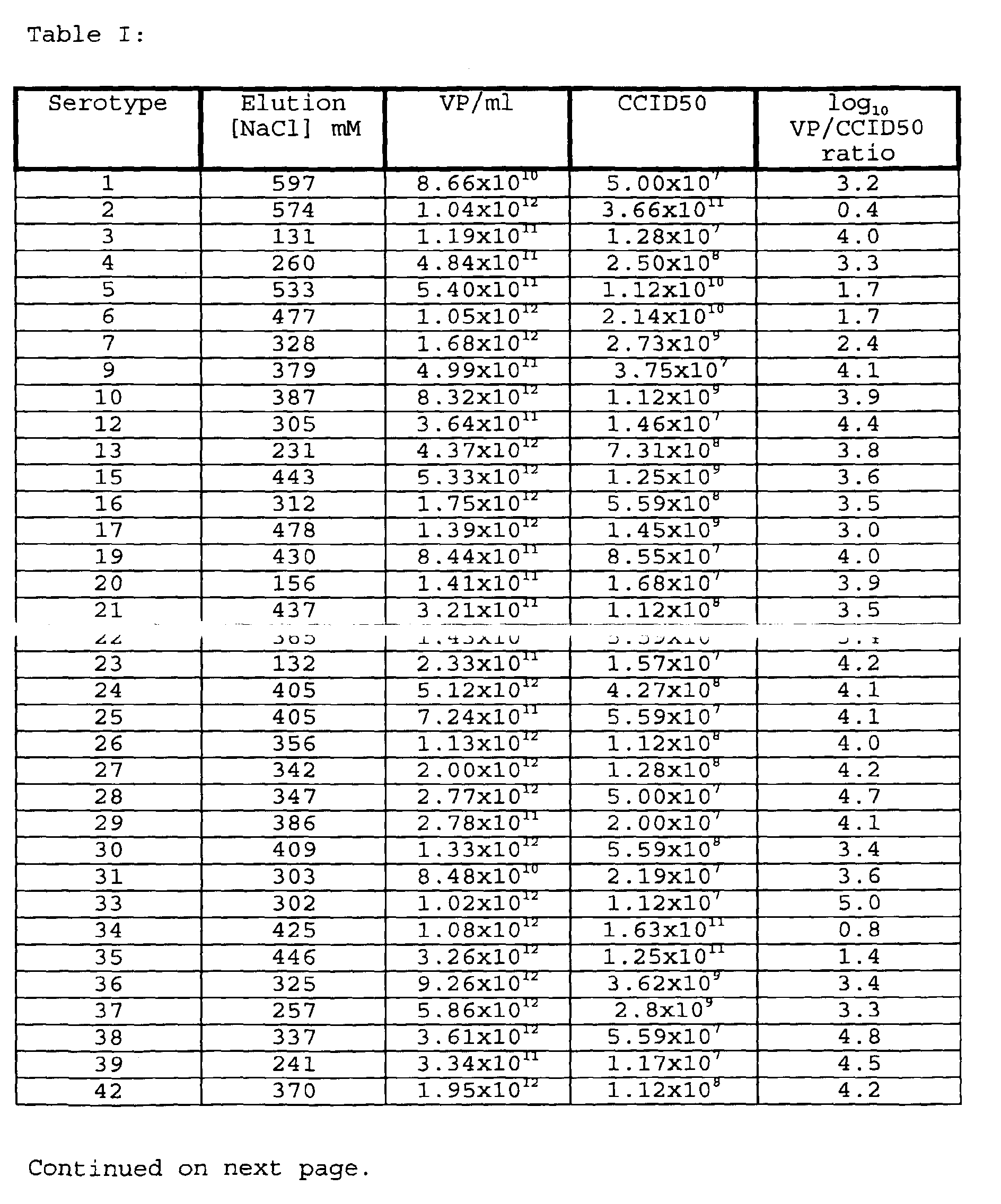
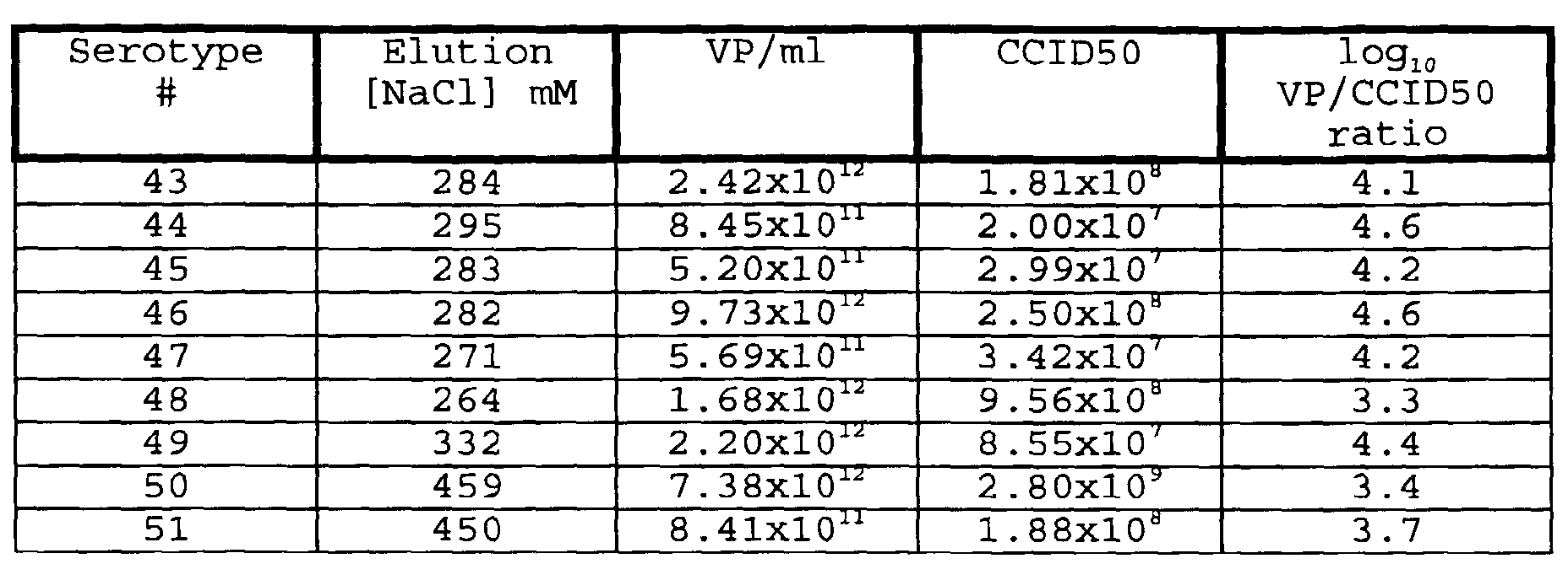


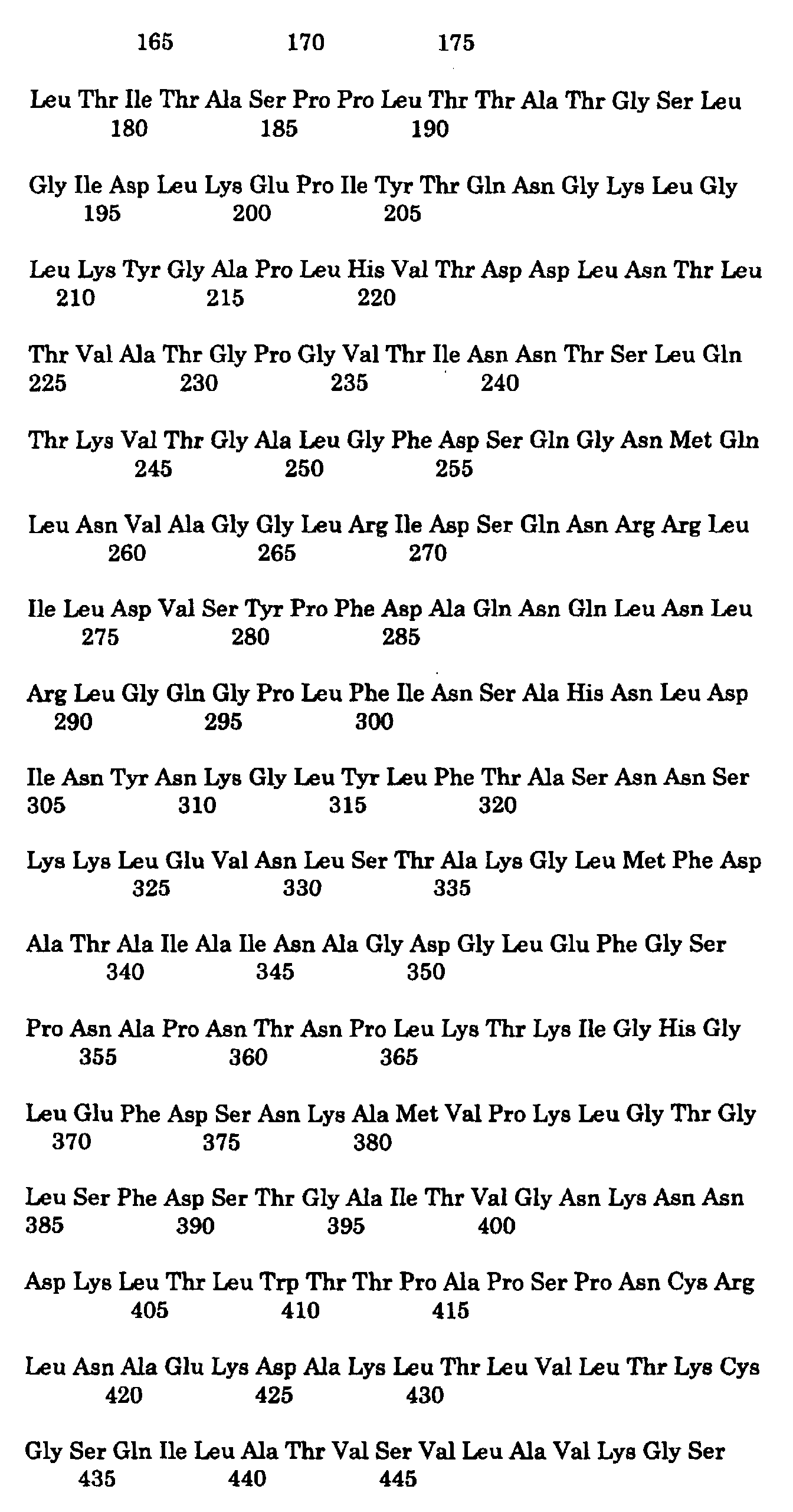
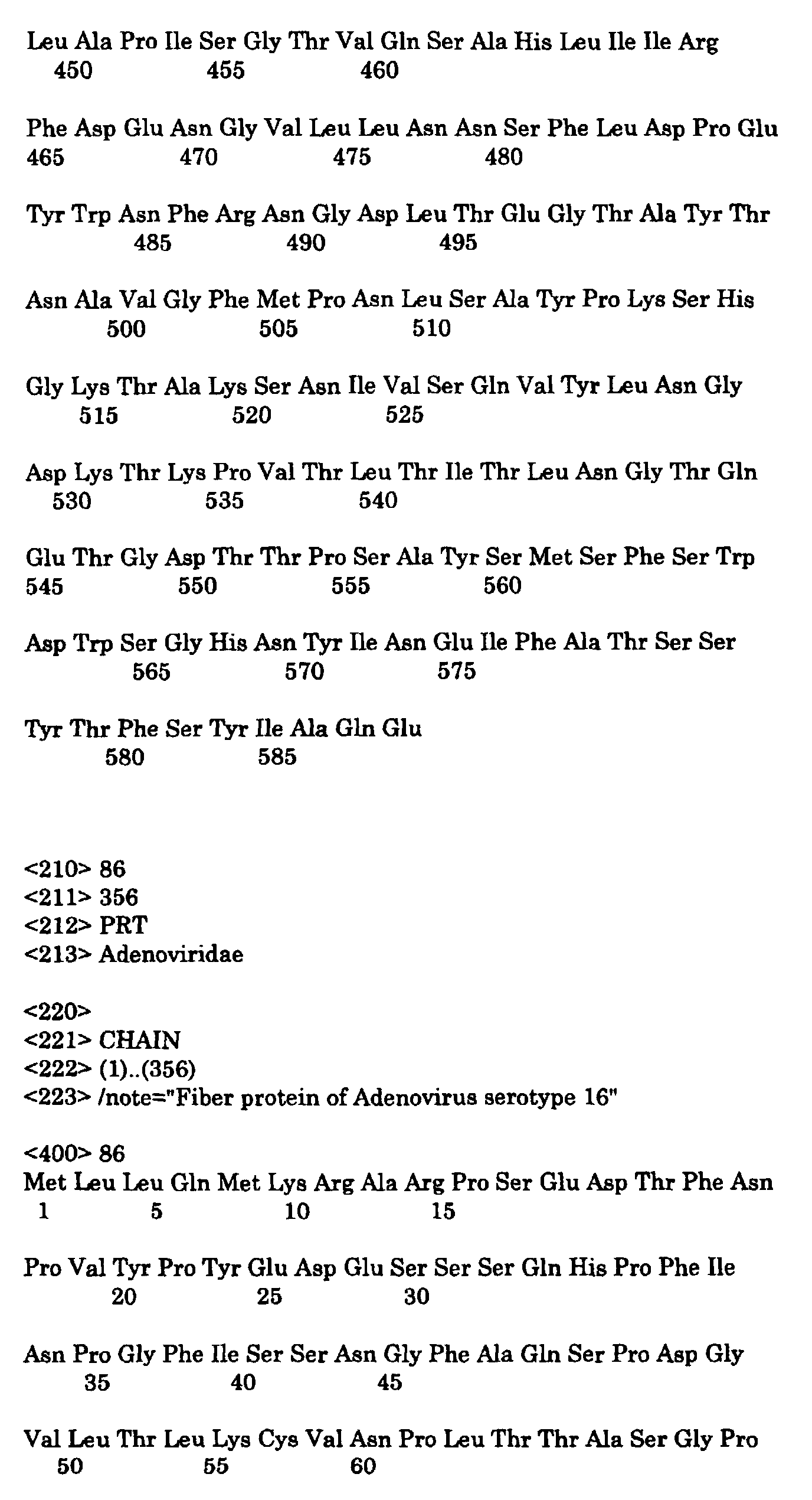
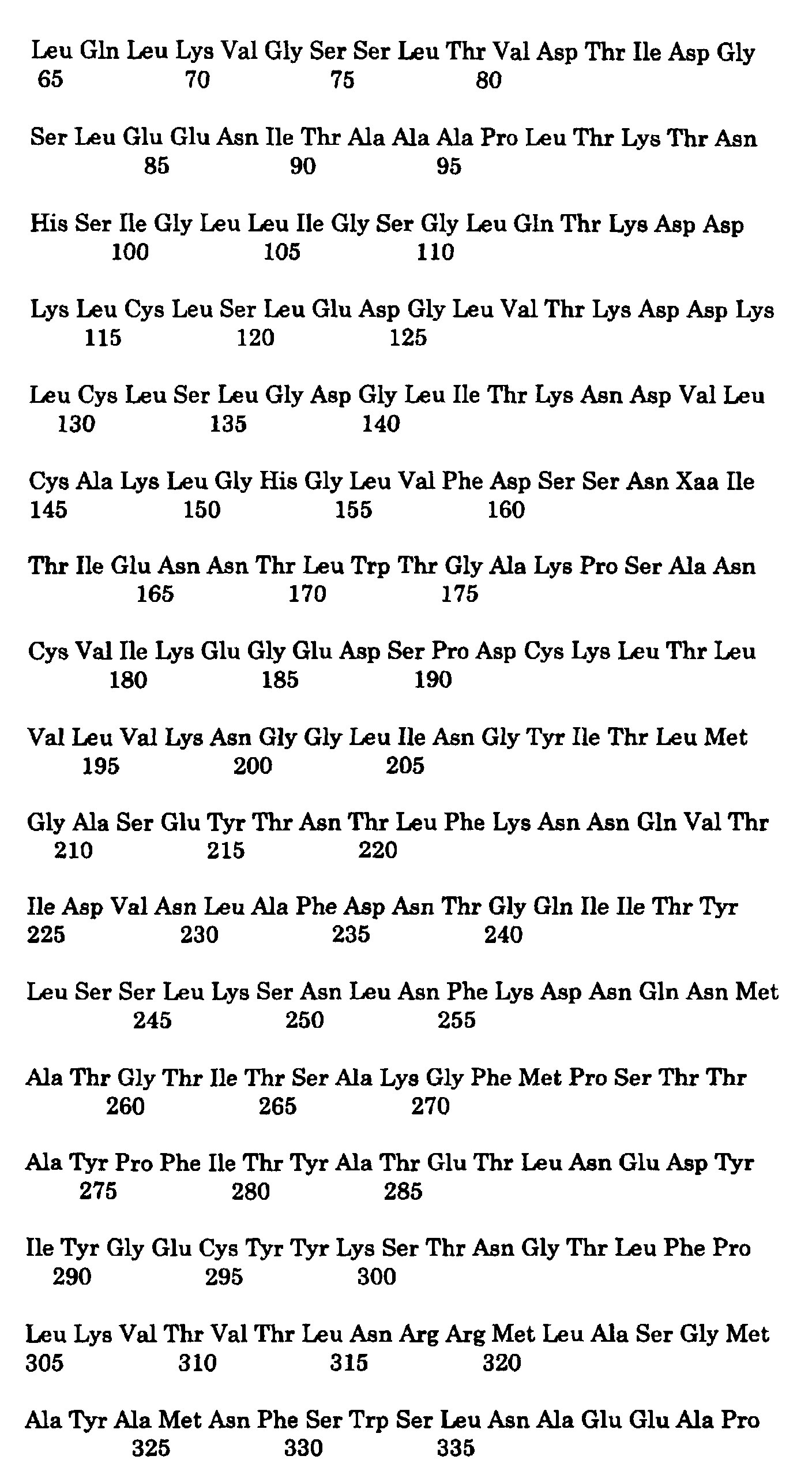
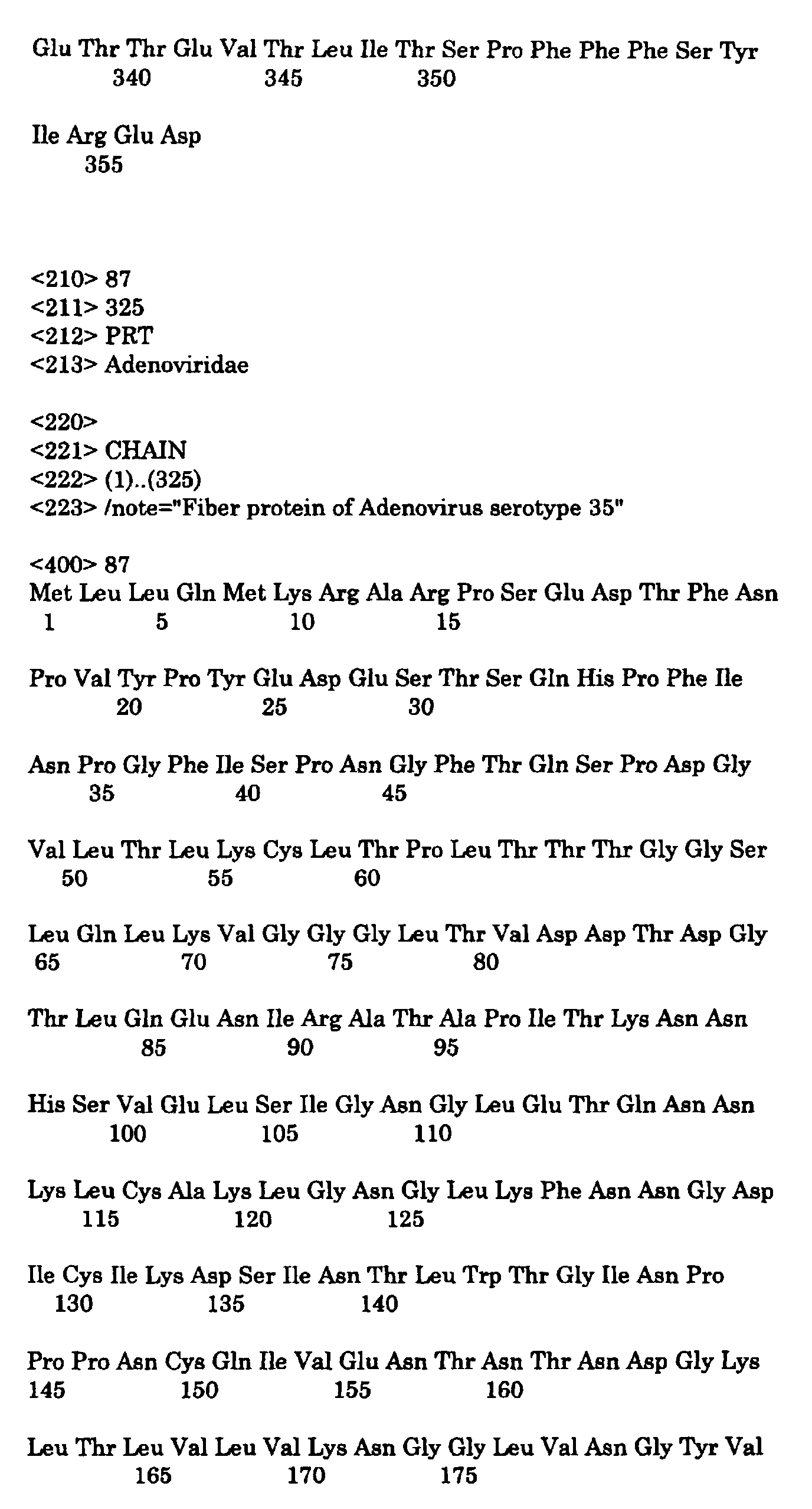
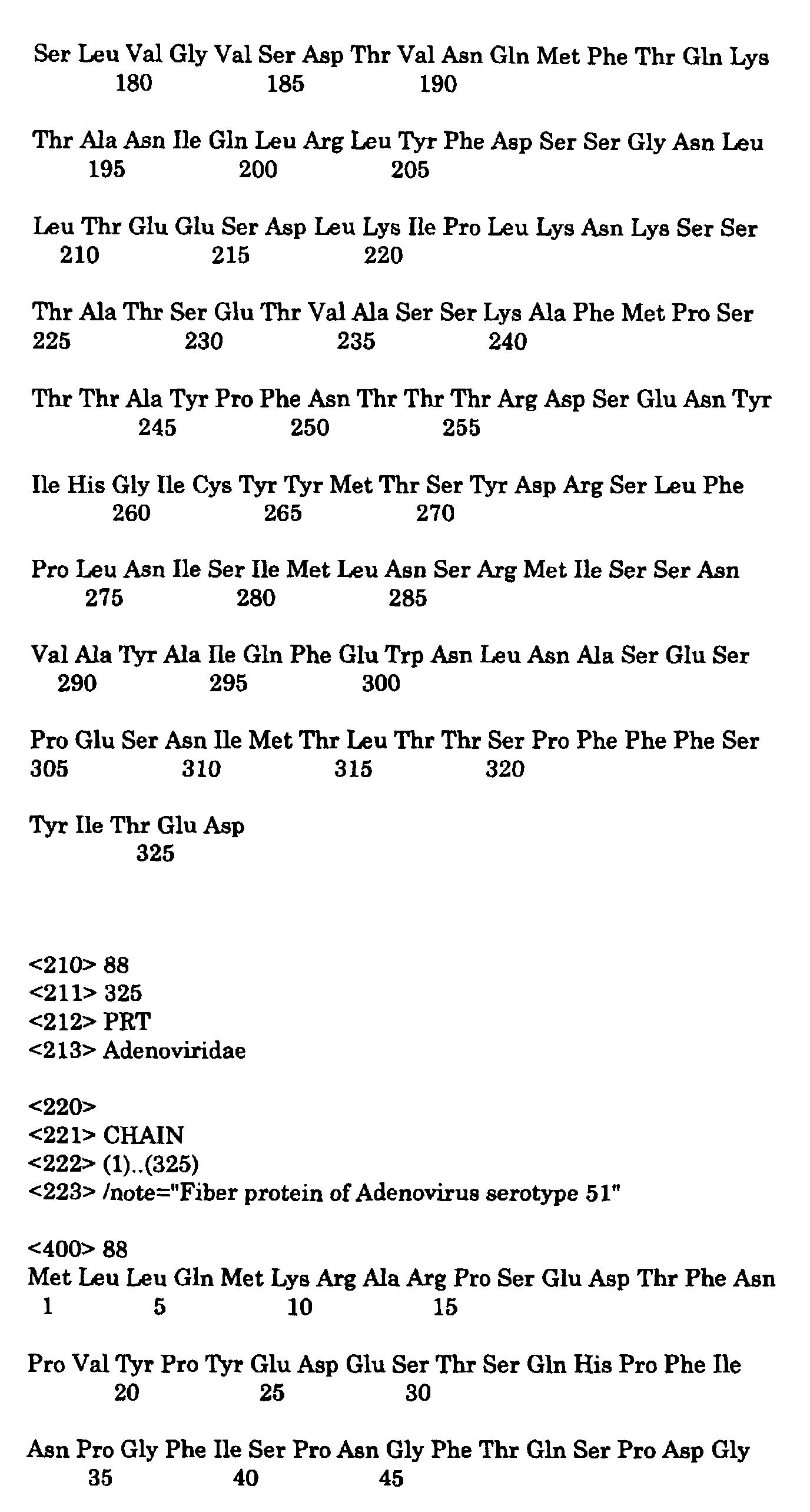

Claims (23)
- A gene delivery vehicle comprising at least one of the adenovirus serotype 35 elements or a functional equivalent thereof, responsible for avoiding or deminishing neutralising activity against adenoviral elements by the host to which the gene is to be delivered and a gene of interest.
- Adenovirus serotype 35 or a functional homologue thereof or a chimaeric virus derived therefrom, or a gene delivery vehicle based on said virus its homologue or its chimaera for use as a pharmaceutical.
- A gene delivery vehicle according to claim 1, whereby said elements comprise adenovirus 35 E3 expression products or the genes encoding them.
- A gene delivery vehicle according to claim 1 or 3, whereby said elements comprise adenovirus 35 fiber, penton and/or hexon proteins or a gene encoding either.
- A gene delivery according to any one of claims 1,3, or 4 which is a chimaera of adenovirus 35 with at least one other adenovirus.
- A gene delivery vehicle according to any one of claims 1,3,4 or 5 which has a different tropism than adenovirus 35.
- A nucleic acid encoding at least a functional part of a gene delivery vehicle according to any one of claims 1 or 3-6, or a virus, homologue or chimaera thereof according to claim 2.
- A nucleic acid encoding at least one of the adenovirus serotype 35 elements or a functional equivalent thereof, responsible for avoiding or deminishing neutralising activity against adenoviral elements by the host to which the gene is to be delivered and having a site for introducing a gene of interest therein.
- A nucleic acid according to claim 7 or 8, further comprising at least one ITR.
- A nucleic acid according to claim 7, 8 or 9 further comprising a region of nucleotides designed or useable for homologous recombination.
- At least one set of two nucleic acids comprising a nucleic acid according to any one of claims 7-10, whereby said set of nucleic acids is capable of a single homologous recombination event with each other, which leads to a nucleic acid encoding a functional gene delivery vehicle.
- A cell comprising a nucleic acid according to any one of claims 7-10 or a set of nucleic acids according to claim 11.
- A cell according to claim 12 which complements the necessary elements for adenoviral replication which are absent from the nucleic acid according to any one of claims 7-10 or a set of nucleic acids according to claim 11.
- A cell according to claim 12 or 13, which originates from a PER.C6 cell (ECACC deposit number 96022940).
- A method for producing a gene delivery vehicle according to claim 1, or 3-6, comprising expressing a nucleic acid according to any one of claims 7-10 in a cell according to claim 12 or 13 and harvesting the resulting gene delivery vehicle.
- A method for producing a gene delivery vehicle according to claim 1, or 3-6, comprising culturing a cell according to claim 12 or 13 in a suitable culture medium and harvesting the resulting gene delivery vehicle.
- A gene delivery vehicle obtainable by a method according to claims 15 or 16.
- A gene delivery vehicle according to any one of claims 1,3-6 or 17, which is derived from a chimaera of an adenovirus and an integrating virus.
- A gene delivery vehicle according to claim 18, wherein said integrating virus is adeno associated virus.
- A gene delivery vehicle according to any one of claims 1, 3-6 or 17-19, which has the tropism determining parts of adenovirus 16 or functional equivalents thereof.
- A gene delivery vehicle according to anyone of claims 1, 3-6 or 17-20 for use as a pharmaceutical.
- A pharmaceutical formulation comprising a gene delivery vehicle according to any one of claims 1, 3-6 or 17-20 and a suitable excipient.
- A pharmaceutical formulation comprising an adenovirus, a chimaera thereof, or a functional homologue thereof according to claim 2 and a suitable excipient.
Priority Applications (7)
| Application Number | Priority Date | Filing Date | Title |
|---|---|---|---|
| EP07106044A EP1816204B1 (en) | 1999-05-17 | 2000-05-16 | Recombinant Adenovirus of the Ad26 serotype |
| EP04077434A EP1550722B1 (en) | 1999-05-17 | 2000-05-16 | Recombinant human adenovirus serotype 35 |
| EP05077719A EP1681353B1 (en) | 1999-05-17 | 2000-05-16 | Adenovirus derived gene delivery vehicles comprising at least one element of adenovirus type 35 |
| EP07106054A EP1816205B1 (en) | 1999-05-17 | 2000-05-16 | Recombinant Adenovirus based on serotype 48 (Ad48). |
| EP07106036A EP1818408B1 (en) | 1999-05-17 | 2000-05-16 | Recombinant Adenovirus of the Ad11 serotype |
| EP00201738A EP1054064B2 (en) | 1999-05-17 | 2000-05-16 | Adenovirus derived gene delivery vehicles comprising at least one element of adenovirus type 35 |
| CY20071100996T CY1106762T1 (en) | 1999-05-17 | 2007-07-25 | RECOMBINANT HUMAN ADENOINE SEROTYPE 35 |
Applications Claiming Priority (3)
| Application Number | Priority Date | Filing Date | Title |
|---|---|---|---|
| EP99201545 | 1999-05-17 | ||
| EP99201545 | 1999-05-17 | ||
| EP00201738A EP1054064B2 (en) | 1999-05-17 | 2000-05-16 | Adenovirus derived gene delivery vehicles comprising at least one element of adenovirus type 35 |
Related Child Applications (1)
| Application Number | Title | Priority Date | Filing Date |
|---|---|---|---|
| EP04077434A Division EP1550722B1 (en) | 1999-05-17 | 2000-05-16 | Recombinant human adenovirus serotype 35 |
Publications (3)
| Publication Number | Publication Date |
|---|---|
| EP1054064A1 true EP1054064A1 (en) | 2000-11-22 |
| EP1054064B1 EP1054064B1 (en) | 2004-10-06 |
| EP1054064B2 EP1054064B2 (en) | 2009-12-16 |
Family
ID=8240209
Family Applications (6)
| Application Number | Title | Priority Date | Filing Date |
|---|---|---|---|
| EP04077434A Expired - Lifetime EP1550722B1 (en) | 1999-05-17 | 2000-05-16 | Recombinant human adenovirus serotype 35 |
| EP07106054A Expired - Lifetime EP1816205B1 (en) | 1999-05-17 | 2000-05-16 | Recombinant Adenovirus based on serotype 48 (Ad48). |
| EP00201738A Expired - Lifetime EP1054064B2 (en) | 1999-05-17 | 2000-05-16 | Adenovirus derived gene delivery vehicles comprising at least one element of adenovirus type 35 |
| EP07106044A Expired - Lifetime EP1816204B1 (en) | 1999-05-17 | 2000-05-16 | Recombinant Adenovirus of the Ad26 serotype |
| EP05077719A Expired - Lifetime EP1681353B1 (en) | 1999-05-17 | 2000-05-16 | Adenovirus derived gene delivery vehicles comprising at least one element of adenovirus type 35 |
| EP07106036A Expired - Lifetime EP1818408B1 (en) | 1999-05-17 | 2000-05-16 | Recombinant Adenovirus of the Ad11 serotype |
Family Applications Before (2)
| Application Number | Title | Priority Date | Filing Date |
|---|---|---|---|
| EP04077434A Expired - Lifetime EP1550722B1 (en) | 1999-05-17 | 2000-05-16 | Recombinant human adenovirus serotype 35 |
| EP07106054A Expired - Lifetime EP1816205B1 (en) | 1999-05-17 | 2000-05-16 | Recombinant Adenovirus based on serotype 48 (Ad48). |
Family Applications After (3)
| Application Number | Title | Priority Date | Filing Date |
|---|---|---|---|
| EP07106044A Expired - Lifetime EP1816204B1 (en) | 1999-05-17 | 2000-05-16 | Recombinant Adenovirus of the Ad26 serotype |
| EP05077719A Expired - Lifetime EP1681353B1 (en) | 1999-05-17 | 2000-05-16 | Adenovirus derived gene delivery vehicles comprising at least one element of adenovirus type 35 |
| EP07106036A Expired - Lifetime EP1818408B1 (en) | 1999-05-17 | 2000-05-16 | Recombinant Adenovirus of the Ad11 serotype |
Country Status (14)
| Country | Link |
|---|---|
| EP (6) | EP1550722B1 (en) |
| JP (2) | JP4843145B2 (en) |
| KR (1) | KR100741247B1 (en) |
| AT (6) | ATE485382T1 (en) |
| AU (1) | AU777041B2 (en) |
| CA (1) | CA2372655C (en) |
| CY (4) | CY1106762T1 (en) |
| DE (4) | DE60043126D1 (en) |
| DK (6) | DK1816204T3 (en) |
| ES (5) | ES2372823T3 (en) |
| IL (3) | IL146479A0 (en) |
| NZ (1) | NZ515582A (en) |
| PT (5) | PT1816204E (en) |
| WO (1) | WO2000070071A1 (en) |
Cited By (22)
| Publication number | Priority date | Publication date | Assignee | Title |
|---|---|---|---|---|
| WO2002053759A1 (en) * | 2001-01-04 | 2002-07-11 | Wadell Goeran | Viral vector for gene therapy |
| WO2004001032A2 (en) | 2002-04-25 | 2003-12-31 | Crucell Holland B.V. | Stable adenoviral vectors and methods for propagation thereof |
| US7235233B2 (en) | 2000-09-26 | 2007-06-26 | Crucell Holland B.V. | Serotype 5 adenoviral vectors with chimeric fibers for gene delivery in skeletal muscle cells or myoblasts |
| US7238528B2 (en) | 1999-03-04 | 2007-07-03 | Crucell Holland B.V. | Means and methods for fibroblast-like or macrophage-like cell transduction |
| US7250293B2 (en) | 1999-05-18 | 2007-07-31 | Crucell Holland B.V. | Complementing cell lines |
| US7270811B2 (en) | 1999-05-18 | 2007-09-18 | Crucell Holland B.V. | Serotype of adenovirus and uses thereof |
| US7300657B2 (en) | 2002-12-17 | 2007-11-27 | Crucell Holland B.V. | Recombinant viral-based malaria vaccines |
| US7468181B2 (en) | 2002-04-25 | 2008-12-23 | Crucell Holland B.V. | Means and methods for the production of adenovirus vectors |
| US7598078B2 (en) | 2002-10-23 | 2009-10-06 | Crucell Holland B.V. | Settings for recombinant adenoviral-based vaccines |
| US7749493B2 (en) | 1998-07-08 | 2010-07-06 | Crucell Holland B.V. | Chimeric adenoviruses |
| US7888102B2 (en) | 2005-07-22 | 2011-02-15 | Crucell Holland B.V. | Cell line for producing coronaviruses |
| US7968286B2 (en) | 2002-10-23 | 2011-06-28 | Crucell Holland B.V. | Settings for recombinant adenoviral-based vaccines |
| US20130230902A2 (en) * | 2004-05-26 | 2013-09-05 | Psioxus Therapeutics Limited | Chimeric adenoviruses for use in cancer treatment |
| CN111337042A (en) * | 2020-03-13 | 2020-06-26 | 湖北大学 | Vehicle path planning method and system |
| US10849945B2 (en) | 2015-04-30 | 2020-12-01 | Psioxus Therapeutics Limited | Oncolytic adenovirus encoding a B7 protein or active fragment |
| US11077156B2 (en) | 2013-03-14 | 2021-08-03 | Salk Institute For Biological Studies | Oncolytic adenovirus compositions |
| US11130968B2 (en) | 2016-02-23 | 2021-09-28 | Salk Institute For Biological Studies | High throughput assay for measuring adenovirus replication kinetics |
| US11155622B2 (en) | 2015-12-17 | 2021-10-26 | Psioxus Therapeutics Limited | Virus encoding an anti-TCR-complex antibody or fragment |
| US11401529B2 (en) | 2016-02-23 | 2022-08-02 | Salk Institute For Biological Studies | Exogenous gene expression in recombinant adenovirus for minimal impact on viral kinetics |
| US11439678B2 (en) | 2013-10-25 | 2022-09-13 | Psioxus Therapeutics Limited | Oncolytic adenoviruses armed with heterologous genes |
| US11813337B2 (en) | 2016-12-12 | 2023-11-14 | Salk Institute For Biological Studies | Tumor-targeting synthetic adenoviruses and uses thereof |
| US11840702B2 (en) | 2017-08-28 | 2023-12-12 | Akamis Bio Limited | Adenovirus armed with bispecific T cell activator |
Families Citing this family (80)
| Publication number | Priority date | Publication date | Assignee | Title |
|---|---|---|---|---|
| ATE278794T1 (en) | 1995-06-15 | 2004-10-15 | Crucell Holland Bv | PACKAGING SYSTEMS FOR HUMAN, HUMAN ADENOVIRUSES, FOR USE IN GENE THERAPY |
| US6929946B1 (en) | 1998-11-20 | 2005-08-16 | Crucell Holland B.V. | Gene delivery vectors provided with a tissue tropism for smooth muscle cells, and/or endothelial cells |
| US7094398B1 (en) | 1999-06-01 | 2006-08-22 | University Of Washington | Recombinant adenoviral vectors expressing chimeric fiber proteins for cell specific infection and genome integration |
| US6558948B1 (en) | 1999-11-23 | 2003-05-06 | Stefan Kochanek | Permanent amniocytic cell line, its production and use for the production of gene transfer vectors |
| EP1191105A1 (en) * | 2000-09-25 | 2002-03-27 | Galapagos Genomics B.V. | Gene delivery vectors provided with a tissue tropism for T-lymphocytes |
| AU6368901A (en) | 2000-05-31 | 2001-12-11 | Univ Saskatchewan | Modified bovine adenovirus having altered tropism |
| EP1322774A2 (en) * | 2000-09-20 | 2003-07-02 | Crucell Holland B.V. | Gene delivery vectors provided with a tissue tropism for dendritic cells |
| WO2002029073A2 (en) * | 2000-10-06 | 2002-04-11 | Crucell Holland B.V. | Gene delivery vectors for stem cells |
| EP1195440A1 (en) * | 2000-10-06 | 2002-04-10 | Introgene B.V. | Gene delivery vectors for stem cells |
| CA2439185A1 (en) | 2001-02-23 | 2002-09-06 | Novartis Ag | Vector constructs |
| US20040002060A1 (en) * | 2002-01-24 | 2004-01-01 | Novartis Ag | Fiber shaft modifications for efficient targeting |
| JP4237449B2 (en) * | 2002-06-05 | 2009-03-11 | 国立医薬品食品衛生研究所長 | Adenovirus vector |
| CA2527369A1 (en) * | 2003-07-18 | 2005-02-03 | Onyx Pharmaceuticals, Inc. | Subgroup b adenoviral vectors for treating disease |
| US20070207461A1 (en) | 2004-02-23 | 2007-09-06 | Crucell Holland B.V. | Virus Purification Methods |
| US20070207166A1 (en) * | 2004-04-12 | 2007-09-06 | Genvec, Inc. | Method of Using Adenoviral Vectors to Induce an Immune Response |
| WO2006040330A2 (en) | 2004-10-13 | 2006-04-20 | Crucell Holland B.V. | Improved adenoviral vectors and uses thereof |
| JP4838259B2 (en) | 2004-11-16 | 2011-12-14 | クルセル ホランド ベー ヴェー | Multivalent vaccine containing recombinant viral vectors |
| CN101155915B (en) | 2005-04-11 | 2013-12-04 | 克鲁塞尔荷兰公司 | Virus purification using ultrafiltration |
| WO2007104792A2 (en) * | 2006-03-16 | 2007-09-20 | Crucell Holland B.V. | Recombinant adenoviruses based on serotype 26 and 48, and use thereof |
| CN102203242B (en) | 2008-11-03 | 2013-06-12 | 克鲁塞尔荷兰公司 | Method for the production of adenoviral vectors |
| MX2012001592A (en) | 2009-08-07 | 2012-05-22 | Transgene Sa | Composition for treating hbv infection. |
| CN102762721B (en) * | 2010-02-15 | 2015-03-11 | 克鲁塞尔荷兰公司 | Method for the production of Ad26 adenoviral vectors |
| US20120315696A1 (en) * | 2010-02-15 | 2012-12-13 | Alfred Luitjens | METHOD FOR THE PRODUCTION OF Ad26 ADENOVIRAL VECTORS |
| CA2808556A1 (en) | 2010-09-20 | 2012-03-29 | Crucell Holland B.V. | Therapeutic vaccination against active tuberculosis |
| AU2011310838B2 (en) | 2010-09-27 | 2015-11-05 | Crucell Holland B.V. | Heterologous prime boost vaccination regimen against malaria |
| CN102260712B (en) * | 2011-05-31 | 2013-10-02 | 北京锤特生物科技有限公司 | Construction and application of type B adenovirus hominis Ad11 mutant with enhanced tumor dissolving ability |
| US8932607B2 (en) | 2012-03-12 | 2015-01-13 | Crucell Holland B.V. | Batches of recombinant adenovirus with altered terminal ends |
| CA2864956C (en) | 2012-03-12 | 2021-11-09 | Crucell Holland B.V. | Batches of recombinant adenovirus with altered terminal ends |
| JP5845376B2 (en) | 2012-03-22 | 2016-01-20 | クルセル ホランド ベー ヴェー | Vaccine against RSV |
| US9119813B2 (en) | 2012-03-22 | 2015-09-01 | Crucell Holland B.V. | Vaccine against RSV |
| EP2855685B1 (en) | 2012-05-24 | 2017-03-08 | Janssen Vaccines & Prevention B.V. | Adenoviral vectors for transduction of vascular tissue |
| BR112015000530A2 (en) | 2012-07-10 | 2018-08-28 | Transgene Sa | mycobacterial antigen vaccine. |
| WO2014009433A1 (en) | 2012-07-10 | 2014-01-16 | Transgene Sa | Mycobacterium resuscitation promoting factor for use as adjuvant |
| JP2015536147A (en) | 2012-11-16 | 2015-12-21 | ベス イスラエル デアコネス メディカル センター インコーポレイテッド | Recombinant adenovirus and use thereof |
| WO2014174018A1 (en) | 2013-04-25 | 2014-10-30 | Crucell Holland B.V. | Stabilized soluble prefusion rsv f polypeptides |
| CA2914792C (en) | 2013-06-17 | 2024-02-27 | Crucell Holland B.V. | Stabilized soluble pre-fusion rsv f polypeptides |
| EP3092000A1 (en) | 2014-01-09 | 2016-11-16 | Transgene SA | Fusion of heterooligomeric mycobacterial antigens |
| JP6462861B2 (en) | 2014-09-03 | 2019-01-30 | バヴァリアン ノルディック エー/エス | Methods and compositions aimed at inducing protective immunity against filovirus infection |
| BR112017004202A2 (en) | 2014-09-03 | 2019-09-03 | Bavarian Nordic As | compositions for enhancing immune responses and their uses |
| LT3197489T (en) | 2014-09-26 | 2021-07-26 | Beth Israel Deaconess Medical Center, Inc. | Methods and compositions for inducing protective immunity against human immunodeficiency virus infection |
| EP3421046A1 (en) | 2014-11-04 | 2019-01-02 | Janssen Vaccines & Prevention B.V. | Therapeutic hpv16 vaccines |
| PL3283634T3 (en) | 2015-04-14 | 2019-10-31 | Janssen Vaccines & Prevention Bv | Recombinant adenovirus expressing two transgenes with a bidirectional promoter |
| CA2991002C (en) | 2015-07-07 | 2023-11-28 | Janssen Vaccines & Prevention B.V. | Vaccine against rsv |
| EA035909B1 (en) | 2015-07-07 | 2020-08-31 | Янссен Вэксинс Энд Превеншн Б.В. | Stabilized soluble pre-fusion rsv f polypeptides |
| EP3337500A1 (en) | 2015-08-20 | 2018-06-27 | Janssen Vaccines & Prevention B.V. | Therapeutic hpv18 vaccines |
| EP3584252B1 (en) | 2015-12-15 | 2021-08-25 | Janssen Vaccines & Prevention B.V. | Human immunodeficiency virus antigens, vectors, compositions, and methods of use thereof |
| ES2858315T3 (en) | 2016-04-05 | 2021-09-30 | Janssen Vaccines & Prevention Bv | Stabilized soluble RSV prefusion protein F for use in the prophylaxis of RSV infection |
| BR112018070323A2 (en) | 2016-04-05 | 2019-01-29 | Janssen Vaccines & Prevention Bv | rsv vaccine |
| BR112018072372A2 (en) | 2016-05-02 | 2019-02-19 | Janssen Vaccines & Prevention B.V. | hpv therapeutic vaccine combinations |
| IL264119B2 (en) | 2016-05-30 | 2023-04-01 | Janssen Vaccines Prevention B V | Stabilized pre-fusion rsv f proteins |
| AU2017286375B2 (en) | 2016-06-16 | 2019-04-18 | Janssen Vaccines & Prevention B.V. | HIV vaccine formulation |
| WO2017220499A1 (en) | 2016-06-20 | 2017-12-28 | Janssen Vaccines & Prevention B.V. | Potent and balanced bidirectional promoter |
| EP3484506A1 (en) | 2016-07-14 | 2019-05-22 | Janssen Vaccines & Prevention B.V. | Hpv vaccines |
| WO2018011198A1 (en) | 2016-07-15 | 2018-01-18 | Janssen Vaccines & Prevention B.V. | Methods and compositions for inducing protective immunity against a marburg virus infection |
| EP3484507A1 (en) | 2016-07-15 | 2019-05-22 | Janssen Vaccines & Prevention B.V. | Methods and compositions for inducing protective immunity against a marburg virus infection |
| BR112019015671A2 (en) | 2017-02-09 | 2020-04-14 | Janssen Vaccines & Prevention Bv | potent and short promoter for expression of heterological genes |
| US11173204B2 (en) | 2017-04-06 | 2021-11-16 | Janssen Vaccines & Prevention B.V. | MVA-BN and Ad26.ZEBOV or Ad26.filo prime-boost regimen |
| CA3061278A1 (en) | 2017-05-17 | 2018-11-22 | Janssen Vaccines & Prevention B.V. | Methods and compositions for inducing protective immunity against rsv infection |
| WO2018210871A1 (en) | 2017-05-17 | 2018-11-22 | Janssen Vaccines & Prevention B.V. | Methods and compositions for inducing protective immunity against rsv infection |
| MA49397A (en) | 2017-06-15 | 2020-04-22 | Bavarian Nordic As | POXVIRUS VECTORS CODING FOR HIV ANTIGENS, AND METHODS FOR USING THEM |
| US11229695B2 (en) | 2017-09-15 | 2022-01-25 | Janssen Vaccines & Prevention B.V. | Method for the safe induction of immunity against RSV |
| EP3713599A1 (en) | 2017-11-20 | 2020-09-30 | Janssen Pharmaceuticals, Inc. | Method of providing safe administration of adenoviral vectors encoding a zika virus antigen |
| EP3723771A4 (en) | 2017-12-11 | 2022-04-06 | Beth Israel Deaconess Medical Center, Inc. | Recombinant adenoviruses and uses thereof |
| BR112020012273A2 (en) | 2017-12-19 | 2020-11-24 | Janssen Sciences Ireland Unlimited Company | methods and compositions to induce an immune response against the hepatitis b virus (hbv) |
| WO2020016394A1 (en) | 2018-07-20 | 2020-01-23 | Janssen Vaccines & Prevention B.V. | Recombinant adenoviral vector expressing zika antigen with improved productivity |
| TW202043256A (en) | 2019-01-10 | 2020-12-01 | 美商健生生物科技公司 | Prostate neoantigens and their uses |
| AU2020275455A1 (en) | 2019-05-15 | 2021-12-09 | Janssen Vaccines & Prevention B.V. | Co-administration of seasonal influenza vaccine and an adenovirus based respiratory syncytial virus vaccine |
| EA202193119A1 (en) | 2019-05-15 | 2022-02-18 | Янссен Вэксинс Энд Превеншн Б.В. | PREVENTIVE TREATMENT OF RESPIRATORY SYNCYTHIAL VIRUS INFECTION WITH ADENOVIRUS VACCINE |
| IL293051A (en) | 2019-11-18 | 2022-07-01 | Janssen Biotech Inc | Vaccines based on mutant calr and jak2 and their uses |
| TW202144388A (en) | 2020-02-14 | 2021-12-01 | 美商健生生物科技公司 | Neoantigens expressed in ovarian cancer and their uses |
| TW202144389A (en) | 2020-02-14 | 2021-12-01 | 美商健生生物科技公司 | Neoantigens expressed in multiple myeloma and their uses |
| JP2023521194A (en) | 2020-04-13 | 2023-05-23 | ヤンセン バイオテツク,インコーポレーテツド | PSMA and STEAP1 vaccines and their uses |
| WO2022009052A2 (en) | 2020-07-06 | 2022-01-13 | Janssen Biotech, Inc. | Prostate neoantigens and their uses |
| US20230024133A1 (en) | 2020-07-06 | 2023-01-26 | Janssen Biotech, Inc. | Prostate Neoantigens And Their Uses |
| US20230035403A1 (en) | 2020-07-06 | 2023-02-02 | Janssen Biotech, Inc. | Method For Determining Responsiveness To Prostate Cancer Treatment |
| TW202216190A (en) | 2020-07-08 | 2022-05-01 | 愛爾蘭商健生科學愛爾蘭無限公司 | Rna replicon vaccines against hbv |
| GB202013940D0 (en) | 2020-09-04 | 2020-10-21 | Synpromics Ltd | Regulatory nucleic acid sequences |
| JP2023545731A (en) | 2020-10-07 | 2023-10-31 | アスクレピオス バイオファーマシューティカル, インコーポレイテッド | Therapeutic adeno-associated virus delivery of fukutin-related protein (FKRP) to treat dystroglycanopathy disorders including limb girdle type 2I (LGMD2I) |
| US20220194999A1 (en) | 2020-12-23 | 2022-06-23 | Janssen Biotech, Inc. | Neoantigen Peptide Mimics |
| WO2023198815A1 (en) | 2022-04-14 | 2023-10-19 | Janssen Vaccines & Prevention B.V. | Sequential administration of adenoviruses |
Citations (4)
| Publication number | Priority date | Publication date | Assignee | Title |
|---|---|---|---|---|
| WO1996012030A1 (en) * | 1994-10-17 | 1996-04-25 | Rhone-Poulenc Rorer S.A. | Defective adenoviruses including a therapeutic gene and an immunoprotective gene |
| WO1996013598A2 (en) * | 1994-10-28 | 1996-05-09 | The Trustees Of The University Of Pennsylvania | Hybrid adenovirus-aav virus and method of use thereof |
| WO1997012986A2 (en) * | 1995-10-02 | 1997-04-10 | Cornell Research Foundation, Inc. | Non-group c adenoviral vectors |
| WO1998022609A1 (en) * | 1996-11-20 | 1998-05-28 | Genzyme Corporation | Chimeric adenoviral vectors |
Family Cites Families (4)
| Publication number | Priority date | Publication date | Assignee | Title |
|---|---|---|---|---|
| ATE278794T1 (en) | 1995-06-15 | 2004-10-15 | Crucell Holland Bv | PACKAGING SYSTEMS FOR HUMAN, HUMAN ADENOVIRUSES, FOR USE IN GENE THERAPY |
| US6100086A (en) * | 1997-04-14 | 2000-08-08 | Genzyme Corporation | Transgene expression systems |
| DE60039683D1 (en) * | 1999-03-04 | 2008-09-11 | Crucell Holland Bv | USE OF AN ADENOVIRUS VECTOR FOR THE TRANSDUCTION OF SYNOVIAL CELLS |
| WO2009146902A1 (en) * | 2008-06-03 | 2009-12-10 | Okairòs Ag | A vaccine for the prevention and therapy of hcv infections |
-
2000
- 2000-05-16 EP EP04077434A patent/EP1550722B1/en not_active Expired - Lifetime
- 2000-05-16 ES ES07106036T patent/ES2372823T3/en not_active Expired - Lifetime
- 2000-05-16 AT AT07106044T patent/ATE485382T1/en active
- 2000-05-16 WO PCT/NL2000/000325 patent/WO2000070071A1/en active Application Filing
- 2000-05-16 DK DK07106044.6T patent/DK1816204T3/en active
- 2000-05-16 EP EP07106054A patent/EP1816205B1/en not_active Expired - Lifetime
- 2000-05-16 EP EP00201738A patent/EP1054064B2/en not_active Expired - Lifetime
- 2000-05-16 ES ES04077434T patent/ES2289426T3/en not_active Expired - Lifetime
- 2000-05-16 PT PT07106044T patent/PT1816204E/en unknown
- 2000-05-16 DE DE60043126T patent/DE60043126D1/en not_active Expired - Lifetime
- 2000-05-16 AT AT00201738T patent/ATE278792T1/en active
- 2000-05-16 EP EP07106044A patent/EP1816204B1/en not_active Expired - Lifetime
- 2000-05-16 JP JP2000618477A patent/JP4843145B2/en not_active Expired - Lifetime
- 2000-05-16 CA CA2372655A patent/CA2372655C/en not_active Expired - Lifetime
- 2000-05-16 DK DK00201738.2T patent/DK1054064T4/en active
- 2000-05-16 AT AT07106036T patent/ATE519855T1/en active
- 2000-05-16 EP EP05077719A patent/EP1681353B1/en not_active Expired - Lifetime
- 2000-05-16 PT PT07106036T patent/PT1818408E/en unknown
- 2000-05-16 PT PT07106054T patent/PT1816205E/en unknown
- 2000-05-16 AT AT07106054T patent/ATE519854T1/en active
- 2000-05-16 DE DE60045138T patent/DE60045138D1/en not_active Expired - Lifetime
- 2000-05-16 DK DK07106036.2T patent/DK1818408T3/en active
- 2000-05-16 IL IL14647988A patent/IL146479A0/en unknown
- 2000-05-16 AU AU49547/00A patent/AU777041B2/en not_active Expired
- 2000-05-16 ES ES00201738T patent/ES2231103T5/en not_active Expired - Lifetime
- 2000-05-16 DK DK05077719T patent/DK1681353T3/en active
- 2000-05-16 ES ES07106054T patent/ES2372824T3/en not_active Expired - Lifetime
- 2000-05-16 AT AT05077719T patent/ATE445018T1/en not_active IP Right Cessation
- 2000-05-16 DE DE60035229T patent/DE60035229T2/en not_active Expired - Lifetime
- 2000-05-16 DK DK04077434T patent/DK1550722T3/en active
- 2000-05-16 DE DE60014489T patent/DE60014489T3/en not_active Expired - Lifetime
- 2000-05-16 PT PT00201738T patent/PT1054064E/en unknown
- 2000-05-16 ES ES07106044T patent/ES2354578T3/en not_active Expired - Lifetime
- 2000-05-16 AT AT04077434T patent/ATE364707T1/en active
- 2000-05-16 PT PT04077434T patent/PT1550722E/en unknown
- 2000-05-16 NZ NZ515582A patent/NZ515582A/en not_active IP Right Cessation
- 2000-05-16 EP EP07106036A patent/EP1818408B1/en not_active Expired - Lifetime
- 2000-05-16 KR KR1020017014631A patent/KR100741247B1/en active IP Right Grant
- 2000-05-16 DK DK07106054.5T patent/DK1816205T3/en active
-
2001
- 2001-11-13 IL IL146479A patent/IL146479A/en not_active IP Right Cessation
-
2007
- 2007-07-25 CY CY20071100996T patent/CY1106762T1/en unknown
-
2011
- 2011-01-10 CY CY20111100030T patent/CY1111494T1/en unknown
- 2011-07-14 JP JP2011156125A patent/JP5335037B2/en not_active Expired - Lifetime
- 2011-08-02 IL IL214413A patent/IL214413A/en not_active IP Right Cessation
- 2011-11-07 CY CY20111101075T patent/CY1112189T1/en unknown
- 2011-11-07 CY CY20111101074T patent/CY1112008T1/en unknown
Patent Citations (4)
| Publication number | Priority date | Publication date | Assignee | Title |
|---|---|---|---|---|
| WO1996012030A1 (en) * | 1994-10-17 | 1996-04-25 | Rhone-Poulenc Rorer S.A. | Defective adenoviruses including a therapeutic gene and an immunoprotective gene |
| WO1996013598A2 (en) * | 1994-10-28 | 1996-05-09 | The Trustees Of The University Of Pennsylvania | Hybrid adenovirus-aav virus and method of use thereof |
| WO1997012986A2 (en) * | 1995-10-02 | 1997-04-10 | Cornell Research Foundation, Inc. | Non-group c adenoviral vectors |
| WO1998022609A1 (en) * | 1996-11-20 | 1998-05-28 | Genzyme Corporation | Chimeric adenoviral vectors |
Non-Patent Citations (7)
| Title |
|---|
| BASLER C. F. ET AL.: "Sequence of the immunoregulatory early region 3 and flanking sequences of adenovirus type 35", GENE, vol. 170, no. 2, 8 May 1996 (1996-05-08), pages 249-254, XP004042835, ISSN: 0378-1119 * |
| BASLER C.F. ET AL.: "Subgroup B adenovirus type 35 early region 3 mRNAs differ from those of the subgroup C adenoviruses.", VIROLOGY, vol. 215, 1996, pages 165 - 177, XP002119501 * |
| FALLAUX F. J. ET AL.: "New helper cells and matched early region 1-deleted adenovirus vectors prevent generation of replication-competent adenoviruses", HUMAN GENE THERAPY, vol. 9, no. 13, 1 September 1998 (1998-09-01), pages 1909 - 1917, XP002111678, ISSN: 1043-0342 * |
| FLOMENBERG P. R. ET AL.: "Sequence and genetic organization of Adenovirus Type 35 early region 3.", JOURNAL OF VIROLOGY, vol. 62, no. 11, 1988, pages 4431 - 4437, XP002119500 * |
| GALL J. ET AL.: "ADENOVIRUS TYPE 5 AND 7 CAPSID CHIMERA: FIBER REPLACEMENT ALTERS RECEPTOR TROPISM WITHOUT AFFECTING PRIMARY IMMUNE NEUTRALIZATION EPITOPES", JOURNAL OF VIROLOGY, vol. 70, no. 4, 1 April 1996 (1996-04-01), pages 2116 - 2123, XP002050655, ISSN: 0022-538X * |
| LEE M. G. ET AL.: "THE CONSTITUTIVE EXPRESSION OF THE IMMUNOMODULATORY GP 19K PROTEIN IN E1-, E3- ADENOVIRAL VECTORS STRONGLY REDUCES THE HOST CYTOTOXIC T CELL RESPONSE AGAINST THE VECTOR", GENE THERAPY, vol. 2, no. 4, 1 June 1995 (1995-06-01), pages 256 - 262, XP000561548, ISSN: 0969-7128 * |
| SHAYAKHMETOV D. M. ET AL.: "Efficient gene transfer into human CD34+ cells by a retargeted adenovirus vector.", JOURNAL OF VIROLOGY, vol. 74, no. 6, March 2000 (2000-03-01), pages 2567 - 2583, XP000938716, ISSN: 0022-538X * |
Cited By (42)
| Publication number | Priority date | Publication date | Assignee | Title |
|---|---|---|---|---|
| US7749493B2 (en) | 1998-07-08 | 2010-07-06 | Crucell Holland B.V. | Chimeric adenoviruses |
| US7238528B2 (en) | 1999-03-04 | 2007-07-03 | Crucell Holland B.V. | Means and methods for fibroblast-like or macrophage-like cell transduction |
| US8221971B2 (en) | 1999-05-18 | 2012-07-17 | Crucell Holland B.V. | Serotype of adenovirus and uses thereof |
| US7250293B2 (en) | 1999-05-18 | 2007-07-31 | Crucell Holland B.V. | Complementing cell lines |
| US7270811B2 (en) | 1999-05-18 | 2007-09-18 | Crucell Holland B.V. | Serotype of adenovirus and uses thereof |
| US7906113B2 (en) | 1999-05-18 | 2011-03-15 | Crucell Holland B.V. | Serotype of adenovirus and uses thereof |
| US7235233B2 (en) | 2000-09-26 | 2007-06-26 | Crucell Holland B.V. | Serotype 5 adenoviral vectors with chimeric fibers for gene delivery in skeletal muscle cells or myoblasts |
| US7344883B2 (en) | 2000-11-15 | 2008-03-18 | Crucell Holland B.V. | Complementing cell lines |
| US7459153B2 (en) | 2001-01-04 | 2008-12-02 | Wadell Goeran | Viral vectors for gene therapy |
| WO2002053759A1 (en) * | 2001-01-04 | 2002-07-11 | Wadell Goeran | Viral vector for gene therapy |
| US7820440B2 (en) | 2002-04-25 | 2010-10-26 | Crucell Holland B.V. | Means and methods for producing adenovirus vectors |
| US7468181B2 (en) | 2002-04-25 | 2008-12-23 | Crucell Holland B.V. | Means and methods for the production of adenovirus vectors |
| WO2004001032A2 (en) | 2002-04-25 | 2003-12-31 | Crucell Holland B.V. | Stable adenoviral vectors and methods for propagation thereof |
| US8052967B2 (en) | 2002-04-25 | 2011-11-08 | Crucell Holland B.V. | Stable adenoviral vectors and methods for propagation thereof |
| US7285265B2 (en) | 2002-04-25 | 2007-10-23 | Crucell Holland B.V. | Stable adenoviral vectors and methods for propagation thereof |
| US7968286B2 (en) | 2002-10-23 | 2011-06-28 | Crucell Holland B.V. | Settings for recombinant adenoviral-based vaccines |
| US7598078B2 (en) | 2002-10-23 | 2009-10-06 | Crucell Holland B.V. | Settings for recombinant adenoviral-based vaccines |
| US8361478B2 (en) | 2002-12-17 | 2013-01-29 | Crucell Holland B.V. | Recombinant viral-based malaria vaccines |
| US7387894B2 (en) | 2002-12-17 | 2008-06-17 | Crucell Holland B.V. | Recombinant viral-based malaria vaccines |
| US7867764B2 (en) | 2002-12-17 | 2011-01-11 | Crucell Holland B.V. | Recombinant viral-based malaria vaccines |
| US7300657B2 (en) | 2002-12-17 | 2007-11-27 | Crucell Holland B.V. | Recombinant viral-based malaria vaccines |
| AU2003298361B2 (en) * | 2002-12-17 | 2009-05-14 | Crucell Holland B.V. | Recombinant viral-based malaria vaccines |
| US7524947B2 (en) | 2002-12-17 | 2009-04-28 | Crucell Holland B.V. | Recombinant viral-based malaria vaccines |
| US8097453B2 (en) | 2002-12-17 | 2012-01-17 | Crucell Holland B.V. | Recombinant viral-based malaria vaccines |
| US7521229B2 (en) | 2002-12-17 | 2009-04-21 | Crucell Holland B.V. | Recombinant viral-based malaria vaccines |
| US9234185B2 (en) | 2004-05-26 | 2016-01-12 | Psioxus Therapeutics Limited | Chimeric adenoviruses for use in cancer treatment |
| US20130230902A2 (en) * | 2004-05-26 | 2013-09-05 | Psioxus Therapeutics Limited | Chimeric adenoviruses for use in cancer treatment |
| US8765463B2 (en) * | 2004-05-26 | 2014-07-01 | Psioxus Therapeutics Limited | Chimeric adenoviruses for use in cancer treatment |
| US9034344B2 (en) | 2004-05-26 | 2015-05-19 | Psioxus Therapeutics Limited | Chimeric adenoviruses for use in cancer treatment |
| US9115337B2 (en) | 2004-05-26 | 2015-08-25 | Psioxus Therapeutics Limited | Chimeric adenoviruses for use in cancer treatment |
| US7888102B2 (en) | 2005-07-22 | 2011-02-15 | Crucell Holland B.V. | Cell line for producing coronaviruses |
| US11077156B2 (en) | 2013-03-14 | 2021-08-03 | Salk Institute For Biological Studies | Oncolytic adenovirus compositions |
| US11439678B2 (en) | 2013-10-25 | 2022-09-13 | Psioxus Therapeutics Limited | Oncolytic adenoviruses armed with heterologous genes |
| US11938159B2 (en) | 2013-10-25 | 2024-03-26 | Akamis Bio Limited | Oncolytic adenoviruses armed with heterologous genes |
| US10849945B2 (en) | 2015-04-30 | 2020-12-01 | Psioxus Therapeutics Limited | Oncolytic adenovirus encoding a B7 protein or active fragment |
| US11000559B2 (en) | 2015-04-30 | 2021-05-11 | Psioxus Therapeutics Limited | Oncolytic adenovirus encoding a B7 protein |
| US11155622B2 (en) | 2015-12-17 | 2021-10-26 | Psioxus Therapeutics Limited | Virus encoding an anti-TCR-complex antibody or fragment |
| US11130968B2 (en) | 2016-02-23 | 2021-09-28 | Salk Institute For Biological Studies | High throughput assay for measuring adenovirus replication kinetics |
| US11401529B2 (en) | 2016-02-23 | 2022-08-02 | Salk Institute For Biological Studies | Exogenous gene expression in recombinant adenovirus for minimal impact on viral kinetics |
| US11813337B2 (en) | 2016-12-12 | 2023-11-14 | Salk Institute For Biological Studies | Tumor-targeting synthetic adenoviruses and uses thereof |
| US11840702B2 (en) | 2017-08-28 | 2023-12-12 | Akamis Bio Limited | Adenovirus armed with bispecific T cell activator |
| CN111337042A (en) * | 2020-03-13 | 2020-06-26 | 湖北大学 | Vehicle path planning method and system |
Also Published As
Similar Documents
| Publication | Publication Date | Title |
|---|---|---|
| EP1054064B1 (en) | Adenovirus derived gene delivery vehicles comprising at least one element of adenovirus type 35 | |
| US6913922B1 (en) | Serotype of adenovirus and uses thereof | |
| US20050232900A1 (en) | Serotype of adenovirus and uses thereof | |
| EP1307573B1 (en) | Adenoviral vectors for the transduction of chondrocytes | |
| AU2001294348B2 (en) | Transduction of chondrocytes using adenoviral vectors | |
| AU2001294348A1 (en) | Transduction of chondrocytes using adenoviral vectors | |
| WO2002029073A2 (en) | Gene delivery vectors for stem cells |
Legal Events
| Date | Code | Title | Description |
|---|---|---|---|
| PUAI | Public reference made under article 153(3) epc to a published international application that has entered the european phase |
Free format text: ORIGINAL CODE: 0009012 |
|
| AK | Designated contracting states |
Kind code of ref document: A1 Designated state(s): AT BE CH CY DE DK ES FI FR GB GR IE IT LI LU MC NL PT SE |
|
| AX | Request for extension of the european patent |
Free format text: AL;LT;LV;MK;RO;SI |
|
| 17P | Request for examination filed |
Effective date: 20010510 |
|
| AKX | Designation fees paid |
Free format text: AT BE CH CY DE DK ES FI FR GB GR IE IT LI LU MC NL PT SE |
|
| 17Q | First examination report despatched |
Effective date: 20020320 |
|
| RAP1 | Party data changed (applicant data changed or rights of an application transferred) |
Owner name: CRUCELL HOLLAND B.V. |
|
| GRAP | Despatch of communication of intention to grant a patent |
Free format text: ORIGINAL CODE: EPIDOSNIGR1 |
|
| GRAS | Grant fee paid |
Free format text: ORIGINAL CODE: EPIDOSNIGR3 |
|
| RIN1 | Information on inventor provided before grant (corrected) |
Inventor name: VOGELS, RONALD Inventor name: HAVENGA, MENZO JANS EMCO Inventor name: BOUT, ABRAHAM |
|
| GRAA | (expected) grant |
Free format text: ORIGINAL CODE: 0009210 |
|
| AK | Designated contracting states |
Kind code of ref document: B1 Designated state(s): AT BE CH CY DE DK ES FI FR GB GR IE IT LI LU MC NL PT SE |
|
| REG | Reference to a national code |
Ref country code: GB Ref legal event code: FG4D |
|
| RIN1 | Information on inventor provided before grant (corrected) |
Inventor name: BOUT, ABRAHAM Inventor name: HAVENGA, MENZO JANS EMCO Inventor name: VOGELS, RONALD |
|
| REG | Reference to a national code |
Ref country code: CH Ref legal event code: EP |
|
| REG | Reference to a national code |
Ref country code: IE Ref legal event code: FG4D |
|
| REF | Corresponds to: |
Ref document number: 60014489 Country of ref document: DE Date of ref document: 20041111 Kind code of ref document: P |
|
| REG | Reference to a national code |
Ref country code: CH Ref legal event code: NV Representative=s name: BOVARD AG PATENTANWAELTE |
|
| REG | Reference to a national code |
Ref country code: GR Ref legal event code: EP Ref document number: 20040404411 Country of ref document: GR |
|
| REG | Reference to a national code |
Ref country code: SE Ref legal event code: TRGR |
|
| REG | Reference to a national code |
Ref country code: PT Ref legal event code: SC4A Free format text: AVAILABILITY OF NATIONAL TRANSLATION Effective date: 20041217 |
|
| REG | Reference to a national code |
Ref country code: ES Ref legal event code: FG2A Ref document number: 2231103 Country of ref document: ES Kind code of ref document: T3 |
|
| PLBI | Opposition filed |
Free format text: ORIGINAL CODE: 0009260 |
|
| PLAX | Notice of opposition and request to file observation + time limit sent |
Free format text: ORIGINAL CODE: EPIDOSNOBS2 |
|
| ET | Fr: translation filed | ||
| 26 | Opposition filed |
Opponent name: CELL GENESYS INC. Effective date: 20050706 |
|
| NLR1 | Nl: opposition has been filed with the epo |
Opponent name: CELL GENESYS INC. |
|
| PLAF | Information modified related to communication of a notice of opposition and request to file observations + time limit |
Free format text: ORIGINAL CODE: EPIDOSCOBS2 |
|
| PLAF | Information modified related to communication of a notice of opposition and request to file observations + time limit |
Free format text: ORIGINAL CODE: EPIDOSCOBS2 |
|
| PLBB | Reply of patent proprietor to notice(s) of opposition received |
Free format text: ORIGINAL CODE: EPIDOSNOBS3 |
|
| PLBP | Opposition withdrawn |
Free format text: ORIGINAL CODE: 0009264 |
|
| PLAY | Examination report in opposition despatched + time limit |
Free format text: ORIGINAL CODE: EPIDOSNORE2 |
|
| PLBC | Reply to examination report in opposition received |
Free format text: ORIGINAL CODE: EPIDOSNORE3 |
|
| PUAH | Patent maintained in amended form |
Free format text: ORIGINAL CODE: 0009272 |
|
| STAA | Information on the status of an ep patent application or granted ep patent |
Free format text: STATUS: PATENT MAINTAINED AS AMENDED |
|
| 27A | Patent maintained in amended form |
Effective date: 20091216 |
|
| AK | Designated contracting states |
Kind code of ref document: B2 Designated state(s): AT BE CH CY DE DK ES FI FR GB GR IE IT LI LU MC NL PT SE |
|
| REG | Reference to a national code |
Ref country code: CH Ref legal event code: AEN Free format text: AUFRECHTERHALTUNG DES PATENTES IN GEAENDERTER FORM |
|
| NLR2 | Nl: decision of opposition |
Effective date: 20091216 |
|
| REG | Reference to a national code |
Ref country code: SE Ref legal event code: RPEO |
|
| REG | Reference to a national code |
Ref country code: GR Ref legal event code: EP Ref document number: 20100400328 Country of ref document: GR |
|
| REG | Reference to a national code |
Ref country code: NL Ref legal event code: T3 |
|
| REG | Reference to a national code |
Ref country code: NL Ref legal event code: T3 |
|
| REG | Reference to a national code |
Ref country code: ES Ref legal event code: DC2A Date of ref document: 20100211 Kind code of ref document: T5 |
|
| REG | Reference to a national code |
Ref country code: DK Ref legal event code: T4 |
|
| REG | Reference to a national code |
Ref country code: CH Ref legal event code: PFA Owner name: CRUCELL HOLLAND B.V. Free format text: CRUCELL HOLLAND B.V.#ARCHIMEDESWEG 4#2333 CN LEIDEN (NL) -TRANSFER TO- CRUCELL HOLLAND B.V.#ARCHIMEDESWEG 4#2333 CN LEIDEN (NL) |
|
| PGFP | Annual fee paid to national office [announced via postgrant information from national office to epo] |
Ref country code: MC Payment date: 20140502 Year of fee payment: 15 |
|
| PGFP | Annual fee paid to national office [announced via postgrant information from national office to epo] |
Ref country code: GR Payment date: 20140529 Year of fee payment: 15 Ref country code: LU Payment date: 20140604 Year of fee payment: 15 |
|
| PGFP | Annual fee paid to national office [announced via postgrant information from national office to epo] |
Ref country code: CY Payment date: 20140505 Year of fee payment: 15 |
|
| REG | Reference to a national code |
Ref country code: FR Ref legal event code: PLFP Year of fee payment: 16 |
|
| PG25 | Lapsed in a contracting state [announced via postgrant information from national office to epo] |
Ref country code: LU Free format text: LAPSE BECAUSE OF NON-PAYMENT OF DUE FEES Effective date: 20150516 Ref country code: MC Free format text: LAPSE BECAUSE OF NON-PAYMENT OF DUE FEES Effective date: 20150601 Ref country code: GR Free format text: LAPSE BECAUSE OF NON-PAYMENT OF DUE FEES Effective date: 20151208 |
|
| PG25 | Lapsed in a contracting state [announced via postgrant information from national office to epo] |
Ref country code: CY Free format text: LAPSE BECAUSE OF NON-PAYMENT OF DUE FEES Effective date: 20150516 |
|
| REG | Reference to a national code |
Ref country code: GR Ref legal event code: ML Ref document number: 20100400328 Country of ref document: GR Effective date: 20151208 |
|
| REG | Reference to a national code |
Ref country code: FR Ref legal event code: PLFP Year of fee payment: 17 |
|
| REG | Reference to a national code |
Ref country code: FR Ref legal event code: PLFP Year of fee payment: 18 |
|
| REG | Reference to a national code |
Ref country code: FR Ref legal event code: PLFP Year of fee payment: 19 |
|
| PGFP | Annual fee paid to national office [announced via postgrant information from national office to epo] |
Ref country code: NL Payment date: 20190515 Year of fee payment: 20 |
|
| PGFP | Annual fee paid to national office [announced via postgrant information from national office to epo] |
Ref country code: PT Payment date: 20190516 Year of fee payment: 20 Ref country code: IT Payment date: 20190527 Year of fee payment: 20 Ref country code: DE Payment date: 20190430 Year of fee payment: 20 Ref country code: IE Payment date: 20190509 Year of fee payment: 20 Ref country code: DK Payment date: 20190510 Year of fee payment: 20 Ref country code: FI Payment date: 20190509 Year of fee payment: 20 Ref country code: ES Payment date: 20190603 Year of fee payment: 20 |
|
| PGFP | Annual fee paid to national office [announced via postgrant information from national office to epo] |
Ref country code: BE Payment date: 20190417 Year of fee payment: 20 Ref country code: FR Payment date: 20190410 Year of fee payment: 20 Ref country code: SE Payment date: 20190513 Year of fee payment: 20 |
|
| PGFP | Annual fee paid to national office [announced via postgrant information from national office to epo] |
Ref country code: CH Payment date: 20190516 Year of fee payment: 20 |
|
| PGFP | Annual fee paid to national office [announced via postgrant information from national office to epo] |
Ref country code: GB Payment date: 20190515 Year of fee payment: 20 Ref country code: AT Payment date: 20190425 Year of fee payment: 20 |
|
| REG | Reference to a national code |
Ref country code: DE Ref legal event code: R071 Ref document number: 60014489 Country of ref document: DE |
|
| REG | Reference to a national code |
Ref country code: DK Ref legal event code: EUP Expiry date: 20200516 |
|
| REG | Reference to a national code |
Ref country code: NL Ref legal event code: MK Effective date: 20200515 |
|
| REG | Reference to a national code |
Ref country code: CH Ref legal event code: PL |
|
| REG | Reference to a national code |
Ref country code: FI Ref legal event code: MAE |
|
| REG | Reference to a national code |
Ref country code: GB Ref legal event code: PE20 Expiry date: 20200515 |
|
| REG | Reference to a national code |
Ref country code: SE Ref legal event code: EUG |
|
| PG25 | Lapsed in a contracting state [announced via postgrant information from national office to epo] |
Ref country code: PT Free format text: LAPSE BECAUSE OF EXPIRATION OF PROTECTION Effective date: 20200527 Ref country code: IE Free format text: LAPSE BECAUSE OF EXPIRATION OF PROTECTION Effective date: 20200516 |
|
| REG | Reference to a national code |
Ref country code: BE Ref legal event code: MK Effective date: 20200516 |
|
| REG | Reference to a national code |
Ref country code: AT Ref legal event code: MK07 Ref document number: 278792 Country of ref document: AT Kind code of ref document: T Effective date: 20200516 |
|
| REG | Reference to a national code |
Ref country code: ES Ref legal event code: FD2A Effective date: 20200826 |
|
| PG25 | Lapsed in a contracting state [announced via postgrant information from national office to epo] |
Ref country code: GB Free format text: LAPSE BECAUSE OF EXPIRATION OF PROTECTION Effective date: 20200515 |
|
| PG25 | Lapsed in a contracting state [announced via postgrant information from national office to epo] |
Ref country code: ES Free format text: LAPSE BECAUSE OF EXPIRATION OF PROTECTION Effective date: 20200517 |