EP1209469A1 - Production and use of a targeted diagnostic system - Google Patents
Production and use of a targeted diagnostic system Download PDFInfo
- Publication number
- EP1209469A1 EP1209469A1 EP01127384A EP01127384A EP1209469A1 EP 1209469 A1 EP1209469 A1 EP 1209469A1 EP 01127384 A EP01127384 A EP 01127384A EP 01127384 A EP01127384 A EP 01127384A EP 1209469 A1 EP1209469 A1 EP 1209469A1
- Authority
- EP
- European Patent Office
- Prior art keywords
- diagnostic system
- diagnostic
- cells
- negatively charged
- rgd
- Prior art date
- Legal status (The legal status is an assumption and is not a legal conclusion. Google has not performed a legal analysis and makes no representation as to the accuracy of the status listed.)
- Withdrawn
Links
Images


Classifications
-
- A—HUMAN NECESSITIES
- A61—MEDICAL OR VETERINARY SCIENCE; HYGIENE
- A61K—PREPARATIONS FOR MEDICAL, DENTAL OR TOILETRY PURPOSES
- A61K49/00—Preparations for testing in vivo
- A61K49/06—Nuclear magnetic resonance [NMR] contrast preparations; Magnetic resonance imaging [MRI] contrast preparations
- A61K49/18—Nuclear magnetic resonance [NMR] contrast preparations; Magnetic resonance imaging [MRI] contrast preparations characterised by a special physical form, e.g. emulsions, microcapsules, liposomes
- A61K49/1806—Suspensions, emulsions, colloids, dispersions
- A61K49/1812—Suspensions, emulsions, colloids, dispersions liposomes, polymersomes, e.g. immunoliposomes
-
- G—PHYSICS
- G01—MEASURING; TESTING
- G01N—INVESTIGATING OR ANALYSING MATERIALS BY DETERMINING THEIR CHEMICAL OR PHYSICAL PROPERTIES
- G01N33/00—Investigating or analysing materials by specific methods not covered by groups G01N1/00 - G01N31/00
- G01N33/48—Biological material, e.g. blood, urine; Haemocytometers
- G01N33/50—Chemical analysis of biological material, e.g. blood, urine; Testing involving biospecific ligand binding methods; Immunological testing
- G01N33/58—Chemical analysis of biological material, e.g. blood, urine; Testing involving biospecific ligand binding methods; Immunological testing involving labelled substances
- G01N33/585—Chemical analysis of biological material, e.g. blood, urine; Testing involving biospecific ligand binding methods; Immunological testing involving labelled substances with a particulate label, e.g. coloured latex
- G01N33/586—Liposomes, microcapsules or cells
Definitions
- the present invention concerns a targeted diagnostic system comprising a negatively charged carrier, a targeting moiety and a diagnostic agent and methods of producing and using the diagnostic system.
- the targeted diagnostic system of the present inventions allows the specific delivery of diagnostic agents to only certain tissues and/or cell types and provides prolonged persistence of the diagnostic agent in the respective tissues and cell types.
- the targeted diagnostic system of the present invention is particularly suitable for diagnosis in tumor diseases, wherein a preferred target is the tumor endothelium.
- Exact diagnosis of a disease is in general the prerequisite for an efficient and successful treatment of any disease. Such a diagnosis will in many cases not only require a qualitative result on whether, for example, a certain marker is present but the treatment will also depend on the spatial distribution of the marker within a patient.
- the identification of distant or near metastasis or the accurate determination of the perimeter of a tumor is a necessary precondition for the determination of the appropriate treatment regime like, for example, surgery, chemotherapy, radiotherapy and/or cryotherapy. To that end it has been suggested to selectively stain diseased tissue areas. Although, it has been difficult to target diagnostic agents to specific tissues or cell populations for extended periods of time.
- a targeted diagnostic system should be safe, non-toxic, non-immunogenic and cell-type specific, it should mark and/or transduce the target cells with a high efficiency, and - with respect to clinical applicability - it should offer the possibility of large-scale production, stability of the product and ease of handling. To date, no diagnostic system is available that would accomplish high target selectivity and low systemic distribution, particularly in organs rich in reticuloendothelial cells.
- PEG poly(ethylene glycol)
- PEG-PE PEG-derivatized phosphatidylethanolamine
- poly(2-methyl-2-oxazoline) (PMOZ) and poly(2-ethyl-2-oxazoline) (PEOZ) were synthesized by linking glutarate esters of the polymers to distearoylphosphatidylethanolamine (DSPE) or alternatively by termination of the polymerization process with DSPE (Woodle, M.C. Engbers C.M. et al. (1994) Bioconjug. Chem. 5: 493-6).
- DSPE distearoylphosphatidylethanolamine
- PEG polyethyleneglycol
- Novel conjugates for inclusion in liposomal formulations containing distearoylphosphatidylethanolamine (DSPE) as a lipid anchor, heterobifunctional polyethylene glycol (PEG) with a molecular weight of 2000 as a linking moiety, and a biological cell adhesive ligand [YIGSR peptide or Sialyl Lewis(X) oligosaccharide (SLX)], were synthesized (Zalipsky, S.N. et al. (1997) Bioconjug. Chem. 8 : 111-8).
- DSPE distearoylphosphatidylethanolamine
- PEG polyethylene glycol
- SLX biological cell adhesive ligand
- a diagnostic system comprising: (a) at least one negatively charged carrier, (b) at least one targeting moiety and (c) at least one diagnostic agent.
- said negatively charged carrier is selected from the group consisting of, phosphatidylserine, phosphatidylglycerol, phosphatidylacid, phosphatidylinositol, and cholesterolglutarate.
- targeting moiety is selected from the group consisting of a peptide, a protein, an antibody, an antibody fragment, a single-chain antibody, a diabody or a small molecule.
- diagnostic agent is selected from the group consisting of an electron dense substance, a paramagnetic substance, a superparamagnetic substance, a radioactive substance, a fluorescent substance, a luminescent or otherwise light-emitting substance, a genetic construct that codes for a fluorescent protein and a genetic construct that codes for a protein detectable by staining procedures.
- the diagnostic system of the present invention further comprising at least one uncharged carrier.
- said uncharged carrier is selected from the group consisting of cholesterol, phosphatidylcholine and phosphatidylethanolamine.
- said uncharged carrier is selected from the group consisting of cholesterol, phosphatidylcholine, and phosphatidylethanolamine.
- the diagnostic system of the present invention further comprising at least one amphiphatic polymer.
- said amphiphatic polymer is selected from the group of polyethylene glycol (PEG), poly(2-methyl-2-oxazoline) (PMOZ), and poly(2-ethyl-2-oxazoline) PEOZ.
- said amphiphatic polymer is attached to a lipid.
- said lipid is selected from alpha-methoxy-omega-(1,2-dioctadecenoyloxy glyceryl) (DO), alpha- methoxy-omega-(1,2-ditetradecenoyloxy glyceryl) (DT), distearoylphosphatidyl (DSPE), and alpha-(dipalmitoylphosphatidyl) (DPP).
- DO alpha-methoxy-omega-(1,2-dioctadecenoyloxy glyceryl)
- DT alpha- methoxy-omega-(1,2-ditetradecenoyloxy glyceryl)
- DSPE distearoylphosphatidyl
- DPP alpha-(dipalmitoylphosphatidyl)
- said amphiphatic polymer has a molecular weight between 1.000 and 5.000 Da.
- the diagnostic system of the present invention wherein the negative carrier of the diagnostic system forms at least a part of a lamellar structure.
- said lamellar structure is a unilamellar, bilamellar, oligolamellar or polylamellar structure.
- said lamellar structure is a liposome.
- said lamellar structure is an artificial viral envelope.
- said lamellar structure comprises a payload compartment.
- said diagnostic agent is encapsulated within said payload compartment.
- said at least one targeting moiety is covalently attached to at least one of said negatively charged carriers or uncharged carriers.
- a method for producing the diagnostic system of the present invention wherein at least one lamella is formed with at least one negatively charged carrier and at least one diagnostic agent in solution.
- the method for producing the diagnostic system of the present invention wherein at least one targeting moiety is covalently attached to at least one of said negatively charged carrier.
- said at least one targeting moiety is covalently attached after formation of at least one lamella.
- the method for producing the diagnostic system of the present invention wherein at least one targeting moiety is mixed with said at least one negatively charged carrier.
- a method for producing of the diagnostic system of the present invention wherein at least one lamella is loaded with at least one diagnostic agent upon formation of the lamella.
- a method of using the diagnostic system of the present invention for the diagnosis of a disease is a method of using the diagnostic system of the present invention for the diagnosis of a disease.
- the disease is a tumor disease.
- a method of using the diagnostic system of the present invention for the diagnosis of microbiological organisms in vivo is a method of using the diagnostic system of the present invention for the diagnosis of microbiological organisms in vivo.
- a diagnostic kit comprising at least one negatively charged carrier and at least one diagnostic agent.
- the diagnostic kit of the present invention further comprises at least one targeting moiety.
- Liposomes carrying iodine or antibodies directed against certain disease markers have been used for targeted diagnosis with the disadvantage of poor targeting and/or short persistence in the targeted tissue and/or cells.
- the targeted diagnostic system of the present invention comprising a negatively charged carrier, a targeting moiety and a diagnostic agent, for example, improves the persistence of diagnostic agent in the cell and/or tissue, has a low toxicity, a long shelf life and increases the specificity of the targeting of the diagnostic agent.
- a rate-limiting step of diagnostic in vivo analysis is the residence time of the diagnostic agent at the site which is determined by binding affinity to the target, desorption from the target and rate of elimination (which is often limited by the rate of blood flow). This equilibrium can be favorably changed towards longer residence times and higher intensities when the diagnostic agent is (a) selectively carried to the target site and does not bind in appreciable amounts to any non-target areas; (b) binding affinity to the target cell population is high; and (c) the carrier is capable of intracellularly depositing the diagnostic agent, thus eliminating the wash-out as a rate-limiting factor.
- AVEs artificial viral envelopes
- US 5,252,348; US 5,766,625; US 5,753,258; EP 0 555 333 B1 that encapsulate at least one diagnostic agent.
- AVEs mimic the lipid composition of retroviruses. These natural lipids are anionic and, in contrast to their artificial cationic counterparts, interact only weakly with their biological environment and are, therefore, non-toxic. Due to the fact that they carry a negative surface charge (as a consequence of their lipid composition) they are ideally suited to attach targeting moieties to, or insert them stably into the lipid layer.
- the artificial viral envelops are equipped with a targeting moiety directed at, for example, tumor endothelial cells or activated endothelial cells.
- a targeting moiety directed at, for example, tumor endothelial cells or activated endothelial cells.
- a cyclic RGD peptide can be used, that is thought to interact with ⁇ v ⁇ 3 integrin.
- Negatively charged carriers of the present invention are chemical compounds comprising at least one negatively charged region and at least one hydrophobic region, that can form lamellar structures, in particular unilamellar, bilamellar, oligolamellar or multilamellar lamellar structures.
- lamellar structures in particular unilamellar, bilamellar, oligolamellar or multilamellar lamellar structures.
- layer are used synonymously in the present invention.
- the lamellar structure is a unilamellar, bilamellar, oligolamellar or multilamellar vesicles.
- the lamellar structures of the present invention can also be formed by an even number of lamellas like, for example, 2, 4, 6, 8, 10, or 12 or an uneven number of lamellas like, for example, 1, 3, 5, 7, 9, or 11 lamellas wherein one site is facing a hydrophobic interior and the other site is facing the hydrophilic exterior of a suspension.
- This type of lamellas are, for example, suitable for the delivery of hydrophobic diagnostic agents.
- anionic lipids in particular but not limited to phosphatidylserine, phosphatidylglycerol, phosphatidylacid, phosphatidylinositol, or cholesterolglutarate.
- the diagnostic system comprises a mixture of at least two different negatively charged carriers.
- Suitable negatively charged carriers that can be mixed are, for example, phosphatidylglycerol and phosphatidylserine.
- the diagnostic system of the present invention can further comprise uncharged carriers that are incorporated into the lamellar structure like, for example, neutral lipids.
- Neutral lipids that can be incorporated into the lamellar structure of the present invention are, for example, but not limited to glycerophospholipids, in particular phosphatidylcholin, steroids, in particular chloesterol, glycerophosphonolipids, glycerophosphinolipids, sphingolipids, phosphatidylglycerol and/or phosphatidylethanolamine.
- a particularly, preferred, neutral lipid, which can form part of the lamellar structure is cholesterol.
- the diagnostic system of the present invention can further comprise at least one amphiphatic polymer, which decreases the uptake of the diagnostic system by macrophages and, thus, increases the live span of the diagnostic system of the present invention in the circulation in in vivo applications.
- said amphiphatic polymer is selected from the group of polyethylene glycol (PEG), poly(2-methyl-2-oxazoline) (PMOZ), and poly(2-ethyl-2-oxazoline) PEOZ.
- PEG polyethylene glycol
- PMOZ poly(2-methyl-2-oxazoline)
- PEOZ poly(2-ethyl-2-oxazoline)
- the amphiphatic polymer can be attached to any component forming the diagnostic system as long as it allows presentation of the amphiphatic polymer on the outside of the diagnostic system.
- the linkage can be covalently or non-covalently, however, a covalent linkage between the amphiphatic polymer and the component of the diagnostic system that it is linked to is preferred.
- Particularly suitable for attachment of the amphiphatic polymer are negatively charged carriers or uncharged carriers.
- the amphiphatic polymer is attached to a lipid, preferably a anionic lipid or a neutral lipid.
- Suitable lipids are, for example, alpha-methoxy-omega-(1,2-dioctadecenoyloxy glyceryl) (DO), alpha- methoxy-omega-(1,2-ditetradecenoyloxy glyceryl) (DT), distearoylphosphatidyl (DSPE), and alpha-(dipalmitoylphosphatidyl) (DPP).
- DO alpha-methoxy-omega-(1,2-dioctadecenoyloxy glyceryl)
- DT alpha- methoxy-omega-(1,2-ditetradecenoyloxy glyceryl)
- DSPE distearoylphosphatidyl
- DPP alpha-(dipalmitoylphosphatidyl)
- lipids and amphiphatic polymers are alpha-methoxy-omega-(1,2- dioctadecenoyloxy glyceryl)polyoxyethylene (DO-PEG) and alpha- methoxy-omega-(1,2-ditetradecenoyloxy glyceryl) polyoxyethylene (DT- PEG), in which PEG is directly linked to glycerol, and distearoylphosphatidyl-N-(methoxy polyoxyethylene succinyl)-ethanolamine (DSPE-PEG).
- DO-PEG alpha-methoxy-omega-(1,2- dioctadecenoyloxy glyceryl)polyoxyethylene
- DT- PEG alpha- methoxy-omega-(1,2-ditetradecenoyloxy glyceryl) polyoxyethylene
- DSPE-PEG distearoylphosphatidyl-N-(methoxy polyoxyethylene succinyl)
- the amphiphatic polymer can further comprise a group, which allows coupling of at least one targeting moiety of the present invention to the amphiphatic polymer, thereby combining the targeting function provided by the targeting moiety and the increase of circulation time provided by the amphiphatic polymer in one molecule.
- PEG-derivatives with a functional group at the PEG terminal which allow such further coupling reactions are distearoyl-phosphatidyl- N-(3-carboxypropionyl polyoxyethylene succinyl)ethanolamine (DSPE-PEG-COOH) and alpha-(dipalmitoylphosphatidyl)-omega-hydroxypolyoxyethylene (DPP-PEG-OH).
- the molecular weight of the amphipatic polymer, in particular PEG, is in a preferred embodiment of the diagnostic system of the present invention between approx. 500 Da and approx. 50.000 Da, more preferably between approx. 750 Da and approx. 10.000 Da and even more preferably between approx. 1.000 Da and approx. 5.000 Da. If an amphiphatic polymer, in particular PEG, is included in the diagnostic system of the present invention targeting is most efficient, if the amphiphatic polymer is between approx. 2.000 Da and approx. 3.500 Da.
- the amphiphatic polymer can be coupled to one of the components of the diagnostic system of the present invention prior or after addition of the individual components. However in a preferred embodiment the amphiphatic polymer is already coupled to one of the component of the diagnostic system when it is added. It is also possible to add lipid coupled amphiphatic polymers like, for example, PEG-lipids to preformed liposomes. Upon incubation with the liposomes the PEG-lipid will "slip" into the preformed liposome and be retained within the liposome.
- lipid coupled amphiphatic polymers like, for example, PEG-lipids to preformed liposomes. Upon incubation with the liposomes the PEG-lipid will "slip" into the preformed liposome and be retained within the liposome.
- amphiphatic polymer coupled to a component of the diagnostic system in particular a lipid coupled amphiphatic polymer, constitutes between approx. 1 and approx. 30 mol% of the lipid components of the diagnostic system, more preferred between approx. 3 and approx. 20 mol%, even more preferred between approx. 5 and approx. 15 mol% and most preferred approx. 10 mol%.
- the diagnostic system of the present invention can further comprise proteins that are incorporated into the lamellar structure like, for example, proteins with at least one membrane spanning domain.
- any compound that is incorporated into the lamellar structure can contain at least one group that allows coupling of targeting moieties.
- the negatively charged and/or uncharged carriers of the present invention are compounds, which can contain at least one group that allows covalent or non-covalent coupling of targeting moieties like, for example, coupling via carboxylic acid amid bonds (reversible covalent coupling with, for example, N-glutarylphosphatidylethanolamine; Bogdanov, A. A. et al. (1988) FEBS Lett. 231: 381 -384), biotin-avidin (non-covalent coupling; see, for example, Urdal, D. L., Hakomori, S. (1980), J. Biol. Chem.
- only a fraction of the compounds incorporated into lamellar structure will contain at least one group that allows coupling of targeting moieties. In particular between about 1% to about 50 %, more preferred about 5% to about 20%, in particular about 10% of the compounds incorporated into a lamellar structure will contain at least one group that allows coupling of targeting moieties.
- the lamellar structure comprises at least one negatively charged carrier within the molar ratio of 100% to 5%.
- the lamellar structure comprises at least one negatively charged carrier in a molar ratio of less than about 70%, in particular less than about 60%, preferably less than about 50%, more preferably less than about 40% and most preferably less than about 30%.
- the lamellar structure comprises less than 100% of the negatively charged carrier
- the remainder of the lamellar structure is composed of uncharged carriers, in particular of at lest one neutral lipid.
- one or more negatively charged carrier in particular negatively charged lipids
- the ratio of negatively charged carrier, in particular negatively charged lipid, to neutral carrier, in particular neutral lipid, within the lamellar structure is between 10-0.1 (negatively charged: neutral), preferably about 1-0.15, more preferably 0.8-0.2, more preferably about 0.5-0.25, most preferably about 0.4-0.3.
- the molar ratios of the components of the lamellar structure phosphatidylserine, phosphatidylethanolamine, cholesterol and phosphoethanolamine-N-(Glutaryl) are 3:3:3:1.
- Diagnostic agents that are comprised in the diagnostic system of the present invention are, for example, but not limited to electron dense molecules, paramagnetic, superparamagnetic or radioactive molecules, fluorescent agents, or nucleic acid constructs encoding fluorescent proteins or stainable peptides (less than 50 amino acids) or proteins (more than 50 amino acids).
- Examples are Europium complexes as a paramagnetic agent (Nomicos, C.D. (1998) 8(2) : 313-318) or Iotrolan and Iopamidol as radioactive molecules ( Boni, L.T. (1991) Invest. Radiol., 26 Supl 1: 169-171).
- Particularly suitable are, for example, diagnostic agents that are detectable by medical imaging systems, e.g.
- fluorescent proteins are, for example, green-fluorescent protein (GFP) or its derivatives that emit fluorescent light with a different wave length and/or that have a shifted absorption maximum, i.e. that are exited at a different wave length.
- GFP green-fluorescent protein
- the most preferred embodiment of the invention employs , paramagnetic, superparamagnetic or radioactive molecules, which can be visualized with non-invasive methods using medical imaging systems, e.g. x-ray, nuclear magnetic resonance and computer tomogram.
- medical imaging systems e.g. x-ray, nuclear magnetic resonance and computer tomogram.
- the diagnostic agent can comprise a nucleic acid encoding a secreted protein that can be detected in, for example, the blood of the patient.
- the detection of very small cell masses can be necessary, for example, after operation of prostate carcinoma or testicular carcinoma, wherein only a few cancer cells might be released into the body. Since the targeted diagnostic will preferably transduce the released prostate or testicular carcinoma cells a sustained increase in the secreted protein will indicate the presence of released cancer cells and will allow a more accurate decision on whether further treatment like, for example, chemo- or radiotherapy is necessary.
- the targeting moiety can be directed against any substance that is present on the surface of a tissue and/or a cell (hereinafter target).
- target a tissue and/or a cell
- targets that are preferentially present in tissues and/or cells that are diseased or in tissues and/or cells that are adjacent to diseased tissues and/or cells.
- the targeting moiety that binds to this target can be, for example, a peptide , a protein as EGF (Fuwa T. et al. (1996) Bioch. Biophys. Res. Com., 227 : 666-671, an antibody like the 34 A monoclonal antibody or the monoclonal antibody 21B2 (Moribe K. et al (1999) Adv. Drug Del. Rev., 40 : 89-102), an antibody fragment ( Hudson, P. (1998) Current opinion in Biotechnology, 9 : 395-402, a single-chain antibody (Schier R. et al. (1999) J. of Immunological Methods, 231 : 249-260), a diabody or a small molecule like folate (Low, P.S. et al. (1994) J. Biol. Chem., 269 : 3198-3204).
- the small molecules are compounds that are not proteinaceous in nature and can bind to the surface of the targeted cell and/or tissue.
- the small molecules can bind to targets, for example, receptors that are present on the surface of the cell and/or tissue.
- targets for example, receptors that are present on the surface of the cell and/or tissue.
- examples of such small molecules are, but not limited to hormones and signaling molecules.
- the targeting moiety can be coupled to any compound forming the lamellar structure, preferred is the coupling to uncharged or negatively charged carriers, for example, prior, during or after formation of the lamellar structure.
- it can be coupled to a substance that is later incorporated into the lamellar structure like, for example, a lipid, in particular a neutral or negatively charged lipid or it can naturally be contained in or fused to a region that is incorporated into the lamellar structure like, for example, a hydrophobic membrane spanning region of a protein.
- marker proteins have been identified, that are upregulated in tumor endothelium.
- membrane-associated proteins are of particular interest, because they can be exploited for targeting the tumor vasculature. Examples are vascular endothelial growth factor receptor II (FLK-1, KDR) (Plate, K.H. et al .(1994) Int. J. Cancer 59 : 520-9), transforming growth factor binding protein CD105/endoglin (Burrows, F.J, et al. (1995) Clin. Cancer Res. 1 : 1623-34; Miller, D.W, et al. (1999) Int. J.
- FLK-1, KDR vascular endothelial growth factor receptor II
- CD105/endoglin transforming growth factor binding protein CD105/endoglin
- the diagnostic system of the present invention can further comprise substances that provide fusogenic properties, which facilitate fusion of the diagnostic system with the targeted cell membrane or endosomolytic properties that facilitate escape of the diagnostic system from the endosomes.
- a suitable compound providing endosomolytic properties are the cationic polymers like, for example, polyethyleneimine (PEI) (Boussif, O., et al. (1995) Proc. Natl. Acad. Sci. USA 92 : 7297-301), which on one hand condenses the DNA but also acts as an endosomolytic compound (Behr, J. (1997). Chimia ja 51: 34-36.
- LMW PEI low molecular weight
- the diagnostic system of the present invention is preferred, when the diagnostic agent is DNA.
- the endosomolytic properties of PEI are also desirable, if the diagnostic agent is, for example, a protein or an electron dense molecule.
- the diagnostic system of the present invention can be used to diagnose the absence, increase, decrease or presence of certain cell types and/or tissues within a mammal, i.e. in vivo.
- the cell types can be, for example, tumor cells, microorganisms, infected cells, in particular virus infected cells or cells that show an aberration of metabolic functions.
- the diagnostic read out of the diagnostic system can also be the increase or decrease of the amount of the diagnostic agent that can be detected in the target cells and/or tissues. Therefore, in another embodiment the diagnostic system of the present invention can be used to diagnose the increase of decrease of at least one diagnostic agent in a cell and/or tissue.
- the diagnostic system of the present invention can be used to diagnose the absence of presence of certain cell types or tissues in vitro.
- This type of in vitro diagnosis can be used, for example, for the in vitro analysis of biopsies of a patient for tumor cells and/or tumor tissue or for analysis or tissue culture cells established from a patient to detect the presence or absence of specific tissue and/or cells.
- Unilamellar vesicles were prepared from dry lipid films of phosphatidylethanolamine, phosphatidylserine and cholesterol, which represent major constituents of the HIV envelope, and extruded using a LiposoFastTM extruder.
- the cyclic ligand with the RGD motif (Pasqualini, R., et al. (1997) Nat. Biotechnol. 15 : 542-6) has coupled to the liposomes via an anchor lipid containing a glutaric acid group. Coupling efficiency was ⁇ 1 mol%, corresponding to ⁇ 1000 peptide ligands per liposome, as judged by coupling a fluorescent derivative of the peptide to the liposomal surface.
- the average size of these RGD-AVEs was 94 nm according to photon correlation spectroscopy analysis (Fig. 1).
- the zeta potential was -45 mV.
- the AVEs could be stored for >2 months without loss of activity and did not exhibit any size increase.
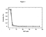
- Plasmid DNA was condensed by mixing with small molecular mass non-linear polyethyleneimine (PEI) (Boussif, O., et al. (1995) Proc. Natl. Acad. Sci. USA 92: 7297-301) (or protamine sulfate for comparison). DNA condensation was already achieved at a N/P-ratio of ⁇ 8, as judged by dye exclusion (Fig. 2).
- PEI polyethyleneimine
- AVPs artificial viral particles
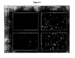
- HUVECs were exposed to AVPs for 1 h with or without the RGD ligand that was labeled with a fluorescent lipid (N-(7-nitrobenz-2-oxa-1,3-diazol-4-yl)-1,2-dihexadecanoyl-sn-glycero-3-phospho-ethanolamine, NBD-PE).
- a fluorescent lipid N-(7-nitrobenz-2-oxa-1,3-diazol-4-yl)-1,2-dihexadecanoyl-sn-glycero-3-phospho-ethanolamine, NBD-PE.
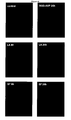
- Cells were observed directly 1 h after incubation with the labeled AVPs (Fig. 6; 1 h) or after another 2 h in normal culture medium (Fig. 6; 3 h) under a fluorescence microscope. At both time points a considerably stronger staining was detectable in cultures that had received the RGD-AVPs. More than 99% of these cells were stained.
- the transduction efficiency of the RGD-AVPs with respect to (i) transduction efficiency, (ii) contribution of the ligand to EC transduction and (iii) cell type specificity was evaluated.
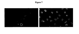
- a plasmid carrying a CMV promoter driven nuclear green fluorescent protein (H2BGFP) gene (Kanda, T., et al. (1998) Curr. Biol. 8: 377-85) was packaged into RGD-AVPs. Cells were exposed for 1 h to the RGD-AVPs, and the fraction of green fluorescent cells was determined 23 h later (Fig. 7). A quantitative evaluation of the experiment showed that the majority of cells were successfully transduced by RGD-AVPs and expressed GFP.
- DPPE 1,2-dipalmitoleoyl-sn-glycero-3-phosphoethanolamine
- DOPS 1,2-dioleoyl-sn-glycero-3-[phospho-L-serine]
- DLPE 1,2-dilauroyl-sn-glycero-3-phosphoethanolamine
- cholesterol was from Calbiochem (San Diego, CA). All other substances were of analytical grade.
- Egg phosphatidylcholine (ePC) was produced by Lipoid KG (Ludwigshafen, Germany) and the anchor lipid 1,2-distearoyl-sn-glycero-3-phosphatidylethanolamine-N-[methoxy(polyethylene glycol)-3400-N-hydroxysuccinimidyl carbonate] (DSPE-PEG-NHS (3400)) was from Shearwater (Huntsville, USA).
- N-Glutaryl-DPPE was prepared by dissolving DPPE in anhydrous chloroform. Glutaric anhydride and water-free pyridine was added and the solution stirred at 20°C for two days. After this period, the products were dried by applying vacuum to the solution and N-Glutaryl-DPPE was purified using preparative silica gel chromatography (Merck 60 F 254) in chloroform/methanol/ammonia (65/35/3). Spots were dissolved in methanol, dried and redissolved in chloroform for further use. N-Glutaryl-DPPE was identified by 1 H-NMR as described previously (Ahl, P.L., et al.
- AVEs with the composition DOPS/DLPE/Cholesterol/N-Glutaryl-DPPE (3:3:3:1 mol/mol) were prepared as follows: a chloroform solution of the lipid mixture (total amount 10 ⁇ mol) was dried into a thin film in the inner surface of a rotating 100 ml glass vessel warmed by a water bath to 30°C. Residual chloroform was removed by vacuum desiccation for 15 min. The lipid film was hydrated either in 1 ml of sterile 10 mM Tris buffer (pH 7.4) or in 1 ml sterile PBS.
- the resulting multilamellar liposome suspension was placed in a 3 ml glass vessel cooled by an ice/water mixture and sonicated for 15 s in a MSE-Soniprep 150 (Zivy AG, Oberwil, Switzerland) equipped with a titanium tip. Sonication was repeated 10 times, each sonication period was followed by a 30 s pause allowing the suspension to cool.
- the resulting liposome suspension was extruded through polycarbonate membrane filters with a pore size of 50 nm using a standard device (Liposofast) (MacDonald, R.C., et al. (1991) Biochim. Biophys. Acta 1061: 297-303) purchased from Avestin, Ottawa, Canada. The liposomes were used up two months after preparation, during which time no significant increase in liposome size could be detected.
- Liposofast MacDonald, R.C., et al. (1991) Biochim. Biophys. Acta 1061
- a cyclic peptide with the amino acid sequence CDCRGDCFC having an additional arginine at the N-terminus was synthesized.
- Cyclic condensation of the peptide was accomplished by stirring an aqueous solution of the synthesized peptide under access of ambient air. Completion of cyclic condensation was verified by HPLC. After HPLC purification, the peptide was lyophilized and stored at 4°C.
- N-Glutaryl-DPPE carboxyl group at the liposomal surface was achieved by adding 3.5 mg 1-ethyl-3-(3-dimethylaminopropyl) carbodiimide to 400 ⁇ L AVE and shaking the suspension for 5 hr in the dark.
- the resulting active O-acyl-intermediate reacted with added RGD-peptide (250 ⁇ g in 150 ⁇ L buffer) overnight yielding a covalent coupling of the peptide to the liposomal surface.
- RGD-AVE were separated from unbound peptide by Sephadex G25 gel permeation chromatography in Tris buffer 10 mM pH 7.4. Coupling efficiency was monitored using a fluorescently labeled derivative of the RGD-peptide.
- RGD was conjugated with 5-(4,6-dichlorotriazinyl)aminofluorescein (5-DTAF) (Blakeslee, D. & Baines, M.G. (1976) J. Immunol. Meth. 13: 305) purchased from Molecular Probes (Eugene, OR). 5-DTAF was dissolved in borate buffer (pH 9) and RGD-peptide was added in a 1:4 molar ratio (RGD / 5-DTAF).
- Low molecular weight branched polyethylenimine (PEI; Lupasol G100, BASF AG, Ludwigshafen, Germany) was added to 15 ⁇ g of plasmid DNA up to a ratio of polyethylenimine-nitrogen:DNA-phosphate of 20.7. Condensation of plasmid DNA was monitored by dye exclusion technique using PicogreenTM (Molecular Probes, Eugene, OR) as described previously (Fahr, A., et al. (1998) J. Liposome Res. 8 : 9-12). In another set of experiments, protamine sulfate (USP-quality; EliLilly & Co., Indianapolis, IN) was used as condensing agent.
- a suspension of AVPs was applied to a holey carbon-foil grid and vitrified by flash-freezing in liquid ethane.
- the grids were cryo-transferred to the liquid nitrogen-cooled cryoelectron microscope (Philips CM200 FEG, FEI GmbH, Germany). Images were taken at a magnification of 60 000 under liquid nitrogen conditions at 1.5 microns defocus at 160 keV.
- Human umbilical vein endothelial cells were prepared by the method of Jaffe (in Biology of Endothelial Cells (eds. Jaffe, E.) 1-13 (Martinus Nijhoff, Boston, 1986) as modified by Thornton et al. ((1983) Science 222 : 623-625), and cultivated in EGM-2 medium (BioWhittaker Europe, Verviers, Belgium).
- the colon carcinoma cell line LoVo (provided by I. Hart, London, UK), the lung adenocarcinoma cell line A549 (obtained from K. Havemann, Marburg), the prostate carcinoma cell line DU-145 (obtained from G.
- transfection reagents Lipofectamine and Lipofectin were purchased from Gibco-BRL (Eggenstein, Germany). Effectene and Superfect were obtained from Quiagen (Hilden, Germany). The concentrations of the transfection reagents were optimized for transduction efficiencies and low toxicities (see legend to Fig. 4 and 5 for details). The medium was replaced with normal culture medium, and WST-assays were performed either directly or after another 18 h. The supernatant was withdrawn, 500 ⁇ l of WST diluted 1:10 in medium were added to each well and incubated for 30 min at 37°C. Aliquots of 100 ⁇ L were transferred to 96-well plates, and the plates were read at 440 nm. All measurements were performed as triplicates, and each experiment was repeated at least three times.
- Cells were grown on 10 cm dishes to ⁇ 70% confluency and transfected for 1 h with AVPs made up of 45 ⁇ g of DNA and 180 ⁇ g of AVE.
- the cells were trypsinized 23 h later, washed once with PBS, fixed in ice-cold 75% ethanol overnight at 4°C, resuspended in PBS, treated with RNase A (Roche; 400 ⁇ g/ml) overnight at 4°C and stained with propidium iodide (20 ⁇ g/ml) for at least 10 min.
- the cells were analyzed by flow cytometry (FACS Calibur; Becton Dickinson, Heidelberg, Germany) using a laser excitation at 488 nm.
- FACSCalibur Becton Dickinson, Heidelberg, Germany
- a series of liposome formulations was prepared using the rotary evaporation technique. Lipids of the appropriate composition were dissolved in an organic solvent (chloroform or chloroform/methanol) and this solution was dried in a rotary evaporator at 35 mbar and 34°C, thus, yielding a thin lipid film. This thin lipid film was further dried in a vacuum chamber at 10 mbar.
- an organic solvent chloroform or chloroform/methanol
- the lipid composition of the AVE produced comprised the phospholipids phosphatidylserine (DOPS), phosphatidylethanolamine (DLPE), cholesterol, phosphatidylcholine (ePC), 1,2-distearoyl-sn-glycero-3-phosphatidylethanolamine-N-[methoxy (polyethylene glycol)-3400-N-hydroxysuccinimidyl carbonate (DSPE-PEG-NHS) and/or phosphoethanolamine-N-(Glutaryl) (N-Glut-DPPE) in the following ratios: Type of liposome DOPS DLPE Cho- lesterol ePC N-Glut- DPPE DSPE- PEG-NHS total lipid amount AVE-3 15,00 15,00 15,0 0,00 5,00 0,00 50,00 AVE-3RGD 15,00 15,00 15,0 0,00 5,00 0,00 50,00 AVE3-PEG-NHS 15,83 15,83, 15,83 0,00 0,00 2,50 50,00 EPC 0,00
- the dried film was hydrated in a rotary evaporator with a few glass beads with an aqueous solution buffered to a pH of 5 to 9.5 in order to form multilamellar vesicles.
- the buffer contained between 0.1 and 1 molar of gadoteridol (Prohance® , Bracco-Byk Gulden, Konstanz, Germany (RS-[10-Hydroxypropyl)-1,4,7,10-tetraazacyclodecane-1,4,7-triacetato(3-)]gadolinium) or Gadobutrol (Schering AG, Berlin, Germany).
- the total lipid concentration in this system was 20 mg/ml.
- the vesicles were subjected to extrusion through polycarbonate membranes having pores with a size of 50 nm. The extrusion was repeated 21 times. Liposomes with a mean size of 90 - 120 nm were obtained, as checked by photon correlation spectroscopy.
- Unentrapped metal chelate was removed by gel filtration (Sephadex G25 medium) with phosphate buffer pH 7.4 as elution buffer. Removal of unentrapped metal chelate was also possible using a dialysis method (Mini Lipoprep, Dianorm GmbH, Germany) with a dialysis membrane having a MW cutoff of about 10,000 D. AVEs containing 1 M Gadobutrol where dialyzed with four changes of buffer at 2, 4, 6, 8 h against PBS. The thus obtained liposomes had a diameter of between 100 to 180 nm as measured by a Zetasizer 3000 HS (Malvern Instruments, UK).
- Unbound peptide was separated from the liposome suspension by gel filtration (Sephadex G25 medium) with Tris buffer, pH 7.4.
- the resulting liposome suspensions were ready for injection or transfection.
- RGD-receptor (HUVEC, MEWO) and cells without the RGD-receptor (A549) as control were used in the following experiments.
- the cells were grown in 6 well tissue culture dishes under standard conditions As cell culture medium DMEM was used for MEWO and A549 cells.
- HUVECs were cultured in EGM-2 medium .
- the growth medium was removed and 500 ⁇ l transfection media 199 was applied to each well of the plate and 10-40 ⁇ l liposomes (EPC, AVE-3, AVE-3, which were produced using 1 N-Gadobutol, or free Gadobutol as control) were applied to each well.
- the transfection media was removed and the cells were washed with 2 ml PBS for each well and subsequently the respective growth media required by the cells were added. After 18 h the media was again removed and the cells were washed twice with 2 ml PBS and subsequently 250 ⁇ l luciferase lysis buffer (1ml Triton; 25 ml Glycylglycine 0,1 M pH 7,8; 1,5 ml MgSO 4 1M; 1,6 ml EGTA 0,25 M and 71,9 ml purified water) was added. The lysates were transferred into 1.5 ml tubes and the T1-relaxation times were measured in a magnetic resonance tomograph for each probe.
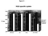
- the T1 relaxation time was shortened in samples obtained from AVE-RGD-Gd treated cells and only in cells carrying the RGD-receptor.
- the lysates of A549 cells did not show a significant increase of the T-1 relaxation time.
Abstract
Description
- Figure 1:
- Condensation of DNA by polyethylenimine determined by dye exclusion. Six individual measurements with different DNA plasmid concentrations are shown to illustrate the high reproducibility of the method.
- Figure 2:
- Size determination of RGD-AVEs, PEI-DNA and RGD-AVPs by photon correlation spectroscopy. The mean values for the size distributions estimated by z-average calculation are indicated.
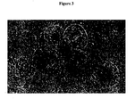
- Figure 3:
- Cryogenic transmission electron microscopy (Cryo-TEM) of RGD-AVPs complexed with PEI/plasmid-DNA. The pictures shows encapsulation of condensed DNA in a lipid layer within discrete particles. The diameter of the larger liposomes shown are approx. 140 nm.
- Figure 4:
- Toxicity of AVPs for HUVECs compared to Lipofectamin (LA; Gibco BRL) and Superfect (SF; Quiagen, Hilden Germany). The analyses were performed at 6 h and 24 h after transfection. Identification of apoptotic cells by staining with Hoechst 33258. Control, untransfected cells.
- Figure 5:
- Toxicity of AVPs for HUVECs compared to Lipofectamin (LA; Gibco BRL) and Superfect (SF; Quiagen, Hilden Germany). The analyses were performed at 6 h and 24 h after transfection. Determination of metabolic activity by WST-assay. The following amounts of DNA and transfection reagents were used (per 3 cm well): AVP, 5 µg DNA + 20 µg RGD-AVE; LA, 2 µg DNA + 20 µg lipid; SF, 2 µg DNA + 30 µg of the transfection reagent.
- Figure 6:
- Binding of AVPs to HUVECs. HUVECs were exposed for 1 h to NBD-PE labeled AVPs with and without RGD ligand and analyzed by fluorescence microscopy either directly (1 h) or after another 2 h in normal culture medium (3 h).
- Figure 7:
- Transduction of HUVECs by RGD-AVPs carrying a CMV-H2BGFP plasmid. Cells were exposed for 1 h to the AVPs and fluorescence microscopy was performed after another 23 h in normal medium (left panel). The right panel shows the same cells stained with Hoechst 33258.
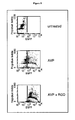
- Figure 8:
- FACS-analysis of
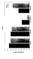
HUVECs 24 h after transduction with AVPs (± RGD ligand) carrying a CMV-H2BGFP plasmid compared to untreated cells. Cells were exposed for 1 h to the AVPs and FACS analysis was performed after another 23 h in normal medium. - Figure 9:
- Transduction of different cell types with AVPs (A) RGD-AVPs (B) carrying a CMV promoter-H2BGFP plasmid: summary of FACS analyses determining the fraction of GFP-positive cells. Cells were exposed for 1 h to the AVPs and FACS-analysis was performed after another 23 h in normal medium. Values were calculated relative to the transduction efficiency obtained with RGD-AVP transfected HUVECs (100%).
- Figure 10:
- FACS-analysis of αvβ3 expression in HUVECs and different tumor cell lines after indirect immunostaining as described in Materials and Methods. Bold lines: specific staining (first antibody: αvβ3-specific; second antibody: Cy3-labeled); thin line: unspecific staining (second antibody only); dotted line: unlabeled cells. The relative fraction of cells is plotted against the intensity of fluorescence.
- Figure 11:
- T1-relaxation times in three different cell lines after treatment with EPC-Gd liposomes (EPC Gd), free Gd (Gd), AVE-RGD liposomes (AVE-RGD) and AVE-RGD Gd liposomes (AVE-RGD Gd).
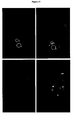
- Figure 12:
- Detection of a subcutaneous MEWO tumor in a mouse with AVE-PEG-RGD
Gd-liposomes after intravenous injection of the liposomal
formulation. The images on the left side were taken before the injection.
The two images on the right side were taken 20 minutes after the
injection. The upper and the lower images show two different sections
of the same mouse. Both circle 1 and
circle 2 in the upper images are located within the subcutaneous MEWO tumor. The subpopulation of tumor cells incircle 2 will also accumulate AVE-PEG-RGD Gd-liposomes after 1 h (Figure not shown). The white region in the lower right image lies within the MEWO tumor.
| Type of liposome | DOPS | DLPE | Cho- lesterol | ePC | N-Glut- DPPE | DSPE- PEG-NHS | total lipid amount |
| AVE-3 | 15,00 | 15,00 | 15,0 | 0,00 | 5,00 | 0,00 | 50,00 |
| AVE- | 15,00 | 15,00 | 15,0 | 0,00 | 5,00 | 0,00 | 50,00 |
| AVE3-PEG- | 15,83 | 15,83, | 15,83 | 0,00 | 0,00 | 2,50 | 50,00 |
| | 0,00 | 0,00 | 0 | 50,00 | 0,00 | 0,00 | 50,00 |

Claims (29)
- A diagnostic system comprising:(a) at least one negatively charged carrier;(b) at least one targeting moiety; and(c) at least one diagnostic agent.
- The diagnostic system according to claim 1, wherein said negatively charged carrier is an anionic lipid.
- The diagnostic system according to claim 2, wherein said anionic lipid is selected from the group consisting of phosphatidylserine, phosphatidylglycerol, phosphatidylacid, phosphatidylinositol, and cholesterolglutarate.
- The diagnostic system according to one of claims 1-3, wherein said targeting moiety is selected from the group consisting of a peptide, a protein, an antibody, an antibody fragment, a single-chain antibody, a diabody or a small molecule.
- The diagnostic system according to one of claims 1-4, wherein said diagnostic agent is selected from the group consisting of an electron dense substance, a paramagnetic substance, a superparamagnetic substance, a radioactive substance, a fluorescent substance, a luminescent substance, a light-emitting substance, a genetic construct that codes for a fluorescent protein and a genetic construct that codes for a protein detectable by staining procedures.
- The diagnostic system according to one of claims 1-5, further comprising at least one uncharged carrier.
- The diagnostic system according to claim 6, wherein said uncharged carrier is selected from the group consisting of cholesterol, phosphatidylcholine, and phosphatidylethanolamine.
- The diagnostic system according to one of claims 1-7, further comprising at least one amphiphatic polymer.
- The diagnostic system according to claim 8, wherein the amphiphatic polymer is selected from the group of polyethylene glycol (PEG), poly(2-methyl-2-oxazoline) (PMOZ), and poly(2-ethyl-2-oxazoline) PEOZ.
- The diagnostic system according to one of claims 8-9, wherein the amphiphatic polymer is attached to a lipid,
- The diagnostic system according to claim 10, wherein the lipid is selected from alpha-methoxy-omega-(1,2-dioctadecenoyloxy glyceryl) (DO), alpha-methoxy-omega-(1,2-ditetradecenoyloxy glyceryl) (DT), distearoylphosphatidyl (DSPE), and alpha-(dipalmitoylphosphatidyl) (DPP).
- The diagnostic system according to one of claims 8-11, wherein the amphiphatic polymer has a molecular weight between 1.000 and 5.000 Da.
- The diagnostic system to one of claims 1-12, wherein the negative carrier of the diagnostic system forms at least part of a lamellar structure.
- The diagnostic system of claim 13, wherein the lamellar structure is a unilamellar, bilamellar, oligolamellar or polylamellar structure.
- The diagnostic system to one of claims 13-14, wherein said lamellar structure is a liposome.
- The diagnostic system according to one of claims 13-15, wherein said lamellar structure is an artificial viral envelope.
- The diagnostic system according to one of claims 13-16, wherein said lamellar structure comprises a payload compartment.
- The diagnostic system according to claim 17, wherein the diagnostic agent is encapsulated within said payload compartment.
- The diagnostic system to one of claims 6-18, wherein at least one targeting moiety is covalently attached to at least one of said negatively charged carriers or uncharged carriers.
- A method for producing the diagnostic system according to one of claims 1-19, wherein at least one lamella is formed with at least one negatively charged carrier and at least one diagnostic agent in solution.
- The method according to claim 20, wherein at least one targeting moiety is covalently attached after formation of at least one lamella.
- The method according to claim 20, wherein at least one targeting moiety is mixed with said at least one negatively charged carrier.
- A method for producing the diagnostic system according to one of claims 1-19, wherein at least one lamella is loaded with at least one diagnostic agent upon formation of the lamella.
- Use of the diagnostic system according to one of claims 1-19 for the manufacturing of a medicament for the diagnosis of a disease.
- The use according to claim 24, wherein said disease is a tumor disease.
- A method of using the diagnostic system according to one of claims 1-19 for diagnosis of cells and tissues in vitro.
- A method of using the diagnostic system according to one of claims 1-19 for the diagnosis of microbiological organisms in vitro.
- A diagnostic kit comprising:(a) at least one negatively charged carrier; and(b) at least one diagnostic agent.
- The diagnostic kit according to claim 28 further comprising at least one targeting moiety.
Applications Claiming Priority (2)
| Application Number | Priority Date | Filing Date | Title |
|---|---|---|---|
| US25266600P | 2000-11-22 | 2000-11-22 | |
| US252666P | 2000-11-22 |
Publications (1)
| Publication Number | Publication Date |
|---|---|
| EP1209469A1 true EP1209469A1 (en) | 2002-05-29 |
Family
ID=22956996
Family Applications (1)
| Application Number | Title | Priority Date | Filing Date |
|---|---|---|---|
| EP01127384A Withdrawn EP1209469A1 (en) | 2000-11-22 | 2001-11-22 | Production and use of a targeted diagnostic system |
Country Status (3)
| Country | Link |
|---|---|
| EP (1) | EP1209469A1 (en) |
| AU (1) | AU2002231637A1 (en) |
| WO (1) | WO2002041870A2 (en) |
Cited By (1)
| Publication number | Priority date | Publication date | Assignee | Title |
|---|---|---|---|---|
| WO2006099169A2 (en) | 2005-03-10 | 2006-09-21 | Mebiopharm Co., Ltd. | Novel liposome compositions |
Families Citing this family (2)
| Publication number | Priority date | Publication date | Assignee | Title |
|---|---|---|---|---|
| EP1538164A1 (en) * | 2003-12-04 | 2005-06-08 | Vectron Therapeutics AG | RGD targeting moiety its production and use |
| CN109996809A (en) | 2016-11-14 | 2019-07-09 | 诺华股份有限公司 | Composition relevant to fusogenic protein MINION, method and therapeutical uses |
Citations (6)
| Publication number | Priority date | Publication date | Assignee | Title |
|---|---|---|---|---|
| US5512294A (en) * | 1994-08-05 | 1996-04-30 | Li; King C. | Targeted polymerized liposome contrast agents |
| US5534241A (en) * | 1993-07-23 | 1996-07-09 | Torchilin; Vladimir P. | Amphipathic polychelating compounds and methods of use |
| WO1997004748A2 (en) * | 1995-08-01 | 1997-02-13 | Advanced Therapies, Inc. | Enhanced artificial viral envelopes for cellular delivery of therapeutic substances |
| WO1998008094A1 (en) * | 1996-08-23 | 1998-02-26 | Amcell Corporation | Sensitive detection of cell surface markers on viable cells |
| US5753258A (en) * | 1990-10-19 | 1998-05-19 | University Of Florida | Artificial viral envelopes |
| US6090408A (en) * | 1994-08-05 | 2000-07-18 | Targesome, Inc. | Use of polymerized lipid diagnostic agents |
-
2001
- 2001-11-22 EP EP01127384A patent/EP1209469A1/en not_active Withdrawn
- 2001-11-22 WO PCT/EP2001/013618 patent/WO2002041870A2/en not_active Application Discontinuation
- 2001-11-22 AU AU2002231637A patent/AU2002231637A1/en not_active Abandoned
Patent Citations (7)
| Publication number | Priority date | Publication date | Assignee | Title |
|---|---|---|---|---|
| US5753258A (en) * | 1990-10-19 | 1998-05-19 | University Of Florida | Artificial viral envelopes |
| US5534241A (en) * | 1993-07-23 | 1996-07-09 | Torchilin; Vladimir P. | Amphipathic polychelating compounds and methods of use |
| US5512294A (en) * | 1994-08-05 | 1996-04-30 | Li; King C. | Targeted polymerized liposome contrast agents |
| US6090408A (en) * | 1994-08-05 | 2000-07-18 | Targesome, Inc. | Use of polymerized lipid diagnostic agents |
| US6132764A (en) * | 1994-08-05 | 2000-10-17 | Targesome, Inc. | Targeted polymerized liposome diagnostic and treatment agents |
| WO1997004748A2 (en) * | 1995-08-01 | 1997-02-13 | Advanced Therapies, Inc. | Enhanced artificial viral envelopes for cellular delivery of therapeutic substances |
| WO1998008094A1 (en) * | 1996-08-23 | 1998-02-26 | Amcell Corporation | Sensitive detection of cell surface markers on viable cells |
Cited By (7)
| Publication number | Priority date | Publication date | Assignee | Title |
|---|---|---|---|---|
| WO2006099169A2 (en) | 2005-03-10 | 2006-09-21 | Mebiopharm Co., Ltd. | Novel liposome compositions |
| WO2006099169A3 (en) * | 2005-03-10 | 2007-02-22 | Mebiopharm Co Ltd | Novel liposome compositions |
| US7829113B2 (en) | 2005-03-10 | 2010-11-09 | Mebiopharm Co., Ltd. | Liposome compositions |
| AU2006223329B2 (en) * | 2005-03-10 | 2011-11-17 | Mebiopharm Co., Ltd | Novel liposome compositions |
| RU2454229C2 (en) * | 2005-03-10 | 2012-06-27 | Мебайофарм Ко., Лтд. | New liposome compositions |
| CN101170995B (en) * | 2005-03-10 | 2013-01-16 | 美生物药物株式会社 | Novel liposome compositions |
| US8758810B2 (en) | 2005-03-10 | 2014-06-24 | Mebiopharm Co., Ltd. | Liposome compositions |
Also Published As
| Publication number | Publication date |
|---|---|
| AU2002231637A1 (en) | 2002-06-03 |
| WO2002041870A3 (en) | 2002-11-07 |
| WO2002041870A2 (en) | 2002-05-30 |
Similar Documents
| Publication | Publication Date | Title |
|---|---|---|
| Tseng et al. | Translocation of liposomes into cancer cells by cell-penetrating peptides penetratin and tat: a kinetic and efficacy study | |
| Harris et al. | Tissue-specific gene delivery via nanoparticle coating | |
| ElBayoumi et al. | Current trends in liposome research | |
| JP5797104B2 (en) | Multi-component biological transport system | |
| Fu et al. | Tumor-targeted paclitaxel delivery and enhanced penetration using TAT-decorated liposomes comprising redox-responsive poly (ethylene glycol) | |
| Zou et al. | Systemic linear polyethylenimine (L‐PEI)‐mediated gene delivery in the mouse | |
| US7060291B1 (en) | Modular targeted liposomal delivery system | |
| Agadjanian et al. | Specific delivery of corroles to cells via noncovalent conjugates with viral proteins | |
| Erdogan et al. | Enhanced tumor MR imaging with gadolinium‐loaded polychelating polymer‐containing tumor‐targeted liposomes | |
| US20070160658A1 (en) | Delivery system for diagnostic and therapeutic agents | |
| CA2924018C (en) | Cell-specific targeting using nanostructured delivery systems | |
| JPH10508302A (en) | Cell-specific gene delivery vehicle | |
| Fonseca et al. | Targeting of sterically stabilised pH-sensitive liposomes to human T-leukaemia cells | |
| US20120059240A1 (en) | Method for preparation of micellar hybrid nanoparticles for therapeutic and diagnostic applications and compositions thereof | |
| Sawant et al. | Intracellular delivery of nanoparticles with CPPs | |
| US20080206139A1 (en) | Delivery system for diagnostic and therapeutic agents | |
| Müller et al. | Highly efficient transduction of endothelial cells by targeted artificial virus-like particles | |
| Du et al. | A tumor-targeted, intracellular activatable and theranostic nanodiamond drug platform for strongly enhanced in vivo antitumor therapy | |
| ES2320193T3 (en) | MODULAR DIRECTED LIPOSOMIC SUPPLY SYSTEM. | |
| EP1209469A1 (en) | Production and use of a targeted diagnostic system | |
| Accardo et al. | Naposomes: A new class of peptide-derivatized, target-selective multimodal nanoparticles for imaging and therapeutic applications | |
| EP3402484A1 (en) | Peptide-conjugated nanoparticles for targeting, imaging, and treatment of prostate cancer | |
| Negishi et al. | Development of a screening system for targeting carriers using peptide-modified liposomes and tissue sections | |
| Salzano et al. | Intracellular delivery of nanoparticles with cell penetrating peptides | |
| EP2832373B1 (en) | Liposome for blocking site-specifically chemokine-related inflammatory processes in vascular diseases and metastasis |
Legal Events
| Date | Code | Title | Description |
|---|---|---|---|
| PUAI | Public reference made under article 153(3) epc to a published international application that has entered the european phase |
Free format text: ORIGINAL CODE: 0009012 |
|
| AK | Designated contracting states |
Kind code of ref document: A1 Designated state(s): AT BE CH CY DE DK ES FI FR GB GR IE IT LI LU MC NL PT SE TR |
|
| AX | Request for extension of the european patent |
Free format text: AL;LT;LV;MK;RO;SI |
|
| RIN1 | Information on inventor provided before grant (corrected) |
Inventor name: BRUESSELBACH, SABINE Inventor name: FAHR, ALFRED Inventor name: GRASER, ANDREAS Inventor name: MUELLER, ROLF |
|
| 17P | Request for examination filed |
Effective date: 20021023 |
|
| AKX | Designation fees paid |
Designated state(s): AT BE CH CY DE DK ES FI FR GB GR IE IT LI LU MC NL PT SE TR |
|
| 19U | Interruption of proceedings before grant |
Effective date: 20040601 |
|
| 19W | Proceedings resumed before grant after interruption of proceedings |
Effective date: 20050301 |
|
| RAP1 | Party data changed (applicant data changed or rights of an application transferred) |
Owner name: VECTRON THERAPEUTICS AG |
|
| STAA | Information on the status of an ep patent application or granted ep patent |
Free format text: STATUS: THE APPLICATION IS DEEMED TO BE WITHDRAWN |
|
| 18D | Application deemed to be withdrawn |
Effective date: 20060203 |