EP1237511B1 - Preparation for repairing cartilage defects or cartilage/bone defects in human or animal joints - Google Patents
Preparation for repairing cartilage defects or cartilage/bone defects in human or animal joints Download PDFInfo
- Publication number
- EP1237511B1 EP1237511B1 EP00979315A EP00979315A EP1237511B1 EP 1237511 B1 EP1237511 B1 EP 1237511B1 EP 00979315 A EP00979315 A EP 00979315A EP 00979315 A EP00979315 A EP 00979315A EP 1237511 B1 EP1237511 B1 EP 1237511B1
- Authority
- EP
- European Patent Office
- Prior art keywords
- bone
- cartilage
- preparation
- bone plate
- tissue
- Prior art date
- Legal status (The legal status is an assumption and is not a legal conclusion. Google has not performed a legal analysis and makes no representation as to the accuracy of the status listed.)
- Expired - Lifetime
Links
Images


Classifications
-
- A—HUMAN NECESSITIES
- A61—MEDICAL OR VETERINARY SCIENCE; HYGIENE
- A61B—DIAGNOSIS; SURGERY; IDENTIFICATION
- A61B17/00—Surgical instruments, devices or methods, e.g. tourniquets
- A61B17/32—Surgical cutting instruments
- A61B17/3205—Excision instruments
- A61B17/32053—Punch like cutting instruments, e.g. using a cylindrical or oval knife
-
- A—HUMAN NECESSITIES
- A61—MEDICAL OR VETERINARY SCIENCE; HYGIENE
- A61F—FILTERS IMPLANTABLE INTO BLOOD VESSELS; PROSTHESES; DEVICES PROVIDING PATENCY TO, OR PREVENTING COLLAPSING OF, TUBULAR STRUCTURES OF THE BODY, e.g. STENTS; ORTHOPAEDIC, NURSING OR CONTRACEPTIVE DEVICES; FOMENTATION; TREATMENT OR PROTECTION OF EYES OR EARS; BANDAGES, DRESSINGS OR ABSORBENT PADS; FIRST-AID KITS
- A61F2/00—Filters implantable into blood vessels; Prostheses, i.e. artificial substitutes or replacements for parts of the body; Appliances for connecting them with the body; Devices providing patency to, or preventing collapsing of, tubular structures of the body, e.g. stents
- A61F2/02—Prostheses implantable into the body
- A61F2/28—Bones
-
- A—HUMAN NECESSITIES
- A61—MEDICAL OR VETERINARY SCIENCE; HYGIENE
- A61F—FILTERS IMPLANTABLE INTO BLOOD VESSELS; PROSTHESES; DEVICES PROVIDING PATENCY TO, OR PREVENTING COLLAPSING OF, TUBULAR STRUCTURES OF THE BODY, e.g. STENTS; ORTHOPAEDIC, NURSING OR CONTRACEPTIVE DEVICES; FOMENTATION; TREATMENT OR PROTECTION OF EYES OR EARS; BANDAGES, DRESSINGS OR ABSORBENT PADS; FIRST-AID KITS
- A61F2/00—Filters implantable into blood vessels; Prostheses, i.e. artificial substitutes or replacements for parts of the body; Appliances for connecting them with the body; Devices providing patency to, or preventing collapsing of, tubular structures of the body, e.g. stents
- A61F2/02—Prostheses implantable into the body
- A61F2/30—Joints
- A61F2/30756—Cartilage endoprostheses
-
- A—HUMAN NECESSITIES
- A61—MEDICAL OR VETERINARY SCIENCE; HYGIENE
- A61F—FILTERS IMPLANTABLE INTO BLOOD VESSELS; PROSTHESES; DEVICES PROVIDING PATENCY TO, OR PREVENTING COLLAPSING OF, TUBULAR STRUCTURES OF THE BODY, e.g. STENTS; ORTHOPAEDIC, NURSING OR CONTRACEPTIVE DEVICES; FOMENTATION; TREATMENT OR PROTECTION OF EYES OR EARS; BANDAGES, DRESSINGS OR ABSORBENT PADS; FIRST-AID KITS
- A61F2/00—Filters implantable into blood vessels; Prostheses, i.e. artificial substitutes or replacements for parts of the body; Appliances for connecting them with the body; Devices providing patency to, or preventing collapsing of, tubular structures of the body, e.g. stents
- A61F2/02—Prostheses implantable into the body
- A61F2/30—Joints
- A61F2/46—Special tools or methods for implanting or extracting artificial joints, accessories, bone grafts or substitutes, or particular adaptations therefor
- A61F2/4601—Special tools or methods for implanting or extracting artificial joints, accessories, bone grafts or substitutes, or particular adaptations therefor for introducing bone substitute, for implanting bone graft implants or for compacting them in the bone cavity
-
- A—HUMAN NECESSITIES
- A61—MEDICAL OR VETERINARY SCIENCE; HYGIENE
- A61B—DIAGNOSIS; SURGERY; IDENTIFICATION
- A61B17/00—Surgical instruments, devices or methods, e.g. tourniquets
- A61B17/16—Bone cutting, breaking or removal means other than saws, e.g. Osteoclasts; Drills or chisels for bones; Trepans
- A61B17/1637—Hollow drills or saws producing a curved cut, e.g. cylindrical
-
- A—HUMAN NECESSITIES
- A61—MEDICAL OR VETERINARY SCIENCE; HYGIENE
- A61B—DIAGNOSIS; SURGERY; IDENTIFICATION
- A61B17/00—Surgical instruments, devices or methods, e.g. tourniquets
- A61B2017/00969—Surgical instruments, devices or methods, e.g. tourniquets used for transplantation
-
- A—HUMAN NECESSITIES
- A61—MEDICAL OR VETERINARY SCIENCE; HYGIENE
- A61F—FILTERS IMPLANTABLE INTO BLOOD VESSELS; PROSTHESES; DEVICES PROVIDING PATENCY TO, OR PREVENTING COLLAPSING OF, TUBULAR STRUCTURES OF THE BODY, e.g. STENTS; ORTHOPAEDIC, NURSING OR CONTRACEPTIVE DEVICES; FOMENTATION; TREATMENT OR PROTECTION OF EYES OR EARS; BANDAGES, DRESSINGS OR ABSORBENT PADS; FIRST-AID KITS
- A61F2/00—Filters implantable into blood vessels; Prostheses, i.e. artificial substitutes or replacements for parts of the body; Appliances for connecting them with the body; Devices providing patency to, or preventing collapsing of, tubular structures of the body, e.g. stents
- A61F2/02—Prostheses implantable into the body
- A61F2/28—Bones
- A61F2002/2835—Bone graft implants for filling a bony defect or an endoprosthesis cavity, e.g. by synthetic material or biological material
- A61F2002/2839—Bone plugs or bone graft dowels
-
- A—HUMAN NECESSITIES
- A61—MEDICAL OR VETERINARY SCIENCE; HYGIENE
- A61F—FILTERS IMPLANTABLE INTO BLOOD VESSELS; PROSTHESES; DEVICES PROVIDING PATENCY TO, OR PREVENTING COLLAPSING OF, TUBULAR STRUCTURES OF THE BODY, e.g. STENTS; ORTHOPAEDIC, NURSING OR CONTRACEPTIVE DEVICES; FOMENTATION; TREATMENT OR PROTECTION OF EYES OR EARS; BANDAGES, DRESSINGS OR ABSORBENT PADS; FIRST-AID KITS
- A61F2/00—Filters implantable into blood vessels; Prostheses, i.e. artificial substitutes or replacements for parts of the body; Appliances for connecting them with the body; Devices providing patency to, or preventing collapsing of, tubular structures of the body, e.g. stents
- A61F2/02—Prostheses implantable into the body
- A61F2/30—Joints
- A61F2002/30001—Additional features of subject-matter classified in A61F2/28, A61F2/30 and subgroups thereof
- A61F2002/30003—Material related properties of the prosthesis or of a coating on the prosthesis
- A61F2002/30004—Material related properties of the prosthesis or of a coating on the prosthesis the prosthesis being made from materials having different values of a given property at different locations within the same prosthesis
- A61F2002/30032—Material related properties of the prosthesis or of a coating on the prosthesis the prosthesis being made from materials having different values of a given property at different locations within the same prosthesis differing in absorbability or resorbability, i.e. in absorption or resorption time
-
- A—HUMAN NECESSITIES
- A61—MEDICAL OR VETERINARY SCIENCE; HYGIENE
- A61F—FILTERS IMPLANTABLE INTO BLOOD VESSELS; PROSTHESES; DEVICES PROVIDING PATENCY TO, OR PREVENTING COLLAPSING OF, TUBULAR STRUCTURES OF THE BODY, e.g. STENTS; ORTHOPAEDIC, NURSING OR CONTRACEPTIVE DEVICES; FOMENTATION; TREATMENT OR PROTECTION OF EYES OR EARS; BANDAGES, DRESSINGS OR ABSORBENT PADS; FIRST-AID KITS
- A61F2/00—Filters implantable into blood vessels; Prostheses, i.e. artificial substitutes or replacements for parts of the body; Appliances for connecting them with the body; Devices providing patency to, or preventing collapsing of, tubular structures of the body, e.g. stents
- A61F2/02—Prostheses implantable into the body
- A61F2/30—Joints
- A61F2002/30001—Additional features of subject-matter classified in A61F2/28, A61F2/30 and subgroups thereof
- A61F2002/30003—Material related properties of the prosthesis or of a coating on the prosthesis
- A61F2002/3006—Properties of materials and coating materials
- A61F2002/30062—(bio)absorbable, biodegradable, bioerodable, (bio)resorbable, resorptive
-
- A—HUMAN NECESSITIES
- A61—MEDICAL OR VETERINARY SCIENCE; HYGIENE
- A61F—FILTERS IMPLANTABLE INTO BLOOD VESSELS; PROSTHESES; DEVICES PROVIDING PATENCY TO, OR PREVENTING COLLAPSING OF, TUBULAR STRUCTURES OF THE BODY, e.g. STENTS; ORTHOPAEDIC, NURSING OR CONTRACEPTIVE DEVICES; FOMENTATION; TREATMENT OR PROTECTION OF EYES OR EARS; BANDAGES, DRESSINGS OR ABSORBENT PADS; FIRST-AID KITS
- A61F2/00—Filters implantable into blood vessels; Prostheses, i.e. artificial substitutes or replacements for parts of the body; Appliances for connecting them with the body; Devices providing patency to, or preventing collapsing of, tubular structures of the body, e.g. stents
- A61F2/02—Prostheses implantable into the body
- A61F2/30—Joints
- A61F2002/30001—Additional features of subject-matter classified in A61F2/28, A61F2/30 and subgroups thereof
- A61F2002/30108—Shapes
- A61F2002/3011—Cross-sections or two-dimensional shapes
- A61F2002/30112—Rounded shapes, e.g. with rounded corners
- A61F2002/30113—Rounded shapes, e.g. with rounded corners circular
-
- A—HUMAN NECESSITIES
- A61—MEDICAL OR VETERINARY SCIENCE; HYGIENE
- A61F—FILTERS IMPLANTABLE INTO BLOOD VESSELS; PROSTHESES; DEVICES PROVIDING PATENCY TO, OR PREVENTING COLLAPSING OF, TUBULAR STRUCTURES OF THE BODY, e.g. STENTS; ORTHOPAEDIC, NURSING OR CONTRACEPTIVE DEVICES; FOMENTATION; TREATMENT OR PROTECTION OF EYES OR EARS; BANDAGES, DRESSINGS OR ABSORBENT PADS; FIRST-AID KITS
- A61F2/00—Filters implantable into blood vessels; Prostheses, i.e. artificial substitutes or replacements for parts of the body; Appliances for connecting them with the body; Devices providing patency to, or preventing collapsing of, tubular structures of the body, e.g. stents
- A61F2/02—Prostheses implantable into the body
- A61F2/30—Joints
- A61F2002/30001—Additional features of subject-matter classified in A61F2/28, A61F2/30 and subgroups thereof
- A61F2002/30108—Shapes
- A61F2002/3011—Cross-sections or two-dimensional shapes
- A61F2002/30138—Convex polygonal shapes
- A61F2002/30143—Convex polygonal shapes hexagonal
-
- A—HUMAN NECESSITIES
- A61—MEDICAL OR VETERINARY SCIENCE; HYGIENE
- A61F—FILTERS IMPLANTABLE INTO BLOOD VESSELS; PROSTHESES; DEVICES PROVIDING PATENCY TO, OR PREVENTING COLLAPSING OF, TUBULAR STRUCTURES OF THE BODY, e.g. STENTS; ORTHOPAEDIC, NURSING OR CONTRACEPTIVE DEVICES; FOMENTATION; TREATMENT OR PROTECTION OF EYES OR EARS; BANDAGES, DRESSINGS OR ABSORBENT PADS; FIRST-AID KITS
- A61F2/00—Filters implantable into blood vessels; Prostheses, i.e. artificial substitutes or replacements for parts of the body; Appliances for connecting them with the body; Devices providing patency to, or preventing collapsing of, tubular structures of the body, e.g. stents
- A61F2/02—Prostheses implantable into the body
- A61F2/30—Joints
- A61F2002/30001—Additional features of subject-matter classified in A61F2/28, A61F2/30 and subgroups thereof
- A61F2002/30108—Shapes
- A61F2002/3011—Cross-sections or two-dimensional shapes
- A61F2002/30138—Convex polygonal shapes
- A61F2002/30153—Convex polygonal shapes rectangular
-
- A—HUMAN NECESSITIES
- A61—MEDICAL OR VETERINARY SCIENCE; HYGIENE
- A61F—FILTERS IMPLANTABLE INTO BLOOD VESSELS; PROSTHESES; DEVICES PROVIDING PATENCY TO, OR PREVENTING COLLAPSING OF, TUBULAR STRUCTURES OF THE BODY, e.g. STENTS; ORTHOPAEDIC, NURSING OR CONTRACEPTIVE DEVICES; FOMENTATION; TREATMENT OR PROTECTION OF EYES OR EARS; BANDAGES, DRESSINGS OR ABSORBENT PADS; FIRST-AID KITS
- A61F2/00—Filters implantable into blood vessels; Prostheses, i.e. artificial substitutes or replacements for parts of the body; Appliances for connecting them with the body; Devices providing patency to, or preventing collapsing of, tubular structures of the body, e.g. stents
- A61F2/02—Prostheses implantable into the body
- A61F2/30—Joints
- A61F2002/30001—Additional features of subject-matter classified in A61F2/28, A61F2/30 and subgroups thereof
- A61F2002/30108—Shapes
- A61F2002/3011—Cross-sections or two-dimensional shapes
- A61F2002/30138—Convex polygonal shapes
- A61F2002/30154—Convex polygonal shapes square
-
- A—HUMAN NECESSITIES
- A61—MEDICAL OR VETERINARY SCIENCE; HYGIENE
- A61F—FILTERS IMPLANTABLE INTO BLOOD VESSELS; PROSTHESES; DEVICES PROVIDING PATENCY TO, OR PREVENTING COLLAPSING OF, TUBULAR STRUCTURES OF THE BODY, e.g. STENTS; ORTHOPAEDIC, NURSING OR CONTRACEPTIVE DEVICES; FOMENTATION; TREATMENT OR PROTECTION OF EYES OR EARS; BANDAGES, DRESSINGS OR ABSORBENT PADS; FIRST-AID KITS
- A61F2/00—Filters implantable into blood vessels; Prostheses, i.e. artificial substitutes or replacements for parts of the body; Appliances for connecting them with the body; Devices providing patency to, or preventing collapsing of, tubular structures of the body, e.g. stents
- A61F2/02—Prostheses implantable into the body
- A61F2/30—Joints
- A61F2002/30001—Additional features of subject-matter classified in A61F2/28, A61F2/30 and subgroups thereof
- A61F2002/30108—Shapes
- A61F2002/3011—Cross-sections or two-dimensional shapes
- A61F2002/30138—Convex polygonal shapes
- A61F2002/30156—Convex polygonal shapes triangular
-
- A—HUMAN NECESSITIES
- A61—MEDICAL OR VETERINARY SCIENCE; HYGIENE
- A61F—FILTERS IMPLANTABLE INTO BLOOD VESSELS; PROSTHESES; DEVICES PROVIDING PATENCY TO, OR PREVENTING COLLAPSING OF, TUBULAR STRUCTURES OF THE BODY, e.g. STENTS; ORTHOPAEDIC, NURSING OR CONTRACEPTIVE DEVICES; FOMENTATION; TREATMENT OR PROTECTION OF EYES OR EARS; BANDAGES, DRESSINGS OR ABSORBENT PADS; FIRST-AID KITS
- A61F2/00—Filters implantable into blood vessels; Prostheses, i.e. artificial substitutes or replacements for parts of the body; Appliances for connecting them with the body; Devices providing patency to, or preventing collapsing of, tubular structures of the body, e.g. stents
- A61F2/02—Prostheses implantable into the body
- A61F2/30—Joints
- A61F2002/30001—Additional features of subject-matter classified in A61F2/28, A61F2/30 and subgroups thereof
- A61F2002/30108—Shapes
- A61F2002/3011—Cross-sections or two-dimensional shapes
- A61F2002/30159—Concave polygonal shapes
- A61F2002/30172—T-shaped
-
- A—HUMAN NECESSITIES
- A61—MEDICAL OR VETERINARY SCIENCE; HYGIENE
- A61F—FILTERS IMPLANTABLE INTO BLOOD VESSELS; PROSTHESES; DEVICES PROVIDING PATENCY TO, OR PREVENTING COLLAPSING OF, TUBULAR STRUCTURES OF THE BODY, e.g. STENTS; ORTHOPAEDIC, NURSING OR CONTRACEPTIVE DEVICES; FOMENTATION; TREATMENT OR PROTECTION OF EYES OR EARS; BANDAGES, DRESSINGS OR ABSORBENT PADS; FIRST-AID KITS
- A61F2/00—Filters implantable into blood vessels; Prostheses, i.e. artificial substitutes or replacements for parts of the body; Appliances for connecting them with the body; Devices providing patency to, or preventing collapsing of, tubular structures of the body, e.g. stents
- A61F2/02—Prostheses implantable into the body
- A61F2/30—Joints
- A61F2002/30001—Additional features of subject-matter classified in A61F2/28, A61F2/30 and subgroups thereof
- A61F2002/30108—Shapes
- A61F2002/30199—Three-dimensional shapes
- A61F2002/30224—Three-dimensional shapes cylindrical
- A61F2002/30233—Stepped cylinders, i.e. having discrete diameter changes
-
- A—HUMAN NECESSITIES
- A61—MEDICAL OR VETERINARY SCIENCE; HYGIENE
- A61F—FILTERS IMPLANTABLE INTO BLOOD VESSELS; PROSTHESES; DEVICES PROVIDING PATENCY TO, OR PREVENTING COLLAPSING OF, TUBULAR STRUCTURES OF THE BODY, e.g. STENTS; ORTHOPAEDIC, NURSING OR CONTRACEPTIVE DEVICES; FOMENTATION; TREATMENT OR PROTECTION OF EYES OR EARS; BANDAGES, DRESSINGS OR ABSORBENT PADS; FIRST-AID KITS
- A61F2/00—Filters implantable into blood vessels; Prostheses, i.e. artificial substitutes or replacements for parts of the body; Appliances for connecting them with the body; Devices providing patency to, or preventing collapsing of, tubular structures of the body, e.g. stents
- A61F2/02—Prostheses implantable into the body
- A61F2/30—Joints
- A61F2002/30001—Additional features of subject-matter classified in A61F2/28, A61F2/30 and subgroups thereof
- A61F2002/30108—Shapes
- A61F2002/30199—Three-dimensional shapes
- A61F2002/30299—Three-dimensional shapes umbrella-shaped or mushroom-shaped
-
- A—HUMAN NECESSITIES
- A61—MEDICAL OR VETERINARY SCIENCE; HYGIENE
- A61F—FILTERS IMPLANTABLE INTO BLOOD VESSELS; PROSTHESES; DEVICES PROVIDING PATENCY TO, OR PREVENTING COLLAPSING OF, TUBULAR STRUCTURES OF THE BODY, e.g. STENTS; ORTHOPAEDIC, NURSING OR CONTRACEPTIVE DEVICES; FOMENTATION; TREATMENT OR PROTECTION OF EYES OR EARS; BANDAGES, DRESSINGS OR ABSORBENT PADS; FIRST-AID KITS
- A61F2/00—Filters implantable into blood vessels; Prostheses, i.e. artificial substitutes or replacements for parts of the body; Appliances for connecting them with the body; Devices providing patency to, or preventing collapsing of, tubular structures of the body, e.g. stents
- A61F2/02—Prostheses implantable into the body
- A61F2/30—Joints
- A61F2002/30001—Additional features of subject-matter classified in A61F2/28, A61F2/30 and subgroups thereof
- A61F2002/30316—The prosthesis having different structural features at different locations within the same prosthesis; Connections between prosthetic parts; Special structural features of bone or joint prostheses not otherwise provided for
- A61F2002/30317—The prosthesis having different structural features at different locations within the same prosthesis
- A61F2002/30327—The prosthesis having different structural features at different locations within the same prosthesis differing in diameter
-
- A—HUMAN NECESSITIES
- A61—MEDICAL OR VETERINARY SCIENCE; HYGIENE
- A61F—FILTERS IMPLANTABLE INTO BLOOD VESSELS; PROSTHESES; DEVICES PROVIDING PATENCY TO, OR PREVENTING COLLAPSING OF, TUBULAR STRUCTURES OF THE BODY, e.g. STENTS; ORTHOPAEDIC, NURSING OR CONTRACEPTIVE DEVICES; FOMENTATION; TREATMENT OR PROTECTION OF EYES OR EARS; BANDAGES, DRESSINGS OR ABSORBENT PADS; FIRST-AID KITS
- A61F2/00—Filters implantable into blood vessels; Prostheses, i.e. artificial substitutes or replacements for parts of the body; Appliances for connecting them with the body; Devices providing patency to, or preventing collapsing of, tubular structures of the body, e.g. stents
- A61F2/02—Prostheses implantable into the body
- A61F2/30—Joints
- A61F2/30721—Accessories
- A61F2002/30733—Inserts placed into an endoprosthetic cavity, e.g. for modifying a material property
-
- A—HUMAN NECESSITIES
- A61—MEDICAL OR VETERINARY SCIENCE; HYGIENE
- A61F—FILTERS IMPLANTABLE INTO BLOOD VESSELS; PROSTHESES; DEVICES PROVIDING PATENCY TO, OR PREVENTING COLLAPSING OF, TUBULAR STRUCTURES OF THE BODY, e.g. STENTS; ORTHOPAEDIC, NURSING OR CONTRACEPTIVE DEVICES; FOMENTATION; TREATMENT OR PROTECTION OF EYES OR EARS; BANDAGES, DRESSINGS OR ABSORBENT PADS; FIRST-AID KITS
- A61F2/00—Filters implantable into blood vessels; Prostheses, i.e. artificial substitutes or replacements for parts of the body; Appliances for connecting them with the body; Devices providing patency to, or preventing collapsing of, tubular structures of the body, e.g. stents
- A61F2/02—Prostheses implantable into the body
- A61F2/30—Joints
- A61F2/30721—Accessories
- A61F2/30749—Fixation appliances for connecting prostheses to the body
- A61F2002/30751—Fixation appliances for connecting prostheses to the body for attaching cartilage scaffolds to underlying bone
-
- A—HUMAN NECESSITIES
- A61—MEDICAL OR VETERINARY SCIENCE; HYGIENE
- A61F—FILTERS IMPLANTABLE INTO BLOOD VESSELS; PROSTHESES; DEVICES PROVIDING PATENCY TO, OR PREVENTING COLLAPSING OF, TUBULAR STRUCTURES OF THE BODY, e.g. STENTS; ORTHOPAEDIC, NURSING OR CONTRACEPTIVE DEVICES; FOMENTATION; TREATMENT OR PROTECTION OF EYES OR EARS; BANDAGES, DRESSINGS OR ABSORBENT PADS; FIRST-AID KITS
- A61F2/00—Filters implantable into blood vessels; Prostheses, i.e. artificial substitutes or replacements for parts of the body; Appliances for connecting them with the body; Devices providing patency to, or preventing collapsing of, tubular structures of the body, e.g. stents
- A61F2/02—Prostheses implantable into the body
- A61F2/30—Joints
- A61F2/30756—Cartilage endoprostheses
- A61F2002/30759—Mosaicplasty, i.e. using a plurality of individual cartilage plugs for filling a substantial cartilage defect
-
- A—HUMAN NECESSITIES
- A61—MEDICAL OR VETERINARY SCIENCE; HYGIENE
- A61F—FILTERS IMPLANTABLE INTO BLOOD VESSELS; PROSTHESES; DEVICES PROVIDING PATENCY TO, OR PREVENTING COLLAPSING OF, TUBULAR STRUCTURES OF THE BODY, e.g. STENTS; ORTHOPAEDIC, NURSING OR CONTRACEPTIVE DEVICES; FOMENTATION; TREATMENT OR PROTECTION OF EYES OR EARS; BANDAGES, DRESSINGS OR ABSORBENT PADS; FIRST-AID KITS
- A61F2/00—Filters implantable into blood vessels; Prostheses, i.e. artificial substitutes or replacements for parts of the body; Appliances for connecting them with the body; Devices providing patency to, or preventing collapsing of, tubular structures of the body, e.g. stents
- A61F2/02—Prostheses implantable into the body
- A61F2/30—Joints
- A61F2/30767—Special external or bone-contacting surface, e.g. coating for improving bone ingrowth
- A61F2/30771—Special external or bone-contacting surface, e.g. coating for improving bone ingrowth applied in original prostheses, e.g. holes or grooves
- A61F2002/30795—Blind bores, e.g. of circular cross-section
- A61F2002/30807—Plurality of blind bores
- A61F2002/30808—Plurality of blind bores parallel
-
- A—HUMAN NECESSITIES
- A61—MEDICAL OR VETERINARY SCIENCE; HYGIENE
- A61F—FILTERS IMPLANTABLE INTO BLOOD VESSELS; PROSTHESES; DEVICES PROVIDING PATENCY TO, OR PREVENTING COLLAPSING OF, TUBULAR STRUCTURES OF THE BODY, e.g. STENTS; ORTHOPAEDIC, NURSING OR CONTRACEPTIVE DEVICES; FOMENTATION; TREATMENT OR PROTECTION OF EYES OR EARS; BANDAGES, DRESSINGS OR ABSORBENT PADS; FIRST-AID KITS
- A61F2/00—Filters implantable into blood vessels; Prostheses, i.e. artificial substitutes or replacements for parts of the body; Appliances for connecting them with the body; Devices providing patency to, or preventing collapsing of, tubular structures of the body, e.g. stents
- A61F2/02—Prostheses implantable into the body
- A61F2/30—Joints
- A61F2/30767—Special external or bone-contacting surface, e.g. coating for improving bone ingrowth
- A61F2002/30929—Special external or bone-contacting surface, e.g. coating for improving bone ingrowth having at least two superposed coatings
-
- A—HUMAN NECESSITIES
- A61—MEDICAL OR VETERINARY SCIENCE; HYGIENE
- A61F—FILTERS IMPLANTABLE INTO BLOOD VESSELS; PROSTHESES; DEVICES PROVIDING PATENCY TO, OR PREVENTING COLLAPSING OF, TUBULAR STRUCTURES OF THE BODY, e.g. STENTS; ORTHOPAEDIC, NURSING OR CONTRACEPTIVE DEVICES; FOMENTATION; TREATMENT OR PROTECTION OF EYES OR EARS; BANDAGES, DRESSINGS OR ABSORBENT PADS; FIRST-AID KITS
- A61F2/00—Filters implantable into blood vessels; Prostheses, i.e. artificial substitutes or replacements for parts of the body; Appliances for connecting them with the body; Devices providing patency to, or preventing collapsing of, tubular structures of the body, e.g. stents
- A61F2/02—Prostheses implantable into the body
- A61F2/30—Joints
- A61F2/3094—Designing or manufacturing processes
- A61F2002/30971—Laminates, i.e. layered products
-
- A—HUMAN NECESSITIES
- A61—MEDICAL OR VETERINARY SCIENCE; HYGIENE
- A61F—FILTERS IMPLANTABLE INTO BLOOD VESSELS; PROSTHESES; DEVICES PROVIDING PATENCY TO, OR PREVENTING COLLAPSING OF, TUBULAR STRUCTURES OF THE BODY, e.g. STENTS; ORTHOPAEDIC, NURSING OR CONTRACEPTIVE DEVICES; FOMENTATION; TREATMENT OR PROTECTION OF EYES OR EARS; BANDAGES, DRESSINGS OR ABSORBENT PADS; FIRST-AID KITS
- A61F2/00—Filters implantable into blood vessels; Prostheses, i.e. artificial substitutes or replacements for parts of the body; Appliances for connecting them with the body; Devices providing patency to, or preventing collapsing of, tubular structures of the body, e.g. stents
- A61F2/02—Prostheses implantable into the body
- A61F2/30—Joints
- A61F2/46—Special tools or methods for implanting or extracting artificial joints, accessories, bone grafts or substitutes, or particular adaptations therefor
- A61F2002/4631—Special tools or methods for implanting or extracting artificial joints, accessories, bone grafts or substitutes, or particular adaptations therefor the prosthesis being specially adapted for being cemented
-
- A—HUMAN NECESSITIES
- A61—MEDICAL OR VETERINARY SCIENCE; HYGIENE
- A61F—FILTERS IMPLANTABLE INTO BLOOD VESSELS; PROSTHESES; DEVICES PROVIDING PATENCY TO, OR PREVENTING COLLAPSING OF, TUBULAR STRUCTURES OF THE BODY, e.g. STENTS; ORTHOPAEDIC, NURSING OR CONTRACEPTIVE DEVICES; FOMENTATION; TREATMENT OR PROTECTION OF EYES OR EARS; BANDAGES, DRESSINGS OR ABSORBENT PADS; FIRST-AID KITS
- A61F2/00—Filters implantable into blood vessels; Prostheses, i.e. artificial substitutes or replacements for parts of the body; Appliances for connecting them with the body; Devices providing patency to, or preventing collapsing of, tubular structures of the body, e.g. stents
- A61F2/02—Prostheses implantable into the body
- A61F2/30—Joints
- A61F2/46—Special tools or methods for implanting or extracting artificial joints, accessories, bone grafts or substitutes, or particular adaptations therefor
- A61F2/4644—Preparation of bone graft, bone plugs or bone dowels, e.g. grinding or milling bone material
- A61F2002/4649—Bone graft or bone dowel harvest sites
-
- A—HUMAN NECESSITIES
- A61—MEDICAL OR VETERINARY SCIENCE; HYGIENE
- A61F—FILTERS IMPLANTABLE INTO BLOOD VESSELS; PROSTHESES; DEVICES PROVIDING PATENCY TO, OR PREVENTING COLLAPSING OF, TUBULAR STRUCTURES OF THE BODY, e.g. STENTS; ORTHOPAEDIC, NURSING OR CONTRACEPTIVE DEVICES; FOMENTATION; TREATMENT OR PROTECTION OF EYES OR EARS; BANDAGES, DRESSINGS OR ABSORBENT PADS; FIRST-AID KITS
- A61F2/00—Filters implantable into blood vessels; Prostheses, i.e. artificial substitutes or replacements for parts of the body; Appliances for connecting them with the body; Devices providing patency to, or preventing collapsing of, tubular structures of the body, e.g. stents
- A61F2/02—Prostheses implantable into the body
- A61F2/30—Joints
- A61F2/46—Special tools or methods for implanting or extracting artificial joints, accessories, bone grafts or substitutes, or particular adaptations therefor
- A61F2002/4685—Special tools or methods for implanting or extracting artificial joints, accessories, bone grafts or substitutes, or particular adaptations therefor by means of vacuum
-
- A—HUMAN NECESSITIES
- A61—MEDICAL OR VETERINARY SCIENCE; HYGIENE
- A61F—FILTERS IMPLANTABLE INTO BLOOD VESSELS; PROSTHESES; DEVICES PROVIDING PATENCY TO, OR PREVENTING COLLAPSING OF, TUBULAR STRUCTURES OF THE BODY, e.g. STENTS; ORTHOPAEDIC, NURSING OR CONTRACEPTIVE DEVICES; FOMENTATION; TREATMENT OR PROTECTION OF EYES OR EARS; BANDAGES, DRESSINGS OR ABSORBENT PADS; FIRST-AID KITS
- A61F2210/00—Particular material properties of prostheses classified in groups A61F2/00 - A61F2/26 or A61F2/82 or A61F9/00 or A61F11/00 or subgroups thereof
- A61F2210/0004—Particular material properties of prostheses classified in groups A61F2/00 - A61F2/26 or A61F2/82 or A61F9/00 or A61F11/00 or subgroups thereof bioabsorbable
-
- A—HUMAN NECESSITIES
- A61—MEDICAL OR VETERINARY SCIENCE; HYGIENE
- A61F—FILTERS IMPLANTABLE INTO BLOOD VESSELS; PROSTHESES; DEVICES PROVIDING PATENCY TO, OR PREVENTING COLLAPSING OF, TUBULAR STRUCTURES OF THE BODY, e.g. STENTS; ORTHOPAEDIC, NURSING OR CONTRACEPTIVE DEVICES; FOMENTATION; TREATMENT OR PROTECTION OF EYES OR EARS; BANDAGES, DRESSINGS OR ABSORBENT PADS; FIRST-AID KITS
- A61F2230/00—Geometry of prostheses classified in groups A61F2/00 - A61F2/26 or A61F2/82 or A61F9/00 or A61F11/00 or subgroups thereof
- A61F2230/0002—Two-dimensional shapes, e.g. cross-sections
- A61F2230/0004—Rounded shapes, e.g. with rounded corners
- A61F2230/0006—Rounded shapes, e.g. with rounded corners circular
-
- A—HUMAN NECESSITIES
- A61—MEDICAL OR VETERINARY SCIENCE; HYGIENE
- A61F—FILTERS IMPLANTABLE INTO BLOOD VESSELS; PROSTHESES; DEVICES PROVIDING PATENCY TO, OR PREVENTING COLLAPSING OF, TUBULAR STRUCTURES OF THE BODY, e.g. STENTS; ORTHOPAEDIC, NURSING OR CONTRACEPTIVE DEVICES; FOMENTATION; TREATMENT OR PROTECTION OF EYES OR EARS; BANDAGES, DRESSINGS OR ABSORBENT PADS; FIRST-AID KITS
- A61F2230/00—Geometry of prostheses classified in groups A61F2/00 - A61F2/26 or A61F2/82 or A61F9/00 or A61F11/00 or subgroups thereof
- A61F2230/0002—Two-dimensional shapes, e.g. cross-sections
- A61F2230/0017—Angular shapes
- A61F2230/0019—Angular shapes rectangular
-
- A—HUMAN NECESSITIES
- A61—MEDICAL OR VETERINARY SCIENCE; HYGIENE
- A61F—FILTERS IMPLANTABLE INTO BLOOD VESSELS; PROSTHESES; DEVICES PROVIDING PATENCY TO, OR PREVENTING COLLAPSING OF, TUBULAR STRUCTURES OF THE BODY, e.g. STENTS; ORTHOPAEDIC, NURSING OR CONTRACEPTIVE DEVICES; FOMENTATION; TREATMENT OR PROTECTION OF EYES OR EARS; BANDAGES, DRESSINGS OR ABSORBENT PADS; FIRST-AID KITS
- A61F2230/00—Geometry of prostheses classified in groups A61F2/00 - A61F2/26 or A61F2/82 or A61F9/00 or A61F11/00 or subgroups thereof
- A61F2230/0002—Two-dimensional shapes, e.g. cross-sections
- A61F2230/0017—Angular shapes
- A61F2230/0021—Angular shapes square
-
- A—HUMAN NECESSITIES
- A61—MEDICAL OR VETERINARY SCIENCE; HYGIENE
- A61F—FILTERS IMPLANTABLE INTO BLOOD VESSELS; PROSTHESES; DEVICES PROVIDING PATENCY TO, OR PREVENTING COLLAPSING OF, TUBULAR STRUCTURES OF THE BODY, e.g. STENTS; ORTHOPAEDIC, NURSING OR CONTRACEPTIVE DEVICES; FOMENTATION; TREATMENT OR PROTECTION OF EYES OR EARS; BANDAGES, DRESSINGS OR ABSORBENT PADS; FIRST-AID KITS
- A61F2230/00—Geometry of prostheses classified in groups A61F2/00 - A61F2/26 or A61F2/82 or A61F9/00 or A61F11/00 or subgroups thereof
- A61F2230/0002—Two-dimensional shapes, e.g. cross-sections
- A61F2230/0017—Angular shapes
- A61F2230/0023—Angular shapes triangular
-
- A—HUMAN NECESSITIES
- A61—MEDICAL OR VETERINARY SCIENCE; HYGIENE
- A61F—FILTERS IMPLANTABLE INTO BLOOD VESSELS; PROSTHESES; DEVICES PROVIDING PATENCY TO, OR PREVENTING COLLAPSING OF, TUBULAR STRUCTURES OF THE BODY, e.g. STENTS; ORTHOPAEDIC, NURSING OR CONTRACEPTIVE DEVICES; FOMENTATION; TREATMENT OR PROTECTION OF EYES OR EARS; BANDAGES, DRESSINGS OR ABSORBENT PADS; FIRST-AID KITS
- A61F2230/00—Geometry of prostheses classified in groups A61F2/00 - A61F2/26 or A61F2/82 or A61F9/00 or A61F11/00 or subgroups thereof
- A61F2230/0002—Two-dimensional shapes, e.g. cross-sections
- A61F2230/0028—Shapes in the form of latin or greek characters
- A61F2230/0052—T-shaped
-
- A—HUMAN NECESSITIES
- A61—MEDICAL OR VETERINARY SCIENCE; HYGIENE
- A61F—FILTERS IMPLANTABLE INTO BLOOD VESSELS; PROSTHESES; DEVICES PROVIDING PATENCY TO, OR PREVENTING COLLAPSING OF, TUBULAR STRUCTURES OF THE BODY, e.g. STENTS; ORTHOPAEDIC, NURSING OR CONTRACEPTIVE DEVICES; FOMENTATION; TREATMENT OR PROTECTION OF EYES OR EARS; BANDAGES, DRESSINGS OR ABSORBENT PADS; FIRST-AID KITS
- A61F2230/00—Geometry of prostheses classified in groups A61F2/00 - A61F2/26 or A61F2/82 or A61F9/00 or A61F11/00 or subgroups thereof
- A61F2230/0063—Three-dimensional shapes
- A61F2230/0069—Three-dimensional shapes cylindrical
-
- A—HUMAN NECESSITIES
- A61—MEDICAL OR VETERINARY SCIENCE; HYGIENE
- A61F—FILTERS IMPLANTABLE INTO BLOOD VESSELS; PROSTHESES; DEVICES PROVIDING PATENCY TO, OR PREVENTING COLLAPSING OF, TUBULAR STRUCTURES OF THE BODY, e.g. STENTS; ORTHOPAEDIC, NURSING OR CONTRACEPTIVE DEVICES; FOMENTATION; TREATMENT OR PROTECTION OF EYES OR EARS; BANDAGES, DRESSINGS OR ABSORBENT PADS; FIRST-AID KITS
- A61F2230/00—Geometry of prostheses classified in groups A61F2/00 - A61F2/26 or A61F2/82 or A61F9/00 or A61F11/00 or subgroups thereof
- A61F2230/0063—Three-dimensional shapes
- A61F2230/0093—Umbrella-shaped, e.g. mushroom-shaped
-
- A—HUMAN NECESSITIES
- A61—MEDICAL OR VETERINARY SCIENCE; HYGIENE
- A61F—FILTERS IMPLANTABLE INTO BLOOD VESSELS; PROSTHESES; DEVICES PROVIDING PATENCY TO, OR PREVENTING COLLAPSING OF, TUBULAR STRUCTURES OF THE BODY, e.g. STENTS; ORTHOPAEDIC, NURSING OR CONTRACEPTIVE DEVICES; FOMENTATION; TREATMENT OR PROTECTION OF EYES OR EARS; BANDAGES, DRESSINGS OR ABSORBENT PADS; FIRST-AID KITS
- A61F2240/00—Manufacturing or designing of prostheses classified in groups A61F2/00 - A61F2/26 or A61F2/82 or A61F9/00 or A61F11/00 or subgroups thereof
- A61F2240/001—Designing or manufacturing processes
-
- A—HUMAN NECESSITIES
- A61—MEDICAL OR VETERINARY SCIENCE; HYGIENE
- A61F—FILTERS IMPLANTABLE INTO BLOOD VESSELS; PROSTHESES; DEVICES PROVIDING PATENCY TO, OR PREVENTING COLLAPSING OF, TUBULAR STRUCTURES OF THE BODY, e.g. STENTS; ORTHOPAEDIC, NURSING OR CONTRACEPTIVE DEVICES; FOMENTATION; TREATMENT OR PROTECTION OF EYES OR EARS; BANDAGES, DRESSINGS OR ABSORBENT PADS; FIRST-AID KITS
- A61F2250/00—Special features of prostheses classified in groups A61F2/00 - A61F2/26 or A61F2/82 or A61F9/00 or A61F11/00 or subgroups thereof
- A61F2250/0014—Special features of prostheses classified in groups A61F2/00 - A61F2/26 or A61F2/82 or A61F9/00 or A61F11/00 or subgroups thereof having different values of a given property or geometrical feature, e.g. mechanical property or material property, at different locations within the same prosthesis
- A61F2250/003—Special features of prostheses classified in groups A61F2/00 - A61F2/26 or A61F2/82 or A61F9/00 or A61F11/00 or subgroups thereof having different values of a given property or geometrical feature, e.g. mechanical property or material property, at different locations within the same prosthesis differing in adsorbability or resorbability, i.e. in adsorption or resorption time
-
- A—HUMAN NECESSITIES
- A61—MEDICAL OR VETERINARY SCIENCE; HYGIENE
- A61F—FILTERS IMPLANTABLE INTO BLOOD VESSELS; PROSTHESES; DEVICES PROVIDING PATENCY TO, OR PREVENTING COLLAPSING OF, TUBULAR STRUCTURES OF THE BODY, e.g. STENTS; ORTHOPAEDIC, NURSING OR CONTRACEPTIVE DEVICES; FOMENTATION; TREATMENT OR PROTECTION OF EYES OR EARS; BANDAGES, DRESSINGS OR ABSORBENT PADS; FIRST-AID KITS
- A61F2250/00—Special features of prostheses classified in groups A61F2/00 - A61F2/26 or A61F2/82 or A61F9/00 or A61F11/00 or subgroups thereof
- A61F2250/0014—Special features of prostheses classified in groups A61F2/00 - A61F2/26 or A61F2/82 or A61F9/00 or A61F11/00 or subgroups thereof having different values of a given property or geometrical feature, e.g. mechanical property or material property, at different locations within the same prosthesis
- A61F2250/0039—Special features of prostheses classified in groups A61F2/00 - A61F2/26 or A61F2/82 or A61F9/00 or A61F11/00 or subgroups thereof having different values of a given property or geometrical feature, e.g. mechanical property or material property, at different locations within the same prosthesis differing in diameter
-
- A—HUMAN NECESSITIES
- A61—MEDICAL OR VETERINARY SCIENCE; HYGIENE
- A61F—FILTERS IMPLANTABLE INTO BLOOD VESSELS; PROSTHESES; DEVICES PROVIDING PATENCY TO, OR PREVENTING COLLAPSING OF, TUBULAR STRUCTURES OF THE BODY, e.g. STENTS; ORTHOPAEDIC, NURSING OR CONTRACEPTIVE DEVICES; FOMENTATION; TREATMENT OR PROTECTION OF EYES OR EARS; BANDAGES, DRESSINGS OR ABSORBENT PADS; FIRST-AID KITS
- A61F2310/00—Prostheses classified in A61F2/28 or A61F2/30 - A61F2/44 being constructed from or coated with a particular material
- A61F2310/00005—The prosthesis being constructed from a particular material
- A61F2310/00179—Ceramics or ceramic-like structures
- A61F2310/00293—Ceramics or ceramic-like structures containing a phosphorus-containing compound, e.g. apatite
-
- A—HUMAN NECESSITIES
- A61—MEDICAL OR VETERINARY SCIENCE; HYGIENE
- A61F—FILTERS IMPLANTABLE INTO BLOOD VESSELS; PROSTHESES; DEVICES PROVIDING PATENCY TO, OR PREVENTING COLLAPSING OF, TUBULAR STRUCTURES OF THE BODY, e.g. STENTS; ORTHOPAEDIC, NURSING OR CONTRACEPTIVE DEVICES; FOMENTATION; TREATMENT OR PROTECTION OF EYES OR EARS; BANDAGES, DRESSINGS OR ABSORBENT PADS; FIRST-AID KITS
- A61F2310/00—Prostheses classified in A61F2/28 or A61F2/30 - A61F2/44 being constructed from or coated with a particular material
- A61F2310/00389—The prosthesis being coated or covered with a particular material
- A61F2310/00964—Coating or prosthesis-covering structure made of cartilage
Definitions
- the invention is in the field of medical technology and relates to a preparation according to the preamble of the independent claim.
- the preparation is used for Repair of cartilage or cartilage / bone defects in human or animal Joints, that is, it is used to repair defects in the cartilage layer. that covers the bone surface in joints, or defects that affect this layer of cartilage and the underlying bone tissue.
- a preparation for the repair of cartilage or cartilage / bone defects according to the preamble of claim 1 is from WO-A-93/15694 known.
- Damage to articular cartilage from injuries or from age or Disease-related regression is very common, especially in humans.
- the bone tissue under the articular cartilage is also of such type Damage affected.
- the severity of cartilage Cartilage / bone defects are recorded using the outer bridge scale, with the following categories: superficial fraying (approx. 10% of all cases), cracks in cartilage (approx. 28%), cracks down to the bone (approx. 41%), damage with involvement of the bone (approx. 19%), other damage such as osteochondritis dissecans and joint fractures (approx. 2% of all recorded cases).
- Vital cartilage tissue contains living cells, through their activity in the Growth phase the specific intercellular cartilage matrix is built up, it is but hardly has blood flow, at least when fully grown, and therefore has only a very limited ability to regenerate after the actual growth phase.
- cartilage or cartilage / bone defects in particular defects of this kind which affect a relatively large cartilage surface, do not heal by themselves and therefore need to be repaired surgically (Mankin HJ: The response of articular cartilage to mechanical injury, Journal of Bone and Joint Surgery (Am) 64A (1982) March: pages 460-466).
- Preparations are proposed in which the tissue to be replaced already exists or is well educated.
- Such preparations are cylindrical and wise a layer of cartilage on one end.
- the preparation is inserted into the bore such that the cartilage layer of the Implant is directed towards the outside.
- the hole is sufficient regardless of the depth of the defect down to healthy bone tissue.
- the preparation has a slightly larger one Diameter on than the bore and same axial length. So that is achieved that there is a radial tension between the native tissue and the implant (press fit), through which the implant is held in the hole and that the cartilage surface of the implant is aligned with the surrounding native cartilage surface.
- preparations have a diameter of 4 to 10 mm (e.g. 5.4mm for specimen and 5.3mm for drilling) and lengths of approx. 10 to 20mm
- the cylindrical preparations are, for example, autologous (car transplants). It is used, for example, for the repair of a defect in the articular cartilage a heavily loaded part of a joint can arise. from a relatively little a suitable hollow point with a suitable hollow drill Tissue piece removed and into a hole made at the defective point transplanted (Hangody L et al .: Mosaicplasty for the treatment of articular cartilage defects: application in clinical practice. Orthopedics 1998 Jul, 21 (7): 751-6).
- the cylindrical preparations can also come from a suitable donor (homologous grafts).
- corresponding heterologous or xenografts are known which can be prepared using a suitable method, e.g. Photooxidation (as described in Sulzer Innotec's publication EP-0768332), treated in such a way that an immune reaction after implantation is prevented or is at least greatly weakened (immunological deactivation).
- a suitable method e.g. Photooxidation (as described in Sulzer Innotec's publication EP-0768332)
- implants are made from shoulder joints of slaughtered cattle withdrawn and have the advantage that they are available in much larger quantities are available as autologous or homologous grafts and that they have no removal defects cause, which in turn have to be repaired and new ones Can cause difficulties.
- the subchondral bone plate also provides one Area that, thanks to its higher density, has a higher mechanical strength has as the inner bone tissue.
- the bone part of the implants is not in the native tissue integrated or successively replaced by new repair fabric, but that the bone part of the implant undergoes a transformation process that essentially has three successive phases.
- bone-degrading cells osteoclasts
- This first phase is already clear after six to eight weeks visible.
- a cavity is created in the implant (Cyst), which is filled with connective tissue.
- This second phase culminates after about six months.
- bone-building Cells osteoblasts
- the renovation process is completed after about twelve months. Then the newly formed bone structure is so well adapted that the original boundary hardly perceptible between the implant and the surrounding bone tissue is.
- the described three-phase conversion process creates in the middle Phase in which the cartilage layer of the implant is not separated from the load-bearing part of the bone but is carried by a mechanically inferior cyst, a high one Risk of pushing the cartilage layer into this cyst where it is can neither fulfill their mechanical function nor their biological function and from where they no longer moved during the subsequent phases of the healing process can be. This risk increases the chances of success of the cure significantly reduced and it needs to be healed with poorly positioned Cartilage layer consequential disadvantages are accepted.
- the object of the invention is now a preparation for repairing cartilage defects or cartilage / bone defects in human or animal joints to create with which preparation the risk of sinking described above an implanted layer of cartilage in the area of the native bone tissue is prevented.
- the preparation according to the invention is to be produced in a similarly simple manner and can be implanted like the known ones described at the beginning Preparations that have one part of the bone and one that has grown together with this part of the bone Have a cartilage layer.
- the preparation according to the invention for repairing cartilage or cartilage / bone defects in human or animal joints is based on the finding that subchondral bone plate in the middle, critical phase of the above Healing process is apparently still essentially unchanged, if the bone part is completely or largely replaced by connective tissue. This is probably due to the fact that this subchondral bone plate is degraded much more slowly than the inner ones because of its higher density Areas of the bone part. Because this subchondral bone plate is mechanical is sufficiently stable, the cartilage layer grown on it can sink prevent it from developing into an underlying cyst if it is not only on the Bone material of the implanted bone part but also on material is supported with other degradation properties in such a way that it also during the critical healing phase cannot be moved. If the subchondral bone plate of the implant after this critical phase, i.e. at a time in which the load-bearing capacity of the inner implant area is restored, is also broken down, this can no longer strongly influence the healing process.
- a support for the implanted specimen during the critical healing phase can be realized in two ways.
- the implant can be shaped such that the cartilage layer and the subchondral bone plate of the specimen have a larger cross-sectional area than the bone part.
- Such a preparation is implanted in a two-stage bore. such that the subchondral bone plate of the preparation is not only on the bone part of the implant but also on healthy bone tissue supports next to the hole created for the repair.
- columns with reduced Degradability can be created. These columns extend axially through the bone part and extend below the subchondral bone plate.
- the opposite of the Bone partial areas between the pillars reduced degradability can be realized by chemical treatment, or by that axially running bores are created in the bone part of the specimen and with an artificial, more resistant to degradation material.
- Figure 1 shows a preparation as it is in the prior art for the repair of Cartilage or cartilage / bone defects on human and animal joints is used.
- the preparation is cylindrical, advantageously with a circular one Cross-section. and has a bone part 1 and an end face on the bone part 1 grown cartilage layer 2.
- the cartilage layer 2 forms one Cartilage surface 3.
- Extends between bone part 1 and cartilage layer 2 a subschondral bone plate 4.
- the transitions from bone part 1 to subchondral Bone plate 4 and from subchondral bone plate 4 to the cartilage layer 2 are not recognizable as lines, as shown in FIG. 1 for simplicity rather, they are natural rather continuous transitions.
- a preparation as shown in FIG. 1 is advantageously punched out of vital tissue using a hollow drill (Auto grafts and home grafts) and implanted as soon as possible or it is made from joints of slaughter animals (for example from shoulder joints of slaughtered animals Cattle) and treated for implantation prior to implantation subjected to immunological deactivation.
- a hollow drill Auto grafts and home grafts
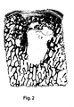
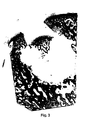
- FIGS. 2 to 4 illustrate the experiments already mentioned above Implants according to FIG. 1 in sheep joints and the mentioned risk that Repairs with such implants include. These are tissue sections by repair points parallel to the axis of the implant, which in the present Enlargement of the cartilage surface shown above about 7 cm in the bone tissue protrudes.
- Figures 2 and 3 show preparations that last six months were created after the implantation and in which cyst-like at the repair sites Cavities in the bone tissue are visible.
- Figure 2 shows a case in which the cartilage layer is still positioned in its original place, in Figure 3 they sank into the cyst room.
- Figure 4 shows an identical repair site after twelve months. It is clearly one the unevenness in the implanted cartilage layer Cartilage surface visible.
- repair sites were found at seven treated animals were examined after twelve months and in two cases bumps were found in the cartilage surface as found in FIG. 4. In the rest In cases, the cartilage surface in the repair area was flat.
- FIG. 5 shows a preferred embodiment of the preparation according to the invention for the repair of cartilage or cartilage / bone defects in human or animal joints. It shows the preparation 10, for the implantation of the preparation 10 opening or bore 20 to be created and the inserted into the opening Specimen, the implant 30 (cuts along the axis of the specimen 10 or Opening 20).
- the preparation 10 has a bone part in the same way as the preparation of FIG. 1 1, a frontal cartilage layer 2, which forms a cartilage surface 3, and in the transition area between the bone part 1 and cartilage layer 2 a subchondral Bone plate 4.
- the preparation has a head part 11 with a larger one Cross section and a foot part 12 with a smaller cross section.
- the head part 11 essentially comprises the cartilage layer 2 and the subschondral bone plate 4, the foot part 12 essentially corresponds to the bone part 1.
- the foot part 12 has advantageously (but not necessarily) the shape of a circular cylinder or steep circular truncated cone and the head part 11 is on all sides above the bone part 1 before and is also circular cylindrical, for example.
- Auto grafts and grafts from living donors advantageously have cylindrical head parts, because such preparations should be as small as possible Cause removal opening.
- Preparations made from tissue from slaughter animals can be produced easily Have headboards with different shapes of cartilage surface because that Material is available in large quantities and can therefore also be processed with waste become. But even in this case it is advantageous to design the foot section in such a way that the opening to be created for the implantation is simple Tool, for example, can be created with a drill.
- a preparation 10 with a cross section that is at least round in the foot region is, for example made from a corresponding cylindrical preparation in that the bone part is processed accordingly.
- This processing can be done with a Tool can be carried out in which the cylindrical preparation can be positioned is and in the blades can be activated in a predetermined or adjustable Distance from the cartilage surface the cross-section of the preparation to a predetermined one Reduce measure.
- the Dimensioning of preparation 10 and bore 20 is for a press fit both in Area of the head part as well as in the foot part of the preparation.
- a bore 20, as shown in FIG. 5, is created, for example with a tool that has a blade with a circular cutting edge has and two drills axially limited relative to the cutting edge or hollow drill.
- the tool is positioned on the defect and the blade pressed down to the subchondral bone plate.
- the blade can be moved and its diameter substantially corresponds to the inner diameter of the blade, the outer region 21 is drilled out.
- the inner area 22 is subsequently drilled out with the second drill, whereby the drilling depth relative to the cutting edge or relative to the end position of the first Drill is adjustable.
- FIG. 5 also shows the implant 30 on the right-hand side, that is to say in the bore 20 used preparation 10.
- the implant 30 has one with the native cartilage surface 3 'aligned cartilage surface 3 and one with the native subchondral Bone plate 4 'approximately aligned subchondral bone plate 4. Die subchondral bone plate 4 of the implant is obviously supported on the one hand on the bone part 1 of the implant and on the other hand on native bone tissue immediately below or in the native, subchondral bone plate 4 '.
- a tool for implanting a preparation in a bore, as shown by FIG. 5 is used, for example, a tool that has a sleeve and an in the sleeve has axially movable plunger.
- the sleeve has an inner cross section, which corresponds to the cross section of the head part of the preparation to be implanted.
- the plunger advantageously has an approximately same cross section as this Headboard; it is longer than the sleeve and has a channel on one Front of the ram begins and in the area of the other ram end to one Suction line can be connected.
- the face of the plunger is inserted into the channel opening Pushed sleeve and a preparation is pulled into the sleeve with the suction force generated. Then the sleeve with pestle and the preparation sucked on it the prepared hole and the preparation is positioned using the pestle and, if necessary, a hammer is pressed into the bore.
- the head part it is recommended to use the supernatant dimension the head part to about 1 to 2 mm (e.g. bone part with diameter approx. 3 mm, head section with a diameter of 5 to 6 mm)
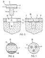
- Figures 6 and 7 show two further exemplary embodiments of the inventive Preparation. These are based on the idea of the subchondral bone plate of the preparation additionally not on native bone tissue, as in FIG. 5 shown, but on at least one created in the bone part of the preparation Support column, whereby the columns from the remaining bone material of the bone part 1 have a different degradability in such a way that they Time. in which the remaining bone material of the bone part is broken down, the wear subchondral bone plate and the overgrown cartilage layer and prevent sinking into the cyst area.
- Figures 6 and 7 are cross sections through bone parts 1 of inventive Preparations. These preparations are cylindrical, for example, and have a bone part axially extending columns, which consist of a material that is slower is degradable as the bone material of the areas between the pillars.
- the Columns are arranged, for example, on the surface of the bone part (surface columns 40 in Figure 6) and are treated appropriately with the bone material generated or they are located inside the bone part (inner Columns 41 in Figure 7) and are created by drilling and filling the holes with a suitable material. In both cases, the pillars are enough at least to the subchondral bone plate.
- Resorbable material for filling holes can be, for example, hydroxylapatite ceramic be used.
- Figures 8 and 9 show a cartilage defect (Figure 8) and a cartilage / bone defect ( Figure 9) with sizes such that they can not with a single Implant can be repaired. The defects are indicated by dash-dotted lines.
- the repair consists of a mosaic-like arrangement of inventive Preparations as shown in Figure 5.
- FIG. 8 shows that the implants 30 with the head part 11 are also in a mosaic repair a support for the cartilage layer and the subchondral bone plate of the implant allow on native bone tissue 1 'and thereby a decrease in Counteract the cartilage layer in a critical healing phase. It is also evident that the areas of native bone tissue 1 'between the foot parts 12 of the implants are wider than would be possible with cylindrical implants. That also means that the chances of a cure compared to the state of the art are improved. And it can be seen that the cartilage surface is all over Defect area can be designed essentially without interruption if the shape corresponding to the headboards (e.g. square, rectangular, triangular or hexagonal) is chosen.
- the shape corresponding to the headboards e.g. square, rectangular, triangular or hexagonal
- FIG. 9 shows a cartilage / bone defect (indicated by a dash-dotted line), which is repaired with a plurality of preparations according to FIG. 5.
- the foot parts 12 of the implants 30 extend into healthy bone tissue 1 '.
- a suitable material e.g. tricalcium phosphate or hydraulic Bone cement.
- This material should be chosen so that it is in front of or is only dismantled after the foot parts 12 of the implants 30, that is to say in the critical phase of the healing process, in which the bone parts of the implants degraded but not yet replaced, support the cartilage layers and sink in that can prevent cartilage layers.
- the filling material advantageously has sufficient mechanical strength, such that it is first filled in the defect and then can be pierced.
Abstract
Description
Die Erfindung liegt auf dem Gebiete der Medizinaltechnik und betrifft ein Präparat gemäss dem Oberbegriff des unabhängigen Patentanspruchs. Das Präparat dient zur Reparatur von Knorpel- oder Knorpel/Knochen-Defekten in menschlichen oder tierischen Gelenken, das heisst, es dient zur Reparatur von Defekten in der Knorpelschicht. die in Gelenken die Knochenoberfläche abdeckt, oder von Defekten, die diese Knorpelschicht und auch darunterliegendes Knochengewebe betreffen.The invention is in the field of medical technology and relates to a preparation according to the preamble of the independent claim. The preparation is used for Repair of cartilage or cartilage / bone defects in human or animal Joints, that is, it is used to repair defects in the cartilage layer. that covers the bone surface in joints, or defects that affect this layer of cartilage and the underlying bone tissue.
Ein Präparat zur Reparatur von Knorpel-oder Knorpel/Knochen-Defekten
gemäβ dem Oberbegriff von Anspruch 1 ist aus der WO-A-93/15694
bekannt.A preparation for the repair of cartilage or cartilage / bone defects
according to the preamble of
Beschädigungen von Gelenkknorpel durch Verletzungen oder durch alters- oder krankheitsbedingte Rückbildung sind insbesondere beim Menschen sehr häufig. Vielfach ist auch das unter dem Gelenkknorpel liegende Knochengewebe durch derartige Beschädigungen in Mitleidenschaft gezogen. Der Schweregrad von Knorpelund Knorpel/Knochen-Defekten wird mit Hilfe der Outerbridge-Skala erfasst, mit den folgenden Kategorien: oberflächliche Ausfransung (ca. 10% aller Fälle), Risse im Knorpel (ca. 28%), Risse bis zum Knochen (ca. 41%), Schäden mit Beteiligung des Knochens (ca. 19%), andere Schäden, wie Osteochondritis dissecans und Gelenkbrüche (ca. 2% aller erfassten Fälle).Damage to articular cartilage from injuries or from age or Disease-related regression is very common, especially in humans. In many cases, the bone tissue under the articular cartilage is also of such type Damage affected. The severity of cartilage Cartilage / bone defects are recorded using the outer bridge scale, with the following categories: superficial fraying (approx. 10% of all cases), cracks in cartilage (approx. 28%), cracks down to the bone (approx. 41%), damage with involvement of the bone (approx. 19%), other damage such as osteochondritis dissecans and joint fractures (approx. 2% of all recorded cases).
Vitales Knorpelgewebe enthält zwar lebende Zellen, durch deren Tätigkeit in der Wachstumsphase die spezifische interzelluläre Knorpelmatrix aufgebaut wird, es ist aber mindestens in ausgewachsenem Zustand kaum durchblutet und besitzt deshalb nach der eigentlichen Wachstumsphase eine nur sehr beschränkte Regenerationsfähigkeit. Das heisst mit anderen Worten, Knorpel- oder Knorpel/Knochen-Defekte, insbesondere derartige Defekte, die eine relativ grosse Knorpeloberfläche betreffen, heilen nicht von selbst und müssen deshalb operativ repariert werden (Mankin HJ: The response of articular cartilage to mechanical injury, Journal of Bone and Joint Surgery (Am) 64A (1982) March: Seiten 460-466).Vital cartilage tissue contains living cells, through their activity in the Growth phase the specific intercellular cartilage matrix is built up, it is but hardly has blood flow, at least when fully grown, and therefore has only a very limited ability to regenerate after the actual growth phase. In other words, cartilage or cartilage / bone defects, in particular defects of this kind which affect a relatively large cartilage surface, do not heal by themselves and therefore need to be repaired surgically (Mankin HJ: The response of articular cartilage to mechanical injury, Journal of Bone and Joint Surgery (Am) 64A (1982) March: pages 460-466).
Für die Reparatur der genannten Defekte wird beispielsweise das Implantieren von Präparaten vorgeschlagen, in denen das zu ersetzende Gewebe bereits vorhanden ist oder möglichst gut vorgebildet ist. Solche Präparate sind zylinderförmig und weisen auf einer Stirnseite eine Knorpelschicht auf. Für die Implantation wird im Bereich des zu reparierenden Defektes eine Sackloch-förmige Öffnung oder Bohrung erzeugt und das Präparat wird derart in die Bohrung eingesetzt, dass die Knorpelschicht des Implantates gegen aussen gerichtet ist. Die Bohrung reicht, unabhängig von der Defekttiefe, bis in gesundes Knochengewebe. Das Präparat weist einen etwas grösseren Durchmesser auf als die Bohrung und dieselbe achsiale Länge. Damit wird erreicht, dass zwischen nativem Gewebe und Implantat eine radiale Spannung entsteht (Pressfit), durch die das Implantat in der Bohrung gehalten wird, und dass die Knorpeloberfläche des Implantates mit der umgebenden nativen Knorpeloberfläche fluchtet. Die Präparate haben je nach Defektgrösse einen Durchmesser von 4 bis 10mm (z.B. 5,4mm für Präparat und 5,3mm für Bohrung) und Längen von ca. 10 bis 20mmFor the repair of the defects mentioned, for example, the implantation of Preparations are proposed in which the tissue to be replaced already exists or is well educated. Such preparations are cylindrical and wise a layer of cartilage on one end. For the implantation is in the area creates a blind hole-shaped opening or hole of the defect to be repaired and the preparation is inserted into the bore such that the cartilage layer of the Implant is directed towards the outside. The hole is sufficient regardless of the depth of the defect down to healthy bone tissue. The preparation has a slightly larger one Diameter on than the bore and same axial length. So that is achieved that there is a radial tension between the native tissue and the implant (press fit), through which the implant is held in the hole and that the cartilage surface of the implant is aligned with the surrounding native cartilage surface. The Depending on the size of the defect, preparations have a diameter of 4 to 10 mm (e.g. 5.4mm for specimen and 5.3mm for drilling) and lengths of approx. 10 to 20mm
Für grössere Defekte wird vorgeschlagen, eine Mehrzahl derartiger zylinderförmiger Präparate mosaikartig im defekten Bereich einzusetzen und die Zwischenräume zwischen den Implantaten mit einem geeigneten Material auszufüllen. For larger defects, a plurality of such cylindrical ones is proposed Use preparations like a mosaic in the defective area and the spaces between fill the implants with a suitable material.
Die zylinderförmigen Präparate sind beispielsweise autolog (Autotransplantate). Es wird also beispielsweise für die Reparatur eines Gelenkknorpel-Defektes, wie er an einer stark belasteten Stelle eines Gelenkes entstehen kann. aus einer relativ wenig belasteten Stelle desselben Gelenkes mit einem entsprechenden Hohlbohrer ein geeignetes Gewebestück entnommen und in eine an der defekten Stelle erstellte Bohrung transplantiert (Hangody L et al.: Mosaicplasty for the treatment of articular cartilage defects: application in clinical practice. Orthopedics 1998 Jul,21(7):751-6).The cylindrical preparations are, for example, autologous (car transplants). It is used, for example, for the repair of a defect in the articular cartilage a heavily loaded part of a joint can arise. from a relatively little a suitable hollow point with a suitable hollow drill Tissue piece removed and into a hole made at the defective point transplanted (Hangody L et al .: Mosaicplasty for the treatment of articular cartilage defects: application in clinical practice. Orthopedics 1998 Jul, 21 (7): 751-6).
Die zylinderförmigen Präparate können auch von einem geeigneten Spender stammen (homologe Transplantate). Auch entsprechende heterologe oder Xenotransplantate sind bekannt, die vor der Implantation mit einem geeigneten Verfahren, z.B. Photooxidation (wie beschrieben in der Publikation EP-0768332 von Sulzer Innotec), derart behandelt werden, dass eine Immunreaktion nach der Implantation verhindert oder mindestens stark abgeschwächt wird (immunologische Desaktivierung). Solche Implantate werden beispielsweise aus Schultergelenken von geschlachteten Rindern entnommen und haben den Vorteil, dass sie in viel grösseren Mengen zur Verfügung stehen als autologe oder homologe Transplantate und dass sie keine Entnahme-Defekte verursachen, die ihrerseits wieder repariert werden müssen und zu neuen Schwierigkeiten führen können.The cylindrical preparations can also come from a suitable donor (homologous grafts). Also corresponding heterologous or xenografts are known which can be prepared using a suitable method, e.g. Photooxidation (as described in Sulzer Innotec's publication EP-0768332), treated in such a way that an immune reaction after implantation is prevented or is at least greatly weakened (immunological deactivation). Such For example, implants are made from shoulder joints of slaughtered cattle withdrawn and have the advantage that they are available in much larger quantities are available as autologous or homologous grafts and that they have no removal defects cause, which in turn have to be repaired and new ones Can cause difficulties.
In der Publikation WO-97/46665 (Sulzer Orthopedics) wird auch ein entsprechendes Präparat beschrieben, in dem der Knochenteil aus einem Knochenersatzmaterial besteht und in dem die stirnseitige Knorpelschicht beispielsweise ausgehend von autologen Chondrocyten in vitro aufgewachsen wird.A corresponding one is also described in publication WO-97/46665 (Sulzer Orthopedics) Preparation described in which the bone part consists of a bone replacement material and in which the frontal cartilage layer starts, for example, from autologists Chondrocytes are grown in vitro.
In allen oben genannten Präparaten, die aus natürlichem Gewebe hergestellt werden, gibt es eine natürliche Verbindung oder Verwachsung zwischen der stirnseitigen Knorpelschicht und dem Knochenteil und es gibt einen äussersten Knochenbereich (subchondrale Knochenplatte), in dem das Knochengewebe dichter ist als in anderen Knochenbereichen. Auch die genannten, teilweise künstlichen Implantate weisen die Verwachsung der Knorpelschicht mit dem Knochenteil auf und der künstliche Knochenteil ist vorteilhafterweise mit einer dichteren, das heisst weniger porösen Aussenschicht, die der Knorpelschicht als Unterlage dient, ausgestaltet.In all the above-mentioned preparations, which are made from natural tissue, there is a natural connection or intergrowth between the front Cartilage layer and the bone part and there is an outermost bone area (subchondral bone plate) in which the bone tissue is denser than in others Bone regions. The mentioned, partly artificial implants also have the The cartilage layer fuses with the bone part on and the artificial bone part is advantageously with a denser, i.e. less porous outer layer, which serves as a base for the cartilage layer.
Eine wichtige Funktion der subchondralen Knochenplatte oder einer künstlichen Nachbildung davon ist offenbar die Verhinderung einer vom Knochengewebe ausgehenden Vaskularisierung der Knorpelschicht, die zu einer Verknöcherung dieser Schicht führen würde. Daneben stellt die subchondrale Knochenplatte aber auch einen Bereich dar, der dank seiner höheren Dichte eine höhere mechanische Festigkeit hat als das innere Knochengewebe.An important function of the subchondral bone plate or an artificial one Imitation of this is apparently the prevention of one that starts from the bone tissue Vascularization of the cartilage layer, leading to ossification of this Shift would lead. In addition, the subchondral bone plate also provides one Area that, thanks to its higher density, has a higher mechanical strength has as the inner bone tissue.
Mit den genannten Präparaten wird versucht, im wesentlichen die folgenden Ziele zu erreichen:
- Der Knochenteil des Präparates soll eine feste Verankerung des Implantates im gesunden Knochengewebe durch Pressfit ermöglichen, derart, dass das Implantat keine weiteren, gesunde Knorpelbereiche beeinflussende Befestigungsmittel benötigt.
- Die Verwachsung von Knorpelschicht und Knochenteil im Präparat soll dem Implantat eine Stabilität geben, derart, dass die Knorpelschicht auch ohne Stilllegung des betroffenen Gelenkes sich nicht ablösen und aus der Defektstelle entfernen kann.
- Die Knorpelschicht des Präparates soll eine derartige mechanische Festigkeit und Elastizität aufweisen, dass die Reparaturstelle schon unmittelbar nach der Implantation voll belastbar ist.
- Die Knorpelschicht soll eine Zone bilden, in der für implantierte Zellen oder für nach der Implantation zuwandernde Zellen geeignete Bedingungen für die Erzeugung und/oder Aufrechterhaltung von funktionsfähigem Knorpelgewebe herrschen. Dies soll auch unterstützt werden durch die subchondrale Knochenplatte, die die Knorpelschicht vom Knochenteil trennt und die hilft, eine vom Knochenteil ausgehende Vaskularisierung der Knorpelschicht zu verhindern.
- Der Knochenteil soll eine Zone darstellen, in der für implantierte Zellen oder für nach der Implantation zuwandernde Zellen geeignete Bedingungen für die Erzeugung und/oder für die Aufrechterhaltung von funktionsfähigem Knochengewebe herrschen.
- The bone part of the preparation is intended to enable the implant to be firmly anchored in the healthy bone tissue by means of a press fit in such a way that the implant does not require any further fasteners which influence healthy cartilage areas.
- The intergrowth of the cartilage layer and the bone part in the preparation is intended to give the implant a stability such that the cartilage layer cannot detach and remove from the defect site even without the affected joint being shut down.
- The cartilage layer of the preparation should have such mechanical strength and elasticity that the repair site is fully resilient immediately after implantation.
- The cartilage layer should form a zone in which suitable conditions for the production and / or maintenance of functional cartilage tissue prevail for implanted cells or for cells that migrate after the implantation. This should also be supported by the subchondral bone plate, which separates the cartilage layer from the bone part and which helps to prevent vascularization of the cartilage layer starting from the bone part.
- The bone part is intended to represent a zone in which suitable conditions for the production and / or maintenance of functional bone tissue prevail for implanted cells or for cells that migrate after the implantation.
Neue Versuche, in denen künstlich erzeugte Defekte in Gelenken von Schafen mit Autotransplantaten,. mit Homotransplantaten oder mit Heterotransplantaten (aus Rindergewebe) in der eingangs beschriebenen Art repariert wurden, zeigen nun, dass der Heilungsprozess nach der Implantation nicht wie erwartet abläuft.New trials involving artificially created defects in sheep's joints Autografts ,. with homograft or with heterograft (from bovine tissue) were repaired in the manner described above, now show that the Healing process after implantation does not proceed as expected.
Es zeigt sich nämlich, dass der Knochenteil der Implantate nicht im nativen Gewebe integriert oder sukzessive durch neues Reparaturgewebe ersetzt wird, sondern dass der Knochenteil des Implantates einen Umwandlungsprozess durchläuft, der im wesentlichen drei aufeinanderfolgende Phasen aufweist. In einem ersten Schritt werden knochenabbauende Zellen (Osteoclasten) stimuliert und der eingesetzte Knochen wird aufgelöst. Diese erste Phase ist bereits nach sechs bis acht Wochen deutlich sichtbar. Mit fortschreitender Implantationsdauer entsteht im Implantat ein Hohlraum (Zyste), der mit Bindegewebe aufgefüllt ist. Diese zweite Phase erreicht einen Höhepunkt nach ca. sechs Monaten. In der dritten und letzten Phase werden knochenbildende Zellen (Osteoblasten) angelockt, welche das Bindegewebe in Knochen umbauen. Der Umbauprozess ist nach ungefähr zwölf Monaten abgeschlossen. Dann ist die neu entstandene Knochenstruktur so gut angepasst, dass die ursprüngliche Grenze zwischen dem Implantat und dem umliegenden Knochengewebe kaum mehr wahrnehmbar ist.It turns out that the bone part of the implants is not in the native tissue integrated or successively replaced by new repair fabric, but that the bone part of the implant undergoes a transformation process that essentially has three successive phases. In a first step bone-degrading cells (osteoclasts) and the bone used are stimulated will be dissolved. This first phase is already clear after six to eight weeks visible. As the duration of the implantation progresses, a cavity is created in the implant (Cyst), which is filled with connective tissue. This second phase culminates after about six months. In the third and final phase, bone-building Cells (osteoblasts) that convert the connective tissue into bones. The renovation process is completed after about twelve months. Then the newly formed bone structure is so well adapted that the original boundary hardly perceptible between the implant and the surrounding bone tissue is.
Durch den beschriebenen, dreiphasigen Umwandlungsprozess entsteht in der mittleren Phase, in der die Knorpelschicht des Implantates nicht vom tragfähigen Knochenteil getragen wird sondern von einer mechanisch minderwertigen Zyste, ein hohes Risiko dafür, dass die Knorpelschicht in diese Zyste hineingedrückt wird, wo sie weder ihre mechanische Funktion noch ihre biologische Funktion erfüllen kann und von wo sie während der folgenden Phasen des Heilungsprozesses nicht mehr verschoben werden kann. Durch dieses Risiko werden also die Erfolgschancen der Heilung bedeutend reduziert und es müssen bei einer Verheilung mit schlecht positionierter Knorpelschicht Folgenachteile in Kauf genommen werden.The described three-phase conversion process creates in the middle Phase in which the cartilage layer of the implant is not separated from the load-bearing part of the bone but is carried by a mechanically inferior cyst, a high one Risk of pushing the cartilage layer into this cyst where it is can neither fulfill their mechanical function nor their biological function and from where they no longer moved during the subsequent phases of the healing process can be. This risk increases the chances of success of the cure significantly reduced and it needs to be healed with poorly positioned Cartilage layer consequential disadvantages are accepted.
Erstaunlich an den beschriebenen Befunden ist die Tatsache, dass die Bildung der Zyste an der Stelle des Knochenteils eines Implantates in einer mittleren Phase des Heilungsprozesses nicht nur bei homologen und heterologen Implantaten auftritt, sondern insbesondere auch bei Autotransplantaten. Der anfängliche Abbau des implantierten Knochengewebes scheint also nicht eine Immunreaktion zu sein, in der implantiertes; vitales Material als frernd erkannt und deshalb abgebaut wird. Die Vermutung liegt nahe, dass es sich um eine Reaktion auf implantiertes, totes Material handelt. Dies würde bedeuten, dass durch die Durchtrennung der natürlichen Blutversorgung in einem zwar aus lebensfähigem Gewebe entnommenen Implantat das Knochengewebe stirbt, auch wenn es unmittelbar nach der Entnahme wieder implantiert wird. In jedem Falle baut der Körper den Knochenteil des implantierten Präparates ab, um ihn erst dann wieder neu aufzubauen.The fact that the formation of the Cyst at the site of the bone part of an implant in a middle phase of the Healing process not only occurs with homologous and heterologous implants, but especially in the case of car transplants. The initial breakdown of the implanted Bone tissue does not appear to be an immune response in which implanted; vital material is recognized as foreign and is therefore broken down. The Presumption suggests that it is a reaction to implanted, dead material is. This would mean cutting through the natural blood supply in an implant taken from viable tissue Bone tissue dies, even if it is implanted again immediately after removal becomes. In any case, the body builds the bone part of the implanted specimen only to rebuild it again.
Die Erfindung stellt sich nun die Aufgabe, ein Präparat zur Reparatur von Knorpel-Defekten oder von Knorpel/Knochen-Defekten in menschlichen oder tierischen Gelenken zu schaffen, mit welchem Präparat das oben beschriebene Risiko des Absinkens einer implantierten Knorpelschicht in den Bereich des nativen Knochengewebes verhindert wird. Das erfindungsgemässe Präparat soll in ähnlich einfacher Art hergestellt und implantiert werden können wie die eingangs beschriebenen, bekannten Präparate, die einen Knochenteil und eine mit diesem Knochenteil verwachsene Knorpelschicht aufweisen.The object of the invention is now a preparation for repairing cartilage defects or cartilage / bone defects in human or animal joints to create with which preparation the risk of sinking described above an implanted layer of cartilage in the area of the native bone tissue is prevented. The preparation according to the invention is to be produced in a similarly simple manner and can be implanted like the known ones described at the beginning Preparations that have one part of the bone and one that has grown together with this part of the bone Have a cartilage layer.
Diese Aufgabe wird gelöst durch das Präparat, wie es im unabhängigen Patentanspruch definiert ist. Die abhängigen Ansprüche definieren bevorzugte Ausführungsformen des erfindungsgemässen Präparates.This object is achieved by the preparation as set out in the independent claim is defined. The dependent claims define preferred embodiments of the preparation according to the invention.
Das erfindungsgemässe Präparat zur Reparatur von Knorpel- oder Knorpel/Knochen-Defekten in menschlichen oder tierischen Gelenken basiert auf dem Befund, dass die subchondrale Knochenplatte in der mittleren, kritischen Phase des oben beschriebenen Heilungsprozesses offenbar noch im wesentlichen unverändert vorhanden ist, wenn der Knochenteil vollständig oder grösstenteils durch Bindegewebe ersetzt ist. Dies ist wahrscheinlich darauf zurückzuführen, dass diese subchondrale Knochenplatte wegen ihrer höheren Dichte bedeutend langsamer abgebaut wird als die inneren Bereiche des Knochenteils. Da diese subchondrale Knochenplatte mechanisch genügend stabil ist, kann sie ein Absinken der darauf aufgewachsenen Knorpelschicht in eine darunter entstehende Zyste verhindern, wenn sie nicht nur auf dem Knochenmaterial des implantierten Knochenteils sondern noch zusätzlich auf Material mit anderen Abbau-Eigenschaften derart abgestützt ist, dass sie auch während der kritischen Heilungsphase nicht verschiebbar bleibt. Wenn die subchondrale Knochenplatte des Implantates nach dieser kritischen Phase, das heisst zu einem Zeitpunkt, in dem die Tragfähigkeit des inneren Implantatbereiches wieder hergestellt ist, ebenfalls abgebaut wird, kann dies den Heilungsprozess nicht mehr stark beeinflussen. The preparation according to the invention for repairing cartilage or cartilage / bone defects in human or animal joints is based on the finding that subchondral bone plate in the middle, critical phase of the above Healing process is apparently still essentially unchanged, if the bone part is completely or largely replaced by connective tissue. This is probably due to the fact that this subchondral bone plate is degraded much more slowly than the inner ones because of its higher density Areas of the bone part. Because this subchondral bone plate is mechanical is sufficiently stable, the cartilage layer grown on it can sink prevent it from developing into an underlying cyst if it is not only on the Bone material of the implanted bone part but also on material is supported with other degradation properties in such a way that it also during the critical healing phase cannot be moved. If the subchondral bone plate of the implant after this critical phase, i.e. at a time in which the load-bearing capacity of the inner implant area is restored, is also broken down, this can no longer strongly influence the healing process.
Eine Abstützung des implantierten Präparates während der kritischen Heilungsphase lässt sich im wesentlichen auf zwei Arten realisieren.A support for the implanted specimen during the critical healing phase can be realized in two ways.
Einerseits kann das Implantat derart geformt sein, dass die Knorpelschicht und die subchondrale Knochenplatte des Präparates eine grössere Querschnittsfläche haben als der Knochenteil. Ein derartiges Präparat wird in eine zweistufige Bohrung implantiert. derart, dass die subchondrale Knochenplatte des Präparates sich nicht nur auf dem Knochenteil des Implantates sondern auch auf gesundem Knochengewebe neben der für die Reparatur erstellten Bohrung abstützt.On the one hand, the implant can be shaped such that the cartilage layer and the subchondral bone plate of the specimen have a larger cross-sectional area than the bone part. Such a preparation is implanted in a two-stage bore. such that the subchondral bone plate of the preparation is not only on the bone part of the implant but also on healthy bone tissue supports next to the hole created for the repair.
Andererseits können im Knochenteil des Präparates Säulen mit einer reduzierten Abbaubarkeit erstellt werden. Diese Säulen erstrecken sich axial durch den Knochenteil und reichen bis unter die subchondrale Knochenplatte. Die gegenüber den Knochenteilbereichen zwischen den Säulen reduzierte Abbaubarkeit kann realisiert werden durch eine bereichsweise, geeignete chemische Behandlung oder dadurch, dass im Knochenteil des Präparates achsial verlaufende Bohrungen erzeugt und mit einem künstlichen, abbauresistenteren Material gefüllt werden.On the other hand, columns with reduced Degradability can be created. These columns extend axially through the bone part and extend below the subchondral bone plate. The opposite of the Bone partial areas between the pillars reduced degradability can be realized by chemical treatment, or by that axially running bores are created in the bone part of the specimen and with an artificial, more resistant to degradation material.
Die der Erfindung zugrunde liegenden Befunde sowie beispielhafte Ausführungsformen des erfindungsgemässen Präparates werden im Zusammenhang mit den folgenden Figuren detailliert beschrieben. Dabei zeigen:
-
Figur 1 - ein zylinderförmiges Präparat gemäss dem Stande der Technik zur Reparatur von Knorpel- oder Knorpel/Knochen-Defekten in menschlichen oder tierischen Gelenken;
-
Figuren 2bis 4 - Gewebeschnitte durch mit einem Präparat gemäss Figur 1 reparierte Knorpel/Knochen-Defekte in einem Schafgelenk zu verschiedenen Zeitpunkten nach der Implantation;
- Figur 5
- ein Schema der Reparatur eines Knorpel-Defektes mit Hilfe einer bevorzugten Ausführungsform des erfindungsgemässen Präparates (Schnitte entlang der Achse des Präparates bzw. der Bohrung);
- Figuren 6 und 7
- zwei weitere beispielhafte Ausführungsformen des erfindungsgemässen Präparates im Querschnitt;
- Figuren 8 und 9
- Schnitte durch einen Knorpel-Defekt (Figur 8) und durch einen Knorpel/Knochen-Defekt (Figur 9), die beide mosaikartig mit einer Mehrzahl von erfindungsgemässen Präparaten gemäss Figur 5 repariert sind.
- Figure 1
- a cylindrical preparation according to the prior art for repairing cartilage or cartilage / bone defects in human or animal joints;
- Figures 2 to 4
- Tissue sections through cartilage / bone defects repaired with a preparation according to FIG. 1 in a sheep joint at different times after the implantation;
- Figure 5
- a diagram of the repair of a cartilage defect using a preferred embodiment of the preparation according to the invention (cuts along the axis of the preparation or the bore);
- Figures 6 and 7
- two further exemplary embodiments of the preparation according to the invention in cross section;
- Figures 8 and 9
- Sections through a cartilage defect (FIG. 8) and through a cartilage / bone defect (FIG. 9), both of which have been repaired like a mosaic with a plurality of preparations according to the invention as shown in FIG.
Figur 1 zeigt ein Präparat, wie es nach dem Stande der Technik für die Reparatur von
Knorpel- oder Knorpel/Knochen-Defekten an menschlichen und tierischen Gelenken
verwendet wird. Das Präparat ist zylinderförmig, vorteilhafterweise mit einem kreisrunden
Querschnitt. und weist einen Knochenteil 1 und eine stirnseitig auf dem Knochenteil
1 aufgewachsene Knorpelschicht 2 auf. Die Knorpelschicht 2 bildet eine
Knorpeloberfläche 3. Zwischen Knochenteil 1 und Knorpelschicht 2 erstreckt sich
eine subschondrale Knochenplatte 4. Die Übergänge von Knochenteil 1 zu subchondraler
Knochenplatte 4 und von subchondraler Knochenplatte 4 zu Knorpelschicht
2 sind nicht, wie in der Figur 1 vereinfachend dargestellt, als Linien erkennbar
sondern es handelt sich um natürliche eher kontinuierliche Übergänge.Figure 1 shows a preparation as it is in the prior art for the repair of
Cartilage or cartilage / bone defects on human and animal joints
is used. The preparation is cylindrical, advantageously with a circular one
Cross-section. and has a
Wie bereits eingangs erwähnt, wird ein Präparat, wie es in der Figur 1 dargestellt ist, vorteilhafterweise aus vitalem Gewebe mit Hilfe eines Hohlbohrers ausgestanzt (Autotransplantate und Homotransplantate) und möglichst sofort implantiert oder es wird aus Gelenken von Schlachttieren (beispielsweise aus Schultergelenken von geschlachteten Rindern) entnommen und vor der Implantation einer Behandlung zur immunologischen Desaktivierung unterzogen.As already mentioned at the beginning, a preparation as shown in FIG. 1 is advantageously punched out of vital tissue using a hollow drill (Auto grafts and home grafts) and implanted as soon as possible or it is made from joints of slaughter animals (for example from shoulder joints of slaughtered animals Cattle) and treated for implantation prior to implantation subjected to immunological deactivation.
Die Figuren 2 bis 4 illustrieren die weiter oben bereits angeführten Versuche mit Implantaten gemäss Figur 1 in Schafgelenken und das angesprochene Risiko, das Reparaturen mit derartigen Implantaten beinhalten. Es handelt sich um Gewebeschnitte durch Reparaturstellen parallel zur Achse des Implantates, das bei der vorliegenden Vergrösserung von der oben dargestellten Knorpeloberfläche etwa 7 cm in das Knochengewebe hineinragt. Figuren 2 und 3 zeigen Präparate, die sechs Monate nach der Implantation erstellt wurden und in denen an den Reparaturstellen zystenartige Hohlräume im Knochengewebe sichtbar sind. Figur 2 zeigt einen Fall, in dem die Knorpelschicht noch an ihrem ursprünglichen Platz positioniert ist, in Figur 3 ist sie in den Zystenraum abgesunken.FIGS. 2 to 4 illustrate the experiments already mentioned above Implants according to FIG. 1 in sheep joints and the mentioned risk that Repairs with such implants include. These are tissue sections by repair points parallel to the axis of the implant, which in the present Enlargement of the cartilage surface shown above about 7 cm in the bone tissue protrudes. Figures 2 and 3 show preparations that last six months were created after the implantation and in which cyst-like at the repair sites Cavities in the bone tissue are visible. Figure 2 shows a case in which the cartilage layer is still positioned in its original place, in Figure 3 they sank into the cyst room.
Wie aus den Figuren 2 und 3 ersichtlich ist, ist im kritischen Zeitraum, in dem sich an der Stelle des implantierten Knochenteils ein zystenartiger Hohlraum befindet, die subchondrale Knochenplatte des Implantates noch im Wesentlichen unverändert vorhanden. Dieser Befund lässt sich auf die gegenüber dem inneren Knochengewebe erhöhte Dichte der subchondralen Knochenplatte und auf eine daraus resultierende, reduzierte Abbaubarkeit zurückführen. Die subchondrale Knochenplatte des Implantates hat offensichtlich andere Abbau-Eigenschaften als innere Bereiche des Knochteils desselben Implantates.As can be seen from Figures 2 and 3, is in the critical period in which there is a cyst-like cavity at the site of the implanted bone part, the subchondral bone plate of the implant is still essentially unchanged. This finding can be compared to the inner bone tissue increased density of the subchondral bone plate and on a resulting, Reduce degradability. The subchondral bone plate of the implant has obviously different degradation properties than inner areas of the Bone part of the same implant.
Die Versuche wurden mit Autotransplantaten und mit Heterotransplantaten durchgeführt. An 7 behandelten Tieren wurden die Reparaturstellen nach sechs Monaten untersucht und in 10 Fällen (fünf Tiere) wurden in den Zystenhohlraum verschobene Knorpelschichten gefunden, in 4 Fällen (zwei Tiere) waren die Knorpelschichten an Ort und Stelle geblieben. In keinem Fall ging die Knorpelschicht in den Gelenkraum verloren.The experiments were carried out with auto-grafts and with heterografts. The repair sites on 7 treated animals were removed after six months were examined and in 10 cases (five animals) were displaced into the cyst cavity Cartilage layers found, in 4 cases (two animals) the cartilage layers were on Stayed in place. In no case did the cartilage layer go into the joint space lost.
Diese Versuchsresultate zeigen, dass offenbar zwischen Zyste und implantierter Knorpelschicht bzw. subchondraler Knochenplatte eine genügende Adhäsion besteht, um die Knorpelschicht daran zu hindern, sich gegen aussen aus der Reparaturstelle zu entfernen, dass aber die Tragfähigkeit der Zyste nicht ausreicht, um die Knorpelschicht daran zu hindern, sich gegen innen zu verschieben.These experimental results show that apparently between the cyst and the implanted Cartilage layer or subchondral bone plate there is sufficient adhesion, to prevent the cartilage layer from escaping outside the repair site to remove that but the load bearing capacity of the cyst is not sufficient to remove the cartilage layer to prevent it from moving towards the inside.
Figur 4 zeigt eine gleiche Reparaturstelle nach zwölf Monaten. Es ist deutlich eine durch das Einsinken der implantierten Knorpelschicht entstandene Unebenheit in der Knorpeloberfläche sichtbar. In der Versuchsreihe wurden Reparaturstellen an sieben behandelten Tieren nach zwölf Monaten untersucht und in zwei Fällen wurden Unebenheiten in der Knorpeloberfläche wie in der Figur 4 gefunden. In den restlichen Fällen war die Knorpeloberfläche im Reparaturbereich eben.Figure 4 shows an identical repair site after twelve months. It is clearly one the unevenness in the implanted cartilage layer Cartilage surface visible. In the test series, repair sites were found at seven treated animals were examined after twelve months and in two cases bumps were found in the cartilage surface as found in FIG. 4. In the rest In cases, the cartilage surface in the repair area was flat.
Figur 5 zeigt eine bevorzugte Ausführungsform des erfindungsgemässen Präparates
zur Reparatur von Knorpel- oder Knorpel/Knochendefekten in menschlichen oder
tierischen Gelenken. Sie zeigt das Präparat 10, die für die Implantation des Präparates
10 zu erstellende Öffnung oder Bohrung 20 und das in die Öffnung eingefügte
Präparat, das Implantat 30 (Schnitte entlang der Achse des Präparartes 10 bzw. der
Öffnung 20).FIG. 5 shows a preferred embodiment of the preparation according to the invention
for the repair of cartilage or cartilage / bone defects in human or
animal joints. It shows the
Das Präparat 10 hat in derselben Weise wie das Präparat der Figur 1 einen Knochenteil
1, eine stirnseitige Knorpelschicht 2, die eine Knorpeloberfläche 3 bildet,
und im Übergangsbereich zwischen Knochenteil 1 und Knorpelschicht 2 eine subchondrale
Knochenplatte 4. Das Präparat hat einen Kopfteil 11 mit einem grösseren
Querschnitt und einen Fussteil 12 mit einem kleineren Querschnitt. Der Kopfteil 11
umfasst im wesentlichen die Knorpelschicht 2 und die subschondrale Knochenplatte
4, der Fussteil 12 entspricht im wesentlichen dem Knochenteil 1. Der Fussteil 12 hat
dabei vorteilhafterweise (aber nicht unbedingt) die Form eines Kreiszylinders oder
steilen Kreiskegelstumpfes und der Kopfteil 11 steht allseitig über den Knochenteil 1
vor und ist beispielsweise ebenfalls kreiszylinderförmig.The
Autotransplantate und Transplantate von lebenden Donoren haben vorteilhafterweise zylinderförmige Kopfteile, denn derartige Präparate sollten eine nur möglichst kleine Entnahmeöffnung verursachen. Präparate, die aus Gewebe von Schlachttieren (vorteilhafterweise Rinder oder Schweine) hergestellt werden, können ohne weiteres Kopfteile mit verschiedenen Formen von Knorpeloberfläche aufweisen, denn das Material ist in grossen Mengen verfügbar und kann deshalb auch mit Abfall verarbeitet werden. Aber auch in diesem Falle ist es vorteilhaft, den Fussteil derart auszugestalten, dass die für die Implantation zu erstellende Öffnung mit einem einfachen Werkzeug, beispielsweise mit einem Bohrer erstellbar ist.Auto grafts and grafts from living donors advantageously have cylindrical head parts, because such preparations should be as small as possible Cause removal opening. Preparations made from tissue from slaughter animals (advantageously Cattle or pigs) can be produced easily Have headboards with different shapes of cartilage surface because that Material is available in large quantities and can therefore also be processed with waste become. But even in this case it is advantageous to design the foot section in such a way that the opening to be created for the implantation is simple Tool, for example, can be created with a drill.
Ein Präparat 10 mit mindestens im Fussbereich rundem Querschnitt wird beispielsweise
aus einem entsprechenden zylinderförmigen Präparat hergestellt dadurch, dass
der Knochenteil entsprechend bearbeitet wird. Diese Bearbeitung kann mit einem
Werkzeug durchgeführt werden, in dem das zylinderförmige Präparat positionierbar
ist und in dem Klingen aktivierbar sind, die in einem vorbestimmten oder einstellbaren
Abstand von der Knorpeloberfläche den Querschnitt des Präparates auf ein vorbestimmtes
Mass reduzieren.A
Die Öffnung oder Bohrung 20, die in einem Defektbereich für die Implantation des
Präparates 10 zu erstellen ist, weist einen an den Kopfteil 11 des Präparates 10 angepassten
äusseren Bereich 21 auf, der eine Tiefe bis in den Bereich der nativen subchondralen
Knochenplatte 4' aufweist, und einen an den Fussteil 12 des Präparates
angepassten, inneren Bereich 22, dessen Tiefe sich nach der Form des zu reparierenden
Defektes richtet und nach der Länge des zu implantierenden Präparates. Bei der
Dimensionierung von Präparat 10 und Bohrung 20 ist für einen Pressfit sowohl im
Bereich des Kopfteils als auch im Bereich der Fussteils des Präparates zu sorgen.The opening or bore 20, which is in a defect area for the implantation of the
Eine Bohrung 20, wie sie in der Figur 5 dargestellt ist, wird beispielsweise erstellt
mit einem Werkzeug, das eine Klinge mit einer kreislinienförmigen Schneidekante
aufweist und zwei relativ zur Schneidekante axial beschränkt bewegliche Bohrer
oder Hohlbohrer. Das Werkzeug wird auf der Defektstelle positioniert und die Klinge
bis auf die subchondrale Knochenplatte eingedrückt. Dann wird mit dem ersten
Bohrer, der um die Dicke der subchondralen Knochenplatte achsial über die Schneidekante
der Klinge hinaus bewegt werden kann und dessen Durchmesser im wesentlichen
dem Innendurchmesser der Klinge entspricht, der Äussere Bereich 21 ausgebohrt.
Nachher wird mit dem zweiten Bohrer der innere Bereich 22 ausgebohrt, wobei
die Bohrtiefe relativ zur Schneidekante oder relativ zur Endposition des ersten
Bohrers einstellbar ist.A
Figur 5 zeigt auf der rechten Seite auch das Implantat 30, das heisst das in der Bohrung
20 eingesetzte Präparat 10. Das Implantat 30 weist eine mit der nativen Knorpeloberfläche
3' fluchtende Knorpeloberfläche 3 auf und eine mit der nativen subchondralen
Knochenplatte 4' in etwa fluchtende subchondrale Knochenplatte 4. Die
subchondrale Knochenplatte 4 des Implantates ist offensichtlich abgestützt einerseits
auf dem Knochenteil 1 des Implantates und andererseits auf nativem Knochengewebe
unmittelbar unter der nativen, subchondralen Knochenplatte 4' oder in deren Bereich.
Damit auch der Kopfbereich 11 des Implantates durch einen wirkungsvollen
Pressfit in der Bohrung 20 gehalten wird, ist es vorteilhaft, dem äusseren Bereich 21
der Bohrung eine Tiefe zu geben, derart, dass die subchondrale Knochenplatte 4 des
Implantates radial nicht nur gegen natives Knorpelgewebe 3' sondern auch gegen
natives Knochengewebe (subchondrale Knochenplatte 4') abgestützt ist, wie dies in
der Figur 5 dargestellt ist.FIG. 5 also shows the
Zum Implantieren eines Präparates in einer Bohrung, wie es durch die Figur 5 gezeigt wird, wird beispielsweise ein Werkzeug verwendet, das eine Hülse und einen in der Hülse axial beweglichen Stössel aufweist. Die Hülse hat einen inneren Querschnitt, der dem Querschnitt des Kopfteils des zu implantierenden Präparats entspricht. Der Stössel hat vorteilhafterweise einen etwa gleichen Querschnitt wie dieser Kopfteil; er ist länger als die Hülse und weist einen Kanal auf, der auf der einen Stirnseite des Stössels beginnt und im Bereiche des anderen Stösselendes an eine Saugleitung anschliessbar ist.For implanting a preparation in a bore, as shown by FIG. 5 is used, for example, a tool that has a sleeve and an in the sleeve has axially movable plunger. The sleeve has an inner cross section, which corresponds to the cross section of the head part of the preparation to be implanted. The plunger advantageously has an approximately same cross section as this Headboard; it is longer than the sleeve and has a channel on one Front of the ram begins and in the area of the other ram end to one Suction line can be connected.
Für die Implantation wird die Stirnseite des Stössels mit der Kanalöffnung in die Hülse geschoben und wird mit der erzeugten Saugkraft ein Präparat in die Hülse gezogen. Dann wird die Hülse mit Stössel und dem daran angesaugten Präparat über der vorbereiteten Bohrung positioniert und wird das Präparat mit Hilfe des Stössels und gegebenenfalls eines Hammers in die Bohrung gepresst.For the implantation, the face of the plunger is inserted into the channel opening Pushed sleeve and a preparation is pulled into the sleeve with the suction force generated. Then the sleeve with pestle and the preparation sucked on it the prepared hole and the preparation is positioned using the pestle and, if necessary, a hammer is pressed into the bore.
Es zeigt sich, dass durch den Abbau des Knochenteils eines implantierten Präparates auch unmittelbar daran angrenzende Bereiche des nativen Knochengewebes in Mitleidenschaft gezogen werden. Aus diesem Grunde empfiehlt es sich, den Überstand des Kopfteiles auf etwa 1 bis 2 mm zu dimensionieren (z.B. Knochenteil mit Durchmesser ca. 3mm, Kopfteil mit Durchmesser 5 bis 6 mm)It turns out that by breaking down the bone part of an implanted specimen also directly adjacent areas of the native bone tissue affected to be pulled. For this reason, it is recommended to use the supernatant dimension the head part to about 1 to 2 mm (e.g. bone part with diameter approx. 3 mm, head section with a diameter of 5 to 6 mm)
Figuren 6 und 7 zeigen zwei weitere beispielhafte Ausführungsformen des erfindungsgemässen
Präparates. Diese basieren auf der Idee, die subchondrale Knochenplatte
des Präparates zusätzlich nicht auf nativem Knochengewebe, wie in Figur 5
dargestellt, sondern auf mindestens einer im Knochenteil des Präparates erstellten
Säule abzustützen, wobei die Säulen vom restlichen Knochenmaterial des Knochenteils
1 eine verschiedene Abbaubarkeit aufweisen, derart, dass sie in demjenigen
Zeitpunkt. in dem das restliche Knochenmaterial des Knochenteils abgebaut ist, die
subchondrale Knochenplatte und die damit verwachsene Knorpelschicht tragen und
ein Einsinken in den Zystenbereich verhindern können.Figures 6 and 7 show two further exemplary embodiments of the inventive
Preparation. These are based on the idea of the subchondral bone plate
of the preparation additionally not on native bone tissue, as in FIG. 5
shown, but on at least one created in the bone part of the preparation
Support column, whereby the columns from the remaining bone material of the
Figuren 6 und 7 sind Querschnitte durch Knochenteile 1 von erfindungsgemässen
Präparaten. Diese Präparate sind beispielsweise zylinderförmig und weisen im Knochenteil
achsial verlaufende Säulen auf, die aus einem Material bestehen, das langsamer
abbaubar ist als das Knochenmaterial der Bereiche zwischen den Säulen. Die
Säulen sind beispielsweise an der Oberfläche des Knochenteiles angeordnet (Oberflächensäulen
40 in Figur 6) und werden durch eine geeignete Behandlung des Knochenmaterials
erzeugt oder sie befinden sich im Innern des Knochenteils (innere
Säulen 41 in Figur 7) und werden erzeugt durch Erstellen von Bohrungen und Füllen
der Bohrungen mit einem geeigneten Material. In beiden Fällen reichen die Säulen
mindestens bis zur subchondralen Knochenplatte.Figures 6 and 7 are cross sections through
Als lokale Behandlung des Knochenmaterials für eine reduzierte Abbaubarkeit bietet sich eine Behandlung mit Biphosphonat an ("Biphosphonates in Bone Disease" Herbert Fleisch. The Parthenon Publisching Group, New York und London 1995). Als resorbierbares Material zum Füllen von Bohrungen kann beispielsweise Hydroxylapatitkeramik verwendet werden.Offers local treatment of bone material for reduced degradability treatment with biphosphonate ("Biphosphonates in Bone Disease" Herbert Flesh. The Parthenon Publisching Group, New York and London 1995). As Resorbable material for filling holes can be, for example, hydroxylapatite ceramic be used.
Figuren 8 und 9 zeigen einen Knorpel-Defekt (Figur 8) und einen Knorpel/Knochendefekt (Figur 9) mit Grössen derart, dass sie nicht mit einem einzigen Implantat reparierbar sind. Die Defekte sind mit strichpunktierten Linien angedeutet. Figures 8 and 9 show a cartilage defect (Figure 8) and a cartilage / bone defect (Figure 9) with sizes such that they can not with a single Implant can be repaired. The defects are indicated by dash-dotted lines.
Die Reparatur besteht aus einer mosaikartigen Anordnung von erfindungsgemässen Präparaten, wie sie in der Figur 5 dargestellt sind.The repair consists of a mosaic-like arrangement of inventive Preparations as shown in Figure 5.
Wie bereits eingangs erwähnt, werden auch mit Implantaten gemäss dem Stande der Technik (Figur 1) derartige mosaikförmige Anordnungen für die Reparatur von grösseren Defekten verwendet. Die zylinderförmigen Präparate werden dabei so nahe beieinander wie möglich implantiert, wobei einerseits Lücken in der Knorpelschicht und andererseits sehr schmale Bereiche von nativem Knochengewebe zwischen den Knochenteilen der Implantate entstehen. Es zeigt sich, dass die Heilungschancen dieser Reparaturen in Randbereichen besser sind als in mittleren Bereichen, und es lässt sich vermuten, dass dies einerseits auf die mangelnde Kompaktheit der neu erstellten Knorpelschicht und auf mangelnde Reparaturfähigkeit des sehr reduzierten nativen Knochengewebes zwischen den Implantaten zurück zu führen ist.As already mentioned at the beginning, implants according to the state of the art Technique (Figure 1) such mosaic arrangements for the repair of larger ones Defects used. The cylindrical preparations are so close implanted as closely as possible, with gaps in the cartilage layer and on the other hand very narrow areas of native bone tissue between the Bone parts of the implants arise. It shows that the chances of recovery these repairs are better in peripheral areas than in middle areas, and it it can be assumed that this is due to the lack of compactness of the newly created Cartilage layer and reduced repairability of the very native bone tissue between the implants.
Figur 8 zeigt, dass die Implantate 30 mit Kopfteil 11 auch in einer Mosaik-Reparatur
eine Abstützung der Knorpelschicht und der subchondralen Knochenplatte des Implantates
auf nativem Knochengewebe 1' erlauben und dadurch einem Absinken der
Knorpelschicht in einer kritischen Heilungsphase entgegenwirken. Es ist auch ersichtlich,
dass die Bereiche von nativem Knochengewebe 1' zwischen den Fussteilen
12 der Implantate breiter sind als dies bei zylindrischen Implantaten möglich wäre.
Dass also auch dadurch die Heilungschancen gegenüber dem Stande der Technik
verbessert sind. Und es ist ersichtlich, dass die Knorpeloberfläche über den gesamten
Defektbereich im wesentlichen unterbruchslos gestaltet werden kann, wenn die Form
der Kopfteile entsprechend (z.B. quadratisch, rechteckig, dreieckig oder sechseckig)
gewählt wird.FIG. 8 shows that the
Figur 9 zeigt einen Knorpel/Knochen-Defekt (mit strichpunktierter Linie angedeutet),
der mit einer Mehrzahl von Präparaten gemäss Figur 5 repariert ist. Die Fussteile
12 der Implantate 30 reichen bis in gesundes Knochengewebe 1'. Stellen 50, wo
Knochenmaterial fehlt oder beschädigtes Knochenmaterial entfernt wurde, sind mit
einem geeigneten Material (beispielsweise Trikalziumphosphat oder hydraulischer
Knochenzement) aufgefüllt. Dieses Material ist derart zu wählen, dass es vor oder
erst nach den Fussteilen 12 der Implantate 30 abgebaut wird, dass es also in derjenigen,
kritischen Phase des Heilungprozesses, in der die Knochenteile der Implantate
abgebaut aber noch nicht ersetzt sind, die Knorpelschichten stützen und ein Einsinken
der Knorpelschichten verhindern kann. Vorteilhafterweise weist das Füllmaterial
eine genügende, mechanische Festigkeit auf, derart, dass es zuerst in den Defekt gefüllt
und dann durchbohrt werden kann.FIG. 9 shows a cartilage / bone defect (indicated by a dash-dotted line),
which is repaired with a plurality of preparations according to FIG. 5. The
Die für die Reparatur der Figur 8 beschriebenen zusätzlichen Vorteile gelten auch für die Reparatur gemäss Figur 9.The additional advantages described for the repair of FIG. 8 also apply to the repair according to FIG. 9.
Claims (14)
- A device (10) for repairing cartilage defects or cartilage/bone defects in human or animal joints, said device comprising a bone part (1), a cartilage layer (2) grown onto the bone part (1) and forming a cartilage surface (3), and in the transition region between the bone part (1) and the cartilage layer (2) a subchondral bone plate (4) or an imitation of such a bone plate, and said device being implantable into an opening or a bore (20) set up in the defect region in a manner such that the bone part is anchored in the healthy bone tissue and the cartilage surface (3) is flush with a native cartilage surface (3'), characterized in that for supporting the subchondral bone plate (4) and the cartilage layer (2) of the implanted device on at least two materials with differing resorbabilities, the bone part (1) has a smaller cross section than the subchondral bone plate (4) and the cartilage layer (2) or the bone part (1) comprises column regions (40, 41) which extend to the subchondral bone plate (41) and have a resorbability different from the resorbability of the regions between the column regions.
- A device according to claim 1, characterized in that it comprises a top part (11) and a bottom part (12), wherein the top part (11) substantially comprises the cartilage layer (2) and the subchondral bone plate (4), and the bottom part (12) substantially corresponds to the bone part and wherein the subchondral bone plate (4) is larger than a cross section parallel to the subchondral bone plate through the bottom part (12) in a manner such that the subchondral bone plate (4) of the implanted device (30) is supported on native bone tissue.
- A device according to claim 2, characterized in that the top part (11) and the bottom part (12) are coaxial circular cylinders.
- A device according to claim 2, characterized in that the bottom part (12) is a circular cylinder and that the top part (11) has a square, rectangular, triangular or hexagonal cartilage surface (3).
- A device according to claim 1, characterized in that the column regions (40, 41) extend perpendicularly to the subchondral bone plate (4) through the bone part (1) up to the subchondral bone plate (4).
- A device according to claim 5, characterized in that it is essentially cylindrical and the cartilage layer (3) is arranged on one of its end faces.
- A device according to one of claims 5 or 6, characterized in that the resorption behavior of the column regions (40) is altered with respect to the resorption behavior of the regions between the column regions by way of chemical treatment.
- A device according to claim 7, characterized in that the column regions (40) consist of bone tissue treated with biphosphonate.
- A device according to one of claims 5 or 6, characterized in that the column regions (41) consist of a bone replacement material filled into corresponding axial bores.
- A device according to one of claims 1 to 9, characterized in that it is made of autologous tissue.
- A device according to one of claims 1 to 9, characterized in that it is made of homologous or heterologous, immunologically deactivated tissue.
- A device according to claim 11, characterized in that it is made of tissue which is immunologically deactivated by way of photo-oxidation.
- A device according to one of claims 11 or 12, characterized in that it consists of tissue removed from slaughtered animals.
- A device according to claim 13, characterized in that it is removed from cattle or pig's joints.
Priority Applications (1)
| Application Number | Priority Date | Filing Date | Title |
|---|---|---|---|
| EP04020622A EP1498089A1 (en) | 1999-12-15 | 2000-12-12 | Preparation for repairing cartilage defects or cartilage/bone defects in human or animal joints |
Applications Claiming Priority (3)
| Application Number | Priority Date | Filing Date | Title |
|---|---|---|---|
| CH229699 | 1999-12-15 | ||
| CH229699 | 1999-12-15 | ||
| PCT/CH2000/000659 WO2001043667A1 (en) | 1999-12-15 | 2000-12-12 | Preparation for repairing cartilage defects or cartilage/bone defects in human or animal joints |
Related Child Applications (1)
| Application Number | Title | Priority Date | Filing Date |
|---|---|---|---|
| EP04020622A Division EP1498089A1 (en) | 1999-12-15 | 2000-12-12 | Preparation for repairing cartilage defects or cartilage/bone defects in human or animal joints |
Publications (2)
| Publication Number | Publication Date |
|---|---|
| EP1237511A1 EP1237511A1 (en) | 2002-09-11 |
| EP1237511B1 true EP1237511B1 (en) | 2004-09-01 |
Family
ID=4230469
Family Applications (2)
| Application Number | Title | Priority Date | Filing Date |
|---|---|---|---|
| EP04020622A Withdrawn EP1498089A1 (en) | 1999-12-15 | 2000-12-12 | Preparation for repairing cartilage defects or cartilage/bone defects in human or animal joints |
| EP00979315A Expired - Lifetime EP1237511B1 (en) | 1999-12-15 | 2000-12-12 | Preparation for repairing cartilage defects or cartilage/bone defects in human or animal joints |
Family Applications Before (1)
| Application Number | Title | Priority Date | Filing Date |
|---|---|---|---|
| EP04020622A Withdrawn EP1498089A1 (en) | 1999-12-15 | 2000-12-12 | Preparation for repairing cartilage defects or cartilage/bone defects in human or animal joints |
Country Status (9)
| Country | Link |
|---|---|
| US (2) | USRE43714E1 (en) |
| EP (2) | EP1498089A1 (en) |
| JP (1) | JP2003516803A (en) |
| AT (1) | ATE274875T1 (en) |
| AU (1) | AU783038B2 (en) |
| CA (1) | CA2393776C (en) |
| DE (1) | DE50007640D1 (en) |
| ES (1) | ES2230170T3 (en) |
| WO (1) | WO2001043667A1 (en) |
Cited By (7)
| Publication number | Priority date | Publication date | Assignee | Title |
|---|---|---|---|---|
| US7815926B2 (en) | 2005-07-11 | 2010-10-19 | Musculoskeletal Transplant Foundation | Implant for articular cartilage repair |
| US7837740B2 (en) | 2007-01-24 | 2010-11-23 | Musculoskeletal Transplant Foundation | Two piece cancellous construct for cartilage repair |
| USRE42208E1 (en) | 2003-04-29 | 2011-03-08 | Musculoskeletal Transplant Foundation | Glue for cartilage repair |
| US7901457B2 (en) | 2003-05-16 | 2011-03-08 | Musculoskeletal Transplant Foundation | Cartilage allograft plug |
| US8292968B2 (en) | 2004-10-12 | 2012-10-23 | Musculoskeletal Transplant Foundation | Cancellous constructs, cartilage particles and combinations of cancellous constructs and cartilage particles |
| US8435551B2 (en) | 2007-03-06 | 2013-05-07 | Musculoskeletal Transplant Foundation | Cancellous construct with support ring for repair of osteochondral defects |
| US8753406B2 (en) | 2010-08-31 | 2014-06-17 | Zimmer Inc. | Osteochondral graft delivery device and uses thereof |
Families Citing this family (69)
| Publication number | Priority date | Publication date | Assignee | Title |
|---|---|---|---|---|
| EP0896825B1 (en) | 1997-08-14 | 2002-07-17 | Sulzer Innotec Ag | Composition and device for in vivo cartilage repair comprising nanocapsules with osteoinductive and/or chondroinductive factors |
| US8563232B2 (en) | 2000-09-12 | 2013-10-22 | Lifenet Health | Process for devitalizing soft-tissue engineered medical implants, and devitalized soft-tissue medical implants produced |
| EP1099443A1 (en) | 1999-11-11 | 2001-05-16 | Sulzer Orthopedics Ltd. | Transplant/implant device and method for its production |
| EP1498089A1 (en) | 1999-12-15 | 2005-01-19 | Zimmer GmbH | Preparation for repairing cartilage defects or cartilage/bone defects in human or animal joints |
| US6626950B2 (en) | 2001-06-28 | 2003-09-30 | Ethicon, Inc. | Composite scaffold with post anchor for the repair and regeneration of tissue |
| WO2003061653A1 (en) * | 2002-01-22 | 2003-07-31 | Pfizer Limited | 3-(imidazolyl)-2-aminopropanoic acids for use as tafi-a inhibitors for the treatment of thrombotic diseases |
| WO2004075940A1 (en) * | 2003-02-26 | 2004-09-10 | Zimmer Orthobiologics, Inc. | Preparation for repairing cartilage tissue, especially articular cartilage defects |
| US20050222687A1 (en) * | 2004-04-02 | 2005-10-06 | Gordana Vunjak-Novakovic | Cartilage implant assembly and method for implantation |
| US20050064042A1 (en) * | 2003-04-29 | 2005-03-24 | Musculoskeletal Transplant Foundation | Cartilage implant plug with fibrin glue and method for implantation |
| US20090291112A1 (en) * | 2003-05-16 | 2009-11-26 | Truncale Katherine G | Allograft osteochondral plug combined with cartilage particle mixture |
| US7488348B2 (en) * | 2003-05-16 | 2009-02-10 | Musculoskeletal Transplant Foundation | Cartilage allograft plug |
| GB0325141D0 (en) * | 2003-10-28 | 2003-12-03 | Xiros Plc | Repair of damaged tissue on a bone site |
| EP1691726B1 (en) * | 2003-11-21 | 2013-01-16 | Osteopore International Pte Ltd | Bioabsorbable plug implants |
| JP2007537778A (en) * | 2004-03-09 | 2007-12-27 | オステオバイオロジックス, インコーポレイテッド | Graft scaffold in combination with self or allogeneic tissue |
| US20070185585A1 (en) * | 2004-03-09 | 2007-08-09 | Brat Bracy | Implant Scaffold Combined With Autologous Tissue, Allogenic Tissue, Cultured Tissue, or combinations Thereof |
| US20080220044A1 (en) * | 2007-03-06 | 2008-09-11 | Semler Eric J | Cancellous construct with support ring for repair of osteochondral defects |
| US8071574B2 (en) * | 2005-02-22 | 2011-12-06 | John Dennis Bobyn | Implant improving local bone formation |
| WO2007035778A2 (en) | 2005-09-19 | 2007-03-29 | Histogenics Corporation | Cell-support matrix and a method for preparation thereof |
| EP1764117A1 (en) | 2005-09-20 | 2007-03-21 | Zimmer GmbH | Implant for the repair of a cartilage defect and method for manufacturing the implant |
| US20080125863A1 (en) * | 2006-11-28 | 2008-05-29 | Mckay William F | Implant designs and methods of improving cartilage repair |
| US7758643B2 (en) * | 2007-02-26 | 2010-07-20 | Biomet Sports Medicine, Llc | Stable cartilage defect repair plug |
| US20080294270A1 (en) * | 2007-05-24 | 2008-11-27 | Zimmer Orthobiologics, Inc. | Differentially processed tissue and processing methods thereof |
| US9744043B2 (en) * | 2007-07-16 | 2017-08-29 | Lifenet Health | Crafting of cartilage |
| US20090054906A1 (en) * | 2007-08-24 | 2009-02-26 | Zimmer Orthobiologics, Inc. | Medical device and method for delivering an implant to an anatomical site |
| EP2224884A2 (en) * | 2007-12-05 | 2010-09-08 | Musculoskeletal Transplant Foundation | Cancellous bone implant for cartilage repair |
| EP2234559A1 (en) * | 2008-01-09 | 2010-10-06 | Innovative Health Technologies, Llc | Implant pellets and methods for performing bone augmentation and preservation |
| US8801725B2 (en) | 2008-03-10 | 2014-08-12 | Zimmer Orthobiologics, Inc. | Instruments and methods used when repairing a defect on a tissue surface |
| GB0809721D0 (en) * | 2008-05-28 | 2008-07-02 | Univ Bath | Improvements in or relating to joints and/or implants |
| EP2344084A1 (en) * | 2008-07-28 | 2011-07-20 | Zimmer, Inc. | Mosaicplasty constructs |
| US8308814B2 (en) | 2009-03-27 | 2012-11-13 | Depuy Mitek, Inc. | Methods and devices for preparing and implanting tissue scaffolds |
| US8241298B2 (en) | 2009-03-27 | 2012-08-14 | Depuy Mitek, Inc. | Methods and devices for delivering and affixing tissue scaffolds |
| US20100256758A1 (en) * | 2009-04-02 | 2010-10-07 | Synvasive Technology, Inc. | Monolithic orthopedic implant with an articular finished surface |
| US8556972B2 (en) * | 2009-04-02 | 2013-10-15 | Sevika Holding AG | Monolithic orthopedic implant with an articular finished surface |
| US20110257753A1 (en) * | 2009-04-02 | 2011-10-20 | Synvasive Technology, Inc. | Implant having a convex surface surrounding a concave articular surface |
| US10016278B2 (en) * | 2009-06-30 | 2018-07-10 | Dsm Ip Assets B.V. | Biphasic implant device providing joint fluid therapy |
| US9744123B2 (en) * | 2009-06-30 | 2017-08-29 | Kensey Nash Corporation | Biphasic implant device providing gradient |
| US20100331998A1 (en) * | 2009-06-30 | 2010-12-30 | Ringeisen Timothy A | Electrokinetic device for tissue repair |
| US8858592B2 (en) * | 2009-11-24 | 2014-10-14 | Covidien Lp | Wound plugs |
| US8994666B2 (en) * | 2009-12-23 | 2015-03-31 | Colin J. Karpfinger | Tactile touch-sensing interface system |
| US8444699B2 (en) * | 2010-02-18 | 2013-05-21 | Biomet Manufacturing Corp. | Method and apparatus for augmenting bone defects |
| IT1398443B1 (en) * | 2010-02-26 | 2013-02-22 | Lima Lto S P A Ora Limacorporate Spa | INTEGRATED PROSTHETIC ELEMENT |
| US20110288652A1 (en) * | 2010-05-20 | 2011-11-24 | Indiana University Research & Technology Corporation | Materials and methods for treating critically sized defects in mouse bone |
| EP2389899B1 (en) | 2010-05-24 | 2015-04-29 | Episurf IP Management AB | Method of manufacturing a surgical kit for cartilage repair in a joint |
| EP2389901B8 (en) | 2010-05-24 | 2013-05-15 | Episurf IP Management AB | An implant for cartilage repair |
| EP2389904B1 (en) * | 2010-05-24 | 2013-07-24 | Episurf IP Management AB | Surgical kit for cartilage repair comprising implant and a set of tools |
| EP2389905B1 (en) | 2010-05-24 | 2012-05-23 | Episurf Medical AB | Method of designing a surgical kit for cartilage repair in a joint |
| US9113916B2 (en) | 2010-08-31 | 2015-08-25 | Zimmer, Inc. | Drill bit for osteochondral drilling with guiding element and uses thereof |
| US8641718B2 (en) * | 2010-10-19 | 2014-02-04 | Biomet Manufacturing, Llc | Method and apparatus for harvesting cartilage for treatment of a cartilage defect |
| US9248020B2 (en) * | 2010-11-17 | 2016-02-02 | Zimmer, Inc. | Ceramic monoblock implants with osseointegration fixation surfaces |
| US8551525B2 (en) | 2010-12-23 | 2013-10-08 | Biostructures, Llc | Bone graft materials and methods |
| US10603049B2 (en) | 2011-09-02 | 2020-03-31 | Episurf Ip-Management Ab | Implant specific drill bit in surgical kit for cartilage repair |
| EP2564792A1 (en) | 2011-09-02 | 2013-03-06 | Episurf Medical AB | Modular surgical kit for cartilage repair |
| US11000387B2 (en) | 2011-09-02 | 2021-05-11 | Episurf Ip-Management Ab | Implant for cartilage repair |
| US9498334B2 (en) * | 2012-03-27 | 2016-11-22 | DePuy Synthes Products, Inc. | Glenoid defect-filling component |
| WO2014130883A2 (en) | 2013-02-22 | 2014-08-28 | Allosource | Cartilage mosaic compositions and methods |
| WO2014150784A1 (en) | 2013-03-15 | 2014-09-25 | Allosource | Cell repopulated collagen matrix for soft tissue repair and regeneration |
| WO2014151939A1 (en) | 2013-03-15 | 2014-09-25 | Allosource | Perforated osteochondral allograft compositions |
| KR20170009825A (en) | 2014-03-11 | 2017-01-25 | 더 트러스티스 오브 콜롬비아 유니버시티 인 더 시티 오브 뉴욕 | Customized bendable osteochondral allografts |
| WO2016004991A1 (en) | 2014-07-09 | 2016-01-14 | Episurf Ip-Management Ab | Customized implant for cartilage repair and corresponding method of design |
| US20170172744A1 (en) | 2014-07-09 | 2017-06-22 | Episurf Ip-Management Ab | Surgical joint implant and a bone-mountable rig |
| US10077420B2 (en) | 2014-12-02 | 2018-09-18 | Histogenics Corporation | Cell and tissue culture container |
| WO2017001028A1 (en) * | 2015-07-02 | 2017-01-05 | Episurf Ip-Management Ab | System, guide tools and design methods related thereto for performing osteochondral transplantation surgery in a joint |
| CN108495595B (en) | 2015-11-25 | 2022-02-01 | 软骨解决方案股份有限公司 | Methods, systems, and devices for repairing anatomical joint conditions |
| US9943414B2 (en) * | 2015-12-30 | 2018-04-17 | Wasas, Llc. | System and method for non-binding allograft subtalar joint implant |
| US10786370B2 (en) * | 2017-12-28 | 2020-09-29 | Industrial Technology Research Institute | Cartilage repair implant, auxiliary surgical tool kit and cartilage repair system |
| CN109966029A (en) * | 2017-12-28 | 2019-07-05 | 财团法人工业技术研究院 | Repair of cartilage carrier, the operational tool group arranged in pairs or groups and cartilage repair systems |
| CN111787886A (en) | 2018-01-02 | 2020-10-16 | 卡尔蒂希尔(2009)公司 | Optimized solid matrix implantation tools and protocols for promoting cell and tissue growth |
| SE543241C2 (en) | 2018-04-27 | 2020-10-27 | Episurf Ip Man Ab | An implant for cartilage and/or bone repair |
| CN116019610B (en) * | 2023-01-09 | 2023-07-25 | 北京大学口腔医学院 | Multi-layer cap type vertical heightening jawbone defect prosthesis and preparation method thereof |
Family Cites Families (113)
| Publication number | Priority date | Publication date | Assignee | Title |
|---|---|---|---|---|
| US1703154A (en) | 1927-05-13 | 1929-02-26 | Lanzkron Anna | Vegetable slicer |
| US3971273A (en) | 1975-02-26 | 1976-07-27 | Leo Peters | Meat slicing board |
| CH632922A5 (en) * | 1978-10-06 | 1982-11-15 | Sulzer Ag | Anchorage pin for bone implants |
| US4932973A (en) | 1983-09-30 | 1990-06-12 | El Gendler | Cartilage and bone induction by artificially perforated organic bone matrix |
| US4589206A (en) | 1984-09-24 | 1986-05-20 | Emery Marcoux | Slicing knife and board |
| US4627853A (en) | 1985-05-29 | 1986-12-09 | American Hospital Supply Corporation | Method of producing prostheses for replacement of articular cartilage and prostheses so produced |
| IL76079A (en) | 1985-08-13 | 1991-03-10 | Univ Ramot | Collagen implants |
| KR960005871B1 (en) | 1986-05-15 | 1996-05-03 | 스미또모 시멘트 가부시끼가이샤 | Artificial biocompatible material |
| US5112354A (en) | 1989-11-16 | 1992-05-12 | Northwestern University | Bone allograft material and method |
| JP2951342B2 (en) * | 1989-12-07 | 1999-09-20 | オリンパス光学工業株式会社 | Artificial bone prosthesis |
| US5152791A (en) | 1989-12-07 | 1992-10-06 | Olympus Optical Co., Ltd. | Prosthetic artificial bone having ceramic layers of different porosity |
| ES2071195T3 (en) * | 1990-12-19 | 1995-06-16 | Harle Anton | PROTEST FOR BONES. |
| US5320115A (en) | 1991-01-16 | 1994-06-14 | Applied Biological Concepts | Method and apparatus for arthroscopic knee surgery |
| US5853746A (en) | 1991-01-31 | 1998-12-29 | Robert Francis Shaw | Methods and compositions for the treatment and repair of defects or lesions in cartilage or bone using functional barrier |
| US5115704A (en) | 1991-06-03 | 1992-05-26 | International Coffee & Tea Inc. | Bread cutter box |
| US5116337A (en) | 1991-06-27 | 1992-05-26 | Johnson Lanny L | Fixation screw and method for ligament reconstruction |
| US6013853A (en) | 1992-02-14 | 2000-01-11 | The University Of Texas System | Continuous release polymeric implant carrier |
| US5876452A (en) * | 1992-02-14 | 1999-03-02 | Board Of Regents, University Of Texas System | Biodegradable implant |
| DE69307299T2 (en) | 1992-02-14 | 1997-04-30 | Univ Texas | MULTI-PHASE, BIODEGRADABLE IMPLANT / CARRIER AND METHOD FOR THE PRODUCTION THEREOF |
| US5320626A (en) | 1992-02-19 | 1994-06-14 | Arthrex Inc. | Endoscopic drill guide |
| SE9201670D0 (en) | 1992-05-27 | 1992-05-27 | Astra Ab | METHOD OF TREATING SURGICAL DRILL, SURGICAL DRILL AND USE OF SURGICAL DRILL |
| US5370692A (en) | 1992-08-14 | 1994-12-06 | Guild Associates, Inc. | Rapid, customized bone prosthesis |
| DE4302709C1 (en) | 1993-02-01 | 1994-07-28 | Kirsch Axel | Cover device with cover membrane |
| DE4317448A1 (en) | 1993-05-19 | 1994-11-24 | Gross Ulrich | Joint replacement |
| US5906827A (en) | 1994-06-03 | 1999-05-25 | Creative Biomolecules, Inc. | Matrix for the manufacture of autogenous replacement body parts |
| US5769899A (en) | 1994-08-12 | 1998-06-23 | Matrix Biotechnologies, Inc. | Cartilage repair unit |
| US5632745A (en) * | 1995-02-07 | 1997-05-27 | R&D Biologicals, Inc. | Surgical implantation of cartilage repair unit |
| HU214568B (en) * | 1994-09-16 | 1998-04-28 | METRIMED Orvosi Műszergyártó Kft. | Instrument-set for implantation of chondrus, like a mosaic |
| US6080194A (en) * | 1995-02-10 | 2000-06-27 | The Hospital For Joint Disease Orthopaedic Institute | Multi-stage collagen-based template or implant for use in the repair of cartilage lesions |
| US5919196A (en) | 1995-02-16 | 1999-07-06 | Arthrex, Inc. | Method and apparatus for osteochondral autograft transplantation |
| US5603716A (en) | 1995-02-16 | 1997-02-18 | Arthrex Inc. | Method of ligament reconstruction using double socket graft placement and fixation |
| US6592588B1 (en) | 1995-02-16 | 2003-07-15 | Arthrex, Inc. | Apparatus for osteochondral autograft transplantation |
| US7141072B2 (en) | 1998-10-05 | 2006-11-28 | Ed. Geistlich Soehne Ag Fuer Chemische Industrie | Method for promoting regeneration of surface cartilage in a damaged joint using multi-layer covering |
| US5782835A (en) * | 1995-03-07 | 1998-07-21 | Innovasive Devices, Inc. | Apparatus and methods for articular cartilage defect repair |
| US5632747A (en) | 1995-03-15 | 1997-05-27 | Osteotech, Inc. | Bone dowel cutter |
| WO1996034955A1 (en) | 1995-05-03 | 1996-11-07 | Warner-Lambert Company | Method of treating cartilaginous diseases with genetically modified chondrocytes |
| US5741685A (en) | 1995-06-07 | 1998-04-21 | Children's Medical Center Corporation | Parenchymal cells packaged in immunoprotective tissue for implantation |
| US7291149B1 (en) | 1995-06-07 | 2007-11-06 | Warsaw Orthopedic, Inc. | Method for inserting interbody spinal fusion implants |
| US5655546A (en) * | 1995-06-07 | 1997-08-12 | Halpern; Alan A. | Method for cartilage repair |
| DE19520867C2 (en) | 1995-06-08 | 1998-05-20 | Kirsch Axel | Covering membrane |
| ATE200092T1 (en) * | 1995-10-11 | 2001-04-15 | Sulzer Markets & Technology Ag | METHOD FOR A PHOTOOXIDATIVE TREATMENT OF TISSUES CONTAINING COLLAGEN |
| US5788625A (en) | 1996-04-05 | 1998-08-04 | Depuy Orthopaedics, Inc. | Method of making reconstructive SIS structure for cartilaginous elements in situ |
| ATE250666T1 (en) * | 1996-06-04 | 2003-10-15 | Sulzer Orthopedics Ltd | METHOD FOR PRODUCING CARTILAGE TISSUE AND IMPLANTS |
| US5935172A (en) * | 1996-06-28 | 1999-08-10 | Johnson & Johnson Professional, Inc. | Prosthesis with variable fit and strain distribution |
| US6126690A (en) | 1996-07-03 | 2000-10-03 | The Trustees Of Columbia University In The City Of New York | Anatomically correct prosthesis and method and apparatus for manufacturing prosthesis |
| US5881733A (en) | 1996-09-13 | 1999-03-16 | Depuy Orthopaedic Technology, Inc. | Technique for osteocartilaginous transplantation in a mammalian joint |
| US7468075B2 (en) | 2001-05-25 | 2008-12-23 | Conformis, Inc. | Methods and compositions for articular repair |
| US5718707A (en) | 1997-01-22 | 1998-02-17 | Mikhail; W. E. Michael | Method and apparatus for positioning and compacting bone graft |
| US6126919A (en) * | 1997-02-07 | 2000-10-03 | 3M Innovative Properties Company | Biocompatible compounds for pharmaceutical drug delivery systems |
| US6146385A (en) | 1997-02-11 | 2000-11-14 | Smith & Nephew, Inc. | Repairing cartilage |
| US5885293A (en) | 1997-03-03 | 1999-03-23 | Innovasive Devices, Inc. | Apparatus and method for cutting a surface at a repeatable angle |
| JP3669107B2 (en) * | 1997-03-17 | 2005-07-06 | 東レ株式会社 | Fiber structure |
| DE59806038D1 (en) | 1997-04-25 | 2002-11-28 | Sulzer Orthopaedie Ag Baar | DEVICE FOR PRODUCING ENDOCHONDRAL OR OSTEOCHONDRAL BORES |
| FR2762776B1 (en) | 1997-04-30 | 1999-06-04 | Implant Diffusion | BONE RECOVERY SURGICAL DRILL COMPRISING A COLORED STOPPER |
| US5993844A (en) | 1997-05-08 | 1999-11-30 | Organogenesis, Inc. | Chemical treatment, without detergents or enzymes, of tissue to form an acellular, collagenous matrix |
| US6113640A (en) * | 1997-06-11 | 2000-09-05 | Bionx Implants Oy | Reconstructive bioabsorbable joint prosthesis |
| JP2001520911A (en) | 1997-10-27 | 2001-11-06 | ズルツァー オーソピーディクス リミテッド | Methods and devices for repairing endochondral and osteochondral defects |
| US6123731A (en) | 1998-02-06 | 2000-09-26 | Osteotech, Inc. | Osteoimplant and method for its manufacture |
| US6293970B1 (en) | 1998-06-30 | 2001-09-25 | Lifenet | Plasticized bone and soft tissue grafts and methods of making and using same |
| ATE290824T1 (en) | 1998-09-29 | 2005-04-15 | Zimmer Gmbh | HOLLOW MILLING CUTTER FOR TISSUE |
| US6025538A (en) | 1998-11-20 | 2000-02-15 | Musculoskeletal Transplant Foundation | Compound bone structure fabricated from allograft tissue |
| US6727224B1 (en) | 1999-02-01 | 2004-04-27 | Genetics Institute, Llc. | Methods and compositions for healing and repair of articular cartilage |
| JP4242536B2 (en) | 1999-03-23 | 2009-03-25 | ツィマー ゲーエムベーハー | Device and device set for introduction of cartilage graft |
| US6251143B1 (en) | 1999-06-04 | 2001-06-26 | Depuy Orthopaedics, Inc. | Cartilage repair unit |
| WO2001005336A1 (en) | 1999-07-16 | 2001-01-25 | Eska Implants Gmbh & Co. | Implant designed to fill up cartilage and bone defects in joints |
| DE19950406A1 (en) | 1999-10-20 | 2001-05-31 | Volkmar Jansson | System for covering cartilage defects using cartilage replacement structures |
| DE19952550A1 (en) | 1999-11-02 | 2001-05-03 | Tutogen Medical Gmbh | Bone implant |
| DE60035894T2 (en) | 1999-12-03 | 2008-04-17 | The University Of Leeds | RECOVERY OF DAMAGED TISSUE |
| EP1498089A1 (en) | 1999-12-15 | 2005-01-19 | Zimmer GmbH | Preparation for repairing cartilage defects or cartilage/bone defects in human or animal joints |
| US6591581B2 (en) | 2000-03-08 | 2003-07-15 | Arthrex, Inc. | Method for preparing and inserting round, size specific osteochondral cores in the knee |
| US6626945B2 (en) * | 2000-03-14 | 2003-09-30 | Chondrosite, Llc | Cartilage repair plug |
| US6632246B1 (en) | 2000-03-14 | 2003-10-14 | Chondrosite, Llc | Cartilage repair plug |
| US6679917B2 (en) | 2000-05-01 | 2004-01-20 | Arthrosurface, Incorporated | System and method for joint resurface repair |
| US6520964B2 (en) | 2000-05-01 | 2003-02-18 | Std Manufacturing, Inc. | System and method for joint resurface repair |
| US6488033B1 (en) | 2000-05-15 | 2002-12-03 | Cryolife, Inc. | Osteochondral transplant techniques |
| US6440141B1 (en) | 2000-07-24 | 2002-08-27 | Oratec Interventions, Inc. | Method and apparatus for treating osteochondral pathologies |
| US6575986B2 (en) | 2001-02-26 | 2003-06-10 | Ethicon, Inc. | Scaffold fixation device for use in articular cartilage repair |
| US6699252B2 (en) | 2001-04-17 | 2004-03-02 | Regeneration Technologies, Inc. | Methods and instruments for improved meniscus transplantation |
| US6626950B2 (en) * | 2001-06-28 | 2003-09-30 | Ethicon, Inc. | Composite scaffold with post anchor for the repair and regeneration of tissue |
| AU2002313694B2 (en) | 2001-07-16 | 2007-08-30 | Depuy Products, Inc. | Cartilage repair apparatus and method |
| AU2002330148B2 (en) | 2001-09-27 | 2007-11-01 | Zimmer Spine, Inc. | Modular spinal fusion device |
| US7931687B2 (en) | 2002-05-13 | 2011-04-26 | Articular Engineering, Llc | Tissue engineered osteochondral implant |
| US8187326B2 (en) | 2002-05-22 | 2012-05-29 | Advanced Technologies And Regenerative Medicine, Llc. | Attachment of absorbable tissue scaffolds to fixation devices |
| US6989034B2 (en) | 2002-05-31 | 2006-01-24 | Ethicon, Inc. | Attachment of absorbable tissue scaffolds to fixation devices |
| US7591820B2 (en) | 2002-08-09 | 2009-09-22 | Arthrex, Inc. | Retrograde osteochondral autograft transfer system |
| US7264634B2 (en) | 2002-09-20 | 2007-09-04 | Arthrex, Inc. | Method and instrumentation for osteochondral repair using preformed implants |
| US20040147932A1 (en) | 2002-10-15 | 2004-07-29 | Brian Burkinshaw | Device for performing automated microfracture |
| US6960214B2 (en) | 2002-10-15 | 2005-11-01 | Zimmer Austin, Inc. | Method for performing automated microfracture |
| WO2004075940A1 (en) | 2003-02-26 | 2004-09-10 | Zimmer Orthobiologics, Inc. | Preparation for repairing cartilage tissue, especially articular cartilage defects |
| US7160305B2 (en) | 2003-03-07 | 2007-01-09 | Arthrex, Inc. | Retrodrill technique for insertion of autograft, allograft or synthetic osteochondral implants |
| US20050064042A1 (en) | 2003-04-29 | 2005-03-24 | Musculoskeletal Transplant Foundation | Cartilage implant plug with fibrin glue and method for implantation |
| US20050222687A1 (en) | 2004-04-02 | 2005-10-06 | Gordana Vunjak-Novakovic | Cartilage implant assembly and method for implantation |
| US7901457B2 (en) | 2003-05-16 | 2011-03-08 | Musculoskeletal Transplant Foundation | Cartilage allograft plug |
| US7488348B2 (en) | 2003-05-16 | 2009-02-10 | Musculoskeletal Transplant Foundation | Cartilage allograft plug |
| AU2004266710A1 (en) | 2003-08-20 | 2005-03-03 | Histogenics Corporation | Acellular matrix implanted into an articular cartilage or osteochondral lesion protected with a biodegradable polymer modified to have extended polymerization time and methods for preparation and use thereof |
| US7217294B2 (en) | 2003-08-20 | 2007-05-15 | Histogenics Corp. | Acellular matrix implants for treatment of articular cartilage, bone or osteochondral defects and injuries and method for use thereof |
| FR2860423B1 (en) | 2003-10-02 | 2006-06-09 | Tbf Lab | IMPLANT FOR THE TREATMENT OF INSULATED LESIONS OF CARTILAGE BY CHONDROCYTE GRAFT |
| DE10348219A1 (en) | 2003-10-13 | 2005-05-12 | Aesculap Ag & Co Kg | Cartilage replacement implant and method of making a cartilage replacement implant |
| AU2005225403A1 (en) | 2004-03-03 | 2005-10-06 | Schwartz Biomedical, Llc | Articular cartilage fixation device and method |
| JP2007537778A (en) | 2004-03-09 | 2007-12-27 | オステオバイオロジックス, インコーポレイテッド | Graft scaffold in combination with self or allogeneic tissue |
| DE102004044102B4 (en) | 2004-09-07 | 2014-07-31 | Technische Universität Dresden | Implant for the treatment of osteochondral defects, and process for its preparation |
| US8043315B2 (en) | 2004-09-23 | 2011-10-25 | Arthrex, Inc. | Osteochondral repair using plug fashioned from partial distal allograft femur or condyle |
| US9981063B2 (en) | 2004-11-24 | 2018-05-29 | Mayo Foundation For Medical Education And Research | Biosynthetic composite for osteochondral defect repair |
| EP1835867A2 (en) | 2004-11-30 | 2007-09-26 | Osteobiologics, Inc. | Implants and delivery system for treating defects in articulating surfaces |
| ITMI20050343A1 (en) | 2005-03-04 | 2006-09-05 | Fin Ceramica Faenza S R L | CARTRIDGE REPLACEMENT AND OSTEOCINDRAL INCLUDING A MULTILAYER STRUCTURE AND ITS USE |
| US20060247790A1 (en) | 2005-04-30 | 2006-11-02 | Mckay William F | Shaped osteochondral grafts and methods of using same |
| US20080051812A1 (en) | 2006-08-01 | 2008-02-28 | Baxano, Inc. | Multi-Wire Tissue Cutter |
| US7371260B2 (en) | 2005-10-26 | 2008-05-13 | Biomet Sports Medicine, Inc. | Method and instrumentation for the preparation and transplantation of osteochondral allografts |
| US20070276506A1 (en) | 2006-05-25 | 2007-11-29 | Biomet Manufacturing Corp. | Demineralized osteochondral plug |
| US20070299517A1 (en) | 2006-06-21 | 2007-12-27 | Howmedica Osteonics Corp. | Articular cartilage implant |
| US7985230B2 (en) | 2006-07-27 | 2011-07-26 | Warsaw Orthopedic, Inc. | System and method of harvesting osteochondral plugs |
| US8435305B2 (en) | 2010-08-31 | 2013-05-07 | Zimmer, Inc. | Osteochondral graft delivery device and uses thereof |
| US9113916B2 (en) | 2010-08-31 | 2015-08-25 | Zimmer, Inc. | Drill bit for osteochondral drilling with guiding element and uses thereof |
-
2000
- 2000-12-12 EP EP04020622A patent/EP1498089A1/en not_active Withdrawn
- 2000-12-12 AU AU16857/01A patent/AU783038B2/en not_active Ceased
- 2000-12-12 CA CA002393776A patent/CA2393776C/en not_active Expired - Fee Related
- 2000-12-12 DE DE50007640T patent/DE50007640D1/en not_active Expired - Lifetime
- 2000-12-12 US US11/705,575 patent/USRE43714E1/en not_active Expired - Lifetime
- 2000-12-12 WO PCT/CH2000/000659 patent/WO2001043667A1/en active IP Right Grant
- 2000-12-12 US US10/149,853 patent/US6858042B2/en not_active Expired - Lifetime
- 2000-12-12 EP EP00979315A patent/EP1237511B1/en not_active Expired - Lifetime
- 2000-12-12 AT AT00979315T patent/ATE274875T1/en not_active IP Right Cessation
- 2000-12-12 JP JP2001544609A patent/JP2003516803A/en active Pending
- 2000-12-12 ES ES00979315T patent/ES2230170T3/en not_active Expired - Lifetime
Cited By (10)
| Publication number | Priority date | Publication date | Assignee | Title |
|---|---|---|---|---|
| USRE42208E1 (en) | 2003-04-29 | 2011-03-08 | Musculoskeletal Transplant Foundation | Glue for cartilage repair |
| USRE43258E1 (en) | 2003-04-29 | 2012-03-20 | Musculoskeletal Transplant Foundation | Glue for cartilage repair |
| US7901457B2 (en) | 2003-05-16 | 2011-03-08 | Musculoskeletal Transplant Foundation | Cartilage allograft plug |
| US8221500B2 (en) | 2003-05-16 | 2012-07-17 | Musculoskeletal Transplant Foundation | Cartilage allograft plug |
| US8292968B2 (en) | 2004-10-12 | 2012-10-23 | Musculoskeletal Transplant Foundation | Cancellous constructs, cartilage particles and combinations of cancellous constructs and cartilage particles |
| US7815926B2 (en) | 2005-07-11 | 2010-10-19 | Musculoskeletal Transplant Foundation | Implant for articular cartilage repair |
| US7837740B2 (en) | 2007-01-24 | 2010-11-23 | Musculoskeletal Transplant Foundation | Two piece cancellous construct for cartilage repair |
| US8906110B2 (en) | 2007-01-24 | 2014-12-09 | Musculoskeletal Transplant Foundation | Two piece cancellous construct for cartilage repair |
| US8435551B2 (en) | 2007-03-06 | 2013-05-07 | Musculoskeletal Transplant Foundation | Cancellous construct with support ring for repair of osteochondral defects |
| US8753406B2 (en) | 2010-08-31 | 2014-06-17 | Zimmer Inc. | Osteochondral graft delivery device and uses thereof |
Also Published As
| Publication number | Publication date |
|---|---|
| EP1237511A1 (en) | 2002-09-11 |
| AU1685701A (en) | 2001-06-25 |
| WO2001043667A1 (en) | 2001-06-21 |
| ATE274875T1 (en) | 2004-09-15 |
| US6858042B2 (en) | 2005-02-22 |
| ES2230170T3 (en) | 2005-05-01 |
| EP1498089A1 (en) | 2005-01-19 |
| JP2003516803A (en) | 2003-05-20 |
| USRE43714E1 (en) | 2012-10-02 |
| CA2393776C (en) | 2009-02-10 |
| AU783038B2 (en) | 2005-09-15 |
| DE50007640D1 (en) | 2004-10-07 |
| CA2393776A1 (en) | 2001-06-21 |
| US20030100947A1 (en) | 2003-05-29 |
Similar Documents
| Publication | Publication Date | Title |
|---|---|---|
| EP1237511B1 (en) | Preparation for repairing cartilage defects or cartilage/bone defects in human or animal joints | |
| DE60220249T2 (en) | SPINE IMPLANT FROM BONE MATERIAL | |
| DE60113095T2 (en) | SURFACES ENTMINERALIZED EASTERN OIL PLANTATE AND METHOD FOR THE PRODUCTION THEREOF | |
| EP0922093B1 (en) | Method for making cartilage and implants | |
| EP1093773B1 (en) | Biodegradable fixation device | |
| DE69728424T2 (en) | SPACER FOR SPIN | |
| DE69822817T2 (en) | OPEN INTERMEDIATE DISTANCE PIECE | |
| EP1206226B1 (en) | Implant from osseous material | |
| EP1673446B1 (en) | Cartilage replacement implant and method for producing a cartilage replacement implant | |
| DE69629666T2 (en) | MULTILAYERED MATTRESS OR IMPLANT FOR USE ON DAMAGED CARTILAGE TISSUE | |
| EP0489684B1 (en) | Implant for the construction of bone tissue | |
| DE60009446T2 (en) | BENDING IMPLANT WITH PARTIAL DEMINERALIZED BONE | |
| DE3617897A1 (en) | METHOD FOR THE PRODUCTION OF PROSTHESES AS A REPLACEMENT FOR JOINT CARTILAGE AND THE PROSTHESES PRODUCED THEREFORE | |
| DE60115215T2 (en) | GUN FOR FILLING BONE DEFECTS | |
| CH660122A5 (en) | METHOD FOR PRODUCING AN IMPLANT AS BONE REPLACEMENT. | |
| WO1998052498A1 (en) | Replacement structures for bones and cartilage | |
| WO1997036559A1 (en) | Prosthesis part | |
| EP1138285A1 (en) | Spinal cage for insertion between the vertebraes of the spine | |
| DE60300465T2 (en) | Test system for femoral prosthesis | |
| EP1389979B1 (en) | Bone implant | |
| DE4336819C1 (en) | Femoral endoprosthesis for a hip joint | |
| DE102011051713B4 (en) | Bone grafts for augmenting a jawbone | |
| AU2005239666B2 (en) | Repairing cartilage defects or cartilage/bone defects in human or animal joints | |
| DE4320520C2 (en) | Tibial part of a knee joint prosthesis | |
| DE69736182T2 (en) | BIODEGRADABLE, MACROPOROUS, NONCOLLABATING AND BENDING MEMBRANE BARRIER FOR SKELETON RECOVERY AND REGENERATION |
Legal Events
| Date | Code | Title | Description |
|---|---|---|---|
| PUAI | Public reference made under article 153(3) epc to a published international application that has entered the european phase |
Free format text: ORIGINAL CODE: 0009012 |
|
| 17P | Request for examination filed |
Effective date: 20020610 |
|
| AK | Designated contracting states |
Kind code of ref document: A1 Designated state(s): AT BE CH CY DE DK ES FI FR GB GR IE IT LI LU MC NL PT SE TR |
|
| AX | Request for extension of the european patent |
Free format text: AL;LT;LV;MK;RO;SI |
|
| RIN1 | Information on inventor provided before grant (corrected) |
Inventor name: AUER, JOERG Inventor name: AKENS, MARGARETE Inventor name: VON RECHENBERG, BRIGITTE Inventor name: NADLER, DANIEL Inventor name: BITTMANN, PEDRO |
|
| RIN1 | Information on inventor provided before grant (corrected) |
Inventor name: NADLER, DANIEL Inventor name: BITTMANN, PEDRO Inventor name: AUER, JOERG Inventor name: VON RECHENBERG, BRIGITTE Inventor name: AKENS, MARGARETE |
|
| GRAP | Despatch of communication of intention to grant a patent |
Free format text: ORIGINAL CODE: EPIDOSNIGR1 |
|
| GRAS | Grant fee paid |
Free format text: ORIGINAL CODE: EPIDOSNIGR3 |
|
| GRAA | (expected) grant |
Free format text: ORIGINAL CODE: 0009210 |
|
| AK | Designated contracting states |
Kind code of ref document: B1 Designated state(s): AT BE CH CY DE DK ES FI FR GB GR IE IT LI LU MC NL PT SE TR |
|
| PG25 | Lapsed in a contracting state [announced via postgrant information from national office to epo] |
Ref country code: CY Free format text: LAPSE BECAUSE OF FAILURE TO SUBMIT A TRANSLATION OF THE DESCRIPTION OR TO PAY THE FEE WITHIN THE PRESCRIBED TIME-LIMIT Effective date: 20040901 Ref country code: FI Free format text: LAPSE BECAUSE OF FAILURE TO SUBMIT A TRANSLATION OF THE DESCRIPTION OR TO PAY THE FEE WITHIN THE PRESCRIBED TIME-LIMIT Effective date: 20040901 Ref country code: IE Free format text: LAPSE BECAUSE OF FAILURE TO SUBMIT A TRANSLATION OF THE DESCRIPTION OR TO PAY THE FEE WITHIN THE PRESCRIBED TIME-LIMIT Effective date: 20040901 Ref country code: TR Free format text: LAPSE BECAUSE OF FAILURE TO SUBMIT A TRANSLATION OF THE DESCRIPTION OR TO PAY THE FEE WITHIN THE PRESCRIBED TIME-LIMIT Effective date: 20040901 Ref country code: NL Free format text: LAPSE BECAUSE OF FAILURE TO SUBMIT A TRANSLATION OF THE DESCRIPTION OR TO PAY THE FEE WITHIN THE PRESCRIBED TIME-LIMIT Effective date: 20040901 |
|
| REG | Reference to a national code |
Ref country code: GB Ref legal event code: FG4D Free format text: NOT ENGLISH |
|
| REG | Reference to a national code |
Ref country code: CH Ref legal event code: EP |
|
| REG | Reference to a national code |
Ref country code: IE Ref legal event code: FG4D Free format text: GERMAN |
|
| REF | Corresponds to: |
Ref document number: 50007640 Country of ref document: DE Date of ref document: 20041007 Kind code of ref document: P |
|
| RAP2 | Party data changed (patent owner data changed or rights of a patent transferred) |
Owner name: ZIMMER GMBH |
|
| PG25 | Lapsed in a contracting state [announced via postgrant information from national office to epo] |
Ref country code: DK Free format text: LAPSE BECAUSE OF FAILURE TO SUBMIT A TRANSLATION OF THE DESCRIPTION OR TO PAY THE FEE WITHIN THE PRESCRIBED TIME-LIMIT Effective date: 20041201 Ref country code: SE Free format text: LAPSE BECAUSE OF FAILURE TO SUBMIT A TRANSLATION OF THE DESCRIPTION OR TO PAY THE FEE WITHIN THE PRESCRIBED TIME-LIMIT Effective date: 20041201 Ref country code: GR Free format text: LAPSE BECAUSE OF FAILURE TO SUBMIT A TRANSLATION OF THE DESCRIPTION OR TO PAY THE FEE WITHIN THE PRESCRIBED TIME-LIMIT Effective date: 20041201 |
|
| PGFP | Annual fee paid to national office [announced via postgrant information from national office to epo] |
Ref country code: NL Payment date: 20041216 Year of fee payment: 5 |
|
| PGFP | Annual fee paid to national office [announced via postgrant information from national office to epo] |
Ref country code: MC Payment date: 20041220 Year of fee payment: 5 |
|
| PGFP | Annual fee paid to national office [announced via postgrant information from national office to epo] |
Ref country code: SE Payment date: 20041221 Year of fee payment: 5 |
|
| PGFP | Annual fee paid to national office [announced via postgrant information from national office to epo] |
Ref country code: IE Payment date: 20041222 Year of fee payment: 5 |
|
| PGFP | Annual fee paid to national office [announced via postgrant information from national office to epo] |
Ref country code: LU Payment date: 20041227 Year of fee payment: 5 |
|
| NLT2 | Nl: modifications (of names), taken from the european patent patent bulletin |
Owner name: ZIMMER GMBH |
|
| PGFP | Annual fee paid to national office [announced via postgrant information from national office to epo] |
Ref country code: BE Payment date: 20050119 Year of fee payment: 5 |
|
| LTIE | Lt: invalidation of european patent or patent extension |
Effective date: 20040901 |
|
| NLV1 | Nl: lapsed or annulled due to failure to fulfill the requirements of art. 29p and 29m of the patents act | ||
| GBT | Gb: translation of ep patent filed (gb section 77(6)(a)/1977) |
Effective date: 20050208 |
|
| REG | Reference to a national code |
Ref country code: GB Ref legal event code: 732E |
|
| REG | Reference to a national code |
Ref country code: IE Ref legal event code: FD4D |
|
| REG | Reference to a national code |
Ref country code: ES Ref legal event code: FG2A Ref document number: 2230170 Country of ref document: ES Kind code of ref document: T3 |
|
| PLBE | No opposition filed within time limit |
Free format text: ORIGINAL CODE: 0009261 |
|
| STAA | Information on the status of an ep patent application or granted ep patent |
Free format text: STATUS: NO OPPOSITION FILED WITHIN TIME LIMIT |
|
| ET | Fr: translation filed | ||
| 26N | No opposition filed |
Effective date: 20050602 |
|
| PG25 | Lapsed in a contracting state [announced via postgrant information from national office to epo] |
Ref country code: MC Free format text: LAPSE BECAUSE OF NON-PAYMENT OF DUE FEES Effective date: 20051231 Ref country code: BE Free format text: LAPSE BECAUSE OF NON-PAYMENT OF DUE FEES Effective date: 20051231 Ref country code: LU Free format text: LAPSE BECAUSE OF NON-PAYMENT OF DUE FEES Effective date: 20051231 |
|
| BERE | Be: lapsed |
Owner name: *SULZER ORTHOPEDICS LTD Effective date: 20051231 |
|
| PG25 | Lapsed in a contracting state [announced via postgrant information from national office to epo] |
Ref country code: PT Free format text: LAPSE BECAUSE OF NON-PAYMENT OF DUE FEES Effective date: 20050201 |
|
| PGFP | Annual fee paid to national office [announced via postgrant information from national office to epo] |
Ref country code: AT Payment date: 20081119 Year of fee payment: 9 |
|
| PG25 | Lapsed in a contracting state [announced via postgrant information from national office to epo] |
Ref country code: AT Free format text: LAPSE BECAUSE OF NON-PAYMENT OF DUE FEES Effective date: 20091212 |
|
| PGFP | Annual fee paid to national office [announced via postgrant information from national office to epo] |
Ref country code: IT Payment date: 20101217 Year of fee payment: 11 |
|
| PGFP | Annual fee paid to national office [announced via postgrant information from national office to epo] |
Ref country code: DE Payment date: 20121205 Year of fee payment: 13 Ref country code: CH Payment date: 20121213 Year of fee payment: 13 |
|
| PGFP | Annual fee paid to national office [announced via postgrant information from national office to epo] |
Ref country code: ES Payment date: 20121227 Year of fee payment: 13 Ref country code: GB Payment date: 20121212 Year of fee payment: 13 |
|
| PGFP | Annual fee paid to national office [announced via postgrant information from national office to epo] |
Ref country code: FR Payment date: 20130107 Year of fee payment: 13 |
|
| PG25 | Lapsed in a contracting state [announced via postgrant information from national office to epo] |
Ref country code: IT Free format text: LAPSE BECAUSE OF NON-PAYMENT OF DUE FEES Effective date: 20121212 |
|
| REG | Reference to a national code |
Ref country code: CH Ref legal event code: NV Representative=s name: DR. GRAF AND PARTNER AG INTELLECTUAL PROPERTY, CH |
|
| REG | Reference to a national code |
Ref country code: DE Ref legal event code: R119 Ref document number: 50007640 Country of ref document: DE |
|
| REG | Reference to a national code |
Ref country code: CH Ref legal event code: PL |
|
| GBPC | Gb: european patent ceased through non-payment of renewal fee |
Effective date: 20131212 |
|
| REG | Reference to a national code |
Ref country code: FR Ref legal event code: ST Effective date: 20140829 |
|
| REG | Reference to a national code |
Ref country code: DE Ref legal event code: R119 Ref document number: 50007640 Country of ref document: DE Effective date: 20140701 |
|
| PG25 | Lapsed in a contracting state [announced via postgrant information from national office to epo] |
Ref country code: CH Free format text: LAPSE BECAUSE OF NON-PAYMENT OF DUE FEES Effective date: 20131231 Ref country code: DE Free format text: LAPSE BECAUSE OF NON-PAYMENT OF DUE FEES Effective date: 20140701 Ref country code: LI Free format text: LAPSE BECAUSE OF NON-PAYMENT OF DUE FEES Effective date: 20131231 |
|
| PG25 | Lapsed in a contracting state [announced via postgrant information from national office to epo] |
Ref country code: GB Free format text: LAPSE BECAUSE OF NON-PAYMENT OF DUE FEES Effective date: 20131212 Ref country code: FR Free format text: LAPSE BECAUSE OF NON-PAYMENT OF DUE FEES Effective date: 20131231 |
|
| REG | Reference to a national code |
Ref country code: ES Ref legal event code: FD2A Effective date: 20150407 |
|
| PG25 | Lapsed in a contracting state [announced via postgrant information from national office to epo] |
Ref country code: ES Free format text: LAPSE BECAUSE OF NON-PAYMENT OF DUE FEES Effective date: 20131213 |