EP1374785A1 - Lithotripter with a doppler ultrasound unit for hit/miss monitoring - Google Patents
Lithotripter with a doppler ultrasound unit for hit/miss monitoring Download PDFInfo
- Publication number
- EP1374785A1 EP1374785A1 EP03014395A EP03014395A EP1374785A1 EP 1374785 A1 EP1374785 A1 EP 1374785A1 EP 03014395 A EP03014395 A EP 03014395A EP 03014395 A EP03014395 A EP 03014395A EP 1374785 A1 EP1374785 A1 EP 1374785A1
- Authority
- EP
- European Patent Office
- Prior art keywords
- doppler signal
- lithotripter
- doppler
- duration
- signal
- Prior art date
- Legal status (The legal status is an assumption and is not a legal conclusion. Google has not performed a legal analysis and makes no representation as to the accuracy of the status listed.)
- Granted
Links
Images


Classifications
-
- A—HUMAN NECESSITIES
- A61—MEDICAL OR VETERINARY SCIENCE; HYGIENE
- A61B—DIAGNOSIS; SURGERY; IDENTIFICATION
- A61B17/00—Surgical instruments, devices or methods, e.g. tourniquets
- A61B17/22—Implements for squeezing-off ulcers or the like on the inside of inner organs of the body; Implements for scraping-out cavities of body organs, e.g. bones; Calculus removers; Calculus smashing apparatus; Apparatus for removing obstructions in blood vessels, not otherwise provided for
- A61B17/22004—Implements for squeezing-off ulcers or the like on the inside of inner organs of the body; Implements for scraping-out cavities of body organs, e.g. bones; Calculus removers; Calculus smashing apparatus; Apparatus for removing obstructions in blood vessels, not otherwise provided for using mechanical vibrations, e.g. ultrasonic shock waves
-
- A—HUMAN NECESSITIES
- A61—MEDICAL OR VETERINARY SCIENCE; HYGIENE
- A61B—DIAGNOSIS; SURGERY; IDENTIFICATION
- A61B8/00—Diagnosis using ultrasonic, sonic or infrasonic waves
- A61B8/48—Diagnostic techniques
- A61B8/488—Diagnostic techniques involving Doppler signals
-
- A—HUMAN NECESSITIES
- A61—MEDICAL OR VETERINARY SCIENCE; HYGIENE
- A61B—DIAGNOSIS; SURGERY; IDENTIFICATION
- A61B17/00—Surgical instruments, devices or methods, e.g. tourniquets
- A61B17/22—Implements for squeezing-off ulcers or the like on the inside of inner organs of the body; Implements for scraping-out cavities of body organs, e.g. bones; Calculus removers; Calculus smashing apparatus; Apparatus for removing obstructions in blood vessels, not otherwise provided for
- A61B17/225—Implements for squeezing-off ulcers or the like on the inside of inner organs of the body; Implements for scraping-out cavities of body organs, e.g. bones; Calculus removers; Calculus smashing apparatus; Apparatus for removing obstructions in blood vessels, not otherwise provided for for extracorporeal shock wave lithotripsy [ESWL], e.g. by using ultrasonic waves
- A61B17/2255—Means for positioning patient, shock wave apparatus or locating means, e.g. mechanical aspects, patient beds, support arms, aiming means
-
- A—HUMAN NECESSITIES
- A61—MEDICAL OR VETERINARY SCIENCE; HYGIENE
- A61B—DIAGNOSIS; SURGERY; IDENTIFICATION
- A61B17/00—Surgical instruments, devices or methods, e.g. tourniquets
- A61B17/22—Implements for squeezing-off ulcers or the like on the inside of inner organs of the body; Implements for scraping-out cavities of body organs, e.g. bones; Calculus removers; Calculus smashing apparatus; Apparatus for removing obstructions in blood vessels, not otherwise provided for
- A61B17/225—Implements for squeezing-off ulcers or the like on the inside of inner organs of the body; Implements for scraping-out cavities of body organs, e.g. bones; Calculus removers; Calculus smashing apparatus; Apparatus for removing obstructions in blood vessels, not otherwise provided for for extracorporeal shock wave lithotripsy [ESWL], e.g. by using ultrasonic waves
- A61B17/2256—Implements for squeezing-off ulcers or the like on the inside of inner organs of the body; Implements for scraping-out cavities of body organs, e.g. bones; Calculus removers; Calculus smashing apparatus; Apparatus for removing obstructions in blood vessels, not otherwise provided for for extracorporeal shock wave lithotripsy [ESWL], e.g. by using ultrasonic waves with means for locating or checking the concrement, e.g. X-ray apparatus, imaging means
Definitions
- the present invention relates to a lithotripter for fragmentation of a Target object, in particular a concrement, preferably in one human body comprising a shock wave generator for generation focused shock waves, an ultrasound transmitter / receiver unit with a Ultrasound transducer for sending ultrasound waves into the body and for receiving reflected in a target area of the shock wave generator Ultrasonic waves, and one to the ultrasonic transmitter / receiver unit connected Doppler signal unit for generating and evaluating a Doppler signal from the emitted and received ultrasonic waves.
- Lithotripters are used today as medical devices for smashing Concrements, such as kidney stones, in the body of a patient with the help focused shockwaves widespread.
- Such lithotripters are from the Applicant offered commercially, for example under the name “Dornier Lithotripter S “or” Dornier Compact Delta ". With all these devices this has to be done Concretions can be located before the start of treatment so that the patient can help a sliding couch can be positioned so that, for example his kidney stone is in the focus of the shock waves, which with the help of the Shock wave generator of the lithotripter are generated.
- This initial “Adjustment” i.e.
- the patient is usually positioned with a imaging locating device, such as an ultrasound imaging scanner or an X-ray location device.
- the imaging locating device mentioned is also used for this purpose used the success of lithotripsy during the course of treatment monitor, i.e. the fragmentation of the target. Because a treatment typically takes about 30 minutes, due to the high radiation exposure are not x-rayed continuously, but at intervals of 3 to 5 at most Minutes. In the meantime, the target object is shifted, for example, by a Movement of the patient, so the body is until the next control recording impacted by shock waves without the target being further fragmented because it is no longer in shock wave focus.
- the ultrasonic transducer Ultrasound waves are emitted into the body from the body to the ultrasound transducer back-reflected ultrasound waves detected, one Doppler signal unit from the transmitted and received ultrasonic waves generates and evaluates a Doppler signal, essentially the amount of a Frequency shift of the reflected signal compared to the emitted Waves is calculated and the accuracy is inferred from this.
- the Doppler signal is precisely in the Proximity of its temporal zero point, i.e. immediately after sending out the Shock wave containing artifacts.
- the lithotripters according to EP 0 367 116 B1 and EP 0 548 048 B1 means for time synchronization between the Shock wave generator and the Doppler signal unit on what these devices are expensive and makes it expensive.
- the Doppler signal unit is designed to increase the duration of the Doppler signal determine and specify an associated time duration signal.
- the duration of the Doppler signal is largely independent of the above mentioned artifacts and a characteristic measure for a hit or Fire shot.
- this parameter can be displayed via the Treatment also determine the disintegration effect.
- the configuration of the Doppler signal unit according to the invention is based on new ones in vitro and clinical experiments, each with the entire temporal Course of the Doppler signal, i.e. the one occurring in the reflected signal Frequency shift compared to the transmitted signal as a function of time was investigated.
- the main result was found that the duration of the Doppler signal was a represents a characteristic measure of a "hit". In other words, it can assigned time signal, which of the invention Doppler signal unit is output, can be taken whether and in which Measures a shock wave has hit the target.
- the basic principle here is that the duration of the Doppler signal when the shock wave has a stronger effect on the Target object increases. However, the target object is outside the Shock wave focus, the duration of the Doppler signal is significantly shorter than in one hit. If the target object, for example a concrement, decays in the course of Treatment in an increasing number of smaller fragments, so the Duration of the Doppler signal. Due to the increase in signal length over the course Treatment can disintegrate the shock wave therapy be read.
- time duration signal Hit information can be done in a number of ways:
- the lithotripter In a preferred development of the lithotripter according to the invention provided that it also connected to the Doppler signal unit Display device for displaying Doppler signals and / or a Has alarm device to which the time duration signal is supplied.
- the Current Doppler signals are, as already mentioned above, in one Coordinate system shown, the abscissa the time and their coordinate the by Fourier transformation from the reflected ultrasonic wave amplitudes calculated frequency shift indicates.
- the measured duration of the Doppler signals in the form of a continuous display to show the development of the Track Doppler signal length. With successful progressive smashing of the target object, the Doppler signal length should increase continuously.
- the display device can use the screen, on which also the images supplied by the imaging locating device being represented.
- the alarm device proposed according to the invention can work acoustically and / or optically and send out a warning tone, for example or switch on a warning light as soon as it comes out of the supplied Time duration signal shows that the duration of the Doppler signal is less than one predetermined value has dropped, indicating a "miss”. additionally or alternatively, the alarm can be triggered when the Time signal has a negative trend.
- the shock wave generator with the Doppler signal unit is connected and is designed to generate To stop or continue shock waves as a function of the time duration signal. This allows the shock wave generator to be switched off automatically reach when the target object is apparently no longer in shock wave focus. Consequently is regardless of the attention or response time of the Medical personnel operating lithotripters place an unnecessary burden on the Body of the patient avoided by shock wave shots.
- the ultrasound transducer of the ultrasound transmitter / receiver unit is expediently mounted on an adjustable holder.
- the ultrasound transducer can then be used independently of other parts of the lithotripter Optimization of the Doppler signal adjusted and determined in the optimal position become. In particular, this can ensure that the ultrasound transducer is directed to the shock wave focus.
- Isocentric scanner guidance can also be a so-called in-line transducer be used, i.e. an ultrasonic transducer that is in the shock wave source is integrated.
- a line is defined along which, for example, in a PW method (pulse wave) a transmitted ultrasound pulse spreads in the tissue and Echoes are generated.
- PW method pulse wave
- the isocentric design ensures that that the shock wave focus, i.e. the target area, located on this line.
- the distance of the Determine transducers to focus Due to the known duration of Ultrasonic pulses in the tissue can in turn be taken from this distance Define a time window that cuts out that part of the echo that is in the Target area was generated.
- a Doppler signal also generated by a CW transducer (continuous wave) in which a transmitting element continuously emits ultrasonic waves and a receiving element continuously receives the echo.
- the essential information of the Doppler signal can be determined in the Doppler signal unit in various ways: for example, the Doppler signal unit can be designed to determine a time interval as the duration of the Doppler signal, within which the amount of the Doppler signal dropped to a predetermined fraction of an initial amount is, especially on the e-th part of the starting amount.
- the Doppler signal unit is advantageous if the amount of a Doppler signal each largely an exponentially falling envelope curve corresponds, since then the duration of the Doppler signal by automated Adaptation of test exponential functions (fits) determined quickly and reliably can be.
- the Doppler signal unit is designed to be a time duration of the Doppler signal Time interval between a signal start time and a signal end time to determine, whereby the signal start time is determined, if the amount of the Doppler signal exceeds a first threshold, and the signal end time is determined when the amount of the Doppler signal falls below a second threshold, in a simple development
- the first and second thresholds are identical can.
- these threshold values in the Doppler signal unit of the Lithotripters according to the invention can be preset and both technical data of the shock wave generator (especially its performance) as also take typical patient data into account.
- the Doppler signal unit setting means for setting the first and / or the second threshold can be set individually.
- the thresholds can also be determined automatically from the signal itself. From the namely, signals acquired before the signal start time can be entered Determine noise floor. The thresholds can then be considered a predetermined multiple of the noise amplitude and be defined by the Doppler signal unit according to the invention can be set automatically.
- the Doppler signal unit can also be designed to determine the duration of the Doppler signal to be averaged over several shock waves.
- the following are geometrical for this Consider the circumstances of a typical lithotripsy treatment:
- the Shock wave focus is typically approximately 4 mm wide.
- a typical one Concrement for example a kidney stone, indicates the beginning of treatment Dimensions between 5 and 20 mm on, and its back and forth movement due to the patient's pure breathing with an amplitude of approx. 30 mm respectively. Under these conditions, there would be the emission of shock waves typically without considering the respiratory condition of the Patient occurs, some shock waves inevitably miss the concrement.
- the Doppler signal durations measured immediately following such shock waves are - as already explained above - particularly short, so that depending on Using the assigned time signal, for example Alarm device is actuated or the shock wave generator is switched off, although there is no misalignment. The next would rather be Shock waves hit the concrement again as they hit another The patient's breathing condition.
- averaging the duration of Doppler signal over several shock waves can initially in an initial setting Doppler signal unit is averaged over five shock waves, for example, however, it is recommended to further increase treatment efficiency
- Number of Doppler signals that are averaged at the start of treatment of the Patients depend on their typical breathing behavior, on the size of the patient fragmenting concretions etc. individually. Are basically Instead of averaging, other types of signal filtering are also possible, e.g. a Median filtering.
- the ultrasound transmitter / receiver unit as part of an imaging Ultrasound scanner is designed (duplex scanner).
- the ultrasound transducer can be designed as an inexpensive pin probe be what it can be used flexibly when used with the holder mentioned above to make sure, for example, that he is always on the shock wave focus is directed.
- the Lithotripter according to the invention further comprises an X-ray locating device.
- the imaging takes place at the beginning of the positioning of the patient as well as with the detailed check with help every three to five minutes this X-ray imaging device while the continuous Hit control using the ultrasound transducer designed as a pen probe he follows. It is understood that the display of the Doppler signals also in this case on the display device also used for the display of the X-ray images can be done.
- the present invention further relates to a general method for Monitoring the fragmentation of a target, especially one Concretions, in a preferably human body, comprising the Steps: sending ultrasound waves into the body, receiving am Target object reflected ultrasound waves, and generating and evaluating one Doppler signal from the emitted and received ultrasound waves, which is characterized in that the evaluating step is a determination the duration of the Doppler signal, and further a step of outputting a assigned time signal is provided.
- this procedure can be further developed in lithotripsy that it further comprises the steps of: placing the body in one Lithotripter with a shock wave generator for generating focused shock waves, Displaying the target and focus of the shock wave generator on one Display device of an imaging locating device, adjusting the body such that the target object is in the focus of the shock wave generator, send out a shock wave towards the target object and determining the duration of the Doppler signal, storing the time period as a reference time period, and the later step: normalizing the duration of a Doppler signal measured later to the reference period. In this way, a standardization of the measured Doppler signal time duration to one for this patient and this one Typical reference time period reached.
- Fig. 1 shows a schematic view of essential mechanical components of the lithotripter 10 according to the invention not shown adjustable bed mounted that a coupling pad 14 a Shock wave generator 16 at the desired location on the patient's body 12 can be pressed to shock waves towards one in the body of the To send patients 12 to fragmenting concretions 18.
- this concretion 18 is a kidney stone in the Kidney 20 of patient 12.
- Patient 12 is moved using the adjustable couch thus "adjusted", i.e. positioned that that indicated by a cross in Fig. 1 Focus of shock waves from a shock wave source 22 of the Shock wave generator 16 generated and using the coupling pad 14 in the Body of the patient 12 are sent in, lies in the kidney stone 18.
- This Adjustment is usually carried out using an imaging locating device, for example an x-ray device or an ultrasound scanner.
- the lithotripter 10 further comprises an ultrasound transducer 24, which is mounted on an adjustable holder 26.
- this holder 26 is in the form of an articulated arm and enables precise positioning of the ultrasound transducer 24 on a desired location of the patient's body 12 such that the ultrasound transducer 24 - as indicated in FIG. 1 by dotted lines - on the Shock wave focus is directed.
- This arrangement is called one isocentric scanner guide.
- the ultrasound transducer 24 transmits continuously (continuous wave, CW) or in Pulses (pulsed wave, PW) from ultrasound waves in the direction of the shock wave focus and also receives ultrasonic waves that are in the patient's body 12, in particular from the field of shock wave focus.
- the received ultrasonic signals are, as indicated in Fig. 2, by the ultrasonic transducer 24 fed to a control unit 28, which is not only part of a transmitting / receiving unit controls the actual operation of the ultrasound transducer 24, for example, the piezoelectric elements it contains, and those of Ultrasonic transducer 24 detects measured reflected ultrasonic signals, but also forwards them to downstream electronics units.
- the control unit 28 could turn on the received ultrasound signals forward an image processing module, not shown in FIG. 2, with its Help displayed on a display device 30 ultrasound images of the kidney stone can be.
- the control unit 28 guides the lithotripter according to the invention 10 to the received ultrasound signals of a Doppler signal unit 32 based on the known frequency of the ultrasound transducer 24 emitted ultrasound waves and the Doppler shifted frequencies of the Ultrasonic waves received by the ultrasonic transducer 24 after each A shock wave is first emitted by the shock wave generator 16 Doppler signal generated.
- a Doppler signal comprises the time-resolved one Representation of the measured frequency shifts.
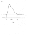
- An example of one generated by the Doppler signal unit 32 and on one to the Doppler signal unit 32 connected display device 30 shown Doppler signal is plotted in Fig. 3 below.
- FIG. 3 corresponds to a part a possible display of the screen of the display device 30 of the Lithotripters 10 according to the invention Usual ultrasound image of a body area of the patient 12, which Concrement 18 contains approximately in the middle.
- Fig. 3 below that is after a shock wave measured Doppler signal in the form of the measured Frequency shifts (note the unit kHz on the ordinate of the shown coordinate system) as a function of time.
- a shock wave measured Doppler signal in the form of the measured Frequency shifts (note the unit kHz on the ordinate of the shown coordinate system) as a function of time.
- the Doppler signal in the Doppler signal unit 32 cached and the amount formed from all stored values in order not having to work with positive and negative frequency shifts. Then a curve can be attached to the corresponding curve of the absolute values, which oscillates strongly Envelope by the vertices of the continuously shrinking Half waves are created.
- a schematic example of the so determined Envelope is shown in Fig. 4.
- this envelope can be known way, for example a falling e-function of the form exp (-t / T) adjust (begin), where t is the time from the emission of the shock wave or from the Reception of the first back-reflected sound wave and where T is the is the desired period of the Doppler signal.
- a signal start time T 1 and a signal end time T 2 can be determined in the amount-based Doppler signal curve already mentioned above, where T 1 corresponds to the time at which the described
- T 2 corresponds to the time at which the envelope falls below a second threshold value for the first time.
- the Doppler signal unit 32 In addition to the already mentioned output of the Doppler signal to the Display device 30 for the purpose of pictorial representation corresponding to FIG. 3 below 4 is the Doppler signal unit 32, as shown in FIG. 2, also with a Alarm device 34 connected to which they have an associated time duration signal outputs. As also below. will be explained, the Alarm device 34 trigger an acoustic and / or visual alarm if the time duration signal results in a too short duration of the Doppler signal, which suggests a miss, i.e. that the concretion 18th is outside the shock wave focus.
- the Doppler signal unit 32 is also with the Shock wave generator 16 connected to this in the event of misses to be able to stop automatically if necessary. In this way, one unnecessary stress on the patient's body 12 due to shock wave misses avoided particularly safely and effectively without any intervention of medical personnel.
- Essential steps of using the lithotripter 10 according to the invention feasible method according to the invention are described below with reference to 5 to 8 are explained:
- the method according to the invention comprises after the start of the lithotripter according to the invention essentially three Sections, namely a first section S10 with steps for presetting the Lithotripters before the actual therapy, a subsequent section S20, in the relevant during therapy in various procedural steps Duration of a Doppler signal is determined, and a further section S30, in the several process steps, the corresponding measures as a function the relevant signal duration determined in section S20.
- step S11 the above-mentioned and in the state of the Technique known adjustment of the patient with the help of the adjustable couch Lithotripters 10.
- the patient 12 is positioned in such a way that his Concrement 18 is located in the shock wave focus.
- step S12 the ultrasonic transducer 24 and the first emission of a shock wave Using the shock wave generator 16.
- step S13 the Doppler signal unit 2 is generated using the reflected ultrasonic waves as well as the known frequency of the transducer 24 transmitted ultrasound waves a Doppler signal, which is then in Step S14 is shown on the display device 30 corresponding to FIG. 3.
- the envelope can correspond to the Doppler signal 4 are shown on the display device 30.
- This Doppler signal is used by the medical personnel in step S15 assessed whether it indicates a hit or a hit Fire shot.
- This first emission of a shock wave results in step S12 usually a hit, since after step S11 the concrement 18 in any case lies in the shock wave focus, and since you send out a "test shock wave” can ask the patient 12 to hold their breath for a short time so that there is no risk of a miss due to the breathing movement of the calculus.
- this assessment of Doppler signals as to whether a hit or a If there is a miss, it is important, among other things, that the respective signal noise, i.e. to determine the respective "background" for each Doppler signal.
- step S15 If it is determined in step S15 that there is no hit, the process is returned to step S11 back. If, on the other hand, a hit is found, this becomes in step S16 Doppler signal already measured as a reference Doppler signal in the memory 32a stored. With the help of this reference Doppler signal are then in step S17 defines the threshold values discussed above, in particular by Consideration of the signal noise in the background of the reference Doppler signal. Furthermore, in step S17, the time period of the reference Doppler signal is used as the reference time period stored in the memory 32a.
- step S18 An initialization then takes place in step S18, i.e. a reset of a Counting parameter z, the meaning of which is clear from FIG. 7 below will be.
- FIG. 7 shows the essential steps within the second section S20 according to Fig. 5, which are used to determine the relevant signal duration.
- step S21 the counting parameter z is incremented, i.e. increased by 1.
- step S22 a shock wave from the shock wave generator 16 is transmitted and in step S23 a Doppler signal from the Doppler signal unit 32 generated, which is then generated in step S24 on the display device 30 is displayed, as is its envelope according to FIG. 4.
- step S25 with the aid of the step S17 according to FIG. 6 defined threshold values the duration of this current Doppler signal certainly.
- this time period is also divided by the 6 into a normalized reference time period determined in step S17 Converted period, which subsequently in step S27 in the Doppler signal unit 32 is stored, for example in the memory 32a or in a separate memory for recording measured values.
- step S29a the last calculated mean of a continuous display added, which are those measured throughout the course of treatment Time duration of the Doppler signals continuously represents the development of the Track Doppler signal length.
- This can expediently be continuous Display can be shown on the display device 30, but it can also act as a separate screen etc.
- An example of such Continuous display is shown in Fig. 9 and will be explained further below.
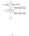
- FIG. 8 shows the essential steps of the third section S30 according to FIGS. 5 and the measures in the context of the inventive method at a Lithotripsy treatment with the lithotripter 10 according to the invention as a function of Average calculated in step S29 can be taken:
- step S31 what is executed in the Doppler signal unit 32
- the program checks whether the mean value calculated in step S29 is greater than one predetermined limit. As explained above, large duration values indicate Hits, whereas small duration values are typical for misses. Accordingly, a positive result of the check in step S31 means that obviously the concrement 18 is still in the shock wave focus and thus Hits. Accordingly, this is done in the Doppler signal unit 32 executed program in this case via a step S32, in which the Counting parameter z is again reset to 0, back to step S21, i.e. it a measurement of five standardized Doppler signal time periods begins again subsequent averaging according to FIG. 7.
- step S31 if the check in step S31 leads to a negative result, i.e. to, that the averaged duration of the last five Doppler signals is too short, which is due to indicates possible misses, the program branches to a step S33, in which said alarm device 34 is triggered, and then to a step S34 in which the shock wave generator 16 is stopped by one unnecessary strain on the patient's body 12 due to misses avoid.
- 9 is a schematic illustration of an exemplary progress control display on display 30 during shock wave therapy. you 9 recognizes a coordinate system, the abscissa of which is the number emitted Shock waves and their ordinate following the respective shock wave determines the determined absolute or standardized double signal duration. You can see from Start the shock wave therapy until shortly before its end a slow increase in Doppler signal times. Individual "drops” in this curve indicate "misses” due to movement of the patient 12 or the concretions 18 within the patient 12, which are immediately replaced by a Repositioning of patient 12 was corrected. 9 represents one horizontal dashed line the amount of the threshold whose Fall below the alarm device 34 triggers. One can see that the local "Slump" in the middle of the curve to fall below this Threshold and thus led to an alarm.
- step S17 determination of the reference time period (step S17) and the later normalization of measured signal durations with the aid of this reference duration (Step S26) can also be omitted, so that the progress check absolute values of the measured Doppler signal duration during the shock wave therapy the display device 30 are displayed and evaluated.
- lithotripter 10 which is only schematically indicated with reference to FIG. 1 it is understood that this can have numerous other components that are known from the prior art, for example a X-ray locating device or an imaging ultrasound scanner.
Abstract
Description
Die vorliegende Erfindung betrifft einen Lithotripter zur Fragmentierung eines Zielobjekts, insbesondere eines Konkrements, in einem vorzugsweise menschlichen Körper, umfassend einen Stoßwellengenerator zur Erzeugung fokussierter Stoßwellen, eine UltraschallSende/Empfangseinheit mit einem Ultraschall-Transducer zum Aussenden von Ultraschallwellen in den Körper und zum Empfangen von in einem Zielgebiet des Stoßwellengenerators reflektierten Ultraschallwellen, und eine an die Ultraschall-Sende/Empfangseinheit angeschlossene Dopplersignaleinheit zum Generieren und Auswerten eines Dopplersignals aus den ausgesandten und empfangenen Ultraschallwellen.The present invention relates to a lithotripter for fragmentation of a Target object, in particular a concrement, preferably in one human body comprising a shock wave generator for generation focused shock waves, an ultrasound transmitter / receiver unit with a Ultrasound transducer for sending ultrasound waves into the body and for receiving reflected in a target area of the shock wave generator Ultrasonic waves, and one to the ultrasonic transmitter / receiver unit connected Doppler signal unit for generating and evaluating a Doppler signal from the emitted and received ultrasonic waves.
Lithotripter sind heutzutage als medizinische Geräte zur Zertrümmerung von Konkrementen, beispielsweise Nierensteinen, im Körper eines Patienten mit Hilfe fokussierter Stoßwellen weitverbreitet. Derartige Lithotripter werden von der Anmelderin gewerblich angeboten, beispielsweise unter der Bezeichnung "Dornier Lithotripter S" oder "Dornier Compact Delta". Bei all diesen Geräten muß das Konkrement vor Beginn der Behandlung geortet werden, damit der Patient mit Hilfe einer verschiebbaren Liege so positioniert werden kann, daß sich beispielsweise sein Nierenstein im Fokus der Stoßwellen befindet, die mit Hilfe des Stoßwellengenerators des Lithotripters erzeugt werden. Diese anfängliche "Justage", d.h. Positionierung des Patienten erfolgt üblicherweise mit einer bildgebenden Ortungsvorrichtung, etwa einem bildgebenden Ultraschall-Scanner oder einer Röntgenortungsvorrichtung. Diese dient zusätzlich zur anfänglichen erstmaligen Ortung des Konkrements vor Beginn der ESWL-Behandlung (extrakorporale Stoßwellenlithotripsie) oder der ESWT-Behandlung (extrakorporale Stoßwellentherapie) auch dazu, während des Verlaufs der Behandlung die Position des Konkrements kontinuierlich zu überwachen, um sicherzustellen, daß es nicht im Körper des Patienten verrutscht bzw. an einen anderen Ort gewandert ist oder daß sich der Patient nicht auf seiner Liege bewegt hat, so daß sich das Konkrement möglicherweise nicht mehr im Fokus der Stoßwellen befindet. Für einen umfassenden Überblick über technische und medizinische Aspekte der ESWT und der bei Lithotriptern eingesetzten Geräte wird auf das Buch "ESWT and Ultrasound Imaging of the Musculosceletal System", Steinkopff-Verlag, Darmstadt, 2001, ISBN 3-7985-1252-3 verwiesen.Lithotripters are used today as medical devices for smashing Concrements, such as kidney stones, in the body of a patient with the help focused shockwaves widespread. Such lithotripters are from the Applicant offered commercially, for example under the name "Dornier Lithotripter S "or" Dornier Compact Delta ". With all these devices this has to be done Concretions can be located before the start of treatment so that the patient can help a sliding couch can be positioned so that, for example his kidney stone is in the focus of the shock waves, which with the help of the Shock wave generator of the lithotripter are generated. This initial "Adjustment", i.e. The patient is usually positioned with a imaging locating device, such as an ultrasound imaging scanner or an X-ray location device. This serves in addition to the initial one localization of the concrement for the first time before the start of ESWL treatment (extracorporeal shock wave lithotripsy) or ESWT treatment (extracorporeal Shock wave therapy) also helps position during the course of treatment continuously monitor the concretions to ensure that it is not slipped in the patient's body or moved to another location or that the patient has not moved on his couch, so that the concrement may no longer be in focus of the shock waves. For one comprehensive overview of technical and medical aspects of ESWT and of the devices used in lithotripters is based on the book "ESWT and Ultrasound Imaging of the Musculosceletal System ", Steinkopff-Verlag, Darmstadt, 2001, ISBN 3-7985-1252-3.
Üblicherweise wird die genannte bildgebende Ortungsvorrichtung auch dazu verwendet, während des Verlaufs der Behandlung den Erfolg der Lithotripsie zu überwachen, d.h. die Fragmentierung des Zielobjekts. Da eine Behandlung typischerweise ca. 30 Minuten dauert, kann wegen der zu hohen Strahlenbelastung nicht kontinuierlich geröntgt werden, sondern allenfalls in Intervallen von 3 bis 5 Minuten. Verlagert sich in der Zwischenzeit das Zielobjekt zum Beispiel durch eine Bewegung des Patienten, so wird bis zur nächsten Kontrollaufnahme der Körper durch Stoßwellen belastet, ohne daß das Zielobjekt weiter fragmentiert wird, da es sich nicht mehr im Stoßwellenfokus befindet.Usually, the imaging locating device mentioned is also used for this purpose used the success of lithotripsy during the course of treatment monitor, i.e. the fragmentation of the target. Because a treatment typically takes about 30 minutes, due to the high radiation exposure are not x-rayed continuously, but at intervals of 3 to 5 at most Minutes. In the meantime, the target object is shifted, for example, by a Movement of the patient, so the body is until the next control recording impacted by shock waves without the target being further fragmented because it is no longer in shock wave focus.
Bei Lithotriptern, bei denen als bildgebende Ortungsvorrichtung ein Ultraschallscanner eingesetzt wird, kann dieser zwar kontinuierlich zur Visualisierung des Zielobjekts eingesetzt werden, allerdings ist die Ortung u.a.wegen der Bildqualität oft wesentlich schwieriger als bei Röntgenaufnahmen, so daß selbst erfahrenes medizinisches Personal oft Schwierigkeiten hat, das Zielobjekt zu erkennen oder gar seinen Fragmentierungsgrad einzuschätzen.In lithotripters, where an imaging locating device Ultrasound scanner is used, this can be used continuously Visualization of the target object can be used, however, the location Because of the image quality, among other things, it is often much more difficult than with X-rays that even experienced medical personnel often have difficulty Recognize the target object or even assess its degree of fragmentation.
Darüberhinaus hat die Verwendung derartiger bildgebender Ortungsvorrichtungen nicht nur zur "Anfangsjustage" des Patienten, sondern auch zur "Trefferkontrolle" im Verlauf der Behandlung folgenden grundsätzlichen Nachteil: Zwar läßt sich hierdurch die Position des Zielobjekts relativ zum Stoßwellenfokus kontrollieren, d.h. eine rein geometrische Größe; es läßt sich jedoch nicht die Wirkung der Stoßwellen auf das Zielobjekt selbst feststellen. Grundsätzliche Probleme wie eine mangelnde Ankopplung des Stoßwellengeräts an den Körper des Patienten, eine Abschattung von Stoßwellen, beispielsweise durch Rippen etc., werden daher unter Umständen erst spät erkannt, wenn nämlich im Therapieverlauf keinerlei Effekte am Zielobjekt sichtbar werden. In addition, the use of such imaging location devices not only for "initial adjustment" of the patient, but also for "hit control" The following fundamental disadvantage in the course of the treatment: thereby control the position of the target object relative to the shock wave focus, i.e. a purely geometric size; however, the effect of Detect shock waves on the target object yourself. Basic problems like one lack of coupling of the shock wave device to the patient's body, a Shading of shock waves, for example by ribs etc., are therefore under Perhaps only recognized late, if no effects in the course of therapy become visible on the target object.
Daher wurden im Stand der Technik bereits Ultraschall-Dopplerverfahren zur
kontinuierlichen Trefferkontrolle vorgeschlagen. Es wurde nämlich angenommen,
daß das Konkrement im menschlichen Körper bei einem Treffer aufgrund des
Impulsübertrags von der Stoßwelle eine makroskopische Bewegung ausführt.
Bestrahlt man das Zielobjekt mit Ultraschallwellen und mißt die an ihm reflektierten
Ultraschallwellen, so äußert sich diese makroskopische Bewegung in einer
Dopplerverschiebung der Frequenz der reflektierten Wellen. Dementsprechend
ausgestattete Lithotripter gemäß dem Oberbegriff des Anspruchs 1 sind
beispielsweise aus der EP 0 367 116 B1, der EP 0 548 048 B1 und der DE 44 46
192 A1 bekannt. Diesen Geräten ist gemeinsam, daß der Ultraschall-Transducer
Ultraschallwellen in den Körper aussendet, die vom Körper zum Ultraschall-Transducer
zurückreflektierten Ultraschallwellen erfaßt, wobei eine
Dopplersignaleinheit aus den ausgesandten und empfangenen Ultraschallwellen
ein Dopplersignal generiert und auswertet, wobei im wesentlichen der Betrag einer
Frequenzverschiebung des reflektierten Signals gegenüber den ausgesandten
Wellen berechnet wird und hieraus auf die Treffgenauigkeit geschlossen wird.Therefore, ultrasound Doppler methods have already been used in the prior art
Continuous hit control suggested. Because it was assumed
that the concretion in the human body when hit due to the
Impulse transfer from the shock wave performs a macroscopic movement.
If the target object is irradiated with ultrasonic waves and the reflected on it is measured
Ultrasound waves, this macroscopic movement is expressed in one
Doppler shift in the frequency of the reflected waves. Accordingly
equipped lithotripters according to the preamble of claim 1
for example from
Diese Vorgehensweise weist verschiedene Nachteile auf:This procedure has several disadvantages:
Es ist im Stand der Technik bereits bekannt, daß das Dopplersignal gerade in der
Nähe seines zeitlichen Nullpunkts, d.h. unmittelbar nach dem Aussenden der
Stoßwelle, Artefakte enthält. Um zu verhindern, daß genau solche Artefakte
gemessen werden, weisen beispielsweise die Lithotripter gemäß der EP 0 367 116
B1 und der EP 0 548 048 B1 Mittel zur zeitlichen Synchronisierung zwischen dem
Stoßwellengenerator und der Dopplersignaleinheit auf, was diese Geräte aufwendig
und teuer macht.It is already known in the prior art that the Doppler signal is precisely in the
Proximity of its temporal zero point, i.e. immediately after sending out the
Shock wave containing artifacts. To prevent such artifacts
are measured, for example, the lithotripters according to
Darüber hinaus kann es in der Umgebung von starken Streuern wie einem Konkrement im Dopplerspektrum zu dem sogenannten Spiegelartefakt kommen. Durch Zweifachreflektion wird die Bewegung eines Streuers zusätzlich in entgegengesetzer Richtung registriert. Dies führt zu einem zusätzlichen Betrag, der dem an der Nullinie gespiegelten Nutzsignal entspricht. Wegen der Kurzlebigkeit . des Prozesses und der starken Artefakte können den Spektren nicht einfach Geschwindigkeits-Zeit-Verläufe und somit eine Trefferinformation zugeordnet werden.In addition, it can be in the vicinity of strong spreaders like one Concrement in the Doppler spectrum come to the so-called mirror artifact. By double reflection the movement of a spreader is additionally in opposite direction registered. This leads to an additional amount that corresponds to the useful signal reflected at the zero line. Because of the short life. of the process and the strong artifacts cannot easily make the spectra Speed-time profiles and thus associated hit information become.
Es ist daher Aufgabe der vorliegenden Erfindung, einen Lithotripter der eingangs genannten Art bereitzustellen, der aus dem Dopplersignal eine Information im Hinblick auf die Treffer- und Desintegrationskontrolle ermittelt und zur Darstellung bringt und keine aufwendigen Mittel zur Synchronisierung des Stoßwellengenerators mit der Dopplersignaleinheit erfordert.It is therefore an object of the present invention to provide a lithotripter to provide the type mentioned, information from the Doppler signal in Determined with regard to hit and disintegration control and for display brings and no expensive means to synchronize the Shock wave generator with the Doppler signal unit required.
Diese Aufgabe wird bei einem erfindüngsgemäßen Lithotripter dadurch gelöst, daß die Dopplersignaleinheit dazu ausgelegt ist, die Zeitdauer des Dopplersignals zu bestimmen und ein zugeordnetes Zeitdauersignal anzugeben.This object is achieved in a lithotripter according to the invention in that the Doppler signal unit is designed to increase the duration of the Doppler signal determine and specify an associated time duration signal.
Die Zeitdauer des Dopplersignals ist weitgehend unabhängig von den oben genannten Artefakten und ein charakteristisches Maß für einen Treffer bzw. Fehlschuß. Darüber hinaus läßt sich durch die Anzeige dieses Parameters über die Behandlung auch die Desintegrationswirkung bestimmen.The duration of the Doppler signal is largely independent of the above mentioned artifacts and a characteristic measure for a hit or Fire shot. In addition, this parameter can be displayed via the Treatment also determine the disintegration effect.
Die erfindungsgemäße Ausgestaltung der Dopplersignaleinheit basiert auf neuen in-vitro- und klinischen Experimenten, bei denen jeweils der gesamte zeitliche Verlauf des Dopplersignals, d.h. die im reflektierten Signal auftretende Frequenzverschiebung gegenüber dem ausgesandten Signal als Funktion der Zeit untersucht wurde. Bei den in-vitro-Experimenten wurde die Frage, ob und mit welcher Genauigkeit eine Stoßwelle das Zielobjekt getroffen hat, mit Hochgeschwindigkeitsfotografien überwacht. Als wichtigstes Ergebnis wurde übereinstimmend festgestellt, daß die Zeitdauer des Dopplersignals ein charakteristisches Maß für einen "Treffer" darstellt. Anders ausgedrückt kann dem zugeordneten Zeitdauersignal, das von der erfindungsgemäßen Dopplersignaleinheit ausgegeben wird, entnommen werden, ob und in welchem Maße eine Stoßwelle das Zielobjekt getroffen hat. Grundsätzlich gilt hierbei, daß die Zeitdauer des Dopplersignals bei stärkerer Wirkung der Stoßwelle auf das Zielobjekt zunimmt. Befindet sich das Zielobjekt hingegen außerhalb des Stoßwellenfokus, so ist die Zeitdauer des Dopplersignals signifikant kürzer als bei einem Treffer. Zerfällt das Zielobjekt, beispielsweise ein Konkrement, im Laufe der Behandlung in eine zunehmende Zahl von kleineren Fragmenten, so nimmt die Zeitdauer des Dopplersignals zu. Durch die Zunahme der Signallänge im Verlauf der Behandlung kann die Desintegrationswirkung der Schockwellentherapie abgelesen werden.The configuration of the Doppler signal unit according to the invention is based on new ones in vitro and clinical experiments, each with the entire temporal Course of the Doppler signal, i.e. the one occurring in the reflected signal Frequency shift compared to the transmitted signal as a function of time was investigated. In the in vitro experiments, the question of whether and with what accuracy a shock wave has hit the target object with High speed photographs monitored. The main result was found that the duration of the Doppler signal was a represents a characteristic measure of a "hit". In other words, it can assigned time signal, which of the invention Doppler signal unit is output, can be taken whether and in which Measures a shock wave has hit the target. The basic principle here is that the duration of the Doppler signal when the shock wave has a stronger effect on the Target object increases. However, the target object is outside the Shock wave focus, the duration of the Doppler signal is significantly shorter than in one hit. If the target object, for example a concrement, decays in the course of Treatment in an increasing number of smaller fragments, so the Duration of the Doppler signal. Due to the increase in signal length over the course Treatment can disintegrate the shock wave therapy be read.
An dieser Stelle ist auf die zugrundeliegenden Wechselwirkungsmechanismen zwischen der Stoßwelle und dem Zielobjekt einzugehen: Im Stand der Technik wurde bislang davon ausgegangen, daß es nur einen einzigen Wechselwirkungsmechanismus gibt, nämliche eine makroskopische Bewegung des Konkrements, wenn es erfolgreich von der Stoßwelle getroffen wird. Die genannten neueren Untersuchungen zeigen jedoch, daß dieser Mechanismus nur einer von mehreren Mechanismen ist: Man beobachtet zwar tatsächlich solche makroskopische Bewegungen des Konkrements, insbesondere dann, wenn es bereits teilweise oder sogar größtenteils fragmentiert ist. Gerade am Anfang der Behandlung, wenn das Konkrement noch weitgehend unfragmentiert ist, dominieren jedoch andere Effekte, nämlich insbesondere das Herausschießen von Fragmenten aus dem Konkrement sowie das Auftreten von Kavitationsblasen um das Konkrement herum bei einem erfolgreichen Treffer. All diese Mechanismen führen zur Dopplerverschiebung bei den. Frequenzen der reflektierten Schallwellen und somit zum beobachteten Dopplersignal und führen dazu, daß dessen Zeitdauer um so größer ist, je stärker die Wechselwirkung zwischen Stoßwelle und Stein ist und je stärker das Konkrement bereits fragmentiert ist.At this point it is on the underlying interaction mechanisms enter between the shock wave and the target object: In the prior art So far it has been assumed that there is only one Interaction mechanism there, namely a macroscopic movement of the Concretions if it is successfully hit by the shock wave. The above however, recent studies show that this mechanism is only one of There are several mechanisms: You actually observe them macroscopic movements of the calculus, especially when it is is already partially or even largely fragmented. Especially at the beginning of the Treatment if the concrement is still largely unfragmented, however, other effects dominate, namely shooting out Fragments from the calculus as well as the appearance of cavitation bubbles around the concrement around on a successful hit. All of these mechanisms lead to Doppler shift in the. Frequencies of the reflected sound waves and thus to the observed Doppler signal and cause its duration The stronger the interaction between the shock wave and the stone, the greater and the more the concrement is already fragmented.
Hinsichtlich der Weiterverarbeitung der im Zeitdauersignal enthaltenen Trefferinformation sind zahlreiche Vorgehensweisen möglich:With regard to the further processing of those contained in the time duration signal Hit information can be done in a number of ways:
In einer bevorzugten Weiterbildung des erfindungsgemäßen Lithotripters ist vorgesehen, daß er ferner eine mit der Dopplersignaleinheit verbundene Anzeigevorrichtung zur Anzeige von Dopplersignalen und/oder eine Alarmvorrichtung aufweist, der das Zeitdauersignal zugeführt wird. Die momentanen Dopplersignale werden, wie oben bereits erwähnt, in einem Koordinatensystem dargestellt, dessen Abszisse die Zeit und deren Koordinate die per Fourier-Transformation aus den reflektierten Ultraschallwellenamplituden berechnete Frequenzverschiebung angibt. Insbesondere kann im Verlauf der gesamten Behandlung die gemessene Zeitdauer der Dopplersignale in Form einer fortlaufenden Anzeige dargestellt werden, um die Entwicklung der Dopplersignallänge zu verfolgen. Mit erfolgreicher fortschreitender Zertrümmerung des Zielobjekts sollte die Dopplersignallänge kontinuierlich zunehmen. Nimmt die Signallänge jedoch ab, so ist dies ein starkes Indiz dafür, daß sich das Konkrement aus dem Stoßwellenfokus herausbewegt hat, sei es durch eine Konkrementbewegung (beispielsweise die Bewegung eines Nierensteins innerhalb der Niere des Patienten) oder durch eine Verschiebung des Patienten selbst. Als Anzeigevorrichtung kann hierbei gegebenenfalls der Bildschirm verwendet werden, auf dem auch die von der bildgebenden Ortungsvorrichtung gelieferten Bilder dargestellt werden. Die erfindungsgemäß vorgeschlagene Alarmvorrichtung kann akustisch und/oder optisch arbeiten und beispielsweise einen Warnton aussenden oder eine Warnleuchte einschalten, sobald sich aus dem zugeführten Zeitdauersignal ergibt, daß die Zeitdauer des Dopplersignals unter einen vorbestimmten Wert gefallen ist, was auf einen "Fehlschuß" hinweist. Zusätzlich oder alternativ dazu kann der Alarm dann ausgelöst werden, wenn das Zeitdauersignal einen negativen Trend aufweist. Bei einer Desintegration ist nämlich von einer steigenden Signallänge auszugehen.In a preferred development of the lithotripter according to the invention provided that it also connected to the Doppler signal unit Display device for displaying Doppler signals and / or a Has alarm device to which the time duration signal is supplied. The Current Doppler signals are, as already mentioned above, in one Coordinate system shown, the abscissa the time and their coordinate the by Fourier transformation from the reflected ultrasonic wave amplitudes calculated frequency shift indicates. In particular, in the course of total treatment the measured duration of the Doppler signals in the form of a continuous display to show the development of the Track Doppler signal length. With successful progressive smashing of the target object, the Doppler signal length should increase continuously. Take that Signal length, however, this is a strong indication that the concrement has moved out of the shock wave focus, be it through a Concremental movement (for example the movement of a kidney stone within the patient 's kidney) or by moving the patient himself. As If necessary, the display device can use the screen, on which also the images supplied by the imaging locating device being represented. The alarm device proposed according to the invention can work acoustically and / or optically and send out a warning tone, for example or switch on a warning light as soon as it comes out of the supplied Time duration signal shows that the duration of the Doppler signal is less than one predetermined value has dropped, indicating a "miss". additionally or alternatively, the alarm can be triggered when the Time signal has a negative trend. When disintegration is namely to assume an increasing signal length.
Altemativ oder zusätzlich kann vorgesehen sein, daß der Stoßwellengenerator mit der Dopplersignaleinheit verbunden und dazu ausgelegt ist, die Erzeugung von Stoßwellen als Funktion des Zeitdauersignals zu stoppen bzw. fortzusetzen. Hierdurch läßt sich eine automatische Abschaltung des Stoßwellengenerators erreichen, wenn offenbar das Zielobjekt nicht mehr im Stoßwellenfokus liegt. Somit wird unabhängig von der Aufmerksamkeit oder der Reaktionszeit des den Lithotripter bedienenden medizinischen Personals eine unnötige Belastung des Körpers des Patienten durch Stoßwellen-Fehlschüsse vermieden.Alternatively or additionally, it can be provided that the shock wave generator with the Doppler signal unit is connected and is designed to generate To stop or continue shock waves as a function of the time duration signal. This allows the shock wave generator to be switched off automatically reach when the target object is apparently no longer in shock wave focus. Consequently is regardless of the attention or response time of the Medical personnel operating lithotripters place an unnecessary burden on the Body of the patient avoided by shock wave shots.
Zweckmäßigerweise ist der Ultraschall-Transducer der Ultraschall-Sende/Empfangseinheit an einem einstellbaren Halter montiert. Der Ultraschall-Transducer kann dann unabhängig von anderen Teilen des Lithotripters zur Optimierung des Dopplersignals justiert und in der optimalen Position festgestellt werden. Insbesondere kann hierdurch gewährleistet werden, daß der Ultraschall-Transducer auf den Stoßwellenfokus gerichtet ist. Alternativ zu dieser sogenannten isozentrischen Scannerführung kann auch ein sogenannter lnline-Transducer eingesetzt werden, d.h. ein Ultraschall-Transducer, der in die Stoßwellenquelle integriert ist.The ultrasound transducer of the ultrasound transmitter / receiver unit is expediently mounted on an adjustable holder. The ultrasound transducer can then be used independently of other parts of the lithotripter Optimization of the Doppler signal adjusted and determined in the optimal position become. In particular, this can ensure that the ultrasound transducer is directed to the shock wave focus. As an alternative to this so-called Isocentric scanner guidance can also be a so-called in-line transducer be used, i.e. an ultrasonic transducer that is in the shock wave source is integrated.
Grundsätzlich ist hierbei folgendes zu beachten: Durch den einstellbaren Halter wird lediglich eine Linie festgelegt, entlang der sich beispielsweise bei einem PW-Verfahren (pulse wave) ein ausgesandter Ultraschallpuls im Gewebe ausbreitet und Echos generiert werden. Bei der isozentrischen Bauweise ist dann gewährleistet, daß sich der Stoßwellenfokus, d.h. das Zielgebiet, auf dieser Linie befindet. Durch bekannte Mittel, beispielsweise einen Weggeber, läßt sich der Abstand des Transducers zum Fokus bestimmen. Aufgrund der bekannten Laufzeit von Ultraschallpulsen im Gewebe läßt sich aus diesem Abstand wiederum ein Zeitfenster definieren, das denjenigen Anteil des Echos herausschneidet, der im Zielgebiet generiert wurde. Alternativ zu diesem Puls-Echo-Verfahren kann ein Dopplersignal auch durch einen CW-Transducer (continuous wave) generiert werden, bei dem ein Sendeelement kontinuierlich Ultraschallwellen aussendet und ein Empfangselement kontinuierlich das Echo empfängt. Bei dieser einfacheren Ausführung ist jedoch keine Tiefenselektion möglich.Basically, the following must be observed: With the adjustable holder only a line is defined along which, for example, in a PW method (pulse wave) a transmitted ultrasound pulse spreads in the tissue and Echoes are generated. The isocentric design ensures that that the shock wave focus, i.e. the target area, located on this line. By known means, such as a displacement sensor, the distance of the Determine transducers to focus. Due to the known duration of Ultrasonic pulses in the tissue can in turn be taken from this distance Define a time window that cuts out that part of the echo that is in the Target area was generated. As an alternative to this pulse-echo method, a Doppler signal also generated by a CW transducer (continuous wave) in which a transmitting element continuously emits ultrasonic waves and a receiving element continuously receives the echo. With this simpler However, depth selection is not possible.
Die Emittlung der wesentlichen Information des Dopllersignals, nämlich ihrer Zeitdauer, in der Dopplersignaleinheit kann auf verscheidene Weisen erfolgen: Beispielsweise kann die Dopplersignaleinheit dazu ausgelegt sein, als Zeitdauer des Dopplersignals ein Zeitintervall zu bestimmen, innerhalb dessen der Betrag des Dopplersignals auf einen vorbestimmten Bruchteil eines Anfangsbetrags gefallen ist, insbesondere auf den e-ten Teil des Anfangsbetrags. Eine derartige Auslegung der Dopplersignaleinheit ist dann vorteilhaft, wenn der Betrag eines Dopplersignals jeweils weitestgehend einer exponentiell abfallenden Einhüllenden-Kurve entspricht, da dann die Zeitdauer des Dopplersignals durch automatisiertes Anpassen von Test-Exponentialfunktionen (Fitten) schnell und zuverlässig ermittelt werden kann.The essential information of the Doppler signal, namely its duration, can be determined in the Doppler signal unit in various ways: for example, the Doppler signal unit can be designed to determine a time interval as the duration of the Doppler signal, within which the amount of the Doppler signal dropped to a predetermined fraction of an initial amount is, especially on the e-th part of the starting amount. Such an interpretation the Doppler signal unit is advantageous if the amount of a Doppler signal each largely an exponentially falling envelope curve corresponds, since then the duration of the Doppler signal by automated Adaptation of test exponential functions (fits) determined quickly and reliably can be.
Wenn hingegen der zeitliche Verlauf des Betrags eines Dopplersignals von einem exponentiellen Abfall der Einhüllenden-Kurve abweicht, so empfiehlt es sich in einer besonders vielseitigen Ausführungsform des erfindungsgemäßen Lithotripters, daß die Dopplersignaleinheit dazu ausgelegt ist, als Zeitdauer des Dopplersignals ein Zeitintervall zwischen einem Signal-Anfangszeitpunkt und einem Signal-Endzeitpunkt zu bestimmen, wobei der Signal-Anfangszeitpunkt festgestellt wird, wenn der Betrag des Dopplersignals einen ersten Schwellenwert überschreitet, und der Signal-Endzeitpunkt festgestellt wird, wenn der Betrag des Dopplersignals einen zweiten Schwellenwert unterschreitet, wobei in einer einfachen Weiterbildung dieser Ausführungsform der erste und der zweite Schwellenwert identisch sein können.If, however, the time course of the amount of a Doppler signal from one exponential drop of the envelope curve deviates, it is recommended in one particularly versatile embodiment of the lithotripter according to the invention that the Doppler signal unit is designed to be a time duration of the Doppler signal Time interval between a signal start time and a signal end time to determine, whereby the signal start time is determined, if the amount of the Doppler signal exceeds a first threshold, and the signal end time is determined when the amount of the Doppler signal falls below a second threshold, in a simple development In this embodiment, the first and second thresholds are identical can.
Grundsätzlich können diese Schwellenwerte in der Dopplersignaleinheit des erfindungsgemäßen Lithotripters voreingestellt sein und hierbei sowohl die technischen Daten des Stoßwellengenerators (insbesondere seine Leistung) als auch typische Patientendaten berücksichtigen. Zweckmäßigerweise ist jedoch vorgesehen, daß die Dopplersignaleinheit Einstellmittel zum Einstellen des ersten und/oder des zweiten Schwellenwerts aufweist. Dann können diese Schwellenwerte zu Beginn der Behandlung abhängig vom auftretenden Rauschen des Dopplersignal-Untergrunds individuell eingestellt werden. Die Schwellenwerte können jedoch auch automatisch aus dem Signal selbst bestimmt werden. Aus den vor dem Signal-Anfangszeitpunkt akquirierten Signalen läßt sich nämlich ein Grundrauschen bestimmen. Die Schwellenwerte können dann als ein vorbestimmtes Vielfaches der Rauschamplitude definiert sein und von der erfindungsgemäßen Dopplersignaleinheit automatisch eingestellt werden.Basically, these threshold values in the Doppler signal unit of the Lithotripters according to the invention can be preset and both technical data of the shock wave generator (especially its performance) as also take typical patient data into account. However, it is expedient provided that the Doppler signal unit setting means for setting the first and / or the second threshold. Then you can Threshold values at the beginning of the treatment depending on the noise occurring of the Doppler signal background can be set individually. The thresholds can also be determined automatically from the signal itself. From the namely, signals acquired before the signal start time can be entered Determine noise floor. The thresholds can then be considered a predetermined multiple of the noise amplitude and be defined by the Doppler signal unit according to the invention can be set automatically.
In einer vorteilhaften Weiterbildung des erfindungsgemäßen Lithotripters kann die Dopplersignaleinheit femer dazu ausgelegt sein, die Zeitdauer des Dopplersignals über mehrere Stoßwellen zu mitteln. Hierfür sind die folgenden geometrischen Umstände einer typischen Lithotripsie-Behandlung zu berücksichtigen: Der Stoßwellenfokus hat typischerweise eine Ausdehnung von ca. 4 mm. Ein typisches Konkrement, beispielsweise ein Nierenstein, weist am Anfang der Behandlung Abmessungen zwischen 5 und 20 mm auf, und seine Hin- und Herverlagerung durch die reine Atmung des Patienten kann mit einer Amplitude von ca. 30 mm erfolgen. Unter diesen Bedingungen würden, da das Aussenden von Stoßwellen typischerweise ohne Berücksichtigung des jeweiligen Atmungszustands des Patienten erfolgt, zwangsläufig einige Stoßwellen das Konkrement verfehlen. Die im unmittelbaren Anschluß an derartige Stoßwellen gemessenen Dopplersignal-Zeitdauem sind - wie oben bereits erläutert - besonders kurz, so daß je nach Verwendung des zugeordneten Zeitdauersignals beispielsweise die Alarmvorrichtung betätigt oder der Stoßwellengenerator abgeschaltet würde, obwohl keinerlei Dejustage gegeben ist. Vielmehr würden bereits die nächsten Stoßwellen wieder das Konkrement treffen, da sie bei einem anderen Atmungszustand des Patienten ausgesandt werden. Bei der in dieser erfindungsgemäßen Ausführungsform vorgesehenen Mittelung der Zeitdauer des Dopplersignals über mehrere Stoßwellen kann zunächst in einer Ersteinstellung der Dopplersignaleinheit eine Mittelung über beispielsweise fünf Stoßwellen erfolgen, es empfiehlt sich jedoch zur weiteren Steigerung der Behandlungseffizienz, die Zahl der Dopplersignale, über die gemittelt wird, zu Beginn der Behandlung des Patienten abhängig von seinem typischen Atmungsverhalten, von der Größe des zu fragmentierenden Konkrements etc. individuell einzustellen. Grundsätzlich sind anstatt einer Mittelung auch andere Arten der Signalfilterung möglich, z.B. eine Median-Filterung. In an advantageous development of the lithotripter according to the invention, the Doppler signal unit can also be designed to determine the duration of the Doppler signal to be averaged over several shock waves. The following are geometrical for this Consider the circumstances of a typical lithotripsy treatment: The Shock wave focus is typically approximately 4 mm wide. A typical one Concrement, for example a kidney stone, indicates the beginning of treatment Dimensions between 5 and 20 mm on, and its back and forth movement due to the patient's pure breathing with an amplitude of approx. 30 mm respectively. Under these conditions, there would be the emission of shock waves typically without considering the respiratory condition of the Patient occurs, some shock waves inevitably miss the concrement. The Doppler signal durations measured immediately following such shock waves are - as already explained above - particularly short, so that depending on Using the assigned time signal, for example Alarm device is actuated or the shock wave generator is switched off, although there is no misalignment. The next would rather be Shock waves hit the concrement again as they hit another The patient's breathing condition. At the in this embodiment of the invention provided averaging the duration of Doppler signal over several shock waves can initially in an initial setting Doppler signal unit is averaged over five shock waves, for example, however, it is recommended to further increase treatment efficiency Number of Doppler signals that are averaged at the start of treatment of the Patients depend on their typical breathing behavior, on the size of the patient fragmenting concretions etc. individually. Are basically Instead of averaging, other types of signal filtering are also possible, e.g. a Median filtering.
Nicht nur hinsichtlich der Zahl der Dopplersignale, über die zweckmäßigerweise gemittelt werden sollte, sondern auch hinsichtlich der typischerweise zu erwartenden absoluten Dopplersignal-Zeitdauem hat sich in klinischen Experimenten eine deutliche Abhängigkeit von individuellen Patienten gezeigt. Daher kann die Dopplersignaleinheit in einer zweckmäßigen Weiterbildung des erfindungsgemäßen Lithotripters dazu ausgelegt sein, die Zeitdauer eines Dopplersignals auf die Referenz-Zeitdauer eines Referenz-Dopplersignals zu normieren. Dieses Referenz-Dopplersignal wird zweckmäßigerweise zu Beginn der Behandlung aufgenommen, wenn der Patient in der oben beschriebenen Weise derart im Lithotripter justiert worden ist, daß - wie sich anhand der bildgebenden Ortungsvorrichtung überwachen läßt - beispielsweise sein Nierenstein exakt im Stoßwellenfokus liegt. Bei der oben genannten Verlaufsanzeige, d.h. der fortlaufenden Auftragung von Signaldauern im Verlauf der Stoßwellenbehandlung, werden dann also keine absoluten Dopplersignal-Zeitdauern aufgetragen, sondern normierte Dopplersignal-Zeitdauern.Not only with regard to the number of Doppler signals, about which expediently should be averaged, but also in terms of typically The expected absolute Doppler signal duration has changed in clinical Experiments showed a clear dependence on individual patients. Therefore, in an expedient development of the Lithotripters according to the invention can be designed to the duration of a Doppler signal to the reference period of a reference Doppler signal normalize. This reference Doppler signal is advantageously at the beginning of the Treatment is added when the patient is in the manner described above has been adjusted in the lithotripter in such a way that - as can be seen from the imaging Locating device can be monitored - for example, his kidney stone exactly in the Shock wave focus is. With the history display above, i.e. the continuous application of signal durations in the course of shock wave treatment, then no absolute Doppler signal time periods are plotted, but standardized Doppler signal time periods.
In einer Ausführungsform des erfindungsgemäßen Lithotripters kann vorgesehen sein, daß die Ultraschall-Sende/Empfangseinheit als Teil eines bildgebenden Ultraschallscanners ausgebildet ist (Duplex-Scanner). In diesem Fall können insbesondere der Ultraschall-Transducer und Teile der Elektronik der Ultraschall-Sende/Empfangseinheit gleichzeitig für die Ultraschall-Bildgebung und für die Dopplersignalmessungen eingesetzt werden.In one embodiment of the lithotripter according to the invention can be provided be that the ultrasound transmitter / receiver unit as part of an imaging Ultrasound scanner is designed (duplex scanner). In this case, you can in particular the ultrasonic transducer and parts of the electronics of the ultrasonic transmitter / receiver unit both for ultrasound imaging and for Doppler signal measurements are used.
Alternativ kann der Ultraschall-Transducer als preisgünstige Stiftsonde ausgebildet sein, was ihn bei Verwendung mit dem oben genannten Halter flexibel einsetzbar macht, um beispielsweise sicherzustellen, daß er immer auf den Stoßwellenfokus gerichtet ist.Alternatively, the ultrasound transducer can be designed as an inexpensive pin probe be what it can be used flexibly when used with the holder mentioned above to make sure, for example, that he is always on the shock wave focus is directed.
Insbesondere empfiehlt sich eine derartige Gestaltung, wenn der erfindungsgemäße Lithotripter ferner eine Röntgenortungsvorrichtung aufweist. In diesem Fall erfolgt die Bildgebung zu Beginn der Positionierung des Patienten sowie bei der etwa alle drei bis fünf Minuten erfolgenden Detailkontrolle mit Hilfe dieser bildgebenden Röntgenortungsvorrichtung, während die kontinuierliche Trefferkontrolle mit Hilfe des als Stiftsonde gestalteten Ultraschall-Transducers erfolgt. Es versteht sich, daß auch in diesem Fall die Anzeige der Dopplersignale auf der auch für die Anzeige der Röntgenbilder eingesetzten Anzeigevorrichtung erfolgen kann.Such a design is particularly recommended if the Lithotripter according to the invention further comprises an X-ray locating device. In In this case, the imaging takes place at the beginning of the positioning of the patient as well as with the detailed check with help every three to five minutes this X-ray imaging device while the continuous Hit control using the ultrasound transducer designed as a pen probe he follows. It is understood that the display of the Doppler signals also in this case on the display device also used for the display of the X-ray images can be done.
Die vorliegende Erfindung betrifft ferner ein allgemeines Verfahren zur Überwachung der Fragmentierung eines Zielobjekts, insbesondere eines Konkrements, in einem vorzugsweise menschlichen Körper, umfassend die Schritte: Aussenden von Ultraschallwellen in den Körper, Empfangen von am Zielobjekt reflektierten Ultraschallwellen, und Generieren und Auswerten eines Dopplersignals aus den ausgesandten und den empfangenen Ultraschallwellen, das dadurch gekennzeichnet ist, daß der Schritt des Auswertens eine Bestimmung der Zeitdauer des Dopplersignals umfaßt, und ferner ein Schritt der Ausgabe eines zugeordneten Zeitdauersignals vorgesehen ist.The present invention further relates to a general method for Monitoring the fragmentation of a target, especially one Concretions, in a preferably human body, comprising the Steps: sending ultrasound waves into the body, receiving am Target object reflected ultrasound waves, and generating and evaluating one Doppler signal from the emitted and received ultrasound waves, which is characterized in that the evaluating step is a determination the duration of the Doppler signal, and further a step of outputting a assigned time signal is provided.
Wie oben erläutert kann dieses Verfahren bei Lithotripsie derart weiter entwickelt werden, daß es ferner die Schritte umfaßt: Anordnen des Körpers in einem Lithotripter mit einem Stoßwellengenerator zur Erzeugung fokussierter Stoßwellen, Anzeigen des Zielobjekts und des Fokus des Stoßwellengenerators auf einer Anzeigevorrichtung einer bildgebenden Ortungsvorrichtung, Justieren des Körpers derart, daß-das Zielobjekt im Fokus des Stoßwellengenerators liegt, Aussenden einer Stoßwelle in Richtung des Zielobjekts und Bestimmen der Zeitdauer des Dopplersignals, Speichern der Zeitdauer als Referenz-Zeitdauer, sowie den späteren Schritt: Normieren der Zeitdauer eines später gemessenen Dopplersignals auf die Referenz-Zeitdauer. Auf diese Weise wird eine Normierung der gemessenen Dopplersignal-Zeitdauem auf eine für diesen Patienten und dieses Konkrement typische Referenz-Zeitdauer erreicht.As explained above, this procedure can be further developed in lithotripsy that it further comprises the steps of: placing the body in one Lithotripter with a shock wave generator for generating focused shock waves, Displaying the target and focus of the shock wave generator on one Display device of an imaging locating device, adjusting the body such that the target object is in the focus of the shock wave generator, send out a shock wave towards the target object and determining the duration of the Doppler signal, storing the time period as a reference time period, and the later step: normalizing the duration of a Doppler signal measured later to the reference period. In this way, a standardization of the measured Doppler signal time duration to one for this patient and this one Typical reference time period reached.
Ein Ausführungsbeispiel der Erfindung wird im folgenden anhand der Zeichnungen erläutert werden. Es zeigen:
- Fig. 1
- eine vereinfachte Darstellung wesentlicher Bauteile des erfindungsgemäßen Lithotripters;
- Fig. 2
- eine schematische Ansicht der Dopplersignaleinheit und ihrer Anschlüsse beim erfindungsgemäßen Lithotripter;
- Fig. 3
- eine Darstellung von Ultraschall- und Dopplersignalen auf der Anzeigevorrichtung des erfindungsgemäßen Lithotripters;
- Fig.4
- ein schematisches Beispiel für eine der weiteren Signalauswertung zugrundzulegende Einhüllende an das Dopplersignal;
- Fig. 5.
- ein schematisches Flußdiagramm zur Verdeutlichung der Hauptabschnitte des erfindungsgemäßen Verfahrens;
- Fig. 6
- ein Flußdiagramm zur Verdeutlichung wesentlicher Verfahrensschritte innerhalb des ersten Hauptabschnitts gemäß Fig. 5;
- Fig. 7
- ein Flußdiagramm zur Verdeutlichung wesentlicher Verfahrensschritte innerhalb des zweiten Hauptabschnitts gemäß Fig. 5;
- Fig. 8
- ein Flußdiagramm zur Verdeutlichung wesentlicher Verfahrensschritte des dritten Hauptabschnitts gemäß Fig. 5; und
- Fig. 9
- ein schematisches Beispiel für eine Signaldauer-Verlaufskontrolle während einer Stoßwellentherapie.
- Fig. 1
- a simplified representation of essential components of the lithotripter according to the invention;
- Fig. 2
- a schematic view of the Doppler signal unit and its connections in the lithotripter according to the invention;
- Fig. 3
- a representation of ultrasound and Doppler signals on the display device of the lithotripter according to the invention;
- Figure 4
- a schematic example of an envelope to be used as the basis for further signal evaluation of the Doppler signal;
- Fig. 5.
- a schematic flow diagram to illustrate the main sections of the method according to the invention;
- Fig. 6
- a flowchart to illustrate essential method steps within the first main section of FIG. 5;
- Fig. 7
- a flowchart to illustrate essential method steps within the second main section of FIG. 5;
- Fig. 8
- a flowchart to illustrate essential method steps of the third main section of FIG. 5; and
- Fig. 9
- a schematic example of a signal duration monitoring during a shock wave therapy.
Fig. 1 zeigt eine schematische Ansicht wesentlicher mechanischer Komponenten
des erfindungsgemäßen Lithotripters 10. Ein Patient 12 ist derart auf einer in Fig. 1
nicht dargestellten verstellbaren Liege gelagert, daß ein Ankoppelkissen 14 eines
Stoßwellengenerators 16 an der gewünschten Stelle an den Körper des Patienten
12 gedrückt werden kann, um Stoßwellen in Richtung eines im Körper des
Patienten 12 zu fragmentierenden Konkrements 18 auszusenden. In dem in Fig. 1
schematisch dargestellten Fall ist dieses Konkrement 18 ein Nierenstein in der
Niere 20 des Patienten 12. Der Patient 12 wird mit Hilfe der verstellbaren Liege
derart "einjustiert", d.h. positioniert, daß der in Fig. 1 durch ein Kreuz angedeutete
Fokus der Stoßwellen, die von einer Stoßwellenquelle 22 des
Stoßwellengenerators 16 erzeugt und mit Hilfe des Ankoppelkissens 14 in den
Körper des Patienten 12 hineingesendet werden, im Nierenstein 18 liegt. Diese
Justage erfolgt üblicherweise mit Hilfe einer bildgebenden Ortungsvorrichtung,
beispielsweise einer Röntgenvorrichtung oder eines Ultraschallscanners.Fig. 1 shows a schematic view of essential mechanical components
of the
Diese Komponenten des Lithotripters 10 und diese Vorgehensweise bei der
Positionierung des Patienten vor Beginn der Behandlung sind an sich bekannt und
werden hier nicht näher erläutert werden.These components of the
Der erfindungsgemäße Lithotripter 10 umfaßt femer einen Ultraschall-Transducer
24, der an einem einstellbaren Halter 26 montiert ist. Bei dem in Fig. 1 gezeigten
Beispiel ist dieser Halter 26 in Form eines gelenkigen Arms ausgebildet und
ermöglicht eine genaue Positionierung des Ultraschall-Transducers 24 an einer
gewünschten Stelle des Körpers des Patienten 12 derart, daß der Ultraschall-Transducer
24 - wie in Fig. 1 durch gepunktete Linien angedeutet - auf den
Stoßwellenfokus gerichtet ist. Man spricht bei dieser Anordnung von einer
isozentrischen Scannerführung.The
Der Ultraschall-Transducer 24 sendet kontinuierlich (continuous wave, CW) oder in
Pulsen (pulsed wave, PW) Ultraschallwellen in Richtung des Stoßwellenfokus aus
und empfängt ferner Ultraschallwellen, die im Körper des Patienten 12,
insbesondere aus dem Gebiet des Stoßwellenfokus, reflektiert worden sind. Die
empfangenen Ultraschallsignale werden, wie in Fig. 2 angedeutet, vom Ultraschall-Transducer
24 einer Steuereinheit 28 zugeführt, die nicht nur als Teil einer Sende-/Empfangseinheit
den eigentlichen Betrieb des Ultraschall-Transducers 24 steuert,
beispielsweise die in ihm enthaltenen piezoelektrischen Elemente, und die vom
Ultraschall-Transducer 24 gemessenen reflektierten Ultraschallsignale erfaßt,
sondern diese auch an nachgeordnete Elektronikeinheiten weiterleitet.
Beispielsweise könnte die Steuereinheit 28 die empfangenen Ultraschallsignale an
ein in Fig. 2 nicht dargestelltes Bildverarbeitungsmodul weiterleiten, mit dessen
Hilfe auf einer Anzeigevorrichtung 30 Ultraschallbilder des Nierensteins dargestellt
werden können.The
Unabhängig hiervon führt die Steuereinheit 28 beim erfindungsgemäßen Lithotripter
10 die empfangenen Ultraschallsignale einer Dopplersignaleinheit 32 zu, die
basierend auf der bekannten Frequenz der vom Ultraschall-Transducer 24
ausgesandten Ultraschallwellen und den dopplerverschobenen Frequenzen der
vom Ultraschall-Transducer 24 empfangenen Ultraschallwellen nach jedem
Aussenden einer Stoßwelle durch den Stoßwellengenerator 16 zunächst ein
Dopplersignal erzeugt. Ein solches Dopplersignal umfaßt die zeitaufgelöste
Darstellung der gemessenen Frequenzverschiebungen. Ein Beispiel für ein solches
von der Dopplersignaleinheit 32 erzeugtes und auf einer an die
Dopplersignaleinheit 32 angeschlossenen Anzeigevorrichtung 30 dargestelltes
Dopplersignal ist in Fig. 3 unten aufgetragen. Hierbei entspricht Fig. 3 einem Teil
einer möglichen Anzeige des Bildschirms der Anzeigevorrichtung 30 des
erfindungsgemäßen Lithotripters 10. In Fig. 3 oben erkennt man zunächst ein
übliches Ultraschallbild eines Körperbereichs des Patienten 12, der das
Konkrement 18 ungefähr in der Mitte enthält. In Fig. 3 unten ist das im Anschluß an
eine Stoßwelle gemessene Dopplersignal in Form der gemessenen
Frequenzverschiebungen (man beachte die Einheit kHz an der Ordinate des
dargestellten Koordinatensystems) als Funktion der Zeit aufgetragen. Man erkennt
annähernd symmetrisch zu Abszisse (Zeitachse) verteilte positive und negative
Frequenzverschiebungen, deren Amplitude mit zunehmendem zeitlichen Abstand
von der Aussendung der Stoßwelle kleiner wird. Ursache für dieses Signal sind die
folgenden Prozesse beim Auftreffen der Stoßwelle auf das Konkrement 18:Regardless of this, the
Liegt das Konkrement 18 im Stoßwellenfokus, so daß beim Aussenden der
Stoßwelle ein "Treffef" erzielt wird, so werden kleine Fragmente aus dem
Konkrement 18 herausgeschlagen und gleichzeitig treten im Konkrement 18
Umschichtungen auf. In der unmittelbaren Umgebung des Konkrements 18 kommt
es ferner in der körpereigenen Flüssigkeit des Patienten 12 zu erhöhter Kavitation.
All diese Prozesse führen dazu, daß die vom Ultraschall-Transducer 24 in Richtung
des Konkrements 18 ausgesandten Ultraschallwellen zunehmend an Objekten
reflektiert werden, die gegenüber dem Transducer 24 in Bewegung sind. Da diese
Bewegung ungerichtet ist, d.h. einige der aus dem Konkrement 18
herausgeschlagenen Fragmente Bewegungskomponenten auf den Ultraschall-Transducer
zu aufweisen, andere Fragmente hingegen Bewegungskomponenten
vom Ultraschall-Transducer 24 weg, treten hinsichtlich der Frequenzen der zum
Ultraschall-Transducer 24 zurückreflektierten Schallwellen sowohl positive als auch
negative Dopplerverschiebungen auf. Durch den sogenannten Spiegelartefakt
werden zusätzliche - artifizielle - Anteile registriert, so daß annähernd gleich viele
positive wie negative Dopplerverschiebungen auftreten. Auch die bei einem Treffer
in der unmittelbaren Umgebung des Konkrements 18 verursachte erhöhte
Kavitation führt zu einer ungeregelten Bewegung von reflektierenden Objekten
(Dichteschwankungen bzw. Gasblasen in der Körperflüssigkeit des Patienten 12),
was ebenso zu positiven und negativen Dopplerverschiebungen führt.Is the concrement 18 in the shock wave focus, so that when the
Shock wave a "hit" is achieved, so small fragments from the
Auch eine makroskopische Bewegung des Konkrements 18, die insbesondere dann
auftreten kann, wenn es bereits mindestens teilweise fragmentiert ist, führt zu einer
zeitlichen Verteilung positiver und negativer Frequenzveränderungen, wenn das
Konkrement 18 aufgrund von elastischen Kräften-des umliegenden Gewebes eine
Art Schwingungsbewegung vollführt.Also a macroscopic movement of the
Wie man beim Dopplersignal in Fig. 3 deutlich erkennt, sinkt sein Betrag mit
zunehmendem zeitlichen Abstand von der Aussendung der Stoßwelle kontinuierlich
ab, da das System zunehmend wieder zur Ruhe kommt. Nach einer gewissen
charakteristischen Zeit ist keinerlei Dopplerverschiebung bei den Frequenzen der
zurückreflektierten Ultraschallwellen mehr feststellbar. Es konnte festgestellt
werden, daß diese Zeitdauer des Dopplersignals ein charakteristisches Maß für die
Stärke der Wechselwirkung der Stoßwelle mit dem Konkrement 18 ist, und somit
Informationen darüber liefern-kann, ob und in welchem Maß das Konkrement 18
getroffen wurde:As can clearly be seen in the Doppler signal in FIG. 3, its amount also decreases
increasing time interval from the emission of the shock wave continuously
as the system comes to rest increasingly. After a certain
characteristic time is no Doppler shift at the frequencies of the
back-reflected ultrasound waves more noticeable. It was found
be that this period of the Doppler signal is a characteristic measure of the
Strength of the interaction of the shock wave with the
So wurde festgestellt, daß ein Treffer zu Beginn einer Lithotripsiebehandlung bei
einem noch weitgehend unfragmentierten Konkrement 18 typischerweise
Dopplersignale mit einer Zeitdauer von etwa 50 msec. liefert. Im Verlauf der
Behandlung erhöht sich diese Zeitdauer kontinuierlich, bei einem teilweise
fragmentierten Konkrement 18 beträgt sie etwa 120 msec. (man beachte die
entsprechende Anzeige "Breite 110 ms" in Fig. 3 unten). Bei einem Fehlschuß
hingegen tritt zwar aufgrund von schwacher Kavitation in der Körperflüssigkeit des
Patienten 12 ebenfalls ein Dopplersignal auf, seine Zeitdauer beträgt jedoch
typischerweise nur etwa 25 msec.It was found that a hit at the start of lithotripsy treatment at
a still largely
Zur Auswertung des Dopplersignals zwecks Ermittlung seiner Zeitdauer sind verschiedene Vorgehensweisen möglich, die auf dem Gebiet der Datenauswertung grundsätzlich bekannt sind:To evaluate the Doppler signal to determine its duration different approaches possible in the field of data evaluation are generally known:
Zunächst wird das Dopplersignal in der Dopplersignaleinheit 32
zwischengespeichert und von allen gespeicherten Werten der Betrag gebildet, um
nicht mit positiven und negativen Frequenzverschiebungen arbeiten zu müssen.
Dann kann an die entsprechende Kurve der Betragswerte, die stark oszilliert, eine
Einhüllende durch die Scheitelpunkte der kontinuierlich kleiner werdenden
Halbwellen angelegt werden. Ein schematisches Beispiel für die derart ermittelte
Einhüllende ist in Fig. 4 dargestellt. An diese Einhüllende läßt sich in grundsätzlich
bekannter Weise beispielsweise eine fallende e-Funktion der Form exp (-t/T)
anpassen (anfitten), wobei t die Zeit ab dem Aussenden der Stoßwelle bzw. ab dem
Empfang der ersten zurückreflektierten Schallwelle darstellt und wobei T die
gesuchte charakteristische Zeitdauer des Dopplersignals ist.First, the Doppler signal in the
Alternativ können bei der oben bereits genannten betragsmäßigen Dopplersignalkurve ein Signal-Anfangszeitpunkt T1 und ein Signal-Endzeitpunkt T2 bestimmt werden, wobei T1 dem Zeitpunkt entspricht, bei dem die beschriebene Alternatively, a signal start time T 1 and a signal end time T 2 can be determined in the amount-based Doppler signal curve already mentioned above, where T 1 corresponds to the time at which the described
Einhüllende erstmals einen vorbestimmten ersten Schwellenwert überschreitet und T2 dem Zeitpunkt entspricht, bei dem die Einhüllende erstmals einen zweiten Schwellenwert unterschreitet. Die gesuchte charakteristische Zeitdauer des Dopplersignals ergibt sich dann als T = T2 - T1. In Fig. 4 sind der erste und der zweite Schwellenwert identisch und als horizontale gestrichelte Linie angedeutet.Envelope exceeds a predetermined first threshold value for the first time and T 2 corresponds to the time at which the envelope falls below a second threshold value for the first time. The characteristic time period of the Doppler signal that is sought then results as T = T 2 - T 1 . 4, the first and the second threshold value are identical and indicated as a horizontal dashed line.
Diese Schwellenwerte werden zu Beginn der Lithotripsiebehandlung beim
Aufnehmen eines Referenz-Dopplersignals ermittelt, insbesondere unter
Berücksichtigung des Untergrundrauschens bei der Messung, und in einem
Speicher 32a in der Dopplersignaleinheit 32 abgespeichert. Ebenso kann die dem
Referenz-Dopplersignal zugeordnete Referenz-Zeitdauer im Speicher 32a im
Hinblick auf eine gegebenenfalls vorzunehmende Normierung später gemessener
Kurven abgespeichert werden. Dies wird weiter unten im Zusammenhang mit den
Fig. 5 bis 8 erläutert werden.These thresholds are set at the start of lithotripsy treatment
Recording a reference Doppler signal determined, in particular under
Consideration of the background noise during the measurement, and in one
Zusätzlich zu der bereits genannten Ausgabe des Dopplersignals an die
Anzeigevorrichtung 30 zwecks bildlicher Darstellung entsprechend Fig. 3 unten
bzw. Fig. 4 ist die Dopplersignaleinheit 32, wie in Fig. 2 gezeigt, auch mit einer
Alarmvorrichtung 34 verbunden, an die sie ein zugeordnetes Zeitdauersignal
ausgibt. Wie ebenfalls weiter unten. erläutert werden wird, kann die
Alarmvorrichtung 34 einen akustischen und/oder optischen Alarm auslösen, wenn
sich aus dem Zeitdauersignal -eine zu kurze Zeitdauer des Dopplersignals ergibt,
was auf einen Fehlschuß schließen läßt, d.h. darauf, daß das Konkrement 18
außerhalb des Stoßwellenfokus liegt.In addition to the already mentioned output of the Doppler signal to the
Wie in Fig. 2 ferner dargestellt, ist die Dopplersignaleinheit 32 auch mit dem
Stoßwellengenerator 16 verbunden, um diesen im Fall von Fehlschüssen
gegebenenfalls automatisch stoppen zu können. Auf diese Weise wird eine
unnötige Belastung des Körpers des Patienten 12 durch Stoßwellen-Fehlschüsse
besonders sicher und wirksam vermieden, ohne daß es hierfür eines Eingreifens
des medizinischen Personals bedürfte.
Wesentliche Schritte des mit dem erfindungsgemäßen Lithotripter- 10
durchführbaren erfindungsgemäßen Verfahrens werden nachfolgend anhand der
Fig. 5 bis 8 erläutert werden:As further shown in Fig. 2, the
Wie in Fig. 5 schematisch angedeutet, umfaßt das erfindungsgemäße Verfahren nach dem Start des erfindungsgemäßen Lithotripters im wesentlichen drei Abschnitte, nämlich einen ersten Abschnitt S10 mit Schritten zur Voreinstellung des Lithotripters vor der eigentlichen Therapie, einen anschließenden Abschnitt S20, in dem während der Therapie in verschiedenen Verfahrensschritten die relevante Dauer eines Dopplersignals bestimmt wird, sowie einen weiteren Abschnitt S30, in dem mehreren Verfahrensschritten die entsprechenden Maßnahmen als Funktion der im Abschnitt S20 ermittelten relevanten Signaldauer ergriffen werden.As indicated schematically in FIG. 5, the method according to the invention comprises after the start of the lithotripter according to the invention essentially three Sections, namely a first section S10 with steps for presetting the Lithotripters before the actual therapy, a subsequent section S20, in the relevant during therapy in various procedural steps Duration of a Doppler signal is determined, and a further section S30, in the several process steps, the corresponding measures as a function the relevant signal duration determined in section S20.
Die wichtigsten Schritte innerhalb des ersten Abschnitts S10 werden nachfolgend anhand von Fig. 6 erläutert werden:The most important steps within the first section S10 are as follows are explained with reference to FIG. 6:
Zunächst erfolgt im Schritt S11 die oben bereits genannte und im Stand der
Technik bekannte Justage des Patienten mit Hilfe der verstellbaren Liege des
Lithotripters 10. Am Ende dieser durch eine bildgebende Ortungsvorrichtung
überwachten Justage ist der Patient 12 derart positioniert, daß sich sein
Konkrement 18 im Stoßwellenfokus befindet.First, in step S11, the above-mentioned and in the state of the
Technique known adjustment of the patient with the help of the
Anschließend beginnt im Schritt S12 das Aussenden von Ultraschallwellen durch
den Ultraschall-Transducer 24 sowie das erstmalige Aussenden einer Stoßwelle mit
Hilfe des Stoßwellengenerators 16.The transmission of ultrasound waves then begins in step S12
the
Anschließend generiert im Schritt S13 die Dopplersignaleinheit 2 anhand der
reflektierten Ultraschallwellen sowie der bekannten Frequenz der vom Transducer
24 ausgesandten Ultraschallwellen ein Dopplersignal, welches anschließend im
Schritt S14 auf der Anzeigevorrichtung 30 entsprechend der Fig. 3 gezeigt wird.
Ebenso kann in diesem Schritt die Einhüllende an das Dopplersignal entsprechend
Fig. 4 auf der Anzeigevorrichtung 30 gezeigt werden. Then, in step S13, the Doppler signal unit 2 is generated using the
reflected ultrasonic waves as well as the known frequency of the
Dieses Dopplersignal wird im Schritt S15 vom medizinischen Personal
dahingehend begutachtet, ob es auf einen Treffer schließen läßt oder auf einen
Fehlschuß. Bei diesem ersten Aussenden einer Stoßwelle im Schritt S12 ergibt sich
in der Regel ein Treffer, da nach dem Schritt S11 das Konkrement 18 auf jeden Fall
im Stoßwellenfokus liegt, und da man vor dem Aussenden dieser einen "Test-Stoßwelle"
den-Patienten 12 auffordern kann, kurzzeitig die Luft anzuhalten, so daß
kein Fehlschuß aufgrund der Atmungsbewegung des Konkrements droht. Bei
dieser Begutachtung von Dopplersignalen dahingehend, ob ein Treffer oder ein
Fehlschuß vorliegt, ist es unter anderem wichtig, das jeweilige Signalrauschen, d.h.
den jeweiligen "Untergrund" bei jedem Dopplersignal, zu ermitteln.This Doppler signal is used by the medical personnel in step S15
assessed whether it indicates a hit or a hit
Fire shot. This first emission of a shock wave results in step S12
usually a hit, since after step S11 the
Wird im Schritt S15 festgestellt, daß kein Treffer vorliegt, so kehrt man zu Schritt
S11 zurück. Wird hingegen ein Treffer festgestellt, so wird im Schritt S16 das
bereits gemessene Dopplersignal als Referenz-Dopplersignal im Speicher 32a
abgespeichert. Mit Hilfe dieses Referenz-Dopplersignals werden dann im Schritt
S17 die oben diskutierten Schwellenwerte festgelegt, insbesondere durch
Betrachtung des Signalrauschens im Untergrund des Referenz-Dopplersignals.
Ferner wird im Schritt S17 die Zeitdauer des Referenz-Dopplersignals als Referenz-Zeitdauer
im Speicher 32a abgespeichert.If it is determined in step S15 that there is no hit, the process is returned to step
S11 back. If, on the other hand, a hit is found, this becomes in step S16
Doppler signal already measured as a reference Doppler signal in the
Anschließend erfolgt im Schritt S18 eine Initialisierung, d.h. ein Rücksetzen eines Zählparameters z, dessen Bedeutung nachfolgend anhand von Fig. 7 deutlich werden wird.An initialization then takes place in step S18, i.e. a reset of a Counting parameter z, the meaning of which is clear from FIG. 7 below will be.
Fig. 7 stellt die wesentlichen Schritte innerhalb des zweiten Abschnitts S20 gemäß Fig. 5 dar, die zur Bestimmung der relevanten Signaldauer dienen.7 shows the essential steps within the second section S20 according to Fig. 5, which are used to determine the relevant signal duration.
Zunächst wird im Schritt S21 der Zählparameter z inkrementiert, d.h. um 1 erhöht.
Anschließend wird im Schritt S22 eine Stoßwelle vom Stoßwellengenerator 16
ausgesandt und im Schritt S23 ein Dopplersignal von der Dopplersignaleinheit 32
generiert, welches anschließend im Schritt S24 auf der Anzeigevorrichtung 30
angezeigt wird, ebenso wie seine Einhüllende gemäß Fig. 4.First, in step S21 the counting parameter z is incremented, i.e. increased by 1.
Then, in step S22, a shock wave from the
Im nachfolgenden Schritt S25 wird mit Hilfe der im Schritt S17 gemäß Fig. 6
festgelegten Schwellenwerte die Zeitdauer dieses aktuellen Dopplersignals
bestimmt. Im Schritt S26 wird diese Zeitdauer mittels Division durch die ebenfalls
im Schritt S17 gemäß Fig. 6 ermittelte Referenz-Zeitdauer in eine normierte
Zeitdauer umgerechnet, die nachfolgend im Schritt S27 in der Dopplersignaleinheit
32 abgespeichert wird, beispielsweise in dem bereits genannten Speicher 32a oder
in einem gesonderten Speicher zur Aufnahme von Meßwerten.In the subsequent step S25, with the aid of the step S17 according to FIG. 6
defined threshold values the duration of this current Doppler signal
certainly. In step S26, this time period is also divided by the
6 into a normalized reference time period determined in step S17
Converted period, which subsequently in step S27 in the
Dann wird im Schritt S28 überprüft, ob der Zählparameter z einen vorbestimmten
Wert erreicht hat, bei dem in Fig. 7 gezeigten Beispiel den Wert z = 5. Ist dies nicht
der Fall, so springt das in der Dopplersignaleinheit 32 ausgeführte Programm zum
Schritt S21 zurück, in dem der Zählparameter z inkrementiert und anschließend ein
weiteres Dopplersignal gemessen wird. Ergibt die Prüfung im Schritt S28 jedoch,
daß der Zählparameter z den vorbestimmten Wert, hier also z = 5 erreicht hat, was
bedeutet, daß die Zeitdauem von fünf zurückliegenden Dopplersignalen in der
Dopplersignaleinheit 32 zwischengespeichert sind, so geht das Programm weiter zu
einem Schritt S29, in dem über diese fünf zwischengespeicherten Zeitdauern
gemittelt wird. Der im Schritt S29 berechnete Mittelwert der Zeitdauerwerte der
letzten fünf Dopplersignale kann ebenfalls in der Dopplersignaleinheit 32 in einem
der genannten Speicher zwischengespeichert werden.It is then checked in step S28 whether the counting parameter z has a predetermined value
Has reached the value z = 5 in the example shown in FIG. 7. If this is not
the case, the program executed in the
Im Schritt S29a wird der zuletzt berechnete Mittelwert einer fortlaufenden Anzeige
hinzugefügt, welche die im Verlauf der gesamten Behandlung gemessenen
Zeitdauem der Dopplersignale kontinuierlich darstellt, um die Entwicklung der
Dopplersignallänge zu verfolgen. Zweckmäßigerweise kann diese fortlaufende
Anzeige auf der Anzeigevorrichtung 30 dargestellt werden, es kann sich jedoch
auch um einen gesonderten Bildschirm etc. handeln. Ein Beispiel für eine derartige
fortlaufende Anzeige ist in Fig. 9 dargestellt und wird weiter unten erläutert werden. In step S29a, the last calculated mean of a continuous display
added, which are those measured throughout the course of treatment
Time duration of the Doppler signals continuously represents the development of the
Track Doppler signal length. This can expediently be continuous
Display can be shown on the
Fig. 8 zeigt die wesentlichen Schritte des dritten Abschnitts S30 gemäß Fig. 5 und
die Maßnahmen, die im Rahmen des erfindungsgemäßen Verfahrens bei einer
Lithotripsiebehandlung mit dem erfindungsgemäßen Lithotripter 10 als Funktion des
im Schritt S29 berechneten Mittelwerts getroffen werden können:FIG. 8 shows the essential steps of the third section S30 according to FIGS. 5 and
the measures in the context of the inventive method at a
Lithotripsy treatment with the
In einem Schritt S31 wird durch das in der Dopplersignaleinheit 32 ausgeführte
Programm überprüft, ob der im Schritt S29 berechnete Mittelwert größer als ein
vorbestimmter Grenzwert ist. Wie oben erläutert, deuten große Zeitdauerwerte auf
Treffer hin, wohingegen kleine Zeitdauerwerte typisch für Fehlschüsse sind.
Dementsprechend bedeutet ein positives Ergebnis der Prüfung im Schritt S31, daß
offensichtlich das Konkrement 18 weiterhin im Stoßwellenfokus liegt und somit
Treffer erfolgen. Dementspechend geht das in der Dopplersignaleinheit 32
ausgeführte Programm in diesem Fall über einen Schritt S32, in dem der
Zählparameter z wiederum auf 0 zurückgesetzt wird, zurück zu Schritt S21, d.h. es
beginnt erneut eine Messung von fünf normierten Dopplersignal-Zeitdauem mit
anschließender Mittelung gemäß Fig. 7.In a step S31, what is executed in the
Führt die Prüfung in Schritt S31 jedoch zu einem negativen Ergebnis, d.h. dazu,
daß die gemittelte Zeitdauer der letzten fünf Dopplersignale zu klein ist, was auf
mögliche Fehlschüsse hinweist, so verzweigt das Programm zu einem Schritt S33,
in dem die genannte Alarmvorrichtung 34 ausgelöst wird, sowie anschließend zu
einem Schritt S34, in dem der Stoßwellengenerator 16 angehalten wird, um eine
unnötige Belastung des Körpers des Patienten 12 durch Fehlschüsse zu
vermeiden.However, if the check in step S31 leads to a negative result, i.e. to,
that the averaged duration of the last five Doppler signals is too short, which is due to
indicates possible misses, the program branches to a step S33,
in which said
Fig. 9 ist eine schematische Darstellung einer beispielhaften Verlaufskontrollen-Anzeige
auf der Anzeigevorrichtung 30 während einer Stoßwellentherapie. Man
erkennt in Fig. 9 ein Koordinatensystem, dessen Abzisse die Zahl ausgesandter
Stoßwellen und dessen Ordinate die im Anschluß an die jeweilige Stoßwelle
ermittelte absolute oder normierte Doppelsignal-Zeitdauer angibt. Man erkennt vom
Start der Stoßwellentherapie bis kurz vor ihrem Ende einen Verlauf entsprechend
einem langsamen Anstieg der Dopplersignal-Zeitdauern. Einzelne "Einbrüche" in
dieser Verlaufskurve deuten auf "Fehlschüsse" durch eine Bewegung des Patienten
12 oder des Konkrements 18 innerhalb des Patienten 12 hin, die sofort durch eine
Neupositionierung des Patienten 12 korrigiert wurden. In Fig. 9 repräsentiert eine
horizontale gestrichelte Linie den Betrag des Schwellenwerts, dessen
Unterschreiten die Alarmvorrichtung 34 auslöst. Man erkennt, daß der lokale
"Einbruch" etwa in der Mitte der Verlaufskurve zu einem Unterschreiten dieses
Schwellenwerts und somit zu einem Alarm geführt hat.9 is a schematic illustration of an exemplary progress control display
on
Es versteht sich, daß die Fig. 5 bis 8 nur die wichtigsten Schritte des
erfindungsgemäßen Verfahrens angeben, und zahlreiche vorherige, spätere oder
auch eingeschobene Schritte möglich sind, die auf dem Gebiet von
Lithotripsieverfahren grundsätzlich bekannt sind. Ebenso versteht es sich, daß das
anhand der Fig. 5 bis 8 erläuterte Verfahren nicht zwingend eine Berechnung der
Dopplersignal-Zeitdauern mit Hilfe der oben beschriebenen Schwellenwertmethode
erfordert, sondern ebenso mit Hilfe der ebenfalls beschriebenen Methode des
Anfittens einer e-Funktion an die Hüllkurve durchführbar ist. Ferner versteht es sich
nicht nur, daß die im Schritt S28 genannte Zahl z = 5 rein beispielhaft zu verstehen
ist. Insbesondere kann diese Zahl auch im Rahmen der Voreinstellungen gemäß
Abschnitt S10 in Fig. 4 vom medizinischen Personal individuell eingegeben werden,
beispielsweise im Schritt S17 in Fig. 6. Somit könnte beispielsweise bei einem
Patienten 12 mit einem eher kleinen Konkrement 18 (das somit leichter durch die
Stoßwellen zu verfehlen ist) und gleichzeitig einer schweren Atmung mit
entsprechend starken Auslenkungen des Konkrements 18 über eine größere Zahl
von Dopplersignal-Zeitdauern gemittelt werden als bei einem anderen Patienten 12
mit eher flacher Atmung und einem größeren Konkrement 18 (welches somit
häufiger getroffen wird). Selbstverständlich kann auch ohne jede Mittelung
gearbeitet werden, entsprechend z = 1.It is understood that Figs. 5 to 8 only the main steps of
specify method according to the invention, and numerous previous, later or
also inserted steps are possible which are in the area of
Lithotripsy procedures are known in principle. It is also understood that the
5 to 8 method explained does not necessarily require a calculation of the
Doppler signal durations using the threshold method described above
required, but also with the help of the also described method of
Is suitable for an e-function on the envelope. It also goes without saying
not only that the number z = 5 mentioned in step S28 is to be understood purely by way of example
is. In particular, this number can also be set as part of the default settings
Section S10 in FIG. 4 are entered individually by medical personnel,
for example in step S17 in FIG. 6
Ebenso versteht es sich, daß die Ermittlung der Referenz-Zeitdauer (Schritt S17)
und die spätere Normierung gemessener Signaldauern mit Hilfe dieser Referenz-Zeitdauer
(Schritt S26) auch unterbleiben kann, so daß bei der Verlaufskontrolle
während der Stoßwellentherapie Absolutwerte der gemessenen Dopplersignale-Zeitdauem
der Anzeigevorrichtung 30 angezeigt und ausgewertet werden.It is also understood that the determination of the reference time period (step S17)
and the later normalization of measured signal durations with the aid of this reference duration
(Step S26) can also be omitted, so that the progress check
absolute values of the measured Doppler signal duration during the shock wave therapy
the
Hinsichtlich des anhand von Fig. 1 nur schematisch angedeuteten Lithotripters 10
versteht es sich, daß dieser zahlreiche weitere Komponenten aufweisen kann, die
aus dem Stand der Technik bekannt sind, beispielsweise eine
Röntgenortungsvorrichtung oder einen bildgebenden Ultraschallscanner. With regard to the
- 1010
- Lithotripterlithotripter
- 1212
- Patientpatient
- 1414
- AnkoppelkisstenAnkoppelkissten
- 1616
- StoßwellengeneratorShockwave generator
- 1818
- Konkrement (Nierenstein)Concrement (kidney stone)
- 2020
- Nierekidney
- 2222
- StoßwellenquelleShockwave source
- 2424
- Ultraschall-TransducerUltrasonic Transducer
- 2626
- Einstellbarer HalterAdjustable holder
- 2828
- Steuereinheitcontrol unit
- 3030
- Anzeigevorrichtungdisplay device
- 3232
- DopplersignaleinheitDoppler signal unit
- 32a32a
- SpeicherStorage
- 3434
- Alarmvorrichtungalarm device
Claims (17)
Applications Claiming Priority (2)
| Application Number | Priority Date | Filing Date | Title |
|---|---|---|---|
| DE10228550 | 2002-06-26 | ||
| DE10228550A DE10228550B3 (en) | 2002-06-26 | 2002-06-26 | Lithotripter for fragmentation of a target in a body and method for monitoring the fragmentation of a target in a body |
Publications (2)
| Publication Number | Publication Date |
|---|---|
| EP1374785A1 true EP1374785A1 (en) | 2004-01-02 |
| EP1374785B1 EP1374785B1 (en) | 2008-05-21 |
Family
ID=29716666
Family Applications (1)
| Application Number | Title | Priority Date | Filing Date |
|---|---|---|---|
| EP03014395A Expired - Lifetime EP1374785B1 (en) | 2002-06-26 | 2003-06-26 | Lithotripter with a doppler ultrasound unit for hit/miss monitoring |
Country Status (3)
| Country | Link |
|---|---|
| EP (1) | EP1374785B1 (en) |
| AT (1) | ATE395870T1 (en) |
| DE (2) | DE10228550B3 (en) |
Cited By (15)
| Publication number | Priority date | Publication date | Assignee | Title |
|---|---|---|---|---|
| CN100353918C (en) * | 2004-01-28 | 2007-12-12 | 多恩尼尔医疗技术有限责任公司 | Stone crusher |
| EP2903688A4 (en) * | 2012-10-05 | 2016-06-15 | Univ Michigan | Bubble-induced color doppler feedback during histotripsy |
| US9526923B2 (en) | 2009-08-17 | 2016-12-27 | Histosonics, Inc. | Disposable acoustic coupling medium container |
| US9636133B2 (en) | 2012-04-30 | 2017-05-02 | The Regents Of The University Of Michigan | Method of manufacturing an ultrasound system |
| US9642634B2 (en) | 2005-09-22 | 2017-05-09 | The Regents Of The University Of Michigan | Pulsed cavitational ultrasound therapy |
| US9901753B2 (en) | 2009-08-26 | 2018-02-27 | The Regents Of The University Of Michigan | Ultrasound lithotripsy and histotripsy for using controlled bubble cloud cavitation in fractionating urinary stones |
| US9943708B2 (en) | 2009-08-26 | 2018-04-17 | Histosonics, Inc. | Automated control of micromanipulator arm for histotripsy prostate therapy while imaging via ultrasound transducers in real time |
| US10071266B2 (en) | 2011-08-10 | 2018-09-11 | The Regents Of The University Of Michigan | Lesion generation through bone using histotripsy therapy without aberration correction |
| US10219815B2 (en) | 2005-09-22 | 2019-03-05 | The Regents Of The University Of Michigan | Histotripsy for thrombolysis |
| US10293187B2 (en) | 2013-07-03 | 2019-05-21 | Histosonics, Inc. | Histotripsy excitation sequences optimized for bubble cloud formation using shock scattering |
| US10780298B2 (en) | 2013-08-22 | 2020-09-22 | The Regents Of The University Of Michigan | Histotripsy using very short monopolar ultrasound pulses |
| US11135454B2 (en) | 2015-06-24 | 2021-10-05 | The Regents Of The University Of Michigan | Histotripsy therapy systems and methods for the treatment of brain tissue |
| US11432900B2 (en) | 2013-07-03 | 2022-09-06 | Histosonics, Inc. | Articulating arm limiter for cavitational ultrasound therapy system |
| US11648424B2 (en) | 2018-11-28 | 2023-05-16 | Histosonics Inc. | Histotripsy systems and methods |
| US11813485B2 (en) | 2020-01-28 | 2023-11-14 | The Regents Of The University Of Michigan | Systems and methods for histotripsy immunosensitization |
Families Citing this family (2)
| Publication number | Priority date | Publication date | Assignee | Title |
|---|---|---|---|---|
| DE102005020115A1 (en) * | 2005-04-29 | 2006-08-24 | Siemens Ag | Concretion destruction ratio determination procedure compares time progression of A scans from impulse echos generated by single transmit signal |
| DE102006021049A1 (en) * | 2006-05-05 | 2007-11-08 | Siemens Ag | Shock wave head for a shock wave treatment device and method for fragmentation and control of fragmentation of a fragmentation object located in an examination subject |
Citations (3)
| Publication number | Priority date | Publication date | Assignee | Title |
|---|---|---|---|---|
| DE4012760A1 (en) * | 1990-04-21 | 1992-05-07 | G M T I Ges Fuer Medizintechni | Ultrasonic Doppler method for gallstone lithography - uses analysis of Doppler frequency shift to detect velocity and calculating size of tracked particles |
| EP0367116B1 (en) | 1988-10-26 | 1994-06-08 | Kabushiki Kaisha Toshiba | Shock wave treatment apparatus |
| DE4446192A1 (en) | 1994-12-23 | 1996-07-04 | Wolf Gmbh Richard | Procedure for accurate hit control of treatment |
-
2002
- 2002-06-26 DE DE10228550A patent/DE10228550B3/en not_active Expired - Fee Related
-
2003
- 2003-06-26 AT AT03014395T patent/ATE395870T1/en not_active IP Right Cessation
- 2003-06-26 EP EP03014395A patent/EP1374785B1/en not_active Expired - Lifetime
- 2003-06-26 DE DE50309870T patent/DE50309870D1/en not_active Expired - Lifetime
Patent Citations (4)
| Publication number | Priority date | Publication date | Assignee | Title |
|---|---|---|---|---|
| EP0367116B1 (en) | 1988-10-26 | 1994-06-08 | Kabushiki Kaisha Toshiba | Shock wave treatment apparatus |
| EP0548048B1 (en) | 1988-10-26 | 1996-02-14 | Kabushiki Kaisha Toshiba | Shock wave treatment apparatus |
| DE4012760A1 (en) * | 1990-04-21 | 1992-05-07 | G M T I Ges Fuer Medizintechni | Ultrasonic Doppler method for gallstone lithography - uses analysis of Doppler frequency shift to detect velocity and calculating size of tracked particles |
| DE4446192A1 (en) | 1994-12-23 | 1996-07-04 | Wolf Gmbh Richard | Procedure for accurate hit control of treatment |
Non-Patent Citations (1)
| Title |
|---|
| HÖRBARTH K, HOFBAUER J, MARBERGER M: "Color flow Doppler sonography for extracorporal shock wave lithotripsy", JOURNAL OF UROLOGY, vol. 150, no. 6, 1993, pages 1768 - 1770, XP009019789 * |
Cited By (20)
| Publication number | Priority date | Publication date | Assignee | Title |
|---|---|---|---|---|
| CN100353918C (en) * | 2004-01-28 | 2007-12-12 | 多恩尼尔医疗技术有限责任公司 | Stone crusher |
| US11364042B2 (en) | 2005-09-22 | 2022-06-21 | The Regents Of The University Of Michigan | Histotripsy for thrombolysis |
| US11701134B2 (en) | 2005-09-22 | 2023-07-18 | The Regents Of The University Of Michigan | Histotripsy for thrombolysis |
| US9642634B2 (en) | 2005-09-22 | 2017-05-09 | The Regents Of The University Of Michigan | Pulsed cavitational ultrasound therapy |
| US10219815B2 (en) | 2005-09-22 | 2019-03-05 | The Regents Of The University Of Michigan | Histotripsy for thrombolysis |
| US9526923B2 (en) | 2009-08-17 | 2016-12-27 | Histosonics, Inc. | Disposable acoustic coupling medium container |
| US9901753B2 (en) | 2009-08-26 | 2018-02-27 | The Regents Of The University Of Michigan | Ultrasound lithotripsy and histotripsy for using controlled bubble cloud cavitation in fractionating urinary stones |
| US9943708B2 (en) | 2009-08-26 | 2018-04-17 | Histosonics, Inc. | Automated control of micromanipulator arm for histotripsy prostate therapy while imaging via ultrasound transducers in real time |
| US10071266B2 (en) | 2011-08-10 | 2018-09-11 | The Regents Of The University Of Michigan | Lesion generation through bone using histotripsy therapy without aberration correction |
| US9636133B2 (en) | 2012-04-30 | 2017-05-02 | The Regents Of The University Of Michigan | Method of manufacturing an ultrasound system |
| US11058399B2 (en) | 2012-10-05 | 2021-07-13 | The Regents Of The University Of Michigan | Bubble-induced color doppler feedback during histotripsy |
| EP2903688A4 (en) * | 2012-10-05 | 2016-06-15 | Univ Michigan | Bubble-induced color doppler feedback during histotripsy |
| US10293187B2 (en) | 2013-07-03 | 2019-05-21 | Histosonics, Inc. | Histotripsy excitation sequences optimized for bubble cloud formation using shock scattering |
| US11432900B2 (en) | 2013-07-03 | 2022-09-06 | Histosonics, Inc. | Articulating arm limiter for cavitational ultrasound therapy system |
| US10780298B2 (en) | 2013-08-22 | 2020-09-22 | The Regents Of The University Of Michigan | Histotripsy using very short monopolar ultrasound pulses |
| US11819712B2 (en) | 2013-08-22 | 2023-11-21 | The Regents Of The University Of Michigan | Histotripsy using very short ultrasound pulses |
| US11135454B2 (en) | 2015-06-24 | 2021-10-05 | The Regents Of The University Of Michigan | Histotripsy therapy systems and methods for the treatment of brain tissue |
| US11648424B2 (en) | 2018-11-28 | 2023-05-16 | Histosonics Inc. | Histotripsy systems and methods |
| US11813484B2 (en) | 2018-11-28 | 2023-11-14 | Histosonics, Inc. | Histotripsy systems and methods |
| US11813485B2 (en) | 2020-01-28 | 2023-11-14 | The Regents Of The University Of Michigan | Systems and methods for histotripsy immunosensitization |
Also Published As
| Publication number | Publication date |
|---|---|
| ATE395870T1 (en) | 2008-06-15 |
| DE10228550B3 (en) | 2004-02-12 |
| EP1374785B1 (en) | 2008-05-21 |
| DE50309870D1 (en) | 2008-07-03 |
Similar Documents
| Publication | Publication Date | Title |
|---|---|---|
| DE10228550B3 (en) | Lithotripter for fragmentation of a target in a body and method for monitoring the fragmentation of a target in a body | |
| EP1384444B1 (en) | Lithotripter | |
| DE3607949C2 (en) | ||
| EP1749488B1 (en) | Shockwave therapy apparatus with imaging | |
| EP0929347B1 (en) | Device for treating the heart | |
| EP1115338B1 (en) | Rotating surgical instrument | |
| DE102005031116B4 (en) | Shockwave system | |
| DE10158519B4 (en) | Shock and shock wave therapy device | |
| DE3338745A1 (en) | ULTRASONIC DEVICE FOR EYE MEASUREMENTS | |
| DE3900893C2 (en) | ||
| EP0526758A1 (en) | Lithotripter with coupling detector | |
| EP1245191A2 (en) | Method and imaging ultrasonic system for determination of the position of a catheter | |
| DE10102317A1 (en) | Method and device for applying pressure waves to the body of a living being | |
| DE19548000C1 (en) | Device for locating calculus in a patient's body | |
| DE3913023C2 (en) | Shatter wave treatment device | |
| DE19648338C2 (en) | Device for tracking a target for a therapy device | |
| CH700592B1 (en) | Apparatus and method for displaying or determining the coupling bellow between Ankoppelgüte and patient in an apparatus for treatment with acoustic shock waves. | |
| EP0419933A1 (en) | Ultrasonic locating device for a lithotriptor | |
| EP1166148A1 (en) | Device and method for real-time 3d sonography | |
| DE10257169A1 (en) | Method and device for in vivo detection of the material properties of a target area of a human or animal body | |
| DE10304904A1 (en) | Ultra-sonic shock-wave lithotripter, comprising evaluation unit for B-scan, ensuring shock-wave to be triggered only when object is perfectly focussed | |
| DE4446192A1 (en) | Procedure for accurate hit control of treatment | |
| DE10260594B4 (en) | Device for extracorporeal generation of focused shock waves | |
| DE102009004226B4 (en) | A method for generating an ultrasound image containing the shockwave focus of a shockwave source of a lithotripsy device and lithotripsy device operated according to this method | |
| DE102011017343B3 (en) | Medical therapy device for intracorporeal treatment, has video module for imaging acquisition of treatment field, where video module is arranged in capsule endoscope, and detecting unit of endoscope detects position of treatment field |
Legal Events
| Date | Code | Title | Description |
|---|---|---|---|
| PUAI | Public reference made under article 153(3) epc to a published international application that has entered the european phase |
Free format text: ORIGINAL CODE: 0009012 |
|
| AK | Designated contracting states |
Kind code of ref document: A1 Designated state(s): AT BE BG CH CY CZ DE DK EE ES FI FR GB GR HU IE IT LI LU MC NL PT RO SE SI SK TR |
|
| AX | Request for extension of the european patent |
Extension state: AL LT LV MK |
|
| 17P | Request for examination filed |
Effective date: 20040701 |
|
| AKX | Designation fees paid |
Designated state(s): AT BE BG CH CY CZ DE DK EE ES FI FR GB GR HU IE IT LI LU MC NL PT RO SE SI SK TR |
|
| 17Q | First examination report despatched |
Effective date: 20060707 |
|
| GRAP | Despatch of communication of intention to grant a patent |
Free format text: ORIGINAL CODE: EPIDOSNIGR1 |
|
| GRAS | Grant fee paid |
Free format text: ORIGINAL CODE: EPIDOSNIGR3 |
|
| GRAA | (expected) grant |
Free format text: ORIGINAL CODE: 0009210 |
|
| AK | Designated contracting states |
Kind code of ref document: B1 Designated state(s): AT BE BG CH CY CZ DE DK EE ES FI FR GB GR HU IE IT LI LU MC NL PT RO SE SI SK TR |
|
| REG | Reference to a national code |
Ref country code: GB Ref legal event code: FG4D Free format text: NOT ENGLISH |
|
| REG | Reference to a national code |
Ref country code: CH Ref legal event code: EP Ref country code: CH Ref legal event code: NV Representative=s name: BOVARD AG PATENTANWAELTE |
|
| REF | Corresponds to: |
Ref document number: 50309870 Country of ref document: DE Date of ref document: 20080703 Kind code of ref document: P |
|
| REG | Reference to a national code |
Ref country code: IE Ref legal event code: FG4D Free format text: LANGUAGE OF EP DOCUMENT: GERMAN |
|
| PG25 | Lapsed in a contracting state [announced via postgrant information from national office to epo] |
Ref country code: SI Free format text: LAPSE BECAUSE OF FAILURE TO SUBMIT A TRANSLATION OF THE DESCRIPTION OR TO PAY THE FEE WITHIN THE PRESCRIBED TIME-LIMIT Effective date: 20080521 |
|
| PG25 | Lapsed in a contracting state [announced via postgrant information from national office to epo] |
Ref country code: ES Free format text: LAPSE BECAUSE OF FAILURE TO SUBMIT A TRANSLATION OF THE DESCRIPTION OR TO PAY THE FEE WITHIN THE PRESCRIBED TIME-LIMIT Effective date: 20080901 Ref country code: FI Free format text: LAPSE BECAUSE OF FAILURE TO SUBMIT A TRANSLATION OF THE DESCRIPTION OR TO PAY THE FEE WITHIN THE PRESCRIBED TIME-LIMIT Effective date: 20080521 |
|
| NLV1 | Nl: lapsed or annulled due to failure to fulfill the requirements of art. 29p and 29m of the patents act | ||
| PG25 | Lapsed in a contracting state [announced via postgrant information from national office to epo] |
Ref country code: NL Free format text: LAPSE BECAUSE OF FAILURE TO SUBMIT A TRANSLATION OF THE DESCRIPTION OR TO PAY THE FEE WITHIN THE PRESCRIBED TIME-LIMIT Effective date: 20080521 |
|
| BERE | Be: lapsed |
Owner name: DORNIER MEDTECH SYSTEMS G.M.B.H. Effective date: 20080630 |
|
| REG | Reference to a national code |
Ref country code: IE Ref legal event code: FD4D |
|
| PG25 | Lapsed in a contracting state [announced via postgrant information from national office to epo] |
Ref country code: IE Free format text: LAPSE BECAUSE OF FAILURE TO SUBMIT A TRANSLATION OF THE DESCRIPTION OR TO PAY THE FEE WITHIN THE PRESCRIBED TIME-LIMIT Effective date: 20080521 Ref country code: MC Free format text: LAPSE BECAUSE OF NON-PAYMENT OF DUE FEES Effective date: 20080630 Ref country code: SE Free format text: LAPSE BECAUSE OF FAILURE TO SUBMIT A TRANSLATION OF THE DESCRIPTION OR TO PAY THE FEE WITHIN THE PRESCRIBED TIME-LIMIT Effective date: 20080821 Ref country code: PT Free format text: LAPSE BECAUSE OF FAILURE TO SUBMIT A TRANSLATION OF THE DESCRIPTION OR TO PAY THE FEE WITHIN THE PRESCRIBED TIME-LIMIT Effective date: 20081021 Ref country code: DK Free format text: LAPSE BECAUSE OF FAILURE TO SUBMIT A TRANSLATION OF THE DESCRIPTION OR TO PAY THE FEE WITHIN THE PRESCRIBED TIME-LIMIT Effective date: 20080521 Ref country code: CZ Free format text: LAPSE BECAUSE OF FAILURE TO SUBMIT A TRANSLATION OF THE DESCRIPTION OR TO PAY THE FEE WITHIN THE PRESCRIBED TIME-LIMIT Effective date: 20080521 |
|
| PG25 | Lapsed in a contracting state [announced via postgrant information from national office to epo] |
Ref country code: SK Free format text: LAPSE BECAUSE OF FAILURE TO SUBMIT A TRANSLATION OF THE DESCRIPTION OR TO PAY THE FEE WITHIN THE PRESCRIBED TIME-LIMIT Effective date: 20080521 Ref country code: RO Free format text: LAPSE BECAUSE OF FAILURE TO SUBMIT A TRANSLATION OF THE DESCRIPTION OR TO PAY THE FEE WITHIN THE PRESCRIBED TIME-LIMIT Effective date: 20080521 |
|
| PLBE | No opposition filed within time limit |
Free format text: ORIGINAL CODE: 0009261 |
|
| STAA | Information on the status of an ep patent application or granted ep patent |
Free format text: STATUS: NO OPPOSITION FILED WITHIN TIME LIMIT |
|
| PG25 | Lapsed in a contracting state [announced via postgrant information from national office to epo] |
Ref country code: BE Free format text: LAPSE BECAUSE OF NON-PAYMENT OF DUE FEES Effective date: 20080630 |
|
| 26N | No opposition filed |
Effective date: 20090224 |
|
| GBPC | Gb: european patent ceased through non-payment of renewal fee |
Effective date: 20080821 |
|
| PG25 | Lapsed in a contracting state [announced via postgrant information from national office to epo] |
Ref country code: EE Free format text: LAPSE BECAUSE OF FAILURE TO SUBMIT A TRANSLATION OF THE DESCRIPTION OR TO PAY THE FEE WITHIN THE PRESCRIBED TIME-LIMIT Effective date: 20080521 Ref country code: BG Free format text: LAPSE BECAUSE OF FAILURE TO SUBMIT A TRANSLATION OF THE DESCRIPTION OR TO PAY THE FEE WITHIN THE PRESCRIBED TIME-LIMIT Effective date: 20080821 |
|
| PG25 | Lapsed in a contracting state [announced via postgrant information from national office to epo] |
Ref country code: IT Free format text: LAPSE BECAUSE OF FAILURE TO SUBMIT A TRANSLATION OF THE DESCRIPTION OR TO PAY THE FEE WITHIN THE PRESCRIBED TIME-LIMIT Effective date: 20080521 Ref country code: AT Free format text: LAPSE BECAUSE OF NON-PAYMENT OF DUE FEES Effective date: 20080626 |
|
| PG25 | Lapsed in a contracting state [announced via postgrant information from national office to epo] |
Ref country code: GB Free format text: LAPSE BECAUSE OF NON-PAYMENT OF DUE FEES Effective date: 20080821 |
|
| PG25 | Lapsed in a contracting state [announced via postgrant information from national office to epo] |
Ref country code: CY Free format text: LAPSE BECAUSE OF FAILURE TO SUBMIT A TRANSLATION OF THE DESCRIPTION OR TO PAY THE FEE WITHIN THE PRESCRIBED TIME-LIMIT Effective date: 20080521 Ref country code: HU Free format text: LAPSE BECAUSE OF FAILURE TO SUBMIT A TRANSLATION OF THE DESCRIPTION OR TO PAY THE FEE WITHIN THE PRESCRIBED TIME-LIMIT Effective date: 20081122 Ref country code: LU Free format text: LAPSE BECAUSE OF NON-PAYMENT OF DUE FEES Effective date: 20080626 |
|
| PG25 | Lapsed in a contracting state [announced via postgrant information from national office to epo] |
Ref country code: TR Free format text: LAPSE BECAUSE OF FAILURE TO SUBMIT A TRANSLATION OF THE DESCRIPTION OR TO PAY THE FEE WITHIN THE PRESCRIBED TIME-LIMIT Effective date: 20080521 |
|
| PG25 | Lapsed in a contracting state [announced via postgrant information from national office to epo] |
Ref country code: GR Free format text: LAPSE BECAUSE OF FAILURE TO SUBMIT A TRANSLATION OF THE DESCRIPTION OR TO PAY THE FEE WITHIN THE PRESCRIBED TIME-LIMIT Effective date: 20080822 |
|
| REG | Reference to a national code |
Ref country code: CH Ref legal event code: PFA Owner name: DORNIER MEDTECH SYSTEMS GMBH Free format text: DORNIER MEDTECH SYSTEMS GMBH#ARGELSRIEDER FELD 7#82234 WESSLING (DE) -TRANSFER TO- DORNIER MEDTECH SYSTEMS GMBH#ARGELSRIEDER FELD 7#82234 WESSLING (DE) |
|
| PGFP | Annual fee paid to national office [announced via postgrant information from national office to epo] |
Ref country code: DE Payment date: 20140629 Year of fee payment: 12 |
|
| PGFP | Annual fee paid to national office [announced via postgrant information from national office to epo] |
Ref country code: CH Payment date: 20140708 Year of fee payment: 12 |
|
| PGFP | Annual fee paid to national office [announced via postgrant information from national office to epo] |
Ref country code: FR Payment date: 20140623 Year of fee payment: 12 |
|
| REG | Reference to a national code |
Ref country code: DE Ref legal event code: R119 Ref document number: 50309870 Country of ref document: DE |
|
| REG | Reference to a national code |
Ref country code: CH Ref legal event code: PL |
|
| REG | Reference to a national code |
Ref country code: FR Ref legal event code: ST Effective date: 20160229 |
|
| PG25 | Lapsed in a contracting state [announced via postgrant information from national office to epo] |
Ref country code: LI Free format text: LAPSE BECAUSE OF NON-PAYMENT OF DUE FEES Effective date: 20150630 Ref country code: CH Free format text: LAPSE BECAUSE OF NON-PAYMENT OF DUE FEES Effective date: 20150630 Ref country code: DE Free format text: LAPSE BECAUSE OF NON-PAYMENT OF DUE FEES Effective date: 20160101 |
|
| PG25 | Lapsed in a contracting state [announced via postgrant information from national office to epo] |
Ref country code: FR Free format text: LAPSE BECAUSE OF NON-PAYMENT OF DUE FEES Effective date: 20150630 |