EP1466015B1 - Bioconjugate-nanoparticle probes - Google Patents
Bioconjugate-nanoparticle probes Download PDFInfo
- Publication number
- EP1466015B1 EP1466015B1 EP02806838A EP02806838A EP1466015B1 EP 1466015 B1 EP1466015 B1 EP 1466015B1 EP 02806838 A EP02806838 A EP 02806838A EP 02806838 A EP02806838 A EP 02806838A EP 1466015 B1 EP1466015 B1 EP 1466015B1
- Authority
- EP
- European Patent Office
- Prior art keywords
- analyte
- nanoparticle
- bound
- oligonucleotides
- nucleic acid
- Prior art date
- Legal status (The legal status is an assumption and is not a legal conclusion. Google has not performed a legal analysis and makes no representation as to the accuracy of the status listed.)
- Expired - Lifetime
Links
- 0 *SSI*N(*1SS1)NO Chemical compound *SSI*N(*1SS1)NO 0.000 description 2
- KDWKWRNZODKJLC-UHFFFAOYSA-N CC(CC1)(C(CC2)C(CC3)C1C(C)(CC1)C3CC1O)C21OC(CSSC2)C2O1 Chemical compound CC(CC1)(C(CC2)C(CC3)C1C(C)(CC1)C3CC1O)C21OC(CSSC2)C2O1 KDWKWRNZODKJLC-UHFFFAOYSA-N 0.000 description 1
- UAEPNZWRGJTJPN-UHFFFAOYSA-N CC1CCCCC1 Chemical compound CC1CCCCC1 UAEPNZWRGJTJPN-UHFFFAOYSA-N 0.000 description 1
Images


Classifications
-
- G—PHYSICS
- G01—MEASURING; TESTING
- G01N—INVESTIGATING OR ANALYSING MATERIALS BY DETERMINING THEIR CHEMICAL OR PHYSICAL PROPERTIES
- G01N33/00—Investigating or analysing materials by specific methods not covered by groups G01N1/00 - G01N31/00
- G01N33/48—Biological material, e.g. blood, urine; Haemocytometers
- G01N33/50—Chemical analysis of biological material, e.g. blood, urine; Testing involving biospecific ligand binding methods; Immunological testing
- G01N33/58—Chemical analysis of biological material, e.g. blood, urine; Testing involving biospecific ligand binding methods; Immunological testing involving labelled substances
- G01N33/585—Chemical analysis of biological material, e.g. blood, urine; Testing involving biospecific ligand binding methods; Immunological testing involving labelled substances with a particulate label, e.g. coloured latex
- G01N33/587—Nanoparticles
-
- C—CHEMISTRY; METALLURGY
- C07—ORGANIC CHEMISTRY
- C07H—SUGARS; DERIVATIVES THEREOF; NUCLEOSIDES; NUCLEOTIDES; NUCLEIC ACIDS
- C07H21/00—Compounds containing two or more mononucleotide units having separate phosphate or polyphosphate groups linked by saccharide radicals of nucleoside groups, e.g. nucleic acids
-
- G—PHYSICS
- G01—MEASURING; TESTING
- G01N—INVESTIGATING OR ANALYSING MATERIALS BY DETERMINING THEIR CHEMICAL OR PHYSICAL PROPERTIES
- G01N33/00—Investigating or analysing materials by specific methods not covered by groups G01N1/00 - G01N31/00
- G01N33/48—Biological material, e.g. blood, urine; Haemocytometers
- G01N33/50—Chemical analysis of biological material, e.g. blood, urine; Testing involving biospecific ligand binding methods; Immunological testing
- G01N33/53—Immunoassay; Biospecific binding assay; Materials therefor
- G01N33/543—Immunoassay; Biospecific binding assay; Materials therefor with an insoluble carrier for immobilising immunochemicals
- G01N33/54306—Solid-phase reaction mechanisms
Definitions
- the invention relates to stable bioconjugate-nanoparticle probes which are useful for detecting nucleic acids and other target analytes.
- the invention also relates to methods for preparing bioconjugate-nanoparticle probes and to methods of detecting target analytes using the probes.
- oligonucleotides having sequences complementary to the nucleic acid to be detected are attached to a nanoparticle.
- the nanoparticle conjugate hybridized to the nucleic acid results in a detectable change resulting from the hybridization of the oligonucleotide on the nanoparticle to the nucleic acid target in solution.
- the oligonucleotide, the nanoparticle or both are functionalized. These methods are known in the art and include, for instance, the functionalization of oligonucleotides with alkanethiols at their 3'-termini or 5'-termini. Such functionalized nucleotides readily attach to gold nanoparticles.
- a problem associated with nanoparticles derivatized with alkanothiol-oligonuelcotides is that the oligonucleotides are easily detached from the nanoparticle surface when the system is heated above a certain temperature. Heating destabilizes and inactivates the nanoparticle-oligonucleotide probes. The oligonucleotides can also be displaced from the nanoparticle surface in the presence of other thiol containing compounds such as DTT.
- oligonucleotide-nanoparticle probes and bioconjugate-nanoparticle probes in general, that exhibit better anchoring of the oligonucleotide to the nanoparticle and are thus more stable and robust. Also needed are methods for preparing such complexes.
- the invention provides a nanoparticle probe comprising a bioconjugate of formula (A) coupled to a nanoparticle: wherein n is 2-100; m is 0-100; X is a nucleotide, modified oligonucleotide, or a nucleic acid derivative; Z is a nucleotide, modified oligonucleotide, or polyanion Q is a recognition group.
- the bioconjugate is coupled to the nanoparticle through the sulfur groups (-SH).
- the bioconjugate is coupled to the nanoparticle through the sulfur groups (-SH).
- the invention further provides a nanoparticle probe comprising a bioconjugate of formula (C) coupled to a nanoparticle: wherein n, m, X, Z and Q are as defined above for bioconjugate (A), and R is an organic moiety such as an alkyl group such linear or branched C 1 -C 8 alkyl, and wherein the bioconjugate is coupled to the nanoparticle through the disulfide groups (-S-S-).
- C bioconjugate of formula (C) coupled to a nanoparticle: wherein n, m, X, Z and Q are as defined above for bioconjugate (A), and R is an organic moiety such as an alkyl group such linear or branched C 1 -C 8 alkyl, and wherein the bioconjugate is coupled to the nanoparticle through the disulfide groups (-S-S-).
- the invention also provides a nanoparticle probe comprising a bioconjugate of formula (D) coupled to a nanoparticle: wherein n, m, X, Z, L and Q are as defined above for bioconjugate (B), and wherein W is an aliphatic or aromatic group.
- the bioconjugate is coupled to the nanoparticle through the sulfur groups (-SH and S-S).
- the invention also provides a nanoparticle probe comprising a bioconjugate of formula (E) coupled to a nanoparticle: wherein n, m, X, Z and Q are as defined above for bioconjugate (A), and wherein W is an aliphatic or aromatic group.
- the bioconjugate is coupled to the nanoparticle through the sulfur groups (-SH and S-S).
- the invention also provides a nanoparticle probe comprising a bioconjugate of formula (F) coupled to a nanoparticle: wherein n, m, X, Z, L and Q are as defined above for bioconjugate (B), and wherein the bioconjugate is coupled to the nanoparticle through the sulfur groups.
- a bioconjugate of formula (F) coupled to a nanoparticle: wherein n, m, X, Z, L and Q are as defined above for bioconjugate (B), and wherein the bioconjugate is coupled to the nanoparticle through the sulfur groups.
- the invention also provides a nanoparticle probe comprising a bioconjugate of formula (G) coupled to a nanoparticle: wherein n, m, X, Z, L, W, R and Q are as defined above.
- the bioconjugate is coupled to the nanoparticle through the sulfur groups.
- the invention also provides methods of preparing bioconjugate-nanoparticle probes, methods of detecting target analytes using the probes and kits comprising the probes of the invention.
- a "type of oligonucleotides” refers to a plurality of oligonucleotide molecules having the same sequence.
- a “type of” nanoparticles, particles, latex microspheres, etc. having oligonucleotides attached thereto refers to a plurality of nanoparticles having the same type(s) of oligonucleotides attached to them.
- Nanoparticles having bioconjugates attached thereto are also sometimes referred to as “nanoparticle-bioconjugate probes," “nanoparticle probes,” “nano probes,” or just “probes.”
- the bioconjugates of formula (A), (B), (C), (D), (E), (F), and (G) provide a solution to the problem of nanoparticle probe instability which results when the probe is heated or subjected to thiol containing compounds.
- the invention permits two or more sulfur groups present on a bioconjugate to bind to the nanoparticle surface, which enhances the stability of the nanoparticle-bioconjugate binding.
- the resulting bioconjugate nanoparticle probes are stable towards heat and have increased resistance to displacement by thiol containing compounds such as DTT (dithiothreitol).
- bioconjugates that are linked to nanoparticles to form the nanoparticle probes of the invention are of the formulae: wherein n, m, X, Z, L, R, W and Q are as defined above.
- Q represents a recognition group.
- recognition group is meant at least one binding moiety with a binding affinity for a target analyte, such as a nucleic acid.
- the binding moiety may be, for example, one member of a recognition couple which consists of two or more substances having a binding affinity of one to the other.
- the recognition group can bind to the nucleic acid by hybridization with the nucleic acid. The nucleic acid bound nanoparticle can then be detected.
- recognition groups include, without limitation, a receptor, a nucleotide, a nucleoside, a polynucleotide, an oligonucleotide, double stranded DNA, a protein, an antibody, a peptide, a carbohydrate, a sugar, a hapten, a nucleic acid, an amino acid, a peptide nucleic acid, a linked nucleic acid, a nucleoside triphosphate, a lipid, a lipid bound protein, an aptamer, a virus, a cell fragment, or a whole cell.
- recognition group-target analyte couples include: an antigen and an antibody; an antigen and an antibody derivative with a complementary antigen-binding domain; sugar and a lectin; a receptor and a ligand; a nucleotide sequence and a complementary nucleotide sequence; a nucleotide sequence and its binding protein or synthetic binding agent; a biotin and avidin or streptavidin; cellulose or chitin and cellulose binding domain.
- a preferred recognition group is an oligonucleotide. Also preferred is an antibody.
- the recognition group can also be an oligonucleotide having a sequence that is complementary to at least a portion of a second oligonucleotide having a second recognition group, e.g., an oligonucleotide sequence or protein, bound thereto.
- the second recognition group can then be used for specific binding to a target analyte, e.g., an antigen.
- the recognition group can also be a first recognition group, e.g., biotin, that can bind to a second recognition group, e.g., streptavidin, that is a member of the recognition couple.
- the second recognition group can then be bound directly or indirectly (e.g., via a linker) to a third recognition group, e.g., a receptor, which can bind to a target analyte.
- Z when present, is a nucleotide spacer, a modified oligonucleotide, a polyanion, or other type of spacer which may be utilized in oligonucleotide synthesis such as a polyethylene glycol. It has been found that hybridization efficiency of nanoparticle-bioconjugate probes with nucleic acids can be increased by the use of a spacer portion between the recognition group on the bioconjugate and the nanoparticle. By using a spacer portion, the recognition group is spaced away from the surface of the nanoparticles and is more accessible for hybridization with its target. The length and sequence of the spacer portion providing good spacing of the recognition portion away from the nanoparticles can be determined empirically.
- a spacer portion comprising at least about 10 nucleotides, preferably 10-50 nucleotides, gives good results.
- the spacer portion may have any sequence which does not interfere with the ability of the recognition group to become bound to a target analyte or a capture moiety on a surface in sandwich hybridization assays.
- the spacer portions should not have sequence complementary to each other, to that of the recognition group, or to that of the target analyte.
- the bases of the nucleotides of the spacer portion are all adenines, all thymines, all cytidines, or all guanines, unless this would cause one of the problems just mentioned.
- the bases are all adenines or all thymines. Most preferably the bases are all thymines.
- Spacer Z and recognition group Q can be attached together by a variety of techniques. For instance, they can be attached directly by a covalent linkage or indirectly by non-covalent linkage.
- L can be any desired chemical group.
- L can be a polymer (e.g., polyethylene glycol, polymethylene, protein, peptide, oligonucleotide, or nucleic acid), - COO-, -CH 2 (CH 2 ) v COO-, -OCO-, R 1 N(CH 2 ) v -NR 1 -, -OC(CH 2 ) v -, -(CH 2 ) v -, -O-(CH 2 ) v -O-, - R 1 N-(CH 2 ) v -, or v is 0-30 and R 1 is H or is G(CH 2 ) v , wherein G is -CH 3 , -CHCH 3 , -COOH, -CO 2 (CH 2 ) v CH 3 , -OH, or -CH 2 OH.
- G is -CH 3 , -CHCH 3 , -COOH, -CO 2 (
- W is an aliphatic or aromatic group on which sulfur moieties can be readily bound.
- W can be steroid.
- W is an epiandrosterone derivative, as described in example 9.
- X represents a nucleotide that has been functionalized with a thiol group (as shown).
- the X groups are all adenines, all thymines, all cytidines, or all guanines, more preferably, all adenines or all thymines, most preferably all thymines.
- n is 2-100.
- n is 2-50, more preferable 2-20, more preferably 2-10.
- Functionalization of the X linkage with a thiol group can be carried out by a variety of techniques.
- either a complex (1) or complex (II) containing at least two reactive groups R 2 is synthesized by incorporating nucleotide building blocks containing an R 2 group during synthesis of the oligonucleotide.
- R 2 can be COOH, NH 2 , CHO, F, Cl, Br, I, NCO, NCS, allyl, or CH 3 CO 2 - . If R 2 in complex (I) or (II) is an NH 2 , then a nucleotide derivative containing an NH 2 group is prepared for incorporation into the oligonucleotide.
- Suitable amine modified nucleotide reagents for use in this aspect of the invention include, but are not limited to, C 6 -dT phosphoramidite (5'-Dimethoxytrityl-5-[N-(trifluoroacetylaminohexyl)-3-acrylimido]-2'-deoxyUridine,3'-[(2-cyanoethyl)-(N,N-diisopropyl)]-phosphoramidite), amino modifier C 6 -dC (5'-Dimethoxytrityl-N-dimethylformamidine-5-[N-(trifluoroacetylaminohexyl)-3-acrylimido]-2'-deoxy-Cytidine,3'-[(2-cyanoethyl)-(N,N-diisopropyl)]-phosphoramidite), and amino modifier C 2 -dT (5'-dimethoxytrityl-5-[N-(trifluoro
- thiolating reagent that is functionalized with a group capable of reacting with the R 2 group on the complex and resulting in formation of a sulfur functionalized bioconjugate.
- Suitable thiolating reagents generally include thiol compounds possessing one or more functional groups capable of reacting with the R 2 group of the complex.
- Such reagents include, but are not limited to, cystamine, and compounds of the formula SH(CH 2 ) n Y, wherein n is 1-20 and Y is COOH, NH 2 , CHO, F, Cl, Br, I, NCO, NCS, allyl, or CH 3 CO 2 - .
- Another suitable thiolating reagent is 2-iminothiolane hydrochloride (Traut's Reagent), which is preferred when R 2 in complex (I) or (II) is an amine.
- fluorinated nucleotides are incorporated into an oligonucleotide sequence and treated with cystamine to provide disulfide units on the sequence ( References (1) L.V. Nechev, I. Kozekov, C.M. Harris, and T.M. Harris, Chem Res Toxicol, 2001, 14, 1506-1512 . (2) A.R. Diaz, R. Eritja, and R.G. Garcia, Nucleos Nucleot, 1997, 16, 2035-2051 . (3) D.A. Erlanson, J.N.M. Glover, and G.L. Verdine, J. Amer. Chem. Soc., 1997, 119, 6927-6928 ). This preferred aspect is described in detail in Examples 7-9, below.
- thiolating complex (I) or (II) is to use a thiolating reagent that is a combination of reagents.
- R 2 in complex (I) or (II) is NH 2
- the complex can be treated with an amine reactive bifunctional crosslinker, such as CHO(CH 2 ) n CHO, and an alkyl or aryl thiol amine, such as SH(CH 2 ) n NH 2 or SH(C 6 H 4 )NH 2 .
- n is independently 1-30.
- the amine reactive bifunctional crosslinker is glutaraldehyde (i.e., n is 3).
- the alkyl thiol amine is mercaptoethylamine (i.e., n is 2).
- Other preferred crosslinkers include 1,4 phenylene diisothiocyanate, 1,6 dihexanoic acid, or 1,6 hexane diisocyanate.
- a phosphoramidite containing an alkylthiol or other thiol based group is synthesized and used to prepare a bioconjugate of formula (A).
- An example of a phosphoramidite containing an alkylthiol group is described in Example 4, below, and is prepared according to the method of Glick et al., Tetrahedron Letters, 1993, 34, 5549-5552 .
- the invention provides bioconjugate nanoparticle probes that are useful for detecting target analytes.
- bioconjugates (A), (B), (C), (D), (E), (F), or (G) are connected to the surface of a nanoparticle through the sulfur linkages on the bioconjugate.
- the connection is through at least two thiol groups per bioconjugate molecule.
- Various methods can be used to connect the bioconjugate to the nanoparticle. In fact, any suitable method for attaching a bioconjugate to a nanoparticle may be used. A preferred method for attaching an bioconjugate to a nanoparticle is based on an aging process described in U.S. patent nos.
- the aging process provides nanoparticle-bioconjugate probes with enhanced stability and selectivity.
- the method comprises providing bioconjugates having covalently bound thereto thiol functional groups, prepared as described above.
- the functionalized bioconjugates are contacted with the nanoparticles in water for a time sufficient to allow at least some of the bioconjugates to bind to the nanoparticles by means of the functional groups.
- Such times can be determined empirically. For instance, it has been found that a time of about 12-24 hours gives good results.
- Other suitable conditions for binding of the bioconjugates can also be determined empirically. For instance, a concentration of about 10-20 nM nanoparticles and incubation at room temperature gives good results.
- the salt can be any suitable water-soluble salt.
- the salt may be sodium chloride, lithium chloride, potassium chloride, cesium chloride, ammonium chloride, sodium nitrate, lithium nitrate, cesium nitrate, sodium acetate, lithium acetate, cesium acetate, ammonium acetate, a combination of two or more of these salts, or one of these salts in phosphate buffer.
- the salt is added as a concentrated solution, but it could be added as a solid.
- the salt can be added to the water all at one time or the salt is added gradually over time. By “gradually over time” is meant that the salt is added in at least two portions at intervals spaced apart by a period of time. Suitable time intervals can be determined empirically.
- the ionic strength of the salt solution must be sufficient to overcome at least partially the electrostatic repulsion of the bioconjugates from each other and, either the electrostatic attraction of the negatively-charged bioconjugates for positively-charged nanoparticles, or the electrostatic repulsion of the negatively-charged bioconjugates from negatively-charged nanoparticles. Gradually reducing the electrostatic attraction and repulsion by adding the salt gradually over time has been found to give the highest surface density of bioconjugates on the nanoparticles. Suitable ionic strengths can be determined empirically for each salt or combination of salts. A final concentration of sodium chloride of from about 0.1 M to about 3.0 M in phosphate buffer, preferably with the concentration of sodium chloride being increased gradually over time, has been found to give good results.
- the bioconjugates and nanoparticles are incubated in the salt solution for an additional period of time sufficient to allow sufficient additional bioconjugates to bind to the nanoparticles to produce the stable nanoparticle- bioconjugates probes.
- An increased surface density of the bioconjugates on the nanoparticles has been found to stabilize the probes.
- the time of this incubation can be determined empirically. A total incubation time of about 24-48, preferably 40 hours, has been found to give good results (this is the total time of incubation; as noted above; the salt concentration can be increased gradually over this total time).
- This second period of incubation in the salt solution is referred to herein as the "aging" step.
- Suitable conditions for this "aging" step can also be determined empirically. For instance, incubation at room temperature and pH 7.0 gives good results.
- the solution is then centrifuged and the nanoparticle probes processed as desired. For instance, the solution can be centrifuged at 14,000 rpm in an Eppendorf Centrifuge 5414 for about 15 minutes to give a very pale pink supernatant containing most of the oligonucleotide (as indicated by the absorbance at 260 nm) along with 7-10% of the colloidal gold (as indicated by the absorbance at 520 nm), and a compact, dark, gelatinous residue at the bottom of the tube. The supernatant is removed, and the residue is resuspended in the desired buffer.
- the probes produced by use of the "aging” step have been found to be considerably more stable than those produced without the “aging” step. As noted above, this increased stability is due to the increased density of the bioconjugates on the surfaces of the nanoparticles which is achieved by the "aging” step.
- the surface density achieved by the “aging” step will depend on the size and type of nanoparticles and on the length, sequence and concentration of the oligonucleotides. A surface density adequate to make the nanoparticles stable and the conditions necessary to obtain it for a desired combination of nanoparticles and oligonucleotides can be determined empirically.
- Nanoparticles useful in the practice of the invention include metal (e.g. , gold, silver, platinum, cobalt), semiconductor (e . g ., Si, CdSe, CdS, and CdS or CdSe coated with ZnS), core shell particles ( e.g ., gold coated silver particles), alloy particles (e.g. silver and gold alloy), ,magnetic ( e.g ., cobalt), and non metallic (e.g. silicon) colloidal materials.
- metal e.g. , gold, silver, platinum, cobalt
- semiconductor e. g ., Si, CdSe, CdS, and CdS or CdSe coated with ZnS
- core shell particles e.g ., gold coated silver particles
- alloy particles e.g. silver and gold alloy
- non metallic e.g. silicon colloidal materials.
- Nanoparticles composed of materials that have an affinity for thiol groups may also be used.
- nanowires or nanorods having a composition with an affinity for thiol groups also may be used.
- the size of the nanoparticles is preferably from about 5 nm to about 150 nm (mean diameter), more preferably from about 5 to about 50 nm, most preferably from about 10 to about 30 nm.
- Suitable nanoparticles are also commercially available from, e.g., Ted Pella, Inc. (gold), Amersham Corporation (gold) and Nanoprobes, Inc. (gold). Presently Preferred nanoparticles are gold nanoparticles.
- the bioconjugate-nanoparticle probes of the invention can be used to detect target analytes, such as nucleic acids.
- nucleic acids that can be detected with nanoparticle probes of the invention include genes (e.g., a gene associated with a particular disease), viral RNA and DNA, bacterial DNA, fungal DNA, cDNA, mRNA, RNA and DNA fragments, oligonucleotides, synthetic oligonucleotides, modified oligonucleotides, single-stranded and double-stranded nucleic acids, natural and synthetic nucleic acids, etc.
- genes e.g., a gene associated with a particular disease
- viral RNA and DNA e.g., a gene associated with a particular disease
- viral RNA and DNA e.g., a gene associated with a particular disease
- bacterial DNA e.g., fungal DNA, cDNA, mRNA, RNA and DNA fragments
- nanoparticle probes prepared according to the invention can be used, for example, for the diagnosis and/or monitoring of viral diseases (e.g., human immunodeficiency virus, hepatitis viruses, herpes viruses, cytomegalovirus, and Epstein-Barr virus), bacterial diseases (e.g., tuberculosis, Lyme disease, H.
- viral diseases e.g., human immunodeficiency virus, hepatitis viruses, herpes viruses, cytomegalovirus, and Epstein-Barr virus
- bacterial diseases e.g., tuberculosis, Lyme disease, H.
- pylori Escherichia coli infections, Legionella infections, Mycoplasma infections, Salmonella infections
- sexually transmitted diseases e.g., gonorrhea
- inherited disorders e.g., cystic fibrosis, Duchene muscular dystrophy, phenylketonuria, sickle cell anemia
- cancers e.g., genes associated with the development of cancer
- a sample suspected of containing a target analyte is contacted with bioconjugate nanoparticle probes having attached thereto recognition groups capable of binding to at least a portion of the target analyte.
- the target to be detected may be isolated by known methods, or may be detected directly in cells, tissue samples, biological fluids (e.g., saliva, urine, blood, serum), solutions containing PCR components, solutions containing large excesses of oligonucleotides or high molecular weight DNA, and other samples, as also known in the art. See, e.g., Sambrook et al., Molecular Cloning: A Laboratory Manual (2nd ed. 1989 ) and B.D. Hames and S.J.
- a nucleic acid may be amplified by methods known in the art. See, e.g., Sambrook et al., Molecular Cloning: A Laboratory Manual (2nd ed. 1989 ) and B.D. Hames and S.J. Higgins, Eds., Gene Probes I (IRL Press, New York, 1995 ). Preferred is polymerase chain reaction (PCR) amplification.
- PCR polymerase chain reaction
- One method according to the invention for detecting nucleic acid comprises contacting a nucleic acid with one or more types of nanoparticle probes of the invention.
- the nucleic acid to be detected has at least two portions. The lengths of these portions and the distance(s), if any, between them are chosen so that when the bioconjugates on the nanoparticles hybridize to the nucleic acid, a detectable change occurs. These lengths and distances can be determined empirically and will depend on the type of particle used and its size and the type of electrolyte which will be present in solutions used in the assay (as is known in the art, certain electrolytes affect the conformation of nucleic acids).
- the portions of the nucleic acid to which the bioconjugates on the nanoparticles are to bind must be chosen so that they contain sufficient unique sequence so that detection of the nucleic acid will be specific. Guidelines for doing so are well known in the art.
- nucleic acids may contain repeating sequences close enough to each other so that only one type of bioconjugate-nanoparticle conjugate need be used, this will be a rare occurrence.
- the chosen portions of the nucleic acid will have different sequences and will be contacted with nanoparticles carrying two or more different bioconjugates, preferably attached to different nanoparticles. Additional portions of the DNA could be targeted with corresponding nanoparticles. Targeting several portions of a nucleic acid increases the magnitude of the detectable change.
- the contacting of the nanoparticle-bioconjugate probes with the nucleic acid takes place under conditions effective for hybridization of the bioconjugates on the nanoparticles with the target sequence(s) of the nucleic acid.
- hybridization conditions are well known in the art and can readily be optimized for the particular system employed. See, e.g., Sambrook et al., Molecular Cloning: A Laboratory Manual (2nd ed. 1989 ).
- Preferably stringent hybridization conditions are employed.
- Faster hybridization can be obtained by freezing and thawing a solution containing the nucleic acid to be detected and the nanoparticle-bioconjugate probes.
- the solution may be frozen in any convenient manner, such as placing it in a dry ice-alcohol bath for a sufficient time for the solution to freeze (generally about 1 minute for 100 microliters of solution).
- the solution must be thawed at a temperature below the thermal denaturation temperature, which can conveniently be room temperature for most combinations of nanoparticle-bioconjugate probes and nucleic acids.
- the hybridization is complete, and the detectable change may be observed, after thawing the solution.
- the rate of hybridization can also be increased by warming the solution containing the nucleic acid to be detected and the nanoparticle-bioconjugate probes to a temperature below the dissociation temperature (Tm) for the complex formed between the bioconjugates on the nanoparticles and the target nucleic acid.
- rapid hybridization can be achieved by heating above the dissociation temperature (Tm) and allowing the solution to cool.
- the rate of hybridization can also be increased by increasing the salt concentration (e.g., from 0.1 M to 1 M NaCl).
- the detectable change that occurs upon hybridization of the bioconjugates on the nanoparticles to the nucleic acid may be an optical change (e.g. color change), the formation of aggregates of the nanoparticles, or the precipitation of the aggregated nanoparticles.
- the optical changes can be observed with the naked eye or spectroscopically.
- the formation of aggregates of the nanoparticles can be observed by electron microscopy or by nephelometry.
- the precipitation of the aggregated nanoparticles can be observed with the naked eye or microscopically. Preferred are color changes observable with the naked eye.
- the observation of a color change with the naked eye can be made more readily against a background of a contrasting color.
- a color change is facilitated by spotting a sample of the hybridization solution on a solid white surface (such as silica or alumina TLC plates, filter paper, cellulose nitrate membranes, and nylon membranes, preferably a nylon membrane) and allowing the spot to dry. Initially, the spot retains the color of the hybridization solution (which ranges from pink/red, in the absence of hybridization, to purplish-red/purple, if there has been hybridization).
- the color change may be quantitated by recording the plate image with an optical scanning device such as a flatbed scanner or CCD camera, and analyzing the amount and type of color of each individual spot.
- an optical scanning device such as a flatbed scanner or CCD camera
- a color filter e.g. red filter
- An alternate method for easily visualizing the assay results is to spot a sample of nanoparticle probes hybridized to a target nucleic acid on a glass fiber filter (e.g., Borosilicate Microfiber Filter, 0.7 micron pore size, grade FG75, for use with gold nanoparticles 13 nm in size), while drawing the liquid through the filter. Subsequent rinsing with water washes the excess, non-hybridized probes through the filter, leaving behind an observable spot comprising the aggregates generated by hybridization of the nanoparticle probes with the target nucleic acid (retained because these aggregates are larger than the pores of the filter). This technique may provide for greater sensitivity, since an excess of nanoparticle probes can be used.
- a glass fiber filter e.g., Borosilicate Microfiber Filter, 0.7 micron pore size, grade FG75, for use with gold nanoparticles 13 nm in size
- Some embodiments of the method of detecting nucleic acid utilize a substrate.
- the detectable change (the signal) can be amplified and the sensitivity of the assay increased.
- Suitable substrates include transparent solid surfaces (e.g., glass, quartz, plastics and other polymers), opaque solid surface (e.g., white solid surfaces, such as TLC silica plates, filter paper, glass fiber filters, cellulose nitrate membranes, nylon membranes), and conducting solid surfaces (e.g., indium-tin-oxide (ITO)).
- transparent solid surfaces e.g., glass, quartz, plastics and other polymers
- opaque solid surface e.g., white solid surfaces, such as TLC silica plates, filter paper, glass fiber filters, cellulose nitrate membranes, nylon membranes
- conducting solid surfaces e.g., indium-tin-oxide (ITO)
- the substrate can be any shape or thickness, but generally will be flat and thin.
- transparent substrates such as glass (e.g., glass slides) or plastics (e.g., wells of microtiter plates).
- oligonucleotides are attached to the substrate.
- the oligonucleotides can be attached to the substrates as described in, e.g., Chrisey et al., Nucleic Acids Res., 24, 3031-3039 (1996 ); Chrisey et al., Nucleic Acids Res., 24, 3040-3047 (1996 ); Mucic et al., Chem. Commun., 555 (1996 ); Zimmermann and Cox, Nucleic Acids Res., 22, 492 (1994 ); Bottomley et al., J. Vac. Sci. Technol. A, 10, 591 (1992 ); and Hegner et al., FEBS Lett., 336, 452 (1993 ).
- the oligonucleotides attached to the substrate have a sequence complementary to a first portion of the sequence of a nucleic acid to be detected.
- the nucleic acid is contacted with the substrate under conditions effective to allow hybridization of the oligonucleotides on the substrate with the nucleic acid. In this manner the nucleic acid becomes bound to the substrate. Any unbound nucleic acid is preferably washed from the substrate before adding nanoparticle-bioconjugate probes.
- the nucleic acid bound to the substrate is contacted with a first type of nanoparticles having bioconjugates, such as oligonucleotides, attached thereto.
- the oligonucleotides have a sequence complementary to a second portion of the sequence of the nucleic acid, and the contacting takes place under conditions effective to allow hybridization of the oligonucleotides on the nanoparticles with the nucleic acid.
- the first type of nanoparticles become bound to the substrate.
- the substrate is washed to remove any unbound nanoparticle-oligonucleotide conjugates and nucleic acid.
- the oligonucleotides on the first type of nanoparticles may all have the same sequence or may have different sequences that hybridize with different portions of the nucleic acid to be detected.
- each nanoparticle may have all of the different oligonucleotides attached to it or, preferably, the different oligonucleotides are attached to different nanoparticles.
- the oligonucleotides on each of the first type of nanoparticles may have a plurality of different sequences, at least one of which must hybridize with a portion of the nucleic acid to be detected.
- the first type of nanoparticle-oligonucleotide conjugates bound to the substrate is optionally contacted with a second type of nanoparticles having oligonucleotides attached thereto.
- These oligonucleotides have a sequence complementary to at least a portion of the sequence(s) of the oligonucleotides attached to the first type of nanoparticles, and the contacting takes place under conditions effective to allow hybridization of the oligonucleotides on the first type of nanoparticles with those on the second type of nanoparticles.
- the substrate is preferably washed to remove any unbound nanoparticle-oligonucleotide conjugates.
- the combination of hybridizations produces a detectable change.
- the detectable changes are the same as those described above, except that the when second type of conjugates, multiple hybridizations result in an amplification of the detectable change.
- each of the first type of nanoparticles has multiple oligonucleotides (having the same or different sequences) attached to it, each of the first type of nanoparticle-oligonucleotide conjugates can hybridize to a plurality of the second type of nanoparticle-oligonucleotide conjugates.
- the first type of nanoparticle-oligonucleotide conjugates may be hybridized to more than one portion of the nucleic acid to be detected.
- the amplification provided by the multiple hybridizations may make the change detectable for the first time or may increase the magnitude of the detectable change. This amplification increases the sensitivity of the assay, allowing for detection of small amounts of nucleic acid.
- additional layers of nanoparticles can be built up by successive additions of the first and second types of nanoparticle-oligonucleotide conjugates. In this way, the number of nanoparticles immobilized per molecule of target nucleic acid can be further increased with a corresponding increase in intensity of the signal.
- the analyte may be bound directly or indirectly , via covalent or non-covalent interactions, to a substrate.
- the substrates are the same type as described above.
- the analyte can be bound to the substrate via a linker, e.g., an oligonucleotide or other spacer molecule.
- the analyte may be modified by binding it to an oligonucleotide having a sequence that is complementary to at least a portion of the sequence of a capture oligonucleotide bound to a substrate.
- the nanoparticle-probe having a recognition group for the analyte is then contacted with the substrate under conditions effective to allow the specific binding of the nanoparticle-probe to the analyte bound to the substrate and the presence of the analyte can be visually detected either by formation of a spot on the substrate or through the use of staining material such as silver on gold stain. See Fig. 1 for examples of this detection method.
- the target analyte in another method for detecting analytes, can be modified by attaching the analyte to the nanoparticle-probe as the recognition portion of the probe. Thereafter, the modified nanoparticle-probe is contacted with a substrate having a second member of the recognition couple bound thereto. The presence of the analyte can be visually detected either by formation of a spot on the substrate or through the use of staining material such as silver on gold stain.
- the target analyte is modified by binding it to an oligonucleotide having a sequence that is complementary to at least a portion of a sequence of an oligonucleotide (recognition group) bound to the nanoparticle-probe.
- the modified target is then coupled to the nanoparticle-probe by contacting the modified target and the nanoparticle-probe under conditions effective for hybridization between the oligonucleotide bound to the target and the oligonucleotide bound to the nanoparticle-probe.
- the hybridized complex is then contacted with a substrate having a recognition group for the analyte bound thereto.
- the presence of the analyte can be visually detected either by formation of a spot on the substrate or through the use of staining material such as silver on gold stain. See Fig. 2 for an example of this method.
- a dectectable change can be produced or enhanced by staining.
- Staining material e.g., gold, silver, etc.
- silver staining can be employed with any type of nanoparticles that catalyze the reduction of silver.
- nanoparticles made of noble metals e.g., gold and silver. See Bassell, et al., J. Cell Biol., 126, 863-876 (1994 ); Braun-Howland et al., Biotechniques, 13, 928-931 (1992 ).
- silver ions can be complexed to the target analyte to catalyze the reduction. See Braun et al., Nature, 391, 775 (1998 ). Also, silver stains are known which can react with the phosphate groups on nucleic acids.
- An alternate method for utilizing the polythiol nanoparticle probes is in the application to micro arrays for detecting a variety of biomolecules such as nucleic acids, proteins or carbohydrates.
- One specific example is the application of polythiol modified oligonucleotide labeled gold nanoparticle probes to the detection of nucleic acids in a sandwich assay format as described in U.S. patent no. 6361944 .
- the gold nanoparticle labels are detected via a silver deposition process.
- a gold nanoparticle development procedure may be used as described in U.S. patent no. 6417340 and detected optically.
- the polythiol nanoparticle probes described herein also can be applied as detection probes for use as in situ hybridization labels or expanded to other DNA/RNA detection technologies.
- amino modifier C 6 -DT groups are introduced into an oligonucleotide using an amino modifier phosphoramidite (available from Glen Research, Sterling, Virginia) ( Fig. 3 ).
- the amino modifier C 6 -dT reacts in a manner identical to normal phosphoramidites, i.e., standard automated oligonucleotide synthesis.
- the trifluoroacetyl (TFA) protecting group on the primary amine is removed during standard ammonium hydroxide deprotection.
- TFA trifluoroacetyl
- a minor side reaction during ammonia deprotection can lead to irreversibly capping 2-5 % of the amine.
- the synthesis is carried out using acetyl-protected dC and deprotection is carried out in 30 % ammonia/40 % methylamine 1:1 (AMA) at 65 °C for 15 minutes.
- Example 2 the amine containing oligonucleotide prepared in Example 1 is reacted with 2-iminothiolane.HCl (Traut's Reagent, available from Pierce Chemical Company, Rockford, Illinois) to introduce thiol groups ( Fig. 4 ).
- 2-iminothiolane.HCl Traut's Reagent, available from Pierce Chemical Company, Rockford, Illinois
- the amine containing oligonucleotide prepared in Example 1 is first purified by reverse phase HPLC and is then dissolved in 50 mM triethanolamine-HCl buffer of pH 8 (or other pH 8 buffer such as 0.16 M Borate of 10 mM phosphate). A 2-10 fold molar excess of 2-iminothiolane-HCl is added. The solution is incubated for 20-60 minutes at 0-25 °C. The thiolated amine oligonucleotide is then separated from the amine oligonucleotide using reverse-phase HPLC (0.03 M TEAA buffer (pH 7) with a 1 %/min gradient of 95:5 acetonitrile/0.03 M TEAA (pH 7)).
- the amine containing oligonucleotide is first purified by reverse-phase HPLC. After purification, the oligonucleotide is re-dissolved in a phosphate or borate buffer (pH 7.2-8.5) containing a 10-100 fold excess of water soluble carbodiimide (WSC, e.g., ethyl dimethylaminopropyl-carbodiimide) and a 10-100 fold excess of 3-mercaptopropionic acid and allowed to stand for 2-4 hours at room temperature. Next, the oligonucleotide is purified through a NAP-10 column to remove excess reagents and eluted in water, followed by reverse-phase HPLC purification.
- WSC water soluble carbodiimide
- This example presents an alternative method for introducing thiol groups into an oligonucleotide.
- An amine reactive bifunctional crosslinker e.g., glutaraldehyde
- a heterobifunctional group such as an alkyl thiol amine
- the amine containing oligonucleotide is first purified be reverse-phase HPLC. After purification, the oligonucleotide is redissolved in a phosphate or borate buffer (pH 6-9) containing 10 % glutaraldehyde and allowed to stand for 1-2 hours. Next, the oligonucleotide is purified through a NAP-10 column to remove excess glutaraldehyde and eluted in pH 6-9 phosphate or borate buffer.
- a phosphate or borate buffer pH 6-9
- a 10-100 fold excess of mercaptoethylamine is then reacted with the oligonucleotide for 2-4 hours at room temperature, followed by addition of sodium cyanoborohydride to create a 10 % solution for 5 min to reduce the Schiff base.
- the oligonucleotide is subsequently purified by reverse-phase HPLC.
- a phosphoramidite of formula III ( Fig. 6 ) containing an alkyl thiol or other thiol based functionality is synthesized according to the method of Glick et al., Tetrahedron letters, 1993, 34, 5549-5552 .
- the phosphoramidite is then used to incorporate thiol groups into an oligonucleotide using standard phosphramidite methodology.
- An alternative protocol for the introduction of thiol groups into an oligonucleotide is as follows.
- Carboxy-dT (available from Glen Research, Sterling, Virginia) is introduced into an oligonucleotide in an analogous manner to Example 1.
- Deprotection is carried out using mild deprotection: 0.4 M methanolic sodium hydroxide (methanol:water 4:1) for 17 hours at room temperature.
- the support is pipetted off and the solution neutralized with 2 M TEAA.
- DNA is purified by reverse phase HPLC.
- the DNA is resuspended in 100 mM MES buffer (pH 6), water soluble coupling reagents are added (ethyl dimethylaminopropyl-carbodiimide (EDC) and -N-hydroxysulfosuccinimide (sulfo NHS)) at a final concentration of 2 mM EDC and 5 mM sulfo-NHS, and the mixture incubated at room temperature for 15 min. Next, a thiol coupling reagent (e.g., SH(CH 2 ) x NH 2 ) is added at a 10 fold excess and the mixture incubated at room temperature for 3 hours. The oligonucleotide is purified through a NAP-10 column to remove excess reagents, followed by HPLC purification.
- EDC ethyl dimethylaminopropyl-carbodiimide
- sulfo NHS -N-hydroxysulfosuccinimide
- thiol functionalized oligonucleotide prepared by any of the methods disclosed herein is attached to a gold nanoparticle through at least two of the thiol groups.
- a 4 ⁇ M solution of a polythiol modified oligonucleotide is incubated with an approximately 15 nM gold particle dispersion and then the particles isolated by centrifugation.
- EXAMPLE 7 Synthesis of epiandrosterone disulfide derivative (EPI) modified oligonucleotides
- Epiandrosterone can be used as an additional linking element. Its advantages include that it is a readily available, easily derivatized to a ketoalcohol and, as a substituent with a large hydrophobic surface, may help screen the approach of water soluble molecules to the gold surface ( Letsinger, et al., J. Am. Chem. Soc. 115, 7535-7536 - Bioconjugate Chem. 9, 826-830 ). Incorporation of the epi disulfide into an oligonucleotide is conducted by phosphoramidite chemistry, as described below.
- the epi disulfide derivative has the structure:
- 5'-Modified oligonucleotides are constructed on CPG supports using conventional phosphoramidite chemistry, except that compound 1b is employed in the final phosphitilation step. Products are cleaved from the support by treatment with concentrated NH 4 OH for 16 h at 55 °C.
- the oligonucleotides 1c are purified by reversed phase HPLC on a Dionex DX500 system equipped with a Hewlett Packard ODS Hypersil column (4.6 x 200 nm, 5 ⁇ m particle size) using TEAA buffer (pH 7.0) and a 1%/min gradient of 95% CH 3 CN/5% 0.03 TEAA at a flow rate of 1 mL/min.
- fluorine modified Inosine nucleotides (2-F-dI; available from Glen Research, Sterling, Virginia) are introduced into oligonucleotide 1c and treated with cystamine after completing synthesis.
- fluorine modified Inosine nucleotides (2-F-dI; available from Glen Research, Sterling, Virginia) are introduced into oligonucleotide 1c and treated with cystamine after completing synthesis.
- Stability of the nanoparticle probes of the invention was evaluated by spotting the subject mixtures on a solid white surface (such as a C-18 silica TLC plate or a reversed phase (RP) HPLC plate) and observing the color of the spot after drying.
- Red indicates starting nanoparticle with DNA strands on dispersed in solution
- violet indicates particle aggregation due to partial displacement of oligonucleotides on the gold nanoparticle
- blue indicates even greater particle aggregation due to more extensive displacement of oligonucleotides.
- Oligonucleotides having the sequence 5'-Epi-SH-SH-a18-gcg gaa gaa tgt gtc-3' [SEQ ID NO:1] were prepared as described in Examples 7-9. These oligonucleotides have an analogous structure to oligonucleotides 1d in Fig. 9 , i.e., they possess an epi disulfide moiety and two further thiol groups on the backbone. The oligonucleotides are loaded onto gold nanoparticles as described below. The probes are representative of probes of the invention and are denoted "EPI+2S probes.”
- EPI+2S probes are prepared (loaded) in two different salt solutions; the same oligonucleotide is loaded in either 0.85 M sodium chloride or in 2.2 M sodium chloride.
- the EPI-2S probes' length is 35mer total and contains no fillers.
- a colloidal solution of citrate stabilized gold nanoparticles (about 10 nM), prepared by the citrate reduction method ( Grabar et. al, Anal. Chem. 1995, 67, 735 .), was mixed with sulfur modified-a 20 -probe oligonucleotide (4 ⁇ M), and allowed to stand for 24 hours at room temperature in 1 ml Eppendorf capped vials. Then, Step 1: 100 ⁇ L of a 0.1 M sodium hydrogen phosphate buffer, pH 7.0, and 100 ⁇ L of 1.0 M NaCl were premixed and added to the solution and allowed to stand for an additional 12 hours. Step2: Then the salt concentration was increased to 0.3M NaCl and kept further 12h at room temperature.
- Step 3 At this point the salt concentration was increased to 0.85 and kept another 16h at room temperature. Total salt aging process took 40h. In the case of the 2.2 M salt concentration, work up at the stage of step 3 salt was increased gradually to 2.2 M NaCl and kept at room temperature.
- the solution was next centrifuged at 14,000 rpm in an Eppendorf Centrifuge 5414 for about 15 minutes to give a very pale pink supernatant containing most of the oligonucleotide (as indicated by the absorbance at 260 nm) along with 7-10% of the colloidal gold (as indicated by the absorbance at 520 nm), and a compact, dark, gelatinous residue at the bottom of the tube.
- the supernatant was removed, and the residue was re suspended in the desired buffer.
- Oligonucleotides having an analogous structure to oligonucleotide 1c i.e., possessing an epi disulfide linkage but no other sulfur groups, were prepared as described in Examples 7-8.
- the probe length is 18mer+A20 linker and total 38mer.
- the probe sequence is 5'-Epi-a20-cct caa aga aaa g-3' [SEQ ID NO:2] and A20-Epi filler.
- the probe is loaded in 0.85 M NaCl solution.
- a colloidal solution of citrate stabilized gold nanoparticles (about 10 nM), prepared by the citrate reduction method ( Grabar et. al, Anal. Chem. 1995, 67, 735 .) was mixed with sulfur modified-a 20 -probe oligonucleotide and corresponding sulfur modified-da 20 filler oligonucleotide (each to a concentration of 1.7 ⁇ M), prepared as described in part B, and allowed to stand for 24 hours at room temperature in 1 ml Eppendorf capped vials.
- Step 1 100 ⁇ L of a 0.1 M sodium hydrogen phosphate buffer, pH 7.0, and 100 ⁇ L of 1.0 M NaCl were premixed and added to the solution and allowed to stand for an additional 12 hours.
- Step2 Then salt concentration was increased to 0.3M NaCl and kept further 12h at room temperature.
- Step 3 At this point salt concentration was increased to 0.85 and kept another 16h at room temperature. Total salt aging process took 40h.
- the solution was next centrifuged at 14,000 rpm in an Eppendorf Centrifuge 5414 for about 15 minutes to give a very pale pink supernatant containing most of the oligonucleotide (as indicated by the absorbance at 260 nm) along with 7-10% of the colloidal gold (as indicated by the absorbance at 520 nm), and a compact, dark, gelatinous residue at the bottom of the tube.
- the supernatant was removed, and the residue was resuspended in the desired buffer.
- EXAMPLE 12 Binding of EPI+2S to a Target
- EPI+2S probes like EPI probes, bind to a target.
- Fig. 10 shows binding of the oligonucleotide to a target.
- R indicates red
- B indicates blue
- P indicates purple.
- the target in this example has the sequence 87mer target: 5'-ggt gtc tgc ggg agc cga ttt cat cat cat cac gca gct ttt ctt tga ggc tga cac att ctt ccg ctt tgt gaa ggc atg cac cga-3' [SEQ ID NO:3].
- This example shows the increased stability of EPI+2S probes in DTT solution, in comparison to EPI probes.
- the example also shows that EPI+2S probes prepared in 2.2 M NaCl solution ("2.2 M EPI+2S probes") are more stable than EPI+2S probes prepared in 0.85 M NaCl solution (“0.85 M EPI+2S probes").
- This example reveals the increased stability of EPI+2S probes in the presence of DTT at elevated temperature, compared with EPI probes.
- Fig. 14 shows displacement of the oligonucleotide from the nanoparticle (indicated by a blue spot).
- This example shows the increased stability of EPI+2S probes in the presence of DTT and MgCl 2 at elevated temperature, compared with EPI probes.
- Fig. 15 shows displacement of the oligonucleotide from the nanoparticle (indicated by a blue spot).
Abstract
Description
- The invention relates to stable bioconjugate-nanoparticle probes which are useful for detecting nucleic acids and other target analytes. The invention also relates to methods for preparing bioconjugate-nanoparticle probes and to methods of detecting target analytes using the probes.
- The development of methods for detecting and sequencing nucleic acids is critical to the diagnosis of genetic, bacterial, and viral diseases. See Mansfield, E.S. et al. Molecular and Cellular Probes, 9, 145-156 (1995). DNA detection methods that employ gold nanoparticle probes, modified with oligonucleotides, to indicate the presence of a particular DNA are described in application number
PCT/US00/17507 . Typically, oligonucleotides having sequences complementary to the nucleic acid to be detected are attached to a nanoparticle. The nanoparticle conjugate hybridized to the nucleic acid results in a detectable change resulting from the hybridization of the oligonucleotide on the nanoparticle to the nucleic acid target in solution. - In order to attach the oligonucleotide to the nanoparticle, the oligonucleotide, the nanoparticle or both, are functionalized. These methods are known in the art and include, for instance, the functionalization of oligonucleotides with alkanethiols at their 3'-termini or 5'-termini. Such functionalized nucleotides readily attach to gold nanoparticles.
- A problem associated with nanoparticles derivatized with alkanothiol-oligonuelcotides is that the oligonucleotides are easily detached from the nanoparticle surface when the system is heated above a certain temperature. Heating destabilizes and inactivates the nanoparticle-oligonucleotide probes. The oligonucleotides can also be displaced from the nanoparticle surface in the presence of other thiol containing compounds such as DTT.
- There exists a need for oligonucleotide-nanoparticle probes, and bioconjugate-nanoparticle probes in general, that exhibit better anchoring of the oligonucleotide to the nanoparticle and are thus more stable and robust. Also needed are methods for preparing such complexes.
- The invention provides a nanoparticle probe comprising a bioconjugate of formula (A) coupled to a nanoparticle:
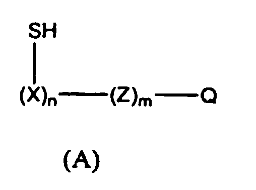
n is 2-100;
m is 0-100;
X is a nucleotide, modified oligonucleotide, or a nucleic acid derivative;
Z is a nucleotide, modified oligonucleotide, or polyanion
Q is a recognition group. The bioconjugate is coupled to the nanoparticle through the sulfur groups (-SH). - The invention also provides a nanoparticle probe comprising a bioconjugate of formula (B) coupled to a nanoparticle:

- The invention further provides a nanoparticle probe comprising a bioconjugate of formula (C) coupled to a nanoparticle:

- The invention also provides a nanoparticle probe comprising a bioconjugate of formula (D) coupled to a nanoparticle:

- The invention also provides a nanoparticle probe comprising a bioconjugate of formula (E) coupled to a nanoparticle:

- The invention also provides a nanoparticle probe comprising a bioconjugate of formula (F) coupled to a nanoparticle:

- The invention also provides a nanoparticle probe comprising a bioconjugate of formula (G) coupled to a nanoparticle:

- The invention also provides methods of preparing bioconjugate-nanoparticle probes, methods of detecting target analytes using the probes and kits comprising the probes of the invention.
- As used herein, a "type of oligonucleotides" refers to a plurality of oligonucleotide molecules having the same sequence. A "type of" nanoparticles, particles, latex microspheres, etc. having oligonucleotides attached thereto refers to a plurality of nanoparticles having the same type(s) of oligonucleotides attached to them. "Nanoparticles having bioconjugates attached thereto" are also sometimes referred to as "nanoparticle-bioconjugate probes," "nanoparticle probes," "nano probes," or just "probes."
-
-
Fig. 1 depicts detection of an analyte using a substrate. -
Fig. 2 depicts detection of an analyte using a substrate. -
Fig. 3 shows a procedure for introducing amine groups into a oligonucleotide. -
Fig.4 shows a procedure for introducing thiol groups into a oligonucleotide. -
Fig. 5 shows an alternative procedure for introducing thiol groups into a oligonucleotide. -
Fig. 6 depicts a phosphoramidite that can be used for introducing thiol groups into an oligonucleotide. -
Fig. 7 shows a method for preparing an epiandrosterone disulfide derivatized phosphoramidite. -
Fig. 8 shows the incorporation of epiandrosterone disulfide into an oligonucleotide. -
Fig. 9 shows incorporation of additional thiol groups into an oligonucleotide having a epiandrosterone disulfide moiety. -
Fig. 10 depicts a spot test which indicates binding of nanoparticle-probes of the invention to a target. -
Fig. 11 depicts spot tests which indicate the relative stability of various nanoparticle-probes in dithiothreitol (DTT) solution. -
Fig. 12 depicts spot tests which indicate the relative stability of various nanoparticle-probe in DTT solution. -
Fig. 13 depicts spot tests which indicate the relative stability of various nanoparticle-probes in DTT solution. -
Fig. 14 depicts spot tests which indicate the relative stability of various nanoparticle-probes in DTT solution at elevated temperature. -
Fig. 15 depicts spot tests which indicate the relative stability of various nanoparticle-probes in DTT solution, at elevated temperature and in the presence of magnesium chloride solution. - The bioconjugates of formula (A), (B), (C), (D), (E), (F), and (G) provide a solution to the problem of nanoparticle probe instability which results when the probe is heated or subjected to thiol containing compounds. Specifically, the invention permits two or more sulfur groups present on a bioconjugate to bind to the nanoparticle surface, which enhances the stability of the nanoparticle-bioconjugate binding. The resulting bioconjugate nanoparticle probes are stable towards heat and have increased resistance to displacement by thiol containing compounds such as DTT (dithiothreitol).
- The bioconjugates that are linked to nanoparticles to form the nanoparticle probes of the invention are of the formulae:


- As indicated above, Q represents a recognition group. By "recognition group" is meant at least one binding moiety with a binding affinity for a target analyte, such as a nucleic acid. Thus, the binding moiety may be, for example, one member of a recognition couple which consists of two or more substances having a binding affinity of one to the other. When a bioconjugate is bound to a nanoparticle, therefore, it provides useful biorecognition properties to the nanoparticle for the analyte. If the target is a nucleic acid, for example, the recognition group can bind to the nucleic acid by hybridization with the nucleic acid. The nucleic acid bound nanoparticle can then be detected.
- Examples of recognition groups include, without limitation, a receptor, a nucleotide, a nucleoside, a polynucleotide, an oligonucleotide, double stranded DNA, a protein, an antibody, a peptide, a carbohydrate, a sugar, a hapten, a nucleic acid, an amino acid, a peptide nucleic acid, a linked nucleic acid, a nucleoside triphosphate, a lipid, a lipid bound protein, an aptamer, a virus, a cell fragment, or a whole cell. Examples of recognition group-target analyte couples include: an antigen and an antibody; an antigen and an antibody derivative with a complementary antigen-binding domain; sugar and a lectin; a receptor and a ligand; a nucleotide sequence and a complementary nucleotide sequence; a nucleotide sequence and its binding protein or synthetic binding agent; a biotin and avidin or streptavidin; cellulose or chitin and cellulose binding domain. A preferred recognition group is an oligonucleotide. Also preferred is an antibody.
- The recognition group can also be an oligonucleotide having a sequence that is complementary to at least a portion of a second oligonucleotide having a second recognition group, e.g., an oligonucleotide sequence or protein, bound thereto. The second recognition group can then be used for specific binding to a target analyte, e.g., an antigen.
- The recognition group can also be a first recognition group, e.g., biotin, that can bind to a second recognition group, e.g., streptavidin, that is a member of the recognition couple. The second recognition group can then be bound directly or indirectly (e.g., via a linker) to a third recognition group, e.g., a receptor, which can bind to a target analyte.
- Z, when present, is a nucleotide spacer, a modified oligonucleotide, a polyanion, or other type of spacer which may be utilized in oligonucleotide synthesis such as a polyethylene glycol. It has been found that hybridization efficiency of nanoparticle-bioconjugate probes with nucleic acids can be increased by the use of a spacer portion between the recognition group on the bioconjugate and the nanoparticle. By using a spacer portion, the recognition group is spaced away from the surface of the nanoparticles and is more accessible for hybridization with its target. The length and sequence of the spacer portion providing good spacing of the recognition portion away from the nanoparticles can be determined empirically. It has been found that a spacer portion comprising at least about 10 nucleotides, preferably 10-50 nucleotides, gives good results. The spacer portion may have any sequence which does not interfere with the ability of the recognition group to become bound to a target analyte or a capture moiety on a surface in sandwich hybridization assays. For instance, the spacer portions should not have sequence complementary to each other, to that of the recognition group, or to that of the target analyte. Preferably, the bases of the nucleotides of the spacer portion are all adenines, all thymines, all cytidines, or all guanines, unless this would cause one of the problems just mentioned. More preferably, the bases are all adenines or all thymines. Most preferably the bases are all thymines. Spacer Z and recognition group Q can be attached together by a variety of techniques. For instance, they can be attached directly by a covalent linkage or indirectly by non-covalent linkage.
- As a linker, L can be any desired chemical group. For instance, L can be a polymer (e.g., polyethylene glycol, polymethylene, protein, peptide, oligonucleotide, or nucleic acid), - COO-, -CH2(CH2)vCOO-, -OCO-, R1N(CH2)v-NR1-, -OC(CH2)v-, -(CH2)v-, -O-(CH2)v-O-, - R1N-(CH2)v-,


- L is also a linker formed by the coupling of two moieties attached to molecules, the moieties selected from the group consisting of COOH, NH2, CHO, Cl, Br, I, NCO, NCS, allyl, and CH3CO2 -, or L is -C(=NH2Cl)(CH2)3 -.
- W is an aliphatic or aromatic group on which sulfur moieties can be readily bound. For instance, W can be steroid. Preferably W is an epiandrosterone derivative, as described in example 9.
- X represents a nucleotide that has been functionalized with a thiol group (as shown). Preferably, the X groups are all adenines, all thymines, all cytidines, or all guanines, more preferably, all adenines or all thymines, most preferably all thymines. In (X)n, n is 2-100. Preferably, n is 2-50, more preferable 2-20, more preferably 2-10. Functionalization of the X linkage with a thiol group can be carried out by a variety of techniques.
- In one embodiment of the invention, either a complex (1) or complex (II) containing at least two reactive groups R2 is synthesized by incorporating nucleotide building blocks containing an R2 group during synthesis of the oligonucleotide. R2 can be COOH, NH2, CHO, F, Cl, Br, I, NCO, NCS, allyl, or CH3CO2 -. If R2 in complex (I) or (II) is an NH2, then a nucleotide derivative containing an NH2 group is prepared for incorporation into the oligonucleotide. Suitable amine modified nucleotide reagents for use in this aspect of the invention include, but are not limited to, C6-dT phosphoramidite (5'-Dimethoxytrityl-5-[N-(trifluoroacetylaminohexyl)-3-acrylimido]-2'-deoxyUridine,3'-[(2-cyanoethyl)-(N,N-diisopropyl)]-phosphoramidite), amino modifier C6-dC (5'-Dimethoxytrityl-N-dimethylformamidine-5-[N-(trifluoroacetylaminohexyl)-3-acrylimido]-2'-deoxy-Cytidine,3'-[(2-cyanoethyl)-(N,N-diisopropyl)]-phosphoramidite), and amino modifier C2-dT (5'-dimethoxytrityl-5-[N-(trifluoroacetylaminoethyl)-3-acrylimido]-2'-deoxy-Uridine,3'-[(2-cyanoethyl)-(N,N-di-isopropyl)]-phosphoramidite). These and other amino modifiers are commercially available, for example from Glen Research, Sterling, Virginia. A preferred amino modifier reagent is C6-dT.

- Complex (I) or (II) is reacted with a thiolating reagent that is functionalized with a group capable of reacting with the R2 group on the complex and resulting in formation of a sulfur functionalized bioconjugate. Suitable thiolating reagents generally include thiol compounds possessing one or more functional groups capable of reacting with the R2 group of the complex. Such reagents include, but are not limited to, cystamine, and compounds of the formula SH(CH2)nY, wherein n is 1-20 and Y is COOH, NH2, CHO, F, Cl, Br, I, NCO, NCS, allyl, or CH3CO2 -. Another suitable thiolating reagent is 2-iminothiolane hydrochloride (Traut's Reagent), which is preferred when R2 in complex (I) or (II) is an amine. In a preferred aspect of this embodiment, fluorinated nucleotides are incorporated into an oligonucleotide sequence and treated with cystamine to provide disulfide units on the sequence (References (1) L.V. Nechev, I. Kozekov, C.M. Harris, and T.M. Harris, Chem Res Toxicol, 2001, 14, 1506-1512. (2) A.R. Diaz, R. Eritja, and R.G. Garcia, Nucleos Nucleot, 1997, 16, 2035-2051. (3) D.A. Erlanson, J.N.M. Glover, and G.L. Verdine, J. Amer. Chem. Soc., 1997, 119, 6927-6928). This preferred aspect is described in detail in Examples 7-9, below.
- An alternative method of thiolating complex (I) or (II) is to use a thiolating reagent that is a combination of reagents. For instance, if R2 in complex (I) or (II) is NH2, then the complex can be treated with an amine reactive bifunctional crosslinker, such as CHO(CH2)nCHO, and an alkyl or aryl thiol amine, such as SH(CH2)nNH2 or SH(C6H4)NH2. In both the amine reactive bifunctional crosslinker and the alkyl thiol amine, n is independently 1-30. Preferably, the amine reactive bifunctional crosslinker is glutaraldehyde (i.e., n is 3). Also preferably, the alkyl thiol amine is mercaptoethylamine (i.e., n is 2). Other preferred crosslinkers include 1,4 phenylene diisothiocyanate, 1,6 dihexanoic acid, or 1,6 hexane diisocyanate.
- In an alternative and more direct approach for preparing a thiol functionalized bioconjugate, a phosphoramidite containing an alkylthiol or other thiol based group is synthesized and used to prepare a bioconjugate of formula (A). An example of a phosphoramidite containing an alkylthiol group is described in Example 4, below, and is prepared according to the method of Glick et al., Tetrahedron Letters, 1993, 34, 5549-5552.
- As indicated above, the invention provides bioconjugate nanoparticle probes that are useful for detecting target analytes. To form the probe, bioconjugates (A), (B), (C), (D), (E), (F), or (G) are connected to the surface of a nanoparticle through the sulfur linkages on the bioconjugate. Preferably, the connection is through at least two thiol groups per bioconjugate molecule. Various methods can be used to connect the bioconjugate to the nanoparticle. In fact, any suitable method for attaching a bioconjugate to a nanoparticle may be used. A preferred method for attaching an bioconjugate to a nanoparticle is based on an aging process described in
U.S. patent nos. 6,361,944, filed June 25, 1999 ;6,506,564, filed June 26, 2000 ;6,767,702, filed January 12, 2001 ;6,760,016, filed March 28, 2001 ;09/927,777, filed August 10, 2001 PCT/US97/12783, filed July 21, 1997 PCT/US00/17507, filed June 26, 2000 ;PCT/US01/01190, filed January 12, 2001 ;PCT/US01/10071, filed March 28, 2001 . - The aging process provides nanoparticle-bioconjugate probes with enhanced stability and selectivity. The method comprises providing bioconjugates having covalently bound thereto thiol functional groups, prepared as described above. The functionalized bioconjugates are contacted with the nanoparticles in water for a time sufficient to allow at least some of the bioconjugates to bind to the nanoparticles by means of the functional groups. Such times can be determined empirically. For instance, it has been found that a time of about 12-24 hours gives good results. Other suitable conditions for binding of the bioconjugates can also be determined empirically. For instance, a concentration of about 10-20 nM nanoparticles and incubation at room temperature gives good results.
- Next, at least one salt is added to the water to form a salt solution. The salt can be any suitable water-soluble salt. For instance, the salt may be sodium chloride, lithium chloride, potassium chloride, cesium chloride, ammonium chloride, sodium nitrate, lithium nitrate, cesium nitrate, sodium acetate, lithium acetate, cesium acetate, ammonium acetate, a combination of two or more of these salts, or one of these salts in phosphate buffer. Preferably, the salt is added as a concentrated solution, but it could be added as a solid. The salt can be added to the water all at one time or the salt is added gradually over time. By "gradually over time" is meant that the salt is added in at least two portions at intervals spaced apart by a period of time. Suitable time intervals can be determined empirically.
- The ionic strength of the salt solution must be sufficient to overcome at least partially the electrostatic repulsion of the bioconjugates from each other and, either the electrostatic attraction of the negatively-charged bioconjugates for positively-charged nanoparticles, or the electrostatic repulsion of the negatively-charged bioconjugates from negatively-charged nanoparticles. Gradually reducing the electrostatic attraction and repulsion by adding the salt gradually over time has been found to give the highest surface density of bioconjugates on the nanoparticles. Suitable ionic strengths can be determined empirically for each salt or combination of salts. A final concentration of sodium chloride of from about 0.1 M to about 3.0 M in phosphate buffer, preferably with the concentration of sodium chloride being increased gradually over time, has been found to give good results.
- After adding the salt, the bioconjugates and nanoparticles are incubated in the salt solution for an additional period of time sufficient to allow sufficient additional bioconjugates to bind to the nanoparticles to produce the stable nanoparticle- bioconjugates probes. An increased surface density of the bioconjugates on the nanoparticles has been found to stabilize the probes. The time of this incubation can be determined empirically. A total incubation time of about 24-48, preferably 40 hours, has been found to give good results (this is the total time of incubation; as noted above; the salt concentration can be increased gradually over this total time). This second period of incubation in the salt solution is referred to herein as the "aging" step. Other suitable conditions for this "aging" step can also be determined empirically. For instance, incubation at room temperature and pH 7.0 gives good results. The solution is then centrifuged and the nanoparticle probes processed as desired. For instance, the solution can be centrifuged at 14,000 rpm in an Eppendorf Centrifuge 5414 for about 15 minutes to give a very pale pink supernatant containing most of the oligonucleotide (as indicated by the absorbance at 260 nm) along with 7-10% of the colloidal gold (as indicated by the absorbance at 520 nm), and a compact, dark, gelatinous residue at the bottom of the tube. The supernatant is removed, and the residue is resuspended in the desired buffer.
- The probes produced by use of the "aging" step have been found to be considerably more stable than those produced without the "aging" step. As noted above, this increased stability is due to the increased density of the bioconjugates on the surfaces of the nanoparticles which is achieved by the "aging" step. The surface density achieved by the "aging" step will depend on the size and type of nanoparticles and on the length, sequence and concentration of the oligonucleotides. A surface density adequate to make the nanoparticles stable and the conditions necessary to obtain it for a desired combination of nanoparticles and oligonucleotides can be determined empirically.
- Oligonucleotides or other recognition elements containing multiple thiol moieties as described above may bind to a variety of nanoparticles that have an affinity for thiol groups. Nanoparticles useful in the practice of the invention include metal (e.g., gold, silver, platinum, cobalt), semiconductor (e.g., Si, CdSe, CdS, and CdS or CdSe coated with ZnS), core shell particles (e.g., gold coated silver particles), alloy particles (e.g. silver and gold alloy), ,magnetic (e.g., cobalt), and non metallic (e.g. silicon) colloidal materials. Core shell particles are described in
PCT applications PCT/US01/50825 , andPCT/US02/16382 , as well as inU.S. PATENT numbers 7,147,687 and7,238,172 . Other nanoparticles composed of materials that have an affinity for thiol groups may also be used. In addition, nanowires or nanorods having a composition with an affinity for thiol groups also may be used. The size of the nanoparticles is preferably from about 5 nm to about 150 nm (mean diameter), more preferably from about 5 to about 50 nm, most preferably from about 10 to about 30 nm. - Methods of making metal, semiconductor and magnetic nanoparticles are well-known in the art. See, e.g., Schmid, G. (ed.) Clusters and Colloids (V C H, Weinheim, 1994); Hayat, M. A. (ed.) Colloidal Gold: Principles, Methods, and Applications (Academic Press, San Diego, 1991); Massart, R., IEEE Transactions On Magnetics, 17, 1247 (1981); Ahmadi, T. S. et al., Science, 272, 1924 (1996); Henglein, A. et al., J. Phys. Chem., 99, 14129 (1995); Curtis, A. C., et al., Angew. Chem. Int. Ed. Engl., 27, 1530 (1988). Methods of making ZnS, ZnO, TiO2, AgI, AgBr, HgI2, PbS, PbSe, ZnTe, CdTe, In2S3, ln2Se3, Cd3P2, Cd3As2, InAs, and GaAs nanoparticles are also known in the art. See, e.g., Weller, Angew. Chem. Int. Ed. Engl., 32, 41 (1993); Henglein, Top. Curr. Chem., 143, 113 (1988); Henglein, Chem. Rev., 89, 1861 (1989); Brus, Appl. Phys. A., 53, 465 (1991); Bahncmann, in Photochemical Conversion and Storage of Solar Energy (eds. Pelizetti and Schiavello 1991), page 251; Wang and Herron, J. Phys. Chem., 95, 525 (1991); Olshavsky et al., J. Am. Chem. Soc., 112, 9438 (1990); Ushida et al., J. Phys. Chem., 95, 5382 (1992).
- Suitable nanoparticles are also commercially available from, e.g., Ted Pella, Inc. (gold), Amersham Corporation (gold) and Nanoprobes, Inc. (gold). Presently Preferred nanoparticles are gold nanoparticles.
- The bioconjugate-nanoparticle probes of the invention can be used to detect target analytes, such as nucleic acids. Examples of nucleic acids that can be detected with nanoparticle probes of the invention include genes (e.g., a gene associated with a particular disease), viral RNA and DNA, bacterial DNA, fungal DNA, cDNA, mRNA, RNA and DNA fragments, oligonucleotides, synthetic oligonucleotides, modified oligonucleotides, single-stranded and double-stranded nucleic acids, natural and synthetic nucleic acids, etc. Thus nanoparticle probes prepared according to the invention can be used, for example, for the diagnosis and/or monitoring of viral diseases (e.g., human immunodeficiency virus, hepatitis viruses, herpes viruses, cytomegalovirus, and Epstein-Barr virus), bacterial diseases (e.g., tuberculosis, Lyme disease, H. pylori, Escherichia coli infections, Legionella infections, Mycoplasma infections, Salmonella infections), sexually transmitted diseases (e.g., gonorrhea), inherited disorders (e.g., cystic fibrosis, Duchene muscular dystrophy, phenylketonuria, sickle cell anemia), and cancers (e.g., genes associated with the development of cancer); in forensics; in DNA sequencing; for paternity testing; for cell line authentication; for monitoring gene therapy; and for many other purposes.
- To perform an assay according to the invention, a sample suspected of containing a target analyte is contacted with bioconjugate nanoparticle probes having attached thereto recognition groups capable of binding to at least a portion of the target analyte. The target to be detected may be isolated by known methods, or may be detected directly in cells, tissue samples, biological fluids (e.g., saliva, urine, blood, serum), solutions containing PCR components, solutions containing large excesses of oligonucleotides or high molecular weight DNA, and other samples, as also known in the art. See, e.g., Sambrook et al., Molecular Cloning: A Laboratory Manual (2nd ed. 1989) and B.D. Hames and S.J. Higgins, Eds., Gene Probes I (IRL Press, New York, 1995). Methods of preparing nucleic acids for detection with hybridizing probes are well known in the art. See, e.g., Sambrook et al., Molecular Cloning: A Laboratory Manual (2nd ed. 1989) and B.D. Hames and S.J. Higgins, Eds., Gene Probes I (IRL Press, New York, 1995).
- If a nucleic acid is present in small amounts, it may be amplified by methods known in the art. See, e.g., Sambrook et al., Molecular Cloning: A Laboratory Manual (2nd ed. 1989) and B.D. Hames and S.J. Higgins, Eds., Gene Probes I (IRL Press, New York, 1995). Preferred is polymerase chain reaction (PCR) amplification.
- One method according to the invention for detecting nucleic acid comprises contacting a nucleic acid with one or more types of nanoparticle probes of the invention. The nucleic acid to be detected has at least two portions. The lengths of these portions and the distance(s), if any, between them are chosen so that when the bioconjugates on the nanoparticles hybridize to the nucleic acid, a detectable change occurs. These lengths and distances can be determined empirically and will depend on the type of particle used and its size and the type of electrolyte which will be present in solutions used in the assay (as is known in the art, certain electrolytes affect the conformation of nucleic acids).
- Also, when a nucleic acid is to be detected in the presence of other nucleic acids, the portions of the nucleic acid to which the bioconjugates on the nanoparticles are to bind must be chosen so that they contain sufficient unique sequence so that detection of the nucleic acid will be specific. Guidelines for doing so are well known in the art.
- Although nucleic acids may contain repeating sequences close enough to each other so that only one type of bioconjugate-nanoparticle conjugate need be used, this will be a rare occurrence. In general, the chosen portions of the nucleic acid will have different sequences and will be contacted with nanoparticles carrying two or more different bioconjugates, preferably attached to different nanoparticles. Additional portions of the DNA could be targeted with corresponding nanoparticles. Targeting several portions of a nucleic acid increases the magnitude of the detectable change.
- The contacting of the nanoparticle-bioconjugate probes with the nucleic acid takes place under conditions effective for hybridization of the bioconjugates on the nanoparticles with the target sequence(s) of the nucleic acid. These hybridization conditions are well known in the art and can readily be optimized for the particular system employed. See, e.g., Sambrook et al., Molecular Cloning: A Laboratory Manual (2nd ed. 1989). Preferably stringent hybridization conditions are employed.
- Faster hybridization can be obtained by freezing and thawing a solution containing the nucleic acid to be detected and the nanoparticle-bioconjugate probes. The solution may be frozen in any convenient manner, such as placing it in a dry ice-alcohol bath for a sufficient time for the solution to freeze (generally about 1 minute for 100 microliters of solution). The solution must be thawed at a temperature below the thermal denaturation temperature, which can conveniently be room temperature for most combinations of nanoparticle-bioconjugate probes and nucleic acids. The hybridization is complete, and the detectable change may be observed, after thawing the solution.
- The rate of hybridization can also be increased by warming the solution containing the nucleic acid to be detected and the nanoparticle-bioconjugate probes to a temperature below the dissociation temperature (Tm) for the complex formed between the bioconjugates on the nanoparticles and the target nucleic acid. Alternatively, rapid hybridization can be achieved by heating above the dissociation temperature (Tm) and allowing the solution to cool.
- The rate of hybridization can also be increased by increasing the salt concentration (e.g., from 0.1 M to 1 M NaCl).
- The detectable change that occurs upon hybridization of the bioconjugates on the nanoparticles to the nucleic acid may be an optical change (e.g. color change), the formation of aggregates of the nanoparticles, or the precipitation of the aggregated nanoparticles. The optical changes can be observed with the naked eye or spectroscopically. The formation of aggregates of the nanoparticles can be observed by electron microscopy or by nephelometry. The precipitation of the aggregated nanoparticles can be observed with the naked eye or microscopically. Preferred are color changes observable with the naked eye.
- The observation of a color change with the naked eye can be made more readily against a background of a contrasting color. For instance, when gold nanoparticles are used, the observation of a color change is facilitated by spotting a sample of the hybridization solution on a solid white surface (such as silica or alumina TLC plates, filter paper, cellulose nitrate membranes, and nylon membranes, preferably a nylon membrane) and allowing the spot to dry. Initially, the spot retains the color of the hybridization solution (which ranges from pink/red, in the absence of hybridization, to purplish-red/purple, if there has been hybridization). On drying at room temperature or 80°C (temperature is not critical), a blue spot develops if the nanoparticle-bioconjugate probes had been linked by hybridization with the target nucleic acid prior to spotting. In the absence of hybridization (e.g., because no target nucleic acid is present), the spot is pink. The blue and the pink spots are stable and do not change on subsequent cooling or heating or over time. They provide a convenient permanent record of the test. No other steps (such as a separation of hybridized and unhybridized nanoparticle-bioconjugate probes) are necessary to observe the color change. The color change may be quantitated by recording the plate image with an optical scanning device such as a flatbed scanner or CCD camera, and analyzing the amount and type of color of each individual spot. Alternatively, a color filter (e.g. red filter) may be used to filter out specific colors so that the signal intensity of each spot may be recorded and analyzed.
- An alternate method for easily visualizing the assay results is to spot a sample of nanoparticle probes hybridized to a target nucleic acid on a glass fiber filter (e.g., Borosilicate Microfiber Filter, 0.7 micron pore size, grade FG75, for use with gold nanoparticles 13 nm in size), while drawing the liquid through the filter. Subsequent rinsing with water washes the excess, non-hybridized probes through the filter, leaving behind an observable spot comprising the aggregates generated by hybridization of the nanoparticle probes with the target nucleic acid (retained because these aggregates are larger than the pores of the filter). This technique may provide for greater sensitivity, since an excess of nanoparticle probes can be used.
- Some embodiments of the method of detecting nucleic acid utilize a substrate. By employing a substrate, the detectable change (the signal) can be amplified and the sensitivity of the assay increased.
- Any substrate can be used which allows observation of the detectable change. Suitable substrates include transparent solid surfaces (e.g., glass, quartz, plastics and other polymers), opaque solid surface (e.g., white solid surfaces, such as TLC silica plates, filter paper, glass fiber filters, cellulose nitrate membranes, nylon membranes), and conducting solid surfaces (e.g., indium-tin-oxide (ITO)). The substrate can be any shape or thickness, but generally will be flat and thin. Preferred are transparent substrates such as glass (e.g., glass slides) or plastics (e.g., wells of microtiter plates).
- In one embodiment oligonucleotides are attached to the substrate. The oligonucleotides can be attached to the substrates as described in, e.g., Chrisey et al., Nucleic Acids Res., 24, 3031-3039 (1996); Chrisey et al., Nucleic Acids Res., 24, 3040-3047 (1996); Mucic et al., Chem. Commun., 555 (1996); Zimmermann and Cox, Nucleic Acids Res., 22, 492 (1994); Bottomley et al., J. Vac. Sci. Technol. A, 10, 591 (1992); and Hegner et al., FEBS Lett., 336, 452 (1993).
- The oligonucleotides attached to the substrate have a sequence complementary to a first portion of the sequence of a nucleic acid to be detected. The nucleic acid is contacted with the substrate under conditions effective to allow hybridization of the oligonucleotides on the substrate with the nucleic acid. In this manner the nucleic acid becomes bound to the substrate. Any unbound nucleic acid is preferably washed from the substrate before adding nanoparticle-bioconjugate probes.
- Next, the nucleic acid bound to the substrate is contacted with a first type of nanoparticles having bioconjugates, such as oligonucleotides, attached thereto. The oligonucleotides have a sequence complementary to a second portion of the sequence of the nucleic acid, and the contacting takes place under conditions effective to allow hybridization of the oligonucleotides on the nanoparticles with the nucleic acid. In this manner the first type of nanoparticles become bound to the substrate. After the nanoparticle-oligonucleotide conjugates are bound to the substrate, the substrate is washed to remove any unbound nanoparticle-oligonucleotide conjugates and nucleic acid.
- The oligonucleotides on the first type of nanoparticles may all have the same sequence or may have different sequences that hybridize with different portions of the nucleic acid to be detected. When oligonucleotides having different sequences are used, each nanoparticle may have all of the different oligonucleotides attached to it or, preferably, the different oligonucleotides are attached to different nanoparticles. Alternatively, the oligonucleotides on each of the first type of nanoparticles may have a plurality of different sequences, at least one of which must hybridize with a portion of the nucleic acid to be detected.
- The first type of nanoparticle-oligonucleotide conjugates bound to the substrate is optionally contacted with a second type of nanoparticles having oligonucleotides attached thereto. These oligonucleotides have a sequence complementary to at least a portion of the sequence(s) of the oligonucleotides attached to the first type of nanoparticles, and the contacting takes place under conditions effective to allow hybridization of the oligonucleotides on the first type of nanoparticles with those on the second type of nanoparticles. After the nanoparticles are bound, the substrate is preferably washed to remove any unbound nanoparticle-oligonucleotide conjugates.
- The combination of hybridizations produces a detectable change. The detectable changes are the same as those described above, except that the when second type of conjugates, multiple hybridizations result in an amplification of the detectable change. In particular, since each of the first type of nanoparticles has multiple oligonucleotides (having the same or different sequences) attached to it, each of the first type of nanoparticle-oligonucleotide conjugates can hybridize to a plurality of the second type of nanoparticle-oligonucleotide conjugates. Also, the first type of nanoparticle-oligonucleotide conjugates may be hybridized to more than one portion of the nucleic acid to be detected. The amplification provided by the multiple hybridizations may make the change detectable for the first time or may increase the magnitude of the detectable change. This amplification increases the sensitivity of the assay, allowing for detection of small amounts of nucleic acid.
- If desired, additional layers of nanoparticles can be built up by successive additions of the first and second types of nanoparticle-oligonucleotide conjugates. In this way, the number of nanoparticles immobilized per molecule of target nucleic acid can be further increased with a corresponding increase in intensity of the signal.
- In one embodiment for detection of non-nucleic acid analytes (see for example
U.S. Patent No. 6,750,016, filed March 28, 2001 , and International applicationPCT/01/10071, filed March 28, 2001 ) the analyte may be bound directly or indirectly , via covalent or non-covalent interactions, to a substrate. The substrates are the same type as described above. For indirect binding, the analyte can be bound to the substrate via a linker, e.g., an oligonucleotide or other spacer molecule. Alternatively, the analyte may be modified by binding it to an oligonucleotide having a sequence that is complementary to at least a portion of the sequence of a capture oligonucleotide bound to a substrate. The nanoparticle-probe having a recognition group for the analyte is then contacted with the substrate under conditions effective to allow the specific binding of the nanoparticle-probe to the analyte bound to the substrate and the presence of the analyte can be visually detected either by formation of a spot on the substrate or through the use of staining material such as silver on gold stain. SeeFig. 1 for examples of this detection method. - In another method for detecting analytes, the target analyte can be modified by attaching the analyte to the nanoparticle-probe as the recognition portion of the probe. Thereafter, the modified nanoparticle-probe is contacted with a substrate having a second member of the recognition couple bound thereto. The presence of the analyte can be visually detected either by formation of a spot on the substrate or through the use of staining material such as silver on gold stain.
- In yet another method for detecting analytes, the target analyte is modified by binding it to an oligonucleotide having a sequence that is complementary to at least a portion of a sequence of an oligonucleotide (recognition group) bound to the nanoparticle-probe. The modified target is then coupled to the nanoparticle-probe by contacting the modified target and the nanoparticle-probe under conditions effective for hybridization between the oligonucleotide bound to the target and the oligonucleotide bound to the nanoparticle-probe. The hybridized complex is then contacted with a substrate having a recognition group for the analyte bound thereto. The presence of the analyte can be visually detected either by formation of a spot on the substrate or through the use of staining material such as silver on gold stain. See
Fig. 2 for an example of this method. - When a substrate is employed, a dectectable change can be produced or enhanced by staining. Staining material, e.g., gold, silver, etc., can be used to produce or enhance a detectable change in any assay performed on a substrate, including those described above. For instance, silver staining can be employed with any type of nanoparticles that catalyze the reduction of silver. Preferred are nanoparticles made of noble metals (e.g., gold and silver). See Bassell, et al., J. Cell Biol., 126, 863-876 (1994); Braun-Howland et al., Biotechniques, 13, 928-931 (1992). If the nanoparticles being employed for the detection of analyte do not catalyze the reduction of silver, then silver ions can be complexed to the target analyte to catalyze the reduction. See Braun et al., Nature, 391, 775 (1998). Also, silver stains are known which can react with the phosphate groups on nucleic acids.
- An alternate method for utilizing the polythiol nanoparticle probes is in the application to micro arrays for detecting a variety of biomolecules such as nucleic acids, proteins or carbohydrates. One specific example is the application of polythiol modified oligonucleotide labeled gold nanoparticle probes to the detection of nucleic acids in a sandwich assay format as described in
U.S. patent no. 6361944 . In this method, the gold nanoparticle labels are detected via a silver deposition process. Alternatively, a gold nanoparticle development procedure may be used as described inU.S. patent no. 6417340 and detected optically. It should be noted that the polythiol nanoparticle probes described herein also can be applied as detection probes for use as in situ hybridization labels or expanded to other DNA/RNA detection technologies. - In this example, amino modifier C6-DT groups are introduced into an oligonucleotide using an amino modifier phosphoramidite (available from Glen Research, Sterling, Virginia) (
Fig. 3 ). - Protocol. The amino modifier C6-dT reacts in a manner identical to normal phosphoramidites, i.e., standard automated oligonucleotide synthesis. The trifluoroacetyl (TFA) protecting group on the primary amine is removed during standard ammonium hydroxide deprotection. However, a minor side reaction during ammonia deprotection can lead to irreversibly capping 2-5 % of the amine. To prevent this reaction, the synthesis is carried out using acetyl-protected dC and deprotection is carried out in 30 % ammonia/40 % methylamine 1:1 (AMA) at 65 °C for 15 minutes.
- In this example, the amine containing oligonucleotide prepared in Example 1 is reacted with 2-iminothiolane.HCl (Traut's Reagent, available from Pierce Chemical Company, Rockford, Illinois) to introduce thiol groups (
Fig. 4 ). - Protocol. The amine containing oligonucleotide prepared in Example 1 is first purified by reverse phase HPLC and is then dissolved in 50 mM triethanolamine-HCl buffer of pH 8 (or other pH 8 buffer such as 0.16 M Borate of 10 mM phosphate). A 2-10 fold molar excess of 2-iminothiolane-HCl is added. The solution is incubated for 20-60 minutes at 0-25 °C. The thiolated amine oligonucleotide is then separated from the amine oligonucleotide using reverse-phase HPLC (0.03 M TEAA buffer (pH 7) with a 1 %/min gradient of 95:5 acetonitrile/0.03 M TEAA (pH 7)).
- Alternatively, the following protocol can be used. The amine containing oligonucleotide is first purified by reverse-phase HPLC. After purification, the oligonucleotide is re-dissolved in a phosphate or borate buffer (pH 7.2-8.5) containing a 10-100 fold excess of water soluble carbodiimide (WSC, e.g., ethyl dimethylaminopropyl-carbodiimide) and a 10-100 fold excess of 3-mercaptopropionic acid and allowed to stand for 2-4 hours at room temperature. Next, the oligonucleotide is purified through a NAP-10 column to remove excess reagents and eluted in water, followed by reverse-phase HPLC purification.
- This example presents an alternative method for introducing thiol groups into an oligonucleotide. An amine reactive bifunctional crosslinker, e.g., glutaraldehyde, and a heterobifunctional group such as an alkyl thiol amine are reacted with the amine functionalized oligonucleotide prepared in Example 2, to create thiol groups (
Fig. 5 ). - Protocol. The amine containing oligonucleotide is first purified be reverse-phase HPLC. After purification, the oligonucleotide is redissolved in a phosphate or borate buffer (pH 6-9) containing 10 % glutaraldehyde and allowed to stand for 1-2 hours. Next, the oligonucleotide is purified through a NAP-10 column to remove excess glutaraldehyde and eluted in pH 6-9 phosphate or borate buffer. A 10-100 fold excess of mercaptoethylamine is then reacted with the oligonucleotide for 2-4 hours at room temperature, followed by addition of sodium cyanoborohydride to create a 10 % solution for 5 min to reduce the Schiff base. The oligonucleotide is subsequently purified by reverse-phase HPLC.
- In this example, a phosphoramidite of formula III (
Fig. 6 ) containing an alkyl thiol or other thiol based functionality is synthesized according to the method of Glick et al., Tetrahedron letters, 1993, 34, 5549-5552. The phosphoramidite is then used to incorporate thiol groups into an oligonucleotide using standard phosphramidite methodology. - An alternative protocol for the introduction of thiol groups into an oligonucleotide is as follows. Carboxy-dT (available from Glen Research, Sterling, Virginia) is introduced into an oligonucleotide in an analogous manner to Example 1. Deprotection is carried out using mild deprotection: 0.4 M methanolic sodium hydroxide (methanol:water 4:1) for 17 hours at room temperature. The support is pipetted off and the solution neutralized with 2 M TEAA. DNA is purified by reverse phase HPLC. The DNA is resuspended in 100 mM MES buffer (pH 6), water soluble coupling reagents are added (ethyl dimethylaminopropyl-carbodiimide (EDC) and -N-hydroxysulfosuccinimide (sulfo NHS)) at a final concentration of 2 mM EDC and 5 mM sulfo-NHS, and the mixture incubated at room temperature for 15 min. Next, a thiol coupling reagent (e.g., SH(CH2)xNH2) is added at a 10 fold excess and the mixture incubated at room temperature for 3 hours. The oligonucleotide is purified through a NAP-10 column to remove excess reagents, followed by HPLC purification.
- In this example the thiol functionalized oligonucleotide prepared by any of the methods disclosed herein is attached to a gold nanoparticle through at least two of the thiol groups.
- Protocol. A 4 µM solution of a polythiol modified oligonucleotide is incubated with an approximately 15 nM gold particle dispersion and then the particles isolated by centrifugation.
- Epiandrosterone can be used as an additional linking element. Its advantages include that it is a readily available, easily derivatized to a ketoalcohol and, as a substituent with a large hydrophobic surface, may help screen the approach of water soluble molecules to the gold surface (Letsinger, et al., J. Am. Chem. Soc. 115, 7535-7536 - Bioconjugate Chem. 9, 826-830). Incorporation of the epi disulfide into an oligonucleotide is conducted by phosphoramidite chemistry, as described below.
- The epi disulfide derivative has the structure:
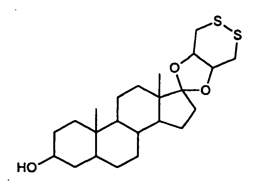
- (a) Synthesis of epi disulfide (
Fig. 7 ). A solution of epiandrosterone (0.5g), 1,2-dithiane-4,5-diol (0.28 g), and p-toluenesulfonic acid (15 mg) in toluene (30 mL) was refluxed for 7 h under conditions for removal of water (Dean Stark apparatus); then the toluene was removed under reduced pressure and the reside taken up in ethyl acetate. This solution was washed with water, dried over sodium sulfate, and concentrated to a syrupy reside, which on standing overnight in pentane/ether affordedcompound epi disulfide 1a as a white solid (400 mg); Rf (TLC, silica plate, ether as eluent) 0.5; for comparison, Rf values for epiandrosterone and 1,2-dithiane-4,5-diol obtained under the same conditions are 0.4, and 0.3, respectively. Recrystallization from pentane/ether afforded a white powder, mp 110-112 °C; 1H NMR, δ 3.6 (1H, C3OH), 3.54-3.39 (2H, m 2OCH of the dithiane ring), 3.2-3.0 (4H, m 2CH2S), 2.1-0.7 (29H, m steroid H); mass spectrum (ES+) calcd for C23H36O3S2 (M+H) 425.2179, found 425.2151. Anal. (C23H37O3S2)S: calcd, 15.12; found, 15.26. - (b) Preparation of Steroid-Disulfide Ketal Phosphoramidite Derivative (
Fig. 7 )
Epi disulfide 1a (100 mg) was dissolved in THF (3 mL) and cooled in a dry ice alcohol bath. N,N-diisopropylethylamine (80 µL) and β- cyanoethyl chlorodiisopropylphosphoramidite (80 µL) were added successively; then the mixture was warmed to room temperature, stirred for 2 h, mixed with ethyl acetate (100 mL), washed with 5% aq. NaHCO3 and with water, dried over sodium sulfate, and concentrated to dryness. The residue was taken up in the minimum amount of dichloromethane, precipitated at -70 °C by addition of hexane, and dried under vacuum; yield 100 mg; 31P NMR 146.02. - 5'-Modified oligonucleotides are constructed on CPG supports using conventional phosphoramidite chemistry, except that
compound 1b is employed in the final phosphitilation step. Products are cleaved from the support by treatment with concentrated NH4OH for 16 h at 55 °C. The oligonucleotides 1c are purified by reversed phase HPLC on a Dionex DX500 system equipped with a Hewlett Packard ODS Hypersil column (4.6 x 200 nm, 5 µm particle size) using TEAA buffer (pH 7.0) and a 1%/min gradient of 95% CH3CN/5% 0.03 TEAA at a flow rate of 1 mL/min. - In this example, fluorine modified Inosine nucleotides (2-F-dI; available from Glen Research, Sterling, Virginia) are introduced into oligonucleotide 1c and treated with cystamine after completing synthesis. See L.V. Nechev, I. Kozekov, C.M. Harris, and T.M. Harris, Chem Res Toxicol, 2001, 14, 1506-1512; A.R. Diaz, R. Eritja, and R.G. Garcia, Nucleos Nucleot, 1997, 16, 2035-2051; D.A. Erlanson, J.N.M. Glover, and G.L. Verdine, J. Amer. Chem. Soc., 1997, 119, 6927-6928.

support 2 times with DMSO then 3 times with acetonitrile and roughly dry the support with argon. Using two disposable syringes as above, treat the support two times with 1ml each 1M DBU in acetonitrile one hour each time. Rinse the support with 1ml each ofmethanol X 2, andacetonitrile X 3. Roughly dry the support with argon and deprotect the oligo 1d from the support using ammonium hydroxide as normal. - Stability of the nanoparticle probes of the invention was evaluated by spotting the subject mixtures on a solid white surface (such as a C-18 silica TLC plate or a reversed phase (RP) HPLC plate) and observing the color of the spot after drying. Red indicates starting nanoparticle with DNA strands on dispersed in solution, and violet indicates particle aggregation due to partial displacement of oligonucleotides on the gold nanoparticle, and blue indicates even greater particle aggregation due to more extensive displacement of oligonucleotides.
- Oligonucleotides having the sequence 5'-Epi-SH-SH-a18-gcg gaa gaa tgt gtc-3' [SEQ ID NO:1] were prepared as described in Examples 7-9. These oligonucleotides have an analogous structure to oligonucleotides 1d in
Fig. 9 , i.e., they possess an epi disulfide moiety and two further thiol groups on the backbone. The oligonucleotides are loaded onto gold nanoparticles as described below. The probes are representative of probes of the invention and are denoted "EPI+2S probes." - EPI+2S probes are prepared (loaded) in two different salt solutions; the same oligonucleotide is loaded in either 0.85 M sodium chloride or in 2.2 M sodium chloride. The EPI-2S probes' length is 35mer total and contains no fillers.
- A colloidal solution of citrate stabilized gold nanoparticles (about 10 nM), prepared by the citrate reduction method (Grabar et. al, Anal. Chem. 1995, 67, 735.), was mixed with sulfur modified-a20-probe oligonucleotide (4µM), and allowed to stand for 24 hours at room temperature in 1 ml Eppendorf capped vials. Then, Step 1: 100 µL of a 0.1 M sodium hydrogen phosphate buffer, pH 7.0, and 100 µL of 1.0 M NaCl were premixed and added to the solution and allowed to stand for an additional 12 hours. Step2: Then the salt concentration was increased to 0.3M NaCl and kept further 12h at room temperature. Step 3: At this point the salt concentration was increased to 0.85 and kept another 16h at room temperature. Total salt aging process took 40h. In the case of the 2.2 M salt concentration, work up at the stage of
step 3 salt was increased gradually to 2.2 M NaCl and kept at room temperature. - The solution was next centrifuged at 14,000 rpm in an Eppendorf Centrifuge 5414 for about 15 minutes to give a very pale pink supernatant containing most of the oligonucleotide (as indicated by the absorbance at 260 nm) along with 7-10% of the colloidal gold (as indicated by the absorbance at 520 nm), and a compact, dark, gelatinous residue at the bottom of the tube. The supernatant was removed, and the residue was re suspended in the desired buffer.
- Oligonucleotides having an analogous structure to oligonucleotide 1c, i.e., possessing an epi disulfide linkage but no other sulfur groups, were prepared as described in Examples 7-8. The probe length is 18mer+A20 linker and total 38mer. The probe sequence is 5'-Epi-a20-cct caa aga aaa g-3' [SEQ ID NO:2] and A20-Epi filler. The probe is loaded in 0.85 M NaCl solution. These probes are representative of prior art probes in that they do not contain additional thiol functionalization of the oligonucleotide backbone. The probes are denoted "EPI probes."
- A colloidal solution of citrate stabilized gold nanoparticles (about 10 nM), prepared by the citrate reduction method (Grabar et. al, Anal. Chem. 1995, 67, 735.) was mixed with sulfur modified-a20-probe oligonucleotide and corresponding sulfur modified-da20 filler oligonucleotide (each to a concentration of 1.7 µM), prepared as described in part B, and allowed to stand for 24 hours at room temperature in 1 ml Eppendorf capped vials. Then, Step 1: 100 µL of a 0.1 M sodium hydrogen phosphate buffer, pH 7.0, and 100 µL of 1.0 M NaCl were premixed and added to the solution and allowed to stand for an additional 12 hours. Step2: Then salt concentration was increased to 0.3M NaCl and kept further 12h at room temperature. Step 3: At this point salt concentration was increased to 0.85 and kept another 16h at room temperature. Total salt aging process took 40h.
- The solution was next centrifuged at 14,000 rpm in an Eppendorf Centrifuge 5414 for about 15 minutes to give a very pale pink supernatant containing most of the oligonucleotide (as indicated by the absorbance at 260 nm) along with 7-10% of the colloidal gold (as indicated by the absorbance at 520 nm), and a compact, dark, gelatinous residue at the bottom of the tube. The supernatant was removed, and the residue was resuspended in the desired buffer.
- This example verified that EPI+2S probes, like EPI probes, bind to a target.
- To 100 µl of a colloid mixture (50µl of EPI+2S probes and 50µl of EPI probes), 1 µl of a 10µM solution of the target (MTHFR 87 mer synthetic target) was added and the sample frozen at -70 °C for I minute and then thawed at room temperature. After the sample was brought to room temperature, 3 µl aliquots were spotted on a RP HPLC plate and solvents evaporated. Simultaneously, control solution prepared in a similar manner but without target was spotted on the HPLC plate. After evaporation of the solvents, the target containing spot turned completely blue, indicating hybridizing to the target. The control spot turned red, indicating absence of hybridization. See
Fig. 10. Fig. 10 shows binding of the oligonucleotide to a target. In the figure, R indicates red, B indicates blue and P indicates purple. The target in this example has the sequence 87mer target: 5'-ggt gtc tgc ggg agc cga ttt cat cat cat cac gca gct ttt ctt tga ggc tga cac att ctt ccg ctt tgt gaa ggc atg cac cga-3' [SEQ ID NO:3]. - This example shows the increased stability of EPI+2S probes in DTT solution, in comparison to EPI probes. The example also shows that EPI+2S probes prepared in 2.2 M NaCl solution ("2.2 M EPI+2S probes") are more stable than EPI+2S probes prepared in 0.85 M NaCl solution ("0.85 M EPI+2S probes").
- To 50 µl of the probe colloid in 0.1 M NaCl and 10mM Phosphate buffer at
pH Figs. 11-13. Figs. 11-13 show displacement of the oligonucleotide from the nanoparticle (indicated by a blue spot). - This example reveals the increased stability of EPI+2S probes in the presence of DTT at elevated temperature, compared with EPI probes.
- To 50µl of the colloid, 5 µl of 0.1M DTT solution was added and incubated at 60 °C and spotted on a C-18 RP silica plate. The EPI probe turned blue in 70min, whereas the 0.85 M EPI-2S probe started turning blue at 120min. See
Fig. 14. Fig. 14 shows displacement of the oligonucleotide from the nanoparticle (indicated by a blue spot). - This example shows the increased stability of EPI+2S probes in the presence of DTT and MgCl2 at elevated temperature, compared with EPI probes.
- To 50µl of the colloid, 5 µl of 0.1M DTT solution and 10mM MgCl2 [final concentration] were added and the samples incubated at 60°C. The samples were spotted on a C-18 RP silica plate. The EPI probe spot turned blue in 25min while the 0.85 M EPI-2S probe spot started turning blue at 25min. The 2.2 M EPI-2S probe spot also started turning blue at 25min. See
Fig. 15. Fig. 15 shows displacement of the oligonucleotide from the nanoparticle (indicated by a blue spot). -
- <110> Garimella, Viswanadham
Storhoff, James J. - <120> Bioconjugate-Nanoparticle Probes
- <130> 01-599-B
- <150> 60/348,239
<151> 2001-11-08 - <160> 7
- <170> PatentIn version 3.1
- <210> 1
<211> 34
<212> DNA
<213> Artificial sequence - <220>
<223> synthetic oligonucleotide - <220>
<221> misc_feature
<222> (1)..(1)
<223> n is epiandrosterone disulfide-SH-SH- - <400> 1
naaaaaaaaa aaaaaaaaag cggaagaatg tgtc 34 - <210> 2
<211> 34
<212> DNA
<213> Artificial sequence - <220>
<223> synthetic oligonucleotide - <220>
<221> misc_feature
<222> (1).. (1)
<223> n is epiandrosterone disulfide - <400> 2
naaaaaaaaa aaaaaaaaaa acctcaaaga aaag 34 - <210> 3
<211> 87
<212> DNA
<213> Artificial sequence - <220>
<223> synthetic oligonucleotide
<400> 3
- <210> 4
<211> 12
<212> DNA
<213> Artificial sequence - <220>
<223> synthetic oligonucleotide - <400> 4
atcggctaat cg 12 - <210> 5
<211> 17
<212> DNA
<213> Artificial sequence - <220>
<223> synthetic oligonucleotide - <220>
<221> misc_feature
<222> (1)..(5)
<223> n is amino modifier C6-dT - <400> 5
nnnnnatcgg ctaatcg 17 - <210> 6
<211> 17
<212> DNA
<213> Artificial sequence - <220>
<223> synthetic oligonucleotide - <220>
<221> misc_feature
<222> (1)..(5)
<223> thiol modified C6-dT - <400> 6
nnnnnatcgg ctaatcg 17 - <210> 7
<211> 17
<212> DNA
<213> Artificial sequence - <220>
<223> synthetic oligonucleotide - <220>
<221> misc_feature
<222> (1)..(5)
<223> thiol modified C6-dT - <400> 7
nnnnnatcgg ctaatcg 17 -
- <110> Garimella, viswanadham
Storhoff, James J. - <120> Bioconjugate-Nanoparticle Probes
- <130> 01-599-B
- <150> 60/348,239
<151> 2001-11-08 - <160> 7
- <170> PatentIn version 3.1
- <210> 1
<211> 34
<212> DNA
<213> Artificial sequence - <220>
<223> synthetic oligonucleotide - <220>
<221> misc_feature
<222> (1)..(1)
<223> n is epiandrosterone disulfide-SH-SH- - <400> 1
naaaaaaaaa aaaaaaaaag cggaagaatg tgtc 34 - <210> 2
<211> 34
<212> DNA
<213> Artificial sequence - <220>
<223> synthetic oligonucleotide - <220>
<221> misc_feature
<222> (1)..(1)
<223> n is epiandrosterone disulfide - <400> 2
naaaaaaaaa aaaaaaaaaa acctcaaaga aaag 34 - <210> 3
<211> 87
<212> DNA
<213> Artificial sequence - <220>
<223> synthetic oligonucleotide - <400> 3

- <210> 4
<211> 12
<212> DNA
<213> Artificial sequence - <220>
<223> synthetic oligonucleotide - <400> 4
atcggctaat cg 12 - <210> 5
<211> 17
<212> DNA
<213> Artificial sequence - <220>
<223> synthetic oligonucleotide - <220>
<221> misc_feature
<222> (1)..(5)
<223> n is amino modifier c6-dT - <400> 5
nnnnnatcgg ctaatcg 17 - <210> 6
<211> 17
<212> DNA
<213> Artificial sequence - <220>
<223> synthetic oligonucleotide - <220>
<221> misc_feature
<222> (1)..(5)
<223> thiol modified C6-dT - <400> 6
nnnnnatcgg ctaatcg 17 - <210> 7
<211> 17
<212> DNA
<213> Artificial sequence - <220>
<223> synthetic oligonucleotide - <220>
<221> misc_feature
<222> (1)..(5)
<223> thiol modified C6-dT - <400> 7
nnnnnatcgg ctaatcg 17
Claims (63)
- A nanoparticle-probe comprising a bioconjugate coupled to a nanoparticle, wherein the bioconjugate is :


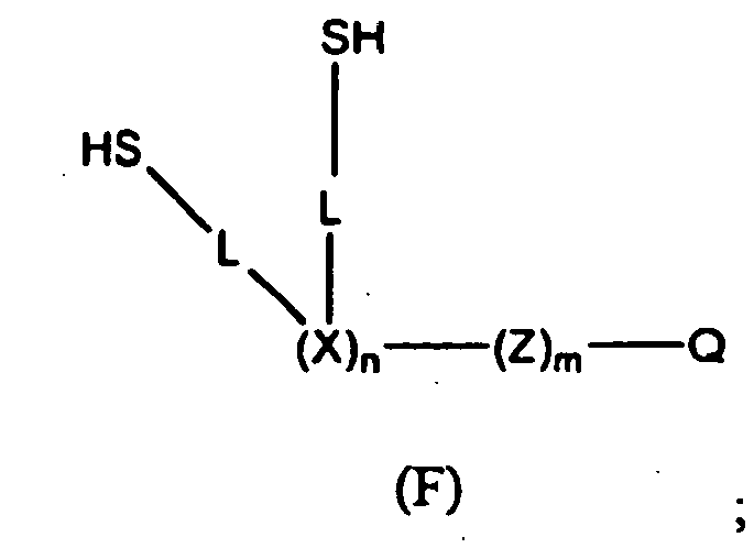

n is 2-100;
m is 0-100;
X is a nucleotide or modified nucleotide;
Z is a nucleotide, modified oligonucleotide, or polyanion;
Q is a recognition group;
R is linear or branched C1-C8 alkyl;
W is a steroid; and
each L is a linker formed by the coupling of two moieties selected from the group consisting of COOH, NH2, CHO, F, Cl, Br, I, NCO, NCS, allyl, vinyl, and CH3CO2 -, or L is -C(=NH2Cl)(CH2)3 -,
wherein the bioconjugate is coupled to the nanoparticle through the sulfur groups. - The probe according to claim 1, wherein Q in the bioconjugate comprises an organic molecule, an organic or inorganic polymer, a receptor, a nucleotide, a nucleoside, a polynucleotide, an oligonucleotide, a protein, an antibody, a peptide, a carbohydrate, a sugar, a hapten, a nucleic acid, an amino acid, a peptide nucleic acid, a linked nucleic acid, a nucleoside triphosphate, a lipid, a lipid bound protein, an aptamer, a virus, a cell fragment, or a whole cell.
- The probe according to claim 1, wherein the recognition group comprises a first and a second recognition group, wherein the first recognition group is bound to the second recognition group and wherein the second recognition group is a member of a recognition couple that specifically binds to a target analyte.
- The probe according to claim 3, wherein the first and second recognition groups comprise a specific binding pair.
- The probe according to claim 4, wherein the binding pair is an antibody-antigen or a receptor-ligand.
- The probe according to claim 3, wherein the first and second recognition groups are oligonucleotides having at least a portion of its sequence complementary to each other.
- The probe according to claim 1, wherein X and Z are independently adenine, guanine, cystosine, thymine, uracil, a nucleotide derivative, a modified nucleotide, or other molecules that may be incorporated into the oligonucleotide synthesis process.
- The probe according to claim 7, wherein X is thymine.
- The probe according to claim 1, wherein L is -NH -C(=O)-.
- The probe according to claim 1, wherein n from bioconjugate D is 2.
- The probe according to claim 2, wherein Q is an oligonucleotide.
- The probe according to claim 2, wherein Q is a cDNA.
- The probe according to claim 2, wherein Q is a polynucleotide.
- The probe according to claim 1, wherein the nanoparticle is selected from nanoparticles that have an affinity for thiol groups.
- The probe according to claim 14, wherein the nanoparticle is selected from the group consisting of a metal, a semiconductor, a magnetic material, including Au, Ag, Pt, Co, CdSe, CdS, or Si.
- The probe according to claim 15, wherein the nanoparticle is gold.
- The probe according to claim 16, wherein the nanoparticle has a mean diameter of about 5 nm to about 150 nm.
- A method for detecting a target analyte comprising:providing a nanoparticle probe according to claim 1, wherein the recognition group on the bioconjugate specifically binds to the target analyte;contacting the nanoparticle probe and the target analyte under conditions effective to specifically bind the target analyte to the recognition group; anddetecting the nanoparticle bound to the target analyte.
- The method according to claim 18, wherein the target analyte is a nucleic acid.
- The method according to claim 19, wherein binding of the recognition group to the nucleic acid is by hybridization of the recognition group with at least a portion of the nucleic acid.
- The method according to claim 18, wherein the target analyte is an antibody.
- The method according to claim 20, wherein the contacting conditions include freezing and thawing.
- The method of any one according to claims 19 or 21, wherein the detectable change is a color change observable with the naked eye.
- The method of any one according to claims 19 or 21, wherein the nanoparticle probe is detected with a silver stain.
- The method according to claim 23, wherein the color change is observed on a solid surface.
- The method according to claim 18, wherein the nanoparticles are made of gold.
- A method for detecting a nucleic acid having a first and second portion comprising:(a) providing a substrate having attached thereto oligonucleotides complementary to the first portion of the nucleic acid;(b) providing nanoparticle-probes according to claim 1, wherein the nanoparticles have oligonucleotides bound thereto as recognition group, the oligonucleotides have a sequence that is complementary to the second portion of the nucleic acid to be detected;(c) contacting the substrate and nanoparticles with the nucleic acid under conditions effective for hybridization between the nucleic acid, the oligonucleotides bound to the substrate and the oligonucleotides bound to the nanoparticles; and(d) detecting the nanoparticles bound to the nucleic acid bound to the substrate.
- The method according to claim 27, wherein the nucleic acid is contacted with the substrate provided in (a) prior to contacting it with the gold nanoparticles provided in (b).
- The method according to claim 27, wherein the nucleic acid is contacted with the nanoparticles provided in (b) prior to contacting it with the substrate provided in (a).
- The method according to claim 27, wherein the nanoparticle comprises oligonucleotides complementary to oligonucleotides on second nanoparticles and form an aggregate with the second gold nanoparticles.
- The method according to claim 27, wherein the nanoparticle is detected with a silver stain.
- The method according to claim 27, wherein the nucleic acid is RNA or DNA.
- The method according to claim 27 wherein the nucleic acid is human, bacterial or viral or fungal origin.
- The method according to claim 27 wherein the nucleic acid is a gene associated with a disease.
- The method according to claim 27, wherein the nucleic acid is a synthetic DNA, a synthetic RNA, a structurally-modified natural or synthetic RNA, or a structurally-modified natural or synthetic DNA.
- The method according to claim 27, wherein the nucleic acid is a product of a polymerase chain reaction amplification.
- The method according to claim 27, wherein the substrate has a plurality of oligonucleotides attached to it in an array to allow for the detection of multiple portions of a single nucleic acid, the detection of multiple different nucleic acids, or both.
- The method according to claim 37, wherein the substrate is a transparent substrate or opaque white substrate.
- The method according to claim 27, wherein the nanoparticles are made of gold.
- The method according to claim 27, wherein the substrate is contacted with staining material to produce the detectable change.
- The method according to claim 40, wherein the staining material is silver stain.
- A method for detecting an analyte in a sample comprising:providing a nanoparticle conjugate according to claim 1, the conjugate having a recognition group that is a specific binding complement of said analyte;contacting the analyte with the nanoparticle conjugate under conditions effective to allow specific binding interactions between the analyte and specific binding complement bound to the nanoparticle conjugate; andobserving a detectable change brought about by the specific binding interaction of the analyte and the specific binding complement of said analyte.
- A method for detecting an analyte in a sample comprising:providing a nanoparticle conjugate according to claim 1, the conjugate having oligonucleotides bound thereto as a recognition group, at least a portion of the oligonucleotides attached to the nanoparticles are bound, as a result of hybridization, to second oligonucleotides having bound thereto a specific binding complement of said analyte;contacting the analyte with the nanoparticle conjugate under conditions effective to allow specific binding interactions between the analyte and specific binding complement bound to the nanoparticle conjugate; andobserving a detectable change brought about by the specific binding interaction of the analyte and the specific binding complement of said analyte.
- The method according to any one of claims 42 or 43, wherein the analyte is polyvalent and binds to two or more nanoparticle conjugates.
- The method according to any one of claims 42 or 43, wherein the analyte is polyvalent and specifically binds to two or more nanoparticle conjugates.
- The method according to any one of claims 42 or 43, wherein the contacting conditions include freezing and thawing.
- The method according to any one of claims 42 or 43, wherein the contacting conditions include heating.
- The method according to any one of claims 42 or 43, wherein the detectable change is observed on a solid surface.
- The method according to any one of claims 42 or 43, wherein the detectable change is a color change observable with the naked eye.
- The method according to any one of claims 42 or 43, wherein the color change is observed on a solid surface.
- The method according to any one of claims 42 or 43, wherein the nanoparticles are made of gold.
- A method for detecting an analyte comprising:providing (i) a support having an analyte bound thereto and (ii) a nanoparticle conjugate according to claim 1, the conjugate having oligonucleotides bound thereto as a recognition group, a least a portion of the oligonucleotides are bound, as a result of hybridization, to second oligonucleotides having a specific binding complement of said analyte;contacting the analyte bound to the support to the nanoparticle conjugate under conditions effective to allow specific binding interactions between the analyte and specific binding complement bound to the nanoparticle conjugate; andobserving a detectable change dependent on the specific binding of the analyte to the specific binding complement of the analyte.
- A method for detecting an analyte comprising:providing (i) a support having a oligonucleotides bound thereto, (ii) an analyte having an oligonucleotide bound thereto, the oligonucleotide bound to the analyte has a sequence that is complementary to the oligonucleotides bound to the support; and (iii) a nanoparticle conjugate according to claim 1, the conjugate having oligonucleotides bound thereto as a recognition group, at least a portion of the oligonucleotides bound to the nanoparticle are bound, as a result of hybridization, to second oligonucleotides having bound thereto a specific binding complement of said analyte;contacting the oligonucleotides bound to the support with the olignonucleotide bound to the analyte under conditions effective to allow hybridization between the oligonucleotides bound to the support and the oligonucleotides bound to the analytes;contacting the analyte bound to the support with the nanoparticle conjugate under conditions effective to allow for specific binding interactions between the analyte bound to the support and the specific binding complement bound to the nanoparticle; andobserving a detectable change dependent on the specific binding of the analyte to the specific binding complement of the analyte.
- The method according to claim 53, wherein the substrate is contacted with staining material to produce the detectable change.
- The method according to claim 54, wherein the staining material is silver stain.
- A method for detecting an analyte comprising:providing (i) a support having an analyte bound thereto and (ii) an nanoparticle conjugate according to claim 1, the conjugates having oligonucleotides bound thereto as a recognition group, at least some of the oligonucleotides attached to the nanoparticle are bound, as a result of hybridization, to second oligonucleotides having bound thereto a specific binding complement of said analyte;contacting a support having an analyte bound thereto with the nanoparticle conjugate under conditions effective to allow specific binding interactions between the analyte bound to the support and specific binding complement bound to the nanoparticle conjugate;contacting the nanoparticle conjugate bound to the support with staining material to produce a detectable change; andobserving the detectable change dependent on the specific binding of the analyte and the specific binding complement of the analyte.
- The method according to any one of claims 53 or 56, wherein the substrate has a plurality of oligonucleotides attached to it in an array to allow for the detection of multiple portions of a single nucleic acid, the detection of multiple different nucleic acids, or both.
- The method according to any one of claims 53 or 56, wherein the detectable change is observed on a solid surface.
- The method according to any one of claims 53 or 56, wherein the detectable change is a color change observable with the naked eye.
- The method according to any one of claims 53 or 56, wherein the color change is observed on a solid surface.
- The method according to any one of claims 53 or 56, wherein the nanoparticles are made of gold.
- The method according to any one of claims 53 or 56, wherein the substrate is a transparent substrate or opaque white substrate.
- The method according to claim 56, wherein the staining material is silver stain.
Applications Claiming Priority (3)
| Application Number | Priority Date | Filing Date | Title |
|---|---|---|---|
| US34823901P | 2001-11-09 | 2001-11-09 | |
| US348239P | 2001-11-09 | ||
| PCT/US2002/035888 WO2003081202A2 (en) | 2001-11-09 | 2002-11-08 | Bioconjugate-nanoparticle probes |
Publications (3)
| Publication Number | Publication Date |
|---|---|
| EP1466015A2 EP1466015A2 (en) | 2004-10-13 |
| EP1466015A4 EP1466015A4 (en) | 2007-02-28 |
| EP1466015B1 true EP1466015B1 (en) | 2008-08-06 |
Family
ID=28454565
Family Applications (1)
| Application Number | Title | Priority Date | Filing Date |
|---|---|---|---|
| EP02806838A Expired - Lifetime EP1466015B1 (en) | 2001-11-09 | 2002-11-08 | Bioconjugate-nanoparticle probes |
Country Status (8)
| Country | Link |
|---|---|
| US (1) | US7186814B2 (en) |
| EP (1) | EP1466015B1 (en) |
| JP (1) | JP4220397B2 (en) |
| AT (1) | ATE403751T1 (en) |
| AU (1) | AU2002367817B2 (en) |
| CA (1) | CA2466656C (en) |
| DE (1) | DE60228128D1 (en) |
| WO (1) | WO2003081202A2 (en) |
Families Citing this family (67)
| Publication number | Priority date | Publication date | Assignee | Title |
|---|---|---|---|---|
| US6506564B1 (en) * | 1996-07-29 | 2003-01-14 | Nanosphere, Inc. | Nanoparticles having oligonucleotides attached thereto and uses therefor |
| US6582921B2 (en) | 1996-07-29 | 2003-06-24 | Nanosphere, Inc. | Nanoparticles having oligonucleotides attached thereto and uses thereof |
| US7098320B1 (en) | 1996-07-29 | 2006-08-29 | Nanosphere, Inc. | Nanoparticles having oligonucleotides attached thereto and uses therefor |
| US7169556B2 (en) * | 1996-07-29 | 2007-01-30 | Nanosphere, Inc. | Nanoparticles having oligonucleotides attached thereto and uses therefor |
| US6984491B2 (en) * | 1996-07-29 | 2006-01-10 | Nanosphere, Inc. | Nanoparticles having oligonucleotides attached thereto and uses therefor |
| US6750016B2 (en) * | 1996-07-29 | 2004-06-15 | Nanosphere, Inc. | Nanoparticles having oligonucleotides attached thereto and uses therefor |
| US6974669B2 (en) * | 2000-03-28 | 2005-12-13 | Nanosphere, Inc. | Bio-barcodes based on oligonucleotide-modified nanoparticles |
| US20050037397A1 (en) * | 2001-03-28 | 2005-02-17 | Nanosphere, Inc. | Bio-barcode based detection of target analytes |
| US20030096321A1 (en) * | 1999-05-19 | 2003-05-22 | Jose Remacle | Method for the identification and/or the quantification of a target compound obtained from a biological sample upon chips |
| WO2001051665A2 (en) * | 2000-01-13 | 2001-07-19 | Nanosphere Inc. | Nanoparticles having oligonucleotides attached thereto and uses therefor |
| US6602669B2 (en) * | 2000-07-11 | 2003-08-05 | Northwestern University | Method of detection by enhancement of silver staining |
| US20060040286A1 (en) * | 2001-03-28 | 2006-02-23 | Nanosphere, Inc. | Bio-barcode based detection of target analytes |
| US20030113740A1 (en) * | 2001-04-26 | 2003-06-19 | Mirkin Chad A. | Oligonucleotide-modified ROMP polymers and co-polymers |
| US7238472B2 (en) | 2001-05-25 | 2007-07-03 | Nanosphere, Inc. | Non-alloying core shell nanoparticles |
| US7147687B2 (en) * | 2001-05-25 | 2006-12-12 | Nanosphere, Inc. | Non-alloying core shell nanoparticles |
| WO2005108625A2 (en) | 2001-07-13 | 2005-11-17 | Nanosphere, Inc. | Method for preparing substrates having immobilized molecules and substrates |
| US20060177852A1 (en) * | 2001-12-12 | 2006-08-10 | Do-Coop Technologies Ltd. | Solid-fluid composition |
| US20030211488A1 (en) * | 2002-05-07 | 2003-11-13 | Northwestern University | Nanoparticle probs with Raman spectrocopic fingerprints for analyte detection |
| JP2005532456A (en) * | 2002-07-02 | 2005-10-27 | ナノスフェアー インコーポレイテッド | Nanoparticle polyanion complex and its use in the detection of analytes |
| JP2007524347A (en) * | 2003-02-27 | 2007-08-30 | ナノスフェアー インコーポレイテッド | Label-free gene expression profiling using universal nanoparticle probes in microarray format assays |
| US20050250094A1 (en) * | 2003-05-30 | 2005-11-10 | Nanosphere, Inc. | Method for detecting analytes based on evanescent illumination and scatter-based detection of nanoparticle probe complexes |
| US7226636B2 (en) * | 2003-07-31 | 2007-06-05 | Los Alamos National Security, Llc | Gold-coated nanoparticles for use in biotechnology applications |
| JP4207754B2 (en) * | 2003-10-31 | 2009-01-14 | 和光純薬工業株式会社 | Immunological measurement method using magnetic substance |
| US7396677B2 (en) * | 2003-11-07 | 2008-07-08 | Nanosphere, Inc. | Method of preparing nucleic acids for detection |
| US7906345B2 (en) * | 2003-11-12 | 2011-03-15 | The Board Of Trustees Of The Leland Stanford Junior University | Magnetic nanoparticles, magnetic detector arrays, and methods for their use in detecting biological molecules |
| US20090206245A1 (en) * | 2004-03-31 | 2009-08-20 | Shionogi Co., Ltd | Mass analysis method using fine metal particles |
| US7462452B2 (en) * | 2004-04-30 | 2008-12-09 | Pacific Biosciences Of California, Inc. | Field-switch sequencing |
| JP2008507296A (en) * | 2004-07-26 | 2008-03-13 | ナノスフェアー インコーポレイテッド | A method for distinguishing methicillin-resistant Staphylococcus aureus from methicillin-sensitive Staphylococcus aureus in mixed cultures |
| US20060134694A1 (en) * | 2004-12-22 | 2006-06-22 | Intel Corporation | Methods of protein profiling by thiolation |
| JP4791867B2 (en) * | 2005-03-31 | 2011-10-12 | 日立マクセル株式会社 | Detection method of test substance using precious metal coated magnetic particles |
| US20070154903A1 (en) * | 2005-06-23 | 2007-07-05 | Nanosphere, Inc. | Selective isolation and concentration of nucleic acids from complex samples |
| DE602006018477D1 (en) * | 2005-08-19 | 2011-01-05 | Nanosphere Inc | DNA AND ANTIBODIES AND USES THEREOF |
| CA2635978A1 (en) * | 2006-01-04 | 2007-07-12 | Do-Coop Technologies Ltd. | Solid-fluid composition |
| US20090004296A1 (en) * | 2006-01-04 | 2009-01-01 | Do-Coop Technologies Ltd. | Antiseptic Compositions and Methods of Using Same |
| US20090253613A1 (en) * | 2006-01-04 | 2009-10-08 | Do-Coop Technologies Ltd. | Solid-Fluid Composition |
| WO2008091364A2 (en) * | 2006-06-30 | 2008-07-31 | Intel Corporation | Nanoparticles as tags for bioanalyte detection by nuclear magnetic resonance or electron spin resonance |
| KR100828936B1 (en) | 2006-08-01 | 2008-05-20 | 김성천 | - Method for Analysis of Biological Molecule Using Single-Stranded Nucleic Acid Aptamer and Gold Nano-Particle |
| US20100041103A1 (en) * | 2007-01-04 | 2010-02-18 | Do-Coop Technologies Ltd. | Composition and method for enhancing cell growth and cell fusion |
| AU2008203627A1 (en) * | 2007-01-04 | 2008-07-10 | Do-Coop Technologies Ltd. | Detection of analytes |
| US8354110B2 (en) | 2007-03-07 | 2013-01-15 | Uti Limited Partnership | Compositions and methods for the prevention and treatment of autoimmune conditions |
| US9222936B2 (en) | 2007-04-18 | 2015-12-29 | Solulink, Inc. | Methods and/or use of oligonucleotide conjugates for suppressing background due to cross-hybridization |
| US8597959B2 (en) * | 2007-04-19 | 2013-12-03 | 3M Innovative Properties Company | Methods of use of solid support material for binding biomolecules |
| EP2137533A2 (en) * | 2007-04-19 | 2009-12-30 | 3M Innovative Properties Company | Uses of water-dispersible silica nanoparticles for attaching biomolecules |
| CA2694045C (en) * | 2007-07-26 | 2016-06-21 | Agamatrix, Inc. | Electrochemical analyte detection apparatus and method |
| EP2200727B1 (en) | 2007-09-20 | 2014-08-13 | Magarray, Inc. | Analyte detection with magnetic sensors |
| US20090185113A1 (en) * | 2008-01-22 | 2009-07-23 | Industrial Technology Research Institute | Color Filter Module and Device of Having the Same |
| JP2012523576A (en) | 2009-04-13 | 2012-10-04 | ザ ボード オブ トラスティーズ オブ ザ リーランド スタンフォード ジュニア ユニバーシティ | Method and apparatus for detecting the presence of an analyte in a sample |
| EP2430033A4 (en) * | 2009-05-05 | 2012-11-07 | Altermune Technologies Llc | Chemically programmable immunity |
| US20100291541A1 (en) * | 2009-05-14 | 2010-11-18 | Evoy Stephane | Bacteriophage immobilization for biosensors |
| WO2011112929A2 (en) | 2010-03-12 | 2011-09-15 | The Board Of Trustees Of The Leland Stanford Junior University | Magnetic sensor based quantitative binding kinetics analysis |
| EP3308797A1 (en) | 2010-09-29 | 2018-04-18 | Uti Limited Partnership | Methods for treating autoimmune disease using biocompatible bioabsorbable nanospheres |
| US9511151B2 (en) | 2010-11-12 | 2016-12-06 | Uti Limited Partnership | Compositions and methods for the prevention and treatment of cancer |
| US9851308B2 (en) * | 2010-11-19 | 2017-12-26 | Wisconsin Alumni Research Foundation (Warf) | Visible detection of microorganisms |
| US20120258870A1 (en) * | 2010-11-22 | 2012-10-11 | The University Of Chicago | Methods, Systems, and/or Use of Oligonucleotide Conjugates to Develop Panels for Use in Assays and Detections |
| US9151763B2 (en) | 2011-03-09 | 2015-10-06 | Magarray, Inc. | Systems and methods for high-throughput detection of an analyte in a sample |
| US8946389B2 (en) | 2011-04-25 | 2015-02-03 | University Of Washington | Compositions and methods for multiplex biomarker profiling |
| US9982292B2 (en) | 2012-09-28 | 2018-05-29 | Src, Inc. | Detecting chemical and biological agents using textile-based sensors |
| US9512468B2 (en) | 2012-11-06 | 2016-12-06 | Industrial Technology Research Institute | Detection method uses magnetic and detectable nanoparticles with oligonucleotides attached thereto |
| WO2014151820A1 (en) | 2013-03-15 | 2014-09-25 | Magarray, Inc. | Magnetic tunnel junction sensors and methods for using the same |
| CA2928190C (en) * | 2013-10-23 | 2023-09-19 | Chemgenes Corporation | Dithiolane functionalized nucleoside amidites and supports for stronger immobilization of bio-molecules on solid surfaces |
| KR101718867B1 (en) * | 2014-06-02 | 2017-03-22 | 한국과학기술연구원 | Biocompatible Fluorescence Nanoparticles and Uses Thereof |
| CN104894097B (en) * | 2015-05-09 | 2018-11-06 | 浙江省农业科学院 | A kind of long-acting water purification bacterium capsule and preparation method thereof |
| CN104909463A (en) * | 2015-05-09 | 2015-09-16 | 浙江省农业科学院 | Core-shell structured water purification bacterium capsule and preparation method thereof |
| JP6752231B2 (en) | 2015-06-01 | 2020-09-09 | カリフォルニア インスティテュート オブ テクノロジー | Compositions and Methods for Screening T Cells for Specific Populations Using Antigens |
| WO2019200326A1 (en) | 2018-04-13 | 2019-10-17 | Rarecyte, Inc. | Kits for labeling of biomarkers and methods of using the same |
| CN113311047B (en) * | 2021-05-06 | 2022-06-21 | 复旦大学 | Gold nanoparticle modified pseudo MOS biochemical molecular sensor and preparation method thereof |
| US11795505B2 (en) | 2022-03-10 | 2023-10-24 | Singular Genomics Systems, Inc. | Nucleic acid delivery scaffolds |
Family Cites Families (80)
| Publication number | Priority date | Publication date | Assignee | Title |
|---|---|---|---|---|
| US4193983A (en) | 1978-05-16 | 1980-03-18 | Syva Company | Labeled liposome particle compositions and immunoassays therewith |
| NL7807532A (en) | 1978-07-13 | 1980-01-15 | Akzo Nv | METAL IMMUNO TEST. |
| US4318707A (en) | 1978-11-24 | 1982-03-09 | Syva Company | Macromolecular fluorescent quencher particle in specific receptor assays |
| US4256834A (en) | 1979-04-09 | 1981-03-17 | Syva Company | Fluorescent scavenger particle immunoassay |
| US4261968A (en) | 1979-05-10 | 1981-04-14 | Syva Company | Fluorescence quenching with immunological pairs in immunoassays |
| US4650770A (en) | 1981-04-27 | 1987-03-17 | Syntex (U.S.A.) Inc. | Energy absorbing particle quenching in light emitting competitive protein binding assays |
| US4713348A (en) | 1983-04-05 | 1987-12-15 | Syntex (U.S.A.) Inc. | Fluorescent multiparameter particle analysis |
| US5288609A (en) * | 1984-04-27 | 1994-02-22 | Enzo Diagnostics, Inc. | Capture sandwich hybridization method and composition |
| US4868104A (en) | 1985-09-06 | 1989-09-19 | Syntex (U.S.A.) Inc. | Homogeneous assay for specific polynucleotides |
| US4996143A (en) * | 1985-12-23 | 1991-02-26 | Syngene, Inc. | Fluorescent stokes shift probes for polynucleotide hybridization |
| US5137827A (en) * | 1986-03-25 | 1992-08-11 | Midwest Research Technologies, Inc. | Diagnostic element for electrical detection of a binding reaction |
| US5514602A (en) | 1986-06-09 | 1996-05-07 | Ortho Diagnostic Systems, Inc. | Method of producing a metal sol reagent containing colloidal metal particles |
| US5360895A (en) * | 1987-04-22 | 1994-11-01 | Associated Universities, Inc. | Derivatized gold clusters and antibody-gold cluster conjugates |
| US4853335A (en) | 1987-09-28 | 1989-08-01 | Olsen Duane A | Colloidal gold particle concentration immunoassay |
| GB8800702D0 (en) | 1988-01-13 | 1988-02-10 | Nycomed As | Test method & reagent kit therefor |
| WO1990002205A1 (en) | 1988-08-25 | 1990-03-08 | Angenics, Inc. | Detection of nucleic acid sequences using particle agglutination |
| US5460831A (en) | 1990-06-22 | 1995-10-24 | The Regents Of The University Of California | Targeted transfection nanoparticles |
| GB9019512D0 (en) | 1990-09-06 | 1990-10-24 | Ici Plc | Assay method |
| AU8951191A (en) * | 1990-10-29 | 1992-05-26 | Dekalb Plant Genetics | Isolation of biological materials using magnetic particles |
| US5665582A (en) | 1990-10-29 | 1997-09-09 | Dekalb Genetics Corp. | Isolation of biological materials |
| US5294369A (en) | 1990-12-05 | 1994-03-15 | Akzo N.V. | Ligand gold bonding |
| WO1993010564A1 (en) | 1991-11-22 | 1993-05-27 | The Regents Of The University Of California | Semiconductor nanocrystals covalently bound to solid inorganic surfaces using self-assembled monolayers |
| US5225064A (en) | 1992-01-15 | 1993-07-06 | Enzyme Technology Research Group, Inc. | Peroxidase colloidal gold oxidase biosensors for mediatorless glucose determination |
| GB9212164D0 (en) | 1992-06-09 | 1992-07-22 | Medical Res Council | Preparation of nucleic acids |
| US5472881A (en) * | 1992-11-12 | 1995-12-05 | University Of Utah Research Foundation | Thiol labeling of DNA for attachment to gold surfaces |
| US5384265A (en) * | 1993-03-26 | 1995-01-24 | Geo-Centers, Inc. | Biomolecules bound to catalytic inorganic particles, immunoassays using the same |
| US5637508A (en) * | 1993-03-26 | 1997-06-10 | Geo-Centers, Inc. | Biomolecules bound to polymer or copolymer coated catalytic inorganic particles, immunoassays using the same and kits containing the same |
| US5681943A (en) | 1993-04-12 | 1997-10-28 | Northwestern University | Method for covalently linking adjacent oligonucleotides |
| AU690124B2 (en) | 1993-06-09 | 1998-04-23 | Gamera Bioscience Corporation | Magnetic cycle reaction |
| EP0630974A3 (en) | 1993-06-25 | 1995-11-15 | Clinical Diagnostic Syst | Method and test kit for the detection of inorganic orthophosphate by-product from amplification of target nucleic acid. |
| US5543158A (en) | 1993-07-23 | 1996-08-06 | Massachusetts Institute Of Technology | Biodegradable injectable nanoparticles |
| JPH07227299A (en) | 1994-02-14 | 1995-08-29 | Kyoto Daiichi Kagaku:Kk | Method for detecting specific base sequence of dna and device therefor |
| US6121425A (en) * | 1994-07-29 | 2000-09-19 | Nanoprobes, Inc. | Metal-lipid molecules |
| US5521289A (en) * | 1994-07-29 | 1996-05-28 | Nanoprobes, Inc. | Small organometallic probes |
| US5599668A (en) * | 1994-09-22 | 1997-02-04 | Abbott Laboratories | Light scattering optical waveguide method for detecting specific binding events |
| US6025202A (en) | 1995-02-09 | 2000-02-15 | The Penn State Research Foundation | Self-assembled metal colloid monolayers and detection methods therewith |
| US5609907A (en) | 1995-02-09 | 1997-03-11 | The Penn State Research Foundation | Self-assembled metal colloid monolayers |
| IL126544A (en) * | 1996-04-25 | 2004-08-31 | Genicon Sciences Inc | Analyte assay using scattered-light detectable particles |
| US6586193B2 (en) | 1996-04-25 | 2003-07-01 | Genicon Sciences Corporation | Analyte assay using particulate labels |
| US6582921B2 (en) * | 1996-07-29 | 2003-06-24 | Nanosphere, Inc. | Nanoparticles having oligonucleotides attached thereto and uses thereof |
| US6750016B2 (en) | 1996-07-29 | 2004-06-15 | Nanosphere, Inc. | Nanoparticles having oligonucleotides attached thereto and uses therefor |
| US6361944B1 (en) | 1996-07-29 | 2002-03-26 | Nanosphere, Inc. | Nanoparticles having oligonucleotides attached thereto and uses therefor |
| US6506564B1 (en) | 1996-07-29 | 2003-01-14 | Nanosphere, Inc. | Nanoparticles having oligonucleotides attached thereto and uses therefor |
| US20020172953A1 (en) | 1996-07-29 | 2002-11-21 | Mirkin Chad A. | Movement of biomolecule-coated nanoparticles in an electric field |
| ES2287956T3 (en) | 1996-07-29 | 2007-12-16 | Nanosphere Inc. | NANOPARTICLES THAT HAVE OLIGONUCLEOTIDES UNITED TO THE SAME AND USES OF THE SAME. |
| WO1998010289A1 (en) | 1996-09-04 | 1998-03-12 | The Penn State Research Foundation | Self-assembled metal colloid monolayers |
| JP2001503041A (en) | 1996-10-23 | 2001-03-06 | エスケーダヴリュ トロストベルグ アクチエンゲゼルシャフト | Method for producing bioactive polymer nanoparticles-nucleic acid conjugate |
| US5830986A (en) | 1996-10-28 | 1998-11-03 | Massachusetts Institute Of Technology | Methods for the synthesis of functionalizable poly(ethylene oxide) star macromolecules |
| US5900481A (en) | 1996-11-06 | 1999-05-04 | Sequenom, Inc. | Bead linkers for immobilizing nucleic acids to solid supports |
| US5922537A (en) * | 1996-11-08 | 1999-07-13 | N.o slashed.AB Immunoassay, Inc. | Nanoparticles biosensor |
| US5939021A (en) * | 1997-01-23 | 1999-08-17 | Hansen; W. Peter | Homogeneous binding assay |
| US6149868A (en) | 1997-10-28 | 2000-11-21 | The Penn State Research Foundation | Surface enhanced raman scattering from metal nanoparticle-analyte-noble metal substrate sandwiches |
| AUPP004497A0 (en) | 1997-10-28 | 1997-11-20 | University Of Melbourne, The | Stabilized particles |
| JP2002509443A (en) | 1997-10-31 | 2002-03-26 | ジェン−プローブ・インコーポレーテッド | Nucleic acid detection method |
| US5990479A (en) * | 1997-11-25 | 1999-11-23 | Regents Of The University Of California | Organo Luminescent semiconductor nanocrystal probes for biological applications and process for making and using such probes |
| US5972615A (en) * | 1998-01-21 | 1999-10-26 | Urocor, Inc. | Biomarkers and targets for diagnosis, prognosis and management of prostate disease |
| US6287765B1 (en) | 1998-05-20 | 2001-09-11 | Molecular Machines, Inc. | Methods for detecting and identifying single molecules |
| US6290839B1 (en) * | 1998-06-23 | 2001-09-18 | Clinical Micro Sensors, Inc. | Systems for electrophoretic transport and detection of analytes |
| US6406921B1 (en) | 1998-07-14 | 2002-06-18 | Zyomyx, Incorporated | Protein arrays for high-throughput screening |
| US6251303B1 (en) | 1998-09-18 | 2001-06-26 | Massachusetts Institute Of Technology | Water-soluble fluorescent nanocrystals |
| US6306610B1 (en) | 1998-09-18 | 2001-10-23 | Massachusetts Institute Of Technology | Biological applications of quantum dots |
| US6203989B1 (en) | 1998-09-30 | 2001-03-20 | Affymetrix, Inc. | Methods and compositions for amplifying detectable signals in specific binding assays |
| IL126776A (en) | 1998-10-27 | 2001-04-30 | Technion Res & Dev Foundation | Method for gold depositions |
| EP2000803A1 (en) | 1998-11-30 | 2008-12-10 | Nanosphere, Inc. | Nanoparticles with Polymer Shells |
| US6277489B1 (en) | 1998-12-04 | 2001-08-21 | The Regents Of The University Of California | Support for high performance affinity chromatography and other uses |
| AU784040B2 (en) | 1999-06-25 | 2006-01-19 | Nanosphere, Inc. | Nanoparticles having oligonucleotides attached thereto and uses therefor |
| JP2003506919A (en) | 1999-08-03 | 2003-02-18 | ポリコム・インコーポレイテッド | System and method for secure data transfer in a wireless communication system |
| WO2001051665A2 (en) | 2000-01-13 | 2001-07-19 | Nanosphere Inc. | Nanoparticles having oligonucleotides attached thereto and uses therefor |
| ATE487136T1 (en) | 2000-03-28 | 2010-11-15 | Nanosphere Inc | NANOPARTICLES WITH BONDED OLIGONUCLEOTIDES AND USES THEREOF |
| AU5667901A (en) | 2000-05-11 | 2001-11-20 | Center For Advanced Science And Technology Incubation, Ltd. | Polymer composition for forming surface of biosensor |
| US6602669B2 (en) | 2000-07-11 | 2003-08-05 | Northwestern University | Method of detection by enhancement of silver staining |
| AU2001281248A1 (en) | 2000-08-11 | 2002-03-13 | Nanosphere Inc. | Nanoparticles having oligonucleotides attached thereto and uses therefor |
| JP2004534721A (en) | 2000-10-31 | 2004-11-18 | ピーアール ファーマシューティカルズ,インク. | Methods and compositions for enhanced delivery of bioactive molecules |
| US6726847B2 (en) | 2000-12-06 | 2004-04-27 | Northwestern University | Silver stain removal by chemical etching and sonication |
| EP1356109A2 (en) | 2000-12-08 | 2003-10-29 | Nanosphere, Inc. | Nanoparticles having oligonucleotides attached thereto and uses therefor |
| US6958216B2 (en) * | 2001-01-10 | 2005-10-25 | The Trustees Of Boston College | DNA-bridged carbon nanotube arrays |
| US7147687B2 (en) | 2001-05-25 | 2006-12-12 | Nanosphere, Inc. | Non-alloying core shell nanoparticles |
| US7238472B2 (en) | 2001-05-25 | 2007-07-03 | Nanosphere, Inc. | Non-alloying core shell nanoparticles |
| EP1412536B1 (en) | 2001-08-03 | 2009-02-25 | Nanosphere, Inc. | Nanoparticle imaging system and method |
| US20050059030A1 (en) | 2002-12-12 | 2005-03-17 | Nanosphere, Inc. | Direct SNP detection with unamplified DNA |
-
2002
- 2002-11-08 WO PCT/US2002/035888 patent/WO2003081202A2/en active Search and Examination
- 2002-11-08 AT AT02806838T patent/ATE403751T1/en not_active IP Right Cessation
- 2002-11-08 AU AU2002367817A patent/AU2002367817B2/en not_active Ceased
- 2002-11-08 DE DE60228128T patent/DE60228128D1/en not_active Expired - Lifetime
- 2002-11-08 US US10/291,291 patent/US7186814B2/en active Active - Reinstated
- 2002-11-08 CA CA2466656A patent/CA2466656C/en not_active Expired - Lifetime
- 2002-11-08 JP JP2003578887A patent/JP4220397B2/en not_active Expired - Lifetime
- 2002-11-08 EP EP02806838A patent/EP1466015B1/en not_active Expired - Lifetime
Also Published As
| Publication number | Publication date |
|---|---|
| EP1466015A4 (en) | 2007-02-28 |
| US7186814B2 (en) | 2007-03-06 |
| ATE403751T1 (en) | 2008-08-15 |
| WO2003081202A3 (en) | 2004-07-22 |
| US20030143598A1 (en) | 2003-07-31 |
| AU2002367817A1 (en) | 2003-10-08 |
| JP2005519627A (en) | 2005-07-07 |
| AU2002367817B2 (en) | 2008-05-29 |
| WO2003081202A2 (en) | 2003-10-02 |
| EP1466015A2 (en) | 2004-10-13 |
| CA2466656A1 (en) | 2003-10-02 |
| CA2466656C (en) | 2013-07-02 |
| DE60228128D1 (en) | 2008-09-18 |
| JP4220397B2 (en) | 2009-02-04 |
Similar Documents
| Publication | Publication Date | Title |
|---|---|---|
| EP1466015B1 (en) | Bioconjugate-nanoparticle probes | |
| EP1540006B1 (en) | Nanoparticle polyanion conjugates and methods of use thereof in detecting analytes | |
| US7208587B2 (en) | Nanoparticles having oligonucleotides attached thereto and uses therefor | |
| JP4146239B2 (en) | Bio barcode based on oligonucleotide modified particles | |
| US7323309B2 (en) | Bio-barcodes based on oligonucleotide-modified particles | |
| KR100571299B1 (en) | Pentopyranosyl nucleosides and preparation methods thereof | |
| US20100234579A1 (en) | Silver nanoparticle binding agent conjugates based on moieties with triple cyclic disulfide anchoring groups | |
| JP2000516460A (en) | Oligonucleotide-attached nanoparticles and methods of using the particles | |
| WO2008127789A2 (en) | Molecule attachment to nanoparticles | |
| JP2002518690A (en) | Binding promotion method for analyte detection | |
| AU757912B2 (en) | Addressable modular recognition system, production mode and use | |
| JP2002515590A (en) | Identification of nucleic acid sequence | |
| WO2002081739A2 (en) | Non-enzymatic liposome-linked closely spaced array electrodes assay (nel-ela) for detecting and quantifying nucleic acids | |
| WO2002082078A2 (en) | Activated enzyme-linked detection systems for detecting and quantifying nucleid acids, antigens antibodies and other analytes | |
| US11224664B2 (en) | Functionalized metal nanoparticle | |
| US6545134B1 (en) | Method for the production of pentopyranosyl nucleosides | |
| JP4460153B2 (en) | Linker nucleosides, their preparation and use |
Legal Events
| Date | Code | Title | Description |
|---|---|---|---|
| PUAI | Public reference made under article 153(3) epc to a published international application that has entered the european phase |
Free format text: ORIGINAL CODE: 0009012 |
|
| 17P | Request for examination filed |
Effective date: 20040609 |
|
| AK | Designated contracting states |
Kind code of ref document: A2 Designated state(s): AT BE BG CH CY CZ DE DK EE ES FI FR GB GR IE IT LI LU MC NL PT SE SK TR |
|
| AX | Request for extension of the european patent |
Extension state: AL LT LV MK RO SI |
|
| A4 | Supplementary search report drawn up and despatched |
Effective date: 20070129 |
|
| GRAP | Despatch of communication of intention to grant a patent |
Free format text: ORIGINAL CODE: EPIDOSNIGR1 |
|
| RIN1 | Information on inventor provided before grant (corrected) |
Inventor name: STORHOFF, JAMES, J. Inventor name: GARIMELLA, VISWANADHAM |
|
| GRAS | Grant fee paid |
Free format text: ORIGINAL CODE: EPIDOSNIGR3 |
|
| GRAA | (expected) grant |
Free format text: ORIGINAL CODE: 0009210 |
|
| AK | Designated contracting states |
Kind code of ref document: B1 Designated state(s): AT BE BG CH CY CZ DE DK EE ES FI FR GB GR IE IT LI LU MC NL PT SE SK TR |
|
| REG | Reference to a national code |
Ref country code: GB Ref legal event code: FG4D |
|
| REG | Reference to a national code |
Ref country code: CH Ref legal event code: EP |
|
| REG | Reference to a national code |
Ref country code: IE Ref legal event code: FG4D |
|
| REF | Corresponds to: |
Ref document number: 60228128 Country of ref document: DE Date of ref document: 20080918 Kind code of ref document: P |
|
| PG25 | Lapsed in a contracting state [announced via postgrant information from national office to epo] |
Ref country code: NL Free format text: LAPSE BECAUSE OF FAILURE TO SUBMIT A TRANSLATION OF THE DESCRIPTION OR TO PAY THE FEE WITHIN THE PRESCRIBED TIME-LIMIT Effective date: 20080806 Ref country code: ES Free format text: LAPSE BECAUSE OF FAILURE TO SUBMIT A TRANSLATION OF THE DESCRIPTION OR TO PAY THE FEE WITHIN THE PRESCRIBED TIME-LIMIT Effective date: 20081117 |
|
| PG25 | Lapsed in a contracting state [announced via postgrant information from national office to epo] |
Ref country code: AT Free format text: LAPSE BECAUSE OF FAILURE TO SUBMIT A TRANSLATION OF THE DESCRIPTION OR TO PAY THE FEE WITHIN THE PRESCRIBED TIME-LIMIT Effective date: 20080806 Ref country code: FI Free format text: LAPSE BECAUSE OF FAILURE TO SUBMIT A TRANSLATION OF THE DESCRIPTION OR TO PAY THE FEE WITHIN THE PRESCRIBED TIME-LIMIT Effective date: 20080806 |
|
| PG25 | Lapsed in a contracting state [announced via postgrant information from national office to epo] |
Ref country code: BE Free format text: LAPSE BECAUSE OF FAILURE TO SUBMIT A TRANSLATION OF THE DESCRIPTION OR TO PAY THE FEE WITHIN THE PRESCRIBED TIME-LIMIT Effective date: 20080806 |
|
| PG25 | Lapsed in a contracting state [announced via postgrant information from national office to epo] |
Ref country code: BG Free format text: LAPSE BECAUSE OF FAILURE TO SUBMIT A TRANSLATION OF THE DESCRIPTION OR TO PAY THE FEE WITHIN THE PRESCRIBED TIME-LIMIT Effective date: 20081106 Ref country code: DK Free format text: LAPSE BECAUSE OF FAILURE TO SUBMIT A TRANSLATION OF THE DESCRIPTION OR TO PAY THE FEE WITHIN THE PRESCRIBED TIME-LIMIT Effective date: 20080806 |
|
| PG25 | Lapsed in a contracting state [announced via postgrant information from national office to epo] |
Ref country code: PT Free format text: LAPSE BECAUSE OF FAILURE TO SUBMIT A TRANSLATION OF THE DESCRIPTION OR TO PAY THE FEE WITHIN THE PRESCRIBED TIME-LIMIT Effective date: 20090106 Ref country code: SK Free format text: LAPSE BECAUSE OF FAILURE TO SUBMIT A TRANSLATION OF THE DESCRIPTION OR TO PAY THE FEE WITHIN THE PRESCRIBED TIME-LIMIT Effective date: 20080806 Ref country code: CZ Free format text: LAPSE BECAUSE OF FAILURE TO SUBMIT A TRANSLATION OF THE DESCRIPTION OR TO PAY THE FEE WITHIN THE PRESCRIBED TIME-LIMIT Effective date: 20080806 |
|
| PLBE | No opposition filed within time limit |
Free format text: ORIGINAL CODE: 0009261 |
|
| STAA | Information on the status of an ep patent application or granted ep patent |
Free format text: STATUS: NO OPPOSITION FILED WITHIN TIME LIMIT |
|
| PG25 | Lapsed in a contracting state [announced via postgrant information from national office to epo] |
Ref country code: MC Free format text: LAPSE BECAUSE OF NON-PAYMENT OF DUE FEES Effective date: 20081130 |
|
| REG | Reference to a national code |
Ref country code: CH Ref legal event code: PL |
|
| 26N | No opposition filed |
Effective date: 20090507 |
|
| GBPC | Gb: european patent ceased through non-payment of renewal fee |
Effective date: 20081108 |
|
| PG25 | Lapsed in a contracting state [announced via postgrant information from national office to epo] |
Ref country code: EE Free format text: LAPSE BECAUSE OF FAILURE TO SUBMIT A TRANSLATION OF THE DESCRIPTION OR TO PAY THE FEE WITHIN THE PRESCRIBED TIME-LIMIT Effective date: 20080806 |
|
| REG | Reference to a national code |
Ref country code: IE Ref legal event code: MM4A |
|
| PG25 | Lapsed in a contracting state [announced via postgrant information from national office to epo] |
Ref country code: IT Free format text: LAPSE BECAUSE OF FAILURE TO SUBMIT A TRANSLATION OF THE DESCRIPTION OR TO PAY THE FEE WITHIN THE PRESCRIBED TIME-LIMIT Effective date: 20080806 |
|
| PG25 | Lapsed in a contracting state [announced via postgrant information from national office to epo] |
Ref country code: LI Free format text: LAPSE BECAUSE OF NON-PAYMENT OF DUE FEES Effective date: 20081130 Ref country code: CH Free format text: LAPSE BECAUSE OF NON-PAYMENT OF DUE FEES Effective date: 20081130 Ref country code: IE Free format text: LAPSE BECAUSE OF NON-PAYMENT OF DUE FEES Effective date: 20081108 |
|
| PG25 | Lapsed in a contracting state [announced via postgrant information from national office to epo] |
Ref country code: GB Free format text: LAPSE BECAUSE OF NON-PAYMENT OF DUE FEES Effective date: 20081108 |
|
| PG25 | Lapsed in a contracting state [announced via postgrant information from national office to epo] |
Ref country code: SE Free format text: LAPSE BECAUSE OF FAILURE TO SUBMIT A TRANSLATION OF THE DESCRIPTION OR TO PAY THE FEE WITHIN THE PRESCRIBED TIME-LIMIT Effective date: 20081106 |
|
| PG25 | Lapsed in a contracting state [announced via postgrant information from national office to epo] |
Ref country code: CY Free format text: LAPSE BECAUSE OF FAILURE TO SUBMIT A TRANSLATION OF THE DESCRIPTION OR TO PAY THE FEE WITHIN THE PRESCRIBED TIME-LIMIT Effective date: 20080806 Ref country code: LU Free format text: LAPSE BECAUSE OF NON-PAYMENT OF DUE FEES Effective date: 20081108 |
|
| PG25 | Lapsed in a contracting state [announced via postgrant information from national office to epo] |
Ref country code: TR Free format text: LAPSE BECAUSE OF FAILURE TO SUBMIT A TRANSLATION OF THE DESCRIPTION OR TO PAY THE FEE WITHIN THE PRESCRIBED TIME-LIMIT Effective date: 20080806 |
|
| PG25 | Lapsed in a contracting state [announced via postgrant information from national office to epo] |
Ref country code: GR Free format text: LAPSE BECAUSE OF FAILURE TO SUBMIT A TRANSLATION OF THE DESCRIPTION OR TO PAY THE FEE WITHIN THE PRESCRIBED TIME-LIMIT Effective date: 20081107 |
|
| REG | Reference to a national code |
Ref country code: FR Ref legal event code: PLFP Year of fee payment: 14 |
|
| REG | Reference to a national code |
Ref country code: FR Ref legal event code: PLFP Year of fee payment: 15 |
|
| REG | Reference to a national code |
Ref country code: FR Ref legal event code: PLFP Year of fee payment: 16 |
|
| PGFP | Annual fee paid to national office [announced via postgrant information from national office to epo] |
Ref country code: FR Payment date: 20211124 Year of fee payment: 20 Ref country code: DE Payment date: 20211126 Year of fee payment: 20 |
|
| REG | Reference to a national code |
Ref country code: DE Ref legal event code: R071 Ref document number: 60228128 Country of ref document: DE |