EP1557431B1 - Assay and reagents for quantifying hBNP - Google Patents
Assay and reagents for quantifying hBNP Download PDFInfo
- Publication number
- EP1557431B1 EP1557431B1 EP05007745A EP05007745A EP1557431B1 EP 1557431 B1 EP1557431 B1 EP 1557431B1 EP 05007745 A EP05007745 A EP 05007745A EP 05007745 A EP05007745 A EP 05007745A EP 1557431 B1 EP1557431 B1 EP 1557431B1
- Authority
- EP
- European Patent Office
- Prior art keywords
- hbnp
- antibody
- label
- complex
- biological fluid
- Prior art date
- Legal status (The legal status is an assumption and is not a legal conclusion. Google has not performed a legal analysis and makes no representation as to the accuracy of the status listed.)
- Expired - Lifetime
Links
Images


Classifications
-
- C—CHEMISTRY; METALLURGY
- C07—ORGANIC CHEMISTRY
- C07K—PEPTIDES
- C07K14/00—Peptides having more than 20 amino acids; Gastrins; Somatostatins; Melanotropins; Derivatives thereof
- C07K14/435—Peptides having more than 20 amino acids; Gastrins; Somatostatins; Melanotropins; Derivatives thereof from animals; from humans
- C07K14/575—Hormones
- C07K14/58—Atrial natriuretic factor complex; Atriopeptin; Atrial natriuretic peptide [ANP]; Cardionatrin; Cardiodilatin
-
- C—CHEMISTRY; METALLURGY
- C07—ORGANIC CHEMISTRY
- C07K—PEPTIDES
- C07K16/00—Immunoglobulins [IGs], e.g. monoclonal or polyclonal antibodies
- C07K16/18—Immunoglobulins [IGs], e.g. monoclonal or polyclonal antibodies against material from animals or humans
- C07K16/26—Immunoglobulins [IGs], e.g. monoclonal or polyclonal antibodies against material from animals or humans against hormones ; against hormone releasing or inhibiting factors
-
- C—CHEMISTRY; METALLURGY
- C07—ORGANIC CHEMISTRY
- C07K—PEPTIDES
- C07K2317/00—Immunoglobulins specific features
- C07K2317/30—Immunoglobulins specific features characterized by aspects of specificity or valency
- C07K2317/34—Identification of a linear epitope shorter than 20 amino acid residues or of a conformational epitope defined by amino acid residues
-
- Y—GENERAL TAGGING OF NEW TECHNOLOGICAL DEVELOPMENTS; GENERAL TAGGING OF CROSS-SECTIONAL TECHNOLOGIES SPANNING OVER SEVERAL SECTIONS OF THE IPC; TECHNICAL SUBJECTS COVERED BY FORMER USPC CROSS-REFERENCE ART COLLECTIONS [XRACs] AND DIGESTS
- Y10—TECHNICAL SUBJECTS COVERED BY FORMER USPC
- Y10S—TECHNICAL SUBJECTS COVERED BY FORMER USPC CROSS-REFERENCE ART COLLECTIONS [XRACs] AND DIGESTS
- Y10S530/00—Chemistry: natural resins or derivatives; peptides or proteins; lignins or reaction products thereof
- Y10S530/827—Proteins from mammals or birds
- Y10S530/839—Nerves; brain
-
- Y—GENERAL TAGGING OF NEW TECHNOLOGICAL DEVELOPMENTS; GENERAL TAGGING OF CROSS-SECTIONAL TECHNOLOGIES SPANNING OVER SEVERAL SECTIONS OF THE IPC; TECHNICAL SUBJECTS COVERED BY FORMER USPC CROSS-REFERENCE ART COLLECTIONS [XRACs] AND DIGESTS
- Y10—TECHNICAL SUBJECTS COVERED BY FORMER USPC
- Y10S—TECHNICAL SUBJECTS COVERED BY FORMER USPC CROSS-REFERENCE ART COLLECTIONS [XRACs] AND DIGESTS
- Y10S530/00—Chemistry: natural resins or derivatives; peptides or proteins; lignins or reaction products thereof
- Y10S530/827—Proteins from mammals or birds
- Y10S530/841—Muscles; heart
Definitions
- This invention relates to the field of immunoassays, more particularly to methods useful for the rapid and sensitive quantification of the peptide hormone hBNP in a biological fluid such as plasma or serum.
- BNP is a cardiac derived peptide hormone that circulates in the blood and exerts potent cardiovascular and renal actions.
- BNP is structurally similar to two other cardiac peptides, atrial natriuretic peptide (ANP) and C-type natriuretic peptide (CNP).
- ANP atrial natriuretic peptide
- CNP C-type natriuretic peptide
- Porcine BNP was isolated from pig brain ( Sudoh et al., Nature 332:78-81, 1988 ) and, hence, was given the name "brain natriuretic peptide". In man the cardiac ventricle is the primary site of BNP synthesis.
- the sequence of human BNP (hBNP) was originally determined by the isolation and characterization of DNA clones from human genomic libraries ( U.S. Patent No. 5,114,923 ).
- hBNP is synthesized, in vivo , as a 108 amino acid precursor that is enzymatically cleaved to yield the mature hBNP peptide.
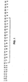
- Mature hBNP consists of a 32 amino acid peptide containing a 17 amino acid ring structure formed by two disulfide bonds (see Fig. 1 ).
- Elevated expression of ventricular hBNP mRNA has been reported in congestive heart failure patients as compared to normal controls. Consistent with the increase in ventricular mass and expression ofBNP mRNA, hBNP levels are elevated in patients with congestive heart failure and appear to correlate with disease severity. Plasma hBNP is also believed to provide a valuable predictive marker for heart disease. Elevated plasma hBNP has been reported in heart disease, following acute myocardial infarction and during symptomless or subclinical ventricular dysfunction ( Mukoyama et al., J. Clin.
- a diagnostic assay for hBNP which is sufficiently sensitive to measure clinically relevant titers of hBNP, sufficiently simple that it can be automated, requires a minimum amount of time to complete and preferably does not require the use of reagents having limited shelf lives.
- Normal levels of hBNP in plasma are quite low, on the order of 1 to 20 pg/mL.
- radioimmunoassays are used to measure titers in this range, although the use of radioactive reagents requires special handling, adds steps to the assay and involves the use of material having limited shelf life.
- a radioimmunoassay is commercially available for measuring hBNP, however, in addition to requiring the use of radioactive reagents? it is complex and cumbersome, requiring an extraction step, precipitation and several centrifugation steps, and takes several days to complete.
- European Patent Application No. 0 542 255 describes a radioimmunoassay for hBNP.
- the present invention provides methods for the rapid and direct quantification of hBNP levels in biological fluids.
- the methods provided herein allow for the detection ofhBNP titers at clinically relevant levels without the use of radioactive labelling (although radioactive labelling may be used, if desired).
- they provide for the direct quantification of hBNP in plasma without the need for cumbersome extraction steps.
- the methods provided herein lend themselves readily to the automated systems used to conduct blood tests on a large scale in commercial clinical laboratories.
- monospecific antibodies to selected peptide epitopes within the hBNP molecule.
- these monospecific antibodies are monoclonal antibodies.
- the monospecific antibodies may be selected from the group consisting of:
- a preferred antibody for use in a method of the invention is a monoclonal antibody that recognizes and binds the peptide epitope comprising amino acids 5-13 of hBNP.
- Described herein is a method for quantifying the amount of hBNP in a biological fluid using the reagents described herein in a sandwich type immunoassay.
- the immunoassay may employ any of numerous labelling techniques to label and quantify immune complexes, with enzymatic labelling being preferred.
- the invention provides an in vitro method of diagnosing congestive heart failure by quantifying hBNP in a biological fluid, comprising:
- the first antibody-hBNP-second antibody complex will usually be separated from the remainder of the biological fluid sample prior to quantifying the label in the complex.
- labelling techniques which allow for the direct measurement of labelled complex in the sample, i.e. without separating the complex from the sample, and such methods are considered to be within the scope of the invention.
- scintillation proximity assay Udenfriend, S. et al., Anal. Biochem., 161:494-500, 1987
- the assay described in Mathis, G., Clin. Chem., 41:1391-1397, 1995 are examples of the scintillation proximity assay.
- This invention uses highly sensitive reagents which allow for the rapid, simple and accurate quantification of hBNP at clinically relevant titers in biological fluids such as plasma or serum.
- biological fluids such as plasma or serum.
- an antibody is referred to herein as being "monospecific" to an epitope, it is meant that the antibody is capable of binding a sequence of amino acids within hBNP containing the amino acids comprising the epitope, but is incapable of binding to a sequence of amino acids within hBNP that does not contain the amino acids comprising the epitope.
- the monospecific antibodies are preferably, though not necessarily, monoclonal antibodies.
- the three epitopes recognized by the monospecific antibodies described herein comprise amino acids 1-10, 5-13 and 15-25 ofhBNP.
- Monoclonal antibodies can be produced by hybridoma cells prepared according to known procedures, e.g . Kohler, G. and Milstein, C., Nature, 256:495, 1975 .
- mice are immunized with an immunogenic conjugate of hBNP and a suitable partner, such as bovine serum albumin. Periodic booster injections are administered until good antibody titers are achieved.
- Spleen cells from the immunized mice are then fused with myeloma cells according to known procedures ( i.e . Galfre et al., Nature, 266:550, 1977 ) to produce hybridoma cells.
- Supernatants from the hybridoma cell cultures are screened for reactivity to hBNP.
- Hybridomas testing positive are recloned and their supernatants retested for reactivity.
- hybridoma cell line 106.3 which secretes monoclonal antibodies that specifically recognize the hBNP fragment 5-13
- hybridoma cell line 201.3 which secretes monoclonal antibodies that specifically recognize the hBNP fragment 1-10
- hybridoma cell line 8.1 which secretes monoclonal antibodies that specifically recognize the hBNP fragment 27-32.
- non-monoclonal monospecific antibodies can be produced from polyclonal antisera.
- Polyclonal antisera is produced according to known techniques. A suitable animal, such as a rabbit, is immunized with an immunogenic conjugate of hBNP and a suitable partner. Periodic booster injections are administered until good antibody titers are achieved. By testing the antisera thus obtained for immunoreactivity against various peptide fragments of hBNP, desirable epitopes are identified. Using this procedure, we identified the peptide fragment hBNP 15-25 as a highly immunogenic epitope of hBNP.
- Monospecific antibodies to the identified epitope can be obtained by affinity purification of polyclonal serum on an affinity column in which a peptide fragment including only that epitope and none of the other epitopes identified in the epitope mapping is bound to a solid support. Using this general procedure, as described in more detail below, we produced non-monoclonal monospecific antibodies that specifically recognize the hBNP fragment 15-25. It will be recognized, however, that monoclonal antibodies to this epitope can also be produced using known procedures.
- Functionally active fragments of the monospecific antibodies described herein can also be used in assays for hBNP.
- a functional fragment is one that retains the immunologic specificity of the antibody, although avidity and/or affinity may not be quantitatively identical. Included in the functionally active fragments are such immunoglobulin fragments as Fab, F(ab') 2 and Fab'.
- the fragments can be produced by known methods such as by enzymatic cleavage of the monospecific antibodies (see, e.g ., Mariani, M. et al., Mol. Immunol., 28:69-77, 1991 ; Ishikawa, E. et al. J. Immunoassay, 4:209-327, 1983 ),
- the reagents of the invention can be used to carry out immunoassays to quantify hBNP levels in biological fluids such as plasma, serum and whole blood.
- the assays can be carried out either in a sandwich type format.
- the biological fluid is preferably plasma or serum, which can be prepared from whole blood using known procedures.
- sandwich type assay two different antibodies are employed to separate and quantify the hBNP in the biological fluid sample.
- the two antibodies bind to the hBNP, thereby forming an immune complex, or sandwich.
- one of the antibodies is used to capture the hBNP in the sample and a second antibody is used to bind a quantifiable label to the sandwich.
- the antibodies chosen to carry out the sandwich type assay are selected such that the first antibody which is brought into contact with the hBNP-containing sample does not bind all or part of the epitope recognized by the second antibody, thereby significantly interfering with the ability of the second antibody to bind hBNP.
- a sandwich type format one preferably should not employ an antibody monospecific for hBNP 5-13 in combination with an antibody monospecific for hBNP 1-10 inasmuch as the epitopes recognized by these antibodies overlap.
- an excellent assay can be effected using a monospecific antibody as the first antibody brought into contact with the hBNP-containing sample and a high affinity polyclonal antibody for hBNP1-32 as the second antibody.
- the fluid sample can be contacted with the first antibody and the second antibody simultaneously or sequentially.
- the hBNP-containing biological fluid sample is first brought into contact with a first antibody which is monospecific for a particular epitope under conditions which allow the formation of an antibody-hBNP complex.
- the first antibody is bound to a solid support to facilitate the ultimate separation of the sandwich complex from the fluid sample.
- the solid supports that can be used are well known to those skilled in the art and can be, for example, polymeric materials in the form of wells, tubes or beads.
- the antibody can be bound to the solid support by adsorption or by covalent bonding using a chemical coupling agent, provided the coupling agent employed does not interfere with the ability of the antibody to bind hBNP.
- the polymeric support materials can be derivatized to allow reactivity with various functional groups on the antibody.
- Typically used coupling agents include maleic anhydride, N-hydroxysuccinimide and 1-ethyl-3-(3-dimethylaminopropyl)carbodiimide.
- the antibody is normally employed in amounts substantially in molar excess of the maximum amount of hBNP expected to be in the sample. Preferably, there are employed from about 0.01 nM to about 1.0 nM of antibody per mL of undiluted plasma. In practice, one may dilute the biological fluid sample in an appropriate physiologic buffer in order to achieve an acceptable molar ratio of antibody to hBNP.
- the sample is incubated to allow the formation of a first antibody-hBNP complex.
- incubation is carried out at or near physiologic pH, e.g. pH 6.0 to pH 8.0 at a temperature from about 4°C to about 37°C.
- Incubation time is at least about 2 minutes, although incubation can be carried out for as long as 18 hours.
- the first antibody-hBNP complex is contacted with the second antibody under conditions which allow formation of a first antibody-hBNP-second antibody complex. Typically, this entails incubation under conditions similar to those outlined above for the formation of the first antibody-hBNP complex.
- a quantifiable label is bound to the second antibody prior to, simultaneously with or after formation of the first antibody-hBNP-second antibody complex Any of the known labels conventionally used in immunoassays may be employed.
- radioactive labels such as 125 I
- enzymatic labels such as horseradish peroxidase or alkaline phosphatase
- chemiluminescent labels such as acridinium esters
- bioluminescent labels fluorescent labels
- thermometric labels immuno-polymerase chain reaction labels or others.
- the label can be bound to the antibody directly or through a coupling agent.
- the second antibody can be bound to biotin, using known techniques. Following formation of the first antibody-hBNP-second antibody complex, the complex can be contacted with avidin-horseradish peroxidase, which is tightly bound by the biotin on the second antibody.
- An alternative method of binding the label to the second antibody involves contacting the first antibody-hBNP-second antibody complex with an antibody which recognizes and binds the F c region of the second antibody and which has the label bound thereto. Other methods of binding label to the second antibody will be readily apparent to those skilled in the art.
- the first antibody-hBNP-second antibody complex is separated from the remainder of the biological fluid sample prior to quantification of the label.
- separation is accomplished simply by removing the fluid from contact with the solid support.
- the first antibody bound to a solid support may be incubated with the hBNP-containing sample to form a first antibody-hBNP complex and the fluid sample may then be removed from contact with the solid support prior to contacting the first antibody-hBNP complex with the second antibody.
- the first antibody bound to a solid support may be simultaneously contacted with the hBNP-containing sample and the second antibody to form a first antibody-hBNP-second antibody complex, followed by removal of the biological fluid from contact with the solid support.
- the amount of label in the complex is quantified using known methods.
- the labelled complex is reacted with a substrate for the label, giving a quantifiable reaction such as the development of color.
- the enzymatic label is horseradish peroxidase
- the labelled complex is reacted with tetramethylbenzidine (TMB), giving a reaction which can be quantified by measuring the development of color.
- TMB tetramethylbenzidine
- the label is quantified using a scintillation counter.
- the concentration of hBNP in the biological fluid sample is determined by use of a standard curve which has been generated using serial dilutions of hBNP of known concentration.
- hBNP 1-32 and the fragments hBNP 1-10 and hBNP 26-32 were prepared by solid phase synthesis. These peptides were coupled to bovine serum albumin (BSA) carrier protein through a carbodiimide coupling procedure or by using the heterobifunctional cross-linking regent N-maleimido-L-aminocaproyl ester of 1-hydroxysuccinimide-2-nitro-4-benzenesulfonic acid (mal-sac-HNSA) according to the method of Aldwin and Nitecki (Anal. Biochem., 164:494-501, 1987 ).
- BSA bovine serum albumin
- BSA (1 mg) was reacted with 0.5 mg of the mal-sac-HNSA cross-linker in 250 ⁇ L of phosphate buffered saline (PBS) pH 7.5 for 40 minutes at room temperature.
- PBS phosphate buffered saline
- the activated BSA was purified over a G-25 Sephadex column with 0.1M phosphate buffer pH 6.0 and combined with approximately 1 mg of the cysteine containing peptides hBNP 1-10 (SPKMVQGSGC) and hBNP 26-32 (CKVLRRH). The mixture was reacted for 4 hours at room temperature and dialyzed overnight against PBS at 4°C. A successful conjugation was indicated by the presence of a protein in an area corresponding to 75 Kd to 90 Kd on a 12% polyacrylamide gel.
- BNP 1-32 BSA conjugates were prepared by dissolving approximately 1 mg BNP in 450 ⁇ L of 1 mM HCl and adding 2.8 mg of 1-ethyl-3-(3-dimethylaminopropyl)carbodiimide (EDAC). The mixture was reacted for 30 minutes on ice and added to 1.47 mg BSA in 300 ⁇ L of 5 mM NaOH. After 4 hours at room temperature, the solution was dialyzed against PBS at 4°C.
- EDAC 1-ethyl-3-(3-dimethylaminopropyl)carbodiimide
- hBNP 1-32 conjugates for preparation of rabbit polyclonal antibodies were prepared by mixing 100 ⁇ g of methylated BSA (MeBSA) with 100 ⁇ g hBNP 1-32 in 100 ⁇ L PBS, pH 7.4.
- MeBSA methylated BSA
- mice were immunized with peptide conjugate emulsions prepared using RIBI Adjuvant System (RIBI Immunochemical Research Inc.). About 700 ⁇ g of Synthetic Trehalose Dicorynomycolate and about 650 ⁇ g of Monophosphoryl Lipid A from S. minnesota R595 dissolved in 4:1 chloroform:methanol were dried in a small glass homogenizer. Sixty ⁇ L squalene was added and mixed into a paste. The peptide conjugate was then added, with continuous stirring, and an additional 1.5 mL 0.2% Tween 80 in water was added with stirring to generate a final emulsion containing approximately 1 mg/mL of protein.
- RIBI Adjuvant System RIBI Immunochemical Research Inc.
- mice Female Balb/c mice (4 to 6 weeks old) were injected intraperitoneally with approximately 125 to 150 ⁇ g of the peptide conjugate. Mice were boosted with intraperitoneal injections once every three weeks until good titers were established. Mice received a final boost four days prior to spleen cell fusions.
- Spleen cells were fused with the myeloma partner FOX-NY (ATCC CRL-1732) according to a method modified from Galfre et al., Nature, 266:550, 1977 .
- Spleens were removed aseptically and single cell suspensions were prepared in serum free medium by homogenization in a 7 mL glass homogenizer.
- the spleen cells were washed twice prior to mixing with the myeloma partner cells.
- the FOX-NY:myeloma cells were harvested at log phase growth, washed and combined with the spleen cells in a 5:1 spleen:myeloma) ratio. The cells were centrifuged, medium was removed and the cell pellet was loosened by tapping.
- PEG polyethylene glycol
- hybridoma selection medium RPMI-1640 hybridoma medium, 10mM Hepes, 20% fetal bovine serum, 50 U/mL penicillin, 50 ⁇ g/mL streptomycin, 75 ⁇ M adenine, 0.8 ⁇ M aminopterin and 16 ⁇ M thymidine.
- the cells were transferred to a T-150 culture flask and incubated overnight prior to plating in 96-well plates (about 10 5 cells per well). Cultures were maintained at 37°C, 7% CO 2 in a humidified incubator. Cells were re-fed 6 days after fusion and conditioned medium was assayed for antibodies at day 10.
- Hybridoma supernatants were tested for reactivity to hBNP 1-32 or various hBNP peptides by a standard ELISA method.
- Peptides 0.1-0.5 ⁇ g/100 ⁇ L/well in PBS, were adsorbed to microtiter plates overnight at 4°C.
- Wells were washed with PBS/Tween (PBS containing 0.05% Tween-20) and blocked with 0.1% gelatin 1 hour at 37°C. Undiluted hybridoma supernatants (100 ⁇ L/well) were added to the peptide-coated wells for 2 hours at 37°C.
- Hybridomas testing positive were recloned by limiting dilution and their supernatants retested for specific reactivity. Hybridoma and secretory stability were established by measuring growth and antibody production characteristics over several passages. This process resulted in the establishment of hybridoma cell line 106.3 and MAb 106.3, which specifically recognizes the hBNP peptide fragment 5-13, hybridoma cell line 201.3 and MAb 201.3, which specifically recognizes the hBNP peptide fragment 1-10 and hybridoma cell line 8.1 and MAb 8.1, which specifically recognizes the hBNP peptide fragment 27-32. Hybridoma cell line 106.3 was deposited at the American Type Culture Collection, Rockville, Md. on February 14, 1996 and has accession no.
- Hybridoma cell line 201.3 was deposited at the American Type Culture Collection, Rockville, Md. on February 14, 1996 and has accession no. HB 12045.
- Hybridoma cell line 8.1 was deposited at the American Type Culture Collection, Rockville, Md. on February 21, 1996 and has the accession no. HB 12050.
- a microtiter plate ELISA was used to identify the class/subclass of the monoclonal antibodies.
- Mouse antibody was added to microtiter plates precoated with hBNP.
- Subclass specific second antibodies conjugated to horseradish peroxidase were added to detect the bound murine monoclonal antibody.
- TMB substrate Several washes followed by the addition of TMB substrate gave specific subclass reactivities for the three monoclonals.
- MAb 106.3 was determined to be an IgG 1 ⁇ antibody
- MAb 201.3 was determined to belong to subclass IgG 2a ⁇
- MAb 8.1 was determined to belong to subclass IgG 1 ⁇ .
- Rabbit polyclonal antibody was tested for reactivity to hBNP 1-32 as well as to a variety of hBNP fragments.
- Microtiter plates were coated with 100 ng of the various peptides/well in 200 ⁇ L of 0.1 M bicarbonate buffer, pH 8-9. The plates were blocked with 0.1% gelatin for 30 minutes at 37°C.
- Rabbit serum was diluted 1:1000 followed by 5 three-fold dilutions in a Tris/Tween/BSA buffer (0.05M Tris, pH 7.5, 0.1 M NaCl, 1% BSA, 0.1% Tween-20).
- Tris/Tween/BSA buffer 0.05M Tris, pH 7.5, 0.1 M NaCl, 1% BSA, 0.1% Tween-20.
- One hundred microliters of the antibody dilution series were added to the microtiter wells and incubated at 4°C for 2 hours.
- Hybridoma cells were seeded in roller bottles at a concentration of 300,000 cells/mL in RPMI 1640 tissue culture medium supplemented with 5% Fetal Clone (Hyclone) and grown to a density of approximately 1 x 10 6 cells/mL. The medium was replaced with serum free Hybridoma SFM medium and incubation was continued for an additional 3 to 5 days. Cells were separated from the culture medium by centrifugation at 400 x g and the medium was clarified by filtration through a 0.2 ⁇ filter. The medium was applied to a Prosep Protein-A affinity column (Bioprocessing Inc.) that had been previously equilibrated with binding buffer at pH 8.6 (1 M glycine, 0.15 M NaCl).
- hBNP 15-27 was isolated from the polyclonal IgG using a diaminodipropylamine affinity column (DADPA) to which the synthetic hBNP fragment 15-27 was immobilized.
- DADPA diaminodipropylamine affinity column
- the DADPA gel was prepared for coupling by washing with 5 mL water followed by 5 mL 0.1 MES buffer (2-(N-morpholino)ethanesulfonic acid), 0.9% NaCl, pH 4.7.
- hBNP 15-27 (2 mg) was dissolved in 2 mL of MES buffer and mixed with the gel.
- EDC (1-ethyl-3-(3-dimethylaminopropyl)carbodiimide) dissolved in MES buffer was added to the gel and incubated with stirring for 3 hours at room temperature.
- the column was washed several times with 1 M NaCl to remove unbound peptide and blocked with 0.1% ovalbumin.
- the column Prior to application of the rabbit IgG, the column was washed with 5 column volumes of PBS, pH 7.5.
- the rabbit IgG was cycled through the column 5 times followed by 5 washes with PBS buffer.
- the antibodies bound to hBNP 15-27 were eluted with 0.1 M glycine HCl, pH 2.5, brought to pH 7.5 with 2 M Tris buffer and concentrated in a Centriprep 10.
- the monoclonal antibodies were dialyzed against 0.1 M sodium borate buffer, pH 8.8 and brought to a final concentration of 1 to 3 mg/mL
- Amino-hexanoyl-biotin-N-hydroxysuccinimide was prepared in dimethyl sulfoxide at a concentration of 10 mg/mL and added to the antibodies at a ratio of 200 ⁇ g of the biotin ester/mg MAb. Conjugation was carried out at room temperature for 4 hours. Uncoupled material was removed from the conjugated antibody by dialysis against phosphate buffered saline.
- hBNP epitopes reacting with MAb 201.3, MAb 106.3 and MAb 8.1 were determined using an inhibition ELISA.
- hBNP biotinylated with a single biotin at the N-terminal amino acid during solid phase synthesis was used as the indicator for competition studies with MAb 106.3 and MAb 8.1.
- hBNP biotinylated at the two cysteine residues, 10 and 26, was prepared by coupling with the sulfhydryl reactive biotinylation reagent Biotin-BMCC. This reagent was necessary to complete the inhibition studies with MAb 201.3.
- Microtiter plates were coated with goat anti-mouse IgG (F c specific) (0.5 ⁇ g/100 ⁇ L/well in bicarbonate buffer, pH 9 for 2 hours at 37°C). Wells were washed four times with wash buffer (0.05 M Tris, pH 7.5, 0.15 M NaCl, 0.05% Tween-20). Purified monoclonal antibodies 106.3, 201.3 and 8.1 were diluted in assay buffer (0.05 M Tris, pH 7.5, 0.15 M NaCl, 0.1% Tween-20 and 1% BSA) and 180 pg/100 ⁇ L/well was incubated for 2 hours at room temperature. The wells were washed with wash buffer.
- wash buffer 0.05 M Tris, pH 7.5, 0.15 M NaCl, 0.1% Tween-20 and 1% BSA
- hBNP peptide fragments were mixed in assay buffer with biotinylated hBNP 1-32 at a molar ratio of 100:1 peptide fragment:biotin-hBNP, added to the microtiter wells and incubated for 1.5 hours at 4°C.
- Wells were washed once with wash buffer, streptavidin-horseradish peroxidase was added in 100 ⁇ L assay buffer and incubated for 20 minutes at 4°C. Plates were washed 4 times with wash buffer and 200 ⁇ L TMB substrate was added and incubated for 30 minutes at room temperature. Color development was stopped with 2.5 M sulfuric acid and O.D. values were read at 450 nm.
- results of the epitope analysis for MAb 106.3, MAb 201.3 and MAb 8.1 are presented in Fig. 3 .
- the binding of biotin-hBNP 1-32 was significantly inhibited by the hBNP fragments specifically reactive with the monoclonal antibodies.
- the smallest hBNP fragment capable of producing an inhibition comparable to full length hBNP 1-32 was the peptide fragment hBNP 5-13.
- the smallest hBNP peptide fragment producing an inhibition comparable to that seen with hBNP 1-32 was the hBNP fragment hBNP 1-10.
- hBNP 27-32 the fragment hBNP 27-32.
- hBNP fragments 1-10, 5-13 and 27-32 represent the hBNP epitopes specifically recognized by MAb 201.3, MAb 106.3 and MAb 8.1, respectively.
- This data also indicates that appropriate hBNP fragments, when used in a competition assay with the appropriate antibody, can provide an alternative reagent to the full length hBNP probe as an indicator of inhibition.
- Microtiter plates were coated with MAb 106.3 at 100 ng/200 ⁇ L/well in bicarbonate buffer, pH 9 and allowed to adsorb for 2 hours at 37°C.
- hBNP was diluted in assay buffer at 12.8 ng/mL and 2-fold serial dilutions were prepared to produce a standard curve ranging down to 12.5 pg/mL. Buffer was removed from the plates and 100 ⁇ L aliquots of the hBNP dilution series were added to the wells. Plates were incubated overnight at 4°C, washed once with wash buffer and incubated with 200 ⁇ L of a 1:2,500 dilution of rabbit antiserum containing polyclonal antibodies to hBNP.
- Plates were coated overnight at 4°C with 400 ng/well MAb 201.3 in bicarbonate buffer. Plates were washed with wash buffer and blocked with assay buffer for 1 hour at room temperature. hBNP was diluted in assay buffer at a concentration of 10 ng/mL and 4-fold serial dilutions were prepared to produce a standard curve ranging down to 10 pg/mL. Plates were emptied and 100 ⁇ L aliquots of the dilution series were added to the wells. Incubation was continued at room temperature for 2 hours. The plates were washed once with wash buffer.
- Rabbit antiserum containing polyclonal antibodies to hBNP was diluted 1:1,000 in assay buffer, 100 ⁇ L was added to the wells and incubated at room temperature for 2 hours. After three washes with wash buffer, 100 ⁇ L/well goat anti-rabbit horseradish peroxidase was added and incubation was continued at room temperature for 1 hour. Plates were washed 3 times with wash buffer, TMB substrate was added and color was allowed to continue for 20 minutes. The reaction was stopped with 100 ⁇ L of 2.5 M sulfuric acid and O.D. was measured at 450 nm. The standard curve produced from these serial dilutions of hBNP is presented in Fig. 5 .
- Microtiter plates were coated with MAb 106.3 at 100 ng/100 ⁇ L/well in bicarbonate buffer, pH 9 overnight at 4°C.
- hBNP was diluted in assay buffer to 10 ng/mL and 2-fold serial dilutions were used to prepare a standard curve ranging down to 10 pg/mL. Plates were washed and blocked for 1 hour with assay buffer. One hundred microliter aliquots of the hBNP dilution series were added to the wells and incubated at room temperature for 2 hours. The wells were washed 4 times with wash buffer and 100 ⁇ L of a 1:50 dilution of rabbit IgG monospecific for hBNP 15-25 were added.
- Microtiter plates were coated with MAb 201.3 at 400 ng/100 ⁇ L/well in bicarbonate buffer, pH 9 overnight at 4°C.
- hBNP was diluted in assay buffer to 10 ng/mL and 2-fold serial dilutions were used to prepare a standard curve ranging down to 10 pg/mL. Plates were washed and blocked for 1 hour with assay buffer. One hundred microliter aliquots of the hBNP dilution series were added to the wells and incubated at room temperature for 2 hours. The wells were washed 4 times with wash buffer and 100 ⁇ L of a 1:50 dilution of rabbit IgG monospecific for hBNP 15-25 were added.
- Microtiter plate wells were coated overnight at 4°C with 100 ng/well MAb 8.1 in 200 ⁇ L bicarbonate buffer. Wells were washed with wash buffer and blocked for 1 hour at 4°C.
- hBNP was diluted in assay buffer to 10 ng/mL and 2-fold standard dilutions were prepared to produce a standard curve ranging down to approximately 10 pg/mL. Buffer was removed from the wells and 100 ⁇ L aliquots of the hBNP standard dilution series and 100 ⁇ L of assay buffer containing 10 ng of biotinylated MAb 106.3 were added. Plates were incubated at 4°C for 1.5 hours.
- Microtiter plate wells were coated overnight at 4°C with 400 ng/well MAb 201.3 in 200 ⁇ L bicarbonate buffer. Wells were washed three times with wash buffer and blocked for 1 hour.
- hBNP was diluted in assay buffer to 100 ng/mL and 10 ng/mL and serial 4-fold dilutions were prepared to produce a standard curve ranging down to approximately 10 pg/mL. Buffer was removed from the wells and 100 ⁇ L aliquots of the hBNP standard dilution series were added. Incubation was carried out at room temperature for 1.5 hours.
- Plasma diluent can be prepared from pooled human plasma or pooled human serum. Preparation from plasma involves inducement of clotting by addition of 2 mg/mL calcium chloride, incubation at room temperature to promote clotting and degrade endogenous hBNP, dialysis using 15,000 MW cutoff dialysis tubing against 0.001 M phosphate buffer containing 0.15 M NaCl, removal of clotted material, addition of EDTA at a concentration of 1.5 mg/mL and filtration through a 0.2 ⁇ filter.
- a protease inhibitor such as 4-(2-aminoethyl)-benzenesulfonyl fluoride, HCl (1 mM to the plasma diluent and assay buffer).
- Microtiter plates were prepared by adding 200 ⁇ L/well of goat anti-murine antibody (F c specific, approximately 0.5 ⁇ g/well) in bicarbonate buffer, pH 9 and incubating for 2 hours at 37°C. Plates were washed 4 times with wash buffer. MAb 106.3 was diluted in assay buffer and 100 ⁇ L (90 pg/well) was added to the microtiter wells. Incubation was continued for 2 hours at room temperature.
- hBNP 1-32 was diluted directly in the plasma diluent using two-fold dilutions to produce a standard curve ranging from 320 pg/mL to 5 pg/mL.
- One hundred microliter samples were added to wells containing 200 ⁇ L assay buffer, plates were covered and incubated overnight at 2-8°C on an orbital shaker.
- Plasma samples from patients were generally diluted at least 1:2 in plasma diluent before they were added to the wells. For patients with high levels of hBNP all further sample dilutions were prepared in plasma diluent.
Abstract
Description
- This invention relates to the field of immunoassays, more particularly to methods useful for the rapid and sensitive quantification of the peptide hormone hBNP in a biological fluid such as plasma or serum.
- BNP is a cardiac derived peptide hormone that circulates in the blood and exerts potent cardiovascular and renal actions. BNP is structurally similar to two other cardiac peptides, atrial natriuretic peptide (ANP) and C-type natriuretic peptide (CNP). Porcine BNP was isolated from pig brain (Sudoh et al., Nature 332:78-81, 1988) and, hence, was given the name "brain natriuretic peptide". In man the cardiac ventricle is the primary site of BNP synthesis. The sequence of human BNP (hBNP) was originally determined by the isolation and characterization of DNA clones from human genomic libraries (
U.S. Patent No. 5,114,923 ). hBNP is synthesized, in vivo, as a 108 amino acid precursor that is enzymatically cleaved to yield the mature hBNP peptide. Mature hBNP consists of a 32 amino acid peptide containing a 17 amino acid ring structure formed by two disulfide bonds (seeFig. 1 ). - Elevated expression of ventricular hBNP mRNA has been reported in congestive heart failure patients as compared to normal controls. Consistent with the increase in ventricular mass and expression ofBNP mRNA, hBNP levels are elevated in patients with congestive heart failure and appear to correlate with disease severity. Plasma hBNP is also believed to provide a valuable predictive marker for heart disease. Elevated plasma hBNP has been reported in heart disease, following acute myocardial infarction and during symptomless or subclinical ventricular dysfunction (Mukoyama et al., J. Clin. Invest., 87:11402-1412, 1991) (Motwani et al., Lancet, 341:1109-1113, 1993) (Yoshibayashi et al., New Eng. J. Med., 327:434, 1992). Reports from two major therapeutic trials, the SAVE trial ( New Eng. J. Med., 327:669-677, 1992) and the SOLVD trial ( New Eng. J. Med., 327:685-691, 1992) suggested that diagnosis and appropriate treatment of patients with asymptomatic left ventricular dysfunction could significantly reduce the incidence of fatal and non-fatal cardiovascular events and related hospitalizations.
- For use in a clinical laboratory setting, it would be highly desirable to provide a diagnostic assay for hBNP which is sufficiently sensitive to measure clinically relevant titers of hBNP, sufficiently simple that it can be automated, requires a minimum amount of time to complete and preferably does not require the use of reagents having limited shelf lives. Normal levels of hBNP in plasma are quite low, on the order of 1 to 20 pg/mL. Typically, radioimmunoassays are used to measure titers in this range, although the use of radioactive reagents requires special handling, adds steps to the assay and involves the use of material having limited shelf life. A radioimmunoassay is commercially available for measuring hBNP, however, in addition to requiring the use of radioactive reagents? it is complex and cumbersome, requiring an extraction step, precipitation and several centrifugation steps, and takes several days to complete. European Patent Application No.
0 542 255 describes a radioimmunoassay for hBNP. - The present invention provides methods for the rapid and direct quantification of hBNP levels in biological fluids. The methods provided herein allow for the detection ofhBNP titers at clinically relevant levels without the use of radioactive labelling (although radioactive labelling may be used, if desired). Moreover, they provide for the direct quantification of hBNP in plasma without the need for cumbersome extraction steps. The methods provided herein lend themselves readily to the automated systems used to conduct blood tests on a large scale in commercial clinical laboratories.
- Described herein are monospecific antibodies to selected peptide epitopes within the hBNP molecule. Preferably, these monospecific antibodies are monoclonal antibodies. The monospecific antibodies may be selected from the group consisting of:
- (a) an antibody that is monospecific to a peptide epitope comprising amino acids 5-13 of hBNP;
- (b) an antibody that is monospecific to a peptide epitope comprising amino acids 1-10 of hBNP; and
- (c) an antibody that is monospecific to a peptide epitope comprising amino acids 15-25 ofhBNP,
- A preferred antibody for use in a method of the invention is a monoclonal antibody that recognizes and binds the peptide epitope comprising amino acids 5-13 of hBNP.
- Described herein is a method for quantifying the amount of hBNP in a biological fluid using the reagents described herein in a sandwich type immunoassay. The immunoassay may employ any of numerous labelling techniques to label and quantify immune complexes, with enzymatic labelling being preferred.
- Accordingly, the invention provides an in vitro method of diagnosing congestive heart failure by quantifying hBNP in a biological fluid, comprising:
- contacting a sample of the biological fluid with a first antibody that is monospecific for a peptide epitope comprising amino acids 5-13 of hBNP, or a fragment thereof that retains the monospecific reactivity of said first antibody
- forming a complex comprising hBNP in the biological fluid, the first antibody and a second antibody, wherein the second antibody is immunoreactive with hBNP, and wherein the second antibody comprises a label; and
- determining the amount of hBNP in the sample by quantifying the label in the complex.
- In the sandwich assays of the invention, the first antibody-hBNP-second antibody complex will usually be separated from the remainder of the biological fluid sample prior to quantifying the label in the complex. There are, however, known labelling techniques which allow for the direct measurement of labelled complex in the sample, i.e. without separating the complex from the sample, and such methods are considered to be within the scope of the invention. As merely exemplary of such methods, one can mention the scintillation proximity assay (Udenfriend, S. et al., Anal. Biochem., 161:494-500, 1987) and the assay described in Mathis, G., Clin. Chem., 41:1391-1397, 1995.
-
-
Fig. 1 is a representation of the amino acid sequence of the mature (32 amino acid) form of hBNP. Amino acids are represented by the standard single letter code and the solid line represents a disulfide bond between the two cysteine residues. -
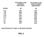
Fig. 2 represents the results of experiments conducted to determine the ability of rabbit polyclonal antisera raised to hBNP to bind various fragments of hBNP. -
Fig. 3 represents the results ofepitope mapping studies of three monoclonal antibodies designated MAb 201.3, MAb 106.3 and MAb 8.1. -
Fig. 4 is a standard curve for a sandwich type enzyme linked immunosorbent assay using MAb 106.3 as the capture antibody and apolyclonal antibody # 4024 as the second antibody. -
Fig. 5 is a standard curve for a sandwich type enzyme linked immunosorbent assay using MAb 201.3 as the capture antibody and polyclonal antibody #4360 as the second antibody. -
Fig. 6 is a standard curve for a sandwich type enzyme linked immunosorbent assay using MAb 106.3 as the capture antibody and a monospecific antibody against epitope 15-25 as the second antibody. -
Fig. 7 is a standard curve for a sandwich type enzyme linked immunosorbent assay using MAb 201.3 as the capture antibody and a monospecific polyclonal antibody against epitope 15-25 as the second antibody. -
Fig. 8 is a standard curve for a sandwich type immunosorbent assay using MAb 8.1 as the capture antibody and biotinylated MAb 106.3 as the second antibody in a monoclonal:monoclonal sandwich. -
Fig. 9 is a standard curve for a sandwich type immunosorbent assay using MAb 201.3 as a capture antibody and biotinylated MAb 8.1 as a second antibody in a monoclonal:monoclonal sandwich. -
Fig. 10 is a standard curve for a competition type enzyme linked immunosorbent assay using MAb 106.3 as the antibody. -
Fig. 11 presents the results obtained using a competition type enzyme linked immunosorbent assay to quantify hBNP levels in the plasma of normal control subjects and subjects with congestive heart disease. - This invention uses highly sensitive reagents which allow for the rapid, simple and accurate quantification of hBNP at clinically relevant titers in biological fluids such as plasma or serum. In particular, we have identified three previously unidentified highly immunogenic epitopes within the hBNP molecule and produced antibodies monospecific to these regions. When an antibody is referred to herein as being "monospecific" to an epitope, it is meant that the antibody is capable of binding a sequence of amino acids within hBNP containing the amino acids comprising the epitope, but is incapable of binding to a sequence of amino acids within hBNP that does not contain the amino acids comprising the epitope. The monospecific antibodies are preferably, though not necessarily, monoclonal antibodies.
- The three epitopes recognized by the monospecific antibodies described herein comprise amino acids 1-10, 5-13 and 15-25 ofhBNP.
- Monoclonal antibodies can be produced by hybridoma cells prepared according to known procedures, e.g. Kohler, G. and Milstein, C., Nature, 256:495, 1975. In general, mice are immunized with an immunogenic conjugate of hBNP and a suitable partner, such as bovine serum albumin. Periodic booster injections are administered until good antibody titers are achieved. Spleen cells from the immunized mice are then fused with myeloma cells according to known procedures (i.e. Galfre et al., Nature, 266:550, 1977) to produce hybridoma cells. Supernatants from the hybridoma cell cultures are screened for reactivity to hBNP. Hybridomas testing positive are recloned and their supernatants retested for reactivity. Using this procedure, as described in more detail below, we obtained hybridoma cell line 106.3, which secretes monoclonal antibodies that specifically recognize the hBNP fragment 5-13, hybridoma cell line 201.3, which secretes monoclonal antibodies that specifically recognize the hBNP fragment 1-10 and hybridoma cell line 8.1, which secretes monoclonal antibodies that specifically recognize the hBNP fragment 27-32.
- Once a highly immunogenic epitope has been identified, non-monoclonal monospecific antibodies can be produced from polyclonal antisera. Polyclonal antisera is produced according to known techniques. A suitable animal, such as a rabbit, is immunized with an immunogenic conjugate of hBNP and a suitable partner. Periodic booster injections are administered until good antibody titers are achieved. By testing the antisera thus obtained for immunoreactivity against various peptide fragments of hBNP, desirable epitopes are identified. Using this procedure, we identified the peptide fragment hBNP 15-25 as a highly immunogenic epitope of hBNP. Monospecific antibodies to the identified epitope can be obtained by affinity purification of polyclonal serum on an affinity column in which a peptide fragment including only that epitope and none of the other epitopes identified in the epitope mapping is bound to a solid support. Using this general procedure, as described in more detail below, we produced non-monoclonal monospecific antibodies that specifically recognize the hBNP fragment 15-25. It will be recognized, however, that monoclonal antibodies to this epitope can also be produced using known procedures.
- Functionally active fragments of the monospecific antibodies described herein can also be used in assays for hBNP. A functional fragment is one that retains the immunologic specificity of the antibody, although avidity and/or affinity may not be quantitatively identical. Included in the functionally active fragments are such immunoglobulin fragments as Fab, F(ab')2 and Fab'. The fragments can be produced by known methods such as by enzymatic cleavage of the monospecific antibodies (see, e.g., Mariani, M. et al., Mol. Immunol., 28:69-77, 1991; Ishikawa, E. et al. J. Immunoassay, 4:209-327, 1983),
- The reagents of the invention can be used to carry out immunoassays to quantify hBNP levels in biological fluids such as plasma, serum and whole blood. The assays can be carried out either in a sandwich type format. When measuring hBNP levels in blood, the biological fluid is preferably plasma or serum, which can be prepared from whole blood using known procedures.
- In a sandwich type assay, two different antibodies are employed to separate and quantify the hBNP in the biological fluid sample. The two antibodies bind to the hBNP, thereby forming an immune complex, or sandwich. Generally, one of the antibodies is used to capture the hBNP in the sample and a second antibody is used to bind a quantifiable label to the sandwich. Preferably, the antibodies chosen to carry out the sandwich type assay are selected such that the first antibody which is brought into contact with the hBNP-containing sample does not bind all or part of the epitope recognized by the second antibody, thereby significantly interfering with the ability of the second antibody to bind hBNP. Consequently, if a sandwich type format is chosen, one preferably should not employ an antibody monospecific for hBNP 5-13 in combination with an antibody monospecific for hBNP 1-10 inasmuch as the epitopes recognized by these antibodies overlap. We have found, however, that an excellent assay can be effected using a monospecific antibody as the first antibody brought into contact with the hBNP-containing sample and a high affinity polyclonal antibody for hBNP1-32 as the second antibody. In particular, we have produced a sandwich type assay which is sensitive in the range of clinically relevant hBNP titers using a monoclonal antibody which recognizes the epitope hBNP 5-13 as a capture antibody and a rabbit polyclonal antibody to hBNP as the second antibody (see Examples 9 and 10).
- The fluid sample can be contacted with the first antibody and the second antibody simultaneously or sequentially. In a preferred embodiment of the sandwich assay of the invention, the hBNP-containing biological fluid sample is first brought into contact with a first antibody which is monospecific for a particular epitope under conditions which allow the formation of an antibody-hBNP complex. Preferably, the first antibody is bound to a solid support to facilitate the ultimate separation of the sandwich complex from the fluid sample. The solid supports that can be used are well known to those skilled in the art and can be, for example, polymeric materials in the form of wells, tubes or beads. The antibody can be bound to the solid support by adsorption or by covalent bonding using a chemical coupling agent, provided the coupling agent employed does not interfere with the ability of the antibody to bind hBNP. The polymeric support materials can be derivatized to allow reactivity with various functional groups on the antibody. Typically used coupling agents include maleic anhydride, N-hydroxysuccinimide and 1-ethyl-3-(3-dimethylaminopropyl)carbodiimide. In a sandwich type assay, the antibody is normally employed in amounts substantially in molar excess of the maximum amount of hBNP expected to be in the sample. Preferably, there are employed from about 0.01 nM to about 1.0 nM of antibody per mL of undiluted plasma. In practice, one may dilute the biological fluid sample in an appropriate physiologic buffer in order to achieve an acceptable molar ratio of antibody to hBNP.
- After the hBNP-containing sample has been brought into contact with the first monospecific antibody, the sample is incubated to allow the formation of a first antibody-hBNP complex. Generally, incubation is carried out at or near physiologic pH, e.g. pH 6.0 to pH 8.0 at a temperature from about 4°C to about 37°C. Incubation time is at least about 2 minutes, although incubation can be carried out for as long as 18 hours.
- The first antibody-hBNP complex is contacted with the second antibody under conditions which allow formation of a first antibody-hBNP-second antibody complex. Typically, this entails incubation under conditions similar to those outlined above for the formation of the first antibody-hBNP complex. A quantifiable label is bound to the second antibody prior to, simultaneously with or after formation of the first antibody-hBNP-second antibody complex Any of the known labels conventionally used in immunoassays may be employed. These may be, for example, radioactive labels, such as 125I, enzymatic labels such as horseradish peroxidase or alkaline phosphatase, chemiluminescent labels such as acridinium esters, bioluminescent labels, fluorescent labels, thermometric labels, immuno-polymerase chain reaction labels or others. For a review of some of the more sensitive labels that can be employed in the assays of the invention, see Kricka, L.J., Clin. Chem., 40(3):347-357, 1994 and Kricka, L.J., Clin. Biochem., 26:325-331, 1993. The label can be bound to the antibody directly or through a coupling agent. For example, in the case of horseradish peroxidase, the second antibody can be bound to biotin, using known techniques. Following formation of the first antibody-hBNP-second antibody complex, the complex can be contacted with avidin-horseradish peroxidase, which is tightly bound by the biotin on the second antibody. An alternative method of binding the label to the second antibody involves contacting the first antibody-hBNP-second antibody complex with an antibody which recognizes and binds the Fc region of the second antibody and which has the label bound thereto. Other methods of binding label to the second antibody will be readily apparent to those skilled in the art.
- In a preferred embodiment, the first antibody-hBNP-second antibody complex is separated from the remainder of the biological fluid sample prior to quantification of the label. When the first antibody is bound to solid support, such as a well or a bead, separation is accomplished simply by removing the fluid from contact with the solid support. For example, the first antibody bound to a solid support may be incubated with the hBNP-containing sample to form a first antibody-hBNP complex and the fluid sample may then be removed from contact with the solid support prior to contacting the first antibody-hBNP complex with the second antibody. Alternatively, the first antibody bound to a solid support may be simultaneously contacted with the hBNP-containing sample and the second antibody to form a first antibody-hBNP-second antibody complex, followed by removal of the biological fluid from contact with the solid support.
- After formation of labelled antibody-hBNP-second antibody complex and separation of the complex from the remainder of the biological fluid sample, the amount of label in the complex is quantified using known methods. For example, in the case of an enzymatic label, the labelled complex is reacted with a substrate for the label, giving a quantifiable reaction such as the development of color. Where the enzymatic label is horseradish peroxidase, for example, the labelled complex is reacted with tetramethylbenzidine (TMB), giving a reaction which can be quantified by measuring the development of color. Where the label is a radioactive label, the label is quantified using a scintillation counter. Numerous other methods are well known for quantifying the various types of labels that can be employed in the assays of the inventions. When the amount of label in the complex has been quantified, the concentration of hBNP in the biological fluid sample is determined by use of a standard curve which has been generated using serial dilutions of hBNP of known concentration.
- The following examples are intended to further illustrate the practice of the invention and are not to be construed as limiting the scope of the invention in any way.
- hBNP 1-32 and the fragments hBNP 1-10 and hBNP 26-32 were prepared by solid phase synthesis. These peptides were coupled to bovine serum albumin (BSA) carrier protein through a carbodiimide coupling procedure or by using the heterobifunctional cross-linking regent N-maleimido-L-aminocaproyl ester of 1-hydroxysuccinimide-2-nitro-4-benzenesulfonic acid (mal-sac-HNSA) according to the method of Aldwin and Nitecki (Anal. Biochem., 164:494-501, 1987). BSA (1 mg) was reacted with 0.5 mg of the mal-sac-HNSA cross-linker in 250 µL of phosphate buffered saline (PBS) pH 7.5 for 40 minutes at room temperature. The activated BSA was purified over a G-25 Sephadex column with 0.1M phosphate buffer pH 6.0 and combined with approximately 1 mg of the cysteine containing peptides hBNP 1-10 (SPKMVQGSGC) and hBNP 26-32 (CKVLRRH). The mixture was reacted for 4 hours at room temperature and dialyzed overnight against PBS at 4°C. A successful conjugation was indicated by the presence of a protein in an area corresponding to 75 Kd to 90 Kd on a 12% polyacrylamide gel.
- BNP 1-32 BSA conjugates were prepared by dissolving approximately 1 mg BNP in 450 µL of 1 mM HCl and adding 2.8 mg of 1-ethyl-3-(3-dimethylaminopropyl)carbodiimide (EDAC). The mixture was reacted for 30 minutes on ice and added to 1.47 mg BSA in 300 µL of 5 mM NaOH. After 4 hours at room temperature, the solution was dialyzed against PBS at 4°C.
- hBNP 1-32 conjugates for preparation of rabbit polyclonal antibodies were prepared by mixing 100 µg of methylated BSA (MeBSA) with 100 µg hBNP 1-32 in 100 µL PBS, pH 7.4.
- For production of monoclonal antibodies, mice were immunized with peptide conjugate emulsions prepared using RIBI Adjuvant System (RIBI Immunochemical Research Inc.). About 700 µg of Synthetic Trehalose Dicorynomycolate and about 650 µg of Monophosphoryl Lipid A from S. minnesota R595 dissolved in 4:1 chloroform:methanol were dried in a small glass homogenizer. Sixty µL squalene was added and mixed into a paste. The peptide conjugate was then added, with continuous stirring, and an additional 1.5 mL 0.2
% Tween 80 in water was added with stirring to generate a final emulsion containing approximately 1 mg/mL of protein. The emulsion was stored at 4°C until use. Female Balb/c mice (4 to 6 weeks old) were injected intraperitoneally with approximately 125 to 150 µg of the peptide conjugate. Mice were boosted with intraperitoneal injections once every three weeks until good titers were established. Mice received a final boost four days prior to spleen cell fusions. - For production of polyclonal antibodies rabbits were immunized with the hBNP 1-32 MeBSA conjugates. Emulsions were prepared by mixing thoroughly 100 µL of the peptide conjugate with 200 µL ofFreund's Complete Adjuvant (FCA) or Freund's Incomplete Adjuvant (FIA). Three hundred µL of the FCA emulsion was injected into the inguinal lymph nodes following the method of Mojsov et al. (J. Biol. Chem., 261:11880-11889). Peptide emulsified in FIA was used for additional boosts at
weeks - Spleen cells were fused with the myeloma partner FOX-NY (ATCC CRL-1732) according to a method modified from Galfre et al., Nature, 266:550, 1977. Spleens were removed aseptically and single cell suspensions were prepared in serum free medium by homogenization in a 7 mL glass homogenizer. The spleen cells were washed twice prior to mixing with the myeloma partner cells. The FOX-NY:myeloma cells were harvested at log phase growth, washed and combined with the spleen cells in a 5:1 spleen:myeloma) ratio. The cells were centrifuged, medium was removed and the cell pellet was loosened by tapping. One mL of 50% polyethylene glycol (PEG) was added and stirred gently at 37°C for approximately 2 minutes. The cells were slowly diluted with 10 mL of serum free medium, centrifuged at 400 x g and resuspended in hybridoma selection medium: RPMI-1640 hybridoma medium, 10mM Hepes, 20% fetal bovine serum, 50 U/mL penicillin, 50 µg/mL streptomycin, 75 µM adenine, 0.8 µM aminopterin and 16 µM thymidine. The cells were transferred to a T-150 culture flask and incubated overnight prior to plating in 96-well plates (about 105 cells per well). Cultures were maintained at 37°C, 7% CO2 in a humidified incubator. Cells were re-fed 6 days after fusion and conditioned medium was assayed for antibodies at
day 10. - Hybridoma supernatants were tested for reactivity to hBNP 1-32 or various hBNP peptides by a standard ELISA method. Peptides, 0.1-0.5 µg/100 µL/well in PBS, were adsorbed to microtiter plates overnight at 4°C. Wells were washed with PBS/Tween (PBS containing 0.05% Tween-20) and blocked with 0.1
% gelatin 1 hour at 37°C. Undiluted hybridoma supernatants (100 µL/well) were added to the peptide-coated wells for 2 hours at 37°C. Wells were washed and incubated with a goat anti-mouse IgG horseradish peroxidase conjugate diluted in PBS/Tween/ovalbumin (0.1% ovalbumin in PBS/Tween) for 2 additional hours. Another wash was followed by addition of the substrate (0.5 mg/mL O-phenylenediamine, 0.012% hydrogen peroxide in 100 mM citric phosphate buffer pH 5.0). The color reaction was stopped with 5 M sulfuric acid and absorbance read at 490 nm. An O.D. value greater than twice background was considered a positive response. - Hybridomas testing positive were recloned by limiting dilution and their supernatants retested for specific reactivity. Hybridoma and secretory stability were established by measuring growth and antibody production characteristics over several passages. This process resulted in the establishment of hybridoma cell line 106.3 and MAb 106.3, which specifically recognizes the hBNP peptide fragment 5-13, hybridoma cell line 201.3 and MAb 201.3, which specifically recognizes the hBNP peptide fragment 1-10 and hybridoma cell line 8.1 and MAb 8.1, which specifically recognizes the hBNP peptide fragment 27-32. Hybridoma cell line 106.3 was deposited at the American Type Culture Collection, Rockville, Md. on February 14, 1996 and has accession no. HB 12044. Hybridoma cell line 201.3 was deposited at the American Type Culture Collection, Rockville, Md. on February 14, 1996 and has accession no. HB 12045. Hybridoma cell line 8.1 was deposited at the American Type Culture Collection, Rockville, Md. on February 21, 1996 and has the accession no. HB 12050.
- A microtiter plate ELISA was used to identify the class/subclass of the monoclonal antibodies. Mouse antibody was added to microtiter plates precoated with hBNP. Subclass specific second antibodies conjugated to horseradish peroxidase were added to detect the bound murine monoclonal antibody. Several washes followed by the addition of TMB substrate gave specific subclass reactivities for the three monoclonals. MAb 106.3 was determined to be an IgG1κ antibody, MAb 201.3 was determined to belong to subclass IgG2aκ and MAb 8.1 was determined to belong to subclass IgG1κ.
- Rabbit polyclonal antibody was tested for reactivity to hBNP 1-32 as well as to a variety of hBNP fragments. Microtiter plates were coated with 100 ng of the various peptides/well in 200 µL of 0.1 M bicarbonate buffer, pH 8-9. The plates were blocked with 0.1% gelatin for 30 minutes at 37°C. Rabbit serum was diluted 1:1000 followed by 5 three-fold dilutions in a Tris/Tween/BSA buffer (0.05M Tris, pH 7.5, 0.1 M NaCl, 1% BSA, 0.1% Tween-20). One hundred microliters of the antibody dilution series were added to the microtiter wells and incubated at 4°C for 2 hours. A wash with Tris/Tween/BSA buffer was followed by a 1 hour incubation with goat anti-rabbit horseradish peroxidase. The wells were washed with
buffer 4 times and 200 µL TMB substrate was added. Following a 30 to 45 minute incubation the reaction was stopped with 2.5 M sulfuric acid and the O.D. was read at 450 nm. Reactivity with the various peptides is presented inFig. 2 . The rabbit antiserum demonstrated specific reactivities with four hBNP peptide fragments, 1-10, 5-13, 15-25 and 27-32, as well as strong reactivity to hBNP 1-32. - Hybridoma cells were seeded in roller bottles at a concentration of 300,000 cells/mL in RPMI 1640 tissue culture medium supplemented with 5% Fetal Clone (Hyclone) and grown to a density of approximately 1 x 10⁶ cells/mL. The medium was replaced with serum free Hybridoma SFM medium and incubation was continued for an additional 3 to 5 days. Cells were separated from the culture medium by centrifugation at 400 x g and the medium was clarified by filtration through a 0.2 µ filter. The medium was applied to a Prosep Protein-A affinity column (Bioprocessing Inc.) that had been previously equilibrated with binding buffer at pH 8.6 (1 M glycine, 0.15 M NaCl). After application of the culture medium was completed the column was washed with binding buffer until OD readings monitored at 280 nM returned to baseline levels. Antibody was eluted using 100 mM citric acid pH 3.0, concentrated to approximately 1 mg/mL in a Centriprep 10 concentrator and dialyzed overnight at 4°C against PBS, pH 7.5. Rabbit antiserum was cleared of lipid prior to column purification of polyclonal IgG. Each 1.5 parts of serum was mixed with 1 part of SeroClear Reagent (Calbiochem), vortexed for approximately 1 minute and centrifuged to separate lipid and aqueous phases. The cleared serum was then loaded directly onto the column with a perfusion pump and purified in the same manner as the tissue culture supernatant fluid.
- Rabbit antibody monospecific to the hBNP epitope comprising amino acids 15-25 was isolated from the polyclonal IgG using a diaminodipropylamine affinity column (DADPA) to which the synthetic hBNP fragment 15-27 was immobilized. The DADPA gel was prepared for coupling by washing with 5 mL water followed by 5 mL 0.1 MES buffer (2-(N-morpholino)ethanesulfonic acid), 0.9% NaCl, pH 4.7. hBNP 15-27 (2 mg) was dissolved in 2 mL of MES buffer and mixed with the gel. EDC (1-ethyl-3-(3-dimethylaminopropyl)carbodiimide) dissolved in MES buffer was added to the gel and incubated with stirring for 3 hours at room temperature. The column was washed several times with 1 M NaCl to remove unbound peptide and blocked with 0.1% ovalbumin. Prior to application of the rabbit IgG, the column was washed with 5 column volumes of PBS, pH 7.5. The rabbit IgG was cycled through the
column 5 times followed by 5 washes with PBS buffer. The antibodies bound to hBNP 15-27 were eluted with 0.1 M glycine HCl, pH 2.5, brought to pH 7.5 with 2 M Tris buffer and concentrated in aCentriprep 10. - The monoclonal antibodies were dialyzed against 0.1 M sodium borate buffer, pH 8.8 and brought to a final concentration of 1 to 3 mg/mL Amino-hexanoyl-biotin-N-hydroxysuccinimide was prepared in dimethyl sulfoxide at a concentration of 10 mg/mL and added to the antibodies at a ratio of 200 µg of the biotin ester/mg MAb. Conjugation was carried out at room temperature for 4 hours. Uncoupled material was removed from the conjugated antibody by dialysis against phosphate buffered saline.
- hBNP epitopes reacting with MAb 201.3, MAb 106.3 and MAb 8.1 were determined using an inhibition ELISA. hBNP biotinylated with a single biotin at the N-terminal amino acid during solid phase synthesis was used as the indicator for competition studies with MAb 106.3 and MAb 8.1. hBNP biotinylated at the two cysteine residues, 10 and 26, was prepared by coupling with the sulfhydryl reactive biotinylation reagent Biotin-BMCC. This reagent was necessary to complete the inhibition studies with MAb 201.3.
- Microtiter plates were coated with goat anti-mouse IgG (Fc specific) (0.5 µg/100 µL/well in bicarbonate buffer,
pH 9 for 2 hours at 37°C). Wells were washed four times with wash buffer (0.05 M Tris, pH 7.5, 0.15 M NaCl, 0.05% Tween-20). Purified monoclonal antibodies 106.3, 201.3 and 8.1 were diluted in assay buffer (0.05 M Tris, pH 7.5, 0.15 M NaCl, 0.1% Tween-20 and 1% BSA) and 180 pg/100 µL/well was incubated for 2 hours at room temperature. The wells were washed with wash buffer. Various different hBNP peptide fragments were mixed in assay buffer with biotinylated hBNP 1-32 at a molar ratio of 100:1 peptide fragment:biotin-hBNP, added to the microtiter wells and incubated for 1.5 hours at 4°C. Wells were washed once with wash buffer, streptavidin-horseradish peroxidase was added in 100 µL assay buffer and incubated for 20 minutes at 4°C. Plates were washed 4 times with wash buffer and 200 µL TMB substrate was added and incubated for 30 minutes at room temperature. Color development was stopped with 2.5 M sulfuric acid and O.D. values were read at 450 nm. - Results of the epitope analysis for MAb 106.3, MAb 201.3 and MAb 8.1 are presented in
Fig. 3 . The binding of biotin-hBNP 1-32 was significantly inhibited by the hBNP fragments specifically reactive with the monoclonal antibodies. For MAb 106.3, the smallest hBNP fragment capable of producing an inhibition comparable to full length hBNP 1-32 was the peptide fragment hBNP 5-13. For MAb 201.3, the smallest hBNP peptide fragment producing an inhibition comparable to that seen with hBNP 1-32 was the hBNP fragment hBNP 1-10. For MAb 8.1, the smallest hBNP peptide fragment producing an inhibition comparable to that seen with hBNP 1-32 was the fragment hBNP 27-32. Thus, hBNP fragments 1-10, 5-13 and 27-32 represent the hBNP epitopes specifically recognized by MAb 201.3, MAb 106.3 and MAb 8.1, respectively. This data also indicates that appropriate hBNP fragments, when used in a competition assay with the appropriate antibody, can provide an alternative reagent to the full length hBNP probe as an indicator of inhibition. - Microtiter plates were coated with MAb 106.3 at 100 ng/200 µL/well in bicarbonate buffer,
pH 9 and allowed to adsorb for 2 hours at 37°C. hBNP was diluted in assay buffer at 12.8 ng/mL and 2-fold serial dilutions were prepared to produce a standard curve ranging down to 12.5 pg/mL. Buffer was removed from the plates and 100 µL aliquots of the hBNP dilution series were added to the wells. Plates were incubated overnight at 4°C, washed once with wash buffer and incubated with 200 µL of a 1:2,500 dilution of rabbit antiserum containing polyclonal antibodies to hBNP. Plates were incubated for 2 hours at 37°C, washed 4 times with wash buffer and 200 µL/well goat anti-rabbit horseradish peroxidase was added. Incubation was continued at 37°C for an additional 2 hours. Plates were washed 4 times with wash buffer, 200 µL TMB substrate was added and incubation was continued for 5 to 30 minutes. Color development was stopped with 100 µL of 2.5 M sulfuric acid and O.D. was measured at 450 nm. A standard curve was prepared by plotting O.D. versus pg/well hBNP. The standard curve is presented inFig. 4 . - Plates were coated overnight at 4°C with 400 ng/well MAb 201.3 in bicarbonate buffer. Plates were washed with wash buffer and blocked with assay buffer for 1 hour at room temperature. hBNP was diluted in assay buffer at a concentration of 10 ng/mL and 4-fold serial dilutions were prepared to produce a standard curve ranging down to 10 pg/mL. Plates were emptied and 100 µL aliquots of the dilution series were added to the wells. Incubation was continued at room temperature for 2 hours. The plates were washed once with wash buffer. Rabbit antiserum containing polyclonal antibodies to hBNP was diluted 1:1,000 in assay buffer, 100 µL was added to the wells and incubated at room temperature for 2 hours. After three washes with wash buffer, 100 µL/well goat anti-rabbit horseradish peroxidase was added and incubation was continued at room temperature for 1 hour. Plates were washed 3 times with wash buffer, TMB substrate was added and color was allowed to continue for 20 minutes. The reaction was stopped with 100 µL of 2.5 M sulfuric acid and O.D. was measured at 450 nm. The standard curve produced from these serial dilutions of hBNP is presented in
Fig. 5 . - Microtiter plates were coated with MAb 106.3 at 100 ng/100 µL/well in bicarbonate buffer,
pH 9 overnight at 4°C. hBNP was diluted in assay buffer to 10 ng/mL and 2-fold serial dilutions were used to prepare a standard curve ranging down to 10 pg/mL. Plates were washed and blocked for 1 hour with assay buffer. One hundred microliter aliquots of the hBNP dilution series were added to the wells and incubated at room temperature for 2 hours. The wells were washed 4 times with wash buffer and 100 µL of a 1:50 dilution of rabbit IgG monospecific for hBNP 15-25 were added. The plates were incubated for 2 hours, washed 4 times with wash buffer and 200 µL/well of goat anti-rabbit horseradish peroxidase was added. Incubation was continued for 30 minutes. Plates were washed 4 times with wash buffer, 200 µL TMB substrate was added and color was allowed to develop for 10 minutes. Color development was stopped with 100 µL of 2.5 M sulfuric acid and O.D. was measured at 450 nm. A standard curve was prepared by plotting O.D. versus pg/well hBNP. The standard curve is presented inFig. 6 . - Microtiter plates were coated with MAb 201.3 at 400 ng/100 µL/well in bicarbonate buffer,
pH 9 overnight at 4°C. hBNP was diluted in assay buffer to 10 ng/mL and 2-fold serial dilutions were used to prepare a standard curve ranging down to 10 pg/mL. Plates were washed and blocked for 1 hour with assay buffer. One hundred microliter aliquots of the hBNP dilution series were added to the wells and incubated at room temperature for 2 hours. The wells were washed 4 times with wash buffer and 100 µL of a 1:50 dilution of rabbit IgG monospecific for hBNP 15-25 were added. The plates were incubated for 2 hours at room temperature, washed 4 times with wash buffer and 200 µL/well goat anti-rabbit horseradish peroxidase was added. Incubation was continued for 30 minutes. Plates were washed 4 times with wash buffer, TMB substrate was added and color was allowed to develop for 10 minutes. Color development was stopped with 100 µL of 2.5 M sulfuric acid and O.D. was measured at 450 nm. A standard curve was prepared by plotting O.D. versus pg/well hBNP. The standard curve is presented inFig. 7 . - Microtiter plate wells were coated overnight at 4°C with 100 ng/well MAb 8.1 in 200 µL bicarbonate buffer. Wells were washed with wash buffer and blocked for 1 hour at 4°C. hBNP was diluted in assay buffer to 10 ng/mL and 2-fold standard dilutions were prepared to produce a standard curve ranging down to approximately 10 pg/mL. Buffer was removed from the wells and 100 µL aliquots of the hBNP standard dilution series and 100 µL of assay buffer containing 10 ng of biotinylated MAb 106.3 were added. Plates were incubated at 4°C for 1.5 hours. Plates were washed three times with wash buffer and 200 µL of a 1:3,000 dilution of streptavidin-horseradish peroxidase was added to each well. Incubation was continued for an additional hour at 4°C. Wells were washed four times with wash buffer, 200 µL TMB substrate was added and incubation was continued for 5 to 30 minutes at room temperature. Color development was stopped with 100 µL of 2.5 M sulfuric acid and O.D. was measured at 450 nm. A standard curve was prepared by plotting O.D. versus pg/well hBNP. The standard curve is presented in
Fig. 8 . - Microtiter plate wells were coated overnight at 4°C with 400 ng/well MAb 201.3 in 200 µL bicarbonate buffer. Wells were washed three times with wash buffer and blocked for 1 hour. hBNP was diluted in assay buffer to 100 ng/mL and 10 ng/mL and serial 4-fold dilutions were prepared to produce a standard curve ranging down to approximately 10 pg/mL. Buffer was removed from the wells and 100 µL aliquots of the hBNP standard dilution series were added. Incubation was carried out at room temperature for 1.5 hours. Plates were washed three times with wash buffer and 100 µL of assay buffer containing 10 ng of biotinylated MAb 8.1 was added to each well. Incubation was continued for an additional 1.5 hours at room temperature. Wells were washed three times with wash buffer and 100 µL of a 1:3,000 dilution of streptavidin-horseradish peroxidase was added to each well. Incubation was continued for an additional hour at room temperature. Wells were washed three times with wash buffer. TMB substrate (200 µL) was added and incubated at room temperature for 5 to 30 minutes. Color development was stopped with 100 µL of 2.5 M sulfuric acid and O.D. was measured at 450 nm. A standard curve was prepared by plotting O.D. versus pg/well hBNP. The standard curve is presented in
Fig. 9 . - Reagents used in this assay included MAb 106.3, biotinylated hBNP 1-32 competing reagent and a plasma diluent capable of controlling patient sample interference. Plasma diluent can be prepared from pooled human plasma or pooled human serum. Preparation from plasma involves inducement of clotting by addition of 2 mg/mL calcium chloride, incubation at room temperature to promote clotting and degrade endogenous hBNP, dialysis using 15,000 MW cutoff dialysis tubing against 0.001 M phosphate buffer containing 0.15 M NaCl, removal of clotted material, addition of EDTA at a concentration of 1.5 mg/mL and filtration through a 0.2 µ filter. Because of the susceptibility of hBNP to proteolytic degradation, it is important to add a protease inhibitor such as 4-(2-aminoethyl)-benzenesulfonyl fluoride, HCl (1 mM to the plasma diluent and assay buffer). Microtiter plates were prepared by adding 200 µL/well of goat anti-murine antibody (Fc specific, approximately 0.5 µg/well) in bicarbonate buffer,
pH 9 and incubating for 2 hours at 37°C. Plates were washed 4 times with wash buffer. MAb 106.3 was diluted in assay buffer and 100 µL (90 pg/well) was added to the microtiter wells. Incubation was continued for 2 hours at room temperature. Plates were emptied and 200 µL/well assay buffer was added. hBNP 1-32 was diluted directly in the plasma diluent using two-fold dilutions to produce a standard curve ranging from 320 pg/mL to 5 pg/mL. One hundred microliter samples were added to wells containing 200 µL assay buffer, plates were covered and incubated overnight at 2-8°C on an orbital shaker. Plasma samples from patients were generally diluted at least 1:2 in plasma diluent before they were added to the wells. For patients with high levels of hBNP all further sample dilutions were prepared in plasma diluent. Following the overnight incubation, the wells were emptied and washed once with 200 µL wash buffer. Biotin-hBNP was diluted in assay buffer and 200 µL/well (125 pg/well) was added. Incubation was carried out for 1 hour at 1-8°C on an orbital shaker. The contents were removed from the wells, 200 µL streptavidin-horseradish peroxidase was added and incubation was continued for 30 minutes at 2-8°C. The plate was washed 4-5 times with 200 µL/well wash buffer and 200 µL TMB was added. Incubation was continued at room temperature for 30 minutes to 1 hour. Color development was stopped by adding 100 µL 2.5 M sulfuric acid to the wells and O.D. measurements were made at 450 nm. O.D. values were converted to B/B0 values and a standard curve was prepared by plotting B/B0 versus pg/mL hBNP. The standard curve is shown inFig. 10 . The standard curve provides a working range of ≤ 10 to 200 pg/mL. hBNP levels in patient samples were determined by extrapolation from the standard curve. Plasma hBNP levels determined for 15 control subjects and 15 patients with congestive heart disease are provided inFig. 11 .
Claims (7)
- An in vitro method of diagnosing congestive heart failure by quantifying human brain natriuretic peptide (hBNP) in a biological fluid, comprising:contacting a sample of the biological fluid with a first antibody that is monospecific for a peptide epitope consisting of amino acids 5-13 of hBNP as set out in Figure 1, or a fragment thereof that retains the monospecific reactivity of said first antibody;forming a complex comprising hBNP in the biological fluid, the first antibody and a second antibody, wherein the second antibody is immunoreactive with hBNP, and wherein the second antibody comprises a label; anddetermining the amount of hBNP in the sample by quantifying the label in the complex.
- The method of claim 1, further comprising the step of separating the complex from the remainder of the biological fluid sample prior to quantifying the label in the said labeled complex and either before or after said complex is labeled.
- The method of claim 2, wherein separation of the complex from the remainder of the biological fluid sample is effected by binding the first or second antibody to a solid support and removing the remainder of the biological fluid sample from contact with said solid support after formation of the complex.
- The method of any one of the preceding claims, wherein the label is a radioactive label, enzymatic label, fluorescent label, thermometric label or immunopolymerase chain reaction label.
- The method of claim 4, wherein the label is an enzymatic label.
- The method of any one of the preceding claims, wherein the biological fluid is plasma.
- The method of any one of the preceding claims, wherein the first antibody is a monoclonal antibody.
Priority Applications (2)
| Application Number | Priority Date | Filing Date | Title |
|---|---|---|---|
| DK10013178.8T DK2292662T3 (en) | 1996-03-04 | 1997-03-04 | ASSAY AND REAGENTS TO QUANTIFY HBNP |
| EP10013178.8A EP2292662B1 (en) | 1996-03-04 | 1997-03-04 | Assay and reagents for quantifying hBNP |
Applications Claiming Priority (3)
| Application Number | Priority Date | Filing Date | Title |
|---|---|---|---|
| US61017296A | 1996-03-04 | 1996-03-04 | |
| US610172 | 1996-03-04 | ||
| EP97907946A EP0914344B8 (en) | 1996-03-04 | 1997-03-04 | ASSAY AND REAGENTS FOR QUANTIFYING hBNP |
Related Parent Applications (1)
| Application Number | Title | Priority Date | Filing Date |
|---|---|---|---|
| EP97907946A Division EP0914344B8 (en) | 1996-03-04 | 1997-03-04 | ASSAY AND REAGENTS FOR QUANTIFYING hBNP |
Related Child Applications (2)
| Application Number | Title | Priority Date | Filing Date |
|---|---|---|---|
| EP10013178.8A Division-Into EP2292662B1 (en) | 1996-03-04 | 1997-03-04 | Assay and reagents for quantifying hBNP |
| EP10013178.8A Division EP2292662B1 (en) | 1996-03-04 | 1997-03-04 | Assay and reagents for quantifying hBNP |
Publications (2)
| Publication Number | Publication Date |
|---|---|
| EP1557431A1 EP1557431A1 (en) | 2005-07-27 |
| EP1557431B1 true EP1557431B1 (en) | 2012-08-15 |
Family
ID=24443982
Family Applications (3)
| Application Number | Title | Priority Date | Filing Date |
|---|---|---|---|
| EP05007745A Expired - Lifetime EP1557431B1 (en) | 1996-03-04 | 1997-03-04 | Assay and reagents for quantifying hBNP |
| EP10013178.8A Expired - Lifetime EP2292662B1 (en) | 1996-03-04 | 1997-03-04 | Assay and reagents for quantifying hBNP |
| EP97907946A Expired - Lifetime EP0914344B8 (en) | 1996-03-04 | 1997-03-04 | ASSAY AND REAGENTS FOR QUANTIFYING hBNP |
Family Applications After (2)
| Application Number | Title | Priority Date | Filing Date |
|---|---|---|---|
| EP10013178.8A Expired - Lifetime EP2292662B1 (en) | 1996-03-04 | 1997-03-04 | Assay and reagents for quantifying hBNP |
| EP97907946A Expired - Lifetime EP0914344B8 (en) | 1996-03-04 | 1997-03-04 | ASSAY AND REAGENTS FOR QUANTIFYING hBNP |
Country Status (13)
| Country | Link |
|---|---|
| US (3) | US6162902A (en) |
| EP (3) | EP1557431B1 (en) |
| JP (3) | JP4049816B2 (en) |
| AT (1) | ATE297950T1 (en) |
| AU (1) | AU1981597A (en) |
| CA (1) | CA2248140C (en) |
| DE (1) | DE69733560T2 (en) |
| DK (2) | DK0914344T3 (en) |
| ES (1) | ES2243979T3 (en) |
| HK (1) | HK1078096A1 (en) |
| PT (2) | PT2292662E (en) |
| WO (1) | WO1997032900A1 (en) |
| ZA (1) | ZA971864B (en) |
Families Citing this family (76)
| Publication number | Priority date | Publication date | Assignee | Title |
|---|---|---|---|---|
| JP2665850B2 (en) * | 1991-11-14 | 1997-10-22 | 塩野義製薬株式会社 | Monoclonal antibody recognizing the C-terminus of hBNP |
| US6379975B1 (en) * | 1996-11-27 | 2002-04-30 | T.A.C. Thrombosis And Coagulation Aktiebolag | Methods and reagents for determining protein S |
| US6117644A (en) * | 1998-06-04 | 2000-09-12 | Ottawa Heart Institute Research Corporation | Predicting and detecting cardiac allograft rejection |
| GB9827348D0 (en) * | 1998-12-12 | 1999-02-03 | Univ Leicester | Natriuretic peptide |
| AU5043900A (en) * | 1999-05-24 | 2000-12-12 | Mayo Foundation For Medical Education And Research | Adenovirus vectors encoding brain natriuretic peptide |
| US6407211B1 (en) | 1999-12-17 | 2002-06-18 | Mayo Foundation For Medical Education And Research | Chimeric natriuretic peptides |
| US20060228364A1 (en) * | 1999-12-24 | 2006-10-12 | Genentech, Inc. | Serum albumin binding peptides for tumor targeting |
| US7396917B2 (en) * | 2000-12-05 | 2008-07-08 | Alexion Pharmaceuticals, Inc. | Rationally designed antibodies |
| ATE544785T1 (en) | 2000-12-05 | 2012-02-15 | Alexion Pharma Inc | RATIONALLY DESIGNED ANTIBODIES |
| US20040253242A1 (en) * | 2000-12-05 | 2004-12-16 | Bowdish Katherine S. | Rationally designed antibodies |
| AU2002231008A1 (en) * | 2000-12-19 | 2002-07-01 | Instrumentation Laboratory Co. | Free analyte detection system |
| US7524635B2 (en) * | 2003-04-17 | 2009-04-28 | Biosite Incorporated | Methods and compositions for measuring natriuretic peptides and uses thereof |
| US20030199000A1 (en) * | 2001-08-20 | 2003-10-23 | Valkirs Gunars E. | Diagnostic markers of stroke and cerebral injury and methods of use thereof |
| US20030219734A1 (en) * | 2001-04-13 | 2003-11-27 | Biosite Incorporated | Polypeptides related to natriuretic peptides and methods of their identification and use |
| US7632647B2 (en) * | 2001-04-13 | 2009-12-15 | Biosite Incorporated | Use of B-type natriuretic peptide as a prognostic indicator in acute coronary syndromes |
| WO2003016910A1 (en) | 2001-08-20 | 2003-02-27 | Biosite, Inc. | Diagnostic markers of stroke and cerebral injury and methods of use thereof |
| US20040176914A1 (en) * | 2001-04-13 | 2004-09-09 | Biosite Incorporated | Methods and compositions for measuring biologically active natriuretic peptides and for improving their therapeutic potential |
| DE60235416D1 (en) * | 2001-05-04 | 2010-04-01 | Biosite Inc | Diagnostic markers of acute coronary syndromes and their uses |
| US7608406B2 (en) * | 2001-08-20 | 2009-10-27 | Biosite, Inc. | Diagnostic markers of stroke and cerebral injury and methods of use thereof |
| US20040219509A1 (en) * | 2001-08-20 | 2004-11-04 | Biosite, Inc. | Diagnostic markers of stroke and cerebral injury and methods of use thereof |
| US20040209307A1 (en) * | 2001-08-20 | 2004-10-21 | Biosite Incorporated | Diagnostic markers of stroke and cerebral injury and methods of use thereof |
| US6461828B1 (en) | 2001-09-04 | 2002-10-08 | Syn X Pharma | Conjunctive analysis of biological marker expression for diagnosing organ failure |
| AUPS169202A0 (en) * | 2002-04-11 | 2002-05-16 | Goetze, Jens Peter | Neuropeptide assay |
| FR2843396B1 (en) * | 2002-08-07 | 2005-04-15 | Bio Rad Pasteur | SPECIFIC ANTIBODIES FOR THE DIAGNOSIS OF HEART FAILURE |
| GB0224425D0 (en) * | 2002-10-21 | 2002-11-27 | Univ Leicester | Method for prediction of cardiac disease |
| AU2003285200A1 (en) * | 2002-11-09 | 2004-06-03 | Nobex Corporation | Modified carbamate-containing prodrugs and methods of synthesizing same |
| US8263325B2 (en) * | 2002-11-15 | 2012-09-11 | Ottawa Heart Institute Research Corporation | Predicting, detecting and monitoring treatment of cardiomyopathies and myocarditis |
| US6960472B2 (en) * | 2002-11-18 | 2005-11-01 | Ottawa Heart Institute Research Corporation | Monoclonal antibodies against N-Terminus proBNP |
| AU2003297583B2 (en) * | 2002-11-26 | 2010-01-14 | Biocon, Ltd | Modified naturetic compounds, conjugates, and uses thereof |
| US7648962B2 (en) * | 2002-11-26 | 2010-01-19 | Biocon Limited | Natriuretic compounds, conjugates, and uses thereof |
| WO2004082639A2 (en) * | 2003-03-19 | 2004-09-30 | Baylor College Of Medicine | Detecting coronary artery disease with bnp during stress testing |
| WO2004094460A2 (en) | 2003-04-17 | 2004-11-04 | Ciphergen Biosystems, Inc. | Polypeptides related to natriuretic peptides and methods of their identification and use |
| WO2004105575A2 (en) * | 2003-05-29 | 2004-12-09 | Trustee Of Darmouth College | A method for detecting cardiac ischemia via changes in b-natriuretic peptide levels |
| WO2005000095A2 (en) | 2003-06-20 | 2005-01-06 | Mayo Foundation For Medical Education And Research | Isoforms of brain natriuretic peptide |
| GB2403533A (en) | 2003-06-30 | 2005-01-05 | Orion Corp | Atrial natriuretic peptide and brain natriuretic peptide and assays and uses thereof |
| US7445933B2 (en) * | 2003-07-16 | 2008-11-04 | Abbott Laboratories, Inc. | Stable calibrators or controls for measuring human natriuretic peptides |
| US7291501B2 (en) * | 2003-07-16 | 2007-11-06 | Abbott Laboratories | Stable compositions for measuring human natriuretic peptides |
| US7632234B2 (en) * | 2003-08-29 | 2009-12-15 | Medtronic, Inc. | Implantable biosensor devices for monitoring cardiac marker molecules |
| KR20050062836A (en) * | 2003-12-18 | 2005-06-28 | 학교법인 대양학원 | Transcoding method and apparatus |
| US20080207505A1 (en) * | 2005-01-12 | 2008-08-28 | James Kenneth D | Bna Conjugates and Methods of Use |
| FR2863269B1 (en) * | 2005-01-14 | 2006-05-26 | Bio Rad Pasteur | SPECIFIC PEPTIDES FOR THE DIAGNOSIS OF HEART FAILURE |
| US7731965B2 (en) | 2005-02-17 | 2010-06-08 | Abbott Lab | Human ring specific BNP antibodies |
| US20080227713A1 (en) * | 2005-10-03 | 2008-09-18 | Protter Andrew A | Oxidized Human Bnp |
| US7939069B2 (en) | 2005-11-09 | 2011-05-10 | Abbott Laboratories | Human BNP immunospecific antibodies |
| JP5364378B2 (en) * | 2005-11-09 | 2013-12-11 | アボット・ラボラトリーズ | Human BNP immunospecific antibody |
| EP2004633A4 (en) * | 2006-03-30 | 2009-08-26 | Palatin Technologies Inc | Linear natriuretic peptide constructs |
| US8580746B2 (en) * | 2006-03-30 | 2013-11-12 | Palatin Technologies, Inc. | Amide linkage cyclic natriuretic peptide constructs |
| BRPI0709427A2 (en) | 2006-03-30 | 2011-07-12 | Palatin Technologies Inc | "Cyclic construct, pharmaceutical composition and use of a compound |
| CN101501067B (en) * | 2006-08-08 | 2013-01-16 | 梅约医学教育与研究基金会 | Diuretic and natriuretic polypeptides |
| US20080064045A1 (en) | 2006-09-07 | 2008-03-13 | Huaiqin Wu | Biomarker fragments for the detection of human BNP |
| JP4851367B2 (en) * | 2007-03-05 | 2012-01-11 | 富士フイルム株式会社 | Local plasmon enhanced fluorescence sensor |
| AT504942B1 (en) | 2007-03-07 | 2009-08-15 | Biomedica Medizinprodukte Gmbh | PROCESS FOR DETERMINING PROBNP |
| WO2008137991A1 (en) * | 2007-05-08 | 2008-11-13 | Abbott Laboratories | Human b-type natriuretic peptide assay having reduced cross-reactivity with other peptide forms |
| FR2919611B1 (en) * | 2007-08-03 | 2009-10-23 | Bio Rad Pasteur Sa | NEW EPITOPE OF THE BNP (1-32) AND ANTIBODIES DIRECTED AGAINST THIS EPITOPE |
| US8357656B2 (en) * | 2007-09-15 | 2013-01-22 | Mayo Foundation For Medical Education And Research | Natriuretic peptide receptor-C agonists |
| PL2612868T3 (en) * | 2007-11-01 | 2018-12-31 | Astellas Pharma Inc. | Immunosuppressive polypeptides and nucleic acids |
| WO2009086411A2 (en) * | 2007-12-27 | 2009-07-09 | Abbott Laboratories | Negative mimic antibody for use as a blocking reagent in bnp immunoassays |
| US9482677B2 (en) | 2008-02-27 | 2016-11-01 | Scios Inc. | Method, composition and device for sampling natriuretic peptides in a biological fluid |
| US20090286256A1 (en) * | 2008-05-19 | 2009-11-19 | Abbott Laboratories | Isolated human autoantibodies to natriuretic peptides and methods and kits for detecting human autoantibodies to natriuretic peptides |
| US20090286329A1 (en) * | 2008-05-19 | 2009-11-19 | Abbott Laboratoires | Isolated human autoantibodies to natriuretic peptides and methods and kits for detecting human autoantibodies to natriuretic peptides |
| US8455212B2 (en) | 2009-11-21 | 2013-06-04 | Abbott Laboratories | Assays for human NT-pro B-type natriuretic peptide, human pro B-type natriuretic peptide and human B-type natriuretic peptide |
| JP5810514B2 (en) * | 2009-12-07 | 2015-11-11 | 東ソー株式会社 | Tag peptide |
| WO2012077643A1 (en) * | 2010-12-09 | 2012-06-14 | 東レ株式会社 | IMMUNOLOGICAL cofilin-1 PROTEIN MEASUREMENT METHOD |
| JP2012140331A (en) * | 2010-12-28 | 2012-07-26 | Tosoh Corp | Tag peptide |
| JP2014525439A (en) | 2011-08-30 | 2014-09-29 | メイヨ・ファウンデーション・フォー・メディカル・エデュケーション・アンド・リサーチ | Natriuretic polypeptide |
| WO2013103896A1 (en) | 2012-01-06 | 2013-07-11 | Mayo Foundation For Medical Education And Research | Treating cardiovascular or renal diseases |
| JP2014028770A (en) * | 2012-07-31 | 2014-02-13 | Tosoh Corp | Human b-type natriuretic peptide specific antibody and measuring method using it |
| JP2014091719A (en) * | 2012-11-05 | 2014-05-19 | Tosoh Corp | Antibody specific to polypeptide corresponding to 3 to 32 of human b-type natriuretic peptide and measuring method using it |
| JP2015137272A (en) * | 2014-01-24 | 2015-07-30 | 東ソー株式会社 | Novel peptide and method for detecting disease |
| CN104569429B (en) * | 2015-01-04 | 2017-01-18 | 深圳市艾瑞生物科技有限公司 | Homogeneous immunometric fluorescent compound set for quickly and quantificationally detecting brain natriuretic peptide (BNP) and preparation method of homogeneous immunometric fluorescent compound set |
| JP6772498B2 (en) | 2016-03-18 | 2020-10-21 | 株式会社Sumco | Board storage container |
| US11193947B2 (en) | 2016-06-03 | 2021-12-07 | Cedars-Sinai Medical Center | B-type natriuretic peptide proteolytic assay for cardiovascular disease risk assessment |
| EP3464339A4 (en) * | 2016-06-03 | 2019-12-25 | Cedars-Sinai Medical Center | B-type natriuretic peptide proteolytic assay for cardiovascular disease risk assessment |
| CN112094338B (en) * | 2019-05-14 | 2022-03-25 | 深圳市亚辉龙生物科技股份有限公司 | Amino-terminal brain natriuretic peptide precursor polypeptide and conjugated protein |
| CN113956355B (en) * | 2021-07-26 | 2023-06-23 | 无锡傲锐东源生物科技有限公司 | Anti-human Brain Natriuretic Peptide (BNP) rabbit monoclonal antibody and application thereof |
| WO2023190560A1 (en) * | 2022-03-31 | 2023-10-05 | デンカ株式会社 | Method, composition, and kit for detecting human b-type natriuretic peptide, or precursor or degradation product of same |
Family Cites Families (8)
| Publication number | Priority date | Publication date | Assignee | Title |
|---|---|---|---|---|
| US5114923A (en) | 1988-05-31 | 1992-05-19 | California Biotechnology Inc. | Recombinant techniques for production of novel natriuretic and vasodilator peptides |
| JP2676114B2 (en) * | 1990-04-16 | 1997-11-12 | 塩野義製薬株式会社 | Monoclonal antibody recognizing hBNP and immunoassay method for hBNP using the antibody |
| JPH05207891A (en) * | 1991-03-08 | 1993-08-20 | Shionogi & Co Ltd | Production of brain natriuretic peptide |
| JPH0556794A (en) * | 1991-09-03 | 1993-03-09 | Dai Ichi Pure Chem Co Ltd | Production of physiologically active peptide |
| JPH0568581A (en) * | 1991-09-10 | 1993-03-23 | Dai Ichi Pure Chem Co Ltd | Production of physiologically active peptide |
| JP2665850B2 (en) * | 1991-11-14 | 1997-10-22 | 塩野義製薬株式会社 | Monoclonal antibody recognizing the C-terminus of hBNP |
| DE4137730C2 (en) | 1991-11-15 | 1993-10-21 | Texas Instruments Deutschland | Circuit arrangement integrated in a semiconductor circuit |
| GB9211686D0 (en) * | 1992-06-03 | 1992-07-15 | Medisinsk Innovation A S | Chemical compounds |
-
1997
- 1997-03-04 CA CA002248140A patent/CA2248140C/en not_active Expired - Lifetime
- 1997-03-04 DK DK97907946T patent/DK0914344T3/en active
- 1997-03-04 PT PT100131788T patent/PT2292662E/en unknown
- 1997-03-04 EP EP05007745A patent/EP1557431B1/en not_active Expired - Lifetime
- 1997-03-04 EP EP10013178.8A patent/EP2292662B1/en not_active Expired - Lifetime
- 1997-03-04 AU AU19815/97A patent/AU1981597A/en not_active Abandoned
- 1997-03-04 AT AT97907946T patent/ATE297950T1/en active
- 1997-03-04 ZA ZA9701864A patent/ZA971864B/en unknown
- 1997-03-04 DK DK10013178.8T patent/DK2292662T3/en active
- 1997-03-04 PT PT97907946T patent/PT914344E/en unknown
- 1997-03-04 EP EP97907946A patent/EP0914344B8/en not_active Expired - Lifetime
- 1997-03-04 JP JP53184497A patent/JP4049816B2/en not_active Expired - Lifetime
- 1997-03-04 DE DE69733560T patent/DE69733560T2/en not_active Expired - Lifetime
- 1997-03-04 WO PCT/US1997/003163 patent/WO1997032900A1/en active IP Right Grant
- 1997-03-04 ES ES97907946T patent/ES2243979T3/en not_active Expired - Lifetime
- 1997-10-01 US US08/942,456 patent/US6162902A/en not_active Expired - Lifetime
- 1997-10-01 US US08/942,332 patent/US6124430A/en not_active Expired - Lifetime
-
2000
- 2000-02-23 US US09/510,721 patent/US6376207B1/en not_active Expired - Lifetime
-
2006
- 2006-01-17 HK HK06100788.0A patent/HK1078096A1/en not_active IP Right Cessation
-
2007
- 2007-02-07 JP JP2007028622A patent/JP2007169293A/en not_active Withdrawn
-
2011
- 2011-01-14 JP JP2011006468A patent/JP2011102318A/en active Pending
Also Published As
| Publication number | Publication date |
|---|---|
| JP2007169293A (en) | 2007-07-05 |
| JP2000507094A (en) | 2000-06-13 |
| DE69733560D1 (en) | 2005-07-21 |
| JP2011102318A (en) | 2011-05-26 |
| AU1981597A (en) | 1997-09-22 |
| EP2292662A2 (en) | 2011-03-09 |
| EP0914344A1 (en) | 1999-05-12 |
| DK0914344T3 (en) | 2005-08-29 |
| ATE297950T1 (en) | 2005-07-15 |
| CA2248140A1 (en) | 1997-09-12 |
| EP2292662A3 (en) | 2011-07-13 |
| ES2243979T3 (en) | 2005-12-01 |
| CA2248140C (en) | 2005-02-01 |
| ZA971864B (en) | 1997-12-18 |
| US6162902A (en) | 2000-12-19 |
| DK2292662T3 (en) | 2014-05-05 |
| DE69733560T2 (en) | 2006-05-11 |
| JP4049816B2 (en) | 2008-02-20 |
| PT2292662E (en) | 2014-07-16 |
| EP0914344B8 (en) | 2005-08-10 |
| PT914344E (en) | 2005-10-31 |
| EP0914344B1 (en) | 2005-06-15 |
| EP1557431A1 (en) | 2005-07-27 |
| US6124430A (en) | 2000-09-26 |
| US6376207B1 (en) | 2002-04-23 |
| EP2292662B1 (en) | 2014-04-23 |
| WO1997032900A1 (en) | 1997-09-12 |
| HK1078096A1 (en) | 2006-03-03 |
Similar Documents
| Publication | Publication Date | Title |
|---|---|---|
| EP1557431B1 (en) | Assay and reagents for quantifying hBNP | |
| WO1997032900A9 (en) | ASSAY AND REAGENTS FOR QUANTIFYING hBNP | |
| JP3987284B2 (en) | Identification method of N-terminal proBNP | |
| JP5649631B2 (en) | Method for detecting proBNP using a monoclonal antibody that binds to amino acids 41 to 46 | |
| AU667223B2 (en) | BNP antibody and immunoassay using it | |
| RU2315773C2 (en) | Specific antibodies for diagnosis of cardiac insufficiency | |
| JP2002511143A (en) | C-peptide specific assay | |
| CA2025266A1 (en) | Anti-hcg-.beta. core monoclonal antibody, its production and use | |
| JP2013177391A (en) | ASSAY AND REAGENT FOR QUANTIFYING hBNP | |
| ES2462762T3 (en) | Test and reagents to quantify BNPh | |
| MXPA01007637A (en) | METHOD OF IDENTIFYING N-TERMINAL proBNP |
Legal Events
| Date | Code | Title | Description |
|---|---|---|---|
| PUAI | Public reference made under article 153(3) epc to a published international application that has entered the european phase |
Free format text: ORIGINAL CODE: 0009012 |
|
| AC | Divisional application: reference to earlier application |
Ref document number: 0914344 Country of ref document: EP Kind code of ref document: P |
|
| AK | Designated contracting states |
Kind code of ref document: A1 Designated state(s): AT BE CH DE DK ES FI FR GB GR IE IT LI LU MC NL PT SE |
|
| REG | Reference to a national code |
Ref country code: HK Ref legal event code: DE Ref document number: 1078096 Country of ref document: HK |
|
| 17P | Request for examination filed |
Effective date: 20060120 |
|
| AKX | Designation fees paid |
Designated state(s): AT BE CH DE DK ES FI FR GB GR IE IT LI LU MC NL PT SE |
|
| GRAP | Despatch of communication of intention to grant a patent |
Free format text: ORIGINAL CODE: EPIDOSNIGR1 |
|
| GRAS | Grant fee paid |
Free format text: ORIGINAL CODE: EPIDOSNIGR3 |
|
| GRAA | (expected) grant |
Free format text: ORIGINAL CODE: 0009210 |
|
| AC | Divisional application: reference to earlier application |
Ref document number: 0914344 Country of ref document: EP Kind code of ref document: P |
|
| AK | Designated contracting states |
Kind code of ref document: B1 Designated state(s): AT BE CH DE DK ES FI FR GB GR IE IT LI LU MC NL PT SE |
|
| REG | Reference to a national code |
Ref country code: GB Ref legal event code: FG4D Ref country code: CH Ref legal event code: EP Ref country code: AT Ref legal event code: REF Ref document number: 570793 Country of ref document: AT Kind code of ref document: T Effective date: 20120815 |
|
| REG | Reference to a national code |
Ref country code: IE Ref legal event code: FG4D |
|
| REG | Reference to a national code |
Ref country code: DE Ref legal event code: R096 Ref document number: 69740455 Country of ref document: DE Effective date: 20121011 |
|
| REG | Reference to a national code |
Ref country code: CH Ref legal event code: NV Representative=s name: PATENTANWAELTE SCHAAD, BALASS, MENZL AND PARTN, CH |
|
| REG | Reference to a national code |
Ref country code: NL Ref legal event code: VDEP Effective date: 20120815 |
|
| RAP2 | Party data changed (patent owner data changed or rights of a patent transferred) |
Owner name: SCIOS INC. |
|
| REG | Reference to a national code |
Ref country code: AT Ref legal event code: MK05 Ref document number: 570793 Country of ref document: AT Kind code of ref document: T Effective date: 20120815 |
|
| PG25 | Lapsed in a contracting state [announced via postgrant information from national office to epo] |
Ref country code: AT Free format text: LAPSE BECAUSE OF FAILURE TO SUBMIT A TRANSLATION OF THE DESCRIPTION OR TO PAY THE FEE WITHIN THE PRESCRIBED TIME-LIMIT Effective date: 20120815 Ref country code: FI Free format text: LAPSE BECAUSE OF FAILURE TO SUBMIT A TRANSLATION OF THE DESCRIPTION OR TO PAY THE FEE WITHIN THE PRESCRIBED TIME-LIMIT Effective date: 20120815 |
|
| PG25 | Lapsed in a contracting state [announced via postgrant information from national office to epo] |
Ref country code: PT Free format text: LAPSE BECAUSE OF FAILURE TO SUBMIT A TRANSLATION OF THE DESCRIPTION OR TO PAY THE FEE WITHIN THE PRESCRIBED TIME-LIMIT Effective date: 20121217 Ref country code: BE Free format text: LAPSE BECAUSE OF FAILURE TO SUBMIT A TRANSLATION OF THE DESCRIPTION OR TO PAY THE FEE WITHIN THE PRESCRIBED TIME-LIMIT Effective date: 20120815 Ref country code: GR Free format text: LAPSE BECAUSE OF FAILURE TO SUBMIT A TRANSLATION OF THE DESCRIPTION OR TO PAY THE FEE WITHIN THE PRESCRIBED TIME-LIMIT Effective date: 20121116 Ref country code: SE Free format text: LAPSE BECAUSE OF FAILURE TO SUBMIT A TRANSLATION OF THE DESCRIPTION OR TO PAY THE FEE WITHIN THE PRESCRIBED TIME-LIMIT Effective date: 20120815 |
|
| PG25 | Lapsed in a contracting state [announced via postgrant information from national office to epo] |
Ref country code: NL Free format text: LAPSE BECAUSE OF FAILURE TO SUBMIT A TRANSLATION OF THE DESCRIPTION OR TO PAY THE FEE WITHIN THE PRESCRIBED TIME-LIMIT Effective date: 20120815 |
|
| PG25 | Lapsed in a contracting state [announced via postgrant information from national office to epo] |
Ref country code: ES Free format text: LAPSE BECAUSE OF FAILURE TO SUBMIT A TRANSLATION OF THE DESCRIPTION OR TO PAY THE FEE WITHIN THE PRESCRIBED TIME-LIMIT Effective date: 20121126 Ref country code: DK Free format text: LAPSE BECAUSE OF FAILURE TO SUBMIT A TRANSLATION OF THE DESCRIPTION OR TO PAY THE FEE WITHIN THE PRESCRIBED TIME-LIMIT Effective date: 20120815 |
|
| REG | Reference to a national code |
Ref country code: HK Ref legal event code: GR Ref document number: 1078096 Country of ref document: HK |
|
| PG25 | Lapsed in a contracting state [announced via postgrant information from national office to epo] |
Ref country code: IT Free format text: LAPSE BECAUSE OF FAILURE TO SUBMIT A TRANSLATION OF THE DESCRIPTION OR TO PAY THE FEE WITHIN THE PRESCRIBED TIME-LIMIT Effective date: 20120815 |
|
| PLBE | No opposition filed within time limit |
Free format text: ORIGINAL CODE: 0009261 |
|
| STAA | Information on the status of an ep patent application or granted ep patent |
Free format text: STATUS: NO OPPOSITION FILED WITHIN TIME LIMIT |
|
| 26N | No opposition filed |
Effective date: 20130516 |
|
| REG | Reference to a national code |
Ref country code: DE Ref legal event code: R097 Ref document number: 69740455 Country of ref document: DE Effective date: 20130516 |
|
| REG | Reference to a national code |
Ref country code: FR Ref legal event code: PLFP Year of fee payment: 20 |
|
| PGFP | Annual fee paid to national office [announced via postgrant information from national office to epo] |
Ref country code: LU Payment date: 20160226 Year of fee payment: 20 |
|
| PGFP | Annual fee paid to national office [announced via postgrant information from national office to epo] |
Ref country code: DE Payment date: 20160302 Year of fee payment: 20 Ref country code: IE Payment date: 20160309 Year of fee payment: 20 Ref country code: MC Payment date: 20160209 Year of fee payment: 20 Ref country code: CH Payment date: 20160311 Year of fee payment: 20 |
|
| PGFP | Annual fee paid to national office [announced via postgrant information from national office to epo] |
Ref country code: GB Payment date: 20160302 Year of fee payment: 20 Ref country code: FR Payment date: 20160208 Year of fee payment: 20 |
|
| REG | Reference to a national code |
Ref country code: DE Ref legal event code: R071 Ref document number: 69740455 Country of ref document: DE |
|
| REG | Reference to a national code |
Ref country code: CH Ref legal event code: PL |
|
| REG | Reference to a national code |
Ref country code: GB Ref legal event code: PE20 Expiry date: 20170303 |
|
| REG | Reference to a national code |
Ref country code: IE Ref legal event code: MK9A |
|
| PG25 | Lapsed in a contracting state [announced via postgrant information from national office to epo] |
Ref country code: GB Free format text: LAPSE BECAUSE OF EXPIRATION OF PROTECTION Effective date: 20170303 |
|
| PG25 | Lapsed in a contracting state [announced via postgrant information from national office to epo] |
Ref country code: IE Free format text: LAPSE BECAUSE OF EXPIRATION OF PROTECTION Effective date: 20170304 |