EP1584685A2 - Tumor-associated antigen derivatives from the mage family, and nucleic acid sequences encoding them, used for the preparation of fusion proteins and of compositions for vaccination - Google Patents
Tumor-associated antigen derivatives from the mage family, and nucleic acid sequences encoding them, used for the preparation of fusion proteins and of compositions for vaccination Download PDFInfo
- Publication number
- EP1584685A2 EP1584685A2 EP05076599A EP05076599A EP1584685A2 EP 1584685 A2 EP1584685 A2 EP 1584685A2 EP 05076599 A EP05076599 A EP 05076599A EP 05076599 A EP05076599 A EP 05076599A EP 1584685 A2 EP1584685 A2 EP 1584685A2
- Authority
- EP
- European Patent Office
- Prior art keywords
- mage
- protein
- vaccine
- derivative
- antigen
- Prior art date
- Legal status (The legal status is an assumption and is not a legal conclusion. Google has not performed a legal analysis and makes no representation as to the accuracy of the status listed.)
- Granted
Links
- 239000000427 antigen Substances 0.000 title claims abstract description 38
- 102000036639 antigens Human genes 0.000 title claims abstract description 38
- 108091007433 antigens Proteins 0.000 title claims abstract description 38
- 206010028980 Neoplasm Diseases 0.000 title claims abstract description 14
- 150000007523 nucleic acids Chemical group 0.000 title claims description 9
- 108020001507 fusion proteins Proteins 0.000 title description 35
- 102000037865 fusion proteins Human genes 0.000 title description 34
- 239000000203 mixture Substances 0.000 title description 22
- 238000002360 preparation method Methods 0.000 title description 19
- 238000002255 vaccination Methods 0.000 title description 5
- 108090000623 proteins and genes Proteins 0.000 claims abstract description 111
- 102000004169 proteins and genes Human genes 0.000 claims abstract description 99
- 108050008953 Melanoma-associated antigen Proteins 0.000 claims abstract description 94
- 102000000440 Melanoma-associated antigen Human genes 0.000 claims abstract description 92
- 229960005486 vaccine Drugs 0.000 claims abstract description 29
- 230000004927 fusion Effects 0.000 claims abstract description 20
- 238000000034 method Methods 0.000 claims abstract description 16
- 230000001900 immune effect Effects 0.000 claims abstract description 10
- 238000004519 manufacturing process Methods 0.000 claims abstract description 6
- 238000012737 microarray-based gene expression Methods 0.000 claims description 75
- 238000012243 multiplex automated genomic engineering Methods 0.000 claims description 75
- 239000013598 vector Substances 0.000 claims description 28
- 239000002671 adjuvant Substances 0.000 claims description 24
- 230000014509 gene expression Effects 0.000 claims description 24
- XLYOFNOQVPJJNP-UHFFFAOYSA-N water Substances O XLYOFNOQVPJJNP-UHFFFAOYSA-N 0.000 claims description 20
- 239000012634 fragment Substances 0.000 claims description 19
- 102100037840 Dehydrogenase/reductase SDR family member 2, mitochondrial Human genes 0.000 claims description 15
- 101710188053 Protein D Proteins 0.000 claims description 15
- 101710132893 Resolvase Proteins 0.000 claims description 15
- 201000001441 melanoma Diseases 0.000 claims description 15
- 108091034117 Oligonucleotide Proteins 0.000 claims description 13
- 238000000746 purification Methods 0.000 claims description 13
- 108020004707 nucleic acids Proteins 0.000 claims description 7
- 102000039446 nucleic acids Human genes 0.000 claims description 7
- 230000008569 process Effects 0.000 claims description 7
- 230000000903 blocking effect Effects 0.000 claims description 5
- 241000193998 Streptococcus pneumoniae Species 0.000 claims description 4
- 230000003308 immunostimulating effect Effects 0.000 claims description 4
- 229940031000 streptococcus pneumoniae Drugs 0.000 claims description 4
- 108091028043 Nucleic acid sequence Proteins 0.000 claims description 3
- 238000011097 chromatography purification Methods 0.000 claims description 3
- 239000003623 enhancer Substances 0.000 claims description 3
- 125000003396 thiol group Chemical class [H]S* 0.000 claims description 3
- 206010022000 influenza Diseases 0.000 claims description 2
- 208000037798 influenza B Diseases 0.000 claims description 2
- KXDHJXZQYSOELW-UHFFFAOYSA-M Carbamate Chemical compound NC([O-])=O KXDHJXZQYSOELW-UHFFFAOYSA-M 0.000 claims 1
- 102000019034 Chemokines Human genes 0.000 claims 1
- 108010012236 Chemokines Proteins 0.000 claims 1
- 102000004127 Cytokines Human genes 0.000 claims 1
- 108090000695 Cytokines Proteins 0.000 claims 1
- SOWBFZRMHSNYGE-UHFFFAOYSA-N Monoamide-Oxalic acid Natural products NC(=O)C(O)=O SOWBFZRMHSNYGE-UHFFFAOYSA-N 0.000 claims 1
- 239000003814 drug Substances 0.000 claims 1
- 239000007762 w/o emulsion Substances 0.000 claims 1
- 102000004895 Lipoproteins Human genes 0.000 abstract description 17
- 108090001030 Lipoproteins Proteins 0.000 abstract description 17
- 238000011282 treatment Methods 0.000 abstract description 3
- 235000018102 proteins Nutrition 0.000 description 90
- 210000004027 cell Anatomy 0.000 description 51
- 241000588724 Escherichia coli Species 0.000 description 33
- 239000013612 plasmid Substances 0.000 description 31
- XSQUKJJJFZCRTK-UHFFFAOYSA-N Urea Chemical compound NC(N)=O XSQUKJJJFZCRTK-UHFFFAOYSA-N 0.000 description 30
- LOKCTEFSRHRXRJ-UHFFFAOYSA-I dipotassium trisodium dihydrogen phosphate hydrogen phosphate dichloride Chemical compound P(=O)(O)(O)[O-].[K+].P(=O)(O)([O-])[O-].[Na+].[Na+].[Cl-].[K+].[Cl-].[Na+] LOKCTEFSRHRXRJ-UHFFFAOYSA-I 0.000 description 25
- 239000002953 phosphate buffered saline Substances 0.000 description 25
- 239000000872 buffer Substances 0.000 description 24
- 239000000243 solution Substances 0.000 description 23
- 108020004414 DNA Proteins 0.000 description 21
- RAXXELZNTBOGNW-UHFFFAOYSA-N imidazole Natural products C1=CNC=N1 RAXXELZNTBOGNW-UHFFFAOYSA-N 0.000 description 19
- 108091026890 Coding region Proteins 0.000 description 18
- 241000699670 Mus sp. Species 0.000 description 17
- HEMHJVSKTPXQMS-UHFFFAOYSA-M Sodium hydroxide Chemical compound [OH-].[Na+] HEMHJVSKTPXQMS-UHFFFAOYSA-M 0.000 description 15
- 239000004202 carbamide Substances 0.000 description 15
- 238000010367 cloning Methods 0.000 description 14
- 102000007056 Recombinant Fusion Proteins Human genes 0.000 description 13
- 108010008281 Recombinant Fusion Proteins Proteins 0.000 description 13
- 235000001014 amino acid Nutrition 0.000 description 13
- 150000001413 amino acids Chemical class 0.000 description 13
- 108020004705 Codon Proteins 0.000 description 12
- 229940024606 amino acid Drugs 0.000 description 12
- 238000009472 formulation Methods 0.000 description 12
- 230000006698 induction Effects 0.000 description 12
- 239000008188 pellet Substances 0.000 description 12
- FAPWRFPIFSIZLT-UHFFFAOYSA-M Sodium chloride Chemical compound [Na+].[Cl-] FAPWRFPIFSIZLT-UHFFFAOYSA-M 0.000 description 11
- 238000002347 injection Methods 0.000 description 11
- 239000007924 injection Substances 0.000 description 11
- 230000004044 response Effects 0.000 description 11
- 238000013461 design Methods 0.000 description 10
- LFQSCWFLJHTTHZ-UHFFFAOYSA-N Ethanol Chemical compound CCO LFQSCWFLJHTTHZ-UHFFFAOYSA-N 0.000 description 9
- 239000000839 emulsion Substances 0.000 description 9
- 230000001965 increasing effect Effects 0.000 description 9
- 238000002415 sodium dodecyl sulfate polyacrylamide gel electrophoresis Methods 0.000 description 9
- 239000001488 sodium phosphate Substances 0.000 description 9
- 229910000162 sodium phosphate Inorganic materials 0.000 description 9
- 239000006228 supernatant Substances 0.000 description 9
- RYFMWSXOAZQYPI-UHFFFAOYSA-K trisodium phosphate Chemical compound [Na+].[Na+].[Na+].[O-]P([O-])([O-])=O RYFMWSXOAZQYPI-UHFFFAOYSA-K 0.000 description 9
- 230000005875 antibody response Effects 0.000 description 8
- 239000002981 blocking agent Substances 0.000 description 8
- 238000012512 characterization method Methods 0.000 description 8
- 239000013604 expression vector Substances 0.000 description 8
- 239000000499 gel Substances 0.000 description 8
- SBUJHOSQTJFQJX-NOAMYHISSA-N kanamycin Chemical compound O[C@@H]1[C@@H](O)[C@H](O)[C@@H](CN)O[C@@H]1O[C@H]1[C@H](O)[C@@H](O[C@@H]2[C@@H]([C@@H](N)[C@H](O)[C@@H](CO)O2)O)[C@H](N)C[C@@H]1N SBUJHOSQTJFQJX-NOAMYHISSA-N 0.000 description 8
- 239000000047 product Substances 0.000 description 8
- 239000008213 purified water Substances 0.000 description 8
- 238000001262 western blot Methods 0.000 description 8
- 239000011701 zinc Substances 0.000 description 8
- 238000011740 C57BL/6 mouse Methods 0.000 description 7
- HCHKCACWOHOZIP-UHFFFAOYSA-N Zinc Chemical compound [Zn] HCHKCACWOHOZIP-UHFFFAOYSA-N 0.000 description 7
- 238000005119 centrifugation Methods 0.000 description 7
- 239000003480 eluent Substances 0.000 description 7
- 229930027917 kanamycin Natural products 0.000 description 7
- 229960000318 kanamycin Drugs 0.000 description 7
- 229930182823 kanamycin A Natural products 0.000 description 7
- 239000002609 medium Substances 0.000 description 7
- 235000010482 polyoxyethylene sorbitan monooleate Nutrition 0.000 description 7
- 229920000053 polysorbate 80 Polymers 0.000 description 7
- 238000003786 synthesis reaction Methods 0.000 description 7
- 230000009466 transformation Effects 0.000 description 7
- 238000005406 washing Methods 0.000 description 7
- 229910052725 zinc Inorganic materials 0.000 description 7
- QKNYBSVHEMOAJP-UHFFFAOYSA-N 2-amino-2-(hydroxymethyl)propane-1,3-diol;hydron;chloride Chemical compound Cl.OCC(N)(CO)CO QKNYBSVHEMOAJP-UHFFFAOYSA-N 0.000 description 6
- 241000894006 Bacteria Species 0.000 description 6
- 102000053602 DNA Human genes 0.000 description 6
- 241000701959 Escherichia virus Lambda Species 0.000 description 6
- PEDCQBHIVMGVHV-UHFFFAOYSA-N Glycerine Chemical compound OCC(O)CO PEDCQBHIVMGVHV-UHFFFAOYSA-N 0.000 description 6
- 238000012408 PCR amplification Methods 0.000 description 6
- 230000015572 biosynthetic process Effects 0.000 description 6
- 210000004899 c-terminal region Anatomy 0.000 description 6
- 239000002299 complementary DNA Substances 0.000 description 6
- 210000001151 cytotoxic T lymphocyte Anatomy 0.000 description 6
- GVJHHUAWPYXKBD-UHFFFAOYSA-N d-alpha-tocopherol Natural products OC1=C(C)C(C)=C2OC(CCCC(C)CCCC(C)CCCC(C)C)(C)CCC2=C1C GVJHHUAWPYXKBD-UHFFFAOYSA-N 0.000 description 6
- 238000011033 desalting Methods 0.000 description 6
- 239000003599 detergent Substances 0.000 description 6
- 239000013613 expression plasmid Substances 0.000 description 6
- 230000005847 immunogenicity Effects 0.000 description 6
- 239000012528 membrane Substances 0.000 description 6
- 229940035032 monophosphoryl lipid a Drugs 0.000 description 6
- 239000003921 oil Substances 0.000 description 6
- 239000011780 sodium chloride Substances 0.000 description 6
- 230000001131 transforming effect Effects 0.000 description 6
- CKLJMWTZIZZHCS-REOHCLBHSA-N L-aspartic acid Chemical compound OC(=O)[C@@H](N)CC(O)=O CKLJMWTZIZZHCS-REOHCLBHSA-N 0.000 description 5
- 241001465754 Metazoa Species 0.000 description 5
- PXHVJJICTQNCMI-UHFFFAOYSA-N Nickel Chemical compound [Ni] PXHVJJICTQNCMI-UHFFFAOYSA-N 0.000 description 5
- 241000283973 Oryctolagus cuniculus Species 0.000 description 5
- 238000001042 affinity chromatography Methods 0.000 description 5
- 230000001580 bacterial effect Effects 0.000 description 5
- 229960000074 biopharmaceutical Drugs 0.000 description 5
- 238000004587 chromatography analysis Methods 0.000 description 5
- 238000010276 construction Methods 0.000 description 5
- 238000002474 experimental method Methods 0.000 description 5
- 239000000284 extract Substances 0.000 description 5
- PJJJBBJSCAKJQF-UHFFFAOYSA-N guanidinium chloride Chemical compound [Cl-].NC(N)=[NH2+] PJJJBBJSCAKJQF-UHFFFAOYSA-N 0.000 description 5
- PGLTVOMIXTUURA-UHFFFAOYSA-N iodoacetamide Chemical compound NC(=O)CI PGLTVOMIXTUURA-UHFFFAOYSA-N 0.000 description 5
- DVEKCXOJTLDBFE-UHFFFAOYSA-N n-dodecyl-n,n-dimethylglycinate Chemical compound CCCCCCCCCCCC[N+](C)(C)CC([O-])=O DVEKCXOJTLDBFE-UHFFFAOYSA-N 0.000 description 5
- MHWLWQUZZRMNGJ-UHFFFAOYSA-N nalidixic acid Chemical compound C1=C(C)N=C2N(CC)C=C(C(O)=O)C(=O)C2=C1 MHWLWQUZZRMNGJ-UHFFFAOYSA-N 0.000 description 5
- 229960000210 nalidixic acid Drugs 0.000 description 5
- 150000007949 saponins Chemical class 0.000 description 5
- 210000002966 serum Anatomy 0.000 description 5
- 238000001542 size-exclusion chromatography Methods 0.000 description 5
- 238000006467 substitution reaction Methods 0.000 description 5
- GVJHHUAWPYXKBD-IEOSBIPESA-N α-tocopherol Chemical compound OC1=C(C)C(C)=C2O[C@@](CCC[C@H](C)CCC[C@H](C)CCCC(C)C)(C)CCC2=C1C GVJHHUAWPYXKBD-IEOSBIPESA-N 0.000 description 5
- DGVVWUTYPXICAM-UHFFFAOYSA-N β‐Mercaptoethanol Chemical compound OCCS DGVVWUTYPXICAM-UHFFFAOYSA-N 0.000 description 5
- 241000282326 Felis catus Species 0.000 description 4
- DHMQDGOQFOQNFH-UHFFFAOYSA-N Glycine Chemical compound NCC(O)=O DHMQDGOQFOQNFH-UHFFFAOYSA-N 0.000 description 4
- 241000606768 Haemophilus influenzae Species 0.000 description 4
- XEEYBQQBJWHFJM-UHFFFAOYSA-N Iron Chemical compound [Fe] XEEYBQQBJWHFJM-UHFFFAOYSA-N 0.000 description 4
- 241000699666 Mus <mouse, genus> Species 0.000 description 4
- BHEOSNUKNHRBNM-UHFFFAOYSA-N Tetramethylsqualene Natural products CC(=C)C(C)CCC(=C)C(C)CCC(C)=CCCC=C(C)CCC(C)C(=C)CCC(C)C(C)=C BHEOSNUKNHRBNM-UHFFFAOYSA-N 0.000 description 4
- JLCPHMBAVCMARE-UHFFFAOYSA-N [3-[[3-[[3-[[3-[[3-[[3-[[3-[[3-[[3-[[3-[[3-[[5-(2-amino-6-oxo-1H-purin-9-yl)-3-[[3-[[3-[[3-[[3-[[3-[[5-(2-amino-6-oxo-1H-purin-9-yl)-3-[[5-(2-amino-6-oxo-1H-purin-9-yl)-3-hydroxyoxolan-2-yl]methoxy-hydroxyphosphoryl]oxyoxolan-2-yl]methoxy-hydroxyphosphoryl]oxy-5-(5-methyl-2,4-dioxopyrimidin-1-yl)oxolan-2-yl]methoxy-hydroxyphosphoryl]oxy-5-(6-aminopurin-9-yl)oxolan-2-yl]methoxy-hydroxyphosphoryl]oxy-5-(6-aminopurin-9-yl)oxolan-2-yl]methoxy-hydroxyphosphoryl]oxy-5-(6-aminopurin-9-yl)oxolan-2-yl]methoxy-hydroxyphosphoryl]oxy-5-(6-aminopurin-9-yl)oxolan-2-yl]methoxy-hydroxyphosphoryl]oxyoxolan-2-yl]methoxy-hydroxyphosphoryl]oxy-5-(5-methyl-2,4-dioxopyrimidin-1-yl)oxolan-2-yl]methoxy-hydroxyphosphoryl]oxy-5-(4-amino-2-oxopyrimidin-1-yl)oxolan-2-yl]methoxy-hydroxyphosphoryl]oxy-5-(5-methyl-2,4-dioxopyrimidin-1-yl)oxolan-2-yl]methoxy-hydroxyphosphoryl]oxy-5-(5-methyl-2,4-dioxopyrimidin-1-yl)oxolan-2-yl]methoxy-hydroxyphosphoryl]oxy-5-(6-aminopurin-9-yl)oxolan-2-yl]methoxy-hydroxyphosphoryl]oxy-5-(6-aminopurin-9-yl)oxolan-2-yl]methoxy-hydroxyphosphoryl]oxy-5-(4-amino-2-oxopyrimidin-1-yl)oxolan-2-yl]methoxy-hydroxyphosphoryl]oxy-5-(4-amino-2-oxopyrimidin-1-yl)oxolan-2-yl]methoxy-hydroxyphosphoryl]oxy-5-(4-amino-2-oxopyrimidin-1-yl)oxolan-2-yl]methoxy-hydroxyphosphoryl]oxy-5-(6-aminopurin-9-yl)oxolan-2-yl]methoxy-hydroxyphosphoryl]oxy-5-(4-amino-2-oxopyrimidin-1-yl)oxolan-2-yl]methyl [5-(6-aminopurin-9-yl)-2-(hydroxymethyl)oxolan-3-yl] hydrogen phosphate Polymers Cc1cn(C2CC(OP(O)(=O)OCC3OC(CC3OP(O)(=O)OCC3OC(CC3O)n3cnc4c3nc(N)[nH]c4=O)n3cnc4c3nc(N)[nH]c4=O)C(COP(O)(=O)OC3CC(OC3COP(O)(=O)OC3CC(OC3COP(O)(=O)OC3CC(OC3COP(O)(=O)OC3CC(OC3COP(O)(=O)OC3CC(OC3COP(O)(=O)OC3CC(OC3COP(O)(=O)OC3CC(OC3COP(O)(=O)OC3CC(OC3COP(O)(=O)OC3CC(OC3COP(O)(=O)OC3CC(OC3COP(O)(=O)OC3CC(OC3COP(O)(=O)OC3CC(OC3COP(O)(=O)OC3CC(OC3COP(O)(=O)OC3CC(OC3COP(O)(=O)OC3CC(OC3COP(O)(=O)OC3CC(OC3COP(O)(=O)OC3CC(OC3CO)n3cnc4c(N)ncnc34)n3ccc(N)nc3=O)n3cnc4c(N)ncnc34)n3ccc(N)nc3=O)n3ccc(N)nc3=O)n3ccc(N)nc3=O)n3cnc4c(N)ncnc34)n3cnc4c(N)ncnc34)n3cc(C)c(=O)[nH]c3=O)n3cc(C)c(=O)[nH]c3=O)n3ccc(N)nc3=O)n3cc(C)c(=O)[nH]c3=O)n3cnc4c3nc(N)[nH]c4=O)n3cnc4c(N)ncnc34)n3cnc4c(N)ncnc34)n3cnc4c(N)ncnc34)n3cnc4c(N)ncnc34)O2)c(=O)[nH]c1=O JLCPHMBAVCMARE-UHFFFAOYSA-N 0.000 description 4
- 125000000539 amino acid group Chemical group 0.000 description 4
- 230000003321 amplification Effects 0.000 description 4
- 238000004458 analytical method Methods 0.000 description 4
- 238000005571 anion exchange chromatography Methods 0.000 description 4
- 230000000692 anti-sense effect Effects 0.000 description 4
- 238000010790 dilution Methods 0.000 description 4
- 239000012895 dilution Substances 0.000 description 4
- PRAKJMSDJKAYCZ-UHFFFAOYSA-N dodecahydrosqualene Natural products CC(C)CCCC(C)CCCC(C)CCCCC(C)CCCC(C)CCCC(C)C PRAKJMSDJKAYCZ-UHFFFAOYSA-N 0.000 description 4
- 239000002158 endotoxin Substances 0.000 description 4
- 230000002255 enzymatic effect Effects 0.000 description 4
- 229960000789 guanidine hydrochloride Drugs 0.000 description 4
- 210000004698 lymphocyte Anatomy 0.000 description 4
- 230000001320 lysogenic effect Effects 0.000 description 4
- 229910052751 metal Inorganic materials 0.000 description 4
- 239000002184 metal Substances 0.000 description 4
- 208000002154 non-small cell lung carcinoma Diseases 0.000 description 4
- 238000003199 nucleic acid amplification method Methods 0.000 description 4
- 229920002704 polyhistidine Polymers 0.000 description 4
- 229920000136 polysorbate Polymers 0.000 description 4
- 108090000765 processed proteins & peptides Proteins 0.000 description 4
- 229940031439 squalene Drugs 0.000 description 4
- TUHBEKDERLKLEC-UHFFFAOYSA-N squalene Natural products CC(=CCCC(=CCCC(=CCCC=C(/C)CCC=C(/C)CC=C(C)C)C)C)C TUHBEKDERLKLEC-UHFFFAOYSA-N 0.000 description 4
- 239000000126 substance Substances 0.000 description 4
- 150000003573 thiols Chemical class 0.000 description 4
- 229930003799 tocopherol Natural products 0.000 description 4
- 235000010384 tocopherol Nutrition 0.000 description 4
- 239000011732 tocopherol Substances 0.000 description 4
- 229960001295 tocopherol Drugs 0.000 description 4
- 208000029729 tumor suppressor gene on chromosome 11 Diseases 0.000 description 4
- YYGNTYWPHWGJRM-UHFFFAOYSA-N (6E,10E,14E,18E)-2,6,10,15,19,23-hexamethyltetracosa-2,6,10,14,18,22-hexaene Chemical compound CC(C)=CCCC(C)=CCCC(C)=CCCC=C(C)CCC=C(C)CCC=C(C)C YYGNTYWPHWGJRM-UHFFFAOYSA-N 0.000 description 3
- 108700028369 Alleles Proteins 0.000 description 3
- 201000009030 Carcinoma Diseases 0.000 description 3
- 238000002965 ELISA Methods 0.000 description 3
- 101150028693 LPD1 gene Proteins 0.000 description 3
- 101100525628 Picea mariana SB62 gene Proteins 0.000 description 3
- 108010076504 Protein Sorting Signals Proteins 0.000 description 3
- 229920005654 Sephadex Polymers 0.000 description 3
- 239000012507 Sephadex™ Substances 0.000 description 3
- 229920002684 Sepharose Polymers 0.000 description 3
- 210000004369 blood Anatomy 0.000 description 3
- 239000008280 blood Substances 0.000 description 3
- 239000013522 chelant Substances 0.000 description 3
- 229960001231 choline Drugs 0.000 description 3
- OEYIOHPDSNJKLS-UHFFFAOYSA-N choline Chemical compound C[N+](C)(C)CCO OEYIOHPDSNJKLS-UHFFFAOYSA-N 0.000 description 3
- 239000013599 cloning vector Substances 0.000 description 3
- 101150002054 galE gene Proteins 0.000 description 3
- 230000003053 immunization Effects 0.000 description 3
- 238000002649 immunization Methods 0.000 description 3
- 230000002163 immunogen Effects 0.000 description 3
- 238000000338 in vitro Methods 0.000 description 3
- 230000003834 intracellular effect Effects 0.000 description 3
- 239000007788 liquid Substances 0.000 description 3
- 238000001471 micro-filtration Methods 0.000 description 3
- 230000000813 microbial effect Effects 0.000 description 3
- 238000002156 mixing Methods 0.000 description 3
- 239000002773 nucleotide Substances 0.000 description 3
- 125000003729 nucleotide group Chemical group 0.000 description 3
- 239000012466 permeate Substances 0.000 description 3
- 239000000546 pharmaceutical excipient Substances 0.000 description 3
- 239000002243 precursor Substances 0.000 description 3
- 239000001397 quillaja saponaria molina bark Substances 0.000 description 3
- 238000006722 reduction reaction Methods 0.000 description 3
- 238000011160 research Methods 0.000 description 3
- 229930182490 saponin Natural products 0.000 description 3
- 239000007858 starting material Substances 0.000 description 3
- 239000000725 suspension Substances 0.000 description 3
- 238000013518 transcription Methods 0.000 description 3
- 230000035897 transcription Effects 0.000 description 3
- BFSVOASYOCHEOV-UHFFFAOYSA-N 2-diethylaminoethanol Chemical compound CCN(CC)CCO BFSVOASYOCHEOV-UHFFFAOYSA-N 0.000 description 2
- HRPVXLWXLXDGHG-UHFFFAOYSA-N Acrylamide Chemical compound NC(=O)C=C HRPVXLWXLXDGHG-UHFFFAOYSA-N 0.000 description 2
- 108700023418 Amidases Proteins 0.000 description 2
- XKRFYHLGVUSROY-UHFFFAOYSA-N Argon Chemical compound [Ar] XKRFYHLGVUSROY-UHFFFAOYSA-N 0.000 description 2
- OYPRJOBELJOOCE-UHFFFAOYSA-N Calcium Chemical compound [Ca] OYPRJOBELJOOCE-UHFFFAOYSA-N 0.000 description 2
- 241000283707 Capra Species 0.000 description 2
- 241000282693 Cercopithecidae Species 0.000 description 2
- 108700010070 Codon Usage Proteins 0.000 description 2
- 108091035707 Consensus sequence Proteins 0.000 description 2
- 235000001815 DL-alpha-tocopherol Nutrition 0.000 description 2
- 239000011627 DL-alpha-tocopherol Substances 0.000 description 2
- 102000012410 DNA Ligases Human genes 0.000 description 2
- 108010061982 DNA Ligases Proteins 0.000 description 2
- 102000004594 DNA Polymerase I Human genes 0.000 description 2
- 108010017826 DNA Polymerase I Proteins 0.000 description 2
- KCXVZYZYPLLWCC-UHFFFAOYSA-N EDTA Chemical compound OC(=O)CN(CC(O)=O)CCN(CC(O)=O)CC(O)=O KCXVZYZYPLLWCC-UHFFFAOYSA-N 0.000 description 2
- ZRALSGWEFCBTJO-UHFFFAOYSA-N Guanidine Chemical compound NC(N)=N ZRALSGWEFCBTJO-UHFFFAOYSA-N 0.000 description 2
- VEXZGXHMUGYJMC-UHFFFAOYSA-N Hydrochloric acid Chemical compound Cl VEXZGXHMUGYJMC-UHFFFAOYSA-N 0.000 description 2
- 241000282560 Macaca mulatta Species 0.000 description 2
- TWRXJAOTZQYOKJ-UHFFFAOYSA-L Magnesium chloride Chemical compound [Mg+2].[Cl-].[Cl-] TWRXJAOTZQYOKJ-UHFFFAOYSA-L 0.000 description 2
- 229910019142 PO4 Inorganic materials 0.000 description 2
- 208000000102 Squamous Cell Carcinoma of Head and Neck Diseases 0.000 description 2
- 108010090804 Streptavidin Proteins 0.000 description 2
- 101710172711 Structural protein Proteins 0.000 description 2
- QAOWNCQODCNURD-UHFFFAOYSA-L Sulfate Chemical compound [O-]S([O-])(=O)=O QAOWNCQODCNURD-UHFFFAOYSA-L 0.000 description 2
- 239000012505 Superdex™ Substances 0.000 description 2
- 210000001744 T-lymphocyte Anatomy 0.000 description 2
- 241000700605 Viruses Species 0.000 description 2
- 238000002835 absorbance Methods 0.000 description 2
- 159000000013 aluminium salts Chemical class 0.000 description 2
- 229910000329 aluminium sulfate Inorganic materials 0.000 description 2
- 102000005922 amidase Human genes 0.000 description 2
- 229960000723 ampicillin Drugs 0.000 description 2
- AVKUERGKIZMTKX-NJBDSQKTSA-N ampicillin Chemical compound C1([C@@H](N)C(=O)N[C@H]2[C@H]3SC([C@@H](N3C2=O)C(O)=O)(C)C)=CC=CC=C1 AVKUERGKIZMTKX-NJBDSQKTSA-N 0.000 description 2
- QVGXLLKOCUKJST-UHFFFAOYSA-N atomic oxygen Chemical compound [O] QVGXLLKOCUKJST-UHFFFAOYSA-N 0.000 description 2
- 206010005084 bladder transitional cell carcinoma Diseases 0.000 description 2
- 201000001528 bladder urothelial carcinoma Diseases 0.000 description 2
- 239000011575 calcium Substances 0.000 description 2
- 229910052791 calcium Inorganic materials 0.000 description 2
- 229910052799 carbon Inorganic materials 0.000 description 2
- 238000006243 chemical reaction Methods 0.000 description 2
- HVYWMOMLDIMFJA-DPAQBDIFSA-N cholesterol Chemical compound C1C=C2C[C@@H](O)CC[C@]2(C)[C@@H]2[C@@H]1[C@@H]1CC[C@H]([C@H](C)CCCC(C)C)[C@@]1(C)CC2 HVYWMOMLDIMFJA-DPAQBDIFSA-N 0.000 description 2
- 238000011210 chromatographic step Methods 0.000 description 2
- 239000012141 concentrate Substances 0.000 description 2
- 238000007796 conventional method Methods 0.000 description 2
- 238000012258 culturing Methods 0.000 description 2
- 230000002950 deficient Effects 0.000 description 2
- 235000014113 dietary fatty acids Nutrition 0.000 description 2
- 230000000694 effects Effects 0.000 description 2
- 238000010828 elution Methods 0.000 description 2
- 229930195729 fatty acid Natural products 0.000 description 2
- 239000000194 fatty acid Substances 0.000 description 2
- 150000004665 fatty acids Chemical class 0.000 description 2
- 239000012527 feed solution Substances 0.000 description 2
- 238000000855 fermentation Methods 0.000 description 2
- 230000004151 fermentation Effects 0.000 description 2
- 238000001914 filtration Methods 0.000 description 2
- 238000013467 fragmentation Methods 0.000 description 2
- 238000006062 fragmentation reaction Methods 0.000 description 2
- 229960004198 guanidine Drugs 0.000 description 2
- 201000000459 head and neck squamous cell carcinoma Diseases 0.000 description 2
- 125000000487 histidyl group Chemical group [H]N([H])C(C(=O)O*)C([H])([H])C1=C([H])N([H])C([H])=N1 0.000 description 2
- 230000028993 immune response Effects 0.000 description 2
- 238000011534 incubation Methods 0.000 description 2
- 239000002054 inoculum Substances 0.000 description 2
- 238000003780 insertion Methods 0.000 description 2
- 230000037431 insertion Effects 0.000 description 2
- 238000009434 installation Methods 0.000 description 2
- 150000002500 ions Chemical class 0.000 description 2
- 229910052742 iron Inorganic materials 0.000 description 2
- 229920006008 lipopolysaccharide Polymers 0.000 description 2
- 238000011068 loading method Methods 0.000 description 2
- 210000001165 lymph node Anatomy 0.000 description 2
- 230000001589 lymphoproliferative effect Effects 0.000 description 2
- 239000006166 lysate Substances 0.000 description 2
- 229910021645 metal ion Inorganic materials 0.000 description 2
- 238000012986 modification Methods 0.000 description 2
- 230000004048 modification Effects 0.000 description 2
- 230000035772 mutation Effects 0.000 description 2
- 229910052759 nickel Inorganic materials 0.000 description 2
- 235000015097 nutrients Nutrition 0.000 description 2
- 239000007764 o/w emulsion Substances 0.000 description 2
- 230000003287 optical effect Effects 0.000 description 2
- 230000001590 oxidative effect Effects 0.000 description 2
- 239000001301 oxygen Substances 0.000 description 2
- 229910052760 oxygen Inorganic materials 0.000 description 2
- 238000012856 packing Methods 0.000 description 2
- 102000013415 peroxidase activity proteins Human genes 0.000 description 2
- 108040007629 peroxidase activity proteins Proteins 0.000 description 2
- 239000012071 phase Substances 0.000 description 2
- 239000008363 phosphate buffer Substances 0.000 description 2
- SCVFZCLFOSHCOH-UHFFFAOYSA-M potassium acetate Chemical compound [K+].CC([O-])=O SCVFZCLFOSHCOH-UHFFFAOYSA-M 0.000 description 2
- 238000001556 precipitation Methods 0.000 description 2
- 230000009696 proliferative response Effects 0.000 description 2
- 150000003248 quinolines Chemical class 0.000 description 2
- 230000009467 reduction Effects 0.000 description 2
- 108091008146 restriction endonucleases Proteins 0.000 description 2
- 230000000717 retained effect Effects 0.000 description 2
- 150000003839 salts Chemical class 0.000 description 2
- 239000012723 sample buffer Substances 0.000 description 2
- SQGYOTSLMSWVJD-UHFFFAOYSA-N silver(1+) nitrate Chemical compound [Ag+].[O-]N(=O)=O SQGYOTSLMSWVJD-UHFFFAOYSA-N 0.000 description 2
- ATHGHQPFGPMSJY-UHFFFAOYSA-N spermidine Chemical compound NCCCCNCCCN ATHGHQPFGPMSJY-UHFFFAOYSA-N 0.000 description 2
- 238000010186 staining Methods 0.000 description 2
- 229910021653 sulphate ion Inorganic materials 0.000 description 2
- 238000012360 testing method Methods 0.000 description 2
- 229930101283 tetracycline Natural products 0.000 description 2
- 229960000984 tocofersolan Drugs 0.000 description 2
- 230000001988 toxicity Effects 0.000 description 2
- 231100000419 toxicity Toxicity 0.000 description 2
- 230000001960 triggered effect Effects 0.000 description 2
- 239000011800 void material Substances 0.000 description 2
- 239000011592 zinc chloride Substances 0.000 description 2
- JIAARYAFYJHUJI-UHFFFAOYSA-L zinc dichloride Chemical compound [Cl-].[Cl-].[Zn+2] JIAARYAFYJHUJI-UHFFFAOYSA-L 0.000 description 2
- NWXMGUDVXFXRIG-WESIUVDSSA-N (4s,4as,5as,6s,12ar)-4-(dimethylamino)-1,6,10,11,12a-pentahydroxy-6-methyl-3,12-dioxo-4,4a,5,5a-tetrahydrotetracene-2-carboxamide Chemical compound C1=CC=C2[C@](O)(C)[C@H]3C[C@H]4[C@H](N(C)C)C(=O)C(C(N)=O)=C(O)[C@@]4(O)C(=O)C3=C(O)C2=C1O NWXMGUDVXFXRIG-WESIUVDSSA-N 0.000 description 1
- 108091032973 (ribonucleotides)n+m Proteins 0.000 description 1
- OEWIYUQCPIHWHB-UHFFFAOYSA-N 2-(diethylamino)ethyl 4-isothiocyanatobenzoate Chemical compound CCN(CC)CCOC(=O)C1=CC=C(N=C=S)C=C1 OEWIYUQCPIHWHB-UHFFFAOYSA-N 0.000 description 1
- DVLFYONBTKHTER-UHFFFAOYSA-N 3-(N-morpholino)propanesulfonic acid Chemical compound OS(=O)(=O)CCCN1CCOCC1 DVLFYONBTKHTER-UHFFFAOYSA-N 0.000 description 1
- DGZSVBBLLGZHSF-UHFFFAOYSA-N 4,4-diethylpiperidine Chemical compound CCC1(CC)CCNCC1 DGZSVBBLLGZHSF-UHFFFAOYSA-N 0.000 description 1
- NLHHRLWOUZZQLW-UHFFFAOYSA-N Acrylonitrile Chemical compound C=CC#N NLHHRLWOUZZQLW-UHFFFAOYSA-N 0.000 description 1
- 229920000936 Agarose Polymers 0.000 description 1
- 241000710929 Alphavirus Species 0.000 description 1
- VHUUQVKOLVNVRT-UHFFFAOYSA-N Ammonium hydroxide Chemical compound [NH4+].[OH-] VHUUQVKOLVNVRT-UHFFFAOYSA-N 0.000 description 1
- 241000272478 Aquila Species 0.000 description 1
- 239000004475 Arginine Substances 0.000 description 1
- DCXYFEDJOCDNAF-UHFFFAOYSA-N Asparagine Natural products OC(=O)C(N)CC(N)=O DCXYFEDJOCDNAF-UHFFFAOYSA-N 0.000 description 1
- 102000040350 B family Human genes 0.000 description 1
- 108091072128 B family Proteins 0.000 description 1
- 108091003079 Bovine Serum Albumin Proteins 0.000 description 1
- 206010006187 Breast cancer Diseases 0.000 description 1
- 208000026310 Breast neoplasm Diseases 0.000 description 1
- 101000583086 Bunodosoma granuliferum Delta-actitoxin-Bgr2b Proteins 0.000 description 1
- 101100459912 Caenorhabditis elegans ncs-1 gene Proteins 0.000 description 1
- UXVMQQNJUSDDNG-UHFFFAOYSA-L Calcium chloride Chemical compound [Cl-].[Cl-].[Ca+2] UXVMQQNJUSDDNG-UHFFFAOYSA-L 0.000 description 1
- OKTJSMMVPCPJKN-UHFFFAOYSA-N Carbon Chemical compound [C] OKTJSMMVPCPJKN-UHFFFAOYSA-N 0.000 description 1
- 101100499351 Chlorobaculum tepidum (strain ATCC 49652 / DSM 12025 / NBRC 103806 / TLS) lpd gene Proteins 0.000 description 1
- 238000011537 Coomassie blue staining Methods 0.000 description 1
- RYGMFSIKBFXOCR-UHFFFAOYSA-N Copper Chemical compound [Cu] RYGMFSIKBFXOCR-UHFFFAOYSA-N 0.000 description 1
- 108091029430 CpG site Proteins 0.000 description 1
- 229920002271 DEAE-Sepharose Polymers 0.000 description 1
- 230000006820 DNA synthesis Effects 0.000 description 1
- 102000016928 DNA-directed DNA polymerase Human genes 0.000 description 1
- 108010014303 DNA-directed DNA polymerase Proteins 0.000 description 1
- 102000004163 DNA-directed RNA polymerases Human genes 0.000 description 1
- 108090000626 DNA-directed RNA polymerases Proteins 0.000 description 1
- 102000004190 Enzymes Human genes 0.000 description 1
- 108090000790 Enzymes Proteins 0.000 description 1
- 101100402127 Escherichia coli (strain K12) moaA gene Proteins 0.000 description 1
- 238000012366 Fed-batch cultivation Methods 0.000 description 1
- 102100039717 G antigen 1 Human genes 0.000 description 1
- 239000004471 Glycine Substances 0.000 description 1
- 101001015673 Haemophilus influenzae (strain ATCC 51907 / DSM 11121 / KW20 / Rd) Glycerophosphodiester phosphodiesterase Proteins 0.000 description 1
- 101710154606 Hemagglutinin Proteins 0.000 description 1
- 102000008949 Histocompatibility Antigens Class I Human genes 0.000 description 1
- 108010088652 Histocompatibility Antigens Class I Proteins 0.000 description 1
- 241000282412 Homo Species 0.000 description 1
- 101000886137 Homo sapiens G antigen 1 Proteins 0.000 description 1
- 101001005719 Homo sapiens Melanoma-associated antigen 3 Proteins 0.000 description 1
- QNAYBMKLOCPYGJ-REOHCLBHSA-N L-alanine Chemical compound C[C@H](N)C(O)=O QNAYBMKLOCPYGJ-REOHCLBHSA-N 0.000 description 1
- DCXYFEDJOCDNAF-REOHCLBHSA-N L-asparagine Chemical compound OC(=O)[C@@H](N)CC(N)=O DCXYFEDJOCDNAF-REOHCLBHSA-N 0.000 description 1
- WHUUTDBJXJRKMK-VKHMYHEASA-N L-glutamic acid Chemical compound OC(=O)[C@@H](N)CCC(O)=O WHUUTDBJXJRKMK-VKHMYHEASA-N 0.000 description 1
- AGPKZVBTJJNPAG-WHFBIAKZSA-N L-isoleucine Chemical compound CC[C@H](C)[C@H](N)C(O)=O AGPKZVBTJJNPAG-WHFBIAKZSA-N 0.000 description 1
- ROHFNLRQFUQHCH-YFKPBYRVSA-N L-leucine Chemical compound CC(C)C[C@H](N)C(O)=O ROHFNLRQFUQHCH-YFKPBYRVSA-N 0.000 description 1
- FFEARJCKVFRZRR-BYPYZUCNSA-N L-methionine Chemical compound CSCC[C@H](N)C(O)=O FFEARJCKVFRZRR-BYPYZUCNSA-N 0.000 description 1
- COLNVLDHVKWLRT-QMMMGPOBSA-N L-phenylalanine Chemical compound OC(=O)[C@@H](N)CC1=CC=CC=C1 COLNVLDHVKWLRT-QMMMGPOBSA-N 0.000 description 1
- QIVBCDIJIAJPQS-VIFPVBQESA-N L-tryptophane Chemical compound C1=CC=C2C(C[C@H](N)C(O)=O)=CNC2=C1 QIVBCDIJIAJPQS-VIFPVBQESA-N 0.000 description 1
- OUYCCCASQSFEME-QMMMGPOBSA-N L-tyrosine Chemical compound OC(=O)[C@@H](N)CC1=CC=C(O)C=C1 OUYCCCASQSFEME-QMMMGPOBSA-N 0.000 description 1
- KZSNJWFQEVHDMF-BYPYZUCNSA-N L-valine Chemical compound CC(C)[C@H](N)C(O)=O KZSNJWFQEVHDMF-BYPYZUCNSA-N 0.000 description 1
- GUBGYTABKSRVRQ-QKKXKWKRSA-N Lactose Natural products OC[C@H]1O[C@@H](O[C@H]2[C@H](O)[C@@H](O)C(O)O[C@@H]2CO)[C@H](O)[C@@H](O)[C@H]1O GUBGYTABKSRVRQ-QKKXKWKRSA-N 0.000 description 1
- ROHFNLRQFUQHCH-UHFFFAOYSA-N Leucine Natural products CC(C)CC(N)C(O)=O ROHFNLRQFUQHCH-UHFFFAOYSA-N 0.000 description 1
- 241000186781 Listeria Species 0.000 description 1
- 239000004472 Lysine Substances 0.000 description 1
- KDXKERNSBIXSRK-UHFFFAOYSA-N Lysine Natural products NCCCCC(N)C(O)=O KDXKERNSBIXSRK-UHFFFAOYSA-N 0.000 description 1
- 102000043129 MHC class I family Human genes 0.000 description 1
- 108091054437 MHC class I family Proteins 0.000 description 1
- FYYHWMGAXLPEAU-UHFFFAOYSA-N Magnesium Chemical compound [Mg] FYYHWMGAXLPEAU-UHFFFAOYSA-N 0.000 description 1
- 229910021380 Manganese Chloride Inorganic materials 0.000 description 1
- GLFNIEUTAYBVOC-UHFFFAOYSA-L Manganese chloride Chemical compound Cl[Mn]Cl GLFNIEUTAYBVOC-UHFFFAOYSA-L 0.000 description 1
- 102000018697 Membrane Proteins Human genes 0.000 description 1
- 108010052285 Membrane Proteins Proteins 0.000 description 1
- 241001092142 Molina Species 0.000 description 1
- MSFSPUZXLOGKHJ-UHFFFAOYSA-N Muraminsaeure Natural products OC(=O)C(C)OC1C(N)C(O)OC(CO)C1O MSFSPUZXLOGKHJ-UHFFFAOYSA-N 0.000 description 1
- 108010062010 N-Acetylmuramoyl-L-alanine Amidase Proteins 0.000 description 1
- KTHDTJVBEPMMGL-VKHMYHEASA-N N-acetyl-L-alanine Chemical compound OC(=O)[C@H](C)NC(C)=O KTHDTJVBEPMMGL-VKHMYHEASA-N 0.000 description 1
- KTHDTJVBEPMMGL-UHFFFAOYSA-N N-acetyl-L-alanine Natural products OC(=O)C(C)NC(C)=O KTHDTJVBEPMMGL-UHFFFAOYSA-N 0.000 description 1
- GHAZCVNUKKZTLG-UHFFFAOYSA-N N-ethyl-succinimide Natural products CCN1C(=O)CCC1=O GHAZCVNUKKZTLG-UHFFFAOYSA-N 0.000 description 1
- HDFGOPSGAURCEO-UHFFFAOYSA-N N-ethylmaleimide Chemical compound CCN1C(=O)C=CC1=O HDFGOPSGAURCEO-UHFFFAOYSA-N 0.000 description 1
- CHJJGSNFBQVOTG-UHFFFAOYSA-N N-methyl-guanidine Natural products CNC(N)=N CHJJGSNFBQVOTG-UHFFFAOYSA-N 0.000 description 1
- 125000000729 N-terminal amino-acid group Chemical group 0.000 description 1
- 101710141454 Nucleoprotein Proteins 0.000 description 1
- 239000004677 Nylon Substances 0.000 description 1
- 101710093908 Outer capsid protein VP4 Proteins 0.000 description 1
- 101710135467 Outer capsid protein sigma-1 Proteins 0.000 description 1
- 108010013639 Peptidoglycan Proteins 0.000 description 1
- 229920001213 Polysorbate 20 Polymers 0.000 description 1
- 101710176177 Protein A56 Proteins 0.000 description 1
- 239000012564 Q sepharose fast flow resin Substances 0.000 description 1
- 239000012614 Q-Sepharose Substances 0.000 description 1
- 241001454523 Quillaja saponaria Species 0.000 description 1
- 235000009001 Quillaja saponaria Nutrition 0.000 description 1
- 230000004570 RNA-binding Effects 0.000 description 1
- 108020004511 Recombinant DNA Proteins 0.000 description 1
- 102000009661 Repressor Proteins Human genes 0.000 description 1
- 108010034634 Repressor Proteins Proteins 0.000 description 1
- 241001222774 Salmonella enterica subsp. enterica serovar Minnesota Species 0.000 description 1
- MTCFGRXMJLQNBG-UHFFFAOYSA-N Serine Natural products OCC(N)C(O)=O MTCFGRXMJLQNBG-UHFFFAOYSA-N 0.000 description 1
- QAOWNCQODCNURD-UHFFFAOYSA-N Sulfuric acid Chemical compound OS(O)(=O)=O QAOWNCQODCNURD-UHFFFAOYSA-N 0.000 description 1
- 230000024932 T cell mediated immunity Effects 0.000 description 1
- 239000004098 Tetracycline Substances 0.000 description 1
- AYFVYJQAPQTCCC-UHFFFAOYSA-N Threonine Natural products CC(O)C(N)C(O)=O AYFVYJQAPQTCCC-UHFFFAOYSA-N 0.000 description 1
- 239000004473 Threonine Substances 0.000 description 1
- 239000007983 Tris buffer Substances 0.000 description 1
- 229920004890 Triton X-100 Polymers 0.000 description 1
- QIVBCDIJIAJPQS-UHFFFAOYSA-N Tryptophan Natural products C1=CC=C2C(CC(N)C(O)=O)=CNC2=C1 QIVBCDIJIAJPQS-UHFFFAOYSA-N 0.000 description 1
- 239000006035 Tryptophane Substances 0.000 description 1
- 102000003425 Tyrosinase Human genes 0.000 description 1
- 108060008724 Tyrosinase Proteins 0.000 description 1
- KZSNJWFQEVHDMF-UHFFFAOYSA-N Valine Natural products CC(C)C(N)C(O)=O KZSNJWFQEVHDMF-UHFFFAOYSA-N 0.000 description 1
- FHICGHSMIPIAPL-HDYAAECPSA-N [2-[3-[6-[3-[(5R,6aS,6bR,12aR)-10-[6-[2-[2-[4,5-dihydroxy-3-(3,4,5-trihydroxyoxan-2-yl)oxyoxan-2-yl]ethoxy]ethyl]-3,4,5-trihydroxyoxan-2-yl]oxy-5-hydroxy-2,2,6a,6b,9,9,12a-heptamethyl-1,3,4,5,6,6a,7,8,8a,10,11,12,13,14b-tetradecahydropicene-4a-carbonyl]peroxypropyl]-5-[[5-[8-[3,5-dihydroxy-4-(3,4,5-trihydroxyoxan-2-yl)oxyoxan-2-yl]octoxy]-3,4-dihydroxy-6-methyloxan-2-yl]methoxy]-3,4-dihydroxyoxan-2-yl]propoxymethyl]-5-hydroxy-3-[(6S)-6-hydroxy-2,6-dimethylocta-2,7-dienoyl]oxy-6-methyloxan-4-yl] (2E,6S)-6-hydroxy-2-(hydroxymethyl)-6-methylocta-2,7-dienoate Chemical compound C=C[C@@](C)(O)CCC=C(C)C(=O)OC1C(OC(=O)C(\CO)=C\CC[C@](C)(O)C=C)C(O)C(C)OC1COCCCC1C(O)C(O)C(OCC2C(C(O)C(OCCCCCCCCC3C(C(OC4C(C(O)C(O)CO4)O)C(O)CO3)O)C(C)O2)O)C(CCCOOC(=O)C23C(CC(C)(C)CC2)C=2[C@@]([C@]4(C)CCC5C(C)(C)C(OC6C(C(O)C(O)C(CCOCCC7C(C(O)C(O)CO7)OC7C(C(O)C(O)CO7)O)O6)O)CC[C@]5(C)C4CC=2)(C)C[C@H]3O)O1 FHICGHSMIPIAPL-HDYAAECPSA-N 0.000 description 1
- 238000005377 adsorption chromatography Methods 0.000 description 1
- 238000005273 aeration Methods 0.000 description 1
- 230000002776 aggregation Effects 0.000 description 1
- 238000004220 aggregation Methods 0.000 description 1
- 238000013019 agitation Methods 0.000 description 1
- 229960003767 alanine Drugs 0.000 description 1
- 235000004279 alanine Nutrition 0.000 description 1
- 239000002168 alkylating agent Substances 0.000 description 1
- 229940100198 alkylating agent Drugs 0.000 description 1
- 229940037003 alum Drugs 0.000 description 1
- ILRRQNADMUWWFW-UHFFFAOYSA-K aluminium phosphate Chemical compound O1[Al]2OP1(=O)O2 ILRRQNADMUWWFW-UHFFFAOYSA-K 0.000 description 1
- 229940001007 aluminium phosphate Drugs 0.000 description 1
- 229910000147 aluminium phosphate Inorganic materials 0.000 description 1
- SMYKVLBUSSNXMV-UHFFFAOYSA-K aluminum;trihydroxide;hydrate Chemical compound O.[OH-].[OH-].[OH-].[Al+3] SMYKVLBUSSNXMV-UHFFFAOYSA-K 0.000 description 1
- 150000001408 amides Chemical class 0.000 description 1
- 210000000612 antigen-presenting cell Anatomy 0.000 description 1
- 238000013459 approach Methods 0.000 description 1
- 239000008346 aqueous phase Substances 0.000 description 1
- ODKSFYDXXFIFQN-UHFFFAOYSA-N arginine Natural products OC(=O)C(N)CCCNC(N)=N ODKSFYDXXFIFQN-UHFFFAOYSA-N 0.000 description 1
- 229910052786 argon Inorganic materials 0.000 description 1
- 229960001230 asparagine Drugs 0.000 description 1
- 235000009582 asparagine Nutrition 0.000 description 1
- 229940009098 aspartate Drugs 0.000 description 1
- 210000003719 b-lymphocyte Anatomy 0.000 description 1
- 230000027455 binding Effects 0.000 description 1
- 229940098773 bovine serum albumin Drugs 0.000 description 1
- 210000000481 breast Anatomy 0.000 description 1
- 239000001110 calcium chloride Substances 0.000 description 1
- 229910001628 calcium chloride Inorganic materials 0.000 description 1
- 244000309466 calf Species 0.000 description 1
- 238000009566 cancer vaccine Methods 0.000 description 1
- 229940022399 cancer vaccine Drugs 0.000 description 1
- 229940041514 candida albicans extract Drugs 0.000 description 1
- 238000007623 carbamidomethylation reaction Methods 0.000 description 1
- 230000015556 catabolic process Effects 0.000 description 1
- 238000004113 cell culture Methods 0.000 description 1
- 238000001516 cell proliferation assay Methods 0.000 description 1
- 239000006285 cell suspension Substances 0.000 description 1
- 230000007969 cellular immunity Effects 0.000 description 1
- 230000003196 chaotropic effect Effects 0.000 description 1
- 239000003153 chemical reaction reagent Substances 0.000 description 1
- 239000003638 chemical reducing agent Substances 0.000 description 1
- 239000003795 chemical substances by application Substances 0.000 description 1
- 210000004978 chinese hamster ovary cell Anatomy 0.000 description 1
- 235000012000 cholesterol Nutrition 0.000 description 1
- 210000000349 chromosome Anatomy 0.000 description 1
- 101150034144 ci gene Proteins 0.000 description 1
- 238000000975 co-precipitation Methods 0.000 description 1
- 208000029742 colonic neoplasm Diseases 0.000 description 1
- 238000004590 computer program Methods 0.000 description 1
- 239000012468 concentrated sample Substances 0.000 description 1
- 239000000356 contaminant Substances 0.000 description 1
- 229910052802 copper Inorganic materials 0.000 description 1
- 239000010949 copper Substances 0.000 description 1
- 238000010168 coupling process Methods 0.000 description 1
- 238000005859 coupling reaction Methods 0.000 description 1
- 239000000287 crude extract Substances 0.000 description 1
- 208000035250 cutaneous malignant susceptibility to 1 melanoma Diseases 0.000 description 1
- 125000000151 cysteine group Chemical group N[C@@H](CS)C(=O)* 0.000 description 1
- 230000009089 cytolysis Effects 0.000 description 1
- SUYVUBYJARFZHO-RRKCRQDMSA-N dATP Chemical compound C1=NC=2C(N)=NC=NC=2N1[C@H]1C[C@H](O)[C@@H](COP(O)(=O)OP(O)(=O)OP(O)(O)=O)O1 SUYVUBYJARFZHO-RRKCRQDMSA-N 0.000 description 1
- SUYVUBYJARFZHO-UHFFFAOYSA-N dATP Natural products C1=NC=2C(N)=NC=NC=2N1C1CC(O)C(COP(O)(=O)OP(O)(=O)OP(O)(O)=O)O1 SUYVUBYJARFZHO-UHFFFAOYSA-N 0.000 description 1
- RGWHQCVHVJXOKC-SHYZEUOFSA-J dCTP(4-) Chemical compound O=C1N=C(N)C=CN1[C@@H]1O[C@H](COP([O-])(=O)OP([O-])(=O)OP([O-])([O-])=O)[C@@H](O)C1 RGWHQCVHVJXOKC-SHYZEUOFSA-J 0.000 description 1
- HAAZLUGHYHWQIW-KVQBGUIXSA-N dGTP Chemical compound C1=NC=2C(=O)NC(N)=NC=2N1[C@H]1C[C@H](O)[C@@H](COP(O)(=O)OP(O)(=O)OP(O)(O)=O)O1 HAAZLUGHYHWQIW-KVQBGUIXSA-N 0.000 description 1
- NHVNXKFIZYSCEB-XLPZGREQSA-N dTTP Chemical compound O=C1NC(=O)C(C)=CN1[C@@H]1O[C@H](COP(O)(=O)OP(O)(=O)OP(O)(O)=O)[C@@H](O)C1 NHVNXKFIZYSCEB-XLPZGREQSA-N 0.000 description 1
- 238000006731 degradation reaction Methods 0.000 description 1
- 238000012217 deletion Methods 0.000 description 1
- 230000037430 deletion Effects 0.000 description 1
- 238000011161 development Methods 0.000 description 1
- 238000000502 dialysis Methods 0.000 description 1
- 229940061607 dibasic sodium phosphate Drugs 0.000 description 1
- 239000003085 diluting agent Substances 0.000 description 1
- SWSQBOPZIKWTGO-UHFFFAOYSA-N dimethylaminoamidine Natural products CN(C)C(N)=N SWSQBOPZIKWTGO-UHFFFAOYSA-N 0.000 description 1
- VHJLVAABSRFDPM-QWWZWVQMSA-N dithiothreitol Chemical compound SC[C@@H](O)[C@H](O)CS VHJLVAABSRFDPM-QWWZWVQMSA-N 0.000 description 1
- 238000001962 electrophoresis Methods 0.000 description 1
- 239000003995 emulsifying agent Substances 0.000 description 1
- 238000005538 encapsulation Methods 0.000 description 1
- 238000005516 engineering process Methods 0.000 description 1
- 230000002708 enhancing effect Effects 0.000 description 1
- 238000006872 enzymatic polymerization reaction Methods 0.000 description 1
- 150000002148 esters Chemical class 0.000 description 1
- 238000000605 extraction Methods 0.000 description 1
- 210000003191 femoral vein Anatomy 0.000 description 1
- 239000000835 fiber Substances 0.000 description 1
- 210000002950 fibroblast Anatomy 0.000 description 1
- 239000012467 final product Substances 0.000 description 1
- -1 for example Substances 0.000 description 1
- 230000002068 genetic effect Effects 0.000 description 1
- 238000010353 genetic engineering Methods 0.000 description 1
- 102000054766 genetic haplotypes Human genes 0.000 description 1
- 239000011521 glass Substances 0.000 description 1
- 229930195712 glutamate Natural products 0.000 description 1
- ZDXPYRJPNDTMRX-UHFFFAOYSA-N glutamine Natural products OC(=O)C(N)CCC(N)=O ZDXPYRJPNDTMRX-UHFFFAOYSA-N 0.000 description 1
- 150000004676 glycans Chemical class 0.000 description 1
- 239000001963 growth medium Substances 0.000 description 1
- 229940047650 haemophilus influenzae Drugs 0.000 description 1
- 125000001475 halogen functional group Chemical group 0.000 description 1
- 230000036541 health Effects 0.000 description 1
- 239000000185 hemagglutinin Substances 0.000 description 1
- HNDVDQJCIGZPNO-UHFFFAOYSA-N histidine Natural products OC(=O)C(N)CC1=CN=CN1 HNDVDQJCIGZPNO-UHFFFAOYSA-N 0.000 description 1
- 102000045750 human MAGEA3 Human genes 0.000 description 1
- 238000013095 identification testing Methods 0.000 description 1
- 229960001438 immunostimulant agent Drugs 0.000 description 1
- 239000003022 immunostimulating agent Substances 0.000 description 1
- 208000015181 infectious disease Diseases 0.000 description 1
- 239000004615 ingredient Substances 0.000 description 1
- 230000003993 interaction Effects 0.000 description 1
- JDNTWHVOXJZDSN-UHFFFAOYSA-N iodoacetic acid Chemical compound OC(=O)CI JDNTWHVOXJZDSN-UHFFFAOYSA-N 0.000 description 1
- 230000002427 irreversible effect Effects 0.000 description 1
- 238000002955 isolation Methods 0.000 description 1
- 229960000310 isoleucine Drugs 0.000 description 1
- AGPKZVBTJJNPAG-UHFFFAOYSA-N isoleucine Natural products CCC(C)C(N)C(O)=O AGPKZVBTJJNPAG-UHFFFAOYSA-N 0.000 description 1
- 239000008101 lactose Substances 0.000 description 1
- 239000004816 latex Substances 0.000 description 1
- 229920000126 latex Polymers 0.000 description 1
- 230000003902 lesion Effects 0.000 description 1
- GZQKNULLWNGMCW-PWQABINMSA-N lipid A (E. coli) Chemical group O1[C@H](CO)[C@@H](OP(O)(O)=O)[C@H](OC(=O)C[C@@H](CCCCCCCCCCC)OC(=O)CCCCCCCCCCCCC)[C@@H](NC(=O)C[C@@H](CCCCCCCCCCC)OC(=O)CCCCCCCCCCC)[C@@H]1OC[C@@H]1[C@@H](O)[C@H](OC(=O)C[C@H](O)CCCCCCCCCCC)[C@@H](NC(=O)C[C@H](O)CCCCCCCCCCC)[C@@H](OP(O)(O)=O)O1 GZQKNULLWNGMCW-PWQABINMSA-N 0.000 description 1
- 150000002632 lipids Chemical class 0.000 description 1
- 239000002502 liposome Substances 0.000 description 1
- 230000004807 localization Effects 0.000 description 1
- 101150040445 lpd gene Proteins 0.000 description 1
- 210000004072 lung Anatomy 0.000 description 1
- 230000000527 lymphocytic effect Effects 0.000 description 1
- 101150082581 lytA gene Proteins 0.000 description 1
- 239000011777 magnesium Substances 0.000 description 1
- 229910052749 magnesium Inorganic materials 0.000 description 1
- 229910001629 magnesium chloride Inorganic materials 0.000 description 1
- 210000004962 mammalian cell Anatomy 0.000 description 1
- 239000011565 manganese chloride Substances 0.000 description 1
- 239000000463 material Substances 0.000 description 1
- 229930182817 methionine Natural products 0.000 description 1
- RMAHPRNLQIRHIJ-UHFFFAOYSA-N methyl carbamimidate Chemical compound COC(N)=N RMAHPRNLQIRHIJ-UHFFFAOYSA-N 0.000 description 1
- 239000011325 microbead Substances 0.000 description 1
- 238000010369 molecular cloning Methods 0.000 description 1
- 238000012544 monitoring process Methods 0.000 description 1
- 229940111688 monobasic potassium phosphate Drugs 0.000 description 1
- 235000019796 monopotassium phosphate Nutrition 0.000 description 1
- 229940126619 mouse monoclonal antibody Drugs 0.000 description 1
- 239000002777 nucleoside Substances 0.000 description 1
- 150000003833 nucleoside derivatives Chemical class 0.000 description 1
- 229920001778 nylon Polymers 0.000 description 1
- 239000012074 organic phase Substances 0.000 description 1
- 239000002245 particle Substances 0.000 description 1
- 230000000737 periodic effect Effects 0.000 description 1
- 210000001322 periplasm Anatomy 0.000 description 1
- 239000008194 pharmaceutical composition Substances 0.000 description 1
- COLNVLDHVKWLRT-UHFFFAOYSA-N phenylalanine Natural products OC(=O)C(N)CC1=CC=CC=C1 COLNVLDHVKWLRT-UHFFFAOYSA-N 0.000 description 1
- 125000002467 phosphate group Chemical group [H]OP(=O)(O[H])O[*] 0.000 description 1
- HMFAQQIORZDPJG-UHFFFAOYSA-N phosphono 2-chloroacetate Chemical compound OP(O)(=O)OC(=O)CCl HMFAQQIORZDPJG-UHFFFAOYSA-N 0.000 description 1
- 150000008300 phosphoramidites Chemical class 0.000 description 1
- OJMIONKXNSYLSR-UHFFFAOYSA-N phosphorous acid Chemical compound OP(O)O OJMIONKXNSYLSR-UHFFFAOYSA-N 0.000 description 1
- 210000002826 placenta Anatomy 0.000 description 1
- 239000013600 plasmid vector Substances 0.000 description 1
- 239000004033 plastic Substances 0.000 description 1
- 229920003023 plastic Polymers 0.000 description 1
- 238000005498 polishing Methods 0.000 description 1
- 229920002627 poly(phosphazenes) Polymers 0.000 description 1
- 229920002401 polyacrylamide Polymers 0.000 description 1
- 238000002264 polyacrylamide gel electrophoresis Methods 0.000 description 1
- 239000000256 polyoxyethylene sorbitan monolaurate Substances 0.000 description 1
- 235000010486 polyoxyethylene sorbitan monolaurate Nutrition 0.000 description 1
- 229920001282 polysaccharide Polymers 0.000 description 1
- 239000005017 polysaccharide Substances 0.000 description 1
- 239000013641 positive control Substances 0.000 description 1
- 235000011056 potassium acetate Nutrition 0.000 description 1
- 239000001103 potassium chloride Substances 0.000 description 1
- WCUXLLCKKVVCTQ-UHFFFAOYSA-M potassium chloride Inorganic materials [Cl-].[K+] WCUXLLCKKVVCTQ-UHFFFAOYSA-M 0.000 description 1
- 235000011164 potassium chloride Nutrition 0.000 description 1
- 230000003389 potentiating effect Effects 0.000 description 1
- 239000003755 preservative agent Substances 0.000 description 1
- 230000002335 preservative effect Effects 0.000 description 1
- 238000003825 pressing Methods 0.000 description 1
- 125000002924 primary amino group Chemical group [H]N([H])* 0.000 description 1
- 102000004196 processed proteins & peptides Human genes 0.000 description 1
- 230000035755 proliferation Effects 0.000 description 1
- 238000000164 protein isolation Methods 0.000 description 1
- 230000009145 protein modification Effects 0.000 description 1
- 238000010926 purge Methods 0.000 description 1
- 238000013094 purity test Methods 0.000 description 1
- 239000011541 reaction mixture Substances 0.000 description 1
- 230000000601 reactogenic effect Effects 0.000 description 1
- 238000010188 recombinant method Methods 0.000 description 1
- 230000001105 regulatory effect Effects 0.000 description 1
- 239000011347 resin Substances 0.000 description 1
- 229920005989 resin Polymers 0.000 description 1
- 239000000523 sample Substances 0.000 description 1
- 230000028327 secretion Effects 0.000 description 1
- 238000002864 sequence alignment Methods 0.000 description 1
- 238000013207 serial dilution Methods 0.000 description 1
- 229910001961 silver nitrate Inorganic materials 0.000 description 1
- 239000007787 solid Substances 0.000 description 1
- 239000007790 solid phase Substances 0.000 description 1
- 230000009870 specific binding Effects 0.000 description 1
- 229940063673 spermidine Drugs 0.000 description 1
- 210000000952 spleen Anatomy 0.000 description 1
- 210000004989 spleen cell Anatomy 0.000 description 1
- 206010041823 squamous cell carcinoma Diseases 0.000 description 1
- 208000012972 squamous cell carcinoma of colon Diseases 0.000 description 1
- 239000011550 stock solution Substances 0.000 description 1
- 239000000758 substrate Substances 0.000 description 1
- 235000000346 sugar Nutrition 0.000 description 1
- 150000008163 sugars Chemical class 0.000 description 1
- 230000002195 synergetic effect Effects 0.000 description 1
- 210000001550 testis Anatomy 0.000 description 1
- 229960002180 tetracycline Drugs 0.000 description 1
- 235000019364 tetracycline Nutrition 0.000 description 1
- 150000003522 tetracyclines Chemical class 0.000 description 1
- 238000002560 therapeutic procedure Methods 0.000 description 1
- 229940021747 therapeutic vaccine Drugs 0.000 description 1
- RTKIYNMVFMVABJ-UHFFFAOYSA-L thimerosal Chemical compound [Na+].CC[Hg]SC1=CC=CC=C1C([O-])=O RTKIYNMVFMVABJ-UHFFFAOYSA-L 0.000 description 1
- 229960004906 thiomersal Drugs 0.000 description 1
- 210000001519 tissue Anatomy 0.000 description 1
- 231100000331 toxic Toxicity 0.000 description 1
- 230000002588 toxic effect Effects 0.000 description 1
- 230000002103 transcriptional effect Effects 0.000 description 1
- 238000010361 transduction Methods 0.000 description 1
- 230000026683 transduction Effects 0.000 description 1
- 238000001890 transfection Methods 0.000 description 1
- 238000013519 translation Methods 0.000 description 1
- 239000001226 triphosphate Substances 0.000 description 1
- 235000011178 triphosphate Nutrition 0.000 description 1
- 125000002264 triphosphate group Chemical class [H]OP(=O)(O[H])OP(=O)(O[H])OP(=O)(O[H])O* 0.000 description 1
- LENZDBCJOHFCAS-UHFFFAOYSA-N tris Chemical compound OCC(N)(CO)CO LENZDBCJOHFCAS-UHFFFAOYSA-N 0.000 description 1
- 229960004799 tryptophan Drugs 0.000 description 1
- OUYCCCASQSFEME-UHFFFAOYSA-N tyrosine Natural products OC(=O)C(N)CC1=CC=C(O)C=C1 OUYCCCASQSFEME-UHFFFAOYSA-N 0.000 description 1
- 125000001493 tyrosinyl group Chemical class [H]OC1=C([H])C([H])=C(C([H])=C1[H])C([H])([H])C([H])(N([H])[H])C(*)=O 0.000 description 1
- 241000701161 unidentified adenovirus Species 0.000 description 1
- 241001515965 unidentified phage Species 0.000 description 1
- 210000003932 urinary bladder Anatomy 0.000 description 1
- 229940125575 vaccine candidate Drugs 0.000 description 1
- 239000004474 valine Substances 0.000 description 1
- 239000003981 vehicle Substances 0.000 description 1
- 239000013603 viral vector Substances 0.000 description 1
- 239000011534 wash buffer Substances 0.000 description 1
- 239000008215 water for injection Substances 0.000 description 1
- 239000012138 yeast extract Substances 0.000 description 1
Images


Classifications
-
- A—HUMAN NECESSITIES
- A61—MEDICAL OR VETERINARY SCIENCE; HYGIENE
- A61K—PREPARATIONS FOR MEDICAL, DENTAL OR TOILETRY PURPOSES
- A61K39/00—Medicinal preparations containing antigens or antibodies
- A61K39/0005—Vertebrate antigens
- A61K39/0011—Cancer antigens
- A61K39/001184—Cancer testis antigens, e.g. SSX, BAGE, GAGE or SAGE
- A61K39/001186—MAGE
-
- C—CHEMISTRY; METALLURGY
- C07—ORGANIC CHEMISTRY
- C07K—PEPTIDES
- C07K14/00—Peptides having more than 20 amino acids; Gastrins; Somatostatins; Melanotropins; Derivatives thereof
-
- A—HUMAN NECESSITIES
- A61—MEDICAL OR VETERINARY SCIENCE; HYGIENE
- A61K—PREPARATIONS FOR MEDICAL, DENTAL OR TOILETRY PURPOSES
- A61K38/00—Medicinal preparations containing peptides
- A61K38/16—Peptides having more than 20 amino acids; Gastrins; Somatostatins; Melanotropins; Derivatives thereof
- A61K38/17—Peptides having more than 20 amino acids; Gastrins; Somatostatins; Melanotropins; Derivatives thereof from animals; from humans
- A61K38/1703—Peptides having more than 20 amino acids; Gastrins; Somatostatins; Melanotropins; Derivatives thereof from animals; from humans from vertebrates
- A61K38/1709—Peptides having more than 20 amino acids; Gastrins; Somatostatins; Melanotropins; Derivatives thereof from animals; from humans from vertebrates from mammals
-
- A—HUMAN NECESSITIES
- A61—MEDICAL OR VETERINARY SCIENCE; HYGIENE
- A61P—SPECIFIC THERAPEUTIC ACTIVITY OF CHEMICAL COMPOUNDS OR MEDICINAL PREPARATIONS
- A61P35/00—Antineoplastic agents
-
- C—CHEMISTRY; METALLURGY
- C07—ORGANIC CHEMISTRY
- C07K—PEPTIDES
- C07K14/00—Peptides having more than 20 amino acids; Gastrins; Somatostatins; Melanotropins; Derivatives thereof
- C07K14/195—Peptides having more than 20 amino acids; Gastrins; Somatostatins; Melanotropins; Derivatives thereof from bacteria
- C07K14/24—Peptides having more than 20 amino acids; Gastrins; Somatostatins; Melanotropins; Derivatives thereof from bacteria from Enterobacteriaceae (F), e.g. Citrobacter, Serratia, Proteus, Providencia, Morganella, Yersinia
- C07K14/245—Escherichia (G)
-
- C—CHEMISTRY; METALLURGY
- C07—ORGANIC CHEMISTRY
- C07K—PEPTIDES
- C07K14/00—Peptides having more than 20 amino acids; Gastrins; Somatostatins; Melanotropins; Derivatives thereof
- C07K14/195—Peptides having more than 20 amino acids; Gastrins; Somatostatins; Melanotropins; Derivatives thereof from bacteria
- C07K14/285—Peptides having more than 20 amino acids; Gastrins; Somatostatins; Melanotropins; Derivatives thereof from bacteria from Pasteurellaceae (F), e.g. Haemophilus influenza
-
- C—CHEMISTRY; METALLURGY
- C07—ORGANIC CHEMISTRY
- C07K—PEPTIDES
- C07K14/00—Peptides having more than 20 amino acids; Gastrins; Somatostatins; Melanotropins; Derivatives thereof
- C07K14/195—Peptides having more than 20 amino acids; Gastrins; Somatostatins; Melanotropins; Derivatives thereof from bacteria
- C07K14/315—Peptides having more than 20 amino acids; Gastrins; Somatostatins; Melanotropins; Derivatives thereof from bacteria from Streptococcus (G), e.g. Enterococci
- C07K14/3156—Peptides having more than 20 amino acids; Gastrins; Somatostatins; Melanotropins; Derivatives thereof from bacteria from Streptococcus (G), e.g. Enterococci from Streptococcus pneumoniae (Pneumococcus)
-
- C—CHEMISTRY; METALLURGY
- C07—ORGANIC CHEMISTRY
- C07K—PEPTIDES
- C07K14/00—Peptides having more than 20 amino acids; Gastrins; Somatostatins; Melanotropins; Derivatives thereof
- C07K14/435—Peptides having more than 20 amino acids; Gastrins; Somatostatins; Melanotropins; Derivatives thereof from animals; from humans
- C07K14/46—Peptides having more than 20 amino acids; Gastrins; Somatostatins; Melanotropins; Derivatives thereof from animals; from humans from vertebrates
- C07K14/47—Peptides having more than 20 amino acids; Gastrins; Somatostatins; Melanotropins; Derivatives thereof from animals; from humans from vertebrates from mammals
- C07K14/4701—Peptides having more than 20 amino acids; Gastrins; Somatostatins; Melanotropins; Derivatives thereof from animals; from humans from vertebrates from mammals not used
- C07K14/4748—Tumour specific antigens; Tumour rejection antigen precursors [TRAP], e.g. MAGE
-
- A—HUMAN NECESSITIES
- A61—MEDICAL OR VETERINARY SCIENCE; HYGIENE
- A61K—PREPARATIONS FOR MEDICAL, DENTAL OR TOILETRY PURPOSES
- A61K39/00—Medicinal preparations containing antigens or antibodies
- A61K2039/555—Medicinal preparations containing antigens or antibodies characterised by a specific combination antigen/adjuvant
- A61K2039/55511—Organic adjuvants
- A61K2039/55566—Emulsions, e.g. Freund's adjuvant, MF59
-
- A—HUMAN NECESSITIES
- A61—MEDICAL OR VETERINARY SCIENCE; HYGIENE
- A61K—PREPARATIONS FOR MEDICAL, DENTAL OR TOILETRY PURPOSES
- A61K39/00—Medicinal preparations containing antigens or antibodies
- A61K2039/555—Medicinal preparations containing antigens or antibodies characterised by a specific combination antigen/adjuvant
- A61K2039/55511—Organic adjuvants
- A61K2039/55572—Lipopolysaccharides; Lipid A; Monophosphoryl lipid A
-
- A—HUMAN NECESSITIES
- A61—MEDICAL OR VETERINARY SCIENCE; HYGIENE
- A61K—PREPARATIONS FOR MEDICAL, DENTAL OR TOILETRY PURPOSES
- A61K39/00—Medicinal preparations containing antigens or antibodies
- A61K2039/555—Medicinal preparations containing antigens or antibodies characterised by a specific combination antigen/adjuvant
- A61K2039/55511—Organic adjuvants
- A61K2039/55577—Saponins; Quil A; QS21; ISCOMS
-
- A—HUMAN NECESSITIES
- A61—MEDICAL OR VETERINARY SCIENCE; HYGIENE
- A61K—PREPARATIONS FOR MEDICAL, DENTAL OR TOILETRY PURPOSES
- A61K39/00—Medicinal preparations containing antigens or antibodies
- A61K2039/60—Medicinal preparations containing antigens or antibodies characteristics by the carrier linked to the antigen
- A61K2039/6031—Proteins
- A61K2039/6068—Other bacterial proteins, e.g. OMP
-
- A—HUMAN NECESSITIES
- A61—MEDICAL OR VETERINARY SCIENCE; HYGIENE
- A61K—PREPARATIONS FOR MEDICAL, DENTAL OR TOILETRY PURPOSES
- A61K48/00—Medicinal preparations containing genetic material which is inserted into cells of the living body to treat genetic diseases; Gene therapy
-
- C—CHEMISTRY; METALLURGY
- C07—ORGANIC CHEMISTRY
- C07K—PEPTIDES
- C07K2319/00—Fusion polypeptide
-
- C—CHEMISTRY; METALLURGY
- C07—ORGANIC CHEMISTRY
- C07K—PEPTIDES
- C07K2319/00—Fusion polypeptide
- C07K2319/20—Fusion polypeptide containing a tag with affinity for a non-protein ligand
Definitions
- the present invention relates to protein derivatives, comprising a tumor-associated antigen, that find utility in cancer vaccine therapy.
- the derivatives of the invention include fusion proteins comprising an antigen encoded by the family of MAGE genes (e.g. MAGE-3, MAGE-1), linked to an immunological fusion partner which provides T helper epitopes, such as, for example the lipidated form of protein D from Haemophilus influenzae B; chemically modified MAGE proteins wherein the antigen's disulphide bridges are reduced and the resulting thiols blocked and genetically modified MAGE proteins provided with an affinity tag and/or genetically modified to prevent disulphide bridge formation.
- MAGE genes e.g. MAGE-3, MAGE-1
- T helper epitopes such as, for example the lipidated form of protein D from Haemophilus influenzae B
- chemically modified MAGE proteins wherein the antigen's disulphide bridges are reduced and the resulting thiols blocked and genetically modified MAGE proteins provided with an
- Methods are also described for purifying MAGE proteins and for formulating vaccines for treating a range of cancers, including, but not limited to Melanoma, breast, bladder, lung, NSCLC, head and squamous cell carcinoma, colon carcinoma and oesophagus carcinoma.
- Antigens encoded by the family of MAGE genes are predominately expressed on melanoma cells (including malignant melanoma) and some other cancers including NSCLC (non small cell lung cancer), head and neck squamous cell carcinoma, bladder transitional cell carcinoma and oesophagus carcinoma, but are not detectable on normal tissues except in the testis and the placenta (Gaugler, 1994; Weynants, 1994; Patard, 1995).
- MAGE-3 is expressed in 69% of melanomas (Gaugler, 1994), and can also be detected in 44% of NSCLC (Yoshimatsu 1988), 48% of head and neck squamous cell carcinoma, 34% of bladder transitional cell carcinoma 57% of oesophagus carcinoma 32% of colon cancers and 24% of breast cancers (Van Pel, 1995); Inoue, 1995 Fujie 1997; Nishimura 1997). Cancers expressing MAGE proteins are known as Mage associated tumours.
- melanoma cells The immunogenicity of human melanoma cells has been elegantly demonstrated in experiments using mixed cultures of melanoma cells and autologous lymphocytes. These culture often generate specific cytotoxic T lymphocytes (CTLs) able to lyse exclusively the autologous melanoma cells but neither autologous fibroblasts, nor autologous EBV-transformed B lymphocytes (Knuth, 1984; Anichini, 1987). Several of the antigens recognised on autologous melanoma cells by these CTL clones are now identified, including those of the MAGE family.
- CTLs cytotoxic T lymphocytes
- MZ2-E The first antigen which could be defined through its recognition by specific CTLs on autologous melanoma cells is termed MZ2-E (Van den Eynde, 1989) and is encoded by the gene MAGE-1 (Van der Bruggen, 1991).
- CTLs directed against MZ2-E recognise and lyse MZ2-E positive melanoma cells from autologous as well as from other patients provided that these cells have the HLA.A1 allele.
- the MAGE-1 gene belongs to a family of 12 closely related genes, MAGE 1, MAGE 2, MAGE 3, MAGE 4, MAGE 5, MAGE 6, MAGE 7 , MAGE 8, MAGE 9, MAGE 10, MAGE 11, MAGE 12, located on chromosome X and sharing with each other 64 to 85% homology in their coding sequence (De Plaen, 1994). These are sometimes known as MAGE A1, MAGE A2, MAGE A3, MAGE A4, MAGE A5, MAGE A6, MAGE A7, MAGE A8, MAGE A9, MAGE A 10, MAGE A11, MAGE A 12 (The MAGE A family). Two other groups of proteins are also part of the MAGE family although more distantly related. These are the MAGE B and MAGE C group.
- the MAGE B family includes MAGE B1 (also known as MAGE Xp1, and DAM 10), MAGE B2 (also known as MAGE Xp2 and DAM 6) MAGE B3 and MAGE B4 - the Mage C family currently includes MAGE C 1 and MAGE C2.
- a MAGE protein can be defined as containing a core sequence signature located towards the C-terminal end of the protein (for example with respect to MAGE A1 a 309 amino acid protein, the core signature corresponds to amino acid 195-279).
- Conservative substitutions are well known and are generally set up as the default scoring matrices in sequence alignment computer programs. These programs include PAM250 (Dayhoft M.O. et al ., (1978), "A model of evolutionary changes in proteins", In “Atlas of Protein sequence and structure” 5(3) M.O. Dayhoft (ed.), 345-352), National Biomedical Research Foundation, Washington, and Blosum 62 (Steven Henikoft and Jorja G. Henikoft (1992), "Amino acid substitution matricies from protein blocks"), Proc. Natl. Acad. Sci. USA 89 (Biochemistry): 10915-10919.
- substitution within the following groups are conservative substitutions, but substitutions between groups are considered non-conserved.
- the groups are:
- a MAGE protein will be approximately 50% identical in this core region with amino acids 195 to 279 of MAGE A1.
- MAGE-3.A1 is a nonapeptide sequence located between amino acids 168 and 176 of the MAGE-3 protein which constitutes an epitope specific for CTLs when presented in association with the MHC class I molecule HLA.A1.
- Recently two additional CTL epitopes have been identified on the peptide sequence of the MAGE-3 protein by their ability to mount a CTL response in a mixed culture of melanoma cells and autologous lymphocytes. These two epitopes have specific binding motifs for the HLA.A2 (Van der Bruggen, 1994) and HLA.B44 (Herman, 1996) alleles respectively.
- the present invention provides MAGE protein derivatives. Such derivatives are suitable for use in therapeutic vaccine formulations which are suitable for the treatment of a range of tumour types.
- the derivative is a fusion proteins comprising an antigen from the MAGE protein family linked to a heterologous partner.
- the proteins may be chemically conjugated, but are preferably expressed as recombinant fusion proteins allowing increased levels to be produced in an expression system as compared to non-fused protein.
- the fusion partner may assist in providing T helper epitopes(immunological fusion partner), preferably T helper epitopes recognised by humans, or assist in expressing the protein (expression enhancer) at higher yields than the native recombinant protein.
- the fusion partner will be both an immunological fusion partner and expression enhancing partner.
- the immunological fusion partner is derived from protein D, a surface protein of the gram-negative bacterium, Haemophilus influenza B (WO91/18926).
- the protein D derivative comprises approximately the first 1/3 of the protein, in particular approximately the first N-terminal 100-110 amino acids.
- the protein D derivative is lipidated.
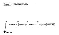
- the first 109 residues of the Lipoprotein D fusion partner is included on the N-terminus to provide the vaccine candidate antigen with additional exogenous T-cell epitopes and increase expression level in E-coli (thus acting also as an expression enhancer).
- the lipid tail ensures optimal presentation of the antigen to antigen presenting cells.
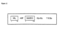
- fusion partners include the non-structural protein from influenzae virus, NS1 (hemagglutinin). Typically the N terminal 81 amino acids are utilised, although different fragments may be used provided they include T-helper epitopes.
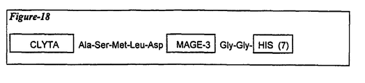
- the immunological fusion partner is the protein known as LYTA.
- LYTA the protein known as LYTA.
- the C terminal portion of the molecule is used.
- Lyta is derived from Streptococcus pneumoniae which synthesize an N-acetyl-L-alanine amidase, amidase LYTA, (coded by the lytA gene ⁇ Gene, 43 (1986) page 265-272 ⁇ an autolysin that specifically degrades certain bonds in the peptidoglycan backbone.
- the C-terminal domain of the LYTA protein is responsible for the affinity to the choline or to some choline analogues such as DEAE.
- immunological fusion partners noted above are also advantageous in aiding expression.
- such fusions are expressed at higher yields than native recombinant MAGE proteins.
- the present invention in the embodiment provides fusion proteins comprising a tumour-associated antigen from the MAGE family linked to an immunological fusion partner.
- the immunological fusion partner is protein D or fragment thereof, most preferably lipoprotein D.
- the MAGE proteins are preferably MAGE A1 or MAGE A3.
- the Lipoprotein D part preferably comprises the first 1/3 of Lipoprotein D.
- the proteins of the present invention preferably are expressed in E. coli.
- the proteins are expressed with an affinity tag, such as for example, a histidine tail comprising between 5 to 9 and preferably six histidine residues. These are advantageous in aiding purification.
- the present invention also provides a nucleic acid encoding the proteins of the present invention.
- Such sequences can be inserted into a suitable expression vector and used for DNA/RNA vaccination or expressed in a suitable host.
- Microbial vectors expressing the nucleic acid may be used as vaccines.
- Such vectors include for example, poxvirus, adenovirus, alphavirus, listeria and monarphage.
- a DNA sequence encoding the proteins of the present invention can be synthesized using standard DNA synthesis techniques, such as by enzymatic ligation as described by D.M. Roberts et al . in Biochemistry 1985, 24, 5090-5098, by chemical synthesis, by in vitro enzymatic polymerization, or by PCR technology utilising for example a heat stable polymerase, or by a combination of these techniques.
- Enzymatic polymerisation of DNA may be carried out in vitro using a DNA polymerase such as DNA polymerase I (Klenow fragment) in an appropriate buffer containing the nucleoside triphosphates dATP, dCTP, dGTP and dTTP as required at a temperature of 10°-37°C, generally in a volume of 50 ⁇ l or less.
- a DNA polymerase such as DNA polymerase I (Klenow fragment) in an appropriate buffer containing the nucleoside triphosphates dATP, dCTP, dGTP and dTTP as required at a temperature of 10°-37°C, generally in a volume of 50 ⁇ l or less.
- Enzymatic ligation of DNA fragments may be carried out using a DNA ligase such as T4 DNA ligase in an appropriate buffer, such as 0.05M Tris (pH 7.4), 0.01M MgCl 2 , 0.01M dithiothreitol, 1mM spermidine, 1mM ATP and 0.1mg/ml bovine serum albumin, at a temperature of 4°C to ambient, generally in a volume of 50ml or less.
- a DNA ligase such as T4 DNA ligase in an appropriate buffer, such as 0.05M Tris (pH 7.4), 0.01M MgCl 2 , 0.01M dithiothreitol, 1mM spermidine, 1mM ATP and 0.1mg/ml bovine serum albumin, at a temperature of 4°C to ambient, generally in a volume of 50ml or less.
- the chemical synthesis of the DNA polymer or fragments may be carried out by conventional phosphotriester, phosphite or phosphoramidite chemistry, using solid phase techniques such as those described in 'Chemical and Enzymatic Synthesis of Gene Fragments - A Laboratory Manual' (ed. H.G. Gassen and A. Lang), Verlag Chemie, Weinheim (1982), or in other scientific publications, for example M.J. Gait, H.W.D. Matthes, M. Singh, B.S. Sproat, and R.C. Titmas, Nucleic Acids Research, 1982, 10, 6243; B.S. Sproat, and W. Bannwarth, Tetrahedron Letters, 1983, 24, 5771; M.D.
- the process of the invention may be performed by conventional recombinant techniques such as described in Maniatis et al ., Molecular Cloning - A Laboratory Manual; Cold Spring Harbor, 1982-1989.
- the process may comprise the steps of :
- the term 'transforming' is used herein to mean the introduction of foreign DNA into a host cell. This can be achieved for example by transformation, transfection or infection with an appropriate plasmid or viral vector using e.g. conventional techniques as described in Genetic Engineering; Eds. S.M. Kingsman and A.J. Kingsman; Blackwell Scientific Publications; Oxford, England, 1988.
- the term 'transformed' or 'transformant' will hereafter apply to the resulting host cell containing and expressing the foreign gene of interest.
- the expression vectors are novel and also form part of the invention.
- the replicable expression vectors may be prepared in accordance with the invention, by cleaving a vector compatible with the host cell to provide a linear DNA segment having an intact replicon, and combining said linear segment with one or more DNA molecules which, together with said linear segment encode the desired product, such as the DNA polymer encoding the protein of the invention, or derivative thereof, under ligating conditions.
- the DNA polymer may be preformed or formed during the construction of the vector, as desired.
- vector will be determined in part by the host cell, which may be prokaryotic or eukaryotic but are preferably E. Coli or CHO cells. Suitable vectors include plasmids, bacteriophages, cosmids and recombinant viruses.
- the preparation of the replicable expression vector may be carried out conventionally with appropriate enzymes for restriction, polymerisation and ligation of the DNA, by procedures described in, for example, Maniatis et al . cited above.
- the recombinant host cell is prepared, in accordance with the invention, by transforming a host cell with a replicable expression vector of the invention under transforming conditions.
- Suitable transforming conditions are conventional and are described in, for example, Maniatis et al . cited above, or "DNA Cloning" Vol. II, D.M. Glover ed., IRL Press Ltd, 1985.
- a bacterial host such as E. coli may be treated with a solution of CaCl 2 (Cohen et al ., Proc. Nat. Acad. Sci., 1973, 69, 2110) or with a solution comprising a mixture of RbC1, MnCl 2 , potassium acetate and glycerol, and then with 3-[N-morpholino]-propane-sulphonic acid, RbC1 and glycerol.
- Mammalian cells in culture may be transformed by calcium co-precipitation of the vector DNA onto the cells.
- the invention also extends to a host cell transformed with a replicable expression vector of the invention.
- Culturing the transformed host cell under conditions permitting expression of the DNA polymer is carried out conventionally, as described in, for example, Maniatis et al . and "DNA Cloning" cited above.
- the cell is supplied with nutrient and cultured at a temperature below 50°C.
- the product is recovered by conventional methods according to the host cell and according to the localisation of the expression product (intracellular or secreted into the culture medium or into the cell periplasm).
- the host cell is bacterial, such as E. coli it may, for example, be lysed physically, chemically or enzymatically and the protein product isolated from the resulting lysate.
- the product may generally be isolated from the nutrient medium or from cell free extracts.
- Conventional protein isolation techniques include selective precipitation, adsorption chromatography, and affinity chromatography including a monoclonal antibody affinity column.
- the proteins of the present invention are provided either soluble in a liquid form or in a lyophilised form.
- each human dose will comprise 1 to 1000 ⁇ g of protein, and preferably 30 - 300 ⁇ g.
- the present invention also provides pharmaceutical composition comprising a protein of the present invention in a pharmaceutically acceptable excipient.
- a preferred vaccine composition comprises at least Lipoprotein D - MAGE-3.
- Such vaccine may optionally contain one or more other tumor-associated antigen.
- tumor-associated antigen include MAGE-1, GAGE-1 or Tyrosinase proteins.
- Vaccine preparation is generally described in Vaccine Design ("The subunit and adjuvant approach” (eds. Powell M.F. & Newman M.J). (1995) Plenum Press New York). Encapsulation within liposomes is described by Fullerton, US Patent 4,235,877.
- the proteins of the present invention are preferably adjuvanted in the vaccine formulation of the invention.
- Suitable adjuvants include an aluminium salt such as aluminium hydroxide gel (alum) or aluminium phosphate, but may also be a salt of calcium, iron or zinc, or may be an insoluble suspension of acylated tyrosine, or acylated sugars, cationically or anionically derivatised polysaccharides, or polyphosphazenes.
- Other known adjuvants include CpG containing oligonucleotides. The oligonucleotides are characterised in that the CpG dinucleotide is unmethylated. Such oligonucleotides are well known and are described in, for example WO96/02555.
- the adjuvant composition induces an immune response preferentially of the TH1 type.
- Suitable adjuvant systems include, for example a combination of monophosphoryl lipid A, preferably 3-de-O-acylated monophosphoryl lipid A (3D-MPL) together with an aluminium salt.
- CpG oligonucleotides also preferentially induce a TH1 response.
- An enhanced system involves the combination of a monophosphoryl lipid A and a saponin derivative particularly the combination of QS21 and 3D- MPL as disclosed in WO 94/00153, or a less reactogenic composition where the QS21 is quenched with cholesterol as disclosed in WO 96/33739.
- a particularly potent adjuvant formulation involving QS21 3D-MPL & tocopherol in an oil in water emulsion is described in WO 95/17210 and is a preferred formulation.
- a vaccine comprising a protein of the present invention, more preferably a Lipoprotein D (or derivative thereof) - MAGE-3 adjuvanted with a monophosphoryl lipid A or derivative thereof.
- the vaccine additionally comprises a saponin, more preferably QS21.
- the formulation additional comprises an oil in water emulsion and tocopherol.
- the present invention also provides a method for producing a vaccine formulation comprising mixing a protein of the present invention together with a pharmaceutically acceptable excipient, such as 3D-MPL.
- a process for purifying a recombinantly produced MAGE-protein comprises solubilising the protein, for example in a strong chaotropic agent (such as for example, urea, guanidium hydrochloride), or in a Zwitterionicnic detergent, e.g. (Empigen BB - n-dodecyl- N,N-dimethylglycine), reducing the protein's intra and inter molecular disulphide bonds, blocking the resulting thiols to prevent oxidative recoupling, and subjecting the protein to one or more chromatographic steps.
- a strong chaotropic agent such as for example, urea, guanidium hydrochloride
- Zwitterionicnic detergent e.g. (Empigen BB - n-dodecyl- N,N-dimethylglycine)
- the blocking agent is an alkylating agent.
- blocking agents include but are not limited to alpha haloacids or alpha haloamides.
- alpha haloacids or alpha haloamides For example iodoacetic acid and iodoacetamide which results in carboxymethylation or carboxyamidation (carbamidomethylation) of the protein.
- Other blocking agents may be used and are described in the literature (See for example, The Proteins Vol II Eds H neurath, RL Hill and C-L Boeder, Academic press 1976, or Chemical Reagents for Protein modification Vol I eds. RL Lundblad and CM Noyes, CRC Press 1985).
- blocking agents include N -ethylmaleimide, chloroacetyl phosphate, O-methylisourea and acrylonitrile.
- the use of the blocking agent is advantageous as it prevents aggregation of the product, and ensure stability for downstream purification.
- the blocking agents are selected to induce a stable covalent and irreversible derivative (eg alpha halo acids or alpha haloamides).
- a stable covalent and irreversible derivative eg alpha halo acids or alpha haloamides.
- other blocking agents maybe selected such that after purification the blocking agent may be removed to release the non derivatised protein.
- MAGE proteins having derivatised free thiol residues are new and form an aspect of the invention.
- carboxyamidated or carboxymethylated derivatives are a preferred embodiment of the invention.
- the proteins of the present invention is provided with an affinity tag, such as CLYTA or a polyhistidine tail.
- an affinity tag such as CLYTA or a polyhistidine tail.
- the protein after the blocking step is preferably subjected to affinity chromatography.
- affinity chromatography immobilised metal ion affinity chromatography (IMAC) may be performed.
- the metal ion may be any suitable ion for example zinc, nickel, iron, magnesium or copper, but is preferably zinc or nickel.
- the IMAC buffer contain a zwitterionic detergent such as Empigen BB (hereinafter Empigen) as this results in lower levels of endotoxin in the final product.
- the protein may be purifed by exploiting its affinity to choline or choline analogues such as DEAE.
- the proteins are provided with a polyhistidine tail and a Clyta part. These may purified in a simple two step affinity chromatographic purification schedule.
- Lipoprotein D the DNA encoding protein D has been cloned into the expression vector pMG 81.
- This plasmid utilizes signals from lambda phage DNA to drive the transcription and translation of inserted foreign genes.
- the vector contains the lambda PL promoter PL, operator OL and two utilization sites (NutL and NutR) to relieve transcriptional polarity effects when N protein is provided (Gross et al., 1985. Mol. & Cell. Biol. 5:1015).
- Vectors containing the PL promoter are introduced into an E. coli lysogenic host to stabilize the plasmid DNA.
- Lysogenic host strains contain replication-defective lambda phage DNA integrated into the genome (Shatzman et al., 1983; In Experimental Manipulation of Gene Expression. Inouya (ed) pp 1-14. Academic Press NY).
- the lambda phage DNA directs the synthesis of the cI repressor protein which binds to the OL repressor of the vector and prevents binding of RNA polymerase to the PL promoter and thereby transcription of the inserted gene.
- the cI gene of the expression strain AR58 contains a temperature sensitive mutation so that PL directed transcription can be regulated by temperature shift, i.e. an increase in culture temperature inactivates the repressor and synthesis of the foreign protein is initiated. This expression system allows controlled synthesis of foreign proteins especially of those that may be toxic to the cell (Shimataka & Rosenberg, 1981. Nature 292:128).
- the AR58 lysogenic E. coli strain used for the production of the LPD-MAGE-3-His protein is a derivative of the standard NIH E.coli K12 strain N99 (F- su- galK2, lacZ- thr-). It contains a defective lysogenic lambda phage (galE::TN10, 1 Kil- cI857 DH1). The Kil- phenotype prevents the shut off of host macromolecular synthesis. The cI857 mutation confers a temperature sensitive lesion to the cI repressor. The DH1 deletion removes the lambda phage right operon and the hosts bio, uvr3, and chlA loci.
- the AR58 strain was generated by transduction of N99 with a P lambda phage stock previously grown on an SA500 derivative (galE::TN10, 1 Kil- cI857 DH1).
- the introduction of the defective lysogen into N99 was selected with tetracycline by virtue of the presence of a TN10 transposon coding for tetracyclin resistance in the adjacent galE gene.
- N99 and SA500 are E.coli K12 strains derived from Dr. Martin Rosenberg's laboratory at the National Institutes of Health.
- MAGE 3 was expressed as a fusion protein using the N-terminal third of the lipidated protein D as fusion partner connected at the N-terminus of MAGE-3 and a sequence of several histidine residues (His tail) placed at its C-terminus.
- Protein D is a lipoprotein (a 42 kDa immunoglobulin D binding protein exposed on the surface of the Gram-negative bacterium Haemophilus influenzae ).
- the protein is synthesized as a precursor with an 18 amino acid residue signal sequence, containing a consensus sequence for bacterial lipoprotein (WO 91/18926).
- the Cys (at position 19 in the precursor molecule) becomes the amino terminal residue and is concomitantly modified by covalent attachment of both ester-linked and amide-linked fatty acids.
- the fatty acids linked to the amino-terminal cysteine residue then function as membrane anchor.
- the plasmid expressing the fusion protein was designed to express a precursor protein containing the 18 amino acids signal sequence and the first 109 residues of the processed protein D, two unrelated amino acids (Met and Asp), amino acid residues 2 to 314 of MAGE-3, two Gly residues functioning as a hinge region to expose the subsequent seven His residues.
- the recombinant strain thus produces the processed lipidated His tailed fusion protein of 432 amino acid residues long (see Figure 1), with the amino acids sequence described in ID No 1 and the coding sequence is described in ID No2.
- a cDNA plasmid (from Dr Thierry Boon from the Ludwig Institute) containing the coding sequence for MAGE-3 gene (Gaugler B et al , 1994), and the vector PRIT 14586, containing the N terminal portion of the Lipo-D-1/3 coding sequence (prepared as outlined in Figure 2) were used.
- the cloning strategy included the following steps ( Figure 3).
- Cells of AR58 transformed with plasmid pRIT14477 were grown in 2 litre flasks, each containing 400 mL of LY12 medium supplemented with yeast extract (6.4 g/L) and kanamycin sulphate (50 mg/L). After incubation on a shaking table at 30°C for 8 +/- 1 h, a small sample was removed from each flask for microscopic examination. The contents of the two flasks were pooled to provide the inoculum for the 20 litre fermentor.
- the inoculum (about 800 mL) was added to a pre-sterilised 20 litre (total volume) fermentor containing 7 litres of medium, supplemented with 50 mg/L of kanamycin sulphate.
- the pH was adjusted to and maintained at 6.8 by the periodic addition of NH 4 OH (25 % v/v), and the temperature was adjusted to and maintained at 30°C.
- the aeration rate was adjusted to and maintained at 12 litres of air/min and the dissolved oxygen tension was maintained at 50 % of saturation by feedback control of the agitation speed.
- the over-pressure in the fermentor was maintained at 500 g/cm 2 (0.5 bar).
- the fed-batch cultivation was carried out by controlled addition of a carbon feed solution.
- the feed solution was added at an initial rate of 0.04 mL/min, and increased exponentially during the first 42 hours to maintain a growth rate of 0.1 h -1 .
- the temperature in the fermentor was rapidly increased to 39°C, and the feeding speed was maintained constant at 0.005 mL/g DCW/min during the induction phase for an additional 22-23 hours, during which time intracellular expression of LPD-MAGE-3-His reached a maximum level.
- the optical density of the culture was between 80 and 120 (corresponding to a cell concentration of between 48 and 72 g DCW/L), and the total liquid volume was approximately 12 litres.
- the culture was rapidly cooled to between 6 and 10°C, and the cells of ECK32 were separated from the culture broth by centrifugation at 5000 x g at 4°C for 30 minutes.
- the concentrated cells of ECK32 were quickly stored in plastic bags and immediately frozen at -80°C.
- the frozen concentrated cells of ECK32 were thawed to 4°C before being resuspended in cell disruption buffer to a final optical density of 60 (corresponding to a cell concentration of approximately 36 g DCW/L).
- the cells were disrupted by two passes through a high-pressure homogeniser (1000 bar).
- the broken cell suspension was centrifuged (x 10 000g at 4°C for 30 minutes) and the pellet fraction was washed twice with Triton X100 (1% w/v) + EDTA (1 mM), followed by a wash with phosphate buffered saline (PBS) + Tween 20 (0.1% v/v) and finally a wash with PBS. Between each washing stage, the suspension was centrifuged at x 10 000g for 30 minutes at 4°C, the supernatant was discarded and the pellet fraction was retained.
- PBS phosphate buffered saline
- LPD-MAGE-3-His was purified from the cell homogenate using a sequence of steps described below:
- pellet fraction from the final washing stage was resolubilised overnight in 800 mL of a solution of guanidine hydrochloride (6M) and sodium phosphate (0.1 M, pH 7.0) at 4°C.
- the solubilised material (a pale yellow, turbid suspension) was flushed with argon to purge any remaining oxygen, and a stock solution of 2-mercaptoethanol (14M) was added to provide a final concentration of 4.3M (which corresponded to 0.44 mL of 2-mercaptoethanol per mL of solution).
- the resulting solution was divided and transferred into two glass flasks which were both heated to 95 °C in a water bath. After 15 minutes at 95 °C, the flasks were removed from the water bath and allowed to cool, whereupon the contents were pooled into a foil-covered beaker (5 L), placed on ice, and solid iodoacetamide added with vigorous mixing to provide a final concentration of 6M (which corresponded to 1.11 g of iodoacetamide per mL of solution).
- the mixture was microfiltered in an Amicon Proflux M12 tangential-flow unit equipped with a Minikros hollow fibre cartridge (ref. No. M22M-600-01N; area 5,600 cm 2 , 0.2 ⁇ m).
- the permeate was retained for subsequent chromatographic purification.
- Metal chelate chromatography was performed with Chelating Sepharose FF (Pharmacia Biotechnology Cat. No. 17-0575-01) packed into a BPG 100/500 column (Pharmacia Biotechnology Cat No. 18-1103-01).
- the dimensions of the packed bed were: diameter 10 cm; cross-sectional area 79 cm 2 ; bed height 19 cm; packed volume 1,500 mL.
- the empty column was sanitised with sodium hydroxide (0.5M), then washed with purified water.
- the support (delivered in 20 % v/v ethanol) was washed with purified water (8 litres) on a Buchner funnel (under vacuum) and charged with zinc by passing at least 15 litres of a solution of ZnCl 2 (0.1M). Excess zinc was removed by washing the support with 10 litres of purified water, until the pH of the outlet liquid reached the pH of the ZnCl 2 solution (pH 5.0). The support was then equilibrated with 4 litres of a solution containing guanidine hydrochloride (6M) and sodium phosphate (0.1M, pH 7.0).
- the next stages of metal chelate chromatography were conducted at an eluent flow rate of 60 mL/min.
- the column was washed, first with the solution containing guanidine hydrochloride (6M) and sodium phosphate (0.1M, pH 7.0), then with the solution containing urea (6M) and sodium phosphate (0.1M, pH 7.0), until the column eluent attained zero absorbance at OD 280 nm (baseline).
- the semi-pure LPD-MAGE-3-His protein fraction was eluted with 2 column volumes of a solution containing urea (6M), sodium phosphate (0.1M, pH 7.0) and imidazole (0.5M). The conductance of this fraction was approximately 16 mS/cm.
- the conductance of the semi-pure LPD-MAGE-3-His protein fraction was reduced to approximately 4 mS/cm by dilution with a solution containing urea (6M) and Tris-HCl (20 mM, pH 8.0).
- Anion exchange chromatography was performed using Q-Sepharose FF (Pharmacia Biotechnology, Cat. No. 17-0510-01) packed in a BPG 200/500 column (Pharmacia Biotechnology Cat. No. 18-1103-11).
- the dimensions of the packed bed were: diameter 10 cm; cross-sectional area 314 cm 2 ; bed height 9 cm; packed volume 2,900 mL.
- the column was packed (with 20 % v/v ethanol) and washed with 9 litres of purified water at an eluent flow rate of 70 mL/min.
- the packed column was sanitised with 3 litres of sodium hydroxide (0.5M), washed with 30 litres of purified water, then equilibrated with 6 litres of a solution containing urea (6M) and Tris-HCl (20 mM, pH 8.0).
- the diluted, semi-purified LPD-MAGE-3-His was loaded onto the column and then washed with 9 litres of a solution containing urea (6M), Tris-HCl (20 mM, pH 8.0), EDTA (1mM) and Tween (0.1 %), until the absorbance (280 nm) of the eluent fell to zero.
- a further washing step was performed with 6 litres of a solution containing urea (6M) and Tris-HCl (20 mM, pH 8.0).
- the purified LPD-MAGE-3-His was eluted from the column with a solution containing urea (6M), Tris-HCl (20 mM, pH 8.0) and NaCl (0.25M).
- the column was packed in ethanol (20 %) and washed with 5 litres of purified water at an effluent flow rate of 20 mL/min.
- the column was sanitised with 2 litres of sodium hydroxide (0.5M), washed with 5 litres of purified water, then equilibrated with 5 litres of phosphate-buffered saline containing Tween 80 (0.1 % v/v).
- the purified LPD-MAGE-3-His fraction (maximum 500 mL/ desalting run) was loaded onto the column at an eluent flow rate of 20 mL/min.
- the desalted purified LPD-MAGE-3-His was eluted from the column with 3 litres of PBS containing Tween 80 (0.1 % v/v).
- the bulk LPD-MAGE-3-His from size exclusion chromatography was filtered through a 0.22 ⁇ m membrane in a laminar flow hood (class 10.000). The filtered bulk was frozen at -80 °C and stored until the desalting step.
- the column was sanitised with 6 litres of sodium hydroxide (0.5M), then equilibrated with 10 litres of a solution containing sodium phosphate (10 mM, pH 6.8), NaCl (20 mM) and Tween 80 (0.1 % v/v).
- the purified LPD-MAGE-3-His fraction (maximum 1500 mL/desalting run) was loaded onto the column at an eluent flow rate of 100 mL/min.
- the desalted purified LPD-MAGE-3-His fraction eluted at the void volume of the column, was sterile filtered through a 0.22 ⁇ m membrane and stored at -80°C.
- the final bulk protein is thawed to +4°C before being aliquoted into vials and freeze-dried in a lactose excipient (3.2%).
- the LPD-MAGE-3-His purified antigen was analysed by SDS-PAGE on a 12.5% acrylamide gel in reducing conditions.
- the protein load was 50 ⁇ g for Coomassie blue staining and 5 ⁇ g for silver nitrate staining.
- Clinical lot 96K19 and pilot lot 96J22 were analyzed. One major band corresponding to a molecular weight of 60kDa was visualised. Two minor additional bands of approximately 45kDa and 35 kDa were also seen.
- the peptides revealed by SDS-PAGE analysis of the LPD-MAGE-3-His protein were identified by Western blot using mouse monoclonal antibodies. These antibodies were developed in-house using a purified preparation of the MAGE-3-His protein (this protein does not contain the LPD part of the LPD-MAGE-3-His).
- FIG. 4 shows the band patterns obtained for lots 96K19 and 96J22 after staining with Mabs 32 and 54.
- Six hundred (600) ng of protein were resolved on a 12.5% SDS-PAGE, transferred to a nylon membrane, reacted with Mabs 32 and 54 (60 ⁇ g/ml) and revealed with anti-mouse antibodies coupled to peroxidase.
- the 60 kDa and 30 kDa peptide detected by SDS-PAGE are revealed by both Mabs.
- the vaccine used in these experiments is produced from a recombinant DNA, encoding a Lipoprotein D 1/3-MAGE-3-His, expressed in E. coli from the strain AR58, either adjuvanted or not.
- the formulation comprises a mixture of 3 de -O-acylated monophosphoryl lipid A (3D-MPL) and QS21 in an oil/water emulsion.
- the adjuvant system SBAS2 has been previously described WO 95/17210.
- 3D-MPL is an immunostimulant derived from the lipopolysaccharide (LPS) of the Gram-negative bacterium Salmonella minnesota. MPL has been deacylated and is lacking a phosphate group on the lipid A moiety. This chemical treatment dramatically reduces toxicity while preserving the immunostimulant properties (Ribi, 1986). Ribi Immunochemistry produces and supplies MPL to SB-Biologicals. Experiments performed at Smith Kline Beecham Biologicals have shown that 3D-MPL combined with various vehicles strongly enhances both the humoral and a TH1 type of cellular immunity.
- LPS lipopolysaccharide
- QS21 is a natural saponin molecule extracted from the bark of the South American tree Quillaja saponaria Molina. A purification technique developed to separate the individual saponines from the crude extracts of the bark, permitted the isolation of the particular saponin, QS21, which is a triterpene glycoside demonstrating stronger adjuvant activity and lower toxicity as compared with the parent component. QS21 has been shown to activate MHC class I restricted CTLs to several subunit Ags, as well as to stimulate Ag specific lymphocytic proliferation (Kensil, 1992). Aquila (formally Cambridge Biotech Corporation) produces and supplies QS21 to SB-Biologicals.
- the oil/water emulsion is composed an organic phase made of of 2 oils (a tocopherol and squalene), and an aqueous phase of PBS containing Tween 80 as emulsifier.
- the emulsion comprised 5% squalene 5% tocopherol 0.4% Tween 80 and had an average particle size of 180 nm and is known as SB62 (see WO 95/17210).
- SB62 see WO 95/17210
- Tween 80 is dissolved in phosphate buffered saline (PBS) to give a 2% solution in the PBS.
- PBS phosphate buffered saline
- To provide 100 ml two fold concentrate emulsion 5g of DL alpha tocopherol and 5ml of squalene are vortexed to mix thoroughly. 90ml of PBS/Tween solution is added and mixed thoroughly. The resulting emulsion is then passed through a syringe and finally microfluidised by using an M110S microfluidics machine.
- the resulting oil droplets have a size of approximately 180 nm.
- the adjuvant is formulated as a combination of MPL and QS21, in an oil/water emulsion. This preparation is delivered in vials of 0.7 ml to be admixed with the lyophilised antigen (vials containing from 30 to 300 ⁇ g antigen).
- the composition of the adjuvant diluent for the lyophilised vaccine is as follows : Ingredients: Quantity (per dose): Adjuvants SB62 Emulsion: 250 ⁇ l - Squalene 10.7 mg - DL ⁇ -tocopherol 11.9 mg - Tween 80 4.8 mg Monophosphoryl Lipid A 100 ⁇ g QS21 100 ⁇ g Preservative Thiomersal 25 ⁇ g Buffer Water for injection q.s. ad 0.5 ml - Dibasic sodium phosphate 575 ⁇ g - Monobasic potassium phosphate 100 ⁇ g - Potassium chloride 100 ⁇ g - Sodium chloride 4.0 mg
- the final vaccine is obtained after reconstitution of the lyophilised LPD-MAGE-3-His preparation with the adjuvant or with PBS alone.
- the adjuvants controls without antigen were prepared by replacing the protein by PBS.
- Vaccine antigen Fusion protein Lipoprotein D1/3 - MAGE-3-His:
- Lipoprotein D is a lipoprotein exposed on the surface of the Gram-negative bacteria Haemophilus influenzae.
- the inclusion of the first 109 residues of the processed protein D as fusion partner is incorporated to provide the vaccine antigen with a T-cell epitopes.
- the protein contains two unrelated amino acids (Met and Asp), amino acid residues 2 to 314 of Mage-3, two Gly residues functioning as hinge region to expose the subsequent seven His residues.
- the candidate vaccine was injected into 2 different mouse strains (C57BL/6 and Balb/C), varying in their genetic background and MHC alleles.
- mouse strains C57BL/6 and Balb/C
- potential MHC class-I and MHC class-II peptide motifs were theoretically predicted for the MAGE part of the LPD-MAGE-3-His fusion protein.
- mice of each strain were injected twice at 2 weeks interval in the foot pad with 5 ⁇ g of LPD-MAGE-3-His, formulated or not in SBAS2 at 1/10th of the concentration used in human settings.
- Lymphocytes were prepared by crushing the spleen or the popliteal lymph nodes from the mice, 2 weeks after the last injection. 2 x 10 5 cells were placed in triplicate in 96 well plates and the cells were re-stimulated in vitro for 72 hours with different concentrations (1- 0.1 ⁇ g/ml) of His-Mage 3 as such or coated onto latex micro-beads.
- lymphocytes from mice immunized with LPD-MAGE-3-His in the adjuvant SBAS2 were obtained with lymphocytes from mice immunized with LPD-MAGE-3-His in the adjuvant SBAS2 (see Figures 6 and 8).
- LPD-MAGE-3-His is immunogenic in mice, and this immunogenicity can be increased by the use of the SBAS2 adjuvant formulation.
- mice were immunized by 2 intra foot pad injections at 2 weeks interval with either PBS, or SBAS2, or 5 ⁇ G of LPD-MAGE-3-His, or 5 ⁇ G of LPD-MAGE-3-His + SBAS2. Three and five animals were used in the control groups and in the tested groups respectively.
- the Ig subclasses-specific anti-MAGE-3 responses after vaccinations in the different groups of mice can be seen on the figures 10 and 11, which give a comparison of the mean midpoint dilution of the sera.
- RTS,S and gp120 were used as positive control.
- the animals received vaccine at day 0 and were boosted at day 28, and 84 and bled to determine their antibody response to both the MAGE 3 and protein D component.
- the vaccines were administered intramuscularly as a bolus injection (0.5ml) in the posterior part of the right leg.
- Serum was removed, frozen at -20°C and sent for determination of the antibody levels by specific Elisa.
- 96well microplates (maxisorb Nunc) were either coated with 5 ⁇ g of His Mage 3 or Protein D overnight at 4°C. After 1 hour saturation at 37°C with PBS NCS 1%, serial dilution of the rabbit sera were added for 1H 30 at 37°C (starting at 1/10), after 3 washings in PBS Tween, anti rabbit biotinylated serum (Amersham ref RPN 1004 lot 88) was added (1/5000). Plates were washed and peroxydase couple streptavidin (1/5000) was added for 30 minutes at 37°C. After washing, 50 ⁇ l TMB (BioRad) was added for 7 minutes and the reaction was stopped with H2S04 0.2M, OD was measured at 450 nm. Midpoint dilutions were calculated by SoftmaxPro.
- LPD - MAGE 1-His was prepared.
- the amino acid and DNA sequences are depicted in SEQUENCE ID Nos. 3 and 4.
- the resulting protein was purified in an analogous manner to the LPD-MAGE-3-His protein. Briefly, the cell culture were homogenated and treated with 4M guanidine HCl and 0.5 M beta mercaptoethanol in the presence of 0.5% Empigen detergent. The product was filtered and the permeate treated with 0.6 M iodoacetamide. The carboxyamidated fractions was subjected to IMAC (zinc Chealate-sepharose FF) chromatography. The column was first equilbrated and washed with a solution containing 4M guanidine.
- IMAC zinc Chealate-sepharose FF
- the eluate was diluted with 4M Urea.
- the Q-sepharose column was equilabrated and washed with 4M Urea in 20mM phosphate buffer (pH7.5) in the presence of 0.5% Empigen.
- a second wash was performed in the same buffer, but devoid of the detergent.
- the protein eluate d in the same buffer but with increasing Imidazole (150mM, 400mM, 1M). The eluate was ultra filtered.
- the primary structure of the resulting protein has the sequence set forth in ID No. 5.
- the coding sequence(ID No. 6) corresponding to the above protein design was placed under the control of ⁇ pL promoter in a E. coli expression plasmid.
- the starting material was a cDNA plasmid received from Dr Tierry Boon from the Ludwig Institute, containing the coding sequence for MAGE-3 gene and the vector PMG81, containing the 81aa of NS 1 (Non structural protein) coding region from Influenza.
- the cloning strategy outlined in figure 13 included the following steps:
- Bacterial cells (23g) were lysed in 203 ml of a 50 mM PO 4 pH7 buffer by Rannie (homogeniser) and the lysate was centrifuged in a JA 20 rotor at 15,000 rpm during 30 minutes. The supernatant was discarded.
- 1/3 of the pellet was resolubilised O/N at 4°C in 34 ml of 100 mM PO 4 - 6 M GuHCl pH7. After centrifugation in a JA 20 rotor at 15,000 rpm for 30 minutes, the pellet was discarded and the supernatant was further purified by IMAC.
- Antigen positive fractions of the IMAC eluate 160 ml were pooled and concentrated to 5 ml in an Amicon stirred cell on a Filtron membrane (type Omega cut-off 10,000). The purity at this stage is about 70% as estimated by SDS-PAGE.
- the antigen was TCA precipitated and after centrifugation in a JA 20 rotor at 15,000 rpm for 20 minutes, the supernatant was discarded. The pellet was resolubilised in PBS buffer pH 7.4.
- the protein is soluble in PBS after freeze/thaw does not show any degradation when stored for 3 hours at 37° C and has an apparent molecular weight of approximately 50,000 Daltons as determined by SDS (12.5% PAGE).
- the primary structure of the resulting protein has the sequence set forth in sequence ID No. 7.
- the coding sequence (see SEQUENCE ID No. 8) corresponding to the above protein design was placed under the control of ⁇ pL promoter in a E. coli expression plasmid.
- the starting material was the vector PCUZ1 that contains the 117 C-terminal codons of the LytA coding region from Streptococcus pneumoniae and the vector pRIT14518, in which we have previously subcloned the MAGE-1 gene cDNA from a plasmid received from Dr Thierry Boon from the Ludwig Institute.
- a EcoRI-NCO 1 restriction fragment containing the long PL promoter and a part of CLYTA sequences was prepared from plasmid pRIT DVA6 and inserted between the EcoRI-NCO 1 sites of plasmid pRIT14613.
- the recombinant plasmid pRIT14614 was obtained.
- the recombinant plasmid pRIT14614 (see figure 17) encoding the fusion protein CLYTA-Mage-1-His was used to transform E. coli AR120.
- a Kan resistant candidate strain was selected and characterized.
- Tumour rejection recombinant antigen a fusion protein CLYTA -Mage-3-His where the C-lyt A fusion partner lead to expression of a soluble protein, act as affinity tag and provides a useful T-helper.
- the primary structure of the resulting protein has the sequence described in SEQUENCE ID No.9: and the coding sequence in sequence ID No. 10
- the coding sequence corresponding to the above protein design was placed under the control of ⁇ pL promoter in a E. coli expression plasmid.
- the starting material was the vector PCUZ1 that contains the 117 C-terminal codons of the LytA coding region from Streptococcus pneumoniae, described in Gene 43, (1986) p. 265-272 and the vector pRIT14426, in which we have previously subcloned the MAGE-3 gene cDNA from a plasmid received from Dr Tierry Boon from the Ludwig Institute.
- a BgIII - XbaI restriction fragment containing the short pL promoter and the CLYTA-Mage-3-His coding sequences was prepared from plasmid pRIT14662. and inserted between the BglII - XbaI sites of plasmid TCM67 (a pBR322 derivative containing the resistance to ampicillin, and the long ⁇ pL promoter, described in the international application PCT/EP92/O1827 ). The plasmid pRIT14607 was obtained. The recombinant plasmid pRIT14607 encoding the fusion protein Clyta-Mage-3 His was used to transform E. coli AR 120 (Mott et al.1985, Proc. Natl. Acad. Sci, 82: 88). An ampicillin resistant candidate strain was selected and characterized.
- the recombinant bacteria AR120 (pRIT 14646) were grown in a 20 Litters fermentor under fed-batch conditions at 30°. The expression of the recombinant protein was induced by adding Nalidixic acid at a final concentration of 60 ?g/ml. Cells were harvested at the end of fermentationand and lyzed at 60 OD/600 by two passages through a French Press disrupter (20 000 psi). Lysed cells were pelleted 20 min at 15 000 g at 4 °C. Supernatant containing the recombinant protein was loaded onto exchange DEAE Sepharose CL6B resin (Pharmacia) pre-equilibrated in 0.3M NaCl, 20 mM Tris HCl pH 7.6 Buffer A.
- fusion protein was eluted by 2 % choline in (Buffer A). Positive antigen fractions, as revealed by Western blotting analysis using an anti Mage-3 antibody, were pooled. DEAE-eluted antigen was brought to 0.5 % Empigen BB (a zwitterionic detergent) and to 0.5 M NaCl before loading onto an Ion Metal Affinity chromatography column preequilibrated in 0.5 % Empigen BB, 0.5 M NaCl, 50 mM phosphate buffer pH 7.6 (Buffer B). IMAC column was washed with buffer B until 280 nm absorbency reached the base line.
- Empigen BB a zwitterionic detergent
- IMAC column was washed with buffer B until 280 nm absorbency reached the base line.
- a second wash in buffer B without Empigen BB (Buffer C) in order to eliminate the detergent was executed before Antigen elution by an Imidazole gradient 0-250mM Imidazole in buffer C. 0.090-0.250 M Imidazole fractions were pooled, concentrated on a 10 kDa Filtron omega membrane before dialysis versus PBS buffer.
Abstract
Description

If the protein is produced with a Clyta part, the protein may be purifed by exploiting its affinity to choline or choline analogues such as DEAE. In an embodiment of the invention the proteins are provided with a polyhistidine tail and a Clyta part. These may purified in a simple two step affinity chromatographic purification schedule.
- f) - Buffer exchange/urea removal by size exclusion chromatography using Superdex 75,
- g) - In-process filtration,
- h) - Buffer exchange/desalting by size exclusion chromatography using Sephadex G25.
The Sephadex G25 was hydrated with 7 litres of purified water and allowed to swell overnight at 4 °C. The gel was then packed in the column with pure water at an eluent flow rate of 100 mL/min.
The final bulk protein is thawed to +4°C before being aliquoted into vials and freeze-dried in a lactose excipient (3.2%).
Experiments performed at SmithKline Beecham Biologicals have proven that the adjunction of this O/W emulsion to 3D-MPL/QS21 (SBAS2)further increases the immunostimulant properties of the latter against various subunit antigens.
The composition of the adjuvant diluent for the lyophilised vaccine is as follows :
| Ingredients: | Quantity (per dose): |
| Adjuvants | |
| SB62 Emulsion: | 250 µl |
| - Squalene | 10.7 mg |
| - DL α-tocopherol | 11.9 mg |
| - Tween 80 | 4.8 mg |
| Monophosphoryl Lipid A | 100 µg |
| QS21 | 100 |
| Preservative | |
| Thiomersal | |
| 25 µg | |
| Buffer | |
| Water for injection | q.s. ad 0.5 ml |
| - Dibasic sodium phosphate | 575 µg |
| - Monobasic potassium phosphate | 100 µg |
| - Potassium chloride | 100 µg |
| - Sodium chloride | 4.0 mg |
Three and five animals were used in the control groups and in the tested groups respectively.
2 µG/ml of purified His
| Groups: | |
| | right leg: RTS,S/SBAS2 |
| left leg: GP120/ | |
| Group | |
| 2 | right leg: RTS,S/SB26T |
| left leg: GP120/ | |
| Group | |
| 3 | right leg: LipoD1/3 |


The supernatant was discarded.
0.1 M P04-6 M urea pH7
After 4 hours induction , cells were harvested, resuspended in PBS and lysed by desintegration (desintegration CLS "one shot" type). After centrifugation, pellet supernatant and total extract were analyzed by SDS-PAGE. Proteins were visualized in Coomassie Bleu stained gels, where the fusion protein represented about 1% of the total E. coli proteins. The fusion protein was identified by Western blot analysis using rabbits anti-Mage-3 polyclonal antibodies . The recombinant protein appeared as a single band with an apparent MW of about 58 kD.
IMAC column was washed with buffer B until 280 nm absorbency reached the base line. A second wash in buffer B without Empigen BB (Buffer C) in order to eliminate the detergent was executed before Antigen elution by an Imidazole gradient 0-250mM Imidazole in buffer C.
0.090-0.250 M Imidazole fractions were pooled, concentrated on a 10 kDa Filtron omega membrane before dialysis versus PBS buffer.
- Anichini A., Fossati G., Parmiani G. Immunol. Today, 8: 385 (1987).
- De Plaen E., Arden K., Traversari C., et al. Immunogenetics, 40: 360 (1994).
- Gaugler B., Van den Eynde B., van der Bruggen P., et al. J. Exp. Med., 179: 921 (1994).
- Herman J., van der Bruggen P., Immanuel F., et al. Immunogenetics, 43: 377 (1996).
- Inoue H., Mori M., Li J., et al. Int. J. Cancer, 63: 523 (1995).
- Kensil C.R., Soltysik S., Patel U., et al. in: Channock R.M. , Ginsburg H.S.,
Brown F., et al., (eds.), Vaccines 92,
(Cold Spring Harbor Laboratory Press, Cold Spring Harbor, N.Y.), 36-40: (1992). - Knuth A., Danowski B., Oettgen H.F., et al. Proc. Natl. Acad. Sci. USA, 81: 3511 (1984).
- Patard J.J., Brasseur F., Gil-Diez S., et al. Int. J. Cancer, 64: 60 (1995).
- Ribi E., et al. in: Levine L., Bonventre P.F., Morello J., et al. (eds)., American Society for Microbiology, Washington DC, Microbiology 1986, 9-13; (1986).
- Van den Eynde B., Hainaut P., Hérin M. et al. Int. J. Cancer, 44: 634 (1989).
- Van der Bruggen P., Traversari C., Chomez P., et al. Science, 254: 1643 (1991).
- Van der Bruggen P., Bastin J., Gajewski T., et al. Eur. J. Immunol., 24: 3038 (1994).
- Van Pel A., van der Bruggen P., Coulie P.G. , et al., Immunol. Rev., 145: 229 (1995).
- Weynants P., Lethé B., Brasseur F., et al. Int. J. Cancer, 56: 826 (1994).
- Nishimura S, Fujita M, Terata N, Tani T, Kodama M, Itoh K, Nihon Rinsho Meneki Gakkai Kaishi 1997, Apr, 20 (2): 95-101.
- Fuijie T et al, Ann Oncol 1997 Apr, 8 (4): 369-72.


Claims (20)
- A tumour-associated antigen derivative from the MAGE family.
- An antigen as claimed in claim 1, when the derivative is a MAGE protein linked to an immunological fusion or expression enhancer partner.
- An antigen as claimed in claim 1 or 2 wherein the derivative comprises an affinity tag.
- An antigen as claimed in any of claims 1 to 3 which contains a derivatised free thiol.
- An antigen as claimed in claim 4 which is a carboxyamide or carboxymethylated derivative.
- A protein as claimed in claim 2, 3, 4 or 5 wherein the fusion partner is protein D or fragment thereof from Heamophilus influenzae B, NS1 protein from influenza or a fragment thereof or Lyta from Streptococcus pneumoniae or fragment thereof.
- A protein as claimed in claim 2, 3, 4 or 5 wherein the fusion partner is the lipidated form of protein D or fragment thereof from Heamophilus influenza B.
- A protein as claimed in claim 1 to 7 wherein the MAGE protein is selected from the group MAGE A1, MAGE A2, MAGE A3, MAGE A4, MAGE A5, MAGE A6, MAGE A7, MAGE A8, MAGE A9, MAGE A10, MAGE A11, MAGE A12, MAGE B1, MAGE B2, MAGE B3 and MAGE B4, MAGE C1, MAGE C2.
- A nucleic acid sequence encoding a protein as claimed herein.
- A vector comprising a nucleic acid of claim 9.
- A host transformed with a vector of claim 10.
- A vaccine containing a protein as claimed in any of claims 1 to 8 or a nucleic acid as claimed in claim 9.
- A vaccine as claimed in claim 12 additionally comprising an adjuvant, and/or immunostimulatory cytokine or chemokine.
- A vaccine as claimed in claim 12 or 13 wherein the protein is presented in an oil in water or a water in oil emulsion vehicle.
- A vaccine as claimed in claim 13 or 14 wherein the adjuvant comprises 3D-MPL, QS21 or a CpG oligonucleotide.
- A vaccine as claimed herein additionally comprising one or more other antigens.
- A vaccine as claimed herein for use in medicine.
- Use of a protein or nucleic acid as claimed herein for the manufacture of a vaccine for immunotherapeutically treating a patient suffering from melanomas or other MAGE-associated tumours.
- A process for the purification of a MAGE protein or derivative thereof, comprising reducing the disulphide bonds, blocking the resulting free thiol group with a blocking group, and subjecting the resulting derivative to one or more chromatographic purification steps.
- A process for the production of a vaccine, comprising the steps of purifying a MAGE protein or a derivative thereof, by the process of claim 19 and formulating the resulting protein as a vaccine.
Priority Applications (6)
| Application Number | Priority Date | Filing Date | Title |
|---|---|---|---|
| EP06075362A EP1659178B1 (en) | 1998-02-05 | 1999-02-02 | A process for the purification or production of a MAGE protein |
| DK06075362.1T DK1659178T3 (en) | 1998-02-05 | 1999-02-02 | Process for the purification or preparation of a MAGE protein |
| DK06075363.9T DK1659179T3 (en) | 1998-02-05 | 1999-02-02 | Tumor-associated antigen derivatives from the MAGE family and nucleic acid sequences encoding them used to prepare fusion proteins and vaccine compositions |
| EP06075363A EP1659179B1 (en) | 1998-02-05 | 1999-02-02 | Tumor-associated antigen derivatives from the mage family, and nucleic acid sequences encoding them, used for the preparation of fusion proteins and of compositions for vaccination |
| SI9931053T SI1584685T1 (en) | 1998-02-05 | 1999-02-02 | Tumor-associated antigen derivatives from the mage family, used for the preparation of fusion proteins with T-helper epitopes and of compositions for vaccination |
| CY20111100663T CY1111672T1 (en) | 1998-02-05 | 2011-07-08 | ANTIGEN PRODUCERS MADE TO THE MAGE FAMILY VOLUME USED FOR PREPARATION OF PRIMARY EFFECTIVENESS |
Applications Claiming Priority (5)
| Application Number | Priority Date | Filing Date | Title |
|---|---|---|---|
| GBGB9802543.0A GB9802543D0 (en) | 1998-02-05 | 1998-02-05 | Vaccine |
| GB9802543 | 1998-02-05 | ||
| GBGB9802650.3A GB9802650D0 (en) | 1998-02-06 | 1998-02-06 | Vaccine |
| GB9802650 | 1998-02-06 | ||
| EP99907476A EP1053325B1 (en) | 1998-02-05 | 1999-02-02 | Tumor-associated antigen derivatives from the mage family, and nucleic acid sequences encoding them, used for the preparation of fusion proteins and of compositions for vaccination |
Related Parent Applications (2)
| Application Number | Title | Priority Date | Filing Date |
|---|---|---|---|
| EP99907476A Division EP1053325B1 (en) | 1998-02-05 | 1999-02-02 | Tumor-associated antigen derivatives from the mage family, and nucleic acid sequences encoding them, used for the preparation of fusion proteins and of compositions for vaccination |
| EP99907476.8 Division | 1999-02-02 |
Related Child Applications (4)
| Application Number | Title | Priority Date | Filing Date |
|---|---|---|---|
| EP06075363A Division EP1659179B1 (en) | 1998-02-05 | 1999-02-02 | Tumor-associated antigen derivatives from the mage family, and nucleic acid sequences encoding them, used for the preparation of fusion proteins and of compositions for vaccination |
| EP06075362A Division EP1659178B1 (en) | 1998-02-05 | 1999-02-02 | A process for the purification or production of a MAGE protein |
| EP06075362.1 Division-Into | 2006-02-17 | ||
| EP06075363.9 Division-Into | 2006-02-17 |
Publications (3)
| Publication Number | Publication Date |
|---|---|
| EP1584685A2 true EP1584685A2 (en) | 2005-10-12 |
| EP1584685A3 EP1584685A3 (en) | 2005-12-07 |
| EP1584685B1 EP1584685B1 (en) | 2011-04-13 |
Family
ID=26313069
Family Applications (4)
| Application Number | Title | Priority Date | Filing Date |
|---|---|---|---|
| EP06075362A Expired - Lifetime EP1659178B1 (en) | 1998-02-05 | 1999-02-02 | A process for the purification or production of a MAGE protein |
| EP06075363A Expired - Lifetime EP1659179B1 (en) | 1998-02-05 | 1999-02-02 | Tumor-associated antigen derivatives from the mage family, and nucleic acid sequences encoding them, used for the preparation of fusion proteins and of compositions for vaccination |
| EP99907476A Expired - Lifetime EP1053325B1 (en) | 1998-02-05 | 1999-02-02 | Tumor-associated antigen derivatives from the mage family, and nucleic acid sequences encoding them, used for the preparation of fusion proteins and of compositions for vaccination |
| EP05076599A Expired - Lifetime EP1584685B1 (en) | 1998-02-05 | 1999-02-02 | Tumor-associated antigen derivatives from the mage family, used for the preparation of fusion proteins with T-helper epitopes and of compositions for vaccination |
Family Applications Before (3)
| Application Number | Title | Priority Date | Filing Date |
|---|---|---|---|
| EP06075362A Expired - Lifetime EP1659178B1 (en) | 1998-02-05 | 1999-02-02 | A process for the purification or production of a MAGE protein |
| EP06075363A Expired - Lifetime EP1659179B1 (en) | 1998-02-05 | 1999-02-02 | Tumor-associated antigen derivatives from the mage family, and nucleic acid sequences encoding them, used for the preparation of fusion proteins and of compositions for vaccination |
| EP99907476A Expired - Lifetime EP1053325B1 (en) | 1998-02-05 | 1999-02-02 | Tumor-associated antigen derivatives from the mage family, and nucleic acid sequences encoding them, used for the preparation of fusion proteins and of compositions for vaccination |
Country Status (26)
| Country | Link |
|---|---|
| US (3) | US8097257B2 (en) |
| EP (4) | EP1659178B1 (en) |
| JP (2) | JP4768121B2 (en) |
| KR (2) | KR100633212B1 (en) |
| CN (1) | CN1227360C (en) |
| AR (1) | AR018064A1 (en) |
| AT (4) | ATE462788T1 (en) |
| AU (1) | AU737337B2 (en) |
| BR (1) | BR9907691B1 (en) |
| CA (2) | CA2584482C (en) |
| CY (4) | CY1105685T1 (en) |
| CZ (2) | CZ298364B6 (en) |
| DE (3) | DE69929310T2 (en) |
| DK (4) | DK1659179T3 (en) |
| ES (2) | ES2255248T3 (en) |
| HK (4) | HK1093522A1 (en) |
| HU (1) | HU228467B1 (en) |
| IL (4) | IL137442A0 (en) |
| NO (2) | NO328507B1 (en) |
| NZ (1) | NZ506086A (en) |
| PT (4) | PT1659179E (en) |
| SA (1) | SA99200126B1 (en) |
| SI (4) | SI1659178T1 (en) |
| TR (1) | TR200002284T2 (en) |
| TW (1) | TWI238853B (en) |
| WO (1) | WO1999040188A2 (en) |
Cited By (1)
| Publication number | Priority date | Publication date | Assignee | Title |
|---|---|---|---|---|
| WO2008087102A1 (en) * | 2007-01-15 | 2008-07-24 | Glaxosmithkline Biologicals Sa | Vaccine |
Families Citing this family (85)
| Publication number | Priority date | Publication date | Assignee | Title |
|---|---|---|---|---|
| ES2255248T3 (en) | 1998-02-05 | 2006-06-16 | Glaxosmithkline Biologicals S.A. | DERIVATIVES OF ANTIGENS ASSOCIATED TO TUMOR OF THE MAGE FAMILY, AND SEQUENCES OF NUCLEIC ACIDS THAT CODIFY THEM, USED FOR THE PREPARATION OF FUSION PROTEINS AND COMPOSITIONS FOR VACCINES. |
| SK4272001A3 (en) | 1998-10-05 | 2003-02-04 | Pharmexa As | Methods for therapeutic vaccination |
| SI1117421T1 (en) * | 1998-10-05 | 2004-12-31 | Pharamexa A/S | Methods for therapeutic vaccination |
| GB9908885D0 (en) * | 1999-04-19 | 1999-06-16 | Smithkline Beecham Biolog | Vccine |
| PT1265915E (en) * | 2000-02-23 | 2011-02-07 | Glaxosmithkline Biolog Sa | Novel compounds |
| AU5162201A (en) * | 2000-04-13 | 2001-10-30 | Corixa Corporation | Immunostimulant compositions comprising an aminoalkyl glucosaminide phosphate and qs-21 |
| EP1282702B1 (en) * | 2000-05-10 | 2006-11-29 | Sanofi Pasteur Limited | Immunogenic polypeptides encoded by mage minigenes and uses thereof |
| AUPQ761200A0 (en) * | 2000-05-19 | 2000-06-15 | Hunter Immunology Limited | Compositions and methods for treatment of mucosal infections |
| DE60140868D1 (en) * | 2000-06-05 | 2010-02-04 | Brigham & Womens Hospital | FOR A HOMANESE OF HUMAN, RESPONSIBLE FOR MULTI-RESISTANCE, P-GLYCOPROTEIN ON CHROMOSOME 7P15-21 ENCODING GENE AND USES THEREOF |
| ATE442866T1 (en) | 2000-06-20 | 2009-10-15 | Corixa Corp | FUSION PROTEINS FROM MYCOBACTERIUM TUBERCULOSIS |
| CA2425358C (en) * | 2000-10-18 | 2012-08-21 | Glaxosmithkline Biologicals S.A. | A vaccine composition comprising qs21, mpl, a cpg oligonucleotide and a cancer antigen |
| PL211151B1 (en) | 2000-10-18 | 2012-04-30 | Glaxosmithkline Biolog Sa | Vaccines |
| GB0109297D0 (en) | 2001-04-12 | 2001-05-30 | Glaxosmithkline Biolog Sa | Vaccine |
| US8921534B2 (en) | 2001-12-12 | 2014-12-30 | Sanofi Pasteur Limited | Enhancement of the immune response using CD36-binding domain |
| DK1944318T3 (en) | 2003-07-21 | 2011-06-14 | Transgene Sa | Multifunctional cytokines |
| CA2537140A1 (en) * | 2003-08-29 | 2005-03-10 | The Nottingham Trent University | Gastric and prostate cancer associated antigens |
| EP1670509A4 (en) * | 2003-09-03 | 2007-10-31 | Dendritherapeutics Inc | Multiplex vaccines |
| GB0321615D0 (en) | 2003-09-15 | 2003-10-15 | Glaxo Group Ltd | Improvements in vaccination |
| GB0409940D0 (en) * | 2004-05-04 | 2004-06-09 | Glaxosmithkline Biolog Sa | Vaccine |
| EP2392347A3 (en) | 2005-03-31 | 2012-01-18 | GlaxoSmithKline Biologicals S.A. | Vaccines against chlamydial infection |
| CN101273055B (en) | 2005-04-29 | 2016-03-16 | 葛兰素史密丝克莱恩生物有限公司 | For preventing or treat the novel method of m tuberculosis infection |
| AU2006251098B2 (en) * | 2005-05-26 | 2011-09-15 | Kuros Us Llc | Scalable fermentation process |
| US7700567B2 (en) | 2005-09-29 | 2010-04-20 | Supergen, Inc. | Oligonucleotide analogues incorporating 5-aza-cytosine therein |
| US20090028888A1 (en) * | 2005-11-14 | 2009-01-29 | Alain Bergeron | Cancer Antigen Mage-A9 and Uses Thereof |
| TWI457133B (en) | 2005-12-13 | 2014-10-21 | Glaxosmithkline Biolog Sa | Novel composition |
| JP5170976B2 (en) * | 2006-04-11 | 2013-03-27 | 株式会社イミュノフロンティア | Protein complex and method for producing the same |
| US20100021424A1 (en) | 2006-06-02 | 2010-01-28 | Vincent Brichard | Method For Identifying Whether A Patient Will Be Responder or Not to Immunotherapy |
| GB0612342D0 (en) * | 2006-06-21 | 2006-08-02 | Glaxosmithkline Biolog Sa | Method |
| US20090181078A1 (en) | 2006-09-26 | 2009-07-16 | Infectious Disease Research Institute | Vaccine composition containing synthetic adjuvant |
| AU2007354917B2 (en) | 2006-09-26 | 2013-06-06 | Access To Advanced Health Institute | Vaccine composition containing synthetic adjuvant |
| GB0700284D0 (en) * | 2007-01-08 | 2007-02-14 | Glaxosmithkline Biolog Sa | Combination therapy |
| US20100210553A1 (en) * | 2007-09-11 | 2010-08-19 | Dorian Bevec | Use of the peptide asn-asp-asp-cys-glu- leu-cys-val-asn-val-ala-cys-thr-gly-cys-leu alone or in combination with the peptide thr-thr-ser-gln-val- arg-pro-arg as a therapeutic agent |
| WO2009039985A2 (en) * | 2007-09-11 | 2009-04-02 | Mondobiotech Laboratories Ag | Therapeutic uses of urocortin ii |
| CN101932723B (en) * | 2007-09-17 | 2014-09-10 | 肿瘤甲基化科学公司 | Improved detection of mage-a expression |
| EP2200642B1 (en) * | 2007-10-19 | 2012-04-18 | Novartis AG | Meningococcal vaccine formulations |
| AU2010225125A1 (en) | 2009-03-17 | 2011-10-20 | Glaxosmithkline Biologicals S.A. | Improved detection of gene expression |
| SG176220A1 (en) | 2009-05-27 | 2011-12-29 | Glaxosmithkline Biolog Sa | Casb7439 constructs |
| CA2764374C (en) | 2009-06-05 | 2019-11-19 | Infectious Disease Research Institute | Synthetic glucopyranosyl lipid adjuvants |
| GB0910046D0 (en) | 2009-06-10 | 2009-07-22 | Glaxosmithkline Biolog Sa | Novel compositions |
| GB0917457D0 (en) | 2009-10-06 | 2009-11-18 | Glaxosmithkline Biolog Sa | Method |
| WO2011092253A1 (en) | 2010-01-27 | 2011-08-04 | Glaxosmithkline Biologicals S.A. | Modified tuberculosis antigens |
| GB201022007D0 (en) | 2010-12-24 | 2011-02-02 | Imp Innovations Ltd | DNA-sensor |
| BR112013020875A2 (en) | 2011-02-15 | 2019-09-24 | Immune Design Corp | method for inducing a specific immune response to an immunogen in an individual. |
| BRPI1100857A2 (en) * | 2011-03-18 | 2013-05-21 | Alexandre Eduardo Nowill | immunomodulatory agent and combinations thereof, their use and immunotherapeutic method for real time recontextualization, reprogramming and rebuilding of the immune system |
| NZ616304A (en) | 2011-04-08 | 2016-01-29 | Immune Design Corp | Immunogenic compositions and methods of using the compositions for inducing humoral and cellular immune responses |
| CN102251034B (en) * | 2011-07-05 | 2012-11-21 | 北京大学人民医院 | Quantitative test kit for assisting diagnosing multiple myeloma patients on basis of MAGE-C2/CT10 gene |
| BR112014004779B1 (en) | 2011-08-30 | 2022-01-18 | Astex Pharmaceuticals, Inc | DECITABINE DERIVATIVES FORMULATIONS, KIT AND RELATED PROCESSES |
| GB201116248D0 (en) | 2011-09-20 | 2011-11-02 | Glaxosmithkline Biolog Sa | Liposome production using isopropanol |
| RU2014122867A (en) * | 2011-12-22 | 2016-02-20 | ГЛАКСОСМИТКЛАЙН ЭлЭлСи | A method for treating cancer with an immunotherapeutic agent specific for MAGEA3 together with a BRAF inhibitor and / or MEK inhibitor |
| TR201908003T4 (en) | 2012-02-07 | 2019-06-21 | Infectious Disease Res Inst | Advanced adjuvant formulations containing TLR4 agonists and their method of use. |
| SI2850431T1 (en) | 2012-05-16 | 2018-08-31 | Immune Design Corp. | Vaccines for hsv-2 |
| JP6430949B2 (en) | 2012-10-23 | 2018-11-28 | エモリー ユニバーシティ | Conjugates, compositions, and related methods of GM-CSF and IL-4 |
| RU2019138702A (en) | 2013-03-01 | 2020-01-27 | Астекс Фармасьютикалз, Инк. | DRUG COMBINATIONS |
| EA032326B1 (en) | 2013-04-18 | 2019-05-31 | Иммьюн Дизайн Корп. | Gla monotherapy for use in cancer treatment |
| US9849066B2 (en) | 2013-04-24 | 2017-12-26 | Corning Incorporated | Delamination resistant pharmaceutical glass containers containing active pharmaceutical ingredients |
| US9463198B2 (en) | 2013-06-04 | 2016-10-11 | Infectious Disease Research Institute | Compositions and methods for reducing or preventing metastasis |
| WO2015092710A1 (en) | 2013-12-19 | 2015-06-25 | Glaxosmithkline Biologicals, S.A. | Contralateral co-administration of vaccines |
| BR112016015422A2 (en) | 2013-12-31 | 2017-10-24 | Infectious Disease Res Inst | single vial vaccine formulations |
| AU2015218365A1 (en) | 2014-02-14 | 2016-09-01 | Immune Design Corp. | Immunotherapy of cancer through combination of local and systemic immune stimulation |
| AU2015289773A1 (en) | 2014-07-15 | 2017-02-02 | Immune Design Corp. | Prime-boost regimens with a TLR4 agonist adjuvant and a lentiviral vector |
| MX2018000016A (en) | 2015-07-02 | 2019-01-31 | Otsuka Pharma Co Ltd | Lyophilized pharmaceutical compositions. |
| CN105181966B (en) * | 2015-09-02 | 2017-08-11 | 南通大学附属医院 | A kind of MAGE A9 purposes |
| KR102566134B1 (en) | 2015-12-07 | 2023-08-10 | 삼성전자주식회사 | A system and method for 3D Profiling of semiconductor device |
| WO2017176076A1 (en) | 2016-04-06 | 2017-10-12 | Ewha University - Industry Collaboration Foundation | A peptide with ability to penetrate cell membrane |
| WO2017180420A1 (en) | 2016-04-11 | 2017-10-19 | Board Of Regents, The University Of Texas System | Methods and compositions for detecting single t cell receptor affinity and sequence |
| EP4112638A1 (en) | 2016-05-16 | 2023-01-04 | Access to Advanced Health Institute | Formulation containing tlr agonist and methods of use |
| BR112018073676B1 (en) | 2016-05-16 | 2023-10-03 | University Of Virginia Patent Foundation | PEGYLATED LIPOSOMES AND METHODS OF USE |
| CN109195587A (en) | 2016-06-01 | 2019-01-11 | 传染病研究所 | Nanometer pellet alum containing cementing agent |
| US11141377B2 (en) | 2017-06-15 | 2021-10-12 | Infectious Disease Research Institute | Nanostructured lipid carriers and stable emulsions and uses thereof |
| KR20200035438A (en) | 2017-08-03 | 2020-04-03 | 오쓰까 세이야꾸 가부시키가이샤 | Drug compound and purification method thereof |
| US20200276299A1 (en) | 2017-09-08 | 2020-09-03 | Infectious Disease Research Institute | Liposomal formulations comprising saponin and methods of use |
| JP2022513076A (en) | 2018-11-19 | 2022-02-07 | ボード オブ リージェンツ,ザ ユニバーシティ オブ テキサス システム | Modular polycistronic vector for CAR and TCR transduction |
| BR112021010297A2 (en) | 2018-11-28 | 2021-08-24 | Board Of Regents, The University Of Texas System | Editing multiplexing immune cell genomes to enhance functionality and resistance to the suppressive environment |
| AU2019386830A1 (en) | 2018-11-29 | 2021-06-24 | Board Of Regents, The University Of Texas System | Methods for ex vivo expansion of natural killer cells and use thereof |
| WO2020243115A1 (en) | 2019-05-25 | 2020-12-03 | Infectious Disease Research Institute | Composition and method for spray drying an adjuvant vaccine emulsion |
| WO2021167908A1 (en) | 2020-02-17 | 2021-08-26 | Board Of Regents, The University Of Texas System | Methods for expansion of tumor infiltrating lymphocytes and use thereof |
| US20230310323A1 (en) | 2020-09-04 | 2023-10-05 | Access To Advanced Health Institute | Co-lyophilized rna and nanostructured lipid carrier |
| AU2021335334A1 (en) | 2020-09-04 | 2023-04-20 | Access To Advanced Health Institute | Genetically-adjuvanted rna vaccines |
| US20230051406A1 (en) | 2020-11-13 | 2023-02-16 | Catamaran Bio, Inc. | Genetically modified natural killer cells and methods of use thereof |
| AU2021388155A1 (en) | 2020-11-25 | 2023-06-15 | Catamaran Bio, Inc. | Cellular therapeutics engineered with signal modulators and methods of use thereof |
| CN117120087A (en) | 2020-12-23 | 2023-11-24 | 高级健康研究所 | Solanesol vaccine adjuvant and its preparation method |
| EP4169513A1 (en) | 2021-10-19 | 2023-04-26 | GlaxoSmithKline Biologicals S.A. | Adjuvant composition comprising sting agonists |
| WO2023089556A1 (en) | 2021-11-22 | 2023-05-25 | Pfizer Inc. | Reducing risk of antigen mimicry in immunogenic medicaments |
| WO2023211972A1 (en) | 2022-04-28 | 2023-11-02 | Medical University Of South Carolina | Chimeric antigen receptor modified regulatory t cells for treating cancer |
| WO2024052882A1 (en) | 2022-09-09 | 2024-03-14 | Access To Advanced Health Institute | Immunogenic vaccine composition incorporating a saponin |
Citations (4)
| Publication number | Priority date | Publication date | Assignee | Title |
|---|---|---|---|---|
| WO1994000153A1 (en) | 1992-06-25 | 1994-01-06 | Smithkline Beecham Biologicals (S.A.) | Vaccine composition containing adjuvants |
| WO1995017210A1 (en) | 1993-12-23 | 1995-06-29 | Smithkline Beecham Biologicals (S.A.) | Vaccines |
| WO1996002555A1 (en) | 1994-07-15 | 1996-02-01 | The University Of Iowa Research Foundation | Immunomodulatory oligonucleotides |
| WO1996033739A1 (en) | 1995-04-25 | 1996-10-31 | Smithkline Beecham Biologicals S.A. | Vaccines containing a saponin and a sterol |
Family Cites Families (44)
| Publication number | Priority date | Publication date | Assignee | Title |
|---|---|---|---|---|
| US4302386A (en) | 1978-08-25 | 1981-11-24 | The Ohio State University | Antigenic modification of polypeptides |
| US4384995A (en) | 1980-01-16 | 1983-05-24 | The Ohio State University | Antigenic modification of polypeptides |
| US4526716A (en) | 1981-11-20 | 1985-07-02 | The Ohio State University | Antigenic modification of polypeptides |
| US5833975A (en) | 1989-03-08 | 1998-11-10 | Virogenetics Corporation | Canarypox virus expressing cytokine and/or tumor-associated antigen DNA sequence |
| AR241713A1 (en) | 1984-08-30 | 1992-11-30 | Smithkline Beckman Corp | Vaccinal polypeptides |
| ZA896627B (en) * | 1988-08-31 | 1991-03-27 | Smithkline Beecham Corp | Vaccinal polypeptides |
| GB8824496D0 (en) | 1988-10-19 | 1988-11-23 | Beecham Group Plc | Process |
| US6780407B1 (en) | 1989-03-08 | 2004-08-24 | Aventis Pasteur | Pox virus comprising DNA sequences encoding CEA and B7 antigen |
| CA2022752C (en) | 1989-08-11 | 1998-07-07 | Naomi Kitamura | Hepatic parenchymal cell growth factor, gene encoding the same, process for producing the factor, and transformants producing the factor |
| GB8927546D0 (en) * | 1989-12-06 | 1990-02-07 | Ciba Geigy | Process for the production of biologically active tgf-beta |
| SE466259B (en) | 1990-05-31 | 1992-01-20 | Arne Forsgren | PROTEIN D - AN IGD BINDING PROTEIN FROM HAEMOPHILUS INFLUENZAE, AND THE USE OF THIS FOR ANALYSIS, VACCINES AND PURPOSE |
| DK0547064T3 (en) | 1990-07-23 | 1995-03-20 | Zymogenetics Inc | Protease resistant PDGF and method of application |
| US6235525B1 (en) | 1991-05-23 | 2001-05-22 | Ludwig Institute For Cancer Research | Isolated nucleic acid molecules coding for tumor rejection antigen precursor MAGE-3 and uses thereof |
| US5925729A (en) | 1991-05-23 | 1999-07-20 | Ludwig Institute For Cancer Research | Tumor rejection antigen precursors, tumor rejection antigens and uses thereof |
| US5342774A (en) | 1991-05-23 | 1994-08-30 | Ludwig Institute For Cancer Research | Nucleotide sequence encoding the tumor rejection antigen precursor, MAGE-1 |
| US5541104A (en) | 1991-05-23 | 1996-07-30 | Ludwig Institute For Cancer Research | Monoclonal antibodies which bind to tumor rejection antigen precursor mage-1 |
| DE69231666T2 (en) | 1991-11-14 | 2001-05-10 | Brigham & Womens Hospital | NITROSYLATION OF ENZYM-SH GROUPS AS A THERAPEUTIC MEASURE |
| GB9213559D0 (en) * | 1992-06-25 | 1992-08-12 | Smithkline Beecham Biolog | Vaccines |
| JP3608788B2 (en) | 1992-08-31 | 2005-01-12 | ルドヴィグ・インスティテュート・フォー・キャンサー・リサーチ | Isolated nonapeptides derived from the MAGE-3 gene and presented by HLA-A1 and their uses |
| EP0721341A4 (en) * | 1993-08-06 | 1998-04-22 | Cytel Corp | Cloning and characterization of the complete mage-1 gene |
| AU698310B2 (en) | 1994-03-01 | 1998-10-29 | Ludwig Institute For Cancer Research | Determination of cancerous conditions by mage gene expression |
| US5879687A (en) * | 1994-04-22 | 1999-03-09 | Corixa Corporation | Methods for enhancement of protective immune responses |
| JPH10511639A (en) * | 1994-09-30 | 1998-11-10 | ルードヴィッヒ・インスティテュート・フォア・キャンサー・リサーチ | Composition containing tumor rejection antigen precursor or tumor rejection antigen and adjuvant and / or growth factor |
| US5612030A (en) * | 1995-01-17 | 1997-03-18 | University Of Kentucky Research Foundation | Anti-idiotype monoclonal antibody 1A7 and use for the treatment of melanoma and small cell carcinoma |
| US6017705A (en) | 1995-03-14 | 2000-01-25 | Ludwig Institute For Cancer Research | Isolated nucleic acid molecules which are members of the MAGE-B family and uses thereof |
| AU6283496A (en) | 1995-06-29 | 1997-01-30 | Ludwig Institute For Cancer Research | Isolated nucleic acid molecule which encodes murine tumor rejection antigen precursor smage-3 |
| AU7440196A (en) | 1995-10-12 | 1997-04-30 | Chiron Corporation | Baboon mage-3 homologs, dna encoding the homologs, and a process for their use |
| US6265215B1 (en) | 1996-09-13 | 2001-07-24 | Ludwig Institute For Cancer Research | Isolated peptides which complex with HLA-Cw16 and uses thereof |
| US5908778A (en) | 1996-10-03 | 1999-06-01 | Ludwig Institute For Cancer Research | Mage-10 encoding cDNA, the tumor rejection antigen precursor mage-10, antibodies specific to the molecule, and uses thereof |
| US6030780A (en) | 1996-10-15 | 2000-02-29 | The Rockefeller University | Purified Stat proteins and methods of purifying thereof |
| WO1998020165A2 (en) | 1996-11-06 | 1998-05-14 | Whitehead Institute For Biomedical Research | Biallelic markers |
| US7157089B1 (en) | 1996-11-26 | 2007-01-02 | Stressgen Biotechnologies Corporation | Immune responses using compositions containing stress proteins |
| WO1998026747A2 (en) | 1996-12-17 | 1998-06-25 | Terman David S | Superantigen based methods and compositions for treatment of diseases |
| US6287569B1 (en) | 1997-04-10 | 2001-09-11 | The Regents Of The University Of California | Vaccines with enhanced intracellular processing |
| US20020176865A1 (en) | 1997-04-25 | 2002-11-28 | Sophie Lucas | Isolated nucleic acid molecules coding for tumor rejection antigen precursors of members of the MAGE-C and MAGE-B families and uses thereof |
| US6027924A (en) | 1997-04-25 | 2000-02-22 | Ludwig Institute For Cancer Research | Isolated nucleic acid molecule coding for tumor rejection antigen precursor MAGE-C1 and uses thereof |
| US6680191B1 (en) | 1997-04-25 | 2004-01-20 | Ludwig Institute For Cancer | Isolated nucleic acid molecules coding for tumor rejection antigen precursors of members of the MAGE-C and MAGE-B FAMILIES and uses thereof |
| US6043084A (en) | 1997-10-10 | 2000-03-28 | Ludwig Institute For Cancer Research | Isolated nucleic acid molecules associated with colon cancer and methods for diagnosing and treating colon cancer |
| US6716809B1 (en) | 1997-09-12 | 2004-04-06 | Ludwig Institute For Cancer Research | Mage-A3 peptides presented by HLA class molecules |
| US6291430B1 (en) | 1997-09-12 | 2001-09-18 | Ludwig Institute For Cancer Research | Mage-3 peptides presented by HLA class II molecules |
| US5965535A (en) | 1997-09-12 | 1999-10-12 | Ludwig Institute For Cancer Research | Mage-3 peptides presented by HLA class II molecules |
| ES2255248T3 (en) | 1998-02-05 | 2006-06-16 | Glaxosmithkline Biologicals S.A. | DERIVATIVES OF ANTIGENS ASSOCIATED TO TUMOR OF THE MAGE FAMILY, AND SEQUENCES OF NUCLEIC ACIDS THAT CODIFY THEM, USED FOR THE PREPARATION OF FUSION PROTEINS AND COMPOSITIONS FOR VACCINES. |
| EP1068354A2 (en) | 1998-04-09 | 2001-01-17 | Whitehead Institute For Biomedical Research | Biallelic markers |
| US6686147B1 (en) | 1998-07-15 | 2004-02-03 | Ludwig Institute For Cancer Research | Cancer associated antigens and uses therefor |
-
1999
- 1999-02-02 ES ES99907476T patent/ES2255248T3/en not_active Expired - Lifetime
- 1999-02-02 AU AU27220/99A patent/AU737337B2/en not_active Ceased
- 1999-02-02 DE DE69929310T patent/DE69929310T2/en not_active Expired - Lifetime
- 1999-02-02 EP EP06075362A patent/EP1659178B1/en not_active Expired - Lifetime
- 1999-02-02 PT PT06075363T patent/PT1659179E/en unknown
- 1999-02-02 EP EP06075363A patent/EP1659179B1/en not_active Expired - Lifetime
- 1999-02-02 BR BRPI9907691-8A patent/BR9907691B1/en not_active IP Right Cessation
- 1999-02-02 TR TR2000/02284T patent/TR200002284T2/en unknown
- 1999-02-02 CA CA2584482A patent/CA2584482C/en not_active Expired - Fee Related
- 1999-02-02 EP EP99907476A patent/EP1053325B1/en not_active Expired - Lifetime
- 1999-02-02 ES ES06075362T patent/ES2342416T3/en not_active Expired - Lifetime
- 1999-02-02 PT PT06075362T patent/PT1659178E/en unknown
- 1999-02-02 AT AT06075362T patent/ATE462788T1/en active
- 1999-02-02 SI SI9931044T patent/SI1659178T1/en unknown
- 1999-02-02 DK DK06075363.9T patent/DK1659179T3/en active
- 1999-02-02 EP EP05076599A patent/EP1584685B1/en not_active Expired - Lifetime
- 1999-02-02 DK DK99907476T patent/DK1053325T3/en active
- 1999-02-02 CZ CZ20002869A patent/CZ298364B6/en not_active IP Right Cessation
- 1999-02-02 CN CNB998046043A patent/CN1227360C/en not_active Expired - Fee Related
- 1999-02-02 NZ NZ506086A patent/NZ506086A/en not_active IP Right Cessation
- 1999-02-02 HU HU0102639A patent/HU228467B1/en not_active IP Right Cessation
- 1999-02-02 DK DK06075362.1T patent/DK1659178T3/en active
- 1999-02-02 KR KR1020007008550A patent/KR100633212B1/en not_active IP Right Cessation
- 1999-02-02 CZ CZ20060442A patent/CZ298347B6/en not_active IP Right Cessation
- 1999-02-02 IL IL13744299A patent/IL137442A0/en active IP Right Grant
- 1999-02-02 SI SI9931053T patent/SI1584685T1/en unknown
- 1999-02-02 AT AT99907476T patent/ATE315088T1/en active
- 1999-02-02 AT AT06075363T patent/ATE513042T1/en active
- 1999-02-02 DE DE69943359T patent/DE69943359D1/en not_active Expired - Lifetime
- 1999-02-02 KR KR1020067008476A patent/KR100824105B1/en not_active IP Right Cessation
- 1999-02-02 DE DE69942214T patent/DE69942214D1/en not_active Expired - Lifetime
- 1999-02-02 AT AT05076599T patent/ATE505542T1/en active
- 1999-02-02 PT PT05076599T patent/PT1584685E/en unknown
- 1999-02-02 PT PT99907476T patent/PT1053325E/en unknown
- 1999-02-02 CA CA2319309A patent/CA2319309C/en not_active Expired - Fee Related
- 1999-02-02 SI SI9930879T patent/SI1053325T1/en unknown
- 1999-02-02 JP JP2000530602A patent/JP4768121B2/en not_active Expired - Fee Related
- 1999-02-02 DK DK05076599.9T patent/DK1584685T3/en active
- 1999-02-02 SI SI9931061T patent/SI1659179T1/en unknown
- 1999-02-02 WO PCT/EP1999/000660 patent/WO1999040188A2/en active Application Filing
- 1999-02-04 AR ARP990100475A patent/AR018064A1/en active IP Right Grant
- 1999-02-12 TW TW088102224A patent/TWI238853B/en not_active IP Right Cessation
- 1999-05-16 SA SA99200126A patent/SA99200126B1/en unknown
-
2000
- 2000-07-21 IL IL137442A patent/IL137442A/en not_active IP Right Cessation
- 2000-08-04 NO NO20003958A patent/NO328507B1/en not_active IP Right Cessation
-
2001
- 2001-05-11 HK HK06111055.3A patent/HK1093522A1/en not_active IP Right Cessation
- 2001-05-11 HK HK06101448.0A patent/HK1083026A1/en not_active IP Right Cessation
- 2001-05-11 HK HK01103307A patent/HK1033838A1/en not_active IP Right Cessation
- 2001-05-11 HK HK06111054.4A patent/HK1093521A1/en not_active IP Right Cessation
-
2006
- 2006-03-07 CY CY20061100310T patent/CY1105685T1/en unknown
- 2006-11-02 IL IL179010A patent/IL179010A0/en unknown
-
2008
- 2008-01-17 IL IL188875A patent/IL188875A/en not_active IP Right Cessation
-
2009
- 2009-04-27 NO NO20091665A patent/NO331719B1/en not_active IP Right Cessation
- 2009-05-18 JP JP2009120409A patent/JP2009201510A/en active Pending
- 2009-12-17 US US12/640,306 patent/US8097257B2/en not_active Expired - Fee Related
-
2010
- 2010-03-12 US US12/722,691 patent/US8044183B2/en not_active Expired - Fee Related
- 2010-06-10 CY CY20101100509T patent/CY1110180T1/en unknown
-
2011
- 2011-07-08 CY CY20111100663T patent/CY1111672T1/en unknown
- 2011-09-14 CY CY20111100885T patent/CY1111831T1/en unknown
- 2011-12-13 US US13/324,509 patent/US8597656B2/en not_active Expired - Fee Related
Patent Citations (4)
| Publication number | Priority date | Publication date | Assignee | Title |
|---|---|---|---|---|
| WO1994000153A1 (en) | 1992-06-25 | 1994-01-06 | Smithkline Beecham Biologicals (S.A.) | Vaccine composition containing adjuvants |
| WO1995017210A1 (en) | 1993-12-23 | 1995-06-29 | Smithkline Beecham Biologicals (S.A.) | Vaccines |
| WO1996002555A1 (en) | 1994-07-15 | 1996-02-01 | The University Of Iowa Research Foundation | Immunomodulatory oligonucleotides |
| WO1996033739A1 (en) | 1995-04-25 | 1996-10-31 | Smithkline Beecham Biologicals S.A. | Vaccines containing a saponin and a sterol |
Non-Patent Citations (5)
| Title |
|---|
| "Chemical Reagents for Protein modification", vol. I, 1985, CRC PRESS |
| "The Proteins", vol. II, 1976, ACADEMIC PRESS |
| CARREL ET AL., INTERNATIONAL JOURNAL OF CANCER, vol. 67, no. 3, 1996, pages 417 - 422 |
| KNUTH: "Atlas of Protein sequence and structure", vol. 5, 1984, pages: 345 - 352 |
| STEVEN HENIKOFT; JORJA G. HENIKOFT: "Amino acid substitution matricies from protein blocks", PROC. NATL. ACAD. SCI. USA 89 (BIOCHEMISTRY), vol. 89, 1992, pages 10915 - 10919 |
Cited By (2)
| Publication number | Priority date | Publication date | Assignee | Title |
|---|---|---|---|---|
| WO2008087102A1 (en) * | 2007-01-15 | 2008-07-24 | Glaxosmithkline Biologicals Sa | Vaccine |
| EA016326B1 (en) * | 2007-01-15 | 2012-04-30 | Глаксосмитклайн Байолоджикалс С.А. | Vaccine |
Also Published As
Similar Documents
| Publication | Publication Date | Title |
|---|---|---|
| EP1659179B1 (en) | Tumor-associated antigen derivatives from the mage family, and nucleic acid sequences encoding them, used for the preparation of fusion proteins and of compositions for vaccination | |
| JP2010532656A (en) | Fusion protein containing cancer regression antigens NY-ESO-1 and LAGE-1 | |
| ES2367998T3 (en) | ANTIGEN DERIVATIVES ASSOCIATED WITH TUMORS OF THE MAGE FAMILY, AND NUCLEIC ACID SEQUENCES CODING THEM, USED FOR THE PREPARATION OF FUSION PROTEINS AND COMPOSITIONS FOR VACCINATION. | |
| MXPA00007677A (en) | Tumor-associated antigen derivatives from the mage family, and nucleic acid sequences encoding them, used for the preparation of fusion proteins and of compositions for vaccination | |
| PL203658B1 (en) | Fusion protein comprising an antigen encoded by the MAGE gene family and its use, nucleic acid sequence and its use, vector, host cell and vaccine | |
| PL203645B1 (en) | MAGE family tumor-associated antigen derivative and use, nucleic acid sequence and use thereof, vector, host cell, vaccine, method of purifying MAGE protein or a derivative thereof, and method of making a vaccine | |
| AU2008206463B2 (en) | Fusion proteins comprising the tumor rejection antigens NY-ESO-1 and LAGE-1 | |
| PL203499B1 (en) | A method of purifying or producing a MAGE protein or derivative thereof, and a method of making a vaccine |
Legal Events
| Date | Code | Title | Description |
|---|---|---|---|
| PUAI | Public reference made under article 153(3) epc to a published international application that has entered the european phase |
Free format text: ORIGINAL CODE: 0009012 |
|
| 17P | Request for examination filed |
Effective date: 20050713 |
|
| AC | Divisional application: reference to earlier application |
Ref document number: 1053325 Country of ref document: EP Kind code of ref document: P |
|
| AK | Designated contracting states |
Kind code of ref document: A2 Designated state(s): AT BE CH CY DE DK ES FI FR GB GR IE IT LI LU MC NL PT SE |
|
| AX | Request for extension of the european patent |
Extension state: AL LT LV MK RO SI |
|
| PUAL | Search report despatched |
Free format text: ORIGINAL CODE: 0009013 |
|
| AK | Designated contracting states |
Kind code of ref document: A3 Designated state(s): AT BE CH CY DE DK ES FI FR GB GR IE IT LI LU MC NL PT SE |
|
| AX | Request for extension of the european patent |
Extension state: AL LT LV MK RO SI |
|
| RIN1 | Information on inventor provided before grant (corrected) |
Inventor name: SLAOUI, MONCEF M. Inventor name: VINALS BASSOLS, CARLOTA, GLAXOSMITHKLINE BIO. S.A. Inventor name: COHEN, JOSEPH, GLAXOSMITHKLINE BIO. S.A. Inventor name: CABEZON SILVA, TERESA, GLAXOSMITHKLINE BIO. S.A. |
|
| REG | Reference to a national code |
Ref country code: HK Ref legal event code: DE Ref document number: 1083026 Country of ref document: HK |
|
| AKX | Designation fees paid |
Designated state(s): AT BE CH CY DE DK ES FI FR GB GR IE IT LI LU MC NL PT SE |
|
| AXX | Extension fees paid |
Extension state: SI Payment date: 20050713 |
|
| 17Q | First examination report despatched |
Effective date: 20060818 |
|
| RTI1 | Title (correction) |
Free format text: TUMOR-ASSOCIATED ANTIGEN DERIVATIVES FROM THE MAGE FAMILY, USED FOR THE PREPARATION OF FUSION PROTEINS WITH T-HELPER EPITOPES AND OF COMPOSITIONS FOR VACCINATION |
|
| GRAP | Despatch of communication of intention to grant a patent |
Free format text: ORIGINAL CODE: EPIDOSNIGR1 |
|
| GRAS | Grant fee paid |
Free format text: ORIGINAL CODE: EPIDOSNIGR3 |
|
| GRAA | (expected) grant |
Free format text: ORIGINAL CODE: 0009210 |
|
| AC | Divisional application: reference to earlier application |
Ref document number: 1053325 Country of ref document: EP Kind code of ref document: P |
|
| AK | Designated contracting states |
Kind code of ref document: B1 Designated state(s): AT BE CH CY DE DK ES FI FR GB GR IE IT LI LU MC NL PT SE |
|
| AX | Request for extension of the european patent |
Extension state: SI |
|
| REG | Reference to a national code |
Ref country code: GB Ref legal event code: FG4D |
|
| REG | Reference to a national code |
Ref country code: CH Ref legal event code: EP |
|
| REG | Reference to a national code |
Ref country code: IE Ref legal event code: FG4D |
|
| REF | Corresponds to: |
Ref document number: 69943359 Country of ref document: DE Date of ref document: 20110526 Kind code of ref document: P |
|
| REG | Reference to a national code |
Ref country code: DE Ref legal event code: R096 Ref document number: 69943359 Country of ref document: DE Effective date: 20110526 |
|
| REG | Reference to a national code |
Ref country code: PT Ref legal event code: SC4A Free format text: AVAILABILITY OF NATIONAL TRANSLATION Effective date: 20110608 |
|
| REG | Reference to a national code |
Ref country code: NL Ref legal event code: T3 |
|
| REG | Reference to a national code |
Ref country code: DK Ref legal event code: T3 |
|
| REG | Reference to a national code |
Ref country code: ES Ref legal event code: FG2A Ref document number: 2362783 Country of ref document: ES Kind code of ref document: T3 Effective date: 20110713 |
|
| REG | Reference to a national code |
Ref country code: CH Ref legal event code: NV Representative=s name: ISLER & PEDRAZZINI AG |
|
| REG | Reference to a national code |
Ref country code: SE Ref legal event code: TRGR |
|
| REG | Reference to a national code |
Ref country code: GR Ref legal event code: EP Ref document number: 20110401637 Country of ref document: GR Effective date: 20110829 |
|
| REG | Reference to a national code |
Ref country code: HK Ref legal event code: GR Ref document number: 1083026 Country of ref document: HK |
|
| PLBE | No opposition filed within time limit |
Free format text: ORIGINAL CODE: 0009261 |
|
| STAA | Information on the status of an ep patent application or granted ep patent |
Free format text: STATUS: NO OPPOSITION FILED WITHIN TIME LIMIT |
|
| 26N | No opposition filed |
Effective date: 20120116 |
|
| REG | Reference to a national code |
Ref country code: DE Ref legal event code: R097 Ref document number: 69943359 Country of ref document: DE Effective date: 20120116 |
|
| PGFP | Annual fee paid to national office [announced via postgrant information from national office to epo] |
Ref country code: LU Payment date: 20140219 Year of fee payment: 16 |
|
| PGFP | Annual fee paid to national office [announced via postgrant information from national office to epo] |
Ref country code: IE Payment date: 20140129 Year of fee payment: 16 Ref country code: FI Payment date: 20140206 Year of fee payment: 16 Ref country code: MC Payment date: 20140130 Year of fee payment: 16 Ref country code: SE Payment date: 20140207 Year of fee payment: 16 Ref country code: DK Payment date: 20140128 Year of fee payment: 16 Ref country code: NL Payment date: 20140211 Year of fee payment: 16 |
|
| PGFP | Annual fee paid to national office [announced via postgrant information from national office to epo] |
Ref country code: GR Payment date: 20140129 Year of fee payment: 16 Ref country code: AT Payment date: 20140128 Year of fee payment: 16 |
|
| PGFP | Annual fee paid to national office [announced via postgrant information from national office to epo] |
Ref country code: PT Payment date: 20140129 Year of fee payment: 16 |
|
| PGFP | Annual fee paid to national office [announced via postgrant information from national office to epo] |
Ref country code: CY Payment date: 20140124 Year of fee payment: 16 |
|
| REG | Reference to a national code |
Ref country code: FR Ref legal event code: PLFP Year of fee payment: 17 |
|
| PGFP | Annual fee paid to national office [announced via postgrant information from national office to epo] |
Ref country code: CH Payment date: 20150126 Year of fee payment: 17 |
|
| REG | Reference to a national code |
Ref country code: PT Ref legal event code: MM4A Free format text: LAPSE DUE TO NON-PAYMENT OF FEES Effective date: 20150803 |
|
| REG | Reference to a national code |
Ref country code: NL Ref legal event code: V1 Effective date: 20150901 |
|
| REG | Reference to a national code |
Ref country code: DK Ref legal event code: EBP Effective date: 20150228 |
|
| REG | Reference to a national code |
Ref country code: SE Ref legal event code: EUG |
|
| PG25 | Lapsed in a contracting state [announced via postgrant information from national office to epo] |
Ref country code: LU Free format text: LAPSE BECAUSE OF NON-PAYMENT OF DUE FEES Effective date: 20150202 Ref country code: NL Free format text: LAPSE BECAUSE OF NON-PAYMENT OF DUE FEES Effective date: 20150901 |
|
| REG | Reference to a national code |
Ref country code: AT Ref legal event code: MM01 Ref document number: 505542 Country of ref document: AT Kind code of ref document: T Effective date: 20150202 |
|
| PG25 | Lapsed in a contracting state [announced via postgrant information from national office to epo] |
Ref country code: PT Free format text: LAPSE BECAUSE OF NON-PAYMENT OF DUE FEES Effective date: 20150803 Ref country code: CY Free format text: LAPSE BECAUSE OF NON-PAYMENT OF DUE FEES Effective date: 20150202 Ref country code: FI Free format text: LAPSE BECAUSE OF NON-PAYMENT OF DUE FEES Effective date: 20150202 Ref country code: MC Free format text: LAPSE BECAUSE OF NON-PAYMENT OF DUE FEES Effective date: 20150302 |
|
| REG | Reference to a national code |
Ref country code: SI Ref legal event code: KO00 Effective date: 20150909 |
|
| REG | Reference to a national code |
Ref country code: GR Ref legal event code: ML Ref document number: 20110401637 Country of ref document: GR Effective date: 20150902 |
|
| REG | Reference to a national code |
Ref country code: IE Ref legal event code: MM4A |
|
| PG25 | Lapsed in a contracting state [announced via postgrant information from national office to epo] |
Ref country code: AT Free format text: LAPSE BECAUSE OF NON-PAYMENT OF DUE FEES Effective date: 20150202 Ref country code: GR Free format text: LAPSE BECAUSE OF NON-PAYMENT OF DUE FEES Effective date: 20150902 Ref country code: SE Free format text: LAPSE BECAUSE OF NON-PAYMENT OF DUE FEES Effective date: 20150203 |
|
| REG | Reference to a national code |
Ref country code: FR Ref legal event code: PLFP Year of fee payment: 18 |
|
| PG25 | Lapsed in a contracting state [announced via postgrant information from national office to epo] |
Ref country code: IE Free format text: LAPSE BECAUSE OF NON-PAYMENT OF DUE FEES Effective date: 20150202 Ref country code: DK Free format text: LAPSE BECAUSE OF NON-PAYMENT OF DUE FEES Effective date: 20150228 |
|
| PGFP | Annual fee paid to national office [announced via postgrant information from national office to epo] |
Ref country code: ES Payment date: 20160204 Year of fee payment: 18 Ref country code: DE Payment date: 20160302 Year of fee payment: 18 Ref country code: IT Payment date: 20160210 Year of fee payment: 18 |
|
| PGFP | Annual fee paid to national office [announced via postgrant information from national office to epo] |
Ref country code: FR Payment date: 20160125 Year of fee payment: 18 Ref country code: BE Payment date: 20160218 Year of fee payment: 18 Ref country code: GB Payment date: 20160127 Year of fee payment: 18 |
|
| REG | Reference to a national code |
Ref country code: CH Ref legal event code: PL |
|
| PG25 | Lapsed in a contracting state [announced via postgrant information from national office to epo] |
Ref country code: LI Free format text: LAPSE BECAUSE OF NON-PAYMENT OF DUE FEES Effective date: 20160229 Ref country code: CH Free format text: LAPSE BECAUSE OF NON-PAYMENT OF DUE FEES Effective date: 20160229 |
|
| PG25 | Lapsed in a contracting state [announced via postgrant information from national office to epo] |
Ref country code: BE Free format text: LAPSE BECAUSE OF NON-PAYMENT OF DUE FEES Effective date: 20170228 |
|
| REG | Reference to a national code |
Ref country code: DE Ref legal event code: R119 Ref document number: 69943359 Country of ref document: DE |
|
| GBPC | Gb: european patent ceased through non-payment of renewal fee |
Effective date: 20170202 |
|
| REG | Reference to a national code |
Ref country code: FR Ref legal event code: ST Effective date: 20171031 |
|
| PG25 | Lapsed in a contracting state [announced via postgrant information from national office to epo] |
Ref country code: DE Free format text: LAPSE BECAUSE OF NON-PAYMENT OF DUE FEES Effective date: 20170901 Ref country code: FR Free format text: LAPSE BECAUSE OF NON-PAYMENT OF DUE FEES Effective date: 20170228 |
|
| REG | Reference to a national code |
Ref country code: BE Ref legal event code: MM Effective date: 20170228 |
|
| PG25 | Lapsed in a contracting state [announced via postgrant information from national office to epo] |
Ref country code: GB Free format text: LAPSE BECAUSE OF NON-PAYMENT OF DUE FEES Effective date: 20170202 Ref country code: IT Free format text: LAPSE BECAUSE OF NON-PAYMENT OF DUE FEES Effective date: 20170202 |
|
| REG | Reference to a national code |
Ref country code: ES Ref legal event code: FD2A Effective date: 20180507 |
|
| PG25 | Lapsed in a contracting state [announced via postgrant information from national office to epo] |
Ref country code: ES Free format text: LAPSE BECAUSE OF NON-PAYMENT OF DUE FEES Effective date: 20170203 |