EP1586338B1 - The use of antioxidants to prevent oxidation and reduce drug degradation in drug eluting medical devices - Google Patents
The use of antioxidants to prevent oxidation and reduce drug degradation in drug eluting medical devices Download PDFInfo
- Publication number
- EP1586338B1 EP1586338B1 EP05252322A EP05252322A EP1586338B1 EP 1586338 B1 EP1586338 B1 EP 1586338B1 EP 05252322 A EP05252322 A EP 05252322A EP 05252322 A EP05252322 A EP 05252322A EP 1586338 B1 EP1586338 B1 EP 1586338B1
- Authority
- EP
- European Patent Office
- Prior art keywords
- rapamycin
- stent
- drug
- coating
- stents
- Prior art date
- Legal status (The legal status is an assumption and is not a legal conclusion. Google has not performed a legal analysis and makes no representation as to the accuracy of the status listed.)
- Revoked
Links
Images


Classifications
-
- A—HUMAN NECESSITIES
- A61—MEDICAL OR VETERINARY SCIENCE; HYGIENE
- A61L—METHODS OR APPARATUS FOR STERILISING MATERIALS OR OBJECTS IN GENERAL; DISINFECTION, STERILISATION OR DEODORISATION OF AIR; CHEMICAL ASPECTS OF BANDAGES, DRESSINGS, ABSORBENT PADS OR SURGICAL ARTICLES; MATERIALS FOR BANDAGES, DRESSINGS, ABSORBENT PADS OR SURGICAL ARTICLES
- A61L31/00—Materials for other surgical articles, e.g. stents, stent-grafts, shunts, surgical drapes, guide wires, materials for adhesion prevention, occluding devices, surgical gloves, tissue fixation devices
- A61L31/14—Materials characterised by their function or physical properties, e.g. injectable or lubricating compositions, shape-memory materials, surface modified materials
- A61L31/16—Biologically active materials, e.g. therapeutic substances
-
- A—HUMAN NECESSITIES
- A61—MEDICAL OR VETERINARY SCIENCE; HYGIENE
- A61F—FILTERS IMPLANTABLE INTO BLOOD VESSELS; PROSTHESES; DEVICES PROVIDING PATENCY TO, OR PREVENTING COLLAPSING OF, TUBULAR STRUCTURES OF THE BODY, e.g. STENTS; ORTHOPAEDIC, NURSING OR CONTRACEPTIVE DEVICES; FOMENTATION; TREATMENT OR PROTECTION OF EYES OR EARS; BANDAGES, DRESSINGS OR ABSORBENT PADS; FIRST-AID KITS
- A61F2/00—Filters implantable into blood vessels; Prostheses, i.e. artificial substitutes or replacements for parts of the body; Appliances for connecting them with the body; Devices providing patency to, or preventing collapsing of, tubular structures of the body, e.g. stents
- A61F2/95—Instruments specially adapted for placement or removal of stents or stent-grafts
- A61F2/954—Instruments specially adapted for placement or removal of stents or stent-grafts for placing stents or stent-grafts in a bifurcation
-
- A—HUMAN NECESSITIES
- A61—MEDICAL OR VETERINARY SCIENCE; HYGIENE
- A61L—METHODS OR APPARATUS FOR STERILISING MATERIALS OR OBJECTS IN GENERAL; DISINFECTION, STERILISATION OR DEODORISATION OF AIR; CHEMICAL ASPECTS OF BANDAGES, DRESSINGS, ABSORBENT PADS OR SURGICAL ARTICLES; MATERIALS FOR BANDAGES, DRESSINGS, ABSORBENT PADS OR SURGICAL ARTICLES
- A61L31/00—Materials for other surgical articles, e.g. stents, stent-grafts, shunts, surgical drapes, guide wires, materials for adhesion prevention, occluding devices, surgical gloves, tissue fixation devices
- A61L31/14—Materials characterised by their function or physical properties, e.g. injectable or lubricating compositions, shape-memory materials, surface modified materials
- A61L31/143—Stabilizers
-
- A—HUMAN NECESSITIES
- A61—MEDICAL OR VETERINARY SCIENCE; HYGIENE
- A61F—FILTERS IMPLANTABLE INTO BLOOD VESSELS; PROSTHESES; DEVICES PROVIDING PATENCY TO, OR PREVENTING COLLAPSING OF, TUBULAR STRUCTURES OF THE BODY, e.g. STENTS; ORTHOPAEDIC, NURSING OR CONTRACEPTIVE DEVICES; FOMENTATION; TREATMENT OR PROTECTION OF EYES OR EARS; BANDAGES, DRESSINGS OR ABSORBENT PADS; FIRST-AID KITS
- A61F2/00—Filters implantable into blood vessels; Prostheses, i.e. artificial substitutes or replacements for parts of the body; Appliances for connecting them with the body; Devices providing patency to, or preventing collapsing of, tubular structures of the body, e.g. stents
- A61F2/02—Prostheses implantable into the body
- A61F2/04—Hollow or tubular parts of organs, e.g. bladders, tracheae, bronchi or bile ducts
- A61F2/06—Blood vessels
- A61F2/07—Stent-grafts
-
- A—HUMAN NECESSITIES
- A61—MEDICAL OR VETERINARY SCIENCE; HYGIENE
- A61F—FILTERS IMPLANTABLE INTO BLOOD VESSELS; PROSTHESES; DEVICES PROVIDING PATENCY TO, OR PREVENTING COLLAPSING OF, TUBULAR STRUCTURES OF THE BODY, e.g. STENTS; ORTHOPAEDIC, NURSING OR CONTRACEPTIVE DEVICES; FOMENTATION; TREATMENT OR PROTECTION OF EYES OR EARS; BANDAGES, DRESSINGS OR ABSORBENT PADS; FIRST-AID KITS
- A61F2/00—Filters implantable into blood vessels; Prostheses, i.e. artificial substitutes or replacements for parts of the body; Appliances for connecting them with the body; Devices providing patency to, or preventing collapsing of, tubular structures of the body, e.g. stents
- A61F2/82—Devices providing patency to, or preventing collapsing of, tubular structures of the body, e.g. stents
- A61F2/86—Stents in a form characterised by the wire-like elements; Stents in the form characterised by a net-like or mesh-like structure
- A61F2/90—Stents in a form characterised by the wire-like elements; Stents in the form characterised by a net-like or mesh-like structure characterised by a net-like or mesh-like structure
- A61F2/91—Stents in a form characterised by the wire-like elements; Stents in the form characterised by a net-like or mesh-like structure characterised by a net-like or mesh-like structure made from perforated sheet material or tubes, e.g. perforated by laser cuts or etched holes
-
- A—HUMAN NECESSITIES
- A61—MEDICAL OR VETERINARY SCIENCE; HYGIENE
- A61F—FILTERS IMPLANTABLE INTO BLOOD VESSELS; PROSTHESES; DEVICES PROVIDING PATENCY TO, OR PREVENTING COLLAPSING OF, TUBULAR STRUCTURES OF THE BODY, e.g. STENTS; ORTHOPAEDIC, NURSING OR CONTRACEPTIVE DEVICES; FOMENTATION; TREATMENT OR PROTECTION OF EYES OR EARS; BANDAGES, DRESSINGS OR ABSORBENT PADS; FIRST-AID KITS
- A61F2/00—Filters implantable into blood vessels; Prostheses, i.e. artificial substitutes or replacements for parts of the body; Appliances for connecting them with the body; Devices providing patency to, or preventing collapsing of, tubular structures of the body, e.g. stents
- A61F2/95—Instruments specially adapted for placement or removal of stents or stent-grafts
- A61F2/962—Instruments specially adapted for placement or removal of stents or stent-grafts having an outer sleeve
- A61F2/966—Instruments specially adapted for placement or removal of stents or stent-grafts having an outer sleeve with relative longitudinal movement between outer sleeve and prosthesis, e.g. using a push rod
-
- A—HUMAN NECESSITIES
- A61—MEDICAL OR VETERINARY SCIENCE; HYGIENE
- A61F—FILTERS IMPLANTABLE INTO BLOOD VESSELS; PROSTHESES; DEVICES PROVIDING PATENCY TO, OR PREVENTING COLLAPSING OF, TUBULAR STRUCTURES OF THE BODY, e.g. STENTS; ORTHOPAEDIC, NURSING OR CONTRACEPTIVE DEVICES; FOMENTATION; TREATMENT OR PROTECTION OF EYES OR EARS; BANDAGES, DRESSINGS OR ABSORBENT PADS; FIRST-AID KITS
- A61F2/00—Filters implantable into blood vessels; Prostheses, i.e. artificial substitutes or replacements for parts of the body; Appliances for connecting them with the body; Devices providing patency to, or preventing collapsing of, tubular structures of the body, e.g. stents
- A61F2/02—Prostheses implantable into the body
- A61F2/04—Hollow or tubular parts of organs, e.g. bladders, tracheae, bronchi or bile ducts
- A61F2/06—Blood vessels
- A61F2002/065—Y-shaped blood vessels
- A61F2002/067—Y-shaped blood vessels modular
-
- A—HUMAN NECESSITIES
- A61—MEDICAL OR VETERINARY SCIENCE; HYGIENE
- A61F—FILTERS IMPLANTABLE INTO BLOOD VESSELS; PROSTHESES; DEVICES PROVIDING PATENCY TO, OR PREVENTING COLLAPSING OF, TUBULAR STRUCTURES OF THE BODY, e.g. STENTS; ORTHOPAEDIC, NURSING OR CONTRACEPTIVE DEVICES; FOMENTATION; TREATMENT OR PROTECTION OF EYES OR EARS; BANDAGES, DRESSINGS OR ABSORBENT PADS; FIRST-AID KITS
- A61F2/00—Filters implantable into blood vessels; Prostheses, i.e. artificial substitutes or replacements for parts of the body; Appliances for connecting them with the body; Devices providing patency to, or preventing collapsing of, tubular structures of the body, e.g. stents
- A61F2/02—Prostheses implantable into the body
- A61F2/04—Hollow or tubular parts of organs, e.g. bladders, tracheae, bronchi or bile ducts
- A61F2/06—Blood vessels
- A61F2/07—Stent-grafts
- A61F2002/077—Stent-grafts having means to fill the space between stent-graft and aneurysm wall, e.g. a sleeve
-
- A—HUMAN NECESSITIES
- A61—MEDICAL OR VETERINARY SCIENCE; HYGIENE
- A61F—FILTERS IMPLANTABLE INTO BLOOD VESSELS; PROSTHESES; DEVICES PROVIDING PATENCY TO, OR PREVENTING COLLAPSING OF, TUBULAR STRUCTURES OF THE BODY, e.g. STENTS; ORTHOPAEDIC, NURSING OR CONTRACEPTIVE DEVICES; FOMENTATION; TREATMENT OR PROTECTION OF EYES OR EARS; BANDAGES, DRESSINGS OR ABSORBENT PADS; FIRST-AID KITS
- A61F2250/00—Special features of prostheses classified in groups A61F2/00 - A61F2/26 or A61F2/82 or A61F9/00 or A61F11/00 or subgroups thereof
- A61F2250/0058—Additional features; Implant or prostheses properties not otherwise provided for
- A61F2250/0067—Means for introducing or releasing pharmaceutical products into the body
-
- A—HUMAN NECESSITIES
- A61—MEDICAL OR VETERINARY SCIENCE; HYGIENE
- A61L—METHODS OR APPARATUS FOR STERILISING MATERIALS OR OBJECTS IN GENERAL; DISINFECTION, STERILISATION OR DEODORISATION OF AIR; CHEMICAL ASPECTS OF BANDAGES, DRESSINGS, ABSORBENT PADS OR SURGICAL ARTICLES; MATERIALS FOR BANDAGES, DRESSINGS, ABSORBENT PADS OR SURGICAL ARTICLES
- A61L2300/00—Biologically active materials used in bandages, wound dressings, absorbent pads or medical devices
- A61L2300/40—Biologically active materials used in bandages, wound dressings, absorbent pads or medical devices characterised by a specific therapeutic activity or mode of action
- A61L2300/416—Anti-neoplastic or anti-proliferative or anti-restenosis or anti-angiogenic agents, e.g. paclitaxel, sirolimus
-
- B—PERFORMING OPERATIONS; TRANSPORTING
- B23—MACHINE TOOLS; METAL-WORKING NOT OTHERWISE PROVIDED FOR
- B23K—SOLDERING OR UNSOLDERING; WELDING; CLADDING OR PLATING BY SOLDERING OR WELDING; CUTTING BY APPLYING HEAT LOCALLY, e.g. FLAME CUTTING; WORKING BY LASER BEAM
- B23K2103/00—Materials to be soldered, welded or cut
- B23K2103/30—Organic material
- B23K2103/42—Plastics
-
- B—PERFORMING OPERATIONS; TRANSPORTING
- B23—MACHINE TOOLS; METAL-WORKING NOT OTHERWISE PROVIDED FOR
- B23K—SOLDERING OR UNSOLDERING; WELDING; CLADDING OR PLATING BY SOLDERING OR WELDING; CUTTING BY APPLYING HEAT LOCALLY, e.g. FLAME CUTTING; WORKING BY LASER BEAM
- B23K2103/00—Materials to be soldered, welded or cut
- B23K2103/50—Inorganic material, e.g. metals, not provided for in B23K2103/02 – B23K2103/26
Definitions
- the present invention relates to the local administration of rapamycin for the prevention and treatment of vascular disease, and more particularly to stents for the local delivery of rapamycin for the prevention and treatment of vascular disease caused by injury and methods and devices for maintaining the rapamycin on the stent, as well as preventing damage to the stent.
- the present invention also relates to coatings for controlling the elution rates rapamycin from stents.
- the present invention also relates to stents having rapamycin affixed thereto for treating vulnerable plaque.
- the present invention also relates to agents for reducing the degradation of drugs caused by oxidation.
- Percutaneous transluminal coronary angioplasty is a medical procedure whose purpose is to increase blood flow through an artery. Percutaneous transluminal coronary angioplasty is the predominant treatment for coronary vessel stenosis. The increasing use of this procedure is attributable to its relatively high success rate and its minimal invasiveness compared with coronary bypass surgery.
- a limitation associated with percutaneous transluminal coronary angioplasty is the abrupt closure of the vessel, which may occur immediately after the procedure and restenosis, which occurs gradually following the procedure. Additionally, restenosis is a chronic problem in patients who have undergone saphenous vein bypass grafting. The mechanism of acute occlusion appears to involve several factors and may result from vascular recoil with resultant closure of the artery and/or deposition of blood platelets and fibrin along the damaged length of the newly opened blood vessel.
- Restenosis after percutaneous transluminal coronary angioplasty is a more gradual process initiated by vascular injury. Multiple processes, including thrombosis, inflammation, growth factor and cytokine release, cell proliferation, cell migration and extracellular matrix synthesis each contribute to the restenotic process.
- inflammatory cells adhere to the site of vascular injury. Within three to seven days post-injury, inflammatory cells have migrated to the deeper layers of the vessel wall. In animal models employing either balloon injury or stent implantation, inflammatory cells may persist at the site of vascular injury for at least thirty days (Tanaka et al., 1993; Edelman et al., 1998). Inflammatory cells therefore are present and may contribute to both the acute and chronic phases of restenosis.
- angiopeptin Lundergan, C.F. et al. Am. J. Cardiol. 17(Suppl. B):132B-136B, 1991
- cyclosporin A Jonasson, L. et al., Proc. Natl., Acad. Sci., 85: 2303, 1988
- goat-anti-rabbit PDGF antibody Fems, G.A.A., et al., Science 253: 1129-1132, 1991
- terbinafine Nemecek, G.M. et al., J. Pharmacol. Exp. Thera.
- agents with diverse mechanisms of smooth muscle cell inhibition may have therapeutic utility in reducing intimal hyperplasia.
- the platelet GP II b /III a receptor, antagonist, Reopro® is still under study but Reopro® has not shown definitive results for the reduction in restenosis following angioplasty and stenting.
- Other agents which have also been unsuccessful in the prevention of restenosis, include the calcium channel antagonists, prostacyclin mimetics, angiotensin converting enzyme inhibitors, serotonin receptor antagonists, and anti-proliferative agents.
- anti-proliferative (or anti-restenosis) concentrations may exceed the known toxic concentrations of these agents so that levels sufficient to produce smooth muscle inhibition may not be reached (Mak and Topol, 1997; Lang et al., 1991; Popma et al., 1991).
- stents Unlike systemic pharmacologic therapy, stents have proven useful in significantly reducing restenosis.
- stents are balloon-expandable slotted metal tubes (usually, but not limited to, stainless steel), which, when expanded within the lumen of an angioplastied coronary artery, provide structural support through rigid scaffolding to the arterial wall. This support is helpful in maintaining vessel lumen patency.
- stents increased angiographic success after percutaneous transluminal coronary angioplasty, by increasing minimal lumen diameter and reducing, but not eliminating, the incidence of restenosis at six months (Serruys et al., 1994; Fischman et al., 1994).
- heparin coating of stents appears to have the added benefit of producing a reduction in sub-acute thrombosis after stent implantation (Serruys et al., 1996).
- sustained mechanical expansion of a stenosed coronary artery with a stent has been shown to provide some measure of restenosis prevention, and the coating of stents with heparin has demonstrated both the feasibility and the clinical usefulness of delivering drugs locally, at the site of injured tissue.
- the use of heparin coated stents demonstrates the feasibility and clinical usefulness of local drug delivery; however, the manner in which the particular drug or drug combination is affixed to the local delivery device will play a role in the efficacy of this type of treatment.
- the processes and materials utilized to affix the drug/drug combinations to the local delivery device should not interfere with the operations of the drug/drug combinations.
- the processes and materials utilized should be biocompatible and maintain the drug/drug combinations on the local device through delivery and over a given period of time. For example, removal of the drug/drug combination during delivery of the local delivery device may potentially cause failure of the device.
- the coatings may be capable themselves of reducing the stimulus the stent provides to the injured lumen wall, thus reducing the tendency towards thrombosis or restenosis.
- the coating may deliver a pharmaceutical/therapeutic agent or drug to the lumen that reduces smooth muscle tissue proliferation or restenosis.
- the mechanism for delivery of the agent is through diffusion of the agent through either a bulk polymer or through pores that are created in the polymer structure, or by erosion of a biodegradable coating.
- Both bioabsorbable and biostable compositions have been reported as coatings for stents. They generally have been polymeric coatings that either encapsulate a pharmaceutical/therapeutic agent or drug, e.g. rapamycin, taxol etc., or bind such an agent to the surface, e.g. heparin-coated stents. These coatings are applied to the stent in a number of ways, including, though not limited to, dip, spray, or spin coating processes.
- PTFE polytetrafluoroethylene
- Stents with coatings made from polyvinylidenefluoride homopolymers and containing pharmaceutical/therapeutic agents or drugs for release have been suggested.
- they are difficult to apply as high quality films onto surfaces without subjecting them to relatively high temperatures that correspond to the melting temperature of the polymer.
- Atherosclerosis is a thickening and hardening of the arteries and is generally believed to be caused by the progressive buildup of fatty substances, e.g. cholesterol, inflammatory cells, cellular waste products, calcium and other substances in the inner lining or intima of the arteries.
- fatty substances e.g. cholesterol, inflammatory cells, cellular waste products, calcium and other substances in the inner lining or intima of the arteries.
- the buildup of these irritating substances may in turn stimulate cells in the walls of the affected arteries to produce additional substances that result in the further buildup of cells leading to the growth of a lesion.
- This buildup or lesion is generally referred to as plaque.
- Vulnerable plaque consists of a lipid-rich core covered by a thin layer of smooth muscle cells. These vulnerable plaques are prone to rupture and erosion, and can cause significant infarcts if the thin cellular layer ruptures or ulcerates. When the inflammatory cells erode or rupture, the lipid core is exposed to the blood flow, forming thrombi in the artery. These thrombi may grow rapidly and block the artery, or detach and travel downstream, leading to embolic events, unstable angina, myocardial infarction, and/or sudden death. In fact, some recent studies have suggested that plaque rupture may trigger sixty to seventy percent of all fatal myocardial infarctions. See U.S. Patent No. 5,924,997 issued to Campbell and U.S. Patent No. 6,245,026 issued to Campbell et al. for further descriptions of vulnerable plaques.
- Treating vulnerable plaque by using balloon angioplasty followed by traditional stenting would provide less than satisfactory results.
- Balloon angioplasty by itself may rupture the vulnerable plaque exposing the underlying fresh tissue cells, collagen or damaged endothelium, to the blood flow. This condition ultimately leads to the formation of a thrombi or blood clot that may partially or completely occlude the vessel.
- bare or uncoated stents will induce neointimal hyperplasia that will provide a protective cover over the vulnerable plaque, restenosis remains a major problem that may create more risk to the patient than the original vulnerable plaque.
- antioxidants are molecules that safely interact with free radicals and terminate the potential chain reaction caused by these highly reactive radicals prior to vital molecule damage.
- antioxidants are essentially scavengers that render free radicals inert prior to damaging molecules or portions of molecules. Accordingly, antioxidants may be utilized in drug formulations to prevent or substantially reduce drug degradation. More specifically, antioxidants may be added to oxygen sensitive drug and/or drug/polymer solutions utilized to coat drug eluting implantable medical devices, such as stents, as well as injectable drug solutions to prevent oxidation and reduce drug degradation. A reduction in degradation due to oxidation will result in an extended shelf life of the drug or drug coated products.
- antioxidants to prevent oxidation and reduce drug degradation in drug eluting medical devices of the present invention provide a means for overcoming the difficulties briefly described above.
- the present invention is directed to a drug eluting medical device as defined in claim 1.
- the drug eluting medical device comprises a stent, a biocompatible polymeric solution, rapamycin, and an antioxidant.
- the rapamycin in therapeutic dosages, is incorporated into the polymeric solution and the resulting mixture is affixed to at least a portion of the stent.
- the antioxidant is incorporated into the resulting mixture to prevent degradation of the rapamycin.
- the rapamycin and stent of the present invention may be utilized to effectively prevent and treat vascular disease, and in particular, vascular disease caused by injury.
- vascular disease vascular disease caused by injury.
- Various medical treatment devices utilized in the treatment of vascular disease may ultimately induce further complications.
- balloon angioplasty is a procedure utilized to increase blood flow through an artery and is the predominant treatment for coronary vessel stenosis.
- the procedure typically causes a certain degree of damage to the vessel wall, thereby potentially exacerbating the problem at a point later in time.
- exemplary embodiments of the present invention will be described with respect to the treatment of restenosis and related complications following percutaneous transluminal coronary angioplasty and other similar arterial/venous procedures, including the joining of arteries, veins and other fluid carrying conduits.
- Intravascular ultrasound studies suggest that coronary stenting effectively prevents vessel constriction and that most of the late luminal loss after stent implantation is due to plaque growth, probably related to neointimal hyperplasia.
- the late luminal loss after coronary stenting is almost two times higher than that observed after conventional balloon angioplasty.
- a combination of drugs, agents or compounds which prevents smooth muscle cell proliferation, reduces inflammation and reduces coagulation or prevents smooth muscle cell proliferation by multiple mechanisms, reduces inflammation and reduces coagulation combined with a stent may provide the most efficacious treatment for post-angioplasty restenosis.
- the local delivery of rapamycin from a stent has the following advantages; namely, the prevention of vessel recoil and remodeling through the scaffolding action of the stent and the prevention of multiple components of neointimal hyperplasia or restenosis as well as a reduction in inflammation and thrombosis.
- This local administration of rapamycin to stented coronary arteries may also have additional therapeutic benefit.
- higher tissue concentrations of the rapamycin may be achieved utilizing local delivery, rather than systemic administration.
- reduced systemic toxicity may be achieved utilizing local delivery rather than systemic administration while maintaining higher tissue concentrations.
- a single procedure may suffice with better patient compliance.
- stents there are a multiplicity of different stents that may be utilized following percutaneous transluminal coronary angioplasty. Although any number of stents may be utilized in accordance with the present invention, for simplicity, a limited number of stents will be described in exemplary embodiments of the present invention. The skilled artisan will recognize that any number of stents may be utilized in connection with the present invention. In addition, as stated above, other medical devices may be utilized.
- a stent is commonly used as a tubular structure left inside the lumen of a duct to relieve an obstruction.
- stents are inserted into the lumen in a non-expanded form and are then expanded autonomously, or with the aid of a second device in situ.
- a typical method of expansion occurs through the use of a catheter-mounted angioplasty balloon which is inflated within the stenosed vessel or body passageway in order to shear and disrupt the obstructions associated with the wall components of the vessel and to obtain an enlarged lumen.
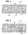
- FIG. 1 illustrates an exemplary stent 100 which may be utilized in accordance with an exemplary embodiment of the present invention.
- the expandable cylindrical stent 100 comprises a fenestrated structure for placement in a blood vessel, duct or lumen to hold the vessel, duct or lumen open, more particularly for protecting a segment of artery from restenosis after angioplasty.
- the stent 100 may be expanded circumferentially and maintained in an expanded configuration, that is circumferentially or radially rigid.
- the stent 100 is axially flexible and when flexed at a band, the stent 100 avoids any externally protruding component parts.
- the stent 100 generally comprises first and second ends with an intermediate section therebetween.
- the stent 100 has a longitudinal axis and comprises a plurality of longitudinally disposed bands 102, wherein each band 102 defines a generally continuous wave along a line segment parallel to the longitudinal axis.
- a plurality of circumferentially arranged links 104 maintain the bands 102 in a substantially tubular structure.
- each longitudinally disposed band 102 is connected at a plurality of periodic locations, by a short circumferentially arranged link 104 to an adjacent band 102.
- the wave associated with each of the bands 102 has approximately the same fundamental spatial frequency in the intermediate section, and the bands 102 are so disposed that the wave associated with them are generally aligned so as to be generally in phase with one another.
- each longitudinally arranged band 102 undulates through approximately two cycles before there is a link to an adjacent band 102.
- the stent 100 may be fabricated utilizing any number of methods.
- the stent 100 may be fabricated from a hollow or formed stainless steel tube that may be machined using lasers, electric discharge milling, chemical etching or other means.
- the stent 100 is inserted into the body and placed at the desired site in an unexpanded form.
- expansion may be effected in a blood vessel by a balloon catheter, where the final diameter of the stent 100 is a function of the diameter of the balloon catheter used.
- a stent 100 in accordance with the present invention may be embodied in a shape-memory material, including, for example, an appropriate alloy of nickel and titanium or stainless steel. Structures formed from stainless steel may be made self-expanding by configuring the stainless steel in a predetermined manner, for example, by twisting it into a braided configuration. In this embodiment after the stent 100 has been formed it may be compressed so as to occupy a space sufficiently small as to permit its insertion in a blood vessel or other tissue by insertion means, wherein the insertion means include a suitable catheter, or flexible rod. On emerging from the catheter, the stent 100 may be configured to expand into the desired configuration where the expansion is automatic or triggered by a change in pressure, temperature or electrical stimulation.
- FIG 2 illustrates an exemplary embodiment of the present invention utilizing the stent 100 illustrated in Figure 1 .
- the stent 100 may be modified to comprise one or more reservoirs 106. Each of the reservoirs 106 may be opened or closed as desired. These reservoirs 106 may be specifically designed to hold the rapamycin to be delivered. Regardless of the design of the stent 100, it is preferable to have the rapamycin dosage applied with enough specificity and a sufficient concentration to provide an effective dosage in the lesion area.
- the reservoir size in the bands 102 is preferably sized to adequately apply the rapamycin dosage at the desired location and in the desired amount.
- the entire inner and outer surface of the stent 100 may be coated with rapamycin in therapeutic dosage amounts.
- rapamycin for treating restenosis, as well as exemplary coating techniques, is described below.
- Rapamycin is a macrocyclic triene antibiotic produced by Streptomyces hygroscopicus as disclosed in U.S. Patent No. 3,929,992 . It has been found that rapamycin among other things inhibits the proliferation of vascular smooth muscle cells in vivo. Accordingly, rapamycin may be utilized in treating intimal smooth muscle cell hyperplasia, restenosis, and vascular occlusion in a mammal, particularly following either biologically or mechanically mediated vascular injury, or under conditions that would predispose a mammal to suffering such a vascular injury. Rapamycin functions to inhibit smooth muscle cell proliferation and does not interfere with the re-endothelialization of the vessel walls.
- Rapamycin reduces vascular hyperplasia by antagonizing smooth muscle proliferation in response to mitogenic signals that are released during an angioplasty induced injury. Inhibition of growth factor and cytokine mediated smooth muscle proliferation at the late G1 phase of the cell cycle is believed to be the dominant mechanism of action of rapamycin. However, rapamycin is also known to prevent T-cell proliferation and differentiation when administered systemically. This is the basis for its immunosuppressive activity and its ability to prevent graft rejection.
- rapamycin includes rapamycin and all analogs, derivatives and conjugates that bind to FKBP12, and other immunophilins and possesses the same pharmacologic properties as rapamycin including inhibition of TOR.
- the anti-proliferative effects of rapamycin may be achieved through systemic use, superior results may be achieved through the local delivery of the compound.
- rapamycin works in the tissues, which are in proximity to the compound, and has diminished effect as the distance from the delivery device increases. In order to take advantage of this effect, one would want the rapamycin in direct contact with the lumen walls.
- the rapamycin is incorporated onto the surface of the stent or portions thereof.
- the rapamycin is preferably incorporated into the stent 100, illustrated in Figure 1 , where the stent 100 makes contact with the lumen wall.
- Rapamycin may be incorporated onto or affixed to the stent in a number of ways.
- the rapamycin is directly incorporated into a polymeric matrix and sprayed onto the outer surface of the stent.
- the rapamycin elutes from the polymeric matrix over time and enters the surrounding tissue.
- the rapamycin preferably remains on the stent for at least three days up to approximately six months, and more preferably between seven and thirty days.
- the rapamycin or other therapeutic agent may be incorporated into a film-forming polyfluoro copolymer comprising an amount of a first moiety selected from the group consisting of polymerized vinylidenefluoride and polymerized tetrafluoroethylene, and an amount of a second moiety other than the first moiety and which is copolymerized with the first moiety, thereby producing the polyfluoro copolymer, the second moiety being capable of providing toughness or elastomeric properties to the polyfluoro copolymer, wherein the relative amounts of the first moiety and the second moiety are effective to provide the coating and film produced therefrom with properties effective for use in treating implantable medical devices.
- the present invention provides polymeric coatings comprising a polyfluoro copolymer and stents coated with a film of the polymeric coating in amounts effective to reduce thrombosis and/or restenosis when the stents are used in, for example, angioplasty procedures.
- polyfluoro copolymers means those copolymers comprising an amount of a first moiety selected from the group consisting of polymerized vinylidenefluoride and polymerized tetrafluoroethylene, and an amount of a second moiety other than the first moiety and which is copolymerized with the first moiety to produce the polyfluoro copolymer, the second moiety being capable of providing toughness or elastomeric properties to the polyfluoro copolymer, wherein the relative amounts of the first moiety and the second moiety are effective to provide coatings and film made from such polyfluoro copolymers with properties effective for use in coating implantable medical devices.
- the present invention comprises polyfluoro copolymers that provide improved biocompatible coatings or vehicles for medical devices. These coatings provide inert biocompatible surfaces to be in contact with body tissue of a mammal, for example, a human, sufficient to reduce restenosis, or thrombosis, or other undesirable reactions. While many reported coatings made from polyfluoro homopolymers are insoluble and/or require high heat, for example, greater than about one hundred twenty-five degrees centigrade, to obtain films with adequate physical and mechanical properties for use on implantable devices, for example, stents, or are not particularly tough or elastomeric, films prepared from the polyfluoro copolymers of the present invention provide adequate adhesion, toughness or elasticity, and resistance to cracking when formed on medical devices. In certain exemplary embodiments, this is the case even where the stent is subjected to relatively low maximum temperatures.
- the polyfluoro copolymers used for coatings according to the present invention are preferably film-forming polymers that have molecular weight high enough so as not to be waxy or tacky.
- the polymers and films formed therefrom should preferably adhere to the stent and not be readily deformable after deposition on the stent as to be able to be displaced by hemodynamic stresses.
- the polymer molecular weight should preferably be high enough to provide sufficient toughness so that films comprising the polymers will not be rubbed off during handling or deployment of the stent. In certain exemplary embodiments the coating will not crack where expansion of the stent occurs.
- Coatings of the present invention comprise polyfluoro copolymers, as defined hereinabove.
- the second moiety polymerized with the first moiety to prepare the polyfluoro copolymer may be selected from those polymerized, biocompatible monomers that would provide biocompatible polymers acceptable for implantation in a mammal, while maintaining sufficient elastomeric film properties for use on medical devices claimed herein.
- Such monomers include, without limitation, hexafluoropropylene (HFP), tetrafluoroethylene (TFE), vinylidenefluoride, 1-hydropentafluoropropylene, perfluoro(methyl vinyl ether), chlorotrifluoroethylene (CTFE), pentafluoropropene, trifluoroethylene, hexafluoroacetone and hexafluoroisobutylene.
- HFP hexafluoropropylene
- TFE tetrafluoroethylene
- VFE chlorotrifluoroethylene
- pentafluoropropene trifluoroethylene
- hexafluoroacetone hexafluororoisobutylene.
- Polyfluoro copolymers used in the present invention typically comprise vinylidinefluoride copolymerized with hexafluoropropylene, in the weight ratio in the range of from about fifty to about ninety-two weight percent vinylidinefluoride to about fifty to about eight weight percent HFP.
- polyfluoro copolymers used in the present invention comprise from about fifty to about eighty-five weight percent vinylidinefluoride copolymerized with from about fifty to about fifteen weight percent HFP. More preferably, the polyfluoro copolymers will comprise from about fifty-five to about seventy weight percent vinylidinefluoride copolymerized with from about forty-five to about thirty weight percent HFP.
- polyfluoro copolymers comprise from about fifty-five to about sixty-five weight percent vinylidinefluoride copolymerized with from about forty-five to about thirty-five weight percent HFP.
- Such polyfluoro copolymers are soluble, in varying degrees, in solvents such as dimethylacetamide (DMAc), tetrahydrofuran, dimethyl formamide, dimethyl sulfoxide and n-methyl pyrrolidone. Some are soluble in methylethylketone (MEK), acetone, methanol and other solvents commonly used in applying coatings to conventional implantable medical devices.
- coatings/films comprise pharmaceutical or therapeutic agents or drugs that are heat sensitive, for example, subject to chemical or physical degradation or other heat-induced negative affects, or when coating heat sensitive substrates of medical devices, for example, subject to heat-induced compositional or structural degradation.
- Crystalline polyfluoro copolymers tend to resist the tendency to flow under applied stress or gravity when exposed to temperatures above their glass transition (Tg) temperatures. Crystalline polyfluoro copolymers provide tougher coatings and films than their fully amorphous counterparts. In addition, crystalline polymers are more lubricious and more easily handled through crimping and transfer processes used to mount self-expanding stents, for example, nitinol stents.
- Semi-crystalline and amorphous polyfluoro copolymers are advantageous where exposure to elevated temperatures is an issue, for example, where heat-sensitive pharmaceutical or therapeutic agents are incorporated into the coatings and films, or where device design, structure and/or use preclude exposure to such elevated temperatures.
- Semi-crystalline polyfluoro copolymer elastomers comprising relatively high levels, for example, from about thirty to about forty-five weight percent of the second moiety, for example, HFP, copolymerized with the first moiety, for example, VDF, have the advantage of reduced coefficient of friction and self-blocking relative to amorphous polyfluoro copolymer elastomers. Such characteristics may be of significant value when processing, packaging and delivering medical devices coated with such polyfluoro copolymers.
- such polyfluoro copolymer elastomers comprising such relatively high content of the second moiety serves to control the solubility of rapamycin, in the polymer and therefore controls permeability of the agent through the matrix.
- Polyfluoro copolymers utilized in the present inventions may be prepared by various known polymerization methods. For example, high pressure, free-radical, semi-continuous emulsion polymerization techniques such as those disclosed in Fluoroelastomers-dependence of relaxation phenomena on compositions, POLYMER 30, 2180, 1989, by Ajroldi , et al., may be employed to prepare amorphous polyfluoro copolymers, some of which may be elastomers. In addition, free-radical batch emulsion polymerization techniques disclosed herein may be used to obtain polymers that are semi-crystalline, even where relatively high levels of the second moiety are included.
- stents may comprise a wide variety of materials and a wide variety of geometrics.
- Stents may be made of biocomptible materials, including biostable and bioabsorbable materials.
- Suitable biocompatible metals include, but are not limited to, stainless steel, tantalum, titanium alloys (including nitinol), and cobalt alloys (including cobalt-chromium nickel alloys).
- Suitable nonmetallic biocompatible materials include, but are not limited to, polyamides, polyolefins (i.e. polypropylene, polyethylene etc.), nonabsorbable polyesters (i.e. polyethylene terephthalate), and bioabsorbable aliphatic polyesters (i.e. homopolymers and copolymers of lactic acid, glycolic acid, lactide, glycolide, para-dioxanone, trimethylene carbonate, ⁇ -caprolactone, and blends thereof).
- the film-forming biocompatible polymer coatings generally are applied to the stent in order to reduce local turbulence in blood flow through the stent, as well as adverse tissue reactions.
- the coatings and films formed therefrom are also used to administer rapamycin to the site of the stent placement.
- the amount of polymer coating to be applied to the stent will vary depending on, among other possible parameters, the particular polyfluoro copolymer used to prepare the coating, the stent design and the desired effect of the coating.
- the coated stent will comprise from about 0.1 to about fifteen weight percent of the coating, preferably from about 0.4 to about ten weight percent.
- the polyfluoro copolymer coatings may be applied in one or more coating steps, depending on the amount of polyfluoro copolymer to be applied. Different polyfluoro copolymers may be used for different layers in the stent coating. In fact, in certain exemplary embodiments, it is highly advantageous to use a diluted first coating solution comprising a polyfluoro copolymer as a primer to promote adhesion of a subsequent polyfluoro copolymer coating layer that includes the rapamycin.
- the individual coatings may be prepared from different polyfluoro copolymers.
- Blends of polyfluoro copolymers may also be used to provide a desirable balance of coating properties, i.e. elasticity, toughness, etc., and drug delivery characteristics, for example, release profile.
- Polyfluoro copolymers with different solubilities in solvents may be used to build up different polymer layers that may be used to deliver different drugs or to control the release profile of a drug.
- polyfluoro copolymers comprising 85.5/14.5 (wt/wt) of poly(vinylidinefluoride/HFP) and 60.6/39.4 (wt/wt) of poly(vinylidinefluoride /HFP) are both soluble in DMAc.
- a first layer of the 85.5/14.5 PVDF polyfluoro copolymer comprising a drug could be over coated with a topcoat of the 60.6/39.4 PVDF polyfluoro copolymer made with the methanol solvent.
- the top coating may be used to delay the drug delivery of the drug contained in the first layer.
- Coatings may be formulated by mixing the rapamycin with the coating polyfluoro copolymers in a coating mixture.
- the rapamycin may be present as a liquid, a finely divided solid, or any other appropriate physical form.
- the coating mixture may include one or more additives, for example, nontoxic auxiliary substances such as diluents, carriers, excipients, stabilizers or the like.
- suitable additives may be formulated with the polymer and rapamycin.
- a hydrophilic polymer may be added to a biocompatible hydrophobic coating to modify the release profile, or a hydrophobic polymer may be added to a hydrophilic coating to modify the release profile.
- hydrophilic polymer selected from the group consisting of polyethylene oxide, polyvinyl pyrrolidone, polyethylene glycol, carboxylmethyl cellulose, and hydroxymethyl cellulose to a polyfluoro copolymer coating to modify the release profile.
- Appropriate relative amounts may be determined by monitoring the in vitro and/or in vivo release profiles for the rapamycin.
- the best conditions for the coating application are when the polyfluoro copolymer and rapamycin have a common solvent. This provides a wet coating that is a true solution. Less desirable, yet still usable, are coatings that contain the rapamycin as a solid dispersion in a solution of the polymer in solvent. Under the dispersion conditions, care must be taken to ensure that the particle size of the dispersed rapamycin, both the primary powder size and its aggregates and agglomerates, is small enough not to cause an irregular coating surface or to clog the slots of the stent that need to remain essentially free of coating.
- a clear (polyfluoro copolymer only) topcoat of the same polyfluoro copolymer used to provide sustained release of the drug or another polyfluoro copolymer that further restricts the diffusion of the rapamycin out of the coating may be applied.
- the topcoat may be applied by dip coating with mandrel to clear the slots. This method is disclosed in United States Patent No. 6,153,252 . Other methods for applying the topcoat include spin coating and spray coating.
- Dip coating of the topcoat can be problematic if the rapamycin is very soluble in the coating solvent, which swells the polyfluoro copolymer, and the clear coating solution acts as a zero concentration sink and redissolves previously deposited rapamycin.
- the time spent in the dip bath may need to be limited so that the rapamycin is not extracted out into the drug-free bath. Drying should be rapid so that the previously deposited rapamycin does not completely diffuse into the topcoat.
- the amount of rapamycin will be dependent upon the medical condition being treated. Typically, the amount of rapamycin represents about 0.001 percent to about seventy percent of the total coating weight, more typically about 0.001 percent to about sixty percent of the total coating weight. It is possible that the rapamycin may represent as little as 0.0001 percent to the total coating weight.
- the quantity and type of polyfluoro copolymers employed in the coating film comprising the rapamycin will vary depending on the release profile desired and the amount of rapamycin employed.
- the product may contain blends of the same or different polyfluoro copolymers having different molecular weights to provide the desired release profile or consistency to a given formulation.
- Polyfluoro copolymers may release dispersed rapamycin by diffusion. This can result in prolonged delivery (over, say approximately one to two-thousand hours, preferably two to eight-hundred hours) of effective amounts (0.001 ⁇ g/cm 2 -min to 1000 ⁇ g/cm 2 -min) of the rapamycin.
- the dosage may be tailored to the subject being treated, the severity of the affliction, the judgment of the prescribing physician, and the like.
- rapamycin and polyfluoro copolymers may be tested in appropriate in vitro and in vivo models to achieve the desired rapamycin release profiles.
- rapamycin could be formulated with a polyfluoro copolymer, or blend of polyfluoro copolymers, coated onto a stent and placed in an agitated or circulating fluid system, for example, twenty-five percent ethanol in water. Samples of the circulating fluid could be taken to determine the release profile (such as by HPLC, UV analysis or use of radiotagged molecules). The release of a pharmaceutical compound from a stent coating into the interior wall of a lumen could be modeled in appropriate animal system.
- rapamycin release profile could then be monitored by appropriate means such as, by taking samples at specific times and assaying the samples for drug concentration (using HPLC to detect rapamycin concentration). Thrombus formation can be modeled in animal models using the In-platelet imaging methods described by Hanson and Harker, Proc. Natl. Acad. Sci. USA 85:3184-3188 (1988 ). Following this or similar procedures, those skilled in the art will be able to formulate a variety of stent coating formulations.
- the coatings and films may be crosslinked once applied to the stent.
- Crosslinking may be affected by any of the known crosslinking mechanisms, such as chemical, heat or light.
- crosslinking initiators and promoters may be used where applicable and appropriate.
- curing may affect the rate at which the drug diffuses from the coating.
- Crosslinked polyfluoro copolymers films and coatings of the present invention also may be used without rapamycin modify the surface of implantable medical devices.
- PVDF homopolymer (Solef® 1008 from Solvay Advanced Polymers, Houston, TX, Tm about 175°C) and polyfluoro copolymers of poly(vinylidenefluoride/HFP), 92/8 and 91/9 weight percent vinylidenefluoride/HFP as determined by F 19 NMR, respectively (eg: Solef® 11010 and 11008, Solvay Advanced Polymers, Houston, TX, Tm about 159 degrees C and 160 degrees C, respectively) were examined as potential coatings for stents.
- polymers are soluble in solvents such as, but not limited to, DMAc, N,N-dimethylformamide (DMF), dimethyl sulfoxide (DMSO), N-methylpyrrolidone (NMP), tetrahydrofuran (THF) and acetone.
- solvents such as, but not limited to, DMAc, N,N-dimethylformamide (DMF), dimethyl sulfoxide (DMSO), N-methylpyrrolidone (NMP), tetrahydrofuran (THF) and acetone.
- Polymer coatings were prepared by dissolving the polymers in acetone, at five weight percent as a primer, or by dissolving the polymer in 50/50 DMAc/acetone, at thirty weight percent as a topcoat. Coatings that were applied to the stents by dipping and dried at 60 degrees C in air for several hours, followed by 60 degrees C for three hours in a ⁇ 100 mm Hg vacuum, resulted in white foamy
- a polyfluoro copolymer (Solef® 21508) comprising 85.5 weight percent vinylidenefluoride copolymerized with 14.5 weight percent HFP, as determined by F 19 NMR, was evaluated. This copolymer is less crystalline than the polyfluoro homopolymer and copolymers described in Example 1. It also has a lower melting point reported to be about 133 degrees C.
- a coating comprising about twenty weight percent of the polyfluoro copolymer was applied from a polymer solution in 50/50 DMAc/MEK. After drying (in air) at 60 degrees C for several hours, followed by 60 degrees C for three hours in a ⁇ 100 mtorr Hg vacuum, clear adherent films were obtained.
- Coatings were smoother and more adherent than those of Example 1. Some coated stents that underwent expansion show some degree of adhesion loss and "tenting" as the film pulls away from the metal. Where necessary, modification of coatings containing such copolymers may be made, e.g. by addition of plasticizers or the like to the coating compositions. Films prepared from such coatings may be used to coat stents.
- Polyfluoro copolymers of still higher HFP content were then examined. This series of polymers were not semicrystalline, but rather are marketed as elastomers.
- One such copolymer is FluorelTM FC2261Q (from Dyneon, a 3M-Hoechst Enterprise, Oakdale, MN), a 60.6/39.4 (wt/wt) copolymer of vinylidenefluoride/HFP.
- this copolymer has a Tg well below room temperature (Tg about minus twenty degrees C) it is not tacky at room temperature or even at sixty degrees C.
- This polymer has no detectable crystallinity when measured by Differential Scanning Calorimetry (DSC) or by wide angle X-ray diffraction. Films formed on stents as described above were non-tacky, clear, and expanded without incident when the stents were expanded.
- coatings comprising the 60.6/39.4 (wt/wt) (vinylidenefluoride/HFP) and about nine, thirty and fifty weight percent of rapamycin (Wyeth-Ayerst Laboratories, Philadelphia, PA), based on total weight of coating solids, respectively.
- Coatings comprising about nine and thirty weight percent rapamycin provided white, adherent, tough films that expanded without incident on the stent. Inclusion of fifty percent drug, in the same manner, resulted in some loss of adhesion upon expansion.
- Changes in the comonomer composition of the polyfluoro copolymer also can affect the nature of the solid state coating, once dried.
- the semicrystalline copolymer, Solef® 21508, containing 85.5 percent vinylidenefluoride polymerized with 14.5 percent by weight HFP forms homogeneous solutions with about 30 percent rapamycin (drug weight divided by total solids weight, for example, drug plus copolymer) in DMAc and 50/50 DMAc/MEK.
- rapamycin drug weight divided by total solids weight, for example, drug plus copolymer
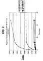
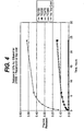
- Example 4 In vitro release results of rapamycin from coating.
- Figure 3 is a plot of data for the 85.5/14.5 vinylidenefluoride/HFP polyfluoro copolymer, indicating fraction of drug released as a function of time, with no topcoat.
- Figure 4 is a plot of data for the same polyfluoro copolymer over which a topcoat has been disposed, indicating that most effect on release rate is with a clear topcoat.
- TC150 refers to a device comprising one hundred fifty micrograms of topcoat
- TC235 refers to two hundred thirty-five micrograms of topcoat, etc.
- the stents before topcoating had an average of seven hundred fifty micrograms of coating containing thirty percent rapamycin.
- Figure 5 is a plot for the 60.6/39.4 vinylidenefluoride/HFP polyfluoro copolymer, indicating fraction of drug released as a function of time, showing significant control of release rate from the coating without the use of a topcoat. Release is controlled by loading of drug in the film.
- Example 5 In vivo stent release kinetics of rapamycin from poly(VDF/HFP).
- Arteriectomy of the right common carotid artery was performed and a 5 F catheter introducer (Cordis, Inc.) placed in the vessel and anchored with ligatures. Iodine contrast agent was injected to visualize the right common carotid artery, brachlocephalic trunk and aortic arch. A steerable guide wire (0.014 inch/180 cm, Cordis, Inc.) was inserted via the introducer and advanced sequentially into each iliac artery to a location where the artery possesses a diameter closest to 2 mm using the angiographic mapping done previously.
- Two stents coated with a film made of poly(VDF/HFP):(60.6/39.4) with thirty percent rapamycin were deployed in each animal where feasible, one in each iliac artery, using 3.0 mm balloon and inflation to 8-10 ATM for thirty seconds followed after a one minute interval by a second inflation to 8-10 ATM for thirty seconds.
- Follow-up angiographs visualizing both iliac arteries are obtained to confirm correct deployment position of the stent.
- the carotid artery was ligated and the skin is closed with 3/0 vicryl suture using a one layered interrupted closure.
- Animals were given butoropanol (0.4 mg/kg, s.c.) and gentamycin (4 mg/kg, i.m.). Following recovery, the animals were returned to their cages and allowed free access to food and water.
- Stented vessels were removed from the remaining seven animals at the following time points: one vessel (one animal) at ten minutes post implant; six vessels (three animals) between forty minutes and two hours post-implant (average, 1.2 hours); two vessels (two animals) at three days post implant; and two vessels (one animal) at seven days post-implant.
- the stent was retrieved from the aorta rather than the iliac artery. Upon removal, arteries were carefully trimmed at both the proximal and distal ends of the stent.
- Example 6 Purifying the polymer.
- the FluorelTM FC2261Q copolymer was dissolved in MEK at about ten weight percent and was washed in a 50/50 mixture of ethanol/water at a 14:1 of ethanol/water to MEK solution ratio. The polymer precipitated out and was separated from the solvent phase by centrifugation. The polymer again was dissolved in MEK and the washing procedure repeated. The polymer was dried after each washing step at sixty degrees C in a vacuum oven ( ⁇ 200 mtorr) over night.
- Example 7 In vivo testing of coated stents in porcine coronary arteries.
- CrossFlex® stents (available from Cordis, a Johnson & Johnson Company) were coated with the "as received" FluorelTM FC2261Q PVDF copolymer and with the purified polyfluoro copolymer of Example 6, using the dip and wipe approach.
- the coated stents were sterilized using ethylene oxide and a standard cycle.
- the coated stents and bare metal stents (controls) were implanted in porcine coronary arteries, where they remained for twenty-eight days.
- Angiography was performed on the pigs at implantation and at twenty-eight days. Angiography indicated that the control uncoated stent exhibited about twenty-one percent restenosis.
- the polyfluoro copolymer "as received" exhibited about twenty-six percent restenosis(equivalent to the control) and the washed copolymer exhibited about 12.5 percent restenosis.
- rapamycin acts by entering the surrounding tissue, it is preferably only affixed to the surface of the stent making contact with one tissue. Typically, only the outer surface of the stent makes contact with the tissue. Accordingly, in one exemplary embodiment, only the outer surface of the stent is coated with rapamycin.
- a rapamycin coating may be applied to stents by a dip, spray or spin coating method, and/or any combination of these methods.
- Various polymers may be utilized. For example, as described above, poly(ethylene-co-vinyl acetate) and polybutyl methacrylate blends may be utilized. Other polymers may also be utilized, but not limited to, for example, polyvinylidene fluoride-co-hexafluoropropylene and polyethylbutyl methacrylate-co-hexyl methacrylate.
- barrier or top coatings may also be applied to modulate the dissolution of rapamycin from the polymer matrix.
- the stent may be formed from any number of materials, including various metals, polymeric materials and ceramic materials. Accordingly, various technologies may be utilized to immobilize the various drugs, agent, compound combinations thereon. Specifically, in addition to the polymeric matricies described above biopolymers may be utilized. Biopolymers may be generally classified as natural polymers, while the above-described polymers may be described as synthetic polymers. Exemplary biopolymers, which may be utilized include, agarose, alginate, gelatin, collagen and elastin.
- Rapamycin functions to inhibit smooth muscle cell proliferation through a number of mechanisms. In addition, rapamycin reduces the other effects caused by vascular injury, for example, inflammation. The mechanisms of action and various functions of rapamycin are described in detail below. Rapamycin as used throughout this application shall include rapamycin, rapamycin analogs, derivatives and congeners that bind FKBP12 and possess the same pharmacologic properties as rapamycin, as described in detail below.
- rapamycin a known anti-proliferative, which acts to reduce the magnitude and duration of neointimal hyperplasia, are still being elucidated. It is known, however, that rapamycin enters cells and binds to a high-affinity cytosolic protein called FKBP12. The complex of rapamycin and FKPB12 in turn binds to and inhibits a phosphoinositide (PI)-3 kinase called the "mammalian Target of Rapamycin" or TOR.
- PI phosphoinositide
- TOR is a protein kinase that plays a key role in mediating the downstream signaling events associated with mitogenic growth factors and cytokines in smooth muscle cells and T lymphocytes. These events include phosphorylation of p27, phosphorylation of p70 s6 kinase and phosphorylation of 4BP-1, an important regulator of protein translation.
- rapamycin reduces restenosis by inhibiting neointimal hyperplasia.
- rapamycin may also inhibit the other major component of restenosis, namely, negative remodeling. Remodeling is a process whose mechanism is not clearly understood but which results in shrinkage of the external elastic lamina and reduction in lumenal area over time, generally a period of approximately three to six months in humans.
- Negative or constrictive vascular remodeling may be quantified angiographically as the percent diameter stenosis at the lesion site where there is no stent to obstruct the process. If late lumen loss is abolished in-lesion, it may be inferred that negative remodeling has been inhibited.
- Another method of determining the degree of remodeling involves measuring in-lesion external elastic lamina area using intravascular ultrasound (IVUS). Intravascular ultrasound is a technique that can image the external elastic lamina as well as the vascular lumen. Changes in the external elastic lamina proximal and distal to the stent from the post-procedural timepoint to four-month and twelve-month follow-ups are reflective of remodeling changes.
- rapamycin exerts an effect on remodeling comes from human implant studies with rapamycin coated stents showing a very low degree of restenosis in-lesion as well as in-stent. In-lesion parameters are usually measured approximately five millimeters on either side of the stent i.e. proximal and distal. Since the stent is not present to control remodeling in these zones which are still affected by balloon expansion, it may be inferred that rapamycin is preventing vascular remodeling.
- rapamycin may represent a biological approach to controlling the vascular remodeling phenomenon.
- rapamycin acts to reduce negative remodeling in several ways. By specifically blocking the proliferation of fibroblasts in the vascular wall in response to injury, rapamycin may reduce the formation of vascular scar tissue. Rapamycin may also affect the translation of key proteins involved in collagen formation or metabolism.
- Rapamycin used in this context includes rapamycin and all analogs, derivatives and congeners that bind FKBP12 and possess the same pharmacologic properties as rapamycin.
- the rapamycin is delivered by a vocal delivery device to control negative remodeling of an arterial segment after balloon angioplasty as a means of reducing or preventing restenosis, wherein said delivery device comprises a stent that includes a coating which elutes or releases rapamycin.
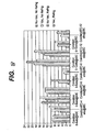
- TC top coat of 30 ⁇ g, 100 ⁇ g, or 300 ⁇ g drug-tree BMA: Biphasic; 2 x 1X layers of rapamycin in EVA/BMA spearated by a 100 ⁇ g drug-free BMA layer. 2 0.25mg/kg/d x 14 d preceeded by a loading dose of 0.5mg/kg/d x 3d prior to stent Implantation. *p-0.05 from EVA/BMA control.
- rapamycin into the vascular wall of a human from a nonerodible polymeric stent coating provides superior results with respect to the magnitude and duration of the reduction in neointimal hyperplasia within the stent as compared to the vascular walls of animals as set forth above.
- the human clinical response to rapamycin reveals essentially total abolition of neointimal hyperplasia inside the stent using both angiographic and intravascular ultrasound measurements.
- Rapamycin produces an unexpected benefit in humans when delivered from a stent by causing a profound reduction in in-stent neointimal hyperplasia that is sustained for at least one year. The magnitude and duration of this benefit in humans is not predicted from animal model data. Rapamycin used in this context includes rapamycin and all analogs, derivatives and congeners that bind FKBP12 and possess the same pharmacologic properties as rapamycin.
- rapamycin in humans is due to greater sensitivity of its mechanism(s) of action toward the pathophysiology of human vascular lesions compared to the pathophysiology of animal models of angioplasty.
- the combination of the dose applied to the stent and the polymer coating that controls the release of the rapamycin is important in the effectiveness of the rapamycin.
- rapamycin reduces vascular hyperplasia by antagonizing smooth muscle proliferation in response to mitogenic signals that are released during angioplasty injury. Also, it is known that rapamycin prevents T-cell proliferation and differentiation when administered systemically. It has also been determined that rapamycin exerts a local anti-inflammatory effect in the vessel wall when administered from a stent in low doses for a sustained period of time (approximately two to six weeks). The local anti-inflammatory benefit is profound and unexpected. In combination with the smooth muscle anti-proliferative effect, this dual mode of action of rapamycin may be responsible for its exceptional efficacy.
- rapamycin delivered from a stent reduces neointimal hyperplasia by a combination of anti-inflammatory and smooth muscle anti-proliferative effects.
- Rapamycin used in this context means rapamycin and all analogs, derivatives and congeners that bind FKBP12 and possess the same pharmacologic properties as rapamycin.
- rapamycin The anti-inflammatory effect of rapamycin is evident in data from an experiment, illustrated in Table 6, in which rapamycin delivered from a stent was compared with dexamethasone delivered from a stent.
- Dexamethasone a potent steroidal anti-inflammatory agent, was used as a reference standard. Although dexamethasone is able to reduce inflammation scores, rapamycin is far more effective than dexamethasone in reducing inflammation scores. In addition, rapamycin significantly reduces neointimal hyperplasia, unlike dexamethasone.
- Rapamycin has also been found to reduce cytokine levels in vascular tissue when delivered from a stent.
- the data in Figure 1 illustrates that rapamycin is highly effective in reducing monocyte chemotactic protein (MCP-1) levels in the vascular wall.
- MCP-1 monocyte chemotactic protein
- MCP-1 is an example of a proinflammatory/chemotactic cytokine that is elaborated during vessel injury.
- Reduction in MCP-1 illustrates the beneficial effect of rapamycin in reducing the expression of proinflammatory mediators and contributing to the anti-inflammatory effect of rapamycin delivered locally from a stent. It is recognized that vascular inflammation in response to injury is a major contributor to the development of neointimal hyperplasia.
- rapamycin may be shown to inhibit local inflammatory events in the vessel it is believed that this could explain the unexpected superiority of rapamycin in inhibiting neointima.
- rapamycin functions on a number of levels to produce such desired effects as the prevention of T-cell proliferation, the inhibition of negative remodeling, the reduction of inflammation, and the prevention of smooth muscle cell proliferation. While the exact mechanisms of these functions are not completely known, the mechanisms that have been identified may be expanded upon.
- rapamycin studies with rapamycin suggest that the prevention of smooth muscle cell proliferation by blockade of the cell cycle is a valid strategy for reducing neointimal hyperplasia. Dramatic and sustained reductions in late lumen loss and neointimal plaque volume have been observed in patients receiving rapamycin delivered locally from a stent.
- the present invention expands upon the mechanism of rapamycin to include additional approaches to inhibit the cell cycle and reduce neointimal hyperplasia without producing toxicity.
- the predominant mechanism of the coating self-adhesion is believed to be due to mechanical forces. When the polymer comes in contact with itself, its chains can tangle causing the mechanical bond, similar to Velcro®.
- Certain polymers do not bond with each other, for example, fluoropolymers. For other polymers, however, powders may be utilized.
- a powder may be applied to the one or more polymers incorporating the rapamycin on the surfaces of the stent to reduce the mechanical bond.
- Any suitable biocompatible material which does not interfere with the rapamycin, or materials utilized to immobilize the drugs, agents or compounds onto the medical device may be utilized.
- a dusting with a water soluble powder may reduce the tackiness of the coatings surface and this will prevent the polymer from sticking to itself thereby reducing the potential for delamination.
- the powder should be water-soluble so that it does not present an emboli risk.
- the powder may comprise an anti-oxidant, such as vitamin C, or it may comprise an anti-coagulant, such as aspirin or heparin.
- An advantage of utilizing an anti-oxidant may be in the fact that the anti-oxidant may preserve the other drugs, agents or compounds over longer periods of time.
- crystalline polymers are generally not sticky or tacky. Accordingly, if crystalline polymers are utilized rather than amorphous polymers, then additional materials may not be necessary. It is also important to note that polymeric coatings without drugs, agents and/or compounds may improve the operating characteristics of the stent. For example, the mechanical properties of the stent may be improved by a polymeric coating. A coated stent may have improved flexibility and increased durability. In addition, the polymeric coating may substantially reduce or eliminate galvanic corrosion between the different metals comprising the medical device.
- FIGs 7 shows a preferred embodiment of a stent 7000, which may be used in conjunction with the present invention.
- Stent 7000 is shown in its unexpanded compressed state, before it is deployed, in Figure 7 .
- Stent 7000 is preferably made from a superelastic alloy such as Nitinol.
- the stent 7000 is made from an alloy comprising from about 50.5 percent (as used herein these percentages refer to atomic percentages) Ni to about 60 percent Ni, and most preferably about 55 percent Ni, with the remainder of the alloy Ti.
- the stent 7000 is such that it is superelastic at body temperature, and preferably has an Af in the range from about twenty-one degrees C to about thirty-seven degrees C.
- the superelastic design of the stent makes it crush recoverable which, as discussed above, can be used as a stent or frame for any number of vascular devices for different applications.
- Stent 7000 is a tubular member having front and back open ends a longitudinal axis extending there between.
- the tubular member has a first smaller diameter for insertion into a patient and navigation through the vessels, and a second larger diameter for deployment into the target area of a vessel.
- the tubular member is made from a plurality of adjacent hoops 7002 extending between the front and back ends.
- the hoops 7002 include a plurality of longitudinal struts 7004 and a plurality of loops 7006 connecting adjacent struts, wherein adjacent struts are connected at opposite ends so as to form a substantially S or Z shape pattern.
- Stent 7000 further includes a plurality of curved bridges 7008, which connect adjacent hoops 7002.
- Bridges 7008 connect adjacent struts together at bridge to loop connection points which are offset from the center of a loop.
- each hoop has between twenty-four to thirty-six or more struts.
- the stent has a ratio of number of struts per hoop to strut length (in inches) which is greater than two hundred. The length of a strut is measured in its compressed state parallel to the longitudinal axis of the stent.
- the stent In trying to minimize the maximum strain experienced by features, the stent utilizes structural geometries which distribute strain to areas of the stent which are less susceptible to failure than others.
- one vulnerable area of the stent is the inside radius of the connecting loops.
- the connecting loops undergo the most deformation of all the stent features.
- the inside radius of the loop would normally be the area with the highest level of strain on the stent. This area is also critical in that it is usually the smallest radius on the stent.
- Stress concentrations are generally controlled or minimized by maintaining the largest radii possible.
- we want to minimize local strain concentrations on the bridge and bridge to loop connection points One way to accomplish this is to utilize the largest possible radii while maintaining feature widths, which are consistent with applied forces. Another consideration is to minimize the maximum open area of the stent. Efficient utilization of the original tube from which the stent is cut increases stent strength and it's ability to trap embolic material.
- excipient agents and/or formulary components may be added to achieve both fast-release and sustained-release drug elution profiles.
- excipient agents may include salts and/or inorganic compounds such as acids/bases or buffer components, anti-oxidants, surfactants, polypeptides, proteins, carbohydrates including sucrose, glucose or dextrose, chelating agents such as EDTA, glutathione or other excipients or agents.
- the entire stent may be coated or only a portion of the stent may be coated.
- the coating may be uniform or non-uniform.
- the coating may be discontinuous.
- Vascular diseases include diseases that affect areas of a living organism relating to or containing blood vessels.
- stenosis is a narrowing or constricting of arterial lumen in a living organism (e.g., a human) usually due to atherosclerosis/coronary heart disease (CHD).
- CHD coronary heart disease
- Restenosis is a recurrence of stenosis after a percuteneous intervention such as angioplasty and stenting. Restenosis typically affects the large arteries of a living organism.
- the underlying mechanisms of restenosis comprise a combination of effects from vessel recoil, negative vascular remodeling, thrombus formation and neointimal hyperplasia. It has been shown that restenosis after balloon angioplasty is mainly due to vessel remodeling and neointimal hyperplasia and after stenting is mainly due to neo-intimal hyperplasia.
- Treatment for stenosis and restenosis varies. Stenosis caused by CHD often forces individuals to restrict and limit their activity levels in order to avoid complications, stroke, heart attack, sudden death and loss of limb or function of a limb stemming from the stenosis.
- the reconstruction of blood vessels, arteries and veins may also be needed to treat individuals suffering from stenosis and restenosis.
- Coronary bypass can also be utilized to revascularize the heart and restore normal blood flow.
- balloon angioplasty may be conducted to increase the orifice size of affected areas.
- these treatments address the problems associated with stenosis, but they also create a high rate of restenosis that can result in recurrence of cardiac symptoms and mortality.
- these treatments are not preventative in nature, and therefore generally are not utilized until the patient or individual has already developed stenosis.
- Atherosclerosis affects medium and large arteries and is characterized by a patchy, intramural thickening that encroaches on the arterial lumen and, in most severe form, causes obstruction.
- the atherosclerotic plaque consists of an accumulation of intracellular and extracellular lipids, smooth muscle cells and connective tissue.
- the earliest lesion of atherosclerosis is the fatty streak that evolves into a fibrous plaque coating the artery.
- Atherosclerotic vessels have reduced systolic expansion and abnormal wave propagation.
- Treatment of atherosclerosis is usually directed at its complications, for example, arrhythmia, heart failure, kidney failure, stroke, and peripheral arterial occlusion.
- Atherosclerosis is a thickening and hardening of the arteries and is generally believed to be caused by the progressive buildup of fatty substances, for example, cholesterol, cellular waste products, inflammatory cells, calcium and other substances in the inner lining or intima of the arteries.
- fatty substances for example, cholesterol, cellular waste products, inflammatory cells, calcium and other substances in the inner lining or intima of the arteries.
- the buildup of these substances may in turn stimulate cells in the walls of the affected arteries to produce additional substances that result in the further buildup of cells.
- Atherosclerosis is a slow, complex disease that typically starts in childhood and progresses as the individual ages.
- the rate of progression may be affected by a number of factors, including blood cholesterol levels, diabetes, obesity, physical inactivity, high blood pressure and tobacco use. This buildup in commonly referred to as plaque and may grow large enough to significantly reduce blood flow through the affected arteries.
- the deposits of the various substances set forth above, and the proliferation of additional cellular substances or constituents caused thereby substantially enlarge the intima, which in turn reduces luminal cross-sectional area and decreases the diameter of the affected artery or arteries, which in turn reduces the oxygen supply to one or more organs.
- the deposits or plaque may also rupture and form blood clots or thrombi that can completely obstruct blood flow in the affected artery or break free and travel, emboli, to another part of the body. If either of these events occurs, the individual may suffer a myocardial infarction if the artery or arteries affected perfuse the heart or a stroke if the artery or arteries affected supply blood to the brain. If the artery or arteries affected supply blood to a limb or appendage, gangrene may result.
- vulnerable plaques These plaques which are prone to rupture, commonly referred to as vulnerable plaques, do not block flow in the affected artery or arteries per se, but rather, much like an abscess, they may be ingrained in the arterial wall so that they are difficult to detect. Essentially, these vulnerable plaques cannot be seen by conventional angiography and/or fluoroscopy, and they do not typically cause symptoms such as shortness of breath or pain. Techniques for determining the presence of vulnerable plaques are, however, improving as discussed subsequently.
- vulnerable plaques are more likely to erode or rupture, creating emboli and raw or exposed tissue surfaces that are highly thrombogenic. Accordingly, it is now accepted that the majority of cases of acute myocardial infarction, sudden cardiac death and thrombolitic stroke result from the disruption of vulnerable atherosclerotic plaques leading to thrombosis. Therefore, these vulnerable plaques are more dangerous than other plaques that simply cause pain, and may be responsible for as much as sixty to eighty percent of all heart attacks.
- Vulnerable plaques are inflammatory vascular lesions that develop in atherosclerotic blood vessels.
- Vulnerable plaques are characterized by active inflammation, cellular hyperplasia and variable degrees of lumen obstruction.
- vulnerable plaques comprise a fibrous cap in contact with the lumen of the vessel overlying a core of lipid and cellular material.
- Vulnerable plaque lesions are not typically obstructive, in contrast to chronic stable plaques that produce ischemic symptoms. For that reason, they are not easily detected.
- vulnerable plaques The hallmark of vulnerable plaques is active inflammation with significant inflammatory cell infiltration, predominantly T-lymphocytes and macrophage, causing the generation of proteolytic enzymes that essentially digest the wall of the fibrous cap thereby inducing plaque instability and eventually plaque rupture. Plaque rupture exposes highly thrombogenic material in the lipid core to flowing blood leading to the rapid development of occlusive thrombi. Ruptured vulnerable plaque, as stated above, is the primary cause of acute coronary and cerebral syndromes. These include unstable angina, myocardial infarction, both Q-wave and non-Q-wave myocardial infarction, cerebral stroke and transient cerebral ischemia. In other words, ruptured vulnerable plaque accounts for a significant percentage of cardiovascular morbidity and mortality.
- Detection technologies currently under investigation include refined magnetic resonance imaging, thermal sensors that measure the temperature of the arterial wall on the premise that the inflammatory process generates heat, elasticity sensors, intravascular ultrasound, optical coherence tomography, contrast agents, and near-infrared and infrared light.
- thermal sensors that measure the temperature of the arterial wall on the premise that the inflammatory process generates heat
- elasticity sensors that measure the temperature of the arterial wall on the premise that the inflammatory process generates heat
- elasticity sensors intravascular ultrasound
- optical coherence tomography contrast agents
- near-infrared and infrared light near-infrared and infrared light
- Inflammation is an ongoing process which includes the inflammation of the fibrous cap and creating a cap vulnerable to rupture.
- Lipid metabolism is the formation of a molten or active lipid pool or core comprising a paliable, cholesterolemic lipid material susceptible to rupture. The inflammation process is the acute phase and the lipid metabolism is the chronic phase of vulnerable plaque disease.
- a stent or other scaffold structure designed to maintain vessel potency and comprising a multilaminate coating architecture, including one or more therapeutic agents, drugs, and/or compounds for treating both the inflammation and lipid metabolism processes, may be utilized to effectively treat vulnerable plaques.
- the stent may comprise any suitable scaffold structure, including balloon expandable stents, constructed from stainless steel or other metal alloys, and/or self-expanding stents, constructed from nitinol or other shape memory metal alloys.
- the stent may be made from non-metallic materials, such as ceramics and/or polymers, which may be biodegradable.
- the biodegradable stent would serve as a temporary scaffold and eventually dissolve over a period of time raging from days or weeks to months and years.
- the stent would be mounted on a delivery catheter and delivered percutaneously through the lumen of a blood vessel to the site of the vulnerable plaque lesion as described in detail above with respect to treating restenosis.
- the stent as described above, is designed to maintain vessel patency and also provide structural support to the weakened or potentially weakened fibrous cap and prevent it from rupturing.
- the stent also provides a means for preventing further encroachment by the lesion.
- the rapamycin utilized in combination with the stent would preferably prevent neointimal hyperplasia that is commonly encountered in stenting and which could lead to restenosis and device failure as described in detail above.
- the rapamycin would preferably stabilize or passivate the lesion by reducing local inflammation and preventing further erosion of the fibrous cap.
- the rapamycin may be delivered in a polymer matrix coating applied to the stent struts or embedded into the material forming the stent itself and would release into the vessel wall over a predetermined period of time, preferably utilizing the dual profile release rate as briefly described above.
- Oxidation may be generally defined as the loss of electrons from a molecule. With respect to organic molecules, oxidation may be more aptly described as an increase in oxygen or decrease in hydrogen content.
- Antioxidants are molecules that safely interact with free radicals and terminate the potential chain reaction caused by these highly reactive radicals prior to vital molecule damage. In other words, antioxidants are essentially scavengers that render free radicals inert prior to damaging molecules or portions of molecules. Accordingly, antioxidants may be utilized in drug formulations to prevent or substantially reduce drug degradation.
- antioxidants may be added to oxygen sensitive drug and/or drug/polymer solutions utilized to coat drug eluting implantable medical devices, such as stents, as well as injectable drug solutions to prevent oxidation and reduce drug degradation. A reduction in degradation due to oxidation will result in an extended shelf life of the drug or drug coated products.
- antioxidants There are a number of different types of antioxidants.
- the broadest group of antioxidants may be generally classified as chain terminators.
- the propagation of radical chains with the consumption of oxygen allows a single initiation event to lead to a large number of drug oxidations.
- Chain terminators form stable radicals, which do not propagate chains.
- Chain terminators include thiols and phenols.
- Sacrificial reductants are also antioxidants. Sacrificial reductants are compounds that are more easily oxidized than the molecule that is to be protected, which in the present invention is the drug molecule. Sacrificial reductants include sulfites and Ascorbic Acid.
- Yet another form of antioxidant includes peroxide quenchers. Peroxide quenchers include mannitol.
- antioxidants The effectiveness of antioxidants depends upon a number of factors. In some circumstances a single antioxidant may provide the desired effect. However, in other circumstances, combinations of antioxidants may provide strong synergistic effects.
- various antioxidants may be utilized alone or in combination to prevent drug degradation. In addition to their antioxidant effect, the antioxidants may also provide therapeutic effects.
- drug eluting medical devices with antioxidants may improve long-term vascular patency by preventing neointimal hyperplasia if sufficiently high or therapeutic dosages are utilized.
- Ascorbic Acid, BHT and/or Tocopherol all antioxidants, may reduce neointimal hyperplasia by preventing free radical formation or being a scavenger for free radicals.
- any number of antioxidants, such as Tocopherol may also be utilized as a lubricant.
- Antioxidants of the present invention are Ascorbyl Palmitate, Ascorbic Acid,Butylated Hydroxytoluene (BHT), Tocopherols and Ascorbic Acid isomers and/or derivatives.
- Exemplary embodiments of the invention are described with respect to rapamycin and more specifically, a drug eluting stent comprising rapamycin.
- molecules or specific portions of molecules may be particularly sensitive to oxidation.
- the conjugated triene moiety of the molecule is particularly susceptible to oxidation. Essentially, oxygen breaks the carbon chain of the conjugate triene moiety and the bioactivity of the rapamycin is degraded.
- the drug is broken down into one or more different compounds. Accordingly, it may be particularly advantageous to mix or co-mingle an antioxidant with the rapamycin.
- the physical positioning of the antioxidant proximate to the drug is the key to success.
- the antioxidant preferably remains free to combine with oxygen so that the oxygen does not break up the moiety and ultimately degrade the drug.
- the rapamycin may be incorporated into a polymeric coating or matrix, it is particularly important that the antioxidant be maintained proximate to the drug rather than the polymer(s)- Factors that influence this include the constituents of the polymeric matrix, the drug, and how the polymer/drug coating is applied to the implantable medical device. Accordingly in order to achieve the desired result, selection of the appropriate antioxidant, the process of mixing all of the elements and the application of the mixture is preferably tailored to the particular application.
- a number of antioxidants were tested to determine their efficacy in preventing the degradation of rapamycin, or more specifically, sirolimus. Screening experiments were performed to evaluate the solubility of various antioxidants in tetrahydroxyfuran (THF) solutions containing sirolimus and the percentage of antioxidant required to prevent oxidation of sirolimus alone and in a basecoat polymeric matrix.
- THF tetrahydroxyfuran
- THF is the solvent in which sirolimus may be dissolved. It is important to note that other solvents may be utilized. Two sets of controls were utilized.
- Control #1 comprises solutions of THF and sirolimus and/or polymers with no antioxidant
- Control #2 comprises solutions of THF and sirolimus and/or polymers, wherein the THF contains a label claim of 250 ppm of BHT as a stabilizer from the vendor of THF.
- the BHT is an added constituent of the THF solvent to prevent oxidation of the solvent.
- Table 10 shown below is a matrix of the various mixtures. All percentages are given as weight/volume.
- Table 11 shown below, identifies the samples for evaluation. All percentages are given as weight/volume. The samples in Table 11 contain no polymer.
- each of the samples in Tables 11 and 12 were tested to determine the solubility of the various antioxidants as well as their effectiveness in preventing drug degradation. All of the antioxidants were soluble in both the solvent with sirolimus solutions and the solvent with sirolimus and polymer solutions. The solubility of each of the antioxidants was determined by a visual inspection of the test samples.
- Table 13 identifies the chosen samples that were evaluated for drug content (percent label claim or %LC) after five (5) days in an oven set at a temperature of sixty degrees C (60°C). The samples were evaluated after five (5) days utilizing a drug testing assay for silolimus. In the exemplary embodiment, a HPLC assay was utilized. The important numbers are the percent label claim number (% LC) of the solutions that indicates how much of the drug remains or is recovered. The antioxidants, BHT, Tocopherol, and/or Ascorbic Acid provided significant protection against the harsh environmental conditions of the test. Lower % LC numbers are evident in solutions samples that do not contain an antioxidant.
- Table 14 provides the %LC results for the samples without polymers and Table 15 provides the %LC results for the samples with polymer after four (4) weeks of sixty degrees C (60°C).
- TABLE 14.0 SAMPLE ID # CALCULATED RESULTS ⁇ g/ml) THEORETICAL CONCENTRATION ( ⁇ g/ml) % LC AA1A 1155.56 1669.2 69.2 AA2A 1280.90 1669.2 76.7 AP1A 851.45 1669.2 51.0 AP2A 939.36 1669.2 56.3 BHT1A 437.38 1669.2 26.2 BHT2A 1434.98 1669.2 86.0 TP1A 1335.58 1669.2 80.0 TP2A 1618.61 1669.2 97.0 C1A#1 608.64 1669.2 36.5 C1A#2 552.57 1669.2 33.1 C2A#1 1794.70 1669.2 107.5 C2A#2 1794.
- Tocopherol, BHT or Ascorbic Acid may be utilized to substantially reduce drug degradation due to oxidation.
- FIG. 8 there is illustrated in graphical format, the results of the same drug screening as described above with the solution applied to a cobalt-chromium, 18 mm stent.
- two sets of solution samples were utilized, one with sirolimus and polymer solution containing the antioxidant and one with sirolimus and polymer solution containing no antioxidant.
- the antioxidant utilized was 0.02 weight percent BHT per total basecoat solids.
- the test was utilized to determine the percent drug content change over a time period of 0 to 12 weeks under two conditions; namely, 40°C with 75 percent relative humidity, and ambient conditions (25°C).
- the addition of BHT to the solution lessens drug degradation at both 8 weeks and 12 weeks under ambient conditions. Accordingly, if one does not stabilize the base coat solution, other process techniques must be utilized; namely, refrigeration and/or vacuum drying.
Abstract
Description
- The present invention relates to the local administration of rapamycin for the prevention and treatment of vascular disease, and more particularly to stents for the local delivery of rapamycin for the prevention and treatment of vascular disease caused by injury and methods and devices for maintaining the rapamycin on the stent, as well as preventing damage to the stent. The present invention also relates to coatings for controlling the elution rates rapamycin from stents. The present invention also relates to stents having rapamycin affixed thereto for treating vulnerable plaque. The present invention also relates to agents for reducing the degradation of drugs caused by oxidation.
- Many individuals suffer from circulatory disease caused by a progressive blockage of the blood vessels that perfuse the heart and other major organs. More severe blockage of blood vessels in such individuals often leads to hypertension, ischemic injury, stroke, or myocardial infarction. Atherosclerotic lesions, which limit or obstruct coronary blood flow, are the major cause of ischemic heart disease. Percutaneous transluminal coronary angioplasty is a medical procedure whose purpose is to increase blood flow through an artery. Percutaneous transluminal coronary angioplasty is the predominant treatment for coronary vessel stenosis. The increasing use of this procedure is attributable to its relatively high success rate and its minimal invasiveness compared with coronary bypass surgery. A limitation associated with percutaneous transluminal coronary angioplasty is the abrupt closure of the vessel, which may occur immediately after the procedure and restenosis, which occurs gradually following the procedure. Additionally, restenosis is a chronic problem in patients who have undergone saphenous vein bypass grafting. The mechanism of acute occlusion appears to involve several factors and may result from vascular recoil with resultant closure of the artery and/or deposition of blood platelets and fibrin along the damaged length of the newly opened blood vessel.
- Restenosis after percutaneous transluminal coronary angioplasty is a more gradual process initiated by vascular injury. Multiple processes, including thrombosis, inflammation, growth factor and cytokine release, cell proliferation, cell migration and extracellular matrix synthesis each contribute to the restenotic process.
- While the exact mechanism of restenosis is not completely understood, the general aspects of the restenosis process have been identified. In the normal arterial wall, smooth muscle cells proliferate at a low rate, approximately less than 0.1 percent per day. Smooth muscle cells in the vessel walls exist in a contractile phenotype characterized by eighty to ninety percent of the cell cytoplasmic volume occupied with the contractile apparatus. Endoplasmic reticulum, Golgi, and free ribosomes are few and are located in the perinuclear region. Extracellular matrix surrounds the smooth muscle cells and is rich in heparin-like glycosylaminoglycans, which are believed to be responsible for maintaining smooth muscle cells in the contractile phenotypic state (Campbell and Campbell, 1985).
- Upon pressure expansion of an intracoronary balloon catheter during angioplasty, smooth muscle cells and endothelial cells within the vessel wall become injured, initiating a thrombotic and inflammatory response. Cell derived growth factors such as platelet derived growth factor, basic fibroblast growth factor, epidermal growth factor, thrombin, etc., released from platelets, invading macrophages and/or leukocytes, or directly from the smooth muscle cells provoke a proliferative and migratory response in medial smooth muscle cells. These cells undergo a change from the contractile phenotype to a synthetic phenotype characterized by only a few contractile filament bundles, extensive rough endoplasmic reticulum, Golgi and free ribosomes. Proliferation/migration usually begins within one to two days' post-injury and peaks several days thereafter (Campbell and Campbell, 1987; Clowes and Schwartz, 1985).
- Daughter cells migrate to the intimal layer of arterial smooth muscle and continue to proliferate and secrete significant amounts of extracellular matrix proteins. Proliferation, migration and extracellular matrix synthesis continue until the damaged endothelial layer is repaired at which time proliferation slows within the intima, usually within seven to fourteen days post-injury. The newly formed tissue is called neointima. The further vascular narrowing that occurs over the next three to six months is due primarily to negative or constrictive remodeling.
- Simultaneous with local proliferation and migration, inflammatory cells adhere to the site of vascular injury. Within three to seven days post-injury, inflammatory cells have migrated to the deeper layers of the vessel wall. In animal models employing either balloon injury or stent implantation, inflammatory cells may persist at the site of vascular injury for at least thirty days (Tanaka et al., 1993; Edelman et al., 1998). Inflammatory cells therefore are present and may contribute to both the acute and chronic phases of restenosis.
- Numerous agents have been examined for presumed anti-proliferative actions in restenosis and have shown some activity in experimental animal models. Some of the agents which have been shown to successfully reduce the extent of intimal hyperplasia in animal models include: heparin and heparin fragments (Clowes, A.W. and Kamovsky M., Nature 265: 25-26, 1977; Guyton, J.R. et al., Circ. Res., 46: 625-634, 1980; Clowes, A.W. and Clowes, M.M., Lab. Invest. 52: 611-616,1985; Clowes, A.W. and Clowes, M.M., Circ. Res. 58: 839-845, 1986; Majesky et al., Circ. Res. 61: 296-300, 1987; Snow et al., Am. J. Pathol. 137: 313-330, 1990; Okada, T. et al., Neurosurgery 25: 92-98, 1989), colchicine (Currier, J.W. et al., Circ. 80: 11-66, 1989), taxol (Sollot, S.J. et al., J. Clin. Invest. 95: 1869-1876, 1995), angiotensin converting enzyme (ACE) inhibitors (Powell, J.S. et al., Science, 245: 186-188, 1989), angiopeptin (Lundergan, C.F. et al. Am. J. Cardiol. 17(Suppl. B):132B-136B, 1991), cyclosporin A (Jonasson, L. et al., Proc. Natl., Acad. Sci., 85: 2303, 1988), goat-anti-rabbit PDGF antibody (Fems, G.A.A., et al., Science 253: 1129-1132, 1991), terbinafine (Nemecek, G.M. et al., J. Pharmacol. Exp. Thera. 248: 1167-1174, 1989), trapidil (Liu, M.W. et al., Circ. 81: 1089-1093, 1990), tranilast (Fukuyama, J. et al., Eur. J. Pharmacol. 318: 327-332, 1996), interferon-gamma (Hansson, G.K. and Holm, J., Circ. 84: 1266-1272, 1991), rapamycin (Marx, S.O. et al., Circ. Res. 76: 412-417, 1995), steroids (Colburn, M.D. et al., J. Vasc. Surg. 15: 510-518, 1992), see also Berk, B.C. et al., J. Am. Coll. Cardiol. 17: 111B-117B, 1991), ionizing radiation (Weinberger, J. et al., Int. J. Rad. Onc. Biol. Phys. 36: 767-775, 1996), fusion toxins (Farb, A. et al., Circ. Res. 80: 542-550, 1997) antisense oligionucleotides (Simons, M. et al., Nature 359: 67-70, 1992) and gene vectors (Chang, M.W. et al., J. Clin. Invest. 96: 2260-2268, 1995). Anti-proliferative action on smooth muscle cells in vitro has been demonstrated for many of these agents, including heparin and heparin conjugates, taxol, tranilast, colchicine, ACE inhibitors, fusion toxins, antisense oligionucleotides, rapamycin and ionizing radiation. Thus, agents with diverse mechanisms of smooth muscle cell inhibition may have therapeutic utility in reducing intimal hyperplasia.
- However, in contrast to animal models, attempts in human angioplasty patients to prevent restenosis by systemic pharmacologic means have thus far been unsuccessful. Neither aspirin-dipyridamole, ticlopidine, anti-coagulant therapy (acute heparin, chronic warfarin, hirudin or hirulog), thromboxane receptor antagonism nor steroids have been effective in preventing restenosis, although platelet inhibitors have been effective in preventing acute reocclusion after angioplasty (Mak and Topol, 1997; Lang et al., 1991; Popma et al., 1991). The platelet GP IIb/IIIa receptor, antagonist, Reopro® is still under study but Reopro® has not shown definitive results for the reduction in restenosis following angioplasty and stenting. Other agents, which have also been unsuccessful in the prevention of restenosis, include the calcium channel antagonists, prostacyclin mimetics, angiotensin converting enzyme inhibitors, serotonin receptor antagonists, and anti-proliferative agents. These agents must be given systemically, however, and attainment of a therapeutically effective dose may not be possible; anti-proliferative (or anti-restenosis) concentrations may exceed the known toxic concentrations of these agents so that levels sufficient to produce smooth muscle inhibition may not be reached (Mak and Topol, 1997; Lang et al., 1991; Popma et al., 1991).
- Additional clinical trials in which the effectiveness for preventing restenosis utilizing dietary fish oil supplements or cholesterol lowering agents has been examined showing either conflicting or negative results so that no pharmacological agents are as yet clinically available to prevent post-angioplasty restenosis (Mak and Topol, 1997; Franklin and Faxon, 1993: Serruys, P.W. et al., 1993). Recent observations suggest that the antilipid/antioxident agent, probucol, may be useful in preventing restenosis but this work requires confirmation (Tardif et al., 1997; Yokoi, et al., 1997). Probucol is presently not approved for use in the United States and a thirty-day pretreatment period would preclude its use in emergency angioplasty. Additionally, the application of ionizing radiation has shown significant promise in reducing or preventing restenosis after angioplasty in patients with stents (Teirstein et al., 1997). Currently, however, the most effective treatments for Restenosis are repeat angioplasty, atherectomy or coronary artery bypass grafting, because no therapeutic agents currently have Food and Drug Administration approval for use for the prevention of post-angioplasty restenosis.
- Unlike systemic pharmacologic therapy, stents have proven useful in significantly reducing restenosis. Typically, stents are balloon-expandable slotted metal tubes (usually, but not limited to, stainless steel), which, when expanded within the lumen of an angioplastied coronary artery, provide structural support through rigid scaffolding to the arterial wall. This support is helpful in maintaining vessel lumen patency. In two randomized clinical trials, stents increased angiographic success after percutaneous transluminal coronary angioplasty, by increasing minimal lumen diameter and reducing, but not eliminating, the incidence of restenosis at six months (Serruys et al., 1994; Fischman et al., 1994).
- Additionally, the heparin coating of stents appears to have the added benefit of producing a reduction in sub-acute thrombosis after stent implantation (Serruys et al., 1996). Thus, sustained mechanical expansion of a stenosed coronary artery with a stent has been shown to provide some measure of restenosis prevention, and the coating of stents with heparin has demonstrated both the feasibility and the clinical usefulness of delivering drugs locally, at the site of injured tissue.
- As stated above, the use of heparin coated stents demonstrates the feasibility and clinical usefulness of local drug delivery; however, the manner in which the particular drug or drug combination is affixed to the local delivery device will play a role in the efficacy of this type of treatment. For example, the processes and materials utilized to affix the drug/drug combinations to the local delivery device should not interfere with the operations of the drug/drug combinations. In addition, the processes and materials utilized should be biocompatible and maintain the drug/drug combinations on the local device through delivery and over a given period of time. For example, removal of the drug/drug combination during delivery of the local delivery device may potentially cause failure of the device.
- Accordingly, there exists a need for drug/drug combinations and associated local delivery devices for the prevention and treatment of vascular injury causing intimal thickening which is either biologically induced, for example, atherosclerosis, or mechanically induced, for example, through percutaneous transluminal coronary angioplasty. In addition, there exists a need for maintaining the drug/drug combinations on the local delivery device through delivery and positioning' as well as ensuring that the drug/drug combination is released in therapeutic dosages over a given period of time.
- A variety of stent coatings and compositions have been proposed for the prevention and treatment of injury causing intimal thickening. The coatings may be capable themselves of reducing the stimulus the stent provides to the injured lumen wall, thus reducing the tendency towards thrombosis or restenosis. Alternately, the coating may deliver a pharmaceutical/therapeutic agent or drug to the lumen that reduces smooth muscle tissue proliferation or restenosis. The mechanism for delivery of the agent is through diffusion of the agent through either a bulk polymer or through pores that are created in the polymer structure, or by erosion of a biodegradable coating.
- Both bioabsorbable and biostable compositions have been reported as coatings for stents. They generally have been polymeric coatings that either encapsulate a pharmaceutical/therapeutic agent or drug, e.g. rapamycin, taxol etc., or bind such an agent to the surface, e.g. heparin-coated stents. These coatings are applied to the stent in a number of ways, including, though not limited to, dip, spray, or spin coating processes.
- One class of biostable materials that has been reported as coatings for stents is polyfluoro homopolymers. Polytetrafluoroethylene (PTFE) homopolymers have been used as implants for many years. These homopolymers are not soluble in any solvent at reasonable temperatures and therefore are difficult to coat onto small medical devices while maintaining important features of the devices (e.g. slots in stents).
- Stents with coatings made from polyvinylidenefluoride homopolymers and containing pharmaceutical/therapeutic agents or drugs for release have been suggested. However, like most crystalline polyfluoro homopolymers, they are difficult to apply as high quality films onto surfaces without subjecting them to relatively high temperatures that correspond to the melting temperature of the polymer.
- It would be advantageous to develop coatings for implantable medical devices that will reduce thrombosis, restenosis, or other adverse reactions, that may include the use of pharmaceutical or therapeutic agents or drugs to achieve such affects, and that possess physical and mechanical properties effective for use in such devices even when such coated devices are subjected to relatively low maximum temperatures. It would also be advantageous to develop implantable medical devices in combination with various drugs, agents and/or compounds which treat disease and minimize or substantially eliminate a living organisms' reaction to the implantation of the medical device. In certain circumstances, it may be advantageous to develop implantable medical devices in combination with various drugs, agents and/or compounds which promote wound healing and endothelialization of the medical device.
- It would also be advantageous to develop coatings for implantable medical devices that allow for the precise control of the elution rate of drugs, agents and/or compounds from the implantable medical devices.
- It would also be advantageous to develop delivery devices that provide for the release of one or more agents that act through different molecular mechanisms affecting cell proliferation.
- It would also be advantageous to develop delivery devices that provide for the regional administration of one or more agents for the treatment of atherosclerotic plaque.
- Another type of vascular disease of considerable concern is atherosclerosis. Atherosclerosis is a thickening and hardening of the arteries and is generally believed to be caused by the progressive buildup of fatty substances, e.g. cholesterol, inflammatory cells, cellular waste products, calcium and other substances in the inner lining or intima of the arteries. The buildup of these irritating substances may in turn stimulate cells in the walls of the affected arteries to produce additional substances that result in the further buildup of cells leading to the growth of a lesion. This buildup or lesion is generally referred to as plaque.
- Recent studies have lead to a shift in the understanding of atherosclerosis and uncovered another major vascular problem not yet well treated. Scientists theorize that at least some coronary disease is an inflammatory process, in which inflammation causes plaque to destabilize and rupture. This inflamed plaque is known as atherosclerotic vulnerable plaque.
- Vulnerable plaque consists of a lipid-rich core covered by a thin layer of smooth muscle cells. These vulnerable plaques are prone to rupture and erosion, and can cause significant infarcts if the thin cellular layer ruptures or ulcerates. When the inflammatory cells erode or rupture, the lipid core is exposed to the blood flow, forming thrombi in the artery. These thrombi may grow rapidly and block the artery, or detach and travel downstream, leading to embolic events, unstable angina, myocardial infarction, and/or sudden death. In fact, some recent studies have suggested that plaque rupture may trigger sixty to seventy percent of all fatal myocardial infarctions. See
U.S. Patent No. 5,924,997 issued to Campbell andU.S. Patent No. 6,245,026 issued to Campbell et al. for further descriptions of vulnerable plaques. - Early methods used to detect atherosclerosis lacked the diagnostic tools to visualize and identify vulnerable plaque in cardiac patients. However, new diagnostic technologies are under development to identify the location of vulnerable plaques in the coronary arteries. These new devices include refined magnetic resonance imaging (MRI), thermal sensors that measure the temperature of the arterial wall on the premise that the inflammatory process generates heat, elasticity sensors, intravascular ultrasound, optical coherence tomography (OCT), contrast agents, and near-infrared and infrared light. What is not currently clear, however, is how to treat these vulnerable plaque lesions once they are found.
- Treating vulnerable plaque by using balloon angioplasty followed by traditional stenting would provide less than satisfactory results. Balloon angioplasty by itself may rupture the vulnerable plaque exposing the underlying fresh tissue cells, collagen or damaged endothelium, to the blood flow. This condition ultimately leads to the formation of a thrombi or blood clot that may partially or completely occlude the vessel. In addition, while bare or uncoated stents will induce neointimal hyperplasia that will provide a protective cover over the vulnerable plaque, restenosis remains a major problem that may create more risk to the patient than the original vulnerable plaque.
- Accordingly, it would be advantageous to develop a drug eluting stent or other medical device that effectively treats vulnerable plaque and related vascular disease.
- One of the more common modes of drug degradation involves oxidation. Antioxidants are molecules that safely interact with free radicals and terminate the potential chain reaction caused by these highly reactive radicals prior to vital molecule damage. In other words, antioxidants are essentially scavengers that render free radicals inert prior to damaging molecules or portions of molecules. Accordingly, antioxidants may be utilized in drug formulations to prevent or substantially reduce drug degradation. More specifically, antioxidants may be added to oxygen sensitive drug and/or drug/polymer solutions utilized to coat drug eluting implantable medical devices, such as stents, as well as injectable drug solutions to prevent oxidation and reduce drug degradation. A reduction in degradation due to oxidation will result in an extended shelf life of the drug or drug coated products.
- Accordingly, it would be advantageous to develop a drug and antioxidant combination that would substantially slow drug degradation.
- The use of antioxidants to prevent oxidation and reduce drug degradation in drug eluting medical devices of the present invention provide a means for overcoming the difficulties briefly described above.
- The present invention is directed to a drug eluting medical device as defined in claim 1. The drug eluting medical device comprises a stent, a biocompatible polymeric solution, rapamycin, and an antioxidant. The rapamycin, in therapeutic dosages, is incorporated into the polymeric solution and the resulting mixture is affixed to at least a portion of the stent. The antioxidant is incorporated into the resulting mixture to prevent degradation of the rapamycin.
- The foregoing and other features and advantages of the invention will be apparent from the following, more particular description of preferred embodiments of the invention, as illustrated in the accompanying drawings.
-
Figure 1 is a view along the length of a stent (ends not shown) prior to expansion showing the exterior surface of the stent and the characteristic banding pattern. -
Figure 2 is a perspective view along the length of the stent ofFigure 1 having reservoirs in accordance with the present invention. -
Figure 3 indicates the fraction of drug released as a function of time from coatings of the present invention over which no topcoat has been disposed. -
Figure 4 indicates the fraction of-drug released as a function of time from coatings of the present invention including a topcoat disposed thereon. -
Figure 5 indicates the fraction of drug released as a function of time from coatings of the present invention over which no topcoat has been disposed. -
Figure 6 indicates in vivo stent release kinetics of rapamycin from poly(VDF/HFP). -
Figure 7 is a simplified elevational view of a stent delivery apparatus showing an enlarged view of the distal end of the apparatus having a section cut away to show the stent loaded therein. -
Figure 8 is a graphical representation of the results of a bioactivity study in accordance with the present invention. - The rapamycin and stent of the present invention may be utilized to effectively prevent and treat vascular disease, and in particular, vascular disease caused by injury. Various medical treatment devices utilized in the treatment of vascular disease may ultimately induce further complications. For example, balloon angioplasty is a procedure utilized to increase blood flow through an artery and is the predominant treatment for coronary vessel stenosis. However, as stated above, the procedure typically causes a certain degree of damage to the vessel wall, thereby potentially exacerbating the problem at a point later in time. Although other procedures and diseases may cause similar injury, exemplary embodiments of the present invention will be described with respect to the treatment of restenosis and related complications following percutaneous transluminal coronary angioplasty and other similar arterial/venous procedures, including the joining of arteries, veins and other fluid carrying conduits.
- Intravascular ultrasound studies (Mintz et al., 1996) suggest that coronary stenting effectively prevents vessel constriction and that most of the late luminal loss after stent implantation is due to plaque growth, probably related to neointimal hyperplasia. The late luminal loss after coronary stenting is almost two times higher than that observed after conventional balloon angioplasty. Thus, inasmuch as stents prevent at least a portion of the restenosis process, a combination of drugs, agents or compounds which prevents smooth muscle cell proliferation, reduces inflammation and reduces coagulation or prevents smooth muscle cell proliferation by multiple mechanisms, reduces inflammation and reduces coagulation combined with a stent may provide the most efficacious treatment for post-angioplasty restenosis.
- The local delivery of rapamycin from a stent has the following advantages; namely, the prevention of vessel recoil and remodeling through the scaffolding action of the stent and the prevention of multiple components of neointimal hyperplasia or restenosis as well as a reduction in inflammation and thrombosis. This local administration of rapamycin to stented coronary arteries may also have additional therapeutic benefit. For example, higher tissue concentrations of the rapamycin may be achieved utilizing local delivery, rather than systemic administration. In addition, reduced systemic toxicity may be achieved utilizing local delivery rather than systemic administration while maintaining higher tissue concentrations. Also in utilizing local delivery from a stent rather than systemic administration, a single procedure may suffice with better patient compliance.
- There are a multiplicity of different stents that may be utilized following percutaneous transluminal coronary angioplasty. Although any number of stents may be utilized in accordance with the present invention, for simplicity, a limited number of stents will be described in exemplary embodiments of the present invention. The skilled artisan will recognize that any number of stents may be utilized in connection with the present invention. In addition, as stated above, other medical devices may be utilized.
- A stent is commonly used as a tubular structure left inside the lumen of a duct to relieve an obstruction. Commonly, stents are inserted into the lumen in a non-expanded form and are then expanded autonomously, or with the aid of a second device in situ. A typical method of expansion occurs through the use of a catheter-mounted angioplasty balloon which is inflated within the stenosed vessel or body passageway in order to shear and disrupt the obstructions associated with the wall components of the vessel and to obtain an enlarged lumen.
-
Figure 1 illustrates anexemplary stent 100 which may be utilized in accordance with an exemplary embodiment of the present invention. The expandablecylindrical stent 100 comprises a fenestrated structure for placement in a blood vessel, duct or lumen to hold the vessel, duct or lumen open, more particularly for protecting a segment of artery from restenosis after angioplasty. Thestent 100 may be expanded circumferentially and maintained in an expanded configuration, that is circumferentially or radially rigid. Thestent 100 is axially flexible and when flexed at a band, thestent 100 avoids any externally protruding component parts. - The
stent 100 generally comprises first and second ends with an intermediate section therebetween. Thestent 100 has a longitudinal axis and comprises a plurality of longitudinally disposedbands 102, wherein eachband 102 defines a generally continuous wave along a line segment parallel to the longitudinal axis. A plurality of circumferentially arrangedlinks 104 maintain thebands 102 in a substantially tubular structure. Essentially, each longitudinally disposedband 102 is connected at a plurality of periodic locations, by a short circumferentially arrangedlink 104 to anadjacent band 102. The wave associated with each of thebands 102 has approximately the same fundamental spatial frequency in the intermediate section, and thebands 102 are so disposed that the wave associated with them are generally aligned so as to be generally in phase with one another. As illustrated in the figure, each longitudinally arrangedband 102 undulates through approximately two cycles before there is a link to anadjacent band 102. - The
stent 100 may be fabricated utilizing any number of methods. For example, thestent 100 may be fabricated from a hollow or formed stainless steel tube that may be machined using lasers, electric discharge milling, chemical etching or other means. Thestent 100 is inserted into the body and placed at the desired site in an unexpanded form. In one exemplary embodiment, expansion may be effected in a blood vessel by a balloon catheter, where the final diameter of thestent 100 is a function of the diameter of the balloon catheter used. - It should be appreciated that a
stent 100 in accordance with the present invention may be embodied in a shape-memory material, including, for example, an appropriate alloy of nickel and titanium or stainless steel. Structures formed from stainless steel may be made self-expanding by configuring the stainless steel in a predetermined manner, for example, by twisting it into a braided configuration. In this embodiment after thestent 100 has been formed it may be compressed so as to occupy a space sufficiently small as to permit its insertion in a blood vessel or other tissue by insertion means, wherein the insertion means include a suitable catheter, or flexible rod. On emerging from the catheter, thestent 100 may be configured to expand into the desired configuration where the expansion is automatic or triggered by a change in pressure, temperature or electrical stimulation. -
Figure 2 illustrates an exemplary embodiment of the present invention utilizing thestent 100 illustrated inFigure 1 . As illustrated, thestent 100 may be modified to comprise one ormore reservoirs 106. Each of thereservoirs 106 may be opened or closed as desired. Thesereservoirs 106 may be specifically designed to hold the rapamycin to be delivered. Regardless of the design of thestent 100, it is preferable to have the rapamycin dosage applied with enough specificity and a sufficient concentration to provide an effective dosage in the lesion area. In this regard, the reservoir size in thebands 102 is preferably sized to adequately apply the rapamycin dosage at the desired location and in the desired amount. - In an alternate exemplary embodiment, the entire inner and outer surface of the
stent 100 may be coated with rapamycin in therapeutic dosage amounts. A detailed description of rapamycin for treating restenosis, as well as exemplary coating techniques, is described below. - Rapamycin is a macrocyclic triene antibiotic produced by Streptomyces hygroscopicus as disclosed in
U.S. Patent No. 3,929,992 . It has been found that rapamycin among other things inhibits the proliferation of vascular smooth muscle cells in vivo. Accordingly, rapamycin may be utilized in treating intimal smooth muscle cell hyperplasia, restenosis, and vascular occlusion in a mammal, particularly following either biologically or mechanically mediated vascular injury, or under conditions that would predispose a mammal to suffering such a vascular injury. Rapamycin functions to inhibit smooth muscle cell proliferation and does not interfere with the re-endothelialization of the vessel walls. - Rapamycin reduces vascular hyperplasia by antagonizing smooth muscle proliferation in response to mitogenic signals that are released during an angioplasty induced injury. Inhibition of growth factor and cytokine mediated smooth muscle proliferation at the late G1 phase of the cell cycle is believed to be the dominant mechanism of action of rapamycin. However, rapamycin is also known to prevent T-cell proliferation and differentiation when administered systemically. This is the basis for its immunosuppressive activity and its ability to prevent graft rejection.
- As used herein, rapamycin includes rapamycin and all analogs, derivatives and conjugates that bind to FKBP12, and other immunophilins and possesses the same pharmacologic properties as rapamycin including inhibition of TOR.
- Although the anti-proliferative effects of rapamycin may be achieved through systemic use, superior results may be achieved through the local delivery of the compound. Essentially, rapamycin works in the tissues, which are in proximity to the compound, and has diminished effect as the distance from the delivery device increases. In order to take advantage of this effect, one would want the rapamycin in direct contact with the lumen walls. Accordingly, in the present invention, the rapamycin is incorporated onto the surface of the stent or portions thereof. Essentially, the rapamycin is preferably incorporated into the
stent 100, illustrated inFigure 1 , where thestent 100 makes contact with the lumen wall. - Rapamycin may be incorporated onto or affixed to the stent in a number of ways. In the exemplary embodiment, the rapamycin is directly incorporated into a polymeric matrix and sprayed onto the outer surface of the stent. The rapamycin elutes from the polymeric matrix over time and enters the surrounding tissue. The rapamycin preferably remains on the stent for at least three days up to approximately six months, and more preferably between seven and thirty days.
- Any number of non-erodible polymers may be utilized in conjunction with rapamycin. In one exemplary embodiment, the rapamycin or other therapeutic agent may be incorporated into a film-forming polyfluoro copolymer comprising an amount of a first moiety selected from the group consisting of polymerized vinylidenefluoride and polymerized tetrafluoroethylene, and an amount of a second moiety other than the first moiety and which is copolymerized with the first moiety, thereby producing the polyfluoro copolymer, the second moiety being capable of providing toughness or elastomeric properties to the polyfluoro copolymer, wherein the relative amounts of the first moiety and the second moiety are effective to provide the coating and film produced therefrom with properties effective for use in treating implantable medical devices.
- The present invention provides polymeric coatings comprising a polyfluoro copolymer and stents coated with a film of the polymeric coating in amounts effective to reduce thrombosis and/or restenosis when the stents are used in, for example, angioplasty procedures. As used herein, polyfluoro copolymers means those copolymers comprising an amount of a first moiety selected from the group consisting of polymerized vinylidenefluoride and polymerized tetrafluoroethylene, and an amount of a second moiety other than the first moiety and which is copolymerized with the first moiety to produce the polyfluoro copolymer, the second moiety being capable of providing toughness or elastomeric properties to the polyfluoro copolymer, wherein the relative amounts of the first moiety and the second moiety are effective to provide coatings and film made from such polyfluoro copolymers with properties effective for use in coating implantable medical devices.
- The present invention comprises polyfluoro copolymers that provide improved biocompatible coatings or vehicles for medical devices. These coatings provide inert biocompatible surfaces to be in contact with body tissue of a mammal, for example, a human, sufficient to reduce restenosis, or thrombosis, or other undesirable reactions. While many reported coatings made from polyfluoro homopolymers are insoluble and/or require high heat, for example, greater than about one hundred twenty-five degrees centigrade, to obtain films with adequate physical and mechanical properties for use on implantable devices, for example, stents, or are not particularly tough or elastomeric, films prepared from the polyfluoro copolymers of the present invention provide adequate adhesion, toughness or elasticity, and resistance to cracking when formed on medical devices. In certain exemplary embodiments, this is the case even where the stent is subjected to relatively low maximum temperatures.
- The polyfluoro copolymers used for coatings according to the present invention are preferably film-forming polymers that have molecular weight high enough so as not to be waxy or tacky. The polymers and films formed therefrom should preferably adhere to the stent and not be readily deformable after deposition on the stent as to be able to be displaced by hemodynamic stresses. The polymer molecular weight should preferably be high enough to provide sufficient toughness so that films comprising the polymers will not be rubbed off during handling or deployment of the stent. In certain exemplary embodiments the coating will not crack where expansion of the stent occurs.
- Coatings of the present invention comprise polyfluoro copolymers, as defined hereinabove. The second moiety polymerized with the first moiety to prepare the polyfluoro copolymer may be selected from those polymerized, biocompatible monomers that would provide biocompatible polymers acceptable for implantation in a mammal, while maintaining sufficient elastomeric film properties for use on medical devices claimed herein. Such monomers include, without limitation, hexafluoropropylene (HFP), tetrafluoroethylene (TFE), vinylidenefluoride, 1-hydropentafluoropropylene, perfluoro(methyl vinyl ether), chlorotrifluoroethylene (CTFE), pentafluoropropene, trifluoroethylene, hexafluoroacetone and hexafluoroisobutylene.
- Polyfluoro copolymers used in the present invention typically comprise vinylidinefluoride copolymerized with hexafluoropropylene, in the weight ratio in the range of from about fifty to about ninety-two weight percent vinylidinefluoride to about fifty to about eight weight percent HFP. Preferably, polyfluoro copolymers used in the present invention comprise from about fifty to about eighty-five weight percent vinylidinefluoride copolymerized with from about fifty to about fifteen weight percent HFP. More preferably, the polyfluoro copolymers will comprise from about fifty-five to about seventy weight percent vinylidinefluoride copolymerized with from about forty-five to about thirty weight percent HFP. Even more preferably, polyfluoro copolymers comprise from about fifty-five to about sixty-five weight percent vinylidinefluoride copolymerized with from about forty-five to about thirty-five weight percent HFP. Such polyfluoro copolymers are soluble, in varying degrees, in solvents such as dimethylacetamide (DMAc), tetrahydrofuran, dimethyl formamide, dimethyl sulfoxide and n-methyl pyrrolidone. Some are soluble in methylethylketone (MEK), acetone, methanol and other solvents commonly used in applying coatings to conventional implantable medical devices.
- Conventional polyfluoro homopolymers are crystalline and difficult to apply as high quality films onto metal surfaces without exposing the coatings to relatively high temperatures that correspond to the melting temperature (Tm) of the polymer. The elevated temperature serves to provide films prepared from such PVDF homopolymer coatings that exhibit sufficient adhesion of the film to the device, while preferably maintaining sufficient flexibility to resist film cracking upon expansion/contraction of the coated medical device. Certain films and coatings according to the present invention provide these same physical and mechanical properties, or essentially the same properties, even when the maximum temperatures to which the coatings and films are exposed is less than about a maximum predetermined temperature. This is particularly important when the coatings/films comprise pharmaceutical or therapeutic agents or drugs that are heat sensitive, for example, subject to chemical or physical degradation or other heat-induced negative affects, or when coating heat sensitive substrates of medical devices, for example, subject to heat-induced compositional or structural degradation.
- Where stents have no restrictions or limitations with respect to exposure of same to elevated temperatures, crystalline polyfluoro copolymers may be employed. Crystalline polyfluoro copolymers tend to resist the tendency to flow under applied stress or gravity when exposed to temperatures above their glass transition (Tg) temperatures. Crystalline polyfluoro copolymers provide tougher coatings and films than their fully amorphous counterparts. In addition, crystalline polymers are more lubricious and more easily handled through crimping and transfer processes used to mount self-expanding stents, for example, nitinol stents.
- Semi-crystalline and amorphous polyfluoro copolymers are advantageous where exposure to elevated temperatures is an issue, for example, where heat-sensitive pharmaceutical or therapeutic agents are incorporated into the coatings and films, or where device design, structure and/or use preclude exposure to such elevated temperatures. Semi-crystalline polyfluoro copolymer elastomers comprising relatively high levels, for example, from about thirty to about forty-five weight percent of the second moiety, for example, HFP, copolymerized with the first moiety, for example, VDF, have the advantage of reduced coefficient of friction and self-blocking relative to amorphous polyfluoro copolymer elastomers. Such characteristics may be of significant value when processing, packaging and delivering medical devices coated with such polyfluoro copolymers. In addition, such polyfluoro copolymer elastomers comprising such relatively high content of the second moiety serves to control the solubility of rapamycin, in the polymer and therefore controls permeability of the agent through the matrix.
- Polyfluoro copolymers utilized in the present inventions may be prepared by various known polymerization methods. For example, high pressure, free-radical, semi-continuous emulsion polymerization techniques such as those disclosed in Fluoroelastomers-dependence of relaxation phenomena on compositions, , et al., may be employed to prepare amorphous polyfluoro copolymers, some of which may be elastomers. In addition, free-radical batch emulsion polymerization techniques disclosed herein may be used to obtain polymers that are semi-crystalline, even where relatively high levels of the second moiety are included.
- As described above, stents may comprise a wide variety of materials and a wide variety of geometrics. Stents may be made of biocomptible materials, including biostable and bioabsorbable materials. Suitable biocompatible metals include, but are not limited to, stainless steel, tantalum, titanium alloys (including nitinol), and cobalt alloys (including cobalt-chromium nickel alloys). Suitable nonmetallic biocompatible materials include, but are not limited to, polyamides, polyolefins (i.e. polypropylene, polyethylene etc.), nonabsorbable polyesters (i.e. polyethylene terephthalate), and bioabsorbable aliphatic polyesters (i.e. homopolymers and copolymers of lactic acid, glycolic acid, lactide, glycolide, para-dioxanone, trimethylene carbonate, ε-caprolactone, and blends thereof).
- The film-forming biocompatible polymer coatings generally are applied to the stent in order to reduce local turbulence in blood flow through the stent, as well as adverse tissue reactions. The coatings and films formed therefrom are also used to administer rapamycin to the site of the stent placement. Generally, the amount of polymer coating to be applied to the stent will vary depending on, among other possible parameters, the particular polyfluoro copolymer used to prepare the coating, the stent design and the desired effect of the coating. Generally, the coated stent will comprise from about 0.1 to about fifteen weight percent of the coating, preferably from about 0.4 to about ten weight percent. The polyfluoro copolymer coatings may be applied in one or more coating steps, depending on the amount of polyfluoro copolymer to be applied. Different polyfluoro copolymers may be used for different layers in the stent coating. In fact, in certain exemplary embodiments, it is highly advantageous to use a diluted first coating solution comprising a polyfluoro copolymer as a primer to promote adhesion of a subsequent polyfluoro copolymer coating layer that includes the rapamycin. The individual coatings may be prepared from different polyfluoro copolymers.
- Blends of polyfluoro copolymers may also be used to provide a desirable balance of coating properties, i.e. elasticity, toughness, etc., and drug delivery characteristics, for example, release profile. Polyfluoro copolymers with different solubilities in solvents may be used to build up different polymer layers that may be used to deliver different drugs or to control the release profile of a drug. For example, polyfluoro copolymers comprising 85.5/14.5 (wt/wt) of poly(vinylidinefluoride/HFP) and 60.6/39.4 (wt/wt) of poly(vinylidinefluoride /HFP) are both soluble in DMAc. However, only the 60.6/39.4 PVDF polyfluoro copolymer is soluble in methanol. So, a first layer of the 85.5/14.5 PVDF polyfluoro copolymer comprising a drug could be over coated with a topcoat of the 60.6/39.4 PVDF polyfluoro copolymer made with the methanol solvent. The top coating may be used to delay the drug delivery of the drug contained in the first layer.
- Coatings may be formulated by mixing the rapamycin with the coating polyfluoro copolymers in a coating mixture. The rapamycin may be present as a liquid, a finely divided solid, or any other appropriate physical form. Optionally, the coating mixture may include one or more additives, for example, nontoxic auxiliary substances such as diluents, carriers, excipients, stabilizers or the like. Other suitable additives may be formulated with the polymer and rapamycin. For example, a hydrophilic polymer may be added to a biocompatible hydrophobic coating to modify the release profile, or a hydrophobic polymer may be added to a hydrophilic coating to modify the release profile. One example would be adding a hydrophilic polymer selected from the group consisting of polyethylene oxide, polyvinyl pyrrolidone, polyethylene glycol, carboxylmethyl cellulose, and hydroxymethyl cellulose to a polyfluoro copolymer coating to modify the release profile. Appropriate relative amounts may be determined by monitoring the in vitro and/or in vivo release profiles for the rapamycin.
- The best conditions for the coating application are when the polyfluoro copolymer and rapamycin have a common solvent. This provides a wet coating that is a true solution. Less desirable, yet still usable, are coatings that contain the rapamycin as a solid dispersion in a solution of the polymer in solvent. Under the dispersion conditions, care must be taken to ensure that the particle size of the dispersed rapamycin, both the primary powder size and its aggregates and agglomerates, is small enough not to cause an irregular coating surface or to clog the slots of the stent that need to remain essentially free of coating. In cases where a dispersion is applied to the stent and the smoothness of the coating film surface requires improvement, or to be ensured that all particles of the rapamycin are fully encapsulated in the polymer, or in cases where the release rate of the rapamycin is to be slowed, a clear (polyfluoro copolymer only) topcoat of the same polyfluoro copolymer used to provide sustained release of the drug or another polyfluoro copolymer that further restricts the diffusion of the rapamycin out of the coating may be applied. The topcoat may be applied by dip coating with mandrel to clear the slots. This method is disclosed in United States Patent No.
6,153,252 . Other methods for applying the topcoat include spin coating and spray coating. Dip coating of the topcoat can be problematic if the rapamycin is very soluble in the coating solvent, which swells the polyfluoro copolymer, and the clear coating solution acts as a zero concentration sink and redissolves previously deposited rapamycin. The time spent in the dip bath may need to be limited so that the rapamycin is not extracted out into the drug-free bath. Drying should be rapid so that the previously deposited rapamycin does not completely diffuse into the topcoat. - The amount of rapamycin will be dependent upon the medical condition being treated. Typically, the amount of rapamycin represents about 0.001 percent to about seventy percent of the total coating weight, more typically about 0.001 percent to about sixty percent of the total coating weight. It is possible that the rapamycin may represent as little as 0.0001 percent to the total coating weight.
- The quantity and type of polyfluoro copolymers employed in the coating film comprising the rapamycin will vary depending on the release profile desired and the amount of rapamycin employed. The product may contain blends of the same or different polyfluoro copolymers having different molecular weights to provide the desired release profile or consistency to a given formulation.
- Polyfluoro copolymers may release dispersed rapamycin by diffusion. This can result in prolonged delivery (over, say approximately one to two-thousand hours, preferably two to eight-hundred hours) of effective amounts (0.001 µg/cm2-min to 1000 µg/cm2-min) of the rapamycin. The dosage may be tailored to the subject being treated, the severity of the affliction, the judgment of the prescribing physician, and the like.
- Individual formulations of rapamycin and polyfluoro copolymers may be tested in appropriate in vitro and in vivo models to achieve the desired rapamycin release profiles. For example, rapamycin could be formulated with a polyfluoro copolymer, or blend of polyfluoro copolymers, coated onto a stent and placed in an agitated or circulating fluid system, for example, twenty-five percent ethanol in water. Samples of the circulating fluid could be taken to determine the release profile (such as by HPLC, UV analysis or use of radiotagged molecules). The release of a pharmaceutical compound from a stent coating into the interior wall of a lumen could be modeled in appropriate animal system. The rapamycin release profile could then be monitored by appropriate means such as, by taking samples at specific times and assaying the samples for drug concentration (using HPLC to detect rapamycin concentration). Thrombus formation can be modeled in animal models using the In-platelet imaging methods described by Hanson and Harker, Proc. Natl. Acad. Sci. USA 85:3184-3188 (1988). Following this or similar procedures, those skilled in the art will be able to formulate a variety of stent coating formulations.
- While not a requirement of the present invention, the coatings and films may be crosslinked once applied to the stent. Crosslinking may be affected by any of the known crosslinking mechanisms, such as chemical, heat or light. In addition, crosslinking initiators and promoters may be used where applicable and appropriate. In those exemplary embodiments utilizing crosslinked films comprising pharmaceutical agents, curing may affect the rate at which the drug diffuses from the coating. Crosslinked polyfluoro copolymers films and coatings of the present invention also may be used without rapamycin modify the surface of implantable medical devices.
- The examples below are for information purposes.
- A PVDF homopolymer (Solef® 1008 from Solvay Advanced Polymers, Houston, TX, Tm about 175°C) and polyfluoro copolymers of poly(vinylidenefluoride/HFP), 92/8 and 91/9 weight percent vinylidenefluoride/HFP as determined by F19 NMR, respectively (eg: Solef® 11010 and 11008, Solvay Advanced Polymers, Houston, TX, Tm about 159 degrees C and 160 degrees C, respectively) were examined as potential coatings for stents. These polymers are soluble in solvents such as, but not limited to, DMAc, N,N-dimethylformamide (DMF), dimethyl sulfoxide (DMSO), N-methylpyrrolidone (NMP), tetrahydrofuran (THF) and acetone. Polymer coatings were prepared by dissolving the polymers in acetone, at five weight percent as a primer, or by dissolving the polymer in 50/50 DMAc/acetone, at thirty weight percent as a topcoat. Coatings that were applied to the stents by dipping and dried at 60 degrees C in air for several hours, followed by 60 degrees C for three hours in a <100 mm Hg vacuum, resulted in white foamy films. As applied, these films adhered poorly to the stent and flaked off, indicating they were too brittle. When stents coated in this manner were heated above 175 degrees C, i.e. above the melting temperature of the polymer, a clear, adherent film was formed. Since coatings require high temperatures, for example, above the melting temperature of the polymer, to achieve high quality films. As mentioned above, the high temperature heat treatment is unacceptable for the majority of drug compounds due to their thermal sensitivity.
- A polyfluoro copolymer (Solef® 21508) comprising 85.5 weight percent vinylidenefluoride copolymerized with 14.5 weight percent HFP, as determined by F19 NMR, was evaluated. This copolymer is less crystalline than the polyfluoro homopolymer and copolymers described in Example 1. It also has a lower melting point reported to be about 133 degrees C. Once again, a coating comprising about twenty weight percent of the polyfluoro copolymer was applied from a polymer solution in 50/50 DMAc/MEK. After drying (in air) at 60 degrees C for several hours, followed by 60 degrees C for three hours in a <100 mtorr Hg vacuum, clear adherent films were obtained. This eliminated the need for a high temperature heat treatment to achieve high quality films. Coatings were smoother and more adherent than those of Example 1. Some coated stents that underwent expansion show some degree of adhesion loss and "tenting" as the film pulls away from the metal. Where necessary, modification of coatings containing such copolymers may be made, e.g. by addition of plasticizers or the like to the coating compositions. Films prepared from such coatings may be used to coat stents.
- The coating process above was repeated, this time with a coating comprising the 85.5/14.6 (wt/wt) (vinylidenefluoride/HFP) and about thirty weight percent of rapamycin (Wyeth-Ayerst Laboratories, Philadelphia, PA), based on total weight of coating solids. Clear films that would occasionally crack or peel upon expansion of the coated stents resulted. It is believed that inclusion of plasticizers and the like in the coating composition will result in coatings and films for use on stents and other medical devices that are not susceptible to such cracking and peeling.
- Polyfluoro copolymers of still higher HFP content were then examined. This series of polymers were not semicrystalline, but rather are marketed as elastomers. One such copolymer is Fluorel™ FC2261Q (from Dyneon, a 3M-Hoechst Enterprise, Oakdale, MN), a 60.6/39.4 (wt/wt) copolymer of vinylidenefluoride/HFP. Although this copolymer has a Tg well below room temperature (Tg about minus twenty degrees C) it is not tacky at room temperature or even at sixty degrees C. This polymer has no detectable crystallinity when measured by Differential Scanning Calorimetry (DSC) or by wide angle X-ray diffraction. Films formed on stents as described above were non-tacky, clear, and expanded without incident when the stents were expanded.
- The coating process above was repeated, this time with coatings comprising the 60.6/39.4 (wt/wt) (vinylidenefluoride/HFP) and about nine, thirty and fifty weight percent of rapamycin (Wyeth-Ayerst Laboratories, Philadelphia, PA), based on total weight of coating solids, respectively. Coatings comprising about nine and thirty weight percent rapamycin provided white, adherent, tough films that expanded without incident on the stent. Inclusion of fifty percent drug, in the same manner, resulted in some loss of adhesion upon expansion.
- Changes in the comonomer composition of the polyfluoro copolymer also can affect the nature of the solid state coating, once dried. For example, the semicrystalline copolymer,
Solef® 21508, containing 85.5 percent vinylidenefluoride polymerized with 14.5 percent by weight HFP forms homogeneous solutions with about 30 percent rapamycin (drug weight divided by total solids weight, for example, drug plus copolymer) in DMAc and 50/50 DMAc/MEK. When the film is dried (60 degrees C/16 hours followed by 60 degrees C/3 hours in vacuum of 100 mm Hg) a clear coating, indicating a solid solution of the drug in the polymer, is obtained. Conversely, when an amorphous copolymer, Fluorel™ FC2261Q, of PDVF/HFP at 60.6/39.5 (wt/wt) forms a similar thirty percent solution of rapamycin in DMAc/MEK and is similarly dried, a white film, indicating phase separation of the drug and the polymer, is obtained. This second drug containing film is much slower to release the drug into an in vitro test solution of twenty-five percent ethanol in water than is the former clear film ofcrystalline Solef® 21508. X-ray analysis of both films indicates that the drug is present in a non-crystalline form. Poor or very low solubility of the drug in the high HFP containing copolymer results in slow permeation of the drug through the thin coating film. Permeability is the product of diffusion rate of the diffusing species (in this case the drug) through the film (the copolymer) and the solubility of the drug in the film. -
Figure 3 is a plot of data for the 85.5/14.5 vinylidenefluoride/HFP polyfluoro copolymer, indicating fraction of drug released as a function of time, with no topcoat.Figure 4 is a plot of data for the same polyfluoro copolymer over which a topcoat has been disposed, indicating that most effect on release rate is with a clear topcoat. As shown therein, TC150 refers to a device comprising one hundred fifty micrograms of topcoat, TC235 refers to two hundred thirty-five micrograms of topcoat, etc. The stents before topcoating had an average of seven hundred fifty micrograms of coating containing thirty percent rapamycin.Figure 5 is a plot for the 60.6/39.4 vinylidenefluoride/HFP polyfluoro copolymer, indicating fraction of drug released as a function of time, showing significant control of release rate from the coating without the use of a topcoat. Release is controlled by loading of drug in the film. - Nine New Zealand white rabbits (2.5-3.0 kg) on a normal diet were given aspirin twenty-four hours prior to surgery, again just prior to surgery and for the remainder of the study. At the time of surgery, animals were premedicated with Acepromazine (0.1-0.2 mg/kg) and anesthetized with a Ketamine/Xylazine mixture (40 mg/kg and 5 mg/kg, respectively). Animals were given a single intraprocedural dose of heparin (150 IU/kg, i.v.)
- Arteriectomy of the right common carotid artery was performed and a 5 F catheter introducer (Cordis, Inc.) placed in the vessel and anchored with ligatures. Iodine contrast agent was injected to visualize the right common carotid artery, brachlocephalic trunk and aortic arch. A steerable guide wire (0.014 inch/180 cm, Cordis, Inc.) was inserted via the introducer and advanced sequentially into each iliac artery to a location where the artery possesses a diameter closest to 2 mm using the angiographic mapping done previously. Two stents coated with a film made of poly(VDF/HFP):(60.6/39.4) with thirty percent rapamycin were deployed in each animal where feasible, one in each iliac artery, using 3.0 mm balloon and inflation to 8-10 ATM for thirty seconds followed after a one minute interval by a second inflation to 8-10 ATM for thirty seconds. Follow-up angiographs visualizing both iliac arteries are obtained to confirm correct deployment position of the stent.
- At the end of procedure, the carotid artery was ligated and the skin is closed with 3/0 vicryl suture using a one layered interrupted closure. Animals were given butoropanol (0.4 mg/kg, s.c.) and gentamycin (4 mg/kg, i.m.). Following recovery, the animals were returned to their cages and allowed free access to food and water.
- Due to early deaths and surgical difficulties, two animals were not used in this analysis. Stented vessels were removed from the remaining seven animals at the following time points: one vessel (one animal) at ten minutes post implant; six vessels (three animals) between forty minutes and two hours post-implant (average, 1.2 hours); two vessels (two animals) at three days post implant; and two vessels (one animal) at seven days post-implant. In one animal at two hours, the stent was retrieved from the aorta rather than the iliac artery. Upon removal, arteries were carefully trimmed at both the proximal and distal ends of the stent. Vessels were then carefully dissected free of the stent, flushed to remove any residual blood, and both stent and vessel frozen immediately, wrapped separately in foil, labeled and kept frozen at minus eighty degrees C. When all samples had been collected, vessels and stents were frozen, transported and subsequently analyzed for rapamycin in tissue and results are illustrated in
Figure 6 . - The Fluorel™ FC2261Q copolymer was dissolved in MEK at about ten weight percent and was washed in a 50/50 mixture of ethanol/water at a 14:1 of ethanol/water to MEK solution ratio. The polymer precipitated out and was separated from the solvent phase by centrifugation. The polymer again was dissolved in MEK and the washing procedure repeated. The polymer was dried after each washing step at sixty degrees C in a vacuum oven (<200 mtorr) over night.
- CrossFlex® stents (available from Cordis, a Johnson & Johnson Company) were coated with the "as received" Fluorel™ FC2261Q PVDF copolymer and with the purified polyfluoro copolymer of Example 6, using the dip and wipe approach. The coated stents were sterilized using ethylene oxide and a standard cycle. The coated stents and bare metal stents (controls) were implanted in porcine coronary arteries, where they remained for twenty-eight days.
- Angiography was performed on the pigs at implantation and at twenty-eight days. Angiography indicated that the control uncoated stent exhibited about twenty-one percent restenosis. The polyfluoro copolymer "as received" exhibited about twenty-six percent restenosis(equivalent to the control) and the washed copolymer exhibited about 12.5 percent restenosis.
- Histology results reported neointimal area at twenty-eight days to be 2.89±0.2, 3.57±0.4 and 2.75±0.3, respectively, for the bare metal control, the unpurified copolymer and the purified copolymer.
- Since rapamycin acts by entering the surrounding tissue, it is preferably only affixed to the surface of the stent making contact with one tissue. Typically, only the outer surface of the stent makes contact with the tissue. Accordingly, in one exemplary embodiment, only the outer surface of the stent is coated with rapamycin.
- A rapamycin coating may be applied to stents by a dip, spray or spin coating method, and/or any combination of these methods. Various polymers may be utilized. For example, as described above, poly(ethylene-co-vinyl acetate) and polybutyl methacrylate blends may be utilized. Other polymers may also be utilized, but not limited to, for example, polyvinylidene fluoride-co-hexafluoropropylene and polyethylbutyl methacrylate-co-hexyl methacrylate. Also as described above, barrier or top coatings may also be applied to modulate the dissolution of rapamycin from the polymer matrix.
- It is important to note that the stent, as described above, may be formed from any number of materials, including various metals, polymeric materials and ceramic materials. Accordingly, various technologies may be utilized to immobilize the various drugs, agent, compound combinations thereon. Specifically, in addition to the polymeric matricies described above biopolymers may be utilized. Biopolymers may be generally classified as natural polymers, while the above-described polymers may be described as synthetic polymers. Exemplary biopolymers, which may be utilized include, agarose, alginate, gelatin, collagen and elastin.
- Rapamycin, as described above, functions to inhibit smooth muscle cell proliferation through a number of mechanisms. In addition, rapamycin reduces the other effects caused by vascular injury, for example, inflammation. The mechanisms of action and various functions of rapamycin are described in detail below. Rapamycin as used throughout this application shall include rapamycin, rapamycin analogs, derivatives and congeners that bind FKBP12 and possess the same pharmacologic properties as rapamycin, as described in detail below.
- The molecular events that are responsible for the actions of rapamycin, a known anti-proliferative, which acts to reduce the magnitude and duration of neointimal hyperplasia, are still being elucidated. It is known, however, that rapamycin enters cells and binds to a high-affinity cytosolic protein called FKBP12. The complex of rapamycin and FKPB12 in turn binds to and inhibits a phosphoinositide (PI)-3 kinase called the "mammalian Target of Rapamycin" or TOR. TOR is a protein kinase that plays a key role in mediating the downstream signaling events associated with mitogenic growth factors and cytokines in smooth muscle cells and T lymphocytes. These events include phosphorylation of p27, phosphorylation of p70 s6 kinase and phosphorylation of 4BP-1, an important regulator of protein translation.
- It is recognized that rapamycin reduces restenosis by inhibiting neointimal hyperplasia. However, there is evidence that rapamycin may also inhibit the other major component of restenosis, namely, negative remodeling. Remodeling is a process whose mechanism is not clearly understood but which results in shrinkage of the external elastic lamina and reduction in lumenal area over time, generally a period of approximately three to six months in humans.
- Negative or constrictive vascular remodeling may be quantified angiographically as the percent diameter stenosis at the lesion site where there is no stent to obstruct the process. If late lumen loss is abolished in-lesion, it may be inferred that negative remodeling has been inhibited. Another method of determining the degree of remodeling involves measuring in-lesion external elastic lamina area using intravascular ultrasound (IVUS). Intravascular ultrasound is a technique that can image the external elastic lamina as well as the vascular lumen. Changes in the external elastic lamina proximal and distal to the stent from the post-procedural timepoint to four-month and twelve-month follow-ups are reflective of remodeling changes.
- Evidence that rapamycin exerts an effect on remodeling comes from human implant studies with rapamycin coated stents showing a very low degree of restenosis in-lesion as well as in-stent. In-lesion parameters are usually measured approximately five millimeters on either side of the stent i.e. proximal and distal. Since the stent is not present to control remodeling in these zones which are still affected by balloon expansion, it may be inferred that rapamycin is preventing vascular remodeling.
- The data in Table 1 below illustrate that in-lesion percent diameter stenosis remains low in the rapamycin treated groups, even at twelve months. Accordingly, these results support the hypothesis that rapamycin reduces remodeling.
TABLE 1.0 Angiographic In-Lesion Percent Diameter Stenosis (%, mean ± SD and "n=") In Patients Who Received a Rapamycin-Coated Stent Coating Group Post Placement 4 - 6 month Follow Up 12 month Follow Up Brazil 10.6 ± 5.7 (30) 13.6 ± 8.6 (30) 22.3 ±7.2 (15) Netherlands 14.7 ± 8.8 22.4 ± 6.4 - - Additional evidence supporting a reduction in negative remodeling with rapamycin comes from intravascular ultrasound data that was obtained from a first-in-man clinical program as illustrated in Table 2 below.
TABLE 2.0 Matched IVUS data in Patients Who Received a Rapamycin-Coated Stent IVUS Parameter Post (n=) 4-Month Follow-Up (n=) 12-Month Follow-Up (n=) Mean proximal vessel area 16.53 ±3.53 16.31 ± 4.36 13.96 ± 2.26 (mm2) (27) (28) (13) Mean distal vessel area 13.12 ± 3.68 13.53 ± 4.17 12.49 ± 3.25 (mm2) (26) (26) (14) - The data illustrated that there is minimal loss of vessel area proximally or distally which indicates that inhibition of negative remodeling has occurred in vessels treated with rapamycin-coated stents.
- Other than the stent itself, there have been no effective solutions to the problem of vascular remodeling. Accordingly, rapamycin may represent a biological approach to controlling the vascular remodeling phenomenon.
- It may be hypothesized that rapamycin acts to reduce negative remodeling in several ways. By specifically blocking the proliferation of fibroblasts in the vascular wall in response to injury, rapamycin may reduce the formation of vascular scar tissue. Rapamycin may also affect the translation of key proteins involved in collagen formation or metabolism.
- Rapamycin used in this context includes rapamycin and all analogs, derivatives and congeners that bind FKBP12 and possess the same pharmacologic properties as rapamycin.
- In a preferred embodiment, the rapamycin is delivered by a vocal delivery device to control negative remodeling of an arterial segment after balloon angioplasty as a means of reducing or preventing restenosis, wherein said delivery device comprises a stent that includes a coating which elutes or releases rapamycin.
- Data generated in porcine and rabbit models show that the release of rapamycin into the vascular wall from a nonerodible polymeric stent coating in a range of doses (35-430 ug/15-18 mm coronary stent) produces a peak fifty to fifty-five percent reduction in neointimal hyperplasia as set forth in Table 3 below. This reduction, which is maximal at about twenty-eight to thirty days, is typically not sustained in the range of ninety to one hundred eighty days in the porcine model as set forth in Table 4 below.
TABLE 3.0 Animal Studies with Rapamvcin-coated stents.
Values are mean ± Standard Error of MeanStudy Duration Stent1 Rapamycin N Neointimal Area (mm2) % Change From Polyme Metal Porcine 98009 14 days Metal 8 2.04±0.17 1X + rapamycin 153 µg 8 1.66 ± 0.17* -42% -19% 1X + TC300 + rapamycin 155 µg 8 1.51 ± 0.19* -47% -26% 99005 28 days Metal 10 2.29 ±0.21 9 3.91 ±0.60** 1X + TC30 + rapamycin 130 µg 8 2.81±0.34 +23% 1X + TC100 + rapamycin 120 µg 9 2.62 ± 0.21 +14% 99006 28 days Metal 12 4.57 ±0.46 EVA/BMA 3X 12 5.02 ± 0.62 +10% 1X + rapamycin 125 µg - 11 2.84±0.31* ** -43% -38% 3X + rapamycin 430 µg 12 3.06 0.17*** -39% -33% 3X + rapamycin 157 µg 12 - 2.77 ± 0.41 *** -45% -39% 99011 28 days Metal 11 3.09 * 0.27 11 4.52± 0.37 1X + rapamycin 189 µg 14 3.05 ± 0.35 -1% 3X + rapamycin/dex 182/363 µg 14 2.72 ±0.71 -12% 99021 60 days Metal 12 214 ± 0.25 1X rapamycin 181 µg 12 2.95 ± 0.38 +38% 99034 28 days Metal 8 5.14 ± 0.58 1X + rapamycin 186 µg 8 2.47 ± 0.33** -53% 3X + rapamycin/dex 185/369 µg 6 2.42 ± 0.64** -54% 20001 28 days Metal 6 1.81+0.09 1X + rapamycin 172 un 5 1 66 ± 0.44 -8% 20007 30 days Metal 9 2.94 ±0.43 1XTC + rapamycin 155 µg 10 1.40 ± 0.11* -52%* Rabbit 99019 28 days Metal 8 1.20 ± 0.07 EVA/BMA 1X 10 1.26±0.16 +5% 1X + Rapamycin 64 µg 9 0.92 ± 0.14 -27% -23% 1X + rapamycin 196 µg 10 0.66 ± 0.12* ** -48% -45% 99020 28 days Metal 12 1.18 ± 0.10 EVA/BMA 1X + rapamycin 197 µg 8 0.81 ± 0.16 -32% 1Stent nomenclature: EVA/BMA 1X, 27, and 3X signifies approx. 500µg, 1000µg, and 1500µg total mass (polymer + drug), respectively. TC, top coat of 30µg, 100µg, or 300µg drug-tree BMA: Biphasic; 2 x 1X layers of rapamycin in EVA/BMA spearated by a 100µg drug-free BMA layer. 20.25mg/kg/d x 14 d preceeded by a loading dose of 0.5mg/kg/d x 3d prior to stent Implantation.
*p-0.05 from EVA/BMA control. "p<0.05 from Metal;
#Inflammation score: (0 = essentially no intimal involvement: 1 = <25% intima involved:2= ≥25% intima involved: 3 = >50% intima involved).TABLE 4.0 180 day Porcine Study with Rapamycin-coated stents.
Values are mean ± Standard Error of MeanStudy Duration Stent 1 Rapamycin N Neointimal Area (mm2) % Change From Inflammation Score # Polyme Metal 20007 3 days Metal 10 0.38 ± 0.06 1.05 ± 0.06 (ETP-2-02233-P) 1XTC + rapamycin 155 µg 10 0.29 ± 0.03 -24% 1.08 ± 0.04 30 days Metal 9 2.94 ± 0.43 0.11 ± 0.08 1XTC rapamycin 155 µg 10 1.40 ± 0.11 * -52% 0.25 ± 0.10 90 days Metal 10 3.45 ± 0.34 0.20 ±0.08 1XTC + rapamycin 155 µg 10 3.03 ± 0.29 -12% 0.80 ± 0.23 1X + rapamycin 171 µg 10 2.86 ± 0.35 -17% 0.60 ± 0.23 180 days Metal 10 3.65 ± 0.39 0.65 ± 0.21 1XTC + rapamycin 155 µg 10 3.34 ± 0.31 -8% 1.50 ± 0.34 1X + rapamycin 171 µg 10 3.87 ± 0.28 +6% 1.68 ± 0.37 - The release of rapamycin into the vascular wall of a human from a nonerodible polymeric stent coating provides superior results with respect to the magnitude and duration of the reduction in neointimal hyperplasia within the stent as compared to the vascular walls of animals as set forth above.
- Humans implanted with a rapamycin coated stent comprising rapamycin in the same dose range as studied in animal models using the same polymeric matrix, as described above, reveal a much more profound reduction in neointimal hyperplasia than observed in animal models, based on the magnitude and duration of reduction in neointima. The human clinical response to rapamycin reveals essentially total abolition of neointimal hyperplasia inside the stent using both angiographic and intravascular ultrasound measurements. These results are sustained for at least one year as set forth in Table 5 below.
TABLE 5.0 Patients Treated (N=45 patients) with a Rapamycin-coated Stent Effectiveness Measures Sirolimus FIM (N=45 Patients, 45 Lesions) 95% Confidence Limit Procedure Success (QCA) 100.0% (45/45) [92.1%,100.0%] 4-month In-Stent Diameter Stenosis (%) Mean±SD (N) 4.8%±6.1% (30) [2.6%,7.0%] Range (min,max) (-8.2%,14.9%) 6-month In-Stent Diameter Stenosis (%) Mean±SD (N) 8.9%±7.6% (13) [4.8%,13.0%] Range (min,max) (-2.9%, 20.4%) 12-month In-Stent Diameter Stenosis (%) Mean±SD (N) 8.9%±6.1% (15) [5.8%,12.0%] Range (min,max) (-3.0%,22.0%) 4-month In-Stent Late Loss (mm) Mean±SD (N) 0.00±0.29 (30) [-0.10,0.10] Range (min,max) (-0.51,0.45) 6-month In-Stent Late Loss (mm) Mean±SD (N) 0.25±0.27 (13) [0.10,0.39] Range (min,max) (-0.51,0.91) 12-month In-Stent Late Loss (mm) Mean±SD (N) 0.1,1±0.36 (15) [-0.08,0.29] Range (min,max) (-0.51,0.82) 4-month Obstruction Volume (%) (IVUS) Mean±SD (N) 10.48%±2.78% (28) [9.45%,11.51%] Range (min,max) (4.60%,16.35%) 6-month Obstruction Volume (%) (IVUS) Mean±SD (N) 7.22%±4.60% (13) [4.72%,9.72%], Range (min,max) (3.82%,19.88%) 12-month Obstruction Volume (%) (IVUS) Mean±SD (N) 2.11%±5.28% (15) [0.00%,4.78%], Range (min,max) (0.00%,19.89%) 6-month Target Lesion Revascularization (TLR) 0.0% (0/30) [0.0%,9.5%] 12-month Target Lesion Revascularization (TLR) 0.0% (0/15) [0.0%,18.1%] QCA = Quantitative Coronary Angiography
SD = Standard Deviation
IVUS = Intravascular Ultrasound - Rapamycin produces an unexpected benefit in humans when delivered from a stent by causing a profound reduction in in-stent neointimal hyperplasia that is sustained for at least one year. The magnitude and duration of this benefit in humans is not predicted from animal model data. Rapamycin used in this context includes rapamycin and all analogs, derivatives and congeners that bind FKBP12 and possess the same pharmacologic properties as rapamycin.
- These results may be due to a number of factors. For example, the greater effectiveness of rapamycin in humans is due to greater sensitivity of its mechanism(s) of action toward the pathophysiology of human vascular lesions compared to the pathophysiology of animal models of angioplasty. In addition, the combination of the dose applied to the stent and the polymer coating that controls the release of the rapamycin is important in the effectiveness of the rapamycin.
- As stated above, rapamycin reduces vascular hyperplasia by antagonizing smooth muscle proliferation in response to mitogenic signals that are released during angioplasty injury. Also, it is known that rapamycin prevents T-cell proliferation and differentiation when administered systemically. It has also been determined that rapamycin exerts a local anti-inflammatory effect in the vessel wall when administered from a stent in low doses for a sustained period of time (approximately two to six weeks). The local anti-inflammatory benefit is profound and unexpected. In combination with the smooth muscle anti-proliferative effect, this dual mode of action of rapamycin may be responsible for its exceptional efficacy.
- Accordingly, rapamycin delivered from a stent reduces neointimal hyperplasia by a combination of anti-inflammatory and smooth muscle anti-proliferative effects. Rapamycin used in this context means rapamycin and all analogs, derivatives and congeners that bind FKBP12 and possess the same pharmacologic properties as rapamycin.
- The anti-inflammatory effect of rapamycin is evident in data from an experiment, illustrated in Table 6, in which rapamycin delivered from a stent was compared with dexamethasone delivered from a stent. Dexamethasone, a potent steroidal anti-inflammatory agent, was used as a reference standard. Although dexamethasone is able to reduce inflammation scores, rapamycin is far more effective than dexamethasone in reducing inflammation scores. In addition, rapamycin significantly reduces neointimal hyperplasia, unlike dexamethasone.
TABLE 6.0 Group Rapamycin Rap N= Neointimal Area (mm2) % Area Stenosis Inflammation Score Uncoated, 8 5.24 ±1.65 54 ±19 0.97 ±1.00 Dexamethasone (Dex) 8 4.31 ± 3.02 45 ± 31 0.39 ± 0.24 Rapamycin (Rap) 7 2.47 ± 0.94* 26 ±10* 0. 13 ± 0.19* Rap + Dex 6 2.42 ± 1.58* 26 ±18* 0.17 ± 0.30* *= significance level P< 0.05 - Rapamycin has also been found to reduce cytokine levels in vascular tissue when delivered from a stent. The data in
Figure 1 illustrates that rapamycin is highly effective in reducing monocyte chemotactic protein (MCP-1) levels in the vascular wall. MCP-1 is an example of a proinflammatory/chemotactic cytokine that is elaborated during vessel injury. Reduction in MCP-1 illustrates the beneficial effect of rapamycin in reducing the expression of proinflammatory mediators and contributing to the anti-inflammatory effect of rapamycin delivered locally from a stent. It is recognized that vascular inflammation in response to injury is a major contributor to the development of neointimal hyperplasia. - Since rapamycin may be shown to inhibit local inflammatory events in the vessel it is believed that this could explain the unexpected superiority of rapamycin in inhibiting neointima.
- As set forth above, rapamycin functions on a number of levels to produce such desired effects as the prevention of T-cell proliferation, the inhibition of negative remodeling, the reduction of inflammation, and the prevention of smooth muscle cell proliferation. While the exact mechanisms of these functions are not completely known, the mechanisms that have been identified may be expanded upon.
- Studies with rapamycin suggest that the prevention of smooth muscle cell proliferation by blockade of the cell cycle is a valid strategy for reducing neointimal hyperplasia. Dramatic and sustained reductions in late lumen loss and neointimal plaque volume have been observed in patients receiving rapamycin delivered locally from a stent. The present invention expands upon the mechanism of rapamycin to include additional approaches to inhibit the cell cycle and reduce neointimal hyperplasia without producing toxicity.
- A difficultly occurs in stents, such that in the drug-coated stents crimped state, some struts come into contact with each other and when the stent is expanded, the motion causes the polymeric coating comprising the rapamycin to stick and stretch. This action may potentially cause the coating to separate from the stent in certain areas. The predominant mechanism of the coating self-adhesion is believed to be due to mechanical forces. When the polymer comes in contact with itself, its chains can tangle causing the mechanical bond, similar to Velcro®. Certain polymers do not bond with each other, for example, fluoropolymers. For other polymers, however, powders may be utilized. In other words, a powder may be applied to the one or more polymers incorporating the rapamycin on the surfaces of the stent to reduce the mechanical bond. Any suitable biocompatible material which does not interfere with the rapamycin, or materials utilized to immobilize the drugs, agents or compounds onto the medical device may be utilized. For example, a dusting with a water soluble powder may reduce the tackiness of the coatings surface and this will prevent the polymer from sticking to itself thereby reducing the potential for delamination. The powder should be water-soluble so that it does not present an emboli risk. The powder may comprise an anti-oxidant, such as vitamin C, or it may comprise an anti-coagulant, such as aspirin or heparin. An advantage of utilizing an anti-oxidant may be in the fact that the anti-oxidant may preserve the other drugs, agents or compounds over longer periods of time.
- It is important to note that crystalline polymers are generally not sticky or tacky. Accordingly, if crystalline polymers are utilized rather than amorphous polymers, then additional materials may not be necessary. It is also important to note that polymeric coatings without drugs, agents and/or compounds may improve the operating characteristics of the stent. For example, the mechanical properties of the stent may be improved by a polymeric coating. A coated stent may have improved flexibility and increased durability. In addition, the polymeric coating may substantially reduce or eliminate galvanic corrosion between the different metals comprising the medical device.
-
Figures 7 shows a preferred embodiment of astent 7000, which may be used in conjunction with the present invention.Stent 7000 is shown in its unexpanded compressed state, before it is deployed, inFigure 7 .Stent 7000 is preferably made from a superelastic alloy such as Nitinol. Most preferably, thestent 7000 is made from an alloy comprising from about 50.5 percent (as used herein these percentages refer to atomic percentages) Ni to about 60 percent Ni, and most preferably about 55 percent Ni, with the remainder of the alloy Ti. Preferably, thestent 7000 is such that it is superelastic at body temperature, and preferably has an Af in the range from about twenty-one degrees C to about thirty-seven degrees C. The superelastic design of the stent makes it crush recoverable which, as discussed above, can be used as a stent or frame for any number of vascular devices for different applications. -
Stent 7000 is a tubular member having front and back open ends a longitudinal axis extending there between. The tubular member has a first smaller diameter for insertion into a patient and navigation through the vessels, and a second larger diameter for deployment into the target area of a vessel. The tubular member is made from a plurality ofadjacent hoops 7002 extending between the front and back ends. Thehoops 7002 include a plurality oflongitudinal struts 7004 and a plurality ofloops 7006 connecting adjacent struts, wherein adjacent struts are connected at opposite ends so as to form a substantially S or Z shape pattern.Stent 7000 further includes a plurality ofcurved bridges 7008, which connectadjacent hoops 7002.Bridges 7008 connect adjacent struts together at bridge to loop connection points which are offset from the center of a loop. - The above described geometry helps to better distribute strain throughout the stent, prevents metal to metal contact when the stent is bent, and minimizes the opening size between the features, struts, loops and bridges. The number of and nature of the design of the struts, loops and bridges are important factors when determining the working properties and fatigue life properties of the stent. Preferably, each hoop has between twenty-four to thirty-six or more struts. Preferably the stent has a ratio of number of struts per hoop to strut length (in inches) which is greater than two hundred. The length of a strut is measured in its compressed state parallel to the longitudinal axis of the stent.
- In trying to minimize the maximum strain experienced by features, the stent utilizes structural geometries which distribute strain to areas of the stent which are less susceptible to failure than others. For example, one vulnerable area of the stent is the inside radius of the connecting loops. The connecting loops undergo the most deformation of all the stent features. The inside radius of the loop would normally be the area with the highest level of strain on the stent. This area is also critical in that it is usually the smallest radius on the stent. Stress concentrations are generally controlled or minimized by maintaining the largest radii possible. Similarly, we want to minimize local strain concentrations on the bridge and bridge to loop connection points. One way to accomplish this is to utilize the largest possible radii while maintaining feature widths, which are consistent with applied forces. Another consideration is to minimize the maximum open area of the stent. Efficient utilization of the original tube from which the stent is cut increases stent strength and it's ability to trap embolic material.
- Within exemplary embodiments of the present invention, excipient agents and/or formulary components may be added to achieve both fast-release and sustained-release drug elution profiles. Such excipient agents may include salts and/or inorganic compounds such as acids/bases or buffer components, anti-oxidants, surfactants, polypeptides, proteins, carbohydrates including sucrose, glucose or dextrose, chelating agents such as EDTA, glutathione or other excipients or agents.
- The entire stent may be coated or only a portion of the stent may be coated. The coating may be uniform or non-uniform. The coating may be discontinuous.
- Vascular diseases include diseases that affect areas of a living organism relating to or containing blood vessels. For example, stenosis is a narrowing or constricting of arterial lumen in a living organism (e.g., a human) usually due to atherosclerosis/coronary heart disease (CHD). Restenosis is a recurrence of stenosis after a percuteneous intervention such as angioplasty and stenting. Restenosis typically affects the large arteries of a living organism. The underlying mechanisms of restenosis comprise a combination of effects from vessel recoil, negative vascular remodeling, thrombus formation and neointimal hyperplasia. It has been shown that restenosis after balloon angioplasty is mainly due to vessel remodeling and neointimal hyperplasia and after stenting is mainly due to neo-intimal hyperplasia.
- Treatment for stenosis and restenosis varies. Stenosis caused by CHD often forces individuals to restrict and limit their activity levels in order to avoid complications, stroke, heart attack, sudden death and loss of limb or function of a limb stemming from the stenosis. The reconstruction of blood vessels, arteries and veins may also be needed to treat individuals suffering from stenosis and restenosis. Coronary bypass can also be utilized to revascularize the heart and restore normal blood flow. In other cases, balloon angioplasty may be conducted to increase the orifice size of affected areas. Overall, these treatments address the problems associated with stenosis, but they also create a high rate of restenosis that can result in recurrence of cardiac symptoms and mortality. Moreover, these treatments are not preventative in nature, and therefore generally are not utilized until the patient or individual has already developed stenosis.
- One type of stenosis and restenosis is atherosclerosis. Atherosclerosis affects medium and large arteries and is characterized by a patchy, intramural thickening that encroaches on the arterial lumen and, in most severe form, causes obstruction. The atherosclerotic plaque consists of an accumulation of intracellular and extracellular lipids, smooth muscle cells and connective tissue. The earliest lesion of atherosclerosis is the fatty streak that evolves into a fibrous plaque coating the artery. Atherosclerotic vessels have reduced systolic expansion and abnormal wave propagation. Treatment of atherosclerosis is usually directed at its complications, for example, arrhythmia, heart failure, kidney failure, stroke, and peripheral arterial occlusion.
- More particularly, atherosclerosis is a thickening and hardening of the arteries and is generally believed to be caused by the progressive buildup of fatty substances, for example, cholesterol, cellular waste products, inflammatory cells, calcium and other substances in the inner lining or intima of the arteries. The buildup of these substances may in turn stimulate cells in the walls of the affected arteries to produce additional substances that result in the further buildup of cells.
- Atherosclerosis is a slow, complex disease that typically starts in childhood and progresses as the individual ages. The rate of progression may be affected by a number of factors, including blood cholesterol levels, diabetes, obesity, physical inactivity, high blood pressure and tobacco use. This buildup in commonly referred to as plaque and may grow large enough to significantly reduce blood flow through the affected arteries.
- Essentially, the deposits of the various substances set forth above, and the proliferation of additional cellular substances or constituents caused thereby, substantially enlarge the intima, which in turn reduces luminal cross-sectional area and decreases the diameter of the affected artery or arteries, which in turn reduces the oxygen supply to one or more organs. The deposits or plaque may also rupture and form blood clots or thrombi that can completely obstruct blood flow in the affected artery or break free and travel, emboli, to another part of the body. If either of these events occurs, the individual may suffer a myocardial infarction if the artery or arteries affected perfuse the heart or a stroke if the artery or arteries affected supply blood to the brain. If the artery or arteries affected supply blood to a limb or appendage, gangrene may result.
- As indicated above, conventional wisdom holds that heart attacks, for example, originate from severe blockages created by atherosclerosis. The increase of lipids in the artery or arteries and ensuing tissue reaction lead to a narrowing of the affected artery or arteries, which in turn, may result in angina and eventually coronary occlusion, sudden cardiac death and thrombotic stroke. Relatively recent research, however, is leading to a shift in understanding atherosclerosis. Researchers now believe that at least some coronary artery disease is an inflammatory process, in which inflammation causes plaque buildup or progression and rupture. These plaques which are prone to rupture, commonly referred to as vulnerable plaques, do not block flow in the affected artery or arteries per se, but rather, much like an abscess, they may be ingrained in the arterial wall so that they are difficult to detect. Essentially, these vulnerable plaques cannot be seen by conventional angiography and/or fluoroscopy, and they do not typically cause symptoms such as shortness of breath or pain. Techniques for determining the presence of vulnerable plaques are, however, improving as discussed subsequently.
- For a variety of reasons, these so-called vulnerable plaques are more likely to erode or rupture, creating emboli and raw or exposed tissue surfaces that are highly thrombogenic. Accordingly, it is now accepted that the majority of cases of acute myocardial infarction, sudden cardiac death and thrombolitic stroke result from the disruption of vulnerable atherosclerotic plaques leading to thrombosis. Therefore, these vulnerable plaques are more dangerous than other plaques that simply cause pain, and may be responsible for as much as sixty to eighty percent of all heart attacks.
- More specifically, unstable or vulnerable plaques are inflammatory vascular lesions that develop in atherosclerotic blood vessels. Vulnerable plaques are characterized by active inflammation, cellular hyperplasia and variable degrees of lumen obstruction. Morphologically, vulnerable plaques comprise a fibrous cap in contact with the lumen of the vessel overlying a core of lipid and cellular material. Vulnerable plaque lesions are not typically obstructive, in contrast to chronic stable plaques that produce ischemic symptoms. For that reason, they are not easily detected.
- The hallmark of vulnerable plaques is active inflammation with significant inflammatory cell infiltration, predominantly T-lymphocytes and macrophage, causing the generation of proteolytic enzymes that essentially digest the wall of the fibrous cap thereby inducing plaque instability and eventually plaque rupture. Plaque rupture exposes highly thrombogenic material in the lipid core to flowing blood leading to the rapid development of occlusive thrombi. Ruptured vulnerable plaque, as stated above, is the primary cause of acute coronary and cerebral syndromes. These include unstable angina, myocardial infarction, both Q-wave and non-Q-wave myocardial infarction, cerebral stroke and transient cerebral ischemia. In other words, ruptured vulnerable plaque accounts for a significant percentage of cardiovascular morbidity and mortality.
- Given the lack of currently available effective technologies for detecting vulnerable plaque, the treatment of vulnerable plaque is typically initiated only after the plaque has ruptured and clinical symptoms have developed. Detection technologies currently under investigation include refined magnetic resonance imaging, thermal sensors that measure the temperature of the arterial wall on the premise that the inflammatory process generates heat, elasticity sensors, intravascular ultrasound, optical coherence tomography, contrast agents, and near-infrared and infrared light. As better diagnostic methods evolve to identify vulnerable plaque lesions before they rupture, it becomes possible to treat discrete lesions before dangerous clinical symptoms occur. The treatment of vulnerable plaque, however, is preferably as described below.
- Essentially, there are two physiologic processes ongoing in active vulnerable plaque, inflammation and lipid metabolism. Inflammation is an ongoing process which includes the inflammation of the fibrous cap and creating a cap vulnerable to rupture. Lipid metabolism is the formation of a molten or active lipid pool or core comprising a paliable, cholesterolemic lipid material susceptible to rupture. The inflammation process is the acute phase and the lipid metabolism is the chronic phase of vulnerable plaque disease.
- A stent or other scaffold structure designed to maintain vessel potency and comprising a multilaminate coating architecture, including one or more therapeutic agents, drugs, and/or compounds for treating both the inflammation and lipid metabolism processes, may be utilized to effectively treat vulnerable plaques.
- The stent may comprise any suitable scaffold structure, including balloon expandable stents, constructed from stainless steel or other metal alloys, and/or self-expanding stents, constructed from nitinol or other shape memory metal alloys. Alternately, the stent may be made from non-metallic materials, such as ceramics and/or polymers, which may be biodegradable. The biodegradable stent would serve as a temporary scaffold and eventually dissolve over a period of time raging from days or weeks to months and years. The stent would be mounted on a delivery catheter and delivered percutaneously through the lumen of a blood vessel to the site of the vulnerable plaque lesion as described in detail above with respect to treating restenosis. The stent, as described above, is designed to maintain vessel patency and also provide structural support to the weakened or potentially weakened fibrous cap and prevent it from rupturing. The stent also provides a means for preventing further encroachment by the lesion.
- The rapamycin utilized in combination with the stent would preferably prevent neointimal hyperplasia that is commonly encountered in stenting and which could lead to restenosis and device failure as described in detail above. In addition, the rapamycin would preferably stabilize or passivate the lesion by reducing local inflammation and preventing further erosion of the fibrous cap. The rapamycin may be delivered in a polymer matrix coating applied to the stent struts or embedded into the material forming the stent itself and would release into the vessel wall over a predetermined period of time, preferably utilizing the dual profile release rate as briefly described above.
- One of the more common modes of drug degradation involves oxidation. Oxidation may be generally defined as the loss of electrons from a molecule. With respect to organic molecules, oxidation may be more aptly described as an increase in oxygen or decrease in hydrogen content. Antioxidants are molecules that safely interact with free radicals and terminate the potential chain reaction caused by these highly reactive radicals prior to vital molecule damage. In other words, antioxidants are essentially scavengers that render free radicals inert prior to damaging molecules or portions of molecules. Accordingly, antioxidants may be utilized in drug formulations to prevent or substantially reduce drug degradation. More specifically, antioxidants may be added to oxygen sensitive drug and/or drug/polymer solutions utilized to coat drug eluting implantable medical devices, such as stents, as well as injectable drug solutions to prevent oxidation and reduce drug degradation. A reduction in degradation due to oxidation will result in an extended shelf life of the drug or drug coated products.
- There are a number of different types of antioxidants. The broadest group of antioxidants may be generally classified as chain terminators. In one form of oxidation, autoxidation, free radicals, i.e. atoms or groups of atoms with unpaired electrons, which are formed when oxygen interacts with certain molecules, can start a chain reaction. The propagation of radical chains with the consumption of oxygen allows a single initiation event to lead to a large number of drug oxidations. Chain terminators form stable radicals, which do not propagate chains. Chain terminators include thiols and phenols. Sacrificial reductants are also antioxidants. Sacrificial reductants are compounds that are more easily oxidized than the molecule that is to be protected, which in the present invention is the drug molecule. Sacrificial reductants include sulfites and Ascorbic Acid. Yet another form of antioxidant includes peroxide quenchers. Peroxide quenchers include mannitol.
- The effectiveness of antioxidants depends upon a number of factors. In some circumstances a single antioxidant may provide the desired effect. However, in other circumstances, combinations of antioxidants may provide strong synergistic effects. In accordance with the present invention, various antioxidants may be utilized alone or in combination to prevent drug degradation. In addition to their antioxidant effect, the antioxidants may also provide therapeutic effects. For example, drug eluting medical devices with antioxidants may improve long-term vascular patency by preventing neointimal hyperplasia if sufficiently high or therapeutic dosages are utilized. Ascorbic Acid, BHT and/or Tocopherol, all antioxidants, may reduce neointimal hyperplasia by preventing free radical formation or being a scavenger for free radicals. In addition, any number of antioxidants, such as Tocopherol may also be utilized as a lubricant.
- Antioxidants of the present invention are Ascorbyl Palmitate, Ascorbic Acid,Butylated Hydroxytoluene (BHT), Tocopherols and Ascorbic Acid isomers and/or derivatives.
- Exemplary embodiments of the invention are described with respect to rapamycin and more specifically, a drug eluting stent comprising rapamycin. As briefly set forth above, molecules or specific portions of molecules may be particularly sensitive to oxidation. In rapamycins, the conjugated triene moiety of the molecule is particularly susceptible to oxidation. Essentially, oxygen breaks the carbon chain of the conjugate triene moiety and the bioactivity of the rapamycin is degraded. In addition, as is typical with oxidation processes, the drug is broken down into one or more different compounds. Accordingly, it may be particularly advantageous to mix or co-mingle an antioxidant with the rapamycin. Specifically, in order to achieve the best results, it is important to co-mingle the antioxidant and the rapamycin to the greatest extent possible. More importantly, the physical positioning of the antioxidant proximate to the drug is the key to success. The antioxidant preferably remains free to combine with oxygen so that the oxygen does not break up the moiety and ultimately degrade the drug. Given that the rapamycin may be incorporated into a polymeric coating or matrix, it is particularly important that the antioxidant be maintained proximate to the drug rather than the polymer(s)- Factors that influence this include the constituents of the polymeric matrix, the drug, and how the polymer/drug coating is applied to the implantable medical device. Accordingly in order to achieve the desired result, selection of the appropriate antioxidant, the process of mixing all of the elements and the application of the mixture is preferably tailored to the particular application.
- In accordance with the invention, a number of antioxidants were tested to determine their efficacy in preventing the degradation of rapamycin, or more specifically, sirolimus. Screening experiments were performed to evaluate the solubility of various antioxidants in tetrahydroxyfuran (THF) solutions containing sirolimus and the percentage of antioxidant required to prevent oxidation of sirolimus alone and in a basecoat polymeric matrix. THF is the solvent in which sirolimus may be dissolved. It is important to note that other solvents may be utilized. Two sets of controls were utilized. Control #1 comprises solutions of THF and sirolimus and/or polymers with no antioxidant, and
Control # 2 comprises solutions of THF and sirolimus and/or polymers, wherein the THF contains a label claim of 250 ppm of BHT as a stabilizer from the vendor of THF. In other words, the BHT is an added constituent of the THF solvent to prevent oxidation of the solvent. Table 10 shown below is a matrix of the various mixtures. All percentages are given as weight/volume.TABLE 10.0 Antioxidant Target % Antioxidant Antioxidant Grams/50 mL Target % Antioxidant Antioxidant Grams/50 mL Ascorbic Acid 0.02 0.01 0.5 0.25 Ascorbyl Palmitate 0.01 0.005 0.02 0.01 BHT 0.005 0.0025 0.02 0.01 Tocopherol 0.05 0.025 0.075 0.0375 Control #1 0.0 0.0 0.0 0.0 Control # 2250 ppm BHT 0.0 0.0 0.0 - Table 11, shown below, identifies the samples for evaluation. All percentages are given as weight/volume. The samples in Table 11 contain no polymer. Table 12, also shown below, identifies the samples for evaluation with the solutions now comprising polymers, including PBMA and PEVA as described herein.
TABLE 11.0 - Solutions with Sirolimus Only- No Polymers SAMPLE ID # ACTUAL % ANTIOXIDANT AA1A 0.026 Ascorbic Acid AA2A 0.50 Ascorbic Acid AP1A 0.01 Ascorbyl Palmitate AP2A 0.02 Ascorbyl Palmitate BHT1A 0.006 BHT BHT2A 0.02 BHT C2A Control #2 - 250 ppm BHT TP1A 0.048 Tocopherol TP2A 0.082 Tocopherol C1A control #1 TABLE 12.0- Solutions with Sirolimus and Polymers SAMPLE ID # ACTUAL % ANTIOXIDANT AA1B 0.022 Ascorbic Acid AA2B 0.508 Ascorbic Acid AP1B 0.01 Ascorbyl Palmitate AP2B 0.02 Ascorbyl Palmitate BHT1B 0.006 BHT BHT28 0.02 BHT C2B Control #2 - 250 ppm BHT TP1B 0.054 Tocopherol TP2B 0.102 Tocopherol C1B Control #1 - As set forth above, each of the samples in Tables 11 and 12 were tested to determine the solubility of the various antioxidants as well as their effectiveness in preventing drug degradation. All of the antioxidants were soluble in both the solvent with sirolimus solutions and the solvent with sirolimus and polymer solutions. The solubility of each of the antioxidants was determined by a visual inspection of the test samples.
- Table 13, as shown below, identifies the chosen samples that were evaluated for drug content (percent label claim or %LC) after five (5) days in an oven set at a temperature of sixty degrees C (60°C). The samples were evaluated after five (5) days utilizing a drug testing assay for silolimus. In the exemplary embodiment, a HPLC assay was utilized. The important numbers are the percent label claim number (% LC) of the solutions that indicates how much of the drug remains or is recovered. The antioxidants, BHT, Tocopherol, and/or Ascorbic Acid provided significant protection against the harsh environmental conditions of the test. Lower % LC numbers are evident in solutions samples that do not contain an antioxidant.
Table 13.0 - Solutions with Sirolimus and Polymers after 5 days 60°C storage SAMPLE ID # ACTUAL % ANTIOXIDANT %LC AA2B 0.508 Ascorbic Acid 96.4 AP2B 0.02 Ascorbyl Palmitate 82.5 BHT2B 0.02 BHT 94.8 TP2B 0.102 Tocopherol 97.3 C2B Control #2 - 250 ppm BHT 99.5 C1B Control #1 70.0 C1B Control #1 69.2 - As shown below, Table 14 provides the %LC results for the samples without polymers and Table 15 provides the %LC results for the samples with polymer after four (4) weeks of sixty degrees C (60°C).
TABLE 14.0 SAMPLE ID # CALCULATED RESULTS
µg/ml)THEORETICAL CONCENTRATION
(µg/ml)% LC AA1A 1155.56 1669.2 69.2 AA2A 1280.90 1669.2 76.7 AP1A 851.45 1669.2 51.0 AP2A 939.36 1669.2 56.3 BHT1A 437.38 1669.2 26.2 BHT2A 1434.98 1669.2 86.0 TP1A 1335.58 1669.2 80.0 TP2A 1618.61 1669.2 97.0 C1A#1 608.64 1669.2 36.5 C1A# 2552.57 1669.2 33.1 C2A#1 1794.70 1669.2 107.5 C2A# 21794.67 1669.2 107.5 TABLE 15.0 SAMPLE ID# CALCULATED RESULTS
(µg/ml)THEORETICAL. CONCENTRATION
(µg/ml)% LC AA1B 884.95 1669.2 53.0 AA2B 1489.70 1669.2 89.2 AP1B 743.98 1669.2 44.6 AP2B 906.76 1669.2 54.3 BHT1B 595.18 1669.2 35.7 BHT2B 1396.55 1669.2 83.7 TP1B 1177.30 1669.2 70.5 TP2B 1695.45 1669.2 101.6 C1B#1 490.56 1669.2 29.4 C1B# 2470.15 1669.2 28.2 C2B#1 1807.44 1669.2 108.3 C2B# 21810.41 1669.2 108.5 - As seen from a review of the % LC or drug recovery enumerated in Tables 14 and 15, higher percent concentrations of Tocopherol, BHT, and/or Ascorbic Acid provide significant protection against the harsh environmental conditions of the test. However, higher %LC numbers are evident in all controls containing 250 ppm BHT due to possible solution evaporation of the samples from loose caps on the samples in the 60°C storage condition.
- Additional samples were tested under ambient conditions, rather than at 60° C, and using the same compositions; however, the test period was expanded to seven weeks. The results are given in Table 16, shown below.
TABLE 16.0 SAMPLE ID# CALCULATED RESULTS
(µg/ml)THEORETICAL CONCENTRATION
(µg/ml)% LC C1A 1248.04 1669.2 74.8 C2A 1578.15 1669.2 94.5 C1BMS 1376.46 1669.2 82.5 C1BMS 1377.20 1669.2 82.5 C2B 1633.07 1669.2 97.8 TP1A 1635.54 1669.2 98.0 TP2A 1632.05 1669.2 97.8 TP1B 1631.75 1669.2 97.8 TP2B 1621.64 1669.2 97.2 AA1A 1590.17 1669.2 95.3 AA2A 1578.21 1669.2 94.5 AA1B 1598.79 1669.2 95.8 AA2B 1592.47 1669.2 95.4 AP1A 1429.76 1669.2 87.7 AP2A 1415.83 1669.2 84.8 AP1B 1472.45 1669.2 88.2 AP2B 1480.31 1669.2 88.7 BHT1A 1527.18 1669.2 91.5 BHT2A 1601.72 1669.2 96.0 BHT1B 1579.50 1669.2 94.6 BHT2B 1614.52 1669.2 96.7 - As may be seen from a review of Table 16, the results are substantially similar to those obtained for five (5) days and four (4) weeks at sixty degrees C (60°C) %LC data. Accordingly, in a preferred exemplary embodiment, Tocopherol, BHT or Ascorbic Acid may be utilized to substantially reduce drug degradation due to oxidation.
- Referring to
Figure 8 , there is illustrated in graphical format, the results of the same drug screening as described above with the solution applied to a cobalt-chromium, 18 mm stent. In this test, two sets of solution samples were utilized, one with sirolimus and polymer solution containing the antioxidant and one with sirolimus and polymer solution containing no antioxidant. The antioxidant utilized was 0.02 weight percent BHT per total basecoat solids. The test was utilized to determine the percent drug content change over a time period of 0 to 12 weeks under two conditions; namely, 40°C with 75 percent relative humidity, and ambient conditions (25°C). As can be seen from the chart, the addition of BHT to the solution lessens drug degradation at both 8 weeks and 12 weeks under ambient conditions. Accordingly, if one does not stabilize the base coat solution, other process techniques must be utilized; namely, refrigeration and/or vacuum drying. - Although shown and described is what is believed to be the most practical and preferred embodiments, it is apparent that departures from specific designs and methods described and shown will suggest themselves to those skilled in the art and may be used without departing from the scope of the invention. The present invention is not restricted to the particular constructions described and illustrated, but should be constructed to cohere with all modifications that may fall within the scope of the appended claims.
Claims (1)
- A drug eluting medical device comprising:a stent;a biocompatible polymeric solution;rapamycin, in therapeutic dosages, incorporated into the polymeric solution, the resulting mixture being affixed to at least a portion of the stent; andan antioxidant incorporated into the resulting mixture to prevent degradation of the rapamycin, wherein the antioxidant is:i) Butylated Hydroxytoluene in the range from 0.006 to 0.02 percent weight by volume; orii) Tocopherol in the range from 0.048 to 0.102 percent weight by volume; oriii) Ascorbic Acid in the range from 0.022 to 0.508 percent weight by volume; oriv) 250 ppm Butylated Hydroxytoluene; orv) Ascorbyl Palmitate in the range from 0.01 to 0.02 percent weight by volume.
Applications Claiming Priority (2)
| Application Number | Priority Date | Filing Date | Title |
|---|---|---|---|
| US10/823,834 US8007737B2 (en) | 2004-04-14 | 2004-04-14 | Use of antioxidants to prevent oxidation and reduce drug degradation in drug eluting medical devices |
| US823834 | 2004-04-14 |
Publications (3)
| Publication Number | Publication Date |
|---|---|
| EP1586338A2 EP1586338A2 (en) | 2005-10-19 |
| EP1586338A3 EP1586338A3 (en) | 2008-03-26 |
| EP1586338B1 true EP1586338B1 (en) | 2011-01-26 |
Family
ID=34940819
Family Applications (1)
| Application Number | Title | Priority Date | Filing Date |
|---|---|---|---|
| EP05252322A Revoked EP1586338B1 (en) | 2004-04-14 | 2005-04-14 | The use of antioxidants to prevent oxidation and reduce drug degradation in drug eluting medical devices |
Country Status (6)
| Country | Link |
|---|---|
| US (1) | US8007737B2 (en) |
| EP (1) | EP1586338B1 (en) |
| JP (1) | JP4832787B2 (en) |
| AT (1) | ATE496643T1 (en) |
| CA (1) | CA2504258C (en) |
| DE (1) | DE602005026078D1 (en) |
Cited By (11)
| Publication number | Priority date | Publication date | Assignee | Title |
|---|---|---|---|---|
| US8449901B2 (en) | 2003-03-28 | 2013-05-28 | Innovational Holdings, Llc | Implantable medical device with beneficial agent concentration gradient |
| US8932561B2 (en) | 2006-11-20 | 2015-01-13 | Lutonix, Inc. | Medical device rapid drug releasing coatings comprising a therapeutic agent and a contrast agent |
| US8998846B2 (en) | 2006-11-20 | 2015-04-07 | Lutonix, Inc. | Drug releasing coatings for balloon catheters |
| US8998847B2 (en) | 2006-11-20 | 2015-04-07 | Lutonix, Inc. | Drug releasing coatings for medical devices |
| US9005161B2 (en) | 2006-11-20 | 2015-04-14 | Lutonix, Inc. | Drug releasing coatings for medical devices |
| US9023371B2 (en) | 2006-11-20 | 2015-05-05 | Lutonix, Inc. | Drug releasing coatings for medical devices |
| US9033919B2 (en) | 2006-11-20 | 2015-05-19 | Lutonix, Inc. | Medical device rapid drug releasing coatings comprising oils, fatty acids, and/or lipids |
| US9180485B2 (en) | 2008-08-29 | 2015-11-10 | Lutonix, Inc. | Methods and apparatuses for coating balloon catheters |
| US9402935B2 (en) | 2006-11-20 | 2016-08-02 | Lutonix, Inc. | Treatment of asthma and chronic obstructive pulmonary disease with anti-proliferate and anti-inflammatory drugs |
| US9700704B2 (en) | 2006-11-20 | 2017-07-11 | Lutonix, Inc. | Drug releasing coatings for balloon catheters |
| US9737640B2 (en) | 2006-11-20 | 2017-08-22 | Lutonix, Inc. | Drug releasing coatings for medical devices |
Families Citing this family (66)
| Publication number | Priority date | Publication date | Assignee | Title |
|---|---|---|---|---|
| US7611533B2 (en) | 1995-06-07 | 2009-11-03 | Cook Incorporated | Coated implantable medical device |
| US8652502B2 (en) | 2003-12-19 | 2014-02-18 | Cordis Corporation | Local vascular delivery of trichostatin A alone or in combination with sirolimus to prevent restenosis following vascular injury |
| US8747881B2 (en) | 2003-12-19 | 2014-06-10 | Cordis Corporation | Intraluminal medical devices in combination with therapeutic agents |
| US20050249776A1 (en) * | 2003-12-19 | 2005-11-10 | Chen Chao C | Coated aneurysmal repair device |
| US20050232965A1 (en) * | 2004-04-15 | 2005-10-20 | Robert Falotico | Local administration of a combination of rapamycin and 17 beta-estradiol for the treatment of vulnerable plaque |
| CN101176040A (en) * | 2005-04-14 | 2008-05-07 | 哈佛大学 | Adjustable solubility in sacrificial layers for microfabrication |
| US20070128242A1 (en) * | 2005-12-01 | 2007-06-07 | Zhao Jonathan Z | Polymeric compositions for controlled release or delivery of pharmacologically active agents |
| AU2014200017B2 (en) * | 2005-12-14 | 2016-06-09 | Abbott Laboratories | One pot synthesis of tetrazole derivatives of sirolimus |
| US7700614B2 (en) * | 2005-12-14 | 2010-04-20 | Abbott Laboratories | One pot synthesis of tetrazole derivatives of rapamycin |
| US7723422B2 (en) * | 2005-12-23 | 2010-05-25 | Boston Scientific Scimed, Inc. | Functionalized block copolymers |
| US7757543B2 (en) * | 2006-07-13 | 2010-07-20 | Advanced Cardiovascular Systems, Inc. | Radio frequency identification monitoring of stents |
| US20090169628A1 (en) * | 2006-10-17 | 2009-07-02 | Armark Authentication Technologies, Llc | Article and method for focused delivery of therapeutic and/or diagnostic materials |
| WO2008063576A2 (en) * | 2006-11-20 | 2008-05-29 | Lutonix, Inc. | Drug releasing coatings for medical devices |
| US20130150943A1 (en) | 2007-01-19 | 2013-06-13 | Elixir Medical Corporation | Biodegradable endoprostheses and methods for their fabrication |
| US8182890B2 (en) * | 2007-01-19 | 2012-05-22 | Elixir Medical Corporation | Biodegradable endoprostheses and methods for their fabrication |
| US8814930B2 (en) | 2007-01-19 | 2014-08-26 | Elixir Medical Corporation | Biodegradable endoprosthesis and methods for their fabrication |
| US8622991B2 (en) | 2007-03-19 | 2014-01-07 | Insuline Medical Ltd. | Method and device for drug delivery |
| CN104069567A (en) | 2007-03-19 | 2014-10-01 | 茵苏莱恩医药有限公司 | Drug delivery device |
| US9220837B2 (en) | 2007-03-19 | 2015-12-29 | Insuline Medical Ltd. | Method and device for drug delivery |
| WO2009036574A1 (en) * | 2007-09-20 | 2009-03-26 | Estracure Inc. | Method of reducing the effects of cytostatic drugs on bone marrow derived cells, and methods of screening |
| US8409133B2 (en) | 2007-12-18 | 2013-04-02 | Insuline Medical Ltd. | Drug delivery device with sensor for closed-loop operation |
| US8409601B2 (en) * | 2008-03-31 | 2013-04-02 | Cordis Corporation | Rapamycin coated expandable devices |
| US20090319031A1 (en) * | 2008-06-19 | 2009-12-24 | Yunbing Wang | Bioabsorbable Polymeric Stent With Improved Structural And Molecular Weight Integrity |
| EP2355758A2 (en) | 2008-11-07 | 2011-08-17 | Insuline Medical Ltd. | Device and method for drug delivery |
| CN101987049B (en) * | 2009-07-30 | 2014-10-01 | 上海微创医疗器械(集团)有限公司 | Method and device for loading medicine and/or polymers on medical appliance |
| US9259219B2 (en) | 2009-11-09 | 2016-02-16 | Ethicon, Llc | Surgical needle coatings and methods |
| CN103108920B (en) * | 2010-09-17 | 2015-07-29 | 泰尔茂株式会社 | Rubber composition |
| WO2013102842A2 (en) | 2012-01-06 | 2013-07-11 | Sahajanand Medical Technologies Private Limited | Device and composition for drug release |
| CA2881091A1 (en) * | 2012-08-06 | 2014-02-13 | South Dakota Board Of Regents | Ascorbic acid-eluting implantable medical devices, systems, and related methods |
| US20150320795A1 (en) * | 2012-12-13 | 2015-11-12 | Roy JOSEE | Compositions and methods comprising polyethylene glycol and magnesium for treatment of neuronal injury |
| US20140255451A1 (en) * | 2013-03-07 | 2014-09-11 | Abbott Cardiovascular Systems Inc. | Implantable Medical Device Comprising A Macrocyclic Triene Lactone Drug And Minimal Antioxidant Stabilizer And Method Of Fabrication |
| US9610385B2 (en) | 2013-03-07 | 2017-04-04 | Abbott Cardiovascular Systems Inc. | Method of fabricating an implantable medical device comprising a rapamycin derivative |
| US9901663B2 (en) | 2013-05-06 | 2018-02-27 | Abbott Cardiovascular Systems Inc. | Hollow stent filled with a therapeutic agent formulation |
| AP2015008920A0 (en) * | 2013-06-04 | 2015-12-31 | Lohmann Therapie Syst Lts | Transdermal delivery system |
| US9730819B2 (en) | 2014-08-15 | 2017-08-15 | Elixir Medical Corporation | Biodegradable endoprostheses and methods of their fabrication |
| US9480588B2 (en) | 2014-08-15 | 2016-11-01 | Elixir Medical Corporation | Biodegradable endoprostheses and methods of their fabrication |
| US9259339B1 (en) | 2014-08-15 | 2016-02-16 | Elixir Medical Corporation | Biodegradable endoprostheses and methods of their fabrication |
| US9855156B2 (en) | 2014-08-15 | 2018-01-02 | Elixir Medical Corporation | Biodegradable endoprostheses and methods of their fabrication |
| US20190015513A1 (en) * | 2015-08-28 | 2019-01-17 | Nippon Kayaku Kabushiki Kaisha | Pharmaceutical composition comprising rapamycin or derivative thereof |
| JP6793652B2 (en) * | 2015-09-03 | 2020-12-02 | 日本化薬株式会社 | Pharmaceutical composition containing rapamycin or a derivative thereof |
| US9827135B2 (en) | 2015-09-15 | 2017-11-28 | Savage Medical, Inc. | Devices and methods for anchoring a sheath in a tissue cavity |
| US11622872B2 (en) | 2016-05-16 | 2023-04-11 | Elixir Medical Corporation | Uncaging stent |
| EP3457985B1 (en) | 2016-05-16 | 2021-02-17 | Elixir Medical Corporation | Uncaging stent |
| US11197666B2 (en) | 2017-09-15 | 2021-12-14 | Cilag Gmbh International | Surgical coated needles |
| US20190300724A1 (en) * | 2018-04-02 | 2019-10-03 | Medical Surface Inc. | Covalently Attached Antioxidant Coatings |
| WO2019195860A2 (en) | 2018-04-04 | 2019-10-10 | Vdyne, Llc | Devices and methods for anchoring transcatheter heart valve |
| US10595994B1 (en) | 2018-09-20 | 2020-03-24 | Vdyne, Llc | Side-delivered transcatheter heart valve replacement |
| US11344413B2 (en) | 2018-09-20 | 2022-05-31 | Vdyne, Inc. | Transcatheter deliverable prosthetic heart valves and methods of delivery |
| US11071627B2 (en) | 2018-10-18 | 2021-07-27 | Vdyne, Inc. | Orthogonally delivered transcatheter heart valve frame for valve in valve prosthesis |
| US11278437B2 (en) | 2018-12-08 | 2022-03-22 | Vdyne, Inc. | Compression capable annular frames for side delivery of transcatheter heart valve replacement |
| US10321995B1 (en) | 2018-09-20 | 2019-06-18 | Vdyne, Llc | Orthogonally delivered transcatheter heart valve replacement |
| US11109969B2 (en) | 2018-10-22 | 2021-09-07 | Vdyne, Inc. | Guidewire delivery of transcatheter heart valve |
| US11253359B2 (en) | 2018-12-20 | 2022-02-22 | Vdyne, Inc. | Proximal tab for side-delivered transcatheter heart valves and methods of delivery |
| US11273032B2 (en) | 2019-01-26 | 2022-03-15 | Vdyne, Inc. | Collapsible inner flow control component for side-deliverable transcatheter heart valve prosthesis |
| US11185409B2 (en) | 2019-01-26 | 2021-11-30 | Vdyne, Inc. | Collapsible inner flow control component for side-delivered transcatheter heart valve prosthesis |
| CN113543750A (en) | 2019-03-05 | 2021-10-22 | 维迪内股份有限公司 | Tricuspid valve regurgitation control apparatus for orthogonal transcatheter heart valve prosthesis |
| US11076956B2 (en) | 2019-03-14 | 2021-08-03 | Vdyne, Inc. | Proximal, distal, and anterior anchoring tabs for side-delivered transcatheter mitral valve prosthesis |
| US11173027B2 (en) | 2019-03-14 | 2021-11-16 | Vdyne, Inc. | Side-deliverable transcatheter prosthetic valves and methods for delivering and anchoring the same |
| AU2020267390A1 (en) | 2019-05-04 | 2021-11-11 | Vdyne, Inc. | Cinch device and method for deployment of a side-delivered prosthetic heart valve in a native annulus |
| JP2022544707A (en) | 2019-08-20 | 2022-10-20 | ブイダイン,インコーポレイテッド | Devices and methods for delivery and retrieval of laterally deliverable transcatheter valve prostheses |
| WO2021040996A1 (en) | 2019-08-26 | 2021-03-04 | Vdyne, Inc. | Side-deliverable transcatheter prosthetic valves and methods for delivering and anchoring the same |
| US11234813B2 (en) | 2020-01-17 | 2022-02-01 | Vdyne, Inc. | Ventricular stability elements for side-deliverable prosthetic heart valves and methods of delivery |
| CN113134116B (en) * | 2020-01-20 | 2022-09-06 | 山东威高宏瑞医学科技有限公司 | Filling composition and filling preparation for endoscopic minimally invasive surgery |
| WO2021151026A1 (en) | 2020-01-24 | 2021-07-29 | PatchClamp Medtech, Inc. | Tissue repair and sealing devices having a detachable graft and clasp assembly and methods for the use thereof |
| US20240009359A1 (en) * | 2020-09-04 | 2024-01-11 | Memorial Sloan Kettering Cancer Center | Drug eluting elastic bands and ligation |
| CN115300675A (en) * | 2022-03-21 | 2022-11-08 | 上海以心医疗器械有限公司 | Medical device with drug coating and preparation method and application thereof, drug coating and application thereof |
Family Cites Families (27)
| Publication number | Priority date | Publication date | Assignee | Title |
|---|---|---|---|---|
| ZA737247B (en) | 1972-09-29 | 1975-04-30 | Ayerst Mckenna & Harrison | Rapamycin and process of preparation |
| US3959078A (en) | 1973-05-18 | 1976-05-25 | Midwest Research Institute | Enzyme immobilization with a thermochemical-photochemical bifunctional agent |
| SU1088712A1 (en) | 1979-11-14 | 1984-04-30 | Всесоюзный научно-исследовательский и испытательный институт медицинской техники | Apparatus for circular suture of blood vessels |
| US4368736A (en) | 1980-11-17 | 1983-01-18 | Kaster Robert L | Anastomotic fitting |
| US4366819A (en) | 1980-11-17 | 1983-01-04 | Kaster Robert L | Anastomotic fitting |
| US4917091A (en) | 1982-06-24 | 1990-04-17 | Unilink Ab | Annular fastening means |
| SE431609B (en) | 1982-06-24 | 1984-02-20 | Unilink Ab | SURGICAL INSTRUMENT FOR THE ASTAD COMMAND OF ANASTOMOS AND ITS PARTS |
| US4722906A (en) | 1982-09-29 | 1988-02-02 | Bio-Metric Systems, Inc. | Binding reagents and methods |
| US4733665C2 (en) | 1985-11-07 | 2002-01-29 | Expandable Grafts Partnership | Expandable intraluminal graft and method and apparatus for implanting an expandable intraluminal graft |
| US5234447A (en) | 1990-08-28 | 1993-08-10 | Robert L. Kaster | Side-to-end vascular anastomotic staple apparatus |
| US5229172A (en) | 1993-01-19 | 1993-07-20 | Medtronic, Inc. | Modification of polymeric surface by graft polymerization |
| US5308641A (en) | 1993-01-19 | 1994-05-03 | Medtronic, Inc. | Biocompatibility of solid surfaces |
| US5350800A (en) | 1993-01-19 | 1994-09-27 | Medtronic, Inc. | Method for improving the biocompatibility of solid surfaces |
| CH686761A5 (en) | 1993-05-27 | 1996-06-28 | Sandoz Ag | Pharmaceutical formulations. |
| US6099562A (en) | 1996-06-13 | 2000-08-08 | Schneider (Usa) Inc. | Drug coating with topcoat |
| US5837313A (en) | 1995-04-19 | 1998-11-17 | Schneider (Usa) Inc | Drug release stent coating process |
| US6120536A (en) | 1995-04-19 | 2000-09-19 | Schneider (Usa) Inc. | Medical devices with long term non-thrombogenic coatings |
| US5924997A (en) | 1996-07-29 | 1999-07-20 | Campbell; Thomas Henderson | Catheter and method for the thermal mapping of hot spots in vascular lesions of the human body |
| US6245026B1 (en) | 1996-07-29 | 2001-06-12 | Farallon Medsystems, Inc. | Thermography catheter |
| JP3523765B2 (en) | 1997-01-24 | 2004-04-26 | テルモ株式会社 | Living organ dilator |
| DE69830340T2 (en) | 1997-02-03 | 2005-11-17 | Angioguard, Inc. | vascular filters |
| US6153252A (en) | 1998-06-30 | 2000-11-28 | Ethicon, Inc. | Process for coating stents |
| US7029492B1 (en) | 1999-03-05 | 2006-04-18 | Terumo Kabushiki Kaisha | Implanting stent and dilating device |
| JP4518609B2 (en) * | 1999-03-05 | 2010-08-04 | テルモ株式会社 | Indwelling stent |
| US7419678B2 (en) | 2000-05-12 | 2008-09-02 | Cordis Corporation | Coated medical devices for the prevention and treatment of vascular disease |
| US20030204239A1 (en) | 2002-04-26 | 2003-10-30 | Wenda Carlyle | Endovascular stent with a preservative coating |
| US20050037048A1 (en) | 2003-08-11 | 2005-02-17 | Young-Ho Song | Medical devices containing antioxidant and therapeutic agent |
-
2004
- 2004-04-14 US US10/823,834 patent/US8007737B2/en not_active Expired - Fee Related
-
2005
- 2005-04-13 JP JP2005116114A patent/JP4832787B2/en active Active
- 2005-04-14 DE DE602005026078T patent/DE602005026078D1/en active Active
- 2005-04-14 EP EP05252322A patent/EP1586338B1/en not_active Revoked
- 2005-04-14 CA CA2504258A patent/CA2504258C/en active Active
- 2005-04-14 AT AT05252322T patent/ATE496643T1/en not_active IP Right Cessation
Cited By (31)
| Publication number | Priority date | Publication date | Assignee | Title |
|---|---|---|---|---|
| US8449901B2 (en) | 2003-03-28 | 2013-05-28 | Innovational Holdings, Llc | Implantable medical device with beneficial agent concentration gradient |
| US9764065B2 (en) | 2006-11-20 | 2017-09-19 | Lutonix, Inc. | Drug releasing coatings for medical devices |
| US8998847B2 (en) | 2006-11-20 | 2015-04-07 | Lutonix, Inc. | Drug releasing coatings for medical devices |
| US9700704B2 (en) | 2006-11-20 | 2017-07-11 | Lutonix, Inc. | Drug releasing coatings for balloon catheters |
| US9005161B2 (en) | 2006-11-20 | 2015-04-14 | Lutonix, Inc. | Drug releasing coatings for medical devices |
| US9023371B2 (en) | 2006-11-20 | 2015-05-05 | Lutonix, Inc. | Drug releasing coatings for medical devices |
| US9033919B2 (en) | 2006-11-20 | 2015-05-19 | Lutonix, Inc. | Medical device rapid drug releasing coatings comprising oils, fatty acids, and/or lipids |
| US9694111B2 (en) | 2006-11-20 | 2017-07-04 | Lutonix, Inc. | Medical device rapid drug releasing coatings comprising a therapeutic agent and a contrast agent |
| US9248220B2 (en) | 2006-11-20 | 2016-02-02 | Lutonix, Inc. | Medical device rapid drug releasing coatings comprising a therapeutic agent and a contrast agent |
| US9283358B2 (en) | 2006-11-20 | 2016-03-15 | Lutonix, Inc. | Drug releasing coatings for medical devices |
| US9289539B2 (en) | 2006-11-20 | 2016-03-22 | Lutonix, Inc. | Drug releasing coatings for medical devices |
| US9289537B2 (en) | 2006-11-20 | 2016-03-22 | Lutonix, Inc. | Medical device rapid drug releasing coatings comprising oils, fatty acids and/or lipids |
| US9314598B2 (en) | 2006-11-20 | 2016-04-19 | Lutonix, Inc. | Drug releasing coatings for balloon catheters |
| US9314552B2 (en) | 2006-11-20 | 2016-04-19 | Lutonix, Inc. | Drug releasing coatings for medical devices |
| US9402935B2 (en) | 2006-11-20 | 2016-08-02 | Lutonix, Inc. | Treatment of asthma and chronic obstructive pulmonary disease with anti-proliferate and anti-inflammatory drugs |
| US11534430B2 (en) | 2006-11-20 | 2022-12-27 | Lutonix, Inc. | Treatment of asthma and chronic obstructive pulmonary disease with anti-proliferate and anti-inflammatory drugs |
| US8998846B2 (en) | 2006-11-20 | 2015-04-07 | Lutonix, Inc. | Drug releasing coatings for balloon catheters |
| US9737691B2 (en) | 2006-11-20 | 2017-08-22 | Lutonix, Inc. | Drug releasing coatings for balloon catheters |
| US9737640B2 (en) | 2006-11-20 | 2017-08-22 | Lutonix, Inc. | Drug releasing coatings for medical devices |
| US9757351B2 (en) | 2006-11-20 | 2017-09-12 | Lutonix, Inc. | Medical device rapid drug releasing coatings comprising oils, fatty acids and/or lipids |
| US9757544B2 (en) | 2006-11-20 | 2017-09-12 | Lutonix, Inc. | Drug releasing coatings for medical devices |
| US8932561B2 (en) | 2006-11-20 | 2015-01-13 | Lutonix, Inc. | Medical device rapid drug releasing coatings comprising a therapeutic agent and a contrast agent |
| US9937159B2 (en) | 2006-11-20 | 2018-04-10 | Lutonix, Inc. | Treatment of asthma and chronic obstructive pulmonary disease with anti-proliferate and anti-inflammatory drugs |
| US10485959B2 (en) | 2006-11-20 | 2019-11-26 | Lutonix, Inc. | Drug releasing coatings for balloon catheters |
| US10485958B2 (en) | 2006-11-20 | 2019-11-26 | Lutonix, Inc. | Drug releasing coatings for balloon catheters |
| US10835719B2 (en) | 2006-11-20 | 2020-11-17 | Lutonix, Inc. | Drug releasing coatings for medical devices |
| US10881644B2 (en) | 2006-11-20 | 2021-01-05 | Lutonix, Inc. | Treatment of asthma and chronic obstructive pulmonary disease with anti-proliferate and anti-inflammatory drugs |
| US10912931B2 (en) | 2006-11-20 | 2021-02-09 | Lutonix, Inc. | Drug releasing coatings for balloon catheters |
| US10994055B2 (en) | 2006-11-20 | 2021-05-04 | Lutonix, Inc. | Drug releasing coatings for medical devices |
| US11376404B2 (en) | 2006-11-20 | 2022-07-05 | Lutonix, Inc. | Drug releasing coatings for medical devices |
| US9180485B2 (en) | 2008-08-29 | 2015-11-10 | Lutonix, Inc. | Methods and apparatuses for coating balloon catheters |
Also Published As
| Publication number | Publication date |
|---|---|
| EP1586338A3 (en) | 2008-03-26 |
| CA2504258C (en) | 2015-06-30 |
| JP4832787B2 (en) | 2011-12-07 |
| EP1586338A2 (en) | 2005-10-19 |
| US8007737B2 (en) | 2011-08-30 |
| ATE496643T1 (en) | 2011-02-15 |
| JP2005296665A (en) | 2005-10-27 |
| US20050232964A1 (en) | 2005-10-20 |
| DE602005026078D1 (en) | 2011-03-10 |
| CA2504258A1 (en) | 2005-10-14 |
Similar Documents
| Publication | Publication Date | Title |
|---|---|---|
| EP1586338B1 (en) | The use of antioxidants to prevent oxidation and reduce drug degradation in drug eluting medical devices | |
| EP1586337B1 (en) | The local administration of a combination of rapamycin and 17 beta-estradiol for the treatment of vulnerable plaque | |
| EP1779816B1 (en) | Stent with thin drug-eluting film | |
| EP1609494B1 (en) | Heparin barrier coating for controlled drug release | |
| AU2003204276B2 (en) | Coated medical devices for the prevention and treatment of vascular disease | |
| US6776796B2 (en) | Antiinflammatory drug and delivery device | |
| EP1386624B1 (en) | Coated vascular devices | |
| EP1362602B1 (en) | Coated medical devices for the treatment of vascular disease | |
| EP1891991B1 (en) | Therapeutic agent elution control process | |
| US7300662B2 (en) | Drug/drug delivery systems for the prevention and treatment of vascular disease | |
| US20020005206A1 (en) | Antiproliferative drug and delivery device | |
| US20020007213A1 (en) | Drug/drug delivery systems for the prevention and treatment of vascular disease | |
| US20020007214A1 (en) | Drug/drug delivery systems for the prevention and treatment of vascular disease | |
| US20020007215A1 (en) | Drug/drug delivery systems for the prevention and treatment of vascular disease | |
| US20090214615A1 (en) | Layer - by - layer stereocomplexed polymers as drug depot carriers or coatings in medical devices | |
| EP1600180A2 (en) | Antiproliferative drug and delivery device | |
| EP1588727A1 (en) | Drug/drug delivery systems for the prevention and treatment of vascular disease | |
| AU2001261580A1 (en) | Delivery devices for treatment of vascular disease | |
| US8236048B2 (en) | Drug/drug delivery systems for the prevention and treatment of vascular disease | |
| CA2429059C (en) | Coated medical devices for the prevention and treatment of vascular disease |
Legal Events
| Date | Code | Title | Description |
|---|---|---|---|
| PUAI | Public reference made under article 153(3) epc to a published international application that has entered the european phase |
Free format text: ORIGINAL CODE: 0009012 |
|
| AK | Designated contracting states |
Kind code of ref document: A2 Designated state(s): AT BE BG CH CY CZ DE DK EE ES FI FR GB GR HU IE IS IT LI LT LU MC NL PL PT RO SE SI SK TR |
|
| AX | Request for extension of the european patent |
Extension state: AL BA HR LV MK YU |
|
| PUAL | Search report despatched |
Free format text: ORIGINAL CODE: 0009013 |
|
| AK | Designated contracting states |
Kind code of ref document: A3 Designated state(s): AT BE BG CH CY CZ DE DK EE ES FI FR GB GR HU IE IS IT LI LT LU MC NL PL PT RO SE SI SK TR |
|
| AX | Request for extension of the european patent |
Extension state: AL BA HR LV MK YU |
|
| 17P | Request for examination filed |
Effective date: 20080925 |
|
| AKX | Designation fees paid |
Designated state(s): AT BE BG CH CY CZ DE DK EE ES FI FR GB GR HU IE IS IT LI LT LU MC NL PL PT RO SE SI SK TR |
|
| 17Q | First examination report despatched |
Effective date: 20081203 |
|
| GRAP | Despatch of communication of intention to grant a patent |
Free format text: ORIGINAL CODE: EPIDOSNIGR1 |
|
| GRAS | Grant fee paid |
Free format text: ORIGINAL CODE: EPIDOSNIGR3 |
|
| GRAA | (expected) grant |
Free format text: ORIGINAL CODE: 0009210 |
|
| AK | Designated contracting states |
Kind code of ref document: B1 Designated state(s): AT BE BG CH CY CZ DE DK EE ES FI FR GB GR HU IE IS IT LI LT LU MC NL PL PT RO SE SI SK TR |
|
| REG | Reference to a national code |
Ref country code: GB Ref legal event code: FG4D |
|
| REG | Reference to a national code |
Ref country code: CH Ref legal event code: EP |
|
| REG | Reference to a national code |
Ref country code: IE Ref legal event code: FG4D |
|
| REF | Corresponds to: |
Ref document number: 602005026078 Country of ref document: DE Date of ref document: 20110310 Kind code of ref document: P |
|
| REG | Reference to a national code |
Ref country code: DE Ref legal event code: R096 Ref document number: 602005026078 Country of ref document: DE Effective date: 20110310 |
|
| REG | Reference to a national code |
Ref country code: NL Ref legal event code: T3 |
|
| LTIE | Lt: invalidation of european patent or patent extension |
Effective date: 20110126 |
|
| PG25 | Lapsed in a contracting state [announced via postgrant information from national office to epo] |
Ref country code: GR Free format text: LAPSE BECAUSE OF FAILURE TO SUBMIT A TRANSLATION OF THE DESCRIPTION OR TO PAY THE FEE WITHIN THE PRESCRIBED TIME-LIMIT Effective date: 20110427 Ref country code: LT Free format text: LAPSE BECAUSE OF FAILURE TO SUBMIT A TRANSLATION OF THE DESCRIPTION OR TO PAY THE FEE WITHIN THE PRESCRIBED TIME-LIMIT Effective date: 20110126 Ref country code: ES Free format text: LAPSE BECAUSE OF FAILURE TO SUBMIT A TRANSLATION OF THE DESCRIPTION OR TO PAY THE FEE WITHIN THE PRESCRIBED TIME-LIMIT Effective date: 20110507 Ref country code: IS Free format text: LAPSE BECAUSE OF FAILURE TO SUBMIT A TRANSLATION OF THE DESCRIPTION OR TO PAY THE FEE WITHIN THE PRESCRIBED TIME-LIMIT Effective date: 20110526 Ref country code: SE Free format text: LAPSE BECAUSE OF FAILURE TO SUBMIT A TRANSLATION OF THE DESCRIPTION OR TO PAY THE FEE WITHIN THE PRESCRIBED TIME-LIMIT Effective date: 20110126 Ref country code: PT Free format text: LAPSE BECAUSE OF FAILURE TO SUBMIT A TRANSLATION OF THE DESCRIPTION OR TO PAY THE FEE WITHIN THE PRESCRIBED TIME-LIMIT Effective date: 20110526 |
|
| PG25 | Lapsed in a contracting state [announced via postgrant information from national office to epo] |
Ref country code: SI Free format text: LAPSE BECAUSE OF FAILURE TO SUBMIT A TRANSLATION OF THE DESCRIPTION OR TO PAY THE FEE WITHIN THE PRESCRIBED TIME-LIMIT Effective date: 20110126 Ref country code: FI Free format text: LAPSE BECAUSE OF FAILURE TO SUBMIT A TRANSLATION OF THE DESCRIPTION OR TO PAY THE FEE WITHIN THE PRESCRIBED TIME-LIMIT Effective date: 20110126 Ref country code: AT Free format text: LAPSE BECAUSE OF FAILURE TO SUBMIT A TRANSLATION OF THE DESCRIPTION OR TO PAY THE FEE WITHIN THE PRESCRIBED TIME-LIMIT Effective date: 20110126 Ref country code: PL Free format text: LAPSE BECAUSE OF FAILURE TO SUBMIT A TRANSLATION OF THE DESCRIPTION OR TO PAY THE FEE WITHIN THE PRESCRIBED TIME-LIMIT Effective date: 20110126 Ref country code: BG Free format text: LAPSE BECAUSE OF FAILURE TO SUBMIT A TRANSLATION OF THE DESCRIPTION OR TO PAY THE FEE WITHIN THE PRESCRIBED TIME-LIMIT Effective date: 20110426 Ref country code: CY Free format text: LAPSE BECAUSE OF FAILURE TO SUBMIT A TRANSLATION OF THE DESCRIPTION OR TO PAY THE FEE WITHIN THE PRESCRIBED TIME-LIMIT Effective date: 20110126 |
|
| PG25 | Lapsed in a contracting state [announced via postgrant information from national office to epo] |
Ref country code: DK Free format text: LAPSE BECAUSE OF FAILURE TO SUBMIT A TRANSLATION OF THE DESCRIPTION OR TO PAY THE FEE WITHIN THE PRESCRIBED TIME-LIMIT Effective date: 20110126 Ref country code: EE Free format text: LAPSE BECAUSE OF FAILURE TO SUBMIT A TRANSLATION OF THE DESCRIPTION OR TO PAY THE FEE WITHIN THE PRESCRIBED TIME-LIMIT Effective date: 20110126 |
|
| PLBI | Opposition filed |
Free format text: ORIGINAL CODE: 0009260 |
|
| PG25 | Lapsed in a contracting state [announced via postgrant information from national office to epo] |
Ref country code: RO Free format text: LAPSE BECAUSE OF FAILURE TO SUBMIT A TRANSLATION OF THE DESCRIPTION OR TO PAY THE FEE WITHIN THE PRESCRIBED TIME-LIMIT Effective date: 20110126 Ref country code: CZ Free format text: LAPSE BECAUSE OF FAILURE TO SUBMIT A TRANSLATION OF THE DESCRIPTION OR TO PAY THE FEE WITHIN THE PRESCRIBED TIME-LIMIT Effective date: 20110126 Ref country code: SK Free format text: LAPSE BECAUSE OF FAILURE TO SUBMIT A TRANSLATION OF THE DESCRIPTION OR TO PAY THE FEE WITHIN THE PRESCRIBED TIME-LIMIT Effective date: 20110126 Ref country code: MC Free format text: LAPSE BECAUSE OF NON-PAYMENT OF DUE FEES Effective date: 20110430 |
|
| REG | Reference to a national code |
Ref country code: CH Ref legal event code: PL |
|
| PLAX | Notice of opposition and request to file observation + time limit sent |
Free format text: ORIGINAL CODE: EPIDOSNOBS2 |
|
| 26 | Opposition filed |
Opponent name: BOSTON SCIENTIFIC SCIMED, INC. Effective date: 20111026 |
|
| REG | Reference to a national code |
Ref country code: DE Ref legal event code: R026 Ref document number: 602005026078 Country of ref document: DE Effective date: 20111026 |
|
| PG25 | Lapsed in a contracting state [announced via postgrant information from national office to epo] |
Ref country code: LI Free format text: LAPSE BECAUSE OF NON-PAYMENT OF DUE FEES Effective date: 20110430 Ref country code: CH Free format text: LAPSE BECAUSE OF NON-PAYMENT OF DUE FEES Effective date: 20110430 |
|
| PLAF | Information modified related to communication of a notice of opposition and request to file observations + time limit |
Free format text: ORIGINAL CODE: EPIDOSCOBS2 |
|
| PLBB | Reply of patent proprietor to notice(s) of opposition received |
Free format text: ORIGINAL CODE: EPIDOSNOBS3 |
|
| PG25 | Lapsed in a contracting state [announced via postgrant information from national office to epo] |
Ref country code: LU Free format text: LAPSE BECAUSE OF NON-PAYMENT OF DUE FEES Effective date: 20110414 |
|
| REG | Reference to a national code |
Ref country code: DE Ref legal event code: R064 Ref document number: 602005026078 Country of ref document: DE Ref country code: DE Ref legal event code: R103 Ref document number: 602005026078 Country of ref document: DE |
|
| RDAF | Communication despatched that patent is revoked |
Free format text: ORIGINAL CODE: EPIDOSNREV1 |
|
| PGFP | Annual fee paid to national office [announced via postgrant information from national office to epo] |
Ref country code: BE Payment date: 20130415 Year of fee payment: 9 Ref country code: DE Payment date: 20130508 Year of fee payment: 9 Ref country code: IE Payment date: 20130410 Year of fee payment: 9 Ref country code: GB Payment date: 20130410 Year of fee payment: 9 |
|
| PGFP | Annual fee paid to national office [announced via postgrant information from national office to epo] |
Ref country code: NL Payment date: 20130410 Year of fee payment: 9 Ref country code: FR Payment date: 20130625 Year of fee payment: 9 |
|
| PG25 | Lapsed in a contracting state [announced via postgrant information from national office to epo] |
Ref country code: TR Free format text: LAPSE BECAUSE OF FAILURE TO SUBMIT A TRANSLATION OF THE DESCRIPTION OR TO PAY THE FEE WITHIN THE PRESCRIBED TIME-LIMIT Effective date: 20110126 |
|
| PG25 | Lapsed in a contracting state [announced via postgrant information from national office to epo] |
Ref country code: HU Free format text: LAPSE BECAUSE OF FAILURE TO SUBMIT A TRANSLATION OF THE DESCRIPTION OR TO PAY THE FEE WITHIN THE PRESCRIBED TIME-LIMIT Effective date: 20110126 |
|
| RDAG | Patent revoked |
Free format text: ORIGINAL CODE: 0009271 |
|
| STAA | Information on the status of an ep patent application or granted ep patent |
Free format text: STATUS: PATENT REVOKED |
|
| 27W | Patent revoked |
Effective date: 20130702 |
|
| GBPR | Gb: patent revoked under art. 102 of the ep convention designating the uk as contracting state |
Effective date: 20130702 |
|
| REG | Reference to a national code |
Ref country code: DE Ref legal event code: R107 Ref document number: 602005026078 Country of ref document: DE Effective date: 20140123 |
|
| PGFP | Annual fee paid to national office [announced via postgrant information from national office to epo] |
Ref country code: IT Payment date: 20140410 Year of fee payment: 10 |