EP1607478A2 - Chondroitin lyase enzymes - Google Patents
Chondroitin lyase enzymes Download PDFInfo
- Publication number
- EP1607478A2 EP1607478A2 EP05075080A EP05075080A EP1607478A2 EP 1607478 A2 EP1607478 A2 EP 1607478A2 EP 05075080 A EP05075080 A EP 05075080A EP 05075080 A EP05075080 A EP 05075080A EP 1607478 A2 EP1607478 A2 EP 1607478A2
- Authority
- EP
- European Patent Office
- Prior art keywords
- chondroitinase
- degrading
- separating
- fractions
- sequence
- Prior art date
- Legal status (The legal status is an assumption and is not a legal conclusion. Google has not performed a legal analysis and makes no representation as to the accuracy of the status listed.)
- Granted
Links
Images


Classifications
-
- C—CHEMISTRY; METALLURGY
- C12—BIOCHEMISTRY; BEER; SPIRITS; WINE; VINEGAR; MICROBIOLOGY; ENZYMOLOGY; MUTATION OR GENETIC ENGINEERING
- C12N—MICROORGANISMS OR ENZYMES; COMPOSITIONS THEREOF; PROPAGATING, PRESERVING, OR MAINTAINING MICROORGANISMS; MUTATION OR GENETIC ENGINEERING; CULTURE MEDIA
- C12N9/00—Enzymes; Proenzymes; Compositions thereof; Processes for preparing, activating, inhibiting, separating or purifying enzymes
- C12N9/88—Lyases (4.)
Definitions
- the present invention is the purification and cloning of chondroitin lyase enzymes found in Flavobacterium heparinum .
- Glycosaminoglycans are unbranched polysaccharides consisting of alternating hexosamine and hexuronic residues which carry sulfate groups in different positions. This class of molecules can be divided into three families according to the composition of the disaccharide backbone. These are: heparin/heparan sulfate [HexA-GlcNAc(SO 4 )]; chondroitin sulfate [HexA-GalNAc]; and keratan sulfate [Gal-GlcNAc].
- the chondroitin sulfate family includes seven sub-types designated unsulfated chondroitin sulfate, oversulfated chondroitin sulfate and chondroitin sulfates A-E which vary in the number and position of their sulfate functional groups. Additionally, chondroitin sulfate B, also referred to as dermatan sulfate, differs in that iduronic acid is the predominant residue in the alternative hexuronic acid position.
- Chondroitin sulfates A, B and C are the predominant forms found in mammals and may be involved in the modulation of various biological activities including cell differentiation, adhesion, enzymatic pathways and hormone interactions.
- the presence of chondroitin sulfate proteoglycans is elevated in the later stages of cell growth in response to tissue and vessel damage, as reported by Yeo, et al., Am. J. Pathol . 138:1437-1450, 1991, Richardson and Hatton, Exp . Mol. Pathol . 58:77-95, 1993 and Forrester, et al., J. Am. Coll. Cardiol. 17:758-769, 1991.
- Chondroitin sulfates also have been associated with events involved in the progression of vascular disease and lipoprotein uptake as described by Tabas, et al., J. Biol. Chem. , 268 (27) :20419-20432 , 1993.
- Chondroitin enzymes of a suitable purity and characterization could be useful tools in determining the role of chondroitin sulfates in modulating these cellular events and in developing therapeutics for the treatment of disease states.
- chondroitinases Chondroitin sulfate degrading enzymes, referred to as chondroitinases or chondroitin sulfate lyases, from several bacterial species have been reported. Takegawa, et al., J. Ferm. Bioeng. 77(2) :128-131, 1991, report a chondroitinase AC from Aureobacterium with a molecular weight of between 81,000 and 83,000 Daltons that is inhibited by copper ions. Bacteriodes thetaiotamicron produces two chondroitinase AC degrading enzymes of molecular weight 104,000 and 108,000 Daltons, as described by Linn, et al., J. Bacteriol . 165:859-866, 1985.
- bacterium including Flavobacterium heparinum, Proteus vulgaris, Arthrobacter aurescens and Pseudomonas fluorescens produce chondroitinase AC or chondroitinase ABC enzymes which are not well characterized, as reviewed by Linhardt, et al., Appl. Biochem. Biotechnol . 12:135-177, 1986.
- F. heparinum is the only microbe that produces an enzyme which is specific for dermatan sulfate, chondroitinase B, as reported by Linhardt, R., et al. However, the chondroitinase degrading enzymes from F. heparinum have not been purified to homogeneity or thoroughly characterized.
- a method for purifying chondroitin lyase enzymes from bacteria such as the Gram negative organism, Flavobacterium heparinum have been developed which yields purified chondroitinase AC and chondroitinase B.
- Cells are grown by fermentation culture, the cells are lysed preferably using an osmotic shock technique which selectively releases proteins from the periplasmic space, then fractionated by cation exchange chromatography.
- Fractions containing chondroitinase degrading activity are further fractionated by affinity chromatography using a sulfated cellulose based resin and hydroxylapatite chromatography which separate the chondroitinase AC and chondroitinase B activities.
- chondroitinase AC and chondroitinase B enzymes of Flavobacterial origin were cloned. These can be used in conjunction with suitable expression systems to produce the enzymes in Flavobacterium , for example, under the control of overexpression promoters, or in organisms other than Flavobacterium .
- Chondroitin sulfate A is included in the media at a concentration of between 0.5 and 10 g/l, preferably between 1.0 g/L to 2.0 g/l to induce chondroitinase AC and chondroitinase B synthesis.
- Crude enzyme extracts are prepared by liberating soluble proteins from the cells by standard cell disruption techniques, preferably osmotic shock based techniques which selectively release proteins from the cell's periplasmic space.
- proteins can be released from the periplasmic space by treatment with non-ionic detergents in the range of 0.01 to 1.0%, freezing and thawing the cells, partial sonication for 0.5 to 6.0 minutes at 30 to 60% power in a pulsed mode 25/75 to 75/25, lysosyme treatment at 0.001 to 1.0 mg/ml for 15 to 60 minutes between 4 and 25°C, organic solvent treatment with 0.01 to 1.0% chloroform or toluene or by the osmotic shock process described in U.S. Patent No. 5,169,772 to Zimmermann and Cooney.
- cells are partially sonicated for between 0.5 and 4.0 minutes, poser 3-6 pulsed mode 50/50, partial homogenization 250 to 500 psi, followed by lysozyme treatment at 0.001 to 1.0 mg/ml for between 15 and 60 minutes at between 4 and 23°C, and organic solvent treatment with 0.01 to 1.0% chloroform or 0.01 to 1.0% toluene.
- the crude extract is fractionated by cation exchange chromatography using a high flow rate resin such as SepharoseTM S Big Beads (Pharmacia), MonoSTM (Pharmacia), CBX (J.T. Baker), SepharoseTM S (Pharmacia), and CM cellulose (Bio-Rad or Sigma), at a pH of between 6.0 and 8.5 with a salt gradient equivalent to 0.01 to 1.0 M NaCl.
- the bound proteins are preferably eluted with step gradients of 0.25 M sodium chloride and 1.0 M sodium chloride, at pH 7.0. Chondroitinase activity elutes in the 0.25 M sodium chloride fraction.
- Other salts can be utilized, such as sodium phosphate or sodium sulfate to create the salt gradient.
- a pH gradient in the range of 6.0 to 10.0 could be employed or a combination of a salt and pH gradient.
- Fractions containing chondroitinase degrading activity are further fractionated by affinity chromatography using a sulfated cellulose based resin with a linear gradient of 0.0 to 0.4 M NaCl.
- Chondroitinase AC primarily elutes at 0.23 to 0.26 M NaCl and chondroitinase B elutes at 0.27 to 0.3 M NaCl. This is followed by hydroxylapatite chromatography using a step gradient of 0.25 M NaCl followed by a linear gradient of 0.25 to 1.0 M NaCl at pH 7.7.
- Chondroitinase B elutes at 0.25 M NaCl while chondroitinase AC elutes at 0.85 to 0.95 M NaCl.
- Ultrafiltration or dialysis membranes with molecular weight cutoffs in the range of 10,000 to 30 000 Daltons are useful in removing small contaminants while ultrafiltration and dialysis membranes with molecular weight cut-offs in the range of 70,000 to 1,000,000 Daltons are useful to remove larger contaminants.
- chondroitinase B containing samples of sufficient purity, more than 25% pure could be further purified by subjecting the sample to gel electrophoresis according to standard laboratory procedures, and excising the major band appearing at a molecular weight of 55,000 ⁇ 2,300 Daltons.
- the method of producing and purifying the chondroitinase lyase enzymes is exemplified as follows.
- F. heparinum was cultured in 15 L computer controlled fermenters in a variation of the defined nutrient medium described by Galliher, et al., Appl. Environ. Microbiol . 41(2):360-365, 1981. Chondroitin sulfate A (Sigma) was included in the media at a concentration of 1.0 g/L as the inducer of chondroitinase AC and chondroitinase B synthesis. The cells were harvested by centrifugation and the desired enzymes released from the periplasmic space by a variation of the osmotic shock procedure described by U.S. Patent No. 5, 169,772 to Zimmermann and Cooney.
- Cells were resuspended in 0.01 M sodium phosphate and 0.3 M sodium chloride at pH 7.0 ⁇ 0.1 to give a final cell concentration of 100 absorbance units at 600 nm.
- the non-ionic detergent NoneditTM P-40 was added to the cell suspension to a final concentration of 0.1% and the cells stirred for 1 hour at room temperature using a magnetic stir bar device.
- Cells and cell debris were then removed by centrifugation using a SorvalTM RC5C centrifuge with a JA-10 rotor at 10,000 RPM for 45 minutes. The cell pellet was discarded and the osmolate supernatant retained for further processing.
- Osmolates obtained from F. heparinum fermentations induced with chondroitin sulfate A were subjected to centrifugation to remove cells and cell debris and the supernatant applied to a cation exchange column (5.0 cm x 30 cm, SepharoseTM S Big Beads, Pharmacia) at a linear flow rate of 10 cm•min -1 .
- the bound proteins were eluted at a linear flow rate of 5.1 cm•min -1 with step gradients of 0.01 M phosphate, 0.01 M phosphate/0.25 M sodium chloride and 0.01 M phosphate/1.0 M sodium chloride, all at pH 7.0 ⁇ 0.1. Chondroitinase activity eluted in the 0.25 M sodium chloride fraction.
- This fraction was further purified by diluting the chondroitinase containing fraction two-fold with 0.01 M sodium phosphate and applying the material onto a column containing cellufine sulfate (2.6 cm i.d. x 100 cm, Amicon) and eluting at a linear flow rate of 1.88 cm•min -1 with a linear gradient of sodium chloride, 0.0 to 0.4 M.
- Chondroitinase AC primarily eluted at 0.23 to 0.26 M sodium chloride while chondroitinase B eluted at 0.27 to 0.3 M sodium chloride.
- Each fraction was diluted two-fold with 0.01 M sodium phosphate and applied to a hydroxylapatite column (2.6 cm i.d. x 30 cm).
- the bound proteins were eluted with a step gradient of 0.25 M sodium chloride followed by a linear gradient of 0.25 to 1.0 M sodium chloride all in 0.025 M sodium phosphate at pH 7.7 ⁇ 0.1.
- Chondroitinase B elutes in the 0.25 M sodium chloride step while chondroitinase AC elutes at 0.85 to 0.95 M sodium chloride.
- the chondroitinase B fraction was diluted two-fold in 0.01 M sodium phosphate and applied to a strong cation exchange column (CBX-S, J.T. Baker, 1.6 cm i.d. x 10 cm).
- the bound material was eluted at a flow rate of 1.0 cm•min -1 with a linear gradient from 0.125 to 0.325 M sodium chloride in 0.025 M sodium phosphate at pH 7.0 ⁇ 0.1.
- Chondroitinase B eluted in a protein peak at 0.175 to 0.225 M sodium chloride and contained a minor contaminating protein of molecular weight 20,000 Daltons.
- This protein was removed by gel filtration chromatography by loading the chondroitinase B sample onto a SuperdexTM 200 column (1.0 cm i.d. x 30 cm, Pharmacia) and eluting with 0.05 M sodium phosphate, pH 7.2 at a linear flow rate of 1.25 cm•min -1 and collecting the protein containing fractions.
- the chondroitinase AC fraction collected from hydroxylapatite chromatography was diluted three-fold in 0.01 M sodium phosphate and applied to a strong cation exchange column (CBX-S, J.T. Baker, 1.6 cm i.d. x 10 cm).
- the bound material was eluted at a flow rate of 1.0 cm•min -1 with a linear gradient from 0.125 to 0.325 M sodium chloride in 0.025 M sodium phosphate at pH 7.0 ⁇ 0.1.
- Chondroitinase AC eluted in a single protein peak at 0.175 to 0.225 M sodium chloride. Purification results for the chondroitinase enzymes are shown in Table 1.
- Chondroitinase activity was determined by a modification of the spectrophotometric assay described by Yang, et al., J. Biol. Chem ., 160(30) :1849-1857, 1985. Chondroitinases degrade their respective substrates by an eliminative reaction resulting in the formation of 4,5-unsaturated sulfated disaccharides which absorb ultraviolet light at 232 nm.
- Reaction buffers contained 50 mM Tris, pH 8.0 and 0.5 mg/ml substrate; dermatan sulfate for chondroitinase B activity, chondroitin sulfate A for chondroitinase AC activity.
- a continuous spectrophotometric assay is carried out by transferring a 10 to 50 ⁇ l sample to a quartz cuvette and adding the reaction buffer to make a final volume of one ml.
- the cuvette is placed in a Beckman DU 640 spectrophotometer, controlled to maintain a constant temperature of 30°C, and the increase in absorbance at 232 nm monitored for three to five minutes. Activities are calculated using the molar extinction coefficient for chondroitin sulfate, 5.1 x 10 3 M -1 , and are expressed in international units, IU, where one IU is the amount of enzyme required to catalyze the formation of one ⁇ mole unsaturated product per minute.
- the purification method described herein is suitable for obtaining sufficient quantities of purified chondroitinase AC and chondroitinase B for characterization studies.
- the purified enzymes were analyzed by SDS-PAGE using the technique of Laemmli, Nature , 227:680-685, 1970, and the gels quantified with a scanning densitometer (Bio-Rad, Model GS-670).
- Chondroitinase AC was shown to have a molecular weight of 77,000 ⁇ 5,000 Daltons and a purity of greater than 99% while chondroitinase B has a molecular weight of 55,000 ⁇ 2,300 Daltons and a purity of greater than 99%.
- Kinetic parameters of the 77,000 Dalton chondroitinase AC protein were measured using both chondroitin sulfate A and chondroitin sulfate C as substrates.
- the K m and K cat values for chondroitinase A activity were 6 ⁇ M and 230 s -1 , respectively, while the K m and K cat values for chondroitinase C activity were 9.3 ⁇ M and 150 s -1 , respectively.
- Kinetic parameters of the 55,000 Dalton chondroitinase B protein were measured using dermatan sulfate as the substrate.
- the K m and K cat values for chondroitinase B activity were 7.4 ⁇ M and 192 s -1 , respectively.
- the V max of the chondroitinase enzymes can be effected by trace amounts of certain elements.
- the chondroitinase enzyme activity can be stabilized by addition of excipients or by lyophilization.
- Stabilizers include carbohydrates, amino acids, fatty acids, and surfactants and are known to those skilled in the art. Examples include carbohydrate such as sucrose, lactose, mannitol, and dextran, proteins such as albumin and protamine, amino acids such as arginine, glycine, and threonine, surfactants such as TweenTM and PluronicTM, salts such as calcium chloride and sodium phosphate, and lipids such as fatty acids, phospholipids, and bile salts.
- the stabilizers are generally added to the protein in a ratio of 1:10 to 4:1, carbohydrate to protein, amino acids to protein, protein stabilizer to protein, and salts to protein; 1:1000 to 1:20, surfactant to protein; and 1:20 to 4:1, lipids to protein.
- Other stabilizers include high concentrations of ammonium sulfate, sodium acetate or sodium sulfate, based on comparative studies with heparinase activity.
- the stabilizing agents preferably the ammonium sulfate or other similar salt, are added to the enzyme in a ratio of 0.1 to 4.0 mg ammonium sulfate/IU enzyme.
- the use of stabilizers is demonstrated as follows.
- the purified chondroitinase enzymes were dialyzed into 10 mM sodium phosphate, pH 7.5, to a concentration of 2 IU/ml and supplemented with either 1 mg/ml bovine serum albumin, 1.5 M sodium acetate, 0.0025 M Tris or 0.15 M Tris, and an accelerated shelf life performed at 37°C.
- 2 IU of purified chondroitinase enzymes also were placed into various buffers, lyophilized and an accelerated shelf life performed at 37°C. The results are shown in Table 3. Stability of chondroitinase enzymes at 37°C.
- the purified proteins were analyzed by the technique of Edman, Ann. N. Y. Acad. Sci. 88:602, 1950, to determine the N-terminal amino acid.
- Edman chemistry was unable to liberate an amino acid, indicating that a post-translational modification had occurred at the N-terminal amino acid of both chondroitinase proteins.
- One nmol samples of chondroitinases AC and B were used for deblocking with pyroglutamate aminopeptidase. Control samples were produced by mock deblocking 1 nmol samples without adding the peptidase. All samples were placed in 10 mM ammonium carbonate buffer at pH 7.5 with 10 mM dithiothreitol.
- the N-terminal sequence of chondroitinase AC was QTGTAEL (Sequence ID No. 2, amino acids 24 to 30) and of chondroitinase B was VVASNEL (Sequence ID No. 4, amino acids 27 to 34).
- chondroitinase enzymes were subjected to enzymatic fragmentation using the arginine specific protease clostripain (EC 3.4.22.8, Sigma).
- Pre-activated clostripain was added to chondroitinase AC at a 1 to 2 % w/w ratio in 0.025 M sodium phosphate, 0.0002 M calcium acetate and 0.0025 M dithiothreitol at pH 7.5 ⁇ 0.1 and incubated for 2 to 3 hours at 37°C.
- the reaction mixture was applied to a Vydac C 18 reverse phase HPLC column (0.46 cm I.D.
- Clostripain was added to chondroitinase B at a 1 to 2 % w/w ratio in 0.025 M sodium phosphate, 0.0002 M calcium acetate and 0.0025 M dithiothreitol at pH 7.5 ⁇ 0.1 and incubated for 2 to 3 hours at 37°C.
- the reaction mixture was applied to a VydacTM C 18 reverse phase HPLC column and the peptide fragments eluted at a linear flow rate of 6.0 cm o min -1 with a linear gradient of 10 to 90 % acetonitrile in 1 % trifluoroacetic acid.
- Three of the peptide fragments obtained were subjected to amino acid sequence determination.
- a Flavobacterium heparinum chromosomal DNA library was constructed in lambda phage DASHII. 0.4 ⁇ g of F. heparinum chromosomal DNA was partially digested with restriction enzyme, Sau3A, to produce a majority of fragments around 20 kb in size, as described in Maniatis, et al., Molecular Cloning, A laboratory Manual, 1982. This DNA was phenol/chloroform extracted, ethanol precipitated, ligated with DASHII arms and packaged with packaging extracts from a Lambda DASHIITM/ Bam HI Cloning Kit (Stratagene, La Jolla, CA).
- the library was titered at approximately 10 -5 pfu/ml after packaging, was amplified to 10 -8 pfu/ml by the plate lysis method, and stored at -70°C as described by Silhavy et al . in Experiments with Gene Fusions, Cold Spring Harbor Laboratory, 1972.
- the F. heparinum chromosomal library was titered to about 300 pfu/plate, overlaid on a lawn of E. coli, and allowed to transfect the cells overnight at 37°C, forming plaques.
- the phage plaques were transferred to nitrocellulose paper, and the phage DNA bound to the filters, as described in Maniatis, et al., ibid.
- Degenerate primers were designed from peptides AC-1, AC-3 and AC-4 (Sequence ID No. 2, amino acids 395 to 413; 603 to 617; 514 to 536; and 280 to 288, respectively). Amplification of the primers was carried out in a 0.1 ml reaction buffer containing 50 mM KCl, 10 mM Tris/HCl pH 9, 0.1% Triton X-100, 2.5 mM MgCl 2 , plus the four dNTPs at 200 ⁇ M, 2.5 units Taq Polymerase (Bio/Can, Mississauga, Ont.), 0.1 mM of each primer and 10 ng of F. heparinum genomic DNA.
- the amplified primers were linearized with Sal I, Not I, and Xba I in individual restriction digests, and combined, after purification, for use as template DNA.
- the samples were placed in an automated heating block, (DNA ThermocyclerTM, Barnstead/Thermolyne, Dubuque, IA) programmed for cycles with temperatures of denaturation at 94°C for 1 min., annealing at 50°C for 2 min., and extension at 72°C for 2 min., with 35 repetitions of this sequence.
- the combination of synthetic oligonucleotide primers 5'-TCNGGRAARTARTANCCDATNGCRTCRTG-3' (Sequence ID No.
- E. coli FTB1 was constructed as follows: the F' episome from E. coli XL-1 Blue, (Stratagene, La Jolla CA) carrying the lac I q repressor gene was moved, as described by Miller, Experiments in Molecular Genetics, Cold Spring Harbor, 1972, into E. coli TB1 described by Baker et al., Proc. Natl. Acad. Sci . 81:6779-6783, 1984.
- the FTB1 background permits a more stringent repression of transcription from plasmids carrying promoters with a lac operator such as the lac and tac promoters.
- a restriction site was incorporated at the 5' ends of the primers.
- the PCR products were analyzed for the absence of restriction sites which are found in the multiple cloning site of pBluescript (Stratagene, La Jolla, CA) to determine which restriction site should be added to the primers. This ensured that the PCR products would not be cut into multiple fragments when treated with the restriction enzyme used to form overhangs on the ends of the DNA fragments.
- Bam HI met this criteria for all three PCR fragments.
- New primers were synthesized with Bam HI sites at their 5' ends, which were otherwise identical to those described above, and used to produce a 764 bp PCR product, Figure 1.
- This DNA fragment was digested with Bam HI, isolated on an agarose gel, as described by Maniatis et al., ibid, and purified using the GenecleanTM kit (Bio/Can, Mississauga, Ont.)
- pBluescript was digested with Bam HI, the 5' ends dephosphorylated by alkaline phosphatase treatment as described by Maniatis et al., ibid , and purified from an agarose gel using the GenecleanTM kit.
- the treated PCR fragment and pBluescript plasmid DNA were ligated, transformed into FTB1, and plated onto LB agar plates containing ampicillin at 0.2 mg/ml.
- Plasmids from colonies grown on these plates were isolated by colony cracking as described in Maniatis et al., ibid . All enzymes were supplied by New England Biolabs (Mississauga, Ont.). Plasmids were isolated using the RPMTM kit (Bio/Can, Mississuaga, Ont.). Sequence analysis of the cloned PCR fragment correlated with reverse transcribed peptide sequences from chondroitinase AC peptides, indicating that the PCR fragment encodes the chondroitinase AC gene. DNA sequencing was performed by the dideoxy-chain termination method of Sanger et al., Proc. Natl. Acad. Sci ., 74:5463-5467, 1978. Sequencing reactions were carried out with the SequenaseTM Kit (U.S. Biochemical Corp., Cleveland, Ohio) and S-dATP (Amersham Canada Ltd., Oakville, Ontario, Canada), as specified by the supplier.
- the 764 bp PCR fragment, contained in plasmid pA2C1BS-11 represents approximately 36% of the coding region for the Chondroitinase AC gene. This entire 764 bp fragment was sequenced and was found to contain a continuous open reading frame which encoded peptides AC-3, AC-4 and AC-1 (Sequence ID No. 2, amino acids 395-413; 603-617; 514-536; 280-288, respectively).
- the 764 bp PCR fragment was used to probe the genomic F. heparinase lambda library.
- pA2C1BS-11 was isolated via the boiling method, as described in Maniatis et al., ibid .
- the plasmid was digested with Bam HI, separated from the vector, purified as described above and labeled with a Nick TranslationTM kit (Boehringer Mannheim, Montreal, Canada) using radiolabelled 32 P ⁇ -dATP.
- E. coli P2392 (Stratagene, La Jolla, CA) was used as the lawn for plating the lambda library.
- plaque hybridization was carried out, at 65°C for 16 hours in a Tek StarTM hybridization oven (Bio/CAN Scientific, Mississauga, Ontario). Subsequent washes were performed at 65°C, twice for 15 min. in 2X SSC, once in 2X SSC/0.1% SDS for 30 min. and once in 0.5X SSC/0.1% SDS for 15 min. More than 100 positive plaques were identified and isolated, some of which were clusters of plaques.
- chondroitinase AC is approximately 75 kD
- the size of the corresponding gene would be approximately 2.05 kb.
- Both the 4.5 kb Sal I and the 6 kb Bam HI chromosomal DNA fragments could include the entire chondroitinase AC gene.
- the pBluescript plasmid containing this Bam HI fragment was isolated using the Qiagene kit (Bio/Can, Miss, Ont). A method of DNA sequencing, the walking primer strategy (Voss et al. Meth. Molec. Cell.
- the Pharmacia AutoCycleTM kit (Pharmacia LKB, Mtl, Qc), and a automated heating block (DNA ThermocyclerTM, Barnstead/Thermolyne, Dubuque, Iowa), programmed for step cycles of 95°C for 36 sec, 50°C for 36 sec and 72°C for 84 sec, repeated 25 times, sequencing of secondary structure regions was accomplished. Any ambiguous areas still not resolved by the first method were sequenced by the method of Sanger et al., Proc . Natl. Acad . Sci . 74 : 5463-5467 (1978), using 35 S a-dATP, and a USB SequenaseTM kit (LaJolla, Ca.) in which dGTP was replaced by dITP.
- the vector pGB is an E. coli expression vector which contains an unique BamH I site, whereby expression of a DNA fragment inserted into this site is driven by a double tac promoter.
- the vector also includes a kanamycin resistance gene and the lac I q gene to allow induction of transcription with IPTG. PCR was used to generate a mature chondroitinase AC gene.
- oligonucleotide 5'-GCGGATCCATGCAGCAGACCGGTACTGCAGAA-3', (Sequence ID No. 7) was designed to insert an ATG-start site immediately preceding the codon for the first amino acid (Q-23) of mature chondroitinase AC, while an oligonucleotide 5'-CGCGGATCCCCTAGATTACTACCATCAAAA-3' (Sequence ID No. 8) was designed to hybridize downstream of the TAG-stop codon. Both oligonucleotides also contain a Bam HI site. Plasmid p64BS2-7 was used as the template in a PCR reaction with an annealing temperature of 45°C. A specific fragment of the expected size of 2034 bp was obtained. This fragment was isolated and inserted into a BamHI site of the expression vector pGB.
- the construct was transformed into E. coli strain, F-TB1, and the transformed bacteria was grown at 37°C in LB medium containing 75 ⁇ g/ml kanamycin to an OD 600 of 0.5, at which point the tac promoter from pGB was induced by the addition of 1 mM IPTG. Cultures were grown an additional 2 to 5.5 hours at either 23°C, 30°C or 37°C. The cells were cooled on ice, concentrated by centrifugation and resuspended in cold PBS at 1/10th the original culture volume. Cells were lysed by sonication and cell debris removed by centrifugation at 10,000 x g, 5 minutes.
- the pellet and supernatant fractions were analyzed separately for chondroitin sulfate A or C degrading (chondroitinase AC) activity.
- Chondroitin sulfate A degrading activities of 1.24 x 10 -2 , 2.88 x 10 -2 , and 4.25 x 10 -2 IU/ml/OD and chondroitin sulfate C degrading activities of 1.57 x 10 -2 , 2.24 x 10 -2 , and 6.02 x 10 -2 IU/ml/OD were observed from cultures grown at 23, 30 and 37°C, respectively.
- the activities using chondroitin sulfate A as the substrate are approximately twice that of those using chondroitin sulfate C as the substrate. This ratio is also observed when measuring the activity of the wild type chondroitinase AC using both these substrates.
- E. coli F-TB1(pGB-ChAC) was grown in a 3.5 L Braun Biostat E computer controlled fermenter in M9 medium to a dry cell weight concentration of 35 g/L.
- Glucose and ammonia were added as needed to maintain growth and pH at 7.0.
- Chondroitinase A activity accumulated to 103.44 IU/ml while chondroitinase C activity accumulated to 28.26 IU/ml.
- Partial-guessmer PCR primers were designed using the amino acid sequences of the clostripain-generated peptides from the chondroitinase B protein and the codons commonly found in Flavobacterium genes, Table 4. Three peptides were generated, designated CHB-1 (Sequence ID No. 4, amino acids 373 to 384), CHB-2 (Sequence ID No. 4, amino acids 41 to 50), and CHB-3 (Sequence ID No. 4, amino acids 130 to 146). Codon usage table for Flavobacterium and Escherichia coli . consensus codon amino acid codon(s) E.
- Flavobacterium A GCT, GCC, GCG, GCA GCT GCC C TGT,TGC EITHER EITHER D GAT, GAC EITHER EITHER E GAG, GAA GAA GAA F TTC, TTT EITHER TTT G GGC, GGA, GGG, GGT GGC or GGT GGC H CAC, CAT CAT CAT I ATC, ATA, ATT ATA ATC K AAA, AAG AAA AAA L CTT, CTA, CTG, TTG, TTA, CTC CTG CTG M ATG ATG ATG N AAC, AAT AAC AAT P CCC, CCT, CCA, CCG CCG CCG Q CAG, CAA CAG CAG R CGT, AGA, CGC, CGA, AGG, CGG CGT CGC S TCA, TCC, TCG, TCT, AGC, AGT TCT ND T ACG, ACC, ACT, ACA ACC or ACT ACC or ACA V GTC, GTA, GTA
- 5'-CGG GAT CCC ARA TYG CCG AYG GNA CNT ATA AAG A-3' was derived from the CHB-2 peptide (Sequence ID No. 4, amino acids 41 to 50) and 5'-CGG GAT CCG GCN SKA TTG CGT TCR TCA AA-3' (Sequence ID No. 10) was derived from peptide CHB-3, Sequence ID No. 4, amino acids 130 to 146.
- a Bam HI site was present on the 5' end of each primer to increase the efficiency of cloning of the PCR product. Using linear F.
- heparinum chromosomal DNA described above, as a template
- a single 300 bp DNA fragment was amplified.
- Conditions for the amplification were as follows: denaturation at 94°C for 40 sec, annealing at 45 or 50°C for 1 min. and extension at 72°C for 2 min. This cycle was repeated 35 times.
- the PCR fragment was purified on an agarose gel, digested with Bam HI and ligated into Bam HI digested, dephosphorylated pBluescript.
- the ligation mixture was used to transform E. coli FTB1. Of the 50 resulting transformants, one yielded a 300 bp fragment when cut with Bam HI.
- the insert in this plasmid, pCHB300 was subjected to DNA sequence analysis, performed as described above, which revealed that the insert contained DNA sequences outside of the primer regions which encoded amino acid sequence matching that determined for two chondroitinase B peptides. This insert was used to screen the lambda library of F. heparinum chromosomal DNA, which was constructed as described above.
- the lambda library was plated with a density of 200 plaques per dish. Plate lifts of 20 dishes were made. For production of the probe, 500 ng of pCHB300 was submitted to 30 cycles of PCR amplification; denaturation at 93°C, annealing at 55°C and extension at 72°C, each for 1 min., using the primers described above. The resulting PCR fragment was purified on agarose gels and labelled with dATPa 32 P, using the Random Primer labelling kit (Boehringer Mannheim, Laval, Canada). Thirty-one potential lambda clones were found which hybridized with this probe, after the lifts were subjected to washing one time, in 2X SSC at 58°C.
- Rescreening of these plaques gave a positive signal for 17 of the plaques after washing at 58°C, 2X for 15 min. in 2X SSC, 1X for 30 min. in 2X SSC/0.1% SDS and 1X for 20 min. in 0.5X SSC/0.1% SDS.
- Two of 8 clones analyzed further showed a 5.0 kb Hin dIII fragment hybridizing with the probe and comigrating with a Hin dIII fragment from F. heparinum chromosomal DNA which also hybridized with the 300 bp probe.
- the 5.0 kb fragment was gel purified from both lambda clones, ligated into the Hin dIII site of pBluescript and transformed into FTB1.
- sequencing reactions were carried out as described above for the A.L.F. DNA sequencer. Sequence analysis revealed a single 1.52 kb open reading frame coding for 506 amino acid residues. The preprotein was found to have a signal peptide of 25 amino acids. The mature chondroitinase B enzyme contains 481 amino acids with a calculated molecular weight of 53,563 daltons.
- chondroitinase B Construction of an expression vector for chondroitinase B is shown in Figure 4. Primers were designed to amplify the coding region of the chondroitinase B gene in an analogous manner to that described above with reference to expression of the chondroitinase AC gene.
- One oligonucleotide used for amplification of the chondroitinase B coding sequence (5'-CGCGGATCCATGCAGGTGTTGCTCAAATGAAACT-3') (Sequence ID No. 11), contained a Bam HI restriction site at its 5' end and an ATG codon that was to be inserted before the first amino acid of the mature protein.
- the second oligonucleotide (5'-CGGAATCAATTCACCGGG-AT-3') (Sequence ID No. 12) was designed with a Xmn I restriction site and a termination codon to be inserted at the end of the coding sequence of the gene.
- pCHB78 100 ng of pCHB78 as template, with an annealing temperature of 52°C, the 1.5 kb fragment was amplified, gel purified, restriction digested and inserted into pGB previously cut with Bam HI and Xmn I. This resulted in the definitive pGB-CHB construct used to express the protein.
- the present invention describes a methodology for obtaining highly purified chondroitin degrading enzymes derived from the natural organism Flavobacterium heparinum , and the genes encoding these enzymes.
- Derivatives of the genes can be prepared by making conservative substitutions, additions and deletions thereof, which do not substantially impact on the resulting enzymatic activity, or by using degenerative forms of the genes.
- conservative substitutions involve substitutions of codons which encode the same amino acids and substitutions of amino acids for amino acids having similar structure or chemical characteristics, which are well known to those skilled in the art, for example, groups of structurally similar amino acids include (I, L, V); (F, Y); (K, R); (Q, N); (D, E); and (G, A).
- the enzyme may be an enzyme selected from the group consisting of chondroitinase AC and chondroitinase B from Flavobacterium heparinum .
- the enzyme may be formulated with a pharmaceutically acceptable carrier.
- the enzyme is expressed in bacteria from a gene isolated from Flavobacterium heparinum .
- the enzyme may be chondroitinase AC of Flavobacterium heparinum and have a molecular weight between 72,000 and 82,000 Daltons and be capable of degrading chondroitin sulfate A and chondroitin sulfate C.
- This enzyme may be encoded by the nucleotide sequence of Sequence ID No. 1 or sequences having conservative or degenerative substitutions thereof.
- This enzyme may have the amino acid sequence of Sequence ID No. 2 or sequences having conservative substitutions thereof.
- the enzyme may be chondroitinase B of Flavobacterium heparinum and have a molecular weight between 52,700 and 57,300 Daltons and be capable of degrading employeeatan sulfate or chondroitin sulfate B.
- This enzyme may be encoded by the nucleotide sequence of Sequence ID No. 3 or sequences having conservative or degenerative substitutions thereof.
- This enzyme may have the amino acid sequence of Sequence ID No. 4 or sequences having conservative substitutions thereof.
- This application also describes an isolated nucleotide sequence encoding an enzyme selected from the group consisting of chondroitinase AC and chondroitinase B from Flavobacterium heparinum .
- the sequence of the isolated nucleotide sequence may occur naturally in Flavobacterium heparinum .
- the sequence may encode the enzyme chondroitinase AC having a molecular weight between 72,000 and 82,000 Daltons and being capable of degrading chondroitin sulfate A and chondroitin sulfate C, such as a protein having the amino acid sequence of Sequence ID No. 2 or a sequence having conservative substitutions thereof.
- the isolated nucleotide sequence preferably comprises the nucleotide sequence of Sequence ID No. 1 or a sequence having conservative or degenerative substitutions thereof.
- the sequence may encode the enzyme chondroitinase B having a molecular weight between 52,700 and 57,300 Daltons and being capable of degrading dermatan sulfate or chondroitin sulfate B, such as a protein having the amino acid sequence of Sequence ID No. 4 or a sequence having conservative substitutions thereof.
- the isolated nucleotide sequence preferably comprises the nucleotide sequence of Sequence ID No. 3 or a sequence having conservative or degenerative substitutions thereof.
- This application also describes a method of purifying a chondroitin lyase from bacteria comprising: lysing the bacteria; extracting proteins from the periplasmic space of the lysed bacteria; separating the extracted proteins by cation exchange chromatography using a salt of pH gradient; separating the fractions having enzymatic activity obtained by elution of the cation exchange chromatography matrix by chromatography on a sulfated cellulose resin using a salt or pH gradient; separating the fractions having enzymatic activity obtained by elution of the sulfated cellulose resin on hydroxyapatite using a salt or pH gradient; separating the fractions having enzymatic activity obtained by elution of the hydroxyapatite by chromatography using cation exchange chromatography using a salt or pH gradient; and separating the fractions with enzymatic activity on the basis of molecular weight.
Abstract
Description
- The present invention is the purification and cloning of chondroitin lyase enzymes found in Flavobacterium heparinum.
- Glycosaminoglycans are unbranched polysaccharides consisting of alternating hexosamine and hexuronic residues which carry sulfate groups in different positions. This class of molecules can be divided into three families according to the composition of the disaccharide backbone. These are: heparin/heparan sulfate [HexA-GlcNAc(SO4)]; chondroitin sulfate [HexA-GalNAc]; and keratan sulfate [Gal-GlcNAc]. The chondroitin sulfate family includes seven sub-types designated unsulfated chondroitin sulfate, oversulfated chondroitin sulfate and chondroitin sulfates A-E which vary in the number and position of their sulfate functional groups. Additionally, chondroitin sulfate B, also referred to as dermatan sulfate, differs in that iduronic acid is the predominant residue in the alternative hexuronic acid position.
- Chondroitin sulfates A, B and C are the predominant forms found in mammals and may be involved in the modulation of various biological activities including cell differentiation, adhesion, enzymatic pathways and hormone interactions. The presence of chondroitin sulfate proteoglycans is elevated in the later stages of cell growth in response to tissue and vessel damage, as reported by Yeo, et al., Am. J. Pathol. 138:1437-1450, 1991, Richardson and Hatton, Exp. Mol. Pathol. 58:77-95, 1993 and Forrester, et al., J. Am. Coll. Cardiol. 17:758-769, 1991. Chondroitin sulfates also have been associated with events involved in the progression of vascular disease and lipoprotein uptake as described by Tabas, et al., J. Biol. Chem. , 268 (27) :20419-20432 , 1993.
- Chondroitin enzymes of a suitable purity and characterization could be useful tools in determining the role of chondroitin sulfates in modulating these cellular events and in developing therapeutics for the treatment of disease states.
- Chondroitin sulfate degrading enzymes, referred to as chondroitinases or chondroitin sulfate lyases, from several bacterial species have been reported. Takegawa, et al., J. Ferm. Bioeng. 77(2) :128-131, 1991, report a chondroitinase AC from Aureobacterium with a molecular weight of between 81,000 and 83,000 Daltons that is inhibited by copper ions. Bacteriodes thetaiotamicron produces two chondroitinase AC degrading enzymes of molecular weight 104,000 and 108,000 Daltons, as described by Linn, et al., J. Bacteriol. 165:859-866, 1985. Other bacterium including Flavobacterium heparinum, Proteus vulgaris, Arthrobacter aurescens and Pseudomonas fluorescens produce chondroitinase AC or chondroitinase ABC enzymes which are not well characterized, as reviewed by Linhardt, et al., Appl. Biochem. Biotechnol. 12:135-177, 1986. F. heparinum is the only microbe that produces an enzyme which is specific for dermatan sulfate, chondroitinase B, as reported by Linhardt, R., et al. However, the chondroitinase degrading enzymes from F. heparinum have not been purified to homogeneity or thoroughly characterized.
- It is therefore an object of the present invention to provide methods for purifying chondroitin lyase enzymes.
- It is a further object of the present invention to provide DNA sequences encoding chondroitin lyase enzymes.
- It is a still further object of the present invention to provide purified chondroitin lyase enzymes which are useful as pharmaceutical regents.
- A method for purifying chondroitin lyase enzymes from bacteria such as the Gram negative organism, Flavobacterium heparinum, have been developed which yields purified chondroitinase AC and chondroitinase B. Cells are grown by fermentation culture, the cells are lysed preferably using an osmotic shock technique which selectively releases proteins from the periplasmic space, then fractionated by cation exchange chromatography. Fractions containing chondroitinase degrading activity are further fractionated by affinity chromatography using a sulfated cellulose based resin and hydroxylapatite chromatography which separate the chondroitinase AC and chondroitinase B activities. Highly purified preparations of each enzyme are obtained by an additional chromatography step using a high resolution strong cation exchange resin. Pure preparations of chondroitinase B may require an additional separation step based on molecular size, such as gel filtration liquid chromatography.
- The genes encoding chondroitinase AC and chondroitinase B enzymes of Flavobacterial origin were cloned. These can be used in conjunction with suitable expression systems to produce the enzymes in Flavobacterium, for example, under the control of overexpression promoters, or in organisms other than Flavobacterium.
-
- Figure 1 is a schematic of the construction of plasmids used to sequence the chondroitinase AC gene from Flavobacterium heparinum, pA2C1B, p64BS2-7. Restriction sites are: S - SaU, B - BamHI, P - PstI, E - EcoRI, H - HindIII, C - ClaI and K - KpnI.
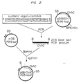
- Figure 2 is a schematic of the construction of pGB-ChAC, a plasmid capable of directing the expression of active chondroitinase AC in E. coli from tandem tac promoters (double arrowheads).
- Figure 3 is a schematic of the construction of plasmids used to sequence the chondroitinase B gene from Flavobacterium heparinum, pCHB300 and pCHB78.
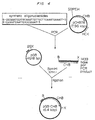
- Figure 4 is a schematic of the construction of pGB-CHB, a plasmid capable of directing the expression of active chondroitinase B in E. coli from tandem tac promoters (double arrowheads).
-
- Cells are grown in fermentation cultures to obtain sufficient quantities of the enzymes. Chondroitin sulfate A is included in the media at a concentration of between 0.5 and 10 g/l, preferably between 1.0 g/L to 2.0 g/l to induce chondroitinase AC and chondroitinase B synthesis. Crude enzyme extracts are prepared by liberating soluble proteins from the cells by standard cell disruption techniques, preferably osmotic shock based techniques which selectively release proteins from the cell's periplasmic space. For example, proteins can be released from the periplasmic space by treatment with non-ionic detergents in the range of 0.01 to 1.0%, freezing and thawing the cells, partial sonication for 0.5 to 6.0 minutes at 30 to 60% power in a pulsed mode 25/75 to 75/25, lysosyme treatment at 0.001 to 1.0 mg/ml for 15 to 60 minutes between 4 and 25°C, organic solvent treatment with 0.01 to 1.0% chloroform or toluene or by the osmotic shock process described in U.S. Patent No. 5,169,772 to Zimmermann and Cooney. In the latter, cells are partially sonicated for between 0.5 and 4.0 minutes, poser 3-6 pulsed mode 50/50, partial homogenization 250 to 500 psi, followed by lysozyme treatment at 0.001 to 1.0 mg/ml for between 15 and 60 minutes at between 4 and 23°C, and organic solvent treatment with 0.01 to 1.0% chloroform or 0.01 to 1.0% toluene.
- In the preferred embodiment, the crude extract is fractionated by cation exchange chromatography using a high flow rate resin such as Sepharose™ S Big Beads (Pharmacia), MonoS™ (Pharmacia), CBX (J.T. Baker), Sepharose™ S (Pharmacia), and CM cellulose (Bio-Rad or Sigma), at a pH of between 6.0 and 8.5 with a salt gradient equivalent to 0.01 to 1.0 M NaCl. The bound proteins are preferably eluted with step gradients of 0.25 M sodium chloride and 1.0 M sodium chloride, at pH 7.0. Chondroitinase activity elutes in the 0.25 M sodium chloride fraction. Other salts can be utilized, such as sodium phosphate or sodium sulfate to create the salt gradient. Alternatively, a pH gradient in the range of 6.0 to 10.0 could be employed or a combination of a salt and pH gradient.
- Fractions containing chondroitinase degrading activity are further fractionated by affinity chromatography using a sulfated cellulose based resin with a linear gradient of 0.0 to 0.4 M NaCl. Chondroitinase AC primarily elutes at 0.23 to 0.26 M NaCl and chondroitinase B elutes at 0.27 to 0.3 M NaCl. This is followed by hydroxylapatite chromatography using a step gradient of 0.25 M NaCl followed by a linear gradient of 0.25 to 1.0 M NaCl at pH 7.7. Chondroitinase B elutes at 0.25 M NaCl while chondroitinase AC elutes at 0.85 to 0.95 M NaCl. Highly purified preparations of each enzyme are obtained using a high resolution strong cation exchange resin eluted with a linear gradient from 0.125 to 0.325 M NaCl in 0.025 M sodium phosphate at pH 7.0 ± 0.1, as described with reference to elution from cation exchange resins described above. Chondroitinase B elutes in a protein peak at 0.175 to 0.225 M NaCl. Chondroitinase B can be further purified on the basis of molecular size by size exclusion chromatography, ultrafiltration or preparative gel electrophoresis. Gel filtration (size exclusion) resins with maximum resolution performance in the range of 5,000 to 100,000 are preferred. These include Superose™ 12, Superose™ 6, Sephadex™ G-50 and Sephadex™ G-50 from Pharmacia and BioGel™ P-60 and BioGel™ P-100 from BioRad. Ultrafiltration or dialysis membranes with molecular weight cutoffs in the range of 10,000 to 30 000 Daltons are useful in removing small contaminants while ultrafiltration and dialysis membranes with molecular weight cut-offs in the range of 70,000 to 1,000,000 Daltons are useful to remove larger contaminants. Alternatively, chondroitinase B containing samples of sufficient purity, more than 25% pure, could be further purified by subjecting the sample to gel electrophoresis according to standard laboratory procedures, and excising the major band appearing at a molecular weight of 55,000 ± 2,300 Daltons.
- The method of producing and purifying the chondroitinase lyase enzymes is exemplified as follows.
- F. heparinum was cultured in 15 L computer controlled fermenters in a variation of the defined nutrient medium described by Galliher, et al., Appl. Environ. Microbiol. 41(2):360-365, 1981. Chondroitin sulfate A (Sigma) was included in the media at a concentration of 1.0 g/L as the inducer of chondroitinase AC and chondroitinase B synthesis. The cells were harvested by centrifugation and the desired enzymes released from the periplasmic space by a variation of the osmotic shock procedure described by U.S. Patent No. 5, 169,772 to Zimmermann and Cooney. Cells were resuspended in 0.01 M sodium phosphate and 0.3 M sodium chloride at pH 7.0 ± 0.1 to give a final cell concentration of 100 absorbance units at 600 nm. The non-ionic detergent Nonedit™ P-40 was added to the cell suspension to a final concentration of 0.1% and the cells stirred for 1 hour at room temperature using a magnetic stir bar device. Cells and cell debris were then removed by centrifugation using a Sorval™ RC5C centrifuge with a JA-10 rotor at 10,000 RPM for 45 minutes. The cell pellet was discarded and the osmolate supernatant retained for further processing.
- Osmolates obtained from F. heparinum fermentations induced with chondroitin sulfate A were subjected to centrifugation to remove cells and cell debris and the supernatant applied to a cation exchange column (5.0 cm x 30 cm, Sepharose™ S Big Beads, Pharmacia) at a linear flow rate of 10 cm•min-1. The bound proteins were eluted at a linear flow rate of 5.1 cm•min-1 with step gradients of 0.01 M phosphate, 0.01 M phosphate/0.25 M sodium chloride and 0.01 M phosphate/1.0 M sodium chloride, all at pH 7.0 ± 0.1. Chondroitinase activity eluted in the 0.25 M sodium chloride fraction.
- This fraction was further purified by diluting the chondroitinase containing fraction two-fold with 0.01 M sodium phosphate and applying the material onto a column containing cellufine sulfate (2.6 cm i.d. x 100 cm, Amicon) and eluting at a linear flow rate of 1.88 cm•min-1 with a linear gradient of sodium chloride, 0.0 to 0.4 M. Chondroitinase AC primarily eluted at 0.23 to 0.26 M sodium chloride while chondroitinase B eluted at 0.27 to 0.3 M sodium chloride.
- Each fraction was diluted two-fold with 0.01 M sodium phosphate and applied to a hydroxylapatite column (2.6 cm i.d. x 30 cm). The bound proteins were eluted with a step gradient of 0.25 M sodium chloride followed by a linear gradient of 0.25 to 1.0 M sodium chloride all in 0.025 M sodium phosphate at pH 7.7 ± 0.1. Chondroitinase B elutes in the 0.25 M sodium chloride step while chondroitinase AC elutes at 0.85 to 0.95 M sodium chloride.
- The chondroitinase B fraction was diluted two-fold in 0.01 M sodium phosphate and applied to a strong cation exchange column (CBX-S, J.T. Baker, 1.6 cm i.d. x 10 cm). The bound material was eluted at a flow rate of 1.0 cm•min-1 with a linear gradient from 0.125 to 0.325 M sodium chloride in 0.025 M sodium phosphate at pH 7.0 ± 0.1. Chondroitinase B eluted in a protein peak at 0.175 to 0.225 M sodium chloride and contained a minor contaminating protein of molecular weight 20,000 Daltons. This protein was removed by gel filtration chromatography by loading the chondroitinase B sample onto a Superdex™ 200 column (1.0 cm i.d. x 30 cm, Pharmacia) and eluting with 0.05 M sodium phosphate, pH 7.2 at a linear flow rate of 1.25 cm•min-1 and collecting the protein containing fractions.
- The chondroitinase AC fraction collected from hydroxylapatite chromatography was diluted three-fold in 0.01 M sodium phosphate and applied to a strong cation exchange column (CBX-S, J.T. Baker, 1.6 cm i.d. x 10 cm). The bound material was eluted at a flow rate of 1.0 cm•min-1 with a linear gradient from 0.125 to 0.325 M sodium chloride in 0.025 M sodium phosphate at pH 7.0 ± 0.1. Chondroitinase AC eluted in a single protein peak at 0.175 to 0.225 M sodium chloride. Purification results for the chondroitinase enzymes are shown in Table 1.
Purification of chondroitinase enzymes from Flavobacterium heparinum fermentations sample activity (IU) specific activity (IU/mg) yield (%) fermentation: chondroitinase AC 65,348 0.764 100 chondroitinase B 21,531 0.252 100 osmolate: chondroitinase AC 39,468 1.44 60 chondroitinase B 15,251 0.588 71 cation exchange: chondroitinase AC 27,935 9.58 43 chondroitinase B 13,801 4.731 64 cellufine sulfate: chondroitinase AC 18,160 22.6 28 chondroitinase B 6,274 21.2 29 hydroxylapatite: chondroitinase AC 14,494 146.8 22 chondroitinase B 3,960 65.62 18 strong cation exchange: chondroitinase AC 9,843 211.4 15 chondroitinase B 4,104 167.2 18 gel filtration: chondroitinase B 2,814 278.7 13 - Chondroitinase activity was determined by a modification of the spectrophotometric assay described by Yang, et al., J. Biol. Chem., 160(30) :1849-1857, 1985. Chondroitinases degrade their respective substrates by an eliminative reaction resulting in the formation of 4,5-unsaturated sulfated disaccharides which absorb ultraviolet light at 232 nm. Reaction buffers contained 50 mM Tris, pH 8.0 and 0.5 mg/ml substrate; dermatan sulfate for chondroitinase B activity, chondroitin sulfate A for chondroitinase AC activity. A continuous spectrophotometric assay is carried out by transferring a 10 to 50 µl sample to a quartz cuvette and adding the reaction buffer to make a final volume of one ml. The cuvette is placed in a Beckman DU 640 spectrophotometer, controlled to maintain a constant temperature of 30°C, and the increase in absorbance at 232 nm monitored for three to five minutes. Activities are calculated using the molar extinction coefficient for chondroitin sulfate, 5.1 x 10³ M-1, and are expressed in international units, IU, where one IU is the amount of enzyme required to catalyze the formation of one µmole unsaturated product per minute.
- The purification method described herein is suitable for obtaining sufficient quantities of purified chondroitinase AC and chondroitinase B for characterization studies. The purified enzymes were analyzed by SDS-PAGE using the technique of Laemmli, Nature, 227:680-685, 1970, and the gels quantified with a scanning densitometer (Bio-Rad, Model GS-670). Chondroitinase AC was shown to have a molecular weight of 77,000 ± 5,000 Daltons and a purity of greater than 99% while chondroitinase B has a molecular weight of 55,000 ± 2,300 Daltons and a purity of greater than 99%.
- Kinetic parameters of the 77,000 Dalton chondroitinase AC protein were measured using both chondroitin sulfate A and chondroitin sulfate C as substrates. The Km and Kcat values for chondroitinase A activity were 6 µM and 230 s-1, respectively, while the Km and Kcat values for chondroitinase C activity were 9.3 µM and 150 s-1, respectively. Kinetic parameters of the 55,000 Dalton chondroitinase B protein were measured using dermatan sulfate as the substrate. The Km and Kcat values for chondroitinase B activity were 7.4 µM and 192 s-1, respectively.
- The Vmax of the chondroitinase enzymes can be effected by trace amounts of certain elements. A base reaction buffer of 20 mM Tris buffer, pH 8.0 and 0.5 mg/ml substrate, either chondroitin sulfate A for chondroitinase AC or dermatan sulfate for chondroitinase B, was used to determine the effect of divalent metals and salts on the activity of the chondroitinase enzymes. The results are shown in Table 2.
Effects of 0.1 mM of various reagents on the activity of chondroitinase enzymes. reagent chondroitinase AC relative activity(%) chondroitinase B relative activity(%) none 100 100 MgCl2 91 91 MnCl2 83 33 CuSO4 92 91 ZnCl2 26 45 FeSO4 98 69 HgCl2 55 40 CoCl2 81 42 EDTA 97 1 - The chondroitinase enzyme activity can be stabilized by addition of excipients or by lyophilization. Stabilizers include carbohydrates, amino acids, fatty acids, and surfactants and are known to those skilled in the art. Examples include carbohydrate such as sucrose, lactose, mannitol, and dextran, proteins such as albumin and protamine, amino acids such as arginine, glycine, and threonine, surfactants such as Tween™ and Pluronic™, salts such as calcium chloride and sodium phosphate, and lipids such as fatty acids, phospholipids, and bile salts. The stabilizers are generally added to the protein in a ratio of 1:10 to 4:1, carbohydrate to protein, amino acids to protein, protein stabilizer to protein, and salts to protein; 1:1000 to 1:20, surfactant to protein; and 1:20 to 4:1, lipids to protein. Other stabilizers include high concentrations of ammonium sulfate, sodium acetate or sodium sulfate, based on comparative studies with heparinase activity. The stabilizing agents, preferably the ammonium sulfate or other similar salt, are added to the enzyme in a ratio of 0.1 to 4.0 mg ammonium sulfate/IU enzyme.
- The use of stabilizers is demonstrated as follows. The purified chondroitinase enzymes were dialyzed into 10 mM sodium phosphate, pH 7.5, to a concentration of 2 IU/ml and supplemented with either 1 mg/ml bovine serum albumin, 1.5 M sodium acetate, 0.0025 M Tris or 0.15 M Tris, and an accelerated shelf life performed at 37°C. 2 IU of purified chondroitinase enzymes also were placed into various buffers, lyophilized and an accelerated shelf life performed at 37°C. The results are shown in Table 3.
Stability of chondroitinase enzymes at 37°C.
7day retention of activity enzymes at (%)additive format chondroitinase AC chondroitinase B 0.15 M Tris liquid 1 42 0.0025 M Tris liquid 22 44 1 mg/ml BSA liquid 1 26 1.5 M NaOAc liquid 64 72 0.15 M Tris lyophilized 26.7 43.7 PBS lyophilized 8.7 15.9 8 mg/ml sucrose lyophilized 88 93.16 2 mg/ml glycine lyophilized 42.4 75.7 - The purified proteins were analyzed by the technique of Edman, Ann. N. Y. Acad. Sci. 88:602, 1950, to determine the N-terminal amino acid. However, the Edman chemistry was unable to liberate an amino acid, indicating that a post-translational modification had occurred at the N-terminal amino acid of both chondroitinase proteins. One nmol samples of chondroitinases AC and B were used for deblocking with pyroglutamate aminopeptidase. Control samples were produced by mock deblocking 1 nmol samples without adding the peptidase. All samples were placed in 10 mM ammonium carbonate buffer at pH 7.5 with 10 mM dithiothreitol. 1 mU peptidase was added to the samples and the reaction allowed to incubate at 37°C for 8 hours. An additional 0.5 mU peptidase was added and incubation continued for 16 h. The reaction mixture was exchanged into 35 % formic acid by diafiltration with 10,000 Dalton cut-off ultrafiltration membranes (Centricon, Amicon) and the sample dried under vacuum. Deblocked chondroitinase enzymes were then analyzed by Edman chemistry to determine the N-terminal sequence, using an Applied Biosystems 745A Protein Sequencer.
- The N-terminal sequence of chondroitinase AC was QTGTAEL (Sequence ID No. 2, amino acids 24 to 30) and of chondroitinase B was VVASNEL (Sequence ID No. 4, amino acids 27 to 34).
- The chondroitinase enzymes were subjected to enzymatic fragmentation using the arginine specific protease clostripain (EC 3.4.22.8, Sigma). Pre-activated clostripain was added to chondroitinase AC at a 1 to 2 % w/w ratio in 0.025 M sodium phosphate, 0.0002 M calcium acetate and 0.0025 M dithiothreitol at pH 7.5 ± 0.1 and incubated for 2 to 3 hours at 37°C. The reaction mixture was applied to a Vydac C18 reverse phase HPLC column (0.46 cm I.D. x 30 cm) and the peptide fragments eluted at a linear flow rate of 1 cm-min-1 with a linear gradient of 10 to 90 % acetonitrile in 1 % trifluoroacetic acid. Four of the peptide fragments obtained were subjected to amino acid sequence determination.
- Clostripain was added to chondroitinase B at a 1 to 2 % w/w ratio in 0.025 M sodium phosphate, 0.0002 M calcium acetate and 0.0025 M dithiothreitol at pH 7.5 ± 0.1 and incubated for 2 to 3 hours at 37°C. The reaction mixture was applied to a Vydac™ C18 reverse phase HPLC column and the peptide fragments eluted at a linear flow rate of 6.0 cmomin-1 with a linear gradient of 10 to 90 % acetonitrile in 1 % trifluoroacetic acid. Three of the peptide fragments obtained were subjected to amino acid sequence determination.
- A Flavobacterium heparinum chromosomal DNA library was constructed in lambda phage DASHII. 0.4 µg of F. heparinum chromosomal DNA was partially digested with restriction enzyme, Sau3A, to produce a majority of fragments around 20 kb in size, as described in Maniatis, et al., Molecular Cloning, A laboratory Manual, 1982. This DNA was phenol/chloroform extracted, ethanol precipitated, ligated with DASHII arms and packaged with packaging extracts from a Lambda DASHII™/BamHI Cloning Kit (Stratagene, La Jolla, CA). The library was titered at approximately 10-5 pfu/ml after packaging, was amplified to 10-8 pfu/ml by the plate lysis method, and stored at -70°C as described by Silhavy et al. in Experiments with Gene Fusions, Cold Spring Harbor Laboratory, 1972.
- The F. heparinum chromosomal library was titered to about 300 pfu/plate, overlaid on a lawn of E. coli, and allowed to transfect the cells overnight at 37°C, forming plaques. The phage plaques were transferred to nitrocellulose paper, and the phage DNA bound to the filters, as described in Maniatis, et al., ibid.
- Degenerate primers were designed from peptides AC-1, AC-3 and AC-4 (Sequence ID No. 2, amino acids 395 to 413; 603 to 617; 514 to 536; and 280 to 288, respectively). Amplification of the primers was carried out in a 0.1 ml reaction buffer containing 50 mM KCl, 10 mM Tris/HCl pH 9, 0.1% Triton X-100, 2.5 mM MgCl2, plus the four dNTPs at 200 µM, 2.5 units Taq Polymerase (Bio/Can, Mississauga, Ont.), 0.1 mM of each primer and 10 ng of F. heparinum genomic DNA. The amplified primers were linearized with SalI, NotI, and XbaI in individual restriction digests, and combined, after purification, for use as template DNA. The samples were placed in an automated heating block, (DNA Thermocycler™, Barnstead/Thermolyne, Dubuque, IA) programmed for cycles with temperatures of denaturation at 94°C for 1 min., annealing at 50°C for 2 min., and extension at 72°C for 2 min., with 35 repetitions of this sequence. The combination of synthetic oligonucleotide primers: 5'-TCNGGRAARTARTANCCDATNGCRTCRTG-3' (Sequence ID No. 5), corresponding to peptide AC-3; and 5'-TAYATGGAYTTYAAYGTNGARGG-3' (Sequence ID No. 6), corresponding to peptide AC-4; yielded a PCR product of approximately 750 bp in size. Attempts to clone this fragment into vectors, pTZ/PC or into pCRII (TA cloning kit, Invitrogen, San Diego, Ca.) in E. coli strain, FTB1, were unsuccessful.
- E. coli FTB1 was constructed as follows: the F' episome from E. coli XL-1 Blue, (Stratagene, La Jolla CA) carrying the lac Iq repressor gene was moved, as described by Miller, Experiments in Molecular Genetics, Cold Spring Harbor, 1972, into E. coli TB1 described by Baker et al., Proc. Natl. Acad. Sci. 81:6779-6783, 1984. The FTB1 background permits a more stringent repression of transcription from plasmids carrying promoters with a lac operator such as the lac and tac promoters.
- To facilitate cloning of these PCR products, a restriction site was incorporated at the 5' ends of the primers. The PCR products were analyzed for the absence of restriction sites which are found in the multiple cloning site of pBluescript (Stratagene, La Jolla, CA) to determine which restriction site should be added to the primers. This ensured that the PCR products would not be cut into multiple fragments when treated with the restriction enzyme used to form overhangs on the ends of the DNA fragments. BamHI met this criteria for all three PCR fragments. New primers were synthesized with BamHI sites at their 5' ends, which were otherwise identical to those described above, and used to produce a 764 bp PCR product, Figure 1. This DNA fragment was digested with BamHI, isolated on an agarose gel, as described by Maniatis et al., ibid, and purified using the Geneclean™ kit (Bio/Can, Mississauga, Ont.) pBluescript was digested with BamHI, the 5' ends dephosphorylated by alkaline phosphatase treatment as described by Maniatis et al., ibid, and purified from an agarose gel using the Geneclean™ kit. The treated PCR fragment and pBluescript plasmid DNA were ligated, transformed into FTB1, and plated onto LB agar plates containing ampicillin at 0.2 mg/ml. Plasmids from colonies grown on these plates were isolated by colony cracking as described in Maniatis et al., ibid. All enzymes were supplied by New England Biolabs (Mississauga, Ont.). Plasmids were isolated using the RPM™ kit (Bio/Can, Mississuaga, Ont.). Sequence analysis of the cloned PCR fragment correlated with reverse transcribed peptide sequences from chondroitinase AC peptides, indicating that the PCR fragment encodes the chondroitinase AC gene. DNA sequencing was performed by the dideoxy-chain termination method of Sanger et al., Proc. Natl. Acad. Sci., 74:5463-5467, 1978. Sequencing reactions were carried out with the Sequenase™ Kit (U.S. Biochemical Corp., Cleveland, Ohio) and S-dATP (Amersham Canada Ltd., Oakville, Ontario, Canada), as specified by the supplier.
- The 764 bp PCR fragment, contained in plasmid pA2C1BS-11 represents approximately 36% of the coding region for the Chondroitinase AC gene. This entire 764 bp fragment was sequenced and was found to contain a continuous open reading frame which encoded peptides AC-3, AC-4 and AC-1 (Sequence ID No. 2, amino acids 395-413; 603-617; 514-536; 280-288, respectively).
- The 764 bp PCR fragment was used to probe the genomic F. heparinase lambda library. First, pA2C1BS-11 was isolated via the boiling method, as described in Maniatis et al., ibid. The plasmid was digested with BamHI, separated from the vector, purified as described above and labeled with a Nick Translation™ kit (Boehringer Mannheim, Montreal, Canada) using radiolabelled 32P α-dATP. E. coli P2392 (Stratagene, La Jolla, CA) was used as the lawn for plating the lambda library. Approximately 6000 plaques were screened by plaque hybridization using BA85 nitrocellulose membranes (Scheicher & Schuell, Keene, NH) as described by Maniatis et al., ibid. Plaque hybridization was carried out, at 65°C for 16 hours in a Tek Star™ hybridization oven (Bio/CAN Scientific, Mississauga, Ontario). Subsequent washes were performed at 65°C, twice for 15 min. in 2X SSC, once in 2X SSC/0.1% SDS for 30 min. and once in 0.5X SSC/0.1% SDS for 15 min. More than 100 positive plaques were identified and isolated, some of which were clusters of plaques. These were rescreened by spotting the lambda clone onto a lawn of P2392 host cells and reprobing via plaque hybridization. Six plaques were positive upon rescreening, and their DNA was isolated, as described by Maniatis, et al., ibid, and digested with restriction enzymes corresponding to the sites on the ends of lambda DASH II arms. This DNA was used in Southern hybridization analysis (Southern, J. Mol. Biol. 98:503-517, 1975) by blotting onto Hybond™ N nylon membrane (Amersham, Oakville, Canada) using hybridization and wash conditions, described above for plaque hybridization. One clone contained a 4.5 kb Sal I fragment and another contained a 6 kb BamHI fragment, both of which hybridized with the probe. These were cloned into corresponding sites of pBluescript.
- Because the molecular weight of chondroitinase AC is approximately 75 kD, the size of the corresponding gene would be approximately 2.05 kb. Both the 4.5 kb SalI and the 6 kb BamHI chromosomal DNA fragments could include the entire chondroitinase AC gene. To increase the probability of analyzing a DNA fragment which encodes the entire gene, the 6 kb BamHI fragment was chosen for sequence analysis. The pBluescript plasmid containing this BamHI fragment (called p64BS2-7, Figure 1) was isolated using the Qiagene kit (Bio/Can, Miss, Ont). A method of DNA sequencing, the walking primer strategy (Voss et al. Meth. Molec. Cell. Biol. 3:153-155 (1992)), was employed using synthetic primers (Eppendorf, model ECOSYN™ D300,Madison, WI) and an A.L.F. DNA sequencer (Pharmacia LKB, Mtl, Qc). Fluorescenated Universal and Reverse primers provided in the Pharmacia AutoRead kit were also used. Fluorescently labeled dNTPs were incorporated into sequencing reactions with the Pharmacia AutoRead Fluorescent labelling kit (Pharmacia LKB, Mtl, QC). Areas of secondary structure were resolved by one of two methods. First, fluorescenated primers which hybridized close to, and 5' to, the region of secondary structure were synthesized. Using these primers, the Pharmacia AutoCycle™ kit (Pharmacia LKB, Mtl, Qc), and a automated heating block (DNA Thermocycler™, Barnstead/Thermolyne, Dubuque, Iowa), programmed for step cycles of 95°C for 36 sec, 50°C for 36 sec and 72°C for 84 sec, repeated 25 times, sequencing of secondary structure regions was accomplished. Any ambiguous areas still not resolved by the first method were sequenced by the method of Sanger et al., Proc . Natl. Acad . Sci . 74: 5463-5467 (1978), using 35S a-dATP, and a USB Sequenase™ kit (LaJolla, Ca.) in which dGTP was replaced by dITP.
- Analysis of the DNA sequence indicated that there was a single, continuous open reading frame of 2100 bp containing codons for 700 amino acid residues. All four clostropain-derived peptides were encoded by this gene. Searching for a possible signal peptide sequence using Geneworks™ (Intelligenetics, Mountain View, Ca.), suggested that there are two possible sites for the processing of the protein into a mature form: Q-23 (glutamine) and A-28 (alanine). N-terminal amino acid sequencing of deblocked, processed Chondroitinase AC indicated that the mature protein begins with Q-23 and contains 678 amino acids with a calculated molecular weight of 77,169 Daltons.
- Construction of an expression vector for chondroitinase AC is shown in Figure 2. The vector pGB is an E. coli expression vector which contains an unique BamHI site, whereby expression of a DNA fragment inserted into this site is driven by a double tac promoter. The vector also includes a kanamycin resistance gene and the lac Iq gene to allow induction of transcription with IPTG. PCR was used to generate a mature chondroitinase AC gene.
- An oligonucleotide, 5'-GCGGATCCATGCAGCAGACCGGTACTGCAGAA-3', (Sequence ID No. 7) was designed to insert an ATG-start site immediately preceding the codon for the first amino acid (Q-23) of mature chondroitinase AC, while an oligonucleotide 5'-CGCGGATCCCCTAGATTACTACCATCAAAA-3' (Sequence ID No. 8) was designed to hybridize downstream of the TAG-stop codon. Both oligonucleotides also contain a BamHI site. Plasmid p64BS2-7 was used as the template in a PCR reaction with an annealing temperature of 45°C. A specific fragment of the expected size of 2034 bp was obtained. This fragment was isolated and inserted into a BamHI site of the expression vector pGB.
- The construct was transformed into E. coli strain, F-TB1, and the transformed bacteria was grown at 37°C in LB medium containing 75 µg/ml kanamycin to an OD600 of 0.5, at which point the tac promoter from pGB was induced by the addition of 1 mM IPTG. Cultures were grown an additional 2 to 5.5 hours at either 23°C, 30°C or 37°C. The cells were cooled on ice, concentrated by centrifugation and resuspended in cold PBS at 1/10th the original culture volume. Cells were lysed by sonication and cell debris removed by centrifugation at 10,000 x g, 5 minutes. The pellet and supernatant fractions were analyzed separately for chondroitin sulfate A or C degrading (chondroitinase AC) activity. Chondroitin sulfate A degrading activities of 1.24 x 10⁻², 2.88 x 10⁻², and 4.25 x 10⁻² IU/ml/OD and chondroitin sulfate C degrading activities of 1.57 x 10⁻², 2.24 x 10⁻², and 6.02 x 10⁻² IU/ml/OD were observed from cultures grown at 23, 30 and 37°C, respectively. The activities using chondroitin sulfate A as the substrate are approximately twice that of those using chondroitin sulfate C as the substrate. This ratio is also observed when measuring the activity of the wild type chondroitinase AC using both these substrates.
- E. coli F-TB1(pGB-ChAC) was grown in a 3.5 L Braun Biostat E computer controlled fermenter in M9 medium to a dry cell weight concentration of 35 g/L. Glucose and ammonia were added as needed to maintain growth and pH at 7.0. Chondroitinase A activity accumulated to 103.44 IU/ml while chondroitinase C activity accumulated to 28.26 IU/ml.
- Partial-guessmer PCR primers were designed using the amino acid sequences of the clostripain-generated peptides from the chondroitinase B protein and the codons commonly found in Flavobacterium genes, Table 4. Three peptides were generated, designated CHB-1 (Sequence ID No. 4, amino acids 373 to 384), CHB-2 (Sequence ID No. 4, amino acids 41 to 50), and CHB-3 (Sequence ID No. 4, amino acids 130 to 146).
Codon usage table for Flavobacterium and Escherichia coli. consensus codon amino acid codon(s) E. coli Flavobacterium A GCT, GCC, GCG, GCA GCT GCC C TGT,TGC EITHER EITHER D GAT, GAC EITHER EITHER E GAG, GAA GAA GAA F TTC, TTT EITHER TTT G GGC, GGA, GGG, GGT GGC or GGT GGC H CAC, CAT CAT CAT I ATC, ATA, ATT ATA ATC K AAA, AAG AAA AAA L CTT, CTA, CTG, TTG, TTA, CTC CTG CTG M ATG ATG ATG N AAC, AAT AAC AAT P CCC, CCT, CCA, CCG CCG CCG Q CAG, CAA CAG CAG R CGT, AGA, CGC, CGA, AGG, CGG CGT CGC S TCA, TCC, TCG, TCT, AGC, AGT TCT ND T ACG, ACC, ACT, ACA ACC or ACT ACC or ACA V GTC, GTA, GTT, GTG GTT ND W TGG TGG TGG Y TAC, TAT EITHER TAT - 5'-CGG GAT CCC ARA TYG CCG AYG GNA CNT ATA AAG A-3' (Sequence ID No. 9) was derived from the CHB-2 peptide (Sequence ID No. 4, amino acids 41 to 50) and 5'-CGG GAT CCG GCN SKA TTG CGT TCR TCA AA-3' (Sequence ID No. 10) was derived from peptide CHB-3, Sequence ID No. 4, amino acids 130 to 146. A BamHI site was present on the 5' end of each primer to increase the efficiency of cloning of the PCR product. Using linear F. heparinum chromosomal DNA, described above, as a template, a single 300 bp DNA fragment was amplified. Conditions for the amplification were as follows: denaturation at 94°C for 40 sec, annealing at 45 or 50°C for 1 min. and extension at 72°C for 2 min. This cycle was repeated 35 times.
- As shown in Figure 3, the PCR fragment was purified on an agarose gel, digested with BamHI and ligated into BamHI digested, dephosphorylated pBluescript. The ligation mixture was used to transform E. coli FTB1. Of the 50 resulting transformants, one yielded a 300 bp fragment when cut with BamHI. The insert in this plasmid, pCHB300, was subjected to DNA sequence analysis, performed as described above, which revealed that the insert contained DNA sequences outside of the primer regions which encoded amino acid sequence matching that determined for two chondroitinase B peptides. This insert was used to screen the lambda library of F. heparinum chromosomal DNA, which was constructed as described above.
- The lambda library was plated with a density of 200 plaques per dish. Plate lifts of 20 dishes were made. For production of the probe, 500 ng of pCHB300 was submitted to 30 cycles of PCR amplification; denaturation at 93°C, annealing at 55°C and extension at 72°C, each for 1 min., using the primers described above. The resulting PCR fragment was purified on agarose gels and labelled with dATPa32P, using the Random Primer labelling kit (Boehringer Mannheim, Laval, Canada). Thirty-one potential lambda clones were found which hybridized with this probe, after the lifts were subjected to washing one time, in 2X SSC at 58°C. Rescreening of these plaques gave a positive signal for 17 of the plaques after washing at 58°C, 2X for 15 min. in 2X SSC, 1X for 30 min. in 2X SSC/0.1% SDS and 1X for 20 min. in 0.5X SSC/0.1% SDS. Two of 8 clones analyzed further showed a 5.0 kb HindIII fragment hybridizing with the probe and comigrating with a HindIII fragment from F. heparinum chromosomal DNA which also hybridized with the 300 bp probe. The 5.0 kb fragment was gel purified from both lambda clones, ligated into the HindIII site of pBluescript and transformed into FTB1.
- 44 colonies were picked and rubbed on the side of a 0.5 ml PCR tube containing 20 µl of the same PCR mixture as above. PCR was performed at: denaturation at 93°C, for 30 sec., annealing at 58°C, for 30 sec. and extension at 72°C, for 1 min, for 35 cycles. Upon analysis, 6 transformants showed amplification of the 300 bp band. DNA from these colonies were isolated and digested by HindIII revealing the presence of a 5.0 kb fragment. 5 out of the 6 clones hybridized with the 300 bp fragment, confirming results of the PCR amplification experiment. One of these clones, pCHB78, was selected and used as a template for DNA sequencing.
- Using a walking primer strategy, sequencing reactions were carried out as described above for the A.L.F. DNA sequencer. Sequence analysis revealed a single 1.52 kb open reading frame coding for 506 amino acid residues. The preprotein was found to have a signal peptide of 25 amino acids. The mature chondroitinase B enzyme contains 481 amino acids with a calculated molecular weight of 53,563 daltons.
- Construction of an expression vector for chondroitinase B is shown in Figure 4. Primers were designed to amplify the coding region of the chondroitinase B gene in an analogous manner to that described above with reference to expression of the chondroitinase AC gene. One oligonucleotide used for amplification of the chondroitinase B coding sequence (5'-CGCGGATCCATGCAGGTGTTGCTCAAATGAAACT-3') (Sequence ID No. 11), contained a BamHI restriction site at its 5' end and an ATG codon that was to be inserted before the first amino acid of the mature protein. The second oligonucleotide (5'-CGGAATCAATTCACCGGG-AT-3') (Sequence ID No. 12) was designed with a XmnI restriction site and a termination codon to be inserted at the end of the coding sequence of the gene. Using 100 ng of pCHB78 as template, with an annealing temperature of 52°C, the 1.5 kb fragment was amplified, gel purified, restriction digested and inserted into pGB previously cut with BamHI and XmnI. This resulted in the definitive pGB-CHB construct used to express the protein.
- This construct was transformed in E. coli strain DH5α, expressed as described for the chondroitinase AC enzyme. After growing cells until an O.D. 600 = 0.5, 1 mM IPTG was added to the cultures to induce the tandem tac promoters and cells were transferred to either 23°C, 30°C or 37°C for additional growth for 5, 3 and 2 hours, respectively. After sonication, supernatant fractions were assayed for activity on dermatan sulfate. Growth of cells at 23°C gave the best results with a degrading activity of 0.57 IU/ml/OD while growth of cells at 30°C and 37°C gave degrading activities of 0.14 and 0.01 IU/ml/OD respectively.
- The present invention describes a methodology for obtaining highly purified chondroitin degrading enzymes derived from the natural organism Flavobacterium heparinum, and the genes encoding these enzymes. Derivatives of the genes can be prepared by making conservative substitutions, additions and deletions thereof, which do not substantially impact on the resulting enzymatic activity, or by using degenerative forms of the genes. As used herein, conservative substitutions involve substitutions of codons which encode the same amino acids and substitutions of amino acids for amino acids having similar structure or chemical characteristics, which are well known to those skilled in the art, for example, groups of structurally similar amino acids include (I, L, V); (F, Y); (K, R); (Q, N); (D, E); and (G, A).
- This application therefore describes a purified chondroitinase degrading enzyme isolated from bacteria. The enzyme may be an enzyme selected from the group consisting of chondroitinase AC and chondroitinase B from Flavobacterium heparinum. The enzyme may be formulated with a pharmaceutically acceptable carrier. Preferably, the enzyme is expressed in bacteria from a gene isolated from Flavobacterium heparinum.
- The enzyme may be chondroitinase AC of Flavobacterium heparinum and have a molecular weight between 72,000 and 82,000 Daltons and be capable of degrading chondroitin sulfate A and chondroitin sulfate C. This enzyme may be encoded by the nucleotide sequence of Sequence ID No. 1 or sequences having conservative or degenerative substitutions thereof. This enzyme may have the amino acid sequence of Sequence ID No. 2 or sequences having conservative substitutions thereof.
- The enzyme may be chondroitinase B of Flavobacterium heparinum and have a molecular weight between 52,700 and 57,300 Daltons and be capable of degrading dennatan sulfate or chondroitin sulfate B. This enzyme may be encoded by the nucleotide sequence of Sequence ID No. 3 or sequences having conservative or degenerative substitutions thereof. This enzyme may have the amino acid sequence of Sequence ID No. 4 or sequences having conservative substitutions thereof.
- This application also describes an isolated nucleotide sequence encoding an enzyme selected from the group consisting of chondroitinase AC and chondroitinase B from Flavobacterium heparinum. The sequence of the isolated nucleotide sequence may occur naturally in Flavobacterium heparinum.
- The sequence may encode the enzyme chondroitinase AC having a molecular weight between 72,000 and 82,000 Daltons and being capable of degrading chondroitin sulfate A and chondroitin sulfate C, such as a protein having the amino acid sequence of Sequence ID No. 2 or a sequence having conservative substitutions thereof. In that case the isolated nucleotide sequence preferably comprises the nucleotide sequence of Sequence ID No. 1 or a sequence having conservative or degenerative substitutions thereof.
- The sequence may encode the enzyme chondroitinase B having a molecular weight between 52,700 and 57,300 Daltons and being capable of degrading dermatan sulfate or chondroitin sulfate B, such as a protein having the amino acid sequence of Sequence ID No. 4 or a sequence having conservative substitutions thereof. In that case the isolated nucleotide sequence preferably comprises the nucleotide sequence of Sequence ID No. 3 or a sequence having conservative or degenerative substitutions thereof.
- This application also describes a method of purifying a chondroitin lyase from bacteria comprising: lysing the bacteria; extracting proteins from the periplasmic space of the lysed bacteria; separating the extracted proteins by cation exchange chromatography using a salt of pH gradient; separating the fractions having enzymatic activity obtained by elution of the cation exchange chromatography matrix by chromatography on a sulfated cellulose resin using a salt or pH gradient; separating the fractions having enzymatic activity obtained by elution of the sulfated cellulose resin on hydroxyapatite using a salt or pH gradient; separating the fractions having enzymatic activity obtained by elution of the hydroxyapatite by chromatography using cation exchange chromatography using a salt or pH gradient; and separating the fractions with enzymatic activity on the basis of molecular weight.
- Variations of these methods will be obvious to those skilled in the art from the foregoing detailed description of the invention. Such modifications are intended to come within the scope of the appended claims.


Claims (16)
- A composition comprising:wherein the enzyme preparation is obtainable from the bacteria by a process comprising the steps of:(a) an enzyme preparation consisting of greater than 99% of a dermatan sulfate-degrading chondroitinase from the bacteria Flavobacterium heparinum, the remaining material in the enzyme preparation being contaminant; and optionally(b) an excipient;lysing the bacteria;extracting proteins from the periplasmic space of the lysed bacteria;separating the extracted proteins by cation exchange chromatography using a salt or pH gradient;separating the fractions having enzymatic activity obtained by elution of the cation exchange chromatography matrix by chromatography on a sulfated cellulose resin using a salt or pH gradient;separating the fractions having enzymatic activity obtained by elution of the sulfated cellulose resin on hydroxyapatite using salt or pH gradient;separating the fractions having enzymatic activity obtained by elution of the hydroxyapatite by chromatography using cation exchange chromatography using a salt or pH gradient; andseparating the fractions with enzymatic activity on the basis of molecular weight.
- The composition of Claim 1 where the chondroitinase is chondroitinase B and has a molecular weight between 52,700 and 57,300 Daltons and is capable of degrading dermatan sulfate or chondroitin sulfate B.
- A recombinant chondroitinase encoded by the nucleotide sequence of Sequence ID No. 3 or a derivative thereof, which derivative comprises conservative or degenerative substitutions, additions or deletions, and wherein the recombinant chrondroitinase degrades dermatan sulfate.
- The enzyme of Claim 3 having the amino acid sequence of Sequence ID No. 4 or sequences having conservative substitutions thereof.
- The composition of Claim 1 wherein the excipient is a pharmaceutically acceptable carrier.
- The dermatan sulfate-degrading chondroitinase of Claim 3 having the amino acid sequence shown in Sequence ID No. 4.
- A method for purifying a dermatan sulfate-degrading chondroitinase from Flavobacterium heparinum by a process comprising the steps of:lysing the bacteria;extracting proteins from the periplasmic space of the lysed bacteria;separating the extracted proteins by cation exchange chromatography using a salt or pH gradient;separating the fractions having enzymatic activity obtained by elution of the cation exchange chromatography matrix by chromatography on a sulfated cellulose resin using a salt or pH gradient;separating the fractions having enzymatic activity obtained by elution of the sulfated cellulose resin on hydroxyapatite using a salt or pH gradient;separating the fractions having enzymatic activity obtained by elution of the hydroxyapatite by chromatography using cation exchange chromatography using a salt or pH gradient; andseparating the fractions with enzymatic activity on the basis of molecular weight, into fractions comprising dermatan sulfate-degrading chondroitinase separate from other enzymes.
- A composition according to any one of Claims 1, 2 or 5 consisting of greater than 99% dermatan-sulfate degrading chondroitinase, the remaining material being a contaminant.
- A composition according to any one of Claims 1, 2, 5 or 8 or a method according to Claim 7 wherein the bacteria recombinantly expresses a gene encoding the dermatan sulfate-degrading chondroitinase.
- A recombinant polynucleotide encoding a dermatan sulfate-degrading chondroitinase as defined in any one of Claims 1-4 or 6.
- A method for producing a dermatan sulfate-degrading chondroitinase comprising expressing said chondroitinase from a recombinant polynucleotide as defined in Claim 10, in a non-human host organism.
- A method according to Claim 11 further comprising the step of purifying the thus expressed chondroitinase from the non-human host organism.
- A method according to Claim 11 or 12 wherein the non-human host organism is a bacteria.
- A method according to Claim 13 wherein the bacteria is Flavobacterium heparinum.
- A method according to Claim 13 or 14 wherein the step of purifying the thus expressed chondroitinase from the non-human host organism comprises the method of Claim 11.
- A method according to any one of Claims 11 to 13 wherein the non-human host organism is an organism other than Flavobacterium heparinum.
Applications Claiming Priority (3)
| Application Number | Priority Date | Filing Date | Title |
|---|---|---|---|
| US08/272,247 US6093563A (en) | 1994-07-08 | 1994-07-08 | Chondroitin lyase enzymes |
| US272247 | 1994-07-08 | ||
| EP95926207A EP0764205B1 (en) | 1994-07-08 | 1995-07-07 | Chondroitin lyase enzymes |
Related Parent Applications (1)
| Application Number | Title | Priority Date | Filing Date |
|---|---|---|---|
| EP95926207A Division EP0764205B1 (en) | 1994-07-08 | 1995-07-07 | Chondroitin lyase enzymes |
Publications (3)
| Publication Number | Publication Date |
|---|---|
| EP1607478A2 true EP1607478A2 (en) | 2005-12-21 |
| EP1607478A3 EP1607478A3 (en) | 2006-02-22 |
| EP1607478B1 EP1607478B1 (en) | 2009-04-15 |
Family
ID=23039019
Family Applications (2)
| Application Number | Title | Priority Date | Filing Date |
|---|---|---|---|
| EP95926207A Expired - Lifetime EP0764205B1 (en) | 1994-07-08 | 1995-07-07 | Chondroitin lyase enzymes |
| EP05075080A Expired - Lifetime EP1607478B1 (en) | 1994-07-08 | 1995-07-07 | Chondroitin lyase enzymes |
Family Applications Before (1)
| Application Number | Title | Priority Date | Filing Date |
|---|---|---|---|
| EP95926207A Expired - Lifetime EP0764205B1 (en) | 1994-07-08 | 1995-07-07 | Chondroitin lyase enzymes |
Country Status (10)
| Country | Link |
|---|---|
| US (2) | US6093563A (en) |
| EP (2) | EP0764205B1 (en) |
| JP (2) | JPH10506263A (en) |
| AT (2) | ATE305032T1 (en) |
| AU (1) | AU693916B2 (en) |
| CA (1) | CA2194375C (en) |
| DE (2) | DE69535939D1 (en) |
| DK (1) | DK0764205T3 (en) |
| ES (1) | ES2255062T3 (en) |
| WO (1) | WO1996001894A1 (en) |
Cited By (1)
| Publication number | Priority date | Publication date | Assignee | Title |
|---|---|---|---|---|
| US7105334B2 (en) | 2002-06-03 | 2006-09-12 | Massachusetts Institute Of Technology | Rationally designed polysaccharide lyases derived from chondroitinase B and methods of specifically cleaving therewith |
Families Citing this family (50)
| Publication number | Priority date | Publication date | Assignee | Title |
|---|---|---|---|---|
| CA2242693C (en) * | 1997-09-04 | 2002-09-17 | Becton, Dickinson And Company | Additive formulation and method of use thereof |
| US6979563B1 (en) | 1999-11-17 | 2005-12-27 | Biomarin Enzymes, Inc. | Attenuation of tumor growth, metastasis and angiogenesis |
| JP3271008B2 (en) * | 1999-11-18 | 2002-04-02 | 独立行政法人産業技術総合研究所 | Method for producing chondroitin sulfates |
| JP2004504262A (en) | 1999-12-02 | 2004-02-12 | アイベックス テクノロジーズ, インコーポレイテッド | Attenuated fibroblast proliferation |
| AU5790801A (en) * | 2000-01-21 | 2001-07-31 | Biomarin Pharmaceutical Inc. | Flavobacterium heparinum expression system |
| MXPA04001334A (en) * | 2001-08-13 | 2004-05-05 | Univ Florida | Materials and methods to promote repair of nerve tissue. |
| EP1476462B1 (en) * | 2002-02-19 | 2011-06-15 | The Procter & Gamble Company | Novel fungal lipase |
| GB0205022D0 (en) | 2002-03-04 | 2002-04-17 | Univ Cambridge Tech | Materials and methods for the treatment of cns damage |
| EP2301564A1 (en) | 2002-05-04 | 2011-03-30 | Acorda Therapeutics, Inc. | Compositions and methods for promoting neuronal outgrowth |
| ES2366333T3 (en) * | 2002-08-15 | 2011-10-19 | Acorda Therapeutics, Inc. | CHEMICAL PROTEIN. |
| CA2504129A1 (en) * | 2002-11-01 | 2004-05-21 | Promega Corporation | Cell lysis compositions, methods of use, apparatus, and kit |
| AU2013201097B2 (en) * | 2003-05-16 | 2016-03-31 | Acorda Therapeutics, Inc. | Proteoglycan degrading mutants for treatment of cns |
| US7959914B2 (en) | 2003-05-16 | 2011-06-14 | Acorda Therapeutics, Inc. | Methods of reducing extravasation of inflammatory cells |
| ES2420510T3 (en) | 2003-05-16 | 2013-08-23 | Acorda Therapeutics, Inc. | Compositions and methods for the treatment of CNS lesions |
| EP3514238B1 (en) | 2003-05-16 | 2021-05-05 | Acorda Therapeutics, Inc. | Proteoglycan degrading mutants for treatment of cns |
| EP1676923A4 (en) * | 2003-08-26 | 2007-02-28 | Seikagaku Kogyo Co Ltd | Sugar chain-cutting agent |
| US7507570B2 (en) * | 2004-03-10 | 2009-03-24 | Massachusetts Institute Of Technology | Recombinant chondroitinase ABC I and uses thereof |
| JP4926962B2 (en) * | 2004-05-18 | 2012-05-09 | アコーダ セラピューティクス、インク. | Purification method of chondroitinase and its stable preparation |
| US20050261241A1 (en) * | 2004-05-19 | 2005-11-24 | Celsus Biopharmaceuticals, Inc. | Use of dermatan sulfates and/or desulfated heparins to treat or prevent heparinoid-induced autoimmune responses |
| EP1928490B8 (en) | 2005-09-26 | 2012-10-03 | Acorda Therapeutics, Inc. | Compositions and methods of using chondroitinase abci mutants |
| EP2450442B1 (en) | 2006-10-10 | 2017-07-19 | Acorda Therapeutics, Inc. | Compositions and methods of using chondroitinase abci mutants |
| US20080299622A1 (en) * | 2007-02-07 | 2008-12-04 | Paulson Bradley A | Starch Hydrolysis Using Phytase with an Alpha Amylase |
| US20090215127A1 (en) * | 2008-02-06 | 2009-08-27 | Danisco Us Inc., Genencor Division | ph Adjustment Free System For Producing Fermentable Sugars and Alcohol |
| TWI395593B (en) | 2008-03-06 | 2013-05-11 | Halozyme Inc | In vivo temporal control of activatable matrix-degrading enzymes |
| US20090286289A1 (en) * | 2008-03-10 | 2009-11-19 | Pang Danny Z | Production of Hyaluronate Unsaturated Disaccharides and its Application |
| NZ588638A (en) | 2008-04-14 | 2012-09-28 | Halozyme Inc | Screening method for identifying a subject for treatment with a modified hyaluronidase polypeptides |
| TWI394580B (en) | 2008-04-28 | 2013-05-01 | Halozyme Inc | Super fast-acting insulin compositions |
| US8292918B2 (en) | 2009-02-20 | 2012-10-23 | Boston Scientific Scimed, Inc. | Composite plug for arteriotomy closure and method of use |
| US8052914B2 (en) * | 2009-02-20 | 2011-11-08 | Boston Scientific Scimed, Inc. | Modified plug for arteriotomy closure |
| US8317824B2 (en) * | 2009-02-20 | 2012-11-27 | Boston Scientific Scimed, Inc. | Tissue puncture closure device |
| US8529598B2 (en) * | 2009-02-20 | 2013-09-10 | Boston Scientific Scimed, Inc. | Tissue puncture closure device |
| US20100217309A1 (en) * | 2009-02-20 | 2010-08-26 | Boston Scientific Scimed, Inc. | Plug for arteriotomy closure and method of use |
| US9913634B2 (en) * | 2009-02-20 | 2018-03-13 | Boston Scientific Scimed, Inc. | Locking element for vascular closure device |
| US8375553B2 (en) * | 2009-02-20 | 2013-02-19 | Boston Scientific Scimed, Inc. | Locking element for vascular closure device |
| WO2011100547A2 (en) | 2010-02-11 | 2011-08-18 | Boston Scientific Scimed, Inc. | Automatic vascular closure deployment devices and methods |
| ES2661089T3 (en) | 2010-07-20 | 2018-03-27 | Halozyme Inc. | Methods of treatment or prevention of adverse side effects associated with the administration of an anti-hyaluronan agent |
| US8758402B2 (en) | 2010-12-17 | 2014-06-24 | Boston Scientific Scimed, Inc. | Tissue puncture closure device |
| ES2634669T3 (en) | 2011-02-08 | 2017-09-28 | Halozyme, Inc. | Composition and lipid formulation of a hyaluronan degradation enzyme and use thereof for the treatment of benign prostatic hyperplasia |
| MX340172B (en) | 2011-06-17 | 2016-06-28 | Halozyme Inc | Stable formulations of a hyaluronan-degrading enzyme. |
| US9993529B2 (en) | 2011-06-17 | 2018-06-12 | Halozyme, Inc. | Stable formulations of a hyaluronan-degrading enzyme |
| MX341448B (en) | 2011-06-17 | 2016-08-19 | Halozyme Inc | Continuous subcutaneous insulin infusion methods with a hyaluronan degrading enzyme. |
| WO2013040501A1 (en) | 2011-09-16 | 2013-03-21 | Pharmathene, Inc. | Compositions and combinations of organophosphorus bioscavengers and hyaluronan-degrading enzymes, and uses thereof |
| MX342735B (en) | 2011-10-24 | 2016-10-07 | Halozyme Inc | Companion diagnostic for anti-hyaluronan agent therapy and methods of use thereof. |
| LT2833905T (en) | 2012-04-04 | 2018-07-10 | Halozyme, Inc. | Combination therapy with hyaluronidase and a tumor-targeted taxane |
| WO2014062856A1 (en) | 2012-10-16 | 2014-04-24 | Halozyme, Inc. | Hypoxia and hyaluronan and markers thereof for diagnosis and monitoring of diseases and conditions and related methods |
| EP2931872B1 (en) | 2012-12-11 | 2018-01-17 | Danisco US Inc. | Trichoderma reesei host cells expressing a glucoamylase from aspergillus fumigatus and methods of use thereof |
| HUE043847T2 (en) | 2014-08-28 | 2019-09-30 | Halozyme Inc | Combination therapy with a hyaluronan-degrading enzyme and an immune checkpoint inhibitor |
| US9796970B1 (en) | 2017-04-24 | 2017-10-24 | Advantek Serum Laboratories Ltd. | Production of high purity chondroitinase ABC |
| US20190351031A1 (en) | 2018-05-16 | 2019-11-21 | Halozyme, Inc. | Methods of selecting subjects for combination cancer therapy with a polymer-conjugated soluble ph20 |
| CN114480182B (en) * | 2022-01-10 | 2023-10-31 | 中国农业大学 | Arthrobacter pseudolaris PL-410 and application thereof in production of chondroitin lyase |
Family Cites Families (8)
| Publication number | Priority date | Publication date | Assignee | Title |
|---|---|---|---|---|
| JPS6031478B2 (en) * | 1978-02-10 | 1985-07-22 | 生化学工業株式会社 | Method for producing pure chondroitinase B |
| JPS54107865A (en) * | 1978-02-13 | 1979-08-24 | Soujirou Fujino | Periphery formation of cylindrical metal container |
| JPS55127988A (en) * | 1979-03-23 | 1980-10-03 | Seikagaku Kogyo Co Ltd | Preparation of peptidase |
| US4390628A (en) * | 1979-05-17 | 1983-06-28 | De Forenede Bryggerier A/S | Process for isolating Cu, Zn-superoxide dismutase from aqueous solutions containing said enzyme together with accompanying proteins |
| US4696816A (en) * | 1985-11-07 | 1987-09-29 | Brown Mark D | Method for treating intervertebral disc displacement with enzymes |
| US5169772A (en) * | 1988-06-06 | 1992-12-08 | Massachusetts Institute Of Technology | Large scale method for purification of high purity heparinase from flavobacterium heparinum |
| ZA912770B (en) * | 1990-04-16 | 1992-01-29 | Bethesda Eye Inst | Enzymatic disinsertion of vitreous body |
| JPH0698769A (en) * | 1992-09-22 | 1994-04-12 | Maruha Corp | Chondroitinase and its gene |
-
1994
- 1994-07-08 US US08/272,247 patent/US6093563A/en not_active Expired - Fee Related
-
1995
- 1995-07-07 DE DE69535939T patent/DE69535939D1/en not_active Expired - Fee Related
- 1995-07-07 JP JP8504422A patent/JPH10506263A/en not_active Withdrawn
- 1995-07-07 DE DE69534466T patent/DE69534466T2/en not_active Expired - Fee Related
- 1995-07-07 AT AT95926207T patent/ATE305032T1/en not_active IP Right Cessation
- 1995-07-07 EP EP95926207A patent/EP0764205B1/en not_active Expired - Lifetime
- 1995-07-07 EP EP05075080A patent/EP1607478B1/en not_active Expired - Lifetime
- 1995-07-07 WO PCT/US1995/008560 patent/WO1996001894A1/en active IP Right Grant
- 1995-07-07 CA CA002194375A patent/CA2194375C/en not_active Expired - Fee Related
- 1995-07-07 AT AT05075080T patent/ATE428774T1/en not_active IP Right Cessation
- 1995-07-07 AU AU30044/95A patent/AU693916B2/en not_active Ceased
- 1995-07-07 DK DK95926207T patent/DK0764205T3/en active
- 1995-07-07 ES ES95926207T patent/ES2255062T3/en not_active Expired - Lifetime
-
1997
- 1997-09-17 US US08/931,952 patent/US6054569A/en not_active Expired - Fee Related
-
2007
- 2007-11-02 JP JP2007286768A patent/JP2008086325A/en active Pending
Non-Patent Citations (1)
| Title |
|---|
| MICHELACCI; DIETRICH, BIOCHEM J, vol. 151, 1975, pages 121 - 129 |
Cited By (2)
| Publication number | Priority date | Publication date | Assignee | Title |
|---|---|---|---|---|
| US7105334B2 (en) | 2002-06-03 | 2006-09-12 | Massachusetts Institute Of Technology | Rationally designed polysaccharide lyases derived from chondroitinase B and methods of specifically cleaving therewith |
| US7129335B2 (en) | 2002-06-03 | 2006-10-31 | Massachusetts Institute Of Technology | Methods for purifying and isolating recombinant chondroitinases |
Also Published As
| Publication number | Publication date |
|---|---|
| CA2194375C (en) | 2002-06-11 |
| AU3004495A (en) | 1996-02-09 |
| JP2008086325A (en) | 2008-04-17 |
| DK0764205T3 (en) | 2005-11-28 |
| US6093563A (en) | 2000-07-25 |
| CA2194375A1 (en) | 1996-01-25 |
| EP1607478B1 (en) | 2009-04-15 |
| US6054569A (en) | 2000-04-25 |
| AU693916B2 (en) | 1998-07-09 |
| EP0764205A1 (en) | 1997-03-26 |
| ES2255062T3 (en) | 2006-06-16 |
| WO1996001894A1 (en) | 1996-01-25 |
| ATE428774T1 (en) | 2009-05-15 |
| ATE305032T1 (en) | 2005-10-15 |
| DE69534466D1 (en) | 2006-02-02 |
| EP0764205B1 (en) | 2005-09-21 |
| JPH10506263A (en) | 1998-06-23 |
| DE69534466T2 (en) | 2006-06-22 |
| DE69535939D1 (en) | 2009-05-28 |
| EP1607478A3 (en) | 2006-02-22 |
Similar Documents
| Publication | Publication Date | Title |
|---|---|---|
| EP0764205B1 (en) | Chondroitin lyase enzymes | |
| EP0790310B1 (en) | Nitrile hydratase, derivatives thereof and methods of producing compounds | |
| US5714376A (en) | Heparinase gene from flavobacterium heparinum | |
| US7553652B2 (en) | Process for the preparation of (S)- or (R)-3,3,3-trifluoro-2-hydroxy-2-methylpropionic acid | |
| Hartmann et al. | Purification, properties and primary structure of H 2-forming N 5, N 10-methylenetetrahydromethanopterin dehydrogenase from Methanococcus thermolithotrophicus | |
| Yonemoto et al. | Bacterial alginate lyase gene: nucleotide sequence and molecular route for generation of alginate lyase species | |
| WO1998011239A1 (en) | REGULATORY SEQUENCE OF CELLULASE cbh1 GENES ORIGINATING IN TRICHODERMA VIRIDE AND SYSTEM FOR MASS-PRODUCING PROTEINS OR PEPTIDES THEREWITH | |
| Tokuyasu et al. | Cloning and expression of chitin deacetylase gene from a Deuteromycete, Colletotrichum lindemuthianum | |
| EP0164754B1 (en) | Process for producing n-acetylneuraminate lyase | |
| CN111876436B (en) | Methionine lyase and coding gene and application thereof | |
| EP0579907A1 (en) | Novel nitrile hydratase, the gene encoding it and its use for producing amides from nitriles | |
| JPH0698769A (en) | Chondroitinase and its gene | |
| JP2850033B2 (en) | Novel L-lactate dehydrogenase and gene encoding the same | |
| Quentmeier et al. | Reevaluation of citrate lyase from Escherichia coli | |
| JP3489604B2 (en) | 5-aminolevulinic acid synthase gene | |
| US5827710A (en) | DNA encoding the subunit of avian lactate dehydrogenase | |
| EP0394479A1 (en) | Gene coding for tyrosine phenol-lyase and its utilization | |
| JP2788070B2 (en) | 2-haloacid dehalogenase producing gene | |
| JPH08238087A (en) | Sarcosine oxidase and its production | |
| JPS6030512B2 (en) | Preparation method of restriction endonuclease | |
| JP2000245471A (en) | Formate dehydrogenase gene, recombinant vector containing the same, transformant containing the recombinant vector and production of formate dehydrogenase using the transformant | |
| JPH0638769A (en) | Dna fragment having genetic information of proline iminopeptidase and its use | |
| MXPA99004513A (en) | ENZYMATIC PROCESS FOR THE PREPARATION OF CEPHALOSPORANIC 7$b(g)-(4-CARBOXYBUTANAMIDE) ACID BY MEANS OF THE MODIFIED ENZYME D-AMINOACID OXIDASE OF TRIGONOPSIS VARIABILIS | |
| JPH04252187A (en) | Dna having genetic information of isocitric dehydrogenase and use thereof |
Legal Events
| Date | Code | Title | Description |
|---|---|---|---|
| PUAI | Public reference made under article 153(3) epc to a published international application that has entered the european phase |
Free format text: ORIGINAL CODE: 0009012 |
|
| 17P | Request for examination filed |
Effective date: 20050203 |
|
| AC | Divisional application: reference to earlier application |
Ref document number: 0764205 Country of ref document: EP Kind code of ref document: P |
|
| AK | Designated contracting states |
Kind code of ref document: A2 Designated state(s): AT BE CH DE DK ES FR GB GR IE IT LI LU MC NL PT SE |
|
| PUAL | Search report despatched |
Free format text: ORIGINAL CODE: 0009013 |
|
| RIC1 | Information provided on ipc code assigned before grant |
Ipc: C12N 15/60 19900101ALI20051128BHEP Ipc: C12R 1/20 19850101ALN20051128BHEP Ipc: C12N 9/88 19800101ALN20051128BHEP Ipc: C12N 9/88 19800101AFI20051128BHEP |
|
| AK | Designated contracting states |
Kind code of ref document: A3 Designated state(s): AT BE CH DE DK ES FR GB GR IE IT LI LU MC NL PT SE |
|
| AKX | Designation fees paid |
Designated state(s): AT BE CH DE DK ES FR GB GR IE IT LI LU MC NL PT SE |
|
| 17Q | First examination report despatched |
Effective date: 20070115 |
|
| GRAP | Despatch of communication of intention to grant a patent |
Free format text: ORIGINAL CODE: EPIDOSNIGR1 |
|
| RIC1 | Information provided on ipc code assigned before grant |
Ipc: C12N 15/60 20060101ALI20080722BHEP Ipc: C12N 9/88 20060101AFI20080722BHEP |
|
| GRAS | Grant fee paid |
Free format text: ORIGINAL CODE: EPIDOSNIGR3 |
|
| GRAA | (expected) grant |
Free format text: ORIGINAL CODE: 0009210 |
|
| AC | Divisional application: reference to earlier application |
Ref document number: 0764205 Country of ref document: EP Kind code of ref document: P |
|
| AK | Designated contracting states |
Kind code of ref document: B1 Designated state(s): AT BE CH DE DK ES FR GB GR IE IT LI LU MC NL PT SE |
|
| REG | Reference to a national code |
Ref country code: CH Ref legal event code: EP Ref country code: GB Ref legal event code: FG4D |
|
| REG | Reference to a national code |
Ref country code: IE Ref legal event code: FG4D |
|
| REF | Corresponds to: |
Ref document number: 69535939 Country of ref document: DE Date of ref document: 20090528 Kind code of ref document: P |
|
| NLV1 | Nl: lapsed or annulled due to failure to fulfill the requirements of art. 29p and 29m of the patents act | ||
| PG25 | Lapsed in a contracting state [announced via postgrant information from national office to epo] |
Ref country code: PT Free format text: LAPSE BECAUSE OF FAILURE TO SUBMIT A TRANSLATION OF THE DESCRIPTION OR TO PAY THE FEE WITHIN THE PRESCRIBED TIME-LIMIT Effective date: 20090915 Ref country code: ES Free format text: LAPSE BECAUSE OF FAILURE TO SUBMIT A TRANSLATION OF THE DESCRIPTION OR TO PAY THE FEE WITHIN THE PRESCRIBED TIME-LIMIT Effective date: 20090726 Ref country code: AT Free format text: LAPSE BECAUSE OF FAILURE TO SUBMIT A TRANSLATION OF THE DESCRIPTION OR TO PAY THE FEE WITHIN THE PRESCRIBED TIME-LIMIT Effective date: 20090415 |
|
| PG25 | Lapsed in a contracting state [announced via postgrant information from national office to epo] |
Ref country code: NL Free format text: LAPSE BECAUSE OF FAILURE TO SUBMIT A TRANSLATION OF THE DESCRIPTION OR TO PAY THE FEE WITHIN THE PRESCRIBED TIME-LIMIT Effective date: 20090415 Ref country code: SE Free format text: LAPSE BECAUSE OF FAILURE TO SUBMIT A TRANSLATION OF THE DESCRIPTION OR TO PAY THE FEE WITHIN THE PRESCRIBED TIME-LIMIT Effective date: 20090715 |
|
| PG25 | Lapsed in a contracting state [announced via postgrant information from national office to epo] |
Ref country code: DK Free format text: LAPSE BECAUSE OF FAILURE TO SUBMIT A TRANSLATION OF THE DESCRIPTION OR TO PAY THE FEE WITHIN THE PRESCRIBED TIME-LIMIT Effective date: 20090415 |
|
| PLBE | No opposition filed within time limit |
Free format text: ORIGINAL CODE: 0009261 |
|
| STAA | Information on the status of an ep patent application or granted ep patent |
Free format text: STATUS: NO OPPOSITION FILED WITHIN TIME LIMIT |
|
| PG25 | Lapsed in a contracting state [announced via postgrant information from national office to epo] |
Ref country code: BE Free format text: LAPSE BECAUSE OF FAILURE TO SUBMIT A TRANSLATION OF THE DESCRIPTION OR TO PAY THE FEE WITHIN THE PRESCRIBED TIME-LIMIT Effective date: 20090415 Ref country code: MC Free format text: LAPSE BECAUSE OF NON-PAYMENT OF DUE FEES Effective date: 20090731 |
|
| REG | Reference to a national code |
Ref country code: CH Ref legal event code: PL |
|
| 26N | No opposition filed |
Effective date: 20100118 |
|
| GBPC | Gb: european patent ceased through non-payment of renewal fee |
Effective date: 20090715 |
|
| REG | Reference to a national code |
Ref country code: FR Ref legal event code: ST Effective date: 20100331 |
|
| PG25 | Lapsed in a contracting state [announced via postgrant information from national office to epo] |
Ref country code: FR Free format text: LAPSE BECAUSE OF NON-PAYMENT OF DUE FEES Effective date: 20090731 Ref country code: LI Free format text: LAPSE BECAUSE OF NON-PAYMENT OF DUE FEES Effective date: 20090731 Ref country code: CH Free format text: LAPSE BECAUSE OF NON-PAYMENT OF DUE FEES Effective date: 20090731 |
|
| PG25 | Lapsed in a contracting state [announced via postgrant information from national office to epo] |
Ref country code: GB Free format text: LAPSE BECAUSE OF NON-PAYMENT OF DUE FEES Effective date: 20090715 |
|
| PG25 | Lapsed in a contracting state [announced via postgrant information from national office to epo] |
Ref country code: DE Free format text: LAPSE BECAUSE OF NON-PAYMENT OF DUE FEES Effective date: 20100202 |
|
| PG25 | Lapsed in a contracting state [announced via postgrant information from national office to epo] |
Ref country code: IE Free format text: LAPSE BECAUSE OF NON-PAYMENT OF DUE FEES Effective date: 20090707 |
|
| PG25 | Lapsed in a contracting state [announced via postgrant information from national office to epo] |
Ref country code: GR Free format text: LAPSE BECAUSE OF FAILURE TO SUBMIT A TRANSLATION OF THE DESCRIPTION OR TO PAY THE FEE WITHIN THE PRESCRIBED TIME-LIMIT Effective date: 20090716 |
|
| PG25 | Lapsed in a contracting state [announced via postgrant information from national office to epo] |
Ref country code: IT Free format text: LAPSE BECAUSE OF FAILURE TO SUBMIT A TRANSLATION OF THE DESCRIPTION OR TO PAY THE FEE WITHIN THE PRESCRIBED TIME-LIMIT Effective date: 20090415 |
|
| PG25 | Lapsed in a contracting state [announced via postgrant information from national office to epo] |
Ref country code: LU Free format text: LAPSE BECAUSE OF NON-PAYMENT OF DUE FEES Effective date: 20090707 |