EP1917975A1 - Novel proteins in Enteroaggregative Escherichia Coli (EAEC) useful for diagnosis and therapy of EAEC infections - Google Patents
Novel proteins in Enteroaggregative Escherichia Coli (EAEC) useful for diagnosis and therapy of EAEC infections Download PDFInfo
- Publication number
- EP1917975A1 EP1917975A1 EP08000213A EP08000213A EP1917975A1 EP 1917975 A1 EP1917975 A1 EP 1917975A1 EP 08000213 A EP08000213 A EP 08000213A EP 08000213 A EP08000213 A EP 08000213A EP 1917975 A1 EP1917975 A1 EP 1917975A1
- Authority
- EP
- European Patent Office
- Prior art keywords
- seq
- aap
- aata
- protein
- antibody
- Prior art date
- Legal status (The legal status is an assumption and is not a legal conclusion. Google has not performed a legal analysis and makes no representation as to the accuracy of the status listed.)
- Withdrawn
Links
- 108090000623 proteins and genes Proteins 0.000 title claims abstract description 321
- 102000004169 proteins and genes Human genes 0.000 title claims abstract description 236
- 241000588724 Escherichia coli Species 0.000 title claims abstract description 76
- 208000015181 infectious disease Diseases 0.000 title claims abstract description 45
- 238000003745 diagnosis Methods 0.000 title abstract description 16
- 238000002560 therapeutic procedure Methods 0.000 title abstract description 7
- 239000000523 sample Substances 0.000 claims abstract description 62
- 239000000203 mixture Substances 0.000 claims description 152
- 238000000034 method Methods 0.000 claims description 116
- 230000002163 immunogen Effects 0.000 claims description 114
- 239000012634 fragment Substances 0.000 claims description 109
- 210000004027 cell Anatomy 0.000 claims description 101
- 125000003275 alpha amino acid group Chemical group 0.000 claims description 83
- 108090000765 processed proteins & peptides Proteins 0.000 claims description 60
- 101150116772 aatA gene Proteins 0.000 claims description 59
- 239000002773 nucleotide Substances 0.000 claims description 59
- 125000003729 nucleotide group Chemical group 0.000 claims description 59
- 230000014509 gene expression Effects 0.000 claims description 57
- 102000004196 processed proteins & peptides Human genes 0.000 claims description 56
- 150000001875 compounds Chemical class 0.000 claims description 51
- 101150005925 aspC gene Proteins 0.000 claims description 50
- 108091033319 polynucleotide Proteins 0.000 claims description 48
- 102000040430 polynucleotide Human genes 0.000 claims description 48
- 239000002157 polynucleotide Substances 0.000 claims description 48
- 102000039446 nucleic acids Human genes 0.000 claims description 45
- 108020004707 nucleic acids Proteins 0.000 claims description 45
- 150000007523 nucleic acids Chemical class 0.000 claims description 45
- 229920001184 polypeptide Polymers 0.000 claims description 43
- 239000013604 expression vector Substances 0.000 claims description 42
- 238000006467 substitution reaction Methods 0.000 claims description 38
- 230000028993 immune response Effects 0.000 claims description 34
- 230000027455 binding Effects 0.000 claims description 33
- 241001465754 Metazoa Species 0.000 claims description 30
- 108091034117 Oligonucleotide Proteins 0.000 claims description 23
- 210000001519 tissue Anatomy 0.000 claims description 23
- 102000004190 Enzymes Human genes 0.000 claims description 22
- 108090000790 Enzymes Proteins 0.000 claims description 22
- 238000001514 detection method Methods 0.000 claims description 17
- 229910052751 metal Inorganic materials 0.000 claims description 17
- 239000002184 metal Substances 0.000 claims description 17
- 210000004748 cultured cell Anatomy 0.000 claims description 16
- 239000013522 chelant Substances 0.000 claims description 15
- 239000007850 fluorescent dye Substances 0.000 claims description 15
- FWMNVWWHGCHHJJ-SKKKGAJSSA-N 4-amino-1-[(2r)-6-amino-2-[[(2r)-2-[[(2r)-2-[[(2r)-2-amino-3-phenylpropanoyl]amino]-3-phenylpropanoyl]amino]-4-methylpentanoyl]amino]hexanoyl]piperidine-4-carboxylic acid Chemical compound C([C@H](C(=O)N[C@H](CC(C)C)C(=O)N[C@H](CCCCN)C(=O)N1CCC(N)(CC1)C(O)=O)NC(=O)[C@H](N)CC=1C=CC=CC=1)C1=CC=CC=C1 FWMNVWWHGCHHJJ-SKKKGAJSSA-N 0.000 claims description 13
- 239000000074 antisense oligonucleotide Substances 0.000 claims description 12
- 238000012230 antisense oligonucleotides Methods 0.000 claims description 12
- 238000004519 manufacturing process Methods 0.000 claims description 12
- 102000001706 Immunoglobulin Fab Fragments Human genes 0.000 claims description 10
- 108010054477 Immunoglobulin Fab Fragments Proteins 0.000 claims description 10
- 239000013060 biological fluid Substances 0.000 claims description 10
- 238000012800 visualization Methods 0.000 claims description 9
- 238000004113 cell culture Methods 0.000 claims description 8
- 210000004209 hair Anatomy 0.000 claims description 8
- 210000003491 skin Anatomy 0.000 claims description 8
- 238000012258 culturing Methods 0.000 claims description 7
- 239000003814 drug Substances 0.000 claims description 5
- 239000003937 drug carrier Substances 0.000 claims description 5
- 230000004936 stimulating effect Effects 0.000 claims description 4
- 108020004711 Nucleic Acid Probes Proteins 0.000 claims description 3
- 239000002853 nucleic acid probe Substances 0.000 claims description 3
- 239000013612 plasmid Substances 0.000 abstract description 62
- 108091028043 Nucleic acid sequence Proteins 0.000 abstract description 21
- 241001607435 Escherichia coli 042 Species 0.000 abstract description 15
- 230000002265 prevention Effects 0.000 abstract description 6
- 235000018102 proteins Nutrition 0.000 description 218
- 108020004414 DNA Proteins 0.000 description 61
- 235000001014 amino acid Nutrition 0.000 description 60
- 238000003752 polymerase chain reaction Methods 0.000 description 45
- 241000894006 Bacteria Species 0.000 description 40
- 239000000499 gel Substances 0.000 description 37
- 150000001413 amino acids Chemical class 0.000 description 36
- 238000002415 sodium dodecyl sulfate polyacrylamide gel electrophoresis Methods 0.000 description 36
- 230000001580 bacterial effect Effects 0.000 description 33
- 239000006228 supernatant Substances 0.000 description 32
- 238000009396 hybridization Methods 0.000 description 29
- 101100433740 Rhizobium meliloti (strain 1021) aatC gene Proteins 0.000 description 28
- 238000002360 preparation method Methods 0.000 description 28
- 238000003119 immunoblot Methods 0.000 description 26
- 239000000047 product Substances 0.000 description 25
- 101150052884 aatB gene Proteins 0.000 description 23
- 239000012528 membrane Substances 0.000 description 23
- 238000000746 purification Methods 0.000 description 23
- 239000008188 pellet Substances 0.000 description 22
- XLYOFNOQVPJJNP-UHFFFAOYSA-N water Chemical compound O XLYOFNOQVPJJNP-UHFFFAOYSA-N 0.000 description 22
- FAPWRFPIFSIZLT-UHFFFAOYSA-M Sodium chloride Chemical compound [Na+].[Cl-] FAPWRFPIFSIZLT-UHFFFAOYSA-M 0.000 description 21
- 101150037054 aat gene Proteins 0.000 description 21
- 229940088598 enzyme Drugs 0.000 description 21
- 210000004379 membrane Anatomy 0.000 description 21
- 239000013598 vector Substances 0.000 description 20
- 238000006243 chemical reaction Methods 0.000 description 19
- 239000002502 liposome Substances 0.000 description 19
- 239000002953 phosphate buffered saline Substances 0.000 description 19
- 229960005486 vaccine Drugs 0.000 description 18
- LFQSCWFLJHTTHZ-UHFFFAOYSA-N Ethanol Chemical compound CCO LFQSCWFLJHTTHZ-UHFFFAOYSA-N 0.000 description 17
- 238000004458 analytical method Methods 0.000 description 17
- 230000000295 complement effect Effects 0.000 description 17
- 239000008103 glucose Substances 0.000 description 17
- 238000005119 centrifugation Methods 0.000 description 16
- 239000000243 solution Substances 0.000 description 16
- 239000000725 suspension Substances 0.000 description 16
- 239000000872 buffer Substances 0.000 description 15
- 230000002238 attenuated effect Effects 0.000 description 14
- 108020001507 fusion proteins Proteins 0.000 description 14
- 102000037865 fusion proteins Human genes 0.000 description 14
- 239000007788 liquid Substances 0.000 description 14
- 239000002245 particle Substances 0.000 description 14
- YNJBWRMUSHSURL-UHFFFAOYSA-N trichloroacetic acid Chemical compound OC(=O)C(Cl)(Cl)Cl YNJBWRMUSHSURL-UHFFFAOYSA-N 0.000 description 14
- 238000011144 upstream manufacturing Methods 0.000 description 14
- 108091007433 antigens Proteins 0.000 description 13
- 102000036639 antigens Human genes 0.000 description 13
- 238000009472 formulation Methods 0.000 description 13
- 150000002632 lipids Chemical class 0.000 description 13
- 101100001161 Escherichia coli aggR gene Proteins 0.000 description 12
- TWRXJAOTZQYOKJ-UHFFFAOYSA-L Magnesium chloride Chemical compound [Mg+2].[Cl-].[Cl-] TWRXJAOTZQYOKJ-UHFFFAOYSA-L 0.000 description 12
- 230000003321 amplification Effects 0.000 description 12
- 230000000694 effects Effects 0.000 description 12
- 238000002347 injection Methods 0.000 description 12
- 239000007924 injection Substances 0.000 description 12
- 239000002609 medium Substances 0.000 description 12
- 238000003199 nucleic acid amplification method Methods 0.000 description 12
- 230000028327 secretion Effects 0.000 description 12
- 235000002639 sodium chloride Nutrition 0.000 description 12
- 108700026244 Open Reading Frames Proteins 0.000 description 11
- 239000013504 Triton X-100 Substances 0.000 description 11
- 229920004890 Triton X-100 Polymers 0.000 description 11
- 239000002671 adjuvant Substances 0.000 description 11
- 239000000427 antigen Substances 0.000 description 11
- 210000004877 mucosa Anatomy 0.000 description 11
- 238000010561 standard procedure Methods 0.000 description 11
- 241000283973 Oryctolagus cuniculus Species 0.000 description 10
- XSQUKJJJFZCRTK-UHFFFAOYSA-N Urea Chemical compound NC(N)=O XSQUKJJJFZCRTK-UHFFFAOYSA-N 0.000 description 10
- 239000002299 complementary DNA Substances 0.000 description 10
- 238000010790 dilution Methods 0.000 description 10
- 239000012895 dilution Substances 0.000 description 10
- 238000002955 isolation Methods 0.000 description 10
- 210000001322 periplasm Anatomy 0.000 description 10
- 238000012216 screening Methods 0.000 description 10
- 210000002966 serum Anatomy 0.000 description 10
- 239000011780 sodium chloride Substances 0.000 description 10
- 239000000020 Nitrocellulose Substances 0.000 description 9
- 108010076504 Protein Sorting Signals Proteins 0.000 description 9
- 241000607768 Shigella Species 0.000 description 9
- HEMHJVSKTPXQMS-UHFFFAOYSA-M Sodium hydroxide Chemical compound [OH-].[Na+] HEMHJVSKTPXQMS-UHFFFAOYSA-M 0.000 description 9
- FJWGYAHXMCUOOM-QHOUIDNNSA-N [(2s,3r,4s,5r,6r)-2-[(2r,3r,4s,5r,6s)-4,5-dinitrooxy-2-(nitrooxymethyl)-6-[(2r,3r,4s,5r,6s)-4,5,6-trinitrooxy-2-(nitrooxymethyl)oxan-3-yl]oxyoxan-3-yl]oxy-3,5-dinitrooxy-6-(nitrooxymethyl)oxan-4-yl] nitrate Chemical compound O([C@@H]1O[C@@H]([C@H]([C@H](O[N+]([O-])=O)[C@H]1O[N+]([O-])=O)O[C@H]1[C@@H]([C@@H](O[N+]([O-])=O)[C@H](O[N+]([O-])=O)[C@@H](CO[N+]([O-])=O)O1)O[N+]([O-])=O)CO[N+](=O)[O-])[C@@H]1[C@@H](CO[N+]([O-])=O)O[C@@H](O[N+]([O-])=O)[C@H](O[N+]([O-])=O)[C@H]1O[N+]([O-])=O FJWGYAHXMCUOOM-QHOUIDNNSA-N 0.000 description 9
- 229920001220 nitrocellulos Polymers 0.000 description 9
- 230000008685 targeting Effects 0.000 description 9
- 102000053602 DNA Human genes 0.000 description 8
- PEDCQBHIVMGVHV-UHFFFAOYSA-N Glycerine Chemical compound OCC(O)CO PEDCQBHIVMGVHV-UHFFFAOYSA-N 0.000 description 8
- 241000699666 Mus <mouse, genus> Species 0.000 description 8
- 230000029087 digestion Effects 0.000 description 8
- 238000004520 electroporation Methods 0.000 description 8
- 230000006870 function Effects 0.000 description 8
- 230000012010 growth Effects 0.000 description 8
- 239000001963 growth medium Substances 0.000 description 8
- 210000004400 mucous membrane Anatomy 0.000 description 8
- 230000001105 regulatory effect Effects 0.000 description 8
- 238000003757 reverse transcription PCR Methods 0.000 description 8
- 238000013518 transcription Methods 0.000 description 8
- 230000035897 transcription Effects 0.000 description 8
- 108091032973 (ribonucleotides)n+m Proteins 0.000 description 7
- 229920001817 Agar Polymers 0.000 description 7
- 206010012735 Diarrhoea Diseases 0.000 description 7
- 240000004808 Saccharomyces cerevisiae Species 0.000 description 7
- 235000014680 Saccharomyces cerevisiae Nutrition 0.000 description 7
- 241000700605 Viruses Species 0.000 description 7
- 239000008272 agar Substances 0.000 description 7
- 239000011543 agarose gel Substances 0.000 description 7
- 230000015572 biosynthetic process Effects 0.000 description 7
- 210000004369 blood Anatomy 0.000 description 7
- 239000008280 blood Substances 0.000 description 7
- 239000000969 carrier Substances 0.000 description 7
- 230000001413 cellular effect Effects 0.000 description 7
- 238000002474 experimental method Methods 0.000 description 7
- 238000003780 insertion Methods 0.000 description 7
- 230000037431 insertion Effects 0.000 description 7
- 210000004072 lung Anatomy 0.000 description 7
- 238000012360 testing method Methods 0.000 description 7
- 238000001262 western blot Methods 0.000 description 7
- 102000005416 ATP-Binding Cassette Transporters Human genes 0.000 description 6
- 108010006533 ATP-Binding Cassette Transporters Proteins 0.000 description 6
- CURLTUGMZLYLDI-UHFFFAOYSA-N Carbon dioxide Chemical compound O=C=O CURLTUGMZLYLDI-UHFFFAOYSA-N 0.000 description 6
- DHMQDGOQFOQNFH-UHFFFAOYSA-N Glycine Chemical compound NCC(O)=O DHMQDGOQFOQNFH-UHFFFAOYSA-N 0.000 description 6
- 241000699670 Mus sp. Species 0.000 description 6
- 229960000723 ampicillin Drugs 0.000 description 6
- AVKUERGKIZMTKX-NJBDSQKTSA-N ampicillin Chemical compound C1([C@@H](N)C(=O)N[C@H]2[C@H]3SC([C@@H](N3C2=O)C(O)=O)(C)C)=CC=CC=C1 AVKUERGKIZMTKX-NJBDSQKTSA-N 0.000 description 6
- PYMYPHUHKUWMLA-UHFFFAOYSA-N arabinose Natural products OCC(O)C(O)C(O)C=O PYMYPHUHKUWMLA-UHFFFAOYSA-N 0.000 description 6
- 238000003556 assay Methods 0.000 description 6
- 239000011324 bead Substances 0.000 description 6
- SRBFZHDQGSBBOR-UHFFFAOYSA-N beta-D-Pyranose-Lyxose Natural products OC1COC(O)C(O)C1O SRBFZHDQGSBBOR-UHFFFAOYSA-N 0.000 description 6
- 239000006185 dispersion Substances 0.000 description 6
- 238000000605 extraction Methods 0.000 description 6
- 230000004927 fusion Effects 0.000 description 6
- 108091008053 gene clusters Proteins 0.000 description 6
- 238000011081 inoculation Methods 0.000 description 6
- -1 isocytochrome C Proteins 0.000 description 6
- 229910001629 magnesium chloride Inorganic materials 0.000 description 6
- 239000000463 material Substances 0.000 description 6
- 108091008146 restriction endonucleases Proteins 0.000 description 6
- 239000000126 substance Substances 0.000 description 6
- 239000003053 toxin Substances 0.000 description 6
- 231100000765 toxin Toxicity 0.000 description 6
- 210000003437 trachea Anatomy 0.000 description 6
- 108020000948 Antisense Oligonucleotides Proteins 0.000 description 5
- UXVMQQNJUSDDNG-UHFFFAOYSA-L Calcium chloride Chemical compound [Cl-].[Cl-].[Ca+2] UXVMQQNJUSDDNG-UHFFFAOYSA-L 0.000 description 5
- 241000283707 Capra Species 0.000 description 5
- KCXVZYZYPLLWCC-UHFFFAOYSA-N EDTA Chemical compound OC(=O)CN(CC(O)=O)CCN(CC(O)=O)CC(O)=O KCXVZYZYPLLWCC-UHFFFAOYSA-N 0.000 description 5
- WQZGKKKJIJFFOK-GASJEMHNSA-N Glucose Natural products OC[C@H]1OC(O)[C@H](O)[C@@H](O)[C@@H]1O WQZGKKKJIJFFOK-GASJEMHNSA-N 0.000 description 5
- 108010006519 Molecular Chaperones Proteins 0.000 description 5
- 229930006000 Sucrose Natural products 0.000 description 5
- CZMRCDWAGMRECN-UGDNZRGBSA-N Sucrose Chemical compound O[C@H]1[C@H](O)[C@@H](CO)O[C@@]1(CO)O[C@@H]1[C@H](O)[C@@H](O)[C@H](O)[C@@H](CO)O1 CZMRCDWAGMRECN-UGDNZRGBSA-N 0.000 description 5
- 239000007983 Tris buffer Substances 0.000 description 5
- JLCPHMBAVCMARE-UHFFFAOYSA-N [3-[[3-[[3-[[3-[[3-[[3-[[3-[[3-[[3-[[3-[[3-[[5-(2-amino-6-oxo-1H-purin-9-yl)-3-[[3-[[3-[[3-[[3-[[3-[[5-(2-amino-6-oxo-1H-purin-9-yl)-3-[[5-(2-amino-6-oxo-1H-purin-9-yl)-3-hydroxyoxolan-2-yl]methoxy-hydroxyphosphoryl]oxyoxolan-2-yl]methoxy-hydroxyphosphoryl]oxy-5-(5-methyl-2,4-dioxopyrimidin-1-yl)oxolan-2-yl]methoxy-hydroxyphosphoryl]oxy-5-(6-aminopurin-9-yl)oxolan-2-yl]methoxy-hydroxyphosphoryl]oxy-5-(6-aminopurin-9-yl)oxolan-2-yl]methoxy-hydroxyphosphoryl]oxy-5-(6-aminopurin-9-yl)oxolan-2-yl]methoxy-hydroxyphosphoryl]oxy-5-(6-aminopurin-9-yl)oxolan-2-yl]methoxy-hydroxyphosphoryl]oxyoxolan-2-yl]methoxy-hydroxyphosphoryl]oxy-5-(5-methyl-2,4-dioxopyrimidin-1-yl)oxolan-2-yl]methoxy-hydroxyphosphoryl]oxy-5-(4-amino-2-oxopyrimidin-1-yl)oxolan-2-yl]methoxy-hydroxyphosphoryl]oxy-5-(5-methyl-2,4-dioxopyrimidin-1-yl)oxolan-2-yl]methoxy-hydroxyphosphoryl]oxy-5-(5-methyl-2,4-dioxopyrimidin-1-yl)oxolan-2-yl]methoxy-hydroxyphosphoryl]oxy-5-(6-aminopurin-9-yl)oxolan-2-yl]methoxy-hydroxyphosphoryl]oxy-5-(6-aminopurin-9-yl)oxolan-2-yl]methoxy-hydroxyphosphoryl]oxy-5-(4-amino-2-oxopyrimidin-1-yl)oxolan-2-yl]methoxy-hydroxyphosphoryl]oxy-5-(4-amino-2-oxopyrimidin-1-yl)oxolan-2-yl]methoxy-hydroxyphosphoryl]oxy-5-(4-amino-2-oxopyrimidin-1-yl)oxolan-2-yl]methoxy-hydroxyphosphoryl]oxy-5-(6-aminopurin-9-yl)oxolan-2-yl]methoxy-hydroxyphosphoryl]oxy-5-(4-amino-2-oxopyrimidin-1-yl)oxolan-2-yl]methyl [5-(6-aminopurin-9-yl)-2-(hydroxymethyl)oxolan-3-yl] hydrogen phosphate Polymers Cc1cn(C2CC(OP(O)(=O)OCC3OC(CC3OP(O)(=O)OCC3OC(CC3O)n3cnc4c3nc(N)[nH]c4=O)n3cnc4c3nc(N)[nH]c4=O)C(COP(O)(=O)OC3CC(OC3COP(O)(=O)OC3CC(OC3COP(O)(=O)OC3CC(OC3COP(O)(=O)OC3CC(OC3COP(O)(=O)OC3CC(OC3COP(O)(=O)OC3CC(OC3COP(O)(=O)OC3CC(OC3COP(O)(=O)OC3CC(OC3COP(O)(=O)OC3CC(OC3COP(O)(=O)OC3CC(OC3COP(O)(=O)OC3CC(OC3COP(O)(=O)OC3CC(OC3COP(O)(=O)OC3CC(OC3COP(O)(=O)OC3CC(OC3COP(O)(=O)OC3CC(OC3COP(O)(=O)OC3CC(OC3COP(O)(=O)OC3CC(OC3CO)n3cnc4c(N)ncnc34)n3ccc(N)nc3=O)n3cnc4c(N)ncnc34)n3ccc(N)nc3=O)n3ccc(N)nc3=O)n3ccc(N)nc3=O)n3cnc4c(N)ncnc34)n3cnc4c(N)ncnc34)n3cc(C)c(=O)[nH]c3=O)n3cc(C)c(=O)[nH]c3=O)n3ccc(N)nc3=O)n3cc(C)c(=O)[nH]c3=O)n3cnc4c3nc(N)[nH]c4=O)n3cnc4c(N)ncnc34)n3cnc4c(N)ncnc34)n3cnc4c(N)ncnc34)n3cnc4c(N)ncnc34)O2)c(=O)[nH]c1=O JLCPHMBAVCMARE-UHFFFAOYSA-N 0.000 description 5
- 238000007792 addition Methods 0.000 description 5
- 230000004520 agglutination Effects 0.000 description 5
- PYMYPHUHKUWMLA-WDCZJNDASA-N arabinose Chemical compound OC[C@@H](O)[C@@H](O)[C@H](O)C=O PYMYPHUHKUWMLA-WDCZJNDASA-N 0.000 description 5
- WQZGKKKJIJFFOK-VFUOTHLCSA-N beta-D-glucose Chemical compound OC[C@H]1O[C@@H](O)[C@H](O)[C@@H](O)[C@@H]1O WQZGKKKJIJFFOK-VFUOTHLCSA-N 0.000 description 5
- 239000001110 calcium chloride Substances 0.000 description 5
- 229910001628 calcium chloride Inorganic materials 0.000 description 5
- 239000004202 carbamide Substances 0.000 description 5
- 239000003153 chemical reaction reagent Substances 0.000 description 5
- 238000003776 cleavage reaction Methods 0.000 description 5
- 239000012228 culture supernatant Substances 0.000 description 5
- 230000001419 dependent effect Effects 0.000 description 5
- 201000010099 disease Diseases 0.000 description 5
- 208000037265 diseases, disorders, signs and symptoms Diseases 0.000 description 5
- 238000001962 electrophoresis Methods 0.000 description 5
- 238000000338 in vitro Methods 0.000 description 5
- 238000001727 in vivo Methods 0.000 description 5
- 210000004347 intestinal mucosa Anatomy 0.000 description 5
- 229930027917 kanamycin Natural products 0.000 description 5
- 229960000318 kanamycin Drugs 0.000 description 5
- SBUJHOSQTJFQJX-NOAMYHISSA-N kanamycin Chemical compound O[C@@H]1[C@@H](O)[C@H](O)[C@@H](CN)O[C@@H]1O[C@H]1[C@H](O)[C@@H](O[C@@H]2[C@@H]([C@@H](N)[C@H](O)[C@@H](CO)O2)O)[C@H](N)C[C@@H]1N SBUJHOSQTJFQJX-NOAMYHISSA-N 0.000 description 5
- 229930182823 kanamycin A Natural products 0.000 description 5
- 238000010369 molecular cloning Methods 0.000 description 5
- 229960000210 nalidixic acid Drugs 0.000 description 5
- MHWLWQUZZRMNGJ-UHFFFAOYSA-N nalidixic acid Chemical compound C1=C(C)N=C2N(CC)C=C(C(O)=O)C(=O)C2=C1 MHWLWQUZZRMNGJ-UHFFFAOYSA-N 0.000 description 5
- 239000000546 pharmaceutical excipient Substances 0.000 description 5
- 230000001177 retroviral effect Effects 0.000 description 5
- 230000007017 scission Effects 0.000 description 5
- AJPJDKMHJJGVTQ-UHFFFAOYSA-M sodium dihydrogen phosphate Chemical compound [Na+].OP(O)([O-])=O AJPJDKMHJJGVTQ-UHFFFAOYSA-M 0.000 description 5
- 229910000162 sodium phosphate Inorganic materials 0.000 description 5
- 239000005720 sucrose Substances 0.000 description 5
- 238000012546 transfer Methods 0.000 description 5
- 230000009466 transformation Effects 0.000 description 5
- LENZDBCJOHFCAS-UHFFFAOYSA-N tris Chemical compound OCC(N)(CO)CO LENZDBCJOHFCAS-UHFFFAOYSA-N 0.000 description 5
- 229940126580 vector vaccine Drugs 0.000 description 5
- 210000002845 virion Anatomy 0.000 description 5
- 239000000304 virulence factor Substances 0.000 description 5
- 230000007923 virulence factor Effects 0.000 description 5
- 241000710929 Alphavirus Species 0.000 description 4
- 241000620209 Escherichia coli DH5[alpha] Species 0.000 description 4
- 239000012741 Laemmli sample buffer Substances 0.000 description 4
- 108020004511 Recombinant DNA Proteins 0.000 description 4
- 101800001697 Saposin-B Proteins 0.000 description 4
- 102400000830 Saposin-B Human genes 0.000 description 4
- 101150045686 aggR gene Proteins 0.000 description 4
- 230000002776 aggregation Effects 0.000 description 4
- 238000004220 aggregation Methods 0.000 description 4
- 210000004899 c-terminal region Anatomy 0.000 description 4
- 229910002092 carbon dioxide Inorganic materials 0.000 description 4
- 238000010367 cloning Methods 0.000 description 4
- 238000010276 construction Methods 0.000 description 4
- 238000012217 deletion Methods 0.000 description 4
- 230000037430 deletion Effects 0.000 description 4
- LOKCTEFSRHRXRJ-UHFFFAOYSA-I dipotassium trisodium dihydrogen phosphate hydrogen phosphate dichloride Chemical compound P(=O)(O)(O)[O-].[K+].P(=O)(O)([O-])[O-].[Na+].[Na+].[Cl-].[K+].[Cl-].[Na+] LOKCTEFSRHRXRJ-UHFFFAOYSA-I 0.000 description 4
- 239000012153 distilled water Substances 0.000 description 4
- 239000006196 drop Substances 0.000 description 4
- 238000005516 engineering process Methods 0.000 description 4
- 230000002068 genetic effect Effects 0.000 description 4
- 230000016784 immunoglobulin production Effects 0.000 description 4
- 230000001965 increasing effect Effects 0.000 description 4
- 238000011534 incubation Methods 0.000 description 4
- 230000001939 inductive effect Effects 0.000 description 4
- 230000000968 intestinal effect Effects 0.000 description 4
- BPHPUYQFMNQIOC-NXRLNHOXSA-N isopropyl beta-D-thiogalactopyranoside Chemical compound CC(C)S[C@@H]1O[C@H](CO)[C@H](O)[C@H](O)[C@H]1O BPHPUYQFMNQIOC-NXRLNHOXSA-N 0.000 description 4
- 239000003446 ligand Substances 0.000 description 4
- 108020004999 messenger RNA Proteins 0.000 description 4
- 230000016379 mucosal immune response Effects 0.000 description 4
- 238000002703 mutagenesis Methods 0.000 description 4
- 231100000350 mutagenesis Toxicity 0.000 description 4
- 230000035772 mutation Effects 0.000 description 4
- 210000000056 organ Anatomy 0.000 description 4
- 230000001717 pathogenic effect Effects 0.000 description 4
- 150000003904 phospholipids Chemical class 0.000 description 4
- BASFCYQUMIYNBI-UHFFFAOYSA-N platinum Chemical compound [Pt] BASFCYQUMIYNBI-UHFFFAOYSA-N 0.000 description 4
- 230000010076 replication Effects 0.000 description 4
- 239000012146 running buffer Substances 0.000 description 4
- 238000012163 sequencing technique Methods 0.000 description 4
- 239000000829 suppository Substances 0.000 description 4
- QKNYBSVHEMOAJP-UHFFFAOYSA-N 2-amino-2-(hydroxymethyl)propane-1,3-diol;hydron;chloride Chemical compound Cl.OCC(N)(CO)CO QKNYBSVHEMOAJP-UHFFFAOYSA-N 0.000 description 3
- CSCPPACGZOOCGX-UHFFFAOYSA-N Acetone Chemical compound CC(C)=O CSCPPACGZOOCGX-UHFFFAOYSA-N 0.000 description 3
- 229920000936 Agarose Polymers 0.000 description 3
- 244000153158 Ammi visnaga Species 0.000 description 3
- 235000010585 Ammi visnaga Nutrition 0.000 description 3
- 108091003079 Bovine Serum Albumin Proteins 0.000 description 3
- 108091026890 Coding region Proteins 0.000 description 3
- 108010041986 DNA Vaccines Proteins 0.000 description 3
- 229940021995 DNA vaccine Drugs 0.000 description 3
- 241000282326 Felis catus Species 0.000 description 3
- 241000282412 Homo Species 0.000 description 3
- 102100034343 Integrase Human genes 0.000 description 3
- GUBGYTABKSRVRQ-QKKXKWKRSA-N Lactose Natural products OC[C@H]1O[C@@H](O[C@H]2[C@H](O)[C@@H](O)C(O)O[C@@H]2CO)[C@H](O)[C@@H](O)[C@H]1O GUBGYTABKSRVRQ-QKKXKWKRSA-N 0.000 description 3
- 235000010643 Leucaena leucocephala Nutrition 0.000 description 3
- 240000007472 Leucaena leucocephala Species 0.000 description 3
- 108010052285 Membrane Proteins Proteins 0.000 description 3
- 102000018697 Membrane Proteins Human genes 0.000 description 3
- OKKJLVBELUTLKV-UHFFFAOYSA-N Methanol Chemical compound OC OKKJLVBELUTLKV-UHFFFAOYSA-N 0.000 description 3
- 108020005187 Oligonucleotide Probes Proteins 0.000 description 3
- 108010038807 Oligopeptides Proteins 0.000 description 3
- 102000015636 Oligopeptides Human genes 0.000 description 3
- 239000002202 Polyethylene glycol Substances 0.000 description 3
- 108010039918 Polylysine Proteins 0.000 description 3
- 229920001213 Polysorbate 20 Polymers 0.000 description 3
- DNIAPMSPPWPWGF-UHFFFAOYSA-N Propylene glycol Chemical compound CC(O)CO DNIAPMSPPWPWGF-UHFFFAOYSA-N 0.000 description 3
- 108010092799 RNA-directed DNA polymerase Proteins 0.000 description 3
- 241000607142 Salmonella Species 0.000 description 3
- 238000012300 Sequence Analysis Methods 0.000 description 3
- VYPSYNLAJGMNEJ-UHFFFAOYSA-N Silicium dioxide Chemical compound O=[Si]=O VYPSYNLAJGMNEJ-UHFFFAOYSA-N 0.000 description 3
- 238000010521 absorption reaction Methods 0.000 description 3
- 239000002253 acid Substances 0.000 description 3
- 238000000246 agarose gel electrophoresis Methods 0.000 description 3
- 235000010443 alginic acid Nutrition 0.000 description 3
- 229920000615 alginic acid Polymers 0.000 description 3
- WNROFYMDJYEPJX-UHFFFAOYSA-K aluminium hydroxide Chemical compound [OH-].[OH-].[OH-].[Al+3] WNROFYMDJYEPJX-UHFFFAOYSA-K 0.000 description 3
- 210000000628 antibody-producing cell Anatomy 0.000 description 3
- 230000008901 benefit Effects 0.000 description 3
- 230000004071 biological effect Effects 0.000 description 3
- 229940098773 bovine serum albumin Drugs 0.000 description 3
- 210000001175 cerebrospinal fluid Anatomy 0.000 description 3
- 239000003795 chemical substances by application Substances 0.000 description 3
- 238000004587 chromatography analysis Methods 0.000 description 3
- 230000000112 colonic effect Effects 0.000 description 3
- NKLPQNGYXWVELD-UHFFFAOYSA-M coomassie brilliant blue Chemical compound [Na+].C1=CC(OCC)=CC=C1NC1=CC=C(C(=C2C=CC(C=C2)=[N+](CC)CC=2C=C(C=CC=2)S([O-])(=O)=O)C=2C=CC(=CC=2)N(CC)CC=2C=C(C=CC=2)S([O-])(=O)=O)C=C1 NKLPQNGYXWVELD-UHFFFAOYSA-M 0.000 description 3
- 238000011161 development Methods 0.000 description 3
- 230000018109 developmental process Effects 0.000 description 3
- 238000000502 dialysis Methods 0.000 description 3
- 229940079593 drug Drugs 0.000 description 3
- 239000000839 emulsion Substances 0.000 description 3
- 210000001035 gastrointestinal tract Anatomy 0.000 description 3
- 235000011187 glycerol Nutrition 0.000 description 3
- 125000000487 histidyl group Chemical group [H]N([H])C(C(=O)O*)C([H])([H])C1=C([H])N([H])C([H])=N1 0.000 description 3
- 230000003053 immunization Effects 0.000 description 3
- 230000005847 immunogenicity Effects 0.000 description 3
- 230000003993 interaction Effects 0.000 description 3
- 238000001990 intravenous administration Methods 0.000 description 3
- 239000008101 lactose Substances 0.000 description 3
- 239000004816 latex Substances 0.000 description 3
- 229920000126 latex Polymers 0.000 description 3
- 230000004807 localization Effects 0.000 description 3
- 239000012139 lysis buffer Substances 0.000 description 3
- 239000003550 marker Substances 0.000 description 3
- 230000007246 mechanism Effects 0.000 description 3
- 230000004048 modification Effects 0.000 description 3
- 238000012986 modification Methods 0.000 description 3
- 238000011587 new zealand white rabbit Methods 0.000 description 3
- 231100000252 nontoxic Toxicity 0.000 description 3
- 230000003000 nontoxic effect Effects 0.000 description 3
- 239000003921 oil Substances 0.000 description 3
- 235000019198 oils Nutrition 0.000 description 3
- 239000002751 oligonucleotide probe Substances 0.000 description 3
- 244000052769 pathogen Species 0.000 description 3
- 230000035515 penetration Effects 0.000 description 3
- 210000001986 peyer's patch Anatomy 0.000 description 3
- 229920001223 polyethylene glycol Polymers 0.000 description 3
- 229920000656 polylysine Polymers 0.000 description 3
- 229920005862 polyol Polymers 0.000 description 3
- 235000010486 polyoxyethylene sorbitan monolaurate Nutrition 0.000 description 3
- 239000000256 polyoxyethylene sorbitan monolaurate Substances 0.000 description 3
- 229920000136 polysorbate Polymers 0.000 description 3
- 239000001267 polyvinylpyrrolidone Substances 0.000 description 3
- 229920000036 polyvinylpyrrolidone Polymers 0.000 description 3
- 235000013855 polyvinylpyrrolidone Nutrition 0.000 description 3
- 239000000843 powder Substances 0.000 description 3
- 230000008569 process Effects 0.000 description 3
- 238000001742 protein purification Methods 0.000 description 3
- 239000011541 reaction mixture Substances 0.000 description 3
- 238000011160 research Methods 0.000 description 3
- 150000003839 salts Chemical class 0.000 description 3
- 229920006395 saturated elastomer Polymers 0.000 description 3
- 235000020183 skimmed milk Nutrition 0.000 description 3
- 238000005063 solubilization Methods 0.000 description 3
- 230000007928 solubilization Effects 0.000 description 3
- 241000894007 species Species 0.000 description 3
- 238000001228 spectrum Methods 0.000 description 3
- 238000010186 staining Methods 0.000 description 3
- 229940031626 subunit vaccine Drugs 0.000 description 3
- 235000000346 sugar Nutrition 0.000 description 3
- 238000003786 synthesis reaction Methods 0.000 description 3
- 230000009885 systemic effect Effects 0.000 description 3
- 230000002103 transcriptional effect Effects 0.000 description 3
- GPRLSGONYQIRFK-MNYXATJNSA-N triton Chemical compound [3H+] GPRLSGONYQIRFK-MNYXATJNSA-N 0.000 description 3
- 238000005406 washing Methods 0.000 description 3
- HVCOBJNICQPDBP-UHFFFAOYSA-N 3-[3-[3,5-dihydroxy-6-methyl-4-(3,4,5-trihydroxy-6-methyloxan-2-yl)oxyoxan-2-yl]oxydecanoyloxy]decanoic acid;hydrate Chemical compound O.OC1C(OC(CC(=O)OC(CCCCCCC)CC(O)=O)CCCCCCC)OC(C)C(O)C1OC1C(O)C(O)C(O)C(C)O1 HVCOBJNICQPDBP-UHFFFAOYSA-N 0.000 description 2
- OSJPPGNTCRNQQC-UWTATZPHSA-N 3-phospho-D-glyceric acid Chemical compound OC(=O)[C@H](O)COP(O)(O)=O OSJPPGNTCRNQQC-UWTATZPHSA-N 0.000 description 2
- QFVHZQCOUORWEI-UHFFFAOYSA-N 4-[(4-anilino-5-sulfonaphthalen-1-yl)diazenyl]-5-hydroxynaphthalene-2,7-disulfonic acid Chemical compound C=12C(O)=CC(S(O)(=O)=O)=CC2=CC(S(O)(=O)=O)=CC=1N=NC(C1=CC=CC(=C11)S(O)(=O)=O)=CC=C1NC1=CC=CC=C1 QFVHZQCOUORWEI-UHFFFAOYSA-N 0.000 description 2
- HRPVXLWXLXDGHG-UHFFFAOYSA-N Acrylamide Chemical compound NC(=O)C=C HRPVXLWXLXDGHG-UHFFFAOYSA-N 0.000 description 2
- 102000002260 Alkaline Phosphatase Human genes 0.000 description 2
- 108020004774 Alkaline Phosphatase Proteins 0.000 description 2
- IJGRMHOSHXDMSA-UHFFFAOYSA-N Atomic nitrogen Chemical compound N#N IJGRMHOSHXDMSA-UHFFFAOYSA-N 0.000 description 2
- 238000009631 Broth culture Methods 0.000 description 2
- VTYYLEPIZMXCLO-UHFFFAOYSA-L Calcium carbonate Chemical compound [Ca+2].[O-]C([O-])=O VTYYLEPIZMXCLO-UHFFFAOYSA-L 0.000 description 2
- 241000700198 Cavia Species 0.000 description 2
- 229920002261 Corn starch Polymers 0.000 description 2
- 102000012410 DNA Ligases Human genes 0.000 description 2
- 108010061982 DNA Ligases Proteins 0.000 description 2
- 239000003298 DNA probe Substances 0.000 description 2
- BWGNESOTFCXPMA-UHFFFAOYSA-N Dihydrogen disulfide Chemical compound SS BWGNESOTFCXPMA-UHFFFAOYSA-N 0.000 description 2
- 238000002965 ELISA Methods 0.000 description 2
- 238000012286 ELISA Assay Methods 0.000 description 2
- ZHNUHDYFZUAESO-UHFFFAOYSA-N Formamide Chemical compound NC=O ZHNUHDYFZUAESO-UHFFFAOYSA-N 0.000 description 2
- 108010010803 Gelatin Proteins 0.000 description 2
- 108700039691 Genetic Promoter Regions Proteins 0.000 description 2
- 206010071602 Genetic polymorphism Diseases 0.000 description 2
- 102000005731 Glucose-6-phosphate isomerase Human genes 0.000 description 2
- 108010070600 Glucose-6-phosphate isomerase Proteins 0.000 description 2
- SXRSQZLOMIGNAQ-UHFFFAOYSA-N Glutaraldehyde Chemical compound O=CCCCC=O SXRSQZLOMIGNAQ-UHFFFAOYSA-N 0.000 description 2
- 239000004471 Glycine Substances 0.000 description 2
- 229930186217 Glycolipid Natural products 0.000 description 2
- 102000008394 Immunoglobulin Fragments Human genes 0.000 description 2
- 108010021625 Immunoglobulin Fragments Proteins 0.000 description 2
- 241000186660 Lactobacillus Species 0.000 description 2
- CSNNHWWHGAXBCP-UHFFFAOYSA-L Magnesium sulfate Chemical compound [Mg+2].[O-][S+2]([O-])([O-])[O-] CSNNHWWHGAXBCP-UHFFFAOYSA-L 0.000 description 2
- 241000124008 Mammalia Species 0.000 description 2
- 229920000168 Microcrystalline cellulose Polymers 0.000 description 2
- 241001529936 Murinae Species 0.000 description 2
- 241001467552 Mycobacterium bovis BCG Species 0.000 description 2
- 125000001429 N-terminal alpha-amino-acid group Chemical group 0.000 description 2
- PXHVJJICTQNCMI-UHFFFAOYSA-N Nickel Chemical compound [Ni] PXHVJJICTQNCMI-UHFFFAOYSA-N 0.000 description 2
- 101710116435 Outer membrane protein Proteins 0.000 description 2
- 101150038013 PIR gene Proteins 0.000 description 2
- 108010090127 Periplasmic Proteins Proteins 0.000 description 2
- 206010035226 Plasma cell myeloma Diseases 0.000 description 2
- 108010008281 Recombinant Fusion Proteins Proteins 0.000 description 2
- 102000007056 Recombinant Fusion Proteins Human genes 0.000 description 2
- 229920002684 Sepharose Polymers 0.000 description 2
- 241000607762 Shigella flexneri Species 0.000 description 2
- 108020004682 Single-Stranded DNA Proteins 0.000 description 2
- 108091081024 Start codon Proteins 0.000 description 2
- 108010006785 Taq Polymerase Proteins 0.000 description 2
- 238000001042 affinity chromatography Methods 0.000 description 2
- 238000001261 affinity purification Methods 0.000 description 2
- 230000004931 aggregating effect Effects 0.000 description 2
- 239000000783 alginic acid Substances 0.000 description 2
- 229960001126 alginic acid Drugs 0.000 description 2
- 150000004781 alginic acids Chemical class 0.000 description 2
- 239000003242 anti bacterial agent Substances 0.000 description 2
- 230000000692 anti-sense effect Effects 0.000 description 2
- 229940088710 antibiotic agent Drugs 0.000 description 2
- 230000005875 antibody response Effects 0.000 description 2
- 230000000890 antigenic effect Effects 0.000 description 2
- 239000012062 aqueous buffer Substances 0.000 description 2
- 239000007864 aqueous solution Substances 0.000 description 2
- 229960000190 bacillus calmette–guérin vaccine Drugs 0.000 description 2
- 230000008436 biogenesis Effects 0.000 description 2
- 239000012472 biological sample Substances 0.000 description 2
- KGBXLFKZBHKPEV-UHFFFAOYSA-N boric acid Chemical compound OB(O)O KGBXLFKZBHKPEV-UHFFFAOYSA-N 0.000 description 2
- 239000004327 boric acid Substances 0.000 description 2
- 210000000621 bronchi Anatomy 0.000 description 2
- 239000007975 buffered saline Substances 0.000 description 2
- 239000001506 calcium phosphate Substances 0.000 description 2
- 125000004432 carbon atom Chemical group C* 0.000 description 2
- 239000001569 carbon dioxide Substances 0.000 description 2
- 125000002091 cationic group Chemical group 0.000 description 2
- 239000006285 cell suspension Substances 0.000 description 2
- HVYWMOMLDIMFJA-DPAQBDIFSA-N cholesterol Chemical compound C1C=C2C[C@@H](O)CC[C@]2(C)[C@@H]2[C@@H]1[C@@H]1CC[C@H]([C@H](C)CCCC(C)C)[C@@]1(C)CC2 HVYWMOMLDIMFJA-DPAQBDIFSA-N 0.000 description 2
- 238000000576 coating method Methods 0.000 description 2
- 238000001246 colloidal dispersion Methods 0.000 description 2
- 239000000356 contaminant Substances 0.000 description 2
- 238000013270 controlled release Methods 0.000 description 2
- 239000008120 corn starch Substances 0.000 description 2
- 229940099112 cornstarch Drugs 0.000 description 2
- 125000000151 cysteine group Chemical group N[C@@H](CS)C(=O)* 0.000 description 2
- 230000009089 cytolysis Effects 0.000 description 2
- 230000002950 deficient Effects 0.000 description 2
- 239000003599 detergent Substances 0.000 description 2
- 244000000021 enteric pathogen Species 0.000 description 2
- 230000000688 enterotoxigenic effect Effects 0.000 description 2
- 230000002255 enzymatic effect Effects 0.000 description 2
- ZMMJGEGLRURXTF-UHFFFAOYSA-N ethidium bromide Chemical compound [Br-].C12=CC(N)=CC=C2C2=CC=C(N)C=C2[N+](CC)=C1C1=CC=CC=C1 ZMMJGEGLRURXTF-UHFFFAOYSA-N 0.000 description 2
- 229960005542 ethidium bromide Drugs 0.000 description 2
- LZCLXQDLBQLTDK-UHFFFAOYSA-N ethyl 2-hydroxypropanoate Chemical compound CCOC(=O)C(C)O LZCLXQDLBQLTDK-UHFFFAOYSA-N 0.000 description 2
- 230000001747 exhibiting effect Effects 0.000 description 2
- 239000013613 expression plasmid Substances 0.000 description 2
- 229940012356 eye drops Drugs 0.000 description 2
- 239000012530 fluid Substances 0.000 description 2
- 239000006260 foam Substances 0.000 description 2
- 238000001502 gel electrophoresis Methods 0.000 description 2
- 239000008273 gelatin Substances 0.000 description 2
- 229920000159 gelatin Polymers 0.000 description 2
- 235000019322 gelatine Nutrition 0.000 description 2
- 235000011852 gelatine desserts Nutrition 0.000 description 2
- 239000011521 glass Substances 0.000 description 2
- 102000006602 glyceraldehyde-3-phosphate dehydrogenase Human genes 0.000 description 2
- 108020004445 glyceraldehyde-3-phosphate dehydrogenase Proteins 0.000 description 2
- UYTPUPDQBNUYGX-UHFFFAOYSA-N guanine Chemical compound O=C1NC(N)=NC2=C1N=CN2 UYTPUPDQBNUYGX-UHFFFAOYSA-N 0.000 description 2
- 210000004408 hybridoma Anatomy 0.000 description 2
- 230000001900 immune effect Effects 0.000 description 2
- 230000036039 immunity Effects 0.000 description 2
- 238000003018 immunoassay Methods 0.000 description 2
- 230000003308 immunostimulating effect Effects 0.000 description 2
- 230000001976 improved effect Effects 0.000 description 2
- 230000006698 induction Effects 0.000 description 2
- 230000005764 inhibitory process Effects 0.000 description 2
- 230000010354 integration Effects 0.000 description 2
- 210000000936 intestine Anatomy 0.000 description 2
- 238000007918 intramuscular administration Methods 0.000 description 2
- 229960003299 ketamine Drugs 0.000 description 2
- 108010045069 keyhole-limpet hemocyanin Proteins 0.000 description 2
- 229940039696 lactobacillus Drugs 0.000 description 2
- 125000005647 linker group Chemical group 0.000 description 2
- 239000012669 liquid formulation Substances 0.000 description 2
- 239000011344 liquid material Substances 0.000 description 2
- 210000004185 liver Anatomy 0.000 description 2
- 238000011068 loading method Methods 0.000 description 2
- 210000004698 lymphocyte Anatomy 0.000 description 2
- 239000006166 lysate Substances 0.000 description 2
- 229920002521 macromolecule Polymers 0.000 description 2
- MYWUZJCMWCOHBA-VIFPVBQESA-N methamphetamine Chemical compound CN[C@@H](C)CC1=CC=CC=C1 MYWUZJCMWCOHBA-VIFPVBQESA-N 0.000 description 2
- 229920000609 methyl cellulose Polymers 0.000 description 2
- 239000001923 methylcellulose Substances 0.000 description 2
- 235000010981 methylcellulose Nutrition 0.000 description 2
- 239000000693 micelle Substances 0.000 description 2
- 235000019813 microcrystalline cellulose Nutrition 0.000 description 2
- 239000008108 microcrystalline cellulose Substances 0.000 description 2
- 229940016286 microcrystalline cellulose Drugs 0.000 description 2
- 239000004005 microsphere Substances 0.000 description 2
- 238000001823 molecular biology technique Methods 0.000 description 2
- 210000000865 mononuclear phagocyte system Anatomy 0.000 description 2
- 201000000050 myeloid neoplasm Diseases 0.000 description 2
- 239000007923 nasal drop Substances 0.000 description 2
- 229940100662 nasal drops Drugs 0.000 description 2
- 230000007935 neutral effect Effects 0.000 description 2
- 235000015097 nutrients Nutrition 0.000 description 2
- 210000003463 organelle Anatomy 0.000 description 2
- 229910000489 osmium tetroxide Inorganic materials 0.000 description 2
- 239000012285 osmium tetroxide Substances 0.000 description 2
- 238000012856 packing Methods 0.000 description 2
- 230000008506 pathogenesis Effects 0.000 description 2
- 239000008194 pharmaceutical composition Substances 0.000 description 2
- WTJKGGKOPKCXLL-RRHRGVEJSA-N phosphatidylcholine Chemical compound CCCCCCCCCCCCCCCC(=O)OC[C@H](COP([O-])(=O)OCC[N+](C)(C)C)OC(=O)CCCCCCCC=CCCCCCCCC WTJKGGKOPKCXLL-RRHRGVEJSA-N 0.000 description 2
- 229910052697 platinum Inorganic materials 0.000 description 2
- 150000003077 polyols Chemical class 0.000 description 2
- 239000002244 precipitate Substances 0.000 description 2
- 230000000644 propagated effect Effects 0.000 description 2
- 230000001681 protective effect Effects 0.000 description 2
- 238000000164 protein isolation Methods 0.000 description 2
- 108020003175 receptors Proteins 0.000 description 2
- 230000002441 reversible effect Effects 0.000 description 2
- 238000004626 scanning electron microscopy Methods 0.000 description 2
- 239000002002 slurry Substances 0.000 description 2
- 239000001509 sodium citrate Substances 0.000 description 2
- 239000007787 solid Substances 0.000 description 2
- 230000009870 specific binding Effects 0.000 description 2
- 239000003381 stabilizer Substances 0.000 description 2
- 230000000087 stabilizing effect Effects 0.000 description 2
- 238000007920 subcutaneous administration Methods 0.000 description 2
- 238000010254 subcutaneous injection Methods 0.000 description 2
- 239000007929 subcutaneous injection Substances 0.000 description 2
- 239000000758 substrate Substances 0.000 description 2
- 239000004094 surface-active agent Substances 0.000 description 2
- 230000005945 translocation Effects 0.000 description 2
- 230000032258 transport Effects 0.000 description 2
- 238000011282 treatment Methods 0.000 description 2
- 235000011178 triphosphate Nutrition 0.000 description 2
- 239000001226 triphosphate Substances 0.000 description 2
- UNXRWKVEANCORM-UHFFFAOYSA-N triphosphoric acid Chemical compound OP(O)(=O)OP(O)(=O)OP(O)(O)=O UNXRWKVEANCORM-UHFFFAOYSA-N 0.000 description 2
- HRXKRNGNAMMEHJ-UHFFFAOYSA-K trisodium citrate Chemical compound [Na+].[Na+].[Na+].[O-]C(=O)CC(O)(CC([O-])=O)C([O-])=O HRXKRNGNAMMEHJ-UHFFFAOYSA-K 0.000 description 2
- 229940038773 trisodium citrate Drugs 0.000 description 2
- 229960001005 tuberculin Drugs 0.000 description 2
- 238000002255 vaccination Methods 0.000 description 2
- 235000015112 vegetable and seed oil Nutrition 0.000 description 2
- 239000008158 vegetable oil Substances 0.000 description 2
- 239000003981 vehicle Substances 0.000 description 2
- 239000013603 viral vector Substances 0.000 description 2
- 230000003612 virological effect Effects 0.000 description 2
- 230000001018 virulence Effects 0.000 description 2
- 239000012130 whole-cell lysate Substances 0.000 description 2
- BPICBUSOMSTKRF-UHFFFAOYSA-N xylazine Chemical compound CC1=CC=CC(C)=C1NC1=NCCCS1 BPICBUSOMSTKRF-UHFFFAOYSA-N 0.000 description 2
- 229960001600 xylazine Drugs 0.000 description 2
- OPCHFPHZPIURNA-MFERNQICSA-N (2s)-2,5-bis(3-aminopropylamino)-n-[2-(dioctadecylamino)acetyl]pentanamide Chemical compound CCCCCCCCCCCCCCCCCCN(CC(=O)NC(=O)[C@H](CCCNCCCN)NCCCN)CCCCCCCCCCCCCCCCCC OPCHFPHZPIURNA-MFERNQICSA-N 0.000 description 1
- ASWBNKHCZGQVJV-UHFFFAOYSA-N (3-hexadecanoyloxy-2-hydroxypropyl) 2-(trimethylazaniumyl)ethyl phosphate Chemical compound CCCCCCCCCCCCCCCC(=O)OCC(O)COP([O-])(=O)OCC[N+](C)(C)C ASWBNKHCZGQVJV-UHFFFAOYSA-N 0.000 description 1
- KILNVBDSWZSGLL-KXQOOQHDSA-N 1,2-dihexadecanoyl-sn-glycero-3-phosphocholine Chemical compound CCCCCCCCCCCCCCCC(=O)OC[C@H](COP([O-])(=O)OCC[N+](C)(C)C)OC(=O)CCCCCCCCCCCCCCC KILNVBDSWZSGLL-KXQOOQHDSA-N 0.000 description 1
- NRJAVPSFFCBXDT-HUESYALOSA-N 1,2-distearoyl-sn-glycero-3-phosphocholine Chemical compound CCCCCCCCCCCCCCCCCC(=O)OC[C@H](COP([O-])(=O)OCC[N+](C)(C)C)OC(=O)CCCCCCCCCCCCCCCCC NRJAVPSFFCBXDT-HUESYALOSA-N 0.000 description 1
- TZCPCKNHXULUIY-RGULYWFUSA-N 1,2-distearoyl-sn-glycero-3-phosphoserine Chemical compound CCCCCCCCCCCCCCCCCC(=O)OC[C@H](COP(O)(=O)OC[C@H](N)C(O)=O)OC(=O)CCCCCCCCCCCCCCCCC TZCPCKNHXULUIY-RGULYWFUSA-N 0.000 description 1
- GZCWLCBFPRFLKL-UHFFFAOYSA-N 1-prop-2-ynoxypropan-2-ol Chemical compound CC(O)COCC#C GZCWLCBFPRFLKL-UHFFFAOYSA-N 0.000 description 1
- OWEGMIWEEQEYGQ-UHFFFAOYSA-N 100676-05-9 Natural products OC1C(O)C(O)C(CO)OC1OCC1C(O)C(O)C(O)C(OC2C(OC(O)C(O)C2O)CO)O1 OWEGMIWEEQEYGQ-UHFFFAOYSA-N 0.000 description 1
- LDGWQMRUWMSZIU-LQDDAWAPSA-M 2,3-bis[(z)-octadec-9-enoxy]propyl-trimethylazanium;chloride Chemical compound [Cl-].CCCCCCCC\C=C/CCCCCCCCOCC(C[N+](C)(C)C)OCCCCCCCC\C=C/CCCCCCCC LDGWQMRUWMSZIU-LQDDAWAPSA-M 0.000 description 1
- KSXTUUUQYQYKCR-LQDDAWAPSA-M 2,3-bis[[(z)-octadec-9-enoyl]oxy]propyl-trimethylazanium;chloride Chemical compound [Cl-].CCCCCCCC\C=C/CCCCCCCC(=O)OCC(C[N+](C)(C)C)OC(=O)CCCCCCC\C=C/CCCCCCCC KSXTUUUQYQYKCR-LQDDAWAPSA-M 0.000 description 1
- UFBJCMHMOXMLKC-UHFFFAOYSA-N 2,4-dinitrophenol Chemical compound OC1=CC=C([N+]([O-])=O)C=C1[N+]([O-])=O UFBJCMHMOXMLKC-UHFFFAOYSA-N 0.000 description 1
- XZKIHKMTEMTJQX-UHFFFAOYSA-N 4-Nitrophenyl Phosphate Chemical compound OP(O)(=O)OC1=CC=C([N+]([O-])=O)C=C1 XZKIHKMTEMTJQX-UHFFFAOYSA-N 0.000 description 1
- TVZGACDUOSZQKY-LBPRGKRZSA-N 4-aminofolic acid Chemical compound C1=NC2=NC(N)=NC(N)=C2N=C1CNC1=CC=C(C(=O)N[C@@H](CCC(O)=O)C(O)=O)C=C1 TVZGACDUOSZQKY-LBPRGKRZSA-N 0.000 description 1
- HIQIXEFWDLTDED-UHFFFAOYSA-N 4-hydroxy-1-piperidin-4-ylpyrrolidin-2-one Chemical compound O=C1CC(O)CN1C1CCNCC1 HIQIXEFWDLTDED-UHFFFAOYSA-N 0.000 description 1
- HBAQYPYDRFILMT-UHFFFAOYSA-N 8-[3-(1-cyclopropylpyrazol-4-yl)-1H-pyrazolo[4,3-d]pyrimidin-5-yl]-3-methyl-3,8-diazabicyclo[3.2.1]octan-2-one Chemical class C1(CC1)N1N=CC(=C1)C1=NNC2=C1N=C(N=C2)N1C2C(N(CC1CC2)C)=O HBAQYPYDRFILMT-UHFFFAOYSA-N 0.000 description 1
- 102000021527 ATP binding proteins Human genes 0.000 description 1
- 108091011108 ATP binding proteins Proteins 0.000 description 1
- 102000013563 Acid Phosphatase Human genes 0.000 description 1
- 108010051457 Acid Phosphatase Proteins 0.000 description 1
- 241000251468 Actinopterygii Species 0.000 description 1
- 101710187573 Alcohol dehydrogenase 2 Proteins 0.000 description 1
- 101710133776 Alcohol dehydrogenase class-3 Proteins 0.000 description 1
- 239000005995 Aluminium silicate Substances 0.000 description 1
- 108010039627 Aprotinin Proteins 0.000 description 1
- 101100172211 Arabidopsis thaliana HAG3 gene Proteins 0.000 description 1
- 239000004475 Arginine Substances 0.000 description 1
- 208000002109 Argyria Diseases 0.000 description 1
- 206010003497 Asphyxia Diseases 0.000 description 1
- 241000416162 Astragalus gummifer Species 0.000 description 1
- 241000972773 Aulopiformes Species 0.000 description 1
- 241000714235 Avian retrovirus Species 0.000 description 1
- 108700039325 BBS Plus Proteins 0.000 description 1
- 101100404144 Bacillus subtilis (strain 168) nasD gene Proteins 0.000 description 1
- 208000034309 Bacterial disease carrier Diseases 0.000 description 1
- 102100026189 Beta-galactosidase Human genes 0.000 description 1
- 241000282472 Canis lupus familiaris Species 0.000 description 1
- 108010035563 Chloramphenicol O-acetyltransferase Proteins 0.000 description 1
- 206010008631 Cholera Diseases 0.000 description 1
- 102000009016 Cholera Toxin Human genes 0.000 description 1
- 108010049048 Cholera Toxin Proteins 0.000 description 1
- 108090000317 Chymotrypsin Proteins 0.000 description 1
- 108020004705 Codon Proteins 0.000 description 1
- 206010010144 Completed suicide Diseases 0.000 description 1
- 241000186216 Corynebacterium Species 0.000 description 1
- FBPFZTCFMRRESA-KVTDHHQDSA-N D-Mannitol Chemical compound OC[C@@H](O)[C@@H](O)[C@H](O)[C@H](O)CO FBPFZTCFMRRESA-KVTDHHQDSA-N 0.000 description 1
- 108020003215 DNA Probes Proteins 0.000 description 1
- 108010014303 DNA-directed DNA polymerase Proteins 0.000 description 1
- 102000016928 DNA-directed DNA polymerase Human genes 0.000 description 1
- 241000702421 Dependoparvovirus Species 0.000 description 1
- 240000006497 Dianthus caryophyllus Species 0.000 description 1
- 235000009355 Dianthus caryophyllus Nutrition 0.000 description 1
- 235000019739 Dicalciumphosphate Nutrition 0.000 description 1
- 108090000204 Dipeptidase 1 Proteins 0.000 description 1
- 101100127285 Drosophila melanogaster unc-104 gene Proteins 0.000 description 1
- LVGKNOAMLMIIKO-UHFFFAOYSA-N Elaidinsaeure-aethylester Natural products CCCCCCCCC=CCCCCCCCC(=O)OCC LVGKNOAMLMIIKO-UHFFFAOYSA-N 0.000 description 1
- 241000196324 Embryophyta Species 0.000 description 1
- 241000792859 Enema Species 0.000 description 1
- YQYJSBFKSSDGFO-UHFFFAOYSA-N Epihygromycin Natural products OC1C(O)C(C(=O)C)OC1OC(C(=C1)O)=CC=C1C=C(C)C(=O)NC1C(O)C(O)C2OCOC2C1O YQYJSBFKSSDGFO-UHFFFAOYSA-N 0.000 description 1
- 241000283073 Equus caballus Species 0.000 description 1
- 241000312060 Escherichia coli HS Species 0.000 description 1
- 239000001856 Ethyl cellulose Substances 0.000 description 1
- ZZSNKZQZMQGXPY-UHFFFAOYSA-N Ethyl cellulose Chemical compound CCOCC1OC(OC)C(OCC)C(OCC)C1OC1C(O)C(O)C(OC)C(CO)O1 ZZSNKZQZMQGXPY-UHFFFAOYSA-N 0.000 description 1
- 241000206602 Eukaryota Species 0.000 description 1
- 229920001917 Ficoll Polymers 0.000 description 1
- 241001076388 Fimbria Species 0.000 description 1
- 108010000916 Fimbriae Proteins Proteins 0.000 description 1
- 101710177917 Fimbrial protein Proteins 0.000 description 1
- 238000000729 Fisher's exact test Methods 0.000 description 1
- 108700028146 Genetic Enhancer Elements Proteins 0.000 description 1
- 102000030595 Glucokinase Human genes 0.000 description 1
- 108010021582 Glucokinase Proteins 0.000 description 1
- JZNWSCPGTDBMEW-UHFFFAOYSA-N Glycerophosphorylethanolamin Natural products NCCOP(O)(=O)OCC(O)CO JZNWSCPGTDBMEW-UHFFFAOYSA-N 0.000 description 1
- ZWZWYGMENQVNFU-UHFFFAOYSA-N Glycerophosphorylserin Natural products OC(=O)C(N)COP(O)(=O)OCC(O)CO ZWZWYGMENQVNFU-UHFFFAOYSA-N 0.000 description 1
- 108010043121 Green Fluorescent Proteins Proteins 0.000 description 1
- 102000004144 Green Fluorescent Proteins Human genes 0.000 description 1
- 102000005548 Hexokinase Human genes 0.000 description 1
- 108700040460 Hexokinases Proteins 0.000 description 1
- 108010001336 Horseradish Peroxidase Proteins 0.000 description 1
- 108010003272 Hyaluronate lyase Proteins 0.000 description 1
- 108060003951 Immunoglobulin Proteins 0.000 description 1
- 206010062717 Increased upper airway secretion Diseases 0.000 description 1
- 206010061218 Inflammation Diseases 0.000 description 1
- 208000022559 Inflammatory bowel disease Diseases 0.000 description 1
- YQEZLKZALYSWHR-UHFFFAOYSA-N Ketamine Chemical compound C=1C=CC=C(Cl)C=1C1(NC)CCCCC1=O YQEZLKZALYSWHR-UHFFFAOYSA-N 0.000 description 1
- ODKSFYDXXFIFQN-BYPYZUCNSA-P L-argininium(2+) Chemical compound NC(=[NH2+])NCCC[C@H]([NH3+])C(O)=O ODKSFYDXXFIFQN-BYPYZUCNSA-P 0.000 description 1
- FBOZXECLQNJBKD-ZDUSSCGKSA-N L-methotrexate Chemical compound C=1N=C2N=C(N)N=C(N)C2=NC=1CN(C)C1=CC=C(C(=O)N[C@@H](CCC(O)=O)C(O)=O)C=C1 FBOZXECLQNJBKD-ZDUSSCGKSA-N 0.000 description 1
- QIVBCDIJIAJPQS-VIFPVBQESA-N L-tryptophane Chemical compound C1=CC=C2C(C[C@H](N)C(O)=O)=CNC2=C1 QIVBCDIJIAJPQS-VIFPVBQESA-N 0.000 description 1
- KZSNJWFQEVHDMF-BYPYZUCNSA-N L-valine Chemical compound CC(C)[C@H](N)C(O)=O KZSNJWFQEVHDMF-BYPYZUCNSA-N 0.000 description 1
- 241000283953 Lagomorpha Species 0.000 description 1
- 239000000232 Lipid Bilayer Substances 0.000 description 1
- 108090001030 Lipoproteins Proteins 0.000 description 1
- 102000004895 Lipoproteins Human genes 0.000 description 1
- 241001625930 Luria Species 0.000 description 1
- 239000006142 Luria-Bertani Agar Substances 0.000 description 1
- 239000006137 Luria-Bertani broth Substances 0.000 description 1
- 239000004472 Lysine Substances 0.000 description 1
- KDXKERNSBIXSRK-UHFFFAOYSA-N Lysine Natural products NCCCCC(N)C(O)=O KDXKERNSBIXSRK-UHFFFAOYSA-N 0.000 description 1
- 101710082149 Major fimbrial subunit Proteins 0.000 description 1
- 208000002720 Malnutrition Diseases 0.000 description 1
- GUBGYTABKSRVRQ-PICCSMPSSA-N Maltose Natural products O[C@@H]1[C@@H](O)[C@H](O)[C@@H](CO)O[C@@H]1O[C@@H]1[C@@H](CO)OC(O)[C@H](O)[C@H]1O GUBGYTABKSRVRQ-PICCSMPSSA-N 0.000 description 1
- 229930195725 Mannitol Natural products 0.000 description 1
- 102000003939 Membrane transport proteins Human genes 0.000 description 1
- 108090000301 Membrane transport proteins Proteins 0.000 description 1
- 102000005431 Molecular Chaperones Human genes 0.000 description 1
- 101100462972 Mus musculus Pcdh8 gene Proteins 0.000 description 1
- FXHOOIRPVKKKFG-UHFFFAOYSA-N N,N-Dimethylacetamide Chemical compound CN(C)C(C)=O FXHOOIRPVKKKFG-UHFFFAOYSA-N 0.000 description 1
- GXCLVBGFBYZDAG-UHFFFAOYSA-N N-[2-(1H-indol-3-yl)ethyl]-N-methylprop-2-en-1-amine Chemical compound CN(CCC1=CNC2=C1C=CC=C2)CC=C GXCLVBGFBYZDAG-UHFFFAOYSA-N 0.000 description 1
- 101710106579 NADH-ubiquinone oxidoreductase chain 2 Proteins 0.000 description 1
- 102100028488 NADH-ubiquinone oxidoreductase chain 2 Human genes 0.000 description 1
- 108091061960 Naked DNA Proteins 0.000 description 1
- 229930193140 Neomycin Natural products 0.000 description 1
- 101710163270 Nuclease Proteins 0.000 description 1
- 239000004677 Nylon Substances 0.000 description 1
- 238000010222 PCR analysis Methods 0.000 description 1
- 101710190786 PI protein Proteins 0.000 description 1
- 229930040373 Paraformaldehyde Natural products 0.000 description 1
- 102000057297 Pepsin A Human genes 0.000 description 1
- 108090000284 Pepsin A Proteins 0.000 description 1
- 108091005804 Peptidases Proteins 0.000 description 1
- 102000001105 Phosphofructokinases Human genes 0.000 description 1
- 108010069341 Phosphofructokinases Proteins 0.000 description 1
- 102000012288 Phosphopyruvate Hydratase Human genes 0.000 description 1
- 108010022181 Phosphopyruvate Hydratase Proteins 0.000 description 1
- ABLZXFCXXLZCGV-UHFFFAOYSA-N Phosphorous acid Chemical class OP(O)=O ABLZXFCXXLZCGV-UHFFFAOYSA-N 0.000 description 1
- 108091000080 Phosphotransferase Proteins 0.000 description 1
- 241000276498 Pollachius virens Species 0.000 description 1
- 239000004698 Polyethylene Substances 0.000 description 1
- 239000004372 Polyvinyl alcohol Substances 0.000 description 1
- 239000004365 Protease Substances 0.000 description 1
- 108010011939 Pyruvate Decarboxylase Proteins 0.000 description 1
- 102000013009 Pyruvate Kinase Human genes 0.000 description 1
- 108020005115 Pyruvate Kinase Proteins 0.000 description 1
- 108020004518 RNA Probes Proteins 0.000 description 1
- 239000003391 RNA probe Substances 0.000 description 1
- 238000011530 RNeasy Mini Kit Methods 0.000 description 1
- 238000010240 RT-PCR analysis Methods 0.000 description 1
- 244000088415 Raphanus sativus Species 0.000 description 1
- 235000006140 Raphanus sativus var sativus Nutrition 0.000 description 1
- 241000700159 Rattus Species 0.000 description 1
- 102100037486 Reverse transcriptase/ribonuclease H Human genes 0.000 description 1
- 241000220317 Rosa Species 0.000 description 1
- 241000710961 Semliki Forest virus Species 0.000 description 1
- 229920005654 Sephadex Polymers 0.000 description 1
- 239000012507 Sephadex™ Substances 0.000 description 1
- BQCADISMDOOEFD-UHFFFAOYSA-N Silver Chemical compound [Ag] BQCADISMDOOEFD-UHFFFAOYSA-N 0.000 description 1
- 241000710960 Sindbis virus Species 0.000 description 1
- 229920002472 Starch Polymers 0.000 description 1
- 235000021355 Stearic acid Nutrition 0.000 description 1
- 208000037065 Subacute sclerosing leukoencephalitis Diseases 0.000 description 1
- 206010042297 Subacute sclerosing panencephalitis Diseases 0.000 description 1
- 239000004098 Tetracycline Substances 0.000 description 1
- RYYWUUFWQRZTIU-UHFFFAOYSA-N Thiophosphoric acid Chemical class OP(O)(S)=O RYYWUUFWQRZTIU-UHFFFAOYSA-N 0.000 description 1
- 229920001615 Tragacanth Polymers 0.000 description 1
- 102000005924 Triose-Phosphate Isomerase Human genes 0.000 description 1
- 108700015934 Triose-phosphate isomerases Proteins 0.000 description 1
- QIVBCDIJIAJPQS-UHFFFAOYSA-N Tryptophan Natural products C1=CC=C2C(CC(N)C(O)=O)=CNC2=C1 QIVBCDIJIAJPQS-UHFFFAOYSA-N 0.000 description 1
- COQLPRJCUIATTQ-UHFFFAOYSA-N Uranyl acetate Chemical compound O.O.O=[U]=O.CC(O)=O.CC(O)=O COQLPRJCUIATTQ-UHFFFAOYSA-N 0.000 description 1
- 206010046865 Vaccinia virus infection Diseases 0.000 description 1
- KZSNJWFQEVHDMF-UHFFFAOYSA-N Valine Natural products CC(C)C(N)C(O)=O KZSNJWFQEVHDMF-UHFFFAOYSA-N 0.000 description 1
- 241000607626 Vibrio cholerae Species 0.000 description 1
- HIHOWBSBBDRPDW-PTHRTHQKSA-N [(3s,8s,9s,10r,13r,14s,17r)-10,13-dimethyl-17-[(2r)-6-methylheptan-2-yl]-2,3,4,7,8,9,11,12,14,15,16,17-dodecahydro-1h-cyclopenta[a]phenanthren-3-yl] n-[2-(dimethylamino)ethyl]carbamate Chemical compound C1C=C2C[C@@H](OC(=O)NCCN(C)C)CC[C@]2(C)[C@@H]2[C@@H]1[C@@H]1CC[C@H]([C@H](C)CCCC(C)C)[C@@]1(C)CC2 HIHOWBSBBDRPDW-PTHRTHQKSA-N 0.000 description 1
- ATBOMIWRCZXYSZ-XZBBILGWSA-N [1-[2,3-dihydroxypropoxy(hydroxy)phosphoryl]oxy-3-hexadecanoyloxypropan-2-yl] (9e,12e)-octadeca-9,12-dienoate Chemical compound CCCCCCCCCCCCCCCC(=O)OCC(COP(O)(=O)OCC(O)CO)OC(=O)CCCCCCC\C=C\C\C=C\CCCCC ATBOMIWRCZXYSZ-XZBBILGWSA-N 0.000 description 1
- 101150007191 aatC gene Proteins 0.000 description 1
- 101150097937 aaxC gene Proteins 0.000 description 1
- NOSIYYJFMPDDSA-UHFFFAOYSA-N acepromazine Chemical compound C1=C(C(C)=O)C=C2N(CCCN(C)C)C3=CC=CC=C3SC2=C1 NOSIYYJFMPDDSA-UHFFFAOYSA-N 0.000 description 1
- 229960005054 acepromazine Drugs 0.000 description 1
- DPXJVFZANSGRMM-UHFFFAOYSA-N acetic acid;2,3,4,5,6-pentahydroxyhexanal;sodium Chemical compound [Na].CC(O)=O.OCC(O)C(O)C(O)C(O)C=O DPXJVFZANSGRMM-UHFFFAOYSA-N 0.000 description 1
- 230000002378 acidificating effect Effects 0.000 description 1
- 239000004480 active ingredient Substances 0.000 description 1
- 239000013543 active substance Substances 0.000 description 1
- 238000007259 addition reaction Methods 0.000 description 1
- 230000001464 adherent effect Effects 0.000 description 1
- 238000007818 agglutination assay Methods 0.000 description 1
- 238000013019 agitation Methods 0.000 description 1
- GZCGUPFRVQAUEE-KVTDHHQDSA-N aldehydo-D-mannose Chemical compound OC[C@@H](O)[C@@H](O)[C@H](O)[C@H](O)C=O GZCGUPFRVQAUEE-KVTDHHQDSA-N 0.000 description 1
- WQZGKKKJIJFFOK-PHYPRBDBSA-N alpha-D-galactose Chemical compound OC[C@H]1O[C@H](O)[C@H](O)[C@@H](O)[C@H]1O WQZGKKKJIJFFOK-PHYPRBDBSA-N 0.000 description 1
- AWUCVROLDVIAJX-UHFFFAOYSA-N alpha-glycerophosphate Natural products OCC(O)COP(O)(O)=O AWUCVROLDVIAJX-UHFFFAOYSA-N 0.000 description 1
- 230000004075 alteration Effects 0.000 description 1
- 229940037003 alum Drugs 0.000 description 1
- 235000012211 aluminium silicate Nutrition 0.000 description 1
- 125000000539 amino acid group Chemical group 0.000 description 1
- 229960003896 aminopterin Drugs 0.000 description 1
- 210000004381 amniotic fluid Anatomy 0.000 description 1
- 230000003698 anagen phase Effects 0.000 description 1
- 125000000129 anionic group Chemical group 0.000 description 1
- 238000000137 annealing Methods 0.000 description 1
- 239000005557 antagonist Substances 0.000 description 1
- 230000002744 anti-aggregatory effect Effects 0.000 description 1
- 230000000845 anti-microbial effect Effects 0.000 description 1
- 239000003429 antifungal agent Substances 0.000 description 1
- 229940121375 antifungal agent Drugs 0.000 description 1
- 229960004405 aprotinin Drugs 0.000 description 1
- 239000008135 aqueous vehicle Substances 0.000 description 1
- ODKSFYDXXFIFQN-UHFFFAOYSA-N arginine Natural products OC(=O)C(N)CCCNC(N)=N ODKSFYDXXFIFQN-UHFFFAOYSA-N 0.000 description 1
- 239000000823 artificial membrane Substances 0.000 description 1
- 239000012298 atmosphere Substances 0.000 description 1
- 210000003719 b-lymphocyte Anatomy 0.000 description 1
- 230000009286 beneficial effect Effects 0.000 description 1
- 108010005774 beta-Galactosidase Proteins 0.000 description 1
- 102000006635 beta-lactamase Human genes 0.000 description 1
- GUBGYTABKSRVRQ-QUYVBRFLSA-N beta-maltose Chemical compound OC[C@H]1O[C@H](O[C@H]2[C@H](O)[C@@H](O)[C@H](O)O[C@@H]2CO)[C@H](O)[C@@H](O)[C@@H]1O GUBGYTABKSRVRQ-QUYVBRFLSA-N 0.000 description 1
- 210000000941 bile Anatomy 0.000 description 1
- 239000011230 binding agent Substances 0.000 description 1
- 230000003115 biocidal effect Effects 0.000 description 1
- 230000008827 biological function Effects 0.000 description 1
- 229920001222 biopolymer Polymers 0.000 description 1
- 238000001574 biopsy Methods 0.000 description 1
- 230000000903 blocking effect Effects 0.000 description 1
- 210000004556 brain Anatomy 0.000 description 1
- 239000011449 brick Substances 0.000 description 1
- 239000007853 buffer solution Substances 0.000 description 1
- 239000011575 calcium Substances 0.000 description 1
- 229910000019 calcium carbonate Inorganic materials 0.000 description 1
- 235000010216 calcium carbonate Nutrition 0.000 description 1
- 229910000389 calcium phosphate Inorganic materials 0.000 description 1
- 235000011010 calcium phosphates Nutrition 0.000 description 1
- 244000309466 calf Species 0.000 description 1
- 239000002775 capsule Substances 0.000 description 1
- 239000001768 carboxy methyl cellulose Substances 0.000 description 1
- 235000010418 carrageenan Nutrition 0.000 description 1
- 239000000679 carrageenan Substances 0.000 description 1
- 229920001525 carrageenan Polymers 0.000 description 1
- 229940113118 carrageenan Drugs 0.000 description 1
- 230000015556 catabolic process Effects 0.000 description 1
- 150000001768 cations Chemical class 0.000 description 1
- 210000003169 central nervous system Anatomy 0.000 description 1
- 229930183167 cerebroside Natural products 0.000 description 1
- 150000001784 cerebrosides Chemical class 0.000 description 1
- 239000007795 chemical reaction product Substances 0.000 description 1
- 230000000973 chemotherapeutic effect Effects 0.000 description 1
- 210000000038 chest Anatomy 0.000 description 1
- 235000012000 cholesterol Nutrition 0.000 description 1
- 230000002759 chromosomal effect Effects 0.000 description 1
- 229960002376 chymotrypsin Drugs 0.000 description 1
- 239000011248 coating agent Substances 0.000 description 1
- 235000019868 cocoa butter Nutrition 0.000 description 1
- 229940110456 cocoa butter Drugs 0.000 description 1
- 239000008119 colloidal silica Substances 0.000 description 1
- 239000000084 colloidal system Substances 0.000 description 1
- 238000002052 colonoscopy Methods 0.000 description 1
- 239000003086 colorant Substances 0.000 description 1
- 238000004040 coloring Methods 0.000 description 1
- 238000004440 column chromatography Methods 0.000 description 1
- 230000009137 competitive binding Effects 0.000 description 1
- IQFVPQOLBLOTPF-HKXUKFGYSA-L congo red Chemical compound [Na+].[Na+].C1=CC=CC2=C(N)C(/N=N/C3=CC=C(C=C3)C3=CC=C(C=C3)/N=N/C3=C(C4=CC=CC=C4C(=C3)S([O-])(=O)=O)N)=CC(S([O-])(=O)=O)=C21 IQFVPQOLBLOTPF-HKXUKFGYSA-L 0.000 description 1
- 238000007796 conventional method Methods 0.000 description 1
- 230000008878 coupling Effects 0.000 description 1
- 238000010168 coupling process Methods 0.000 description 1
- 238000005859 coupling reaction Methods 0.000 description 1
- 239000006071 cream Substances 0.000 description 1
- ATDGTVJJHBUTRL-UHFFFAOYSA-N cyanogen bromide Chemical compound BrC#N ATDGTVJJHBUTRL-UHFFFAOYSA-N 0.000 description 1
- 230000000120 cytopathologic effect Effects 0.000 description 1
- 210000000805 cytoplasm Anatomy 0.000 description 1
- 230000001086 cytosolic effect Effects 0.000 description 1
- SUYVUBYJARFZHO-RRKCRQDMSA-N dATP Chemical compound C1=NC=2C(N)=NC=NC=2N1[C@H]1C[C@H](O)[C@@H](COP(O)(=O)OP(O)(=O)OP(O)(O)=O)O1 SUYVUBYJARFZHO-RRKCRQDMSA-N 0.000 description 1
- SUYVUBYJARFZHO-UHFFFAOYSA-N dATP Natural products C1=NC=2C(N)=NC=NC=2N1C1CC(O)C(COP(O)(=O)OP(O)(=O)OP(O)(O)=O)O1 SUYVUBYJARFZHO-UHFFFAOYSA-N 0.000 description 1
- RGWHQCVHVJXOKC-SHYZEUOFSA-J dCTP(4-) Chemical compound O=C1N=C(N)C=CN1[C@@H]1O[C@H](COP([O-])(=O)OP([O-])(=O)OP([O-])([O-])=O)[C@@H](O)C1 RGWHQCVHVJXOKC-SHYZEUOFSA-J 0.000 description 1
- HAAZLUGHYHWQIW-KVQBGUIXSA-N dGTP Chemical compound C1=NC=2C(=O)NC(N)=NC=2N1[C@H]1C[C@H](O)[C@@H](COP(O)(=O)OP(O)(=O)OP(O)(O)=O)O1 HAAZLUGHYHWQIW-KVQBGUIXSA-N 0.000 description 1
- NHVNXKFIZYSCEB-XLPZGREQSA-N dTTP Chemical compound O=C1NC(=O)C(C)=CN1[C@@H]1O[C@H](COP(O)(=O)OP(O)(=O)OP(O)(O)=O)[C@@H](O)C1 NHVNXKFIZYSCEB-XLPZGREQSA-N 0.000 description 1
- 238000006731 degradation reaction Methods 0.000 description 1
- 239000003405 delayed action preparation Substances 0.000 description 1
- 229940009976 deoxycholate Drugs 0.000 description 1
- KXGVEGMKQFWNSR-LLQZFEROSA-N deoxycholic acid Chemical compound C([C@H]1CC2)[C@H](O)CC[C@]1(C)[C@@H]1[C@@H]2[C@@H]2CC[C@H]([C@@H](CCC(O)=O)C)[C@@]2(C)[C@@H](O)C1 KXGVEGMKQFWNSR-LLQZFEROSA-N 0.000 description 1
- 239000008121 dextrose Substances 0.000 description 1
- 230000000741 diarrhetic effect Effects 0.000 description 1
- NEFBYIFKOOEVPA-UHFFFAOYSA-K dicalcium phosphate Chemical compound [Ca+2].[Ca+2].[O-]P([O-])([O-])=O NEFBYIFKOOEVPA-UHFFFAOYSA-K 0.000 description 1
- 229910000390 dicalcium phosphate Inorganic materials 0.000 description 1
- 229940038472 dicalcium phosphate Drugs 0.000 description 1
- 238000009792 diffusion process Methods 0.000 description 1
- UGMCXQCYOVCMTB-UHFFFAOYSA-K dihydroxy(stearato)aluminium Chemical compound CCCCCCCCCCCCCCCCCC(=O)O[Al](O)O UGMCXQCYOVCMTB-UHFFFAOYSA-K 0.000 description 1
- OGQYPPBGSLZBEG-UHFFFAOYSA-N dimethyl(dioctadecyl)azanium Chemical compound CCCCCCCCCCCCCCCCCC[N+](C)(C)CCCCCCCCCCCCCCCCCC OGQYPPBGSLZBEG-UHFFFAOYSA-N 0.000 description 1
- OGGXGZAMXPVRFZ-UHFFFAOYSA-M dimethylarsinate Chemical compound C[As](C)([O-])=O OGGXGZAMXPVRFZ-UHFFFAOYSA-M 0.000 description 1
- MWRBNPKJOOWZPW-CLFAGFIQSA-N dioleoyl phosphatidylethanolamine Chemical compound CCCCCCCC\C=C/CCCCCCCC(=O)OCC(COP(O)(=O)OCCN)OC(=O)CCCCCCC\C=C/CCCCCCCC MWRBNPKJOOWZPW-CLFAGFIQSA-N 0.000 description 1
- 239000002612 dispersion medium Substances 0.000 description 1
- 238000002224 dissection Methods 0.000 description 1
- NAGJZTKCGNOGPW-UHFFFAOYSA-N dithiophosphoric acid Chemical class OP(O)(S)=S NAGJZTKCGNOGPW-UHFFFAOYSA-N 0.000 description 1
- 229940059082 douche Drugs 0.000 description 1
- 238000012377 drug delivery Methods 0.000 description 1
- 238000009510 drug design Methods 0.000 description 1
- 241001493065 dsRNA viruses Species 0.000 description 1
- 238000001493 electron microscopy Methods 0.000 description 1
- 230000008030 elimination Effects 0.000 description 1
- 238000003379 elimination reaction Methods 0.000 description 1
- 238000010828 elution Methods 0.000 description 1
- 238000005538 encapsulation Methods 0.000 description 1
- 108010030074 endodeoxyribonuclease MluI Proteins 0.000 description 1
- 238000001839 endoscopy Methods 0.000 description 1
- 239000007920 enema Substances 0.000 description 1
- 229940095399 enema Drugs 0.000 description 1
- 239000003623 enhancer Substances 0.000 description 1
- 239000000147 enterotoxin Substances 0.000 description 1
- 231100000655 enterotoxin Toxicity 0.000 description 1
- 210000000981 epithelium Anatomy 0.000 description 1
- 210000003743 erythrocyte Anatomy 0.000 description 1
- 150000002148 esters Chemical class 0.000 description 1
- 235000019325 ethyl cellulose Nutrition 0.000 description 1
- 229920001249 ethyl cellulose Polymers 0.000 description 1
- 229940116333 ethyl lactate Drugs 0.000 description 1
- LVGKNOAMLMIIKO-QXMHVHEDSA-N ethyl oleate Chemical compound CCCCCCCC\C=C/CCCCCCCC(=O)OCC LVGKNOAMLMIIKO-QXMHVHEDSA-N 0.000 description 1
- 229940093471 ethyl oleate Drugs 0.000 description 1
- 238000011156 evaluation Methods 0.000 description 1
- 239000003889 eye drop Substances 0.000 description 1
- 206010016165 failure to thrive Diseases 0.000 description 1
- 235000019688 fish Nutrition 0.000 description 1
- 239000000796 flavoring agent Substances 0.000 description 1
- 235000013355 food flavoring agent Nutrition 0.000 description 1
- 235000003599 food sweetener Nutrition 0.000 description 1
- 238000005194 fractionation Methods 0.000 description 1
- 229930182830 galactose Natural products 0.000 description 1
- 150000002270 gangliosides Chemical class 0.000 description 1
- 230000002496 gastric effect Effects 0.000 description 1
- 230000002414 glycolytic effect Effects 0.000 description 1
- 125000003630 glycyl group Chemical group [H]N([H])C([H])([H])C(*)=O 0.000 description 1
- BBKFSSMUWOMYPI-UHFFFAOYSA-N gold palladium Chemical compound [Pd].[Au] BBKFSSMUWOMYPI-UHFFFAOYSA-N 0.000 description 1
- 238000011194 good manufacturing practice Methods 0.000 description 1
- 239000005090 green fluorescent protein Substances 0.000 description 1
- 238000000227 grinding Methods 0.000 description 1
- 230000035931 haemagglutination Effects 0.000 description 1
- LNEPOXFFQSENCJ-UHFFFAOYSA-N haloperidol Chemical compound C1CC(O)(C=2C=CC(Cl)=CC=2)CCN1CCCC(=O)C1=CC=C(F)C=C1 LNEPOXFFQSENCJ-UHFFFAOYSA-N 0.000 description 1
- 210000003128 head Anatomy 0.000 description 1
- 230000036541 health Effects 0.000 description 1
- 229910001385 heavy metal Inorganic materials 0.000 description 1
- 108010037896 heparin-binding hemagglutinin Proteins 0.000 description 1
- 239000008241 heterogeneous mixture Substances 0.000 description 1
- 238000004128 high performance liquid chromatography Methods 0.000 description 1
- HNDVDQJCIGZPNO-UHFFFAOYSA-N histidine Natural products OC(=O)C(N)CC1=CN=CN1 HNDVDQJCIGZPNO-UHFFFAOYSA-N 0.000 description 1
- 235000010979 hydroxypropyl methyl cellulose Nutrition 0.000 description 1
- 239000001866 hydroxypropyl methyl cellulose Substances 0.000 description 1
- 229920003088 hydroxypropyl methyl cellulose Polymers 0.000 description 1
- UFVKGYZPFZQRLF-UHFFFAOYSA-N hydroxypropyl methyl cellulose Chemical compound OC1C(O)C(OC)OC(CO)C1OC1C(O)C(O)C(OC2C(C(O)C(OC3C(C(O)C(O)C(CO)O3)O)C(CO)O2)O)C(CO)O1 UFVKGYZPFZQRLF-UHFFFAOYSA-N 0.000 description 1
- 239000005457 ice water Substances 0.000 description 1
- 210000000987 immune system Anatomy 0.000 description 1
- 238000002649 immunization Methods 0.000 description 1
- 230000000951 immunodiffusion Effects 0.000 description 1
- 230000009851 immunogenic response Effects 0.000 description 1
- 102000018358 immunoglobulin Human genes 0.000 description 1
- 238000001114 immunoprecipitation Methods 0.000 description 1
- 230000002779 inactivation Effects 0.000 description 1
- 238000010348 incorporation Methods 0.000 description 1
- 230000004054 inflammatory process Effects 0.000 description 1
- 238000001802 infusion Methods 0.000 description 1
- ZPNFWUPYTFPOJU-LPYSRVMUSA-N iniprol Chemical compound C([C@H]1C(=O)NCC(=O)NCC(=O)N[C@H]2CSSC[C@H]3C(=O)N[C@@H](CCCCN)C(=O)N[C@@H](C)C(=O)N[C@@H](CCCNC(N)=N)C(=O)N[C@H](C(N[C@H](C(=O)N[C@@H](CCCNC(N)=N)C(=O)N[C@@H](CC=4C=CC(O)=CC=4)C(=O)N[C@@H](CC=4C=CC=CC=4)C(=O)N[C@@H](CC=4C=CC(O)=CC=4)C(=O)N[C@@H](CC(N)=O)C(=O)N[C@@H](C)C(=O)N[C@@H](CCCCN)C(=O)N[C@@H](C)C(=O)NCC(=O)N[C@@H](CC(C)C)C(=O)N[C@@H](CSSC[C@H](NC(=O)[C@H](CC(O)=O)NC(=O)[C@H](CCC(O)=O)NC(=O)[C@H](C)NC(=O)[C@H](CO)NC(=O)[C@H](CCCCN)NC(=O)[C@H](CC=4C=CC=CC=4)NC(=O)[C@H](CC(N)=O)NC(=O)[C@H](CC(N)=O)NC(=O)[C@H](CCCNC(N)=N)NC(=O)[C@H](CCCCN)NC(=O)[C@H](C)NC(=O)[C@H](CCCNC(N)=N)NC2=O)C(=O)N[C@@H](CCSC)C(=O)N[C@@H](CCCNC(N)=N)C(=O)N[C@@H]([C@@H](C)O)C(=O)N[C@@H](CSSC[C@H](NC(=O)[C@H](CC=2C=CC=CC=2)NC(=O)[C@H](CC(O)=O)NC(=O)[C@H]2N(CCC2)C(=O)[C@@H](N)CCCNC(N)=N)C(=O)N[C@@H](CC(C)C)C(=O)N[C@@H](CCC(O)=O)C(=O)N2[C@@H](CCC2)C(=O)N2[C@@H](CCC2)C(=O)N[C@@H](CC=2C=CC(O)=CC=2)C(=O)N[C@@H]([C@@H](C)O)C(=O)NCC(=O)N2[C@@H](CCC2)C(=O)N3)C(=O)NCC(=O)NCC(=O)N[C@@H](C)C(O)=O)C(=O)N[C@@H](CCC(N)=O)C(=O)N[C@H](C(=O)N[C@@H](CC=2C=CC=CC=2)C(=O)N[C@H](C(=O)N1)C(C)C)[C@@H](C)O)[C@@H](C)CC)=O)[C@@H](C)CC)C1=CC=C(O)C=C1 ZPNFWUPYTFPOJU-LPYSRVMUSA-N 0.000 description 1
- 239000007972 injectable composition Substances 0.000 description 1
- 229910052500 inorganic mineral Inorganic materials 0.000 description 1
- 238000009434 installation Methods 0.000 description 1
- 239000000138 intercalating agent Substances 0.000 description 1
- 210000002490 intestinal epithelial cell Anatomy 0.000 description 1
- 210000003093 intracellular space Anatomy 0.000 description 1
- 238000010253 intravenous injection Methods 0.000 description 1
- 238000004255 ion exchange chromatography Methods 0.000 description 1
- 238000001155 isoelectric focusing Methods 0.000 description 1
- 239000007951 isotonicity adjuster Substances 0.000 description 1
- 238000012804 iterative process Methods 0.000 description 1
- NLYAJNPCOHFWQQ-UHFFFAOYSA-N kaolin Chemical compound O.O.O=[Al]O[Si](=O)O[Si](=O)O[Al]=O NLYAJNPCOHFWQQ-UHFFFAOYSA-N 0.000 description 1
- VCMGMSHEPQENPE-UHFFFAOYSA-N ketamine hydrochloride Chemical compound [Cl-].C=1C=CC=C(Cl)C=1C1([NH2+]C)CCCCC1=O VCMGMSHEPQENPE-UHFFFAOYSA-N 0.000 description 1
- 238000002372 labelling Methods 0.000 description 1
- 238000011005 laboratory method Methods 0.000 description 1
- 101150066555 lacZ gene Proteins 0.000 description 1
- 210000002429 large intestine Anatomy 0.000 description 1
- 239000000787 lecithin Substances 0.000 description 1
- 235000010445 lecithin Nutrition 0.000 description 1
- 125000003473 lipid group Chemical group 0.000 description 1
- 238000004811 liquid chromatography Methods 0.000 description 1
- 239000012160 loading buffer Substances 0.000 description 1
- 150000004668 long chain fatty acids Chemical class 0.000 description 1
- 239000006210 lotion Substances 0.000 description 1
- 239000000314 lubricant Substances 0.000 description 1
- 230000002934 lysing effect Effects 0.000 description 1
- HQKMJHAJHXVSDF-UHFFFAOYSA-L magnesium stearate Chemical class [Mg+2].CCCCCCCCCCCCCCCCCC([O-])=O.CCCCCCCCCCCCCCCCCC([O-])=O HQKMJHAJHXVSDF-UHFFFAOYSA-L 0.000 description 1
- 235000019359 magnesium stearate Nutrition 0.000 description 1
- 229910052943 magnesium sulfate Inorganic materials 0.000 description 1
- 230000001071 malnutrition Effects 0.000 description 1
- 235000000824 malnutrition Nutrition 0.000 description 1
- 210000004962 mammalian cell Anatomy 0.000 description 1
- 235000010355 mannitol Nutrition 0.000 description 1
- 239000000594 mannitol Substances 0.000 description 1
- 238000004949 mass spectrometry Methods 0.000 description 1
- 230000013011 mating Effects 0.000 description 1
- 239000011159 matrix material Substances 0.000 description 1
- 230000010534 mechanism of action Effects 0.000 description 1
- 230000001404 mediated effect Effects 0.000 description 1
- 238000002844 melting Methods 0.000 description 1
- 230000008018 melting Effects 0.000 description 1
- 210000000713 mesentery Anatomy 0.000 description 1
- 230000004060 metabolic process Effects 0.000 description 1
- 229960000485 methotrexate Drugs 0.000 description 1
- 244000005700 microbiome Species 0.000 description 1
- 239000003094 microcapsule Substances 0.000 description 1
- 238000000520 microinjection Methods 0.000 description 1
- 239000011707 mineral Substances 0.000 description 1
- 239000007758 minimum essential medium Substances 0.000 description 1
- 238000002156 mixing Methods 0.000 description 1
- 238000000302 molecular modelling Methods 0.000 description 1
- 239000002808 molecular sieve Substances 0.000 description 1
- 239000012514 monoclonal antibody product Substances 0.000 description 1
- CQDGTJPVBWZJAZ-UHFFFAOYSA-N monoethyl carbonate Chemical compound CCOC(O)=O CQDGTJPVBWZJAZ-UHFFFAOYSA-N 0.000 description 1
- 235000019799 monosodium phosphate Nutrition 0.000 description 1
- 125000004573 morpholin-4-yl group Chemical group N1(CCOCC1)* 0.000 description 1
- 210000003097 mucus Anatomy 0.000 description 1
- 210000003205 muscle Anatomy 0.000 description 1
- 239000002088 nanocapsule Substances 0.000 description 1
- 239000013642 negative control Substances 0.000 description 1
- 229960004927 neomycin Drugs 0.000 description 1
- 230000001537 neural effect Effects 0.000 description 1
- 230000003472 neutralizing effect Effects 0.000 description 1
- 229910052759 nickel Inorganic materials 0.000 description 1
- 101150044129 nirB gene Proteins 0.000 description 1
- 229910052757 nitrogen Inorganic materials 0.000 description 1
- 230000009871 nonspecific binding Effects 0.000 description 1
- 238000007899 nucleic acid hybridization Methods 0.000 description 1
- 230000000269 nucleophilic effect Effects 0.000 description 1
- 239000006916 nutrient agar Substances 0.000 description 1
- 208000015380 nutritional deficiency disease Diseases 0.000 description 1
- 229920001778 nylon Polymers 0.000 description 1
- QIQXTHQIDYTFRH-UHFFFAOYSA-N octadecanoic acid Chemical compound CCCCCCCCCCCCCCCCCC(O)=O QIQXTHQIDYTFRH-UHFFFAOYSA-N 0.000 description 1
- OQCDKBAXFALNLD-UHFFFAOYSA-N octadecanoic acid Natural products CCCCCCCC(C)CCCCCCCCC(O)=O OQCDKBAXFALNLD-UHFFFAOYSA-N 0.000 description 1
- 101150110245 ompC gene Proteins 0.000 description 1
- 238000011275 oncology therapy Methods 0.000 description 1
- 230000003287 optical effect Effects 0.000 description 1
- 230000008520 organization Effects 0.000 description 1
- 230000003647 oxidation Effects 0.000 description 1
- 238000007254 oxidation reaction Methods 0.000 description 1
- 229920002866 paraformaldehyde Polymers 0.000 description 1
- 230000001575 pathological effect Effects 0.000 description 1
- 230000007170 pathology Effects 0.000 description 1
- 230000037361 pathway Effects 0.000 description 1
- 235000010987 pectin Nutrition 0.000 description 1
- 229920001277 pectin Polymers 0.000 description 1
- 239000001814 pectin Substances 0.000 description 1
- 229940111202 pepsin Drugs 0.000 description 1
- 102000013415 peroxidase activity proteins Human genes 0.000 description 1
- 108040007629 peroxidase activity proteins Proteins 0.000 description 1
- 230000002688 persistence Effects 0.000 description 1
- 101150080402 pet gene Proteins 0.000 description 1
- 229940124531 pharmaceutical excipient Drugs 0.000 description 1
- 208000026435 phlegm Diseases 0.000 description 1
- BULVZWIRKLYCBC-UHFFFAOYSA-N phorate Chemical compound CCOP(=S)(OCC)SCSCC BULVZWIRKLYCBC-UHFFFAOYSA-N 0.000 description 1
- 125000001095 phosphatidyl group Chemical group 0.000 description 1
- 150000008104 phosphatidylethanolamines Chemical class 0.000 description 1
- 150000004713 phosphodiesters Chemical group 0.000 description 1
- 150000008298 phosphoramidates Chemical class 0.000 description 1
- 102000020233 phosphotransferase Human genes 0.000 description 1
- 210000002381 plasma Anatomy 0.000 description 1
- 238000007747 plating Methods 0.000 description 1
- 210000004910 pleural fluid Anatomy 0.000 description 1
- 229920001983 poloxamer Polymers 0.000 description 1
- 229920002401 polyacrylamide Polymers 0.000 description 1
- 238000002264 polyacrylamide gel electrophoresis Methods 0.000 description 1
- 229920000768 polyamine Polymers 0.000 description 1
- 229920000447 polyanionic polymer Polymers 0.000 description 1
- 229920000573 polyethylene Polymers 0.000 description 1
- 229920000642 polymer Polymers 0.000 description 1
- 229920002451 polyvinyl alcohol Polymers 0.000 description 1
- 235000019422 polyvinyl alcohol Nutrition 0.000 description 1
- 230000001323 posttranslational effect Effects 0.000 description 1
- 238000001556 precipitation Methods 0.000 description 1
- 230000037452 priming Effects 0.000 description 1
- 238000012545 processing Methods 0.000 description 1
- 230000002035 prolonged effect Effects 0.000 description 1
- 230000001737 promoting effect Effects 0.000 description 1
- 230000000069 prophylactic effect Effects 0.000 description 1
- 230000004853 protein function Effects 0.000 description 1
- 238000001799 protein solubilization Methods 0.000 description 1
- 230000007925 protein solubilization Effects 0.000 description 1
- 230000012743 protein tagging Effects 0.000 description 1
- 238000003259 recombinant expression Methods 0.000 description 1
- 229940100618 rectal suppository Drugs 0.000 description 1
- 239000006215 rectal suppository Substances 0.000 description 1
- 230000009467 reduction Effects 0.000 description 1
- 230000002829 reductive effect Effects 0.000 description 1
- 230000000754 repressing effect Effects 0.000 description 1
- 239000011347 resin Substances 0.000 description 1
- 229920005989 resin Polymers 0.000 description 1
- 230000004044 response Effects 0.000 description 1
- 238000012552 review Methods 0.000 description 1
- 210000003296 saliva Anatomy 0.000 description 1
- 210000003079 salivary gland Anatomy 0.000 description 1
- 235000019515 salmon Nutrition 0.000 description 1
- 229940016590 sarkosyl Drugs 0.000 description 1
- 108700004121 sarkosyl Proteins 0.000 description 1
- 239000006152 selective media Substances 0.000 description 1
- JRPHGDYSKGJTKZ-UHFFFAOYSA-N selenophosphoric acid Chemical class OP(O)([SeH])=O JRPHGDYSKGJTKZ-UHFFFAOYSA-N 0.000 description 1
- 210000000582 semen Anatomy 0.000 description 1
- 238000000926 separation method Methods 0.000 description 1
- 238000013207 serial dilution Methods 0.000 description 1
- 230000035939 shock Effects 0.000 description 1
- 239000000377 silicon dioxide Substances 0.000 description 1
- 229910052709 silver Inorganic materials 0.000 description 1
- 239000004332 silver Substances 0.000 description 1
- 238000002741 site-directed mutagenesis Methods 0.000 description 1
- 150000003384 small molecules Chemical class 0.000 description 1
- URGAHOPLAPQHLN-UHFFFAOYSA-N sodium aluminosilicate Chemical compound [Na+].[Al+3].[O-][Si]([O-])=O.[O-][Si]([O-])=O URGAHOPLAPQHLN-UHFFFAOYSA-N 0.000 description 1
- 235000019812 sodium carboxymethyl cellulose Nutrition 0.000 description 1
- 229920001027 sodium carboxymethylcellulose Polymers 0.000 description 1
- KSAVQLQVUXSOCR-UHFFFAOYSA-M sodium lauroyl sarcosinate Chemical compound [Na+].CCCCCCCCCCCC(=O)N(C)CC([O-])=O KSAVQLQVUXSOCR-UHFFFAOYSA-M 0.000 description 1
- 239000008109 sodium starch glycolate Substances 0.000 description 1
- 229920003109 sodium starch glycolate Polymers 0.000 description 1
- 229940079832 sodium starch glycolate Drugs 0.000 description 1
- 239000002904 solvent Substances 0.000 description 1
- 125000006850 spacer group Chemical group 0.000 description 1
- 150000003408 sphingolipids Chemical class 0.000 description 1
- 210000000952 spleen Anatomy 0.000 description 1
- 210000004989 spleen cell Anatomy 0.000 description 1
- 239000007921 spray Substances 0.000 description 1
- 238000004544 sputter deposition Methods 0.000 description 1
- 230000006641 stabilisation Effects 0.000 description 1
- 238000011105 stabilization Methods 0.000 description 1
- 239000012128 staining reagent Substances 0.000 description 1
- 239000008107 starch Substances 0.000 description 1
- 229940032147 starch Drugs 0.000 description 1
- 235000019698 starch Nutrition 0.000 description 1
- 238000007619 statistical method Methods 0.000 description 1
- 239000008117 stearic acid Substances 0.000 description 1
- 210000001562 sternum Anatomy 0.000 description 1
- 150000003431 steroids Chemical class 0.000 description 1
- 230000000638 stimulation Effects 0.000 description 1
- 239000011550 stock solution Substances 0.000 description 1
- 210000002784 stomach Anatomy 0.000 description 1
- 238000003860 storage Methods 0.000 description 1
- 150000008163 sugars Chemical class 0.000 description 1
- 238000000352 supercritical drying Methods 0.000 description 1
- 239000000375 suspending agent Substances 0.000 description 1
- 210000004243 sweat Anatomy 0.000 description 1
- 239000003765 sweetening agent Substances 0.000 description 1
- 230000002194 synthesizing effect Effects 0.000 description 1
- 239000006188 syrup Substances 0.000 description 1
- 235000020357 syrup Nutrition 0.000 description 1
- 239000003826 tablet Substances 0.000 description 1
- 239000000454 talc Substances 0.000 description 1
- 229910052623 talc Inorganic materials 0.000 description 1
- 229960002180 tetracycline Drugs 0.000 description 1
- 229930101283 tetracycline Natural products 0.000 description 1
- 235000019364 tetracycline Nutrition 0.000 description 1
- 150000003522 tetracyclines Chemical class 0.000 description 1
- 229940124597 therapeutic agent Drugs 0.000 description 1
- 230000001225 therapeutic effect Effects 0.000 description 1
- 235000010487 tragacanth Nutrition 0.000 description 1
- 239000000196 tragacanth Substances 0.000 description 1
- 229940116362 tragacanth Drugs 0.000 description 1
- 108091006106 transcriptional activators Proteins 0.000 description 1
- 238000001890 transfection Methods 0.000 description 1
- 238000013519 translation Methods 0.000 description 1
- 210000003384 transverse colon Anatomy 0.000 description 1
- QORWJWZARLRLPR-UHFFFAOYSA-H tricalcium bis(phosphate) Chemical compound [Ca+2].[Ca+2].[Ca+2].[O-]P([O-])([O-])=O.[O-]P([O-])([O-])=O QORWJWZARLRLPR-UHFFFAOYSA-H 0.000 description 1
- 239000001974 tryptic soy broth Substances 0.000 description 1
- 108010050327 trypticase-soy broth Proteins 0.000 description 1
- 238000010396 two-hybrid screening Methods 0.000 description 1
- 241000701161 unidentified adenovirus Species 0.000 description 1
- 241000701447 unidentified baculovirus Species 0.000 description 1
- 241001529453 unidentified herpesvirus Species 0.000 description 1
- 241001430294 unidentified retrovirus Species 0.000 description 1
- 239000002691 unilamellar liposome Substances 0.000 description 1
- 210000000689 upper leg Anatomy 0.000 description 1
- 210000002700 urine Anatomy 0.000 description 1
- 208000007089 vaccinia Diseases 0.000 description 1
- 229940120293 vaginal suppository Drugs 0.000 description 1
- 239000006216 vaginal suppository Substances 0.000 description 1
- 239000004474 valine Substances 0.000 description 1
- 210000003462 vein Anatomy 0.000 description 1
- 230000002861 ventricular Effects 0.000 description 1
- 229940118696 vibrio cholerae Drugs 0.000 description 1
- 230000006648 viral gene expression Effects 0.000 description 1
- 238000003260 vortexing Methods 0.000 description 1
- 239000008215 water for injection Substances 0.000 description 1
- 239000001993 wax Substances 0.000 description 1
- 239000000080 wetting agent Substances 0.000 description 1
- 108091005957 yellow fluorescent proteins Proteins 0.000 description 1
- UHVMMEOXYDMDKI-JKYCWFKZSA-L zinc;1-(5-cyanopyridin-2-yl)-3-[(1s,2s)-2-(6-fluoro-2-hydroxy-3-propanoylphenyl)cyclopropyl]urea;diacetate Chemical compound [Zn+2].CC([O-])=O.CC([O-])=O.CCC(=O)C1=CC=C(F)C([C@H]2[C@H](C2)NC(=O)NC=2N=CC(=CC=2)C#N)=C1O UHVMMEOXYDMDKI-JKYCWFKZSA-L 0.000 description 1
Images


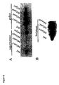


Classifications
-
- C—CHEMISTRY; METALLURGY
- C07—ORGANIC CHEMISTRY
- C07K—PEPTIDES
- C07K14/00—Peptides having more than 20 amino acids; Gastrins; Somatostatins; Melanotropins; Derivatives thereof
- C07K14/195—Peptides having more than 20 amino acids; Gastrins; Somatostatins; Melanotropins; Derivatives thereof from bacteria
- C07K14/24—Peptides having more than 20 amino acids; Gastrins; Somatostatins; Melanotropins; Derivatives thereof from bacteria from Enterobacteriaceae (F), e.g. Citrobacter, Serratia, Proteus, Providencia, Morganella, Yersinia
- C07K14/245—Escherichia (G)
-
- A—HUMAN NECESSITIES
- A61—MEDICAL OR VETERINARY SCIENCE; HYGIENE
- A61P—SPECIFIC THERAPEUTIC ACTIVITY OF CHEMICAL COMPOUNDS OR MEDICINAL PREPARATIONS
- A61P1/00—Drugs for disorders of the alimentary tract or the digestive system
-
- A—HUMAN NECESSITIES
- A61—MEDICAL OR VETERINARY SCIENCE; HYGIENE
- A61K—PREPARATIONS FOR MEDICAL, DENTAL OR TOILETRY PURPOSES
- A61K38/00—Medicinal preparations containing peptides
-
- A—HUMAN NECESSITIES
- A61—MEDICAL OR VETERINARY SCIENCE; HYGIENE
- A61K—PREPARATIONS FOR MEDICAL, DENTAL OR TOILETRY PURPOSES
- A61K39/00—Medicinal preparations containing antigens or antibodies
Definitions
- EAEC Enteroaggregative Escherichia coli
- the pathogenesis of EAEC infection includes strong adherence to the intestinal mucosa, most likely to both the small and the large intestines, followed by secretion of one or more enterotoxins that induce cytopathic effects in intestinal epithelial cells.
- EAEC defining feature of EAEC is its distinctive characteristic aggregative adherence (AA) pattern to HEp-2 cells in culture.
- AA characteristic aggregative adherence
- EAEC adhere to the surface of HEp-2 cells, to the glass substratum, and to each other in a distinctive stacked-brick formation.
- Aggregating adherence to HEp-2 cells in well-characterized strains requires the expression of one or more members of the plasmid-borne Aggregative Adherence Fimbriae (AAF) family.
- AAF Aggregative Adherence Fimbriae
- AggR-dependent gene lying immediately upstream of AggR in EAEC 042 has been identified and characterized.
- This AggR-dependent gene encodes a secreted 10.2 kD protein, designated Aap, that appears to coat the bacterial surface and promote dispersion of EAEC on the intestinal mucosa.
- Aap has alternatively been designated dispersin. It has been shown that Aap mutants form larger aggregates, fewer individual bacteria, and aggregate more intensely than the wild type.
- Aap partially counteracts AAF-mediated aggregation and may play a fundamental role in EAEC pathogenesis.
- the data suggest that Aap binds non-covalently to the bacterial cell surface.
- the mechanism by which Aap is translocated across the outer membrane is as yet unknown.
- the nucleotide sequence of the AA probe represented a cryptic open reading frame (ORF) located adjacent to the plasmid replicon. ( Nataro et al., Infect Immun 64:4761-4768 (1996 )). Further sequence analysis of the AA probe region revealed five ORFs.
- the predicted protein of one of the ORFs was similar with the ATP-binding cassette (ABC) domain of the ABC transporter, suggesting that the gene cluster is involved in the transport of an unidentified molecule of EAEC. Furthermore, this gene cluster appears to be associated with the translocation of Aap, and the formation of a new transporting system of the virulence factor of EAEC. The five gene cluster was designated aat .
- EAEC infections are medically important in populations around the world. For example, in developing countries, children are often infected with EAEC and can develop prolonged diarrhea, often with significant malnutrition. In industrialized countries, EAEC is an emerging cause of traveler's diarrhea. An effective vaccine to protect against enterotoxigenic E. coli would protect against at least one-half of traveler's diarrhea cases. However, in the past, there has been no effective treatment or prevention of EAEC-induced illnesses.
- Another embodiment of the present invention relates to an isolated nucleotide sequence comprising aap, aatP, aatA, aatB, aaxC, or aatD or a functional fragment thereof.
- Yet another embodiment of the present invention relates to a purified polypeptide sequence encoding Aap, AatP, AatA, AatB, AatC, or AatD or a functional equivalent of Aap, AatP, AatA, AatB, AatC, or AatD.
- a further embodiment of the present invention relates to methods of using the gene encoding for aap, aatP, aatA, aatB, aatC, or aatD, products, and fragments thereof.
- One particular embodiment disclosed herein teaches a method of generating an immune response that includes providing an immunogenic composition that includes Aap, AatP, AatA, AatB, AatC, or AatD, a functional fragment thereof, or a product thereof to a subject and contacting the subject with the immunogenic composition, which generates an immune response in the subject.
- Another embodiment of the present invention relates to a method of producing a polypeptide product from a polynucleotide encoding aap, aatP, aatA, aatB, aatC, or aatD, or functional fragment thereof, that includes providing aap, aatP, aatA, aatB, aatC, or aatD in an expression vector, introducing the expression vector into a host cell such that a recombinant host cell is produced, and subjecting the recombinant host cell to conditions such that a protein from aap, aatP, aatA, aatB, aatC, or aatD is expressed.
- An additional embodiment of the present invention relates to cells comprising recombinant aap, aatP, aatA, aatB, aatC, or aatD, or fragments thereof, and expression vectors comprising aap, aatP, aatA, aatB, aatC, or aatD, or fragments thereof.
- Yet another embodiment of the present invention relates to antibodies and antibody fragments that bind to and recognize Aap, AatP, AatA, AatB, AatC, or AatD, or fragments of Aap, AatP, AatA, AatB, AatC, or AatD.
- a further embodiment of the present invention relates to the diagnosis of diseases caused by enteroaggregative E. coli (EAEC).
- EAEC enteroaggregative E. coli
- the diagnosis of diseases caused by EAEC can be performed by using antibodies or fragments of antibodies that bind to and recognize Aap, AatP, AatA, AatB, AatC, or AatD, or fragments of Aap, AatP, AatA, AatB, AatC, or AatD.
- An alternative embodiment of the present invention relating to the diagnosis of diseases caused by EAEC relates to the use of polynucleotides that are complementary to aap, aatP, aatA, aatB, aatC, or aatD, or portions of aap, aatP, aatA, aatB, aatC, or aatD to diagnose the disease caused by EAEC.
- kits that utilize antibodies or fragments of antibodies that bind to and recognize Aap, AatP, AatA, AatB, AatC, or AatD or fragments of Aap, AatP, AatA, AatB, AatC, or AatD, kits that utilize antibodies or other proteins ( i.e., Aap, AatP, AatA, AatB, AatC, or AatD), and kits that utilize polynucleotides that bind to and recognize aap, aatP, aatA, aatB, aatC, or aatD, or fragments of aap, aatP, aatA, aatB, aatC, or aatD.
- kits can be used for the diagnosis of disease caused by EAEC and for diagnosis of EAEC infections.
- isolated and purified polynucleotide molecules encoding the Aap protein are capable of hybridizing under moderate to stringent conditions to an oligonucleotide of 15 or more contiguous nucleotides of SEQ ID NO: 1 or its complementary strand.
- isolated and purified polynucleotide molecules encoding the AatP protein are capable of hybridizing under moderate to stringent conditions to an oligonucleotide of 15 or more contiguous nucleotides of SEQ ID NO: 9 or its complementary strand.
- isolated and purified polynucleotide molecules encoding the AatB protein are capable of hybridizing under moderate to stringent conditions to an oligonucleotide of 15 or more contiguous nucleotides of SEQ ID NO: 11 or its complementary strand.
- isolated and purified polynucleotide molecules encoding the AatC protein are capable of hybridizing under moderate to stringent conditions to an oligonucleotide of 15 or more contiguous nucleotides of SEQ ID NO: 12 or its complementary strand.
- isolated and purified polynucleotide molecules encoding the AatD protein are capable of hybridizing under moderate to stringent conditions to an oligonucleotide of 15 or more contiguous nucleotides of SEQ ID NO: 13 or its complementary strand.
- Figure 2 illustrates the amino acid sequence of Aap (SEQ ID NO:2)
- Figure 3 is a map of 7 kb fragment of the pAA plasmid of EAEC 042 showing the location of the aat gene cluster;
- Figure 4A is a photograph of the SDS-PAGE of culture media precipitated with trichloroacetic acid of wild type and mutant aat;
- Figure 4B is a photograph of the SDS-PAGE of the supernatant of the culture with 0.1 % Triton X-100 of wild type and mutant aat ;
- Figure 5 is a photograph the Western Immunoblot of 042 and 042 aat A in supernatant and cell pellet;
- Figure 6A is a photograph of a Western Immunoblot of secreted Aat in the supernatant and cell pellet, and illustrates different levels of expression in wild type, 042aatC and 042 aatD, but not in 042 pet;
- Figure 7A is a photograph of a Western Immunoblot of whole cells and the outer membrane of 042 aatA (pJNW);
- Figure 7B is a photograph of a Western Immunoblot analysis for Aap in the supernatant of 042, 042 aatA, and 042 aatA (pJNW);
- Figure 9 illustrates the nucleotide sequence of the 7 kb fragment of pAA of EAEC 042 (SEQ ID NO: 3);
- Figure 10 illustrates the amino acid sequence of AatP (SEQ ID NO: 4).
- Figure 11 illustrates the amino acid sequence of AatA (SEQ ID NO: 5).
- Figure 12 illustrates the amino acid sequence of AatB (SEQ ID NO: 6).
- Figure 13 illustrates the amino acid sequence of AatC (SEQ ID NO: 7).
- Figure 14 illustrates the amino acid sequence of AatD (SEQ ID NO: 8).
- Figure 15 illustrates the nucleotide sequence of aatP (SEQ ID NO: 9).
- Figure 16 illustrates the nucleotide sequence of aatA (SEQ ID NO: 10).
- Figure 17 illustrates the nucleotide sequence of aatB (SEQ ID NO: 11).
- Figure 18 illustrates the nucleotide sequence of aatC (SEQ ID NO: 12).
- Figure 19 illustrates the nucleotide sequence of aatD (SEQ ID NO: 13).
- This invention covers Aap (for A nti- a ggregation p rotein, also called dispersin), AatP, AatA, AatB, AatC, and AatD which are all proteins found in enteroaggregative E. coli (EAEC).
- This invention also covers the genes for these proteins ( aap, aatP, aatA, aatB, aatC, and aatD ) and fragments of the genes and proteins, and use of these proteins, genes, and fragments thereof for diagnosis and therapy of EAEC infections.
- the following genes are also called the aat gene cluster: aatP, aatA, aatB, aatC, and aatD.
- Aap is secreted from enteroaggregative E. coli (EAEC) and modulates the strong aggregative phenotype of EAEC on the intestinal mucosa.
- EAEC enteroaggregative E. coli
- the secretion of Aap does not negatively affect EAEC fimbrial expression or fimbrial biogenesis.
- Aap is a 116 amino acid protein, has a molecular weight of about 10.2 kilodaltons (kDa) as determined by SDS-PAGE, a pI of approximately 9.25, and an amino acid sequence illustrated in Figure 2 (SEQ ID NO: 2).
- SIGNALP analysis strongly predicts a signal sequence with cleavage after position 21.
- An N-terminal amino acid sequence of Aap from bacterial culture supernatants confirms this cleavage.
- aap is 348 base-pairs (bp) in length and has the polynucleotide sequence illustrated in Figure 1 (SEQ ID NO: 1).
- aap is located on the pAA plasmid (ca. 100 kp) of prototype EAEC strain 042 (EAEC 042) upstream of the aggR gene which is located downstream of the fimbrial subunit ( aafA ).
- Sequencing of the pAA plasmid DNA upstream of aggR revealed an open reading frame 843 nucleotides upstream from the aggR start codon. This open reading frame was 348 nucleotides in length and encoded a predicted protein product of 116 amino acids.
- the pAA plasmid not only contains putative virulence factors AAF, Aap (dispersin), Pet, EAST1, and AggR but also contains sequences homologous to an empirically derived probe sensitive to EAEC strains (the "AA probe").
- the region comprising the AA probe i.e., the insert of plasmid pCVD432 has been shown to be associated with pathogenic EAEC strains and to correlate with the presence of plausible virulence factors.
- This cluster of ORF's is known as the aat gene cluster or aat cluster because two of genes in this cluster exhibit significant homology to bacterial ABC transporter proteins thus is short for entero a ggregative A BC t ransporter.
- the aat gene cluster features very low G + C ratios, ranging from 29.6 to 34.3.
- the aat gene cluster contains aatP, aatA, aatB, aatC, and aatD. Flanking the aat gene cluster at distances of over 500 bp on each side are remnants of IS sequences.
- AatB The nucleotide sequence of aatB is shown in Figure 17 (SEQ ID NO: 11).
- the amino acid sequence for AatB is shown in Figure 12 (SEQ ID NO: 6).
- AatB is a predicted protein of 274 amino acids (31.1 kDa) and runs from nucleotide 3687 to 4508 of the 7 kb fragment from pAA as shown in Figure 9 (SEQ ID NO: 3).
- AatB does not have any significant homology to any known protein sequences in the GenBank library.
- the protein has a transmembrane region in the N-terminus and is believed to be an inner membrane protein.
- AatD The nucleotide sequence of aatD is shown in Figure 19 (SEQ ID NO: 13).
- the amino acid sequence for AatD is shown is Figure 14 (SEQ ID NO: 8).
- AatD is a predicted protein of 405 amino acids (47.2 kDa) and runs from nucleotide 5142 to 6356 of the 7 kb fragment from pAA as shown in Figure 9 (SEQ ID NO: 3).
- AatD is 29 % identical at the amino acid level over 34 % of its length to NADH dehydrogenase subunit 2.
- AatD has five transmembrane regions in the N-terminal and is believed to be an inner membrane protein.
- additional polynucleotide molecules that encode the proteins discussed above include those polynucleotide sequences resulting in minor genetic polymorphisms, differences between strains, degenerate variants, and encoding for proteins that contain amino acid substitutions, additions, and/or deletions.
- Nucleotide sequences encoding aap and/or the aat gene cluster can be used to identify polynucleotide molecules encoding other proteins with biological functions similar to that of Aap and/or the proteins of the aat gene cluster by screening a cDNA library or DNA library from other organisms (prokaryotic or eukaryotic). These new DNA molecules can be isolated from such a library using the polynucleotide sequence disclosed herein with standard hybridization techniques or by the amplification of sequences using polymerase chain reaction (PCR) amplification. Suitable probes for use in identifying protein homolog sequences can be obtained from gene-specific sequences. Alternatively, oligonucleotides containing specific DNA sequences from coding region for these genes can be used to identify related clones.
- PCR polymerase chain reaction
- Homologous polynucleotide molecules can be isolated using standard hybridization techniques with probes of at least about 7 nucleotides, more preferably 15 nucleotides, in length (but can be as much as the full coding sequence). Homologous polynucleotide sequences can be identified using degenerate oligonucleotides based on the sequences disclosed herein which are capable of hybridization at moderate or greater stringency.
- the term, "capable of hybridization" as used herein means that the subject nucleic acid molecules (whether DNA or RNA) anneal to an oligonucleotide of 15 or more contiguous nucleotides of one of the polynucleotide sequences disclosed herein.
- hybridization stringency can be altered, for example, by adjusting the temperature of hybridization, adjusting the percentage of helix-destabilizing agents (e.g., formamide) in the hybridization mix, and adjusting the temperature and salt concentration of the wash solutions. In general, the stringency of hybridization is adjusted during post-hybridization washes by varying the salt concentration and/or the temperature, which results in progressively higher stringency conditions.
- An example of progressively higher stringency conditions is as follows: 2 x SSC/0.1% SDS at about room temperature (hybridization conditions); 0.2 x SSC/0.1% SDS at about room temperature (low stringency conditions); 0.2 x SSC/0.1% SDS at about 42 °C (moderate stringency conditions); and 0.1 x SSC at about 68°C (high stringency conditions). Washing can be carried out using only one of these conditions, e.g., high stringency conditions, or each of the conditions can be used, e.g. , for 10-15 minutes each, in the order listed above, repeating any or all of the steps listed. Optimal conditions will vary depending on the particular hybridization reaction involved, and can be determined empirically. Conditions of high stringency are preferably used for the hybridization of the probe of interest.
- polynucleotides having substantially the same nucleotide sequence as or are substantially identical to one of the polynucleotide sequences of aap or the aat gene cluster represent aap -like or aat gene cluster-like genes.
- polynucleotides which encode for proteins that are functionally equivalent of, or functional fragments of Aap or the proteins of the aat gene cluster are included in this invention.
- “Substantially the same” or “substantially identical” is meant a nucleic acid, polynucleotide, or polypeptide exhibiting at least 80%, 85%, 90%, 91%, 92%, 93%, 94%, 95%, 96%, 97%, 98%, 99%, or 100% homology to a reference nucleic acid, polynucleotide, or polypeptide.
- the length of comparison sequences will generally be at least 10 to 500 nucleotides in length.
- the length of comparison will be at least 50 nucleotides, more preferably at least 60 nucleotides, even more preferably at least 75 nucleotides, and most preferably at least 110 nucleotides in length.
- an "isolated" nucleic acid is a nucleic acid the structure of which is not identical to that of any naturally occurring nucleic acid or to that of any fragment of a naturally occurring genomic nucleic acid spanning more than three separate genes.
- the term therefore covers, for example, (a) a DNA which has the sequence of part of a naturally occurring genomic DNA molecule but is not flanked by both of the coding sequences that flank that part of the molecule in the genome of the organism in which it naturally occurs; (b) a nucleic acid incorporated into an expression vector or into the genomic DNA of a prokaryote or eukaryote in a manner such that the resulting molecule is not identical to naturally occurring expression vector or genomic DNA; (c) a separate molecule such as cDNA, a genomic fragment, a fragment produced by polymerase chain reaction (PCR), or a restriction fragment; and (d) a hybrid gene, i.e., a gene encoding a fusion protein.
- PCR polyme
- nucleic acids present in mixtures of (i) DNA molecules, (ii) transfected cells, and (iii) cell clones, e.g., as these occur in a DNA library such as a cDNA or genomic DNA library.
- DNA sequences of the invention can be obtained by several methods.
- the DNA can be isolated using hybridization or computer-based techniques that are well-known in the art.
- Such techniques include, but are not limited to, the hybridization of genomic or cDNA libraries with probes to detect homologous nucleotide sequences; antibody screening of expression libraries to detect cloned DNA fragments with shared structural features; polymerase chain reaction (PCR) on genomic DNA or cDNA using primers capable of annealing to the DNA sequence of interest; computer searches of sequence databases for similar sequences; and differential screening of a subtracted DNA library.
- Oligonucleotide probes which correspond to a part of the sequences for aap or the aat gene cluster which are provided herein, can be synthesized chemically. Synthesis of other oligonucleotide probes may require that short, oligo-peptide stretches of the amino acid sequence be known because the DNA sequence encoding a specific protein can be deduced using the genetic code; however, the degeneracy of the code must be taken into account.
- Another standard procedure for isolating DNA sequences of interest is the formation of plasmid- or phage-carrying genomic libraries which include total DNA from the organism of interest. When used in combination with polymerase chain reaction technology, even rare expression products can be cloned. In those cases where significant portions of the amino acid sequence of the polypeptide are known, the production of labeled single or double-stranded DNA or RNA probe sequences duplicating a sequence putatively present in the target DNA can be employed in DNA/DNA hybridization procedures which are carried out on cloned copies of the DNA that have been denatured into a single-stranded form. ( Jay et al., Nucl. Acid Res., 11:2325 (1983 ).
- nucleotide sequences of aap and the aat gene cluster have a myriad of applications. Representative uses of these nucleotide sequences include the construction of DNA and oligonucleotide probes useful in Northern, Southern, immuno-PCR, dot-blot, and other assays for detecting EAEC, quantifying the level of expression of Aap, AatP, AatA, AatB, AatC, or AatD in a cell, or generating the particular protein or polypeptide which is encoded by the nucleotide sequence.
- aap and the aat gene cluster nucleotide sequences can be employed for the construction of recombinant cell lines, recombinant organisms, expression vectors, and the like.
- Such recombinant constructs can be used to express recombinant Aap, AatP, AatA, AatB, AatC, and/or AatD, and can be used as vaccines (via a live, attenuated vector vaccine or other type of invasive vector vaccine) or to screen for candidate therapeutic agents capable of altering the pathology of an organism expressing one or more these proteins.
- the proteins or polypeptides of this invention are highly useful in the generation of immunogenic compositions that can be used to generate an immune response in a subject (e.g., via a subunit vaccine) and in kits for the detection of EAEC.
- a subunit vaccine one or more of the proteins of this invention can be expressed, purified, and used to prepare an immunogenic subunit composition.
- a live, attenuated vector vaccine one or more of the proteins of this invention can also be expressed in an attenuated, invasive bacteria or virus, and the whole organism can be formulated into an immunogenic composition.
- one or more of the genes of this invention can be placed on a plasmid downstream of a signal sequence of an eukaryotic promoter. That plasmid can contain one or more selectable markers and be transfected into a prokaryotic organism, such as Salmonella spp., Shigella spp., or other suitable bacteria. The bacteria is then administered to the eukaryotic subject for which immune response to EAEC is desired. See, for example, U.S. Patent No. 5,887,159 to Hone, et al. Additionally, a polynucleotide encoding one of the proteins of this invention can be administered to the mucosal tissue of a subject to generate an immunogenic response. See, for example, U.S.
- a naked polynucleotide encoding one of the proteins of this invention can be electroporated into a subject to generate an immune response using the methods described within Drabick, J.J.; et al.; Cutaneous transfection and immune responses to intradermal nucleic acid vaccination are significantly enhanced by in vivo electropermeabilization, Mol. Ther. Vol 3(2); pp.249-55 (2001 ).
- genes of this invention can also be placed into expression vectors which are discussed more fully below.
- Antisense oligonucleotides can be prepared, for example, by inserting a DNA molecule containing the target DNA sequence into a suitable expression vector such that the DNA molecule is inserted downstream of a promoter in a reverse orientation as compared to a particular gene of this invention.
- the expression vector can then be transduced, transformed, or transfected into a cell (prokaryotic and/or eukaryotic) suitable for expressing the antisense oligonucleotides.
- antisense oligonucleotides can be synthesized using standard manual or automated synthesis techniques. These synthesized oligonucleotides are introduced into suitable cells by a variety of means including electroporation, calcium phosphate precipitation, and microinjection. The selection of a suitable antisense oligonucleotide administration method would be evident to one of ordinary skill in the art.
- the stability of antisense oligonucleotide mRNA hybrids is advantageously increased by the addition of stabilizing agents to the oligonucleotide.
- a stabilizing agent includes intercalating agents that are covalently attached to either or both ends of the oligonucleotide.
- the oligonucleotides are made resistant to nucleases by modifications to the phosphodiester backbone by the introduction of phosphotriesters, phosphonates, phosphorothioates, phosphoroselenoates, phosphoramidates, phosphorodithioates, or morpholino rings.
- Aap encompass the whole protein (Aap, AatP, AatA, AatB, AatC, and AatD respectively), fragments of the protein (Aap, AatP, AatA, AatB, AatC, and AatD respectively) that are functionally active, and polypeptides that contain substitutions such as one basic amino acid for another basic amino acid, or one acidic amino acid for another acid amino acid, or one neutral amino acid for another neutral amino acid.
- Aap also encompass Aap, AatP, AatA, AatB, AatC, and AatD respectively purified from naturally occurring materials and closely related, functionally similar proteins retrieved by antisera specific to the particular protein.
- Recombinantly expressed proteins encoded by genetic materials (DNA, RNA, cDNA) retrieved on the basis of their similarity to regions in the particular gene sequence are also encompassed by the present description.
- Polynucleotide molecules encoding Aap, AatP, AatA, AatB, AatC, and AatD include molecules that encode Aap, AatP, AatA, AatB, AatC, and AatD, respectively, or peptides that share identity with the sequence shown in SEQ ID NO: 2 ( Figure 2 ); SEQ ID NO: 4 ( Figure 10 ); SEQ ID NO: 5 ( Figure 11 ); SEQ ID NO: 6 ( Figure 12 ); SEQ ID NO: 7 ( Figure 13 ), and SEQ ID NO: 8 ( Figure 14 ) respectively. These molecules preferably share greater than 30% identity at the amino acid level with the disclosed protein.
- the polynucleotide molecules share greater identity at the amino acid level across highly conserved regions.
- Variants of a particular gene encoded protein include those amino acid sequences resulting from minor genetic polymorphisms, differences between strains, and those that contain amino acid substitutions, additions, and/or deletions, and conservative amino acid substitutions.
- a conservative amino acid substitution includes a replacement of one amino acid residue with a different residue having similar biochemical characteristics, such as size, charge, and polarity vs. nonpolarity.
- sequence analysis software e.g., Sequence Analysis Software Package of the Genetics Computer Group, University of Wisconsin Biotechnology Center, 1710 University Avenue, Madison, WI 53705 or the NCBI BLAST program.
- sequence analysis software e.g., Sequence Analysis Software Package of the Genetics Computer Group, University of Wisconsin Biotechnology Center, 1710 University Avenue, Madison, WI 53705 or the NCBI BLAST program.
- sequence analysis software e.g., Sequence Analysis Software Package of the Genetics Computer Group, University of Wisconsin Biotechnology Center, 1710 University Avenue, Madison, WI 53705 or the NCBI BLAST program.
- substantially identical also means an amino acid sequence which differs only by conservative amino acid substitutions, for example, substitution of one amino acid for another of the same class (e.g., valine for glycine, arginine for lysine, etc. ) or by one or more non-conservative substitutions, deletions, or insertions located at positions of the amino acid sequence that do not destroy the function of the protein assayed, (e.g., as described herein).
- such a sequence is at least 85%, and more preferably from 90%, 91%, 92%, 93%, 94%, 95%, 96%, 97%, 98%, 99%, to 100% homologous at the amino acid level to sequences described herein.
- fragments include fragments of the proteins described herein that have a similar amino acid sequence and that retain the function or activity of the particular protein. Where full-length protein is described, one of skill in the art can screen for the functionality of a fragment by using the examples provided herein.
- substantially pure polypeptide means an polypeptide that has been separated from components that naturally accompany it.
- the polypeptide is substantially pure when it is at least 60%, by weight, free from the proteins and other naturally occurring molecules with which it is typically associated.
- the preparation is at least 75%, 80%, 90%, 95%, and most preferably at least 99%, by weight.
- a substantially pure protein can be obtained, for example, by extraction from a natural source, by expression of a recombinant nucleic acid encoding a particular protein, or by chemically synthesizing the protein. Purity can be measured by any appropriate method, such as column chromatography, polyacrylamide gel electrophoresis, and HPLC analysis.
- a protein is substantially free of naturally associated components when it is separated from those contaminants that accompany it in its natural state.
- a protein that is chemically synthesized or produced in a cellular system different from the cell or host from which it naturally originates is substantially free from its naturally associated components.
- substantially pure polypeptides include those derived from prokaryotic organisms but synthesized in eukaryotic organisms.
- a purified polypeptide is a polypeptide substantially free of naturally associated components when it is separated from those contaminants that accompany it in its natural state.
- polynucleotide molecules according to the present disclosure can be expressed in a variety of prokaryotic and eucaryotic cells using regulatory sequences, expression vectors, and methods well established in the literature to produce a polypeptide or protein which can be purified.
- Purification of a protein of this invention can be performed using a number of established methods such as affinity chromatography using antibodies specific to a particular protein coupled to a solid support. Fusion proteins of an antigenic tag and the protein of interest can be purified using antibodies to the tag. Optionally, additional purification is achieved using conventional purification means such as liquid chromatography, gradient centrifugation, or gel electrophoresis. Methods of protein purification are well known in the art and can be applied to the purification of recombinant proteins described herein. The purification of the proteins of this invention is discussed in more detail below.
- One embodiment of the present invention involves the isolation of proteins that interact with the proteins described herein or are receptors for the proteins described herein.
- Aap, AatP, AatA, AatB, AatC, and/or AatD can be used in immunoprecipitation to isolate interacting factors or for the screening of interactors using different methods of two hybrid screening. Isolated interactors of these proteins can be used to modify or block activity of the particular protein in a host or between EAEC.
- Synthetic peptides recombinantly derived peptides, fusion proteins, chiral proteins (stereochemical isomers, racemates, enantiomers, and D-isomers), and the like are provided which include a portion of one of the proteins described herein or the entire protein.
- the subject peptides have an amino acid sequence encoded by a nucleic acid which hybridizes under stringent conditions with an oligonucleotide of 15 or more contiguous nucleotides of SEQ ID NO: 1; SEQ ID NO: 9; SEQ ID NO: 10; SEQ ID NO: 11; SEQ ID NO: 12; or SEQ ID NO: 13.
- Representative amino acid sequences of the subject peptides is disclosed in SEQ ID NO: 2; SEQ ID NO: 4, SEQ ID NO: 5; SEQ ID NO: 6 ; SEQ ID NO: 7; or SEQ ID NO: 8.
- the subject peptides find a variety of uses, including preparation of specific antibodies and preparation of antagonists of activity the proteins described herein.
- the isolated or purified protein can be used to generate antibodies and tools for identifying agents that interact with that particular protein and fragments of interest.
- the term "antibodies” can encompass polyclonal, monoclonal, chimeric, single chain, Fab fragments, and fragments produced by a Fab expression library.
- Antibodies that recognize Aap, AatP, AatA, AatB, AatC, and/or AatD and fragments thereof have many uses including, but not limited to, biotechnological applications, therapeutic/prophylactic applications, and diagnostic applications.
- Peptides used to induce specific antibodies have an amino acid sequence of at least three amino acids, and preferably an amino acid sequence of at least 10 to 15 amino acids. Preferably, short stretches of amino acids encoding fragments of a particular protein are fused with those of another protein such as keyhole limpet hemocyanin such that an antibody is produced against the chimeric molecule.
- antibodies capable of specifically recognizing a particular protein of this invention can be generated by injecting synthetic 3-mer, 10-mer, and 15-mer peptides that correspond to an amino acid sequence of that particular protein into a host (e.g., mice), a more diverse set of antibodies can be generated by using a particular recombinant protein, a particular purified protein, or fragments of a particular protein.
- a substantially pure Aap, AatP, AatA, AatB, AatC, AatD, or a fragment thereof is isolated from a transfected or transformed cell or the wildtype EAEC.
- concentration of the polypeptide in the final preparation is adjusted, for example, by concentration on an Amicon filter device, to the level of a few micrograms/ml.
- Monoclonal antibodies to Aap, AatP, AatA, AatB, AatC, AatD, or a fragment thereof can be prepared using any technique that provides for the production of antibody molecules by continuous cell lines in culture.
- Such techniques include, but are not limited to, the hybridoma technique originally described by Koehler and Milstein (Nature 256:495-497 (1975 )), the human B-cell hybridoma technique ( Kosbor et al., Immunol Today 4:72 (1983 ); Cote et al., Proc Natl. Acad. Sci 80:2026-2030 (1983 )), and the EBV-hybridoma technique ( Cole et al., Monoclonal Antibodies and Cancer Therapy, Alan R.
- Antibody fragments that contain specific binding sites for a particular protein of this invention can also be generated.
- fragments include, but are not limited to, F(ab') 2 fragments produced by pepsin digestion of the antibody molecule and Fab fragments generated by reducing the disulfide bridges of the F(ab') 2 fragments.
- Fab expression libraries can be constructed to allow rapid and easy identification of monoclonal Fab fragments with the desired specificity. ( Huse W. D. et al., Science 256:1275-1281 (1989 )).
- Antibody-producing clones are identified by the detection of antibody in the supernatant fluid of the wells by immunoassay procedures such as ELISA, as originally described by Engvall, E., Meth. Enzymol. 70:419 (1980 ), and derivative methods thereof. Selected positive clones can be expanded and their monoclonal antibody product harvested for use. An example of a detailed procedure for monoclonal antibody production can be found in Davis, L. et al., Basic Methods in Molecular Biology Elsevier, New York. Section 21-2 .
- Polyclonal antiserum containing antibodies to heterogeneous epitopes of a single protein can be prepared by immunizing suitable animals with the expressed protein or peptides derived therefrom, which can be unmodified or modified to enhance immunogenicity.
- Effective polyclonal antibody production is affected by many factors related both to the antigen and to the host species. For example, small molecules tend to be less immunogenic than larger molecules and can require the use of carriers and adjuvants.
- host animals vary in response to the site(s) of inoculations and dose, with both inadequate or excessive doses of antigen resulting in low titer antisera. Small doses (e.g., ng level) of antigen administered at multiple intradermal sites appears to be most reliable.
- An example of an effective immunization protocol for rabbits can be found in Vaitukaitis, J. et al., J. Clin. Endocrinol. Metab. 33:988-991 (1971 ).
- Antibody preparations prepared according to either protocol are useful in quantitative immunoassays that determine concentrations of antigen-bearing substances in biological samples. These antibody preparations are also used semi-quantitatively or qualitatively (e.g., in diagnostic embodiments that identify the presence of Aap in biological samples).
- Recombinant gene expression vectors containing any of the genes of this invention, or portions thereof, can be constructed in a variety of forms well-known in the art.
- Preferred expression vectors include plasmids and cosmids.
- Expression vectors include one or more fragments of a particular gene and preferably comprise the full length gene.
- An expression vector containing one or more of the genes of this invention can be used to transfect or transform a suitable host cell (prokaryotic or eukaryotic) to produce the protein or to produce an immune response, or for some other purpose.
- operatively encode refers to one or more protein coding regions associated with those regulatory sequences required for expression of the polypeptide encoded by the coding region.
- regulatory regions include promoter binding sites, enhancer elements, ribosome binding sites, and the like.
- suitable regulatory sequences for use in various eukaryotic and prokaryotic systems are described in Ausubel, et al., Short Protocols in Molecular Biology, 3rd ed., John Wiley & Sons, Inc, New York, 1997 , which is hereby incorporated by reference in its entirety.
- Expression vectors for use with the aap gene or the aat gene cluster typically contain regulatory sequences derived from a compatible species for expression in the desired host cell.
- E. coli when E. coli is the host cell, the host cell population is typically transformed using pBR322, a plasmid derived from an E. coli species that contains genes for ampicillin (AMPR) and tetracycline resistance.
- AMPR ampicillin
- tetracycline resistance Bolivar, et al., Gene 2:95 (1977 )
- pBR322 provides an easy means for identifying transformed cells.
- the plasmids described in Galen, WO 00/32047 are also useful for expression vectors in some bacteria.
- Promoters suitable for use with prokaryotic hosts illustratively include the beta-lactamase and lactose promoter systems ( Chang, et al., Nature 275:617 (1978 ); Goeddel, et al., Nature 281:544 (1979 )), alkaline phosphatase, the tryptophan (trp) promoter system ( Goeddel, Nucleic Acids Res. 8:4057 (1980 )), ompC, nirB, and hybrid promoters such as the taq promoter ( de Boer, et al., Proc. Natl. Acad. Sci. USA 80:21-25 (1983 )).
- Eukaryotic microbes such as yeast cultures can also be used as sources for the regulatory sequences.
- Saccharomyces cerevisiae is a commonly used eukaryotic host microorganism.
- Suitable promoting sequences for use with yeast hosts include the promoters for 3-phosphoglycerate kinase ( Hitzeman, et al., J. Biol. Chem.
- glycolytic enzymes such as enolase, glyceraldehyde-3-phosphate dehydrogenase, hexokinase, pyruvate decarboxylase, phosphofructokinase, glucose-6-phosphate isomerase, 3-phosphoglycerate mutase, pyruvate kinase, triosephosphate isomerase, phosphoglucose isomerase, and glucokinase ( Hess, et al. J. Adv. Enzyme Reg. 7:149 (1968 ); Holland, Biochemistry 17:4900 (1978 )).
- yeast promoters which are inducible promoters having the advantage of transcription controlled by growth conditions, include the promoter regions for alcohol dehydrogenase 2, isocytochrome C, acid phosphatase, degraded enzymes associated with nitrogen metabolism, metallothionine, glyceraldehyde-3-phosphate dehydrogenase, and enzymes responsible for maltose and galactose utilization.
- Yeast enhancers are advantageously used with yeast promoters.
- selectable markers are generally introduced into the cells or organisms along with the cloned DNA molecules and include genes that confer resistance to drugs such as ampicillin, neomycin, hygromycin, and methotrexate. Selectable markers can also complement auxotrophies in the host cell, or can provide for detectable signals, such as beta-galactosidase, green fluorescent protein, or yellow fluorescent protein, to identify cells or organisms containing the cloned DNA molecules.
- nucleotide sequences of aap and/or the aat gene cluster, and optionally other immunostimulatory polynucleotides, into viral gene expression vectors can be used to incorporate the sequences into live and attenuated live viruses for use as immunogenic compositions.
- Targeting of mucosal tissues can be performed by exploiting inherent biological properties of the lymphoid bed which is to be targeted. These properties include the crypt architecture of the tonsillar pillars which can be used to entrap particles and also include the M cells of Peyer's patches in the gut which specifically endocytose a wide variety of particles including lipid particles and other small particulates. Therefore, those skilled in the art can prepare a wide variety of molecular particulate preparations which, if provided to the intestine, will lodge within the crypt portions of intestinal Peyer's patches and be endocytosed by M cells. If such particles provide for delivery of a biologically active polynucleotide to M cells, the particles will enable the stimulation or modulation of a mucosal immune response induction by the Peyer's patch lymphoid tissue to which the M cell traffics.
- Suitable expression vectors containing desired coding, non-coding, and control sequences employ standard ligation techniques. Isolated plasmids or DNA fragments are cleaved, tailored, and re-ligated in the form desired to construct the required plasmids. To confirm correct sequences in the plasmids constructed, the ligation mixtures can be used, for example, to transform a host cell and successful transformants selected by antibiotic resistance where appropriate. Plasmids from the transformants are prepared, analyzed by restriction and/or sequenced by, for example, by the method disclosed in Messing, et al. (Nucleic Acids Res., 9:309 (1981 )), Maxam, et al.
- Host cells can be transformed with the expression vectors described herein and cultured in conventional nutrient media modified as is appropriate for inducing promoters, selecting transformants, or amplifying genes.
- the culture conditions such as temperature, pH and the like, are those previously used with the host cell selected for expression, and will be apparent to the ordinarily skilled artisan.
- Steps involved in the purification of one or more of the proteins of this invention include (1) solubilization of the desired protein, (2) the development of one or more isolation and concentration procedures, (3) stabilization of the protein following purification, and (4) development of a suitable assay to determine the presence of the desired protein.
- Various aspects of protein isolation and purification are discussed in detail in Cooper, T. G., "The Tools of Biochemistry,” John Wiley & Sons, New York, 1977 , which is hereby incorporated by reference in its entirety.
- As the techniques of protein isolation and purification are notoriously well known in the art, this disclosure will refrain from discussing them in detail. Nevertheless, elements of the cited reference are summarized and discussed below.
- Solubilization is required of most proteins that are to be purified, as most isolation procedures commonly used operate in aqueous solutions. In some cases, solubilization can be achieved by merely lysing a host cell within which a desired protein has been expressed. In other situations, additional steps, such as extracting the desired protein from a subcellular organelle, may be required. Osmotic lysis, grinding, the use of blenders, ultrasonic waves, presses, and other well known techniques of protein solubilization can be used with the methods disclosed herein.
- Stabilizing and maintaining a purified protein product in a functional state warrants attention to a number of different conditions such as (1) pH, (2) degree of oxidation, (3) heavy metal concentration, (4) medium polarity, (5) protease concentration, and (6) temperature.
- the proteins encoded by aap and the aat gene cluster can be used to formulate immunogenic compositions that facilitate an immune response.
- Examples of a typical immune response to EAEC infections include a mucosal immune response and a systemic immune response.
- smaller fragments of the proteins of this invention are used to provide an immunogenic composition.
- these fragments comprise an immunogenic region of expression products, typically from about 5, 6, 8, 10 or 12 amino acids to about 20, 22, 24, 30, or more amino acids.
- Suitable fragments or immunogenic regions can be readily ascertained using the techniques set forth below as screening procedures. In one example of a suitable screening procedure, a large number of candidate fragments are more or less randomly produced and used to immunize guinea pigs or other suitable models.
- full-length polypeptides shown to be active in the present invention can be truncated and screened in an iterative process to isolate the immunogenic and protective activity to a minimal fragment.
- a nucleotide sequence comprising the aap gene or a functional fragment thereof or one or more of the genes or fragments thereof of the aat gene cluster is introduced into an exogenous organism using standard molecular biology techniques well known to those of ordinary skill in the art. Exemplary molecular biology techniques are discussed in Ausubel, et al., "Short Protocols in Molecular Biology.” The resulting recombinant organism can then be used as an immunogen against which an immune response may be engendered. In a preferred embodiment, an attenuated pathogenic organism serves as the exogenous organism.
- Another embodiment of the present invention relates to the generation of immunogenic compositions comprising distinct immunogenic proteins (or fragments thereof) or functional fragments of the organisms of interest.
- immunogenic compositions are referred to herein as subunit immunogenic compositions because at least one of the components of the composition is a subunit of an organism, rather than an entire organism.
- subunit immunogenic composition comprises one or more immunogenic components.
- the subunit immunogenic composition includes a carrier component and an immunogenic component.
- the carrier component functions as a binding moiety with which the originating organism uses to bind to and gain entrance into the host organism.
- Aap, AatP, AatA, AatB, AatC, and/or AatD from EAEC functions as the carrier component. Any protein, peptide, or amino acid sequence that elicits an immune response can be used as the immunogenic component in a subunit immunogenic composition.
- the carrier component of the subunit immunogenic composition can possess immunogenic characteristics themselves.
- adjuvants are used in immunogenic compositions to enhance the immune response directed against the immunogenic component of the immunogenic compositions.
- Carrier components that possess both mucosa binding characteristics and immunogenic characteristics can be used. For example, Aap, AatP, AatA, AatB, AatC, and/or AatD can function both as the carrier component and the immunogenic component.
- the entire molecule can be used as the carrier component, or a functionally active fragment of the molecule can be used. Mutagenized forms of these molecules can also be used as carrier components.
- the subunit immunogenic composition functions by exposing the immunogenic component of the subunit immunogenic composition to the mucosa and the various immune system components present there.
- the generation of a desired immune response by the subunit immunogenic composition occurs by increasing the exposure of the immunogenic composition to the target tissue. The presence of both a carrier component and an immunogenic component are theorized to achieve this goal.
- Aap is isolated, purified, and mixed or coupled with one or more immunogenic or adjuvant compounds.
- Aap can be expressed, purified, and cross-linked to a toxin, toxoid (an attenuated toxin), or some other immunogenic compound for use in a subunit immunogenic composition.
- AatA can be expressed, purified, and cross-linked to a toxin, toxoid (an attenuated toxin), or some other immunogenic compound for use in a subunit immunogenic composition.
- expressed Aap or fragments thereof or AatA or fragments thereof can be cross-linked to an immunogenic component, which can be an isolated protein, a functional fragment thereof, a whole organism (such as a bacterium or a virus) or functional fragment thereof that is isolated either in part or is used as a whole pathogenic organism.
- Aap and AatA can be isolated from the bacterium itself or it can be produced using recombinant DNA techniques well known in the art. It is contemplated, and within the purview of the present invention, that an entire recombinant organism or a functional fragment thereof, such as an isolated membrane fraction, liposome, or the like, can be used to generate an immunogenic composition.
- the proteins of this invention which are used to form the subunit immunogenic compositions can be the whole protein, an immunogenic fragment thereof, a mutagenized form of the protein, or a fusion protein comprising the particular protein or a fragment thereof and a suitable fusion partner (e.g., any protein, peptide or amino acid sequence that facilitates the expression and/or purification of the protein and fusion partner using recombinant DNA techniques known in the art).
- a suitable fusion partner e.g., any protein, peptide or amino acid sequence that facilitates the expression and/or purification of the protein and fusion partner using recombinant DNA techniques known in the art.
- one or more additional immunogens can serve as the fusion partner in a fusion protein.
- an immune response can also be elicited using nucleotide-containing compositions.
- a mucosal or systemic immune response is elicited in a host by administering an antigen-encoding polynucleotide preparation including DNA or RNA that encodes an antigenic epitope to the host.
- the nucleotide-containing composition is administered to a mucosal inductor site in the mucosal tissue of the host.
- Naked DNA may be administered directly to the mucosa (e.g., in saline drops) or in a recombinant gene expression vector.
- the recombinant gene expression vector is not capable of replication or dissemination.
- Balb-C mice (SPF female, 6 week, Charles River) are inoculated with 10 6 virion particles (10 7 /ml) via either intratracheal, intranasal, or intravenous routes. Animals are anesthetized with a cocktail of ketamine (22 mg/kg), xylazine (2.5 mg/kg) and acepromazine (0.75 mg/kg) prior to inoculation. Intratracheal inoculation is performed by making a small medial cut through the skin at the ventral site of the neck. Salivary glands are teased apart using blunt dissection to expose the trachea.
- a 30.5 gauge needle with a 1 cc tuberculin syringe attached is placed through the rings of the trachea toward the bronchi.
- 100 ⁇ l of the above virion particles are injected into the lung.
- Intranasal installation consists of placing 50 ⁇ l of the virion particles into one of the nares. Once this is taken into the nasal passages by inhalation, the other side is inoculated in the same manner.
- Intravenous inoculation is performed using a 30.5 gauge needle with 100 ⁇ l of the above virion particles inserted into the tail vein.
- Blood is collected from the retro-orbital venous plexus at day 0, 14, and 28, using a microcapillary tube.
- the blood is allowed to sit at room temperature for 2-4 hours to clot, then it is spun in a IEC Centra MP4R centrifuge at 6,000 RPM for six minutes.
- the supernatant (serum) is removed and stored at -20°C until ELISA assays are performed.
- Lung lavages at week 4 are performed by the following method. Mice are killed by carbon dioxide asphyxiation. The ventrum is skinned and the mesentery removed which exposes the liver. The liver is moved aside to visualize the diaphragm. The diaphragm is opened and the rib cage is cut bi-laterauy up through the sternum. This section is lifted up over the mouse's head, leaving the trachea exposed. A transverse cut of the trachea is made approximately 2 cm above the bronchus. A blunt 24 gauge needle is inserted 0.5 cm into the trachea and tied in place with surgical thread.
- BBS boric acid, 90 mM NaCl, pH 8.3 [NaOH]
- the solution is centrifuged to produce a cellular (particulate) and a supernatant component.
- Recovered volumes are normally in the range of 0.85 to 0.95 ml. This cellular and supernatant components are stored at -20°C until used.
- Animals for which histological sections are to be taken are killed by carbon dioxide on day two. Their lungs are fixed in paraformaldehyde for thirty minutes, the tissue is then incubated overnight at 4°C in a mix of PBS+2 mM MgCl 2 +30% sucrose. Lung tissue are cryosectioned and placed on gelatinized slides. The slides are then fixed and stained for Aap using an biotinylated labeled anti-Aap antibody.
- ELISA assays are performed on samples using 96 well microtiter plates using the same basic protocol as described in by Engvall, E., Meth. Enzymol. 70:419 (1980 ), and derivative methods thereof.
- Aap is suspended to a concentration of 1 mg/ml with PBS supplemented with 5 mg/ml bovine serum albumin.
- the wells of the microtiter plate are coated with 5 mg of Aap (in that solution) per ml BBS (89 mM boric acid, 90 mM NaCl, pH 8.3 [NaOH]).
- the plates are incubated overnight at 4 °C.
- the plates are dried by pounding on a stack of paper towels and blocked with 150 ⁇ l BB (BBS as above with 1% bovine serum albumin added). After the plates sit at room temperature for 2 hours, eight two-fold dilutions of sample sera in BB are pipetted into the microtiter plates (50 ⁇ l per well). Dilutions are performed as follows: serum IgG and IgA - 1:10 to 1:5120, lavage IgG and IgA - 1:1 to 1:128. The plates are incubated overnight at 4 °C. Plates are washed 5-6 time with BBS plus 0.05% Tween and dried.
- BBS bovine serum albumin
- a Dynatech MR5000 ELISA plate reader with a 405 nm wavelength (Dynatech Laboratories, Chantilly, VA) hooked up to a iMac (having the appropriate software) reads the plates. Background signal is defined using control serum, with positive titer identified at >2.5x background. For lavage samples, OD 405 is reported using the 1:2 dilution, with positive signal defined as 2.5x background.
- one or more of the genes of the aat cluster can be introduced to an attenuated EAEC, Salmonella spp., Shigella spp., Lactobacillus spp., or other attenuated bacteria which is invasive for mucosal tissue, which then expresses the particular Aat protein encoded by the gene.
- the bacteria is administered to an animal to generate an immune response to the particular Aat protein encoded.
- Administration of the immunogenic compositions can be by nasal application, by inhalation, ophthalmically, orally, rectally, vaginally, or by any other mode that results in the immunogenic composition contacting mucosal tissues.
- Solid formulations of the compositions for oral administration may contain suitable carriers or excipients, such as corn starch, gelatin, lactose, acacia, sucrose, microcrystalline cellulose, kaolin, mannitol, dicalcium phosphate, calcium carbonate, sodium chloride, or alginic acid.
- Disintegrators that can be used include, without limitation, microcrystalline cellulose, cornstarch, sodium starch glycolate, and alginic acid.
- Tablet binders that may be used include acacia, methylcellulose, sodium carboxymethylcellulose, polyvinylpyrrolidone, hydroxypropyl methylcellulose, sucrose, starch, and ethylcellulose.
- Lubricants that may be used include magnesium stearates, stearic acid, ailicone fluid, talc, waxes, oil, and colloidal silica.
- the immunogenic compositions described herein can also be formulated in the form of a rectal or vaginal suppository.
- Typical carriers used in the formulation of the inactive portion of the suppository include polyethylene glycol, glycerine, cocoa butter, and/or other compounds well known to those of skill in the art.
- delivery of immunogenic compositions via a suppository is hypothesized to have the effect of contacting a mucosal surface with the immunogenic compositions for release to proximal mucosal tissues.
- Distal mucosal tissues also receive the immunogenic composition by diffusion.
- Other suppository formulations suitable for delivery of the immunogenic compositions encompassed by the present invention are also contemplated.
- immunogenic compositions also exist in a liquid form.
- the liquid can be for oral dosage, for ophthalmic or nasal dosage as drops, or for use as an enema or douche.
- the immunogenic composition is formulated as a liquid, the liquid can be either a solution or a suspension of the immunogenic composition.
- suitable formulations for the solution or suspension of the immunogenic composition are well know to those of skill in the art, depending on the intended use thereof.
- Liquid formulations for oral administration prepared in water or other aqueous vehicles may contain various suspending agents such as methylcellulose, alginates, tragacanth, pectin, kelgin, carrageenan, acacia, polyvinylpyrrolidone, and polyvinyl alcohol.
- the liquid formulations may also include solutions, emulsions, syrups and elixirs containing, together with the active compound(s), wetting agents, sweeteners, and coloring and flavoring agents.
- Various liquid and powder formulations can be prepared by conventional methods for inhalation into the lungs of the mammal to be treated.
- Liquid carriers for nasal drops are typically various forms of buffered saline.
- a colloidal dispersion system may be used for targeted delivery of nucleic acid-containing immunogenic compositions.
- Colloidal dispersion systems include macromolecule complexes, nanocapsules, microspheres, beads, and lipid-based systems including oil-in-water emulsions, micelles, mixed micelles, and liposomes.
- a preferred colloidal system is a lipid preparation including unilamaller and multilamellar liposomes.
- Injectable formulations of the compositions may contain various carriers such as vegetable oils, dimethylacetamide, dimethylformaamide, ethyl lactate, ethyl carbonate, isopropyl myristate, ethanol, polyols (glycerol, propylene glycol, and liquid polyethylene glycol) and the like.
- water soluble versions of the compounds may be administered by the drip method, whereby a pharmaceutical formulation containing an antifungal agent and a physiologically acceptable excipient is infused.
- Physiologically acceptable excipients may include, for example, 5% dextrose, 0.9% saline, Ringer's solution or other suitable excipients.
- Intramuscular preparations e.g., a sterile formulation of a suitable soluble salt form of the composition
- a pharmaceutical excipient such as Water-for-Injection, 0.9% saline, or 5% glucose solution.
- a suitable insoluble form of the composition may be prepared and administered as a suspension in an aqueous base or a pharmaceutically acceptable oil base, such as an ester of a long chain fatty acid (e.g., ethyl oleate).
- Liposomes are artificial membrane vesicles that are useful as delivery vehicles in vitro and in vivo. It has been shown that large unilamellar vesicles (LUV), which range in size from 0.2-4.0 ⁇ m, can encapsulate a substantial percentage of an aqueous buffer containing large macromolecules. RNA, DNA, and intact virions can be encapsulated within the aqueous interior and can be delivered to cells in a biologically active form ( Fraley, et al., Trends Biochem. Sci. 6:77 (1981 )). In addition to mammalian cells, liposomes have been used for delivery of polynucleotides in plant, yeast, and bacterial cells.
- LUV large unilamellar vesicles
- a liposome In order for a liposome to be an efficient gene transfer vehicle, the following characteristics should be present: (1) encapsulation of the genes encoding the polynucleotides at high efficiency while not compromising their biological activity; (2) preferential and substantial binding to a target cell in comparison to non-target cells; (3) delivery of the aqueous contents of the vesicle to the target cell cytoplasm at high efficiency; and (4) accurate and effective expression of genetic information. ( Mannino, et al., Biotechniques 6:682 (1988 )).
- multilamellar and small unilamellar lipid preparations that incorporate various cationic lipid amphiphiles can also be mixed with anionic polynucleotides to form nucleolipidic particles which are often also referred to as liposomes.
- nucleolipidic particles which are often also referred to as liposomes.
- the composition of the liposome is usually a combination of phospholipids, preferably high-phase-transition-temperature phospholipids, usually in combination with steroids, preferably cholesterol. However, other phospholipids or other lipids may also be used.
- the physical characteristics of the liposomes depend on pH, ionic strength, and the presence of divalent cations.
- the appropriate composition and preparation of cationic lipid amphiphile:polynucleotide formulations are known to those skilled in the art, and a number of references which provide this information are available ( e.g. , Bennett, et al, J. Liposome Res. 6(3):545 ).
- lipids useful in liposome production include, but are not limited to phosphatidyl compounds, such as phosphatidylglycerol, phosphatidylcholine, phosphatidylserine, phosphatidylethanolamine, sphingolipids, cerebrosides, and gangliosides. Particularly useful are diacylphosphatidylglycerols, where the lipid moiety contains from 14-18 carbon atoms, preferably from 16-18 carbon atoms, and is saturated.
- Illustrative phospholipids include egg phosphatidylcholine, dipalmitoylphosphatidylcholine, and distearoylphosphatidylcholine.
- Examples of cationic amphiphilic lipids useful in formulation of nucleolipid particles for polynucleotide delivery include the monovalent lipids DOTAP, DOTMA, and DC-Chol, the polyvalent lipids LipofectAMINE, DOGS, Transfectam, and other amphiphilic polyamines. These agents may be prepared with helper lipids (such as Dioleoyl Phosphatidyl Ethanolamine) or with various carrier compositions, including various adjuvants such as cholera-derived molecules including cholera toxin.
- helper lipids such as Dioleoyl Phosphatidyl Ethanolamine
- carrier compositions including various adjuvants such as cholera-derived molecules including cholera toxin.
- the targeting of liposomes can be classified based on anatomical and mechanistic factors.
- Anatomical classification is based on the level of selectivity, for example, organ-specific, cell-specific, and organelle-specific.
- Mechanistic targeting can be distinguished based upon whether it is passive or active. Passive targeting utilizes the natural tendency of liposomes to distribute to cells of the reticulo-endothelial system (RES) in organs that contain sinusoidal capillaries.
- RES reticulo-endothelial system
- Active targeting involves alteration of the liposome by coupling the liposome to a specific ligand such as a monoclonal antibody, sugar, glycolipid, or protein, or by changing the composition or size of the liposome in order to achieve targeting to organs and cell types other than the naturally occurring sites of localization.
- a specific ligand such as a monoclonal antibody, sugar, glycolipid, or protein
- the surface of the targeted delivery system may be modified in a variety of ways.
- lipid groups can be incorporated into the lipid bilayer of the liposome in order to maintain the targeting ligand in stable association with the liposomal bilayer.
- Various linking groups can be used to join the lipid chains to the targeting ligand.
- Administration of the compounds discussed above can be practiced in vitro or in vivo.
- any sterile, non-toxic route of administration may be used.
- administration of the compounds discussed above may be achieved advantageously by subcutaneous, intravenous, intramuscular, intraocular, oral, transmucosal, or transdermal routes, such as, for example, by injection or by means of a controlled release mechanism.
- the immunogenic compositions are administered in a pharmaceutically acceptable form and in substantially non-toxic quantities.
- the immunogenic compositions can be administered in amounts appropriate to those individual compounds to produce an immune response.
- Appropriate doses can readily be determined by techniques well known to those of ordinary skill in the art without undue experimentation. Such a determination will be based, in part, on the tolerability and efficacy of a particular dose using techniques similar to those used to determine proper chemotherapeutic doses.
- the compounds may be administered in water with or without a surfactant.
- Injectable preparations include sterile aqueous solutions or dispersions and powders, which may be diluted or suspended in a sterile environment prior to use.
- Carriers such as solvents or dispersion media containing water, ethanol polyols, vegetable oils and the like may also be added to the compositions described herein. Coatings such as lecithins and surfactants may be used to maintain the proper fluidity of the composition.
- Isotonic agents such as sugars or sodium chloride may be added, as well as products intended to delay absorption of the active compounds, such as aluminum monostearate and gelatin.
- Sterile injectable solutions are prepared according to methods well known to those of skill in the art and can be filtered prior to storage and/or use.
- Sterile powders may be vacuum or freeze dried from a solution or suspension. Sustained-release preparations and formulations are also contemplated. Any material used in the compositions described herein should be pharmaceutically acceptable and substantially non-toxic in the amounts employed. Antimicrobial compounds may optionally be added to the preparations.
- the compounds are administered in a single dose, it should be understood that in a clinical setting, the compounds may be administered in multiple doses over prolonged periods of time. In particular, the compounds may be administered for periods up to about one week, and even for extended periods longer than one month or one year. In some instances, administration of the compounds may be discontinued and resumed at a later time.
- All compound preparations may be provided in dosage unit forms for uniform dosage and ease of administration.
- Each dosage unit form contains a predetermined quantity of active ingredient calculated to produce a desired effect in association with a pharmaceutically acceptable carrier. Such a dosage would therefore define an effective amount of a particular compound.
- kits comprising the necessary components of an immunogenic composition that elicits an immune response to a selected immunogenic component are also within the purview of the present invention.
- the invention also provides for a method of detecting Aap, AatP, AatA, AatB, AatC, and/or AatD in a sample which includes contacting a sample from a subject with an antibody to one or more of these proteins and detecting binding of the antibody to the specific protein for which the antibody recognizes. Antibody binding is indicative of the presence of the specific protein in the sample.
- the presence of Aap, AatP, AatA, AatB, AatC, and/or AatD in the sample is indicative of infection by EAEC. Since Aap and Aat are highly specific for EAEC, the detection of these proteins on bacteria in a clinical sample suggests that the patient is infected with EAEC bacteria.
- sample includes material derived from an animal (e.g., fish, bird, mammal, human, etc.). Such samples include, but are not limited to, hair, skin, tissue, cultured cells, cultured cell media, and biological fluids.
- tissue refers to a mass of connected cells (e.g., CNS tissue, neural tissue, or eye tissue) derived from a human or other animal and includes the connecting material and the liquid material in association with the cells.
- biological fluid refers to liquid material derived from a human or other animal.
- Such biological fluids include, but are not limited to, blood, plasma, urine, semen, excrement, serum, serum derivatives, bile, phlegm, saliva, sweat, amniotic fluid, pleural fluid, and cerebrospinal fluid (CSF), such as lumbar or ventricular CSF.
- CSF cerebrospinal fluid
- sample also includes solutions containing the isolated Aap, AatP, AatA, AatB, AatC, and/or AatD polypeptides, media into which these proteins has been secreted, and media containing host cells which produce these proteins.
- a sample may be a protein sample which is to be resolved by SDS-PAGE and transferred to nitrocellulose for Western blot analysis.
- the quantity of sample required to obtain a reaction may be readily determined by one skilled in the art by standard laboratory techniques.
- the optimal quantity of a sample may be determined by serial dilution.
- kits for diagnosing or determining the presence of EAEC in an animal is an embodiment within the scope of this invention.
- a kit contains one or more antibodies (e.g. , that have been described above) that recognize Aap, AatP, AatA, AatB, AatC, and/or AatD or a fragment of one or more of these proteins.
- the kit is useful for the detection of Aap, AatP, AatA, AatB, AatC, and/or AatD and has a compartmentalized carrier capable of receiving in a close confinement a container containing an antibody which binds to Aap, AatP, AatA, AatB, AatC, and/or AatD.
- a container includes vials, tubes, and the like, each of the containers containing one of the separate elements to be used.
- the antibody which binds to Aap, AatP, AatA, AatB, AatC, and/or AatD is detectably labeled.
- the label is a radioisotope, a bioluminescent compound, a chemiluminescent compound, a fluorescent compound, a metal chelate, or an enzyme.
- the kit may alternatively contain an antibody which recognizes an anti-Aap antibody, or anti-AatP antibody, or anti-AatA antibody, or anti-AatB antibody, or anti-AatC antibody, or anti-AatD antibody so that the kit can be used to determine if antibodies to one or more of these proteins are present in a sample or tissue.
- the antibody In a kit format, the antibody would be commercially bound to latex beads.
- the beads would be mixed with a suspension of E. coli bacteria in saline. The suspension is rocked at room temperature for one minute and the presence of agglutination of the suspension is read as a positive test.
- the kit is useful for the detection of an aap polynucleotide and/or polynucleotides for one or more genes of the aat gene cluster.
- the kit is a compartmentalized carrier to receive in close confinement a container containing the nucleic acid probe that hybridizes to a polynucleotide for aap, and/or one or more genes of the att gene cluster.
- the nucleic acid probe that hybridizes to the polynucleotide from the sample is detectably labeled. It is preferred that the label is a radioisotope, a bioluminescent compound, a chemiluminescent compound, a fluorescent compound, a metal chelate, or an enzyme.
- aatA is remarkably conserved between AAF/I-encoding strain 17-2 and AAF/II-encoding strain 042. They are 96% identical at the nucleotide level and 95% at the amino acid level.
- aatBCD also referred to as aatB, aatC, aatD
- Figure 4 shows the representative PCR results.
- aggR is investigated by PCR to compare the prevalence of aat and aggR.
- aatA The prevalence of aatA, aatBCD, and aggR in 31 EAEC isolates is shown below in Table 1 set forth below.
- the incidences of aatA and aatBCD are 71.0% (22/31) and 74.2% (23/31) respectively.
- aatA is always accompanied by aatBCD, except in one strain.
- This observation suggests that the aat cluster is highly conserved among EAEC clinical isolates.
- Nineteen of twenty-two aat -positive strains are aggR -positive, while five of nine aat -negative strains are aggR -negative.
- aatA, aatC, and aatD mutants of strain 042 are constructed. Each gene is then inactivated by the integration of a suicide plasmid, pJP5603. Each transcript is observed in the wild type but not in the mutant (data not shown).
- RT-PCR is performed using several combinations of primers to the genes in the aat cluster (shown in Table 2).
- the RT-PCR assay using the primer aatP int-F and aatA int-R yields a predicted size of product, but the assays using the combinations of primers derived from aatA, aatB, aatC, and aatD genes do not yield products (data not shown).
- These results suggest that the aatP and aatA genes are transcribed polycistronically.
- the data also indicates transcriptional linkage of the other genes in the aat cluster.
- RT-PCR for the aatA transcript in strains 042 and 042 aggR is performed. A product of the predicted size is seen in 042 but is absent in 042 aggR . ( See Figure 6 ).
- AggR is required for aat cluster transcription, the transcription of aatA in the complement strain is found to be under control of the arabinose-dependent promoter, 042 aggR( pBAD aggR ). ( See Figure 6 , lanes 4 and 5).
- aat A transcription is restored in 042 aggR (pBAD aggR ) when cells were grown in the presence of arabinose ( ara -inducing conditions) but not in the presence of glucose (repressing conditions). aat transcription was not observed in the negative control strain 042aggR(pBAD30). (See Figure 6 , lane 3). Using similar methods, AggR is also shown to be essential in the transcription of aatP, aatC, and aatD (data not shown).
- An AatA-His fusion protein is expressed and purified ( see below). This protein is then used to generate a highly specific AatA antiserum. AatA-specific antibodies are concentrated by affinity purification against the purified protein (see below).
- Western immunoblots are then prepared from whole cell, periplasm, and outer membrane. A band of the expected size is observed in whole cell lysates of 042 cultured in Dulbecco's minimum essential medium (DMEM) supplemented with 0.45% glucose (high-glucose DMEM) but not in 042 aatA . (Data not shown). The same size of band is observed in the outer membrane, but not in the periplasm fraction. AatA is not observed in the culture media (data not shown).
- DMEM Dulbecco's minimum essential medium
- the transmembrane domain of the importers among the prokaryote ABC transporters invariably carries a conserved motif, EAA, in the C-terminal. ( Holland, J Mol Biol 293:381-399 (1999 )). Because the motif is not observed in the amino acid sequences of the aat cluster, it is hypothesized that the aat cluster exports an unidentified protein across the inner and/or outer membrane. To verify the hypothesis, the differences in secreted proteins between the wild type and the mutant in sodium dodecyl sulfate-polyacrylamide gel electrophoresis (SDS-PAGE) of culture media precipitated with trichloroacetic acid (TCA) are investigated.
- SDS-PAGE sodium dodecyl sulfate-polyacrylamide gel electrophoresis
- Strain 042 and 042 aatA was cultured in high-glucose DMEM over night.
- the culture media were precipitated with TCA, boiled for 5 min, and separated using 15% SDS-PAGE. No significant differences of SDS-PAGE patterns of the culture media of L-broth are observed. However, a slight difference in high-glucose DMEM ( Figure 4A ), in which AggR is known to be expressed, is observed.
- Silver staining of the gel shows a band of 10 kD in wild type EAEC, but not in 042 aatA. Because the molecular size of the mature Aap is known to be 10.2 kD, this secreted protein is predicted to be Aap. Aap is known to bind non-covalently to the bacterial cell surface, and to be easily released into the culture media by adding Triton X-100 to a final concentration of 0.1 % into the medium. SDS-PAGE of the supernatant of the culture is performed with 0.1% Triton X-100. ( See Figure 4B ). SDS-PAGE of the culture media of 042, 042 aatA, 042 aatC, 042 aatD, and 042 pet.
- the strains were cultured in high-glucose DMEM containing 0.1% Triton X-100 for 6 hours.
- the culture media were precipitated with TCA, boiled for 5 min, separated using 15% SDS-PAGE, and stained with Coomassie blue.
- the band in the wild type is easily observed by the Coomassie staining.
- the band is not observed in 042 aatA , 042 aatC , and 042 aatD.
- the mutant 042 pet which harbors pJP5603 integrated into the gene of the EAEC toxin pet, is examined as a control.
- the 042 pet shows the same band as the wild type.
- Aap is observed in the supernatant of wild type EAEC in high-glucose DMEM, but not in 042 aatA.
- the amount of secreted Aap increases after the addition of 0.1% Triton X-100 in both the wild type and 042 aatA.
- the level of secreted of Aap in the wild type is much higher than that in 042 aatA .
- the level of Aap remaining in the cell pellet is higher in 042 aatA than in the wild type.
- the level of secreted Aap is also reduced in 042 aatC and 042 aatD, but not in 042 pet. ( See Figure 6A ).
- Western Immunoblot analysis was conducted for 042, 042 aatA, 042 aatC, 042 aatD, and 042 pet in the supernatant and pellet. Each strain was cultured in high-glucose DMEM with 0.1% Triton X-100 for 6 hours. The supernatant was then precipitated with TCA, boiled, and separated using 15% SDS-PAGE.
- Western Immunoblot was performed by standard methods using the specific polyclonal antibody against Aap. The degree of reduction is estimated to be less than 10-fold.
- Aap levels in the pellet and the periplasm in 042 aatA, 042 aatC and 042 aatD are higher than in the wild type, suggesting that unsecreted Aap remains inside of the outer membrane, presumably in the periplasm. ( See Figures 6A and 6B ).
- 042 aatA is complemented.
- pJNAD which carries the 4.5-kb fragment encoding aatABCD cloned into the single copy expression vector pZC320, is introduced into 042 aatA .
- this complement 042 aatA does not restore Aap secretion completely (data not shown).
- RT-PCR assay shows that the transcriptional level of aatA in 042 aatA (pJNAD) is very low compared to the wild type (data not shown). Therefore, it is believed that the promoter of aatA exists in the upstream region of aatP.
- pJNW which contains the 6.5-kb whole aat cluster including the upstream region of aatP cloned into the pZC320 (see Figure 3 ), is introduced into 042 aatA.
- This complement is defined as 042 aatA (pJNW).
- AatA is detected in the whole cells and the outer membrane of 042 aat A(pJNW) by Western Immonoblot. ( See Figure 7A ).
- Western immunoblot analysis was conducted for AatA in the whole cells, the outer membrane, and the supernatant of 042, 042 aatA, and 042 aatA (pJNW). Each strain was cultured in high-glucose DMEM over night.
- the outer membrane fractions were prepared as described in the section below entitled "Preparation and Analysis of Cellular Fractions".
- the outer membrane fraction and whole cell samples were separated using 10% SDS-PAGE after being boiled.
- the supernatant was precipitated with TCA, boiled, and separated using 15% SDS-PAGE.
- Western Immunoblot was performed by standard methods using the specific polyclonal antibodies against AatA.
- the secretion level of Aap is restored in 042 aat A(pJNW) to a level that is almost the same as the wild type. ( See Figure 7B ).
- the complement 042 aat C(pJNW) and 042 aat D(pJNW) also restores Aap secretion.
- the results illustrated in Figure 8 are the results of a western immunoblot analysis of Aap in the supernatant of 042, 042 aatC, 042 aatC (pJNW), 042 aatD , and 042 aatD (pJNW).
- each strain was cultured in high-glucose DMEM for 6 hours with 0.1 % Triton. The supernatant was precipitated with TCA, boiled, and separated using 12.5% SDS-PAGE.
- Western immunoblot was performed by standard methods using the specific polyclonal antibodies against the Aap protein;
- pJNPA a 3.9-kb fragment encoding aatPA cloned into pZC320 ( Figure 3 ) is introduced into 042 aatA .
- This complement, 042 aatA (pJNPA) restores the secretion of Aap to almost the same level as 042 aatA (pJNW), suggesting that there is no polar effect of the mutagenesis.
- Km R , Ap R This work 042 aatA (pJNPA) 042 aatA carrying the aatP and aat A gene cloned into pZC320.
- Km R , Ap R This work DH5 ⁇ ⁇ pir K12 E. coli lysogenized for the pir gene, which permits replication of R6K plasmid replicons Elliott 1997 S17-1 ⁇ pir Conjugative K12 lysogenized for pir.
- Plasmid DNA purification, restriction, ligation, transformation, and agarose gel electrophoresis are performed by standard methods ( Sambrook et al., Molecular cloning: a laboratory manual, 3rd ed. Cold Spring Harbor Laboratory Press, Cold Spring Harbor, N.Y. (2001 )). Plasmid DNA, except pAA2, as extracted using the QIAprep Spin Miniprep Kit or the QIAEX II Gel Extraction kit (Qiagen, Valencia, CA). The extraction of DNA fragments from agarose gels is performed using the Concert Rapid PCR purification kit (Life Technologies, Rockville, MD). Plasmid DNA is introduced into E.
- Amplifications are performed with 500 ng of purified genomic DNA as templates in a 50 ⁇ l reaction mixture containing 2.5 U of Taq DNA polymerase, 0.5 ⁇ M each primer, 0.2 mM each deoxynucleoside triphosphate, 2 mM MgCl 2 and 5 ⁇ l of the manufacturer's buffer (Invitrogen, Carlsbad, CA).
- Amplification reactions are performed in an MJ Minicycler for 5 minutes at 94 °C, followed by 30 cycles at 94 °C for 30 seconds, 50 °C for 40 seconds, and 72°C for 1 minute per kb, concluding with extension at 72°C for 10 minutes unless otherwise stated.
- the products are separated by 0.7 ⁇ 1.0% agarose gels, stained with ethidium bromide, and visualized with UV transillumination.
- the primers sequences used in this study are shown in Table 2 above.
- Amplification reactions were performed in an MJ Minicycler for 5 minutes at 94 °C, followed by 30 cycles of 94 °C for 30 seconds, 50 °C for 40 seconds, and 72 °C for 1 minute per kb, concluding with extension at 72 °C for 10 minutes unless otherwise stated.
- the products were separated by 0.7 ⁇ 1.0% agarose gels, stained with ethidium bromide, and visualized with UV transillumination.
- Aap is the dominant protein non-covalently attached to the surface of EAEC bacteria, it can easily be isolated.
- the bacteria are cultured overnight at 37 °C in Minimal Essential Medium or L-broth, either containing 0.05% glucose to maximize Aap expression. After cultivation, the bacteria are pelleted by centrifugation for 20 minutes at 20,000 x g. The bacterial pellet is then resuspended in PBS with 0.1% Triton and incubated at 37 °C for one hour. The bacteria are then pelleted as before. The supernatant contains substantially pure Aap protein (ca. 95%).
- AatA expression and purification are described below, the expression and purification of Aap, AatP, AatB, AatC, and AatD is conducted in the same manner.
- the aatA gene product is expressed by cloning a 1064-bp fragment generated by PCR into the Bam HI and Eco RI sites of the expression vector pET21a(+) (Novagen, Madison, WI).
- the primer sequences of aat A-F and -R for amplification are shown in Table 2.
- the protein is thereby synthesized as a fusion with a hexa-His-tag to the carboxy terminus and T7-tag. Protein expression is achieved by incubating an LB culture of the construct at 37 °C with shaking until an optical density at 600 nm of 0.5 to 0.6 was reached.
- IPTG isopropyl- ⁇ -D-thiogalactopyranoside
- the fusion protein is purified in the denaturing condition by passage through a metal affinity matrix according to the standard protocols supplied with the Talon metal affinity resin (Clontech, Palo Alto, CA). The column eluate is dialyzed overnight against PBS containing 8 M urea (pH 7.4). His-tag fusion protein is separated by SDS -PAGE and detected by staining with Coomassie brilliant blue R250. To determine the identities of the protein of interest, the bands are excised from gel and analyzed by mass spectrometry at the Protein and Nucleic Acid Research Facility, Stanford University School of Medicine, Palo Alto, CA.
- Antibodies Rabbit antiserum specific for AatA is raised by subcutaneous injection of AatA preparations in Freund's adjuvant as described in Harlow et al., 1998 . Affinity purification of antiserum is performed to purify AatA-specific antibodies. His-tag AatA fusion proteins (10 mg) is coupled to CNBr-activated Sepharose 4B (Pharmacia, Uppsala, Sweden). Rabbit antiserum is adsorbed by mixing with His-AatA protein-coupled Sepharose for 2 hours at room temperature. After washing, bound antibodies are eluted with 0.2 M glycin (pH 1.85) and immediately neutralized with 1 M Tris-HCl (pH 8.5). The eluate is dialyzed against PBS, concentrated using a Vivaspin 20, 50,000 MWCO (Vivascience, Hannover, Germany), and then subsequently is used for Western Immunoblot.
- Monoclonal antibodies could be raised against Aap by injecting the purified protein into mice.
- 50 mcg of purified Aap protein would be injected with Freund's adjuvant into each of three mice on days 0, day 14, day 28 and day 43.
- mouse lymphocytes are isolated and fused with myeloma cells according to standard procedures.
- Twenty four days after fusion cell lines are screened for antibody production by examining the medium of individual colonies for the presence of antibodies to Aap by Western blot. Colonies with high level specific antibody production will be expanded to full cell culture flasks and saved for future use.
- Polyclonal antibodies are raised by injection of gel slices into New Zealand White rabbits.
- a preparation containing 20 mcg of pure Aap protein is added to SDS-PAGE running buffer (available commercially from Bio-Rad) and separated on an SDS-PAGE gel (15% gel) for 4 hours.
- the gel is then stained with Coomassie Brilliant Blue (Bio-Rad) and visualized.
- the band corresponding to Aap is easily identified by size.
- the band is excised from the gel using a razor blade, and the band is chopped into very small slices with the blade.
- the slices are mixed with an equal volume of PBS and drawn in and out through a 20 gauge hypodermic needle to further homogenize the gel slice in the PBS. When only small (ca.
- the slurry is injected subcutaneously into the subcutaneous region on the back of a 1.5 kg New Zealand White rabbit. Typically the injectate is divided into 3-4 injections. This procedure is repeated two weeks later, then again four weeks after the second injection. One week after the third injection, the rabbit is bled for antiserum.
- the blood is allowed to clot at room temperature for 3 hr, then the blood is centrifuged at 15,000 x g for 10 minutes. The supernatant is removed and stored at -20 °C.
- the pellet is again collected by centrifugation at 14,000 x g for 15 minutes, dried, and suspended in 50 ⁇ l of Laemmli sample buffer.
- strains are grown in medium with 0.1% Triton X-100. The supernatant is precipitated with TCA.
- Outer membrane proteins are extracted from cultures grown overnight at 37 °C in 20 ml of high-glucose DMEM. Bacteria are harvested by centrifugation at 6,000 x g for 10 minutes at 4 °C and resuspended in 3.0 ml of 10 mM Tris, pH 8.0. The cells are lysed with a French press and centrifuged for 30 minutes at 13,000 g at 4 °C.
- the pellet is resuspended in 240 ⁇ l of 10 mM Tris, pH 8.0, 60 ⁇ l of 10% Triton X-100, and 1.5 ⁇ l of 1M MgCl 2 ( Skare, et al., J Bacteriol 178:4909-4918 (1996 )) or 35 ⁇ l of distilled H 2 O and 265 ⁇ l of Sarkosyl ( Amako, et.al., Microbiol Immunol 40:749-754 (1996 )). It is incubated at room temperature for 20 minutes, and the pellet is resuspended in 50 ⁇ l of Laemmli sample buffer.
- One-dimensional SDS-PAGE ( Laemmli et al., J Mol Biol. Vol. 47(1):69-85 (1970 )) is performed using 10 ⁇ 15% (wt/vol) acrylamide separating gels and 4.0% (wt/vol) acrylamide stacking gels. Samples are routinely heated for 5 minutes at 100 °C in Laemmli sample buffer ( Laemmli et al., Nature 227:680-685 (1970 )) prior to loading. Proteins are detected by staining with Coomassie brilliant blue or Silver Stain Kit (Bio-Rad Laboratories, Inc., Hercules, CA).
- Preparative protein samples were boiled for 5 minutes, separated by SDS-PAGE (10.5%, 12.5%, or 15%), and transferred onto Immobion-P membranes (Millipore). Dried skimmed milk (5% [wt/vol]) is used as a blocking reagent.
- an anti-AatA specific antibody and an anti-Aap antiserum are used at a dilution of 1:1,000 and 1:8,000, respectively.
- an internal portion of the aatA gene (nucleotides 2893 to 3412) is generated by PCR and cloned into Xba I and Eco RI sites of the suicide vector pJP5603, whose replication requires a copy of the R6K pir -encoded ⁇ protein supplied in trans ( Penfold et al., Gene 118:145-146 (1992 )).
- the sequences of primers used to amplify the internal fragment are shown in Table 2.
- the resulting plasmid, pINTA is propagated in E. coli DH5 ⁇ ⁇ pir prior to transformation into the donor E. coli strains in S 17-1 ⁇ pir .
- the mutant strain is then obtained by conjugal mating between the wild type parent strain 042 (which is nalidixic acid resistant) and E. coli strains in S17-1 ⁇ pir.
- Transconjugants are selected on LB agar supplemented with kanamycin and nalidixic acid. This process results in integration of pINTA into the homologous site in the aatA gene (and hence a merodiploid state).
- the resulting strain is designated 042aatA.
- the insertion of the suicide plasmid into the native aatA gene is confirmed by restriction analysis of the pAA2 plasmid using Mlu I and BamH I . Lack of the aatA transcript and the AatA protein is confirmed by RT-PCR and Western immunoblot, respectively.
- aatC and the aatD mutant (042 aatC and 042 aatD , respectively)
- an internal portion of each gene (nucleotides 4610 to 4909 and 5361 to 5860, respectively) is generated by PCR and cloned into pJP5603 (pINTC and pINTD, respectively).
- the sequences of primers used to amplify the internal fragment are shown in Table 2.
- the 042 aatC and the 042 aatD are generated as described above. Lack of the aatC and aatD transcript is confirmed by RT-PCR.
- the whole aat cluster is amplified by PCR and cloned into the single copy vector pZC320.
- the primer sequences of aat W-F and -R used for amplification are shown in Table 2. Amplifications are performed with 500 ng of purified genomic DNA as templates in a 50 ⁇ l reaction mixture containing 2.5 U of Platinum pfx DNA polymerase, 0.5 ⁇ M each primer, 0.3 mM each deoxynucleoside triphosphate, 2 mM MgCl 2 and 5 ⁇ l of the manufacturer's buffer (Invitrogen, Carlsbad, CA).
- Amplification reactions are performed in an MJ Minicycler for 5 minutes at 94°C, followed by 30 cycles at 94 °C for 30 seconds, 54 °C for 40 seconds, and 68 °C for 6.5 minutes, concluding with extension at 68°C for 10 minutes.
- the 6.5-kb PCR product is cloned into the Bam HI and Not I sites of the vector pZC320. This plasmid is designated pJNW.
- pJNW is introduced into 042 aatA , 042 aatC, and 042 aatD by electroporation.
- pJNPA 3.9-kb fragment encoding aatP and aatA cloned into pZC320, is constructed in the same manner as described above using the primer aat W-F and aat PW-R.
- pJNPA is introduced into 042 aatA by electroporation.
- pJNAD the 4.5-kb fragment encoding aatABCD cloned into pZC320, is also constructed, and introduced into 042 aatA,
- the aggR gene is located downstream of the fimbrial subunit ( aafA ) on the pAA plasmid of prototype EAEC strain 042. This region is completely conserved in the sequence of the AAF/I-encoding plasmid of strain 17-2, with 97% sequence homology and conservation of all open reading frames.
- Aap sequence featured two cysteine residues that may form a disulfide loop, but there was no other distinguishing amino acid motifs or properties.
- An aap mutant is constructed in E. coli strain 042 by a single crossover insertion of suicide plasmid pJP5603 using the following methods:
- a DNA fragment internal to aap is synthesized by PCR using the following primers:
- the PCR reaction is performed in a PTC-150 Minicycler from MJ Research, Inc., (Watertown, MD) using the following program: 40 cycles at 94 °C for 1 minute, 58°C for 90 seconds, 72 °C for 2 minutes; followed by a single extension reaction at 72 °C for 10 minutes.
- the reaction product is digested with restriction enzymes Kpn I and Sac I (Gibco/BRL, Gaithersburg, MD) for 3 hours and then separated by agarose gel electrophoresis using standard methods.
- the fragment is excised from the gel and cloned into suicide vector pJP5603 (obtained from Dr. JM. Pemberton and referenced in Penfold RJ and Pemberton JM, Gene 118:145;1992 ), previously digested with the same restriction enzymes for 3 hour.
- E. coli suicide vector pJP5603 is kanamycin resistant and has the suicide R6K replicon.
- Ligation is performed by standard protocols disclosed in Ausubel et al. with a DNA ligase obtained from Gibco/BRL for 18 hours at room temperature using a buffer provided by the manufacturer and ATP obtained from Gibco/BRL.
- the volume of the ligation reaction is 20 ⁇ l. 5 ⁇ l are used to transform E. coli DH5 ⁇ ( ⁇ pir) using standard calcium chloride transformation conditions according to standard protocols.
- the recombinant plasmid is then purified using the Qiagen midi-plasmid kit according to manufacturer's instructions. (Qiagen, Inc., Valencia, CA). 1 ⁇ g of purified plasmid DNA is transformed into strain S17-1( ⁇ pir) according to the standard calcium chloride technique.
- Strain S17-1( ⁇ pir) is used for mobilization of the plasmid into strain 042.
- S 17-1( ⁇ pir) carrying the pJP5603 aap construct and recipient strain 042 are each grown in Luria broth to log phase (3 hr) at 37°C. 100 ⁇ l of each broth culture are then mixed on a cellulose nitrate disc (obtained from Scheicher and Schuell, Inc., Keene, N.H.) which had been applied to a Luria agar plate without antibiotics. The plate is incubated overnight at 37°C.
- bacterial growth is resuspended in L-broth and plated on L-agar with ampicillin (100 ⁇ .g/ml) and nalidixic acid (100 ⁇ g/ml). After overnight incubation, individual colonies are picked and the insertion of the suicide plasmid into the native aap gene is confirmed by extracting the 100 kb pAA2 plasmid and digesting it with Bam HI and Hind III restriction enzymes. Since the pattern of digestion of the native plasmid is known, the sites of insertions are be easily determined.
- the resulting E. coli strain is designated 042 aap .
- the construct contained the pJP5603 plasmid integrated into the aap gene on native plasmid pAA2.
- the method of inactivation results in a merodiploid duplicate for the aap fragment cloned into pJP5603. Lack of Aap expression was confirmed by Western blot as follows. 042 aap is grown in L-broth for 3 hours at 37 °C with shaking.
- the bacteria are pelleted by centrifugation (10,000 x g) and 20 ⁇ l of the culture supernatant are mixed with SDS-PAGE running buffer (obtained from Bio-Rad Inc., Hercules, CA), then separated on a 10% SDS-PAGE gel. The gel is then transferred to nitrocellulose paper by electroblotting. Electroblotting is performed using a Bio-Rad Mini-Transfer system at 60 V for 4 hours at 4 °C in Towbin's Tris/Glycine buffer with 20% methanol.
- the blot After transfer to nitrocellulose membrane (Schleicher and Schuell, Keene, N.H.), the blot is blocked in phosphate-buffered saline with 0.5% Tween 20 (Sigma Chemical Co., St. Louis MO) with 5% skim milk (Carnation, Nestle, Inc., Glendale, CA) overnight at 4 °C. The blot is then incubated with anti-Aap antibody (raised as described below) at a dilution of 1:2000 in PBS 0.5% Tween-20 (Sigma Chemical Co., St. Louis MO) for 18 hours. Next, the blot is incubated with 0.5% skim milk for 2 hours at room temperature and then washed three times with PBS containing 0.5% Tween-20.
- the blot is then reacted for 1 hour at room temperature with a secondary antibody, which was a 1:40,000 dilution (in PBS Tween) of goat anti-rabbit-horse radish peroxidase conjugate. (Amersham Pharmacia Biotech, Piscataway, NJ). The blot is then washed three times with PBS Tween. The blot is then developed with an chemiluminescence kit from Amersham Pharmacia according to manufacturer's instructions. The blot is then air dried and exposed to X-ray film. (X-omat film, Kodak, Rochester, NY).
- the aap polynucleotide is ligated into plasmid pQE70 (with the resultant plasmid designated pAap) downstream of the lac promoter and upstream of six histidine residues.
- pAap resultant plasmid downstream of the lac promoter and upstream of six histidine residues.
- a native Aap protein carrying an additional six histidine residues fused to the carboxy terminus of the protein is produced.
- the aap gene is amplified by PCR using primers with the following sequences:
- Amplification is performed using buffer number 8 and other reagents in the OptiPrime kit from Stratagene, Inc. (La Jolla, CA) according to manufacturer's protocols.
- the PCR reaction is performed in an PTC-150 Minicycler thermal cycler (MJ Research, Inc.) programmed for the following sequence of steps: 94 °C for 3-minutes; followed by 35 cycles at 94 °C for 1 minutes, 43 °C for 3 minutes, 72 °C for 2 minutes; followed by a final extension at 72 °C for 10 minutes. This reaction results in a polynucleotide fragment with Sph I and Bam HI restriction sites at the aap upstream and downstream ends respectively.
- the PCR product is digested with Sph I and Bam HI (according to manufacturer's instructions; Gibco/BRL, Gaithersburg, MD) and ligated into expression vector pQE70, digested with the same enzymes.
- the ligation reaction is used to transform E. coli strain DH5 ⁇ by the calcium chloride technique.
- the resulting plasmid is designated pAap.
- Plasmid pAap DNA is isolated from DH5 ⁇ pAap using the Qiagen plasmid extraction kit according to manufacturer's instructions (Qiagen, Valencia, CA). The plasmid DNA is confirmed to be the desired construct by restriction digestion using Sph I and Bam HI . PCR is performed using the same primers and conditions employed to generate the fragment used for cloning. The construct produces a product of the desired size.
- the pAap plasmid is extracted as described above then electroporated into E. coli 042 aap.
- 042 aap is made competent by the method described in Ausubel et al., as follows: 042 aap is grown overnight in 500 ml L-broth with shaking to an OD600 of 0.6. The culture is then chilled on ice and harvested by centrifugation. The pellet is resuspended in 500 ml ice-cold water and centrifuged again. This process is repeated. The final pellet is resuspended in 5 ml ice water.
- Electroporation is performed using a Gene Pulser from Bio-Rad, Inc. 0.5 ⁇ g purified plasmid DNA resuspended in distilled water is added to 0.5 ml cell suspension in a 0.2 cm cuvette from Bio-Rad. The Gene Pulser is set for 2.5 kV, 25 ⁇ F, 200 ohms and a single pulse is applied. 1 ml SOC medium (Gibco/BRL, Gaithersburg, MD) is then added, and the culture is incubated at 37 °C for 30 minutes with shaking prior to plating on L-agar plates with 100 mcg/ml ampicillin (Sigma).
- SOC medium Gibco/BRL, Gaithersburg, MD
- the Aap-6His fusion protein is purified from a 100 ml culture volume grown in L-broth with shaking at 37 °C until an OD of 0.6 is reached. At this point, IPTG (Sigma Chemical Co, St; Louis, MO) is added to a concentration of 1 mM and incubation at 37 °C is continued for an additional 4 hours. The bacterial cells are then pelleted by centrifugation at 4,000 x g for 20 minutes. Extraction of the protein is performed as described in protocol 10 ("Purification of periplasmic proteins") in the QIAexpressionist manual (Version 3/99; Qiagen, Inc, Valencia, CA).
- the cell pellet are resuspended in 30 mM Tris-Cl, 20% sucrose, pH 8.0 at 80 ml per gram wet weight of pellet.
- EDTA is then added to reach a concentration of 1 mM and the cells are gently stirred for 10 minutes.
- the cell suspension is then centrifuged at 8000 x g for 20 minutes at 4°C, and the pellet is resuspended in the same volume of cold 5 mMgSO 4 . Next, the suspension is stirred gently for 10 minutes, then centrifuged at 8000 x g for 20 minutes at 4 °C.
- the supernatant is then dialyzed 3 times (at least 6 hours each) against 1 L of lysis buffer comprising 8 M urea, 0.1 M NaH 2 PO 4 , 0.01 M Tris-Cl pH 8.0. It is to be noted that dialysis is performed here and elsewhere in the application using Spectra/Por 5,000 Da MW cutoff dialysis tubing (Spectrum Medical Industries, Inc., Houston TX).
- the column is then eluted with 4 washes with 0.5 ml "buffer D” [8 M urea, 0.1 M NaH 2 PO 4 , 0.01 M Tris-Cl pH 5.9], followed by 4 similar washes with "buffer E” [8 M urea, 0.1 M NaH 2 PO 4 , 0.01 M Tris-Cl pH 4.5]. Most of the Aap-6His protein is found in the buffer E washes. These fractions are pooled and dialyzed against three changes of phosphate-buffered saline for 18 hours each, using 5,000 Da MW dialysis tubing (Spectrum).
- the protein After elution from the column, the protein is separated on 10% SDS-PAGE and excised from the gel using a razor blade. The identity of the protein is confirmed by N-terminal sequence analysis using automated Edman degradation in the Protein and Nucleic Acid Facility, Stanford University School of Medicine. Approximately 1 mg of the protein contained within the gel slice was then macerated and injected subcutaneously into a 1.5 kg New Zealand White rabbit. 2 weeks later a second duplicate gel slice containing protein is injected in the rabbit in an identical fashion. 3 weeks after the second injection, the rabbit is bled. Blood is permitted to clot at room temperature overnight, and the erythrocytes are then pelleted at 3,000 x g and the serum is collected. The serum is stored at -20°C.
- strain 042 aap produces a larger volume of precipitated material (to the naked eye) after overnight growth.
- 042 aap produced a similar amount of culture precipitate compared to strain 042.
- AAF/II fimbriae is observed by scanning electron microscopy. Strains to be examined are grown in L-broth overnight with shaking at 37 °C. Bacterial cultures are spotted onto silica chips (Ted Pella, Inc., Redding, CA) previously coated with poly-lysine for 5 minutes. The poly-lysine stock consisted of 0.1% poly-lysine (Sigma Chemical Co, St. Louis, MO) in distilled water. 30 ⁇ l of the overnight bacterial culture are spotted onto the chip and allowed to incubate at room temperature for 5 minutes.
- the specimens are then stained with 2% uranyl acetate (Ted Pella, Inc.) for 30 minutes at room temperature, rinsed with 50% ethanol, then dehydrated in a series of ethanol baths for 5 minutes each (50%, 70%, 90%, 100%). The 100% ethanol wash step is performed three times for 5 minutes each.
- the specimens are then inserted into the critical point dryer (model CPD-030, Bal-Tec AG, Balzers, Switzerland) until dry, followed by sputter coating in a Desk II Cold Sputtercoater for 90 seconds using platinum/paladium for 90 seconds according to manufacturer's protocols (Denton Vacuum, Inc, Moorestown, NJ).
- the specimens are then examined by scanning electron microscopy in a Leo 1550 field emission scanning electron microscope (Leo Electron Microscopy Inc, Thornwood, NY).
- Endoscopy is performed using an Olympus PCF pediatric endoscope. 2-3 mm 2 biopsy specimens are taken from the transverse colons from 6 pediatric patients (5 male:1 female; aged 41-174 months, median age 107 months). Tissue is mounted (mucosal surface upwards) on a foam support (polyethylene packing foam taken from discarded packing material) in a petri dish (VWR Scientific, Dorset, England) at 37°C in a 95% O 2 and 5% CO 2 atmosphere. The tissue is partially submerged in medium containing a 1:1 mixture of NCTC-135 medium and DMEM containing 0.5% d-mannose, with 10% (vol/vol) newborn calf serum (Gibco/BRL, Gaithersburg MD).
- the medium is changed every two hours to maintain pH and nutrient supply.
- Bacteria are grown in brain heart infusion broth (Difco, Becton Dickenson, Franklin Lakes, NJ) for eighteen hours at 37 °C without agitation. 25 ⁇ l of the overnight bacterial culture is applied to the tissue and incubated for eight hours. The tissue specimens are washed three times in fresh NCTC-135/DMEM medium to remove any non-adherent bacteria. Specimens are then fixed in 3% phosphate-buffered saline glutaraldehyde (Sigma Chemical Co, St. Louis MO) and postfixed in 1% aqueous osmium tetroxide (mfg).
- mfg aqueous osmium tetroxide
- Specimens are then taken through a graduated series of ethanol and critical point dried in liquid CO 2 using a Polaron E3000 critical point drying apparatus. Samples are sputter-coated with gold palladium in a Polaron E5100 series II coating system and examined in a JEOL JSM scanning electron microscope (JEOL, Inc., Peabody, MA). Eight hour pediatric intestinal IVOC are compared with uninoculated controls and wild type E. coli O42.
- E. coli 042 wild type adhere to the mucosa with thick aggregates of bacteria and single bacteria.
- the aap mutant adhered in "super-aggregates" with virtually no single bacteria on the mucosal surface.
- the IVOC experiments illustrate that Aap helps in the dispersal or non-aggregation of EAEC.
- Aap is expressed maximally during the logarithmic growth phase.
- E. coli 042 is cultured in L broth at 37 °C with shaking and 2 ml samples are withdrawn from the culture every two hours. The bacterial cells are pelleted by centrifugation at 10,000 x g and 50 ⁇ l of the supernatants are mixed with an equal volume of SDS-PAGE loading buffer (BioRad, Valencia, CA). The samples are then boiled for 10 minutes and run on a 10% SDS-PAGE gel. The gel is then electroblottedto Nylon membrane and subjected to immunoblot using anti-Aap antiserum. Aap expression is shown to be the highest during mid-logarithmic phase.
- Aap has several attributes characteristic of bacterial pilin subunits, including small size, a cleavable signal sequence, two cysteine residues capable of forming a disulfide loop, and C-terminal glycine residues which could indicate association with a PapD-like chaperone protein.
- AAF/II expression is dependent on the PapD-like chaperone AafD and the PapC usher homolog AafC. ( Elias, et al., J Bacteriol 181:1779-85 (1999 )).
- Organization of biogenesis genes for Aggregative Adherence Fimbria II defines a virulence gene cluster in enteroaggregative E. coli. ( Id. ).
- Aap was detected in supernatants of E. coli 042 and E. coli 042 with insertional mutations in the aafD, aafC, and aafA genes. Aap is undetectable in the aafD and aafC mutants. However, Aap is secreted at greater than wild type levels in aafA mutants ( i.e., deficient in expression of the AAF/II major fimbrial subunit).
- EAEC EAEC which express the Aap protein on the bacterial surface. Therefore, detection of E. coli expressing Aap, by immunologic or genetic methods will diagnose EAEC infection. Immunologic detection can be performed by the slide agglutination method, using antibodies to Aap. The antibodies can be used alone or conjugated to latex beads.
- PCR polymerase chain reaction
- PCR could be performed on E. coli isolated from the stools of patients with diarrhea.
- a positive reaction would diagnose the infection.
- a patient's stool would be plated on MacConkey medium and incubated overnight at 37 °C. After such incubation, the plate would be examined and lactose positive colonies would be picked with a toothpick and resuspended in 500 mcl of distilled water, then boiled for 10 min.
- Colony blot hybridization is performed by standard methods ( Sambrook and Russell, Molecular Cloning: A Laboratory Manual, Cold Spring Harbor Laboratory Press, 2001; pp. 1.126-1.142 ).
- a piece of nitrocellulose paper (Millipore HAWP) is layered onto an L-agar plate. Bacterial colonies to be tested are inoculated onto the nitrocellulose using toothpicks. The inoculated plate is incubated overnight at 37 °C. After overnight incubation, the nitrocellulose paper is saturated with 10% SDS in water for 3 min, then saturated with denaturing solution [0.4 M NaOH/10 mM EDTA] for 5 min.
- the filters are wetted with neutralizing solution [1M Tris pH 7.0, 1.5M NaCl] two times for five minutes each. After these treatments, the filter is wetted with 2x SSPE [0.3 M NaCl, 20 mM NaH 2 PO 4 , 2 mM EDTA, pH 7.0] for 5 min. The filter is then dried before being baked at 80 °C under vacuum.
- the filters are first wetted for 5 min in 2x SSC [0.3 M NaCl/0.03 M trisodium citrate], then are incubated at 50 °C for 60 min in 3x SSC [0.45 M NaCl, 0.045 M trisodium citrate]/1% SDS, with 4x Denhardt solution added [where 100x Denhardt solution comprises 2% Ficoll 400,2% polyvinylpyrrolidone, and 20 g/L Bovine serum albumin]. 100 ug/ml denatured salmon sperm DNA is added.
- the blot is left to hybridize at 65 °C overnight, after which time, the blot is washed three times with 0.1 x SSC/0.1% SDS at 65 °C, and exposed to x-ray film at -80°C overnight. Strains that had been characterized in previous studies were used as controls. A darkening of the film in the area of an E . coli colony is interpreted as a positive test, thereby diagnosing an EAEC infection in the patient from whom the E. coli was isolated.
- An immunogenic composition can be made using Aap by administering the purified protein by injection along with an adjuvant.
- an adjuvant used in animals and humans is aluminum hydroxide (alum).
- alum aluminum hydroxide
- One milligram of aluminum hydroxide is mixed with 50 mcg of protein antigen in 1 ml of 0.9% saline and the mixture vortexed then incubated at room temperature for 20 min. The mixture is centrifuged at 10,000 X g for 10 min. The supernatant is withdrawn and saved at 4 °C.
- the mixture can be injected subcutaneously into rabbits in two 0.5 ml injections.
- the preparation would be made in exactly the same way, but under the conditions of Good Manufacturing Practices.
- the antigen could be injected intramuscularly to humans as a subunit vaccine.
- the aap gene could be delivered as a DNA vaccine to induce antibodies to the Aap protein.
- the aap gene would be amplified using the primers
- the Aap protein could be expressed as part of a vaccine by engineering expression of the protein in a vaccine delivery vector. Most gram negative vaccine vectors could probably be successfully engineered to accomplish this.
- One example of this technology would be to introduce the pAap plasmid into vector vaccine strain CVD 1204 ( U.S. Patent No. 5,783,196, issued to Noreiga et al. on July 21, 1998 ).
- CVD 1204 is an attenuated Shigella vector vaccine which colonizes the colonic mucosa and induces an antibody response to both Shigella surface antigens and foreign proteins expressed on the bacterial cell surface, including enterotoxigenic E. coli fimbrial proteins ( Altboum et al., Infect Immun 69:3150-8 (2001 )).
- Another example of this technology would be to PCR amplified the aap gene from strain 042 using upstream and downstream primers incorporating BamHI (upstream primer) and NheI (downstream primer) sites. After PCR, the fragment would be purified from an agarose gel and digested with BamHI and NheI.
- Expression vector pSEC10 would be digested with the restriction enzymes BamHI and NheI, generating two fragments. The larger fragment would be isolated from an agarose gel and ligated with the aap PCR fragment previously digested with BamHI and NheI. Digestion and ligation protocols are as described in Ausubel et al, Current Protocols in Molecular Biology . The ligation mix would then be electroporated into competent Shigella flexneri vaccine vector strain CVD 1204. Transformants would be selected on kanamycin containing agar and the plasmid verified by digestion with BamHI and NheI.
- the amplified aap would include its signal sequence, which would mediate secretion into the periplasm.
- Aap appears to have a dedicated secretion system (Aat), it has been shown that there is residual translocation to the bacterial cell surface even in the absence of Aat. Therefore, the expression of Aap alone may be sufficient to establish export.
- the AatA protein could also serve as an effective vaccine for EAEC infection.
- aat can be introduced into an attenuated EAEC, Salmonella, Shigella, Lactobacillus, or other bacteria. The bacteria is then administered to an animal to generate an immune response to Aat.
- the aatA gene would be PCR amplified from strain 042 using upstream and downstream primers incorporating Bam HI (upstream primer) and Nhe I (downstream primer) sites. After PCR, the fragment would be purified from an agarose gel and digested with Bam HI and Nhe I .
- Expression vector pSEC10 would be digested with the restriction enzymes Bam HI and Nhe I , generating two fragments. The larger fragment would be isolated from an agarose gel and ligated with the aatA PCR fragment previously digested with Bam HI and Nhe I. Digestion, ligation and transformation/electroporation protocols are as described ( Sambrook and Russell, Molecular Cloning: A Laboratory Manual. Cold Spring Harbor Laboratory Press, 2001 ). The ligation mix would then be electroporated into competent Shigella flexneri vaccine vector strain CVD 1204. Transformants would be selected on kanamycin containing agar and the plasmid verified by digestion with Bam HI and N
- the preparation is centrifuged at 20,000 X g for 15 minutes and the pellet applied to an SDS-PAGE gel, then subjected to electrophoresis.
- the gel would be transferred to nitrocellulose and subjected to Western immunoblot using anti-AatA antiserum.
- guinea pigs When expressed in an attenuated Shigella strain, such as described above, guinea pigs, anesthetized subcutaneously with ketamine HCl (40mg/kg) and xylazine (5mg/kg), are inoculated by intranasal administration twice on days 0 and 14 with ⁇ 2 x 10 9 bacteria (0.1 ml of 400D 600 nm) of the Shigella AatA strain that had been propagated on TSA / Congo red / guanine containing plates and harvested in PBS. Sera were obtained on days 0, 14 and 30 by anterior vena cava puncture of anesthetized animals.
- the immunized animals were tested for bacterial agglutination, immunoblotting, and inhibition of hemagglutination and adherence of Aat expressing strains to Caco-2 cells.
- the animals would be tested for immune response to AatA by Western blot as described above.
- This group of animals would then be challenged orally with 10 9 cfu of strain 042; a second group of control animals not immunized would also be challenged with 042.
- Stool samples from all animals would be screened for shedding of EAEC by colony hybridization for aap as described above. The presence of bacteria containing aap indicated an EAEC infection.
- enteric pathogens must adhere to the epithelial surface to effect efficient colonization. ( Nataro et al., Clin Microbiol Reviews Jan;11(1):142-201 (1998 )). However, it may be important to modulate that adherence to permit improved spread across the epithelial surface. Knutton, et al., Molecular Microbiology 33: 499-509 (1999 ), have shown that the bundle-forming pilus (BFP) of EPEC serves both aggregate formation and detachment roles, which occur sequentially.
- BFP bundle-forming pilus
- EPEC can adhere and multiply early in the pathogenetic sequence, establishing a foothold on the epithelium, but can then "shake loose" individual progeny that are free to establish new foci of infection on the mucosa at some distance.
- This paradigm is intuitively beneficial for the bacteria, and therefore may be a property of many other mucosal pathogens. Indeed, Benitez, et al. have suggested that the Hap mucinase of Vibrio cholerae may serve as an enzymatic "detachase", the mutation of which results in increased density of bacterial colonization but also attenuation of virulence. (Infection and Immunity 69:6549-53 (2001 )). Similarly, the highly conserved Aap protein of EAEC may serve as a modulator of adherence and colonization in EAEC.
- Aap is secreted from the bacterial cell and is present free from the cell in the extracellular environment. This evidence suggests that Aap works in a unique fashion. Aap is more hydrophilic than AAF/II, and thus its presence outside the cell mitigates the high surface hydrophobicity conferred by the expression of the AAF fimbriae. Aap may also possess enzymatic activity as well, but in this case, the substrate would need to be of bacterial origin because Aap effects are illustrated only in a cell-free in vitro system.
- An alternative embodiment is that Aap binds to the bacterial outer membrane or to the AAF fimbriae themselves, thereby modulating the biophysical characteristics of the bacterial cell. However, Aap does not bind to any cellular component. In another embodiment, Aap suppresses the expression of the AAF fimbriae at a post-translational level.
- Aap plays a role in the penetration of the mucosal mucous blanket, perhaps through a lessening of particle size. Indeed, as demonstrated in a purified mucin gel column assay, Aap mutants penetrated more slowly than the wild type parent (see above). Thus, moderation of EAEC aggregates could serve to promote penetration of the mucus blanket, and/or promote the dissemination of the organism across the mucosal surface.
Abstract
Description
- This non-provisional application claims priority under 35 U.S.C. §120 to provisional applications
U.S. Serial Nos. 60/334,425 60/398,775, filed November 30, 2001 - This invention was made with U.S. Government support under NIADA grant No. AI33096 awarded by the National Institutes of Health. The U.S. Government may have certain rights in this invention.
- The present invention relates generally to novel proteins and genes in enteroaggregative Escherichia coli (EAEC), and more particularly, to the use of these proteins and their corresponding nucleotide sequences for diagnosis, therapy, and prevention of EAEC infections.
- Enteroaggregative Escherichia coli (EAEC) is an emerging enteric pathogen associated with sporadic, endemic, and epidemic diarrheal illnesses in individuals of all ages in both developing and industrialized countries. (Nataro et al., Emerg Infect Dis 4:251-261 (1998); Nataro et al., Clin Microbiol Rev. 11:142-201 (1998); Okeke et al., Lancet Infect Dis 1:304-313 (2001)). The pathogenesis of EAEC infection includes strong adherence to the intestinal mucosa, most likely to both the small and the large intestines, followed by secretion of one or more enterotoxins that induce cytopathic effects in intestinal epithelial cells. (Nataro et al., Infect Immun 64:4761-4768 (1996); Eslava et al., Escherichia coli. Infect Immun 66:3155-3163 (1998); Czeczulin et al., Infect Immun 67:2692-2699 (1999)). Adherence of EAEC to the intestinal mucosa is characterized by the presence of a thick aggregating biofilm, which may favor the persistence of this organism in the human intestine. (Nataro et al., Clin Microbiol Rev. 11:142-201 (1998); Tzipori et al., Infect Immun 60:5302-5306 (1992)). In addition, EAEC may induce intestinal inflammation, which can precipitate growth failure even in the absence of diarrhea. (Steiner et al., J Infect Dis 177:88-96 (1998)).
- The defining feature of EAEC is its distinctive characteristic aggregative adherence (AA) pattern to HEp-2 cells in culture. (Nataro et al., Pediatr Infect Dis J 6:829-831 (1987)). In particular, EAEC adhere to the surface of HEp-2 cells, to the glass substratum, and to each other in a distinctive stacked-brick formation. Aggregating adherence to HEp-2 cells in well-characterized strains requires the expression of one or more members of the plasmid-borne Aggregative Adherence Fimbriae (AAF) family. (Nataro et al., Infect Immun 60:2297-2304 (1992); Czeczulin et al., Infect Immun 65:4135-4145 (1997)). Two of members of the AAF family, AAF/I and AAF/II, have been characterized at the genetic level and each is encoded on a large plasmid (designated pAA). (Savarino et al., J Bacteriol 176:4949-57 (1994); Elias et al., J Bacteriol 181:1779-85 (1999)). It has been shown that the human
pathogenic strain 042 requires AAF/II fimbrial antigen for adherence of the bacterium to the colonic mucosa, thereby suggesting that this adhesin is a virulence factor for human infection. (Czeczulin et al., Infect Immun 65:4135-4145 (1997)). - It has been shown that the majority of EAEC strains lack AAF/I and AAF/II. (Czeczulin et al., Infect Immun 67:2692-2699 (1999)). Nevertheless, most EAEC strains carry the ca. 100 kb pAA plasmid, recognized by the presence of several conserved loci. The most prominent among these loci is a transcriptional activator of the AraC class designated AggR, which is required for expression of both AAF/I and AAF/II. AggR is also present in a large percentage of EAEC strains that do not express any identified AAF. (Nataro et al., J Bacteriol 176:4691-4699 (1994)). Recently, a novel AggR-dependent gene lying immediately upstream of AggR in EAEC 042 has been identified and characterized. (Sheikh et al., J Clin Invest (in press) (2002)). This AggR-dependent gene encodes a secreted 10.2 kD protein, designated Aap, that appears to coat the bacterial surface and promote dispersion of EAEC on the intestinal mucosa. Aap has alternatively been designated dispersin. It has been shown that Aap mutants form larger aggregates, fewer individual bacteria, and aggregate more intensely than the wild type. In addition, Aap partially counteracts AAF-mediated aggregation and may play a fundamental role in EAEC pathogenesis. Moreover, the data suggest that Aap binds non-covalently to the bacterial cell surface. However, the mechanism by which Aap is translocated across the outer membrane is as yet unknown.
- Another prominent locus is the binding site of a DNA probe, CVD432, on the plasmid. The CVD432 probe was developed to simplify the identification of EAEC and has been used as the AA probe. (Baudry et al., J Infect Dis 161:1249-1251 (1990)). In its original evaluation, the probe was found to be 89% sensitive and 99% specific for EAEC. The nucleotide sequence of the AA probe represented a cryptic open reading frame (ORF) located adjacent to the plasmid replicon. (Nataro et al., Infect Immun 64:4761-4768 (1996)). Further sequence analysis of the AA probe region revealed five ORFs. The predicted protein of one of the ORFs was similar with the ATP-binding cassette (ABC) domain of the ABC transporter, suggesting that the gene cluster is involved in the transport of an unidentified molecule of EAEC. Furthermore, this gene cluster appears to be associated with the translocation of Aap, and the formation of a new transporting system of the virulence factor of EAEC. The five gene cluster was designated aat.
- EAEC infections are medically important in populations around the world. For example, in developing countries, children are often infected with EAEC and can develop prolonged diarrhea, often with significant malnutrition. In industrialized countries, EAEC is an emerging cause of traveler's diarrhea. An effective vaccine to protect against enterotoxigenic E. coli would protect against at least one-half of traveler's diarrhea cases. However, in the past, there has been no effective treatment or prevention of EAEC-induced illnesses.
- It is therefore desirable to identify and characterize Aap and the five gene cluster (aat) of the AA probe region of the pAA plasmid of EAEC 042 and to use these proteins and their corresponding nucleotide sequences for diagnosis, therapy, and prevention of EAEC infections.
- Accordingly, it is an object of the present invention to identify and characterize Aap, AatP, AatA, AatB, AatC, and AatD, and fragments of these proteins, for diagnosis, therapy, and prevention of EAEC infections. It is also an object of this invention to identify and characterize aap, aatP, aatA, aatB, aatC, and aatD (the aat gene cluster or the aat cluster), and fragments of these genes, for diagnosis, therapy, and prevention of EAEC infections.
- One embodiment of the present invention relates to an immunogenic composition that includes a recombinant product of Aap, AatP, AatA, AatB, AatC, or AatD and a carrier. Various aspects of this embodiment relate to compositions in which the recombinant product of Aap, AatP, AatA, AatB, AatC, or AatD is a fragment of Aap, AatP, AatA, AatB, AatC, or AatD, respectively, full-length Aap, AatP, AatA, AatB, AatC, or AatD, respectively, or a product that is at least 95% homologous to Aap, AatP, AatA, AatB, AatC, or AatD respectively. The recombinant product of Aap, AatP, AatA, AatB, AatC, or AatD can comprise Aap, AatP, AatA, AatB, AatC, or AatD itself, respectively.
- Another embodiment of the present invention relates to an isolated nucleotide sequence comprising aap, aatP, aatA, aatB, aaxC, or aatD or a functional fragment thereof.
- Yet another embodiment of the present invention relates to a purified polypeptide sequence encoding Aap, AatP, AatA, AatB, AatC, or AatD or a functional equivalent of Aap, AatP, AatA, AatB, AatC, or AatD.
- A further embodiment of the present invention relates to methods of using the gene encoding for aap, aatP, aatA, aatB, aatC, or aatD, products, and fragments thereof. One particular embodiment disclosed herein teaches a method of generating an immune response that includes providing an immunogenic composition that includes Aap, AatP, AatA, AatB, AatC, or AatD, a functional fragment thereof, or a product thereof to a subject and contacting the subject with the immunogenic composition, which generates an immune response in the subject.
- Another embodiment of the present invention relates to a method of producing a polypeptide product from a polynucleotide encoding aap, aatP, aatA, aatB, aatC, or aatD, or functional fragment thereof, that includes providing aap, aatP, aatA, aatB, aatC, or aatD in an expression vector, introducing the expression vector into a host cell such that a recombinant host cell is produced, and subjecting the recombinant host cell to conditions such that a protein from aap, aatP, aatA, aatB, aatC, or aatD is expressed.
- An additional embodiment of the present invention relates to cells comprising recombinant aap, aatP, aatA, aatB, aatC, or aatD, or fragments thereof, and expression vectors comprising aap, aatP, aatA, aatB, aatC, or aatD, or fragments thereof.
- Yet another embodiment of the present invention relates to antibodies and antibody fragments that bind to and recognize Aap, AatP, AatA, AatB, AatC, or AatD, or fragments of Aap, AatP, AatA, AatB, AatC, or AatD.
- A further embodiment of the present invention relates to the diagnosis of diseases caused by enteroaggregative E. coli (EAEC). The diagnosis of diseases caused by EAEC can be performed by using antibodies or fragments of antibodies that bind to and recognize Aap, AatP, AatA, AatB, AatC, or AatD, or fragments of Aap, AatP, AatA, AatB, AatC, or AatD. An alternative embodiment of the present invention relating to the diagnosis of diseases caused by EAEC relates to the use of polynucleotides that are complementary to aap, aatP, aatA, aatB, aatC, or aatD, or portions of aap, aatP, aatA, aatB, aatC, or aatD to diagnose the disease caused by EAEC.
- Additional embodiments of the present invention encompass kits that utilize antibodies or fragments of antibodies that bind to and recognize Aap, AatP, AatA, AatB, AatC, or AatD or fragments of Aap, AatP, AatA, AatB, AatC, or AatD, kits that utilize antibodies or other proteins (i.e., Aap, AatP, AatA, AatB, AatC, or AatD), and kits that utilize polynucleotides that bind to and recognize aap, aatP, aatA, aatB, aatC, or aatD, or fragments of aap, aatP, aatA, aatB, aatC, or aatD. These kits can be used for the diagnosis of disease caused by EAEC and for diagnosis of EAEC infections. ,
- It is a feature of the present invention that isolated and purified polynucleotide molecules encoding the Aap protein are capable of hybridizing under moderate to stringent conditions to an oligonucleotide of 15 or more contiguous nucleotides of SEQ ID NO: 1 or its complementary strand.
- It is another feature of the present invention that isolated and purified polynucleotide molecules encoding the AatP protein are capable of hybridizing under moderate to stringent conditions to an oligonucleotide of 15 or more contiguous nucleotides of SEQ ID NO: 9 or its complementary strand.
- It is a yet another feature of the present invention that isolated and purified polynucleotide molecules encoding the AatA protein are capable of hybridizing under moderate to stringent conditions to an oligonucleotide of 15 or more contiguous nucleotides of SEQ ID NO: 10 or its complementary strand.
- It is a further feature of the present invention that isolated and purified polynucleotide molecules encoding the AatB protein are capable of hybridizing under moderate to stringent conditions to an oligonucleotide of 15 or more contiguous nucleotides of SEQ ID NO: 11 or its complementary strand.
- It is also a feature of the present invention that isolated and purified polynucleotide molecules encoding the AatC protein are capable of hybridizing under moderate to stringent conditions to an oligonucleotide of 15 or more contiguous nucleotides of SEQ ID NO: 12 or its complementary strand.
- It is a further feature of the present invention that isolated and purified polynucleotide molecules encoding the AatD protein are capable of hybridizing under moderate to stringent conditions to an oligonucleotide of 15 or more contiguous nucleotides of SEQ ID NO: 13 or its complementary strand.
- The foregoing and other objects, features, and advantages of the invention will appear more fully hereinafter from a consideration of the detailed description that follows, in conjunction with the accompanying sheets of figures. It is to be expressly understood, however, that the drawings are for illustrative purposes and are not to be construed as defining the limits of the invention.
- The advantages of this invention will be apparent upon consideration of the following detailed disclosure of the invention, especially when taken in conjunction with the accompanying drawings wherein:
-
Figure 1 illustrates the nucleotide sequence of aap (SEQ ID NO: 1); -
Figure 2 illustrates the amino acid sequence of Aap (SEQ ID NO:2); -
Figure 3 is a map of 7 kb fragment of the pAA plasmid ofEAEC 042 showing the location of the aat gene cluster; -
Figure 4A is a photograph of the SDS-PAGE of culture media precipitated with trichloroacetic acid of wild type and mutant aat; -
Figure 4B is a photograph of the SDS-PAGE of the supernatant of the culture with 0.1 % Triton X-100 of wild type and mutant aat; -
Figure 5 is a photograph the Western Immunoblot of 042 and 042aatA in supernatant and cell pellet; -
Figure 6A is a photograph of a Western Immunoblot of secreted Aat in the supernatant and cell pellet, and illustrates different levels of expression in wild type, 042aatC and 042aatD, but not in 042pet; -
Figure 6B is a photograph of a western immunoblot of levels of Aat wild type, 042aatC, 042aatD and 042pet in periplasm; -
Figure 7A is a photograph of a Western Immunoblot of whole cells and the outer membrane of 042aatA(pJNW); -
Figure 7B is a photograph of a Western Immunoblot analysis for Aap in the supernatant of 042, 042aatA, and 042aatA (pJNW); -
Figure 8 is a photograph of a Western Immunoblot illustrating that the complement 042aatC(pJNW) and 042aatD(pJNW) restores Aap secretion; -
Figure 9 illustrates the nucleotide sequence of the 7 kb fragment of pAA of EAEC 042 (SEQ ID NO: 3); -
Figure 10 illustrates the amino acid sequence of AatP (SEQ ID NO: 4). -
Figure 11 illustrates the amino acid sequence of AatA (SEQ ID NO: 5). -
Figure 12 illustrates the amino acid sequence of AatB (SEQ ID NO: 6). -
Figure 13 illustrates the amino acid sequence of AatC (SEQ ID NO: 7). -
Figure 14 illustrates the amino acid sequence of AatD (SEQ ID NO: 8). -
Figure 15 illustrates the nucleotide sequence of aatP (SEQ ID NO: 9). -
Figure 16 illustrates the nucleotide sequence of aatA (SEQ ID NO: 10). -
Figure 17 illustrates the nucleotide sequence of aatB (SEQ ID NO: 11). -
Figure 18 illustrates the nucleotide sequence of aatC (SEQ ID NO: 12). -
Figure 19 illustrates the nucleotide sequence of aatD (SEQ ID NO: 13). - This invention covers Aap (for Anti-aggregation protein, also called dispersin), AatP, AatA, AatB, AatC, and AatD which are all proteins found in enteroaggregative E. coli (EAEC). This invention also covers the genes for these proteins (aap, aatP, aatA, aatB, aatC, and aatD) and fragments of the genes and proteins, and use of these proteins, genes, and fragments thereof for diagnosis and therapy of EAEC infections. The following genes are also called the aat gene cluster: aatP, aatA, aatB, aatC, and aatD.
- Aap is secreted from enteroaggregative E. coli (EAEC) and modulates the strong aggregative phenotype of EAEC on the intestinal mucosa. The secretion of Aap does not negatively affect EAEC fimbrial expression or fimbrial biogenesis. Aap is a 116 amino acid protein, has a molecular weight of about 10.2 kilodaltons (kDa) as determined by SDS-PAGE, a pI of approximately 9.25, and an amino acid sequence illustrated in
Figure 2 (SEQ ID NO: 2). SIGNALP analysis strongly predicts a signal sequence with cleavage after position 21. An N-terminal amino acid sequence of Aap from bacterial culture supernatants confirms this cleavage. - aap is 348 base-pairs (bp) in length and has the polynucleotide sequence illustrated in
Figure 1 (SEQ ID NO: 1). aap is located on the pAA plasmid (ca. 100 kp) of prototype EAEC strain 042 (EAEC 042) upstream of the aggR gene which is located downstream of the fimbrial subunit (aafA). Sequencing of the pAA plasmid DNA upstream of aggR revealed an open reading frame 843 nucleotides upstream from the aggR start codon. This open reading frame was 348 nucleotides in length and encoded a predicted protein product of 116 amino acids. Analysis with SIGNALP (a web-based algorithm which predicts the site of signal sequence processing) strongly predicted a signal sequence with cleavage after position 21. N-terminal amino acid sequence of Aap from bacterial culture supernatants confirmed the cleavage signal sequence and the sequence indicated inFigure 2 . - The pAA plasmid not only contains putative virulence factors AAF, Aap (dispersin), Pet, EAST1, and AggR but also contains sequences homologous to an empirically derived probe sensitive to EAEC strains (the "AA probe"). (Baudrey et al., J Infect Dis 161:1249-1251 (1990)). The region comprising the AA probe (i.e., the insert of plasmid pCVD432) has been shown to be associated with pathogenic EAEC strains and to correlate with the presence of plausible virulence factors. (Okeke et al., J Infect Dis 81:252-60 (2000); Cohen, M. and Nataro, J.P., unpublished). Although the 765 bp sequence of the probe itself has been previously reported (Schmidt et al., J Clin Microbiol 33:701-5 (1995)), it was noted that the sequence did not contain stop codons in one potential reading frame. A 7 kb sequence of the pAA2 plasmid was determined by shotgun sequencing of a pBluescript library (see
Figure 9 (SEQ ID NO: 3)). Discrepancies were resolved by directed sequencing of pBluescript clones comprising the desired region. - Analysis of the DNA sequence of this region of the probe reveal a cluster of five open reading frames (ORFs) in the same rightward orientation and very closely spaced. (See
Fig. 3 ). This cluster of ORF's is known as the aat gene cluster or aat cluster because two of genes in this cluster exhibit significant homology to bacterial ABC transporter proteins thus is short for enteroaggregative ABC transporter. The aat gene cluster features very low G + C ratios, ranging from 29.6 to 34.3. The aat gene cluster contains aatP, aatA, aatB, aatC, and aatD. Flanking the aat gene cluster at distances of over 500 bp on each side are remnants of IS sequences. - The nucleotide sequence of aatP is shown in
Figure 15 (SEQ ID NO: 9). The amino acid sequence for AatP is shown in (SEQ ID NO: 4). AatP is a predicted protein of 377 amino acids (42.7 kDa) and runs from nucleotide 1425 to 2555 of the 7 kb fragment from pAA as shown inFigure 9 . AatP is 20 % identical at the amino acid level over its entire length to permease components of ABC-type transport systems that are involved in lipoprotein release. The protein has five transmembrane regions and is be believed to be an inner membrane protein, based on computer analysis. - The nucleotide sequence of aatA is shown in
Figure 16 (SEQ ID NO: 10). The amino acid sequence for AatA is shown inFigure 11 (SEQ ID NO: 5). AatA is a predicted protein of 413 amino acids (48.5 kDa) and runs from nucleotide 2552 to 3790 of the 7 kb fragment from pAA as shown inFigure 9 (SEQ ID NO: 3). SignalP analysis strongly predicts a signal sequence with cleavage after position 23. The predicted mature protein of 391 amino acids is 45.9 kDa in size. Neither the nucleotide sequence nor the deduced amino acid sequence of aatA display's significant identity to any other known gene or protein. AatA has a coiled coil region and is believed to be an outer membrane or periplasmic protein. - The nucleotide sequence of aatB is shown in
Figure 17 (SEQ ID NO: 11). The amino acid sequence for AatB is shown inFigure 12 (SEQ ID NO: 6). AatB is a predicted protein of 274 amino acids (31.1 kDa) and runs from nucleotide 3687 to 4508 of the 7 kb fragment from pAA as shown inFigure 9 (SEQ ID NO: 3). AatB does not have any significant homology to any known protein sequences in the GenBank library. The protein has a transmembrane region in the N-terminus and is believed to be an inner membrane protein. - The nucleotide sequence of aatC is shown in
Figure 18 (SEQ ID NO: 12). The amino acid sequence for AatC is shown inFigure 13 (SEQ ID NO: 7). AatC is a predicted protein of 210 amino acids (23.3 kDa) and runs from nucleotide 4501 to 5130 of the 7 kb fragment from pAA as shown inFigure 9 (SEQ ID NO: 3). AatC is 45 % identical at the amino acid level over its entire length to an ABC transporter ATP-binding protein. ABC domains of E. coli have four short motifs that are invariably conserved: Walker A, Walker B, ABC signature, and the histidine motif (Linton 1998). All these motifs were conserved in AatC. - The nucleotide sequence of aatD is shown in
Figure 19 (SEQ ID NO: 13). The amino acid sequence for AatD is shown isFigure 14 (SEQ ID NO: 8). AatD is a predicted protein of 405 amino acids (47.2 kDa) and runs from nucleotide 5142 to 6356 of the 7 kb fragment from pAA as shown inFigure 9 (SEQ ID NO: 3). AatD is 29 % identical at the amino acid level over 34 % of its length toNADH dehydrogenase subunit 2. AatD has five transmembrane regions in the N-terminal and is believed to be an inner membrane protein. - For each of the above discussed genes, additional polynucleotide molecules that encode the proteins discussed above include those polynucleotide sequences resulting in minor genetic polymorphisms, differences between strains, degenerate variants, and encoding for proteins that contain amino acid substitutions, additions, and/or deletions.
- Nucleotide sequences encoding aap and/or the aat gene cluster can be used to identify polynucleotide molecules encoding other proteins with biological functions similar to that of Aap and/or the proteins of the aat gene cluster by screening a cDNA library or DNA library from other organisms (prokaryotic or eukaryotic). These new DNA molecules can be isolated from such a library using the polynucleotide sequence disclosed herein with standard hybridization techniques or by the amplification of sequences using polymerase chain reaction (PCR) amplification. Suitable probes for use in identifying protein homolog sequences can be obtained from gene-specific sequences. Alternatively, oligonucleotides containing specific DNA sequences from coding region for these genes can be used to identify related clones. One of ordinary skill in the art will appreciate that the regulatory regions of the genes and homologous genes can be obtained using similar methods.
- Homologous polynucleotide molecules can be isolated using standard hybridization techniques with probes of at least about 7 nucleotides, more preferably 15 nucleotides, in length (but can be as much as the full coding sequence). Homologous polynucleotide sequences can be identified using degenerate oligonucleotides based on the sequences disclosed herein which are capable of hybridization at moderate or greater stringency. The term, "capable of hybridization" as used herein means that the subject nucleic acid molecules (whether DNA or RNA) anneal to an oligonucleotide of 15 or more contiguous nucleotides of one of the polynucleotide sequences disclosed herein.
- The choice of hybridization conditions will be evident to one ordinarily skilled in the art and will generally be guided by the purpose of the hybridization, the type of hybridization (DNA-DNA or DNA-RNA), and the level of desired relatedness between the sequences. Methods for hybridization are well established in the literature. One of ordinary skill in the art realizes that the stability of nucleic acid duplexes decrease with an increased number and location of mismatched bases. As a result, the stringency of hybridization can be used to maximize or minimize the stability of such duplexes. Hybridization stringency can be altered, for example, by adjusting the temperature of hybridization, adjusting the percentage of helix-destabilizing agents (e.g., formamide) in the hybridization mix, and adjusting the temperature and salt concentration of the wash solutions. In general, the stringency of hybridization is adjusted during post-hybridization washes by varying the salt concentration and/or the temperature, which results in progressively higher stringency conditions.
- An example of progressively higher stringency conditions is as follows: 2 x SSC/0.1% SDS at about room temperature (hybridization conditions); 0.2 x SSC/0.1% SDS at about room temperature (low stringency conditions); 0.2 x SSC/0.1% SDS at about 42 °C (moderate stringency conditions); and 0.1 x SSC at about 68°C (high stringency conditions). Washing can be carried out using only one of these conditions, e.g., high stringency conditions, or each of the conditions can be used, e.g., for 10-15 minutes each, in the order listed above, repeating any or all of the steps listed. Optimal conditions will vary depending on the particular hybridization reaction involved, and can be determined empirically. Conditions of high stringency are preferably used for the hybridization of the probe of interest.
- Alternatively, polynucleotides having substantially the same nucleotide sequence as or are substantially identical to one of the polynucleotide sequences of aap or the aat gene cluster represent aap-like or aat gene cluster-like genes. Also polynucleotides which encode for proteins that are functionally equivalent of, or functional fragments of Aap or the proteins of the aat gene cluster are included in this invention. "Substantially the same" or "substantially identical" is meant a nucleic acid, polynucleotide, or polypeptide exhibiting at least 80%, 85%, 90%, 91%, 92%, 93%, 94%, 95%, 96%, 97%, 98%, 99%, or 100% homology to a reference nucleic acid, polynucleotide, or polypeptide. For nucleotide sequences, the length of comparison sequences will generally be at least 10 to 500 nucleotides in length. Preferably, the length of comparison will be at least 50 nucleotides, more preferably at least 60 nucleotides, even more preferably at least 75 nucleotides, and most preferably at least 110 nucleotides in length.
- An "isolated" nucleic acid is a nucleic acid the structure of which is not identical to that of any naturally occurring nucleic acid or to that of any fragment of a naturally occurring genomic nucleic acid spanning more than three separate genes. The term therefore covers, for example, (a) a DNA which has the sequence of part of a naturally occurring genomic DNA molecule but is not flanked by both of the coding sequences that flank that part of the molecule in the genome of the organism in which it naturally occurs; (b) a nucleic acid incorporated into an expression vector or into the genomic DNA of a prokaryote or eukaryote in a manner such that the resulting molecule is not identical to naturally occurring expression vector or genomic DNA; (c) a separate molecule such as cDNA, a genomic fragment, a fragment produced by polymerase chain reaction (PCR), or a restriction fragment; and (d) a hybrid gene, i.e., a gene encoding a fusion protein. Specifically excluded from this definition are nucleic acids present in mixtures of (i) DNA molecules, (ii) transfected cells, and (iii) cell clones, e.g., as these occur in a DNA library such as a cDNA or genomic DNA library.
- DNA sequences of the invention can be obtained by several methods. For example, the DNA can be isolated using hybridization or computer-based techniques that are well-known in the art. Such techniques include, but are not limited to, the hybridization of genomic or cDNA libraries with probes to detect homologous nucleotide sequences; antibody screening of expression libraries to detect cloned DNA fragments with shared structural features; polymerase chain reaction (PCR) on genomic DNA or cDNA using primers capable of annealing to the DNA sequence of interest; computer searches of sequence databases for similar sequences; and differential screening of a subtracted DNA library.
- Screening procedures that rely on nucleic acid hybridization make it possible to isolate any gene sequence from any organism, provided the appropriate oliogonucleotide probe is available. Oligonucleotide probes, which correspond to a part of the sequences for aap or the aat gene cluster which are provided herein, can be synthesized chemically. Synthesis of other oligonucleotide probes may require that short, oligo-peptide stretches of the amino acid sequence be known because the DNA sequence encoding a specific protein can be deduced using the genetic code; however, the degeneracy of the code must be taken into account. When the sequence is degenerate, it is possible to perform a mixed addition reaction that includes a heterogeneous mixture of denatured double-stranded DNA. For such screening, hybridization is preferably performed on either single-stranded DNA or denatured double-stranded DNA. Hybridization is particularly useful in the detection of cDNA clones derived from sources where an extremely low amount of mRNA sequences relating to the polypeptide of interest are present. By using stringent hybridization conditions directed to avoid non-specific binding, it is possible, for example, to allow the autoradiographic visualization of a specific cDNA or DNA clone by the hybridization of the target DNA to that single probe in the mixture that is its complete complement. (Wallace et al., Nuc. Acid Res. 9:879 (1981)). Alternatively, a subtractive library is useful for elimination of non-specific cDNA clones.
- Another standard procedure for isolating DNA sequences of interest is the formation of plasmid- or phage-carrying genomic libraries which include total DNA from the organism of interest. When used in combination with polymerase chain reaction technology, even rare expression products can be cloned. In those cases where significant portions of the amino acid sequence of the polypeptide are known, the production of labeled single or double-stranded DNA or RNA probe sequences duplicating a sequence putatively present in the target DNA can be employed in DNA/DNA hybridization procedures which are carried out on cloned copies of the DNA that have been denatured into a single-stranded form. (Jay et al., Nucl. Acid Res., 11:2325 (1983).
- The nucleotide sequences of aap and the aat gene cluster have a myriad of applications. Representative uses of these nucleotide sequences include the construction of DNA and oligonucleotide probes useful in Northern, Southern, immuno-PCR, dot-blot, and other assays for detecting EAEC, quantifying the level of expression of Aap, AatP, AatA, AatB, AatC, or AatD in a cell, or generating the particular protein or polypeptide which is encoded by the nucleotide sequence. aap and the aat gene cluster nucleotide sequences can be employed for the construction of recombinant cell lines, recombinant organisms, expression vectors, and the like. Such recombinant constructs can be used to express recombinant Aap, AatP, AatA, AatB, AatC, and/or AatD, and can be used as vaccines (via a live, attenuated vector vaccine or other type of invasive vector vaccine) or to screen for candidate therapeutic agents capable of altering the pathology of an organism expressing one or more these proteins.
- Considering the important role these proteins plays in EAEC aggregation, penetration of the mucosal mucous blanket, attachment, and colonization, the proteins or polypeptides of this invention are highly useful in the generation of immunogenic compositions that can be used to generate an immune response in a subject (e.g., via a subunit vaccine) and in kits for the detection of EAEC. For a subunit vaccine, one or more of the proteins of this invention can be expressed, purified, and used to prepare an immunogenic subunit composition. For a live, attenuated vector vaccine, one or more of the proteins of this invention can also be expressed in an attenuated, invasive bacteria or virus, and the whole organism can be formulated into an immunogenic composition. In another type of vaccine, one or more of the genes of this invention can be placed on a plasmid downstream of a signal sequence of an eukaryotic promoter. That plasmid can contain one or more selectable markers and be transfected into a prokaryotic organism, such as Salmonella spp., Shigella spp., or other suitable bacteria. The bacteria is then administered to the eukaryotic subject for which immune response to EAEC is desired. See, for example,
U.S. Patent No. 5,887,159 to Hone, et al. Additionally, a polynucleotide encoding one of the proteins of this invention can be administered to the mucosal tissue of a subject to generate an immunogenic response. See, for example,U.S. Patent No. 6,110,898 to Malone et al. Also, a naked polynucleotide encoding one of the proteins of this invention can be electroporated into a subject to generate an immune response using the methods described within Drabick, J.J.; et al.; Cutaneous transfection and immune responses to intradermal nucleic acid vaccination are significantly enhanced by in vivo electropermeabilization, Mol. Ther. Vol 3(2); pp.249-55 (2001). - The genes of this invention can also be placed into expression vectors which are discussed more fully below.
- Antisense
- Antisense nucleotide sequences to the genes of this invention can be used to block expression of proteins of this invention. Suitable antisense oligonucleotides are at least 11 nucleotides in length and can include untranslated (upstream) and associated coding sequences. As known by those of skill in the art, the optimal length of an antisense oligonucleotide depends on the strength of the interaction between the antisense oligonucleotide and the complementary mRNA, the temperature and ionic environment in which translation takes place, the base sequence of the antisense oligonucleotide, and the presence of secondary and tertiary structures in the mRNA and/or in the antisense oligonucleotide. Suitable target sequences for antisense oligonucleotides include promoter regions, ribosome binding sites, and sites that interfere with ribosome progression.
- Antisense oligonucleotides can be prepared, for example, by inserting a DNA molecule containing the target DNA sequence into a suitable expression vector such that the DNA molecule is inserted downstream of a promoter in a reverse orientation as compared to a particular gene of this invention. The expression vector can then be transduced, transformed, or transfected into a cell (prokaryotic and/or eukaryotic) suitable for expressing the antisense oligonucleotides. Alternatively, antisense oligonucleotides can be synthesized using standard manual or automated synthesis techniques. These synthesized oligonucleotides are introduced into suitable cells by a variety of means including electroporation, calcium phosphate precipitation, and microinjection. The selection of a suitable antisense oligonucleotide administration method would be evident to one of ordinary skill in the art.
- With respect to synthesized oligonucleotides, the stability of antisense oligonucleotide mRNA hybrids is advantageously increased by the addition of stabilizing agents to the oligonucleotide. One example of a stabilizing agent includes intercalating agents that are covalently attached to either or both ends of the oligonucleotide. In preferred embodiments, the oligonucleotides are made resistant to nucleases by modifications to the phosphodiester backbone by the introduction of phosphotriesters, phosphonates, phosphorothioates, phosphoroselenoates, phosphoramidates, phosphorodithioates, or morpholino rings.
- Amino Acids
- "Aap", "AatP", "AatA", "AatB", "AatC", and "AatD", as described herein, encompass the whole protein (Aap, AatP, AatA, AatB, AatC, and AatD respectively), fragments of the protein (Aap, AatP, AatA, AatB, AatC, and AatD respectively) that are functionally active, and polypeptides that contain substitutions such as one basic amino acid for another basic amino acid, or one acidic amino acid for another acid amino acid, or one neutral amino acid for another neutral amino acid. "Aap", "AatP", "AatA", "AatB", "AatC", and "AatD" also encompass Aap, AatP, AatA, AatB, AatC, and AatD respectively purified from naturally occurring materials and closely related, functionally similar proteins retrieved by antisera specific to the particular protein. Recombinantly expressed proteins encoded by genetic materials (DNA, RNA, cDNA) retrieved on the basis of their similarity to regions in the particular gene sequence are also encompassed by the present description.
- Polynucleotide molecules encoding Aap, AatP, AatA, AatB, AatC, and AatD include molecules that encode Aap, AatP, AatA, AatB, AatC, and AatD, respectively, or peptides that share identity with the sequence shown in SEQ ID NO: 2 (
Figure 2 ); SEQ ID NO: 4 (Figure 10 ); SEQ ID NO: 5 (Figure 11 ); SEQ ID NO: 6 (Figure 12 ); SEQ ID NO: 7 (Figure 13 ), and SEQ ID NO: 8 (Figure 14 ) respectively. These molecules preferably share greater than 30% identity at the amino acid level with the disclosed protein. In preferred embodiments, the polynucleotide molecules share greater identity at the amino acid level across highly conserved regions. Variants of a particular gene encoded protein include those amino acid sequences resulting from minor genetic polymorphisms, differences between strains, and those that contain amino acid substitutions, additions, and/or deletions, and conservative amino acid substitutions. A conservative amino acid substitution includes a replacement of one amino acid residue with a different residue having similar biochemical characteristics, such as size, charge, and polarity vs. nonpolarity. - Amino acid sequences substantially the same as the sequences set forth in SEQ ID NO: 2; SEQ ID NO: 4; SEQ ID NO: 5; SEQ ID NO: 6; SEQ ID NO: 7; and SEQ ID NO: 8 are encompassed by the present description. A preferred embodiment includes polypeptides having substantially the same sequence of amino acids as the sequences described herein, functional fragments thereof, or amino acid sequences that are substantially identical to the sequences described herein. As described above, "substantially the same" or "substantially identical" is meant a polypeptide exhibiting at least 80%, 85%, 90%, 91%, 92%, 93%, 94%, 95%, 96%, 97%, 98%, 99%, to 100% homology to a reference amino acid sequence. For polypeptides, the length of comparison sequences will generally be at least 16 amino acids, preferably at least 20 amino acids, more preferably at least 25 amino acids, and most preferably at least 35 amino acids.
- Homology of sequences is often measured using sequence analysis software (e.g., Sequence Analysis Software Package of the Genetics Computer Group, University of Wisconsin Biotechnology Center, 1710 University Avenue, Madison, WI 53705 or the NCBI BLAST program). Such software matches similar sequences by assigning degrees of homology to various substitutions, deletions, substitutions, and other modifications.
- The term "substantially identical" also means an amino acid sequence which differs only by conservative amino acid substitutions, for example, substitution of one amino acid for another of the same class (e.g., valine for glycine, arginine for lysine, etc.) or by one or more non-conservative substitutions, deletions, or insertions located at positions of the amino acid sequence that do not destroy the function of the protein assayed, (e.g., as described herein). Preferably, such a sequence is at least 85%, and more preferably from 90%, 91%, 92%, 93%, 94%, 95%, 96%, 97%, 98%, 99%, to 100% homologous at the amino acid level to sequences described herein.
- The term "functional fragments" include fragments of the proteins described herein that have a similar amino acid sequence and that retain the function or activity of the particular protein. Where full-length protein is described, one of skill in the art can screen for the functionality of a fragment by using the examples provided herein.
- The term "substantially pure polypeptide" as used herein means an polypeptide that has been separated from components that naturally accompany it. Typically, the polypeptide is substantially pure when it is at least 60%, by weight, free from the proteins and other naturally occurring molecules with which it is typically associated. Preferably, the preparation is at least 75%, 80%, 90%, 95%, and most preferably at least 99%, by weight. A substantially pure protein can be obtained, for example, by extraction from a natural source, by expression of a recombinant nucleic acid encoding a particular protein, or by chemically synthesizing the protein. Purity can be measured by any appropriate method, such as column chromatography, polyacrylamide gel electrophoresis, and HPLC analysis.
- A protein is substantially free of naturally associated components when it is separated from those contaminants that accompany it in its natural state. Thus, a protein that is chemically synthesized or produced in a cellular system different from the cell or host from which it naturally originates is substantially free from its naturally associated components. Accordingly, substantially pure polypeptides include those derived from prokaryotic organisms but synthesized in eukaryotic organisms. A purified polypeptide is a polypeptide substantially free of naturally associated components when it is separated from those contaminants that accompany it in its natural state. As would be evident to one ordinarily skilled in the art, the polynucleotide molecules according to the present disclosure can be expressed in a variety of prokaryotic and eucaryotic cells using regulatory sequences, expression vectors, and methods well established in the literature to produce a polypeptide or protein which can be purified.
- Purification of a protein of this invention can be performed using a number of established methods such as affinity chromatography using antibodies specific to a particular protein coupled to a solid support. Fusion proteins of an antigenic tag and the protein of interest can be purified using antibodies to the tag. Optionally, additional purification is achieved using conventional purification means such as liquid chromatography, gradient centrifugation, or gel electrophoresis. Methods of protein purification are well known in the art and can be applied to the purification of recombinant proteins described herein. The purification of the proteins of this invention is discussed in more detail below.
- Fusion proteins typically contain additions, substitutions, or replacements of one or more contiguous amino acids of the native protein with amino acid(s) from a suitable fusion protein partner. Such fusion proteins are obtained using recombinant DNA techniques easily identified and well known by those of skill in the art. For example, DNA molecules encoding the hybrid Aap fusion protein of interest are prepared using generally available methods such as PCR mutagenesis, site-directed mutagenesis, and/or restriction digestion and ligation. The hybrid DNA is then inserted into expression vectors and introduced into suitable host cells.
- One embodiment of the present invention involves the isolation of proteins that interact with the proteins described herein or are receptors for the proteins described herein. Aap, AatP, AatA, AatB, AatC, and/or AatD can be used in immunoprecipitation to isolate interacting factors or for the screening of interactors using different methods of two hybrid screening. Isolated interactors of these proteins can be used to modify or block activity of the particular protein in a host or between EAEC.
- Synthetic peptides, recombinantly derived peptides, fusion proteins, chiral proteins (stereochemical isomers, racemates, enantiomers, and D-isomers), and the like are provided which include a portion of one of the proteins described herein or the entire protein. The subject peptides have an amino acid sequence encoded by a nucleic acid which hybridizes under stringent conditions with an oligonucleotide of 15 or more contiguous nucleotides of SEQ ID NO: 1; SEQ ID NO: 9; SEQ ID NO: 10; SEQ ID NO: 11; SEQ ID NO: 12; or SEQ ID NO: 13. Representative amino acid sequences of the subject peptides is disclosed in SEQ ID NO: 2; SEQ ID NO: 4, SEQ ID NO: 5; SEQ ID NO: 6 ; SEQ ID NO: 7; or SEQ ID NO: 8. The subject peptides find a variety of uses, including preparation of specific antibodies and preparation of antagonists of activity the proteins described herein.
- Antibodies That Bind To Aap, AatP, AatA, AatB, AatC, or AatD
- The production of antisera or monoclonal antibodies (e.g., murine, lagomorph, porcine, equine, or human) is well known and can be accomplished by methods easily identified by one of skill in the art, such as, for example, immunizing an animal with one of the proteins described herein or a peptide derived from one of the proteins. For the production of monoclonal antibodies, antibody producing cells are obtained from immunized animals, immortalized, and screened. Alternatively, antibody producing cells are first screened for the production of the antibody that binds to a particular protein or the particular derived peptides and then immortalized. It can be desirable to transfer the antigen binding regions (e.g., F(ab')2 or hypervariable regions) of non-human antibodies into the framework of a human antibody by recombinant DNA techniques to produce a substantially human molecule.
- Following synthesis or expression and isolation or purification of a particular protein or a portion thereof, the isolated or purified protein can be used to generate antibodies and tools for identifying agents that interact with that particular protein and fragments of interest. Depending on the context, the term "antibodies" can encompass polyclonal, monoclonal, chimeric, single chain, Fab fragments, and fragments produced by a Fab expression library. Antibodies that recognize Aap, AatP, AatA, AatB, AatC, and/or AatD and fragments thereof have many uses including, but not limited to, biotechnological applications, therapeutic/prophylactic applications, and diagnostic applications.
- For the production of antibodies, various hosts including, but not limited to, goats, rabbits, rats, mice, humans, etc., can be immunized by injection with a particular protein or any portion, fragment, or oligopeptide that retains immunogenic properties. Depending on the host species, various adjuvants can be used to increase the immunological response. Such adjuvants include, but are not limited to, detoxified heat labile toxin from E. coli, Freund's, mineral gels such as aluminum hydroxide, and surface active substances such as lysolecithin, pluronic polyols, polyanions, peptides, oil emulsions, keyhole limpet hemocyanin, and dinitrophenol. BCG (Bacillus Calmette-Guerin) and Corynebacterium parvum are also potentially useful adjuvants.
- Peptides used to induce specific antibodies have an amino acid sequence of at least three amino acids, and preferably an amino acid sequence of at least 10 to 15 amino acids. Preferably, short stretches of amino acids encoding fragments of a particular protein are fused with those of another protein such as keyhole limpet hemocyanin such that an antibody is produced against the chimeric molecule. Although antibodies capable of specifically recognizing a particular protein of this invention can be generated by injecting synthetic 3-mer, 10-mer, and 15-mer peptides that correspond to an amino acid sequence of that particular protein into a host (e.g., mice), a more diverse set of antibodies can be generated by using a particular recombinant protein, a particular purified protein, or fragments of a particular protein.
- To generate antibodies to Aap, AatP, AatA, AatB, AatC, AatD, and/or fragments thereof, a substantially pure Aap, AatP, AatA, AatB, AatC, AatD, or a fragment thereof is isolated from a transfected or transformed cell or the wildtype EAEC. The concentration of the polypeptide in the final preparation is adjusted, for example, by concentration on an Amicon filter device, to the level of a few micrograms/ml.
- Monoclonal antibodies to Aap, AatP, AatA, AatB, AatC, AatD, or a fragment thereof can be prepared using any technique that provides for the production of antibody molecules by continuous cell lines in culture. Such techniques include, but are not limited to, the hybridoma technique originally described by Koehler and Milstein (Nature 256:495-497 (1975)), the human B-cell hybridoma technique (Kosbor et al., Immunol Today 4:72 (1983); Cote et al., Proc Natl. Acad. Sci 80:2026-2030 (1983)), and the EBV-hybridoma technique ( Cole et al., Monoclonal Antibodies and Cancer Therapy, Alan R. Liss Inc, New York N.Y., pp 77-96 (1985)). Techniques developed for the production of "chimeric antibodies", i.e., the splicing of mouse antibody genes to human antibody genes to obtain a molecule with appropriate antigen specificity and biological activity, can also be used. (Morrison et al., Proc Natl. Acad. Sci 81:6851-6855 (1984); Neuberger et al., Nature 312:604-608(1984);, Takeda et al., Nature 314:452-454(1985)). Alternatively, techniques described for the production of single chain antibodies, such as disclosed in
U.S. Patent No. 4,946,778 , incorporated herein by reference in its entirety, can be adapted to produce Aap-specific single chain antibodies. Additionally, antibodies can be produced by inducing in vivo production in the lymphocyte population or by screening recombinant immunoglobulin libraries or panels of highly specific binding reagents as disclosed in Orlandi et al., Proc Natl. Acad. Sci 86: 3833-3837 (1989); and Winter G. and Milstein C., Nature 349:293-299 (1991). - Antibody fragments that contain specific binding sites for a particular protein of this invention can also be generated. For example, such fragments include, but are not limited to, F(ab')2 fragments produced by pepsin digestion of the antibody molecule and Fab fragments generated by reducing the disulfide bridges of the F(ab')2 fragments. Alternatively, Fab expression libraries can be constructed to allow rapid and easy identification of monoclonal Fab fragments with the desired specificity. (Huse W. D. et al., Science 256:1275-1281 (1989)).
- One method of making monoclonal antibodies to Aap, AatP, AatA, AatB, AatC, AatD, or fragments thereof includes repetitively inoculating a mouse with a few micrograms of the selected protein or peptides over a period of a few weeks. The mouse is then sacrificed and the antibody producing cells of the spleen are isolated. These spleen cells are fused in the presence of polyethylene glycol and mouse myeloma cells. The excess unfused cells are destroyed by growth of the system on selective media comprising aminopterin (HAT media). The successfully fused cells are then diluted and aliquots of the dilution are placed in wells of a microtiter plate where growth of the culture is continued. Antibody-producing clones are identified by the detection of antibody in the supernatant fluid of the wells by immunoassay procedures such as ELISA, as originally described by Engvall, E., Meth. Enzymol. 70:419 (1980), and derivative methods thereof. Selected positive clones can be expanded and their monoclonal antibody product harvested for use. An example of a detailed procedure for monoclonal antibody production can be found in Davis, L. et al., Basic Methods in Molecular Biology Elsevier, New York. Section 21-2.
- Polyclonal antiserum containing antibodies to heterogeneous epitopes of a single protein can be prepared by immunizing suitable animals with the expressed protein or peptides derived therefrom, which can be unmodified or modified to enhance immunogenicity. Effective polyclonal antibody production is affected by many factors related both to the antigen and to the host species. For example, small molecules tend to be less immunogenic than larger molecules and can require the use of carriers and adjuvants. Also, host animals vary in response to the site(s) of inoculations and dose, with both inadequate or excessive doses of antigen resulting in low titer antisera. Small doses (e.g., ng level) of antigen administered at multiple intradermal sites appears to be most reliable. An example of an effective immunization protocol for rabbits can be found in Vaitukaitis, J. et al., J. Clin. Endocrinol. Metab. 33:988-991 (1971).
- Booster injections can be given at regular intervals, and antiserum harvested when the antibody titer thereof begins to fall. This fall in antibody titer can be determined semi-quantitatively, for example, by double immunodiffusion in agar against known concentrations of the antigen. (See, for example, Ouchterlony, O. et al., Chap. 19 in: Handbook of Experimental Immunology D. Wier (ed) Blackwell (1973)). Plateau concentration of antibody is usually in the range of 0.1 to 0.2 mg/ml of serum (about 12µM). Affinity of the antisera for the antigen is determined by preparing competitive binding curves, as described, for example, by Fisher, D., Chap. 42 in: Manual of Clinical Immunology, 2d Ed. (Rose and Friedman, Eds.) Amer. Soc. For Microbiol., Washington, D.C. (1980). Antibody preparations prepared according to either protocol are useful in quantitative immunoassays that determine concentrations of antigen-bearing substances in biological samples. These antibody preparations are also used semi-quantitatively or qualitatively (e.g., in diagnostic embodiments that identify the presence of Aap in biological samples). It is also contemplated that various methods of molecular modeling and rational drug design can be applied to identify compounds that resemble Aap, AatP, AatA, AatB, AatC, AatD, fragments, or derivatives thereof, and molecules that interact with Aap, AatP, AatA, AatB, AatC, and/or AatD, and, thereby modulate the particular protein's function.
- Non-therapeutic uses of antibodies to Aap or Aat proteins would include detection of the bacteria. Monoclonal or polyclonal antibodies could be used in slide agglutination assays to rapidly detect the bacteria. E. coli from a stool would be passaged onto nutrient agar and cultivated overnight at 37 °C. Colonies would then be picked with a toothpick and resuspended in one drop of phosphate buffered saline on a glass microscope slide. One drop of antibody suspension would then be added to the bacterial suspension, mixed and rocked for one minute. At this time, the suspension would be examined for agglutination of the bacteria. Agglutination would be indicative of a positive interaction of antibodies and bacteria and would indicate that the bacteria expressed Aap or Aat proteins. As a modification of this technique, the antibodies could be affixed to latex beads to improve visualization.
- Expression Vectors
- Recombinant gene expression vectors containing any of the genes of this invention, or portions thereof, can be constructed in a variety of forms well-known in the art. Preferred expression vectors include plasmids and cosmids. Expression vectors include one or more fragments of a particular gene and preferably comprise the full length gene. An expression vector containing one or more of the genes of this invention can be used to transfect or transform a suitable host cell (prokaryotic or eukaryotic) to produce the protein or to produce an immune response, or for some other purpose.
- As used herein, the phrase "operatively encode" refers to one or more protein coding regions associated with those regulatory sequences required for expression of the polypeptide encoded by the coding region. Examples of such regulatory regions include promoter binding sites, enhancer elements, ribosome binding sites, and the like. Those of ordinary skill in the art will be able to select regulatory sequences and incorporate them into the recombinant expression vectors described herein without undue experimentation. For example, suitable regulatory sequences for use in various eukaryotic and prokaryotic systems are described in Ausubel, et al., Short Protocols in Molecular Biology, 3rd ed., John Wiley & Sons, Inc, New York, 1997, which is hereby incorporated by reference in its entirety.
- Expression vectors for use with the aap gene or the aat gene cluster typically contain regulatory sequences derived from a compatible species for expression in the desired host cell. For example, when E. coli is the host cell, the host cell population is typically transformed using pBR322, a plasmid derived from an E. coli species that contains genes for ampicillin (AMPR) and tetracycline resistance. (Bolivar, et al., Gene 2:95 (1977)). Thus, pBR322 provides an easy means for identifying transformed cells. The plasmids described in Galen,
WO 00/32047 - Promoters suitable for use with prokaryotic hosts illustratively include the beta-lactamase and lactose promoter systems (Chang, et al., Nature 275:617 (1978); Goeddel, et al., Nature 281:544 (1979)), alkaline phosphatase, the tryptophan (trp) promoter system (Goeddel, Nucleic Acids Res. 8:4057 (1980)), ompC, nirB, and hybrid promoters such as the taq promoter (de Boer, et al., Proc. Natl. Acad. Sci. USA 80:21-25 (1983)). Other functional bacterial promoters are also suitable, and would be easily identifiable by those of skill in the art. The nucleotide sequences of these functional bacterial promoters are also generally known in the art, thereby enabling a skilled worker to ligate them to a polynucleotide encoding the peptide of interest (Siebenlist, et al., Cell 20:269 (1980)) using linkers or adapters to supply any required restriction sites.
- Eukaryotic microbes such as yeast cultures can also be used as sources for the regulatory sequences. For example, Saccharomyces cerevisiae is a commonly used eukaryotic host microorganism. Suitable promoting sequences for use with yeast hosts include the promoters for 3-phosphoglycerate kinase (Hitzeman, et al., J. Biol. Chem. 255:12073 (1980)) or other glycolytic enzymes such as enolase, glyceraldehyde-3-phosphate dehydrogenase, hexokinase, pyruvate decarboxylase, phosphofructokinase, glucose-6-phosphate isomerase, 3-phosphoglycerate mutase, pyruvate kinase, triosephosphate isomerase, phosphoglucose isomerase, and glucokinase (Hess, et al. J. Adv. Enzyme Reg. 7:149 (1968); Holland, Biochemistry 17:4900 (1978)).
- Other yeast promoters, which are inducible promoters having the advantage of transcription controlled by growth conditions, include the promoter regions for
alcohol dehydrogenase 2, isocytochrome C, acid phosphatase, degraded enzymes associated with nitrogen metabolism, metallothionine, glyceraldehyde-3-phosphate dehydrogenase, and enzymes responsible for maltose and galactose utilization. Yeast enhancers are advantageously used with yeast promoters. - A recombinant virus can also be used as the expression vector. Exemplary viruses include the adenoviruses, adeno-associated viruses, herpes viruses, vaccinia, CMV, BLUESCRIPT (Stratagene, San Diego, CA), baculovirus, or an RNA virus such as a retrovirus or an alphavirus. Preferably, the retroviral vector is a derivative of a murine or avian retrovirus. The alphavirus vector is preferably derived from Sindbis or Semliki Forest Virus. All of these expression vectors can transfer or incorporate a gene for a selectable marker so that transduced cells can be identified and generated.
- The viral vector can be made target specific by inserting one or more sequences of interest into the viral vector, along with another gene which encodes the ligand for a receptor on a specific target cell. For example, retroviral vectors can be made target specific by inserting a polynucleotide encoding a sugar, a glycolipid, or a protein. Preferred targeting is accomplished by using an antibody to target the retroviral vector, such as to the vicinity of a mucosal inductor site, using a MALT-specific antibody. Those of skill in the art will know of, or can readily ascertain without undue experimentation, specific polynucleotide sequences which can be inserted into the retroviral genome to allow target specific delivery of the retroviral vector containing the polynucleotides of interest.
- In some instances, it can be preferable to use a selectable marker to identify cells or organisms that contain the expression vector and the DNA of interest. Selectable markers are generally introduced into the cells or organisms along with the cloned DNA molecules and include genes that confer resistance to drugs such as ampicillin, neomycin, hygromycin, and methotrexate. Selectable markers can also complement auxotrophies in the host cell, or can provide for detectable signals, such as beta-galactosidase, green fluorescent protein, or yellow fluorescent protein, to identify cells or organisms containing the cloned DNA molecules.
- It will be appreciated that the same techniques that are utilized to incorporate the nucleotide sequences of aap and/or the aat gene cluster, and optionally other immunostimulatory polynucleotides, into viral gene expression vectors can be used to incorporate the sequences into live and attenuated live viruses for use as immunogenic compositions.
- Targeting of mucosal tissues can be performed by exploiting inherent biological properties of the lymphoid bed which is to be targeted. These properties include the crypt architecture of the tonsillar pillars which can be used to entrap particles and also include the M cells of Peyer's patches in the gut which specifically endocytose a wide variety of particles including lipid particles and other small particulates. Therefore, those skilled in the art can prepare a wide variety of molecular particulate preparations which, if provided to the intestine, will lodge within the crypt portions of intestinal Peyer's patches and be endocytosed by M cells. If such particles provide for delivery of a biologically active polynucleotide to M cells, the particles will enable the stimulation or modulation of a mucosal immune response induction by the Peyer's patch lymphoid tissue to which the M cell traffics.
- Construction of suitable expression vectors containing desired coding, non-coding, and control sequences employ standard ligation techniques. Isolated plasmids or DNA fragments are cleaved, tailored, and re-ligated in the form desired to construct the required plasmids. To confirm correct sequences in the plasmids constructed, the ligation mixtures can be used, for example, to transform a host cell and successful transformants selected by antibiotic resistance where appropriate. Plasmids from the transformants are prepared, analyzed by restriction and/or sequenced by, for example, by the method disclosed in Messing, et al. (Nucleic Acids Res., 9:309 (1981)), Maxam, et al. (Methods in Enzymology 65:499 (1980)), or other suitable methods which will be known to those skilled in the art. Size separation of cleaved fragments can be performed using conventional gel electrophoresis as described, for example, by Maniatis, et al. (Molecular Cloning, pp. 133-134 (1982)).
- Host cells can be transformed with the expression vectors described herein and cultured in conventional nutrient media modified as is appropriate for inducing promoters, selecting transformants, or amplifying genes. The culture conditions, such as temperature, pH and the like, are those previously used with the host cell selected for expression, and will be apparent to the ordinarily skilled artisan.
- Purification
- Steps involved in the purification of one or more of the proteins of this invention include (1) solubilization of the desired protein, (2) the development of one or more isolation and concentration procedures, (3) stabilization of the protein following purification, and (4) development of a suitable assay to determine the presence of the desired protein. Various aspects of protein isolation and purification are discussed in detail in Cooper, T. G., "The Tools of Biochemistry," John Wiley & Sons, New York, 1977, which is hereby incorporated by reference in its entirety. As the techniques of protein isolation and purification are notoriously well known in the art, this disclosure will refrain from discussing them in detail. Nevertheless, elements of the cited reference are summarized and discussed below.
- Solubilization is required of most proteins that are to be purified, as most isolation procedures commonly used operate in aqueous solutions. In some cases, solubilization can be achieved by merely lysing a host cell within which a desired protein has been expressed. In other situations, additional steps, such as extracting the desired protein from a subcellular organelle, may be required. Osmotic lysis, grinding, the use of blenders, ultrasonic waves, presses, and other well known techniques of protein solubilization can be used with the methods disclosed herein.
- There are a variety of techniques available that are well known in the art for the isolation and concentration of the proteins of this invention. These techniques include, but are not limited to, (1) differential solubility, (2) ion exchange chromatography, (3) absorption chromatography, (4) molecular sieve techniques, (5) affinity chromatography, (6) electrophoresis, and (7) electrofocusing. Each of these techniques can also be useful in the purification of a protein of this invention. An example of the purification of Aap can be found below.
- Stabilizing and maintaining a purified protein product in a functional state warrants attention to a number of different conditions such as (1) pH, (2) degree of oxidation, (3) heavy metal concentration, (4) medium polarity, (5) protease concentration, and (6) temperature. One of ordinary skill in the art would readily know which of the available techniques to use to maintain purified protein in an active form without undue experimentation.
- Compositions
- The proteins encoded by aap and the aat gene cluster can be used to formulate immunogenic compositions that facilitate an immune response. Examples of a typical immune response to EAEC infections include a mucosal immune response and a systemic immune response.
- In accordance with one aspect of the present invention, smaller fragments of the proteins of this invention are used to provide an immunogenic composition. Specifically, these fragments comprise an immunogenic region of expression products, typically from about 5, 6, 8, 10 or 12 amino acids to about 20, 22, 24, 30, or more amino acids. Suitable fragments or immunogenic regions can be readily ascertained using the techniques set forth below as screening procedures. In one example of a suitable screening procedure, a large number of candidate fragments are more or less randomly produced and used to immunize guinea pigs or other suitable models. Alternatively, full-length polypeptides shown to be active in the present invention can be truncated and screened in an iterative process to isolate the immunogenic and protective activity to a minimal fragment. Such screening can be readily carried out without undue experimentation and the active fragments are within the contemplation of the present invention. A third example would be to predict surface-exposed and immunogenic epitopes using hydrophobicity and/or other amino acid properties. Predicted surface-exposed residues would be synthesized as oligopeptides and antibodies would be raised against these peptides, e.g., in a rabbit or mouse. The antibodies would then be tested for reaction against the intact protein. Binding to the protein would suggest that the predicted epitopes are indeed surface-exposed and could therefore represent potentially protective epitopes for an EAEC vaccine.
- Recombinant organisms
- In one embodiment of the present invention, a nucleotide sequence comprising the aap gene or a functional fragment thereof or one or more of the genes or fragments thereof of the aat gene cluster is introduced into an exogenous organism using standard molecular biology techniques well known to those of ordinary skill in the art. Exemplary molecular biology techniques are discussed in Ausubel, et al., "Short Protocols in Molecular Biology." The resulting recombinant organism can then be used as an immunogen against which an immune response may be engendered. In a preferred embodiment, an attenuated pathogenic organism serves as the exogenous organism. In addition, it is contemplated that an entire recombinant organism or a functional fragment thereof, such as an isolated membrane fraction, liposome, or the like, can be used to generate an immunogenic composition. One this contemplated embodiment, the aap gene would be co-expressed with the usher and chaperone from the parent organism,
EAEC 042, to ensure secretion of the Aap protein through the bacterial outer membrane. The usher (aafC) and chaperone genes (aafD) are available as chimeric clones. In another embodiment, the entire aat gene cluster would be expressed within an organism. - Subunit Immunogenic Compositions
- Another embodiment of the present invention relates to the generation of immunogenic compositions comprising distinct immunogenic proteins (or fragments thereof) or functional fragments of the organisms of interest. Such immunogenic compositions are referred to herein as subunit immunogenic compositions because at least one of the components of the composition is a subunit of an organism, rather than an entire organism. Typically, such a subunit immunogenic composition comprises one or more immunogenic components.
- In a preferred embodiment, the subunit immunogenic composition includes a carrier component and an immunogenic component. Typically, the carrier component functions as a binding moiety with which the originating organism uses to bind to and gain entrance into the host organism. In another preferred embodiment, Aap, AatP, AatA, AatB, AatC, and/or AatD from EAEC functions as the carrier component. Any protein, peptide, or amino acid sequence that elicits an immune response can be used as the immunogenic component in a subunit immunogenic composition.
- The carrier component of the subunit immunogenic composition can possess immunogenic characteristics themselves. Typically, adjuvants are used in immunogenic compositions to enhance the immune response directed against the immunogenic component of the immunogenic compositions. Carrier components that possess both mucosa binding characteristics and immunogenic characteristics can be used. For example, Aap, AatP, AatA, AatB, AatC, and/or AatD can function both as the carrier component and the immunogenic component.
- For the carrier components described above, the entire molecule can be used as the carrier component, or a functionally active fragment of the molecule can be used. Mutagenized forms of these molecules can also be used as carrier components.
- Although not wishing to be bound by theory, it is hypothesized that the subunit immunogenic composition functions by exposing the immunogenic component of the subunit immunogenic composition to the mucosa and the various immune system components present there. According to one theory, the generation of a desired immune response by the subunit immunogenic composition occurs by increasing the exposure of the immunogenic composition to the target tissue. The presence of both a carrier component and an immunogenic component are theorized to achieve this goal.
- In one embodiment of the instant invention, Aap is isolated, purified, and mixed or coupled with one or more immunogenic or adjuvant compounds. For example, Aap can be expressed, purified, and cross-linked to a toxin, toxoid (an attenuated toxin), or some other immunogenic compound for use in a subunit immunogenic composition. In another embodiment, AatA can be expressed, purified, and cross-linked to a toxin, toxoid (an attenuated toxin), or some other immunogenic compound for use in a subunit immunogenic composition.
- In another embodiment, expressed Aap or fragments thereof or AatA or fragments thereof can be cross-linked to an immunogenic component, which can be an isolated protein, a functional fragment thereof, a whole organism (such as a bacterium or a virus) or functional fragment thereof that is isolated either in part or is used as a whole pathogenic organism. Aap and AatA can be isolated from the bacterium itself or it can be produced using recombinant DNA techniques well known in the art. It is contemplated, and within the purview of the present invention, that an entire recombinant organism or a functional fragment thereof, such as an isolated membrane fraction, liposome, or the like, can be used to generate an immunogenic composition.
- The proteins of this invention which are used to form the subunit immunogenic compositions can be the whole protein, an immunogenic fragment thereof, a mutagenized form of the protein, or a fusion protein comprising the particular protein or a fragment thereof and a suitable fusion partner (e.g., any protein, peptide or amino acid sequence that facilitates the expression and/or purification of the protein and fusion partner using recombinant DNA techniques known in the art). Alternatively, one or more additional immunogens can serve as the fusion partner in a fusion protein.
- Nucleotide Immunogenic Compositions
- An immune response can also be elicited using nucleotide-containing compositions. For example, in one embodiment encompassed by the present invention, a mucosal or systemic immune response is elicited in a host by administering an antigen-encoding polynucleotide preparation including DNA or RNA that encodes an antigenic epitope to the host. Preferably, the nucleotide-containing composition is administered to a mucosal inductor site in the mucosal tissue of the host. Naked DNA may be administered directly to the mucosa (e.g., in saline drops) or in a recombinant gene expression vector. Preferably, the recombinant gene expression vector is not capable of replication or dissemination.
- Nucleotide-containing immunogenic compositions also include live viral immunogenic compositions. The viruses for use in the viral immunogenic compositions include immunostimulatory polynucleotides. Preferably, a target protein antigen is administered through its expression by a recombinant gene expression vector.
-
U.S. Patent No. 6,110,898, to Malone, et al. , entitled, "DNA vaccines for eliciting a mucosal immune response," which is hereby incorporated by reference in its entirety, provides detailed teaching for the generation of such immunogenic compositions. In particular, Malone teaches obtaining a recombinant alphavirus vector system as described in Malone, J.G., et al., "Mucosal immune responses associated with polynucleotide vaccination", Behring Inst Mitt 98:63-72 (1997 Feb). DNA encoding Aap (for example) is substituted for the lacZ gene in the vector. The replication defective alphavirus particles are activated by combining one volume of virus stock to 1/20 volume of Chymotrypsin 10 mg/ml (in PBS with Ca2+/Mg2+) and 1/50 volume of CaCl2 (50 mM). This mixture is allowed to incubate at room temperature for thirty minutes and then put on ice. Next, a 1/2 volume ofAprotinin 2 mg/ml is added. The solution is kept on ice for up to one hour and discarded if not used. - Balb-C mice (SPF female, 6 week, Charles River) are inoculated with 106 virion particles (107/ml) via either intratracheal, intranasal, or intravenous routes. Animals are anesthetized with a cocktail of ketamine (22 mg/kg), xylazine (2.5 mg/kg) and acepromazine (0.75 mg/kg) prior to inoculation. Intratracheal inoculation is performed by making a small medial cut through the skin at the ventral site of the neck. Salivary glands are teased apart using blunt dissection to expose the trachea. With the trachea visualized, a 30.5 gauge needle with a 1 cc tuberculin syringe attached is placed through the rings of the trachea toward the bronchi. 100 µl of the above virion particles are injected into the lung. Intranasal installation consists of placing 50 µl of the virion particles into one of the nares. Once this is taken into the nasal passages by inhalation, the other side is inoculated in the same manner. Intravenous inoculation is performed using a 30.5 gauge needle with 100 µl of the above virion particles inserted into the tail vein.
- Blood is collected from the retro-orbital venous plexus at day 0, 14, and 28, using a microcapillary tube. The blood is allowed to sit at room temperature for 2-4 hours to clot, then it is spun in a IEC Centra MP4R centrifuge at 6,000 RPM for six minutes. The supernatant (serum) is removed and stored at -20°C until ELISA assays are performed.
- Lung lavages at
week 4 are performed by the following method. Mice are killed by carbon dioxide asphyxiation. The ventrum is skinned and the mesentery removed which exposes the liver. The liver is moved aside to visualize the diaphragm. The diaphragm is opened and the rib cage is cut bi-laterauy up through the sternum. This section is lifted up over the mouse's head, leaving the trachea exposed. A transverse cut of the trachea is made approximately 2 cm above the bronchus. A blunt 24 gauge needle is inserted 0.5 cm into the trachea and tied in place with surgical thread. One ml of BBS (89 mM boric acid, 90 mM NaCl, pH 8.3 [NaOH]) is slowly introduced into the lungs using a 1 ml tuberculin syringe attached to the blunt needle and then the volume is slowly withdrawn. The solution is centrifuged to produce a cellular (particulate) and a supernatant component. Recovered volumes are normally in the range of 0.85 to 0.95 ml. This cellular and supernatant components are stored at -20°C until used. - Animals for which histological sections are to be taken are killed by carbon dioxide on day two. Their lungs are fixed in paraformaldehyde for thirty minutes, the tissue is then incubated overnight at 4°C in a mix of PBS+2 mM MgCl2+30% sucrose. Lung tissue are cryosectioned and placed on gelatinized slides. The slides are then fixed and stained for Aap using an biotinylated labeled anti-Aap antibody.
- ELISA assays are performed on samples using 96 well microtiter plates using the same basic protocol as described in by Engvall, E., Meth. Enzymol. 70:419 (1980), and derivative methods thereof. In brief, Aap is suspended to a concentration of 1 mg/ml with PBS supplemented with 5 mg/ml bovine serum albumin. The wells of the microtiter plate are coated with 5 mg of Aap (in that solution) per ml BBS (89 mM boric acid, 90 mM NaCl, pH 8.3 [NaOH]). The plates are incubated overnight at 4 °C. The plates are dried by pounding on a stack of paper towels and blocked with 150 µl BB (BBS as above with 1% bovine serum albumin added). After the plates sit at room temperature for 2 hours, eight two-fold dilutions of sample sera in BB are pipetted into the microtiter plates (50 µl per well). Dilutions are performed as follows: serum IgG and IgA - 1:10 to 1:5120, lavage IgG and IgA - 1:1 to 1:128. The plates are incubated overnight at 4 °C. Plates are washed 5-6 time with BBS plus 0.05% Tween and dried. Either alkaline phosphatase conjugated goat anti-mouse IgG at 1:2000 dilution or goat anti-mouse IgA at 1:1000 dilution in BB are added (50 µl per well). Plates are incubated for two hours at room temperature. They are washed and dried as described above and the substrate buffer (1 mg/ml p-nitrophenol phosphate, 50 mM Na-bicarbonate buffer, pH 9.8, 1 mM MgCl2) is added. Plates are incubated again at room temperature for one hour. A Dynatech MR5000 ELISA plate reader with a 405 nm wavelength (Dynatech Laboratories, Chantilly, VA) hooked up to a iMac (having the appropriate software) reads the plates. Background signal is defined using control serum, with positive titer identified at >2.5x background. For lavage samples, OD405 is reported using the 1:2 dilution, with positive signal defined as 2.5x background.
- The results of the lung lavage studies demonstrate that intranasal inoculation results in high levels of both IgG and IgA (indicative of mucosal immunity). Intratracheal and intravascular inoculations produce a systemic immune response.
- Alternatively, one or more of the genes of the aat cluster can be introduced to an attenuated EAEC, Salmonella spp., Shigella spp., Lactobacillus spp., or other attenuated bacteria which is invasive for mucosal tissue, which then expresses the particular Aat protein encoded by the gene. The bacteria is administered to an animal to generate an immune response to the particular Aat protein encoded.
- Formulations and Administration
- The immunogenic compositions described herein can be formulated in a variety of useful formats for administration by a variety of routes. Concentrations of the immunogenic components in the formulations described will be such that an effective dose of the immunogenic components is included in the formulation. Determination of such a concentration would be readily apparent to those of ordinary skill in the art.
- Administration of the immunogenic compositions can be by nasal application, by inhalation, ophthalmically, orally, rectally, vaginally, or by any other mode that results in the immunogenic composition contacting mucosal tissues.
- Solid formulations of the compositions for oral administration may contain suitable carriers or excipients, such as corn starch, gelatin, lactose, acacia, sucrose, microcrystalline cellulose, kaolin, mannitol, dicalcium phosphate, calcium carbonate, sodium chloride, or alginic acid. Disintegrators that can be used include, without limitation, microcrystalline cellulose, cornstarch, sodium starch glycolate, and alginic acid. Tablet binders that may be used include acacia, methylcellulose, sodium carboxymethylcellulose, polyvinylpyrrolidone, hydroxypropyl methylcellulose, sucrose, starch, and ethylcellulose. Lubricants that may be used include magnesium stearates, stearic acid, ailicone fluid, talc, waxes, oil, and colloidal silica.
- In one embodiment of the present invention, the immunogenic composition exists as an atomized dispersion for delivery by inhalation. The atomized dispersion of the immunogenic components typically contains carriers common for atomized or aerosolized dispersions, such as buffered saline and/or other compounds well known to those of skill in the art. The delivery of the immunogenic compositions via inhalation has the effect of rapidly dispersing the immunogenic components to a large area of mucosal tissues as well as quick absorption by the blood for circulation of the immunogenic components. One example of a method of preparing an atomized dispersion is described in
U.S. Patent No. 6,187,344 , entitled, "Powdered Pharmaceutical Formulations Having Improved Dispersibility," which is hereby incorporated by reference in its entirety. , - The immunogenic compositions described herein can also be formulated in the form of a rectal or vaginal suppository. Typical carriers used in the formulation of the inactive portion of the suppository include polyethylene glycol, glycerine, cocoa butter, and/or other compounds well known to those of skill in the art. Although not wishing to be bound by theory, delivery of immunogenic compositions via a suppository is hypothesized to have the effect of contacting a mucosal surface with the immunogenic compositions for release to proximal mucosal tissues. Distal mucosal tissues also receive the immunogenic composition by diffusion. Other suppository formulations suitable for delivery of the immunogenic compositions encompassed by the present invention are also contemplated.
- Additionally, immunogenic compositions also exist in a liquid form. The liquid can be for oral dosage, for ophthalmic or nasal dosage as drops, or for use as an enema or douche. When the immunogenic composition is formulated as a liquid, the liquid can be either a solution or a suspension of the immunogenic composition. There are a variety of suitable formulations for the solution or suspension of the immunogenic composition that are well know to those of skill in the art, depending on the intended use thereof. Liquid formulations for oral administration prepared in water or other aqueous vehicles may contain various suspending agents such as methylcellulose, alginates, tragacanth, pectin, kelgin, carrageenan, acacia, polyvinylpyrrolidone, and polyvinyl alcohol. The liquid formulations may also include solutions, emulsions, syrups and elixirs containing, together with the active compound(s), wetting agents, sweeteners, and coloring and flavoring agents. Various liquid and powder formulations can be prepared by conventional methods for inhalation into the lungs of the mammal to be treated.
- Delivery of the described immunogenic compositions in liquid form via oral dosage exposes the mucosa of the gastrointestinal and urogenital tracts to the immunogenic compositions. A suitable dose, stabilized to resist the pH extremes of the stomach, delivers the immunogenic compositions to all parts of the gastrointestinal tract, especially the upper portions thereof. Any method of stabilizing the immunogenic compositions in a liquid oral dosage such that the effective delivery of the composition is distributed along the gastrointestinal tract are contemplated for use with the immunogenic compositions described herein.
- Delivery of the described immunogenic compositions in liquid form via ophthalmic drops exposes the mucosa of the eyes and associated tissues to the immunogenic compositions. A typical liquid carrier for eye drops is buffered and contains other compounds well known and easily identifiable to those of skill in the art.
- Delivery of the described immunogenic compositions in liquid form via nasal drops exposes the mucosa of the nose and sinuses and associated tissues to the immunogenic compositions. Liquid carriers for nasal drops are typically various forms of buffered saline.
- A colloidal dispersion system may be used for targeted delivery of nucleic acid-containing immunogenic compositions. Colloidal dispersion systems include macromolecule complexes, nanocapsules, microspheres, beads, and lipid-based systems including oil-in-water emulsions, micelles, mixed micelles, and liposomes. A preferred colloidal system is a lipid preparation including unilamaller and multilamellar liposomes.
- Injectable formulations of the compositions may contain various carriers such as vegetable oils, dimethylacetamide, dimethylformaamide, ethyl lactate, ethyl carbonate, isopropyl myristate, ethanol, polyols (glycerol, propylene glycol, and liquid polyethylene glycol) and the like. For intravenous injections, water soluble versions of the compounds may be administered by the drip method, whereby a pharmaceutical formulation containing an antifungal agent and a physiologically acceptable excipient is infused. Physiologically acceptable excipients may include, for example, 5% dextrose, 0.9% saline, Ringer's solution or other suitable excipients. Intramuscular preparations (e.g., a sterile formulation of a suitable soluble salt form of the composition) can be dissolved and administered in a pharmaceutical excipient such as Water-for-Injection, 0.9% saline, or 5% glucose solution. A suitable insoluble form of the composition may be prepared and administered as a suspension in an aqueous base or a pharmaceutically acceptable oil base, such as an ester of a long chain fatty acid (e.g., ethyl oleate).
- Liposomes are artificial membrane vesicles that are useful as delivery vehicles in vitro and in vivo. It has been shown that large unilamellar vesicles (LUV), which range in size from 0.2-4.0 µm, can encapsulate a substantial percentage of an aqueous buffer containing large macromolecules. RNA, DNA, and intact virions can be encapsulated within the aqueous interior and can be delivered to cells in a biologically active form (Fraley, et al., Trends Biochem. Sci. 6:77 (1981)). In addition to mammalian cells, liposomes have been used for delivery of polynucleotides in plant, yeast, and bacterial cells. In order for a liposome to be an efficient gene transfer vehicle, the following characteristics should be present: (1) encapsulation of the genes encoding the polynucleotides at high efficiency while not compromising their biological activity; (2) preferential and substantial binding to a target cell in comparison to non-target cells; (3) delivery of the aqueous contents of the vesicle to the target cell cytoplasm at high efficiency; and (4) accurate and effective expression of genetic information. (Mannino, et al., Biotechniques 6:682 (1988)). In addition to such LUV structures, multilamellar and small unilamellar lipid preparations that incorporate various cationic lipid amphiphiles can also be mixed with anionic polynucleotides to form nucleolipidic particles which are often also referred to as liposomes. (Felgner, et al., Proc Natl. Acad. Sci. U.S.A. 84 (21): 7413 (1987)). These nucleophilic particles can be used to deliver the nucleic acids into cells.
- The composition of the liposome is usually a combination of phospholipids, preferably high-phase-transition-temperature phospholipids, usually in combination with steroids, preferably cholesterol. However, other phospholipids or other lipids may also be used. The physical characteristics of the liposomes depend on pH, ionic strength, and the presence of divalent cations. The appropriate composition and preparation of cationic lipid amphiphile:polynucleotide formulations are known to those skilled in the art, and a number of references which provide this information are available (e.g., Bennett, et al, J. Liposome Res. 6(3):545).
- Examples of lipids useful in liposome production include, but are not limited to phosphatidyl compounds, such as phosphatidylglycerol, phosphatidylcholine, phosphatidylserine, phosphatidylethanolamine, sphingolipids, cerebrosides, and gangliosides. Particularly useful are diacylphosphatidylglycerols, where the lipid moiety contains from 14-18 carbon atoms, preferably from 16-18 carbon atoms, and is saturated. Illustrative phospholipids include egg phosphatidylcholine, dipalmitoylphosphatidylcholine, and distearoylphosphatidylcholine. Examples of cationic amphiphilic lipids useful in formulation of nucleolipid particles for polynucleotide delivery include the monovalent lipids DOTAP, DOTMA, and DC-Chol, the polyvalent lipids LipofectAMINE, DOGS, Transfectam, and other amphiphilic polyamines. These agents may be prepared with helper lipids (such as Dioleoyl Phosphatidyl Ethanolamine) or with various carrier compositions, including various adjuvants such as cholera-derived molecules including cholera toxin.
- The targeting of liposomes can be classified based on anatomical and mechanistic factors. Anatomical classification is based on the level of selectivity, for example, organ-specific, cell-specific, and organelle-specific. Mechanistic targeting can be distinguished based upon whether it is passive or active. Passive targeting utilizes the natural tendency of liposomes to distribute to cells of the reticulo-endothelial system (RES) in organs that contain sinusoidal capillaries. Active targeting, on the other hand, involves alteration of the liposome by coupling the liposome to a specific ligand such as a monoclonal antibody, sugar, glycolipid, or protein, or by changing the composition or size of the liposome in order to achieve targeting to organs and cell types other than the naturally occurring sites of localization.
- The surface of the targeted delivery system may be modified in a variety of ways. In the case of a liposomal targeted delivery system, lipid groups can be incorporated into the lipid bilayer of the liposome in order to maintain the targeting ligand in stable association with the liposomal bilayer. Various linking groups can be used to join the lipid chains to the targeting ligand.
- Administration of the compounds discussed above can be practiced in vitro or in vivo. When practiced in vitro, any sterile, non-toxic route of administration may be used. When practiced in vivo, administration of the compounds discussed above may be achieved advantageously by subcutaneous, intravenous, intramuscular, intraocular, oral, transmucosal, or transdermal routes, such as, for example, by injection or by means of a controlled release mechanism. Examples of controlled release mechanisms include polymers, gels, microspheres, liposomes, tablets, capsules, suppositories, pumps, syringes, ocular inserts, transdermal formulations, lotions, creams, transnasal sprays, hydrophilic gums, microcapsules, inhalants, and colloidal drug delivery systems.
- The immunogenic compositions are administered in a pharmaceutically acceptable form and in substantially non-toxic quantities. In particular, the immunogenic compositions can be administered in amounts appropriate to those individual compounds to produce an immune response. Appropriate doses can readily be determined by techniques well known to those of ordinary skill in the art without undue experimentation. Such a determination will be based, in part, on the tolerability and efficacy of a particular dose using techniques similar to those used to determine proper chemotherapeutic doses. Additionally, the compounds may be administered in water with or without a surfactant.
- Injectable preparations include sterile aqueous solutions or dispersions and powders, which may be diluted or suspended in a sterile environment prior to use. Carriers such as solvents or dispersion media containing water, ethanol polyols, vegetable oils and the like may also be added to the compositions described herein. Coatings such as lecithins and surfactants may be used to maintain the proper fluidity of the composition. Isotonic agents such as sugars or sodium chloride may be added, as well as products intended to delay absorption of the active compounds, such as aluminum monostearate and gelatin. Sterile injectable solutions are prepared according to methods well known to those of skill in the art and can be filtered prior to storage and/or use. Sterile powders may be vacuum or freeze dried from a solution or suspension. Sustained-release preparations and formulations are also contemplated. Any material used in the compositions described herein should be pharmaceutically acceptable and substantially non-toxic in the amounts employed. Antimicrobial compounds may optionally be added to the preparations.
- Although in some of the experiments that follow the compounds are administered in a single dose, it should be understood that in a clinical setting, the compounds may be administered in multiple doses over prolonged periods of time. In particular, the compounds may be administered for periods up to about one week, and even for extended periods longer than one month or one year. In some instances, administration of the compounds may be discontinued and resumed at a later time.
- All compound preparations may be provided in dosage unit forms for uniform dosage and ease of administration. Each dosage unit form contains a predetermined quantity of active ingredient calculated to produce a desired effect in association with a pharmaceutically acceptable carrier. Such a dosage would therefore define an effective amount of a particular compound.
- A kit comprising the necessary components of an immunogenic composition that elicits an immune response to a selected immunogenic component are also within the purview of the present invention.
- Diagnosis
- The invention also provides for a method of detecting Aap, AatP, AatA, AatB, AatC, and/or AatD in a sample which includes contacting a sample from a subject with an antibody to one or more of these proteins and detecting binding of the antibody to the specific protein for which the antibody recognizes. Antibody binding is indicative of the presence of the specific protein in the sample. In one embodiment of the instant invention, the presence of Aap, AatP, AatA, AatB, AatC, and/or AatD in the sample is indicative of infection by EAEC. Since Aap and Aat are highly specific for EAEC, the detection of these proteins on bacteria in a clinical sample suggests that the patient is infected with EAEC bacteria.
- The term "sample" includes material derived from an animal (e.g., fish, bird, mammal, human, etc.). Such samples include, but are not limited to, hair, skin, tissue, cultured cells, cultured cell media, and biological fluids. The term "tissue" refers to a mass of connected cells (e.g., CNS tissue, neural tissue, or eye tissue) derived from a human or other animal and includes the connecting material and the liquid material in association with the cells. As used herein, the term "biological fluid" refers to liquid material derived from a human or other animal. Such biological fluids include, but are not limited to, blood, plasma, urine, semen, excrement, serum, serum derivatives, bile, phlegm, saliva, sweat, amniotic fluid, pleural fluid, and cerebrospinal fluid (CSF), such as lumbar or ventricular CSF.
- The term "sample" also includes solutions containing the isolated Aap, AatP, AatA, AatB, AatC, and/or AatD polypeptides, media into which these proteins has been secreted, and media containing host cells which produce these proteins. For example, a sample may be a protein sample which is to be resolved by SDS-PAGE and transferred to nitrocellulose for Western blot analysis. The quantity of sample required to obtain a reaction may be readily determined by one skilled in the art by standard laboratory techniques. The optimal quantity of a sample may be determined by serial dilution.
- A kit for diagnosing or determining the presence of EAEC in an animal is an embodiment within the scope of this invention. In one embodiment of this invention, a kit contains one or more antibodies (e.g., that have been described above) that recognize Aap, AatP, AatA, AatB, AatC, and/or AatD or a fragment of one or more of these proteins. The kit is useful for the detection of Aap, AatP, AatA, AatB, AatC, and/or AatD and has a compartmentalized carrier capable of receiving in a close confinement a container containing an antibody which binds to Aap, AatP, AatA, AatB, AatC, and/or AatD. As used herein, "a container" includes vials, tubes, and the like, each of the containers containing one of the separate elements to be used. In a preferred embodiment, the antibody which binds to Aap, AatP, AatA, AatB, AatC, and/or AatD is detectably labeled. In an even more preferred embodiment, the label is a radioisotope, a bioluminescent compound, a chemiluminescent compound, a fluorescent compound, a metal chelate, or an enzyme. The kit may alternatively contain an antibody which recognizes an anti-Aap antibody, or anti-AatP antibody, or anti-AatA antibody, or anti-AatB antibody, or anti-AatC antibody, or anti-AatD antibody so that the kit can be used to determine if antibodies to one or more of these proteins are present in a sample or tissue.
- In a kit format, the antibody would be commercially bound to latex beads. The beads would be mixed with a suspension of E. coli bacteria in saline. The suspension is rocked at room temperature for one minute and the presence of agglutination of the suspension is read as a positive test.
- In yet another embodiment, the kit is useful for the detection of an aap polynucleotide and/or polynucleotides for one or more genes of the aat gene cluster. The kit is a compartmentalized carrier to receive in close confinement a container containing the nucleic acid probe that hybridizes to a polynucleotide for aap, and/or one or more genes of the att gene cluster. Preferably, the nucleic acid probe that hybridizes to the polynucleotide from the sample is detectably labeled. It is preferred that the label is a radioisotope, a bioluminescent compound, a chemiluminescent compound, a fluorescent compound, a metal chelate, or an enzyme.
- Prevalence of the aat cluster among EAEC
- The sequence of aatA is remarkably conserved between AAF/I-encoding strain 17-2 and AAF/II-
encoding strain 042. They are 96% identical at the nucleotide level and 95% at the amino acid level. To assess the conservation of the aat cluster among clinical EAEC isolates, 31 strains isolated from children of several districts around the world for aatA and aatBCD (also referred to as aatB, aatC, aatD) are examined by PCR.Figure 4 shows the representative PCR results. In addition, the presence of aggR is investigated by PCR to compare the prevalence of aat and aggR. The prevalence of aatA, aatBCD, and aggR in 31 EAEC isolates is shown below in Table 1 set forth below. The incidences of aatA and aatBCD are 71.0% (22/31) and 74.2% (23/31) respectively. It is noted that aatA is always accompanied by aatBCD, except in one strain. This observation suggests that the aat cluster is highly conserved among EAEC clinical isolates. Nineteen of twenty-two aat-positive strains are aggR-positive, while five of nine aat-negative strains are aggR-negative. The presence of aggR significantly correlates with that of the aat cluster (p=0.016).Table 1 Prevalences of the aat cluster and aggR in EAEC isolates. No. Strain aatA aatBCD aggR 1 042 (Peru) + + + 2 17-2 (Chile) + + + 3 Brazil236 + + + 4 Mexico 60A + + + 5 Peru 11145-1 + + + 6 Peru 11194-2 + + + 7 Peru 11232-1 + + + 8 Peru 1132-1 + + - 9 Peru 1146-2 + + - 10 Peru 1177-1 + + + 11 Peru 1192-1 + + + 12 Peru 133 + + + 13 Phil DS244-R3 + + + 14 Phil DS61-R2 + + - 15 Phil DS67-R2 + + + 16 Thai 103-1-1 + + + 17 Thai 144-1-1 + + + 18 Thai 199-1-4 + + + 19 Thai 253-1-1 + + + 20 Thai 309-1-1 + + + 21 Thai 44-1-1 + + + 22 Thai 6-1-1 + + + 23 Peru 11223-1 - + + 24 Peru 1111-1 - - + 25 Peru 11191-1 - - + 26 Peru 1172-2 - - - 27 Phil DS65-R3 - - - 28 Thai 435-1-1 - - - 29 Thai 501-1-1 - - + 30 Japan 101-1 - - - 31 Serbia - - - - The transcription of the aat cluster and its dependency on AggR
- Initially, aatA, aatC, and aatD mutants of
strain 042 are constructed. Each gene is then inactivated by the integration of a suicide plasmid, pJP5603. Each transcript is observed in the wild type but not in the mutant (data not shown). - To assess the transcriptional linkage among the five genes, RT-PCR is performed using several combinations of primers to the genes in the aat cluster (shown in Table 2). The RT-PCR assay using the primer aatPint-F and aatAint-R yields a predicted size of product, but the assays using the combinations of primers derived from aatA, aatB, aatC, and aatD genes do not yield products (data not shown). These results suggest that the aatP and aatA genes are transcribed polycistronically. The data also indicates transcriptional linkage of the other genes in the aat cluster.
Table 2 Sequences of primers NAME GENE SITE SEQUENCE (5' to 3') RE SEQ ID NO: PCR aatA-F aatA 2723-2744 acggatccatgttaccagatataaatatag BamHI SEQ ID NO: 18 aatA-R aatA 3766-3787 acgaatccatttcccctgtattggaaatg EcoRI SEQ ID NO: 19 aatAint-F aatA 2893-2910 actctagatgaaatgcttagtgagag XbaI SEQ ID NO: 20 aatAint-R aatA 3395-3412 acgaattcgatacccagactagcact EcoRI SEQ ID NO: 21 aatCint-F aatC 4610-4630 actctagaagttggaaagacttcactgc XbaI SEQ ID NO: 22 aatCint-R aatC 4889-4909 acgaattccggagagaaatgatacatta EcoRI SEQ ID NO: 23 aatDint-F aatD 5361-5380 actctagaagttcttatgggttacttgg Xbal SEQ ID NO: 24 aatDint-R aatD 5841-5860 acgaattcatcccatatttgtagtggag EcoRI SEQ ID NO: 25 aatW-F aat cluster 001-022 acggatccggagacgtttggaggtgtatggg BamHI SEQ ID NO: 26 aatW-R aat cluster 6446-6465 acgcggccgctagcgttattgttcaacgcc NotI SEQ ID NO: 27 aat-PA-R aatP and aatA 3864-3886 acgcggccgcacattaccttcaatcatgtcctc NotI SEQ ID NO: 28 RT-PCR CAT-f cat tcactggatataccaccgtt SEQ ID NO: 29 CAT-R cat ccactcatcgcagtactgtt SEQ ID NO: 30 aatA-F aatA 2723-2744 atgttaccagatataaatatag SEQ ID NO: 31 aatA-R aatA 3766-3787 catttcccctgtattggaaatg SEQ ID NO: 32 aatB-F aatB 3687-3709 atgaaacagaaaatgaatttcag SEQ ID NO: 33 aatB-R aatB 4483-4508 ctaatcatctattataatctcaaacg SEQ ID NO: 34 aatC-F aatC 4501-4523 atgattagagtaaaaatacataa SEQ ID NO: 35 aatC-R aatC 5108-5130 ctatgtatttaatagttggatta SEQ ID NO: 36 aatD-F aatD 5142-5166 atgaaattcgctattgtcttattgt SEQ ID NO: 37 aatD-R aatD 6328-6356 tcatatctgtgtaaataaaaaaggttccg SEQ ID NO: 38 aatPint-F aatP 2004-2023 ctcgataacagagtcaatgc SEQ ID NO: 39 aaP-F aatP 1432-1457 ctttgcactattatctaaatgaggcg SEQ ID NO: 40 aatP-R aatP 2522-2548 atctttccttttattgcattaacaggg SEQ ID NO: 41 *Restriction Enzyme shown as the underlined sequence. - To test the effect of AggR on the transcription of the aat cluster, RT-PCR for the aatA transcript in
strains 042 and 042aggR is performed. A product of the predicted size is seen in 042 but is absent in 042 aggR. (SeeFigure 6 ). To confirm that AggR is required for aat cluster transcription, the transcription of aatA in the complement strain is found to be under control of the arabinose-dependent promoter, 042aggR(pBADaggR). (SeeFigure 6 ,lanes 4 and 5). The aatA transcription is restored in 042aggR(pBADaggR) when cells were grown in the presence of arabinose (ara-inducing conditions) but not in the presence of glucose (repressing conditions). aat transcription was not observed in the negative control strain 042aggR(pBAD30). (SeeFigure 6 , lane 3). Using similar methods, AggR is also shown to be essential in the transcription of aatP, aatC, and aatD (data not shown). - Localization of the AatA protein
- An AatA-His fusion protein is expressed and purified (see below). This protein is then used to generate a highly specific AatA antiserum. AatA-specific antibodies are concentrated by affinity purification against the purified protein (see below). Western immunoblots are then prepared from whole cell, periplasm, and outer membrane. A band of the expected size is observed in whole cell lysates of 042 cultured in Dulbecco's minimum essential medium (DMEM) supplemented with 0.45% glucose (high-glucose DMEM) but not in 042aatA. (Data not shown). The same size of band is observed in the outer membrane, but not in the periplasm fraction. AatA is not observed in the culture media (data not shown). Thus, AatA exists mainly on the outer membrane. To determine whether AatA is translocated to the outer membrane using the energy generated by the ABC transporter junction of AatC, 042aatC and 042aatD are examined for AatA by Western immunoblot. AatA is observed in the outer membranes of both 042aatC and 042aatD (data not shown).
- The aat cluster is associated with the secretion of a dispersin, Aap
- The transmembrane domain of the importers among the prokaryote ABC transporters invariably carries a conserved motif, EAA, in the C-terminal. (Holland, J Mol Biol 293:381-399 (1999)). Because the motif is not observed in the amino acid sequences of the aat cluster, it is hypothesized that the aat cluster exports an unidentified protein across the inner and/or outer membrane. To verify the hypothesis, the differences in secreted proteins between the wild type and the mutant in sodium dodecyl sulfate-polyacrylamide gel electrophoresis (SDS-PAGE) of culture media precipitated with trichloroacetic acid (TCA) are investigated.
Strain 042 and 042aatA was cultured in high-glucose DMEM over night. The culture media were precipitated with TCA, boiled for 5 min, and separated using 15% SDS-PAGE. No significant differences of SDS-PAGE patterns of the culture media of L-broth are observed. However, a slight difference in high-glucose DMEM (Figure 4A ), in which AggR is known to be expressed, is observed. - Silver staining of the gel shows a band of 10 kD in wild type EAEC, but not in 042aatA. Because the molecular size of the mature Aap is known to be 10.2 kD, this secreted protein is predicted to be Aap. Aap is known to bind non-covalently to the bacterial cell surface, and to be easily released into the culture media by adding Triton X-100 to a final concentration of 0.1 % into the medium. SDS-PAGE of the supernatant of the culture is performed with 0.1% Triton X-100. (See
Figure 4B ). SDS-PAGE of the culture media of 042, 042aatA, 042aatC, 042aatD, and 042pet. The strains were cultured in high-glucose DMEM containing 0.1% Triton X-100 for 6 hours. The culture media were precipitated with TCA, boiled for 5 min, separated using 15% SDS-PAGE, and stained with Coomassie blue. The band in the wild type is easily observed by the Coomassie staining. On the other hand, the band is not observed in 042aatA, 042aatC, and 042aatD. To eliminate the possibility of an effect of pJP5603, the mutant 042pet, which harbors pJP5603 integrated into the gene of the EAEC toxin pet, is examined as a control. The 042pet shows the same band as the wild type. - This 10 kDa secreted protein is confirmed to be Aap by western immunoblot. (See
Figure 5 ). Each strain was cultured in high-glucose DMEM for 6 hours with and without 0.1% Triton, and the supernatant was precipitated with TCA. The samples were then boiled and separated using 15% SDS-PAGE. The western immunoblot was performed by standard methods using the specific polyclonal antibodies against Aap. - As shown in
Figure 5 , Aap is observed in the supernatant of wild type EAEC in high-glucose DMEM, but not in 042aatA. The amount of secreted Aap increases after the addition of 0.1% Triton X-100 in both the wild type and 042aatA. However, the level of secreted of Aap in the wild type is much higher than that in 042aatA. The level of Aap remaining in the cell pellet is higher in 042aatA than in the wild type. - The level of secreted Aap is also reduced in 042aatC and 042aatD, but not in 042pet. (See
Figure 6A ). Western Immunoblot analysis was conducted for 042, 042aatA, 042aatC, 042aatD, and 042pet in the supernatant and pellet. Each strain was cultured in high-glucose DMEM with 0.1% Triton X-100 for 6 hours. The supernatant was then precipitated with TCA, boiled, and separated using 15% SDS-PAGE. Western Immunoblot was performed by standard methods using the specific polyclonal antibody against Aap. The degree of reduction is estimated to be less than 10-fold. - In addition, western immunoblot analysis was conducted for Aap in the periplasms of 042, 042aatA, 042aatC, and 042aatD. Each strain was cultured in high-glucose DMEM over night. The periplasms were extracted as described below in the section entitled "Preparation and Analysis of Cellular Fractions".
- Aap levels in the pellet and the periplasm in 042aatA, 042aatC and 042aatD are higher than in the wild type, suggesting that unsecreted Aap remains inside of the outer membrane, presumably in the periplasm. (See
Figures 6A and 6B ). - To verify that aatA is associated with the secretion of Aap, 042aatA is complemented. Initially, pJNAD (see
Figure 3 ), which carries the 4.5-kb fragment encoding aatABCD cloned into the single copy expression vector pZC320, is introduced into 042aatA. However, this complement 042aatA (pJNAD) does not restore Aap secretion completely (data not shown). RT-PCR assay shows that the transcriptional level of aatA in 042aatA (pJNAD) is very low compared to the wild type (data not shown). Therefore, it is believed that the promoter of aatA exists in the upstream region of aatP. - pJNW, which contains the 6.5-kb whole aat cluster including the upstream region of aatP cloned into the pZC320 (see
Figure 3 ), is introduced into 042aatA. This complement is defined as 042aatA(pJNW). AatA is detected in the whole cells and the outer membrane of 042aatA(pJNW) by Western Immonoblot. (SeeFigure 7A ). Western immunoblot analysis was conducted for AatA in the whole cells, the outer membrane, and the supernatant of 042, 042aatA, and 042aatA (pJNW). Each strain was cultured in high-glucose DMEM over night. The outer membrane fractions were prepared as described in the section below entitled "Preparation and Analysis of Cellular Fractions". The outer membrane fraction and whole cell samples were separated using 10% SDS-PAGE after being boiled. The supernatant was precipitated with TCA, boiled, and separated using 15% SDS-PAGE. Western Immunoblot was performed by standard methods using the specific polyclonal antibodies against AatA. The secretion level of Aap is restored in 042aatA(pJNW) to a level that is almost the same as the wild type. (SeeFigure 7B ). - The complement 042aatC(pJNW) and 042aatD(pJNW) also restores Aap secretion. (See
Figure 8 ). The results illustrated inFigure 8 are the results of a western immunoblot analysis of Aap in the supernatant of 042, 042aatC, 042aatC(pJNW), 042aatD, and 042aatD(pJNW). In particular, each strain was cultured in high-glucose DMEM for 6 hours with 0.1 % Triton. The supernatant was precipitated with TCA, boiled, and separated using 12.5% SDS-PAGE. Western immunoblot was performed by standard methods using the specific polyclonal antibodies against the Aap protein; - To determine the possibility of translational polar effects, pJNPA, a 3.9-kb fragment encoding aatPA cloned into pZC320 (
Figure 3 ), is introduced into 042aatA. This complement, 042aatA(pJNPA), restores the secretion of Aap to almost the same level as 042aatA(pJNW), suggesting that there is no polar effect of the mutagenesis. - Experimental Procedures
- Bacterial strains, plasmids, and growth conditions
- Strains and plasmids used herein are listed in Table 3 set forth below.
Table 3 Bacterial strains and plasmids Strain or plasmid Characteristics Reference or source Strains 042
042aatAWild-type EAEC prototype strain 042 harboring pJP5603 integrated into the aatA gene. KmRNataro 1985 This work 042aatC 042 harboring pJP5603 integrated into the aatC gene. KmR This work 042aatD 042 harboring pJP5603 integrated into the aatD gene, KmR This work 042aatA(pJNW) 042aatA carrying the whole aat cluster cloned into pZC320. KmR, ApR This work 042aatC(pJNW) 042aatC carrying the whole aat cluster cloned into pZC320. KmR, ApR This work 042aatD(pJNW) 042aatD carrying the whole aat cluster cloned into pZC320. KmR, ApR This work 042aatA(pJNAD) 042aatA carrying aatA, aatB, aatC, and aatD cloned into pZC320. KmR, ApR This work 042aatA(pJNPA) 042aatA carrying the aatP and aatA gene cloned into pZC320. KmR, ApR This work DH5α λpir K12 E. coli lysogenized for the pir gene, which permits replication of R6K plasmid replicons Elliott 1997 S17-1 λpir Conjugative K12 lysogenized for pir. TetR, KmR Simon 1983 042pet 042 harboring pJP5603 integrated into the pet gene, KmR Henderson 1999 042aggR 042 carrying TnphoA inserted into the aggR gene Sheikh 2001 042aggR(pBADaggR) 042aggR carrying the aggR gene cloned into pBAD30 to permit expression of AggR in the presence of arabinose Sheikh 2002 042aggR(pBAD30) 042aggR carrying pBAD30 used as a background for 042aggR(pBADaggR) This work Plasmids pET21a(+) T7 promoter-driven expression vector, ApR Novagen pJP5603 3.1-kb R6K suicide plasmid, KmR Penfold 1992 pZC320 7.5-kb single copy vector, ApR Shi 1995 pAatA 1065-bp fragment of the aatA gene cloned into multiple cloning site of pET21a(+) to provide IPTG-inducible expression of the AatA protein as a 6-His fusion, ApR This work pINTA 520-bp internal fragment of the aatA gene in pJP5603 This work pINTC 300-bp internal fragment of the aátC gene in pJP5603 This work pINTD 500-bp internal fragment of the aatD gene in pJP5603 This work pJNAD 4.5-kb PCR-derived fragment encoding aatA, aatB, aatC, and aatD. This work pJNW 6.5-kb PCR-derived fragment of the whole aat cluster cloned into pZC320 This work pJNPA 3.9-kb fragment encoding aatP and aatA cloned into pZC320 This work pBAD30 High copy number expression vector permitting expression of foreign genes under control of the Guzman pBADaggR arabinose operon promoter aggR cloned into multiple cloning site of pBAD30 to permit expression of AggR in the presence of arabinose Sheikh 2002 -
Strain 042 was isolated from a child with diarrhea in the course of an epidemiological study in Lima, Peru, in 1983 (Nataro et al., J Infect Dis 152:560-565 (1985)). This strain has also been shown to cause diarrhea in adult volunteers (Nataro et al., J Infect Dis 171:465-468 (1995)). EAEC strains used in retrospective PCR analysis are from the Center for Vaccine Development and were isolated during epidemiological studies in various sites throughout the world. All strains are stored at -70 °C in Trypticase soy broth with 15% glycerol. All E. coli strains are grown aerobically at 37 °C in Luria-Bertani (LB) medium unless otherwise stated. Antibiotics are added at the following concentrations where appropriate: ampicillin, 100 µg/ml; kanamycin, 50 µg/ml; and nalidixic acid, 50 µg/ml. - Molecular cloning and sequencing procedure
- Plasmid DNA purification, restriction, ligation, transformation, and agarose gel electrophoresis are performed by standard methods (Sambrook et al., Molecular cloning: a laboratory manual, 3rd ed. Cold Spring Harbor Laboratory Press, Cold Spring Harbor, N.Y. (2001)). Plasmid DNA, except pAA2, as extracted using the QIAprep Spin Miniprep Kit or the QIAEX II Gel Extraction kit (Qiagen, Valencia, CA). The extraction of DNA fragments from agarose gels is performed using the Concert Rapid PCR purification kit (Life Technologies, Rockville, MD). Plasmid DNA is introduced into E. coli DH5α, DH5α λpir, and S 17-1 λpir by heat shock transformation of competent cells according to the method of Hanahan (J. Mol. Biol. 166:557-580 (1983)) or into 042aatA, 042aatC, and 042aatD by electroporation using a Gene Pulser II system (Bio-rad, Hercules, CA). DNA sequence analysis is performed at the University Maryland Department of Microbiology & Immunology Biopolymer Facility on an Applied Biosystems model 373A sequencer; template DNA was purified by using minicolumns from Amersham-Pharmacia-Biotech (Piscataway, N.J.).
- PCR procedure
- Amplifications are performed with 500 ng of purified genomic DNA as templates in a 50 µl reaction mixture containing 2.5 U of Taq DNA polymerase, 0.5 µM each primer, 0.2 mM each deoxynucleoside triphosphate, 2 mM MgCl2 and 5 µl of the manufacturer's buffer (Invitrogen, Carlsbad, CA). Amplification reactions are performed in an MJ Minicycler for 5 minutes at 94 °C, followed by 30 cycles at 94 °C for 30 seconds, 50 °C for 40 seconds, and 72°C for 1 minute per kb, concluding with extension at 72°C for 10 minutes unless otherwise stated. The products are separated by 0.7~1.0% agarose gels, stained with ethidium bromide, and visualized with UV transillumination. The primers sequences used in this study are shown in Table 2 above.
- RT-PCR analysis
- Total RNA is extracted with an RNeasy mini kit (Qiagen Inc., Valencia, CA) from LB culture shaking to mid-log phase. Preparations are treated with RNase-free Dnase I (Roche Molecular Biochemicals, Indianapolis, IN) to eliminate contaminating DNA. The absence of contaminating genomic DNA in RNA preparations is verified by performing PCR for the chromosomal chloramphenicol acetyltransferase (cat) gene. To synthesize cDNA, total RNA (2 µg) is subjected to reverse transcriptase (RT) reactions using Thermoscript RT (Invitrogen) and gene-specific reverse primers according to the manufacturer's instructions. Primers used for RT-PCR are shown in Table 2. Amplification reactions were performed in an MJ Minicycler for 5 minutes at 94 °C, followed by 30 cycles of 94 °C for 30 seconds, 50 °C for 40 seconds, and 72 °C for 1 minute per kb, concluding with extension at 72 °C for 10 minutes unless otherwise stated. The products were separated by 0.7~1.0% agarose gels, stained with ethidium bromide, and visualized with UV transillumination.
- Isolation of Aap
- Although an isolation technique for isolating Aap is described below, the isolation of AatP, AatA, AatB, AatC, and AatD is conducted in a like manner.
- Given that Aap is the dominant protein non-covalently attached to the surface of EAEC bacteria, it can easily be isolated. The bacteria are cultured overnight at 37 °C in Minimal Essential Medium or L-broth, either containing 0.05% glucose to maximize Aap expression. After cultivation, the bacteria are pelleted by centrifugation for 20 minutes at 20,000 x g. The bacterial pellet is then resuspended in PBS with 0.1% Triton and incubated at 37 °C for one hour. The bacteria are then pelleted as before. The supernatant contains substantially pure Aap protein (ca. 95%).
- AatA expression and purification
- Although the AatA expression and purification are described below, the expression and purification of Aap, AatP, AatB, AatC, and AatD is conducted in the same manner.
- The aatA gene product is expressed by cloning a 1064-bp fragment generated by PCR into the BamHI and EcoRI sites of the expression vector pET21a(+) (Novagen, Madison, WI). The primer sequences of aatA-F and -R for amplification are shown in Table 2. The protein is thereby synthesized as a fusion with a hexa-His-tag to the carboxy terminus and T7-tag. Protein expression is achieved by incubating an LB culture of the construct at 37 °C with shaking until an optical density at 600 nm of 0.5 to 0.6 was reached. Cells were induced with a final concentration of 0.4 mM isopropyl-β-D-thiogalactopyranoside (IPTG) (Sigma Chemical Co., St. Louis, MO.) for 3 hours. The fusion protein is purified in the denaturing condition by passage through a metal affinity matrix according to the standard protocols supplied with the Talon metal affinity resin (Clontech, Palo Alto, CA). The column eluate is dialyzed overnight against PBS containing 8 M urea (pH 7.4). His-tag fusion protein is separated by SDS -PAGE and detected by staining with Coomassie brilliant blue R250. To determine the identities of the protein of interest, the bands are excised from gel and analyzed by mass spectrometry at the Protein and Nucleic Acid Research Facility, Stanford University School of Medicine, Palo Alto, CA.
- Preparation of AatA-specific antibodies
- Although the preparation of AatA-specific antibodies are described below, antibodies to Aap, AatP, AatB, AatC, and AatD are prepared in the same manner.
- Antibodies Rabbit antiserum specific for AatA is raised by subcutaneous injection of AatA preparations in Freund's adjuvant as described in Harlow et al., 1998. Affinity purification of antiserum is performed to purify AatA-specific antibodies. His-tag AatA fusion proteins (10 mg) is coupled to CNBr-activated Sepharose 4B (Pharmacia, Uppsala, Sweden). Rabbit antiserum is adsorbed by mixing with His-AatA protein-coupled Sepharose for 2 hours at room temperature. After washing, bound antibodies are eluted with 0.2 M glycin (pH 1.85) and immediately neutralized with 1 M Tris-HCl (pH 8.5). The eluate is dialyzed against PBS, concentrated using a
Vivaspin 20, 50,000 MWCO (Vivascience, Hannover, Germany), and then subsequently is used for Western Immunoblot. - Monoclonal antibodies could be raised against Aap by injecting the purified protein into mice. 50 mcg of purified Aap protein would be injected with Freund's adjuvant into each of three mice on days 0, day 14, day 28 and day 43. Three days after the final boost, mouse lymphocytes are isolated and fused with myeloma cells according to standard procedures. Twenty four days after fusion, cell lines are screened for antibody production by examining the medium of individual colonies for the presence of antibodies to Aap by Western blot. Colonies with high level specific antibody production will be expanded to full cell culture flasks and saved for future use.
- Polyclonal antibodies are raised by injection of gel slices into New Zealand White rabbits. A preparation containing 20 mcg of pure Aap protein is added to SDS-PAGE running buffer (available commercially from Bio-Rad) and separated on an SDS-PAGE gel (15% gel) for 4 hours. The gel is then stained with Coomassie Brilliant Blue (Bio-Rad) and visualized. The band corresponding to Aap is easily identified by size. The band is excised from the gel using a razor blade, and the band is chopped into very small slices with the blade. The slices are mixed with an equal volume of PBS and drawn in and out through a 20 gauge hypodermic needle to further homogenize the gel slice in the PBS. When only small (ca. 1 mm) lumps are left, the slurry is injected subcutaneously into the subcutaneous region on the back of a 1.5 kg New Zealand White rabbit. Typically the injectate is divided into 3-4 injections. This procedure is repeated two weeks later, then again four weeks after the second injection. One week after the third injection, the rabbit is bled for antiserum. The blood is allowed to clot at room temperature for 3 hr, then the blood is centrifuged at 15,000 x g for 10 minutes. The supernatant is removed and stored at -20 °C.
- Preparation and analysis of cellular fractions
- To prepare culture supernatant fractions, strains are grown overnight or for 6 hours at 37 °C in 5 ml of high-glucose DMEM. The growth rate of mutants is almost the same as that of the wild type (data not shown). After centrifugation at 13,000 x g for 5 minutes, proteins in the supernatant are precipitated with trichloroacetic acid (TCA). One-fourth volume of TCA containing 0.4% (wt/vol) deoxycholate is added to the supernatant in an eppendorf tube, and incubated on ice for 30 minutes after vortexing. The pellet is centrifuged at 14,000 x g for 15 minutes and washed with 1.5 ml of acetone for 15 minutes at room temperature. The pellet is again collected by centrifugation at 14,000 x g for 15 minutes, dried, and suspended in 50 µl of Laemmli sample buffer. To detect Aap that attached to the outer membrane non-covalently, strains are grown in medium with 0.1% Triton X-100. The supernatant is precipitated with TCA.
- Outer membrane proteins are extracted from cultures grown overnight at 37 °C in 20 ml of high-glucose DMEM. Bacteria are harvested by centrifugation at 6,000 x g for 10 minutes at 4 °C and resuspended in 3.0 ml of 10 mM Tris, pH 8.0. The cells are lysed with a French press and centrifuged for 30 minutes at 13,000 g at 4 °C. The pellet is resuspended in 240 µl of 10 mM Tris, pH 8.0, 60 µl of 10% Triton X-100, and 1.5 µl of 1M MgCl2 (Skare, et al., J Bacteriol 178:4909-4918 (1996)) or 35 µl of distilled H2O and 265 µl of Sarkosyl (Amako, et.al., Microbiol Immunol 40:749-754 (1996)). It is incubated at room temperature for 20 minutes, and the pellet is resuspended in 50 µl of Laemmli sample buffer.
- Periplasms are extracted from cultures grown overnight at 37 °C in 5 ml of high-glucose DMEM. Bacteria are harvested by centrifugation at 3,800 x g for 10 minutes and resuspended in 500 µl of lysis buffer (50 mM Tris, pH 8.0, 3 mM EDTA, and 01% Triton X-100) (Thorstenson, et. al., J Bacteriol 179:5333-5339 (1997)). The suspension is kept on ice for 30 minutes, and centrifuged for 15 minutes at 3,800 x g. The supernatant is stocked and the pellet is resuspended in 500 µl of lysis buffer. The suspension is centrifuged for 15 minutes at 3,800 x g. The supernatants are collected and precipitated with TCA as described above. The pellet is resuspended in 100 µl of Laemmli sample buffer.
- One-dimensional SDS-PAGE (Laemmli et al., J Mol Biol. Vol. 47(1):69-85 (1970)) is performed using 10~15% (wt/vol) acrylamide separating gels and 4.0% (wt/vol) acrylamide stacking gels. Samples are routinely heated for 5 minutes at 100 °C in Laemmli sample buffer (Laemmli et al., Nature 227:680-685 (1970)) prior to loading. Proteins are detected by staining with Coomassie brilliant blue or Silver Stain Kit (Bio-Rad Laboratories, Inc., Hercules, CA).
- Western Immunoblot analysis
- Preparative protein samples were boiled for 5 minutes, separated by SDS-PAGE (10.5%, 12.5%, or 15%), and transferred onto Immobion-P membranes (Millipore). Dried skimmed milk (5% [wt/vol]) is used as a blocking reagent. For detection of AatA and Aap, an anti-AatA specific antibody and an anti-Aap antiserum are used at a dilution of 1:1,000 and 1:8,000, respectively. Antigen-antibody complexes are reacted with a horseradish peroxidase-conjugated goat anti-rabbit IgG at a dilution of 1:40,000 and visualized using a chemiluminescence ECL kit (Amersham, Uppsala, Sweden).
- Mutagenesis and complementation
- To construct the aatA mutant, an internal portion of the aatA gene (nucleotides 2893 to 3412) is generated by PCR and cloned into XbaI and EcoRI sites of the suicide vector pJP5603, whose replication requires a copy of the R6K pir-encoded π protein supplied in trans (Penfold et al., Gene 118:145-146 (1992)). The sequences of primers used to amplify the internal fragment are shown in Table 2. The resulting plasmid, pINTA, is propagated in E. coli DH5α λpir prior to transformation into the donor E. coli strains in S 17-1 λpir. The mutant strain is then obtained by conjugal mating between the wild type parent strain 042 (which is nalidixic acid resistant) and E. coli strains in S17-1 λpir. Transconjugants are selected on LB agar supplemented with kanamycin and nalidixic acid. This process results in integration of pINTA into the homologous site in the aatA gene (and hence a merodiploid state). The resulting strain is designated 042aatA. The insertion of the suicide plasmid into the native aatA gene is confirmed by restriction analysis of the pAA2 plasmid using MluI and BamHI. Lack of the aatA transcript and the AatA protein is confirmed by RT-PCR and Western immunoblot, respectively.
- To construct the aatC and the aatD mutant (042aatC and 042aatD, respectively), an internal portion of each gene (nucleotides 4610 to 4909 and 5361 to 5860, respectively) is generated by PCR and cloned into pJP5603 (pINTC and pINTD, respectively). The sequences of primers used to amplify the internal fragment are shown in Table 2. The 042aatC and the 042aatD are generated as described above. Lack of the aatC and aatD transcript is confirmed by RT-PCR.
- To construct the trans complement of mutants, the whole aat cluster is amplified by PCR and cloned into the single copy vector pZC320. The primer sequences of aatW-F and -R used for amplification are shown in Table 2. Amplifications are performed with 500 ng of purified genomic DNA as templates in a 50 µl reaction mixture containing 2.5 U of Platinum pfx DNA polymerase, 0.5 µM each primer, 0.3 mM each deoxynucleoside triphosphate, 2 mM MgCl2 and 5 µl of the manufacturer's buffer (Invitrogen, Carlsbad, CA). Amplification reactions are performed in an MJ Minicycler for 5 minutes at 94°C, followed by 30 cycles at 94 °C for 30 seconds, 54 °C for 40 seconds, and 68 °C for 6.5 minutes, concluding with extension at 68°C for 10 minutes. The 6.5-kb PCR product is cloned into the BamHI and NotI sites of the vector pZC320. This plasmid is designated pJNW. pJNW is introduced into 042aatA, 042aatC, and 042aatD by electroporation.
- pJNPA, 3.9-kb fragment encoding aatP and aatA cloned into pZC320, is constructed in the same manner as described above using the primer aatW-F and aatPW-R. pJNPA is introduced into 042aatA by electroporation. pJNAD, the 4.5-kb fragment encoding aatABCD cloned into pZC320, is also constructed, and introduced into 042aatA,
- Computer and statistical analysis
- Analysis of DNA and protein sequence is performed using programs available through the National Center for Biotechnology Information (www.ncbi.nlm.nih.gov) and the ExPASy server of the Swiss Institute of Bioinformatics (www.expasy.ch). Statistical testing is performed using Fisher's exact test. Probability values less than 0.05 are considered significant.
- Identification of the aap gene
- As mentioned above, aap is located on pAA approximately 843 nucleotides upstream from the aggR start codon. SDS-PAGE was performed by the standard method of Laemmli et al., J Mol Biol., Vol. 47(1):69-85 (1970) as described in Ausubel et al. All of the gels are run on the Mini-Protean III electrophoresis system from Bio-Rad using 1 mm spacers. All of the reagents for SDS-PAGE are purchased from Bio-Rad,Inc. (Hercules, CA). The gels used in the SDS-PAGE are 10% polyacrylamide unless otherwise specified. All of the samples are prepared in BioRad SDS-PAGE running buffer and boiled for 10 minutes prior to gel loading. The gels were stained in Coomassie blue using gel staining reagents from BioRad according to manufacturer's protocols.
- The aggR gene is located downstream of the fimbrial subunit (aafA) on the pAA plasmid of
prototype EAEC strain 042. This region is completely conserved in the sequence of the AAF/I-encoding plasmid of strain 17-2, with 97% sequence homology and conservation of all open reading frames. - Further, it was determined that the Aap sequence featured two cysteine residues that may form a disulfide loop, but there was no other distinguishing amino acid motifs or properties.
- Construction of an aap knockout mutation in
strain 042 - An aap mutant is constructed in E. coli strain 042 by a single crossover insertion of suicide plasmid pJP5603 using the following methods:
- A DNA fragment internal to aap is synthesized by PCR using the following primers:
- 5'-ATGGTACCTTGTTATCT1'TTCTGGCATCTTGGGT- 3' (SEQ ID NO. 14)
- 5'-ATGAGCTCTGGAGGGGGTAACAACCCCTTTGAAGT- 3' (SEQ ID NO. 15).
- The PCR reaction is performed in a PTC-150 Minicycler from MJ Research, Inc., (Watertown, MD) using the following program: 40 cycles at 94 °C for 1 minute, 58°C for 90 seconds, 72 °C for 2 minutes; followed by a single extension reaction at 72 °C for 10 minutes.
- Next, the reaction product is digested with restriction enzymes KpnI and SacI (Gibco/BRL, Gaithersburg, MD) for 3 hours and then separated by agarose gel electrophoresis using standard methods. The fragment is excised from the gel and cloned into suicide vector pJP5603 (obtained from Dr. JM. Pemberton and referenced in Penfold RJ and Pemberton JM, Gene 118:145;1992), previously digested with the same restriction enzymes for 3 hour. E. coli suicide vector pJP5603 is kanamycin resistant and has the suicide R6K replicon.
- Ligation is performed by standard protocols disclosed in Ausubel et al. with a DNA ligase obtained from Gibco/BRL for 18 hours at room temperature using a buffer provided by the manufacturer and ATP obtained from Gibco/BRL. The volume of the ligation reaction is 20 µl. 5 µl are used to transform E. coli DH5α(λpir) using standard calcium chloride transformation conditions according to standard protocols. The recombinant plasmid is then purified using the Qiagen midi-plasmid kit according to manufacturer's instructions. (Qiagen, Inc., Valencia, CA). 1 µg of purified plasmid DNA is transformed into strain S17-1(λpir) according to the standard calcium chloride technique.
- Strain S17-1(λpir) is used for mobilization of the plasmid into
strain 042. S 17-1(λpir) carrying the pJP5603aap construct and recipient strain 042 (Nalidixic acid resistant) are each grown in Luria broth to log phase (3 hr) at 37°C. 100 µl of each broth culture are then mixed on a cellulose nitrate disc (obtained from Scheicher and Schuell, Inc., Keene, N.H.) which had been applied to a Luria agar plate without antibiotics. The plate is incubated overnight at 37°C. The following day, bacterial growth is resuspended in L-broth and plated on L-agar with ampicillin (100 µ.g/ml) and nalidixic acid (100 µg/ml). After overnight incubation, individual colonies are picked and the insertion of the suicide plasmid into the native aap gene is confirmed by extracting the 100 kb pAA2 plasmid and digesting it with BamHI and HindIII restriction enzymes. Since the pattern of digestion of the native plasmid is known, the sites of insertions are be easily determined. - The resulting E. coli strain is designated 042aap. The construct contained the pJP5603 plasmid integrated into the aap gene on native plasmid pAA2. The method of inactivation results in a merodiploid duplicate for the aap fragment cloned into pJP5603. Lack of Aap expression was confirmed by Western blot as follows. 042aap is grown in L-broth for 3 hours at 37 °C with shaking. The bacteria are pelleted by centrifugation (10,000 x g) and 20 µl of the culture supernatant are mixed with SDS-PAGE running buffer (obtained from Bio-Rad Inc., Hercules, CA), then separated on a 10% SDS-PAGE gel. The gel is then transferred to nitrocellulose paper by electroblotting. Electroblotting is performed using a Bio-Rad Mini-Transfer system at 60 V for 4 hours at 4 °C in Towbin's Tris/Glycine buffer with 20% methanol.
- After transfer to nitrocellulose membrane (Schleicher and Schuell, Keene, N.H.), the blot is blocked in phosphate-buffered saline with 0.5% Tween 20 (Sigma Chemical Co., St. Louis MO) with 5% skim milk (Carnation, Nestle, Inc., Glendale, CA) overnight at 4 °C. The blot is then incubated with anti-Aap antibody (raised as described below) at a dilution of 1:2000 in PBS 0.5% Tween-20 (Sigma Chemical Co., St. Louis MO) for 18 hours. Next, the blot is incubated with 0.5% skim milk for 2 hours at room temperature and then washed three times with PBS containing 0.5% Tween-20. The blot is then reacted for 1 hour at room temperature with a secondary antibody, which was a 1:40,000 dilution (in PBS Tween) of goat anti-rabbit-horse radish peroxidase conjugate. (Amersham Pharmacia Biotech, Piscataway, NJ). The blot is then washed three times with PBS Tween. The blot is then developed with an chemiluminescence kit from Amersham Pharmacia according to manufacturer's instructions. The blot is then air dried and exposed to X-ray film. (X-omat film, Kodak, Rochester, NY).
- All of the PCR reactions were performed using buffers from Stratagene Inc. (La Jolla, CA) and protocols from the manufacturer. Details specific for each PCR reaction are provided below.
- Isolation and Purification of Aap protein and generation of antibodies
- For purification of the Aap protein, the aap polynucleotide is ligated into plasmid pQE70 (with the resultant plasmid designated pAap) downstream of the lac promoter and upstream of six histidine residues. A native Aap protein carrying an additional six histidine residues fused to the carboxy terminus of the protein is produced. The aap gene is amplified by PCR using primers with the following sequences:
- 5'-ACATGCATGCAAAAAATTAAGTTTGTTATC- 3' (SEQ ID NO: 16); and
- 5'-CGGGATCCAACCCATTCGGTTAGAGC- 3' (SEQ ID NO: 17).
- Amplification is performed using
buffer number 8 and other reagents in the OptiPrime kit from Stratagene, Inc. (La Jolla, CA) according to manufacturer's protocols. The PCR reaction is performed in an PTC-150 Minicycler thermal cycler (MJ Research, Inc.) programmed for the following sequence of steps: 94 °C for 3-minutes; followed by 35 cycles at 94 °C for 1 minutes, 43 °C for 3 minutes, 72 °C for 2 minutes; followed by a final extension at 72 °C for 10 minutes. This reaction results in a polynucleotide fragment with SphI and BamHI restriction sites at the aap upstream and downstream ends respectively. - After amplification, the PCR product is digested with SphI and BamHI (according to manufacturer's instructions; Gibco/BRL, Gaithersburg, MD) and ligated into expression vector pQE70, digested with the same enzymes. The ligation reaction is used to transform E. coli strain DH5α by the calcium chloride technique. The resulting plasmid is designated pAap.
- Plasmid pAap DNA is isolated from DH5αpAap using the Qiagen plasmid extraction kit according to manufacturer's instructions (Qiagen, Valencia, CA). The plasmid DNA is confirmed to be the desired construct by restriction digestion using SphI and BamHI. PCR is performed using the same primers and conditions employed to generate the fragment used for cloning. The construct produces a product of the desired size.
- The pAap plasmid is extracted as described above then electroporated into E. coli 042aap. 042aap is made competent by the method described in Ausubel et al., as follows: 042aap is grown overnight in 500 ml L-broth with shaking to an OD600 of 0.6. The culture is then chilled on ice and harvested by centrifugation. The pellet is resuspended in 500 ml ice-cold water and centrifuged again. This process is repeated. The final pellet is resuspended in 5 ml ice water.
- Electroporation is performed using a Gene Pulser from Bio-Rad, Inc. 0.5 µg purified plasmid DNA resuspended in distilled water is added to 0.5 ml cell suspension in a 0.2 cm cuvette from Bio-Rad. The Gene Pulser is set for 2.5 kV, 25 µF, 200 ohms and a single pulse is applied. 1 ml SOC medium (Gibco/BRL, Gaithersburg, MD) is then added, and the culture is incubated at 37 °C for 30 minutes with shaking prior to plating on L-agar plates with 100 mcg/ml ampicillin (Sigma).
- Western immunoblot analysis of E. coli 042aap(pAap) using anti-Aap antibodies indicate the restoration of Aap expression. Levels of Aap expression in E. coli 042aap(pAap) in the absence of IPTG induction are slightly higher than native levels.
- The Aap-6His fusion protein is purified from a 100 ml culture volume grown in L-broth with shaking at 37 °C until an OD of 0.6 is reached. At this point, IPTG (Sigma Chemical Co, St; Louis, MO) is added to a concentration of 1 mM and incubation at 37 °C is continued for an additional 4 hours. The bacterial cells are then pelleted by centrifugation at 4,000 x g for 20 minutes. Extraction of the protein is performed as described in protocol 10 ("Purification of periplasmic proteins") in the QIAexpressionist manual (
Version 3/99; Qiagen, Inc, Valencia, CA). In particular, the cell pellet are resuspended in 30 mM Tris-Cl, 20% sucrose, pH 8.0 at 80 ml per gram wet weight of pellet. EDTA is then added to reach a concentration of 1 mM and the cells are gently stirred for 10 minutes. The cell suspension is then centrifuged at 8000 x g for 20 minutes at 4°C, and the pellet is resuspended in the same volume of cold 5 mMgSO4. Next, the suspension is stirred gently for 10 minutes, then centrifuged at 8000 x g for 20 minutes at 4 °C. The supernatant is then dialyzed 3 times (at least 6 hours each) against 1 L of lysis buffer comprising 8 M urea, 0.1 M NaH2PO4, 0.01 M Tris-Cl pH 8.0. It is to be noted that dialysis is performed here and elsewhere in the application using Spectra/Por 5,000 Da MW cutoff dialysis tubing (Spectrum Medical Industries, Inc., Houston TX). - The cleared lysate obtained above is then subjected to Ni-NTA purification using protocol 14 from the QIAexpressionist manual (
Version 3/99). Briefly, the purification is performed as follows: 1ml 50% Ni-NTA slurry (Qiagen, Inc.) is added to 4 ml cleared lysate and mixed gently for 60 minutes on a rotary shaker. The mixture is then loaded into a 5 ml chromatography column (from Qiagen, Inc) and the flow through is collected. The column is washed twice with 4 ml "buffer C" [8 M urea, 0.1 M NaH2PO4, 0.01 M Tris-Cl pH 6.3]. The column is then eluted with 4 washes with 0.5 ml "buffer D" [8 M urea, 0.1 M NaH2PO4, 0.01 M Tris-Cl pH 5.9], followed by 4 similar washes with "buffer E" [8 M urea, 0.1 M NaH2PO4, 0.01 M Tris-Cl pH 4.5]. Most of the Aap-6His protein is found in the buffer E washes. These fractions are pooled and dialyzed against three changes of phosphate-buffered saline for 18 hours each, using 5,000 Da MW dialysis tubing (Spectrum). - High quality polyclonal antiserum is raised against the purified Aap-6His protein fusion by subcutaneous injection of rabbits (as described below). The Aap protein containing the Histidine tag is purified by chromatography through a Nickel-based column provided by Quiagen, Inc., according to manufacturer's instructions.
- After elution from the column, the protein is separated on 10% SDS-PAGE and excised from the gel using a razor blade. The identity of the protein is confirmed by N-terminal sequence analysis using automated Edman degradation in the Protein and Nucleic Acid Facility, Stanford University School of Medicine. Approximately 1 mg of the protein contained within the gel slice was then macerated and injected subcutaneously into a 1.5 kg New Zealand White rabbit. 2 weeks later a second duplicate gel slice containing protein is injected in the rabbit in an identical fashion. 3 weeks after the second injection, the rabbit is bled. Blood is permitted to clot at room temperature overnight, and the erythrocytes are then pelleted at 3,000 x g and the serum is collected. The serum is stored at -20°C. An aliquot of the serum is absorbed for cross-reacting antibodies by reacting 1 ml of antiserum (diluted to 10 ml final volume with PBS) overnight with E. coli agarose beads (Sigma Chemical Co, St. Louis, MO) according to manufacturer's instructions. The resulting absorbed antiserum is found to react only with the Aap-6His protein by Western blot analysis, performed as described above.
- Function of Aap
- Overnight growth of
strain 042 in L-broth at 37°C, produces aggregates ("flocs") of bacteria that settled to the bottom of the culture tube. 042aap produces a larger volume of precipitated material (to the naked eye) after overnight growth. In addition, 042aap(pAap) produced a similar amount of culture precipitate compared tostrain 042. - Expression of AA-F/II fimbriae
- Expression of AAF/II fimbriae is observed by scanning electron microscopy. Strains to be examined are grown in L-broth overnight with shaking at 37 °C. Bacterial cultures are spotted onto silica chips (Ted Pella, Inc., Redding, CA) previously coated with poly-lysine for 5 minutes. The poly-lysine stock consisted of 0.1% poly-lysine (Sigma Chemical Co, St. Louis, MO) in distilled water. 30 µl of the overnight bacterial culture are spotted onto the chip and allowed to incubate at room temperature for 5 minutes. Water is withdrawn using a Pasteur pipette and then 2% glutaraldehyde (E.M.S., Inc, Fort Washington, PA) in CaCo buffer (0.1 M Ca cacodylate, 3 mM CaCl2, all reagents from E.M.S., Inc.) are applied for 1 hour and incubated at room temperature. The liquid is withdrawn and 0.1 M CaCo buffer is applied 3 times for five minutes each wash. The specimen is then post-fixed with 2% osmium tetroxide (E.M.S., Inc) in CaCo buffer for 1 hour on ice. The specimens are then rinsed twice with distilled water for five minutes each. The specimens are then stained with 2% uranyl acetate (Ted Pella, Inc.) for 30 minutes at room temperature, rinsed with 50% ethanol, then dehydrated in a series of ethanol baths for 5 minutes each (50%, 70%, 90%, 100%). The 100% ethanol wash step is performed three times for 5 minutes each. The specimens are then inserted into the critical point dryer (model CPD-030, Bal-Tec AG, Balzers, Switzerland) until dry, followed by sputter coating in a Desk II Cold Sputtercoater for 90 seconds using platinum/paladium for 90 seconds according to manufacturer's protocols (Denton Vacuum, Inc, Moorestown, NJ). The specimens are then examined by scanning electron microscopy in a Leo 1550 field emission scanning electron microscope (Leo Electron Microscopy Inc, Thornwood, NY).
- Localization of the Aap protein
- To localize Aap in wild
type E. coli 042, cell fractionation experiments of wildtype E. coli 042 and 042aap are performed. Cultures of the test bacteria are inoculated into 5 ml L-broth cultures and incubated overnight at 37 °C with shaking. For the respective fractions, samples are prepared as follows: - 1) Supernatant: 100 µl of culture is withdrawn and the bacteria are pelleted by centrifugation. 20 µl of the remaining supernatant is applied to an SDS-PAGE gel.
- 2) Periplasm: The pellet from a centrifuged 5 ml culture is resuspended in 1
ml 30 mM Tris-HCl, 20% sucrose, pH 8.0. On ice, EDTA (0.5 M stock solution, Sigma Chemical) is added to bring the final concentration to 1 mM. After five minutes on ice, the cells are pelleted by centrifugation and then resuspended in 1 ml cold 5 mM MgSO4 (Sigma). The suspension is stirred for 10 minutes. The cells are then pelleted by centrifugation and 20 µl of the supernatant is run on SDS-PAGE as the periplasmic fraction. - 3) Whole cell: After centrifugation, the remaining pellet is aspirated and the cell pellet resuspended in an equal volume of SDS-PAGE running buffer. The suspension is boiled for 10 minutes. 10 µl of this preparation is run on the gel.
- By running SDS-PAGE gels and extrapolating back to the number of cells in the original culture, it is estimated that at least 50% of Aap produced by
strain 042 is localized to the culture supernatant and free from the bacterial cell. Whole cell lysates of culture pellets indicate that the majority of cell associated Aap is in the 12 kDa unprocessed cytoplasmic form. It is also determined that the Aap protein has an excellent signal sequence between residues 21 and 22 of SEQ ID NO: 1 . - Role of Aap in intestinal adherence
- Because of the decrease in aggregation of E. coli 042aap(pAap), the ability of the aap mutant to adhere to human mucosa in in vitro organ cultures (IVOC) is examined. Colonic samples are obtained with fully informed parental consent from pediatric patients undergoing colonoscopy for possible inflammatory bowel disease. All tissue used in these experiments are determined normal by pathologic examination prior to experimentation.
- Endoscopy is performed using an Olympus PCF pediatric endoscope. 2-3 mm2 biopsy specimens are taken from the transverse colons from 6 pediatric patients (5 male:1 female; aged 41-174 months, median age 107 months). Tissue is mounted (mucosal surface upwards) on a foam support (polyethylene packing foam taken from discarded packing material) in a petri dish (VWR Scientific, Dorset, England) at 37°C in a 95% O2 and 5% CO2 atmosphere. The tissue is partially submerged in medium containing a 1:1 mixture of NCTC-135 medium and DMEM containing 0.5% d-mannose, with 10% (vol/vol) newborn calf serum (Gibco/BRL, Gaithersburg MD). The medium is changed every two hours to maintain pH and nutrient supply. Bacteria are grown in brain heart infusion broth (Difco, Becton Dickenson, Franklin Lakes, NJ) for eighteen hours at 37 °C without agitation. 25 µl of the overnight bacterial culture is applied to the tissue and incubated for eight hours. The tissue specimens are washed three times in fresh NCTC-135/DMEM medium to remove any non-adherent bacteria. Specimens are then fixed in 3% phosphate-buffered saline glutaraldehyde (Sigma Chemical Co, St. Louis MO) and postfixed in 1% aqueous osmium tetroxide (mfg). Specimens are then taken through a graduated series of ethanol and critical point dried in liquid CO2 using a Polaron E3000 critical point drying apparatus. Samples are sputter-coated with gold palladium in a Polaron E5100 series II coating system and examined in a JEOL JSM scanning electron microscope (JEOL, Inc., Peabody, MA). Eight hour pediatric intestinal IVOC are compared with uninoculated controls and wild type E. coli O42.
-
E. coli 042 wild type adhere to the mucosa with thick aggregates of bacteria and single bacteria. In contrast, the aap mutant (E. coli 042aap) adhered in "super-aggregates" with virtually no single bacteria on the mucosal surface. Thus, the IVOC experiments illustrate that Aap helps in the dispersal or non-aggregation of EAEC. - Expression conditions of aap
- Aap is expressed maximally during the logarithmic growth phase.
E. coli 042 is cultured in L broth at 37 °C with shaking and 2 ml samples are withdrawn from the culture every two hours. The bacterial cells are pelleted by centrifugation at 10,000 x g and 50 µl of the supernatants are mixed with an equal volume of SDS-PAGE loading buffer (BioRad, Valencia, CA). The samples are then boiled for 10 minutes and run on a 10% SDS-PAGE gel. The gel is then electroblottedto Nylon membrane and subjected to immunoblot using anti-Aap antiserum. Aap expression is shown to be the highest during mid-logarithmic phase. - The usher chaperone pathway
- Aap has several attributes characteristic of bacterial pilin subunits, including small size, a cleavable signal sequence, two cysteine residues capable of forming a disulfide loop, and C-terminal glycine residues which could indicate association with a PapD-like chaperone protein.
- It has been shown that AAF/II expression is dependent on the PapD-like chaperone AafD and the PapC usher homolog AafC. (Elias, et al., J Bacteriol 181:1779-85 (1999)). Organization of biogenesis genes for Aggregative Adherence Fimbria II defines a virulence gene cluster in enteroaggregative E. coli. (Id.).
- Mutants of
strain 042 with insertions of pJP5603 are examined for their abilities to secrete Aap into the intracellular space. (Id.). The method for the insertion of pJP5603 is described above. - Aap was detected in supernatants of
E. coli 042 andE. coli 042 with insertional mutations in the aafD, aafC, and aafA genes. Aap is undetectable in the aafD and aafC mutants. However, Aap is secreted at greater than wild type levels in aafA mutants (i.e., deficient in expression of the AAF/II major fimbrial subunit). - To further demonstrate this point, the following experiments are conducted to determine the presence of the Aap protein in cultures of E. coli HS carrying plasmid pAA2 and pAA2 with mutations in aafC, aafD and aafA. Identical results are observed when compared with the wild type strain, indicating that Aap expression is dependent on the usher-chaperone system of
E. coli strain 042, but not on the expression of the AAF/II fimbriae themselves. - Diagnosis of EAEC infection
- Although the diagnosis of an EAEC infection is described below using an antibody specific for Aap, antibodies to AatP, AatA, AatB, AatC, and AatD can be used for diagnosis in a similar manner.
- Individuals with EAEC will most likely pass in their stools EAEC which express the Aap protein on the bacterial surface. Therefore, detection of E. coli expressing Aap, by immunologic or genetic methods will diagnose EAEC infection. Immunologic detection can be performed by the slide agglutination method, using antibodies to Aap. The antibodies can be used alone or conjugated to latex beads.
- Because the aap gene is present only in EAEC strains, the presence of E. coli carrying the aap gene will diagnose an EAEC infection. One useful way to find the aap gene in an E. coli colony is via polymerase chain reaction, PCR. PCR could be performed on E. coli isolated from the stools of patients with diarrhea. A positive reaction would diagnose the infection. A patient's stool would be plated on MacConkey medium and incubated overnight at 37 °C. After such incubation, the plate would be examined and lactose positive colonies would be picked with a toothpick and resuspended in 500 mcl of distilled water, then boiled for 10 min. 2 mcl of this preparation would then be added to an aqueous buffer containing 10mM Tris-HCl [pH 8.3], 50 mM KCl, 2 mM MgCl2, 100 µM each dATP, dCTP, dGTP, and dTTP, 2.5 U of Ampli Taq polymerase [Perkin-Elmer, Norwalk, Conn.]), 25 pmol of each aap primer. The sequences of PCR primers are
- 5'-TTGTTATCTIT'I'CTGGCATCTTGGGT- 3' (SEQ ID NO: 18); and
- 5'-GAGGGGGTAACAACCCCTTTGAAGT- 3'(SEQ ID NO: 19).
- Colony blot hybridization is performed by standard methods (Sambrook and Russell, Molecular Cloning: A Laboratory Manual, Cold Spring Harbor Laboratory Press, 2001; pp. 1.126-1.142). A piece of nitrocellulose paper (Millipore HAWP) is layered onto an L-agar plate. Bacterial colonies to be tested are inoculated onto the nitrocellulose using toothpicks. The inoculated plate is incubated overnight at 37 °C. After overnight incubation, the nitrocellulose paper is saturated with 10% SDS in water for 3 min, then saturated with denaturing solution [0.4 M NaOH/10 mM EDTA] for 5 min. After this step, the filters are wetted with neutralizing solution [1M Tris pH 7.0, 1.5M NaCl] two times for five minutes each. After these treatments, the filter is wetted with 2x SSPE [0.3 M NaCl, 20 mM NaH2PO4, 2 mM EDTA, pH 7.0] for 5 min. The filter is then dried before being baked at 80 °C under vacuum.
- For the hybridization reaction, the filters are first wetted for 5 min in 2x SSC [0.3 M NaCl/0.03 M trisodium citrate], then are incubated at 50 °C for 60 min in 3x SSC [0.45 M NaCl, 0.045 M trisodium citrate]/1% SDS, with 4x Denhardt solution added [where 100x Denhardt solution comprises 2
% Ficoll - Immunogenic Composition
- Although an immunogenic composition is described below with respect to Aap, an immunogenic composition using any of the other proteins described herein is made in a like manner.
- An immunogenic composition can be made using Aap by administering the purified protein by injection along with an adjuvant. One such adjuvant used in animals and humans is aluminum hydroxide (alum). One milligram of aluminum hydroxide is mixed with 50 mcg of protein antigen in 1 ml of 0.9% saline and the mixture vortexed then incubated at room temperature for 20 min. The mixture is centrifuged at 10,000 X g for 10 min. The supernatant is withdrawn and saved at 4 °C. To test immunogenicity, the mixture can be injected subcutaneously into rabbits in two 0.5 ml injections. For human injection, the preparation would be made in exactly the same way, but under the conditions of Good Manufacturing Practices. The antigen could be injected intramuscularly to humans as a subunit vaccine.
- Nucleotide Immunogenic Compositions
- Although an immunogenic composition is described below with respect to aap, an immunogenic composition using any of the other genes described herein is made in a like manner.
- The aap gene could be delivered as a DNA vaccine to induce antibodies to the Aap protein. The aap gene would be amplified using the primers
- 5'-ACAAGCTTGCAAAAAATTAAGTTTGTTATC- 3' (SEQ ID NO: 20) and
- 5'-CGGGATCCAACCCATTCGGTTAGAGC-3' (SEQ ID NO: 17);
- EAEC Vaccine
- The Aap protein could also serve as an effective vaccine for EAEC infection. It has been shown that the Aap protein is secreted outside of the bacterial cell and that inhibition of the function of the protein (by mutagenesis of the gene) changes the ability of the bacterium to penetrate a mucous layer and also changes the morphology of interaction with the intestinal mucosa. For this reason, it is inferred that Aap is a virulence factor and that the presence of antibodies against the protein will interfere with the pathogenic process. Thus, expression of the Aap protein could serve as a useful component of an EAEC vaccine.
- The Aap protein could be expressed as part of a vaccine by engineering expression of the protein in a vaccine delivery vector. Most gram negative vaccine vectors could probably be successfully engineered to accomplish this. One example of this technology would be to introduce the pAap plasmid into vector vaccine strain CVD 1204 (
U.S. Patent No. 5,783,196, issued to Noreiga et al. on July 21, 1998 ). CVD 1204 is an attenuated Shigella vector vaccine which colonizes the colonic mucosa and induces an antibody response to both Shigella surface antigens and foreign proteins expressed on the bacterial cell surface, including enterotoxigenic E. coli fimbrial proteins (Altboum et al., Infect Immun 69:3150-8 (2001)). The pAap plasmid would be introduced by electroporation using the method described above in the section "Isolation and Purification of Aap protein and generation of antibodies". The vaccine could then be administered orally as described in the literature (Kotloff et al., Infect Immun 68:1034-9 (2000)) and an antibody response could be detected by Western immunoblots using the methods described above. - Another example of this technology would be to PCR amplified the aap gene from
strain 042 using upstream and downstream primers incorporating BamHI (upstream primer) and NheI (downstream primer) sites. After PCR, the fragment would be purified from an agarose gel and digested with BamHI and NheI. Expression vector pSEC10 would be digested with the restriction enzymes BamHI and NheI, generating two fragments. The larger fragment would be isolated from an agarose gel and ligated with the aap PCR fragment previously digested with BamHI and NheI. Digestion and ligation protocols are as described in Ausubel et al, Current Protocols in Molecular Biology. The ligation mix would then be electroporated into competent Shigella flexneri vaccine vector strain CVD 1204. Transformants would be selected on kanamycin containing agar and the plasmid verified by digestion with BamHI and NheI. - The aap expression plasmid in CVD 1204 would comprise an aap-expressing attenuated Shigella strain, which would be expected to express Aap on its surface. This would be verified by Western blot using the methods described above. 10 ml of L-broth would be inoculated with the Aap-expressing pSEC10 clone and permitted to grow overnight. After this period, the cells would be precipitated by centrifugation and washed for 10 min in 500 mcl 0.1% Triton X-100 detergent to remove surface localized Aap protein. 20 mcl of this detergent wash would be applied to an SDS-PAGE gel, and subjected to electrophoresis. The gel would be transferred to nitrocellulose and subjected to Western immunoblot using anti-Aap antiserum as above.
- The amplified aap would include its signal sequence, which would mediate secretion into the periplasm. Although Aap appears to have a dedicated secretion system (Aat), it has been shown that there is residual translocation to the bacterial cell surface even in the absence of Aat. Therefore, the expression of Aap alone may be sufficient to establish export.
- aat vaccine
- The AatA protein could also serve as an effective vaccine for EAEC infection. We have shown that the AatA protein is secreted to the outer membrane of the bacterial cell and appears to be able to do so in the absence of other genes. Therefore, the AatA protein could be expressed as part of a vaccine by engineering expression of the protein in a vaccine delivery vector without additional proteins. aat can be introduced into an attenuated EAEC, Salmonella, Shigella, Lactobacillus, or other bacteria. The bacteria is then administered to an animal to generate an immune response to Aat.
- The aatA gene would be PCR amplified from
strain 042 using upstream and downstream primers incorporating BamHI (upstream primer) and NheI (downstream primer) sites. After PCR, the fragment would be purified from an agarose gel and digested with BamHI and NheI. Expression vector pSEC10 would be digested with the restriction enzymes BamHI and NheI, generating two fragments. The larger fragment would be isolated from an agarose gel and ligated with the aatA PCR fragment previously digested with BamHI and NheI. Digestion, ligation and transformation/electroporation protocols are as described (Sambrook and Russell, Molecular Cloning: A Laboratory Manual. Cold Spring Harbor Laboratory Press, 2001). The ligation mix would then be electroporated into competent Shigella flexneri vaccine vector strain CVD 1204. Transformants would be selected on kanamycin containing agar and the plasmid verified by digestion with BamHI and NheI. - The aatA expression plasmid in CVD 1204 would comprise an aatA-expressing attenuated Shigella strain, which would be expected to express AatA on its surface (inserted in the outer membrane). This would be verified by Western blot using standard methods (Harlowe and Lane, Antibodies: A Laboratory Manual. Cold Spring Harbor Laboratory Press, 1988). 10 ml of L-broth would be inoculated with the AatA-expressing pSEC10 clone and permitted to grow overnight. After this period, the cells would be precipitated by centrifugation and Triton X-100 added to the supernatant to a final concentration of 2%. The preparation is centrifuged at 20,000 X g for 15 minutes and the pellet applied to an SDS-PAGE gel, then subjected to electrophoresis. The gel would be transferred to nitrocellulose and subjected to Western immunoblot using anti-AatA antiserum.
- When expressed in an attenuated Shigella strain, such as described above, guinea pigs, anesthetized subcutaneously with ketamine HCl (40mg/kg) and xylazine (5mg/kg), are inoculated by intranasal administration twice on days 0 and 14 with ∼2 x 10⁹ bacteria (0.1 ml of 400D600nm) of the Shigella AatA strain that had been propagated on TSA / Congo red / guanine containing plates and harvested in PBS. Sera were obtained on
days 0, 14 and 30 by anterior vena cava puncture of anesthetized animals. The immunized animals were tested for bacterial agglutination, immunoblotting, and inhibition of hemagglutination and adherence of Aat expressing strains to Caco-2 cells. The animals would be tested for immune response to AatA by Western blot as described above. This group of animals would then be challenged orally with 109 cfu ofstrain 042; a second group of control animals not immunized would also be challenged with 042. Stool samples from all animals would be screened for shedding of EAEC by colony hybridization for aap as described above. The presence of bacteria containing aap indicated an EAEC infection. - Mechanism of Action and Importance
- It has been shown that enteric pathogens must adhere to the epithelial surface to effect efficient colonization. (Nataro et al., Clin Microbiol Reviews Jan;11(1):142-201 (1998)). However, it may be important to modulate that adherence to permit improved spread across the epithelial surface. Knutton, et al., Molecular Microbiology 33: 499-509 (1999), have shown that the bundle-forming pilus (BFP) of EPEC serves both aggregate formation and detachment roles, which occur sequentially. In this way, EPEC can adhere and multiply early in the pathogenetic sequence, establishing a foothold on the epithelium, but can then "shake loose" individual progeny that are free to establish new foci of infection on the mucosa at some distance. This paradigm is intuitively beneficial for the bacteria, and therefore may be a property of many other mucosal pathogens. Indeed, Benitez, et al. have suggested that the Hap mucinase of Vibrio cholerae may serve as an enzymatic "detachase", the mutation of which results in increased density of bacterial colonization but also attenuation of virulence. (Infection and Immunity 69:6549-53 (2001)). Similarly, the highly conserved Aap protein of EAEC may serve as a modulator of adherence and colonization in EAEC.
- Aap is secreted from the bacterial cell and is present free from the cell in the extracellular environment. This evidence suggests that Aap works in a unique fashion. Aap is more hydrophilic than AAF/II, and thus its presence outside the cell mitigates the high surface hydrophobicity conferred by the expression of the AAF fimbriae. Aap may also possess enzymatic activity as well, but in this case, the substrate would need to be of bacterial origin because Aap effects are illustrated only in a cell-free in vitro system. An alternative embodiment is that Aap binds to the bacterial outer membrane or to the AAF fimbriae themselves, thereby modulating the biophysical characteristics of the bacterial cell. However, Aap does not bind to any cellular component. In another embodiment, Aap suppresses the expression of the AAF fimbriae at a post-translational level.
- Aap plays a role in the penetration of the mucosal mucous blanket, perhaps through a lessening of particle size. Indeed, as demonstrated in a purified mucin gel column assay, Aap mutants penetrated more slowly than the wild type parent (see above). Thus, moderation of EAEC aggregates could serve to promote penetration of the mucus blanket, and/or promote the dissemination of the organism across the mucosal surface.
- The invention of this application has been described above both generically and with regard to specific embodiments. Although the invention has been set forth in what is believed to be the preferred embodiments, a wide variety of alternatives known to those of skill in the art can be selected within the generic disclosure. The invention is not otherwise limited, except for the recitation of the claims set forth below.
The invention therefore comprises: - An expression vector comprising a nucleic acid encoding for at least one of the proteins selected from the group consisting of SEQ ID NO: 2, SEQ ID NO: 4, SEQ ID NO: 5, SEQ ID NO: 6, SEQ ID NO: 7 and SEQ ID NO: 8.
- A host cell comprising the expression vector disclosed above.
- An expression vector comprising a nucleic acid encoding for at least one of the genes selected from the group consisting of SEQ ID NO: 1, SEQ ID NO: 9, SEQ ID NO: 10, SEQ ID NO: 11, SEQ ID NO: 12 and SEQ ID NO: 13.
- A host cell comprising the expression vector disclosed above.
- An isolated nucleic acid molecule comprising the nucleotide sequence of SEQ ID NO: 1 or a degenerate variant of SEQ ID NO: 1.
- An isolated nucleic acid molecule comprising a sequence that encodes a polypeptide having amino acid sequence SEQ ID NO: 2, or SEQ ID NO: 2 with conservative amino acid substitutions.
- A method of preparing a protein having amino acid sequence SEQ ID NO:2, or SEQ ID NO: 2 with conservative amino acid substitutions comprising culturing the host cell of
Claim 2 under conditions permitting the expression of said protein and recovering said protein from the cell culture. - A purified polypeptide molecule comprising the amino acid sequence SEQ ID NO: 2 or SEQ ID NO: 2 with conservative amino acid substitutions.
- An antibody that binds specifically to Aap.
- The antibody disclosed above, wherein said antibody is selected from the group consisting of polyclonal, monoclonal, chimeric, single chain and Fab fragments.
- The antibody that binds specifically to Aap, wherein said antibody is detectably labeled.
- The antibody disclosed above, wherein said label is selected from the group consisting of a radioisotope, a bioluminescent compound, a chemiluminescent compound, a fluorescent compound, a metal chelate and an enzyme.
- A method of detecting the presence of Aap in a sample comprising the steps of: contacting said sample with an antibody that binds specifically to Aap; and detecting the binding of said antibody to Aap;
wherein said binding is indicative of the presence of Aap in said sample. - The method disclosed above, wherein preferably (i) said presence of Aap is indicative of the presence of enteroaggregative E. coli; (ii) said sample is hair, skin, tissue, cultured cells, cultured cell media and biological fluids; or (iii) said antibody is labeled.
- The method discussed above, wherein said antibody is labeled and said label is selected from the group consisting of a radioisotope, a bioluminescent compound, a chemiluminescent compound, a fluorescent compound, a metal chelate and an enzyme.
- A kit comprising a vessel containing an antibody that binds specifically to Aap and instructions for using the antibody to detect protein Aap.
- A kit for detecting an infection caused by enteroaggregative E. coli comprising: a container, a label on said container, a composition containing an antibody that binds specifically to Aap within said container, a visualization means for detecting binding of the antibody to Aap; and instructions for using the composition to detect an infection caused by enteroaggregative E. coli;
wherein the label on said container indicates that the composition can be used for the detection of an infection caused by enteroaggregative E. coli. - An isolated nucleic acid molecule comprising the nucleotide sequence of SEQ ID NO: 9 or a degenerate variant of SEQ ID NO: 9.
- An isolated nucleic acid molecule comprising a sequence that encodes a polypeptide having amino acid sequence SEQ ID NO: 4, or SEQ ID NO: 4 with conservative amino acid substitutions.
- A method of preparing a protein having amino acid sequence SEQ ID NO: 4, or SEQ ID NO: 4 with conservative amino acid substitutions comprising culturing the host cell as disclosed above under conditions permitting the expression of said protein and recovering said protein from the cell culture.
- A purified polypeptide molecule comprising the amino acid sequence SEQ ID NO: 4 or SEQ ID NO: 4 with conservative amino acid substitutions.
- An antibody that binds specifically to AatP.
- The antibody as disclosed above, wherein said antibody is selected from the group consisting of polyclonal, monoclonal, chimeric, single chain and Fab fragments.
- The antibody that binds specifically to AatP, wherein said antibody is detectably labeled.
- The antibody disclosed above, wherein said label is selected from the group consisting of a radioisotope, a bioluminescent compound, a chemiluminescent compound, a fluorescent compound, a metal chelate and an enzyme.
- A method of detecting the presence of protein AatP in a sample comprising the steps of:
- contacting said sample with an antibody that binds specifically to AatP; and
- detecting the binding of said antibody to AatP;
- wherein said binding is indicative of the presence of AatP in said sample.
- The method disclosed above, wherein preferably (i) said presence of AatP is indicative of the presence of enteroaggregative E. coli, (ii) said sample is hair, skin, tissue, cultured cells, cultured cell media and biological fluids, or (iii) said antibody is labeled.
- The method disclosed above, wherein said antibody is labeled and said label is selected from the group consisting of a radioisotope, a bioluminescent compound, a chemiluminescent compound, a fluorescent compound, a metal chelate and an enzyme.
- A kit comprising a vessel containing an antibody that binds specifically to AatP and instructions for using the antibody to detect protein AatP.
- A kit for detecting an infection caused by enteroaggregative E. coli comprising:
- a container, a label on said container, a composition containing an antibody that specifically binds to AatP within said container, a visualization means for detecting binding of the antibody to AatP; and instructions for using the composition to detect an infection caused by enteroaggregative E. coli;
- wherein the label on said container indicates that the composition can be used for the detection of an infection caused by enteroaggregative E. coli.
- An isolated nucleic acid molecule comprising the nucleotide sequence of SEQ ID NO: 10 or a degenerate variant of SEQ ID NO: 10.
- An isolated nucleic acid molecule comprising a sequence that encodes a polypeptide having amino acid sequence SEQ ID NO: 5, or SEQ ID NO: 5 with conservative amino acid substitutions.
- A method of preparing a protein having amino acid sequence SEQ ID NO: 5, or SEQ ID NO: 5 with conservative amino acid substitutions comprising culturing the host cell as disclosed above under conditions permitting the expression of said protein and recovering said protein from the cell culture.
- A purified polypeptide molecule comprising the amino acid sequence SEQ ID NO: 5 or SEQ ID NO: 5 with conservative amino acid substitutions.
- An antibody that binds specifically to AatA.
- The antibody disclosed above, wherein said antibody is selected from the group consisting of polyclonal, monoclonal, chimeric, single chain and Fab fragments.
- The antibody that binds specifically to AatA, wherein said antibody is detectably labeled.
- The antibody disclosed above, wherein said label is selected from the group consisting of a radioisotope, a bioluminescent compound, a chemiluminescent compound, a fluorescent compound, a metal chelate and an enzyme.
- A method of detecting the presence of AatA in a sample comprising the steps of:
- contacting said sample with an antibody that binds specifically to AatA; and
- detecting the binding of said antibody to AatA;
- wherein said binding is indicative of the presence of AatA in said sample.
- The method disclosed above, wherein preferably (i) said presence of AatA is indicative of the presence of enteroaggregative E. coli; (ii) said sample is hair, skin, tissue, cultured cells, cultured cell media and biological fluids, or (iii) said antibody is labeled.
- The method disclosed above, wherein said antibody is labeled and said label is selected from the group consisting of a radioisotope, a bioluminescent compound, a chemiluminescent compound, a fluorescent compound, a metal chelate and an enzyme.
- A kit comprising a vessel containing an antibody that binds specifically to AatA and instructions for using the antibody to detect protein AatA.
- A kit for detecting an infection caused by enteroaggregative E. coli comprising:
- a container, a label on said container, a composition containing an antibody that binds specifically to AatA within said container, a visualization means for detecting binding of the antibody to AatA; and instructions for using the composition to detect an infection caused by enteroaggregative E. coli;
- wherein the label on said container indicates that the composition can be used for the detection of an infection caused by enteroaggregative E. coli.
- An isolated nucleic acid molecule comprising the nucleotide sequence of SEQ ID NO: 11 or a degenerate variant of SEQ ID NO: 11.
- An isolated nucleic acid molecule comprising a sequence that encodes a polypeptide having amino acid sequence SEQ ID NO: 6, or SEQ ID NO: 6 with conservative amino acid substitutions.
- A method of preparing a protein having amino acid sequence SEQ ID NO:6, or SEQ ID NO: 6 with conservative amino acid substitutions comprising culturing the host cell as disclosed above under conditions permitting the expression of said protein and recovering said protein from the cell culture.
- A purified polypeptide molecule comprising the amino acid sequence SEQ ID NO: 6 or SEQ ID NO: 6 with conservative amino acid substitutions.
- An antibody that binds specifically to AatB.
- The antibody as disclosed above, wherein said antibody is selected from the group consisting of polyclonal, monoclonal, chimeric, single chain and Fab fragments.
- The antibody that binds specifically to AatB, wherein said antibody is detectably labeled.
- The antibody as disclosed above, wherein said label is selected from the group consisting of a radioisotope, a bioluminescent compound, a chemiluminescent compound, a fluorescent compound, a metal chelate and an enzyme.
- A method of detecting the presence of protein AatB in a sample comprising the steps of:
- contacting said sample with an antibody that binds specifically to AatB; and
- detecting the binding of said antibody to AatB;
- wherein said binding is indicative of the presence of AatB in said sample.
- The method as disclosed above, wherein preferably (i) said presence of AatB is indicative of the presence of enteroaggregative E. coli, (ii) said sample is hair, skin, tissue, cultured cells, cultured cell media and biological fluids, or (iii) said antibody is labeled.
- The method disclosed above, wherein said antibody is labeled and said label is selected from the group consisting of a radioisotope, a bioluminescent compound, a chemiluminescent compound, a fluorescent compound, a metal chelate and an enzyme.
- A kit comprising a vessel containing an antibody that binds specifically to AatB and instructions for using the antibody to detect AatB.
- A kit for detecting an infection caused by enteroaggregative E. coli comprising:
- a container, a label on said container, a composition containing an antibody that binds specifically to AatB within said container, a visualization means for detecting binding of the antibody to AatB; and instructions for using the composition to detect an infection caused by enteroaggregative E. coli;
- wherein the label on said container indicates that the composition can be used for the detection of an infection caused by enteroaggregative E. coli.
- An isolated nucleic acid molecule comprising the nucleotide sequence of SEQ ID NO: 12 or a degenerate variant of SEQ ID NO: 12.
- An isolated nucleic acid molecule comprising a sequence that encodes a polypeptide having amino acid sequence SEQ ID NO: 7, or SEQ ID NO: 7 with conservative amino acid substitutions.
- A method of preparing a protein having amino acid sequence SEQ ID NO: 7, or SEQ ID NO: 7 with conservative amino acid substitutions comprising culturing the host cell as disclosed above under conditions permitting the expression of said protein and recovering said protein from the cell culture.
- A purified polypeptide molecule comprising the amino acid sequence SEQ ID NO: 7 or SEQ ID NO: 7 with conservative amino acid substitutions.
- An antibody that binds specifically to AatC.
- The antibody disclosed above, wherein said antibody is selected from the group consisting of polyclonal, monoclonal, chimeric, single chain and Fab fragments.
- The antibody that binds specifically to AatC, wherein said antibody is detectably labeled.
- The antibody disclosed above, wherein said label is selected from the group consisting of a radioisotope, a bioluminescent compound, a chemiluminescent compound, a fluorescent compound, a metal chelate and an enzyme.
- A method of detecting the presence of AatC in a sample comprising the steps of:
- contacting said sample with an antibody that binds specifically to AatC; and
- detecting the binding of said antibody to AatC;
- wherein said binding is indicative of the presence of AatC in said sample.
- The method disclosed above, wherein preferably (i) said presence of AatC is indicative of the presence of enteroaggregative E. coli, (ii) said sample is hair, skin, tissue, cultured cells, cultured cell media and biological fluids, or (iii) said antibody is labeled.
- The method disclosed above, wherein said antibody is labeled and said label is selected from the group consisting of a radioisotope, a bioluminescent compound, a chemiluminescent compound, a fluorescent compound, a metal chelate and an enzyme.
- A kit comprising a vessel containing an antibody that binds specifically to AatC and instructions for using the antibody to detect AatC.
- A kit for detecting an infection caused by enteroaggregative E. coli comprising:
- a container, a label on said container, a composition containing an antibody that binds specifically to AatC within said container, a visualization means for detecting binding of the antibody to AatC; and instructions for using the composition to detect an infection caused by enteroaggregative E. coli;
- wherein the label on said container indicates that the composition can be used for the detection of an infection caused by enteroaggregative E. coli.
- An isolated nucleic acid molecule comprising the nucleotide sequence of SEQ ID NO: 13 or a degenerate variant of SEQ ID NO: 13.
- An isolated nucleic acid molecule comprising a sequence that encodes a polypeptide having amino acid sequence SEQ ID NO: 8, or SEQ ID NO: 8 with conservative amino acid substitutions.
- A method of preparing a protein having amino acid sequence SEQ ID NO: 8, or SEQ ID NO: 8 with conservative amino acid substitutions comprising culturing the host cell as disclosed above under conditions permitting the expression of said protein and recovering said protein from the cell culture.
- A purified polypeptide molecule comprising the amino acid sequence SEQ ID NO: 8 or SEQ ID NO: 8 with conservative amino acid substitutions.
- An antibody that binds specifically to AatD.
- The antibody as disclosed above, wherein said antibody is selected from the group consisting of polyclonal, monoclonal, chimeric, single chain and Fab fragments.
- The antibody that binds specifically to AatD, wherein said antibody is detectably labeled.
- The antibody as disclosed above, wherein said label is selected from the group consisting of a radioisotope, a bioluminescent compound, a chemiluminescent compound, a fluorescent compound, a metal chelate and an enzyme.
- A method of detecting the presence of AatD in a sample comprising the steps of:
- contacting said sample with an antibody that binds specifically to AatD; and
- detecting the binding of said antibody to AatD;
- wherein said binding is indicative of the presence of AatD in said sample.
- The method as disclosed above, wherein preferably (i) said presence of AatD is indicative of the presence of enteroaggregative E. coli; (ii) said sample is hair, skin, tissue, cultured cells, cultured cell media and biological fluids, or (iii) said antibody is labeled.
- The method as disclosed above, wherein said antibody is labeled and said label is selected from the group consisting of a radioisotope, a bioluminescent compound, a chemiluminescent compound, a fluorescent compound, a metal chelate and an enzyme.
- A kit comprising a vessel containing an antibody that binds specifically to AatD and instructions for using the antibody to detect AatD.
- A kit for detecting an infection caused by enteroaggregative E. coli comprising:
- a container, a label on said container, a composition containing an antibody that binds specifically to AatD within said container, a visualization means for detecting binding of the antibody to AatD; and instructions for using the composition to detect an infection caused by enteroaggregative E. coli;
- wherein the label on said container indicates that the composition can be used for the detection of an infection caused by enteroaggregative E. coli.
- An immunogenic composition capable of stimulating an immune response in an animal to enteroaggerative E. coli comprising a pharmaceutically acceptable carrier and one or more of the isolated, purified proteins selected from the group consisting of Aat, AatP, AatA, AatB, AatC, and AatD or fragments thereof.
- The immunogenic composition as disclosed above, wherein said immunogenic composition is administered by a route selected from the group consisting of oral administration, nasal administration, inhalation, ophthalmic administration, rectal administration and vaginal administration.
- A method of generating an immune response to enteroaggregative E. coli in an animal comprising administering the immunogenic composition as first disclosed above to the animal.
- An immunogenic composition capable of stimulating an immune response in an animal to enteroaggerative E. coli comprising a pharmaceutically acceptable carrier and one or more isolated nucleic acid molecules which encode a polypeptide selected from the group consisting of SEQ ID NO: 2, SEQ ID NO: 4, SEQ ID NO: 5, SEQ ID NO: 6, SEQ ID NO: 7, and SEQ ID NO: 8, or these sequences with conservative amino acid substitutions.
- The immunogenic composition disclosed above, wherein said immunogenic composition is administered by a route selected from the group consisting of oral administration, nasal administration, inhalation, ophthalmic administration, rectal administration and vaginal administration.
- A method of generating an immune response to enteroaggerative E. coli in an animal comprising administering the immunogenic composition as first disclosed above to the animal.
- A kit comprising:
- a container, a label on said container, and the immunogenic composition disclosed above (referring to SEQ ID NOs) contained within said container and instructions for using the immunogenic composition to elicit an immune response to enteroaggregative E. coli;
- wherein said label on said container indicates that said immunogenic composition can be used to elicit an immune response in an animal to enteroaggerative E. coli.


Claims (24)
- An expression vector comprising a nucleic acid encoding for a protein of SEQ ID NO: 5 or this sequence with conservative amino acid substitutions.
- An expression vector of claim 1 wherein the nucleic acid is SEQ ID NO: 10 or a degenerate variant of SEQ ID NO: 10.
- A host cell comprising the expression vector of Claim 1 or 2.
- An isolated nucleic acid molecule comprising a sequence that encodes a polypeptide having the amino acid sequence of SEQ ID NO: 5 with conservative amino acid substitutions.
- An isolated nucleic acid molecule of claim 4 comprising the nucleotide sequence of SEQ ID NO: 10 or a degenerate variant of SEQ ID NO: 10.
- A method of preparing a protein having amino acid sequence SEQ ID NO: 5 or SEQ ID NO: 5 with conservative amino acid substitutions comprising culturing the host cell of Claim 3 under conditions permitting the expression of said protein and recovering said protein from the cell culture.
- A purified polypeptide molecule comprising the amino acid sequence of SEQ ID NO: 5 or SEQ ID NO: 5 with conservative amino acid substitutions.
- An antibody that binds specifically to AatA.
- The antibody of Claim 8, wherein said antibody is selected from the group consisting of polyclonal, monoclonal, chimeric, single chain and Fab fragments.
- The antibody of Claim 8 or 9, wherein said antibody is detectably labeled.
- The antibody of Claim 10, wherein said label is selected from the group consisting of a radioisotope, a bioluminescent compound, a chemiluminescent compound, a fluorescent compound, a metal chelate and an enzyme.
- A method of detecting the presence of AatA in a sample comprising the steps of:contacting said sample with the antibody of any one of Claims 8 to 11; anddetecting the binding of said antibody to AatA;wherein said binding is indicative of the presence of AatA in said sample.
- The method of Claim 12, wherein said presence of AatA is indicative of the presence of enteroaggregative E. coli.
- The method of Claim 12, wherein said sample is hair, skin, tissue, cultured cells, cultured cell media or biological fluids.
- A kit comprising a vessel containing the antibody of any one of Claims 8 to 11 and instructions for using the antibody to detect protein AatA.
- A kit for detecting an infection caused by enteroaggregative E. coli comprising:a container, a label on said container, a composition containing the antibody of any one of Claims 8 to 11 within said container, a visualization means for detecting binding of the antibody to AatA; andinstructions for using the composition to detect an infection caused by enteroaggregative E. coli;wherein the label on said container indicates that the composition can be used for the detection of an infection caused by enteroaggregative E. coli.
- An immunogenic composition capable of stimulating an immune response in an animal to enteroaggregative E. coli comprising a pharmaceutically acceptable carrier and the isolated, purified protein or fragments thereof; or one or more isolated nucleic acid molecules which encode a polypeptide of SEQ ID NO: 5, or this sequence with conservative amino acid substitutions and optionally one or more of the isolated proteins selected from the group consisting of Aap, AatP, AatB, AatC and AatD or fragments thereof or one or more isolated nucleic acid molecules which encode a polypeptide selected from the group consisting of SEQ ID NO: 2, SEQ ID NO: 4, SEQ ID NO: 6, SEQ ID NO: 7 and SEQ ID NO: 8 or these sequences with conservative amino acid substitutions.
- The immunogenic composition of Claim 17, wherein said immunogenic composition is for administration by a route selected from the group consisting of oral administration, nasal administration, inhalation, ophthalmic administration, rectal administration and vaginal administration.
- Use of the immunogenic composition of claim 17 for the manufacture of a medicament for generating an immune response to enteroaggregative E. coli in an animal.
- A kit comprising:a container, a label on said container, and the immunogenic composition of Claim 17 contained within said container and instructions for using the immunogenic composition to elicit an immune response to enteroaggregative E. coli;wherein said label on said container indicates that said immunogenic composition can be used to elicit an immune response in an animal to enteroaggregative E. coli.
- An immunogenic composition for use in stimulating an immune response in an animal to enteroaggregative E. coli comprising a pharmaceutically acceptable carrier and the isolated, purified protein AatA or fragments thereof; or one or more isolated nucleic acid molecules which encode a polypeptide of SEQ ID NO: 5, or this sequence with conservative amino acid substitutions and optionally one or more of the isolated, purified proteins selected from the group consisting of Aap, AatP, AatB, AatC and AatD or fragments thereof or one or more isolated nucleic acid molecules which encode a polypeptide selected from the group consisting of SEQ ID NO: 2, SEQ ID NO: 4, SEQ ID NO: 6, SEQ ID NO: 7 and SEQ ID NO: 8 or these sequences with conservative amino acid substitutions.
- An antisense oligonucleotide to a nucleic acid molecule of claim 4 or 5, wherein said oligonucleotide is at least 11 nucleotides in length and blocks expression of the polypeptide of claim 7.
- A recombinant organism containing a nucleic acid molecule as claimed in claim 5, introduced into said organism.
- A kit for detecting a polynucleotide for aatA, comprising:a container containing a nucleic acid probe that hybridises to a nucleic acid molecule as claimed in claim 5.
Applications Claiming Priority (3)
| Application Number | Priority Date | Filing Date | Title |
|---|---|---|---|
| US33442501P | 2001-11-30 | 2001-11-30 | |
| US39877502P | 2002-07-26 | 2002-07-26 | |
| EP02807622A EP1474167A4 (en) | 2001-11-30 | 2002-12-02 | Novel proteins in enteroaggregative esheriachia coli (eaec) useful for diagnosis and therapy of eaec infections |
Related Parent Applications (1)
| Application Number | Title | Priority Date | Filing Date |
|---|---|---|---|
| EP02807622A Division EP1474167A4 (en) | 2001-11-30 | 2002-12-02 | Novel proteins in enteroaggregative esheriachia coli (eaec) useful for diagnosis and therapy of eaec infections |
Publications (1)
| Publication Number | Publication Date |
|---|---|
| EP1917975A1 true EP1917975A1 (en) | 2008-05-07 |
Family
ID=39263139
Family Applications (1)
| Application Number | Title | Priority Date | Filing Date |
|---|---|---|---|
| EP08000213A Withdrawn EP1917975A1 (en) | 2001-11-30 | 2002-12-02 | Novel proteins in Enteroaggregative Escherichia Coli (EAEC) useful for diagnosis and therapy of EAEC infections |
Country Status (1)
| Country | Link |
|---|---|
| EP (1) | EP1917975A1 (en) |
Citations (6)
| Publication number | Priority date | Publication date | Assignee | Title |
|---|---|---|---|---|
| US4946778A (en) | 1987-09-21 | 1990-08-07 | Genex Corporation | Single polypeptide chain binding molecules |
| US5783196A (en) | 1996-04-09 | 1998-07-21 | University Of Maryland At Baltimore | Gua mutants of shigella spp. and vaccines containing the same |
| US5887159A (en) | 1996-12-11 | 1999-03-23 | Digital Equipment Corporation | Dynamically determining instruction hint fields |
| WO2000032047A1 (en) | 1998-12-02 | 2000-06-08 | University Of Maryland, Baltimore | Plasmid maintenance system for antigen delivery |
| US6110898A (en) | 1996-05-24 | 2000-08-29 | University Of Maryland, Baltimore | DNA vaccines for eliciting a mucosal immune response |
| US6187344B1 (en) | 1995-04-14 | 2001-02-13 | Inhale Therapeutic Systems | Powdered pharmaceutical formulations having improved dispersibility |
-
2002
- 2002-12-02 EP EP08000213A patent/EP1917975A1/en not_active Withdrawn
Patent Citations (6)
| Publication number | Priority date | Publication date | Assignee | Title |
|---|---|---|---|---|
| US4946778A (en) | 1987-09-21 | 1990-08-07 | Genex Corporation | Single polypeptide chain binding molecules |
| US6187344B1 (en) | 1995-04-14 | 2001-02-13 | Inhale Therapeutic Systems | Powdered pharmaceutical formulations having improved dispersibility |
| US5783196A (en) | 1996-04-09 | 1998-07-21 | University Of Maryland At Baltimore | Gua mutants of shigella spp. and vaccines containing the same |
| US6110898A (en) | 1996-05-24 | 2000-08-29 | University Of Maryland, Baltimore | DNA vaccines for eliciting a mucosal immune response |
| US5887159A (en) | 1996-12-11 | 1999-03-23 | Digital Equipment Corporation | Dynamically determining instruction hint fields |
| WO2000032047A1 (en) | 1998-12-02 | 2000-06-08 | University Of Maryland, Baltimore | Plasmid maintenance system for antigen delivery |
Non-Patent Citations (90)
| Title |
|---|
| ALTBOUM ET AL., INFECT IMMUN, vol. 69, 2001, pages 3150 - 8 |
| AMAKO, MICROBIOL IMMUNOL, vol. 40, 1996, pages 749 - 754 |
| AUSUBEL ET AL., CURRENT PROTOCOLS IN MOLECULAR BIOLOGY |
| AUSUBEL ET AL., SHORT PROTOCOLS IN MOLECULAR BIOLOGY. |
| AUSUBEL ET AL.: "Short Protocols in Molecular Biology", 1997, JOHN WILEY & SONS |
| BAUDREY ET AL., J INFECT DIS, vol. 161, 1990, pages 1249 - 1251 |
| BAUDRY ET AL., J INFECT DIS, vol. 161, 1990, pages 1249 - 1251 |
| BENNETT ET AL., J. LIPOSOME RES, vol. 6, no. 3, pages 545 |
| BOLIVAR ET AL., GENE, vol. 2, 1977, pages 95 |
| CHANG ET AL., NATURE, vol. 275, 1978, pages 617 |
| COLE ET AL.: "Monoclonal Antibodies and Cancer Therapy", 1985, ALAN R. LISS INC, pages: 77 - 96 |
| COOPER, T. G.: "The Tools of Biochemistry", 1977, JOHN WILEY & SONS |
| COTE ET AL., PROC NATL. ACAD. SCI, vol. 80, 1983, pages 2026 - 2030 |
| CZECZULIN ET AL., INFECT IMMUN, vol. 65, 1997, pages 4135 - 4145 |
| CZECZULIN ET AL., INFECT IMMUN, vol. 67, 1999, pages 2692 - 2699 |
| DATABASE EMBL-SVA 14 December 1999 (1999-12-14), retrieved from EBI Database accession no. AF159702.2 * |
| DATABASE EMBL-SVA 24 July 1995 (1995-07-24), retrieved from EBI Database accession no. X81423.1 * |
| DATABASE EMBL-SVA 30 August 2003 (2003-08-30), retrieved from EBI Database accession no. AY351860.1 * |
| DATABASE EMBL-SVA 31 October 1994 (1994-10-31), XP002350616, retrieved from EBI Database accession no. Z32523.1 * |
| DATABASE UniProt 1 May 2000 (2000-05-01), retrieved from EBI Database accession no. Q9R5D1 * |
| DATABASE UniProt 5 July 2004 (2004-07-05), XP002363180, retrieved from EBI Database accession no. Q6V4K6 * |
| DAVIS, L. ET AL.: "Basic Methods in Molecular Biology", ELSEVIER, pages: 21 - 2 |
| DE BOER ET AL., PROC. NATL. ACAD. SCI. USA, vol. 80, 1983, pages 21 - 25 |
| DRABICK, J.J. ET AL.: "Cutaneous transfection and immune responses to intradennal nucleic acid vaccination are significantly enhanced by in vivo electropermeabilization", MOL. THER, vol. 3, no. 2, 2001, pages 249 - 55 |
| ELIAS ET AL., BACTERIOL, vol. 181, 1999, pages 1779 - 85 |
| ELIAS ET AL., J BACTERIOL, vol. 181, 1999, pages 1779 - 85 |
| ENGVALL, E., METH. ENZYMOL, vol. 70, 1980, pages 419 |
| ESLAVA ET AL., ESCHERICHIA COLI. INFECT IMMUN, vol. 66, 1998, pages 3155 - 3163 |
| FELGNER ET AL., PROC NATL. ACAD. SCI, vol. 84, no. 21, 1987, pages 7413 |
| FISHER, D.: "Manual of Clinical Immunology", 1980 |
| FRALEY ET AL., TRENDS BIOCHEM. SCI, vol. 6, 1981, pages 77 |
| GOEDDEL ET AL., NATURE, vol. 281, 1979, pages 544 |
| GOEDDEL, NUCLEIC ACIDS RES., vol. 8, 1980, pages 4057 |
| HANAHAN, J. MOL. BIOL., vol. 166, 1983, pages 557 - 580 |
| HARLOWE; LANE: "Antibodies: A Laboratory Manual", 1988, COLD SPRING HARBOR LABORATORY PRESS |
| HESS ET AL., J. ADV. ENZYME REG, vol. 7, 1968, pages 149 |
| HITZEMAN ET AL., J. BIOL. CHEM., vol. 255, 1980, pages 12073 |
| HOLLAND, BIOCHEMISTRY, vol. 17, 1978, pages 4900 |
| HOLLAND, J MOL BIOL, vol. 293, 1999, pages 381 - 399 |
| HUSE W. D. ET AL., SCIENCE, vol. 256, 1989, pages 1275 - 1281 |
| INFECTION AND IMMUNITY, vol. 69, 2001, pages 6549 - 53 |
| JAY ET AL., NUCL. ACID RES., vol. 11, 1983, pages 2325 |
| KNUTTON ET AL., MOLECULAR MICROBIOLOGY, vol. 33, 1999, pages 499 - 509 |
| KOEHLER; MILSTEIN, NATURE, vol. 256, 1975, pages 495 - 497 |
| KOSBOR ET AL., HIIMUNOL TODAY, vol. 4, 1983, pages 72 |
| KOTLOFF ET AL., INFECT IMMUN, vol. 68, 2000, pages 1034 - 9 |
| LAEMMLI ET AL., J MOL BIOL, vol. 47, no. 1, 1970, pages 69 - 85 |
| LAEMMLI ET AL., JMOL BIOI, vol. 47, no. 1, 1970, pages 69 - 85 |
| LAEMMLI ET AL., NATURE, vol. 227, 1970, pages 680 - 685 |
| LAI X H ET AL: "Expression of cytotoxicity by potential pathogens in the standard Escherichia coli collection of reference (ECOR) strains.", MICROBIOLOGY (READING, ENGLAND) NOV 1999, vol. 145 ( Pt 11), November 1999 (1999-11-01), pages 3295 - 3303, XP002363178, ISSN: 1350-0872 * |
| MALONE, J.G. ET AL.: "Mucosal immune responses associated with polynucleotide vaccination", BEHRING INST MITT, vol. 98, February 1997 (1997-02-01), pages 63 - 72 |
| MANIATIS ET AL., MOLECULAR CLONING, 1982, pages 133 - 134 |
| MANNINO ET AL., BAOTECLUIIQUES, vol. 6, 1988, pages 682 |
| MAXAM ET AL., METLWDS IN ENZYMOLOGY, vol. 65, 1980, pages 499 |
| MESSING ET AL., NUCLEIC ACIDS RES., vol. 9, 1981, pages 309 |
| MORRISON ET AL., PROC NATL. ACAD. SCI, vol. 81, 1984, pages 6851 - 6855 |
| NATARO ET AL., CLIN MICROBIOL REV, vol. 11, 1998, pages 142 - 201 |
| NATARO ET AL., CLIN MICROBIOL REVIEWS, vol. 11, no. 1, 1998, pages 142 - 201 |
| NATARO ET AL., EMERG INFECT DIS, vol. 4, 1998, pages 251 - 261 |
| NATARO ET AL., INFECT IMMU17, vol. 64, 1996, pages 4761 - 4768 |
| NATARO ET AL., INFECT IMMUN, vol. 60, 1992, pages 2297 - 2304 |
| NATARO ET AL., INFECT IMMUN, vol. 64, 1996, pages 4761 - 4768 |
| NATARO ET AL., J BACTERIOL, vol. 176, 1994, pages 4691 - 4699 |
| NATARO ET AL., J INFECT DIS, vol. 152, 1985, pages 560 - 565 |
| NATARO ET AL., J INFECT DIS, vol. 171, 1995, pages 465 - 468 |
| NATARO ET AL., PEDIATR INFECT DIS J, vol. 6, 1987, pages 829 - 831 |
| NATARO J P ET AL: "Aggregative adherence fimbria I expression in enteroaggregative Escherichia coli requires two unlinked plasmid regions", INFECTION AND IMMUNITY, AMERICAN SOCIETY FOR MICROBIOLOGY. WASHINGTON, US, vol. 61, no. 3, March 1993 (1993-03-01), pages 1126 - 1131, XP002977615, ISSN: 0019-9567 * |
| NATARO J P ET AL: "Detection of an adherence factor of enteropathogenic Escherichia coli with a DNA probe.", THE JOURNAL OF INFECTIOUS DISEASES. SEP 1985, vol. 152, no. 3, September 1985 (1985-09-01), pages 560 - 565, XP009054202, ISSN: 0022-1899 * |
| NATARO J P ET AL: "Heterogeneity of enteroaggregative Escherichia coli virulence demonstrated in volunteers.", THE JOURNAL OF INFECTIOUS DISEASES. FEB 1995, vol. 171, no. 2, February 1995 (1995-02-01), pages 465 - 468, XP009054201, ISSN: 0022-1899 * |
| NEUBERGER ET AL., NATURE, vol. 312, 1984, pages 604 - 608 |
| OKEKE ET AL., J INFECT DIS, vol. 81, 2000, pages 252 - 60 |
| OKEKE ET AL., LANCET INFECT DIS, vol. 1, 2001, pages 304 - 313 |
| ORLANDI ET AL., PROC NATL. ACAD. SCI, vol. 86, 1989, pages 3833 - 3837 |
| OUCHTERLONY, O. ET AL.: "Handbook of Experimental Immunology", 1973, BLACKWELL |
| PENFOLD ET AL., GENE, vol. 118, 1992, pages 145 - 146 |
| SAMBROOK ET AL.: "Molecular cloning: a laboratory manual", 2001, COLD SPRING HARBOR LABORATORY PRESS |
| SAMBROOK; RUSSELL: "Molecular Cloning: A Laboratory Manual", 2001, COLD SPRING HARBOR LABORATORY PRESS |
| SAVARINO ET AL., J BACTERIOL, vol. 176, 1994, pages 4949 - 57 |
| SCHMIDT ET AL., J CLIN MICROBIOL, vol. 33, 1995, pages 701 - 5 |
| SCHMIDT H ET AL: "DEVELOPMENT OF PCR FOR SCREENING OF ENTEROAGGREGATIVE ESCHERICHIA COLI", JOURNAL OF CLINICAL MICROBIOLOGY, WASHINGTON, DC, US, vol. 33, no. 3, March 1995 (1995-03-01), pages 701 - 705, XP002909197, ISSN: 0095-1137 * |
| SHEIKH ET AL.: "J Clin Invest", 2002, IN PRESS |
| SIEBENLIST ET AL., CELL, vol. 20, 1980, pages 269 |
| SKARE ET AL., J BACTERIOL, vol. 178, 1996, pages 4909 - 4918 |
| STEINER ET AL., J INFECT DIS, vol. 177, 1998, pages 88 - 96 |
| TAKEDA ET AL., NATURE, vol. 314, 1985, pages 452 - 454 |
| THORSTENSON, J BACTERIOL, vol. 179, 1997, pages 5333 - 5339 |
| TZIPORI ET AL., INFECT IMMUN, vol. 60, 1992, pages 5302 - 5306 |
| VAITUKAITIS, J. ET AL., J. C/M. ENDOCRINOL. METAB, vol. 33, 1971, pages 988 - 991 |
| WALLACE ET AL., NUC. ACID RES, vol. 9, 1981, pages 879 |
| WINTER G.; MILSTEIN C., NATURE, vol. 349, 1991, pages 293 - 299 |
Similar Documents
| Publication | Publication Date | Title |
|---|---|---|
| JP3380559B2 (en) | Helicobacter pylori antigen and vaccine composition | |
| US7504108B2 (en) | Proteins in enteroaggregative Escherichia coli (EAEC) useful for diagnosis and therapy of EAEC infections | |
| US8703153B2 (en) | Salmonella vectored vaccines against Chlamydia and methods of use | |
| US7399474B2 (en) | Isolation and characterization of the csa operon (ETEC-CS4 pili) and methods of using same | |
| AU2001255543A1 (en) | Isolation and characterization of the CSA operon (ETEC-CS4 pili) and methods of using same | |
| AU2007216886B2 (en) | Novel proteins in enteroaggregative escherichia coli (EAEC) useful for diagnosis and therapy of EAEC infections | |
| EP1917975A1 (en) | Novel proteins in Enteroaggregative Escherichia Coli (EAEC) useful for diagnosis and therapy of EAEC infections | |
| CA2784450A1 (en) | Polypeptides of pseudomonas aeruginosa | |
| JP2003502291A (en) | Pilus protein |
Legal Events
| Date | Code | Title | Description |
|---|---|---|---|
| PUAI | Public reference made under article 153(3) epc to a published international application that has entered the european phase |
Free format text: ORIGINAL CODE: 0009012 |
|
| AC | Divisional application: reference to earlier application |
Ref document number: 1474167 Country of ref document: EP Kind code of ref document: P |
|
| AK | Designated contracting states |
Kind code of ref document: A1 Designated state(s): AT BE BG CH CY CZ DE DK EE ES FI FR GB GR IE IT LI LU MC NL PT SE SI SK TR |
|
| 17P | Request for examination filed |
Effective date: 20081016 |
|
| 17Q | First examination report despatched |
Effective date: 20081117 |
|
| AKX | Designation fees paid |
Designated state(s): DE FR GB |
|
| STAA | Information on the status of an ep patent application or granted ep patent |
Free format text: STATUS: THE APPLICATION IS DEEMED TO BE WITHDRAWN |
|
| 18D | Application deemed to be withdrawn |
Effective date: 20090328 |