EP2168485A1 - Brustfixierung für ein Untersuchungsgerät zur Untersuchung der weiblichen Brust - Google Patents
Brustfixierung für ein Untersuchungsgerät zur Untersuchung der weiblichen Brust Download PDFInfo
- Publication number
- EP2168485A1 EP2168485A1 EP09154854A EP09154854A EP2168485A1 EP 2168485 A1 EP2168485 A1 EP 2168485A1 EP 09154854 A EP09154854 A EP 09154854A EP 09154854 A EP09154854 A EP 09154854A EP 2168485 A1 EP2168485 A1 EP 2168485A1
- Authority
- EP
- European Patent Office
- Prior art keywords
- breast
- cup
- patient
- fixing
- ray
- Prior art date
- Legal status (The legal status is an assumption and is not a legal conclusion. Google has not performed a legal analysis and makes no representation as to the accuracy of the status listed.)
- Withdrawn
Links
Images


Classifications
-
- A—HUMAN NECESSITIES
- A61—MEDICAL OR VETERINARY SCIENCE; HYGIENE
- A61B—DIAGNOSIS; SURGERY; IDENTIFICATION
- A61B6/00—Apparatus for radiation diagnosis, e.g. combined with radiation therapy equipment
- A61B6/02—Devices for diagnosis sequentially in different planes; Stereoscopic radiation diagnosis
- A61B6/03—Computerised tomographs
- A61B6/032—Transmission computed tomography [CT]
-
- A—HUMAN NECESSITIES
- A61—MEDICAL OR VETERINARY SCIENCE; HYGIENE
- A61B—DIAGNOSIS; SURGERY; IDENTIFICATION
- A61B5/00—Measuring for diagnostic purposes; Identification of persons
- A61B5/43—Detecting, measuring or recording for evaluating the reproductive systems
- A61B5/4306—Detecting, measuring or recording for evaluating the reproductive systems for evaluating the female reproductive systems, e.g. gynaecological evaluations
- A61B5/4312—Breast evaluation or disorder diagnosis
-
- A—HUMAN NECESSITIES
- A61—MEDICAL OR VETERINARY SCIENCE; HYGIENE
- A61B—DIAGNOSIS; SURGERY; IDENTIFICATION
- A61B5/00—Measuring for diagnostic purposes; Identification of persons
- A61B5/70—Means for positioning the patient in relation to the detecting, measuring or recording means
- A61B5/704—Tables
-
- A—HUMAN NECESSITIES
- A61—MEDICAL OR VETERINARY SCIENCE; HYGIENE
- A61B—DIAGNOSIS; SURGERY; IDENTIFICATION
- A61B6/00—Apparatus for radiation diagnosis, e.g. combined with radiation therapy equipment
- A61B6/02—Devices for diagnosis sequentially in different planes; Stereoscopic radiation diagnosis
- A61B6/03—Computerised tomographs
- A61B6/032—Transmission computed tomography [CT]
- A61B6/035—Mechanical aspects of CT
-
- A—HUMAN NECESSITIES
- A61—MEDICAL OR VETERINARY SCIENCE; HYGIENE
- A61B—DIAGNOSIS; SURGERY; IDENTIFICATION
- A61B6/00—Apparatus for radiation diagnosis, e.g. combined with radiation therapy equipment
- A61B6/04—Positioning of patients; Tiltable beds or the like
- A61B6/0407—Supports, e.g. tables or beds, for the body or parts of the body
- A61B6/0435—Supports, e.g. tables or beds, for the body or parts of the body with means for imaging suspended breasts
-
- A—HUMAN NECESSITIES
- A61—MEDICAL OR VETERINARY SCIENCE; HYGIENE
- A61B—DIAGNOSIS; SURGERY; IDENTIFICATION
- A61B6/00—Apparatus for radiation diagnosis, e.g. combined with radiation therapy equipment
- A61B6/42—Apparatus for radiation diagnosis, e.g. combined with radiation therapy equipment with arrangements for detecting radiation specially adapted for radiation diagnosis
- A61B6/4275—Apparatus for radiation diagnosis, e.g. combined with radiation therapy equipment with arrangements for detecting radiation specially adapted for radiation diagnosis using a detector unit almost surrounding the patient, e.g. more than 180°
-
- A—HUMAN NECESSITIES
- A61—MEDICAL OR VETERINARY SCIENCE; HYGIENE
- A61B—DIAGNOSIS; SURGERY; IDENTIFICATION
- A61B6/00—Apparatus for radiation diagnosis, e.g. combined with radiation therapy equipment
- A61B6/50—Clinical applications
- A61B6/502—Clinical applications involving diagnosis of breast, i.e. mammography
-
- A—HUMAN NECESSITIES
- A61—MEDICAL OR VETERINARY SCIENCE; HYGIENE
- A61B—DIAGNOSIS; SURGERY; IDENTIFICATION
- A61B90/00—Instruments, implements or accessories specially adapted for surgery or diagnosis and not covered by any of the groups A61B1/00 - A61B50/00, e.g. for luxation treatment or for protecting wound edges
- A61B90/10—Instruments, implements or accessories specially adapted for surgery or diagnosis and not covered by any of the groups A61B1/00 - A61B50/00, e.g. for luxation treatment or for protecting wound edges for stereotaxic surgery, e.g. frame-based stereotaxis
- A61B90/14—Fixators for body parts, e.g. skull clamps; Constructional details of fixators, e.g. pins
- A61B90/17—Fixators for body parts, e.g. skull clamps; Constructional details of fixators, e.g. pins for soft tissue, e.g. breast-holding devices
-
- G—PHYSICS
- G01—MEASURING; TESTING
- G01K—MEASURING TEMPERATURE; MEASURING QUANTITY OF HEAT; THERMALLY-SENSITIVE ELEMENTS NOT OTHERWISE PROVIDED FOR
- G01K11/00—Measuring temperature based upon physical or chemical changes not covered by groups G01K3/00, G01K5/00, G01K7/00 or G01K9/00
- G01K11/30—Measuring temperature based upon physical or chemical changes not covered by groups G01K3/00, G01K5/00, G01K7/00 or G01K9/00 using measurement of the effect of a material on X-radiation, gamma radiation or particle radiation
-
- G—PHYSICS
- G06—COMPUTING; CALCULATING OR COUNTING
- G06T—IMAGE DATA PROCESSING OR GENERATION, IN GENERAL
- G06T7/00—Image analysis
- G06T7/10—Segmentation; Edge detection
- G06T7/12—Edge-based segmentation
-
- G—PHYSICS
- G06—COMPUTING; CALCULATING OR COUNTING
- G06T—IMAGE DATA PROCESSING OR GENERATION, IN GENERAL
- G06T7/00—Image analysis
- G06T7/70—Determining position or orientation of objects or cameras
- G06T7/73—Determining position or orientation of objects or cameras using feature-based methods
- G06T7/74—Determining position or orientation of objects or cameras using feature-based methods involving reference images or patches
-
- A—HUMAN NECESSITIES
- A61—MEDICAL OR VETERINARY SCIENCE; HYGIENE
- A61B—DIAGNOSIS; SURGERY; IDENTIFICATION
- A61B18/00—Surgical instruments, devices or methods for transferring non-mechanical forms of energy to or from the body
-
- A—HUMAN NECESSITIES
- A61—MEDICAL OR VETERINARY SCIENCE; HYGIENE
- A61B—DIAGNOSIS; SURGERY; IDENTIFICATION
- A61B17/00—Surgical instruments, devices or methods, e.g. tourniquets
- A61B2017/00017—Electrical control of surgical instruments
- A61B2017/00022—Sensing or detecting at the treatment site
- A61B2017/00084—Temperature
-
- A—HUMAN NECESSITIES
- A61—MEDICAL OR VETERINARY SCIENCE; HYGIENE
- A61B—DIAGNOSIS; SURGERY; IDENTIFICATION
- A61B90/00—Instruments, implements or accessories specially adapted for surgery or diagnosis and not covered by any of the groups A61B1/00 - A61B50/00, e.g. for luxation treatment or for protecting wound edges
- A61B90/36—Image-producing devices or illumination devices not otherwise provided for
- A61B90/37—Surgical systems with images on a monitor during operation
- A61B2090/376—Surgical systems with images on a monitor during operation using X-rays, e.g. fluoroscopy
- A61B2090/3762—Surgical systems with images on a monitor during operation using X-rays, e.g. fluoroscopy using computed tomography systems [CT]
-
- A—HUMAN NECESSITIES
- A61—MEDICAL OR VETERINARY SCIENCE; HYGIENE
- A61B—DIAGNOSIS; SURGERY; IDENTIFICATION
- A61B5/00—Measuring for diagnostic purposes; Identification of persons
- A61B5/01—Measuring temperature of body parts ; Diagnostic temperature sensing, e.g. for malignant or inflamed tissue
- A61B5/015—By temperature mapping of body part
-
- A—HUMAN NECESSITIES
- A61—MEDICAL OR VETERINARY SCIENCE; HYGIENE
- A61B—DIAGNOSIS; SURGERY; IDENTIFICATION
- A61B6/00—Apparatus for radiation diagnosis, e.g. combined with radiation therapy equipment
- A61B6/02—Devices for diagnosis sequentially in different planes; Stereoscopic radiation diagnosis
- A61B6/027—Devices for diagnosis sequentially in different planes; Stereoscopic radiation diagnosis characterised by the use of a particular data acquisition trajectory, e.g. helical or spiral
-
- A—HUMAN NECESSITIES
- A61—MEDICAL OR VETERINARY SCIENCE; HYGIENE
- A61B—DIAGNOSIS; SURGERY; IDENTIFICATION
- A61B6/00—Apparatus for radiation diagnosis, e.g. combined with radiation therapy equipment
- A61B6/06—Diaphragms
-
- A—HUMAN NECESSITIES
- A61—MEDICAL OR VETERINARY SCIENCE; HYGIENE
- A61B—DIAGNOSIS; SURGERY; IDENTIFICATION
- A61B6/00—Apparatus for radiation diagnosis, e.g. combined with radiation therapy equipment
- A61B6/10—Application or adaptation of safety means
- A61B6/107—Protection against radiation, e.g. shielding
-
- A—HUMAN NECESSITIES
- A61—MEDICAL OR VETERINARY SCIENCE; HYGIENE
- A61B—DIAGNOSIS; SURGERY; IDENTIFICATION
- A61B6/00—Apparatus for radiation diagnosis, e.g. combined with radiation therapy equipment
- A61B6/58—Testing, adjusting or calibrating apparatus or devices for radiation diagnosis
- A61B6/582—Calibration
- A61B6/583—Calibration using calibration phantoms
-
- G—PHYSICS
- G06—COMPUTING; CALCULATING OR COUNTING
- G06T—IMAGE DATA PROCESSING OR GENERATION, IN GENERAL
- G06T2207/00—Indexing scheme for image analysis or image enhancement
- G06T2207/10—Image acquisition modality
- G06T2207/10072—Tomographic images
- G06T2207/10081—Computed x-ray tomography [CT]
-
- G—PHYSICS
- G06—COMPUTING; CALCULATING OR COUNTING
- G06T—IMAGE DATA PROCESSING OR GENERATION, IN GENERAL
- G06T2207/00—Indexing scheme for image analysis or image enhancement
- G06T2207/30—Subject of image; Context of image processing
- G06T2207/30004—Biomedical image processing
- G06T2207/30068—Mammography; Breast
-
- G—PHYSICS
- G06—COMPUTING; CALCULATING OR COUNTING
- G06T—IMAGE DATA PROCESSING OR GENERATION, IN GENERAL
- G06T2207/00—Indexing scheme for image analysis or image enhancement
- G06T2207/30—Subject of image; Context of image processing
- G06T2207/30004—Biomedical image processing
- G06T2207/30096—Tumor; Lesion
Definitions
- the invention relates to a device for fixing the breast of a patient in an examination device for examining the female breast.
- an examination device may be an X-ray device for imaging the female breast (mammography), a CT scanner or even an ultrasound device.
- the invention relates to an X-ray device or a CT scanner with a corresponding fixation of the breast of a patient and a method for operating such a device.
- X-ray equipment To examine the female breast, various devices, such as X-ray equipment or CT scanners are known. Such a CT scanner is for example in the US 2006/0094950 A1 disclosed.
- a CT scanner Under a couch, on which a patient to be examined lies, there is an X-ray device with a rotating gantry which has an X-ray tube and a detector.
- the breast of the patient to be examined projects through an opening in the couch into the beam path of the x-ray device.
- the breast to be examined is pressed upwards with a stamp and brought into a predefined shape. By moving the stamp an adaptation to different breast sizes is possible. The adjustment can only be made here in length and not in Diameter made.
- Another device for stabilizing the breast of the patient is in the US Pat.
- No. 6,418,188 B1 disclosed.
- a gummed tissue cup is slipped over the breast and pulled away from the patient by a cord. As a result, the breast is compressed in diameter and stretched in length. With this device, no exactly reproducible position and shape of the breast can be produced.
- An improved device is in the US 2004/0082856 A1 disclosed. Interchangeable inserts in the patient bed provide by their outer contour for a firm fixation of the breast. Here remains the problem persist that repeated use different applications with different sizes are used and thus the individual images are hardly comparable.
- the invention has the object of providing a device for fixing the breast of a patient in an examination device for examining the female breast in such a way that the patient's breast can be fixed with the greatest possible comfort for the patient in a position reproducible over several recordings.
- Another aspect of the invention is the configuration of an examination device for examining the female breast, in particular an X-ray device or a CT scanner with a device according to the invention for fixing the breast.
- a method for operating such a device is the subject of the invention.
- a device according to the invention for fixing a female breast of a patient in an examination device comprises a cup 40, which can be inserted into a support surface 20, which can also be a patient couch.
- the fixing device has an RFID transponder.
- a fixing device can be selected suitable for the breast shape.
- the integrated RFID transponder can ensure that even with repeated examinations the same shape or size of the fixing device is always used.
- the shape of the fixing device can be automatically taken into account in the evaluation of the image data.
- the collimation or the measuring field can be optimally adjusted on the basis of the data from the RFID transponder in order to minimize the radiation exposure and to optimize the image quality.
- other parameters for correction methods can also be set.
- a serial number or another identification of the fixing device can be automatically read out and recorded with the image data.
- sensors for detecting biological, chemical or physical variables such as temperature, pressure, etc. may also be attached to the device. These sensors can advantageously be queried by radio or via the RFID transponder.
- reinforcing ribs 44 are provided, which set the contour of a wall 46 and stabilize them at the same time.
- the wall itself is preferably made of one pierceable material, so that, for example, injected contrast media into the breast or tissue samples can be removed from this.
- the cup 40 is made of an optically transparent material or has at least transparent regions. Additionally or alternatively, markings may also be provided on the cup 40. Such markers may, for example, indicate puncture positions for biopsy needles with which certain positions inside the breast can be achieved. Furthermore, holes or openings for medical instruments could also be provided in the cup 40.
- the fixation device can also be held by the vacuum system on the chest.
- a hose connection 43 or other means, such as a connector for connection of a vacuum pump is provided for the vacuum system.
- suction channels 45 By sucking the air in the interior of the fixing device by means of suction channels 45, the breast adapts exactly to the shape of the fixing device and the fixing device also holds firmly on the chest. It is particularly favorable to provide a multiplicity of suction channels 45 and air outlet openings, through which air can escape from the interior of the fixing device into the suction channels.
- a set of fixing devices and comprises a plurality of the above-described fixing devices in different sizes or with cups of different sizes.
- the diameters of the cups are preferably in a range of 80mm to 180mmm.
- information about their size is stored in the RFID transponder of each fixing device. This information can be immediate size indications, such as diameter or length. But they can also be indirect information, such as a serial number, on the basis of which a reader, for example to resize information stored in a database.
- Another aspect of the invention is an X-ray device, in particular a CT scanner with a device for breast fixation as shown above. Furthermore, such an inventive X-ray device has a reading device for an RFID transponder.
- such an X-ray device has a database with scan parameters such as voltage, current, iris, spiral length, water correction parameters, scattered beam correction parameters, ring artifact correction parameters for various devices for fixing the breast, or at least has a data connection to such a database.
- scan parameters such as voltage, current, iris, spiral length, water correction parameters, scattered beam correction parameters, ring artifact correction parameters for various devices for fixing the breast, or at least has a data connection to such a database.
- the corresponding RFID transponder identifications are contained in this database.
- the vacuum hose is preferably pulled through the breast cutout 21 of the support surface.
- a holder for the cup on the support surface is necessary, which automatically locks, for example, a magnetic closure.
- the vacuum connection is preferably made automatically, for example by a magnetic coupling.
- the cup is connected to the vacuum pump.
- a database or at least a table with suitable parameters for different RFID transponder identifiers in the X-ray device is present.
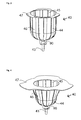
- FIG. 1 shows a device according to the invention for fixing a female breast.
- the fixing device has a cup 40 for receiving the breast.
- the basic shape of the wall 46 of the cup 40 is predetermined and stabilized by reinforcing ribs 44.
- the cup 40 may next to a like in the FIG. 1 also have a cylindrical, a conical, a hemispherical or any other adequate shape shown adapted to the breast shape.
- the wall itself is preferably made of a puncturable material, so that For example, contrast medium injected into the breast or tissue samples can be taken from this.
- At the upper end of the cup 40 corresponding to the upper end of the device pointing in the direction of the chest wall of the patient, there is an opening which is sufficiently large to enclose the patient's breast.
- the fixing device has a vacuum system.
- a suction channel 45 which is preferably integrated in a reinforcing rib 44, the air is sucked out of the interior of the cup.
- a hose connection 43 is provided for connecting a vacuum pump.
- an RFID transponder (90) is provided for identification of the cup.
- technical information on the fixing device or the cup itself such as size, material, sterility, X-ray properties, date of manufacture, service life, number of missions or tests performed, serial number, suitability for use in a particular X-ray machine, etc. may be stored. These data can be programmed on the one hand in the production in the transponder or programmed or updated during use.
- FIG. 2 shows a further embodiment of a device according to the invention for breast fixation.
- the wall 46 of the cup 40 comprises a fabric grid.
- This fabric grid can also be designed to contract when pulled and compress the breast. In this case, a pulling force would have to be applied at the end on the side of the hose connection 43. It can a close-meshed mesh screen itself can be used for breast fixation.
- the vacuum system is not yet usable here, since air would again penetrate between the meshes of the fabric.
- the device is provided on the inside with an elastic film or coating, it can thereby be made airtight, so that again the air can be sucked out of the space between the wall 46 and the breast.
- FIG. 3 a further embodiment of a device according to the invention for breast fixation is given.
- the cup of the fixing device is based here on an approximately cylindrical basic shape.
- the other features correspond to the FIG. 1 ,
- FIG. 4 is a similar breast fixation device as in FIG. 3 disclosed. However, this device is designed for smaller breasts with a smaller cup size. It can be an alternative to the device FIG. 3 be used.
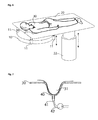
- FIG. 5 shows the integration of a device according to the invention for breast fixation in a support surface 20.
- a receiving ring 48 is integrated into the support surface. It can also be used for coarse adaptation to different breast sizes.
- the fixing device is attached here by way of example by means of tabs of the terminal edge 47 on the receiving ring 48. For this purpose, it is first pushed up into the guide groove 49 (1.) and then twisted (2.).
- the principle corresponds to that of a bayonet closure.
- other closure mechanisms such as a magnetic closure, a snap closure or a hook and loop fastener can be realized.
- the fixing device is easily replaceable. As shown, the device is made from below, that is from the side facing away from the patient of the support surface.
- a passive transponder is used for cost reasons, which receives its energy through the reader.
- An RFID system according to one of the following standards is particularly preferably used: ISO / IEC 10536, ISO / IEC 14443, ISO / IEC 15693, ISO 69873.
- FIG. 6 An x-ray machine for imaging a female breast is shown.
- a patient 30 is lying on the patient couch 20.
- the breast to be examined hangs over a breast cutout 21 through the patient couch 20 into the receiving area of a gantry 10 and is held there by a breast fixation according to the invention.
- This breast fixation is not apparent from this graphic perspective that it is covered by the patient's body.
- the gantry 10 is a spiral computed tomography gantry with an x-ray tube and a detector which rotate around the breast to be examined. During the rotation, the breast is imaged. Simultaneously with the rotation, a shift in the vertical direction is performed via the Gantryhubantrieb 11, so that the breast is scanned spirally.
- the patient couch 20 is height-adjustable via a patient couch lift drive 22.
- it can also be rotatable about the axis of the patient couch lifting drive 22.
- FIG. 7 shows by way of example a fixing device in which the cup 40, which is inserted into the support surface 20.
- the connection is made here by way of example via a flat taper fit.
- a vacuum pump 42 is connected, so that the breast 31 of the patient is fixed in the fixing device by negative pressure.
- FIG. 8 shows an example of a vertically arranged X-ray device, in which the cup 40 is inserted into a vertical support surface.
Abstract
Description
- Die Erfindung betrifft eine Vorrichtung zur Fixierung der Brust einer Patientin in einem Untersuchungsgerät zur Untersuchung der weiblichen Brust. Ein solches Untersuchungsgerät kann ein Röntgengerät zur Abbildung der weiblichen Brust (Mammografie), ein CT-Scanner oder auch ein Ultraschallgerät sein. Weiterhin betrifft die Erfindung ein Röntgengerät beziehungsweise einen CT Scanner mit einer entsprechenden Fixierung der Brust einer Patientin sowie ein Verfahren zum Betrieb eines solchen Gerätes.
- Zur Untersuchung der weiblichen Brust sind verschiedene Geräte, wie Röntgengeräte oder auch CT-Scanner bekannt. Ein solcher CT-Scanner ist beispielsweise in der
US 2006/0094950 A1 offenbart. Unter einer Liege, auf der eine zu untersuchende Patientin liegt, befindet sich eine Röntgenvorrichtung mit einer rotierenden Gantry, welche eine Röntgenröhre und einen Detektor aufweist. Die zu untersuchende Brust der Patientin ragt durch eine Öffnung in der Liege in den Strahlengang der Röntgenvorrichtung. Um nun während der Untersuchung konstante Verhältnisse zu erzeugen, wird die zu untersuchende Brust mit einem Stempel nach oben gedrückt und in eine vordefinierte Form gebracht. Durch eine Verschiebung des Stempels ist eine Anpassung an unterschiedliche Brustgrößen möglich. Die Anpassung kann hier allerdings nur in der Länge und nicht im Durchmesser erfolgen. Eine andere Vorrichtung zur Stabilisierung der Brust der Patientin ist in derUS 6,418,188 B1 offenbart. Ein Becher aus gummiartigem Gewebe wird über die Brust gestülpt und mittels einer Schnur von der Patientin weggezogen. Dadurch wird die Brust im Durchmesser komprimiert und in die Länge gezogen. Mit dieser Vorrichtung ist keine exakt reproduzierbare Lage und Form der Brust herstellbar. Eine verbesserte Vorrichtung ist in derUS 2004/0082856 A1 offenbart. Austauschbare Einsätze in die Patientenliege sorgen durch ihre Außenkontur für eine feste Fixierung der Brust. Hier bleibt weiterhin die Problematik bestehen, dass bei wiederholten Untersuchungen unterschiedliche Einsätze mit unterschiedlichen Größen eingesetzt werden und dadurch die einzelnen Aufnahmen kaum mehr miteinander vergleichbar sind. - Der Erfindung liegt die Aufgabe zugrunde, eine Vorrichtung zur Fixierung der Brust einer Patientin in einem Untersuchungsgerät zur Untersuchung der weiblichen Brust derart auszugestalten, dass die Brust der Patientin bei größtmöglichen Komfort für die Patientin in einer auch über mehrere Aufnahmen reproduzierbaren Lage fixiert werden kann. Ein weiterer Aspekt der Erfindung ist die Ausgestaltung eines Untersuchungsgerätes zur Untersuchung der weiblichen Brust, insbesondere eines Röntgengerätes beziehungsweise eines CT-Scanners mit einer erfindungsgemäßen Vorrichtung zur Fixierung der Brust. Schließlich ist noch ein Verfahren zum Betrieb eines solchen Gerätes Gegenstand der Erfindung.
- Diese Aufgabe wird durch Vorrichtungen nach den unabhängigen Ansprüchen gelöst. Vorteilhafte Ausgestaltungen der Erfindung sind in den Unteransprüchen angegeben.
- Eine erfindungsgemäße Vorrichtung zur Fixierung einer weiblichen Brust einer Patientin in einem Untersuchungsgerät umfasst einen Becher 40, welcher in eine Auflagefläche 20, die auch eine Patientenliege sein kann, einsetzbar ist. Zur Identifikation der Fixiervorrichtung weist die Fixiervorrichtung einen RFID-Transponder auf.
- Es ist besonders vorteilhaft, wenn verschiedene Exemplare der Fixiervorrichtung in unterschiedlichen Größen und in unterschiedlichen, an verschiedene Brustformen angepassten Formen ausgeführt sind. Somit kann jeweils eine Fixiervorrichtung passend für die Brustform ausgewählt werden. Durch den integrierten RFID-Transponder kann sichergestellt werden, dass auch bei wiederholten Untersuchungen immer dieselbe Form beziehungsweise Größe der Fixiervorrichtung verwendet wird. Weiterhin kann bei der Auswertung der Bilddaten automatisch die Form der Fixiervorrichtung berücksichtigt werden. So kann die Kollimierung beziehungsweise das Messfeld aufgrund der Daten aus dem RFID-Transponder optimal eingestellt werden, um die Strahlenbelastung zu minimieren und die Bildqualität zu optimieren. Ebenso können auch noch weitere Parameter für Korrekturverfahren eingestellt werden Es kann insbesondere eine Seriennummer oder eine andere Kennzeichnung der Fixiervorrichtung automatisch ausgelesen und mit den Bilddaten aufgezeichnet werden. Grundsätzlich können auch andere Identifikationssysteme, wie beispielsweise Schaltnocken oder auch Barcodes vorgesehen sein. Weiterhin können noch Sensoren zur Erfassung biologischer, chemischer oder physikalischer Größen wie der Temperatur, des Druckes etc. an der Vorrichtung angebracht sein. Diese Sensoren können vorteilhafterweise per Funk oder über den RFID-Transponder abgefragt werden.
- In einer weiteren vorteilhaften Ausgestaltung der Erfindung sind Verstärkungsrippen 44 vorgesehen, welche die Kontur einer Wandung 46 vorgegeben und diese gleichzeitig stabilisieren. Die Wandung selbst ist vorzugsweise aus einem durchstechbaren Material, so dass beispielsweise Kontrastmittel in die Brust injiziert oder Gewebeproben aus dieser entnommen werden können.
- In einer anderen Ausgestaltung der Erfindung ist der Becher 40 aus einem optisch transparenten Material oder weist zumindest transparente Bereiche auf. Zusätzlich beziehungsweise alternativ können auch Markierungen an dem Becher 40 vorgesehen sein. Solche Markierungen können beispielsweise Einstichpositionen für Biopsienadeln angeben, mit denen bestimmte Positionen im Inneren der Brust erreicht werden können. Weiterhin könnten auch Löcher beziehungsweise Öffnungen für medizinische Instrumente in dem Becher 40 vorgesehen sein.
- Besonders vorteilhaft ist es, wenn ein Vakuumssystem vorgesehen ist, um die Brust exakt in die Form der Fixiervorrichtung zu bringen. Gegebenenfalls kann die Fixiervorrichtung auch durch das Vakuumssystem an der Brust gehalten werden. Für das Vakuumssystem ist ein Schlauchanschluss 43 oder ein anderes Mittel, wie beispielsweise eine Steckverbindung zum Anschluss einer Vakuumpumpe vorgesehen. Durch Absaugen der Luft im Innenraum der Fixiervorrichtung mittels Saugkanälen 45 passt sich die Brust exakt an die Form der Fixiervorrichtung an und die Fixiervorrichtung hält zudem fest an der Brust. Besonders günstig ist es, eine Vielzahl von Saugkanälen 45 und Luftaustrittsöffnungen, durch die Luft vom inneren der Fixiervorrichtung in die Saugkanäle austreten kann, vorzusehen.
- Ein erfindungsgemäßer Satz Fixiervorrichtungen und umfasst mehrere der oben dargestellten Fixiervorrichtungen in unterschiedlichen Größen beziehungsweise mit Bechern unterschiedlicher Größen. Die Durchmesser der Becher liegen bevorzugt in einem Bereich von 80mm bis 180mmm. Weiterhin sind in dem RFID-Transponder einer jeder Fixiervorrichtung Informationen über deren Größe abgespeichert. Diese Informationen können unmittelbare Größenangaben, wie Durchmesser oder Länge sein. Sie können aber auch mittelbare Angaben, wie beispielsweise eine Seriennummer sein, anhand derer ein Lesegerät beispielsweise über in einer Datenbank abgespeicherte Informationen auf die Größe zurück schließen kann.
- Ein weiterer Aspekt der Erfindung ist ein Röntgengerät, insbesondere einen CT-Scanner mit einer oben dargestellten Vorrichtung zur Brustfixierung. Weiterhin weist ein solches erfindungsgemäßes Röntgengerät ein Lesegerät für einen RFID-Transponder auf.
- Besonders vorteilhaft ist es, wenn ein solches Röntgengerät eine Datenbank mit Scanparametern wie Spannung, Strom, Blende, Spirallänge, Wasserkorrekturparameter, Streustrahlenkorrekturparameter, Ringartefaktkorrekturparameter für verschiedene Vorrichtungen zur Fixierung der Brust aufweist oder zumindest eine Datenverbindung zu einer solchen Datenbank hat. Zudem ist es vorteilhaft, wenn in dieser Datenbank die entsprechenden RFID-Transponderkennungen enthalten sind.
- Grundsätzlich ist es möglich, die Patientin auf der Auflagefläche 20 mit bereits eingesetztem Becher 40 zu platzieren. Besonders vorteilhaft ist es jedoch, wenn die Brustfixierung beziehungsweise der Becher vor der Platzierung der Patientin an der Auflagefläche bereits an der Patientin angelegt wird. Ein entsprechendes Verfahren umfasst die folgenden Schritte:
- a. Anlegen des Bechers an der Brust sowie Aktivieren des Vakuums, und
- b. Platzieren der Patientin an der Auflagefläche.
- Hierzu wird vorzugsweise der Vakuumsschlauch durch den Brustausschnitt 21 der Auflagefläche gezogen.
- Ein alternatives Verfahren umfasst die folgenden Schritte:
- a. Halten des Bechers an der Brust, und
- b. Platzieren der Patientin an der Auflagefläche.
- Hierzu ist eine Halterung für den Becher an der Auflagefläche notwendig, welche sich selbsttätig arretiert, beispielsweise ein Magnetverschluss. Weiterhin wird bevorzugt automatisch, beispielsweise durch eine Magnetkupplung, die Vakuumverbindung hergestellt. Hierzu wird der Becher mit der Vakuumpumpe verbunden.
- Ein Verfahren zur Verwendung eines erfindungsgemäßen Röntgengerätes, wie zuvor beschrieben, umfasst die folgenden Schritte:
- a. Einsetzen der Fixiervorrichtung in eine Auflagefläche;
- b. Auslesen des RFID-Transponders;
- c. Überprüfen, ob die Fixiervorrichtung bereits verwendet wurde anhand der aus dem RFID-Transponder ausgelesenen Daten;
- d. Ausgabe einer Fehlermeldung, wenn die Fixiervorrichtung bereits verwendet wurde. Optional kann das Verfahren an dieser Stelle abgebrochen werden.
- e. Schreiben einer Markierung in den RFID-Transponder, dass die Fixiervorrichtung bereits verwendet wurde. Optional kann auch hier die Anzahl der Verwendungen in den RFID-Transponder geschrieben werden, oder es kann auch ein entsprechender Zähler in dem RFID-Transponder erhöht werden.
- f. Automatische Auswahl wenigstens eines optimalen Scanparameters aus Spannung, Strom, Blende, Spirallänge, Wasserkorrekturparameter, Streustrahlenkorrekturparameter, Ringartefaktkorrekturparameter aufgrund der aus dem RFID-Transponder ausgelesenen Daten.
- g. Durchführen des Scans, d. h. der Röntgenabbildung.
- Zur Auswahl der Daten in Schritt f. ist vorzugsweise eine Datenbank oder zumindest eine Tabelle mit geeigneten Parametern für verschiedene RFID-Transponder-Kennungen in dem Röntgengerät vorhanden.
- Die Erfindung wird nachstehend ohne Beschränkung des allgemeinen Erfindungsgedankens anhand von Ausführungsbeispielen unter Bezugnahme auf die Zeichnungen exemplarisch beschrieben.
- Figur 1
- zeigt eine Erfindungsgemäße Vorrichtung zur Fixierung einer weiblichen Brust.
- Figur 2
- zeigt eine weitere Vorrichtung zur Fixierung einer weiblichen Brust mit einer Gitterstruktur.
- Figur 3
- zeigt eine weitere Vorrichtung zur Fixierung einer weiblichen Brust.
- Figur 4
- zeigt eine kleinere Vorrichtung zur Fixierung einer weiblichen Brust.
- Figur 5
- zeigt die Integration einer erfindungsgemäßen Vorrichtung.
- Figur 6
- zeigt ein Röntgengerät zur Abbildung einer weiblichen Brust.
- Figur 7
- zeigt eine erfindungsgemäße Vorrichtung zur Brustfixierung, welche in eine Patientenliege eines Röntgengerätes integriert ist.
- Figur 8
- zeigt ein vertikal angeordnetes Röntgengerät.
-
Figur 1 zeigt eine erfindungsgemäße Vorrichtung zur Fixierung einer weiblichen Brust. Die Fixiervorrichtung weist einen Becher 40 zur Aufnahme der Brust auf. Die Grundform der Wandung 46 des Bechers 40 wird durch Verstärkungsrippen 44 vorgegeben und stabilisiert. Der Becher 40 kann neben einer wie in derFigur 1 dargestellten, der Brust angepassten Form auch eine zylindrische, eine kegelförmige, eine halbkugelförmige oder jede andere adäquate Form aufweisen. Die Wandung selbst ist vorzugsweise aus einem durchstechbaren Material, so dass beispielsweise Kontrastmittel in die Brust injiziert oder Gewebeproben aus dieser entnommen werden können. An dem oberen Ende des Bechers 40, entsprechend dem oberen Ende der Vorrichtung, welches in Richtung der Brustwand der Patientin zeigt, befindet sich eine Öffnung, die hinreichend groß ist, um die Brust der Patientin zu umschließen. Diese Öffnung ist von einem Abschlussrand 47 umschlossen. Er erhöht weiterhin die Stabilität und weist Laschen auf, mit denen dieser an der Auflagefläche 20 befestigt werden kann. Entsprechend der Brustgröße der Patientin sind verschiedene Größen und Formen der Becher 40 vorgesehen. Um die Brust nun exakt in die Form der Becher 40 zu bringen, weist die Fixiervorrichtung ein Vakuumssystem auf. Durch mindestens einen Saugkanal 45, der bevorzugt in eine Verstärkungsrippe 44 integriert ist, wird die Luft aus dem Innenraum des Bechers abgesaugt. Hierzu ist ein Schlauchanschluss 43 zum Anschluss einer Vakuumpumpe vorgesehen. Durch Absaugen der Luft im Innenraum passt sich die Brust exakt an die Form des Bechers der Fixiervorrichtung an und die Fixiervorrichtung hält zudem fest an der Brust. Besonders günstig ist es, eine Vielzahl von Saugkanälen und entsprechenden Öffnungen vorzusehen. Zur Identifikation des Bechers ist ein RFID-Transponder (90) vorgesehen. In diesem Transponder können technische Informationen zur Fixiervorrichtung beziehungsweise zum Bechers selbst, wie beispielsweise über Größe, Material, Sterilität, Röntgeneigenschaften, Herstellungsdatum, Nutzungsdauer, Anzahl der Einsätze beziehungsweise durchgeführte Untersuchungen, Seriennummer, Eignung zum Einsatz in einem bestimmten Röntgengerät etc. gespeichert sein. Diese Daten können einerseits bei der Herstellung in den Transponder programmiert oder auch während des Einsatzes programmiert oder aktualisiert werden. -
Figur 2 zeigt eine weitere Ausgestaltung einer erfindungsgemäßen Vorrichtung zur Brustfixierung. Hier umfasst die Wandung 46 des Bechers 40 ein Gewebegitter. Dieses Gewebegitter kann auch so gestaltet sein, dass es sich bei Zug zusammenzieht und die Brust komprimiert. In diesem Falle müsste eine Zugkraft an dem Ende auf Seiten des Schlauchanschlusses 43 aufgebracht werden. Es kann ein engmaschiges Gewebegitter selbst zur Brustfixierung verwendet werden. Allerdings ist hier das Unterdrucksystem noch nicht einsetzbar, da zwischen den Maschen des Gewebes wieder Luft eindringen würde. Wird nun die Vorrichtung auf der Innenseite mit einer elastischen Folie oder Beschichtung versehen, so kann sie dadurch luftdicht gemacht werden, so dass wieder die Luft aus dem Zwischenraum zwischen der Wandung 46 und der Brust abgesaugt werden kann. - In
Figur 3 ist eine weitere Ausgestaltung einer erfindungsgemäßen Vorrichtung zur Brustfixierung angegeben. Der Becher der Fixiervorrichtung basiert hier auf einer näherungsweise zylindrischen Grundform. Die übrigen Merkmale entsprechen derFigur 1 . - In
Figur 4 ist eine ähnliche Vorrichtung zur Brustfixierung wie inFigur 3 offenbart. Allerdings ist diese Vorrichtung für kleinere Brüste mit einer kleineren Körbchengröße ausgelegt. Sie kann alternativ zur Vorrichtung ausFigur 3 eingesetzt werden. -
Figur 5 zeigt die Integration einer erfindungsgemäßen Vorrichtung zur Brustfixierung in eine Auflagefläche 20. Ein Aufnahmering 48 ist in die Auflagefläche integriert. Er kann auch zur groben Adaption an unterschiedliche Brustgrößen verwendet werden. Die Fixiervorrichtung wird hier beispielhaft mittels Laschen des Abschlussrandes 47 an dem Aufnahmering 48 befestigt. Dazu wird sie zuerst nach oben in die Führungsnut 49 geschoben (1.) und dann verdreht (2.). Das Prinzip entspricht dem eines Bajonettverschlusses. selbstverständlich sind auch andere Verschlussmechanismen, wie beispielsweise ein Magnetverschluss, ein Schnappverschluss oder auch ein Klettverschluss realisierbar. Wesentlich ist, dass die Fixiervorrichtung einfach austauschbar ist. Wie gezeigt, wird die Vorrichtung von unten, das heißt von der der Patientin abgewandten Seite der Auflagefläche vorgenommen. Dies erleichtert auch den Anschluss des Vakuumsschlauches. Besonders günstig ist es jedoch, wenn die Fixiervorrichtung von der Patientenseite aus eingesetzt werden kann, denn dann kann die der Patientin abgewandte Seite der Auflagefläche vollständig in ein geschlossenes Gehäuse integriert werden. Für diesen Fall wird vorteilhafterweise ein Stecksystem für den Vakuumanschluss zur Luftabsaugung vorgesehen. Ein solches Stecksystem ist auch mit einer Magnetkupplung besonders einfach realisierbar. In dieser Figur ist weiterhin noch die Kommunikation zwischen dem RFID-Transponder 90 und dem RFID-Lesegerät 91 schematisch dargestellt. Zunächst sendet das RFID-Lesegerät 91 ein Anfragesignal 92, welches dann von dem RFID-Transponder 90 mittels des Antwortsignals 93 beantwortet wird. Unter RFID (Radio Frequency Identification) wird hier eine Identifizierung durch Funksignale oder andere elektromagnetische Signale im Allgemeinen verstanden. Vorzugsweise wird aus Kostengründen ein passiver Transponder eingesetzt, der seine Energie durch das Lesegerät erhält. Besonders bevorzugt wird ein RFID-System entsprechend einer der folgenden Normen eingesetzt: ISO/IEC 10536, ISO/IEC 14443, ISO/IEC 15693, ISO 69873. - In
Figur 6 ist ein Röntgengerät zur Abbildung einer weiblichen Brust dargestellt. Auf der Patientenliege 20 liegt eine Patientin 30. Die zu untersuchende Brust hängt über einen Brustausschnitt 21 durch die Patientenliege 20 in den Aufnahmebereich einer Gantry 10 und wird dort durch eine erfindungsgemäße Brustfixierung gehalten. Diese Brustfixierung ist aus dieser zeichnerischen Perspektive nicht erkennbar, das sie durch den Körper der Patientin abgedeckt wird. Die Gantry 10 ist eine Spiral-Computertomographen Gantry mit einer Röntgenröhre und einem Detektor, welche sich um die zu untersuchende Brust drehen. Während der Drehung wird die Brust abgebildet. Gleichzeitig mit der Drehung wird über den Gantryhubantrieb 11 eine Verschiebung in vertikaler Richtung durchgeführt, so dass die Brust spiralförmig abgetastet wird. Die Patientenliege 20 ist über einen Patientenliegenhubantrieb 22 in der Höhe verstellbar. Optional kann bei einem fest installierten Patiententisch dieser auch noch um die Achse des Patientenliegenhubantriebs 22 drehbar sein. -
Figur 7 zeigt exemplarisch eine Fixiervorrichtung, bei der der Becher 40, die in die Auflagefläche 20 eingesetzt ist. Die Verbindung erfolgt hier beispielhaft über eine flache Kegelpassung. Über einen Schlauch 41 ist eine Vakuumpumpe 42 angeschlossen, so dass die Brust 31 der Patientin in der Fixiervorrichtung durch Unterdruck fixiert wird. -
Figur 8 zeigt exemplarisch ein vertikal angeordnetes Röntgengerät, bei dem der Becher 40 in eine vertikale Auflagefläche eingesetzt ist. -
- 10
- Gantry
- 11
- Gantryhubantrieb
- 20
- Auflagefläche
- 21
- Brustausschnitt
- 22
- Patientenliegenhubantrieb
- 25
- Trennwand
- 30
- Patientin
- 31
- Brust
- 40
- Becher
- 41
- Schlauch
- 42
- Vakuumpumpe
- 43
- Schlauchanschluss
- 44
- Verstärkungsrippe
- 45
- Saugkanal
- 46
- Wandung
- 47
- Abschlussrand
- 48
- Aufnahmering
- 49
- Führungsnut
- 90
- RFID Transponder
- 91
- RFID Lesegerät
- 92
- RFID Anfragesignal
- 93
- RFID Antwortsignal
Claims (15)
- Vorrichtung zur Fixierung einer weiblichen Brust (31) einer Patientin (30) in einem Untersuchungsgerät, umfassend einen Becher (40) zur Aufnahme der Brust, welcher in eine Öffnung einer Auflagefläche (20) für die Patientin (30) eingebracht werden kann,
dadurch gekennzeichnet, dass
an dem Becher (40) ein RFID-Transponder (90) angebracht ist. - Vorrichtung nach Anspruch 1,
dadurch gekennzeichnet, dass
in dem ein RFID-Transponder (90) wenigstens eine der folgenden Informationen gespeichert ist: Größe und/oder Material des Bechers, Sterilität, Röntgeneigenschaften, Herstellungsdatum, Nutzungsdauer, Anzahl der durchgeführten Untersuchungen, Seriennummer. - Vorrichtung nach Anspruch 1 oder 2,
dadurch gekennzeichnet, dass
der Becher (40) eine Wandung (46), welche durch Verstärkungsrippen (44) verstärkt ist umfasst. - Vorrichtung nach einem der vorhergehenden Ansprüche,
dadurch gekennzeichnet, dass
an dem Becher (40) Mittel (43) zum Anschluss einer Vakuumpumpe vorgesehen sind. - Vorrichtung nach einem der vorhergehenden Ansprüche,
dadurch gekennzeichnet, dass
der Becher (40) ein optisch transparentes Material umfasst. - Vorrichtung nach einem der vorhergehenden Ansprüche,
dadurch gekennzeichnet, dass
der Becher (40) Markierungen für bestimmte Positionen im Inneren der Brust umfasst. - Vorrichtung nach einem der vorhergehenden Ansprüche,
dadurch gekennzeichnet, dass
der Becher (40) Öffnungen für medizinische Instrumente umfasst. - Vorrichtungssatz zur Fixierung einer weiblichen Brust einer Patientin (30) in einem Untersuchungsgerät, umfassend mehrere Vorrichtungen entsprechend einem der vorhergehenden Ansprüchen mit Bechern (40) in verschiedenen Größen und einem RFID-Transponder (90), in welchem jeweils eine Kennung zur Identifizierung der entsprechenden Größe der Becher gespeichert ist.
- Röntgengerät zur Abbildung einer Brust einer Patientin (30), mit- einer Röntgeneinrichtung umfassend eine Röntgenröhre und einen Röntgendetektor,- einer Auflagefläche (20) zur Aufnahme einer Patientin (30) mit einem Brustausschnitt (21),- wenigstens einer Vorrichtung zur Fixierung der Brust nach einem der vorhergehenden Ansprüche mit einem RFID-Transponder (90) und- mit einem RFID-Lesegerät zur Abfrage von Daten des RFID-Transponders und/oder Speicherung von Daten in dem RFID-Transponder wenigstens einer der Vorrichtungen zur Fixierung der Brust.
- Röntgengerät nach Anspruch 9,
dadurch gekennzeichnet, dass
das Röntgengerät eine Datenbank mit Scanparametern für verschiedene Vorrichtungen zur Fixierung der Brust aufweist oder zumindest eine Datenverbindung zu einer solchen Datenbank hat. - Röntgengerät zur Abbildung einer Brust einer Patientin (30), mit- einer Röntgeneinrichtung umfassend eine Röntgenröhre und einen Röntgendetektor,- einer Auflagefläche (20) zur Aufnahme einer Patientin (30) mit einem Brustausschnitt (21),- wenigstens einer Vorrichtung zur Fixierung der Brust nach einem der vorhergehenden Ansprüche mit einem RFID-Transponder (90) und- mit einem RFID-Lesegerät zur Abfrage von Daten des RFID-Transponders und/oder Speicherung von Daten in dem RFID-Transponder wenigstens einer der Vorrichtungen zur Fixierung der Brust.
- Röntgengerät nach Anspruch 11, welches ein CT-Scanner ist.
- Verfahren zum Anlegen einer Vorrichtung nach Anspruch 1 zur Fixierung einer weiblichen Brust (31) an eine Patientin (30) umfassend die Schritte:a. Anlegen des Bechers (40) der Vorrichtung an der Brust (31) sowie Aktivieren eines Vakuums zum Ansaugen des Bechers an die Brust, undb. Platzieren der Patientin an der Auflagefläche (20).
- Verfahren zum Anlegen einer Vorrichtung nach Anspruch 1 zur Fixierung einer weiblichen Brust (31) an eine Patientin (30), umfassend die Schritte:a. Halten des Bechers (40) der Vorrichtung an der Brust (31),b. Platzieren der Patientin an der Auflagefläche, undc. Herstellen einer Verbindung zwischen dem Becher (40) und einer Vakuumpumpe (42).
- Verfahren zur Verwendung eines Röntgengerätes nach Anspruch 11, umfassend die Schritte:a. Einsetzen der Fixierung der Brust in eine Auflagefläche;b. Auslesen des RFID-Transponders;c. Überprüfen, ob die Fixiervorrichtung bereits verwendet wurde anhand der aus dem RFID-Transponder ausgelesenen Daten;d. Ausgabe einer Fehlermeldung, wenn die Fixiervorrichtung bereits verwendet wurde.e. Schreiben einer Markierung in den RFID-Transponder, dass die Fixiervorrichtung bereits verwendet wurde oder Erhöhung eines Verwendungszählers.f. Automatische Auswahl wenigstens eines optimalen Scanparameters aus Spannung, Strom, Blende, Spirallänge, Wasserkorrekturparameter, Streustrahlenkorrekturparameter, Ringartefaktkorrekturparameter aufgrund der aus dem RFID-Transponder ausgelesenen Daten;g. Durchführen des Scans, d. h. der Röntgenabbildung.
Applications Claiming Priority (1)
| Application Number | Priority Date | Filing Date | Title |
|---|---|---|---|
| DE102008042430 | 2008-09-29 |
Publications (1)
| Publication Number | Publication Date |
|---|---|
| EP2168485A1 true EP2168485A1 (de) | 2010-03-31 |
Family
ID=40524871
Family Applications (8)
| Application Number | Title | Priority Date | Filing Date |
|---|---|---|---|
| EP09154854A Withdrawn EP2168485A1 (de) | 2008-09-29 | 2009-03-11 | Brustfixierung für ein Untersuchungsgerät zur Untersuchung der weiblichen Brust |
| EP09154884.2A Withdrawn EP2178048A3 (de) | 2008-09-29 | 2009-03-11 | Verfahren zur Definition eines patientenindividuellen Koordinationssystems einer weiblichen Brust |
| EP09154833A Expired - Fee Related EP2168489B1 (de) | 2008-09-29 | 2009-03-11 | Röntgengerät zur Brustuntersuchung im Stehen |
| EP09154863A Withdrawn EP2168491A1 (de) | 2008-09-29 | 2009-03-11 | Brustfixierung mit Probencontainer für ein Untersuchungsgerät der weiblichen Brust |
| EP09154842A Expired - Fee Related EP2168484B1 (de) | 2008-09-29 | 2009-03-11 | Röntgengerät zur Brustuntersuchung mit einer in eine Patientenliege integrierten Gantry |
| EP09154848A Withdrawn EP2168490A1 (de) | 2008-09-29 | 2009-03-11 | Röntgengerät zur Brustuntersuchung mit einer Detektor-Röhren Anordnung für hochauflösende Aufnahmen |
| EP09154891A Expired - Fee Related EP2168486B1 (de) | 2008-09-29 | 2009-03-11 | Modulares System zur Brustdiagnose und -intervention |
| EP09154900A Withdrawn EP2168487A1 (de) | 2008-09-29 | 2009-03-11 | Verfahren und Vorrichtung zur thermischen Brusttumor-Behandlung mit 3D Monitorfunktion |
Family Applications After (7)
| Application Number | Title | Priority Date | Filing Date |
|---|---|---|---|
| EP09154884.2A Withdrawn EP2178048A3 (de) | 2008-09-29 | 2009-03-11 | Verfahren zur Definition eines patientenindividuellen Koordinationssystems einer weiblichen Brust |
| EP09154833A Expired - Fee Related EP2168489B1 (de) | 2008-09-29 | 2009-03-11 | Röntgengerät zur Brustuntersuchung im Stehen |
| EP09154863A Withdrawn EP2168491A1 (de) | 2008-09-29 | 2009-03-11 | Brustfixierung mit Probencontainer für ein Untersuchungsgerät der weiblichen Brust |
| EP09154842A Expired - Fee Related EP2168484B1 (de) | 2008-09-29 | 2009-03-11 | Röntgengerät zur Brustuntersuchung mit einer in eine Patientenliege integrierten Gantry |
| EP09154848A Withdrawn EP2168490A1 (de) | 2008-09-29 | 2009-03-11 | Röntgengerät zur Brustuntersuchung mit einer Detektor-Röhren Anordnung für hochauflösende Aufnahmen |
| EP09154891A Expired - Fee Related EP2168486B1 (de) | 2008-09-29 | 2009-03-11 | Modulares System zur Brustdiagnose und -intervention |
| EP09154900A Withdrawn EP2168487A1 (de) | 2008-09-29 | 2009-03-11 | Verfahren und Vorrichtung zur thermischen Brusttumor-Behandlung mit 3D Monitorfunktion |
Country Status (2)
| Country | Link |
|---|---|
| US (8) | US8199993B2 (de) |
| EP (8) | EP2168485A1 (de) |
Cited By (1)
| Publication number | Priority date | Publication date | Assignee | Title |
|---|---|---|---|---|
| CN110975156A (zh) * | 2019-11-15 | 2020-04-10 | 山东大学齐鲁医院 | 乳房牵引固定装置及系统 |
Families Citing this family (61)
| Publication number | Priority date | Publication date | Assignee | Title |
|---|---|---|---|---|
| US8272088B2 (en) * | 2007-09-06 | 2012-09-25 | Orbital Therapy Llc | Patient support system for full access prone position breast radiotherapy |
| EP2168485A1 (de) * | 2008-09-29 | 2010-03-31 | MIR Medical Imaging Research Holding GmbH | Brustfixierung für ein Untersuchungsgerät zur Untersuchung der weiblichen Brust |
| DE102008049711A1 (de) * | 2008-09-30 | 2010-04-15 | Siemens Aktiengesellschaft | Lagerungsvorrichtung, Patientenlagerungstisch und medizinisches Gerät |
| US8014490B2 (en) * | 2009-10-20 | 2011-09-06 | Linda Mitchell | Mammogram tender machine |
| US8421604B2 (en) * | 2009-11-30 | 2013-04-16 | Symbol Technologies, Inc. | Method and apparatus for identifying read zone of RFID reader |
| US8374312B2 (en) * | 2010-02-18 | 2013-02-12 | Varian Medical Systems, Inc. | Prone patient positioning devices and methods |
| DE102010011660A1 (de) * | 2010-03-17 | 2011-09-22 | Siemens Aktiengesellschaft | Mammographiegerät |
| JP5700950B2 (ja) * | 2010-04-21 | 2015-04-15 | キヤノン株式会社 | 生体情報取得装置 |
| US20140191852A1 (en) * | 2010-05-13 | 2014-07-10 | Carestream Health, Inc. | Method and system for phosphor plate identification in computed radiography |
| US20120001737A1 (en) * | 2010-05-13 | 2012-01-05 | Amir Berger | Method and system for computed radiography |
| GB2483640A (en) * | 2010-09-10 | 2012-03-21 | Specialty Magnetics Ltd | Breast immobilisation arrangement |
| WO2012048000A2 (en) | 2010-10-05 | 2012-04-12 | Hologic, Inc. | Upright x-ray breast imaging with a ct mode, multiple tomosynthesis modes, and a mammography mode |
| DE102010052603A1 (de) * | 2010-11-25 | 2012-05-31 | Artemis Imaging Gmbh | Patientenliege |
| WO2012120498A1 (en) * | 2011-03-04 | 2012-09-13 | Technion Research & Development | Non-invasive thermal treatment monitoring |
| DE102011006353A1 (de) | 2011-03-29 | 2012-10-04 | Siemens Aktiengesellschaft | Mammographieanlage |
| WO2012171029A1 (en) | 2011-06-09 | 2012-12-13 | The Regents Of The University Of California | Excised specimen imaging using a combined pet and micro ct scanner |
| US8842806B2 (en) | 2012-04-03 | 2014-09-23 | Carestream Health, Inc. | Apparatus and method for breast imaging |
| EP2845024B1 (de) | 2012-05-02 | 2019-04-10 | Koninklijke Philips N.V. | Thermometrieabbildung |
| US9307961B2 (en) * | 2012-06-29 | 2016-04-12 | Carefusion 2200, Inc. | Fine needle aspiration biopsy device |
| KR102001926B1 (ko) * | 2012-09-11 | 2019-07-30 | 삼성디스플레이 주식회사 | 엑스레이 검출기, 이를 포함하는 엑스레이 검출 시스템 및 엑스레이 검출 방법 |
| DE102012216687A1 (de) * | 2012-09-18 | 2014-03-20 | Jan Rimbach | Vorrichtung zur Untersuchung von Prüfkörpern |
| DE102012217301B4 (de) | 2012-09-25 | 2021-10-14 | Bayer Pharma Aktiengesellschaft | Kombination aus Kontrastmittel und Mammographie-CT-System mit vorgegebenem Energiebereich und Verfahren zur Erzeugung tomographischer Mammographie-CT-Aufnahmen durch diese Kombination |
| CN103908343B (zh) | 2012-12-31 | 2016-10-05 | 西门子(深圳)磁共振有限公司 | 患者检查床和磁共振成像设备 |
| EP3964132B1 (de) | 2013-10-09 | 2023-07-19 | Hologic, Inc. | Röntgenstrahl-brusttomosynthese mit erhöhung der räumlichen auflösung in dickenrichtung einer abgeflachten brust |
| US9161725B1 (en) * | 2014-02-05 | 2015-10-20 | Regine Millien-White | Adjustable breast examination device |
| JP6376783B2 (ja) * | 2014-03-12 | 2018-08-22 | キヤノン株式会社 | 乳房断層撮影装置および制御方法 |
| JP6381253B2 (ja) * | 2014-03-31 | 2018-08-29 | キヤノン株式会社 | 放射線撮影装置、断層撮影装置 |
| EP3125758B1 (de) * | 2014-04-04 | 2018-12-12 | Pierfrancesco Pavoni | Zugriffs-gate oder -gantry mit einer antennenanordnung zur therapie oder bildgebung |
| US9326739B2 (en) | 2014-04-28 | 2016-05-03 | Cheryl A. Galambos McLaughlin | Mammogram table |
| US9301726B2 (en) * | 2014-05-02 | 2016-04-05 | Wisconsin Alumni Research Foundation | CT machine for multi-angle scanning of stationary patients |
| CN104173075B (zh) * | 2014-08-26 | 2016-07-06 | 李丙曙 | 放射科检查床 |
| JP6611428B2 (ja) * | 2014-12-09 | 2019-11-27 | キヤノン株式会社 | マンモ断層撮像システム |
| EP3238628A4 (de) * | 2014-12-26 | 2018-08-15 | Rayence Co., Ltd. | Hebevorrichtung für ein kompressionspaddle und röntgenbildfotografievorrichtung damit |
| CN105832353B (zh) * | 2015-01-30 | 2020-11-06 | 佳能株式会社 | 放射线摄像系统 |
| JP6651069B2 (ja) * | 2015-05-13 | 2020-02-19 | フジデノロ株式会社 | 固定具装着装置 |
| KR20160139292A (ko) * | 2015-05-27 | 2016-12-07 | 삼성전자주식회사 | Rf 표면 코일부 및 이를 포함하는 자기공명영상 시스템 |
| JP6525768B2 (ja) * | 2015-06-30 | 2019-06-05 | キヤノン株式会社 | 乳房撮影装置 |
| US10542951B2 (en) * | 2015-07-23 | 2020-01-28 | General Electric Company | Systems, methods, and devices for simplified high quality imaging of biopsy samples on a mammography machine |
| WO2017019401A1 (en) * | 2015-07-24 | 2017-02-02 | Dretzaka-Kaye Tricia | Anatomy scanning system and method |
| WO2017091787A1 (en) * | 2015-11-25 | 2017-06-01 | The Regents Of The University Of California | 3d-beam modulation filter for equalizing dose and image quality in breast ct |
| DE102015225236A1 (de) * | 2015-12-15 | 2017-06-22 | Siemens Healthcare Gmbh | Mammographie-Screening mit hoher Durchgangsrate |
| CN106933857B (zh) * | 2015-12-30 | 2020-12-29 | 创新先进技术有限公司 | 一种数据仓库中任务的调度方法、装置 |
| DE102016206198A1 (de) * | 2016-04-13 | 2017-10-19 | Siemens Healthcare Gmbh | Röntgensystem |
| US10603003B2 (en) * | 2016-04-14 | 2020-03-31 | Dedicating2Imaging, LLC | CT systems for imaging of the breast |
| US11395593B2 (en) * | 2016-09-14 | 2022-07-26 | Mor Research Applications Ltd. | Device, system and method for detecting irregularities in soft tissue |
| US10180207B1 (en) * | 2017-07-13 | 2019-01-15 | Danylo Kozub | Stand |
| CN108175430A (zh) * | 2018-01-17 | 2018-06-19 | 江苏美伦影像系统有限公司 | 一种具有辐射防护功能的乳腺x射线摄影系统 |
| US10959747B1 (en) * | 2018-04-02 | 2021-03-30 | Lifei Guo | Tissue removing |
| US10893844B1 (en) * | 2018-10-10 | 2021-01-19 | David Byron Douglas | Method and apparatus for performing 3D imaging examinations of a structure under differing configurations and analyzing morphologic changes |
| DE102018207636A1 (de) * | 2018-05-16 | 2019-11-21 | Siemens Healthcare Gmbh | Patiententisch mit Vorrichtung zur reversiblen Aufnahme einer Transferplatte |
| CN108956656B (zh) * | 2018-07-17 | 2021-02-05 | 青岛大学附属医院 | 一种高衬度低剂量相位衬度ct成像装置 |
| CA3173541A1 (en) * | 2020-03-31 | 2021-10-07 | Hologic, Inc. | Systems and methods for x-ray imaging tissue specimens |
| KR102640269B1 (ko) * | 2020-05-29 | 2024-02-26 | (의료)길의료재단 | 유방암 치료용 방사선 조사 장치 |
| CN111714222B (zh) * | 2020-06-29 | 2021-07-23 | 北京欧扬医疗美容门诊部有限公司 | 一种无痕隆胸用脂肪自体植入装置 |
| CN111714191A (zh) * | 2020-06-30 | 2020-09-29 | 广西医科大学附属肿瘤医院 | 用于锥光束乳腺ct引导下悬垂穿刺的激光定位装置 |
| US11692951B2 (en) * | 2021-02-24 | 2023-07-04 | GE Precision Healthcare LLC | System and method for specimen imaging using an existing mammography imaging system |
| EP4226876A1 (de) * | 2022-02-09 | 2023-08-16 | Storz Medical AG | Stosswellenvorrichtung mit verbesserter akustischer kopplung |
| EP4226877A1 (de) * | 2022-02-09 | 2023-08-16 | Storz Medical AG | Stosswellenvorrichtung mit integrierter ultraschallsonde |
| EP4226875A1 (de) * | 2022-02-09 | 2023-08-16 | Storz Medical AG | Stosswellenvorrichtung mit einer quelle, die sich selbst auf eine röntgenvorrichtung ausrichtet |
| EP4226874A1 (de) * | 2022-02-09 | 2023-08-16 | Storz Medical AG | Ultraschall- und/oder stosswellenvorrichtung mit auf einer hexapod-plattform montierter quelle |
| WO2023200896A1 (en) * | 2022-04-14 | 2023-10-19 | Koning Corporation | Cone beam breast computed tomography with patient support subsystem |
Citations (8)
| Publication number | Priority date | Publication date | Assignee | Title |
|---|---|---|---|---|
| WO2004006755A2 (en) | 2002-07-16 | 2004-01-22 | Alfred E. Mann Institute For Biomedical Engineering At The University Of Southern California | Support bra for ultrasonic breast scanner |
| US20040254464A1 (en) | 2003-05-30 | 2004-12-16 | Stribling Mark L. | Apparatus and method for three dimensional ultrasound breast imaging |
| US20060145871A1 (en) | 2004-12-02 | 2006-07-06 | Smith & Nephew, Inc. | Radio Frequency Identification for Medical Devices |
| DE102005022347A1 (de) | 2005-05-13 | 2006-11-23 | Siemens Ag | Medizintechnisches Basissystem und medizintechnisches System |
| DE102005048049A1 (de) | 2005-10-07 | 2007-04-19 | Forschungszentrum Karlsruhe Gmbh | Vorrichtung zur bildgestützten Mammadiagnose und -therapie |
| EP1864611A1 (de) | 2005-04-01 | 2007-12-12 | Keizi Shibuya | Brustuntersuchungssystem |
| US20080037703A1 (en) | 2006-08-09 | 2008-02-14 | Digimd Corporation | Three dimensional breast imaging |
| US20080230074A1 (en) | 2007-03-23 | 2008-09-25 | Zheng Mike Q | Method and device for immobilization of the human breast in a prone position for radiotherapy |
Family Cites Families (102)
| Publication number | Priority date | Publication date | Assignee | Title |
|---|---|---|---|---|
| US3673394A (en) | 1969-02-18 | 1972-06-27 | North American Rockwell | Measuring method and apparatus |
| US4015836A (en) | 1975-07-31 | 1977-04-05 | General Electric Company | Mammography table |
| US4400827A (en) * | 1981-11-13 | 1983-08-23 | Spears James R | Method and apparatus for calibrating rapid sequence radiography |
| US4680028A (en) * | 1984-07-02 | 1987-07-14 | Lact-Assist, Incorporated | Flexible breast receptor for breast pump |
| US4709382A (en) * | 1984-11-21 | 1987-11-24 | Picker International, Inc. | Imaging with focused curved radiation detectors |
| US5415169A (en) | 1989-11-21 | 1995-05-16 | Fischer Imaging Corporation | Motorized mammographic biopsy apparatus |
| FI85803C (fi) | 1989-11-23 | 1992-06-10 | Planmed Oy | Foerfarande och anordning foer styrning av funktioner av en mammografiroentgenanordning. |
| US5569266A (en) | 1991-03-11 | 1996-10-29 | Fischer Imaging Corporation | Magnetic resonance imaging device useful for guiding a medical instrument |
| US5409497A (en) | 1991-03-11 | 1995-04-25 | Fischer Imaging Corporation | Orbital aiming device for mammo biopsy |
| US5289520A (en) | 1991-11-27 | 1994-02-22 | Lorad Corporation | Stereotactic mammography imaging system with prone position examination table and CCD camera |
| US5308321A (en) | 1992-05-05 | 1994-05-03 | Castro Donna J | Retainer assisted by vacuum expansion system |
| US5273435B1 (en) | 1992-07-16 | 1995-12-05 | Wisconsin Med College Inc | Tumor localization phantom |
| US5386447A (en) | 1992-09-23 | 1995-01-31 | Fischer Imaging Corporation | Mammographic screening and biopsy apparatus |
| US5490513A (en) * | 1992-09-28 | 1996-02-13 | Fonar Corporation | Multiple patient breast scanning on a magnetic resonance imaging apparatus |
| US6075879A (en) * | 1993-09-29 | 2000-06-13 | R2 Technology, Inc. | Method and system for computer-aided lesion detection using information from multiple images |
| JPH07303633A (ja) * | 1994-05-11 | 1995-11-21 | Mitsubishi Electric Corp | X線乳房撮影装置 |
| US5528043A (en) | 1995-04-21 | 1996-06-18 | Thermotrex Corporation | X-ray image sensor |
| US5609827A (en) | 1995-05-02 | 1997-03-11 | Beekley Corporation | Biopsy specimen container |
| US5709206A (en) | 1995-11-27 | 1998-01-20 | Teboul; Michel | Imaging system for breast sonography |
| US5757878A (en) * | 1996-08-16 | 1998-05-26 | Analogic Corporation | Detector arrangement for x-ray tomography system |
| DE19639975C1 (de) | 1996-09-27 | 1998-05-07 | Siemens Ag | Medizinische Einrichtung mit einer tunnelförmigen Öffnung zur Aufnahme eines Untersuchungsobjektes |
| EP0983020A1 (de) | 1997-05-06 | 2000-03-08 | Quanta Vision, Inc. | Gewebeanalysevorrichtung |
| US6358246B1 (en) | 1999-06-25 | 2002-03-19 | Radiotherapeutics Corporation | Method and system for heating solid tissue |
| US5991357A (en) | 1997-12-16 | 1999-11-23 | Analogic Corporation | Integrated radiation detecting and collimating assembly for X-ray tomography system |
| US6175117B1 (en) * | 1998-01-23 | 2001-01-16 | Quanta Vision, Inc. | Tissue analysis apparatus |
| DE19812995A1 (de) | 1998-03-25 | 1999-10-07 | Siemens Ag | Mammographie-Gerät, insbesondere für Vergrößerungs-Mammographie |
| US6242743B1 (en) * | 1998-08-11 | 2001-06-05 | Mosaic Imaging Technology, Inc. | Non-orbiting tomographic imaging system |
| JP2000116631A (ja) | 1998-10-16 | 2000-04-25 | Toshiba Corp | X線診断装置 |
| JP3866431B2 (ja) * | 1999-02-17 | 2007-01-10 | 株式会社東芝 | X線ct装置 |
| US6684097B1 (en) | 1999-04-22 | 2004-01-27 | University Of Miami | Intraoperative monitoring of temperature-induced tissue changes with a high-resolution digital x-ray system during thermotherapy |
| TW406009B (en) * | 1999-07-16 | 2000-09-21 | Nat Science Council | 3-D localization method of clustered microcalcifications using cranio-caudal and medio-lateral oblique views |
| US6254614B1 (en) * | 1999-10-18 | 2001-07-03 | Jerry M. Jesseph | Device and method for improved diagnosis and treatment of cancer |
| US6480565B1 (en) * | 1999-11-18 | 2002-11-12 | University Of Rochester | Apparatus and method for cone beam volume computed tomography breast imaging |
| US6987831B2 (en) | 1999-11-18 | 2006-01-17 | University Of Rochester | Apparatus and method for cone beam volume computed tomography breast imaging |
| DE10026792A1 (de) * | 2000-05-31 | 2001-12-06 | Bip Biomedizinische Instr & Pr | Diagnose- und Therapietisch |
| US6463122B1 (en) | 2000-08-21 | 2002-10-08 | Bio-Imaging Resource, Inc. | Mammography of computer tomography for imaging and therapy |
| US7467892B2 (en) * | 2000-08-29 | 2008-12-23 | Imaging Therapeutics, Inc. | Calibration devices and methods of use thereof |
| US7940966B2 (en) | 2000-11-24 | 2011-05-10 | U-Systems, Inc. | Full-field breast image data processing and archiving |
| US6419390B1 (en) * | 2001-03-26 | 2002-07-16 | Marianette Landis-Lowell | Folding mammography table and method of use |
| US6516045B2 (en) * | 2001-05-04 | 2003-02-04 | The Regents Of The University Of California | Device and method for determining proportions of body materials |
| US6418188B1 (en) * | 2001-06-14 | 2002-07-09 | Juanita L. Broadnax | Radiation breast cup and method |
| US6674835B2 (en) * | 2001-10-12 | 2004-01-06 | General Electric Co. | Methods and apparatus for estimating a material composition of an imaged object |
| US6671975B2 (en) | 2001-12-10 | 2004-01-06 | C. William Hennessey | Parallel kinematic micromanipulator |
| DE10207623B4 (de) | 2002-02-22 | 2004-05-06 | Siemens Ag | Verfahren für die Computertomographie sowie Computertomographie (CT)-Gerät |
| US20040254461A1 (en) * | 2002-03-20 | 2004-12-16 | Ackerman William H. | Acoustic beam shaping by pulse power modulation at constant amplitude |
| US7783089B2 (en) | 2002-04-15 | 2010-08-24 | General Electric Company | Method and apparatus for providing mammographic image metrics to a clinician |
| US7218766B2 (en) * | 2002-04-15 | 2007-05-15 | General Electric Company | Computer aided detection (CAD) for 3D digital mammography |
| CA2393101A1 (en) | 2002-07-11 | 2004-01-11 | Martin Cyr | Apparatus, system and method of calibrating medical imaging systems |
| US6904119B2 (en) * | 2002-10-02 | 2005-06-07 | Shimadzu Corporation | Radiographic apparatus |
| US7149566B2 (en) | 2002-10-31 | 2006-12-12 | Manoa Medical, Inc. | Soft tissue orientation and imaging guide systems and methods |
| US7809422B2 (en) * | 2002-11-08 | 2010-10-05 | Art Advanced Research Technologies Inc. | Method and apparatus for optical imaging |
| US7286634B2 (en) * | 2002-12-23 | 2007-10-23 | Select Technologies, Llc | Method and apparatus for improving baggage screening examination |
| EP1599139B1 (de) | 2003-02-20 | 2009-08-12 | Manoa Medical, Inc. | Biegbare schneidevorrichtung |
| US6872001B1 (en) | 2003-05-05 | 2005-03-29 | Peco Controls Corp. | X-ray shielding structure for food inspection station |
| US6982424B2 (en) | 2003-06-02 | 2006-01-03 | Ge Medical Systems Global Technology Company, Llc | X-ray and CT image detector |
| US7291841B2 (en) | 2003-06-16 | 2007-11-06 | Robert Sigurd Nelson | Device and system for enhanced SPECT, PET, and Compton scatter imaging in nuclear medicine |
| US6837772B1 (en) | 2003-07-18 | 2005-01-04 | Regina Miracle International Limited | Breast cup construction |
| GB0318701D0 (en) * | 2003-08-08 | 2003-09-10 | Inst Of Cancer Res The | A method and apparatus for image processing |
| JP2005258370A (ja) | 2003-09-05 | 2005-09-22 | Fuji Photo Film Co Ltd | 放射線カセッテ |
| US7005988B2 (en) * | 2003-09-19 | 2006-02-28 | International Business Machines Corporation | Using radio frequency identification to detect and/or prevent theft and shoplifting |
| US20050070817A1 (en) * | 2003-09-30 | 2005-03-31 | Mueller Richard L. | Lavage assist device |
| US20050096515A1 (en) * | 2003-10-23 | 2005-05-05 | Geng Z. J. | Three-dimensional surface image guided adaptive therapy system |
| US7653229B2 (en) | 2003-12-23 | 2010-01-26 | General Electric Company | Methods and apparatus for reconstruction of volume data from projection data |
| JP4119835B2 (ja) | 2003-12-26 | 2008-07-16 | ジーイー・メディカル・システムズ・グローバル・テクノロジー・カンパニー・エルエルシー | 被曝線量計算方法およびx線撮影装置 |
| US7519209B2 (en) * | 2004-06-23 | 2009-04-14 | Vanderbilt University | System and methods of organ segmentation and applications of same |
| DE102004042790A1 (de) | 2004-09-03 | 2006-03-09 | Siemens Ag | Röntgeneinrichtung |
| WO2006086765A2 (en) * | 2005-02-11 | 2006-08-17 | University Of Florida Research Foundation, Inc. | System including computed tomography device for image guided treatment |
| US20060239398A1 (en) | 2005-03-07 | 2006-10-26 | Fused Multimodality Imaging, Ltd. | Breast diagnostic apparatus for fused SPECT, PET, x-ray CT, and optical surface imaging of breast cancer |
| US10492749B2 (en) | 2005-05-03 | 2019-12-03 | The Regents Of The University Of California | Biopsy systems for breast computed tomography |
| US7573034B2 (en) * | 2005-05-18 | 2009-08-11 | Carestream Health, Inc. | Mobile radiography image recording system |
| US7492858B2 (en) * | 2005-05-20 | 2009-02-17 | Varian Medical Systems, Inc. | System and method for imaging and treatment of tumorous tissue in breasts using computed tomography and radiotherapy |
| US7304578B1 (en) | 2005-06-02 | 2007-12-04 | Hewlett-Packard Development Company, L.P. | Tag including RFID circuit storing data modifiable using a physically alterable medium |
| CN101203170B (zh) | 2005-06-02 | 2015-11-25 | 赛利恩影像股份有限公司 | 计算机辅助检测系统 |
| WO2007008530A1 (en) * | 2005-07-08 | 2007-01-18 | Wisconsin Alumni Research Foundation | Backprojection reconstruction method for ct imaging |
| US20070064867A1 (en) | 2005-09-20 | 2007-03-22 | Hansen Timothy B | Apparatus and method to acquire data for reconstruction of images pertaining to functional and anatomical structure of the breast |
| JP4837507B2 (ja) * | 2005-10-06 | 2011-12-14 | 富士フイルム株式会社 | 乳房画像撮影装置 |
| US7742796B2 (en) | 2005-10-25 | 2010-06-22 | General Electric Company | Breast immobilization device and method of imaging the breast |
| US7558370B2 (en) | 2005-11-07 | 2009-07-07 | Sommer Jr Edward J | Method and apparatus for improving identification and control of articles passing through a scanning system |
| DE102005053993A1 (de) * | 2005-11-10 | 2007-05-24 | Siemens Ag | Diagnosevorrichtung und Diagnoseverfahren für kombinierte und/oder kombinierbare radiographische und nuklearmedizinische Untersuchungen |
| US8014576B2 (en) * | 2005-11-23 | 2011-09-06 | The Medipattern Corporation | Method and system of computer-aided quantitative and qualitative analysis of medical images |
| US10064584B2 (en) | 2005-12-22 | 2018-09-04 | Visen Medical, Inc. | Combined x-ray and optical tomographic imaging system |
| CN101370429A (zh) | 2006-01-17 | 2009-02-18 | 成象诊断系统公司 | 具有可变患者定位功能的激光成像设备 |
| US7806855B2 (en) | 2006-04-11 | 2010-10-05 | Playtex Products, Inc. | Manual breast pump |
| US7483511B2 (en) * | 2006-06-06 | 2009-01-27 | Ge Homeland Protection, Inc. | Inspection system and method |
| US7840046B2 (en) * | 2006-06-27 | 2010-11-23 | Siemens Medical Solutions Usa, Inc. | System and method for detection of breast masses and calcifications using the tomosynthesis projection and reconstructed images |
| US7677799B2 (en) * | 2006-07-28 | 2010-03-16 | General Electric Company | Coordination of radiological imaging subsystems and components |
| US7871406B2 (en) | 2006-08-04 | 2011-01-18 | INTIO, Inc. | Methods for planning and performing thermal ablation |
| WO2008024611A2 (en) | 2006-08-21 | 2008-02-28 | Ev Products, Inc. | Staggered array imaging system using pixilated radiation detectors |
| US7715523B2 (en) | 2006-09-28 | 2010-05-11 | Lafferty Peter R | System and apparatus for rapid stereotactic breast biopsy analysis |
| US20080084961A1 (en) * | 2006-10-04 | 2008-04-10 | Cynthia Keppel | Method and apparatus for combined gamma/x-ray imaging in stereotactic biopsy |
| JP4857070B2 (ja) | 2006-10-11 | 2012-01-18 | キヤノン株式会社 | 乳房撮影用x線ct装置 |
| WO2008054279A1 (en) | 2006-10-31 | 2008-05-08 | Xcounter Ab | Imaging arrangement and system for imaging |
| JP4851298B2 (ja) | 2006-10-31 | 2012-01-11 | 富士フイルム株式会社 | 放射線断層画像生成装置 |
| US20080221479A1 (en) | 2007-03-07 | 2008-09-11 | Ritchie Paul G | Integrated Imaging and Biopsy System with Integrated Utilities |
| JP3133186U (ja) * | 2007-04-17 | 2007-07-05 | 岡崎産業株式会社 | ブラジャー用洗濯ケース |
| JP2008272093A (ja) | 2007-04-26 | 2008-11-13 | Toshiba Corp | 乳房用x線撮影装置および乳房用x線撮影方法 |
| US7453978B1 (en) * | 2007-06-25 | 2008-11-18 | University Of Tennessee Research Foundation | Variable resolution x-ray CT detector with multi-axis tilt |
| US7764765B2 (en) * | 2007-07-24 | 2010-07-27 | Fujifilm Corporation | Cassette and mobile X-ray image capturing apparatus |
| GB2465726A (en) * | 2007-08-23 | 2010-06-02 | Fischer Medical Technologies Inc | Improved computed tomography breast imaging and biopsy system |
| US7697658B2 (en) | 2008-02-01 | 2010-04-13 | Virginia Tech Intellectual Properties, Inc. | Interior tomography and instant tomography by reconstruction from truncated limited-angle projection data |
| EP2168485A1 (de) | 2008-09-29 | 2010-03-31 | MIR Medical Imaging Research Holding GmbH | Brustfixierung für ein Untersuchungsgerät zur Untersuchung der weiblichen Brust |
| US20100128843A1 (en) | 2008-11-22 | 2010-05-27 | Mir Medical Imaging Research Holding Gmbh | Device for Locating a Female Breast for Diagnostic Imaging and Intervention |
-
2009
- 2009-03-11 EP EP09154854A patent/EP2168485A1/de not_active Withdrawn
- 2009-03-11 US US12/401,976 patent/US8199993B2/en not_active Expired - Fee Related
- 2009-03-11 US US12/402,141 patent/US20100080349A1/en not_active Abandoned
- 2009-03-11 US US12/401,792 patent/US8102964B2/en not_active Expired - Fee Related
- 2009-03-11 US US12/402,225 patent/US7945019B2/en not_active Expired - Fee Related
- 2009-03-11 EP EP09154884.2A patent/EP2178048A3/de not_active Withdrawn
- 2009-03-11 US US12/402,059 patent/US7869564B2/en active Active
- 2009-03-11 US US12/401,814 patent/US7881427B2/en not_active Expired - Fee Related
- 2009-03-11 EP EP09154833A patent/EP2168489B1/de not_active Expired - Fee Related
- 2009-03-11 EP EP09154863A patent/EP2168491A1/de not_active Withdrawn
- 2009-03-11 EP EP09154842A patent/EP2168484B1/de not_active Expired - Fee Related
- 2009-03-11 US US12/401,765 patent/US7864918B2/en active Active
- 2009-03-11 US US12/401,735 patent/US7924974B2/en not_active Expired - Fee Related
- 2009-03-11 EP EP09154848A patent/EP2168490A1/de not_active Withdrawn
- 2009-03-11 EP EP09154891A patent/EP2168486B1/de not_active Expired - Fee Related
- 2009-03-11 EP EP09154900A patent/EP2168487A1/de not_active Withdrawn
Patent Citations (8)
| Publication number | Priority date | Publication date | Assignee | Title |
|---|---|---|---|---|
| WO2004006755A2 (en) | 2002-07-16 | 2004-01-22 | Alfred E. Mann Institute For Biomedical Engineering At The University Of Southern California | Support bra for ultrasonic breast scanner |
| US20040254464A1 (en) | 2003-05-30 | 2004-12-16 | Stribling Mark L. | Apparatus and method for three dimensional ultrasound breast imaging |
| US20060145871A1 (en) | 2004-12-02 | 2006-07-06 | Smith & Nephew, Inc. | Radio Frequency Identification for Medical Devices |
| EP1864611A1 (de) | 2005-04-01 | 2007-12-12 | Keizi Shibuya | Brustuntersuchungssystem |
| DE102005022347A1 (de) | 2005-05-13 | 2006-11-23 | Siemens Ag | Medizintechnisches Basissystem und medizintechnisches System |
| DE102005048049A1 (de) | 2005-10-07 | 2007-04-19 | Forschungszentrum Karlsruhe Gmbh | Vorrichtung zur bildgestützten Mammadiagnose und -therapie |
| US20080037703A1 (en) | 2006-08-09 | 2008-02-14 | Digimd Corporation | Three dimensional breast imaging |
| US20080230074A1 (en) | 2007-03-23 | 2008-09-25 | Zheng Mike Q | Method and device for immobilization of the human breast in a prone position for radiotherapy |
Non-Patent Citations (1)
| Title |
|---|
| MUN I K ET AL: "Active RFID System Augmented With 2D Barcode for Asset Management in a Hospital Setting", RFID, 2007. IEEE INTERNATIONAL CONFERENCE ON, IEEE, PI, 1 January 2007 (2007-01-01), pages 205 - 211, XP031174027, ISBN: 978-1-4244-1012-5 * |
Cited By (2)
| Publication number | Priority date | Publication date | Assignee | Title |
|---|---|---|---|---|
| CN110975156A (zh) * | 2019-11-15 | 2020-04-10 | 山东大学齐鲁医院 | 乳房牵引固定装置及系统 |
| CN110975156B (zh) * | 2019-11-15 | 2021-11-19 | 山东大学齐鲁医院 | 乳房牵引固定装置及系统 |
Also Published As
| Publication number | Publication date |
|---|---|
| EP2178048A3 (de) | 2017-07-19 |
| EP2168487A1 (de) | 2010-03-31 |
| US7945019B2 (en) | 2011-05-17 |
| US20100080345A1 (en) | 2010-04-01 |
| US7864918B2 (en) | 2011-01-04 |
| US20100080347A1 (en) | 2010-04-01 |
| US20100080348A1 (en) | 2010-04-01 |
| US20100080349A1 (en) | 2010-04-01 |
| EP2168489B1 (de) | 2011-06-29 |
| US20100080344A1 (en) | 2010-04-01 |
| EP2168489A1 (de) | 2010-03-31 |
| EP2168486B1 (de) | 2011-10-05 |
| EP2168486A1 (de) | 2010-03-31 |
| EP2178048A2 (de) | 2010-04-21 |
| US7869564B2 (en) | 2011-01-11 |
| EP2168490A1 (de) | 2010-03-31 |
| US8102964B2 (en) | 2012-01-24 |
| EP2168484B1 (de) | 2011-10-26 |
| US7881427B2 (en) | 2011-02-01 |
| EP2168491A1 (de) | 2010-03-31 |
| US8199993B2 (en) | 2012-06-12 |
| US7924974B2 (en) | 2011-04-12 |
| US20100080343A1 (en) | 2010-04-01 |
| EP2168484A1 (de) | 2010-03-31 |
| US20100080346A1 (en) | 2010-04-01 |
| US20100080350A1 (en) | 2010-04-01 |
Similar Documents
| Publication | Publication Date | Title |
|---|---|---|
| EP2168485A1 (de) | Brustfixierung für ein Untersuchungsgerät zur Untersuchung der weiblichen Brust | |
| DE4225001C1 (de) | Stereotaktische Zusatzeinrichtung für Kernspintomographen | |
| DE69832193T2 (de) | Vorrichtung zur serienweisen abnahme, speicherung und verarbeitung von biopsieproben | |
| EP3332730B1 (de) | Verfahren und trackingsystem zum nachverfolgen eines medizinischen objekts | |
| EP2138104A1 (de) | Kernbiopsieanordnung | |
| DE202011004071U1 (de) | Kompressionsplatte für Tomosynthese | |
| DE102007044874A1 (de) | Verfahren zur Ermittlung von Schwächungswerten für PET-Daten eines Patienten | |
| DE102014207020A1 (de) | Verfahren zur Positionierung wenigstens einer Lokalspule zur Aufnahme von Magnetresonanzdaten mit einer Magnetresonanzeinrichtung und Magnetresonanzanlage | |
| DE102018200108A1 (de) | Positionierung eines Untersuchungsobjekts bezüglich eines Röntgengeräts | |
| DE19511796C2 (de) | Kopfantenne für Magnetresonanzuntersuchungen | |
| EP2189114A1 (de) | Vorrichtung zur Fixierung der weiblichen Brust für die diagnostische Bildgebung und Intervention | |
| DE19905239B4 (de) | Positioniereinrichtung zur Positionierung von medizinischen Werkzeugen zur MR-gestützten Untersuchung und Behandlung von Körperteilen | |
| DE102006056885B4 (de) | Verfahren und Vorrichtung zum Positionieren einer Lagervorrichtung einer Magnetresonanzapparatur | |
| DE102015205004A1 (de) | Einstellen einer Tischposition eines Tomographen | |
| DE102005022347B4 (de) | Medizintechnisches Basissystem und medizintechnisches System | |
| DE102006041457A1 (de) | Verfahren zur Unterstützung der Durchführung einer bildgestützten medizinischen Maßnahme und mit diesem Verfahren betriebene Einrichtung | |
| DE102008011157B4 (de) | Medizinisches Röntgensystem | |
| EP3108908B1 (de) | Vorrichtung zur kontrollierten blutenthame von einem patienten/spender sowie kit mit einer derartigen vorrichtung | |
| DE102015202771A1 (de) | Verfahren zum Betreiben eines medizinischen bildgebenden Systems und Bedienvorrichtung für solch ein System | |
| DE102009048151B4 (de) | Verfahren zur Steuerung eines bildgebenden Untersuchungssystems und zugehöriges Untersuchungssystem | |
| EP2986228A1 (de) | Vorrichtung zur probennahme | |
| DE19703418A1 (de) | Kompressionseinsatz für eine medizinische Kompressions- und Biopsievorrichtung sowie medizinische Kompressions- und Biopsievorrichtung dazu | |
| DE102007037022B4 (de) | Haltevorrichtung zur Halterung des Kopfes eines auf einer Patientenliege eines Tomographiegerätes gelagerten Patienten in einer Kopfschale | |
| DE4000826C2 (de) | Vorrichtung zur Aufnahme einer Gewebeprobe zur Lokalisation nicht-tastbarer Mammaveränderungen zur interoperativen Schnellschnittdiagnostik | |
| DE10027275B4 (de) | Halterung für Gewebeproben aus der weiblichen Brustdrüse zur räumlichen Orientierung bei der röntgenologischen und feingeweblichen Untersuchung |
Legal Events
| Date | Code | Title | Description |
|---|---|---|---|
| PUAI | Public reference made under article 153(3) epc to a published international application that has entered the european phase |
Free format text: ORIGINAL CODE: 0009012 |
|
| AK | Designated contracting states |
Kind code of ref document: A1 Designated state(s): AT BE BG CH CY CZ DE DK EE ES FI FR GB GR HR HU IE IS IT LI LT LU LV MC MK MT NL NO PL PT RO SE SI SK TR |
|
| AX | Request for extension of the european patent |
Extension state: AL BA RS |
|
| 17P | Request for examination filed |
Effective date: 20100930 |
|
| 17Q | First examination report despatched |
Effective date: 20101028 |
|
| AKX | Designation fees paid |
Designated state(s): DE |
|
| RAP1 | Party data changed (applicant data changed or rights of an application transferred) |
Owner name: FRIEDRICH-ALEXANDER-UNIVERSITAET ERLANGEN-NUERNBER Owner name: MIR MEDICAL IMAGING RESEARCH HOLDING GMBH |
|
| 19U | Interruption of proceedings before grant |
Effective date: 20160714 |
|
| 19W | Proceedings resumed before grant after interruption of proceedings |
Effective date: 20170502 |
|
| STAA | Information on the status of an ep patent application or granted ep patent |
Free format text: STATUS: EXAMINATION IS IN PROGRESS |
|
| STAA | Information on the status of an ep patent application or granted ep patent |
Free format text: STATUS: THE APPLICATION IS DEEMED TO BE WITHDRAWN |
|
| 18D | Application deemed to be withdrawn |
Effective date: 20171103 |