EP2168487A1 - Verfahren und Vorrichtung zur thermischen Brusttumor-Behandlung mit 3D Monitorfunktion - Google Patents
Verfahren und Vorrichtung zur thermischen Brusttumor-Behandlung mit 3D Monitorfunktion Download PDFInfo
- Publication number
- EP2168487A1 EP2168487A1 EP09154900A EP09154900A EP2168487A1 EP 2168487 A1 EP2168487 A1 EP 2168487A1 EP 09154900 A EP09154900 A EP 09154900A EP 09154900 A EP09154900 A EP 09154900A EP 2168487 A1 EP2168487 A1 EP 2168487A1
- Authority
- EP
- European Patent Office
- Prior art keywords
- ray
- recording
- control
- gantry
- thermal treatment
- Prior art date
- Legal status (The legal status is an assumption and is not a legal conclusion. Google has not performed a legal analysis and makes no representation as to the accuracy of the status listed.)
- Withdrawn
Links
Images
Classifications
-
- A—HUMAN NECESSITIES
- A61—MEDICAL OR VETERINARY SCIENCE; HYGIENE
- A61B—DIAGNOSIS; SURGERY; IDENTIFICATION
- A61B6/00—Apparatus for radiation diagnosis, e.g. combined with radiation therapy equipment
- A61B6/02—Devices for diagnosis sequentially in different planes; Stereoscopic radiation diagnosis
- A61B6/03—Computerised tomographs
- A61B6/032—Transmission computed tomography [CT]
-
- A—HUMAN NECESSITIES
- A61—MEDICAL OR VETERINARY SCIENCE; HYGIENE
- A61B—DIAGNOSIS; SURGERY; IDENTIFICATION
- A61B5/00—Measuring for diagnostic purposes; Identification of persons
- A61B5/43—Detecting, measuring or recording for evaluating the reproductive systems
- A61B5/4306—Detecting, measuring or recording for evaluating the reproductive systems for evaluating the female reproductive systems, e.g. gynaecological evaluations
- A61B5/4312—Breast evaluation or disorder diagnosis
-
- A—HUMAN NECESSITIES
- A61—MEDICAL OR VETERINARY SCIENCE; HYGIENE
- A61B—DIAGNOSIS; SURGERY; IDENTIFICATION
- A61B5/00—Measuring for diagnostic purposes; Identification of persons
- A61B5/70—Means for positioning the patient in relation to the detecting, measuring or recording means
- A61B5/704—Tables
-
- A—HUMAN NECESSITIES
- A61—MEDICAL OR VETERINARY SCIENCE; HYGIENE
- A61B—DIAGNOSIS; SURGERY; IDENTIFICATION
- A61B6/00—Apparatus for radiation diagnosis, e.g. combined with radiation therapy equipment
- A61B6/02—Devices for diagnosis sequentially in different planes; Stereoscopic radiation diagnosis
- A61B6/03—Computerised tomographs
- A61B6/032—Transmission computed tomography [CT]
- A61B6/035—Mechanical aspects of CT
-
- A—HUMAN NECESSITIES
- A61—MEDICAL OR VETERINARY SCIENCE; HYGIENE
- A61B—DIAGNOSIS; SURGERY; IDENTIFICATION
- A61B6/00—Apparatus for radiation diagnosis, e.g. combined with radiation therapy equipment
- A61B6/04—Positioning of patients; Tiltable beds or the like
- A61B6/0407—Supports, e.g. tables or beds, for the body or parts of the body
- A61B6/0435—Supports, e.g. tables or beds, for the body or parts of the body with means for imaging suspended breasts
-
- A—HUMAN NECESSITIES
- A61—MEDICAL OR VETERINARY SCIENCE; HYGIENE
- A61B—DIAGNOSIS; SURGERY; IDENTIFICATION
- A61B6/00—Apparatus for radiation diagnosis, e.g. combined with radiation therapy equipment
- A61B6/42—Apparatus for radiation diagnosis, e.g. combined with radiation therapy equipment with arrangements for detecting radiation specially adapted for radiation diagnosis
- A61B6/4275—Apparatus for radiation diagnosis, e.g. combined with radiation therapy equipment with arrangements for detecting radiation specially adapted for radiation diagnosis using a detector unit almost surrounding the patient, e.g. more than 180°
-
- A—HUMAN NECESSITIES
- A61—MEDICAL OR VETERINARY SCIENCE; HYGIENE
- A61B—DIAGNOSIS; SURGERY; IDENTIFICATION
- A61B6/00—Apparatus for radiation diagnosis, e.g. combined with radiation therapy equipment
- A61B6/50—Clinical applications
- A61B6/502—Clinical applications involving diagnosis of breast, i.e. mammography
-
- A—HUMAN NECESSITIES
- A61—MEDICAL OR VETERINARY SCIENCE; HYGIENE
- A61B—DIAGNOSIS; SURGERY; IDENTIFICATION
- A61B90/00—Instruments, implements or accessories specially adapted for surgery or diagnosis and not covered by any of the groups A61B1/00 - A61B50/00, e.g. for luxation treatment or for protecting wound edges
- A61B90/10—Instruments, implements or accessories specially adapted for surgery or diagnosis and not covered by any of the groups A61B1/00 - A61B50/00, e.g. for luxation treatment or for protecting wound edges for stereotaxic surgery, e.g. frame-based stereotaxis
- A61B90/14—Fixators for body parts, e.g. skull clamps; Constructional details of fixators, e.g. pins
- A61B90/17—Fixators for body parts, e.g. skull clamps; Constructional details of fixators, e.g. pins for soft tissue, e.g. breast-holding devices
-
- G—PHYSICS
- G01—MEASURING; TESTING
- G01K—MEASURING TEMPERATURE; MEASURING QUANTITY OF HEAT; THERMALLY-SENSITIVE ELEMENTS NOT OTHERWISE PROVIDED FOR
- G01K11/00—Measuring temperature based upon physical or chemical changes not covered by groups G01K3/00, G01K5/00, G01K7/00 or G01K9/00
- G01K11/30—Measuring temperature based upon physical or chemical changes not covered by groups G01K3/00, G01K5/00, G01K7/00 or G01K9/00 using measurement of the effect of a material on X-radiation, gamma radiation or particle radiation
-
- G—PHYSICS
- G06—COMPUTING; CALCULATING OR COUNTING
- G06T—IMAGE DATA PROCESSING OR GENERATION, IN GENERAL
- G06T7/00—Image analysis
- G06T7/10—Segmentation; Edge detection
- G06T7/12—Edge-based segmentation
-
- G—PHYSICS
- G06—COMPUTING; CALCULATING OR COUNTING
- G06T—IMAGE DATA PROCESSING OR GENERATION, IN GENERAL
- G06T7/00—Image analysis
- G06T7/70—Determining position or orientation of objects or cameras
- G06T7/73—Determining position or orientation of objects or cameras using feature-based methods
- G06T7/74—Determining position or orientation of objects or cameras using feature-based methods involving reference images or patches
-
- A—HUMAN NECESSITIES
- A61—MEDICAL OR VETERINARY SCIENCE; HYGIENE
- A61B—DIAGNOSIS; SURGERY; IDENTIFICATION
- A61B18/00—Surgical instruments, devices or methods for transferring non-mechanical forms of energy to or from the body
-
- A—HUMAN NECESSITIES
- A61—MEDICAL OR VETERINARY SCIENCE; HYGIENE
- A61B—DIAGNOSIS; SURGERY; IDENTIFICATION
- A61B17/00—Surgical instruments, devices or methods, e.g. tourniquets
- A61B2017/00017—Electrical control of surgical instruments
- A61B2017/00022—Sensing or detecting at the treatment site
- A61B2017/00084—Temperature
-
- A—HUMAN NECESSITIES
- A61—MEDICAL OR VETERINARY SCIENCE; HYGIENE
- A61B—DIAGNOSIS; SURGERY; IDENTIFICATION
- A61B90/00—Instruments, implements or accessories specially adapted for surgery or diagnosis and not covered by any of the groups A61B1/00 - A61B50/00, e.g. for luxation treatment or for protecting wound edges
- A61B90/36—Image-producing devices or illumination devices not otherwise provided for
- A61B90/37—Surgical systems with images on a monitor during operation
- A61B2090/376—Surgical systems with images on a monitor during operation using X-rays, e.g. fluoroscopy
- A61B2090/3762—Surgical systems with images on a monitor during operation using X-rays, e.g. fluoroscopy using computed tomography systems [CT]
-
- A—HUMAN NECESSITIES
- A61—MEDICAL OR VETERINARY SCIENCE; HYGIENE
- A61B—DIAGNOSIS; SURGERY; IDENTIFICATION
- A61B5/00—Measuring for diagnostic purposes; Identification of persons
- A61B5/01—Measuring temperature of body parts ; Diagnostic temperature sensing, e.g. for malignant or inflamed tissue
- A61B5/015—By temperature mapping of body part
-
- A—HUMAN NECESSITIES
- A61—MEDICAL OR VETERINARY SCIENCE; HYGIENE
- A61B—DIAGNOSIS; SURGERY; IDENTIFICATION
- A61B6/00—Apparatus for radiation diagnosis, e.g. combined with radiation therapy equipment
- A61B6/02—Devices for diagnosis sequentially in different planes; Stereoscopic radiation diagnosis
- A61B6/027—Devices for diagnosis sequentially in different planes; Stereoscopic radiation diagnosis characterised by the use of a particular data acquisition trajectory, e.g. helical or spiral
-
- A—HUMAN NECESSITIES
- A61—MEDICAL OR VETERINARY SCIENCE; HYGIENE
- A61B—DIAGNOSIS; SURGERY; IDENTIFICATION
- A61B6/00—Apparatus for radiation diagnosis, e.g. combined with radiation therapy equipment
- A61B6/06—Diaphragms
-
- A—HUMAN NECESSITIES
- A61—MEDICAL OR VETERINARY SCIENCE; HYGIENE
- A61B—DIAGNOSIS; SURGERY; IDENTIFICATION
- A61B6/00—Apparatus for radiation diagnosis, e.g. combined with radiation therapy equipment
- A61B6/10—Application or adaptation of safety means
- A61B6/107—Protection against radiation, e.g. shielding
-
- A—HUMAN NECESSITIES
- A61—MEDICAL OR VETERINARY SCIENCE; HYGIENE
- A61B—DIAGNOSIS; SURGERY; IDENTIFICATION
- A61B6/00—Apparatus for radiation diagnosis, e.g. combined with radiation therapy equipment
- A61B6/58—Testing, adjusting or calibrating apparatus or devices for radiation diagnosis
- A61B6/582—Calibration
- A61B6/583—Calibration using calibration phantoms
-
- G—PHYSICS
- G06—COMPUTING; CALCULATING OR COUNTING
- G06T—IMAGE DATA PROCESSING OR GENERATION, IN GENERAL
- G06T2207/00—Indexing scheme for image analysis or image enhancement
- G06T2207/10—Image acquisition modality
- G06T2207/10072—Tomographic images
- G06T2207/10081—Computed x-ray tomography [CT]
-
- G—PHYSICS
- G06—COMPUTING; CALCULATING OR COUNTING
- G06T—IMAGE DATA PROCESSING OR GENERATION, IN GENERAL
- G06T2207/00—Indexing scheme for image analysis or image enhancement
- G06T2207/30—Subject of image; Context of image processing
- G06T2207/30004—Biomedical image processing
- G06T2207/30068—Mammography; Breast
-
- G—PHYSICS
- G06—COMPUTING; CALCULATING OR COUNTING
- G06T—IMAGE DATA PROCESSING OR GENERATION, IN GENERAL
- G06T2207/00—Indexing scheme for image analysis or image enhancement
- G06T2207/30—Subject of image; Context of image processing
- G06T2207/30004—Biomedical image processing
- G06T2207/30096—Tumor; Lesion
Definitions
- the invention relates to an X-ray apparatus and a method for monitoring a diathermy treatment.
- the invention relates to an X-ray machine for imaging the female breast (mammography) and to a method for monitoring a diathermic breast tumor treatment.
- Various x-ray devices are known for examining the female breast. Under a couch, on which a patient to be examined lies, there is an X-ray device with a rotating gantry, which has an X-ray tube and a detector. Such a device is for example in the US 4,015,836 disclosed. With such X-ray devices, a diagnosis of diseases of the breast, especially of tumors in the breast is possible. These tumors are treated in several ways. A proven method is hyperthermia. Here, the tissue in the area of the tumor is heated so much that the cells of the tumor die off. Such a hyperthermia device is in the US 6,358,246 disclosed. To treat the tumor, an electrode is introduced into the tumor.
- the invention has for its object to present a medical device with which a thermal tumor treatment, in particular a diathermic tumor treatment of a female breast can be controlled, the radiation exposure is significantly reduced compared to the prior art.
- a further aspect of the invention is a method for monitoring and / or controlling a medical device for monitoring or controlled diathermic tumor treatment, in particular of a female breast, wherein the radiation exposure is substantially reduced compared with the prior art.
- a method according to the invention it is now possible to create a temperature profile of the thermally treated region.
- a spatial temperature profile can also be created. Due to the temperature profile, the course of treatment can be precisely controlled. Accordingly, the temperature change generating device can be adjusted and / or regulated accordingly. Likewise, when a certain temperature profile is reached, the treatment can also be ended.
- the radiation dose can be kept relatively small, since only a partial volume of the heated area must be created.
- the temperature change-generating device or a probe of the device in the tissue can be precisely positioned by the X-ray machine before the start of treatment and during treatment.
- the gantry rotates continuously during the thermal treatment and makes measurements at predetermined time intervals.
- a measurement comprises two images that are offset by 90 degrees from each other.
- the X-ray tube is activated only to carry out the measurements or images. Since it is sufficient here to carry out the measurements in relatively large time intervals, for example 1, 5, 10, 20 or 60 seconds, the gantry can rotate relatively slowly.
- the gantry preferably rotates at a speed that allows it to perform multiple revolutions within these time intervals, thus reducing the skew between two measurements offset by 90 degrees. By way of example, at measuring intervals of 60 seconds, one revolution per second is carried out.
- the time offset between two 90 degree staggered measurements is 0.25 seconds.
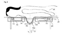
- a diaphragm 56 is provided, which is set in such a way that the X-radiation of the X-ray device detects only a predetermined range. This aperture is adjusted for the individual measurements depending on the position of the gantry.
- the diaphragm 56 is set for each of these positions such that the X-ray radiation of the X-ray device detects only a predetermined range.
- a plurality of recordings are made during a circular or spiral movement of a gantry 10 in order to produce the at least one control recording and a diaphragm 56 is adjusted in accordance with the movement such that the x-ray radiation of the x-ray device captures only a predetermined range.
- the aperture can optionally be tracked in steps according to the movement or continuously.
- an X-ray device has means for collimating the X-ray radiation of the X-ray tube.
- a diaphragm 56 This diaphragm preferably allows a constriction of the fan beam 16 in a plane perpendicular to the axis of rotation 12.
- a narrowing in a plane parallel to the axis of rotation is possible.
- At least one container with a reference medium or a reference liquid 161 is provided.
- a reference liquid may be, for example, water or glycerol.
- a temperature sensor 162 can be used. Since the changes in the X-ray properties of the tissue due to temperature changes are only relatively small, a calibration of the X-ray apparatus can be carried out in each case by the reference liquid or by a reference medium in the general case. Preferably, the calibration or the Measurements of the reference medium before and / or during a reference recording and / or a control recording.
- Another object of the invention is an X-ray machine for carrying out the method described above.
- This x-ray device is preferably a CT scanner and has an adjustable diaphragm 56.
- a medical system according to the invention for the controlled diathermic treatment of body tissue comprises an X-ray machine as described above and a device for heating and / or cooling body tissue.
- a device for heating and / or cooling body tissue This may be, for example, a diathermy device or else a device for cryotherapeutic treatment.
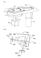
- FIG. 1 an X-ray device according to the invention for the examination or treatment of a female breast is shown.
- the gantry 10 is a spiral computed tomography gantry with an X-ray tube and a detector which rotate around the breast to be examined. During the rotation, the breast is imaged. Simultaneously with the rotation, a shift in the vertical direction is performed via the Gantryhubantrieb 11, so that the breast is scanned spirally.
- the patient couch 20 is height-adjustable via a patient couch lift drive 22.
- the diathermy device 150 energizes the instrument 151, which results in heating of the region of interest in the patient's chest.
- the device according to the invention is shown with a horizontally arranged bearing surface for the patient. In principle, however, a vertical or arranged at different angles support surface is possible.
- FIG. 2 the function of dynamic focusing is shown to minimize radiation exposure during temperature measurement.
- no complete, high-resolution 3-D scan is performed during the temperature measurement.
- an illustration is made of two positions offset by 90 degrees from one another here by way of example. These are the same components.
- the corresponding letter a for the first position and the letter b for the second position are indicated behind the reference symbol.
- Such an arrangement is usually sufficient to determine the temperature distribution with sufficient accuracy. This is also based on the knowledge that a roughly spherical temperature distribution develops in approximately homogeneous materials such as human body tissue.
- a first image is taken here with X-ray radiation starting from a position of the X-ray tube with a first focal point 55a.
- a diaphragm 56a adjusted to this position, a fan beam 16a is generated, which is just large enough to cover the region ROI 160 to be examined and as little radiation as possible is delivered to the neighborhood.
- the radiation is captured and evaluated by the detector in position 14a. It is still shown for this position, the associated central beam 52a.
- the position of the focal point 55b results.
- a fan beam 16b is generated, which in turn as accurately as possible comprises the region ROI 160 to be examined.
- the radiation is detected and evaluated by the detector in position 14b.
- the associated central beam 52b is the outside of the fan beam 16b.
- FIG. 3 a device according to the invention is shown in section.
- an x-ray tube 15 which generates a beam fan 16 for irradiating the breast 31 fixed in a fixing device 40.
- the radiation is received by a detector 14 and guided to an evaluation unit (not shown here).
- the gantry is rotatable on the one hand with a Gantryfiberlager 13 around the breast with the axis of rotation 12 and slidable over the Gantryhubantrieb 11 in height or at a distance to the patient.
- a spiral scan of the breast 31 to be examined is possible.
- the X-ray apparatus shown here allows the use of control recordings during the treatment, since the diathermy device passes through the gantry and does not hinder it during its spiral movement during a recording.
- a calibration of the device is possible.
- the exact temperature of the reference liquid is determined by the temperature sensor 162.
Abstract
Description
- Die Erfindung betrifft ein Röntgengerät sowie ein Verfahren zur Überwachung einer Diathermie- Behandlung. Die Erfindung betrifft insbesondere ein Röntgengerät zur Abbildung der weiblichen Brust (Mammografie) sowie ein Verfahren zur Überwachung einer diathermischen Brusttumor- Behandlung.
- Zur Untersuchung der weiblichen Brust sind verschiedene Röntgengeräte bekannt. Unter einer Liege, auf der eine zu untersuchende Patientin liegt, befindet sich eine Röntgenvorrichtung mit einer rotierenden Gantry, welche eine Röntgenröhre und einem Detektor aufweist. Ein solches Gerät ist beispielsweise in der
US 4,015,836 offenbart. Mit derartigen Röntgengeräten ist eine Diagnose von Erkrankungen der Brust, insbesondere von Tumoren in der Brust möglich. Diese Tumore werden auf verschiedene Arten behandelt. Ein bewährtes Verfahren ist die Hyperthermie. Hierbei wird das Gewebe im Bereich des Tumors so stark erhitzt, dass die Zellen des Tumors absterben. Ein solches Hyperthermiegerät ist in derUS 6,358,246 offenbart. Zur Behandlung des Tumors wird eine Elektrode in den Tumor eingebracht. Durch diese wird hochfrequenter Strom in das Gewebe des Tumors geleitet. Als Maß für die maximale Leistung wird hier ein Anstieg der elektrischen Impedanz des Gewebes herangezogen. Problematisch hierbei ist, dass die Erwärmung nicht gezielt gesteuert werden kann, denn die Temperatur ist keine direkte Funktion des Stromflusses oder der in das Gewebe eingebrachten Energie. Eine kontrollierte Therapie des Tumors ist mit einem solchen Instrument nur schwer möglich. - In der
US 6,684,097 B1 ist ein Verfahren zur Temperaturüberwachung während einer diathermischen Behandlung einer Brust offenbart. Dieses Verfahren fertigt vor der Behandlung eine hochauflösende dreidimensionale Röntgenaufnahme von der Brust an und speichert diese ab. Während der Behandlung werden weitere hochauflösende dreidimensionale Aufnahmen von der Brust angefertigt und mit der ursprünglichen Aufnahme verglichen. Aus einer Differenz der einzelnen Pixelwerte wird auf eine Temperaturänderung geschlossen. Nachteilig an diesem Verfahren ist die hohe Strahlenbelastung durch die hochauflösenden dreidimensionalen Aufnahmen. - Der Erfindung liegt die Aufgabe zugrunde, ein medizinisches Gerät vorzustellen, mit dem eine thermische Tumorbehandlung, insbesondere eine diathermische Tumorbehandlung einer weiblichen Brust kontrolliert erfolgen kann, wobei die Strahlenbelastung gegenüber dem Stand der Technik wesentlich reduziert wird. Ein weiterer Aspekt der Erfindung ist ein Verfahren zur Überwachung und/oder Steuerung eines medizinischen Gerätes zur Überwachung beziehungsweise zu kontrollierten diathermischen Tumorbehandlung, insbesondere einer weiblichen Brust, wobei die Strahlenbelastung gegenüber dem Stand der Technik wesentlich reduziert wird.
- Diese Aufgabe wird durch ein Röntgengerät nach Anspruch 1 gelöst. Vorteilhafte Ausgestaltungen der Erfindung sind in den Unteransprüchen angegeben.
- Ein erfindungsgemäßes Verfahren umfasst die folgenden Schritte:
- a. Erstellen einer Röntgenaufnahme von der zu behandelnden Region; Hierbei wird zuerst wenigstens eine Referenzaufnahme des zu behandelnden Gewebes erstellt. Grundsätzlich kann eine Vielzahl von verschiedenen Referenzaufnahmen erstellt werden.
- b. Thermische Behandlung;
Nach der Referenzaufnahme erfolgt eine diathermische Behandlung durch Erwärmen des Gewebes. Es kann eine neue Behandlung begonnen oder auch eine vorhergehende Behandlung fortgesetzt werden. Zur Erwärmung wird Energie, vorzugsweise als elektrische Energie, aber auch durch Bestrahlung in das Gewebe eingekoppelt. - c. Erstellen von Kontrollaufnahmen;
Zur Kontrolle der diathermischen Behandlung werden vorzugsweise in vorgegebenen Intervallen Röntgenaufnahmen angefertigt. Diese werden mit wenigstens einer der in Schritt a. angefertigten Referenzaufnahmen verglichen. Im einfachsten Falle erfolgt eine einfache Subtraktion der Helligkeitswerte. Aufgrund der Differenz zur Referenzaufnahme kann auf die Temperaturänderung des Gewebes geschlossen werden. Damit kann die Funktion des Diathermiegerätes überwacht werden. Kontrollaufnahmen können grundsätzlich während der Behandlung oder auch in kurzen Behandlungspausen durchgeführt werden. Erfindungsgemäß erfolgen diese Kontrollaufnahmen mit einer insgesamt niedrigeren Strahlendosis als eine normale hochauflösende dreidimensionale Aufnahme. Weiterhin wird diese Aufnahme als Teilvolumenaufnahme realisiert, wobei nur ein bestimmter für die Temperaturüberwachung ausgewählter Bereich bestrahlt und aufgenommen wird. - Röntgenuntersuchungen haben gezeigt, dass sich bei Diathermiebehandlungen, d.h. bei einer Temperaturänderung des Gewebes die Röntgeneigenschaften um einige Hounsfield Units ändern. Der zu Grunde liegende physikalische Effekt ist vermutlich eine Dichteänderung des Gewebes.
- Durch ein erfindungsgemäßes Verfahren ist es nun möglich, ein Temperaturprofil der thermisch behandelten Region zu erstellen. Durch den Einsatz von 3 D-Röntgengeräten kann auch ein räumliches Temperaturprofil erstellt werden. Aufgrund des Temperaturprofils kann der Verlauf der Behandlung präzise kontrolliert werden. Entsprechend kann das die Temperaturänderung erzeugende Gerät entsprechend eingestellt und/oder geregelt werden. Ebenso kann bei Erreichen eines bestimmten Temperaturprofils auch die Behandlung beendet werden. Bei einem erfindungsgemäßen Verfahren kann die Strahlendosis relativ klein gehalten werden, da nur eine Teilvolumenaufnahme des erwärmten Bereichs erstellt werden muss. Weiterhin kann durch das Röntgengerät bereits vor Beginn der Behandlung und auch während der Behandlung das die Temperaturänderung erzeugende Gerät oder eine Sonde des Gerätes in dem Gewebe präzise positioniert werden.
- Bevorzugt rotiert die Gantry während der thermischen Behandlung kontinuierlich und fertigt in vorgegebenen Zeitabständen Messungen an. Bevorzugt umfasst eine Messung zwei Aufnahmen, die um 90 Grad gegeneinander versetzt sind. Selbstverständlich wird nur zur Durchführung der Messungen beziehungsweise Abbildungen die Röntgenröhre aktiviert. Da es hier genügt, die Messungen in relativ großen Zeitintervallen, beispielsweise 1, 5, 10, 20 oder 60 Sekunden durchzuführen, kann die Gantry relativ langsam rotieren. Bei den größeren Zeitintervallen wie 10, 20 oder 60 Sekunden, rotiert die Gantry bevorzugt mit einer Geschwindigkeit, die diese mehrere Umdrehungen innerhalb dieser Zeitintervallen durchführen lässt, so dass der zeitliche Versatz zwischen zwei um 90 Grad gegeneinander versetzten Messungen reduziert wird. Beispielhaft wird bei Messintervallen von 60 Sekunden eine Umdrehung pro Sekunde durchgeführt.
- Damit ist der Zeitversatz zwischen zwei um 90 Grad gegeneinander versetzten Messungen 0,25 Sekunden.
- Bevorzugt ist eine Blende 56 vorgesehen, die derart eingestellt wird, dass die Röntgenstrahlung des Röntgengerätes nur einen vorgegebenen Bereich erfasst. Diese Blende wird bei den einzelnen Messungen abhängig von der Position der Gantry eingestellt.
- Alternativ zu den um 90 Grad versetzten Aufnahmen können auch mehrere Aufnahmen aus verschiedenen Positionen der Gantry 10 durchgeführt werden. Hierbei wird die Blende 56 für jede dieser Positionen derart eingestellt, dass die Röntgenstrahlung des Röntgengerätes nur einen vorgegebenen Bereich erfasst.
- Besonders günstig ist es, wenn zum Erstellen der wenigstens einen Kontrollaufnahme mehrere Aufnahmen während einer Kreis- oder Spiralbewegung einer Gantry 10 durchgeführt werden und eine Blende 56 entsprechend der Bewegung derart eingestellt wird, dass die Röntgenstrahlung des Röntgengerätes nur einen vorgegebenen Bereich erfasst. Hierbei kann die Blende wahlweise in Schritten entsprechend der Bewegung oder auch stufenlos nachgeführt werden.
- Ein erfindungsgemäßes Röntgengerät umfasst eine Auflagefläche 20 für einen Patienten 30 sowie eine Gantry 10 mit einer Röntgenröhre und einem Detektor, welche durch einen Z-Achsen-Antrieb gegenüber dem Patienten verschoben werden kann. Weiterhin bietet das Röntgengerät einen Zugang für ein Gerät, welches das zu behandelnde Gewebe erwärmt und/oder abkühlt. Die Erwärmung bzw. Abkühlung des Gewebes kann durch
- elektrischen Strom, vorzugsweise hochfrequenten Strom,
- fokussierten Ultraschall,
- Wärmestrahlung, bevorzugt im fernen Infrarotbereich,
- Terahertz-Strahlung,
- Ein Wärme und/oder Kühlmedium wie eine Flüssigkeit oder ein Gas, bevorzugt ein flüssiges Gas, besonders bevorzugt flüssiger Stickstoff
- Weiterhin weist ein erfindungsgemäßes Röntgengerät Mittel zur Kollimierung der Röntgenstrahlung der Röntgenröhre auf. Um Aufnahmen aus mehreren Positionen anfertigen zu können, ist es notwendig, die Strahlkollimierung positionsabhängig einzustellen. Dies erfolgt mittels einer Blende 56. Diese Blende ermöglicht bevorzugt eine Einengung des Strahlenfächer 16 in einer Ebene senkrecht zur Rotationsachse 12. Optional ist auch noch eine Einengung in einer Ebene parallel zur Rotationsachse möglich.
- Besonders günstig ist es, wenn Methoden und Verfahren zur Verbesserung der zeitlichen und/oder räumlichen Auflösung bei der Kombination der verschiedenen Aufnahmen genutzt werden. Da sich die Aufnahmen nur innerhalb lokaler Parameter, wie z.B. die Temperatur in einem bestimmten zu therapierenden Subvolumen unterscheiden verbessert die zeitlich/räumliche Verknüpfung der Aufnahmen die Bildqualität bzw. minimiert bei gleichbleibender Bildqualität die dem Patienten zu applizierende Strahlendosis. Besonders vorteilhaft sind Verfahren, die unter dem Namen PICCS (Prior image constrained compressed sensing) und HYPR (HighlY constrained backPRojection) bekannt sind.
- In einer weiteren Ausführung von der Erfindung ist wenigstens ein Behälter mit einem Referenzmedium beziehungsweise einer Referenzflüssigkeit 161 vorgesehen. Eine solche Referenzflüssigkeit kann beispielsweise Wasser oder Glycerin sein. Zur exakten Temperaturüberwachung kann ein Temperatursensor 162 dienen. Da die Änderungen der Röntgeneigenschaften des Gewebes durch Temperaturänderungen nur relativ gering sind, kann jeweils durch die Referenzflüssigkeit oder durch ein Referenzmedium im allgemeinen Falle eine Kalibrierung des Röntgengerätes erfolgen. Bevorzugt erfolgt die Kalibrierung beziehungsweise die Messungen des Referenzmediums vor und/oder während einer Referenzaufnahme und/oder einer Kontrollaufnahme.
- Ein weiterer Gegenstand der Erfindung ist ein Röntgengerät zur Durchführung der zuvor beschriebenen Verfahren. Dieses Röntgengerät ist bevorzugt ein CT-Scanner und weist eine verstellbare Blende 56 auf. Bevorzugterweise ist auch noch wenigstens ein Referenzmedium 161 vorgesehen.
- Ein erfindungsgemäßes medizinisches System zur kontrollierten diathermischen Behandlung von Körpergewebe umfasst ein Röntgengerät wie zuvor dargestellt, sowie ein Gerät zur Erwärmung und/oder Abkühlung von Körpergewebe. Es kann sich hierbei beispielsweise um ein Diathermiegerät oder auch um ein Gerät zur kryotherapeutischen Behandlung handeln.
- Die Erfindung wird nachstehend ohne Beschränkung des allgemeinen Erfindungsgedankens anhand von Ausführungsbeispielen unter Bezugnahme auf die Zeichnungen exemplarisch beschrieben.
-
Figur 1 zeigt ein erfindungsgemäßes Röntgengerät zur Untersuchung bzw. Behandlung einer weiblichen Brust -
Figur 2 zeigt die Funktion der dynamischen Kollimierung -
Figur 3 zeigt ein erfindungsgemäßes Röntgengerät im seitlichen Schnitt - In
Figur 1 ist ein erfindungsgemäßes Röntgengerät zur Untersuchung bzw. Behandlung einer weiblichen Brust dargestellt. Auf der Auflagefläche 20 liegt eine Patientin 30. Die zu untersuchende Brust hängt über einen Brustausschnitt 21 durch die Patientenliege 20 in den Aufnahmebereich einer Gantry 10. Die Gantry 10 ist eine Spiral-Computertomographen Gantry mit einer Röntgenröhre und einem Detektor, welche sich um die zu untersuchende Brust drehen. Während der Drehung wird die Brust abgebildet. Gleichzeitig mit der Drehung wird über den Gantryhubantrieb 11 eine Verschiebung in vertikaler Richtung durchgeführt, so dass die Brust spiralförmig abgetastet wird. Die Patientenliege 20 ist über einen Patientenliegenhubantrieb 22 in der Höhe verstellbar. Das Diathermiegerät 150 versorgt das Instrument 151 mit Energie, welche zu einer Erwärmung der betreffenden Region in der Brust der Patientin führt. In diesem Beispiel ist das Erfindungsgemäße Gerät mit einer horizontal angeordneten Auflagefläche für den Patienten dargestellt. Grundsätzlich ist aber auch eine senkrecht stehende oder in anderen Winkeln angeordnete Auflagefläche möglich. - In
Figur 2 ist die Funktion der der dynamischen Fokussierung zur Minimierung der Strahlungsbelastung während der Temperaturmessung dargestellt. Um die Strahlungsbelastung möglichst gering zu halten wird während der Temperaturmessung kein vollständiger, hochauflösender 3-D-Scan vorgenommen. Vielmehr wird hier beispielhaft jeweils eine Abbildung aus zwei um 90 Grad zueinander versetzten Positionen vorgenommen. Es handelt sich hierbei um dieselben Komponenten. Entsprechend der Position ist hinter dem Bezugszeichen der entsprechende Buchstabe a für die erste Position und der Buchstabe b für die zweite Position angegeben. Eine solche Anordnung reicht in der Regel aus, um mit hinreichender Genauigkeit die Temperaturverteilung zu ermitteln. Diese basiert auch auf der Erkenntnis, dass sich in näherungsweise homogenen Materialien wie dem menschlichen Körpergewebe eine in erster Näherung kugelförmige Temperaturverteilung entwickelt. Eine erste Aufnahme erfolgt hier mit Röntgenstrahlung ausgehend von einer Position der Röntgenröhre mit einem ersten Fokuspunkt 55a. Durch eine zu dieser Position entsprechend eingestellte Blende 56a wird ein Strahlenfächer 16a erzeugt, welcher gerade so groß ist, dass der zu untersuchende Bereich ROI 160 abgedeckt wird und möglichst wenig Strahlung in die Nachbarschaft abgegeben wird. Die Strahlung wird mit dem Detektor in der Position 14a aufgefangen und ausgewertet. Es ist noch der zu dieser Position der zugehörige Zentralstrahl 52a dargestellt. In einer weiteren Position der Röntgenröhre ergibt sich die Position des Fokuspunkt 55b. Hier wird durch eine entsprechend angepasste Einstellung der Blende 56b ein Strahlenfächer 16b erzeugt, der nun wiederum möglichst genau den zu untersuchenden Bereich ROI 160 umfasst. Die Strahlung wird durch den Detektor in der Position 14b erfasst und ausgewertet. Der zugehörige Zentralstrahl 52b liegt die außerhalb des Strahlenfächers 16b. - In
Figur 3 ist eine erfindungsgemäße Vorrichtung im Schnitt dargestellt. Innerhalb des Gantrygehäuses 29 befindet sich eine Röntgenröhre 15, welche einen Strahlenfächer 16 zur Durchstrahlung der in einer Fixiervorrichtung 40 fixierten Brust 31 erzeugt. Die Strahlung wird von einem Detektor 14 empfangen und zu einer Auswerteeinheit (hier nicht dargestellt) geführt. Die Gantry ist einerseits mit einem Gantrydrehlager 13 um die Brust mit der Drehachse 12 drehbar und über den Gantryhubantrieb 11 in ihrer Höhe beziehungsweise im Abstand zur Patientin verschiebbar. Damit ist eine spiralförmige Abtastung der zu untersuchenden Brust 31 möglich. Das hier dargestellte Röntgengerät erlaubt eine Durchführung von Kontrollaufnahmen während der Behandlung, da das Diathermiegerät durch die Gantry hindurchgreift und diese bei ihrer Spiralbewegung während einer Aufnahme nicht behindert. Mit dem Behälter mit einem Referenzmedium 161 ist eine Kalibrierung der Vorrichtung möglich. Die exakte Temperatur der Referenzflüssigkeit wird durch den Temperatursensor 162 ermittelt. Optional sind noch Mittel zum Beheizen und/oder Abkühlen der Referenzflüssigkeit vorhanden. -
- 10
- Gantry
- 11
- Z-Achsen-Antrieb
- 12
- Rotationsachse
- 13
- Gantrydrehlager
- 14
- Detektor
- 15
- Röntgenröhre
- 16
- Strahlenfächer
- 20
- Auflagefläche
- 21
- Brustausschnitt
- 22
- Patientenliegenhubantrieb
- 29
- Gantrygehäuse
- 30
- Patient
- 31
- Brust
- 40
- Fixiervorrichtung
- 52
- Zentralstrahl
- 55
- Fokuspunkt (Brennfleck)
- 56
- Blende
- 150
- Diathermiegerät
- 151
- Gerät zur thermischen Behandlung
- 160
- ROI Region Of Interest
- 161
- Referenzmedium
- 162
- Temperatursensor
Claims (15)
- Verfahren zur Überwachung eines Gerätes zur thermischen Behandlung von Körpergewebe einer bestimmten Region eines menschlichen Körpers umfassend die Schritte:a. Erstellen wenigstens einer dreidimensionalen Referenzaufnahme von der zu behandelnden Region durch ein Röntgengerät;b. Durchführen der thermischen Behandlung;c. Erstellen von wenigstens einer Kontrollaufnahme durch das Röntgengerät;d. Erstellen eines Temperaturänderungsprofils durch Vergleich der wenigstens einen Kontrollaufnahme mit der wenigstens einen Referenzaufnahme,dadurch gekennzeichnet, dass
zum Erstellen der wenigstens einen Kontrollaufnahme diese als eine Teilvolumenaufnahme der zu behandelnden Region mit niedrigerer Strahlendosis als eine normale dreidimensionale Röntgenaufnahme durchgeführt wird - Verfahren nach Anspruch 1,
dadurch gekennzeichnet, dass
vor dem Erstellen der wenigstens einen Kontrollaufnahme eine Blende (56) derart eingestellt wird, dass die Röntgenstrahlung des Röntgengerätes nur einen vorgegebenen Bereich erfasst. - Verfahren nach Anspruch 1 oder 2,
dadurch gekennzeichnet, dass
zum Erstellen der wenigstens einen Kontrollaufnahme zwei Aufnahmen aus zwei um 90 Grad gegeneinander versetzten Positionen einer Gantry (10) mit Röntgenröhre (15) und Röntgendetektor (14) durchgeführt werden. - Verfahren nach Anspruch 2,
dadurch gekennzeichnet, dass
zum Erstellen der wenigstens einen Kontrollaufnahme zwei Aufnahmen aus zwei um 90 Grad gegeneinander versetzten Positionen einer Gantry (10) mit Röntgenröhre (15) und Röntgendetektor (14) durchgeführt werden und eine Blende (56) für jede dieser Positionen derart eingestellt wird, dass die Röntgenstrahlung des Röntgengerätes nur einen vorgegebenen Bereich erfasst. - Verfahren nach Anspruch 2,
dadurch gekennzeichnet, dass
zum Erstellen der wenigstens einen Kontrollaufnahme mehrere Aufnahmen aus verschiedenen Positionen einer Gantry (10) mit Röntgenröhre (15) und Röntgendetektor (14) durchgeführt werden und eine Blende (56) für jede dieser Positionen derart eingestellt wird, dass die Röntgenstrahlung des Röntgengerätes nur einen vorgegebenen Bereich erfasst. - Verfahren nach Anspruch 2,
dadurch gekennzeichnet, dass
zum Erstellen der wenigstens einen Kontrollaufnahme mehrere Aufnahmen während einer Kreis- oder Spiralbewegung einer Gantry (10) mit Röntgenröhre (15) und Röntgendetektor (14) durchgeführt werden und eine Blende (56) entsprechend der Bewegung derart eingestellt wird, dass die Röntgenstrahlung des Röntgengerätes nur einen vorgegebenen Bereich erfasst. - Verfahren nach einem der vorhergehenden Ansprüche,
dadurch gekennzeichnet, dass
Verfahren zur Verbesserung der zeitlichen und/oder räumlichen Auflösung wie Namen PICCS (Prior image constrained compressed sensing) und HYPR (HighlY constrained backPRojection) eingesetzt werden. - Verfahren nach Anspruch 1,
dadurch gekennzeichnet, dass
das Röntgengerät wahlweise vor oder gleichzeitig mit der Durchführung einer Referenzaufnahme oder einer Kontrollaufnahme durch Vergleich mit wenigstens einem Referenzmedium 161 kalibriert wird. - Verfahren nach Anspruch 8,
dadurch gekennzeichnet, dass
mit der Durchführung einer Referenzaufnahme oder einer Kontrollaufnahme auch die Aufnahme wenigstens eines Referenzmediums 161 durchgeführt wird. - Verfahren nach Anspruch 8 oder 9,
dadurch gekennzeichnet, dass
gleichzeitig die Temperatur des Referenzmediums gemessen wird. - Röntgengerät zur Durchführung des Verfahrens nach Anspruch 1.
- Röntgengerät nach Anspruch 11,
dadurch gekennzeichnet, dass
das Röntgengerät ein CT-Scanner ist. - Röntgengerät nach Anspruch 12,
dadurch gekennzeichnet, dass
das Röntgengerät eine verstellbare Blende (56) aufweist. - Röntgengerät nach Anspruch 13,
dadurch gekennzeichnet, dass
ein Referenzmedium (161) zur Kalibrierung vorgesehen ist. - Medizinisches System zur kontrollierten thermischen Behandlung von Körpergewebe,
umfassend einen Röntgengerät nach einem der Ansprüche 10 bis 13 sowie ein Gerät zur Erwärmung und/oder Abkühlung von Körpergewebe einer bestimmten Region.
Applications Claiming Priority (1)
| Application Number | Priority Date | Filing Date | Title |
|---|---|---|---|
| DE102008042430 | 2008-09-29 |
Publications (1)
| Publication Number | Publication Date |
|---|---|
| EP2168487A1 true EP2168487A1 (de) | 2010-03-31 |
Family
ID=40524871
Family Applications (8)
| Application Number | Title | Priority Date | Filing Date |
|---|---|---|---|
| EP09154863A Withdrawn EP2168491A1 (de) | 2008-09-29 | 2009-03-11 | Brustfixierung mit Probencontainer für ein Untersuchungsgerät der weiblichen Brust |
| EP09154848A Withdrawn EP2168490A1 (de) | 2008-09-29 | 2009-03-11 | Röntgengerät zur Brustuntersuchung mit einer Detektor-Röhren Anordnung für hochauflösende Aufnahmen |
| EP09154884.2A Withdrawn EP2178048A3 (de) | 2008-09-29 | 2009-03-11 | Verfahren zur Definition eines patientenindividuellen Koordinationssystems einer weiblichen Brust |
| EP09154842A Expired - Fee Related EP2168484B1 (de) | 2008-09-29 | 2009-03-11 | Röntgengerät zur Brustuntersuchung mit einer in eine Patientenliege integrierten Gantry |
| EP09154900A Withdrawn EP2168487A1 (de) | 2008-09-29 | 2009-03-11 | Verfahren und Vorrichtung zur thermischen Brusttumor-Behandlung mit 3D Monitorfunktion |
| EP09154891A Expired - Fee Related EP2168486B1 (de) | 2008-09-29 | 2009-03-11 | Modulares System zur Brustdiagnose und -intervention |
| EP09154854A Withdrawn EP2168485A1 (de) | 2008-09-29 | 2009-03-11 | Brustfixierung für ein Untersuchungsgerät zur Untersuchung der weiblichen Brust |
| EP09154833A Expired - Fee Related EP2168489B1 (de) | 2008-09-29 | 2009-03-11 | Röntgengerät zur Brustuntersuchung im Stehen |
Family Applications Before (4)
| Application Number | Title | Priority Date | Filing Date |
|---|---|---|---|
| EP09154863A Withdrawn EP2168491A1 (de) | 2008-09-29 | 2009-03-11 | Brustfixierung mit Probencontainer für ein Untersuchungsgerät der weiblichen Brust |
| EP09154848A Withdrawn EP2168490A1 (de) | 2008-09-29 | 2009-03-11 | Röntgengerät zur Brustuntersuchung mit einer Detektor-Röhren Anordnung für hochauflösende Aufnahmen |
| EP09154884.2A Withdrawn EP2178048A3 (de) | 2008-09-29 | 2009-03-11 | Verfahren zur Definition eines patientenindividuellen Koordinationssystems einer weiblichen Brust |
| EP09154842A Expired - Fee Related EP2168484B1 (de) | 2008-09-29 | 2009-03-11 | Röntgengerät zur Brustuntersuchung mit einer in eine Patientenliege integrierten Gantry |
Family Applications After (3)
| Application Number | Title | Priority Date | Filing Date |
|---|---|---|---|
| EP09154891A Expired - Fee Related EP2168486B1 (de) | 2008-09-29 | 2009-03-11 | Modulares System zur Brustdiagnose und -intervention |
| EP09154854A Withdrawn EP2168485A1 (de) | 2008-09-29 | 2009-03-11 | Brustfixierung für ein Untersuchungsgerät zur Untersuchung der weiblichen Brust |
| EP09154833A Expired - Fee Related EP2168489B1 (de) | 2008-09-29 | 2009-03-11 | Röntgengerät zur Brustuntersuchung im Stehen |
Country Status (2)
| Country | Link |
|---|---|
| US (8) | US7924974B2 (de) |
| EP (8) | EP2168491A1 (de) |
Families Citing this family (62)
| Publication number | Priority date | Publication date | Assignee | Title |
|---|---|---|---|---|
| US8272088B2 (en) * | 2007-09-06 | 2012-09-25 | Orbital Therapy Llc | Patient support system for full access prone position breast radiotherapy |
| US7924974B2 (en) * | 2008-09-29 | 2011-04-12 | Mir Medical Imaging Research Holding Gmbh | X-ray machine for breast examination in a standing position |
| DE102008049711A1 (de) * | 2008-09-30 | 2010-04-15 | Siemens Aktiengesellschaft | Lagerungsvorrichtung, Patientenlagerungstisch und medizinisches Gerät |
| US8014490B2 (en) * | 2009-10-20 | 2011-09-06 | Linda Mitchell | Mammogram tender machine |
| US8421604B2 (en) * | 2009-11-30 | 2013-04-16 | Symbol Technologies, Inc. | Method and apparatus for identifying read zone of RFID reader |
| US8374312B2 (en) * | 2010-02-18 | 2013-02-12 | Varian Medical Systems, Inc. | Prone patient positioning devices and methods |
| DE102010011660A1 (de) * | 2010-03-17 | 2011-09-22 | Siemens Aktiengesellschaft | Mammographiegerät |
| JP5700950B2 (ja) * | 2010-04-21 | 2015-04-15 | キヤノン株式会社 | 生体情報取得装置 |
| US20140191852A1 (en) * | 2010-05-13 | 2014-07-10 | Carestream Health, Inc. | Method and system for phosphor plate identification in computed radiography |
| US20120001737A1 (en) * | 2010-05-13 | 2012-01-05 | Amir Berger | Method and system for computed radiography |
| GB2483640A (en) * | 2010-09-10 | 2012-03-21 | Specialty Magnetics Ltd | Breast immobilisation arrangement |
| CN103179906B (zh) | 2010-10-05 | 2016-04-06 | 霍洛吉克公司 | 具有ct模式、多层析摄像模式和乳腺摄像模式的竖立式x射线胸部成像 |
| US9668711B2 (en) | 2010-10-05 | 2017-06-06 | Hologic, Inc | X-ray breast tomosynthesis enhancing spatial resolution including in the thickness direction of a flattened breast |
| DE102010052603A1 (de) * | 2010-11-25 | 2012-05-31 | Artemis Imaging Gmbh | Patientenliege |
| WO2012120498A1 (en) * | 2011-03-04 | 2012-09-13 | Technion Research & Development | Non-invasive thermal treatment monitoring |
| DE102011006353A1 (de) | 2011-03-29 | 2012-10-04 | Siemens Aktiengesellschaft | Mammographieanlage |
| US9557281B2 (en) | 2011-06-09 | 2017-01-31 | The Regents Of The University Of California | Excised specimen imaging using a combined PET and micro CT scanner |
| US8842806B2 (en) | 2012-04-03 | 2014-09-23 | Carestream Health, Inc. | Apparatus and method for breast imaging |
| JP6226961B2 (ja) * | 2012-05-02 | 2017-11-08 | コーニンクレッカ フィリップス エヌ ヴェKoninklijke Philips N.V. | 撮像温度測定 |
| US9307961B2 (en) * | 2012-06-29 | 2016-04-12 | Carefusion 2200, Inc. | Fine needle aspiration biopsy device |
| KR102001926B1 (ko) * | 2012-09-11 | 2019-07-30 | 삼성디스플레이 주식회사 | 엑스레이 검출기, 이를 포함하는 엑스레이 검출 시스템 및 엑스레이 검출 방법 |
| DE102012216687A1 (de) * | 2012-09-18 | 2014-03-20 | Jan Rimbach | Vorrichtung zur Untersuchung von Prüfkörpern |
| DE102012217301B4 (de) | 2012-09-25 | 2021-10-14 | Bayer Pharma Aktiengesellschaft | Kombination aus Kontrastmittel und Mammographie-CT-System mit vorgegebenem Energiebereich und Verfahren zur Erzeugung tomographischer Mammographie-CT-Aufnahmen durch diese Kombination |
| CN103908343B (zh) | 2012-12-31 | 2016-10-05 | 西门子(深圳)磁共振有限公司 | 患者检查床和磁共振成像设备 |
| US9161725B1 (en) * | 2014-02-05 | 2015-10-20 | Regine Millien-White | Adjustable breast examination device |
| JP6376783B2 (ja) * | 2014-03-12 | 2018-08-22 | キヤノン株式会社 | 乳房断層撮影装置および制御方法 |
| JP6381253B2 (ja) * | 2014-03-31 | 2018-08-29 | キヤノン株式会社 | 放射線撮影装置、断層撮影装置 |
| US20170020410A1 (en) * | 2014-04-04 | 2017-01-26 | Pierfrancesco Pavoni | Access gate or gantry comprising an antennas assembly for therapy or imaging |
| US9326739B2 (en) * | 2014-04-28 | 2016-05-03 | Cheryl A. Galambos McLaughlin | Mammogram table |
| US9301726B2 (en) * | 2014-05-02 | 2016-04-05 | Wisconsin Alumni Research Foundation | CT machine for multi-angle scanning of stationary patients |
| CN104173075B (zh) * | 2014-08-26 | 2016-07-06 | 李丙曙 | 放射科检查床 |
| JP6611428B2 (ja) * | 2014-12-09 | 2019-11-27 | キヤノン株式会社 | マンモ断層撮像システム |
| EP3238628A4 (de) * | 2014-12-26 | 2018-08-15 | Rayence Co., Ltd. | Hebevorrichtung für ein kompressionspaddle und röntgenbildfotografievorrichtung damit |
| CN105832353B (zh) | 2015-01-30 | 2020-11-06 | 佳能株式会社 | 放射线摄像系统 |
| JP6651069B2 (ja) * | 2015-05-13 | 2020-02-19 | フジデノロ株式会社 | 固定具装着装置 |
| KR20160139292A (ko) * | 2015-05-27 | 2016-12-07 | 삼성전자주식회사 | Rf 표면 코일부 및 이를 포함하는 자기공명영상 시스템 |
| JP6525768B2 (ja) * | 2015-06-30 | 2019-06-05 | キヤノン株式会社 | 乳房撮影装置 |
| US10542951B2 (en) | 2015-07-23 | 2020-01-28 | General Electric Company | Systems, methods, and devices for simplified high quality imaging of biopsy samples on a mammography machine |
| US20190104967A1 (en) * | 2015-07-24 | 2019-04-11 | Tricia Dretzka-Kaye | Anatomy Scanning System and Method |
| US11076821B2 (en) | 2015-11-25 | 2021-08-03 | The Regents Of The University Of California | 3D-beam modulation filter for equalizing dose and image quality in breast CT |
| DE102015225236A1 (de) * | 2015-12-15 | 2017-06-22 | Siemens Healthcare Gmbh | Mammographie-Screening mit hoher Durchgangsrate |
| CN106933857B (zh) * | 2015-12-30 | 2020-12-29 | 创新先进技术有限公司 | 一种数据仓库中任务的调度方法、装置 |
| DE102016206198A1 (de) * | 2016-04-13 | 2017-10-19 | Siemens Healthcare Gmbh | Röntgensystem |
| US10603003B2 (en) * | 2016-04-14 | 2020-03-31 | Dedicating2Imaging, LLC | CT systems for imaging of the breast |
| WO2018051220A1 (en) * | 2016-09-14 | 2018-03-22 | Mor Research Applications Ltd. | Device, system and method for detecting irregularities in soft tissue |
| US10180207B1 (en) * | 2017-07-13 | 2019-01-15 | Danylo Kozub | Stand |
| CN108175430A (zh) * | 2018-01-17 | 2018-06-19 | 江苏美伦影像系统有限公司 | 一种具有辐射防护功能的乳腺x射线摄影系统 |
| US11318010B1 (en) * | 2018-04-02 | 2022-05-03 | Lifei Guo | Tissue removing |
| US10893844B1 (en) * | 2018-10-10 | 2021-01-19 | David Byron Douglas | Method and apparatus for performing 3D imaging examinations of a structure under differing configurations and analyzing morphologic changes |
| DE102018207636A1 (de) * | 2018-05-16 | 2019-11-21 | Siemens Healthcare Gmbh | Patiententisch mit Vorrichtung zur reversiblen Aufnahme einer Transferplatte |
| CN108956656B (zh) * | 2018-07-17 | 2021-02-05 | 青岛大学附属医院 | 一种高衬度低剂量相位衬度ct成像装置 |
| CN110975156B (zh) * | 2019-11-15 | 2021-11-19 | 山东大学齐鲁医院 | 乳房牵引固定装置及系统 |
| EP4125605A1 (de) * | 2020-03-31 | 2023-02-08 | Hologic, Inc. | Systeme und verfahren zur röntgenbildgebung von gewebeproben |
| KR102640269B1 (ko) * | 2020-05-29 | 2024-02-26 | (의료)길의료재단 | 유방암 치료용 방사선 조사 장치 |
| CN111714222B (zh) * | 2020-06-29 | 2021-07-23 | 北京欧扬医疗美容门诊部有限公司 | 一种无痕隆胸用脂肪自体植入装置 |
| CN111714191A (zh) * | 2020-06-30 | 2020-09-29 | 广西医科大学附属肿瘤医院 | 用于锥光束乳腺ct引导下悬垂穿刺的激光定位装置 |
| US11692951B2 (en) * | 2021-02-24 | 2023-07-04 | GE Precision Healthcare LLC | System and method for specimen imaging using an existing mammography imaging system |
| EP4226875A1 (de) * | 2022-02-09 | 2023-08-16 | Storz Medical AG | Stosswellenvorrichtung mit einer quelle, die sich selbst auf eine röntgenvorrichtung ausrichtet |
| EP4226877A1 (de) * | 2022-02-09 | 2023-08-16 | Storz Medical AG | Stosswellenvorrichtung mit integrierter ultraschallsonde |
| EP4226874A1 (de) * | 2022-02-09 | 2023-08-16 | Storz Medical AG | Ultraschall- und/oder stosswellenvorrichtung mit auf einer hexapod-plattform montierter quelle |
| EP4226876A1 (de) * | 2022-02-09 | 2023-08-16 | Storz Medical AG | Stosswellenvorrichtung mit verbesserter akustischer kopplung |
| WO2023200899A1 (en) * | 2022-04-14 | 2023-10-19 | Koning Corporation | Cone beam breast computed tomography with pivotal gantry subsystem |
Citations (4)
| Publication number | Priority date | Publication date | Assignee | Title |
|---|---|---|---|---|
| US6684097B1 (en) | 1999-04-22 | 2004-01-27 | University Of Miami | Intraoperative monitoring of temperature-induced tissue changes with a high-resolution digital x-ray system during thermotherapy |
| EP1549115A2 (de) * | 2003-12-26 | 2005-06-29 | GE Medical Systems Global Technology Company LLC | Verfahren zur Berechnung der Belichtung und Röntgengerät |
| DE102004042790A1 (de) * | 2004-09-03 | 2006-03-09 | Siemens Ag | Röntgeneinrichtung |
| US20080033420A1 (en) | 2006-08-04 | 2008-02-07 | Nields Morgan W | Methods for planning and performing thermal ablation |
Family Cites Families (106)
| Publication number | Priority date | Publication date | Assignee | Title |
|---|---|---|---|---|
| US3673394A (en) * | 1969-02-18 | 1972-06-27 | North American Rockwell | Measuring method and apparatus |
| US4015836A (en) * | 1975-07-31 | 1977-04-05 | General Electric Company | Mammography table |
| US4400827A (en) | 1981-11-13 | 1983-08-23 | Spears James R | Method and apparatus for calibrating rapid sequence radiography |
| US4680028A (en) * | 1984-07-02 | 1987-07-14 | Lact-Assist, Incorporated | Flexible breast receptor for breast pump |
| US4709382A (en) * | 1984-11-21 | 1987-11-24 | Picker International, Inc. | Imaging with focused curved radiation detectors |
| US5415169A (en) * | 1989-11-21 | 1995-05-16 | Fischer Imaging Corporation | Motorized mammographic biopsy apparatus |
| FI85803C (fi) | 1989-11-23 | 1992-06-10 | Planmed Oy | Foerfarande och anordning foer styrning av funktioner av en mammografiroentgenanordning. |
| US5569266A (en) | 1991-03-11 | 1996-10-29 | Fischer Imaging Corporation | Magnetic resonance imaging device useful for guiding a medical instrument |
| US5409497A (en) | 1991-03-11 | 1995-04-25 | Fischer Imaging Corporation | Orbital aiming device for mammo biopsy |
| US5289520A (en) | 1991-11-27 | 1994-02-22 | Lorad Corporation | Stereotactic mammography imaging system with prone position examination table and CCD camera |
| US5308321A (en) * | 1992-05-05 | 1994-05-03 | Castro Donna J | Retainer assisted by vacuum expansion system |
| US5273435B1 (en) * | 1992-07-16 | 1995-12-05 | Wisconsin Med College Inc | Tumor localization phantom |
| US5386447A (en) | 1992-09-23 | 1995-01-31 | Fischer Imaging Corporation | Mammographic screening and biopsy apparatus |
| US5490513A (en) * | 1992-09-28 | 1996-02-13 | Fonar Corporation | Multiple patient breast scanning on a magnetic resonance imaging apparatus |
| US6075879A (en) * | 1993-09-29 | 2000-06-13 | R2 Technology, Inc. | Method and system for computer-aided lesion detection using information from multiple images |
| JPH07303633A (ja) | 1994-05-11 | 1995-11-21 | Mitsubishi Electric Corp | X線乳房撮影装置 |
| US5528043A (en) | 1995-04-21 | 1996-06-18 | Thermotrex Corporation | X-ray image sensor |
| US5609827A (en) | 1995-05-02 | 1997-03-11 | Beekley Corporation | Biopsy specimen container |
| US5709206A (en) | 1995-11-27 | 1998-01-20 | Teboul; Michel | Imaging system for breast sonography |
| US5757878A (en) | 1996-08-16 | 1998-05-26 | Analogic Corporation | Detector arrangement for x-ray tomography system |
| DE19639975C1 (de) | 1996-09-27 | 1998-05-07 | Siemens Ag | Medizinische Einrichtung mit einer tunnelförmigen Öffnung zur Aufnahme eines Untersuchungsobjektes |
| JP2001524011A (ja) | 1997-05-06 | 2001-11-27 | クワンタ・ビジョン | 組織分析装置 |
| US6358246B1 (en) * | 1999-06-25 | 2002-03-19 | Radiotherapeutics Corporation | Method and system for heating solid tissue |
| US5991357A (en) | 1997-12-16 | 1999-11-23 | Analogic Corporation | Integrated radiation detecting and collimating assembly for X-ray tomography system |
| US6175117B1 (en) * | 1998-01-23 | 2001-01-16 | Quanta Vision, Inc. | Tissue analysis apparatus |
| DE19812995A1 (de) * | 1998-03-25 | 1999-10-07 | Siemens Ag | Mammographie-Gerät, insbesondere für Vergrößerungs-Mammographie |
| US6242743B1 (en) | 1998-08-11 | 2001-06-05 | Mosaic Imaging Technology, Inc. | Non-orbiting tomographic imaging system |
| JP2000116631A (ja) | 1998-10-16 | 2000-04-25 | Toshiba Corp | X線診断装置 |
| JP3866431B2 (ja) * | 1999-02-17 | 2007-01-10 | 株式会社東芝 | X線ct装置 |
| TW406009B (en) * | 1999-07-16 | 2000-09-21 | Nat Science Council | 3-D localization method of clustered microcalcifications using cranio-caudal and medio-lateral oblique views |
| US6254614B1 (en) * | 1999-10-18 | 2001-07-03 | Jerry M. Jesseph | Device and method for improved diagnosis and treatment of cancer |
| US6480565B1 (en) | 1999-11-18 | 2002-11-12 | University Of Rochester | Apparatus and method for cone beam volume computed tomography breast imaging |
| US6987831B2 (en) | 1999-11-18 | 2006-01-17 | University Of Rochester | Apparatus and method for cone beam volume computed tomography breast imaging |
| DE10026792A1 (de) | 2000-05-31 | 2001-12-06 | Bip Biomedizinische Instr & Pr | Diagnose- und Therapietisch |
| US6463122B1 (en) * | 2000-08-21 | 2002-10-08 | Bio-Imaging Resource, Inc. | Mammography of computer tomography for imaging and therapy |
| US7467892B2 (en) | 2000-08-29 | 2008-12-23 | Imaging Therapeutics, Inc. | Calibration devices and methods of use thereof |
| US7940966B2 (en) | 2000-11-24 | 2011-05-10 | U-Systems, Inc. | Full-field breast image data processing and archiving |
| US6419390B1 (en) | 2001-03-26 | 2002-07-16 | Marianette Landis-Lowell | Folding mammography table and method of use |
| US6516045B2 (en) | 2001-05-04 | 2003-02-04 | The Regents Of The University Of California | Device and method for determining proportions of body materials |
| US6418188B1 (en) | 2001-06-14 | 2002-07-09 | Juanita L. Broadnax | Radiation breast cup and method |
| US6674835B2 (en) | 2001-10-12 | 2004-01-06 | General Electric Co. | Methods and apparatus for estimating a material composition of an imaged object |
| US6671975B2 (en) | 2001-12-10 | 2004-01-06 | C. William Hennessey | Parallel kinematic micromanipulator |
| DE10207623B4 (de) | 2002-02-22 | 2004-05-06 | Siemens Ag | Verfahren für die Computertomographie sowie Computertomographie (CT)-Gerät |
| US20040254461A1 (en) | 2002-03-20 | 2004-12-16 | Ackerman William H. | Acoustic beam shaping by pulse power modulation at constant amplitude |
| US7783089B2 (en) * | 2002-04-15 | 2010-08-24 | General Electric Company | Method and apparatus for providing mammographic image metrics to a clinician |
| US7218766B2 (en) | 2002-04-15 | 2007-05-15 | General Electric Company | Computer aided detection (CAD) for 3D digital mammography |
| CA2393101A1 (en) * | 2002-07-11 | 2004-01-11 | Martin Cyr | Apparatus, system and method of calibrating medical imaging systems |
| US20040082856A1 (en) | 2002-07-16 | 2004-04-29 | Alfred E. Mann Institute For Biomedical Engineering, University Of Southern California | Support bra for ultrasonic breast scanner |
| US6904119B2 (en) | 2002-10-02 | 2005-06-07 | Shimadzu Corporation | Radiographic apparatus |
| US7149566B2 (en) * | 2002-10-31 | 2006-12-12 | Manoa Medical, Inc. | Soft tissue orientation and imaging guide systems and methods |
| US7809422B2 (en) * | 2002-11-08 | 2010-10-05 | Art Advanced Research Technologies Inc. | Method and apparatus for optical imaging |
| US7286634B2 (en) * | 2002-12-23 | 2007-10-23 | Select Technologies, Llc | Method and apparatus for improving baggage screening examination |
| WO2004073524A1 (en) | 2003-02-20 | 2004-09-02 | Manoa Medical, Inc. | Bendable cutting device |
| US6872001B1 (en) | 2003-05-05 | 2005-03-29 | Peco Controls Corp. | X-ray shielding structure for food inspection station |
| US7850613B2 (en) * | 2003-05-30 | 2010-12-14 | Orison Corporation | Apparatus and method for three dimensional ultrasound breast imaging |
| US6982424B2 (en) | 2003-06-02 | 2006-01-03 | Ge Medical Systems Global Technology Company, Llc | X-ray and CT image detector |
| US7291841B2 (en) * | 2003-06-16 | 2007-11-06 | Robert Sigurd Nelson | Device and system for enhanced SPECT, PET, and Compton scatter imaging in nuclear medicine |
| US6837772B1 (en) | 2003-07-18 | 2005-01-04 | Regina Miracle International Limited | Breast cup construction |
| GB0318701D0 (en) | 2003-08-08 | 2003-09-10 | Inst Of Cancer Res The | A method and apparatus for image processing |
| JP2005258370A (ja) | 2003-09-05 | 2005-09-22 | Fuji Photo Film Co Ltd | 放射線カセッテ |
| US7005988B2 (en) | 2003-09-19 | 2006-02-28 | International Business Machines Corporation | Using radio frequency identification to detect and/or prevent theft and shoplifting |
| US20050070817A1 (en) | 2003-09-30 | 2005-03-31 | Mueller Richard L. | Lavage assist device |
| US20050096515A1 (en) * | 2003-10-23 | 2005-05-05 | Geng Z. J. | Three-dimensional surface image guided adaptive therapy system |
| US7653229B2 (en) * | 2003-12-23 | 2010-01-26 | General Electric Company | Methods and apparatus for reconstruction of volume data from projection data |
| US7519209B2 (en) * | 2004-06-23 | 2009-04-14 | Vanderbilt University | System and methods of organ segmentation and applications of same |
| US20060145871A1 (en) | 2004-12-02 | 2006-07-06 | Smith & Nephew, Inc. | Radio Frequency Identification for Medical Devices |
| US7564945B2 (en) | 2005-02-11 | 2009-07-21 | University Of Florida Research Foundation, Inc. | System including computed tomography device for image guided treatment |
| US20060239398A1 (en) | 2005-03-07 | 2006-10-26 | Fused Multimodality Imaging, Ltd. | Breast diagnostic apparatus for fused SPECT, PET, x-ray CT, and optical surface imaging of breast cancer |
| US20100177866A1 (en) | 2005-04-01 | 2010-07-15 | Keizi Shibuya | Mammography Equipment |
| WO2006119426A2 (en) | 2005-05-03 | 2006-11-09 | Regents Of The University Of California | Biopsy systems for breast computed tomography |
| DE102005022347B4 (de) | 2005-05-13 | 2010-08-12 | Siemens Ag | Medizintechnisches Basissystem und medizintechnisches System |
| US7573034B2 (en) * | 2005-05-18 | 2009-08-11 | Carestream Health, Inc. | Mobile radiography image recording system |
| US7492858B2 (en) | 2005-05-20 | 2009-02-17 | Varian Medical Systems, Inc. | System and method for imaging and treatment of tumorous tissue in breasts using computed tomography and radiotherapy |
| EP1893077A4 (de) * | 2005-06-02 | 2011-02-09 | Medipattern Corp | Computergestütztes nachweissystem und verfahren |
| US7304578B1 (en) * | 2005-06-02 | 2007-12-04 | Hewlett-Packard Development Company, L.P. | Tag including RFID circuit storing data modifiable using a physically alterable medium |
| DE602006018934D1 (de) * | 2005-07-08 | 2011-01-27 | Wisconsin Alumni Res Found | Rückprojektions-rekonstruktionsverfahren für ct-bildgebung |
| US20070064867A1 (en) | 2005-09-20 | 2007-03-22 | Hansen Timothy B | Apparatus and method to acquire data for reconstruction of images pertaining to functional and anatomical structure of the breast |
| JP4837507B2 (ja) * | 2005-10-06 | 2011-12-14 | 富士フイルム株式会社 | 乳房画像撮影装置 |
| DE102005048049B4 (de) | 2005-10-07 | 2010-09-23 | Karlsruher Institut für Technologie | Vorrichtung zur bildgestützten Mammadiagnose und -therapie |
| US7742796B2 (en) * | 2005-10-25 | 2010-06-22 | General Electric Company | Breast immobilization device and method of imaging the breast |
| WO2007089362A2 (en) | 2005-11-07 | 2007-08-09 | Sommer Jr Edward J | Method and apparatus for improving identification and control of articles passing through a scanning system |
| DE102005053993A1 (de) * | 2005-11-10 | 2007-05-24 | Siemens Ag | Diagnosevorrichtung und Diagnoseverfahren für kombinierte und/oder kombinierbare radiographische und nuklearmedizinische Untersuchungen |
| US8014576B2 (en) | 2005-11-23 | 2011-09-06 | The Medipattern Corporation | Method and system of computer-aided quantitative and qualitative analysis of medical images |
| WO2007111669A2 (en) * | 2005-12-22 | 2007-10-04 | Visen Medical, Inc. | Combined x-ray and optical tomographic imaging system |
| CN101370429A (zh) * | 2006-01-17 | 2009-02-18 | 成象诊断系统公司 | 具有可变患者定位功能的激光成像设备 |
| US7806855B2 (en) * | 2006-04-11 | 2010-10-05 | Playtex Products, Inc. | Manual breast pump |
| US7483511B2 (en) * | 2006-06-06 | 2009-01-27 | Ge Homeland Protection, Inc. | Inspection system and method |
| US7840046B2 (en) * | 2006-06-27 | 2010-11-23 | Siemens Medical Solutions Usa, Inc. | System and method for detection of breast masses and calcifications using the tomosynthesis projection and reconstructed images |
| US7677799B2 (en) * | 2006-07-28 | 2010-03-16 | General Electric Company | Coordination of radiological imaging subsystems and components |
| US20080037703A1 (en) * | 2006-08-09 | 2008-02-14 | Digimd Corporation | Three dimensional breast imaging |
| WO2008024611A2 (en) | 2006-08-21 | 2008-02-28 | Ev Products, Inc. | Staggered array imaging system using pixilated radiation detectors |
| US7715523B2 (en) | 2006-09-28 | 2010-05-11 | Lafferty Peter R | System and apparatus for rapid stereotactic breast biopsy analysis |
| US20080084961A1 (en) | 2006-10-04 | 2008-04-10 | Cynthia Keppel | Method and apparatus for combined gamma/x-ray imaging in stereotactic biopsy |
| JP4857070B2 (ja) | 2006-10-11 | 2012-01-18 | キヤノン株式会社 | 乳房撮影用x線ct装置 |
| JP4851298B2 (ja) * | 2006-10-31 | 2012-01-11 | 富士フイルム株式会社 | 放射線断層画像生成装置 |
| WO2008054279A1 (en) | 2006-10-31 | 2008-05-08 | Xcounter Ab | Imaging arrangement and system for imaging |
| US20080221444A1 (en) | 2007-03-07 | 2008-09-11 | Ritchie Paul G | Integrated Imaging and Biopsy System with Integrated Surgical, Therapy, and Diagnostic Devices |
| US7597104B2 (en) | 2007-03-23 | 2009-10-06 | Zheng Mike Q | Method and device for immobilization of the human breast in a prone position for radiotherapy |
| JP3133186U (ja) | 2007-04-17 | 2007-07-05 | 岡崎産業株式会社 | ブラジャー用洗濯ケース |
| JP2008272093A (ja) | 2007-04-26 | 2008-11-13 | Toshiba Corp | 乳房用x線撮影装置および乳房用x線撮影方法 |
| US7453978B1 (en) * | 2007-06-25 | 2008-11-18 | University Of Tennessee Research Foundation | Variable resolution x-ray CT detector with multi-axis tilt |
| US7764765B2 (en) | 2007-07-24 | 2010-07-27 | Fujifilm Corporation | Cassette and mobile X-ray image capturing apparatus |
| WO2009026587A1 (en) | 2007-08-23 | 2009-02-26 | Fischer Medical Technologies, Inc. | Improved computed tomography breast imaging and biopsy system |
| US7697658B2 (en) | 2008-02-01 | 2010-04-13 | Virginia Tech Intellectual Properties, Inc. | Interior tomography and instant tomography by reconstruction from truncated limited-angle projection data |
| US7924974B2 (en) | 2008-09-29 | 2011-04-12 | Mir Medical Imaging Research Holding Gmbh | X-ray machine for breast examination in a standing position |
| EP2189114A1 (de) | 2008-11-22 | 2010-05-26 | MIR Medical Imaging Research Holding GmbH | Vorrichtung zur Fixierung der weiblichen Brust für die diagnostische Bildgebung und Intervention |
-
2009
- 2009-03-11 US US12/401,735 patent/US7924974B2/en not_active Expired - Fee Related
- 2009-03-11 EP EP09154863A patent/EP2168491A1/de not_active Withdrawn
- 2009-03-11 US US12/402,141 patent/US20100080349A1/en not_active Abandoned
- 2009-03-11 US US12/401,792 patent/US8102964B2/en not_active Expired - Fee Related
- 2009-03-11 EP EP09154848A patent/EP2168490A1/de not_active Withdrawn
- 2009-03-11 EP EP09154884.2A patent/EP2178048A3/de not_active Withdrawn
- 2009-03-11 US US12/401,814 patent/US7881427B2/en not_active Expired - Fee Related
- 2009-03-11 US US12/401,976 patent/US8199993B2/en not_active Expired - Fee Related
- 2009-03-11 EP EP09154842A patent/EP2168484B1/de not_active Expired - Fee Related
- 2009-03-11 US US12/402,225 patent/US7945019B2/en not_active Expired - Fee Related
- 2009-03-11 EP EP09154900A patent/EP2168487A1/de not_active Withdrawn
- 2009-03-11 EP EP09154891A patent/EP2168486B1/de not_active Expired - Fee Related
- 2009-03-11 EP EP09154854A patent/EP2168485A1/de not_active Withdrawn
- 2009-03-11 US US12/402,059 patent/US7869564B2/en active Active
- 2009-03-11 US US12/401,765 patent/US7864918B2/en active Active
- 2009-03-11 EP EP09154833A patent/EP2168489B1/de not_active Expired - Fee Related
Patent Citations (4)
| Publication number | Priority date | Publication date | Assignee | Title |
|---|---|---|---|---|
| US6684097B1 (en) | 1999-04-22 | 2004-01-27 | University Of Miami | Intraoperative monitoring of temperature-induced tissue changes with a high-resolution digital x-ray system during thermotherapy |
| EP1549115A2 (de) * | 2003-12-26 | 2005-06-29 | GE Medical Systems Global Technology Company LLC | Verfahren zur Berechnung der Belichtung und Röntgengerät |
| DE102004042790A1 (de) * | 2004-09-03 | 2006-03-09 | Siemens Ag | Röntgeneinrichtung |
| US20080033420A1 (en) | 2006-08-04 | 2008-02-07 | Nields Morgan W | Methods for planning and performing thermal ablation |
Non-Patent Citations (8)
| Title |
|---|
| BENTZEN ET AL.: "RADIOTHERAPY AND ONCOLOGY", vol. 2, 1 October 1984, ELSEVIER, article "Isotherm mapping in hyperthermia using subtraction X-ray computed tomography", pages: 255 - 260 |
| BENTZEN ET AL: "Isotherm mapping in hyperthermia using subtraction X-ray computed tomography", RADIOTHERAPY AND ONCOLOGY, ELSEVIER, vol. 2, no. 3, 1 October 1984 (1984-10-01), pages 255 - 260, XP022065510, ISSN: 0167-8140 * |
| FALLONE B G ET AL.: "MEDICAL PHYSICS", 1 September 1982, AIP, article "Noninvasive thermometry with a clinical x-ray CT scanner", pages: 715 - 721 |
| FALLONE B G ET AL: "Noninvasive thermometry with a clinical x-ray CT scanner", MEDICAL PHYSICS, AIP, MELVILLE, NY, US, vol. 9, no. 5, 1 September 1982 (1982-09-01), pages 715 - 721, XP002144150, ISSN: 0094-2405 * |
| GRIFFITHS H ET AL.: "CLINICAL PHYSICS AND PHYSIOLOGICAL MEASUREMENT", vol. 8, 1 November 1987, INSTITUTE OF PHYSICS PUBLISHING, article "Applied potential tomography for non-invasive temperature mapping in hyperthermia", pages: 147 - 153 |
| GRIFFITHS H ET AL: "Applied potential tomography for non-invasive temperature mapping in hyperthermia", CLINICAL PHYSICS AND PHYSIOLOGICAL MEASUREMENT, INSTITUTE OF PHYSICS PUBLISHING, BRISTOL, GB, vol. 8, no. 4A, 1 November 1987 (1987-11-01), pages 147 - 153, XP020026358, ISSN: 0143-0815 * |
| JENNE J W ET AL.: "CT on-line monitoring of HIFU therapy", ULTRASONICS SYMPOSIUM, 1997. PROCEEDINGS., 1997 IEEE TORONTO, ONT., CANADA 5-8 OCT. 1997, NEW YORK, NY, USA,IEEE, US, 5 October 1997 (1997-10-05), pages 1377 - 1380, XP000800031 |
| JENNE J W ET AL: "CT on-line monitoring of HIFU therapy", ULTRASONICS SYMPOSIUM, 1997. PROCEEDINGS., 1997 IEEE TORONTO, ONT., CANADA 5-8 OCT. 1997, NEW YORK, NY, USA,IEEE, US, vol. 2, 5 October 1997 (1997-10-05), pages 1377 - 1380, XP010271597, ISBN: 978-0-7803-4153-1 * |
Also Published As
| Publication number | Publication date |
|---|---|
| EP2168489A1 (de) | 2010-03-31 |
| US20100080346A1 (en) | 2010-04-01 |
| US20100080343A1 (en) | 2010-04-01 |
| EP2178048A3 (de) | 2017-07-19 |
| US8102964B2 (en) | 2012-01-24 |
| EP2168484A1 (de) | 2010-03-31 |
| US7869564B2 (en) | 2011-01-11 |
| US20100080345A1 (en) | 2010-04-01 |
| US7864918B2 (en) | 2011-01-04 |
| EP2168486B1 (de) | 2011-10-05 |
| US8199993B2 (en) | 2012-06-12 |
| EP2168485A1 (de) | 2010-03-31 |
| US20100080348A1 (en) | 2010-04-01 |
| EP2168489B1 (de) | 2011-06-29 |
| US20100080350A1 (en) | 2010-04-01 |
| EP2168490A1 (de) | 2010-03-31 |
| EP2178048A2 (de) | 2010-04-21 |
| US20100080349A1 (en) | 2010-04-01 |
| EP2168486A1 (de) | 2010-03-31 |
| EP2168484B1 (de) | 2011-10-26 |
| US20100080347A1 (en) | 2010-04-01 |
| US7924974B2 (en) | 2011-04-12 |
| US20100080344A1 (en) | 2010-04-01 |
| US7945019B2 (en) | 2011-05-17 |
| US7881427B2 (en) | 2011-02-01 |
| EP2168491A1 (de) | 2010-03-31 |
Similar Documents
| Publication | Publication Date | Title |
|---|---|---|
| EP2168487A1 (de) | Verfahren und Vorrichtung zur thermischen Brusttumor-Behandlung mit 3D Monitorfunktion | |
| DE102005061557B3 (de) | Bildgebungsgerät sowie Verfahren zum Betrieb eines Bildgebungsgerätes | |
| DE102012215496B4 (de) | Verfahren zur automatischen Positionierung eines Aufnahmesystems eines Röntgengerätes und Röntgengerät | |
| EP1852822B1 (de) | Generierung eines dreidimensionalen medizinischen Bildes mit unabhängiger Positionierung von Emissionsquelle und Detektor | |
| DE10206716B4 (de) | Verfahren zur Festlegung eines Zielbereichs einer CT-Röntgenbildaufnahmevorrichtung | |
| CN100362964C (zh) | 用于形成病人乳房的锥形线束体积计算机x线断层摄影乳房图像的装置和方法 | |
| DE102005004502B4 (de) | Verfahren zur Erzeugung 3D-tomographischer Bilder eines Objektes | |
| JP4361778B2 (ja) | 計算機式断層写真法(ct)スカウト画像を形成する方法及び装置 | |
| JP4311900B2 (ja) | 像形成法を用いた生体の診察装置 | |
| DE102009057066B4 (de) | Strahlentherapiegerät mit einer Bildgebungsvorrichtung und Verfahren zur Erzeugung eines Bildes | |
| DE102005033471A1 (de) | Verfahren und Röntgendiagnostikeinrichtung zur Erzeugung eines Bildes von einem sich bewegenden Körperbereich eines Lebewesens | |
| DE102013200337B4 (de) | Verfahren, Computertomopraph und Computerprogrammprodukt zum Bestimmen von Intensitätswerten einer Röntgenstrahlung zur Dosismodulation | |
| KR20070103862A (ko) | 관상동맥 ct 혈관조영술에서의 ct번호의 표준편차를이용한 방사선량 조절방법 및 장치 | |
| DE102006031374A1 (de) | System und Verfahren zur Bildgebung unter Verwendung verteilter Röntgenquellen | |
| DE102006004692A1 (de) | Bildgebendes medizintechnisches Gerät und Verfahren für ein derartiges Gerät | |
| JP2005312970A (ja) | コンピュータ断層撮影における線量低減された部分的スパイラル走査時の投影データセットの再構成方法 | |
| DE102006050992A1 (de) | Verfahren und Systeme zur Nachführung von Instrumenten in der Fluoroskopie | |
| US9629594B2 (en) | Contrast-enhanced imaging of objects | |
| WO2016117418A1 (ja) | X線ct装置及び撮影方法 | |
| DE102007041976A1 (de) | Verfahren zur Erzeugung eines Tomosynthesebildes | |
| DE102011078529B4 (de) | Verfahren zur Darstellung einer, durch eine radiologische Bildgebung bedingte oder bedingbare, Strahlenexposition eines Untersuchungsbereiches eines Untersuchungsobjektes und entsprechende Bildgebungsvorrichtung | |
| DE202011004071U1 (de) | Kompressionsplatte für Tomosynthese | |
| DE60317411T2 (de) | Verfahren und System zur Verminderung der Strahlungsbelastung | |
| EP3378401A1 (de) | Darstellung eines interessierenden bereichs | |
| EP1116475A1 (de) | Computertomographie-Verfahren zur Erzeugung eines Scannogramms |
Legal Events
| Date | Code | Title | Description |
|---|---|---|---|
| PUAI | Public reference made under article 153(3) epc to a published international application that has entered the european phase |
Free format text: ORIGINAL CODE: 0009012 |
|
| AK | Designated contracting states |
Kind code of ref document: A1 Designated state(s): AT BE BG CH CY CZ DE DK EE ES FI FR GB GR HR HU IE IS IT LI LT LU LV MC MK MT NL NO PL PT RO SE SI SK TR |
|
| AX | Request for extension of the european patent |
Extension state: AL BA RS |
|
| 17P | Request for examination filed |
Effective date: 20100930 |
|
| 17Q | First examination report despatched |
Effective date: 20101028 |
|
| AKX | Designation fees paid |
Designated state(s): DE |
|
| RAP1 | Party data changed (applicant data changed or rights of an application transferred) |
Owner name: FRIEDRICH-ALEXANDER-UNIVERSITAET ERLANGEN-NUERNBER Owner name: MIR MEDICAL IMAGING RESEARCH HOLDING GMBH |
|
| 19U | Interruption of proceedings before grant |
Effective date: 20160714 |
|
| 19W | Proceedings resumed before grant after interruption of proceedings |
Effective date: 20170502 |
|
| STAA | Information on the status of an ep patent application or granted ep patent |
Free format text: STATUS: EXAMINATION IS IN PROGRESS |
|
| STAA | Information on the status of an ep patent application or granted ep patent |
Free format text: STATUS: THE APPLICATION IS DEEMED TO BE WITHDRAWN |
|
| 18D | Application deemed to be withdrawn |
Effective date: 20170927 |