EP2264146A1 - Islet cells from human embryonic stem cells - Google Patents
Islet cells from human embryonic stem cells Download PDFInfo
- Publication number
- EP2264146A1 EP2264146A1 EP20100174969 EP10174969A EP2264146A1 EP 2264146 A1 EP2264146 A1 EP 2264146A1 EP 20100174969 EP20100174969 EP 20100174969 EP 10174969 A EP10174969 A EP 10174969A EP 2264146 A1 EP2264146 A1 EP 2264146A1
- Authority
- EP
- European Patent Office
- Prior art keywords
- cells
- islet
- cell
- islet cell
- differentiation
- Prior art date
- Legal status (The legal status is an assumption and is not a legal conclusion. Google has not performed a legal analysis and makes no representation as to the accuracy of the status listed.)
- Withdrawn
Links
Images

Classifications
-
- C—CHEMISTRY; METALLURGY
- C12—BIOCHEMISTRY; BEER; SPIRITS; WINE; VINEGAR; MICROBIOLOGY; ENZYMOLOGY; MUTATION OR GENETIC ENGINEERING
- C12N—MICROORGANISMS OR ENZYMES; COMPOSITIONS THEREOF; PROPAGATING, PRESERVING, OR MAINTAINING MICROORGANISMS; MUTATION OR GENETIC ENGINEERING; CULTURE MEDIA
- C12N5/00—Undifferentiated human, animal or plant cells, e.g. cell lines; Tissues; Cultivation or maintenance thereof; Culture media therefor
- C12N5/06—Animal cells or tissues; Human cells or tissues
- C12N5/0602—Vertebrate cells
- C12N5/0676—Pancreatic cells
-
- C—CHEMISTRY; METALLURGY
- C12—BIOCHEMISTRY; BEER; SPIRITS; WINE; VINEGAR; MICROBIOLOGY; ENZYMOLOGY; MUTATION OR GENETIC ENGINEERING
- C12N—MICROORGANISMS OR ENZYMES; COMPOSITIONS THEREOF; PROPAGATING, PRESERVING, OR MAINTAINING MICROORGANISMS; MUTATION OR GENETIC ENGINEERING; CULTURE MEDIA
- C12N5/00—Undifferentiated human, animal or plant cells, e.g. cell lines; Tissues; Cultivation or maintenance thereof; Culture media therefor
- C12N5/06—Animal cells or tissues; Human cells or tissues
- C12N5/0602—Vertebrate cells
-
- A—HUMAN NECESSITIES
- A01—AGRICULTURE; FORESTRY; ANIMAL HUSBANDRY; HUNTING; TRAPPING; FISHING
- A01N—PRESERVATION OF BODIES OF HUMANS OR ANIMALS OR PLANTS OR PARTS THEREOF; BIOCIDES, e.g. AS DISINFECTANTS, AS PESTICIDES OR AS HERBICIDES; PEST REPELLANTS OR ATTRACTANTS; PLANT GROWTH REGULATORS
- A01N65/00—Biocides, pest repellants or attractants, or plant growth regulators containing material from algae, lichens, bryophyta, multi-cellular fungi or plants, or extracts thereof
-
- A—HUMAN NECESSITIES
- A61—MEDICAL OR VETERINARY SCIENCE; HYGIENE
- A61K—PREPARATIONS FOR MEDICAL, DENTAL OR TOILETRY PURPOSES
- A61K35/00—Medicinal preparations containing materials or reaction products thereof with undetermined constitution
- A61K35/12—Materials from mammals; Compositions comprising non-specified tissues or cells; Compositions comprising non-embryonic stem cells; Genetically modified cells
- A61K35/37—Digestive system
- A61K35/39—Pancreas; Islets of Langerhans
-
- A—HUMAN NECESSITIES
- A61—MEDICAL OR VETERINARY SCIENCE; HYGIENE
- A61P—SPECIFIC THERAPEUTIC ACTIVITY OF CHEMICAL COMPOUNDS OR MEDICINAL PREPARATIONS
- A61P1/00—Drugs for disorders of the alimentary tract or the digestive system
- A61P1/18—Drugs for disorders of the alimentary tract or the digestive system for pancreatic disorders, e.g. pancreatic enzymes
-
- A—HUMAN NECESSITIES
- A61—MEDICAL OR VETERINARY SCIENCE; HYGIENE
- A61P—SPECIFIC THERAPEUTIC ACTIVITY OF CHEMICAL COMPOUNDS OR MEDICINAL PREPARATIONS
- A61P3/00—Drugs for disorders of the metabolism
- A61P3/08—Drugs for disorders of the metabolism for glucose homeostasis
- A61P3/10—Drugs for disorders of the metabolism for glucose homeostasis for hyperglycaemia, e.g. antidiabetics
-
- A—HUMAN NECESSITIES
- A61—MEDICAL OR VETERINARY SCIENCE; HYGIENE
- A61P—SPECIFIC THERAPEUTIC ACTIVITY OF CHEMICAL COMPOUNDS OR MEDICINAL PREPARATIONS
- A61P43/00—Drugs for specific purposes, not provided for in groups A61P1/00-A61P41/00
-
- A—HUMAN NECESSITIES
- A61—MEDICAL OR VETERINARY SCIENCE; HYGIENE
- A61P—SPECIFIC THERAPEUTIC ACTIVITY OF CHEMICAL COMPOUNDS OR MEDICINAL PREPARATIONS
- A61P5/00—Drugs for disorders of the endocrine system
- A61P5/48—Drugs for disorders of the endocrine system of the pancreatic hormones
- A61P5/50—Drugs for disorders of the endocrine system of the pancreatic hormones for increasing or potentiating the activity of insulin
-
- C—CHEMISTRY; METALLURGY
- C12—BIOCHEMISTRY; BEER; SPIRITS; WINE; VINEGAR; MICROBIOLOGY; ENZYMOLOGY; MUTATION OR GENETIC ENGINEERING
- C12N—MICROORGANISMS OR ENZYMES; COMPOSITIONS THEREOF; PROPAGATING, PRESERVING, OR MAINTAINING MICROORGANISMS; MUTATION OR GENETIC ENGINEERING; CULTURE MEDIA
- C12N15/00—Mutation or genetic engineering; DNA or RNA concerning genetic engineering, vectors, e.g. plasmids, or their isolation, preparation or purification; Use of hosts therefor
- C12N15/09—Recombinant DNA-technology
- C12N15/63—Introduction of foreign genetic material using vectors; Vectors; Use of hosts therefor; Regulation of expression
- C12N15/79—Vectors or expression systems specially adapted for eukaryotic hosts
- C12N15/85—Vectors or expression systems specially adapted for eukaryotic hosts for animal cells
-
- C—CHEMISTRY; METALLURGY
- C12—BIOCHEMISTRY; BEER; SPIRITS; WINE; VINEGAR; MICROBIOLOGY; ENZYMOLOGY; MUTATION OR GENETIC ENGINEERING
- C12N—MICROORGANISMS OR ENZYMES; COMPOSITIONS THEREOF; PROPAGATING, PRESERVING, OR MAINTAINING MICROORGANISMS; MUTATION OR GENETIC ENGINEERING; CULTURE MEDIA
- C12N5/00—Undifferentiated human, animal or plant cells, e.g. cell lines; Tissues; Cultivation or maintenance thereof; Culture media therefor
- C12N5/06—Animal cells or tissues; Human cells or tissues
- C12N5/0602—Vertebrate cells
- C12N5/067—Hepatocytes
-
- C—CHEMISTRY; METALLURGY
- C12—BIOCHEMISTRY; BEER; SPIRITS; WINE; VINEGAR; MICROBIOLOGY; ENZYMOLOGY; MUTATION OR GENETIC ENGINEERING
- C12N—MICROORGANISMS OR ENZYMES; COMPOSITIONS THEREOF; PROPAGATING, PRESERVING, OR MAINTAINING MICROORGANISMS; MUTATION OR GENETIC ENGINEERING; CULTURE MEDIA
- C12N5/00—Undifferentiated human, animal or plant cells, e.g. cell lines; Tissues; Cultivation or maintenance thereof; Culture media therefor
- C12N5/06—Animal cells or tissues; Human cells or tissues
- C12N5/0602—Vertebrate cells
- C12N5/0676—Pancreatic cells
- C12N5/0678—Stem cells; Progenitor cells; Precursor cells
-
- C—CHEMISTRY; METALLURGY
- C12—BIOCHEMISTRY; BEER; SPIRITS; WINE; VINEGAR; MICROBIOLOGY; ENZYMOLOGY; MUTATION OR GENETIC ENGINEERING
- C12N—MICROORGANISMS OR ENZYMES; COMPOSITIONS THEREOF; PROPAGATING, PRESERVING, OR MAINTAINING MICROORGANISMS; MUTATION OR GENETIC ENGINEERING; CULTURE MEDIA
- C12N9/00—Enzymes; Proenzymes; Compositions thereof; Processes for preparing, activating, inhibiting, separating or purifying enzymes
- C12N9/10—Transferases (2.)
- C12N9/1048—Glycosyltransferases (2.4)
-
- G—PHYSICS
- G01—MEASURING; TESTING
- G01N—INVESTIGATING OR ANALYSING MATERIALS BY DETERMINING THEIR CHEMICAL OR PHYSICAL PROPERTIES
- G01N33/00—Investigating or analysing materials by specific methods not covered by groups G01N1/00 - G01N31/00
-
- G—PHYSICS
- G01—MEASURING; TESTING
- G01N—INVESTIGATING OR ANALYSING MATERIALS BY DETERMINING THEIR CHEMICAL OR PHYSICAL PROPERTIES
- G01N33/00—Investigating or analysing materials by specific methods not covered by groups G01N1/00 - G01N31/00
- G01N33/48—Biological material, e.g. blood, urine; Haemocytometers
- G01N33/50—Chemical analysis of biological material, e.g. blood, urine; Testing involving biospecific ligand binding methods; Immunological testing
- G01N33/5005—Chemical analysis of biological material, e.g. blood, urine; Testing involving biospecific ligand binding methods; Immunological testing involving human or animal cells
- G01N33/5008—Chemical analysis of biological material, e.g. blood, urine; Testing involving biospecific ligand binding methods; Immunological testing involving human or animal cells for testing or evaluating the effect of chemical or biological compounds, e.g. drugs, cosmetics
- G01N33/5044—Chemical analysis of biological material, e.g. blood, urine; Testing involving biospecific ligand binding methods; Immunological testing involving human or animal cells for testing or evaluating the effect of chemical or biological compounds, e.g. drugs, cosmetics involving specific cell types
- G01N33/5067—Liver cells
-
- G—PHYSICS
- G01—MEASURING; TESTING
- G01N—INVESTIGATING OR ANALYSING MATERIALS BY DETERMINING THEIR CHEMICAL OR PHYSICAL PROPERTIES
- G01N33/00—Investigating or analysing materials by specific methods not covered by groups G01N1/00 - G01N31/00
- G01N33/48—Biological material, e.g. blood, urine; Haemocytometers
- G01N33/50—Chemical analysis of biological material, e.g. blood, urine; Testing involving biospecific ligand binding methods; Immunological testing
- G01N33/5005—Chemical analysis of biological material, e.g. blood, urine; Testing involving biospecific ligand binding methods; Immunological testing involving human or animal cells
- G01N33/5008—Chemical analysis of biological material, e.g. blood, urine; Testing involving biospecific ligand binding methods; Immunological testing involving human or animal cells for testing or evaluating the effect of chemical or biological compounds, e.g. drugs, cosmetics
- G01N33/5044—Chemical analysis of biological material, e.g. blood, urine; Testing involving biospecific ligand binding methods; Immunological testing involving human or animal cells for testing or evaluating the effect of chemical or biological compounds, e.g. drugs, cosmetics involving specific cell types
- G01N33/507—Pancreatic cells
-
- G—PHYSICS
- G01—MEASURING; TESTING
- G01N—INVESTIGATING OR ANALYSING MATERIALS BY DETERMINING THEIR CHEMICAL OR PHYSICAL PROPERTIES
- G01N33/00—Investigating or analysing materials by specific methods not covered by groups G01N1/00 - G01N31/00
- G01N33/48—Biological material, e.g. blood, urine; Haemocytometers
- G01N33/50—Chemical analysis of biological material, e.g. blood, urine; Testing involving biospecific ligand binding methods; Immunological testing
- G01N33/5005—Chemical analysis of biological material, e.g. blood, urine; Testing involving biospecific ligand binding methods; Immunological testing involving human or animal cells
- G01N33/5008—Chemical analysis of biological material, e.g. blood, urine; Testing involving biospecific ligand binding methods; Immunological testing involving human or animal cells for testing or evaluating the effect of chemical or biological compounds, e.g. drugs, cosmetics
- G01N33/5044—Chemical analysis of biological material, e.g. blood, urine; Testing involving biospecific ligand binding methods; Immunological testing involving human or animal cells for testing or evaluating the effect of chemical or biological compounds, e.g. drugs, cosmetics involving specific cell types
- G01N33/5073—Stem cells
-
- A—HUMAN NECESSITIES
- A01—AGRICULTURE; FORESTRY; ANIMAL HUSBANDRY; HUNTING; TRAPPING; FISHING
- A01N—PRESERVATION OF BODIES OF HUMANS OR ANIMALS OR PLANTS OR PARTS THEREOF; BIOCIDES, e.g. AS DISINFECTANTS, AS PESTICIDES OR AS HERBICIDES; PEST REPELLANTS OR ATTRACTANTS; PLANT GROWTH REGULATORS
- A01N2300/00—Combinations or mixtures of active ingredients covered by classes A01N27/00 - A01N65/48 with other active or formulation relevant ingredients, e.g. specific carrier materials or surfactants, covered by classes A01N25/00 - A01N65/48
-
- A—HUMAN NECESSITIES
- A61—MEDICAL OR VETERINARY SCIENCE; HYGIENE
- A61K—PREPARATIONS FOR MEDICAL, DENTAL OR TOILETRY PURPOSES
- A61K35/00—Medicinal preparations containing materials or reaction products thereof with undetermined constitution
- A61K35/12—Materials from mammals; Compositions comprising non-specified tissues or cells; Compositions comprising non-embryonic stem cells; Genetically modified cells
-
- C—CHEMISTRY; METALLURGY
- C12—BIOCHEMISTRY; BEER; SPIRITS; WINE; VINEGAR; MICROBIOLOGY; ENZYMOLOGY; MUTATION OR GENETIC ENGINEERING
- C12N—MICROORGANISMS OR ENZYMES; COMPOSITIONS THEREOF; PROPAGATING, PRESERVING, OR MAINTAINING MICROORGANISMS; MUTATION OR GENETIC ENGINEERING; CULTURE MEDIA
- C12N2500/00—Specific components of cell culture medium
- C12N2500/05—Inorganic components
- C12N2500/10—Metals; Metal chelators
- C12N2500/20—Transition metals
- C12N2500/24—Iron; Fe chelators; Transferrin
- C12N2500/25—Insulin-transferrin; Insulin-transferrin-selenium
-
- C—CHEMISTRY; METALLURGY
- C12—BIOCHEMISTRY; BEER; SPIRITS; WINE; VINEGAR; MICROBIOLOGY; ENZYMOLOGY; MUTATION OR GENETIC ENGINEERING
- C12N—MICROORGANISMS OR ENZYMES; COMPOSITIONS THEREOF; PROPAGATING, PRESERVING, OR MAINTAINING MICROORGANISMS; MUTATION OR GENETIC ENGINEERING; CULTURE MEDIA
- C12N2500/00—Specific components of cell culture medium
- C12N2500/30—Organic components
- C12N2500/36—Lipids
-
- C—CHEMISTRY; METALLURGY
- C12—BIOCHEMISTRY; BEER; SPIRITS; WINE; VINEGAR; MICROBIOLOGY; ENZYMOLOGY; MUTATION OR GENETIC ENGINEERING
- C12N—MICROORGANISMS OR ENZYMES; COMPOSITIONS THEREOF; PROPAGATING, PRESERVING, OR MAINTAINING MICROORGANISMS; MUTATION OR GENETIC ENGINEERING; CULTURE MEDIA
- C12N2500/00—Specific components of cell culture medium
- C12N2500/30—Organic components
- C12N2500/38—Vitamins
-
- C—CHEMISTRY; METALLURGY
- C12—BIOCHEMISTRY; BEER; SPIRITS; WINE; VINEGAR; MICROBIOLOGY; ENZYMOLOGY; MUTATION OR GENETIC ENGINEERING
- C12N—MICROORGANISMS OR ENZYMES; COMPOSITIONS THEREOF; PROPAGATING, PRESERVING, OR MAINTAINING MICROORGANISMS; MUTATION OR GENETIC ENGINEERING; CULTURE MEDIA
- C12N2500/00—Specific components of cell culture medium
- C12N2500/30—Organic components
- C12N2500/44—Thiols, e.g. mercaptoethanol
-
- C—CHEMISTRY; METALLURGY
- C12—BIOCHEMISTRY; BEER; SPIRITS; WINE; VINEGAR; MICROBIOLOGY; ENZYMOLOGY; MUTATION OR GENETIC ENGINEERING
- C12N—MICROORGANISMS OR ENZYMES; COMPOSITIONS THEREOF; PROPAGATING, PRESERVING, OR MAINTAINING MICROORGANISMS; MUTATION OR GENETIC ENGINEERING; CULTURE MEDIA
- C12N2500/00—Specific components of cell culture medium
- C12N2500/30—Organic components
- C12N2500/46—Amines, e.g. putrescine
-
- C—CHEMISTRY; METALLURGY
- C12—BIOCHEMISTRY; BEER; SPIRITS; WINE; VINEGAR; MICROBIOLOGY; ENZYMOLOGY; MUTATION OR GENETIC ENGINEERING
- C12N—MICROORGANISMS OR ENZYMES; COMPOSITIONS THEREOF; PROPAGATING, PRESERVING, OR MAINTAINING MICROORGANISMS; MUTATION OR GENETIC ENGINEERING; CULTURE MEDIA
- C12N2500/00—Specific components of cell culture medium
- C12N2500/60—Buffer, e.g. pH regulation, osmotic pressure
- C12N2500/62—DMSO
-
- C—CHEMISTRY; METALLURGY
- C12—BIOCHEMISTRY; BEER; SPIRITS; WINE; VINEGAR; MICROBIOLOGY; ENZYMOLOGY; MUTATION OR GENETIC ENGINEERING
- C12N—MICROORGANISMS OR ENZYMES; COMPOSITIONS THEREOF; PROPAGATING, PRESERVING, OR MAINTAINING MICROORGANISMS; MUTATION OR GENETIC ENGINEERING; CULTURE MEDIA
- C12N2501/00—Active agents used in cell culture processes, e.g. differentation
- C12N2501/10—Growth factors
- C12N2501/105—Insulin-like growth factors [IGF]
-
- C—CHEMISTRY; METALLURGY
- C12—BIOCHEMISTRY; BEER; SPIRITS; WINE; VINEGAR; MICROBIOLOGY; ENZYMOLOGY; MUTATION OR GENETIC ENGINEERING
- C12N—MICROORGANISMS OR ENZYMES; COMPOSITIONS THEREOF; PROPAGATING, PRESERVING, OR MAINTAINING MICROORGANISMS; MUTATION OR GENETIC ENGINEERING; CULTURE MEDIA
- C12N2501/00—Active agents used in cell culture processes, e.g. differentation
- C12N2501/10—Growth factors
- C12N2501/11—Epidermal growth factor [EGF]
-
- C—CHEMISTRY; METALLURGY
- C12—BIOCHEMISTRY; BEER; SPIRITS; WINE; VINEGAR; MICROBIOLOGY; ENZYMOLOGY; MUTATION OR GENETIC ENGINEERING
- C12N—MICROORGANISMS OR ENZYMES; COMPOSITIONS THEREOF; PROPAGATING, PRESERVING, OR MAINTAINING MICROORGANISMS; MUTATION OR GENETIC ENGINEERING; CULTURE MEDIA
- C12N2501/00—Active agents used in cell culture processes, e.g. differentation
- C12N2501/10—Growth factors
- C12N2501/115—Basic fibroblast growth factor (bFGF, FGF-2)
-
- C—CHEMISTRY; METALLURGY
- C12—BIOCHEMISTRY; BEER; SPIRITS; WINE; VINEGAR; MICROBIOLOGY; ENZYMOLOGY; MUTATION OR GENETIC ENGINEERING
- C12N—MICROORGANISMS OR ENZYMES; COMPOSITIONS THEREOF; PROPAGATING, PRESERVING, OR MAINTAINING MICROORGANISMS; MUTATION OR GENETIC ENGINEERING; CULTURE MEDIA
- C12N2501/00—Active agents used in cell culture processes, e.g. differentation
- C12N2501/10—Growth factors
- C12N2501/12—Hepatocyte growth factor [HGF]
-
- C—CHEMISTRY; METALLURGY
- C12—BIOCHEMISTRY; BEER; SPIRITS; WINE; VINEGAR; MICROBIOLOGY; ENZYMOLOGY; MUTATION OR GENETIC ENGINEERING
- C12N—MICROORGANISMS OR ENZYMES; COMPOSITIONS THEREOF; PROPAGATING, PRESERVING, OR MAINTAINING MICROORGANISMS; MUTATION OR GENETIC ENGINEERING; CULTURE MEDIA
- C12N2501/00—Active agents used in cell culture processes, e.g. differentation
- C12N2501/10—Growth factors
- C12N2501/13—Nerve growth factor [NGF]; Brain-derived neurotrophic factor [BDNF]; Cilliary neurotrophic factor [CNTF]; Glial-derived neurotrophic factor [GDNF]; Neurotrophins [NT]; Neuregulins
-
- C—CHEMISTRY; METALLURGY
- C12—BIOCHEMISTRY; BEER; SPIRITS; WINE; VINEGAR; MICROBIOLOGY; ENZYMOLOGY; MUTATION OR GENETIC ENGINEERING
- C12N—MICROORGANISMS OR ENZYMES; COMPOSITIONS THEREOF; PROPAGATING, PRESERVING, OR MAINTAINING MICROORGANISMS; MUTATION OR GENETIC ENGINEERING; CULTURE MEDIA
- C12N2501/00—Active agents used in cell culture processes, e.g. differentation
- C12N2501/10—Growth factors
- C12N2501/148—Transforming growth factor alpha [TGF-a]
-
- C—CHEMISTRY; METALLURGY
- C12—BIOCHEMISTRY; BEER; SPIRITS; WINE; VINEGAR; MICROBIOLOGY; ENZYMOLOGY; MUTATION OR GENETIC ENGINEERING
- C12N—MICROORGANISMS OR ENZYMES; COMPOSITIONS THEREOF; PROPAGATING, PRESERVING, OR MAINTAINING MICROORGANISMS; MUTATION OR GENETIC ENGINEERING; CULTURE MEDIA
- C12N2501/00—Active agents used in cell culture processes, e.g. differentation
- C12N2501/10—Growth factors
- C12N2501/155—Bone morphogenic proteins [BMP]; Osteogenins; Osteogenic factor; Bone inducing factor
-
- C—CHEMISTRY; METALLURGY
- C12—BIOCHEMISTRY; BEER; SPIRITS; WINE; VINEGAR; MICROBIOLOGY; ENZYMOLOGY; MUTATION OR GENETIC ENGINEERING
- C12N—MICROORGANISMS OR ENZYMES; COMPOSITIONS THEREOF; PROPAGATING, PRESERVING, OR MAINTAINING MICROORGANISMS; MUTATION OR GENETIC ENGINEERING; CULTURE MEDIA
- C12N2501/00—Active agents used in cell culture processes, e.g. differentation
- C12N2501/10—Growth factors
- C12N2501/16—Activin; Inhibin; Mullerian inhibiting substance
-
- C—CHEMISTRY; METALLURGY
- C12—BIOCHEMISTRY; BEER; SPIRITS; WINE; VINEGAR; MICROBIOLOGY; ENZYMOLOGY; MUTATION OR GENETIC ENGINEERING
- C12N—MICROORGANISMS OR ENZYMES; COMPOSITIONS THEREOF; PROPAGATING, PRESERVING, OR MAINTAINING MICROORGANISMS; MUTATION OR GENETIC ENGINEERING; CULTURE MEDIA
- C12N2501/00—Active agents used in cell culture processes, e.g. differentation
- C12N2501/10—Growth factors
- C12N2501/19—Growth and differentiation factors [GDF]
-
- C—CHEMISTRY; METALLURGY
- C12—BIOCHEMISTRY; BEER; SPIRITS; WINE; VINEGAR; MICROBIOLOGY; ENZYMOLOGY; MUTATION OR GENETIC ENGINEERING
- C12N—MICROORGANISMS OR ENZYMES; COMPOSITIONS THEREOF; PROPAGATING, PRESERVING, OR MAINTAINING MICROORGANISMS; MUTATION OR GENETIC ENGINEERING; CULTURE MEDIA
- C12N2501/00—Active agents used in cell culture processes, e.g. differentation
- C12N2501/30—Hormones
- C12N2501/33—Insulin
-
- C—CHEMISTRY; METALLURGY
- C12—BIOCHEMISTRY; BEER; SPIRITS; WINE; VINEGAR; MICROBIOLOGY; ENZYMOLOGY; MUTATION OR GENETIC ENGINEERING
- C12N—MICROORGANISMS OR ENZYMES; COMPOSITIONS THEREOF; PROPAGATING, PRESERVING, OR MAINTAINING MICROORGANISMS; MUTATION OR GENETIC ENGINEERING; CULTURE MEDIA
- C12N2501/00—Active agents used in cell culture processes, e.g. differentation
- C12N2501/30—Hormones
- C12N2501/38—Hormones with nuclear receptors
- C12N2501/385—Hormones with nuclear receptors of the family of the retinoic acid recptor, e.g. RAR, RXR; Peroxisome proliferator-activated receptor [PPAR]
-
- C—CHEMISTRY; METALLURGY
- C12—BIOCHEMISTRY; BEER; SPIRITS; WINE; VINEGAR; MICROBIOLOGY; ENZYMOLOGY; MUTATION OR GENETIC ENGINEERING
- C12N—MICROORGANISMS OR ENZYMES; COMPOSITIONS THEREOF; PROPAGATING, PRESERVING, OR MAINTAINING MICROORGANISMS; MUTATION OR GENETIC ENGINEERING; CULTURE MEDIA
- C12N2501/00—Active agents used in cell culture processes, e.g. differentation
- C12N2501/30—Hormones
- C12N2501/38—Hormones with nuclear receptors
- C12N2501/39—Steroid hormones
- C12N2501/392—Sexual steroids
-
- C—CHEMISTRY; METALLURGY
- C12—BIOCHEMISTRY; BEER; SPIRITS; WINE; VINEGAR; MICROBIOLOGY; ENZYMOLOGY; MUTATION OR GENETIC ENGINEERING
- C12N—MICROORGANISMS OR ENZYMES; COMPOSITIONS THEREOF; PROPAGATING, PRESERVING, OR MAINTAINING MICROORGANISMS; MUTATION OR GENETIC ENGINEERING; CULTURE MEDIA
- C12N2501/00—Active agents used in cell culture processes, e.g. differentation
- C12N2501/30—Hormones
- C12N2501/38—Hormones with nuclear receptors
- C12N2501/395—Thyroid hormones
-
- C—CHEMISTRY; METALLURGY
- C12—BIOCHEMISTRY; BEER; SPIRITS; WINE; VINEGAR; MICROBIOLOGY; ENZYMOLOGY; MUTATION OR GENETIC ENGINEERING
- C12N—MICROORGANISMS OR ENZYMES; COMPOSITIONS THEREOF; PROPAGATING, PRESERVING, OR MAINTAINING MICROORGANISMS; MUTATION OR GENETIC ENGINEERING; CULTURE MEDIA
- C12N2501/00—Active agents used in cell culture processes, e.g. differentation
- C12N2501/40—Regulators of development
- C12N2501/41—Hedgehog proteins; Cyclopamine (inhibitor)
-
- C—CHEMISTRY; METALLURGY
- C12—BIOCHEMISTRY; BEER; SPIRITS; WINE; VINEGAR; MICROBIOLOGY; ENZYMOLOGY; MUTATION OR GENETIC ENGINEERING
- C12N—MICROORGANISMS OR ENZYMES; COMPOSITIONS THEREOF; PROPAGATING, PRESERVING, OR MAINTAINING MICROORGANISMS; MUTATION OR GENETIC ENGINEERING; CULTURE MEDIA
- C12N2502/00—Coculture with; Conditioned medium produced by
- C12N2502/02—Coculture with; Conditioned medium produced by embryonic cells
-
- C—CHEMISTRY; METALLURGY
- C12—BIOCHEMISTRY; BEER; SPIRITS; WINE; VINEGAR; MICROBIOLOGY; ENZYMOLOGY; MUTATION OR GENETIC ENGINEERING
- C12N—MICROORGANISMS OR ENZYMES; COMPOSITIONS THEREOF; PROPAGATING, PRESERVING, OR MAINTAINING MICROORGANISMS; MUTATION OR GENETIC ENGINEERING; CULTURE MEDIA
- C12N2502/00—Coculture with; Conditioned medium produced by
- C12N2502/14—Coculture with; Conditioned medium produced by hepatocytes
-
- C—CHEMISTRY; METALLURGY
- C12—BIOCHEMISTRY; BEER; SPIRITS; WINE; VINEGAR; MICROBIOLOGY; ENZYMOLOGY; MUTATION OR GENETIC ENGINEERING
- C12N—MICROORGANISMS OR ENZYMES; COMPOSITIONS THEREOF; PROPAGATING, PRESERVING, OR MAINTAINING MICROORGANISMS; MUTATION OR GENETIC ENGINEERING; CULTURE MEDIA
- C12N2503/00—Use of cells in diagnostics
- C12N2503/02—Drug screening
-
- C—CHEMISTRY; METALLURGY
- C12—BIOCHEMISTRY; BEER; SPIRITS; WINE; VINEGAR; MICROBIOLOGY; ENZYMOLOGY; MUTATION OR GENETIC ENGINEERING
- C12N—MICROORGANISMS OR ENZYMES; COMPOSITIONS THEREOF; PROPAGATING, PRESERVING, OR MAINTAINING MICROORGANISMS; MUTATION OR GENETIC ENGINEERING; CULTURE MEDIA
- C12N2506/00—Differentiation of animal cells from one lineage to another; Differentiation of pluripotent cells
- C12N2506/02—Differentiation of animal cells from one lineage to another; Differentiation of pluripotent cells from embryonic cells
-
- C—CHEMISTRY; METALLURGY
- C12—BIOCHEMISTRY; BEER; SPIRITS; WINE; VINEGAR; MICROBIOLOGY; ENZYMOLOGY; MUTATION OR GENETIC ENGINEERING
- C12N—MICROORGANISMS OR ENZYMES; COMPOSITIONS THEREOF; PROPAGATING, PRESERVING, OR MAINTAINING MICROORGANISMS; MUTATION OR GENETIC ENGINEERING; CULTURE MEDIA
- C12N2510/00—Genetically modified cells
-
- C—CHEMISTRY; METALLURGY
- C12—BIOCHEMISTRY; BEER; SPIRITS; WINE; VINEGAR; MICROBIOLOGY; ENZYMOLOGY; MUTATION OR GENETIC ENGINEERING
- C12N—MICROORGANISMS OR ENZYMES; COMPOSITIONS THEREOF; PROPAGATING, PRESERVING, OR MAINTAINING MICROORGANISMS; MUTATION OR GENETIC ENGINEERING; CULTURE MEDIA
- C12N5/00—Undifferentiated human, animal or plant cells, e.g. cell lines; Tissues; Cultivation or maintenance thereof; Culture media therefor
- C12N5/06—Animal cells or tissues; Human cells or tissues
- C12N5/0602—Vertebrate cells
- C12N5/0603—Embryonic cells ; Embryoid bodies
- C12N5/0606—Pluripotent embryonic cells, e.g. embryonic stem cells [ES]
-
- C—CHEMISTRY; METALLURGY
- C12—BIOCHEMISTRY; BEER; SPIRITS; WINE; VINEGAR; MICROBIOLOGY; ENZYMOLOGY; MUTATION OR GENETIC ENGINEERING
- C12N—MICROORGANISMS OR ENZYMES; COMPOSITIONS THEREOF; PROPAGATING, PRESERVING, OR MAINTAINING MICROORGANISMS; MUTATION OR GENETIC ENGINEERING; CULTURE MEDIA
- C12N5/00—Undifferentiated human, animal or plant cells, e.g. cell lines; Tissues; Cultivation or maintenance thereof; Culture media therefor
- C12N5/06—Animal cells or tissues; Human cells or tissues
- C12N5/0602—Vertebrate cells
- C12N5/0607—Non-embryonic pluripotent stem cells, e.g. MASC
-
- C—CHEMISTRY; METALLURGY
- C12—BIOCHEMISTRY; BEER; SPIRITS; WINE; VINEGAR; MICROBIOLOGY; ENZYMOLOGY; MUTATION OR GENETIC ENGINEERING
- C12N—MICROORGANISMS OR ENZYMES; COMPOSITIONS THEREOF; PROPAGATING, PRESERVING, OR MAINTAINING MICROORGANISMS; MUTATION OR GENETIC ENGINEERING; CULTURE MEDIA
- C12N5/00—Undifferentiated human, animal or plant cells, e.g. cell lines; Tissues; Cultivation or maintenance thereof; Culture media therefor
- C12N5/06—Animal cells or tissues; Human cells or tissues
- C12N5/0602—Vertebrate cells
- C12N5/0618—Cells of the nervous system
-
- G—PHYSICS
- G01—MEASURING; TESTING
- G01N—INVESTIGATING OR ANALYSING MATERIALS BY DETERMINING THEIR CHEMICAL OR PHYSICAL PROPERTIES
- G01N33/00—Investigating or analysing materials by specific methods not covered by groups G01N1/00 - G01N31/00
- G01N33/48—Biological material, e.g. blood, urine; Haemocytometers
- G01N33/50—Chemical analysis of biological material, e.g. blood, urine; Testing involving biospecific ligand binding methods; Immunological testing
- G01N33/5005—Chemical analysis of biological material, e.g. blood, urine; Testing involving biospecific ligand binding methods; Immunological testing involving human or animal cells
- G01N33/5008—Chemical analysis of biological material, e.g. blood, urine; Testing involving biospecific ligand binding methods; Immunological testing involving human or animal cells for testing or evaluating the effect of chemical or biological compounds, e.g. drugs, cosmetics
- G01N33/5014—Chemical analysis of biological material, e.g. blood, urine; Testing involving biospecific ligand binding methods; Immunological testing involving human or animal cells for testing or evaluating the effect of chemical or biological compounds, e.g. drugs, cosmetics for testing toxicity
-
- G—PHYSICS
- G01—MEASURING; TESTING
- G01N—INVESTIGATING OR ANALYSING MATERIALS BY DETERMINING THEIR CHEMICAL OR PHYSICAL PROPERTIES
- G01N33/00—Investigating or analysing materials by specific methods not covered by groups G01N1/00 - G01N31/00
- G01N33/48—Biological material, e.g. blood, urine; Haemocytometers
- G01N33/50—Chemical analysis of biological material, e.g. blood, urine; Testing involving biospecific ligand binding methods; Immunological testing
- G01N33/53—Immunoassay; Biospecific binding assay; Materials therefor
- G01N33/569—Immunoassay; Biospecific binding assay; Materials therefor for microorganisms, e.g. protozoa, bacteria, viruses
- G01N33/56966—Animal cells
Definitions
- This invention relates generally to the fields of cell biology, embryonic stem cells, and cell differentiation. More specifically, this invention provides differentiated cells with pancreatic endocrine function.
- Type I diabetes mellitus also known as insulin-dependent diabetes
- the pathology arises because the patient's insulin-secreting beta cells in the pancreas have been eliminated by an autoimmune reaction.
- the condition is managed by regular injection of insulin, constant attention to diet, and continuous monitoring of blood glucose levels to adjust the insulin dosing. it is estimated that the market for recombinant insulin will reach $4 billion by 2005.
- the availability of insulin is life-saving for Type I diabetics. But there is no question that the daily regimen that diabetics must adhere to is enormously cumbersome, and not universally effective.
- WO 00/78929 reports methods of making pancreatic islet cells.
- Kim et al. (Genes Dev. 15:111, 2001 ) review the intercellular signals regulating pancreas development and function.
- Yamaoka et al. (Int. J. Mol. Med. 3:247, 1999 ) review the development of pancreatic islets, and the putative role of factors such as Sonic hedgehog and activin, transcriptional factors like PDX1 and Isl1, growth factors like EGF and HGF, hormones like insulin and growth hormone, and cell adhesion molecules such as N-CAM and cadherins.
- pancreatic stem cells be used as building blocks for better surrogate islets for treating Type I diabetes.
- WO 00/47721 reports methods of inducing insulin positive progenitor cells.
- WO 01/39784 reports pancreatic stem cells isolated from islet cells that are nestin-positive.
- WO 01/77300 reports human pancreatic epithelial progenitors that are proposed to have the capacity to differentiate into acinar, ductal, and islet cells.
- Deutsch et al. (Development 128:871, 2001 ) describe a bipotential precursor population for pancreas and liver within the embryonic endoderm. Zulewski et al.
- the mouse model of embryonic stem cell development is its own peculiar case, and does not yield strategies for differentiation that are applicable to other species.
- pluripotent stem cells have been reproducibly isolated from very few other mammalian species. Only recently did Thomson et al. isolate embryonic stem cells from human blastocysts ( Science 282:114, 1998 ). Concurrently, Gearhart and coworkers derived human embryonic germ (hEG) cell lines from fetal gonadal tissue ( Shamblott et al., Proc. Natl. Acad. Sci. USA 95:13726, 1998 ).
- human embryonic stem cells Unlike mouse embryonic stem cells, which can be kept from differentiation simply by culturing with Leukemia Inhibitory Factor (LIF), human embryonic stem cells must be maintained under very special conditions ( U.S. Patent 6,200,806 ; WO 99/20741 ; WO 01/51616 ). Accordingly, it is necessary to develop completely new paradigms to differentiate human pluripotent cells into fully functional differentiated cell types.
- LIF Leukemia Inhibitory Factor
- Jacobson et al. Transplant. Proc. 33:674, 2001 ) reported differentiation of intestinal and pancreatic endoderm from rhesus embryonic stem cells.
- Assady et al. (Diabetes 50:1691, 2001 ) identified insulin production by human embryonic stem cells differentiated to embryoid bodies, by immunohistochemistry and enzyme-linked immunoassay of the culture medium.
- embryoid bodies contain an enormous variety of different cell types ( WO 01/51616 ), and Assady made no attempt to isolate the insulin-secreting cells or determine differentiation conditions that would produce enriched populations.
- This invention provides a system for efficient production of primate cells that have differentiated from pluripotent cells into cells of the islet cell lineage. Populations of cells are described that are considerably enriched for islet progenitor cells. In turn, the islet progenitors can be further differentiated into colonies comprising cells that secrete insulin, glucagon, somatostatin, or a combination of all three.
- one embodiment of the invention is a cell population obtained by differentiating primate pluripotent stem (pPS) cells (such as embryonic stem cells), in which at least 5% of the cells secrete at least one of the four islet cell proteins from an endogenous gene: the hormones insulin, glucagon, somatostatin, and the apparently inert product known as pancreatic polypeptide.
- the cells may be in clusters, comprising cells secreting each of the three endocrines, and may be processed to contain a minimum proportion of other tissue types.
- the cells of this invention may be identified by phenotypic markers listed later on in this disclosure. Functional efficacy can be confirmed by the ability to improve fasting glucose levels when administered to a hyperglycemic subject.
- Another embodiment of the invention is a differentiated cell population capable of self-renewal, and capable of forming progeny that are mature islet cells. This means that the cells are no longer pluripotent, but retain the ability to form islet ceils upon proliferation. The proportion of undifferentiated pluripotent cells in the population is preferably minimized, and any residual undifferentiated cells are not the cells responsible for forming the islet cells upon further proliferation.
- replication capacity of the stem cells can be improved by increasing telomerase activity.
- Another embodiment of the invention is a method for obtaining polypeptide-secreting cells, in which differentiation of pPS cells is initiated, for example, by forming embryoid bodies or other form comprising early ectoderm.
- the cells are then cultured in a mixture of differentiation factors, such as TGF- ⁇ antagonists like Noggin, activin A, n-butyrate, or combinations of the other factors listed below.
- TGF- ⁇ antagonists like Noggin, activin A, n-butyrate, or combinations of the other factors listed below.
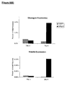
- the cells can be genetically altered to cause expression of a pancreatic transcription factor such as Neurogenin 3.
- a further embodiment of the invention is a method of screening a compound for its ability to modulate islet cell function, using a cell composition of the invention.
- Another embodiment of this invention is a method for making insulin, glucagon, or somatostatin by growing the islet cells of this invention. Also included are pharmaceutical compositions and devices containing the cells of this invention. The cells, compositions, and devices of this invention are useful for reconstituting islet cell function in a subject, especially but not limited to the treatment of Type I diabetes.
- This invention solves the problem of generating large populations of human islet cells by showing how to efficiently differentiate them from pluripotent stem cells.
- stem cells can be coaxed along the islet cell differentiation pathway by initiating differentiation towards the endodermal lineage, and focusing the differentiation process by culturing in the presence of factors that facilitate outgrowth of islet cells.
- This invention provides a system for producing a population of cells enriched for multipotent islet cell progenitors, capable of forming mature islets. If desired, the differentiation process can continue in order to maintain mature endocrine-secreting cells.
- this disclosure provides a strategy of dividing the differentiation pathway into a series of sequential stages. In this way, factors effective in pushing the cells along each segment of the differentiation pathway can be identified.
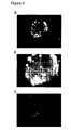
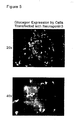
- the differentiation process proceeded as follows, For Stage 1, undifferentiated human embryonic stem cells from feeder free culture were differentiated so as to form a mixed cell aggregate containing endoderm cells in suspension culture. Retinoic acid was used as the initial differentiation agent, in combination with enrichment factors selenium and T3. For Stage 2, differentiation to pancreas progenitor cells was effected by culturing in a medium containing Noggin (200 ng/ml), EGF (20 ng/ml) and bFGF (2 ng/ml). For Stage 3, differentiation to end-stage islet cells was induced by withdrawing the Noggin, EGF and bFGF, and instead culturing the cells with 10 mM nicotinamide. As shown in Figure 4 , clusters of cells were obtained synthesizing antibody-detectable levels of c-peptide of insulin and somatostatin.
- This invention provides a system that can be used to generate unbounded quantities of islet progenitors - and progeny that are committed to form mature islet cells.
- the disclosure that follows provides further information on the production and testing of islet cells of this invention. It also provides extensive illustrations of how these cells can be used in research, pharmaceutical development, and the therapeutic management of conditions related to islet cell dysfunction.
- the term "islet cell” refers to terminally differentiated pancreatic endocrine cells, and any precursor cell that is committed to form progeny normally classified as pancreatic endocrine.
- the cell expresses some of the accepted morphological features and phenotypic markers (exemplified below) that are characteristic of the islet cell lineage. Mature alpha cells secrete glucagon; mature beta cells secrete insulin; mature delta cells secrete somatostatin; PP cells secrete pancreatic polypeptide.
- islet progenitor is an islet cell that does not substantially secrete endocrines, but has the capability to proliferate and generate terminally differentiated cells. It may also have the capability to self-renew.
- islet progenitors are multipotent, which means that they are capable of forming at least two and potentially all four mature islet cell types.
- a “pancreas progenitor”, precursor, or stem cell is capable of forming both pancreatic endocrine and pancreatic exocrine cells.
- differentiated is a relative term.
- a "differentiated cell” is a cell that has progressed further down the developmental pathway than the cell it is being compared with. It is a hypothesis of this invention that pluripotent embryonic stem cells in the course of normal ontogeny differentiate first to an endoderm cell that is capable of forming pancreas cells and other endoderm cell types. Further differentiation leads to the pancreatic pathway, where ⁇ 98% of the cells become exocrine, ductular, or matrix cells, and ⁇ 2% become endocrine cells. Early endocrine cells are islet progenitors, which then differentiate further into functional endocrine cells specializing in secretion of insulin, glucagon, somatostatin, or pancreatic polypeptide.
- a “differentiation agent”, as used in this disclosure, refers to one of a collection of compounds that are used in culture systems of this invention to produce differentiated cells of the islet lineage (including precursor cells and terminally differentiated cells). No limitation is intended as to the mode of action of the compound.
- the agent may assist the differentiation process by inducing or assisting a change in phenotype, promoting growth of cells with a particular phenotype or retarding the growth of others. It may also act as an inhibitor to other factors that may be in the medium or synthesized by the cell population that would otherwise direct differentiation down the pathway to an unwanted cell type.
- Prototype Pluripotent Stem cells are pluripotent cells derived from pre-embryonic, embryonic, or fetal tissue at any time after fertilization, and have the characteristic of being capable under appropriate conditions of producing progeny of several different cell types that are derivatives of all of the three germinal layers (endoderm, mesoderm, and ectoderm), according to a standard art-accepted test, such as the ability to form a teratoma in 8-12 week old SCID mice.
- the term includes both established lines of stem cells of various kinds, and cells obtained from primary tissue that are pluripotent in the manner described.
- pPS cells included in the definition of pPS cells are embryonic cells of various types, exemplified by human embryonic stem (hES) cells, described by Thomson et al. (Science 282:1145, 1998 ); embryonic stem cells from other primates, such as Rhesus stem cells ( Thomson et al., Proc. Natl. Acad. Sci. USA 92:7844, 1995 ), marmoset stem cells ( Thomson et al., Biol. Reprod. 55:254, 1996 ) and human embryonic germ (hEG) cells ( Shamblott et al., Proc. Natl. Acad. Sci. USA 95:13726, 1998 ). Other types of pluripotent cells are also included in the term.
- hES human embryonic stem
- Any cells of primate origin that are capable of producing progeny that are derivatives of all three germinal layers are included, regardless of whether they were derived from embryonic tissue, fetal tissue, or other sources.
- the pPS cells are preferably not derived from a malignant source. It is desirable (but not always necessary) that the cells be karyotypically normal.
- pPS cell cultures are described as "undifferentiated” when a substantial proportion of stem cells and their derivatives in the population display morphological characteristics of undifferentiated cells, clearly distinguishing them from differentiated cells of embryo or adult origin. Undifferentiated pPS cells are easily recognized by those skilled in the art, and typically appear in the two dimensions of a microscopic view in colonies of cells with high nuclear/cytoplasmic ratios and prominent nucleoll, It is understood that colonies of undifferentiated cells within the population will often be surrounded by neighboring cells that are differentiated.
- “Feeder cells” are cells of one type that are co-cultured with cells of another type, to provide an environment in which the cells of the second type can grow. Certain types of pPS cells can be supported by primary mouse embryonic fibroblasts, immortalized mouse embryonic fibroblasts, or human fibroblast-like cells differentiated from hES cell. pPS cell populations are said to be "essentially free” of feeder cells if the cells have been grown through at least one round after splitting in which fresh feeder cells are not added to support growth of the pPS cells.
- embryoid bodies is a term of art synonymous with “aggregate bodies”, referring to aggregates of differentiated and undifferentiated cells that appear when pPS cells overgrow in monolayer cultures, or are maintained in suspension cultures. Embryoid bodies are a mixture of different cell types, typically from several germ layers, distinguishable by morphological criteria and cell markers detectable by immunocytochemistry.
- a “growth environment” is an environment in which cells of interest will proliferate, differentiate, or mature in vitro.
- the environment include the medium in which the cells are cultured, any growth factors or differentiation factors that may be present, and a supporting structure (such as a substrate on a solid surface) if present.
- a cell is said to be "genetically altered” or “transfected” when a polynucleotide has been transferred into the cell by any suitable means of artificial manipulation, or where the cell is a progeny of the originally altered cell that has inherited the polynucleotide.
- Reagents, cloning vectors, and kits for genetic manipulation referred to in this disclosure are available from commercial vendors such as BioRad, Stratagene, Invitrogen, ClonTech, and Sigma-Aldrich Co.
- stem cells of various types.
- stem cells suitable for use in this invention are primate pluripotent stem (pPS) cells derived from tissue formed after gestation, such as a blastocyst, or fetal or embryonic tissue taken any time during gestation.
- pPS pluripotent stem
- Non-limiting examples are primary cultures or established lines of embryonic stem cells or embryonic germ cells, as exemplified below.
- the techniques of this invention can also be implemented directly with primary embryonic or fetal tissue, deriving islet cells directly from primary cells that have the potential to give rise to islet cells without first establishing an undifferentiated cell line. Under certain circumstances, the methods of this invention may also be invoked using multipotent cells from cord blood, placenta, or certain adult tissues.
- Embryonic stem cells can be isolated from blastocysts of members of the primate species ( U.S. Patent 5,843,780 ; Thomson et al., Proc. Natl. Acad. Sci. USA 92:7844, 1995 ).
- Human embryonic stem (hES) cells can be prepared from human blastocyst cells using the techniques described by Thomson et al. (U.S. Patent 6,200,806 ; Science 282:1145, 1998 ; Curr. Top. Dev. Biol. 38:133 ff., 1998 ) and Reubinoff et al, Nature Biotech. 18:399, 2000 .
- Equivalent cell types to hES cells include their pluripotent derivatives, such as primitive ectoderm-like (EPL) cells, as outlined in WO 01/51610 (Bresagen).
- hES cells can be obtained from human preimplantation embryos.
- in vitro fertilized (IVF) embryos can be used, or one-cell human embryos can be expanded to the blastocyst stage ( Bongso et al., Hum Reprod 4: 706, 1989 ).
- Embryos are cultured to the blastocyst stage in G1.2 and G2.2 medium ( Gardner et al., Fertil. Sterit. 69:84, 1998 ).
- the zona pellucida is removed from developed blastocysts by brief exposure to pronase (Sigma).
- the inner cell masses are isolated by immunosurgery, in which blastocysts are exposed to a 1:50 dilution of rabbit anti-human spleen cell antiserum for 30 min, then washed for 5 min three times in DMEM, and exposed to a 1:5 dilution of Guinea pig complement (Gibco) for 3 min ( Solter et al., Proc. Natl. Acad. Sci. USA 72:5099, 1975 ). After two further washes in DMEM, lysed trophectoderm cells are removed from the intact inner cell mass (ICM) by gentle pipetting, and the ICM plated on mEF feeder layers.
- ICM inner cell mass
- inner cell mass-derived outgrowths are dissociated into clumps, either by exposure to calcium and magnesium-free phosphate-buffered saline (PBS) with 1 mM EDTA, by exposure to dispase or trypsin, or by mechanical dissociation with a micropipette; and then replated on mEF in fresh medium.
- PBS calcium and magnesium-free phosphate-buffered saline
- EDTA calcium and magnesium-free phosphate-buffered saline
- dispase or trypsin or by mechanical dissociation with a micropipette
- ES-like morphology is characterized as compact colonies with apparently high nucleus to cytoplasm ratio and prominent nucleoli.
- ES cells are then routinely split every 1-2 weeks by brief trypsinization, exposure to Dulbeeco's PBS (containing 2 mM EDTA), exposure to type IV collagenase ( ⁇ 200 U/mL; Gibco) or by selection of individual colonies by micropipette. Clump sizes of about 50 to 100 cells are optimal.
- Human Embryonic Germ (hEG) cells can be prepared from primordial germ cells present in human fetal material taken about 8-11 weeks after the last menstrual period. Suitable preparation methods are described in Shamblott et al., Proc. Natl. Acad. Sci. USA 95:13726, 1998 and U.S. Patent 6,090,622 .
- EG growth medium is DMEM, 4500 mg/L D-glucose, 2200 mg/L mM NaHCO 3 ; 15% ES qualified fetal calf serum (BRL); 2 mM glutamine (BRL); 1 mM sodium pyruvate (BRL); 1000-2000 U/mL human recombinant leukemia inhibitory factor (LIF, Genzyme); 1-2 ng/mL human recombinant bFGF (Genzyme); and 10 ⁇ M forskolin (in 10% DMSO).
- feeder cells e.g., STO cells, ATCC No. CRL 15D3
- modified EG growth medium free of LIF, bFGF or forskolin inactivated with 5000 rad ⁇ -irradiation.
- PSC primary germ cell
- the first passage is done after 7-10 days in EG growth medium, transferring each well to one well of a 24-well culture dish previously prepared with irradiated STO mouse fibroblasts.
- the cells are cultured with daily replacement of medium until cell morphology consistent with EG cells is observed, typically after 7-30 days or 1-4 passages.
- pPS cells can be propagated continuously in culture, using culture conditions that promote proliferation without promoting differentiation
- Exemplary serum-containing ES medium is made with 80% DMEM (such as Knock-Out DMEM, Gibco), 20% of either defined fetal bovine serum (FBS, Hyclone) or serum replacement ( WO 98/30679 ), 1% non-essential amino acids, 1 mM L-glutamine, and 0.1 mM ⁇ -mercaptoethanol.
- human bFGF is added to 4 ng/mL ( WO 99/20741 , Geron Corp.).
- ES cells are cultured on a layer of feeder cells, typically fibroblasts derived from embryonic or fetal tissue.
- pPS cells can be maintained in an undifferentiated state even without feeder cells.
- the environment for feeder-free cultures includes a suitable culture substrate, particularly an extracellular matrix such as Matrigele or laminin.
- an extracellular matrix such as Matrigele or laminin.
- enzymatic digestion is halted before cells become completely dispersed (say, -5 min with collagenase IV).
- Clumps of ⁇ 10 to 2,000 cells are then plated directly onto the substrate without further dispersal.
- Feeder-free cultures are supported by a nutrient medium containing factors that support proliferation of the cells without differentiation. Such factors may be introduced into the medium by culturing the medium with cells secreting such factors, such as irradiated ( ⁇ 4,000 rad) primary mouse embryonic fibroblasts, telomerized mouse fibroblasts, or fibroblast-like cells derived from pPS cells.
- Medium can be conditioned by plating the feeders at a density of -5-6 x 10 4 cm -2 in a serum free medium such as KO DMEM supplemented with 20% serum replacement and 4 ng/mL bFGF.
- a serum free medium such as KO DMEM supplemented with 20% serum replacement and 4 ng/mL bFGF.
- Medium that has been conditioned for 1-2 days is supplemented with further bFGF, and used to support pPS cell culture for 1-2 days.
- ES cells Under the microscope, ES cells appear with high nuclear/cytoplasmic ratios, prominent nucleoli, and compact colony formation with poorly discernable cell junctions. Primate ES cells express stage-specific embryonic antigens (SSEA) 3 and 4, and markers detectable using antibodies designated Tra-1-60 and Tra-1-81 ( Thomson et al., Science 282:1145, 1998 ). Mouse ES cells can be used as a positive control for SSEA-1, and as a negative control for SSEA-4, Tra-1-60, and Tra-1-81. SSEA-4 is consistently present on human embryonal carcinoma (hEC) cells. Differentiation of pPS cells in vitro results in the loss of SSEA-4, Tra-1-60, and Tra-1-81 expression, and increased expression of SSEA-1, which is also found on undifferentiated hEG cells.
- SSEA stage-specific embryonic antigens
- Islet cells of this invention are obtained by culturing, differentiating, or reprogramming stem cells in a special growth environment that enriches for cells with the desired phenotype (either by outgrowth of the desired cells, or by inhibition or killing of other cell types). These methods are applicable to many types of stem cells, including primate pluripotent stem (pPS) cells described in the previous section.
- pPS primate pluripotent stem
- One intermediate between undifferentiated pPS cells and mature islets is an immature endoderm cell.
- endoderm cells are capable of making epithelial cells of the GI tract and respiratory system, and the key digestive organs (liver and pancreas).
- Islet cells can be generated using a two-stage approach. Stage 1 involves obtaining a population of common endoderm precursor cells. Stage 2 involves maturing the endoderm precursors into pancreatic endocrine. As illustrated in Example 3, pPS cells can be initiated along the endoderm differentiation pathway by culturing with the hepatocyte differentiation agent n-butyrate. Further elaboration of the hepatocyte differentiation paradigm is described in International Patent Publication WO 01/81549 (Geron Corporation).
- Sonic Hedgehog is thought to be involved in liver specification, so including cyclopamine (an inhibitor of Sonic Hedgehog) in the culture medium is thought to help divert the cells toward the pancreatic lineage. Differentiation can then be pushed further in a subsequent step, using the terminal differentiation factor nicotinamide (in the presence of cyclopamine and activin A).
- the differentiation pathway is divided into three stages. pPS cells are first differentiated to endoderm (Stage 1), and then to a second intermediate (Stage 2) - perhaps at the level of a committed pancreas precursor (identifiable with the marker Pdx1). A further differentiation step (Stage 3) can be performed if the user wants to obtain mature islets.
- Stage 1 pPS cells can be differentiated to cells having markers for gut endoderm using a combination of n-butyrate and activin A (Example 4).
- a heterogeneous population comprising endodermal cells can be prepared by culturing pPS cells with retinoic acid in the presence of enriching agents (selenium and thyroid hormones such as T3) (Example 5).
- enriching agents such as T3
- the cells can be cultured with TGF- ⁇ antagonists such as Noggin, in combination with mitogens (a member of the FGF family, possibly in combination with EGF or betacellulin) (Example 5). It may also be helpful to block hedgehog signaling with cyclopamine.
- Stage 3 can be accomplished as already described, using nicotinamide as the terminal differentiation agent (Example 5).
- transcription factors can be activated by direct manipulation that causes progression from Pdx1 positive pancreatic precursors to mature islet cells (Example 6).
- step-wise approach to differentiation is intended as a guide to the reader, and does not limit the invention except where explicitly indicated.
- the differentiation pathway can be broken down into even more stages so that step-wise differentiation can be optimized in an incremental fashion.
- a potential intermediate between pancreatic precursors and mature islets are precursor cells committed to form pancreatic endocrine.
- effective differentiation agents may be combined to work on cells in different stages at the same time, or to promote a cascading effect down the differentiation pathway.
- the desired end-stage cell population will depend in part on its intended use.
- committed islet progenitor cells may be of particular value for therapy of generalized islet insufficiency, and studying islet differentiation in vitro.
- Earlier progenitors may have greater capacity for self-renewal.
- pPS cells are harvested by brief collagenase digestion, dissociated into clusters, and passaged in non-adherent cell culture plates.
- the aggregates are fed every few days, and then harvested after a suitable period, typically 4-8 days (Examples 1 & 5).
- differentiation is enhanced by other factors in the medium: for example, retinoic acid (Example 5) or dimethyl sulfoxide (DMSO).
- retinoic acid Example 5
- DMSO dimethyl sulfoxide
- aggregates will generally start by forming a heterogeneous population of cell types, including a substantial frequency of endoderm cells.
- the embryoid bodies can then be dispersed and replated for the next stage in the differentiation process, on substrates such as laminin or fibronectin; or passaged in suspension culture, using non-adherent plates and a suitable medium.
- Direct differentiation or differentiation in aggregates can be monitored for the presence of endoderm cells using the markers listed below. Once a sufficient proportion of endoderm is obtained, cells are replated or otherwise manipulated to begin Stage 11. In certain circumstances, differentiation or maintenance of islet cells may be enhanced if the cells are kept in micromass clusters (for example, 50 to 5,000 cells), so that alpha, beta, and delta cells can interact directly.
- micromass clusters for example, 50 to 5,000 cells
- the common progenitor cells can be cultured with specific differentiation factors and/or induced with islet-specific genes or promoters as described in the sections that follow.
- pPS cells or their differentiated progeny may be cultured in a cocktail of islet differentiation factors. Alone or in combination, each of the factors may increase the frequency of conversion to the desired cell type, cause outgrowth of cells with a islet phenotype, inhibit growth of other cell types, or enrich for islet cells in another fashion. It is not necessary to understand the mechanism resulting in islet cells being enriched in order to practice the invention. What follows is a non-limiting list of candidate differentiation factors.
- Table 1 Factors for Differentiation of Islet Cells from pPS Cells Factor Compound type or family Proposed function
- Initial working concentration Cyclopamine steroidal alkaloid inhibitor of hedgehog signaling; may also act as inhibitor of cholesterol biosynthesis 10 ⁇ M Betaceliulin EGF family member mitogen and promoter of beta cell differentiation.
- nM Activin A TGF- ⁇ family member differentiation factor causes ductallike cell lines to differentiate into endocrine pancreas cells 4 nM Exendin-4 glucagon-like peptide 1 agonist
- GLP-1 More stable form of GLP-1 (see below) 20 nM Glucagon-like peptide 1 (GLP1) peptide hormone, one of the protein products from the glucagon gene; G-protein coupled receptor ligand induces glucose production, insulin secretion, induces beta cell neogenesis, induced beta cell proliferation
- G-protein coupled receptor ligand induces glucose production, insulin secretion, induces beta cell neogenesis, induced beta cell proliferation
- HGF Hepatocyte Growth Factor
- Ligand for the c-Met receptor Increases beta cell mass in transgenic animals overexpressing HGF, induces beta cell formation from ductal cell line 10 ng/ml Niacinamide (nicotinamide) Member of vitamin B family May affect ADP-fibosylation
- the differentiation cocktail typically, at least two, three, or more than three such factors are combined in the differentiation cocktail.
- Human proteins are preferred, but species homologs and variants may also be used.
- the reader may use other ligands that bind the same receptors or stimulate the same signal transduction pathways, such as receptor-specific antibody.
- other components may be included in the medium to neutralize the effect of other factors that may be present to drive differentiation down a different pathway.
- efficacy of any of these factors can be determined empirically using a matrix strategy to arrive at combinations capable of promoting differentiation one or more steps down the islet cell pathway. Efficacy is assessed by monitoring emergence of the phenotype of the intended intermediate or end-stage cell, using the phenotypic markers such as those listed below. For example, factors believed to induce endocrine pancreas differentiation or proliferation are tested for their ability to induce Pdx1 expression and subsequently and insulin expression in standard culture conditions.
- a fractional factorial design strategy can be used to screen several compounds in an efficient manner.
- Each factor is assigned two levels: for example, the culture matrix would be assigned fibronectin for one level and laminin for the second level.
- the culture matrix would be assigned fibronectin for one level and laminin for the second level.
- 64 factor combinations 64 experiments, it is possible to determine which factors (from a group of 15-20) significantly influence differentiation in a statistically robust manner.
- Combinations suitable for analysis by this strategy include cyclopamine, TGF family members (TGF- ⁇ , Activin A, Activin B, TGF ⁇ 1, TGF ⁇ 3), Exendin 4, nicotinamide, n-butyrate, DMSO, all-trans retinoic acid, GLP-1, bone morphogenic proteins (BMP-2, BMP-5, BMP-6, BMP-7), insulin-like growth factors (IGF-I, IGF-II), fibroblast growth factor (FGF7, FGF10, bFGF, FGF4), other growth factors (EGF, betacellulin, growth hormone, HGF), other hormones (prolactin, cholecytokinin, gastrin I, placental lactogen), TGF- ⁇ family antagonists (Noggin, follistatin, chordin), IBMX, wortmannin, dexamethazone, Reg, INGAP, cAMP or cAMP activators (forskolin), and extracellular matrix components (laminin,
- Suitable promoters specific for islet progenitors include those driving transcription of Pyx1 (NT_009799), Neurogenin 3 (NT_008583), NeuroD1 (NT_005265), Nestin (NT_004858), and Ptf1a-p48 (NT_008705).
- Suitable promoters specific for mature islet cells are those driving expression of insulin (GenBank Accession NT_009308), glucagon (NT_022154), somatostatin (NT_005962), or pancreatic polypeptide (NT_010755).
- a minimal effective sequence of the chosen promoter ( ⁇ 0.5 to 5 kB of upstream sequence) is amplified by PCR and spliced into a standard plasmid, adenovirus, lentivirus, or retrovirus vector, in a position that drives expression of a suitable reporter gene.
- Suitable reporter genes either encode a fluorescent molecule (such as green fluorescent protein or luciferase), encode a detectable enzyme (such as alkaline phosphatase), or produce a cell surface antigen (any heterologous protein or carbohydrate) that can be detected by antibody or lectin binding.
- Cells transfected with a tissue-specific promoter can be used to optimize differentiation procedures in the manner already described. For example, transfected undifferentiated pPS cells or cells in the early stage of differentiation can be subject to various differentiation regimes, and then analyzed for the proportion of cells reporting expression driven by the islet-specific promoter. This provides a rapid read-out of effective differentiation agents, culture environments, and timing. The optimal procedure can then be used with untransfected cells to generate high-quality populations of the islet lineage that have a native genotype.
- Cells transfected with a tissue-specific promoter can also be used as a means for accomplishing mechanical sorting.
- the promoter may drive expression of a drug-resistant phenotype, such as the neomycin resistance gene (conferring resistance to the drug G418) or the blasticidin resistance gene.
- the reporter gene may encode a fluorescent molecule, or cause expression of a detectable surface antigen.
- cells of interest are sorted from a population of differentiated cells by cell sorting based on either direct or indirect fluorescence, or by immunoadsorption.
- a non-replicative, non-integrating vector (such as an adenovirus vector) can be used for transient transfection during the sorting step, and will dilute out upon subsequent proliferation of the cell, leaving the cell with a native genotype.
- the cells can also be genetically altered for the purpose of directly driving the cells further down the differentiation pathway for islet cells or their progenitors. It is hypothesized that deliberate upregulation of certain genes normally expressed in the islet cells will cause less differentiated cells to recruit the genetic expression profile appropriate for more mature islet cells. Suitable genes include those that encode transcription regulators capable of influencing downstream gene expression.
- Candidates for pancreatic ontogeny include Sox17 (GenBank Accession NM_022454), Hlxb9 (NM_005515), Pyx1 (NM_000209), Neurogenin3 (NM_020999), Pax4 (NM_006193) or NeuroD1 (NM_002500), Isl1 (NM_002202), Nkx2.2 (NM_002509), and Nkx6.1 (NM_006168).
- Neurogenin 3 expression induced by transduction of cells in an islet cell differentiation paradigm enhances expression in downstream regulatory genes, and genes encoding islet hormones.
- the neurogenins are a family of a neuroD-related bHLH transcription factors: neuronal determination genes active in the developing nervous system.
- Cells can be characterized according to phenotypic criteria, such as microscopic observation of morphological features, detection or quantitation of expressed cell markers, functional criteria measurable in vitro, and behavior upon infusion into a host animal.
- phenotypic criteria such as microscopic observation of morphological features, detection or quantitation of expressed cell markers, functional criteria measurable in vitro, and behavior upon infusion into a host animal.
- Cells of this invention can be characterized according to whether they express phenotypic markers characteristic of islet cells of various kinds. Useful markers include those shown in Table 2.
- Table 2 Phenotypic Markers for Cell Identification SSEA-4 Embryonic stem and germ cells Oct-4 Undifferentiated embryonic pluripotent cells Telomerase reverse transcriptase (TERT) Cells capable of unlimited replication (e.g., undifferentiated pPS cells) Pdx1 expressed before pancreatic bud formation in the region of the duodenum that give rise to the pancreas. Also expressed in mature beta cells Neurogenin 3 (Ngn3) marker of islet precursor cells.
- Ngn3 Neurogenin 3
- pancreatic polypeptide PP cells islet amyloid polypeptide (IAPP) islet marker Islet-1 islet marker, also neural expression Beta-2/NeuroD pan islet cell marker HNF3b endoderm marker HNF4a endoderm marker Sox17 definitive endoderm marker Amylase exocrine cells HES exocrine pancreas marker nestin potential precursor cell marker Sonic hedgehog signaling molecule, absence is required for pancreas formation CK19 pancreatic duct marker (possible pancreatic precursor) Glut-2 glucose transporter Patched homologue hedgehog receptor Smoothened homologue hedgehog receptor
- Tissue-specific markers can be detected using any suitable immunological technique - such as flow immunocytochemistry for cell-surface markers, or immunohistochemistry (for example, of fixed cells or tissue sections) for intracellular or cell-surface markers.
- flow immunocytochemistry for cell-surface markers
- immunohistochemistry for example, of fixed cells or tissue sections
- a detailed method for flow cytometry analysis is provided in Gallacher et al., Blood 96:1740, 2000.
- Expression of a cell-surface antigen is defined as positive if a significantly detectable amount of antibody will bind to the antigen in a standard immunocytochemistry or flow cytometry assay, optionally after fixation of the cells, and optionally using a labeled secondary antibody or other conjugate to amplify labeling.
- tissue-specific gene products can also be detected at the mRNA level by Northern blot analysis, dot-blot hybridization analysis, or by reverse transcriptase initiated polymerase chain reaction (RT-PCR) using sequence-specific primers in standard amplification methods. See U.S. Patent No. 5,843,780 for further details. Sequence data for particular markers listed in this disclosure can be obtained from public databases such as GenBank.
- Initial assessment can be done using a marker combination that provides a wide ontogenic profile: for example, Pdx1 for early pancreas cells; Ngn3 for early pancreatic endocrine cells; and insulin for mature beta cells.
- the cells are harvested from the differentiation paradigms described earlier at regular intervals (say, weekly) to determine the kinetics of differentiation. Cells that test positive for these markers can then be analyzed for expression of other markers, such as IAPP and Nkx6.1. Once the cells are characterized, differentiation factor combinations and the timing of each step can be optimized.
- Certain embodiments of this invention relate to populations in which at least 2%, 5%, 10%, or more of the cells bear the surface markers referred to above, either alone or in combination.
- Endocrine function is critical to many research and therapeutic applications, in which case populations comprising at least 5% of the cells secreting insulin, glucagon, somatostatin, or pancreatic polypeptide are of particular interest, as are progenitor cells capable of differentiating into such endocrine-secreting cells. It is a hypothesis of this invention that interaction between the alpha, beta, and delta cells may be important in preventing dedifferentiation and maintaining efficient endocrine secretion.
- This invention also includes masses or clusters of cells (perhaps 50-5,000 cells in size), containing two or three of these cell types, either bound in a matrix of their own making, with a matrix component supplied in culture, or by microancapsulation.
- populations with a low residual proportion of undifferentiated pPS cells are less than 1%, or 0.2% SSEA-4 +ve, Oct-4 +ve, or positive for expression of endogenous telomerase reverse transcriptase. Preferred populations also have relatively low proportions ( ⁇ 5%, 1%, or 0.2%) of certain other cell types, such as hepatocytes (albumin positive cells), skeletal muscle cells (myoD positive), smooth muscle cells (smooth muscle actin), cells with fibroblast morphology, or neurons ( ⁇ -tubulin III or NCAM positive cells).
- hepatocytes albumin positive cells
- skeletal muscle cells myoD positive
- smooth muscle cells smooth muscle actin
- neurons ⁇ -tubulin III or NCAM positive cells
- the cell populations and isolated cells of this invention When derived from an established line of pPS cells, the cell populations and isolated cells of this invention will have the same genome as the line from which they are derived. This means that over and above any karyotype abnormalities, the chromosomal DNA will be over 90% identical between the pPS cells and the islet cells, which can be inferred if the islet cells are obtained from the undifferentiated line through the course of normal mitotic division. Islet cells that have been treated by recombinant methods to introduce a transgene or knock out an endogenous gene are still considered to have the same genome as the line from which they are derived, since all non-manipulated genetic elements are preserved.
- the non-obese diabetic (NOD) mouse carries a genetic defect that results in insulitis showing at several weeks of age ( Yoshida et al., Rev. Immunogenet. 2:140, 2000 ). 60-90% of the females develop overt diabetes by 20-30 weeks. The immune-related pathology appears to be similar to that in human Type I diabetes.
- Other models of Type I diabetes are mice with transgene and knockout mutations ( Wong et al., Immunol. Rev. 169:93, 1999 ).
- a rat model for spontaneous Type I diabetes was recently reported by Lenzen et al. (Diabetologia 44:1189, 2001 ).
- Hyperglycemia can also be induced in mice (>500 mg glucose/dL) by way of a single intraperitoneal injection of streptozotocin ( Soria et al., Diabetes 49:157, 2000 ), or by sequential low doses of streptozotocin ( Ito et al., Environ. Toxicol. Pharmacol. 9:71, 2001 ). To test the efficacy of implanted islet cells, the mice are monitored for return of glucose to normal levels ( ⁇ 200 mg/dL).
- Dogs can be rendered insulin-dependent by removing the pancreas ( J. Endocrinol. 158:49,2001 ), or by feeding galactose ( Kador et al., Arch. Opthalmol. 113:352, 1995 ).
- galactose Kador et al., Arch. Opthalmol. 113:352, 1995
- There is also an inherited model for Type I diabetes in keeshond dogs Am. J. Pathol. 105:194, 1981 ).
- Early work with a dog model Banting et al., Can. Med. Assoc. J. 22:141, 1922 ) resulted in a couple of Canadians making a long ocean journey to Sweden in February of 1925.
- a pilot study can be conducted using pPS derived islet cells in the following animals: a) non-diabetic nude (T-cell deficient) mice; b) nude mice rendered diabetic by streptozotocin treatment; and c) nude mice in the process of regenerating islets following partial pancreatectomy.
- the number of cells transplanted is equivalent to ⁇ 1000-2000 normal human islets, implanted under the kidney capsule, in the liver, or in the pancreas.
- the endpoints of can be assessment of graft survival (histological examination) and determination of insulin production by biochemical analysis, RIA, ELISA, and immunohistochemistry.
- Streptozotocin treated and partially pancreatectomized animals can also be evaluated for survival, metabolic control (blood glucose) and weight gain.
- the islet precursor cells of this invention have a substantial proliferation capacity.
- the replication capacity can be further enhanced by increasing the level of telomerase reverse transcriptase (TERT) in the cell, either by increasing transcription from the endogenous gene, or by introducing a transgene.
- telomerase reverse transcriptase particularly suitable is the catalytic component of human telomerase (hTERT), provided in International Patent Application WO 98/14592 . Transfection and expression of telomerase in human cells is described in Bodnar et al., Science 279:349, 1998 and Jiang et al., Nat. Genet. 21:111, 1999 .
- telomere activity Assay
- immunocytochemical staining for hTERT or replicative capacity, according to standard methods.
- Other methods of immortalizing cells are also contemplated, such as transforming the cells with DNA encoding myc, the SV40 large T antigen, or MOT-2 ( U.S. Patent 5,869,243 , International Patent Applications WO 97/32972 and WO 01/23555 ).
- the cells of this invention can be prepared or further treated to remove undifferentiated cells in vitro, or to safeguard against revertants in vivo.
- One way of depleting undifferentiated stem cells from the population is to transfect the population with a vector in which an effector gene under control of a promoter that causes preferential expression in undifferentiated cells - such as the TERT promoter or the OCT-4 promoter.
- the effector gene may be a reporter to guide cell sorting, such as green fluorescent protein.
- the effector may be directly lytic to the cell, encoding, for example, a toxin, or a mediator of apoptosis, such as caspase (Shinoura et al., Cancer Gene Ther. 7:739, 2000).
- the effector gene may have the effect of rendering the cell susceptible to toxic effects of an external agent, such as an antibody or a prodrug.
- an external agent such as an antibody or a prodrug.
- exemplary is a herpes simplex thymidine kinase (tk ) gene, which causes cells in which it is expressed to be susceptible to ganciclovir ( USSN 60/253,443 ).
- the effector can cause cell surface expression of a foreign determinant that makes any cells that revert to an undifferentiated phenotype susceptible to naturally occurring antibody in vivo ( USSN 60/253,357 ).
- This invention provides a method to produce large numbers of islet precursor cells, and mature islet cells. These cell populations can be used for a variety of important research, development, and commercial purposes.
- the cells of this invention can be used to prepare a cDNA library relatively uncontaminated with cDNA preferentially expressed in cells from other lineages.
- the differentiated cells of this invention can also be used to prepare monoclonal or polyclonal antibodies that are specific for markers of islet precursors and their derivatives, according to standard methods.
- compositions of this invention for drug development and clinical therapy.
- Islet cells of this invention can be used to screen for factors (such as solvents, small molecule drugs, peptides, polynucleotides) or environmental conditions (such as culture conditions or manipulation) that affect the characteristics of islet precursor cells and their various progeny.
- factors such as solvents, small molecule drugs, peptides, polynucleotides
- environmental conditions such as culture conditions or manipulation
- a prime example is the use of islet cell clusters or homogeneous beta cell preparations for the effect of small molecule drugs that have the potential to up- or down-regulate insulin synthesis or secretion.
- the cells are combined with the test compound, and then monitored for change in expression or secretion rate, for example by RT-PCR or immunoassay of the culture medium.
- screening methods of this invention relate to the testing of pharmaceutical compounds for a potential effect on islet cell growth, development, or toxicity. This type of screening is appropriate not only when the compound is designed to have a pharmacological effect on islet cells, but also to test for islet-related side-effects of compounds designed for a primary pharmacological effect elsewhere.
- pPS cells are used to screen factors that promote maturation into islet cells, or promote proliferation and maintenance of islet cells in long-term culture.
- candidate differentiation factors or maturation factors are tested by adding them to cells in different wells, and then determining any phenotypic change that results, according to desirable criteria for further culture and use of the cells. This can lead to improved derivation and culture methods for not only pPS derived islets, but also for islet cells and their progenitors isolated from pancreas.
- Assessment of the activity of candidate pharmaceutical compounds generally involves combining the differentiated cells of this invention with the candidate compound, either alone or in combination with other drugs.
- the investigator determines any change in the morphology, marker phenotype, or functional activity of the cells that is attributable to the compound (compared with untreated cells or cells treated with an inert compound), and then correlates the effect of the compound with the observed change.
- Cytotoxicity can be determined in the first instance by the effect on cell viability, survival, morphology, and the expression of certain markers and receptors. Effects of a drug on chromosomal DNA can be determined by measuring DNA synthesis or repair. [ 3 H]-thymidine or BrdU incorporation, especially at unscheduled times in the cell cycle, or above the level required for cell replication, is consistent with a drug effect. Unwanted effects can also include unusual rates of sister chromatid exchange, determined by metaphase spread. The reader is referred to A. Vickers (pp 375-410 in "In vitro Methods in Pharmaceutical Research," Academic Press, 1997 ) for further elaboration.
- This invention also provides for the use of islet precursor cells or their derivatives to restore islet function in a patient in need of such therapy. Any condition relating to inadequate production of a pancreatic endocrine (insulin, glucagon, or somatostatin), or the inability to properly regulate secretion may be considered for treatment with cells prepared according to this invention, as appropriate. Of especial interest is the treatment of Type I (insulin-dependent) diabetes mellitus.
- Patients are chosen for treatment based on confirmed long-term dependence on administration of exogenous insulin, and acceptable risk profile.
- the patient receives approximately 10,000 islet equivalents per kg body weight.
- anti-rejection drugs such as FK506 and rapamycin (orally) and daclizumab (intravenously).
- the islet cells are infused through a catheter in the portal vein.
- the patient is then subjected to abdominal ultrasound and blood tests to determine liver function.
- Daily insulin requirement is tracked, and the patient is given a second transplant if required.
- Follow-up monitoring includes frequent blood tests for drug levels, immune function, general health status, and whether the patient remains insulin independent.
- islet cells for the treatment of Type I diabetes is discussed at length in Cellular Inter-Relationships in the Pancreas: Implications for Islet Transplantation, by L. Rosenberg et al., Chapman & Hall 1899 ; and Fetal lslet Transplantation, by C.M. Peterson et al. eds., Kluwer 1995 .
- pancreatic endocrine cells are responsible for the ultimate responsibility for patient selection, the mode of administration, and dosage of pancreatic endocrine cells.
- islet cells of this invention are typically supplied in the form of a pharmaceutical composition, comprising an isotonic excipient prepared under sufficiently sterile conditions for human administration.
- This invention also includes sets of cells that exist at any time during their manufacture, distribution, or use.
- the cell sets comprise any combination of two or more cell populations described in this disclosure, exemplified but not limited to a type of differentiated pPS-derived cell (islet cells, their precursors, subtypes, and so on), in combination with undifferentiated pPS cells or other differentiated cell types, sometimes sharing the same genome.
- Each cell type in the set may be packaged together, or in separate containers in the same facility, or at different locations, under control of the same entity or different entities sharing a business relationship.
- compositions For general principles in medicinal formulation of cell compositions, the reader is referred to Cell Therapy: Stem Cell Transplantation, Gene Therapy, and Cellular Immunotherapy, by G. Morstyn & W. Sheridan eds, Cambridge University Press, 1996 .
- the composition is optionally packaged in a suitable container with written instructions for a desired purpose, such as the treatment of diabetes.
- the cells of this invention can also be used as the functional component in a mechanical device designed to produce one or more of the endocrine polypeptides of pancreatic islet cells.
- the device contains the pPS derived islet cells behind a semipermeable membrane that prevents passage of the cell population, retaining them in the device, but permits passage of insulin, glucagon, or somatostatin secreted by the cell population.
- U.S. Patent 4,391,909 describe islet cells encapsulated in a spheroid semipermeable membrane made up of polysaccharide polymers >3,000 mol. wt. that are cross-linked so that it is permeable to proteins the size of insulin, but impermeable to molecules over 100,000 mol. wt.
- Patent 6,023,009 describes islet cells encapsulated in a semipermeable membrane made of agarose and agaropectin. Microcapsules of this nature are adapted for administration into the body cavity of a diabetic patient, and are thought to have certain advantages in reducing histocompatibility problems or susceptibility to bacteria.
- U.S. Patent 4,378,016 describes an artificial endocrine gland containing an extracorporeal segment, a subcutaneous segment, and a replaceable envelope containing the hormone-producing cells.
- U.S. Patent 5,674,289 describes a bioartificial pancreas having an islet chamber, separated by a semipermeable membrane to one or more vascularizing chambers open to surrounding tissue.
- Useful devices typically have a chamber adapted to contain the islet cells, and a chamber separated from the islet cells by a semipermeable membrane which collects the secreted proteins from the islet cells, and which may also permit signaling back to the islet cells, for example, of the circulating glucose level.
- Example 1 Feeder-free propagation of embryonic stem cells
- Conditioned medium prepared in advance using primary mouse embryonic fibroblasts (mEF) isolated according to standard procedures ( WO 01/51616 ).
- hES cultures were passaged onto Matrigel® coated plates. About one week after seeding, the cultures became confluent and could be passaged. Cultures maintained under these conditions for over 180 days continued to display ES-like morphology. SSEA-4, Tra-1-60, Tra-1-81, and alkaline phosphatase were expressed by the hES colonies, as assessed by immunocytochemistry, but not by the differentiated cells in between the colonies. Pluripotency was confirmed by subjecting them to established protocols for making particular cell types.
- Example 2 Derivation of hepatocytes from hES cells
- Undifferentiated hES cells were differentiated into cells having characteristics of human hepatocytes using a strategy to initiate a global differentiation process using DMSO in a subconfluent culture. The cells are then induced to form hepatocyte-like cells by the addition of Na-butyrate.
- the hES cells were maintained in undifferentiated culture conditions for 2-3 days after splitting. At this time, the cells were 50 to 60% confluent and the medium was exchanged with unconditioned SR medium containing 1% DMSO (Step II). The cultures were fed daily with SR medium for 4 days and then exchanged into unconditioned SR medium containing both 1% DMSO and 2.5% Na-butyrate, with which they were fed daily for 6 days (Step III). They were then replated onto collagen, and cultured in a hepatocyte maturation medium containing a cocktail of hepatocyte-friendly growth factors (Step IV). The procedure is summarized in Table 3.
- Hepatocyte Differentiation Protocol Step I Step II Step III Step IV Undifferentiated cells Pre-differentiation Hepatocyte differentiation Hepatocyte maturation (until confluent) (4 days) (6 days) (Groups 7-9 only; 4 days) Feeder-free conditions 20% SR medium + 1 % DMSO 20% SR medium + 1 % DMSO + 2.5 mM butyrate HCM + 30 ng/mL hEGF + 10 ng/mL TGF- ⁇ + 30 ng/mL HGF + 2.5 mM butyrate
- Figure 1 shows the hepatocyte-like cells that were obtained. Left column: 10 X magnification; Right column: 40 X magnification.
- By 4 days in the presence of butyrate more than 80% of cells in the culture are large in diameter, containing large nuclei and granular cytoplasm (Row A). After 5 days in SR medium, the cells were switched to HCM. Two days later, many cells are multinucleated, and have a large polygonal shape (Row B).
- By 4 days in HCM multinucleated polygonal cells are common, and have a darker cytosol (Row C), by which criteria they resemble freshly isolated human adult hepatocytes (Row D) or fetal hepatocytes (Row E).
- Example 3 Obtaining insulin-secreting cells from early cells in the hepatocyte pathway
- Insulin-secreting cells were derived from hES cells of the H9 line using a modification of the hepatocyte differentiation protocol described in the last Example.
- Step I hES cells were grown to confluence after their last passage over a 7-8 day period using SR medium conditioned by mouse embryonic fibroblasts as described in Example 1.
- Step II the medium was exchanged with unconditioned SR medium containing 1% DMSO, to which 10 ⁇ M cyclopamine was added, and the cells were cultured for 4 days.
- Step III the cells were cultured for 11 days in RPMI1640 medium with B27 supplement, containing 0.5 mM butyrate (5-fold lower than the hepatocyte protocol), 10 ⁇ M cyclopamine, 4 nM activin A and 10 mM nicotinamide.
- Step IV the cells are matured RPMI1640/B27 by culturing for 11 days in RPMI supplemented with B27, and containing possible islet differentiation factors.
- Figure 2 shows the results in which the cells were cultured in Step IV with 4 nM activin A, 4 nM betacellulin, 10 nM IGF-1, and 10 mM nicotinamide.
- the round circles in the upper panel represent the DAPI blue-stained nuclei.
- the diffuse staining centered in the middle of the field is red fluorescent staining, representing insulin synthesized and fixed within the cell. It was estimated that about 1% of the cells in the well were expressing insulin. No insulin staining was observed in other cell clusters on the same slide or in the isotype control.
- Example 4 Entering the islet cell pathway by optimizing formation of gut endoderm
- This example provides an illustration of the stepwise induction of islets from pPS cells, mimicking the normal development that occurs in utero.
- the H7 and H9 line of hES cells were grown to confluence using standard feeder free conditions, which served as starting material for the experiment.
- the medium was changed to RPMI 1640 supplemented with B27 (Invitrogen) and one of the following alternatives:
- Figure 3 shows expression levels of the markers for gut endoderm. Results are standardized to the expression level in human fetal pancreas. Also shown for comparison is the level of expression in undifferentiated hES cells (the starting cell line). The three markers were induced most effectively by the combination of both Activin A and sodium butyrate. Sox17 was expressed at high levels (2.3 relative expression) in undifferentiated H9 cells, but was induced to 32 relative expression level with the two additives (more than 100-fold increase). In H7 cells, the level of induction was more dramatic, going from .007 times fetal pancreas to 79 times fetal pancreas (more than 10,000-fold increase). HNF3beta increased from less than 0.5 relative expression to 5.1 or 6.3 (more than 10-fold increase). The levels of HNF4alpha increased from less than 0.1 relative expression level to over 1.0 (more than 10-fold increase).
- Example 5 Obtaining islet cells by using additives in long-term aggregate culture
- the H7 line of hES cells was grown in an undifferentiated form in mEF medium as in Example 1. They were then grown in suspension culture (forming embryoid bodies) in a blended medium consisting of mEF conditioned medium and DFB+ at 1:1.
- DFB+ is DMEM/F12 medium supplemented with B27 additive (1 x), insulin (25 ⁇ g/ml), progesterone (6.3 ng/ml), putrescine (10 ⁇ g/ml), selenium (in the form of selenite, 100 ng/ml), transferrin (50 ⁇ g/ml), and the thyroid hormone receptor ligand T3 (40 ng/ml).
- B27 additive (1 x)
- insulin 25 ⁇ g/ml
- progesterone 6.3 ng/ml
- putrescine 10 ⁇ g/ml
- selenium in the form of selenite, 100 ng/ml
- transferrin 50 ⁇ g/ml
- T3 thyroid hormone receptor
- c-Peptide is a component of proinsulin that is removed before secretion. It is useful in distinguishing insulin synthesized from the cell from insulin present as a component of the medium.
- Figure 4 shows the results.
- the top and middle panels show staining for insulin c-peptide at low and high magnification, indicating a cluster of mature pancreatic beta cells.
- the bottom panel shows staining for somatostatin, a marker of islet delta cells.
- Taqman real-time RT-PCR confirmed that insulin and glucagon are expressed by these cells at the mRNA level.
- This experiment demonstrates the effectiveness of overexpressing an islet related gene in recruiting a gene expression profile characteristic of the islet cell lineage during Stage III of differentiation.
- AdNgn3 is a replication deficient, E1- and E3- deleted recombinant adenovirus 5 based vector constructed using the AdMAXTM vector construction kit (Microbix Biosystems, Toronto).
- Ngn3 cDNA 645 bp (GenBank accession # NM_020999) was cloned into pDC515 shuttle vector downstream of mCMV (Murine Cytomegalovirus Immediate Early Gene) promoter.
- mCMV Medouracil-1
- the shuttle plasmid containing the Ngn3 insert and the Ad genomic plasmid were cotransfected into E1-expressing 293 cells for homologous recombination to occur. Plaques were isolated, the viral stock was amplified in 293 cells, and then purified by CsCl density gradient centrifugation. Viral particle concentration was determined by measuring optical density at A260, and the infectious titer was determined by standard plaque assay.
- Human ES cells of the H7 line were suspended and cultured in blended medium as in Example 5. All-trans retinoid acid (10 ⁇ M) was added to the medium the next day. Seven days later, the EBs were plated on Matrigel® in DFB+ medium plus EGF (20 ng/ml) and bFGF (2 ng/ml). After five days in culture, cells were infected with the AdNgn3 vector at MOI of 50. AdGFP (an equivalent vector containing the gene for green fluorescent protein, not expected to direct differentiation) was used as a negative control.
- AdGFP an equivalent vector containing the gene for green fluorescent protein, not expected to direct differentiation
- AdNgn3 or AdGFP virus at an MOI of 50 was applied directly to cells in 6-well plates in 1 mL of DFB+ medium containing EGF (20 ng/ml) for 4 h, then additional 2 mL of the same medium was added to each well.. The medium was replenished every other day thereafter.
- Cells were harvested 2 days or 8 days after the infection, and RNA was isolated for analysis by real-time RT-PCR as in Example 4.
- Cells plated on chamber slides for immunohistochemistry were fixed in 4% paraformaldehyde, blocked, and stained with antibody against glucagon (1:1000), followed by FITC conjugated goat anti-mouse IgG (1:500).
- Figure 5 shows immunocytochemical staining of the Neurogenin 3 transduced cells 9 days after transduction. This plated embryoid body shows a substantial level of antibody-detectable glucagon expression.
- Figure 6(A) and (B) shows mRNA expression levels in the Neurogenin 3 transduced cells (solid bars), compared with the negative control (hatched bars).
- Real-time RT-PCR data is normalized to expression levels in human fetal pancreas.
- NeuroD1 and Nkx2.2 were both substantially upregulated by day 2; Nkx6.1 was upregulated by day 8.
- Insulin and glucagon expression were also substantially up-regulated by day 8, indicating that the gene transfection strategy substantially improves maturation to cells making the clinically important end-stage products of islet cells.
- Embodiments of the invention may include the following.
Abstract
Description
- This invention relates generally to the fields of cell biology, embryonic stem cells, and cell differentiation. More specifically, this invention provides differentiated cells with pancreatic endocrine function.
- This application claims priority to
U.S. provisional application 60/338,885, filed December 7, 2001 - The American Diabetes Association estimates that there are currently 5 million people in the United States with confirmed diabetes, and over 10 million at risk.
- The cost of this disease and its sequelae to the American economy is staggering. Care of diabetics consumes a total of $98 billion per year, accounting for one of every seven healthcare dollars spent in the U.S. There are 24,000 new cases of diabetes-caused blindness caused by diabetes each year. Diabetes is the leading cause of kidney failure, contributing about 40% of new dialysis patients. Diabetes is also the most frequent cause of lower limb amputation, with 56,000 limbs lost to diabetes each year. The per capita health care costs incurred per diabetic person is $10,071 annually, compared with $2,669 for non-diabetics.
- Type I diabetes mellitus (also known as insulin-dependent diabetes) is a severe condition accounting for 5-10% all diabetics. The pathology arises because the patient's insulin-secreting beta cells in the pancreas have been eliminated by an autoimmune reaction. Under current practice, the condition is managed by regular injection of insulin, constant attention to diet, and continuous monitoring of blood glucose levels to adjust the insulin dosing. it is estimated that the market for recombinant insulin will reach $4 billion by 2005. Of course, the availability of insulin is life-saving for Type I diabetics. But there is no question that the daily regimen that diabetics must adhere to is enormously cumbersome, and not universally effective.
- For this reason, there are several clinical tests underway to transplant diabetics with islet cells isolated from donor pancreas. This has been made possible by recent advances in the isolation and culture of islet cells.
U.S. Patent 4,797,213 described separation of islets of Langerhans.U.S. Patent 4,439,521 reports a method for producing self-reproducing pancreatic islet-like structures.U.S. Patent 5,919,703 reports preparation and storage of pancreatic islets.U.S. Patent 5,888,816 reports cell culture techniques for pancreatic cells using hypothalamus and pituitary extracts.WO 00172885 WO 00/78929 - Peck et al. (Ann. Med. 33:186, 2001) propose that pancreatic stem cells be used as building blocks for better surrogate islets for treating Type I diabetes.
WO 00/47721 WO 01/39784 WO 01/77300 U.S. Patent 6,326,201 (Curis Inc.) reports pancreatic progenitor cells made by dissociating and culturing cells from pancreatic duct. The present clinical experience of islet cell transplantation is reviewed by Bretzel et al. (Exp. Clin. Endocrinol. Diabetes 190 (Suppl. 2):S384, 2001) and Oberholzer et al. (Ann. N.Y. Acad. Sci. 875:189, 1999). The current clinical trials typically involve infusing cells from at least two pancreas donors. Even if this treatment proves to be successful, there will be insufficient material available from current sources to treat all the eligible Type I diabetic patients. - Developmental work has been done in several institutions to capitalize on the promise of pluripotent stem cells from the embryo to differentiate into other cell types. Cells bearing features of the islet cell lineage have reportedly been derived from embryonic cells of the mouse. For example, Lumelsky et al. (Science 292:1389, 2001) report differentiation of mouse embryonic stem cells to insulin-secreting structures similar to pancreatic islets. Soria et al. (Diabetes 49:157, 2000) report that insulin-secreting cells derived from mouse embryonic stem cells normalize glycemia in streptozotocin-induced diabetic mice.
- Regrettably, the mouse model of embryonic stem cell development is its own peculiar case, and does not yield strategies for differentiation that are applicable to other species. In fact, pluripotent stem cells have been reproducibly isolated from very few other mammalian species. Only recently did Thomson et al. isolate embryonic stem cells from human blastocysts (Science 282:114, 1998). Concurrently, Gearhart and coworkers derived human embryonic germ (hEG) cell lines from fetal gonadal tissue (Shamblott et al., Proc. Natl. Acad. Sci. USA 95:13726, 1998). Unlike mouse embryonic stem cells, which can be kept from differentiation simply by culturing with Leukemia Inhibitory Factor (LIF), human embryonic stem cells must be maintained under very special conditions (
U.S. Patent 6,200,806 ;WO 99/20741 WO 01/51616 - Jacobson et al. (Transplant. Proc. 33:674, 2001) reported differentiation of intestinal and pancreatic endoderm from rhesus embryonic stem cells. Assady et al. (Diabetes 50:1691, 2001) identified insulin production by human embryonic stem cells differentiated to embryoid bodies, by immunohistochemistry and enzyme-linked immunoassay of the culture medium. Of course, embryoid bodies contain an enormous variety of different cell types (
WO 01/51616 - For embryonic stem cell derived islet cells to become a commercially viable proposition, there is a need to develop new procedures that provide for populations of islet cells of high purity.
- This invention provides a system for efficient production of primate cells that have differentiated from pluripotent cells into cells of the islet cell lineage. Populations of cells are described that are considerably enriched for islet progenitor cells. In turn, the islet progenitors can be further differentiated into colonies comprising cells that secrete insulin, glucagon, somatostatin, or a combination of all three.
- Accordingly, one embodiment of the invention is a cell population obtained by differentiating primate pluripotent stem (pPS) cells (such as embryonic stem cells), in which at least 5% of the cells secrete at least one of the four islet cell proteins from an endogenous gene: the hormones insulin, glucagon, somatostatin, and the apparently inert product known as pancreatic polypeptide. The cells may be in clusters, comprising cells secreting each of the three endocrines, and may be processed to contain a minimum proportion of other tissue types. The cells of this invention may be identified by phenotypic markers listed later on in this disclosure. Functional efficacy can be confirmed by the ability to improve fasting glucose levels when administered to a hyperglycemic subject.
- Another embodiment of the invention is a differentiated cell population capable of self-renewal, and capable of forming progeny that are mature islet cells. This means that the cells are no longer pluripotent, but retain the ability to form islet ceils upon proliferation. The proportion of undifferentiated pluripotent cells in the population is preferably minimized, and any residual undifferentiated cells are not the cells responsible for forming the islet cells upon further proliferation. Optionally, replication capacity of the stem cells can be improved by increasing telomerase activity.
- Another embodiment of the invention is a method for obtaining polypeptide-secreting cells, in which differentiation of pPS cells is initiated, for example, by forming embryoid bodies or other form comprising early ectoderm. The cells are then cultured in a mixture of differentiation factors, such as TGF-β antagonists like Noggin, activin A, n-butyrate, or combinations of the other factors listed below. In addition or as an alternative, the cells can be genetically altered to cause expression of a pancreatic transcription factor such as Neurogenin 3.
- A further embodiment of the invention is a method of screening a compound for its ability to modulate islet cell function, using a cell composition of the invention.
- Another embodiment of this invention is a method for making insulin, glucagon, or somatostatin by growing the islet cells of this invention. Also included are pharmaceutical compositions and devices containing the cells of this invention. The cells, compositions, and devices of this invention are useful for reconstituting islet cell function in a subject, especially but not limited to the treatment of Type I diabetes.
- These and other embodiments of the invention are further elucidated in the description that follows.
-
-
Figure 1 shows hES-derived hepatocytes during the differentiation and maturation process (10X, 40X). Row A shows cells 4 days after culture in medium containing 5 mM sodium n-butyrate. More than 80% of cells in the culture contain large nuclei and granular cytoplasm. The cells were switched to specialized hepatocyte culture medium for 2-4 days (Rows B and C). Multinucleated polygonal cells are common. The ES-derived hepatocytes share morphological features with freshly isolated human adult hepatocytes (Row D) and fetal hepatocytes (Row E). -
Figure 2 shows hES-derived cells expressing insulin. The differentiation strategy is based on the hypothesis that hepatocytes and pancreatic cells have a common early progenitor. Initial differentiation was similar to that of hepatocytes, except that cyclopamine was included in the culture medium. The cells were next cultured with the islet differentiation factors activin A, nicotinamide, cyclopamine, and a low concentration of n-butyrate. Finally, the cells were cultured for 11 days with activin A, betacellulin, nicotinamide, and IGF-1 to promote outgrowth of islet cells. DAPI stained cell nuclei are shown in the upper panel. The corresponding field in the lower panel shows diffuse red antibody staining for insulin. -
Figure 3 shows results of a protocol in which hES cells are being directed along the pathway of islet cell ontogeny in a step-wise fashion. Expression of the three gut endoderm markers was assayed at the mRNA level, and normalized to expression levels in pancreas. Expression in undifferentiated or nonspecifically differentiated cells was low, but a combination of n-Butyrate and Activin A caused differentiation or selection of cells having characteristics for gut endoderm. -
Figure 4 shows results of a protocol in which hES cells were put in long-term aggregate culture in a transition medium, and then in a medium containing mitogens and the islet cell differentiation factor Noggin. The top and middle panels show staining for insulin c-peptide at low and high magnification, indicating a cluster of mature pancreatic beta cells. The bottom panel shows staining for somatostatin, a marker of delta cells. -
Figure 5 shows cells in an islet cell differentiation paradigm, 9 days after transfecting with a vector containing the transcription regulator gene Neurogenin 3. The cells show a high level of antibody-detectable glucagon expression. -
Figure 6(A) and(B) shows mRNA expression levels in the Neurogenin3 transduced cells (solid bars), compared with the negative control (hatched bars). The transfected gene causes upregulation downstream genes NeuroD1 and Nkx2.2 as early asday 2, leading to enhanced expression of insulin and glucagon. - This invention solves the problem of generating large populations of human islet cells by showing how to efficiently differentiate them from pluripotent stem cells.
- It has been discovered that stem cells can be coaxed along the islet cell differentiation pathway by initiating differentiation towards the endodermal lineage, and focusing the differentiation process by culturing in the presence of factors that facilitate outgrowth of islet cells. This invention provides a system for producing a population of cells enriched for multipotent islet cell progenitors, capable of forming mature islets. If desired, the differentiation process can continue in order to maintain mature endocrine-secreting cells.
- As an aid to optimizing the differentiation process, this disclosure provides a strategy of dividing the differentiation pathway into a series of sequential stages. In this way, factors effective in pushing the cells along each segment of the differentiation pathway can be identified.
- In the illustration described in Example 5, the differentiation process proceeded as follows, For Stage 1, undifferentiated human embryonic stem cells from feeder free culture were differentiated so as to form a mixed cell aggregate containing endoderm cells in suspension culture. Retinoic acid was used as the initial differentiation agent, in combination with enrichment factors selenium and T3. For
Stage 2, differentiation to pancreas progenitor cells was effected by culturing in a medium containing Noggin (200 ng/ml), EGF (20 ng/ml) and bFGF (2 ng/ml). For Stage 3, differentiation to end-stage islet cells was induced by withdrawing the Noggin, EGF and bFGF, and instead culturing the cells with 10 mM nicotinamide. As shown inFigure 4 , clusters of cells were obtained synthesizing antibody-detectable levels of c-peptide of insulin and somatostatin. - Methods to efficiently produce islet stem cells from pPS cells are important, because pPS cells can be caused to proliferate indefinitely. This invention provides a system that can be used to generate unbounded quantities of islet progenitors - and progeny that are committed to form mature islet cells.
- The disclosure that follows provides further information on the production and testing of islet cells of this invention. It also provides extensive illustrations of how these cells can be used in research, pharmaceutical development, and the therapeutic management of conditions related to islet cell dysfunction.
- For purposes of this disclosure, the term "islet cell" refers to terminally differentiated pancreatic endocrine cells, and any precursor cell that is committed to form progeny normally classified as pancreatic endocrine. The cell expresses some of the accepted morphological features and phenotypic markers (exemplified below) that are characteristic of the islet cell lineage. Mature alpha cells secrete glucagon; mature beta cells secrete insulin; mature delta cells secrete somatostatin; PP cells secrete pancreatic polypeptide.
- An "islet progenitor", "islet precursor" or "islet stem cell" is an islet cell that does not substantially secrete endocrines, but has the capability to proliferate and generate terminally differentiated cells. It may also have the capability to self-renew. Early islet progenitors are multipotent, which means that they are capable of forming at least two and potentially all four mature islet cell types.
- A "pancreas progenitor", precursor, or stem cell is capable of forming both pancreatic endocrine and pancreatic exocrine cells.
- In the context of cell ontogeny, the adjective "differentiated" is a relative term. A "differentiated cell" is a cell that has progressed further down the developmental pathway than the cell it is being compared with. It is a hypothesis of this invention that pluripotent embryonic stem cells in the course of normal ontogeny differentiate first to an endoderm cell that is capable of forming pancreas cells and other endoderm cell types. Further differentiation leads to the pancreatic pathway, where ∼98% of the cells become exocrine, ductular, or matrix cells, and ∼2% become endocrine cells. Early endocrine cells are islet progenitors, which then differentiate further into functional endocrine cells specializing in secretion of insulin, glucagon, somatostatin, or pancreatic polypeptide.
- A "differentiation agent", as used in this disclosure, refers to one of a collection of compounds that are used in culture systems of this invention to produce differentiated cells of the islet lineage (including precursor cells and terminally differentiated cells). No limitation is intended as to the mode of action of the compound. For example, the agent may assist the differentiation process by inducing or assisting a change in phenotype, promoting growth of cells with a particular phenotype or retarding the growth of others. It may also act as an inhibitor to other factors that may be in the medium or synthesized by the cell population that would otherwise direct differentiation down the pathway to an unwanted cell type.
- Prototype "primate Pluripotent Stem cells" (pPS cells) are pluripotent cells derived from pre-embryonic, embryonic, or fetal tissue at any time after fertilization, and have the characteristic of being capable under appropriate conditions of producing progeny of several different cell types that are derivatives of all of the three germinal layers (endoderm, mesoderm, and ectoderm), according to a standard art-accepted test, such as the ability to form a teratoma in 8-12 week old SCID mice. The term includes both established lines of stem cells of various kinds, and cells obtained from primary tissue that are pluripotent in the manner described.
- included in the definition of pPS cells are embryonic cells of various types, exemplified by human embryonic stem (hES) cells, described by Thomson et al. (Science 282:1145, 1998); embryonic stem cells from other primates, such as Rhesus stem cells (Thomson et al., Proc. Natl. Acad. Sci. USA 92:7844, 1995), marmoset stem cells (Thomson et al., Biol. Reprod. 55:254, 1996) and human embryonic germ (hEG) cells (Shamblott et al., Proc. Natl. Acad. Sci. USA 95:13726, 1998). Other types of pluripotent cells are also included in the term. Any cells of primate origin that are capable of producing progeny that are derivatives of all three germinal layers are included, regardless of whether they were derived from embryonic tissue, fetal tissue, or other sources. The pPS cells are preferably not derived from a malignant source. It is desirable (but not always necessary) that the cells be karyotypically normal.
- pPS cell cultures are described as "undifferentiated" when a substantial proportion of stem cells and their derivatives in the population display morphological characteristics of undifferentiated cells, clearly distinguishing them from differentiated cells of embryo or adult origin. Undifferentiated pPS cells are easily recognized by those skilled in the art, and typically appear in the two dimensions of a microscopic view in colonies of cells with high nuclear/cytoplasmic ratios and prominent nucleoll, It is understood that colonies of undifferentiated cells within the population will often be surrounded by neighboring cells that are differentiated.
- "Feeder cells" are cells of one type that are co-cultured with cells of another type, to provide an environment in which the cells of the second type can grow. Certain types of pPS cells can be supported by primary mouse embryonic fibroblasts, immortalized mouse embryonic fibroblasts, or human fibroblast-like cells differentiated from hES cell. pPS cell populations are said to be "essentially free" of feeder cells if the cells have been grown through at least one round after splitting in which fresh feeder cells are not added to support growth of the pPS cells.
- The term "embryoid bodies" is a term of art synonymous with "aggregate bodies", referring to aggregates of differentiated and undifferentiated cells that appear when pPS cells overgrow in monolayer cultures, or are maintained in suspension cultures. Embryoid bodies are a mixture of different cell types, typically from several germ layers, distinguishable by morphological criteria and cell markers detectable by immunocytochemistry.
- A "growth environment" is an environment in which cells of interest will proliferate, differentiate, or mature in vitro. Features of the environment include the medium in which the cells are cultured, any growth factors or differentiation factors that may be present, and a supporting structure (such as a substrate on a solid surface) if present.
- A cell is said to be "genetically altered" or "transfected" when a polynucleotide has been transferred into the cell by any suitable means of artificial manipulation, or where the cell is a progeny of the originally altered cell that has inherited the polynucleotide.
- General methods in molecular genetics and genetic engineering are described in the current editions of Molecular Cloning: A Laboratory Manual, (Sambrook et al., Cold Spring Harbor); Gene Transfer Vectors for Mammalian Cells (Miller & Calos eds.); and Current Protocols in Molecular Biology (F.M. Ausubel et al. eds., Wiley & Sons). Cell biology, protein chemistry, and antibody techniques can be found in Current Protocols in Protein Science (J.E. Colligan et at. eds., Wiley & Sons); Current Protocols in Cell Biology (J.S. Bonifacino et al., Wiley & Sons) and Current protocols in Immunology (J.E. Colligan et al. eds., Wiley & Sons.). Reagents, cloning vectors, and kits for genetic manipulation referred to in this disclosure are available from commercial vendors such as BioRad, Stratagene, Invitrogen, ClonTech, and Sigma-Aldrich Co.
- Cell culture methods are described generally in the current edition of Culture of Animal Cells: A Manual of Basic Technique (R.I. Freshney ed., Wiley & Sons); General Techniques of Cell Culture (M.A. Harrison & I.F. Rae, Cambridge Univ. Press), and Embryonic Stem Cells: Methods and Protocols (K. Turksen ed., Humana Press). Tissue culture supplies and reagents are available from commercial vendors such as Gibco/BRL, Nalgene-Nunc International, Sigma Chemical Co., and ICN Biomedicals.
- Specialized works relevant to this disclosure include The Comparative Physiology of the Pancreatic lslei by J.E. Brinn, Springer-Verlag 1988; Pancreafic Islet Cell Regeneration and Growth, by E.J. Vinik et al. eds., Kluwer 1992; ; and Immunomodulation of Pancreatic Islet (Pancreatic Islet Transplantation, vol 2), by R.P. Lanza et al eds., Springer Verlag 1994.
- This invention can be practiced using stem cells of various types. Amongst the stem cells suitable for use in this invention are primate pluripotent stem (pPS) cells derived from tissue formed after gestation, such as a blastocyst, or fetal or embryonic tissue taken any time during gestation. Non-limiting examples are primary cultures or established lines of embryonic stem cells or embryonic germ cells, as exemplified below.
- The techniques of this invention can also be implemented directly with primary embryonic or fetal tissue, deriving islet cells directly from primary cells that have the potential to give rise to islet cells without first establishing an undifferentiated cell line. Under certain circumstances, the methods of this invention may also be invoked using multipotent cells from cord blood, placenta, or certain adult tissues.
- Embryonic stem cells can be isolated from blastocysts of members of the primate species (
U.S. Patent 5,843,780 ; Thomson et al., Proc. Natl. Acad. Sci. USA 92:7844, 1995). Human embryonic stem (hES) cells can be prepared from human blastocyst cells using the techniques described byThomson et al. (U.S. Patent 6,200,806 ; Science 282:1145, 1998; Curr. Top. Dev. Biol. 38:133 ff., 1998) and Reubinoff et al, Nature Biotech. 18:399, 2000. Equivalent cell types to hES cells include their pluripotent derivatives, such as primitive ectoderm-like (EPL) cells, as outlined inWO 01/51610 - hES cells can be obtained from human preimplantation embryos. Alternatively, in vitro fertilized (IVF) embryos can be used, or one-cell human embryos can be expanded to the blastocyst stage (Bongso et al., Hum Reprod 4: 706, 1989). Embryos are cultured to the blastocyst stage in G1.2 and G2.2 medium (Gardner et al., Fertil. Sterit. 69:84, 1998). The zona pellucida is removed from developed blastocysts by brief exposure to pronase (Sigma). The inner cell masses are isolated by immunosurgery, in which blastocysts are exposed to a 1:50 dilution of rabbit anti-human spleen cell antiserum for 30 min, then washed for 5 min three times in DMEM, and exposed to a 1:5 dilution of Guinea pig complement (Gibco) for 3 min (Solter et al., Proc. Natl. Acad. Sci. USA 72:5099, 1975). After two further washes in DMEM, lysed trophectoderm cells are removed from the intact inner cell mass (ICM) by gentle pipetting, and the ICM plated on mEF feeder layers.
- After 9 to 15 days, inner cell mass-derived outgrowths are dissociated into clumps, either by exposure to calcium and magnesium-free phosphate-buffered saline (PBS) with 1 mM EDTA, by exposure to dispase or trypsin, or by mechanical dissociation with a micropipette; and then replated on mEF in fresh medium. Growing colonies having undifferentiated morphology are individually selected by micropipette, mechanically dissociated into clumps, and replated. ES-like morphology is characterized as compact colonies with apparently high nucleus to cytoplasm ratio and prominent nucleoli. Resulting ES cells are then routinely split every 1-2 weeks by brief trypsinization, exposure to Dulbeeco's PBS (containing 2 mM EDTA), exposure to type IV collagenase (∼200 U/mL; Gibco) or by selection of individual colonies by micropipette. Clump sizes of about 50 to 100 cells are optimal.
- Human Embryonic Germ (hEG) cells can be prepared from primordial germ cells present in human fetal material taken about 8-11 weeks after the last menstrual period. Suitable preparation methods are described in Shamblott et al., Proc. Natl. Acad. Sci. USA 95:13726, 1998 and
U.S. Patent 6,090,622 . - Briefly, genital ridges processed to form disaggregated cells. EG growth medium is DMEM, 4500 mg/L D-glucose, 2200 mg/L mM NaHCO3; 15% ES qualified fetal calf serum (BRL); 2 mM glutamine (BRL); 1 mM sodium pyruvate (BRL); 1000-2000 U/mL human recombinant leukemia inhibitory factor (LIF, Genzyme); 1-2 ng/mL human recombinant bFGF (Genzyme); and 10 µM forskolin (in 10% DMSO). Ninety-six well tissue culture plates are prepared with a sub-confluent layer of feeder cells (e.g., STO cells, ATCC No. CRL 15D3) cultured for 3 days in modified EG growth medium free of LIF, bFGF or forskolin, inactivated with 5000 rad γ-irradiation. ∼0.2 mL of primary germ cell (PGC) suspension is added to each of the wells. The first passage is done after 7-10 days in EG growth medium, transferring each well to one well of a 24-well culture dish previously prepared with irradiated STO mouse fibroblasts. The cells are cultured with daily replacement of medium until cell morphology consistent with EG cells is observed, typically after 7-30 days or 1-4 passages.
- pPS cells can be propagated continuously in culture, using culture conditions that promote proliferation without promoting differentiation, Exemplary serum-containing ES medium is made with 80% DMEM (such as Knock-Out DMEM, Gibco), 20% of either defined fetal bovine serum (FBS, Hyclone) or serum replacement (
WO 98/30679 WO 99/20741 - Scientists at Geron have discovered that pPS cells can be maintained in an undifferentiated state even without feeder cells. The environment for feeder-free cultures includes a suitable culture substrate, particularly an extracellular matrix such as Matrigele or laminin. Typically, enzymatic digestion is halted before cells become completely dispersed (say, -5 min with collagenase IV). Clumps of ∼10 to 2,000 cells are then plated directly onto the substrate without further dispersal.
- Feeder-free cultures are supported by a nutrient medium containing factors that support proliferation of the cells without differentiation. Such factors may be introduced into the medium by culturing the medium with cells secreting such factors, such as irradiated (∼4,000 rad) primary mouse embryonic fibroblasts, telomerized mouse fibroblasts, or fibroblast-like cells derived from pPS cells. Medium can be conditioned by plating the feeders at a density of -5-6 x 104 cm-2 in a serum free medium such as KO DMEM supplemented with 20% serum replacement and 4 ng/mL bFGF. Medium that has been conditioned for 1-2 days is supplemented with further bFGF, and used to support pPS cell culture for 1-2 days. Features of the feeder-free culture method are further discussed in International Patent Publication
WO 01/51616 - Under the microscope, ES cells appear with high nuclear/cytoplasmic ratios, prominent nucleoli, and compact colony formation with poorly discernable cell junctions. Primate ES cells express stage-specific embryonic antigens (SSEA) 3 and 4, and markers detectable using antibodies designated Tra-1-60 and Tra-1-81 (Thomson et al., Science 282:1145, 1998). Mouse ES cells can be used as a positive control for SSEA-1, and as a negative control for SSEA-4, Tra-1-60, and Tra-1-81. SSEA-4 is consistently present on human embryonal carcinoma (hEC) cells. Differentiation of pPS cells in vitro results in the loss of SSEA-4, Tra-1-60, and Tra-1-81 expression, and increased expression of SSEA-1, which is also found on undifferentiated hEG cells.
- Islet cells of this invention are obtained by culturing, differentiating, or reprogramming stem cells in a special growth environment that enriches for cells with the desired phenotype (either by outgrowth of the desired cells, or by inhibition or killing of other cell types). These methods are applicable to many types of stem cells, including primate pluripotent stem (pPS) cells described in the previous section.
- It is a hypothesis of the invention that the production and enrichment of islet cells from pPS cells may be facilitated by forming an early-stage progenitor that is multipotential for formation of pancreatic cells and other cell types. In a general sense, this strategy involves first forming a cell population enriched for a relevant committed common precursor cell, and then further differentiating into more mature cells that are more and more specialized towards the formation of islets. According to this strategy, differentiation of pPS cells towards mature islets is done in several deliberate stages.
- One intermediate between undifferentiated pPS cells and mature islets is an immature endoderm cell. Early in ontogeny, endoderm cells are capable of making epithelial cells of the GI tract and respiratory system, and the key digestive organs (liver and pancreas). Islet cells can be generated using a two-stage approach. Stage 1 involves obtaining a population of common endoderm precursor cells.
Stage 2 involves maturing the endoderm precursors into pancreatic endocrine. As illustrated in Example 3, pPS cells can be initiated along the endoderm differentiation pathway by culturing with the hepatocyte differentiation agent n-butyrate. Further elaboration of the hepatocyte differentiation paradigm is described in International Patent PublicationWO 01/81549 - In a further manifestation of this approach, the differentiation pathway is divided into three stages. pPS cells are first differentiated to endoderm (Stage 1), and then to a second intermediate (Stage 2) - perhaps at the level of a committed pancreas precursor (identifiable with the marker Pdx1). A further differentiation step (Stage 3) can be performed if the user wants to obtain mature islets. By way of illustration, to accomplish Stage 1, pPS cells can be differentiated to cells having markers for gut endoderm using a combination of n-butyrate and activin A (Example 4). Alternatively, a heterogeneous population comprising endodermal cells can be prepared by culturing pPS cells with retinoic acid in the presence of enriching agents (selenium and thyroid hormones such as T3) (Example 5). To accomplish
Stage 2, the cells can be cultured with TGF-β antagonists such as Noggin, in combination with mitogens (a member of the FGF family, possibly in combination with EGF or betacellulin) (Example 5). It may also be helpful to block hedgehog signaling with cyclopamine. Stage 3 can be accomplished as already described, using nicotinamide as the terminal differentiation agent (Example 5). Alternatively, transcription factors can be activated by direct manipulation that causes progression from Pdx1 positive pancreatic precursors to mature islet cells (Example 6). - This step-wise approach to differentiation is intended as a guide to the reader, and does not limit the invention except where explicitly indicated. The differentiation pathway can be broken down into even more stages so that step-wise differentiation can be optimized in an incremental fashion. For example, a potential intermediate between pancreatic precursors and mature islets are precursor cells committed to form pancreatic endocrine. On the other hand, depending on the circumstances, effective differentiation agents may be combined to work on cells in different stages at the same time, or to promote a cascading effect down the differentiation pathway.
- The desired end-stage cell population will depend in part on its intended use. For example, committed islet progenitor cells may be of particular value for therapy of generalized islet insufficiency, and studying islet differentiation in vitro. Earlier progenitors may have greater capacity for self-renewal. Mature cell populations showing high-level synthesis of insulin or other endocrines where immediate production of the hormone is required. The differentiation process is tailored accordingly, stopping the process at a stage that yields the desired level of maturation.
- The following sections provide tissue culture and gene transfection techniques that are effective in promoting differentiation in the manner described.
- There are two approaches to begin differentiation of pPS cells towards endodermal cells. One is to plate the cells onto a new substrate, or exchange the medium to remove extracellular matrix or soluble factors that inhibit differentiation. This is sometimes referred to as the "direct differentiation method", and is described in general terms in International patent publication
WO 01/51616 U.S. Patent Publication 20020019046 . It is usually preferable in the direct differentiation method to begin with a feeder-free culture of pPS cells, so as to avoid potential complications in the differentiation process caused by residual feeder cells. Example 4 provides an illustration in which gut endoderm is produced by direct differentiation by introducing medium containing early differentiation factors. - The other approach is to put undifferentiated pPS cells in suspension culture, which will frequently cause them to form embryoid bodies or aggregates. For example, pPS cells are harvested by brief collagenase digestion, dissociated into clusters, and passaged in non-adherent cell culture plates. The aggregates are fed every few days, and then harvested after a suitable period, typically 4-8 days (Examples 1 & 5). In some instances, differentiation is enhanced by other factors in the medium: for example, retinoic acid (Example 5) or dimethyl sulfoxide (DMSO). Depending on the conditions, aggregates will generally start by forming a heterogeneous population of cell types, including a substantial frequency of endoderm cells. The embryoid bodies can then be dispersed and replated for the next stage in the differentiation process, on substrates such as laminin or fibronectin; or passaged in suspension culture, using non-adherent plates and a suitable medium.
- Direct differentiation or differentiation in aggregates can be monitored for the presence of endoderm cells using the markers listed below. Once a sufficient proportion of endoderm is obtained, cells are replated or otherwise manipulated to begin Stage 11. In certain circumstances, differentiation or maintenance of islet cells may be enhanced if the cells are kept in micromass clusters (for example, 50 to 5,000 cells), so that alpha, beta, and delta cells can interact directly.
- Once the common progenitor cells are made in this manner, they can be cultured with specific differentiation factors and/or induced with islet-specific genes or promoters as described in the sections that follow.
- In order to drive the culture further down the stages of the islet pathway, pPS cells or their differentiated progeny may be cultured in a cocktail of islet differentiation factors. Alone or in combination, each of the factors may increase the frequency of conversion to the desired cell type, cause outgrowth of cells with a islet phenotype, inhibit growth of other cell types, or enrich for islet cells in another fashion. It is not necessary to understand the mechanism resulting in islet cells being enriched in order to practice the invention. What follows is a non-limiting list of candidate differentiation factors.
Table 1: Factors for Differentiation of Islet Cells from pPS Cells Factor Compound type or family Proposed function Initial working concentration Cyclopamine steroidal alkaloid inhibitor of hedgehog signaling; may also act as inhibitor of cholesterol biosynthesis 10 µM Betaceliulin EGF family member mitogen and promoter of beta cell differentiation. These two functions have been ascribed to different domains of the protein 4 nM Activin A TGF-β family member differentiation factor, causes ductallike cell lines to differentiate into endocrine pancreas cells 4 nM Exendin-4 glucagon-like peptide 1 agonist More stable form of GLP-1 (see below) 20 nM Glucagon-like peptide 1 (GLP1) peptide hormone, one of the protein products from the glucagon gene; G-protein coupled receptor ligand induces glucose production, insulin secretion, induces beta cell neogenesis, induced beta cell proliferation 20 nM Hepatocyte Growth Factor (HGF) Ligand for the c-Met receptor Increases beta cell mass in transgenic animals overexpressing HGF, induces beta cell formation from ductal cell line 10 ng/ml Niacinamide (nicotinamide) Member of vitamin B family May affect ADP-fibosylation and/or oxidation state of cell; promotes beta cell differentiation 10mM Insulin-like growth factor 1 (IGF-I) Peptide hormone Beta cell mitogen 10 nM n-butyrate Histone deacetylase inhibitor (used to produce hepatocytes) 0.5 mM to 2.5 mM Retinoic Acid (all trans) Retinoids differentiation factor 10µM Growth Hormone (Human pituitary Somatotropin) somatotropin/prolactin family beta cell mitogen 100 ng/ml Placental Lactogen somatotropin/prolactin family Increases beta cell mass in transgenic animals overexpressing PL, beta cell mitogen during pregnancy 50 ng/ml Vascular endothelial growth factor (VEGF) Flt-1 and Flk-1 are receptors. PDGFNEGF family possible mitogen for endothelial islet precursor 50 ng/ml Insulin-like growth factor II(IGF-II) insulin family similar to IGF-1, mitogen, not as strongly implicated in pancreas development or function 10 nM 3-Isobutyl-1-methylxanthine (IBMX) phosphodiesterase inhibitor, increases cAMP levels increases insulin secretion 100 µM wortmannin PI-3 kinase inhibitor may increase insulin secretion 30 nM Gastrin Peptide hormone May promote beta cell neogenesis 10 ng/ml Cholecystokinin Gut hormone May aid in maturation of islets 5µM Nerve growth factor (NGF) NGF family increases insulin secretion 50 ng/ml Epidermal growth factor (EGF) EGF family (see betacellulin above) transgenic overexpression results in proliferation of beta cells 4 nM Keratinocyte growth factor (KGF), aka FGF7 FGF family member transgenic overexpression results in proliferation of beta cells 10 ng/ml Platelet-derived growth factor (PDGF) PDGF family (see VEGF above) potential precursor mitogen 60 ng/ml Regenerating gene (Reg) Reg family Plays a role in islet regeneration after pancreas damage 100 ng/ml Islet neogenesis-associated protein (INGAP) Reg family May play a role in islet regeneration 100 ng/ml - Typically, at least two, three, or more than three such factors are combined in the differentiation cocktail. Human proteins are preferred, but species homologs and variants may also be used. In place of any of these factors, the reader may use other ligands that bind the same receptors or stimulate the same signal transduction pathways, such as receptor-specific antibody. In addition, other components may be included in the medium to neutralize the effect of other factors that may be present to drive differentiation down a different pathway.
- The efficacy of any of these factors can be determined empirically using a matrix strategy to arrive at combinations capable of promoting differentiation one or more steps down the islet cell pathway. Efficacy is assessed by monitoring emergence of the phenotype of the intended intermediate or end-stage cell, using the phenotypic markers such as those listed below. For example, factors believed to induce endocrine pancreas differentiation or proliferation are tested for their ability to induce Pdx1 expression and subsequently and insulin expression in standard culture conditions.
- A fractional factorial design strategy can be used to screen several compounds in an efficient manner. Each factor is assigned two levels: for example, the culture matrix would be assigned fibronectin for one level and laminin for the second level. In 64 factor combinations (64 experiments), it is possible to determine which factors (from a group of 15-20) significantly influence differentiation in a statistically robust manner. Combinations suitable for analysis by this strategy include cyclopamine, TGF family members (TGF-α, Activin A, Activin B, TGFβ1, TGFβ3), Exendin 4, nicotinamide, n-butyrate, DMSO, all-trans retinoic acid, GLP-1, bone morphogenic proteins (BMP-2, BMP-5, BMP-6, BMP-7), insulin-like growth factors (IGF-I, IGF-II), fibroblast growth factor (FGF7, FGF10, bFGF, FGF4), other growth factors (EGF, betacellulin, growth hormone, HGF), other hormones (prolactin, cholecytokinin, gastrin I, placental lactogen), TGF-β family antagonists (Noggin, follistatin, chordin), IBMX, wortmannin, dexamethazone, Reg, INGAP, cAMP or cAMP activators (forskolin), and extracellular matrix components (laminin, fibronectin). The emerging cell populations are assessed by phenotypic markers, and expression patterns are analyzed to determine which factors have a positive or negative influence on the differentiation pathway.
- In optimizing a differentiation or separation strategy, it is sometimes helpful to use cells that have been genetically altered with a vector in which a tissue-specific promoter drives expression of a reporter gene.
- Suitable promoters specific for islet progenitors include those driving transcription of Pyx1 (NT_009799), Neurogenin 3 (NT_008583), NeuroD1 (NT_005265), Nestin (NT_004858), and Ptf1a-p48 (NT_008705). Suitable promoters specific for mature islet cells are those driving expression of insulin (GenBank Accession NT_009308), glucagon (NT_022154), somatostatin (NT_005962), or pancreatic polypeptide (NT_010755). A minimal effective sequence of the chosen promoter (∼0.5 to 5 kB of upstream sequence) is amplified by PCR and spliced into a standard plasmid, adenovirus, lentivirus, or retrovirus vector, in a position that drives expression of a suitable reporter gene. Suitable reporter genes either encode a fluorescent molecule (such as green fluorescent protein or luciferase), encode a detectable enzyme (such as alkaline phosphatase), or produce a cell surface antigen (any heterologous protein or carbohydrate) that can be detected by antibody or lectin binding.
- Cells transfected with a tissue-specific promoter can be used to optimize differentiation procedures in the manner already described. For example, transfected undifferentiated pPS cells or cells in the early stage of differentiation can be subject to various differentiation regimes, and then analyzed for the proportion of cells reporting expression driven by the islet-specific promoter. This provides a rapid read-out of effective differentiation agents, culture environments, and timing. The optimal procedure can then be used with untransfected cells to generate high-quality populations of the islet lineage that have a native genotype.
- Cells transfected with a tissue-specific promoter can also be used as a means for accomplishing mechanical sorting. For example, the promoter may drive expression of a drug-resistant phenotype, such as the neomycin resistance gene (conferring resistance to the drug G418) or the blasticidin resistance gene. In this case, once the cells are differentiated, they are cultured with the drug to promote outgrowth of the desired cell type. Alternatively, the reporter gene may encode a fluorescent molecule, or cause expression of a detectable surface antigen. In this case, cells of interest are sorted from a population of differentiated cells by cell sorting based on either direct or indirect fluorescence, or by immunoadsorption. A non-replicative, non-integrating vector (such as an adenovirus vector) can be used for transient transfection during the sorting step, and will dilute out upon subsequent proliferation of the cell, leaving the cell with a native genotype.
- The cells can also be genetically altered for the purpose of directly driving the cells further down the differentiation pathway for islet cells or their progenitors. It is hypothesized that deliberate upregulation of certain genes normally expressed in the islet cells will cause less differentiated cells to recruit the genetic expression profile appropriate for more mature islet cells. Suitable genes include those that encode transcription regulators capable of influencing downstream gene expression. Candidates for pancreatic ontogeny (roughly in order of their position in the pathway) include Sox17 (GenBank Accession NM_022454), Hlxb9 (NM_005515), Pyx1 (NM_000209), Neurogenin3 (NM_020999), Pax4 (NM_006193) or NeuroD1 (NM_002500), Isl1 (NM_002202), Nkx2.2 (NM_002509), and Nkx6.1 (NM_006168).
- An illustration of this approach is provided in Example 6. Neurogenin 3 expression induced by transduction of cells in an islet cell differentiation paradigm enhances expression in downstream regulatory genes, and genes encoding islet hormones. The neurogenins are a family of a neuroD-related bHLH transcription factors: neuronal determination genes active in the developing nervous system.
- Cells can be characterized according to phenotypic criteria, such as microscopic observation of morphological features, detection or quantitation of expressed cell markers, functional criteria measurable in vitro, and behavior upon infusion into a host animal.
- Cells of this invention can be characterized according to whether they express phenotypic markers characteristic of islet cells of various kinds. Useful markers include those shown in Table 2.
Table 2: Phenotypic Markers for Cell Identification SSEA-4 Embryonic stem and germ cells Oct-4 Undifferentiated embryonic pluripotent cells Telomerase reverse transcriptase (TERT) Cells capable of unlimited replication (e.g., undifferentiated pPS cells) Pdx1 expressed before pancreatic bud formation in the region of the duodenum that give rise to the pancreas. Also expressed in mature beta cells Neurogenin 3 (Ngn3) marker of islet precursor cells. Glucagon alpha cells Nkx6.1 beta cell marker glucokinase beta cell marker Insulin or proinsulin mature beta cells Somatostatin delta cells pancreatic polypeptide PP cells islet amyloid polypeptide (IAPP) islet marker Islet-1 islet marker, also neural expression Beta-2/NeuroD pan islet cell marker HNF3b endoderm marker HNF4a endoderm marker Sox17 definitive endoderm marker Amylase exocrine cells HES exocrine pancreas marker nestin potential precursor cell marker Sonic hedgehog signaling molecule, absence is required for pancreas formation CK19 pancreatic duct marker (possible pancreatic precursor) Glut-2 glucose transporter Patched homologue hedgehog receptor Smoothened homologue hedgehog receptor - Tissue-specific markers can be detected using any suitable immunological technique - such as flow immunocytochemistry for cell-surface markers, or immunohistochemistry (for example, of fixed cells or tissue sections) for intracellular or cell-surface markers. A detailed method for flow cytometry analysis is provided in Gallacher et al., Blood 96:1740, 2000. Expression of a cell-surface antigen is defined as positive if a significantly detectable amount of antibody will bind to the antigen in a standard immunocytochemistry or flow cytometry assay, optionally after fixation of the cells, and optionally using a labeled secondary antibody or other conjugate to amplify labeling. Current sources of specific antibody include the following: Insulin, Sigma Aldrich (12018); glucagon, Sigma Aldrich (G2654); somatostatin, Santa Cruz Biotech (sc-7820); neurogenin 3, Chemicon (AB5684); nestin, BD Transduction Labs (N17220); α-amylase, Sigma Aldrich (A8273); Glut-2, Alpha Diagnostics (GT-22A)
- The expression of tissue-specific gene products can also be detected at the mRNA level by Northern blot analysis, dot-blot hybridization analysis, or by reverse transcriptase initiated polymerase chain reaction (RT-PCR) using sequence-specific primers in standard amplification methods. See
U.S. Patent No. 5,843,780 for further details. Sequence data for particular markers listed in this disclosure can be obtained from public databases such as GenBank. - Initial assessment can be done using a marker combination that provides a wide ontogenic profile: for example, Pdx1 for early pancreas cells; Ngn3 for early pancreatic endocrine cells; and insulin for mature beta cells. The cells are harvested from the differentiation paradigms described earlier at regular intervals (say, weekly) to determine the kinetics of differentiation. Cells that test positive for these markers can then be analyzed for expression of other markers, such as IAPP and Nkx6.1. Once the cells are characterized, differentiation factor combinations and the timing of each step can be optimized.
- Certain embodiments of this invention relate to populations in which at least 2%, 5%, 10%, or more of the cells bear the surface markers referred to above, either alone or in combination. Endocrine function is critical to many research and therapeutic applications, in which case populations comprising at least 5% of the cells secreting insulin, glucagon, somatostatin, or pancreatic polypeptide are of particular interest, as are progenitor cells capable of differentiating into such endocrine-secreting cells. It is a hypothesis of this invention that interaction between the alpha, beta, and delta cells may be important in preventing dedifferentiation and maintaining efficient endocrine secretion. This invention also includes masses or clusters of cells (perhaps 50-5,000 cells in size), containing two or three of these cell types, either bound in a matrix of their own making, with a matrix component supplied in culture, or by microancapsulation.
- Also desirable are populations with a low residual proportion of undifferentiated pPS cells. Preferred populations are less than 1%, or 0.2% SSEA-4 +ve, Oct-4 +ve, or positive for expression of endogenous telomerase reverse transcriptase. Preferred populations also have relatively low proportions (<5%, 1%, or 0.2%) of certain other cell types, such as hepatocytes (albumin positive cells), skeletal muscle cells (myoD positive), smooth muscle cells (smooth muscle actin), cells with fibroblast morphology, or neurons (β-tubulin III or NCAM positive cells).
- When derived from an established line of pPS cells, the cell populations and isolated cells of this invention will have the same genome as the line from which they are derived. This means that over and above any karyotype abnormalities, the chromosomal DNA will be over 90% identical between the pPS cells and the islet cells, which can be inferred if the islet cells are obtained from the undifferentiated line through the course of normal mitotic division. Islet cells that have been treated by recombinant methods to introduce a transgene or knock out an endogenous gene are still considered to have the same genome as the line from which they are derived, since all non-manipulated genetic elements are preserved.
- Of considerable interest for the purposes of islet cells for clinical application is the ability of cell populations to reconstitute the islet system of a host animal. Reconstitution can be tested using several well-established animal models.
- The non-obese diabetic (NOD) mouse carries a genetic defect that results in insulitis showing at several weeks of age (Yoshida et al., Rev. Immunogenet. 2:140, 2000). 60-90% of the females develop overt diabetes by 20-30 weeks. The immune-related pathology appears to be similar to that in human Type I diabetes. Other models of Type I diabetes are mice with transgene and knockout mutations (Wong et al., Immunol. Rev. 169:93, 1999). A rat model for spontaneous Type I diabetes was recently reported by Lenzen et al. (Diabetologia 44:1189, 2001). Hyperglycemia can also be induced in mice (>500 mg glucose/dL) by way of a single intraperitoneal injection of streptozotocin (Soria et al., Diabetes 49:157, 2000), or by sequential low doses of streptozotocin (Ito et al., Environ. Toxicol. Pharmacol. 9:71, 2001). To test the efficacy of implanted islet cells, the mice are monitored for return of glucose to normal levels (<200 mg/dL).
- Larger animals provide a good model for following the sequelae of chronic hyperglycemia. Dogs can be rendered insulin-dependent by removing the pancreas (J. Endocrinol. 158:49,2001), or by feeding galactose (Kador et al., Arch. Opthalmol. 113:352, 1995). There is also an inherited model for Type I diabetes in keeshond dogs (Am. J. Pathol. 105:194, 1981). Early work with a dog model (Banting et al., Can. Med. Assoc. J. 22:141, 1922) resulted in a couple of Canadians making a long ocean journey to Stockholm in February of 1925.
- By way of illustration, a pilot study can be conducted using pPS derived islet cells in the following animals: a) non-diabetic nude (T-cell deficient) mice; b) nude mice rendered diabetic by streptozotocin treatment; and c) nude mice in the process of regenerating islets following partial pancreatectomy. The number of cells transplanted is equivalent to ∼1000-2000 normal human islets, implanted under the kidney capsule, in the liver, or in the pancreas. For non-diabetic mice, the endpoints of can be assessment of graft survival (histological examination) and determination of insulin production by biochemical analysis, RIA, ELISA, and immunohistochemistry. Streptozotocin treated and partially pancreatectomized animals can also be evaluated for survival, metabolic control (blood glucose) and weight gain.
- The islet precursor cells of this invention have a substantial proliferation capacity. If desired, the replication capacity can be further enhanced by increasing the level of telomerase reverse transcriptase (TERT) in the cell, either by increasing transcription from the endogenous gene, or by introducing a transgene. Particularly suitable is the catalytic component of human telomerase (hTERT), provided in International Patent Application
WO 98/14592 U.S. Patent 5,869,243 , International Patent ApplicationsWO 97/32972 WO 01/23555 - If desired, the cells of this invention can be prepared or further treated to remove undifferentiated cells in vitro, or to safeguard against revertants in vivo. One way of depleting undifferentiated stem cells from the population is to transfect the population with a vector in which an effector gene under control of a promoter that causes preferential expression in undifferentiated cells - such as the TERT promoter or the OCT-4 promoter. The effector gene may be a reporter to guide cell sorting, such as green fluorescent protein. The effector may be directly lytic to the cell, encoding, for example, a toxin, or a mediator of apoptosis, such as caspase (Shinoura et al., Cancer Gene Ther. 7:739, 2000). The effector gene may have the effect of rendering the cell susceptible to toxic effects of an external agent, such as an antibody or a prodrug. Exemplary is a herpes simplex thymidine kinase (tk) gene, which causes cells in which it is expressed to be susceptible to ganciclovir (
USSN 60/253,443USSN 60/253,357 - This invention provides a method to produce large numbers of islet precursor cells, and mature islet cells. These cell populations can be used for a variety of important research, development, and commercial purposes.
- The cells of this invention can be used to prepare a cDNA library relatively uncontaminated with cDNA preferentially expressed in cells from other lineages. The differentiated cells of this invention can also be used to prepare monoclonal or polyclonal antibodies that are specific for markers of islet precursors and their derivatives, according to standard methods.
- Of particular interest are use of the compositions of this invention for drug development and clinical therapy.
- Islet cells of this invention can be used to screen for factors (such as solvents, small molecule drugs, peptides, polynucleotides) or environmental conditions (such as culture conditions or manipulation) that affect the characteristics of islet precursor cells and their various progeny.
- A prime example is the use of islet cell clusters or homogeneous beta cell preparations for the effect of small molecule drugs that have the potential to up- or down-regulate insulin synthesis or secretion. The cells are combined with the test compound, and then monitored for change in expression or secretion rate, for example by RT-PCR or immunoassay of the culture medium.
- Other screening methods of this invention relate to the testing of pharmaceutical compounds for a potential effect on islet cell growth, development, or toxicity. This type of screening is appropriate not only when the compound is designed to have a pharmacological effect on islet cells, but also to test for islet-related side-effects of compounds designed for a primary pharmacological effect elsewhere.
- In a third example, pPS cells (undifferentiated or differentiated) are used to screen factors that promote maturation into islet cells, or promote proliferation and maintenance of islet cells in long-term culture. For example, candidate differentiation factors or maturation factors are tested by adding them to cells in different wells, and then determining any phenotypic change that results, according to desirable criteria for further culture and use of the cells. This can lead to improved derivation and culture methods for not only pPS derived islets, but also for islet cells and their progenitors isolated from pancreas.
- The reader is referred generally to the standard textbook "In vitro Methods in Pharmaceutical Research", Academic Press, 1997, and
U.S. Patent 5,030,015 . Assessment of the activity of candidate pharmaceutical compounds generally involves combining the differentiated cells of this invention with the candidate compound, either alone or in combination with other drugs. The investigator determines any change in the morphology, marker phenotype, or functional activity of the cells that is attributable to the compound (compared with untreated cells or cells treated with an inert compound), and then correlates the effect of the compound with the observed change. - Cytotoxicity can be determined in the first instance by the effect on cell viability, survival, morphology, and the expression of certain markers and receptors. Effects of a drug on chromosomal DNA can be determined by measuring DNA synthesis or repair. [3H]-thymidine or BrdU incorporation, especially at unscheduled times in the cell cycle, or above the level required for cell replication, is consistent with a drug effect. Unwanted effects can also include unusual rates of sister chromatid exchange, determined by metaphase spread. The reader is referred to A. Vickers (pp 375-410 in "In vitro Methods in Pharmaceutical Research," Academic Press, 1997) for further elaboration.
- This invention also provides for the use of islet precursor cells or their derivatives to restore islet function in a patient in need of such therapy. Any condition relating to inadequate production of a pancreatic endocrine (insulin, glucagon, or somatostatin), or the inability to properly regulate secretion may be considered for treatment with cells prepared according to this invention, as appropriate. Of especial interest is the treatment of Type I (insulin-dependent) diabetes mellitus.
- Patients are chosen for treatment based on confirmed long-term dependence on administration of exogenous insulin, and acceptable risk profile. The patient receives approximately 10,000 islet equivalents per kg body weight. To overcome an allotype mismatch, the patient is started before surgery with anti-rejection drugs such as FK506 and rapamycin (orally) and daclizumab (intravenously). The islet cells are infused through a catheter in the portal vein. The patient is then subjected to abdominal ultrasound and blood tests to determine liver function. Daily insulin requirement is tracked, and the patient is given a second transplant if required. Follow-up monitoring includes frequent blood tests for drug levels, immune function, general health status, and whether the patient remains insulin independent.
- General approaches to the management of the diabetic patient are provided in standard textbooks, such as the Textbook of Internal Medicine, 3rd Edition, by W.N. Kelley ed., Lippincott-Raven, 1997; and in specialized references such as Diabetes Mellitus: A Fundamental and Clinical Text 2nd Edition, by D. Leroith ed., Lippincott Williams & Wilkins 2000; Diabetes (Atlas of Clinical Endocrinology Vol. 2) by C.R. Kahn et al. eds., Blackwell Science 1999; and Medical Management of Type 1 Diabetes 3rd Edition, McGraw Hill 1998. Use of islet cells for the treatment of Type I diabetes is discussed at length in Cellular Inter-Relationships in the Pancreas: Implications for Islet Transplantation, by L. Rosenberg et al., Chapman & Hall 1899; and Fetal lslet Transplantation, by C.M. Peterson et al. eds., Kluwer 1995.
- As always, the ultimate responsibility for patient selection, the mode of administration, and dosage of pancreatic endocrine cells is the responsibility of the managing clinician.
- For purposes of commercial distribution, islet cells of this invention are typically supplied in the form of a pharmaceutical composition, comprising an isotonic excipient prepared under sufficiently sterile conditions for human administration. This invention also includes sets of cells that exist at any time during their manufacture, distribution, or use. The cell sets comprise any combination of two or more cell populations described in this disclosure, exemplified but not limited to a type of differentiated pPS-derived cell (islet cells, their precursors, subtypes, and so on), in combination with undifferentiated pPS cells or other differentiated cell types, sometimes sharing the same genome. Each cell type in the set may be packaged together, or in separate containers in the same facility, or at different locations, under control of the same entity or different entities sharing a business relationship.
- For general principles in medicinal formulation of cell compositions, the reader is referred to Cell Therapy: Stem Cell Transplantation, Gene Therapy, and Cellular Immunotherapy, by G. Morstyn & W. Sheridan eds, Cambridge University Press, 1996. The composition is optionally packaged in a suitable container with written instructions for a desired purpose, such as the treatment of diabetes.
- The cells of this invention can also be used as the functional component in a mechanical device designed to produce one or more of the endocrine polypeptides of pancreatic islet cells.
- In its simplest form, the device contains the pPS derived islet cells behind a semipermeable membrane that prevents passage of the cell population, retaining them in the device, but permits passage of insulin, glucagon, or somatostatin secreted by the cell population. This includes islet cells that are microencapsulated, typically in the form of cell clusters to permit the cell interaction that inhibits dedifferentiation. For example,
U.S. Patent 4,391,909 describe islet cells encapsulated in a spheroid semipermeable membrane made up of polysaccharide polymers >3,000 mol. wt. that are cross-linked so that it is permeable to proteins the size of insulin, but impermeable to molecules over 100,000 mol. wt.U.S. Patent 6,023,009 describes islet cells encapsulated in a semipermeable membrane made of agarose and agaropectin. Microcapsules of this nature are adapted for administration into the body cavity of a diabetic patient, and are thought to have certain advantages in reducing histocompatibility problems or susceptibility to bacteria. - More elaborate devices are also contemplated, either for implantation into diabetic patients, or for extracorporeal therapy.
U.S. Patent 4,378,016 describes an artificial endocrine gland containing an extracorporeal segment, a subcutaneous segment, and a replaceable envelope containing the hormone-producing cells.U.S. Patent 5,674,289 describes a bioartificial pancreas having an islet chamber, separated by a semipermeable membrane to one or more vascularizing chambers open to surrounding tissue. Useful devices typically have a chamber adapted to contain the islet cells, and a chamber separated from the islet cells by a semipermeable membrane which collects the secreted proteins from the islet cells, and which may also permit signaling back to the islet cells, for example, of the circulating glucose level. - Established lines of undifferentiated human embryonic stem (hES) cells were maintained in a culture environment essentially free of feeder cells.
- Conditioned medium prepared in advance using primary mouse embryonic fibroblasts (mEF) isolated according to standard procedures (
WO 01/51616 - hES cultures were passaged onto Matrigel® coated plates. About one week after seeding, the cultures became confluent and could be passaged. Cultures maintained under these conditions for over 180 days continued to display ES-like morphology. SSEA-4, Tra-1-60, Tra-1-81, and alkaline phosphatase were expressed by the hES colonies, as assessed by immunocytochemistry, but not by the differentiated cells in between the colonies. Pluripotency was confirmed by subjecting them to established protocols for making particular cell types.
- Undifferentiated hES cells were differentiated into cells having characteristics of human hepatocytes using a strategy to initiate a global differentiation process using DMSO in a subconfluent culture. The cells are then induced to form hepatocyte-like cells by the addition of Na-butyrate.
- Briefly, the hES cells were maintained in undifferentiated culture conditions for 2-3 days after splitting. At this time, the cells were 50 to 60% confluent and the medium was exchanged with unconditioned SR medium containing 1% DMSO (Step II). The cultures were fed daily with SR medium for 4 days and then exchanged into unconditioned SR medium containing both 1% DMSO and 2.5% Na-butyrate, with which they were fed daily for 6 days (Step III). They were then replated onto collagen, and cultured in a hepatocyte maturation medium containing a cocktail of hepatocyte-friendly growth factors (Step IV). The procedure is summarized in Table 3.
TABLE 3: Hepatocyte Differentiation Protocol Step I Step II Step III Step IV Undifferentiated cells Pre-differentiation Hepatocyte differentiation Hepatocyte maturation (until confluent) (4 days) (6 days) (Groups 7-9 only; 4 days) Feeder- free conditions 20% SR medium + 1 % DMSO 20% SR medium + 1 % DMSO + 2.5 mM butyrate HCM + 30 ng/mL hEGF + 10 ng/mL TGF-α + 30 ng/mL HGF + 2.5 mM butyrate -
Figure 1 shows the hepatocyte-like cells that were obtained. Left column: 10 X magnification; Right column: 40 X magnification. By 4 days in the presence of butyrate, more than 80% of cells in the culture are large in diameter, containing large nuclei and granular cytoplasm (Row A). After 5 days in SR medium, the cells were switched to HCM. Two days later, many cells are multinucleated, and have a large polygonal shape (Row B). By 4 days in HCM, multinucleated polygonal cells are common, and have a darker cytosol (Row C), by which criteria they resemble freshly isolated human adult hepatocytes (Row D) or fetal hepatocytes (Row E). - Insulin-secreting cells were derived from hES cells of the H9 line using a modification of the hepatocyte differentiation protocol described in the last Example.
- Briefly, in Step I, hES cells were grown to confluence after their last passage over a 7-8 day period using SR medium conditioned by mouse embryonic fibroblasts as described in Example 1. In Step II, the medium was exchanged with unconditioned SR medium containing 1% DMSO, to which 10 µM cyclopamine was added, and the cells were cultured for 4 days. In Step III, the cells were cultured for 11 days in RPMI1640 medium with B27 supplement, containing 0.5 mM butyrate (5-fold lower than the hepatocyte protocol), 10 µM cyclopamine, 4 nM activin A and 10 mM nicotinamide. The cells were then harvested, fixed, and stained for insulin expression (using the Sigma mouse monoclonal anti-insulin antibody at a dilution of 1:500) and counterstained with DAPI to detect nuclei. In Step IV, the cells are matured RPMI1640/B27 by culturing for 11 days in RPMI supplemented with B27, and containing possible islet differentiation factors.
-
Figure 2 shows the results in which the cells were cultured in Step IV with 4 nM activin A, 4 nM betacellulin, 10 nM IGF-1, and 10 mM nicotinamide. The round circles in the upper panel represent the DAPI blue-stained nuclei. The diffuse staining centered in the middle of the field is red fluorescent staining, representing insulin synthesized and fixed within the cell. It was estimated that about 1% of the cells in the well were expressing insulin. No insulin staining was observed in other cell clusters on the same slide or in the isotype control. - This example provides an illustration of the stepwise induction of islets from pPS cells, mimicking the normal development that occurs in utero.
- The H7 and H9 line of hES cells were grown to confluence using standard feeder free conditions, which served as starting material for the experiment. To initiate differentiation, the medium was changed to RPMI 1640 supplemented with B27 (Invitrogen) and one of the following alternatives:
- no further additives (medium control)
- 4 nM Activin A (R&D systems)
- 0.5 mM sodium butyrate (Sigma)
- both Activin A and butyrate together.
- At the end of this period, the medium was removed, the cells were rinsed with PBS, and a cell lysate was prepared by the addition of 400 µL of RTL lysis buffer (Qiagen). RNA was prepared using the Qiagen RNAeasy Mini kit according to the manufacturer's instructions. The RNA was treated with DNAse, and random primed first strand cDNA was prepared using the Invitrogen Preamplification First Strand cDNA kit following manufacturer's instructions. Standard real-time RT-PCR (Taqman) was carried out on each cDNA sample using primer and probe sets for Sox17, HNF1alpha, and HNF3beta. To normalize expression levels, parallel Taqman assay was run using the probe and primer set for cycophilin (Applied Biosystems). The relative abundance of each sample compared to a pancreas standard was calculated using the delta delta Ct method (Applied Biosystems).
-
Figure 3 shows expression levels of the markers for gut endoderm. Results are standardized to the expression level in human fetal pancreas. Also shown for comparison is the level of expression in undifferentiated hES cells (the starting cell line). The three markers were induced most effectively by the combination of both Activin A and sodium butyrate. Sox17 was expressed at high levels (2.3 relative expression) in undifferentiated H9 cells, but was induced to 32 relative expression level with the two additives (more than 100-fold increase). In H7 cells, the level of induction was more dramatic, going from .007 times fetal pancreas to 79 times fetal pancreas (more than 10,000-fold increase). HNF3beta increased from less than 0.5 relative expression to 5.1 or 6.3 (more than 10-fold increase). The levels of HNF4alpha increased from less than 0.1 relative expression level to over 1.0 (more than 10-fold increase). - The robust induction of these three markers confirms that these hES derived cells have key characteristics of gut endoderm. These cells are next subcultured and treated with factors that induce pancreas formation, monitored by induction of the key pancreas marker Pdx1.
- The H7 line of hES cells was grown in an undifferentiated form in mEF medium as in Example 1. They were then grown in suspension culture (forming embryoid bodies) in a blended medium consisting of mEF conditioned medium and DFB+ at 1:1. DFB+ is DMEM/F12 medium supplemented with B27 additive (1 x), insulin (25 µg/ml), progesterone (6.3 ng/ml), putrescine (10 µg/ml), selenium (in the form of selenite, 100 ng/ml), transferrin (50 µg/ml), and the thyroid hormone receptor ligand T3 (40 ng/ml). The next day, 10 µM all-trans retinoic acid was added. On the third day, the medium was changed to DFB+ with 10 µM all trans retinoic acid, then fed every other day for 7 days (Stage 1).
- To initiate
Stage 2, all-trans retinoic acid was removed and replaced with medium containing Noggin (200 ng/ml), EGF (20 ng/ml) and bFGF (2 ng/ml). The cells were fed every other day for 14 days. - For Stage 3, Noggin, EGF and bFGF were withdrawn, and the cells were cultured in DFB+ containing nicotinamide (10 mM). The medium was changed every other day for 5 days. The cells were then plated on coated chamber slides for one day, fixed and stained with antibodies against the c-peptide of insulin, or somatostatin. c-Peptide is a component of proinsulin that is removed before secretion. It is useful in distinguishing insulin synthesized from the cell from insulin present as a component of the medium.
-
Figure 4 shows the results. The top and middle panels show staining for insulin c-peptide at low and high magnification, indicating a cluster of mature pancreatic beta cells. The bottom panel shows staining for somatostatin, a marker of islet delta cells. Taqman real-time RT-PCR confirmed that insulin and glucagon are expressed by these cells at the mRNA level. - This experiment demonstrates the effectiveness of overexpressing an islet related gene in recruiting a gene expression profile characteristic of the islet cell lineage during Stage III of differentiation.
- An adenovirus vector was constructed containing the transcription regulator gene Neurogenin 3 (Ngn3) under control of a constitutive promoter. AdNgn3 is a replication deficient, E1- and E3- deleted
recombinant adenovirus 5 based vector constructed using the AdMAX™ vector construction kit (Microbix Biosystems, Toronto). Ngn3 cDNA 645 bp (GenBank accession # NM_020999) was cloned into pDC515 shuttle vector downstream of mCMV (Murine Cytomegalovirus Immediate Early Gene) promoter. A natural sequence TAGAAAGG immediately upstream of ATG start codon was replaced with the consensus Kozak sequence GCCACC. The shuttle plasmid containing the Ngn3 insert and the Ad genomic plasmid (AdMAX plasmid) were cotransfected into E1-expressing 293 cells for homologous recombination to occur. Plaques were isolated, the viral stock was amplified in 293 cells, and then purified by CsCl density gradient centrifugation. Viral particle concentration was determined by measuring optical density at A260, and the infectious titer was determined by standard plaque assay. - Human ES cells of the H7 line were suspended and cultured in blended medium as in Example 5. All-trans retinoid acid (10 µM) was added to the medium the next day. Seven days later, the EBs were plated on Matrigel® in DFB+ medium plus EGF (20 ng/ml) and bFGF (2 ng/ml). After five days in culture, cells were infected with the AdNgn3 vector at MOI of 50. AdGFP (an equivalent vector containing the gene for green fluorescent protein, not expected to direct differentiation) was used as a negative control. AdNgn3 or AdGFP virus at an MOI of 50 was applied directly to cells in 6-well plates in 1 mL of DFB+ medium containing EGF (20 ng/ml) for 4 h, then additional 2 mL of the same medium was added to each well.. The medium was replenished every other day thereafter. Cells were harvested 2 days or 8 days after the infection, and RNA was isolated for analysis by real-time RT-PCR as in Example 4. Cells plated on chamber slides for immunohistochemistry were fixed in 4% paraformaldehyde, blocked, and stained with antibody against glucagon (1:1000), followed by FITC conjugated goat anti-mouse IgG (1:500).
-
Figure 5 shows immunocytochemical staining of the Neurogenin 3 transduced cells 9 days after transduction. This plated embryoid body shows a substantial level of antibody-detectable glucagon expression. -
Figure 6(A) and(B) shows mRNA expression levels in the Neurogenin 3 transduced cells (solid bars), compared with the negative control (hatched bars). Real-time RT-PCR data is normalized to expression levels in human fetal pancreas. - It was found that several genes that are downstream of Neurogenin 3 were specifically up-regulated in the transduced cells, compared with controls. NeuroD1 and Nkx2.2 were both substantially upregulated by
day 2; Nkx6.1 was upregulated byday 8. Insulin and glucagon expression were also substantially up-regulated byday 8, indicating that the gene transfection strategy substantially improves maturation to cells making the clinically important end-stage products of islet cells. - Embodiments of the invention may include the following.
- 1. An isolated cell population obtained by differentiating primate pluripotent stem (pPS) cells, in which at least 5% of the cells secrete one or more of the following proteins from an endogenous gene: insulin, glucagon, somatostatin, or pancreatic polypeptide.
- 2. An isolated cell cluster obtained by differentiating primate pluripotent stem (pPS) cells, comprising cells that secrete insulin, cells that secrete glucagon, and cells that secrete somatostatin.
- 3. The cell population of either preceding paragraph, in which at least 5% of the cells express at least two of the following markers: Pdx1, Ngn3, insulin, IAPP, and Nkx6.1.
- 4. The cell population of any preceding paragraph, comprising less than 1 % undifferentiated pPS cells.
- 5. The cell population of any preceding paragraph, which when implanted into hyperglycemic NOD mice causes fasting glucose level to drop below 200 mg/dL.
- 6. The cell population of any preceding paragraph, which have been genetically altered to express tefomerase reverse transcriptase (TERT) at an elevated level.
- 7. The cell population of any preceding paragraph, which has the same genome as an established line of primate embryonic stem cells.
- 8. Two cell populations, comprising the differentiated cell population of any preceding paragraph, and the undifferentiated pPS cell line from which it was obtained.
- 9. A method for obtaining polypeptide-secreting cells, comprising culturing pPS cells or their progeny in a mixture of islet cell differentiation factors, thereby obtaining a cell population in which at least 5% of the cells secrete at least one of the following proteins from an endogenous gene : insulin, glucagon, somatostatin, and pancreatic polypeptide.
- 10. The method of paragraph 9, wherein the pPS cells are differentiated to form embryoid bodies, or cells with characteristics of hepatocytes or gut endothelium.
- 11. The method of paragraphs 9-10, wherein the mixture of islet cell differentiation factors comprises at least one of the following : activin A, nicotinamide, cyclopamine, betacellulin, and IGF-1.
- 12. The method of paragraphs 9-10, wherein the mixture of islet cell differentiation factors comprises a TGF- antagonist and one or more mitogens.
- 13. The method of paragraphs 9-12, further comprising genetically altering the cells to cause expression of a pancreatic transcription factor, such as Neurogenin 3.
- 14. A differentiated cell population according to paragraphs 1-8, obtained according to the method of any of paragraphs 9-13.
- 15. A method for producing insulin, glucagon, or somatostatin, comprising:
- a) culturing a differentiated cell population according to paragraphs 1-8 or obtained according to the method of paragraphs 9-13, and
- b) harvesting insulin, glucagon, or somatostatin secreted by the cultured cells.
- 16. A device for the production of insulin, glucagon, or somatostatin, containing:
- a) a differentiated cell population according to paragraphs 1-8 or obtained according to the method of paragraphs 9-13, and
- b) a semipermeable membrane that prevents passage of the cell population, but permits passage of insulin, glucagon, or somatostatin secreted by the cell population.
- 17. A pharmaceutical composition, comprising a differentiated cell population according to paragraphs 1-8 or obtained according to the method of paragraphs 9-13.
- 18. A method of screening a compound for its ability to modulate islet cell function, comprising combining the compound with a differentiated cell population according to paragraphs 1-8, or obtained according to the method of paragraphs 9-13, determining any phenotypic or metabolic changes in the cell population that result from being combined with the compound, and correlating the change with an ability of the compound to modulate secretion of insulin, glucagon, or somatostatin.
- 19. A method of reconstituting islet cell function in an subject, comprising administering to the subject a differentiated cell population according to paragraphs 1-8, or obtained according to the method of paragraphs 9-13.
- 20. A method of treating Type I diabetes in an subject, comprising administering to the subject a differentiated cell population according to paragraphs 1-8, or obtained according to the method of paragraphs 9-13.
- 21. Use of the differentiated cell population according to paragraphs 1-8 or obtained according to the method of paragraphs 9-13 in the preparation of a medicament for treatment of a condition associated with deficiency of insulin, glucagon, or somatostatin.
- 22. The use according to paragraph 18. wherein the condition is Type I diabetes.
- 23. The cell population, device, method, or use according to any preceding paragraph, wherein the pPS cells are progeny of cells obtained from a human blastocyst.
- 24. The cell population, device, method, or use according to any preceding paragraph, wherein the pPS cells are human embryonic stem cells.
- The skilled reader will appreciate that the invention can be modified as a matter of routine optimization, without departing from the spirit of the invention, or the scope of the appended claims.
Claims (25)
- A system of components for generating islet cell progenitors comprising (a) primate pluripotent stem cells; and (b) a population of cells comprising islet cell progenitors differentiated from the primate pluripotent stem cells of (a).
- The system of claim 1, wherein the islet cell progenitors are multipotent.
- The system of claim 1, wherein the islet cell progenitors are self renewing.
- The system of claim 1, wherein the primate pluripotent stem cells are human embryonic stem cells.
- The system of claim 1, wherein the islet cell progenitors express Pdx1.
- The system of claim 1, wherein the islet cell progenitors express neurogenin.
- The system of claim 1, wherein the islet cell progenitors express SOX17.
- The system of claim 1, wherein the islet cell progenitors express HNF3β.
- The system of claim 1, wherein the islet cell progenitors express HNF4α.
- A method of differentiating human embryonic stem cells into gut endoderm comprising culturing the hES cells with early differentiation.
- The method of claim 10 wherein the early differentiation factors include activin.
- The method of claim 10, wherein the early differentiation factors include sodium butyrate.
- The method of claim 10 wherein the gut endoderm cells express SOX17.
- The method of claim 10 wherein the gut endoderm cells express HNF3β.
- A method of screening a compound for its effect on an islet cell precursor or its progeny comprising (a) combining an islet cell precursor or its progeny with a test compound; and (b) monitoring the effects of the test compound on the islet cell precursor or its progeny, wherein the islet cell precursor or its progeny are differentiated in vitro from a primate pluripotent stem cell.
- The method of claim 15, wherein the test compound is a drug.
- The method of claim 15, wherein the test compound is a small molecule.
- The method of claim 15, wherein the test compound is a peptide.
- The method of claim 15, wherein the test compound is a polynucleotide.
- The method of claim 15, wherein the islet cell precursor or its progeny is monitored for toxicity.
- The method of claim 15, wherein the islet cell precursor or its progeny is monitored for insulin expression.
- A method for generating endoderm cells from human pluripotent stem (hPS) cells, comprising culturing the hPS cells in a medium comprising Activin A, thereby generating endoderm cells, wherein the endoderm cells express one or more of the the markers Sox 17, HNF3β and HNF4α.
- The method of claim 1, wherein the medium further comprises sodium butyrate.
- The method of claim 1, wherein the hPS cells are human embryonic stem cells.
- The method of claim 1, wherein the hPS cells are grown to confluence under feeder-free conditions before the hPS cells are cultured with Activin A.
Applications Claiming Priority (2)
| Application Number | Priority Date | Filing Date | Title |
|---|---|---|---|
| US33888501P | 2001-12-07 | 2001-12-07 | |
| EP02799217A EP1463798A4 (en) | 2001-12-07 | 2002-12-06 | Islet cells from human embryonic stem cells |
Related Parent Applications (1)
| Application Number | Title | Priority Date | Filing Date |
|---|---|---|---|
| EP02799217.1 Division | 2002-12-06 |
Publications (1)
| Publication Number | Publication Date |
|---|---|
| EP2264146A1 true EP2264146A1 (en) | 2010-12-22 |
Family
ID=23326555
Family Applications (2)
| Application Number | Title | Priority Date | Filing Date |
|---|---|---|---|
| EP02799217A Withdrawn EP1463798A4 (en) | 2001-12-07 | 2002-12-06 | Islet cells from human embryonic stem cells |
| EP20100174969 Withdrawn EP2264146A1 (en) | 2001-12-07 | 2002-12-06 | Islet cells from human embryonic stem cells |
Family Applications Before (1)
| Application Number | Title | Priority Date | Filing Date |
|---|---|---|---|
| EP02799217A Withdrawn EP1463798A4 (en) | 2001-12-07 | 2002-12-06 | Islet cells from human embryonic stem cells |
Country Status (11)
| Country | Link |
|---|---|
| US (8) | US7033831B2 (en) |
| EP (2) | EP1463798A4 (en) |
| JP (3) | JP4666567B2 (en) |
| KR (2) | KR101008868B1 (en) |
| CN (2) | CN101240262A (en) |
| AU (3) | AU2002364143B2 (en) |
| CA (2) | CA2692325C (en) |
| GB (2) | GB2415432B (en) |
| HK (1) | HK1074218A1 (en) |
| IL (3) | IL162131A0 (en) |
| WO (1) | WO2003050249A2 (en) |
Families Citing this family (210)
| Publication number | Priority date | Publication date | Assignee | Title |
|---|---|---|---|---|
| US6558952B1 (en) * | 1992-12-14 | 2003-05-06 | Waratah Pharmaceuticals, Inc. | Treatment for diabetes |
| US20040037818A1 (en) * | 1998-07-30 | 2004-02-26 | Brand Stephen J. | Treatment for diabetes |
| US7410798B2 (en) | 2001-01-10 | 2008-08-12 | Geron Corporation | Culture system for rapid expansion of human embryonic stem cells |
| US7473555B2 (en) | 2000-04-27 | 2009-01-06 | Geron Corporation | Protocols for making hepatocytes from embryonic stem cells |
| AU2002351238B2 (en) * | 2001-12-04 | 2009-01-08 | Organogenesis Inc. | Cultured cells from pancreatic islets |
| KR101008868B1 (en) * | 2001-12-07 | 2011-01-17 | 제론 코포레이션 | Islet cells from human embryonic stem cells |
| CA2473115A1 (en) * | 2002-01-09 | 2003-07-24 | Japan Science And Technology Agency | Remedy for dysmnesia |
| US20030134422A1 (en) * | 2002-01-16 | 2003-07-17 | Sayre Chauncey Bigelow | Stem cell maturation for all tissue lines |
| US20050090004A1 (en) * | 2003-01-16 | 2005-04-28 | Sayre Chauncey B. | Stem cell maturation for all tissue lines |
| US20050170506A1 (en) * | 2002-01-16 | 2005-08-04 | Primegen Biotech Llc | Therapeutic reprogramming, hybrid stem cells and maturation |
| AU2003210959A1 (en) * | 2002-02-12 | 2003-09-04 | Cornell Research Foundation, Inc. | Identification and high-yield isolation of human pancreatic islet progenitor and stem cells |
| US20060003446A1 (en) * | 2002-05-17 | 2006-01-05 | Gordon Keller | Mesoderm and definitive endoderm cell populations |
| AU2003304106C1 (en) | 2002-05-17 | 2010-10-28 | Mount Sinai School Of Medicine Of New York University | Mesoderm and definitive endoderm cell populations |
| IL165242A0 (en) * | 2002-05-24 | 2005-12-18 | Waratah Pharmaceuticals Inc | Treatment for diabetes |
| US20040002447A1 (en) * | 2002-06-04 | 2004-01-01 | Regents Of The University Of California | Induction of insulin expression |
| ES2320754T3 (en) * | 2002-10-22 | 2009-05-28 | Waratah Pharmaceuticals, Inc. | TREATMENT OF DIABETES. |
| ES2571355T3 (en) | 2002-12-16 | 2016-05-24 | Technion Res & Dev Foundation | Culture system without feeder cells or xenocontaminants for human embryonic stem cells |
| WO2004069172A2 (en) * | 2003-01-30 | 2004-08-19 | The Government of the United States of America as represented by the Department of Veterans Affairs | Multilineage-inducible cells and uses thereof |
| EP1625213A2 (en) | 2003-05-08 | 2006-02-15 | Cellartis AB | A method for efficient transfer of human blastocyst-derived stem cells (hbs cells) from a feeder-supported to a feeder-free culture system |
| CA2526397A1 (en) * | 2003-05-20 | 2004-12-02 | Riken | Preparation of endodermal stem cells |
| US9592258B2 (en) | 2003-06-27 | 2017-03-14 | DePuy Synthes Products, Inc. | Treatment of neurological injury by administration of human umbilical cord tissue-derived cells |
| US7875272B2 (en) * | 2003-06-27 | 2011-01-25 | Ethicon, Incorporated | Treatment of stroke and other acute neuraldegenerative disorders using postpartum derived cells |
| US9572840B2 (en) | 2003-06-27 | 2017-02-21 | DePuy Synthes Products, Inc. | Regeneration and repair of neural tissue using postpartum-derived cells |
| US8790637B2 (en) | 2003-06-27 | 2014-07-29 | DePuy Synthes Products, LLC | Repair and regeneration of ocular tissue using postpartum-derived cells |
| EP1641913B1 (en) | 2003-06-27 | 2016-01-06 | DePuy Synthes Products, Inc. | Postpartum cells derived from umbilical cord tissue, and methods of making and using the same |
| US8518390B2 (en) | 2003-06-27 | 2013-08-27 | Advanced Technologies And Regenerative Medicine, Llc | Treatment of stroke and other acute neural degenerative disorders via intranasal administration of umbilical cord-derived cells |
| US8491883B2 (en) * | 2003-06-27 | 2013-07-23 | Advanced Technologies And Regenerative Medicine, Llc | Treatment of amyotrophic lateral sclerosis using umbilical derived cells |
| EP1668119A2 (en) * | 2003-09-15 | 2006-06-14 | Ramot at Tel Aviv University Ltd. | Insulin-producing bone marrow derived cells and methods of generating and using same |
| EP1682075A4 (en) * | 2003-11-07 | 2009-12-09 | Univ Florida | Culturing and differentiating neural precursor cells |
| US8586357B2 (en) * | 2003-12-23 | 2013-11-19 | Viacyte, Inc. | Markers of definitive endoderm |
| EP1709159B1 (en) | 2003-12-23 | 2019-05-15 | Viacyte, Inc. | Definitive endoderm |
| AU2013200203B2 (en) * | 2003-12-23 | 2014-11-20 | Viacyte, Inc. | Definitive endoderm |
| US8647873B2 (en) | 2004-04-27 | 2014-02-11 | Viacyte, Inc. | PDX1 expressing endoderm |
| US7625753B2 (en) | 2003-12-23 | 2009-12-01 | Cythera, Inc. | Expansion of definitive endoderm cells |
| US7541185B2 (en) * | 2003-12-23 | 2009-06-02 | Cythera, Inc. | Methods for identifying factors for differentiating definitive endoderm |
| US20050266554A1 (en) | 2004-04-27 | 2005-12-01 | D Amour Kevin A | PDX1 expressing endoderm |
| US7985585B2 (en) | 2004-07-09 | 2011-07-26 | Viacyte, Inc. | Preprimitive streak and mesendoderm cells |
| US20050180957A1 (en) * | 2004-01-16 | 2005-08-18 | Scharp David W. | Method of using fibrin-bound angiogenic factors to stimulate vascularization of transplant site of encapsulated cells |
| WO2005071066A1 (en) * | 2004-01-23 | 2005-08-04 | Board Of Regents, The University Of Texas System | Methods and compositions for preparing pancreatic insulin secreting cells |
| EP1730261A4 (en) * | 2004-03-10 | 2007-11-28 | Univ California | Compositions and methods for growth of embryonic stem cells |
| WO2005097977A2 (en) | 2004-04-01 | 2005-10-20 | Wisconsin Alumni Research Foundation | Differentiation of stem cells to endoderm and pancreatic lineage |
| EP2377922B1 (en) * | 2004-04-27 | 2020-04-08 | Viacyte, Inc. | PDX1 expressing endoderm |
| KR101278421B1 (en) * | 2004-04-27 | 2013-07-15 | 비아싸이트, 인크. | Pdx1 expressing endoderm |
| EP1749091B1 (en) * | 2004-05-21 | 2013-07-31 | Wicell Research Institute, Inc. | Feeder independent extended culture of embryonic stem cells |
| JPWO2006001471A1 (en) * | 2004-06-29 | 2008-04-17 | 国立大学法人 東京大学 | Pharmaceutical composition for promoting glucose-responsive insulin secretion |
| AU2011203583B2 (en) * | 2004-07-09 | 2013-03-07 | Viacyte, Inc. | Methods for identifying factors for differentiating definitive endoderm |
| CA2573354C (en) * | 2004-07-09 | 2015-02-24 | Cythera, Inc. | Preprimitive streak and mesendoderm cells |
| AU2005271723B2 (en) | 2004-07-13 | 2010-12-16 | Asterias Biotherapeutics, Inc. | Medium for growing human embryonic stem cells |
| MX2007001772A (en) * | 2004-08-13 | 2007-07-11 | Univ Georgia Res Found | Compositions and methods for self-renewal and differentiation in human embryonic stem cells. |
| GB2464652B (en) * | 2004-09-29 | 2010-06-30 | Cellartis Ab | Improved methods for the generation of hepatocyte-like cells from human blastocyst-derived stem (hBS) |
| CA2583308C (en) * | 2004-10-08 | 2020-01-07 | Georgia Tech Research Corporation | Microencapsulation of cells in hydrogels using electrostatic potentials |
| WO2006040763A2 (en) * | 2004-10-12 | 2006-04-20 | Technion Research & Development Foundation Ltd. | Isolated primate embryonic cells and methods of generating and using same |
| US8017395B2 (en) | 2004-12-17 | 2011-09-13 | Lifescan, Inc. | Seeding cells on porous supports |
| US20060153815A1 (en) * | 2004-12-21 | 2006-07-13 | Agnieszka Seyda | Tissue engineering devices for the repair and regeneration of tissue |
| WO2006101548A2 (en) * | 2004-12-21 | 2006-09-28 | Ethicon, Inc. | Postpartum cells derived from umbilical cord tissue, and methods of making, culturing, and using the same |
| CA2592435C (en) | 2004-12-23 | 2017-03-28 | Ethicon, Incorporated | Treatment of stroke and other acute neural degenerative disorders using postpartum derived cells |
| PL1831356T3 (en) | 2004-12-23 | 2017-07-31 | DePuy Synthes Products, Inc. | Postpartum cells derived from umbilical cord tissue, and methods of making and using the same |
| EP1838843B1 (en) * | 2004-12-23 | 2019-07-10 | Viacyte, Inc. | Expansion of definitive endoderm cells |
| EP1833494B1 (en) * | 2005-01-07 | 2016-05-18 | Wake Forest University Health Sciences | Regeneration of pancreatic islets by amniotic fluid stem cell therapy |
| EP1859026A2 (en) * | 2005-01-31 | 2007-11-28 | ES Cell International Pte Ltd. | Directed differentiation of embryonic stem cells and uses thereof |
| CN100425694C (en) * | 2005-04-15 | 2008-10-15 | 北京大学 | Method of inducing embryo stem cell to differentiate toward pancreatic cell |
| EP1728873A1 (en) | 2005-05-30 | 2006-12-06 | DeveloGen Aktiengesellschaft | Use of an insulinoma cell for identifying pancreatic beta-cell mitogens |
| AU2006202209B2 (en) * | 2005-05-27 | 2011-04-14 | Lifescan, Inc. | Amniotic fluid derived cells |
| WO2006133052A2 (en) * | 2005-06-08 | 2006-12-14 | Centocor, Inc. | A cellular therapy for ocular degeneration |
| WO2007002086A2 (en) | 2005-06-22 | 2007-01-04 | Geron Corporation | Suspension culture of human embryonic stem cells |
| EP1962719A4 (en) | 2005-08-29 | 2011-05-04 | Technion Res And Dev Of Foundation Ltd | Media for culturing stem cells |
| JP5131833B2 (en) * | 2005-10-05 | 2013-01-30 | 晃文 松山 | Method for obtaining pancreatic endocrine cells from adipose tissue-derived cells |
| JP4753683B2 (en) * | 2005-10-14 | 2011-08-24 | オルテック インコーポレイテッド | Methods and compositions for altering cell function |
| US8871715B2 (en) | 2005-10-14 | 2014-10-28 | Alltech, Inc. | Use of selenium compounds, especially selenium yeasts for altering cognitive function |
| CA2625883A1 (en) | 2005-10-14 | 2007-04-26 | Regents Of The University Of Minnesota | Differentiation of non-embryonic stem cells to cells having a pancreatic phenotype |
| US8865763B2 (en) | 2005-10-14 | 2014-10-21 | Alltech, Inc. | Methods and compositions for altering cell function |
| ES2687233T3 (en) | 2005-10-27 | 2018-10-24 | Viacyte, Inc. | Endoderm of the dorsal and ventral proximal intestine expressing PDX-1 |
| AU2012202481B2 (en) * | 2005-10-27 | 2015-07-02 | Viacyte, Inc. | PDX1-expressing dorsal and ventral foregut endoderm |
| PL1971681T3 (en) * | 2005-12-16 | 2018-01-31 | Depuy Synthes Products Inc | Compositions and methods for inhibiting adverse immune response in histocompatibility-mismatched transplantation |
| WO2007073552A1 (en) * | 2005-12-19 | 2007-06-28 | Ethicon, Inc. | In vitro expansion of postpartum derived cells in roller bottles |
| WO2007076522A2 (en) * | 2005-12-28 | 2007-07-05 | Ethicon, Incorporated | Treatment of peripheral vascular disease using postpartum-derived cells |
| US9125906B2 (en) | 2005-12-28 | 2015-09-08 | DePuy Synthes Products, Inc. | Treatment of peripheral vascular disease using umbilical cord tissue-derived cells |
| SG10201405380QA (en) * | 2006-03-02 | 2014-10-30 | Cythera Inc | Endocrine precursor cells, pancreatic hormone-expressing cells and methods of production |
| US7695965B2 (en) | 2006-03-02 | 2010-04-13 | Cythera, Inc. | Methods of producing pancreatic hormones |
| US11254916B2 (en) | 2006-03-02 | 2022-02-22 | Viacyte, Inc. | Methods of making and using PDX1-positive pancreatic endoderm cells |
| US8741643B2 (en) | 2006-04-28 | 2014-06-03 | Lifescan, Inc. | Differentiation of pluripotent stem cells to definitive endoderm lineage |
| US7989204B2 (en) * | 2006-04-28 | 2011-08-02 | Viacyte, Inc. | Hepatocyte lineage cells |
| EP2021462B1 (en) * | 2006-04-28 | 2019-01-09 | Lifescan, Inc. | Differentiation of human embryonic stem cells |
| GB2452186B (en) * | 2006-05-02 | 2011-01-26 | Wisconsin Alumni Res Found | Method of differentiating stem cells into cells of the endoderm and pancreatic lineage |
| US8685730B2 (en) | 2006-05-02 | 2014-04-01 | Wisconsin Alumni Research Foundation | Methods and devices for differentiating pluripotent stem cells into cells of the pancreatic lineage |
| GB2453074B (en) * | 2006-06-02 | 2011-06-22 | Geron Corp | Differentiation of primate pluripotent cells to hepatocyte-lineage cells |
| US20090298169A1 (en) * | 2006-06-02 | 2009-12-03 | The University Of Georgia Research Foundation | Pancreatic and Liver Endoderm Cells and Tissue by Differentiation of Definitive Endoderm Cells Obtained from Human Embryonic Stems |
| US8415153B2 (en) | 2006-06-19 | 2013-04-09 | Geron Corporation | Differentiation and enrichment of islet-like cells from human pluripotent stem cells |
| US8874380B2 (en) | 2010-12-09 | 2014-10-28 | Rutgers, The State University Of New Jersey | Method of overcoming therapeutic limitations of nonuniform distribution of radiopharmaceuticals and chemotherapy drugs |
| AU2007277364B2 (en) * | 2006-07-26 | 2010-08-12 | Viacyte, Inc. | Methods of producing pancreatic hormones |
| EP3441459B1 (en) | 2006-08-02 | 2021-03-17 | Technion Research & Development Foundation Limited | Methods of expanding embryonic stem cells in a suspension culture |
| KR100885854B1 (en) | 2006-09-08 | 2009-02-26 | 고려대학교 산학협력단 | Method for differentiation of pancreatic progenitor cells from human embryonic stem cells |
| US20100323442A1 (en) * | 2006-10-17 | 2010-12-23 | Emmanuel Edward Baetge | Modulation of the phosphatidylinositol-3-kinase pathway in the differentiation of human embryonic stem cells |
| AU2007321928A1 (en) | 2006-11-24 | 2008-05-29 | Regents Of The University Of Minnesota | Endodermal progenitor cells |
| EP2128261B1 (en) * | 2006-12-20 | 2012-06-06 | Shenzhen Chingen Genetic Engineering Co., Ltd. | A recombinant adenovirus comprising recombinant khp53 gene and preparation method and uses thereof |
| WO2008156708A2 (en) * | 2007-06-15 | 2008-12-24 | Massachusetts Institute Of Technology | Methods and compositions for enhanced differentiation from embryonic stem cells |
| US9080145B2 (en) | 2007-07-01 | 2015-07-14 | Lifescan Corporation | Single pluripotent stem cell culture |
| CN101861386A (en) | 2007-07-18 | 2010-10-13 | 生命扫描有限公司 | The differentiation of human embryo stem cell |
| US8481316B2 (en) * | 2007-07-19 | 2013-07-09 | Agency For Science, Technology And Research | Method for differentiating embryonic stem cells into cells expressing AQP-1 |
| ES2665434T3 (en) | 2007-07-31 | 2018-04-25 | Lifescan, Inc. | Differentiation of pluripotent stem cells using human feeder cells |
| DK2185693T3 (en) | 2007-07-31 | 2019-09-23 | Lifescan Inc | DIFFERENTIZING HUMAN EMBRYONIC STEM CELLS |
| US7695963B2 (en) | 2007-09-24 | 2010-04-13 | Cythera, Inc. | Methods for increasing definitive endoderm production |
| US8034329B2 (en) * | 2007-10-05 | 2011-10-11 | Advanced Technologies And Regenerative Medicine, Llc | Repair and regeneration of renal tissue using human umbilical cord tissue-derived cells |
| CA2702386C (en) | 2007-10-12 | 2018-07-24 | Advanced Cell Technology, Inc. | Improved methods of producing rpe cells and compositions of rpe cells |
| US8198083B1 (en) | 2007-10-31 | 2012-06-12 | William Gunter Loudon | Organotypic slices of the central nervous system |
| CN107574142B (en) * | 2007-11-27 | 2021-07-06 | 生命扫描有限公司 | Differentiation of human embryonic stem cells |
| US8236538B2 (en) | 2007-12-20 | 2012-08-07 | Advanced Technologies And Regenerative Medicine, Llc | Methods for sterilizing materials containing biologically active agents |
| CA2715878C (en) | 2008-02-21 | 2017-06-13 | Centocor Ortho Biotech Inc. | Methods, surface modified plates and compositions for cell attachment, cultivation and detachment |
| WO2009124213A2 (en) * | 2008-04-02 | 2009-10-08 | The Trustees Of Columbia University In The City Of New York | Dental stem cell differentiation |
| US8623648B2 (en) * | 2008-04-24 | 2014-01-07 | Janssen Biotech, Inc. | Treatment of pluripotent cells |
| US7939322B2 (en) | 2008-04-24 | 2011-05-10 | Centocor Ortho Biotech Inc. | Cells expressing pluripotency markers and expressing markers characteristic of the definitive endoderm |
| EP2297298A4 (en) | 2008-05-09 | 2011-10-05 | Vistagen Therapeutics Inc | Pancreatic endocrine progenitor cells derived from pluripotent stem cells |
| KR20110051166A (en) | 2008-05-22 | 2011-05-17 | 베스타 테라퓨틱스 인코포레이티드 | Method of differentiating mammalian progenitor cells into insulin producing pancreatic islet cells |
| GB2460552B (en) * | 2008-06-05 | 2011-09-07 | Iti Scotland Ltd | Stem cell culture media and methods |
| KR20180018839A (en) | 2008-06-30 | 2018-02-21 | 얀센 바이오테크 인코포레이티드 | Differentiation of pluripotent stem cells |
| WO2010002785A1 (en) * | 2008-06-30 | 2010-01-07 | Centocor Ortho Biotech Inc. | Differentiation of pluripotent stem cells |
| US20100028307A1 (en) * | 2008-07-31 | 2010-02-04 | O'neil John J | Pluripotent stem cell differentiation |
| KR101025880B1 (en) * | 2008-10-23 | 2011-03-30 | 공희숙 | Method for the differentiation of human adult stem cells into insulin-secreting cells |
| JP2012507289A (en) * | 2008-10-31 | 2012-03-29 | ヤンセン バイオテツク,インコーポレーテツド | Differentiation of human embryonic stem cells into the pancreatic endocrine system |
| US9234178B2 (en) | 2008-10-31 | 2016-01-12 | Janssen Biotech, Inc. | Differentiation of human pluripotent stem cells |
| CA2907326A1 (en) * | 2008-11-04 | 2010-05-14 | Chad Green | Stem cell aggregate suspension compositions and methods for differentiation thereof |
| EP2356227B1 (en) | 2008-11-14 | 2018-03-28 | Viacyte, Inc. | Encapsulation of pancreatic cells derived from human pluripotent stem cells |
| EP2366022B1 (en) | 2008-11-20 | 2016-04-27 | Janssen Biotech, Inc. | Methods and compositions for cell attachment and cultivation on planar substrates |
| MX356756B (en) | 2008-11-20 | 2018-06-11 | Centocor Ortho Biotech Inc | Pluripotent stem cell culture on micro-carriers. |
| US10179900B2 (en) * | 2008-12-19 | 2019-01-15 | DePuy Synthes Products, Inc. | Conditioned media and methods of making a conditioned media |
| US10557116B2 (en) | 2008-12-19 | 2020-02-11 | DePuy Synthes Products, Inc. | Treatment of lung and pulmonary diseases and disorders |
| PL2393917T3 (en) * | 2009-02-03 | 2016-12-30 | Culture medium for epithelial stem cells and organoids comprising said stem cells. | |
| WO2010091241A2 (en) | 2009-02-06 | 2010-08-12 | President And Fellows Of Harvard College | Compositions and methods for promoting the generation of definitive endoderm |
| AU2010229651B2 (en) * | 2009-03-26 | 2014-05-08 | Advanced Technologies And Regenerative Medicine, Llc | Human umbilical cord tissue cells as therapy for Alzheimer' s disease |
| CN102439418B (en) * | 2009-06-12 | 2013-12-11 | 奥林巴斯株式会社 | Subject information analysis device and subject information analysis method |
| US20130022989A1 (en) * | 2009-06-25 | 2013-01-24 | The Trustees Of Columbia University In The City Of New York | Dental stem cell reprogramming |
| AU2010266016B2 (en) | 2009-06-25 | 2016-05-26 | Asterias Biotherapeutics, Inc. | Differentiated pluripotent stem cell progeny depleted of extraneous phenotypes |
| WO2011005326A1 (en) | 2009-07-09 | 2011-01-13 | Massachusetts Institute Of Technology | Methods and compositions for increased safety of stem cell-derived populations |
| JP5819825B2 (en) * | 2009-07-20 | 2015-11-24 | ヤンセン バイオテツク,インコーポレーテツド | Differentiation of human embryonic stem cells |
| JP6219568B2 (en) | 2009-07-20 | 2017-10-25 | ヤンセン バイオテツク,インコーポレーテツド | Differentiation of human embryonic stem cells |
| US8785185B2 (en) * | 2009-07-20 | 2014-07-22 | Janssen Biotech, Inc. | Differentiation of human embryonic stem cells |
| EP2494035B1 (en) * | 2009-10-29 | 2018-02-28 | Janssen Biotech, Inc. | Pluripotent stem cells |
| CA2783437C (en) | 2009-11-12 | 2020-08-04 | Technion Research & Development Foundation Ltd. | Culture media, cell cultures and methods of culturing pluripotent stem cells in an undifferentiated state |
| RU2664864C1 (en) * | 2009-12-23 | 2018-08-23 | Янссен Байотек, Инк. | Ways to increase expression of ngn3 and nkx6.1 in pancreatic endocrine cells |
| MX343786B (en) | 2009-12-23 | 2016-11-22 | Janssen Biotech Inc | Differentiation of human embryonic stem cells. |
| JP5762979B2 (en) | 2009-12-29 | 2015-08-12 | 武田薬品工業株式会社 | Method for producing pancreatic hormone-producing cells |
| JP6013196B2 (en) | 2010-03-01 | 2016-10-25 | ヤンセン バイオテツク,インコーポレーテツド | Method for purifying cells derived from pluripotent stem cells |
| US9719068B2 (en) | 2010-05-06 | 2017-08-01 | Children's Hospital Medical Center | Methods and systems for converting precursor cells into intestinal tissues through directed differentiation |
| RU2587634C2 (en) * | 2010-05-12 | 2016-06-20 | Янссен Байотек, Инк. | Differentiation of human embryo stem cells |
| WO2011159797A2 (en) | 2010-06-15 | 2011-12-22 | Cellular Dynamics International, Inc. | A compendium of ready-built stem cell models for interrogation of biological response |
| CA2806858C (en) | 2010-08-04 | 2021-06-15 | Cellular Dynamics International, Inc. | Reprogramming immortalized b cells |
| KR101877077B1 (en) * | 2010-08-09 | 2018-07-10 | 다케다 야쿠힌 고교 가부시키가이샤 | Method of producing pancreatic hormone-producing cells |
| SG10201506264QA (en) | 2010-08-12 | 2015-09-29 | Janssen Biotech Inc | Treatment of diabetes with pancreatic endocrine precursor cells |
| ES2585028T3 (en) | 2010-08-31 | 2016-10-03 | Janssen Biotech, Inc. | Differentiation of pluripotent stem cells |
| KR101851956B1 (en) | 2010-08-31 | 2018-04-25 | 얀센 바이오테크 인코포레이티드 | Differentiation of human embryonic stem cells |
| ES2659393T3 (en) | 2010-08-31 | 2018-03-15 | Janssen Biotech, Inc. | Differentiation of human embryonic stem cells |
| CA2814860C (en) | 2010-10-22 | 2020-08-25 | Biotime Inc. | Methods of modifying transcriptional regulatory networks in stem cells |
| EP2644694B1 (en) * | 2010-10-28 | 2018-07-11 | National University Corporation Kumamoto University | Method and culture medium for improving pluripotent stem cell differentiation inducing efficiency |
| US9574173B2 (en) | 2010-11-15 | 2017-02-21 | Accelerated Biosciences Corp. | Generation of neural stem cells from human trophoblast stem cells |
| EP2652125B1 (en) | 2010-12-15 | 2017-04-26 | Kadimastem Ltd. | Insulin producing cells derived from pluripotent stem cells |
| WO2012105505A1 (en) | 2011-01-31 | 2012-08-09 | 独立行政法人国立国際医療研究センター | Highly functional liver cells derived from pluripotent stem cells, method for producing same, and method for testing metabolism/toxicity of drug |
| US20140329314A1 (en) | 2011-03-29 | 2014-11-06 | Christopher O'Sullivan | Enriched populations of cardiomyocyte lineage cells from pluripotent stem cells |
| CN108220224A (en) | 2011-06-21 | 2018-06-29 | 诺沃—诺迪斯克有限公司 | Definitive entoderm is effectively induced from pluripotent stem cell |
| WO2013010045A1 (en) | 2011-07-12 | 2013-01-17 | Biotime Inc. | Novel methods and formulations for orthopedic cell therapy |
| WO2013049375A2 (en) * | 2011-09-28 | 2013-04-04 | The Board Of Trustees Of The Leland Stanford Junior University | Methods and compositions for modulating beta cell proliferation |
| WO2013055397A1 (en) | 2011-10-14 | 2013-04-18 | Children's Medical Center Corporation | Inhibition and enhancement of reprogramming by chromatin modifying enzymes |
| RU2705001C2 (en) * | 2011-12-22 | 2019-11-01 | Янссен Байотек, Инк. | Differentiation of human embryonic stem cells into single-hormonal insulin-positive cells |
| SG11201403465PA (en) | 2011-12-23 | 2014-10-30 | Atrm Llc | Detection of human umbilical cord tissue-derived cells |
| CA2866590A1 (en) | 2012-03-07 | 2013-09-12 | Janssen Biotech, Inc. | Defined media for expansion and maintenance of pluripotent stem cells |
| EP2863026A4 (en) | 2012-04-27 | 2016-03-02 | Hino Motors Ltd | Burner and filter renewal device |
| US9447378B2 (en) | 2012-04-27 | 2016-09-20 | Massachusetts Institute Of Technology | Method for differentiating human embryonic stem cells into β-cells for the treatment of type I diabetes |
| LT2844739T (en) * | 2012-04-30 | 2019-10-10 | University Health Network | METHODS AND COMPOSITIONS FOR GENERATING PANCREATIC PROGENITORS AND FUNCTIONAL BETA CELLS FROM hPSCs |
| AU2013259706A1 (en) | 2012-05-07 | 2014-10-30 | Janssen Biotech, Inc. | Differentiation of human embryonic stem cells into pancreatic endoderm |
| KR20160027217A (en) | 2012-05-23 | 2016-03-09 | 에프. 호프만-라 로슈 아게 | Compositions and methods of obtaining and using endoderm and hepatocyte cells |
| JP6469003B2 (en) | 2012-06-08 | 2019-02-13 | ヤンセン バイオテツク,インコーポレーテツド | Differentiation of human embryonic stem cells into pancreatic endocrine cells |
| WO2013186946A1 (en) * | 2012-06-11 | 2013-12-19 | 国立大学法人北海道大学 | Method for selecting pluripotent stem cell |
| GB201216796D0 (en) * | 2012-09-20 | 2012-11-07 | Cambridge Entpr Ltd | In vitro pancreatic differentiation |
| EP3591036B1 (en) | 2012-11-30 | 2023-03-22 | Accelerated BioSciences Corp. | Methods of differentiating stem cells by modulating mir-124 |
| EP4039798A1 (en) | 2012-12-31 | 2022-08-10 | Janssen Biotech, Inc. | Suspension and clustering of human pluripotent cells |
| EP2938723B1 (en) | 2012-12-31 | 2023-02-01 | Janssen Biotech, Inc. | Differentiation of human embryonic stem cells into pancreatic endocrine cells using hb9 regulators |
| US10370644B2 (en) | 2012-12-31 | 2019-08-06 | Janssen Biotech, Inc. | Method for making human pluripotent suspension cultures and cells derived therefrom |
| JP6557146B2 (en) | 2012-12-31 | 2019-08-07 | ヤンセン バイオテツク,インコーポレーテツド | Culture of human embryonic stem cells at the air-liquid interface for differentiation from pluripotent stem cells to pancreatic endocrine cells |
| CA3173076A1 (en) | 2013-02-27 | 2014-09-04 | The Regents Of The University Of California | Generation of thymic epithelial progenitor cells in vitro |
| US8859286B2 (en) | 2013-03-14 | 2014-10-14 | Viacyte, Inc. | In vitro differentiation of pluripotent stem cells to pancreatic endoderm cells (PEC) and endocrine cells |
| US20170029778A1 (en) | 2013-06-11 | 2017-02-02 | President And Fellows Of Harvard College | Sc-beta cells and compositions and methods for generating the same |
| US9982236B2 (en) | 2013-06-13 | 2018-05-29 | Orgenesis Ltd. | Cell populations, methods of transdifferentiation and methods of use thereof |
| WO2014201431A1 (en) | 2013-06-14 | 2014-12-18 | Massachusetts Institute Of Technology | Articles and methods for stem cell differentiation |
| GB201317869D0 (en) | 2013-10-09 | 2013-11-20 | Cambridge Entpr Ltd | In vitro production of foregut stem cells |
| US9988605B2 (en) | 2013-12-11 | 2018-06-05 | Korea Advanced Institute Of Science And Technology | Method for preparing endocrine aggregate of insulin-producing beta cells from human pluripotent stem cells |
| CN111635883A (en) * | 2013-12-16 | 2020-09-08 | 弗雷森纽斯医疗护理德国有限责任公司 | Islet-like cell structure and preparation method thereof |
| US9804692B2 (en) * | 2014-03-28 | 2017-10-31 | Samsung Display Co., Ltd. | System and method for generating haptic feedback in stylus |
| KR20160145186A (en) | 2014-04-24 | 2016-12-19 | 보드 오브 리전츠, 더 유니버시티 오브 텍사스 시스템 | Application of induced pluripotent stem cells to generate adoptive cell therapy products |
| CA2949056A1 (en) * | 2014-05-16 | 2015-11-19 | Janssen Biotech, Inc. | Use of small molecules to enhance mafa expression in pancreatic endocrine cells |
| AU2015261380B2 (en) | 2014-05-16 | 2021-04-15 | Koninklijke Nederlandse Akademie Van Wetenschappen | Improved culture method for organoids |
| US10174289B2 (en) | 2014-05-28 | 2019-01-08 | Children's Hospital Medical Center | Methods and systems for converting precursor cells into gastric tissues through directed differentiation |
| SG11201701655YA (en) | 2014-09-19 | 2017-04-27 | Agency Science Tech & Res | Differentiation of hepatocyte-like cells from stem cells |
| US11584916B2 (en) | 2014-10-17 | 2023-02-21 | Children's Hospital Medical Center | Method of making in vivo human small intestine organoids from pluripotent stem cells |
| WO2016100930A1 (en) | 2014-12-18 | 2016-06-23 | President And Fellows Of Harvard College | Methods for generating stem cell-derived b cells and methods of use thereof |
| WO2016100898A1 (en) | 2014-12-18 | 2016-06-23 | President And Fellows Of Harvard College | Serum-free in vitro directed differentiation protocol for generating stem cell-derived b cells and uses thereof |
| EP3234110B1 (en) | 2014-12-18 | 2024-02-28 | President and Fellows of Harvard College | METHODS FOR GENERATING STEM CELL-DERIVED ß CELLS AND USES THEREOF |
| JP6800854B2 (en) | 2014-12-19 | 2020-12-16 | ヤンセン バイオテツク,インコーポレーテツド | Suspension culture of pluripotent stem cells |
| MA41296A (en) | 2014-12-30 | 2017-11-07 | Orgenesis Ltd | TRANSDIFFERENTIATION PROCESSES AND METHODS FOR USING THE SAME |
| MA45479A (en) | 2016-04-14 | 2019-02-20 | Janssen Biotech Inc | DIFFERENTIATION OF PLURIPOTENT STEM CELLS IN ENDODERMAL CELLS OF MIDDLE INTESTINE |
| ES2929758T3 (en) | 2016-05-05 | 2022-12-01 | Childrens Hospital Med Ct | Methods for the in vitro manufacture of gastric fundus tissue and compositions related thereto |
| KR20230093079A (en) | 2016-11-04 | 2023-06-26 | 칠드런즈 호스피탈 메디칼 센터 | Liver orgranoid composition and methods of making and using same |
| KR102558606B1 (en) | 2016-12-05 | 2023-07-26 | 칠드런즈 호스피탈 메디칼 센터 | Colon organ organ and methods of making and using the same |
| US20190390169A1 (en) | 2017-03-03 | 2019-12-26 | Kyoto University | Pancreatic progenitor cell production method |
| US10767164B2 (en) | 2017-03-30 | 2020-09-08 | The Research Foundation For The State University Of New York | Microenvironments for self-assembly of islet organoids from stem cells differentiation |
| EP3635106A4 (en) | 2017-05-08 | 2021-01-06 | Orgenesis Ltd. | Transdifferentiated cell populations and methods of use thereof |
| CA3066055A1 (en) | 2017-06-14 | 2018-12-20 | Semma Therapeutics, Inc. | Devices and methods for delivering therapeutics |
| US20190100729A1 (en) | 2017-10-03 | 2019-04-04 | Wallkill BioPharma, Inc. | Treating diabetes with genetically modified beta cells |
| CN111630155A (en) | 2017-11-15 | 2020-09-04 | 森玛治疗公司 | Islet cell preparative compositions and methods of use |
| EP3833365A4 (en) | 2018-08-10 | 2022-05-11 | Vertex Pharmaceuticals Incorporated | Stem cell derived islet differentiation |
| CN112852627A (en) * | 2019-11-28 | 2021-05-28 | 中国科学院大连化学物理研究所 | Method for co-culturing pluripotent stem cells from human liver and pancreatic islets based on multi-organ chip |
| CN113106054B (en) * | 2020-01-13 | 2022-10-14 | 青岛瑞思德生物科技有限公司 | Inducer for inducing and differentiating mesenchymal stem cells into islet cells |
Citations (27)
| Publication number | Priority date | Publication date | Assignee | Title |
|---|---|---|---|---|
| US4378016A (en) | 1981-07-15 | 1983-03-29 | Biotek, Inc. | Artificial endocrine gland containing hormone-producing cells |
| US4391909A (en) | 1979-03-28 | 1983-07-05 | Damon Corporation | Microcapsules containing viable tissue cells |
| US4439521A (en) | 1981-10-21 | 1984-03-27 | Ontario Cancer Institute | Method for producing self-reproducing mammalian pancreatic islet-like structures |
| US4797213A (en) | 1986-02-24 | 1989-01-10 | Parisius John W | Separation of Islets of Langerhans |
| US5030015A (en) | 1986-11-19 | 1991-07-09 | Rolls-Royce Plc | Fluid bearings |
| WO1997032972A1 (en) | 1996-03-05 | 1997-09-12 | Rhode Island Hospital | Immortalized hepatocytes |
| US5674289A (en) | 1992-07-30 | 1997-10-07 | The University Of Toledo | Bioartificial pancreas |
| WO1998014592A2 (en) | 1996-10-01 | 1998-04-09 | Geron Corporation | Telomerase reverse transcriptase |
| WO1998030679A1 (en) | 1997-01-10 | 1998-07-16 | Life Technologies, Inc. | Embryonic stem cell serum replacement |
| US5843780A (en) | 1995-01-20 | 1998-12-01 | Wisconsin Alumni Research Foundation | Primate embryonic stem cells |
| US5888816A (en) | 1993-04-08 | 1999-03-30 | Human Cell Cultures Inc. | Cell cultures of and cell culturing method for nontransformed pancreatic, thyroid, and parathyroid cells |
| WO1999020741A1 (en) | 1997-10-23 | 1999-04-29 | Geron Corporation | Methods and materials for the growth of primate-derived primordial stem cells |
| US5919703A (en) | 1995-04-10 | 1999-07-06 | The Regents Of The University Of California | Preparation and storage of pancreatic islets |
| US6023009A (en) | 1996-02-23 | 2000-02-08 | Circe Biomedical, Inc. | Artificial pancreas |
| US6090622A (en) | 1997-03-31 | 2000-07-18 | The Johns Hopkins School Of Medicine | Human embryonic pluripotent germ cells |
| WO2000047721A2 (en) | 1999-02-10 | 2000-08-17 | Ontogeny, Inc. | Methods of inducing insulin positive progenitor cells |
| WO2000072885A2 (en) | 1999-06-01 | 2000-12-07 | Sarah Ferber | Methods of inducing regulated pancreatic hormone production in non-pancreatic islet tissues |
| WO2000078929A1 (en) | 1999-06-23 | 2000-12-28 | Joslin Diabetes Center, Inc. | Methods of making pancreatic islet cells |
| WO2001023555A1 (en) | 1999-09-27 | 2001-04-05 | Chugai Seiyaku Kabushiki Kaisha | Method of prolonging normal cell life span |
| WO2001039784A1 (en) | 1999-12-06 | 2001-06-07 | The General Hospital Corporation | Pancreatic stem cells and their use in transplantation |
| WO2001051610A1 (en) | 2000-01-14 | 2001-07-19 | Bresagen Limited | Ectoderm cell production |
| WO2001051616A2 (en) | 2000-01-11 | 2001-07-19 | Geron Corporation | Techniques for growth and differentiation of human pluripotent stem cells |
| WO2001077300A2 (en) | 2000-04-10 | 2001-10-18 | Raven Biotechnologies, Inc. | Human pancreatic epithelial progenitor cells and methods of isolation and use thereof |
| WO2001081549A2 (en) | 2000-04-27 | 2001-11-01 | Geron Corporation | Hepatocyte lineage cells derived from pluripotent stem cells |
| US6326201B1 (en) | 1999-02-10 | 2001-12-04 | Curis, Inc. | Pancreatic progenitor cells, methods and uses related thereto |
| WO2002086107A2 (en) * | 2001-04-19 | 2002-10-31 | DeveloGen Aktiengesellschaft für entwicklungsbiologische Forschung | A method for differentiating stem cells into insulin-producing cells |
| WO2002092756A2 (en) * | 2001-05-15 | 2002-11-21 | Rappaport Family Institute For Research In The Medical Sciences | Insulin producing cells derived from human embryonic stem cells |
Family Cites Families (26)
| Publication number | Priority date | Publication date | Assignee | Title |
|---|---|---|---|---|
| DK0605428T5 (en) | 1991-06-24 | 2003-01-06 | Hcell Technology Inc | Hormone-secreting pancreatic cells maintained in long-term culture |
| FR2710654B1 (en) * | 1993-09-30 | 1995-12-22 | Maryam Asfari | New insulin secreting cell lines, their methods of obtaining by genetic manipulation and their use in diabetic subjects in protected form. |
| US5834308A (en) | 1994-04-28 | 1998-11-10 | University Of Florida Research Foundation, Inc. | In vitro growth of functional islets of Langerhans |
| US5587309A (en) | 1994-04-29 | 1996-12-24 | The United States Of America As Represented By The Department Of Health And Human Services | Method of stimulating proliferation and differentiation of human fetal pancreatic cells ex vivo |
| US5849989A (en) | 1994-10-07 | 1998-12-15 | Ontogeny, Inc. | Insulin promoter factor, and uses related thereto |
| US5935852A (en) * | 1997-07-03 | 1999-08-10 | Genetics Institute, Inc. | DNA molecules encoding mammalian cerberus-like proteins |
| US6946293B1 (en) * | 1999-02-10 | 2005-09-20 | Es Cell International Pte Ltd. | Progenitor cells, methods and uses related thereto |
| IL147990A0 (en) | 1999-08-05 | 2002-09-12 | Mcl Llc | Multipotent adult stem cells and methods for isolation |
| US6610535B1 (en) | 2000-02-10 | 2003-08-26 | Es Cell International Pte Ltd. | Progenitor cells and methods and uses related thereto |
| EP1366148A2 (en) | 2001-01-24 | 2003-12-03 | THE GOVERNMENT OF THE UNITED STATES OF AMERICA, represented by THE DEPARTMENT OF HEALTH & HUMAN SERVICES | Differentiation of stem cells to pancreatic endocrine cells |
| US20030082810A1 (en) | 2001-02-26 | 2003-05-01 | Palle Serup | Methods for generating insulin-secreting cells suitable for transplantation |
| AU2002242538B2 (en) | 2001-03-21 | 2007-06-28 | Geron Corporation | Animal tissue with carbohydrate antigens compatible for human transplantation |
| CA2442177A1 (en) * | 2001-03-29 | 2002-10-10 | Ixion Biotechnology, Inc. | Method for transdifferentiation of non-pancreatic stem cells to the pancreatic differentiation pathway |
| JP2005515753A (en) | 2001-05-25 | 2005-06-02 | サイセラ,インコーポレイテッド | Stem cell differentiation |
| EP1298201A1 (en) * | 2001-09-27 | 2003-04-02 | Cardion AG | Process for the production of cells exhibiting an islet-beta-cell-like state |
| CA2466342A1 (en) | 2001-11-09 | 2003-05-15 | Es Cell International Pte Ltd | Characterization and isolation of subsets of human embryonic stem cells (hes) and cells associated or derived therefrom |
| KR101008868B1 (en) * | 2001-12-07 | 2011-01-17 | 제론 코포레이션 | Islet cells from human embryonic stem cells |
| AU2003304106C1 (en) | 2002-05-17 | 2010-10-28 | Mount Sinai School Of Medicine Of New York University | Mesoderm and definitive endoderm cell populations |
| US20060003446A1 (en) * | 2002-05-17 | 2006-01-05 | Gordon Keller | Mesoderm and definitive endoderm cell populations |
| AU2003228255A1 (en) | 2002-05-28 | 2003-12-19 | Becton, Dickinson And Company | Pancreatic acinar cells into insulin-producing cells |
| US7541185B2 (en) | 2003-12-23 | 2009-06-02 | Cythera, Inc. | Methods for identifying factors for differentiating definitive endoderm |
| EP1709159B1 (en) | 2003-12-23 | 2019-05-15 | Viacyte, Inc. | Definitive endoderm |
| US20050266554A1 (en) | 2004-04-27 | 2005-12-01 | D Amour Kevin A | PDX1 expressing endoderm |
| EP1859026A2 (en) * | 2005-01-31 | 2007-11-28 | ES Cell International Pte Ltd. | Directed differentiation of embryonic stem cells and uses thereof |
| WO2007075807A2 (en) | 2005-12-20 | 2007-07-05 | Es Cell International Pte Ltd. | Methods for the directed differentiation of embryonic stem cell |
| US8415153B2 (en) | 2006-06-19 | 2013-04-09 | Geron Corporation | Differentiation and enrichment of islet-like cells from human pluripotent stem cells |
-
2002
- 2002-12-06 KR KR1020087002476A patent/KR101008868B1/en active IP Right Grant
- 2002-12-06 WO PCT/US2002/039089 patent/WO2003050249A2/en active Application Filing
- 2002-12-06 US US10/313,739 patent/US7033831B2/en not_active Expired - Lifetime
- 2002-12-06 EP EP02799217A patent/EP1463798A4/en not_active Withdrawn
- 2002-12-06 GB GB0517624A patent/GB2415432B/en not_active Expired - Lifetime
- 2002-12-06 JP JP2003551271A patent/JP4666567B2/en not_active Expired - Lifetime
- 2002-12-06 EP EP20100174969 patent/EP2264146A1/en not_active Withdrawn
- 2002-12-06 CA CA2692325A patent/CA2692325C/en not_active Expired - Lifetime
- 2002-12-06 KR KR1020047008713A patent/KR101089591B1/en active IP Right Grant
- 2002-12-06 CN CNA2007103073536A patent/CN101240262A/en active Pending
- 2002-12-06 CA CA2470539A patent/CA2470539C/en not_active Expired - Lifetime
- 2002-12-06 AU AU2002364143A patent/AU2002364143B2/en not_active Expired
- 2002-12-06 IL IL16213102A patent/IL162131A0/en unknown
- 2002-12-06 GB GB0414958A patent/GB2399823B/en not_active Expired - Lifetime
- 2002-12-06 CN CN028243676A patent/CN1602351B/en not_active Expired - Lifetime
-
2004
- 2004-05-23 IL IL16213104A patent/IL162131A/en active IP Right Grant
-
2005
- 2005-08-03 HK HK05106662A patent/HK1074218A1/en not_active IP Right Cessation
- 2005-10-31 US US11/262,633 patent/US7326572B2/en not_active Expired - Lifetime
-
2007
- 2007-12-19 US US11/960,477 patent/US20080145889A1/en not_active Abandoned
- 2007-12-21 AU AU2007254644A patent/AU2007254644B2/en not_active Expired
- 2007-12-27 IL IL18847207A patent/IL188472A/en active IP Right Grant
-
2008
- 2008-02-22 JP JP2008040781A patent/JP4917559B2/en not_active Expired - Lifetime
- 2008-10-31 US US12/262,536 patent/US20090093055A1/en not_active Abandoned
-
2009
- 2009-08-19 US US12/543,875 patent/US20090325180A1/en not_active Abandoned
-
2010
- 2010-02-05 AU AU2010200610A patent/AU2010200610B2/en not_active Expired
- 2010-04-19 US US12/762,676 patent/US9085756B2/en not_active Expired - Fee Related
- 2010-11-16 US US12/947,605 patent/US20110065118A1/en not_active Abandoned
-
2011
- 2011-11-28 JP JP2011258931A patent/JP5460677B2/en not_active Expired - Lifetime
-
2014
- 2014-06-25 US US14/315,204 patent/US20140342452A1/en not_active Abandoned
Patent Citations (30)
| Publication number | Priority date | Publication date | Assignee | Title |
|---|---|---|---|---|
| US4391909A (en) | 1979-03-28 | 1983-07-05 | Damon Corporation | Microcapsules containing viable tissue cells |
| US4378016A (en) | 1981-07-15 | 1983-03-29 | Biotek, Inc. | Artificial endocrine gland containing hormone-producing cells |
| US4439521A (en) | 1981-10-21 | 1984-03-27 | Ontario Cancer Institute | Method for producing self-reproducing mammalian pancreatic islet-like structures |
| US4797213A (en) | 1986-02-24 | 1989-01-10 | Parisius John W | Separation of Islets of Langerhans |
| US5030015A (en) | 1986-11-19 | 1991-07-09 | Rolls-Royce Plc | Fluid bearings |
| US5674289A (en) | 1992-07-30 | 1997-10-07 | The University Of Toledo | Bioartificial pancreas |
| US5888816A (en) | 1993-04-08 | 1999-03-30 | Human Cell Cultures Inc. | Cell cultures of and cell culturing method for nontransformed pancreatic, thyroid, and parathyroid cells |
| US6200806B1 (en) | 1995-01-20 | 2001-03-13 | Wisconsin Alumni Research Foundation | Primate embryonic stem cells |
| US5843780A (en) | 1995-01-20 | 1998-12-01 | Wisconsin Alumni Research Foundation | Primate embryonic stem cells |
| US5919703A (en) | 1995-04-10 | 1999-07-06 | The Regents Of The University Of California | Preparation and storage of pancreatic islets |
| US6023009A (en) | 1996-02-23 | 2000-02-08 | Circe Biomedical, Inc. | Artificial pancreas |
| WO1997032972A1 (en) | 1996-03-05 | 1997-09-12 | Rhode Island Hospital | Immortalized hepatocytes |
| US5869243A (en) | 1996-03-05 | 1999-02-09 | Rhode Island Hospital | Immortalized hepatocytes |
| WO1998014592A2 (en) | 1996-10-01 | 1998-04-09 | Geron Corporation | Telomerase reverse transcriptase |
| WO1998030679A1 (en) | 1997-01-10 | 1998-07-16 | Life Technologies, Inc. | Embryonic stem cell serum replacement |
| US6090622A (en) | 1997-03-31 | 2000-07-18 | The Johns Hopkins School Of Medicine | Human embryonic pluripotent germ cells |
| WO1999020741A1 (en) | 1997-10-23 | 1999-04-29 | Geron Corporation | Methods and materials for the growth of primate-derived primordial stem cells |
| US6326201B1 (en) | 1999-02-10 | 2001-12-04 | Curis, Inc. | Pancreatic progenitor cells, methods and uses related thereto |
| WO2000047721A2 (en) | 1999-02-10 | 2000-08-17 | Ontogeny, Inc. | Methods of inducing insulin positive progenitor cells |
| WO2000072885A2 (en) | 1999-06-01 | 2000-12-07 | Sarah Ferber | Methods of inducing regulated pancreatic hormone production in non-pancreatic islet tissues |
| WO2000078929A1 (en) | 1999-06-23 | 2000-12-28 | Joslin Diabetes Center, Inc. | Methods of making pancreatic islet cells |
| WO2001023555A1 (en) | 1999-09-27 | 2001-04-05 | Chugai Seiyaku Kabushiki Kaisha | Method of prolonging normal cell life span |
| WO2001039784A1 (en) | 1999-12-06 | 2001-06-07 | The General Hospital Corporation | Pancreatic stem cells and their use in transplantation |
| WO2001051616A2 (en) | 2000-01-11 | 2001-07-19 | Geron Corporation | Techniques for growth and differentiation of human pluripotent stem cells |
| US20020019046A1 (en) | 2000-01-11 | 2002-02-14 | Carpenter Melissa K. | Direct differentiation of human pluripotent stem cells and characterization of differentiated cells |
| WO2001051610A1 (en) | 2000-01-14 | 2001-07-19 | Bresagen Limited | Ectoderm cell production |
| WO2001077300A2 (en) | 2000-04-10 | 2001-10-18 | Raven Biotechnologies, Inc. | Human pancreatic epithelial progenitor cells and methods of isolation and use thereof |
| WO2001081549A2 (en) | 2000-04-27 | 2001-11-01 | Geron Corporation | Hepatocyte lineage cells derived from pluripotent stem cells |
| WO2002086107A2 (en) * | 2001-04-19 | 2002-10-31 | DeveloGen Aktiengesellschaft für entwicklungsbiologische Forschung | A method for differentiating stem cells into insulin-producing cells |
| WO2002092756A2 (en) * | 2001-05-15 | 2002-11-21 | Rappaport Family Institute For Research In The Medical Sciences | Insulin producing cells derived from human embryonic stem cells |
Non-Patent Citations (67)
| Title |
|---|
| "In vitro Methods in Pharmaceutical Research", 1997, ACADEMIC PRESS |
| "Medical Management of Type 1 Diabetes 3rd Edition", 1998, MCGRAW HILL |
| AM. J. PATHOL., vol. 105, 1981, pages 194 |
| ASSADY ET AL., DIABETES, vol. 50, 2001, pages 1691 |
| ASSADY S ET AL: "Insulin production by human embryonic stem cells", REPRODUCTION, NUTRITION, DEVELOPMENT, PARIS, FR, vol. 50, no. 8, August 2001 (2001-08-01), pages 1691 - 1697, XP002233758 * |
| BANTING ET AL., CAN. MED. ASSOC. J., vol. 22, 1922, pages 141 |
| BEATTIE G M ET AL: "REGULATION OF PROLIFERATION AND DIFFERENTIATION OF HUMAN FETAL PANCREATIC ISLET CELLS BY EXTRACELLULAR MATRIX, HEPATOCYTE GROWTH FACTOR, AND CELL-CELL CONTACT", DIABETES, AMERICAN DIABETES ASSOCIATION, US LNKD- DOI:10.2337/DIABETES.45.9.1223, vol. 45, 1 September 1996 (1996-09-01), pages 1223 - 1228, XP002931498, ISSN: 0012-1797 * |
| BODNAR ET AL., SCIENCE, vol. 279, 1998, pages 349 |
| BONGSO ET AL., HUM REPROD., vol. 4, 1989, pages 706 |
| BRETZEL ET AL., EXP. CLIN. ENDOCRINOL. DIABETES, vol. 190, no. 2, 2001, pages 384 |
| C.M. PETERSON ET AL: "Fetal Islet Transplantation", 1995, KLUWER |
| C.R. KAHN ET AL.: "Atlas of Clinical Endocrinology", vol. 2, 1999, BLACKWELL SCIENCE |
| CURR. TOP. DEV. BIOL., vol. 38, 1998, pages 133 |
| D. LEROITH: "Diabetes Mellitus: A Fundamental and Clinical nd Edition", 2000, LIPPINCOTT WILLIAMS & WILKINS |
| DEUTSCH ET AL., DEVELOPMENT, vol. 128, 2001, pages 871 |
| DUNCAN S A: "The pancreas and its heartless beginnings.", NATURE GENETICS APR 2001 LNKD- PUBMED:11279510, vol. 27, no. 4, April 2001 (2001-04-01), pages 355 - 356, XP002603599, ISSN: 1061-4036 * |
| E.J. VINIK ET AL.: "Pancreatic Islet Cell Regeneration and Growth", 1992, KLUWER |
| F.M. AUSUBEL ET AL.: "Current Protocols in Molecular Biology", WILEY & SONS |
| G. MORSTYN & W. SHERIDAN: "Cell Therapy: Stem Cell Transplantation, Gene Therapy, and Cellular Immunotherapy", 1996, CAMBRIDGE UNIVERSITY PRESS |
| GALLACHER ET AL., BLOOD, vol. 96, 2000, pages 1740 |
| GARDNER ET AL., FERTIL. STERIL., vol. 69, 1998, pages 84 |
| HUOTARI M-A ET AL: "GROWTH FACTOR-MEDIATED PROLIFERATION AND DIFFERENTIATION OF INSULIN-PRODUCING INS-1 AND RINM5F CELLS: IDENTIFICATION OF BETACELLULIN AS A NOVEL BETA-CELL MITOGEN", ENDOCRINOLOGY, THE ENDOCRINE SOCIETY, US, vol. 139, no. 4, 1 January 1998 (1998-01-01), pages 1494 - 1499, XP000917303, ISSN: 0013-7227, DOI: 10.1210/EN.139.4.1494 * |
| ITO ET AL., ENVIRON. TOXICOL. PHARMACOL., vol. 9, 2001, pages 71 |
| J. ENDOCRINOL., vol. 158, 2001, pages 49 |
| J.E. BRINN: "The Comparative Physiology of the Pancreatic Islet", 1988, SPRINGER-VERLAG |
| J.E. COLLIGAN ET AI.: "Current protocols in Immunology", WILEY & SONS |
| J.E. COLLIGAN ET AL.: "Current Protocols in Protein Science", WILEY & SONS |
| J.S. BONIFACINO ET AL.: "Current Protocols in Cell Biology", WILEY & SONS |
| JACOBSON ET AL., TRANSPLANT. PROC., vol. 33, 2001, pages 674 |
| JIANG ET AL., NAT. GENET., vol. 21, 1999, pages 111 |
| K. TURKSEN: "Embryonic Stem Cells: Methods and Protocols", HUMANA PRESS |
| K. YOSHIOKA: "Activin A and Follistatin Regulate Developmental Competence of In Vitro-Produced Bovine Embryos", BIOLOGY OF REPRODUCTION, vol. 59, no. 5, 1 November 1998 (1998-11-01), pages 1017 - 1022, XP055080563, ISSN: 0006-3363, DOI: 10.1095/biolreprod59.5.1017 * |
| KADOR ET AL., ARCH. OPTHALMOL., vol. 113, 1995, pages 352 |
| KIM ET AL., GENES DEV., vol. 15, 2001, pages 111 |
| L. ROSENBERG ET AL.: "Cellular Inter-Relationships in the Pancreas: Implications for Islet Transplantation", 1999, CHAPMAN & HALL |
| LEBKOWKSI ET AL: "Differentiation of Human Embryonic Stem Cells into Hepatocytes, Cardiomyocytes and Neurons: Transplantation Applications", BLOOD, AMERICAN SOCIETY OF HEMATOLOGY, US, vol. 98, no. 11, Part 1, 16 November 2001 (2001-11-16), pages 548A - 549A, XP009116794, ISSN: 0006-4971 * |
| LENZEN ET AL., DIABETOLOGIA, vol. 44, 2001, pages 1189 |
| LUMELSKY ET AL., SCIENCE, vol. 292, 2001, pages 1389 |
| M.A. HARRISON; I.F. RAE: "General Techniques of Cell Culture", CAMBRIDGE UNIV. PRESS |
| N TAMURA ET AL: "Effect of activin on production and secretion of prolactin and growth hormone in cultured rat GH3 cells", EUROPEAN JOURNAL OF ENDOCRINOLOGY, vol. 142, no. 5, 1 May 2000 (2000-05-01), pages 506 - 511, XP055080570, ISSN: 0804-4643, DOI: 10.1530/eje.0.1420506 * |
| OBERHOLZER ET AL., ANN. N.Y. ACAD. SCI., vol. 875, 1999, pages 189 |
| PECK ET AL., ANN. MED., vol. 33, 2001, pages 186 |
| R.L FRESHNEY: "Culture of Animal Cells: A Manual of Basic Technique", WILEY & SONS |
| R.P. LANZA ET AL: "Immunomodulation of Pancreatic Islet (Pancreatic Islet Transplantation", vol. 2, 1994, SPRINGER VERLAG |
| REUBINOFF ET AL., NATURE BIOTECH., vol. 18, 2000, pages 399 |
| SAMBROOK ET AL.: "Molecular Cloning: A Laboratory Manual", COLD SPRING HARBOR |
| SCHULDINER MAYA ET AL: "Effects of eight growth factors on the differentiation of cells derived from human embryonic stem cells", PROCEEDINGS OF THE NATIONAL ACADEMY OF SCIENCES - PNAS, NATIONAL ACADEMY OF SCIENCES, US, vol. 97, no. 21, 10 October 2000 (2000-10-10), pages 11307 - 11312, XP002184277, ISSN: 0027-8424, DOI: 10.1073/PNAS.97.21.11307 * |
| SCIENCE, vol. 282, 1998, pages 1145 |
| SHAMBLOTT ET AL., PROC. NATL. ACAD. SCI. USA, vol. 95, 1998, pages 13726 |
| SHAMBLOTT, PROC. NATL. ACAD. SCI. USA, vol. 95, 1998, pages 13726 |
| SHINOURA, CANCER GENE THER., vol. 7, 2000, pages 739 |
| SOLTER ET AL., PROC. NATL. ACAD. SCI. USA, vol. 72, 1975, pages 5099 |
| SORIA B ET AL: "From stem cells to beta cells: New strategies in cell therapy of diabetes mellitus", DIABETOLOGIA, BERLIN, DE, vol. 44, no. 4, April 2001 (2001-04-01), pages 407 - 415, XP002183386, ISSN: 0012-186X * |
| SORIA BERNAT ET AL: "Insulin-secreting cells derived from embryonic stem cells normalize glycemia in streptozotocin-induced diabetic mice", DIABETES, NEW YORK, NY, US, vol. 49, no. 2, February 2000 (2000-02-01), pages 157 - 162, XP002183378, ISSN: 0012-1797 * |
| SORIA ET AL., DIABETES, vol. 49, 2000, pages 157 |
| THOMSON ET AL., BIOL. REPROD., vol. 55, 1996, pages 254 |
| THOMSON ET AL., PROC. NATL. ACAD. SCI. USA, vol. 92, 1995, pages 7844 |
| THOMSON ET AL., SCIENCE, vol. 282, 1998, pages 1145 |
| THOMSON ET AL.: "isolate embryonic stem cells from human blastocysts", SCIENCE, vol. 282, 1998, pages 114 |
| THOMSON, SCIENCE, vol. 282, 1998, pages 1145 |
| VICKERS: "In vitro Methods in Pharmaceutical Research", 1997, ACADEMIC PRESS, pages: 375 - 410 |
| W.N. KELLEY: "Textbook of Internal Medicine", 1997, LIPPINCOTT-RAVEN |
| WONG ET AL., IMMUNOL. REV., vol. 169, 1999, pages 93 |
| XU ET AL., NAT. BIOTECHNOL., vol. 19, 2001, pages 971 |
| YAMAOKA ET AL., INT. J. MOL. MED., vol. 3, 1999, pages 247 |
| YOSHIDA ET AL., REV. IMMUNOGENET., vol. 2, 2000, pages 140 |
| ZULEWSKI ET AL., DIABETES, vol. 50, 2001, pages 521 |
Also Published As
Similar Documents
| Publication | Publication Date | Title |
|---|---|---|
| CA2470539C (en) | Islet cells from human embryonic stem cells | |
| US11104883B2 (en) | SC-beta cells and compositions and methods for generating the same | |
| US20210214690A1 (en) | Stem cell-derived alpha cells and methods of generating same | |
| AU2014200013A1 (en) | Islet cells from human embryonic stem cells | |
| US20230340417A1 (en) | Defined matrix for the differentiation of islets |
Legal Events
| Date | Code | Title | Description |
|---|---|---|---|
| PUAI | Public reference made under article 153(3) epc to a published international application that has entered the european phase |
Free format text: ORIGINAL CODE: 0009012 |
|
| AC | Divisional application: reference to earlier application |
Ref document number: 1463798 Country of ref document: EP Kind code of ref document: P |
|
| AK | Designated contracting states |
Kind code of ref document: A1 Designated state(s): AT BE BG CH CY CZ DE DK EE ES FI FR GB GR IE IT LI LU MC NL PT SE SI SK TR |
|
| 17P | Request for examination filed |
Effective date: 20110617 |
|
| REG | Reference to a national code |
Ref country code: HK Ref legal event code: DE Ref document number: 1152343 Country of ref document: HK |
|
| 17Q | First examination report despatched |
Effective date: 20120829 |
|
| RAP1 | Party data changed (applicant data changed or rights of an application transferred) |
Owner name: ASTERIAS BIOTHERAPEUTICS, INC. |
|
| STAA | Information on the status of an ep patent application or granted ep patent |
Free format text: STATUS: THE APPLICATION HAS BEEN WITHDRAWN |
|
| 18W | Application withdrawn |
Effective date: 20161219 |
|
| REG | Reference to a national code |
Ref country code: HK Ref legal event code: WD Ref document number: 1152343 Country of ref document: HK |