EP2289909A1 - Screening method, process for purifying of non-diffusible a-beta oligomers, selective antibodies against said non-diffusible a-beta oligomers and a process for manufacturing of said antibodies - Google Patents
Screening method, process for purifying of non-diffusible a-beta oligomers, selective antibodies against said non-diffusible a-beta oligomers and a process for manufacturing of said antibodies Download PDFInfo
- Publication number
- EP2289909A1 EP2289909A1 EP10178394A EP10178394A EP2289909A1 EP 2289909 A1 EP2289909 A1 EP 2289909A1 EP 10178394 A EP10178394 A EP 10178394A EP 10178394 A EP10178394 A EP 10178394A EP 2289909 A1 EP2289909 A1 EP 2289909A1
- Authority
- EP
- European Patent Office
- Prior art keywords
- antibody
- antibodies
- globulomer
- amino acid
- antigen
- Prior art date
- Legal status (The legal status is an assumption and is not a legal conclusion. Google has not performed a legal analysis and makes no representation as to the accuracy of the status listed.)
- Granted
Links
- 238000000034 method Methods 0.000 title claims abstract description 150
- 238000012216 screening Methods 0.000 title description 25
- 230000008569 process Effects 0.000 title description 12
- 238000004519 manufacturing process Methods 0.000 title description 10
- 108010064539 amyloid beta-protein (1-42) Proteins 0.000 claims abstract description 239
- 230000027455 binding Effects 0.000 claims abstract description 208
- 239000000427 antigen Substances 0.000 claims abstract description 154
- 108091007433 antigens Proteins 0.000 claims abstract description 152
- 102000036639 antigens Human genes 0.000 claims abstract description 152
- 208000024827 Alzheimer disease Diseases 0.000 claims abstract description 63
- 206010002022 amyloidosis Diseases 0.000 claims abstract description 55
- 239000013598 vector Substances 0.000 claims abstract description 39
- 150000007523 nucleic acids Chemical class 0.000 claims abstract description 28
- 102000039446 nucleic acids Human genes 0.000 claims abstract description 21
- 108020004707 nucleic acids Proteins 0.000 claims abstract description 21
- 241000282414 Homo sapiens Species 0.000 claims description 112
- 125000003275 alpha amino acid group Chemical group 0.000 claims description 91
- 239000012634 fragment Substances 0.000 claims description 34
- 206010044688 Trisomy 21 Diseases 0.000 claims description 19
- 230000015572 biosynthetic process Effects 0.000 claims description 18
- 201000010374 Down Syndrome Diseases 0.000 claims description 12
- 239000001963 growth medium Substances 0.000 claims description 10
- 239000008194 pharmaceutical composition Substances 0.000 claims description 10
- 108010054477 Immunoglobulin Fab Fragments Proteins 0.000 claims description 8
- 102000001706 Immunoglobulin Fab Fragments Human genes 0.000 claims description 8
- 108010067060 Immunoglobulin Variable Region Proteins 0.000 claims description 7
- 102000017727 Immunoglobulin Variable Region Human genes 0.000 claims description 7
- 230000013595 glycosylation Effects 0.000 claims description 7
- 238000006206 glycosylation reaction Methods 0.000 claims description 7
- 238000012258 culturing Methods 0.000 claims description 5
- 239000003937 drug carrier Substances 0.000 claims description 2
- 210000004027 cell Anatomy 0.000 abstract description 125
- 210000004408 hybridoma Anatomy 0.000 abstract description 117
- 239000000203 mixture Substances 0.000 abstract description 28
- 230000001225 therapeutic effect Effects 0.000 abstract description 10
- 206010002023 Amyloidoses Diseases 0.000 abstract description 4
- FAPWRFPIFSIZLT-UHFFFAOYSA-M Sodium chloride Chemical compound [Na+].[Cl-] FAPWRFPIFSIZLT-UHFFFAOYSA-M 0.000 description 146
- 125000000539 amino acid group Chemical group 0.000 description 121
- 239000000243 solution Substances 0.000 description 116
- 241000699670 Mus sp. Species 0.000 description 92
- 108090000623 proteins and genes Proteins 0.000 description 92
- 239000000178 monomer Substances 0.000 description 87
- HEMHJVSKTPXQMS-UHFFFAOYSA-M Sodium hydroxide Chemical compound [OH-].[Na+] HEMHJVSKTPXQMS-UHFFFAOYSA-M 0.000 description 83
- 239000000523 sample Substances 0.000 description 82
- 108090000765 processed proteins & peptides Proteins 0.000 description 74
- 239000011780 sodium chloride Substances 0.000 description 74
- 102100032412 Basigin Human genes 0.000 description 69
- 108010064397 amyloid beta-protein (1-40) Proteins 0.000 description 69
- 230000003053 immunization Effects 0.000 description 63
- 229910000162 sodium phosphate Inorganic materials 0.000 description 62
- 238000002649 immunization Methods 0.000 description 57
- AJPJDKMHJJGVTQ-UHFFFAOYSA-M sodium dihydrogen phosphate Chemical compound [Na+].OP(O)([O-])=O AJPJDKMHJJGVTQ-UHFFFAOYSA-M 0.000 description 56
- 238000002360 preparation method Methods 0.000 description 54
- 210000004556 brain Anatomy 0.000 description 47
- 229940024606 amino acid Drugs 0.000 description 45
- 150000001413 amino acids Chemical class 0.000 description 45
- 239000006228 supernatant Substances 0.000 description 44
- XLYOFNOQVPJJNP-UHFFFAOYSA-N water Substances O XLYOFNOQVPJJNP-UHFFFAOYSA-N 0.000 description 43
- 241000699666 Mus <mouse, genus> Species 0.000 description 42
- 108010047041 Complementarity Determining Regions Proteins 0.000 description 41
- 239000000872 buffer Substances 0.000 description 40
- 241001465754 Metazoa Species 0.000 description 39
- 102000004169 proteins and genes Human genes 0.000 description 37
- 238000010186 staining Methods 0.000 description 37
- LOKCTEFSRHRXRJ-UHFFFAOYSA-I dipotassium trisodium dihydrogen phosphate hydrogen phosphate dichloride Chemical compound P(=O)(O)(O)[O-].[K+].P(=O)(O)([O-])[O-].[Na+].[Na+].[Cl-].[K+].[Cl-].[Na+] LOKCTEFSRHRXRJ-UHFFFAOYSA-I 0.000 description 35
- 238000001727 in vivo Methods 0.000 description 35
- 239000002953 phosphate buffered saline Substances 0.000 description 35
- 238000000338 in vitro Methods 0.000 description 32
- 238000005406 washing Methods 0.000 description 32
- 108020004414 DNA Proteins 0.000 description 31
- IAZDPXIOMUYVGZ-UHFFFAOYSA-N Dimethylsulphoxide Chemical compound CS(C)=O IAZDPXIOMUYVGZ-UHFFFAOYSA-N 0.000 description 30
- 108060003951 Immunoglobulin Proteins 0.000 description 30
- 102000018358 immunoglobulin Human genes 0.000 description 30
- 230000000694 effects Effects 0.000 description 29
- 239000006180 TBST buffer Substances 0.000 description 28
- 238000013459 approach Methods 0.000 description 27
- 150000001875 compounds Chemical class 0.000 description 27
- 238000002965 ELISA Methods 0.000 description 26
- 238000012360 testing method Methods 0.000 description 26
- 102000004196 processed proteins & peptides Human genes 0.000 description 25
- BYEAHWXPCBROCE-UHFFFAOYSA-N 1,1,1,3,3,3-hexafluoropropan-2-ol Chemical compound FC(F)(F)C(O)C(F)(F)F BYEAHWXPCBROCE-UHFFFAOYSA-N 0.000 description 24
- DBMJMQXJHONAFJ-UHFFFAOYSA-M Sodium laurylsulphate Chemical compound [Na+].CCCCCCCCCCCCOS([O-])(=O)=O DBMJMQXJHONAFJ-UHFFFAOYSA-M 0.000 description 24
- 239000003599 detergent Substances 0.000 description 24
- 208000037265 diseases, disorders, signs and symptoms Diseases 0.000 description 24
- 239000012528 membrane Substances 0.000 description 24
- 239000013604 expression vector Substances 0.000 description 23
- 230000014509 gene expression Effects 0.000 description 23
- 239000002773 nucleotide Substances 0.000 description 23
- 125000003729 nucleotide group Chemical group 0.000 description 23
- 239000003656 tris buffered saline Substances 0.000 description 23
- 210000004698 lymphocyte Anatomy 0.000 description 20
- 230000035772 mutation Effects 0.000 description 20
- VHUUQVKOLVNVRT-UHFFFAOYSA-N Ammonium hydroxide Chemical compound [NH4+].[OH-] VHUUQVKOLVNVRT-UHFFFAOYSA-N 0.000 description 19
- 229910017974 NH40H Inorganic materials 0.000 description 19
- 230000000903 blocking effect Effects 0.000 description 19
- 239000003153 chemical reaction reagent Substances 0.000 description 19
- 238000010790 dilution Methods 0.000 description 19
- 239000012895 dilution Substances 0.000 description 19
- 239000000020 Nitrocellulose Substances 0.000 description 17
- 230000009471 action Effects 0.000 description 17
- 238000003018 immunoassay Methods 0.000 description 17
- 229920001220 nitrocellulos Polymers 0.000 description 17
- 238000006467 substitution reaction Methods 0.000 description 17
- 208000037259 Amyloid Plaque Diseases 0.000 description 16
- 238000001514 detection method Methods 0.000 description 16
- 238000001542 size-exclusion chromatography Methods 0.000 description 16
- 230000009261 transgenic effect Effects 0.000 description 16
- 238000005119 centrifugation Methods 0.000 description 15
- 208000035475 disorder Diseases 0.000 description 15
- 238000011534 incubation Methods 0.000 description 15
- 238000011830 transgenic mouse model Methods 0.000 description 15
- 239000007983 Tris buffer Substances 0.000 description 14
- -1 alkaline earth metal cations Chemical class 0.000 description 14
- 238000004458 analytical method Methods 0.000 description 14
- 238000006243 chemical reaction Methods 0.000 description 14
- 238000011161 development Methods 0.000 description 14
- 230000018109 developmental process Effects 0.000 description 14
- 239000000463 material Substances 0.000 description 14
- 239000008188 pellet Substances 0.000 description 14
- 238000011282 treatment Methods 0.000 description 14
- LENZDBCJOHFCAS-UHFFFAOYSA-N tris Chemical compound OCC(N)(CO)CO LENZDBCJOHFCAS-UHFFFAOYSA-N 0.000 description 14
- NFGXHKASABOEEW-UHFFFAOYSA-N 1-methylethyl 11-methoxy-3,7,11-trimethyl-2,4-dodecadienoate Chemical compound COC(C)(C)CCCC(C)CC=CC(C)=CC(=O)OC(C)C NFGXHKASABOEEW-UHFFFAOYSA-N 0.000 description 13
- LFQSCWFLJHTTHZ-UHFFFAOYSA-N Ethanol Chemical compound CCO LFQSCWFLJHTTHZ-UHFFFAOYSA-N 0.000 description 13
- 229920001213 Polysorbate 20 Polymers 0.000 description 13
- 238000005516 engineering process Methods 0.000 description 13
- 239000000284 extract Substances 0.000 description 13
- 239000012071 phase Substances 0.000 description 13
- 210000002381 plasma Anatomy 0.000 description 13
- 239000000256 polyoxyethylene sorbitan monolaurate Substances 0.000 description 13
- 235000010486 polyoxyethylene sorbitan monolaurate Nutrition 0.000 description 13
- 241000894007 species Species 0.000 description 13
- 108010090849 Amyloid beta-Peptides Proteins 0.000 description 12
- 102000013455 Amyloid beta-Peptides Human genes 0.000 description 12
- 208000032843 Hemorrhage Diseases 0.000 description 12
- 241000699660 Mus musculus Species 0.000 description 12
- 235000020121 low-fat milk Nutrition 0.000 description 12
- 238000003259 recombinant expression Methods 0.000 description 12
- 238000013207 serial dilution Methods 0.000 description 12
- 239000000758 substrate Substances 0.000 description 12
- 208000005145 Cerebral amyloid angiopathy Diseases 0.000 description 11
- 230000002776 aggregation Effects 0.000 description 11
- 238000004220 aggregation Methods 0.000 description 11
- 238000003556 assay Methods 0.000 description 11
- 210000004602 germ cell Anatomy 0.000 description 11
- 210000002966 serum Anatomy 0.000 description 11
- ZRALSGWEFCBTJO-UHFFFAOYSA-N Guanidine Chemical compound NC(N)=N ZRALSGWEFCBTJO-UHFFFAOYSA-N 0.000 description 10
- 206010035226 Plasma cell myeloma Diseases 0.000 description 10
- IQFVPQOLBLOTPF-HKXUKFGYSA-L congo red Chemical compound [Na+].[Na+].C1=CC=CC2=C(N)C(/N=N/C3=CC=C(C=C3)C3=CC=C(C=C3)/N=N/C3=C(C4=CC=CC=C4C(=C3)S([O-])(=O)=O)N)=CC(S([O-])(=O)=O)=C21 IQFVPQOLBLOTPF-HKXUKFGYSA-L 0.000 description 10
- 230000000875 corresponding effect Effects 0.000 description 10
- UQLDLKMNUJERMK-UHFFFAOYSA-L di(octadecanoyloxy)lead Chemical compound [Pb+2].CCCCCCCCCCCCCCCCCC([O-])=O.CCCCCCCCCCCCCCCCCC([O-])=O UQLDLKMNUJERMK-UHFFFAOYSA-L 0.000 description 10
- HQPMKSGTIOYHJT-UHFFFAOYSA-N ethane-1,2-diol;propane-1,2-diol Chemical compound OCCO.CC(O)CO HQPMKSGTIOYHJT-UHFFFAOYSA-N 0.000 description 10
- 210000003527 eukaryotic cell Anatomy 0.000 description 10
- 230000003619 fibrillary effect Effects 0.000 description 10
- 230000004927 fusion Effects 0.000 description 10
- 239000002609 medium Substances 0.000 description 10
- BDAGIHXWWSANSR-UHFFFAOYSA-N methanoic acid Natural products OC=O BDAGIHXWWSANSR-UHFFFAOYSA-N 0.000 description 10
- 201000000050 myeloid neoplasm Diseases 0.000 description 10
- 229920001993 poloxamer 188 Polymers 0.000 description 10
- 239000000725 suspension Substances 0.000 description 10
- 239000003826 tablet Substances 0.000 description 10
- 102000004190 Enzymes Human genes 0.000 description 9
- 108090000790 Enzymes Proteins 0.000 description 9
- OKKJLVBELUTLKV-UHFFFAOYSA-N Methanol Chemical compound OC OKKJLVBELUTLKV-UHFFFAOYSA-N 0.000 description 9
- 239000012472 biological sample Substances 0.000 description 9
- 239000012141 concentrate Substances 0.000 description 9
- 201000010099 disease Diseases 0.000 description 9
- 229940088598 enzyme Drugs 0.000 description 9
- 230000001105 regulatory effect Effects 0.000 description 9
- 150000003839 salts Chemical class 0.000 description 9
- 239000000126 substance Substances 0.000 description 9
- 239000003981 vehicle Substances 0.000 description 9
- PEDCQBHIVMGVHV-UHFFFAOYSA-N Glycerine Chemical compound OCC(O)CO PEDCQBHIVMGVHV-UHFFFAOYSA-N 0.000 description 8
- DHMQDGOQFOQNFH-UHFFFAOYSA-N Glycine Chemical compound NCC(O)=O DHMQDGOQFOQNFH-UHFFFAOYSA-N 0.000 description 8
- 108091028043 Nucleic acid sequence Proteins 0.000 description 8
- 241000283973 Oryctolagus cuniculus Species 0.000 description 8
- 239000004365 Protease Substances 0.000 description 8
- 210000001175 cerebrospinal fluid Anatomy 0.000 description 8
- 235000008504 concentrate Nutrition 0.000 description 8
- 238000002474 experimental method Methods 0.000 description 8
- 230000028993 immune response Effects 0.000 description 8
- 210000004962 mammalian cell Anatomy 0.000 description 8
- 108091033319 polynucleotide Proteins 0.000 description 8
- 102000040430 polynucleotide Human genes 0.000 description 8
- 239000002157 polynucleotide Substances 0.000 description 8
- 238000002415 sodium dodecyl sulfate polyacrylamide gel electrophoresis Methods 0.000 description 8
- 239000007790 solid phase Substances 0.000 description 8
- 238000003260 vortexing Methods 0.000 description 8
- AZUYLZMQTIKGSC-UHFFFAOYSA-N 1-[6-[4-(5-chloro-6-methyl-1H-indazol-4-yl)-5-methyl-3-(1-methylindazol-5-yl)pyrazol-1-yl]-2-azaspiro[3.3]heptan-2-yl]prop-2-en-1-one Chemical compound ClC=1C(=C2C=NNC2=CC=1C)C=1C(=NN(C=1C)C1CC2(CN(C2)C(C=C)=O)C1)C=1C=C2C=NN(C2=CC=1)C AZUYLZMQTIKGSC-UHFFFAOYSA-N 0.000 description 7
- QRXMUCSWCMTJGU-UHFFFAOYSA-N 5-bromo-4-chloro-3-indolyl phosphate Chemical compound C1=C(Br)C(Cl)=C2C(OP(O)(=O)O)=CNC2=C1 QRXMUCSWCMTJGU-UHFFFAOYSA-N 0.000 description 7
- 241000124008 Mammalia Species 0.000 description 7
- 241001494479 Pecora Species 0.000 description 7
- 102000035195 Peptidases Human genes 0.000 description 7
- 108091005804 Peptidases Proteins 0.000 description 7
- 102000003992 Peroxidases Human genes 0.000 description 7
- 238000000692 Student's t-test Methods 0.000 description 7
- 230000009824 affinity maturation Effects 0.000 description 7
- 210000004369 blood Anatomy 0.000 description 7
- 239000008280 blood Substances 0.000 description 7
- 235000019253 formic acid Nutrition 0.000 description 7
- 239000000499 gel Substances 0.000 description 7
- 230000036541 health Effects 0.000 description 7
- 230000001900 immune effect Effects 0.000 description 7
- 230000001965 increasing effect Effects 0.000 description 7
- 230000003993 interaction Effects 0.000 description 7
- 238000002372 labelling Methods 0.000 description 7
- 239000003550 marker Substances 0.000 description 7
- 210000005253 yeast cell Anatomy 0.000 description 7
- 238000010175 APPswe/PSEN1dE9 Methods 0.000 description 6
- QTBSBXVTEAMEQO-UHFFFAOYSA-N Acetic acid Chemical compound CC(O)=O QTBSBXVTEAMEQO-UHFFFAOYSA-N 0.000 description 6
- 102000002260 Alkaline Phosphatase Human genes 0.000 description 6
- 108020004774 Alkaline Phosphatase Proteins 0.000 description 6
- 241000287828 Gallus gallus Species 0.000 description 6
- 241000238631 Hexapoda Species 0.000 description 6
- 108010076504 Protein Sorting Signals Proteins 0.000 description 6
- 241000700159 Rattus Species 0.000 description 6
- 108020004511 Recombinant DNA Proteins 0.000 description 6
- QAOWNCQODCNURD-UHFFFAOYSA-N Sulfuric acid Chemical compound OS(O)(=O)=O QAOWNCQODCNURD-UHFFFAOYSA-N 0.000 description 6
- 230000006399 behavior Effects 0.000 description 6
- 235000013330 chicken meat Nutrition 0.000 description 6
- 238000004132 cross linking Methods 0.000 description 6
- 230000002950 deficient Effects 0.000 description 6
- 238000004090 dissolution Methods 0.000 description 6
- 238000011156 evaluation Methods 0.000 description 6
- 239000007924 injection Substances 0.000 description 6
- 238000002347 injection Methods 0.000 description 6
- 210000000478 neocortex Anatomy 0.000 description 6
- 239000000137 peptide hydrolase inhibitor Substances 0.000 description 6
- 238000002823 phage display Methods 0.000 description 6
- 238000003752 polymerase chain reaction Methods 0.000 description 6
- 239000000047 product Substances 0.000 description 6
- 210000001236 prokaryotic cell Anatomy 0.000 description 6
- 239000000700 radioactive tracer Substances 0.000 description 6
- 230000009467 reduction Effects 0.000 description 6
- 230000010076 replication Effects 0.000 description 6
- 230000003248 secreting effect Effects 0.000 description 6
- 238000012353 t test Methods 0.000 description 6
- 239000011534 wash buffer Substances 0.000 description 6
- 241000283707 Capra Species 0.000 description 5
- 102000053602 DNA Human genes 0.000 description 5
- SXRSQZLOMIGNAQ-UHFFFAOYSA-N Glutaraldehyde Chemical compound O=CCCCC=O SXRSQZLOMIGNAQ-UHFFFAOYSA-N 0.000 description 5
- MHAJPDPJQMAIIY-UHFFFAOYSA-N Hydrogen peroxide Chemical compound OO MHAJPDPJQMAIIY-UHFFFAOYSA-N 0.000 description 5
- 108700005091 Immunoglobulin Genes Proteins 0.000 description 5
- 241001529936 Murinae Species 0.000 description 5
- CHJJGSNFBQVOTG-UHFFFAOYSA-N N-methyl-guanidine Natural products CNC(N)=N CHJJGSNFBQVOTG-UHFFFAOYSA-N 0.000 description 5
- 102220606690 Neurotrimin_D23N_mutation Human genes 0.000 description 5
- 102220538768 Superoxide dismutase [Cu-Zn]_E22G_mutation Human genes 0.000 description 5
- 108700019146 Transgenes Proteins 0.000 description 5
- 238000010521 absorption reaction Methods 0.000 description 5
- 239000002671 adjuvant Substances 0.000 description 5
- 238000000540 analysis of variance Methods 0.000 description 5
- 230000001580 bacterial effect Effects 0.000 description 5
- 210000004978 chinese hamster ovary cell Anatomy 0.000 description 5
- 238000003776 cleavage reaction Methods 0.000 description 5
- SWSQBOPZIKWTGO-UHFFFAOYSA-N dimethylaminoamidine Natural products CN(C)C(N)=N SWSQBOPZIKWTGO-UHFFFAOYSA-N 0.000 description 5
- 239000003623 enhancer Substances 0.000 description 5
- 230000002538 fungal effect Effects 0.000 description 5
- 239000011521 glass Substances 0.000 description 5
- 239000011159 matrix material Substances 0.000 description 5
- 108020004999 messenger RNA Proteins 0.000 description 5
- 108040007629 peroxidase activity proteins Proteins 0.000 description 5
- 239000013612 plasmid Substances 0.000 description 5
- 239000002243 precursor Substances 0.000 description 5
- 239000011541 reaction mixture Substances 0.000 description 5
- 102200131541 rs121912450 Human genes 0.000 description 5
- 102200158871 rs33955330 Human genes 0.000 description 5
- 230000007017 scission Effects 0.000 description 5
- 239000001488 sodium phosphate Substances 0.000 description 5
- 239000002904 solvent Substances 0.000 description 5
- 210000001519 tissue Anatomy 0.000 description 5
- RYFMWSXOAZQYPI-UHFFFAOYSA-K trisodium phosphate Chemical compound [Na+].[Na+].[Na+].[O-]P([O-])([O-])=O RYFMWSXOAZQYPI-UHFFFAOYSA-K 0.000 description 5
- IJGRMHOSHXDMSA-UHFFFAOYSA-N Atomic nitrogen Chemical compound N#N IJGRMHOSHXDMSA-UHFFFAOYSA-N 0.000 description 4
- 108091003079 Bovine Serum Albumin Proteins 0.000 description 4
- 208000028698 Cognitive impairment Diseases 0.000 description 4
- 206010010099 Combined immunodeficiency Diseases 0.000 description 4
- 108091035707 Consensus sequence Proteins 0.000 description 4
- 241000701022 Cytomegalovirus Species 0.000 description 4
- 206010012289 Dementia Diseases 0.000 description 4
- 239000004471 Glycine Substances 0.000 description 4
- 101000823051 Homo sapiens Amyloid-beta precursor protein Proteins 0.000 description 4
- 108010001336 Horseradish Peroxidase Proteins 0.000 description 4
- 208000036110 Neuroinflammatory disease Diseases 0.000 description 4
- 240000004808 Saccharomyces cerevisiae Species 0.000 description 4
- 235000014680 Saccharomyces cerevisiae Nutrition 0.000 description 4
- 239000004234 Yellow 2G Substances 0.000 description 4
- 238000013019 agitation Methods 0.000 description 4
- 210000004102 animal cell Anatomy 0.000 description 4
- 230000000890 antigenic effect Effects 0.000 description 4
- 229910052799 carbon Inorganic materials 0.000 description 4
- 208000010877 cognitive disease Diseases 0.000 description 4
- 230000000295 complement effect Effects 0.000 description 4
- 238000000502 dialysis Methods 0.000 description 4
- 239000003085 diluting agent Substances 0.000 description 4
- 239000006185 dispersion Substances 0.000 description 4
- POULHZVOKOAJMA-UHFFFAOYSA-N dodecanoic acid Chemical compound CCCCCCCCCCCC(O)=O POULHZVOKOAJMA-UHFFFAOYSA-N 0.000 description 4
- 239000002552 dosage form Substances 0.000 description 4
- 239000003814 drug Substances 0.000 description 4
- 206010014599 encephalitis Diseases 0.000 description 4
- 238000000605 extraction Methods 0.000 description 4
- 230000006870 function Effects 0.000 description 4
- 102000046783 human APP Human genes 0.000 description 4
- 239000004615 ingredient Substances 0.000 description 4
- 230000002401 inhibitory effect Effects 0.000 description 4
- 239000007788 liquid Substances 0.000 description 4
- 201000011475 meningoencephalitis Diseases 0.000 description 4
- 238000002703 mutagenesis Methods 0.000 description 4
- 231100000350 mutagenesis Toxicity 0.000 description 4
- 230000003959 neuroinflammation Effects 0.000 description 4
- 238000006384 oligomerization reaction Methods 0.000 description 4
- 239000002245 particle Substances 0.000 description 4
- 229920001184 polypeptide Polymers 0.000 description 4
- 238000001556 precipitation Methods 0.000 description 4
- 238000012545 processing Methods 0.000 description 4
- 238000000746 purification Methods 0.000 description 4
- 238000011002 quantification Methods 0.000 description 4
- 102220249000 rs200856000 Human genes 0.000 description 4
- 102200151424 rs5198 Human genes 0.000 description 4
- 239000004094 surface-active agent Substances 0.000 description 4
- 238000012546 transfer Methods 0.000 description 4
- 230000009466 transformation Effects 0.000 description 4
- 238000013519 translation Methods 0.000 description 4
- YBJHBAHKTGYVGT-ZKWXMUAHSA-N (+)-Biotin Chemical compound N1C(=O)N[C@@H]2[C@H](CCCCC(=O)O)SC[C@@H]21 YBJHBAHKTGYVGT-ZKWXMUAHSA-N 0.000 description 3
- OSWFIVFLDKOXQC-UHFFFAOYSA-N 4-(3-methoxyphenyl)aniline Chemical compound COC1=CC=CC(C=2C=CC(N)=CC=2)=C1 OSWFIVFLDKOXQC-UHFFFAOYSA-N 0.000 description 3
- WEVYAHXRMPXWCK-UHFFFAOYSA-N Acetonitrile Chemical compound CC#N WEVYAHXRMPXWCK-UHFFFAOYSA-N 0.000 description 3
- 108010043324 Amyloid Precursor Protein Secretases Proteins 0.000 description 3
- 102000002659 Amyloid Precursor Protein Secretases Human genes 0.000 description 3
- 238000008157 ELISA kit Methods 0.000 description 3
- 241000196324 Embryophyta Species 0.000 description 3
- 241000588724 Escherichia coli Species 0.000 description 3
- 108091029865 Exogenous DNA Proteins 0.000 description 3
- FBOZXECLQNJBKD-ZDUSSCGKSA-N L-methotrexate Chemical compound C=1N=C2N=C(N)N=C(N)C2=NC=1CN(C)C1=CC=C(C(=O)N[C@@H](CCC(O)=O)C(O)=O)C=C1 FBOZXECLQNJBKD-ZDUSSCGKSA-N 0.000 description 3
- 241000829100 Macaca mulatta polyomavirus 1 Species 0.000 description 3
- CTQNGGLPUBDAKN-UHFFFAOYSA-N O-Xylene Chemical compound CC1=CC=CC=C1C CTQNGGLPUBDAKN-UHFFFAOYSA-N 0.000 description 3
- 238000012408 PCR amplification Methods 0.000 description 3
- 229940124158 Protease/peptidase inhibitor Drugs 0.000 description 3
- 238000011579 SCID mouse model Methods 0.000 description 3
- 210000001744 T-lymphocyte Anatomy 0.000 description 3
- 108090001109 Thermolysin Proteins 0.000 description 3
- DTQVDTLACAAQTR-UHFFFAOYSA-N Trifluoroacetic acid Chemical compound OC(=O)C(F)(F)F DTQVDTLACAAQTR-UHFFFAOYSA-N 0.000 description 3
- 230000003321 amplification Effects 0.000 description 3
- 238000010171 animal model Methods 0.000 description 3
- 229940019748 antifibrinolytic proteinase inhibitors Drugs 0.000 description 3
- 230000008901 benefit Effects 0.000 description 3
- 102000005936 beta-Galactosidase Human genes 0.000 description 3
- 108010005774 beta-Galactosidase Proteins 0.000 description 3
- 230000008033 biological extinction Effects 0.000 description 3
- 229940098773 bovine serum albumin Drugs 0.000 description 3
- 210000005013 brain tissue Anatomy 0.000 description 3
- 239000007853 buffer solution Substances 0.000 description 3
- 210000004899 c-terminal region Anatomy 0.000 description 3
- 230000002490 cerebral effect Effects 0.000 description 3
- 239000003795 chemical substances by application Substances 0.000 description 3
- 238000010367 cloning Methods 0.000 description 3
- 238000000576 coating method Methods 0.000 description 3
- 230000001149 cognitive effect Effects 0.000 description 3
- 230000003931 cognitive performance Effects 0.000 description 3
- 230000008878 coupling Effects 0.000 description 3
- 238000010168 coupling process Methods 0.000 description 3
- 238000005859 coupling reaction Methods 0.000 description 3
- 238000003745 diagnosis Methods 0.000 description 3
- 238000010586 diagram Methods 0.000 description 3
- 230000029087 digestion Effects 0.000 description 3
- 238000007865 diluting Methods 0.000 description 3
- 238000004520 electroporation Methods 0.000 description 3
- 235000013305 food Nutrition 0.000 description 3
- 235000011187 glycerol Nutrition 0.000 description 3
- 230000008105 immune reaction Effects 0.000 description 3
- 229940072221 immunoglobulins Drugs 0.000 description 3
- 238000003780 insertion Methods 0.000 description 3
- 230000037431 insertion Effects 0.000 description 3
- 229960000485 methotrexate Drugs 0.000 description 3
- 238000003199 nucleic acid amplification method Methods 0.000 description 3
- 230000003287 optical effect Effects 0.000 description 3
- 210000004129 prosencephalon Anatomy 0.000 description 3
- 230000017854 proteolysis Effects 0.000 description 3
- 230000002829 reductive effect Effects 0.000 description 3
- 238000011160 research Methods 0.000 description 3
- 230000004044 response Effects 0.000 description 3
- 238000000926 separation method Methods 0.000 description 3
- 239000007787 solid Substances 0.000 description 3
- 210000004988 splenocyte Anatomy 0.000 description 3
- 239000012128 staining reagent Substances 0.000 description 3
- 239000011550 stock solution Substances 0.000 description 3
- 208000024891 symptom Diseases 0.000 description 3
- 229940124597 therapeutic agent Drugs 0.000 description 3
- 238000013518 transcription Methods 0.000 description 3
- 230000035897 transcription Effects 0.000 description 3
- 238000001890 transfection Methods 0.000 description 3
- 241000701161 unidentified adenovirus Species 0.000 description 3
- 230000002792 vascular Effects 0.000 description 3
- 230000003612 virological effect Effects 0.000 description 3
- WRIDQFICGBMAFQ-UHFFFAOYSA-N (E)-8-Octadecenoic acid Natural products CCCCCCCCCC=CCCCCCCC(O)=O WRIDQFICGBMAFQ-UHFFFAOYSA-N 0.000 description 2
- 108091032973 (ribonucleotides)n+m Proteins 0.000 description 2
- HZAXFHJVJLSVMW-UHFFFAOYSA-N 2-Aminoethan-1-ol Chemical compound NCCO HZAXFHJVJLSVMW-UHFFFAOYSA-N 0.000 description 2
- KUWPCJHYPSUOFW-YBXAARCKSA-N 2-nitrophenyl beta-D-galactoside Chemical compound O[C@@H]1[C@@H](O)[C@@H](O)[C@@H](CO)O[C@H]1OC1=CC=CC=C1[N+]([O-])=O KUWPCJHYPSUOFW-YBXAARCKSA-N 0.000 description 2
- LQJBNNIYVWPHFW-UHFFFAOYSA-N 20:1omega9c fatty acid Natural products CCCCCCCCCCC=CCCCCCCCC(O)=O LQJBNNIYVWPHFW-UHFFFAOYSA-N 0.000 description 2
- NEGFNJRAUMCZMY-UHFFFAOYSA-N 3-(dimethylamino)benzoic acid Chemical compound CN(C)C1=CC=CC(C(O)=O)=C1 NEGFNJRAUMCZMY-UHFFFAOYSA-N 0.000 description 2
- XZKIHKMTEMTJQX-UHFFFAOYSA-N 4-Nitrophenyl Phosphate Chemical compound OP(O)(=O)OC1=CC=C([N+]([O-])=O)C=C1 XZKIHKMTEMTJQX-UHFFFAOYSA-N 0.000 description 2
- QSBYPNXLFMSGKH-UHFFFAOYSA-N 9-Heptadecensaeure Natural products CCCCCCCC=CCCCCCCCC(O)=O QSBYPNXLFMSGKH-UHFFFAOYSA-N 0.000 description 2
- 108010067409 AN-1792 Proteins 0.000 description 2
- 239000004229 Alkannin Substances 0.000 description 2
- 241000271566 Aves Species 0.000 description 2
- 241000894006 Bacteria Species 0.000 description 2
- 108091026890 Coding region Proteins 0.000 description 2
- 108020004635 Complementary DNA Proteins 0.000 description 2
- FBPFZTCFMRRESA-KVTDHHQDSA-N D-Mannitol Chemical compound OC[C@@H](O)[C@@H](O)[C@H](O)[C@H](O)CO FBPFZTCFMRRESA-KVTDHHQDSA-N 0.000 description 2
- 101150074155 DHFR gene Proteins 0.000 description 2
- 241000283074 Equus asinus Species 0.000 description 2
- 239000001828 Gelatine Substances 0.000 description 2
- 101000690301 Homo sapiens Aldo-keto reductase family 1 member C4 Proteins 0.000 description 2
- 101000617536 Homo sapiens Presenilin-1 Proteins 0.000 description 2
- 101001116548 Homo sapiens Protein CBFA2T1 Proteins 0.000 description 2
- 102000009786 Immunoglobulin Constant Regions Human genes 0.000 description 2
- 108010009817 Immunoglobulin Constant Regions Proteins 0.000 description 2
- 102000012745 Immunoglobulin Subunits Human genes 0.000 description 2
- 108010079585 Immunoglobulin Subunits Proteins 0.000 description 2
- 102100021496 Insulin-degrading enzyme Human genes 0.000 description 2
- 108090000828 Insulysin Proteins 0.000 description 2
- HNDVDQJCIGZPNO-YFKPBYRVSA-N L-histidine Chemical compound OC(=O)[C@@H](N)CC1=CN=CN1 HNDVDQJCIGZPNO-YFKPBYRVSA-N 0.000 description 2
- KZSNJWFQEVHDMF-BYPYZUCNSA-N L-valine Chemical compound CC(C)[C@H](N)C(O)=O KZSNJWFQEVHDMF-BYPYZUCNSA-N 0.000 description 2
- 239000005639 Lauric acid Substances 0.000 description 2
- 108060001084 Luciferase Proteins 0.000 description 2
- 239000005089 Luciferase Substances 0.000 description 2
- 229930195725 Mannitol Natural products 0.000 description 2
- 108091007491 NSP3 Papain-like protease domains Proteins 0.000 description 2
- 108090000028 Neprilysin Proteins 0.000 description 2
- 102000003729 Neprilysin Human genes 0.000 description 2
- 239000005642 Oleic acid Substances 0.000 description 2
- ZQPPMHVWECSIRJ-UHFFFAOYSA-N Oleic acid Natural products CCCCCCCCC=CCCCCCCCC(O)=O ZQPPMHVWECSIRJ-UHFFFAOYSA-N 0.000 description 2
- 229930040373 Paraformaldehyde Natural products 0.000 description 2
- 108700020962 Peroxidase Proteins 0.000 description 2
- ISWSIDIOOBJBQZ-UHFFFAOYSA-N Phenol Chemical compound OC1=CC=CC=C1 ISWSIDIOOBJBQZ-UHFFFAOYSA-N 0.000 description 2
- 241000276498 Pollachius virens Species 0.000 description 2
- 239000002202 Polyethylene glycol Substances 0.000 description 2
- 239000004743 Polypropylene Substances 0.000 description 2
- 108010003723 Single-Domain Antibodies Proteins 0.000 description 2
- VMHLLURERBWHNL-UHFFFAOYSA-M Sodium acetate Chemical compound [Na+].CC([O-])=O VMHLLURERBWHNL-UHFFFAOYSA-M 0.000 description 2
- UIIMBOGNXHQVGW-UHFFFAOYSA-M Sodium bicarbonate Chemical compound [Na+].OC([O-])=O UIIMBOGNXHQVGW-UHFFFAOYSA-M 0.000 description 2
- 108010090804 Streptavidin Proteins 0.000 description 2
- 229930006000 Sucrose Natural products 0.000 description 2
- CZMRCDWAGMRECN-UGDNZRGBSA-N Sucrose Chemical compound O[C@H]1[C@H](O)[C@@H](CO)O[C@@]1(CO)O[C@@H]1[C@H](O)[C@@H](O)[C@H](O)[C@@H](CO)O1 CZMRCDWAGMRECN-UGDNZRGBSA-N 0.000 description 2
- 108010022394 Threonine synthase Proteins 0.000 description 2
- IQFYYKKMVGJFEH-XLPZGREQSA-N Thymidine Chemical compound O=C1NC(=O)C(C)=CN1[C@@H]1O[C@H](CO)[C@@H](O)C1 IQFYYKKMVGJFEH-XLPZGREQSA-N 0.000 description 2
- 239000013504 Triton X-100 Substances 0.000 description 2
- 229920004890 Triton X-100 Polymers 0.000 description 2
- KZSNJWFQEVHDMF-UHFFFAOYSA-N Valine Natural products CC(C)C(N)C(O)=O KZSNJWFQEVHDMF-UHFFFAOYSA-N 0.000 description 2
- 238000002835 absorbance Methods 0.000 description 2
- 239000002253 acid Substances 0.000 description 2
- 230000002378 acidificating effect Effects 0.000 description 2
- 125000000217 alkyl group Chemical group 0.000 description 2
- FEWOUVRMGWFWIH-ILZZQXMPSA-N amyloid-beta polypeptide 40 Chemical compound C([C@@H](C(=O)N[C@@H](C)C(=O)N[C@@H](CCC(O)=O)C(=O)N[C@@H](CC(O)=O)C(=O)N[C@H](C(=O)NCC(=O)N[C@@H](CO)C(=O)N[C@@H](CC(N)=O)C(=O)N[C@@H](CCCCN)C(=O)NCC(=O)N[C@@H](C)C(=O)N[C@H](C(=O)N[C@@H]([C@@H](C)CC)C(=O)NCC(=O)N[C@@H](CC(C)C)C(=O)N[C@@H](CCSC)C(=O)N[C@@H](C(C)C)C(=O)NCC(=O)NCC(=O)N[C@@H](C(C)C)C(=O)N[C@@H](C(C)C)C(O)=O)[C@@H](C)CC)C(C)C)NC(=O)[C@H](CC=1C=CC=CC=1)NC(=O)[C@@H](NC(=O)[C@H](CC(C)C)NC(=O)[C@H](CCCCN)NC(=O)[C@H](CCC(N)=O)NC(=O)[C@H](CC=1N=CNC=1)NC(=O)[C@H](CC=1N=CNC=1)NC(=O)[C@@H](NC(=O)[C@H](CCC(O)=O)NC(=O)[C@H](CC=1C=CC(O)=CC=1)NC(=O)CNC(=O)[C@H](CO)NC(=O)[C@H](CC(O)=O)NC(=O)[C@H](CC=1N=CNC=1)NC(=O)[C@H](CCCNC(N)=N)NC(=O)[C@H](CC=1C=CC=CC=1)NC(=O)[C@H](CCC(O)=O)NC(=O)[C@H](C)NC(=O)[C@@H](N)CC(O)=O)C(C)C)C(C)C)C1=CC=CC=C1 FEWOUVRMGWFWIH-ILZZQXMPSA-N 0.000 description 2
- 230000005875 antibody response Effects 0.000 description 2
- 210000000628 antibody-producing cell Anatomy 0.000 description 2
- 230000007503 antigenic stimulation Effects 0.000 description 2
- 239000007864 aqueous solution Substances 0.000 description 2
- 238000000429 assembly Methods 0.000 description 2
- 230000000712 assembly Effects 0.000 description 2
- 230000009286 beneficial effect Effects 0.000 description 2
- 239000013060 biological fluid Substances 0.000 description 2
- 210000000601 blood cell Anatomy 0.000 description 2
- 210000002798 bone marrow cell Anatomy 0.000 description 2
- 239000001506 calcium phosphate Substances 0.000 description 2
- 229910000389 calcium phosphate Inorganic materials 0.000 description 2
- 235000011010 calcium phosphates Nutrition 0.000 description 2
- 239000002775 capsule Substances 0.000 description 2
- 125000004432 carbon atom Chemical group C* 0.000 description 2
- 239000000969 carrier Substances 0.000 description 2
- 230000015556 catabolic process Effects 0.000 description 2
- 238000004113 cell culture Methods 0.000 description 2
- 239000011248 coating agent Substances 0.000 description 2
- 230000003930 cognitive ability Effects 0.000 description 2
- 238000004440 column chromatography Methods 0.000 description 2
- 238000002967 competitive immunoassay Methods 0.000 description 2
- 238000013270 controlled release Methods 0.000 description 2
- 238000007796 conventional method Methods 0.000 description 2
- 239000002577 cryoprotective agent Substances 0.000 description 2
- 239000012228 culture supernatant Substances 0.000 description 2
- 230000007812 deficiency Effects 0.000 description 2
- 238000006731 degradation reaction Methods 0.000 description 2
- 238000010217 densitometric analysis Methods 0.000 description 2
- 230000001627 detrimental effect Effects 0.000 description 2
- 102000004419 dihydrofolate reductase Human genes 0.000 description 2
- 238000001962 electrophoresis Methods 0.000 description 2
- 238000001704 evaporation Methods 0.000 description 2
- 230000008020 evaporation Effects 0.000 description 2
- 239000000945 filler Substances 0.000 description 2
- 108091006047 fluorescent proteins Proteins 0.000 description 2
- 102000034287 fluorescent proteins Human genes 0.000 description 2
- 238000009472 formulation Methods 0.000 description 2
- 125000002485 formyl group Chemical group [H]C(*)=O 0.000 description 2
- 230000008014 freezing Effects 0.000 description 2
- 238000007710 freezing Methods 0.000 description 2
- 229920000159 gelatin Polymers 0.000 description 2
- 235000019322 gelatine Nutrition 0.000 description 2
- 230000002949 hemolytic effect Effects 0.000 description 2
- 238000000265 homogenisation Methods 0.000 description 2
- 238000002744 homologous recombination Methods 0.000 description 2
- 230000006801 homologous recombination Effects 0.000 description 2
- 102000054751 human RUNX1T1 Human genes 0.000 description 2
- 229910052739 hydrogen Inorganic materials 0.000 description 2
- 239000001257 hydrogen Substances 0.000 description 2
- FDGQSTZJBFJUBT-UHFFFAOYSA-N hypoxanthine Chemical compound O=C1NC=NC2=C1NC=N2 FDGQSTZJBFJUBT-UHFFFAOYSA-N 0.000 description 2
- 238000010191 image analysis Methods 0.000 description 2
- 229940127121 immunoconjugate Drugs 0.000 description 2
- 238000009169 immunotherapy Methods 0.000 description 2
- 230000002779 inactivation Effects 0.000 description 2
- 230000001939 inductive effect Effects 0.000 description 2
- 238000001802 infusion Methods 0.000 description 2
- 238000001990 intravenous administration Methods 0.000 description 2
- 229960000310 isoleucine Drugs 0.000 description 2
- AGPKZVBTJJNPAG-UHFFFAOYSA-N isoleucine Natural products CCC(C)C(N)C(O)=O AGPKZVBTJJNPAG-UHFFFAOYSA-N 0.000 description 2
- 150000002519 isoleucine derivatives Chemical class 0.000 description 2
- QXJSBBXBKPUZAA-UHFFFAOYSA-N isooleic acid Natural products CCCCCCCC=CCCCCCCCCC(O)=O QXJSBBXBKPUZAA-UHFFFAOYSA-N 0.000 description 2
- 239000007951 isotonicity adjuster Substances 0.000 description 2
- 230000000670 limiting effect Effects 0.000 description 2
- 238000001638 lipofection Methods 0.000 description 2
- 239000002502 liposome Substances 0.000 description 2
- 239000008297 liquid dosage form Substances 0.000 description 2
- 239000007791 liquid phase Substances 0.000 description 2
- 230000014759 maintenance of location Effects 0.000 description 2
- 239000000594 mannitol Substances 0.000 description 2
- 235000010355 mannitol Nutrition 0.000 description 2
- 230000035800 maturation Effects 0.000 description 2
- 210000000274 microglia Anatomy 0.000 description 2
- 238000010369 molecular cloning Methods 0.000 description 2
- 210000002682 neurofibrillary tangle Anatomy 0.000 description 2
- 210000002569 neuron Anatomy 0.000 description 2
- 229910052757 nitrogen Inorganic materials 0.000 description 2
- ZQPPMHVWECSIRJ-KTKRTIGZSA-N oleic acid Chemical compound CCCCCCCC\C=C/CCCCCCCC(O)=O ZQPPMHVWECSIRJ-KTKRTIGZSA-N 0.000 description 2
- 229920002866 paraformaldehyde Polymers 0.000 description 2
- 230000036961 partial effect Effects 0.000 description 2
- 239000013610 patient sample Substances 0.000 description 2
- 230000002093 peripheral effect Effects 0.000 description 2
- 150000002978 peroxides Chemical class 0.000 description 2
- 238000005375 photometry Methods 0.000 description 2
- 230000008488 polyadenylation Effects 0.000 description 2
- 229920001223 polyethylene glycol Polymers 0.000 description 2
- 229920001155 polypropylene Polymers 0.000 description 2
- 229920000136 polysorbate Polymers 0.000 description 2
- 229920002223 polystyrene Polymers 0.000 description 2
- 239000000843 powder Substances 0.000 description 2
- 239000002244 precipitate Substances 0.000 description 2
- 230000002265 prevention Effects 0.000 description 2
- 230000000069 prophylactic effect Effects 0.000 description 2
- RXWNCPJZOCPEPQ-NVWDDTSBSA-N puromycin Chemical compound C1=CC(OC)=CC=C1C[C@H](N)C(=O)N[C@H]1[C@@H](O)[C@H](N2C3=NC=NC(=C3N=C2)N(C)C)O[C@@H]1CO RXWNCPJZOCPEPQ-NVWDDTSBSA-N 0.000 description 2
- 238000011158 quantitative evaluation Methods 0.000 description 2
- 230000005855 radiation Effects 0.000 description 2
- 238000003127 radioimmunoassay Methods 0.000 description 2
- 210000003705 ribosome Anatomy 0.000 description 2
- 239000012723 sample buffer Substances 0.000 description 2
- 238000003118 sandwich ELISA Methods 0.000 description 2
- 108700004121 sarkosyl Proteins 0.000 description 2
- 230000028327 secretion Effects 0.000 description 2
- 238000012764 semi-quantitative analysis Methods 0.000 description 2
- 239000001632 sodium acetate Substances 0.000 description 2
- 235000017281 sodium acetate Nutrition 0.000 description 2
- KSAVQLQVUXSOCR-UHFFFAOYSA-M sodium lauroyl sarcosinate Chemical compound [Na+].CCCCCCCCCCCC(=O)N(C)CC([O-])=O KSAVQLQVUXSOCR-UHFFFAOYSA-M 0.000 description 2
- 238000005063 solubilization Methods 0.000 description 2
- 230000007928 solubilization Effects 0.000 description 2
- 210000001082 somatic cell Anatomy 0.000 description 2
- 230000000087 stabilizing effect Effects 0.000 description 2
- 238000010561 standard procedure Methods 0.000 description 2
- 239000012089 stop solution Substances 0.000 description 2
- 238000003860 storage Methods 0.000 description 2
- 238000010254 subcutaneous injection Methods 0.000 description 2
- 239000007929 subcutaneous injection Substances 0.000 description 2
- 239000005720 sucrose Substances 0.000 description 2
- 235000000346 sugar Nutrition 0.000 description 2
- 239000012134 supernatant fraction Substances 0.000 description 2
- 230000002459 sustained effect Effects 0.000 description 2
- 238000013268 sustained release Methods 0.000 description 2
- 239000004149 tartrazine Substances 0.000 description 2
- 231100000331 toxic Toxicity 0.000 description 2
- 230000002588 toxic effect Effects 0.000 description 2
- 230000005030 transcription termination Effects 0.000 description 2
- 230000002103 transcriptional effect Effects 0.000 description 2
- QORWJWZARLRLPR-UHFFFAOYSA-H tricalcium bis(phosphate) Chemical compound [Ca+2].[Ca+2].[Ca+2].[O-]P([O-])([O-])=O.[O-]P([O-])([O-])=O QORWJWZARLRLPR-UHFFFAOYSA-H 0.000 description 2
- LWIHDJKSTIGBAC-UHFFFAOYSA-K tripotassium phosphate Chemical compound [K+].[K+].[K+].[O-]P([O-])([O-])=O LWIHDJKSTIGBAC-UHFFFAOYSA-K 0.000 description 2
- GPRLSGONYQIRFK-MNYXATJNSA-N triton Chemical compound [3H+] GPRLSGONYQIRFK-MNYXATJNSA-N 0.000 description 2
- 238000000108 ultra-filtration Methods 0.000 description 2
- 241001515965 unidentified phage Species 0.000 description 2
- 239000004474 valine Substances 0.000 description 2
- 108700026220 vif Genes Proteins 0.000 description 2
- 239000013603 viral vector Substances 0.000 description 2
- 238000001262 western blot Methods 0.000 description 2
- HDTRYLNUVZCQOY-UHFFFAOYSA-N α-D-glucopyranosyl-α-D-glucopyranoside Natural products OC1C(O)C(O)C(CO)OC1OC1C(O)C(O)C(O)C(CO)O1 HDTRYLNUVZCQOY-UHFFFAOYSA-N 0.000 description 1
- QRXMUCSWCMTJGU-UHFFFAOYSA-L (5-bromo-4-chloro-1h-indol-3-yl) phosphate Chemical compound C1=C(Br)C(Cl)=C2C(OP([O-])(=O)[O-])=CNC2=C1 QRXMUCSWCMTJGU-UHFFFAOYSA-L 0.000 description 1
- GEYOCULIXLDCMW-UHFFFAOYSA-N 1,2-phenylenediamine Chemical compound NC1=CC=CC=C1N GEYOCULIXLDCMW-UHFFFAOYSA-N 0.000 description 1
- IIZPXYDJLKNOIY-JXPKJXOSSA-N 1-palmitoyl-2-arachidonoyl-sn-glycero-3-phosphocholine Chemical compound CCCCCCCCCCCCCCCC(=O)OC[C@H](COP([O-])(=O)OCC[N+](C)(C)C)OC(=O)CCC\C=C/C\C=C/C\C=C/C\C=C/CCCCC IIZPXYDJLKNOIY-JXPKJXOSSA-N 0.000 description 1
- GZCWLCBFPRFLKL-UHFFFAOYSA-N 1-prop-2-ynoxypropan-2-ol Chemical compound CC(O)COCC#C GZCWLCBFPRFLKL-UHFFFAOYSA-N 0.000 description 1
- XSXWOBXNYNULJG-UHFFFAOYSA-N 2-(2,4,4-trimethylpentan-2-yl)phenol Chemical class CC(C)(C)CC(C)(C)C1=CC=CC=C1O XSXWOBXNYNULJG-UHFFFAOYSA-N 0.000 description 1
- QKNYBSVHEMOAJP-UHFFFAOYSA-N 2-amino-2-(hydroxymethyl)propane-1,3-diol;hydron;chloride Chemical compound Cl.OCC(N)(CO)CO QKNYBSVHEMOAJP-UHFFFAOYSA-N 0.000 description 1
- UAIUNKRWKOVEES-UHFFFAOYSA-N 3,3',5,5'-tetramethylbenzidine Chemical compound CC1=C(N)C(C)=CC(C=2C=C(C)C(N)=C(C)C=2)=C1 UAIUNKRWKOVEES-UHFFFAOYSA-N 0.000 description 1
- JRBJSXQPQWSCCF-UHFFFAOYSA-N 3,3'-Dimethoxybenzidine Chemical compound C1=C(N)C(OC)=CC(C=2C=C(OC)C(N)=CC=2)=C1 JRBJSXQPQWSCCF-UHFFFAOYSA-N 0.000 description 1
- UMCMPZBLKLEWAF-BCTGSCMUSA-N 3-[(3-cholamidopropyl)dimethylammonio]propane-1-sulfonate Chemical compound C([C@H]1C[C@H]2O)[C@H](O)CC[C@]1(C)[C@@H]1[C@@H]2[C@@H]2CC[C@H]([C@@H](CCC(=O)NCCC[N+](C)(C)CCCS([O-])(=O)=O)C)[C@@]2(C)[C@@H](O)C1 UMCMPZBLKLEWAF-BCTGSCMUSA-N 0.000 description 1
- TVZGACDUOSZQKY-LBPRGKRZSA-N 4-aminofolic acid Chemical compound C1=NC2=NC(N)=NC(N)=C2N=C1CNC1=CC=C(C(=O)N[C@@H](CCC(O)=O)C(O)=O)C=C1 TVZGACDUOSZQKY-LBPRGKRZSA-N 0.000 description 1
- IFBHRQDFSNCLOZ-YBXAARCKSA-N 4-nitrophenyl-beta-D-galactoside Chemical compound O[C@@H]1[C@@H](O)[C@@H](O)[C@@H](CO)O[C@H]1OC1=CC=C([N+]([O-])=O)C=C1 IFBHRQDFSNCLOZ-YBXAARCKSA-N 0.000 description 1
- 238000013296 A/J mouse Methods 0.000 description 1
- 102000013563 Acid Phosphatase Human genes 0.000 description 1
- 108010051457 Acid Phosphatase Proteins 0.000 description 1
- 206010069754 Acquired gene mutation Diseases 0.000 description 1
- 241000242764 Aequorea victoria Species 0.000 description 1
- 241000607620 Aliivibrio fischeri Species 0.000 description 1
- GUBGYTABKSRVRQ-XLOQQCSPSA-N Alpha-Lactose Chemical compound O[C@@H]1[C@@H](O)[C@@H](O)[C@@H](CO)O[C@H]1O[C@@H]1[C@@H](CO)O[C@H](O)[C@H](O)[C@H]1O GUBGYTABKSRVRQ-XLOQQCSPSA-N 0.000 description 1
- 108091023037 Aptamer Proteins 0.000 description 1
- 239000004475 Arginine Substances 0.000 description 1
- DWRXFEITVBNRMK-UHFFFAOYSA-N Beta-D-1-Arabinofuranosylthymine Natural products O=C1NC(=O)C(C)=CN1C1C(O)C(O)C(CO)O1 DWRXFEITVBNRMK-UHFFFAOYSA-N 0.000 description 1
- 125000001433 C-terminal amino-acid group Chemical group 0.000 description 1
- 206010008111 Cerebral haemorrhage Diseases 0.000 description 1
- 102000008186 Collagen Human genes 0.000 description 1
- 108010035532 Collagen Proteins 0.000 description 1
- FBPFZTCFMRRESA-FSIIMWSLSA-N D-Glucitol Natural products OC[C@H](O)[C@H](O)[C@@H](O)[C@H](O)CO FBPFZTCFMRRESA-FSIIMWSLSA-N 0.000 description 1
- FBPFZTCFMRRESA-JGWLITMVSA-N D-glucitol Chemical compound OC[C@H](O)[C@@H](O)[C@H](O)[C@H](O)CO FBPFZTCFMRRESA-JGWLITMVSA-N 0.000 description 1
- 241000702421 Dependoparvovirus Species 0.000 description 1
- 229920002307 Dextran Polymers 0.000 description 1
- 241000581356 Dictyosoma Species 0.000 description 1
- BWGNESOTFCXPMA-UHFFFAOYSA-N Dihydrogen disulfide Chemical compound SS BWGNESOTFCXPMA-UHFFFAOYSA-N 0.000 description 1
- KCXVZYZYPLLWCC-UHFFFAOYSA-N EDTA Chemical compound OC(=O)CN(CC(O)=O)CCN(CC(O)=O)CC(O)=O KCXVZYZYPLLWCC-UHFFFAOYSA-N 0.000 description 1
- 229920005682 EO-PO block copolymer Polymers 0.000 description 1
- YQYJSBFKSSDGFO-UHFFFAOYSA-N Epihygromycin Natural products OC1C(O)C(C(=O)C)OC1OC(C(=C1)O)=CC=C1C=C(C)C(=O)NC1C(O)C(O)C2OCOC2C1O YQYJSBFKSSDGFO-UHFFFAOYSA-N 0.000 description 1
- 241000206602 Eukaryota Species 0.000 description 1
- 239000004230 Fast Yellow AB Substances 0.000 description 1
- 108700028146 Genetic Enhancer Elements Proteins 0.000 description 1
- WQZGKKKJIJFFOK-GASJEMHNSA-N Glucose Natural products OC[C@H]1OC(O)[C@H](O)[C@@H](O)[C@@H]1O WQZGKKKJIJFFOK-GASJEMHNSA-N 0.000 description 1
- HTTJABKRGRZYRN-UHFFFAOYSA-N Heparin Chemical compound OC1C(NC(=O)C)C(O)OC(COS(O)(=O)=O)C1OC1C(OS(O)(=O)=O)C(O)C(OC2C(C(OS(O)(=O)=O)C(OC3C(C(O)C(O)C(O3)C(O)=O)OS(O)(=O)=O)C(CO)O2)NS(O)(=O)=O)C(C(O)=O)O1 HTTJABKRGRZYRN-UHFFFAOYSA-N 0.000 description 1
- 241000282412 Homo Species 0.000 description 1
- 101000611183 Homo sapiens Tumor necrosis factor Proteins 0.000 description 1
- RKUNBYITZUJHSG-UHFFFAOYSA-N Hyosciamin-hydrochlorid Natural products CN1C(C2)CCC1CC2OC(=O)C(CO)C1=CC=CC=C1 RKUNBYITZUJHSG-UHFFFAOYSA-N 0.000 description 1
- 206010020751 Hypersensitivity Diseases 0.000 description 1
- UGQMRVRMYYASKQ-UHFFFAOYSA-N Hypoxanthine nucleoside Natural products OC1C(O)C(CO)OC1N1C(NC=NC2=O)=C2N=C1 UGQMRVRMYYASKQ-UHFFFAOYSA-N 0.000 description 1
- YQEZLKZALYSWHR-UHFFFAOYSA-N Ketamine Chemical compound C=1C=CC=C(Cl)C=1C1(NC)CCCCC1=O YQEZLKZALYSWHR-UHFFFAOYSA-N 0.000 description 1
- FFEARJCKVFRZRR-UHFFFAOYSA-N L-Methionine Natural products CSCCC(N)C(O)=O FFEARJCKVFRZRR-UHFFFAOYSA-N 0.000 description 1
- ODKSFYDXXFIFQN-BYPYZUCNSA-P L-argininium(2+) Chemical compound NC(=[NH2+])NCCC[C@H]([NH3+])C(O)=O ODKSFYDXXFIFQN-BYPYZUCNSA-P 0.000 description 1
- FFEARJCKVFRZRR-BYPYZUCNSA-N L-methionine Chemical compound CSCC[C@H](N)C(O)=O FFEARJCKVFRZRR-BYPYZUCNSA-N 0.000 description 1
- 229930195722 L-methionine Natural products 0.000 description 1
- GUBGYTABKSRVRQ-QKKXKWKRSA-N Lactose Natural products OC[C@H]1O[C@@H](O[C@H]2[C@H](O)[C@@H](O)C(O)O[C@@H]2CO)[C@H](O)[C@@H](O)[C@H]1O GUBGYTABKSRVRQ-QKKXKWKRSA-N 0.000 description 1
- 229920004011 Macrolon® Polymers 0.000 description 1
- PEEHTFAAVSWFBL-UHFFFAOYSA-N Maleimide Chemical compound O=C1NC(=O)C=C1 PEEHTFAAVSWFBL-UHFFFAOYSA-N 0.000 description 1
- BACYUWVYYTXETD-UHFFFAOYSA-N N-Lauroylsarcosine Chemical compound CCCCCCCCCCCC(=O)N(C)CC(O)=O BACYUWVYYTXETD-UHFFFAOYSA-N 0.000 description 1
- 125000001429 N-terminal alpha-amino-acid group Chemical group 0.000 description 1
- 241001457453 Nairobi sheep disease virus Species 0.000 description 1
- 241001045988 Neogene Species 0.000 description 1
- 206010028980 Neoplasm Diseases 0.000 description 1
- 230000004989 O-glycosylation Effects 0.000 description 1
- 239000004235 Orange GGN Substances 0.000 description 1
- 229910019142 PO4 Inorganic materials 0.000 description 1
- 108090000526 Papain Proteins 0.000 description 1
- 102000057297 Pepsin A Human genes 0.000 description 1
- 108090000284 Pepsin A Proteins 0.000 description 1
- 241000577979 Peromyscus spicilegus Species 0.000 description 1
- 101000622060 Photinus pyralis Luciferin 4-monooxygenase Proteins 0.000 description 1
- 108010004729 Phycoerythrin Proteins 0.000 description 1
- 229920002732 Polyanhydride Polymers 0.000 description 1
- 229920000954 Polyglycolide Polymers 0.000 description 1
- 229920001710 Polyorthoester Polymers 0.000 description 1
- 238000010240 RT-PCR analysis Methods 0.000 description 1
- 238000001069 Raman spectroscopy Methods 0.000 description 1
- 239000004231 Riboflavin-5-Sodium Phosphate Substances 0.000 description 1
- 102000006382 Ribonucleases Human genes 0.000 description 1
- 108010083644 Ribonucleases Proteins 0.000 description 1
- 108091028664 Ribonucleotide Proteins 0.000 description 1
- 239000012722 SDS sample buffer Substances 0.000 description 1
- 229920002684 Sepharose Polymers 0.000 description 1
- 101150052863 THY1 gene Proteins 0.000 description 1
- HDTRYLNUVZCQOY-WSWWMNSNSA-N Trehalose Natural products O[C@@H]1[C@@H](O)[C@@H](O)[C@@H](CO)O[C@@H]1O[C@@H]1[C@H](O)[C@@H](O)[C@@H](O)[C@@H](CO)O1 HDTRYLNUVZCQOY-WSWWMNSNSA-N 0.000 description 1
- 108090000848 Ubiquitin Proteins 0.000 description 1
- 102000044159 Ubiquitin Human genes 0.000 description 1
- 208000036142 Viral infection Diseases 0.000 description 1
- 238000002441 X-ray diffraction Methods 0.000 description 1
- SXEHKFHPFVVDIR-UHFFFAOYSA-N [4-(4-hydrazinylphenyl)phenyl]hydrazine Chemical compound C1=CC(NN)=CC=C1C1=CC=C(NN)C=C1 SXEHKFHPFVVDIR-UHFFFAOYSA-N 0.000 description 1
- 230000002159 abnormal effect Effects 0.000 description 1
- 239000003070 absorption delaying agent Substances 0.000 description 1
- 238000009825 accumulation Methods 0.000 description 1
- 150000007513 acids Chemical class 0.000 description 1
- DZBUGLKDJFMEHC-UHFFFAOYSA-O acridine;hydron Chemical compound C1=CC=CC2=CC3=CC=CC=C3[NH+]=C21 DZBUGLKDJFMEHC-UHFFFAOYSA-O 0.000 description 1
- 239000004480 active ingredient Substances 0.000 description 1
- 239000013543 active substance Substances 0.000 description 1
- 230000002411 adverse Effects 0.000 description 1
- 238000001042 affinity chromatography Methods 0.000 description 1
- 230000004520 agglutination Effects 0.000 description 1
- 230000032683 aging Effects 0.000 description 1
- 230000001476 alcoholic effect Effects 0.000 description 1
- 150000001299 aldehydes Chemical class 0.000 description 1
- 229910052783 alkali metal Inorganic materials 0.000 description 1
- 150000001340 alkali metals Chemical class 0.000 description 1
- 229910052784 alkaline earth metal Inorganic materials 0.000 description 1
- 125000003342 alkenyl group Chemical group 0.000 description 1
- 208000026935 allergic disease Diseases 0.000 description 1
- 230000007815 allergy Effects 0.000 description 1
- HDTRYLNUVZCQOY-LIZSDCNHSA-N alpha,alpha-trehalose Chemical compound O[C@@H]1[C@@H](O)[C@H](O)[C@@H](CO)O[C@@H]1O[C@@H]1[C@H](O)[C@@H](O)[C@H](O)[C@@H](CO)O1 HDTRYLNUVZCQOY-LIZSDCNHSA-N 0.000 description 1
- VREFGVBLTWBCJP-UHFFFAOYSA-N alprazolam Chemical compound C12=CC(Cl)=CC=C2N2C(C)=NN=C2CN=C1C1=CC=CC=C1 VREFGVBLTWBCJP-UHFFFAOYSA-N 0.000 description 1
- WLDHEUZGFKACJH-UHFFFAOYSA-K amaranth Chemical compound [Na+].[Na+].[Na+].C12=CC=C(S([O-])(=O)=O)C=C2C=C(S([O-])(=O)=O)C(O)=C1N=NC1=CC=C(S([O-])(=O)=O)C2=CC=CC=C12 WLDHEUZGFKACJH-UHFFFAOYSA-K 0.000 description 1
- 229960003896 aminopterin Drugs 0.000 description 1
- 239000003708 ampul Substances 0.000 description 1
- 239000003242 anti bacterial agent Substances 0.000 description 1
- 230000000844 anti-bacterial effect Effects 0.000 description 1
- 230000009830 antibody antigen interaction Effects 0.000 description 1
- 239000003429 antifungal agent Substances 0.000 description 1
- 229940121375 antifungal agent Drugs 0.000 description 1
- 239000012062 aqueous buffer Substances 0.000 description 1
- 239000006286 aqueous extract Substances 0.000 description 1
- ODKSFYDXXFIFQN-UHFFFAOYSA-N arginine Natural products OC(=O)C(N)CCCNC(N)=N ODKSFYDXXFIFQN-UHFFFAOYSA-N 0.000 description 1
- 238000002820 assay format Methods 0.000 description 1
- RKUNBYITZUJHSG-SPUOUPEWSA-N atropine Chemical compound O([C@H]1C[C@H]2CC[C@@H](C1)N2C)C(=O)C(CO)C1=CC=CC=C1 RKUNBYITZUJHSG-SPUOUPEWSA-N 0.000 description 1
- 229960000396 atropine Drugs 0.000 description 1
- 230000001363 autoimmune Effects 0.000 description 1
- 238000011888 autopsy Methods 0.000 description 1
- OHDRQQURAXLVGJ-HLVWOLMTSA-N azane;(2e)-3-ethyl-2-[(e)-(3-ethyl-6-sulfo-1,3-benzothiazol-2-ylidene)hydrazinylidene]-1,3-benzothiazole-6-sulfonic acid Chemical compound [NH4+].[NH4+].S/1C2=CC(S([O-])(=O)=O)=CC=C2N(CC)C\1=N/N=C1/SC2=CC(S([O-])(=O)=O)=CC=C2N1CC OHDRQQURAXLVGJ-HLVWOLMTSA-N 0.000 description 1
- 210000003719 b-lymphocyte Anatomy 0.000 description 1
- 239000011324 bead Substances 0.000 description 1
- WQZGKKKJIJFFOK-VFUOTHLCSA-N beta-D-glucose Chemical compound OC[C@H]1O[C@@H](O)[C@H](O)[C@@H](O)[C@@H]1O WQZGKKKJIJFFOK-VFUOTHLCSA-N 0.000 description 1
- IQFYYKKMVGJFEH-UHFFFAOYSA-N beta-L-thymidine Natural products O=C1NC(=O)C(C)=CN1C1OC(CO)C(O)C1 IQFYYKKMVGJFEH-UHFFFAOYSA-N 0.000 description 1
- 230000001588 bifunctional effect Effects 0.000 description 1
- 238000012742 biochemical analysis Methods 0.000 description 1
- 230000003115 biocidal effect Effects 0.000 description 1
- 229920000249 biocompatible polymer Polymers 0.000 description 1
- 230000005540 biological transmission Effects 0.000 description 1
- 229960002685 biotin Drugs 0.000 description 1
- 235000020958 biotin Nutrition 0.000 description 1
- 239000011616 biotin Substances 0.000 description 1
- 230000006287 biotinylation Effects 0.000 description 1
- 238000007413 biotinylation Methods 0.000 description 1
- 229920001400 block copolymer Polymers 0.000 description 1
- 230000008499 blood brain barrier function Effects 0.000 description 1
- 210000004204 blood vessel Anatomy 0.000 description 1
- 210000001218 blood-brain barrier Anatomy 0.000 description 1
- 230000037396 body weight Effects 0.000 description 1
- UDSAIICHUKSCKT-UHFFFAOYSA-N bromophenol blue Chemical compound C1=C(Br)C(O)=C(Br)C=C1C1(C=2C=C(Br)C(O)=C(Br)C=2)C2=CC=CC=C2S(=O)(=O)O1 UDSAIICHUKSCKT-UHFFFAOYSA-N 0.000 description 1
- 239000006189 buccal tablet Substances 0.000 description 1
- DQXBYHZEEUGOBF-UHFFFAOYSA-N but-3-enoic acid;ethene Chemical compound C=C.OC(=O)CC=C DQXBYHZEEUGOBF-UHFFFAOYSA-N 0.000 description 1
- 201000011510 cancer Diseases 0.000 description 1
- 125000003178 carboxy group Chemical group [H]OC(*)=O 0.000 description 1
- 239000003054 catalyst Substances 0.000 description 1
- 150000001767 cationic compounds Chemical class 0.000 description 1
- 230000007910 cell fusion Effects 0.000 description 1
- 230000001413 cellular effect Effects 0.000 description 1
- 210000001638 cerebellum Anatomy 0.000 description 1
- 238000012512 characterization method Methods 0.000 description 1
- 238000004587 chromatography analysis Methods 0.000 description 1
- 210000000349 chromosome Anatomy 0.000 description 1
- 210000003703 cisterna magna Anatomy 0.000 description 1
- 230000019771 cognition Effects 0.000 description 1
- 230000007370 cognitive improvement Effects 0.000 description 1
- 229920001436 collagen Polymers 0.000 description 1
- 230000000052 comparative effect Effects 0.000 description 1
- 239000002299 complementary DNA Substances 0.000 description 1
- 230000000536 complexating effect Effects 0.000 description 1
- 239000008139 complexing agent Substances 0.000 description 1
- 238000012790 confirmation Methods 0.000 description 1
- 230000021615 conjugation Effects 0.000 description 1
- 239000000470 constituent Substances 0.000 description 1
- 239000013068 control sample Substances 0.000 description 1
- 230000001276 controlling effect Effects 0.000 description 1
- NKLPQNGYXWVELD-UHFFFAOYSA-M coomassie brilliant blue Chemical compound [Na+].C1=CC(OCC)=CC=C1NC1=CC=C(C(=C2C=CC(C=C2)=[N+](CC)CC=2C=C(C=CC=2)S([O-])(=O)=O)C=2C=CC(=CC=2)N(CC)CC=2C=C(C=CC=2)S([O-])(=O)=O)C=C1 NKLPQNGYXWVELD-UHFFFAOYSA-M 0.000 description 1
- 230000002079 cooperative effect Effects 0.000 description 1
- 239000004148 curcumin Substances 0.000 description 1
- XUJNEKJLAYXESH-UHFFFAOYSA-N cysteine Natural products SCC(N)C(O)=O XUJNEKJLAYXESH-UHFFFAOYSA-N 0.000 description 1
- 230000001086 cytosolic effect Effects 0.000 description 1
- 231100000433 cytotoxic Toxicity 0.000 description 1
- 230000001472 cytotoxic effect Effects 0.000 description 1
- 230000007547 defect Effects 0.000 description 1
- 230000001934 delay Effects 0.000 description 1
- 238000012217 deletion Methods 0.000 description 1
- 230000037430 deletion Effects 0.000 description 1
- 238000000326 densiometry Methods 0.000 description 1
- 238000001212 derivatisation Methods 0.000 description 1
- 238000013461 design Methods 0.000 description 1
- 239000008121 dextrose Substances 0.000 description 1
- MUCZHBLJLSDCSD-UHFFFAOYSA-N diisopropyl fluorophosphate Chemical compound CC(C)OP(F)(=O)OC(C)C MUCZHBLJLSDCSD-UHFFFAOYSA-N 0.000 description 1
- 239000002612 dispersion medium Substances 0.000 description 1
- 238000010494 dissociation reaction Methods 0.000 description 1
- 230000005593 dissociations Effects 0.000 description 1
- 239000012153 distilled water Substances 0.000 description 1
- 238000011827 double-transgenic mouse model Methods 0.000 description 1
- 229940079593 drug Drugs 0.000 description 1
- 238000012377 drug delivery Methods 0.000 description 1
- 230000009977 dual effect Effects 0.000 description 1
- 239000003995 emulsifying agent Substances 0.000 description 1
- 230000002708 enhancing effect Effects 0.000 description 1
- 230000007613 environmental effect Effects 0.000 description 1
- 238000006911 enzymatic reaction Methods 0.000 description 1
- 210000003743 erythrocyte Anatomy 0.000 description 1
- 239000003797 essential amino acid Substances 0.000 description 1
- 235000020776 essential amino acid Nutrition 0.000 description 1
- 239000005038 ethylene vinyl acetate Substances 0.000 description 1
- 230000007717 exclusion Effects 0.000 description 1
- 238000001400 expression cloning Methods 0.000 description 1
- 230000005294 ferromagnetic effect Effects 0.000 description 1
- 239000012091 fetal bovine serum Substances 0.000 description 1
- 108091005899 fibrous proteins Proteins 0.000 description 1
- 102000034240 fibrous proteins Human genes 0.000 description 1
- 239000000706 filtrate Substances 0.000 description 1
- 238000001914 filtration Methods 0.000 description 1
- 239000005308 flint glass Substances 0.000 description 1
- 238000007667 floating Methods 0.000 description 1
- MHMNJMPURVTYEJ-UHFFFAOYSA-N fluorescein-5-isothiocyanate Chemical compound O1C(=O)C2=CC(N=C=S)=CC=C2C21C1=CC=C(O)C=C1OC1=CC(O)=CC=C21 MHMNJMPURVTYEJ-UHFFFAOYSA-N 0.000 description 1
- 210000002196 fr. b Anatomy 0.000 description 1
- 210000003918 fraction a Anatomy 0.000 description 1
- 210000000540 fraction c Anatomy 0.000 description 1
- 238000005194 fractionation Methods 0.000 description 1
- 230000002068 genetic effect Effects 0.000 description 1
- 238000010353 genetic engineering Methods 0.000 description 1
- PCHJSUWPFVWCPO-UHFFFAOYSA-N gold Chemical compound [Au] PCHJSUWPFVWCPO-UHFFFAOYSA-N 0.000 description 1
- 239000010931 gold Substances 0.000 description 1
- 229910052737 gold Inorganic materials 0.000 description 1
- 238000003306 harvesting Methods 0.000 description 1
- 229910001385 heavy metal Inorganic materials 0.000 description 1
- 229960002897 heparin Drugs 0.000 description 1
- 229920000669 heparin Polymers 0.000 description 1
- 229960002885 histidine Drugs 0.000 description 1
- 239000011539 homogenization buffer Substances 0.000 description 1
- 102000057041 human TNF Human genes 0.000 description 1
- 229940040731 human interleukin-12 Drugs 0.000 description 1
- 230000008348 humoral response Effects 0.000 description 1
- GPRLSGONYQIRFK-UHFFFAOYSA-N hydron Chemical compound [H+] GPRLSGONYQIRFK-UHFFFAOYSA-N 0.000 description 1
- 230000000521 hyperimmunizing effect Effects 0.000 description 1
- 238000003384 imaging method Methods 0.000 description 1
- 238000007654 immersion Methods 0.000 description 1
- 210000000987 immune system Anatomy 0.000 description 1
- 238000003119 immunoblot Methods 0.000 description 1
- 230000002163 immunogen Effects 0.000 description 1
- 230000016784 immunoglobulin production Effects 0.000 description 1
- 238000012151 immunohistochemical method Methods 0.000 description 1
- 238000012744 immunostaining Methods 0.000 description 1
- 239000007943 implant Substances 0.000 description 1
- 230000001976 improved effect Effects 0.000 description 1
- 238000010249 in-situ analysis Methods 0.000 description 1
- 230000006698 induction Effects 0.000 description 1
- 239000003701 inert diluent Substances 0.000 description 1
- 230000008595 infiltration Effects 0.000 description 1
- 238000001764 infiltration Methods 0.000 description 1
- 230000002757 inflammatory effect Effects 0.000 description 1
- 230000005764 inhibitory process Effects 0.000 description 1
- 239000007972 injectable composition Substances 0.000 description 1
- 230000003834 intracellular effect Effects 0.000 description 1
- 238000007918 intramuscular administration Methods 0.000 description 1
- 238000010255 intramuscular injection Methods 0.000 description 1
- 239000007927 intramuscular injection Substances 0.000 description 1
- 238000007912 intraperitoneal administration Methods 0.000 description 1
- 239000007928 intraperitoneal injection Substances 0.000 description 1
- 238000010253 intravenous injection Methods 0.000 description 1
- 238000002955 isolation Methods 0.000 description 1
- 229940039412 ketalar Drugs 0.000 description 1
- VCMGMSHEPQENPE-UHFFFAOYSA-N ketamine hydrochloride Chemical compound [Cl-].C=1C=CC=C(Cl)C=1C1([NH2+]C)CCCCC1=O VCMGMSHEPQENPE-UHFFFAOYSA-N 0.000 description 1
- 230000002147 killing effect Effects 0.000 description 1
- 238000011813 knockout mouse model Methods 0.000 description 1
- 239000008101 lactose Substances 0.000 description 1
- 239000004816 latex Substances 0.000 description 1
- 229920000126 latex Polymers 0.000 description 1
- 239000000787 lecithin Substances 0.000 description 1
- 235000010445 lecithin Nutrition 0.000 description 1
- 229940067606 lecithin Drugs 0.000 description 1
- 231100000518 lethal Toxicity 0.000 description 1
- 231100000636 lethal dose Toxicity 0.000 description 1
- 230000001665 lethal effect Effects 0.000 description 1
- 239000006193 liquid solution Substances 0.000 description 1
- 238000004020 luminiscence type Methods 0.000 description 1
- 210000001165 lymph node Anatomy 0.000 description 1
- 239000008176 lyophilized powder Substances 0.000 description 1
- 229920002521 macromolecule Polymers 0.000 description 1
- 230000005291 magnetic effect Effects 0.000 description 1
- 239000006249 magnetic particle Substances 0.000 description 1
- 238000005259 measurement Methods 0.000 description 1
- 230000007246 mechanism Effects 0.000 description 1
- 230000001404 mediated effect Effects 0.000 description 1
- 230000006993 memory improvement Effects 0.000 description 1
- 230000007334 memory performance Effects 0.000 description 1
- KBOPZPXVLCULAV-UHFFFAOYSA-N mesalamine Chemical compound NC1=CC=C(O)C(C(O)=O)=C1 KBOPZPXVLCULAV-UHFFFAOYSA-N 0.000 description 1
- 229960004963 mesalazine Drugs 0.000 description 1
- 229910052751 metal Inorganic materials 0.000 description 1
- 239000002184 metal Substances 0.000 description 1
- 229960004452 methionine Drugs 0.000 description 1
- 239000000693 micelle Substances 0.000 description 1
- 239000004530 micro-emulsion Substances 0.000 description 1
- 239000011859 microparticle Substances 0.000 description 1
- 235000013336 milk Nutrition 0.000 description 1
- 239000008267 milk Substances 0.000 description 1
- 210000004080 milk Anatomy 0.000 description 1
- 230000004048 modification Effects 0.000 description 1
- 238000012986 modification Methods 0.000 description 1
- 239000003147 molecular marker Substances 0.000 description 1
- 210000004237 neck muscle Anatomy 0.000 description 1
- 239000013642 negative control Substances 0.000 description 1
- 101150091879 neo gene Proteins 0.000 description 1
- 210000000944 nerve tissue Anatomy 0.000 description 1
- 230000000626 neurodegenerative effect Effects 0.000 description 1
- 230000003227 neuromodulating effect Effects 0.000 description 1
- 230000002981 neuropathic effect Effects 0.000 description 1
- 231100000189 neurotoxic Toxicity 0.000 description 1
- 230000002887 neurotoxic effect Effects 0.000 description 1
- 230000003472 neutralizing effect Effects 0.000 description 1
- JPXMTWWFLBLUCD-UHFFFAOYSA-N nitro blue tetrazolium(2+) Chemical compound COC1=CC(C=2C=C(OC)C(=CC=2)[N+]=2N(N=C(N=2)C=2C=CC=CC=2)C=2C=CC(=CC=2)[N+]([O-])=O)=CC=C1[N+]1=NC(C=2C=CC=CC=2)=NN1C1=CC=C([N+]([O-])=O)C=C1 JPXMTWWFLBLUCD-UHFFFAOYSA-N 0.000 description 1
- 238000002515 oligonucleotide synthesis Methods 0.000 description 1
- 238000005457 optimization Methods 0.000 description 1
- 239000007935 oral tablet Substances 0.000 description 1
- 210000000056 organ Anatomy 0.000 description 1
- 150000002892 organic cations Chemical class 0.000 description 1
- 239000003960 organic solvent Substances 0.000 description 1
- 238000004806 packaging method and process Methods 0.000 description 1
- 238000004091 panning Methods 0.000 description 1
- 235000019834 papain Nutrition 0.000 description 1
- 229940055729 papain Drugs 0.000 description 1
- 238000007911 parenteral administration Methods 0.000 description 1
- 230000001575 pathological effect Effects 0.000 description 1
- 230000007170 pathology Effects 0.000 description 1
- 230000007310 pathophysiology Effects 0.000 description 1
- 229940111202 pepsin Drugs 0.000 description 1
- LQRJAEQXMSMEDP-XCHBZYMASA-N peptide a Chemical compound N([C@@H](CC=1C2=CC=CC=C2NC=1)C(=O)N[C@@H](C)C(=O)NCCCC[C@@H](NC(=O)[C@H](C)NC(=O)[C@H](CC=1C2=CC=CC=C2NC=1)NC(=O)C(\NC(=O)[C@@H](CCCCN)NC(=O)CNC(C)=O)=C/C=1C=CC=CC=1)C(N)=O)C(=O)C(\NC(=O)[C@@H](CCCCN)NC(=O)CNC(C)=O)=C\C1=CC=CC=C1 LQRJAEQXMSMEDP-XCHBZYMASA-N 0.000 description 1
- 125000001151 peptidyl group Chemical group 0.000 description 1
- 210000005105 peripheral blood lymphocyte Anatomy 0.000 description 1
- 239000008177 pharmaceutical agent Substances 0.000 description 1
- 239000000546 pharmaceutical excipient Substances 0.000 description 1
- NBIIXXVUZAFLBC-UHFFFAOYSA-K phosphate Chemical compound [O-]P([O-])([O-])=O NBIIXXVUZAFLBC-UHFFFAOYSA-K 0.000 description 1
- 239000010452 phosphate Substances 0.000 description 1
- LFGREXWGYUGZLY-UHFFFAOYSA-N phosphoryl Chemical group [P]=O LFGREXWGYUGZLY-UHFFFAOYSA-N 0.000 description 1
- 239000006187 pill Substances 0.000 description 1
- 239000004033 plastic Substances 0.000 description 1
- 229920003023 plastic Polymers 0.000 description 1
- 229920001983 poloxamer Polymers 0.000 description 1
- 229920001200 poly(ethylene-vinyl acetate) Polymers 0.000 description 1
- 229920000747 poly(lactic acid) Polymers 0.000 description 1
- 239000004633 polyglycolic acid Substances 0.000 description 1
- 239000004626 polylactic acid Substances 0.000 description 1
- 239000000244 polyoxyethylene sorbitan monooleate Substances 0.000 description 1
- 235000010482 polyoxyethylene sorbitan monooleate Nutrition 0.000 description 1
- 229940068977 polysorbate 20 Drugs 0.000 description 1
- 229920000053 polysorbate 80 Polymers 0.000 description 1
- 229940068968 polysorbate 80 Drugs 0.000 description 1
- 239000011148 porous material Substances 0.000 description 1
- 239000013641 positive control Substances 0.000 description 1
- 229910000160 potassium phosphate Inorganic materials 0.000 description 1
- 235000011009 potassium phosphates Nutrition 0.000 description 1
- 230000003389 potentiating effect Effects 0.000 description 1
- 230000001376 precipitating effect Effects 0.000 description 1
- 239000003755 preservative agent Substances 0.000 description 1
- 230000009465 prokaryotic expression Effects 0.000 description 1
- 230000002035 prolonged effect Effects 0.000 description 1
- 235000019419 proteases Nutrition 0.000 description 1
- 238000002331 protein detection Methods 0.000 description 1
- 238000001742 protein purification Methods 0.000 description 1
- 230000004850 protein–protein interaction Effects 0.000 description 1
- 230000006337 proteolytic cleavage Effects 0.000 description 1
- 229950010131 puromycin Drugs 0.000 description 1
- 238000004445 quantitative analysis Methods 0.000 description 1
- 239000004172 quinoline yellow Substances 0.000 description 1
- 230000002285 radioactive effect Effects 0.000 description 1
- 238000002708 random mutagenesis Methods 0.000 description 1
- 230000009257 reactivity Effects 0.000 description 1
- 230000008707 rearrangement Effects 0.000 description 1
- 238000010188 recombinant method Methods 0.000 description 1
- 230000003362 replicative effect Effects 0.000 description 1
- 238000012340 reverse transcriptase PCR Methods 0.000 description 1
- 230000002441 reversible effect Effects 0.000 description 1
- 210000001202 rhombencephalon Anatomy 0.000 description 1
- 230000033764 rhythmic process Effects 0.000 description 1
- 239000002151 riboflavin Substances 0.000 description 1
- 239000002336 ribonucleotide Substances 0.000 description 1
- 125000002652 ribonucleotide group Chemical group 0.000 description 1
- 229940069575 rompun Drugs 0.000 description 1
- 102220092852 rs754853086 Human genes 0.000 description 1
- 238000013341 scale-up Methods 0.000 description 1
- 239000008299 semisolid dosage form Substances 0.000 description 1
- 230000035945 sensitivity Effects 0.000 description 1
- 230000009919 sequestration Effects 0.000 description 1
- 238000002741 site-directed mutagenesis Methods 0.000 description 1
- 210000003625 skull Anatomy 0.000 description 1
- 238000002791 soaking Methods 0.000 description 1
- 235000017557 sodium bicarbonate Nutrition 0.000 description 1
- 229910000030 sodium bicarbonate Inorganic materials 0.000 description 1
- 239000001509 sodium citrate Substances 0.000 description 1
- NLJMYIDDQXHKNR-UHFFFAOYSA-K sodium citrate Chemical compound O.O.[Na+].[Na+].[Na+].[O-]C(=O)CC(O)(CC([O-])=O)C([O-])=O NLJMYIDDQXHKNR-UHFFFAOYSA-K 0.000 description 1
- 159000000000 sodium salts Chemical class 0.000 description 1
- 229940074404 sodium succinate Drugs 0.000 description 1
- ZDQYSKICYIVCPN-UHFFFAOYSA-L sodium succinate (anhydrous) Chemical compound [Na+].[Na+].[O-]C(=O)CCC([O-])=O ZDQYSKICYIVCPN-UHFFFAOYSA-L 0.000 description 1
- 239000007909 solid dosage form Substances 0.000 description 1
- 230000003381 solubilizing effect Effects 0.000 description 1
- 230000000392 somatic effect Effects 0.000 description 1
- 230000037439 somatic mutation Effects 0.000 description 1
- 239000000600 sorbitol Substances 0.000 description 1
- 230000009870 specific binding Effects 0.000 description 1
- 210000004989 spleen cell Anatomy 0.000 description 1
- 238000001694 spray drying Methods 0.000 description 1
- 230000007480 spreading Effects 0.000 description 1
- 238000003892 spreading Methods 0.000 description 1
- 239000003381 stabilizer Substances 0.000 description 1
- 239000008223 sterile water Substances 0.000 description 1
- 230000004936 stimulating effect Effects 0.000 description 1
- 238000007920 subcutaneous administration Methods 0.000 description 1
- 150000005846 sugar alcohols Polymers 0.000 description 1
- 150000008163 sugars Chemical class 0.000 description 1
- 125000000472 sulfonyl group Chemical group *S(*)(=O)=O 0.000 description 1
- 239000004173 sunset yellow FCF Substances 0.000 description 1
- 239000000829 suppository Substances 0.000 description 1
- 238000003786 synthesis reaction Methods 0.000 description 1
- 230000002194 synthesizing effect Effects 0.000 description 1
- 235000020357 syrup Nutrition 0.000 description 1
- 239000006188 syrup Substances 0.000 description 1
- 230000008685 targeting Effects 0.000 description 1
- 102000013498 tau Proteins Human genes 0.000 description 1
- 108010026424 tau Proteins Proteins 0.000 description 1
- 229940104230 thymidine Drugs 0.000 description 1
- 230000005026 transcription initiation Effects 0.000 description 1
- 241001430294 unidentified retrovirus Species 0.000 description 1
- 210000002700 urine Anatomy 0.000 description 1
- 229960005486 vaccine Drugs 0.000 description 1
- 238000001291 vacuum drying Methods 0.000 description 1
- 238000012795 verification Methods 0.000 description 1
- 230000009385 viral infection Effects 0.000 description 1
- 238000011179 visual inspection Methods 0.000 description 1
- 239000003643 water by type Substances 0.000 description 1
- 235000019786 weight gain Nutrition 0.000 description 1
- 239000000080 wetting agent Substances 0.000 description 1
- 239000002023 wood Substances 0.000 description 1
- 238000010626 work up procedure Methods 0.000 description 1
- BPICBUSOMSTKRF-UHFFFAOYSA-N xylazine Chemical compound CC1=CC=CC(C)=C1NC1=NCCCS1 BPICBUSOMSTKRF-UHFFFAOYSA-N 0.000 description 1
- QYEFBJRXKKSABU-UHFFFAOYSA-N xylazine hydrochloride Chemical compound Cl.CC1=CC=CC(C)=C1NC1=NCCCS1 QYEFBJRXKKSABU-UHFFFAOYSA-N 0.000 description 1
Images


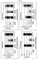
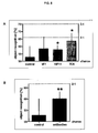


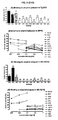


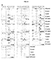


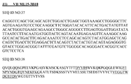
Classifications
-
- C—CHEMISTRY; METALLURGY
- C07—ORGANIC CHEMISTRY
- C07K—PEPTIDES
- C07K16/00—Immunoglobulins [IGs], e.g. monoclonal or polyclonal antibodies
- C07K16/18—Immunoglobulins [IGs], e.g. monoclonal or polyclonal antibodies against material from animals or humans
-
- A—HUMAN NECESSITIES
- A61—MEDICAL OR VETERINARY SCIENCE; HYGIENE
- A61K—PREPARATIONS FOR MEDICAL, DENTAL OR TOILETRY PURPOSES
- A61K38/00—Medicinal preparations containing peptides
- A61K38/04—Peptides having up to 20 amino acids in a fully defined sequence; Derivatives thereof
-
- A—HUMAN NECESSITIES
- A61—MEDICAL OR VETERINARY SCIENCE; HYGIENE
- A61P—SPECIFIC THERAPEUTIC ACTIVITY OF CHEMICAL COMPOUNDS OR MEDICINAL PREPARATIONS
- A61P25/00—Drugs for disorders of the nervous system
-
- A—HUMAN NECESSITIES
- A61—MEDICAL OR VETERINARY SCIENCE; HYGIENE
- A61P—SPECIFIC THERAPEUTIC ACTIVITY OF CHEMICAL COMPOUNDS OR MEDICINAL PREPARATIONS
- A61P25/00—Drugs for disorders of the nervous system
- A61P25/28—Drugs for disorders of the nervous system for treating neurodegenerative disorders of the central nervous system, e.g. nootropic agents, cognition enhancers, drugs for treating Alzheimer's disease or other forms of dementia
-
- A—HUMAN NECESSITIES
- A61—MEDICAL OR VETERINARY SCIENCE; HYGIENE
- A61P—SPECIFIC THERAPEUTIC ACTIVITY OF CHEMICAL COMPOUNDS OR MEDICINAL PREPARATIONS
- A61P37/00—Drugs for immunological or allergic disorders
-
- C—CHEMISTRY; METALLURGY
- C07—ORGANIC CHEMISTRY
- C07K—PEPTIDES
- C07K14/00—Peptides having more than 20 amino acids; Gastrins; Somatostatins; Melanotropins; Derivatives thereof
- C07K14/435—Peptides having more than 20 amino acids; Gastrins; Somatostatins; Melanotropins; Derivatives thereof from animals; from humans
- C07K14/46—Peptides having more than 20 amino acids; Gastrins; Somatostatins; Melanotropins; Derivatives thereof from animals; from humans from vertebrates
- C07K14/47—Peptides having more than 20 amino acids; Gastrins; Somatostatins; Melanotropins; Derivatives thereof from animals; from humans from vertebrates from mammals
- C07K14/4701—Peptides having more than 20 amino acids; Gastrins; Somatostatins; Melanotropins; Derivatives thereof from animals; from humans from vertebrates from mammals not used
- C07K14/4711—Alzheimer's disease; Amyloid plaque core protein
-
- G—PHYSICS
- G01—MEASURING; TESTING
- G01N—INVESTIGATING OR ANALYSING MATERIALS BY DETERMINING THEIR CHEMICAL OR PHYSICAL PROPERTIES
- G01N33/00—Investigating or analysing materials by specific methods not covered by groups G01N1/00 - G01N31/00
- G01N33/48—Biological material, e.g. blood, urine; Haemocytometers
- G01N33/50—Chemical analysis of biological material, e.g. blood, urine; Testing involving biospecific ligand binding methods; Immunological testing
- G01N33/68—Chemical analysis of biological material, e.g. blood, urine; Testing involving biospecific ligand binding methods; Immunological testing involving proteins, peptides or amino acids
- G01N33/6893—Chemical analysis of biological material, e.g. blood, urine; Testing involving biospecific ligand binding methods; Immunological testing involving proteins, peptides or amino acids related to diseases not provided for elsewhere
- G01N33/6896—Neurological disorders, e.g. Alzheimer's disease
-
- A—HUMAN NECESSITIES
- A61—MEDICAL OR VETERINARY SCIENCE; HYGIENE
- A61K—PREPARATIONS FOR MEDICAL, DENTAL OR TOILETRY PURPOSES
- A61K39/00—Medicinal preparations containing antigens or antibodies
- A61K2039/505—Medicinal preparations containing antigens or antibodies comprising antibodies
-
- A—HUMAN NECESSITIES
- A61—MEDICAL OR VETERINARY SCIENCE; HYGIENE
- A61K—PREPARATIONS FOR MEDICAL, DENTAL OR TOILETRY PURPOSES
- A61K38/00—Medicinal preparations containing peptides
-
- C—CHEMISTRY; METALLURGY
- C07—ORGANIC CHEMISTRY
- C07K—PEPTIDES
- C07K2317/00—Immunoglobulins specific features
- C07K2317/20—Immunoglobulins specific features characterized by taxonomic origin
- C07K2317/21—Immunoglobulins specific features characterized by taxonomic origin from primates, e.g. man
-
- C—CHEMISTRY; METALLURGY
- C07—ORGANIC CHEMISTRY
- C07K—PEPTIDES
- C07K2317/00—Immunoglobulins specific features
- C07K2317/20—Immunoglobulins specific features characterized by taxonomic origin
- C07K2317/24—Immunoglobulins specific features characterized by taxonomic origin containing regions, domains or residues from different species, e.g. chimeric, humanized or veneered
-
- C—CHEMISTRY; METALLURGY
- C07—ORGANIC CHEMISTRY
- C07K—PEPTIDES
- C07K2317/00—Immunoglobulins specific features
- C07K2317/40—Immunoglobulins specific features characterized by post-translational modification
- C07K2317/41—Glycosylation, sialylation, or fucosylation
-
- C—CHEMISTRY; METALLURGY
- C07—ORGANIC CHEMISTRY
- C07K—PEPTIDES
- C07K2317/00—Immunoglobulins specific features
- C07K2317/50—Immunoglobulins specific features characterized by immunoglobulin fragments
- C07K2317/56—Immunoglobulins specific features characterized by immunoglobulin fragments variable (Fv) region, i.e. VH and/or VL
- C07K2317/565—Complementarity determining region [CDR]
-
- C—CHEMISTRY; METALLURGY
- C07—ORGANIC CHEMISTRY
- C07K—PEPTIDES
- C07K2317/00—Immunoglobulins specific features
- C07K2317/90—Immunoglobulins specific features characterized by (pharmaco)kinetic aspects or by stability of the immunoglobulin
- C07K2317/92—Affinity (KD), association rate (Ka), dissociation rate (Kd) or EC50 value
-
- G—PHYSICS
- G01—MEASURING; TESTING
- G01N—INVESTIGATING OR ANALYSING MATERIALS BY DETERMINING THEIR CHEMICAL OR PHYSICAL PROPERTIES
- G01N2333/00—Assays involving biological materials from specific organisms or of a specific nature
- G01N2333/435—Assays involving biological materials from specific organisms or of a specific nature from animals; from humans
- G01N2333/46—Assays involving biological materials from specific organisms or of a specific nature from animals; from humans from vertebrates
- G01N2333/47—Assays involving proteins of known structure or function as defined in the subgroups
- G01N2333/4701—Details
- G01N2333/4709—Amyloid plaque core protein
-
- G—PHYSICS
- G01—MEASURING; TESTING
- G01N—INVESTIGATING OR ANALYSING MATERIALS BY DETERMINING THEIR CHEMICAL OR PHYSICAL PROPERTIES
- G01N2800/00—Detection or diagnosis of diseases
- G01N2800/28—Neurological disorders
- G01N2800/2814—Dementia; Cognitive disorders
- G01N2800/2821—Alzheimer
-
- G—PHYSICS
- G01—MEASURING; TESTING
- G01N—INVESTIGATING OR ANALYSING MATERIALS BY DETERMINING THEIR CHEMICAL OR PHYSICAL PROPERTIES
- G01N2800/00—Detection or diagnosis of diseases
- G01N2800/38—Pediatrics
- G01N2800/385—Congenital anomalies
- G01N2800/387—Down syndrome; Trisomy 18; Trisomy 13
Definitions
- the present invention relates to anti-A ⁇ globulomer antibodies, antigen-binding moieties thereof, hybridomas producing said antibodies, nucleic acids encoding said antibodies, vectors comprising said nucleic acids, host cells comprising said vectors, methods of producing said antibodies, compositions comprising said antibodies, therapeutic and diagnostic uses of said antibodies and corresponding methods relating to Alzheimer's disease and other amyloidoses.
- Alzheimer's disease is the most frequent cause for dementia among the aged with an incidence of about 10% of the population above 65 years. With increasing age, the probability of disease also rises. Globally, there are about 15 million people affected and further increases in life expectancy are expected to increase the number of diseased people to about threefold over the next decades.
- AD Alzheimer's disease
- extra-cellular amyloid plaques these deposits consist mostly of amyloid- ⁇ -peptide filaments, in the case of the intracellular neurofibrillary tangles (NFTs) of the tau protein.
- the amyloid ⁇ (A ⁇ ) peptide arises from the ( ⁇ -amyloid precursor protein by proteolytic cleavage. This cleavage is effected by the cooperative activity of several proteases named ⁇ -, ⁇ - and ⁇ -secretase. Cleavage leads to a number of specific fragments of differing length.
- the amyloid plaques consist mostly of peptides with a length of 40 or 42 amino acids (A ⁇ 40, A ⁇ 42). The dominant cleavage product is A ⁇ 40; however, A ⁇ 42 has a much stronger toxic effect.
- Cerebral amyloid deposits and cognitive impairments very similar to those observed in Alzheimer's disease are also hallmarks of Down's syndrome (trisomy 21), which occurs at a frequency of about 1 in 800 births.
- a ⁇ plaque burden deposited this hypothesis was favoured until recently.
- Active immunization with A ⁇ peptides leads to a reduction in the formation as well as to partial dissolution of existing plaques. At the same time it leads to alleviation of cognitive defects in APP transgenic mouse models.
- Meningoencephalitis was characterized by neuroinflammation and infiltration of T-cells into the brain. Presumably, this was due to a T-cell immune response induced by injection of A ⁇ (1-42) as antigen. Such an immune response is not to be expected after passive immunization. To date, there are no clinical data with reference to this available yet. However, with reference to such a passive approach to immunization concerns about the side effect profile were voiced because of preclinical studies in very old APP23 mice which received an antibody directed against an N-terminal epitope of A ⁇ (1-42) once a week over 5 months.
- mice showed an increase in the number and severity of microhaemorrhages compared to control animals treated with saline ( Pfeifer et al., 2002, Science, 298, 1379 ).
- a comparable increase in microhaemorrhages was also described in very old (> 24 months) Tg2576 and PDAPP mice ( Racke et al., 2005, J Neurosci, 25, 629-636 ; Wilcock et al. 2004, J. Neuroinflammation, 1(1):24 ; De Mattos et al., 2004, Neurobiol. Aging 25(S2):577 ).
- antibody injection led to a significant increase in microhaemorrhages.
- WO2004/067561 describes stable A ⁇ (1-42) oligomers (A ⁇ (1-42) globulomers) and antibodies directed specifically against the globulomers. Digestion with unspecific proteases shows that the A ⁇ globulomer may be digested beginning with the hydrophilic N-terminus protruding from the globular core structure ( Barghorn et al., 2005, J Neurochem, 95, 834-847 ). Such N-terminal truncated A ⁇ globulomers (A ⁇ (12-42) and A ⁇ (20-42) globulomers) represent the basic structural unit of this oligomeric A ⁇ .
- a therapeutically questionable reduction of brain volume has been observed in the study of active immunization with A ⁇ peptides in fibrillary condition of aggregation (ELAN trial with AN1792) : Moreover, in this trial severe side effects in form of a meningoencephalitis were observed.
- the present invention solves this problem by providing globulomer-specific antibodies possessing high affinity for truncated forms of A ⁇ globulomers. These antibodies are capable of discriminating not only other forms of A ⁇ peptides, particularly monomers and fibrils, but also untruncated forms of A ⁇ globulomers.
- the present invention relates to an antibody having a binding affinity to an A ⁇ (20-42) globulomer that is greater than the binding affinity of this antibody to an A ⁇ (1-42) globulomer.
- the present invention relates to an antibody having a binding affinity to an A ⁇ (20-42) globulomer that is greater than the binding affinity of this antibody to an A ⁇ (12-42) globulomer.
- the invention thus relates to antibodies having a binding affinity to the A ⁇ (20-42) globulomer that is greater than the binding affinity of the antibody to both the A ⁇ (1-42) globulomer and the A ⁇ (12-42) globulomer.
- a ⁇ (X-Y) here refers to the amino acid sequence from amino acid position X to amino acid position Y of the human amyloid ⁇ protein including both X and Y, in particular to the amino acid sequence from amino acid position X to amino acid position Y of the amino acid sequence DAEFRHDSGY EVHHQKLVFF AEDVGSNKGA IIGLMVGGVV IAT (corresponding to amino acid positions 1 to 43) or any of its naturally occurring variants, in particular those with at least one mutation selected from the group consisting of A2T, H6R, D7N, A21 G ("Flemish"), E22G (“Arctic”), E22Q (“Dutch”), E22K (“Italian”), D23N (“lowa”), A42T and A42V wherein the numbers are relative to the start of the A ⁇ peptide, including both position X and position Y or a sequence with up to three additional amino acid substitutions none of which may prevent globulomer formation, preferably with no additional
- a ⁇ (1-42) here refers to the amino acid sequence from amino acid position 1 to amino acid position 42 of the human amyloid ⁇ protein including both 1 and 42, in particular to the amino acid sequence DAEFRHDSGY EVHHQKLVFF AEDVGSNKGA llGLMVGGVV lA or any of its naturally occurring variants, in particular those with at least one mutation selected from the group consisting of A2T, H6R, D7N, A21 G ("Flemish"), E22G (“Arctic”), E22Q (“Dutch”), E22K (“Italian”), D23N (“lowa”), A42T and A42V wherein the numbers are relative to the start of the A ⁇ peptide, including both 1 and 42 or a sequence with up to three additional amino acid substitutions none of which may prevent globulomer formation, preferably with no additional amino acid substitutions in the portion from amino acid 20 to amino acid 42.
- a ⁇ (1-40) here refers to the amino acid sequence from amino acid position 1 to amino acid position 40 of the human amyloid ⁇ protein including both 1 and 40, in particular to the amino acid sequence DAEFRHDSGY EVHHQKLVFF AEDVGSNKGA IIGLMVGGVV or any of its naturally occurring variants, in particular those with at least one mutation selected from the group consisting of A2T, H6R, D7N, A21 G ("Flemish”), E22G (“Arctic”), E22Q (“Dutch”), E22K (“Italian”), and D23N (“lowa”) wherein the numbers are relative to the start of the A ⁇ peptide, including both 1 and 40 or a sequence with up to three additional amino acid substitutions none of which may prevent globulomer formation, preferably with no additional amino acid substitutions in the portion from amino acid 20 to amino acid 40.
- a ⁇ (12-42) here refers to the amino acid sequence from amino acid position 12 to amino acid position 42 of the human amyloid ⁇ protein including both 12 and 42, in particular to the amino acid sequence VHHQKLVFF AEDVGSNKGA IIGLMVGGVV lA or any of its naturally occurring variants, in particular those with at least one mutation selected from the group consisting of A21 G ("Flemish”), E22G (“Arctic”), E22Q (“Dutch”), E22K (“Italian”), D23N (“lowa”), A42T and A42V wherein the numbers are relative to the start of the A ⁇ peptide, including both 12 and 42 or a sequence with up to three additional amino acid substitutions none of which may prevent globulomer formation, preferably with no additional amino acid substitutions in the portion from amino acid 20 to amino acid 42.
- a ⁇ (20-42) here refers to the amino acid sequence from amino acid position 20 to amino acid position 42 of the human amyloid ⁇ protein including both 20 and 42, in particular to the amino acid sequence F AEDVGSNKGA llGLMVGGVV lA or any of its naturally occurring variants, in particular those with at least one mutation selected from the group consisting of A21G ("Flemish”), E22G (“Arctic”), E22Q (“Dutch”), E22K (“Italian”), D23N (“lowa”), A42T and A42V wherein the numbers are relative to the start of the A ⁇ peptide, including both 20 and 42 or a sequence with up to three additional amino acid substitutions none of which may prevent globulomer formation, preferably without any additional amino acid substitutions.
- a ⁇ (X-Y) globulomer refers to a soluble, globular, non-covalent association of A ⁇ (X-Y) peptides as defined above, possessing homogeneity and distinct physical characteristics.
- a ⁇ (X-Y) globulomers are stable, non-fibrillar, oligomeric assemblies of A ⁇ (X-Y) peptides which are obtainable by incubation with anionic detergents. In contrast to monomer and fibrils, these globulomers are characterized by defined assembly numbers of subunits (e.g.
- the globulomers have a 3-dimensional globular type structure ("molten globule”, see Barghorn et al., 2005, J Neurochem, 95, 834-847 ). They may be further characterized by one or more of the following features:
- a ⁇ (X-Y) globulomer here refers in particular to a product which is obtainable by a process as described in WO 2004/067561 , which is incorporated herein by reference.
- Said process comprises unfolding a natural, recombinant or synthetic A ⁇ (X-Y) peptide or a derivative thereof; exposing the at least partially unfolded A ⁇ (X-Y) peptide or derivative thereof to a detergent, reducing the detergent action and continuing incubation.
- hydrogen bond-breaking agents such as, for example, hexafluoroisopropanol (HFIP) may be allowed to act on the protein. Times of action of a few minutes, for example about 10 to 60 minutes, are sufficient when the temperature of action is from about 20 to 50°C and in particular about 35 to 40°C. Subsequent dissolution of the residue evaporated to dryness, preferably in concentrated form, in suitable organic solvents miscible with aqueous buffers, such as, for example, dimethyl sulfoxide (DMSO), results in a suspension of the at least partially unfolded peptide or derivative thereof, which can be used subsequently. If required, the stock suspension may be stored at low temperature, for example at about -20°C, for an interim period.
- DMSO dimethyl sulfoxide
- the peptide or the derivative thereof may be taken up in slightly acidic, preferably aqueous, solution, for example an about 10 mM aqueous HCl solution. After an incubation time of usually a few minutes, insoluble components are removed by centrifugation. A few minutes at 10000 g is expedient. These method steps are preferably carried out at room temperature, i.e. a temperature in the range from 20 to 30°C. The supernatant obtained after centrifugation contains the A ⁇ (X-Y) peptide or the derivative thereof and may be stored at low temperature, for example at about -20°C, for an interim period.
- oligomers A an intermediate type of oligomers (in WO 2004/067561 referred to as oligomers A).
- a detergent is allowed to act on the at least partially unfolded peptide or derivative thereof until sufficient intermediate oligomer has been produced.
- ionic detergents in particular anionic detergents.
- a detergent of the formula (I): R-X is used, in which the radical R is unbranched or branched alkyl having from 6 to 20 and preferably 10 to 14 carbon atoms or unbranched or branched alkenyl having from 6 to 20 and preferably 10 to 14 carbon atoms, the radical X is an acidic group or salt thereof, with X being preferably selected from among -COO - M + , -SO 3 - M + , and especially -OSO 3 - M + and M + is a hydrogen cation or an inorganic or organic cation preferably selected from alkali metal and alkaline earth metal cations and ammonium cations.
- Particular preference is given to sodium dodecyl sulfate (SDS).
- Lauric acid and oleic acid can also be used advantageously.
- the sodium salt of the detergent lauroylsarcosin also known as sarkosyl NL-30 or Gardol ® ) is also particularly advantageous.
- the time of detergent action in particular depends on whether - and if yes, to what extent - the peptide or the derivative thereof subjected to oligomerization has unfolded. If, according to the unfolding step, the peptide or derivative thereof has been treated beforehand with a hydrogen bond-breaking agent, i.e. in particular with hexafluoroisopropanol, times of action in the range of a few hours, advantageously from about 1 to 20 and in particular from about 2 to 10 hours, are sufficient when the temperature of action is about 20 to 50°C and in particular about 35 to 40°C. If a less unfolded or an essentially not unfolded peptide or derivative thereof is the starting point, correspondingly longer times of action are expedient.
- a hydrogen bond-breaking agent i.e. in particular with hexafluoroisopropanol
- peptide or the derivative thereof has been pretreated, for example, according to the procedure indicated above as an alternative to the HFlP treatment or said peptide or derivative thereof is directly subjected to oligomerization, times of action in the range from about 5 to 30 hours and in particular from about 10 to 20 hours are sufficient when the temperature of action is about 20 to 50°C and in particular about 35 to 40°C. After incubation, insoluble components are advantageously removed by centrifugation. A few minutes at 10000 g is expedient.
- the detergent concentration to be chosen depends on the detergent used. If SDS is used, a concentration in the range from 0.01 to 1% by weight, preferably from 0.05 to 0.5% by weight, for example of about 0.2% by weight, proves expedient. If lauric acid or oleic acid are used, somewhat higher concentrations are expedient, for example in a range from 0.05 to 2% by weight, preferably from 0.1 to 0.5% by weight, for example of about 0.5% by weight.
- the detergent action should take place at a salt concentration approximately in the physiological range.
- NaCl concentrations in the range from 50 to 500 mM, preferably from 100 to 200 mM and particularly at about 140 mM are expedient.
- oligomers B The subsequent reduction of the detergent action and continuation of incubation relates to a further oligomerization to give the A ⁇ (X-Y) globulomer of the invention (in WO 2004/067561 referred to as oligomers B).
- the composition obtained from the preceding step regularly contains detergent and a salt concentration in the physiological range it is then expedient to reduce detergent action and, preferably, also the salt concentration. This may be carried out by reducing the concentration of detergent and salt, for example, by diluting, expediently with water or a buffer of lower salt concentration, for example Tris-HCl, pH 7.3. Dilution factors in the range from about 2 to 10, advantageously in the range from about 3 to 8 and in particular of about 4, have proved suitable.
- the reduction in detergent action may also be achieved by adding substances which can neutralize said detergent action.
- substances which can neutralize said detergent action include substances capable of complexing the detergents, like substances capable of stabilizing cells in the course of purification and extraction measures, for example particular EO/PO block copolymers, in particular the block copolymer under the trade name Pluronic ® F 68.
- Alkoxylated and, in particular, ethoxylated alkyl phenols such as the ethoxylated t-octylphenols of the Triton ® X series, in particular Triton ® X100, 3-(3-cholamidopropyldimethylammonio)-1-propanesulfonate (CHAPS ® ) or alkoxylated and, in particular, ethoxylated sorbitan fatty esters such as those of the Tween ® series, in particular Tween ® 20, in concentration ranges around or above the particular critical micelle concentration, may be equally used.
- ethoxylated alkyl phenols such as the ethoxylated t-octylphenols of the Triton ® X series, in particular Triton ® X100, 3-(3-cholamidopropyldimethylammonio)-1-propanesulfonate (CHAPS ® ) or alkoxy
- the solution is incubated until sufficient A ⁇ (X-Y) globulomer of the invention has been produced. Times of action in the range of several hours, preferably in the range from about 10 to 30 hours and in particular in the range from about 15 to 25 hours, are sufficient when the temperature of action is about 20 to 50°C and in particular about 35 to 40°C.
- the solution may then be concentrated and possible residues may be removed by centrifugation. Here too, a few minutes at 10000 g proves expedient.
- the supernatant obtained after centrifugation contains an A ⁇ (X-Y) globulomer of the invention.
- An A ⁇ (X-Y) globulomer of the invention can be finally recovered in a manner known per se, e. g. by ultrafiltration, dialysis, precipitation or centrifugation.
- electrophoretic separation of the A ⁇ (X-Y) globulomers under denaturing conditions produces a double band (e. g. with an apparent molecular weight of 38 / 48 kDa for A ⁇ (1-42)), and especially preferred if upon glutardialdehyde treatment of the globulomers before separation these two bands are merged into one.
- size exclusion chromatography of the globulomers results in a single peak (e. g. corresponding to a molecular weight of approximately 100 kDa for A ⁇ (1-42) globulomer or of approximately 60 kDa for glutardialdehyde cross-linked A ⁇ (1-42) globulomer), respectively.
- a ⁇ (X-Y) globulomers wherein X is selected from the group consisting of the numbers 2 .. 24 and Y is as defined above are those which are obtainable by truncating A ⁇ (1-Y) globulomers into shorter forms wherein X is selected from the group consisting of the numbers 2 .. 24, with X preferably being 20 or 12, and Y is as defined above, which can be achieved by treatment with appropriate proteases.
- an A ⁇ (20-42) globulomer can be obtained by subjecting an A ⁇ (1-42) globulomer to thermolysin proteolysis
- an A ⁇ (12-42) globulomer can be obtained by subjecting an A ⁇ (1-42) globulomer to endoproteinase GluC proteolysis.
- the protease is inactivated in a generally known manner.
- the resulting globulomers may then be isolated following the procedures already described herein and, if required, processed further by further work-up and purification steps. A detailed description of said processes is disclosed in WO 2004/067561 , which is incorporated herein by reference.
- an A ⁇ (1-42) globulomer is in particular the A ⁇ (1-42) globulomer as described in example 1a herein; an A ⁇ (20-42) globulomer is in particular the A ⁇ (20-42) globulomer as described in examples 1c herein, and an A ⁇ (12-42) globulomer is in particular the A ⁇ (12-42) globulomer as described in examples 1d herein.
- the globulomer shows affinity to neuronal cells.
- the globulomer also exhibits neuromodulating effects.
- the globulomer consists of 11 to 16, and most preferably, of 12 to 14 A ⁇ (X-Y) peptides.
- the term "A ⁇ (X-Y) globulomer' here refers to a globulomer consisting essentially of A ⁇ (X-Y) subunits, where it is preferred if on average at least 11 of 12 subunits are of the A ⁇ (X-Y) type, more preferred if less than 10% of the globulomers comprise any non-A ⁇ (X-Y) peptides, and most preferred if the content of non-A ⁇ (X-Y) peptides is below the detection threshold.
- a ⁇ (1-42) globulomer here refers to a globulomer consisting essentially of A ⁇ (1-42) units as defined above; the term “A ⁇ (12-42) globulomer” here refers to a globulomer consisting essentially of A ⁇ (12-42) units as defined above; and the term “A ⁇ (20-42) globulomer” here refers to a globulomer consisting essentially of A ⁇ (20-42) units as defined above.
- cross-linked A ⁇ (X-Y) globulomer here refers to a molecule obtainable from an A ⁇ (X-Y) globulomer as described above by cross-linking, preferably chemically cross-linking, more preferably aldehyde cross-linking, most preferably glutardialdehyde cross-linking of the constituent units of the globulomer.
- a cross-linked globulomer is essentially a globulomer in which the units are at least partially joined by covalent bonds, rather than being held together by non-covalent interactions only.
- a cross-linked A ⁇ (1-42) globulomer is in particular the cross-linked A ⁇ (1-42) oligomer as described in example 1b herein.
- a ⁇ (X-Y) globulomer derivative here refers in particular to a globulomer that is labelled by being covalently linked to a group that facilitates detection, preferably a fluorophore, e. g. fluorescein isothiocyanate, phycoerythrin, Aequorea victoria fluorescent protein, Dic-tyosoma fluorescent protein or any combination or fluorescence-active derivative thereof; a chromophore; a chemoluminophore, e. g.
- luciferase preferably Photinus pyralis luciferase, Vibrio fischeri luciferase, or any combination or chemoluminescence-active derivative thereof; an enzymatically active group, e. g. peroxidase, e. g. horseradish peroxidase, or any enzymatically active derivative thereof; an electron-dense group, e. g. a heavy metal containing group, e.g. a gold containing group; a hapten, e. g. a phenol derived hapten; a strongly antigenic structure, e. g. peptide sequence predicted to be antigenic, e. g.
- a chelating group e. g. hexahistidinyl
- a natural or nature-derived protein structure mediating further specific protein-protein interactions, e. g. a member of the fos/jun pair
- a magnetic group e. g. a ferromagnetic group
- a radioactive group e. g.
- a group comprising 1 H, 14 C, 32 p, 35 S or 125 l or any combination thereof; or to a globulomer flagged by being covalently or by non-covalent high-affinity interaction, preferably covalently linked to a group that facilitates inactivation, sequestration, degradation and/or precipitation, preferably flagged with a group that promotes in vivo degradation, more preferably with ubiquitin, where is particularly preferred if this flagged oligomer is assembled in vivo; or to a globulomer modified by any combination of the above.
- Such labelling and flagging groups and methods for attaching them to proteins are known in the art. Labelling and/or flagging may be performed before, during or after globulomerisation.
- a globulomer derivative is a molecule obtainable from a globulomer by a labelling and/or flagging reaction.
- a ⁇ (X-Y) monomer derivative here refers in particular to an A ⁇ monomer that is labelled or flagged as described for the globulomer.
- the antibody of the present invention binds to an A ⁇ (20-42) globulomer with a K D in the range of 1x10 -6 M to 1x10 -12 M.
- the antibody binds to an A ⁇ (20-42) globulomer with high affinity, for instance with a K D of 1x10 -7 M or greater affinity, e.g. with a K D of 3x10 -8 M or greater affinity, with a K D of 1 x10 -8 M or greater affinity, e.g. with a K D of 3x10 -9 M or greater affinity, with a K D of 1x10 -9 M or greater affinity, e.g.
- K D of 3x10 -10 M or greater affinity with a K D of 1x10 -10 M or greater affinity, e.g. with a K D of 3x10 -11 M or greater affinity, or with a K D of 1 x10 -11 M or greater affinity.
- greater affinity here refers to a degree of interaction where the equilibrium between unbound antibody and unbound globulomer on the one hand and antibody-globulomer complex on the other is further in favour of the antibody-globulomer complex.
- small affinity here refers to a degree of interaction where the equilibrium between unbound antibody and unbound globulomer on the one hand and antibody-globulomer complex on the other is further in favour of the unbound antibody and unbound globulomer.
- greater affinity is synonymous with the term “higher affinity” and term “smaller affinity”is synonymous with the term “lower affinity”.
- the invention relates to an antibody which binds to the A ⁇ (20-42) globulomer with a K D in the range of 1x10 -6 M to 1x10 -12 M, to the A ⁇ (1-42) globulomer with a K D of 10 -12 M or smaller affinity, the binding affinity to the A ⁇ (20-42) globulomer being greater than the binding affinity to the A ⁇ (1-42) globulomer.
- the binding affinity of the antibody of the present invention to the A ⁇ (20-42) globulomer is at least 2 times, e. g. at least 3 times or at least 5 times, preferably at least 10 times, e. g. at least 20 times, at least 30 times or at least 50 times, more preferably at least 100 times, e. g. at least 200 times, at least 300 times or at least 500 times, and even more preferably at least 1000 times, e. g. at least 2000 times, at least 3000 times or at least 5000 times, even more preferably at least 10000 times, e. g. at least 20000 times, at least 30000 or at least 50000 times, and most preferably at least 100000 times greater than the binding affinity of the antibody to the A ⁇ (1-42) globulomer.
- the invention relates to an antibody which binds to the A ⁇ (12-42) globulomer with a K D with a K D of 10 -12 M or smaller affinity, the binding affinity to the A ⁇ (20-42) globulomer being greater than the binding affinity to the A ⁇ (12-42) globulomer.
- the binding affinity of the antibody of the present invention to the A ⁇ (20-42) globulomer is at least 2 times, e. g. at least 3 times or at least 5 times, preferably at least 10 times, e. g. at least 20 times, at least 30 times or at least 50 times, more preferably at least 100 times, e. g. at least 200 times, at least 300 times or at least 500 times, and even more preferably at least 1000 times, e. g. at least 2000 times, at least 3000 times or at least 5000 times, even more preferably at least 10000 times, e. g. at least 20000 times, at least 30000 or at least 50000 times, and most preferably at least 100000 times greater than the binding affinity of the antibody to the A ⁇ (12-42) globulomer.
- the antibodies of the present invention bind to at least one A ⁇ globulomer, as defined above, and have a comparatively smaller affinity for at least one non-globulomer form of A ⁇ .
- Antibodies of the present invention having a comparatively smaller affinity for at least one non-globulomer form of A ⁇ than for at least one A ⁇ globulomer include antibodies having a binding affinity to the A ⁇ (20-42) globulomer that is greater than to an A ⁇ (1-42) monomer. Further, it is preferred that, alternatively or additionally, the binding affinity of the antibody to the A ⁇ (20-42) globulomer is greater than to an A ⁇ (1-40) monomer.
- the affinity of the antibody to the A ⁇ (20-42) globulomer is greater than its affinity to both the A ⁇ (1-40) and the A ⁇ (1-42) monomer.
- a ⁇ (X-Y) monomer here refers to the isolated form of the A ⁇ (X-Y) peptide, preferably a form of the A ⁇ (X-Y) peptide which is not engaged in essentially non-covalent interactions with other A ⁇ peptides.
- the A ⁇ (X-Y) monomer is usually provided in the form of an aqueous solution.
- the aqueous monomer solution contains 0.05% to 0.2%, more preferably about 0.1 % NH 4 0H.
- the aqueous monomer solution contains 0.05% to 0.2%, more preferably about 0.1 % NaOH.
- a ⁇ (1-40) monomer here refers to an A ⁇ (1-40) monomer preparation as described in example 2 herein
- a ⁇ (1-42) monomer here refers to an A ⁇ (1-42) preparation as described in example 2 herein.
- the antibody of the present invention binds to one or, more preferably, both monomers with low affinity, most preferably with a K D of 1x10 -8 M or smaller affinity, e. g. with a K D of 3x10 -8 M or smaller affinity, with a K D of 1x10 -7 M or smaller affinity, e. g. with a K D of 3x10 -7 M or smaller affinity, or with a K D of 1x10 -6 M or smaller affinity, e. g. with a K D of 3x10 -5 M or smaller affinity, or with a K D of 1x10 -5 M or smaller affinity.
- the binding affinity of the antibody of the present invention to the A ⁇ (20-42) globulomer is at least 2 times, e. g. at least 3 times or at least 5 times, preferably at least 10 times, e. g. at least 20 times, at least 30 times or at least 50 times, more preferably at least 100 times, e. g. at least 200 times, at least 300 times or at least 500 times, and even more preferably at least 1000 times, e. g. at least 2000 times, at least 3000 times or at least 5000 times, even more preferably at least 10000 times, e. g. at least 20000 times, at least 30000 or at least 50000 times, and most preferably at least 100000 times greater than the binding affinity of the antibody to one or, more preferably, both monomers.
- Antibodies of the present invention having a comparatively smaller affinity for at least one non-globulomer form of A ⁇ than for at least one A ⁇ globulomer further include antibodies having a binding affinity to the A ⁇ (20-42) globulomer that is greater than to A ⁇ (1-42) fibrils. Further, it is preferred that, alternatively or additionally, the binding affinity of the antibody to the A ⁇ (20-42) globulomer is greater than to A ⁇ (1-40) fibrils.
- fibrill here refers to a molecular structure that comprises assemblies of non-covalently associated, individual A ⁇ (X-Y) peptides, which show fibrillary structure in the electron microscope, which bind Congo red and then exhibit birefringence under polarized light and whose X-ray diffraction pattern is a cross- ⁇ structure.
- a fibril is a molecular structure obtainable by a process that comprises the self-induced polymeric aggregation of a suitable A ⁇ peptide in the absence of detergents, e. g. in 0.1 M HCl, leading to the formation of aggregates of more than 24, preferably more than 100 units.
- This process is well known in the art.
- a ⁇ (X-Y) fibrils are used in the form of an aqueous solution.
- the aqueous fibril solution is made by dissolving the A ⁇ peptide in 0.1 % NH 4 OH, diluting it 1 : 4 with 20 mM NaH 2 PO 4 , 140 mM NaCl, pH 7.4, followed by readjusting the pH to 7.4, incubating the solution at 37 °C for 20 h, followed by centrifugation at 10000 g for 10 min and resuspension in 20 mM NaH 2 PO 4 , 140 mM Nacl, pH 7.4.
- a ⁇ (X-Y) fibril here refers to a fibril consisting essentially of A ⁇ (X-Y) subunits, where it is preferred if on average at least 90% of the subunits are of the A ⁇ (X-Y) type, more preferred if at least 98% of the subunits are of the A ⁇ (X-Y) type, and most preferred if the content of non-A ⁇ (X-Y) peptides is below the detection threshold.
- a ⁇ (1-42) fibril here refers to a A ⁇ (1-42) fibril preparation as described in example 3 herein.
- the antibody of the present invention binds to one or, more preferably, both fibrils with low affinity, most preferably with a K D of 1x10 -8 M or smaller affinity, e. g. with a K D of 3x10 -8 M or smaller affinity, with a K D of 1x10 -7 M or smaller affinity, e. g. with a K D of 3x10 -7 M or smaller affinity, or with a K D of 1x10 -6 M or smaller affinity, e. g. with a K D of 3x10 -5 M or smaller affinity, or with a K D of 1x10 -5 M or smaller affinity.
- the binding affinity of the antibody of the present invention to A ⁇ (20-42) globulomer is at least 2 times, e. g. at least 3 times or at least 5 times, preferably at least 10 times, e. g. at least 20 times, at least 30 times or at least 50 times, more preferably at least 100 times, e. g. at least 200 times, at least 300 times or at least 500 times, and even more preferably at least 1000 times, e. g. at least 2000 times, at least 3000 times or at least 5000 times, even more preferably at least 10000 times, e. g. at least 20000 times, at least 30000 or at least 50000 times, and most preferably at least 100000 times greater than the binding affinity of the antibody to one or, more preferably, both fibrils.
- the invention relates to antibodies having a binding affinity to the A ⁇ (20-42) globulomer which is greater than its binding affinity to both A ⁇ (1-40) and A ⁇ (1-42) fibrils.
- the present invention relates to antibodies having a comparatively smaller affinity for both the monomeric and fibrillary forms of A ⁇ than for at least one A ⁇ globulomer, in particular A ⁇ (20-42) globulomer.
- a ⁇ globulomer in particular A ⁇ (20-42) globulomer.
- Antibodies of the present invention further include antibodies having a binding affinity to the A ⁇ (20-42) globulomer that is greater than to a cross-linked A ⁇ (1-42) globulomer, in particular to a glutardialdehyde cross-linked A ⁇ (1-42) globulomer, such as that described in example 1b herein.
- the binding affinity of the antibody to A ⁇ (20-42) globulomer is at least 2 times, e. g. at least 3 times or at least 5 times, preferably at least 10 times, e. g. at least 20 times, at least 30 times or at least 50 times, more preferably at least 100 times, e. g. at least 200 times, at least 300 times or at least 500 times, and even more preferably at least 1000 times, e. g. at least 2000 times, at least 3000 times or at least 5000 times, even more preferably at least 10000 times, e. g. at least 20000 times, at least 30000 or at least 50000 times, and most preferably at least 100000 times greater than the binding affinity of the antibody to cross-linked A ⁇ (1-42) globulomer.
- the antibodies of the present invention are preferably isolated, in particular monoclonal and more particularly recombinant.
- the present invention also relates to a monoclonal antibody (5F7) that is obtainable from a hybridoma designated by American Type Culture Collection deposit number PTA-7241.
- the present invention also relates to a monoclonal antibody (10F11) that is obtainable from a hybridoma designated by American Type Culture Collection deposit number PTA-7239.
- the present invention also relates to a monoclonal antibody (7C6) that is obtainable from a hybridoma designated by American Type Culture Collection deposit number PTA-7240.
- the present invention also relates to a monoclonal antibody (4B7) that is obtainable from a hybridoma designated by American Type Culture Collection deposit number PTA-7242.
- the present invention also relates to a monoclonal antibody (6A2) that is obtainable from a hybridoma designated by American Type Culture Collection deposit number PTA-7409.
- the present invention also relates to a monoclonal antibody (2F2) that is obtainable from a hybridoma designated by American Type Culture Collection deposit number PTA-7408.
- the present invention also relates to a monoclonal antibody (4D10) that is obtainable from a hybridoma designated by American Type Culture Collection deposit number PTA-7405.
- the present invention also relates to a monoclonal antibody (7E5) that is obtainable from a hybridoma designated by American Type Culture Collection deposit number PTA-7809.
- the present invention also relates to a monoclonal antibody (10C1) that is obtainable from a hybridoma designated by American Type Culture Collection deposit number PTA-7810.
- the present invention also relates to a monoclonal antibody (3B10) that is obtainable from a hybridoma designated by American Type Culture Collection deposit number PTA-7851.
- the present invention also relates to antibodies having a similar binding profile to that of any one of said monoclonal antibodies, 5F7, 10F11, 7C6, 4B7, 6A2, 2F2, 4D10, 7E5, 10C1 and 3B10.
- Antibodies having a similar binding profile to that of any one of said monoclonal antibodies should be understood as not being limited to antibodies having a binding affinity to an A ⁇ (20-42) globulomer that is greater than the binding affinity of the antibody to an A ⁇ (1-42) globulomer.
- Antibodies having a binding profile similar to that of any one of said monoclonal antibodies, 5F7, 10F11, 7C6, 4B7, 6A2, 2F2, 4D10, 7E5, 10C1 and 3B10 include antibodies which bind to the same epitope as monoclonal antibody 5F7, 10F11, 7C6, 4B7, 6A2, 2F2, 4D10, 7E5, 10C1 and 3B10.
- All monoclonal antibodies from the group consisting of 5F7, 10F11, 7C6, 4B7, 6A2, 2F2, 4D10, 7E5, 10C1 and 3B10 bind to an epitope contained within the 20 and 42 A ⁇ sequence range, in particular within the 20-30 A ⁇ sequence range.
- said epitope is believed to be a structural, non-linear epitope in between subunits in the region of amino acids 20 and 42, in particular in the region of amino acids 20 and 30.
- the present invention also relates to antibodies which are capable of competing with at least one, preferably all, antibodies selected from the group consisting of 5F7, 10F11, 7C6, 4B7, 6A2, 2F2, 4D10, 7E5, 10C1 and 3B10.
- serving antibodies refers to any number of antibodies targeting the same molecular or stably but non-covalently linked supermolecular entity, preferably the same molecule, wherein at least one is capable of specifically reducing the measurable binding of another, preferably by sterically hampering the other's access to its target epitope or by inducing and/or stabilizing a conformation in the target entity that reduces the target's affinity for the other antibody, more preferably by directly blocking access to the other's target epitope by binding to an epitope in sufficiently close vicinity of the former, overlapping with the former or identical to the former, most preferably overlapping or identical, in particular identical.
- Two epitopes are herein said to be “overlapping” if they share part of their chemical structures, preferably their amino acid sequences, and to be “identical”, if their chemical structures, preferably their amino acid sequences, are identical.
- the present invention also relates to antibodies whose target epitopes are overlapping with, preferably identical to, the target epitope of at least one of the antibodies selected from the group consisting of 5F7, 10F11, 7C6, 4B7, 6A2, 2F2, 4D10, 7E5, 10C1 and 3B10.
- Antibodies having a similar binding profile to that of any one of said monoclonal antibodies, 5F7, 10F11, 7C6, 4B7, 6A2, 2F2, 4D10, 7E5, 10C1 and 3B10 thus further include antibodies which comprise at least a portion of the antigen-binding moiety of any one of said monoclonal antibodies, 5F7, 10F11, 7C6, 4B7, 6A2, 2F2, 4D10, 7E5, 10C1 and 3B10.
- said portion comprises at least one complementary determining region (CDR) of any one of said monoclonal antibodies, 5F7, 10F11, 7C6, 4B7, 6A2, 2F2, 4D10, 7E5, 10C1 and 3B10.
- the present invention relates to antibodies comprising the amino acid sequence of the heavy chain CDR3 and/or the amino acid sequence of the light chain CDR3 of monoclonal antibody 5F7, 10F11, 7C6, 4B7, 6A2, 2F2, 4D10, 7E5, 10C1 or 3B10.
- specific examples of such antibodies include those which also comprise the amino acid sequence of the heavy chain CDR2 and/or the amino acid sequence of the light chain CDR2 of monoclonal antibody 5F7, 10F11, 7C6, 4B7, 6A2, 2F2, 4D10, 7E5, 10C1 or 3B10, respectively.
- such antibodies include those which also comprise the amino acid sequence of the heavy chain CDR1 and/or the amino acid sequence of the light chain CDR1 of monoclonal antibody 5F7, 10F11, 7C6, 4B7, 6A2, 2F2, 4D10, 7E5, 10C1 or 3B10, respectively.
- the present invention thus relates to antibodies comprising a heavy chain wherein the CDR3, CDR2 and/or CDR1 domain comprises the amino acid sequence of the heavy chain CDR3, CDR2 and/or CDR1 of monoclonal antibody 5F7, 10F11, 7C6, 4B7, 6A2, 2F2, 4D10, 7E5, 10C1 or 3B10.
- the present invention thus relates to antibodies comprising a light chain wherein the CDR3, CDR2 and/or CDR1 domain comprises the amino acid sequence of the light chain CDR3, CDR2 and/or CDR1, respectively, of monoclonal antibody 5F7, 10F11, 7C6, 4B7, 6A2, 2F2, 4D10, 7E5, 10C1 or 3B10.
- the antibody comprises at least 3 CDRs selected from the group consisting of the sequences disclosed above. More preferably the 3 CDRs selected are from sets of variable domain CDRs selected from the group consisting of: VH 5F7 CDR Set VH 5F7 CDR-H1 TFYIH: residues 31-35 of SEQ ID NO:3 VH 5F7 CDR-H2 MTGPGSGNTYYNEMFKD : residues 50-66 of SEQ ID N0:3 VH 5F7 CDR-H3 AKSARAAWFAY: residues 99-109 of SEQ ID NO:3 VL 5F7 CDR Set RSSQSVVQSNGNTYLE: VL 5F7 CDR-L1 residues 24-39 of SEQ ID NO:4 VL 5F7 CDR-L2 KVSNRFS: residues 55-61 of SEQ ID NO:4 VL 5F7 CDR-L3 FQGSHVPPT: residues 94-102 of SEQ ID NO:4 VH 10F
- the antibody of the invention comprises at least two variable domain CDR sets. More preferably, the two variable domain CDR sets are selected from the group consisting of: VH 5F7 CDR Set & VL 5F7 CDR Set; VH 10F11 CDR Set & VL 10F11 CDR Set; VH 7C6 CDR Set & VL 7C6 CDR Set; VH 4B7 CDR Set & VL 4B7 CDR Set; VH 2F2 CDR Set & VL 2F2 CDR Set; VH 6A2 CDR Set & VL 6A2 CDR Set; VH 4D10 CDR Set & VL 4D10 CDR Set; VH 7E5 CDR Set & VL 7E5 CDR Set; and VH 10C1 CDR Set & VL 10C1 CDR Set.
- the antibody disclosed above further comprises a human acceptor framework.
- the antibody is a CDR grafted antibody.
- the CDR grafted antibody comprises one or more of the CDRs disclosed above.
- the CDR grafted antibody comprises a human acceptor framework.
- the antibody is a humanized antibody.
- the humanized antibody comprises one or more of the CDRs disclosed above. More preferably the humanized antibody comprises three or more of the CDRs disclosed above. Most preferably the humanized antibody comprises six CDRs disclosed above.
- the CDRs are incorporated into a human antibody variable domain of a human acceptor framework.
- the human antibody variable domain is a consensus human variable domain.
- the human acceptor framework comprises at least one Framework Region amino acid substitution at a key residue, wherein the key residue is selected from the group consisting of a residue adjacent to a CDR; a glycosylation site residue; a rare residue; a residue capable of interacting with A ⁇ (20-42) globulomer; a residue capable of interacting with a CDR; a canonical residue; a contact residue between heavy chain variable region and light chain variable region; a residue within a Vernier zone; and a residue in a region that overlaps between a Chothia-defined variable heavy chain CDR1 and a Kabat-defined first heavy chain framework.
- the human acceptor framework human acceptor framework comprises at least one Framework Region amino acid substitution, wherein the amino acid sequence of the framework is at least 65% identical to the sequence of said human acceptor framework and comprises at least 70 amino acid residues identical to said human acceptor framework.
- the present invention relates to antibodies comprising both the heavy and light chain as defined above.
- the antibody comprises at least one variable domain having an amino acid sequence selected from the group consisting of: SEQ ID NO:3, SEQ ID NO:4, SEQ ID NO:7, SEQ ID NO:8, SEQ ID N0:11, SEQ ID NO:12, SEQ ID NO:15, SEQ ID NO:16, SEQ ID N0:19, SEQ ID NO:20, SEQ ID NO:23, SEQ ID NO:24, SEQ ID NO:27, SEQ ID NO:28, SEQ ID N0:31, SEQ ID NO:32, SEQ ID NO:35, SEQ ID NO:36, and SEQ ID NO:38.
- the antibody comprises two variable domains, wherein said two variable domains have amino acid sequences selected from the group consisting of: SEQ ID NO:3 & SEQ ID NO:4, SEQ ID NO:7 & SEQ ID NO:8 & SEQ ID NO:11 & SEQ ID NO:12, SEQ ID NO:15 & SEQ ID NO:16, SEQ ID N0:19 & SEQ ID NO:20, SEQ ID NO:23 & SEQ ID NO:24, SEQ ID NO:27 & SEQ ID NO:28, SEQ ID NO:31 & SEQ ID NO:32, and SEQ ID NO:35 & SEQ ID NO:36.
- the antibodies of the present invention comprise a heavy chain constant region selected from the group consisting of IgG1, IgG2, IgG3, IgG4, IgM, IgA, IgD, IgE and human IgG1 Ala234 Ala235 mutant constant regions.
- the antibodies comprise a human constant region. More preferably, the antibodies comprise an amino acid sequence selected from the group consisting of SEQ ID NOs:39-42. Antibodies comprising an IgG1 heavy chain constant region are preferred.
- the antibody is glycosylated.
- the glycosylation pattern is a human glycosylation pattern or a glycosylation pattern produced by any one of the eukaryotic cells disclosed herein, in particular CHO cells.
- the present invention also relates to an antigen-binding moiety of an antibody of the present invention.
- antigen-binding moieties include, but are not limited to, Fab fragments, F(ab') 2 fragments and single chain Fv fragments of the antibody. Further antigen-binding moieties are Fab' fragments, Fv fragments, and disulfide linked Fv fragments.
- the invention also provides an isolated nucleic acid encoding any one of the antibodies disclosed herein.
- a further embodiment provides a vector comprising the isolated nucleic acid disclosed herein.
- Said vector may in particular be selected from the group consisting of pcDNA; pTT ( Durocher et al., Nucleic Acids Research 2002, Vol 30, No.2 ); pTT3 (pTT with additional multiple cloning site; pEFBOS ( Mizushima, S. and Nagata, S., (1990) Nucleic Acids Research Vol 18, No. 17 ); pBV; pJV; and pBJ.
- a host cell is transformed with the vector disclosed herein.
- the host cell is a prokaryotic cell. More preferably the host cell is E.coli.
- the host cell is a eukaryotic cell.
- the eukaryotic cell is selected from the group consisting of a protist cell, an animal cell (e.g. a mammalian cell, an avian cell, and an insect cell), a plant cell and a fungal cell. More preferably the host cell is a mammalian cell including, but not limited to, CHO and COS; or a fungal cell, eg., a yeast cell, such as Saccharomyces cerevisiae; or an insect cell such as Sf9.
- Another aspect of the invention provides a method of producing an antibody of the invention, comprising culturing any one of the host cells or a hybridoma disclosed herein in a culture medium under conditions suitable to produce the antibody.
- Another embodiment provides an antibody that is obtainable by the method disclosed herein.
- Antibodies of the present invention can be obtained in a manner known per se.
- B lymphocytes which, in totality, contain an antibody repertoire composed of hundreds of billions of different antibody specificities are a part of the mammalian immune system.
- a normal immune response to a particular antigen means selection of one or more antibodies of said repertoire which specifically bind to said antigen, and the success of an immune response is based at least partially on the ability of said antibodies to specifically recognize (and ultimately to eliminate) the stimulating antigen and to ignore other molecules in the environment of said antibodies.
- Standardized hybridoma technology now allows the production of antibodies with a single specificity for an antigen of interest. More recently, recombinant antibody techniques such as in-vitro screening of antibody libraries have been developed. These techniques likewise allow antibodies having a single specificity for an antigen of interest to be produced.
- the antigen of interest may be allowed to act on the antibody repertoire either in vivo or in vitro.
- the antigen is allowed to act on the repertoire by immunizing an animal in vivo with said antigen.
- This in-vivo approach may furthermore comprise establishing from the lymphocytes of an animal a number of hybridomas and selecting a particular hybridoma which secretes an antibody specifically binding to said antigen.
- the animal to be immunized may be, for example, a mouse, rat, rabbit, chicken, camelid or sheep or may be a transgenic version of any of the animals mentioned above, for example a transgenic mouse with human immunoglobulin genes, which produces human antibodies after an antigenic stimulus.
- mice with severe combined immunodeficiency which have been reconstituted with human peripheral mononuclear blood cells (chimeric hu-PBMC SCID mice) or with lymphoid cells or precursors thereof, as well as mice which have been treated with a lethal total body irradiation, then protected against radiation with bone marrow cells from a mouse with severe combined immunodeficiency (SCID) and subsequently transplanted with functional human lymphocytes (the "Trimera” system).
- SCID severe combined immunodeficiency
- mice with severe combined immunodeficiency mice with human peripheral mononuclear blood cells
- lymphoid cells or precursors thereof as well as mice which have been treated with a lethal total body irradiation, then protected against radiation with bone marrow cells from a mouse with severe combined immunodeficiency (SCID) and subsequently transplanted with functional human lymphocytes (the "Trimera” system).
- Another type of an animal to be immunized is an animal (e.g.
- polyclonal or monoclonal antibodies produced by this method are characterized and selected by using known screening methods which include, but are not limited to, ELISA and dot blot techniques.
- the antigen is allowed to act on the antibody repertoire in vitro by screening a recombinant antibody library with said antigen.
- the recombinant antibody library may be expressed, for example, on the surface of bacteriophages or on the surface of yeast cells or on the surface of bacterial cells.
- the recombinant antibody library is an scFv library or an Fab library, for example.
- antibody libraries are expressed as RNA-protein fusions.
- the antigen may be allowed to act on the antibody repertoire by immunizing an animal in vivo with said antigen and then screening in vitro with said antigen a recombinant antibody library prepared from lymphoid cells of said animal or a single domain antibody library (e.g. containing heavy and/or light chains).
- the antigen is allowed to act on the antibody repertoire by immunizing an animal in vivo with said antigen and then subjecting a recombinant antibody library or single domain library produced from lymphoid cells of said animal to affinity maturation.
- the antigen is allowed to act on the antibody repertoire by immunizing an animal in vivo with said antigen, then selecting individual antibody-producing cells secreting an antibody of interest and obtaining from said selected cells cDNAs for the variable region of the heavy and light chains (e.g. by means of PCR) and expressing said variable regions of the heavy and light chains in mammalian host cells in vitro (this being referred to as selected lymphocyte antibody method or SLAM), thereby being able to further select and manipulate the selected antibody gene sequences.
- monoclonal antibodies may be selected by expression cloning by expressing the antibody genes for the heavy and light chains in mammalian cells and selecting those mammalian cells which secrete an antibody having the desired binding affinity.
- the present invention provides defined antigens for screening and counter screening. Thus it is possible, according to the invention, to select those polyclonal and monoclonal antibodies which bind to an A ⁇ (20-42) globulomer with the binding affinities as defined above.
- the methods of the invention for producing antibodies can be used to produce various types of antibodies. These include monoclonal, in particular recombinant antibodies, especially essentially human antibodies, chimeric antibodies, humanized antibodies and CDR graft antibodies, and also antigen-binding moieties thereof.
- the present invention further relates to a hybridoma that is capable of producing (secreting) a monoclonal antibody of the present invention.
- Hybridomas of the present invention include those designated by an American Type Culture Collection deposit number selected from the group consisting of PTA-7241, PTA-7239, PTA-7240, PTA-7242, PTA-7408, PTA-7409, PTA-7405, PTA-7809, PTA-7810 and PTA-7851.
- the antibodies of the present invention may also be reactive with, i.e. bind to, A ⁇ forms other than the A ⁇ globulomers described herein. These antigens may or may not be oligomeric or globulomeric. Thus, the antigens to which the antibodies of the present invention bind include any A ⁇ form that comprises the globulomer epitope with which the antibodies of the present invention are reactive.
- Such A ⁇ forms include truncated and non-truncated A ⁇ (X-Y) forms (with X and Y being defined as above), such as A ⁇ (20-42), A ⁇ (20-40), A ⁇ (12-42), A ⁇ (12-40), A ⁇ (1-42), and A ⁇ (1-40) forms, provided that said forms comprise the globulomer epitope.
- the present invention also relates to a composition
- a composition comprising an antibody of the invention or an antigen-binding moiety thereof, as defined above.
- said composition is a pharmaceutical composition which comprises the antibody of the invention or the antigen-binding moiety and a pharmaceutical acceptable carrier.
- the antibody of the invention or the antigen-binding moiety as defined above is preferably capable of neutralizing, both in vitro and in vivo, the activity of A ⁇ globulomer or a derivative thereof to which it binds.
- Said antibody or antigen-binding moiety may therefore be used for inhibiting the activity of said globulomer or derivative thereof, for example in a preparation containing said globulomer or derivative thereof or in human individuals or other mammals in which said globulomer or derivative thereof is present.
- the invention relates to a method of inhibiting the activity of said globulomer or derivative thereof, which method comprises allowing an antibody of the invention or an antigen-binding moiety thereof to act on a globulomer or derivative thereof so as to inhibit the activity of said globulomer or derivative thereof.
- Said activity may be inhibited in vitro, for example.
- the antibody of the invention or the antigen-binding moiety may be added to a preparation such as a sample derived from a subject or a cell culture which contains or is suspected to contain said globulomer or derivative thereof, in order to inhibit the activity of said globulomer or derivative thereof in said sample.
- the activity of the globulomer or derivative thereof may be inhibited in an individual in vivo.
- the present invention further relates to the use of an antibody or an antigen-binding moiety as defined above for preparing a pharmaceutical composition for treating or preventing an amyloidosis, in particular an amyloidosis selected from the group consisting of Alzheimer's disease and the amyloidosis of Down's syndrome.
- an antibody or an antigen-binding moiety as defined above for preparing a pharmaceutical composition for treating or preventing an amyloidosis, in particular an amyloidosis selected from the group consisting of Alzheimer's disease and the amyloidosis of Down's syndrome.
- One aspect of said use of the invention is therefore a method of treating or preventing an amyloidosis, in particular Alzheimer's disease or the amyloidosis of Down's syndrome, in a subject in need thereof, which comprises administering an antibody or an antigen-binding moiety as defined above to the subject.
- said antibody or antigen-binding moiety for treating and especially preventing the amyloidosis, in particular Alzheimer's disease or the amyloidosis of Down's syndrome is in particular for passive immunization. Accordingly, in the method of treating or preventing an amyloidosis, in particular Alzheimer's disease or the amyloidosis of Down's syndrome, in a subject in need thereof one purpose of administering the antibody or antigen-binding moiety to the subject is passively immunizing the subject against the amyloidosis, in particular Alzheimer's disease or the amyloidosis of Down's syndrome.
- the antibody of the invention or the antigen-binding moiety as defined above is preferably capable of detecting, both in vitro and in vivo, an A ⁇ globulomer or derivative thereof to which it binds.
- Said antibody or the antigen-binding moiety may therefore be used for detecting said globulomer or derivative thereof, for example in a preparation containing said globulomer or derivative thereof or in human individuals or other mammals in which said globulomer or derivatives thereof is present.
- the invention relates to a method of detecting said globulomer or derivative thereof, which method comprises allowing an antibody of the invention or an antigen-binding moiety thereof to act on a globulomer or derivative thereof so as to bind to said globulomer or derivative thereof (and thereby preferably forming a complex comprising the antibody or antigen-binding moiety thereof and the globulomer or derivative thereof).
- Said globulomer may be detected in vitro, for example.
- the antibody of the invention or the antigen-binding moiety may be added to a preparation, for instance a sample derived from a subject or a cell culture which contains or is suspected to contain said globulomer or derivative thereof, in order to detect said globulomer or derivative thereof in said preparation.
- a preparation for instance a sample derived from a subject or a cell culture which contains or is suspected to contain said globulomer or derivative thereof, in order to detect said globulomer or derivative thereof in said preparation.
- the globulomer or derivative thereof may be detected in an individual in vivo.
- the present invention further relates to the use of an antibody or an antigen-binding moiety as defined above for preparing a composition for diagnosing an amyloidosis, in particular Alzheimer's disease or the amyloidosis of Down's syndrom.
- One aspect of said use of the invention is a method of diagnosing an amyloidosis, in particular Alzheimer's disease or the amyloidosis of Down's syndrom, in a subject suspect of having the amyloidosis, in particular Alzheimer's disease or the amyloidosis of Down's syndrom, which comprises administering to the subject an antibody or an antigen-binding moiety as defined above and detecting the formation of a complex comprising the antibody or the antigen-binding moiety with the antigen, the presence of the complex indicating the amyloidosis, in particular Alzheimer's disease or the amyloidosis of Down's syndrom, in the subject.
- a second aspect of said use of the invention is a method of diagnosing an amyloidosis, in particular Alzheimer's disease or the amyloidosis of Down's syndrom, in a subject suspect of having the amyloidosis, in particular Alzheimer's disease or the amyloidosis of Down's syndrom, which comprises providing a sample from the subject, contacting the sample with an antibody or an antigen-binding moiety as defined above and detecting the formation of a complex comprising the antibody or the antigen-binding moiety with the antigen, the presence of the complex indicating the amyloidosis, in particular Alzheimer's disease or the amyloidosis of Down's syndrom, in the subject.
- the binding affinities of the antibodies of the invention may be evaluated by using standardized in-vitro immunoassays such as ELISA, dot blot or BlAcore analyses (Pharmacia Biosensor AB, Uppsala, Sweden and Piscataway, NJ).
- in-vitro immunoassays such as ELISA, dot blot or BlAcore analyses
- the affinities defined herein refer to the values obtained by performing a dot blot as described in example 8 and evaluating it by densitometry.
- determining the binding affinity by dot blot comprises the following: a certain amount of the antigen (e.g.
- the relative affinity of two different antibodies to one target, or of one antibody to two different targets is here defined as the relation of the respective amounts of target-bound antibody observed with the two antibody-target combinations under otherwise identical dot blot conditions.
- the dot blot approach will determine an antibody's affinity to a given target in the latter's natural conformation; unlike the ELISA approach, the dot blot approach does not suffer from differences in the affinities between different targets and the matrix, thereby allowing for more precise comparisons between different targets.
- K d is intended to refer to the dissociation constant of a particular antibody-antigen interaction as is known in the art.
- the antibodies of the present invention are preferably isolated antibodies.
- An "isolated antibody” means an antibody having the binding affinities as described above and which is essentially free of other antibodies having different binding affinities.
- the term “essentially free” here refers to an antibody preparation in which at least 95% of the antibodies, preferably at least 98% of the antibodies and more preferably at least 99% of the antibodies have the desired binding affinity.
- an isolated antibody may be substantially free of other cellular material and/or chemicals.
- the isolated antibodies of the present invention include monoclonal antibodies.
- a "monoclonal antibody” as used herein is intended to refer to a preparation of antibody molecules, antibodies which share a common heavy chain and common light chain amino acid sequence, in contrast with “polyclonal” antibody preparations which contain a mixture of antibodies of different amino acid sequence.
- Monoclonal antibodies can be generated by several novel technologies like phage, bacteria, yeast or ribosomal display, as well as by classical methods exemplified by hybridoma-derived antibodies (e.g., an antibody secreted by a hybridoma prepared by hybridoma technology, such as the standard Kohler and Milstein hybridoma methodology ((1975) Nature 256:495-497 ).
- a non-hybridoma-derived antibody with uniform sequence is still referred to as a monoclonal antibody herein although it may have been obtained by non-classical methodologies, and the term "monoclonal" is not restricted to hybridoma-derived antibodies but used to refer to all antibodies derived from one nucleic acid clone.
- the monoclonal antibodies of the present invention include recombinant antibodies.
- the term "recombinant” herein refers to any artificial combination of two otherwise separated segments of sequence, e.g., by chemical synthesis or by the manipulation of isolated segments of nucleic acids by genetic engineering techniques.
- the term “recombinant antibody” refers to antibodies which are produced, expressed, generated or isolated by recombinant means, such as antibodies which are expressed using a recombinant expression vector transfected into a host cell; antibodies isolated from a recombinant combinatorial antibody library; antibodies isolated from an animal (e.g. a mouse) which is transgenic due to human immunoglobulin genes (see, for example, Taylor, L.D., et al.
- the antibody is a humanized antibody.
- the antibody may comprise an amino acid sequence derived entirely from a single species, such as a human antibody or a mouse antibody.
- the antibody may be a chimeric antibody or a CDR graft antibody or another form of a humanized antibody.
- antibody is intended to refer to immunoglobulin molecules consisting of 4 polypeptide chains, two heavy (H) chains and two light (L) chains.
- the chains are usually linked to one another via disulfide bonds.
- Each heavy chain is composed of a variable region of said heavy chain (abbreviated here as HCVR or VH) and a constant region of said heavy chain.
- the heavy chain constant region consists of three domains CH1, CH2 and CH3.
- Each light chain is composed of a variable region of said light chain (abbreviated here as LCVR or VL) and a constant region of said light chain.
- the light chain constant region consists of a CL domain.
- VH and VL regions may be further divided into hypervariable regions referred to as complementarity-determining regions (CDRs) and interspersed with conserved regions referred to as framework regions (FR).
- CDRs complementarity-determining regions
- FR framework regions
- Each VH and VL region thus consists of three CDRs and four FRs which are arranged from the N terminus to the C terminus in the following order: FR1, CDR1, FR2, CDR2, FR3, CDR3, FR4. This structure is well known to those skilled in the art.
- an antibody refers to one or more fragments of an antibody of the invention, said fragment(s) still having the binding affinities as defined above. Fragments of a complete antibody have been shown to be able to carry out the antigen-binding function of an antibody.
- binding fragments include (i) an Fab fragment, i.e. a monovalent fragment composed of the VL, VH, CL and CH1 domains; (ii) an F(ab') 2 fragment, i.e.
- a bivalent fragment comprising two Fab fragments linked to one another in the hinge region via a disulfide bridge; (iii) an Fd fragment composed of the VH and CH1 domains; (iv) an Fv fragment composed of the FL and VH domains of a single arm of an antibody; (v) a dAb fragment ( Ward et al., (1989) Nature 341:544-546 ) consisting of a VH domain or of VH, CH1, CH2, DH3, or VH, CH2, CH3; and (vi) an isolated complementarity-determining region (CDR).
- CDR complementarity-determining region
- the two domains of the Fv fragment namely VL and VH
- a synthetic linker e.g. a poly-G 4 S amino acid sequence
- recombinant methods making it possible to prepare them as a single protein chain in which the VL and VH regions combine in order to form monovalent molecules
- Single chain Fv Single chain Fv
- the term "antigen-binding moiety" of an antibody is also intended to comprise such single chain antibodies.
- Diabodies are bivalent, bispecific antibodies in which VH and VL domains are expressed on a single polypeptide chain, but using a linker which is too short for the two domains being able to combine on the same chain, thereby forcing said domains to pair with complementary domains of a different chain and to form two antigen-binding sites (see, for example, Holliger, P., et al. (1993) Proc. Natl. Acad. Sci. USA 90:6444-6448 ; Poljak, R.J., et al. (1994) Structure 2:1121-1123 ).
- An immunoglobulin constant domain refers to a heavy or light chain constant domain.
- Human IgG heavy chain and light chain constant domain amino acid sequences are known in the art and are represented in Table 1.
- Table 1 Sequence of human IgG heavy chain constant domain and light chain constant domain Protein Sequence ID Sequence Ig gamma-1 constant region SEQ ID NO:39 Ig gamma-1 constant region mutant SEQ ID NO:40 Ig Kappa constant region SEQ ID NO:41 Ig Lambda constant region SEQ ID NO:42
- an antibody of the present invention or antigen-binding moiety thereof may be part of a larger immunoadhesion molecule formed by covalent or noncovalent association of said antibody or antibody moiety with one or more further proteins or peptides.
- immunoadhesion molecules are the use of the streptavidin core region in order to prepare a tetrameric scFv molecule ( Kipriyanov, S.M., et a/. (1995) Human Antibodies and Hybridomas 6:93-101 ) and the use of a cystein residue, a marker peptide and a C-terminal polyhis-tidinyl, e. g.
- human antibody refers to antibodies whose variable and constant regions correspond to or are derived from immunoglobulin sequences of the human germ line, as described, for example, by Kabat et a / . (see Kabat, et a/. (1991) Sequences of Proteins of Immunological Interest, Fifth Edition, U.S. Department of Health and Human Services, NIH Publication No. 91-3242 ).
- the human antibodies of the invention may contain amino acid residues not encoded by human germ line immunoglobulin sequences (for example mutations which have been introduced by random or site-specific mutagenesis in vitro or by somatic mutation in vivo), for example in the CDRs, and in particular in CDR3.
- Recombinant human antibodies of the invention have variable regions and may also contain constant regions derived from immunoglobulin sequences of the human germ line (see Kabat, E.A., et al. (1991) Sequences of Proteins of Immunological Interest, Fifth Edition, U.S. Department of Health and Human Services, NIH Publication No. 91-3242 ).
- recombinant human antibodies are subjected to in-vitro mutagenesis (or to a somatic in-vivo mutagenesis, if an animal is used which is transgenic due to human Ig sequences) so that the amino acid sequences of the VH and VL regions of the recombinant antibodies are sequences which although related to or derived from VH and VL sequences of the human germ line, do not naturally exist in vivo within the human antibody germ line repertoire.
- recombinant antibodies of this kind are the result of selective mutagenesis or back mutation or of both.
- mutagenesis leads to an affinity to the target which is greater, and/or an affinity to non-target structures which is smaller than that of the parent antibody.
- chimeric antibody refers to antibodies which contain sequences for the variable region of the heavy and light chains from one species and constant region sequences from another species, such as antibodies having murine heavy and light chain variable regions linked to human constant regions.
- CDR-grafted antibody refers to antibodies which comprise heavy and light chain variable region sequences from one species but in which the sequences of one or more of the CDR regions of VH and/or VL are replaced with CDR sequences of another species, such as antibodies having murine heavy and light chain variable regions in which one or more of the murine CDRs (e.g., CDR3) has been replaced with human CDR sequences.
- humanized antibody refers to antibodies which contain sequences of the variable region of heavy and light chains from a nonhuman species (e.g. mouse, rat, rabbit, chicken, camelid, sheep or goat) but in which at least one part of the VH and/or VL sequence has been altered in order to be more "human-like", i.e. to be more similar to variable sequences of the human germ line.
- a humanized antibody is a CDR graft antibody in which human CDR sequences have been inserted into nonhuman VH and VL sequences to replace the corresponding nonhuman CDR sequences.
- Kabat numbering “Kabat definitions” and “Kabat labeling” are used interchangeably herein. These terms, which are recognized in the art, refer to a system of numbering amino acid residues which are more variable ( i.e. hypervariable) than other amino acid residues in the heavy and light chain variable regions of an antibody, or an antigen binding portion thereof ( Kabat et al. (1971) Ann. NY Acad, Sci. 190:382-391 and Kabat, E.A., et al. (1991) Sequences of Proteins of Immunological Interest, Fifth Edition, U.S. Department of Health and Human Services, NIH Publication No. 91-3242 ).
- the hypervariable region ranges from amino acid positions 31 to 35 for CDR1, amino acid positions 50 to 65 for CDR2, and amino acid positions 95 to 102 for CDR3.
- the hypervariable region ranges from amino acid positions 24 to 34 for CDR1, amino acid positions 50 to 56 for CDR2, and amino acid positions 89 to 97 for CDR3.
- the terms “acceptor” and “acceptor antibody” refer to the antibody or nucleic acid sequence providing or encoding at least 80%, at least 85%, at least 90%, at least 95%, at least 98% or 100% of the amino acid sequences of one or more of the framework regions.
- the term “acceptor” refers to the antibody amino acid or nucleic acid sequence providing or encoding the constant region(s).
- the term “acceptor” refers to the antibody amino acid or nucleic acid sequence providing or encoding one or more of the framework regions and the constant region(s).
- the term "acceptor” refers to a human antibody amino acid or nucleic acid sequence that provides or encodes at least 80%, preferably, at least 85%, at least 90%, at least 95%, at least 98%, or 100% of the amino acid sequences of one or more of the framework regions.
- an acceptor may contain at least 1, at least 2, at least 3, least 4, at least 5, or at least 10 amino acid residues not occuring at one or more specific positions of a human antibody.
- acceptor framework region and/or acceptor constant region(s) may be, e.g., derived or obtained from a germline antibody gene, a mature antibody gene, a functional antibody (e.g., antibodies well-known in the art, antibodies in development, or antibodies commercially available).
- CDR refers to the complementarity determining region within antibody variable sequences. There are three CDRs in each of the variable regions of the heavy chain and of the light chain, which are designated CDR1, CDR2 and CDR3, for each of the variable regions.
- CDR set refers to a group of three CDRs that occur in a single variable region capable of binding the antigen. The exact boundaries of these CDRs have been defined differently according to different systems. The system described by Kabat ( Kabat et al., Sequences of Proteins of Immunological Interest (National Institutes of Health, Bethesda, Md.
- CDR boundary definitions may not strictly follow one of the above systems, but will nonetheless overlap with the Kabat CDRs, although they may be shortened or lengthened in light of prediction or experimental findings that particular residues or groups of residues or even entire CDRs do not significantly impact antigen binding.
- the methods used herein may utilize CDRs defined according to any of these systems, although preferred embodiments use Kabat or Chothia defined CDRs.
- canonical residue refers to a residue in a CDR or framework that defines a particular canonical CDR structure as defined by Chothia et al. (J. Mol. Biol. 196:901-907 (1987 ); Chothia et al., J. Mol. Biol. 227:799 (1992 ), both are incorporated herein by reference). According to Chothia et al., critical portions of the CDRs of many antibodies have nearly identical peptide backbone confirmations despite great diversity at the level of amino acid sequence. Each canonical structure specifies primarily a set of peptide backbone torsion angles for a contiguous segment of amino acid residues forming a loop.
- the terms “donor” and “donor antibody” refer to an antibody providing one or more CDRs.
- the donor antibody is an antibody from a species different from the antibody from which the framework regions are obtained or derived.
- the term “donor antibody” refers to a non-human antibody providing one or more CDRs.
- the term "framework” or "framework sequence” refers to the remaining sequences of a variable region minus the CDRs. Because the exact definition of a CDR sequence can be determined using different systems, the meaning of a framework sequence is subject to correspondingly different interpretations.
- the six CDRs (CDR-L1, -L2, and -L3 of light chain and CDR-H1, -H2, and -H3 of heavy chain) also divide the framework regions on the light chain and the heavy chain into four sub-regions (FR1, FR2, FR3 and FR4) on each chain, in which CDR1 is positioned between FR1 and FR2, CDR2 between FR2 and FR3, and CDR3 between FR3 and FR4.
- a framework region represents the combined FR's within the variable region of a single, naturally occurring immunoglobulin chain.
- a FR represents one of the four sub- regions, and FRs represents two or more of the four sub- regions constituting a framework region.
- germline antibody gene or “gene fragment” refers to an immunoglobulin sequence encoded by non- lymphoid cells that have not undergone the maturation process that leads to genetic rearrangement and mutation for expression of a particular immunoglobulin.
- a particular immunoglobulin See, e.g., Shapiro et al., Crit. Rev. Immunol. 22(3): 183-200 (2002 ); Marchalonis et al., Adv Exp Med Biol. 484:13-30 (2001 )).
- One of the advantages provided by various embodiments of the present invention stems from the finding that germline antibody genes are more likely than mature antibody genes are to conserve essential amino acid sequence structures characteristic of individuals in the species, hence less likely to be recognized as non-self when used in that species.
- key residues refers to certain residues within the variable region that have more impact on the binding specificity and/or affinity of an antibody, in particular a humanized antibody.
- a key residue includes, but is not limited to, one or more of the following: a residue that is adjacent to a CDR, a potential glycosylation site (which can be either N- or O-glycosylation site), a rare residue, a residue capable of interacting with the antigen, a residue capable of interacting with a CDR, a canonical residue, a contact residue between heavy chain variable region and light chain variable region, a residue within the Vernier zone, and a residue in the region that overlaps between the Chothia definition of a variable heavy chain CDR1 and the Kabat definition of the first heavy chain framework.
- the term “humanized antibody” specifically refers to an antibody or a variant, derivative, analog or fragment thereof which immunospecifically binds to an antigen of interest and which comprises a framework (FR) region having substantially the amino acid sequence of a human antibody and a complementary determining region (CDR) having substantially the amino acid sequence of a non-human antibody.
- FR framework
- CDR complementary determining region
- the term “substantially” in the context of a CDR refers to a CDR having an amino acid sequence at least 80%, preferably at least 85%, at least 90%, at least 95%, at least 98% or at least 99% identical to the amino acid sequence of a non-human antibody CDR.
- a humanized antibody comprises substantially all of at least one, and typically two, variable domains (Fab, Fab', F(ab') 2, FabC, Fv) in which all or substantially all of the CDR regions correspond to those of a non-human immunoglobulin (i.e., donor antibody) and all or substantially all of the framework regions are those of a human immunoglobulin consensus sequence.
- a humanized antibody also comprises at least a portion of an immunoglobulin constant region (Fc), typically that of a human immunoglobulin.
- a humanized antibody contains both the light chain as well as at least the variable domain of a heavy chain.
- the antibody also may include the CH1, hinge, CH2, CH3, and CH4 regions of the heavy chain.
- a humanized antibody only contains a humanized light chain. In some embodiments, a humanized antibody only contains a humanized heavy chain. In specific embodiments, a humanized antibody only contains a humanized variable domain of a light chain and/or humanized heavy chain.
- the humanized antibody can be selected from any class of immunoglobulins, including IgM, IgG, IgD, IgA and IgE, and any subclass, including without limitation IgG 1, IgG2, IgG3 and IgG4.
- the framework and CDR regions of a humanized antibody need not correspond precisely to the parental sequences, e.g., the donor antibody CDR or the consensus framework may be mutagenized by substitution, insertion and/or deletion of at least one amino acid residue so that the CDR or framework residue at that site does not correspond exactly to either the donor antibody or the consensus framework. In a preferred embodiment, such mutations, however, will not be extensive. Usually, at least 80%, preferably at least 85%, more preferably at least 90%, and most preferably at least 95% of the humanized antibody residues will correspond to those of the parental FR and CDR sequences.
- the term "consensus framework" refers to the framework region in the consensus immunoglobulin sequence.
- the term "consensus immunoglobulin sequence” refers to the sequence formed from the most frequently occurring amino acids (or nucleotides) in a family of related immunoglobulin sequences (see e.g., Winnaker, From Genes to Clones (Verlagsgesellschaft, Weinheim, Germany 1987 ). In a family of immunoglobulins, each position in the consensus sequence is occupied by the amino acid occurring most frequently at that position in the family. Where two amino acids occur equally frequently, either can be included in the consensus sequence.
- Vernier zone refers to a subset of framework residues that may adjust CDR structure and fine-tune the fit to antigen as described by Foote and Winter (1992, J. Mol. Biol. 224:487-499 , which is incorporated herein by reference). Vernier zone residues form a layer underlying the CDRs and may impact on the structure of CDRs and the affinity of the antibody.
- epitope includes any polypeptide determinant capable of specific binding to an immunoglobulin.
- epitope determinants include chemically active surface groupings of molecules such as amino acids, sugar side chains, phosphoryl, or sulfonyl, and, in certain embodiments, may have specific three dimensional structural characteristics, and/or specific charge characteristics.
- An epitope is a region of an antigen that is bound by an antibody.
- an antibody is said to specifically bind an antigen when it preferentially recognizes its target antigen in a complex mixture of proteins and/or macromolecules.
- polynucleotide as referred to herein means a polymeric form of two or more nucleotides, either ribonucleotides or deoxynucleotides or a modified form of either type of nucleotide.
- the term includes single and double stranded forms of DNA but preferably is double-stranded DNA.
- isolated polynucleotide shall mean a polynucleotide (e.g., of genomic, cDNA, or synthetic origin, or any combination thereof) that, by virtue of its origin , the "isolated polynucleotide” is not associated with all or a portion of a polynucleotide with which the "isolated polynucleotide” is found in nature; is operably linked to a polynucleotide that it is not linked to in nature; or does not occur in nature as part of a larger sequence.
- vector is intended to refer to a nucleic acid molecule capable of transporting another nucleic acid to which it has been linked.
- plasmid refers to a circular double stranded DNA into which additional DNA segments may be ligated.
- viral vector is another type of vector, wherein additional DNA segments may be ligated into the viral genome.
- Certain vectors are capable of autonomous replication in a host cell into which they are introduced (e.g., bacterial vectors having a bacterial origin of replication and episomal mammalian vectors).
- vectors e.g., non-episomal mammalian vectors
- vectors can be integrated into the genome of a host cell upon introduction into the host cell, and thereby are replicated along with the host genome.
- certain vectors are capable of directing the expression of genes to which they are operatively linked.
- Such vectors are referred to herein as "recombinant expression vectors" (or simply, "expression vectors”).
- expression vectors of utility in recombinant DNA techniques are often in the form of plasmids.
- plasmid and vector may be used interchangeably as the plasmid is the most commonly used form of vector.
- the invention is intended to include such other forms of expression vectors, such as viral vectors (e.g., replication defective retroviruses, adenoviruses and adeno-associated viruses), which serve equivalent functions.
- operably linked refers to a juxtaposition wherein the components described are in a relationship permitting them to function in their intended manner.
- a control sequence "operably linked" to a coding sequence is connected in such a way that expression of the coding sequence is achieved under conditions compatible with the control sequences.
- "Operably linked” sequences include both expression control sequences that are contiguous with the gene of interest and expression control sequences that act in trans or at a distance to control the gene of interest.
- expression control sequence refers to polynucleotide sequences which are necessary to effect the expression and processing of coding sequences to which they are ligated.
- Expression control sequences include appropriate transcription initiation, termination, promoter and enhancer sequences; efficient RNA processing signals such as splicing and polyadenylation signals; sequences that stabilize cytoplasmic mRNA; sequences that enhance translation efficiency (i.e., Kozak consensus sequence); sequences that enhance protein stability; and when desired, sequences that enhance protein secretion.
- the nature of such control sequences differs depending upon the host organism; in prokaryotes, such control sequences generally include promoter, ribosomal binding site, and transcription termination sequence; in eukaryotes, generally, such control sequences include promoters and transcription termination sequence.
- control sequences is intended to include components whose presence is essential for expression and processing, and can also include additional components whose presence is advantageous, for example, leader sequences and fusion partner sequences.
- Transformation refers to any process by which exogenous DNA enters a host cell. Transformation may occur under natural or artificial conditions using various methods well known in the art. Transformation may rely on any known method for the insertion of foreign nucleic acid sequences into a prokaryotic or eukaryotic host cell. The method is selected based on the host cell being transformed and may include, but is not limited to, viral infection, electroporation, lipofection, and particle bombardment. Such "transformed” cells include stably transformed cells in which the inserted DNA is capable of replication either as an autonomously replicating plasmid or as part of the host chromosome. They also include cells which transiently express the inserted DNA or RNA for limited periods of time.
- host cell is intended to refer to a cell into which exogenous DNA has been introduced. It should be understood that such terms are intended to refer not only to the particular subject cell, but, also to the progeny of such a cell. Because certain modifications may occur in succeeding generations due to either mutation or environmental influences, such progeny may not, in fact, be identical to the parent cell, but are still included within the scope of the term "host cell” as used herein.
- host cells include prokaryotic and eukaryotic cells selected from any of the kingdoms of life. Preferred eukaryotic cells include protist, fungal, plant and animal cells.
- host cells include but are not limited to the prokaryotic cell line E.coli; mammalian cell lines CHO, HEK 293 and COS; the insect cell line Sf9; and the fungal cell Saccharomyces cerevisiae.
- Standard techniques may be used for recombinant DNA, oligonucleotide synthesis, and tissue culture and transformation (e.g., electroporation, lipofection).
- Enzymatic reactions and purification techniques may be performed according to manufacturer's specifications or as commonly accomplished in the art or as described herein.
- the foregoing techniques and procedures may be generally performed according to conventional methods well known in the art and as described in various general and more specific references that are cited and discussed throughout the present specification. See e.g., Sambrook et al., Molecular Cloning: A Laboratory Manual (2d ed., Cold Spring Harbor Laboratory Press, Cold Spring Harbor, N.Y. (1989 )), which is incorporated herein by reference for any purpose.
- Transgenic organism refers to an organism having cells that contain a transgene, wherein the transgene introduced into the organism (or an ancestor of the organism) expresses a polypeptide not naturally expressed in the organism.
- a "transgene” is a DNA construct, which is stably and operably integrated into the genome of a cell from which a transgenic organism develops, directing the expression of an encoded gene product in one or more cell types or tissues of the transgenic organism.
- mice which had been made deficient in a particular endogenous protein via homologous recombination at the corresponding endogenous gene (i.e. knockout mice) have been shown to generate a humoral response to the protein with which they have been immunized and therefore to be able to be used for production of high-affinity monoclonal antibodies to the protein (see, for example, Roes, J. et al. (1995) J. Immunol. Methods 183:231-237 ; Lunn, M.P. et al. (2000) J. Neurochem. 75:404-412 ).
- a multiplicity of nonhuman mammals are suitable hosts for antibody production in order to produce nonhuman antibodies of the invention. They include mice, rats, chickens, camelids, rabbits, sheep and goats (and knockout versions thereof), although preference is given to mice for the production of hybridomas. Furthermore, a nonhuman host animal expressing a human antibody repertoire may be used for producing essentially human antibodies to a human antigen with dual specificity. Nonhuman animals of this kind include transgenic animals (e.g. mice) bearing human immunoglobulin transgenes (chimeric hu-PBMC SCID mice) and human/mouse irradiation chimeras which are described in more detail below.
- transgenic animals e.g. mice
- human immunoglobulin transgenes chimeric hu-PBMC SCID mice
- human/mouse irradiation chimeras which are described in more detail below.
- the animal immunized with A ⁇ (20-42) globulomer or derivative thereof is a nonhuman mammal, preferably a mouse, which is transgenic due to human immunoglobulin genes so that said nonhuman mammal makes human antibodies upon antigenic stimulation.
- immunoglobulin transgenes for heavy and light chains with human germ line configuration are introduced into such animals which have been altered such that their endogenous heavy and light chain loci are inactive. If such animals are stimulated with antigen (e.g. with a human antigen), antibodies derived from the human immunoglobulin sequences (human antibodies) are produced. It is possible to make from the lymphocytes of such animals human monoclonal antibodies by means of standardized hybridoma technology.
- transgenic mice with human immunoglobulins and their use in the production of human antibodies, see, for example, US 5,939,598 , WO 96/33735 , WO 96/34096 , WO 98/24893 and WO 99/53049 (Abgenix Inc.), and US 5,545,806 , US 5,569,825 , US 5,625,126 , US 5,633,425 , US 5,661,016 , US 5,770,429 , US 5,814,318 , US 5,877,397 and WO 99/45962 (Genpharm Inc.); see also MacQuitty, J.J. and Kay, R.M. (1992) Science 257:1188 ; Taylor, L.D.
- the animal which is immunized with A ⁇ (20-42) globulomer or derivative thereof may be a mouse with severe combined immunodeficiency (SCID), which has been reconstituted with human peripheral mononuclear blood cells or lymphoid cells or precursors thereof.
- SCID severe combined immunodeficiency
- Such mice which are referred to as chimeric hu-PBMC SCID mice produce human immunoglobulin responses upon antigenic stimulation, as has been proved.
- the animal which is immunized with A ⁇ (20-42) globulomer or a derivative thereof is a mouse which has been treated with a lethal dose of total body irradiation, then protected from radiation with bone marrow cells from mice with severe combined immunodeficiency (SCID) and subsequently transplanted with functional human lymphocytes.
- This type of chimera referred to as the Trimera system, is used in order to produce human monoclonal antibodies by immunizing said mice with the antigen of interest and then producing monoclonal antibodies by using standardized hybridoma technology.
- these mice and of their use for generating antibodies see, for example, Eren, R. et al.
- monoclonal antibodies may be produced by means of standardized techniques such as the hybridoma technique originally described by Kohler and Milstein (1975, Nature 256:495-497 ) (see also Brown et al. (1981) J. Immunol 127:539-46 ; Brown et al. (1980) J Biol Chem 255:4980-83 ; Yeh et al. (1976) PNAS 76:2927-31 ; and Yeh et al. (1982) Int. J. Cancer 29:269-75 ).
- the technology of producing monoclonal antibody hybridomas is sufficiently known (see generally R. H.
- an immortalized cell line (typically a myeloma) is fused with lymphocytes (typically splenocytes or lymph node cells or peripheral blood lymphocytes) of a mammal immunized with the A ⁇ globulomer of the invention or derivative thereof, and the culture supernatants of the resulting hybridoma cells are screened in order to identify a hybridoma which produces a monoclonal antibody of the present invention.
- lymphocytes typically splenocytes or lymph node cells or peripheral blood lymphocytes
- Any of the many well known protocols for fusing lymphocytes and immortalized cell lines can be applied for this purpose (see also G. Galfre et al. (1977) Nature 266:550-52 ; Gefter et al. Somatic Cell Genet., cited supra; Lerner, Yale J.
- the immortalized cell line e.g. a myeloma cell line
- the immortalized cell line is derived from the same mammalian species as the lymphocytes.
- murine hybridomas may be established by fusing lymphocytes from a mouse immunized with an immunogenic preparation of the invention with an immortalized mouse cell line.
- Preferred immortalized cell lines are mouse myeloma cell lines which are sensitive to culture medium containing hypoxanthine, aminopterine and thymidine (HAT medium).
- myeloma cell lines may be used by default as fusion partner, for example the P3-NS1/1-Ag4-1, P3-x63-Ag8.653 or Sp2/O-Ag14 myeloma lines.
- These myeloma cell lines are available from the American Type Culture Collection (ATCC), Rockville, MD.
- ATCC American Type Culture Collection
- HAT-sensitive mouse myeloma cells are fused to mouse splenocytes using polyethylene glycol (PEG).
- PEG polyethylene glycol
- Hybridoma cells resulting from the fusion are then selected using HAT medium, thereby killing unfused and unproductively fused myeloma cells (unfused splenocytes die after several days because they are not transformed).
- Hybridoma cells producing monoclonal antibodies of the invention are identified by screening the hybridoma culture supernatants for such antibodies, for example by using a dot blot assay as described above and in example 8 in order to select those antibodies which have the binding affinities as defined above.
- the monoclonal antibodies 5F7, 10F11, 7C6, 4B7, 6A2, 2F2, 4D10, 7E5, 10C1, and 3B10 all have been generated using the above-described in-vivo approach and thereof are obtainable from a hybridoma as defined herein.
- said hybridoma can be used as a source of nucleic acid encoding light and/or heavy chains in order to recombinantly produce antibodies of the present invention, as is described below in further detail.
- antibodies of the invention may be identified and isolated by screening recombinant combinatorial immunoglobulin libraries with A ⁇ (20-42) globulomer or derivative thereof to thereby isolate immunoglobulin library members which have the required binding affinity.
- Kits for generating and screening display libraries are commercially available (e.g. the Pharmacia Recombinant Phage Antibody System, catalog No. 27-9400-01; and the Stratagene SurfZAP ® Phage Display Kit, catalog No. 240612).
- the display library is an scFv library or an Fab library. The phage display technique for screening recombinant antibody libraries has been adequately described.
- WO 97/29131 (describes production of a recombinant human antibody to a human antigen (human tumor necrosis factor alpha) and also in-vitro affinity maturation of the recombinant antibody) and Salfeld et al.
- U.S. Provisional Application No. 60/126,603 and the patent applications based hereupon (likewise describes production of recombinant human antibodies to human antigen (human interleukin-12), and also in-vitro affinity maturation of the recombinant antibody).
- recombinant antibody libraries may be expressed on the surface of yeast cells or of bacterial cells.
- WO 99/36569 describes methods of preparing and screening libraries expressed on the surface of yeast cells.
- WO 98/49286 describes in more detail methods of preparing and screening libraries expressed on the surface of bacterial cells.
- a selection process for enriching recombinant antibodies with the desired properties form an integral part of the process, which is generally referred to as "panning” and often takes the form of affinity chromatography over columns to whose matrix the target structure has been attached.
- Promising candidate molecules are then subjected to individual determination of their absolute and/or relative affinities, preferably by means of a standardized dot blot assay, as described above and in example 8.
- the DNA sequences encoding the light and heavy chains of said antibody are isolated by means of standardized molecular-biological techniques, for example by means of PCR amplification of DNA from the display package (e.g. the phage) which has been isolated during library screening.
- Nucleotide sequences of genes for light and heavy antibody chains, which may be used for preparing PCR primers, are known to the skilled worker. A multiplicity of such sequences are described, for example, in Kabat, E.A., et al. (1991) Sequences of Proteins of Immunological Interest, Fifth Edition, U.S. Department of Health and Human Services, NIH Publication No. 91-3242 and in the database of sequences of the human germ line VBASE.
- An antibody or antibody moiety of the invention may be produced by recombinantly expressing the genes for light and heavy immunoglobulin chains in a host cell.
- a host cell is transfected with one or more recombinant expression vectors carrying DNA fragments encoding the light and heavy immunoglobulin chains of said antibody, thereby expressing the light and heavy chains in the host cell and secreting them preferably into the medium in which said host cells are cultured.
- the antibodies can be isolated from this medium. Standardized recombinant DNA methods are used in order to obtain genes for heavy and light antibody chains, to insert said genes into recombinant expression vectors and to introduce said vectors into host cells.
- DNA fragments encoding VH and VL segments of the antibody of interest may be further manipulated using standardized recombinant DNA techniques, for example in order to convert the genes for variable regions to genes for full length antibody chains, to genes for Fab fragments or to an scFv gene.
- These manipulations comprise linking a VL- or VH-encoding DNA fragment operatively to another DNA fragment encoding another protein, for example a constant antibody region or a flexible linker.
- the term "operatively linked” is to be understood here as meaning that the two DNA fragments are linked in such a way that the amino acid sequences encoded by said two DNA fragments remain in frame.
- the isolated DNA encoding the VH region may be converted to a gene for a full length heavy chain by operatively linking the VH-region encoding DNA with another DNA molecule encoding heavy chain constant regions (CH1, CH2 and CH3).
- CH1, CH2 and CH3 DNA molecule encoding heavy chain constant regions
- the sequences of human heavy chain constant region genes are well known (see, for example, Kabat, E.A., et al. (1991) Sequences of Proteins of Immunological Interest, Fifth Edition, U.S. Department of Health and Human Services, NIH Publication No. 91-3242 ), and DNA fragments spanning said regions may be obtained by means of standardized PCR amplification.
- the heavy chain constant region may be a constant region from IgG1, IgG2, IgG3, IgG4, IgM, IgA, IgE or IgD, with preference being given to a constant region from IgG, in particular IgG1 or IgG4.
- the VH-encoding DNA may be operatively linked to another DNA molecule encoding merely the heavy chain constant region CH 1.
- the isolated DNA encoding the VL region may be converted to a gene for a full length light chain (and a gene for an Fab light chain) by operatively linking the VL-encoding DNA to another DNA molecule encoding the light chain constant region CL.
- the sequences of genes of the constant region of human light chain are well known (see Kabat, E.A., et al. (1991) Sequences of Proteins of Immunological Interest, Fifth Edition, U.S. Department of Health and Human Services, NIH Publication No. 91-3242 ), and DNA fragments spanning said regions may be obtained by means of standardized PCR amplification.
- the light chain constant region may be a constant kappa or lambda region, a constant kappa region being preferred.
- the VH- and VL-encoding DNA fragments may be operatively linked to another fragment encoding a flexible linker, for example the amino acid sequence (Gly 4 -Ser) 3 so that the VH and VL sequences are expressed as a continuous single-chain protein, with the VL and VH regions being linked to one another via said flexible linker (see Bird et al. (1988) Science 242:423-426 ; Huston et al. (1988) Proc. Natl. Acad. Sci. USA 85:5879-5883 ; McCafferty et al., Nature (1990) 348:552-554 ).
- a flexible linker for example the amino acid sequence (Gly 4 -Ser) 3 so that the VH and VL sequences are expressed as a continuous single-chain protein, with the VL and VH regions being linked to one another via said flexible linker
- Single domain VH and VL having the binding affinities as described above may be isolated from single domain libraries by the above-described methods.
- Two VH single-domain chains (with or without CH1) or two VL chains or a pair of one VH chain and one VL chain with the desired binding affinity may be useful as described herein for the antibodies of the invention.
- the DNAs encoding partial or full length light and heavy chains may be inserted into expression vectors so as to operatively link the genes to appropriate transcriptional and translational control sequences.
- operatively linked is to be understood as meaning that an antibody gene is ligated in a vector in such a way that transcriptional and translational control sequences within the vector fulfill their intended function of regulating transcription and translation of said antibody gene.
- the expression vector and the expression control sequences are chosen so as to be compatible with the expression host cell used.
- the gene for the antibody light chain and the gene for the antibody heavy chain may be inserted into separate vectors or both genes are inserted into the same expression vector, this being the usual case.
- the antibody genes are inserted into the expression vector by means of standardized methods (for example by ligation of complementary restriction cleavage sites on the antibody gene fragment and the vector, or by ligation of blunt ends, if no restriction cleavage sites are present).
- the expression vector may already carry sequences for antibody constant regions prior to insertion of the sequences for the light and heavy chains.
- one approach is to convert the VH and VL sequences to full length antibody genes by inserting them into expression vectors already encoding the heavy and, respectively, light chain constant regions, thereby operatively linking the VH segment to the CH segment(s) within the vector and also operatively linking the VL segment to the CL segment within the vector.
- the recombinant expression vector may encode a signal peptide which facilitates secretion of the antibody chain from the host cell.
- the gene for said antibody chain may be cloned into the vector, thereby linking the signal peptide in frame to the N terminus of the gene for the antibody chain.
- the signal peptide may be an immunoglobulin signal peptide or a heterologous signal peptide (i.e. a signal peptide from a non-immunoglobulin protein).
- the expression vectors of the invention may have regulatory sequences controlling expression of the genes for the antibody chain in a host cell.
- regulatory sequence is intended to include promoters, enhancers and further expression control elements (e.g. polyadenylation signals) which control transcription or translation of the genes for the antibody chain. Regulatory sequences of this kind are described, for example, in Goeddel; Gene Expression Technology: Methods in Enzymology 185, Academic Press, San Diego, CA (1990 ). The skilled worker will appreciate that the expression vector design which includes selection of regulatory sequences may depend on factors such as the choice of the host cell to be transformed, the desired strength of expression of the protein, etc.
- Preferred regulatory sequences for expression in mammalian host cells include viral elements resulting in strong and constitutive protein expression in mammalian cells, such as promoters and/or enhancers derived from cytomegalovirus (CMV) (such as the CMV promoter/enhancer), simian virus 40 (SV40) (such as the SV40 promoter/enhancer), adenovirus (e.g. the adenovirus major late promoter (AdMLP)) and polyoma.
- CMV cytomegalovirus
- SV40 simian virus 40
- AdMLP adenovirus major late promoter
- the recombinant expression vectors of the invention may have additional sequences such as those which regulate replication of the vector in host cells (e.g. origins of replication) and selectable marker genes.
- the selectable marker genes facilitate the selection of host cells into which the vector has been introduced (see, for example, US patent Nos 4,399,216 , 4,634,665 and 5,179,017 , all to Axel et al.).
- Preferred selectable marker genes include the gene for dihydrofolate reductase (DHFR) (for use in dhfr host cells with methotrexate selection/amplification) and the neo gene (for G418 selection).
- DHFR dihydrofolate reductase
- neo gene for G418 selection.
- the expression vector(s) encoding said heavy and light chains is(are) transfected into a host cell by means of standardized techniques.
- the various forms of the term "transfection" are intended to comprise a multiplicity of techniques customarily used for introducing exogenous DNA into a prokaryotic or eukaryotic host cell, for example electroporation, calcium phosphate precipitation, DEAE-dextran transfection, and the like.
- the antibodies of the invention are expressed either in prokaryotic or eukaryotic host cells
- Prokaryotic expression of antibody genes has been reported as being ineffective for production of high yields of active antibody ( Boss, M.A. and Wood, C. R. (1985) Immunology Today 6:12-13 ).
- Preferred mammalian host cells for expressing recombinant antibodies of the invention include CHO cells (including dhfr - CHO cells described in Urlaub and Chasin, (1980) Proc. Natl. Acad. Sci. USA 77:4216-4220 , which are used together with a DHFR-selectable marker, as described, for example, in R.J. Kaufman and P.A. Sharp (1982) Mol. Biol. 159:601-621 ), NS0 myeloma cells, COS cells and SP2 cells.
- the antibodies When introducing recombinant expression vectors encoding the antibody genes into mammalian host cells, the antibodies are produced by culturing the host cells until the antibody is expressed in said host cells or, preferably, the antibody is secreted into the culture medium in which the host cells grow. The antibodies may then be isolated from the culture medium by using standardized protein purification methods.
- host cells in order to produce moieties of intact antibodies, such as Fab fragments or scFv molecules. Variations of the above-described procedure are of course included in the invention. For example, it may be desirable to transfect a host cell with DNA encoding either the light chain or the heavy chain (but not both) of an antibody of the invention. If either light or heavy chains are present which are not required for binding of the antigen of interest, then the DNA encoding either such a light or such a heavy chain or both may be removed partially or completely by means of recombinant DNA technology. Molecules expressed by such truncated DNA molecules are likewise included in the antibodies of the invention.
- bifunctional antibodies in which a heavy chain and a light chain are an antibody of the invention and the other heavy chain and the other light chain have specificity for an antigen different from the antigen of interest, by crosslinking an antibody of the invention to a second antibody by means of standardized chemical methods.
- a recombinant expression vector encoding both the antibody heavy chain and the antibody light chain is introduced into dhfr - CHO cells by means of calcium phosphate-mediated transfection.
- the genes for the heavy and light antibody chains are in each case operatively linked to regulatory CMV enhancer/AdMLP-promoter elements in order to effect strong transcription of said genes.
- the recombinant expression vector also carries a DHFR gene which can be used for selecting dhfr - CHO cells transfected with the vector by using methotrexate selection/amplification.
- the selected transformed host cells are cultured so that the heavy and light antibody chains are expressed, and intact antibody is isolated from the culture medium.
- Standardized molecular-biological techniques are used in order to prepare the recombinant expression vector, to transfect the host cells, to select the transformants, to culture said host cells, and to obtain the antibody from the culture medium.
- the invention relates to a method of synthesizing a recombinant antibody of the invention by culturing a host cell of the invention in a suitable culture medium until a recombinant antibody of the invention has been synthesized.
- the method may furthermore comprise isolating said recombinant antibody from said culture medium.
- any expression system in which a close physical linkage between a nucleic acid and the antibody encoded thereby is established and may be used to select a suitable nucleic acid sequence by virtue of the properties of the antibody it encodes may be employed.
- the recombinant antibody library is expressed in the form of RNA-protein fusions, as described in WO 98/31700 to Szostak and Roberts , and in Roberts, R.W. and Szostak, J.W. (1997) Proc. Natl. Acad. Sci. USA 94:12297-12302 .
- in-vitro translation of synthetic mRNAs carrying on their 3' end puromycin, a peptidyl acceptor antibiotic generates a covalent fusion of an mRNA and the peptide or protein encoded by it.
- a specific mRNA of a complex mixture of mRNAs e.g.
- a combinatorial library may be concentrated on the basis of the properties of the encoded peptide or protein (e.g. of the antibody or a moiety thereof), such as binding of said antibody or said moiety thereof to A ⁇ (20-42) globulomer or a derivative thereof.
- Nucleic acid sequences which encode antibodies or moieties thereof and which are obtained by screening of such libraries may be expressed by recombinant means in the above-described manner (e.g.
- the antibodies of the invention may likewise be produced by using a combination of in-vivo and in-vitro approaches such as methods in which A ⁇ (20-42) globulomer or a derivative thereof is first allowed to act on an antibody repertoire in a host animal in vivo to stimulate production of A ⁇ (20-42) globulomer- or derivative-binding antibodies and then further antibody selection and/or antibody maturation (i.e. optimization) are accomplished with the aid of one or more in-vitro techniques.
- a combined method of this kind may comprise firstly immunizing a nonhuman animal (e.g.
- the first step of this combined procedure may be carried out in the manner described above in connection with the in-vivo approaches, while the second step of this procedure may be carried out in the manner described above in connection with the in-vitro approaches.
- Preferred methods of hyperimmunizing nonhuman animals with subsequent in-vitro screening of phage display libraries prepared from said stimulated lymphocytes include those described by BioSite Inc., see, for example, WO 98/47343 , WO 91/17271 , US 5,427,908 and US 5,580,717 .
- a combined method comprises firstly immunizing a nonhuman animal (e.g. a mouse, rat, rabbit, chicken, camelid, sheep, goat or a knockout and/or transgenic version thereof, or a chimeric mouse) with an A ⁇ (20-42) globulomer of the invention or derivative thereof to stimulate an antibody response to said A ⁇ (20-42) globulomer or derivative thereof and selecting the lymphocytes which produce the antibodies having the desired specificity by screening hybridomas (prepared, for example, from the immunized animals).
- a nonhuman animal e.g. a mouse, rat, rabbit, chicken, camelid, sheep, goat or a knockout and/or transgenic version thereof, or a chimeric mouse
- the genes for the antibodies or single domain antibodies are isolated from the selected clones (by means of standardized cloning methods such as reverse transcriptase polymerase chain reaction) and subjected to in-vitro affinity maturation in order to improve thereby the binding properties of the selected antibody or the selected antibodies.
- the first step of this procedure may be conducted in the manner described above in connection with the in-vivo approaches, while the second step of this procedure may be conducted in the manner described above in connection with the in-vitro approaches, in particular by using methods of in-vitro affinity maturation, such as those described in WO 97/29131 and WO 00/56772 .
- the recombinant antibodies are generated from individual isolated lymphocytes by using a procedure which is known to the skilled worker as selected lymphocyte antibody methods (SLAM) and which is described in US 5,627,052 , WO 92/02551 and Babcock, J.S. et al. (1996) Proc. Natl. Acad. Sci. USA 93:7843-7848 .
- SLAM selected lymphocyte antibody methods
- a non-human animal e.g.
- a mouse, rat, rabbit, chicken, camelid, sheep, goat, or a transgenic version thereof, or a chimeric mouse is firstly immunized in vivo with A ⁇ (20-42) globulomer or a derivative thereof to stimulate an immune response to said oligomer or derivative, and then individual cells secreting antibodies of interest are selected by using an antigen-specific haemolytic plaque assay.
- the globulomer or derivative thereof or structurally related molecules of interest may be coupled to sheep erythrocytes, using a linker such as biotin, thereby making it possible to identify individual cells secreting antibodies with suitable specificity by using the haemolytic plaque assay.
- variable regions of the light and heavy chains are obtained from the cells by reverse transcriptase PCR, and said variable regions may then be expressed in association with suitable immunoglobulin constant regions (e.g. human constant regions) in mammalian host cells such as COS or CHO cells.
- suitable immunoglobulin constant regions e.g. human constant regions
- the host cells transfected with the amplified immunoglobulin sequences derived from in vivo-selected lymphocytes may then be subjected to further in-vitro analysis and in-vitro selection by spreading out the transfected cells, for example, in order to isolate cells expressing antibodies with the binding affinity.
- the amplified immunoglobulin sequences may furthermore be manipulated in vitro.
- Antibodies having the required affinities defined herein can be selected by performing a dot blot essentially as described above. Briefly, the antigen is attached to a solid matrix, preferably dotted onto a nitrocellulose membrane, in serial dilutions. The immobilized antigen is then contacted with the antibody of interest followed by detection of the latter by means of an enzyme-conjugated secondary antibody and a colorimetric reaction; at defined antibody and antigen concentrations, the amount of antibody bound allows affinity determination.
- the relative affinity of two different antibodies to one target, or of one antibody to two different targets is here defined as the relation of the respective amounts of target-bound antibody observed with the two antibody-target combinations under otherwise identical dot blot conditions.
- Antibodies which bind to the same epitope as monoclonal antibody 5F7, 10F11, 7C6, 4B7, 6A2, 2F2, 4D10, 7E5, 10C1, or 3B10 can be obtained in a manner known per se.
- target structures are herein said to be "competing" for a particular antibody if at least one of these structures is capable of specifically reducing the measurable binding of another, preferably by offering an overlapping or identical epitope, more preferably an identical epitope.
- Competing target entities are useful for directly selecting antibodies by virtue of their relative affinity to such target structures.
- Relative affinities may thus be determined directly by using a competition assay in which distinguishable forms of the competing entities, e. g. differently labelled competing structures, are contacted with the antibody of interest, and the relative affinity of the antibody to each of these entities is deduced from the relative amounts of these entities which are bound by the antibody.
- Such competition may be used to directly enrich for antibodies possessing a desired relative affinity to the target entity, by attaching the entity towards which greater affinity is desired to a solid matrix support and adding a suitable amount, preferably a molar excess, of the competing entity towards which smaller affinity is desired to the medium.
- the antibodies displaying the desired relative affinities will tend to bind to the matrix more strongly than others and may be obtained after washing out the less desirable forms, e. g. by washing out at low salt concentrations and then harvesting the bound antibody by reversibly detaching it from its target by using high salt concentrations.
- several rounds of enrichment may be performed.
- the genotype underlying an antibody is physically linked to this antibody, e. g. in a pool of hybridomas or antigen-displaying phages or yeast cells, the corresponding phenotype may be rescued.
- a modified dot blot is used where the immobilized antigen competes with a solved entity for antibody binding, so that the relative affinity of the antibody can be deduced from the percentage bound to the immobilized antigen.
- Antibody moieties such as Fab and F(ab') 2 fragments may be produced from whole antibodies by using conventional techniques such as digestion with papain or pepsin.
- antibodies, antibody moieties and immunoadhesion molecules may be obtained by using standardized recombinant DNA techniques.
- compositions comprising an antibody of the invention and, optionally, a pharmaceutically suitable carrier.
- Pharmaceutical compositions of the invention may furthermore contain at least one additional therapeutic agent, for example one or more additional therapeutic agents for the treatment of a disease for whose relief the antibodies of the invention are useful. If, for example, the antibody of the invention binds to a globulomer of the invention, the pharmaceutical composition may furthermore contain one or more additional therapeutic agents useful for the treatment of disorders in which the activity of said globulomer is important.
- Pharmaceutically suitable carriers include any solvents, dispersing media, coatings, antibacterial and antifungal agents, isotonic and absorption-delaying agents, and the like, as long as they are physiologically compatible.
- Pharmaceutically acceptable carriers include, for example, water, saline, phosphate-buffered saline, dextrose, glycerol, ethanol and the like, and combinations thereof.
- isotonic agents for example sugars, polyalcohols such as mannitol or sorbitol, or sodium chloride in addition.
- Pharmaceutically suitable carriers may furthermore contain relatively small amounts of auxiliary substances such as wetting agents or emulsifiers, preservatives or buffers, which increase the half life or efficacy of the antibodies.
- the pharmaceutical compositions may be suitable for parenteral administration, for example.
- the antibodies are prepared preferably as injectable solutions with an antibody content of 0.1 ⁇ 250 mg/ml.
- the injectable solutions may be prepared in liquid or lyophilized form, the dosage form being a flint glass or vial, an ampoule or a filled syringe.
- the buffer may contain L-histidine (1 ⁇ 50 mM, preferably 5 ⁇ 10 mM) and have a pH of 5.0 ⁇ 7.0, preferably of 6.0.
- Further suitable buffers include, without being limited thereto, sodium succinate, sodium citrate, sodium phosphate or potassium phosphate buffers.
- Sodium chloride may be used in order to adjust the tonicity of the solution to a concentration of 0 - 300 mM (preferably 150 mM for a liquid dosage form).
- Cryoprotectants for example sucrose (e.g. 0 - 10%, preferably 0.5 ⁇ 1.0%) may also be included for a lyophilized dosage form.
- Other suitable cryoprotectants are trehalose and lactose.
- Fillers for example mannitol (e.g. 1 ⁇ 10%, preferably 2 ⁇ 4%) may also be included for a lyophilized dosage form.
- Stabilizers for example L-methionine (e.g.
- 51 - 50 mM may be used both in liquid and lyophilized dosage forms.
- Further suitable fillers are glycine and arginine.
- Surfactants for example polysorbate 80 (e.g. 0 ⁇ 0.05%, preferably 0.005 ⁇ 0.01%), may also be used.
- Further surfactants are polysorbate 20 and BRIJ surfactants.
- compositions of the invention may have a multiplicity of forms. These include liquid, semisolid and solid dosage forms, such as liquid solutions (e.g. injectable and infusible solutions), dispersions or suspensions, tablets, pills, powders, liposomes and suppositories.
- liquid solutions e.g. injectable and infusible solutions
- dispersions or suspensions tablets, pills, powders, liposomes and suppositories.
- the preferred form depends on the intended type of administration and on the therapeutic application. Typically, preference is given to compositions in the form of injectable or infusible solutions, for example compositions which are similar to other antibodies for passive immunization of humans.
- the preferred route of administration is parenteral (e.g. intravenous, subcutaneous, intraperitoneal or intramuscular).
- the antibody is administered by intravenous infusion or injection.
- the antibody is administered by intramuscular or subcutaneous injection.
- compositions must typically be sterile and stable under preparation and storage conditions.
- the compositions may be formulated as solutions, microemulsions, dispersions, liposomes or other ordered structures suitable for high concentrations of active substance.
- Sterile injectable solutions may be prepared by introducing the active compound (i.e. the antibody) in the required amount into a suitable solvent, where appropriate with one or a combination of the abovementioned ingredients, as required, and then sterile-filtering said solution.
- Dispersions are usually prepared by introducing the active compound into a sterile vehicle containing a basic dispersion medium and, where appropriate, other required ingredients.
- a sterile lyophilized powder for preparing sterile injectable solutions vacuum drying and spray drying are preferred methods of preparation, which produces a powder of the active ingredient and, where appropriate, of further desired ingredients from a previously sterile-filtered solution.
- the correct flowability of a solution may be maintained by using, for example, a coating such as lecithin, by maintaining, in the case of dispersions the required particle size or by using surfactants.
- a prolonged absorption of injectable compositions may be achieved by additionally introducing into the composition an agent which delays absorption, for example monostearate salts and gelatine.
- the antibodies of the invention may be administered by a multiplicity of methods known to the skilled worker, although the preferred type of administration for many therapeutic applications is subcutaneous injection, intravenous injection or infusion.
- the active compound may be prepared with a carrier which protects the compound against rapid release, such as, for example, a formulation with sustained or controlled release, which includes implants, transdermal plasters and microencapsulated release systems.
- a carrier which protects the compound against rapid release, such as, for example, a formulation with sustained or controlled release, which includes implants, transdermal plasters and microencapsulated release systems.
- Biologically degradable biocompatible polymers such as ethylene vinyl acetate, polyanhydrides, polyglycolic acid, collagen, polyorthoesters and polylactic acid may be used.
- the methods of preparing such formulations are well known to the skilled worker; see, for example, Sustained and Controlled Release Drug Delivery Systems, J.R. Robinson, ed., Marcel Dekker, Inc., New York, 1978 .
- an antibody of the invention may be administered orally, for example in an inert diluent or a metabolizable edible carrier.
- the antibody (and further ingredients, if desired) may also be enclosed in a hard or soft gelatine capsule, compressed to tablets or added directly to food.
- the antibodies may be mixed with excipients and used in the form of oral tablets, buccal tablets, capsules, elixirs, suspensions, syrups and the like. If it is intended to administer an antibody of the invention via a route other than the parenteral one, it may be necessary to choose a coating from a material which prevents its inactivation.
- the present invention also relates to a method of inhibiting the activity of globulomers of the invention in an individual which suffers from a disorder in which the amyloid ⁇ protein is involved and in which in particular the activity of said globulomers of the invention is important.
- Said method comprises the administration of at least one antibody of the invention to the individual with the aim of inhibiting the activity of the globulomer to which the antibody binds.
- Said individual is preferably a human being.
- An antibody of the invention may be administered for therapeutic purposes to a human individual.
- an antibody of the invention may be administered to a nonhuman mammal for veterinary purposes or within the framework of an animal model for a particular disorder. Such animal models may be useful for evaluating the therapeutic efficacy of antibodies of the invention (for example for testing dosages and time courses of administration).
- disorders in which the globulomers of the invention play a part include in particular disorders in whose development and/or progression a globulomer of the invention is involved. These are in particular those disorders in which globulomers of the invention are evidently or presumably responsible for the pathophysiology of said disorder or are a factor which contributes to the development and/or progression of said disorder. Accordingly, those disorders are included here in which inhibition of the activity of globulomers of the invention can relieve symptoms and/or progression of the disorder. Such disorders can be verified, for example, by an increased concentration of globulomers of the invention in a biological fluid of an individual suffering from a particular disorder (e.g. increased concentration in serum, plasma, CSF, urine, etc.). This may be detected, for example, by using an antibody of the invention.
- the globulomers of the invention play an important part in the pathology associated with a multiplicity of disorders in which neurodegenerative elements, cognitive deficiencies, neurotoxic elements and inflammatory elements are involved.
- disorders that can be treated or prevented include those associated with amyloidoses.
- amyloidoses here denotes a number of disorders characterized by abnormal folding, clumping, aggregation and/or accumulation of particular proteins (amyloids, fibrous proteins and their precursors) in various tissues of the body.
- nerve tissue is affected, and in cerebral amyloid angiopathy (CAA) blood vessels are affected.
- CAA cerebral amyloid angiopathy
- compositions of the invention may include a "therapeutically effective amount” or a “prophylactically effective amount” of an antibody or antibody moiety of the invention.
- a “therapeutically effective amount” refers to an amount effective, at dosages and for periods of time necessary, to achieve the desired therapeutic result.
- a therapeutically effective amount of the antibody or antibody moiety may be determined by a person skilled in the art and may vary according to factors such as the disease state, age, sex, and weight of the individual, and the ability of the antibody or antibody moiety to elicit a desired response in the individual.
- a therapeutically effective amount is also one in which any toxic or detrimental effects of the antibody or antibody portion are outweighed by the therapeutically beneficial effects.
- prophylactically effective amount refers to an amount effective, at dosages and for periods of time necessary, to achieve the desired prophylactic result. Typically, since a prophylactic dose is used in subjects prior to or at an earlier stage of disease, the prophylactically effective amount will be less than the therapeutically effective amount.
- the present invention includes a further method of preventing or treating Alzheimer's disease in a patient in need of such prevention or treatment.
- This method comprises the step of administering the vaccine noted above to the patient in an amount sufficient to effect the prevention or treatment.
- the present invention encompasses a method of identifying compounds suitable for active immunization of a patient predicted to develop an amyloidosis, e.g. Alzheimer's disease.
- This method comprises: 1) exposing one or more compounds of interest to one or more of the antibodies described above for a time and under conditions sufficient for the one or more compounds to bind to the antibody or antibodies; 2) identifying those compounds which bind to the antibody or antibodies, the identified compounds to be used in active immunization in a patient predicated to develop an amyloidosis, e.g. Alzheimer's disease.
- the antibodies of the invention advantageously have detection threshold concentrations of less than 10 ng/ml of sample, preferably of less than 1 ng/ml of sample and particularly preferably of less than 100 pg/ml of sample, meaning that at least the concentration of globulomer per ml of sample, indicated in each case, advantageously also lower concentrations, can be detected by the antibodies of the invention.
- the detection is carried out immunologically. This may be carried out in principle by using any analytical or diagnostic assay method in which antibodies are used, including agglutination and precipitation techniques, immunoassays, immunohistochemical methods and immunoblot techniques, for example Western blotting or, preferably, dot blot methods. In vivo methods, for example imaging methods, are also included here.
- immunoassays are advantageous.
- Competitive immunoassays i.e. assays where antigen and labelled antigen (tracer) compete for antibody binding
- sandwich immunoassays i.e. assays where binding of specific antibodies to the antigen is detected by a second, usually labelled antibody
- assays may be either homogeneous, i.e. without separation into solid and liquid phases, or heterogeneous, i.e. bound labels are separated from unbound ones, for example via solid phase-bound antibodies.
- the various heterogeneous and homogeneous immunoassay formats can be classified into particular classes, for example RIAs (radioimmunoassays), ELISA (enzyme-linked immunosorbent assay), FIA (fluorescence immunoassay), LIA (luminescence immunoassay), TRFIA (time-resolved FIA), IMAC (immunoactivation), EMIT (enzyme-multiplied immune test), TIA (turbidometric immunoassay), I-PCR (immuno-PCR).
- RIAs radioimmunoassays
- ELISA enzyme-linked immunosorbent assay
- FIA fluorescence immunoassay
- LIA luminescence immunoassay
- TRFIA time-resolved FIA
- IMAC immunoactivation
- EMIT enzyme-multiplied immune test
- TIA turbidometric immunoassay
- I-PCR immuno-PCR
- globulomer quantification of the invention preference is given to competitive immunoassays in which a defined amount of labelled globulomer derivative serving as tracer competes with the globulomer of the sample (containing an unknown amount of unlabelled globulomers) to be quantified for binding to the antibody used.
- the amount of antigen, i.e. the amount of globulomer, in the sample can be determined from the amount of the displaced tracer with the aid of a standard curve.
- enzymes have proved advantageous.
- Systems based on peroxidases in particular horseradish peroxidase, alkaline phosphatase and ( ⁇ -D-galactosidase, may be used, for example.
- Specific substrates whose conversion can be monitored photometrically, for example, are available for these enzymes.
- Suitable substrate systems are based on p-nitrophenyl phosphate (p-NPP), 5-bromo-4-chloro-3-indolyl phosphate/nitroblue tetrazolium (BCIP/NPT), Fast-Red/naphthol-AS-TS phosphate for alkaline phosphatase; 2,2-azinobis(3-ethylbenzothiazoline-6-sulfonic acid) (ABTS), o-phenylenediamine (OPT), 3,3',5,5'-tetramethylbenzidine (TMB), o-dianisidine, 5-aminosalicylic acid, 3-dimethylaminobenzoic acid (DMAB) and 3-methyl-2-benzothiazolinehydrazone (MBTH) for peroxidases; o-nitrophenyl- ⁇ -D-galactoside (o-NPG), p-nitrophenyl- ⁇ -D-galactoside and 4-methylumbelliphenyl- ⁇ -D-gal
- the tracers used may be labelled globulomers. In this sense, a particular globulomer can be determined by labelling the globulomer to be determined and using it as tracer.
- the coupling of labels to globulomers for preparing tracers may be carried out in a manner known per se.
- the comments above on derivatization of globulomers of the invention are referred to by analogy.
- a number of labels appropriately modified for conjugation to proteins are available, for example biotin-, avidin-, extravidin- or streptavidin-conjugated enzymes, maleimide-activated enzymes and the like.
- These labels may be reacted directly with the oligomer or, if required, with the appropriately derivatized globulomer to give the tracer.
- a streptavidin-peroxidase conjugate is used, then this firstly requires biotinylation of the globulomer. This applies correspondingly to the reverse order. Suitable methods to this end are also known to the skilled worker.
- the antigen-antibody complex may be separated by binding it to the support, for example via an anti-idiotypical antibody coupled to said support, e.g. an antibody directed against rabbit IgG.
- an anti-idiotypical antibody coupled to said support, e.g. an antibody directed against rabbit IgG.
- Appropriate supports, in particular microtiter plates coated with appropriate antibodies, are known and partly commercially available.
- the present invention further relates to immunoassay sets having at least one antibody as described above and further components.
- Said sets are, usually in the form of a packaging unit, a combination of means for carrying out a globulomer determination of the invention.
- said means are preferably provided in an essentially ready-to-use form.
- An advantageous arrangement offers the immunoassay in the form of a kit.
- a kit usually comprises multiple containers for separate arrangement of components. All components may be provided in a ready-to-use dilution, as a concentrate for diluting or as a dry substance or lyophilisate for dissolving or suspending; individual or all components may be frozen or stored at room temperature until use.
- Sera are preferably shock-frozen, for example at -20°C so that in these cases an immunoassay has to be kept preferably at temperatures below freezing prior to use.
- immunoassay Further components supplied with the immunoassay depend on the type of said immunoassay. Usually, standard protein, tracer which may or may not be required and control serum are supplied together with the antiserum. Furthermore, microtiter plates, preferably antibody-coated, buffers, for example for testing, for washing or for conversion of the substrate, and the enzyme substrate itself may also be included.
- the present invention also includes a method of diagnosing an amyloidosis, e.g. Alzheimer's disease, in a patient suspected of having this disease.
- This method comprises the steps of: 1) isolating a biological sample from the patient; 2) contacting the biological sample with at least one of the antibodies described above for a time and under conditions sufficient for formation of antigen/antibody complexes; and 3) detecting presence of the antigen/antibody complexes in said sample, presence of the complexes indicating a diagnosis of an amyloidosis, e.g. Alzheimer's disease, in the patient.
- the antigen may be, for example, an globulomer or a portion or fragment thereof which has the same functional properties as the full globulomer (e.g., binding activity).
- the present invention includes another method of diagnosing an amyloidosis, e.g. Alzheimer's disease in a patient suspected of having this disease.
- This method comprising the steps of: 1) isolating a biological sample from the patient; 2) contacting the biological sample with an antigen for a time and under conditions sufficient for the formation of antibody/antigen complexes; 3) adding a conjugate to the resulting antibody/antigen complexes for a time and under conditions sufficient to allow the conjugate to bind to the bound antibody, wherein the conjugate comprises one of the antibodies described above, attached to a signal generating compound capable of generating a detectable signal; and 4) detecting the presence of an antibody which may be present in the biological sample, by detecting a signal generated by the signal generating compound, the signal indicating a diagnosis of an amyloidosis, e.g. Alzheimer's disease in the patient.
- the antigen may be a globulomer or a portion or fragment thereof having the same functional properties as the full globulo
- the present invention includes an additional method of diagnosing an amyloidosis, e.g. Alzheimer's disease in a patient suspected of having an amyloidosis, e.g. Alzheimer's disease.
- This method comprises the steps of: 1) isolating a biological sample from said patient; 2) contacting the biological sample with anti-antibody, wherein the anti-antibody is specific for one of the antibodies described above, for a time and under conditions sufficient to allow for formation of anti-antibody/antibody complexes, the complexes containing antibody present in the biological sample; 2) adding a conjugate to resulting anti-antibody/antibody complexes for a time and under conditions sufficient to allow the conjugate to bind to bound antibody, wherein the conjugate comprises an antigen, which binds to a signal generating compound capable of generating a detectable signal; and 3) detecting a signal generated by the signal generating compound, the signal indicating a diagnosis of an amyloidosis, e.g. Alzheimer's disease in the patient
- the present invention includes a kit comprising: a) at least one of the antibodies described above and b) a conjugate comprising an antibody attached to a signal-generating compound, wherein the antibody of the conjugate is different from the isolated antibody.
- the present invention also encompasses a kit comprising: a) an anti-antibody to one of the antibodies described above and b) a conjugate comprising an antigen attached to a signal-generating compound.
- the antigen may be a globulomer or a fragment or portion thereof having the same functional characteristics as the globulomer (e.g., binding activity).
- an antibody of the present invention is coated on a solid phase (or is present in a liquid phase).
- the test or biological sample e.g., whole blood, cerebrospinal fluid, serum, etc.
- the test or biological sample e.g., whole blood, cerebrospinal fluid, serum, etc.
- antigen e.g., globulomer
- the direct method comprises simply detecting presence of the complex itself and thus presence of the antigens.
- a conjugate is added to the bound antigen.
- the conjugate comprises a second antibody, which binds to the bound antigen, attached to a signal-generating compound or label. Should the second antibody bind to the bound antigen, the signal-generating compound generates a measurable signal. Such signal then indicates presence of the antigen in the test sample.
- solid phases used in diagnostic immunoassays are porous and non-porous materials, latex particles, magnetic particles, microparticles (see U.S. Patent No. 5,705,330 ), beads, membranes, microtiter wells and plastic tubes.
- the choice of solid phase material and method of labeling the antigen or antibody present in the conjugate, if desired, are determined based upon desired assay format performance characteristics.
- the conjugate (or indicator reagent) will comprise an antibody (or perhaps anti-antibody, depending upon the assay), attached to a signal-generating compound or label.
- This signal-generating compound or label is itself detectable or may be reacted with one or more additional compounds to generate a detectable product.
- signal-generating compounds include chromogens, radioisotopes (e.g., 1251, 131I, 32P, 3H, 35S and 14C), chemiluminescent compounds (e.g., acridinium), particles (visible or fluorescent), nucleic acids, complexing agents, or catalysts such as enzymes (e.g., alkaline phosphatase, acid phosphatase, horseradish peroxidase, beta-galactosidase and ribonuclease).
- radioisotopes e.g., 1251, 131I, 32P, 3H, 35S and 14C
- chemiluminescent compounds e.g., acridinium
- particles visible or fluorescent
- nucleic acids e.g., nucleic acids, complexing agents, or catalysts
- enzymes e.g., alkaline phosphatase, acid phosphatase, horseradish peroxidase, beta-galact
- chromo-, fluro-, or lumo-genic substrate results in generation of a detectable signal.
- detection systems such as time-resolved fluorescence, internal-reflection fluorescence, amplification (e.g., polymerase chain reaction) and Raman spectroscopy are also useful.
- the present invention also encompasses a method for detecting the presence of antibodies in a test sample.
- This method comprises the steps of: (a) contacting the test sample suspected of containing antibodies with anti-antibody specific for the antibodies in the patient sample under time and conditions sufficient to allow the formation of anti-antibody/antibody complexes, wherein the anti-antibody is an antibody of the present invention which binds to an antibody in the patient sample; (b) adding a conjugate to the resulting anti-antibody/antibody complexes, the conjugate comprising an antigen (which binds to the anti-antibody) attached to a signal generating compound capable of detecting a detectable signal; and (d) detecting the presence of the antibodies which may be present in the test sample by detecting the signal generated by the signal generating compound.
- a control or calibrator may be used which comprises antibody to the anti-antibody.
- kits are also included within the scope of the present invention. More specifically, the present invention includes kits for determining the presence of antigens (e.g., globulomers) in a patient suspected of having Alzheimer's disease or another condition characterized by cognitive impairment.
- a kit for determining the presence of antigens in a test sample comprises a) an antibody as defined herein or moiety thereof; and b) a conjugate comprising a second antibody (having specificity for the antigen) attached to a signal generating compound capable of generating a detectable signal.
- the kit may also contain a control or calibrator which comprises a reagent which binds to the antigen.
- the present invention also includes a kit for detecting antibodies in a test sample.
- the kit may comprise a) an anti-antibody specific (for example, one of the subject invention) for the antibody of interest, and b) an antigen or portion thereof as defined above.
- a control or calibrator comprising a reagent which binds to the antigen may also be included.
- the kit may comprise a) an anti-antibody (such as the one of the present invention) specific for the antibody and b) a conjugate comprising an antigen (e.g., globulomer) attached to a signal generating compound capable of generating a detectable signal.
- the kit may also comprise a control of calibrator comprising a reagent which binds to the antigen.
- the kit may also comprise one container such as vial, bottles or strip, with each container with a pre-set solid phase, and other containers containing the respective conjugates.
- These kits may also contain vials or containers of other reagents needed for performing the assay, such as washing, processing and indicator reagents.
- the subject invention not only includes the full length antibodies described above but also moities or fragments thereof, for example, the Fab portion thereof. Additionally, the subject invention encompasses any antibody having the same properties of the present antibodies in terms of, for example, binding specificity, structure, etc.
- the hybridoma which produces monoclonal antibody 5F7 was deposited with the American Type Culture Collection, 10801 University Boulevard, Manassas, Virginia 20110 on December 01, 2005 under the terms of the Budapest Treaty and received designation PTA-7241. Further, the hybridoma which produces monoclonal antibody 10F11 was deposited with the American Type Culture Collection, 10801 University Boulevard, Manassas, Virginia 10801 on December 01, 2005 under the terms of the Budapest Treaty and received designation PTA-7239.
- the hybridoma which produces monoclonal antibody 4B7 was deposited with the American Type Culture Collection, 10801 University Boulevard, Manassas, Virginia 10801 on December 01, 2005 under the terms of the Budapest Treaty and received designation PTA-7242, and the hybridoma which produces monoclonal antibody 7C6 was deposited with the American Type Culture Collection, 10801 University Boulevard, Manassas, Virginia 10801 on December 01, 2005 under the terms of the Budapest Treaty and received designation PTA-7240.
- the hybridoma which produces monoclonal antibody 6A2 was deposited with the American Type Culture Collection, 10801 University Boulevard, Manassas, Virginia 10801 on February 28, 2006 under the terms of the Budapest Treaty and received designation PTA-7409, and the hybridoma which produces monoclonal antibody 2F2 was deposited with the American Type Culture Collection, 10801 University Boulevard, Manassas, Virginia 10801 on February 28, 2006 under the terms of the Budapest Treaty and received designation PTA-7408.
- the hybridoma which produces monoclonal antibody 4D10 was deposited with the American Type Culture Collection, 10801 University Boulevard, Manassas, Virginia 10801 on February 28, 2006 under the terms of the Budapest Treaty and received designation PTA-7405.
- the hybridoma which produces monoclonal antibody 7E5 was deposited with the American Type Culture Collection, 10801 University Boulevard, Manassas, Virginia 10801 on August 16, 2006 under the terms of the Budapest Treaty and received designation PTA-7809.
- the hybridoma which produces monoclonal antibody 10C1 was deposited with the American Type Culture Collection, 10801 University Boulevard, Manassas, Virginia 10801 on August 16, 2006 under the terms of the Budapest Treaty and received designation PTA-7810.
- the hybridoma which produces monoclonal antibody 3B10 was deposited with the American Type Culture Collection, 10801 University Boulevard, Manassas, Virginia 10801 on September 01, 2006 under the terms of the Budapest Treaty and received designation PTA-7851. All deposits have been made on behalf of Abbott Laboratories, 100 Abbott Park Road, Abbott Park, Illinois 60064 (US).
- Figure 1 shows size-exclusion chromatograms of A ⁇ (1-42) and A ⁇ (1-40).
- a ⁇ (1-42) monomer was dissolved in A) 0.1 % NH 4 0H, B) 70% formic acid C) 0.1 % NaOH and in D) A ⁇ (1-40) was dissolved in 0.1 % NaOH.
- Figure 2 A shows an SDS PAGE of standard proteins (molecular marker proteins, lane 1); A ⁇ (1-42) fibril preparation; control (lane 2); A ⁇ (1-42) fibril preparation + mAb 5F7, 20h, 37°C, supernatant (lane 3); A ⁇ (1-42) fibril preparation + mAb 5F7, 20h, 37°C, pellet (lane 4); A ⁇ (1-42) fibril preparation + mAb 6E10, 20h, 37°C, supernatant (lane 5); A ⁇ (1-42) fibril preparation + mAb 6E10, 20h 37°C, pellet (lane 6); ); B) shows the results of the quantitative analysis of mAbs bound to
- the monoclonal antibodies tested here were obtained by active immunization of mice with A ⁇ (20-42) globulomer followed by selection of the fused hybridoma cells (except for the commercial available 6E10, Signet No 9320).
- the individual A ⁇ forms were applied in serial dilutions and incubated with the respective monoclonal antibodies for immune reaction:
- the A ⁇ (1-42) synthetic peptide (H-1368, Bachem, Bubendorf, Switzerland) was suspended in 100% 1,1,1,3,3,3-hexafluoro-2-propanol (HFIP) at 6 mg/mL and incubated for complete solubilization under shaking at 37 °C for 1.5 h.
- the HFIP acts as a hydrogen-bond breaker and was used to eliminate pre-existing structural inhomogeneities in then A ⁇ peptide.
- HFIP was removed by evaporation in a SpeedVac and A ⁇ (1-42) resuspended at a concentration of 5 mM in dimethylsulfoxide and sonicated for 20 s.
- the HFIP-pre-treated A ⁇ (1-42) was diluted in phosphate-buffered saline (PBS) (20 mM NaH 2 PO 4 , 140 mM NaCl, pH 7.4) to 400 ⁇ M and 1/10 volume 2% sodium dodecyl sulfate (SDS) (in H 2 O) added (final concentration of 0.2% SDS).
- PBS phosphate-buffered saline
- SDS sodium dodecyl sulfate
- the 38/48-kDa A ⁇ (1-42) globulomer was generated by a further dilution with three volumes of H 2 O and incubation for 18 h at 37 °C.
- Cross-linking of the 38/48-kDa A ⁇ (1-42) globulomer was now performed by incubation with 1 mM glutaraldehyde for 2 h at 21 °C room temperature (RT) followed by ethanolamine (5 mM) treatment for 30 min at RT.
- the concentrate was admixed with 9 ml of buffer (50 mM MES/NaOH, 0.02 % SDS, pH 7.4) and again concentrated to 1 ml.
- the concentrate was dialyzed at 6°C against 1 I of buffer (5 mM sodium phosphate, 35 mM NaCl) in a dialysis tube for 16 h.
- the dialysate was adjusted to an SDS content of 0.1% with a 2% strength SDS solution in water.
- the sample was centrifuged at 10000 g for 10 min and the A ⁇ (20-42) globulomer supernatant was withdrawn.
- Example 2 Size-exclusion chromatography of different A ⁇ (1-42) monomer and A ⁇ (1-40) monomer preparations
- the A ⁇ peptide supplier states in their technical information that the A ⁇ (1-42) should be solubilized in 0.1 % NH 4 OH.
- Monomeric A ⁇ (1-42) runs at a major peak with 11 kD and a shoulder at 6kD.
- the best initial solvent for A ⁇ (1-42) to prevent aggregation is 0.1 % NaOH which even after 1 h incubation of solubilization and further dilution shows only a minor fraction of aggregated A ⁇ (1-42) with the majority of A ⁇ (1-42) being still monomeric.
- a ⁇ (1-40) solubilized initially in 0.1% NaOH shows no signs at all of aggregation even after 1 h at RT incubation.
- Example 3 Semi-quantitative analysis visualized by SDS-PAGE of the discrimination of A ⁇ (20-42) globulomer selective antibodies for A ⁇ (1-42) fibrils.
- Binding of anti-A ⁇ antibodies to A ⁇ (1-42) fibrils 40 ⁇ l of A ⁇ (1-42) fibril preparation were diluted with 160 ⁇ l of 20 mM NaH 2 PO 4 , 140 mM NaCl, 0.05% Tween 20, pH 7.4 and agitated 5 min at ambient temperature, then the sample was centrifuged for 10 min at 10'000 g. The supernatant was discarded, and the residue was resuspended in 95 ⁇ l of 20 mM NaH 2 PO 4 , 140 mM NaCl, 0.05% Tween 20, pH 7.4. Resuspension was prompted by vigorous agitation ("vortexing"). Aliquots of 10 ⁇ l of the fibril preparation were each mixed with:
- the samples were incubated at 37 °C for 20 hours, then centrifuged for 10 min at 10'000 g.
- the supernatants were collected and mixed with 20 ⁇ l of SDS-PAGE sample buffer.
- the residues were mixed with 50 ⁇ l of 20 mM NaH 2 PO 4 , 140 mM NaCl, 0.025% Tween 20, pH 7.4 and resuspended by "vortexing", then the samples were centrifuged for 10 min at 10'000 g.
- the supernatants were discarded, and the residues were mixed with 20 ⁇ l 20 mM NaH 2 PO 4 , 140 mM NaCl, 0.025% Tween 20, pH 7.4, then with 20 ⁇ l of SDS-PAGE sample buffer.
- the samples were applied to a 4 - 20% Tris/glycine gel for electrophoresis.
- SDS sample buffer 0.3 g SDS 4 ml 1 M Tris/HCl pH 6.8 8 ml glycerine 1 ml 1% bromphenol blue in ethanol Fill with H 2 O ad 50 ml 4-20% Tris/Glycine Gel: (Invitrogen, Cat. no.: EC6025BOX)
- Electrophoresis buffer 7.5 g Tris 36 g Glycine 2.5 g SDS Fill with H 2 O ad 2.5 l
- the gel is run at a constant current of 20 mA.
- the relative binding to fibril type A ⁇ was evaluated from SDS-PAGE analysis by measuring the Optical Density (OD) values from the Heavy Chain of the antibodies in the fibril bound (pellet-fraction) and the supernatant fractions after centrifugation.
- Antibodies that have bound to the A ⁇ fibrils should be co-pelleted with the A ⁇ -fibrils and therefore are found in the pellet fraction whereas non-A ⁇ -fibril bound (free) antibodies are found in the supernatant.
- a ⁇ fibrils are a major component of the total A ⁇ peptide pool.
- a ⁇ -antibodies By attacking these fibrils by anti A ⁇ -antibodies the risk of negative side effects is elevated due to a liberation of high amounts of A ⁇ which subsequently may increase the risk of microhaemorrhages.
- An increased risk for microhaemorrhages was observed in an active immunization approach with fibrillar aggregates of the A ⁇ peptide ( Bennett and Holtzman, 2005, Neurology, 64, 10-12 ; Orgogozo J, Neurology, 2003, 61,46-54 ; Schenk et al., 2004, Curr Opin lmmunol, 16, 599-606 ).
- the A ⁇ (20-42) globulomer selective antibody 5F7 (which actually has the lowest selectivity for A ⁇ (20-42) globulomers over other A ⁇ -forms) does not bind to A ⁇ (1-42) fibrils in an co-pelleting experiment. This is shown by the fact that the 5F7 antibody after an incubation with A ⁇ (1-42) fibrils remains after a pelleting step in the supernatant and is not co-pelleted because of being bound to the A ⁇ (1-42) fibrils.
- Example 4 Analysis of cognitive performance in mice by means of an object recognition test after active immunization with A ⁇ (1-42) monomer (0.1% NH 4 0H), A ⁇ (1-42) globulomer or A ⁇ (20-42) globulomer in comparison to wild type.
- mice overexpressing human APP with a point mutation were used.
- the point mutation refers to amino acid 717 (substitution of isoleucine for valine) and has been found in a London family where it leads to onset of AD before the beginning of the sixth decade of life ( Mullan et al., Nature Genetics 2 (1992) 340-342 ).
- the transgenic mice, herein referred to as APP/L were created by and first described in Leuven ( Moechars et al., J. Biol. Chem. 274 (1999) 6483-6492 ). Female APP/L mice were subjected to active immunization at 6 weeks of age.
- mice received either 100 ⁇ g of A ⁇ (1-42) monomer (0.1% NH 4 0H), A ⁇ (1-42) globulomer or A ⁇ (20-42) globulomer in phosphate-buffered saline (PBS) mixed with an equal amount of complete Freund's adjuvant intraperitoneally, followed by booster injections with the same amount of antigene in incomplete Freund's adjuvant every third week for three months.
- PBS phosphate-buffered saline
- the "recognition index” expresses this relation (time for new object/total time).
- a mouse which does not remember the known object will consider it as equally interesting as the new object and spend an equal amount of time on exploring it, in other words, will show a recognition index of 50%.
- a mouse which remembers the known object will consider it as not interesting and therefore show a significantly higher recognition index.
- APP/L mice are known to be cognitively deficient at 4.5 months of age and exhibit a recognition index in the dimension of the random level, i.e. 50%.
- mice Object recognition test in mice.
- the test reports recognition of a known object in comparison to an unknown one, measured in terms of explorative behaviour during a 10 minute test phase.
- the recognition index is defined as the percentage of time which the mouse spends on exploring the unknown object relative to the time spent on exploring both objects.
- the known object was explored by the mouse during a 10 minute acquisition phase three hours before the test phase.
- the other four groups of APP transgenic mice were subjected to active immunisation three months before.
- the immunogens used were A ⁇ (1-42) monomer, A ⁇ (1-42) globulomer and A ⁇ (20-42) globulomer.
- APP/L mice are known to show, in contrast to non-transgenic mice, a cognitive deficiency at 4.5 months of age, scoring results close to random level (i.e. 50% recognition index).
- the PBS-treated mice showed random behaviour, in contrast to non-transgenic mice (wild type).
- Immunization with native A ⁇ (1-42) globulomer as well as with A ⁇ (20-42) globulomer resulted in significantly improved object recognition in APP/L mice.
- Example 5 Dot-Blot analysis of the antibody profile for different A ⁇ -forms after an active immunization of APP/L Tg mice with A ⁇ (20-42) globulomer.
- mice were assessed for anti-A ⁇ antibodies.
- dilution series of the individual A ⁇ (1-42) forms ranging from 100 pmol/ ⁇ l to 0.01 pmol/ ⁇ l in PBS supplemented with 0.2 mg/ml BSA were made. 1 ⁇ l of each sample was blotted onto a nitrocellulose membrane. For detection the corresponding mouse plasma samples were used (diluted 1:400). lmmunostaining was done using alkaline phosphatase conjugated anti-mouse-IgG and the staining reagent NBT/BCIP.
- a ⁇ (1-42) (Bachem Inc. Catalog Nr.: H-1368) were dissolved in 500 ⁇ l aqueous 0.1% NH 4 0H (Eppendorff tube) and the sample was stirred for 1 min at room temperature. 100 ⁇ l of this freshly prepared A ⁇ (1-42) solution were neutralized with 300 ⁇ l 20 mM NaH 2 PO 4 ; 140 mM NaCl, pH 7.4. The pH was adjusted to pH 7.4 with 1% HCl. The sample was incubated for 24 h at 37°C and centrifuged (10 min at 10000g). The supernatant was discarded and the fibril pellet resuspended with 400 ⁇ l of 20 mM NaH 2 PO 4 ; 140 mM NaCl, pH 7.4 by vortexing for 1 min.
- a ⁇ -standards Serial dilution of A ⁇ -antigens in 20 mM NaH 2 PO 4 , 140 mM NaCl, pH 7.4+ 0.2 mg/ml BSA 1) 100 pmol/ ⁇ 1 2) 10 pmol/ ⁇ l 3) 1 pmol/ ⁇ l 4) 0,1 pmol/ ⁇ l 5) 0,01 pmol/ ⁇ l Nitrocellulose: Trans-Blot Transfer medium, Pure Nitrocellulose Membrane (0.45 ⁇ m); BIO-RAD Anti-Mouse-AP: AQ330A (Chemicon) Detection reagent: NBT/BCIP Tablets ( Roche) Bovine Serum Albumin, (BSA): A-7888 (SIGMA) Blocking reagent: 5 % low fat milk in TBS Buffer solutions: TBS 25 mM Tris / HCl - buffer pH 7.5 + 150 mM NaCl TTBS 25 mM Tris / HCl - buffer pH 7.5 + 150 mM NaCl + 0.05
- Plasma samples from APP/L Tg mice after being actively immunized with A ⁇ (20-42) globulomer exhibit an antibody profile (predominant recognition of A ⁇ (20-42) globulomer and A ⁇ (12-42) globulomer) which resembles that of the A ⁇ (20-42) globulomer mAbs claimed herein.
- Example 6 Concentration of soluble and insoluble A ⁇ (1-42) and A ⁇ (1-40) peptide in brain extracts of actively immunized APP/PS1 Tg mice with either A ⁇ (1-42) monomer, A ⁇ (1-42) globulomer, A ⁇ (20-42) globulomer or vehicle as control.
- mice 40 female mice of a double transgenic mouse model of Alzheimer's Disease (APP/PS1 Tg mice) in FVBxC57BI/6J background with an age of 4 months were used for this study.
- the APP/PS1 Tg mice contain the 695 amino acid form of human APP with the V7171 mutation (position refering to the longest APP-isoform) and in addition the human Presenilin 1 gene with the A264E mutation. Both genes are under control of the Thy1 promotor.
- the mice were generated and characterized in one of the founding laboratories of reMYND, the Experimental Genetics Group, Campus Gasthuisberg, Democratic University Leuven, by Prof. Fred Van Leuven et al.
- mice were genotyped by polymerase chain reaction (PCR) at the age of 3 weeks and received a unique identity number, once the PCR results were known.
- PCR polymerase chain reaction
- mice had free access to pre-filtered and sterile water (UV-lamp) and standard mouse chow.
- the food was stored under dry and cool conditions in a well-ventilated storage room. The amount of water and food was checked daily, supplied when necessary and by default refreshed twice a week.
- mice were housed under a reversed day-night rhythm: 14 hours light/10 hours darkness starting at 7 p.m., in standard metal cages type RVS T2 (area of 540 cm 2 ). The cages are equipped with solid floors and layer of bedding litter. The number of mice per cage was limited in accordance with legislation on animal welfare. Five days before the onset of the behaviour test, mice were re-caged in macrolon Type 2 cages and transported to the laboratory in order to adapt to the laboratory environment in preparation to the behaviour test.
- mice received either 100 ⁇ g of A ⁇ (1-42) monomer (0.1% NH 4 OH), A ⁇ (1-42) globulomer or A ⁇ (20-42) globulomer in phosphate-buffered saline (PBS) mixed with an equal amount of complete Freund's adjuvant intraperitoneally, followed by booster injections with the same amount of antigene in incomplete Freund's adjuvant every third week for four months.
- PBS phosphate-buffered saline
- the A ⁇ (1-40) and A ⁇ (1-42) in the soluble fraction of 1 hemisphere was determined by ELISA.
- the A ⁇ (1-40) and A ⁇ (1-42) of the insoluble membrane fraction of 1 hemisphere was determined by ELISA.
- mice were anaesthetized with a 2:1:1 mixture of Ketalar (ketamin), Rompun (xylazin 2%) and atropin and flushed trans-cardially with physiological serum at 4°C. This was performed to remove blood from the brain vessels, a procedure which has no influence on organ integrity. Cerebrospinal fluid (CSF) was collected via an incision in the neck muscles, between the skull and the first cervical vertebrae. A puncture into the cisterna magna was given with a 26 gauge needle and 10-20 ⁇ l of CSF was collected with a fine glass pipette.
- CSF Cerebrospinal fluid
- Blood was collected via a heart puncture and a 1 ml syringe into heparin-coated Eppendorf tubes. The blood was centrifuged at 14.000 rpm at 4°C for 5 minutes. The serum was stored at -70°C.
- mice were flushed transcardially with physiological serum at 4°C.
- the brain was removed from the cranium and hindbrain and forebrain were separated by a cut in the coronal/frontal plane.
- the cerebellum was discharged.
- the forebrain was divided evenly into left and right hemisphere by using a midline sagittal cut.
- One hemisphere was immediately immersed in liquid nitrogen and stored at -70°C until biochemical analysis.
- Brains were homogenized using a Potter, a glass tube (detergent free, 2 cm 3 ) and a mechanical homogenizer (650 rpm). A volume of 6,5 x 1 ⁇ 2 brain weight of freshly prepared 20 mM Tris/HCl buffer (pH 8.5) with Proteinase Inhibitors (1 tablet per 50 ml Tris/HCl buffer, CompleteTM, Roche, Mannheim, Germany) was used as homogenization buffer.
- the supernatant (soluble fraction containing secreted APP and amyloid peptides) was separated from the pellet (membrane fraction containing membrane-bound APP-fragments and plaque-associated amyloid peptides in case of aged mice). The supernatant was divided over two tubes of which one was stored at ⁇ 20°C as back-up and the other was processed further for column chromatography to concentrate the amyloid peptides.
- Brain weights, volume Tris/HCl buffer used, centrifugation sessions (marked by colour) and volume soluble fraction used for column chromatography are given exemplary in the following table.
- the pellets were further fractionated into different membrane fractions: membrane fraction A (MFA), membrane fraction B (MFB) containing full length APP and membrane fraction C (MFC) containing plaque associated amyloid. Therefore the pellets were dissolved in TBS buffer with proteinase inhibitors (1 tablet per 50 ml TBS buffer, CompleteTM, Roche, Mannheim, Germany) and the MFA was divided over two tubes of which one was stored at -20°C as back-up.
- MFA membrane fraction A
- MFB membrane fraction B
- MFC membrane fraction C
- Brain weights 60% of the brain weight, the volumes TBS + PI + NP40 + Triton X-100 buffer used and the centrifugation sessions (marked by colour) are given exemplary in the following table.
- ELISA Enzyme-Linked-Immunosorbent-Assay
- Samples, standards and blanks (50 ⁇ l) were added to the anti-A ⁇ -coated polystyrol plate (capture antibody selectively recognizes the C-terminal end of the antigen) in addition with a selective anti-A ⁇ -antibody conjugate (biotinylated detection antibody) and incubated overnight at 4°C in order to allow formation of the antibody-Amyloid-antibody-complex.
- a Streptavidine-Peroxidase-Conjugate was added, followed 30 minutes later by the addition of a TMB/peroxide mixture, resulting in the conversion of the substrate into a coloured product.
- the MFC samples were further processed and dissolved in 8M Guanidine in 80 mM Tris/HCl. Subsequently samples were incubated for 3 hours in a thermomixer at 25°C and pipetted up and down with a 100 ⁇ l pipette every hour to dissolve the MFC pellet into the guanidine buffer. Finally samples were centrifuged for only 1 minute at 4000 rpm to remove debris.
- Brain weight, the weight of the MFC pellet and the volume of 8M guanidine buffer are given examplary in the following table.
- ELISA Enzyme-Linked-immunosorbent-Assay
- the standard dilutions with final concentrations of 1000, 500, 250, 125, 62.5, 31.3 and 15.6 pg/ml were prepared in sample diluent with the same concentration guanidine as for the samples. This was performed in a 96-well polypropylene plate without protein binding capacity (Greiner bio-one, Frickenhausen, Germany).
- Samples, standards and blanks (50 ⁇ l) were added to the anti-A ⁇ -coated polystyrol plate (capture antibody selectively recognizes the C-terminal end of the antigen) in addition with a selective anti-A ⁇ -antibody conjugate (biotinylated detection antibody) and incubated overnight at 4°C in order to allow formation of the antibody-Amyloid-antibody-complex. The following day, a streptavidin-peroxidase conjugate was added, followed 30 minutes later by the addition of a TMB/peroxide mixture, resulting in the conversion of the substrate into a coloured product.
- Example 7 Analysis of cognitive performance by object recognition test in APP/L transgenic mice after passive immunization with anti-A ⁇ (20-42) globulomer antibodies
- mice overexpressing human APP with a point mutation were used.
- the point mutation refers to amino acid 717 (substitution of isoleucine for valine) and has been found in a London family where it leads to onset of AD before the beginning of the sixth decade of life ( Mullan et al., Nature Genetics 2 (1992) 340-342 ).
- the transgenic mice, herein referred to as APP/L were created by and first described in Leuven ( Moechars et al., J. Biol. Chem. 274 (1999) 6483-6492 ). Female APP/L mice were subjected to passive immunization at 3 months of age.
- mice received 250 ⁇ g of any of the monoclonal mouse antibodies 5F7, 10F11 or 7C6 in 100 ⁇ l of phosphate-buffered saline (PBS). Throughout the time course of the experiment the animals were kept under standard conditions in a reverted day/night cycle (14 hours of light beginning at 7 pm/10 hours of darkness). They tolerated passive immunization well, without any signs of adverse effects.
- PBS phosphate-buffered saline
- the "recognition index” expresses this relation (time for new object/total time).
- a mouse which does not remember the known object will consider it as equally interesting as the new object and spend an equal amount of time on exploring it, in other words, will show a recognition index of 50%.
- a mouse which remembers the known object will consider it as not interesting and therefore show a significantly higher recognition index.
- APP/L mice are known to be cognitively deficient at 4.5 months of age and exhibit a recognition index in the dimension of the random level, i.e. 50%.
- Object recognition test in mice reports recognition of a known object in comparison to an unknown one, measured in terms of explorative behaviour during a 10 minute test phase.
- the recognition index is defined as the percentage of time which the mouse spends on exploring the unknown object relative to the time spent on exploring both objects.
- the known object was explored by the mouse during a 10 minute acquisition phase 2.5 hours before the test phase.
- APP/L mice are known to be cognitively deficient at 4.5 months of age and exhibit a recognition index in the dimension of the random level, i.e. 50%.
- Example 8 Dot-Blot profile of the selectivity of the anti-A ⁇ (20-42) globulomer antibodies.
- a ⁇ (1-42) globulomer The preparation of the A ⁇ (1-42) globulomer is described in example 1a. 6. A ⁇ (12-42) globulomer The preparation of the A ⁇ (12-42) globulomer is described in example 1d. 7. A ⁇ (20-42) globulomer The preparation of the A ⁇ (20-42) globulomer is described in example 1c. 8. A ⁇ (1-42) fibrils 1 mg A ⁇ (1-42) (Bachem Inc. cat.
- sAPPa Supplied by Sigma (cat.no. S9564; 25 ⁇ g in 20 mM NaH 2 PO 4 ; 140 mM NaCl; pH 7.4).
- the anti A ⁇ (20-42) globulomer selective mAbs can be divided in 3 classes with respect to the discrimination of A ⁇ (1-42) globulomer and A ⁇ (12-42) globulomer.
- the first class comprising the antibodies 6A2, 5F7 and 2F2 recognizes preferentially A ⁇ (20-42) globulomer and to some extent A ⁇ (1-42) globulomer (and also A ⁇ (12-42) globulomer).
- the second class comprising the antibodies 10F11, 4D10 and 3B10 recognizes preferentially A ⁇ (20-42) globulomer and also recognizes A ⁇ (12-42) globulomer but to a lesser extent and do not significantly recognize A ⁇ (1-42) globulomer.
- the third class comprising the antibodies 7C6, 4B7, 7E5 and 10C1 recognizes A ⁇ (20-42) globulomer but shows no significant recognition of the others. All three classes do not significantly recognize monomeric A ⁇ (1-42), monomeric A ⁇ (1-40), A ⁇ (1-42) fibrils or sAPPa.
- the selectivity profile of the anti-A ⁇ (20-42) globulomer antibodies shows that the significantly elevated recognition index in the passive immunization (in figure 6 ) must mainly be due to a selective recognition of truncated A ⁇ (20-42) globulomer and A ⁇ (12-42) globulomer and to a much lesser extent of A ⁇ (1-42) globulomer and not monomeric A ⁇ (1-42), monomeric A ⁇ (1-40), A ⁇ (1-42) fibrils or sAPPa.
- Example 9 In situ analysis of the specific reaction of A ⁇ (20-42) selective antibodies to fibrillary A ⁇ peptide in the form of A ⁇ plaques in old TG2576 mice and A ⁇ amyloid in meningeal vessels.
- brain material of 19 month old TG2576 mice Hsiao et al., 1996, Science; 274(5284), 99-102 ) or 9 month old APP/LxPS1 mice (description as above; ReMYND, Leuven, Belgium) or autopsy material of two Alzheimer's disease patients (RZ16 and RZ55; obtained from BrainNet, Kunststoff) was used.
- mice overexpress human APP with the so-called Swedish mutation (K670N/M671 L; Tg2576) or with the so-called London mutation (V7171) in addition with the human Presenilin 1 gene with the A264E mutation (APP/LxPS1) and formed ⁇ amyloid deposits in the brain parenchyma at about 7-11 months of age and ⁇ amyloid deposits in larger cerebral vessels at about 18 months of age (Tg2576).
- the animals were deeply anaesthetized and transcardially perfused with 0.1 M phosphate-buffered saline (PBS) to flush the blood. Then the brain was removed from the cranium and divided longitudinally.
- PBS phosphate-buffered saline
- One hemisphere of the brain was shock-frozen, the other fixated by immersion into 4% paraformaldehyde.
- the immersion-fixated hemisphere was cryoprotected by soaking in 30% sucrose in PBS and mounted on a freezing microtome. The entire forebrain was cut into 40 ⁇ m section which were collected in PBS and used for the subsequent staining procedure.
- the human brain material was an about 1 cm 3 deep-frozen block of the neocortex. A small part of the block was immersion-fixated in 4% paraformaldehyde and further treated like the mouse brain material.
- Antibody staining was performed by incubating the sections with a solution containing 0.07-7.0 ⁇ g/ml of the respective antibody in accordance with the following protocol:
- plaque staining was additionally quantified by graphically excising 10 randomly selected plaques from the histological images using the ImagePro 5.0 image analysis system and determining their average greyscale value. Optical density values were calculated from the greyscale values by subtracting the mean background density of the stained material from the density of amyloid plaques (0% - no plaque staining above surrounding background, 100% - no transmission / maximal staining), and the differences between control and antibodies and between 6G1 and the antibodies selective for A ⁇ (20-42), respectively, were tested for statistical significance with ANOVA.
- Example 10 Anti-A ⁇ -antibody titer and dot-blot selectivity profile in plasma of TG2576 mice approximately one year after active immunization.
- a ⁇ (1-42) (Bachem Inc. Catalog Nr.: H-1368) were solved in 500 ⁇ l aqueous 0.1% NH 4 0H (Eppendorff tube) and the sample was stirred for 1 min at room temperature. 100 ⁇ lof this freshly prepared A ⁇ (1-42) solution were neutralized with 300 ⁇ l 20 mM NaH 2 PO 4 ; 140 mM NaCl, pH 7.4. The pH was adjusted to pH 7.4 with 1% HCl. The sample was incubated for 24h at 37°C and centrifuged (10 min at 10000g). The supernatant was discarded and the fibril pellet resuspended with 400 ⁇ l 20 mM NaH 2 PO 4 ; 140 mM NaCl, pH 7.4 by vortexing for 1min.
- a ⁇ -standards Serial dilution of A ⁇ -antigens in 20 mM NaH 2 PO 4 , 140 mM NaCl, pH 7.4 + 0.2 mg/ml BSA 1) 100 pmol/ ⁇ l 2) 10 pmol/ ⁇ l 3) 1 pmol/ ⁇ l 4) 0.1 pmol/ul 5) 0.01 pmol/ ⁇ l Nitrocellulose: Trans-Blot Transfer medium, Pure Nitrocellulose Membrane (0.45 ⁇ m); BIO-RAD Anti-Mouse-AP: AQ330A (Chemicon) Detection reagent: NBT/BCIP Tablets (Roche) Bovine Serum Albumin, (BSA): A-7888 (Fa.
- Blocking reagent 5 % low fat milk in TBS Buffer solutions: TBS 25 mM Tris / HCl - buffer pH 7.5 + 150 mM NaCl TTBS 25 mM Tris / HCl - buffer pH 7.5 + 150 mM NaCl + 0.05 % Tween 20 PBS + 0.2 mg/ml BSA 20 mM NaH 2 PO 4 buffer pH 7.4 + 140 mM NaCl + 0.2 mg/ml BSA Antibody solution I: Plasma of the TG2576 mice actively immunized 1/400 diluted in 20 ml 1% low fat milk in TBS Antibody solution II: 1:5000 dilution Anti-Mouse-AP in 1 % low fat milk in TBS
- Sera of different immunization groups a) A ⁇ (20-42) globulomers; b) A ⁇ (12-42) globulomers; c) A ⁇ (1-42) monomer, 0.1% NH 4 0H; d) vehicle control were tested against different A ⁇ forms in a dot blot for differing antibody profiles.
- the immunization with A ⁇ (20-42) globulomer or A ⁇ (12-42) globulomer exhibits even after approximately one year of the last immunization a high titer of antibodies.
- These antibodies are selective for the A ⁇ (20-42) globulomer in the case of the A ⁇ (20-42) globulomer immunization or A ⁇ (20-42) globulomer and A ⁇ ( 12-42) globulomer selective in the case of the A ⁇ ( 12-42) globulomer immunization.
- Example 11 Brain-levels of A ⁇ (20-42) globulomer epitopes in Alzheimer's disease patients.
- 100 mg of AD brain sample are homogenized in 2.5 ml NaH 2 PO 4 , 140 mM NaCl, 0.05% Tween 20, 0.5% BSA (supplemented with 25 ⁇ l protease inhibitor solution) with 20 strokes in a glass potter.
- the suspension is sonified for 30 sec on ice, then incubated at 37 °C for 16 h.
- the suspension is centrifuged at 100'000 g and 8 °C for one hour, then the supernatant is collected.
- the residue is dissolved in 5 mM NaH 2 PO 4 , 35 mM NaCl, pH 7.4 and homogenized with 10 strokes in a glass potter.
- 75 ⁇ l of 10% SDS and 125 ⁇ l of 0.16 mg/ml DTT are added and stirred for 20 minutes at ambient temperature.
- the sample was centrifuged for 10 minutes at 10'000 g, and the supernatant is stored overnight at -20 °C. Before use the supernatant is thawed and centrifuged for another 10 min at 10'000 g.
- Calibration standards for A ⁇ (20-42) Calibration standard volume of calibration standard PBST + 0.5% BSA final concentration A ⁇ (20-42) (pg/ml) CS1.1 1 ml of CS1 0ml 10000 CS1.2 0.316m1 of CS1.1 0.684ml 3160 CS1.3 0.316ml of CS1.2 0.684ml 1000 CS1.4 0.316ml of CS1.3 0.684ml 316 CS1.5 0.316m1 of CS1.4 0.684ml 100 CS1.6 0.316ml of CS1.5 0.684ml 31.6 CS1.7 0.316ml of CS1.6 0.684ml 10 CS1.8 0.0 1.0ml 0.0
- This ELISA is performed with each of the binding monoclonal antibodies 5F7, 7C6, 10F11.
- a sandwich ELISA was used to assess brain extracts for their truncated A ⁇ (20-42) globulomer epitope content. ELISAs with the respective antibodies against the A ⁇ (20-42) globulomer were used for calibration.
- Alzheimer's disease brain tissue shows that the A ⁇ (20-42) globulomer epitope content is significantly elevated compared to a control patient. This shows that indeed the A ⁇ (20-42) globulomer epitope is a relevant A ⁇ -species in human Alzheimer's disease brain and not only relevant for Alzheimer's disease animal models. Antibodies directed against the A ⁇ (20-42) globulomer epitope therefore are highly desirable for the treatment of Alzheimer's disease.
- Monoclonal antibodies can be prepared using a wide variety of techniques known in the art including the use of hybridoma, recombinant, and phage display technologies, or a combination thereof.
- monoclonal antibodies can be produced using hybridoma techniques including those known in the art and taught, for example, in Harlow et al., Antibodies: A Laboratory Manual, (Cold Spring Harbor Laboratory Press, 2nd ed. 1988 ); Hammerling, et al., in: Monoclonal Antibodies and T-Cell Hybridomas 563-681 (Elsevier, N.Y., 1981 ) (said references incorporated herein by reference in their entireties).
- the term “monoclonal antibody” as used herein is not limited to antibodies produced through hybridoma technology.
- the term “monoclonal antibody” refers to an antibody that is derived from a single clone, including any eukaryotic, prokaryotic, or phage clone, and not the method by which it is produced.
- mice Balb/c and A/J mice (6-8week old) were immunized subcutaneously with 50 ug of antigen in CFA. Animals were boosted every three weeks with 50 ug of antigen in ImmuneasyTM (Qiagen) for a total of three boosts. Four days prior to fusion, mice were boosted with 10 ug of antigen intravenously.
- Cell fusion and hybridoma screening Spleen cells from immunized animals were fused with SP2/0-Ag14 myeloma cells at a ratio of 5:1 using standard techniques. Seven to ten days post fusion, when macroscopic colonies were observed, SN were tested by ELISA for antibody to A ⁇ (20-42) globulomer. Cells from ELISA positive wells were scaled up and cloned by limiting dilution.
- Antibody isotype determination The isotype of the anti-A ⁇ (20-42) globulomer mAbs was determined using the Zymed EIA isotyping kit.
- Hybridomas were expanded into media containing 5% Low IgG. Fetal bovine serum (Hyclone). Supernatant was harvested and concentrated mAb was purified using Protein A chromatography and dialyzed into PBS.
- Serum titers Ten mice were immunized with the A ⁇ (20-42) globulomer. All mice seroconverted with ELISA titers (1/2 Max OD 450 nm) of 1:5000-10,000.
Abstract
Description
- The present invention relates to anti-Aβ globulomer antibodies, antigen-binding moieties thereof, hybridomas producing said antibodies, nucleic acids encoding said antibodies, vectors comprising said nucleic acids, host cells comprising said vectors, methods of producing said antibodies, compositions comprising said antibodies, therapeutic and diagnostic uses of said antibodies and corresponding methods relating to Alzheimer's disease and other amyloidoses.
- In 1907 the physician Alois Alzheimer first described the neuropathological features of a form of dementia subsequently named in his honour (Alzheimer 1907). Alzheimer's disease (AD) is the most frequent cause for dementia among the aged with an incidence of about 10% of the population above 65 years. With increasing age, the probability of disease also rises. Globally, there are about 15 million people affected and further increases in life expectancy are expected to increase the number of diseased people to about threefold over the next decades.
- From a molecular point of view Alzheimer's disease (AD) is characterized by a deposit of abnormally aggregated proteins. In the case of extra-cellular amyloid plaques these deposits consist mostly of amyloid-β-peptide filaments, in the case of the intracellular neurofibrillary tangles (NFTs) of the tau protein. The amyloid β (Aβ) peptide arises from the (β-amyloid precursor protein by proteolytic cleavage. This cleavage is effected by the cooperative activity of several proteases named α-, β- and γ-secretase. Cleavage leads to a number of specific fragments of differing length. The amyloid plaques consist mostly of peptides with a length of 40 or 42 amino acids (Aβ40, Aβ42). The dominant cleavage product is Aβ40; however, Aβ42 has a much stronger toxic effect.
- Cerebral amyloid deposits and cognitive impairments very similar to those observed in Alzheimer's disease are also hallmarks of Down's syndrome (trisomy 21), which occurs at a frequency of about 1 in 800 births.
- The amyloid cascade hypothesis of Hardy and Higgins postulated that increased production of Aβ(1-42) would lead to the formation of protofibrils and fibrils, the principal components of Aβ plaques, these fibrils being responsible for the symptoms of Alzheimer's disease. Despite the poor correlation between severity of dementia and Aβ plaque burden deposited this hypothesis was favoured until recently. The discovery of soluble Aβ forms in AD brains, which correlates better with AD symptoms than plaque load does, has led to a revised amyloid-cascade-hypothesis.
- Active immunization with Aβ peptides leads to a reduction in the formation as well as to partial dissolution of existing plaques. At the same time it leads to alleviation of cognitive defects in APP transgenic mouse models.
- For passive immunization with antibodies directed to Aβ peptides a reduction of an Aβ plaque burden was also found.
- The results of a phase lla trial (ELAN Corporation Plc, South San Francisco, CA, USA and Dublin, UK) of active immunization with AN-1792 (Aβ(1-42) peptide in fibrillary condition of aggregation) suggest that immunotherapy directed to Aβ peptide was successful. In a subgroup of 30 patients the progression of disease was significantly reduced in patients with positive anti-Aβ antibody titer, measured by MMSE and DAD index. However, this study was stopped because of serious side effects in form of a meningoencephalitis (Bennett and Holtzman, 2005, Neurology, 64, 10-12).
- Meningoencephalitis was characterized by neuroinflammation and infiltration of T-cells into the brain. Presumably, this was due to a T-cell immune response induced by injection of Aβ(1-42) as antigen. Such an immune response is not to be expected after passive immunization. To date, there are no clinical data with reference to this available yet. However, with reference to such a passive approach to immunization concerns about the side effect profile were voiced because of preclinical studies in very old APP23 mice which received an antibody directed against an N-terminal epitope of Aβ(1-42) once a week over 5 months. These mice showed an increase in the number and severity of microhaemorrhages compared to control animals treated with saline (Pfeifer et al., 2002, Science, 298, 1379). A comparable increase in microhaemorrhages was also described in very old (> 24 months) Tg2576 and PDAPP mice (Racke et al., 2005, J Neurosci, 25, 629-636; Wilcock et al. 2004, J. Neuroinflammation, 1(1):24; De Mattos et al., 2004, Neurobiol. Aging 25(S2):577). In both mouse strains antibody injection led to a significant increase in microhaemorrhages. In contrast, an antibody directed against the central region of the Aβ(1-42) peptide did not induce microhaemorrhages (de Mattos et al., supra). The lack of inducing microhaemorrhages was associated with an antibody treatment which did not bind to aggregated Aβ peptide in the form of CAA (Racke et al., J Neurosci, 25, 629-636). But, the exact mechanism leading to microhaemorrhages in mice transgenic for APP has not been understood. Presumably, cerebral amyloid angiopathy (CAA) induces or at least aggravates cerebral haemorrhages. CAA is present in nearly every Alzheimer's disease brain and about 20% of the cases are regared as "severe CAA". Passive immunization should therefore aim at avoiding microhaemorrhages by selecting an antibody which recognizes the central or the carboxy terminal region of the Aβ peptide.
-
WO2004/067561 describes stable Aβ(1-42) oligomers (Aβ(1-42) globulomers) and antibodies directed specifically against the globulomers. Digestion with unspecific proteases shows that the Aβ globulomer may be digested beginning with the hydrophilic N-terminus protruding from the globular core structure (Barghorn et al., 2005, J Neurochem, 95, 834-847). Such N-terminal truncated Aβ globulomers (Aβ(12-42) and Aβ(20-42) globulomers) represent the basic structural unit of this oligomeric Aβ. They are a very potent antigen for active immunization of rabbits and mice leading to high antibody titers (WO2004/067561 ). The putative pathological role of N-terminally truncated Aβ forms in vivo has been suggested by several recent reports of their existence in AD brains (Sergeant et al., 2003, J Neurochem, 85, 1581-1591; Thal et al., 1999, J Neuropathol. Exp Neurol, 58, 210-216). During in vivo digestion certain proteases found in brain, e.g. neprilysin (NEP 24.11) or insulin degrading enzyme (IDE), may be involved (Selkoe, 2001, Neuron, 32, 177-180). - It was an object of the present invention to provide antibodies directed against Aβ globulomers which improve the cognitive performance of a patient in immunotherapy while at the same time reacting only with a small portion of the entire amount of Aβ peptide in brain. This is expected to prevent a substantial disturbance of cerebral Aβ balance and lead to less side effects. (For instance, a therapeutically questionable reduction of brain volume has been observed in the study of active immunization with Aβ peptides in fibrillary condition of aggregation (ELAN trial with AN1792): Moreover, in this trial severe side effects in form of a meningoencephalitis were observed.
- The present invention solves this problem by providing globulomer-specific antibodies possessing high affinity for truncated forms of Aβ globulomers. These antibodies are capable of discriminating not only other forms of Aβ peptides, particularly monomers and fibrils, but also untruncated forms of Aβ globulomers.
- Thus, the present invention relates to an antibody having a binding affinity to an Aβ(20-42) globulomer that is greater than the binding affinity of this antibody to an Aβ(1-42) globulomer.
- Further, the present invention relates to an antibody having a binding affinity to an Aβ(20-42) globulomer that is greater than the binding affinity of this antibody to an Aβ(12-42) globulomer.
- According to a particular embodiment, the invention thus relates to antibodies having a binding affinity to the Aβ(20-42) globulomer that is greater than the binding affinity of the antibody to both the Aβ(1-42) globulomer and the Aβ(12-42) globulomer.
- The term "Aβ(X-Y)" here refers to the amino acid sequence from amino acid position X to amino acid position Y of the human amyloid β protein including both X and Y, in particular to the amino acid sequence from amino acid position X to amino acid position Y of the amino acid sequence DAEFRHDSGY EVHHQKLVFF AEDVGSNKGA IIGLMVGGVV IAT (corresponding to
amino acid positions 1 to 43) or any of its naturally occurring variants, in particular those with at least one mutation selected from the group consisting of A2T, H6R, D7N, A21 G ("Flemish"), E22G ("Arctic"), E22Q ("Dutch"), E22K ("Italian"), D23N ("lowa"), A42T and A42V wherein the numbers are relative to the start of the Aβ peptide, including both position X and position Y or a sequence with up to three additional amino acid substitutions none of which may prevent globulomer formation, preferably with no additional amino acid substitutions in the portion fromamino acid 12 or X, whichever number is higher, to amino acid 42 or Y, whichever number is lower, more preferably with no additional amino acid substitutions in the portion fromamino acid 20 or X, whichever number is higher, to amino acid 42 or Y, whichever number is lower, and most preferably with no additional amino acid substitutions in the portion fromamino acid 20 or X, whichever number is higher, toamino acid 40 or Y, whichever number is lower, an "additional" amino acid substation herein being any deviation from the canonical sequence that is not found in nature. - More specifically, the term "Aβ(1-42)" here refers to the amino acid sequence from
amino acid position 1 to amino acid position 42 of the human amyloid β protein including both 1 and 42, in particular to the amino acid sequence DAEFRHDSGY EVHHQKLVFF AEDVGSNKGA llGLMVGGVV lA or any of its naturally occurring variants, in particular those with at least one mutation selected from the group consisting of A2T, H6R, D7N, A21 G ("Flemish"), E22G ("Arctic"), E22Q ("Dutch"), E22K ("Italian"), D23N ("lowa"), A42T and A42V wherein the numbers are relative to the start of the Aβ peptide, including both 1 and 42 or a sequence with up to three additional amino acid substitutions none of which may prevent globulomer formation, preferably with no additional amino acid substitutions in the portion fromamino acid 20 to amino acid 42. Likewise, the term "Aβ(1-40)" here refers to the amino acid sequence fromamino acid position 1 toamino acid position 40 of the human amyloid β protein including both 1 and 40, in particular to the amino acid sequence DAEFRHDSGY EVHHQKLVFF AEDVGSNKGA IIGLMVGGVV or any of its naturally occurring variants, in particular those with at least one mutation selected from the group consisting of A2T, H6R, D7N, A21 G ("Flemish"), E22G ("Arctic"), E22Q ("Dutch"), E22K ("Italian"), and D23N ("lowa") wherein the numbers are relative to the start of the Aβ peptide, including both 1 and 40 or a sequence with up to three additional amino acid substitutions none of which may prevent globulomer formation, preferably with no additional amino acid substitutions in the portion fromamino acid 20 toamino acid 40. - More specifically, the term "Aβ(12-42)" here refers to the amino acid sequence from
amino acid position 12 to amino acid position 42 of the human amyloid β protein including both 12 and 42, in particular to the amino acid sequence VHHQKLVFF AEDVGSNKGA IIGLMVGGVV lA or any of its naturally occurring variants, in particular those with at least one mutation selected from the group consisting of A21 G ("Flemish"), E22G ("Arctic"), E22Q ("Dutch"), E22K ("Italian"), D23N ("lowa"), A42T and A42V wherein the numbers are relative to the start of the Aβ peptide, including both 12 and 42 or a sequence with up to three additional amino acid substitutions none of which may prevent globulomer formation, preferably with no additional amino acid substitutions in the portion fromamino acid 20 to amino acid 42. - More specifically, the term "Aβ(20-42)" here refers to the amino acid sequence from
amino acid position 20 to amino acid position 42 of the human amyloid β protein including both 20 and 42, in particular to the amino acid sequence F AEDVGSNKGA llGLMVGGVV lA or any of its naturally occurring variants, in particular those with at least one mutation selected from the group consisting of A21G ("Flemish"), E22G ("Arctic"), E22Q ("Dutch"), E22K ("Italian"), D23N ("lowa"), A42T and A42V wherein the numbers are relative to the start of the Aβ peptide, including both 20 and 42 or a sequence with up to three additional amino acid substitutions none of which may prevent globulomer formation, preferably without any additional amino acid substitutions. - The term "Aβ(X-Y) globulomer" (Aβ(X-Y) globular oligomer) here refers to a soluble, globular, non-covalent association of Aβ(X-Y) peptides as defined above, possessing homogeneity and distinct physical characteristics. According to one aspect, Aβ(X-Y) globulomers are stable, non-fibrillar, oligomeric assemblies of Aβ(X-Y) peptides which are obtainable by incubation with anionic detergents. In contrast to monomer and fibrils, these globulomers are characterized by defined assembly numbers of subunits (e.g. early assembly forms, n=4-6, "oligomers A", and late assembly forms, n=12-14, "oligomers B", as described in
WO2004/067561 ). The globulomers have a 3-dimensional globular type structure ("molten globule", see Barghorn et al., 2005, J Neurochem, 95, 834-847). They may be further characterized by one or more of the following features: - cleavability of N-terminal amino acids X-23 with promiscuous proteases (such as thermolysin or endoproteinase GluC) yielding truncated forms of globulomers;
- non-accessibility of C-terminal amino acids 24-Y with promiscuous proteases and antibodies;
- truncated forms of these globulomers maintain the 3-dimensional core structure of said globulomers with a better accessibility of the core epitope Aβ(20-Y) in its globulomer conformation.
- According to the invention and in particular for the purpose of assessing the binding affinities of the antibodies of the present invention, the term "Aβ(X-Y) globulomer" here refers in particular to a product which is obtainable by a process as described in
WO 2004/067561 , which is incorporated herein by reference. - Said process comprises unfolding a natural, recombinant or synthetic Aβ(X-Y) peptide or a derivative thereof; exposing the at least partially unfolded Aβ(X-Y) peptide or derivative thereof to a detergent, reducing the detergent action and continuing incubation.
- For the purpose of unfolding the peptide, hydrogen bond-breaking agents such as, for example, hexafluoroisopropanol (HFIP) may be allowed to act on the protein. Times of action of a few minutes, for example about 10 to 60 minutes, are sufficient when the temperature of action is from about 20 to 50°C and in particular about 35 to 40°C. Subsequent dissolution of the residue evaporated to dryness, preferably in concentrated form, in suitable organic solvents miscible with aqueous buffers, such as, for example, dimethyl sulfoxide (DMSO), results in a suspension of the at least partially unfolded peptide or derivative thereof, which can be used subsequently. If required, the stock suspension may be stored at low temperature, for example at about -20°C, for an interim period.
- Alternatively, the peptide or the derivative thereof may be taken up in slightly acidic, preferably aqueous, solution, for example an about 10 mM aqueous HCl solution. After an incubation time of usually a few minutes, insoluble components are removed by centrifugation. A few minutes at 10000 g is expedient. These method steps are preferably carried out at room temperature, i.e. a temperature in the range from 20 to 30°C. The supernatant obtained after centrifugation contains the Aβ(X-Y) peptide or the derivative thereof and may be stored at low temperature, for example at about -20°C, for an interim period.
- The following exposure to a detergent relates to the oligomerization of the peptide or the derivative thereof to give an intermediate type of oligomers (in
WO 2004/067561 referred to as oligomers A). For this purpose, a detergent is allowed to act on the at least partially unfolded peptide or derivative thereof until sufficient intermediate oligomer has been produced. - Preference is given to using ionic detergents, in particular anionic detergents.
- According to a particular embodiment, a detergent of the formula (I):
R-X,
is used, in which
the radical R is unbranched or branched alkyl having from 6 to 20 and preferably 10 to 14 carbon atoms or unbranched or branched alkenyl having from 6 to 20 and preferably 10 to 14 carbon atoms,
the radical X is an acidic group or salt thereof, with X being preferably selected from among -COO-M+, -SO3 -M+, and especially
-OSO3 -M+ and M+ is a hydrogen cation or an inorganic or organic cation preferably selected from alkali metal and alkaline earth metal cations and ammonium cations. - Advantageous are detergents of the formula (I), in which R is unbranched alkyl of which alk-1-yl radicals must be mentioned in particular. Particular preference is given to sodium dodecyl sulfate (SDS). Lauric acid and oleic acid can also be used advantageously. The sodium salt of the detergent lauroylsarcosin (also known as sarkosyl NL-30 or Gardol®) is also particularly advantageous.
- The time of detergent action in particular depends on whether - and if yes, to what extent - the peptide or the derivative thereof subjected to oligomerization has unfolded. If, according to the unfolding step, the peptide or derivative thereof has been treated beforehand with a hydrogen bond-breaking agent, i.e. in particular with hexafluoroisopropanol, times of action in the range of a few hours, advantageously from about 1 to 20 and in particular from about 2 to 10 hours, are sufficient when the temperature of action is about 20 to 50°C and in particular about 35 to 40°C. If a less unfolded or an essentially not unfolded peptide or derivative thereof is the starting point, correspondingly longer times of action are expedient. If the peptide or the derivative thereof has been pretreated, for example, according to the procedure indicated above as an alternative to the HFlP treatment or said peptide or derivative thereof is directly subjected to oligomerization, times of action in the range from about 5 to 30 hours and in particular from about 10 to 20 hours are sufficient when the temperature of action is about 20 to 50°C and in particular about 35 to 40°C. After incubation, insoluble components are advantageously removed by centrifugation. A few minutes at 10000 g is expedient.
- The detergent concentration to be chosen depends on the detergent used. If SDS is used, a concentration in the range from 0.01 to 1% by weight, preferably from 0.05 to 0.5% by weight, for example of about 0.2% by weight, proves expedient. If lauric acid or oleic acid are used, somewhat higher concentrations are expedient, for example in a range from 0.05 to 2% by weight, preferably from 0.1 to 0.5% by weight, for example of about 0.5% by weight.
- The detergent action should take place at a salt concentration approximately in the physiological range. Thus, in particular NaCl concentrations in the range from 50 to 500 mM, preferably from 100 to 200 mM and particularly at about 140 mM are expedient.
- The subsequent reduction of the detergent action and continuation of incubation relates to a further oligomerization to give the Aβ(X-Y) globulomer of the invention (in
WO 2004/067561 referred to as oligomers B). Since the composition obtained from the preceding step regularly contains detergent and a salt concentration in the physiological range it is then expedient to reduce detergent action and, preferably, also the salt concentration. This may be carried out by reducing the concentration of detergent and salt, for example, by diluting, expediently with water or a buffer of lower salt concentration, for example Tris-HCl, pH 7.3. Dilution factors in the range from about 2 to 10, advantageously in the range from about 3 to 8 and in particular of about 4, have proved suitable. The reduction in detergent action may also be achieved by adding substances which can neutralize said detergent action. Examples of these include substances capable of complexing the detergents, like substances capable of stabilizing cells in the course of purification and extraction measures, for example particular EO/PO block copolymers, in particular the block copolymer under the trade name Pluronic® F 68. Alkoxylated and, in particular, ethoxylated alkyl phenols such as the ethoxylated t-octylphenols of the Triton® X series, in particular Triton® X100, 3-(3-cholamidopropyldimethylammonio)-1-propanesulfonate (CHAPS®) or alkoxylated and, in particular, ethoxylated sorbitan fatty esters such as those of the Tween® series, inparticular Tween ® 20, in concentration ranges around or above the particular critical micelle concentration, may be equally used. - Subsequently, the solution is incubated until sufficient Aβ(X-Y) globulomer of the invention has been produced. Times of action in the range of several hours, preferably in the range from about 10 to 30 hours and in particular in the range from about 15 to 25 hours, are sufficient when the temperature of action is about 20 to 50°C and in particular about 35 to 40°C. The solution may then be concentrated and possible residues may be removed by centrifugation. Here too, a few minutes at 10000 g proves expedient. The supernatant obtained after centrifugation contains an Aβ(X-Y) globulomer of the invention.
- An Aβ(X-Y) globulomer of the invention can be finally recovered in a manner known per se, e. g. by ultrafiltration, dialysis, precipitation or centrifugation.
- It is further preferred if electrophoretic separation of the Aβ(X-Y) globulomers under denaturing conditions, e. g. by SDS-PAGE, produces a double band (e. g. with an apparent molecular weight of 38 / 48 kDa for Aβ(1-42)), and especially preferred if upon glutardialdehyde treatment of the globulomers before separation these two bands are merged into one. It is also preferred if size exclusion chromatography of the globulomers results in a single peak (e. g. corresponding to a molecular weight of approximately 100 kDa for Aβ(1-42) globulomer or of approximately 60 kDa for glutardialdehyde cross-linked Aβ(1-42) globulomer), respectively.
- Starting out from Aβ(1-42) peptide, Aβ(12-42) peptide, and Aβ(20-42) peptide said processes are in particular suitable for obtaining Aβ(12-42) globulomers, Aβ(12-42) globulomers, and Aβ(20-42) globulomers.
- In a particular embodiment of the invention, Aβ(X-Y) globulomers wherein X is selected from the group consisting of the
numbers 2 .. 24 and Y is as defined above, are those which are obtainable by truncating Aβ(1-Y) globulomers into shorter forms wherein X is selected from the group consisting of thenumbers 2 .. 24, with X preferably being 20 or 12, and Y is as defined above, which can be achieved by treatment with appropriate proteases. For instance, an Aβ(20-42) globulomer can be obtained by subjecting an Aβ(1-42) globulomer to thermolysin proteolysis, and an Aβ(12-42) globulomer can be obtained by subjecting an Aβ(1-42) globulomer to endoproteinase GluC proteolysis. When the desired degree of proteolysis is reached, the protease is inactivated in a generally known manner. The resulting globulomers may then be isolated following the procedures already described herein and, if required, processed further by further work-up and purification steps. A detailed description of said processes is disclosed inWO 2004/067561 , which is incorporated herein by reference. - For the purposes of the present invention, an Aβ(1-42) globulomer is in particular the Aβ(1-42) globulomer as described in example 1a herein; an Aβ(20-42) globulomer is in particular the Aβ(20-42) globulomer as described in examples 1c herein, and an Aβ(12-42) globulomer is in particular the Aβ(12-42) globulomer as described in examples 1d herein.
- Preferably, the globulomer shows affinity to neuronal cells. Preferably, the globulomer also exhibits neuromodulating effects.
- According to another aspect of the invention, the globulomer consists of 11 to 16, and most preferably, of 12 to 14 Aβ(X-Y) peptides.
- According to another aspect of the invention, the term "Aβ(X-Y) globulomer' here refers to a globulomer consisting essentially of Aβ(X-Y) subunits, where it is preferred if on average at least 11 of 12 subunits are of the Aβ(X-Y) type, more preferred if less than 10% of the globulomers comprise any non-Aβ(X-Y) peptides, and most preferred if the content of non-Aβ(X-Y) peptides is below the detection threshold.
- More specifically, the term "Aβ(1-42) globulomer" here refers to a globulomer consisting essentially of Aβ(1-42) units as defined above; the term "Aβ(12-42) globulomer" here refers to a globulomer consisting essentially of Aβ(12-42) units as defined above; and the term "Aβ(20-42) globulomer" here refers to a globulomer consisting essentially of Aβ(20-42) units as defined above.
- The term "cross-linked Aβ(X-Y) globulomer" here refers to a molecule obtainable from an Aβ(X-Y) globulomer as described above by cross-linking, preferably chemically cross-linking, more preferably aldehyde cross-linking, most preferably glutardialdehyde cross-linking of the constituent units of the globulomer. In another aspect of the invention, a cross-linked globulomer is essentially a globulomer in which the units are at least partially joined by covalent bonds, rather than being held together by non-covalent interactions only. For the purposes of the present invention, a cross-linked Aβ(1-42) globulomer is in particular the cross-linked Aβ(1-42) oligomer as described in example 1b herein.
- The term "Aβ(X-Y) globulomer derivative" here refers in particular to a globulomer that is labelled by being covalently linked to a group that facilitates detection, preferably a fluorophore, e. g. fluorescein isothiocyanate, phycoerythrin, Aequorea victoria fluorescent protein, Dic-tyosoma fluorescent protein or any combination or fluorescence-active derivative thereof; a chromophore; a chemoluminophore, e. g. luciferase, preferably Photinus pyralis luciferase, Vibrio fischeri luciferase, or any combination or chemoluminescence-active derivative thereof; an enzymatically active group, e. g. peroxidase, e. g. horseradish peroxidase, or any enzymatically active derivative thereof; an electron-dense group, e. g. a heavy metal containing group, e.g. a gold containing group; a hapten, e. g. a phenol derived hapten; a strongly antigenic structure, e. g. peptide sequence predicted to be antigenic, e. g. predicted to be antigenic by the algorithm of Kolaskar and Tongaonkar; an aptamer for another molecule; a chelating group, e. g. hexahistidinyl; a natural or nature-derived protein structure mediating further specific protein-protein interactions, e. g. a member of the fos/jun pair; a magnetic group, e. g. a ferromagnetic group; or a radioactive group, e. g. a group comprising 1H, 14C, 32p, 35S or 125l or any combination thereof; or to a globulomer flagged by being covalently or by non-covalent high-affinity interaction, preferably covalently linked to a group that facilitates inactivation, sequestration, degradation and/or precipitation, preferably flagged with a group that promotes in vivo degradation, more preferably with ubiquitin, where is particularly preferred if this flagged oligomer is assembled in vivo; or to a globulomer modified by any combination of the above. Such labelling and flagging groups and methods for attaching them to proteins are known in the art. Labelling and/or flagging may be performed before, during or after globulomerisation. In another aspect of the invention, a globulomer derivative is a molecule obtainable from a globulomer by a labelling and/or flagging reaction.
- Correspondingly, term "Aβ(X-Y) monomer derivative" here refers in particular to an Aβ monomer that is labelled or flagged as described for the globulomer.
- Expediently, the antibody of the present invention binds to an Aβ(20-42) globulomer with a KD in the range of 1x10⁻⁶ M to 1x10-12 M. Preferably, the antibody binds to an Aβ(20-42) globulomer with high affinity, for instance with a KD of 1x10⁻⁷ M or greater affinity, e.g. with a KD of 3x10⁻⁸ M or greater affinity, with a KD of 1 x10⁻⁸ M or greater affinity, e.g. with a KD of 3x10⁻⁹ M or greater affinity, with a KD of 1x10⁻⁹ M or greater affinity, e.g. with a KD of 3x10⁻¹⁰ M or greater affinity, with a KD of 1x10⁻¹⁰ M or greater affinity, e.g. with a KD of 3x10-11 M or greater affinity, or with a KD of 1 x10-11 M or greater affinity.
- The term "greater affinity" here refers to a degree of interaction where the equilibrium between unbound antibody and unbound globulomer on the one hand and antibody-globulomer complex on the other is further in favour of the antibody-globulomer complex. Likewise, the term "smaller affinity" here refers to a degree of interaction where the equilibrium between unbound antibody and unbound globulomer on the one hand and antibody-globulomer complex on the other is further in favour of the unbound antibody and unbound globulomer. The term "greater affinity"is synonymous with the term "higher affinity" and term "smaller affinity"is synonymous with the term "lower affinity".
- According to a particular embodiment, the invention relates to an antibody which binds to the Aβ(20-42) globulomer with a KD in the range of 1x10⁻⁶ M to 1x10-12 M, to the Aβ(1-42) globulomer with a KD of 10-12 M or smaller affinity, the binding affinity to the Aβ(20-42) globulomer being greater than the binding affinity to the Aβ(1-42) globulomer.
- It is preferred that the binding affinity of the antibody of the present invention to the Aβ(20-42) globulomer is at least 2 times, e. g. at least 3 times or at least 5 times, preferably at least 10 times, e. g. at least 20 times, at least 30 times or at least 50 times, more preferably at least 100 times, e. g. at least 200 times, at least 300 times or at least 500 times, and even more preferably at least 1000 times, e. g. at least 2000 times, at least 3000 times or at least 5000 times, even more preferably at least 10000 times, e. g. at least 20000 times, at least 30000 or at least 50000 times, and most preferably at least 100000 times greater than the binding affinity of the antibody to the Aβ(1-42) globulomer.
- According to a particular embodiment, the invention relates to an antibody which binds to the Aβ(12-42) globulomer with a KD with a KD of 10-12 M or smaller affinity, the binding affinity to the Aβ(20-42) globulomer being greater than the binding affinity to the Aβ(12-42) globulomer.
- It is also preferred that the binding affinity of the antibody of the present invention to the Aβ(20-42) globulomer is at least 2 times, e. g. at least 3 times or at least 5 times, preferably at least 10 times, e. g. at least 20 times, at least 30 times or at least 50 times, more preferably at least 100 times, e. g. at least 200 times, at least 300 times or at least 500 times, and even more preferably at least 1000 times, e. g. at least 2000 times, at least 3000 times or at least 5000 times, even more preferably at least 10000 times, e. g. at least 20000 times, at least 30000 or at least 50000 times, and most preferably at least 100000 times greater than the binding affinity of the antibody to the Aβ(12-42) globulomer.
- Preferably, the antibodies of the present invention bind to at least one Aβ globulomer, as defined above, and have a comparatively smaller affinity for at least one non-globulomer form of Aβ.
- Antibodies of the present invention having a comparatively smaller affinity for at least one non-globulomer form of Aβ than for at least one Aβ globulomer include antibodies having a binding affinity to the Aβ(20-42) globulomer that is greater than to an Aβ(1-42) monomer. Further, it is preferred that, alternatively or additionally, the binding affinity of the antibody to the Aβ(20-42) globulomer is greater than to an Aβ(1-40) monomer.
- In a preferred embodiment of the invention, the affinity of the antibody to the Aβ(20-42) globulomer is greater than its affinity to both the Aβ(1-40) and the Aβ(1-42) monomer.
- The term "Aβ(X-Y) monomer" here refers to the isolated form of the Aβ(X-Y) peptide, preferably a form of the Aβ(X-Y) peptide which is not engaged in essentially non-covalent interactions with other Aβ peptides. Practically, the Aβ(X-Y) monomer is usually provided in the form of an aqueous solution. In a particularly preferred embodiment of the invention, the aqueous monomer solution contains 0.05% to 0.2%, more preferably about 0.1 % NH40H. In another particularly preferred embodiment of the invention, the aqueous monomer solution contains 0.05% to 0.2%, more preferably about 0.1 % NaOH. When used (for instance for determining the binding affinities of the antibodies of the present invention), it may be expedient to dilute said solution in an appropriate manner. Further, it is usually expedient to use said solution within 2 hours, in particular within 1 hour, and especially within 30 minutes after its preparation.
- More specifically, the term "Aβ(1-40) monomer" here refers to an Aβ(1-40) monomer preparation as described in example 2 herein, and the term "Aβ(1-42) monomer" here refers to an Aβ(1-42) preparation as described in example 2 herein.
- Expediently, the antibody of the present invention binds to one or, more preferably, both monomers with low affinity, most preferably with a KD of 1x10⁻⁸ M or smaller affinity, e. g. with a KD of 3x10⁻⁸ M or smaller affinity, with a KD of 1x10⁻⁷ M or smaller affinity, e. g. with a KD of 3x10⁻⁷ M or smaller affinity, or with a KD of 1x10⁻⁶ M or smaller affinity, e. g. with a KD of 3x10⁻⁵ M or smaller affinity, or with a KD of 1x10⁻⁵ M or smaller affinity.
- It is especially preferred that the binding affinity of the antibody of the present invention to the Aβ(20-42) globulomer is at least 2 times, e. g. at least 3 times or at least 5 times, preferably at least 10 times, e. g. at least 20 times, at least 30 times or at least 50 times, more preferably at least 100 times, e. g. at least 200 times, at least 300 times or at least 500 times, and even more preferably at least 1000 times, e. g. at least 2000 times, at least 3000 times or at least 5000 times, even more preferably at least 10000 times, e. g. at least 20000 times, at least 30000 or at least 50000 times, and most preferably at least 100000 times greater than the binding affinity of the antibody to one or, more preferably, both monomers.
- Antibodies of the present invention having a comparatively smaller affinity for at least one non-globulomer form of Aβ than for at least one Aβ globulomer further include antibodies having a binding affinity to the Aβ(20-42) globulomer that is greater than to Aβ(1-42) fibrils. Further, it is preferred that, alternatively or additionally, the binding affinity of the antibody to the Aβ(20-42) globulomer is greater than to Aβ(1-40) fibrils.
- The term "fibril" here refers to a molecular structure that comprises assemblies of non-covalently associated, individual Aβ(X-Y) peptides, which show fibrillary structure in the electron microscope, which bind Congo red and then exhibit birefringence under polarized light and whose X-ray diffraction pattern is a cross-β structure.
- In another aspect of the invention, a fibril is a molecular structure obtainable by a process that comprises the self-induced polymeric aggregation of a suitable Aβ peptide in the absence of detergents, e. g. in 0.1 M HCl, leading to the formation of aggregates of more than 24, preferably more than 100 units. This process is well known in the art. Expediently, Aβ(X-Y) fibrils are used in the form of an aqueous solution. In a particularly preferred embodiment of the invention, the aqueous fibril solution is made by dissolving the Aβ peptide in 0.1 % NH4OH, diluting it 1 : 4 with 20 mM NaH2PO4, 140 mM NaCl, pH 7.4, followed by readjusting the pH to 7.4, incubating the solution at 37 °C for 20 h, followed by centrifugation at 10000 g for 10 min and resuspension in 20 mM NaH2PO4, 140 mM Nacl, pH 7.4.
- The term "Aβ(X-Y) fibril" here refers to a fibril consisting essentially of Aβ(X-Y) subunits, where it is preferred if on average at least 90% of the subunits are of the Aβ(X-Y) type, more preferred if at least 98% of the subunits are of the Aβ(X-Y) type, and most preferred if the content of non-Aβ(X-Y) peptides is below the detection threshold.
- More specifically, the term "Aβ(1-42) fibril" here refers to a Aβ(1-42) fibril preparation as described in example 3 herein.
- Expediently, the antibody of the present invention binds to one or, more preferably, both fibrils with low affinity, most preferably with a KD of 1x10⁻⁸ M or smaller affinity, e. g. with a KD of 3x10⁻⁸ M or smaller affinity, with a KD of 1x10⁻⁷ M or smaller affinity, e. g. with a KD of 3x10⁻⁷ M or smaller affinity, or with a KD of 1x10⁻⁶ M or smaller affinity, e. g. with a KD of 3x10⁻⁵ M or smaller affinity, or with a KD of 1x10⁻⁵ M or smaller affinity.
- It is especially preferred that the binding affinity of the antibody of the present invention to Aβ(20-42) globulomer is at least 2 times, e. g. at least 3 times or at least 5 times, preferably at least 10 times, e. g. at least 20 times, at least 30 times or at least 50 times, more preferably at least 100 times, e. g. at least 200 times, at least 300 times or at least 500 times, and even more preferably at least 1000 times, e. g. at least 2000 times, at least 3000 times or at least 5000 times, even more preferably at least 10000 times, e. g. at least 20000 times, at least 30000 or at least 50000 times, and most preferably at least 100000 times greater than the binding affinity of the antibody to one or, more preferably, both fibrils.
- According to one particular embodiment, the invention relates to antibodies having a binding affinity to the Aβ(20-42) globulomer which is greater than its binding affinity to both Aβ(1-40) and Aβ(1-42) fibrils.
- According to a particularly preferred embodiment, the present invention relates to antibodies having a comparatively smaller affinity for both the monomeric and fibrillary forms of Aβ than for at least one Aβ globulomer, in particular Aβ(20-42) globulomer. These antibodies hereinafter are referred to globulomer-specific antibodies.
- Antibodies of the present invention further include antibodies having a binding affinity to the Aβ(20-42) globulomer that is greater than to a cross-linked Aβ(1-42) globulomer, in particular to a glutardialdehyde cross-linked Aβ(1-42) globulomer, such as that described in example 1b herein.
- In a particularly preferred embodiment of the invention, the binding affinity of the antibody to Aβ(20-42) globulomer is at least 2 times, e. g. at least 3 times or at least 5 times, preferably at least 10 times, e. g. at least 20 times, at least 30 times or at least 50 times, more preferably at least 100 times, e. g. at least 200 times, at least 300 times or at least 500 times, and even more preferably at least 1000 times, e. g. at least 2000 times, at least 3000 times or at least 5000 times, even more preferably at least 10000 times, e. g. at least 20000 times, at least 30000 or at least 50000 times, and most preferably at least 100000 times greater than the binding affinity of the antibody to cross-linked Aβ(1-42) globulomer.
- The antibodies of the present invention are preferably isolated, in particular monoclonal and more particularly recombinant.
- The present invention also relates to a monoclonal antibody (5F7) that is obtainable from a hybridoma designated by American Type Culture Collection deposit number PTA-7241.
- The present invention also relates to a monoclonal antibody (10F11) that is obtainable from a hybridoma designated by American Type Culture Collection deposit number PTA-7239.
- The present invention also relates to a monoclonal antibody (7C6) that is obtainable from a hybridoma designated by American Type Culture Collection deposit number PTA-7240.
- The present invention also relates to a monoclonal antibody (4B7) that is obtainable from a hybridoma designated by American Type Culture Collection deposit number PTA-7242.
- The present invention also relates to a monoclonal antibody (6A2) that is obtainable from a hybridoma designated by American Type Culture Collection deposit number PTA-7409.
- The present invention also relates to a monoclonal antibody (2F2) that is obtainable from a hybridoma designated by American Type Culture Collection deposit number PTA-7408.
- The present invention also relates to a monoclonal antibody (4D10) that is obtainable from a hybridoma designated by American Type Culture Collection deposit number PTA-7405.
- The present invention also relates to a monoclonal antibody (7E5) that is obtainable from a hybridoma designated by American Type Culture Collection deposit number PTA-7809.
- The present invention also relates to a monoclonal antibody (10C1) that is obtainable from a hybridoma designated by American Type Culture Collection deposit number PTA-7810.
- The present invention also relates to a monoclonal antibody (3B10) that is obtainable from a hybridoma designated by American Type Culture Collection deposit number PTA-7851.
- These antibodies of the present invention, 5F7, 10F11,7C6, 4B7, 6A2, 2F2, 4D10, 7E5,10C1 and 3B10, are characterized by having a binding affinity to an Aβ(20-42) globulomer that is greater than the binding affinity of the antibody to an Aβ(1-42) globulomer.
- The present invention also relates to antibodies having a similar binding profile to that of any one of said monoclonal antibodies, 5F7, 10F11, 7C6, 4B7, 6A2, 2F2, 4D10, 7E5, 10C1 and 3B10. Antibodies having a similar binding profile to that of any one of said monoclonal antibodies should be understood as not being limited to antibodies having a binding affinity to an Aβ(20-42) globulomer that is greater than the binding affinity of the antibody to an Aβ(1-42) globulomer.
- Antibodies having a binding profile similar to that of any one of said monoclonal antibodies, 5F7, 10F11, 7C6, 4B7, 6A2, 2F2, 4D10, 7E5, 10C1 and 3B10, include antibodies which bind to the same epitope as monoclonal antibody 5F7, 10F11, 7C6, 4B7, 6A2, 2F2, 4D10, 7E5, 10C1 and 3B10.
- All monoclonal antibodies from the group consisting of 5F7, 10F11, 7C6, 4B7, 6A2, 2F2, 4D10, 7E5, 10C1 and 3B10 bind to an epitope contained within the 20 and 42 Aβ sequence range, in particular within the 20-30 Aβ sequence range. Without being bound to theory, said epitope is believed to be a structural, non-linear epitope in between subunits in the region of
amino acids 20 and 42, in particular in the region ofamino acids - The present invention also relates to antibodies which are capable of competing with at least one, preferably all, antibodies selected from the group consisting of 5F7, 10F11, 7C6, 4B7, 6A2, 2F2, 4D10, 7E5, 10C1 and 3B10.
- The term "competing antibodies" herein refers to any number of antibodies targeting the same molecular or stably but non-covalently linked supermolecular entity, preferably the same molecule, wherein at least one is capable of specifically reducing the measurable binding of another, preferably by sterically hampering the other's access to its target epitope or by inducing and/or stabilizing a conformation in the target entity that reduces the target's affinity for the other antibody, more preferably by directly blocking access to the other's target epitope by binding to an epitope in sufficiently close vicinity of the former, overlapping with the former or identical to the former, most preferably overlapping or identical, in particular identical. Two epitopes are herein said to be "overlapping" if they share part of their chemical structures, preferably their amino acid sequences, and to be "identical", if their chemical structures, preferably their amino acid sequences, are identical.
- Thus, the present invention also relates to antibodies whose target epitopes are overlapping with, preferably identical to, the target epitope of at least one of the antibodies selected from the group consisting of 5F7, 10F11, 7C6, 4B7, 6A2, 2F2, 4D10, 7E5, 10C1 and 3B10.
- Antibodies having a similar binding profile to that of any one of said monoclonal antibodies, 5F7, 10F11, 7C6, 4B7, 6A2, 2F2, 4D10, 7E5, 10C1 and 3B10, thus further include antibodies which comprise at least a portion of the antigen-binding moiety of any one of said monoclonal antibodies, 5F7, 10F11, 7C6, 4B7, 6A2, 2F2, 4D10, 7E5, 10C1 and 3B10. Preferably, said portion comprises at least one complementary determining region (CDR) of any one of said monoclonal antibodies, 5F7, 10F11, 7C6, 4B7, 6A2, 2F2, 4D10, 7E5, 10C1 and 3B10.
- Thus, according to a further particular embodiment, the present invention relates to antibodies comprising the amino acid sequence of the heavy chain CDR3 and/or the amino acid sequence of the light chain CDR3 of monoclonal antibody 5F7, 10F11, 7C6, 4B7, 6A2, 2F2, 4D10, 7E5, 10C1 or 3B10. Specific examples of such antibodies include those which also comprise the amino acid sequence of the heavy chain CDR2 and/or the amino acid sequence of the light chain CDR2 of monoclonal antibody 5F7, 10F11, 7C6, 4B7, 6A2, 2F2, 4D10, 7E5, 10C1 or 3B10, respectively. Even more specifically, such antibodies include those which also comprise the amino acid sequence of the heavy chain CDR1 and/or the amino acid sequence of the light chain CDR1 of monoclonal antibody 5F7, 10F11, 7C6, 4B7, 6A2, 2F2, 4D10, 7E5, 10C1 or 3B10, respectively.
- In one aspect, the present invention thus relates to antibodies comprising a heavy chain wherein the CDR3, CDR2 and/or CDR1 domain comprises the amino acid sequence of the heavy chain CDR3, CDR2 and/or CDR1 of monoclonal antibody 5F7, 10F11, 7C6, 4B7, 6A2, 2F2, 4D10, 7E5, 10C1 or 3B10.
- In a further aspect, the present invention thus relates to antibodies comprising a light chain wherein the CDR3, CDR2 and/or CDR1 domain comprises the amino acid sequence of the light chain CDR3, CDR2 and/or CDR1, respectively, of monoclonal antibody 5F7, 10F11, 7C6, 4B7, 6A2, 2F2, 4D10, 7E5, 10C1 or 3B10.
- Preferably, the antibody comprises at least one CDR comprising an amino acid sequence selected from the group consisting of: amino acid residues 31-35 of SEQ ID NO:3, amino acid residues 50-66 of SEQ ID NO:3, amino acid residues 99-109 of SEQ ID NO:3, amino acid residues 24-39 of SEQ ID NO:4, amino acid residues 55-61 of SEQ ID NO:4, amino acid residues 94-102 of SEQ ID NO:4, amino acid residues 31-35 of SEQ ID NO:7, amino acid residues 50-66 of SEQ ID NO:7, amino acid residues 97-109 of SEQ ID NO:7, amino acid residues 24-39 of SEQ ID NO:8, amino acid residues 55-61 of SEQ ID NO:8, amino acid residues 94-102 of SEQ ID NO:8, amino acid residues 31-35 of SEQ ID N0:11, amino acid residues 50-65 of SEQ ID N0:11, amino acid residues 98-107 of SEQ ID N0:11, amino acid residues 24-39 of SEQ ID N0:12, amino acid residues 55-61 of SEQ ID N0:12, amino acid residues 94-102 of SEQ ID N0:12, amino acid residues 31-35 of SEQ ID N0:15, amino acid residues 50-66 of SEQ ID N0:15, amino acid residues 99-107 of SEQ ID N0:15, amino acid residues 24-40 of SEQ ID N0:16, amino acid residues 56-62 of SEQ ID N0:16, amino acid residues 95-103 of SEQ ID N0:16, amino acid residues 31-35 of SEQ ID N0:19, amino acid residues 50-66 of SEQ ID N0:19, amino acid residues 99-109 of SEQ ID N0:19, amino acid residues 24-39 of SEQ ID NO:20, amino acid residues 55-61 of SEQ ID NO:20, amino acid residues 94-102 of SEQ ID NO:20, amino acid residues 31-35 of SEQ ID NO:23, amino acid residues 50-66 of SEQ ID NO:23, amino acid residues 99-109 of SEQ ID NO:23, amino acid residues 24-39 of SEQ ID NO:24, amino acid residues 55-61 of SEQ ID NO:24, amino acid residues 94-102 of SEQ ID NO:24, amino acid residues 31-35 of SEQ ID NO:27, amino acid residues 50-65 of SEQ ID NO:27, amino acid residues 98-101 of SEQ ID NO:27, amino acid residues 24-39 of SEQ ID NO:28, amino acid residues 55-61 of SEQ ID NO:28, amino acid residues 94-102 of SEQ ID NO:28, amino acid residues 31-35 of SEQ ID N0:31, amino acid residues 50-66 of SEQ ID N0:31, amino acid residues 99-107 of SEQ ID N0:31, amino acid residues 24-40 of SEQ ID NO:32, amino acid residues 56-62 of SEQ ID NO:32, amino acid residues 95-103 of SEQ ID NO:32, amino acid residues 31-35 of SEQ ID NO:35, amino acid residues 50-66 of SEQ ID NO:35, amino acid residues 99-107 of SEQ ID NO:35, amino acid residues 24-40 of SEQ ID NO:36, amino acid residues 56-62 of SEQ ID NO:36, amino acid residues 95-103 of SEQ ID NO:36, amino acid residues 31-35 of SEQ ID NO:38, amino acid residues 50-66 of SEQ ID N0:38, and amino acid residues 98-109 of SEQ ID N0:38.
- In a preferred embodiment, the antibody comprises at least 3 CDRs selected from the group consisting of the sequences disclosed above. More preferably the 3 CDRs selected are from sets of variable domain CDRs selected from the group consisting of:
VH 5F7 CDR Set VH 5F7 CDR-H1 TFYIH: residues 31-35 of SEQ ID NO:3 VH 5F7 CDR-H2 MTGPGSGNTYYNEMFKD: residues 50-66 of SEQ ID N0:3 VH 5F7 CDR-H3 AKSARAAWFAY: residues 99-109 of SEQ ID NO:3 VL 5F7 CDR Set RSSQSVVQSNGNTYLE: VL 5F7 CDR-L1 residues 24-39 of SEQ ID NO:4 VL 5F7 CDR-L2 KVSNRFS: residues 55-61 of SEQ ID NO:4 VL 5F7 CDR-L3 FQGSHVPPT: residues 94-102 of SEQ ID NO:4 VH 10F11 CDR Set VH 10F11 CDR-H1 SYVMH: residues 31-35 of SEQ ID NO:7 VH 10F11 CDR-H2 YIYPYNDGTKYNEKFKG: residues 50-66 of SEQ ID NO:7 VH 10F11 TVEGATWDGYFDV: CDR-H3 residues 97-109 of SEQ ID N0:7 VL 10F11 CDR Set VL 10F11 CDR-L1 KSSQSLLYSKGKTYLN: residues 24-39 of SEQ ID NO:8 VL 10F11 CDR-L2 LVSKLDS: residues 55-61 of SEQ ID NO:8 VL 10F11 CDR-L3 VQGTHFPHT: residues 94-102 of SEQ ID NO:8 VH 7C6 CDR Set VH 7C6 CDR-H1 SYAMS: residues 31-35 of SEQ ID NO:11 VH 7C6 CDR-H2 SIHNRGTIFYLDSVKG: residues 50-65 of SEQ ID NO:11 VH 7C6 CDR-H3 GRSNSYAMDY: residues 98-107 of SEQ ID N0:11 VL 7C6 CDR Set VL 7C6 CDR-L1 RSTQTLVHRNGDTYLE: residues 24-39 of SEQ ID N0:12 VL 7C6 CDR-L2 KVSNRFS: residues 55-61 of SEQ ID NO:12 VL 7C6 CDR-L3 FQGSHVPYT: residues 94-102 of SEQ ID NO:12 VH 4B7 CDR Set 17H 4B7 CDR-H1 DYEMV: residues 31-35 of SEQ ID N0:15 VH 4B7 CDR-H2 YISSGSRTIHYADTVKG: residues 50-66 of SEQ ID N0:15 VH 4B7 CDR-H3 TLLRLHFDY: residues 99-107 of SEQ ID NO:15 VL 4B7 CDR Set VL 4B7 CDR-L1 RSSQSLFYRSNQKNFLA: residues 24-40 of SEQ ID N0:16 VL 4B7 CDR-L2 WASTRES: residues 56-62 of SEQ ID NO:16 VL 4B7 CDR-L3 QQYYSYPWT: residues 95-103 of SEQ ID NO:16 VH 2F2 CDR Set VH 2F2 CDR-H1 TFYIH: residues 31-35 of SEQ ID NO:19 VH 2F2 CDR-H2 MIGPGSGNTYYNEMFKD: residues 50-66 of SEQ ID NO:19 VH 2F2 CDR-H3 AKSARAAWFAY: residues 99-109 of SEQ ID NO:19 VL 2F2 CDR Set VL 2F2 CDR-L1 residues 24-39 of SEQ ID NO:20 VL 2F2 CDR-L2 KVSNRFS: residues 55-61 of SEQ ID NO:20 VL 2F2 CDR-L3 FQGSHVPPT: residues 94-102 of SEQ ID NO:20 VH 6A2 CDR Set VH 6A2 CDR-H1 TFYIH: residues 31-35 of SEQ ID NO:23 VH 6A2 CDR-H2 MIGPGSGNTYYNEMFKD: residues 50-66 of SEQ ID NO:23 VH 6A2 CDR-H3 AKSHRAAWFAY: residues 99-109 of SEQ ID NO:23 VL 6A2 CDR Set VL 6A2 CDR-L1 RSSQSVVQSNGNTYLE: residues 24-39 of SEQ ID NO:24 VL 6A2 CDR-L2 KVSNRFF: residues 55-61 of SEQ ID NO:24 VL 6A2 CDR-L3 FQGSHVPPT: residues 94-102 of SEQ ID NO:24 VH 4D10 CDR Set VH 4D10 CDR-H1 SYGVH: residues 31-35 of SEQ ID NO:27 VH 4DlO CDR-H2 VIWRGGRIDYNAAFMS: residues 50-65 of SEQ ID NO:27 NSDV VH 4D10 CDR-H3 residues 98-101 of SEQ ID:NO:27 VL 4D10 CDR Set VL 4D10 CDR-L1 KSSQSLLDIDGKTYLN: residues 24-39 of SEQ ID NO:28 VL 4D10 CDR-L2 LVSKLDS: residues 55-61 of SEQ ID NO:28 VL 4D10 CDR-L3 WQGTHFPYT: residues 94-102 of SEQ ID NO:28 VH 7E5 CDR Set VH 7E5 CDR-H1 DYEMV: residues 31-35 of SEQ ID NO:31 VH 7E5 CDR-H2 YISSGSRTIHYADTVKG: residues 50-66 of SEQ ID NO:31 VH 7E5 CDR-H3 TLLRLHFDY: residues 99-107 of SEQ ID NO:31 VL 7E5 CDR Set VL 7E5 CDR-L1 RSSQSLFYRSNQKNFLA: residues 24-40 of SEQ ID NO:32 VL 7E5 CDR-L2 WASTRES: residues 56-62 of SEQ ID NO:32 VL 7E5 CDR-L3 QQYYSYPWT: residues 95-103 of SEQ ID NO:32 VH 10C1 CDR Set VH 10Cl CDR-H1 DYEMV: residues 31-35 of SEQ ID NO:35 VH 1OC1 CDR-H2 YINSGSGTIHYADTVKG: residues 50-66 of SEQ ID NO:35 VH 10Cl CDR-H3 TLLRLHFDY: residues 99-107 of SEQ ID NO:35 VL 10C1 CDR Set VL 10Cl CDR-L1 KSSQSLFYSRNQKNFLA: residues 24-40 of SEQ ID NO:36 VL 10Cl CDR-L2 WASTGES: residues 56-62 of SEQ ID NO:36 VL 10Cl CDR-L3 QQYFSYPWT: residues 95-103 of SEQ ID NO:36 VH 3B10 CDR Set VH 3B10 CDR-H1 DYVIH: residues 31-35 of SEQ ID NO:38 VH 3B10 CDR-H2 YINPYNDGTQYNEKFKG: residues 50-66 of SEQ ID NO:38 VH 3B10 CDR-H3 VEGGTWDGYFDV: residues 98-109 of SEQ ID NO:38 - In one embodiment the antibody of the invention comprises at least two variable domain CDR sets. More preferably, the two variable domain CDR sets are selected from the group consisting of: VH 5F7 CDR Set & VL 5F7 CDR Set; VH 10F11 CDR Set & VL 10F11 CDR Set; VH 7C6 CDR Set & VL 7C6 CDR Set; VH 4B7 CDR Set & VL 4B7 CDR Set; VH 2F2 CDR Set & VL 2F2 CDR Set; VH 6A2 CDR Set & VL 6A2 CDR Set; VH 4D10 CDR Set & VL 4D10 CDR Set; VH 7E5 CDR Set & VL 7E5 CDR Set; and VH 10C1 CDR Set & VL 10C1 CDR Set.
- In another embodiment the antibody disclosed above further comprises a human acceptor framework.
- In a preferred embodiment the antibody is a CDR grafted antibody. Preferably the CDR grafted antibody comprises one or more of the CDRs disclosed above.
- Preferably the CDR grafted antibody comprises a human acceptor framework.
- In a preferred embodiment the antibody is a humanized antibody. Preferably the humanized antibody comprises one or more of the CDRs disclosed above. More preferably the humanized antibody comprises three or more of the CDRs disclosed above. Most preferably the humanized antibody comprises six CDRs disclosed above. In a particular embodiment, the CDRs are incorporated into a human antibody variable domain of a human acceptor framework. Preferably the human antibody variable domain is a consensus human variable domain. More preferably the human acceptor framework comprises at least one Framework Region amino acid substitution at a key residue, wherein the key residue is selected from the group consisting of a residue adjacent to a CDR; a glycosylation site residue; a rare residue; a residue capable of interacting with Aβ(20-42) globulomer; a residue capable of interacting with a CDR; a canonical residue; a contact residue between heavy chain variable region and light chain variable region; a residue within a Vernier zone; and a residue in a region that overlaps between a Chothia-defined variable heavy chain CDR1 and a Kabat-defined first heavy chain framework. Preferably the human acceptor framework human acceptor framework comprises at least one Framework Region amino acid substitution, wherein the amino acid sequence of the framework is at least 65% identical to the sequence of said human acceptor framework and comprises at least 70 amino acid residues identical to said human acceptor framework.
- In yet a further aspect, the present invention relates to antibodies comprising both the heavy and light chain as defined above.
- Preferably, the antibody comprises at least one variable domain having an amino acid sequence selected from the group consisting of: SEQ ID NO:3, SEQ ID NO:4, SEQ ID NO:7, SEQ ID NO:8, SEQ ID N0:11, SEQ ID NO:12, SEQ ID NO:15, SEQ ID NO:16, SEQ ID N0:19, SEQ ID NO:20, SEQ ID NO:23, SEQ ID NO:24, SEQ ID NO:27, SEQ ID NO:28, SEQ ID N0:31, SEQ ID NO:32, SEQ ID NO:35, SEQ ID NO:36, and SEQ ID NO:38. More preferably, the antibody comprises two variable domains, wherein said two variable domains have amino acid sequences selected from the group consisting of: SEQ ID NO:3 & SEQ ID NO:4, SEQ ID NO:7 & SEQ ID NO:8 & SEQ ID NO:11 & SEQ ID NO:12, SEQ ID NO:15 & SEQ ID NO:16, SEQ ID N0:19 & SEQ ID NO:20, SEQ ID NO:23 & SEQ ID NO:24, SEQ ID NO:27 & SEQ ID NO:28, SEQ ID NO:31 & SEQ ID NO:32, and SEQ ID NO:35 & SEQ ID NO:36.
- In another aspect, the antibodies of the present invention comprise a heavy chain constant region selected from the group consisting of IgG1, IgG2, IgG3, IgG4, IgM, IgA, IgD, IgE and human IgG1 Ala234 Ala235 mutant constant regions. In particular, the antibodies comprise a human constant region. More preferably, the antibodies comprise an amino acid sequence selected from the group consisting of SEQ ID NOs:39-42. Antibodies comprising an IgG1 heavy chain constant region are preferred.
- In another embodiment the antibody is glycosylated. Preferably the glycosylation pattern is a human glycosylation pattern or a glycosylation pattern produced by any one of the eukaryotic cells disclosed herein, in particular CHO cells.
- The present invention also relates to an antigen-binding moiety of an antibody of the present invention. Such antigen-binding moieties include, but are not limited to, Fab fragments, F(ab')2 fragments and single chain Fv fragments of the antibody. Further antigen-binding moieties are Fab' fragments, Fv fragments, and disulfide linked Fv fragments.
- The invention also provides an isolated nucleic acid encoding any one of the antibodies disclosed herein. A further embodiment provides a vector comprising the isolated nucleic acid disclosed herein. Said vector may in particular be selected from the group consisting of pcDNA; pTT (Durocher et al., Nucleic Acids Research 2002, ); pTT3 (pTT with additional multiple cloning site; pEFBOS (Mizushima, S. and Nagata, S., (1990) Nucleic ); pBV; pJV; and pBJ.
- In another aspect a host cell is transformed with the vector disclosed herein. Preferably the host cell is a prokaryotic cell. More preferably the host cell is E.coli. In a related embodiment the host cell is a eukaryotic cell. Preferably the eukaryotic cell is selected from the group consisting of a protist cell, an animal cell (e.g. a mammalian cell, an avian cell, and an insect cell), a plant cell and a fungal cell. More preferably the host cell is a mammalian cell including, but not limited to, CHO and COS; or a fungal cell, eg., a yeast cell, such as Saccharomyces cerevisiae; or an insect cell such as Sf9.
- Another aspect of the invention provides a method of producing an antibody of the invention, comprising culturing any one of the host cells or a hybridoma disclosed herein in a culture medium under conditions suitable to produce the antibody. Another embodiment provides an antibody that is obtainable by the method disclosed herein.
- Antibodies of the present invention can be obtained in a manner known per se.
- B lymphocytes which, in totality, contain an antibody repertoire composed of hundreds of billions of different antibody specificities are a part of the mammalian immune system. A normal immune response to a particular antigen means selection of one or more antibodies of said repertoire which specifically bind to said antigen, and the success of an immune response is based at least partially on the ability of said antibodies to specifically recognize (and ultimately to eliminate) the stimulating antigen and to ignore other molecules in the environment of said antibodies.
- The usefulness of antibodies which specifically recognize one particular target antigen has led to the development of monoclonal antibody technology. Standardized hybridoma technology now allows the production of antibodies with a single specificity for an antigen of interest. More recently, recombinant antibody techniques such as in-vitro screening of antibody libraries have been developed. These techniques likewise allow antibodies having a single specificity for an antigen of interest to be produced.
- In the method of the invention, the antigen of interest may be allowed to act on the antibody repertoire either in vivo or in vitro.
- According to one embodiment, the antigen is allowed to act on the repertoire by immunizing an animal in vivo with said antigen. This in-vivo approach may furthermore comprise establishing from the lymphocytes of an animal a number of hybridomas and selecting a particular hybridoma which secretes an antibody specifically binding to said antigen. The animal to be immunized may be, for example, a mouse, rat, rabbit, chicken, camelid or sheep or may be a transgenic version of any of the animals mentioned above, for example a transgenic mouse with human immunoglobulin genes, which produces human antibodies after an antigenic stimulus. Other types of animals which may be immunized include mice with severe combined immunodeficiency (SCID) which have been reconstituted with human peripheral mononuclear blood cells (chimeric hu-PBMC SCID mice) or with lymphoid cells or precursors thereof, as well as mice which have been treated with a lethal total body irradiation, then protected against radiation with bone marrow cells from a mouse with severe combined immunodeficiency (SCID) and subsequently transplanted with functional human lymphocytes (the "Trimera" system). Another type of an animal to be immunized is an animal (e.g. a mouse) in whose genome an endogenous gene encoding the antigen of interest has been switched off (knocked out), for example by homologous recombination, so that, after immunization with the antigen, said animal recognizes said antigen as foreign. It is obvious to the skilled worker that the polyclonal or monoclonal antibodies produced by this method are characterized and selected by using known screening methods which include, but are not limited to, ELISA and dot blot techniques.
- According to another embodiment, the antigen is allowed to act on the antibody repertoire in vitro by screening a recombinant antibody library with said antigen. The recombinant antibody library may be expressed, for example, on the surface of bacteriophages or on the surface of yeast cells or on the surface of bacterial cells. In a variety of embodiments, the recombinant antibody library is an scFv library or an Fab library, for example. According to another embodiment, antibody libraries are expressed as RNA-protein fusions.
- Another approach to producing antibodies of the invention comprises a combination of in vivo and in vitro approaches. For example, the antigen may be allowed to act on the antibody repertoire by immunizing an animal in vivo with said antigen and then screening in vitro with said antigen a recombinant antibody library prepared from lymphoid cells of said animal or a single domain antibody library (e.g. containing heavy and/or light chains). According to another approach, the antigen is allowed to act on the antibody repertoire by immunizing an animal in vivo with said antigen and then subjecting a recombinant antibody library or single domain library produced from lymphoid cells of said animal to affinity maturation. According to another approach, the antigen is allowed to act on the antibody repertoire by immunizing an animal in vivo with said antigen, then selecting individual antibody-producing cells secreting an antibody of interest and obtaining from said selected cells cDNAs for the variable region of the heavy and light chains (e.g. by means of PCR) and expressing said variable regions of the heavy and light chains in mammalian host cells in vitro (this being referred to as selected lymphocyte antibody method or SLAM), thereby being able to further select and manipulate the selected antibody gene sequences. Moreover, monoclonal antibodies may be selected by expression cloning by expressing the antibody genes for the heavy and light chains in mammalian cells and selecting those mammalian cells which secrete an antibody having the desired binding affinity.
- The present invention provides defined antigens for screening and counter screening. Thus it is possible, according to the invention, to select those polyclonal and monoclonal antibodies which bind to an Aβ(20-42) globulomer with the binding affinities as defined above.
- The methods of the invention for producing antibodies can be used to produce various types of antibodies. These include monoclonal, in particular recombinant antibodies, especially essentially human antibodies, chimeric antibodies, humanized antibodies and CDR graft antibodies, and also antigen-binding moieties thereof.
- The present invention further relates to a hybridoma that is capable of producing (secreting) a monoclonal antibody of the present invention. Hybridomas of the present invention include those designated by an American Type Culture Collection deposit number selected from the group consisting of PTA-7241, PTA-7239, PTA-7240, PTA-7242, PTA-7408, PTA-7409, PTA-7405, PTA-7809, PTA-7810 and PTA-7851.
- It is noted that the antibodies of the present invention may also be reactive with, i.e. bind to, Aβ forms other than the Aβ globulomers described herein. These antigens may or may not be oligomeric or globulomeric. Thus, the antigens to which the antibodies of the present invention bind include any Aβ form that comprises the globulomer epitope with which the antibodies of the present invention are reactive. Such Aβ forms include truncated and non-truncated Aβ(X-Y) forms (with X and Y being defined as above), such as Aβ(20-42), Aβ(20-40), Aβ(12-42), Aβ(12-40), Aβ(1-42), and Aβ(1-40) forms, provided that said forms comprise the globulomer epitope.
- The present invention also relates to a composition comprising an antibody of the invention or an antigen-binding moiety thereof, as defined above.
- According to a particular embodiment, said composition is a pharmaceutical composition which comprises the antibody of the invention or the antigen-binding moiety and a pharmaceutical acceptable carrier.
- The antibody of the invention or the antigen-binding moiety as defined above is preferably capable of neutralizing, both in vitro and in vivo, the activity of Aβ globulomer or a derivative thereof to which it binds. Said antibody or antigen-binding moiety may therefore be used for inhibiting the activity of said globulomer or derivative thereof, for example in a preparation containing said globulomer or derivative thereof or in human individuals or other mammals in which said globulomer or derivative thereof is present.
- According to one embodiment, the invention relates to a method of inhibiting the activity of said globulomer or derivative thereof, which method comprises allowing an antibody of the invention or an antigen-binding moiety thereof to act on a globulomer or derivative thereof so as to inhibit the activity of said globulomer or derivative thereof. Said activity may be inhibited in vitro, for example. For instance, the antibody of the invention or the antigen-binding moiety may be added to a preparation such as a sample derived from a subject or a cell culture which contains or is suspected to contain said globulomer or derivative thereof, in order to inhibit the activity of said globulomer or derivative thereof in said sample. Alternatively, the activity of the globulomer or derivative thereof may be inhibited in an individual in vivo.
- Thus the present invention further relates to the use of an antibody or an antigen-binding moiety as defined above for preparing a pharmaceutical composition for treating or preventing an amyloidosis, in particular an amyloidosis selected from the group consisting of Alzheimer's disease and the amyloidosis of Down's syndrome. One aspect of said use of the invention is therefore a method of treating or preventing an amyloidosis, in particular Alzheimer's disease or the amyloidosis of Down's syndrome, in a subject in need thereof, which comprises administering an antibody or an antigen-binding moiety as defined above to the subject. Using said antibody or antigen-binding moiety for treating and especially preventing the amyloidosis, in particular Alzheimer's disease or the amyloidosis of Down's syndrome, is in particular for passive immunization. Accordingly, in the method of treating or preventing an amyloidosis, in particular Alzheimer's disease or the amyloidosis of Down's syndrome, in a subject in need thereof one purpose of administering the antibody or antigen-binding moiety to the subject is passively immunizing the subject against the amyloidosis, in particular Alzheimer's disease or the amyloidosis of Down's syndrome.
- The antibody of the invention or the antigen-binding moiety as defined above is preferably capable of detecting, both in vitro and in vivo, an Aβ globulomer or derivative thereof to which it binds. Said antibody or the antigen-binding moiety may therefore be used for detecting said globulomer or derivative thereof, for example in a preparation containing said globulomer or derivative thereof or in human individuals or other mammals in which said globulomer or derivatives thereof is present.
- According to one embodiment, the invention relates to a method of detecting said globulomer or derivative thereof, which method comprises allowing an antibody of the invention or an antigen-binding moiety thereof to act on a globulomer or derivative thereof so as to bind to said globulomer or derivative thereof (and thereby preferably forming a complex comprising the antibody or antigen-binding moiety thereof and the globulomer or derivative thereof). Said globulomer may be detected in vitro, for example. For example, the antibody of the invention or the antigen-binding moiety may be added to a preparation, for instance a sample derived from a subject or a cell culture which contains or is suspected to contain said globulomer or derivative thereof, in order to detect said globulomer or derivative thereof in said preparation. Alternatively, the globulomer or derivative thereof may be detected in an individual in vivo.
- Thus the present invention further relates to the use of an antibody or an antigen-binding moiety as defined above for preparing a composition for diagnosing an amyloidosis, in particular Alzheimer's disease or the amyloidosis of Down's syndrom. One aspect of said use of the invention is a method of diagnosing an amyloidosis, in particular Alzheimer's disease or the amyloidosis of Down's syndrom, in a subject suspect of having the amyloidosis, in particular Alzheimer's disease or the amyloidosis of Down's syndrom, which comprises administering to the subject an antibody or an antigen-binding moiety as defined above and detecting the formation of a complex comprising the antibody or the antigen-binding moiety with the antigen, the presence of the complex indicating the amyloidosis, in particular Alzheimer's disease or the amyloidosis of Down's syndrom, in the subject. A second aspect of said use of the invention is a method of diagnosing an amyloidosis, in particular Alzheimer's disease or the amyloidosis of Down's syndrom, in a subject suspect of having the amyloidosis, in particular Alzheimer's disease or the amyloidosis of Down's syndrom, which comprises providing a sample from the subject, contacting the sample with an antibody or an antigen-binding moiety as defined above and detecting the formation of a complex comprising the antibody or the antigen-binding moiety with the antigen, the presence of the complex indicating the amyloidosis, in particular Alzheimer's disease or the amyloidosis of Down's syndrom, in the subject.
- The binding affinities of the antibodies of the invention may be evaluated by using standardized in-vitro immunoassays such as ELISA, dot blot or BlAcore analyses (Pharmacia Biosensor AB, Uppsala, Sweden and Piscataway, NJ). For further descriptions, see Jönsson, U., et al. (1993) Ann. Biol. Clin. 51:19-26; Jönsson, U., et al. (1991) Biotechniques 11:620-627; Johnsson, B., et al. (1995) J. Mol. Recognit. 8:125-131; and Johnsson, B., et al. (1991) Anal. Biochem. 198:268-277.
- According to a particular embodiment, the affinities defined herein refer to the values obtained by performing a dot blot as described in example 8 and evaluating it by densitometry. According to a particular embodiment of the invention, determining the binding affinity by dot blot comprises the following: a certain amount of the antigen (e.g. the Aβ(X-Y) globulomer, Aβ(X-Y) monomer or Aβ(X-Y) fibrils, as defined above) or, expediently, an appropriate dilution thereof, for instance in 20 mM NaH2PO4, 140 mM NaCl, pH 7.4, 0.2 mg/ml BSA to an antigen concentration of, for example, 100 pmol/µl, 10 pmol/µl, 1 pmol/µl, 0.1 pmol/µl and 0.01 pmol/µl, is dotted onto a nitrocellulose membrane, the membrane is then blocked with milk to prevent unspecific binding and washed, then contacted with the antibody of interest followed by detection of the latter by means of an enzyme-conjugated secondary antibody and a colorimetric reaction; at defined antibody concentrations, the amount of antibody bound allows affinity determination. Thus the relative affinity of two different antibodies to one target, or of one antibody to two different targets, is here defined as the relation of the respective amounts of target-bound antibody observed with the two antibody-target combinations under otherwise identical dot blot conditions. Unlike a similar approach based on Western blotting, the dot blot approach will determine an antibody's affinity to a given target in the latter's natural conformation; unlike the ELISA approach, the dot blot approach does not suffer from differences in the affinities between different targets and the matrix, thereby allowing for more precise comparisons between different targets.
- The term "Kd", as used herein, is intended to refer to the dissociation constant of a particular antibody-antigen interaction as is known in the art.
- The antibodies of the present invention are preferably isolated antibodies. An "isolated antibody" means an antibody having the binding affinities as described above and which is essentially free of other antibodies having different binding affinities. The term "essentially free" here refers to an antibody preparation in which at least 95% of the antibodies, preferably at least 98% of the antibodies and more preferably at least 99% of the antibodies have the desired binding affinity. Moreover, an isolated antibody may be substantially free of other cellular material and/or chemicals.
- The isolated antibodies of the present invention include monoclonal antibodies. A "monoclonal antibody" as used herein is intended to refer to a preparation of antibody molecules, antibodies which share a common heavy chain and common light chain amino acid sequence, in contrast with "polyclonal" antibody preparations which contain a mixture of antibodies of different amino acid sequence. Monoclonal antibodies can be generated by several novel technologies like phage, bacteria, yeast or ribosomal display, as well as by classical methods exemplified by hybridoma-derived antibodies (e.g., an antibody secreted by a hybridoma prepared by hybridoma technology, such as the standard Kohler and Milstein hybridoma methodology ((1975) Nature 256:495-497). Thus, a non-hybridoma-derived antibody with uniform sequence is still referred to as a monoclonal antibody herein although it may have been obtained by non-classical methodologies, and the term "monoclonal" is not restricted to hybridoma-derived antibodies but used to refer to all antibodies derived from one nucleic acid clone.
- Thus, the monoclonal antibodies of the present invention include recombinant antibodies. The term "recombinant" herein refers to any artificial combination of two otherwise separated segments of sequence, e.g., by chemical synthesis or by the manipulation of isolated segments of nucleic acids by genetic engineering techniques. In particular, the term "recombinant antibody" refers to antibodies which are produced, expressed, generated or isolated by recombinant means, such as antibodies which are expressed using a recombinant expression vector transfected into a host cell; antibodies isolated from a recombinant combinatorial antibody library; antibodies isolated from an animal (e.g. a mouse) which is transgenic due to human immunoglobulin genes (see, for example, Taylor, L.D., et al. (1992) Nucl. Acids Res. 20:6287-6295); or antibodies which are produced, expressed, generated or isolated in any other way in which particular immunoglobulin gene sequences (such as human immunoglobulin gene sequences) are assembled with other DNA sequences. Recombinant antibodies include, for example, chimeric, CDR graft and humanized antibodies. The person skilled in the art will be aware that expression of a conventional hybridoma-derived monoclonal antibody in a heterologous system will require the generation of a recombinant antibody even if the amino acid sequence of the resulting antibody protein is not changed or intended to be changed.
- In a particular embodiment of the invention, the antibody is a humanized antibody.
- According to a multiplicity of embodiments, the antibody may comprise an amino acid sequence derived entirely from a single species, such as a human antibody or a mouse antibody. According to other embodiments, the antibody may be a chimeric antibody or a CDR graft antibody or another form of a humanized antibody.
- The term "antibody" is intended to refer to immunoglobulin molecules consisting of 4 polypeptide chains, two heavy (H) chains and two light (L) chains. The chains are usually linked to one another via disulfide bonds. Each heavy chain is composed of a variable region of said heavy chain (abbreviated here as HCVR or VH) and a constant region of said heavy chain. The heavy chain constant region consists of three domains CH1, CH2 and CH3. Each light chain is composed of a variable region of said light chain (abbreviated here as LCVR or VL) and a constant region of said light chain. The light chain constant region consists of a CL domain. The VH and VL regions may be further divided into hypervariable regions referred to as complementarity-determining regions (CDRs) and interspersed with conserved regions referred to as framework regions (FR). Each VH and VL region thus consists of three CDRs and four FRs which are arranged from the N terminus to the C terminus in the following order: FR1, CDR1, FR2, CDR2, FR3, CDR3, FR4. This structure is well known to those skilled in the art.
- The term "antigen-binding moiety" of an antibody (or simply "antibody moiety") refers to one or more fragments of an antibody of the invention, said fragment(s) still having the binding affinities as defined above. Fragments of a complete antibody have been shown to be able to carry out the antigen-binding function of an antibody. In accordance with the term "antigen-binding moiety" of an antibody, examples of binding fragments include (i) an Fab fragment, i.e. a monovalent fragment composed of the VL, VH, CL and CH1 domains; (ii) an F(ab')2 fragment, i.e. a bivalent fragment comprising two Fab fragments linked to one another in the hinge region via a disulfide bridge; (iii) an Fd fragment composed of the VH and CH1 domains; (iv) an Fv fragment composed of the FL and VH domains of a single arm of an antibody; (v) a dAb fragment (Ward et al., (1989) Nature 341:544-546) consisting of a VH domain or of VH, CH1, CH2, DH3, or VH, CH2, CH3; and (vi) an isolated complementarity-determining region (CDR). Although the two domains of the Fv fragment, namely VL and VH, are encoded by separate genes, they may further be linked to one another using a synthetic linker, e.g. a poly-G4S amino acid sequence, and recombinant methods, making it possible to prepare them as a single protein chain in which the VL and VH regions combine in order to form monovalent molecules (known as single chain Fv (ScFv); see, for example, Bird et al. (1988) Science 242:423-426; and Huston et al. (1988) Proc. Natl. Acad. Sci. USA 85:5879-5883). The term "antigen-binding moiety" of an antibody is also intended to comprise such single chain antibodies. Other forms of single chain antibodies such as "diabodies" are likewise included here. Diabodies are bivalent, bispecific antibodies in which VH and VL domains are expressed on a single polypeptide chain, but using a linker which is too short for the two domains being able to combine on the same chain, thereby forcing said domains to pair with complementary domains of a different chain and to form two antigen-binding sites (see, for example, Holliger, P., et al. (1993) Proc. Natl. Acad. Sci. USA 90:6444-6448; Poljak, R.J., et al. (1994) Structure 2:1121-1123). An immunoglobulin constant domain refers to a heavy or light chain constant domain. Human IgG heavy chain and light chain constant domain amino acid sequences are known in the art and are represented in Table 1.
Table 1: Sequence of human IgG heavy chain constant domain and light chain constant domain Protein Sequence ID Sequence Ig gamma-1 constant region SEQ ID NO:39 
Ig gamma-1 constant region mutant SEQ ID NO:40 
Ig Kappa constant region SEQ ID NO:41 
Ig Lambda constant region SEQ ID NO:42 
- Furthermore, an antibody of the present invention or antigen-binding moiety thereof may be part of a larger immunoadhesion molecule formed by covalent or noncovalent association of said antibody or antibody moiety with one or more further proteins or peptides. Relevant to such immunoadhesion molecules are the use of the streptavidin core region in order to prepare a tetrameric scFv molecule (Kipriyanov, S.M., et a/. (1995) Human Antibodies and Hybridomas 6:93-101) and the use of a cystein residue, a marker peptide and a C-terminal polyhis-tidinyl, e. g. hexahistidinyl, tag in order to produce bivalent and biotinylated scFv molecules (Kipriyanov, S.M., et al. (1994) Mol. Immunol. 31:1047-1058).
- The term "human antibody" refers to antibodies whose variable and constant regions correspond to or are derived from immunoglobulin sequences of the human germ line, as described, for example, by Kabat et a/. (see Kabat, et a/. (1991) Sequences of Proteins of Immunological Interest, Fifth Edition, U.S. Department of Health and Human Services, NIH Publication No. 91-3242). However, the human antibodies of the invention may contain amino acid residues not encoded by human germ line immunoglobulin sequences (for example mutations which have been introduced by random or site-specific mutagenesis in vitro or by somatic mutation in vivo), for example in the CDRs, and in particular in CDR3. Recombinant human antibodies of the invention have variable regions and may also contain constant regions derived from immunoglobulin sequences of the human germ line (see Kabat, E.A., et al. (1991) Sequences of Proteins of Immunological Interest, Fifth Edition, U.S. Department of Health and Human Services, NIH Publication No. 91-3242). According to particular embodiments, however, such recombinant human antibodies are subjected to in-vitro mutagenesis (or to a somatic in-vivo mutagenesis, if an animal is used which is transgenic due to human Ig sequences) so that the amino acid sequences of the VH and VL regions of the recombinant antibodies are sequences which although related to or derived from VH and VL sequences of the human germ line, do not naturally exist in vivo within the human antibody germ line repertoire. According to particular embodiments, recombinant antibodies of this kind are the result of selective mutagenesis or back mutation or of both. Preferably, mutagenesis leads to an affinity to the target which is greater, and/or an affinity to non-target structures which is smaller than that of the parent antibody.
- The term "chimeric antibody" refers to antibodies which contain sequences for the variable region of the heavy and light chains from one species and constant region sequences from another species, such as antibodies having murine heavy and light chain variable regions linked to human constant regions.
- The term "CDR-grafted antibody" refers to antibodies which comprise heavy and light chain variable region sequences from one species but in which the sequences of one or more of the CDR regions of VH and/or VL are replaced with CDR sequences of another species, such as antibodies having murine heavy and light chain variable regions in which one or more of the murine CDRs (e.g., CDR3) has been replaced with human CDR sequences.
- The term "humanized antibody" refers to antibodies which contain sequences of the variable region of heavy and light chains from a nonhuman species (e.g. mouse, rat, rabbit, chicken, camelid, sheep or goat) but in which at least one part of the VH and/or VL sequence has been altered in order to be more "human-like", i.e. to be more similar to variable sequences of the human germ line. One type of a humanized antibody is a CDR graft antibody in which human CDR sequences have been inserted into nonhuman VH and VL sequences to replace the corresponding nonhuman CDR sequences.
- The terms "Kabat numbering", "Kabat definitions" and "Kabat labeling" are used interchangeably herein. These terms, which are recognized in the art, refer to a system of numbering amino acid residues which are more variable (i.e. hypervariable) than other amino acid residues in the heavy and light chain variable regions of an antibody, or an antigen binding portion thereof (Kabat et al. (1971) Ann. NY Acad, Sci. 190:382-391 and Kabat, E.A., et al. (1991) Sequences of Proteins of Immunological Interest, Fifth Edition, U.S. Department of Health and Human Services, NIH Publication No. 91-3242). For the heavy chain variable region, the hypervariable region ranges from amino acid positions 31 to 35 for CDR1, amino acid positions 50 to 65 for CDR2, and amino acid positions 95 to 102 for CDR3. For the light chain variable region, the hypervariable region ranges from amino acid positions 24 to 34 for CDR1, amino acid positions 50 to 56 for CDR2, and amino acid positions 89 to 97 for CDR3.
- As used herein, the terms "acceptor" and "acceptor antibody" refer to the antibody or nucleic acid sequence providing or encoding at least 80%, at least 85%, at least 90%, at least 95%, at least 98% or 100% of the amino acid sequences of one or more of the framework regions. In some embodiments, the term "acceptor" refers to the antibody amino acid or nucleic acid sequence providing or encoding the constant region(s). In yet another embodiment, the term "acceptor" refers to the antibody amino acid or nucleic acid sequence providing or encoding one or more of the framework regions and the constant region(s). In a specific embodiment, the term "acceptor" refers to a human antibody amino acid or nucleic acid sequence that provides or encodes at least 80%, preferably, at least 85%, at least 90%, at least 95%, at least 98%, or 100% of the amino acid sequences of one or more of the framework regions. In accordance with this embodiment, an acceptor may contain at least 1, at least 2, at least 3, least 4, at least 5, or at least 10 amino acid residues not occuring at one or more specific positions of a human antibody. An acceptor framework region and/or acceptor constant region(s) may be, e.g., derived or obtained from a germline antibody gene, a mature antibody gene, a functional antibody (e.g., antibodies well-known in the art, antibodies in development, or antibodies commercially available).
- As used herein, the term "CDR" refers to the complementarity determining region within antibody variable sequences. There are three CDRs in each of the variable regions of the heavy chain and of the light chain, which are designated CDR1, CDR2 and CDR3, for each of the variable regions. The term "CDR set" as used herein refers to a group of three CDRs that occur in a single variable region capable of binding the antigen. The exact boundaries of these CDRs have been defined differently according to different systems. The system described by Kabat (Kabat et al., Sequences of Proteins of Immunological Interest (National Institutes of Health, Bethesda, Md. (1987) and (1991)) not only provides an unambiguous residue numbering system applicable to any variable region of an antibody, but also provides precise residue boundaries defining the three CDRs. These CDRs may be referred to as Kabat CDRs. Chothia and coworkers (Chothia & Lesk, J. Mol. Biol. 196:901-917 (1987) and Chothia et al., Nature 342:877-883 (1989)) found that certain sub- portions within Kabat CDRs adopt nearly identical peptide backbone conformations, in spite of great diversity at the level of amino acid sequence. These sub-portions were designated as L1, L2 and L3 or H1, H2 and H3 where the "L" and the "H" designates the light chain and the heavy chains regions, respectively. These regions may be referred to as Chothia CDRs, which have boundaries that overlap with Kabat CDRs. Other boundaries defining CDRs overlapping with the Kabat CDRs have been described by Padlan (FASEB J. 9:133-139 (1995)) and MacCallum (J Mol Biol 262(5):732-45 (1996)). Still other CDR boundary definitions may not strictly follow one of the above systems, but will nonetheless overlap with the Kabat CDRs, although they may be shortened or lengthened in light of prediction or experimental findings that particular residues or groups of residues or even entire CDRs do not significantly impact antigen binding. The methods used herein may utilize CDRs defined according to any of these systems, although preferred embodiments use Kabat or Chothia defined CDRs.
- As used herein, the term "canonical" residue refers to a residue in a CDR or framework that defines a particular canonical CDR structure as defined by Chothia et al. (J. Mol. Biol. 196:901-907 (1987); Chothia et al., J. Mol. Biol. 227:799 (1992), both are incorporated herein by reference). According to Chothia et al., critical portions of the CDRs of many antibodies have nearly identical peptide backbone confirmations despite great diversity at the level of amino acid sequence. Each canonical structure specifies primarily a set of peptide backbone torsion angles for a contiguous segment of amino acid residues forming a loop.
- As used herein, the terms "donor" and "donor antibody" refer to an antibody providing one or more CDRs. In a preferred embodiment, the donor antibody is an antibody from a species different from the antibody from which the framework regions are obtained or derived. In the context of a humanized antibody, the term "donor antibody" refers to a non-human antibody providing one or more CDRs.
- As used herein, the term "framework" or "framework sequence" refers to the remaining sequences of a variable region minus the CDRs. Because the exact definition of a CDR sequence can be determined using different systems, the meaning of a framework sequence is subject to correspondingly different interpretations. The six CDRs (CDR-L1, -L2, and -L3 of light chain and CDR-H1, -H2, and -H3 of heavy chain) also divide the framework regions on the light chain and the heavy chain into four sub-regions (FR1, FR2, FR3 and FR4) on each chain, in which CDR1 is positioned between FR1 and FR2, CDR2 between FR2 and FR3, and CDR3 between FR3 and FR4. Without specifying the particular sub-regions as FR1, FR2, FR3 or FR4, a framework region, as referred by others, represents the combined FR's within the variable region of a single, naturally occurring immunoglobulin chain. As used herein, a FR represents one of the four sub- regions, and FRs represents two or more of the four sub- regions constituting a framework region.
- Human heavy chain and light chain acceptor sequences are known in the art.
- As used herein, the term "germline antibody gene" or "gene fragment" refers to an immunoglobulin sequence encoded by non- lymphoid cells that have not undergone the maturation process that leads to genetic rearrangement and mutation for expression of a particular immunoglobulin. (See, e.g., Shapiro et al., Crit. Rev. Immunol. 22(3): 183-200 (2002); Marchalonis et al., Adv Exp Med Biol. 484:13-30 (2001)). One of the advantages provided by various embodiments of the present invention stems from the finding that germline antibody genes are more likely than mature antibody genes are to conserve essential amino acid sequence structures characteristic of individuals in the species, hence less likely to be recognized as non-self when used in that species.
- As used herein, the term "key" residues refers to certain residues within the variable region that have more impact on the binding specificity and/or affinity of an antibody, in particular a humanized antibody. A key residue includes, but is not limited to, one or more of the following: a residue that is adjacent to a CDR, a potential glycosylation site (which can be either N- or O-glycosylation site), a rare residue, a residue capable of interacting with the antigen, a residue capable of interacting with a CDR, a canonical residue, a contact residue between heavy chain variable region and light chain variable region, a residue within the Vernier zone, and a residue in the region that overlaps between the Chothia definition of a variable heavy chain CDR1 and the Kabat definition of the first heavy chain framework.
- As used herein, the term "humanized antibody" specifically refers to an antibody or a variant, derivative, analog or fragment thereof which immunospecifically binds to an antigen of interest and which comprises a framework (FR) region having substantially the amino acid sequence of a human antibody and a complementary determining region (CDR) having substantially the amino acid sequence of a non-human antibody. As used herein, the term "substantially" in the context of a CDR refers to a CDR having an amino acid sequence at least 80%, preferably at least 85%, at least 90%, at least 95%, at least 98% or at least 99% identical to the amino acid sequence of a non-human antibody CDR. A humanized antibody comprises substantially all of at least one, and typically two, variable domains (Fab, Fab', F(ab') 2, FabC, Fv) in which all or substantially all of the CDR regions correspond to those of a non-human immunoglobulin (i.e., donor antibody) and all or substantially all of the framework regions are those of a human immunoglobulin consensus sequence. Preferably, a humanized antibody also comprises at least a portion of an immunoglobulin constant region (Fc), typically that of a human immunoglobulin. In some embodiments, a humanized antibody contains both the light chain as well as at least the variable domain of a heavy chain. The antibody also may include the CH1, hinge, CH2, CH3, and CH4 regions of the heavy chain. In some embodiments, a humanized antibody only contains a humanized light chain. In some embodiments, a humanized antibody only contains a humanized heavy chain. In specific embodiments, a humanized antibody only contains a humanized variable domain of a light chain and/or humanized heavy chain.
- The humanized antibody can be selected from any class of immunoglobulins, including IgM, IgG, IgD, IgA and IgE, and any subclass, including without
limitation IgG 1, IgG2, IgG3 and IgG4. - The framework and CDR regions of a humanized antibody need not correspond precisely to the parental sequences, e.g., the donor antibody CDR or the consensus framework may be mutagenized by substitution, insertion and/or deletion of at least one amino acid residue so that the CDR or framework residue at that site does not correspond exactly to either the donor antibody or the consensus framework. In a preferred embodiment, such mutations, however, will not be extensive. Usually, at least 80%, preferably at least 85%, more preferably at least 90%, and most preferably at least 95% of the humanized antibody residues will correspond to those of the parental FR and CDR sequences. As used herein, the term "consensus framework" refers to the framework region in the consensus immunoglobulin sequence. As used herein, the term "consensus immunoglobulin sequence" refers to the sequence formed from the most frequently occurring amino acids (or nucleotides) in a family of related immunoglobulin sequences (see e.g., Winnaker, From Genes to Clones (Verlagsgesellschaft, Weinheim, Germany 1987). In a family of immunoglobulins, each position in the consensus sequence is occupied by the amino acid occurring most frequently at that position in the family. Where two amino acids occur equally frequently, either can be included in the consensus sequence.
- As used herein, "Vernier" zone refers to a subset of framework residues that may adjust CDR structure and fine-tune the fit to antigen as described by Foote and Winter (1992, J. Mol. Biol. 224:487-499, which is incorporated herein by reference). Vernier zone residues form a layer underlying the CDRs and may impact on the structure of CDRs and the affinity of the antibody.
- The term "epitope" includes any polypeptide determinant capable of specific binding to an immunoglobulin. In certain embodiments, epitope determinants include chemically active surface groupings of molecules such as amino acids, sugar side chains, phosphoryl, or sulfonyl, and, in certain embodiments, may have specific three dimensional structural characteristics, and/or specific charge characteristics. An epitope is a region of an antigen that is bound by an antibody. In certain embodiments, an antibody is said to specifically bind an antigen when it preferentially recognizes its target antigen in a complex mixture of proteins and/or macromolecules.
- The term "polynucleotide" as referred to herein means a polymeric form of two or more nucleotides, either ribonucleotides or deoxynucleotides or a modified form of either type of nucleotide. The term includes single and double stranded forms of DNA but preferably is double-stranded DNA.
- The term "isolated polynucleotide" as used herein shall mean a polynucleotide (e.g., of genomic, cDNA, or synthetic origin, or any combination thereof) that, by virtue of its origin , the "isolated polynucleotide" is not associated with all or a portion of a polynucleotide with which the "isolated polynucleotide" is found in nature; is operably linked to a polynucleotide that it is not linked to in nature; or does not occur in nature as part of a larger sequence.
- The term "vector", as used herein, is intended to refer to a nucleic acid molecule capable of transporting another nucleic acid to which it has been linked. One type of vector is a "plasmid", which refers to a circular double stranded DNA into which additional DNA segments may be ligated. Another type of vector is a viral vector, wherein additional DNA segments may be ligated into the viral genome. Certain vectors are capable of autonomous replication in a host cell into which they are introduced (e.g., bacterial vectors having a bacterial origin of replication and episomal mammalian vectors). Other vectors (e.g., non-episomal mammalian vectors) can be integrated into the genome of a host cell upon introduction into the host cell, and thereby are replicated along with the host genome. Moreover, certain vectors are capable of directing the expression of genes to which they are operatively linked. Such vectors are referred to herein as "recombinant expression vectors" (or simply, "expression vectors"). In general, expression vectors of utility in recombinant DNA techniques are often in the form of plasmids. In the present specification, "plasmid" and "vector" may be used interchangeably as the plasmid is the most commonly used form of vector. However, the invention is intended to include such other forms of expression vectors, such as viral vectors (e.g., replication defective retroviruses, adenoviruses and adeno-associated viruses), which serve equivalent functions.
- The term "operably linked" refers to a juxtaposition wherein the components described are in a relationship permitting them to function in their intended manner. A control sequence "operably linked" to a coding sequence is connected in such a way that expression of the coding sequence is achieved under conditions compatible with the control sequences. "Operably linked" sequences include both expression control sequences that are contiguous with the gene of interest and expression control sequences that act in trans or at a distance to control the gene of interest. The term "expression control sequence" as used herein refers to polynucleotide sequences which are necessary to effect the expression and processing of coding sequences to which they are ligated. Expression control sequences include appropriate transcription initiation, termination, promoter and enhancer sequences; efficient RNA processing signals such as splicing and polyadenylation signals; sequences that stabilize cytoplasmic mRNA; sequences that enhance translation efficiency (i.e., Kozak consensus sequence); sequences that enhance protein stability; and when desired, sequences that enhance protein secretion. The nature of such control sequences differs depending upon the host organism; in prokaryotes, such control sequences generally include promoter, ribosomal binding site, and transcription termination sequence; in eukaryotes, generally, such control sequences include promoters and transcription termination sequence. The term "control sequences" is intended to include components whose presence is essential for expression and processing, and can also include additional components whose presence is advantageous, for example, leader sequences and fusion partner sequences.
- "Transformation", as defined herein, refers to any process by which exogenous DNA enters a host cell. Transformation may occur under natural or artificial conditions using various methods well known in the art. Transformation may rely on any known method for the insertion of foreign nucleic acid sequences into a prokaryotic or eukaryotic host cell. The method is selected based on the host cell being transformed and may include, but is not limited to, viral infection, electroporation, lipofection, and particle bombardment. Such "transformed" cells include stably transformed cells in which the inserted DNA is capable of replication either as an autonomously replicating plasmid or as part of the host chromosome. They also include cells which transiently express the inserted DNA or RNA for limited periods of time.
- The term "recombinant host cell" (or simply "host cell"), as used herein, is intended to refer to a cell into which exogenous DNA has been introduced. It should be understood that such terms are intended to refer not only to the particular subject cell, but, also to the progeny of such a cell. Because certain modifications may occur in succeeding generations due to either mutation or environmental influences, such progeny may not, in fact, be identical to the parent cell, but are still included within the scope of the term "host cell" as used herein. Preferably host cells include prokaryotic and eukaryotic cells selected from any of the kingdoms of life. Preferred eukaryotic cells include protist, fungal, plant and animal cells. Most preferably host cells include but are not limited to the prokaryotic cell line E.coli; mammalian cell lines CHO, HEK 293 and COS; the insect cell line Sf9; and the fungal cell Saccharomyces cerevisiae.
- Standard techniques may be used for recombinant DNA, oligonucleotide synthesis, and tissue culture and transformation (e.g., electroporation, lipofection). Enzymatic reactions and purification techniques may be performed according to manufacturer's specifications or as commonly accomplished in the art or as described herein. The foregoing techniques and procedures may be generally performed according to conventional methods well known in the art and as described in various general and more specific references that are cited and discussed throughout the present specification. See e.g., Sambrook et al., Molecular Cloning: A Laboratory Manual (2d ed., Cold Spring Harbor Laboratory Press, Cold Spring Harbor, N.Y. (1989)), which is incorporated herein by reference for any purpose.
- "Transgenic organism", as known in the art and as used herein, refers to an organism having cells that contain a transgene, wherein the transgene introduced into the organism (or an ancestor of the organism) expresses a polypeptide not naturally expressed in the organism. A "transgene" is a DNA construct, which is stably and operably integrated into the genome of a cell from which a transgenic organism develops, directing the expression of an encoded gene product in one or more cell types or tissues of the transgenic organism.
- Methods of producing antibodies of the invention are described below. A distinction is made here between in-vivo approaches, in-vitro approaches or a combination of both.
- Some methods of producing antibodies of the invention are described below. A distinction is made here between in-vivo approaches, in-vitro approaches or a combination of both.
- Depending on the type of the desired antibody, various host animals may be used for in-vivo immunization. A host expressing itself an endogenous version of the antigen of interest may be used. Alternatively, it is possible to use a host which has been made deficient in an endogenous version of the antigen of interest. For example, mice which had been made deficient in a particular endogenous protein via homologous recombination at the corresponding endogenous gene (i.e. knockout mice) have been shown to generate a humoral response to the protein with which they have been immunized and therefore to be able to be used for production of high-affinity monoclonal antibodies to the protein (see, for example, Roes, J. et al. (1995) J. Immunol. Methods 183:231-237; Lunn, M.P. et al. (2000) J. Neurochem. 75:404-412).
- A multiplicity of nonhuman mammals are suitable hosts for antibody production in order to produce nonhuman antibodies of the invention. They include mice, rats, chickens, camelids, rabbits, sheep and goats (and knockout versions thereof), although preference is given to mice for the production of hybridomas. Furthermore, a nonhuman host animal expressing a human antibody repertoire may be used for producing essentially human antibodies to a human antigen with dual specificity. Nonhuman animals of this kind include transgenic animals (e.g. mice) bearing human immunoglobulin transgenes (chimeric hu-PBMC SCID mice) and human/mouse irradiation chimeras which are described in more detail below.
- According to one embodiment, the animal immunized with Aβ(20-42) globulomer or derivative thereof is a nonhuman mammal, preferably a mouse, which is transgenic due to human immunoglobulin genes so that said nonhuman mammal makes human antibodies upon antigenic stimulation. Typically, immunoglobulin transgenes for heavy and light chains with human germ line configuration are introduced into such animals which have been altered such that their endogenous heavy and light chain loci are inactive. If such animals are stimulated with antigen (e.g. with a human antigen), antibodies derived from the human immunoglobulin sequences (human antibodies) are produced. It is possible to make from the lymphocytes of such animals human monoclonal antibodies by means of standardized hybridoma technology. For a further description of transgenic mice with human immunoglobulins and their use in the production of human antibodies, see, for example,
US 5,939,598 ,WO 96/33735 WO 96/34096 WO 98/24893 WO 99/53049 US 5,545,806 ,US 5,569,825 ,US 5,625,126 ,US 5,633,425 ,US 5,661,016 ,US 5,770,429 ,US 5,814,318 ,US 5,877,397 andWO 99/45962 - According to another embodiment, the animal which is immunized with Aβ(20-42) globulomer or derivative thereof may be a mouse with severe combined immunodeficiency (SCID), which has been reconstituted with human peripheral mononuclear blood cells or lymphoid cells or precursors thereof. Such mice which are referred to as chimeric hu-PBMC SCID mice produce human immunoglobulin responses upon antigenic stimulation, as has been proved. For a further description of these mice and of their use for generating antibodies, see, for example, Leader, K.A. et al. (1992) Immunology 76:229-234 ; Bombil, F. et al. (1996) Immunobiol. 195:360-375; Murphy, W.J. et al. (1996) Semin. Immunol. 8:233-241; Herz, U. et al. (1997) Int. Arch. Allergy Immunol. 113:150-152; Albert, S.E. et al. (1997) J. Immunol. 159:1393-1403; Nguyen, H. et al. (1997) Microbiol. Immunol. 41:901-907; Arai, K. et al. (1998) J. Immunol. Methods 217:79-85; Yoshinari, K. and Arai, K. (1998) Hybridoma 17:41-45; Hutchins, W.A. et al. (1999) Hybridoma 18:121-129; Murphy, W.J. et al. (1999) Clin. Immunol. 90:22-27; Smith-son, S.L. et al. (1999) Mol. Immunol. 36:113-124; Chamat, S. et al. (1999) J. Infect. Diseases 180:268-277; and Heard, C. et al. (1999) Molec. Med. 5:35-45.
- According to another embodiment, the animal which is immunized with Aβ(20-42) globulomer or a derivative thereof is a mouse which has been treated with a lethal dose of total body irradiation, then protected from radiation with bone marrow cells from mice with severe combined immunodeficiency (SCID) and subsequently transplanted with functional human lymphocytes. This type of chimera, referred to as the Trimera system, is used in order to produce human monoclonal antibodies by immunizing said mice with the antigen of interest and then producing monoclonal antibodies by using standardized hybridoma technology. For a further description of these mice and of their use for generating antibodies, see, for example, Eren, R. et al. (1998) Immunology 93:154-161; Reisner, Y and Dagan, S. (1998) Trends Biotechnol. 16:242-246; Ilan, E. et al. (1999) Hepatology 29:553-562; and Bocher, W.O. et al. (1999) Immunology 96:634-641.
- Starting from the in-vivo generated antibody-producing cells, monoclonal antibodies may be produced by means of standardized techniques such as the hybridoma technique originally described by Kohler and Milstein (1975, Nature 256:495-497) (see also Brown et al. (1981) J. Immunol 127:539-46 ; Brown et al. (1980) J Biol Chem 255:4980-83; Yeh et al. (1976) PNAS 76:2927-31; and Yeh et al. (1982) Int. J. Cancer 29:269-75). The technology of producing monoclonal antibody hybridomas is sufficiently known (see generally R. H. Kenneth, in Monoclonal Antibodies: A New Dimension In Biological Analyses, Plenum Publishing Corp., New York, New York (1980); E. A. Lerner (1981) Yale J. Biol. Med., 54:387-402; M. L. Gefter et al. (1977) Somatic Cell Genet., 3:231-36). Briefly, an immortalized cell line (typically a myeloma) is fused with lymphocytes (typically splenocytes or lymph node cells or peripheral blood lymphocytes) of a mammal immunized with the Aβ globulomer of the invention or derivative thereof, and the culture supernatants of the resulting hybridoma cells are screened in order to identify a hybridoma which produces a monoclonal antibody of the present invention. Any of the many well known protocols for fusing lymphocytes and immortalized cell lines can be applied for this purpose (see also G. Galfre et al. (1977) Nature 266:550-52; Gefter et al. Somatic Cell Genet., cited supra; Lerner, Yale J. Biol. Med., cited supra; Kenneth, Monoclonal Antibodies, cited supra). Moreover, the skilled worker will appreciate that there are diverse variations of such methods, which are likewise useful. Typically, the immortalized cell line (e.g. a myeloma cell line) is derived from the same mammalian species as the lymphocytes. For example, murine hybridomas may be established by fusing lymphocytes from a mouse immunized with an immunogenic preparation of the invention with an immortalized mouse cell line. Preferred immortalized cell lines are mouse myeloma cell lines which are sensitive to culture medium containing hypoxanthine, aminopterine and thymidine (HAT medium). Any of a number of myeloma cell lines may be used by default as fusion partner, for example the P3-NS1/1-Ag4-1, P3-x63-Ag8.653 or Sp2/O-Ag14 myeloma lines. These myeloma cell lines are available from the American Type Culture Collection (ATCC), Rockville, MD. Typically, HAT-sensitive mouse myeloma cells are fused to mouse splenocytes using polyethylene glycol (PEG). Hybridoma cells resulting from the fusion are then selected using HAT medium, thereby killing unfused and unproductively fused myeloma cells (unfused splenocytes die after several days because they are not transformed). Hybridoma cells producing monoclonal antibodies of the invention are identified by screening the hybridoma culture supernatants for such antibodies, for example by using a dot blot assay as described above and in example 8 in order to select those antibodies which have the binding affinities as defined above.
- The monoclonal antibodies 5F7, 10F11, 7C6, 4B7, 6A2, 2F2, 4D10, 7E5, 10C1, and 3B10 all have been generated using the above-described in-vivo approach and thereof are obtainable from a hybridoma as defined herein.
- Likewise, said hybridoma can be used as a source of nucleic acid encoding light and/or heavy chains in order to recombinantly produce antibodies of the present invention, as is described below in further detail.
- As an alternative to producing antibodies of the invention by immunization and selection, antibodies of the invention may be identified and isolated by screening recombinant combinatorial immunoglobulin libraries with Aβ(20-42) globulomer or derivative thereof to thereby isolate immunoglobulin library members which have the required binding affinity. Kits for generating and screening display libraries are commercially available (e.g. the Pharmacia Recombinant Phage Antibody System, catalog No. 27-9400-01; and the Stratagene SurfZAP® Phage Display Kit, catalog No. 240612). In many embodiments, the display library is an scFv library or an Fab library. The phage display technique for screening recombinant antibody libraries has been adequately described. Examples of methods and compounds which can be used particularly advantageously for generating and screening antibody display libraries can be found, for example, in
McCafferty et a/. WO 92/01047 US 5,969,108 andEP 589 877 Ladner et al. US 5,223,409 ,US 5,403,484 ,US 5,571,698 ,US 5,837,500 andEP 436 597 Dower et al. WO 91/17271 US 5,427,908 ,US 5,580,717 andEP 527 839 WO 92/20791 EP 368,684 US 5,885,793 andEP 589 877 Garrard et al. WO 92/09690 Knappik et al. WO 97/08320 Salfeld et al. WO 97/29131 Salfeld et al. U.S. Provisional Application No. 60/126,603 and the patent applications based hereupon (likewise describes production of recombinant human antibodies to human antigen (human interleukin-12), and also in-vitro affinity maturation of the recombinant antibody). - Further descriptions of screenings of recombinant antibody libraries can be found in scientific publications such as Fuchs et al. (1991) Bio/Technology 9:1370-1372; Hay et al. (1992) Hum Antibod Hybridomas 3:81-85; Huse et al. (1989) Science 246:1275-1281; Griffiths et al. (1993) EMBOJ 12:725-734; Hawkins et al. (1992) J Mol Bio/ 226:889-896; Clarkson et al. (1991) Nature 352:624-628; Gram et al. (1992) PNAS 89:3576-3580; Garrard et al. (1991) Bio/Technology 9:1373-1377; Hoogenboom et al. (1991) Nuc Acid Res 19:4133-4137; Barbas et al. (1991) PNAS 88:7978-7982; McCafferty et al. Nature (1990) 348:552-554; and Knappik et al. (2000) J. Mol. Biol. 296:57-86.
- As an alternative to using bacteriophage display systems, recombinant antibody libraries may be expressed on the surface of yeast cells or of bacterial cells.
WO 99/36569 WO 98/49286 - In all in vitro approaches, a selection process for enriching recombinant antibodies with the desired properties form an integral part of the process, which is generally referred to as "panning" and often takes the form of affinity chromatography over columns to whose matrix the target structure has been attached. Promising candidate molecules are then subjected to individual determination of their absolute and/or relative affinities, preferably by means of a standardized dot blot assay, as described above and in example 8.
- Once an antibody of interest of a combinatorial library has been identified and sufficiently characterized, the DNA sequences encoding the light and heavy chains of said antibody are isolated by means of standardized molecular-biological techniques, for example by means of PCR amplification of DNA from the display package (e.g. the phage) which has been isolated during library screening. Nucleotide sequences of genes for light and heavy antibody chains, which may be used for preparing PCR primers, are known to the skilled worker. A multiplicity of such sequences are described, for example, in Kabat, E.A., et al. (1991) Sequences of Proteins of Immunological Interest, Fifth Edition, U.S. Department of Health and Human Services, NIH Publication No. 91-3242 and in the database of sequences of the human germ line VBASE.
- An antibody or antibody moiety of the invention may be produced by recombinantly expressing the genes for light and heavy immunoglobulin chains in a host cell. In order to recombinantly express an antibody, a host cell is transfected with one or more recombinant expression vectors carrying DNA fragments encoding the light and heavy immunoglobulin chains of said antibody, thereby expressing the light and heavy chains in the host cell and secreting them preferably into the medium in which said host cells are cultured. The antibodies can be isolated from this medium. Standardized recombinant DNA methods are used in order to obtain genes for heavy and light antibody chains, to insert said genes into recombinant expression vectors and to introduce said vectors into host cells. Methods of this kind are described, for example, in Sambrook, Fritsch and Maniatis (eds.), Molecular Cloning; A Laboratory Manual, Second Edition, Cold Spring Harbor, N.Y., (1989), Ausubel, F.M. et al. (eds.) Current Protocols in Molecular Biology, Greene Publishing Associates, (1989) and in
US 4,816,397 by Boss et al. . - Once DNA fragments encoding VH and VL segments of the antibody of interest have been obtained, said DNA fragments may be further manipulated using standardized recombinant DNA techniques, for example in order to convert the genes for variable regions to genes for full length antibody chains, to genes for Fab fragments or to an scFv gene. These manipulations comprise linking a VL- or VH-encoding DNA fragment operatively to another DNA fragment encoding another protein, for example a constant antibody region or a flexible linker. The term "operatively linked" is to be understood here as meaning that the two DNA fragments are linked in such a way that the amino acid sequences encoded by said two DNA fragments remain in frame.
- The isolated DNA encoding the VH region may be converted to a gene for a full length heavy chain by operatively linking the VH-region encoding DNA with another DNA molecule encoding heavy chain constant regions (CH1, CH2 and CH3). The sequences of human heavy chain constant region genes are well known (see, for example, Kabat, E.A., et al. (1991) Sequences of Proteins of Immunological Interest, Fifth Edition, U.S. Department of Health and Human Services, NIH Publication No. 91-3242), and DNA fragments spanning said regions may be obtained by means of standardized PCR amplification. The heavy chain constant region may be a constant region from IgG1, IgG2, IgG3, IgG4, IgM, IgA, IgE or IgD, with preference being given to a constant region from IgG, in particular IgG1 or IgG4. To obtain a gene for a heavy chain Fab fragment, the VH-encoding DNA may be operatively linked to another DNA molecule encoding merely the heavy chain
constant region CH 1. - The isolated DNA encoding the VL region may be converted to a gene for a full length light chain (and a gene for an Fab light chain) by operatively linking the VL-encoding DNA to another DNA molecule encoding the light chain constant region CL. The sequences of genes of the constant region of human light chain are well known (see Kabat, E.A., et al. (1991) Sequences of Proteins of Immunological Interest, Fifth Edition, U.S. Department of Health and Human Services, NIH Publication No. 91-3242), and DNA fragments spanning said regions may be obtained by means of standardized PCR amplification. The light chain constant region may be a constant kappa or lambda region, a constant kappa region being preferred.
- In order to generate an scFv gene, the VH- and VL-encoding DNA fragments may be operatively linked to another fragment encoding a flexible linker, for example the amino acid sequence (Gly4-Ser)3 so that the VH and VL sequences are expressed as a continuous single-chain protein, with the VL and VH regions being linked to one another via said flexible linker (see Bird et al. (1988) Science 242:423-426; Huston et al. (1988) Proc. Natl. Acad. Sci. USA 85:5879-5883; McCafferty et al., Nature (1990) 348:552-554).
- Single domain VH and VL having the binding affinities as described above may be isolated from single domain libraries by the above-described methods. Two VH single-domain chains (with or without CH1) or two VL chains or a pair of one VH chain and one VL chain with the desired binding affinity may be useful as described herein for the antibodies of the invention.
- In order to express the recombinant antibodies or antibody moieties of the invention, the DNAs encoding partial or full length light and heavy chains may be inserted into expression vectors so as to operatively link the genes to appropriate transcriptional and translational control sequences. In this context, the term "operatively linked" is to be understood as meaning that an antibody gene is ligated in a vector in such a way that transcriptional and translational control sequences within the vector fulfill their intended function of regulating transcription and translation of said antibody gene.
- Expediently, the expression vector and the expression control sequences are chosen so as to be compatible with the expression host cell used. The gene for the antibody light chain and the gene for the antibody heavy chain may be inserted into separate vectors or both genes are inserted into the same expression vector, this being the usual case. The antibody genes are inserted into the expression vector by means of standardized methods (for example by ligation of complementary restriction cleavage sites on the antibody gene fragment and the vector, or by ligation of blunt ends, if no restriction cleavage sites are present). The expression vector may already carry sequences for antibody constant regions prior to insertion of the sequences for the light and heavy chains. For example, one approach is to convert the VH and VL sequences to full length antibody genes by inserting them into expression vectors already encoding the heavy and, respectively, light chain constant regions, thereby operatively linking the VH segment to the CH segment(s) within the vector and also operatively linking the VL segment to the CL segment within the vector.
- Additionally or alternatively, the recombinant expression vector may encode a signal peptide which facilitates secretion of the antibody chain from the host cell. The gene for said antibody chain may be cloned into the vector, thereby linking the signal peptide in frame to the N terminus of the gene for the antibody chain. The signal peptide may be an immunoglobulin signal peptide or a heterologous signal peptide (i.e. a signal peptide from a non-immunoglobulin protein). In addition to the genes for the antibody chain, the expression vectors of the invention may have regulatory sequences controlling expression of the genes for the antibody chain in a host cell.
- The term "regulatory sequence" is intended to include promoters, enhancers and further expression control elements (e.g. polyadenylation signals) which control transcription or translation of the genes for the antibody chain. Regulatory sequences of this kind are described, for example, in Goeddel; Gene Expression Technology: Methods in Enzymology 185, Academic Press, San Diego, CA (1990). The skilled worker will appreciate that the expression vector design which includes selection of regulatory sequences may depend on factors such as the choice of the host cell to be transformed, the desired strength of expression of the protein, etc. Preferred regulatory sequences for expression in mammalian host cells include viral elements resulting in strong and constitutive protein expression in mammalian cells, such as promoters and/or enhancers derived from cytomegalovirus (CMV) (such as the CMV promoter/enhancer), simian virus 40 (SV40) (such as the SV40 promoter/enhancer), adenovirus (e.g. the adenovirus major late promoter (AdMLP)) and polyoma. For a further description of viral regulatory elements and sequences thereof, see, for example,
US 5,168,062 to Stinski ,US 4,510,245 to Bell et al. andUS 4,968,615 to Schaffner et al. - Apart from the genes for the antibody chain and the regulatory sequences, the recombinant expression vectors of the invention may have additional sequences such as those which regulate replication of the vector in host cells (e.g. origins of replication) and selectable marker genes. The selectable marker genes facilitate the selection of host cells into which the vector has been introduced (see, for example,
US patent Nos 4,399,216 ,4,634,665 and5,179,017 , all to Axel et al.). For example, it is common for the selectable marker gene to render a host cell into which the vector has been inserted resistant to cytotoxic drugs such as G418, hygromycin or methotrexate. Preferred selectable marker genes include the gene for dihydrofolate reductase (DHFR) (for use in dhfr host cells with methotrexate selection/amplification) and the neo gene (for G418 selection). - For expression of the light and heavy chains, the expression vector(s) encoding said heavy and light chains is(are) transfected into a host cell by means of standardized techniques. The various forms of the term "transfection" are intended to comprise a multiplicity of techniques customarily used for introducing exogenous DNA into a prokaryotic or eukaryotic host cell, for example electroporation, calcium phosphate precipitation, DEAE-dextran transfection, and the like. Although it is theoretically possible to express the antibodies of the invention either in prokaryotic or eukaryotic host cells, preference is given to expressing the antibodies in eukaryotic cells and, in particular, in mammalian host cells, since the probability of a correctly folded and immunologically active antibody being assembled and secreted is higher in such eukaryotic cells and in particular mammalian cells than in prokaryotic cells. Prokaryotic expression of antibody genes has been reported as being ineffective for production of high yields of active antibody (Boss, M.A. and Wood, C. R. (1985) Immunology Today 6:12-13).
- Preferred mammalian host cells for expressing recombinant antibodies of the invention include CHO cells (including dhfr-CHO cells described in Urlaub and Chasin, (1980) Proc. Natl. Acad. Sci. USA 77:4216-4220, which are used together with a DHFR-selectable marker, as described, for example, in R.J. Kaufman and P.A. Sharp (1982) Mol. Biol. 159:601-621), NS0 myeloma cells, COS cells and SP2 cells. When introducing recombinant expression vectors encoding the antibody genes into mammalian host cells, the antibodies are produced by culturing the host cells until the antibody is expressed in said host cells or, preferably, the antibody is secreted into the culture medium in which the host cells grow. The antibodies may then be isolated from the culture medium by using standardized protein purification methods.
- It is likewise possible to use host cells in order to produce moieties of intact antibodies, such as Fab fragments or scFv molecules. Variations of the above-described procedure are of course included in the invention. For example, it may be desirable to transfect a host cell with DNA encoding either the light chain or the heavy chain (but not both) of an antibody of the invention. If either light or heavy chains are present which are not required for binding of the antigen of interest, then the DNA encoding either such a light or such a heavy chain or both may be removed partially or completely by means of recombinant DNA technology. Molecules expressed by such truncated DNA molecules are likewise included in the antibodies of the invention. In addition, it is possible to produce bifunctional antibodies in which a heavy chain and a light chain are an antibody of the invention and the other heavy chain and the other light chain have specificity for an antigen different from the antigen of interest, by crosslinking an antibody of the invention to a second antibody by means of standardized chemical methods.
- In a preferred system for recombinant expression of an antibody of the invention or an antigen-binding moiety thereof, a recombinant expression vector encoding both the antibody heavy chain and the antibody light chain is introduced into dhfr-CHO cells by means of calcium phosphate-mediated transfection. Within the recombinant expression vector, the genes for the heavy and light antibody chains are in each case operatively linked to regulatory CMV enhancer/AdMLP-promoter elements in order to effect strong transcription of said genes. The recombinant expression vector also carries a DHFR gene which can be used for selecting dhfr- CHO cells transfected with the vector by using methotrexate selection/amplification. The selected transformed host cells are cultured so that the heavy and light antibody chains are expressed, and intact antibody is isolated from the culture medium. Standardized molecular-biological techniques are used in order to prepare the recombinant expression vector, to transfect the host cells, to select the transformants, to culture said host cells, and to obtain the antibody from the culture medium. Thus the invention relates to a method of synthesizing a recombinant antibody of the invention by culturing a host cell of the invention in a suitable culture medium until a recombinant antibody of the invention has been synthesized. The method may furthermore comprise isolating said recombinant antibody from said culture medium.
- As an alternative to screening recombinant antibody libraries by phage display, other methods known to the skilled worker may be used for screening large combinatorial libraries to identify the antibodies of the invention. Basically, any expression system in which a close physical linkage between a nucleic acid and the antibody encoded thereby is established and may be used to select a suitable nucleic acid sequence by virtue of the properties of the antibody it encodes may be employed.
- In one type of an alternative expression system, the recombinant antibody library is expressed in the form of RNA-protein fusions, as described in
WO 98/31700 to Szostak and Roberts - The antibodies of the invention may likewise be produced by using a combination of in-vivo and in-vitro approaches such as methods in which Aβ(20-42) globulomer or a derivative thereof is first allowed to act on an antibody repertoire in a host animal in vivo to stimulate production of Aβ(20-42) globulomer- or derivative-binding antibodies and then further antibody selection and/or antibody maturation (i.e. optimization) are accomplished with the aid of one or more in-vitro techniques. According to one embodiment, a combined method of this kind may comprise firstly immunizing a nonhuman animal (e.g. a mouse, rat, rabbit, chicken, camelid, sheep or goat or a transgenic version thereof or a chimeric mouse) with said Aβ(20-42) globulomer or derivative thereof to stimulate an antibody response to the antigen and then preparing and screening a phage display antibody library by using immunoglobulin sequences of lymphocytes which have been stimulated in vivo by the action of said Aβ(20-42) globulomer or derivative. The first step of this combined procedure may be carried out in the manner described above in connection with the in-vivo approaches, while the second step of this procedure may be carried out in the manner described above in connection with the in-vitro approaches. Preferred methods of hyperimmunizing nonhuman animals with subsequent in-vitro screening of phage display libraries prepared from said stimulated lymphocytes include those described by BioSite Inc., see, for example,
WO 98/47343 WO 91/17271 US 5,427,908 andUS 5,580,717 . - According to another embodiment, a combined method comprises firstly immunizing a nonhuman animal (e.g. a mouse, rat, rabbit, chicken, camelid, sheep, goat or a knockout and/or transgenic version thereof, or a chimeric mouse) with an Aβ(20-42) globulomer of the invention or derivative thereof to stimulate an antibody response to said Aβ(20-42) globulomer or derivative thereof and selecting the lymphocytes which produce the antibodies having the desired specificity by screening hybridomas (prepared, for example, from the immunized animals). The genes for the antibodies or single domain antibodies are isolated from the selected clones (by means of standardized cloning methods such as reverse transcriptase polymerase chain reaction) and subjected to in-vitro affinity maturation in order to improve thereby the binding properties of the selected antibody or the selected antibodies. The first step of this procedure may be conducted in the manner described above in connection with the in-vivo approaches, while the second step of this procedure may be conducted in the manner described above in connection with the in-vitro approaches, in particular by using methods of in-vitro affinity maturation, such as those described in
WO 97/29131 WO 00/56772 - In a further combined method, the recombinant antibodies are generated from individual isolated lymphocytes by using a procedure which is known to the skilled worker as selected lymphocyte antibody methods (SLAM) and which is described in
US 5,627,052 ,WO 92/02551 - Antibodies having the required affinities defined herein can be selected by performing a dot blot essentially as described above. Briefly, the antigen is attached to a solid matrix, preferably dotted onto a nitrocellulose membrane, in serial dilutions. The immobilized antigen is then contacted with the antibody of interest followed by detection of the latter by means of an enzyme-conjugated secondary antibody and a colorimetric reaction; at defined antibody and antigen concentrations, the amount of antibody bound allows affinity determination. Thus the relative affinity of two different antibodies to one target, or of one antibody to two different targets, is here defined as the relation of the respective amounts of target-bound antibody observed with the two antibody-target combinations under otherwise identical dot blot conditions.
- Antibodies which bind to the same epitope as monoclonal antibody 5F7, 10F11, 7C6, 4B7, 6A2, 2F2, 4D10, 7E5, 10C1, or 3B10 can be obtained in a manner known per se.
- In the same way as antibodies may be competing, described above, different target structures are herein said to be "competing" for a particular antibody if at least one of these structures is capable of specifically reducing the measurable binding of another, preferably by offering an overlapping or identical epitope, more preferably an identical epitope.
- Competing target entities are useful for directly selecting antibodies by virtue of their relative affinity to such target structures. Relative affinities may thus be determined directly by using a competition assay in which distinguishable forms of the competing entities, e. g. differently labelled competing structures, are contacted with the antibody of interest, and the relative affinity of the antibody to each of these entities is deduced from the relative amounts of these entities which are bound by the antibody.
- Such competition may be used to directly enrich for antibodies possessing a desired relative affinity to the target entity, by attaching the entity towards which greater affinity is desired to a solid matrix support and adding a suitable amount, preferably a molar excess, of the competing entity towards which smaller affinity is desired to the medium. Thus, the antibodies displaying the desired relative affinities will tend to bind to the matrix more strongly than others and may be obtained after washing out the less desirable forms, e. g. by washing out at low salt concentrations and then harvesting the bound antibody by reversibly detaching it from its target by using high salt concentrations. If desired, several rounds of enrichment may be performed. In a particular embodiment of the invention, where the genotype underlying an antibody is physically linked to this antibody, e. g. in a pool of hybridomas or antigen-displaying phages or yeast cells, the corresponding phenotype may be rescued.
- In another embodiment of the invention, a modified dot blot is used where the immobilized antigen competes with a solved entity for antibody binding, so that the relative affinity of the antibody can be deduced from the percentage bound to the immobilized antigen.
- Antibody moieties such as Fab and F(ab')2 fragments may be produced from whole antibodies by using conventional techniques such as digestion with papain or pepsin. In addition, antibodies, antibody moieties and immunoadhesion molecules may be obtained by using standardized recombinant DNA techniques.
- The present invention also relates to pharmaceutical agents (compositions) comprising an antibody of the invention and, optionally, a pharmaceutically suitable carrier. Pharmaceutical compositions of the invention may furthermore contain at least one additional therapeutic agent, for example one or more additional therapeutic agents for the treatment of a disease for whose relief the antibodies of the invention are useful. If, for example, the antibody of the invention binds to a globulomer of the invention, the pharmaceutical composition may furthermore contain one or more additional therapeutic agents useful for the treatment of disorders in which the activity of said globulomer is important.
- Pharmaceutically suitable carriers include any solvents, dispersing media, coatings, antibacterial and antifungal agents, isotonic and absorption-delaying agents, and the like, as long as they are physiologically compatible. Pharmaceutically acceptable carriers include, for example, water, saline, phosphate-buffered saline, dextrose, glycerol, ethanol and the like, and combinations thereof. In many cases, preference is given to using isotonic agents, for example sugars, polyalcohols such as mannitol or sorbitol, or sodium chloride in addition. Pharmaceutically suitable carriers may furthermore contain relatively small amounts of auxiliary substances such as wetting agents or emulsifiers, preservatives or buffers, which increase the half life or efficacy of the antibodies.
- The pharmaceutical compositions may be suitable for parenteral administration, for example. Here, the antibodies are prepared preferably as injectable solutions with an antibody content of 0.1 ― 250 mg/ml. The injectable solutions may be prepared in liquid or lyophilized form, the dosage form being a flint glass or vial, an ampoule or a filled syringe. The buffer may contain L-histidine (1 ― 50 mM, preferably 5 ― 10 mM) and have a pH of 5.0 ― 7.0, preferably of 6.0. Further suitable buffers include, without being limited thereto, sodium succinate, sodium citrate, sodium phosphate or potassium phosphate buffers. Sodium chloride may be used in order to adjust the tonicity of the solution to a concentration of 0 - 300 mM (preferably 150 mM for a liquid dosage form). Cryoprotectants, for example sucrose (e.g. 0 - 10%, preferably 0.5 ― 1.0%) may also be included for a lyophilized dosage form. Other suitable cryoprotectants are trehalose and lactose. Fillers, for example mannitol (e.g. 1 ― 10%, preferably 2 ― 4%) may also be included for a lyophilized dosage form. Stabilizers, for example L-methionine (e.g. 51 - 50 mM, preferably 5 ― 10 mM) may be used both in liquid and lyophilized dosage forms. Further suitable fillers are glycine and arginine. Surfactants, for example polysorbate 80 (e.g. 0 ― 0.05%, preferably 0.005 ― 0.01%), may also be used. Further surfactants are
polysorbate 20 and BRIJ surfactants. - The compositions of the invention may have a multiplicity of forms. These include liquid, semisolid and solid dosage forms, such as liquid solutions (e.g. injectable and infusible solutions), dispersions or suspensions, tablets, pills, powders, liposomes and suppositories. The preferred form depends on the intended type of administration and on the therapeutic application. Typically, preference is given to compositions in the form of injectable or infusible solutions, for example compositions which are similar to other antibodies for passive immunization of humans. The preferred route of administration is parenteral (e.g. intravenous, subcutaneous, intraperitoneal or intramuscular). According to a preferred embodiment, the antibody is administered by intravenous infusion or injection. According to another preferred embodiment, the antibody is administered by intramuscular or subcutaneous injection.
- Therapeutic compositions must typically be sterile and stable under preparation and storage conditions. The compositions may be formulated as solutions, microemulsions, dispersions, liposomes or other ordered structures suitable for high concentrations of active substance. Sterile injectable solutions may be prepared by introducing the active compound (i.e. the antibody) in the required amount into a suitable solvent, where appropriate with one or a combination of the abovementioned ingredients, as required, and then sterile-filtering said solution. Dispersions are usually prepared by introducing the active compound into a sterile vehicle containing a basic dispersion medium and, where appropriate, other required ingredients. In the case of a sterile lyophilized powder for preparing sterile injectable solutions, vacuum drying and spray drying are preferred methods of preparation, which produces a powder of the active ingredient and, where appropriate, of further desired ingredients from a previously sterile-filtered solution. The correct flowability of a solution may be maintained by using, for example, a coating such as lecithin, by maintaining, in the case of dispersions the required particle size or by using surfactants. A prolonged absorption of injectable compositions may be achieved by additionally introducing into the composition an agent which delays absorption, for example monostearate salts and gelatine.
- The antibodies of the invention may be administered by a multiplicity of methods known to the skilled worker, although the preferred type of administration for many therapeutic applications is subcutaneous injection, intravenous injection or infusion. The skilled worker will appreciate that the route and/or type of administration depend on the result desired. According to particular embodiments, the active compound may be prepared with a carrier which protects the compound against rapid release, such as, for example, a formulation with sustained or controlled release, which includes implants, transdermal plasters and microencapsulated release systems. Biologically degradable biocompatible polymers such as ethylene vinyl acetate, polyanhydrides, polyglycolic acid, collagen, polyorthoesters and polylactic acid may be used. The methods of preparing such formulations are well known to the skilled worker; see, for example, Sustained and Controlled Release Drug Delivery Systems, J.R. Robinson, ed., Marcel Dekker, Inc., New York, 1978.
- According to particular embodiments, an antibody of the invention may be administered orally, for example in an inert diluent or a metabolizable edible carrier. The antibody (and further ingredients, if desired) may also be enclosed in a hard or soft gelatine capsule, compressed to tablets or added directly to food. For oral therapeutic administration, the antibodies may be mixed with excipients and used in the form of oral tablets, buccal tablets, capsules, elixirs, suspensions, syrups and the like. If it is intended to administer an antibody of the invention via a route other than the parenteral one, it may be necessary to choose a coating from a material which prevents its inactivation.
- The present invention also relates to a method of inhibiting the activity of globulomers of the invention in an individual which suffers from a disorder in which the amyloid β protein is involved and in which in particular the activity of said globulomers of the invention is important. Said method comprises the administration of at least one antibody of the invention to the individual with the aim of inhibiting the activity of the globulomer to which the antibody binds. Said individual is preferably a human being. An antibody of the invention may be administered for therapeutic purposes to a human individual. In addition, an antibody of the invention may be administered to a nonhuman mammal for veterinary purposes or within the framework of an animal model for a particular disorder. Such animal models may be useful for evaluating the therapeutic efficacy of antibodies of the invention (for example for testing dosages and time courses of administration).
- Disorders in which the globulomers of the invention play a part include in particular disorders in whose development and/or progression a globulomer of the invention is involved. These are in particular those disorders in which globulomers of the invention are evidently or presumably responsible for the pathophysiology of said disorder or are a factor which contributes to the development and/or progression of said disorder. Accordingly, those disorders are included here in which inhibition of the activity of globulomers of the invention can relieve symptoms and/or progression of the disorder. Such disorders can be verified, for example, by an increased concentration of globulomers of the invention in a biological fluid of an individual suffering from a particular disorder (e.g. increased concentration in serum, plasma, CSF, urine, etc.). This may be detected, for example, by using an antibody of the invention. The globulomers of the invention play an important part in the pathology associated with a multiplicity of disorders in which neurodegenerative elements, cognitive deficiencies, neurotoxic elements and inflammatory elements are involved.
- In another aspect of the invention, disorders that can be treated or prevented include those associated with amyloidoses. The term "amyloidoses" here denotes a number of disorders characterized by abnormal folding, clumping, aggregation and/or accumulation of particular proteins (amyloids, fibrous proteins and their precursors) in various tissues of the body. In Alzheimer's disease and Down's syndrome, nerve tissue is affected, and in cerebral amyloid angiopathy (CAA) blood vessels are affected.
- The pharmaceutical compositions of the invention may include a "therapeutically effective amount" or a "prophylactically effective amount" of an antibody or antibody moiety of the invention. A "therapeutically effective amount" refers to an amount effective, at dosages and for periods of time necessary, to achieve the desired therapeutic result. A therapeutically effective amount of the antibody or antibody moiety may be determined by a person skilled in the art and may vary according to factors such as the disease state, age, sex, and weight of the individual, and the ability of the antibody or antibody moiety to elicit a desired response in the individual. A therapeutically effective amount is also one in which any toxic or detrimental effects of the antibody or antibody portion are outweighed by the therapeutically beneficial effects. A "prophylactically effective amount" refers to an amount effective, at dosages and for periods of time necessary, to achieve the desired prophylactic result. Typically, since a prophylactic dose is used in subjects prior to or at an earlier stage of disease, the prophylactically effective amount will be less than the therapeutically effective amount.
- Moreover, the present invention includes a further method of preventing or treating Alzheimer's disease in a patient in need of such prevention or treatment. This method comprises the step of administering the vaccine noted above to the patient in an amount sufficient to effect the prevention or treatment.
- Further, the present invention encompasses a method of identifying compounds suitable for active immunization of a patient predicted to develop an amyloidosis, e.g. Alzheimer's disease. This method comprises: 1) exposing one or more compounds of interest to one or more of the antibodies described above for a time and under conditions sufficient for the one or more compounds to bind to the antibody or antibodies; 2) identifying those compounds which bind to the antibody or antibodies, the identified compounds to be used in active immunization in a patient predicated to develop an amyloidosis, e.g. Alzheimer's disease.
- Within the framework of diagnostic usage of the antibodies, qualitative or quantitative specific globulomer determination serves in particular to diagnose disease-relevant amyloid β forms. In this context, specificity means the possibility of being able to detect a particular globulomer or a derivative thereof, or a mixture thereof with sufficient sensitivity. The antibodies of the invention advantageously have detection threshold concentrations of less than 10 ng/ml of sample, preferably of less than 1 ng/ml of sample and particularly preferably of less than 100 pg/ml of sample, meaning that at least the concentration of globulomer per ml of sample, indicated in each case, advantageously also lower concentrations, can be detected by the antibodies of the invention.
- The detection is carried out immunologically. This may be carried out in principle by using any analytical or diagnostic assay method in which antibodies are used, including agglutination and precipitation techniques, immunoassays, immunohistochemical methods and immunoblot techniques, for example Western blotting or, preferably, dot blot methods. In vivo methods, for example imaging methods, are also included here.
- The use in immunoassays is advantageous. Competitive immunoassays, i.e. assays where antigen and labelled antigen (tracer) compete for antibody binding, and sandwich immunoassays, i.e. assays where binding of specific antibodies to the antigen is detected by a second, usually labelled antibody, are both suitable. These assays may be either homogeneous, i.e. without separation into solid and liquid phases, or heterogeneous, i.e. bound labels are separated from unbound ones, for example via solid phase-bound antibodies. Depending on labelling and method of measurement, the various heterogeneous and homogeneous immunoassay formats can be classified into particular classes, for example RIAs (radioimmunoassays), ELISA (enzyme-linked immunosorbent assay), FIA (fluorescence immunoassay), LIA (luminescence immunoassay), TRFIA (time-resolved FIA), IMAC (immunoactivation), EMIT (enzyme-multiplied immune test), TIA (turbidometric immunoassay), I-PCR (immuno-PCR).
- For the globulomer quantification of the invention, preference is given to competitive immunoassays in which a defined amount of labelled globulomer derivative serving as tracer competes with the globulomer of the sample (containing an unknown amount of unlabelled globulomers) to be quantified for binding to the antibody used. The amount of antigen, i.e. the amount of globulomer, in the sample can be determined from the amount of the displaced tracer with the aid of a standard curve.
- Of the labels available for these purposes, enzymes have proved advantageous. Systems based on peroxidases, in particular horseradish peroxidase, alkaline phosphatase and (β-D-galactosidase, may be used, for example. Specific substrates whose conversion can be monitored photometrically, for example, are available for these enzymes. Suitable substrate systems are based on p-nitrophenyl phosphate (p-NPP), 5-bromo-4-chloro-3-indolyl phosphate/nitroblue tetrazolium (BCIP/NPT), Fast-Red/naphthol-AS-TS phosphate for alkaline phosphatase; 2,2-azinobis(3-ethylbenzothiazoline-6-sulfonic acid) (ABTS), o-phenylenediamine (OPT), 3,3',5,5'-tetramethylbenzidine (TMB), o-dianisidine, 5-aminosalicylic acid, 3-dimethylaminobenzoic acid (DMAB) and 3-methyl-2-benzothiazolinehydrazone (MBTH) for peroxidases; o-nitrophenyl-β-D-galactoside (o-NPG), p-nitrophenyl-β-D-galactoside and 4-methylumbelliphenyl-β-D-galactoside (MUG) for β-D-galactosidase. In many cases, these substrate systems are commercially available in a ready-to-use form, for example in the form of tablets which may also contain further reagents such as appropriate buffers and the like.
- The tracers used may be labelled globulomers. In this sense, a particular globulomer can be determined by labelling the globulomer to be determined and using it as tracer.
- The coupling of labels to globulomers for preparing tracers may be carried out in a manner known per se. The comments above on derivatization of globulomers of the invention are referred to by analogy. In addition, a number of labels appropriately modified for conjugation to proteins are available, for example biotin-, avidin-, extravidin- or streptavidin-conjugated enzymes, maleimide-activated enzymes and the like. These labels may be reacted directly with the oligomer or, if required, with the appropriately derivatized globulomer to give the tracer. If, for example, a streptavidin-peroxidase conjugate is used, then this firstly requires biotinylation of the globulomer. This applies correspondingly to the reverse order. Suitable methods to this end are also known to the skilled worker.
- If a heterogeneous immunoassay format is chosen, the antigen-antibody complex may be separated by binding it to the support, for example via an anti-idiotypical antibody coupled to said support, e.g. an antibody directed against rabbit IgG. Appropriate supports, in particular microtiter plates coated with appropriate antibodies, are known and partly commercially available.
- The present invention further relates to immunoassay sets having at least one antibody as described above and further components. Said sets are, usually in the form of a packaging unit, a combination of means for carrying out a globulomer determination of the invention. For the purpose of as easy handling as possible, said means are preferably provided in an essentially ready-to-use form. An advantageous arrangement offers the immunoassay in the form of a kit. A kit usually comprises multiple containers for separate arrangement of components. All components may be provided in a ready-to-use dilution, as a concentrate for diluting or as a dry substance or lyophilisate for dissolving or suspending; individual or all components may be frozen or stored at room temperature until use. Sera are preferably shock-frozen, for example at -20°C so that in these cases an immunoassay has to be kept preferably at temperatures below freezing prior to use.
- Further components supplied with the immunoassay depend on the type of said immunoassay. Usually, standard protein, tracer which may or may not be required and control serum are supplied together with the antiserum. Furthermore, microtiter plates, preferably antibody-coated, buffers, for example for testing, for washing or for conversion of the substrate, and the enzyme substrate itself may also be included.
- General principles of immunoassays and generation and use of antibodies as auxiliaries in laboratory and hospital can be found, for example, in Antibodies, A Laboratory Manual (Harlow, E., and Lane, D., Ed., Cold Spring Harbor Laboratory, Cold Spring Harbor, NY, 1988).
- Thus, the present invention also includes a method of diagnosing an amyloidosis, e.g. Alzheimer's disease, in a patient suspected of having this disease. This method comprises the steps of: 1) isolating a biological sample from the patient; 2) contacting the biological sample with at least one of the antibodies described above for a time and under conditions sufficient for formation of antigen/antibody complexes; and 3) detecting presence of the antigen/antibody complexes in said sample, presence of the complexes indicating a diagnosis of an amyloidosis, e.g. Alzheimer's disease, in the patient. The antigen may be, for example, an globulomer or a portion or fragment thereof which has the same functional properties as the full globulomer (e.g., binding activity).
- Further, the present invention includes another method of diagnosing an amyloidosis, e.g. Alzheimer's disease in a patient suspected of having this disease. This method comprising the steps of: 1) isolating a biological sample from the patient; 2) contacting the biological sample with an antigen for a time and under conditions sufficient for the formation of antibody/antigen complexes; 3) adding a conjugate to the resulting antibody/antigen complexes for a time and under conditions sufficient to allow the conjugate to bind to the bound antibody, wherein the conjugate comprises one of the antibodies described above, attached to a signal generating compound capable of generating a detectable signal; and 4) detecting the presence of an antibody which may be present in the biological sample, by detecting a signal generated by the signal generating compound, the signal indicating a diagnosis of an amyloidosis, e.g. Alzheimer's disease in the patient. The antigen may be a globulomer or a portion or fragment thereof having the same functional properties as the full globulomer (e.g., binding activity).
- The present invention includes an additional method of diagnosing an amyloidosis, e.g. Alzheimer's disease in a patient suspected of having an amyloidosis, e.g. Alzheimer's disease. This method comprises the steps of: 1) isolating a biological sample from said patient; 2) contacting the biological sample with anti-antibody, wherein the anti-antibody is specific for one of the antibodies described above, for a time and under conditions sufficient to allow for formation of anti-antibody/antibody complexes, the complexes containing antibody present in the biological sample; 2) adding a conjugate to resulting anti-antibody/antibody complexes for a time and under conditions sufficient to allow the conjugate to bind to bound antibody, wherein the conjugate comprises an antigen, which binds to a signal generating compound capable of generating a detectable signal; and 3) detecting a signal generated by the signal generating compound, the signal indicating a diagnosis of an amyloidosis, e.g. Alzheimer's disease in the patient.
- Also, the present invention includes a kit comprising: a) at least one of the antibodies described above and b) a conjugate comprising an antibody attached to a signal-generating compound, wherein the antibody of the conjugate is different from the isolated antibody.
- The present invention also encompasses a kit comprising: a) an anti-antibody to one of the antibodies described above and b) a conjugate comprising an antigen attached to a signal-generating compound. The antigen may be a globulomer or a fragment or portion thereof having the same functional characteristics as the globulomer (e.g., binding activity).
- In one diagnostic embodiment of the present invention, an antibody of the present invention, or a portion thereof, is coated on a solid phase (or is present in a liquid phase). The test or biological sample (e.g., whole blood, cerebrospinal fluid, serum, etc.) is then contacted with the solid phase. If antigen (e.g., globulomer) is present in the sample, such antigens bind to the antibodies on the solid phase and are then detected by either a direct or indirect method. The direct method comprises simply detecting presence of the complex itself and thus presence of the antigens. In the indirect method, a conjugate is added to the bound antigen. The conjugate comprises a second antibody, which binds to the bound antigen, attached to a signal-generating compound or label. Should the second antibody bind to the bound antigen, the signal-generating compound generates a measurable signal. Such signal then indicates presence of the antigen in the test sample.
- Examples of solid phases used in diagnostic immunoassays are porous and non-porous materials, latex particles, magnetic particles, microparticles (see
U.S. Patent No. 5,705,330 ), beads, membranes, microtiter wells and plastic tubes. The choice of solid phase material and method of labeling the antigen or antibody present in the conjugate, if desired, are determined based upon desired assay format performance characteristics. - As noted above, the conjugate (or indicator reagent) will comprise an antibody (or perhaps anti-antibody, depending upon the assay), attached to a signal-generating compound or label. This signal-generating compound or "label" is itself detectable or may be reacted with one or more additional compounds to generate a detectable product. Examples of signal-generating compounds include chromogens, radioisotopes (e.g., 1251, 131I, 32P, 3H, 35S and 14C), chemiluminescent compounds (e.g., acridinium), particles (visible or fluorescent), nucleic acids, complexing agents, or catalysts such as enzymes (e.g., alkaline phosphatase, acid phosphatase, horseradish peroxidase, beta-galactosidase and ribonuclease). In the case of enzyme use (e.g., alkaline phosphatase or horseradish peroxidase), addition of a chromo-, fluro-, or lumo-genic substrate results in generation of a detectable signal. Other detection systems such as time-resolved fluorescence, internal-reflection fluorescence, amplification (e.g., polymerase chain reaction) and Raman spectroscopy are also useful.
- Examples of biological fluids which may be tested by the above immunoassays include plasma, whole blood, dried whole blood, serum, cerebrospinal fluid or aqueous or organo-aqueous extracts of tissues and cells.
- The present invention also encompasses a method for detecting the presence of antibodies in a test sample. This method comprises the steps of: (a) contacting the test sample suspected of containing antibodies with anti-antibody specific for the antibodies in the patient sample under time and conditions sufficient to allow the formation of anti-antibody/antibody complexes, wherein the anti-antibody is an antibody of the present invention which binds to an antibody in the patient sample; (b) adding a conjugate to the resulting anti-antibody/antibody complexes, the conjugate comprising an antigen (which binds to the anti-antibody) attached to a signal generating compound capable of detecting a detectable signal; and (d) detecting the presence of the antibodies which may be present in the test sample by detecting the signal generated by the signal generating compound. A control or calibrator may be used which comprises antibody to the anti-antibody.
- Kits are also included within the scope of the present invention. More specifically, the present invention includes kits for determining the presence of antigens (e.g., globulomers) in a patient suspected of having Alzheimer's disease or another condition characterized by cognitive impairment. In particular, a kit for determining the presence of antigens in a test sample comprises a) an antibody as defined herein or moiety thereof; and b) a conjugate comprising a second antibody (having specificity for the antigen) attached to a signal generating compound capable of generating a detectable signal. The kit may also contain a control or calibrator which comprises a reagent which binds to the antigen.
- The present invention also includes a kit for detecting antibodies in a test sample. The kit may comprise a) an anti-antibody specific (for example, one of the subject invention) for the antibody of interest, and b) an antigen or portion thereof as defined above. A control or calibrator comprising a reagent which binds to the antigen may also be included. More specifically, the kit may comprise a) an anti-antibody (such as the one of the present invention) specific for the antibody and b) a conjugate comprising an antigen (e.g., globulomer) attached to a signal generating compound capable of generating a detectable signal. Again, the kit may also comprise a control of calibrator comprising a reagent which binds to the antigen.
- The kit may also comprise one container such as vial, bottles or strip, with each container with a pre-set solid phase, and other containers containing the respective conjugates. These kits may also contain vials or containers of other reagents needed for performing the assay, such as washing, processing and indicator reagents.
- It should also be noted that the subject invention not only includes the full length antibodies described above but also moities or fragments thereof, for example, the Fab portion thereof. Additionally, the subject invention encompasses any antibody having the same properties of the present antibodies in terms of, for example, binding specificity, structure, etc.
- By immunization with Aβ(20-42) globulomer different monoclonal antibodies may be obtained which differ in their tolerance or recognition of different Aβ(1-42) oligomers and Aβ(X-42) oligomers, as determined by comparative dot blotting as described above. This allows development of an antibody directed to N-terminally truncated Aβ oligomers which possesses an optimal relation between cognition enhancing effect, desired specificity over other Aβ forms and minimal side effect profile. Surprisingly, the Aβ(1-42) and Aβ(12-42) globulomers, in spite of containing the structural element, are only partly recognized by antibodies obtained by using the further truncated Aβ(20-42) globulomer as antigen.
- By restriction of the specific oligomeric Aβ form to the basic structural principle (i.e. use of the N-terminally truncated Aβ globulomers) an antibody profile is generated in active immunization which is highly specific for oligomeric Aβ forms. The same holds true for monoclonal antibodies for use in passive immunization. The advantage of such a specific strategy for immunization (active and passive) is that it will not induce an immune response against Aβ monomers, Aβ peptides in fibrillary states of aggregation or sAPPα. This is advantageous in several ways:
- 1) In the form of insoluble Aβ plaques Aβ peptides in fibrillary states of aggregation amount to the major part of the entire Aβ peptide pool in AD brains. A massive release of Aβ by dissolution of Aβ plaques induced by reaction of anti-Aβ antibodies with these plaques is to be regarded as detrimental. This massive release of Aβ would then cross the blood-brain barrier, enter the bloodstream and potentially increase the risk of microhaemorrhages. In addition, in the ELAN trial this very strategy of immunization with fibrillary Aβ peptide forms required cancellation of the trial due to 6% of cases with an onset of meningoencephalitis.
- 2) Immune responses directed to monomeric Aβ peptide forms are undesirable, as it was possible to show that the latter may exert cognition-enhancing effects.
- 3) Immune responses directed to sAPPα are likewise undesirable, as this might lead to a reaction with the physiologically occurring precursor protein APP and thus to an autoimmune reaction. Moreover, sAPPα was also shown to exert cognition-enhancing effects.
- 4) A response directed to vascular Aβ peptide in the form of CAA is to be avoided in order to eschew the undesirable side effect of microhaemorrhages (antibodies against the central portion of Aβ and which in addition do not bind to Aβ-peptides aggregated in the form of CAA induce fewer microhaemorrhages when compared to such against the N-terminus, see above).
- 5) Antibodies which specifically react with Aβ oligomers will have higher bioavailability with regard to the pathophysiologically relevant Aβ species, as they will not be bound to, e.g., fibrillary Aβ and thus made unavailable for therapeutic effect.
- Deposit Information: The hybridoma which produces monoclonal antibody 5F7 was deposited with the American Type Culture Collection, 10801 University Boulevard, Manassas, Virginia 20110 on December 01, 2005 under the terms of the Budapest Treaty and received designation PTA-7241. Further, the hybridoma which produces monoclonal antibody 10F11 was deposited with the American Type Culture Collection, 10801 University Boulevard, Manassas, Virginia 10801 on December 01, 2005 under the terms of the Budapest Treaty and received designation PTA-7239. Additionally, the hybridoma which produces monoclonal antibody 4B7 was deposited with the American Type Culture Collection, 10801 University Boulevard, Manassas, Virginia 10801 on December 01, 2005 under the terms of the Budapest Treaty and received designation PTA-7242, and the hybridoma which produces monoclonal antibody 7C6 was deposited with the American Type Culture Collection, 10801 University Boulevard, Manassas, Virginia 10801 on December 01, 2005 under the terms of the Budapest Treaty and received designation PTA-7240. Additionally, the hybridoma which produces monoclonal antibody 6A2 was deposited with the American Type Culture Collection, 10801 University Boulevard, Manassas, Virginia 10801 on February 28, 2006 under the terms of the Budapest Treaty and received designation PTA-7409, and the hybridoma which produces monoclonal antibody 2F2 was deposited with the American Type Culture Collection, 10801 University Boulevard, Manassas, Virginia 10801 on February 28, 2006 under the terms of the Budapest Treaty and received designation PTA-7408. The hybridoma which produces monoclonal antibody 4D10 was deposited with the American Type Culture Collection, 10801 University Boulevard, Manassas, Virginia 10801 on February 28, 2006 under the terms of the Budapest Treaty and received designation PTA-7405. The hybridoma which produces monoclonal antibody 7E5 was deposited with the American Type Culture Collection, 10801 University Boulevard, Manassas, Virginia 10801 on August 16, 2006 under the terms of the Budapest Treaty and received designation PTA-7809. The hybridoma which produces monoclonal antibody 10C1 was deposited with the American Type Culture Collection, 10801 University Boulevard, Manassas, Virginia 10801 on August 16, 2006 under the terms of the Budapest Treaty and received designation PTA-7810. The hybridoma which produces monoclonal antibody 3B10 was deposited with the American Type Culture Collection, 10801 University Boulevard, Manassas, Virginia 10801 on September 01, 2006 under the terms of the Budapest Treaty and received designation PTA-7851. All deposits have been made on behalf of Abbott Laboratories, 100 Abbott Park Road, Abbott Park, Illinois 60064 (US).
- In the drawings:
Figure 1 shows size-exclusion chromatograms of Aβ(1-42) and Aβ(1-40). Aβ(1-42) monomer was dissolved in A) 0.1 % NH40H, B) 70% formic acid C) 0.1 % NaOH and in D) Aβ(1-40) was dissolved in 0.1 % NaOH. Subsequently, the samples were further diluted 1:10 in 20mM NaH2PO4, 140mM NaCl, pH 7.4 These sam- ples were incubated for 5 min (left column) or 1 hour (right column) after disso- lution at ambient temperature, then applied to the size exclusion column;
Figure 2 A) shows an SDS PAGE of standard proteins (molecular marker proteins, lane 1); Aβ(1-42) fibril preparation; control (lane 2); Aβ(1-42) fibril preparation + mAb 5F7, 20h, 37°C, supernatant (lane 3); Aβ(1-42) fibril preparation + mAb 5F7, 20h, 37°C, pellet (lane 4); Aβ(1-42) fibril preparation + mAb 6E10, 20h, 37°C, supernatant (lane 5); Aβ(1-42) fibril preparation + mAb 6E10, 20h 37°C, pellet (lane 6); ); B) shows the results of the quantitative analysis of mAbs bound to Aβ-fibrils in percent of total antibody;
Figure 3 is a bar diagram which shows the results of the object recognition test with APP/L transgenic mice after active immunization with Aβ(1-42) monomers in 0.1% NH40H, Aβ(1-42) globulomers and Aβ(20-42) globulomers as compared to wild-type mice (positive control) and PBS-treated APP/L mice (negative con- trol), where circles indicate significant differences to PBS-treated APP/L mice and asterisks indicate highly significant differences to chance level (50%) ac- cording to post-hoc t-test after P<0.05 in ANOVA for differences among groups;
Figure 4 shows dot blots of the reactivity of 100 pmol/µl (row A); 10 pmol/µl (row B); 1 pmol/µl (row C); 0.1 pmol/µl (row D) and 0.01 pmol/µl (row E) of Aβ(1-42) globulomer (column 1); of HFIP pretreated Aβ(1-42) monomer in Pluronic F68 (column 2); of Aβ(20-42) globulomer (column 3); of Aβ(12-42) globulomer (col- umn 4); of HFIP pretreated Aβ(1-40) monomer in DMSO (column 5); of Aβ(1-Figure 5 42) monomer, NH4OH (column 6); of an Aβ(1-42) fibril preparation (column 7); and of sAPPα from Sigma (column 8) with various antisera obtained after an ac- tive immunization of APP/L Tg mice with Aβ(20-42) globulomer; is a bar diagram which shows the concentrations of soluble and insoluble Aβ(1- 42) and Aβ(1-40) peptide in brain extracts of actively immunized APP/PS1 Tg- mice with either Aβ(1-42) monomer (0.1 % NH4OH), Aβ(1-42) globulomer, Aβ(20-42) globulomer or vehicle as control;
Figure 6 is a bar diagram which shows the results of the object recognition test with APP/L transgenic mice after passive immunization with anti Aβ(20-42) globu- lomer antibodies 5F7, 10F11, and 7C6 as compared to control mice for A) each antibody separately and B) for all antibodies taken together;
Figure 7 A) shows a dot blot analysis of the specificity of different anti-Aβ antibodies (-6E10, -5F7, -4B7, -10F11, -6A2, -4D10, -3B10, -2F2, -7C6, -7E5, -10C1). The monoclonal antibodies tested here were obtained by active immunization of mice with Aβ(20-42) globulomer followed by selection of the fused hybridoma cells (except for the commercial available 6E10, Signet No 9320). The individual Aβ forms were applied in serial dilutions and incubated with the respective monoclonal antibodies for immune reaction: - 1. Aβ(1-42) monomer, 0. 1 % NH4OH
- 2. Aβ(1-40) monomer, 0.1% NH4OH
- 3. Aβ(1-42) monomer, 0.1% NaOH
- 4. Aβ(1-40) monomer, 0.1% NaOH
- 5. Aβ(1-42) globulomer
- 6. Aβ(12-42) globulomer
- 7. Aβ(20-42) globulomer
- 8. Aβ(1-42) fibril preparation
- 9. sAPPα (Sigma) (first dot: 1 pmol)
B) Quantitative evaluation was done using a densitometric analysis of the inten- sity. For each Aβ form, only the dot corresponding to the lowest antigen concen- tration was evaluated provided that it had a relative density of greater than 20% of the relative density of the last optically unambiguously identified dot of the Aβ(20-42) globulomer (threshold). This threshold value was determined for every dot-blot independently. The value indicates the relation between recogni- tion of Aβ(20-42) globulomer and the respective Aβ form for the antibody given; - 1. Aβ (1-42) globulomer
- 2. Aβ (1-42) monomer, HFIP pretreated, in 0.1% Pluronic F68
- 3. Aβ (20-42) globulomer
- 4. Aβ (12-42) globulomer
- 5. Aβ (1-40) monomer, HFIP pre-treated, 5mM in DMSO
- 6. Aβ (1-42) monomer, 0.1% NH40H
- 7. Aβ (1-42) fibril preparation
- 8. sAPPα (Sigma); (first dot: 1pmol);
- The Aβ(1-42) synthetic peptide (H-1368, Bachem, Bubendorf, Switzerland) was suspended in 100% 1,1,1,3,3,3-hexafluoro-2-propanol (HFIP) at 6 mg/mL and incubated for complete solubilization under shaking at 37 °C for 1.5 h. The HFIP acts as a hydrogen-bond breaker and was used to eliminate pre-existing structural inhomogeneities in then Aβ peptide. HFIP was removed by evaporation in a SpeedVac and Aβ(1-42) resuspended at a concentration of 5 mM in dimethylsulfoxide and sonicated for 20 s. The HFIP-pre-treated Aβ(1-42) was diluted in phosphate-buffered saline (PBS) (20 mM NaH2PO4, 140 mM NaCl, pH 7.4) to 400 µM and 1/10
volume 2% sodium dodecyl sulfate (SDS) (in H2O) added (final concentration of 0.2% SDS). An incubation for 6 h at 37 °C resulted in the 16/20-kDa Aβ(1-42) globulomer (short form for globular oligomer) intermediate. The 38/48-kDa Aβ(1-42) globulomer was generated by a further dilution with three volumes of H2O and incubation for 18 h at 37 °C. Cross-linking of the 38/48-kDa Aβ(1-42) globulomer was now performed by incubation with 1 mM glutaraldehyde for 2 h at 21 °C room temperature (RT) followed by ethanolamine (5 mM) treatment for 30 min at RT. - 1.59 ml of Aβ(1-42) globulomer preparation prepared according to example 1a were admixed with 38 ml of buffer (50 mM MES/NaOH, pH 7.4) and 200 µl of a 1 mg/ml thermolysin solution (Roche) in water. The reaction mixture was stirred at RT for 20 h. Then 80 µl of a 100 mM EDTA solution, pH 7.4, in water were added and the mixture was furthermore adjusted to an SDS content of 0.01% with 400 µl of a 1% strength SDS solution. The reaction mixture was concentrated to approx. 1 ml via a 15
ml 30 kDa Centriprep tube. The concentrate was admixed with 9 ml of buffer (50 mM MES/NaOH, 0.02 % SDS, pH 7.4) and again concentrated to 1 ml. The concentrate was dialyzed at 6°C against 1 I of buffer (5 mM sodium phosphate, 35 mM NaCl) in a dialysis tube for 16 h. The dialysate was adjusted to an SDS content of 0.1% with a 2% strength SDS solution in water. The sample was centrifuged at 10000 g for 10 min and the Aβ(20-42) globulomer supernatant was withdrawn. - 2 ml of an Ap(1-42) globulomer preparation prepared according to example 1a were admixed with 38 ml buffer (5 mM sodium phosphate, 35 mM sodium chloride, pH 7.4) and 150 µl of a 1 mg/ml GluC endoproteinase (Roche) in water. The reaction mixture was stirred for 6 h at RT, and a further 150 µl of a 1 mg/ml GluC endoproteinase (Roche) in water were subsequently added. The reaction mixture was stirred at RT for another 16 h, followed by addition of 8 µl of a 5 M DIFP solution. The reaction mixture was concentrated to approx. 1 ml via a 15
ml 30 kDa Centriprep tube. The concentrate was admixed with 9 ml of buffer (5 mM sodium phosphate, 35 mM sodium chloride, pH 7.4) and again concentrated to 1 ml. The concentrate was dialyzed at 6°C against 1 l of buffer (5 mM sodium phosphate, 35 mM NaCl) in a dialysis tube for 16 h. The dialysate was adjusted to an SDS content of 0.1 % with a 1% strength SDS solution in water. The sample was centrifuged at 10000 g for 10 min and the Aβ(12-42) globulomer supernatant was withdrawn. -
- Aβ(1-42), 0.1% NH4OH:
1 mg of Aβ(1-42) (Bachem, catalogue no. H-1368) were dissolved in 500 µl of 0.1% NH4OH in H2O and agitated for 1 min at ambient temperature. The sample was centrifuged for 5 min at 10'000 g. The supernatant was collected. Aβ(1-42) concentration in the supernatant was determined according to Bradford's method (BIO-RAD). - 5 min sample:
20 µl of Aβ (1-42) in the 0.1 % NH4OH containing supernatant were diluted with 20 mM NaH2PO4, 140 mM NaCl, pH 7.4 to an Aβ(1-42) concentration of 0.2 mg/ml. The sample was incubated for 5 min at ambient temperature. Then 100 µl were analyzed by size exclusion chromatography (SEC). - 1 hour sample:
20 µl of Aβ(1-42) in the 0.1% NH4OH containing supernatant were diluted with 20 mM NaH2PO4, 140 mM NaCl, pH 7.4 to an Aβ(1-42) concentration of 0.2 mg/ml. The sample was incubated for 1 hour at ambient temperature. Then 100 µl were analyzed by size exclusion chromatography (SEC). - AP(1-42), 70% HCOOH:
1 mg of Aβ(1-42) were dissolved in 50µl 70% HCOOH in H2O and agitated 1 min at ambient temperature. The sample was centrifuged for 5 min at 10'000 g. The supernatant was collected. Aβ(1-42) concentration in the supernatant is determined according to Bradford's method (BIO-RAD). - 5 min sample:
2 µl of Aβ(1-42) in 70% HCOOH were diluted to a concentration of 0.2 mg/ml Aβ(1-42) with 20 mM NaH2PO4, 140 mM NaCl, pH 7.4 and adjusted to pH 7.4 with 1 M NaOH. The sample was incubated for 5 min at ambient temperature. Then 100 µl were analyzed by size exclusion chromatography. - 1 hour sample:
2 µl of Aβ(1-42) in 70% HCOOH were diluted to a concentration of 0.2 mg/ml Aβ(1-42) with 20 mM NaH2PO4, 140 mM NaCl, pH 7.4 and adjusted to pH 7.4 with 1 M NaOH. The sample was incubated for 1 hour at ambient temperature. Then 100 µl were analyzed by size exclusion chromatography. - Aβ(1-42), 0.1% NaOH :
1 mg of Aβ(1-42) (Bachem, catalogue no. H-1368) were dissolved in 500 µl of 0.1% NaOH in H2O and agitated 1 min at ambient temperature. The sample was centrifuged for 5 min at 10'000 g. The supernatant was collected. Aβ(1-42) concentration in the supernatant is determined according to Bradford's method (BIO-RAD). - 5 min sample:
20 µl of Aβ(1-42) in 0.1% NaOH were diluted to a concentration of 0.2 mg/ml Aβ(1-42) with 20 mM NaH2PO4, 140 mM NaCl, pH 7.4. The sample was incubated for 5 min at ambient temperature. Then 100 µl were analyzed by size exclusion chromatography. - 1 hour sample:
20 µl of Aβ(1-42) in 0.1% NaOH were diluted to a concentration of 0.2 mg/ml Aβ(1-42) with 20 mM NaH2PO4, 140 mM NaCl, pH 7.4. The sample was incubated for 1 hour at ambient temperature. Then 100 µl were analyzed by size exclusion chromatography. - Aβ(1-40), 0.1% NaOH :
1 mg of Aβ(1-40) (Bachem, catalogue no. H-1194) were dissolved in 500 µl of 0.1% NaOH in H2O and agitated 1 min at ambient temperature. The sample is was centrifuged for 5 min at 10'000 g. The supernatant was collected. Aβ(1-42) concentration in the supernatant was determined according to Bradford's method (BIO-RAD). - 5 min sample:
20 µl of Aβ(1-40) in 0.1% NaOH were diluted to a concentration of 0.2 mg/ml Aβ(1-40) with 20 mM NaH2PO4, 140 mM NaCl, pH 7.4. The sample was incubated for 5 min at ambient temperature. Then 100 µl were analyzed by size exclusion chromatography. - 1 hour sample:
20 µl of Aβ(1-40) in 0.1 % NaOH were diluted to a concentration of 0.2 mg/ml Aβ(1-40) with 20 mM NaH2PO4, 140 mM NaCl, pH 7.4. The sample was incubated for 1 hour at ambient temperature. Then 100 µl were analyzed by size exclusion chromatography. - Conditions for size exclusion chromatography (SEC):
- SEC column: Superose 12
HR 10/300 GL (Amersham, catalogue no. 17-5173-01) - Flow: 0.5 ml/min
- Paper feed: 0.2 cm/min
- Extinction at 214 nm: 0 - 0.2 absorption units
- Mobile phase: 20 mM NaH2PO4, 140 mM NaCl, pH 7.4
- The preparation of a purely monomeric Aβ-solution is a great challenge due to the strong tendency of the Aβ peptide, especially the Aβ(1-42) monomer, to aggregate into fibrils. Nevertheless, for the screening and characterization of anti-Aβ(20-42) globulomers that discriminate Aβ(1-42)-monomers and Aβ(1-40)-monomers the best technically achievable Aβ-monomeric preparation should be used. Here the effect of the initial solvent of the Aβ peptide on the aggregation effect after further dilution into 20 mM NaH2PO4, 140 mM NaCl,pH 7.4 was tested. The Aβ peptide supplier (Bachem) states in their technical information that the Aβ(1-42) should be solubilized in 0.1 % NH4OH. Five minutes at room temperature (RT) after solubilizing the Aβ(1-42) in NH4OH and immediate further 1:10 dilution in 20 mM NaH2PO4, 140 mM NaCl, pH 7.4 a size-exclusion chromatography shows first signs of Aβ(1-42) aggregation to fibrillary precursors with a minor peak at 74kD. Monomeric Aβ(1-42) runs at a major peak with 11 kD and a shoulder at 6kD. After incubation for one hour at room temperature (RT) the Aβ(1-42) peptide in NH4OH has already aggregated to a high extent to Aβ(1-42) fibrils leading to a loss of detectable material that did not enter the size-exclusion chromatographic column. If 70% formic acid is used as the initial solvent for Aβ(1-42) peptide a high extent of aggregation after 1 h at RT occurs with only a minor fraction Aβ(1-42) monomer left (note that the formic acid itself leads to a high background absorption at the protein detection wavelength). The best initial solvent for Aβ(1-42) to prevent aggregation is 0.1 % NaOH which even after 1 h incubation of solubilization and further dilution shows only a minor fraction of aggregated Aβ(1-42) with the majority of Aβ(1-42) being still monomeric. Aβ(1-40) solubilized initially in 0.1% NaOH shows no signs at all of aggregation even after 1 h at RT incubation.
-
- Aβ(1-42) fibril preparation:
1 mg of Aβ(1-42) (Bachem, Cat. no.: H-1368) were dissolved in 500 µl 0.1% NH4OH in H2O and agitated for 1 min at ambient temperature. The sample was centrifuged for 5 min at 10'000 g. The supernatant was collected. Aβ(1-42) concentration in the supernatant was determined according to Bradford's method (BIO-RAD). - 100 µl of Aβ(1-42) in 0.1% NH4OH were mixed with 300 µl of 20 mM NaH2PO4, 140 mM Nacl, pH 7.4 and adjusted to pH 7.4 with 2% HCl. The sample was then incubated at 37 °C for 20 hours. Following which the sample was centrifuged for 10 min at 10'000 g. The supernatant was discarded, and the residue was mixed with 400 µl of 20 mM NaH2PO4, 140 mM NaCl, pH 7.4, resuspended by vigorous agitation ("vortexing") for 1 min and centrifuged for 10 min at 10'000 g. The supernatant was discarded and the residue was mixed with 400 µl of 20 mM NaH2PO4, 140 mM NaCl, pH 7.4, resuspended by vigorous agitation ("vortexing") for 1 min and centrifuged for 10 min at 10'000 g once more. The supernatant was discarded. The residue was resuspended in 380 µl of 20 mM NaH2PO4, 140 mM NaCl, pH 7.4 and prompted by vigorous agitation ("vortexing").
- Binding of anti-Aβ antibodies to Aβ(1-42) fibrils:
40 µl of Aβ(1-42) fibril preparation were diluted with 160 µl of 20 mM NaH2PO4, 140 mM NaCl, 0.05% Tween 20, pH 7.4 and agitated 5 min at ambient temperature, then the sample was centrifuged for 10 min at 10'000 g. The supernatant was discarded, and the residue was resuspended in 95 µl of 20 mM NaH2PO4, 140 mM NaCl, 0.05% Tween 20, pH 7.4. Resuspension was prompted by vigorous agitation ("vortexing").
Aliquots of 10 µl of the fibril preparation were each mixed with: - a) 10
µl 20 mM NaH2PO4, 140 mM Nacl, pH 7.4 - b) 10 µl 0.5 µg/µl of 5F7 in 20 mM NaH2PO4, 140 mM NaCl, pH 7.4
- c) 10 µl 0.5 µg/µl of 6E10 (Signet Nr.: 9320) in 20 mM NaH2PO4, 140 mM NaCl, pH 7.4
- The samples were incubated at 37 °C for 20 hours, then centrifuged for 10 min at 10'000 g. The supernatants were collected and mixed with 20 µl of SDS-PAGE sample buffer. The residues were mixed with 50 µl of 20 mM NaH2PO4, 140 mM NaCl, 0.025
% Tween 20, pH 7.4 and resuspended by "vortexing", then the samples were centrifuged for 10 min at 10'000 g. The supernatants were discarded, and the residues were mixed with 20µl 20 mM NaH2PO4, 140 mM NaCl, 0.025% Tween 20, pH 7.4, then with 20 µl of SDS-PAGE sample buffer. The samples were applied to a 4 - 20% Tris/glycine gel for electrophoresis. - Parameters for SDS-PAGE:
SDS sample buffer: 0.3 g SDS 4 ml 1 M Tris/HCl pH 6.8 8 ml glycerine 1 ml 1% bromphenol blue in ethanolFill with H2O ad 50 ml Electrophoresis buffer: 7.5 g Tris 36 g Glycine 2.5 g SDS Fill with H2O ad 2.5 l - The gel is run at a constant current of 20 mA.
- Staining of the gels: Coomassie Blue R250
- Semiquantitative analysis of different anti-Aβ antibodies and their discrimination of Aβ(1-42) fibrils. Positions of antibodies, Aβ(1-42) fibrils and Aβ(1-42) monomers are marked at the edge of the gel. Due to their size, Ap(1-42) fibrils cannot enter the SDS-PAGE gel and can be seen in the gel slot.
- 1. Marker
- 2. Aβ(1-42) fibril preparation; control
- 3. Aβ(1-42) fibril preparation; + mAb 5F7; 20h 37°C; supernatant
- 4. Aβ(1-42) fibril preparation; + mAb 5F7; 20h 37°C; pellet
- 5. Aβ(1-42) fibril preparation; + mAb 6E10; 20h 37°C; supernatant
- 6. Aβ(1-42) fibril preparation; + mAb 6E10; 20h 37°C; pellet
- The relative binding to fibril type Aβ was evaluated from SDS-PAGE analysis by measuring the Optical Density (OD) values from the Heavy Chain of the antibodies in the fibril bound (pellet-fraction) and the supernatant fractions after centrifugation. Antibodies that have bound to the Aβ fibrils should be co-pelleted with the Aβ-fibrils and therefore are found in the pellet fraction whereas non-Aβ-fibril bound (free) antibodies are found in the supernatant. The percentage of antibody bound to Aβ-fibrils was calculated according to the following formula:

- This procedure was performed for the mAbs 6E10 (Signet, Cat. no.: 9320), 5F7, 2F2, 6A2, 4D10, 10F11, 3B10, 7CC, 7E5 and 10C1.
- In the Alzheimer disease brain the Aβ fibrils are a major component of the total Aβ peptide pool. By attacking these fibrils by anti Aβ-antibodies the risk of negative side effects is elevated due to a liberation of high amounts of Aβ which subsequently may increase the risk of microhaemorrhages. An increased risk for microhaemorrhages was observed in an active immunization approach with fibrillar aggregates of the Aβ peptide (Bennett and Holtzman, 2005, Neurology, 64, 10-12; Orgogozo J, Neurology, 2003, 61,46-54; Schenk et al., 2004, Curr Opin lmmunol, 16, 599-606).
- In contrast to the commercially available antibody 6E10 (Signet 9320) which recognizes a linear Aβ-epitope between AA1-17, the Aβ(20-42) globulomer selective antibody 5F7 (which actually has the lowest selectivity for Aβ(20-42) globulomers over other Aβ-forms) does not bind to Aβ(1-42) fibrils in an co-pelleting experiment. This is shown by the fact that the 5F7 antibody after an incubation with Aβ(1-42) fibrils remains after a pelleting step in the supernatant and is not co-pelleted because of being bound to the Aβ(1-42) fibrils.
- Example 4: Analysis of cognitive performance in mice by means of an object recognition test after active immunization with Aβ(1-42) monomer (0.1% NH40H), Aβ(1-42) globulomer or Aβ(20-42) globulomer in comparison to wild type.
- In these experiments mice overexpressing human APP with a point mutation were used. The point mutation refers to amino acid 717 (substitution of isoleucine for valine) and has been found in a London family where it leads to onset of AD before the beginning of the sixth decade of life (Mullan et al., Nature Genetics 2 (1992) 340-342). The transgenic mice, herein referred to as APP/L, were created by and first described in Leuven (Moechars et al., J. Biol. Chem. 274 (1999) 6483-6492). Female APP/L mice were subjected to active immunization at 6 weeks of age.
- The mice received either 100 µg of Aβ(1-42) monomer (0.1% NH40H), Aβ(1-42) globulomer or Aβ(20-42) globulomer in phosphate-buffered saline (PBS) mixed with an equal amount of complete Freund's adjuvant intraperitoneally, followed by booster injections with the same amount of antigene in incomplete Freund's adjuvant every third week for three months. Throughout the time course of the experiment the animals were kept under standard conditions in a reverted day/night cycle (14 hours of light beginning at 7 pm/10 hours of darkness). Body weight gain over the time of the experiment was as expected and did not differ from a control group which received PBS / adjuvant alone, suggesting that the antigen treatments were well tolerated.
- At 4.5 month of age cognitive ability of the mice was tested by an object recognition test as described in the art (Dewachter et al. Journal of Neuroscience 22 (2002) 3445-3453). To this end, mice were accustomed to an arena and then exposed for 10 minutes to an acquisition phase during which they were individually placed in the arena which now contained two identical elements (blue pyramid, green cube, yellow cylinder of similar size, ca. 4 cm). The duration and frequency with which the mouse explored the objects were recorded. During retention phase, 2.5 h later, mice were returned to the arena which now contained, in addition to the known object, an unknown object randomly selected from the other objects. Recognition of the new object was recorded as the time during which the mouse was exploring the old object relative to total time (exploration of old and new object). The "recognition index" expresses this relation (time for new object/total time). A mouse which does not remember the known object will consider it as equally interesting as the new object and spend an equal amount of time on exploring it, in other words, will show a recognition index of 50%. A mouse which remembers the known object will consider it as not interesting and therefore show a significantly higher recognition index. APP/L mice are known to be cognitively deficient at 4.5 months of age and exhibit a recognition index in the dimension of the random level, i.e. 50%.
- Object recognition test in mice. The test reports recognition of a known object in comparison to an unknown one, measured in terms of explorative behaviour during a 10 minute test phase. The recognition index is defined as the percentage of time which the mouse spends on exploring the unknown object relative to the time spent on exploring both objects. The known object was explored by the mouse during a 10 minute acquisition phase three hours before the test phase. Five groups of mice (number n given below the columns) were compared. Normal C57Bl/6 mice (wild type) show a high Rl significantly different from random level (50%, i.e. equal times of exploration spent on both the known and the unknown object) (*** = p < 0.001; Student's t-test). The other four groups of APP transgenic mice were subjected to active immunisation three months before. The immunogens used were Aβ(1-42) monomer, Aβ(1-42) globulomer and Aβ(20-42) globulomer. Phosphate-buffered saline (PBS) was used as control. Significant differences between PBS and the other groups are indicated with circles: ° = p < 0.05; °° = p < 0.01 (post-hoc t-test after p < 0.05 in ANOVA).
- APP/L mice are known to show, in contrast to non-transgenic mice, a cognitive deficiency at 4.5 months of age, scoring results close to random level (i.e. 50% recognition index). In fact, the PBS-treated mice showed random behaviour, in contrast to non-transgenic mice (wild type). Immunization with native Aβ(1-42) globulomer as well as with Aβ(20-42) globulomer resulted in significantly improved object recognition in APP/L mice.
- As both globulomer preparations (native and truncated) resulted in memory improvement in APP transgenic animals and even superior recognition in animals treated with Aβ(20-42) globulomer it is reasonable to assume that induction of antibodies against truncated Aβ(20-42) globulomer will produce the best result, and that passive immunisation with antibodies reacting specifically with this species represents the optimal strategy of treatment.
- Example 5: Dot-Blot analysis of the antibody profile for different Aβ-forms after an active immunization of APP/L Tg mice with Aβ(20-42) globulomer.
- After immunization of mice (compare example 4) of APP/L mice (Moechars et al., 1999, J. Biol. Chem. 274, 6483-6492) with different forms of Aβ, plasma samples were assessed for anti-Aβ antibodies. To this end, dilution series of the individual Aβ(1-42) forms ranging from 100 pmol/µl to 0.01 pmol/µl in PBS supplemented with 0.2 mg/ml BSA were made. 1 µl of each sample was blotted onto a nitrocellulose membrane. For detection the corresponding mouse plasma samples were used (diluted 1:400). lmmunostaining was done using alkaline phosphatase conjugated anti-mouse-IgG and the staining reagent NBT/BCIP.
- The preparation of the Aβ(1-42) globulomer is described in example 1a.
- 3 mg of Aβ(1-42), (Bachem Inc.; cat. no. H-1368 ) were dissolved in 0.5 ml HFIP (6 mg/ml suspension) in an 1.7 ml Eppendorff tube and was shaken (Eppendorff Thermo mixer, 1400 rpm) for 1.5 h at 37°C till a clear solution was obtained. The sample was dried in a SpeedVac concentrator (1.5 h) and resuspended in 13.2 µl DMSO, shaken for 10 sec., followed by sonification (20 sec), and shaking (e.g. in Eppendorff Thermo mixer, 1400 rpm) for 10 min. 6 ml of 20 mM NaH2PO4; 140 mM NaCl; 0.1 % Pluronic F68; pH 7.4 were added and stirred for 1 h at room temperature. The sample was centrifuged for 20 min at 3000g. The supernatant was discarded and the precipitate solved in 0.6
ml 20 mM NaH2PO4 ; 140 mM NaCl ; 1% Pluronic F68, pH 7.4. 3.4 ml of H2O were added and stirred for 1 h at room temperature followed by 20 min centrifugation at 3000g. Eight aliquots of each 0.5ml of the supernatant were stored at - 20° for further use. - The preparation of the Aβ(20-42) globulomer is described in example 1c.
- The preparation of the Aβ(12-42) globulomer is described in example 1d.
- 1 mg Aβ(1-40), (Bachem Inc, cat. no. H-1194) were suspended in 0.25 ml HFIP (4mg/ml suspension) in an Eppendorff tube. The tube was shaken (e.g. in Eppendorff Thermo mixer, 1400 rpm) for 1.5 h at 37 °C to get a clear solution and afterwards dried in a speed vac concentrator (1.5 h). The sample was redissolved in 46 µl DMSO (21.7 mg/ml solution = 5 mM), shaken for 10 sec and subsequently sonicated for 20sec. After 10 min shaking (e.g. in Eppendorff Thermo mixer, 1400 rpm) the sample is stored at -20°C for further use .
- 1 mg Aβ(1-42) (Bachem Inc., cat. no. H-1368) were dissolved in 0.5 ml 0.1% NH4OH in H2O (freshly prepared) ( = 2 mg/ml) and immediately shaken for 30 sec. at room temperature to get a clear solution. The sample was stored at -20 °C for further use.
- 1 mg Aβ(1-42) (Bachem Inc. Catalog Nr.: H-1368) were dissolved in 500 µl aqueous 0.1% NH40H (Eppendorff tube) and the sample was stirred for 1 min at room temperature. 100 µl of this freshly prepared Aβ(1-42) solution were neutralized with 300
µl 20 mM NaH2PO4 ; 140 mM NaCl, pH 7.4. The pH was adjusted to pH 7.4 with 1% HCl. The sample was incubated for 24 h at 37°C and centrifuged (10 min at 10000g). The supernatant was discarded and the fibril pellet resuspended with 400 µl of 20 mM NaH2PO4; 140 mM NaCl, pH 7.4 by vortexing for 1 min. - Supplied from Sigma (cat.no. S9564; 25 µg in 20 mM NaH2PO4; 140 mM NaCl ; pH 7.4). The sAPPa was diluted with 20 mM NaH2PO4, 140 mM NaCl, pH 7.4, 0.2mg/ml BSA to 0.1 mg/ml (= 1 pmol/µl).
-
Aβ-standards: Serial dilution of Aβ-antigens in 20 mM NaH2PO4, 140 mM NaCl, pH 7.4+ 0.2 mg/ml BSA 1) 100 pmol/µ1 2) 10 pmol/µl 3) 1 pmol/µl 4) 0,1 pmol/µl 5) 0,01 pmol/µl Nitrocellulose: Trans-Blot Transfer medium, Pure Nitrocellulose Membrane (0.45 µm); BIO-RAD Anti-Mouse-AP: AQ330A (Chemicon) Detection reagent: NBT/BCIP Tablets ( Roche) Bovine Serum Albumin, (BSA): A-7888 (SIGMA) Blocking reagent: 5 % low fat milk in TBS Buffer solutions: TBS 25 mM Tris / HCl - buffer pH 7.5 + 150 mM NaCl TTBS 25 mM Tris / HCl - buffer pH 7.5 + 150 mM NaCl + 0.05 % Tween 20PBS + 0.2 mg/ ml BSA 20 mM NaH2PO4 buffer pH 7.4 + 140 mM NaCl + 0.2 mg/ml BSA Antibody solution 1: Mouse plasma samples from an active immunization study with Aβ(20-42) globulomer (1:400 diluted in 20 ml 1% low fat milk in TBS)Antibody solution II: 1:5000 dilution Anti-Mouse-AP in 1 % low fat milk in TBS -
- 1) 1 µl each of the different Aβ-standards (in their 5 serial dilutions) were dotted onto the nitrocellulose membrane in a distance of approximately 1 cm from each other.
- 2) The Aβ-standards dots were allowed to dry on the nitrocellulose membrane on air for at least 10 min at room temperature (RT) (= dot blot)
- 3) Blocking:
The dot blot was incubated with 30ml 5 % low fat milk in TBS for 1.5 h at RT. - 4) Washing:
The blocking solution was discarded and the dot blot was incubated under shaking with 20 ml TTBS for 10 min at RT. - 5) Antibody solution I:
The washing buffer was discarded and the dot blot was incubated with antibody solution I overnight at RT. - 6) Washing:
The antibody solution I was discarded and the dot blot was incubated under shaking with 20 ml TTBS for 10 min at RT. The washing solution was discarded and the dot blot was incubated under shaking with 20 ml TTBS for 10 min at RT. The washing solution was discarded and the dot blot was incubated under shaking with 20 ml TBS for 10 min at RT. - 7) Antibody solution II:
The washing buffer was discarded and the dot blot was incubated with antibody solution II for 1 h at RT - 8) Washing:
The antibody solution II was discarded and the dot blot was incubated under shaking with 20 ml TTBS for 10 min at RT. The washing solution was discarded and the dot blot was incubated under shaking with 20 ml TTBS for 10 min at RT. The washing solution was discarded and the dot blot was incubated under shaking with 20 ml TBS for 10 min at RT. - 9) Development:
The washing solution was discarded. 1 tablet NBT/BCIP was dissolved in 20 ml H2O and the dot blot was incubated for 5 min with this solution. The development was stopped by intensive washing with H2O. - Dot blot analysis of anti-Aβ antibodies produced after active immunization of mice with Aβ(20-42) globulomer to assess their specificity towards different forms of Aβ. The individual forms of Aβ were blotted in serial dilutions and incubated with the corresponding mouse plasma containing anti Aβ-antibodies produced during immune reaction. The individual dot blots correspond to different individuals of immunized mice.
- 1. Aβ(1-42) globulomer
- 2. Aβ(1-42) monomer, HFIP pretreated, in 0.1% Pluronic F68
- 3. Aβ(20-42) globulomer
- 4. Aβ(12-42) globulomer
- 5. Aβ(1-40) monomer, HFIP pretreated, 5 mM in DMSO
- 6. Aβ(1-42) monomer, 0.1% NH4OH
- 7. Aβ(1-42) fibril preparation
- 8. sAPPα (Sigma); (first dot: 1 pmol)
- In the active immunization study of APP/L Tg-mice it was shown that immunization with Aβ(20-42) globulomer leads to the best result in alleviating a cognitive impairment in these mice compared to PBS treatment. Plasma samples from APP/L Tg mice after being actively immunized with Aβ(20-42) globulomer exhibit an antibody profile (predominant recognition of Aβ(20-42) globulomer and Aβ(12-42) globulomer) which resembles that of the Aβ(20-42) globulomer mAbs claimed herein.
- Example 6: Concentration of soluble and insoluble Aβ(1-42) and Aβ(1-40) peptide in brain extracts of actively immunized APP/PS1 Tg mice with either Aβ(1-42) monomer, Aβ(1-42) globulomer, Aβ(20-42) globulomer or vehicle as control.
- 40 female mice of a double transgenic mouse model of Alzheimer's Disease (APP/PS1 Tg mice) in FVBxC57BI/6J background with an age of 4 months were used for this study. The APP/PS1 Tg mice contain the 695 amino acid form of human APP with the V7171 mutation (position refering to the longest APP-isoform) and in addition the
human Presenilin 1 gene with the A264E mutation. Both genes are under control of the Thy1 promotor. The mice were generated and characterized in one of the founding laboratories of reMYND, the Experimental Genetics Group, Campus Gasthuisberg, Catholic University Leuven, by Prof. Fred Van Leuven et al. - All mice were genotyped by polymerase chain reaction (PCR) at the age of 3 weeks and received a unique identity number, once the PCR results were known.
- Mice had free access to pre-filtered and sterile water (UV-lamp) and standard mouse chow. The food was stored under dry and cool conditions in a well-ventilated storage room. The amount of water and food was checked daily, supplied when necessary and by default refreshed twice a week.
- Mice were housed under a reversed day-night rhythm: 14 hours light/10 hours darkness starting at 7 p.m., in standard metal cages type RVS T2 (area of 540 cm2 ). The cages are equipped with solid floors and layer of bedding litter. The number of mice per cage was limited in accordance with legislation on animal welfare. Five days before the onset of the behaviour test, mice were re-caged in
macrolon Type 2 cages and transported to the laboratory in order to adapt to the laboratory environment in preparation to the behaviour test. - The mice received either 100 µg of Aβ(1-42) monomer (0.1% NH4OH), Aβ(1-42) globulomer or Aβ(20-42) globulomer in phosphate-buffered saline (PBS) mixed with an equal amount of complete Freund's adjuvant intraperitoneally, followed by booster injections with the same amount of antigene in incomplete Freund's adjuvant every third week for four months.
- The Aβ(1-40) and Aβ(1-42) in the soluble fraction of 1 hemisphere was determined by ELISA. In addition the Aβ(1-40) and Aβ(1-42) of the insoluble membrane fraction of 1 hemisphere was determined by ELISA.
- The mice were anaesthetized with a 2:1:1 mixture of Ketalar (ketamin), Rompun (xylazin 2%) and atropin and flushed trans-cardially with physiological serum at 4°C. This was performed to remove blood from the brain vessels, a procedure which has no influence on organ integrity. Cerebrospinal fluid (CSF) was collected via an incision in the neck muscles, between the skull and the first cervical vertebrae. A puncture into the cisterna magna was given with a 26 gauge needle and 10-20 µl of CSF was collected with a fine glass pipette.
- Blood was collected via a heart puncture and a 1 ml syringe into heparin-coated Eppendorf tubes. The blood was centrifuged at 14.000 rpm at 4°C for 5 minutes. The serum was stored at -70°C.
- The brain was removed from the cranium and hindbrain and forebrain were separated by a cut in the coronal/frontal plane. The cerebellum was discharged. The forebrain was divided evenly into left and right hemisphere by using a midline sagittal cut.
- One hemisphere was immediately immersed in liquid nitrogen and stored at -70°C until biochemical analysis.
- Brains were homogenized using a Potter, a glass tube (detergent free, 2 cm3) and a mechanical homogenizer (650 rpm). A volume of 6,5 x ½ brain weight of freshly prepared 20 mM Tris/HCl buffer (pH 8.5) with Proteinase Inhibitors (1 tablet per 50 ml Tris/HCl buffer, Complete™, Roche, Mannheim, Germany) was used as homogenization buffer.
- Samples were transferred from -70°C into a sample holder with liquid nitrogen and each individual sample was pre-warmed by incubation on the bench for a few seconds prior to homogenization. The homogenates were collected in Beckman centrifuge tubes TLX and collected on ice prior to centrifugation. Between two samples, the Potter and the glass tube were rinsed carefully with distilled water (AD) without detergents and dried with absorption paper.
- Samples were centrifuged in a pre-cooled ultracentrifuge (Beckman, Mannheim, Germany) for 1 hour and 20 minutes at 48000 rpm (135.000 x g) at 4°C. Due to a limited number of centrifuge holders (N=8), samples were sorted by brain weight (to equilibrate the centrifuge) and randomized in order to divide the different treatment groups over the different centrifugation sessions.
- The supernatant (soluble fraction containing secreted APP and amyloid peptides) was separated from the pellet (membrane fraction containing membrane-bound APP-fragments and plaque-associated amyloid peptides in case of aged mice). The supernatant was divided over two tubes of which one was stored at ―20°C as back-up and the other was processed further for column chromatography to concentrate the amyloid peptides.
- Brain weights, volume Tris/HCl buffer used, centrifugation sessions (marked by colour) and volume soluble fraction used for column chromatography are given exemplary in the following table.

- Small reversed phase columns (C18-Sep-Pack Vac 3cc cartridges, Waters, Massachusetts, MA) were mounted on a vacuum system and washed with 80% acetonitrile in 0.1% trifluoroacetic acid (A-TFA) followed with 0.1% TFA twice. Then the samples were applied and the columns were washed successively with 5% and 25% A-TFA. Amyloid peptides were eluted with 75% A-TFA and the eluates were collected in 2 ml tubes on ice. Eluates were freeze-dried in a SpeedVac concentrator (Savant, Farmingdale, NY) overnight and resolved in 330 µl of the sample diluent furnished with the ELISA kits.
- The pellets were further fractionated into different membrane fractions: membrane fraction A (MFA), membrane fraction B (MFB) containing full length APP and membrane fraction C (MFC) containing plaque associated amyloid. Therefore the pellets were dissolved in TBS buffer with proteinase inhibitors (1 tablet per 50 ml TBS buffer, Complete™, Roche, Mannheim, Germany) and the MFA was divided over two tubes of which one was stored at -20°C as back-up. 60% of MFA was further processed with addition of NP40 (2% of final volume) and Triton X-100 (2% of final volume) in TBS with proteinase inhibitors and centrifuged for one hour at 27.000 rpm (98'000 x g) in a Beckman ultracentrifuge at 4°C using a swing-out rotor (SW60). The supernatant (MFB) was separated from the pellet (MFC) and both were stored at -20°C.
- Brain weights, 60% of the brain weight, the volumes TBS + PI + NP40 + Triton X-100 buffer used and the centrifugation sessions (marked by colour) are given exemplary in the following table.

- To quantify the amount of human Aβ(1-40) and human Aβ(1-42) in the soluble fraction of the brain homogenates and/or in cerebrospinal fluid (CSF), commercially available Enzyme-Linked-Immunosorbent-Assay (ELISA) kits were used (h Amyloid β40 or β42 ELISA high sensitive, The Genetics Company, Zurich, Switzerland). The ELISA was performed according to the manufacturer's protocol. Briefly, standards (a dilution of synthetic Aβ(1-40) or Aβ(1-42)) and samples were prepared in a 96-well polypropylene plate without protein binding capacity (Greiner bio-one, Frickenhausen, Germany). The standard dilutions with final concentrations of 1000, 500, 250, 125, 62.5, 31.3 and 15.6 pg/ml and the samples were prepared in the sample diluent, furnished with the ELISA kit, to a final volume of 60 µl. Since amyloid levels increase with the age of the mouse and since the actual evaluation requires that the readings of the samples are within the linear part of the standard curve, the samples for Aβ(1-40) analysis were diluted 1:3, the samples for Aβ(1-42) analysis were diluted 1:6.
- Samples, standards and blanks (50 µl) were added to the anti-Aβ-coated polystyrol plate (capture antibody selectively recognizes the C-terminal end of the antigen) in addition with a selective anti-Aβ-antibody conjugate (biotinylated detection antibody) and incubated overnight at 4°C in order to allow formation of the antibody-Amyloid-antibody-complex. The following day, a Streptavidine-Peroxidase-Conjugate was added, followed 30 minutes later by the addition of a TMB/peroxide mixture, resulting in the conversion of the substrate into a coloured product. This reaction was stopped by the addition of sulfuric acid (1 M) and the colour intensity was measured by means of photometry with an ELISA-reader with a 450 nm filter. Quantification of the Aβ content of the samples was obtained by comparing absorbance to the standard curve made with synthetic Aβ(1-40) or Aβ(l -42).
- To quantify the amount of human Aβ(1-40) and human Aβ(1-42) in the insoluble membrane fraction of the brain homogenates, the MFC samples were further processed and dissolved in 8M Guanidine in 80 mM Tris/HCl. Subsequently samples were incubated for 3 hours in a thermomixer at 25°C and pipetted up and down with a 100 µl pipette every hour to dissolve the MFC pellet into the guanidine buffer. Finally samples were centrifuged for only 1 minute at 4000 rpm to remove debris.
- Brain weight, the weight of the MFC pellet and the volume of 8M guanidine buffer are given examplary in the following table.

- To quantify the amount of human Aβ(1-40) and human Aβ(1-42) in the final samples, commercially available Enzyme-Linked-immunosorbent-Assay (ELISA) kits were used (h Amyloid β40 or β42 ELISA high sensitive, The Genetics Company, Zurich, Switzerland). The ELISA was performed according to the manufacturer's protocol, except for the preparation of the standards (a dilution of synthetic Aβ(1-40) or Aβ(1-42)). The samples were prepared in the sample diluent, furnished with the ELISA kit, to a final volume of 60 µl. Since guanidine influences the OD-values of the standard curve, the standard dilutions with final concentrations of 1000, 500, 250, 125, 62.5, 31.3 and 15.6 pg/ml were prepared in sample diluent with the same concentration guanidine as for the samples. This was performed in a 96-well polypropylene plate without protein binding capacity (Greiner bio-one, Frickenhausen, Germany).
- Since amyloid levels increase with the age of the mouse and since the actual evaluation requires that the readings of the samples are within the linear part of the standard curve, the samples for insoluble Aβ(1-40) and insoluble Aβ(1-42) analysis were diluted 1:500.
- Samples, standards and blanks (50 µl) were added to the anti-Aβ-coated polystyrol plate (capture antibody selectively recognizes the C-terminal end of the antigen) in addition with a selective anti-Aβ-antibody conjugate (biotinylated detection antibody) and incubated overnight at 4°C in order to allow formation of the antibody-Amyloid-antibody-complex. The following day, a streptavidin-peroxidase conjugate was added, followed 30 minutes later by the addition of a TMB/peroxide mixture, resulting in the conversion of the substrate into a coloured product. This reaction was stopped by the addition of sulfuric acid (1 M) and the colour intensity was measured by means of photometry with an ELISA-reader with a 450 nm filter. Quantification of the Aβ content of the samples was obtained by comparing absorbance to the standard curve made with synthetic Ap(1-40) or Aβ(1-42).
- Concentration of soluble and insoluble Aβ(1-42) and Aβ(1-40) peptide in brain extracts of actively immunized APP/PS1 Tg-mice with either Aβ(1-42) monomer (0.1 % NH40H), Aβ(1-42) globulomer, Aβ(20-42) globulomer or vehicle as control.
- In the soluble and insoluble fraction of a brain extract of APP/PS1 Tg-mice actively immunized with Aβ(20-42) globulomer the level of Aβ(1-40)- and Aβ(1-42)-peptide is not significantly different to the vehicle control. In contrast, immunization with Aβ(1-42) globulomer and Aβ(1-42) monomer leads to a reduction in brain Aβ(1-40)- and Aβ(1-42)-levels. This shows that an Aβ(20-42) globulomer directed immunization approach does not alter the total Aβ-brain levels significantly but nonetheless is effective in alleviating the Aβ-peptide related cognitive impairments (see example 4).
- In these experiments mice overexpressing human APP with a point mutation were used. The point mutation refers to amino acid 717 (substitution of isoleucine for valine) and has been found in a London family where it leads to onset of AD before the beginning of the sixth decade of life (Mullan et al., Nature Genetics 2 (1992) 340-342). The transgenic mice, herein referred to as APP/L, were created by and first described in Leuven (Moechars et al., J. Biol. Chem. 274 (1999) 6483-6492). Female APP/L mice were subjected to passive immunization at 3 months of age. Mice received 250 µg of any of the monoclonal mouse antibodies 5F7, 10F11 or 7C6 in 100 µl of phosphate-buffered saline (PBS). Throughout the time course of the experiment the animals were kept under standard conditions in a reverted day/night cycle (14 hours of light beginning at 7 pm/10 hours of darkness). They tolerated passive immunization well, without any signs of adverse effects.
- After the third injection (
day 15 of experiment) cognitive ability of the mice was tested by an object recognition test as described in the art (Dewachter et al. Journal of Neuroscience 22 (2002) 3445-3453). To this end, mice were accustomed to an arena and then exposed for 10 minutes to an acquisition phase during which they were individually placed in the arena which now contained two identical elements (green cube or orange cylinder of similar size, ca. 4 cm). The duration and frequency with which the mouse explored the objects were recorded. During retention phase, 2.5 h later, mice were returned to the arena which now contained, in addition to the known object, the other object. Recognition of the new object was recorded as the time during which the mouse was exploring the old object relative to total time (exploration of old and new object). The "recognition index" expresses this relation (time for new object/total time). A mouse which does not remember the known object will consider it as equally interesting as the new object and spend an equal amount of time on exploring it, in other words, will show a recognition index of 50%. A mouse which remembers the known object will consider it as not interesting and therefore show a significantly higher recognition index. APP/L mice are known to be cognitively deficient at 4.5 months of age and exhibit a recognition index in the dimension of the random level, i.e. 50%. - Object recognition test in mice. The test reports recognition of a known object in comparison to an unknown one, measured in terms of explorative behaviour during a 10 minute test phase. The recognition index is defined as the percentage of time which the mouse spends on exploring the unknown object relative to the time spent on exploring both objects. The known object was explored by the mouse during a 10 minute acquisition phase 2.5 hours before the test phase.
- a) APP transgenic mice were immunized once a week for three weeks by intraperitoneal injection of 250 µg of the antibody 5F7 (n=9), the antibody 10F11 (n=11 ) or the antibody 7C6 (n=11 ); control animals received PBS (n=6). Significant differences from random level (50%, i.e. equal time of exploration spent on the known and the unknown object) are indicated with asterisks. * = p < 0.05 (t-test)
- b) Comparison of all mice treated with antibodies (5F7, 10F11 and 7C6; (n=31)) and mice treated with phosphate-buffered saline (PBS; n=6). The R1 of the antibody-treated group different significantly from random level (** = P<0.01; t-Test).
- APP/L mice are known to be cognitively deficient at 4.5 months of age and exhibit a recognition index in the dimension of the random level, i.e. 50%.
- Indeed the PBS-treated mice showed random behaviour. Passive immunization with all three antibodies (5F7, 10F11 and 7C6) resulted in a markedly increased recognition index. When compared as a pooled group against the controls, the recognition index is significantly increased. This beneficial effect on memory performance of APP/L mice after administration of all three antibodies suggests that an antibody against truncated Aβ(20-42) globulomer is sufficient to achieve cognitive improvement.
- In order to characterize the selectivity of the monoclonal anti Aβ(20-42) globulomer antibodies they were probed for recognition with different Aβ-forms. To this end, serial dilutions of the individual Aβ(1-42) forms ranging from 100 pmol/µl to 0.01 pmol/µl in PBS supplemented with 0.2 mg/ml BSA were made. 1 µl of each sample was blotted onto a nitrocellulose membrane. For detection the corresponding antibody was used (0.2 µg/ml). Immunostaining was done using peroxidase conjugated anti-mouse-IgG and the staining reagent BM Blue POD Substrate (Roche).
- Aβ-standards for dot-blot:
1. Aβ(1-42) monomer, 0.1% NH40H
1 mg Aβ(1-42) (Bachem Inc., cat. no. H-1368) were dissolved in 0.5 ml 0.1% NH40H in H2O (freshly prepared) (= 2 mg/ml) and immediately shaken for 30 sec at room temperature to get a clear solution. The sample was stored at -20 °C for further use.
2. Ap(1-40) monomer, 0.1% NH40H
1 mg Aβ(1-40) (Bachem Inc., cat. no. H-1368) were dissolved in 0.5ml 0. 1 % NH40H in H2O (freshly prepared) (= 2 mg/ml) and immediately shaken for 30 sec. at room temperature to get a clear solution. The sample was stored at -20°C for further use.
3. Aβ(1-42) monomer, 0.1% NaOH
2.5 mg Aβ(1-42) (Bachem Inc., cat. no. H-1368) were dissolved in 0.5 ml 0.1% NaOH in H2O (freshly prepared) ( = 5 mg/ml) and immediately shaken for 30 sec. at room temperature to obtain a clear solution. The sample was stored at -20°C for further use.
4. Aβ(1-40) monomer, 0.1% NaOH
2.5 mg Aβ(1-40) (Bachem Inc., cat. no. H-1368) were dissolved in 0.5 ml 0.1 % NaOH in H2O (freshly prepared) (= 5 mg/ml) and immediately shaken for 30 sec. at room temperature to obtain a clear solution. The sample was stored at -20°C for further use.
5. Aβ(1-42) globulomer
The preparation of the Aβ(1-42) globulomer is described in example 1a.
6. Aβ(12-42) globulomer
The preparation of the Aβ(12-42) globulomer is described in example 1d.
7. Aβ(20-42) globulomer
The preparation of the Aβ(20-42) globulomer is described in example 1c.
8. Aβ(1-42) fibrils
1 mg Aβ(1-42) (Bachem Inc. cat. no.: H-1368) were solved in 500 µl aqueous 0.1% NH4OH (Eppendorff tube) and the sample was stirred for 1 min at room temperature. 100 µl of this freshly prepared Aβ(1-42) solution were neutralized with 300µl 20 mM NaH2PO4; 140 mM NaCl, pH 7.4. The pH was adjusted to pH 7.4 with 1% HCl. The sample was incubated for 24 h at 37°C and centrifuged (10 min at 10000g). The supernatant was discarded and the fibril pellet resuspended with 400µl 20 mM NaH2PO4; 140 mM NaCl, pH 7.4 by vortexing for 1 min.
9. sAPPa
Supplied by Sigma (cat.no. S9564; 25 µg in 20 mM NaH2PO4; 140 mM NaCl; pH 7.4). The sAPPa was diluted to 0.1 mg/ml (= 1pmol/µl) with 20 mM NaH2PO4, 140 mM NaCl, pH 7.4, 0.2 mg/ml BSA.Materials for dot blot: Aβ-standards: Serial dilution of Aβ antigens in 20 mM NaH2PO4, 140 mM NaCl, pH 7.4 + 0.2 mg/ml BSA 1) 100 pmol/µl 2) 10 pmol/µl 3) 1 pmol/µl 4) 0,1 pmol/µl 5) 0,01 pmol/µl Nitrocellulose: Trans-Blot Transfer medium, Pure Nitrocellulose Membrane (0.45 µm); BIO-RAD Anti-Mouse-POD: Cat no: 715-035-150 (Jackson Immuno Research) Detection reagent: BM Blue POD Substrate, precipitating (Roche) Bovine Serum Albumin, (BSA): Cat no: A-7888 (SIGMA) Blocking reagent: 5 % low fat milk in TBS Buffer solutions: TBS 25 mM Tris / HCl buffer pH 7.5 + 150 mM NaCl TTBS 25 mM Tris / HCl - buffer pH 7.5 + 150 mM NaCl + 0.05 % Tween 20PBS + 0.2 mg/ ml BSA 20 mM NaH2PO4 buffer pH 7.4 + 140 mM NaCl + 0.2 mg/ml BSA Antibody solution 1: 0.2 µg/ml antibody diluted in 20 ml 1 % low fat milk in TBSAntibody solution II: 1:5000 dilution Anti-Mouse-POD in 1 % low fat milk in TBS - Dot blot procedure:
- 1) 1 µl each of the different Aβ-standards (in their 5 serial dilutions) were dotted onto the nitrocellulose membrane in a distance of approximately 1 cm from each other.
- 2) The Aβ-standards dots were allowed to dry on the nitrocellulose membrane on air for at least 10 min at room temperature (RT) (= dot blot)
- 3) Blocking:
The dot blot was incubated with 30ml 5% low fat milk in TBS for 1.5 h at RT. - 4) Washing:
The blocking solution was discarded and the dot blot was incubated under shaking with 20 ml TTBS for 10 min at RT. - 5) Antibody solution I:
The washing buffer was discarded and the dot blot was incubated withantibody solution 1 for 2 h at RT - 6) Washing:
The antibody solution I was discarded and the dot blot was incubated under shaking with 20 ml TTBS for 10 min at RT. The washing solution was discarded and the dot blot was incubated under shaking with 20 ml TTBS for 10 min at RT. The washing solution was discarded and the dot blot was incubated under shaking with 20 ml TBS for 10 min at RT. - 7) Antibody solution II:
The washing buffer was discarded and the dot blot was incubated with antibody solution II overnight at RT - 8) Washing:
The antibody solution II was discarded and the dot blot was incubated under shaking with 20 ml TTBS for 10 min at RT. The washing solution was discarded and the dot blot was incubated under shaking with 20 ml TTBS for 10 min at RT. The washing solution was discarded and the dot blot was incubated under shaking with 20 ml TBS for 10 min at RT. - 9) Development:
The washing solution was discarded. The dot blot was developed with 10 ml BM Blue POD Substrate for 10 min. The development was stopped by intense washing of the dot blot with H2O. Quantitative evaluation was done using a densitometric analysis (GS800 densitometer (BioRad) and software package Quantity one, Version 4.5.0 (BioRad)) of the dot-intensity. Only dots were evaluted that had a relative density of greater than 20% of the relative density of the last optically unambiguously identified dot of the Aβ(20-42) globulomer. This threshold value was determined for every dot-blot independently. The calculated value indicates the relation between recognition of Aβ(20-42) globulomer and the respective Aβ form for the antibody given. - Dot blot analysis of the specificity of different anti-Aβ antibodies (6E10, 5F7, 4B7, 10F11, 6A2, 4D10, 2F2; 3B10, 7C6, 7E5, 10C1) towards different forms of Aβ. The monoclonal antibodies tested were obtained (except for 6E10) by active immunization of mice with Aβ(20-42) globulomer, followed by selection of the fused hybridoma cells. The individual Aβ forms were applied in serial dilusions and incubated with the respective antibodies for immune reaction.
- 1. Aβ(1-42) monomer, 0.1% NH40H
- 2. Aβ(1-40) monomer, 0.1% NH40H
- 3. Aβ(1-42) monomer, 0.1%NaOH
- 4. Aβ(1-40) monomer, 0.1% NaOH
- 5. Aβ(1-42) globulomer
- 6. Aβ(12-42) globulomer
- 7. Aβ(20-42) globulomer
- 8. Aβ(1-42) fibril preparation
- 9. sAPPa (Sigma); (first dot: 1pmol)
- The anti Aβ(20-42) globulomer selective mAbs can be divided in 3 classes with respect to the discrimination of Aβ(1-42) globulomer and Aβ(12-42) globulomer. The first class comprising the antibodies 6A2, 5F7 and 2F2 recognizes preferentially Aβ(20-42) globulomer and to some extent Aβ(1-42) globulomer (and also Aβ(12-42) globulomer). The second class comprising the antibodies 10F11, 4D10 and 3B10 recognizes preferentially Aβ(20-42) globulomer and also recognizes Aβ(12-42) globulomer but to a lesser extent and do not significantly recognize Aβ(1-42) globulomer. The third class comprising the antibodies 7C6, 4B7, 7E5 and 10C1 recognizes Aβ(20-42) globulomer but shows no significant recognition of the others. All three classes do not significantly recognize monomeric Aβ(1-42), monomeric Aβ(1-40), Aβ(1-42) fibrils or sAPPa.
- The selectivity profile of the anti-Aβ(20-42) globulomer antibodies shows that the significantly elevated recognition index in the passive immunization (in
figure 6 ) must mainly be due to a selective recognition of truncated Aβ(20-42) globulomer and Aβ(12-42) globulomer and to a much lesser extent of Aβ(1-42) globulomer and not monomeric Aβ(1-42), monomeric Aβ(1-40), Aβ(1-42) fibrils or sAPPa. - Example 9: In situ analysis of the specific reaction of Aβ(20-42) selective antibodies to fibrillary Aβ peptide in the form of Aβ plaques in old TG2576 mice and Aβ amyloid in meningeal vessels.
- For these experiments brain material of 19 month old TG2576 mice (Hsiao et al., 1996, Science; 274(5284), 99-102) or 9 month old APP/LxPS1 mice (description as above; ReMYND, Leuven, Belgium) or autopsy material of two Alzheimer's disease patients (RZ16 and RZ55; obtained from BrainNet, Munich) was used. The mice overexpress human APP with the so-called Swedish mutation (K670N/M671 L; Tg2576) or with the so-called London mutation (V7171) in addition with the
human Presenilin 1 gene with the A264E mutation (APP/LxPS1) and formed β amyloid deposits in the brain parenchyma at about 7-11 months of age and β amyloid deposits in larger cerebral vessels at about 18 months of age (Tg2576). The animals were deeply anaesthetized and transcardially perfused with 0.1 M phosphate-buffered saline (PBS) to flush the blood. Then the brain was removed from the cranium and divided longitudinally. One hemisphere of the brain was shock-frozen, the other fixated by immersion into 4% paraformaldehyde. The immersion-fixated hemisphere was cryoprotected by soaking in 30% sucrose in PBS and mounted on a freezing microtome. The entire forebrain was cut into 40 µm section which were collected in PBS and used for the subsequent staining procedure. The human brain material was an about 1 cm3 deep-frozen block of the neocortex. A small part of the block was immersion-fixated in 4% paraformaldehyde and further treated like the mouse brain material. - Individual sections were stained with Congo Red using the following protocol:
-
- Amyloid dye Congo Red kit (Sigma-Aldrich; HT-60), consisting of alcoholic NaCl solution, NaOH solution and Congo Red solution
- staining cuvettes
- microscope slides SuperfrostPlus and coverslips
- Ethanol, Xylol, embedding medium
-
- NaOH diluted 1 : 100 with NaCl solution yields alkaline saline
- alkaline saline diluted 1 : 100 with Congo Red solution yields alkaline Congo Red solution (prepare no more than 15 min before use, filtrate)
- mount sections on slide and allow them to dry
- incubate slide in staining cuvette, first for 30 - 40 minutes in alkaline saline, then for 30 - 40 minutes in alkaline Congo Red solution
- rinse three times with fresh ethanol and embed over xylol
- Staining was first photographed using a Zeiss Axioplan microscope and evaluated qualitatively. Red colour indicated amyloid deposits both in the form of plaques and in larger meningeal vessels. These Results are shown in
Fig. 8A . Later on, evaluation of antibody staining focused on these structures. - Antibody staining was performed by incubating the sections with a solution containing 0.07-7.0 µg/ml of the respective antibody in accordance with the following protocol:
-
- TBST washing solution (Tris Buffered Saline with
Tween 20; 10x concentrate; DakoCytomation; S3306 1:10 in Aqua bidest) - 0.3% H2O2 in methanol
- donkey serum (Serotec), 5% in TBST
- monoclonal mouse-anti-globulomer antibody diluted in TBST
- secondary antibody: biotinylated donkey-anti-mouse antibody (Jackson Immuno; 715-065-150; diluted 1:500 in TBST)
- StreptABComplex (DakoCytomation; K 0377)
- Peroxidase Substrate Kit diaminobenzidine (=DAB; Vector Laboratories; SK-4100)
- SuperFrost Plus microscope slides and coverslips
- xylol free embedding medium (Medite; X-tra Kitt)
-
- transfer floating sections into ice-cold 0.3% H2O2 and incubate for 30 min
- wash for 5 min in TBST buffer
- incubate with donkey serum / TBST for 20 minutes
- incubate with primary antibody for 24 hours at room temperature
- wash in TBST buffer for 5 minutes
- incubate with blocking serum from the Vectastain Elite ABC peroxidase kit for 20 minutes
- wash in TBST buffer for 5 minutes
- incubate with secondary antibody for 60 minutes at ambient temperature
- wash in TBST buffer for 5 minutes
- incubate with StreptABComplex for 60 minutes at ambient temperature
- wash in TBST buffer for 5 minutes
- incubate with DAB from the Vectastain Elite ABC peroxidase kit for 20 minutes
- mount the section on slides, air-dry them, dehydrate them with alcohol and embed them
- Besides visual inspection of the staining, plaque staining was additionally quantified by graphically excising 10 randomly selected plaques from the histological images using the ImagePro 5.0 image analysis system and determining their average greyscale value. Optical density values were calculated from the greyscale values by subtracting the mean background density of the stained material from the density of amyloid plaques (0% - no plaque staining above surrounding background, 100% - no transmission / maximal staining), and the differences between control and antibodies and between 6G1 and the antibodies selective for Aβ(20-42), respectively, were tested for statistical significance with ANOVA.
- Results of the staining in Tg2576 and APP/LxPS1 mice are shown in
Figure 8 B-D andH . - Binding of different antibodies at a concentration of 0.7 µg/ml in transversal section of the neocortices of transgenic TG 2576 mice at 19 months of age:
- C) Parenchymal Aβ deposits (amyloid plaques) were stained only with 6G1 and 6E10 but not with the globulomer selective antibodies (i.e. 5F7, 2F2, 6A2, 4D10, 10F11, 3B10, 7C6, 7E5 and 10C1).
- D) All globulomer selective antibodies (i.e. 5F7, 2F2, 6A2, 4D10, 10F11, 3B10, 7C6, 7E5 and 10C1) showed significantly less parenchymal plaque staining compared to the commercially available antibodies 6E10 and 4G8.
- Binding of different antibodies at a concentration of 0.07-7.0 µg/ml in transversal section of the neocortices of transgenic APP/LxPS1 mice at 11 months of age:
- E) Parenchymal Aβdeposits (amyloid plaques) were significantly more and at lower concentrations stained with 6G1, 6E10 and 4G8 than with the globulomer selective antibodies (i.e. 5F7, 2F2, 6A2, 4D10, 10F11, 3B10, 7C6, 7E5 and 10C1).
- All amyloid deposits had been verified by congophilic staining before (Congo Red; see
Fig. 8A ). Bar = 100 µm. - Evaluation of brown DAB deposits showed that the Aµ-unselective 6G1 and 6E10 antibodies stained plaques and meningeal vessels, whereas the Aµ(20-42) globulomer selective antibodies 5F7, 2F2, 6A2, 4D10, 10F11, 3B10, 7C6, 7E5 and 10C1 did not. This finding demonstrates that there is no or markedly less binding of these antibodies to Aβ fibrils or other Aβ species present in the amyloid structures in vivo. This reduced binding is supposed to reduce the danger of side effects induced by too fast dissolution of plaques and subsequent increase in soluble Aβ or neuroinflammation due to the interaction of plaque-bound antibodies with microglia.
- Results of the staining in human Alzheimer's disease brain are shown in
Figure 8B ,F-H . - Binding of different antibodies at a concentration of 0.7 µg/ml in transversal section of the neocortex of patient RZ55:
- B) Parenchymal Aβ deposits (amyloid plaques) were stained only with 6G1 and 6E10 but not with the globulomer selective antibodies (i.e. 5F7, 2F2, 6A2, 4D10, 10F11, 3B10, 7C6, 7E5 and 10C1).
- F) All globulomer selective antibodies (i.e. 5F7, 2F2, 6A2, 4D10, 10F11, 3B10, 7C6, 7E5 and 10C1) showed significantly less staining compared to the commercially available antibodies 6E10 and 4G8.
- H) Vascular Aβ deposits (arrows) were stained only with 6G1 and 6E10 but not with globulomer selective antibodies (i.e. 5F7, 2F2, 6A2, 4D10, 10F11, 3B10, 7C6, 7E5 and 10C1).
- Binding of different antibodies at a concentration of 0.07-7.0 µg/ml in transversal section of the neocortices of transgenic APP/LxPS1 mice at 11 months of age:
- G) Parenchymal Aβ deposits (amyloid plaques) were significantly more and at lower concentrations stained with 6G1, 6E10 and 4G8 than with the globulomer selective antibodies (i.e. 5F7, 2F2, 6A2, 4D10, 10F11, 3B10, 7C6, 7E5 and 10C1).
- All amyloid deposits had been verified by congophilic staining before (Congo Red; see
Fig. 8A ). - Evaluation of brown DAB deposits showed that the Aβ-unselective 6G1 and 6E10 antibodies stained plaques and meningeal vessels, whereas the Aβ(20-42) globulomer selective antibodies 5F7, 2F2, 6A2, 4D10, 10F11, 3B10, 7C6, 7E5 and 10C1 did not. Commercially available antibodies 6E10 and 4G8 showed stronger staining compared to globulomer selective antibodies, but less staining than 6G1. This finding confirms the staining pattern in APP transgenic mice where there is no or markedly less binding of the globulomer selective antibodies to Aβ fibrils or other Aβ species present in the amyloid structures in vivo. This reduced binding to human amyloid is supposed to reduce the danger of side effects induced by too fast dissolution of plaques and subsequent increase in soluble Aβ or neuroinflammation due to the interaction of plaque-bound antibodies with microglia.
- Approximately one year after the last immunization (with Aβ(20-42) globulomer, Aβ(12-42) globulomer, Aβ(1-42) monomer and vehicle) of Tg 2576 mice (from example 9) plasma samples were assessed for anti-Aβ antibodies produced and still present. To this end, dilution series of the different forms of Aβ(1-42) in the concentration range from 100 pmol/µl to 0.01 pmol/µl in PBS + 0.2 mg/ml BSA were made. Of each sample, 1 µl was applied to a nitrocellulose membrane. Detection was performed with suitable mouse plasma samples (diluted 1:400). Staining was done with anti-mouse-IgG conjugated alkaline phosphatase and addition of the staining reagent NBT/BCIP.
- Aβ-standards for dot-blot:
- The preparation of the Aβ(1-42) globulomer is described in example 1a.
- 3mg Aβ(1-42), (Bachem Inc.; cat. no. H-1368) were dissolved in 0.5ml HFIP (6mg/ml suspension) in an 1.7 ml Eppendorff tube and was shaken (Eppendorff Thermo mixer, 1400 rpm) for 1.5 h at 37°C till a clear solution was obtained. The sample was dried in a SpeedVac concentrator (1.5 h) and resuspended in 13.2 µl DMSO, shaken for 10 sec., followed by sonification (20sec), and shaking (e.g. in Eppendorff Thermo mixer, 1400 rpm) for 10 min. 6ml 20 mM NaH2PO4; 140 mM NaCl; 0.1 % Pluronic F68; pH 7.4 was added and stirred for 1 h at room temperature. The sample was centrifuged for 20min at 3000g. The supernatant was discarded and the precipitate solved in 0.6
ml 20 mM NaH2PO4 ; 140 mM NaCl ; 1% Pluronic F68, pH 7.4. 3.4ml H2O was added and stirred for 1 h at room temperature followed by 20 min centrifugation at 3000g. Eight aliquots of each 0.5ml of the supernatant were stored at -20° for further use. - The preparation of the Aβ(20-42) globulomer is described in example 1c.
- The preparation of the Aβ(1-42) globulomer is described in example 1d.
- 1 mg Aβ(1-40), (Bachem Inc, cat. no. H-1194) were suspended in 0.25 ml HFIP (4 mg/ml suspension) in an Eppendorff tube. The tube was shaken (e.g. in Eppendorff Thermo mixer, 1400 rpm) for 1.5 h at 37 °C to get a clear solution and afterwards dried in a SpeedVac concentrator (for 1.5 h). The sample was redissolved in 46 µl DMSO (21.7 mg/ml solution = 5 mM), shaken for 10 sec and subsequently sonicated for 20 sec. After shaking (e.g. in Eppendorff Thermo mixer, 1400 rpm) for 10 min the sample is stored at -20°C for further use .
- 1 mg Aβ(1] -42) (Bachem Inc., cat. no. H-1368) were dissolved in 0.5ml 0.1% NH40H in H2O (freshly prepared) ( = 2mg/ml) and immediately shaken for 30 sec. at room temperature to get a clear solution. The sample was stored at -20°C for further use .
- 1 mg Aβ(1-42) (Bachem Inc. Catalog Nr.: H-1368) were solved in 500 µl aqueous 0.1% NH40H (Eppendorff tube) and the sample was stirred for 1 min at room temperature. 100 µlof this freshly prepared Aβ(1-42) solution were neutralized with 300
µl 20 mM NaH2PO4 ; 140 mM NaCl, pH 7.4. The pH was adjusted to pH 7.4 with 1% HCl. The sample was incubated for 24h at 37°C and centrifuged (10 min at 10000g). The supernatant was discarded and the fibril pellet resuspended with 400µl 20 mM NaH2PO4; 140 mM NaCl, pH 7.4 by vortexing for 1min. - Supplied from Sigma (cat.no. S9564; 25 µg in 20 mM NaH2PO4; 140 mM NaCl ; pH 7.4). The sAPPα was diluted with 20 mM NaH2PO4, 140 mM NaCl, pH 7.4, 0.2mg/ml BSA to 0.1 mg/ml ( = 1pmol/µl).
-
Aβ-standards: Serial dilution of Aβ-antigens in 20 mM NaH2PO4, 140 mM NaCl, pH 7.4 + 0.2 mg/ml BSA 1) 100 pmol/µl 2) 10 pmol/µl 3) 1 pmol/µl 4) 0.1 pmol/ul 5) 0.01 pmol/µl Nitrocellulose: Trans-Blot Transfer medium, Pure Nitrocellulose Membrane (0.45 µm); BIO-RAD Anti-Mouse-AP: AQ330A (Chemicon) Detection reagent: NBT/BCIP Tablets (Roche) Bovine Serum Albumin, (BSA): A-7888 (Fa. SIGMA) Blocking reagent: 5 % low fat milk in TBS Buffer solutions: TBS 25 mM Tris / HCl - buffer pH 7.5 + 150 mM NaCl TTBS 25 mM Tris / HCl - buffer pH 7.5 + 150 mM NaCl + 0.05 % Tween 20PBS + 0.2 mg/ ml BSA 20 mM NaH2PO4buffer pH 7.4 + 140 mM NaCl + 0.2 mg/ml BSA Antibody solution I: Plasma of the TG2576 mice actively immunized 1/400 diluted in 20 ml 1% low fat milk in TBSAntibody solution II: 1:5000 dilution Anti-Mouse-AP in 1 % low fat milk in TBS - Dot blot procedure:
- 1) 1 µl each of the different Aβ standards ( in their 5 serial dilutions) were dotted onto the nitrocellulose membrane in approximately 1cm distance from each other.
- 2) The Aβ standards dots are allowed to dry on the nitrocellulose membrane on air for at least 10 min at room temperature (RT) (= dot blot)
- 3) Blocking:
- The dot blot is incubated with 30
ml 5% low fat milk in TBS for 1.5 h at RT.
- The dot blot is incubated with 30
- 4) Washing:
- The blocking solution is discareded and the dot blot incubated under shaking with 20 ml TTBS for 10 min at RT.
- 5) Antibody solution I:
- The washing buffer is discarded and the dot blot incubated with antibody solution I overnight at RT
- 6) Washing:
- The antibody solution I is discarded and the dot blot incubated under shaking with 20 ml TTBS for 10 min at RT. The washing solution is discarded and the dot blot incubated under shaking with 20 ml TTBS for 10 min at RT. The washing solution is discarded and the dot blot incubated under shaking with 20 ml TBS for 10 min at RT.
- 7) Antibody solution II:
- The washing buffer is discarded and the dot blot incubated with antibody solution II for 1 h at RT.
- 8) Washing:
- The antibody solution II is discarded and the dot blot incubated under shaking with 20 ml TTBS for 10 min at RT. The washing solution is discarded and the dot blot incubated under shaking with 20 ml TTBS for 10 min at RT. The washing solution is discarded and the dot blot incubated under shaking with 20 ml TBS for 10 min at RT.
- 9) Development:
- The washing solution is discarded. 1 tablet NBT/BCIP is dissolved in 20 ml H2O and the dot blot is incubated for 5 min with this solution. The development is stopped by intensive washing with H2O.
- Sera of different immunization groups: a) Aβ(20-42) globulomers; b) Aβ(12-42) globulomers; c) Aβ(1-42) monomer, 0.1% NH40H; d) vehicle control were tested against different Aβ forms in a dot blot for differing antibody profiles.
- 1. Aβ(1-42) globulomer
- 2. Aβ(1-42) monomer, HFIP pretreated, in 0.1% Pluronic F68
- 3. Aβ(20-42) globulomer
- 4. Aβ(12-42) globulomer
- 5. Aβ(1-40) monomer, HFIP pretreated, 5 mM in DMSO
- 6. Aβ(1-42) monomer, dissolved in 0.1% NH40H
- 7. Aβ(1-42) fibril preparation
- 8. sAPPa (Sigma); (first dot: 1 pmol)
- In contrast to the active immunizations with either vehicle as control or Aβ(1-42) monomer the immunization with Aβ(20-42) globulomer or Aβ(12-42) globulomer exhibits even after approximately one year of the last immunization a high titer of antibodies. These antibodies are selective for the Aβ(20-42) globulomer in the case of the Aβ(20-42) globulomer immunization or Aβ(20-42) globulomer and Aβ( 12-42) globulomer selective in the case of the Aβ( 12-42) globulomer immunization. This shows that the truncated Aβ(20-42) globulomer and Aβ(12-42) globulomer represent a very good antigen and that antibodies directed against them persist very long in vivo.
- (Note that on some dot-blots an unspecific staining signal is observed which is most likely a cross reaction of murine antibodies to the BSA used in the serial dilutions of the Aβ peptides.)
- SDS-DTT-brain extract:
- AD brain samples:
RZ 16;RZ 52 und RZ 55 (obtained from Brain Net, Munich) Control sample: RZ 92 (obtained from Brain Net, Munich) - One tablet of Complete Protease Inhibitor (Roche, Cat. No. 1697 498) is dissolved in 1ml H2O ( = protease inhibitor solution). 100 mg of AD brain sample are homogenized in 2.5 ml NaH2PO4, 140 mM NaCl, 0.05
% Tween 20, 0.5% BSA (supplemented with 25 µl protease inhibitor solution) with 20 strokes in a glass potter. The suspension is sonified for 30 sec on ice, then incubated at 37 °C for 16 h. The suspension is centrifuged at 100'000 g and 8 °C for one hour, then the supernatant is collected. The residue is dissolved in 5 mM NaH2PO4, 35 mM NaCl, pH 7.4 and homogenized with 10 strokes in a glass potter. 75 µl of 10% SDS and 125 µl of 0.16 mg/ml DTT are added and stirred for 20 minutes at ambient temperature. The sample was centrifuged for 10 minutes at 10'000 g, and the supernatant is stored overnight at -20 °C. Before use the supernatant is thawed and centrifuged for another 10 min at 10'000 g. The supernatant (= SDS/DTT brain extract) is used for ELISA. - Reagent list:
- 1. F96 Cert. Maxisorp NUNC-Immuno Plate (Cat.No.:439454)
- 2. Binding antibody: 5F7, 7C6, 10F1
- 3. Coupling buffer:
- 100 mM sodium hydrogen carbonate, pH9.6
- 4. Blocking reagent for ELISA (Roche Diagnostics GmbH Cat.No.: 1112589)
- 5. PBST buffer:
- 20 mM NaH2PO4, 140 mM NaCl, 0.05
% Tween 20, pH 7.4
- 20 mM NaH2PO4, 140 mM NaCl, 0.05
- 6. Aβ(20-42) calibration standard
- 7. Primary antibody:
- anti-Aβ pRAb BA199; affinity purified (by Aβ(1-42) globulomer-Sepharose) IgG solution in PBS; Konz.: 0.22mg/ml
- 8. Secondary antibody:
- anti-rabbit-POD conjugate; (Jackson ImmunoResearch, Cat.No.: 111-036-045)
- 9. Development:
- TMB; (Roche Diagnostics GmbH Cat.No.: 92817060) 42 mM in DMSO
- 3% H2O2 in H2O
- 100 mM sodium acetate, pH4.9
- Stop solution: 2M sulfuric acid
- Preparation of reagents:
- 1. Binding antibody:
- The individual binding antibodies 5F7, 7C6 and 10F11 are diluted to a final concentration of 0.7 µg/ml in coupling buffer.
- 2. Blocking reagent:
- For preparation of the blocking stock solution the blocking reagent is dissolved in 100 ml H2O and stored at -20 °C in aliquots of 10 ml each.
- 3 ml of the blocking stock solution are diluted with 27 ml H2O for blocking one ELISA plate.
- The Aβ(20-42) globulomer protein concentration was determined (6.81 mg/ml) after Bradford (BioRad). 14.68 µl Aβ(20-42) globulomer (6.81 mg/ml) are diluted in
10m1 20 mM NaH2PO4, 140 mM NaCl, 0.05% Tween20, pH 7.4, 0.5% BSA (= 10 µg/ml). 10 µl of the 10 µg/ml solution are further diluted in10m1 20 mM NaH2PO4, 140 mM NaCl, 0.05% Tween20, pH 7.4, 0.5% BSA ( = 10ng/ml = CS1) - Calibration standards for Aβ(20-42):
Calibration standard volume of calibration standard PBST + 0.5% BSA final concentration Aβ(20-42) (pg/ml) CS1.1 1 ml of CS1 0ml 10000 CS1.2 0.316m1 of CS1.1 0.684ml 3160 CS1.3 0.316ml of CS1.2 0.684ml 1000 CS1.4 0.316ml of CS1.3 0.684ml 316 CS1.5 0.316m1 of CS1.4 0.684 ml 100 CS1.6 0.316ml of CS1.5 0.684ml 31.6 CS1.7 0.316ml of CS1.6 0.684 ml 10 CS1.8 0.0 1.0ml 0.0 - SDS/DTT-brain extracts:
- SDS/DTT-brain extracts = E#
- (# represents the 4 human brain samples (1)
RZ 16; (2)RZ 52; (3)RZ 55; (4) RZ 92) - 4. Primary antibody:
- The anti-Aβ pRAb stock solution is diluted to 0.05 µg/ml in
PBST +
- The anti-Aβ pRAb stock solution is diluted to 0.05 µg/ml in
- 5. Secondary antibody:
- Lyophilized anti-rabbit-POD conjugate is dissolved in 0.5 ml H2O and mixed with 500 µl glycerol. The antibody concentrate is then stored at -20 °C in aliquots of 100 µl The concentrate is diluted 1:10'000 in PBST buffer. The antibody solution is used immediately.
- 6. TMB solution:
- 20 ml of 100 mM sodium acetate, pH 4.9, are mixed with 200 µl TMB solution and 29.5 µl of 3% hydrogen peroxide. This solution is used immediately.
- ELISA-plate for Aµ(20-42):
Calibration standards (CS1.1-CS1.8) and SDS/DTT-brain extracts of the 4 human brain samples (1)RZ 16; (2)RZ 52; (3)RZ 55; (4) RZ 92 ( = E1-E4 in their 4 serial dilutions E#.1-E#.4) are determined in double:1 2 3 4 5 6 7 8 9 10 11 12 A CS1. 1 CS1. 1 E1.1 E1.1 E3.1 E3.1 B CS1. 2 CS1. 2 E1.2 E1.2 E3.2 E3.2 C CS1. 3 CS1. 3 E1.3 E1.3 E3.3 E3.3 D CS1. 4 CS1. 4 E1.4 E1.4 E3.4 E3.4 E CS1. 5 CS1. 5 E2.1 E2.1 E4.1 E4.1 F CS1. 6 CS1. 6 E2.2 E2.2 E4.2 E4.2 G CS1. 7 CS1. 7 E2.3 E2.3 E4.3 E4.3 H CS1. 8 CS1. 8 E2.4 E2.4 E4.4 E4.4 - This ELISA is performed with each of the binding monoclonal antibodies 5F7, 7C6, 10F11.
-
- 1. Add 100 µl mAb solution per well. Incubate the ELISA plate overnight at +6°C (fridge).
- 2. Decant the antibody solution and wash wells three times with 250 µl PBST buffer each.
- 3. Add 250 µl/well of blocking solution. Incubate for 2 hours at ambient temperature.
- 4. Decant the blocking solution and wash wells three times with 250 µl PBST buffer each.
- 5. Add 100 µl/well each of calibration standards and SDS/DTT brain extracts. Incubate plate for 2 hours at ambient temperature, then overnight at 6 °C.
- 6. Decant the calibration standards and SDS/DTT brain extracts solution and wash wells three times with 250 µl PBST buffer each.
- 7. Add 200 µl/well of primary antibody solution and incubate for 1 hour at ambient temperature.
- 8. Decant the primary antibody solution and wash wells three times with 250 µl PBST buffer each.
- 9. Add 200 µl/well of secondary antibody solution and incubate for 1 hour at ambient temperature.
- 10. Decant the secondary antibody solution and wash wells three times with 250 µl PBST buffer each.
- 11. Add 100 µl/well of TMB solution.
- 12. Monitor plate colour during development (5 - 15 min at ambient temperature) and terminate reaction by adding 50 µl/well of stop solution when an appropriate colour has developed.
- 13. Measure extinction at 450 nm.
- 14. Calculate results using calibration.
- 15. Evaluation: If the extinctions of the samples are beyond the linear calibration range, dilute them again and repeat.
- Brain levels of Aµ(20-42) globulomer epitopes in brain extracts from AD patients and control subjects
- A sandwich ELISA was used to assess brain extracts for their truncated Aµ(20-42) globulomer epitope content. ELISAs with the respective antibodies against the Aβ(20-42) globulomer were used for calibration.
- Extraction of Alzheimer's disease brain tissue shows that the Aβ(20-42) globulomer epitope content is significantly elevated compared to a control patient. This shows that indeed the Aβ(20-42) globulomer epitope is a relevant Aβ-species in human Alzheimer's disease brain and not only relevant for Alzheimer's disease animal models. Antibodies directed against the Aβ(20-42) globulomer epitope therefore are highly desirable for the treatment of Alzheimer's disease.
- Monoclonal antibodies can be prepared using a wide variety of techniques known in the art including the use of hybridoma, recombinant, and phage display technologies, or a combination thereof. For example, monoclonal antibodies can be produced using hybridoma techniques including those known in the art and taught, for example, in Harlow et al., Antibodies: A Laboratory Manual, (Cold Spring Harbor Laboratory Press, 2nd ed. 1988); Hammerling, et al., in: Monoclonal Antibodies and T-Cell Hybridomas 563-681 (Elsevier, N.Y., 1981) (said references incorporated herein by reference in their entireties). The term "monoclonal antibody" as used herein is not limited to antibodies produced through hybridoma technology. The term "monoclonal antibody" refers to an antibody that is derived from a single clone, including any eukaryotic, prokaryotic, or phage clone, and not the method by which it is produced.
- The particular protocol used to produce the antibodies described herein is as follows:
- Immunization of mice: Balb/c and A/J mice (6-8week old) were immunized subcutaneously with 50 ug of antigen in CFA. Animals were boosted every three weeks with 50 ug of antigen in Immuneasy™ (Qiagen) for a total of three boosts. Four days prior to fusion, mice were boosted with 10 ug of antigen intravenously.
- Cell fusion and hybridoma screening: Spleen cells from immunized animals were fused with SP2/0-Ag14 myeloma cells at a ratio of 5:1 using standard techniques. Seven to ten days post fusion, when macroscopic colonies were observed, SN were tested by ELISA for antibody to Aβ(20-42) globulomer. Cells from ELISA positive wells were scaled up and cloned by limiting dilution.
- Antibody isotype determination: The isotype of the anti-Aβ(20-42) globulomer mAbs was determined using the Zymed EIA isotyping kit.
- Scale up and purification of monoclonal antibodies: Hybridomas were expanded into media containing 5% Low IgG. Fetal bovine serum (Hyclone). Supernatant was harvested and concentrated mAb was purified using Protein A chromatography and dialyzed into PBS.
- Serum titers: Ten mice were immunized with the Aβ(20-42) globulomer. All mice seroconverted with ELISA titers (1/2 Max OD 450 nm) of 1:5000-10,000.
- Internal designations of Abbott Laboratories used for the deposits.
- Deposited Cell Lines:
- 1) ML13-7C6.1 D4.4A9.5G8 (also referred to herein as "7C6")
- 2) ML15-5F7.5B10 (also referred to herein as "5F7")
- 3) ML15-10F11.3D9 (also referred to herein as "10F11")
- 4) ML15-4B7.3A6 (also referred to herein as "4B7")
- 5) ML15-2F2.3E12 (also referred to herein as "2F2")
- 6) ML15-6A2.4B10 (also referred to herein as "6A2")
- 7) ML13-4D10.3F3 (also referred to herein as "4D10")
- 8) ML15-7E5.5E12 (also referred to herein as "7E5")
- 9) ML15-10C1.5C6.3H4 (also referred to herein as "10C1")
- 10) ML15-3B10.2D5.3F1 (also referred to herein as "3B10")
- Further exemplary and non-limiting embodiments of the invention include:
- E-1. An antibody having a binding affinity to an Aβ(20-42) globulomer that is greater than the binding affinity of the antibody to an Aβ(1-42) globulomer.
- E-2. The antibody as defined in E-1, wherein the binding affinity of the antibody to the Aβ(20-42) globulomer is at least 10 times greater than the binding affinity of the antibody to the Aβ(1-42) globulomer.
- E-3. The antibody as defined in E-1 or E-2, wherein the binding affinity of the antibody to the Aβ(20-42) globulomer is at least 100 times greater than the binding affinity of the antibody to the Aβ(1-42) globulomer.
- E-4. The antibody as defined in any one of E-1 to E-3, wherein the binding affinity of the antibody to the Aβ(20-42) globulomer is at least 1000 times greater than the binding affinity of the antibody to the Aβ(1-42) globulomer.
- E-5. The antibody as defined in any one of E-1 to 4, wherein the binding affinity of the antibody to the Aβ(20-42) globulomer is at least 10000 times greater than the binding affinity of the antibody to the Aβ(1-42) globulomer.
- E-6. The antibody as defined in any one of E-1 to E-5, wherein the binding affinity of the antibody to the Aβ(20-42) globulomer is at least 100000 times greater than the binding affinity of the antibody to the Aβ(1-42) globulomer.
- E-7. The antibody as defined in any one of E-1 to E-6, wherein the antibody binds to the Aβ(20-42) globulomer with a KD in the range of 1 × 10⁻⁶ M to 1 × 10-12 M.
- E-8. The antibody as defined in any one of E-1 to E-7, wherein the antibody binds to the Aβ(20-42) globulomer with a KD of 1 × 10⁻⁷ M or greater affinity.
- E-9. The antibody as defined in any one of E-1 to E-8, wherein the antibody binds to the Aβ(20-42) globulomer with a KD of 1×10⁻⁸ M or greater affinity.
- E-10. The antibody as defined in any one of E-1 to E-9, wherein the antibody binds to the Aβ(20-42) globulomer with a KD of 1×10⁻⁹M or greater affinity.
- E-11. The antibody as defined in any one of E-1 to E-10, wherein the antibody binds to the Aβ(20-42) globulomer with a KD of 1 ×10⁻¹⁰ M or greater affinity.
- E-12. The antibody as defined in any one of E-1 to E-11, wherein the antibody binds to A the β(20-42) globulomer with a KD of 1×10-11 M or greater affinity.
- E-13. The antibody as defined in any one of E-1 to E-12, wherein the binding affinity of the antibody to the Aβ(20-42) globulomer is greater than the binding affinity of the antibody to an Aβ(12-42) globulomer.
- E-14. The antibody as defined in E-13, wherein the binding affinity of the antibody to the Aβ(20-42) globulomer is at least 10 times greater than the binding affinity of the antibody to the Aβ(12-42) globulomer.
- E-15. The antibody as defined in E-13 or E-14, wherein the binding affinity of the antibody to the Aβ(20-42) globulomer is at least 100 times greater than the binding affinity of the antibody to the Aβ(12-42) globulomer.
- E-16. The antibody as defined in any one of E-13 to E-15, wherein the binding affinity of the antibody to the Aβ(20-42) globulomer is at least 1000 times greater than the binding affinity of the antibody to the Aβ(12-42) globulomer.
- E-17. The antibody as defined in any one of E-13 to E-16, wherein the binding affinity of the antibody to the Aβ(20-42) globulomer is at least 10000 times greater than the binding affinity of the antibody to the Aβ(12-42) globulomer.
- E-18. The antibody as defined in any one of 13 to 17, wherein the binding affinity of the antibody to the Aβ(20-42) globulomer is at least 100000 times greater than the binding affinity of the antibody to the Aβ(12-42) globulomer.
- E-19. The antibody as defined in any one of E-1 to E-18, wherein the antibody binds to the Aβ(20-42) globulomer with a KD in the range of 1 ×10⁻⁶ M to 1 ×10-12 M, to the Aβ(1-42) globulomer with a KD of 10-12 M or smaller affinity, the binding affinity to the Aβ(20-42) globulomer being greater than the binding affinity to the Aβ(1-42) globulomer.
- E-20. The antibody as defined in E-19, wherein the antibody binds to the Aβ(12-42) globulomer with a KD of 10-12 M or smaller affinity, the binding affinity to the Aβ(20-42) globulomer being greater than the binding affinity to the Aβ(12-42) globulomer.
- E-21. The antibody as defined in any one of E-1 to E-20, wherein the binding affinity of the antibody to the Aβ(20-42) globulomer is at least 10 times greater than to an Aβ(1-42) monomer.
- E-22. The antibody as defined in any one of E-1 to E-21, wherein the binding affinity of the antibody to the Aβ(20-42) globulomer is at least 100 times greater than to the Aβ(1-42) monomer.
- E-23. The antibody as defined in any one of E-1 to E-22, wherein the binding affinity of the antibody to the Aβ(20-42) globulomer is at least 1000 times greater than to the Aβ(1-42) monomer.
- E-24. The antibody as defined in any one of E-1 to E-23, wherein the binding affinity of the antibody to the Aβ(20-42) globulomer is at least 10000 times greater than to the Aβ(1-42) monomer.
- E-25. The antibody as defined in any one of E-1 to E-24, wherein the binding affinity of the antibody to Aβ(20-42) globulomer is at least 100000 times greater than to the Aβ(1-42) monomer.
- E-26. The antibody as defined in any one of E-1 to E-25, wherein the antibody binds to the Aβ(1-42) monomer with a KD of 1×10⁻⁸M or smaller affinity.
- E-27. The antibody as defined in any one of E-1 to E-26, wherein the antibody binds to the Aβ(1-42) monomer with a KD of 1×10⁻⁷ M or smaller affinity.
- E-28. The antibody as defined in any one of E-1 to E-27, wherein the antibody binds to the Aβ(1-42) monomer with a KD of 1×10⁻⁶ M or smaller affinity.
- E-29. The antibody as defined in any one of E-1 to E-28, wherein the antibody binds to the Aβ(1-42) monomer with a KD of 1×10⁻⁵ M or smaller affinity.
- E-30. The antibody as defined in any one of E-1 to E-29, wherein the binding affinity of the antibody to the Aβ(20-42) globulomer is at least 10 times greater than the binding affinity of the antibody to an Aβ(1-40) monomer.
- E-31. The antibody as defined in any one of E-1 to E-30, wherein the binding affinity of the antibody to the Aβ(20-42) globulomer is at least 100 times greater than the binding affinity of the antibody to the Aβ(1-40) monomer.
- E-32. The antibody as defined in any one of E-1 to E-31, wherein the binding affinity of the antibody to the Aβ(20-42) globulomer is at least 1000 times greater than the binding affinity of the antibody to the Aβ(1-40) monomer.
- E-33. The antibody as defined in any one of E-1 to E-32, wherein the binding affinity of the antibody to the Aβ(20-42) globulomer is at least 10000 times greater than the binding affinity of the antibody to the Aβ(1-40) monomer.
- E-34. The antibody as defined in any one of E-1 to E-33, wherein the binding affinity of the antibody to the Aβ(20-42) globulomer is at least 100000 times greater than the binding affinity of the antibody to the Aβ(1-40) monomer.
- E-35. The antibody as defined in any one of E-1 to E-34, wherein the antibody binds to the Aβ(1-40) monomer with a KD of 1×10⁻⁸ M or smaller affinity.
- E-36. The antibody as defined in any one of E-1 to E-35, wherein the antibody binds to the Aβ(1-40) monomer with a KD of 1×10⁻⁷ M or smaller affinity.
- E-37. The antibody as defined in any one of E-1 to E-36, wherein the antibody binds to the Aβ(1-40) monomer with a KD of 1×10⁻⁶ M or smaller affinity.
- E-38. The antibody as defined in any one of E-1 to E-37, wherein the antibody binds to the Aβ(1-40) monomer with a KD of 1×10⁻⁵ M or smaller affinity.
- E-39. The antibody as defined in any one of E-1 to E-38, wherein the binding affinity of the antibody to the Aβ(20-42) globulomer is at least 10 times greater than the binding affinity of the antibody to Aβ(1-42) fibrils.
- E-40. The antibody as defined in any one of E-1 to E-39, wherein the binding affinity of the antibody to the Aβ(20-42) globulomer is at least 100 times greater than the binding affinity of the antibody to Aβ(1-42) fibrils.
- E-41. The antibody as defined in any one of E-1 to E-40, wherein the binding affinity of the antibody to the Aβ(20-42) globulomer is at least 1000 times greater than the binding affinity of the antibody to Aβ(1-42) fibrils.
- E-42. The antibody as defined in any one of E-1 to E-41, wherein the binding affinity of the antibody to the Aβ(20-42) globulomer is at least 10000 times greater than the binding affinity of the antibody to Aβ(1-42) fibrils.
- E-43. The antibody as defined in any one of E-1 to E-42, wherein the binding affinity of the antibody to the Aβ(20-42) globulomer is at least 100000 times greater than the binding affinity of the antibody to Aβ(1-42) fibrils.
- E-44. The antibody as defined in any one of E-1 to E-43, wherein the antibody binds to Aβ(1-42) fibrils with a KD of 1×10⁻⁸ M or smaller affinity.
- E-45. The antibody as defined in any one of E-1 to E-44, wherein the antibody binds to Aβ(1-42) fibrils with a KD of 1×10⁻⁷ M or smaller affinity.
- E-46. The antibody as defined in any one of E-1 to E-45, wherein the antibody binds to Aβ(1-42) fibrils with a KD of 1×10⁻⁶ M or smaller affinity.
- E-47. The antibody as defined in any one of E-1 to E-46, wherein the antibody binds to Aβ(1-42) fibrils with a KD of 1×10⁻⁵ M or smaller affinity.
- E-48. The antibody as defined in any one of E-1 to E-47, wherein the binding affinity of the antibody to the Aβ(20-42) globulomer is at least 10 times greater than the binding affinity of the antibody to Aβ(1-40) fibrils.
- E-49. The antibody as defined in any one of E-1 to E-48, wherein the binding affinity of the antibody to the Aβ(20-42) globulomer is at least 100 times greater than the binding affinity of the antibody to Aβ(1-40) fibrils.
- E-50. The antibody as defined in any one of E-1 to E-49, wherein the binding affinity of the antibody to the Aβ(20-42) globulomer is at least 1000 times greater than the binding affinity of the antibody to Aβ(1-40) fibrils.
- E-51. The antibody as defined in any one of E-1 to E-50, wherein the binding affinity of the antibody to the Aβ(20-42) globulomer is at least 10000 times greater than the binding affinity of the antibody to Aβ(1-40) fibrils.
- E-52. The antibody as defined in any one of E-1 to E-51, wherein the binding affinity of the antibody to the Aβ(20-42) globulomer is at least 100000 times greater than the binding affinity of the antibody to Aβ(1-40) fibrils.
- E-53. The antibody as defined in any one of E-1 to E-52, wherein the antibody binds to Aβ(1-40) fibrils with a KD of 1×10⁻⁸ M or smaller affinity.
- E-54. The antibody as defined in any one of E-1 to E-53, wherein the antibody binds to Aβ(1-40) fibrils with a KD of 1×10⁻⁷ M or smaller affinity.
- E-55. The antibody as defined in any one of E-1 to E-54, wherein the antibody binds to Aβ(1-40) fibrils with a KD of 1×10⁻⁶ M or smaller affinity.
- E-56. The antibody as defined in any one of E-1 to E-55, wherein the antibody binds to Aβ(1-40) fibrils with a KD of 1×10⁻⁵ M or smaller affinity.
- E-57. The antibody as defined in any one of E-1 to E-56, wherein the antibody is an isolated antibody.
- E-58. The antibody as defined in any one of E-1 to E-57, wherein the antibody is a monoclonal antibody.
- E-59. The antibody as defined in any one of E-1 to E-58, wherein the antibody is a recombinant antibody.
- E-60. The antibody as defined in any one of E-1 to E-59, wherein the antibody is human or humanized.
- E-61. The antibody as defined in any one of E-1 to E-60, wherein said antibody binds to the same epitope as a monoclonal antibody selected from the group consisting of the monoclonal antibody 5F7 obtainable from a hybridoma designated by American Type Culture Collection deposit number PTA-7241, the monoclonal antibody 10F11 obtainable from a hybridoma designated by American Type Culture Collection deposit number PTA-7239, the monoclonal antibody 7C6 obtainable from a hybridoma designated by American Type Culture Collection deposit number PTA-7240, the monoclonal antibody 4B7 obtainable from a hybridoma designated by American Type Culture Collection deposit number PTA-7242, the monoclonal antibody 2F2 obtainable from a hybridoma designated by American Type Culture Collection deposit number PTA-7408, the monoclonal antibody 6A2 obtainable from a hybridoma designated by American Type Culture Collection deposit number PTA-7409, the monoclonal antibody 4D10 obtainable from a hybridoma designated by American Type Culture Collection deposit number PTA-7405, the monoclonal antibody 7E5 obtainable from a hybridoma designated by American Type Culture Collection deposit number PTA-7809, the monoclonal antibody 10C1 obtainable from a hybridoma designated by American Type Culture Collection deposit number PTA-7810, and the monoclonal antibody 3B10 obtainable from a hybridoma designated by American Type Culture Collection deposit number PTA-7851.
- E-62. The antibody as defined in any one of E-1 to E-61, wherein the antibody comprises the amino acid sequence of the heavy chain CDR3 and/or the amino acid sequence of the light chain CDR3 of a monoclonal antibody selected from the group consisting of the monoclonal antibody 5F7 obtainable from a hybridoma designated by American Type Culture Collection deposit number PTA-7241, the monoclonal antibody 10F11 obtainable from a hybridoma designated by American Type Culture Collection deposit number PTA-7239, the monoclonal antibody 7C6 obtainable from a hybridoma designated by American Type Culture Collection deposit number PTA-7240, the monoclonal antibody 4B7 obtainable from a hybridoma designated by American Type Culture Collection deposit number PTA-7242, the monoclonal antibody 2F2 obtainable from a hybridoma designated by American Type Culture Collection deposit number PTA-7408, the monoclonal antibody 6A2 obtainable from a hybridoma designated by American Type Culture Collection deposit number PTA-7409, the monoclonal antibody 4D10 obtainable from a hybridoma designated by American Type Culture Collection deposit number PTA-7405, the monoclonal antibody 7E5 obtainable from a hybridoma designated by American Type Culture Collection deposit number PTA-7809, the monoclonal antibody 10C1 obtainable from a hybridoma designated by American Type Culture Collection deposit number PTA-7810, and the monoclonal antibody 3B10 obtainable from a hybridoma designated by American Type Culture Collection deposit number PTA-7851.
- E-63. The antibody as defined in E-62, wherein the antibody comprises the amino acid sequence of the heavy chain CDR2 and/or the amino acid sequence of the light chain CDR2 of a monoclonal antibody selected from the group consisting of the monoclonal antibody 5F7 obtainable from a hybridoma designated by American Type Culture Collection deposit number PTA-7241, the monoclonal antibody 10F11 obtainable from a hybridoma designated by American Type Culture Collection deposit number PTA-7239, the monoclonal antibody 7C6 obtainable from a hybridoma designated by American Type Culture Collection deposit number PTA-7240, the monoclonal antibody 4B7 obtainable from a hybridoma designated by American Type Culture Collection deposit number PTA-7242, the monoclonal antibody 2F2 obtainable from a hybridoma designated by American Type Culture Collection deposit number PTA-7408, the monoclonal antibody 6A2 obtainable from a hybridoma designated by American Type Culture Collection deposit number PTA-7409, the monoclonal antibody 4D10 obtainable from a hybridoma designated by American Type Culture Collection deposit number PTA-7405, the monoclonal antibody 7E5 obtainable from a hybridoma designated by American Type Culture Collection deposit number PTA-7809, the monoclonal antibody 10C1 obtainable from a hybridoma designated by American Type Culture Collection deposit number PTA-7810, and the monoclonal antibody 3B10 obtainable from a hybridoma designated by American Type Culture Collection deposit number PTA-7851.
- E-64. The antibody as defined in E-62 or E-63, wherein the antibody comprises the amino acid sequence of the heavy chain CDR1 and/or the amino acid sequence of the light chain CDR1 of a monoclonal antibody selected from the group consisting of the monoclonal antibody 5F7 obtainable from a hybridoma designated by American Type Culture Collection deposit number PTA-7241, the monoclonal antibody 10F11 obtainable from a hybridoma designated by American Type Culture Collection deposit number PTA-7239, the monoclonal antibody 7C6 obtainable from a hybridoma designated by American Type Culture Collection deposit number PTA-7240, the monoclonal antibody 4B7 obtainable from a hybridoma designated by American Type Culture Collection deposit number PTA-7242, the monoclonal antibody 2F2 obtainable from a hybridoma designated by American Type Culture Collection deposit number PTA-7408, the monoclonal antibody 6A2 obtainable from a hybridoma designated by American Type Culture Collection deposit number PTA-7409, the monoclonal antibody 4D10 obtainable from a hybridoma designated by American Type Culture Collection deposit number PTA-7405, the monoclonal antibody 7E5 obtainable from a hybridoma designated by American Type Culture Collection deposit number PTA-7809, the monoclonal antibody 10C1 obtainable from a hybridoma designated by American Type Culture Collection deposit number PTA-7810, and the monoclonal antibody 3B10 obtainable from a hybridoma designated by American Type Culture Collection deposit number PTA-7851.
- E-65. The antibody as defined in any one of E-1 to E-64 wherein the antibody comprises at least one CDR comprising an amino acid sequence selected from the group consisting of: amino acid residues 31-35 of SEQ ID NO:3, amino acid residues 50-66 of SEQ ID NO:3, amino acid residues 99-109 of SEQ ID NO:3, amino acid residues 24-39 of SEQ ID NO:4, amino acid residues 55-61 of SEQ ID NO:4, amino acid residues 94-102 of SEQ ID NO:4, amino acid residues 31-35 of SEQ ID NO:7, amino acid residues 50-66 of SEQ ID NO:7, amino acid residues 97-109 of SEQ ID NO:7, amino acid residues 24-39 of SEQ ID NO:8, amino acid residues 55-61 of SEQ ID NO:8, amino acid residues 94-102 of SEQ ID NO:8, amino acid residues 31-35 of SEQ ID NO:11, amino acid residues 50-65 of SEQ ID NO:11, amino acid residues 98-107 of SEQ ID NO:11, amino acid residues 24-39 of SEQ ID NO:12, amino acid residues 55-61 of SEQ ID NO:12, amino acid residues 94-102 of SEQ ID NO:12, amino acid residues 31-35 of SEQ ID NO:15, amino acid residues 50-66 of SEQ ID NO:15, amino acid residues 99-107 of SEQ ID NO:15, amino acid residues 24-40 of SEQ ID NO:16, amino acid residues 56-62 of SEQ ID NO:16, amino acid residues 95-103 of SEQ ID NO:16, amino acid residues 31-35 of SEQ ID NO:19, amino acid residues 50-66 of SEQ ID NO:19, amino acid residues 99-109 of SEQ ID NO:19, amino acid residues 24-39 of SEQ ID NO:20, amino acid residues 55-61 of SEQ ID NO:20, amino acid residues 94-102 of SEQ ID NO:20, amino acid residues 31-35 of SEQ ID NO:23, amino acid residues 50-66 of SEQ ID NO:23, amino acid residues 99-109 of SEQ ID NO:23, amino acid residues 24-39 of SEQ ID NO:24, amino acid residues 55-61 of SEQ ID NO:24, amino acid residues 94-102 of SEQ ID NO:24, amino acid residues 31-35 of SEQ ID NO:27, amino acid residues 50-65 of SEQ ID NO:27, amino acid residues 98-101 of SEQ ID NO:27, amino acid residues 24-39 of SEQ ID NO:28, amino acid residues 55-61 of SEQ ID NO:28, amino acid residues 94-102 of SEQ ID NO:28, amino acid residues 31-35 of SEQ ID NO:31, amino acid residues 50-66 of SEQ ID NO:31, amino acid residues 99-107 of SEQ ID NO:31, amino acid residues 24-40 of SEQ ID NO:32, amino acid residues 56-62 of SEQ ID NO:32, amino acid residues 95-103 of SEQ ID NO:32, amino acid residues 31-35 of SEQ ID NO:35, amino acid residues 50-66 of SEQ ID NO:35, amino acid residues 99-107 of SEQ ID NO:35, amino acid residues 24-40 of SEQ ID NO:36, amino acid residues 56-62 of SEQ ID NO:36, amino acid residues 95-103 of SEQ ID NO:36, amino acid residues 31-35 of SEQ ID NO:38, amino acid residues 50-66 of SEQ ID NO:38, and amino acid residues 98-109 of SEQ ID NO:38.
- E-66. The antibody as defined in E-65, wherein the antibody comprises at least 3 CDRs.
- E-67. The antibody as defined in E-66, wherein the at least 3 CDRs are selected from a variable domain CDR set consisting of:


- E-68. The antibody as defined in E-67, wherein the antibody comprises at least 2 variable domain CDR sets.
- E-69. The antibody as defined in any one of E-1 to E-68, wherein the at least 2 variable domain CDR sets are selected from the group consisting of:
- VH 5F7 CDR Set & VL 5F7 CDR Set; VH 10F11 CDR Set & VL 10F11 CDR Set; VH 7C6 CDR Set & VL 7C6 CDR Set; VH 4B7 CDR Set & VL 4B7 CDR Set; VH 2F2 CDR Set & VL 2F2 CDR Set; VH 6A2 CDR Set & VL 6A2 CDR Set; VH 4D10 CDR Set & VL 4D10 CDR Set; VH 7E5 CDR Set & VL 7E5 CDR Set; and VH 10C1 CDR Set & VL 10C1 CDR Set.
- E-70. Antibody comprising at least one variable domain having an amino acid sequence selected from the group consisting of: SEQ ID NO:3, SEQ ID NO:4, SEQ ID NO:7, SEQ ID NO:8, SEQ ID NO:11, SEQ ID NO:12, SEQ ID NO:15, SEQ ID NO:16, SEQ ID NO:19, SEQ ID NO:20, SEQ ID NO:23, SEQ ID NO:24, SEQ ID NO:27, SEQ ID NO:28, SEQ ID NO:31, SEQ ID NO:32, SEQ ID NO:35, SEQ ID NO:36, and SEQ ID NO:38.
- E-71. The antibody as defined in E-70 comprising two variable domains, wherein said two variable domains have amino acid sequences selected from the group consisting of: SEQ ID NO:3 & SEQ ID NO:4, SEQ ID NO:7 & SEQ ID NO:8, SEQ ID NO:11 & SEQ ID NO:12, SEQ ID NO:15 & SEQ ID NO:16, SEQ ID NO:19 & SEQ ID NO:20, SEQ ID NO:23 & SEQ ID NO:24, SEQ ID NO:27 & SEQ ID NO:28, SEQ ID NO:31 & SEQ ID NO:32, and SEQ ID NO:35 & SEQ ID NO:36.
- E-72. The antibody as defined in any one of E-1 to 71 wherein the antibody comprises a constant region.
- E-73. The antibody as defined in E-72, wherein the antibody comprises a heavy chain constant region selected from the group consisting of IgG1, IgG2, IgG3, IgG4, IgM, IgA, IgD and IgE constant regions.
- E-74. The antibody as defined in E-73, wherein the antibody comprises an IgG1 heavy chain constant region.
- E-75. The antibody as defined in any one of E-72 to E-74 wherein the constant region is a human constant region.
- E-76. The antibody as defined in E-72 wherein the constant region comprises an amino acid sequence selected from the group consisting of SEQ ID NOS:39-42.
- E-77. The antibody as defined in any one of E-1 to 76 wherein the antibody possesses a human glycosylation pattern.
- E-78. Monoclonal antibody (5F7) obtainable from a hybridoma designated by American Type Culture Collection deposit number PTA-7241.
- E-79. Monoclonal antibody (10F11) obtainable from a hybridoma designated by American Type Culture Collection deposit number PTA-7239.
- E-80. Monoclonal antibody (7C6) obtainable from a hybridoma designated by American Type Culture Collection deposit number PTA-7240.
- E-81. Monoclonal antibody (4B7) obtainable from a hybridoma designated by American Type Culture Collection deposit number PTA-7242.
- E-82. Monoclonal antibody (2F2) obtainable from a hybridoma designated by American Type Culture Collection deposit number PTA-7408.
- E-83. Monoclonal antibody (6A2) obtainable from a hybridoma designated by American Type Culture Collection deposit number PTA-7409.
- E-84. Monoclonal antibody (4D10) obtainable from a hybridoma designated by American Type Culture Collection deposit number PTA-7405.
- E-85. Monoclonal antibody (7E5) obtainable from a hybridoma designated by American Type Culture Collection deposit number PTA-7809.
- E-86. Monoclonal antibody (10C1) obtainable from a hybridoma designated by American Type Culture Collection deposit number PTA-7810.
- E-87. Monoclonal antibody (3B10) obtainable from a hybridoma designated by American Type Culture Collection deposit number PTA-7851.
- E-88. Antigen-binding moiety of an antibody as defined in any one of E-1 to E-87.
- E-89. The antigen-binding moiety as defined in E-88, wherein the moiety is a Fab fragment, a F(ab')2 fragment or a single chain Fv fragment of the antibody.
- E-90. Hybridoma designated by an American Type Culture Collection deposit number selected from the group consisting of PTA-7241, PTA-7239, PTA-7240, PTA-7242, PTA-7408, PTA-7409, PTA-7405, PTA-7809, PTA-7810 and PTA-7851.
- E-91. lsolated nucleic acid encoding an antibody amino acid sequence as defined in any one of E-1 to E-89.
- E-92. Vector comprising an isolated nucleic acid as defined in E-91.
- E-93. The vector as defined in E-92 wherein said vector is selected from the group consisting of pcDNA, pTT, pTT3, pEFBOS, pBV, pJV, and pBJ.
- E-94. Host cell comprising a vector as defined in E-92 or E-93.
- E-95. The host cell as defined in E-94 wherein said host cell is a prokaryotic cell.
- E-96. The host cell as defined in E-95 wherein said prokaryotic cell is E.coli.
- E-97. The host cell as defined in E-94 wherein said host cell is a eukaryotic cell.
- E-98. The host cell as defined in E-97 wherein said eukaryotic cell is selected from the group consisting of protist cells, animal cells, plant cells and fungal cells.
- E-99. The host cell as defined in E-98 wherein said animal cell is selected from the group consisting of a mammalian cell, an avian cell, and an insect cell.
- E-100. The host cell as defined in E-97 wherein said eukaryotic cell is a CHO cell, a COS cell or a yeast cell.
- E-101. The host cell as defined in E-1 00 wherein said yeast cell is Saccharomyces cerevisiae.
- E-102. The host cell as defined in E-99 wherein said insect cell is an insect Sf9 cell.
- E-103. Method of producing an antibody, comprising culturing a host cell as defined in any one of E-94 to E-102 or a hybridoma as defined in E-90 in culture medium under conditions suitable to produce the antibody.
- E-104. Antibody obtainable by a method as defined in E-103.
- E-105. Composition comprising an antibody or an antigen-binding moiety as defined in any one of E-1 to E-89.
- E-106. The composition as defined in E-1 05, wherein the composition is a pharmaceutical composition and further comprises a pharmaceutical acceptable carrier.
- E-107. Use of an antibody or an antigen-binding moiety as defined in any one of E-1 to E-89 for preparing a pharmaceutical composition for treating or preventing an amyloidosis.
- E-108. The use as defined in E-107, wherein pharmaceutical composition is for passive immunization.
- E-109. Use of an antibody or an antigen-binding moiety as defined in any one of E-1 to E-89 for preparing a composition for diagnosing an amyloidosis.
- E-110. The use as defined in any one of E-107 to E-109, wherein the amyloidosis is Alzheimer's disease.
- E-111. The use as defined in any one of E-107 to E-109, wherein the amyloidosis is the amyloidosis of Down's syndrome.
- E-112. Method of treating or preventing an amyloidosis in a subject in need thereof, which comprises administering an antibody or an antigen-binding moiety as defined in any one of E-1 to E-89 to the subject.
- E-113. The method as defined in E-112, wherein administering the antibody or the antigen-binding moiety is for passive immunization.
- E-114. Method of diagnosing an amyloidosis which comprises providing a sample from the subject suspected of having the amyloidosis, contacting the sample with an antibody or an antigen-binding moiety as defined in any one of E-1 to E-89 and detecting the formation of a complex comprising the antibody or the antigen-binding moiety with an antigen, the presence of the complex indicating an amyloidosis in the subject.
- E-115. The method as defined in any one of E-112 to E-114, wherein the amyloidosis is Alzheimer's disease.
- E-116. The method as defined in any one of E-112 to E-114, wherein the amyloidosis is the amyloidosis of Down's syndrome.
globulomer, B) Aβ (12-42) globulomer, C) Aβ (1-42) monomer and D) vehicle, were assessed for anti-Aβ antibodies produced and still present by dot-blot.
| | SEQ ID NO: | Sequence Type | Chain | mAb |
| A1 | 1 | nucleotide | variable heavy (VH) | 5F7 |
| A2 | 2 | nucleotide | variable light (VL) | 5F7 |
| A1 | 3 | amino acid | variable heavy (VH) | 5F7 |
| A2 | 4 | amino acid | variable light (VL) | 5F7 |
| B1 | 5 | nucleotide | variable heavy (VH) | 10F11 |
| B2 | 6 | nucleotide | variable light (VL) | 10F11 |
| B1 | 7 | amino acid | variable heavy (VH) | 10F11 |
| B2 | 8 | amino acid | variable light (VL) | 10F11 |
| C1 | 9 | nucleotide | variable heavy (VH) | 7C6 |
| C2 | 10 | nucleotide | variable light (VL) | 7C6 |
| C1 | 11 | amino acid | variable heavy (VH) | 7C6 |
| C2 | 12 | amino acid | variable light (VL) | 7C6 |
| D1 | 13 | nucleotide | variable heavy (VH) | 4B7 |
| D2 | 14 | nucleotide | variable light (VL) | 4B7 |
| D1 | 15 | amino acid | variable heavy (VH) | 4B7 |
| D2 | 16 | amino acid | variable light (VL) | 4B7 |
| E1 | 17 | nucleotide | variable heavy (VH) | 2F2 |
| E2 | 18 | nucleotide | variable light (VL) | 2F2 |
| E1 | 19 | amino acid | variable heavy (VH) | 2F2 |
| E2 | 20 | amino acid | variable light (VL) | 2F2 |
| F1 | 21 | nucleotide | variable heavy (VH) | 6A2 |
| F2 | 22 | nucleotide | variable light (VL) | 6A2 |
| F1 | 23 | amino acid | variable heavy (VH) | 6A2 |
| F2 | 24 | amino acid | variable light (VL) | 6A2 |
| G1 | 25 | nucleotide | variable heavy (VH) | 4D10 |
| G2 | 26 | nucleotide | variable light (VL) | 4D10 |
| G1 | 27 | amino acid | variable heavy (VH) | 4D10 |
| G2 | 28 | amino acid | variable light (VL) | 4D10 |
| H1 | 29 | nucleotide | variable heavy (VH) | 7E5 |
| H2 | 30 | nucleotide | variable light (VL) | 7E5 |
| H1 | 31 | amino acid | variable heavy (VH) | 7E5 |
| H2 | 32 | amino acid | variable light (VL) | 7E5 |
| I1 | 33 | nucleotide | variable heavy (VH) | 10C1 |
| I2 | 34 | nucleotide | variable light (VL) | 10C1 |
| I1 | 35 | amino acid | variable heavy (VH) | 10C1 |
| I2 | 36 | amino acid | variable light (VL) | 10C1 |
| J1 | 37 | nucleotide | variable heavy (VH) | 3B10 |
| J1 | 38 | amino acid | variable heavy (VH) | 3B10 |
| extraction sample | volume of extraction sample | PBST + 0.5% BSA | dilution factor |
| E#.1 | 1 ml of E# | 0.0ml | direct |
| E#.2 | 0.316ml of E#.1 | 0.684ml | 1:3.16 |
| E#.3 | 0.316mlofE#.1 | 0.684ml | 1:10 |
| E#.4 | 0.316ml of E#.1 | 0.684mi | 1:31.6 |


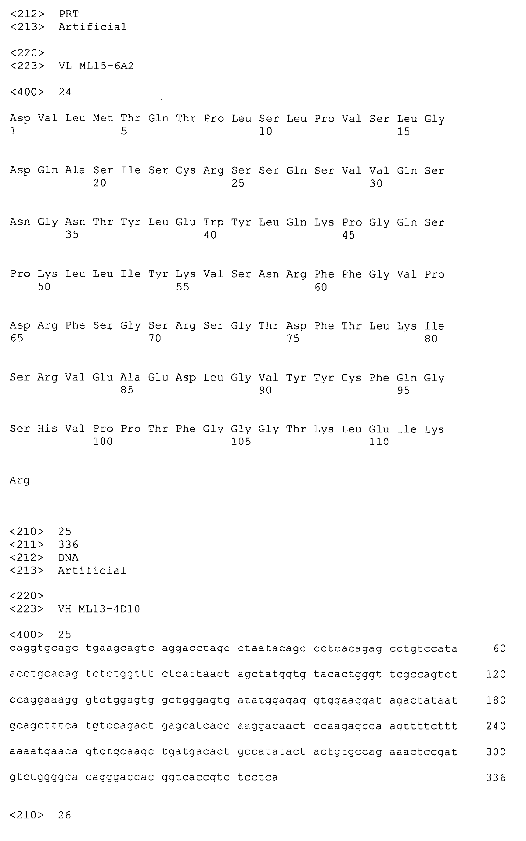


Claims (15)
- Antibody comprising a variable heavy (VH) and light (VL) chain wherein the variable heavy and light chain CDR domains comprise the amino acid sequences selected from the group consisting of the following CDR sets:


- The antibody of claim 1, wherein the antibody is humanized.
- The antibody of claims 1 or 2 wherein the antibody comprises a human constant region.
- The antibody of any one of claims 1 to 3 wherein the antibody possesses a human glycosylation pattern.
- Antigen-binding moiety of an antibody as defined in any one of claims 1 to 4.
- The antigen-binding moiety of claim 5, wherein the moiety is a Fab fragment, a F(ab')2 fragment or a single chain Fv fragment of the antibody.
- lsolated nucleic acid encoding an antibody amino acid sequence of any one of claims 1 to 6.
- Vector comprising an isolated nucleic acid of claim 7.
- Host cell comprising a vector of claim 8.
- Method of producing an antibody, comprising culturing a host cell of claim 9 in culture medium under conditions suitable to produce the antibody.
- Antibody obtainable by a method of claim 10.
- Pharmaceutical composition comprising an antibody or an antigen-binding moiety as defined in any one of claims 1 to 6 and a pharmaceutically acceptable carrier.
- The antibody or an antigen-binding moiety as defined in any one of claims 1 to 6 for use in treating or preventing, or diagnosing an amyloidosis such as Alzheimer's disease or Down's syndrome.
- Method of diagnosing an amyloidosis such as Alzheimer's disease or Down's syndrome which comprises providing a sample from the subject suspected of having the amyloidosis, contacting the sample with an antibody or an antigen-binding moiety as defined in any one of claims 1 to 6 and detecting the formation of a complex comprising the antibody or the antigen-binding moiety with an antigen, the presence of the complex indicating an amyloidosis in the subject.
- Antibody having a binding affinity to an Aβ(20-42) globulomer that is greater than the binding affinity of the antibody to an Aβ(1-42) globulomer.
Priority Applications (2)
| Application Number | Priority Date | Filing Date | Title |
|---|---|---|---|
| PL10178394T PL2289909T3 (en) | 2005-11-30 | 2006-11-30 | Screening method, process for purifying of non-diffusible a-beta oligomers, selective antibodies against said non-diffusible a-beta oligomers and a process for manufacturing of said antibodies |
| SI200631887T SI2289909T1 (en) | 2005-11-30 | 2006-11-30 | Screening method, process for purifying of non-diffusible a-beta oligomers, selective antibodies against said non-diffusible a-beta oligomers and a process for manufacturing of said antibodies |
Applications Claiming Priority (5)
| Application Number | Priority Date | Filing Date | Title |
|---|---|---|---|
| US74086605P | 2005-11-30 | 2005-11-30 | |
| US77917106P | 2006-03-03 | 2006-03-03 | |
| US78736106P | 2006-03-30 | 2006-03-30 | |
| US84240006P | 2006-09-05 | 2006-09-05 | |
| EP06818937.2A EP1954718B1 (en) | 2005-11-30 | 2006-11-30 | Anti-a globulomer antibodies, antigen-binding moieties thereof, corresponding hybridomas, nucleic acids, vectors, host cells, methods of producing said antibodies, compositions comprising said antibodies, uses of said antibodies and methods of using said antibodies |
Related Parent Applications (3)
| Application Number | Title | Priority Date | Filing Date |
|---|---|---|---|
| EP06818937.2 Division | 2006-11-30 | ||
| EP06818937.2A Division-Into EP1954718B1 (en) | 2005-11-30 | 2006-11-30 | Anti-a globulomer antibodies, antigen-binding moieties thereof, corresponding hybridomas, nucleic acids, vectors, host cells, methods of producing said antibodies, compositions comprising said antibodies, uses of said antibodies and methods of using said antibodies |
| EP06818937.2A Division EP1954718B1 (en) | 2005-11-30 | 2006-11-30 | Anti-a globulomer antibodies, antigen-binding moieties thereof, corresponding hybridomas, nucleic acids, vectors, host cells, methods of producing said antibodies, compositions comprising said antibodies, uses of said antibodies and methods of using said antibodies |
Publications (2)
| Publication Number | Publication Date |
|---|---|
| EP2289909A1 true EP2289909A1 (en) | 2011-03-02 |
| EP2289909B1 EP2289909B1 (en) | 2014-10-29 |
Family
ID=37744650
Family Applications (2)
| Application Number | Title | Priority Date | Filing Date |
|---|---|---|---|
| EP10178394.2A Active EP2289909B1 (en) | 2005-11-30 | 2006-11-30 | Screening method, process for purifying of non-diffusible a-beta oligomers, selective antibodies against said non-diffusible a-beta oligomers and a process for manufacturing of said antibodies |
| EP06818937.2A Active EP1954718B1 (en) | 2005-11-30 | 2006-11-30 | Anti-a globulomer antibodies, antigen-binding moieties thereof, corresponding hybridomas, nucleic acids, vectors, host cells, methods of producing said antibodies, compositions comprising said antibodies, uses of said antibodies and methods of using said antibodies |
Family Applications After (1)
| Application Number | Title | Priority Date | Filing Date |
|---|---|---|---|
| EP06818937.2A Active EP1954718B1 (en) | 2005-11-30 | 2006-11-30 | Anti-a globulomer antibodies, antigen-binding moieties thereof, corresponding hybridomas, nucleic acids, vectors, host cells, methods of producing said antibodies, compositions comprising said antibodies, uses of said antibodies and methods of using said antibodies |
Country Status (20)
| Country | Link |
|---|---|
| US (3) | US8691224B2 (en) |
| EP (2) | EP2289909B1 (en) |
| JP (3) | JP5475994B2 (en) |
| KR (1) | KR20080090408A (en) |
| CN (1) | CN101432302A (en) |
| AU (1) | AU2006319358B2 (en) |
| BR (1) | BRPI0619249A2 (en) |
| CA (1) | CA2628703C (en) |
| CR (1) | CR9969A (en) |
| DK (1) | DK1954718T3 (en) |
| EC (1) | ECSP088462A (en) |
| ES (2) | ES2524984T3 (en) |
| GT (1) | GT200800082A (en) |
| IL (2) | IL191588A0 (en) |
| NO (1) | NO20082890L (en) |
| PL (2) | PL2289909T3 (en) |
| PT (2) | PT1954718E (en) |
| RU (1) | RU2442793C2 (en) |
| WO (1) | WO2007062852A2 (en) |
| ZA (1) | ZA200804346B (en) |
Families Citing this family (56)
| Publication number | Priority date | Publication date | Assignee | Title |
|---|---|---|---|---|
| DE10303974A1 (en) | 2003-01-31 | 2004-08-05 | Abbott Gmbh & Co. Kg | Amyloid β (1-42) oligomers, process for their preparation and their use |
| ES2542501T3 (en) | 2005-09-30 | 2015-08-06 | Abbvie Deutschland Gmbh & Co Kg | Protein binding domains of the protein family of repulsive targeting molecules (RGM) and functional fragments thereof, as well as their use |
| EP1976877B2 (en) | 2005-11-30 | 2016-10-05 | AbbVie Inc. | Monoclonal antibodies against amyloid beta protein and uses thereof |
| DK1954718T3 (en) | 2005-11-30 | 2014-12-15 | Abbvie Inc | Anti-A-globulomer antibodies antigenbindingsgrupper thereof, corresponding hybridomas, nucleic acids, vectors, host cells, methods for producing said antibodies, |
| MX358175B (en) * | 2005-12-12 | 2018-08-08 | Ac Immune Sa | A beta 1-42 specific monoclonal antibodies with therapeutic properties. |
| WO2008011348A2 (en) | 2006-07-14 | 2008-01-24 | Ac Immune S.A. | Humanized antibody against amyloid beta |
| US8455626B2 (en) | 2006-11-30 | 2013-06-04 | Abbott Laboratories | Aβ conformer selective anti-aβ globulomer monoclonal antibodies |
| KR101735257B1 (en) * | 2007-01-05 | 2017-05-12 | 유니버시티 오브 취리히 | Method of providing disease-specific binding molecules and targets |
| IL199534A (en) | 2007-01-05 | 2013-01-31 | Univ Zuerich | Isolated human antibody which is capable of selectively recognizing a neoepitope of a disorder-associated protein, a polynucleotide encoding the antibody, a vector comprising the polynucleotide, a host cell comprising the polynucleotide or the vector, a composition comprising the antibody and methods and uses associated therewith |
| EP2486928A1 (en) | 2007-02-27 | 2012-08-15 | Abbott GmbH & Co. KG | Method for the treatment of amyloidoses |
| US8613923B2 (en) | 2007-06-12 | 2013-12-24 | Ac Immune S.A. | Monoclonal antibody |
| US8048420B2 (en) | 2007-06-12 | 2011-11-01 | Ac Immune S.A. | Monoclonal antibody |
| CA2701793C (en) | 2007-10-05 | 2017-04-25 | Genentech, Inc. | Use of anti-amyloid beta antibody in ocular diseases |
| SG178809A1 (en) * | 2007-10-05 | 2012-03-29 | Genentech Inc | Use of anti-amyloid beta antibody in ocular diseases |
| JPWO2009104736A1 (en) * | 2008-02-22 | 2011-06-23 | 田平 武 | Tissue amyloid plaque affinity antibody and pharmaceutical composition using the same |
| US8962803B2 (en) * | 2008-02-29 | 2015-02-24 | AbbVie Deutschland GmbH & Co. KG | Antibodies against the RGM A protein and uses thereof |
| KR20110014607A (en) | 2008-04-29 | 2011-02-11 | 아보트 러보러터리즈 | Dual variable domain immunoglobulins and uses thereof |
| KR101649168B1 (en) | 2008-05-09 | 2016-08-18 | 애브비 인코포레이티드 | Antibodies to receptor of advanced glycation end products (RAGE) and uses thereof |
| CN102112494A (en) | 2008-06-03 | 2011-06-29 | 雅培制药有限公司 | Dual variable domain immunoglobulins and uses thereof |
| WO2009149189A2 (en) | 2008-06-03 | 2009-12-10 | Abbott Laboratories | Dual variable domain immunoglobulins and uses thereof |
| BRPI0915448A2 (en) | 2008-07-08 | 2015-11-10 | Abbott Lab | prostaglandin e2 double variable domain immunoglobulins and uses thereof |
| EP2376120A2 (en) | 2008-11-25 | 2011-10-19 | Biogen Idec MA Inc. | Use of dr6 and p75 antagonists to promote survival of cells of the nervous system |
| EP2949666B1 (en) | 2008-12-19 | 2018-12-19 | Biogen International Neuroscience GmbH | Human anti-alpha-synuclein antibodies |
| RU2012119756A (en) | 2009-10-15 | 2013-11-20 | Эбботт Лэборетриз | IMMUNOGLOBULINS WITH TWO VARIABLE DOMAINS AND THEIR APPLICATION |
| UY32979A (en) | 2009-10-28 | 2011-02-28 | Abbott Lab | IMMUNOGLOBULINS WITH DUAL VARIABLE DOMAIN AND USES OF THE SAME |
| US8420083B2 (en) | 2009-10-31 | 2013-04-16 | Abbvie Inc. | Antibodies to receptor for advanced glycation end products (RAGE) and uses thereof |
| US20110110942A1 (en) | 2009-11-12 | 2011-05-12 | Genentech, Inc. | Method of promoting dendritic spine density |
| ES2562832T3 (en) | 2009-12-08 | 2016-03-08 | Abbvie Deutschland Gmbh & Co Kg | Monoclonal antibodies against the RGM protein for use in the treatment of degeneration of the retinal nerve fiber layer |
| CA2796339C (en) * | 2010-04-15 | 2020-03-31 | Abbott Laboratories | Amyloid-beta binding proteins |
| EP2575859B1 (en) * | 2010-06-03 | 2018-09-19 | Ramot at Tel Aviv University, Ltd. | Methods of treating diabetes and compositions capable of same |
| CN103179981B (en) | 2010-07-30 | 2017-02-08 | Ac免疫有限公司 | Safe and functional humanized antibodies |
| EP3252072A3 (en) | 2010-08-03 | 2018-03-14 | AbbVie Inc. | Dual variable domain immunoglobulins and uses thereof |
| US9062101B2 (en) | 2010-08-14 | 2015-06-23 | AbbVie Deutschland GmbH & Co. KG | Amyloid-beta binding proteins |
| WO2012027570A2 (en) | 2010-08-26 | 2012-03-01 | Abbott Laboratories | Dual variable domain immunoglobulins and uses thereof |
| WO2012097313A2 (en) | 2011-01-14 | 2012-07-19 | The Regents Of The University Of California | Therapeutic antibodies against ror-1 protein and methods for use of same |
| WO2012154983A2 (en) | 2011-05-10 | 2012-11-15 | Biocare Medical, Llc | Systems and methods for anti-pax8 antibodies |
| PT2723379T (en) | 2011-06-23 | 2018-11-14 | Univ Of Zuerich | Anti-alpha synuclein binding molecules |
| JP2015508994A (en) | 2011-12-30 | 2015-03-26 | アッヴィ・インコーポレイテッド | Dual variable domain immunoglobulins against IL-13 and / or IL-17 |
| LT2807192T (en) | 2012-01-27 | 2018-07-25 | AbbVie Deutschland GmbH & Co. KG | Composition and method for diagnosis and treatment of diseases associated with neurite degeneration |
| US10316103B1 (en) | 2012-03-30 | 2019-06-11 | Biocare Medical, Llc | Systems and methods for anti-Uroplakin III antibodies |
| ES2682345T3 (en) | 2012-09-27 | 2018-09-20 | Biocare Medical, Llc | Antiuroplachin II antibody systems and procedures |
| EP2914625A2 (en) | 2012-11-01 | 2015-09-09 | AbbVie Inc. | Anti-vegf/dll4 dual variable domain immunoglobulins and uses thereof |
| WO2014100220A2 (en) | 2012-12-18 | 2014-06-26 | Biocare Medical, Llc | Antibody cocktail systems and methods for classification of histologic subtypes in lung cancer |
| DK2962113T3 (en) | 2013-02-28 | 2019-07-01 | Biocare Medical Llc | Systems and Methods with Anti-p40 Antibodies |
| US9062108B2 (en) | 2013-03-15 | 2015-06-23 | Abbvie Inc. | Dual specific binding proteins directed against IL-1 and/or IL-17 |
| EP3052522B1 (en) | 2013-10-03 | 2019-12-11 | Biocare Medical, LLC | Anti-sox10 antibody systems and methods |
| CA2954031A1 (en) * | 2014-07-07 | 2016-01-14 | AbbVie Deutschland GmbH & Co. KG | Immunogenic products based on mutein amyloid .beta. (.alpha..beta.) amino acid sequences and uses thereof |
| MA41115A (en) | 2014-12-02 | 2017-10-10 | Biogen Int Neuroscience Gmbh | ALZHEIMER'S DISEASE TREATMENT PROCESS |
| US10093733B2 (en) | 2014-12-11 | 2018-10-09 | Abbvie Inc. | LRP-8 binding dual variable domain immunoglobulin proteins |
| TW201710286A (en) | 2015-06-15 | 2017-03-16 | 艾伯維有限公司 | Binding proteins against VEGF, PDGF, and/or their receptors |
| WO2017189964A2 (en) | 2016-04-29 | 2017-11-02 | Voyager Therapeutics, Inc. | Compositions for the treatment of disease |
| WO2017189959A1 (en) | 2016-04-29 | 2017-11-02 | Voyager Therapeutics, Inc. | Compositions for the treatment of disease |
| TW201827467A (en) | 2016-11-03 | 2018-08-01 | 比利時商健生藥品公司 | Antibodies to pyroglutamate amyloid-[beta] and uses thereof |
| CA3073066A1 (en) | 2017-08-22 | 2019-02-28 | Biogen Ma Inc. | Pharmaceutical compositions containing anti-beta amyloid antibodies |
| EA202192606A1 (en) | 2019-03-26 | 2022-01-24 | Янссен Фармацевтика Нв | ANTIBODIES TO PYROGLUTAMATE-AMYLOID AND THEIR APPLICATIONS |
| EP4185612A1 (en) | 2020-07-23 | 2023-05-31 | Othair Prothena Limited | Anti-abeta antibodies |
Citations (43)
| Publication number | Priority date | Publication date | Assignee | Title |
|---|---|---|---|---|
| US4399216A (en) | 1980-02-25 | 1983-08-16 | The Trustees Of Columbia University | Processes for inserting DNA into eucaryotic cells and for producing proteinaceous materials |
| US4510245A (en) | 1982-11-18 | 1985-04-09 | Chiron Corporation | Adenovirus promoter system |
| US4634665A (en) | 1980-02-25 | 1987-01-06 | The Trustees Of Columbia University In The City Of New York | Processes for inserting DNA into eucaryotic cells and for producing proteinaceous materials |
| US4816397A (en) | 1983-03-25 | 1989-03-28 | Celltech, Limited | Multichain polypeptides or proteins and processes for their production |
| EP0368684A1 (en) | 1988-11-11 | 1990-05-16 | Medical Research Council | Cloning immunoglobulin variable domain sequences. |
| US4968615A (en) | 1985-12-18 | 1990-11-06 | Ciba-Geigy Corporation | Deoxyribonucleic acid segment from a virus |
| EP0436597A1 (en) | 1988-09-02 | 1991-07-17 | Protein Eng Corp | Generation and selection of recombinant varied binding proteins. |
| WO1991017271A1 (en) | 1990-05-01 | 1991-11-14 | Affymax Technologies N.V. | Recombinant library screening methods |
| WO1992001047A1 (en) | 1990-07-10 | 1992-01-23 | Cambridge Antibody Technology Limited | Methods for producing members of specific binding pairs |
| WO1992002551A1 (en) | 1990-08-02 | 1992-02-20 | B.R. Centre Limited | Methods for the production of proteins with a desired function |
| WO1992009690A2 (en) | 1990-12-03 | 1992-06-11 | Genentech, Inc. | Enrichment method for variant proteins with altered binding properties |
| WO1992020791A1 (en) | 1990-07-10 | 1992-11-26 | Cambridge Antibody Technology Limited | Methods for producing members of specific binding pairs |
| US5168062A (en) | 1985-01-30 | 1992-12-01 | University Of Iowa Research Foundation | Transfer vectors and microorganisms containing human cytomegalovirus immediate-early promoter-regulatory DNA sequence |
| US5179017A (en) | 1980-02-25 | 1993-01-12 | The Trustees Of Columbia University In The City Of New York | Processes for inserting DNA into eucaryotic cells and for producing proteinaceous materials |
| US5223409A (en) | 1988-09-02 | 1993-06-29 | Protein Engineering Corp. | Directed evolution of novel binding proteins |
| US5545806A (en) | 1990-08-29 | 1996-08-13 | Genpharm International, Inc. | Ransgenic non-human animals for producing heterologous antibodies |
| US5569825A (en) | 1990-08-29 | 1996-10-29 | Genpharm International | Transgenic non-human animals capable of producing heterologous antibodies of various isotypes |
| WO1996034096A1 (en) | 1995-04-28 | 1996-10-31 | Abgenix, Inc. | Human antibodies derived from immunized xenomice |
| WO1996033735A1 (en) | 1995-04-27 | 1996-10-31 | Abgenix, Inc. | Human antibodies derived from immunized xenomice |
| WO1997008320A1 (en) | 1995-08-18 | 1997-03-06 | Morphosys Gesellschaft Für Proteinoptimierung Mbh | Protein/(poly)peptide libraries |
| US5625126A (en) | 1990-08-29 | 1997-04-29 | Genpharm International, Inc. | Transgenic non-human animals for producing heterologous antibodies |
| US5633425A (en) | 1990-08-29 | 1997-05-27 | Genpharm International, Inc. | Transgenic non-human animals capable of producing heterologous antibodies |
| WO1997029131A1 (en) | 1996-02-09 | 1997-08-14 | Basf Aktiengesellschaft | HUMAN ANTIBODIES THAT BIND HUMAN TNF$g(a) |
| US5661016A (en) | 1990-08-29 | 1997-08-26 | Genpharm International Inc. | Transgenic non-human animals capable of producing heterologous antibodies of various isotypes |
| US5705330A (en) | 1995-04-14 | 1998-01-06 | Abbott Laboratories | Chemiluminescent immunoassay for antibody detection |
| WO1998024893A2 (en) | 1996-12-03 | 1998-06-11 | Abgenix, Inc. | TRANSGENIC MAMMALS HAVING HUMAN IG LOCI INCLUDING PLURAL VH AND Vλ REGIONS AND ANTIBODIES PRODUCED THEREFROM |
| US5770429A (en) | 1990-08-29 | 1998-06-23 | Genpharm International, Inc. | Transgenic non-human animals capable of producing heterologous antibodies |
| WO1998031700A1 (en) | 1997-01-21 | 1998-07-23 | The General Hospital Corporation | Selection of proteins using rna-protein fusions |
| WO1998033815A1 (en) * | 1997-02-05 | 1998-08-06 | Northwestern University | Amyloid beta protein (globular assembly and uses thereof) |
| US5814318A (en) | 1990-08-29 | 1998-09-29 | Genpharm International Inc. | Transgenic non-human animals for producing heterologous antibodies |
| WO1998047343A2 (en) | 1997-04-04 | 1998-10-29 | Biosite Diagnostics, Inc. | Antibodies or binding protein libraries displayed on phage, cells, or other replicatable genetic packages |
| WO1998049286A2 (en) | 1997-05-01 | 1998-11-05 | Board Of Regents, The University Of Texas System | Directed evolution of enzymes and antibodies |
| US5877397A (en) | 1990-08-29 | 1999-03-02 | Genpharm International Inc. | Transgenic non-human animals capable of producing heterologous antibodies of various isotypes |
| US5885793A (en) | 1991-12-02 | 1999-03-23 | Medical Research Council | Production of anti-self antibodies from antibody segment repertoires and displayed on phage |
| WO1999036569A1 (en) | 1998-01-20 | 1999-07-22 | The Board Of Trustees Of The University Of Illinois | Yeast cell surface display of proteins and uses thereof |
| US5939598A (en) | 1990-01-12 | 1999-08-17 | Abgenix, Inc. | Method of making transgenic mice lacking endogenous heavy chains |
| WO1999045962A1 (en) | 1998-03-13 | 1999-09-16 | Genpharm International, Inc. | Transgenic non-human animals capable of producing heterologous antibodies |
| WO1999053049A1 (en) | 1998-04-15 | 1999-10-21 | Abgenix, Inc. | Epitope-driven human antibody production and gene expression profiling |
| WO2000056772A1 (en) | 1999-03-25 | 2000-09-28 | Knoll Gmbh | Human antibodies that bind human il-12 and methods for producing |
| WO2001010900A2 (en) * | 1999-08-04 | 2001-02-15 | University Of Southern California | Globular assembly of amyloid beta protein and uses thereof |
| WO2001062801A2 (en) * | 2000-02-24 | 2001-08-30 | Washington University | Humanized antibodies that sequester amyloid beta peptide |
| WO2004067561A1 (en) | 2003-01-31 | 2004-08-12 | Abbott Gmbh & Co. Kg | Amyloid-$g(b)(1-42) oligomers, derivatives thereof, antibodies for the same, method for production and use therof |
| WO2006094724A2 (en) * | 2005-03-05 | 2006-09-14 | Abbott Gmbh & Co. Kg | Screening method, process for purifying of non-diffusible a-beta oligomers, selective antibodies against said non-diffusible a-beta oligomers and a process for manufacturing of said antibodies |
Family Cites Families (690)
| Publication number | Priority date | Publication date | Assignee | Title |
|---|---|---|---|---|
| DE2500076C3 (en) | 1975-01-02 | 1982-11-18 | SCHURA Blutderivate GmbH & Co KG, 4150 Krefeld | Process for the production of intravenously tolerated gamma globulins |
| NO812612L (en) | 1980-08-06 | 1982-02-08 | Ferring Pharma Ltd | ENZYME inhibitor. |
| BR8108820A (en) | 1980-09-24 | 1982-08-24 | Cetus Corp | DIAGNOSTIC PROCESS AND PROBE |
| EP0084796B1 (en) | 1982-01-22 | 1990-05-02 | Cetus Corporation | Hla typing method and cdna probes used therein |
| US4582788A (en) | 1982-01-22 | 1986-04-15 | Cetus Corporation | HLA typing method and cDNA probes used therein |
| EP0092918B1 (en) | 1982-04-22 | 1988-10-19 | Imperial Chemical Industries Plc | Continuous release formulations |
| US4816567A (en) * | 1983-04-08 | 1989-03-28 | Genentech, Inc. | Recombinant immunoglobin preparations |
| US4526039A (en) | 1983-06-23 | 1985-07-02 | The United States Of America As Represented By The Secretary Of Transportation | Removable strain gauge fixture and method for measuring accumulated strain in a material |
| US4683194A (en) | 1984-05-29 | 1987-07-28 | Cetus Corporation | Method for detection of polymorphic restriction sites and nucleic acid sequences |
| US5807715A (en) | 1984-08-27 | 1998-09-15 | The Board Of Trustees Of The Leland Stanford Junior University | Methods and transformed mammalian lymphocyte cells for producing functional antigen-binding protein including chimeric immunoglobulin |
| US5128326A (en) | 1984-12-06 | 1992-07-07 | Biomatrix, Inc. | Drug delivery systems based on hyaluronans derivatives thereof and their salts and methods of producing same |
| US4683202A (en) | 1985-03-28 | 1987-07-28 | Cetus Corporation | Process for amplifying nucleic acid sequences |
| DE3590766C2 (en) | 1985-03-30 | 1991-01-10 | Marc Genf/Geneve Ch Ballivet | |
| US4666829A (en) | 1985-05-15 | 1987-05-19 | University Of California | Polypeptide marker for Alzheimer's disease and its use for diagnosis |
| US4980286A (en) | 1985-07-05 | 1990-12-25 | Whitehead Institute For Biomedical Research | In vivo introduction and expression of foreign genetic material in epithelial cells |
| US5618920A (en) | 1985-11-01 | 1997-04-08 | Xoma Corporation | Modular assembly of antibody genes, antibodies prepared thereby and use |
| DE3600905A1 (en) | 1986-01-15 | 1987-07-16 | Ant Nachrichtentech | METHOD FOR DECODING BINARY SIGNALS AND VITERBI DECODERS AND APPLICATIONS |
| CA1284931C (en) | 1986-03-13 | 1991-06-18 | Henry A. Erlich | Process for detecting specific nucleotide variations and genetic polymorphisms present in nucleic acids |
| US5225539A (en) | 1986-03-27 | 1993-07-06 | Medical Research Council | Recombinant altered antibodies and methods of making altered antibodies |
| GB8607679D0 (en) | 1986-03-27 | 1986-04-30 | Winter G P | Recombinant dna product |
| US5107065A (en) | 1986-03-28 | 1992-04-21 | Calgene, Inc. | Anti-sense regulation of gene expression in plant cells |
| CA1338457C (en) | 1986-08-22 | 1996-07-16 | Henry A. Erlich | Purified thermostable enzyme |
| US5231170A (en) | 1986-08-27 | 1993-07-27 | Paul Averback | Antibodies to dense microspheres |
| US5525339A (en) | 1986-08-27 | 1996-06-11 | Dms Pharmaceutical Inc. | Isolated components of dense microspheres derived from mammalian brain tissue and antibodies thereto |
| US4946778A (en) | 1987-09-21 | 1990-08-07 | Genex Corporation | Single polypeptide chain binding molecules |
| US5811310A (en) | 1986-09-30 | 1998-09-22 | Albert Einstein College Of Medicine Of Yeshiva Univ. | The Alz-50 monoclonal antibody and diagnostic assay for alzheimer's disease |
| JPS63240797A (en) | 1986-10-14 | 1988-10-06 | Shin Etsu Chem Co Ltd | Monoclonal antibody, production thereof and diagnostic containing said antibody |
| AU611122B2 (en) | 1986-11-17 | 1991-06-06 | Scios Nova Inc. | Recombinant alzheimer's amyloid protein |
| US5763192A (en) | 1986-11-20 | 1998-06-09 | Ixsys, Incorporated | Process for obtaining DNA, RNA, peptides, polypeptides, or protein, by recombinant DNA technique |
| DE3702789A1 (en) | 1987-01-30 | 1988-08-18 | Bayer Ag | PROCUREMENT PROTEIN OF APC POLYPEPTIDE, FOR CODING DNA AND DIAGNOSTIC USE OF DNA AND PROTEIN |
| DE3883899T3 (en) | 1987-03-18 | 1999-04-22 | Sb2 Inc | CHANGED ANTIBODIES. |
| JPS63245689A (en) | 1987-03-31 | 1988-10-12 | Suntory Ltd | Human amyloid-related protein monoclonal antibody |
| US5258498A (en) | 1987-05-21 | 1993-11-02 | Creative Biomolecules, Inc. | Polypeptide linkers for production of biosynthetic proteins |
| US4880078A (en) | 1987-06-29 | 1989-11-14 | Honda Giken Kogyo Kabushiki Kaisha | Exhaust muffler |
| CA1340802C (en) | 1987-08-15 | 1999-10-26 | Yasuyuki Takahashi | Senile amyloid precursor protein and an antibody specific for the same |
| US5004697A (en) | 1987-08-17 | 1991-04-02 | Univ. Of Ca | Cationized antibodies for delivery through the blood-brain barrier |
| US5231000A (en) | 1987-10-08 | 1993-07-27 | The Mclean Hospital | Antibodies to A4 amyloid peptide |
| CA1339014C (en) | 1987-10-08 | 1997-03-25 | Ronald E. Majocha | Antibodies to a4 amyloid peptide |
| WO1989006689A1 (en) | 1988-01-13 | 1989-07-27 | The Mclean Hospital Corporation | Genetic constructs containing the alzheimer brain amyloid gene |
| WO1989007657A1 (en) | 1988-02-10 | 1989-08-24 | The Children's Medical Center Corporation | Amyloid protein precursors, genetic probes, antibodies, and methods of use |
| US5134062A (en) | 1988-03-22 | 1992-07-28 | Cornell Research Foundation, Inc. | Diagnosis of neuronal disorders and screening potential therapeutic agents therefor |
| US5015570A (en) | 1988-05-13 | 1991-05-14 | Molecular Therapeutics, Inc. | Molecular diagnosis of Alzheimer Disease |
| US6287793B1 (en) | 1988-08-19 | 2001-09-11 | Elan Pharmaceuticals, Inc. | Diagnostic methods for alzheimer's disease |
| US5530101A (en) | 1988-12-28 | 1996-06-25 | Protein Design Labs, Inc. | Humanized immunoglobulins |
| US20030229208A1 (en) | 1988-12-28 | 2003-12-11 | Protein Design Labs, Inc. | Humanized immunoglobulins |
| US5231020A (en) | 1989-03-30 | 1993-07-27 | Dna Plant Technology Corporation | Genetic engineering of novel plant phenotypes |
| US5262332A (en) | 1989-04-05 | 1993-11-16 | Brigham And Women's Hospital | Diagnostic method for Alzheimer's disease: examination of non-neural tissue |
| AU5525090A (en) | 1989-04-14 | 1990-11-16 | Research Foundation For Mental Hygiene, Inc. | Monoclonal antibody to amyloid peptide |
| CA2016841C (en) | 1989-05-16 | 1999-09-21 | William D. Huse | A method for producing polymers having a preselected activity |
| EP0478627A4 (en) | 1989-05-16 | 1992-08-19 | William D. Huse | Co-expression of heteromeric receptors |
| CA2016842A1 (en) | 1989-05-16 | 1990-11-16 | Richard A. Lerner | Method for tapping the immunological repertoire |
| US5234814A (en) | 1989-06-01 | 1993-08-10 | Du Pont Merck Pharmaceutical Company | Diagnostic assay for alzheimer's disease |
| EP0415801A1 (en) | 1989-07-05 | 1991-03-06 | N.V. Innogenetics S.A. | Monoclonal antibodies against activated microglial cells |
| EP0411974A1 (en) | 1989-07-05 | 1991-02-06 | N.V. Innogenetics S.A. | Monoclonal antibodies to a neurofibrillary tangle antigen |
| AU6430190A (en) | 1989-10-10 | 1991-05-16 | Pitman-Moore, Inc. | Sustained release composition for macromolecular proteins |
| JP2571874B2 (en) | 1989-11-06 | 1997-01-16 | アルカーメス コントロールド セラピューティクス,インコーポレイテッド | Protein microsphere composition |
| GB8928874D0 (en) | 1989-12-21 | 1990-02-28 | Celltech Ltd | Humanised antibodies |
| US5780225A (en) | 1990-01-12 | 1998-07-14 | Stratagene | Method for generating libaries of antibody genes comprising amplification of diverse antibody DNAs and methods for using these libraries for the production of diverse antigen combining molecules |
| WO1991010737A1 (en) | 1990-01-11 | 1991-07-25 | Molecular Affinities Corporation | Production of antibodies using gene libraries |
| US6075181A (en) | 1990-01-12 | 2000-06-13 | Abgenix, Inc. | Human antibodies derived from immunized xenomice |
| AU7121191A (en) | 1990-02-26 | 1991-10-10 | Albert Einstein College Of Medicine Of Yeshiva University | Diagnostic assay for alzheimer's disease |
| US5753624A (en) | 1990-04-27 | 1998-05-19 | Milkhaus Laboratory, Inc. | Materials and methods for treatment of plaquing disease |
| DE4022247A1 (en) | 1990-07-12 | 1992-01-16 | Hoechst Ag | METHOD FOR EXTRACTIVELY SEPARATING 2-ALKYLTHIO-5-PHENYLPYRIMIDINES FROM THEIR REACTION MIXTURES |
| US5698426A (en) | 1990-09-28 | 1997-12-16 | Ixsys, Incorporated | Surface expression libraries of heteromeric receptors |
| JPH04252195A (en) | 1991-01-29 | 1992-09-08 | Asahi Chem Ind Co Ltd | Monoclonal antibody and hybridoma |
| EP1820858B1 (en) | 1991-03-01 | 2009-08-12 | Dyax Corporation | Chimeric protein comprising micro-protein having two or more disulfide bonds and embodiments thereof |
| AU662148B2 (en) | 1991-04-10 | 1995-08-24 | Scripps Research Institute, The | Heterodimeric receptor libraries using phagemids |
| JPH04320694A (en) | 1991-04-22 | 1992-11-11 | Ono Pharmaceut Co Ltd | Monoclonal antibody against human gliacyte growth inhibiting factor |
| ATE221379T1 (en) | 1991-05-01 | 2002-08-15 | Jackson H M Found Military Med | METHOD FOR TREATING INFECTIOUS RESPIRATORY DISEASES |
| JPH04342883A (en) | 1991-05-17 | 1992-11-30 | Sanden Corp | Variable delivery swash plate type compressor |
| EP0519596B1 (en) | 1991-05-17 | 2005-02-23 | Merck & Co. Inc. | A method for reducing the immunogenicity of antibody variable domains |
| WO1992022324A1 (en) | 1991-06-14 | 1992-12-23 | Xoma Corporation | Microbially-produced antibody fragments and their conjugates |
| DE4122599C2 (en) | 1991-07-08 | 1993-11-11 | Deutsches Krebsforsch | Phagemid for screening antibodies |
| ES2136092T3 (en) | 1991-09-23 | 1999-11-16 | Medical Res Council | PROCEDURES FOR THE PRODUCTION OF HUMANIZED ANTIBODIES. |
| WO1993008302A1 (en) | 1991-10-25 | 1993-04-29 | Itt Automotive Europe Gmbh | Monoclonal antibodies directed against the microtubule-associated protein tau |
| JP3277211B2 (en) | 1991-11-12 | 2002-04-22 | プラナ・バイオテクノロジー・リミテッド | Test and treatment methods for Alzheimer's disease |
| US20020009443A1 (en) | 1991-12-02 | 2002-01-24 | Vanitha Ramakrishman | Inhibitory immunoglobulin polypeptides to human pdgf beta receptor |
| US7408027B1 (en) | 1991-12-06 | 2008-08-05 | Max-Planck-Gesellschaft Zur Forderung Der Wissenchaften | Tools for the diagnosis and treatment of Alzheimer's disease |
| WO1993011231A1 (en) | 1991-12-06 | 1993-06-10 | MAX-PLANCK-Gesellschaft zur Förderung der Wissenschaften e.V. | Tools for the diagnosis and treatment of alzheimer's disease |
| CA2103887C (en) | 1991-12-13 | 2005-08-30 | Gary M. Studnicka | Methods and materials for preparation of modified antibody variable domains and therapeutic uses thereof |
| US20010029293A1 (en) | 1992-01-27 | 2001-10-11 | Icos Corporation | Icam-related protein |
| US5538845A (en) | 1992-02-05 | 1996-07-23 | Athena Neurosciences, Inc. | Beta-amyloid peptide production inhibitors and methods for their identification |
| US5714350A (en) | 1992-03-09 | 1998-02-03 | Protein Design Labs, Inc. | Increasing antibody affinity by altering glycosylation in the immunoglobulin variable region |
| US5912015A (en) | 1992-03-12 | 1999-06-15 | Alkermes Controlled Therapeutics, Inc. | Modulated release from biocompatible polymers |
| US5733743A (en) | 1992-03-24 | 1998-03-31 | Cambridge Antibody Technology Limited | Methods for producing members of specific binding pairs |
| US5912120A (en) | 1992-04-09 | 1999-06-15 | The United States Of America As Represented By The Department Of Health And Human Services, | Cloning, expression and diagnosis of human cytochrome P450 2C19: the principal determinant of s-mephenytoin metabolism |
| US5441870A (en) | 1992-04-15 | 1995-08-15 | Athena Neurosciences, Inc. | Methods for monitoring cellular processing of β-amyloid precursor protein |
| US5604102A (en) | 1992-04-15 | 1997-02-18 | Athena Neurosciences, Inc. | Methods of screening for β-amyloid peptide production inhibitors |
| US5455169A (en) | 1992-06-04 | 1995-10-03 | Alzheimer's Institute Of America, Inc. | Nucleic acids for diagnosing and modeling Alzheimer's disease |
| US5837672A (en) | 1992-07-10 | 1998-11-17 | Athena Neurosciences, Inc. | Methods and compositions for the detection of soluble β-amyloid peptide |
| US6610493B1 (en) | 1993-06-17 | 2003-08-26 | Brigham And Women's Hospital | Screening compounds for the ability to alter the production of amyloid-β peptide |
| US5766846A (en) | 1992-07-10 | 1998-06-16 | Athena Neurosciences | Methods of screening for compounds which inhibit soluble β-amyloid peptide production |
| ATE381614T1 (en) | 1992-07-24 | 2008-01-15 | Amgen Fremont Inc | FORMATION OF XENOGENE ANTIBODIES |
| US5639641A (en) | 1992-09-09 | 1997-06-17 | Immunogen Inc. | Resurfacing of rodent antibodies |
| TW258789B (en) | 1992-09-28 | 1995-10-01 | Rohm & Haas | |
| US5605811A (en) | 1992-10-26 | 1997-02-25 | Athena Neurosciences, Inc. | Methods and compositions for monitoring cellular processing of beta-amyloid precursor protein |
| US5891623A (en) | 1992-11-09 | 1999-04-06 | Consorzio Per Le Biotecnologie | Diagnosis and treatment of AIDS onset |
| ZA938243B (en) | 1992-11-12 | 1995-05-04 | Hybritech Inc | Altered affinity polypeptides of metal chelate binding antibodies |
| US6010913A (en) | 1992-12-14 | 2000-01-04 | N.V. Innogenetics S.A. | Isolated human tau peptide |
| EP0683234B2 (en) | 1993-01-25 | 2007-06-06 | Takeda Chemical Industries, Ltd. | Antibody against beta-amyloid or their derivative and use thereof |
| US5955317A (en) | 1993-01-25 | 1999-09-21 | Takeda Chemical Industries, Ltd. | Antibodies to β-amyloids or their derivatives and use thereof |
| US5934272A (en) | 1993-01-29 | 1999-08-10 | Aradigm Corporation | Device and method of creating aerosolized mist of respiratory drug |
| CA2115900A1 (en) | 1993-02-22 | 1994-08-23 | Gerald W. Becker | Pharmaceutical screens and antibodies |
| US5840294A (en) | 1993-03-29 | 1998-11-24 | Queen's University At Kingston | Method for treating amyloidosis |
| ES2318848T3 (en) | 1993-09-14 | 2009-05-01 | Pharmexa Inc. | PEPTIDES THAT JOIN PAN DR TO POWER THE IMMUNE RESPONSE. |
| WO1995011311A1 (en) | 1993-10-20 | 1995-04-27 | Duke University | METHOD OF BINDING MATERIAL TO THE β-AMYLOID PEPTIDE |
| US5565352A (en) | 1993-11-24 | 1996-10-15 | Arch Development Corporation | Deubiquitinating enzyme: compositions and methods |
| JPH09506262A (en) | 1993-12-08 | 1997-06-24 | ジェンザイム・コーポレイション | Method for producing specific antibody |
| AU1397495A (en) | 1993-12-13 | 1995-07-03 | Uab Research Foundation, The | Diagnostic tests and reagents for detecting risk of alzheimer's disease and stroke |
| US6251395B1 (en) | 1993-12-23 | 2001-06-26 | W. Michael Gallatin | Methods of inhibiting inflammation at the site of a central nervous system injury with alphaD-specific antibodies |
| US6620915B2 (en) | 1993-12-23 | 2003-09-16 | Icos Corporation | Monoclonal antibodies specific for integrin α-d subunit |
| US6432404B1 (en) | 1993-12-23 | 2002-08-13 | Icos Corporation | Methods of inhibiting locomotor damage following spinal cord injury with α D-specific antibodies |
| JPH07209296A (en) | 1994-01-10 | 1995-08-11 | Nikken Chem Co Ltd | Antibody, enzyme-labeled antigen and method and kit for enzyme immunoassay |
| JPH07209295A (en) | 1994-01-10 | 1995-08-11 | Nikken Chem Co Ltd | Antibody, enzyme-labeled antigen and method and kit for enzyme immunoassay |
| US5877399A (en) | 1994-01-27 | 1999-03-02 | Johns Hopkins University | Transgenic mice expressing APP-Swedish mutation develop progressive neurologic disease |
| DE69531148T2 (en) | 1994-01-31 | 2004-04-29 | Trustees Of Boston University, Boston | LIBRARIES MADE OF POLYCLONAL ANTIBODIES |
| JPH07238096A (en) | 1994-02-25 | 1995-09-12 | S R L:Kk | Antipolyubiquitin-monoclonal antibody and method for measuring polyubiquitin |
| US7473423B2 (en) | 1994-04-29 | 2009-01-06 | Mayo Foundation For Medical Education And Research | Human IgM antibodies, and diagnostic and therapeutic uses thereof particularly in the central nervous system |
| JPH07309900A (en) | 1994-05-20 | 1995-11-28 | Idemitsu Kosan Co Ltd | Anti-human procathepsin b monoclonal antibody, hybridoma producting the same and assay of human procathepsin b or human cathepsin b using the same |
| US5516637A (en) | 1994-06-10 | 1996-05-14 | Dade International Inc. | Method involving display of protein binding pairs on the surface of bacterial pili and bacteriophage |
| US6018021A (en) | 1994-10-19 | 2000-01-25 | The Research Foundation Of State University Of New York | Human transaldolase: an autoantigen with a function in metabolism |
| US6114133A (en) | 1994-11-14 | 2000-09-05 | Elan Pharmaceuticals, Inc. | Methods for aiding in the diagnosis of Alzheimer's disease by measuring amyloid-β peptide (x-≧41) |
| EP0805678B1 (en) | 1995-01-05 | 2003-10-29 | THE BOARD OF REGENTS acting for and on behalf of THE UNIVERSITY OF MICHIGAN | Surface-modified nanoparticles and method of making and using same |
| US5786180A (en) | 1995-02-14 | 1998-07-28 | Bayer Corporation | Monoclonal antibody 369.2B specific for β A4 peptide |
| JP3663228B2 (en) | 1995-03-14 | 2005-06-22 | 株式会社医学生物学研究所 | Antibodies against neuropsin |
| US6091001A (en) | 1995-03-29 | 2000-07-18 | Abgenix, Inc. | Production of antibodies using Cre-mediated site-specific recombination |
| US6130364A (en) | 1995-03-29 | 2000-10-10 | Abgenix, Inc. | Production of antibodies using Cre-mediated site-specific recombination |
| US6019968A (en) | 1995-04-14 | 2000-02-01 | Inhale Therapeutic Systems, Inc. | Dispersible antibody compositions and methods for their preparation and use |
| KR100479146B1 (en) | 1995-04-21 | 2005-05-16 | 셀 제네시스, 인코포레이티드 | Generation of Large Genomic DNA Deletions |
| US5712158A (en) | 1995-06-06 | 1998-01-27 | Warner-Lambert Company | Cell line for the rapid expression of functional calcium channels |
| AU6255096A (en) | 1995-06-07 | 1996-12-30 | Mount Sinai School Of Medicine Of The City University Of New York, The | Pegylated modified proteins |
| US6717031B2 (en) | 1995-06-07 | 2004-04-06 | Kate Dora Games | Method for selecting a transgenic mouse model of alzheimer's disease |
| JP3767755B2 (en) | 1995-06-23 | 2006-04-19 | エーザイ株式会社 | Anti-JC virus antibody |
| US6127977A (en) | 1996-11-08 | 2000-10-03 | Cohen; Nathan | Microstrip patch antenna with fractal structure |
| US5916597A (en) | 1995-08-31 | 1999-06-29 | Alkermes Controlled Therapeutics, Inc. | Composition and method using solid-phase particles for sustained in vivo release of a biologically active agent |
| BR9610580A (en) | 1995-09-14 | 2000-10-24 | Univ California | Antibodies specific for native prpse |
| FR2740454B1 (en) | 1995-10-26 | 1997-11-21 | Rhone Poulenc Rorer Sa | PEPTIDES CAPABLE OF INHIBITING APP ENDOCYTOSIS AND CORRESPONDING NUCLEOTIDE SEQUENCES |
| AUPN649395A0 (en) | 1995-11-10 | 1995-12-07 | Ramsay Health Care Pty Ltd | A method for diagnosing alzheimer's disease |
| FR2741881B1 (en) | 1995-12-01 | 1999-07-30 | Centre Nat Rech Scient | NOVEL PURINE DERIVATIVES HAVING IN PARTICULAR ANTI-PROLIFERATIVE PRORIETES AND THEIR BIOLOGICAL APPLICATIONS |
| AU1110297A (en) | 1995-12-12 | 1997-07-03 | Kyowa Hakko Kogyo Co. Ltd. | Novel dnas, novel polypeptides and novel antibodies |
| JPH09178743A (en) | 1995-12-27 | 1997-07-11 | Oriental Yeast Co Ltd | Determinationof soluble app |
| WO1997026537A1 (en) | 1996-01-19 | 1997-07-24 | Research Corporation Technologies, Inc. | Methods for the detection of alzheimer's disease |
| JP2978435B2 (en) | 1996-01-24 | 1999-11-15 | チッソ株式会社 | Method for producing acryloxypropyl silane |
| WO1997028188A1 (en) | 1996-01-30 | 1997-08-07 | The Scripps Research Institute | A g protein-coupled receptor with an enlarged extracellular domain |
| ATE508733T1 (en) | 1996-03-04 | 2011-05-15 | Penn State Res Found | MATERIALS AND METHODS FOR INCREASE CELLULAR INTERNALIZATION |
| US5714352A (en) | 1996-03-20 | 1998-02-03 | Xenotech Incorporated | Directed switch-mediated DNA recombination |
| US5882644A (en) | 1996-03-22 | 1999-03-16 | Protein Design Labs, Inc. | Monoclonal antibodies specific for the platelet derived growth factor β receptor and methods of use thereof |
| US5855913A (en) | 1997-01-16 | 1999-01-05 | Massachusetts Instite Of Technology | Particles incorporating surfactants for pulmonary drug delivery |
| US5874064A (en) | 1996-05-24 | 1999-02-23 | Massachusetts Institute Of Technology | Aerodynamically light particles for pulmonary drug delivery |
| US5985309A (en) | 1996-05-24 | 1999-11-16 | Massachusetts Institute Of Technology | Preparation of particles for inhalation |
| US6699658B1 (en) | 1996-05-31 | 2004-03-02 | Board Of Trustees Of The University Of Illinois | Yeast cell surface display of proteins and uses thereof |
| WO1997046678A1 (en) | 1996-06-06 | 1997-12-11 | Bayer Corporation | Nucleic acids and polypeptides related to presenilin |
| ATE205086T1 (en) * | 1996-08-13 | 2001-09-15 | Gerolymatos P N Sa | USE OF THE CHELATING AGENT CLIOQUINOL FOR THE PRODUCTION OF A MEDICINAL PRODUCT FOR THE TREATMENT OF ALZHEIMER'S DISEASE |
| WO1999009150A1 (en) | 1996-08-15 | 1999-02-25 | Bayer Corporation | Method of introducing modifications into a gene |
| CA2183901A1 (en) | 1996-08-22 | 1998-02-23 | Johanna E. Bergmann | Targets for therapy and diagnosis of alzheimer's disease and down syndrome in humans |
| US7521531B2 (en) | 1996-08-28 | 2009-04-21 | Immunomedics, Inc. | Methods for the purification of stable radioiodine conjugates |
| JPH1075781A (en) | 1996-08-30 | 1998-03-24 | Sumitomo Electric Ind Ltd | Human proteasome subunit p58 protein and specific antibody against the same |
| EP0834561A1 (en) | 1996-09-27 | 1998-04-08 | Rijksuniversiteit te Leiden | A gene related to migraine in man |
| US5916771A (en) | 1996-10-11 | 1999-06-29 | Abgenix, Inc. | Production of a multimeric protein by cell fusion method |
| WO1998022120A1 (en) | 1996-11-19 | 1998-05-28 | The Wistar Institute Of Anatomy & Biology | Diagnostic and therapeutic reagents for alzheimer's disease |
| US5919687A (en) | 1996-12-24 | 1999-07-06 | John Hopkins University | Recombinant N-SMases and nucleic acids encoding same |
| DE69732306T2 (en) | 1997-01-16 | 2006-01-12 | Massachusetts Institute Of Technology, Cambridge | PREPARATION OF PARTICLE MEDICINES FOR INHALATION |
| JPH10210982A (en) | 1997-01-31 | 1998-08-11 | Fuji Yakuhin Kogyo Kk | New protein |
| US20030068316A1 (en) | 1997-02-05 | 2003-04-10 | Klein William L. | Anti-ADDL antibodies and uses thereof |
| US6413940B1 (en) | 1997-02-07 | 2002-07-02 | Nymox Corporation | Pharmaceutically active agents that impede the formation of amyloid by impeding the genesis of DMS |
| US7226730B1 (en) | 1997-02-26 | 2007-06-05 | The General Hospital Corporation | Transgenic animals and cell lines for screening drugs effective for the treatment or prevention of Alzheimer's Disease |
| US7045531B1 (en) | 1997-03-11 | 2006-05-16 | The General Hospital Corporation | Composition comprising a metal chelator and a method of treating amyloidosis by administering the metal chelator |
| RU2224510C2 (en) | 1997-03-17 | 2004-02-27 | Би Ти Джи Интернэшнл Лимитед | Therapeutic compositions |
| US20020086847A1 (en) | 1997-04-09 | 2002-07-04 | Mindset Biopharmaceuticals (Usa) | Recombinant antibodies specific for beta-amyloid ends, DNA encoding and methods of use thereof |
| US8173127B2 (en) | 1997-04-09 | 2012-05-08 | Intellect Neurosciences, Inc. | Specific antibodies to amyloid beta peptide, pharmaceutical compositions and methods of use thereof |
| US6235883B1 (en) | 1997-05-05 | 2001-05-22 | Abgenix, Inc. | Human monoclonal antibodies to epidermal growth factor receptor |
| DK0981618T4 (en) | 1997-05-15 | 2011-11-21 | Genentech Inc | Anti-Apo-2 antibody |
| BR9810118A (en) | 1997-07-21 | 2000-08-08 | Arpi Matossian Rogers | Monoclonal or polyclonal antibody or equivalent ligand, use of the same, peptide, oligopeptide, polypeptide or protein, use of them, sequence of DNA, DNA or genomic DNA, bacteriophage clone, biologically functional plasmid or viral vector, host cell, processes for detection of a naturally occurring autoantibody, and of treatment |
| IT1293511B1 (en) | 1997-07-30 | 1999-03-01 | Gentili Ist Spa | MONOCLONAL CATALYTIC ANTIBODIES WITH PROTEASIC ACTIVITY FOR THE SELECTIVE LYSIS OF THE PROTEIN COMPONENT OF PLATES AND RELATED AGGREGATES |
| ATE352558T1 (en) | 1997-08-14 | 2007-02-15 | Pasteur Institut | HYBDRID TETANUS TOXIN PROTEINS THAT RETROACTIVELY AND TRANSSYNATICALLY MIGRANT INTO THE CNS |
| US7923216B2 (en) | 1997-08-14 | 2011-04-12 | Institut Pasteur | In vivo modulation of neuronal transport |
| NZ503033A (en) | 1997-08-26 | 2003-01-31 | Gliatech Inc | Inhibition of complement activation using anti-properdin agents |
| US6666935B1 (en) | 1997-09-09 | 2003-12-23 | The Regents Of The University Of California | Sol-gel manufactured energetic materials |
| FR2768346B1 (en) | 1997-09-15 | 2002-04-19 | Fond Jean Dausset Ceph | COMPOUND FOR INHIBITION OF PRESENILINE 1 FOR THE PREPARATION OF A MEDICAMENT AND DIAGNOSTIC AGENT |
| US20040185039A1 (en) | 2002-08-30 | 2004-09-23 | Heinz Kohler | Therapeutic applications of noncovalent dimerizing antibodies |
| US5989463A (en) | 1997-09-24 | 1999-11-23 | Alkermes Controlled Therapeutics, Inc. | Methods for fabricating polymer-based controlled release devices |
| SE512663C2 (en) | 1997-10-23 | 2000-04-17 | Biogram Ab | Active substance encapsulation process in a biodegradable polymer |
| AU1197599A (en) | 1997-10-24 | 1999-05-17 | Cornel Research Foundation, Inc. | Detection of neurodegenerative diseases |
| US6710226B1 (en) | 1997-12-02 | 2004-03-23 | Neuralab Limited | Transgenic mouse assay to determine the effect of Aβ antibodies and Aβ Fragments on alzheimer's disease characteristics |
| US6913745B1 (en) | 1997-12-02 | 2005-07-05 | Neuralab Limited | Passive immunization of Alzheimer's disease |
| US7179892B2 (en) | 2000-12-06 | 2007-02-20 | Neuralab Limited | Humanized antibodies that recognize beta amyloid peptide |
| US7964192B1 (en) | 1997-12-02 | 2011-06-21 | Janssen Alzheimer Immunotherapy | Prevention and treatment of amyloidgenic disease |
| US6761888B1 (en) | 2000-05-26 | 2004-07-13 | Neuralab Limited | Passive immunization treatment of Alzheimer's disease |
| TWI239847B (en) | 1997-12-02 | 2005-09-21 | Elan Pharm Inc | N-terminal fragment of Abeta peptide and an adjuvant for preventing and treating amyloidogenic disease |
| US6787523B1 (en) | 1997-12-02 | 2004-09-07 | Neuralab Limited | Prevention and treatment of amyloidogenic disease |
| US6750324B1 (en) | 1997-12-02 | 2004-06-15 | Neuralab Limited | Humanized and chimeric N-terminal amyloid beta-antibodies |
| US6743427B1 (en) | 1997-12-02 | 2004-06-01 | Neuralab Limited | Prevention and treatment of amyloidogenic disease |
| JP2002511385A (en) | 1997-12-03 | 2002-04-16 | ニューララブ リミテッド | Method for suppressing β-amyloid-related changes in Alzheimer's disease |
| WO1999031239A1 (en) | 1997-12-15 | 1999-06-24 | Kyowa Hakko Kogyo Co., Ltd. | HUMAN Nap1 PROTEIN |
| US6730778B2 (en) | 1997-12-19 | 2004-05-04 | Pharmacia And Upjohn Company | Human sel-10 polypeptides and polynucleotides that encode them |
| US6436989B1 (en) | 1997-12-24 | 2002-08-20 | Vertex Pharmaceuticals, Incorporated | Prodrugs of aspartyl protease inhibitors |
| US7189703B2 (en) | 1998-01-09 | 2007-03-13 | Intracell, Llc | Treatment and diagnosis of alzheimer's disease |
| MX2008014823A (en) | 1998-02-11 | 2009-03-06 | Neurochem Int Ltd | Method for modulating macrophage activation. |
| WO1999045031A2 (en) | 1998-03-03 | 1999-09-10 | Abgenix, Inc. | Cd147 binding molecules as therapeutics |
| US6323218B1 (en) | 1998-03-11 | 2001-11-27 | The General Hospital Corporation | Agents for use in the treatment of Alzheimer's disease |
| US20050112543A1 (en) | 1998-03-11 | 2005-05-26 | The General Hospital Corporation | Method of screening for drugs useful in treating Alzheimer's disease |
| US20050059591A1 (en) | 1998-04-07 | 2005-03-17 | Neuralab Limited | Prevention and treatment of amyloidogenic disease |
| US20050059802A1 (en) | 1998-04-07 | 2005-03-17 | Neuralab Ltd | Prevention and treatment of amyloidogenic disease |
| US6913756B1 (en) | 1998-04-29 | 2005-07-05 | The Uab Research Foundation | Monoclonal antibodies specific for anthrax and peptides derived from the antibodies thereof |
| NO314086B1 (en) | 1998-05-08 | 2003-01-27 | Gemvax As | Peptides and pharmaceutical compositions containing them, nucleic acid sequences encoding such peptides, plasmids and virus vectors encompassing such DNA sequences and their use for the preparation of pharmaceutical preparations for |
| WO1999058157A1 (en) | 1998-05-08 | 1999-11-18 | Cramer Donald V | Method for inhibiting antibody-mediated rejection of xenogeneic tissues |
| JP2002515429A (en) | 1998-05-15 | 2002-05-28 | ネウロケム、インク | How to regulate neuronal cell death |
| US20030147882A1 (en) | 1998-05-21 | 2003-08-07 | Alan Solomon | Methods for amyloid removal using anti-amyloid antibodies |
| CA2333951C (en) | 1998-06-01 | 2012-02-28 | Ortho-Mcneil Pharmaceutical, Inc. | Method for treating neurodegenerative disorders |
| JP2000050885A (en) | 1998-06-02 | 2000-02-22 | Fuji Chemical Industries Ltd | Antibody against cathepsin and its utilization |
| DE69907456T2 (en) | 1998-06-24 | 2004-03-25 | Advanced Inhalation Research, Inc., Cambridge | LARGE POROUS PARTICLES SURROUNDERED BY AN INHALER |
| PT1092767E (en) | 1998-07-02 | 2007-01-31 | Fuso Pharmaceutical Ind | Serine protease-specific monoclonal antibody and utilization thereof |
| AU770555B2 (en) | 1998-08-17 | 2004-02-26 | Abgenix, Inc. | Generation of modified molecules with increased serum half-lives |
| US7026455B2 (en) | 1998-09-01 | 2006-04-11 | Genentech, Inc. | Anti-pro 1343 antibodies |
| BR9913850A (en) | 1998-09-17 | 2001-10-23 | Otsuka Pharma Co Ltd | Ly6h gene |
| US7083950B2 (en) | 1998-09-25 | 2006-08-01 | Regeneron Pharmaceuticals, Inc. | High affinity fusion proteins and therapeutic and diagnostic methods for use |
| AU5999199A (en) | 1998-09-29 | 2000-04-17 | Kyowa Hakko Kogyo Co. Ltd. | Novel antibodies, drugs containing these antibodies and methods for screening compounds by using these antibodies |
| US6660843B1 (en) | 1998-10-23 | 2003-12-09 | Amgen Inc. | Modified peptides as therapeutic agents |
| AU769472B2 (en) | 1998-12-02 | 2004-01-29 | Florida Institute Of Technology | Methods and compositions for determining lipid peroxidation levels in oxidant stress syndromes and diseases |
| EP1731913A3 (en) | 1998-12-02 | 2007-01-17 | The Trustees Of The University Of Pennsylvania | Methods and compositions for determining lipid peroxidation levels in oxidant stress syndromes and diseases |
| CA2354456A1 (en) | 1998-12-14 | 2000-06-22 | University Of Miami | Connective tissue growth factor fragments and methods and uses thereof |
| CN1328571B (en) | 1998-12-23 | 2016-08-31 | 辉瑞大药厂 | The human monoclonal antibodies of anti-CTLA-4 |
| DE19902550A1 (en) | 1999-01-22 | 2000-07-27 | Memorec Medical Molecular Rese | A novel aspartate protease, coding sequence, inhibitors and antibodies, useful for treatment and diagnosis of Alzheimer's disease |
| US20030185826A1 (en) | 1999-02-24 | 2003-10-02 | Tobinick Edward L. | Cytokine antagonists for the treatment of localized disorders |
| FR2791263B1 (en) | 1999-03-26 | 2001-04-27 | Halina Zofia Malina | DRUG PREPARATIONS BASED ON AN IMMUNE RESPONSE AGAINST ACCUMULATION OF XANTHURENIC ACID MODIFIED PROTEINS |
| DK2270147T4 (en) | 1999-04-09 | 2020-08-31 | Kyowa Kirin Co Ltd | METHOD OF MONITORING THE ACTIVITY OF IMMUNOLOGICAL FUNCTIONAL MOLECULE |
| US7527787B2 (en) | 2005-10-19 | 2009-05-05 | Ibc Pharmaceuticals, Inc. | Multivalent immunoglobulin-based bioactive assemblies |
| US6787637B1 (en) | 1999-05-28 | 2004-09-07 | Neuralab Limited | N-Terminal amyloid-β antibodies |
| PE20010212A1 (en) | 1999-06-01 | 2001-02-22 | Neuralab Ltd | COMPOSITIONS OF THE A-BETA PEPTIDE AND PROCESSES TO PRODUCE THEM |
| UA81216C2 (en) | 1999-06-01 | 2007-12-25 | Prevention and treatment of amyloid disease | |
| US20030082554A1 (en) | 1999-06-03 | 2003-05-01 | Curagen Corporation | Novel nucleic acid sequences encoding human cell adhesion molecule protein-like polypeptides |
| JP2000354487A (en) | 1999-06-15 | 2000-12-26 | Takashi Muramatsu | Midkine receptor |
| US6582945B1 (en) | 1999-06-16 | 2003-06-24 | Boston Biomedical Research Institute | Immunological control of β-amyloid levels in vivo |
| US20020002270A1 (en) | 1999-06-16 | 2002-01-03 | Raymond P. Zinkowski | Purified antigen for alzheimer's disease, and methods of obtaining and using same |
| WO2001014424A2 (en) | 1999-08-24 | 2001-03-01 | Medarex, Inc. | Human ctla-4 antibodies and their uses |
| US7605238B2 (en) | 1999-08-24 | 2009-10-20 | Medarex, Inc. | Human CTLA-4 antibodies and their uses |
| ATE283487T1 (en) | 1999-09-01 | 2004-12-15 | Evotec Neurosciences Gmbh | METHOD FOR DIAGNOSIS OR PROGNOSIS OF AGE-RELATED MACULAR DEGENERATION |
| US20040013647A1 (en) | 1999-09-03 | 2004-01-22 | Ramot At Tel-Aviv University Ltd. | Methods and compositions for treating a plaque-forming disease |
| IL142948A0 (en) | 1999-09-03 | 2002-04-21 | Univ Ramot | Agents and compositions and methods utilizing same useful in diagnosing and/or treating or preventing plaque forming diseases |
| US6294171B2 (en) | 1999-09-14 | 2001-09-25 | Milkhaus Laboratory, Inc. | Methods for treating disease states comprising administration of low levels of antibodies |
| JP2001122900A (en) | 1999-10-21 | 2001-05-08 | Yasukazu Tanuma | ANTI-DNASE gamma ANTIBODY AND ITS PREPARATION AND USE |
| EP1230269A2 (en) | 1999-11-03 | 2002-08-14 | Maxygen, Inc. | Antibody diversity generation |
| AU784312B2 (en) | 1999-11-29 | 2006-03-09 | Bellus Health (International) Limited | Vaccine for the prevention and treatment of alzheimer's and amyloid related diseases |
| US20020094335A1 (en) | 1999-11-29 | 2002-07-18 | Robert Chalifour | Vaccine for the prevention and treatment of alzheimer's and amyloid related diseases |
| US20070135337A2 (en) | 1999-11-29 | 2007-06-14 | Neurochem (International) Limited | Vaccine for the Prevention and Treatment of Alzheimer's and Amyloid Related Diseases |
| IL149976A0 (en) | 1999-12-08 | 2002-12-01 | Mindset Biopharmaceuticals Usa | Chimeric peptides as immunogens, antibodies thereto, and methods for immunization using chimeric peptides or antibodies |
| EP1752472A3 (en) | 1999-12-08 | 2007-04-25 | Intellect Neurosciences, Inc. | Chimeric amyloid beta peptides |
| JP2001231578A (en) | 1999-12-09 | 2001-08-28 | Kyowa Hakko Kogyo Co Ltd | Protein belonging to il-1 family |
| US6670399B2 (en) | 1999-12-23 | 2003-12-30 | Neurochem (International) Limited | Compounds and methods for modulating cerebral amyloid angiopathy |
| US20030077757A1 (en) | 2000-01-11 | 2003-04-24 | Andrews William H. | Method of treating aging-related disorders |
| JP2003520987A (en) | 2000-01-17 | 2003-07-08 | コーニング オー.ティー.アイ.,エスピーエイ | Integrated attenuator having modulator and transmission module for WDM system using the same |
| US6806254B2 (en) | 2000-02-03 | 2004-10-19 | Nuvelo, Inc. | Methods and materials relating to alpha-2-macroglobulin-like polypeptides and polynucleotides |
| US20020182660A1 (en) | 2000-02-18 | 2002-12-05 | Fong Kei-Lai L. | N- and C-terminus specific immunoassays for full length beta-amyloid peptide-Abeta(1-40), Abeta(1-39), Abeta(1-40), Abeta(1-42) , and Abeta(1-43) |
| EP1259251B1 (en) | 2000-02-21 | 2005-10-19 | Pharmexa A/S | Novel method for down-regulation of amyloid |
| CA2400838C (en) | 2000-02-21 | 2013-04-23 | Pharmexa A/S | Novel method for down-regulation of amyloid |
| EP1130032A1 (en) | 2000-02-28 | 2001-09-05 | Gesellschaft für biotechnologische Forschung mbH (GBF) | Single-chain antibodies recognizing the human vascular endothelial growth factor receptor-2 (VEGFR-2/KDR) |
| GB0006398D0 (en) | 2000-03-16 | 2000-05-03 | Novartis Ag | Organic compounds |
| WO2001071351A1 (en) | 2000-03-22 | 2001-09-27 | The General Hospital Corporation | Method for treatment of neurodegenerative diseases |
| AU2001253020A1 (en) | 2000-03-30 | 2001-10-15 | Elan Pharmaceuticals, Inc. | Screening markers and methods for neurodegenerative disorders |
| JP2004505609A (en) | 2000-04-03 | 2004-02-26 | オックスフォード グリコサイエンシズ(ユーケー) リミテッド | Nucleic acid molecules, polypeptides, and uses thereof including diagnosis and treatment of Alzheimer's disease |
| US7371365B2 (en) | 2000-04-04 | 2008-05-13 | Mayo Foundation For Medical Education And Research | Methods for detecting parenchymal plaques in vivo |
| AUPQ712000A0 (en) | 2000-04-27 | 2000-05-18 | Medvet Science Pty. Ltd. | Antigenic peptide fragments of VapA protein, and uses thereof |
| US20080009467A1 (en) | 2000-05-01 | 2008-01-10 | Accera, Inc. | Combinations of medium chain triglycerides and therapeutic agents for the treatment and prevention of alzheimers disease and other diseases resulting from reduced neuronal metabolism |
| AU2001259432B2 (en) | 2000-05-03 | 2005-04-21 | Amgen Inc. | Modified peptides, comprising an FC domain, as therapeutic agents |
| WO2003105658A2 (en) | 2002-06-14 | 2003-12-24 | Brainsgate Ltd. | Methods and systems for management of alzheimer's disease |
| DE60108111T2 (en) | 2000-05-22 | 2005-12-08 | New York University | SYNTHETIC IMMUNOGENS, HOWEVER, ARE NOT AMYLOIDOGENEOUS PEPTIDES HOMOLOGOUS TO AMYLOID BETA AND THEIR USE FOR THE INDUCTION OF AN IMMUNE RESPONSE AGAINST AMYLOID BETA AND AMYLOIDAGGREGATES |
| DE60140252D1 (en) | 2000-06-22 | 2009-12-03 | Genentech Inc | AGONISTIC MONOCLONAL ANTIBODIES AGAINST TRKC |
| JP4252195B2 (en) | 2000-06-27 | 2009-04-08 | 新日本無線株式会社 | High frequency line converter |
| US7449308B2 (en) | 2000-06-28 | 2008-11-11 | Glycofi, Inc. | Combinatorial DNA library for producing modified N-glycans in lower eukaryotes |
| NZ523428A (en) | 2000-06-28 | 2008-03-28 | Prana Biotechnology Ltd | Neurotoxic oligomers |
| DK1297172T3 (en) | 2000-06-28 | 2006-02-13 | Glycofi Inc | Methods for Generating Modified Glucoproteins |
| US6686449B2 (en) | 2000-06-30 | 2004-02-03 | Pharmacia & Upjohn Company | Mutant presenilin 1 polypeptides |
| WO2002002769A1 (en) | 2000-07-06 | 2002-01-10 | Vlaams Interuniversitair Instituut Voor Biotechnologie Vzw | A novel app mutation associated with an unusual alzheimer's disease pathology |
| AU2007200047B2 (en) | 2000-07-07 | 2009-11-26 | Bioarctic Neuroscience Ab | Prevention and treatment of Alzheimer's disease |
| JP5362164B2 (en) | 2000-07-07 | 2013-12-11 | バイオアークティック ニューロサイエンス アーベー | Prevention and treatment of Alzheimer's disease |
| US6524819B1 (en) | 2000-07-11 | 2003-02-25 | Incyte Genomics, Inc. | Down syndrome critical region 1-like proteins |
| EP1172378A1 (en) | 2000-07-12 | 2002-01-16 | Richard Dr. Dodel | Human beta-amyloid antibody and use thereof for treatment of alzheimer's disease |
| US20020009445A1 (en) | 2000-07-12 | 2002-01-24 | Yansheng Du | Human beta-amyloid antibody and use thereof for treatment of alzheimer's disease |
| JP2002040023A (en) | 2000-07-28 | 2002-02-06 | Mitsubishi Chemicals Corp | Detection method of alzheimer's disease |
| US20070009931A1 (en) | 2000-08-04 | 2007-01-11 | Kirsch Wolff M | Negative correlation between IRP-2 and Transferrin receptor expression as a diagnostic of Alzheimer's disease |
| DK1317479T3 (en) | 2000-09-06 | 2009-11-23 | Aventis Pharma Sa | Methods and Compositions for Diseases Associated with Amyloidosis |
| GB0024446D0 (en) | 2000-10-05 | 2000-11-22 | Glaxo Group Ltd | Protein |
| IT1319277B1 (en) | 2000-10-24 | 2003-09-26 | Chiesi Farma Spa | MELTING PROTEINS USEFUL FOR ALZHEIMER'S MILK IMMUNIZATION TREATMENT. |
| CA2360219A1 (en) | 2000-10-27 | 2002-04-27 | Mount Sinai Hospital | Method for detecting alzheimer's disease |
| WO2002036614A2 (en) | 2000-11-01 | 2002-05-10 | Insight Biotechnology Limited | Peptides for use in the treatment of alzheimer's disease |
| DE10055703A1 (en) | 2000-11-02 | 2002-05-08 | Peter F Pascoe | Ex vivo blood lavage with oligomeric carbohydrates for removal of pathogens, hapten-containing antibody aggregates and amyloids, useful in treatment of e.g. multiple sclerosis and Alzheimer's disease |
| WO2002094870A2 (en) | 2000-11-02 | 2002-11-28 | Curagen Corporation | Proteins and nucleic acids encoding same |
| WO2002035987A2 (en) | 2000-11-03 | 2002-05-10 | Massachusetts Institute Of Technology | METHODS FOR IDENTIFYING TREATMENTS FOR NEUROTOXICITY IN ALZHEIMER'S DISEASE CAUSED BY β-AMYLOID PEPTIDES |
| US20060062786A1 (en) | 2000-11-08 | 2006-03-23 | Human Genome Sciences, Inc. | Antibodies that immunospecifically bind to TRAIL receptors |
| PT1346041E (en) | 2000-11-27 | 2007-06-05 | Praecis Pharm Inc | Therapeutic agents and methods of use thereof for treating an amyloidogenic disease |
| PE20020574A1 (en) | 2000-12-06 | 2002-07-02 | Wyeth Corp | HUMANIZED ANTIBODIES THAT RECOGNIZE THE AMYLOID PEPTIDE BETA |
| GB0100110D0 (en) | 2001-01-04 | 2001-02-14 | Univ Leeds | Modulation of calcium channel activity |
| DE10101430B4 (en) | 2001-01-13 | 2008-10-02 | Fraunhofer-Gesellschaft zur Förderung der angewandten Forschung e.V. | Soluble cyclic analogues for the modulation of amyloidogenesis |
| US20020132758A1 (en) | 2001-01-18 | 2002-09-19 | Shell John W. | Method for identifying compounds to treat medical pathologies associated with molecular crystallization |
| US7264810B2 (en) | 2001-01-19 | 2007-09-04 | Cytos Biotechnology Ag | Molecular antigen array |
| AT500379B8 (en) | 2001-02-02 | 2009-08-15 | Axon Neuroscience | TAU PROTEINS |
| US7175988B2 (en) | 2001-02-09 | 2007-02-13 | Human Genome Sciences, Inc. | Human G-protein Chemokine Receptor (CCR5) HDGNR10 |
| US20040191264A1 (en) | 2001-02-19 | 2004-09-30 | Nielsen Klaus Gregorius | Synthetic vaccine agents |
| JP4659236B2 (en) | 2001-03-01 | 2011-03-30 | 大塚製薬株式会社 | Method for measuring cytotoxic activity |
| FR2822238B1 (en) | 2001-03-16 | 2003-08-29 | Gemac | METHOD AND KIT FOR MONITORING NEURODEGENERATIVE DISEASES |
| US6815175B2 (en) | 2001-03-16 | 2004-11-09 | Cornell Research Foundation, Inc. | Anti-amyloid peptide antibody based diagnosis and treatment of a neurological disease or disorder |
| DE10117281A1 (en) | 2001-04-06 | 2002-10-24 | Inst Molekulare Biotechnologie | Peptide for the diagnosis and therapy of Alzheimer's dementia |
| WO2002085922A2 (en) | 2001-04-23 | 2002-10-31 | Curagen Corporation | Proteins and nucleic acids encoding same |
| US6451547B1 (en) | 2001-04-25 | 2002-09-17 | Syn X Pharma | Process for differential diagnosis of Alzheimer's dementia and device therefor |
| EP1385545B1 (en) | 2001-04-30 | 2009-01-07 | Eli Lilly And Company | Humanized antibodies recognizing the beta-amyloid peptide |
| WO2002088307A2 (en) | 2001-04-30 | 2002-11-07 | Eli Lilly And Company | Humanized antibodies |
| JP4454230B2 (en) | 2001-05-22 | 2010-04-21 | メルク エンド カムパニー インコーポレーテッド | β-secretase substrate and use thereof |
| US7196163B2 (en) | 2001-05-22 | 2007-03-27 | Merk & Co., Inc. | Assays using amyloid precursor proteins with modified β-secretase cleavage sites to monitor β-secretase activity |
| US6906169B2 (en) | 2001-05-25 | 2005-06-14 | United Biomedical, Inc. | Immunogenic peptide composition comprising measles virus Fprotein Thelper cell epitope (MUFThl-16) and N-terminus of β-amyloid peptide |
| DE50103881D1 (en) | 2001-06-12 | 2004-11-04 | Wiltfang Jens | Monoclonal antibody, mbAb 1E8, which is specific for the first two N-terminal amino acids of amyloid beta peptides and its use for the detection of amyloid beta peptides and / or sAPPa |
| JP4298498B2 (en) | 2001-06-13 | 2009-07-22 | ゲンマブ エー/エス | Human monoclonal antibody against epidermal growth factor receptor (EGFR) |
| US7595378B2 (en) | 2001-06-13 | 2009-09-29 | Genmab A/S | Human monoclonal antibodies to epidermal growth factor receptor (EGFR) |
| US7094884B2 (en) | 2001-06-22 | 2006-08-22 | Roche Diagnostics Corporation | Soluble complexes of amylod β peptide and peptidyl prolyl isomerase chaperone and methods of making and using them |
| AU2002345843A1 (en) | 2001-06-22 | 2003-01-08 | Panacea Pharmaceuticals, Inc. | Compositions and methods for preventing protein aggregation in neurodegenerative diseases |
| CN1396183A (en) | 2001-07-13 | 2003-02-12 | 张小如 | Human fusion antibody for reducing cerebral amyloid fibers associated with senile dementia |
| WO2003008626A2 (en) | 2001-07-20 | 2003-01-30 | Oregon Health And Science University | Novel human nucleic acids encoding a pantothenate kinase and methods of use |
| DE60226036T9 (en) | 2001-08-03 | 2016-09-29 | Medical & Biological Laboratories Co., Ltd. | ANTIBODY THAT DETERMINES THE GM1-GANGLIOSID-BOUND AMYLOID-B PROTEIN AND DNA THAT CODES FOR THIS ANTIBODY |
| JP4320694B2 (en) | 2001-08-08 | 2009-08-26 | 株式会社オーク製作所 | Multiple exposure drawing apparatus and multiple exposure drawing method |
| AU2002324787A1 (en) | 2001-08-10 | 2003-02-24 | University Of South Florida | Modulation of angiogenesis by a-beta peptides |
| US7771722B2 (en) | 2001-08-17 | 2010-08-10 | Eli Lilly And Company | Assay method for alzheimer's disease |
| EP1519740A4 (en) | 2001-08-17 | 2005-11-09 | Lilly Co Eli | Rapid improvement of cognition in conditions related to a-beta |
| PT1416965E (en) | 2001-08-17 | 2008-04-01 | Lilly Co Eli | Assay method for alzheimer`s disease |
| CA2452104A1 (en) | 2001-08-17 | 2003-02-27 | Eli Lilly And Company | Use of antibodies having high affinity for soluble ass to treat conditions and diseases related to ass |
| US20040192898A1 (en) | 2001-08-17 | 2004-09-30 | Jia Audrey Yunhua | Anti-abeta antibodies |
| MY144532A (en) | 2001-08-20 | 2011-09-30 | Lundbeck & Co As H | Novel method for down-regulation of amyloid |
| US20030082191A1 (en) | 2001-08-29 | 2003-05-01 | Poduslo Joseph F. | Treatment for central nervous system disorders |
| DE60232672D1 (en) | 2001-10-01 | 2009-07-30 | Dyax Corp | MULTILACKED EUKARYONTIC DISPLAY VECTORS AND THEIR USES |
| JP2003284574A (en) | 2001-10-03 | 2003-10-07 | Pfizer Prod Inc | Nucleic acid molecule, polypeptide and use therefor including diagnosis and treatment of alzheimer's disease |
| EP1463527A4 (en) | 2001-10-04 | 2005-05-11 | Protein Therapeutics Inc | The use of gammaglobulin for the treatment of immune-mediated diseases |
| DE60225821T2 (en) | 2001-10-04 | 2009-04-30 | Immuno-Biological Laboratories Co., Ltd., Takasaki | Reagent for detecting a risk factor of Alzheimer's disease, detection kit therefor and methods for detecting a risk factor of Alzheimer's disease using it |
| GB0124317D0 (en) | 2001-10-10 | 2001-11-28 | Celltech R&D Ltd | Biological products |
| KR100988949B1 (en) | 2001-10-25 | 2010-10-20 | 제넨테크, 인크. | Glycoprotein compositions |
| CN100488982C (en) | 2001-11-02 | 2009-05-20 | 迪尔基因国际有限公司 | Methods of preparing monoclonal antibodies binding with beta-amyloid proteins and bringing the protein configuration conversion |
| CA2466841A1 (en) | 2001-11-21 | 2003-06-05 | New York University | Synthetic immunogenic but non-deposit-forming polypeptides and peptides homologous to amyloid .beta., prion protein, amylin, .alpha.-synuclein, or polyglutamine repeats for induction of an immune response thereto |
| US7026129B2 (en) | 2001-11-23 | 2006-04-11 | Syn X Pharma, Inc. | IG lambda biopolymer markers predictive of Alzheimers disease |
| US7179606B2 (en) | 2001-11-23 | 2007-02-20 | Syn X Pharma, Inc. | IG heavy chain, IG kappa, IG lambda biopolymer markers predictive of Alzheimer's disease |
| DE10158180A1 (en) | 2001-11-28 | 2003-09-11 | Biovision Ag | Method for the detection of Alzheimer's disease and to differentiate Alzheimer's disease from other dementia diseases, associated peptides and their use |
| WO2003046012A1 (en) | 2001-11-30 | 2003-06-05 | Crucell Holland B.V. | Antigen presenting cell targeting conjugate, an antigen presenting cell contacted with such conjugate, their use for vaccination or as medicament, and methods for their production or generation |
| US7887833B2 (en) | 2001-12-04 | 2011-02-15 | Ben-Gurion University Of The Negev Research And Development Authority | Amphiphilic compounds and vesicles liposomes for organ-specified drug targeting |
| WO2003051374A2 (en) | 2001-12-17 | 2003-06-26 | New York State Office Of Mental Health | SEQUESTRATION OF Aβ IN THE PERIPHERY IN THE ABSENCE OF IMMUNOMODULATING AGENT AS A THERAPEUTIC APPROACH FOR THE TREATMENT OR PREVENTION OF BETA-AMYLOID RELATED DISEASES |
| EP1975179A1 (en) | 2001-12-26 | 2008-10-01 | Araclon Biotech, S.L. | Polyclonal antibodies, preparation method thereof and use of same |
| US7781396B2 (en) | 2002-01-31 | 2010-08-24 | Tel Aviv University Future Technology Development L.P. | Peptides directed for diagnosis and treatment of amyloid-associated disease |
| US20050153381A1 (en) | 2002-02-14 | 2005-07-14 | Marusich Michael F. | Immunocapture of mitochondrial protein complexes |
| AR038568A1 (en) | 2002-02-20 | 2005-01-19 | Hoffmann La Roche | ANTI-A BETA ANTIBODIES AND ITS USE |
| DE10208812A1 (en) | 2002-03-01 | 2003-09-11 | Basf Plant Science Gmbh | Process for the production of unsaturated fatty acids |
| AU2003213718A1 (en) | 2002-03-01 | 2003-09-16 | Szu-Yi Chou | Method of producing antigens |
| AU2003209446B2 (en) | 2002-03-01 | 2008-09-25 | Immunomedics, Inc. | Bispecific antibody point mutations for enhancing rate of clearance |
| EP1480666B1 (en) | 2002-03-05 | 2012-06-13 | Ramot at Tel-Aviv University Ltd. | Immunizing composition and method for inducing an immune response against the beta-secretase cleavage site of amyloid precursor protein |
| MY139983A (en) | 2002-03-12 | 2009-11-30 | Janssen Alzheimer Immunotherap | Humanized antibodies that recognize beta amyloid peptide |
| CN1305905C (en) | 2002-03-22 | 2007-03-21 | Aprogen株式会社 | Humanized antibody and process for preparing same |
| US20030190689A1 (en) | 2002-04-05 | 2003-10-09 | Cell Signaling Technology,Inc. | Molecular profiling of disease and therapeutic response using phospho-specific antibodies |
| US7122374B1 (en) | 2002-04-09 | 2006-10-17 | Takaomi Saido | Amyloid beta-protein 3(pE)-42 antibodies and uses thereof |
| WO2003086310A2 (en) | 2002-04-12 | 2003-10-23 | Ramot At Tel Aviv University Ltd. | Prevention of brain inflammation as a result of induced autoimmune response |
| MXPA04010255A (en) | 2002-04-19 | 2008-03-04 | Univ Toronto | Immunological methods and compositions for the treatment of alzheimer's disease. |
| EP1497661B1 (en) | 2002-04-24 | 2009-11-25 | EVOTEC Neurosciences GmbH | Diagnostic use of ensadin-0477 gene and protein for neurodegenerative diseases |
| EP1501531B1 (en) | 2002-04-25 | 2009-01-07 | Eli Lilly And Company | Method for treating anxiety in older subjects |
| US20060257420A1 (en) | 2002-04-26 | 2006-11-16 | Cel-Sci Corporation | Methods of preparation and composition of peptide constructs useful for treatment of autoimmune and transplant related host versus graft conditions |
| DE10221052A1 (en) | 2002-05-10 | 2003-12-04 | Transmit Technologietransfer | Active substances for therapy, diagnostics and prophylaxis of diseases in which abnormal protein structures occur |
| WO2003100419A1 (en) | 2002-05-27 | 2003-12-04 | Bioceros B.V. | Methods for using the cd163 pathway for modulating an immune response |
| EP1514118A2 (en) | 2002-06-20 | 2005-03-16 | EVOTEC Neurosciences GmbH | Diagnostic and therapeutic use of ras-gtpase-activating sh3-domain-binding protein 2 (g3bp2) for neurodegenerative diseases |
| DE60317671T2 (en) | 2002-06-27 | 2008-10-30 | Evotec Neurosciences Gmbh | DIAGNOSTIC AND THERAPEUTIC USE OF ENSADIN-0581 GENE AND PROTEIN FOR NEURODEGENERATIVE ILLNESSES |
| JP2006512895A (en) | 2002-06-28 | 2006-04-20 | ドマンティス リミテッド | Ligand |
| CN100572392C (en) | 2002-07-12 | 2009-12-23 | 阿克松神经科学研究和发展股份有限公司 | The brachymemma Protein tau |
| CA2493119A1 (en) | 2002-07-17 | 2004-01-22 | Mindset Biopharmaceuticals Usa Inc. | Peptides and methods of screening immunogenic peptide vaccines against alzheimer's disease |
| EP1544210A4 (en) | 2002-07-18 | 2006-01-25 | Masayasu Okochi | Novel notch-origin polypeptides and biomarkers and reagents using the same |
| US20040219142A1 (en) | 2002-07-19 | 2004-11-04 | Abbott Laboratories S.A. | Treatment of skin and nail disorders using TNFalpha inhibitors |
| SI1524994T1 (en) | 2002-07-19 | 2011-08-31 | Cytos Biotechnology Ag | Vaccine compositions containing amyloid beta1-6 antigen arrays |
| US7250551B2 (en) | 2002-07-24 | 2007-07-31 | President And Fellows Of Harvard College | Transgenic mice expressing inducible human p25 |
| WO2004013172A2 (en) | 2002-07-24 | 2004-02-12 | Innogenetics N.V. | Fragments of beta-amyloid as targets for vaccination against alzheimer disease |
| SE0202880D0 (en) | 2002-07-26 | 2002-09-30 | Wieslab Ab | Complement system deficiency assay |
| DE60326153D1 (en) | 2002-07-30 | 2009-03-26 | David Gladstone Inst | PROCESS FOR THE DIAGNOSIS OF MORBUS ALZHEIMER |
| US20040138296A1 (en) | 2002-08-12 | 2004-07-15 | Pharmacia Corporation | Amyloid immunization and Cox-2 inhibitors for the treatment of alzheimer's disease |
| AU2003256789A1 (en) | 2002-08-13 | 2004-02-25 | U.S. Department Of Veterans Affairs | Method of detecting and preventing alzheimer's disease, particularly at prodromal and early stages |
| EP1535930B1 (en) | 2002-08-14 | 2010-10-06 | Mitsubishi Chemical Medience Corporation | Antibody specific to central nervous system tau protein |
| AU2003259965A1 (en) | 2002-08-20 | 2004-03-11 | Neurogenetics, Inc. | Methods and compositions for modulating amyloid beta |
| AU2003266302A1 (en) | 2002-08-23 | 2004-03-11 | Proteosys Ag | Method for the diagnosis of alzheimer disease |
| JP2006516535A (en) | 2002-09-12 | 2006-07-06 | ザ、リージェンツ、オブ、ザ、ユニバーシティ、オブ、カリフォルニア | Immunogens and corresponding antibodies specific for high molecular weight aggregation intermediates common to amyloids formed from proteins of different sequences |
| WO2005025516A2 (en) | 2003-09-12 | 2005-03-24 | The Regents Of The University Of California | Monoclonal antibodies specific for conformational epitopes of prefibrillar aggregates |
| US7238488B2 (en) | 2002-09-13 | 2007-07-03 | Grace Maresh | Therapeutic and diagnostic applications of perlecan domain I splice variants |
| US20070010657A1 (en) | 2002-09-13 | 2007-01-11 | Rainer Klocke | Cytoplasmic dynein heavy chain 1 genes, expression products, non-human animal model uses in human neurological diseases |
| JP2006502188A (en) | 2002-09-17 | 2006-01-19 | ニューヨーク ユニバーシティ | Methods for treating age-related memory deficits (AAMI), moderate cognitive deficits (MCI), and dementia with cell cycle inhibitors |
| JP4242128B2 (en) | 2002-09-18 | 2009-03-18 | 武田薬品工業株式会社 | Screening method for cerebral amyloidosis preventive and therapeutic drug |
| WO2004029629A1 (en) | 2002-09-27 | 2004-04-08 | Janssen Pharmaceutica N.V. | N-11 truncated amyloid-beta nomoclonal antibodies, compositions, methods and uses |
| US7541440B2 (en) | 2002-09-30 | 2009-06-02 | Immunomedics, Inc. | Chimeric, human and humanized anti-granulocyte antibodies and methods of use |
| US20070213512A1 (en) | 2002-10-01 | 2007-09-13 | Krafft Grant A | Amyloid beta peptide analogs and assemblies thereof |
| JP2006508072A (en) | 2002-10-01 | 2006-03-09 | ノースウエスタン ユニバーシティ | Amyloid beta-derived diffusible ligands (ADDLs), ADDL substitutes, ADDL-binding molecules, and uses thereof |
| AU2003272925A1 (en) | 2002-10-04 | 2004-04-23 | Techno Network Shikoku Co., Ltd. | Monoclonal antibody against subtilisin-like proprotein convertase pace4 and utilization thereof |
| US7923433B2 (en) | 2002-10-09 | 2011-04-12 | Activx Biosciences, Inc. | Activity-based probes and methods of their preparation and use |
| US20040146512A1 (en) | 2002-10-09 | 2004-07-29 | Arnon Rosenthal | Methods of treating Alzheimer's disease using antibodies directed against amyloid beta peptide and compositions thereof |
| AU2003279312A1 (en) | 2002-10-24 | 2004-05-13 | Evotec Neurosciences Gmbh | Diagnostic and therapeutic use of ensadin-0289 gene and protein for neurodegenerative diseases |
| US20080014194A1 (en) | 2003-10-31 | 2008-01-17 | Elan Pharmaceuticals, Inc. | Prevention and Treatment of Synucleinopathic and Amyloidogenic Disease |
| TW200509968A (en) | 2002-11-01 | 2005-03-16 | Elan Pharm Inc | Prevention and treatment of synucleinopathic disease |
| FR2846667B1 (en) | 2002-11-06 | 2004-12-31 | Pasteur Institut | VARIABLE FRAGMENTS OF SINGLE-CHAIN CAMELIDE ANTIBODIES DIRECTED AGAINST BETA-AMYLOID PEPTIDE 1-42 AND THEIR APPLICATIONS FOR DIAGNOSIS AND TREATMENT OF NEUROAGREGATIVE DISEASES |
| AU2003295411A1 (en) | 2002-11-07 | 2004-06-03 | Celltech R & D | Human monoclonal antibodies to heparanase |
| WO2004045525A2 (en) | 2002-11-15 | 2004-06-03 | Morehouse School Of Medicine | Anti-chemokine and associated receptor antibodies and uses for inhibition of inflammation. |
| US20040162414A1 (en) | 2002-11-22 | 2004-08-19 | Santora Ling C. | Method for reducing or preventing modification of a polypeptide in solution |
| DE10256900A1 (en) | 2002-11-29 | 2004-06-24 | Nemod Immuntherapie Ag | Tumor-specific recognition molecules |
| CN100450551C (en) | 2002-11-29 | 2009-01-14 | 中国医学科学院基础医学研究所 | Recombinated adeno-associated virus genes vaccine for prevention and cure of Alzheimer disease and use thereof |
| US20050058649A1 (en) | 2002-12-02 | 2005-03-17 | Landes Gregory M. | Antibodies directed to phospholipase A2 and uses thereof |
| US20070010435A1 (en) | 2002-12-19 | 2007-01-11 | New York University | Method for treating amyloid disease |
| BR0317747A (en) | 2002-12-24 | 2005-11-22 | Neurochem Int Ltd | Method of concomitant therapeutic treatment of an individual, pharmaceutical composition, kit, use of a first agent and a second agent, and methods of preventing or treating amyloid-b-related disease, alzheimer's disease and mild cognitive impairment |
| US20050031651A1 (en) | 2002-12-24 | 2005-02-10 | Francine Gervais | Therapeutic formulations for the treatment of beta-amyloid related diseases |
| GB0230203D0 (en) | 2002-12-27 | 2003-02-05 | Domantis Ltd | Fc fusion |
| AT413945B (en) | 2003-01-14 | 2006-07-15 | Mattner Frank Dr | Use of a compound having a binding capacity to an antibody specific for the natural N-terminal AB42 sequence, for preparing a vaccine for preventing and treating Alzheimer's disease |
| CA2514153A1 (en) | 2003-01-22 | 2004-08-05 | Takeda Pharmaceutical Company Limited | Antibody and use thereof |
| WO2004065569A2 (en) | 2003-01-23 | 2004-08-05 | The Regents Of The University Of California | Multi-functional antibodies |
| JP4532409B2 (en) | 2003-01-23 | 2010-08-25 | 小野薬品工業株式会社 | Substance with specificity for human PD-1 |
| CN1745175A (en) | 2003-02-01 | 2006-03-08 | 神经实验室有限公司 | Active immunization to generate antibodies to soluble A-beta |
| WO2004068931A2 (en) | 2003-02-07 | 2004-08-19 | Protein Design Labs Inc. | Amphiregulin antibodies and their use to treat cancer and psoriasis |
| ES2344645T3 (en) | 2003-02-10 | 2010-09-02 | Applied Molecular Evolution, Inc. | MOLECULES OF UNION TO ABETA. |
| US8663650B2 (en) | 2003-02-21 | 2014-03-04 | Ac Immune Sa | Methods and compositions comprising supramolecular constructs |
| US20040242845A1 (en) | 2003-02-21 | 2004-12-02 | Nicolau Yves Claude | Methods and compositions comprising supramolecular antigenic constructs and antibodies elicited against them |
| DE10307603A1 (en) | 2003-02-22 | 2004-09-02 | Rieter Ingolstadt Spinnereimaschinenbau Ag | textile machine |
| KR100595494B1 (en) | 2003-02-24 | 2006-07-03 | (주) 디지탈바이오텍 | - diagnostic kit for alzheimer's disease using the level of anti-beta-amyloid antibody in blood |
| US20040223970A1 (en) | 2003-02-28 | 2004-11-11 | Daniel Afar | Antibodies against SLC15A2 and uses thereof |
| US20060104968A1 (en) | 2003-03-05 | 2006-05-18 | Halozyme, Inc. | Soluble glycosaminoglycanases and methods of preparing and using soluble glycosaminogly ycanases |
| PT2177620E (en) | 2003-03-05 | 2015-02-16 | Halozyme Inc | Soluble hyaluronidase glycoprotein (shasegp), process for preparing the same, uses and pharmaceutical compositions comprising thereof |
| US8545830B2 (en) | 2003-03-24 | 2013-10-01 | University Of Tennessee Research Foundation | Multi-functional polymeric materials and their uses |
| WO2004087735A2 (en) | 2003-03-26 | 2004-10-14 | The University Of Texas | Proteolytic and covalent antibodies |
| KR20060054174A (en) | 2003-03-28 | 2006-05-22 | 센토코 인코포레이티드 | Anti-amyloid antibodies, compositions, methods and uses |
| WO2004087733A2 (en) | 2003-03-28 | 2004-10-14 | New York University | Prevention and treatment of alzheimer amyloid deposition |
| CN1189210C (en) | 2003-03-28 | 2005-02-16 | 万选才 | Coupling object between CB and biological active peptide or immunoglobhulin or immunological activity original as well as medication usage |
| DK1625402T3 (en) | 2003-04-09 | 2014-03-10 | Canadian Blood Services | Detection, characterization and treatment of viral infection, and methods therefor |
| EP1469312A1 (en) | 2003-04-18 | 2004-10-20 | Friedrich-Alexander-Universität Erlangen-Nürnberg | Diagnosis of Alzheimer's disease |
| US20060240485A1 (en) | 2003-04-24 | 2006-10-26 | Universitat Zurich | Method of monitoring immunotherapy |
| CA2524737A1 (en) | 2003-05-06 | 2005-02-24 | Human Genome Sciences, Inc. | Antibodies that immunospecifically bind to trail receptors |
| US20040223912A1 (en) | 2003-05-07 | 2004-11-11 | Montalto Michael Christopher | Compositions and methods for non-invasive imaging of soluble beta-amyloid |
| ES2246178B1 (en) | 2003-05-08 | 2007-03-01 | Universidad De Zaragoza. | USE OF ANTIBODIES FOR THE TREATMENT OF AMYLOID DISEASES. |
| EP1480041A1 (en) | 2003-05-22 | 2004-11-24 | Innogenetics N.V. | Method for the prediction, diagnosis and differential diagnosis of Alzheimer's disease |
| WO2005018536A2 (en) | 2003-05-23 | 2005-03-03 | Human Genome Sciences, Inc. | Agonist antibodies that specifically bind the glucagon like peptide-1 receptor |
| TWI306458B (en) | 2003-05-30 | 2009-02-21 | Elan Pharma Int Ltd | Humanized antibodies that recognize beta amyloid peptide |
| CA2528182A1 (en) | 2003-06-06 | 2005-06-16 | Oncomax Acquisition Corp. | Antibodies specific for cancer associated antigen sm5-1 and uses thereof |
| US7892751B2 (en) | 2003-06-09 | 2011-02-22 | Redox-Reactive Reagents Llc | Method of detecting or diagnosing of a neurodegenerative disease or condition |
| JP4888876B2 (en) | 2003-06-13 | 2012-02-29 | 田平 武 | Recombinant adeno-associated virus vector for the treatment of Alzheimer's disease |
| KR20060022289A (en) | 2003-06-23 | 2006-03-09 | 제네틱스 인스티튜트, 엘엘씨 | Antibodies against interleukin-22 and uses therefor |
| CA2530927A1 (en) | 2003-06-30 | 2005-01-06 | Tel Aviv University Future Technology Development L.P. | Peptides antibodies directed thereagainst and methods using same for diagnosing and treating amyloid-associated diseases |
| US20050009110A1 (en) | 2003-07-08 | 2005-01-13 | Xiao-Jia Chang | Methods of producing antibodies for diagnostics and therapeutics |
| WO2005005366A1 (en) | 2003-07-15 | 2005-01-20 | Ono Pharmaceutical Co., Ltd. | Branched carboxylic acid compound and use thereof |
| WO2005012330A2 (en) | 2003-07-30 | 2005-02-10 | Brigham And Women's Hospital, Inc. | AMYLOID β-PEPTIDE AND METHODS OF USE |
| US20050124016A1 (en) | 2003-08-01 | 2005-06-09 | Enh Research Institute | Antibodies specific for toxic amyloid beta protein oligomers |
| US20050079184A1 (en) | 2003-08-08 | 2005-04-14 | Immunomedics, Inc. | Bispecific antibodies for inducing apoptosis of tumor and diseased cells |
| JP4239750B2 (en) | 2003-08-13 | 2009-03-18 | セイコーエプソン株式会社 | Microlens and microlens manufacturing method, optical device, optical transmission device, laser printer head, and laser printer |
| WO2005018424A2 (en) | 2003-08-18 | 2005-03-03 | Research Foundation For Mental Hygiene, Inc. | Antibodies specific for fibrillar amyloid and a procedure to detect fibrillar amyloid deposits |
| WO2005035575A2 (en) | 2003-08-22 | 2005-04-21 | Medimmune, Inc. | Humanization of antibodies |
| CA2538220A1 (en) | 2003-09-09 | 2005-03-24 | Takeda Pharmaceutical Company Limited | Use of antibody |
| EP1516930A1 (en) | 2003-09-16 | 2005-03-23 | Georg-August Universität Göttingen | Cellular model of tauopathies for lead identification and drug discovery |
| EP1670510A1 (en) | 2003-09-24 | 2006-06-21 | Peter Krammer | Antibodies against annexins, use thereof for therapy and diagnosis. use of annexins for therapy and diagnosis. |
| IL158287A0 (en) | 2003-10-07 | 2004-05-12 | Yeda Res & Dev | Antibodies to nik, their preparation and use |
| WO2005033145A1 (en) | 2003-10-07 | 2005-04-14 | Yeda Research And Development Co. Ltd. | Antibodies to nik, their preparation and use |
| CA2542084A1 (en) | 2003-10-14 | 2005-04-28 | University Of South Florida | A method for the separation anti-amyloid beta antibody with amyloid beta peptide |
| AU2004282754B9 (en) | 2003-10-15 | 2009-12-10 | Sekisui Medical Co., Ltd. | Method of selectively assaying adiponectin multimer |
| EP1684574A4 (en) | 2003-10-20 | 2008-08-06 | Envivo Pharmaceuticals Inc | Transgenic flies expressing mutant alpfa42 |
| EP1680447A2 (en) | 2003-10-24 | 2006-07-19 | Istituto Di Ricerche Di Biologia Molecolare P. Angeletti S.P.A. | Orthogonal gene switches |
| SI1682180T1 (en) | 2003-11-04 | 2010-03-31 | Novartis Vaccines & Diagnostic | Antagonist anti-cd40 monoclonal antibodies and methods for their use |
| WO2005044847A1 (en) | 2003-11-05 | 2005-05-19 | Toshiharu Suzuki | Marker peptide for alzheimer’s disease |
| EP1682171A4 (en) | 2003-11-07 | 2008-02-27 | Univ Rochester | Compositions and methods of treating neurological diseases |
| EP2369348A1 (en) | 2003-11-07 | 2011-09-28 | Ciphergen Biosystems, Inc. | Biomarkers for Alzheimer's disease |
| US7674599B2 (en) | 2003-11-08 | 2010-03-09 | Elan Pharmaceuticals, Inc. | Methods of using antibodies to detect alpha-synuclein in fluid samples |
| KR100556660B1 (en) | 2003-11-11 | 2006-03-10 | 국립암센터 | Neutralizable epitope of hgf and neutralizing antibody binding to same |
| WO2005052002A2 (en) | 2003-11-20 | 2005-06-09 | Massachusetts Institute Of Technology | Single-domain antibodies and uses thereof |
| RU2006122946A (en) | 2003-11-28 | 2008-01-10 | Астразенека Аб (Se) | ANTIBODIES |
| JP4870348B2 (en) | 2003-12-04 | 2012-02-08 | 株式会社ペルセウスプロテオミクス | Antibody acquisition and antigen identification against cell surface antigens |
| RU2486199C2 (en) | 2003-12-10 | 2013-06-27 | Медарекс, Инк. | Recovered ip-10 antibody, immunoconjugate and biospecific molecule thereof, compositions thereof, method of treating (versions), coding molecule of nucleic acid, relative expression vector, host cell and hybridoma |
| SG182163A1 (en) | 2003-12-17 | 2012-07-30 | Wyeth Corp | Immunogenic peptide carrier conjugates and methods of producing same |
| ATE440866T1 (en) | 2003-12-22 | 2009-09-15 | Glaxo Group Ltd | NOGO-A-NEUTRALIZING IMMUNOLOBULINES FOR THE TREATMENT OF NEUROLOGICAL DISEASES |
| WO2005070965A2 (en) | 2004-01-21 | 2005-08-04 | Five Prime Therapeutics, Inc. | Pharmaceutical compositions containing antagonists to lrp4, lrp8 or megalin for treatment of diseases |
| EP1711208A2 (en) | 2004-01-28 | 2006-10-18 | Curix APS | Conjugates of amyloid proteins as vaccines for amyloid-related diseases |
| US7238788B2 (en) | 2004-02-18 | 2007-07-03 | University Of Iowa Foundation | Antibodies to phosphorylated tau, methods of making and methods of use |
| WO2005080435A1 (en) | 2004-02-20 | 2005-09-01 | Immuno-Biological Laboratories Co., Ltd. | Monoclonal antibody and use thereof |
| UA93181C2 (en) | 2004-02-23 | 2011-01-25 | Эли Лилли Энд Компани | PROCESS FOR PREPARING AN Aв ANTIBODY |
| EP1723178A4 (en) | 2004-03-12 | 2007-12-12 | Human Genome Sciences Inc | Human g-protein chemokine receptor (ccr5) hdgnr10 |
| US20080057057A1 (en) | 2004-03-18 | 2008-03-06 | Applied Research Systems Ars Holding N.V. | Anti-Lipid Rafts Antibodies |
| CN1314805C (en) | 2004-03-26 | 2007-05-09 | 中国人民解放军军事医学科学院放射与辐射医学研究所 | A novel APO and antibody thereof and preparation and use |
| RU2429245C2 (en) | 2004-03-30 | 2011-09-20 | Глаксо Груп Лимитед | Immunoglobulins |
| US20070202547A1 (en) | 2004-04-02 | 2007-08-30 | Coburn Craig A | Methods For Detecting Substances Which Bind To The Amyloid Precursor Protein Or Beta Amyloid Fragments, And Binding Compounds |
| US20060099211A1 (en) | 2004-04-12 | 2006-05-11 | Carmen Monthe | Safer, more potent human immunoglobulin preparations for treating Alzheimer's disease |
| JP2007535317A (en) | 2004-04-15 | 2007-12-06 | グライコフィ, インコーポレイテッド | Production of galactosylated glycoproteins in lower eukaryotes |
| CA2564068A1 (en) | 2004-04-15 | 2005-11-17 | Samaritan Pharmaceuticals, Inc. | Use of (4-alkylpiperazinyl) (phenyl) methanones in the treatment of alzheimer's disease |
| US7763249B2 (en) | 2004-04-27 | 2010-07-27 | Juridical Foundation The Chemo-Sero-Therapeutic Research Institute | Human anti-amyloid β peptide antibody and fragment of said antibody |
| ITRM20040212A1 (en) | 2004-04-30 | 2004-07-30 | Lay Line Genomics Spa | NON-HUMAN TRANSGENIC ANIMAL AS A MODEL FOR NEURODEGENERATIVE DISEASES AND THEIR EARLY DIAGNOSIS. |
| GR1005016B (en) | 2004-05-14 | 2005-10-11 | BIOMENTIKA@ΛΑΙΦ@ΣΑΙΕΝΣΙΣ@ΑΝΩΝΥΜΗ@ΕΤΑΙΡΕΙΑ@ΦΑΡΜΑΚΕΥΑΤΙΚΩΝ@ΠΡΟΙΟΝΤΩΝ@(συμμετέχει@σε@ποσοστό@50%)@Α | IgG & IgY ANTIBODIES DEVELOPED VERSUS THE HUMANIN SPECIALLY-SYNTHESIZED EPITOPE DERIVATIVE (BIO-ACTIVE 24-PEPTIDE) RELATED TO THE ALZHEIMER' S DISEASE AND DETECTION OF SAID BIO-ACTIVE 24- PEPTIDE BYSAME ANTIBODIES |
| AU2005244013A1 (en) | 2004-05-14 | 2005-11-24 | Acumen Pharmaceuticals, Inc. | Compositions comprising ADDL receptor syngap |
| EP1766396B1 (en) | 2004-06-07 | 2010-08-11 | Ramot at Tel-Aviv University Ltd. | Method of passive immunization against disease or disorder characterized by amyloid aggregation with diminished risk of neuroinflammation |
| US20060018896A1 (en) | 2004-06-10 | 2006-01-26 | University Of Leicester | Methods for treating conditions associated with lectin-dependent complement activation |
| US20050276806A1 (en) | 2004-06-15 | 2005-12-15 | Advanced Biotherapy, Inc. | Treatment of autism |
| EP1877082A2 (en) | 2004-06-18 | 2008-01-16 | The Government of the United States of America as represented by The Secretary of the Department of Health and Human Services | Anti-lympho- plus anti-monocytes globulin preparation for inhibiting immune responses |
| SE0401601D0 (en) | 2004-06-21 | 2004-06-21 | Bioarctic Neuroscience Ab | Protofibril specific antibodies and uses thereof |
| WO2006014478A1 (en) | 2004-07-02 | 2006-02-09 | Northwestern University | MONOLOCAL ANTIBODIES THAT TARGET PATHOLOGICAL ASSEMBLIES OF AMYLOID β (ABETA) |
| WO2006005588A1 (en) | 2004-07-12 | 2006-01-19 | Geneprot, Inc. | Polypeptide species useful for the treatment of neurological disorders |
| AT413946B (en) | 2004-07-13 | 2006-07-15 | Mattner Frank Dr | VACCINE AGAINST THE ALZHEIMER DISEASE |
| AT500483B1 (en) | 2004-07-13 | 2006-01-15 | Mattner Frank Dr | Kit for prevention or treatment of Alzheimer's disease comprises means for inducing sequestration of amyloid beta in plasma and apheresis apparatus which exhibits an amyloid beta precursor protein receptor |
| CN1721437A (en) | 2004-07-13 | 2006-01-18 | 中南大学 | Polypeptide antigen and antibody related to Tau protein |
| US20090156471A1 (en) | 2004-07-15 | 2009-06-18 | Ramot At Tel Aviv University Ltd. | Use of anti-amyloid agents for treating and typing pathogen infections |
| US7955812B2 (en) | 2004-07-19 | 2011-06-07 | The General Hospital Corporation | Methods of diagnosing alzheimer's disease by detecting antibodies to cross-linked β-amyloid oligomers |
| MX2007001102A (en) | 2004-07-28 | 2007-04-13 | Schering Corp | Macrocyclic beta-secretase inhibitors. |
| US7807165B2 (en) | 2004-07-30 | 2010-10-05 | Rinat Neuroscience Corp. | Antibodies directed against amyloid-beta peptide and methods using same |
| DK1786442T3 (en) | 2004-08-09 | 2008-03-17 | Cellzome Ag | Treatment of neurodegenerative diseases using DEGS inhibitors |
| CN101080421A (en) | 2004-08-11 | 2007-11-28 | 三菱化学株式会社 | Antibody and utilization of the same |
| WO2006015976A1 (en) | 2004-08-11 | 2006-02-16 | Evotec Neurosciences Gmbh | Diagnostic and therapeutic use of a plasma membrane atpase |
| DE102004039326A1 (en) | 2004-08-12 | 2006-02-16 | Abbott Gmbh & Co. Kg | Treatment of neurodegenerative diseases, specifically Alzheimer's disease and Down syndrome, uses a chemical/biological substance which inhibits fatty acid amide hydrolase without inhibiting cyclooxygenase-1 and/or 2 |
| TWI374935B (en) | 2004-08-27 | 2012-10-21 | Pfizer Ireland Pharmaceuticals | Production of α-abeta |
| US20060246075A1 (en) | 2004-09-29 | 2006-11-02 | Marc Mercken | Anti-amyloid antibodies, compositions, methods and uses |
| DE602005026219D1 (en) | 2004-10-01 | 2011-03-17 | Max Planck Gesellschaft | AGAINST THE MAMMALIAN EAG1 ION CHANNEL PROPER ANTIBODIES |
| WO2006039327A2 (en) | 2004-10-01 | 2006-04-13 | Merck & Co., Inc. | Methods of treatment or prophylaxis of amyloidogenic diseases of the eye or optic nerve |
| ZA200703561B (en) | 2004-10-05 | 2009-09-30 | Wyeth Corp | Methods and compositions for improving recombinant protein production |
| KR20070100234A (en) | 2004-10-06 | 2007-10-10 | 히로시 모리 | Mutanted amyloid protein |
| KR20070084170A (en) | 2004-10-13 | 2007-08-24 | 아블린쓰 엔.브이. | Single domain camelide anti-amyloid beta antibodies and polypeptides comprising the same for the treatment and diagnosis of degenerative neural diseases such as alzheimer's disease |
| WO2006047254A1 (en) | 2004-10-22 | 2006-05-04 | Regents Of The University Of Minnesota | Assemblies of oligomeric amyloid beta protein and uses thereof |
| KR20070094890A (en) | 2004-10-25 | 2007-09-27 | 머크 앤드 캄파니 인코포레이티드 | Anti-addl antibodies and uses thereof |
| US20060160161A1 (en) | 2004-10-26 | 2006-07-20 | Elan Pharmaceuticals, Inc. | Methods for assessing antibodies to neurodegenerative disease-associated antigens |
| EP1813947A4 (en) | 2004-10-28 | 2008-06-11 | Sanko Junyaku Kk | Method of examining alzheimer's disease and diagnostic reagent |
| WO2006050041A2 (en) | 2004-10-28 | 2006-05-11 | Ramot At Tel Aviv University Ltd. | Methods for reducing or inhibiting brain inflammation or for promoting neurogenesis |
| US7709208B2 (en) | 2004-11-08 | 2010-05-04 | New York University | Methods for diagnosis of major depressive disorder |
| WO2006052924A2 (en) | 2004-11-08 | 2006-05-18 | Nanobac Life Sciences | Methods and compositions for protein-hydroxy apatite complexes and their application in testing and modulating immunological system including a novel in vitro test for the detection of antibodies against calcium binding protein-hydroxy apatite complexes |
| CN1772766A (en) | 2004-11-12 | 2006-05-17 | 中国科学院上海生命科学研究院 | Acetylcholinesterase monoclonal antibody related to resisting apoptosis and its use |
| EP1824496A4 (en) | 2004-11-17 | 2008-07-16 | Joanne Mclaurin | Compositions comprising scyllo-inositol derivatives and methods to treat disorders of protein aggregation |
| WO2006066118A2 (en) | 2004-12-15 | 2006-06-22 | Neuralab Limited | Contextual fear test for predicting efficacy of alzheimer immunotherapeutic treatment |
| US7625560B2 (en) | 2004-12-15 | 2009-12-01 | Janssen Alzheimer Immunotherapy | Humanized antibodies that recognize beta amyloid peptide |
| AR052051A1 (en) | 2004-12-15 | 2007-02-28 | Neuralab Ltd | AB HUMANIZED ANTIBODIES USED TO IMPROVE COGNITION |
| WO2006066233A1 (en) | 2004-12-15 | 2006-06-22 | Neuralab Limited | An immunoprecipitation-based assay for predicting in vivo efficacy of beta-amyloid antibodies |
| TW200635608A (en) | 2004-12-15 | 2006-10-16 | Neuralab Ltd | Aβ antibodies for use in improving cognition |
| JP2006166879A (en) | 2004-12-20 | 2006-06-29 | Japan Health Science Foundation | Ab-dip, and preventing and treating agent of alzheimer's disease |
| WO2006067792A2 (en) | 2004-12-22 | 2006-06-29 | Yeda Research And Development Co. Ltd. At The Weizmann Institute Of Science | Aldolase autoantigens useful in diagnosis and treatment of alzheimer's disease |
| MY146381A (en) | 2004-12-22 | 2012-08-15 | Amgen Inc | Compositions and methods relating relating to anti-igf-1 receptor antibodies |
| GB0428084D0 (en) * | 2004-12-22 | 2005-01-26 | Nokia Corp | Method for producing authentication information |
| US20090074775A1 (en) | 2004-12-22 | 2009-03-19 | David Michael Holtzman | Use Of Anti-AB Antibody To Treat Traumatic Brain Injury |
| EP1676859A1 (en) | 2004-12-30 | 2006-07-05 | Pevion Biotech Ltd. | Immunogenic compositions of cyclic peptides derived from the beta-amyloid peptide |
| CA2593846A1 (en) | 2005-01-14 | 2006-08-10 | The Regents Of The University Of California | Compositions and methods for inhibiting drusen formation and for diagnosing or treating drusen-related disorders |
| CA2589860A1 (en) | 2005-01-24 | 2006-08-03 | Amgen Inc. | Humanized anti-amyloid antibody |
| GT200600031A (en) | 2005-01-28 | 2006-08-29 | ANTI-BETA ANTIBODY FORMULATION | |
| JP2006213621A (en) | 2005-02-02 | 2006-08-17 | Japan Health Science Foundation | Adoplin protein and utilization of the same |
| US7790363B2 (en) | 2005-02-07 | 2010-09-07 | Abbott Laboratories Inc. | Diagnostic test for vitamin B12 |
| US20070082350A1 (en) | 2005-02-09 | 2007-04-12 | Philip Landfield | Assay and method for diagnosing and treating alzheimer's disease |
| US7731962B2 (en) | 2005-02-14 | 2010-06-08 | Merck & Co., Inc. | Anti-ADDL monoclonal antibody and use thereof |
| US7700099B2 (en) | 2005-02-14 | 2010-04-20 | Merck & Co., Inc. | Non-immunostimulatory antibody and compositions containing the same |
| GB0503434D0 (en) | 2005-02-18 | 2005-03-30 | Senexis Ltd | Amyloid-binding peptides, analogues and uses thereof |
| ES2665422T3 (en) | 2005-03-03 | 2018-04-25 | Immunomedics Inc. | Humanized L243 antibodies |
| US7678371B2 (en) | 2005-03-04 | 2010-03-16 | Biogen Idec Ma Inc. | Methods of humanizing immunoglobulin variable regions through rational modification of complementarity determining residues |
| WO2006096529A2 (en) | 2005-03-07 | 2006-09-14 | Novartis Ag | Genes involved in neurodegenerative conditions |
| ES2259270B1 (en) | 2005-03-09 | 2007-11-01 | Consejo Superior De Investigaciones Cientificas | IN VITRO DIAGNOSTIC METHOD OF ALZHEIMER'S DISEASE BY A MONOCLONAL ANTIBODY. |
| WO2006099543A2 (en) | 2005-03-15 | 2006-09-21 | The Regents Of The University Of California | Methods for assessing antibody-mediated cytotoxicity |
| WO2006100679A2 (en) | 2005-03-22 | 2006-09-28 | Quark Pharmaceuticals, Inc. | Recombinant antibodies against human type ii transglutaminase and uses thereof |
| JP2006265189A (en) | 2005-03-24 | 2006-10-05 | Kyoto Univ | beta-AMYLOID PEPTIDE AND METHOD FOR SCREENING THERAPEUTIC AGENT OR PROPHYLACTIC AGENT FOR ALZHEIMER'S DISEASE USING THE SAME |
| ES2318918B1 (en) | 2005-04-01 | 2010-02-16 | Biotherapix Molecular Medicines, S.L.U. | HUMAN ANTIBODIES WITH CAPACITY OF UNION TO THE BETA-AMYLOID PEPTIDE AND ITS APPLICATIONS. |
| WO2006110748A2 (en) | 2005-04-08 | 2006-10-19 | The University Of Maryland, Baltimore | Response gene to complement 32 (rgc-32) in disease |
| US20060241038A1 (en) | 2005-04-20 | 2006-10-26 | Eisai Co., Ltd. | Therapeutic agent for Abeta related disorders |
| CN101203242A (en) | 2005-04-22 | 2008-06-18 | 健泰科生物技术公司 | Method for treating dementia or alzheimer's disease by CD2O antibody |
| MY148086A (en) | 2005-04-29 | 2013-02-28 | Rinat Neuroscience Corp | Antibodies directed against amyloid-beta peptide and methods using same |
| WO2006119449A2 (en) | 2005-05-04 | 2006-11-09 | Vectorlogics, Inc. | Modified adenovirus containing a stabilized antibody |
| CA2607868A1 (en) | 2005-05-05 | 2006-11-16 | Merck & Co., Inc. | Peptide conjugate compositions and methods for the prevention and treatment of alzheimer's disease |
| US20060272038A1 (en) | 2005-05-27 | 2006-11-30 | Michael De Vivo | Transgenic Alzheimer's mouse model vectors and uses thereof |
| EP1888783B1 (en) | 2005-05-27 | 2011-10-26 | EVOTEC Neurosciences GmbH | Kcnn3 as diagnostic and therapeutic target for alzheimer's disease |
| KR20080032070A (en) | 2005-06-06 | 2008-04-14 | 와이어쓰 | Anti-trkb monoclonal antibodies and uses thereof |
| ES2402650T3 (en) | 2005-06-17 | 2013-05-07 | Janssen Alzheimer Immunotherapy | Anti A beta antibody purification methods |
| WO2006137354A1 (en) | 2005-06-21 | 2006-12-28 | Medical & Biological Laboratories Co., Ltd. | Antibody having inhibitory effect on amyloid fibril formation |
| TW200726774A (en) | 2005-06-30 | 2007-07-16 | Merck & Co Inc | Composition and method for producing stable amyloid beta oligomers |
| TW200726482A (en) | 2005-06-30 | 2007-07-16 | Merck & Co Inc | Method for preparing a covalently cross linked oligomer of amyloid beta peptides |
| BRPI0613387A2 (en) | 2005-07-08 | 2011-01-11 | Biogen Idec Inc | isolated antibody or antigen binding fragment thereof and its use, isolated polynucleotide, composition, vector, host cell, anti-sp35 antibody and method for producing the same, isolated polypeptide, in vitro method for reducing inhibition of axonal growth and in vitro method for inhibiting cone collapse growth |
| WO2007011639A2 (en) | 2005-07-13 | 2007-01-25 | Coimmune Inc. | Catalytic immunoglobulins |
| US7666886B2 (en) | 2005-07-15 | 2010-02-23 | The Regents Of The University Of California | Compounds and methods for the diagnosis and treatment of amyloid associated diseases |
| US20070092517A1 (en) | 2005-08-10 | 2007-04-26 | Oklahoma Medical Research Foundation | Truncated memapsin 2 compositions and treatments |
| EP2578602B1 (en) | 2005-08-11 | 2016-10-19 | Matossian-Rogers Arpi | TCR-V-beta related peptides for treatment and diagnosis of autoimmune disease |
| AU2006281980A1 (en) | 2005-08-15 | 2007-02-22 | Cephalon Australia Pty Ltd | Engineered antibodies with new world primate framework regions |
| EP1937720B1 (en) | 2005-08-18 | 2014-04-09 | Ramot at Tel-Aviv University Ltd. | Single chain antibodies against beta-amyloid peptide |
| US7612181B2 (en) | 2005-08-19 | 2009-11-03 | Abbott Laboratories | Dual variable domain immunoglobulin and uses thereof |
| JP2007077103A (en) | 2005-09-16 | 2007-03-29 | Yokohama City Univ | Prophylactic or therapeutic agent for alzheimer's disease |
| US20080274096A1 (en) | 2005-10-03 | 2008-11-06 | Astrazeneca Ab | Fusion Proteins Having a Modulated Half-Life in Plasma |
| WO2007042261A2 (en) | 2005-10-11 | 2007-04-19 | Micromet Ag | Compositions comprising cross-species-specific antibodies and uses thereof |
| US20070092508A1 (en) | 2005-10-21 | 2007-04-26 | Recombiant Technologies, Llc | Detoxification depot for Alzheimer's disease |
| EP1940466B1 (en) | 2005-10-21 | 2012-11-28 | Merck Sharp & Dohme Corp. | Anti-addl monoclonal antibodies and use thereof |
| WO2007047995A2 (en) | 2005-10-21 | 2007-04-26 | Catalyst Biosciences, Inc. | Modified proteases that inhibit complement activation |
| WO2007053661A2 (en) | 2005-11-01 | 2007-05-10 | Novartis Ag | Uses of anti-cd40 antibodies |
| US20090181008A1 (en) | 2005-11-10 | 2009-07-16 | Satoris, Inc. | Methods of treating alzheimer's disease |
| US8316104B2 (en) | 2005-11-15 | 2012-11-20 | California Institute Of Technology | Method and apparatus for collaborative system |
| US20080014596A1 (en) | 2005-11-16 | 2008-01-17 | Jasna Jerecic | ADDL Binding to Hippocampal Neurons |
| WO2007062088A1 (en) | 2005-11-22 | 2007-05-31 | The Trustees Of The University Of Pennsylvania | Antibody treatment of alzheimer's and related diseases |
| EP1976877B2 (en) | 2005-11-30 | 2016-10-05 | AbbVie Inc. | Monoclonal antibodies against amyloid beta protein and uses thereof |
| MX2008006957A (en) | 2005-11-30 | 2008-10-20 | Abbott Lab | Methods of preparation of recombinant forms of human beta-amyloid protein and uses of these proteins. |
| DK1954718T3 (en) * | 2005-11-30 | 2014-12-15 | Abbvie Inc | Anti-A-globulomer antibodies antigenbindingsgrupper thereof, corresponding hybridomas, nucleic acids, vectors, host cells, methods for producing said antibodies, |
| WO2007064919A2 (en) | 2005-12-02 | 2007-06-07 | Genentech, Inc. | Binding polypeptides with restricted diversity sequences |
| WO2007067512A2 (en) | 2005-12-08 | 2007-06-14 | Merck & Co., Inc. | Method for identifying modulators of adprh useful for treating alzheimer's disease |
| MX358175B (en) | 2005-12-12 | 2018-08-08 | Ac Immune Sa | A beta 1-42 specific monoclonal antibodies with therapeutic properties. |
| NZ568384A (en) | 2005-12-12 | 2011-09-30 | Ac Immune Sa | Therapeutic vaccine comprising Abeta peptide |
| TWI382990B (en) | 2005-12-12 | 2013-01-21 | Hoffmann La Roche | Antibody glycosylation in the variable region |
| EP1973948B1 (en) | 2005-12-15 | 2015-02-11 | Genentech, Inc. | Methods and compositions for targeting polyubiquitin |
| US20080058276A1 (en) | 2006-01-13 | 2008-03-06 | Cornell Research Foundation, Inc. | Alzheimer's disease therapeutics based on pin-1 catalyzed conformational changes in phosphorylated amyloid precursor protein |
| EP1811304A1 (en) | 2006-01-18 | 2007-07-25 | Friedrich-Alexander-Universität Erlangen-Nürnberg | Large Aß-peptide binding particles (LAPS) in diagnosis and therapy of Alzheimer's dementia |
| CN1329413C (en) | 2006-01-23 | 2007-08-01 | 南京医科大学 | Antibody for treating or preventing senile dementia, its expression vector and application in drug preparation |
| JP5697847B2 (en) | 2006-01-30 | 2015-04-08 | グリフオルス・セラピユーテイクス・インコーポレーテツドGrifols Therapeutics,Inc. | Methods of treatment and prevention using IgM |
| GB0601976D0 (en) | 2006-02-01 | 2006-03-15 | Merck Sharp & Dohme | Proteins |
| US20090304712A1 (en) | 2006-02-02 | 2009-12-10 | National University Corporation Nagoya University | Neuronal Cell Death Inhibitor and Screening Method |
| WO2007092861A2 (en) | 2006-02-06 | 2007-08-16 | Elan Pharmaceuticals, Inc. | Inhibitors specific of presenilin-1 and their uses |
| CA2637775A1 (en) | 2006-02-09 | 2007-08-16 | Novartis Ag | Antibodies against secreted frizzled related protein-4 (sfrp-4 ) |
| AU2007217039A1 (en) | 2006-02-21 | 2007-08-30 | Oklahoma Medical Research Foundation | Treatment of Alzheimer's disease with inhibitors of APoE binding to APoE receptor |
| US20070196367A1 (en) | 2006-02-22 | 2007-08-23 | Valentin Dinu | Methods of preventing and treating Alzheimer's disease, age related macular degeneration and other diseases involving extra-cellular debris through the inhibition of the complement system |
| CA2638775A1 (en) | 2006-02-22 | 2007-08-30 | Kabushiki Kaisha Hayashibara Seibutsu Kagaku Kenkyujo | Peptide vaccine for inducing production of anti-amyloid-.beta.-peptide antibody |
| CA2638841A1 (en) | 2006-02-24 | 2007-08-30 | Chiesi Farmaceutici S.P.A. | Anti-amyloid immunogenic compositions, methods and uses |
| US20100196932A1 (en) | 2006-03-02 | 2010-08-05 | The Board Of Trustees Of The University Of Illinois | Yeast Reporter System |
| EP2514823B1 (en) | 2006-03-03 | 2018-05-02 | ProMIS Neurosciences Inc. | Methods and compositions to treat and detect misfolded-SOD1 mediated diseases |
| WO2007109107A2 (en) | 2006-03-17 | 2007-09-27 | The Trustees Of Columbia University In The City Of New York | Atf4 as a therapeutic target in alzheimers disease and other neurological disorders |
| MX2008012023A (en) | 2006-03-21 | 2008-10-01 | Wyeth Corp | Methods for preventing and treating amyloidogenic diseases. |
| WO2007112288A2 (en) | 2006-03-23 | 2007-10-04 | Mount Sinai School Of Medicine | Cardiovascular composition and use the same for the treatment of alzheimers disease |
| ATE492561T1 (en) | 2006-03-23 | 2011-01-15 | Bioartic Neuroscience Ab | IMPROVED PROTOFIBRIL SELECTIVE ANTIBODIES AND THEIR USE |
| CA2541522A1 (en) | 2006-03-28 | 2007-09-28 | E. Rick Preddie | A double elisa for alzheimer's disease (ad) with applications for therapeutic vaccines to stop the disease |
| FR2899107B1 (en) | 2006-03-30 | 2008-06-13 | Neurokin Entpr Unipersonnelle | USE OF (S) -ROSCOVITINE FOR THE MANUFACTURE OF A MEDICINAL PRODUCT |
| EA015654B9 (en) | 2006-03-30 | 2012-01-30 | Глаксо Груп Лимитед | Antibodies against amyloid-beta peptide |
| KR20090007374A (en) | 2006-03-31 | 2009-01-16 | 더 리전트 오브 더 유니버시티 오브 캘리포니아 | Methods and compositions for treating neurodegenerative disorders and alzheimer's disease and improving normal memory |
| US20090291453A1 (en) | 2006-04-13 | 2009-11-26 | Shigeo Takayama | Method for Testing Alzheimer's Disease by Measuring Degradation Rate of B-Amyloid in Blood and Diagnostic Reagent |
| KR101188861B1 (en) | 2006-04-21 | 2012-10-08 | 주식회사 피플바이오 | Method for Differentially Detecting a Multimeric Form from a Monomeric Form of Multimer-Forming Polypeptides Through Three-Dimensional Interactions |
| CN101058608B (en) | 2006-04-21 | 2011-02-23 | 杜如昱 | Human anti-Abeta(1-32) amyloid antibody, purifying method and use thereof |
| JP5311303B2 (en) | 2006-04-25 | 2013-10-09 | 国立大学法人 東京大学 | Alzheimer's disease and cancer treatment |
| GB0608386D0 (en) | 2006-04-27 | 2006-06-07 | Senexis Ltd | Compounds |
| CA2650376A1 (en) | 2006-04-27 | 2007-11-08 | The Mclean Hospital Corporation | Treatment and screening methods for promoting neurogenesis |
| WO2007127448A2 (en) | 2006-04-28 | 2007-11-08 | Northwestern University | Salts of pyridazine compounds |
| JP2007300856A (en) | 2006-05-11 | 2007-11-22 | Hiroshi Mori | Amyloid protein imitation |
| US20070292895A1 (en) | 2006-05-19 | 2007-12-20 | Xiao-Ping Shi | Assays and methods to detect beta-secretase and its activity in body fluids and tissue extracts |
| EP2035448A4 (en) | 2006-06-01 | 2010-11-03 | Elan Pharm Inc | Neuroactive fragments of app |
| US7427342B2 (en) | 2006-06-02 | 2008-09-23 | General Electric Company | Method and apparatus for shifting current distribution in electrodeionization systems |
| US7479550B2 (en) | 2006-06-02 | 2009-01-20 | The Board Of Regents Of The University Of Texas System | Amyloid β gene vaccines |
| JP4933159B2 (en) | 2006-06-02 | 2012-05-16 | 国立大学法人金沢大学 | Diagnostic method for Alzheimer's disease |
| EP2032597A2 (en) | 2006-06-16 | 2009-03-11 | UMC Utrecht Holding B.V. | Fplr-1 inhibitors for use in diseases involving amyloid-induced inflammatory events (flipr and flipr-like) and immunecomplex-mediated diseases |
| TW200815469A (en) | 2006-06-23 | 2008-04-01 | Astrazeneca Ab | Compounds |
| EP2043687A4 (en) | 2006-06-29 | 2010-03-17 | Centocor Ortho Biotech Inc | Anti-amyloid antibodies, compositions, methods and uses |
| WO2008006070A2 (en) | 2006-07-06 | 2008-01-10 | Roskamp Research Llc | Compounds and combinations thereof for inhibiting beta-amyloid production and methods of use thereof |
| US20080012103A1 (en) | 2006-07-13 | 2008-01-17 | Foster Robert H | Emi absorbing gap filling material |
| WO2008008463A2 (en) | 2006-07-14 | 2008-01-17 | Trustees Of Columbia University In The City Of New York | METHODS AND COMPOSITIONS FOR DETECTING AND QUANTIFYING SAPPβ |
| WO2008011348A2 (en) | 2006-07-14 | 2008-01-24 | Ac Immune S.A. | Humanized antibody against amyloid beta |
| EP1882944B1 (en) | 2006-07-28 | 2009-03-18 | VISTA VENTURES GmbH | Method for the detection of amyloid-beta oligomers in body fluids |
| US7705475B2 (en) | 2006-08-03 | 2010-04-27 | Stats Chippac Ltd. | Integrated circuit package system |
| SG173405A1 (en) | 2006-08-04 | 2011-08-29 | Lonza Biologics Plc | Method for predicting protein aggregation and designing aggregation inhibitors |
| WO2008021296A2 (en) | 2006-08-14 | 2008-02-21 | Thymon, L.L.C. | Compositions and methods for the treatment and prophylaxis of alzheimer's disease |
| WO2008022349A2 (en) | 2006-08-18 | 2008-02-21 | Armagen Technologies, Inc. | Agents for blood-brain barrier delivery |
| WO2008051326A2 (en) | 2006-08-21 | 2008-05-02 | President And Fellows Of Harvard College | Identification of contactins and l1- cams as ligands for the amyloid precursor protein |
| WO2008070229A2 (en) | 2006-08-28 | 2008-06-12 | Case Western Reserve University | Detection of pathogenic aggregates of protein in a sample by homologous elisa |
| JP2010502623A (en) | 2006-08-31 | 2010-01-28 | バイオジェン・アイデック・エムエイ・インコーポレイテッド | Methods for peripheral administration of Nogo receptor polypeptides |
| US8372399B2 (en) | 2006-08-31 | 2013-02-12 | Cardiac Pacemakers, Inc. | Bispecific antibodies and agents to enhance stem cell homing |
| US20080118938A1 (en) | 2006-09-06 | 2008-05-22 | Lisbell Estrada | Methods and Compositions for the Detection of Protein Folding Disorders |
| CA2662723A1 (en) | 2006-09-08 | 2008-03-13 | Vib Vzw | Means and methods for the production of amyloid oligomers |
| WO2008030251A1 (en) | 2006-09-08 | 2008-03-13 | Georgetown University | Deglycosylated anti-amyloid beta antibodies |
| ES2307396B1 (en) | 2006-09-14 | 2009-09-30 | Fundacion Para Investigaciones Neurologicas (Fin) | SYSTEMS FOR THE ELIMINATION OF NEUROTOXIC SUBSTANCES CAUSING NEURODEGENERATIVE DISEASES THROUGH THEIR SELECTIVE ATTACKING BY IMMUNOFINITY IN THE CIRCULATING CEPHALO-RAQUIDEO LIQUID. |
| US7375190B2 (en) | 2006-09-19 | 2008-05-20 | National Yang-Ming University | Recombinant protein and method of screening for agents that modulate polypeptide aggregation |
| SI2074145T1 (en) | 2006-10-02 | 2017-12-29 | Ac Immune S.A. | Humanized antibody against amyloid beta |
| US9382327B2 (en) | 2006-10-10 | 2016-07-05 | Vaccinex, Inc. | Anti-CD20 antibodies and methods of use |
| WO2008045962A2 (en) | 2006-10-10 | 2008-04-17 | Mayo Foundation For Medical Education And Research | Methods and materials related to anti-a (beta) antibodies |
| JP5153114B2 (en) | 2006-10-12 | 2013-02-27 | 知宏 千葉 | Novel method for detecting Alzheimer's disease |
| US20080113444A1 (en) | 2006-10-17 | 2008-05-15 | Pray Todd R | Method for detecting oligermization of soluble amyloid beta oligomers |
| GB0620735D0 (en) | 2006-10-18 | 2006-11-29 | Ares Trading Sa | Proteins |
| KR100883132B1 (en) | 2006-10-24 | 2009-02-10 | 재단법인서울대학교산학협력재단 | A cleavage agent selectively acting on soluble assembly of amyloidogenic peptide or protein |
| ES2442258T3 (en) | 2006-10-27 | 2014-02-10 | Abbvie Biotechnology Ltd | Anti-hTNF alpha crystalline antibodies |
| WO2008064244A2 (en) | 2006-11-20 | 2008-05-29 | The Trustees Of Columbia University In The City Of New York | Phosphoinositide modulation for the treatment of neurodegenerative diseases |
| EP2083621A4 (en) | 2006-11-20 | 2010-03-24 | Satori Pharmaceuticals Inc | Modulators of amyloid-beta production |
| BRPI0719379A2 (en) | 2006-11-24 | 2014-02-11 | Ac Immune Sa | COMPOUND, PHARMACEUTICAL COMPOSITION, USE OF COMPOUND, MIXTURE, METHODS TO COLLECT DATA FOR THE DIAGNOSIS OF A DISEASE OR CONDITION ASSOCIATED WITH AMYLOID IN A SAMPLE OR PATIENT, TO DETERMINE THE EXTENSION OF A FLUID / PLUGIDE CARGO BODY TO COLLECT DATA TO DETERMINE THE PREDISPOSITION TO A DISEASE OR CONDITION ASSOCIATED WITH AMYLOID IN A PATIENT, TO COLLECT DATA TO MONITOR MINIMUM RESIDUAL DISEASE IN A PATIENT FOLLOWING THE TREATMENT WITH A PREPARATION AND COMPOSITION DIAGNOSIS A PATIENT'S RESPONSIBILITY TREATED WITH AN ANTIBODY OR VACCINE COMPOSITION, AND, TEST KIT |
| US8455626B2 (en) | 2006-11-30 | 2013-06-04 | Abbott Laboratories | Aβ conformer selective anti-aβ globulomer monoclonal antibodies |
| WO2008143708A2 (en) | 2006-12-07 | 2008-11-27 | Mayo Foundation For Medical Education And Research | Methods and materials related to anti-amyloid antibodies |
| MX2009006199A (en) | 2006-12-11 | 2009-06-22 | Hoffmann La Roche | Abeta antibody parenteral formulation. |
| WO2008076262A2 (en) | 2006-12-15 | 2008-06-26 | Merck & Co., Inc. | Receptor for amyloid beta and uses thereof |
| DK2104682T3 (en) | 2007-01-11 | 2017-01-16 | Michael Bacher | DIAGNOSIS AND TREATMENT OF ALZHEIMER'S AND OTHER DEMENTIA DISEASES |
| WO2008140639A2 (en) | 2007-02-08 | 2008-11-20 | Oligomerix, Inc. | Biomarkers and assays for alzheimer's disease |
| KR100806914B1 (en) | 2007-02-14 | 2008-02-22 | 경북대학교 산학협력단 | 2 Novel use of lipocalin 2 for preventing and treating neurodegenerative disease |
| EP2486928A1 (en) | 2007-02-27 | 2012-08-15 | Abbott GmbH & Co. KG | Method for the treatment of amyloidoses |
| EP2125015A1 (en) | 2007-02-27 | 2009-12-02 | Abbott GmbH & Co. KG | Method for the treatment of amyloidoses |
| GB0704394D0 (en) | 2007-03-07 | 2007-04-11 | Senexis Ltd | Compounds |
| US20090022728A1 (en) | 2007-03-09 | 2009-01-22 | Rinat Neuroscience Corporation | Methods of treating ophthalmic diseases |
| EP1978035A1 (en) | 2007-04-05 | 2008-10-08 | Hans-Knöll-Institut Leibniz-Institut für Naturstoff-Forschung | Anti-amyloid antibodies and their use in diagnosis and therapy of amyloid diseases |
| DK2148667T3 (en) | 2007-04-12 | 2013-08-26 | Waratah Pharmaceuticals Inc | Use of cyclohexane hexol derivatives for the treatment of eye diseases |
| JP2011526240A (en) | 2007-04-18 | 2011-10-06 | ヤンセン アルツハイマー イミュノセラピー | Prevention and treatment of cerebral amyloid angiopathy |
| WO2008129023A2 (en) | 2007-04-19 | 2008-10-30 | Vib Vzw | Oligonucleotide compositions for the treatment of alzheimer's disease |
| CA2719878C (en) | 2007-04-26 | 2017-08-29 | Yale University | Prion protein as a receptor for amyloid-beta oligomers |
| PE20090329A1 (en) | 2007-05-30 | 2009-03-27 | Abbott Lab | HUMANIZED ANTIBODIES AGAINST GLOBULOMER AB (20-42) AND ITS USES |
| US20100178658A1 (en) | 2007-05-30 | 2010-07-15 | The Regents Of The University Of Michigan | Screening assays for inhibitors of beta amyloid peptide ion channel formation |
| US20090232801A1 (en) | 2007-05-30 | 2009-09-17 | Abbot Laboratories | Humanized Antibodies Which Bind To AB (1-42) Globulomer And Uses Thereof |
| EP2527366B1 (en) | 2007-06-12 | 2017-08-16 | AC Immune S.A. | Monoclonal anti beta amyloid antibody |
| EP2170389B1 (en) | 2007-06-12 | 2014-10-29 | AC Immune S.A. | Humanized antibodies to amyloid beta |
| EP2009445A1 (en) | 2007-06-29 | 2008-12-31 | Institut Pasteur | Use of a camelid single-domain antibody for detecting an oligomeric form of an amyloid beta peptide and its applications |
| CN101084909A (en) | 2007-07-03 | 2007-12-12 | 福建医科大学附属协和医院 | New use of ginsenoside Rg1 |
| WO2009009768A2 (en) | 2007-07-12 | 2009-01-15 | Acumen Pharmaceuticals, Inc. | METHODS OF INHIBITING THE FORMATION OF AMYLOID-β DIFFUSABLE LIGANDS USING A ACYLHYDRAZIDE COMPOUDS |
| WO2009008891A1 (en) | 2007-07-12 | 2009-01-15 | Acumen Pharmaceuticals, Inc. | Methods of enhancing cognitive function using non-peptidic compounds |
| WO2009008890A1 (en) | 2007-07-12 | 2009-01-15 | Acumen Pharmaceuticals, Inc. | METHODS OF MODIFYING AMYLOID β OLIGOMERS USING NON-PEPTIDIC COMPOUNDS |
| US8962677B2 (en) | 2007-07-12 | 2015-02-24 | Acumen Pharmaceuticals, Inc. | Methods of restoring cognitive ability using non-peptidic compounds |
| SG178809A1 (en) | 2007-10-05 | 2012-03-29 | Genentech Inc | Use of anti-amyloid beta antibody in ocular diseases |
| GB0719559D0 (en) | 2007-10-05 | 2007-11-14 | Senexis Ltd | Compounds |
| CA2701793C (en) | 2007-10-05 | 2017-04-25 | Genentech, Inc. | Use of anti-amyloid beta antibody in ocular diseases |
| EP2205631B1 (en) | 2007-10-05 | 2016-11-23 | Genentech, Inc. | Methods and compositions for diagnosis and treatment of amyloidosis |
| CN101152576A (en) | 2007-10-15 | 2008-04-02 | 王延江 | Medicament for preventing and controlling alzheimer's disease |
| CR20170001A (en) | 2008-04-28 | 2017-08-10 | Genentech Inc | ANTI FACTOR D HUMANIZED ANTIBODIES |
| EP2303920A4 (en) | 2008-07-25 | 2011-11-09 | Abbott Lab | AMYLOID ß PEPTIDE ANALOGUES, OLIGOMERS THEREOF, PROCESSES FOR PREPARING AND COMPOSITIONS COMPRISING SAID ANALOGUES OR OLIGOMERS, AND THEIR USES |
| WO2010010469A2 (en) | 2008-07-25 | 2010-01-28 | Abbott Gmbh & Co. Kg | Abeta (x-38..43) oligomers, and processes, compositions, and uses thereof |
| AU2010217100B2 (en) * | 2009-02-25 | 2014-03-27 | Academia Sinica | Anti-CepsilonmX antibodies capable of binding to human mIgE on B lymphocytes |
| CA2796339C (en) | 2010-04-15 | 2020-03-31 | Abbott Laboratories | Amyloid-beta binding proteins |
| US9062101B2 (en) | 2010-08-14 | 2015-06-23 | AbbVie Deutschland GmbH & Co. KG | Amyloid-beta binding proteins |
-
2006
- 2006-11-30 DK DK06818937.2T patent/DK1954718T3/en active
- 2006-11-30 JP JP2008542672A patent/JP5475994B2/en active Active
- 2006-11-30 CN CNA2006800520457A patent/CN101432302A/en active Pending
- 2006-11-30 US US11/574,876 patent/US8691224B2/en active Active
- 2006-11-30 ES ES06818937.2T patent/ES2524984T3/en active Active
- 2006-11-30 EP EP10178394.2A patent/EP2289909B1/en active Active
- 2006-11-30 PT PT68189372T patent/PT1954718E/en unknown
- 2006-11-30 EP EP06818937.2A patent/EP1954718B1/en active Active
- 2006-11-30 RU RU2008126242/10A patent/RU2442793C2/en active
- 2006-11-30 WO PCT/EP2006/011530 patent/WO2007062852A2/en active Application Filing
- 2006-11-30 ES ES10178394.2T patent/ES2527661T3/en active Active
- 2006-11-30 PL PL10178394T patent/PL2289909T3/en unknown
- 2006-11-30 BR BRPI0619249-1A patent/BRPI0619249A2/en not_active IP Right Cessation
- 2006-11-30 PT PT101783942T patent/PT2289909E/en unknown
- 2006-11-30 CA CA2628703A patent/CA2628703C/en active Active
- 2006-11-30 AU AU2006319358A patent/AU2006319358B2/en active Active
- 2006-11-30 KR KR1020087016011A patent/KR20080090408A/en not_active Application Discontinuation
- 2006-11-30 PL PL06818937T patent/PL1954718T3/en unknown
-
2008
- 2008-05-12 CR CR9969A patent/CR9969A/en not_active Application Discontinuation
- 2008-05-20 IL IL191588A patent/IL191588A0/en unknown
- 2008-05-20 ZA ZA200804346A patent/ZA200804346B/en unknown
- 2008-05-21 EC EC2008008462A patent/ECSP088462A/en unknown
- 2008-05-30 GT GT200800082A patent/GT200800082A/en unknown
- 2008-06-30 NO NO20082890A patent/NO20082890L/en not_active Application Discontinuation
-
2010
- 2010-11-16 IL IL209350A patent/IL209350A0/en unknown
-
2013
- 2013-05-14 US US13/893,780 patent/US9540432B2/en active Active
- 2013-10-03 JP JP2013207925A patent/JP2014040453A/en not_active Withdrawn
-
2016
- 2016-04-14 JP JP2016081289A patent/JP2016183160A/en active Pending
- 2016-10-11 US US15/290,699 patent/US10538581B2/en active Active
Patent Citations (52)
| Publication number | Priority date | Publication date | Assignee | Title |
|---|---|---|---|---|
| US4634665A (en) | 1980-02-25 | 1987-01-06 | The Trustees Of Columbia University In The City Of New York | Processes for inserting DNA into eucaryotic cells and for producing proteinaceous materials |
| US4399216A (en) | 1980-02-25 | 1983-08-16 | The Trustees Of Columbia University | Processes for inserting DNA into eucaryotic cells and for producing proteinaceous materials |
| US5179017A (en) | 1980-02-25 | 1993-01-12 | The Trustees Of Columbia University In The City Of New York | Processes for inserting DNA into eucaryotic cells and for producing proteinaceous materials |
| US4510245A (en) | 1982-11-18 | 1985-04-09 | Chiron Corporation | Adenovirus promoter system |
| US4816397A (en) | 1983-03-25 | 1989-03-28 | Celltech, Limited | Multichain polypeptides or proteins and processes for their production |
| US5168062A (en) | 1985-01-30 | 1992-12-01 | University Of Iowa Research Foundation | Transfer vectors and microorganisms containing human cytomegalovirus immediate-early promoter-regulatory DNA sequence |
| US4968615A (en) | 1985-12-18 | 1990-11-06 | Ciba-Geigy Corporation | Deoxyribonucleic acid segment from a virus |
| EP0436597A1 (en) | 1988-09-02 | 1991-07-17 | Protein Eng Corp | Generation and selection of recombinant varied binding proteins. |
| US5223409A (en) | 1988-09-02 | 1993-06-29 | Protein Engineering Corp. | Directed evolution of novel binding proteins |
| US5571698A (en) | 1988-09-02 | 1996-11-05 | Protein Engineering Corporation | Directed evolution of novel binding proteins |
| US5837500A (en) | 1988-09-02 | 1998-11-17 | Dyax, Corp. | Directed evolution of novel binding proteins |
| US5403484A (en) | 1988-09-02 | 1995-04-04 | Protein Engineering Corporation | Viruses expressing chimeric binding proteins |
| EP0368684A1 (en) | 1988-11-11 | 1990-05-16 | Medical Research Council | Cloning immunoglobulin variable domain sequences. |
| US5939598A (en) | 1990-01-12 | 1999-08-17 | Abgenix, Inc. | Method of making transgenic mice lacking endogenous heavy chains |
| WO1991017271A1 (en) | 1990-05-01 | 1991-11-14 | Affymax Technologies N.V. | Recombinant library screening methods |
| EP0527839A1 (en) | 1990-05-01 | 1993-02-24 | Affymax Technologies N.V. | Recombinant library screening methods |
| US5580717A (en) | 1990-05-01 | 1996-12-03 | Affymax Technologies N.V. | Recombinant library screening methods |
| US5427908A (en) | 1990-05-01 | 1995-06-27 | Affymax Technologies N.V. | Recombinant library screening methods |
| EP0589877A1 (en) | 1990-07-10 | 1994-04-06 | Cambridge Antibody Tech | Methods for producing members of specific binding pairs. |
| WO1992001047A1 (en) | 1990-07-10 | 1992-01-23 | Cambridge Antibody Technology Limited | Methods for producing members of specific binding pairs |
| WO1992020791A1 (en) | 1990-07-10 | 1992-11-26 | Cambridge Antibody Technology Limited | Methods for producing members of specific binding pairs |
| US5969108A (en) | 1990-07-10 | 1999-10-19 | Medical Research Council | Methods for producing members of specific binding pairs |
| US5627052A (en) | 1990-08-02 | 1997-05-06 | B.R. Centre, Ltd. | Methods for the production of proteins with a desired function |
| WO1992002551A1 (en) | 1990-08-02 | 1992-02-20 | B.R. Centre Limited | Methods for the production of proteins with a desired function |
| US5625126A (en) | 1990-08-29 | 1997-04-29 | Genpharm International, Inc. | Transgenic non-human animals for producing heterologous antibodies |
| US5814318A (en) | 1990-08-29 | 1998-09-29 | Genpharm International Inc. | Transgenic non-human animals for producing heterologous antibodies |
| US5545806A (en) | 1990-08-29 | 1996-08-13 | Genpharm International, Inc. | Ransgenic non-human animals for producing heterologous antibodies |
| US5633425A (en) | 1990-08-29 | 1997-05-27 | Genpharm International, Inc. | Transgenic non-human animals capable of producing heterologous antibodies |
| US5661016A (en) | 1990-08-29 | 1997-08-26 | Genpharm International Inc. | Transgenic non-human animals capable of producing heterologous antibodies of various isotypes |
| US5877397A (en) | 1990-08-29 | 1999-03-02 | Genpharm International Inc. | Transgenic non-human animals capable of producing heterologous antibodies of various isotypes |
| US5569825A (en) | 1990-08-29 | 1996-10-29 | Genpharm International | Transgenic non-human animals capable of producing heterologous antibodies of various isotypes |
| US5770429A (en) | 1990-08-29 | 1998-06-23 | Genpharm International, Inc. | Transgenic non-human animals capable of producing heterologous antibodies |
| WO1992009690A2 (en) | 1990-12-03 | 1992-06-11 | Genentech, Inc. | Enrichment method for variant proteins with altered binding properties |
| US5885793A (en) | 1991-12-02 | 1999-03-23 | Medical Research Council | Production of anti-self antibodies from antibody segment repertoires and displayed on phage |
| US5705330A (en) | 1995-04-14 | 1998-01-06 | Abbott Laboratories | Chemiluminescent immunoassay for antibody detection |
| WO1996033735A1 (en) | 1995-04-27 | 1996-10-31 | Abgenix, Inc. | Human antibodies derived from immunized xenomice |
| WO1996034096A1 (en) | 1995-04-28 | 1996-10-31 | Abgenix, Inc. | Human antibodies derived from immunized xenomice |
| WO1997008320A1 (en) | 1995-08-18 | 1997-03-06 | Morphosys Gesellschaft Für Proteinoptimierung Mbh | Protein/(poly)peptide libraries |
| WO1997029131A1 (en) | 1996-02-09 | 1997-08-14 | Basf Aktiengesellschaft | HUMAN ANTIBODIES THAT BIND HUMAN TNF$g(a) |
| WO1998024893A2 (en) | 1996-12-03 | 1998-06-11 | Abgenix, Inc. | TRANSGENIC MAMMALS HAVING HUMAN IG LOCI INCLUDING PLURAL VH AND Vλ REGIONS AND ANTIBODIES PRODUCED THEREFROM |
| WO1998031700A1 (en) | 1997-01-21 | 1998-07-23 | The General Hospital Corporation | Selection of proteins using rna-protein fusions |
| WO1998033815A1 (en) * | 1997-02-05 | 1998-08-06 | Northwestern University | Amyloid beta protein (globular assembly and uses thereof) |
| WO1998047343A2 (en) | 1997-04-04 | 1998-10-29 | Biosite Diagnostics, Inc. | Antibodies or binding protein libraries displayed on phage, cells, or other replicatable genetic packages |
| WO1998049286A2 (en) | 1997-05-01 | 1998-11-05 | Board Of Regents, The University Of Texas System | Directed evolution of enzymes and antibodies |
| WO1999036569A1 (en) | 1998-01-20 | 1999-07-22 | The Board Of Trustees Of The University Of Illinois | Yeast cell surface display of proteins and uses thereof |
| WO1999045962A1 (en) | 1998-03-13 | 1999-09-16 | Genpharm International, Inc. | Transgenic non-human animals capable of producing heterologous antibodies |
| WO1999053049A1 (en) | 1998-04-15 | 1999-10-21 | Abgenix, Inc. | Epitope-driven human antibody production and gene expression profiling |
| WO2000056772A1 (en) | 1999-03-25 | 2000-09-28 | Knoll Gmbh | Human antibodies that bind human il-12 and methods for producing |
| WO2001010900A2 (en) * | 1999-08-04 | 2001-02-15 | University Of Southern California | Globular assembly of amyloid beta protein and uses thereof |
| WO2001062801A2 (en) * | 2000-02-24 | 2001-08-30 | Washington University | Humanized antibodies that sequester amyloid beta peptide |
| WO2004067561A1 (en) | 2003-01-31 | 2004-08-12 | Abbott Gmbh & Co. Kg | Amyloid-$g(b)(1-42) oligomers, derivatives thereof, antibodies for the same, method for production and use therof |
| WO2006094724A2 (en) * | 2005-03-05 | 2006-09-14 | Abbott Gmbh & Co. Kg | Screening method, process for purifying of non-diffusible a-beta oligomers, selective antibodies against said non-diffusible a-beta oligomers and a process for manufacturing of said antibodies |
Non-Patent Citations (112)
| Title |
|---|
| "Antibodies, A Laboratory Manual", 1988, COLD SPRING HARBOR LABORATORY |
| "Current Protocols in Molecular Biology", 1989, GREENE PUBLISHING ASSOCIATES |
| "Molecular Cloning; A Laboratory Manual", 1989 |
| "Sustained and Controlled Release Drug Delivery Systems", 1978, MARCEL DEKKER, INC. |
| ALBERT, S.E. ET AL., J. IMMUNOL., vol. 159, 1997, pages 1393 - 1403 |
| ARAI, K. ET AL., J. IMMUNOL. METHODS, vol. 217, 1998, pages 79 - 85 |
| BABCOCK, J.S., PROC. NATL. ACAD. SCI. USA, vol. 93, 1996, pages 7843 - 7848 |
| BARBAS ET AL., PNAS, vol. 88, 1991, pages 7978 - 7982 |
| BARGHORN ET AL., J NEUROCHEM, vol. 95, 2005, pages 834 - 847 |
| BENNETT; HOLTZMAN, NEUROLOGY, vol. 64, 2005, pages 10 - 12 |
| BIRD ET AL., SCIENCE, vol. 242, 1988, pages 423 - 426 |
| BOCHER, W.O. ET AL., IMMUNOLOGY, vol. 96, 1999, pages 634 - 641 |
| BOMBIL, F. ET AL., IMMUNOBIOL., vol. 195, 1996, pages 360 - 375 |
| BOSS, M.A.; WOOD, C. R., IMMUNOLOGY TODAY, vol. 6, 1985, pages 12 - 13 |
| BROWN ET AL., J BIOL CHEM, vol. 255, 1980, pages 4980 - 83 |
| BROWN ET AL., J. IMMUNOL, vol. 127, 1981, pages 539 - 46 |
| CHAMAT, S. ET AL., J. INFECT. DISEASES, vol. 180, 1999, pages 268 - 277 |
| CHOTHIA ET AL., J. MOL. BIOL., vol. 196, 1987, pages 901 - 907 |
| CHOTHIA ET AL., J. MOL. BIOL., vol. 227, 1992, pages 799 |
| CHOTHIA ET AL., NATURE, vol. 342, 1989, pages 877 - 883 |
| CHOTHIA; LESK, J. MOL. BIOL., vol. 196, 1987, pages 901 - 917 |
| CLARKSON ET AL., NATURE, vol. 352, 1991, pages 624 - 628 |
| DE MATTOS ET AL., NEUROBIOL. AGING, vol. 25, no. S2, 2004, pages 577 |
| DEWACHTER ET AL., JOURNAL OF NEUROSCIENCE, vol. 22, 2002, pages 3445 - 3453 |
| DUROCHER ET AL., NUCLEIC ACIDS RESEARCH, vol. 30, no. 2, 2002 |
| E. A. LERNER, YALE J. BIOL. MED., vol. 54, 1981, pages 387 - 402 |
| EREN, R. ET AL., IMMUNOLOGY, vol. 93, 1998, pages 154 - 161 |
| FISHWILD, D. M. ET AL., NATURE BIOTECHNOLOGY, vol. 14, 1996, pages 845 - 851 |
| FOOTE; WINTER, J. MOL. BIOL., vol. 224, 1992, pages 487 - 499 |
| FUCHS ET AL., BIO/TECHNOLOGY, vol. 9, 1991, pages 1370 - 1372 |
| G. GALFRE ET AL., NATURE, vol. 266, 1977, pages 550 - 52 |
| GALLO, M.L. ET AL., EUR. J. IMMUNOL., vol. 30, 2000, pages 534 - 540 |
| GARRARD ET AL., BIO/TECHNOLOGY, vol. 9, 1991, pages 1373 - 1377 |
| GEFTER ET AL., SOMATIC CELL GENET. |
| GOEDDEL: "Gene Expression Technology: Methods in Enzymology", 1990, ACADEMIC PRESS, pages: 185 |
| GRAM ET AL., PNAS, vol. 89, 1992, pages 3576 - 3580 |
| GREEN, L.L., J. IMMUNOL. METHODS, vol. 231, 1999, pages 11 - 23 |
| GREEN, L.L.; JAKOBOVITS, A., J. EXP. MED., vol. 188, 1998, pages 483 - 495 |
| GRIFFITHS ET AL., EMBO J, vol. 12, 1993, pages 725 - 734 |
| HAMMERLING ET AL.: "Monoclonal Antibodies and T-Cell Hybridomas", 1981, ELSEVIER, pages: 563 - 681 |
| HARDING, F.A.; LONBERG, N., ANN. N.Y ACAD. SCI., vol. 764, 1995, pages 536 - 546 |
| HARLOW ET AL.: "Antibodies: A Laboratory Manual", 1988, COLD SPRING HARBOR LABORATORY PRESS |
| HAWKINS ET AL., J MOL BIOL, vol. 226, 1992, pages 889 - 896 |
| HAY ET AL., HUM ANTIBOD HYBRIDOMAS, vol. 3, 1992, pages 81 - 85 |
| HEARD, C. ET AL., MOLEC. MED., vol. 5, 1999, pages 35 - 45 |
| HERZ, U. ET AL., INT. ARCH. ALLERGY IMMUNOL., vol. 113, 1997, pages 150 - 152 |
| HOLLIGER, P. ET AL., PROC. NATL. ACAD. SCI. USA, vol. 90, 1993, pages 6444 - 6448 |
| HOOGENBOOM ET AL., NUC ACID RES, vol. 19, 1991, pages 4133 - 4137 |
| HSIAO ET AL., SCIENCE, vol. 274, no. 5284, 1996, pages 99 - 102 |
| HUSE ET AL., SCIENCE, vol. 246, 1989, pages 1275 - 1281 |
| HUSTON ET AL., PROC. NATL. ACAD. SCI. USA, vol. 85, 1988, pages 5879 - 5883 |
| HUTCHINS, W.A. ET AL., HYBRIDOMA, vol. 18, 1999, pages 121 - 129 |
| ILAN, E. ET AL., HEPATOLOGY, vol. 29, 1999, pages 553 - 562 |
| JOHNSSON, B. ET AL., ANAL. BIOCHEM., vol. 198, 1991, pages 268 - 277 |
| JOHNSSON, B. ET AL., J. MOL. RECOGNIT., vol. 8, 1995, pages 125 - 131 |
| JONSSON, U. ET AL., ANN. BIOL. CLIN., vol. 51, 1993, pages 19 - 26 |
| JONSSON, U. ET AL., BIOTECHNIQUES, vol. 11, 1991, pages 620 - 627 |
| KABAT ET AL., ANN. NYACAD, SCI., vol. 190, 1971, pages 382 - 391 |
| KABAT ET AL.: "Sequences of Proteins of Immunological Interest", 1987, NATIONAL INSTITUTES OF HEALTH |
| KABAT, E.A. ET AL.: "Sequences of Proteins of Immunological Interest", 1991, NIH PUBLICATION NO. 91-3242 |
| KABAT: "Sequences of Proteins of Immunological Interest", 1991, NIH PUBLICATION NO. 91-3242 |
| KIPRIYANOV, S.M. ET AL., HUMAN ANTIBODIES AND HYBRIDOMAS, vol. 6, 1995, pages 93 - 101 |
| KIPRIYANOV, S.M. ET AL., MOL. IMMUNOL., vol. 31, 1994, pages 1047 - 1058 |
| KNAPPIK ET AL., J. MOL. BIOL., vol. 296, 2000, pages 57 - 86 |
| KOHLER; MILSTEIN, NATURE, vol. 256, 1975, pages 495 - 497 |
| KOHLER; MILSTEIN: "hybridoma methodology", NATURE, vol. 256, 1975, pages 495 - 497 |
| LEADER, K.A. ET AL., IMMUNOLOGY, vol. 76, 1992, pages 229 - 234 |
| LERNER, YALE, J. BIOL. MED. |
| LONBERG, N. ET AL., NATURE, vol. 368, 1994, pages 856 - 859 |
| LONBERG, N.; HUSZAR, D., INT. REV. IMMUNOL., vol. 13, 1995, pages 65 - 93 |
| LUNN, M.P. ET AL., J. NEUROCHEM., vol. 75, 2000, pages 404 - 412 |
| M. L. GEFTER ET AL., SOMATIC CELL GENET., vol. 3, 1977, pages 231 - 36 |
| MACCALLUM, J MOL BIOL, vol. 262, no. 5, 1996, pages 732 - 45 |
| MACQUITTY, J.J.; KAY, R.M., SCIENCE, vol. 257, 1992, pages 1188 |
| MARCHALONIS ET AL., ADV EXP MED BIOL., vol. 484, 2001, pages 13 - 30 |
| MCCAFFERTY ET AL., NATURE, vol. 348, 1990, pages 552 - 554 |
| MENDEZ, M. J. ET AL., NATURE GENETICS, vol. 15, 1997, pages 146 - 156 |
| MIZUSHIMA, S.; NAGATA, S., NUCLEIC ACIDS RESEARCH, vol. 18, no. 17, 1990 |
| MOECHARS ET AL., J. BIOL. CHEM., vol. 274, 1999, pages 6483 - 6492 |
| MULLAN ET AL., NATURE GENETICS, vol. 2, 1992, pages 340 - 342 |
| MURPHY, W.J. ET AL., CLIN. IMMUNOL., vol. 90, 1999, pages 22 - 27 |
| MURPHY, W.J. ET AL., SEMIN. IMMUNOL., vol. 8, 1996, pages 233 - 241 |
| NGUYEN, H. ET AL., MICROBIOL. IMMUNOL., vol. 41, 1997, pages 901 - 907 |
| ORGOGOZO, J, NEUROLOGY, vol. 61, 2003, pages 46 - 54 |
| PADLAN, FASEB J., vol. 9, 1995, pages 133 - 139 |
| PFEIFER ET AL., SCIENCE, vol. 298, 2002, pages 1379 |
| POLJAK, R.J. ET AL., STRUCTURE, vol. 2, 1994, pages 1121 - 1123 |
| R. H. KENNETH: "Monoclonal Antibodies: A New Dimension In Biological Analyses", 1980, PLENUM PUBLISHING CORP. |
| R.J. KAUFMAN; P.A. SHARP, MOL. BIOL., vol. 159, 1982, pages 601 - 621 |
| RACKE ET AL., J NEUROSCI, vol. 25, 2005, pages 629 - 636 |
| RACKE ET AL., J NEUROSCI, vol. 25, pages 629 - 636 |
| REISNER, Y; DAGAN, S., TRENDS BIOTECHNOL., vol. 16, 1998, pages 242 - 246 |
| ROBERTS, R.W.; SZOSTAK, J.W., PROC. NATL. ACAD. SCI. USA, vol. 94, 1997, pages 12297 - 12302 |
| ROES, J. ET AL., J. /MMUNO/. METHODS, vol. 183, 1995, pages 231 - 237 |
| S BARGHORN ET AL.: "Globular amyloid beta-peptide 1-42 oligomer - a homogeneous and stable neuropathological protein in Anlzheimer's disease", JOURNAL OF NEUROCHEMISTRY., vol. 95, November 2005 (2005-11-01), US NEW YORK, NY, pages 834 - 847, XP002386548 * |
| SAMBROOK ET AL.: "Molecular Cloning: A Laboratory Manual", 1989, COLD SPRING HARBOR LABORATORY PRESS |
| SCHENK ET AL., CURR OPIN LM- MUNOL, vol. 16, 2004, pages 599 - 606 |
| SELKOE, NEURON, vol. 32, 2001, pages 177 - 180 |
| SERGEANT ET AL., J NEUROCHEM, vol. 85, 2003, pages 1581 - 1591 |
| SHAPIRO ET AL., CRIT. REV. IMMUNOL., vol. 22, no. 3, 2002, pages 183 - 200 |
| SMITH- SON, S.L. ET AL., MOL. IMMUNOL., vol. 36, 1999, pages 113 - 124 |
| TAYLOR, L.D. ET AL., NUCL. ACIDS RES., vol. 20, 1992, pages 6287 - 6295 |
| TAYLOR, L.D. ET AL., NUCLEIC ACIDS RES., vol. 20, 1992, pages 6287 - 6295 |
| THAI ET AL., J NEUROPATHOL. EXP NEUROL, vol. 58, 1999, pages 210 - 216 |
| URLAUB; CHASIN, PROC. NATL. ACAD. SCI. USA, vol. 77, 1980, pages 4216 - 4220 |
| WARD ET AL., NATURE, vol. 341, 1989, pages 544 - 546 |
| WILCOCK ET AL., J. NEUROINFLAMMATION, vol. 1, no. 1, 2004, pages 24 |
| WINNAKER: "From Genes to Clones", 1987, VERLAGSGESELLSCHAFT |
| YANG, X.D. ET AL., J. LEUKOC. BIOL., vol. 66, 1999, pages 401 - 410 |
| YEH ET AL., INT. J. CANCER, vol. 29, 1982, pages 269 - 75 |
| YEH ET AL., PNAS, vol. 76, 1976, pages 2927 - 31 |
| YOSHINARI, K.; ARAI, K., HYBRIDOMA, vol. 17, 1998, pages 41 - 45 |
Also Published As
Similar Documents
| Publication | Publication Date | Title |
|---|---|---|
| US10538581B2 (en) | Anti-Aβ globulomer 4D10 antibodies | |
| US9951125B2 (en) | Aβ conformer selective anti-Aβ globulomer monoclonal antibodies | |
| EP1976877B1 (en) | Monoclonal antibodies against amyloid beta protein and uses thereof | |
| DK2289909T3 (en) | The screening method, method of purification of non-diffusing alpha-beta oligomers selective antibodies to said non-diffunderingsdygtige alpha-beta oligomers and a method of producing said antibodies | |
| AU2012201856A1 (en) | Anti-Abeta globulomer antibodies, antigen-binding moieties thereof, corresponding hybridomas, nucleic acids, vectors, host cells, methods of producing said antibodies, compositions comprising said antibodies, uses of said antibodies and methods of using said antibodies | |
| CN117903302A (en) | Anti-aβ globulomer antibodies, related products thereof, methods of producing said antibodies, uses of said antibodies, and methods of use | |
| MX2008007006A (en) | Monoclonal antibodies against amyloid beta protein and uses thereof |
Legal Events
| Date | Code | Title | Description |
|---|---|---|---|
| PUAI | Public reference made under article 153(3) epc to a published international application that has entered the european phase |
Free format text: ORIGINAL CODE: 0009012 |
|
| AC | Divisional application: reference to earlier application |
Ref document number: 1954718 Country of ref document: EP Kind code of ref document: P |
|
| AK | Designated contracting states |
Kind code of ref document: A1 Designated state(s): AT BE BG CH CY CZ DE DK EE ES FI FR GB GR HU IE IS IT LI LT LU LV MC NL PL PT RO SE SI SK TR |
|
| AX | Request for extension of the european patent |
Extension state: HR RS |
|
| 17P | Request for examination filed |
Effective date: 20110902 |
|
| 17Q | First examination report despatched |
Effective date: 20120302 |
|
| RAP1 | Party data changed (applicant data changed or rights of an application transferred) |
Owner name: ABBVIE INC. Owner name: ABBOTT GMBH & CO. KG |
|
| RAP1 | Party data changed (applicant data changed or rights of an application transferred) |
Owner name: ABBVIE DEUTSCHLAND GMBH & CO KG Owner name: ABBVIE INC. |
|
| GRAP | Despatch of communication of intention to grant a patent |
Free format text: ORIGINAL CODE: EPIDOSNIGR1 |
|
| INTG | Intention to grant announced |
Effective date: 20140509 |
|
| GRAS | Grant fee paid |
Free format text: ORIGINAL CODE: EPIDOSNIGR3 |
|
| GRAA | (expected) grant |
Free format text: ORIGINAL CODE: 0009210 |
|
| AC | Divisional application: reference to earlier application |
Ref document number: 1954718 Country of ref document: EP Kind code of ref document: P |
|
| AK | Designated contracting states |
Kind code of ref document: B1 Designated state(s): AT BE BG CH CY CZ DE DK EE ES FI FR GB GR HU IE IS IT LI LT LU LV MC NL PL PT RO SE SI SK TR |
|
| AX | Request for extension of the european patent |
Extension state: HR RS |
|
| REG | Reference to a national code |
Ref country code: GB Ref legal event code: FG4D |
|
| REG | Reference to a national code |
Ref country code: CH Ref legal event code: EP |
|
| REG | Reference to a national code |
Ref country code: AT Ref legal event code: REF Ref document number: 693550 Country of ref document: AT Kind code of ref document: T Effective date: 20141115 |
|
| REG | Reference to a national code |
Ref country code: IE Ref legal event code: FG4D |
|
| REG | Reference to a national code |
Ref country code: DE Ref legal event code: R096 Ref document number: 602006043546 Country of ref document: DE Effective date: 20141211 |
|
| REG | Reference to a national code |
Ref country code: ES Ref legal event code: FG2A Ref document number: 2527661 Country of ref document: ES Kind code of ref document: T3 Effective date: 20150128 |
|
| REG | Reference to a national code |
Ref country code: DK Ref legal event code: T3 Effective date: 20150130 |
|
| REG | Reference to a national code |
Ref country code: SE Ref legal event code: TRGR Ref country code: PT Ref legal event code: SC4A Free format text: AVAILABILITY OF NATIONAL TRANSLATION Effective date: 20150129 |
|
| REG | Reference to a national code |
Ref country code: NL Ref legal event code: T3 |
|
| REG | Reference to a national code |
Ref country code: CH Ref legal event code: NV Representative=s name: BOHEST AG, CH |
|
| REG | Reference to a national code |
Ref country code: LT Ref legal event code: MG4D |
|
| PG25 | Lapsed in a contracting state [announced via postgrant information from national office to epo] |
Ref country code: IS Free format text: LAPSE BECAUSE OF FAILURE TO SUBMIT A TRANSLATION OF THE DESCRIPTION OR TO PAY THE FEE WITHIN THE PRESCRIBED TIME-LIMIT Effective date: 20150228 Ref country code: FI Free format text: LAPSE BECAUSE OF FAILURE TO SUBMIT A TRANSLATION OF THE DESCRIPTION OR TO PAY THE FEE WITHIN THE PRESCRIBED TIME-LIMIT Effective date: 20141029 Ref country code: LT Free format text: LAPSE BECAUSE OF FAILURE TO SUBMIT A TRANSLATION OF THE DESCRIPTION OR TO PAY THE FEE WITHIN THE PRESCRIBED TIME-LIMIT Effective date: 20141029 |
|
| REG | Reference to a national code |
Ref country code: PL Ref legal event code: T3 |
|
| PG25 | Lapsed in a contracting state [announced via postgrant information from national office to epo] |
Ref country code: LV Free format text: LAPSE BECAUSE OF FAILURE TO SUBMIT A TRANSLATION OF THE DESCRIPTION OR TO PAY THE FEE WITHIN THE PRESCRIBED TIME-LIMIT Effective date: 20141029 Ref country code: GR Free format text: LAPSE BECAUSE OF FAILURE TO SUBMIT A TRANSLATION OF THE DESCRIPTION OR TO PAY THE FEE WITHIN THE PRESCRIBED TIME-LIMIT Effective date: 20150130 Ref country code: CY Free format text: LAPSE BECAUSE OF FAILURE TO SUBMIT A TRANSLATION OF THE DESCRIPTION OR TO PAY THE FEE WITHIN THE PRESCRIBED TIME-LIMIT Effective date: 20141029 |
|
| REG | Reference to a national code |
Ref country code: DE Ref legal event code: R097 Ref document number: 602006043546 Country of ref document: DE |
|
| PG25 | Lapsed in a contracting state [announced via postgrant information from national office to epo] |
Ref country code: SK Free format text: LAPSE BECAUSE OF FAILURE TO SUBMIT A TRANSLATION OF THE DESCRIPTION OR TO PAY THE FEE WITHIN THE PRESCRIBED TIME-LIMIT Effective date: 20141029 Ref country code: EE Free format text: LAPSE BECAUSE OF FAILURE TO SUBMIT A TRANSLATION OF THE DESCRIPTION OR TO PAY THE FEE WITHIN THE PRESCRIBED TIME-LIMIT Effective date: 20141029 Ref country code: MC Free format text: LAPSE BECAUSE OF FAILURE TO SUBMIT A TRANSLATION OF THE DESCRIPTION OR TO PAY THE FEE WITHIN THE PRESCRIBED TIME-LIMIT Effective date: 20141029 Ref country code: RO Free format text: LAPSE BECAUSE OF FAILURE TO SUBMIT A TRANSLATION OF THE DESCRIPTION OR TO PAY THE FEE WITHIN THE PRESCRIBED TIME-LIMIT Effective date: 20141029 |
|
| PLBE | No opposition filed within time limit |
Free format text: ORIGINAL CODE: 0009261 |
|
| STAA | Information on the status of an ep patent application or granted ep patent |
Free format text: STATUS: NO OPPOSITION FILED WITHIN TIME LIMIT |
|
| 26N | No opposition filed |
Effective date: 20150730 |
|
| REG | Reference to a national code |
Ref country code: FR Ref legal event code: PLFP Year of fee payment: 10 |
|
| REG | Reference to a national code |
Ref country code: AT Ref legal event code: UEP Ref document number: 693550 Country of ref document: AT Kind code of ref document: T Effective date: 20141029 |
|
| PG25 | Lapsed in a contracting state [announced via postgrant information from national office to epo] |
Ref country code: BG Free format text: LAPSE BECAUSE OF FAILURE TO SUBMIT A TRANSLATION OF THE DESCRIPTION OR TO PAY THE FEE WITHIN THE PRESCRIBED TIME-LIMIT Effective date: 20141029 |
|
| PG25 | Lapsed in a contracting state [announced via postgrant information from national office to epo] |
Ref country code: HU Free format text: LAPSE BECAUSE OF FAILURE TO SUBMIT A TRANSLATION OF THE DESCRIPTION OR TO PAY THE FEE WITHIN THE PRESCRIBED TIME-LIMIT; INVALID AB INITIO Effective date: 20061130 Ref country code: LU Free format text: LAPSE BECAUSE OF NON-PAYMENT OF DUE FEES Effective date: 20141130 |
|
| REG | Reference to a national code |
Ref country code: FR Ref legal event code: PLFP Year of fee payment: 11 |
|
| REG | Reference to a national code |
Ref country code: FR Ref legal event code: PLFP Year of fee payment: 12 |
|
| REG | Reference to a national code |
Ref country code: FR Ref legal event code: PLFP Year of fee payment: 13 |
|
| PGFP | Annual fee paid to national office [announced via postgrant information from national office to epo] |
Ref country code: BE Payment date: 20221014 Year of fee payment: 17 |
|
| P01 | Opt-out of the competence of the unified patent court (upc) registered |
Effective date: 20230331 |
|
| PGFP | Annual fee paid to national office [announced via postgrant information from national office to epo] |
Ref country code: PL Payment date: 20230929 Year of fee payment: 18 Ref country code: NL Payment date: 20231012 Year of fee payment: 18 |
|
| PGFP | Annual fee paid to national office [announced via postgrant information from national office to epo] |
Ref country code: GB Payment date: 20231013 Year of fee payment: 18 |
|
| PGFP | Annual fee paid to national office [announced via postgrant information from national office to epo] |
Ref country code: ES Payment date: 20231208 Year of fee payment: 18 |
|
| PGFP | Annual fee paid to national office [announced via postgrant information from national office to epo] |
Ref country code: TR Payment date: 20231113 Year of fee payment: 18 Ref country code: SI Payment date: 20231011 Year of fee payment: 18 Ref country code: SE Payment date: 20231108 Year of fee payment: 18 Ref country code: PT Payment date: 20231026 Year of fee payment: 18 Ref country code: IT Payment date: 20231113 Year of fee payment: 18 Ref country code: IE Payment date: 20231026 Year of fee payment: 18 Ref country code: FR Payment date: 20231010 Year of fee payment: 18 Ref country code: DK Payment date: 20231027 Year of fee payment: 18 Ref country code: DE Payment date: 20231010 Year of fee payment: 18 Ref country code: CZ Payment date: 20231013 Year of fee payment: 18 Ref country code: CH Payment date: 20231201 Year of fee payment: 18 Ref country code: AT Payment date: 20231027 Year of fee payment: 18 |
|
| PGFP | Annual fee paid to national office [announced via postgrant information from national office to epo] |
Ref country code: BE Payment date: 20231011 Year of fee payment: 18 |