EP2295604A2 - Diagnosis and treatment of cancers with microRNA located in or near cancer-associated chromosomal features - Google Patents
Diagnosis and treatment of cancers with microRNA located in or near cancer-associated chromosomal features Download PDFInfo
- Publication number
- EP2295604A2 EP2295604A2 EP10179994A EP10179994A EP2295604A2 EP 2295604 A2 EP2295604 A2 EP 2295604A2 EP 10179994 A EP10179994 A EP 10179994A EP 10179994 A EP10179994 A EP 10179994A EP 2295604 A2 EP2295604 A2 EP 2295604A2
- Authority
- EP
- European Patent Office
- Prior art keywords
- mir
- prec
- hsa
- cancer
- gene
- Prior art date
- Legal status (The legal status is an assumption and is not a legal conclusion. Google has not performed a legal analysis and makes no representation as to the accuracy of the status listed.)
- Granted
Links
Images


Classifications
-
- C—CHEMISTRY; METALLURGY
- C12—BIOCHEMISTRY; BEER; SPIRITS; WINE; VINEGAR; MICROBIOLOGY; ENZYMOLOGY; MUTATION OR GENETIC ENGINEERING
- C12N—MICROORGANISMS OR ENZYMES; COMPOSITIONS THEREOF; PROPAGATING, PRESERVING, OR MAINTAINING MICROORGANISMS; MUTATION OR GENETIC ENGINEERING; CULTURE MEDIA
- C12N15/00—Mutation or genetic engineering; DNA or RNA concerning genetic engineering, vectors, e.g. plasmids, or their isolation, preparation or purification; Use of hosts therefor
- C12N15/09—Recombinant DNA-technology
- C12N15/11—DNA or RNA fragments; Modified forms thereof; Non-coding nucleic acids having a biological activity
- C12N15/113—Non-coding nucleic acids modulating the expression of genes, e.g. antisense oligonucleotides; Antisense DNA or RNA; Triplex- forming oligonucleotides; Catalytic nucleic acids, e.g. ribozymes; Nucleic acids used in co-suppression or gene silencing
- C12N15/1135—Non-coding nucleic acids modulating the expression of genes, e.g. antisense oligonucleotides; Antisense DNA or RNA; Triplex- forming oligonucleotides; Catalytic nucleic acids, e.g. ribozymes; Nucleic acids used in co-suppression or gene silencing against oncogenes or tumor suppressor genes
-
- A—HUMAN NECESSITIES
- A61—MEDICAL OR VETERINARY SCIENCE; HYGIENE
- A61K—PREPARATIONS FOR MEDICAL, DENTAL OR TOILETRY PURPOSES
- A61K31/00—Medicinal preparations containing organic active ingredients
- A61K31/70—Carbohydrates; Sugars; Derivatives thereof
- A61K31/7088—Compounds having three or more nucleosides or nucleotides
- A61K31/713—Double-stranded nucleic acids or oligonucleotides
-
- A—HUMAN NECESSITIES
- A61—MEDICAL OR VETERINARY SCIENCE; HYGIENE
- A61K—PREPARATIONS FOR MEDICAL, DENTAL OR TOILETRY PURPOSES
- A61K45/00—Medicinal preparations containing active ingredients not provided for in groups A61K31/00 - A61K41/00
- A61K45/06—Mixtures of active ingredients without chemical characterisation, e.g. antiphlogistics and cardiaca
-
- A—HUMAN NECESSITIES
- A61—MEDICAL OR VETERINARY SCIENCE; HYGIENE
- A61P—SPECIFIC THERAPEUTIC ACTIVITY OF CHEMICAL COMPOUNDS OR MEDICINAL PREPARATIONS
- A61P35/00—Antineoplastic agents
-
- C—CHEMISTRY; METALLURGY
- C12—BIOCHEMISTRY; BEER; SPIRITS; WINE; VINEGAR; MICROBIOLOGY; ENZYMOLOGY; MUTATION OR GENETIC ENGINEERING
- C12N—MICROORGANISMS OR ENZYMES; COMPOSITIONS THEREOF; PROPAGATING, PRESERVING, OR MAINTAINING MICROORGANISMS; MUTATION OR GENETIC ENGINEERING; CULTURE MEDIA
- C12N15/00—Mutation or genetic engineering; DNA or RNA concerning genetic engineering, vectors, e.g. plasmids, or their isolation, preparation or purification; Use of hosts therefor
- C12N15/09—Recombinant DNA-technology
- C12N15/11—DNA or RNA fragments; Modified forms thereof; Non-coding nucleic acids having a biological activity
- C12N15/113—Non-coding nucleic acids modulating the expression of genes, e.g. antisense oligonucleotides; Antisense DNA or RNA; Triplex- forming oligonucleotides; Catalytic nucleic acids, e.g. ribozymes; Nucleic acids used in co-suppression or gene silencing
-
- C—CHEMISTRY; METALLURGY
- C12—BIOCHEMISTRY; BEER; SPIRITS; WINE; VINEGAR; MICROBIOLOGY; ENZYMOLOGY; MUTATION OR GENETIC ENGINEERING
- C12N—MICROORGANISMS OR ENZYMES; COMPOSITIONS THEREOF; PROPAGATING, PRESERVING, OR MAINTAINING MICROORGANISMS; MUTATION OR GENETIC ENGINEERING; CULTURE MEDIA
- C12N7/00—Viruses; Bacteriophages; Compositions thereof; Preparation or purification thereof
-
- C—CHEMISTRY; METALLURGY
- C12—BIOCHEMISTRY; BEER; SPIRITS; WINE; VINEGAR; MICROBIOLOGY; ENZYMOLOGY; MUTATION OR GENETIC ENGINEERING
- C12Q—MEASURING OR TESTING PROCESSES INVOLVING ENZYMES, NUCLEIC ACIDS OR MICROORGANISMS; COMPOSITIONS OR TEST PAPERS THEREFOR; PROCESSES OF PREPARING SUCH COMPOSITIONS; CONDITION-RESPONSIVE CONTROL IN MICROBIOLOGICAL OR ENZYMOLOGICAL PROCESSES
- C12Q1/00—Measuring or testing processes involving enzymes, nucleic acids or microorganisms; Compositions therefor; Processes of preparing such compositions
- C12Q1/68—Measuring or testing processes involving enzymes, nucleic acids or microorganisms; Compositions therefor; Processes of preparing such compositions involving nucleic acids
- C12Q1/6876—Nucleic acid products used in the analysis of nucleic acids, e.g. primers or probes
- C12Q1/6883—Nucleic acid products used in the analysis of nucleic acids, e.g. primers or probes for diseases caused by alterations of genetic material
- C12Q1/6886—Nucleic acid products used in the analysis of nucleic acids, e.g. primers or probes for diseases caused by alterations of genetic material for cancer
-
- C—CHEMISTRY; METALLURGY
- C12—BIOCHEMISTRY; BEER; SPIRITS; WINE; VINEGAR; MICROBIOLOGY; ENZYMOLOGY; MUTATION OR GENETIC ENGINEERING
- C12N—MICROORGANISMS OR ENZYMES; COMPOSITIONS THEREOF; PROPAGATING, PRESERVING, OR MAINTAINING MICROORGANISMS; MUTATION OR GENETIC ENGINEERING; CULTURE MEDIA
- C12N2310/00—Structure or type of the nucleic acid
- C12N2310/10—Type of nucleic acid
- C12N2310/11—Antisense
-
- C—CHEMISTRY; METALLURGY
- C12—BIOCHEMISTRY; BEER; SPIRITS; WINE; VINEGAR; MICROBIOLOGY; ENZYMOLOGY; MUTATION OR GENETIC ENGINEERING
- C12N—MICROORGANISMS OR ENZYMES; COMPOSITIONS THEREOF; PROPAGATING, PRESERVING, OR MAINTAINING MICROORGANISMS; MUTATION OR GENETIC ENGINEERING; CULTURE MEDIA
- C12N2310/00—Structure or type of the nucleic acid
- C12N2310/10—Type of nucleic acid
- C12N2310/14—Type of nucleic acid interfering N.A.
- C12N2310/141—MicroRNAs, miRNAs
-
- C—CHEMISTRY; METALLURGY
- C12—BIOCHEMISTRY; BEER; SPIRITS; WINE; VINEGAR; MICROBIOLOGY; ENZYMOLOGY; MUTATION OR GENETIC ENGINEERING
- C12N—MICROORGANISMS OR ENZYMES; COMPOSITIONS THEREOF; PROPAGATING, PRESERVING, OR MAINTAINING MICROORGANISMS; MUTATION OR GENETIC ENGINEERING; CULTURE MEDIA
- C12N2310/00—Structure or type of the nucleic acid
- C12N2310/30—Chemical structure
- C12N2310/31—Chemical structure of the backbone
-
- C—CHEMISTRY; METALLURGY
- C12—BIOCHEMISTRY; BEER; SPIRITS; WINE; VINEGAR; MICROBIOLOGY; ENZYMOLOGY; MUTATION OR GENETIC ENGINEERING
- C12N—MICROORGANISMS OR ENZYMES; COMPOSITIONS THEREOF; PROPAGATING, PRESERVING, OR MAINTAINING MICROORGANISMS; MUTATION OR GENETIC ENGINEERING; CULTURE MEDIA
- C12N2310/00—Structure or type of the nucleic acid
- C12N2310/30—Chemical structure
- C12N2310/32—Chemical structure of the sugar
-
- C—CHEMISTRY; METALLURGY
- C12—BIOCHEMISTRY; BEER; SPIRITS; WINE; VINEGAR; MICROBIOLOGY; ENZYMOLOGY; MUTATION OR GENETIC ENGINEERING
- C12N—MICROORGANISMS OR ENZYMES; COMPOSITIONS THEREOF; PROPAGATING, PRESERVING, OR MAINTAINING MICROORGANISMS; MUTATION OR GENETIC ENGINEERING; CULTURE MEDIA
- C12N2310/00—Structure or type of the nucleic acid
- C12N2310/30—Chemical structure
- C12N2310/33—Chemical structure of the base
-
- C—CHEMISTRY; METALLURGY
- C12—BIOCHEMISTRY; BEER; SPIRITS; WINE; VINEGAR; MICROBIOLOGY; ENZYMOLOGY; MUTATION OR GENETIC ENGINEERING
- C12N—MICROORGANISMS OR ENZYMES; COMPOSITIONS THEREOF; PROPAGATING, PRESERVING, OR MAINTAINING MICROORGANISMS; MUTATION OR GENETIC ENGINEERING; CULTURE MEDIA
- C12N2710/00—MICROORGANISMS OR ENZYMES; COMPOSITIONS THEREOF; PROPAGATING, PRESERVING, OR MAINTAINING MICROORGANISMS; MUTATION OR GENETIC ENGINEERING; CULTURE MEDIA dsDNA viruses
- C12N2710/00011—Details
- C12N2710/16011—Herpesviridae
- C12N2710/16111—Cytomegalovirus, e.g. human herpesvirus 5
- C12N2710/16141—Use of virus, viral particle or viral elements as a vector
- C12N2710/16143—Use of virus, viral particle or viral elements as a vector viral genome or elements thereof as genetic vector
-
- C—CHEMISTRY; METALLURGY
- C12—BIOCHEMISTRY; BEER; SPIRITS; WINE; VINEGAR; MICROBIOLOGY; ENZYMOLOGY; MUTATION OR GENETIC ENGINEERING
- C12Q—MEASURING OR TESTING PROCESSES INVOLVING ENZYMES, NUCLEIC ACIDS OR MICROORGANISMS; COMPOSITIONS OR TEST PAPERS THEREFOR; PROCESSES OF PREPARING SUCH COMPOSITIONS; CONDITION-RESPONSIVE CONTROL IN MICROBIOLOGICAL OR ENZYMOLOGICAL PROCESSES
- C12Q2600/00—Oligonucleotides characterized by their use
- C12Q2600/106—Pharmacogenomics, i.e. genetic variability in individual responses to drugs and drug metabolism
-
- C—CHEMISTRY; METALLURGY
- C12—BIOCHEMISTRY; BEER; SPIRITS; WINE; VINEGAR; MICROBIOLOGY; ENZYMOLOGY; MUTATION OR GENETIC ENGINEERING
- C12Q—MEASURING OR TESTING PROCESSES INVOLVING ENZYMES, NUCLEIC ACIDS OR MICROORGANISMS; COMPOSITIONS OR TEST PAPERS THEREFOR; PROCESSES OF PREPARING SUCH COMPOSITIONS; CONDITION-RESPONSIVE CONTROL IN MICROBIOLOGICAL OR ENZYMOLOGICAL PROCESSES
- C12Q2600/00—Oligonucleotides characterized by their use
- C12Q2600/158—Expression markers
-
- C—CHEMISTRY; METALLURGY
- C12—BIOCHEMISTRY; BEER; SPIRITS; WINE; VINEGAR; MICROBIOLOGY; ENZYMOLOGY; MUTATION OR GENETIC ENGINEERING
- C12Q—MEASURING OR TESTING PROCESSES INVOLVING ENZYMES, NUCLEIC ACIDS OR MICROORGANISMS; COMPOSITIONS OR TEST PAPERS THEREFOR; PROCESSES OF PREPARING SUCH COMPOSITIONS; CONDITION-RESPONSIVE CONTROL IN MICROBIOLOGICAL OR ENZYMOLOGICAL PROCESSES
- C12Q2600/00—Oligonucleotides characterized by their use
- C12Q2600/178—Oligonucleotides characterized by their use miRNA, siRNA or ncRNA
Definitions
- the invention relates to the diagnosis of cancers, or the screening of individuals for the predisposition to cancer, by evaluating the status of at least one miR gene located in close proximity to chromosomal features, such as cancer-associated genomic regions, fragile sites, human papilloma virus integration sites, and homeobox genes and gene clusters.
- the invention also relates to the treatment of cancers by altering the amount of gene product produced from miR genes located in close proximity to these chromosomal features.
- cancers are a significant source of mortality and morbidity in the U.S. and throughout the world.
- cancers are a large and varied class of diseases with diverse etiologies.
- researchers therefore have been unable to develop treatments or diagnostic tests which cover more than a few types of cancer.
- cancers are associated with many different classes of chromosomal features.
- One such class of chromosomal features are perturbations in the genomic structure of certain genes, such as the deletion or mutation of tumor suppressor genes.
- the activation of proto-oncogenes by gene amplification or promoter activation (e.g ., by viral integration), epigenetic modifications (e.g ., a change in DNA methylation) and chromosomal translocations can also cause cancerigenesis.
- Such perturbations in the genomic structure which are involved in the etiology of cancers are called “cancer-associated genomic regions" or "CAGRs.”
- Chromosomal fragile sites are another class of chromosomal feature implicated in the etiology of cancers. Chromosomal fragile sites are regions of genomic DNA which show an abnormally high occurrence of gaps or breaks when DNA synthesis is perturbed during metaphase. These fragile sites are categorized as "rare” or “common.” As their name suggests, rare fragile sites are uncommon. Such sites are associated with di- or tri-nucleotide repeats, can be induced in metaphase chromosomes by folic acid deficiency, and segregate in a Mendelian manner. An exemplary rare fragile site is the Fragile X site.
- Common fragile sites are revealed when cells are grown in the presence of aphidocolin or 5-azacytidine, which inhibit DNA polymerase. At least eighty-nine common fragile sites have been identified, and at least one such site is found on every human chromosome. Thus, while their function is poorly understood, common fragile sites represent a basic component of the human chromosome structure.
- Cervical cancer which is the second leading cause of female cancer mortality worldwide, is highly associated with human papillomavirus (HPV) infection. Indeed, sequences from the HPV 16 or HPV 18 viruses are found in cells from nearly every cervical tumor cell examined. In malignant forms of cervical cancer, the HPV genome is found integrated into the genome of the cancer cells. HPV preferentially integrates in or near common chromosomal fragile sites. HPV integration into a host cell genome can cause large amplification, deletions or rearrangements near the integration site. Expression of cellular genes near the HPV integration site can therefore be affected, which may contribute to the oncogenesis of the infected cell. These sites of HPV integration into a host cell genome are therefore considered another class of chromosomal feature that is associated with a cancer.
- HPV human papillomavirus
- Homeobox genes are a conserved family of regulatory genes that contain the same 183-nucleotide sequence, called the "homeobox.”
- the homeobox genes encode nuclear transcription factors called “homeoproteins,” which regulate the expression of numerous downstream genes important in development.
- the homeobox sequence itself encodes a 61 amino acid “homeodomain” that recognizes and binds to a specific DNA binding motif in the target developmental genes.
- Homeobox genes are categorized as “class I” or “clustered” homeobox genes, which regulate antero-posterior patterning during embryogenesis, or “class II” homeobox genes, which are dispersed throughout the genome. Altogether, the homeobox genes account for more than 0.1 % of the vertebrate genome.
- the homeobox genes are believed to "decode" external inductive stimuli that signal a given cell to proceed down a particular developmental lineage. For example, specific homeobox genes might be activated in response to various growth factors or other external stimuli that activate signal transduction pathways in a cell. The homeobox genes then activate and/or repress specific programs of effector or developmental genes (e.g., morphogenetic molecules, cell-cycle regulators, pro- or anti-apoptotic proteins, etc.) to induce the phenotype "ordered" by the external stimuli.
- effector or developmental genes e.g., morphogenetic molecules, cell-cycle regulators, pro- or anti-apoptotic proteins, etc.
- the homeobox system is clearly highly coordinated during embryogenesis and morphogenesis, but appears to be dysregulated during oncogenesis. Such dysregulation likely occurs because of disruptions in the genomic structure or chromosomal architecture surrounding the homeobox genes or gene clusters. The homeobox genes or gene clusters are therefore considered yet another chro
- Micro RNAs are naturally-occurring 19 to 25 nucleotide transcripts found in over one hundred distinct organisms, including fruit flies, nematodes and humans.
- the miRs are typically processed from 60- to 70-nucleotide foldback RNA precursor structures, which are transcribed from the miR gene.
- the miR precursor processing reaction requires Dicer RNase III and Argonaute family members ( Sasaki et al. (2003), Genomics 82, 323-330 ).
- the miR precursor or processed miR products are easily detected, and an alteration in the levels of these molecules within a cell can indicate a perturbation in the chromosomal region containing the miR gene.
- miR15a and miR16a Two miR genes ( miR15a and miR16a ) have been localized to a homozygously deleted region on chromosome 13 that is correlated with chronic lymphocytic leukemia ( Calin et al. (2002), Proc. Natl. Acad. Sci. USA 99:15524-29 ), and the miR-143 / miR145 gene cluster is downregulated in colon cancer ( Michael et al. (2003), Mol. Cancer Res. 1:882-91 ).
- the distribution of miR genes throughout the genome, and the relationship of the miR genes to the diverse chromosomal features discussed herein, has not been systematically studied.
- a method for reliably and accurately diagnosing, or for screening individuals for a predisposition to, cancers associated with such diverse chromosomal features as CAGRs, fragile sites, HPV integration sites and homeobox genes is needed.
- a method of treating cancers associated with these diverse chromosomal features is also highly desired.
- miR genes are commonly associated with chromosomal features involved in the etiology of different cancers.
- the perturbations in the genomic structure or chromosomal architecture of a cell caused by a cancer-associated chromosomal feature can affect the expression of the miR gene(s) located in close proximity to that chromosomal feature. Evaluation of miR gene expression can therefore be used to indicate the presence of a cancer-causing chromosomal lesion in a subject.
- a given cancer can be treated by restoring the level of miR gene expression to normal.
- the invention therefore provides a method of diagnosing cancer in a subject.
- the cancer can be any cancer associated with a cancer-associated chromosomal feature.
- a cancer-associated chromosomal feature includes, but is not limited to, a cancer-associated genomic region, a chromosomal fragile site, a human papillomavirus integration site on a chromosome of the subject, and a homeobox gene or gene cluster on a chromosome of the subject.
- the cancer can also be any cancer associated with one or more adverse prognostic markers, including cancers associated with positive ZAP-70 expression, an unmutated IgV H gene, positive CD38 expression, deletion at chromosome 11q23, and loss or mutation of TP53.
- the diagnostic method comprises the following steps.
- a sample obtained from a subject suspected of having a cancer associated with a cancer-associated chromosomal feature the status of at least one miR gene located in close proximity to the cancer-associated chromosomal feature is evaluated by measuring the level of at least one miR gene product from the miR gene in the sample, provided the miR genes are not miR-15, miR-16, miR-143 or miR-145.
- An alteration in the level of miR gene product in the sample relative to the level of miR gene product in a control sample is indicative of the presence of the cancer in the subject.
- the diagnostic method comprises evaluating in a sample obtained from a subject suspected of having a cancer associated with a cancer-associated chromosomal feature, the status of at least one miR gene located in close proximity to the cancer-associated chromosomal feature, provided the miR gene is not miR-15 or miR-16, by measuring the level of at least one miR gene product from the miR gene in the sample. An alteration in the level of miR gene product in the sample relative to the level of miR gene product in a control sample is indicative of the presence of the cancer in the subject.
- the status of the at least one miR gene in the subject's sample can also be evaluated by analyzing the at least one miR gene for a deletion, mutation and/or amplification.
- the detection of a deletion, mutation and/or amplification in the miR gene relative to the miR gene in a control sample is indicative of the presence of the cancer in the subject.
- the status of the at least one miR gene in the subject's sample can also be evaluated by measuring the copy number of the at least one miR gene in the sample, wherein a copy number other than two for miR genes located on any chromosome other than a Y chromosome, and other than one for miR genes located on a Y chromosome, is indicative of the subject either having or being at risk for having a cancer.
- the diagnostic method comprises analyzing at least one miR gene in the sample for a deletion, mutation and/or amplification, wherein detection of a deletion, mutation and/or amplification in the miR gene relative to the miR gene in a control sample is indicative of the presence of the cancer in the subject.
- the diagnostic method comprises analyzing at least one miR gene in the sample for a deletion, mutation or amplification, provided the miR gene is not miR-15 or miR-16, wherein detection of a deletion, mutation and/or amplification in the miR gene relative to the miR gene in a control sample is indicative of the presence of the cancer in the subject.
- the diagnostic method comprises analyzing the miR-16 gene in the sample for a specific mutation, depicted in SEQ ID NO. 642, wherein detection of the mutation in the miR-16 gene relative to a miR-16 gene in a control sample is indicative of the presence of the cancer in the subject.
- the invention also provides a method of screening subjects for a predisposition to develop a cancer associated with a cancer-associated chromosomal feature, by evaluating the status of at least one miR gene located in close proximity to the cancer-associated chromosomal feature in the same manner described herein for the diagnostic method.
- the cancer can be any cancer associated with a cancer-associated chromosomal feature.
- the level of the at least one miR gene product from the sample is measured by quantitatively reverse transcribing the miR gene product to form a complementary target oligodeoxynucleotide, and hybridizing the target oligodeoxynucleotide to a microarray comprising a probe oligonucleotide specific for the miR gene product.
- the levels of multiple miR gene products in a sample are measured in this fashion, by quantitatively reverse transcribing the miR gene products to form complementary target oligodeoxynucleotides, and hybridizing the target oligodeoxynucleotides to a microarray comprising probe oligonucleotides specific for the miR gene products.
- the multiple miR gene products are simultaneously reverse transcribed, and the resulting set of target oligodeoxynucleotides are simultaneously exposed to the microarray.
- the invention provides a method of diagnosing cancer in a subject, comprising reverse transcribing total RNA from a sample from the subject to provide a set of labeled target oligodeoxynucleotides; hybridizing the target oligodeoxynucleotides to a microarray comprising miRNA-specific probe oligonucleotides to provide a hybridization profile for the sample; and comparing the sample hybridization profile to the hybridization profile generated from a control sample, an alteration in the profile being indicative of the subject either having, or being at risk for developing, a cancer.
- the microarray of miRNA-specific probe oligonucleotides preferably comprises miRNA-specific probe oligonucleotides for a substantial portion of the human miRNome, the full complement of microRNA genes in a cell.
- the microarray more preferably comprises at least about 60%, 70%, 80%, 90%, or 95% of the human miRNome.
- the cancer is associated with a cancer-associated chromosomal feature, such as a cancer-associated genomic region or a chromosomal fragile site.
- the cancer is associated with one or more adverse prognostic markers.
- the cancer is B-cell chronic lymphocytic leukemia.
- the cancer is a subset of B-cell chronic lymphocytic leukemia that is associated with one or more adverse prognostic markers.
- an adverse prognostic marker is any indicator, such as a specific genetic alteration or a level of expression of a gene, whose presence suggests an unfavorable prognosis concerning disease progression, the severity of the cancer, and/or the likelihood of developing the cancer.
- the invention further provides a method of treating a cancer associated with a cancer-associated chromosomal feature in a subject.
- the cancer can be any cancer associated with a cancer-associated chromosomal feature, for example, cancers associated with a cancer-associated genomic region, a chromosomal fragile site, a human papillomavirus integration site on a chromosome of the subject, or a homeobox gene or gene cluster on a chromosome of the subject.
- the cancer is a cancer associated with a cancer-associated chromosomal feature in which at least one isolated miR gene product from a miR gene located in close proximity to the cancer-associated chromosomal feature is down-regulated or up-regulated in cancer cells of the subject, as compared to control cells.
- the method comprises administering to the subject, an effective amount of at least one isolated miR gene product from the at least one miR gene, such that proliferation of cancer cells in the subject is inhibited.
- an effective amount of at least one compound for inhibiting expression of the at least one miR gene is administered to the subject, such that proliferation of cancer cells in the subject is inhibited.
- the invention further provides a method of treating cancer associated with a cancer-associated chromosomal feature in a subject, comprising the following steps.
- the amount of miR gene product expressed from at least one miR gene located in close proximity to the cancer-associated chromosomal region in cancer cells from the subject is determined relative to control cells. If the amount of the miR gene product expressed in the cancer cells is less than the amount of the miR gene product expressed in control cells, the amount of miR gene product expressed in the cancer cells is altered by administering to the subject an effective amount of at least one isolated miR gene product from the miR gene, such that proliferation of cancer cells in the subject is inhibited.
- the amount of the miR gene product expressed in the cancer cells is greater than the amount of the miR gene product expressed in control cells, the amount of miR gene product expressed in the cancer cells is altered by administering to the subject an effective amount of at least one compound for inhibiting expression of the at least one miR gene, such that proliferation of cancer cells in the subject is inhibited.
- the invention further provides pharmaceutical compositions comprising a pharmaceutically acceptable carrier and at least one miR gene product, or a nucleic acid expressing at least one miR gene product, from an miR gene located in close proximity to a cancer-associated chromosomal feature, provided the miR gene product is not miR-15 or miR-16.
- the invention still further provides for the use of at least one miR gene product, or a nucleic acid expressing at least one miR gene product, from an miR gene located in close proximity to a cancer-associated chromosomal feature for the manufacture of a medicament for the treatment of a cancer associated with a cancer-associated chromosomal region.
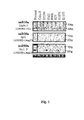
- FIGURE 1 is an image of a Northern blot analysis of the expression of miR-16a (upper panel), miR-26a (middle panel), and miR-99a (lower panel) in normal human lung (lane 1) and human lung cancer cells (lanes 2-8). Below the three blots is an image of an ethidium bromide-stained gel indicating the 5S RNA lane loading control. The genomic location and the type of alteration are indicated.
- FIGURE 2 is a schematic representation demonstrating the position of various miR genes on human chromosomes in relation to HOX gene clusters.
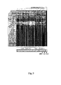
- FIGURE 3 shows an miRNome expression analysis of 38 individual CLL samples.
- the main miR-associated CLL clusters are presented.
- the control samples are: MNC, mononuclear cells; Ly, Diffuse large B cell lymphoma; CD5, selected CD5+ B lymphocytes.
- FIGURE 4 is an image of a Northern blot analysis of the expression of miR-16a (upper panel), miR-26a (middle panel), and miR-99a (lower panel) in 12 B-CLL samples. Below the three blots is an image of an ethidium bromide-stained gel indicating the 5S RNA lane loading control. miR-16a expression levels varied in these B-CLL cases, and were either low or absent in several of the samples tested. However, the expression levels of miR-26a and miR-99a, both regions not involved in B-CLL, were relatively constantn in the tested samples.
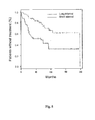
- FIGURE 5 shows Kaplan-Meier curves depicting the relationship between miRNA expression levels and the time from diagnosis to either the time of initial therapy or the present, if therapy had not commenced.
- the proportion of untreated patients with CLL is plotted against time since diagnosis.
- the patients are grouped according to the expression profile generated by 11 microRNA genes.
- FIGURE 6A shows the expression levels of miR-16-1 and miR-15a miRNAs in samples from two patients with a miR-16-1 mutation (see SEQ ID NO. 642) and in CD5+ cell samples from normal patients, both by Northern blot analysis (upper panels) and by miRNACHIP (expression level indicated by numbers below panels).
- FIGURE 6B is a Northern blot depicting levels of miR-16-1 and miR-15a expression (precursor and mature form) in 293 cells after transfection with miR-16-1-WT, miR-16-1-MUT or empty vector. Untransfected 293 cells were used as a control. The normalization was performed using U6 expression.
- genes are represented by italics, and gene products are represented by normal type; e.g., mir- 17 is the gene and miR-17 is the gene product.
- miR gene complement of the human genome or "miRNome”
- miR genes that comprise the miR gene complement of the human genome are non-randomly distributed throughout the genome in relation to each other.
- the largest cluster is composed of six genes located on chromosome 13 at 13q31; the miR genes in this cluster are miR-17 / miR-18 / miR-19almiR-20 / miR-19b1 / miR-92-1.
- the human miR genes are also non-randomly distributed across the human chromosomal complement.
- chromosome 4 has a less-than-expected rate of miRs, and chromosomes 17 and 19 contain significantly more miR genes than expected based on chromosome size.
- miR gene products The sequences of the gene products of 187 miR genes are provided in Table 1. The location and distribution of these 187 miR genes in the human genome is given in Tables 2 and 3; see also Example 1. All Tables are located in the Examples section below.
- miR gene product or “miRNA” means the unprocessed or processed RNA transcript from an miR gene. As the miR gene products are not translated into a protein, the term “miR gene products" does not include proteins.
- probe oligonucleotide refers to an oligonucleotide that is capable of hybridizing to a target oligonucleotide.
- Target oligonucleotide or “target oligodeoxynucleotide” refers to a molecule to be detected (e.g., in a hybridization).
- miR-specific probe oligonucleotide or “probe oligonucleotide specific for an miR” is meant a probe oligonucleotide that has a sequence selected to hybridize to a specific miR gene product, or to a reverse transcript of the specific miR gene product.
- the unprocessed miR gene transcript is also called an "miR precursor," and typically comprises an RNA transcript of about 70 nucleotides in length.
- the miR precursor can be processed by digestion with an RNAse (such as, Dicer, Argonaut, or RNAse III e.g., E.coli RNAse III)) into an active 19-25 nucleotide RNA molecule.
- This active 19-25 nucleotide RNA molecule is also called the "processed miR gene transcript.”
- the active 19-25 nucleotide RNA molecule can be obtained from the miR precursor through natural processing routes (e.g ., using intact cells or cell lysates) or by synthetic processing routes (e.g ., using isolated processing enzymes, such as isolated Dicer, Argonaut, or RNAase III). It is understood that the active 19-25 nucleotide RNA molecule can also be produced directly by biological or chemical syntheses, without having been processed from the miR precursor. For ease of discussion, such a directly produced active 19-25 nucleotide RNA molecule is also referred to as a "processed miR gene product.”
- miR gene expression refers to the production of miR gene products from an miR gene, including processing of the miR precursor into a processed miR gene product.
- a "cancer-associated chromosomal feature” refers to a region of a given chromosome, which, when perturbed, is correlated with the occurrence of at least one human cancer.
- a chromosomal feature is "correlated" with a cancer when the feature and the cancer occur together in individuals of a study population in a manner not expected on the basis of chance alone.
- a region of a chromosome is "perturbed” when the chromosomal architecture or genomic DNA sequence in that region is disturbed or differs from the normal architecture or sequence in that region.
- exemplary perturbations of chromosomal regions include, e.g., chromosomal breakage and translocation, mutations, deletions or amplifications of genomic DNA, a change in the methylation pattern of genomic DNA, the presence of fragile sites, and the presence of viral integration sites.
- chromosomal perturbations associated with a cancer are possible.
- a cancer-associated chromosomal feature can be a chromosomal region where perturbations are known to occur at a higher rate than at other regions in the genome, but where the perturbation has not yet occurred.
- a common chromosomal breakpoint or fragile site is considered a cancer-associated chromosomal feature, even if a break has not yet occurred.
- a region in the genomic DNA known as a mutational "hotspot" can be a cancer-associated chromosomal feature, even if no mutations have yet occurred in the region.
- CAGR cancer-associated genomic region
- Exemplary genetic changes include single- and double-stranded breaks (including common breakpoint regions in or near possible oncogenes or tumor-suppressor genes); chromosomal translocations; mutations, deletions, insertions (including viral, plasmid or transposon integrations) and amplifications (including gene duplications) in the DNA; minimal regions of loss-of heterozygosity (LOH) suggestive of the presence of tumor-suppressor genes; and minimal regions of amplification suggestive of the presence of oncogenes.
- Exemplary epigenetic changes include any changes in DNA methylation patterns (e.g ., DNA hyper- or hypomethylation, especially in promoter regions).
- “cancer-associated genomic region” or "CAGR” specifically excludes chromosomal fragile sites or human papillomavirus insertion sites.
- miR genes in the human genome are in or near CAGRs, including 80 miR genes that are located exactly in minimal regions of LOH or minimal regions of amplification correlated to a variety of cancers. Other miR genes are located in or near breakpoint regions, deleted areas, or regions of amplification.
- the distribution of miR genes in the human genome relative to CAGRs is given in Tables 6 and 7 and in Example 4A below.
- an miR gene is "associated" with a given CAGR when the miR gene is located in close proximity to the CAGR; i.e ., when the miR is located within the same chromosomal band or within 3 megabases (3 Mb) of the CAGR. See Tables 6 and 7 and Example 4A below for a description of cancers which are correlated with CAGRs, and a description of miRs associated with those CAGRs.
- cancers associated with CAGRs include leukemia (e.g ., AML, CLL, pro-lymphocytic leukemia), lung cancer (e.g ., small cell and non-small cell lung carcinoma), esophageal cancer, gastric cancer, colorectal cancer, brain cancer (e.g ., astrocytoma, glioma, glioblastoma, medulloblastoma, meningioma, neuroblastoma), bladder cancer, breast cancer, cervical cancer, epithelial cancer, nasopharyngeal cancer ( e.g ., oral or laryngeal squamous cell carcinoma), lymphoma ( e.g ., follicular lymphoma), uterine cancer ( e.g ., malignant fibrous histiocytoma), hepatic cancer ( e.g ., hepatocellular carcinoma), head-and-neck cancer ( e.g., a a
- miR genes associated with CAGRs include miR-153-2, let-7i, miR-33a, miR-34a-2, miR 34a-1, let-7a-1, let-7d; let-7f-1, miR-24-1, miR-27b, miR-23b, miR-181a; miR-199b, miR-218-1, miR-31, let-7a-2, let-7g, miR-21, miR-32a-1, miR-33b, miR-100, miR-101-1, miR-125b-1, miR-135-1, miR-142as, miR-142s; miR-144, miR-301, miR-297-3, miR-155(BIC), miR-26a, miR-17, miR-18, miR-19a, miR-19b1, miR-20, miR-92-1, miR-128a, miR-7-3, miR-22, miR-123, miR-132, miR-149, miR-161; miR-177, miR
- miR gene(s) that are associated with a particular cancer are evident from Tables 6 and 7, and are preferred.
- AML acute myeloid leukemia
- adenocarcinoma of the lung or esophagus is associated with let-7i.
- the cancer associated with those genes can be diagnosed by evaluating any one of the listed miR genes, or by evaluating any combination of the listed miR genes.
- Subgenera of CAGRs or associated with miR gene(s) would also be evident to one of ordinary skill in the art from Tables 6 and 7.
- a "chromosomal fragile site” or “FRAs” includes any rare or common fragile site in a chromosome; e.g ., one that can be induced by subjecting a cell to stress during DNA replication.
- a rare FRA can be induced by subjecting the cell to folic acid deficiency during DNA replication.
- a common FRA can be induced by treating the cell with aphidocolin or 5-azacytidine during DNA replication.
- an miR gene is "associated" with a given FRA when the miR gene is located in close proximity to the FRA; i.e ., when the miR is located within the same chromosomal band or within 3 megabases (3 Mb) of the FRA. See Table 5 and Example 2 for a description of cancers which are correlated with FRAs, and a description of miRs associated with those FRAs.
- cancers associated with FRAs include bladder cancer, esophageal cancer, lung cancer, stomach cancer, kidney cancer, cervical cancer, ovarian cancer, breast cancer, lymphoma, Ewing sarcoma, hematopoietic tumors, solid tumors and leukemia.
- miR genes associated with FRAs include miR-186, miR-101-1, miR-194, miR-215, miR-106b, miR-25, miR-93, miR-29b, miR-29a, miR-96, miR-182s, miR-182as, miR-183, miR-129-1, let7a-1, let-7d, let-7f-1, miR-23b, miR-24-1, miR-27b, miR-32, miR-159-1, miR-192, miR-125b-1, let-7a-2, miR-100, miR-196-2, miR-148b, miR-190, miR-21, miR-301, miR-142s, miR-142as, miR-105-1, miR-175 and combinations thereof.
- FRA7H is correlated with esophageal cancer, and is associated with miR-29b, miR-29a, miR-96, miR-182s, miR-182as, miR-183, and miR-129-1.
- FRA9D is correlated with bladder cancer, and is associated with let7a-1, let-7d, let-7f-1, miR-23b, miR-24-1, and miR-27b.
- HPV integration site includes any site in a chromosome of a subject where some or all of an HPV genome can insert into the genomic DNA, or any site where some or all of an HPV genome has inserted into the genomic DNA.
- HPV integration sites are often associated with common FRAs, but are distinct from FRAs for purposes of the present invention. Any species or strain of HPV can insert some or all of its genome into an HPV integration site.
- HPV 16 and HPV 18 are the most common strains of HPV which insert some or all of their genomes into an HPV integration site.
- HPV 16 and HPV 18 are the most common strains of HPV which insert some or all of their genomes into an HPV integration site.
- the identification of HPV integration sites in the human genome is within the skill in the art; see, e.g ., Thorland et al. (2000), Cancer Res. 60:5916-21 , the entire disclosure of which is herein incorporated by reference.
- miR genes Thirteen miR genes (7%) are located within 2.5 Mb of seven of the seventeen (45%) cloned integration sites in the human genome. The relative incidence of miRs at HPV 16 integration sites occurred at a rate 3.22 times higher than in the rest of the genome. Indeed, four miR genes ( miR-21, miR-301, miR-142s and miR-142as ) were located within one cluster of integration sites at chromosome 17q23, in which there are three HPV16 integration events spread over roughly 4 Mb of genomic sequence.
- an miR gene is "associated" with a given HPV integration site when the miR gene is located in close proximity to the HPV integration site; i.e., when the miR is located within the same chromosomal band or within 3 megabases (3 Mb), preferably within 2.5 Mb, of the HPV integration site. See Table 5 and Example 3 for a description of miRs associated with HPV integration sites.
- miR gene(s) that are associated with a particular HPV integration site are evident from Table 5, and are preferred.
- the HPV integration site located in or near FRA9E is associated with miR-32.
- the HPV integration site located in or near FRA1H is associated with miR-194 and miR-215.
- the HPV integration site located in or near FRA17B is associated with miR-21, miR-301, miR-142s, and miR-142as.
- the cancer associated with those miR genes can be diagnosed by evaluating any one of the listed miR genes, or by evaluating any combination of the listed miR genes.
- a “homeobox gene or gene cluster” is a single gene or a grouping of genes, characterized in that the gene or genes have been classified by sequence or function as a class I or class II homeobox gene or contain the 183-nucleotide "homeobox” sequence. Identification and characterization of homeobox genes or gene clusters are within the skill in the art; see, e.g., Cillo et al. (1999), Exp. Cell Res. 248:1-9 and Pollard et al. (2000), Current Biology 10:1059-62 , the entire disclosures of which are herein incorporated by reference.
- miR-10a and miR-196-1 are in the HOX B cluster on 17q21; miR-196-2 is in the HOX C cluster at 12q13; and miR-10b is in the HOX D cluster at 2q31.
- miR-148, miR-152 and miR-148b are located within 1 Mb of a HOX gene cluster.
- miR genes are also found within class II homeobox gene clusters; for example, seven microRNAs ( miR-129-1, miR-153-2, let-7a-1, let-7f-1, let-7d, miR-202 and miR-139 ) are located within 0.5 Mb of class II homeotic genes. See Example 5 and Figure 2 for a description of miRs associated with homeobox genes or gene clusters in the human genome.
- Examples of homeobox genes associated with miR genes in the human genome include genes in the HOXA cluster, genes in the HOXB cluster, genes in the HOXC cluster, genes in the HOXD cluster, NK1, NK3, NK4, Lbx, Tlx, Emx, Vax, Hmx, NK6, Msx, Cdx, Xlox, Gsx, En, HB9, Gbx, Msx-1, Msx-2, GBX2, HLX, HEX, PMX1, DLX, LHX2 and CDX2.
- homeobox gene clusters associated with miR genes in the human genome include HOXA, HOXB, HOXC, HOXD, extended Hox, NKL, ParaHox, and EHGbox, PAX, PBX, MEIS, REIG and PREP/KNOX1.
- cancers associated with homeobox genes or gene clusters include renal cancer, Wilm's tumor, colorectal cancer, small cell lung cancer, melanoma, breast cancer, prostate cancer, skin cancer, osteosarcoma, neuroblastoma, leukemia (acute lymphocytic leukemia, acute myeloid leukemia, chronic lymphocytic leukemia), glioblastoma multiform, medulloblastoma, lymphoplasmacytoid lymphoma, thyroid cancer, rhabdomyosarcoma and solid tumors.
- leukemia acute lymphocytic leukemia, acute myeloid leukemia, chronic lymphocytic leukemia
- glioblastoma multiform medulloblastoma
- lymphoplasmacytoid lymphoma thyroid cancer
- rhabdomyosarcoma solid tumors.
- miR genes associated with homeobox genes or gene clusters include miR-148, miR-10a, miR-196-1, miR-152, miR-196-2, miR-148b, miR-10b, miR-129-1, miR-153-2, miR-202, miR-139, let-7a, let-7f, let-7d and combinations thereof.
- homeobox gene cluster HOXA is associated with miR-148.
- Homeobox gene cluster HOXB is associated with miR-148, miR-10a, miR-196-1, miR-152 and combinations thereof.
- Homeobox gene cluster HOXC is associated with miR-196-2, miR-148b or a combination thereof.
- Homeobox gene cluster HOXD is associated with miR-10b.
- the cancer associated with those genes can be diagnosed by evaluating any one of the miR genes, or by evaluating any combination of the miR genes.
- the mIR gene or gene product that is measured or analyzed is not miR-15, miR-16, miR-143 and/or miR-145.
- perturbations in the genomic structure or chromosomal architecture of a cell which comprise the cancer-associated chromosomal feature can affect the expression of the miR gene(s) associated with the feature in that cell.
- a CAGR can comprise an amplification of the region containing an miR gene(s), causing an up-regulation of miR gene expression.
- the CAGR can comprise a chromosomal breakpoint or a deletion that disrupts gene expression, and results in a down-regulation of miR gene expression.
- HPV integrations and FRAs can cause deletions, amplifications or rearrangement of the surrounding DNA, which can also affect the structure or expression of any associated miR genes.
- the invention therefore also provides a method of screening subjects for a predisposition to developing a cancer associated with a cancer-associated chromosomal feature, by evaluating the status of at least one miR gene associated with a cancer-associated chromosomal feature in a tissue or cell sample from a subject, relative to the status of that miR gene in a control sample.
- Subjects with a change in the status of one or more miR genes associated with a cancer-associated chromosomal feature are candidates for further testing to determine or confirm that the subjects have cancer.
- Such further testing can comprise histological examination of blood or tissue samples, or other techniques within the skill in the art.
- the "status of an miR gene” refers to the condition of the miR gene in terms of its physical sequence or structure, or its ability to express a gene product.
- the status of an miR gene in cells of a subject can be evaluated by any technique suitable for detecting genetic or epigenetic changes in the miR gene, or by any technique suitable for detecting the level of miR gene product produced from the miR gene.
- the level of at least one miR gene product produced from an miR gene can be measured in cells of a biological sample obtained from the subject.
- An alteration in the level (i.e ., an up- or down-regulation) of miR gene product in the sample obtained from the subject relative to the level of miR gene product in a control sample is indicative of the presence of the cancer in the subject.
- a "subject" is any mammal suspected of having a cancer associated with a cancer-associated chromosomal feature.
- the subject is a human suspected of having a cancer associated with a cancer-associated chromosomal feature.
- expression of an miR gene is “up-regulated” when the amount of miR gene product produced from that gene in a cell or tissue sample from a subject is greater than the amount produced from the same gene in a control cell or tissue sample.
- expression of an miR gene is “down-regulated” when the amount of miR gene product produced from that gene in a cell or tissue sample from a subject is less than the amount produced from the same gene in a control cell or tissue sample.
- tissue sample can be removed from a subject suspected of having cancer associated with a cancer-associated chromosomal feature by conventional biopsy techniques.
- a blood sample can be removed from the subject, and white blood cells isolated for DNA extraction by standard techniques.
- the blood or tissue sample is preferably obtained from the subject prior to initiation of radiotherapy, chemotherapy or other therapeutic treatment.
- a corresponding control tissue or blood sample can be obtained from unaffected tissues of the subject, from a normal human individual or population of normal individuals, or from cultured cells corresponding to the majority of cells in the subject's sample.
- the control tissue or blood sample is then processed along with the sample from the subject, so that the levels of miR gene product produced from a given miR gene in cells from the subject's sample can be compared to the corresponding miR gene product levels from cells of the control sample.
- the relative miR gene expression in the control and normal samples can be conveniently determined with respect to one or more RNA expression standards.
- the standards can comprise, for example, a zero miR gene expression level, the miR gene expression level in a standard cell line, or the average level of miR gene expression previously obtained for a population of normal human controls.
- RNA transcripts of a particular gene in cells are within the skill in the art.
- total cellular RNA can be purified from cells by homogenization in the presence of nucleic acid extraction buffer, followed by centrifugation. Nucleic acids are precipitated, and DNA is removed by treatment with DNase and precipitation. The RNA molecules are then separated by gel electrophoresis on agarose gels according to standard techniques, and transferred to nitrocellulose filters by, e.g ., the so-called "Northern" blotting technique. The RNA is then immobilized on the filters by heating. Detection and quantification of specific RNA is accomplished using appropriately labeled DNA or RNA probes complementary to the RNA in question. See, for example, Molecular Cloning: A Laboratory Manual, J. Sambrook et al., eds., 2nd edition, Cold Spring Harbor Laboratory Press, 1989, Chapter 7 , the entire disclosure of which is incorporated by reference.
- Suitable probes for Northern blot hybridization of a given miR gene product can be produced from the nucleic acid sequences provided in Table 1. Methods for preparation of labeled DNA and RNA probes, and the conditions for hybridization thereof to target nucleotide sequences, are described in Molecular Cloning: A Laboratory Manual, J. Sambrook et al., eds., 2nd edition, Cold Spring Harbor Laboratory Press, 1989, Chapters 10 and 11 , the disclosures of which are herein incorporated by reference.
- the nucleic acid probe can be labeled with, e.g ., a radionuclide such as 3 H, 32 P, 33 P, 14 C, or 35 S; a heavy metal; or a ligand capable of functioning as a specific binding pair member for a labeled ligand (e.g ., biotin, avidin or an antibody), a fluorescent molecule, a chemiluminescent molecule, an enzyme or the like.
- a radionuclide such as 3 H, 32 P, 33 P, 14 C, or 35 S
- a heavy metal e.g a labeled ligand
- a fluorescent molecule e.g ., biotin, avidin or an antibody
- chemiluminescent molecule e.g chemiluminescent molecule
- Probes can be labeled to high specific activity by either the nick translation method of Rigby et al. (1977), J. Mol. Biol. 113:237-251 or by the random priming method of Fienberg et al. (1983), Anal. Biochem. 132:6-13 , the entire disclosures of which are herein incorporated by reference.
- the latter is the method of choice for synthesizing 32 P-labeled probes of high specific activity from single-stranded DNA or from RNA templates. For example, by replacing preexisting nucleotides with highly radioactive nucleotides according to the nick translation method, it is possible to prepare 32 P-labeled nucleic acid probes with a specific activity well in excess of 10 8 cpm/microgram.
- Autoradiographic detection of hybridization can then be performed by exposing hybridized filters to photographic film. Densitometric scanning of the photographic films exposed by the hybridized filters provides an accurate measurement of miR gene transcript levels. Using another approach, miR gene transcript levels can be quantified by computerized imaging systems, such the Molecular Dynamics 400-B 2D Phosphorimager available from Amersham Biosciences, Piscataway, NJ.
- the random-primer method can be used to incorporate an analogue, for example, the dTTP analogue 5-(N-(N-biotinyl-epsilon-aminocaproyl)-3-aminoallyl)deoxyuridine triphosphate, into the probe molecule.
- analogue for example, the dTTP analogue 5-(N-(N-biotinyl-epsilon-aminocaproyl)-3-aminoallyl)deoxyuridine triphosphate
- the biotinylated probe oligonucleotide can be detected by reaction with biotin-binding proteins, such as avidin, streptavidin, and antibodies (e.g., anti-biotin antibodies) coupled to fluorescent dyes or enzymes that produce color reactions.
- determining the levels of RNA transcripts can be accomplished using the technique of in situ hybridization.
- This technique requires fewer cells than the Northern blotting technique, and involves depositing whole cells onto a microscope cover slip and probing the nucleic acid content of the cell with a solution containing radioactive or otherwise labeled nucleic acid (e.g., cDNA or RNA) probes.
- a solution containing radioactive or otherwise labeled nucleic acid e.g., cDNA or RNA
- This technique is particularly well-suited for analyzing tissue biopsy samples from subjects.
- the practice of the in situ hybridization technique is described in more detail in U.S. Pat. No. 5,427,916 , the entire disclosure of which is incorporated herein by reference.
- Suitable probes for in situ hybridization of a given miR gene product can be produced from the nucleic acid sequences provided in Table 1, as described above.
- the relative number of miR gene transcripts in cells can also be determined by reverse transcription of miR gene transcripts, followed by amplification of the reverse-transcribed transcripts by polymerase chain reaction (RT-PCR).
- the levels of miR gene transcripts can be quantified in comparison with an internal standard, for example, the level ofmRNA from a "housekeeping" gene present in the same sample.
- a suitable "housekeeping" gene for use as an internal standard includes, e.g., myosin or glyceraldehyde-3-phosphate dehydrogenase (G3PDH).
- G3PDH glyceraldehyde-3-phosphate dehydrogenase
- an oligolibrary in microchip format may be constructed containing a set of probe oligonucleotides specific for a set of miR genes.
- the oligolibrary contains probes corresponding to all known miRs from the human genome.
- the microchip oligolibrary may be expanded to include additional miRNAs as they are discovered.
- the microchip is prepared from gene-specific oligonucleotide probes generated from known miRNAs.
- the array contains two different oligonucleotide probes for each miRNA, one containing the active sequence and the other being specific for the precursor of the miRNA.
- the array may also contain controls such as one or more mouse sequences differing from human orthologs by only a few bases, which can serve as controls for hybridization stringency conditions.
- tRNAs from both species may also be printed on the microchip, providing an internal, relatively stable positive control for specific hybridization.
- One or more appropriate controls for non-specific hybridization may also be included on the microchip. For this purpose, sequences are selected based upon the absence of any homology with any known miRNAs.
- the microchip may be fabricated by techniques known in the art. For example, probe oligonucleotides of an appropriate length, e.g., 40 nucleotides, are 5'-amine modified at position C6 and printed using commercially available microarray systems, e.g., the GeneMachine OmniGrid TM 100 Microarrayer and Amersham CodeLink TM activated slides. Labeled cDNA oligomer corresponding to the target RNAs is prepared by reverse transcribing the target RNA with labeled primer. Following first strand synthesis, the RNA/DNA hybrids are denatured to degrade the RNA templates. The labeled target cDNAs thus prepared are then hybridized to the microarray chip under hybridizing conditions, e.g.
- the labeled cDNA oligomer is a biotin-labeled cDNA, prepared from a biotin-labeled primer.
- microarray is then processed by direct detection of the biotin-containing transcripts using, e.g., Streptavidin-Alexa647 conjugate, and scanned utilizing conventional scanning methods. Images intensities of each spot on the array are proportional to the abundance of the corresponding miR in the patient sample.
- the use of the array has several advantages for miRNA expression detection.
- the relatively limited number of miRNAs allows the construction of a common microarray for several species, with distinct oligonucleotide probes for each. Such a tool would allow for analysis of trans-species expression for each known miR under various conditions.
- a microchip containing miRNA-specific probe oligonucleotides corresponding to a substantial portion of the miRNome, preferably the entire miRNome may be employed to carry out miR gene expression profiling, for analysis of miR expression patterns. Distinct miR signatures may be associated with established disease markers, or directly with a disease state. As described hereinafter in Example 11, two distinct clusters of human B-cell chronic lymphocytic leukemia (CLL) samples are associated with the presence or the absence of Zap-70 expression, a predictor of early disease progression.
- CLL chronic lymphocytic leukemia
- miR gene expression profiles can be used for diagnosing the disease state of a cancer, such as whether a cancer is malignant or benign, based on whether or not a given profile is representative of a cancer that is associated with one or more established adverse prognostic markers.
- Prognostic markers that are suitable for this method include ZAP-70 expression, unmutated IgV H gene, CD38 expression, deletion at chromosome11q23, loss or mutation of TP53, and any combination thereof.
- total RNA from a sample from a subject suspected of having a cancer is quantitatively reverse transcribed to provide a set of labeled target oligodeoxynucleotides complementary to the RNA in the sample.
- the target oligodeoxynucleotides are then hybridized to a microarray comprising miRNA-specific probe oligonucleotides to provide a hybridization profile for the sample.
- the result is a hybridization profile for the sample representing the expression pattern of miRNA in the sample.
- the hybridization profile comprises the signal from the binding of the target oligodeoxynucleotides from the sample to the miRNA-specific probe oligonucleotides in the microarray.
- the profile may be recorded as the presence or absence of binding (signal vs. zero signal). More preferably, the profile recorded includes the intensity of the signal from each hybridization. The profile is compared to the hybridization profile generated from a normal, i.e., noncancerous, control sample. An alteration in the signal is indicative of the presence of the cancer in the subject.
- the status of an miR gene in a cell of a subject can also be evaluated by analyzing at least one miR gene in the sample for a deletion, mutation or amplification, wherein detection of a deletion, mutation or amplification in the miR gene relative to the miR gene in a control sample is indicative of the presence of the cancer in the subject.
- a mutation is any alteration in the sequence of a gene of interest that results from one or more nucleotide changes. Such changes include, but are not limited to, allelic polymorphisms, and may affect gene expression and/or function of the gene product.
- a deletion, mutation or amplification in an miR gene can be detected by determining the structure or sequence of genes in cells from a biological sample from a subject suspected of having cancer associated with a cancer-associated chromosomal feature, and comparing this with the structure or sequence of these genes in cells from a control sample.
- Subject and control samples can be obtained as described herein.
- Candidate miR genes for this type of analysis include, but are not limited to, miR-16-1, miR-27b and miR-206. As described in Example 13, mutations in these three miR genes have been identified in samples from CLL patients.
- any technique suitable for detecting alterations in the structure or sequence of genes can be used in the practice of the present method.
- the presence of miR gene deletions, mutations or amplifications can be detected by Southern blot hybridization of the genomic DNA from a subject, using nucleic acid probes specific for miR gene sequences.
- genomic DNA isolated from a subject's sample can be digested with restriction endonucleases. This digestion generates restriction fragments of the genomic DNA that can be separated by electrophoresis, for example, on an agarose gel. The restriction fragments are then blotted onto a hybridization membrane (e.g ., nitrocellulose or nylon), and hybridized with labeled probes specific for a given miR gene or genes. A deletion or mutation of these genes is indicated by an alteration of the restriction fragment patterns on the hybridization membrane, as compared to DNA from a control sample that has been treated identically to the DNA from the subject's sample.
- a hybridization membrane e.g nitrocellulose or nylon
- Probe labeling and hybridization conditions suitable for detecting alterations in gene structure or sequence can be readily determined by one of ordinary skill in the art.
- the miR gene nucleic acid probes for Southern blot hybridization can be designed based upon the nucleic acid sequences provided in Table 1, as described herein. Nucleic acid probe hybridization can then be detected by exposing hybridized filters to photographic film, or by employing computerized imaging systems, such the Molecular Dynamics 400-B 2D Phosphorimager available from Amersham Biosciences, Piscataway, NJ.
- Deletions, mutations and/or amplifications of an miR gene can also be detected by amplifying a fragment of these genes by polymerase chain reaction (PCR), and analyzing the amplified fragment by sequencing or by electrophoresis to determine if the sequence and/or length of the amplified fragment from the subject's DNA sample is different from that of a control DNA sample.
- PCR polymerase chain reaction
- Suitable reaction and cycling conditions for PCR amplification of DNA fragments can be readily determined by one of ordinary skill in the art.
- Deletions of an miR gene can also be identified by detecting deletions of chromosomal markers that are closely linked to the miR gene. Mutations in an miR gene can also be detected by the technique of single strand conformational polymorphism (SSCP), for example, as described in Orita et al. (1989), Genomics 5:874-879 and Hayashi (1991), PCR Methods and Applic. 1:34-38 , the entire disclosures of which are herein incorporated by reference.
- the SSCP technique consists of amplifying a fragment of the gene of interest by PCR; denaturing the fragment and electrophoresing the two denatured single strands under non-denaturing conditions. The single strands assume a complex sequence-dependent intrastrand secondary structure that affects the strands electrophoretic mobility.
- the status of an miR gene in cells of a subject can also be evaluated by measuring the copy number of the at least one miR gene in the sample, wherein a gene copy number other than two for miR genes on somatic chromosomes and sex chromosomes in a female, or other than one for miR genes on sex chromosomes in a male, is indicative of the presence of the cancer in the subject.
- Any technique suitable for detecting gene copy number can be used in the practice of the present method, including the Southern blot and PCR amplification techniques described above.
- An alternative method of determining the miR gene copy number in a sample of tissue relies on the fact that many miR genes or gene clusters are closely linked to chromosomal markers or other genes. The loss of a copy of an miR gene in an individual who is heterozygous at a marker or gene closely linked to the miR gene can be inferred from the loss of heterozygosity in the closely linked marker or gene. Methods for determining loss of heterozygosity of chromosomal markers are within the skill in the art.
- the human miR genes are closely associated with different classes of chromosomal features that are themselves associated with cancer. These cancers are likely caused, in part, by the perturbation in the chromosome or genomic DNA caused by the cancer-associated chromosomal feature, which can affect expression of oncogenes or tumor-suppressor genes located near the site of perturbation. Without wishing to be bound by any theory, it is believed that the perturbations caused by the cancer-associated chromosomal features also affect the expression level of miR genes associated with the feature, and that this also may also contribute to cancerigenesis. Therefore, a given cancer can be treated by restoring the level of miR gene expression associated with that cancer to normal.
- the cancer can be treated by raising the miR expression level.
- the level of miR gene expression is up-regulated in cancer cells of a subject, then the cancer can be treated by reducing the miR expression level.
- the cancers associated with different cancer-associated chromosomal features, and the miR genes associated with these features, are described above and in Tables 5, 6 and 7 and Figure 2 .
- expression the appropriate miR gene or genes associated with a particular cancer and/or cancer-associated chromosomal features is altered by the compositions and methods described herein.
- specific groupings of miR gene(s) that are associated with a particular cancer-associated chromosomal feature and/or cancer are evident from Tables 5, 6 and 7 and in Figure 2 , and are preferred.
- the method of treatment comprising administering an miR gene product.
- the method of treatment comprises administering an miR gene product, provided the miR gene product is not miR-15, mIR-16, miR-143 and/or miR-145.
- the level of at least one miR gene product in cancer cells of a subject is first determined relative to control cells. Techniques suitable for determining the relative level of miR gene product in cells are described above. If miR gene expression is down-regulated in the cancer cell relative to control cells, then the cancer cells are treated with an effective amount of a compound comprising the isolated miR gene product from the miR gene which is down-regulated. If miR gene expression is up-regulated in cancer cells relative to control cells, then the cancer cells are treated with an effective amount of a compound that inhibits miR gene expression. In one embodiment, the level of miR gene product in a cancer cell is not determined beforehand, for example, in those cancers where miR gene expression is known to be up- or down-regulated.
- an effective amount of at least one isolated miR gene product can be administered to a subject.
- an "effective amount" of an isolated miR gene product is an amount sufficient to inhibit proliferation of a cancer cell in a subject suffering from a cancer associated with a cancer-associated chromosomal feature.
- One skilled in the art can readily determine an effective amount of an miR gene product to be administered to a given subject, by taking into account factors such as the size and weight of the subject; the extent of disease penetration; the age, health and sex of the subject; the route of administration; and whether the administration is regional or systemic.
- an effective amount of isolated miR gene product can be based on the approximate weight of a tumor mass to be treated.
- the approximate weight of a tumor mass can be determined by calculating the approximate volume of the mass, wherein one cubic centimeter of volume is roughly equivalent to one gram.
- An effective amount of the isolated miR gene product based on the weight of a tumor mass can be at least about 10 micrograms/gram of tumor mass, and is preferably between about 10-500 micrograms/gram of tumor mass. More preferably, the effective amount is at least about 60 micrograms/gram of tumor mass. Particularly preferably, the effective amount is at least about 100 micrograms/gram of tumor mass. It is preferred that an effective amount based on the weight of the tumor mass be injected directly into the tumor.
- an effective amount of an isolated miR gene product can also be based on the approximate or estimated body weight of a subject to be treated. Preferably, such effective amounts are administered parenterally or enterally, as described herein.
- an effective amount of the isolated miR gene product is administered to a subject can range from about 5 - 3000 micrograms/kg of body weight, and is preferably between about 700 - 1000 micrograms/kg of body weight, and is more preferably greater than about 1000 micrograms/kg of body weight.
- an appropriate dosage regimen for the administration of an isolated miR gene product to a given subject can be administered to the subject once (e.g ., as a single injection or deposition).
- an miR gene product can be administered once or twice daily to a subject for a period of from about three to about twenty-eight days, more preferably from about seven to about ten days.
- an miR gene product is administered once a day for seven days.
- a dosage regimen comprises multiple administrations, it is understood that the effective amount of the miR gene product administered to the subject can comprise the total amount of gene product administered over the entire dosage regimen.
- an "isolated" miR gene product is one which is synthesized, or altered or removed from the natural state through human intervention.
- an miR gene product naturally present in a living animal is not “isolated.”
- An isolated miR gene product can exist in substantially purified form, or can exist in a cell into which the miR gene product has been delivered.
- an miR gene product which is deliberately delivered to, or expressed in, a cell is considered an "isolated” miR gene product.
- An miR gene product produced inside a cell by from an miR precursor molecule is also considered to be “isolated” molecule.
- Isolated miR gene products can be obtained using a number of standard techniques.
- the miR gene products can be chemically synthesized or recombinantly produced using methods known in the art.
- miR gene products are chemically synthesized using appropriately protected ribonucleoside phosphoramidites and a conventional DNA/RNA synthesizer.
- RNA molecules or synthesis reagents include, e.g., Proligo (Hamburg, Germany), Dharmacon Research (Lafayette, CO, USA), Pierce Chemical (part of Perbio Science, Rockford, IL, USA), Glen Research (Sterling, VA, USA), ChemGenes (Ashland, MA, USA) and Cruachem (Glasgow, UK).
- the miR gene products can be expressed from recombinant circular or linear DNA plasmids using any suitable promoter.
- suitable promoters for expressing RNA from a plasmid include, e.g., the U6 or H1 RNA pol III promoter sequences, or the cytomegalovirus promoters. Selection of other suitable promoters is within the skill in the art.
- the recombinant plasmids of the invention can also comprise inducible or regulatable promoters for expression of the miR gene products in cancer cells.
- the miR gene products that are expressed from recombinant plasmids can be isolated from cultured cell expression systems by standard techniques.
- the miR gene products which are expressed from recombinant plasmids can also be delivered to, and expressed directly in, the cancer cells.
- the use of recombinant plasmids to deliver the miR gene products to cancer cells is discussed in more detail below.
- the miR gene products can be expressed from a separate recombinant plasmid, or can be expressed from the same recombinant plasmid.
- the miR gene products are expressed as the RNA precursor molecules from a single plasmid, and the precursor molecules are processed into the functional miR gene product by a suitable processing system, including processing systems extant within a cancer cell.
- suitable processing systems include, e.g., the in vitro Drosophila cell lysate system as described in U.S. published application 2002/0086356 to Tuschl et al. and the E. coli RNAse III system described in U.S. published patent application 2004/0014113 to Yang et al. , the entire disclosures of which are herein incorporated by reference.
- plasmids suitable for expressing the miR gene products are within the skill in the art. See, for example, Zeng et al. (2002), Molecular Cell 9:1327-1333 ; Tuschl (2002), Nat. Biotechnol, 20:446-448 ; Brummelkamp et al. (2002), Science 296:550-553 ; Miyagishi et al. (2002), Nat. Biotechnol. 20:497-500 ; Paddison et al. (2002), Genes Dev.
- a plasmid expressing the miR gene products comprises a sequence encoding a miR precursor RNA under the control of the CMV intermediate-early promoter.
- "under the control" of a promoter means that the nucleic acid sequences encoding the miR gene product are located 3' of the promoter, so that the promoter can initiate transcription of the miR gene product coding sequences.
- the miR gene products can also be expressed from recombinant viral vectors. It is contemplated that the miR gene products can be expressed from two separate recombinant viral vectors, or from the same viral vector.
- the RNA expressed from the recombinant viral vectors can either be isolated from cultured cell expression systems by standard techniques, or can be expressed directly in cancer cells. The use of recombinant viral vectors to deliver the miR gene products to cancer cells is discussed in more detail below.
- the recombinant viral vectors of the invention comprise sequences encoding the miR gene products and any suitable promoter for expressing the RNA sequences.
- suitable promoters include, for example, the U6 or H1 RNA pol III promoter sequences, or the cytomegalovirus promoters. Selection of other suitable promoters is within the skill in the art.
- the recombinant viral vectors of the invention can also comprise inducible or regulatable promoters for expression of the miR gene products in a cancer cell.
- Any viral vector capable of accepting the coding sequences for the miR gene products can be used; for example, vectors derived from adenovirus (AV); adeno-associated virus (AAV); retroviruses (e.g ., lentiviruses (LV), Rhabdoviruses, murine leukemia virus); herpes virus, and the like.
- AV adenovirus
- AAV adeno-associated virus
- retroviruses e.g ., lentiviruses (LV), Rhabdoviruses, murine leukemia virus
- herpes virus and the like.
- the tropism of the viral vectors can be modified by pseudotyping the vectors with envelope proteins or other surface antigens from other viruses, or by substituting different viral capsid proteins, as appropriate.
- lentiviral vectors of the invention can be pseudotyped with surface proteins from vesicular stomatitis virus (VSV), rabies, Ebola, Mokola, and the like.
- AAV vectors of the invention can be made to target different cells by engineering the vectors to express different capsid protein serotypes.
- an AAV vector expressing a serotype 2 capsid on a serotype 2 genome is called AAV 2/2.
- This serotype 2 capsid gene in the AAV 2/2 vector can be replaced by a serotype 5 capsid gene to produce an AAV 2/5 vector.
- AAV vectors which express different capsid protein serotypes are within the skill in the art; see, e.g ., Rabinowitz J.E. et al. (2002), J Virol 76:791-801 , the entire disclosure of which is herein incorporated by reference.
- recombinant viral vectors suitable for use in the invention methods for inserting nucleic acid sequences for expressing RNA into the vector, methods of delivering the viral vector to the cells of interest, and recovery of the expressed RNA products are within the skill in the art. See, for example, Dornburg (1995), Gene Therap. 2:301-310 ; Eglitis (1988), Biotechniques 6:608-614 ; Miller (1990), Hum. Gene Therap. 1:5-14 ; and Anderson (1998), Nature 392:25-30 , the entire disclosures of which are herein incorporated by reference.
- Preferred viral vectors are those derived from AV and AAV.
- a suitable AV vector for expressing the miR gene products, a method for constructing the recombinant AV vector, and a method for delivering the vector into target cells are described in Xia et al. (2002), Nat. Biotech. 20:1006-1010 , the entire disclosure ofwhich is herein incorporated by reference.
- Suitable AAV vectors for expressing the miR gene products, methods for constructing the recombinant AAV vector, and methods for delivering the vectors into target cells are described in Samulski et al. (1987), J. Viol. 61:3096-3101 ; Fisher et al. (1996), J.
- the miR gene products are expressed from a single recombinant AAV vector comprising the CMV intermediate early promoter.
- a recombinant AAV viral vector of the invention comprises a nucleic acid sequence encoding an miR precursor RNA in operable connection with a polyT termination sequence under the control of a human U6 RNA promoter.
- operable connection with a polyT termination sequence means that the nucleic acid sequences encoding the sense or antisense strands are immediately adjacent to the polyT termination signal in the 5' direction.
- the polyT termination signals act to terminate transcription.
- an effective amount of at least one compound which inhibits miR gene expression can also be administered to the subject.
- inhibiting miR gene expression means that the production of miR gene product from the miR gene in the cancer cell after treatment is less than the amount produced prior to treatment.
- One skilled in the art can readily determine whether miR gene expression has been inhibited in a cancer cell, using for example the techniques for determining miR transcript level discussed above for the diagnostic method.
- an "effective amount" of a compound that inhibits miR gene expression is an amount sufficient to inhibit proliferation of a cancer cell in a subject suffering from a cancer associated with a cancer-associated chromosomal feature.
- an effective amount of an miR gene expression-inhibiting compound to be administered to a given subject by taking into account factors such as the size and weight of the subject; the extent of disease penetration; the age, health and sex of the subject; the route of administration; and whether the administration is regional or systemic.
- an effective amount of the expression-inhibiting compound can be based on the approximate weight of a tumor mass to be treated.
- the approximate weight of a tumor mass can be determined by calculating the approximate volume of the mass, wherein one cubic centimeter of volume is roughly equivalent to one gram.
- An effective amount based on the weight of a tumor mass can be at least about 10 micrograms/gram of tumor mass, and is preferably between about 10-500 micrograms/gram of tumor mass. More preferably, the effective amount is at least about 60 micrograms/gram of tumor mass. Particularly preferably, the effective amount is at least about 100 micrograms/gram of tumor mass. It is preferred that an effective amount based on the weight of the tumor mass be injected directly into the tumor.
- an effective amount of a compound that inhibits miR gene expression can also be based on the approximate or estimated body weight of a subject to be treated. Preferably, such effective amounts are administered parenterally or enterally, as described herein.
- an effective amount of the expression-inhibiting compound administered to a subject can range from about 5 -3000 micrograms/kg of body weight, and is preferably between about 700 - 1000 micrograms/kg of body weight, and is more preferably greater than about 1000 micrograms/kg of body weight.
- an expression-inhibiting compound can be administered to the subject once ( e.g ., as a single injection or deposition).
- an expression-inhibiting compound can be administered once or twice daily to a subject for a period of from about three to about twenty-eight days, more preferably from about seven to about ten days.
- an expression-inhibiting compound is administered once a day for seven days.
- a dosage regimen comprises multiple administrations, it is understood that the effective amount of the expression-inhibiting compound administered to the subject can comprise the total amount of compound administered over the entire dosage regimen.
- Suitable compounds for inhibiting miR gene expression include double-stranded RNA (such as short- or small-interfering RNA or "siRNA”), antisense nucleic acids, and enzymatic RNA molecules such as ribozymes. Each of these compounds can be targeted to a given miR gene product and destroy or induce the destruction of the target miR gene product.
- siRNA short- or small-interfering RNA or "siRNA”
- antisense nucleic acids such as ribozymes.
- enzymatic RNA molecules such as ribozymes.
- RNA interference of the miR gene can be inhibited by inducing RNA interference of the miR gene with an isolated double-stranded RNA ("dsRNA") molecule which has at least 90%, for example 95%, 98%, 99% or 100%, sequence homology with at least a portion of the miR gene product.
- dsRNA isolated double-stranded RNA
- the dsRNA molecule is a "short or small interfering RNA" or "siRNA.”
- siRNA useful in the present methods comprise short double-stranded RNA from about 17 nucleotides to about 29 nucleotides in length, preferably from about 19 to about 25 nucleotides in length.
- the siRNA comprise a sense RNA strand and a complementary antisense RNA strand annealed together by standard Watson-Crick base-pairing interactions (hereinafter "base-paired").
- the sense strand comprises a nucleic acid sequence which is substantially identical to a nucleic acid sequence contained within the target miR gene product.
- a nucleic acid sequence in an siRNA which is "substantially identical" to a target sequence contained within the target mRNA is a nucleic acid sequence that is identical to the target sequence, or that differs from the target sequence by one or two nucleotides.
- the sense and antisense strands of the siRNA can comprise two complementary, single-stranded RNA molecules, or can comprise a single molecule in which two complementary portions are base-paired and are covalently linked by a single-stranded "hairpin" area.
- the siRNA can also be altered RNA that differs from naturally-occurring RNA by the addition, deletion, substitution and/or alteration of one or more nucleotides.
- Such alterations can include addition of non-nucleotide material, such as to the end(s) of the siRNA or to one or more internal nucleotides of the siRNA, or modifications that make the siRNA resistant to nuclease digestion, or the substitution of one or more nucleotides in the siRNA with deoxyribonucleotides.
- the siRNA can also comprise a 3' overhang.
- a "3' overhang” refers to at least one unpaired nucleotide extending from the 3'-end of a duplexed RNA strand.
- the siRNA comprises at least one 3' overhang of from 1 to about 6 nucleotides (which includes ribonucleotides or deoxyribonucleotides) in length, preferably from 1 to about 5 nucleotides in length, more preferably from 1 to about 4 nucleotides in length, and particularly preferably from about 2 to about 4 nucleotides in length.
- the 3' overhang is present on both strands of the siRNA, and is 2 nucleotides in length.
- each strand of the siRNA can comprise 3' overhangs of dithymidylic acid ("TT") or diuridylic acid (“uu").
- the siRNA can be produced chemically or biologically, or can be expressed from a recombinant plasmid or viral vector, as described above for the isolated miR gene products.
- Exemplary methods for producing and testing dsRNA or siRNA molecules are described in U.S. published patent application 2002/0173478 to Gewirtz and in U.S. published patent application 2004/0018176 to Reich et al. , the entire disclosures of which are herein incorporated by reference.
- an antisense nucleic acid refers to a nucleic acid molecule that binds to target RNA by means of RNA-RNA or RNA-DNA or RNA-peptide nucleic acid interactions, which alters the activity of the target RNA.
- Antisense nucleic acids suitable for use in the present methods are single-stranded nucleic acids (e.g ., RNA, DNA, RNA-DNA chimeras, PNA) that generally comprise a nucleic acid sequence complementary to a contiguous nucleic acid sequence in an miR gene product.
- the antisense nucleic acid comprises a nucleic acid sequence that is 50-100% complementary, more preferably 75-100% complementary, and most preferably 95-100% complementary to a contiguous nucleic acid sequence in an miR gene product.
- Nucleic acid sequences for the miR gene products are provided in Table 1. Without wishing to be bound by any theory, it is believed that the antisense nucleic acids activate RNase H or some other cellular nuclease that digests the miR gene product/antisense nucleic acid duplex.
- Antisense nucleic acids can also contain modifications to the nucleic acid backbone or to the sugar and base moieties (or their equivalent) to enhance target specificity, nuclease resistance, delivery or other properties related to efficacy of the molecule. Such modifications include cholesterol moieties, duplex intercalators such as acridine or the inclusion of one or more nuclease-resistant groups.
- Antisense nucleic acids can be produced chemically or biologically, or can be expressed from a recombinant plasmid or viral vector, as described above for the isolated miR gene products. Exemplary methods for producing and testing are within the skill in the art; see, e.g ., Stein and Cheng (1993), Science 261:1004 and U.S. Pat. No. 5,849,902 to Woolf et al. , the entire disclosures of which are herein incorporated by reference.
- an "enzymatic nucleic acid” refers to a nucleic acid comprising a substrate binding region that has complementarity to a contiguous nucleic acid sequence of an miR gene product, and which is able to specifically cleave the miR gene product.
- the enzymatic nucleic acid substrate binding region is 50-100% complementary, more preferably 75-100% complementary, and most preferably 95-100% complementary to a contiguous nucleic acid sequence in an miR gene product.
- the enzymatic nucleic acids can also comprise modifications at the base, sugar, and/or phosphate groups.
- An exemplary enzymatic nucleic acid for use in the present methods is a ribozyme.
- the enzymatic nucleic acids can be produced chemically or biologically, or can be expressed from a recombinant plasmid or viral vector, as described above for the isolated miR gene products.
- exemplary methods for producing and testing dsRNA or siRNA molecules are described in Werner and Uhlenbeck (1995), Nucl. Acids Res. 23:2092-96 ; Hammann et al. (1999), Antisense and Nucleic Acid Drug Dev. 9:25-31 ; and U.S. Pat. No. 4,987,071 to Cech et al , the entire disclosures of which are herein incorporated by reference.
- Administration of at least one miR gene product, or at least one compound for inhibiting miR gene expression will inhibit the proliferation of cancer cells in a subject who has a cancer associated with a cancer-associated chromosomal feature.
- to "inhibit the proliferation of a cancer cell” means to kill the cell, or permanently or temporarily arrest or slow the growth of the cell.
- Inhibition of cancer cell proliferation can be inferred if the number of such cells in the subj ect remains constant or decreases after administration of the miR gene products or miR gene expression-inhibiting compounds.
- An inhibition of cancer cell proliferation can also be inferred if the absolute number of such cells increases, but the rate of tumor growth decreases.
- the number of cancer cells in a subject's body can be determined by direct measurement, or by estimation from the size of primary or metastatic tumor masses.
- the number of cancer cells in a subject can be measured by immunohistological methods, flow cytometry, or other techniques designed to detect characteristic surface markers of cancer cells.
- the size of a tumor mass can be ascertained by direct visual observation, or by diagnostic imaging methods, such as X-ray, magnetic resonance imaging, ultrasound, and scintigraphy. Diagnostic imaging methods used to ascertain size of the tumor mass can be employed with or without contrast agents, as is known in the art.
- the size of a tumor mass can also be ascertained by physical means, such as palpation of the tissue mass or measurement of the tissue mass with a measuring instrument, such as a caliper.
- the miR gene products or miR gene expression-inhibiting compounds can be administered to a subject by any means suitable for delivering these compounds to cancer cells of the subject.
- the miR gene products or miR expression inhibiting compounds can be administered by methods suitable to transfect cells of the subject with these compounds, or with nucleic acids comprising sequences encoding these compounds.
- the cells are transfected with a plasmid or viral vector comprising sequences encoding at least one miR gene product or miR gene expression inhibiting compound.
- Transfection methods for eukaryotic cells include, e.g., direct injection of the nucleic acid into the nucleus or pronucleus of a cell; electroporation; liposome transfer or transfer mediated by lipophilic materials; receptor mediated nucleic acid delivery, bioballistic or particle acceleration; calcium phosphate precipitation, and transfection mediated by viral vectors.
- cells can be transfected with a liposomal transfer compound, e.g., DOTAP (N-[1-(2,3-dioleoyloxy)propyl]-N,N,N-trimethyl-ammonium methylsulfate, Boehringer - Mannheim) or an equivalent, such as LIPOFECTIN.
- DOTAP N-[1-(2,3-dioleoyloxy)propyl]-N,N,N-trimethyl-ammonium methylsulfate, Boehringer - Mannheim
- LIPOFECTIN LIPOFECTIN
- An miR gene product or miR gene expression inhibiting compound can also be administered to a subject by any suitable enteral or parenteral administration route.
- Suitable enteral administration routes for the present methods include, e.g., oral, rectal, or intranasal delivery.
- Suitable parenteral administration routes include, e.g., intravascular administration (e.g., intravenous bolus injection, intravenous infusion, intra-arterial bolus injection, intra-arterial infusion and catheter instillation into the vasculature); peri- and intra-tissue injection (e.g., peri-tumoral and intra-tumoral injection, intra-retinal injection, or subretinal injection); subcutaneous injection or deposition, including subcutaneous infusion (such as by osmotic pumps); direct application to the tissue of interest, for example by a catheter or other placement device (e.g ., a retinal pellet or a suppository or an implant comprising a porous, non-porous, or gelatinous material); and inhalation.
- Preferred administration routes are injection, infusion and direct injection into the tumor.
- an miR gene product or miR gene expression inhibiting compound can be administered to the subject either as naked RNA, in combination with a delivery reagent, or as a nucleic acid (e.g ., a recombinant plasmid or viral vector) comprising sequences that express the miR gene product or expression inhibiting compound.
- a delivery reagent include, e.g, the Mirus Transit TKO lipophilic reagent; lipofectin; lipofectamine; cellfectin; polycations ( e.g ., polylysine), and liposomes.
- Recombinant plasmids and viral vectors comprising sequences that express the miR gene products or miR gene expression inhibiting compounds, and techniques for delivering such plasmids and vectors to cancer cells, are discussed above.
- liposomes are used to deliver an miR gene product or miR gene expression-inhibiting compound (or nucleic acids comprising sequences encoding them) to a subject. Liposomes can also increase the blood half-life of the gene products or nucleic acids.
- Liposomes suitable for use in the invention can be formed from standard vesicle-forming lipids, which generally include neutral or negatively charged phospholipids and a sterol, such as cholesterol.
- the selection of lipids is generally guided by consideration of factors such as the desired liposome size and half-life of the liposomes in the blood stream.
- a variety of methods are known for preparing liposomes, for example, as described in Szoka et al. (1980), Ann. Rev. Biophys. Bioeng. 9:467 ; and U.S. Pat. Nos. 4,235,871 , 4,501,728 , 4,837,028 , and 5,019,369 , the entire disclosures of which are herein incorporated by reference.
- the liposomes for use in the present methods can comprise a ligand molecule that targets the liposome to cancer cells.
- Ligands which bind to receptors prevalent in cancer cells such as monoclonal antibodies that bind to tumor cell antigens, are preferred.
- the liposomes for use in the present methods can also be modified so as to avoid clearance by the mononuclear macrophage system ("MMS") and reticuloendothelial system ("RES").
- MMS mononuclear macrophage system
- RES reticuloendothelial system
- modified liposomes have opsonization-inhibition moieties on the surface or incorporated into the liposome structure.
- a liposome of the invention can comprise both opsonization-inhibition moieties and a ligand.
- Opsonization-inhibiting moieties for use in preparing the liposomes of the invention are typically large hydrophilic polymers that are bound to the liposome membrane.
- an opsonization inhibiting moiety is "bound" to a liposome membrane when it is chemically or physically attached to the membrane, e.g., by the intercalation of a lipid-soluble anchor into the membrane itself, or by binding directly to active groups of membrane lipids.
- These opsonization-inhibiting hydrophilic polymers form a protective surface layer that significantly decreases the uptake of the liposomes by the MMS and RES; e.g., as described in U.S. Pat. No. 4,920,016 , the entire disclosure of which is herein incorporated by reference.
- Opsonization inhibiting moieties suitable for modifying liposomes are preferably water-soluble polymers with a number-average molecular weight from about 500 to about 40,000 daltons, and more preferably from about 2,000 to about 20,000 daltons.
- Such polymers include polyethylene glycol (PEG) or polypropylene glycol (PPG) derivatives; e.g., methoxy PEG or PPG, and PEG or PPG stearate; synthetic polymers such as polyacrylamide or poly N-vinyl pyrrolidone; linear, branched, or dendrimeric polyamidoamines; polyacrylic acids; polyalcohols, e.g., polyvinylalcohol and polyxylitol to which carboxylic or amino groups are chemically linked, as well as gangliosides, such as ganglioside GM1.
- PEG polyethylene glycol
- PPG polypropylene glycol
- synthetic polymers such as polyacrylamide or poly N-viny
- Copolymers of PEG, methoxy PEG, or methoxy PPG, or derivatives thereof, are also suitable.
- the opsonization inhibiting polymer can be a block copolymer of PEG and either a polyamino acid, polysaccharide, polyamidoamine, polyethyleneamine, or polynucleotide.
- the opsonization inhibiting polymers can also be natural polysaccharides containing amino acids or carboxylic acids, e.g., galacturonic acid, glucuronic acid, mannuronic acid, hyaluronic acid, pectic acid, neuraminic acid, alginic acid, carrageenan; aminated polysaccharides or oligosaccharides (linear or branched); or carboxylated polysaccharides or oligosaccharides, e.g., reacted with derivatives of carbonic acids with resultant linking of carboxylic groups.
- the opsonization-inhibiting moiety is a PEG, PPG, or derivatives thereof. Liposomes modified with PEG or PEG-derivatives are sometimes called "PEGylated liposomes.”
- the opsonization inhibiting moiety can be bound to the liposome membrane by any one of numerous well-known techniques.
- an N-hydroxysuccinimide ester of PEG can be bound to a phosphatidyl-ethanolamine lipid-soluble anchor, and then bound to a membrane.
- a dextran polymer can be derivatized with a stearylamine lipid-soluble anchor via reductive amination using Na(CN)BH 3 and a solvent mixture, such as tetrahydrofuran and water in a 30:12 ratio at 60°C.
- Liposomes modified with opsonization-inhibition moieties remain in the circulation much longer than unmodified liposomes. For this reason, such liposomes are sometimes called "stealth” liposomes. Stealth liposomes are known to accumulate in tissues fed by porous or "leaky” microvasculature. Thus, tissue characterized by such microvasculature defects, for example solid tumors, will efficiently accumulate these liposomes; see Gabizon, et al. (1988), Proc. Natl. Acad. Sci., USA, 18:6949-53 . In addition, the reduced uptake by the RES lowers the toxicity of stealth liposomes by preventing significant accumulation of the liposomes in the liver and spleen. Thus, liposomes that are modified with opsonization-inhibition moieties are particularly suited to deliver the miR gene products or miR gene expression inhibition compounds (or nucleic acids comprising sequences encoding them) to tumor cells.
- the miR gene products or miR gene expression inhibition compounds are preferably formulated as pharmaceutical compositions, sometimes called “medicaments," prior to administering to a subject, according to techniques known in the art.
- Pharmaceutical compositions of the present invention are characterized as being at least sterile and pyrogen-free.
- pharmaceutical formulations include formulations for human and veterinary use. Methods for preparing pharmaceutical compositions of the invention are within the skill in the art, for example as described in Remington's Pharmaceutical Science, 17th ed., Mack Publishing Company, Easton, Pa. (1985 ), the entire disclosure of which is herein incorporated by reference.
- the present pharmaceutical formulations comprise at least one miR gene product or miR gene expression inhibition compound (or at least one nucleic acid comprising sequences encoding them) (e.g., 0.1 to 90% by weight), or a physiologically acceptable salt thereof, mixed with a pharmaceutically-acceptable carrier.
- the pharmaceutical formulations of the invention can also comprise at least one miR gene product or miR gene expression inhibition compound (or at least one nucleic acid comprising sequences encoding them) which are encapsulated by liposomes and a pharmaceutically-acceptable carrier.
- the pharmaceutical compositions comprise an mIR gene or gene product that is is not miR-15, miR-16, miR-143 and/or miR-145.
- Preferred pharmaceutically-acceptable carriers are water, buffered water, normal saline, 0.4% saline, 0.3% glycine, hyaluronic acid and the like.
- the pharmaceutical compositions of the invention comprise at least one miR gene product or miR gene expression inhibition compound (or at least one nucleic acid comprising sequences encoding them) which is resistant to degradation by nucleases.
- miR gene product or miR gene expression inhibition compound or at least one nucleic acid comprising sequences encoding them
- nucleases One skilled in the art can readily synthesize nucleic acids which are nuclease resistant, for example by incorporating one or more ribonucleotides that are modified at the 2'-position into the miR gene products. Suitable 2'-modified ribonucleotides include those modified at the 2'-position with fluoro, amino, alkyl, alkoxy, and O-allyl.
- compositions of the invention can also comprise conventional pharmaceutical excipients and/or additives.
- Suitable pharmaceutical excipients include stabilizers, antioxidants, osmolality adjusting agents, buffers, and pH adjusting agents.
- Suitable additives include, e.g., physiologically biocompatible buffers (e.g., tromethamine hydrochloride), additions of chelants (such as, for example, DTPA or DTPA-bisamide) or calcium chelate complexes (such as, for example, calcium DTPA, CaNaDTPA-bisamide), or, optionally, additions of calcium or sodium salts (for example, calcium chloride, calcium ascorbate, calcium gluconate or calcium lactate).
- Pharmaceutical compositions of the invention can be packaged for use in liquid form, or can be lyophilized.
- conventional nontoxic solid pharmaceutically-acceptable carriers can be used; for example, pharmaceutical grades of mannitol, lactose, starch, magnesium stearate, sodium saccharin, talcum, cellulose, glucose, sucrose, magnesium carbonate, and the like.
- a solid pharmaceutical composition for oral administration can comprise any of the carriers and excipients listed above and 10-95%, preferably 25%-75%, of the at least one miR gene product or miR gene expression inhibition compound (or at least one nucleic acid comprising sequences encoding them).
- a pharmaceutical composition for aerosol (inhalational) administration can comprise 0.01-20% by weight, preferably 1%-10% by weight, of the at least one miR gene product or miR gene expression inhibition compound (or at least one nucleic acid comprising sequences encoding them) encapsulated in a liposome as described above, and a propellant.
- a carrier can also be included as desired; e.g., lecithin for intranasal delivery.
- a set of 187 human miR genes was compiled (see Table 1).
- the set comprises 153 miRs identified in the miR Registry (http://www.sanger.ac.uk/Software/Rfam/mirna/; maintained by the Wellcome Trust Sanger Institute, Cambridge, UK), and 36 other miRs manually curated from published papers ( Lim et al., 2003, Science 299:1540 ; Lagos-Quintana et al., 2001, Science 294:853-858 ; Lau et al., 2001, Science 294:858-862 ; Lee et al., 2001, Science 294:862-864 ; Mourelatos et al., 2002, Genes Dev.
- a BLAST search was performed using the default parameters against the human genome to find the precise location, followed by mapping using the maps available at the Human Genome Resources at the NCBI website. See also Altschul et al. (1990), J. Mol. Biol. 215:403-10 and Altschul et al. (1997), Nucleic Acids Res. 25:3389-3402 , the entire disclosures of which are herein incorporated by reference, for a discussion of the BLAST search algorithm.
- This database was constructed using the Virtual Gene Nomenclature Workshop (htpp://www.gene.ucl.ac.uk/nomenclature/workshop), maintained by the HUGO Gene Nomenclature Committee at University College, London.
- the literature was screened for publications reporting the cloning of the locus. In ten cases, genomic positions for both centromeric and telomeric ends were found. The total genomic length of these FRA loci is 26.9 Mb. In twenty-nine cases, only one anchoring marker was identified. It was determined, based on the published data, that 3 Mb can be used as the median length for each FRA locus. Therefore, 3 Mb was used as a guideline or window length for considering whether miR were in close proximity to the FRA sites.
- HPV16 integration sites The human clones for seventeen HPV16 integration sites (IS) were also precisely mapped on the human genome.
- "close" vicinity was defined to be a distance of less than 2 Mb.
- the PubMed database was screened on-line for publications describing cancer-related abnormalities such as minimal regions of loss-of-heterozygosity (minimal LOH) and minimal regions of amplification (minimal amplicons) using the words “LOH and genome-wide,” “amplification and genome-wide” and "amplicon and cancer.”
- the PubMed database is maintained by the NCBI and was accessed via its website.
- the data obtained from thirty-two papers were used to screen for putative CAGRs, based on markers with high frequency of LOH/amplification.
- a literature search was performed to determine the presence or absence of the above three types of alterations and to determine the precise location of miRs with respect to CAGRs (see above).
- Search phrases included the combinations "minimal regions of LOH AND cancer", and "minimal region amplification AND cancer.”
- a total of 296 publications were found and manually curated to find regions defined by both telomeric and centromeric markers.
- One hundred fifty-four minimally deleted regions (median length- 4.14 Mb) and 37 minimally amplified regions (median length- 2.45 Mb) were identified with precise genomic mapping for both telomeric and centromeric ends involving all human chromosomes except Y.
- PubMed was searched with the combination "translocation AND cloning AND breakpoint AND cancer.” The search yielded 308 papers, which were then manually curated. Among these papers, 45 translocations with at least one breakpoint precisely mapped were reported.
- This model provided the incidence rate ratio (IRR), 2-sided 95% confidence interval of the IRR, and 2-sided p-values for testing the hypothesis that the IRR is 1.0.
- IRR incidence rate ratio
- 2-sided 95% confidence interval of the IRR 2-sided 95% confidence interval of the IRR
- 2-sided p-values for testing the hypothesis that the IRR is 1.0.
- An IRR significantly greater than 1 indicates an increase in the number of miR genes within a region.
- CLL chronic lymphocytic leukemia
- mononuclear cells were isolated through Ficoll-Hypaque gradient centrifugation (Amersham Pharmacia Biotech, Piscataway, NJ), as previously described ( Calin et al., Proc. Natl. Acad. Sci. USA 2002, 99:15524-15529 ).
- Samples were then processed for RNA and DNA extraction according to standard protocols as described in Sambrook J et al. (1989), Molecular cloning: A Laboratory Manual (Cold Spring Harbor Laboratory Press, Cold Spring Harbor, NY ), the entire disclosure of which is herein incorporated by reference.
- RNA isolation from patient samples and cell lines described above was performed using the Tri-Reagent protocol (Molecular Research Center, Inc). RNA samples (30 ⁇ g each) were run on 15% acryl amide denaturing (urea) Criterion recast gels (Bio-Red Laboratories, Hercules, CA) and then transferred onto Hyoid-N+ membrane (Amersham Pharmacia Biotech), as previously described ( Calin et al., Proc. Natl. Acad. Sci. USA 2002, 99:15524-15529 ). Hybridization with gamma- 32 P ATP labeled probes was performed at 42°C in 7% SDS, 0.2 M Na 2 PO 4 , pH 7.0 overnight.
- Membranes were washed at 42°C, twice in 2x SSPE, 0.1% SDS and twice with 0.5x SSPE, 0.1% SDS. Blots were stripped by boiling in 0.1% aqueous SDS/0.1x SSC for 10 minutes, and were reprobed several times. As a gel loading control, 5S rRNA was also loaded and was stained with ethidium bromide. Lung tissue RNA was utilized as the normal control; normal lung total RNA was purchased from Clontech (Palo Alto, CA).
- Example 1- miR Genes are Non-Randomly Distributed in the Human Genome
- the distribution of the 186 human miR genes was found to be non-random.
- the largest cluster found comprises six genes ( miR-17 / miR-18 / miR-19a / miR-20 / miR-19bJ / miR-92-1) and is located at 13q31 (Table 2).
- Chromosome 1 is used as the baseline in the model with a rate of miR gene incidence of0.057, which is approximately equal to the overall rate of miR gene incidence across the genome.
- Table 3 Location of miRs by chromosome and results of mixed effects Poisson regression model.
- Example 2- miR Genes are Located In or Near Fragile Sites
- the other three of the four most active sites contain known or candidate TS genes; i.e., FHIT, WWOX and PARK2, respectively ( Ohta et al., 1996, Cell 84:587-597 ; Paige et al., 2001, Proc. Natl. Acad. Sci. USA 98:11417-11422 ; Cesari et al., 2003, Proc. Natl. Acad. Sci. USA 100:5956-5961 ).
- Example 3 - miR genes are Located In or Near Human Papilloma Virus (HPV) Integration Sites
- Example 4A - miR Genes are Located In or Near Cancer Associated Genomic Regions
- miR genes might be involved in malignancies through other mechanisms, such as deletion, amplification, or epigenetic modifications.
- a search was performed for reported genomic alterations in human cancers, located in regions containing miR genes. PubMed was searched for reports of CAGR such as minimal regions of loss-of heterozygosity (LOH) suggestive of the presence of tumor-suppressor genes (TSs), minimal regions of amplification suggestive of the presence of oncogenes (OGs), and common breakpoint regions in or near possible OGs or TSs (see General Methods above).
- LH loss-of heterozygosity
- TSs tumor-suppressor genes
- OGs oncogenes
- TSs oncogenes
- miR-31 near IFNA miR-204 near D9S 15
- miR-181 and miR-147 near GSN miR-181 and miR-147 near GSN
- miR-123 near D9S67 miR-123 near D9S67.
- High frequency LOH at 17p13.3 and relatively low TP53 mutation frequency in cases of hepatocellular carcinomas (HCC), lung cancers and astrocytomas indicate the presence of other TSs involved in the development of these tumors.
- miR-195 is located between EN03 and TP53 on chromosome 17.
- HD Homozygous deletions in cancer can indicate the presence of TSs ( Huebner et al., 1998, Annu. Rev. Genet. 32:7-31 ), and several miR genes are located in homozygously deleted regions without known TSs.
- miR-15a and miR-16a located at 13q14 HD region in B-CLL the cluster miR-99a / let-7c / miR-125b-2 mapped in a 21p11.1 region of HD in lung cancers and miR-32 at 9q31.2 in a region of HD in various types of cancer.
- microRNAs Located in Minimal Deleted Regions, Minimal Amplified Regions, and Breakpoint Regions Involved in Human Cancers * Chromosome Location (defining markers) Size Mb MiR Gene Histotype Known OG/TS 3p21.1-21.2-D ARP-DRR1 7 let-7g / miR-135-1 lung, breast cancer - 3p21.3(AP20)-D GOLGA4 -VILL 0.75 miR-26a epithelial cancer - 3p23-21.31 D3S1768 - 12.32 miR-26a; miR-138-1 nasopharyngeal - (MDR2)-D D3S1767 cancer 5q32-D ADRB2 -ATX1 2.92 miR-145 / miR-143 myelodysplastic syndrome - 9q22.3-D D9S280 -D9S1809 1.46 miR-24-1 / mir-27b / miR-23b; let-7a-1 / let-7f
- miR genes in a cluster are separated by a slash.
- Table 7 MicroRNAs Located in Minimal Deleted Regions, Minimal Amplified Regions and Breakpoint Regions Involved in Human Cancers Chromosome Type of region (name) Marker 1 Position (Mb) Marker 2 Position (Mb) Size/ Distance (Mb) Histotype Closest miR miR Location (Mb) 01p31 D D1S2638 62.92 ARHI 67.885 4.96 ovarian and breast cancer miR-101-1 64.9 O1p36.3 D D1S468 3.36 D1s2697 15.23 0 Non Small Cell Lung Ca. miR-34 8.8 02q21 D D2S1334 136.66 0.1 gastric ca.
- miR-31 21.4 09q22 D D9S280 92.47 D9S1809 93.93 1.46 urothelial ca. miR-24-1 / miR-27b / miR-23b; let 7a-1 / let-7f1 / let-7d 92.9; 92.3 09q22.3 D(reg B) D9S12 91.21 D9S180 R 96.03 4.82 bladder ca. let-7a-1 / let-7f1 / let-7d; miR-24-1 / miR-27b / miR-23b 92.3; 92.9 09q32 D D9S1677 107.35 0.2 Small Cell Lung Ca., Non-Small Cell Lung Ca.
- miR-32 107.15 09q3313 D D9S1826 133.88 D9S158 4.53 0.4 NSCLC miR-123 134.95 09q33-34.1 D (reg D) GSN 119.45 D9S260 127.09 7.64 bladder ca. miR-181a; miR-199b 122.85; 126.3 09q34 D D9S158 134.54 0.4 HCC miR-123 134.95 11p15 D D11S2071 0.23 0.4 ovarian ca. miR-210 0.6 11p15.5 D (LOH11B) HRAS 0.52 D11S1363 1.05 0.53 lung ca.
- miR-210 0.6 11q13 D D11S4946 64.35 D11S4939 64.54 0.19 sporadic follicular thyroid tumor miR-159-1 / miR-192 64.45 11q22 D D11S940/ S1782 100.65 CD3D/ D11S4104 118.7 18.05 lung adenocarcinoma miR-34a-1 / miR-34a-2 111 11q22.1-23.2 D (MDR3) D11S2017 107.05 D11S965 111.3 4.25 nasopharyngeal carcinoma miR-34a-1 / miR-34a-2 111 11q22.3-q25 D D11S1340 116.12 D11S912 128.16 12.04 ovarian ca.
- miR-34a-1 / miR-34a-2 111 11q23 D D11S1345 121.83 D11S1328 123.56 1.73 lung adenocarcinoma miR125b-1 / let-7a-2 / miR-100 121.5 11q23 D D11S1345 121.83 D11S1328 123.56 1.73 lung adenocarcinoma miR125b-1 / let-7a-2 / miR-100 121.5 11q23.1-23.2
- D (LOH) D11S4167 121.68 D11S4144 122.96 1.28 cervical ca.
- miR-33a 40.6 q13.33 22q12.3- D D22S280 31.53 D22S282 42.1 10.57 astrocytomas miR-33a 40.6 q13.33 22q12.1 t(4;22) MN1 26.5 meningioma miR-180 26.45 Xq25-26.1 D DXS1206 125.08 HPRT 132.31 7.23 advanced ovarian ca. miR-92-2 / miR-19b-2 / miR-106a 132 Note: * - D - deletion; A - amplification; ca. - cancer; sy - syndrome. The distance (in Mb) from the markers used in genome-wide analysis is shown. miRs in clusters are separated by a slash. Positions are according to BUILD 34, version 1, of the Human Genome at http://www.ncbi.gov *
- miR-26a and miR-99a located at 3p21 and 21q1.2, respectively, are not expressed or are expressed at low levels in lung cancer cell lines.
- the locations of miR-26a and miR-99a correlate with regions of LOH/HD in lung tumors.
- the expression of miR-16a located at 13q14 was unchanged in the majority of lung tumor cell lines as compared to normal lung (see Figure 1 ).
- miR genes are located near breakpoint regions, including miR-142s at 50 nt from the t(8;17) translocation involving chromosome 17 and MYC, and miR-180 at 1 kb from the MN1 gene involved in a t(4;22) translocation in meningioma (Table 6).
- the t(8;17) translocation brings the MYC gene near the miR gene promoter, with consequent MYC over-expression, while the t(4;22) translocation inactivates the MN1 gene, and possibly inactivates the miR gene located in the same position.
- miR-122a is located in the minimal amplicon around MALT1 in aggressive marginal zone lymphoma (MZL), and was found to be about 160 kb from the breakpoint region of translocation t(11;18) in mucosa-associated lymphoid tissue (MALT) lymphoma ( Sanchez-Izquierdo et al., 2003, Blood 101:4539-4546 ).
- miR-122a Apart from miR-122a, several other miR genes were located in regions particularly prone to cancer-specific abnormalities, such as miR-142s and miR-142as, located at 17q23 close to a t(8;17) breakpoint in B cell acute leukemia, and also located within the minimal amplicon in breast cancer and near the FRA17B site, which is also a target for HPV16 integration in cervical tumors (see Tables 5 and 7).
- Example 5 MicroRNAs are Located In or Near HOX Gene Clusters
- Homeobox-containing genes are a family of transcription factor genes that play crucial roles during normal development and in oncogenesis.
- HOXB4, HOXB5, HOXC9, HOXC10, HOXD4 and HOXD8, all with miR gene neighbors, are deregulated in a variety of solid and hematopoietic cancers ( Cillo et al., 1999, Exp. Cell Res. 248:1-9 ; Owebs et al., 2002, Stem Cells 20:364-379 ).
- a strong correlation was found between the location of specific miR genes and homeobox (HOX) genes.
- the miR-10a and miR-196-1 genes are located within the HOX B cluster on 17q21, while miR-196-2 is within the HOX C cluster at 12q13, and miR-10b maps to the HOX D cluster at 2q31 (see Figure 2 ).
- miR-148, miR-152 and miR-148b are close to HOX clusters (less than 1 Mb; see Figure 2 ).
- the 1 Mb distance was selected because some form of long-range coordinated regulation of gene expression was shown to expand up to one megabase to HOX clusters ( Kamath et al., 2003, Nature 421:231-7 ).
- miR genes were located within class II HOX gene clusters as well.
- the cDNA sequence encoding the entire miR precursor transcript of an miR gene is separately cloned into the context of an irrelevant mRNA expressed under the control of the cytomegalovirus immediate early (CMV-IE) promoter, according to the procedure of Zeng et al., 2002, Mol. Cell 9:1327-1333 , the entire disclosure of which is herein incorporated by reference.
- CMV-IE cytomegalovirus immediate early
- Xho I linkers are placed on the end of double-stranded cDNA sequences encoding an miR precursor, and this construct is separately cloned into the Xho I site present in the pBC12/CMV plasmid.
- the pBC12/CMV plasmid is described in Cullen, 1986, Cell 46:973-982 , the entire disclosure of which is herein incorporated by reference.
- pCMV plasmid containing the miR precursor coding sequence is transfected into cultured human 293T cells by standard techniques using the FuGene 6 reagent (Roche). Total RNA is extracted as described above, and the presence of the processed miR transcript is detected by Northern blot analysis with an miR probe specific for the miR transcript.
- the proliferative disease or cancer cell types include ovarian cancer, breast cancer, small cell lung cancer, sporadic follicular thyroid tumor, chronic lymphocytic leukemia, cervical cancer, acute myeloid leukemia, adenocarcinomas, male germ cell tumor, non-small cell lung cancer, gastric cancer, hepatocellular carcinoma, lung cancer, nasopharyngeal cancer, B-chronic lymphocytic leukemia, lipoma, mesothelioma, kidney cancer, NF1 microdeletion, neuroblastoma, medulloblastoma, pancreatic cancer, biliary cancer, colon cancer, gastric adenocarcinoma, head/neck squamous carcinoma, astrocytoma, meningioma, B cell leukemia, primary bladder cancer, prostate cancer, myel
- RNA is extracted as described above, and the presence of processed miR transcripts in the cancer cells is detected by Northern blot analysis with miR specific probes.
- the transfected cells are also evaluated for changes in morphology, the ability to overcome contact inhibition, and other markers indicative of a transformed phenotype.
- Liposome Preparation 1 Liposomes composed of lactosyl cerebroside, phosphatidylglycerol, phosphatidylcholine, and cholesterol in molar ratios of 1:1:4:5 are prepared by the reverse phase evaporation method described in U.S. Pat. No. 4,235,871 , the entire disclosure of which is herein incorporated by reference.
- the liposomes are prepared in an aqueous solution of 100 ⁇ g/ml processed miR transcripts or 500 ⁇ g/ml pCMV-microRNA.
- the liposomes thus prepared encapsulate either the processed microRNA, or the pCMV-microRNA plasmids.
- the liposomes are then passed through a 0.4 polycarbonate membrane and suspended in saline, and are separated from non-encapsulated material by column chromatography in 135 mM sodium chloride, 10 mM sodium phosphate (pH 7.4).
- the liposomes are used without further modification, or are modified as described herein.
- a quantity of the liposomes prepared above are charged to an appropriate reaction vessel to which is added, with stirring, a solution of 20 mM sodium metaperiodate, 135 mM sodium chloride and 10 mM sodium phosphate (pH 7.4).
- the resulting mixture is allowed to stand in darkness for 90 minutes at a temperature of about 20°C. Excess periodate is removed by dialysis of the reaction mixture against 250 ml of buffered saline (135 mM sodium chloride, 10 mM sodium phosphate, pH 7.4) for 2 hours.
- the product is a liposome having a surface modified by oxidation of carbohydrate hydroxyl groups to aldehyde groups. Targeting groups or opsonization inhibiting moieties are conjugated to the liposome surface via these aldehyde groups.
- Liposome Preparation 2 A second liposome preparation composed of maleimidobenzoyl-phosphatidylethanolamine (MBPE), phosphatidylcholine and cholesterol is obtained as follows.
- MBPE is an activated phospholipid for coupling sulfhydryl- containing compounds, including proteins, to the liposomes.
- DMPE dimyristoylphosphatidylethanolamine
- 5 ml of anhydrous methanol containing 2 equivalents of triethylamine and 50 mg of m-maleimidobenzoyl N-hydroxysuccinimide ester as described in Kitagawa et al. (1976), J. Biochem. 79:233-236 , the entire disclosure of which is herein incorporated by reference.
- the resulting reaction is allowed to proceed under a nitrogen gas atmosphere overnight at room temperature, and is subjected to thin layer chromatography on Silica gel H in chloroform/methanol/water (65/25/4), which reveals quantitative conversion of the DMPE to a faster migrating product.
- Methanol is removed under reduced pressure and the products redissolved in chloroform.
- the chloroform phase is extracted twice with 1% sodium chloride and the maleimidobenzoyl-phosphatidylethanolamine (MBPE) purified by silicic acid chromatography with chloroform/methanol (4/1) as the solvent. Following purification, thin-layer chromatography indicates a single phosphate containing spot that is ninhydrin negative.
- MBPE maleimidobenzoyl-phosphatidylethanolamine
- Liposomes are prepared with MBPE, phosphatidylcholine and cholesterol in molar ratios of 1:9:8 by the reverse phase evaporation method of U.S. Pat. No. 4,235,871 , supra , in an aqueous solution of 100 ⁇ g/ml processed microRNA or a solution of 500 ⁇ g/ml pCMV-miR (see above). Liposomes are separated from non-encapsulated material by column chromatography in 100 mM sodium chloride-2 mM sodium phosphate (pH 6.0).
- An appropriate vessel is charged with 1.1 ml (containing about 10 mmoles) of Liposome Preparation 1 (see above) carrying reactive aldehyde groups, or Liposome Preparation 2 (see above).
- 0.2 ml of a 200 mM sodium cyanoborohydride solution and 1.0 ml of a 3 mg/ml solution of a monoclonal antibody directed against a tumor cell antigen is added to the preparation, with stirring.
- the resulting reaction mixture is allowed to stand overnight while maintained at a temperature of 4°C.
- the reaction mixture is separated on a Biogel A5M agarose column (Biorad, Richmond, Ca.; 1.5 X 37 cm).
- a cancer cell line such as one of the lung cancer cell lines described above or a tumor-derived cell, is inoculated into nude mice, and the mice are divided into treatment and control groups.
- tumors in the mice reach 100 to 250 cubic millimeters
- processed miR transcripts encapsulated in liposomes are injected directly into the tumors of the test group.
- the tumors of the control group are injected with liposomes encapsulating carrier solution only. Tumor volume is measured throughout the study.
- a micro-chip microarray was prepared as follows, containing 368 gene-specific oligonucleotide probes generated from 248 miRNAs (161 human, 84 mouse, and 3 arabidopsis) and 15 tRNAs (8 human and 7 mouse). These sequences correspond to human and mouse miRNAs found in the miRNA Registry at www.sanger.ac.uk/Software/Rfam/mirna/ (June 2003) ( Griffiths-Jones, S. (2004) Nucleic Acids Res. 32, D109-D111 ) or collected from published literature ( Lagos-Quintana, M., Rauhut, R., Lendeckel, W. & Tuschl, T. (2001) Science 294, 853-858 ; Lim, L.
- oligonucleotide-probes were printed in triplicate. Fourteen oligonucleotides had a total of six replicates because of identical mouse and human sequences and therefore were spotted on both human and mouse sections of the array. Several mouse and human orthologs differ only in few bases, serving as controls for the hybridization stringency conditions. tRNAs from both species were also printed on the microchip, providing an internal, relatively stable positive, control for specific hybridization, while Arabidopsis sequences were selected, based on the absence of any homology with known miRNAs from other species, and used as controls for non-specific hybridization.
- miRNA Oligonucleotide Probe Design A total of 281 miRNA precursor sequences (190 Homo sapiens, 88 Mus musculus, and 3 Arabidopsis thaliana ) with annotated active sites were selected for oligonucleotide design. These correspond to human and mouse miRNAs found in the miRNA Registry or collected from published literature. All of the sequences were confirmed by BLAST alignment with the corresponding genome at www.ncbi.nlm.nih.gov and the hairpin structures were analyzed at www.bioinfo.rpi.edu/applications/mfold/old/rna.
- oligonucleotide was selected that contained each active site of each miRNA. This produced a total of 259 oligonucleotides; there were 11 clusters with multiple annotated active sites.
- miRNA Microarray Fabrication 40-mer 5' amine modified C6 oligonucleotides were resuspended in 50 mM phosphate buffer pH 8.0 at 20 mM concentration. The individual oligonucleotide-probe was printed in triplicate on Amersham CodeLinkTM activated slides under 45% humidity by GeneMachine OmniGridTM 100 Microarrayer in 2 x 2 pin configuration and 20 x 20 spot configuration of each subarray. The spot diameter was 100 ⁇ m and distance from center to center was 200 ⁇ m. The printed miRNA microarrays were further chemically covalently-coupled under 70% humidity overnight. The miRNA microarrays were ready for sample hybridization after additional blocking and washing steps.
- RNA Five ⁇ g of total RNA were separately added to a reaction mix in a final volume of 12 ⁇ l, containing 1 ⁇ g of [3'(N)8-(A)12-biotin-(A)12-biotin 5'] oligonucleotide primer. The mixture was incubated for 10 min at 70°C and chilled on ice.
- Array Hybridization Labeled targets from 5 ⁇ g of total RNA were used for hybridization on each KCC/TJU miRNA microarray containing 368 probes in triplicate, corresponding to 245 human and mouse miRNA genes. All probes on these microarrays were 40-mer oligonucleotides spotted by contacting technologies and covalently attached to a polymeric matrix.
- the microarrays were hybridized in 6X SSPE/30% formamide at 25°C for 18 hours, washed in 0.75X TNT at 37°C for 40 min, and processed using direct detection of the biotin-containing transcripts by Streptavidin-Alexa647 conjugate. Processed slides were scanned using a Perkin Elmer ScanArray® XL5K Scanner with the laser set to 635 nm, at Power 80 and PMT 70 setting, and a scan resolution of 10 microns.
- HeLa cells were purchased from ATCC and grown as recommended.
- Mouse macrophage cell line RAW264.7 (established from BALB/c mice) was also used ( 1968, C. D., Ceci, J. D., Tsatsanis, C., Kontoyiannis, D., Stamatakis, K., Lin, J. H., Patriotis, C., Jenkins, N. A., Copeland, N. G., Kollias, G. & Tsichlis, P. N. (2000) Cell 103, 1071-83 ).
- RNA from 20 normal human tissues including 18 of adult origin (7 hematopoietic: bone marrow, lymphocytes B, T, and CD5+ cells from 2 individuals, peripheral blood leukocytes derived from three healthy donors, spleen, and thymus; and 11 solid tissues, including brain, breast, ovary, testis, prostate, lung, heart, kidney, liver, skeletal muscle, and placenta) and 2 of fetal origin (fetal liver and fetal brain) were assessed for miRNA expression. Each RNA was labeled and hybridized in duplicate and the average expression was calculated. For all the normal tissues, except lymphocytes B, T and CD5+ cells, total RNA was purchased from Ambion (Austin, TX).
- MNC Mononuclear cells
- T cells were purified from these MNC by rosetting with neuraminidase treated SRBC and depletion of contaminant monocytes (Cd11b+), natural killer cells (CD16+) and B lymphocytes (CD19+) were purified using magnetic beads (Dynabeads, Unipath, Milano, Italy) and specific monoclonal antibodies (Becton Dickinson, San Jose, CA).
- Total B cells and CD5+ B cells were prepared from tonsils as described ( Dono, M., Zupo, S., Leanza, N., Melioli, G., Fogli, M., Melagrana, A., Chiorazzi, N. & Ferrarini, M. (2000) J. Immunol 164, 5596-604 ). Briefly, tonsils were obtained from patients in the pediatric age group undergoing routine tonsillectomies, after informed consent. Purified B cells were prepared by rosetting T cells from MNC cells with neuraminidase treated SRBC. In order to obtain CD5+ B cells, purified B cells were incubated with anti CD5 monoclonal antibody followed by goat anti mouse Ig conjugated with magnetic microbeads. CD5+ B cells were positively selected by collecting the cells retained on the magnetic column MS by Mini MACS system (Miltenyi Biotec, Auburn, CA). The degree of purification of the cell preparations was higher than 95%, as assessed by flow cytometry.
- RNA Extraction and Northern Blots Total RNA isolation and blots were performed as described ( Calin, et al., (2002) Proc Natl Acad Sc U S A. 99, 15524-15529 ). After RNA isolation, the washing step with ethanol was not performed, or if performed, the tube walls were rinsed with 75% ethanol without perturbing the RNA pellet ( Lagos-Quintana, et al., (2001) Science 294, 853-858 ). For reuse, blots were stripped by boiling in 0.1% aqueous SDS/0.1xSSC for 10 min, and were reprobed. 5S rRNA stained with ethidium bromide served as a loading control.
- the miRNAs analyzed included miR-15a, miR-16-1, miR-18, miR-20, miR-21, miR-28-2, miR-30d, miR-93-1, miR-105, miR-124a-2, miR-147, miR-216, miR-219, and miR-224.
- the primers used were as published ( Schmittgen, T. D., Jiang, J., Liu, Q. & Yang, L. (2004) Nucleic Acid Research 32, 43-53 ).
- Microarray Data submission All data were submitted using MIAMExpress to Array Express database and each of the 44 samples described here received an ID number ranging from SAMPLE169150SUB621 to SAMPLE 169193SIUB621.
- Hybridization Sensitivity The hybridization sensitivity of the miRNA microarray was tested using various quantities of total RNA from HeLa cells, starting from 2.5 ⁇ g up to 20 ⁇ g. The coefficients of correlation between the 5 ⁇ g experiment versus the 2.5, 10 and 20 ⁇ g experiments, were 0.98, 0.99 and 0.97 respectively. These results clearly show high inter-assay reproducibility, even in the presence of large differences in RNA quantities. In addition, standard deviation calculated for miRNA triplicates was below 10% for the vast majority (>95%) of oligonucleotides. All other experiments described here were performed with 5 ⁇ g of total RNA.
- Microarray specificity To test the specificity of the microchip, miRNA expression in human blood leukocytes from three healthy donors and 2 samples of mouse macrophages was analyzed. Samples derived from the same type of tissue presented homogenous patterns of miRNA expression. Furthermore, the pattern of hybridization is different for the two species. To confirm microarray results, the same RNA samples from mouse macrophages and HeLa cells were also analyzed by quantitative RT-PCR for a randomly selected set of 14 miRNAs ( Schmittgen, T. D., Jiang, J., Liu, Q. & Yang, L. (2004) Nucleic Acid Research 32, 43-53 ).
- each quantified result represents the specific expression of a single miRNA member and not the combined expression of the entire family.
- a portion of oligonucleotide was 100% identical for two probes (for example, the 23mer of active molecule present in the 40-mer oligonucleotides for both mir-16 sequences from chromosome 13 and chromosome 3), very similar profiles were observed. Therefore, both sequence similarity and secondary structure influence the cross-hybridization between different molecules on this type of microarray.
- RNAs from human normal tissues including 18 of adult origin (7 hematopoietic and 11 solid tissues) and 2 of fetal origin (fetal liver and brain). For 15 of them, at least two different RNA samples or two replicates from the same preparation were used (for a detailed list of samples see the above Methods). The results demonstrated that different tissues have distinctive patterns of miRNome expression (defined as the full complement of miRNAs in a cell) with each tissue presenting a specific signature. Using unsupervised hierarchical clustering, the same types of tissue from different individuals clustered together.
- RNA of fetal or adult type from the same tissue origin (brain) present different miRNA expression pattern.
- the types of normalization of the GeneSpring software did not influence these results.
- the miRNome expression in 38 individual human B-cell chronic lymphocytic leukemia (CLL) cell samples was determined utilizing the microchip of Example 10.
- MNC blood mononuclear cell
- two distinct clusters of CLL samples associated with the presence or the absence of Zap-70 expression, a predictor of early disease progression.
- Two miRNA signatures were associated with presence or absence of mutations in the expressed immunoglobulin variable-region genes or with deletions at 13q14 respectively.
- Tissue Samples and CLL Samples 47 samples were used for this study, including 41 samples from 38 patients with CLL, and 6 normal samples, including one lymph node, tonsillar CD5+ B cells from two normal donors and blood mononuclear cells from three normal donors. For three cases, two independent samples were collected and processed.
- CLL samples were obtained after informed consent from patients diagnosed with CLL at the CLL Research Consortium institutions. Briefly, blood was obtained from CLL patients, mononuclear cells were isolated through Ficoll/Hypaque gradient centrifugation (Amersham Pharmacia Biotech) and processed for RNA extraction according to described protocols ( M. Lagos-Quintana, R. Rauhut, W. Lendeckel, T. Tuschl, Science 294, 853-858 (2001 )).
- MNC Mononuclear cells
- T cells were purified from these MNC by rosetting with neuraminidase-treated sheep red blood cells (SRBC) and depletion of contaminant monocytes (Cd11b+), natural killer cells (CD16+) and B lymphocytes (CD19+) were purified using magnetic beads (Dynabeads, Unipath, Milano, Italy) and specific monoclonal antibodies (Becton Dickinson, San Jose, CA).
- SRBC sheep red blood cells
- CD16+ natural killer cells
- B lymphocytes CD19+
- RNA Extraction and Northern Blots Total RNA isolation and blots were performed as described ( G. A. Calin et al., Proc Natl Acad Sc USA. 99, 15524-15529 (2002 )). After RNA isolation, the washing step with ethanol was not performed, or if performed, the tube walls were rinsed with 75% ethanol without perturbing the RNA pellet ( M. Lagos-Quintana, R. Rauhut, W. Lendeckel, T. Tuschl, Science 294, 853-858 (2001 )). For reuse, blots were stripped by boiling in 0.1% aqueous SDS/0.1xSSC for 10 minutes, and were reprobed. 5S rRNA stained with ethidium bromide served as a sample loading control.
- RNA blot analysis was performed as described in Example 10, utilizing the microchip of Example 10. Briefly, labeled targets from 5 ⁇ g of total RNA was used for hybridization on each miRNA microarray chip containing 368 probes in triplicate, corresponding to 245 human and mouse miRNA genes.
- the microarrays were hybridized in 6x SSPE/30% formamide at 25°C for 18 hrs, washed in 0.75x TNT at 37°C for 40 min, and processed using a method of direct detection of the biotin-containing transcripts by Streptavidin-Alexa647 conjugate. Processed slides were scanned using a Perkin Elmer ScanArray® XL5K Scanner, with the laser set to 635 nm, at Power 80 and PMT 70 setting, and a scan resolution of 10 microns.
- RNA was reverse transcribed to cDNA with gene-specific primers and Thermoscript and the relative amount of each miRNA to tRNA for initiator methionine was described, using the equation 2 -dC T, where dC T (C TmiRNA - C TU6 or HUM1MI RNA ).
- dC T (C TmiRNA - C TU6 or HUM1MI RNA ).
- the primers used were as published ( Id. ).
- Protein lysates were prepared from the leukemia cells of 7 CLL patients and from isolated tonsillar CD5 + B cells. Western blot analysis was performed with a polyclonal Pten antibody (Cell Signaling Technology, Beverly, MA) and was normalized using an anti-actin antibody (Sigma, St. Louis, MO).
- Microarray Data submission All data were submitted using MIAMExpress to the Array Express database and each of the 39 CLL samples described here received an ID number ranging from SAMPLE 169194SUB621 to SAMPLE 169234SIUB621.
- the results illustrate the complexity of the patterns of miRNA expression in CLL and indicate the existence of mechanisms regulating individual miRNA genes that map in the same chromosome region.
- miR-223 reported to be expressed at high levels in granulocytes ( M. Lagos-Quintana et al., Curr Biol 12, 735-739 (2002 )), was expressed at significantly lower levels in the CLL samples than in the MNC, but at about the same level as that noted for CD5 + B cells (which generally constitute less than a few percent of blood MNC).
- miR-213 As indicated in the CLL vs. CD5+ B cell list of Table 10, several miRNAs located exactly inside fragile sites ( miR-183 at FRA7H, miR-190 at FRA15A and miR-24-1 at FRA9D) and miR-213.
- the mature miR-213 molecule is expressed at lower levels in all the CLL samples, and the precursor miR-213 is reduced in expression in 62.5% of the samples.
- miR-16-1 at 13q14.3, which we previously reported to be down-regulated in the majority of CLL cases by microarray analysis ( G. A. Calin et al., Proc Natl Acad Sc U S A. 99, 15524-15529 (2002 ), was expressed at low levels in 45% of CLL samples.
- Unsupervised hierarchical clustering generated two clearly distinguishable miRNA signatures within the set of CLL samples, one closer to the miRNA expression profile observed in human leukocytes and the other clearly different ( Figure 3 ).
- a list of the microRNAs differentially expressed between the two main CLL clusters is given in Table 11.
- the name of each miRNA is as in the miRNA Registry.
- the disregulation of either active molecule or precursor is specified in the name.
- the location in minimally deleted or minimally amplified or breakpoint regions or in fragile sites is presented.
- the top 25 differentially expressed miRNA in these two signatures include genes known or suggested to be involved in cancer.
- the precursor of miR-155 is over-expressed in the majority of childhood Burkitt's lymphoma ( M. Metzler, M.
- miR-21 is located at the fragile site FRA17B ( G. A. Calin et al., Proc Natl Acad Sci U S A. 101, 2999-3004. (2004 )), miR-26a is at 3p21.3, a region frequently deleted region in epithelial cancers, while miR-92-1 and miR-17 are at 13q32, a region amplified in follicular lymphoma ( Id. ).
- Table 11 microRNAs differentially expressed between the two main CLL clusters*.
- hsa-miR-027a-prec miR-27a 19p13.2 0.00000024 hsa-miR-023a-prec miR-23a 19p13.2 0.00000032 hsa-miR-092-prec-X 092-2 miR-92-2 Xq26.2 0.00000040 Del - Advanced Ovarian ca.
- hsa-miR-030b-precNo1 miR-30b 08q24.2 0.000004 hsa-miR-026a-precNo1 miR-26a 03p21 0.000009 Del - Epithelial malignancies hsa-miR-093-prec-7.1 093-1 miR-93-1 07q22 0.000009 Amp - Folicular Ly/ Del - HCC; FRA7F hsa-miR-194-precNo1 miR-194 01q41 0.000015 FRA1H hsa-miR-155-prec miR-155 21q21 0.000028 Amp - Colon ca; Childhood Burkit Ly hsa-miR-153-2-prec miR-153-2 07q36 0.000028 t(7;12)(q36;p13) - Acute Myeloid Leukemia hsa-miR-193-precNo2 miR-193-prec
- hsa-miR-221-prec miR-221 Xp11.3 0.0002 hsa-miR-191-prec miR-191 03p21 0.0002 hsa-miR-018-prec miR-18 13q31 0.0003 Amp - Follicular Lymphoma hsa-miR-203-precNo2 miR-203-prec 14q32.33 0.0004 Del - Nasopharyngeal ca.
- HCC hsa-miR-127-prec miR-127 14q32 0.0326 Del - Nasopharyngeal ca. hsa-miR-202-prec miR-202 10q26.3 0.0333 hsa-let-7g-precNo2 let-7g-prec 03p21.3 0.0350 Del - Lung Ca., Breast Ca.
- the two clusters may be distinguished by at least one clinico-biological factor.
- the mean value of ZAP-70 was 19% ( ⁇ 31% S.D.) vs . 35% ( ⁇ 30% S.D.), respectively or otherwise the two clusters can discriminate between patients who express and who do not express this protein (at levels ⁇ 20% ZAP-70 is considered as non-expressed) (Table 9).
- ZAP-70 is a tyrosine kinase, which is a strong predictor of early disease progression, and low levels of expression are proved to be a finding associated with good prognosis ( J. A. Orchard et al., Lancet 363, 105-11 (2004 )).
- the microarray data revealed specific molecular signatures predictive for subsets of CLL that differ in clinical behavior.
- CLL cases harbor deletions at chromosome 13q14.3 in approximately 50% of cases ( F. Bullrich, C. M. Croce, Chronic Lymphoid leukemia. B. D. Chenson, Ed. (Dekker, New York, 2001 )).
- F. Bullrich, C. M. Croce, Chronic Lymphoid leukemia. B. D. Chenson, Ed. (Dekker, New York, 2001 ) As a single cytogenetic defect, these CLL patients have a relatively good prognosis, compared with patients with leukemia cells harboring complex cytogenetic changes ( H. Dohner et al., N Engl J Med. 343, 1910-6. (2000 )).
- deletion at 13q14.3 was associated with the presence of mutated immunoglobulin V H (IgV H ) genes ( D. G. Oscier et al., Blood. 100, 1177-84 (2002 )), another good prognostic factor.
- IgV H immunoglobulin V H
- miR-24-2, miR-195, miR-203, miR-220 and miR-221 are expressed at significantly reduced levels, while miR-7-1, miR-19a, miR-136, miR-154, miR-217 and the precursor of miR-218-2 are expressed at significantly higher levels in the samples with 13q14.3 deletions, respectively (Table 12). All these genes are located in different regions of the genome and differ in their nucleotide sequences, excluding the possibility of cross-hybridization. Without wishing to be bound by any theory, these results suggest the existence of functional miRNA networks in which hierarchical regulation may be present, with some miRNA (such as miR-16-1 ) controlling or influencing the expression of other miRNA Table 12.
- microRNAs signatures associated with prognosis in B-CLL 1 miRNA Chr. location P-value Association Observation miR-7-1 9q21.33 0.030 13q14 normal miR-16-1 13q14.3 0.030 IGVH mutations negative 0.023 13q14 deleted miR-19a 13q31 0.024 13q14 normal miR-24-2 19p13.2 0.033 13q14 deleted miR-29c 1q32.2-32.3 0.018 IGVH mutations positive cluster miR-29c-miR 102 miR-102 1q32.2-32.3 0.023 IGVH mutations positive cluster miR-29c-miR 102 miR-132 17p13.3 0.033 IGVH mutations negative miR-136 14q32 0.045 13q14 normal miR-154 14q32 0.020 13q14 normal miR-186 1p31 0.038 IGVH mutations negative mir-195 17p13 0.036 13q14 deleted miR-203 14q32.33 0.026 13q14 deleted miR
- mutated IgV H is a favorable prognostic marker ( D. G. Oscier et al., Blood. 100, 1177-84 (2002 )).
- miRNA signature composed of 5 differentially expressed genes ( miR-186, miR-132, miR-16-1, miR-102 and miR-29c ) that distinguished CLL samples that expressed mutated IgV H gene from those that expressed unmutated IgV H genes, indicating that miRNA expression profiles have prognostic significance in CLL.
- miRNA signature composed of 5 differentially expressed genes ( miR-186, miR-132, miR-16-1, miR-102 and miR-29c ) that distinguished CLL samples that expressed mutated IgV H gene from those that expressed unmutated IgV H genes, indicating that miRNA expression profiles have prognostic significance in CLL.
- miRNAs expand the spectrum of adverse prognostic markers in CLL, such as expression of ZAP-70, unmutated IgV H , CD38, deletion at chromosome 11q23, or loss or mutation of TP53.
- Example 12 Identification of miRNA Signature Profiles Associated with Prognostic Factors and Disease Survival in B-Cell Chronic Leukemia Samples.
- miRNome the full complement of microRNAs in a cell
- miRNACH1P the miRNACH1P
- the miRNome expression in 94 CLL samples was determined utilizing the microchip of Example 10.
- miRNA expression profiles were analyzed to determine if distinct molecular signatures are associated with the presence or absence of two prognostic markers, ZAP-70 expression and mutation of the IgV H gene.
- the microarray data revealed that two specific molecular signatures were associated with the presence or absence of each of these markers.
- RNA Extraction and Northern Blots Total RNA isolation and RNA blotting were performed as described ( G. A. Calin et al., Proc Proc. Natl. Acad. Sc. U. S. A. 99, 15524-15529 (2002 )).
- Microarray Experiments Microarray experiments were performed as described in Example 11. Of note, for 76 microRNAs on the miRNACHIP, two specific oligonucleotides were synthesized - one identifying the active 22 nucleotide part of the molecule and the other identifying the 60-110 nucleotide precursor. All probes on these microarrays are 40-mer oligonucleotides spotted by contacting technologies and covalently attached to a polymeric matrix.
- ZAP-70 negative ZAP-70 expression ⁇ 20%
- ZAP-70 positive ZAP-70 expression >20%
- IgV H unmutated homology ⁇ 98%
- IgV H mutated homology ⁇ 98%.
- the numbers indicate the PAM scores in the two classes (n score and y score).
- the expression profile generated by a spectrum of 9 microRNAs can differentiate between two subsets of patients in the group of 94 tested - one subset with a short interval from diagnosis to initial therapy and the second subset with a significantly longer interval (see Table 14 and Figure 5 ).
- the significance of Kaplan-Meier curves improves if we restrict the analyses to the two main groups of 83 patients (the Zap-70 positive /IgV H Umut and Zap-70 negative /IgV H Mut groups) or if we use only the 17 microRNAs from the signature (P decreases from ⁇ 0.01 to P ⁇ 0.005 and P ⁇ 0.001, respectively). All of the microRNAs which can predict the time to initial therapy, with the exception of mir-29c, are overexpressed in the group characterized by a short interval from diagnosis to initial therapy.
- Example 13 Identification of sequence alterations in miR genes associated with CLL.
- tumor DNA from CLL samples we screened more than 700kb of tumor DNAs (mean 39 patients/miRNA for mean 500bp/miRNA) for sequence alterations in each of 35 different miR genes. Very rare polymorphisms or tumor specific mutations were identified in 4 of the 39 CLL cases, affecting one of three different miR genes: miR-16-1, miR-27b and miR-206. In two other miR genes, miR-34b and miR-100, polymorphisms were identified in both CLL and normal samples with similar frequencies.
- miR genes were analyzed for the presence of a mutation, including 16 members of the miR expression signature identified in Example 12 ( mir-15a, mir-16-1, mir-23a, mir-23b, mir-24-1, mir-24-2, mir-27a, mir-27b, mir-29b-2, mir-29c, mir-146, mir-155, mir-181a, mir-221, mir-222, mir-223 ) and 19 other miR genes selected randomly ( let-7a2, let-7b, mir-21, mir-30a, mir-30b, mir-30c, mir-30d, mir-30e, mir-32, mir-100, mir-108, mir-125b1, mir-142-5p, mir-142-3p, mir-193, mir-181a, mir-206, mir-213 and mir-224 ).
- the algorithm for screening for miR gene mutations in CLL samples was performed as follows: the genomic region corresponding to each precursor miRNA from either 39 CLL samples or 3 normal mononuclear cell samples from healthy individuals was amplified, including at least 50 base pairs in the 5' and 3' extremities. For the miRNAs located in clusters covering less than one kilobase, the entire corresponding genomic region was amplified and sequenced using the Applied Biosystems Model 377 DNA sequencing system (PE, Applied Biosystems, Foster City, CA). When a deviation from the normal sequence was found, a panel of blood DNAs from 95 normal individuals was screened to confirm that the deviation represented a polymorphism.
- PE Applied Biosystems Model 377 DNA sequencing system
- sequencing data were normal, an additional panel of 37 CLL cases was screened to determine the frequency of mutations in a total of 76 cancer patients. If additional mutations were found, another set of 65 normal DNAs was screened, to assess the frequency of the specific alteration in a total of 160 normal samples.
- mir-16-1 mutant effects In vivo studies of mir-16-1 mutant effects.
- mir-16-1-WT and mir-16-1-MUT constructs were assessed by Northern blotting as previously described ( G. A. Calin et al., Proc. Natl. Acad. Sc. U. S. A. 99, 15524-15529 (2002 )).
- hsa-mir-16-1-normal 641 hsa-mir-16-1-MUT 642 hsa-mir-100 643 hsa-mir-100-MUT 644 hsa-mir-27b-normal 645 hsa-mir-27b-MUT 646 has-mir-206 230 has-mir-206-MUT 647 hsa-mir-34b-normal 648 hsa-mir-34b-MUT 650
- Each mutation/polymorphism is underlined and indicated in bold in the sequences marked "MUT".
- the miR-16-1 gene is located at 13q13.4.
- SEQ. ID NO. 642 located in a 3' region of the miR-16-1 precursor with strong conservation in all of the primates analyzed ( E. Berezikov et al., Cell 120(1):21-4 (2005 )), suggesting that this polymorphism has functional implications.
- RT-PCR and Northern blotting we have shown that the precursor miRNA includes the 3' region harboring the base substitution. Both patients have a significant reduction in mir-16-1 expression in comparison with normal CD5+ cells by miRNACHIP and Northern blotting ( Figure 6A ).
- the miR-27b gene is located on chromosome 9.
- the miR-34b gene is located at 11q23.
- CLL patients out of 39 carried two associated polymorphisms, a G to A polymorphism, as shown in SEQ. ID NO. 650, and a T to G polymorphism located in the 3' region of the miR-34b precursor. Both polymorphisms were within the transcript of the mir-34b-mir-34c cluster.
- One patient was found to be homozygous (presenting by FISH heterozygous abnormal chromosome 11q23), while the other three were heterozygous for the polymorphisms. The same frequency of mutation was found in 35 normal individuals tested.
Abstract
Description
- The invention described herein was supported in part by grant nos. P01CA76259, P01CA81534, and P30CA56036 from the National Cancer Institute. The U. S. government has certain rights in this invention.
- This application claims the benefit of
U.S. Provisional Application No. - 60/543,119, filed February 9, 2004 U.S. Provisional Application No. 60/542,929, filed February 9, 2004 U.S. Provisional Application No. 60/542,963, filed February 9, 2004 U.S. Provisional Application No. 60/542,940, filed February 9, 2004 U.S. Provisional Application No. 60/580,959, filed June 18, 2004 U.S. Provisional Application No. 60/580,797, filed June 18, 2004 - The invention relates to the diagnosis of cancers, or the screening of individuals for the predisposition to cancer, by evaluating the status of at least one miR gene located in close proximity to chromosomal features, such as cancer-associated genomic regions, fragile sites, human papilloma virus integration sites, and homeobox genes and gene clusters. The invention also relates to the treatment of cancers by altering the amount of gene product produced from miR genes located in close proximity to these chromosomal features.
- Taken as a whole, cancers are a significant source of mortality and morbidity in the U.S. and throughout the world. However, cancers are a large and varied class of diseases with diverse etiologies. Researchers therefore have been unable to develop treatments or diagnostic tests which cover more than a few types of cancer.
- For example, cancers are associated with many different classes of chromosomal features. One such class of chromosomal features are perturbations in the genomic structure of certain genes, such as the deletion or mutation of tumor suppressor genes. The activation of proto-oncogenes by gene amplification or promoter activation (e.g., by viral integration), epigenetic modifications (e.g., a change in DNA methylation) and chromosomal translocations can also cause cancerigenesis. Such perturbations in the genomic structure which are involved in the etiology of cancers are called "cancer-associated genomic regions" or "CAGRs."
- Chromosomal fragile sites are another class of chromosomal feature implicated in the etiology of cancers. Chromosomal fragile sites are regions of genomic DNA which show an abnormally high occurrence of gaps or breaks when DNA synthesis is perturbed during metaphase. These fragile sites are categorized as "rare" or "common." As their name suggests, rare fragile sites are uncommon. Such sites are associated with di- or tri-nucleotide repeats, can be induced in metaphase chromosomes by folic acid deficiency, and segregate in a Mendelian manner. An exemplary rare fragile site is the Fragile X site.
- Common fragile sites are revealed when cells are grown in the presence of aphidocolin or 5-azacytidine, which inhibit DNA polymerase. At least eighty-nine common fragile sites have been identified, and at least one such site is found on every human chromosome. Thus, while their function is poorly understood, common fragile sites represent a basic component of the human chromosome structure.
- Induction of fragile sites in vitro leads to increased sister-chromatid exchange and a high rate of chromosomal deletions, amplifications and translocations, while fragile sites have been colocalized with chromosome breakpoints in vivo. Also, most common fragile sites studied in tumor cells contain large, intra-locus deletions or translocations, and a number of tumors have been identified with deletions in multiple fragile sites. Chromosomal fragile sites are therefore mechanistically involved in producing many of the chromosomal lesions commonly seen in cancer cells.
- Cervical cancer, which is the second leading cause of female cancer mortality worldwide, is highly associated with human papillomavirus (HPV) infection. Indeed, sequences from the HPV 16 or HPV 18 viruses are found in cells from nearly every cervical tumor cell examined. In malignant forms of cervical cancer, the HPV genome is found integrated into the genome of the cancer cells. HPV preferentially integrates in or near common chromosomal fragile sites. HPV integration into a host cell genome can cause large amplification, deletions or rearrangements near the integration site. Expression of cellular genes near the HPV integration site can therefore be affected, which may contribute to the oncogenesis of the infected cell. These sites of HPV integration into a host cell genome are therefore considered another class of chromosomal feature that is associated with a cancer.
- Homeobox genes are a conserved family of regulatory genes that contain the same 183-nucleotide sequence, called the "homeobox." The homeobox genes encode nuclear transcription factors called "homeoproteins," which regulate the expression of numerous downstream genes important in development. The homeobox sequence itself encodes a 61 amino acid "homeodomain" that recognizes and binds to a specific DNA binding motif in the target developmental genes. Homeobox genes are categorized as "class I" or "clustered" homeobox genes, which regulate antero-posterior patterning during embryogenesis, or "class II" homeobox genes, which are dispersed throughout the genome. Altogether, the homeobox genes account for more than 0.1 % of the vertebrate genome.
- The homeobox genes are believed to "decode" external inductive stimuli that signal a given cell to proceed down a particular developmental lineage. For example, specific homeobox genes might be activated in response to various growth factors or other external stimuli that activate signal transduction pathways in a cell. The homeobox genes then activate and/or repress specific programs of effector or developmental genes (e.g., morphogenetic molecules, cell-cycle regulators, pro- or anti-apoptotic proteins, etc.) to induce the phenotype "ordered" by the external stimuli. The homeobox system is clearly highly coordinated during embryogenesis and morphogenesis, but appears to be dysregulated during oncogenesis. Such dysregulation likely occurs because of disruptions in the genomic structure or chromosomal architecture surrounding the homeobox genes or gene clusters. The homeobox genes or gene clusters are therefore considered yet another chromosomal feature which are associated with cancers.
- Micro RNAs (miRs) are naturally-occurring 19 to 25 nucleotide transcripts found in over one hundred distinct organisms, including fruit flies, nematodes and humans. The miRs are typically processed from 60- to 70-nucleotide foldback RNA precursor structures, which are transcribed from the miR gene. The miR precursor processing reaction requires Dicer RNase III and Argonaute family members (Sasaki et al. (2003), Genomics 82, 323-330). The miR precursor or processed miR products are easily detected, and an alteration in the levels of these molecules within a cell can indicate a perturbation in the chromosomal region containing the miR gene.
- To date, at least 222 separate miR genes have been identified in the human genome. Two miR genes (miR15a and miR16a) have been localized to a homozygously deleted region on chromosome 13 that is correlated with chronic lymphocytic leukemia (Calin et al. (2002), Proc. Natl. Acad. Sci. USA 99:15524-29), and the miR-143/miR145 gene cluster is downregulated in colon cancer (Michael et al. (2003), Mol. Cancer Res. 1:882-91). However, the distribution of miR genes throughout the genome, and the relationship of the miR genes to the diverse chromosomal features discussed herein, has not been systematically studied.
- A method for reliably and accurately diagnosing, or for screening individuals for a predisposition to, cancers associated with such diverse chromosomal features as CAGRs, fragile sites, HPV integration sites and homeobox genes is needed. A method of treating cancers associated with these diverse chromosomal features is also highly desired.
- It has now been discovered that miR genes are commonly associated with chromosomal features involved in the etiology of different cancers. The perturbations in the genomic structure or chromosomal architecture of a cell caused by a cancer-associated chromosomal feature can affect the expression of the miR gene(s) located in close proximity to that chromosomal feature. Evaluation of miR gene expression can therefore be used to indicate the presence of a cancer-causing chromosomal lesion in a subject. As the change in miR gene expression level caused by a cancer-associated chromosomal feature may also contribute to cancerigenesis, a given cancer can be treated by restoring the level of miR gene expression to normal.
- The invention therefore provides a method of diagnosing cancer in a subject. The cancer can be any cancer associated with a cancer-associated chromosomal feature. As used herein, a cancer-associated chromosomal feature includes, but is not limited to, a cancer-associated genomic region, a chromosomal fragile site, a human papillomavirus integration site on a chromosome of the subject, and a homeobox gene or gene cluster on a chromosome of the subject. The cancer can also be any cancer associated with one or more adverse prognostic markers, including cancers associated with positive ZAP-70 expression, an unmutated IgVH gene, positive CD38 expression, deletion at chromosome 11q23, and loss or mutation of TP53. In one embodiment, the diagnostic method comprises the following steps. In a sample obtained from a subject suspected of having a cancer associated with a cancer-associated chromosomal feature, the status of at least one miR gene located in close proximity to the cancer-associated chromosomal feature is evaluated by measuring the level of at least one miR gene product from the miR gene in the sample, provided the miR genes are not miR-15, miR-16, miR-143 or miR-145. An alteration in the level of miR gene product in the sample relative to the level of miR gene product in a control sample is indicative of the presence of the cancer in the subject. In a related embodiment, the diagnostic method comprises evaluating in a sample obtained from a subject suspected of having a cancer associated with a cancer-associated chromosomal feature, the status of at least one miR gene located in close proximity to the cancer-associated chromosomal feature, provided the miR gene is not miR-15 or miR-16, by measuring the level of at least one miR gene product from the miR gene in the sample. An alteration in the level of miR gene product in the sample relative to the level of miR gene product in a control sample is indicative of the presence of the cancer in the subject.
- The status of the at least one miR gene in the subject's sample can also be evaluated by analyzing the at least one miR gene for a deletion, mutation and/or amplification. The detection of a deletion, mutation and/or amplification in the miR gene relative to the miR gene in a control sample is indicative of the presence of the cancer in the subject. The status of the at least one miR gene in the subject's sample can also be evaluated by measuring the copy number of the at least one miR gene in the sample, wherein a copy number other than two for miR genes located on any chromosome other than a Y chromosome, and other than one for miR genes located on a Y chromosome, is indicative of the subject either having or being at risk for having a cancer. In one embodiment, the diagnostic method comprises analyzing at least one miR gene in the sample for a deletion, mutation and/or amplification, wherein detection of a deletion, mutation and/or amplification in the miR gene relative to the miR gene in a control sample is indicative of the presence of the cancer in the subject. In a related embodiment, the diagnostic method comprises analyzing at least one miR gene in the sample for a deletion, mutation or amplification, provided the miR gene is not miR-15 or miR-16, wherein detection of a deletion, mutation and/or amplification in the miR gene relative to the miR gene in a control sample is indicative of the presence of the cancer in the subject. In a further embodiment, the diagnostic method comprises analyzing the miR-16 gene in the sample for a specific mutation, depicted in SEQ ID NO. 642, wherein detection of the mutation in the miR-16 gene relative to a miR-16 gene in a control sample is indicative of the presence of the cancer in the subject.
- The invention also provides a method of screening subjects for a predisposition to develop a cancer associated with a cancer-associated chromosomal feature, by evaluating the status of at least one miR gene located in close proximity to the cancer-associated chromosomal feature in the same manner described herein for the diagnostic method. The cancer can be any cancer associated with a cancer-associated chromosomal feature.
- In one embodiment, the level of the at least one miR gene product from the sample is measured by quantitatively reverse transcribing the miR gene product to form a complementary target oligodeoxynucleotide, and hybridizing the target oligodeoxynucleotide to a microarray comprising a probe oligonucleotide specific for the miR gene product. In another embodiment, the levels of multiple miR gene products in a sample are measured in this fashion, by quantitatively reverse transcribing the miR gene products to form complementary target oligodeoxynucleotides, and hybridizing the target oligodeoxynucleotides to a microarray comprising probe oligonucleotides specific for the miR gene products. In another embodiment, the multiple miR gene products are simultaneously reverse transcribed, and the resulting set of target oligodeoxynucleotides are simultaneously exposed to the microarray.
- In a related embodiment, the invention provides a method of diagnosing cancer in a subject, comprising reverse transcribing total RNA from a sample from the subject to provide a set of labeled target oligodeoxynucleotides; hybridizing the target oligodeoxynucleotides to a microarray comprising miRNA-specific probe oligonucleotides to provide a hybridization profile for the sample; and comparing the sample hybridization profile to the hybridization profile generated from a control sample, an alteration in the profile being indicative of the subject either having, or being at risk for developing, a cancer. The microarray of miRNA-specific probe oligonucleotides preferably comprises miRNA-specific probe oligonucleotides for a substantial portion of the human miRNome, the full complement of microRNA genes in a cell. The microarray more preferably comprises at least about 60%, 70%, 80%, 90%, or 95% of the human miRNome. In one embodiment, the cancer is associated with a cancer-associated chromosomal feature, such as a cancer-associated genomic region or a chromosomal fragile site. In another embodiment, the cancer is associated with one or more adverse prognostic markers. In a particular embodiment, the cancer is B-cell chronic lymphocytic leukemia. In a further embodiment, the cancer is a subset of B-cell chronic lymphocytic leukemia that is associated with one or more adverse prognostic markers. As used herein, an adverse prognostic marker is any indicator, such as a specific genetic alteration or a level of expression of a gene, whose presence suggests an unfavorable prognosis concerning disease progression, the severity of the cancer, and/or the likelihood of developing the cancer.
- The invention further provides a method of treating a cancer associated with a cancer-associated chromosomal feature in a subject. The cancer can be any cancer associated with a cancer-associated chromosomal feature, for example, cancers associated with a cancer-associated genomic region, a chromosomal fragile site, a human papillomavirus integration site on a chromosome of the subject, or a homeobox gene or gene cluster on a chromosome of the subject. Furthermore, the cancer is a cancer associated with a cancer-associated chromosomal feature in which at least one isolated miR gene product from a miR gene located in close proximity to the cancer-associated chromosomal feature is down-regulated or up-regulated in cancer cells of the subject, as compared to control cells. When the at least one isolated miR gene product is down regulated in the subject's cancer cells, the method comprises administering to the subject, an effective amount of at least one isolated miR gene product from the at least one miR gene, such that proliferation of cancer cells in the subject is inhibited. When the at least one isolated miR gene product is up-regulated in the cancer cells, an effective amount of at least one compound for inhibiting expression of the at least one miR gene is administered to the subject, such that proliferation of cancer cells in the subject is inhibited.
- The invention further provides a method of treating cancer associated with a cancer-associated chromosomal feature in a subject, comprising the following steps. The amount of miR gene product expressed from at least one miR gene located in close proximity to the cancer-associated chromosomal region in cancer cells from the subject is determined relative to control cells. If the amount of the miR gene product expressed in the cancer cells is less than the amount of the miR gene product expressed in control cells, the amount of miR gene product expressed in the cancer cells is altered by administering to the subject an effective amount of at least one isolated miR gene product from the miR gene, such that proliferation of cancer cells in the subject is inhibited. If the amount of the miR gene product expressed in the cancer cells is greater than the amount of the miR gene product expressed in control cells, the amount of miR gene product expressed in the cancer cells is altered by administering to the subject an effective amount of at least one compound for inhibiting expression of the at least one miR gene, such that proliferation of cancer cells in the subject is inhibited.
- The invention further provides pharmaceutical compositions comprising a pharmaceutically acceptable carrier and at least one miR gene product, or a nucleic acid expressing at least one miR gene product, from an miR gene located in close proximity to a cancer-associated chromosomal feature, provided the miR gene product is not miR-15 or miR-16.
- The invention still further provides for the use of at least one miR gene product, or a nucleic acid expressing at least one miR gene product, from an miR gene located in close proximity to a cancer-associated chromosomal feature for the manufacture of a medicament for the treatment of a cancer associated with a cancer-associated chromosomal region.
-
FIGURE 1 is an image of a Northern blot analysis of the expression of miR-16a (upper panel), miR-26a (middle panel), and miR-99a (lower panel) in normal human lung (lane 1) and human lung cancer cells (lanes 2-8). Below the three blots is an image of an ethidium bromide-stained gel indicating the 5S RNA lane loading control. The genomic location and the type of alteration are indicated. -
FIGURE 2 is a schematic representation demonstrating the position of various miR genes on human chromosomes in relation to HOX gene clusters. -
FIGURE 3 shows an miRNome expression analysis of 38 individual CLL samples. The main miR-associated CLL clusters are presented. The control samples are: MNC, mononuclear cells; Ly, Diffuse large B cell lymphoma; CD5, selected CD5+ B lymphocytes. -
FIGURE 4 is an image of a Northern blot analysis of the expression of miR-16a (upper panel), miR-26a (middle panel), and miR-99a (lower panel) in 12 B-CLL samples. Below the three blots is an image of an ethidium bromide-stained gel indicating the 5S RNA lane loading control. miR-16a expression levels varied in these B-CLL cases, and were either low or absent in several of the samples tested. However, the expression levels of miR-26a and miR-99a, both regions not involved in B-CLL, were relatively constatn in the tested samples. -
FIGURE 5 shows Kaplan-Meier curves depicting the relationship between miRNA expression levels and the time from diagnosis to either the time of initial therapy or the present, if therapy had not commenced. The proportion of untreated patients with CLL is plotted against time since diagnosis. The patients are grouped according to the expression profile generated by 11 microRNA genes. -
FIGURE 6A shows the expression levels of miR-16-1 and miR-15a miRNAs in samples from two patients with a miR-16-1 mutation (see SEQ ID NO. 642) and in CD5+ cell samples from normal patients, both by Northern blot analysis (upper panels) and by miRNACHIP (expression level indicated by numbers below panels). -
FIGURE 6B is a Northern blot depicting levels of miR-16-1 and miR-15a expression (precursor and mature form) in 293 cells after transfection with miR-16-1-WT, miR-16-1-MUT or empty vector. Untransfected 293 cells were used as a control. The normalization was performed using U6 expression. - All nucleic acid sequences herein are given in the 5' to 3' direction. In addition, genes are represented by italics, and gene products are represented by normal type; e.g., mir-17 is the gene and miR-17 is the gene product.
- It has now been discovered that the genes that comprise the miR gene complement of the human genome (or "miRNome") are non-randomly distributed throughout the genome in relation to each other. For example, of 222 human miR genes, at least ninety are located in thirty-six gene clusters, typically with two or three miR genes per cluster (median = 2.5). The largest cluster is composed of six genes located on chromosome 13 at 13q31; the miR genes in this cluster are miR-17/miR-18/miR-19almiR-20/miR-19b1/miR-92-1.
- The human miR genes are also non-randomly distributed across the human chromosomal complement. For example,
chromosome 4 has a less-than-expected rate of miRs, and chromosomes 17 and 19 contain significantly more miR genes than expected based on chromosome size. Indeed, six of the thirty-six miR gene clusters (17%), containing 16 of 90 clustered genes (18%), are located on chromosomes 17 and 19, which account for only 5% of the entire human genome. - The sequences of the gene products of 187 miR genes are provided in Table 1. The location and distribution of these 187 miR genes in the human genome is given in Tables 2 and 3; see also Example 1. All Tables are located in the Examples section below. As used herein, an "miR gene product" or "miRNA" means the unprocessed or processed RNA transcript from an miR gene. As the miR gene products are not translated into a protein, the term "miR gene products" does not include proteins.
- A used herein, "probe oligonucleotide" refers to an oligonucleotide that is capable of hybridizing to a target oligonucleotide. "Target oligonucleotide" or "target oligodeoxynucleotide" refers to a molecule to be detected (e.g., in a hybridization). By "miR-specific probe oligonucleotide" or "probe oligonucleotide specific for an miR" is meant a probe oligonucleotide that has a sequence selected to hybridize to a specific miR gene product, or to a reverse transcript of the specific miR gene product.
- The unprocessed miR gene transcript is also called an "miR precursor," and typically comprises an RNA transcript of about 70 nucleotides in length. The miR precursor can be processed by digestion with an RNAse (such as, Dicer, Argonaut, or RNAse III e.g., E.coli RNAse III)) into an active 19-25 nucleotide RNA molecule. This active 19-25 nucleotide RNA molecule is also called the "processed miR gene transcript."
- The active 19-25 nucleotide RNA molecule can be obtained from the miR precursor through natural processing routes (e.g., using intact cells or cell lysates) or by synthetic processing routes (e.g., using isolated processing enzymes, such as isolated Dicer, Argonaut, or RNAase III). It is understood that the active 19-25 nucleotide RNA molecule can also be produced directly by biological or chemical syntheses, without having been processed from the miR precursor. For ease of discussion, such a directly produced active 19-25 nucleotide RNA molecule is also referred to as a "processed miR gene product."
- As used herein, "miR gene expression" refers to the production of miR gene products from an miR gene, including processing of the miR precursor into a processed miR gene product.
- The human miR genes are closely associated with different classes of chromosomal features that are themselves associated with cancer. As used herein, a "cancer-associated chromosomal feature" refers to a region of a given chromosome, which, when perturbed, is correlated with the occurrence of at least one human cancer. As used herein, a chromosomal feature is "correlated" with a cancer when the feature and the cancer occur together in individuals of a study population in a manner not expected on the basis of chance alone.
- A region of a chromosome is "perturbed" when the chromosomal architecture or genomic DNA sequence in that region is disturbed or differs from the normal architecture or sequence in that region. Exemplary perturbations of chromosomal regions include, e.g., chromosomal breakage and translocation, mutations, deletions or amplifications of genomic DNA, a change in the methylation pattern of genomic DNA, the presence of fragile sites, and the presence of viral integration sites. One skilled in the art would recognize that other chromosomal perturbations associated with a cancer are possible.
- It is understood that a cancer-associated chromosomal feature can be a chromosomal region where perturbations are known to occur at a higher rate than at other regions in the genome, but where the perturbation has not yet occurred. For example, a common chromosomal breakpoint or fragile site is considered a cancer-associated chromosomal feature, even if a break has not yet occurred. Likewise, a region in the genomic DNA known as a mutational "hotspot" can be a cancer-associated chromosomal feature, even if no mutations have yet occurred in the region.
- One class of cancer-associated chromosomal feature which is closely associated with miR genes in the human genome is a "cancer-associated genomic region" or "CAGR" (see Table 4). As used herein, a "CAGR" includes any region of the genomic DNA that comprises a genetic or epigenetic change (or the potential for a genetic or epigenetic change) that differs from normal DNA, and which is correlated with a cancer. Exemplary genetic changes include single- and double-stranded breaks (including common breakpoint regions in or near possible oncogenes or tumor-suppressor genes); chromosomal translocations; mutations, deletions, insertions (including viral, plasmid or transposon integrations) and amplifications (including gene duplications) in the DNA; minimal regions of loss-of heterozygosity (LOH) suggestive of the presence of tumor-suppressor genes; and minimal regions of amplification suggestive of the presence of oncogenes. Exemplary epigenetic changes include any changes in DNA methylation patterns (e.g., DNA hyper- or hypomethylation, especially in promoter regions). As used herein, "cancer-associated genomic region" or "CAGR" specifically excludes chromosomal fragile sites or human papillomavirus insertion sites.
- Many of the known miR genes in the human genome are in or near CAGRs, including 80 miR genes that are located exactly in minimal regions of LOH or minimal regions of amplification correlated to a variety of cancers. Other miR genes are located in or near breakpoint regions, deleted areas, or regions of amplification. The distribution of miR genes in the human genome relative to CAGRs is given in Tables 6 and 7 and in Example 4A below.
- As used herein, an miR gene is "associated" with a given CAGR when the miR gene is located in close proximity to the CAGR; i.e., when the miR is located within the same chromosomal band or within 3 megabases (3 Mb) of the CAGR. See Tables 6 and 7 and Example 4A below for a description of cancers which are correlated with CAGRs, and a description of miRs associated with those CAGRs.
- For example, cancers associated with CAGRs include leukemia (e.g., AML, CLL, pro-lymphocytic leukemia), lung cancer (e.g., small cell and non-small cell lung carcinoma), esophageal cancer, gastric cancer, colorectal cancer, brain cancer (e.g., astrocytoma, glioma, glioblastoma, medulloblastoma, meningioma, neuroblastoma), bladder cancer, breast cancer, cervical cancer, epithelial cancer, nasopharyngeal cancer (e.g., oral or laryngeal squamous cell carcinoma), lymphoma (e.g., follicular lymphoma), uterine cancer (e.g., malignant fibrous histiocytoma), hepatic cancer (e.g., hepatocellular carcinoma), head-and-neck cancer (e.g., head-and-neck squamous cell carcinoma), renal cancer, male germ cell tumors, malignant mesothelioma, myelodysplastic syndrome, ovarian cancer, pancreatic or biliary cancer, prostate cancer, thyroid cancer (e.g., sporadic follicular thyroid tumors), and urothelial cancer.
- Examples of miR genes associated with CAGRs include miR-153-2, let-7i, miR-33a, miR-34a-2, miR 34a-1, let-7a-1, let-7d; let-7f-1, miR-24-1, miR-27b, miR-23b, miR-181a; miR-199b, miR-218-1, miR-31, let-7a-2, let-7g, miR-21, miR-32a-1, miR-33b, miR-100, miR-101-1, miR-125b-1, miR-135-1, miR-142as, miR-142s; miR-144, miR-301, miR-297-3, miR-155(BIC), miR-26a, miR-17, miR-18, miR-19a, miR-19b1, miR-20, miR-92-1, miR-128a, miR-7-3, miR-22, miR-123, miR-132, miR-149, miR-161; miR-177, miR-195, miR-212, let-7c, miR-99a, miR-125b-2, miR-210, miR-135-2, miR-124a-1, miR-208, miR-211, miR-180, miR-145, miR-143, miR-127, miR-136, miR-138-1, miR-154, miR-134, miR-299, miR-203, miR-34, miR-92-2, miR-19b-2, miR-108-1, miR-193, miR-106a, miR-29a, miR-29b, miR-129-1, miR-182s, miR-182as, miR-96, miR-183, miR-32, miR-159-1, miR-192 and combinations thereof.
- Specific groupings ofmiR gene(s) that are associated with a particular cancer are evident from Tables 6 and 7, and are preferred. For example, acute myeloid leukemia (AML) is associated with miR-153-2, and adenocarcinoma of the lung or esophagus is associated with let-7i. Where more than one miR gene is listed in Tables 6 and 7, it is understood that the cancer associated with those genes can be diagnosed by evaluating any one of the listed miR genes, or by evaluating any combination of the listed miR genes. Subgenera of CAGRs or associated with miR gene(s) would also be evident to one of ordinary skill in the art from Tables 6 and 7.
- Another class of cancer-associated chromosomal feature which is closely associated with miR genes in the human genome is a "chromosomal fragile site" or "FRAs" (see Table 4 and Example 2). As used herein, a "FRA" includes any rare or common fragile site in a chromosome; e.g., one that can be induced by subjecting a cell to stress during DNA replication. For example, a rare FRA can be induced by subjecting the cell to folic acid deficiency during DNA replication. A common FRA can be induced by treating the cell with aphidocolin or 5-azacytidine during DNA replication. The identification or induction of chromosomal fragile sites is within the skill in the art; see, e.g., Arlt et al. (2003), Cytogenet. Genome Res. 100:92-100 and Arlt et al. (2002), Genes, Chromosomes and Cancer 33:82-92, the entire disclosures of which are herein incorporated by reference.
- Approximately 20% of the known human miR genes are located in (13 miRs) or within 3 Mb (22 miRs) of cloned FRAs. Indeed, the relative incidence of miR genes inside fragile sites occurs at a rate 9.12 times higher than in non-fragile sites. Moreover, after studying 113 fragile sites in a human karyotype, it was found that 61 miR genes are located in the same chromosomal band as a FRA. The distribution of miR genes in the human genome relative to FRAs is given in Table 5 and in Example 2.
- As used herein, an miR gene is "associated" with a given FRA when the miR gene is located in close proximity to the FRA; i.e., when the miR is located within the same chromosomal band or within 3 megabases (3 Mb) of the FRA. See Table 5 and Example 2 for a description of cancers which are correlated with FRAs, and a description of miRs associated with those FRAs.
- For example, cancers associated with FRAs include bladder cancer, esophageal cancer, lung cancer, stomach cancer, kidney cancer, cervical cancer, ovarian cancer, breast cancer, lymphoma, Ewing sarcoma, hematopoietic tumors, solid tumors and leukemia.
- Examples of miR genes associated with FRAs include miR-186, miR-101-1, miR-194, miR-215, miR-106b, miR-25, miR-93, miR-29b, miR-29a, miR-96, miR-182s, miR-182as, miR-183, miR-129-1, let7a-1, let-7d, let-7f-1, miR-23b, miR-24-1, miR-27b, miR-32, miR-159-1, miR-192, miR-125b-1, let-7a-2, miR-100, miR-196-2, miR-148b, miR-190, miR-21, miR-301, miR-142s, miR-142as, miR-105-1, miR-175 and combinations thereof.
- Specific groupings of miR gene(s) that are associated with a particular cancer and FRA are evident from Table 5, and are preferred. For example, FRA7H is correlated with esophageal cancer, and is associated with miR-29b, miR-29a, miR-96, miR-182s, miR-182as, miR-183, and miR-129-1. FRA9D is correlated with bladder cancer, and is associated with let7a-1, let-7d, let-7f-1, miR-23b, miR-24-1, and miR-27b. Where more than one miR gene is listed in Table 5 in association with a FRA, it is understood that the cancer associated with those miR genes can be diagnosed by evaluating any one of the listed miR genes, or by evaluating any combination of the listed miR genes. Subgenera of CAGRs and/or associated with miR gene(s) would also be evident to one of ordinary skill in the art from Table 5.
- Another class of cancer-associated chromosomal feature which is closely associated with miR genes in the human genome is a "human papillomavirus (HPV) integration site" (see Table 4 and Example 3). As used herein, an "HPV integration site" includes any site in a chromosome of a subject where some or all of an HPV genome can insert into the genomic DNA, or any site where some or all of an HPV genome has inserted into the genomic DNA. HPV integration sites are often associated with common FRAs, but are distinct from FRAs for purposes of the present invention. Any species or strain of HPV can insert some or all of its genome into an HPV integration site. However, the most common strains of HPV which insert some or all of their genomes into an HPV integration site are HPV 16 and HPV 18. The identification of HPV integration sites in the human genome is within the skill in the art; see, e.g., Thorland et al. (2000), Cancer Res. 60:5916-21, the entire disclosure of which is herein incorporated by reference.
- Thirteen miR genes (7%) are located within 2.5 Mb of seven of the seventeen (45%) cloned integration sites in the human genome. The relative incidence of miRs at HPV 16 integration sites occurred at a rate 3.22 times higher than in the rest of the genome. Indeed, four miR genes (miR-21, miR-301, miR-142s and miR-142as) were located within one cluster of integration sites at chromosome 17q23, in which there are three HPV16 integration events spread over roughly 4 Mb of genomic sequence.
- As used herein, an miR gene is "associated" with a given HPV integration site when the miR gene is located in close proximity to the HPV integration site; i.e., when the miR is located within the same chromosomal band or within 3 megabases (3 Mb), preferably within 2.5 Mb, of the HPV integration site. See Table 5 and Example 3 for a description of miRs associated with HPV integration sites.
- Insertion of HPV sequences into the genome of subject is correlated with the occurrence of cervical cancer. Examples of miR genes associated with HPV integration sites on human chromosomes include miR-21, miR-301, miR-142as, miR-142s, miR-194, miR-215, miR-32 and combinations thereof.
- Specific groupings of miR gene(s) that are associated with a particular HPV integration site are evident from Table 5, and are preferred. For example, the HPV integration site located in or near FRA9E is associated with miR-32. The HPV integration site located in or near FRA1H is associated with miR-194 and miR-215. The HPV integration site located in or near FRA17B is associated with miR-21, miR-301, miR-142s, and miR-142as. Where more than one miR gene is listed in Table 5 in relation to an HPV integration site, it is understood that the cancer associated with those miR genes can be diagnosed by evaluating any one of the listed miR genes, or by evaluating any combination of the listed miR genes.
- Another class of cancer-associated chromosomal feature which is closely associated with miR genes in the human genome is a "homeobox gene or gene cluster" (see Table 4 and Example 5). As used herein, a "homeobox gene or gene cluster" is a single gene or a grouping of genes, characterized in that the gene or genes have been classified by sequence or function as a class I or class II homeobox gene or contain the 183-nucleotide "homeobox" sequence. Identification and characterization of homeobox genes or gene clusters are within the skill in the art; see, e.g., Cillo et al. (1999), Exp. Cell Res. 248:1-9 and Pollard et al. (2000), Current Biology 10:1059-62, the entire disclosures of which are herein incorporated by reference.
- Of the four known class I homeobox gene clusters in the human genome, three contain miR genes: miR-10a and miR-196-1 are in the HOX B cluster on 17q21; miR-196-2 is in the HOX C cluster at 12q13; and miR-10b is in the HOX D cluster at 2q31. Three other miRs (miR-148, miR-152 and miR-148b) are located within 1 Mb of a HOX gene cluster. miR genes are also found within class II homeobox gene clusters; for example, seven microRNAs (miR-129-1, miR-153-2, let-7a-1, let-7f-1, let-7d, miR-202 and miR-139) are located within 0.5 Mb of class II homeotic genes. See Example 5 and
Figure 2 for a description of miRs associated with homeobox genes or gene clusters in the human genome. - Examples of homeobox genes associated with miR genes in the human genome include genes in the HOXA cluster, genes in the HOXB cluster, genes in the HOXC cluster, genes in the HOXD cluster, NK1, NK3, NK4, Lbx, Tlx, Emx, Vax, Hmx, NK6, Msx, Cdx, Xlox, Gsx, En, HB9, Gbx, Msx-1, Msx-2, GBX2, HLX, HEX, PMX1, DLX, LHX2 and CDX2. Examples of homeobox gene clusters associated with miR genes in the human genome include HOXA, HOXB, HOXC, HOXD, extended Hox, NKL, ParaHox, and EHGbox, PAX, PBX, MEIS, REIG and PREP/KNOX1.
- Examples of cancers associated with homeobox genes or gene clusters include renal cancer, Wilm's tumor, colorectal cancer, small cell lung cancer, melanoma, breast cancer, prostate cancer, skin cancer, osteosarcoma, neuroblastoma, leukemia (acute lymphocytic leukemia, acute myeloid leukemia, chronic lymphocytic leukemia), glioblastoma multiform, medulloblastoma, lymphoplasmacytoid lymphoma, thyroid cancer, rhabdomyosarcoma and solid tumors.
- Examples of miR genes associated with homeobox genes or gene clusters include miR-148, miR-10a, miR-196-1, miR-152, miR-196-2, miR-148b, miR-10b, miR-129-1, miR-153-2, miR-202, miR-139, let-7a, let-7f, let-7d and combinations thereof.
- Specific groupings of miR gene(s) that are associated with particular homeobox genes or gene cluster are evident from Example 5 and
Figure 2 , and are preferred. For example, homeobox gene cluster HOXA is associated with miR-148. Homeobox gene cluster HOXB is associated with miR-148, miR-10a, miR-196-1, miR-152 and combinations thereof. Homeobox gene cluster HOXC is associated with miR-196-2, miR-148b or a combination thereof. Homeobox gene cluster HOXD, is associated with miR-10b. Where more than one miR gene is associated with a homeobox gene or gene cluster, it is understood that the cancer associated with those genes can be diagnosed by evaluating any one of the miR genes, or by evaluating any combination of the miR genes. In one embodiment, the mIR gene or gene product that is measured or analyzed is not miR-15, miR-16, miR-143 and/or miR-145. - Without wishing to be bound by any theory, it is believed that perturbations in the genomic structure or chromosomal architecture of a cell which comprise the cancer-associated chromosomal feature can affect the expression of the miR gene(s) associated with the feature in that cell. For example, a CAGR can comprise an amplification of the region containing an miR gene(s), causing an up-regulation of miR gene expression. Likewise, the CAGR can comprise a chromosomal breakpoint or a deletion that disrupts gene expression, and results in a down-regulation of miR gene expression. HPV integrations and FRAs can cause deletions, amplifications or rearrangement of the surrounding DNA, which can also affect the structure or expression of any associated miR genes. The factors which cause the collected dysregulation of homeobox genes or gene clusters would cause similar disruptions to any associated miR genes. A change in the status of at least one of the miR genes associated with a cancer-associated chromosomal feature in a tissue or cell sample from a subject, relative to the status of that miR gene in a control sample, therefore is indicative of the presence of a cancer, or a susceptability to cancer, in a subject.
- Without wishing to be bound by any theory, it is also believed that a change in status of miR genes associated with a cancer-associated chromosomal feature can be detected prior to, or in the early stages of, the development of transformed or neoplastic phenotypes in cells of a subject. The invention therefore also provides a method of screening subjects for a predisposition to developing a cancer associated with a cancer-associated chromosomal feature, by evaluating the status of at least one miR gene associated with a cancer-associated chromosomal feature in a tissue or cell sample from a subject, relative to the status of that miR gene in a control sample. Subjects with a change in the status of one or more miR genes associated with a cancer-associated chromosomal feature are candidates for further testing to determine or confirm that the subjects have cancer. Such further testing can comprise histological examination of blood or tissue samples, or other techniques within the skill in the art.
- As used herein, the "status of an miR gene" refers to the condition of the miR gene in terms of its physical sequence or structure, or its ability to express a gene product. Thus, the status of an miR gene in cells of a subject can be evaluated by any technique suitable for detecting genetic or epigenetic changes in the miR gene, or by any technique suitable for detecting the level of miR gene product produced from the miR gene.
- For example, the level of at least one miR gene product produced from an miR gene can be measured in cells of a biological sample obtained from the subject. An alteration in the level (i.e., an up- or down-regulation) of miR gene product in the sample obtained from the subject relative to the level of miR gene product in a control sample is indicative of the presence of the cancer in the subject. As used herein, a "subject" is any mammal suspected of having a cancer associated with a cancer-associated chromosomal feature. In one embodiment, the subject is a human suspected of having a cancer associated with a cancer-associated chromosomal feature. As used herein, expression of an miR gene is "up-regulated" when the amount of miR gene product produced from that gene in a cell or tissue sample from a subject is greater than the amount produced from the same gene in a control cell or tissue sample. Likewise, expression of an miR gene is "down-regulated" when the amount of miR gene product produced from that gene in a cell or tissue sample from a subject is less than the amount produced from the same gene in a control cell or tissue sample.
- Methods for determining RNA expression levels in cells from a biological sample are within the level of skill in the art. For example, tissue sample can be removed from a subject suspected of having cancer associated with a cancer-associated chromosomal feature by conventional biopsy techniques. In another example, a blood sample can be removed from the subject, and white blood cells isolated for DNA extraction by standard techniques. The blood or tissue sample is preferably obtained from the subject prior to initiation of radiotherapy, chemotherapy or other therapeutic treatment. A corresponding control tissue or blood sample can be obtained from unaffected tissues of the subject, from a normal human individual or population of normal individuals, or from cultured cells corresponding to the majority of cells in the subject's sample. The control tissue or blood sample is then processed along with the sample from the subject, so that the levels of miR gene product produced from a given miR gene in cells from the subject's sample can be compared to the corresponding miR gene product levels from cells of the control sample.
- For example, the relative miR gene expression in the control and normal samples can be conveniently determined with respect to one or more RNA expression standards. The standards can comprise, for example, a zero miR gene expression level, the miR gene expression level in a standard cell line, or the average level of miR gene expression previously obtained for a population of normal human controls.
- Suitable techniques for determining the level of RNA transcripts of a particular gene in cells are within the skill in the art. According to one such method, total cellular RNA can be purified from cells by homogenization in the presence of nucleic acid extraction buffer, followed by centrifugation. Nucleic acids are precipitated, and DNA is removed by treatment with DNase and precipitation. The RNA molecules are then separated by gel electrophoresis on agarose gels according to standard techniques, and transferred to nitrocellulose filters by, e.g., the so-called "Northern" blotting technique. The RNA is then immobilized on the filters by heating. Detection and quantification of specific RNA is accomplished using appropriately labeled DNA or RNA probes complementary to the RNA in question. See, for example, Molecular Cloning: A Laboratory Manual, J. Sambrook et al., eds., 2nd edition, Cold Spring Harbor Laboratory Press, 1989, , the entire disclosure of which is incorporated by reference.
- Suitable probes for Northern blot hybridization of a given miR gene product can be produced from the nucleic acid sequences provided in Table 1. Methods for preparation of labeled DNA and RNA probes, and the conditions for hybridization thereof to target nucleotide sequences, are described in Molecular Cloning: A Laboratory Manual, J. Sambrook et al., eds., 2nd edition, Cold Spring Harbor Laboratory Press, 1989, , the disclosures of which are herein incorporated by reference.
- For example, the nucleic acid probe can be labeled with, e.g., a radionuclide such as 3H, 32P, 33P, 14C, or 35S; a heavy metal; or a ligand capable of functioning as a specific binding pair member for a labeled ligand (e.g., biotin, avidin or an antibody), a fluorescent molecule, a chemiluminescent molecule, an enzyme or the like.
- Probes can be labeled to high specific activity by either the nick translation method of Rigby et al. (1977), J. Mol. Biol. 113:237-251 or by the random priming method of Fienberg et al. (1983), Anal. Biochem. 132:6-13, the entire disclosures of which are herein incorporated by reference. The latter is the method of choice for synthesizing 32P-labeled probes of high specific activity from single-stranded DNA or from RNA templates. For example, by replacing preexisting nucleotides with highly radioactive nucleotides according to the nick translation method, it is possible to prepare 32P-labeled nucleic acid probes with a specific activity well in excess of 108 cpm/microgram. Autoradiographic detection of hybridization can then be performed by exposing hybridized filters to photographic film. Densitometric scanning of the photographic films exposed by the hybridized filters provides an accurate measurement of miR gene transcript levels. Using another approach, miR gene transcript levels can be quantified by computerized imaging systems, such the Molecular Dynamics 400-B 2D Phosphorimager available from Amersham Biosciences, Piscataway, NJ.
- Where radionuclide labeling of DNA or RNA probes is not practical, the random-primer method can be used to incorporate an analogue, for example, the dTTP analogue 5-(N-(N-biotinyl-epsilon-aminocaproyl)-3-aminoallyl)deoxyuridine triphosphate, into the probe molecule. The biotinylated probe oligonucleotide can be detected by reaction with biotin-binding proteins, such as avidin, streptavidin, and antibodies (e.g., anti-biotin antibodies) coupled to fluorescent dyes or enzymes that produce color reactions.
- In addition to Northern and other RNA blotting hybridization techniques, determining the levels of RNA transcripts can be accomplished using the technique of in situ hybridization. This technique requires fewer cells than the Northern blotting technique, and involves depositing whole cells onto a microscope cover slip and probing the nucleic acid content of the cell with a solution containing radioactive or otherwise labeled nucleic acid (e.g., cDNA or RNA) probes. This technique is particularly well-suited for analyzing tissue biopsy samples from subjects. The practice of the in situ hybridization technique is described in more detail in
U.S. Pat. No. 5,427,916 , the entire disclosure of which is incorporated herein by reference. Suitable probes for in situ hybridization of a given miR gene product can be produced from the nucleic acid sequences provided in Table 1, as described above. - The relative number of miR gene transcripts in cells can also be determined by reverse transcription of miR gene transcripts, followed by amplification of the reverse-transcribed transcripts by polymerase chain reaction (RT-PCR). The levels of miR gene transcripts can be quantified in comparison with an internal standard, for example, the level ofmRNA from a "housekeeping" gene present in the same sample. A suitable "housekeeping" gene for use as an internal standard includes, e.g., myosin or glyceraldehyde-3-phosphate dehydrogenase (G3PDH). The methods for quantitative RT-PCR and variations thereof are within the skill in the art.
- In some instances, it may be desirable to simultaneously determine the expression level of a plurality of different of miR genes in a sample. In certain instances, it may be desirable to determine the expression level of the transcripts of all known miR genes correlated with cancer. Assessing cancer-specific expression levels for hundreds of miR genes is time consuming and requires a large amount of total RNA (at least 20 µg for each Northern blot) and autoradiographic techniques that require radioactive isotopes. To overcome these limitations, an oligolibrary in microchip format may be constructed containing a set of probe oligonucleotides specific for a set of miR genes. In one embodiment, the oligolibrary contains probes corresponding to all known miRs from the human genome. The microchip oligolibrary may be expanded to include additional miRNAs as they are discovered.
- The microchip is prepared from gene-specific oligonucleotide probes generated from known miRNAs. According to one embodiment, the array contains two different oligonucleotide probes for each miRNA, one containing the active sequence and the other being specific for the precursor of the miRNA. The array may also contain controls such as one or more mouse sequences differing from human orthologs by only a few bases, which can serve as controls for hybridization stringency conditions. tRNAs from both species may also be printed on the microchip, providing an internal, relatively stable positive control for specific hybridization. One or more appropriate controls for non-specific hybridization may also be included on the microchip. For this purpose, sequences are selected based upon the absence of any homology with any known miRNAs.
- The microchip may be fabricated by techniques known in the art. For example, probe oligonucleotides of an appropriate length, e.g., 40 nucleotides, are 5'-amine modified at position C6 and printed using commercially available microarray systems, e.g., the
GeneMachine OmniGrid ™ 100 Microarrayer and Amersham CodeLink™ activated slides. Labeled cDNA oligomer corresponding to the target RNAs is prepared by reverse transcribing the target RNA with labeled primer. Following first strand synthesis, the RNA/DNA hybrids are denatured to degrade the RNA templates. The labeled target cDNAs thus prepared are then hybridized to the microarray chip under hybridizing conditions, e.g. 6X SSPE/30% formamide at 25°C for 18 hours, followed by washing in 0.75X TNT at 37°C for 40 minutes. At positions on the array where the immobilized probe DNA recognizes a complementary target cDNA in the sample, hybridization occurs. The labeled target cDNA marks the exact position on the array where binding occurs, allowing automatic detection and quantification. The output consists of a list of hybridization events, indicating the relative abundance of specific cDNA sequences, and therefore the relative abundance of the corresponding complementary miRs, in the patient sample. According to one embodiment, the labeled cDNA oligomer is a biotin-labeled cDNA, prepared from a biotin-labeled primer. The microarray is then processed by direct detection of the biotin-containing transcripts using, e.g., Streptavidin-Alexa647 conjugate, and scanned utilizing conventional scanning methods. Images intensities of each spot on the array are proportional to the abundance of the corresponding miR in the patient sample. - The use of the array has several advantages for miRNA expression detection. First, the global expression of several hundred genes can be identified in a same sample at one time point. Second, through careful design of the oligonucleotide probes, expression of both mature and precursor molecules can be identified. Third, in comparison with Northern blot analysis, the chip requires a small amount of RNA, and provides reproducible results using 2.5 µg of total RNA. The relatively limited number of miRNAs (a few hundred per species) allows the construction of a common microarray for several species, with distinct oligonucleotide probes for each. Such a tool would allow for analysis of trans-species expression for each known miR under various conditions.
- In addition to use for quantitative expression level assays of specific miRs, a microchip containing miRNA-specific probe oligonucleotides corresponding to a substantial portion of the miRNome, preferably the entire miRNome, may be employed to carry out miR gene expression profiling, for analysis of miR expression patterns. Distinct miR signatures may be associated with established disease markers, or directly with a disease state. As described hereinafter in Example 11, two distinct clusters of human B-cell chronic lymphocytic leukemia (CLL) samples are associated with the presence or the absence of Zap-70 expression, a predictor of early disease progression. As described in Examples 11 and 12, two miRNA signatures were associated with the presence of absence of prognostic markers of disease progression, including Zap-70 expression, mutations in the expressed immunoglobulin variable-region gene IgVH and deletions at 13q14. Therefore, miR gene expression profiles can be used for diagnosing the disease state of a cancer, such as whether a cancer is malignant or benign, based on whether or not a given profile is representative of a cancer that is associated with one or more established adverse prognostic markers. Prognostic markers that are suitable for this method include ZAP-70 expression, unmutated IgVH gene, CD38 expression, deletion at chromosome11q23, loss or mutation of TP53, and any combination thereof.
- According to the expression profiling method in one embodiment, total RNA from a sample from a subject suspected of having a cancer is quantitatively reverse transcribed to provide a set of labeled target oligodeoxynucleotides complementary to the RNA in the sample. The target oligodeoxynucleotides are then hybridized to a microarray comprising miRNA-specific probe oligonucleotides to provide a hybridization profile for the sample. The result is a hybridization profile for the sample representing the expression pattern of miRNA in the sample. The hybridization profile comprises the signal from the binding of the target oligodeoxynucleotides from the sample to the miRNA-specific probe oligonucleotides in the microarray. The profile may be recorded as the presence or absence of binding (signal vs. zero signal). More preferably, the profile recorded includes the intensity of the signal from each hybridization. The profile is compared to the hybridization profile generated from a normal, i.e., noncancerous, control sample. An alteration in the signal is indicative of the presence of the cancer in the subject.
- Other techniques for measuring miR gene expression are also within the skill in the art, and include various techniques for measuring rates of RNA transcription and degradation.
- The status of an miR gene in a cell of a subject can also be evaluated by analyzing at least one miR gene in the sample for a deletion, mutation or amplification, wherein detection of a deletion, mutation or amplification in the miR gene relative to the miR gene in a control sample is indicative of the presence of the cancer in the subject. As used herein, a mutation is any alteration in the sequence of a gene of interest that results from one or more nucleotide changes. Such changes include, but are not limited to, allelic polymorphisms, and may affect gene expression and/or function of the gene product.
- A deletion, mutation or amplification in an miR gene can be detected by determining the structure or sequence of genes in cells from a biological sample from a subject suspected of having cancer associated with a cancer-associated chromosomal feature, and comparing this with the structure or sequence of these genes in cells from a control sample. Subject and control samples can be obtained as described herein. Candidate miR genes for this type of analysis include, but are not limited to, miR-16-1, miR-27b and miR-206. As described in Example 13, mutations in these three miR genes have been identified in samples from CLL patients.
- Any technique suitable for detecting alterations in the structure or sequence of genes can be used in the practice of the present method. For example, the presence of miR gene deletions, mutations or amplifications can be detected by Southern blot hybridization of the genomic DNA from a subject, using nucleic acid probes specific for miR gene sequences.
- Southern blot hybridization techniques are within the skill in the art. For example, genomic DNA isolated from a subject's sample can be digested with restriction endonucleases. This digestion generates restriction fragments of the genomic DNA that can be separated by electrophoresis, for example, on an agarose gel. The restriction fragments are then blotted onto a hybridization membrane (e.g., nitrocellulose or nylon), and hybridized with labeled probes specific for a given miR gene or genes. A deletion or mutation of these genes is indicated by an alteration of the restriction fragment patterns on the hybridization membrane, as compared to DNA from a control sample that has been treated identically to the DNA from the subject's sample. Probe labeling and hybridization conditions suitable for detecting alterations in gene structure or sequence can be readily determined by one of ordinary skill in the art. The miR gene nucleic acid probes for Southern blot hybridization can be designed based upon the nucleic acid sequences provided in Table 1, as described herein. Nucleic acid probe hybridization can then be detected by exposing hybridized filters to photographic film, or by employing computerized imaging systems, such the Molecular Dynamics 400-B 2D Phosphorimager available from Amersham Biosciences, Piscataway, NJ.
- Deletions, mutations and/or amplifications of an miR gene can also be detected by amplifying a fragment of these genes by polymerase chain reaction (PCR), and analyzing the amplified fragment by sequencing or by electrophoresis to determine if the sequence and/or length of the amplified fragment from the subject's DNA sample is different from that of a control DNA sample. Suitable reaction and cycling conditions for PCR amplification of DNA fragments can be readily determined by one of ordinary skill in the art.
- Deletions of an miR gene can also be identified by detecting deletions of chromosomal markers that are closely linked to the miR gene. Mutations in an miR gene can also be detected by the technique of single strand conformational polymorphism (SSCP), for example, as described in Orita et al. (1989), Genomics 5:874-879 and Hayashi (1991), PCR Methods and Applic. 1:34-38, the entire disclosures of which are herein incorporated by reference. The SSCP technique consists of amplifying a fragment of the gene of interest by PCR; denaturing the fragment and electrophoresing the two denatured single strands under non-denaturing conditions. The single strands assume a complex sequence-dependent intrastrand secondary structure that affects the strands electrophoretic mobility.
- The status of an miR gene in cells of a subject can also be evaluated by measuring the copy number of the at least one miR gene in the sample, wherein a gene copy number other than two for miR genes on somatic chromosomes and sex chromosomes in a female, or other than one for miR genes on sex chromosomes in a male, is indicative of the presence of the cancer in the subject.
- Any technique suitable for detecting gene copy number can be used in the practice of the present method, including the Southern blot and PCR amplification techniques described above. An alternative method of determining the miR gene copy number in a sample of tissue relies on the fact that many miR genes or gene clusters are closely linked to chromosomal markers or other genes. The loss of a copy of an miR gene in an individual who is heterozygous at a marker or gene closely linked to the miR gene can be inferred from the loss of heterozygosity in the closely linked marker or gene. Methods for determining loss of heterozygosity of chromosomal markers are within the skill in the art.
- As discussed above, the human miR genes are closely associated with different classes of chromosomal features that are themselves associated with cancer. These cancers are likely caused, in part, by the perturbation in the chromosome or genomic DNA caused by the cancer-associated chromosomal feature, which can affect expression of oncogenes or tumor-suppressor genes located near the site of perturbation. Without wishing to be bound by any theory, it is believed that the perturbations caused by the cancer-associated chromosomal features also affect the expression level of miR genes associated with the feature, and that this also may also contribute to cancerigenesis. Therefore, a given cancer can be treated by restoring the level of miR gene expression associated with that cancer to normal. For example, if the level of miR gene expression is down-regulated in cancer cells of a subject, then the cancer can be treated by raising the miR expression level. Likewise, if the level of miR gene expression is up-regulated in cancer cells of a subject, then the cancer can be treated by reducing the miR expression level.
- The cancers associated with different cancer-associated chromosomal features, and the miR genes associated with these features, are described above and in Tables 5, 6 and 7 and
Figure 2 . In the practice of the present method, expression the appropriate miR gene or genes associated with a particular cancer and/or cancer-associated chromosomal features is altered by the compositions and methods described herein. As before, specific groupings of miR gene(s) that are associated with a particular cancer-associated chromosomal feature and/or cancer are evident from Tables 5, 6 and 7 and inFigure 2 , and are preferred. In one embodiment, the method of treatment comprising administering an miR gene product. In another embodiment, the method of treatment comprises administering an miR gene product, provided the miR gene product is not miR-15, mIR-16, miR-143 and/or miR-145. - In one embodiment of the present method, the level of at least one miR gene product in cancer cells of a subject is first determined relative to control cells. Techniques suitable for determining the relative level of miR gene product in cells are described above. If miR gene expression is down-regulated in the cancer cell relative to control cells, then the cancer cells are treated with an effective amount of a compound comprising the isolated miR gene product from the miR gene which is down-regulated. If miR gene expression is up-regulated in cancer cells relative to control cells, then the cancer cells are treated with an effective amount of a compound that inhibits miR gene expression. In one embodiment, the level of miR gene product in a cancer cell is not determined beforehand, for example, in those cancers where miR gene expression is known to be up- or down-regulated.
- Thus, in the practice of the present treatment methods, an effective amount of at least one isolated miR gene product can be administered to a subject. As used herein, an "effective amount" of an isolated miR gene product is an amount sufficient to inhibit proliferation of a cancer cell in a subject suffering from a cancer associated with a cancer-associated chromosomal feature. One skilled in the art can readily determine an effective amount of an miR gene product to be administered to a given subject, by taking into account factors such as the size and weight of the subject; the extent of disease penetration; the age, health and sex of the subject; the route of administration; and whether the administration is regional or systemic.
- For example, an effective amount of isolated miR gene product can be based on the approximate weight of a tumor mass to be treated. The approximate weight of a tumor mass can be determined by calculating the approximate volume of the mass, wherein one cubic centimeter of volume is roughly equivalent to one gram. An effective amount of the isolated miR gene product based on the weight of a tumor mass can be at least about 10 micrograms/gram of tumor mass, and is preferably between about 10-500 micrograms/gram of tumor mass. More preferably, the effective amount is at least about 60 micrograms/gram of tumor mass. Particularly preferably, the effective amount is at least about 100 micrograms/gram of tumor mass. It is preferred that an effective amount based on the weight of the tumor mass be injected directly into the tumor.
- An effective amount of an isolated miR gene product can also be based on the approximate or estimated body weight of a subject to be treated. Preferably, such effective amounts are administered parenterally or enterally, as described herein. For example, an effective amount of the isolated miR gene product is administered to a subject can range from about 5 - 3000 micrograms/kg of body weight, and is preferably between about 700 - 1000 micrograms/kg of body weight, and is more preferably greater than about 1000 micrograms/kg of body weight.
- One skilled in the art can also readily determine an appropriate dosage regimen for the administration of an isolated miR gene product to a given subject. For example, an miR gene product can be administered to the subject once (e.g., as a single injection or deposition). Alternatively, an miR gene product can be administered once or twice daily to a subject for a period of from about three to about twenty-eight days, more preferably from about seven to about ten days. In a preferred dosage regimen, an miR gene product is administered once a day for seven days. Where a dosage regimen comprises multiple administrations, it is understood that the effective amount of the miR gene product administered to the subject can comprise the total amount of gene product administered over the entire dosage regimen.
- As used herein, an "isolated" miR gene product is one which is synthesized, or altered or removed from the natural state through human intervention. For example, an miR gene product naturally present in a living animal is not "isolated." A synthetic miR gene product, or an miR gene product partially or completely separated from the coexisting materials of its natural state, is "isolated." An isolated miR gene product can exist in substantially purified form, or can exist in a cell into which the miR gene product has been delivered. Thus, an miR gene product which is deliberately delivered to, or expressed in, a cell is considered an "isolated" miR gene product. An miR gene product produced inside a cell by from an miR precursor molecule is also considered to be "isolated" molecule.
- Isolated miR gene products can be obtained using a number of standard techniques. For example, the miR gene products can be chemically synthesized or recombinantly produced using methods known in the art. Preferably, miR gene products are chemically synthesized using appropriately protected ribonucleoside phosphoramidites and a conventional DNA/RNA synthesizer. Commercial suppliers of synthetic RNA molecules or synthesis reagents include, e.g., Proligo (Hamburg, Germany), Dharmacon Research (Lafayette, CO, USA), Pierce Chemical (part of Perbio Science, Rockford, IL, USA), Glen Research (Sterling, VA, USA), ChemGenes (Ashland, MA, USA) and Cruachem (Glasgow, UK).
- Alternatively, the miR gene products can be expressed from recombinant circular or linear DNA plasmids using any suitable promoter. Suitable promoters for expressing RNA from a plasmid include, e.g., the U6 or H1 RNA pol III promoter sequences, or the cytomegalovirus promoters. Selection of other suitable promoters is within the skill in the art. The recombinant plasmids of the invention can also comprise inducible or regulatable promoters for expression of the miR gene products in cancer cells.
- The miR gene products that are expressed from recombinant plasmids can be isolated from cultured cell expression systems by standard techniques. The miR gene products which are expressed from recombinant plasmids can also be delivered to, and expressed directly in, the cancer cells. The use of recombinant plasmids to deliver the miR gene products to cancer cells is discussed in more detail below.
- The miR gene products can be expressed from a separate recombinant plasmid, or can be expressed from the same recombinant plasmid. Preferably, the miR gene products are expressed as the RNA precursor molecules from a single plasmid, and the precursor molecules are processed into the functional miR gene product by a suitable processing system, including processing systems extant within a cancer cell. Other suitable processing systems include, e.g., the in vitro Drosophila cell lysate system as described in
U.S. published application 2002/0086356 to Tuschl et al. and the E. coli RNAse III system described inU.S. published patent application 2004/0014113 to Yang et al. , the entire disclosures of which are herein incorporated by reference. - Selection of plasmids suitable for expressing the miR gene products, methods for inserting nucleic acid sequences into the plasmid to express the gene products, and methods of delivering the recombinant plasmid to the cells of interest are within the skill in the art. See, for example, Zeng et al. (2002), Molecular Cell 9:1327-1333; Tuschl (2002), Nat. Biotechnol, 20:446-448; Brummelkamp et al. (2002), Science 296:550-553; Miyagishi et al. (2002), Nat. Biotechnol. 20:497-500; Paddison et al. (2002), Genes Dev. 16:948-958; Lee et al. (2002), Nat. Biotechnol. 20:500-505; and Paul et al. (2002), Nat. Biotechnol. 20:505-508, the entire disclosures of which are herein incorporated by reference.
- In one embodiment, a plasmid expressing the miR gene products comprises a sequence encoding a miR precursor RNA under the control of the CMV intermediate-early promoter. As used herein, "under the control" of a promoter means that the nucleic acid sequences encoding the miR gene product are located 3' of the promoter, so that the promoter can initiate transcription of the miR gene product coding sequences.
- The miR gene products can also be expressed from recombinant viral vectors. It is contemplated that the miR gene products can be expressed from two separate recombinant viral vectors, or from the same viral vector. The RNA expressed from the recombinant viral vectors can either be isolated from cultured cell expression systems by standard techniques, or can be expressed directly in cancer cells. The use of recombinant viral vectors to deliver the miR gene products to cancer cells is discussed in more detail below.
- The recombinant viral vectors of the invention comprise sequences encoding the miR gene products and any suitable promoter for expressing the RNA sequences. Suitable promoters include, for example, the U6 or H1 RNA pol III promoter sequences, or the cytomegalovirus promoters. Selection of other suitable promoters is within the skill in the art. The recombinant viral vectors of the invention can also comprise inducible or regulatable promoters for expression of the miR gene products in a cancer cell.
- Any viral vector capable of accepting the coding sequences for the miR gene products can be used; for example, vectors derived from adenovirus (AV); adeno-associated virus (AAV); retroviruses (e.g., lentiviruses (LV), Rhabdoviruses, murine leukemia virus); herpes virus, and the like. The tropism of the viral vectors can be modified by pseudotyping the vectors with envelope proteins or other surface antigens from other viruses, or by substituting different viral capsid proteins, as appropriate.
- For example, lentiviral vectors of the invention can be pseudotyped with surface proteins from vesicular stomatitis virus (VSV), rabies, Ebola, Mokola, and the like. AAV vectors of the invention can be made to target different cells by engineering the vectors to express different capsid protein serotypes. For example, an AAV vector expressing a
serotype 2 capsid on aserotype 2 genome is calledAAV 2/2. Thisserotype 2 capsid gene in theAAV 2/2 vector can be replaced by aserotype 5 capsid gene to produce anAAV 2/5 vector. Techniques for constructing AAV vectors which express different capsid protein serotypes are within the skill in the art; see, e.g., Rabinowitz J.E. et al. (2002), J Virol 76:791-801, the entire disclosure of which is herein incorporated by reference. - Selection of recombinant viral vectors suitable for use in the invention, methods for inserting nucleic acid sequences for expressing RNA into the vector, methods of delivering the viral vector to the cells of interest, and recovery of the expressed RNA products are within the skill in the art. See, for example, Dornburg (1995), Gene Therap. 2:301-310; Eglitis (1988), Biotechniques 6:608-614; Miller (1990), Hum. Gene Therap. 1:5-14; and Anderson (1998), Nature 392:25-30, the entire disclosures of which are herein incorporated by reference.
- Preferred viral vectors are those derived from AV and AAV. A suitable AV vector for expressing the miR gene products, a method for constructing the recombinant AV vector, and a method for delivering the vector into target cells, are described in Xia et al. (2002), Nat. Biotech. 20:1006-1010, the entire disclosure ofwhich is herein incorporated by reference. Suitable AAV vectors for expressing the miR gene products, methods for constructing the recombinant AAV vector, and methods for delivering the vectors into target cells are described in Samulski et al. (1987), J. Viol. 61:3096-3101; Fisher et al. (1996), J. Virol., 70:520-532; Samulski et al. (1989), J. Virol. 63:3822-3826;
U.S. Pat. No. 5,252,479 ;U.S. Pat. No. 5,139,941 ; International Patent Application No.WO 94/13788 WO 93/24641 - In one embodiment, a recombinant AAV viral vector of the invention comprises a nucleic acid sequence encoding an miR precursor RNA in operable connection with a polyT termination sequence under the control of a human U6 RNA promoter. As used herein, "in operable connection with a polyT termination sequence" means that the nucleic acid sequences encoding the sense or antisense strands are immediately adjacent to the polyT termination signal in the 5' direction. During transcription of the miR sequences from the vector, the polyT termination signals act to terminate transcription.
- In the practice of the present treatment methods, an effective amount of at least one compound which inhibits miR gene expression can also be administered to the subject. As used herein, "inhibiting miR gene expression" means that the production of miR gene product from the miR gene in the cancer cell after treatment is less than the amount produced prior to treatment. One skilled in the art can readily determine whether miR gene expression has been inhibited in a cancer cell, using for example the techniques for determining miR transcript level discussed above for the diagnostic method.
- As used herein, an "effective amount" of a compound that inhibits miR gene expression is an amount sufficient to inhibit proliferation of a cancer cell in a subject suffering from a cancer associated with a cancer-associated chromosomal feature. One skilled in the art can readily determine an effective amount of an miR gene expression-inhibiting compound to be administered to a given subject, by taking into account factors such as the size and weight of the subject; the extent of disease penetration; the age, health and sex of the subject; the route of administration; and whether the administration is regional or systemic.
- For example, an effective amount of the expression-inhibiting compound can be based on the approximate weight of a tumor mass to be treated. The approximate weight of a tumor mass can be determined by calculating the approximate volume of the mass, wherein one cubic centimeter of volume is roughly equivalent to one gram. An effective amount based on the weight of a tumor mass can be at least about 10 micrograms/gram of tumor mass, and is preferably between about 10-500 micrograms/gram of tumor mass. More preferably, the effective amount is at least about 60 micrograms/gram of tumor mass. Particularly preferably, the effective amount is at least about 100 micrograms/gram of tumor mass. It is preferred that an effective amount based on the weight of the tumor mass be injected directly into the tumor.
- An effective amount of a compound that inhibits miR gene expression can also be based on the approximate or estimated body weight of a subject to be treated. Preferably, such effective amounts are administered parenterally or enterally, as described herein. For example, an effective amount of the expression-inhibiting compound administered to a subject can range from about 5 -3000 micrograms/kg of body weight, and is preferably between about 700 - 1000 micrograms/kg of body weight, and is more preferably greater than about 1000 micrograms/kg of body weight.
- One skilled in the art can also readily determine an appropriate dosage regimen for administering a compound that inhibits miR gene expression to a given subject. For example, an expression-inhibiting compound can be administered to the subject once (e.g., as a single injection or deposition). Alternatively, an expression-inhibiting compound can be administered once or twice daily to a subject for a period of from about three to about twenty-eight days, more preferably from about seven to about ten days. In a preferred dosage regimen, an expression-inhibiting compound is administered once a day for seven days. Where a dosage regimen comprises multiple administrations, it is understood that the effective amount of the expression-inhibiting compound administered to the subject can comprise the total amount of compound administered over the entire dosage regimen.
- Suitable compounds for inhibiting miR gene expression include double-stranded RNA (such as short- or small-interfering RNA or "siRNA"), antisense nucleic acids, and enzymatic RNA molecules such as ribozymes. Each of these compounds can be targeted to a given miR gene product and destroy or induce the destruction of the target miR gene product.
- For example, expression of a given miR gene can be inhibited by inducing RNA interference of the miR gene with an isolated double-stranded RNA ("dsRNA") molecule which has at least 90%, for example 95%, 98%, 99% or 100%, sequence homology with at least a portion of the miR gene product. In a preferred embodiment, the dsRNA molecule is a "short or small interfering RNA" or "siRNA."
- siRNA useful in the present methods comprise short double-stranded RNA from about 17 nucleotides to about 29 nucleotides in length, preferably from about 19 to about 25 nucleotides in length. The siRNA comprise a sense RNA strand and a complementary antisense RNA strand annealed together by standard Watson-Crick base-pairing interactions (hereinafter "base-paired"). The sense strand comprises a nucleic acid sequence which is substantially identical to a nucleic acid sequence contained within the target miR gene product.
- As used herein, a nucleic acid sequence in an siRNA which is "substantially identical" to a target sequence contained within the target mRNA is a nucleic acid sequence that is identical to the target sequence, or that differs from the target sequence by one or two nucleotides. The sense and antisense strands of the siRNA can comprise two complementary, single-stranded RNA molecules, or can comprise a single molecule in which two complementary portions are base-paired and are covalently linked by a single-stranded "hairpin" area.
- The siRNA can also be altered RNA that differs from naturally-occurring RNA by the addition, deletion, substitution and/or alteration of one or more nucleotides. Such alterations can include addition of non-nucleotide material, such as to the end(s) of the siRNA or to one or more internal nucleotides of the siRNA, or modifications that make the siRNA resistant to nuclease digestion, or the substitution of one or more nucleotides in the siRNA with deoxyribonucleotides.
- One or both strands of the siRNA can also comprise a 3' overhang. As used herein, a "3' overhang" refers to at least one unpaired nucleotide extending from the 3'-end of a duplexed RNA strand. Thus, in one embodiment, the siRNA comprises at least one 3' overhang of from 1 to about 6 nucleotides (which includes ribonucleotides or deoxyribonucleotides) in length, preferably from 1 to about 5 nucleotides in length, more preferably from 1 to about 4 nucleotides in length, and particularly preferably from about 2 to about 4 nucleotides in length. In a preferred embodiment, the 3' overhang is present on both strands of the siRNA, and is 2 nucleotides in length. For example, each strand of the siRNA can comprise 3' overhangs of dithymidylic acid ("TT") or diuridylic acid ("uu").
- The siRNA can be produced chemically or biologically, or can be expressed from a recombinant plasmid or viral vector, as described above for the isolated miR gene products. Exemplary methods for producing and testing dsRNA or siRNA molecules are described in
U.S. published patent application 2002/0173478 to Gewirtz and inU.S. published patent application 2004/0018176 to Reich et al. , the entire disclosures of which are herein incorporated by reference. - Expression of a given miR gene can also be inhibited by an antisense nucleic acid. As used herein, an "antisense nucleic acid" refers to a nucleic acid molecule that binds to target RNA by means of RNA-RNA or RNA-DNA or RNA-peptide nucleic acid interactions, which alters the activity of the target RNA. Antisense nucleic acids suitable for use in the present methods are single-stranded nucleic acids (e.g., RNA, DNA, RNA-DNA chimeras, PNA) that generally comprise a nucleic acid sequence complementary to a contiguous nucleic acid sequence in an miR gene product. Preferably, the antisense nucleic acid comprises a nucleic acid sequence that is 50-100% complementary, more preferably 75-100% complementary, and most preferably 95-100% complementary to a contiguous nucleic acid sequence in an miR gene product. Nucleic acid sequences for the miR gene products are provided in Table 1. Without wishing to be bound by any theory, it is believed that the antisense nucleic acids activate RNase H or some other cellular nuclease that digests the miR gene product/antisense nucleic acid duplex.
- Antisense nucleic acids can also contain modifications to the nucleic acid backbone or to the sugar and base moieties (or their equivalent) to enhance target specificity, nuclease resistance, delivery or other properties related to efficacy of the molecule. Such modifications include cholesterol moieties, duplex intercalators such as acridine or the inclusion of one or more nuclease-resistant groups.
- Antisense nucleic acids can be produced chemically or biologically, or can be expressed from a recombinant plasmid or viral vector, as described above for the isolated miR gene products. Exemplary methods for producing and testing are within the skill in the art; see, e.g., Stein and Cheng (1993), Science 261:1004 and
U.S. Pat. No. 5,849,902 to Woolf et al. , the entire disclosures of which are herein incorporated by reference. - Expression of a given miR gene can also be inhibited by an enzymatic nucleic acid. As used herein, an "enzymatic nucleic acid" refers to a nucleic acid comprising a substrate binding region that has complementarity to a contiguous nucleic acid sequence of an miR gene product, and which is able to specifically cleave the miR gene product. Preferably, the enzymatic nucleic acid substrate binding region is 50-100% complementary, more preferably 75-100% complementary, and most preferably 95-100% complementary to a contiguous nucleic acid sequence in an miR gene product. The enzymatic nucleic acids can also comprise modifications at the base, sugar, and/or phosphate groups. An exemplary enzymatic nucleic acid for use in the present methods is a ribozyme.
- The enzymatic nucleic acids can be produced chemically or biologically, or can be expressed from a recombinant plasmid or viral vector, as described above for the isolated miR gene products. Exemplary methods for producing and testing dsRNA or siRNA molecules are described in Werner and Uhlenbeck (1995), Nucl. Acids Res. 23:2092-96; Hammann et al. (1999), Antisense and Nucleic Acid Drug Dev. 9:25-31; and
U.S. Pat. No. 4,987,071 to Cech et al , the entire disclosures of which are herein incorporated by reference. - Administration of at least one miR gene product, or at least one compound for inhibiting miR gene expression, will inhibit the proliferation of cancer cells in a subject who has a cancer associated with a cancer-associated chromosomal feature. As used herein, to "inhibit the proliferation of a cancer cell" means to kill the cell, or permanently or temporarily arrest or slow the growth of the cell. Inhibition of cancer cell proliferation can be inferred if the number of such cells in the subj ect remains constant or decreases after administration of the miR gene products or miR gene expression-inhibiting compounds. An inhibition of cancer cell proliferation can also be inferred if the absolute number of such cells increases, but the rate of tumor growth decreases.
- The number of cancer cells in a subject's body can be determined by direct measurement, or by estimation from the size of primary or metastatic tumor masses. For example, the number of cancer cells in a subject can be measured by immunohistological methods, flow cytometry, or other techniques designed to detect characteristic surface markers of cancer cells.
- The size of a tumor mass can be ascertained by direct visual observation, or by diagnostic imaging methods, such as X-ray, magnetic resonance imaging, ultrasound, and scintigraphy. Diagnostic imaging methods used to ascertain size of the tumor mass can be employed with or without contrast agents, as is known in the art. The size of a tumor mass can also be ascertained by physical means, such as palpation of the tissue mass or measurement of the tissue mass with a measuring instrument, such as a caliper.
- The miR gene products or miR gene expression-inhibiting compounds can be administered to a subject by any means suitable for delivering these compounds to cancer cells of the subject. For example, the miR gene products or miR expression inhibiting compounds can be administered by methods suitable to transfect cells of the subject with these compounds, or with nucleic acids comprising sequences encoding these compounds. Preferably, the cells are transfected with a plasmid or viral vector comprising sequences encoding at least one miR gene product or miR gene expression inhibiting compound.
- Transfection methods for eukaryotic cells are well known in the art, and include, e.g., direct injection of the nucleic acid into the nucleus or pronucleus of a cell; electroporation; liposome transfer or transfer mediated by lipophilic materials; receptor mediated nucleic acid delivery, bioballistic or particle acceleration; calcium phosphate precipitation, and transfection mediated by viral vectors.
- For example, cells can be transfected with a liposomal transfer compound, e.g., DOTAP (N-[1-(2,3-dioleoyloxy)propyl]-N,N,N-trimethyl-ammonium methylsulfate, Boehringer - Mannheim) or an equivalent, such as LIPOFECTIN. The amount of nucleic acid used is not critical to the practice of the invention; acceptable results may be achieved with 0.1-100 micrograms of nucleic acid/105 cells. For example, a ratio of about 0.5 micrograms of plasmid vector in 3 micrograms of DOTAP per 105 cells can be used.
- An miR gene product or miR gene expression inhibiting compound can also be administered to a subject by any suitable enteral or parenteral administration route. Suitable enteral administration routes for the present methods include, e.g., oral, rectal, or intranasal delivery. Suitable parenteral administration routes include, e.g., intravascular administration (e.g., intravenous bolus injection, intravenous infusion, intra-arterial bolus injection, intra-arterial infusion and catheter instillation into the vasculature); peri- and intra-tissue injection (e.g., peri-tumoral and intra-tumoral injection, intra-retinal injection, or subretinal injection); subcutaneous injection or deposition, including subcutaneous infusion (such as by osmotic pumps); direct application to the tissue of interest, for example by a catheter or other placement device (e.g., a retinal pellet or a suppository or an implant comprising a porous, non-porous, or gelatinous material); and inhalation. Preferred administration routes are injection, infusion and direct injection into the tumor.
- In the present methods, an miR gene product or miR gene expression inhibiting compound can be administered to the subject either as naked RNA, in combination with a delivery reagent, or as a nucleic acid (e.g., a recombinant plasmid or viral vector) comprising sequences that express the miR gene product or expression inhibiting compound. Suitable delivery reagents include, e.g, the Mirus Transit TKO lipophilic reagent; lipofectin; lipofectamine; cellfectin; polycations (e.g., polylysine), and liposomes.
- Recombinant plasmids and viral vectors comprising sequences that express the miR gene products or miR gene expression inhibiting compounds, and techniques for delivering such plasmids and vectors to cancer cells, are discussed above.
- In a preferred embodiment, liposomes are used to deliver an miR gene product or miR gene expression-inhibiting compound (or nucleic acids comprising sequences encoding them) to a subject. Liposomes can also increase the blood half-life of the gene products or nucleic acids.
- Liposomes suitable for use in the invention can be formed from standard vesicle-forming lipids, which generally include neutral or negatively charged phospholipids and a sterol, such as cholesterol. The selection of lipids is generally guided by consideration of factors such as the desired liposome size and half-life of the liposomes in the blood stream. A variety of methods are known for preparing liposomes, for example, as described in Szoka et al. (1980), Ann. Rev. Biophys. Bioeng. 9:467; and
U.S. Pat. Nos. 4,235,871 ,4,501,728 ,4,837,028 , and5,019,369 , the entire disclosures of which are herein incorporated by reference. - The liposomes for use in the present methods can comprise a ligand molecule that targets the liposome to cancer cells. Ligands which bind to receptors prevalent in cancer cells, such as monoclonal antibodies that bind to tumor cell antigens, are preferred.
- The liposomes for use in the present methods can also be modified so as to avoid clearance by the mononuclear macrophage system ("MMS") and reticuloendothelial system ("RES"). Such modified liposomes have opsonization-inhibition moieties on the surface or incorporated into the liposome structure. In a particularly preferred embodiment, a liposome of the invention can comprise both opsonization-inhibition moieties and a ligand.
- Opsonization-inhibiting moieties for use in preparing the liposomes of the invention are typically large hydrophilic polymers that are bound to the liposome membrane. As used herein, an opsonization inhibiting moiety is "bound" to a liposome membrane when it is chemically or physically attached to the membrane, e.g., by the intercalation of a lipid-soluble anchor into the membrane itself, or by binding directly to active groups of membrane lipids. These opsonization-inhibiting hydrophilic polymers form a protective surface layer that significantly decreases the uptake of the liposomes by the MMS and RES; e.g., as described in
U.S. Pat. No. 4,920,016 , the entire disclosure of which is herein incorporated by reference. - Opsonization inhibiting moieties suitable for modifying liposomes are preferably water-soluble polymers with a number-average molecular weight from about 500 to about 40,000 daltons, and more preferably from about 2,000 to about 20,000 daltons. Such polymers include polyethylene glycol (PEG) or polypropylene glycol (PPG) derivatives; e.g., methoxy PEG or PPG, and PEG or PPG stearate; synthetic polymers such as polyacrylamide or poly N-vinyl pyrrolidone; linear, branched, or dendrimeric polyamidoamines; polyacrylic acids; polyalcohols, e.g., polyvinylalcohol and polyxylitol to which carboxylic or amino groups are chemically linked, as well as gangliosides, such as ganglioside GM1. Copolymers of PEG, methoxy PEG, or methoxy PPG, or derivatives thereof, are also suitable. In addition, the opsonization inhibiting polymer can be a block copolymer of PEG and either a polyamino acid, polysaccharide, polyamidoamine, polyethyleneamine, or polynucleotide. The opsonization inhibiting polymers can also be natural polysaccharides containing amino acids or carboxylic acids, e.g., galacturonic acid, glucuronic acid, mannuronic acid, hyaluronic acid, pectic acid, neuraminic acid, alginic acid, carrageenan; aminated polysaccharides or oligosaccharides (linear or branched); or carboxylated polysaccharides or oligosaccharides, e.g., reacted with derivatives of carbonic acids with resultant linking of carboxylic groups. Preferably, the opsonization-inhibiting moiety is a PEG, PPG, or derivatives thereof. Liposomes modified with PEG or PEG-derivatives are sometimes called "PEGylated liposomes."
- The opsonization inhibiting moiety can be bound to the liposome membrane by any one of numerous well-known techniques. For example, an N-hydroxysuccinimide ester of PEG can be bound to a phosphatidyl-ethanolamine lipid-soluble anchor, and then bound to a membrane. Similarly, a dextran polymer can be derivatized with a stearylamine lipid-soluble anchor via reductive amination using Na(CN)BH3 and a solvent mixture, such as tetrahydrofuran and water in a 30:12 ratio at 60°C.
- Liposomes modified with opsonization-inhibition moieties remain in the circulation much longer than unmodified liposomes. For this reason, such liposomes are sometimes called "stealth" liposomes. Stealth liposomes are known to accumulate in tissues fed by porous or "leaky" microvasculature. Thus, tissue characterized by such microvasculature defects, for example solid tumors, will efficiently accumulate these liposomes; see Gabizon, et al. (1988), Proc. Natl. Acad. Sci., USA, 18:6949-53. In addition, the reduced uptake by the RES lowers the toxicity of stealth liposomes by preventing significant accumulation of the liposomes in the liver and spleen. Thus, liposomes that are modified with opsonization-inhibition moieties are particularly suited to deliver the miR gene products or miR gene expression inhibition compounds (or nucleic acids comprising sequences encoding them) to tumor cells.
- The miR gene products or miR gene expression inhibition compounds are preferably formulated as pharmaceutical compositions, sometimes called "medicaments," prior to administering to a subject, according to techniques known in the art. Pharmaceutical compositions of the present invention are characterized as being at least sterile and pyrogen-free. As used herein, "pharmaceutical formulations" include formulations for human and veterinary use. Methods for preparing pharmaceutical compositions of the invention are within the skill in the art, for example as described in Remington's Pharmaceutical Science, 17th ed., Mack Publishing Company, Easton, Pa. (1985), the entire disclosure of which is herein incorporated by reference.
- The present pharmaceutical formulations comprise at least one miR gene product or miR gene expression inhibition compound (or at least one nucleic acid comprising sequences encoding them) (e.g., 0.1 to 90% by weight), or a physiologically acceptable salt thereof, mixed with a pharmaceutically-acceptable carrier. The pharmaceutical formulations of the invention can also comprise at least one miR gene product or miR gene expression inhibition compound (or at least one nucleic acid comprising sequences encoding them) which are encapsulated by liposomes and a pharmaceutically-acceptable carrier. In one embodiment, the pharmaceutical compositions comprise an mIR gene or gene product that is is not miR-15, miR-16, miR-143 and/or miR-145.
- Preferred pharmaceutically-acceptable carriers are water, buffered water, normal saline, 0.4% saline, 0.3% glycine, hyaluronic acid and the like.
- In a preferred embodiment, the pharmaceutical compositions of the invention comprise at least one miR gene product or miR gene expression inhibition compound (or at least one nucleic acid comprising sequences encoding them) which is resistant to degradation by nucleases. One skilled in the art can readily synthesize nucleic acids which are nuclease resistant, for example by incorporating one or more ribonucleotides that are modified at the 2'-position into the miR gene products. Suitable 2'-modified ribonucleotides include those modified at the 2'-position with fluoro, amino, alkyl, alkoxy, and O-allyl.
- Pharmaceutical compositions of the invention can also comprise conventional pharmaceutical excipients and/or additives. Suitable pharmaceutical excipients include stabilizers, antioxidants, osmolality adjusting agents, buffers, and pH adjusting agents. Suitable additives include, e.g., physiologically biocompatible buffers (e.g., tromethamine hydrochloride), additions of chelants (such as, for example, DTPA or DTPA-bisamide) or calcium chelate complexes (such as, for example, calcium DTPA, CaNaDTPA-bisamide), or, optionally, additions of calcium or sodium salts (for example, calcium chloride, calcium ascorbate, calcium gluconate or calcium lactate). Pharmaceutical compositions of the invention can be packaged for use in liquid form, or can be lyophilized.
- For solid pharmaceutical compositions of the invention, conventional nontoxic solid pharmaceutically-acceptable carriers can be used; for example, pharmaceutical grades of mannitol, lactose, starch, magnesium stearate, sodium saccharin, talcum, cellulose, glucose, sucrose, magnesium carbonate, and the like.
- For example, a solid pharmaceutical composition for oral administration can comprise any of the carriers and excipients listed above and 10-95%, preferably 25%-75%, of the at least one miR gene product or miR gene expression inhibition compound (or at least one nucleic acid comprising sequences encoding them). A pharmaceutical composition for aerosol (inhalational) administration can comprise 0.01-20% by weight, preferably 1%-10% by weight, of the at least one miR gene product or miR gene expression inhibition compound (or at least one nucleic acid comprising sequences encoding them) encapsulated in a liposome as described above, and a propellant. A carrier can also be included as desired; e.g., lecithin for intranasal delivery.
- The invention will now be illustrated by the following non-limiting examples.
- The following techniques were used in the Examples.
- General Methods:
- The miR gene database
- A set of 187 human miR genes was compiled (see Table 1). The set comprises 153 miRs identified in the miR Registry (http://www.sanger.ac.uk/Software/Rfam/mirna/; maintained by the Wellcome Trust Sanger Institute, Cambridge, UK), and 36 other miRs manually curated from published papers (Lim et al., 2003, Science 299:1540; Lagos-Quintana et al., 2001, Science 294:853-858; Lau et al., 2001, Science 294:858-862; Lee et al., 2001, Science 294:862-864; Mourelatos et al., 2002, Genes Dev. 16:720-728; Lagos-Quintana et al., 2002, Curr. Biol. 12:735-739; Dostie et al., 2003, RNA 9:180-186; Houbaviy et al., 2003, Dev. Cell. 5:351-8) or found in the GenBank database accessed through the National Center for Biotechnology Information (NCBI) website, maintained by the National Institutes of Health and the National Library of Medicine Nineteen new human miRs (approximately 10% of the miR set) were found based on their homology with cloned miRs from other species (mainly mouse). For all miRs, the sequence of the precursor was identified using the M Zucker RNA folding program and selecting the precursor sequence that gave the best score for the hairpin structure. The program is available at www.bioinfo.rpi.edulapplications/mfold/old/rna/ and is maintained by Michael Zucker of Rensselaer Polytechnic Institute.
Table 1- Human miR Gene Product Sequences Name Precursor Sequence (5' to 3')* SEQ ID NO. hsa-let-7a-1-prec 
1 hsa-let-7a-2-prec 
2 hsa-let-7a-3-prec 
3 hsa-let-7a-4-prec 
4 hsa-let-7b-prec 
5 hsa-let-7c-prec 
6 hsa-let-7d-prec 
7 hsa-let-7d-v1-prec 
8 hsa-let-7d-v2-prec 
9 hsa-let-7e-prec 
10 hsa-let-7f-1-pre 
11 hsa-let-7f-2-prec 
12 hsa-let-7f-2-prec 
13 hsa-let-7g-prec 
14 hsa-let-7i-prec 
15 hsa-mir-001b-1-prec 
16 hsa-mir-001b-1-prec 
17 hsa-mir-001b-2-prec 
18 hsa-mir-001b-prec 
19 hsa-mir-001d-prec 
20 hsa-mir-007-1 
21 hsa-mir-007-1-prec 
22 hsa-mir-007-2 
23 hsa-mir-007-2-prec 
24 hsa-mir-007-3 
25 hsa-mir-007-3-prec 
26 hsa-mir-009-1 
27 hsa-mir-009-2 
28 hsa-mir-009-3 
29 hsa-mir-010a-prec 
30 hsa-mir-010b-prec 
31 hsa-mir-015a-2-prec 
32 hsa-mir-015a-prec 
33 hsa-mir-015b- prec 
34 hsa-mir-015b- rec 
35 hsa-mir-016a- hr13 
36 hsa-mir-016b- hr3 
37 hsa-mir-016- rec-13 
38 hsa-mir-017-prec 
39 hsa-mir-018-prec 
40 hsa-mir-018- prec-13 
41 hsa-mir-019a-prec 
42 hsa-mir-019a- prec-13 
43 hsa-mir-019b-1- rec 
44 hsa-mir-019b-2-prec 
45 hsa-mir-019b-prec-13 
46 hsa-mir-019b-prec-X 
47 hsa-mir-020-prec 
48 hsa-mir-021-prec 
49 hsa-mir-021-rec-17 
50 hsa-mir-022-prec 
51 hsa-mir-023a- prec 
52 hsa-mir-023b- prec 
53 hsa-mir-023-prec-19 
54 hsa-mir-024-1-prec 
55 hsa-mir-024-2-prec 
56 hsa-mir-024-rec-19 
57 hsa-mir-024-prec-9 
58 hsa-mir-025-prec 
59 hsa-mir-026a-prec 
60 hsa-mir-026b-prec 
61 hsa-mir-027a-prec 
62 hsa-mir-027b-prec 
63 hsa-mir-027b-prec 
64 hsa-mir-027-prec-19 
65 hsa-mir-028-prec 
66 hsa-mir-029a-2 
67 hsa-mir-029a-prec 
68 hsa-mir-029c-prec 
69 hsa-mir-030a-rec 
70 hsa-mir-030b-prec 
71 hsa-mir-030b-prec 
72 hsa-mir-030c-prec 
73 hsa-mir-030d-prec 
74 hsa-mir-031-prec 
75 hsa-mir-032-prec 
76 hsa-mir-033b-prec 
77 hsa-mir-033-prec 
78 hsa-mir-034-prec 
79 hsa-mir-091-prec-13 
80 hsa-mir-092-prec-13=092-1 
81 hsa-mir-092-prec-X=092-2 
82 hsa-mir-093- prec-7.1=093-1 
83 hsa-mir-093- rec-7.2=093-2 
84 hsa-mir-095- prec-4 
85 hsa-mir-096-prec-7 
86 hsa-mir-098-rec-X 
87 hsa-mir-099b-prec-19 
88 hsa-mir-099-prec-21 
89 hsa-mir-100-1/2-rec 
90 hsa-mir-100-prec-11 
91 hsa-mir-101-1 /2-prec 
92 hsa-mir-101-prec 
93 hsa-mir-101-prec-1 
94 hsa-mir-101-prec-9 
95 hsa-mir-102- prec-1 
96 hsa-mir-102-prec-7.1 
97 hsa-mir-102-prec-7.2 
98 hsa-mir-103-2-prec 
99 hsa-mir-103-prec-20 
100 hsa-mir-103-prec-5=103-1 
101 hsa-mir-104-prec-17 
102 hsa-mir-105-prec-X.1=105-1 
103 hsa-mir-105-prec-X.2=105-2 
104 hsa-mir-106-prec-X 
105 hsa-mir-107-prec-10 
106 hsa-mir-122a-prec 
107 hsa-mir-122a-prec 
108 hsa-mir-123-prec 
109 hsa-mir-124a-1-prec 
110 hsa-mir-124a-1-prec 
111 hsa-mir-124a-2-rec 
112 hsa-mir-124a-3-prec 
113 hsa-mir-124a-3-prec 
114 hsa-mir-124a-prec 
115 hsa-mir-124b-prec 
116 hsa-mir-125a-prec 
117 hsa-mir-125a-prec 
118 hsa-mir-125b-1 
119 hsa-mir-125b-1 
120 hsa-mir-125b-2-prec 
121 hsa-mir-125b-2-prec 
122 hsa-mir-126-prec 
123 hsa-mir-126-prec 
124 hsa-mir-127-prec 
125 hsa-mir-127-prec 
126 hsa-mir-128a-prec 
127 hsa-mir-128b-prec 
128 hsa-mir-128-prec 
129 hsa-mir-129-prec 
130 hsa-mir-130a-prec 
131 hsa-mir-131-1-prec 
132 hsa-mir-131-3-prec 
133 hsa-mir-131-prec 
134 hsa-mir-132-prec 
135 hsa-mir-132-prec 
136 hsa-mir-133a-1 
137 hsa-mir-133a-2 
138 hsa-mir-133-prec 
139 hsa-mir-134-prec 
140 hsa-mir-134-prec 
141 hsa-mir-135-1-prec 
142 hsa-mir-135-2-prec 
143 hsa-mir-135-prec 
144 hsa-mir-136-prec 
145 hsa-mir-136-prec 
146 hsa-mir-137-prec 
147 hsa-mir-138-1-prec 
148 hsa-mir-138-2-prec 
149 hsa-mir-138-prec 
150 hsa-mir-139-prec 
151 hsa-mir-140 
152 hsa-mir-140as-prec 
153 hsa-mir-140s-prec 
154 hsa-mir-141-prec 
155 hsa-mir-141-prec 
156 hsa-mir-142as-prec 
157 hsa-mir-142-prec 
158 hsa-mir-142s-pres 
159 hsa-mir-143-prec 
160 hsa-mir-143-prec 
161 hsa-mir-144-prec 
162 hsa-mir-144-prec 
163 hsa-mir-145-prec 
164 hsa-mir-145-prec 
165 hsa-mir-146-prec 
166 hsa-mir-146-prec 
167 hsa-mir-147-prec 
168 hsa-mir-148-prec 
169 hsa-mir-149-prec 
170 hsa-mir-149-prec 
171 hsa-mir-150-prec 
172 hsa-mir-150-prec 
173 hsa-mir-151-prec 
174 hsa-mir-152-prec 
175 hsa-mir-152-prec 
176 hsa-mir-153-1-prec 
177 hsa-mir-153-1-prec 
178 hsa-mir-153-2-prec 
179 hsa-mir-153-2-prec 
180 hsa-mir-154-prec 
181 hsa-mir-154-prec 
182 hsa-mir-155-prec 
183 hsa-mir-16-2-prec 
184 hsa-mir-181a-prec 
185 hsa-mir-181b-prec 
186 hsa-mir-181c-prec 
187 hsa-mir-182-as-prec 
188 hsa-mir-182-prec 
189 hsa-mir-183-prec 
190 hsa-mir-184-prec 
191 hsa-mir-184-prec 
192 hsa-mir-185-prec 
193 hsa-mir-185-prec 
194 hsa-mir-186-prec 
195 hsa-mir-186-prec 
196 hsa-mir-187-prec 
197 hsa-mir-188-prec 
198 hsa-mir-188-prec 
199 hsa-mir-189-prec 
200 hsa-mir-189-prec 
201 hsa-mir-190-prec 
202 hsa-mir-190-prec 
203 hsa-mir-191-prec 
204 hsa-mir-191-prec 
205 hsa-mir-192-2/3 
206 hsa-mir-192-prec 
207 hsa-mir-193-prec 
208 hsa-mir-193-prec 
209 hsa-mir-194-prec 
210 hsa-mir-194-prec 
211 hsa-mir-195-prec 
212 hsa-mir-195-prec 
213 hsa-mir-196-1-prec 
214 hsa-mir-196-1-prec 
215 hsa-mir-196-2-prec 
216 hsa-mir-196-prec 
217 hsa-mir-197-prec 
218 hsa-mir-198-prec 
219 hsa-mir-199a-1-prec 
220 hsa-mir-199a-2-prec 
221 hsa-mir-199b-prec 
222 hsa-mir-199s-prec 
223 hsa-mir-200a-prec 
224 hsa-mir-200b-prec 
225 hsa-mir-202-prec 
226 hsa-mir-203-prec 
227 hsa-mir-204-prec 
228 hsa-mir-205-prec 
229 hsa-mir-206-prec 
230 hsa-mir-206-prec 
231 hsa-mir-208-prec 
232 hsa-mir-210-prec 
233 hsa-mir-211-prec 
234 hsa-mir-212-prec 
235 hsa-mir-213-prec 
236 hsa-mir-213-prec-LIM 
237 hsa-mir-214-prec 
238 hsa-mir-215-prec 
239 hsa-mir-216-prec 
240 hsa-mir-217-prec 
241 hsa-mir-218-1-prec 
242 hsa-mir-218-2-prec 
243 hsa-mir-219-prec 
244 hsa-mir-220-prec 
245 hsa-mir-221-prec 
246 hsa-mir-222-prec 
247 hsa-mir-223-prec 
248 hsa-mir-224-prec 
249 hsA-mir-29b-1=102-prec1 
250 hsA-mir-29b-2=102prec7.1=7. 2 
251 hsA-mir-29b-3=102prec7.1=7. 2 
252 hsa-mir- 30*=mir-097- rec-6 
253 mir-033b 
254 mir-101- precursor-9=mir-101-3 
255 mir-108-1-small 
256 mir-108-2-small 
257 mir-123-prec = mir-126-prec 
258 mir-123-prec = mir-126-prec 
259 mir-129-1-prec 
260 mir-129-small- 2=129b? 
261 mir-133b-small 
262 mir-135-small-2 
263 mir-148b-small 
264 mir-151-prec 
265 mir-155- rec(BIC) 
266 mir-156 = mir- 157=overlap mir-141 
267 mir-158-small = mir-192 
268 mir-159-1-small 
269 mir-161-small 
270 mir-163-1b- small 
271 mir-163-3-small 
272 mir-175- mall=mir-224 
273 mir-177-small 
274 mir-180-small 
275 mir-187-prec 
276 mir-188-prec 
277 mir-190-prec 
278 mir-197-2 
279 mir-197-prec 
280 mir-202-prec 
281 mir-294-1 (chr16) 
282 mir-hes1 
283 mir-hes2 
284 mir-hes3 
285 hsa-mir-29b-1 
651 * An underlined sequence within a precursor sequence represents a processed miR transcript. All sequences are human. - The BUILD 33 and BUILD 34
Version 1 of the Homo sapiens genome, available at the NCBI website (see above), was used for genome analysis. For each human miR present in the miR database, a BLAST search was performed using the default parameters against the human genome to find the precise location, followed by mapping using the maps available at the Human Genome Resources at the NCBI website. See also Altschul et al. (1990), J. Mol. Biol. 215:403-10 and Altschul et al. (1997), Nucleic Acids Res. 25:3389-3402, the entire disclosures of which are herein incorporated by reference, for a discussion of the BLAST search algorithm. Also, as a confirmation of the data, the human clone corresponding to each miR was identified and mapped to the human genome (see Table 2). Perl scripts for the automatic submission of BLAST jobs and for the retrieval of the search results were based on the LPW, HTML, and HTPP Perl modules and BioPerl modules. - This database was constructed using the Virtual Gene Nomenclature Workshop (htpp://www.gene.ucl.ac.uk/nomenclature/workshop), maintained by the HUGO Gene Nomenclature Committee at University College, London. For each FRA locus, the literature was screened for publications reporting the cloning of the locus. In ten cases, genomic positions for both centromeric and telomeric ends were found. The total genomic length of these FRA loci is 26.9 Mb. In twenty-nine cases, only one anchoring marker was identified. It was determined, based on the published data, that 3 Mb can be used as the median length for each FRA locus. Therefore, 3 Mb was used as a guideline or window length for considering whether miR were in close proximity to the FRA sites.
- The human clones for seventeen HPV16 integration sites (IS) were also precisely mapped on the human genome. By analogy with the length of a FRA, in the case of HPV16 integration sites, "close" vicinity was defined to be a distance of less than 2 Mb.
- The PubMed database was screened on-line for publications describing cancer-related abnormalities such as minimal regions of loss-of-heterozygosity (minimal LOH) and minimal regions of amplification (minimal amplicons) using the words "LOH and genome-wide," "amplification and genome-wide" and "amplicon and cancer." The PubMed database is maintained by the NCBI and was accessed via its website. The data obtained from thirty-two papers were used to screen for putative CAGRs, based on markers with high frequency of LOH/amplification. As a second step, a literature search was performed to determine the presence or absence of the above three types of alterations and to determine the precise location of miRs with respect to CAGRs (see above). Search phrases included the combinations "minimal regions of LOH AND cancer", and "minimal region amplification AND cancer." A total of 296 publications were found and manually curated to find regions defined by both telomeric and centromeric markers. One hundred fifty-four minimally deleted regions (median length- 4.14 Mb) and 37 minimally amplified regions (median length- 2.45 Mb) were identified with precise genomic mapping for both telomeric and centromeric ends involving all human chromosomes except Y. To identify common breakpoint regions, PubMed was searched with the combination "translocation AND cloning AND breakpoint AND cancer." The search yielded 308 papers, which were then manually curated. Among these papers, 45 translocations with at least one breakpoint precisely mapped were reported.
- The incidence of miR genes and their association with specific chromosomes and chromosome regions, such as FRAs and amplified or deleted regions in cancer, was analyzed with random effect Poisson regression models. Under these models, "events" are defined as the number of miR genes, and non-overlapping lengths of the region of interest defined exposure "time" (i.e., fragile site versus non-fragile site, etc.). The "length" of a region was exactly ±1 Mb, if known, or estimated as ±1 Mb if unknown. The random effect used was chromosomal location, in that data within a chromosome were assumed to be correlated. The fixed effect in each model consisted of an indicator variable(s) for the type of region. This model provided the incidence rate ratio (IRR), 2-sided 95% confidence interval of the IRR, and 2-sided p-values for testing the hypothesis that the IRR is 1.0. An IRR significantly greater than 1 indicates an increase in the number of miR genes within a region.
- Each model was repeated considering the distribution of miR genes only in the transcriptionally active portion of the genome (about 43% of the genome using the published data), rather than the entire chromosome length, and similar results were obtained. Considering the distribution of miRs only in the transcriptionally active portion of the genome is more conservative, and takes into account the phenomenon of clustering that was observed for the miR genes' genomic location. All computations were completed using STATA v7.0.
- Patient samples were obtained from twelve chronic lymphocytic leukemia (CLL) patients, and mononuclear cells were isolated through Ficoll-Hypaque gradient centrifugation (Amersham Pharmacia Biotech, Piscataway, NJ), as previously described (Calin et al., Proc. Natl. Acad. Sci. USA 2002, 99:15524-15529). Samples were then processed for RNA and DNA extraction according to standard protocols as described in Sambrook J et al. (1989), Molecular cloning: A Laboratory Manual (Cold Spring Harbor Laboratory Press, Cold Spring Harbor, NY), the entire disclosure of which is herein incorporated by reference.
- Seven human lung cancer cell lines were obtained from the American Type Culture Collection (ATCC; Manassas, VA) and maintained according to ATCC instructions. These cell lines were: Calu-3, H1299, H522, H460, H23, H1650 and H1573.
- Total RNA isolation from patient samples and cell lines described above was performed using the Tri-Reagent protocol (Molecular Research Center, Inc). RNA samples (30 µg each) were run on 15% acryl amide denaturing (urea) Criterion recast gels (Bio-Red Laboratories, Hercules, CA) and then transferred onto Hyoid-N+ membrane (Amersham Pharmacia Biotech), as previously described (Calin et al., Proc. Natl. Acad. Sci. USA 2002, 99:15524-15529). Hybridization with gamma-32P ATP labeled probes was performed at 42°C in 7% SDS, 0.2 M Na2PO4, pH 7.0 overnight. Membranes were washed at 42°C, twice in 2x SSPE, 0.1% SDS and twice with 0.5x SSPE, 0.1% SDS. Blots were stripped by boiling in 0.1% aqueous SDS/0.1x SSC for 10 minutes, and were reprobed several times. As a gel loading control, 5S rRNA was also loaded and was stained with ethidium bromide. Lung tissue RNA was utilized as the normal control; normal lung total RNA was purchased from Clontech (Palo Alto, CA).
- One hundred eighty-six human genes representing known or predicted miR genes were mapped, based on mouse homology or computational methods, as described above in the General Methods. The results are presented in Table 2. The names were as in the miRNA Registry; for new miR genes, sequential names were assigned. miR 213 from Sanger database is different from miR 213 described in Lim et al. (2003, Science 299:1540). MiR genes in clusters are separated by a forward slash "/". The approximate location in Mb of each clone is presented in the last column.
Table 2 - miR Database: Chromosome Location and Clustering Name Chromosome location Genes in Cluster Loc (Mb) (built 33) let-7a-1 09q22.2 let-7a-1 / let-7f-1 / let-7d 90.2-.3 let-7a-2 11q24.1 miR-125b-1 / let-7a-2 / miR-100 121.9-122.15 let-7a-3 22q13.3 let-7a-3 / let-7b 44.7-.8 let-7b 22q13.3 let-7a-3 / let-7b 44.7-.8 let-7c 21q11.2 miR-99a / let-7c / miR-125b-2 16.7-.9 let-7d 09q22.2 let-7a-1 / let-7f-1 / let7d 90.2-.3 let-7e 19q13.4 miR-99b/ let-7e / miR-125a 56.75-57 let-7f 09q22.2 let-7a-1 / let-7f-1 / let7d 90.2-.3 let-7f-2 Xp11.2 miR-98 / let-7f-2 52.2-.3 let-7g 03p21.3 let-7g l miR-135-1 52.1-.3 let-7i 12q14.1 62.7-.9 miR- 20q13.3 miR-133a-2 / miR-1b-2 61.75-.8 001b-2 miR-001 d 18q11.1 miR-133a-1 / miR-1d 19.25-.4 miR-007-1 09q21.33 80-80.1 miR-007-2 15q25 86.7-.8 miR-007-3 19p13.3 4.7-.75 miR-009-1(= miR-131-1) 01q22 153.1-.2 miR-009-2 (= miR-131-2) 05q14 87.85-88 miR-009-3 (= miR-131-3) 15q25.3 87.5 miR-010a 17q21.3 miR-196-1 / miR-10a 46.95-47.05 miR-010b 02q31 176.85-177 miR-015a 13q14 miR-16a / miR-15a 49.5-.8 miR-015b 03q26.1 miR-15b / miR-16b 161.35-.5 miR-016a 13q14 miR-16a / miR-15a 49.5-.8 miR-016b 03q26.1 miR-15b / miR-16b 161.35-.5 miR-017 (= miR-91) 13q31 miR-17/miR-18/miR-19a/miR-20/miR-19b-1/miR-92-1 90.82 miR-018 13q31 miR-17/miR-18/miR-19a/miR-20/miR-19b-1/miR-92-1 90.82 miR-019a 13q31 miR-17/miR-18/miR-19a/miR-20/miR-19b-1/miR-92-1 90.82 miR-019b-1 13q31 miR-17/miR-18/miR-19a/miR-20/miR-19b-1/miR-92-1 90.82 miR-019b-2 Xq26.2 miR-92-2 / miR-19b-2 / miR-106a 131.2-.3 miR-020 13q31 miR-17/miR-18/miR-19a/miR-20/miR-19b-1/miR-92-1 90.82 miR-021 (= miR104-as) 17q23.2 58.25-.35 miR-022 17p13.3 1.4-.6 miR-023a 19p13.2 miR-24-2 / miR-27a / miR-23a / miR-181c 13.75-.95 miR-023b 09q22.1 miR-24-1 / miR27b / miR-23b 90.8-91 miR-024-1 (= miR-189) 09q22.1 miR-24-1 / miR27b / miR-23b 90.8-91 miR-024-2 19p13.2 miR-24-2 / miR-27a / miR-23a / miR- 181c 13.75-.95 miR-025 07q22 miR-106b / miR-25 / miR-93-1 99.25-.4 miR-026a 03p21 37.8-.9 miR-026b 02q35 219.1-.3 miR-027a 19p13.2 miR-24-2 / miR-27a / miR-23a / miR- 81c 13.75-.95 miR-027b 09q22.1 miR-24-1 / miR27b / miR-23b 90.8-91 miR-028 03q28 189.65-.85 miR-029a 07q32 miR-29a / miR29b 129.9-130.1 miR-029b (= miR-102-7.1) 07q32 miR-29a / miR29b 129.9-130.1 miR-029c 01q32.2-32.3 miR-29c l miR-102 204.6-.7 miR-030a-as 06q12-13 72.05-.2 miR-030a-s (= miR-097) 06q12-13 72.05-.2 miR-030b 08q24.2 miR-30d / miR-30b 135.5 miR-030c 06q13 71.95-72.1 miR-030d 08q24.2 miR-30d / miR-30b 135.5 miR-031 09p21 21.3-.5 miR-032 09q31.2 105.1-.3 miR-033a 22q13.2 40.5-.8 miR-033b 17p11.2 17.6-.7 miR-034 (= miR-170) 01p36.22 8.8 miR-034a-1 11q23 miR-34a-2 / miR 34a-1 111.3-.5 miR-034a-2 11q23 miR-34a-2 / miR 34a-1 111.3-.5 miR-092-1 13q31 miR-17/miR-18/miR-19a/miR-20/miR-19b-1/miR-92-1 90.82 miR-092-2 Xq26.2 miR-92-2 / miR-19b-2 / miR-106a 131.2-.3 miR-093-1 07q22 miR-106b / miR-25 / miR-93-1 99.25-.4 miR-095 04p16 8-.2 miR-096 07q32 miR-182s / miR-182as / miR-96 / miR-183 128.9-129 miR-098 Xp11.2 miR-98 / let-7f-2 52.2-.3 miR-099a 21q11.2 miR-99a / let-7c / miR-125b-2 16.7-.9 miR-099b 19q13.4 miR-99b / let-7e / miR-125a 56.75-57 miR-100 11q24.1 miR-125b-1 / let-7a-2 / miR-100 121.9-122.15 miR-101-1 01p31.3 64.85-95 miR-101-2 09p24 4.8-5 miR-102 01q32.2-32.3 miR-29c / miR-102 204.5-.7 miR-103-1 05q35.1 167.8-.95 miR-103-2 20p13 3.82-.90 miR-105-1 Xq28 149.3-.4 miR-106b (= miR-94) 07q22 miR-106b / miR-25 / miR-93-1 99.25-.4 miR-106a Xq26.2 miR-92-2 / miR-19b-2 / miR-106a 131.2-.3 miR-107 10q23.31 91.45-.6 miR-108-1 17q11.1 miR-108-1 / miR-193 29.6-.8 miR-108-2 16p13.1 14.3-.5 miR-122a 18q21 55.85-56 miR-123 (= miR-126) 09q34 132.9-133.05 miR-124a-1 08p23 9.5-.65 miR-124a-2 08q12.2 64.9-65.1 miR-124a-3 20q13.33 62.4-.55 miR-125a 19q13.4 miR-99b / let-7e / miR-125a 56.75-57 miR-125b-1 11q24.1 miR-125b1 / let-7a-2 / miR-100 121.9-122.1 miR-125b-2 21q11.2 miR-99a / let-7c / miR-125b-2 16.7-.9 miR-127 14q32 miR-127 / miR-136 99.2-.4 miR-128a 02q21 136.3-.5 miR-128b 03p22 35.45-.6 miR-129-1 07q32 127.25-.4 miR-129-2 11p11.2 43.65-.75 miR-130a 11q12 57.6-.7 miR-130b 22q11.1 20.2-.4 miR-132 17p13.3 miR-212 / miR-132 1.85-2 miR-133a-1 18q11.1 miR-133a-1 / miR-1d 19.25-.4 miR-133a-2 20q13.3 miR-133a-2 / miR-1b-2 61.75-.8 miR-133b 06p12 miR-206 / miR-133b 51.9-52 miR-134 14q32 miR-154 / miR-134 / miR-299 99.4-.6 miR-135-1 03p21.3 let-7glmiR-135-1 52.1-.3 miR-135-2 12q23 97.85-98 miR-136 14q32 miR-127 / miR-136 99.2-.4 miR-137 01p21-22 97.75 miR-138-1 03p21 43.85-.95 miR-138-2 16q12-13 56.55-.7 miR-139 11q13 72.55-.7 miR-140as 16q22.1 69.6-.8 miR-140s 16q22.1 69.6-.8 miR-141 (= overlap miR-156) 12p13 overlap miR-156 - cluster 6.9-7.05 miR-142-as 17q23 56.75-.9 miR-142-s 17q23 56.75-.9 miR-143 05q32-33 miR-145 / miR-143 148.65-.8 miR-144 17q11.2 27.05 miR-145 05q32-33 miR-145 / miR-143 148.65-.8 miR-146 05q34 159.8-.9 miR-147 09q33 116.35-.55 miR-148 07p15 25.6-.8 miR-148b 12q13 54.35-.45 miR-149 02q37.3 241.3-.4 miR-150 19q13 54.6-.8 miR-151 08q24.3 141.4-.5 miR-152 17q21 46.4-.5 miR-153-1 02q36 220.1-.2 miR-153-2 07q36 156.5-.7 miR-154 14q32 miR-154 / miR-134 / miR-299 99.4-.6 miR-155 (BIC) 21q21 25.85 miR-156 (= miR-157) 12p13 3 overlap miR-141 - clusters 6.9-7.05 miR-159-1 11q13 miR-159-1 / miR-192 64.9-65 miR-161 08p21 21.8-.9 miR-175 (= miR-224) Xq28 148.8-.9 miR-177 08p21 21.25-.35 miR-180 22q11.21-12.2 26.45 miR-181a (= miR-178-2) 09q33.1-34.13 120.85-.95 miR-181b (= miR-178 = miR-213 -LIM) 01q31.2-q32.1 miR-213 Sl miR-181b 195.2-.35 miR-181c 19p13.3 miR-24-2 / miR-27a / miR-23a / miR-181c 13.75-.95 miR-182-as 07q32 miR-182s / miR-182as / miR-96 / miR-183 128.9-129 miR-182-s 07q32 miR-182s / miR-182as / miR-96 / miR-183 128.9-129 miR-183 (= mir-174) 07q32 miR-182s / miR-182as / miR-96 / miR-183 128.9-129 miR-184 15q24 76.9-77.1 miR-185 22q11.2 18.35-.45 miR-186 01p31 70.9-71 miR-187 18q12.1 33.25-.4 miR-188 Xp11.23-p11.2 48.35-.5 miR-190 15q21 60.6-.8 miR-191 03p21 48.85-.95 miR-192 (= miR-158) 11q13 miR-159-1 / miR-192 64.9-65 miR-193 17q11.2 miR-108-1 / miR-193 29.6-.8 miR-194 (= miR-159-2) 01q41 miR-215 / miR-194 216.7-.8 miR-195 17p13 6.75-.85 miR-196-1 17q21 miR-196-1 / miR-10a 46.9-47.1 miR-196-2 12q13 54-54.15 miR-197 01p13 109.2-.3 miR-198 03q13.3 121.3-.4 miR-199a-1 (= miR- 199s) 19p13.2 10.75-.8 miR-199a- 01q23.3 miR-214 / miR-199a-2 168.7-.8 miR-199as (= antisense miR-199a-1) 19p13.2 10.75-.8 miR-199b (= miR-164) 09q34 124.3-.5 miR-200 01p36.3 0.9-1 miR-202 10q26.3 135 miR-203 14q32.33 102.4-.6 miR-204 09q21.1 66.9-67 miR-205 01q32.2 206.2-.3 miR-206 06p12 miR-206 / miR-133b 51.9-52 miR-208 14q11.2 21.8-22 miR-210 11p15 0.55-0.75 miR-211 15q11.2-q12 28.9-29.1 miR-212 17p13.3 miR-212 / miR-132 1.9-2.1 miR-213 - SANGER 01q31.3-q32.1 miR-213S / miR-181b 195.2-.35 miR-214 01q23.3 miR-214 / miR-199a-2 168.7-.8 miR-215 01q41 miR-215 / miR-194 216.7-.8 miR-216 02p16 miR-217 / miR-216 56.2-.4 miR-217 02p16 miR-217 / miR-216 56.2-.4 miR-218-1 04p15.32 20.15-.35 miR-218-2 05q35.1 168-.15 miR-219 06p21.2-21.31 33.1-.25 miR-220 Xq25 120.6-.8 miR-221 Xp11.3 miR-222 / miR-221 44.35-.45 miR-222 Xp11.3 miR-222 / miR-221 44.35-.45 miR-223 Xq12-13.3 63.4-.5 miR-294-1 16q22 65.1-.3 miR-297-3 20q13.2 52.25-.35 miR-299 14q32 miR-154 / miR-134 / miR-299 99.4-.6 miR-301 17q23 57.5-.7 miR-302 04q25 113.9-114 mir-hes1 19q13.4 miR-hes1 / miR-hes2 / miR-hes3 58.9-59.05 miR-hes2 19q13.4 miR-hes1 / miR-hes2 / miR-hes3 58.9-59.05 miR-hes3 19q13.4 miR-hes1 / miR-hes2 / miR-hes3 58.9-59.05 - The distribution of the 186 human miR genes was found to be non-random. Ninety miR genes were located in 36 clusters, usually with two or three genes per cluster (median = 2.5). The largest cluster found comprises six genes (miR-17/miR-18/miR-19a/miR-20/miR-19bJ/miR-92-1) and is located at 13q31 (Table 2). A significant association of the incidence of miR genes with specific chromosomes was found.
Chromosome 4 was found to have a lower than expected rate of miR genes (IRR=0.27; p=0.035). Chromosomes 17 and 19 were found to have significantly more miR genes than expected, based on chromosome size (IRR=2.97, p=0.002 and IRR=3.39, p=0.001, respectively). Six of the 36 miR gene clusters (17%), which contain 16 of 90 clustered genes (18%), are located on these two small chromosomes, which account for only 5% of the entire genome. - Similar results were obtained using a model considering the distribution of miR genes only in the transcriptionally active portion of the genome (see Table 3).
-
Chromosome 1 is used as the baseline in the model with a rate of miR gene incidence of0.057, which is approximately equal to the overall rate of miR gene incidence across the genome.Table 3. Location of miRs by chromosome and results of mixed effects Poisson regression model. Chromosome Length # of miRs IRR p 1 279 16 --- --- 2 251 7 0.49 0.112 3 221 10 0.79 0.557 4 197 3 0.27 0.035 5 198 6 0.53 0.183 6 176 6 0.59 0.277 7 163 13 1.39 0.377 8 148 6 0.71 0.469 9 140 15 1.87 0.082 10 143 2 0.24 0.060 11 148 11 1.29 0.508 12 142 6 0.74 0.523 13 118 8 0 1.000 14 107 7 1.14 0.771 15 100 5 0.87 0.789 16 104 5 0.84 0.731 17 88 15 2.97 0.002 18 86 4 0.81 0.708 19 72 14 3.39 0.001 20 66 5 1.32 0.587 21 45 4 1.55 0.433 22 48 6 2.09 0.123 X 163 12 1.28 0.513 Y 51 0 0 1.000 - Thirty-five of 186 miRs (19%) were found in (13 miR genes), or within 3 Mb (22 miR genes) of cloned fragile sites (FRA). A set of 39 fragile sites with available cloning information was used in the analysis. Data were available for the exact dimension (mean 2.69 Mb) and position of ten of these cloned fragile sites (see General Methods above). The relative incidence of miR genes inside fragile sites occurred at a rate 9.12 times higher than in non-fragile sites (p< 0.001, using mixed effect Poisson regression models; see Tables 3 and 4). The same very high statistical significance was also found when only the 13 miRs located exactly inside a FRA or exactly in the vicinity of the "anchoring" marker mapped for a FRA were considered (IRR=3.21, p<0.001). Among the four most active common fragile sites (FRA3B, FRA16D, FRA6E, and FRA7H), the data demonstrate seven miRs in (miR-29a and miR-29b) or close (miR-96, miR-182s, miR-182as, miR-183, and miR-129-1) to FRA7H, the only fragile site where no candidate tumor suppressor (TS) gene has been found. The other three of the four most active sites contain known or candidate TS genes; i.e., FHIT, WWOX and PARK2, respectively (Ohta et al., 1996, Cell 84:587-597; Paige et al., 2001, Proc. Natl. Acad. Sci. USA 98:11417-11422; Cesari et al., 2003, Proc. Natl. Acad. Sci. USA 100:5956-5961).
- Analysis of 113 fragile sites scattered in the human karyotype showed that 61 miR genes are located in the same cytogenetic positions with FRAs. Thirty-five miR genes were located inside twelve cloned FRAs. These data indicate that more miRs are located in or near FRAs, and that the results described herein represent an underestimation of miR gene/FRA association, likely because the mapping of these unstable regions is not complete.
Table 4. Mixed Effect Poisson Regression Results for the Association Between microRNAs and Several Types of Regions of Interest Region of interest Incidence Rate Ratio (IRR) 95% CI IRR p Cloned Fragile sites vs. non-fragile sites 9.12 6.22, 13.38 < 0.001 HPV16 insertion vs. all other 3.22 1.55, 6.68 < 0.002 Deleted region vs. all other 4.08 2.99, 5.56 < 0.001 Amplified region vs. all other 3.97 2.31,6.83 < 0.001 HOX Clusters vs. all other 15.77 7.39, < 0.001 33.62 Homeobox genes vs. all other 2.95 1.63, 5.34 < 0.001 Note: * - "all other" means all the genome except the regions of interest. - Because common fragile sites are preferential targets for HPV16 integration in cervical tumors, and infection with HPV16 or HPV18 is the major risk factor for developing cervical cancer, the association between miR gene locations and HPV16 integration sites in cervical tumors was analyzed. The data indicate that thirteen miR genes (7%) are located within 2.5 Mb of seven of seventeen (45%) cloned integration sites. The relative incidence of miRs at HPV16 integration sites occurred at a rate 3.22 times higher than in the rest of the genome (p< 0.002) (Tables 4 and 5). In one cluster of integration sites at chromosome 17q23, where three HPV16 integration sites are spread over roughly 4 Mb of genomic sequence, four miR genes (miR-21, miR-301, miR-142s and miR-142as) were found.
Table 5. Analyzed FRA Sites, Cancer Correlation and HPV Integration Sites Symbol Chromosome Cancer correlation Type Location (Mb) Closest miR(s) Distance miR-FRA(Mb) HPV16 integration* FRA1A 1p36 FRA1C 1p31 aphidicolin type, common 67.87 miR-186; miR-101-1 3; 3 FRA1F 1q21 bladder FRA1H 1q42.1 cervical 5-azacytidine, common 216.5 miR-194; miR-215 exact YES FRA2G 2q31 RCC FRA2I 2q33 chronic myelogenous leukemia FRA3B 3p14.2 esophageal carcinoma, lung, stomach, kidney, cervical cancer FRA4B 4q12 FRA4C 4q31.1 FRA5C 5q31.1 FRA5E 5p14 FRA6E 6q26 ovarian FRA6F 6q21 leukemias and solid tumors FRA7E 7q21.2 or 21.11 FRA7F 7q22 aphidicolin type, common 100.2-107 miR-106b; miR-25; miR-93 less than 1 FRA7G 7q31.2 ovarian FRA7H 7q32.3 esophageal aphidicolin type, common 129.8- 130.4 miR-29b; miR-29a; miR-96; miR-182s; miR-182as; miR-183; miR-129-1 exact; 1 and 2.5 FRA7I 7q35 breast FRA8B 8q22.1 FRA8E 8q24.1 FRA9D 9q22.1 bladder aphidicolin type, common 89.5-92 let7a-1; let-7d; let-7f-1 miR-23b; miR-24-1; miR-27b exact FRA9E 9q32-33.1 ovarian, bladder, cervical aphidicolin type, common 101.3-111.9 miR-32 exact YES FRA10B 10q25.2 FRA10C 10q21 FRA10D 10q22.1 FRA11A 11q13.3 hematopoietic and solid tumors folic acid type, rare 66.18-66.9 miR-159-1; miR-192 1.2 FRA11B 11q23.3 folic acid type, rare 119.1-.2 miR-125b-1; let-7a-2; miR-100 2 FRA12A 12q13.1 folic acid type, rare 53.55 miR-196-2; miR-148b 1 FRA13C 13q21.2 FRA15A 15q22 aphidicolin type, common 60.93 miR-190 exact FRA16D 16q23.2 gastric adenocarcinoma, adenocarcinomas of stomach, colon, lung and ovary FRA16E 16p12.1 FRA17B 17q23.1 aphidicolin type, common 58.25-58.35 miR-21 miR-301 miR-142s miR-142as exact 0.5 1.5 YES FRA18A 18q12.2 esophageal carcinoma FRA22A 22q13 FRAXA Xq27.31 FRAXB Xp22.3 FRAXE Xq28 FRAXF Xq28 146.58 miR-105-1; miR-175 2.2 Note: * - other microRNAs located close to HPV16 integration sites were found in relation to FRA5C, FRA11C, FRA12B and FRA12E. Positions are indicated according to Build 33 of the Human Genome. - Because the miR-FRA-HPV16 association has significance for cancer pathogenesis, miR genes might be involved in malignancies through other mechanisms, such as deletion, amplification, or epigenetic modifications. Thus, a search was performed for reported genomic alterations in human cancers, located in regions containing miR genes. PubMed was searched for reports of CAGR such as minimal regions of loss-of heterozygosity (LOH) suggestive of the presence of tumor-suppressor genes (TSs), minimal regions of amplification suggestive of the presence of oncogenes (OGs), and common breakpoint regions in or near possible OGs or TSs (see General Methods above). Overall, 98 of 187 (52.5%) miR genes were found to be located in CAGRs (see Tables 6 and 7). Eighty of the miR genes (43%) were found to be located exactly within minimal regions of LOH or minimal regions of amplification described in a variety of tumors, such as lung, breast, ovarian, colon, gastric and hepatocellular carcinoma, as well as leukemias and lymphomas (see Tables 6 and 7).
- The analysis showed that on
chromosome 9, eight of fifteen mapped miR genes (including six located in clusters), were located inside two regions of deletion on 9q (Simoneau et al., 1999, Oncogene 7:157-163): the clusters let-7a-1/let-7f-1/let-7d and miR-23blmiR-27blmiR-24-1 inside region B at 9q22.3 and miR-181a and miR-199b inside region D at 9q33-34.1 (Table 6). Furthermore, five other miR genes were located less than 2 Mb from the markers with the highest rate of LOH: miR-31 near IFNA, miR-204 near D9S 15, miR-181 and miR-147 near GSN, and miR-123 near D9S67. - In breast carcinomas, two different regions of loss at 11q23, independent from the ATM locus, have been studied extensively: the first spans about 2 Mb between loci D 11S1347 and D 11S927; the second is located between loci D11S1345 and D11S1316 and is estimated at about 1 Mb (di Iasio et al., 1999, Oncogene 25:1635-1638). Despite extensive effort, the only candidate TS gene found was the PPP2R1B gene, involved in less than 10% of reported cases (Calin et al., 2002, Proc. Natl. Acad. Sci. USA 99:15524-15529; Wang et al., 1998, Science 282:284-7). Both of these minimal LOH regions contained numerous microRNAs: the cluster miR-34-a1/miR-34-a2 in the first and the cluster miR-125b1/let-7a-2/miR-100 in the second.
- High frequency LOH at 17p13.3 and relatively low TP53 mutation frequency in cases of hepatocellular carcinomas (HCC), lung cancers and astrocytomas indicate the presence of other TSs involved in the development of these tumors. One minimal LOH region correlated with HCC, and located telomeric to TP53 between markers D17S 1866 and D17S1574 on chromosome 17, contained three miR genes: miR-22, miR-132, and miR-212. miR-195 is located between EN03 and TP53 on chromosome 17.
- Homozygous deletions (HD) in cancer can indicate the presence of TSs (Huebner et al., 1998, Annu. Rev. Genet. 32:7-31), and several miR genes are located in homozygously deleted regions without known TSs. In addition to miR-15a and miR-16a located at 13q14 HD region in B-CLL, the cluster miR-99a/let-7c/miR-125b-2 mapped in a 21p11.1 region of HD in lung cancers and miR-32 at 9q31.2 in a region of HD in various types of cancer. Among the seven regions ofLOH and HD on the short arm of
chromosome 3, three of the regions harbor miRs: miR-26a in region AP20, miR-138-1 inregion 5 at 3p21.3 and the cluster let-7glmiR-135-1 inregion 3 at 3p21.1-p21.2. The locations of the miR genes/gene clusters are not likely to be random, because it was found that overall, the relative incidence of miRs in both deleted and amplified regions is highly significant (IRR=4.08, p<0.001 and IRR=3.97, p=0.001, respectively) (Table 4). Thus, these miRs expand the spectrum of candidate TSs.Table 6. Examples of microRNAs Located in Minimal Deleted Regions, Minimal Amplified Regions, and Breakpoint Regions Involved in Human Cancers * Chromosome Location
(defining markers)Size
MbMiR Gene Histotype Known OG/TS 3p21.1-21.2-D ARP-DRR1 7 let-7g/miR-135-1 lung, breast cancer - 3p21.3(AP20)-D GOLGA4 -VILL 0.75 miR-26a epithelial cancer - 3p23-21.31 D3S1768 - 12.32 miR-26a; miR-138-1 nasopharyngeal - (MDR2)-D D3S1767 cancer 5q32-D ADRB2 -ATX1 2.92 miR-145/miR-143 myelodysplastic syndrome - 9q22.3-D D9S280 -D9S1809 1.46 miR-24-1/mir-27b/miR-23b; let-7a-1/let-7f-1/let-7d urothelial cancer PTC, FANC C 9q33-D D9S1826 -D9S158 0.4 miR-123 NSCLC - 11q23-q24-D D11S927 - D11S1347 1.994 miR-34a-1/miR-34a-2 breast, lung cancer PPP2R 1B 11q23-q24-D D11S1345 - 11S1328 1.725 miR-125b-1/let-7a- 2/miR-100 breast, lung, ovary, cervix cancer - 13q14.3-D D13S272 -D13S25 0.54 miR-15a/miR-16a B-CLL - 13q32-33-A stSG15303 - stSG31624 7.15 miR-17/miR-18/miR- 19a/miR-20/miR-19b-1/miR-92-1 follicular lymphoma - 17p13.3-D D17S1866 - D17S1574 1.899 miR-22; miR-132; miR- 212 HCC - 17p13.3-D ENO3 -TP53 2.275 miR-195 lung cancer TP53 17q22-t(8;17) miR-142s / c-MYC miR-142s; miR-142as prolymphocytic leukemia c-MYC 17q23-A CLTC - PPM1D 0.97 miR-21 neuroblastoma - 20q13- A FLJ33887 - NF217 0.55 miR-297-3 colon cancer - 21q11.1-D D21S1911 - ANA 2.84 miR-99a/let-7clmiR- 125b lung cancer - Note: * OG- oncogene; TS- tumor suppressor gene; D- deleted region; A- amplified region; NSCLC-Non-Small Cell Lung Cancer; HCC- Hepatocellular carcinoma; B-CLL- B-Chronic Lymphocytic Leukemia; PTC- patched homolog (Drosophila); FANCC- Fanconi anemia, complementation group C; PPP2R1B- protein phosphatase 2, regulatory subunit A (PR 65), β isoform. miR genes in a cluster are separated by a slash. Table 7. MicroRNAs Located in Minimal Deleted Regions, Minimal Amplified Regions and Breakpoint Regions Involved in Human Cancers Chromosome Type of region (name) Marker 1Position (Mb) Marker 2Position (Mb) Size/ Distance (Mb) Histotype Closest miR miR Location (Mb) 01p31 D D1S2638 62.92 ARHI 67.885 4.96 ovarian and breast cancer miR-101-1 64.9 O1p36.3 D D1S468 3.36 D1s2697 15.23 0 Non Small Cell Lung Ca. miR-34 8.8 02q21 D D2S1334 136.66 0.1 gastric ca. miR-128a 136.55 02q37 D D2S125 241.5 0.2 hepatocellular carcinoma (HCC) miR-149 241.65 03p21.1-21.2 D ARP 51.5 DRR1 58.5 7 lung, breast ca. let-7glmiR-135-1 52.3 03p21.3 D (AP20) GOLGA4 37.25 VILL 38 0.75 epithelial malignancies miR-26a 38 03p23-21.31 1 D (MDR2) D3S1768 34.59 D3S1767 46.91 12.32 nasopharyngeal carcinoma miR-26a; miR-138-1 38; 44 03q27 t(3;11)(q27;q2 3.1) LAZ3/BCL6 188.75 BOB1/ OBF1 110.78 B cell leukemia line (Karpas 231) miR-34a-2/miR 34a-1 110.9 04p15.3 D D4S1608 18.83 D4S404 23.98 5.15 primary bladder ca. miR-218-1 20.25 05q31-33 D D5S1480 144.17 D5S820 156.1 11.93 prostate ca. aggressiveness miR-145/miR-143 148.7 05q32 D ADRB2 148.23 ATX1 151.15 2.92 myelodysplastic syndrome miR-145/miR-143 148.7 07q32 D D7S3061 122.84 D7S1804 131.25 8.41 prostate ca. (aggressiveness) miR-129-1; miR-182s/miR-182aslmiR-96/miR-183; miR29a/miR-29b 127.3; 129; 130 07q32-q33 D D7S2531 130.35 D7S1804 131.69 1.34 prostate ca. (aggressiveness) miR-29a/miR-29b 130 08p21 D(MRL1) D8S560 21.61 D8S1820 28.02 6.41 HCC miR-161; miR-177 22; 21.5 08p21 D D8S282 21.42 0.1 HCC miR-177 21.5 08p22 D D8S254 16.62 SFTP2 22.05 5.43 oral and laryngeal squamous carcinoma. miR-161; miR- 177 22; 21.5 08p23.1 A D8S1819 6.737 D8S550 10.919 4.18 malignant fibrous histiocytomas (MFHs) miR-124a-1 9.75 09p21 D (LOH) IFNA 21.2 D9S171/ S1814 22.07 0.87 primary bladder tumor miR-31 21.4 09p21 D IFNA 21.5 0 lung adenocarcinoma miR-31 21.4 09p21 D IFNA 21.5 0 gastric ca. miR-31 21.4 09p21 D CDKN2A,CDK N2B 21.9 0.5 breast ca. miR-31 21.4 09q22 D D9S280 92.47 D9S1809 93.93 1.46 urothelial ca. miR-24-1/miR-27b/miR-23b; let 7a-1/let-7f1/let-7d 92.9; 92.3 09q22.3 D(reg B) D9S12 91.21 D9S180 R 96.03 4.82 bladder ca. let-7a-1/let-7f1/let-7d; miR-24-1/miR-27b/miR-23b 92.3; 92.9 09q32 D D9S1677 107.35 0.2 Small Cell Lung Ca., Non-Small Cell Lung Ca. miR-32 107.15 09q3313 D D9S1826 133.88 D9S158 4.53 0.4 NSCLC miR-123 134.95 09q33-34.1 D (reg D) GSN 119.45 D9S260 127.09 7.64 bladder ca. miR-181a; miR-199b 122.85; 126.3 09q34 D D9S158 134.54 0.4 HCC miR-123 134.95 11p15 D D11S2071 0.23 0.4 ovarian ca. miR-210 0.6 11p15.5 D (LOH11B) HRAS 0.52 D11S1363 1.05 0.53 lung ca. miR-210 0.6 11q13 D D11S4946 64.35 D11S4939 64.54 0.19 sporadic follicular thyroid tumor miR-159-1/miR-192 64.45 11q22 D D11S940/ S1782 100.65 CD3D/ D11S4104 118.7 18.05 lung adenocarcinoma miR-34a-1/miR-34a-2 111 11q22.1-23.2 D (MDR3) D11S2017 107.05 D11S965 111.3 4.25 nasopharyngeal carcinoma miR-34a-1/miR-34a-2 111 11q22.3-q25 D D11S1340 116.12 D11S912 128.16 12.04 ovarian ca. miR125b-1/let-7a-2/miR-100 121.5 11q22-q23 D D11S2106 /S2220 108.76 D11S1356 117.454 8.7 chronic lymphocytic leukemia miR-34a-1/miR-34a-2 111 11q23 D D11S1647 110.34 NCAM2/ NCAM1 112.5 2.16 lung ca. miR-34a-1/miR-34a-2 111 11q23 D D11S1345 121.83 D11S1328 123.56 1.73 lung adenocarcinoma miR125b-1/let-7a-2/miR-100 121.5 11q23 D D11S1345 121.83 D11S1328 123.56 1.73 lung adenocarcinoma miR125b-1/let-7a-2/miR-100 121.5 11q23.1-23.2 D (LOH) D11S4167 121.68 D11S4144 122.96 1.28 cervical ca. miR125b-1/let-7a-2/miR-100 121.5 11q23-q24 D (LOH11CR1) D11S927 109.676 D11S1347 111.67 1.994 breast, lung ca. miR-34a-1/miR-34a-2 111 11q23-q24 D (LOH11CR2) D11S1345 121.835 D11S1328 123.56 1.725 breast, lung, ovary, cervix ca. miR125b-1/let-7a-2/miR-100 121.5 12p13 t(7;12)(q36;p1 3) TEL(ETV6) 11.83 near HLXB9 156.21 acute myeloid leukemia (AML) miR-153-2 156.6 12q13-q14 A DGKA 54.6 BLOV1 67.4 12.8 adenocarcinomas of lung and esophagus let-7i 61.35 12qI3-q15 A GLI 56.15 MDM2 67.5 11.35 bladder ca. let-7i 61.3-.45 12q22 D D12S1716 95.45 P382A8AG/ D 125296 97.47 2.02 male germ cell tumors miR-135-2 96.5 12q22 D D12S377/ D12S101 94.1 D12S296 97.47 3.37 male germ cell tumors. miR-135-2 96.5 13q14.3 D D13S319/ D13S272 48.5 D13S25 49.04 0.54 B-Chronic Lymphocytic Leuk(B-CLL) miR-15a/miR-16a 48.5 13q14 D D13S260 30.23 AFMa301wb5 48.62 18.39 adult lymphoblastic leukemia miR-15a/miR-16a 48.5 13q14 D Rbl 46.77 BCMS (DLEU-1) 48.46 1.69 lipoma miR-15a/miR16a 48.5 13q14.3 D (RMD) D13S272 48.5 AF077401 48.765 0.265 CLL miR-15a/miR-16a 48.5 13q14.3 D(Reg II) D13S153 46.68 D13S1289 62.43 15.75 head-and-neck squamous-cell carcinoma miR-15a/miR-16a 48.5 13q14.3 D D13S273 48.11 D13S176 58.31 10.2 oral ca. miR-15a/miR-16a 48.5 13q14.3 D D13S1168 48.28 D13S25 49.04 0.76 B-CLL miR-15a/miR-16a 48.5 13q32-33 A stSG15303 89.7 stSG31624 96.85 7.15 5 follicular lymphoma miR-17/miR-18/miR-19a/miR-20/miR-19b-1/miR-92-1 89.7 14q11.1-q12 D D14S283 20.67 D14S64 22.55 1.88 malignant mesothelioma miR-208 21.8 14q32 D D14S51 95.56 telomere 105.2 9.64 nasopharyngeal carcinoma miR-127/miR-136; miR-154/miR-134/miR-299; miR-203 99.3; 99.5; 102.5 15q11.1-15 D D15S128 22.67 D15S1012 36.72 14.05 malignant mesothelioma. miR-211 29 17p11.2 A PNMT 17.351 0.5 breast ca miR-33b 17.8 17p11.2 D D17S1857 16.61 D17S805/ S959 20.79 4.18 kidney ca (Birt-Hogg-Dube sy) miR-33b 17.9 17p11.2 D D117S1857 16.61 D17S805/ S959 20.79 4.18 medulloblastoma (Smith-Magenis syndrome) miR-33b 17.9 17p13 D D17S578 7.025 0 HCC miR-195 7 17pl3.3 D D17S1866 0.121 D17S1574 2.02 1.9 HCC miR-22; miR-132; miR-212 1.75; 2.2 17p13.3 D ENO3 5.5 TP53 7.775 2.275 lung ca. miR-195 7 17p13.3 D D17S1574 2.02 D17S379 2.46 0.44 lung ca. miR-132; miR-212 2.2 17q11.1 D NF1 29.7 0.3 ovarian ca. miR-108-1 30 17q11.1 D NF1 29.7 0.3 ovarian ca. miR-193 30 17q11.2 A MLN 62 (TRAF4) 27.22 0.1 primary breast ca. miR-144 27.35 17q11.2 D (NF1 locus) CYTOR4 29.25 WI-12393 30.52 1.27 NF1 microdeletion miR-108-1/miR- 193 30; 30 17q22 t(8;17) "BCL3" 56.95 c-MYC 128.7 prolymphocytic leukemia miR-142s/miR-142as 56.95 17q23 A RAD51C 57.116 0.25 breast ca. miR-142slmiR-142as 56.95 17q23 A RAD51C 57.116 0.5 breast ca. miR-301 57.7 17q23 A CLTC 58.21 PPM1D 59.18 0.97 neuroblastoma miR-21 58.45 17q25 A (SRO2) D17S1306 53.76 D17S1604 58.45 4.69 breast ca. miR-142s; miR-142as; miR-301; miR-21 56.95; 57.7; 58.45 19p13.3 D D19S886 0.95 D19S216 4.9 3.95 lung adenocarcinoma miR-7-3 4.75 19p13.3 D (LOH) D19S216 4.9 D19S549 5.44 0.54 gynecological tumor in Peutz-Jegher's sy miR-7-3 4.75 19p13.3 D (HZYG) D19S894 4.34 D19S395 7.32 2.98 gynecological tumor in Peutz-Jegher's sy miR-7-3 4.75 19p13.3 D (LOH) D19S886 0.95 D19S216 4.9 3.95 pancreatic and biliary ca miR-7-3 4.75 20q13 A FLJ33887 52.2 ZNF217 52.75 0.55 colon ca miR-297-3 52.35 20q13.1 A ZNF217 52.285 0 ovarian miR-297-3 52.35 20q13.2 A D20S854 52.68 D20S120 53.69 1.01 gastric adenocarcinoma miR-297-3 52.35 20q13.2 A ZNF217 52.85 0.5 head/neck squamous carcinoma miR-297-3 52.35 21q11.1 D D21S120/ S1911 15.06 ANA 17.9 2.84 lung ca. (cell line MA17) miR-99a/let-7c/miR-125b-2 16.8 21q21 A BIC 25.8 BIC 25.9 0.1 colon ca. miR-155(BIC) 25.85 22q12.2- D D22S280 31.53 D22S274 43.54 12.01 colorectal ca. miR-33a 40.6 q13.33 22q12.3- D D22S280 31.53 D22S282 42.1 10.57 astrocytomas miR-33a 40.6 q13.33 22q12.1 t(4;22) MN1 26.5 meningioma miR-180 26.45 Xq25-26.1 D DXS1206 125.08 HPRT 132.31 7.23 advanced ovarian ca. miR-92-2/miR-19b-2/miR-106a 132 Note: * - D - deletion; A - amplification; ca. - cancer; sy - syndrome. The distance (in Mb) from the markers used in genome-wide analysis is shown. miRs in clusters are separated by a slash. Positions are according to BUILD 34, version 1, of the Human Genome at http://www.ncbi.gov * - In order to investigate whether the genomic location in deleted regions influences miR gene expression, a set of lung cancer cell lines was analyzed. miR-26a and miR-99a, located at 3p21 and 21q1.2, respectively, are not expressed or are expressed at low levels in lung cancer cell lines. The locations of miR-26a and miR-99a correlate with regions of LOH/HD in lung tumors. However, the expression of miR-16a (located at 13q14) was unchanged in the majority of lung tumor cell lines as compared to normal lung (see
Figure 1 ). - Several miR genes are located near breakpoint regions, including miR-142s at 50 nt from the t(8;17) translocation involving chromosome 17 and MYC, and miR-180 at 1 kb from the MN1 gene involved in a t(4;22) translocation in meningioma (Table 6). The t(8;17) translocation brings the MYC gene near the miR gene promoter, with consequent MYC over-expression, while the t(4;22) translocation inactivates the MN1 gene, and possibly inactivates the miR gene located in the same position. Other miR genes are located relatively close to chromosomal breakpoints, such as the cluster miR 34a-1/34a-2 and miR-153-2 (see Table 7). Further supporting a role for miR-122a in cancer, it was found herein that human miR-122a is located in the minimal amplicon around MALT1 in aggressive marginal zone lymphoma (MZL), and was found to be about 160 kb from the breakpoint region of translocation t(11;18) in mucosa-associated lymphoid tissue (MALT) lymphoma (Sanchez-Izquierdo et al., 2003, Blood 101:4539-4546). Apart from miR-122a, several other miR genes were located in regions particularly prone to cancer-specific abnormalities, such as miR-142s and miR-142as, located at 17q23 close to a t(8;17) breakpoint in B cell acute leukemia, and also located within the minimal amplicon in breast cancer and near the FRA17B site, which is also a target for HPV16 integration in cervical tumors (see Tables 5 and 7).
- Homeobox-containing genes are a family of transcription factor genes that play crucial roles during normal development and in oncogenesis. HOXB4, HOXB5, HOXC9, HOXC10, HOXD4 and HOXD8, all with miR gene neighbors, are deregulated in a variety of solid and hematopoietic cancers (Cillo et al., 1999, Exp. Cell Res. 248:1-9; Owebs et al., 2002, Stem Cells 20:364-379). A strong correlation was found between the location of specific miR genes and homeobox (HOX) genes. The miR-10a and miR-196-1 genes are located within the HOX B cluster on 17q21, while miR-196-2 is within the HOX C cluster at 12q13, and miR-10b maps to the HOX D cluster at 2q31 (see
Figure 2 ). Moreover, three other miRs (miR-148, miR-152 and miR-148b) are close to HOX clusters (less than 1 Mb; seeFigure 2 ). The 1 Mb distance was selected because some form of long-range coordinated regulation of gene expression was shown to expand up to one megabase to HOX clusters (Kamath et al., 2003, Nature 421:231-7). Such proximity of miR genes to HOX gene clusters is unlikely to have occurred by chance (IRR=15.77; p<0.001) (Table 4). Because collinear expression of, and cooperation between, HOX genes is well demonstrated, these data indicated that miRs are altered along with the HOX genes in human cancers. - Next, it was determined whether miR genes were located within class II HOX gene clusters as well. Fourteen additional human HOX gene clusters (Pollard et al., 2000, Current Biology 10:1059-1062) were analyzed, and seven miR genes (miR-129-1, miR-153-2, let-7a-1, let-7f-1, let-7d, miR-202 and miR-139) were located within 0.5 Mb of class II homeotic genes, a result which was highly unlikely to occur by chance (IRR=2.95, p<0.001) (Table 4).
- The cDNA sequence encoding the entire miR precursor transcript of an miR gene is separately cloned into the context of an irrelevant mRNA expressed under the control of the cytomegalovirus immediate early (CMV-IE) promoter, according to the procedure of Zeng et al., 2002, Mol. Cell 9:1327-1333, the entire disclosure of which is herein incorporated by reference.
- Briefly, Xho I linkers are placed on the end of double-stranded cDNA sequences encoding an miR precursor, and this construct is separately cloned into the Xho I site present in the pBC12/CMV plasmid. The pBC12/CMV plasmid is described in Cullen, 1986, Cell 46:973-982, the entire disclosure of which is herein incorporated by reference.
- pCMV plasmid containing the miR precursor coding sequence is transfected into cultured human 293T cells by standard techniques using the
FuGene 6 reagent (Roche). Total RNA is extracted as described above, and the presence of the processed miR transcript is detected by Northern blot analysis with an miR probe specific for the miR transcript. - pCMV-miR is also transfected into cultured human normal cells or cells with proliferative disorders, such as cancer cells. For example, the proliferative disease or cancer cell types include ovarian cancer, breast cancer, small cell lung cancer, sporadic follicular thyroid tumor, chronic lymphocytic leukemia, cervical cancer, acute myeloid leukemia, adenocarcinomas, male germ cell tumor, non-small cell lung cancer, gastric cancer, hepatocellular carcinoma, lung cancer, nasopharyngeal cancer, B-chronic lymphocytic leukemia, lipoma, mesothelioma, kidney cancer, NF1 microdeletion, neuroblastoma, medulloblastoma, pancreatic cancer, biliary cancer, colon cancer, gastric adenocarcinoma, head/neck squamous carcinoma, astrocytoma, meningioma, B cell leukemia, primary bladder cancer, prostate cancer, myelodysplastic syndrome, oral cavity carcinoma, laryngeal squamous carcinoma, and urothelial cancer. Total RNA is extracted as described above, and the presence of processed miR transcripts in the cancer cells is detected by Northern blot analysis with miR specific probes. The transfected cells are also evaluated for changes in morphology, the ability to overcome contact inhibition, and other markers indicative of a transformed phenotype.
- Liposome Preparation 1 - Liposomes composed of lactosyl cerebroside, phosphatidylglycerol, phosphatidylcholine, and cholesterol in molar ratios of 1:1:4:5 are prepared by the reverse phase evaporation method described in
U.S. Pat. No. 4,235,871 , the entire disclosure of which is herein incorporated by reference. The liposomes are prepared in an aqueous solution of 100 □g/ml processed miR transcripts or 500 □g/ml pCMV-microRNA. The liposomes thus prepared encapsulate either the processed microRNA, or the pCMV-microRNA plasmids. - The liposomes are then passed through a 0.4 polycarbonate membrane and suspended in saline, and are separated from non-encapsulated material by column chromatography in 135 mM sodium chloride, 10 mM sodium phosphate (pH 7.4). The liposomes are used without further modification, or are modified as described herein.
- A quantity of the liposomes prepared above are charged to an appropriate reaction vessel to which is added, with stirring, a solution of 20 mM sodium metaperiodate, 135 mM sodium chloride and 10 mM sodium phosphate (pH 7.4). The resulting mixture is allowed to stand in darkness for 90 minutes at a temperature of about 20°C. Excess periodate is removed by dialysis of the reaction mixture against 250 ml of buffered saline (135 mM sodium chloride, 10 mM sodium phosphate, pH 7.4) for 2 hours. The product is a liposome having a surface modified by oxidation of carbohydrate hydroxyl groups to aldehyde groups. Targeting groups or opsonization inhibiting moieties are conjugated to the liposome surface via these aldehyde groups.
- Liposome Preparation 2 - A second liposome preparation composed of maleimidobenzoyl-phosphatidylethanolamine (MBPE), phosphatidylcholine and cholesterol is obtained as follows. MBPE is an activated phospholipid for coupling sulfhydryl- containing compounds, including proteins, to the liposomes.
- Dimyristoylphosphatidylethanolamine (DMPE) (100 mmoles) is dissolved in 5 ml of anhydrous methanol containing 2 equivalents of triethylamine and 50 mg of m-maleimidobenzoyl N-hydroxysuccinimide ester, as described in Kitagawa et al. (1976), J. Biochem. 79:233-236, the entire disclosure of which is herein incorporated by reference. The resulting reaction is allowed to proceed under a nitrogen gas atmosphere overnight at room temperature, and is subjected to thin layer chromatography on Silica gel H in chloroform/methanol/water (65/25/4), which reveals quantitative conversion of the DMPE to a faster migrating product. Methanol is removed under reduced pressure and the products redissolved in chloroform. The chloroform phase is extracted twice with 1% sodium chloride and the maleimidobenzoyl-phosphatidylethanolamine (MBPE) purified by silicic acid chromatography with chloroform/methanol (4/1) as the solvent. Following purification, thin-layer chromatography indicates a single phosphate containing spot that is ninhydrin negative.
- Liposomes are prepared with MBPE, phosphatidylcholine and cholesterol in molar ratios of 1:9:8 by the reverse phase evaporation method of
U.S. Pat. No. 4,235,871 , supra, in an aqueous solution of 100 □g/ml processed microRNA or a solution of 500 □g/ml pCMV-miR (see above). Liposomes are separated from non-encapsulated material by column chromatography in 100 mM sodium chloride-2 mM sodium phosphate (pH 6.0). - An appropriate vessel is charged with 1.1 ml (containing about 10 mmoles) of Liposome Preparation 1 (see above) carrying reactive aldehyde groups, or Liposome Preparation 2 (see above). 0.2 ml of a 200 mM sodium cyanoborohydride solution and 1.0 ml of a 3 mg/ml solution of a monoclonal antibody directed against a tumor cell antigen is added to the preparation, with stirring. The resulting reaction mixture is allowed to stand overnight while maintained at a temperature of 4°C. The reaction mixture is separated on a Biogel A5M agarose column (Biorad, Richmond, Ca.; 1.5 X 37 cm).
- A cancer cell line, such as one of the lung cancer cell lines described above or a tumor-derived cell, is inoculated into nude mice, and the mice are divided into treatment and control groups. When tumors in the mice reach 100 to 250 cubic millimeters, processed miR transcripts encapsulated in liposomes are injected directly into the tumors of the test group. The tumors of the control group are injected with liposomes encapsulating carrier solution only. Tumor volume is measured throughout the study.
- A micro-chip microarray was prepared as follows, containing 368 gene-specific oligonucleotide probes generated from 248 miRNAs (161 human, 84 mouse, and 3 arabidopsis) and 15 tRNAs (8 human and 7 mouse). These sequences correspond to human and mouse miRNAs found in the miRNA Registry at www.sanger.ac.uk/Software/Rfam/mirna/ (June 2003) (Griffiths-Jones, S. (2004) Nucleic Acids Res. 32, D109-D111) or collected from published literature (Lagos-Quintana, M., Rauhut, R., Lendeckel, W. & Tuschl, T. (2001) Science 294, 853-858; Lim, L. P., Glasner, M. E., Yekta, S., Burge, C. B. & Bartel, D. P. (2003) Science 299, 1540; Mourelatos, Z., Dostie, J., Paushkin, S., Sharma, A., Charroux, B., Abel, L., Rappsilber, J., Mann, M. & Dreyfuss, G. (2002) Genes Dev 16, 720-728). For 76 miRNAs, two different oligonucleotide probes were designed, one containing the active sequence and the other specific for the precursor. Using these distinct sequences, we were able to separately analyze the expression of miRNA and pre-miRNA transcripts for the same gene.
- Various specificity controls were used to validate data. For intra-assay validation, individual oligonucleotide-probes were printed in triplicate. Fourteen oligonucleotides had a total of six replicates because of identical mouse and human sequences and therefore were spotted on both human and mouse sections of the array. Several mouse and human orthologs differ only in few bases, serving as controls for the hybridization stringency conditions. tRNAs from both species were also printed on the microchip, providing an internal, relatively stable positive, control for specific hybridization, while Arabidopsis sequences were selected, based on the absence of any homology with known miRNAs from other species, and used as controls for non-specific hybridization.
- The following materials and methods were employed in designing and testing the microchip.
- miRNA Oligonucleotide Probe Design. A total of 281 miRNA precursor sequences (190 Homo sapiens, 88 Mus musculus, and 3 Arabidopsis thaliana) with annotated active sites were selected for oligonucleotide design. These correspond to human and mouse miRNAs found in the miRNA Registry or collected from published literature. All of the sequences were confirmed by BLAST alignment with the corresponding genome at www.ncbi.nlm.nih.gov and the hairpin structures were analyzed at www.bioinfo.rpi.edu/applications/mfold/old/rna. When two precursors with different length or slightly different base composition for the same miRNAs were found, both sequences were included in the database and the one that satisfied the highest number of design criteria was used. The sequences were clustered by organism using the LEADS platform (Sorek, R., Ast, G. & Graur, D. (2002) ), resulting in 248 clusters (84 mouse, 161 human, and 3 arabidopsis). For each cluster, all 40-mer oligonucleotides were evaluated for their cross-homology to all genes of the relevant organism, number of bases in alignment to a repetitive element, amount of low-complexity sequence, maximum homopolymeric stretch, global and local G+C content, and potential hairpins (self 5-mers). The best oligonucleotide was selected that contained each active site of each miRNA. This produced a total of 259 oligonucleotides; there were 11 clusters with multiple annotated active sites. Next, we attempted to design an oligonucleotide that did not contain the active site for each cluster, when it was possible to choose such an oligonucleotide that did not overlap the selected oligonucleotide(s) by more than 10 nt. To design each of these additional oligonucleotides, we required <75% global cross-homology and <20 bases in any 100% alignment to the relevant organism, <16 bases in alignments to repetitive elements, <16 bases of low-complexity, homopolymeric stretches of no more than 6 bases, G+C content between 30-70% and no more than 11 windows of
size 10 with G+C content outside 30-70%, and no self 5-mers. A total of 76 additional oligonucleotides were designed. In addition, we designed oligonucleotides for 7 mouse tRNAs and 8 human tRNAs, using similar design criteria. We selected a single oligonucleotide for each, with the exception of the human and mouse initiators, Met-tRNA-i, for which we selected two oligonucleotides each (Table 8).Table 8 - Oligonucleotides used for the miRNA microarray chip and correspondence with specific human and mouse microRNAs. Oligonucleotide_name Corresponding miRNA Oligonucleotide sequence Covers active site? Notes SEQ ID NO. ath-miR156a-#1 ath-miR156a TGACAGAAGAGAGTGAGCACACAAAGGCAATTTGCATAT C yes 286 ath-miR156a-#2 ath-miR156a CATTGCACTTGCTTCTCTTGCGTGCTCACTGCTCTTTCTG no 287 ath-miR157a-#1 ath-miR157a GTGTTGACAGAAGATAGAGAGCACAGATGATGAGATACA A yes 288 ath-miR157a-#2 ath-miR157a CATCTTACTCCTTTGTGCTCTCTAGCCTTCTGTCATCACC no 289 ath-mir180a-#1 ath-miR180 GATGGACGGTGGTGATTCACTCTCCACAAAGTTCTCTATG no 290 ath-mir180a-#2 ath-miR180 TGAGAATCTTGATGATGCTGCATCGGCAATCAACGACTAT yes 291 hsa-let-7a-1-prec let-7a-1 TGAGGTAGTAGGTTGTATAGTTTTAGGGTCACACCCACCA yes 292 hsa-let-7a-2-prec-#1 let-7a-2 TACAGCCTCCTAGCTTTCCTTGGGTCTTGCACTAAACAAC no 293 hsa-let-7a-2-prec-#2 let-7a-2 ACTGCATGCTCCCAGGTTGAGGTAGTAGGTTGTATAGTTT yes 294 hsa-let-7a-3-prec let-7a-3 GGGTGAGGTAGTAGGTTGTATAGTTTGGGGCTCTGCCCTG yes 295 hsa-let-7b-prec let-7b TGAGGTAGTAGGTTGTGTGGTTTCAGGGCAGTGATGTTGC yes 296 hsa-let-7c-prec let-7c GCATCCGGGTTGAGGTAGTAGGTTGTATGGTTTAGAGTTA yes 297 hsa-let-7d-prec let-7d CCTAGGAAGAGGTAGTAGGTTGCATAGTTTTAGGGCAGGG yes 298 hsa-let-7d-vl-prec let-7d (= 7d-v1) CTAGGAAGAGGTAGTAGTTTGCATAGTTTTAGGGCAAAGA yes 299 hsa-let-7d-v2-prec-#1 let-7i (= let-7d-v2) TTGGTCGGGTTGTGACATTGCCCGCTGTGGAGATAACTGC no 300 hsa-let-7d-v2-prec-#2 let-7i (= let-7d-v2) GCTGAGGTAGTAGTTTGTGCTGTTGGTCGGGTTGTGACAT yes idem mmu-let-7i-prec 301 hsa-let-7e-prec let-7e GGCTGAGGTAGGAGGTTGTATAGTTGAGGAGGACACCCA A yes 302 hsa-let-7f-1-prec-#1 let-7f-1 GGTAGTGATTTTACCCTGTTCAGGAGATAACTATACAATC no 303 hsa-let-7f-1-prec-#2 let-7f-1 GGGATGAGGTAGTAGATTGTATAGTTGTGGGGTAGTGATT yes 304 hsa-let-7f-2-prec2 let-7f-2 TGAGGTAGTAGATTGTATAGTTTTAGGGTCATACCCCATC yes 305 hsa-let-7g-prec-#1 let-7g CTGATTCCAGGCTGAGGTAGTAGTTTGTACAGTTTGAGGG yes 306 hsa-let-7g-prec-#2 let-7g TTGAGGGTCTATGATACCACCCGGTACAGGAGATAACTGT no 307 hsa-mA-001b-1-prec1 miR-001 AATGCTATGGAATGTAAAGAAGTATGTATTTTTGGTAGGC yes 308 hsa-miR-001b-2-prec miR-001 TAAGCTATGGAATGTAAAGAAGTATGTATCTCAGGCCGGG yes 309 hsa-miR-007-1-prec miR-007-1 TGTTGGCCTAGTTCTGTGTGGAAGACTAGTGATT"I"TGTTG yes 310 hsa-miR-007-2-prec-#1 miR-007-2 TACTGCGCTCAACAACAAATCCCAGTCTACCTAATGGTGC no 311 hsa-miR-007-2-prec-#2 miR-007-2 GGACCGGCTGGCCCCATCTGGAAGACTAGTGATTTTGTTG yes 312 hsa-miR-007-3-prec-#1 miR-007-3 AGATTAGAGTGGCTGTGGTCTAGTGCTGTGTGGAAGACTA no 313 hsa-miR-007-3-prec-#2 miR-007-3 TGGAAGACTAGTGATTTTGTTGTTCTGATGTACTACGACA yes 314 hsa-miR-009-1-#1 miR-009-1 (miR-131-1) TCTTTGGTTATCTAGCTGTATGAGTGGTGTGGAGTCTTCA yes 315 hsa-miR-009-1-#2 xniR-009-1 (miR-131-1) TAAAGCTAGATAACCGAAAGTAAAAATAACCCCATACACT yes 316 hsa-miR-009-2-#1 miR-009-2 (miR-131-2) GAAGCGAGTTGTTATCTTTGGTTATCTAGCTGTATGAGTG yes 317 hsa-miR-009-2-#2 miR-009-2 (miR-131-2) GAGTGTATTGGTCTTCATAAAGCTAGATAACCGAAAGTAA yes idem mmu-miR-009-prec-#2 318 hsa-miR-009-3-#1 miR-009-3 (miR-131-3) GGGAGGCCCGTTTCTCTCTTTGGTTATCTAGCTGTATGAG yes 319 hsa-miR-009-3-#2 miR-009-3 (miR-131-3) GTGCCACAGAGCCGTCATAAAGCTAGATAACCGAAAGTA G yes 320 hsa-miR-010a-prsc-#1 miR-010a GTCTGTCTTCTGTATATACCCTGTAGATCCGAATTTGTGT yes 321 hsa-miR-010a-prec-#2 miR-010a GTGGTCACAAATTCGTATCTAGGGGAATATGTAGTTGACA no 322 hsa-miR-010b-prec-#1 miR-010b TACCCTGTAGAACCGAATTTGTGTGGTATCCGTATAGTCA yes 323 hsa-miR-010b-prec-#2 miR-010b GTCACAGATTCGATTCTAGGGGAATATATGGTCGATGCAA no 324 hsa-miR-015a-2-prec-#1 miR-15-a CCTTGGAGTAAAGTAGCAGCACATAATGGTTTGTGGATTT yes 325 hsa-miR-015a-2-prec-#2 miR-15-a TTTGTGGATTTTGAAAAGGTGCAGGCCATATTGTGCTGCC no 326 hsa-miR-015b-prec-#1 miR-015-b GGCCTTAAAGTACTGTAGCAGCACATCATGGTTTACATGC yes 327 hsa-miR-015b-prec-#2 miR-015-b TGCTACAGTCAAGATGCGAATCATTATTTGCTGCTCTAGA no 328 hsa-miR-016a-chr13 miR-016-1 CAATGTCAGCAGTGCCTTAGCAGCACGTAAATATTGGCGT yes 329 hsa-miR-016b-chr3 miR-016-2 GTTCCACTCTAGCAGCACGTAAATATTGGCGTAGTGAAAT yes 330 hsa-miR-017-prec-#1 miR-017 (miR-091) GCATCTACTGCAGTGAAGGCACTTGTAGCATTATGGTGAC yes 331 hsa-miR-017-prec-#2 miR-017 (miR-091) GTCAGAATAATGTCAAAGTGCTTACAGTGCAGGTAGTGAT yes 332 hsa-miR-018-prec miR-018 TAAGGTGCATCTAGTGCAGATAGTGAAGTAGATTAGCATC yes 333 hsa-mi.R-019a-prec miR-019a TGTAGTTGTGCAAATCTATGCAAAACTGATGGTGGCCTGC yes 334 hsa-miR-019b-1-prec miR-019b-1 TTCTGCTGTGCAAATCCATGCAAAACTGACTGTGGTAGTG yes 335 hsa-miR-019b-2-prec miR-019b-2 GTGGCTGTGCAAATCCATGCAAAACTGATTGTGATAA'TGT yes 336 hsa-miR-020-prec miR-020 TAAAGTGCTTATAGTGCAGGTAGTGTTTAGTTATCTACTG yes 337 hsa-miR-021-prec-17-#1 miR-021 GTCGGGTAGCTTATCAGACTGATGTTGACTGTTGAATCTC yes 338 hsa-miR-021-prec-17-#2 miR-021 TTCAACAGTCAACATCAGTCTGATAAGCTACCCGACAAGG yes 339 hsa-miR-022-prec miR-022 TGTCCTGACCCAGCTAAAGCTGCCAGTTGAAGAACTGTTG yes 340 hsa-miR-023a-prec miR-023a TCCTGTCACAAATCACATTGCCAGGGATTTCCAACCGACC yes 341 hsa-miR-023b-prec miR-023b AATCACATTGCCAGGGATTACCACGCAACCACGACCTTGG yes 342 hsa-miR-024-1-prec-#1 miR-024-1 TTTTACACACTGGCTCAGTTCAGCAGGAACAGGAGTCGAG yes 343 hsa-miR-024-1-prec-#2 miR-024-1 TCCGGTGCCTACTGAGCTGATATCAGTTCTCATTTTACAC yes 344 hsa-miR-024-2-prec miR-024-2 AGTTGGTTTGTGTACACTGGCTCAGTTCAGCAGGAACAGG yes 345 hsa-miR-025-prec miR-025 ACGCTGCCCTGGGCATTGCACTTGTCTCGGTCTGACAGTG yes 346 hsa-miR-026a-prec-#1 miR-026a TTCAAGTAATCCAGGATAGGCTGTGCAGGTCCCAATGGCC yes 347 hsa-miR-026a-prec-#2 miR-026a TCCCAATGGCCTATCTTGGTTACTTGCACGGGGACGCGGG no 348 hsa-miR-026b-prec miR-026b TTCAAGTAATTCAGGATAGGTTGTGTGCTGTCCAGCCTGT yes 349 hsa-miR-027a-prec miR-027a GTCCACACCAAGTCGTGTTCACAGTGGCTAAGTTCCGCCC yes 350 hsa-miR-027b-prec rniR-027b CCGCTTTGTTCACAGTGGCTAAGTTCTGCACCTGAAGAGA yes 351 hsa-miR-028-prec miRs-028 AAGGAGCTCACAGTCTATTGAGTTACCTTTCTGACTTTCC yes 352 hsa-miR-029a-2-#1 miR-029a CTAGCACCATCTGAAATCGGTTATAATGATTGGGGAAGAG yes 353 hsa-miR-029a-2-#2 miR-029a CCCCTTAGAGGATGACTGATTTCTTTTGGTGTTCAGAGTC no 354 hsa-miR-029b-2 = 102prec7.1=7.2 miR-029b (= miR-102-7.1 = 7.2) AGTGATTGTCTAGCACCATITGAAATCAGTGTTCTTGGGG yes 355 hsa-miR-029c-prec miR-029c TTTTGTCTAGCACCATTTGAAATCGGTTATGATGTAGGGG yes 356 hsa-miR-030a-prec-#1 miR-030a-as GCGACTGTAAACATCCTCGACTGGAAGCTGTGAAGCCACA yes 357 hsa-miR-030a-prec-#2 miR-030a-s CACAGATGGGCTTTCAGTCGGATGTTTGCAGCTGCCTACT yes 358 hsa-miR-030b-prec-#1 miR-030b TGTAAACATCCTACACTCAGCTGTAATACATGGATTGGCT yes 359 hsa-miR-030b-prec-#2 miR-030b ATGGATTGGCTGGGAGGTGGATGTTTACTTCAGCTGACTT no 360 hsa-miR-030c-prec miR-030c TACTGTAAACATCCTACACTCTCAGCTGTGGAAAGTAAGA yes 361 hsa-miR-030d-prec-#1 miR-030d TAAGACACAGCTAAGCTTTCAGTCAGATGTTTGCTGCTAC no 362 hsa-miR-030d-prec-#2 miR-030d TTGTAAACATCCCCGACTGGAAGCTGTAAGACACAGCTAA yes 363 hsa-miR-031-prec miR-031 GGCAAGATGCTGGCATAGCTGTTGAACTGGGAACCTGCTA yes 364 hsa-miR-032-prec-#1 miR-032 TGTCACGGCCTCAATGCAATTTAGTGTGTGTGATATTTTC no 365 hsa-miR-032-prec-#2 miR-032 GGAGATATTGCACATTACTAAGTTGCATGTTGTCACGGCC yes 366 hsa-miR-033b-prec miR-033b GTGCATTGCTGTTGCATTGCACGTGTGTGAGGCGGGTGCA yes 367 hsa-miR-033-prec miR-33 GTGGTGCATTGTAGTTGCATTGCATGTTCTGGTGGTACCC yes 368 hsa-miR-034-prec-#1 miR-034 (=miR-170) GAGTGTTTCTTTGGCAGTGTCTTAGCTGGTTGTTGTGAGC yes 369 hsa-miR-034-prec-#2 miR-034 (=miR-170) AGTAAGGAAGCAATCAGCAAGTATACTGCCCTAGAAGTGC no 370 hsa-miR-092-prec-13=092-1-#1 miR-092-1 ACAGGTTGGGATCGGTTGCAATGCTGTGTTTCTGTATGGT no 371 hsa-miR-092-prec-13=092-1-#2 miR-092-1 TCTGTATGGTATTGCACTTGTCCCGGCCTGTTGAGTTTGG yes 372 hsa-miR-092-prec-X=092-2 miR-092-2 GTTCTATATAAAGTATTGCACTTGTCCCGGCCTGTGGAAG yes 373 hsa-miR-093-prec-7.1=093-1 miR-093-1 CCAAAGTGCTGTTCGTGCAGGTAGTGTGATTACCCAACCT yes 374 hsa-miR-095-prec-4 miR-095 CGTTACATTCAACGGGTATTTATTGAGCACCCACTCTGTG yes 375 hsa-miR-096-prec-7-#1 miR-096 CTCCGCTCTGAGCAATCATGTGCAGTGCCAATATGGGAAA no 376 hsa-miR-096-prec-7-#2 miR-096 TGGCCGATTTTGGCACTAGCACATTTTTGCTTGTGTCTCT yes 377 hsa-miR-098-prec-X miR-098 TGAGGTAGTAAGTTGTATTGTTGTGGGGTAGGGATATTAG yes 378 hsa-miR-099b-prec-19-#1 miR-099b GCCTTCGCCGCACACAAGCTCGTGTCTGTGGGTCCGTGTC no idem mmu-miR-099b-prec-#1 379 hsa-miR-099b-prec-19-#2 miR-099b CACCCGTAGAACCGACCTTGCGGGGCCTTCGCCGCACACA yes idem mmu-miR-099b-prec-#2 380 hsa-miR-099-prec-21 miR-099a (=miR-099-prec21) ATAAACCCGTAGATCCGATCTTGTGGTGAAGTGGACCGCA yes 381 hsa-miR-100-1/2-prec miR-100 TGAGGCCTGTTGCCACAAACCCGTAGATCCGAACTTGTGG yes 382 hsa-miR-101-1/2-prec-#1 miR-101-1 CCCTGGCTCAGTTATCACAGTGCTGATGCTGTCTATTCTA no 383 hsa-miR-101-1/2-prec-#2 miR-101-1 TACAGTACTGTGATAACTGAAGGATGGCAGCCATCTTACC yes 384 hsa-miR-101-prec-9 miR-101-2 GCTGTATATCTGAAAGGTACAGTACTGTGATAACTGAAGA yes 385 hsa-miR-102-prec-1 miR-102 TCTTTGTATCTAGCACCATTTGAAATCAGTGTTT"TAGGAG yes 386 hsa-miR-103-2-prec miR-103-2 GTAGCATTCAGGTCAAGCAACATTGTACAGGGCTATGAAA yes 387 hsa-miR-103-prec-5=103-1 miR-103-1 (= miR-103-5) TATGGATCAAGCAGCATTGTACAGGGCTATGAAGGCATTG yes 388 hsa-miR-105-prec-X.1=105-1 miR-105-1 (= miR-105-prec-X) ATCGTGGTCAAATGCTCAGACTCCTGTGGTGGCTGCTCAT yes 389 hsa-miR-106-prec-X miR-106a CCTTGGCCATGTAAAAGTGCTTACAGTGCAGGTAGCTTTT yes 390 hsa-miR-107-prec-10 miR-107 GGCATGGAGTTCAAGCAGCATTGTACAGGGCTATCAAAGC yes 391 hsa-miR-122a-prec miR-122a CCTTAGCAGAGCTGTGGAGTGTGACAATGGTGTTTGTGTC yes 392 hsa-miR-123-prec-#1 miR-123 = miR-126 GACGGGACATTATTACTTTTGGTACGCGCTGTGACACTTC yes 393 hsa-miR-123-prec-#2 miR-123 = miR-126 TGTGACACTTCAAACTCGTACCGTGAGTAATAATGCGCCG yes 394 hsa-miR-124a-1-precl miR-124a-1 ATACAATTAAGGCACGCGGTGAATGCCAAGAATGGGGCT yes 395 G hsa-miR-124a-2-prec miR-124a-2 TTAAGGCACGCGGTGAATGCCAAGAGCGGAGCCTACGGCT yes 396 hsa-miR-124a-3-prec miR-124a-3 TTAAGGCACGCGGTGAATGCCAAGAGAGGCGCCTCCGCCG yes 397 hsa-miR-125a-prec-#1 miR-125a TCTAGGTCCCTGAGACCCTTTAACCTGTGAGGACATCCAG yes 398 hsa-miR-125a-prec-#2 miR-125a CAGGGTCACAGGTGAGGTTCTTGGGAGCCTGGCGTCTGGC no 399 hsa-miR-125b-1 miR-125b-1 TCCCTGAGACCCTAACTTGTGATGTTTACCGTTTAAATCC yes 400 hsa-miR-125b-2-prec-#1 miR-125b-2 TAGTAACATCACAAGTCAGGCTCTTGGGACCTAGGCGGAG no 401 hsa-miR-125b-2-prec-#2 miR-125b-2 ACCAGACTTTTCCTAGTCCCTGAGACCCTAACTTGTGAGG yes 402 hsa-miR-127-prec miR-127 TCGGATCCGTCTGAGCTTGGCTGGTCGGAAGTCTCATCAT yes 403 hsa-miR-128a-prec-#1 miR-128a TTGGATTCGGGGCCGTAGCACTGTCTGAGAGGTTTACATT no idemmmu-mir-128-prec-#2 404 hsa-miR-128a-prec-#2 miR-128a ACATTTCTCACAGTGAACCGGTCTCTTTTTCAGCTGCTTC yes 405 hsa-miR-128b-prec-#1 miR-128b TCACAGTGAACCGGTCTCTTTCCCTACTGTGTCACACTCC yes 406 hsa-miR-128b-prec-#2 miR-128b GGGGGCCGATACACTGTACGAGAGTGAGTAGCAGGTCTCA no 407 hsa-miR-129-prec-#1 miR-129-1/2 TGGATCTTTTTGCGGTCTGGGCTTGCTGTTCCTCTCAACA yes 408 hsa-miR-129-prec-#2 miR-129-1/2 CCTCTCAACAGTAGTCAGGAAGCCCTTACCCCAAAAAGTA no 409 hsa-miR-130a-prec-#1 miR-130a CCAGAGCTCTTTTCACATTGTGCTACTGTCTGCACCTGTC no 410 hsa-miR-130a-prec-#2 miR-130a TGTCTGCACCTGTCACTAGCAGTGCAATGTTAAAAGGGCA yes 411 hsa-miR-132-prec-#1 miR-132 TGTGGGAACTGGAGGTAACAGTCTACAGCCATGGTCGCCC yes 412 hsa-miR-132-prec-#2 miR-132 TCCAGGGCAACCGTGGCTTTCGATTGTTACTGTGGGAACT no 413 hsa-miR-133a-1 miR-133a-1 (= miR-133c) CCTCTTCAATGGATTTGGTCCCCTTCAACCAGCTGTAGCT yes 414 hsa-miR-133a-2 miR-133a-2 (= miR-133d) TTGGTCCCCTTCAACCAGCTGTAGCTGTGCATTGATGGCG yes 415 hsa-miR-134-prec-#1 miR-134 ATGCACTGTGTTCACCCTGTGGGCCACCTAGTCACCAACC no 416 hsa-miR-134-prec-#2 miR-134 GTGTGTGACTGGTTGACCAGAGGGGCATGCACTGTGTTCA yes 417 hsa-miR-135-1-prec miR-135-1 (= miR-135) GCCTCGCTGTTCTCTATGGCTTTTTATTCCTATGTGATTC yes 418 hsa-miR-135-2-prec miR-135-2 CACTCTAGTGCTTTATGGCTTTTTATTCCTATGTGATAGT yes 419 hsa-miR-136-prec-#1 miRs-136 ATGCTCCATCATCGTCTCAAATGAGTCTTCAGAGGGTTCT no 420 hsa-miR-136-prec-#2 miR-136 TGAGCCCTCGGAGGACTCCATTTGTTTTGATGATGGATTC yes 421 hsa-miR-137-prec miR-137 GGATTACGTTGTTATTGCTTAAGAATACGCGTAGTCGAGG yes idemmmu-miR-137-prec 422 hsa-miR-138-1-prec miR-138-1 AGCTGGTGTTGTGAATCAGGCCGTTGCCAATCAGAGAACG yes 423 hsa-miR-138-2-prec miR-138-2 AGCTGGTGTTGTGAATCAGGCCGACGAGCAGCGCATCCTC yes idem mmu-miR-138-prec 424 hsa-miR-139-prec miR-139 GTGTATTCTACAGTGCACGTGTCTCCAGTGTGGCTCGGAG yes 425 hsa-miR-140-#1 miR-140-as GCCAGTGGTTTTACCCTATGGTAGGTTACGTCATGCTGTT no 426 hsa-miR-140-#2 miR-140-as TTCTACCACAGGGTAGAACCACGGACAGGATACCGGGGC A yes 427 hsa-miR-141-prec-#1 miR-141 TTGTGAAGCTCCTAACACTGTCTGGTAAAGATGGCTCCCG yes 428 hsa-miR-141-prec-#2 miR-141 ATCTTCCAGTACAGTGTTGGATGGTCTAATTGTGAAGCTC no 429 hsa-miR-142-prec miR-142-as CCCATAAAGTAGAAAGCACTACTAACAGCACTGGAGGGT G yes idemmmu-miR-142-prec 430 hsa-miR-143-prec miR-143 CTGGTCAGTTGGGAGTCTGAGATGAAGCACTGTAGCTCAG yes 431 hsa-miR-144-prec-#1 miR-144 CGATGAGACACTACAGTATAGATGATGTACTAGTCCGGGC yes 432 hsa-miR-144-prec-#2 miR-144 CCCTGGCTGGGATATCATCATATACTGTAAGTTTGCGATG no 433 hsa-miR-145-prec miR-145 CCTCACGGTCCAGTTTTCCCAGGAATCCCTTAGATGCTAA yes 434 hsa-miR-146-prec miR-146 TGAGAACTGAATTCCATGGGTTGTGTCAGTGTCAGACCTC yes 435 hsa-miR-147-prec miR-147 GACTATGGAAGCCAGTGTGTGGAAATGCTTCTGCTAGATT yes 436 hsa-miR-148-prec miR-148 TGAGTATGATAGAAGTCAGTGCACTACAGAACTTTGTCTC yes 437 hsa-miR-149-prec miR-149 CGAGCTCTGGCTCCGTGTCTTCACTCCCGTGCTTGTCCGA yes 438 hsa-miR-150 prec miR-150 CTCCCCATGGCCCTGTCTCCCAACCCTTGTACCAGTGCTG yes 439 hsa-miR-151-prec miR-151 GTATGTCTCATCCCCTACTAGACTGAAGCTCCTTGAGGAC yes 440 hsa-miR-152-prec-#1 miR-152 ACTCGGGCTCTGGAGCAGTCAGTGCATGACAGAACTTGGG yes idem mmu-miR-152-prec 441 hsa-miR-152-prec-#2 miR-152 CCCCGGCCCAGGTTCTGTGATACACTCCGACTCGGGCTCT no 442 hsa-miR-153-1-prec1 miR-153-1 CAGTTGCATAGTCACAAAAGTGATCATTGGCAGGTGTGGC yes 443 hsa-miR-153-1-prec2 miR-153-1 CACAGCTGCCAGTGTCATTGTCACAAAAGTGATCATTGGC yes 444 hsa-miR-153-2-prec miR-153-2 GCCCAGTTGCATAGTCACAAAAGTGATCATTGGAAACTGT yes 445 hsa-miR-154-prec1-#1 miR-154 GTGGTACTTGAAGATAGGTTATCCGTGTTGCCTTCGCTTT yes 446 hsa-miR-154-prec1-#2 miR-154 GCCTTCGCTTTATTTGTGACGAATCATACACGGTTGACCT no 447 hsa-miR-155-prec miR-155(BIC) TTAATGCTAATCGTGATAGGGGTTTTTGCCTCCAACTGAC yes 448 hsa-miR-181a-prec-#1 miR-181a (=miR-178-2) TCAGAGGACTCCAAGGAACATTCAACGCTGTCGGTGAGTT yes 449 hsa-miR-181a-prec-#2 miR-181a(=miR-178-2) GAAAAAACCACTGACCGTrGACTGTACCTTGGGGTCCTTA no 450 hsa-miR-181b-prec-#1 miR-181b (=miR-178) TGAGGTTGCTTCAGTGAACATTCAACGCTGTCGGTGAGTT yes 451 hsa-miR-181b-prec-#2 miR-181b (= miR-178) ACCATCGACCGTTGATTGTACCCTATGGCTAACCATCATC yes 452 hsa-miR-181c-prec-#1 miR-181c TGCCAAGGGT'TTGGGGGAACATTCAACCTGTCGGTGAGTT yes 453 hsa-miR-181c-prec-#2 miR-181c ATCGACCGTTGAGTGGACCCTGAGGCCTGGAATTGCCATC no 454 hsa-miR-182-prec-#1 miR-182-s AGGTAACAGGATCCGGTGGTTCTAGACTTGCCAACTATGG no 455 hsa-miR-182-prec-#2 miR-182-s TTGGCAATGGTAGAACTCACACTGGTGAGGTAACAGGATC yes 456 hsa-miR-183-prec-#l miR-183 (= miR-174) GACTCCTGTTCTGTGTATGGCACTGGTAGAATTCACTGTG yes 457 hsa-miR-183-prec-#2 miR-183 (=miR-174) GTCTCAGTCAGTGAATTACCGAAGGGCCATAAACAGAGCA no 458 hsa-miR-184-prec-#1 miR-184 GACTGTAAGTGTTGGACGGAGAACTGATAAGGGTAGGTGA yes 459 hsa-miR-184-prec-#2 miR-184 CGTCCCCTTATCACTTTTCCAGCCCAGCTTTGTGACTGTA no 460 hsa-miR-185-prec-#1 miR-185 GCGAGGGATTGGAGAGAAAGGCAGTTCCTGATGGTCCCCT yes 461 hsa-miR-185-prec-#2 miR-185 CCTCCCCAGGGGCTGGCTTTCCTCTGGTCCTTCCCTCCCA no 462 hsa-miR-186-prec miR-186 CTTGTAACTTTCCAAAGAATTCTCCTTTTGGGCTTTCTGG yes 463 hsa-miR-187-prec-#1 miR-187 CTCGTGTCTTGTGTTGCAGCCGGAGGGACGCAGGTCCGCA yes 464 hsa-miR-187-prec-#2 miR-187 TCACCATGACACAGTGTGAGACTCGGGCTACAACACAGGA no 465 hsa-miR-188-prec miR-188 TCACATCCCTTGCATGGTGGAGGGTGAGCTTTCTGAAAAC yes 466 hsa-miR-190-prec miR-190 GCAGGCCTCTGTGTGATATGTTTGATATATTAGGTTGTTA yes 467 hsa-miR-191-prec miR-191 CAACGGAATCCCAAAAGCAGCTGTTGTCTCCAGAGCATTC yes idem mmu-miR-191-prec 468 hsa-miR-192-2/3-#1 miR-192 TCTGACCTATGAATTGACAGCCAGTGCTCTCGTCTCCCCT yes 469 hsa-miR-192-2/3-#2 miR-192 CCAATTCCATAGGTCACAGGTATGTTCGCCTCAATGCCAG no 470 hsa-miR-193-prec-#1 miR-193 AGATGAGGGTGTCGGATCAACTGGCCTACAAAGTCCCAGT yes 471 hsa-miR-193-prec-#2 miR-193 AGGATGGGAGCTGAGGGCTGGGTCTTTGCGGGCGAGATG A no 472 hsa-miR-194-prec-#1 miR-194 TGTAACAGCAACTCCATGTGGACTGTGTACCAATT'TCCAG yes 473 hsa-miR-194-prec-#2 miR-194 CCAATTTCCAGTGGAGATGCTGTTACTTTTGATGGTTACC no 474 hsa-miR-195-prec miR-195 TCTAGCAGCACAGAAATATTGGCACAGGGAAGCGAGTCTG yes 475 hsa-miR-196-1-prec-#l miR-196-1 CTGCTGAGTGAATTAGGTAGTTTCATGTTGTTGGGCCTGG yes 476 hsa-miR-196-1-prec-#2 miR-196-1 ACACAACAACATTAAACCACCCGATTCACGGCAGTTACTG no 477 hsa-miR-196-2-prec-#1 miR-196-2 AGAAACTGCCTGAGTTACATCAGTCGGTTT'TCGTCGAGGG no 478 hsa-miR-196-2-prec-#2 miR-196-2 GCTGATCTGTGGCTTAGGTAGTTTCATGTTGTTGGGATTG yes 479 hsa-miR-197-prec miR-197 TAAGAGCTCTTCACCCTTCACCACCTTCTCCACCCAGCAT yes 480 hsa-miR-198-prec miR-198 TCATTGGTCCAGAGGGGAGATAGGTTCCTGTGATTTTTCC yes 481 hsa-miR-199a-1-prec miR-199a-1(=199s) GCCAACCCAGTGTTCAGACTACCTGTTCAGGAGGCTCTCA yes 482 hsa-miR-199a-2-prec miR-199a-2 TCGCCCCAGTGTTCAGACTACCTGTTCAGGACAATGCCGT yes 483 hsa-miR-199b-prec-#1 miR-199b GTCTGCACATTGGTTAGGCTGGGCTGGGTTAGACCCTCGG no 484 hsa-miR-199b-prec-#2 miR-199b ACCTCCACTCCGTCTACCCAGTGTTTAGACTATCTGTTCA yes 485 hsa-miR-200a-prec miR-200a GTCTCTAATACTGCCTGGTAATGATGACGGCGGAGCCCTG yes 486 hsa-miR-202-prec miR-202 GATCTGGCCTAAAGAGGTATAGGGCATGGGAAGATGGAG C yes 487 hsa-miR-203-prec-#1 miR-203 GTTCTGTAGCGCAATTGTGAAATGTTTAGGACCACTAGAC yes 488 hsa-niiR-203-prec-#2 miR-203 TGGGTCCAGTGGTTCTTAACAGTTCAACAGTTCTGTAGCG no 489 hsa-miR-204-prec-#1 miR-204 CGTGGACTTCCCTTTGTCATCCTATGCCTGAGAATATATG yes 490 hsa-miR-204-prec-#2 miR-204 AGGCTGGGAAGGCAAAGGGACGTTCAATTGTCATCACTGG no 491 hsa-miR-205-prec miR-205 TCCTTCATTCCACCGGAGTCTGTCTCATACCCAACCAGAT yes 492 hsa-miR-206-prec-#1 miR-206 TTGCTATGGAATGTAAGGAAGTGTGTGGTTTCGGCAAGTG yes 493 hsa-miR-206-prec-#2 miR-206 TGCTTCCCGAGGCCACATGCTTCTTTATATCCCCATATGG no 494 hsa-miR-208-prec miR-208 ACCTGATGCTCACGTATAAGACGAGCAAAAAGCTTGTTGG yes 495 hsa-miR-210-prec miR-210 AGACCCACTGTGCGTGTGACAGCGGCTGATCTGTGCCTGG yes 496 hsa-miR-211-prec-#1 miR-211 TTCCCTTTGTCATCCTTCGCCTAGGGCTCTGAGCAGGGCA yes 497 hsa-miR-211-prec-#2 miR-211 GCAGGGACAGCAAAGGGGTGCTCAGTTGTCACTTCCCACA no 498 hsa-miR-212-prec-#1 miR-212 CCTCAGTAACAGTCTCCAGTCACGGCCACCGACGCCTGGC yes 499 hsa-miR-212-prec-#2 miR-212 CGGACAGCGCGCCGGCACCTTGGCTCTAGACTGCTTACTG no 500 hsa-miR-213-prec-#1 miR-213 AACATTCATTGCTGTCGGTGGGTTGAACTGTGTGGACAAG yes idem mmu-miR-213-prec 501 hsa-miR-213-prec-#2 miR-213 TGTGGACAAGCTCACTGAACAATGAATGCAACTGTGGCCC no 502 hsa-miR-214-prec miR-214 TGTACAGCAGGCACAGACAGGCAGTCACATGACAACCCA G yes idemmmu-miR-214-prec 503 hsa-miR-215-prec-#1 miR-215 CAGGAAAATGACCTATGAATTGACAGACAATATAGCTGAG yes 504 hsa-miR-215-prec-#2 miR-215 CATTTCTTTAGGCCAATATTCTGTATGACTGTGCTACTTC no 505 hsa-miR-216-prec-#1 miR-216 CTGGGATTATGCTAAACAGAGCAATTTCCTAGCCCTCACG no 506 hsa-miR-216-prec-#2 miR-216 GATGGCTGTGAGTIGGCTIAATCTCAGCTGÖCAACTGTGA yes 507 hsa-miR-217-prec-#1 miR-217 GAATCAGTCACCATCAGTTCCTAATGCATTGCCTTCAGCA no 508 hsa-miR-217-prec-#2 miR-217 TGTCGCAGATACTGCATCAGGAACTGATTGGATAAGAATC yes 509 hsa-miR-218-1-prec miR-218-1 GTTGTGCTTGATCTAACCATGTGGTTGCGAGGTATGAGTA yes 510 hsa-miR-218-2-prec-#1 miR-218-2 TGGTGGAACGATGGAAACGGAACATGGTTCTGTCAAGCAC no 511 hsa-miR-218-2-prec-#2 miR-218-2 TCGCTGCGGGGCTT'TCCTTTGTGCTTGATCTAACCATGTG yes 512 hsa-miR-219-prec miR-219 ATTGTCCAAACGCAATTCTCGAGTCTATGGCTCCGGCCGA yes 513 hsa-miR-220-prec miR-220 TGTGGCATTGTAGGGCTCCACACCGTATCTGACACTTTGG yes 514 hsa-miR-221-prec miR-221 CAACAGCTACATTGTCTGCTGGGTTTCAGGCTACCTGGAA yes idem mmu-miR-221-prec-#1 515 hsa-miR-222-prec-#1 miR-222 CTTTCGTAATCAGCAGCTACATCTGGCTACTGGGTCTCTG yes 516 hsa-miR-222-prec-#2 miR-222 GCTGCTGGAAGGTGTAGGTACCCTCAATGGCTCAGTAGCC no 517 hsa-miR-223-prec miR-223 GAGTGTCAGTTTGTCAAATACCCCAAGTGCGGCACATGCT yes 518 hsa-miR-224-prec miR-224 GGCTTTCAAGTCACTAGTGGTTCCGTTTAGTAGATGATTG yes 519 HSHELA01 - GGCCGCAGCAACCTCGGTTCGTATCCGAGTCACGGCACCA - 520 HSTRNL - TCCGGATGGAGCGTGGGTTCGAATCCCACTTCTGACACCA - 521 HUMTRAB - ATGGTAGAGCGCTCGCTTTGCTTGCGAGAGGTAGCGGGAT - 522 HUMTRF - GATCTAAAGGTCCCTGGTTCGATCCCGGGTTTCGGCACCA - 523 HUMTRMI-#1 - AGCAGAGTGGCGCAGCGGAAGCGTGCTGGGCCCATAACCC - idem MUSTRMI-#1 524 HUMTRMI-#2 - AACCCAGAGGTCGATGGATCGAAACCATCCTCTGCTACCA - 525 HUMTRN - CAATCGGTTAGCGCGTTCGGCTGTTAACCGAAAGGTTGGT - 526 HUMTRS - TCTAGCGACAGAGTGGTTCAATTCCACCTTTCGGGCGCCA - 527 HUMTRV1A - ACGCGAAAGGTCCCCGGTTCGAAACCGGGCGGAAACACC -A 528 mmu-let-7g-prec mmu-let-7g CTGAGGTAGTAGTTTGTACAGTTTGAGGGTCTATGATACC yes 529 mmu-let-7i-prec mmu-let-7i GCTGAGGTAGTAGTTTGTGCTGTTGGTCGGGTTGTGACAT yes idemhsa-let-7d-v2-prec-#2 530 mmu-miR-001b-prec mmu-miR-001b ATTCAGTGCTATGGAATGTAAAGAAGTATGTATTTTGGGT yes 531 mmu-miR-001d-prec mmu-miR-001d CTGCTAAGCTATGGAATGTAAAGAAGTATGTATTTCAGGC yes 532 mmu-miR-009-prec-#1 mmu-miR-009 ATCTTTGGTTATCTAGCTGTATGAGTGTATTGGTCTTCAT yes 533 mmu-miR-009-prec-#2 mmu-miR-009- GAGTGTATTGGTCTTCATAAAGCTAGATAACCGAAAGTAA yes idem hsa-miR-009-2-#2 534 mmu-miR-010b-prec mmu-miR-010b TACCCTGTAGAACCGAATTTGTGTGGTACCCACATAGTCA yes 535 mmu-miR-023b-prec mmu-miR-023b TTGAGATTAAAATCACATTGCCAGGGATTACCACGCAACC yes 536 mmu-miR-027b-prec mmu-miR-027b TTGGTTTCCGCTTTGTTCACAGTGGCTAAGTTCTGCACCT yes 537 mmu-miR-029b-prec mmu-miR-029b TAAATAGTGATTGTCTAGCACCATTTGAAATCAGTGTTCT yes 538 mmu-miR-030b-prec mmu-miR-030b TGTAAACATCCTACACTCAGCTGTCATACATGCGTTGGCT yes 539 mmu-miR-030e-prec mmu-miR-030e TGTAAACATCCTTGACTGGAAGCTGTAAGGTGTTGAGAGG yes 540 mmu-miR-099a-prec mmu-miR-099a CATAAACCCGTAGATCCGATCTTGTGGTGAAGTGGACCGC yes 541 mmu-miR-099b-prec-#1 mmu-miR-099b GCCTTCGCCGCACACAAGCTCGTGTCTGTGGGTCCGTGTC no idem hsa-miR-099b-prec-19-#1 542 mmu-miR-099b-prec-#2 mmu-miR 099b CACCCGTAGAACCGACCTTGCGGGGCCTTCGCCGCACACA yes idemhsa-miR-099b-prec-19-#2 543 mmu-miR-100-prec mmu-miR-100 TGCCACAAACCCGTAGATCCGAACTTGTGCTGATTCTGCA yes 544 mmu-miR-101-prec mmu-miR-101 GCTGTCCATTCTAAAGGTACAGTACTGTGATAACTGAAGG yes 545 mmu-miR-122a-prec-#1 mmu-miR-122a GTGTCCAAACCATCAAACGCCATTATCACACTAAATAGCT no 546 mmu-miR-122a-prec-#2 mmu-miR-122a GCTGTGGAGTGTGACAATGGTGTTTGTGTCCAAACCATCA yes 547 mmu-miR-123-prec-#1 mmu-miR-123 CATTATTACTTTTGGTACGCGCTGTGACACTTCAAACTCG yes 548 mmu-miR-123-prec-#2 mmu-miR-123 GACACTTCAAACTCGTACCGTGAGTAATAATGCGCGGTCA yes 549 mmu-miR-124a-prec mmu-miR-124a TAATGTCTATACAATTAAGGCACGCGGTGAATGCCAAGAG yes 550 mmu-miR-125a-prec mmu-miR-125a TCCCTGAGACCCTTTAACCTGTGAGGACGTCCAGGGTCAC yes 551 mmu-miR-125b-prec-#1 mmu-miR-125b GCCTAGTCCCTGAGACCCTAACTTGTGAGGTATTTTAGTA yes 552 mmu-miR-125b-prec-#2 mmu-miR-125b ATTTTAGTAACATCACAAGTCAGGTTCTTGGGACCTAGGC no 553 mmu-miR-127-prec mmu-miR-127 TTCAGAAAGATCATCGGATCCGTCTGAGCTTGGCTGGTCG yes 554 mmu-miR-128-prec-#1 mmu-miR-128 AGGTTTACATTTCTCACAGTGAACCGGTCTCTTTTTCAGC yes 555 mmu-miR-128-prec-#2 mmu-miR-128 TTGGATTCGGGGCCGTAGCACTGTCTGAGAGGTTTACATT no idem hsa-miR-128a-prec-#1 556 mmu-miR-129b-prec mmu-miR-129b CTTTTTGCGGTCTGGGCTTGCTGTACATAACTCAATAGCC yes 557 mmu-miR-129-prec mmu-miR-129 CTTTTTGCGGTCTGGGCTTGCTGTTTTCTCGACAGTAGTC yes 558 mmu-miR-130-prec mmu-miR-130 GTCTAACGTGTACCGAGCAGTGCAATGTTAAAAGGGCATC yes 559 mmu-miR-131-3-prec mmu-miR-131-3 AGTGGTGTGGAGTCTTCATAAAGCTAGATAACCGAAAGTA yes 560 mmu-miR-132-prec mmu-miR-132 TGTGGGAACCGGAGGTAACAGTCTACAGCCATGGTCGCCC yes 561 mmu-miR-133-prec mmu-miR-133 ATCGCCTCTTCAATGGATTTGGTCCCCTTCAACCAGCTGT yes 562 mmu-miR-134-prec-#1 mmu-miR-134 GCACTCTGTTCACCCTGTGGGCCACCTAGTCACCAACCCT no 563 mmu-miR-134-prec-#2 mmu-miR-134 TGTGTGACTGGTTGACCAGAGGGGCGTGCACTCTGTTCAC yes 564 mmu-miR-135-prec mmu-miR-135 CTATGGCTTTTTATTCCTATGTGATTCTATTGCTCGCTCA yes 565 mmu-miR-136-prec mmu-miR-136 GAGGACTCCATTTGTTTTGATGATGGATTCTTAAGCTCCA yes 566 mmu-miR-137-prec mmu-miR-137 GGATTACGTTGTTATTGCTTAAGAATACGCGTAGTCGAGG yes idem hsa-miR-137-prec 567 mmu-miR-138-prec mmu-miR-138 AGCTGGTGTTGTGAATCAGGCCGACGAGCAGCGCATCCTC yes idemhsa-miR-138-2-prec 568 mmu-miR-140s-prec mmu-miR-140s TTACGTCATGCTGTTCTACCACAGGGTAGAACCACGGACA yes 569 mmu-miR-141-prec mmu-miR-141 GAAGTATGAAGCTCCTAACACTGTCTGGTAAAGATGGCCC yes 570 mmu-miR-142-prec mmu-miR-142 CCCATAAAGTAGAAAGCACTACTAACAGCACTGGAGGGT G yes idem hsa-miR-142-prec 571 mmu-miR-143-prec mmu-miR-143 TGGTCAGTTGGGAGTCTGAGATGAAGCACTGTAGCTCAGG yes 572 mmu-miR-144-prec mmu-miR-144 GTTTGTGATGAGACACTACAGTATAGATGATGTACTAGTC yes 573 mmu-miR-145-prec mmu-miR-145 ACGGTCCAGTTTTCCCAGGAATCCCTTGGATGCTAAGATG yes 574 mmu-miR-146-prec mmu-miR-146 TGAGAACTGAATTCCATGGGTTATATCAATGTCAGACCTG yes 575 mmu-miR-149-prec mmu-miR-149 GCTCTGGCTCCGTGTCTTCACTCCCGTGTTTGTCCGAGGA yes 576 mmu-miR-150-prec mmu-miR-150 TGTCTCCCAACCCTTGTACCAGTGCTGTGCCTCAGACCCT yes 577 mmu-miR-151-prec mmu-miR-151 TATGTCTCCTCCCTACTAGACTGAGGCTCCTTGAGGGACA yes 578 mmu-miR-152-prec mmu-miR-152 ACTCGGGCTCTGGAGCAGTCAGTGCATGACAGAACTTGGG yes idemhsa-miR-152-prec-#1 579 mmu-miR-153-prec mmu-miR-153 TAATATGAGCCCAGTTGCATAGTGACAAAAGTGATCATTG yes 580 mmu-miR-154-prec mmu-miR-154 AGATAGGTTATCCGTGTTGCCTTCGCTTTATTCGTGACGA yes 581 mmu-miR-155-prec mmu-miR-155 TTAATGCTAATTGTGATAGGGGTTTTGGCCTCTGACTGAC yes 582 mmu-miR-181-prec mmu-miR-181 CCATGGAACATTCAACGCTGTCGGTGAGTTTGGGATTCAA yes 583 mmu-miR-182-prec mmu-miR-182 TTTGGCAATGGTAGAACTCACACCGGTAAGGTAATGGGAC yes 584 mmu-miR-183-prec-#1 mmu-miR-183 AACAGTCTCAGTCAGTGAATTACCGAAGGGCCATAAACAG no 585 mmu-miR-183-prec-#2 mmu-miR-183 TATGGCACTGGTAGAATTCACTGTGAACAGTCTCAGTCAG yes 586 mmu-miR-184-prec mmu-miR-184 TGTGACTCTAAGTGTTGGACGGAGAACTGATAAGGGTAGG yes 587 mmu-miR-185-prec mmu-miR-185 GGGATTGGAGAGAAAGGCAGTTCCTGATGGTCCCCTCCCA yes 588 mmu-miR-186-prec mmu-miR-186 CAAAGAATTCTCCTITTGGGCTITCTCÀ1'TTTATTTTAAG yes 589 mmu-miR-187-prec mmu-miR-187 GGGCGCTGCTCTGACCCCTCGTGTCTTGTGTTGCAGCCGG yes 590 mmu-miR-188-prec mmu-miR-188 TCACATCCCTTGCATGGTGGAGGGTGAGCTCTCTGAAAAC yes 591 mmu-miR-189-prec mmu-miR-189 CGGTGCCTACTGAGCTGATATCAGTTCTCATTTCACACAC yes 592 mmu-miR-190-prec mmu-miR-190 CTGTGTGATATGTTTGATATATTAGGTTGTTATTTAATCC yes 593 mmu-miR-191-prec mmu-miR-191 CAACGGAATCCCAAAAGCAGCTGTTGTCTCCAGAGCATTC yes idemhsa-miR-191-prec 594 mmu-miR-192-2/3-prec mmu-miR-192-2/3 CTGACCTATGAATTGACAGCCAGTGCTCTCGTCTCCCCTC yes 595 mmu-miR-193-prec mmu-miR-193 TGAGAGTGTCAGTTCAACTGGCCTACAAAGTCCCAGTCCT yes 596 mmu-miR-194-prec mmu-miR-194 ATCGGGTGTAACAGCAACTCCATGTGGACTGTGCTCGGAT yes 597 mmu-miR-195-prec mmu-miR-195 TAGCAGCACAGAAATATTGGCATGGGGAAGTGAGTCTGCC yes 598 mmu-miR-196-prec mmu-miR-196 GTAGGTAGTTTCATGTTGTTGGGCCTGGCTTTCTGAACAC yes 599 mmu-miR-199as-prec mmu-miR-199as GAGGCTGGGACATGTACAGTAGTCTGCACATTGGTTAGGC yes 600 mmu-miR-200a-prec-#1 mmu-miR-200a TAGTGTCTGATCTCTAATACTGCCTGGTAATGATGACGGC yes 601 mmu-miR-200a-prec-#2 mmu-miR-200a CCGTGGCCATCTTACTGGGCAGCATTGGATAGTGTCTGAT no 602 mmu-miR-201-prec mmu-miR 201 TACCTTACTCAGTAAGGCATTGTTCTTCTATATTAATAAA yes 603 mmu-miR-202-prec mmu-miR-202 GATCTGGTCTAAAGAGGTATAGCGCATGGGAAGATGGAG C yes 604 mmu-miR-203-prec-#1 mmu-miR-203 GGTCCAGTGGTTCTTGACAGTTCAACAGTTCTGTAGCACA no 605 mmu-miR-203-prec-#2 mmu-miR-203 GTAGCACAATTGTGAAATGTTTAGGACCACTAGACCCGGC yes 606 mmu-miR-204-prec mmu-miR-204 TTCCCTTTGTCATCCTATGCCTGAGAATATATGAAGGAGG yes 607 mmu-miR-205-prec mmu-miR-205 GTCCTTCATTCCACCGGAGTCTGTCTTATGCCAACCAGAT yes 608 mmu-miR-206-prec mmu-miR-206 TAGATATCTCAGCACTATGGAATGTAAGGAAGTGTGTGGT yes 609 mmu-miR-207-prec mmu-miR-207 GCTGCGGCTTGCGCTTCTCCTGGCTCTCCTCCCTCTCCTT yes 610 mmu-miR-212-prec-#1 mmu-miR-212 CTTCAGTAACAGTCTCCAGTCACGGCCACCGACGCCTGGC yes 611 mmu-miR-212-prec-#2 mmu-miR-212 AGCGCGCCGGCACCTTGGCTCTAGACTGCTTACTGCCCGG no 612 mmu-miR-213-prec mmu-miR-213 AACATTCATTGCTGTCGGTGGGTTGAACTGTGTGGACAAG yes idemhsa-miR-213-prec-#1 613 mmu-miR-214-prec mmu-miR-214 TGTACAGCAGGCACAGACAGGCAGTCACATGACAACCCA G yes idemhsa-miR-214-prec 614 mmu-miR-215-prec mmu-miR-215 CAGGAGAATGACCTATGATTTGACAGACCGTGCAGCTGTG yes 615 mmu-miR-216-prec-#1 mmu-miR-216 GAGATGTCCCTATCATTCCTCACAGTGGTCTCTGGGATTA no 616 mmu-miR-216-prec-#2 mmu-miR-216 ATGGCTATGAGTTGGTTTAATCTCAGCTGGCAACTGTGAG yes 617 mmu-miR-217-prec-#1 mmu-miR-217 GCAGATACTGCATCAGGAACTGACTGGATAAGACTTAATC yes 618 mmu-miR-217-prec-#2 mmu-miR-217 CCCCATCAGTTCCTAATGCATTGCCTTCAGCATCTAAACA no 619 mmu-miR-218-2-prec-#1 mmu-miR-218-2 GGGCTTTCCTTTGTGCTTGATCTAACCATGTGGTGGAACG yes 620 mmu-miR-218-2-prec-#2 mmu-miR-218-2 GTGGTGGAACGATGGAAACGGAACATGGTTCTGTCAAGCA no 621 mmu-miR-219-prec-#1 mmu-miR-219 TCCTGATTGTCCAAACGCAATTCTCGAGTCTCTGGCTCCG yes 622 mmu-miR-219-prec-#2 mmu-miR-219 CTCTGGCTCCGGCCGAGAGTTGCGTCTGGACGTCCCGAGC no 623 mmu-miR-221-prec-#1 mmu-miR-221 CAACAGCTACATTGTCTGCTGGGTTTCAGGCTACCTGGAA yes idemhsa-miR-221-prec 624 mmu-miR-221-prec-#2 mmu-miR-221 GGCATACAATGTAGATTTCTGTGTTTGTTAGGCAACAGCT no 625 mmu-miR-222-prec mmu-miR-222 TTGGTAATCAGCAGCTACATCTGGCTACTGGGTCTCTGGT yes 626 mmu-miR-223-prec mmu-miR-223 AGAGTGTCAGTTTGTCAAATACCCCAAGTGTGGCTCATGC yes 627 mmu-miR-224-precformer175-#1 mmu-miR-224-(miR-175) TAAGTCACTAGTGGTTCCGTTTAGTAGATGGTCTGTGCAT yes 628 mmu-xniR-224- mmu-miR-224-(miR- TGCATTGTTTCAAAATGGTGCCCTAGTGACTACAAAGCCC no 629 precformerl75-#2 175) - MUSTRF - TAGACTGAAGATCTAAAGGTCCCTGGTTCGATCCCGGGTT - - 630 MUSTRM4 - AATCTGAAGGTCGTGAGTTCGATCCTCACACGGGGCACCA - - 631 MUSTRMI-#L - AGCAGAGTGGCGCAGCGGAAGCGTGCTGGGCCCATAACC -C - idem HLTMTRMI-#1 632 MUSTRMI-#2 - CCCATAACCCAGAGGTCGATGGATCGAAACCATCCTCTGC - - 633 MUSTRNAH - TGCGTTGTGGCCGCAGCAACCTCGGTTCGAATCCGAGTCA - - 634 MUSTRP2 - GCTCGTTGGTCTAGGGGTATGATTCTCGCTTTGGGTGCGA - - 635 MUSTRS - AGCTGTTTAGCGACAGAGTGGTTCAATTCCACCTTTCGGG - - 636 MUSTRV1MN - TTCCGTAGTGTAGTGGTTATCACGCTCGCCTGACACGCGA - - 637 Oligonucleotide primer #1 - 5' biotin-AAA-AAA-AAA-AAA-(biotin)AAA-AAA-AAA-AAA-NNN-NNN-NN 3' - 638 Oligonucleotide primer #2 - 5' biotin-(biotin)-AAA-NNN-NNN-NN 3' - 639 Oligonucleotide primer #3 - 5' GCC-AGT-GAA-TTG-TAA-TAC-GAC-TCA-CTA-TAG-GGA- -GGC-GGN-NNN-NNN-N 3' - 640 - miRNA Microarray Fabrication. 40-mer 5' amine modified C6 oligonucleotides were resuspended in 50 mM phosphate buffer pH 8.0 at 20 mM concentration. The individual oligonucleotide-probe was printed in triplicate on Amersham CodeLink™ activated slides under 45% humidity by
GeneMachine OmniGrid™ 100 Microarrayer in 2 x 2 pin configuration and 20 x 20 spot configuration of each subarray. The spot diameter was 100 µm and distance from center to center was 200 µm. The printed miRNA microarrays were further chemically covalently-coupled under 70% humidity overnight. The miRNA microarrays were ready for sample hybridization after additional blocking and washing steps. - Target Preparation. Five µg of total RNA were separately added to a reaction mix in a final volume of 12 µl, containing 1 µg of [3'(N)8-(A)12-biotin-(A)12-biotin 5'] oligonucleotide primer. The mixture was incubated for 10 min at 70°C and chilled on ice. With the mixture remaining on ice, 4 µl of 5X first-strand buffer, 2 µl 0.1 M DTT, 1 µl of 10 mM dNTP mix and 1 µl Superscript™ II RNaseH reverse transcriptase (200 U/µl) was added to a final volume of 20 µl, and the mixture incubated for 90 min in a 37°C water bath. After incubation for first strand cDNA synthesis, 3.5 µl of 0.5 M NaOH/50 mM EDTA was added into 20 µl of first strand reaction mix and incubated at 65°C for 15 min to denature the RNA/DNA hybrids and degrade RNA templates. Then 5 µl of 1 M Tris-HCI, pH 7.6 (Sigma) was added to neutralize the reaction mix and labeled targets were stored in 28.5 µl at -80°C until chip hybridization.
- Array Hybridization. Labeled targets from 5 µg of total RNA were used for hybridization on each KCC/TJU miRNA microarray containing 368 probes in triplicate, corresponding to 245 human and mouse miRNA genes. All probes on these microarrays were 40-mer oligonucleotides spotted by contacting technologies and covalently attached to a polymeric matrix. The microarrays were hybridized in 6X SSPE/30% formamide at 25°C for 18 hours, washed in 0.75X TNT at 37°C for 40 min, and processed using direct detection of the biotin-containing transcripts by Streptavidin-Alexa647 conjugate. Processed slides were scanned using a Perkin Elmer ScanArray® XL5K Scanner with the laser set to 635 nm, at Power 80 and PMT 70 setting, and a scan resolution of 10 microns.
- Data Analysis. Images were quantified by QuantArray® Software (PerkinElmer). Signal intensities for each spot were calculated by subtracting local background (based on the median intensity of the area surrounding each spot) from total intensities. Raw data were normalized and analyzed using the GeneSpring® software version 6.1.1 (Silicon Genetics, Redwood City, CA). GeneSpring generates an average value of the three spot replicates of each miRNA. Following data transformation (to convert any negative value to 0.01), normalization was performed by using a per-chip 50th percentile method that normalizes each chip on its median allowing comparison among chips. Hierarchical clustering for both genes and conditions were then generated by using standard correlation as a measure of similarity. To highlight genes that characterize each tissue, a per-gene on median normalization was performed, which normalizes the expression of every miRNA on its median among samples.
- Samples. HeLa cells were purchased from ATCC and grown as recommended. Mouse macrophage cell line RAW264.7 (established from BALB/c mice) was also used (Dumitru, C. D., Ceci, J. D., Tsatsanis, C., Kontoyiannis, D., Stamatakis, K., Lin, J. H., Patriotis, C., Jenkins, N. A., Copeland, N. G., Kollias, G. & Tsichlis, P. N. (2000) Cell 103, 1071-83). RNA from 20 normal human tissues, including 18 of adult origin (7 hematopoietic: bone marrow, lymphocytes B, T, and CD5+ cells from 2 individuals, peripheral blood leukocytes derived from three healthy donors, spleen, and thymus; and 11 solid tissues, including brain, breast, ovary, testis, prostate, lung, heart, kidney, liver, skeletal muscle, and placenta) and 2 of fetal origin (fetal liver and fetal brain) were assessed for miRNA expression. Each RNA was labeled and hybridized in duplicate and the average expression was calculated. For all the normal tissues, except lymphocytes B, T and CD5+ cells, total RNA was purchased from Ambion (Austin, TX).
- Cell Preparation. Mononuclear cells (MNC) from peripheral blood of normal donors were separated by Ficoll-Hypaque density gradients. T cells were purified from these MNC by rosetting with neuraminidase treated SRBC and depletion of contaminant monocytes (Cd11b+), natural killer cells (CD16+) and B lymphocytes (CD19+) were purified using magnetic beads (Dynabeads, Unipath, Milano, Italy) and specific monoclonal antibodies (Becton Dickinson, San Jose, CA). Total B cells and CD5+ B cells were prepared from tonsils as described (Dono, M., Zupo, S., Leanza, N., Melioli, G., Fogli, M., Melagrana, A., Chiorazzi, N. & Ferrarini, M. (2000) J. Immunol 164, 5596-604). Briefly, tonsils were obtained from patients in the pediatric age group undergoing routine tonsillectomies, after informed consent. Purified B cells were prepared by rosetting T cells from MNC cells with neuraminidase treated SRBC. In order to obtain CD5+ B cells, purified B cells were incubated with anti CD5 monoclonal antibody followed by goat anti mouse Ig conjugated with magnetic microbeads. CD5+ B cells were positively selected by collecting the cells retained on the magnetic column MS by Mini MACS system (Miltenyi Biotec, Auburn, CA). The degree of purification of the cell preparations was higher than 95%, as assessed by flow cytometry.
- RNA Extraction and Northern Blots. Total RNA isolation and blots were performed as described (Calin, et al., (2002) Proc Natl Acad Sc U S A. 99, 15524-15529). After RNA isolation, the washing step with ethanol was not performed, or if performed, the tube walls were rinsed with 75% ethanol without perturbing the RNA pellet (Lagos-Quintana, et al., (2001) Science 294, 853-858). For reuse, blots were stripped by boiling in 0.1% aqueous SDS/0.1xSSC for 10 min, and were reprobed. 5S rRNA stained with ethidium bromide served as a loading control.
- Quantitative RT-PCR for miRNA Precursors. Quantitative RT-PCR was performed as described (Schmittgen, T. D., Jiang, J., Liu, Q. & Yang, L. (2004) Nucleic Acid Research 32, 43-53). Briefly, RNA was reverse transcribed to cDNA with gene-specific primers and Thermoscript, and the relative amount of each miRNA to both U6 RNA and tRNA for initiator methionine was described using the equation 2-dCT, where dCT = (CTmiRNA CTU6 or HUMTMI RNA). The miRNAs analyzed included miR-15a, miR-16-1, miR-18, miR-20, miR-21, miR-28-2, miR-30d, miR-93-1, miR-105, miR-124a-2, miR-147, miR-216, miR-219, and miR-224. The primers used were as published (Schmittgen, T. D., Jiang, J., Liu, Q. & Yang, L. (2004) Nucleic Acid Research 32, 43-53).
- Microarray Data Submission. All data were submitted using MIAMExpress to Array Express database and each of the 44 samples described here received an ID number ranging from SAMPLE169150SUB621 to SAMPLE 169193SIUB621.
- Hybridization Sensitivity. The hybridization sensitivity of the miRNA microarray was tested using various quantities of total RNA from HeLa cells, starting from 2.5 µg up to 20 µg. The coefficients of correlation between the 5 µg experiment versus the 2.5, 10 and 20 µg experiments, were 0.98, 0.99 and 0.97 respectively. These results clearly show high inter-assay reproducibility, even in the presence of large differences in RNA quantities. In addition, standard deviation calculated for miRNA triplicates was below 10% for the vast majority (>95%) of oligonucleotides. All other experiments described here were performed with 5 µg of total RNA.
- Microarray specificity. To test the specificity of the microchip, miRNA expression in human blood leukocytes from three healthy donors and 2 samples of mouse macrophages was analyzed. Samples derived from the same type of tissue presented homogenous patterns of miRNA expression. Furthermore, the pattern of hybridization is different for the two species. To confirm microarray results, the same RNA samples from mouse macrophages and HeLa cells were also analyzed by quantitative RT-PCR for a randomly selected set of 14 miRNAs (Schmittgen, T. D., Jiang, J., Liu, Q. & Yang, L. (2004) Nucleic Acid Research 32, 43-53). When we were able to amplify a miRNA precursor for which a correspondent oligonucleotide was present on the chip (hsa-miR-15a, hsa-mir-30d, mmu-miR-219 and mmu-miR-224) the concordance between the two techniques was 100%. Furthermore, it has been reported that expression levels of the active miRNA and the precursor pre-miRNA are different in the same sample (Calin, et al. (2002) Proc Natl Acad Sc U S A. 99, 15524-15529; Mourelatos, et al. (2002) Genes Dev 16, 720-728; Lagos-Quintana, et al. (2002) ); in fact, for another 10 miRNAs for which only the oligonucleotide corresponding to the active version was present on the chip, no concordance with quantitative real-time PCR results was observed for the precursor.
- The stringency of hybridization was, in several instances, sufficient to distinguish nucleotide mismatches for members of closely related miRNA families and very similar sequences gave distinct expression profiles (for example let-7a-1 and let-7f-2 which are 89% similar in an 88 nucleotide sequence). Therefore, each quantified result represents the specific expression of a single miRNA member and not the combined expression of the entire family. In other cases, when a portion of oligonucleotide was 100% identical for two probes (for example, the 23mer of active molecule present in the 40-mer oligonucleotides for both mir-16 sequences from chromosome 13 and chromosome 3), very similar profiles were observed. Therefore, both sequence similarity and secondary structure influence the cross-hybridization between different molecules on this type of microarray.
- miRNA Expression in Normal Human Tissues. To further validate reliability of the microarray, we analyzed a panel of 20 RNAs from human normal tissues, including 18 of adult origin (7 hematopoietic and 11 solid tissues) and 2 of fetal origin (fetal liver and brain). For 15 of them, at least two different RNA samples or two replicates from the same preparation were used (for a detailed list of samples see the above Methods). The results demonstrated that different tissues have distinctive patterns of miRNome expression (defined as the full complement of miRNAs in a cell) with each tissue presenting a specific signature. Using unsupervised hierarchical clustering, the same types of tissue from different individuals clustered together. The hematopoietic tissues presented two distinct clusters, the first one containing CD5+ cells, T lymphocytes, and leukocytes and the second cluster containing bone marrow, fetal liver and B lymphocytes. Of note, RNA of fetal or adult type from the same tissue origin (brain) present different miRNA expression pattern. The results demonstrated that some miRNAs are highly expressed in only one or few tissues, such as miR-1b-2 or miR-99b in brain, and the closely related members miR-133a and miR-133b in skeletal muscle, heart and prostate. The types of normalization of the GeneSpring software (on 50% with or without a per-gene on median normalization) did not influence these results.
- To verify these data, Northern blot analysis was performed on total RNA used in the microarray experiments, using four miRNA probes: miR-16-1, miR-26a, miR-99a and miR-223. In each case, the concordance between the two techniques was high: in all instances the highest and the lowest expression levels were concordant. For example high levels of miR-223 expression were found by both techniques in spleen, for miR-16-1 in CD5+ cells, while very low levels were found in brain for both miRNAs. Moreover, in several instances (for example miR-15a), we were able to identify the same pattern of expression for the precursor and the active miR with both microchip and Northern blots.
- We also compared the published expression data for cloned human and mouse miRNAs by Northern blot analyses against the microarray results. We found that the concordance with the chip data is high for both pattern and intensity of expression. For example, miR-133 was reported to be strongly expressed only in the skeletal muscle and heart (Sempere, et al. (2003) Genome Biol. 5, R13), precisely as was found with the microarray, while miR-125 and mir-128 were reported to be highly expressed in brain (Sempere, et al. (2003) Genome Biol. 5, R13), a finding confirmed on the microchip.
- The miRNome expression in 38 individual human B-cell chronic lymphocytic leukemia (CLL) cell samples was determined utilizing the microchip of Example 10. One normal lymph node sample and 5 samples from healthy donors, including two tonsillar CD5+ B lymphocyte samples and three blood mononuclear cell (MNC) samples, were included for comparison. As hereinafter demonstrated, two distinct clusters of CLL samples associated with the presence or the absence of Zap-70 expression, a predictor of early disease progression. Two miRNA signatures were associated with presence or absence of mutations in the expressed immunoglobulin variable-region genes or with deletions at 13q14 respectively.
- The following methods were employed in the miRNome expression study.
- Tissue Samples and CLL Samples. 47 samples were used for this study, including 41 samples from 38 patients with CLL, and 6 normal samples, including one lymph node, tonsillar CD5+ B cells from two normal donors and blood mononuclear cells from three normal donors. For three cases, two independent samples were collected and processed. CLL samples were obtained after informed consent from patients diagnosed with CLL at the CLL Research Consortium institutions. Briefly, blood was obtained from CLL patients, mononuclear cells were isolated through Ficoll/Hypaque gradient centrifugation (Amersham Pharmacia Biotech) and processed for RNA extraction according to described protocols (M. Lagos-Quintana, R. Rauhut, W. Lendeckel, T. Tuschl, Science 294, 853-858 (2001)). For the majority of samples clinical and biological information, such as age at diagnosis, sex, Rai stage, presence/absence of treatment, ZAP-70 expression, IgVH gene mutation status were available, as provided in Table 9:
Table 9. Clinical and biological data for the patients in the two CLL clusters* Semnification Dx Age Sex % Zap VH gene Mut CLL cluster 1 50.68 F 30.4 VH4-04 Neg CLL cluster 1 57.4 F 50.6 VH3-33 Pos CLL cluster 1 67.49 M 0.5 VH3-23 Pos CLL cluster 1 59.74 M 31.5 VH3-09 Pos CLL cluster 1 77.49 F 0.3 VH5-51 Pos CLL cluster 1 58.19 F 3.6 VH3-30/3-30.5 Pos CLL cluster 1 43 M 41.9 VH4-30.1/4-31 Neg CLL cluster 1 61.82 M 83.2 VH1-03 Neg CLL cluster 1 48.44 F 69.3 VH1-69 Neg CLL cluster 2 72.59 M 2.2 VH3-72 Pos CLL cluster 2 45.19 M 7.3 VH1-69 Pos CLL cluster 2 56.39 F 0.6 VH3-15 Pos CLL cluster 2 61.85 F 0.1 VH3-30 Neg CLL cluster 2 60.89 F 0.1 VH2-05 Pos CLL cluster 2 62.66 M 1 VH3-07 Pos CLL cluster 2 49.85 M 3.6 VH3-74 Pos CLL cluster 2 70.62 M 0.2 VH3-13 Pos CLL cluster 2 68.02 F 0.9 VH3-30.3 Pos CLL cluster 2 46.84 M 62.2 VH3-30/3-30.5 Neg CLL cluster 2 51.31 F 91.9 VH4-59 Neg CLL cluster 2 52.6 F 10.6 VH3-07 Pos CLL cluster 2 56.04 F 0.4 VH3-72 Pos CLL cluster 2 61.67 M 77.9 VH3-74 Neg CLL cluster 2 62.14 F 46 VH1-02 Pos CLL cluster 2 39.29 F 10.1 VH3-07 Neg * - data for ZAP-70 expression were available for 25 patients (25/38, 66%). - Cell Preparation. Mononuclear cells (MNC) from peripheral blood of normal donors were separated by Ficoll-Hypaque density gradients. T cells were purified from these MNC by rosetting with neuraminidase-treated sheep red blood cells (SRBC) and depletion of contaminant monocytes (Cd11b+), natural killer cells (CD16+) and B lymphocytes (CD19+) were purified using magnetic beads (Dynabeads, Unipath, Milano, Italy) and specific monoclonal antibodies (Becton Dickinson, San Jose, CA). Total B cells and CD5+ B cells were prepared from tonsillar lymphocytes as described (M. Dono et al., J. Immunol 164, 5596-604. (2000)). Briefly, tonsils were obtained from patients in the pediatric age group undergoing routine tonsillectomies, after informed consent. Purified B cells were prepared by rosetting T cells from MNC cells with neuraminidase treated SRBC. In order to obtain CD5+ B cells, purified B cells were incubated with anti CD5 monoclonal antibody followed by goat anti mouse Ig conjugated with magnetic microbeads. CD5+ B cells were positively selected by collecting the cells retained on the magnetic column MS by Mini MACS system (Miltenyi Biotec, Auburn, CA). The degree of purification of the cell preparations was higher than 95%, as assessed by flow cytometry.
- RNA Extraction and Northern Blots. Total RNA isolation and blots were performed as described (G. A. Calin et al., Proc Natl Acad Sc USA. 99, 15524-15529 (2002)). After RNA isolation, the washing step with ethanol was not performed, or if performed, the tube walls were rinsed with 75% ethanol without perturbing the RNA pellet (M. Lagos-Quintana, R. Rauhut, W. Lendeckel, T. Tuschl, Science 294, 853-858 (2001)). For reuse, blots were stripped by boiling in 0.1% aqueous SDS/0.1xSSC for 10 minutes, and were reprobed. 5S rRNA stained with ethidium bromide served as a sample loading control.
- Microarray Experiments. RNA blot analysis was performed as described in Example 10, utilizing the microchip of Example 10. Briefly, labeled targets from 5 µg of total RNA was used for hybridization on each miRNA microarray chip containing 368 probes in triplicate, corresponding to 245 human and mouse miRNA genes. The microarrays were hybridized in 6x SSPE/30% formamide at 25°C for 18 hrs, washed in 0.75x TNT at 37°C for 40 min, and processed using a method of direct detection of the biotin-containing transcripts by Streptavidin-Alexa647 conjugate. Processed slides were scanned using a Perkin Elmer ScanArray® XL5K Scanner, with the laser set to 635 nm, at Power 80 and PMT 70 setting, and a scan resolution of 10 microns.
- Data Analysis. Expression profiles were analyzed in duplicate independent experiments starting from the same cell sample. Raw data were normalized and analyzed in GeneSpring® software version 6.1.1 (Silicon Genetics, Redwood City, CA). GeneSpring generated an average value of the three spot replicates of each miRNA. Following data transformation (to convert any negative value to 0.01), normalization was performed by using a per-chip on median normalization method and a normalization to specific samples, expressly to the two CD5+ B cell samples, used as common reference for miRNA expression. Hierarchical clustering for both genes and conditions were generated by using standard correlation as a measure of similarity. To identify genes with statistically significant differences between sample groups (i.e. CLL cells and CD5+ B cells, CLL and MNC, CLL samples with or without IgVH mutations or CLL cases with or without 13q14.3 deletion), a Welch's approximate t-test for two groups (variances not assumed equal) with a p-value cutoff of 0.05 and Benjamini and Hochberg False Discovery Rate as multiple testing correction were performed.
- Real Time PCR. Quantitative real-time PCR was performed as described by T. D. Schmittgen, J. Jiang, Q. Liu, L. Yang, Nucleic Acid Research 32, 43-53 (2004). Briefly, RNA was reverse transcribed to cDNA with gene-specific primers and Thermoscript and the relative amount of each miRNA to tRNA for initiator methionine was described, using the equation 2-dCT, where dCT = (CTmiRNA - CTU6 or HUM1MI RNA). The set of analyzed miRNAs included miR-15a, miR-16-1, miR-18, miR-20, and miR-21. The primers used were as published (Id.).
- Western Blotting. Protein lysates were prepared from the leukemia cells of 7 CLL patients and from isolated tonsillar CD5+ B cells. Western blot analysis was performed with a polyclonal Pten antibody (Cell Signaling Technology, Beverly, MA) and was normalized using an anti-actin antibody (Sigma, St. Louis, MO).
- Microarray Data Submission. All data were submitted using MIAMExpress to the Array Express database and each of the 39 CLL samples described here received an ID number ranging from SAMPLE 169194SUB621 to SAMPLE 169234SIUB621.
- Comparison of miRNA expression in CLL cells vs. normal CD5+ B cells and normal blood mononuclear cells. Normal CD5+ B cells utilized in this study are considered as normal cell counterparts to CLL B cells. As described in Table 10, two groups of differentially expressed miRNAs, the first composed of 55 genes and the second of 29 genes, had statistically significant differences in expression levels between the various groups (p<0.05 using Welch t-test as described in Materials and Methods, above). Only 6 miRNA are shared between the two lists, confirming the results of Example 10 showing distinct miRNome signatures in CD5+ B cells and leukocytes. When both pre-miRNA and mature miRNA were observed to be dysregulated (such as for miR-123, miR-132 or miR-136), the same type of variation in CLL samples with respect to CD5 or MNC was noted in every case. Also, for some miRNA genomic clusters all members were aberrantly regulated (such as the up-regulated 7q32 group encompassing miR-96 - miR182 - miR183), while for others only some members were abnormally expressed (such as the 13q31 genomic cluster where two out of six members, miR-19 and miR-92-1, were strongly up-regulated and two, miR-17 and miR-20, were moderately down-regulated). Without wishing to be bound by any theory, the results illustrate the complexity of the patterns of miRNA expression in CLL and indicate the existence of mechanisms regulating individual miRNA genes that map in the same chromosome region. In confirmation of the accuracy of the data, miR-223, reported to be expressed at high levels in granulocytes (M. Lagos-Quintana et al., )), was expressed at significantly lower levels in the CLL samples than in the MNC, but at about the same level as that noted for CD5+ B cells (which generally constitute less than a few percent of blood MNC).
Table 10: Differentially expressed miRNAs in CLLs versus CD5+ cells or CLLs versus MNC (bold) * Oligonucleotide probe microRNA Chr location FRA associated P-value Type hsa-let-7a-2-precNo1 let-7a-2 11q24.1 0.014 Down hsa-let-7d-v2-precNo2 let-7d-v2-prec 12q14.1 4.29E-04 Down hsa-let-7f-1-precNo1 let-7f-1 09q22.2 FRA9D 3.09E-29 Down hsa-mir-009-2No1 miR-9-2 5q14 0.013 up hsa-mir-010a-precNo2 miR-10a-prec 17q21.3 0.007 up hsa-mir-010b-precNo1 mir-10b 02q31 1.10E-15 up hsa-mir-015b-precNo2 mir-15b-prec 03q26.1 5.79E-14 up hsa-mir-017-precNo2 mir-17-prec 13q31 0.042 Down hsa-mir-017-precNo2 mir-17-prec 13q31 0.049 Down hsa-mir-019a-prec mir-19a 13q31 5.16E-17 up hsa-mir-020-prec mir-20a 13q31 0.038 Down hsa-mir-021-prec-17No2 mir-21-prec 17q23.2 FRA17B 0.044 up hsa-mir-022-prec mir-22 17p13.3 7.16E-04 up hsa-mir-023a-prec mir-23a 19p13.2 0.011 Down hsa-mir-024-1-precNo1 mir-24-1 09q22.1 FRA9D 0.002 Down hsa-mir-024-1-precNo2 mir-24-1-prec 09q22.1 FRA9D 7.35E-20 up hsa-mir-024-2-prec mir-24-2 19p13.2 5.69E-17 Down hsa-mir-025-prec mir-25 07q22 FRA7F 9.52E-04 Down hsa-mir-027b-prec mir-27b 09q22.1 FRA9D 0.046 Down hsa-mir-029a-2No1 mir-29a-2 07q32 FRA7H 0.013 up hsa-mir-029a-2No2 mir-29a-2-prec 07q32 FRA7H 0.001 up hsa-mir-029c-prec mir-29c 01q32.2-32.3 0.002 up hsa-mir-030a-precNo1 mir-30a 06q12-13 0.004 Down hsa-mir-030a-precNo2 mir-30a-prec 06q12-13 0.034 Down hsa-mir-030d-precNo2 mir-30d-prec 08q24.2 0.008 Down hsa-mir-033-prec mir-33 22q13.2 1.56E-18 up hsa-mir-034precNo1 mir-34 01p36.22 6.00E-06 up hsa-mir-092-prec-13=092-1No1 mir-92-1 13q31 1.70E-12 up hsa-mir-092-prec-13=092-1No2 mir-92-prec 13q31 0.021 Down hsa-mir-092-prec-X=092-2 mir-92-2 Xq26.2 3.38E-04 Down hsa-mir-092-prec-X=092-2 mir-92-2 Xq26.2 0.042 Down hsa-mir-096-prec-7No1 mir-96 07q32 FRA7H 1.79E-04 up hsa-mir-099-prec-21 mir-99 21q11.2 0.001 Down hsa-mir-101-1/2-preeNo1 mir-101 01p31.3 FRAIC 1.26E-08 up hsa-mir-101-1/2-preeNo2 mir-101-prec 01p31.3 0.017 up hsa-mir-103-prec-5=103-1 mir-103-1 05q35.1 0.002 Down hsa-mir-103-prec-5=103-1 mir-103-1 05q35.1 0.007 Down hsa-mir-105-prec-X.l=105-1 mir-105-1 Xq28 FRAXF 1.55E-05 up hsa-mir-107-pree-10 mir-107 10q23.31 0.002 Down hsa-mir-123-precNo1 mir-123 09q34 2.80E-16 up hsa-mir-123-precNo1 mir-123 09q34 0.021 Down hsa-mir-123-precNo2 mir-123-prec 09q34 0.021 Down hsa-mir-124a-2-prec mir-124a-2 08q12.2 4.33E-06 up hsa-mir-128b-precNo1 mir-128b 03p22 5.05E-07 Down hsa-mir-128b-precNo2 mir-128-prec 03p22 0.007 up hsa-mir-130a-precNo2 mir-130a-prec 11q12 0.010 Down hsa-mir-130a-precNo2 mir-130a-prec 11q12 0.050 up hsa-mir-132-precNo1 mir-132 11q12 1.68E-07 up hsa-mir-132-precNo2 mir-132-prec 17p13.3 8.62E-04 up hsa-mir-134-precNo1 mir-134 14q32 6.01E-08 up hsa-mir-136-precNo1 mir-136 14q32 0.003 up hsa-mir-136-precNo2 mir-136-prec 14q32 7.44E-04 up hsa-mir-137-prec mir-137 01p21-22 0.013 up hsa-mir-138-1-prec mir-138-1 03p21 2.53E-04 up hsa-mir-140No1 mir-140 16q22.1 2.41E-16 up hsa-mir-141-precNo1 mir-141 12p13 7.91E-08 up hsa-mir-141-precNo2 mir-141-prec 12p13 1.39E-08 up hsa-mir-142-prec mir-142 17q23 FRA17B 0.004 Down hsa-mir-145-prec mir-145 05q32-33 0.021 Down hsa-mir-146-prec mir-146 05q34 1.03E-08 Down hsa-mir-148-prec mir-148 07p15 3.48E-05 up hsa-mir-152-precNo1 mir-152 17q21 0.003 up hsa-mir-152-precNo2 mir-152-prec 17q21 3.35E-05 up hsa-mir-153-1-prec1 mir-153 02q36 0.005 up hsa-mir-153-1-prec2 mir-153-prec 02q36 1.48E-08 up hsa-mir-154-prec1No1 mir-154 14q32 1.14E-10 up hsa-mir-155-prec mir-155 21q21 0.029 up hsa-mir-181b-precNo2 mir-18lb-pree O1q31.2-q32.1 3.26E-06 up hsa-mir-181c-precNo2 mir-181c-prec 19p13.3 0.003 up hsa-mir-182-precNo2 mir-182-prec 07q32 FRA7H 0.001 up hsa-mir-183-precNo2 mir-183-prec 07q32 FRA7H 1.26E-23 up hsa-mir-184-precNo1 mir-184 15q24 0.007 up hsa-mir-188-prec mir-188 Xp11.23-p11.2 6.08E-11 up hsa-mir-190-prec mir-190 15q21 FRA15A 1.48E-20 up hsa-mir-191-prec mir-191 03p21 9.14E-05 Down hsa-mir-192-2/3No1 mir-192 11q13 2.00E-07 Down hsa-mir-193-precNo2 mir-193-prec 17q11.2 9.14E-05 up hsa-mir-194-precNo1 mir-194 01q41 FRA1H 0.002 up hsa-mir-196-2-precNo1 mir-196-2 12q13 FRA12A 4.94E-08 up hsa-mir-196-2-precNo2 mir-196-2-prec 12q13 FRA12A 0.040 up hsa-mir-197-prec mir-197 01p13 0.003 Down hsa-mir-200a-prec mir-200a 01p36.3 9.14E-05 up hsa-mir-204-precNo2 mir-204-prec 09q21.1 8.55E-04 up hsa-mir-206-precNo1 mir-206 06p12 0.003 Down hsa-mir-210-prec mir-210 11p15 0.009 Down hsa-mir-212-precNo1 mir-212 17p13.3 0.045 Down hsa-mir-213-precNo1 mir-213 01q31.3-q32.1 1.47E-33 Down hsa-mir-217-precNo2 mir-217 02p16 3.85E-09 up hsa-mir-220-prec mir-220 Xq25 2.14E-09 Down hsa-mir-220-prec mir-220 Xq25 3.16E-05 Down hsa-mir-221-prec mir-221 Xp11.3 1.39E-05 Down hsa-mir-223-prec mir-223 Xq12-13.3 9.04E-04 Down * - The correlation with fragile sites (FRA) location is as published in Calin et al., Proc Natl Acad Sci U S A. 101, 2999-3004 (2004). - As indicated in the CLL vs. CD5+ B cell list of Table 10, several miRNAs located exactly inside fragile sites (miR-183 at FRA7H, miR-190 at FRA15A and miR-24-1 at FRA9D) and miR-213. The mature miR-213 molecule is expressed at lower levels in all the CLL samples, and the precursor miR-213 is reduced in expression in 62.5% of the samples. miR-16-1, at 13q14.3, which we previously reported to be down-regulated in the majority of CLL cases by microarray analysis (G. A. Calin et al., Proc Natl Acad Sc U S A. 99, 15524-15529 (2002), was expressed at low levels in 45% of CLL samples. An identical mature miR-16 exists on
chromosome 3; because the 40-mer oligonucleotide for both miR-16 sequences from chromosome 13 (miR-16-1) and chromosome 3 (miR-16-2) exhibit the same 23-mer mature sequence, very similar profiles were observed. However, since we observed very low levels of miR-16-2 expression in CLL samples by Northern blot, the expression observed is mainly contributed by miR-16-1. The other miRNA of 13q14.3, miR-15a, was expressed at low levels in ∼25% of CLL cases. Overall, these data demonstrate that CLL is a malignancy with extensive alterations of miRNA expression. - Validation of the microarray data was supplied for four miRNAs by Northern blot analyses: miR-16-1, located within the region of deletion at 13q14.3, miR-26a, on
chromosome 3 in a region not involved in the pathogeneses of CLL, and miR-206 and miR-223 that are down-regulated (see above) in the majority of samples. For all four miRNAs, the Northern blot analyses confirmed the data obtained using the microarray. We also performed real-time PCR to measure expression levels of precursor molecules for five genes (miR-15a, miR-16-1, miR-18, miR-21, and miR-30d) and we found results concordant with the chip data. - Unsupervised hierarchical clustering generated two clearly distinguishable miRNA signatures within the set of CLL samples, one closer to the miRNA expression profile observed in human leukocytes and the other clearly different (
Figure 3 ). A list of the microRNAs differentially expressed between the two main CLL clusters is given in Table 11. The name of each miRNA is as in the miRNA Registry. The disregulation of either active molecule or precursor is specified in the name. The location in minimally deleted or minimally amplified or breakpoint regions or in fragile sites is presented. The top 25 differentially expressed miRNA in these two signatures (at p<0.001) include genes known or suggested to be involved in cancer. The precursor of miR-155 is over-expressed in the majority of childhood Burkitt's lymphoma (M. Metzler, M. Wilda, K. Busch, S. Viehmann, A. Borkhardt, Genes Chromosomes Cancer. 39, 167-9. (2004)), miR-21 is located at the fragile site FRA17B (G. A. Calin et al., Proc Natl Acad Sci U S A. 101, 2999-3004. (2004)), miR-26a is at 3p21.3, a region frequently deleted region in epithelial cancers, while miR-92-1 and miR-17 are at 13q32, a region amplified in follicular lymphoma (Id.).Table 11: microRNAs differentially expressed between the two main CLL clusters*. Oligonucleotide miRNA Chr location P-value Cancer-associated genomic regions hsa-miR-017-precNo2 miR-17-prec 13q31 0.00000000 Amp - Folicular Ly/ Del - HCC hsa-miR-020-prec miR-20 13q31 0.00000000 Amp - Folicular Ly/ Del - HCC hsa-miR-103-2-prec miR-103-2 20p13 0.00000001 hsa-miR-030d-precNo2 miR-30d-prec 08q24.2 0.00000002 hsa-miR-106-prec-X miR-106 Xq26.2 0.00000006 Del - advanced ovarian ca. hsa-miR-026b-prec miR-26b 02q35 0.00000006 hsa-miR-103-prec-5=103-1 miR-103-1 05q35.1 0.00000006 hsa-miR-025-prec miR-25 07q22 0.00000007 FRA7F hsa-miR-030a-precNo1 miR-30a 06q12-13 0.00000008 hsa-miR-021-prec-l7No1 miR-21 17q23.2 0.00000008 Amp Neuroblastoma; FRA17B hsa-miR-107-prec-10 miR-107 10q23.31 0.00000008 hsa-miR-092-prec-13=092-1No2 miR-92-1-prec 13q31 0.00000024 Amp - Follicular Ly. hsa-miR-027a-prec miR-27a 19p13.2 0.00000024 hsa-miR-023a-prec miR-23a 19p13.2 0.00000032 hsa-miR-092-prec-X=092-2 miR-92-2 Xq26.2 0.00000040 Del - Advanced Ovarian ca. hsa-miR-030b-precNo1 miR-30b 08q24.2 0.000004 hsa-miR-026a-precNo1 miR-26a 03p21 0.000009 Del - Epithelial malignancies hsa-miR-093-prec-7.1=093-1 miR-93-1 07q22 0.000009 Amp - Folicular Ly/ Del - HCC; FRA7F hsa-miR-194-precNo1 miR-194 01q41 0.000015 FRA1H hsa-miR-155-prec miR-155 21q21 0.000028 Amp - Colon ca; Childhood Burkit Ly hsa-miR-153-2-prec miR-153-2 07q36 0.000028 t(7;12)(q36;p13) - Acute Myeloid Leukemia hsa-miR-193-precNo2 miR-193-prec 17q11.2 0.000044 Del - Ovarian ca. hsa-miR-130a-precNo1 miR-130a 11q12 0.0001 hsa-miR-023b-prec miR-23b 09q22.1 0.0001 Del - Urothelial Ca.; FRA9D hsa-miR-030c-prec miR-30c 06q13 0.0001 hsa-miR-139-prec miR-139 11q13 0.0001 hsa-miR-144-precNo2 miR-144-prec 17q11.2 0.0001 Amp - Primary Breast ca. hsa-miR-29b-2=102prec7.1=7.2 miR-29b-2 07q32 0.0002 Del - Prostate ca agressiveness; FRA7H hsa-miR-125a-precNo2 miR-125a-prec 19q13.4 0.0002 hsa-miR-224-prec miR-224 Xq28 0.0002 hsa-miR-211-precNo1 miR-211 Xp11.3 0.0002 Del - Malignant Mesothelioma. hsa-miR-221-prec miR-221 Xp11.3 0.0002 hsa-miR-191-prec miR-191 03p21 0.0002 hsa-miR-018-prec miR-18 13q31 0.0003 Amp - Follicular Lymphoma hsa-miR-203-precNo2 miR-203-prec 14q32.33 0.0004 Del - Nasopharyngeal ca. hsa-miR-217-precNo2 miR-217-prec 02p16 0.0004 hsa-miR-204-precNo2 miR-204-prec 09q21.1 0.0004 hsa-miR-199a-1-prec miR-199a-1 19p13.2 0.0005 hsa-miR-128b-precNo1 miR-128b 03p22 0.0005 hsa-miR-I02-prec-1 miR-102 01q32.2-32.3 0.0005 Del - Prostate ca agressiveness hsa-miR-140No2 miR-140-prec 16q22.1 0.0006 hsa-miR-199a-2-prec miR-199a-2 01q23.3 0.0007 hsa-miR-010b-precNo2 miR-10b-prec 02q31 0.0008 hsa-miR-029a-2No1 miR-29a-2 07q32 0.0008 Del - Prostate ca agressiveness; FRA7H hsa-miR-125a-precNo1 miR-125a 19q13.4 0.0010 hsa-miR-204-precNo1 miR-204 09q21.1 0.0011 hsa-miR-181a-precNo1 miR-181a 09q33.1-34.13 0.0014 Del - Bladder ca hsa-miR-188-prec miR-188 Xp11.23-p11.2 0.0014 hsa-miR-200a-prec miR-200a 01p36.3 0.0014 hsa-miR-024-2-prec miR-24-2 19p 13.2 0.0014 hsa-miR-134-precNo2 miR-134-prec 14q32 0.0016 Del - Nasopharyngeal ca. hsa-miR-OlOa-precNo2 miR-10a-prec 17q21.3 0.0018 hsa-miR-029c-prec miR-29c 01q32.2-32.3 0.0021 hsa-miR-OlOa-precNo1 miR-10a 17q21.3 0.0022 hsa-let-7d-v2-precNo1 let-7d-v2 12q14.1 0.0022 Del - Urothelial carc; FRA9D hsa-miR-205-prec miR-205 01q32.2 0.0023 hsa-miR-129-precNo1 miR-129 07q32 0.0023 Del - Prostate ca agressiveness hsa-miR-032-precNo2 miR-32-prec 09q31.2 0.0026 Del - Lung ca.; FRA9E hsa-miR-187-precNo2 miR-187-prec 18q12.1 0.0035 hsa-miR-125b-2-precNo1 miR-125b-2 21q11.2 0.0036 Del - Lung ca.(MA17) hsa-miR-181 c-precNo1 miR-181c 19p13.3 0.0036 hsa-miR-132-precNo2 miR-132-prec 17p13.3 0.0036 Del - HCC hsa-miR-215-precNo1 miR-215 01q41 0.0036 FRA1H hsa-miR-136-precNo1 miR-136 14q32 0.0036 Del - Nasopharyngeal ca. hsa-miR-030a-precNo2 miR-30a-prec 06q12-13 0.0040 hsa-miR-100-1/2- prec miR 100 11q24.1 0.0040 Del - Ovarian Ca.; FRA11B hsa-miR-218-2 precNo1 miR-218-2 05q35.1 0.0040 hsa-miR-193-precNo1 miR-193 17q11.2 0.0052 Del - Ovarian ca. hsa-miR-027b-prec miR-27b 09q22.1 0.0058 Del - Bladder ca; FRA9D hsa-miR-220-prec miR-220 Xq25 0.0065 hsa-miR-024-1-precNo1 miR-24-1 09q22.1 0.0065 Del - Urothelial ca. hsa-miR-019a-prec miR-19a 13q31 0.0071 Amp - Follicular Ly hsa-miR-196-2-precNo1 miR-196-2 12q13 0.0082 FRA12A hsa-miR-022-prec miR-22 17p13.3 0.0086 Del - HCC hsa-miR-183-precNo2 miR-183-prec 07q32 0.0086 Del - Prostate ca agressiveness; FRA7H hsa-miR-128a-precNo2 miR-128a-prec 02q21 0.0105 Del - Gastric Ca hsa-miR-203-precNo1 miR-203 14q32.33 0.0109 Del - Nasopharyngeal ca. hsa-miR-033b-prec miR-33b 17p11.2 0.0109 Amp - Breast ca. hsa-miR-030d-precNo1 miR-30d 08q24.2 0.0111 hsa-miR-133a-1 miR-133a-1 18q11.1 0.0119 hsa-miR-007-3-precNo2 miR-7-3-prec 22q13.3 0.0128 hsa-miR-021-prec-17No2 miR-21-prec 17q23.2 0.0131 Amp - Neuroblastoma hsa-miR-208-prec miR-208 14q11.2 0.0134 Del - Malignant Mesothelioma hsa-miR-154-prec1No2 miR-154-prec 14q32 0.0146 Del - Nasopharyngeal ca. hsa-miR-141-precNo2 miR-141-prec 12p13 0.0154 hsa-miR-024-1-precNo2 miR-024-1-prec 09q22.1 0.0169 Del - Urothelial carc; FRA9D hsa-miR-128a-precNo1 miR-128a 02q21 0.0170 Del - Gastric Ca hsa-miR-184-precNo2 miR-184-prec 15q24 0.0219 hsa-miR-019b-2-prec miR-19b-2 13q31 0.0302 hsa-miR-132-precNo1 miR-132 17p13.3 0.0303 Del - Hepatocellular ca. (HCC) hsa-miR-127-prec miR-127 14q32 0.0326 Del - Nasopharyngeal ca. hsa-miR-202-prec miR-202 10q26.3 0.0333 hsa-let-7g-precNo2 let-7g-prec 03p21.3 0.0350 Del - Lung Ca., Breast Ca. hsa-miR-222-precNo1 miR-222 Xp11.3 0.0351 hsa-miR-009-1No2 miR-009-1-prec 05q14 0.0382 hsa-miR-136-precNo2 miR-136-prec 14q32 0.0391 Del - Nasopharyngeal ca. hsa-miR-010b-precNo1 miR-10b 02q31 0.0403 hsa-miR-223-prec miR-223 I Xq12-13.3 0.0407 * - The location in minimally deleted or minimally amplified or breakpoint regions or in fragile sites is presented. HCC - Hepatocellular ca.; AML - acute myeloid leukemia. - The two clusters may be distinguished by at least one clinico-biological factor. A high difference in the levels of ZAP-70 characterized the two groups: 66% (6/9) patients from the first cluster vs. 25% (4/16) patients from the second one have low levels of ZAP-70 (<20%) (P = 0.04 at chi test) (Table 9). The mean value of ZAP-70 was 19% (±31% S.D.) vs. 35% (±30% S.D.), respectively or otherwise the two clusters can discriminate between patients who express and who do not express this protein (at levels <20% ZAP-70 is considered as non-expressed) (Table 9). ZAP-70 is a tyrosine kinase, which is a strong predictor of early disease progression, and low levels of expression are proved to be a finding associated with good prognosis (J. A. Orchard et al., Lancet 363, 105-11 (2004)).
- The microarray data revealed specific molecular signatures predictive for subsets of CLL that differ in clinical behavior. CLL cases harbor deletions at chromosome 13q14.3 in approximately 50% of cases (F. Bullrich, C. M. Croce, Chronic Lymphoid leukemia. B. D. Chenson, Ed. (Dekker, New York, 2001)). As a single cytogenetic defect, these CLL patients have a relatively good prognosis, compared with patients with leukemia cells harboring complex cytogenetic changes (H. Dohner et al., N Engl J Med. 343, 1910-6. (2000)). It was also shown that deletion at 13q14.3 was associated with the presence of mutated immunoglobulin VH (IgVH) genes (D. G. Oscier et al., Blood. 100, 1177-84 (2002)), another good prognostic factor. By comparing expression data of CLL samples with or without deletions at 13q14, we found that miR-16-1 was expressed at low levels in leukemias harboring deletions at 13q14 (p=0.03, ANOVA test). We also found that miR-24-2, miR-195, miR-203, miR-220 and miR-221 are expressed at significantly reduced levels, while miR-7-1, miR-19a, miR-136, miR-154, miR-217 and the precursor of miR-218-2 are expressed at significantly higher levels in the samples with 13q14.3 deletions, respectively (Table 12). All these genes are located in different regions of the genome and differ in their nucleotide sequences, excluding the possibility of cross-hybridization. Without wishing to be bound by any theory, these results suggest the existence of functional miRNA networks in which hierarchical regulation may be present, with some miRNA (such as miR-16-1) controlling or influencing the expression of other miRNA
Table 12. microRNAs signatures associated with prognosis in B-CLL1. miRNA Chr. location P-value Association Observation miR-7-1 9q21.33 0.030 13q14 normal miR-16-1 13q14.3 0.030 IGVH mutations negative 0.023 13q14 deleted miR-19a 13q31 0.024 13q14 normal miR-24-2 19p13.2 0.033 13q14 deleted miR-29c 1q32.2-32.3 0.018 IGVH mutations positive cluster miR-29c-miR 102 miR-102 1q32.2-32.3 0.023 IGVH mutations positive cluster miR-29c-miR 102 miR-132 17p13.3 0.033 IGVH mutations negative miR-136 14q32 0.045 13q14 normal miR-154 14q32 0.020 13q14 normal miR-186 1p31 0.038 IGVH mutations negative mir-195 17p13 0.036 13q14 deleted miR-203 14q32.33 0.026 13q14 deleted miR-217-prec 2p16 0.005 13q14 normal miR-218-2 5q35.1 0.019 13q14 normal miR-220 Xq25 0.026 13q14 deleted miR-221 Xp11.3 0.021 13q14 deleted 1 - The name of each miRNA is as in miRNA Registry and the disregulation of either active molecule or precursor is specified in the name. - The expression of mutated IgVH is a favorable prognostic marker (D. G. Oscier et al., Blood. 100, 1177-84 (2002)). We found a distinct miRNA signature composed of 5 differentially expressed genes (miR-186, miR-132, miR-16-1, miR-102 and miR-29c) that distinguished CLL samples that expressed mutated IgVH gene from those that expressed unmutated IgVH genes, indicating that miRNA expression profiles have prognostic significance in CLL. As a confirmation of our results is the observation that the common element between the del 13q14.3-related and the IgVH-related signatures is miR-16-1. This gene is located in the common deleted region 13q14.3 and the presence of this particular deletion is associated with good prognosis. Therefore, miRNAs expand the spectrum of adverse prognostic markers in CLL, such as expression of ZAP-70, unmutated IgVH, CD38, deletion at chromosome 11q23, or loss or mutation of TP53.
- Knowing that the expression profile of miRNome, the full complement of microRNAs in a cell, is different between malignant CLL cells and normal corresponding cells, we asked whether microarray analysis using the miRNACH1P could reveal specific molecular signatures predictive for subsets of CLL that differ in clinical behavior. The miRNome expression in 94 CLL samples was determined utilizing the microchip of Example 10. miRNA expression profiles were analyzed to determine if distinct molecular signatures are associated with the presence or absence of two prognostic markers, ZAP-70 expression and mutation of the IgVH gene. The microarray data revealed that two specific molecular signatures were associated with the presence or absence of each of these markers. An analysis of expression profiles from Zap-70 positive/IgVH unmutated (Umut) vs. Zap-70 negative/IgVH mutated (Mut) CLL samples revealed a unique signature of 17 genes that can distinguish these two subsets. Our results indicate that miRNA expression profiles have prognostic significance in CLL.
- Patient Samples and Clinical Database. 94 CLL samples were used for this study, which were obtained after informed consent from patients diagnosed with CLL at the CLL Research Consortium institutions (L.Z. Rassenti et al. N. Engl. J. Med. 351(9):893-901 (2004)). Briefly, blood was obtained from CLL patients and mononuclear cells were isolated through Ficoll/Hypaque gradient centrifugation (Amersham Pharmacia Biotech) and processed for RNA extraction according to described protocols (G. A. Calin et al., Proc. Natl. Acad. Sc. U. S. A. 99, 15524-15529 (2002)). For each sample, clinical and biological information, such as sex, age at diagnosis, Rai stage, presence/absence of treatment, time between diagnosis and therapy, ZAP-70 expression, and IgVH gene mutation status, were available and are described in Table 13.
Table 13 - Characteristics of patients analyzed with the miRNACHIP. Characteristic Value Male sex - no. of patients (%) 58 (61.7) Age at diagnosis - years median 57.3 range 38.2 Therapy begun No No. of patients 53 Time since diagnosis - months 87.07 Yes No. of patients 41 Time between diagnosis & therapy - months 40.27 ZAP-70 level ≤ 20% 48 >20% 46 IgVH Unmutated (≥98% homology) 57 Mutated (<98% homology) 37 - RNA Extraction and Northern Blots. Total RNA isolation and RNA blotting were performed as described (G. A. Calin et al., Proc Proc. Natl. Acad. Sc. U. S. A. 99, 15524-15529 (2002)).
- Microarray Experiments. Microarray experiments were performed as described in Example 11. Of note, for 76 microRNAs on the miRNACHIP, two specific oligonucleotides were synthesized - one identifying the active 22 nucleotide part of the molecule and the other identifying the 60-110 nucleotide precursor. All probes on these microarrays are 40-mer oligonucleotides spotted by contacting technologies and covalently attached to a polymeric matrix.
- Data Analysis. After construction of the expression table with Genespring, data normalization was performed by using Bioconductor package. Analyses were carried out using the PAM package (Prediction Analysis of Microarrays) and SAM (Significance Analysis of Microarrays) software. The data were confirmed by Northern blotting for 4 microRNAs in 20 CLL samples, each. All data were submitted using MIAMExpress to the Array Express database.
- Analysis of ZAP-70 and Sequence analysis of expressed IgVH. Analyses were performed as described previously (L.Z. Rassenti et al. N. Engl. J. Med. 351(9):893-901 (2004)). Briefly, ZAP-70 expression was assessed by immunoblot analysis and flow cytometry, while the analysis of expressed IgVH was performed by direct sequencing.
- Comparison of miRNA expression in ZAP-70 positive vs. ZAP-70 negative CLL cells. Using 20% as a cutoff for defining ZAP-70 positivity, we constructed two classes that were constituted of 48 ZAP-70-negative and 46 ZAP-70-positive CLL samples, respectively. The analyses carried out using the PAM package identified an expression signature composed of 14 microRNAs (14/190 miRNAs on chip, 7.35%) with a PAM score > ±0.02 (Table 14). Using the expression of these microRNAs, it is possible to predict with a low misclassification error (about 0.2 at cross-validation) the type of ZAP-70 expression in a patient's malignant B cells.
- Comparison of miRNA expression in IgVH positive vs. IgVH negative CLL cells. The expression of a mutated IgVH gene is a favorable prognostic marker (D. G. Oscier et al., Blood. 100, 1177-84 (2002)). ZAP-70 expression is well correlated with the status of the IgVH gene. Therefore, we asked whether a specific microRNA signature can predict the mutated (Mut) vs. unmutated (Umut) status of this gene. Using the 98% cutoff for homology with germ-line IgVH, we identified two groups of patients composed of 37 Umut (≥ 98% homology) and 57 Mut (<98% homology). Based on this analysis, 12 microRNAs can be used to correctly predict the Umut vs. Mut status of the gene with a low error (0.02) (Table 14). All of these genes are included in the previous signature.
- Comparison of miRNA expression in Zap-70 positive/IgVH Umut vs. Zap-70 negative /IgVH Mut CLL cells. We divided the 94 CLL cases into 4 groups (Zap-70 positive/ IgVH Umut, Zap-70 positive/ IgVH Mut, Zap-70 negative/ IgVH Umut and Zap-70 negative/ IgVH Mut), and have found, using the PAM package, that the same unique signature composed of 17 genes can discriminate between the two main groups of patients, Zap70 positive/IgVH Umut and Zap-70 negative /IgVH Mut. In this case, we observed the lowest classification error (0.015 at cross validation). Only one patient was Zap-70 negative and IgVH Umut, and therefore was not used in the classification. When the remaining three classes were analyzed, the 10 patients belonging to the Zap-70 positive / IgVH Mut class were always misclassified, which indicates that there are no microRNAs on the miRNACHIP that can compose a different signature. The same unique signature was identified using another algorithm of microarray analysis, SAM, thereby confirming the reproducibility of our results. These results indicate that miRNA expression profiles have prognostic significance in CLL and can be used for diagnosing the disease state of a particular cancer by determining whether or not a given profile is characteristic of a cancer associated with one or more adverse prognostic markers.
Table 14. A miRNA signature associated with prediction factors and disease survival in CLL patients. Signature component ZAP-70+ vs. Zap-70- IgVH Mut vs. IgVH Umut Zap70+/IgVH Umut vs. Zap70-/IgVH Mut Short vs. Long time to initial therapy Observation mir-015a -0.0728 vs. 0.076 NA -0.0372 vs. 0.0485 NA cluster 15a/16-1 del CLL, prostate ca. 13q13.4 (G. A. Calin et al., Proc. Natl. Acad. Sic. U S A. 99, 15524-15529 (2002))mir-016-1 -0.1396 vs. 0.1457 -0.0852 vs. 0.1312 -0.1444 vs. 0.1886 NA del CLL, prostate ca. 13q13.4 (G. A. Calin et al., Proc. Natl. Acad. Sci. U S A. 99, 15524-15529 (2002)) mir-016-2 -0.1615 vs. 0.1685 -0.0969 vs. 0.1493 -0.1619 vs. 0.2113 NA identical 16-1/16-2 mir-023a -0.0235 vs. 0.0245 0.0647 vs. 0.0997 -0.0748 vs. 0.0977 0.0587 vs. -0.019 cluster 23a/24-2 mir-023b -0.0658 vs. 0.0686 -0.0663 vs. 0.1021 -0.0909 vs. 0.1187 0.0643 vs. -0.0208 cluster 24-1/23b FRA 9D; del Urothelial ca. 9q22. (G.A Calin et al. Proc. Natl. Acad. Sci. U. S. A. 101(32):11755-60 (2004)) mir-024-1 NA -0.042 vs. 0.0648 -0.0427 vs. 0.0558 NA FRA 9D; del Urothelial ca. 9q22 (ref (G.A. Calin et al. Proc. Natl. Acad. Sci. U. S. A. 101(32):11755-60 (2004)) mir-024-2 NA NA -0.0272 vs. 0.0355 0.0696 vs. -0.0225 mir-029a 0.0806 vs. -00842 0.0887 vs. -0.1367 0.1139 vs. -0.1487 NA cluster 29a/29b-1 FRA7H; del Prostate ca.7q32 (G.A. Calin et al. Proc. Natl. Acad. Sci. U. S. A. 101(32):11755-60 (2004)) mir-29b-1 0.1284 vs. -0.134 0.1869 vs. -0.2879 0.2065 vs. -0.2696 NA FRA7H; del Prostate ca.7q32 (G.A. Calin et al. Proc. Natl. Acad. Sci. U. S. A. 101(32):11755-60 (2004)) mir-029c 0.1579 vs. -0.1648 0.1846 vs.-0.2844 0.2174 vs. -0.2839 -0.0221 vs. 0.0072 mir-146 -0.1518 vs. 0.1584 -0.1167 vs. 0.1798 -0.1803 vs. 0.2354 0.07 vs. -0.0227 mir-155 -.0.1015 vs. 0.1059 -0.0743 vs. 0.1145 -0.1155 vs. 0.1508 0.1409 vs. -0.0456 amp child Burkitt's lymphoma, colon ca. (M. Metzler et al. Genes Chromosomes Cancer. Feb;39(2):167-9 (2004)) and (M.Z. Michael et al. Mol Cancer Res. 1(12):882-91 (2003)). mir-181a -0.0473 vs. 0.0494 NA -0.0279 vs. 0.0364 0.1862 vs. -0.0603 Up-regulated in differentiated B ly (C.Z. Chen et al. Science. 303(5654):83- 6 (2004)). mir-195 -0.0679 vs. 0.0708 NA -0.053 vs. 0.0692 NA mir-221 -0.0812 vs. 0.0848 -0.0839 vs. 0.1292 -0.1157 vs. 0.1511 0.0343 vs. -0.0111 cluster 221/222 mir-222 NA NA -0.022 vs. 0.0288 0.0458 vs. -0.0148 mir-223 0.0522 vs. -0.0544 0.1036 vs. -0.1596 0.1056 vs. -0.1379 NA Normally expression restricted to myeloid lineage (C.Z. Chen et al. Science. 303(5654):83-6 (2004)). Note: ZAP-70 negative = ZAP-70 expression ≤ 20%; ZAP-70 positive = ZAP-70 expression >20%; IgVH unmutated = homology ≥ 98%; IgVH mutated = homology < 98%. The numbers indicate the PAM scores in the two classes (n score and y score). mir-29b-1was previously named mir-102 - Association between miRNA expression and time to initial therapy. Treatment of patients according to the National Cancer Institute Working Group criteria (B.D. Cheson et al. Blood. 87(12):4990-7 (1996)) was performed when symptomatic or progressive disease developed. Of the 94 patients studied, 41 had initiated therapy (Table 13). We examined the relationship between the expression of 190 microRNA genes and either the time from diagnosis to initial therapy (for patients that have begun treatment) or from the time of diagnosis to the present (for those patients who haven't begun treatment), collectively representing the total group of 94 patients in the study. We found that the expression profile generated by a spectrum of 9 microRNAs, all components of the unique signature, can differentiate between two subsets of patients in the group of 94 tested - one subset with a short interval from diagnosis to initial therapy and the second subset with a significantly longer interval (see Table 14 and
Figure 5 ). The significance of Kaplan-Meier curves improves if we restrict the analyses to the two main groups of 83 patients (the Zap-70 positive /IgVH Umut and Zap-70 negative /IgVH Mut groups) or if we use only the 17 microRNAs from the signature (P decreases from <0.01 to P<0.005 and P<0.001, respectively). All of the microRNAs which can predict the time to initial therapy, with the exception of mir-29c, are overexpressed in the group characterized by a short interval from diagnosis to initial therapy. - Using tumor DNA from CLL samples, we screened more than 700kb of tumor DNAs (mean 39 patients/miRNA for mean 500bp/miRNA) for sequence alterations in each of 35 different miR genes. Very rare polymorphisms or tumor specific mutations were identified in 4 of the 39 CLL cases, affecting one of three different miR genes: miR-16-1, miR-27b and miR-206. In two other miR genes, miR-34b and miR-100, polymorphisms were identified in both CLL and normal samples with similar frequencies.
- Detection of microRNA mutations. Thirty-five miR genes were analyzed for the presence of a mutation, including 16 members of the miR expression signature identified in Example 12 (mir-15a, mir-16-1, mir-23a, mir-23b, mir-24-1, mir-24-2, mir-27a, mir-27b, mir-29b-2, mir-29c, mir-146, mir-155, mir-181a, mir-221, mir-222, mir-223) and 19 other miR genes selected randomly (let-7a2, let-7b, mir-21, mir-30a, mir-30b, mir-30c, mir-30d, mir-30e, mir-32, mir-100, mir-108, mir-125b1, mir-142-5p, mir-142-3p, mir-193, mir-181a, mir-206, mir-213 and mir-224).
- The algorithm for screening for miR gene mutations in CLL samples was performed as follows: the genomic region corresponding to each precursor miRNA from either 39 CLL samples or 3 normal mononuclear cell samples from healthy individuals was amplified, including at least 50 base pairs in the 5' and 3' extremities. For the miRNAs located in clusters covering less than one kilobase, the entire corresponding genomic region was amplified and sequenced using the Applied Biosystems Model 377 DNA sequencing system (PE, Applied Biosystems, Foster City, CA). When a deviation from the normal sequence was found, a panel of blood DNAs from 95 normal individuals was screened to confirm that the deviation represented a polymorphism. If the sequencing data were normal, an additional panel of 37 CLL cases was screened to determine the frequency of mutations in a total of 76 cancer patients. If additional mutations were found, another set of 65 normal DNAs was screened, to assess the frequency of the specific alteration in a total of 160 normal samples.
- In vivo studies of mir-16-1 mutant effects. We constructed two mir-16-1/mir-15a expression vectors - one containing an 832 base pair genomic sequence that included both nair-16-1 and mir-15a, and another nearly identical construct containing the C to T mir-16-1 substitution, as shown in SEQ ID NO. 642 - by ligating the relevant open reading frame in a sense orientation into the mammalian expression vector, pSR-GFP-Neo (OligoEngine, Seattle, WA). These vectors are referred to as mir-16-1-WT and mir-16-1-MUT, respectively. All sequenced constructs were transfected into 293 cells using Lipofectamine 2000 according to the manufacturer's protocol (Invitrogen, Carlsbad, CA). The expression of both mir-16-1-WT and mir-16-1-MUT constructs was assessed by Northern blotting as previously described (G. A. Calin et al., Proc. Natl. Acad. Sc. U. S. A. 99, 15524-15529 (2002)).
- Very rare polymorphisms or tumor specific mutations were identified in 4 of the 39 CLL cases, affecting one of three different miR genes: miR-16-1, miR-27b and miR-206 (Tables 15 and 16). In two other miR genes, miR-34b and miR-100, polymorphisms were identified in both CLL and normal samples with similar frequencies (see Tables 15, 16 and Results section below).
Table 15. Genetic variations in the genomic sequences of miR genes in CLL patients. miRNA Mutation CLL (%) Other allele Normals miRNA CHIP Observation mir-16-1 C to T (see SEQ ID NO. 642) 2/76 (2.6) Deleted (FISH, LOH) 0/160(0) Reduced expression Heterozygous in normal cells from both patients; Previous breast cancer; Mother died with CLL; sister died with breast cancer. mir-27b G to A (see SEQ ID NO. 646) 1/39 (2.6) Normal 0/98(0) Normal expression mir-206 G to A (see SEQ ID NO. 647) 1/39 (2.6) Normal NA NA mir-100 G to A (see SEQ ID NO. 644) 17/39 (43.5) Normal 2/3 NA Table 16. Sequences showing genetic variations in the miR genes of CLL patients. Name Precursor Sequence (5' to 3') SEQ ID NO. hsa-mir-16-1-normal 
641 hsa-mir-16-1-MUT 
642 hsa-mir-100 
643 hsa-mir-100-MUT 
644 hsa-mir-27b-normal 
645 hsa-mir-27b-MUT 
646 has-mir-206 
230 has-mir-206-MUT 
647 hsa-mir-34b-normal 
648 hsa-mir-34b-MUT 
650 Note: Each mutation/polymorphism is underlined and indicated in bold in the sequences marked "MUT". - The miR-16-1 gene is located at 13q13.4. In 2 CLL patients out of 76 screened (2.6%), we found a homozygous C to T polymorphism, as shown in SEQ. ID NO. 642, located in a 3' region of the miR-16-1 precursor with strong conservation in all of the primates analyzed (E. Berezikov et al., Cell 120(1):21-4 (2005)), suggesting that this polymorphism has functional implications. By RT-PCR and Northern blotting we have shown that the precursor miRNA includes the 3' region harboring the base substitution. Both patients have a significant reduction in mir-16-1 expression in comparison with normal CD5+ cells by miRNACHIP and Northern blotting (
Figure 6A ). Further suggesting a pathogenic role, by FISH and LOH, we found a monoallelic deletion at 13q14.3 in the majority of examined cells. This substitution was not found in any of 160 normal control samples (p<0.05 using chi square analysis). In both patients, the normal cells from mucal mucosa were heterozygous for this abnormality. Therefore, this change is a very rare polymorphism or a germ-line mutation. In support of the latter is the fact that one of the patients has two relatives (mother and sister) who have been diagnosed with CLL and breast cancer, respectively. Therefore, this family fulfills the minimal criteria for "familial" CLL, i.e., two or more cases of B-CLL in first-degree living relatives {N. Ishibe et al. Leuk Lymphoma 42(1-2):99-108 (2001)). - To identify a possible pathogenic effect for this substitution, we inserted both the wild-type sequence of the mir-15a/mir-16-1 cluster, as well as the mutated sequence, into separate expression vectors. We transfected 293 cells, which have a low endogenous expression of this cluster. As a control, 293 cells transfected with an empty vector were tested. The expression levels of both mir-15a and mir-16-1 were significantly reduced in transfectants expressing the mutant construct in comparison to transfectants expressing the wild-type construct (
Figure 6B ). The level of expression in transfectants expressing the mutant construct was comparable with the level of endogenous expression in 293 cells (Figure 6B ). Therefore, we conclude that the C to T change in miR-16-1 affects the processing of the pre-miRNA in mature miRNA. - The miR-27b gene is located on
chromosome 9. A heterozygous mutation caused by a G to A change in the miR-27b precursor, as shown in SEQ. ID NO. 646, but within the transcript of the 23b-27b-24-1 cluster, was identified in one out of 39 CLL samples. miRCHIP analysis indicated that miR-27b expression was reduced in this sample. This change has not been found in any of the 98 normal individuals screened to date. - The miR-34b gene is located at 11q23. Four CLL patients out of 39 carried two associated polymorphisms, a G to A polymorphism, as shown in SEQ. ID NO. 650, and a T to G polymorphism located in the 3' region of the miR-34b precursor. Both polymorphisms were within the transcript of the mir-34b-mir-34c cluster. One patient was found to be homozygous (presenting by FISH heterozygous abnormal chromosome 11q23), while the other three were heterozygous for the polymorphisms. The same frequency of mutation was found in 35 normal individuals tested.
- The relevant teachings of all publications cited herein that have not explicitly been incorporated by reference, are incorporated herein by reference in their entirety. One skilled in the art will readily appreciate that the present invention is well adapted to carry out the objects and obtain the ends and advantages mentioned, as well as those inherent therein. The present invention may be embodied in other specific forms without departing from the spirit or essential attributes thereof and, accordingly, reference should be made to the appended claims, rather than to the foregoing specification, as indicating the scope of the invention.
- Further embodiments of the invention are given in the following numbered clauses:
- 1. A method of diagnosing whether a subject has, or is at risk for developing, a cancer linked to a cancer-associated chromosomal feature, comprising evaluating the status in the subject of at least one mIR gene located in close proximity to the cancer-associated chromosomal feature, provided the miR gene is not miR-15 or miR-16, by:
- (i) measuring in a test sample from said subject the level of at least one miR gene product from the mIR gene in the test sample, wherein an alteration in the level of mIR gene product in the test sample relative to the level of corresponding miR gene product in a control sample is indicative of the subject either having, or being at risk for developing, the cancer;
- (ii) analyzing the at least one miR gene in the test sample for a deletion, mutation or amplification, wherein detection of a deletion, mutation and/or amplification in the miR gene as compared to the corresponding mIR gene in the control sample is indicative of the subject either having, or being at risk for developing, the cancer; and/or
- (iii) measuring the copy number of the at least one miR gene in the test sample, wherein a copy number other than two for an miR gene on a somatic chromosome or sex chromosome in a female, or other than one for an miR gene on a sex chromosome in a male, is indicative of the subject either having, or being at risk for developing, the cancer.
- 2. The method of
Clause 1, wherein the cancer-associated chromosomal feature is selected from the group consisting of: a cancer-associated genomic region; a chromosomal fragile site; a human papillomavirus integration site; and a homeobox gene or gene cluster. - 3. The method of
Clause 1, wherein in (i), the level of the at least one miR gene product is measured using an assay selected from the group consisting of: northern blot analysis; in situ hybridization; and a quantitative reverse transcriptase polymerase chain reaction assay. - 4. The method of
Clause 1, wherein in (ii), the at least one miR gene is analyzed for a deletion, mutation and/or amplification using an assay selected from the group consisting of: Southern blot hybridization; a polymerase chain reaction assay; sequence analysis; and a single strand conformational polymorphism assay. - 5. The method of
Clause 1, wherein in (iii), the miR gene copy number is measured by analyzing the loss of heterozygosity of one or more markers for the cancer-associated chromosomal feature. - 6. The method of
Clause 1, wherein the level of the one or more miR gene products is measured by quantitatively reverse transcribing the miR gene products to form complementary target oligodeoxynucleotides, and hybridizing the target oligodeoxynucleotides to a microarray comprising probe oligonucleotides specific for the miR gene products. - 7. The method of
Clause 1, wherein some or all of the at least one miR gene in the test sample is deleted and/or the level of miR gene product in the test sample is less than the level of the miR gene product in the control sample. - 8. The method of
Clause 1, wherein the at least one miR gene is on a somatic chromosome and has a copy number less than two. - 9. The method of
Clause 1, wherein the cancer is selected from the group consisting of: bladder cancer; esophageal cancer; lung cancer; stomach cancer; kidney cancer; cervical cancer; ovarian cancer; breast cancer; lymphoma; Ewing sarcoma; hematopoietic tumors; solid tumors; gastric cancer; colorectal cancer; brain cancer; epithelial cancer; nasopharyngeal cancer; uterine cancer; hepatic cancer; head-and-neck cancer; renal cancer; male germ cell tumors; malignant mesothelioma; myelodysplastic syndrome; pancreatic or biliary cancer; prostate cancer; thyroid cancer; urothelial cancer; renal cancer; Wilm's tumor; small cell lung cancer; melanoma; skin cancer; osteosarcoma; neuroblastoma; leukemia (acute lymphocytic leukemia, acute myeloid leukemia, chronic lymphocytic leukemia);
glioblastoma multiforme; medulloblastoma; lymphoplasmacytoid lymphoma; and rhabdomyosarcoma. - 10. The method of
Clause 1, wherein the cancer-associated chromosomal feature is a chromosomal fragile site. - 11. The method of
Clause 10, wherein the chromosomal fragile site is a rare fragile site. - 12. The method of
Clause 11, wherein the rare fragile site is induced by folic acid deficiency. - 13. The method of
Clause 10, wherein the chromosomal fragile site is a common fragile site. - 14. The method of Clause 13, wherein the common fragile site is induced by aphidocolin or 5-azacytidine.
- 15. The method of
Clause 10, wherein the fragile site, cancer and mIR gene are selected from the group consisting of:- (i) the fragile site is FRA7H, the cancer is esophageal cancer, and the miR gene is selected from the group consisting of miR-29b, miR-29a, miR-96, miR-182s, miR-182as, miR-183, miR-129-1 and combinations thereof;
- (ii) the fragile site is FRA9D, the cancer is bladder cancer, and the miR gene is selected from the group consisting of let-7a-1, let-7d, let-7f-1, miR-23b, miR-24-1, miR-27b and combinations thereof;
- (iii) the fragile site is FRA9E, the cancer is bladder cancer, and the miR gene is miR-32;
- (iv) the fragile site is FRA11A, the cancer is a cancer that produces a hematopoietic or solid tumor, and the miR gene is selected from the group consisting of miR-159-1, miR-192 and a combination thereof; and
- (v) the fragile site is FRA11B, the cancer is selected from the group consisting of an infant leukemia, a lymphoma and Ewing's sarcoma, and the miR gene is selected from the group consisting of miR-125b-1, let-7a-2, miR-100 and combinations thereof.
- 16. The method of
Clause 10, wherein the miR gene is selected from the group consisting of: miR-186; miR-101-1; miR-194; miR-215; miR-106b; miR-25; miR-93; miR-29b; miR-29a; miR-96; miR-182s; miR-182as; miR-183; miR-129-1; let-7a-1; let-7d; let-7f-1; miR-23b; miR-24-1; miR-27b; miR-32; miR-159-1; miR-192; miR-125b-1; let-7a-2; miR-100; miR-196-2; miR-148b; miR-190; miR-21; miR-301; miR-142s; miR-142as; miR-105-1; miR-175; and combinations thereof. - 17. The method of
Clause 1, wherein the cancer-associated chromosomal feature is a human papillomavirus integration site. - 18. The method of Clause 17, wherein the human papillomavirus integration site is a human papillomavirus 16 integration site.
- 19. The method of Clause 17, wherein the mIR gene is selected from the group consisting of: mIR-21; mIR-301; mIR-142as; mIR-142s; mIR-194; mIR-215; mIR-32; and combinations thereof.
- 20. The method of Clause 17, wherein the human papillomavirus integration site is located within 3 Mb of a chromosomal fragile site.
- 21. The method of Clause 17, wherein the human papillomavirus integration site is located in the same chromosomal band as a chromosomal fragile site.
- 22. The method of Clause 17, wherein the HPV integration site is located in, or near, a chromosome fragile site that is associated with an miR gene, wherein the fragile site and associated miR gene are selected from the group consisting of:
- (i) the fragile site is FRA9E and the associated miR gene is miR-32;
- (ii) the fragile site is FRA1H and the associated miR gene is selected from the group consisting of: miR-194, miR-215 and combinations thereof, and
- (iii) the fragile site is FRA17B and the associated miR gene is selected from the group consisting of: miR-21, miR-301, miR-142s, miR-142as and combinations thereof.
- 23. The method of Clause 17, wherein the cancer is cervical cancer.
- 24. The method of
Clause 1, wherein the cancer-associated chromosomal feature is a homeobox gene or gene cluster. - 25. The method of Clause 24, wherein the homeobox gene or gene cluster is a class I homeobox gene or gene cluster.
- 26. The method of Clause 24, wherein the homeobox gene or gene cluster is a class II homeobox gene or gene cluster.
- 27. The method of Clause 24, wherein the homoebox gene or gene cluster is selected from the group consisting of: HOXA; HOXB; HOXC; HOXD; extended Hox; NKL; ParaHox; EHGbox; PAX; PBX; MEIS; REIG; and PREP/KNOX1.
- 28. The method of Clause 24, wherein the homeobox gene or gene cluster is a gene selected from the group consisting of: a gene in the HOXA cluster; a gene in the HOXB cluster; a gene in the HOXC cluster; a gene in the HOXD cluster; NK1; NK3; NK4; Lbx; Tlx; Emx; Vax; Hmx; NK6; Msx; Cdx; Xlox; Gsx; En; HB9; Gbx; Msx-1; Msx-2; GBX2; HLX; HEX; PMX1; DLX; LHX2; and CDX2.
- 29. The method of Clause 24, wherein the cancer is selected from the group consisting of: renal cancer; Wilm's tumor; colorectal cancer; small cell lung cancer; melanoma; breast cancer; prostate cancer; skin cancer; osteosarcoma; neuroblastoma; leukemia (acute lymphocytic leukemia, acute myeloid leukemia, chronic lymphocytic leukemia); glioblastoma multiforme; medulloblastoma; lymphoplasmacytoid lymphoma; thyroid cancer; rhabdomyosarcoma; and solid tumors.
- 30. The method of Clause 24, wherein the miR gene is selected from the group consisting of: miR-148; miR-10a; miR-196-1; miR-152; miR-196-2; miR-148b; miR-10b; miR-129-1; miR-153-2; miR-202; miR-139; let-7a; let-7f; let-7d; and combinations thereof.
- 31. The method of Clause 24, wherein the homeobox gene or gene cluster is a homeobox gene cluster that is associated with an miR gene, and wherein the homeobox gene cluster and associated miR gene are selected from the group consisting of:
- (i) the homeobox gene cluster is HOXA and the associated miR gene is miR-148;
- (ii) the homeobox gene cluster is HOXB and the associated miR gene is selected from the group consisting of miR-148, miR-10a, miR-196-1, miR-152 and combinations thereof;
- (iii) the homeobox gene cluster is HOXC and the associated miR gene is selected from the group consisting of miR-196-2, miR-148b and a combination thereof; and
- (iv) the homeobox gene cluster is HOXD and the associated miR gene is miR-10b.
- 32. The method of
Clause 1, wherein the cancer-associated chromosomal feature is a cancer-associated genomic region. - 33. The method of Clause 32, wherein the cancer is selected from the group consisting of: leukemia; lung cancer; esophageal cancer; gastric cancer; colorectal cancer; brain cancer; bladder cancer; breast cancer; cervical cancer; epithelial cancer; nasopharyngeal cancer; lymphoma; uterine cancer; hepatic cancer; head-and-neck cancer; renal cancer; male germ cell tumors; malignant mesothelioma; myelodysplastic syndrome; ovarian cancer; pancreatic or biliary cancer; prostate cancer; thyroid cancer; and urothelial cancer.
- 34. The method of Clause 32, wherein the miR gene is selected from the group consisting of: miR-153-2; let-7i; miR-33a; miR-34a-2; miR-34a-1; let-7a-1; let-7d; let-7f-1; miR-24-1; miR-27b; miR-23b; miR-181a; miR-199b; miR-218-1; miR-31; let-7a-2; let-7g; miR-21; miR-32a-1; miR-33b; miR-100; miR-101-1; miR-125b-1; miR-135-1; miR-142as;
miR-142s; miR-144; miR-301; miR-297-3; miR-155(BIC); miR-26a; miR-17; miR-18; miR-19a; miR-19b1; miR-20; miR-92-1; miR-128a; miR-7-3; miR-22; miR-123; miR-132; miR-149; miR-161; miR-177; miR-195; miR-212; let-7c; miR-99a; miR-125b-2; miR-210; miR-135-2; miR-124a-1; miR-208; miR-211; miR-180; miR-145; miR-143; miR-127; miR-136; miR-138-1; miR-154; miR-134; miR-299; miR-203; miR-34; miR-92-2; miR-19b-2; miR-108-1; miR-193; miR-106a; miR-29a; miR-29b; miR-129-1; miR-182s; miR-182as; miR-96; miR-183; miR-32; miR-159-1; miR-192; and combinations thereof. - 35. The method of Clause 32, wherein the cancer and the miR gene are selected from the group consisting of:
- (i) the cancer is acute myeloid leukemia (AML) and the miR gene is miR-153-2;
- (ii) the cancer is adenocarcinoma of the lung or esophagus and the miR gene is let-7i;
- (iii) the cancer is astrocytoma and the miR gene is miR-33a;
- (iv) the cancer is B cell leukemia and the miR gene is selected from the group consisting of miR-34a-2, miR 34a-1 and a combination thereof;
- (v) the cancer is bladder cancer and the miR gene is selected from the group consisting of let-7a-1, let-7d, let-7f1, let-7i, miR-24-1, miR-27b, miR-23b, miR-181a, miR-199b, miR-218-1, miR-31 and combinations thereof;
- (vi) the cancer is breast cancer and the miR gene is selected from the group consisting of let-7a-2, let-7i, let-7g, miR-21, miR-31, miR-32a-1, miR-33b, miR-34a-2, miR-100, miR-101-1, miR-125b-1, miR-135-1, miR-142as, miR-142s; miR-144, miR-301 and combinations thereof;
- (vii) the cancer is cervical cancer and the miR gene is selected from the group consisting of miR-125b-1, let-7a-2, miR-100 and combinations thereof;
- (viii) the cancer is chronic lymphocytic leukemia and the miR gene is selected from the group consisting of miR-34a-1, miR-34a-2 and a combination thereof;
- (ix) the cancer is colorectal cancer and the miR gene is selected from the group consisting of miR-297-3, miR-155(BIC), miR-33a and combinations thereof;
- (x) the cancer is epithelial cancer or nasopharyngeal cancer, and the miR gene is miR-26a;
- (xi) the cancer is follicular lymphoma and the miR gene is selected from the group consisting of miR-17, miR-18, miR-19a, miR-19b1, miR-20, miR-92-1 and combinations thereof;
- (xii) the cancer is gastric cancer and the miR gene is selected from the group consisting of miR-297-3, miR-128a, miR-31 and combinations thereof;
- (xiii) the cancer is gynecological tumor in Peutz-Jegher's syndrome and the miR gene is miR-7-3;
- (xiv) the cancer is hepatocellular carcinoma and the miR gene is selected from the group consisting of miR-22, miR-123, miR-132, miR-149, miR-161, miR-177, miR-195, miR-212 and combinations thereof;
- (xv) the cancer is head-and-neck squamous cell carcinoma and the miR gene is miR-297-3;
- (xvi) the cancer is kidney cancer and the miR gene is miR-33b;
- (xvii) the cancer is lung cancer and the miR gene is selected from the group consisting of let-7a-2, let-7c, let-7g, miR-7-3, miR-31, miR-34a-1, miR-34a-2, miR-99a, miR-100, miR-125b-1, miR-125b-2, miR-132, miR-135-1, miR-195, miR-210, miR-212 and combinations thereof;
- (xviii) the cancer is a male germ cell tumor and the miR gene is miR-135-2;
- (xix) the cancer is a malignant fibrous histiocytoma (MFH) and the miR gene is miR-124a-1;
- (xx) the cancer is malignant mesothelioma and the miR gene is selected from the group consisting of miR-208, miR-211 and a combination thereof;
- (xxi) the cancer is medulloblastoma and the miR gene is miR-33b;
- (xxii) the cancer is meningioma and the miR gene is miR-180;
- (xxiii) the cancer is myelodysplastic syndrome and the miR gene is selected from the group consisting of miR-145, miR-143 and a combination thereof;
- (xxiv) the cancer is nasopharyngeal cancer and the miR gene is selected from the group consisting of miR-26a, miR-34a-1, miR-34a-2, miR-127, miR-136, miR-138-1, miR-154, miR-134, miR-299, miR-203 and combinations thereof;
- (xxv) the cancer is neuroblastoma and the miR gene is miR-21;
- (xxvi) the cancer is non-small cell lung carcinoma and the miR gene is selected from the group consisting of miR-34, miR-123 and a combination thereof;
- (xxvii) the cancer is oral or laryngeal squamous cell carcinoma and the miR gene is selected from the group consisting of miR-161, miR-177 and a combination thereof;
- (xxviii)the cancer is ovarian cancer and the miR gene is selected from the group consisting of miR-125b-1, let-7a-2, miR-100, miR-92-2, miR-19b-2, miR-101-1, miR-108-1, miR-193, miR-210, miR-297-3, miR-106a and combinations thereof;
- (xxix) the cancer is pancreatic or biliary cancer and the miR gene is miR-7-3;
- (xxx) the cancer is prolymphocytic leukemia and the miR gene is selected from the group consisting of miR-142s, miR-142as and a combination thereof;
- (xxxi) the cancer is prostate cancer and the miR gene is selected from the group consisting of miR-29a, miR-29b, miR-145, miR-143, miR-129-1, miR-182s, miR-182as, miR-96, miR-183 and combinations thereof;
- (xxxii) the cancer is small cell lung carcinoma and the miR gene is miR-32;
- (xxxiii)the cancer is sporadic follicular thyroid tumor and the miR gene is selected from the group consisting of miR-159-1, miR-192 and a combination thereof; and
- (xxxiv)the cancer is urothelial cancer and the miR gene is selected from the group consisting of miR-24-1, miR-27b, miR-23b, let-7a-1, let-7f-1, let-7d and combinations thereof.
- 36. A method of diagnosing whether a subj ect has, or is at risk for developing, a cancer, comprising:
- (1) reverse transcribing RNA from a test sample obtained from the subject to provide a set of target oligodeoxynucleotides;
- (2) hybridizing the target oligodeoxynucleotides to a microarray comprising miRNA-specific probe oligonucleotides to provide a hybridization profile for the test sample; and
- (3) comparing the test sample hybridization profile to a hybridization profile generated from a control sample, wherein an alteration in the signal is indicative of the subject either having, or being at risk for developing, the cancer.
- 37. The method of Clause 36, wherein the microarray comprises miRNA-specific probes oligonucleotides for a substantial portion of the human miRNome.
- 38. The method of Clause 36, wherein the cancer is a cancer associated with a cancer-associated chromosomal feature.
- 39. The method of
Clause 3 8, wherein the cancer-associated chromosomal feature is selected from the group consisting of: a cancer-associated genomic region, a chromosomal fragile site, a human papillomavirus integration site, and a homeobox gene or gene cluster. - 40. The method of Clause 36, wherein the cancer is selected from the group consisting of:
- leukemia; lung cancer; esophageal cancer; gastric cancer; colorectal cancer; brain cancer; bladder cancer; breast cancer; cervical cancer; epithelial cancer; nasopharyngeal cancer; lymphoma; uterine cancer; hepatic cancer; head-and-neck cancer; renal cancer; male germ cell tumors; malignant mesothelioma; myelodysplastic syndrome; ovarian cancer; pancreatic or biliary cancer; prostate cancer; thyroid cancer; and urothelial cancer.
- 41. The method of Clause 36, wherein the cancer is B-cell chronic lymphocytic leukemia.
- 42. The method of Clause 36, wherein the cancer is B-cell chronic lymphocytic leukemia associated with an unmutated IgVH gene, ZAP-70 expression, or a combination thereof.
- 43. A method of diagnosing whether a subject has, or is at risk for developing, a cancer associated with one or more adverse prognostic markers in a subject, comprising:
- (1) reverse transcribing RNA from a test sample obtained from the subject to provide a set of target oligodeoxynucleotides;
- (2) hybridizing the target oligodeoxynucleotides to a microarray comprising miRNA-specific probe oligonucleotides to provide a hybridization profile for said test sample; and
- (3) comparing the test sample hybridization profile to a hybridization profile generated from a control sample, wherein an alteration in the signal is indicative of the subject either having, or being at risk for developing, the cancer.
- 44. The method of Clause 43, wherein the cancer is B-cell chronic lymphocytic leukemia.
- 45. The method of Clause 44, wherein the B-cell chronic lymphocytic leukemia is associated with an adverse prognostic marker selected from the group consisting of: ZAP-70 expression; an unmutated IgVH gene; CD38 expression; a deletion at chromosome 11q23; a loss or mutation of TP53; and any combination thereof.
- 46. A method of diagnosing whether a subject has, or is at risk for developing, a cancer, comprising analyzing in a test sample from said subject at least one miR gene associated with a cancer, provided that the miR gene is not miR15 or miR16, wherein detection of a mutation in the miR gene, as compared to the corresponding miR gene in a control sample, is indicative of the subject having, or being at risk for developing, the cancer.
- 47. The method of Clause 46, wherein the at least one miR gene is selected from the group consisting of miR-27b and miR-206.
- 48. The method of Clause 46, wherein the cancer is B-cell chronic lymphocytic leukemia.
- 49. The method of Clause 46, wherein the at least one miR gene is analyzed for a mutation or polymorphism using an assay selected from the group consisting of: Southern blot hybridization; a polymerase chain reaction assay; sequence analysis; and a single strand conformational polymorphism assay.
- 50. The method of Clause 46, wherein the miR gene is selected from the group consisting of miR-153-2, let-7i, miR-33a, miR-34a-2, miR-34a-1, let-7a-1, let-7d; let-7f-1, miR-24-1, miR-27b, miR-23b, miR-181a; miR-199b, miR-218-1, miR-31, let-7a-2, let-7g, miR-21, miR-32a-1, miR-33b, miR-100, miR-101-1, miR-125b-1, miR-135-1, miR-142as, miR-142s; miR-144, miR-301, miR-297-3, miR-155(BIC), miR-26a, miR-17, miR-18, miR-19a, miR-19b1, miR-20, miR-92-1, miR-128a, miR-7-3, miR-22, miR-123, miR-132, miR-149, miR-161; miR-177, miR-195, miR-212, let-7c, miR-99a, miR-125b-2, miR-210, miR-135-2, miR-124a-1, miR-208, miR-211, miR-180, miR-145, miR-143, miR-127, miR-136, miR-138-1, miR-154, miR-134, miR-299, miR-203, miR-34, miR-92-2, miR-19b-2, miR-108-1, miR-193, miR-106a, miR-29a, miR-29b, miR-129-1, miR-182s, miR-182as, miR-96, miR-183, miR-32, miR-159-1, miR-192, miR-186, miR-101-1, miR-194, miR-215, miR-106b, miR-25, miR-93, miR-29b, miR-29a, miR-96, miR-182s, miR-182as, miR-183, miR-129-1, let-7a-1, let-7d, let-7f-1, miR-23b, miR-24-1, miR-27b, miR-32, miR-159-1, miR-192, miR-125b-1, let-7a-2, miR-100, miR-196-2, miR-148b, miR-190, miR-21, miR-301, miR-142s, miR-142as, miR-105-1, miR-175, miR-21, miR-301, miR-142as, miR-142s, miR-194, miR-215, miR-32, miR-148, miR-10a, miR-196-1, miR-152, miR-196-2, miR-148b, miR-10b, miR-129-1, miR-153-2, miR-202, miR-139, let-7a, let-7f, let-7d and combinations thereof.
- 51. A method of diagnosing whether a subject has, or is at risk for developing, a miR-16-associated cancer comprising analyzing an miR-16 gene in a test sample from said subject, wherein the presence of the mutation depicted in SEQ ID NO: 642 is indicative of the subject having, or being at risk for developing, the miR-16-associated cancer.
- 52. A pharmaceutical composition comprising an isolated miR gene product and a pharmaceutically-acceptable carrier, wherein the isolated miR gene product is from an miR gene located in close proximity to a cancer-associated chromosomal feature and is not miR15 or miR16.
- 53. The pharmaceutical composition of Clause 52, wherein the cancer-associated chromosomal feature is selected from the group consisting of a cancer-associated genomic region, a chromosomal fragile site, a human papillomavirus integration site, and a homeobox gene or gene cluster.
- 54. The pharmaceutical composition of Clause 52, wherein the isolated miR gene product is resistant to degradation by nucleases.
- 55. The pharmaceutical composition of Clause 52, wherein the miR gene is selected from the group consisting of miR-153-2, let-7i, miR-33a, miR-34a-2, miR-34a-1, let-7a-1, let-7d; let-7f-1, miR-24-1, miR-27b, miR-23b, miR-181a; miR-199b, miR-218-1, miR-31, let-7a-2, let-7g, miR-21, miR-32a-1, miR-33b, miR-100, miR-101-1, miR-125b-1, miR-135-1, miR-142as, miR-142s; miR-144, miR-301, miR-297-3, miR-155(BIC), miR-26a, miR-17, miR-18, miR-19a, miR-19b1, miR-20, miR-92-1, miR-128a, miR-7-3, miR-22, miR-123, miR-132, miR-149, miR-161; miR-177, miR-195, miR-212, let-7c, miR-99a, miR-125b-2; miR-210, miR-135-2, miR-124a-1, miR-208, miR-211, miR-180, miR-145, miR-143, miR-127, miR-136, miR-138-1, miR-154, miR-134, miR-299, miR-203, miR-34, miR-92-2, miR-19b-2, miR-108-1, miR-193, miR-106a, miR-29a, miR-29b, miR-129-1, miR-182s, miR-182as, miR-96, miR-183, miR-32, miR-159-1; miR-192, miR-186, miR-101-1, miR-194, miR-215, miR-106b, miR-25, miR-93, miR-29b, miR-29a, miR-96, miR-182s, miR-182as, miR-183, miR-129-1, let-7a-1, let-7d, let-7f-1, miR-23b, miR-24-1, miR-27b, miR-32, miR-159-1, miR-192, miR-125b-1, let-7a-2, miR-100, miR-196-2, miR-148b, miR-190, miR-21, miR-301, miR-142s, miR-142as, miR-105-1, miR-175, miR-21, miR-301, miR-142as, miR-142s, miR-194, miR-215, miR-32, miR-148, miR-10a, miR-196-1, miR-152, miR-196-2, miR-148b, miR-10b, miR-129-1, miR-153-2, miR-202, miR-139, let-7a, let-7f, let-7d and combinations thereof.
- 56. A pharmaceutical composition comprising a nucleic acid encoding an isolated miR gene product from an miR gene located in close proximity to a cancer-associated chromosomal feature, and a pharmaceutically-acceptable carrier.
- 57. The pharmaceutical composition of Clause 56, wherein the cancer-associated chromosomal feature is selected from the group consisting of a cancer-associated genomic region, a chromosomal fragile site, a human papillomavirus integration site, and a homeobox gene or gene cluster.
- 58. The pharmaceutical composition of Clause 56, wherein the miR gene is selected from the group consisting of miR-153-2, let-7i, miR-33a, miR-34a-2, miR-34a-1, let-7a-1, let-7d; let-7f-1, miR-24-1, miR-27b, miR-23b, miR-181a; miR-199b, miR-218-1, miR-31, let-7a-2, let-7g, miR-21, miR-32a-1, miR-33b, miR-100, miR-101-1, miR-125b-1, miR-135-1, miR-142as, miR-142s; miR-144, miR-301, miR-297-3, miR-155(BIC), miR-26a, miR-17, miR-18, miR-19a, miR-19b1, miR-20, miR-92-1, miR-128a, miR-7-3, miR-22, miR-123, miR-132, miR-149, miR-161; miR-177, miR-195, miR-212, let-7c, miR-99a, miR-125b-2, miR-210, miR-135-2, miR-124a-1, miR-208, miR-211, miR-180, miR-145, miR-143, miR-127, miR-136, miR-138-1, miR-154, miR-134, miR-299, miR-203, miR-34, miR-92-2, miR-19b-2, miR-108-1, miR-193, miR-106a, miR-29a, miR-29b, miR-129-1, miR-182s, miR-182as, miR-96, miR-183, miR-32, miR-159-1, miR-192, miR-186, miR-101-1, miR-194, miR-215, miR-106b, miR-25, miR-93, miR-29b, miR-29a, miR-96, miR-182s, miR-182as, miR-183, miR-129-1, let-7a-1, let-7d, let-7f-1, miR-23b, miR-24-1, miR-27b, miR-32, miR-159-1, miR-192, miR-125b-1, let-7a-2, miR-100, miR-196-2, miR-148b, miR-190, miR-21, miR-301, miR-142s, miR-142as, miR-105-1, miR-175, miR-21, miR-301, miR-142as, miR-142s, miR-194, miR-215, miR-32, miR-148, miR-10a, miR-196-1, miR-152, miR-196-2, miR-148b, miR-10b, miR-129-1, miR-153-2, miR-202, miR-139, let-7a, let-7f, let-7d and combinations thereof.
- 59. A method of treating cancer in a subject, comprising:
- (1) providing a subject who has a cancer associated with a cancer-associated chromosomal feature, in which at least one isolated miR gene product from an miR gene located in close proximity to the cancer-associated chromosomal feature is down-regulated or up-regulated in cancer cells of the subject as compared to control cells; and
- (2) (a) when the at least one isolated miR gene product is down-regulated in the cancer cells, administering to the subject an effective amount of at least one isolated miR gene product from the at least one miR gene, provided that the miR gene is not miR15 or miR16, such that proliferation of cancer cells in the subject is inhibited; or
- (b) when the at least one isolated miR gene product is up-regulated in the cancer cells, administering to the subject an effective amount of at least one compound for inhibiting expression of the at least one miR gene, such that proliferation of cancer cells in the subject is inhibited.
- 60. The method of Clause 59, wherein the cancer-associated chromosomal feature is selected from the group consisting of a cancer-associated genomic region, a chromosomal fragile site, a human papillomavirus integration site, and a homeobox gene or gene cluster.
- 61. A method of treating cancer associated with a cancer-associated chromosomal feature, comprising:
- (1) determining the amount of miR gene product expressed from at least one miR gene located in close proximity to the cancer-associated chromosomal feature in cancer cells from a subject, relative to control cells; and
- (2) altering the amount of miR gene product expressed in the cancer cells by:
- (i) administering to the subject an effective amount of at least one isolated miR gene product from the miR gene, provided that the miR gene product is not miR15 or miR16, if the amount of the miR gene product expressed in the cancer cells is less than amount of the miR gene product expressed in control cells; or
- (ii) administering to the subject an effective amount of at least one compound for inhibiting expression of the at least one miR gene, if the amount of the miR gene product expressed in the cancer cells is greater than the amount of the miR gene product expressed in control cells,
such that proliferation of cancer cells in the subject is inhibited.
- 62. The method of Clause 61, wherein the cancer-associated chromosomal feature is selected from the group consisting of a cancer-associated genomic region, a chromosomal fragile site, a human papillomavirus integration site, and a "homeobox gene or gene cluster.
- 63. The method of Clause 59, wherein the isolated miR gene product is administered parenterally or enterally.
- 64. The method of Clause 59, wherein the isolated miR gene product is administered by transfection of cells of the subj ect.
- 65. The method of Clause 59, wherein the isolated miR gene product is administered as naked RNA, in conjunction with a nucleic acid delivery reagent, or as a nucleic acid comprising a sequence that expresses the miR gene product.
- 66. The method of Clause 65, wherein the nucleic acid delivery reagent comprises a recombinant plasmid or recombinant viral vector.
- 67. The method of Clause 65, wherein the nucleic acid delivery reagent comprises a liposome.
- 68. The method of Clause 62, wherein the cancer-associated chromosomal feature is a chromosomal fragile site.
- 69. The method of Clause 68, wherein the chromosomal fragile site is a rare fragile site.
- 70. The method of Clause 69, wherein the rare fragile site is induced by folic acid deficiency.
- 71. The method of Clause 68, wherein the chromosomal fragile site is a common fragile site.
- 72. The method of Clause 71, wherein the common fragile site is induced by aphidocolin or 5-azacytidine.
- 73. The method of Clause 68, wherein the fragile site, cancer and mIR gene product are selected from the group consisting of:
- (i) the fragile site is FRA7H, the cancer is esophageal cancer, and the miR gene product is selected from the group consisting of miR-29b, miR-29a, miR-96, miR-182s, miR-182as, miR-183, miR-129-1 and combinations thereof;
- (ii) the fragile site is FRA9D, the cancer is bladder cancer, and the miR gene product is selected from the group consisting of let7a-1, let-7d, let-7f-1, iniR-23b, miR-24-1, miR-27b and combinations thereof;
- (iii) the fragile site is FRA9E, the cancer is bladder cancer, and the miR gene product is miR-32;
- (iv) the fragile site is FRA11A, the cancer is a cancer that produces a hematopoietic or solid tumor, and the miR gene product is selected from the group consisting of miR-159-1, miR-192 and a combination thereof; and
- (v) the fragile site is FRA11B, the cancer is selected from the group consisting of an infant leukemia, a lymphoma and Ewing's sarcoma, and the miR gene product is selected from the group consisting of miR-125b-1, let-7a-2, miR-100 and combinations thereof.
- 74. The method of Clause 68, wherein the miR gene product is selected from the group consisting of: miR-186; miR-101-1; miR-194; miR-215; miR-106b; miR-25; miR-93; miR-29b; miR-29a; miR-96; miR-182s; miR-182as; miR-183; miR-129-1; let7a-1; let-7d; let-7f-1; miR-23b; miR-24-1; miR-27b; miR-32; miR-159-1; miR-192; miR-125b-1; let-7a-2; miR-100; miR-196-2; miR-148b; miR-190; miR-21; miR-301; miR-142s; miR-142as; miR-105-1; miR-175; and combinations thereof.
- 75. The method of Clause 62, wherein the cancer-associated chromosomal feature is a human papillomavirus integration site.
- 76. The method of Clause75, wherein the human papillomavirus integration site is a human papillomavirus 16 integration site.
- 77. The method of Clause 75, wherein the mIR gene product is selected from the group consisting of: mIR-21; mIR-301; mIR-142as; mIR142s; mIR-194; mIR-215; mIR-32; and combinations thereof.
- 78. The method of Clause75, wherein the human papillomavirus integration site is located within 3 Mb of a chromosomal fragile site.
- 79. The method of Clause 75, wherein the human papillomavirus integration site is located in the same chromosomal band as a chromosomal fragile site.
- 80. The method of Clause 75, wherein the HPV integration site is located in, or near, a chromosome fragile site and is associated with an miR gene, wherein the fragile site and associated miR gene are selected from the group consisting of:
- (i) the fragile site is FRA9E and the associated miR gene is miR-32;
- (ii) the fragile site is FRA1H and the associated miR gene is selected from the group consisting of: miR-194, miR-215 and a combination thereof, and
- (iii) the fragile site is FRA17B and the associated miR gene is selected from the group consisting of: miR-21, miR-301, miR-142s, miR-142as and combinations thereof.
- 81. The method of Clause 75, wherein the cancer is cervical cancer.
- 82. The method of Clause 62, wherein the cancer-associated chromosomal feature is a homeobox gene or gene cluster.
- 83. The method of Clause 82, wherein the homeobox gene or gene cluster is a class I homeobox gene or gene cluster.
- 84. The method of Clause 82, wherein the homeobox gene or gene cluster is a class II homeobox gene or gene cluster.
- 85. The method of Clause 82, wherein the homoebox gene or gene cluster is selected from the group consisting of: HOXA; HOXB; HOXC; HOXD; extended Hox; NKL; ParaHox; EHGbox; PAX; PBX; MEIS; REIG; and PREP/KNOX1.
- 86. The method of Clause 82, wherein homeobox gene or gene cluster is a gene selected from the group consisting of: a gene in the HOXA cluster; a gene in the HOXB cluster; a gene in the HOXC cluster; a gene in the HOXD cluster; NK1; NK3; NK4; Lbx; Tlx; Emx; Vax; Hmx; NK6; Msx; Cdx; Xlox; Gsx; En; HB9; Gbx; Msx-1; Msx-2; GBX2; HLX; HEX; PMX1; DLX; LHX2; and CDX2.
- 87. The method of Clause 82, wherein the cancer is selected from the group consisting of: renal cancer; Wilm's tumor; colorectal cancer; small cell lung cancer; melanoma; breast cancer; prostate cancer; skin cancer; osteosarcoma; neuroblastoma; leukemia (acute lymphocytic leukemia, acute myeloid leukemia, chronic lymphocytic leukemia); glioblastoma multiforme; medulloblastoma; lymphoplasmacytoid lymphoma; thyroid cancer; rhabdomyosarcoma; and solid tumors.
- 88. The method of Clause 82, wherein the miR gene product is selected from the group consisting of: miR-148; miR-10a; miR-196-1; miR-152; miR-196-2; miR-148b; miR-10b; miR-129-1; miR-153-2; miR-202; miR-139; let-7a; let-7f; let-7d; and combinations thereof.
- 89. The method of Clause 82, wherein the homeobox gene or gene cluster is a homeobox gene cluster that is associated with an miR gene wherein the homeobox gene cluster and associated miR gene are selected from the group consisting of:
- (i) the homeobox gene cluster is HOXA and the associated miR gene is miR-148;
- (ii) the homeobox gene cluster is HOXB and the associated miR gene is selected from the group consisting of miR-148, miR-10a, miR-196-1, miR-152 and combinations thereof;
- (iii) the homeobox gene cluster is HOXC and the associated miR gene is selected from the group consisting of miR-196-2, miR-148b and a combination thereof; and
- (iv) the homeobox gene cluster is HOXD and the associated miR gene is miR-10b.
- 90. The method of Clause 62, wherein the cancer-associated chromosomal feature is a cancer-associated genomic region.
- 91. The method of Clause 90, wherein the cancer is selected from the group consisting of: leukemia; lung cancer; esophageal cancer; gastric cancer; colorectal cancer; brain cancer; bladder cancer; breast cancer; cervical cancer; epithelial cancer; nasopharyngeal cancer; lymphoma; uterine cancer; hepatic cancer; head-and-neck cancer; renal cancer; male germ cell tumors; malignant mesothelioma; myelodysplastic syndrome; ovarian cancer; pancreatic or biliary cancer; prostate cancer; thyroid cancer; and urothelial cancer.
- 92. The method of Clause 90, wherein the miR gene is selected from the group consisting of: miR-153-2; let-7i; miR-33a; miR-34a-2; miR-34a-1; let-7a-1; let-7d; let-7f-1; miR-24-1; miR-27b; miR-23b; miR-181a; miR-199b; miR-218-1; miR-31; let-7a-2; let-7g; miR-21; miR-32a-1; miR-33b; miR-100; miR-101-1; miR-125b-1; miR-135-1; miR-142as; miR-142s; miR-144; miR-301; miR-297-3; miR-155(BIC); miR-26a; miR-17; miR-18; miR-19a; miR-19b1; miR-20; miR-92-1; miR-128a; miR-7-3; miR-22; miR-123; miR-132; miR-149; miR-161; miR-177; miR-195; miR-212; let-7c; miR-99a; miR-125b-2; miR-210; miR-135-2; miR-124a-1; miR-208; miR-211; miR-180; miR-145; miR-143; miR-127; miR-136; miR-138-1;
miR-154; miR-134; miR-299; miR-203; miR-34; miR-92-2; miR-19b-2; miR-108-1; miR-193; miR-106a; miR-29a; miR-29b; miR-129-1; miR-182s; miR-182as; miR-96; miR-183; miR-32; miR-159-1; miR-192; and combinations thereof. - 93. The method of Clause 90, wherein the cancer and the miR gene are selected from the group consisting of:
- (i) the cancer is acute myeloid leukemia (AML) and the miR gene is miR-153-2;
- (ii) the cancer is adenocarcinoma of the lung or esophagus and the miR gene is let-7i;
- (iii) the cancer is astrocytoma and the miR gene is miR-33a;
- (iv) the cancer is B cell leukemia and the miR gene is selected from the group consisting of miR-34a-2, miR 34a-1 and a combination thereof;
- (v) the cancer is bladder cancer and the miR gene is selected from the group consisting of let-7a-1, let-7d; let-7f1, let-7i, miR-24-1, miR-27b, miR-23b, miR-181a; miR-199b, miR-218-1, miR-31 and combinations thereof;
- (vi) the cancer is breast cancer and the miR gene is selected from the group consisting of let-7a-2, let-7i, let-7g; miR-21, miR-31, miR-32a-1, miR-33b, miR-34a-2, miR-100, miR-101-1, miR-125b-1, miR-135-1, miR-142as, miR-142s; miR-144, miR-301 and combinations thereof;
- (vii) the cancer is cervical cancer and the miR gene is selected from the group consisting of miR-125b-1, let-7a-2, miR-100 and combinations thereof;
- (viii) the cancer is chronic lymphocytic leukemia and the miR gene is selected from the group consisting of miR-34a-1, miR-34a-2 and combinations thereof;
- (ix) the cancer is colorectal cancer and the miR gene is selected from the group consisting of miR-297-3, miR-155(BIC), miR-33a and combinations thereof;
- (x) the cancer is epithelial cancer or nasopharyngeal cancer, and the miR gene is miR-26a;
- (xi) the cancer is follicular lymphoma and the miR gene is selected from the group consisting of miR-17, miR-18, miR-19a, miR-19b1, miR-20, miR-92-1 and combinations thereof;
- (xii) the cancer is gastric cancer and the miR gene is selected from the group consisting of miR-297-3, miR-128a, miR-31 snd combinations thereof;
- (xiii) the cancer is gynecological tumor in Peutz-Jegher's syndrome and the miR gene is miR-7-3;
- (xiv) the cancer is hepatocellular carcinoma and the miR gene is selected from the group consisting of miR-22, miR-123, miR-132, miR-149, miR-161; miR-177, miR-195, miR-212 and combinations thereof;
- (xv) the cancer is head-and-neck squamous cell carcinoma and the miR gene is miR-297-3;
- (xvi) the cancer is kidney cancer and the miR gene is miR-33b;
- (xvii) the cancer is lung cancer and the miR gene is selected from the group consisting of let-7a-2, let-7c, let-7g, miR-7-3, miR-31, miR-34a-1, miR-34a-2, miR-99a, miR-100, miR-125b-1, miR-125b-2, miR-132, miR-135-1, miR-195, miR-210, miR-212 and combinations thereof;
- (xviii) the cancer is a male germ cell tumor and the miR gene is miR-135-2;
- (xix) the cancer is a malignant fibrous histiocytoma (MFH) and the miR gene is miR-124a-1;
- (xx) the cancer is malignant mesothelioma and the miR gene is selected from the group consisting of miR-208, miR-211 and a combination thereof;
- (xxi) the cancer is medulloblastoma and the miR gene is miR-33b;
- (xxii) the cancer is meningioma and the miR gene is miR-180;
- (xxiii) the cancer is myelodysplastic syndrome and the miR gene is selected from the group consisting of miR-145, miR-143 and a combination thereof;
- (xxiv) the cancer is nasopharyngeal cancer and the miR gene is selected from the group consisting of miR-26a, miR-34a-1, miR-34a-2, miR-127, miR-136, miR-138-1, miR-154, miR-134, miR-299, miR-203 and combinations thereof;
- (xxv) the cancer is neuroblastoma and the miR gene is miR-21;
- (xxvi) the cancer is non-small cell lung carcinoma and the miR gene is selected from the group consisting of miR-34, miR-123 and a combination thereof;
- (xxvii) the cancer is oral or laryngeal squamous cell carcinoma and the miR gene is selected from the group consisting of miR-161, miR-177 and a combination thereof;
- (xxviii)the cancer is ovarian cancer and the miR gene is selected from the group consisting of miR-125b-1, let-7a-2, miR-100, miR-92-2, miR-19b-2, miR-101-1, miR-108-1, miR-193, miR-210, miR-297-3, miR-106a and combinations thereof;
- (xxix) the cancer is pancreatic or biliary cancer and the miR gene is miR-7-3;
- (xxx) the cancer is prolymphocytic leukemia and the miR gene is selected from the group consisting of miR-142s, miR-142as and a combination thereof;
- (xxxi) the cancer is prostate cancer and the miR gene is selected from the group consisting of miR-29a, miR-29b, miR-145, miR-143, miR-129-1, miR-182s, miR-182as, miR-96, miR-183 and combinations thereof;
- (xxxii) the cancer is small cell lung carcinoma and the miR gene is miR-32;
- (xxxiii)the cancer is sporadic follicular thyroid tumor and the miR gene is selected from the group consisting of miR-159-1, miR-192 and a combination thereof; and
- (xxxiv)the cancer is urothelial cancer and the miR gene is selected from the group consisting of miR-24-1, miR-27b, miR-23b, let-7a-1, let-7f-1, let-7d and combinations thereof.


Claims (15)
- A method of diagnosing whether a subject has, or is at risk for developing, a cancer, comprising:(i) comparing the level of at least one miR gene product in a test sample from the subject to a control level of the at least one miR gene product, wherein if the level of the at least one miR gene product in the test sample is greater or less than the control level the subject either has, or is at risk for developing, the cancer;(ii) analyzing at least one miR gene encoding at least one miR gene product in a test sample from the subject for a deletion, mutation and/or amplification, wherein detection of a deletion, mutation and/or amplification in the miR gene in the test sample as compared to a control sample is indicative of the subject either having, or being at risk for developing, the cancer; and/or(iii) measuring the copy number of at least one miR gene encoding at least one miR gene product in a test sample from the subject, wherein a copy number other than two for an miR gene on a somatic chromosome or sex chromosome in a female, or other than one for an miR gene on a sex chromosome in a male, is indicative of the subject either having, or being at risk for developing, the cancer,
with the proviso that the miR gene is not miR-15 or miR-16. - The method of Claim 1, wherein:(a) the cancer is lung cancer, the at least one miR gene product in (i) is selected from the group consisting of miR-21, miR-155(BIC), miR-17, miR-128b, let-7i, let-7g, miR-34a-1, miR-34a-2, miR-195, miR-99a, let-7c, miR-125b, miR-135-1, miR-31, miR-210, miR-32, miR-125b-1, let-7a-2, miR-100, miR-195, miR-132, miR-212, miR-7-3, miR-99a, let-7c, and miR-125b-2, and the at least one miR gene in (ii) and (iii) is selected from the group consisting of genes encoding miR-21, miR-155(BIC), miR-17, miR-128b, let-7i, let-7g, miR-34a-1, miR-34a-2, miR-195, miR-99a, let-7c, miR-125b, miR-135-1, miR-31, miR-210, miR-32, miR-125b-1, let-7a-2, miR-100, miR-195, miR-132, miR-212, miR-7-3, miR-99a, let-7c, and miR-125b-2; or(b) the cancer is breast cancer, the at least one miR gene product in (i) is selected from the group consisting of miR-21, miR-155(BIC), miR-17, miR-29b, miR-181b, let-7g, miR-135-1, miR-34a-1, miR-34a-2, miR-125b-1, let-7a-2, miR-100, miR-101-1, miR-135, miR-31, miR-144, miR-142as, miR-142s, and miR-301, and the at least one miR gene in (ii) and (iii) is selected from the group consisting of genes encoding miR-21, miR-155(BIC), miR-17, miR-29b, miR-181b, let-7g, miR-135-1, miR-34a-1, miR-34a-2, miR-125b-1, let-7a-2, miR-100, miR-101-1, miR-135, miR-31, miR-144, miR-142as, miR-142s, and miR-301; or(c) the cancer is colorectal cancer, the at least one miR gene product in (i) is selected from the group consisting of miR-21, miR-155(BIC), miR-17, miR-191, miR-29b, miR-223, miR-128b, miR-24-1, miR-24-2, miR-20, miR-107, miR-32, miR-221, miR-106a, miR-297-3 and miR-33a, and the at least one miR gene in (ii) and (iii) is selected from the group consisting of genes encoding miR-21, miR-155(BIC), miR-17, miR-191, miR-29b, miR-223, miR-128b, miR-24-1, miR-24-2, miR-20, miR-107, miR-32, miR-221, miR-106a, miR-297-3 and miR-33a; or(d) the cancer is pancreatic cancer, the at least one miR gene product in (i) is selected from the group consisting of miR-21, miR-17, miR-191, miR-29b, miR-223, miR-128b, miR-24-1, miR-24-2, miR-146, miR-181b, miR-20, miR-107, miR-32, miR-92-2, miR-221, miR-106a and miR-7-3, and the at least one miR gene in (ii) and (iii) is selected from the group consisting of genes encoding miR-21, miR-17, miR-191, miR-29b, miR-223, miR-128b, miR-24-1, miR-24-2, miR-146, miR-181b, miR-20, miR-107, miR-32, miR-92-2, miR-221, miR-106a and miR-7-3; or(e) the cancer is prostate cancer, the at least one miR gene product in (i) is selected from the group consisting of miR-21, miR-17, miR-191, miR-29b, miR-223, miR-146, miR-181b, miR-20, miR-32, miR-92-2, miR-25, miR-106a, miR-145, miR-143, miR-129-1, miR-182s, miR-182as, miR-96, miR-183, and miR-29a, and the at least one miR gene in (ii) and (iii) is selected from the group consisting of genes encoding miR-21, miR-17, miR-191, miR-29b, miR-223, miR-146, miR-181b, miR-20, miR-32, miR-92-2, miR-25, miR-106a, miR-145, miR-143, miR-129-1, miR-182s, miR-182as, miR-96, miR-183, and miR-29a; or(f) the cancer is gastric cancer, the at least one miR gene product in (i) is selected from the group consisting of miR-21, miR-191, miR-223, miR-24-1, miR-24-2, miR-107, miR-92-2, miR-25, miR-221, miR-128a, miR-31 and miR-297-3, and the at least one miR gene in (ii) and (iii) is selected from the group consisting of genes encoding miR-21, miR-191, miR-223, miR-24-1, miR-24-2, miR-107, miR-92-2, miR-25, miR-221, miR-128a, miR-31 and miR-297-3; or(g) the cancer is esophageal cancer, the at least one miR gene product in (i) is selected from the group consisting of miR-29b, miR-29a, miR-96, miR-182s, miR-182as, miR-183, miR-129-1 and let-7i, and the at least one miR gene in (ii) and (iii) is selected from the group consisting of genes encoding miR-29b, miR-29a, miR-96, miR-182s, miR-182as, miR-183, miR-129-1 and let-7i; or(h) the cancer is bladder cancer, the at least one miR gene product in (i) is selected from the group consisting of let-7a-1, let-7d, let-7f-1, miR-23b, miR-24-1 and miR-27b, and the at least one miR gene in (ii) and (iii) is selected from the group consisting of genes encoding let-7a-1, let-7d, let-7f-1, miR-23b, miR-24-1 and miR-27b; or(i) the cancer is cervical cancer, the at least one miR gene product in (i) is selected from the group consisting of miR-21, miR-301, miR-142as, miR-142s, miR-194, miR-215 and miR-32, and the at least one miR gene in (ii) and (iii) is selected from the group consisting of genes encoding miR-21, miR-301, miR-142as, miR-142s, miR-194, miR-215 and miR-32.
- The method of Claim 1, wherein in (i), the level of the at least one miR gene product is measured using an assay selected from the group consisting of: northern blot analysis; in situ hybridization; and a quantitative reverse transcriptase polymerase chain reaction assay.
- The method of Claim 1, wherein in (ii), the at least one miR gene is analyzed for a deletion, mutation and/or amplification using an assay selected from the group consisting of: Southern blot hybridization; a polymerase chain reaction assay; sequence analysis; and a single strand conformational polymorphism assay.
- The method of Claim 1, wherein in (iii), the miR gene copy number is measured by analyzing the loss of heterozygosity of one or more markers for the cancer-associated chromosomal feature.
- The method of Claim 1, wherein in (i), the level of at least one miR gene product in the test sample is determined by quantitatively reverse transcribing the miR gene product to form complementary target oligodeoxynucleotides, and hybridizing the target oligodeoxynucleotides to a microarray comprising probe oligonucleotides specific for the miR gene product.
- The method of Claim 1, wherein in (ii), some or all of the at least one miR gene in the test sample is deleted and/or the level of miR gene product in the test sample is less than the level of the miR gene product in the control sample.
- The method of Claim 1, wherein in (iii), the at least one miR gene is on a somatic chromosome and has a copy number less than two.
- An isolated miR gene product, or a nucleic acid encoding the isolated miR gene product, for use in the treatment of a cancer, with the proviso that the miR gene is not miR-15 or miR-16.
- The isolated miR gene product or the nucleic acid encoding the isolated miR gene product of Claim 9, wherein:(a) the cancer is lung cancer and the miR gene product is selected from the group consisting of miR-21, miR-155(BIC), miR-17, miR-128b, let-7i, let-7g, miR-34a-1, miR-34a-2, miR-195, miR-99a, let-7c, miR-125b, miR-135-1, miR-31, miR-210, miR-32, miR-125b-1, let-7a-2, miR-100, miR-195, miR-132, miR-212, miR-7-3, miR-99a, let-7c, and miR-125b-2; or(b) the cancer is breast cancer and the miR gene product is selected from the group consisting of miR-21, miR-155(BIC), miR-17, miR-29b, miR-181b, let-7g, miR-135-1, miR-34a-1, miR-34a-2, miR-125b-1, let-7a-2, miR-100, miR-101-1, miR-135, miR-31, miR-144, miR-142as, miR-142s, and miR-301; or(c) the cancer is colorectal cancer and the miR gene product is selected from the group consisting of miR-21, miR-155(BIC), miR-17, miR-191, miR-29b, miR-223, miR-128b, miR-24-1, miR-24-2, miR-20, miR-107, miR-32, miR-221, miR-106a, miR-297-3, and miR-33a; or(d) the cancer is pancreatic cancer and the miR gene product is selected from the group consisting of miR-21, miR-17, miR-191, miR-29b, miR-223, miR-128b, miR-24-1, miR-24-2, miR-146, miR-181b, miR-20, miR-107, miR-32, miR-92-2, miR-221, miR-106a and miR-7-3; or(e) the cancer is prostate cancer and the miR gene product is selected from the group consisting of miR-21, miR-17, miR-191, miR-29b, miR-223, miR-146, miR-181b, miR-20, miR-32, miR-92-2, miR-25, miR-106a, miR-145, miR-143, miR-129-1, miR-182s, miR-182as, miR-96, miR-183, and miR-29a; or(f) the cancer is gastric cancer and the miR gene product is selected from the group consisting of miR-21, miR-191, miR-223, miR-24-1, miR-24-2, miR-107, miR-92-2, miR-25, miR-221, miR-128a, miR-31 and miR-297-3; or(g) the cancer is esophageal cancer and the miR gene product is selected from the group consisting of miR-29b, miR-29a, miR-96, miR-182s, miR-182as, miR-183, miR-129-1 and let-7i; or(h) the cancer is bladder cancer and the miR gene product is selected from the group consisting of let-7a-1, let-7d, let-7f-1, miR-23b, miR-24-1 and miR-27b; or(i) the cancer is cervical cancer and the miR gene product is selected from the group consisting of miR-21, miR-301, miR-142as, miR-142s, miR-194, miR-215 and miR-32.
- The isolated miR gene product or the nucleic acid encoding the isolated miR gene product of Claim 10, for administration parenterally or enterally.
- The isolated miR gene product or the nucleic acid encoding the isolated miR gene product of Claim 10, for administration by transfection of cells of the subject.
- The isolated miR gene product or the nucleic acid encoding the isolated miR gene product of Claim 10, wherein the isolated miR gene product is for administration as naked RNA, in conjunction with a nucleic acid delivery reagent, or as a nucleic acid comprising a sequence that expresses the miR gene product.
- The isolated miR gene product or the nucleic acid encoding the isolated miR gene product of Claim 13, wherein the nucleic acid delivery reagent comprises a recombinant plasmid or recombinant viral vector or a liposome.
- A pharmaceutical composition comprising an isolated miR gene product or nucleic acid encoding the isolated miR gene product as claimed in any one of claims 9 to 14.
Applications Claiming Priority (7)
| Application Number | Priority Date | Filing Date | Title |
|---|---|---|---|
| US54292904P | 2004-02-09 | 2004-02-09 | |
| US54311904P | 2004-02-09 | 2004-02-09 | |
| US54294004P | 2004-02-09 | 2004-02-09 | |
| US54296304P | 2004-02-09 | 2004-02-09 | |
| US58079704P | 2004-06-18 | 2004-06-18 | |
| US58095904P | 2004-06-18 | 2004-06-18 | |
| EP05723130A EP1713938A2 (en) | 2004-02-09 | 2005-02-09 | DIAGNOSIS AND TREATMENT OF CANCERS WITH MicroRNA LOCATED IN OR NEAR CANCER-ASSOCIATED CHROMOSOMAL FEATURES |
Related Parent Applications (2)
| Application Number | Title | Priority Date | Filing Date |
|---|---|---|---|
| EP05723130.0 Division | 2005-02-09 | ||
| EP05723130A Division EP1713938A2 (en) | 2004-02-09 | 2005-02-09 | DIAGNOSIS AND TREATMENT OF CANCERS WITH MicroRNA LOCATED IN OR NEAR CANCER-ASSOCIATED CHROMOSOMAL FEATURES |
Publications (3)
| Publication Number | Publication Date |
|---|---|
| EP2295604A2 true EP2295604A2 (en) | 2011-03-16 |
| EP2295604A3 EP2295604A3 (en) | 2012-05-09 |
| EP2295604B1 EP2295604B1 (en) | 2015-04-08 |
Family
ID=34865553
Family Applications (2)
| Application Number | Title | Priority Date | Filing Date |
|---|---|---|---|
| EP10179994.8A Not-in-force EP2295604B1 (en) | 2004-02-09 | 2005-02-09 | Diagnosis and treatment of cancers with microRNA located in or near cancer-associated chromosomal features |
| EP05723130A Withdrawn EP1713938A2 (en) | 2004-02-09 | 2005-02-09 | DIAGNOSIS AND TREATMENT OF CANCERS WITH MicroRNA LOCATED IN OR NEAR CANCER-ASSOCIATED CHROMOSOMAL FEATURES |
Family Applications After (1)
| Application Number | Title | Priority Date | Filing Date |
|---|---|---|---|
| EP05723130A Withdrawn EP1713938A2 (en) | 2004-02-09 | 2005-02-09 | DIAGNOSIS AND TREATMENT OF CANCERS WITH MicroRNA LOCATED IN OR NEAR CANCER-ASSOCIATED CHROMOSOMAL FEATURES |
Country Status (4)
| Country | Link |
|---|---|
| US (7) | US7723030B2 (en) |
| EP (2) | EP2295604B1 (en) |
| CA (1) | CA2554818A1 (en) |
| WO (1) | WO2005078139A2 (en) |
Cited By (1)
| Publication number | Priority date | Publication date | Assignee | Title |
|---|---|---|---|---|
| CN103160509A (en) * | 2013-03-07 | 2013-06-19 | 新乡医学院第一附属医院 | miRNA marker relative to esophagus cancer postoperative early relapse and prognosis and application |
Families Citing this family (235)
| Publication number | Priority date | Publication date | Assignee | Title |
|---|---|---|---|---|
| US7906326B2 (en) * | 2003-05-07 | 2011-03-15 | Rosetta Genomics Ltd. | Bioinformatically detectable group of novel regulatory oligonucleotides associated with alzheimer's disease and uses thereof |
| AU2004294567A1 (en) | 2003-11-26 | 2005-06-16 | University Of Massachusetts | Sequence-specific inhibition of small RNA function |
| WO2005078139A2 (en) * | 2004-02-09 | 2005-08-25 | Thomas Jefferson University | DIAGNOSIS AND TREATMENT OF CANCERS WITH MicroRNA LOCATED IN OR NEAR CANCER-ASSOCIATED CHROMOSOMAL FEATURES |
| WO2005103298A2 (en) * | 2004-04-20 | 2005-11-03 | Genaco Biomedical Products, Inc. | Method for detecting ncrna |
| US8563252B2 (en) | 2004-05-14 | 2013-10-22 | Rosetta Genomics Ltd. | Methods for distinguishing between lung squamous carcinoma and other non small cell lung cancers |
| AU2011253959B2 (en) * | 2004-05-28 | 2014-05-15 | Asuragen, Inc. | Methods and compositions involving MicroRNA |
| EP2471921A1 (en) * | 2004-05-28 | 2012-07-04 | Asuragen, Inc. | Methods and compositions involving microRNA |
| FR2877350B1 (en) * | 2004-11-03 | 2010-08-27 | Centre Nat Rech Scient | IDENTIFICATION AND USE OF miRNAs INVOLVED IN THE DIFFERENTIATION OF CELLS FROM MYELOID LEUKEMIA |
| EP2302054B1 (en) * | 2004-11-12 | 2014-07-16 | Asuragen, Inc. | Methods and compositions involving miRNA and miRNA inhibitor molecules |
| US20070166724A1 (en) * | 2005-02-07 | 2007-07-19 | Itzhak Bentwich | Micrornas and related nucleic acids |
| EP1877557A2 (en) | 2005-04-04 | 2008-01-16 | The Board of Regents of The University of Texas System | Micro-rna's that regulate muscle cells |
| CA2607256A1 (en) * | 2005-05-02 | 2006-11-09 | Cold Spring Harbor Laboratory | Composition and methods for cancer diagnosis utilizing the mir 17-92 cluster |
| US20070054287A1 (en) * | 2005-05-31 | 2007-03-08 | Applera Corporation | Method for identifying medically important cell populations using micro rna as tissue specific biomarkers |
| AU2006254732A1 (en) * | 2005-06-03 | 2006-12-07 | Southern Adelaide Health Service-Flinders Medical Centre | Targeting cells with altered microrna expression |
| CN102533966B (en) * | 2005-08-01 | 2014-03-12 | 俄亥俄州立大学研究基金会 | Micro-RNA-based methods and compositions for diagnosis, prognosis and treatment of breast cancer |
| GB0517466D0 (en) * | 2005-08-26 | 2005-10-05 | Immunodiagnostic Systems Plc | Diagnostic assay and therapeutic treatment |
| WO2007028030A2 (en) * | 2005-09-02 | 2007-03-08 | Picobella, Llc | Oncogenic regulatory rnas for diagnostics and therapeutics |
| EP1937280B1 (en) * | 2005-09-12 | 2014-08-27 | The Ohio State University Research Foundation | Compositions for the therapy of bcl2-associated cancers |
| JP2009511482A (en) * | 2005-10-05 | 2009-03-19 | ジ・オハイオ・ステイト・ユニバーシティ・リサーチ・ファウンデイション | WWOX gene, vectors containing the same, and use in the treatment of cancer |
| US8445198B2 (en) * | 2005-12-01 | 2013-05-21 | Medical Prognosis Institute | Methods, kits and devices for identifying biomarkers of treatment response and use thereof to predict treatment efficacy |
| AU2007205234B2 (en) | 2006-01-05 | 2012-07-12 | The Ohio State University Research Foundation | MicroRNA-based methods and compositions for the diagnosis, prognosis and treatment of lung cancer |
| AU2016203848B2 (en) * | 2006-01-05 | 2017-12-14 | The Ohio State University Research Foundation | MicroRNA-based methods and compositions for the diagnosis, prognosis and treatment of lung cancer |
| CN103642900B (en) * | 2006-01-05 | 2016-04-13 | 俄亥俄州立大学研究基金会 | For the diagnosis and treatment of the method and composition based on Microrna of solid carcinoma |
| AU2013206405B2 (en) * | 2006-01-05 | 2016-02-04 | The Ohio State University Research Foundation | MicroRNA expression abnormalities in pancreatic endocrine and acinar tumors |
| AU2014203454B2 (en) * | 2006-01-05 | 2016-03-17 | The Ohio State University Research Foundation | MicroRNA-based methods and compositions for the diagnosis, prognosis and treatment of lung cancer |
| AU2012238336B8 (en) * | 2006-01-05 | 2014-08-07 | The Ohio State University Research Foundation | MicroRNA-based methods and compositions for the diagnosis, prognosis and treatment of lung cancer |
| AU2007205257B2 (en) | 2006-01-05 | 2013-07-25 | The Ohio State University Research Foundation | MicroRNA expression abnormalities in pancreatic endocrine and acinar tumors |
| FI121236B (en) * | 2006-01-18 | 2010-08-31 | Licentia Oy | Procedure for detecting lung cancer caused by exposure to asbestos |
| GB0603251D0 (en) | 2006-02-17 | 2006-03-29 | Isis Innovation | DNA conformation |
| EP2522748A1 (en) * | 2006-03-02 | 2012-11-14 | The Ohio State University | MicroRNA expression profile associated with pancreatic cancer |
| JP5523825B2 (en) * | 2006-03-20 | 2014-06-18 | ジ・オハイオ・ステイト・ユニバーシティ・リサーチ・ファウンデイション | MicroRNA fingerprints during human megakaryocyte formation |
| EP2194129A3 (en) | 2006-04-03 | 2012-12-26 | Santaris Pharma A/S | Pharmaceutical composition comprising anti-miRNA antisense oligonucleotides |
| US8207325B2 (en) * | 2006-04-03 | 2012-06-26 | Univ. of Copenhagen | MicroRNA biomarkers for human breast and lung cancer |
| SG170773A1 (en) | 2006-04-03 | 2011-05-30 | Santaris Pharma As | Pharmaceutical composition comprising anti-mirna antisense oligonucleotides |
| US8039234B1 (en) * | 2006-05-22 | 2011-10-18 | Applied Biosystems, Llc | Methods, panels of identification markers, and kits for identifying forensic samples |
| WO2007150071A1 (en) * | 2006-06-23 | 2007-12-27 | Myriad Genetics, Inc. | Gene amplifications and deletions |
| WO2008002672A2 (en) * | 2006-06-28 | 2008-01-03 | The Cleveland Clinic Foundation | Targets for use in diagnosis, prognosis and therapy of cancer |
| EP2369017B8 (en) * | 2006-07-13 | 2014-03-12 | The Ohio State University Research Foundation | Micro-RNA-based methods and compositions for the diagnosis and treatment of colon related diseases |
| BRPI0714794A2 (en) | 2006-08-01 | 2013-05-21 | Univ Texas | identification of a micro-rna that activates beta-myosin heavy chain expression |
| WO2008024343A2 (en) * | 2006-08-21 | 2008-02-28 | The University Of North Carolina At Chapel Hill | Micro rna arrays and methods of using the same |
| WO2008036765A2 (en) * | 2006-09-19 | 2008-03-27 | Asuragen, Inc. | Micrornas differentially expressed in pancreatic diseases and uses thereof |
| CA2663027A1 (en) * | 2006-09-19 | 2008-08-14 | The Ohio State University Research Foundation | Tcl1 expression in chronic lymphocytic leukemia (cll) regulated by mir-29 and mir-181 |
| EP2086590A4 (en) * | 2006-10-24 | 2011-04-06 | Univ Leland Stanford Junior | Modulation of t cell signaling threshold and t cell sensitivity to antigens |
| US8343719B2 (en) * | 2006-10-30 | 2013-01-01 | Research Foundation Of State University Of New York | Microrna as biomarker in cancer |
| ES2425416T3 (en) | 2006-11-01 | 2013-10-15 | The Ohio State University Research Foundation | Signature of microRNA expression to predict survival and metastasis in hepatocellular carcinoma |
| US8338109B2 (en) | 2006-11-02 | 2012-12-25 | Mayo Foundation For Medical Education And Research | Predicting cancer outcome |
| JP2010510769A (en) * | 2006-11-28 | 2010-04-08 | キャピタルバイオ コーポレーション | Methods and compositions for diagnosis of esophageal cancer and prognosis and improvement of patient survival |
| US20080131878A1 (en) * | 2006-12-05 | 2008-06-05 | Asuragen, Inc. | Compositions and Methods for the Detection of Small RNA |
| WO2008073919A2 (en) * | 2006-12-08 | 2008-06-19 | Asuragen, Inc. | Mir-20 regulated genes and pathways as targets for therapeutic intervention |
| CN101622350A (en) * | 2006-12-08 | 2010-01-06 | 奥斯瑞根公司 | miR-126 regulated genes and pathways as targets for therapeutic intervention |
| US20100086928A1 (en) * | 2006-12-20 | 2010-04-08 | The Brigham And Women's Hospital, Inc. | Detection of organ rejection |
| WO2008079303A2 (en) | 2006-12-20 | 2008-07-03 | The Brigham And Women's Hospital, Inc. | Detection of organ rejection |
| CA2671270A1 (en) * | 2006-12-29 | 2008-07-17 | Asuragen, Inc. | Mir-16 regulated genes and pathways as targets for therapeutic intervention |
| JP2010516249A (en) * | 2007-01-17 | 2010-05-20 | ザ・ジョンズ・ホプキンス・ユニバーシティ | Compositions and methods for microRNAs for tumor therapy |
| EP2124967A4 (en) * | 2007-01-26 | 2011-01-05 | Rosetta Genomics Ltd | Compositions and methods for treating hematopoietic malignancies |
| CN103555825B (en) * | 2007-01-31 | 2015-09-30 | 俄亥俄州立大学研究基金会 | For the method and composition based on microRNA of the diagnosis of acute myelocytic leukemia (AML), prognosis and treatment |
| CA2699418A1 (en) * | 2007-02-27 | 2008-09-04 | Moshe Oren | Composition and methods for modulating cell proliferation and cell death |
| US9006206B2 (en) | 2007-02-27 | 2015-04-14 | Rosetta Genomics Ltd. | Composition and methods for modulating cell proliferation and cell death |
| CA2677043A1 (en) * | 2007-03-01 | 2008-09-04 | Rosetta Genomics Ltd. | Method for distinguishing between lung squamous carcinoma and other non small cell lung cancers |
| WO2008104984A2 (en) * | 2007-03-01 | 2008-09-04 | Rosetta Genomics Ltd. | Diagnosis and prognosis of various types of cancers |
| EP2126141A4 (en) * | 2007-03-15 | 2010-08-11 | Univ Cleveland Hospitals | Screening, diagnosing, treating and prognosis of pathophysiologic states by rna regulation |
| US7745134B2 (en) * | 2007-04-10 | 2010-06-29 | National Taiwan University | Predicting post-treatment survival in cancer patients with microRNAs |
| US20090105174A1 (en) * | 2007-04-20 | 2009-04-23 | Amgen Inc. | Nucleic acids hybridizable to micro rna and precursors thereof |
| JP5592251B2 (en) * | 2007-04-30 | 2014-09-17 | ジ・オハイオ・ステイト・ユニバーシティ・リサーチ・ファウンデイション | Method for distinguishing pancreatic cancer from normal pancreatic function and / or chronic pancreatitis |
| US20090232893A1 (en) * | 2007-05-22 | 2009-09-17 | Bader Andreas G | miR-143 REGULATED GENES AND PATHWAYS AS TARGETS FOR THERAPEUTIC INTERVENTION |
| WO2008153692A2 (en) * | 2007-05-22 | 2008-12-18 | The Brigham And Women's Hospital, Inc. | Microrna expression profiling of cerebrospinal fluid |
| EP2559773B1 (en) | 2007-06-08 | 2015-04-22 | The Government of the United States of America as represented by the Secretary of the Department of Health and Human Services | Methods for determining a hepatocellular carcinoma subtype |
| EP2167138A2 (en) * | 2007-06-08 | 2010-03-31 | Asuragen, INC. | Mir-34 regulated genes and pathways as targets for therapeutic intervention |
| AU2013224690B2 (en) * | 2007-06-15 | 2015-07-09 | The Ohio State University Research Foundation | Oncogenic ALL-1 fusion proteins for targeting drosha-mediated microRNA processing |
| CA2690749A1 (en) | 2007-06-15 | 2008-12-24 | The Ohio State University Research Foundation | Oncogenic all-1 fusion proteins for targeting drosha-mediated microrna processing |
| WO2009008720A2 (en) * | 2007-07-06 | 2009-01-15 | Koninklijke Nederlandse Akademie Van Wetenschappen | Small rna molecules, precursors thereof, means and methods for detecting them, and uses thereof in typing samples |
| WO2009012263A2 (en) * | 2007-07-18 | 2009-01-22 | The Trustees Of Columbia University In The City Of New York | Tissue-specific micrornas and compositions and uses thereof |
| WO2009018303A2 (en) * | 2007-07-31 | 2009-02-05 | The Ohio State University Research Foundation | Methods for reverting methylation by targeting dnmt3a and dnmt3b |
| CA2694928A1 (en) | 2007-07-31 | 2009-02-05 | Board Of Regents, The University Of Texas System | Micro-rnas that control myosin expression and myofiber identity |
| NZ583024A (en) | 2007-07-31 | 2012-04-27 | Regents The Univeristy Of Texas System Board Of | An agonist of miR-29a, miR-29b or miR-29c that prevents tissue fibrosis and uses thereof |
| EP2657353B1 (en) * | 2007-08-03 | 2017-04-12 | The Ohio State University Research Foundation | Ultraconserved regions encoding ncRNAs |
| WO2009025790A1 (en) * | 2007-08-17 | 2009-02-26 | Catalyst Assets Llc | Diagnostic and prognostic use of human bladder cancer-associated micro rnas |
| CA2926831A1 (en) | 2007-08-22 | 2009-02-26 | The Ohio State University Research Foundation | Methods and compositions for inducing deregulation of epha7 and erk phosphorylation in human acute leukemias |
| JP5401460B2 (en) | 2007-09-06 | 2014-01-29 | ジ・オハイオ・ステイト・ユニバーシティ・リサーチ・ファウンデイション | MicroRNA signature in human ovarian cancer |
| AU2014202073B2 (en) * | 2007-09-06 | 2015-06-25 | The Ohio State University Research Foundation | MicroRNA signatures in human ovarian cancer |
| US8361714B2 (en) | 2007-09-14 | 2013-01-29 | Asuragen, Inc. | Micrornas differentially expressed in cervical cancer and uses thereof |
| EP2208499A4 (en) * | 2007-10-03 | 2011-12-28 | Kyowa Hakko Kirin Co Ltd | Nucleic acid capable of regulating the proliferation of cell |
| DK2205737T3 (en) | 2007-10-04 | 2013-05-21 | Santaris Pharma As | Micromirers |
| CN103937876B (en) * | 2007-10-11 | 2016-08-17 | 俄亥俄州立大学研究基金会 | For diagnosing and treat the method and composition of adenocarcinoma of esophagus |
| US20090186015A1 (en) * | 2007-10-18 | 2009-07-23 | Latham Gary J | Micrornas differentially expressed in lung diseases and uses thereof |
| CN102137927B (en) | 2007-10-26 | 2014-03-12 | 俄亥俄州立大学研究基金会 | Methods for identifying fragile histidine triad (Fhit) interaction and uses thereof |
| JP5535076B2 (en) * | 2007-10-29 | 2014-07-02 | レグルス・セラピューティクス・インコーポレイテッド | Targeted microRNA for treating liver cancer |
| AU2008320407A1 (en) * | 2007-10-31 | 2009-05-07 | Rosetta Genomics Ltd. | Diagnosis and prognosis of specific cancers by means of differential detection of micro-RNAs/miRNAs |
| CA2705325C (en) | 2007-11-09 | 2016-11-01 | The Board Of Regents Of The University Of Texas System | Micro-rnas of the mir-15 family modulate cardiomyocyte survival and cardiac repair |
| EP2225396A4 (en) * | 2007-11-30 | 2011-03-02 | Univ Ohio State Res Found | Microrna expression profiling and targeting in peripheral blood in lung cancer |
| WO2009070805A2 (en) | 2007-12-01 | 2009-06-04 | Asuragen, Inc. | Mir-124 regulated genes and pathways as targets for therapeutic intervention |
| WO2009075799A2 (en) * | 2007-12-05 | 2009-06-18 | The Wistar Institute Of Anatomy And Biology | Method for diagnosing lung cancers using gene expression profiles in peripheral blood mononuclear cells |
| JP5116026B2 (en) | 2008-01-23 | 2013-01-09 | 富士フイルム株式会社 | Cancer detection method and cancer inhibitor |
| WO2009093213A2 (en) * | 2008-01-24 | 2009-07-30 | Universite De Lausanne | Method for predicting and diagnosing brain tumor |
| EP2257625A2 (en) * | 2008-02-21 | 2010-12-08 | The Board of Regents of The University of Texas System | Micro-rnas that modulate smooth muscle proliferation and differentiation and uses thereof |
| CA2717030A1 (en) * | 2008-02-28 | 2009-09-03 | The Ohio State University Research Foundation | Microrna signatures associated with cytogenetics and prognosis in acute myeloid leukemia (aml) and uses thereof |
| CA2717026A1 (en) * | 2008-02-28 | 2009-09-03 | The Ohio State University Research Foundation | Microrna signatures associated with human chronic lymphocytic leukemia (ccl) and uses thereof |
| CN104031984A (en) * | 2008-02-28 | 2014-09-10 | 俄亥俄州立大学研究基金会 | Microrna-based methods and compositions for the diagnosis, prognosis and treatment of prostate related disorders |
| WO2009111375A2 (en) * | 2008-03-01 | 2009-09-11 | Abraxis Bioscience, Llc | Treatment, diagnostic, and method for discovering antagonist using sparc specific mirnas |
| CA2717792A1 (en) | 2008-03-07 | 2009-09-11 | Santaris Pharma A/S | Pharmaceutical compositions for treatment of microrna related diseases |
| ES2325726B1 (en) * | 2008-03-13 | 2010-06-17 | Fundacion Centro Nacional De Investigaciones Oncologicas Carlos Iii | USE OF MICRORNA-203 AND EXPRESSION SYSTEMS OF THE SAME TO MANUFACTURE MEDICATIONS AGAINST CANCER. |
| US8202848B2 (en) | 2008-03-17 | 2012-06-19 | Board Of Regents, The University Of Texas System | Identification of micro-RNAS involved in neuromuscular synapse maintenance and regeneration |
| WO2009126726A1 (en) * | 2008-04-08 | 2009-10-15 | Asuragen, Inc | Methods and compositions for diagnosing and modulating human papillomavirus (hpv) |
| EP2112235A1 (en) * | 2008-04-24 | 2009-10-28 | Max-Planck-Gesellschaft zur Förderung der Wissenschaften e.V. | Compositions and methods for microRNA expression profiling of nasopharyngeal carcinoma |
| CN102076853A (en) * | 2008-05-07 | 2011-05-25 | 阿布拉西斯生物科学有限责任公司 | Enhancement of drug therapy by mirna |
| WO2009137807A2 (en) | 2008-05-08 | 2009-11-12 | Asuragen, Inc. | Compositions and methods related to mirna modulation of neovascularization or angiogenesis |
| EP2123258A1 (en) | 2008-05-23 | 2009-11-25 | Liplasome Pharma A/S | Liposomes for drug delivery |
| WO2009143603A1 (en) | 2008-05-28 | 2009-12-03 | Genomedx Biosciences, Inc. | Systems and methods for expression-based discrimination of distinct clinical disease states in prostate cancer |
| US10407731B2 (en) | 2008-05-30 | 2019-09-10 | Mayo Foundation For Medical Education And Research | Biomarker panels for predicting prostate cancer outcomes |
| EP2294218A2 (en) * | 2008-06-02 | 2011-03-16 | New York University | Compositions and methods for diagnosis, prognosis and treatment of mesothelioma |
| WO2009149318A2 (en) | 2008-06-05 | 2009-12-10 | Research Foundation Of State University Of New York | Mirnas as therapeutic targets in cancer |
| US8247388B2 (en) * | 2008-06-06 | 2012-08-21 | The Board Of Trustees Of The Leland Stanford Junior University | Role of miRNA in T cell leukemia |
| ES2433940T3 (en) | 2008-06-11 | 2013-12-13 | The Government Of The United States Of America As Represented By The Secretary Of The Department Of Health And Human Services | Use of the miR-26 family as a predictive marker of hepatocellular carcinoma and sensitivity to therapy |
| WO2009153774A2 (en) * | 2008-06-17 | 2009-12-23 | Rosetta Genomics Ltd. | Compositions and methods for prognosis of ovarian cancer |
| US20100234445A1 (en) * | 2008-07-01 | 2010-09-16 | Weng Onn Lui | Patterns of known and novel small RNAS in human cervical cancer |
| US20100003674A1 (en) * | 2008-07-03 | 2010-01-07 | Cope Frederick O | Adult stem cells, molecular signatures, and applications in the evaluation, diagnosis, and therapy of mammalian conditions |
| KR101043433B1 (en) * | 2008-07-18 | 2011-06-22 | 국립암센터 | Anti-cancer composition comprising microRNA molecules |
| US20100092961A1 (en) | 2008-07-25 | 2010-04-15 | The Cleveland Clinic Foundation | Methods and compositions for detection of Cowden Syndrome (CS) and CS-like syndrome |
| WO2010014975A2 (en) * | 2008-08-01 | 2010-02-04 | Academia Sinica | Use of microrna signatures for assessing risk levels of neuroblastoma patients |
| WO2010012667A1 (en) | 2008-08-01 | 2010-02-04 | Santaris Pharma A/S | Micro-rna mediated modulation of colony stimulating factors |
| WO2010017551A2 (en) * | 2008-08-08 | 2010-02-11 | The General Hospital Corporation | Method for mir-125a in promoting hematopoietic stem cell self renewal and expansion |
| US20110152357A1 (en) * | 2008-08-12 | 2011-06-23 | The Ohio State University | Micro-RNA-Based Compositions and Methods for the Diagnosis, Prognosis and Treatment of Multiple Myeloma |
| WO2010018563A2 (en) * | 2008-08-12 | 2010-02-18 | Rosetta Genomics Ltd. | Compositions and methods for the prognosis of lymphoma |
| US20110224284A1 (en) * | 2008-09-22 | 2011-09-15 | University Of Southern California | Putative tumor suppressor microrna-101 modulates the cancer epigenome by repressing the polycomb group protein ezh2 |
| WO2010042831A2 (en) * | 2008-10-10 | 2010-04-15 | Swedish Health Services | Diagnosis, prognosis and treatment of glioblastoma multiforme |
| US9068974B2 (en) | 2008-11-08 | 2015-06-30 | The Wistar Institute Of Anatomy And Biology | Biomarkers in peripheral blood mononuclear cells for diagnosing or detecting lung cancers |
| WO2010055487A2 (en) * | 2008-11-13 | 2010-05-20 | Koninklijke Philips Electronics N.V. | Compositions and methods for micro-rna expession profiling of colorectal cancer |
| US9495515B1 (en) | 2009-12-09 | 2016-11-15 | Veracyte, Inc. | Algorithms for disease diagnostics |
| EP3029158A1 (en) | 2008-11-17 | 2016-06-08 | Veracyte, Inc. | Methods and compositions of molecular profiling for disease diagnostics |
| US10236078B2 (en) | 2008-11-17 | 2019-03-19 | Veracyte, Inc. | Methods for processing or analyzing a sample of thyroid tissue |
| US20150197810A1 (en) * | 2008-12-05 | 2015-07-16 | Yeda Research And Development Co. Ltd. | DOWNREGULATION OF miR-218 MAY BE USED AS A BIOMARKER FOR HUMAN ALS |
| US8206910B2 (en) | 2008-12-08 | 2012-06-26 | The Cleveland Clinic Foundation | Targets for use in diagnosis, prognosis and therapy of breast cancer |
| EP2364369B1 (en) * | 2008-12-10 | 2015-07-29 | Universiteit Gent | Neuroblastoma prognostic multigene expression signature |
| US20120015049A1 (en) * | 2009-01-14 | 2012-01-19 | Lin Zhang | Microrna biomarker in cancer |
| US20100240049A1 (en) * | 2009-01-16 | 2010-09-23 | Cepheid | Methods of Detecting Cervical Cancer |
| EP2379720B1 (en) | 2009-01-20 | 2016-08-17 | Alona Zilberberg | Mir-21 promoter driven targeted cancer therapy |
| CN102356162A (en) | 2009-02-04 | 2012-02-15 | 得克萨斯系统大学董事会 | Dual targeting of mir-208 and mir-499 in the treatment of cardiac disorders |
| US9074258B2 (en) | 2009-03-04 | 2015-07-07 | Genomedx Biosciences Inc. | Compositions and methods for classifying thyroid nodule disease |
| IT1398768B1 (en) * | 2009-03-24 | 2013-03-18 | Zollo | USE OF MICRORNA-199B-5P IN MEDICAL AND DIAGNOSTIC FIELD. |
| EP2421970B1 (en) | 2009-04-24 | 2016-09-07 | Roche Innovation Center Copenhagen A/S | Pharmaceutical compositions for treatment of hcv patients that are non-responders to interferon |
| WO2010129934A2 (en) | 2009-05-07 | 2010-11-11 | Veracyte, Inc. | Methods and compositions for diagnosis of thyroid conditions |
| CN102725632A (en) * | 2009-08-28 | 2012-10-10 | 奥斯瑞根公司 | MiRNA biomarkers of lung disease |
| WO2011030334A1 (en) * | 2009-09-09 | 2011-03-17 | New York University | Compositions and methods for treatment, diagnosis and prognosis of mesothelioma |
| DK177532B1 (en) | 2009-09-17 | 2013-09-08 | Bio Bedst Aps | Medical use of sPLA2 hydrolysable liposomes |
| WO2011040613A1 (en) * | 2009-10-01 | 2011-04-07 | 独立行政法人国立がん研究センター | Therapeutic agent for tumor |
| WO2011045349A2 (en) * | 2009-10-13 | 2011-04-21 | INSERM (Institut National de la Santé et de la Recherche Médicale) | Methods for diagnosing and treating a pathology associated with a synonymous mutation occuring within a gene of interest |
| CA2781547A1 (en) | 2009-11-23 | 2011-05-26 | The Ohio State University | Materials and methods useful for affecting tumor cell growth, migration and invasion |
| WO2011067346A1 (en) * | 2009-12-04 | 2011-06-09 | Institut National De La Sante Et De La Recherche Medicale (Inserm) | Method for determining the efficiency of a cancer treatment |
| US20120322680A1 (en) * | 2009-12-08 | 2012-12-20 | Universite Joseph Fourier | Use of mirnas as biomarkers in glioma diagnosis |
| US10446272B2 (en) | 2009-12-09 | 2019-10-15 | Veracyte, Inc. | Methods and compositions for classification of samples |
| US8354520B2 (en) * | 2009-12-10 | 2013-01-15 | Kaohsiung Medical University | Composition and method for treating atherosclerosis, method for determining if a subject has atherosclerosis and method of screening an anti-atherosclerotic drug |
| US8673875B2 (en) | 2009-12-10 | 2014-03-18 | Kaohsiung Medical University | Method for treating atherosclerosis |
| EP2341145A1 (en) * | 2009-12-30 | 2011-07-06 | febit holding GmbH | miRNA fingerprint in the diagnosis of diseases |
| WO2011088309A1 (en) | 2010-01-14 | 2011-07-21 | Regulus Therapeutics Inc. | Microrna compositions and methods |
| WO2011110644A1 (en) * | 2010-03-11 | 2011-09-15 | National University Of Ireland, Galway | Detection and quantification of micrornas in the circulation and the use of circulating micrornas as biomarkers for cancer |
| AU2011232669B2 (en) * | 2010-03-26 | 2016-08-25 | The Ohio State University | Materials and methods related to modulation of mismatch repair and genomic stability by miR-155 |
| WO2011130371A1 (en) | 2010-04-13 | 2011-10-20 | Life Technologies Corporation | Compositions and methods for inhibition of nucleic acids function |
| WO2011135459A2 (en) | 2010-04-29 | 2011-11-03 | Medical Prognosis Institute A/S | Methods and devices for predicting treatment efficacy |
| US20110312520A1 (en) | 2010-05-11 | 2011-12-22 | Veracyte, Inc. | Methods and compositions for diagnosing conditions |
| WO2011150855A1 (en) * | 2010-06-04 | 2011-12-08 | Fudan University | Micro-rna biomarkers and methods for diagnosis of early colorectal carcinoma and high-grade adenoma |
| NZ719520A (en) | 2010-07-06 | 2017-07-28 | Int Tech Bv | Mirna and its diagnostic and therapeutic uses in diseases or conditions associated with melanoma, or in diseases or conditions associated with activated braf pathway |
| US8815826B2 (en) | 2010-07-23 | 2014-08-26 | Regulus Therapeutics, Inc. | Targeting microRNAs for the treatment of fibrosis |
| EP2957642A3 (en) | 2010-08-01 | 2016-03-30 | Tel HaShomer Medical Research Infrastructure and Services Ltd. | Microrna patterns for the diagnosis, prognosis and treatment of melanoma |
| US8933051B2 (en) * | 2010-09-30 | 2015-01-13 | University Of Zurich | Treatment of B-cell lymphoma with microRNA |
| SG180031A1 (en) * | 2010-10-15 | 2012-05-30 | Agency Science Tech & Res | Combination treatment of cancer |
| WO2012065049A1 (en) | 2010-11-12 | 2012-05-18 | The Ohio State University Research Foundation | Materials and methods related to microrna-21, mismatch repair, and colorectal cancer |
| EP2640368B1 (en) | 2010-11-15 | 2020-12-30 | The Ohio State University Research Foundation | Controlled release mucoadhesive systems |
| US9150928B2 (en) | 2010-11-16 | 2015-10-06 | The Brigham And Women's Hospital, Inc. | Diagnosing and monitoring CNS malignancies using MicroRNA |
| SG190355A1 (en) | 2010-12-15 | 2013-07-31 | Miragen Therapeutics | Microrna inhibitors comprising locked nucleotides |
| US9308217B2 (en) | 2010-12-20 | 2016-04-12 | Agency For Science, Technology And Research | Targeting glioma stem cells by sequence-specific functional inhibition of pro-survival oncomir-138 |
| US8455518B2 (en) | 2010-12-28 | 2013-06-04 | Avon Products, Inc. | Method of treating skin with microRNA modulators |
| EP2658995A2 (en) * | 2010-12-30 | 2013-11-06 | Febit Holding GmbH | COMPLEX SETS OF miRNAs AS NON-INVASIVE BIOMARKERS FOR GLIOBLASTOMA |
| EP2474617A1 (en) | 2011-01-11 | 2012-07-11 | InteRNA Technologies BV | Mir for treating neo-angiogenesis |
| CN102174516A (en) * | 2011-01-20 | 2011-09-07 | 中南大学 | Molecular target for diagnosing and treating nasopharyngeal cancer related with Epstein-Barr virus (EBV) infection and application thereof |
| EP2670849A1 (en) | 2011-02-03 | 2013-12-11 | Mirna Therapeutics, Inc. | Synthetic mimics of mir-124 |
| MY165507A (en) | 2011-02-03 | 2018-03-28 | Mirna Therapeutics Inc | Synthetic mimics of mir-34 |
| AU2012225506B2 (en) | 2011-03-07 | 2016-11-17 | The Ohio State University | Mutator activity induced by microRNA-155 (miR-155) links inflammation and cancer |
| US8945829B2 (en) | 2011-03-22 | 2015-02-03 | Cornell University | Distinguishing benign and malignant indeterminate thyroid lesions |
| WO2012142199A1 (en) * | 2011-04-14 | 2012-10-18 | The Regents Of The University Of Colorado, A Body Corporate | Mirnas dysregulated in ewing sarcoma |
| US9603949B2 (en) * | 2011-09-01 | 2017-03-28 | University Of Iowa Research Foundation | Oligonucleotide-based probes for detection of bacterial nucleases |
| US9322016B2 (en) * | 2011-09-07 | 2016-04-26 | 3-D Matrix Ltd. | MicroRNA-based methods and assays for osteosarcoma |
| WO2013040251A2 (en) | 2011-09-13 | 2013-03-21 | Asurgen, Inc. | Methods and compositions involving mir-135b for distinguishing pancreatic cancer from benign pancreatic disease |
| US9428749B2 (en) | 2011-10-06 | 2016-08-30 | The Board Of Regents, The University Of Texas System | Control of whole body energy homeostasis by microRNA regulation |
| US9249468B2 (en) | 2011-10-14 | 2016-02-02 | The Ohio State University | Methods and materials related to ovarian cancer |
| WO2013067048A1 (en) * | 2011-10-31 | 2013-05-10 | The Ohio State University | Materials and methods related to mir-221 and hepatocellular carcinoma |
| IN2014DN03464A (en) * | 2011-11-03 | 2015-06-05 | Dcb Usa Llc | |
| KR101414383B1 (en) * | 2011-11-09 | 2014-07-10 | 경희대학교 산학협력단 | Composition for inhibiting expression of Dlk-1 gene |
| AU2012352153B2 (en) | 2011-12-13 | 2018-07-26 | Veracyte, Inc. | Cancer diagnostics using non-coding transcripts |
| JP2015501843A (en) | 2011-12-13 | 2015-01-19 | オハイオ・ステイト・イノベーション・ファウンデーション | Methods and compositions relating to miR-21 and miR-29a, exosome inhibition, and cancer metastasis |
| CN104685065B (en) | 2012-01-20 | 2017-02-22 | 俄亥俄州立大学 | Breast cancer biomarker signatures for invasiveness and prognosis |
| US20150190532A1 (en) | 2012-04-04 | 2015-07-09 | The Trustees Of Columbia University In The City Of New York | Smooth muscle specific inhibition for anti-restenotic therapy |
| US8846316B2 (en) | 2012-04-30 | 2014-09-30 | Industrial Technology Research Institute | Biomarker for human liver cancer |
| US9163235B2 (en) | 2012-06-21 | 2015-10-20 | MiRagen Therapeutics, Inc. | Inhibitors of the miR-15 family of micro-RNAs |
| DK3354734T3 (en) | 2012-06-21 | 2020-06-29 | Miragen Therapeutics Inc | Oligonucleotide-Based Inhibitors Comprehensive Locked Nucleic Acid Patterns |
| EP2885640B1 (en) | 2012-08-16 | 2018-07-18 | Genomedx Biosciences, Inc. | Prostate cancer prognostics using biomarkers |
| KR101480366B1 (en) * | 2013-01-08 | 2015-01-09 | 서울대학교산학협력단 | Modulation of radiation response using microRNA |
| BR112015022490A2 (en) | 2013-03-15 | 2017-07-18 | Veracyte Inc | methods and compositions for sample classification |
| GB2514549A (en) * | 2013-05-27 | 2014-12-03 | Nat Univ Ireland | A biomarker of breast cancer |
| CN103417987B (en) * | 2013-07-17 | 2015-06-24 | 上海交通大学医学院附属瑞金医院 | Application of miR-148 to proliferation of pancreatic beta cells |
| EP3030240B1 (en) * | 2013-08-05 | 2018-10-03 | Vib Vzw | Carnitine palmitoyltransferase 1 inhibitors for inhibition of pathological angiogenesis |
| EP3036345A2 (en) * | 2013-08-19 | 2016-06-29 | Notre Dame Du Lac | Method and composition for detection of oncogenic hpv |
| WO2015095862A2 (en) * | 2013-12-20 | 2015-06-25 | The Feinstein Institute For Medical Research | Microrna biomarkers for ovarian cancer |
| PL3102704T3 (en) | 2014-02-07 | 2021-01-11 | University Of Iowa Research Foundation | Oligonucleotide-based probes and methods for detection of microbes |
| EP3142707A4 (en) | 2014-05-13 | 2018-02-21 | Sangamo Therapeutics, Inc. | Methods and compositions for prevention or treatment of a disease |
| WO2015179712A1 (en) | 2014-05-22 | 2015-11-26 | University Of Notre Dame Du Lac | Integrated membrane sensor for rapid molecular detection |
| CN104131113B (en) * | 2014-08-22 | 2016-01-06 | 上海赛安生物医药科技有限公司 | A kind of miRNA detection kit and application thereof |
| FI3198035T3 (en) | 2014-09-26 | 2023-01-31 | Methods for predicting drug responsiveness | |
| CN114606309A (en) | 2014-11-05 | 2022-06-10 | 威拉赛特公司 | Diagnostic system and method using machine learning and high-dimensional transcription data |
| WO2016118612A2 (en) | 2015-01-20 | 2016-07-28 | MiRagen Therapeutics, Inc. | Mir-92 inhibitors and uses thereof |
| EP3292221B1 (en) | 2015-05-06 | 2021-06-23 | Seracare Life Sciences Inc. | Liposomal preparations for non-invasive-prenatal or cancer screening |
| RU2718534C2 (en) | 2015-06-05 | 2020-04-08 | Мираджен Терапьютикс, Инк. | Mir-155 inhibitors for treatment of cutaneous t-cell lymphoma (ctcl) |
| US10188750B1 (en) | 2015-10-23 | 2019-01-29 | University Of South Florida | Self-replicating cell selective gene delivery compositions, methods, and uses thereof |
| MA45477A (en) * | 2016-04-15 | 2019-02-20 | Res Inst Nationwide Childrens Hospital | ADENOASSOCIATED VIRUS VECTOR VECTOR MICROARN-29 AND MICRO-DYSTROPHINE TO TREAT MUSCLE DYSTROPHY |
| CN106086178B (en) * | 2016-06-16 | 2020-03-31 | 朱伟 | Serum miRNA marker related to gastric cancer auxiliary diagnosis and application thereof |
| CN106282347A (en) * | 2016-08-17 | 2017-01-04 | 中南大学 | HoxC11 as biomarker preparation adenocarcinoma of lung pre-diagnostic reagent in application |
| EP3504348B1 (en) | 2016-08-24 | 2022-12-14 | Decipher Biosciences, Inc. | Use of genomic signatures to predict responsiveness of patients with prostate cancer to post-operative radiation therapy |
| US9725769B1 (en) | 2016-10-07 | 2017-08-08 | Oncology Venture ApS | Methods for predicting drug responsiveness in cancer patients |
| US10626395B2 (en) | 2016-10-27 | 2020-04-21 | The General Hospital Corporation | Therapeutic targeting of a microRNA to treat Duchenne muscular dystrophy |
| AU2017258901A1 (en) | 2016-12-30 | 2018-07-19 | Allarity Therapeutics Europe ApS | Methods for predicting drug responsiveness in cancer patients |
| CA3050984A1 (en) | 2017-01-20 | 2018-07-26 | Decipher Biosciences, Inc. | Molecular subtyping, prognosis, and treatment of bladder cancer |
| EP3593140A4 (en) | 2017-03-09 | 2021-01-06 | Decipher Biosciences, Inc. | Subtyping prostate cancer to predict response to hormone therapy |
| WO2018205035A1 (en) | 2017-05-12 | 2018-11-15 | Genomedx Biosciences, Inc | Genetic signatures to predict prostate cancer metastasis and identify tumor agressiveness |
| US11217329B1 (en) | 2017-06-23 | 2022-01-04 | Veracyte, Inc. | Methods and systems for determining biological sample integrity |
| US20200157563A1 (en) * | 2017-07-18 | 2020-05-21 | The Broad Institute, Inc. | Methods of producing human cancer cell models and methods of use |
| WO2019078916A1 (en) | 2017-10-18 | 2019-04-25 | Research Institute At Nationwide Children's Hospital | Adeno-associated virus vector delivery of muscle specific micro-dystrophin to treat muscular dystrophy |
| CN108103202A (en) * | 2018-03-06 | 2018-06-01 | 苏州吉玛基因股份有限公司 | 7 kind serum excretion body miRNAs relevant with liver cancer diagnosis and treatment and its application |
| CN109758471A (en) * | 2019-02-26 | 2019-05-17 | 贵州理工学院 | A kind of application of microRNA and its target gene in colorectal cancer |
| US20220364091A1 (en) * | 2019-10-09 | 2022-11-17 | The Regents Of The University Of California | Compositions and methods for reprogramming age-restricted non-neuronal cells |
| CN111471683B (en) * | 2020-04-15 | 2021-09-07 | 湖南省科域生物医药科技有限公司 | Application of miR-93-5p as marker for diagnosing and treating gastric cancer |
| US20230407401A1 (en) * | 2020-05-29 | 2023-12-21 | National Institute Of Immunology | Dna damage dependent microrna signature for cancers, methods and uses related thereto |
| CN111500739B (en) * | 2020-06-15 | 2022-05-17 | 南通大学附属医院 | Biomarker for detecting nasopharyngeal carcinoma prognosis and application thereof |
| WO2022040530A2 (en) * | 2020-08-21 | 2022-02-24 | Capsida, Inc. | Virus compositions for treating mucopolysaccharidosis ii |
| CN112941185B (en) * | 2021-03-26 | 2023-06-23 | 杭州医学院 | Application of miR-29a as marker in preparation of malignant mesothelioma detection kit |
| CN113684277B (en) * | 2021-09-06 | 2022-05-17 | 南方医科大学南方医院 | Method for predicting ovarian cancer homologous recombination defect based on biomarker of genome copy number variation and application |
| CN116426648B (en) * | 2023-03-27 | 2024-03-08 | 艾一生命科技(广东)有限公司 | miRNA combination for identifying stem cell exosomes and qRCR primer thereof |
Citations (16)
| Publication number | Priority date | Publication date | Assignee | Title |
|---|---|---|---|---|
| US4235871A (en) | 1978-02-24 | 1980-11-25 | Papahadjopoulos Demetrios P | Method of encapsulating biologically active materials in lipid vesicles |
| US4501728A (en) | 1983-01-06 | 1985-02-26 | Technology Unlimited, Inc. | Masking of liposomes from RES recognition |
| US4837028A (en) | 1986-12-24 | 1989-06-06 | Liposome Technology, Inc. | Liposomes with enhanced circulation time |
| US4920016A (en) | 1986-12-24 | 1990-04-24 | Linear Technology, Inc. | Liposomes with enhanced circulation time |
| US4987071A (en) | 1986-12-03 | 1991-01-22 | University Patents, Inc. | RNA ribozyme polymerases, dephosphorylases, restriction endoribonucleases and methods |
| US5019369A (en) | 1984-10-22 | 1991-05-28 | Vestar, Inc. | Method of targeting tumors in humans |
| US5139941A (en) | 1985-10-31 | 1992-08-18 | University Of Florida Research Foundation, Inc. | AAV transduction vectors |
| US5252479A (en) | 1991-11-08 | 1993-10-12 | Research Corporation Technologies, Inc. | Safe vector for gene therapy |
| WO1993024641A2 (en) | 1992-06-02 | 1993-12-09 | The United States Of America, As Represented By The Secretary, Department Of Health & Human Services | Adeno-associated virus with inverted terminal repeat sequences as promoter |
| WO1994013788A1 (en) | 1992-12-04 | 1994-06-23 | University Of Pittsburgh | Recombinant viral vector system |
| US5427916A (en) | 1988-05-09 | 1995-06-27 | Temple University Of The Commonwealth System Of Higher Education | Method for predicting the effectiveness of antineoplastic therapy in individual patients |
| US5849902A (en) | 1996-09-26 | 1998-12-15 | Oligos Etc. Inc. | Three component chimeric antisense oligonucleotides |
| US20020086356A1 (en) | 2000-03-30 | 2002-07-04 | Whitehead Institute For Biomedical Research | RNA sequence-specific mediators of RNA interference |
| US20020173478A1 (en) | 2000-11-14 | 2002-11-21 | The Trustees Of The University Of Pennsylvania | Post-transcriptional gene silencing by RNAi in mammalian cells |
| US20040014113A1 (en) | 2002-05-31 | 2004-01-22 | The Regents Of The University Of California | Method for efficient RNA interference in mammalian cells |
| US20040018176A1 (en) | 2002-07-24 | 2004-01-29 | The Trustees Of The University Of Pennsylvania | Compositions and methods for siRNA inhibition of angiogenesis |
Family Cites Families (9)
| Publication number | Priority date | Publication date | Assignee | Title |
|---|---|---|---|---|
| EP2385122B1 (en) * | 2001-09-28 | 2018-04-25 | Max-Planck-Gesellschaft zur Förderung der Wissenschaften e.V. | MicroRNA molecules |
| JP4939055B2 (en) * | 2002-11-13 | 2012-05-23 | トマス ジェファソン ユニバーシティ | Compositions and methods for diagnosis and treatment of cancer |
| CA2533701A1 (en) * | 2003-07-31 | 2005-02-17 | Isis Pharmaceuticals, Inc. | Oligomeric compounds and compositions for use in modulation of small non-coding rnas |
| WO2005078139A2 (en) * | 2004-02-09 | 2005-08-25 | Thomas Jefferson University | DIAGNOSIS AND TREATMENT OF CANCERS WITH MicroRNA LOCATED IN OR NEAR CANCER-ASSOCIATED CHROMOSOMAL FEATURES |
| CN102533966B (en) * | 2005-08-01 | 2014-03-12 | 俄亥俄州立大学研究基金会 | Micro-RNA-based methods and compositions for diagnosis, prognosis and treatment of breast cancer |
| AU2007205257B2 (en) * | 2006-01-05 | 2013-07-25 | The Ohio State University Research Foundation | MicroRNA expression abnormalities in pancreatic endocrine and acinar tumors |
| CN103642900B (en) * | 2006-01-05 | 2016-04-13 | 俄亥俄州立大学研究基金会 | For the diagnosis and treatment of the method and composition based on Microrna of solid carcinoma |
| AU2007205234B2 (en) * | 2006-01-05 | 2012-07-12 | The Ohio State University Research Foundation | MicroRNA-based methods and compositions for the diagnosis, prognosis and treatment of lung cancer |
| CA2717026A1 (en) * | 2008-02-28 | 2009-09-03 | The Ohio State University Research Foundation | Microrna signatures associated with human chronic lymphocytic leukemia (ccl) and uses thereof |
-
2005
- 2005-02-09 WO PCT/US2005/004865 patent/WO2005078139A2/en not_active Application Discontinuation
- 2005-02-09 EP EP10179994.8A patent/EP2295604B1/en not_active Not-in-force
- 2005-02-09 EP EP05723130A patent/EP1713938A2/en not_active Withdrawn
- 2005-02-09 CA CA002554818A patent/CA2554818A1/en not_active Abandoned
- 2005-07-29 US US11/194,055 patent/US7723030B2/en not_active Expired - Fee Related
-
2010
- 2010-03-31 US US12/751,463 patent/US20100203544A1/en not_active Abandoned
- 2010-04-26 US US12/767,279 patent/US20100234241A1/en not_active Abandoned
-
2013
- 2013-08-21 US US13/972,759 patent/US8778676B2/en active Active
-
2014
- 2014-05-19 US US14/281,756 patent/US9150859B2/en active Active
-
2015
- 2015-09-04 US US14/846,193 patent/US9506065B2/en active Active
-
2016
- 2016-08-15 US US15/237,396 patent/US20170175123A1/en not_active Abandoned
Patent Citations (16)
| Publication number | Priority date | Publication date | Assignee | Title |
|---|---|---|---|---|
| US4235871A (en) | 1978-02-24 | 1980-11-25 | Papahadjopoulos Demetrios P | Method of encapsulating biologically active materials in lipid vesicles |
| US4501728A (en) | 1983-01-06 | 1985-02-26 | Technology Unlimited, Inc. | Masking of liposomes from RES recognition |
| US5019369A (en) | 1984-10-22 | 1991-05-28 | Vestar, Inc. | Method of targeting tumors in humans |
| US5139941A (en) | 1985-10-31 | 1992-08-18 | University Of Florida Research Foundation, Inc. | AAV transduction vectors |
| US4987071A (en) | 1986-12-03 | 1991-01-22 | University Patents, Inc. | RNA ribozyme polymerases, dephosphorylases, restriction endoribonucleases and methods |
| US4837028A (en) | 1986-12-24 | 1989-06-06 | Liposome Technology, Inc. | Liposomes with enhanced circulation time |
| US4920016A (en) | 1986-12-24 | 1990-04-24 | Linear Technology, Inc. | Liposomes with enhanced circulation time |
| US5427916A (en) | 1988-05-09 | 1995-06-27 | Temple University Of The Commonwealth System Of Higher Education | Method for predicting the effectiveness of antineoplastic therapy in individual patients |
| US5252479A (en) | 1991-11-08 | 1993-10-12 | Research Corporation Technologies, Inc. | Safe vector for gene therapy |
| WO1993024641A2 (en) | 1992-06-02 | 1993-12-09 | The United States Of America, As Represented By The Secretary, Department Of Health & Human Services | Adeno-associated virus with inverted terminal repeat sequences as promoter |
| WO1994013788A1 (en) | 1992-12-04 | 1994-06-23 | University Of Pittsburgh | Recombinant viral vector system |
| US5849902A (en) | 1996-09-26 | 1998-12-15 | Oligos Etc. Inc. | Three component chimeric antisense oligonucleotides |
| US20020086356A1 (en) | 2000-03-30 | 2002-07-04 | Whitehead Institute For Biomedical Research | RNA sequence-specific mediators of RNA interference |
| US20020173478A1 (en) | 2000-11-14 | 2002-11-21 | The Trustees Of The University Of Pennsylvania | Post-transcriptional gene silencing by RNAi in mammalian cells |
| US20040014113A1 (en) | 2002-05-31 | 2004-01-22 | The Regents Of The University Of California | Method for efficient RNA interference in mammalian cells |
| US20040018176A1 (en) | 2002-07-24 | 2004-01-29 | The Trustees Of The University Of Pennsylvania | Compositions and methods for siRNA inhibition of angiogenesis |
Non-Patent Citations (95)
| Title |
|---|
| "Remington's Pharmaceutical Science, 17th ed.", 1985, MACK PUBLISHING COMPANY |
| ALTSCHUL ET AL., J. MOL. BIOL., vol. 215, 1990, pages 403 - 10 |
| ALTSCHUL ET AL., NUCLEIC ACIDS RES., vol. 25, 1997, pages 3389 - 3402 |
| ANDERSON, NATURE, vol. 392, 1998, pages 25 - 30 |
| ARLT ET AL., CYTOGENET. GENOME RES., vol. 100, 2003, pages 92 - 100 |
| ARLT ET AL., GENES, CHROMOSOMES AND CANCER, vol. 33, 2002, pages 82 - 92 |
| B.D. CHESON ET AL., BLOOD., vol. 87, no. 12, 1996, pages 4990 - 7 |
| BRUMMELKAMP ET AL., SCIENCE, vol. 296, 2002, pages 550 - 553 |
| CALIN ET AL., PROC NATL ACAD SC USA, vol. 99, 2002, pages 15524 - 15529 |
| CALIN ET AL., PROC NATL ACAD SC USA., vol. 99, 2002, pages 15524 - 15529 |
| CALIN ET AL., PROC. NATL. ACAD. SCI. USA, vol. 99, 2002, pages 15524 - 15529 |
| CALIN ET AL., PROC. NATL. ACAD. SCI. USA, vol. 99, 2002, pages 15524 - 29 |
| CESARI ET AL., PROC. NATL. ACAD. SCI. USA, vol. 100, 2003, pages 5956 - 5961 |
| CILLO ET AL., EXP. CELL RES., vol. 248, 1999, pages 1 - 9 |
| CULLEN, CELL, vol. 46, 1986, pages 973 - 982 |
| D. G. OSCIER ET AL., BLOOD, vol. 100, 2002, pages 1177 - 84 |
| D. G. OSCIER ET AL., BLOOD., vol. 100, 2002, pages 1177 - 84 |
| DI IASIO ET AL., ONCOGENE, vol. 25, 1999, pages 1635 - 1638 |
| DONO, M.; ZUPO, S.; LEANZA, N.; MELIOLI, G.; FOGLI, M.; MELAGRANA, A.; CHIORAZZI, N.; FERRARINI, M., J. IMMUNOL, vol. 164, 2000, pages 5596 - 604 |
| DORNBURG, GENE THERAP., vol. 2, 1995, pages 301 - 310 |
| DOSTIE ET AL., RNA, vol. 9, 2003, pages 180 - 186 |
| DUMITRU, C. D.; CECI, J. D.; TSATSANIS, C.; KONTOYIANNIS, D.; STAMATAKIS, K.; LIN, J. H.; PATRIOTIS, C.; JENKINS, N. A.; COPELAND,, CELL, vol. 103, 2000, pages 1071 - 83 |
| E. BEREZIKOV ET AL., CELL, vol. 120, no. 1, 2005, pages 21 - 4 |
| EGLITIS, BIOTECHNIQUES, vol. 6, 1988, pages 608 - 614 |
| F. BULLRICH; C. M. CROCE: "Chronic Lymphoid leukemia. B. D. Chenson", 2001, DEKKER |
| FIENBERG ET AL., ANAL. BIOCHEM., vol. 132, 1983, pages 6 - 13 |
| FISHER ET AL., J. VIROL., vol. 70, 1996, pages 520 - 532 |
| G. A. CALIN ET AL., PROC NATL ACAD SC U S A., vol. 99, 2002, pages 15524 - 15529 |
| G. A. CALIN ET AL., PROC NATL ACAD SC USA, vol. 99, 2002, pages 15524 - 15529 |
| G. A. CALIN ET AL., PROC NATL ACAD SCI USA., vol. 101, 2004, pages 2999 - 3004 |
| G. A. CALIN ET AL., PROC PROC. NATL. ACAD. SC. U. S. A., vol. 99, 2002, pages 15524 - 15529 |
| G. A. CALIN ET AL., PROC. NATL. ACAD. SC. U. S. A., vol. 99, 2002, pages 15524 - 15529 |
| GABIZON ET AL., PROC. NATL. ACAD. SCI., USA, vol. 18, 1988, pages 6949 - 5,3 |
| GRIFFITHS-JONES, S., NUCLEIC ACIDS RES., vol. 32, 2004, pages D109 - D111 |
| H. DOHNER ET AL., NENGL JMED., vol. 343, 2000, pages 1910 - 6 |
| HAMMANN ET AL., AHTISEYASE AND NUCLEIC ACID DRUG DEV., vol. 9, 1999, pages 25 - 31 |
| HAYASHI, PCR METHODS AND APPLIC., vol. 1, 1991, pages 34 - 38 |
| HOUBAVIY ET AL., DEV. CELL, vol. 5, 2003, pages 351 - 8 |
| HUEBNER ET AL., ANNU. REV. GENET, vol. 32, 1998, pages 7 - 31 |
| J. A. ORCHARD ET AL., LANCET, vol. 363, 2004, pages 105 - 11 |
| J. SAMBROOK ET AL.,: "Molecular Cloning: A Laboratory Manual, 2nd edition,", 1989, COLD SPRING HARBOR LABORATORY PRESS |
| KAMATH ET AL., NATURE, vol. 421, 2003, pages 231 - 7 |
| KITAGAWA ET AL., J. BIOCHEM., vol. 79, 1976, pages 233 - 236 |
| L.Z. RASSENTI ET AL., N ENGL. J. MED., vol. 351, no. 9, 2004, pages 893 - 901 |
| L.Z. RASSENTI ET AL., N. ENGL. J. MED., vol. 351, no. 9, 2004, pages 893 - 901 |
| LAGOS-QUINTANA ET AL., CURR BIOL, vol. 12, 2002, pages 735 - 739 |
| LAGOS-QUINTANA ET AL., CURR. BIOL., vol. 12, 2002, pages 735 - 739 |
| LAGOS-QUINTANA ET AL., SCIENCE, vol. 294, 2001, pages 853 - 858 |
| LAGOS-QUINTANA, M.; RAUHUT, R.; LENDECKEL, W.; TUSCHL, T., SCIENCE, vol. 294, 2001, pages 853 - 858 |
| LAU ET AL., SCIENCE, vol. 294, 2001, pages 858 - 862 |
| LEE ET AL., NAT. BIOTECHNOL., vol. 20, 2002, pages 500 - 505 |
| LEE ET AL., SCIENCE, vol. 294, 2001, pages 862 - 864 |
| LIM ET AL., SCIENCE, vol. 299, 2003, pages 1540 |
| LIM, L. P.; GLASNER, M. E.; YEKTA, S.; BURGE, C. B.; BARTEL, D. P., SCIENCE, vol. 299, 2003, pages 1540 |
| M. DONO ET AL., J. IMMUNOL, vol. 164, 2000, pages 5596 - 604 |
| M. LAGOS-QUINTANA ET AL., CURR BIOL, vol. 12, 2002, pages 735 - 739 |
| M. LAGOS-QUINTANA; R. RAUHUT; W. LENDECKEL; T. TUSCH, SCIENCE, vol. 294, 2001, pages 853 - 858 |
| M. LAGOS-QUINTANA; R. RAUHUT; W. LENDECKEL; T. TUSCHL, SCIENCE, vol. 294, 2001, pages 853 - 858 |
| M. METZLER; M. WILDA; K. BUSCH; S. VIEHMANN; A. BORKHARDT, GENES CHROMOSOMES CANCER., vol. 39, 2004, pages 167 - 9 |
| MICHAEL ET AL., MOL. CANCER RES., vol. 1, 2003, pages G82 - 91 |
| MILLER, HUM. GENE THERAP., vol. 1, 1990, pages 5 - 14 |
| MIYAGISHI ET AL., NAT. BIOTECHNOL., vol. 20, 2002, pages 497 - 500 |
| MOURELATOS ET AL., GENES DEV, vol. 16, 2002, pages 720 - 728 |
| MOURELATOS ET AL., GENES DEV., vol. 16, 2002, pages 720 - 728 |
| MOURELATOS, Z.; DOSTIE, J.; PAUSHKIN, S; SHARMA, A.; CHARROUX, B.; ABEL, L.; RAPPSILBER, J.; MANN, M.; DREYFUSS, G., GENES DEV, vol. 16, 2002, pages 720 - 728 |
| N. ISHIBE ET AL., LEUK LYMPHOMA, vol. 42, no. 1-2, 2001, pages 99 - 108 |
| OHTA ET AL., CELL, vol. 84, 1996, pages 587 - 597 |
| ORITA ET AL., GENOMICS, vol. 5, 1989, pages 874 - 879 |
| OWEBS ET AL., STEM CELLS, vol. 20, 2002, pages 364 - 379 |
| PADDISON ET AL., GENES DEV., vol. 16, 2002, pages 948 - 958 |
| PAIGE ET AL., PROC. NATL. ACAD. SCI. USA, vol. 98, 2001, pages 11417 - 11422 |
| PAUL ET AL., NAT. BIOTECHNOL., vol. 20, 2002, pages 505 - 508 |
| POLLARD ET AL., CURRENT BIOLOGY, vol. 10, 2000, pages 1059 - 1062 |
| POLLARD ET AL., CURRENT BIOLOGY, vol. 10, 2000, pages 1059 - 62 |
| RABINOWITZ J.E. ET AL., J VIROL, vol. 76, 2002, pages 791 - 801 |
| RIGBY ET AL., J. MOL. BIOL., vol. 113, 1977, pages 237 - 251 |
| SAMBROOK J ET AL.: "Molecular cloning: A Laboratory Manual", 1989, COLD SPRING HARBOR LABORATORY PRESS |
| SAMULSKI ET AL., J. VIROL., vol. 61, 1987, pages 3096 - 3101 |
| SAMULSKI ET AL., J. VIROL., vol. 63, 1989, pages 3822 - 3826 |
| SANCHEZ-IZQUIERDO ET AL., BLOOD, vol. 101, 2003, pages 4539 - 4546 |
| SASAKI ET AL., GENOMICS, vol. 82, 2003, pages 323 - 330 |
| SCHMITTGEN, T. D.; JIANG, J.; LIU, Q.; YANG, L., NUCLEIC ACID RESEARCH, vol. 32, 2004, pages 43 - 53 |
| SEMPERE ET AL., GENOME BIOL., vol. 5, 2003, pages R13 |
| SIMONEAU ET AL., ONCOGENE, vol. 7, 1999, pages 157 - 163 |
| SOREK, R.; AST, G.; GRAUR, D., GENOME RESEARCH, vol. 12, 2002, pages 1060 - 1067 |
| STEIN; CHENG, SCIENCE, vol. 261, 1993, pages 1004 |
| SZOKA ET AL., ANN. REV. BIOPHYS. BIOENG., vol. 9, 1980, pages 467 |
| T. D. SCHMITTGEN; J. JIANG; Q. LIU; L. YANG, NUCLEIC ACID RESEARCH, vol. 32, 2004, pages 43 - 53 |
| THORLAND ET AL., CANCER RES., vol. 60, 2000, pages 5916 - 21 |
| TUSCHL, NAT. BIOTECHNOL, vol. 20, 2002, pages 446 - 448 |
| WANG ET AL., SCIENCE, vol. 282, 1998, pages 284 - 7 |
| WERNER; UHLENBECK, NUCL. ACIDS RES., vol. 23, 1995, pages 2092 - 96 |
| XIA ET AL., NAT. BIOTECH., vol. 20, 2002, pages 1006 - 1010 |
| ZENG ET AL., MOL. CELL, vol. 9, 2002, pages 1327 - 1333 |
| ZENG ET AL., MOLECULAR CELL, vol. 9, 2002, pages 1327 - 1333 |
Cited By (2)
| Publication number | Priority date | Publication date | Assignee | Title |
|---|---|---|---|---|
| CN103160509A (en) * | 2013-03-07 | 2013-06-19 | 新乡医学院第一附属医院 | miRNA marker relative to esophagus cancer postoperative early relapse and prognosis and application |
| CN103160509B (en) * | 2013-03-07 | 2015-02-18 | 新乡医学院第一附属医院 | miRNA marker relative to esophagus cancer postoperative early relapse and prognosis and application |
Also Published As
| Publication number | Publication date |
|---|---|
| US20150368647A1 (en) | 2015-12-24 |
| US20130330403A1 (en) | 2013-12-12 |
| EP2295604B1 (en) | 2015-04-08 |
| US20100234241A1 (en) | 2010-09-16 |
| US20170175123A1 (en) | 2017-06-22 |
| US7723030B2 (en) | 2010-05-25 |
| EP1713938A2 (en) | 2006-10-25 |
| WO2005078139A3 (en) | 2006-03-23 |
| US9150859B2 (en) | 2015-10-06 |
| US20140256040A1 (en) | 2014-09-11 |
| US9506065B2 (en) | 2016-11-29 |
| US20100203544A1 (en) | 2010-08-12 |
| US20060105360A1 (en) | 2006-05-18 |
| CA2554818A1 (en) | 2005-08-25 |
| US8778676B2 (en) | 2014-07-15 |
| EP2295604A3 (en) | 2012-05-09 |
| WO2005078139A2 (en) | 2005-08-25 |
Similar Documents
| Publication | Publication Date | Title |
|---|---|---|
| US9506065B2 (en) | Diagnosis and treatment of cancers with microrna located in or near cancer-associated chromosomal features | |
| EP2514433B1 (en) | MicroRNA-based methods for the diagnosis, prognosis and treatment of lung cancer | |
| US8865885B2 (en) | MicroRNA fingerprints during human megakaryocytopoiesis | |
| AU2014203454B2 (en) | MicroRNA-based methods and compositions for the diagnosis, prognosis and treatment of lung cancer |
Legal Events
| Date | Code | Title | Description |
|---|---|---|---|
| PUAI | Public reference made under article 153(3) epc to a published international application that has entered the european phase |
Free format text: ORIGINAL CODE: 0009012 |
|
| AC | Divisional application: reference to earlier application |
Ref document number: 1713938 Country of ref document: EP Kind code of ref document: P |
|
| AK | Designated contracting states |
Kind code of ref document: A2 Designated state(s): AT BE BG CH CY CZ DE DK EE ES FI FR GB GR HU IE IS IT LI LT LU MC NL PL PT RO SE SI SK TR |
|
| PUAL | Search report despatched |
Free format text: ORIGINAL CODE: 0009013 |
|
| AK | Designated contracting states |
Kind code of ref document: A3 Designated state(s): AT BE BG CH CY CZ DE DK EE ES FI FR GB GR HU IE IS IT LI LT LU MC NL PL PT RO SE SI SK TR |
|
| RIC1 | Information provided on ipc code assigned before grant |
Ipc: A61K 48/00 20060101ALI20120404BHEP Ipc: A61P 35/00 20060101ALI20120404BHEP Ipc: C12Q 1/68 20060101AFI20120404BHEP |
|
| 17P | Request for examination filed |
Effective date: 20121109 |
|
| 17Q | First examination report despatched |
Effective date: 20130412 |
|
| RIC1 | Information provided on ipc code assigned before grant |
Ipc: A61K 31/713 20060101ALI20140730BHEP Ipc: A61P 35/00 20060101ALI20140730BHEP Ipc: A61K 48/00 20060101ALI20140730BHEP Ipc: C12N 15/113 20100101ALI20140730BHEP Ipc: A61K 45/06 20060101ALI20140730BHEP Ipc: A61K 31/7088 20060101ALI20140730BHEP Ipc: C12Q 1/68 20060101AFI20140730BHEP |
|
| GRAP | Despatch of communication of intention to grant a patent |
Free format text: ORIGINAL CODE: EPIDOSNIGR1 |
|
| INTG | Intention to grant announced |
Effective date: 20140916 |
|
| GRAS | Grant fee paid |
Free format text: ORIGINAL CODE: EPIDOSNIGR3 |
|
| GRAA | (expected) grant |
Free format text: ORIGINAL CODE: 0009210 |
|
| AC | Divisional application: reference to earlier application |
Ref document number: 1713938 Country of ref document: EP Kind code of ref document: P |
|
| AK | Designated contracting states |
Kind code of ref document: B1 Designated state(s): AT BE BG CH CY CZ DE DK EE ES FI FR GB GR HU IE IS IT LI LT LU MC NL PL PT RO SE SI SK TR |
|
| REG | Reference to a national code |
Ref country code: GB Ref legal event code: FG4D |
|
| REG | Reference to a national code |
Ref country code: CH Ref legal event code: EP |
|
| REG | Reference to a national code |
Ref country code: IE Ref legal event code: FG4D |
|
| REG | Reference to a national code |
Ref country code: AT Ref legal event code: REF Ref document number: 720637 Country of ref document: AT Kind code of ref document: T Effective date: 20150515 |
|
| REG | Reference to a national code |
Ref country code: DE Ref legal event code: R096 Ref document number: 602005046300 Country of ref document: DE Effective date: 20150521 |
|
| REG | Reference to a national code |
Ref country code: AT Ref legal event code: MK05 Ref document number: 720637 Country of ref document: AT Kind code of ref document: T Effective date: 20150408 |
|
| REG | Reference to a national code |
Ref country code: NL Ref legal event code: VDEP Effective date: 20150408 |
|
| REG | Reference to a national code |
Ref country code: LT Ref legal event code: MG4D |
|
| PG25 | Lapsed in a contracting state [announced via postgrant information from national office to epo] |
Ref country code: NL Free format text: LAPSE BECAUSE OF FAILURE TO SUBMIT A TRANSLATION OF THE DESCRIPTION OR TO PAY THE FEE WITHIN THE PRESCRIBED TIME-LIMIT Effective date: 20150408 |
|
| PG25 | Lapsed in a contracting state [announced via postgrant information from national office to epo] |
Ref country code: ES Free format text: LAPSE BECAUSE OF FAILURE TO SUBMIT A TRANSLATION OF THE DESCRIPTION OR TO PAY THE FEE WITHIN THE PRESCRIBED TIME-LIMIT Effective date: 20150408 Ref country code: FI Free format text: LAPSE BECAUSE OF FAILURE TO SUBMIT A TRANSLATION OF THE DESCRIPTION OR TO PAY THE FEE WITHIN THE PRESCRIBED TIME-LIMIT Effective date: 20150408 Ref country code: LT Free format text: LAPSE BECAUSE OF FAILURE TO SUBMIT A TRANSLATION OF THE DESCRIPTION OR TO PAY THE FEE WITHIN THE PRESCRIBED TIME-LIMIT Effective date: 20150408 Ref country code: PT Free format text: LAPSE BECAUSE OF FAILURE TO SUBMIT A TRANSLATION OF THE DESCRIPTION OR TO PAY THE FEE WITHIN THE PRESCRIBED TIME-LIMIT Effective date: 20150810 |
|
| PG25 | Lapsed in a contracting state [announced via postgrant information from national office to epo] |
Ref country code: GR Free format text: LAPSE BECAUSE OF FAILURE TO SUBMIT A TRANSLATION OF THE DESCRIPTION OR TO PAY THE FEE WITHIN THE PRESCRIBED TIME-LIMIT Effective date: 20150709 Ref country code: AT Free format text: LAPSE BECAUSE OF FAILURE TO SUBMIT A TRANSLATION OF THE DESCRIPTION OR TO PAY THE FEE WITHIN THE PRESCRIBED TIME-LIMIT Effective date: 20150408 Ref country code: IS Free format text: LAPSE BECAUSE OF FAILURE TO SUBMIT A TRANSLATION OF THE DESCRIPTION OR TO PAY THE FEE WITHIN THE PRESCRIBED TIME-LIMIT Effective date: 20150808 |
|
| REG | Reference to a national code |
Ref country code: DE Ref legal event code: R097 Ref document number: 602005046300 Country of ref document: DE |
|
| PG25 | Lapsed in a contracting state [announced via postgrant information from national office to epo] |
Ref country code: DK Free format text: LAPSE BECAUSE OF FAILURE TO SUBMIT A TRANSLATION OF THE DESCRIPTION OR TO PAY THE FEE WITHIN THE PRESCRIBED TIME-LIMIT Effective date: 20150408 Ref country code: EE Free format text: LAPSE BECAUSE OF FAILURE TO SUBMIT A TRANSLATION OF THE DESCRIPTION OR TO PAY THE FEE WITHIN THE PRESCRIBED TIME-LIMIT Effective date: 20150408 |
|
| PLBE | No opposition filed within time limit |
Free format text: ORIGINAL CODE: 0009261 |
|
| STAA | Information on the status of an ep patent application or granted ep patent |
Free format text: STATUS: NO OPPOSITION FILED WITHIN TIME LIMIT |
|
| REG | Reference to a national code |
Ref country code: FR Ref legal event code: PLFP Year of fee payment: 12 |
|
| PG25 | Lapsed in a contracting state [announced via postgrant information from national office to epo] |
Ref country code: RO Free format text: LAPSE BECAUSE OF NON-PAYMENT OF DUE FEES Effective date: 20150408 Ref country code: PL Free format text: LAPSE BECAUSE OF FAILURE TO SUBMIT A TRANSLATION OF THE DESCRIPTION OR TO PAY THE FEE WITHIN THE PRESCRIBED TIME-LIMIT Effective date: 20150408 Ref country code: SK Free format text: LAPSE BECAUSE OF FAILURE TO SUBMIT A TRANSLATION OF THE DESCRIPTION OR TO PAY THE FEE WITHIN THE PRESCRIBED TIME-LIMIT Effective date: 20150408 Ref country code: CZ Free format text: LAPSE BECAUSE OF FAILURE TO SUBMIT A TRANSLATION OF THE DESCRIPTION OR TO PAY THE FEE WITHIN THE PRESCRIBED TIME-LIMIT Effective date: 20150408 |
|
| 26N | No opposition filed |
Effective date: 20160111 |
|
| PG25 | Lapsed in a contracting state [announced via postgrant information from national office to epo] |
Ref country code: IT Free format text: LAPSE BECAUSE OF FAILURE TO SUBMIT A TRANSLATION OF THE DESCRIPTION OR TO PAY THE FEE WITHIN THE PRESCRIBED TIME-LIMIT Effective date: 20150408 |
|
| PG25 | Lapsed in a contracting state [announced via postgrant information from national office to epo] |
Ref country code: SI Free format text: LAPSE BECAUSE OF FAILURE TO SUBMIT A TRANSLATION OF THE DESCRIPTION OR TO PAY THE FEE WITHIN THE PRESCRIBED TIME-LIMIT Effective date: 20150408 Ref country code: BE Free format text: LAPSE BECAUSE OF NON-PAYMENT OF DUE FEES Effective date: 20160229 |
|
| PG25 | Lapsed in a contracting state [announced via postgrant information from national office to epo] |
Ref country code: BE Free format text: LAPSE BECAUSE OF FAILURE TO SUBMIT A TRANSLATION OF THE DESCRIPTION OR TO PAY THE FEE WITHIN THE PRESCRIBED TIME-LIMIT Effective date: 20150408 |
|
| PG25 | Lapsed in a contracting state [announced via postgrant information from national office to epo] |
Ref country code: MC Free format text: LAPSE BECAUSE OF FAILURE TO SUBMIT A TRANSLATION OF THE DESCRIPTION OR TO PAY THE FEE WITHIN THE PRESCRIBED TIME-LIMIT Effective date: 20150408 Ref country code: LU Free format text: LAPSE BECAUSE OF FAILURE TO SUBMIT A TRANSLATION OF THE DESCRIPTION OR TO PAY THE FEE WITHIN THE PRESCRIBED TIME-LIMIT Effective date: 20160209 |
|
| REG | Reference to a national code |
Ref country code: CH Ref legal event code: PL |
|
| PG25 | Lapsed in a contracting state [announced via postgrant information from national office to epo] |
Ref country code: CH Free format text: LAPSE BECAUSE OF NON-PAYMENT OF DUE FEES Effective date: 20160229 Ref country code: LI Free format text: LAPSE BECAUSE OF NON-PAYMENT OF DUE FEES Effective date: 20160229 |
|
| REG | Reference to a national code |
Ref country code: IE Ref legal event code: MM4A |
|
| PG25 | Lapsed in a contracting state [announced via postgrant information from national office to epo] |
Ref country code: IE Free format text: LAPSE BECAUSE OF NON-PAYMENT OF DUE FEES Effective date: 20160209 |
|
| REG | Reference to a national code |
Ref country code: FR Ref legal event code: PLFP Year of fee payment: 13 |
|
| PG25 | Lapsed in a contracting state [announced via postgrant information from national office to epo] |
Ref country code: SE Free format text: LAPSE BECAUSE OF FAILURE TO SUBMIT A TRANSLATION OF THE DESCRIPTION OR TO PAY THE FEE WITHIN THE PRESCRIBED TIME-LIMIT Effective date: 20150408 |
|
| REG | Reference to a national code |
Ref country code: FR Ref legal event code: PLFP Year of fee payment: 14 |
|
| PGFP | Annual fee paid to national office [announced via postgrant information from national office to epo] |
Ref country code: GB Payment date: 20180207 Year of fee payment: 14 Ref country code: DE Payment date: 20180214 Year of fee payment: 14 |
|
| PG25 | Lapsed in a contracting state [announced via postgrant information from national office to epo] |
Ref country code: HU Free format text: LAPSE BECAUSE OF FAILURE TO SUBMIT A TRANSLATION OF THE DESCRIPTION OR TO PAY THE FEE WITHIN THE PRESCRIBED TIME-LIMIT; INVALID AB INITIO Effective date: 20050209 Ref country code: CY Free format text: LAPSE BECAUSE OF FAILURE TO SUBMIT A TRANSLATION OF THE DESCRIPTION OR TO PAY THE FEE WITHIN THE PRESCRIBED TIME-LIMIT Effective date: 20150408 |
|
| PGFP | Annual fee paid to national office [announced via postgrant information from national office to epo] |
Ref country code: FR Payment date: 20180223 Year of fee payment: 14 |
|
| PG25 | Lapsed in a contracting state [announced via postgrant information from national office to epo] |
Ref country code: TR Free format text: LAPSE BECAUSE OF FAILURE TO SUBMIT A TRANSLATION OF THE DESCRIPTION OR TO PAY THE FEE WITHIN THE PRESCRIBED TIME-LIMIT Effective date: 20150408 |
|
| PG25 | Lapsed in a contracting state [announced via postgrant information from national office to epo] |
Ref country code: BG Free format text: LAPSE BECAUSE OF FAILURE TO SUBMIT A TRANSLATION OF THE DESCRIPTION OR TO PAY THE FEE WITHIN THE PRESCRIBED TIME-LIMIT Effective date: 20150408 |
|
| REG | Reference to a national code |
Ref country code: DE Ref legal event code: R119 Ref document number: 602005046300 Country of ref document: DE |
|
| GBPC | Gb: european patent ceased through non-payment of renewal fee |
Effective date: 20190209 |
|
| PG25 | Lapsed in a contracting state [announced via postgrant information from national office to epo] |
Ref country code: GB Free format text: LAPSE BECAUSE OF NON-PAYMENT OF DUE FEES Effective date: 20190209 Ref country code: DE Free format text: LAPSE BECAUSE OF NON-PAYMENT OF DUE FEES Effective date: 20190903 |
|
| PG25 | Lapsed in a contracting state [announced via postgrant information from national office to epo] |
Ref country code: FR Free format text: LAPSE BECAUSE OF NON-PAYMENT OF DUE FEES Effective date: 20190228 |