EP2333091A2 - Vectors and methods for genetic immunization - Google Patents
Vectors and methods for genetic immunization Download PDFInfo
- Publication number
- EP2333091A2 EP2333091A2 EP10194048A EP10194048A EP2333091A2 EP 2333091 A2 EP2333091 A2 EP 2333091A2 EP 10194048 A EP10194048 A EP 10194048A EP 10194048 A EP10194048 A EP 10194048A EP 2333091 A2 EP2333091 A2 EP 2333091A2
- Authority
- EP
- European Patent Office
- Prior art keywords
- rna
- vector
- plasmid
- rig
- activation
- Prior art date
- Legal status (The legal status is an assumption and is not a legal conclusion. Google has not performed a legal analysis and makes no representation as to the accuracy of the status listed.)
- Granted
Links
Images


Classifications
-
- A—HUMAN NECESSITIES
- A61—MEDICAL OR VETERINARY SCIENCE; HYGIENE
- A61K—PREPARATIONS FOR MEDICAL, DENTAL OR TOILETRY PURPOSES
- A61K39/00—Medicinal preparations containing antigens or antibodies
- A61K39/12—Viral antigens
-
- A—HUMAN NECESSITIES
- A61—MEDICAL OR VETERINARY SCIENCE; HYGIENE
- A61K—PREPARATIONS FOR MEDICAL, DENTAL OR TOILETRY PURPOSES
- A61K48/00—Medicinal preparations containing genetic material which is inserted into cells of the living body to treat genetic diseases; Gene therapy
- A61K48/005—Medicinal preparations containing genetic material which is inserted into cells of the living body to treat genetic diseases; Gene therapy characterised by an aspect of the 'active' part of the composition delivered, i.e. the nucleic acid delivered
- A61K48/0066—Manipulation of the nucleic acid to modify its expression pattern, e.g. enhance its duration of expression, achieved by the presence of particular introns in the delivered nucleic acid
-
- A—HUMAN NECESSITIES
- A61—MEDICAL OR VETERINARY SCIENCE; HYGIENE
- A61K—PREPARATIONS FOR MEDICAL, DENTAL OR TOILETRY PURPOSES
- A61K39/00—Medicinal preparations containing antigens or antibodies
- A61K39/12—Viral antigens
- A61K39/145—Orthomyxoviridae, e.g. influenza virus
-
- C—CHEMISTRY; METALLURGY
- C07—ORGANIC CHEMISTRY
- C07K—PEPTIDES
- C07K14/00—Peptides having more than 20 amino acids; Gastrins; Somatostatins; Melanotropins; Derivatives thereof
- C07K14/005—Peptides having more than 20 amino acids; Gastrins; Somatostatins; Melanotropins; Derivatives thereof from viruses
-
- C—CHEMISTRY; METALLURGY
- C12—BIOCHEMISTRY; BEER; SPIRITS; WINE; VINEGAR; MICROBIOLOGY; ENZYMOLOGY; MUTATION OR GENETIC ENGINEERING
- C12N—MICROORGANISMS OR ENZYMES; COMPOSITIONS THEREOF; PROPAGATING, PRESERVING, OR MAINTAINING MICROORGANISMS; MUTATION OR GENETIC ENGINEERING; CULTURE MEDIA
- C12N15/00—Mutation or genetic engineering; DNA or RNA concerning genetic engineering, vectors, e.g. plasmids, or their isolation, preparation or purification; Use of hosts therefor
- C12N15/09—Recombinant DNA-technology
- C12N15/11—DNA or RNA fragments; Modified forms thereof; Non-coding nucleic acids having a biological activity
- C12N15/111—General methods applicable to biologically active non-coding nucleic acids
-
- C—CHEMISTRY; METALLURGY
- C12—BIOCHEMISTRY; BEER; SPIRITS; WINE; VINEGAR; MICROBIOLOGY; ENZYMOLOGY; MUTATION OR GENETIC ENGINEERING
- C12N—MICROORGANISMS OR ENZYMES; COMPOSITIONS THEREOF; PROPAGATING, PRESERVING, OR MAINTAINING MICROORGANISMS; MUTATION OR GENETIC ENGINEERING; CULTURE MEDIA
- C12N15/00—Mutation or genetic engineering; DNA or RNA concerning genetic engineering, vectors, e.g. plasmids, or their isolation, preparation or purification; Use of hosts therefor
- C12N15/09—Recombinant DNA-technology
- C12N15/11—DNA or RNA fragments; Modified forms thereof; Non-coding nucleic acids having a biological activity
- C12N15/113—Non-coding nucleic acids modulating the expression of genes, e.g. antisense oligonucleotides; Antisense DNA or RNA; Triplex- forming oligonucleotides; Catalytic nucleic acids, e.g. ribozymes; Nucleic acids used in co-suppression or gene silencing
-
- C—CHEMISTRY; METALLURGY
- C12—BIOCHEMISTRY; BEER; SPIRITS; WINE; VINEGAR; MICROBIOLOGY; ENZYMOLOGY; MUTATION OR GENETIC ENGINEERING
- C12N—MICROORGANISMS OR ENZYMES; COMPOSITIONS THEREOF; PROPAGATING, PRESERVING, OR MAINTAINING MICROORGANISMS; MUTATION OR GENETIC ENGINEERING; CULTURE MEDIA
- C12N15/00—Mutation or genetic engineering; DNA or RNA concerning genetic engineering, vectors, e.g. plasmids, or their isolation, preparation or purification; Use of hosts therefor
- C12N15/09—Recombinant DNA-technology
- C12N15/11—DNA or RNA fragments; Modified forms thereof; Non-coding nucleic acids having a biological activity
- C12N15/117—Nucleic acids having immunomodulatory properties, e.g. containing CpG-motifs
-
- C—CHEMISTRY; METALLURGY
- C12—BIOCHEMISTRY; BEER; SPIRITS; WINE; VINEGAR; MICROBIOLOGY; ENZYMOLOGY; MUTATION OR GENETIC ENGINEERING
- C12N—MICROORGANISMS OR ENZYMES; COMPOSITIONS THEREOF; PROPAGATING, PRESERVING, OR MAINTAINING MICROORGANISMS; MUTATION OR GENETIC ENGINEERING; CULTURE MEDIA
- C12N15/00—Mutation or genetic engineering; DNA or RNA concerning genetic engineering, vectors, e.g. plasmids, or their isolation, preparation or purification; Use of hosts therefor
- C12N15/09—Recombinant DNA-technology
- C12N15/63—Introduction of foreign genetic material using vectors; Vectors; Use of hosts therefor; Regulation of expression
- C12N15/79—Vectors or expression systems specially adapted for eukaryotic hosts
- C12N15/85—Vectors or expression systems specially adapted for eukaryotic hosts for animal cells
-
- A—HUMAN NECESSITIES
- A61—MEDICAL OR VETERINARY SCIENCE; HYGIENE
- A61K—PREPARATIONS FOR MEDICAL, DENTAL OR TOILETRY PURPOSES
- A61K39/00—Medicinal preparations containing antigens or antibodies
- A61K2039/51—Medicinal preparations containing antigens or antibodies comprising whole cells, viruses or DNA/RNA
- A61K2039/53—DNA (RNA) vaccination
-
- A—HUMAN NECESSITIES
- A61—MEDICAL OR VETERINARY SCIENCE; HYGIENE
- A61K—PREPARATIONS FOR MEDICAL, DENTAL OR TOILETRY PURPOSES
- A61K39/00—Medicinal preparations containing antigens or antibodies
- A61K2039/555—Medicinal preparations containing antigens or antibodies characterised by a specific combination antigen/adjuvant
- A61K2039/55511—Organic adjuvants
- A61K2039/55561—CpG containing adjuvants; Oligonucleotide containing adjuvants
-
- A—HUMAN NECESSITIES
- A61—MEDICAL OR VETERINARY SCIENCE; HYGIENE
- A61K—PREPARATIONS FOR MEDICAL, DENTAL OR TOILETRY PURPOSES
- A61K48/00—Medicinal preparations containing genetic material which is inserted into cells of the living body to treat genetic diseases; Gene therapy
-
- C—CHEMISTRY; METALLURGY
- C12—BIOCHEMISTRY; BEER; SPIRITS; WINE; VINEGAR; MICROBIOLOGY; ENZYMOLOGY; MUTATION OR GENETIC ENGINEERING
- C12N—MICROORGANISMS OR ENZYMES; COMPOSITIONS THEREOF; PROPAGATING, PRESERVING, OR MAINTAINING MICROORGANISMS; MUTATION OR GENETIC ENGINEERING; CULTURE MEDIA
- C12N2310/00—Structure or type of the nucleic acid
- C12N2310/10—Type of nucleic acid
- C12N2310/11—Antisense
- C12N2310/111—Antisense spanning the whole gene, or a large part of it
-
- C—CHEMISTRY; METALLURGY
- C12—BIOCHEMISTRY; BEER; SPIRITS; WINE; VINEGAR; MICROBIOLOGY; ENZYMOLOGY; MUTATION OR GENETIC ENGINEERING
- C12N—MICROORGANISMS OR ENZYMES; COMPOSITIONS THEREOF; PROPAGATING, PRESERVING, OR MAINTAINING MICROORGANISMS; MUTATION OR GENETIC ENGINEERING; CULTURE MEDIA
- C12N2310/00—Structure or type of the nucleic acid
- C12N2310/10—Type of nucleic acid
- C12N2310/17—Immunomodulatory nucleic acids
-
- C—CHEMISTRY; METALLURGY
- C12—BIOCHEMISTRY; BEER; SPIRITS; WINE; VINEGAR; MICROBIOLOGY; ENZYMOLOGY; MUTATION OR GENETIC ENGINEERING
- C12N—MICROORGANISMS OR ENZYMES; COMPOSITIONS THEREOF; PROPAGATING, PRESERVING, OR MAINTAINING MICROORGANISMS; MUTATION OR GENETIC ENGINEERING; CULTURE MEDIA
- C12N2310/00—Structure or type of the nucleic acid
- C12N2310/50—Physical structure
- C12N2310/53—Physical structure partially self-complementary or closed
- C12N2310/532—Closed or circular
-
- C—CHEMISTRY; METALLURGY
- C12—BIOCHEMISTRY; BEER; SPIRITS; WINE; VINEGAR; MICROBIOLOGY; ENZYMOLOGY; MUTATION OR GENETIC ENGINEERING
- C12N—MICROORGANISMS OR ENZYMES; COMPOSITIONS THEREOF; PROPAGATING, PRESERVING, OR MAINTAINING MICROORGANISMS; MUTATION OR GENETIC ENGINEERING; CULTURE MEDIA
- C12N2320/00—Applications; Uses
- C12N2320/30—Special therapeutic applications
-
- C—CHEMISTRY; METALLURGY
- C12—BIOCHEMISTRY; BEER; SPIRITS; WINE; VINEGAR; MICROBIOLOGY; ENZYMOLOGY; MUTATION OR GENETIC ENGINEERING
- C12N—MICROORGANISMS OR ENZYMES; COMPOSITIONS THEREOF; PROPAGATING, PRESERVING, OR MAINTAINING MICROORGANISMS; MUTATION OR GENETIC ENGINEERING; CULTURE MEDIA
- C12N2320/00—Applications; Uses
- C12N2320/50—Methods for regulating/modulating their activity
-
- C—CHEMISTRY; METALLURGY
- C12—BIOCHEMISTRY; BEER; SPIRITS; WINE; VINEGAR; MICROBIOLOGY; ENZYMOLOGY; MUTATION OR GENETIC ENGINEERING
- C12N—MICROORGANISMS OR ENZYMES; COMPOSITIONS THEREOF; PROPAGATING, PRESERVING, OR MAINTAINING MICROORGANISMS; MUTATION OR GENETIC ENGINEERING; CULTURE MEDIA
- C12N2710/00—MICROORGANISMS OR ENZYMES; COMPOSITIONS THEREOF; PROPAGATING, PRESERVING, OR MAINTAINING MICROORGANISMS; MUTATION OR GENETIC ENGINEERING; CULTURE MEDIA dsDNA viruses
- C12N2710/00011—Details
- C12N2710/10011—Adenoviridae
- C12N2710/10311—Mastadenovirus, e.g. human or simian adenoviruses
- C12N2710/10322—New viral proteins or individual genes, new structural or functional aspects of known viral proteins or genes
-
- C—CHEMISTRY; METALLURGY
- C12—BIOCHEMISTRY; BEER; SPIRITS; WINE; VINEGAR; MICROBIOLOGY; ENZYMOLOGY; MUTATION OR GENETIC ENGINEERING
- C12N—MICROORGANISMS OR ENZYMES; COMPOSITIONS THEREOF; PROPAGATING, PRESERVING, OR MAINTAINING MICROORGANISMS; MUTATION OR GENETIC ENGINEERING; CULTURE MEDIA
- C12N2760/00—MICROORGANISMS OR ENZYMES; COMPOSITIONS THEREOF; PROPAGATING, PRESERVING, OR MAINTAINING MICROORGANISMS; MUTATION OR GENETIC ENGINEERING; CULTURE MEDIA ssRNA viruses negative-sense
- C12N2760/00011—Details
- C12N2760/16011—Orthomyxoviridae
- C12N2760/16111—Influenzavirus A, i.e. influenza A virus
- C12N2760/16122—New viral proteins or individual genes, new structural or functional aspects of known viral proteins or genes
-
- C—CHEMISTRY; METALLURGY
- C12—BIOCHEMISTRY; BEER; SPIRITS; WINE; VINEGAR; MICROBIOLOGY; ENZYMOLOGY; MUTATION OR GENETIC ENGINEERING
- C12N—MICROORGANISMS OR ENZYMES; COMPOSITIONS THEREOF; PROPAGATING, PRESERVING, OR MAINTAINING MICROORGANISMS; MUTATION OR GENETIC ENGINEERING; CULTURE MEDIA
- C12N2760/00—MICROORGANISMS OR ENZYMES; COMPOSITIONS THEREOF; PROPAGATING, PRESERVING, OR MAINTAINING MICROORGANISMS; MUTATION OR GENETIC ENGINEERING; CULTURE MEDIA ssRNA viruses negative-sense
- C12N2760/00011—Details
- C12N2760/16011—Orthomyxoviridae
- C12N2760/16111—Influenzavirus A, i.e. influenza A virus
- C12N2760/16134—Use of virus or viral component as vaccine, e.g. live-attenuated or inactivated virus, VLP, viral protein
-
- C—CHEMISTRY; METALLURGY
- C12—BIOCHEMISTRY; BEER; SPIRITS; WINE; VINEGAR; MICROBIOLOGY; ENZYMOLOGY; MUTATION OR GENETIC ENGINEERING
- C12N—MICROORGANISMS OR ENZYMES; COMPOSITIONS THEREOF; PROPAGATING, PRESERVING, OR MAINTAINING MICROORGANISMS; MUTATION OR GENETIC ENGINEERING; CULTURE MEDIA
- C12N2800/00—Nucleic acids vectors
- C12N2800/10—Plasmid DNA
- C12N2800/106—Plasmid DNA for vertebrates
- C12N2800/107—Plasmid DNA for vertebrates for mammalian
-
- C—CHEMISTRY; METALLURGY
- C12—BIOCHEMISTRY; BEER; SPIRITS; WINE; VINEGAR; MICROBIOLOGY; ENZYMOLOGY; MUTATION OR GENETIC ENGINEERING
- C12N—MICROORGANISMS OR ENZYMES; COMPOSITIONS THEREOF; PROPAGATING, PRESERVING, OR MAINTAINING MICROORGANISMS; MUTATION OR GENETIC ENGINEERING; CULTURE MEDIA
- C12N2830/00—Vector systems having a special element relevant for transcription
Definitions
- the present invention relates to a family of eukaryotic expression plasmids useful for gene therapy, obtaining improved genetic immunization, natural interferon production, and more particularly, for improving the immune response to plasmid encoded antigens.
- the present invention is a family of eukaryotic expression plasmids useful for gene therapy, genetic immunization or interferon therapy. Such molecules and methods for use are useful in biotechnology, gene therapy, cancer and agriculture.
- DNA vaccines are a potential disruptive technology, that offer the promise of a new way to immunize humans (or animals) with materials that are entirely gene-based and expressed by the organism's own cells, making an ideal mimic of intracellular antigens.
- DNA vaccine plasmids Methods to improve immune responses to DNA vaccine plasmids are described in the art.
- the efficacy of a DNA vaccine can be further improved, or tailored for systemic or mucosal immunity, or cancer, allergy, bacterial, intracellular parasite or viral targets, by: coimmunization with costimulatory plasmids (e.g . IL12) to modulate the type of response (T H 1 versus T H 2 bias); cell death inhibitors or enhancers; or optimization of delivery (e.g. electroporation versus gene gun).
- costimulatory plasmids e.g IL12
- T H 1 versus T H 2 bias
- cell death inhibitors or enhancers cell death inhibitors or enhancers
- optimization of delivery e.g. electroporation versus gene gun.
- DNA vaccination could also involve utilizing different delivery systems in the prime and the boost, as taught by Buchan S, Gronevik E. Mathiesen I, King C, Stevenson FK, Rice J. 2005 Immunol. 174: 6292-6298 or different injection sites, as taught by Pavlakis GN, Gragerov A, Felber BK. 2004 US Patent Application US20040241140 .
- DNA vaccination is ideal for rapid deployment vaccines since development times for DNA vaccines are significantly shorter than those for protein or viral vector systems.
- DNA vaccines may be incrementally improved by the following methodologies:
- Antigen expression The art teaches that one of the limitations of DNA vaccination is that antigen expression is generally very low. Vector modifications that improve antigen expression (e.g. codon optimization of the gene, inclusion ofan intron, use of the strong constitutive CMV or CAGG promoters versus weaker or cell line specific promoter) are highly correlative with improved immune responses (reviewed in Manoj S, Babiuk LA, Drunen SV, en Hurk LV. 2004 Critical Rev Clin Lab Sci 41: 1-39 ).
- Vector modifications that improve antigen expression e.g. codon optimization of the gene, inclusion ofan intron, use of the strong constitutive CMV or CAGG promoters versus weaker or cell line specific promoter
- CMV/R hybrid CMV promoter
- a plasmid containing the woodchuck hepatitis virus posttranscriptional regulatory element (a 600 bp element that increases stability and extranuclear transport of RNA resulting in enhanced levels of mRNA for translation) enhanced antigen expression and protective immunity to influenza hemagglutinin (HA) in mice ( Garg S, Oran AE, Hon H, Jacob J. 2002 J Immunol. 173: 550-558 ).
- HA hemagglutinin
- plasmid entry into the nucleus is a limiting factor in obtaining antigen expression.
- Increasing nuclear localization of a plasmid through inclusion of NF ⁇ B binding sites or a SV40 enhancer improves antigen expression in vitro and in vivo ; this is presumed due to binding of NF ⁇ B which then piggybacks the plasmid to the nucleus ( Dean DA, Dean BS, Muller S, Smith LC. 1999 Experimental Cell Research 253: 713-722 ).
- NF ⁇ B is generally localized in the cytoplasm, and transfer to the nucleus is limited, tissue-specific, and dependent on stimulatory signals. This limits the utility of NF ⁇ B nuclear targeting to improve DNA vaccination.
- T H 1 or T H 2 bias The art teaches that shifting immune response to DNA vaccine expressed viral or other antigens from TH 2 to T H 1 is desirable:to elevate humoral and cellular responses; and for other applications, such as allergy or instances where IgG1 (T H 2) provide superior protection to IgG2a (T H 1) a T H 2 biased response is considered optimal.
- IgG1 T H 2
- T H 1 T H 2 biased response is considered optimal.
- CpG sequences which promote T H 1 response
- CTL cytotoxic T lymphocyte
- influenza nucleoprotein DNA vaccines injected 1M Lee SW, Sung YC. 1998 Immunology 94: 285-289 ).
- Coimmunization with IL12 or IL15 T H 1 adjuvants improves T cell responses to influenza HA ( Chattergoon MA, Saulino V, Shames JP, Stein J, Montaner LJ, Weiner DB. 2004 Vaccine 22: 1744-1750 ; Kutzler MA, Robinson TM, Chattergoon MA, Choo DK, Choo AY, Choe PY, Ramamathan MP, Parkinson R, Kudchodkar S, Tamura Y, Sidhu M, Roopchand V, Kim JJ, Pavlakis GN, Felber BK, Waldmann TA, Boyer JD, Weiner DB. 2005 J Immunol 175: 112-123 ) and antibody mediated protection ( Operschall E, Pavlovic J, Nawrath M, Molling K. 2000 Intervirol 43: 322-330 ).
- TLR Toll like receptor
- TLR1/TLR2 Tri-acyl lipopeptides
- TLR2 peptidoglycan
- TLR3 dsRNA
- LPS Lipopolysaccharide
- TLR5 Di-acyl lipopeptide
- TLR6 Di-acyl lipopeptide
- TLR7, TLR8 unmethylated CpG DNA
- Unmethylated CpG is present in the vector backbone of microbial produced plasmids and augmentation (CpG enriched plasmids) can be used to stimulate T H 1 responsive innate immune signals through TLR9.
- CpG enriched plasmids can be used to stimulate T H 1 responsive innate immune signals through TLR9.
- these effects are observed only with high dosages, and CpG effects are minimal with advanced delivery methods which use economically low amounts of antigen (e.g. gene gun) as reflected by a T H 2 biased response.
- the overall poor immunological response to DNA vaccines in humans has been attributed, in part, to significantly reduced expression of TLR9 in humans compared to mice.
- Vector encoded protein TLR agonists potentially would induce the innate immune system at low dose, since the signal from these elements is "amplified" from the vector (rather than a fixed vector component such as CpG). Incorporation of a flagellin producing gene into the vector backbone activates innate immune responses and potentiated T H 1 bias and cellular immune response to an antigen delivered by Gene Gun. This demonstrates the potential for utilization of amplifiable TLR agonists to potentiate low dose DNA vaccination ( Applequist SE, Rollman E, Wareing MD, Liden M, Rozell B, Hinkula J, Ljunggren HG. 2005 J. Immunol. 175: 3882-3891 ).
- Vectors such as alphaviral replicons (which produce dsRNA.adjuvant) or the flagellin producing vector described above contain one or more proteins that can induce adaptive immunity to vector components and are unsuitable for repeat application.
- NP nucleoprotein
- the art teaches that cell death can augment immune responses to antigens.
- IM injection of influenza HA and nucleoprotein (NP) DNA vaccines codelivered with mutant caspases that promote slow cell death enhanced T cell responses and cellular immunity ( Sasaki S, Amara RR, Oran AE, Smith JM, Robinson HL. 2001 Nature Biotechnol 19: 543-547 ).
- the immune response to influenza HA and NP is also dramatically enhanced (compared to DNA vaccines) utilizing Semliki forest alphavirus replicon (suicide) vaccines ( Berglund P, Smerdou C, Fleeton MN, Tubulekas 1, Liljestrom P.
- the optimal condition may be to selectively kill muscle or keratinocyte cells (but not immune cells) for a source of antigen for dendritic or langerhans cells (Reviewed in Leitner WW, Restifo NP. 2003 J Clin invest 112: 22-24 ). This is not possible utilizing constitutive cell death promoting agents. Inhibition of apoptosis can also improve immune responses, wherein coadministering antiapoptotic Bcl-XL strongly enhanced T cell response after Gene Gun administration. This may reflect a benefit of prolonging dendritic cell lifespan. However, the use of cell death inhibitors may predispose cells to transformation (in the case of integrated plasmids) and increase cancer risk.
- Cytoplasmic dsRNA activates PKR, ADAR, OAS, RIG-1 and MDA5, which collectively induce interferon production, inhibit protein synthesis and edit or degrade RNA, thus reducing antigen production eventually leading to apoptotic cell death (reviewed in Wang Q, Carmichael GG. 2004 Microb. Molec. Biol. Rev. 68: 432-452 ). Cell death releases the dsRNA, which can then be taken up by cells, and further induce innate immune response by binding and stimulating endosomally localized TLR3 (Reviewed in Schroder M, Bowie AG. 2005 Trends Immunol. 26: 462-468 ). The art teaches that this type of dsRNA stimulation occurs with alphavirus replicon vaccines.
- Alphavirus replicon (suicide) vaccines induce enhanced immune responses with 100-1000 fold less antigen compared to standard DNA vaccines (by IM injection). These vectors induce apoptosis, presumed through formation of dsRNA which activates antiviral pathways and eventually leads to apoptotic cell death ( Leitner WW, Ying H, Driver DA, Dubensky TW, Restifo NP. 2000 Cancer Research 60: 51-55 ). Cell death is required for improved vaccine efficacy and is mediated by cytoplasmic replicon dsRNA; it is possible that dsRNA in apoptotic elements are phagocytosed by APC's, and induce innate immunity through the endosomal TLR3 dsRNA recognition pathway.
- dsRNA double-stranded RNA
- the authors teach that induction of Type I and II interferon's as a result of the intracellularly produced dsRNA in turn induces the synthesis of protein kinase R (PKR), and 2'-5 oligoadenylate synthetase (OAS), causing apoptosis and protein expression inhibition.
- PLR protein kinase R
- OFAS oligoadenylate synthetase
- a mechanism for induction of interferons by dsRNA is not disclosed.
- the authors do not teach activation of interferon production utilizing MDA5 or RIG-I signaling.
- RNA from the vector may be used to activate cytoplasmic RNA pattern receptors such as PKR, RIG-I or MDA5, or, after cell death, TLR3 (dsRNA) or TLR7/8 (ssRNA) through uptake by bystander cells.
- cytoplasmic RNA pattern receptors such as PKR, RIG-I or MDA5
- dsRNA dsRNA
- ssRNA TLR7/8
- the invention relates to a family of eukaryotic expression plasmids useful for gene therapy, genetic immunization and or interferon therapy.
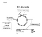
- RNA elements eRNA
- RNA elements eRNA
- RNA elements eRNA
- the RNA elements induce interferon production through activation of cellular RNA receptors, including but not limited to RIG-I and MDA5.
- Preferred RNA element compositions for activating RIG-I and MDA5 cytoplasmic RNA receptors without activating PKR are disclosed. Due to the small size of an RNA expression element (100-500 bp), multiple elements can be included within a single plasmid.
- Composite RNA elements comprising two dissimilar RNA elements that synergistically activate RIG-I and increase protein antigen expression are disclosed.
- Plasmid encoded adenoviral VARNAI is disclosed as a potent MDA5 activator. Since RNA does not induce adaptive immune responses against itself, the response to RNA containing vectors is not dependent on prior exposure of the patient, and can therefore be utilized repeatedly, without inducing immune responses to the vector backbone.
- DNA vaccine plasmids for inducing immune responses through DNA vaccination. It is a purpose and/or objective ofthe present invention to provide expression plasmids for gene therapy.
- Another disclosure is improved DNA vaccine plasmid compositions that, compared to plasmids defined in the art such as VR1012, pVAX1, pVC0396, pCMVkm2, pITR, pPJV7563, pWG4303, or pCOR vectors, or their derivatives, are improved by: increased eukaryotic expression of target antigen by incorporation of a chimeric CMV promoter-HTLV R-U5 -synthetic rabbit ⁇ globin 3' intron acceptor; and smaller size by elimination of all extraneous sequences such as additional sequences flanking the prokaryotic origin or antibiotic resistance genes as a consequence of using the nearest useful restriction sites for vector construction.
- compositions to improve the efficacy of DNA vaccination are used to improve the effectiveness of DNA vaccines by increasing target gene expression compared to DNA vaccine plasmids defined in the art.
- Novel compositions of expressed RNA-containing DNA vaccine vector backbones that improve the immunogenicity of DNA vaccination are disclosed.
- the vector backbones of the current invention encode novel, expressed immunostimulatory RNA compositions, in addition to the optimized protein antigen expression element. These expressed RNA sequences activate interferon production and innate immune response through cellular RNA receptors.
- RNA element compositions that induce natural interferon production in an individual.
- RNA expression element typically 100-500 bp
- multiple elements can be included within a single plasmid; in the case of multiple plasmid immunization, different RNA elements may be included on each plasmid.
- Yet another objective and/or purpose of the invention are to further increase immunostimulation by utilization of composite RNA elements comprising two dissimilar RNA elements. These composite RNA elements unexpectedly synergistically activate cytoplasmic RNA receptor RIG-I. Since RNA does not induce adaptive immune responses to itself, the response to RNA containing vectors is not dependent on prior exposure of the immunized individual, and can therefore be utilized repeatedly, without inducing immune responses to the vector backbone.
- the vectors combine reduced size, sequence content, and antibiotic free selection (which improves regulatory compliance and potency), with improved plasmid production yield, plasmid production integrity, eukaryotic gene expression, genome recombination propensity (safety), and regulation of innate immune responses by RNA elements.
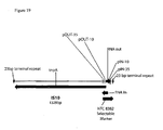
- FIG. 1 an immunostimulatory influenza H5 HA 'bird flu' pDNAVACCultra plasmid (NTC8382-41H-HA) is shown.
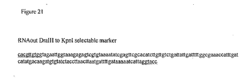
- the RNA-OUT selectable marker is cloned as a 140 bp DraIII-KpnI fragment, replacing the kanR gene in the otherwise equivalent NTC7382 vector.
- the immunostimulatory RNA element (41H) is cloned between the Eukaryotic terminator and the prokaryotic replication origin in this case.
- the RNA element could be cloned at other permissive sites in the plasmid, for example at either end of the selectable marker.
- RNA element could be cloned at permissive sites in other gene delivery vehicles, including viral (e.g . alphavirus, poxvirus, lentivirus, retrovirus, adenovirus, adenovirus related virus, etc) and nonviral (e.g . plasmid, midge, transcriptionally active PCR fragment, minicircles, bacteriophage, etc) vectors.
- viral e.g . alphavirus, poxvirus, lentivirus, retrovirus, adenovirus, adenovirus related virus, etc
- nonviral e.g plasmid, midge, transcriptionally active PCR fragment, minicircles, bacteriophage, etc
- Immunostimulatory RNA may improve innate or adaptive immune responses to the vector encoded antigen through activation of cytokine or chemokine signals, upregulation ofMHC molecules or proteosome peptide processing in response to type I interferon signaling, etc. Immunostimulatory RNA may also increase antigen expression through increasing transcription from the plasmid promoter, decreasing translation shutdown or mRNA degradation or editing ( e.g.
- FIG. 3 immunostimulatory RNA mediated activation of multiple signaling pathways to produce type I interferon and/or inflammatory cytokines is shown.
- Plasmid-produced immunostimulatory RNA may induce IPS-I (also called MAVS, VISA or Wales) through RIG-I and/or mda-5 (RIG-I-like receptors, RLRs) activation, which leads to type I interferon production and activation of proinflammatory cytokines ( Kawai T, Takahashi K, Sato S, Boban C, Kumar H, Kato H, Ishii KH, Takeuchi O, Akira S. 2005 Nature Immunol. 6: 981-988 ).
- Immunostimulatory RNA could also activate endosomally localized TLR3 or TLR7/8 (TLRs) after autophagy or (in non-transfected cells) through phagocytosis, pinocytosis, or receptor mediated uptake after cell death. Immunostimulatory RNA may also potentially activate interferon production through PKR or OAS signaling through undefined pathways (? in Figure). As well, immunostimulatory RNA could induce interferon production through activation of NALP3 or other inflammasome receptors. Up-regulation of MHCI receptors on cells responding to type I interferon may increase antigen presentation and improve adaptive immune responses.
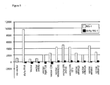
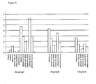
- FIG. 6 optimization of eRNA11 is shown, as determined by hRIG-I mediated activation of the Interferon ⁇ promoter in a one step assay. Blunt 5' end (11a) is optimal. hRIG-I interferon induction (Y axis, luminescence from ⁇ interferon-luciferase reporter) and EGFP expression (Y axis, fluorescence from EGFP DNA vaccine plasmid) for each construct (X axis) is shown.
- a blunt 5' end is demonstrated as optimal for human or murine RIG-I activation in composite eRNA in a one step assay.
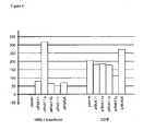
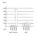
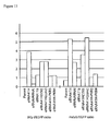
- FIG. 11 RIG-I and MDA5 activation by VARNAI component of eRNA41H is shown in a one step assay.
- eRNAVA Synergy of eRNA11a and VARNAI (eRNAVA) for RIG-I activation observed in cis (eRNA41H) or trans (11a+VA).
- 40 ng of each test plasmid was utilized per transfection well along with 150 ng of an internal control EGFP plasmid to standardize for transfection efficiency.
- VARNAI is a potent MDA5 activator and VAI component accounts for bulk of strong MDA5 activation observed with eRNA41H.
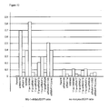
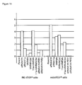
- FIG 12 the synergy of eRNA11a and VARNAI is shown. 40 ng of each test plasmid was utilized per transfection well along with 150 ng of an internal control EGFP plasmid to standardize for transfection efficiency and 20 ng each of RIG-I and MDA5 receptors (left) or no receptors (right) in a one step assay. Synergy is observed in cis (eRNA41H) or trans (11a+VA) with or without receptors. Ratio of Luminescence ⁇ interferon promoter activation) to fluorescence (EGFP internal control) (Y axis) versus constructs (x axis) is shown.
- RIG-I activation is shown to require specific RNA structures.
- Luminescence (RIG-I activation) to fluorescence (EGFP) ratio (Y axis) versus constructs (x axis) is shown.
- dsRNAHI-U6 is construct H1- mU6 (Stul) INF dsRNA repaired.
- eRNA41H-PKR3 and eRNA41H-PKR4 vectors are shown to have eliminated both MDA5 activation and synergistic RIG-I activation in a one step assay
- Synergy of eRNA41H (eRNA11a and VARNAI) for RIG-I activation is eliminated in eRNA41H-PKR3 and eRNA41H-PKR4.
- 40 ng of each test plasmid was utilized per transfection well along with 150 ng of an internal control EGFP plasmid to standardize for transfection efficiency.
- eRNA41H-PKR3 and eRNA41H-PKR4 VARNAI central domain substitutions eliminate MDA5 activation observed with eRNA41H.
- eRNA41H-PKR1 and eRNA41H-PKR2 vectors are shown to have eliminated both MDA5 activation and synergistic RIG-1 activation in a one step assay
- Synergy of eRNA41H (eRNA11a and VARNAI) for RIG-I activation is eliminated in eRNA41H-PKR1 and eRNA41H-PKR2.
- 40 ng of each test plasmid was utilized per transfection well along with 150 ng of an internal control EGFP plasmid to standardize for transfection efficiency.
- eRNA41H-PKR1 and eRNA41H-PKR2 VARNAI variants eliminate MDA5 activation observed with eRNA41H.
- RIG-I activation by composite eRNA vectors is demonstrated to be independent of vector backbone and eRNA orientation.
- the results of a one step RIG-I activation assay with 40, 20 10 or 5 ng per well of the indicated plasmids along with 150 ng of an internal control EGFP plasmid to standardize for transfection efficiency.
- Luminescence (RIG-I activation) to fluorescence (EGFP) ratio (Y axis) versus constructs (x axis) is shown.
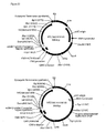
- Vector I pDNAVACCUltra SV40 CMV HLTV-1 R-U5 Rabbit ⁇ globin intron NTC-HA
- Vector 2 RNA-OUT antibiotic free version of Vector I
- Vector 3 pDNAVACCUltra SV40 CMV NTC-HA.
- RNA-OUT sequence as adapted for cloning into DNA vaccine plasmid is shown. Flanking DraIII (cacgttgtg) and Kpnl (ggtacc) sites are underlined.
- RNA-OUT-41H-HA analytical data including production yield from an RNA-OUT influenza DNA vaccine plasmid fermentation
- ADAR Adenosine deaminase acting on RNA (RNA binding protein which edits RNA content from A to I).
- Ad5 Adenovirus serotype 5.
- nucleic acid adjuvant Includes nucleic acid adjuvants (e.g. immunostimulatory eRNA, immunostimulatory CpG DNA, expression vectors producing cytokines, chemokines, etc) or other molecules such as cell death promoters (e.g. herpes thymidine kinase) and chemical adjuvants which are compounds that can enhance, prolong or otherwise modulate antigen-specific immune responses when administered with a vaccine antigen.
- nucleic acid adjuvants e.g. immunostimulatory eRNA, immunostimulatory CpG DNA, expression vectors producing cytokines, chemokines, etc
- cell death promoters e.g. herpes thymidine kinase
- chemical adjuvants which are compounds that can enhance, prolong or otherwise modulate antigen-specific immune responses when administered with a vaccine antigen.
- APC Antigen Processing Cell, for example, langerhans cells, plasmacytoid or conventional dendritic cells.
- BAC Bacterial artificial chromosome.
- Costimulatory molecules Costimulatory plasmids (e.g. IL12) or molecules, cell death inhibitors (e.g . antiapoptotic proteins) or enhancers as know in the art and included herein by reference.
- cmv Cytomegalovirus.
- CTL Cytotoxic T lymphocyte.
- Methods to deliver gene vectors [e.g. poly(lactide-co-glycolide) (PLGA), ISCOMs, liposomes, niosomes, virosomes, chitosan, and other biodegradable polymers, electroporation, sonoporation, ultrasound, gene gun, microneedles, naked DNA injection, hydrodynamic delivery, high pressure tail vein injection, needle free biojector, liposomes, microparticles, microspheres, nanoparticles, virosomes, bacterial ghosts, bacteria, attenuated bacteria, etc] as know in the art and included herein by reference.
- gene vectors e.g. poly(lactide-co-glycolide) (PLGA), ISCOMs, liposomes, niosomes, virosomes, chitosan, and other biodegradable polymers, electroporation, sonoporation, ultrasound, gene gun, microneedles, naked DNA injection, hydrodynamic delivery, high pressure
- DNA replicon Plasmids, cosmids, bacterial artificial chromosomes (BACs), bacteriophages, viral vectors and hybrids thereof.
- dsRNA Double stranded RNA.
- E. coli Escherichia coli, a gram negative bacteria.
- EGFP Enhanced green fluorescent protein.
- eRNA RNA element.
- the eRNA promoter may be a Pol I, Pol II or Pol III promoter, and the expressed RNA may be a single stranded RNA, double stranded RNA, hairpin RNA, microRNA, RNA aptamer or a ribozyme.
- HA Hemagglutinin.
- hdsRNA Hairpin double stranded RNA.
- Antigen reactive cellular e.g. antigen reactive T cells
- antibody e.g. antigen reactive IgG
- immunostimulatory RNA element immunostimulatory eRNA.
- intracellular targeting Directing a target antigen to either an inter or intracellular destination, or the antigen presentation pathway, using a targeting sequence.
- IRF-3 Interferon Regulatory Factor 3.
- MDA5 MDA-5, Melanoma differentiation-associated gene 5, a helicase containing cytoplasmic dsRNA receptor.
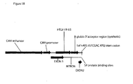
- NTC7382 promoter A chimeric promoter comprising the CMV enhancer-CMV promoter-HTLV R-U5 -synthetic rabbit ⁇ globin 3' intron acceptor -exon 2-SRF protein binding site-kozak sequence, with or without an upstream SV40 enhancer.
- NTC7382-41H-M or NTC7382-41H-M-HA.
- This is the NTC7382 backbone, containing the eRNA41H RNA element (41H), with (HA) or without the H5 HA antigen.
- M refers to insertion of a 1kb noncoding region (the mitochondrial R-region) which contains the mitochondrial bidirectional promoter for the synthesis ofpolycistronic RNA for the heavy and light strands as well as the replication origin for the heavy chain.
- NTC- HA NTC7382-HA, NTC8382-HA, NTCl-HA, NTC2-HA RNA-out-HA vectors, with or without eRNA.
- DNA vaccine vectors encoding the influenza H5 HA 'bird flu' DNA vaccine vectors that contain a synthetic HA gene maintaining the influenza H5N1 'bird flu' A/Vietnam/1203/04 amino acid sequence.
- the HA gene was human codon-optimized to reduce genome homology.
- the multiple basic amino acid motifs at the HA cleavage site correlative with pathogenicity was replaced with the corresponding region from several nonpathogenic influenza strains.
- OAS 2'5' oligoadenylate synthetase (RNA binding protein that activates RNase L).
- pDNA Plasmid DNA.
- PKR Interferon-induced double-stranded RNA-dependent protein kinase, a cytoplasmic dsRNA receptor.
- pm91 A VARNAI derivative (PKR5), in which VARNAI has a I bp change (pm91) in the central domain ( Rahman A, Malhotra P, Shar R, Kewalramani T, Thimmapaya B 1995 J Virology 69: 4299-4307 ) that is inactive for PKR inhibition, but (as disclosed herein) retains MDA5 activation.
- Plasmids Plasmids, cosmids, bacterial artificial chromosomes (BACs), bacteriophages, viral vectors and hybrids thereof.
- 5'-PPP 5'-triphosphate that is present on viral transcripts and some DNA polymerase III transcripts.
- 5'-PPP RNA is a known activator of RIG-I.
- pUC origin pBR322-derived origin, with G to A transition that increases copy number at elevated temperature.
- RIG-I Retinoic acid inducible gene I, a cytoplasmic helicase containing RNA receptor.
- DeNy Dominant Negative hRIG-I.
- RNA element an expression cassette containing an expressed RNA sequence.
- the promoter may be a Pol I, Pol II or Pol III promoter, and the expressed RNA may be a single stranded RNA, double stranded RNA, hairpin RNA, microRNA, RNA aptamer or a ribozyme.
- RNA-OUT Insertion sequence 10 (IS10) encoded RNA-OUT, an antisense RNA that hybridizes to, and reduces translation of, the transposon gene expressed downstream of RNA-IN.
- IS10 Insertion sequence 10
- An RNA-OUT vector selectable marker DNA fragment that is compatible with DraIII and Kpnl is disclosed herein.
- RT-PCR Real time PCR.
- sacB Structural gene encoding Bacillus subtilis levansucrase. Expression of sacB in gram negative bacteria is toxic in the presence of sucrose
- SEAP Secreted alkaline phosphatase.
- shRNA Short hairpin RNA.
- siRNA Short inhibitory RNA.
- ssRNA Single stranded RNA.
- Target antigen Immunogenic protein or peptide epitope, or combination of proteins and epitopes, against which a immune response can be mounted.
- Target antigens may by derived from a pathogen for infectious disease applications, or derived from a host organism for applications such as cancer, allergy, or autoimmune diseases.
- Target antigens are well defined in the art. Some examples are disclosed in Williams, Supra, 2006 and are included herein by reference.
- TE buffer A solution containing approximately 10mM Tris ⁇ pH 8 and 1 mM EDTA.
- T H 1 T helper 1.
- T H 2 T helper 2.
- TLR Toll-Like Receptor.
- VARNA Adenoviral virus associated RNA, including VARNAI (VAI or VA1) and or VARNAII (VAII or VA2) from any Adenovirus serotype, for example, serotype 2, serotype 5 or hybrids thereof.
- VARNAI Adenoviral virus associated RNAI, also referred to as VAI, or VA1, from any Adenovirus serotype, for example, serotype 2, serotype 5 or hybrids thereof.
- a gene delivery vehicle including viral (e.g . alphavirus, poxvirus, lentivirus, retrovirus, adenovirus, adenovirus related virus, etc) and nonviral (e.g . plasmid, midge, transcriptionally active PCR fragment, minicircles, bacteriophage, etc) vectors. These are well known in the art and are included herein by reference.
- viral e.g . alphavirus, poxvirus, lentivirus, retrovirus, adenovirus, adenovirus related virus, etc
- nonviral e.g plasmid, midge, transcriptionally active PCR fragment, minicircles, bacteriophage, etc
- the invention relates to compositions and methods for gene therapy, genetic immunization or interferon therapy.
- the invention is practiced in the expression of gene products in eukaryotic cells, for the purpose of gene therapy, genetic immunization or interferon therapy.
- the invention applies to such use of ccc recombinant DNA molecules such as plasmids, cosmids, BACs, bacteriophages, viral vectors and hybrids thereof (herein collectively referred to as plasmids).
- a pDNAVACCUltra vector is used to express a gene product in an organism for therapy or vaccination.
- an influenza H5 hemagglutinin (HA) pDNAVACCUltra vector (NTC8382) with an immunostimulatory RNA element (41H) is shown in Fig. 1 .
- Kanamycin resistance marker-containing pDNAVACCUltra vectors have been disclosed in Williams, Supra, 2006 and are included herein by reference.
- the prokaryotic replication origin is protected by a downstream prokaryotic transcriptional terminator to improve stability and yield with a broad range of target genes.
- Unique restriction sites flank the prokaryotic modules, to allow easy modifications.
- the resulting vector backbone is much smaller than existing vectors such as VR1012 (5kb, versus 3-3.5 kb for the pDNAVACC vectors, or 2.8 kb for the NTC83 82 backbone (disclosed herein), consisting of replication origin-RNA selectable marker-SV40 enhancer-NTC7382 chimeric CMV promoter), and yet it drives higher levels of expression.
- the pDNAVACCUItra vector used to express a gene product for therapy or vaccination contains a selectable RNA marker ( Fig. 1 ) rather than an antibiotic gene. This further reduces the vector size and complies with regulatory agencies guidance to eliminate antibiotic resistance markers from therapeutic and vaccine vectors.
- the RNA based selectable marker of the invention is incorporated into other DNA vaccine or therapeutic plasmid backbones, a non limiting list includes VR1012,pVAX1, pVC0396, pCMVkm2, pITR, pCOR, pPJV7563, pWG4303 and derivatives.
- a pDNAVACCUltra vector incorporating a chimeric promoter is utilized.
- Preferred chimeric promoters are CMV or SV40-CMV promoters wherein the SV40 enhancer is fused upstream ofthe CMV promoter as disclosed in Williams, Supra, 2006.
- the CMV or SV40-CMV promoter may be linked downstream to a leader sequence and the antigen gene, or preferably, a leader sequence containing an intron, and then the antigen gene.
- the leader sequence containing an intron is the native CMV exon1- intron 1, or a synthetic intron such as is disclosed in Williams, Supra, 2006, or the HTLV-1 R-U5 sequence (CMVFR) as disclosed by Barouch et al, Supra, 2005, or the HTLV R-U5 -synthetic rabbit ⁇ globin 3' intron acceptor-SR protein binding site (NTC7382 promoter) disclosed herein.
- the pDNAVACCUltra vector used to express a gene product for therapy or vaccination contains the NTC7382 promoter ( Fig. 1 ) rather than a standard CMV promoter, to increase expression of the gene product.
- the NTC7382 promoter further comprises an upstream SV40 enhancer.
- the NTC7382 promoter of the invention is incorporated into other DNA vaccine or therapeutic plasmid backbones.
- a non-limiting list includes: VR1012, pVAX1, pVC0396, pCMVkm2, pITR, pCOR, pPJV7563, pWG4303 and derivatives.
- the NTC7382 promoter of the invention is incorporated into viral (e.g. alphavirus, poxvirus, lentivirus, retrovirus, adenovirus, adenovirus related virus, etc) or nonviral (e.g . plasmid, midge, transcriptionally active PCR fragment, minicircles, bacteriophage, etc) ⁇ vectors.
- immunization is performed using a viral vector containing one or more immunostimulatory RNA elements of the invention.
- the viral vector is an alphavirus, poxvirus, lentivirus, retrovirus, adenovirus or adenovirus related virus.
- immunization is performed using a nonviral vector or DNA fragment containing one or more immunostimulatory RNA elements ofthe invention.
- immunization is performed using a pDNAVACCUltra vector containing one or more immunostimulatory RNA elements of the invention.
- the immunostimulatory RNA element embodiments of the invention are incorporated into the backbone of the vector or DNA fragment.
- the immunostimulatory RNA sequences activate innate immune responses ( Fig. 2 ). In another preferred embodiment, the immunostimulatory RNA sequences activate innate immune responses through cytoplasmic RNA receptors RIG-I and/or MDA5 ( Fig. 3 ) or the inflammasome. In another preferred embodiment, the immunostimulatory RNA sequences activate innate immune responses through endosomal RNA receptors TLR3, TLR7 or TLR8 in the transfected cells.
- the immunostimulatory RNA sequences activate innate immune responses through endosomal RNA receptors TLR3, TLR7 or TLR8 in non transfected cells after apoptosis, necropsy, or phagocytosis of the transfected cell.
- the immunostimulatory RNA sequences activate adaptive immune responses against the vector-expressed target antigen.
- the target antigen is targeted to either an inter- or intra-cellular destination, or the antigen presentation pathway, using a targeting sequence.
- targeting sequences A non-limiting list of targeting sequences is disclosed in Williams, Supra, 2006, and included herein by reference.
- the immunostimulatory RNA sequences act to improve expression of the gene of interest from the plasmid.
- RNA element containing nonviral vector or DNA fragments will not induce adaptive immune responses against the vector backbone (i.e. reusable vectors unaffected by prior immune exposure). This is a significant advantage over alternative vectors such as alphaviral replicon or adenoviral vectors.
- non-viral or viral vectors or DNA fragments containing immunostimulatory RNA sequences are used to increase anti-viral, anti-tumor and or antibacterial activity in a vertebrate, or cells of a vertebrate.
- the immunostimulatory RNA sequences increase anti-viral, anti-tumor and or antibacterial activity in a vertebrate, or cells of a vertebrate through induction of type I interferon production.
- vectors or DNA fragments containing immunostimulatory RNA sequences are used in treatment to induce production of natural type I interferon's in the cells of an individual to whom the administration of type 1 interferon's would be beneficial.
- the immunostimulatory RNA may be delivered to a cell in a DNA fragment or vector with or without a co-delivered antigen gene.
- the immunostimulatory RNA may be delivered to cells via a plurality of vectors, a non limiting list including a PCR fragment, a Midge vector, a non-viral vector, or a viral vector.
- the vector embodiments of the invention may be delivered to a cell either in vivo, in vitro or ex vivo, utilizing one or more of the plurality of delivery methods known in the art.
- a non limiting list of delivery methods includes: naked DNA or formulated DNA, needle or needle free delivery, delivery enhancement by electroporation, sonoporation, ultrasound, hydrodynamic, lipofection, cationic polymers, gene gun, nanoparticles, microparticles, cationic lipids, or microneedles.
- delivery may be by oral, buccal, nasal, inhalation, topical, vaginal, rectal, intravenous, intramuscular, intradermal, epidermal, systemic, or by other routes of administration known in the art.
- Cytoplasmic dsRNA can directly induce dsRNA receptors ADAR, PKR, 2'-5' oligoadenylate synthetase (OAS), retinoic acid inducible gene I (RIG-I) and melanoma differentiation-associated gene 5 (MDA5).
- ADAR dsRNA receptors
- PKR 2'-5' oligoadenylate synthetase
- RIG-I retinoic acid inducible gene I
- MDA5 melanoma differentiation-associated gene 5
- PKR contains two dsRNA binding domains (both of which must be occupied for activation) and recognizes primarily long (>30 bp) dsRNAs. PKR activation is inhibited by a variety of structured viral RNAs, such as adenovirus virus-associated RNAI (herein referred to as VARNAI, VAI or VAI).
- adenovirus virus-associated RNAI herein referred to as adenovirus virus-associated RNAI
- RIG-I and MDA5 each contain a single dsRNA binding domain.
- MDA5 recognizes RNA from positive sense ssRNA Picornavirus (EMCV) and poly(I:C) ( Kato, H, Takeuchi O, Sato S, Yoneyama M, Yamamoto M, Matsui K, et al. 2006. Nature 441: 101-105 ; Gitlin L, Barchet W, Gilfillan S, Cella M, Beutler B, Flavell RA, Diamond MS, Colonna M. 2006. Proc. Natl. Acad. Sci.
- EMCV positive sense ssRNA Picornavirus
- poly(I:C) Kato, H, Takeuchi O, Sato S, Yoneyama M, Yamamoto M, Matsui K, et al. 2006. Nature 441: 101-105 ; Gitlin L, Barchet W, Gilfillan S, Cella M, Beutler B, Flavell RA, Diamond MS, Colonna M. 2006. Pro
- RNA from positive sense ssRNA flavivirus Japanese encephalitis virus, dengue, hepatitis
- negative sense RNA virus Orthomyxoviridae (Influenza) Rhabdovirus (Vesicular stomatitis virus) and paramyxoviruses (Sendai virus, Newcastle disease)
- Chang TH Liao CL, Lin YL. 2006.
- dsRNA short siRNA, and long dsRNA that arise during viral replication
- specific structured RNAs such as the hepatitis C virus (HCV) internal ribosome entry site (IRES)
- HCV hepatitis C virus
- IRS internal ribosome entry site
- RIG-I activators Sumpter et al, Supra, 2005; Marques et al, Supra, 2006.
- the optimal ligands are unknown.
- Marques et al, Supra, 2006 teach that siRNAs ranging from 21 to 27 nucleotides in length activated the interferon system when they lacked 2-nucleotide 3' overhangs, a characteristic of Dicer products.
- a 5'-triphosphate (5'-PPP) group increases RIG-I activation ( Pichlmair A, Schulz O, Tan CP, Naslund TI, Liljestrom P, Weber F, Sousa C. 2006. Science 314:997-1001 ; Hornung et al, Supra, 2006).
- Such 5'-triphosphates are generally removed from, or masked on, self RNA species; this is a structural basis for the distinction between self and non-self RNA (Reviewed in Bowie AG, Fitzgerald KA, 2007 Trends Immunol 28: 147-150 ).
- RIG-I ligand is the cytosolic 5'-PPP containing viral leader transcript of measles virus; the 5'-PPP is required for activation but not RIG-I binding ( Plumet S, Herschke F, Bourhis JM, Valentin H, Longhi S, Gerlier D. 2007. PLoS 2:e279 ).
- RNA may be double stranded RNA or may be structured single stranded RNA and activation is stronger if the RNA contains a 5'-triphosphate.
- the optimal length, degree of secondary structure, and end structure is unknown.
- RNAs that RIG-I detects are generally produced in the cytoplasm using viral RNA polymerases that do not remove the 5' triphosphate.
- proteins bound to the 5'-PPP may mask the RNA from RIG-1 detection (such as with endogenous 5S ribosomal RNA which is ribosome associated after nucleosome assembly).
- Careful design is needed to obtain 5'-PPP cytoplasmic RNAs using the endogenous nuclear transcription and nuclear export capacities of a cell needed for expression from a DNA vaccine plasmid.
- an appropriate promoter must be utilized such that the 5'-PPP is not removed by capping (mRNA) or processing (e.g . tRNA, rRNA).
- the RNA must then be exported from the nucleus to the cytoplasm since plasmid transcription will occur in the nucleus while the RIG-I receptor is in the cytoplasm.
- the 5'-PPP containing cytoplasmic RNA must then act as a ligand and activator of RIG-I.
- RNA polymerase III RNA polymerase III promoters. These promoters are small, express to levels up to 10 fold higher than pol II promoters, do not interfere with pol II directed antigen expression (See Williams, Supra, 2006) and have the potential to retain 5'-triphosphates.
- the U6 small nuclear RNA (snRNA) expressed from the U6 promoter contains a gamma-monomethyl phosphate which is distinct from other RNA caps ( Singh R, Reddy R 1989 Proc.
- RNA promoters such as tRNAVa1 promoters will generally produce cytoplasmic RNAs lacking 5'-PPP, since normal processing of tRNAs in the nucleus removes the 5' triphosphate.
- DNA polymerase II promoters will also generally produce cytoplasmic RNAs lacking 5'-PPP, since 5'-PPP on mRNA is removed during capping prior to nuclear export.
- DNA polymerase I promoters will, likewise, not produce unmodified cytoplasmic RNA, since rRNA is heavily processed and modified in the nucleolus prior to nuclear export.
- DNA polymerase III promoters restricts the ligands that can be expressed, since transcription is terminated at runs of 4 or more T residues in the gene, and only short RNAs (generally ⁇ 300 bp) can be made.
- RNA nuclear export: To be immunostimulatory, the RNA must be exported from the nucleus to the cytoplasm (RIG-I and MDA5 proteins are cytoplasmic). Export of Pol III transcripts can be mediated by exportin-t (tRNAs) or exportin-5 (minihelix containing RNAs such as shRNA and VAI) (reviewed in Rodriguez MS, Dargemont C, Stutz F. 2004. Biol Cell 96: 639-655 ). In the case of minihelix, RNA, such as the HCV IRES RIG-I ligand could be inserted within the VAI transcript, in a manner that preserves the minihelix configuration.
- tRNAs exportin-t
- minihelix containing RNAs such as shRNA and VAI
- Nuclear export of long dsRNA Long annealed dsRNA is not exported from the nucleus. Mammalian genomes contain many natural sense-antisense transcripts that have the potential to produce dsRNA Long nuclear dsRNA is a substrate for ADAR. Export of the resultant extensively Adenosine to Inosine edited RNA from the nucleus is inhibited ( e.g. by binding to p54nrb; reviewed in Wang and Carmichael, Supra, 2004). Thus, the art teaches that long dsRNA (>30 bp) expressed in the nucleus will be edited by ADAR, and subsequently will not be exported to the cytoplasm.
- a plasmid vector containing a long dsRNA (250bp) expressed by convergent transcription of U6 and H1 promoters did not activate PKR or the interferon response after transfection and expression of the dsRNA ( Strat A, Gao L, Utsuki T, Cheng B, Nuthalapaty S, Mathis JM, Odaka Y, Giordano T. 2006. Nucl. AcidRes. 34:3803-3810 ).
- RIG-I ligands Several RIG-I ligands have been described in the art and several DNA vaccine RNA element compositions containing them are disclosed and tested herein. These include:
- DNA vaccine vectors containing expressed hairpin double stranded (hdsRNA) sequences similar to those described in Marques et al, Supra, 2006 have been disclosed (Williams, Supra, 2006) and are included herein by reference.
- the pDNAVACCUltra vaccine vectors were modified to encode pol III promoter cassettes that can be used to clone a target RNA (eRNA cassette) to produce RNA upon cell transfection.
- DNA vaccines containing U6 promoter-driven hdsRNAs were tested in vitro (lipofectamine 2000 transfection into RAW 264.3 mouse macrophage cell line) for induction of TNF ⁇ secretion.
- DNA vaccine plasmids containing RNA elements that produce immunostimulatory RNA can potentially signal through TLR3 (dsRNA), TLR8/9 (ssRNA) or activate cytoplasmic RNA receptors RIG-I or MDA5, PKR, ADAR, or OAS induced RNaseL pathway, or nuclear ADAR.
- RNA elements that either activate, or don't activate, PKR.
- PKR activation such as occurs with long dsRNA produced by alphaviral amplicon vectors, leads to shutdown of protein translation and reduced antigen expression. Therefore, one novel class of composite RNA elements would not activate PKR.
- One composition to accomplish this would be to include a PKR inhibitor RNA along with an immunostimulatory RNA, to maximize immunostimulation without loss of antigen expression (Williams, Supra, 2006).
- RNA-based inhibitors of PKR are described in the art.
- VAI adenoviral encoded RNAI
- VAII adenoviral encoded RNAI
- VARNAI is a potent inhibitor of PKR, a weak inhibitor of ADAR, and a weak activator of DAS (1/10 as strong as dsRNA; Desai SY, Patel RC, Sen GC, Malhotra P, Ghadge GD, Thimmapaya B. 1995. J Biol. Chem 270: 3454-3461 ).
- RNA element vectors were constructed, incorporating the design-considerations outlined above.
- the Mfold program of Zuker, M. 2003. Nucleic Acids Res.31:3406-15 was utilized to verify all constructs retained required folding structures (e.g . all tRNA fusions did not interfere with the tRNA structure, or all VARNAI or other minihelix constructs were predicted to retain the terminal minihelix).
- VARNAIA is transcribed clockwise relative to the pDNAVACCUltra vector ( Fig. 1 ) and comprises sequences 10,597-10,790 of the Ad5 genome (Genbank accession number X02996).
- the VAI +VAII clone comprises sequences 10,589-11,049 of the Ad5 genome.
- VA RNAI is a internally Pol III driven highly structured RNA that inhibits the translation blocking activity of PKR.
- the VARNAIA clone was modified to a stable hairpin structure replacing the central domain (critical for PKR inhibition) with an acceptor hairpin structure. As well the 3' end was modified to be blunt.
- the final clone was the same sequence as the VA1-535 chimera disclosed in Thompson JD 2005 US Patent US6852535 .
- eRNA10 eRNA7, with HCV IRES inserted (VARNA1-S35 +HCV IRES)
- the cellular RIG-I dsRNA sensor is activated by structured viral RNAs, such as the HCV IRES.
- structured viral RNAs such as the HCV IRES.
- This 200 bp IRES does not contain runs of four or more T's so it can be made by RNA polymerase III.
- the HCV IRES (Sumpter et al, Supra, 2005) was PCR amplified (from plasmid pLCSR2HCV), and cloned into the VARNAI S35 Acll site (as an internal fusion with the ERNA7 VARNAI-S35 RNA).
- eRNA23 eRNA7: (VAI minihelix)
- eRNA24 eRNA10, (VAI minihelix)
- the S35 constructs do not have an extended 3' end on the terminal stem, due to deletion of a CTCC sequence from VA1. This may affect nuclear export.
- the CTCC sequence was reintroduced into the S35-based constructs eRNA7 and eRNA10, As well, the terminator was extended from the minimal 4T to 6T.
- the 3' end of VA1 is shown below, with the 6 bp insert in eRNA23 and 24 double underlined.
- the 4 TTTT terminator in eRNA7 and 10 is after the 6 bp insert.
- eRNA31B-C VARNAIA-shRNA constructs
- pDNAVACCUltra VARNAI was modified to express shRNA structures from the 3' end. Such constructs were made to contain a Lamin shRNA (31C) exactly as described in Pebemard S, Iggo RD. 2004. Differentiation 72: 103-111 , or the RNA9.2 shRNAs (31C; Williams, Supra, 2006).
- HCV 3' NTR (a known RIG-1 ligand; Saito et al, Supra, 2007) was cloned into the BstEII site of VARNAIA in eRNA42.
- a clone (INF dsRNA) containing a modified 100 bp fragment DNA of influenza (defined above; Lamb and Choppin, Supra, 1983) as a 100 bp hairpin was designed using synthetic oligonucleotides and cloned into HpaI/NheI digested pDNAVACCUltra5 MU6 (Stul) DNA vaccine vector (Williams, Supra, 2006).
- the presence of a 100 bp hairpin was toxic to E. coli and the fragment was uncloneable.
- the clone was redesigned to eliminate the hairpin, and to instead have a single insert transcribed by H1 and U6 from opposite sides. This creates a dsRNA, without need for a large, detrimental hairpin or repeat structure in the vector.
- INF dsRNA contained a 60 bp fragment of influenza DNA.
- INF dsRNA repaired This was repaired using oligonucleotides to contain the full 108 bp influenza fragment ( Fig. 4 ) .
- Pol III terminators (TTTTTT) are inserted before and after the 100 bp dsRNA such that transcription ends after the dsRNA insert.
- eRNA11 tRNAVal MU6 INF dsRNA (dsRNA contains recessed U6 start 5' end, U6 A start)
- the pDNAVACCUltra5 EGFP H1 INF dsRNA U6 (repaired) construct was modified using PCR to remove the H1 promoter, and replace it with the human tRNAVal and linker (using synthetic nucleotides).
- the human tRNAVal and linker sequence is as described (Kuwabara et al, Supra, 2001).
- the configurations ofthe insert and the sequences of the predicted annealed dsRNA for eRNA11 ((SEQ ID NO: 2), eRNA11A (SEQ ID NO: 3) and eRNA11B (SEQ ID NO: 4) are shown in Fig. 4 .
- eRNA11A tRNAVal MU6 TNF dsRNA (dsRNA contains blunt U6 start 5' end, U6 A start).
- the 3 bp insert in eRNA11 used to create eRNA11 a is bolded and underlined in Fig. 4 .
- eRNA11B tRNAVal MU61NF dsRNA (dsRNA contains recessed 5' end, U6 G start)
- the 6 bp insert modification from eRNA11 is bolded and underlined in Fig. 4 .
- eRNA11C eRNA11B tRNA-HCV 3' NTR-U6
- the 100 bp influenza sequence in eRNA11B was replaced with a 90 bp fragment of the highly structured HCV 3' NTR sequence (a known RIG-I ligand).
- eRNA11D eRNA11 tRNA-INF ssDNA
- base eRNA11 vector was modified to produce an ssRNA from the tRNA promoter only (by deletion of the downstream U6 promoter), to determine the contribution of dsRNA, if any, to interferon ⁇ induction.
- the dsRNA and U6 region were deleted and replaced with the HCV IRES.
- eRNA18 U6 TNF ssRNA
- tRNAVal MU6 INF dsRNA The tRNA gene from pDNAVACCUltra EGFP eRNA11 (tRNAVal MU6 INF dsRNA) was deleted to determine the contribution of dsRNA, and tRNA mediated nuclear export, if any, to interferon ⁇ induction.
- CTE constitutive transport element
- eRNA19 H1 EMCV polyC CTE
- eRNA20 H1 ECMV-polyC antisense
- EMCV and EMC-like viruses share structural characteristics in their dsRNA, such as the presence of a homopolymeric polyC acid tract within the 5'-untranslated sequence. These polyC tracts are retained during viral replication in vitro and in vivo and are associated with virulence. This region may account for activation ofMDA5 observed with EMCV viruses.
- Each vector will make a single strand.
- eRNA19 in combination with eRNA20 will make a double stranded RNA, with a CTE sequence on a single strand.
- eRNA34 H1 RTE CTE.
- This clone replaces the INF ssRNA in eRNA17 HI INF ssRNA CTE with a RTE CTE.
- RTEM26 mutation of a RNA Transport Element from a rodent intracisternal A particle retroelement, mechanism of action is unknown
- CTE a TAP/NXF1 binding export hairpin structure, that exports unspliced RNAs derived from simian type D retroviruses.
- the combination of the two is synergistic for nuclear export (Smulevitch et al, Supra, 2006).
- eRNA35 VARNA1A-U6 INF ssRNA O1 (convergent)
- eRNA36 VARNA1A-U6 INF ssRNA 02 (divergent)
- eRNA38 eRNA23-U6 INF ssRNA O1 (convergent)
- RNA elements combine VARNAIA and the U6 INFdsDNA tRNA (eRNA11) in different orientations:
- RNA elements combine VARNAIA and the U6 INFdsDNA tRNA (eRNA11) in different orientations:
- eRNA41d dsRNA contains recessed U6 start 5' end in combination with Adenoviral serotype 2 promoter box B in VARNAI (G to A change in box B resulting in Ad2 ttcgaaccc from Ad5 ttcgagccc).
- Example 1 The vectors from Example 1 were tested for RIG-1 activation using either a one or a two step assay. EGFP target gene expression was also monitored in the same experiments as a surrogate assay for PKR activation (PKR inhibits translation, leading to loss of antigen expression). All assays were performed using 24 well cell culture plates.
- pI25luc a reporter plasmid, with the luciferase gene driven by the interferon ⁇ promoter
- the cytoplasmic RNA receptor either human RIG-I (hRIG-I), murine RIG-I (mRIG-I), human MDA5 (MDA5) or dominant negative hRIG-I control (DeNy); pUNO-hRIG-I, pUNO-mRIG-I, pUNOI-hMDA5 or pDeNy-hRIG-I respectively, Invivogen, San Diego CA
- HEK293 American Type Culture Collection, Manassas, VA
- lipofectamine 2000 as described by the manufacturer (Invitrogen, Carlsbad, CA).
- EGFP EGFP RNA element
- luciferase interferon ⁇ activation
- 0.1 ug p125luc (a reporter plasmid, with the luciferase gene driven by the interferon ⁇ promoter), 0.1 ug of the cytoplasmic RNA receptor [either human RIG-I (hRIG-I), murine RIG-I (mRIG-I), human MDA5 (MDA5) or dominant negative RIG-I control (DeNy); pUNO-hRIG-I, pUNO-mRIG-I, pUNO1-hMDA5 or pDeNy-hRIG-I respectively] and 0.2 ug of the test plasmid were mixed in 50 uL DMEM (no serum) and cotransformed into human cell line HEK293 using lipofectamine 2000 (1.5 uL/50 uL DMEM) as described by the manufacturer (Invitrogen, Carlsbad, CA, USA).
- EGFP expression
- luciferase interferon ⁇ activation
- RIG-I activation was observed with the C26 and Bgal728 constructs. These constructs were disclosed in Williams, Supra, 2006 as inducers that dramatically improved the immunostimulatory activity of the pDNAVACCUltra vector (TNF ⁇ production) , without activating PKR mediated translation inhibition, after lipofectamine 2000 transfection into RAW 264.3 mouse macrophage cell line. The results disclosed herein demonstrate that these constructs are RIG-I activators.
- VARNAI containing DNA vaccines activated RIG-I, and were more potent activators than the C26 or Bgal728 constructs. While the related EBERI RNA is a RIG-I activator (Samanta et al, Supra, 2006) the activation observed with VARNAI was surprising, in light of the Weber et al, Supra, 2006 finding that while the pAdVantage VARNAI/II containing plasmid activate the interferon response in transfected cell lines, they do not activate the RIG-I/MDA5 pathway, or other known pathways ( i.e . no activation of IRF-3, which is a mediator of RIG-I signaling; Weber F, Wagner V, Kessler N, Haller O.
- VARNAI is preferable to EBER1, since EBERI binds multiple cellular proteins, and has documented growth stimulating properties. VA RNAI has not been attributed with these adverse properties, and is considered safe for vaccine use.
- EBERI binds multiple cellular proteins, and has documented growth stimulating properties.
- VA RNAI has not been attributed with these adverse properties, and is considered safe for vaccine use.
- VARNAI is included in the pAdVAntageTM Vector (Promega, Madison WI) plasmid backbone to increase target gene expression of cotransfected plasmids by this mechanism ( Groskreutz, D. Schenborn, E. 1994 Promega Notes 48, 8.12 ) as well as PKR independent stimulation of gene expression potentially through inhibition of RNA interference ( Andersson MG, Haasnoot PCJ, Xu N, Berenjian S, Berkhout B, Akusjarvi G. 2005. J. Virol. 79:9556-9565 ; Lu S. Cullen BR. 2004. J. Virol. 78: 12868-12875 ).
- VARNAI-including backbones have not been used to increase immunostimulation, or suggested for such an application, as is disclosed herein. Indeed, Weber et al, Supra, 2006 do not suggest this application, and instead speculated that the interferon inducing capacity of VA RNAs observed in cell lines is most likely negligible under natural conditions and hypothesize that VA RNAs may stabilize spurious levels of interferon ⁇ mRNA induced by DNA transfection.
- RNAe constructs Name + Pol III Promoter RNA Export Structure and orientation RNA EGFP RIG-I Activation Control None None None None None None None None None None None None None None None None None None None None None None None None None - mU6 ++ U6 None U6 ⁇ short RNA Unstructured - Bga728 ++ U6 minihelix U6 ⁇ Hairpin with microRNA loop dsRNA hairpin + C26 ++ U6 none U6 ⁇ PKR aptamer structured RNA + HI- mU6 INF dsRNA H1 and U6 None H1 ⁇ 60 bp RNA ⁇ U6 dsRNA ⁇ NA HI-mU6 INF dsRNA repaired H1 and U6 None H1 ⁇ 100 bp RNA ⁇ U6 dsRNA ⁇ + VARNAIA VAI minihelix VAI ⁇ structured RNA ⁇ ++ VAI 2x VAI minihelix ⁇ VAI ⁇ VAI structured RNA ++ VAINA
- VARNAI RNA Modifications that do not affect the integrity of the VARNAI RNA, such as serotype 5 to 2 swaps in the B box (e.g . eRNA41H to eRNA41J change), or extensions of the 5' flanking sequences (e.g . eRNA42), do not adversely affect RIG-I activation.
- serotype 5 to 2 swaps in the B box e.g . eRNA41H to eRNA41J change
- extensions of the 5' flanking sequences e.g . eRNA42
- the strongest RIG-I activator was eRNA11a (Table 1). This is a two component activator. It produces a single stranded RNA with a 5' tRNA from the tRNAVal promoter. The art teaches that the 5'-PPP will be removed from this RNA during tRNA processing, and the resulting RNA will be exported to the cytoplasm. Conversely, it is possible that this specific tRNAVal fusi on may have the novel property of export without processing, since a tRNA-riboryme fusion of Kuwabara et al, Supra, 2001 with the same linker was reported to have been exported to the cytoplasm without extensive 5' end removal.
- the observation of RIG-I activation with eRNA11D would be consistent with the tRNA retaining the 5'-PPP.
- the second component is the complementary strand of the dsRNA transcribed convergently using the U6 promoter. This second strand, by itself, shows RIG-I activation (eRNA18, Table1 ). While this short RNA may have a 5'-PPP, there is no obvious mechanism of cytoplasmic transport and U6 transcribed RNAs are typically localized to the nucleus.
- the composite construct, eRNA11 has much more potent RIG-I activation than the single component eRNA11 D or eRNA18 (Table 1) . While not limiting the application of embodiments of the invention, this may be due to dsRNA forming by hybridization of the two complementary strands, followed by tRNA mediated export of the dsRNA.
- a 5'-PPP may be retained on the U6 promoter transcribed strand.
- potential dsRNA configuration i.e . when annealed to the tRNA-opposite strand
- HCV 3' NTR a structured RNA
- eRNA11C lower RIG-I activation
- HCV 3' NTR a known RIG-I ligand
- in vitro transcribed RNA is a strong RIG-I activator.
- the results disclosed herein demonstrate that the RIG-I ligands taught in the art (HCV 3' NTR and HCV IRES identified from in vitro transcribed RNA or native viral RNA isolated from cells) do not function as such when cloned into plasmid backbones.
- ligands expressed from a plasmid need to be optimized for RIG-I activation, and that ligands defined in the art are not predictive of optimal ligands for inclusion in DNA vaccine plasmids.
- the failure of tho HCV 3' NTR to be a strong activator may be due to the high level of secondary structure; each strand may form stable secondary structures, which reduces annealing between strands, and subsequent tRNA mediated nuclear export.
- One skilled in the art can further optimize the existing RIG-I activating constructs reported herein, by varying the length of the RNA (e.g. from 20 to 1000 bp, preferably 30 to 200 bp; 200 bp RNA has been reported to show stronger RIG-I activation than 100 bp RNA (Kato et al, Supra, 2006), while 30 bp RNA will not be extensively edited by ADAR) and the base composition or degree of secondary structure, and then testing using the 1 step and/or 2 step RIG-I activation assays disclosed herein, or other RIG-I activation assays known in the art.
- the length of the RNA e.g. from 20 to 1000 bp, preferably 30 to 200 bp; 200 bp RNA has been reported to show stronger RIG-I activation than 100 bp RNA (Kato et al, Supra, 2006), while 30 bp RNA will not be extensively edited by ADAR) and the base composition or degree of secondary structure,
- RIG-I ligands were expressed from the human HI promoter (eRNA17, eRNA19, eRNA34; Table 1 ) .
- Additional HI constructs tested that did not activate RIG-I included HI HCV IRES CTE (eRNA21) alone or in combination with eRNA35 (as eRNA37, which showed no improvement compared to eRNA35 parent plasmid);
- eRNA32 HI EMCV-polyC RTE CTE (690 bp transcript);
- eRNA33 HI HCV IRES RTE CTE (840 bp transcript); eRNA27 HI EMCV polyC helix and eRNA28 HI HCV IRES helix (the 3' end of the EMCV or HCV IRES was modified to form a minihelix to allow nuclear export) ;
- eRNA21 HI HCV IRES CTE and eRNA22 HI HCV IRES antisense CTE, alone or in combination with each other;
- results disclosed herein teach: 1) pol III promoters for plasmid expressed RIG-I or MDA5 ligands are not equivalent, and that U6, tRNA and VARNAI promoters are superior to the HI promoter and; 2) RIG-I and MDA5 ligands taught in the art (identified from in vitro transcribed RNA or native viral RNA isolated from cells) do not function as such when cloned into plasmid backbones.
- RNA elements VARNAI, eRNA11, eRNA11a
- Example 2 The optimal RNA elements (VARNAI, eRNA11, eRNA11a), defined in Example 2, were then combined into a composite RNA element containing both individual components as described in Example 1.
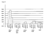
- the surprising, unexpected result of synergistic activation of RIG-I in the composite RNA elements is shown in Table 2, and Figs. 7-10 .
- the composite structure eRNA41 eRNA11 and VARNAI
- a composite RNA element with two copies of VARNAI shows additive activation of RIG-I, while a composite of eRNA11a and VARNAI, 41H, is unexpectedly synergistic.
- a blunt 5' end (41H) in the eRNA11 component of the composite element is superior to recessed (eRNA41) or protruding (eRNA41 i) ( Fig. 9 ).
- eRNA41H RIG-1 activator is not species specific, and should function in mice and humans.
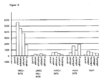
- strong activation of MDA5 by composite eRNA was also observed ( Fig. 9 and Fig. 10 ).
- eRNA41H and eRNA41J, a version with the adenoviral VARNAI modified in a single base in the box B RNA polymerase III recognition site to that of Ad2, rather than Ad5
- composite eRNA vectors are strong MDA5 activators, and strong cross species RIG-I activators.
- RIG-I is not orientation dependent ( Fig. 10 ) although some orientations are stronger than others, with the parent 41 and 41e vectors strongest for RIG-I activation and the 41g strongest for MDA5 activation. This again may reflect differences in preferred ligands for RIG-I and MDA5.
- the strongest RIG-I activating composite eRNAs in the 41 and 41e orientations contain the eRNA11a blunt 5' end; these are eRNA41H, eRNA41J (in the 41 orientation) and eRNA41 e+h (in the 41e orientation). Fold induction of interferon ⁇ promoter by RIG-I is much higher for eRNA41H than eRNA41.
- RIG-I activation synergy is observed with two different eRNA combinations: eRNA18 and VARNAI, and eRNA11 and VARNAI.
- eRNA35 and eRNA36 are also strong MDA5 activators; this may be due to the observation that MDA5 activation is better with ssRNA (such as produced by eRNA18) than dsRNA (as produced by eRNA11) (Kato et al, Supra, 2006).
- the eRNA41H vector was tested in a one step assay for induction of interferon a.
- the interferon ⁇ reporter pNIFTY2-1FA-SEAP (Invivogen, San Diego, CA) was substituted for pI25luc in the one step assay of Example 2.
- 40 ng eRNA41H plasmid (or control plasmid without eRNA41H) was transfected per well with mRIGI and pNIFTY2-IFA-SEAP. SEAP expression was detected after transfection of the eRNA41H vector, but not vector without eRNA41H.
- the composite eRNA vectors also surprisingly enhances target gene expression ( Fig. 8 , 9 and 10 ). While the composite elements are designed to not activate PKR through inclusion of the VARNAI component, the enhanced target gene expression beyond the effect of VARNAI alone was unexpected. While not limiting the application of embodiments of the invention, enhanced target gene expression may be mediated by unexpected interactions between cofactors required for RNA polymerase II (for CMV transcription) and RNA polymerase III (eRNA transcription).
- the tRNA and VARNAI RNA polymerase III promoters are gene internal type 2 RNA polymerase III promoters, while U6 (and H1) are type 3 gene external RNA polymerase III promoters.
- RNA polymerase II While type II promoters have less or no cofactor requirements in common with RNA polymerase II, the U6 type 3 promoters require several cofactors in common with RNA polymerase II, including SNAPc, Oct1, STAF, YY1 (reviewed in Schramm L, Hernandez N. 2002. Genes Dev 16: 2593-2620 ).
- One skilled in the art can further optimize the synergistic activation of RIG-I in the existing composite RIG-I activating constructs reported herein, by varying the component promoters (e.g . human or rat U6 promoter substitution for murine U6 promoter), and then testing using the 1 step and/or 2 step RIG-I activation assays disclosed herein, or other RIG-I activation assays known in the art.
- component promoters e.g . human or rat U6 promoter substitution for murine U6 promoter
- VARNAI adenovirus RNAI
- VAI adenovirus RNAI
- MDA5 activation is reduced by subtle central domain modifications that also reduce PKR inhibition. This may indicate MDA5 is adapted to detect conserved structures of the central domain of VARNAI that are necessary for PKR inhibition.
- PKR inhibition is essential for viral replication, so MDA5 activation by VARNAI may reflect a previously unknown role for MDA5 in control ofdouble stranded DNA virus infections such as adenovirus.
- VARNAI is not an MDA5 activator. Investigation of innate immune response to adenovirus infection identified several mechanisms of innate immune response activation, none of which included MDA5 activation. The authors conclude that DNA-sensing is the primary activator of IRF-3 ( Nociari M, Ocheretina O, Schoggins JW Falek-Pedersen E. 2007 J. Virol. 81: 4145-4157 ). Transfection of cells with a VAI/VAII containing plasmid was found to induce type I interferon production, but not through IRF-3 activation (Weber et al, Supra, 2006). This teaches that interferon production mediated by VALI/VAII is not mediated by MDA5, since IRF-3 is activated by MDA5 signaling.
- the one step RIG-I or MDA5 activation assay (Example 2 ) was modified as follows.
- 0.1 ug pI25luc a reporter plasmid, with the luciferase gene driven by the interferon ⁇ promoter
- 0.15 ug of an internal control EGFP plasmid (EGFP is used to standardize transfections) and ⁇ 0.1 ug of the test plasmid were mixed in 50 uL DMEM (no serum) and cotransformed into human cell line HEK293 using lipofectamine 2000.
- the control EGFP was utilized as an internal transfection control, and the standardized RIG-I or MDA5 activation expressed as
- the strong MDA5 activation with eRNA41H was mostly due to activation by the VARNAI component.
- synergy is not observed with eRNA11a, with most MDA5 activation contributed by the VARNAI component ( Fig. 11 ).
- VARNAI is a relatively weak RIG-I activator (weaker than eRNA11a), it is surprisingly by far the strongest MDA5 activator ofthe single component eRNAs tested ( Fig. 11 ).
- the one step assay was performed as described above, using either 1) both hRIG-I and hMda5 receptors (20 ng each per well) or 2) neither receptor.
- the results demonstrate that in the presence of both receptors, VARNAI is more potent than eRNA11a. This is possibly due to the ability of VARNA1 to activate both RIG-I (weak) and MDA5.
- VARNAI was a weak activator. As before, the strongest activation was observed with the combinations of VARNAI and eRNA11a ( cis and trans ), either with RIG-I/MDA5 or no receptor added.
- VARNAI was again the strongest activator in trans to eRNA11a; weaker activation was observed with eRNA31b and eRNA31c (intact VARNAI RNA's that additionally contain a 3' hairpin; Example 1 ). Activation may be weaker due to impaired nuclear export of these RNAs by disruption of the terminal minihelix.
- VARNAI is the strongest identified transactivator of eRNA11a induced interferon ⁇ activation. This activation is not an artifact of excess RIG-I or MDA5 receptors in the assay, since activation is also seen in native HEK293 cells, in which no additional receptor was added.
- VARNAI activation was then evaluated, by testing the effects of subtle modifications to the central domain of this RNA on RIG-I and MDA5 activation.
- the central domain was unexpectedly found to be critical for VARNAI activation of MDA5, and VARNAI synergy with eRNA11a for activation of RIG-I.
- eRNA41H-PKR1 pDNAVACCUltra EGFP eRNA41H with the VAI component engineered to contain the eRNA44A deletion (Example 1).
- eRNA41H-PKR2 pDNAVACCUltra EGFP eRNA41H with the VAI component engineered to contain the VAI S35 modification (eRNA23; Example 1).
- VARNAI is an inhibitor of PKR, and also a weak inhibitor of ADAR adenosine deaminase (which edits RNA content from A to I) and a weak activator of OAS (which activates RNaseL to degrade RNA).
- VARNAI molecules used in PKR I and 2 activate these RNA degradation enzymes, leading to RNaseL-mediated degradation, or ADAR mediated loss of immunostimulatory activity (due to A to I editing), or ADAR mediated nuclear retention (due to extensive A to I editing) of the eRNA11a immunostimulatory RNA.
- the following constructs were made to test this possibility.
- eRNA41H-PKR3 L3
- eRNA41H-PKR4 L4
- L3 and R1 mutations are described in Ma Y, Mathews, MB RNA 1996 2: 937 and are 2 bp changes in the central region that has been shown to be inactive for PKR inhibition, yet they are transcribed at high level in cell culture and maintain a VARNAI -like RNA secondary structure.
- Both constructs eliminated VARNAI activation of MDA5, and VARNAI synergy with eRNA11a for activation of RIG-I.
- the PKR3 and 4 modifications may not specifically reduce only PKR activation, since the central domain of VARNAI is also needed for inhibition of ADAR activation ( Lei M, Liu Y, Samuel CE. 1998 Virology 245:188-196 ). The following construct was made to test this possibility.
- eRNA41H-PKR5 pDNAVACCUltra EGFP eRNA41H with the VAI component modified in the central domain.
- the VARNAI cassette was slightly modified in the central domain to eliminate only PKR inhibition (and retain ADAR inhibition). This pm91 mutation is described in Rahman et al, Supra 1995 and is a 1 bp change in the central region that has been shown to be inactive for PKR inhibition, but retains ADAR inhibiting activity (Lei et al, Supra, 1998).
- PKR 1 -4 retain residual 11a component activity for RIG-I activation but eliminated both the synergistic activation of RIG-I and VARNAI mediated MDA5 activation activity. This demonstrates that the VARNAI central domain is surprisingly required for both MDA5 activation, and synergistic RIG-I activation with eRNA11a.
- PKR5 retains some synergistic activation of RIG-I as well as MDA5 activation activity. This surprisingly demonstrates that subtle modifications of the central domain can be used to create novel hybrid molecules that vary in potency of MDA5 activation and PKR inhibition.
- eRNA11a may be a PKR ligand as well as a RIG-I ligand.
- RIG-I binds RNA with a lower affinity than PKR ( Gantier MP, Williams BRG 2007 Cytokines Growth Factor Review 18:363-371 ), so most eRNA11a may be unproductively bound to PKR.
- VARNAI is a high affinity PKR ligand that may compete with eRNA11a for PKR binding, freeing eRNA11a (which is a stronger RIG-I activator) for RIG-I activation (ligand titration model).
- eRNA11a may bind and activate ADAR which would initiate Adenosine to Inosine editing, preventing nuclear export and/or reduce immunostimulatory activity.
- Titration of ADAR by VARNAI, and subsequent inhibition of ADAR may reduce eRNA11a editing (ligand destruction model).
- inclusion of an ADAR inhibitor such as VARNAI, can be used to increase export of dsRNA from the nucleus, through inhibition of ADAR mediated RNA editing.
- a receptor e.g . MDA5, PKR, OAS
- RIG-I activating eRNA11a may synergistically activate with RIG-I activating eRNA11a.
- eRNA11a The analysis of the components of eRNA11a ( Fig. 14 ) also surprisingly demonstrate that double stranded RNA per se is not the mediator of RIG-I activation, because the H1 - mU6 (StuI) INF dsRNA repaired construct was a weak activator compared to eRNA11a. This construct makes the same dsRNA as eRNA11a but differs in that the tRNA component is not present. These results demonstrate that the tRNA component of eRNA11a is critical to RIG-I activation, and that double stranded RNA is insufficient to account for the interferon activating potential of eRNA11.
- eRNA41H 150
- eRNA41H eRNA41H
- Conc eRNA41H
- These vectors contain one (150) or several (Conc) copies of a 50 bp sequence inserted internally within the eRNA11a component of eRNA41H.
- This 50 bp sequence contains no Pol III terminators on either strand, and extends the length of the eRNA11a RNA by 50 bp (eRNA41H 150).
- the observed ⁇ interferon promoter activation demonstrates that the length of RNA in eRNA11a can be varied while retaining RIG-I/MDA5 activation.
- the optimal length and composition can be determined by an investigator skilled in the art, by varying the eRNA11a insert and testing immunostimulation using the described assays or equivalent.
- the eRNA41H (DT) is a second DNA vaccine plasmid backbone, which contains a different promoter, antigen and eukaryotic terminator.
- the activation observed with this vector demonstrates that the eRNA41H activator functions in different DNA vaccine plasmid backbones and can therefore be transferred into alternative vectors as needed.
- An analysis of activation of eRNA41H, eRNA41J and eRNA41E+H at different target plasmid dosages and backbones is summarized in Fig. 17 . No difference between eRNA vectors was observed, with activation >2 fold over background observed at the lowest tested target plasmid dose (5 ng/transfection).
- eRNA41H and eRNA41E+H contain the same component RNAs in different orientations. This demonstrates that different orientations of the component RNAs within the composite eRNA are functional. eRNA41H functioned in the three different vector backbones tested - this is consistent with the eRNA41H(DT) results above, and demonstrates eRNA function is not vector specific and can therefore be incorporated into a variety of alternative vector backbones.
- VAI can be combined, in a DNA vaccine vector, with one or more activating dsRNA elements, to selectively induce RIG-I, MDA5 or OAS responses without PKR mediated antigen expression reduction.
- Such vaccines could be superior in vivo to existing replicon vaccines, by combining high antigen expression with RIG-I mediated dendritic cell maturation and interferon production ( Lopez CB, Jount JS, Moran, TM. 2006 J Virol. 80: 3128 ).
- RIG-1 and TLR9 may be synergistic to each other, based on known synergy between TLR3 (dsRNA ligand) and TLR9 (CpG ligand) activation pathways ( Whitmore MM, DeVeer MJ, Edling A, Oates RK, Simons B, Linder D, Williams BRG. 2004. Cancer Research. 64: 5850-5860 ).
- DNA vectors containing these elements would prevent the acquired immunity associated with the use of such virus-derived vectors, and would thus not limit the future use of the DNA vectors to deliver other antigens and eRNA.
- Example 5 In vivo evaluation of influenza eRNA vectors
- the WHO Influenza Surveillance Network has characterized H5N1 influenza viruses isolated from humans and animals from several countries affected by the 2004/2005 H5N1 outbreak in Asia and made recommendations on the antigenic and genetic characteristics of H5N1 viruses suitable for vaccine production (strains A/Vietnam/ 1194/04, A/Vietnam/1203/04 and A/Hongkong/213/03).
- Influenza A HA has not typically been codon optimized for use in DNA vaccines in chickens, mice and primates. This is consistent with the fact that these proteins must be made in these hosts for the virus to replicate.
- a native (secreted-membrane associated) version ofthe A/Vietnam/1203/04 HA gene was synthesized (by DNA2.0, Menlo Park, CA).
- the synthetic gene (herein denoted as HA) was designed to afford simple cloning into the pDNAVACCUltra vectors.
- HA For safety (i.e . gene transfer during immunization to infecting non pathogenic virus) the HA gene was human codon optimized with the DNA2.0 codon optimization software to reduce genome homology.
- the multiple basic amino acid motifs at the HA cleavage site was replaced with the corresponding region from several nonpathogenic influenza strains.
- HA expressing backbones with or without eRNA41H or eRNA41J were tested for immunogenicity (4 mice per group) in vivo. Twenty (20) ⁇ g plasmid was injected into I muscle (quadriceps) of female BALB/c mice (6-8 weeks old) in three doses (day 0, 14, and 28). Serum samples were taken at day 42 and tested for anti-HA IgG response. Anti-HA specific total IgG, IgG2a, and IgG1 levels were determined by standard ELISA assay using plates coated with Nature Technology Corporation produced recombinant E.
- coated wells were blocked and incubated 2 hrs with diluted serum samples, the wells washed and signal detected by subsequent sequential incubation and washing with; goat anti mouse IgG (Fc)-biotin, then Streptavidin-horse radish peroxidase conjugate (totat IgG) or; goat anti-mouse IgGI, then anti-goat IgG-biotin, then Streptavidin-horse radish peroxidase conjugate (IgGI) or; goat anti-mouse IgG2a, then anti-goat IgG-biotin, then Streptavidin-horse radish peroxidase conjugate (IgG1).
- NTC7382 promoter CMV-HTLV-1-synthetic rabbit ⁇ globin intron-exon 2 SRF binding protein
- the NTC7382 promoter is distinct from the CMV/R promoter described in Nabel GJ, Sanchez A, Yang ZY, Sullivan N. 2003 US Patent US7094598 B2 (CMV-HTLV-I-CMV intron) or the base pDNAVACCUltra CMV or SV40-CMV promoter.
- the NTC7382 promoter is composed of the novel combination of; 1) CMV promoter and start of exon 1; 2) the HTLV-I R-U5 sequence which contains the 5' splice acceptor site; 3) a synthetic 3' acceptor site based on the rabbit ⁇ globin intron, and; 4) Splicing enhancer comprised of SR protein binding site (3 copies of GAAGAAGAC) to improve RNA export in exon 2, prior to 5) a kozak sequence upstream of the start codon for the gene of interest.
- the SR protein binding site sequence has been shown to act as a splicing enhancer ( Lavrequisite A. La Branche H, Komblihtt AR, Chabot B. 1993. Genes Develop.
- NTC7382 promoter is shown in Fig. 18 .
- the sequence of the NTC7382 promoter is given in SEQ ID NO: 6.
- Expression levels (of EGFP) from plasmids incorporating the NTC7382 promoter, or NTC73 82 promoter with upstream SV40 enhancer, after transfection of the HEK293 cell line are dramatically increased versus CMV based promoter plasmids ( Table 4 ).
- Table 4 Expression levels (EGFP Fluorescence units) from plasmids incorporating the NTC7382, SV40-NTC7382, or CMV based promoter Vector name ⁇ Promoter Selection RNA element EGFP pDNAVACCUltra-EGFP CMV Kan No 6,668 NTC7382-EGFP (UP) NTC7382 Kan No 20,074 NTC73 82-EGFP-41H (UP) NTC7382 Kan 41H 27,095 NTC7382-EGFP SV40-NTC7382 Kan No 9147 NTC7382-EGFP-41H SV40-NTC7382 Kan 41H 21,545 NTC8382-EGFP (UP) NTC7382 RNA-OUT No 31,788 ⁇ (UP) is a modified backbone wherein the SV40 enhancer and ISS sequences (see Fig. 1 ) are removed
- eRNA versions of NTC7382 were made, by simple swapping of the eRNA region into the plasmid backbone using flanking sites common to all the vectors, such as NotI-AflIII, or NheI-AflIII, or NotI-ApaLI, or NheI-ApaLI, or NotI-DraIII, or NheI-DraIII fragment swaps.
- flanking sites common to all the vectors such as NotI-AflIII, or NheI-AflIII, or NotI-ApaLI, or NheI-ApaLI, or NotI-DraIII, or NheI-DraIII fragment swaps.
- eRNA function (RIG-I and MDA5 activation in vitro ) in the NTC7382 vectors was equivalent to the original CMV based vector ( Fig. 17 ) while antigen expression was enhanced (Table 4).
- DNA vaccines are being developed for a variety of human, animal, bird and fish vaccine applications.
- the presence of an antibiotic resistance gene in the plasmid backbone is considered undesirable by regulatory agencies, due to 1) the potential activation and transcription of the genes from mammalian promoters after cellular incorporation into the genome and 2) the potential transfer of the antibiotic resistance gene to endogenous microbial fauna.
- the presence of antibiotics in the fermentation culture leads to the potential contamination of the final product with residual antibiotics.
- RNA based selectable markers have been developed. Morsey MA, 1999 US patent US5922583 discuss in theoretical terms the use of plasmids expressing the sok or flmB antisense repressor RNAs as selectable markers by selecting and maintaining the plasmids in cells engineered to contain and express the cognate target toxin genes (hok and flmA, respectively). In the presence of the plasmid, toxin expression is repressed and cell viability is maintained. Cells that lose the plasmid will die. Construction and testing results for such vector systems were not disclosed by the inventor so the utility of such systems is unknown.
- RNAII antisense regulators of the pMB1 origin Two groups have developed RNA based selection systems that utilize the endogenous RNAI/RNAII antisense regulators of the pMB1 origin.
- chromosomal genes were engineered to contain an RNAII sequence within the untranslated region of the mRNA.
- the plasmid encoded RNAI repressor binds the RNAII sequence and represses translation of the chromosomal gene.
- This gene can be an antibiotic resistance marker, transcriptional repressor (controlling expression of a second gene) or a lethal gene (Grabherr R, Pfaffenzeller 1. 2006; US patent application US20060063232 ; Cranenburgh RM. 2005; World Patent Application WO2005052167 ).
- These systems are limited to existing ColEl type origin containing plasmid vectors only.
- Counterselectable markers are known in the art and include lacZ, glkA (sensitivity to 2-deoxyglucose) or galK. Knockdown of sugar transport and fermentation genes can be selected by the proton suicide method ( Winkelman JW, Clark DP. 1984. J Bacteriol. 160: 687 ). Other systems that can be counterselected are thymidylate synthase A (thyA), xylB (xylitol media), tetracycline and levansucrase (sucrose media) (reviewed in Reyrat JM, Pelicic V, Gicquel B, Rappuoli R. 1998 Infection Immun. 66: 4011-4017 ).
- RNA based inhibition of engineered chromosomal streptavidin expression cassettes can be utilized for selection, by suppressing the toxicity of streptavidin (biotin binding).
- streptavidin biotin binding
- an RNA that hybridizes to, and reduces translation of, a genomic sequence could be used to repress expression of a transcriptional regulator protein, for example the tetR repressor.
- a transcriptional regulator protein for example the tetR repressor.
- an antibiotic resistance marker is cloned under the control of the tetR repressor, the presence of the antisense RNA in the cell will inhibit tetR expression (if it is placed under the control of the antisense RNA), and derepress the antibiotic resistance gene. This would allow antibiotic selection for a plasmid, without inclusion of the antibiotic gene in the plasmid backbone.
- RNA-OUT an antisense RNA that hybridizes to, and reduces translation of, the transposon gene expressed downstream of RNA-IN.
- the antibiotic resistance marker may be replaced with a natural or engineered regulatory RNA
- application of such a system to plasmid maintenance has not been reported in the art.
- the Hok Sok and flmA flmB systems are actual plasmid maintenance systems, they are not ideal for incorporation into a therapeutic plasmid due to the required presence of a toxin gene on the plasmid.
- the kanamycin resistance marker in NTC7382 was replaced with a regulatory RNA based selectable marker (the IS10 RNA-OUT antisense RNA). This RNA hybridizes to the RNA-IN RNA, and reduces translation of the downstream IS10 transposase gene ( Fig. 19 ).
- a counterselectable marker (sacB; Bacillus subtilis levansucrase) was expressed under the control of the RNA-IN promoter and leader ( Fig. 20 ) and was integrated into the chromosome of DH5 ⁇ . Expression of sacB was determined to be constitutive since cells containing sacB encoded levansucrase were killed in presence of sucrose. Translation of SacB from this RNA was designed to be repressed in the presence of the antisense regulator RNA-OUT ( Fig. 21 ). RNA-OUT was cloned into the pDNAVACCUltra vector, in place of the kanR gene by replacement ofthe DraIII KpnI cassette ( Fig. 21 ). RNA-OUT was expressed from the plasmid, and repressed expression of the chromosomally integrated counterselectable marker SacB controlled by RNA-IN. This allowed antibiotic free selection in the presence of sucrose on solid and liquid media.
- Replication incompetent plasmids can be site specifically integrated into the genome at bacteriophage attachment sites utilizing bacteriophage recombinase expressing plasmids.
- Such systems to allow integration at various bacteriophage attachment sites have been developed ( Haldimann A, Wanner BL, 2001, J. Bacteriol. 183: 6384-6393 ) and are generally available ( E. coli stock center, Yale NH). These plasmids and methods are included herein by reference.
- integration of a gene at the Phage lambda attachment site requires cloning a gene to be expressed into a modified integration plasmid such as pCAH63-CAT.
- the pCAH63-CAT vector was constructed by ligating the bacteriophage lambda attachment site-ori-R-chlorR-rgnB containing DNA fragment of pCAH63 (2.2 kb NheI/PstI DNA fragment) with a 350 bp NheI/PstI tL3 containing DNA fragment from pAH81 (both vectors are described in Haldiman and Wanner, Supra, 2001 and were obtained from the E . coli stock center, Yale NH).
- the resultant pCAH63-CAT plasmid contains the R6K conditional replication origin (which requires engineered pir+ host cells such as BW23474 for propagation; the origin is non functional in DH5 ⁇ ) a multiple cloning site, a chloramphenicol resistance marker (chlorR) and the lambda attachment site.
- the plasmid is integrated into the phage lambda attachment site using the pINT-ts helper plasmid (ampicillin resistant, temperature sensitive R plasmid replicon; this plasmid is lost at 42°C) that encodes a heat inducible phage lambda integrase.
- Transformation of DH5 ⁇ containing the pINT helper with the pCAH63-CAT derivative plasmid to be integrated results in integration of the pCAH63-CAT plasmid into the genome at the phage lambda attachment site; recombinants are selected with chlorR and integration verified using PCR as described in Haldimann and Wanner Supra, 2001.
- the 1.4 kb PCR product was digested with AarI (underlined, with sticky ends bolded in primer sequences above) and the linear fragment purified (F2).
- a constitutive promoter P5/6 5/6 -RNA-IN leader sequence was made using synthetic primers as follows.
- the P5/6 5/6 promoter -35 (double underlined) and -10 (double underlined, bold) regions are indicated in the RNA-INF02 primer.
- the 3 fragments (F1, F2 and F3) were ligated, and transformed into the BW23474 cell line.
- Recombinants [pCAH63-CAT RNA-IN-SacB (p5/6 5/6)] were selected on choramphenicol, correct clones identified by restriction digestion, and verified by sequencing with tL3-r and rgnB-f sequencing primers (Haldimann and Wanner Supra, 2001).
- RNA-IN from the above P5/6 5/6 construct, a promoter mutation was introduced that increased promoter activity (P5/6 6/6 has a single base change from P5/6 5/6 that increases promoter strength to 6/6 match with -10 Pribnow box, by changing the TAGAAT sequence to TATAAT).
- the pCAH63-CAT RNA-IN-SacB (p5/6 5/6) plasmid from above was PCR amplified using the following primers:
- the single base change from C to A is double underlined in the 6/6-10R01 primer (will make G to T change in -10 forward sequence).
- the 4 kb PCR product was digested with AarI (underlined in primers, bold bases are complementary sticky ends) and the linear fragment purified and ligated to itself.
- the BW23474 cell line was transformed and choramphenicol resistant colonies selected and correct clones [pCAH63-CAT RNA-IN-SacB (p5/6 6/6)] identified by restriction digestion.
- the recombinant plasmid ( Fig. 20 ) was sequenced with rgnB-f to confirm the promoter change.
- RNA-IN-SacB The plasmid was integrated into DH5 ⁇ and the cell line (RNA-IN-SacB) confirmed by PCR using P1-P4 primers (Haldimann and Wanner Supra, 2001). RNA-IN-SacB did not grow on LB (no NaCl) +6% sucrose plates. This demonstrates that the novel combination of a constitutive promoter expressing RNAI-IN and SacB can be utilized to create a strain constitutively producing sacB, which prevents growth of the cell line on sucrose containing plates.
- RNA-OUT plasmid A fluorescent RNA-OUT plasmid was created.
- the pDNAVACCUltra- P5 / 6,5 / 6 -RBs -EGFP plasmid (expresses EGFP constitutively) was digested with DraIII/KpnI and the linear vector (2029, 1015) purified.
- the following 2 RNA-OUT primers were annealed. These produce an annealed RNA-OUT fragment that is compatible with DraIII and KpnI ( Fig. 21 ).
- RNA-1N-sacB integrated cell line from step 3 above.
- Colonies were selected on LB (no NaCl) +6% sucrose and plasmid from two fluorescent colonies were sequence verified as correct. Transformation of sequence-verified pDNAVACCUltra- P5 / 6,4 / 6 -RBS -EGFP (RNA-OUT) plasmid into RNA-IN -SacB competent cells was performed; all recovered colonies after sucrose selection were fluorescent ( i.e . 100% of recovered cells contained the plasmid).
- RNA-OUT selectable marker was substituted for the kanamycin resistance gene in a DNA vaccine plasmid as follows.
- the RNA-OUT cassette was excised from pDNAVACCUltra- P5 / 6,4 / 6.
- RBS -EGFP RNA-OUT
- DraIII/XbaI 154bp
- DraIII/XbaI 154bp
- RNA-IN containing plasmids were transformed into serial diluted Z-competent cells (Zymo Research, Orange, CA) of the RNA-IN-SaeB cell line and the transformations plated on LB (no NaCl) +6% sucrose plates. Colonies from each transformation were screened for insert. 8/10 recovered colonies from the undiluted Z competent cells were correct, as were 7/9 from the 1:10 diluted competent cells, and 2/2 from the 1:100 diluted competent cells. Similar results were obtained with a second independent ligation reaction for a different construct. Collectively, these results demonstrate that RNA-IN containing plasmids can be surprisingly easily selected from ligation reactions on LB Sucrose plates, and that the selection is robust.
- RNA-OUT-REV opposite orientation
- RNA-OUT vectors were constructed with either the influenza H5 hemagglutinin gene (HA), SEAP or EGFP reporter genes in the RNA-OUT vector.
- HA hemagglutinin gene
- SEAP SEAP
- EGFP reporter genes in the RNA-OUT vector.
- versions containing various pol III transcribed immunostimulatory RNAs (eRNAs) that activate the RIG-I and MDA5 receptor were constructed.
- Over 20 different DNA vaccine vectors were constructed with the RNA-OUT sucrose selection method. This demonstrates the surprising robustness and general utility of the RNA-OUT sucrose selection methodology.
- the sequence of the NTC8382-41H-HA vector ( Fig. 1 ; RNA-OUT equivalent of kanR NTC738241H-HA) is given in (SEQ ID NO: 16).
- RNA-OUT plasmids Antigen expression, and RIG-I activation, after lipofectamine 2000 transfection of HEK293 cells (human) was determined for RNA-OUT plasmids, compared to control kanR plasmids as follows.
- the vectors were tested for RIG-I activation in HEK293 cells using the one step assay as described in Example 4.
- EGFP and HA Section 7 below protein expression analysis
- the EGFP or HA RNA-OUT or KanR plasmids (0.4 ug each well) were transfected as described in Example 4 (without RIG-I or pI25luc and control EGFP plasmid), and total cellular protein 48 hrs post transfection assayed by FLX800 microplate fluorescence reader (EGFP) or Western blot analysis (HA).
- RNA-OUT plasmids No difference was observed between RNA-OUT plasmids and the corresponding kanR plasmid (otherwise same backbones) for EGFP ( Table 4 ) or HA (Section 7 below) expression or RIG-1 activation ( Fig. 17 ).
- RNA-OUT in the vector backbone does not adversely affect eukaryotic functions of the plasmid,for example, NTC7382 promoter-driven antigen expression or Pol III driven immunostimulatory RNA expression.
- Influenza H5 HA 'bird flu' eRNA RNA-OUT vector were made, by simple swapping of the eRNA region into the plasmid backbone using flanking sites common to all the vectors, such as NotI-AflIII, NheI-AfIIII, NotI-ApaLI, NheI-ApaLI, NotI-DraIII, or NheI-DraIII fragment swaps and the influenza gene using SalI-BglII fragment swaps.
- eRNA bird flu vectors H5 HA vectors were tested for immunostimulation as described in Example 4 . Potent activation of murine and human RIG-I, and human MDA5 was observed. This demonstrates that eRNA activation is not antigen dependent or vector dependent.
- Western blot analysis was performed on protein extracts (as described for EGFP in Section 6 above) using rabbit anti-HA H5N1 rabbit IgG (eEnzyme, Gaithersburg, MD ) primary antibody, and the amplified alkaline phosphatase immuno-blot assay kit and AP conjugate substrate kit (Biorad, Hercules, CA).
- HA protein was expressed in transformed cell lysates as mostly uncleaved protein, while an alternative 1203 HA, with the basic cleavage site intact, was mostly cleaved protein.
- RNA-OUT sequence has no repetitive sequences that may be toxic or unstable in E. coli, so effects on quality in fermentation production were not expected.
- an RNA-OUT-HA vector was tested for ferment ation productivity.
- An exponential, inducible fed-batch fermentation was performed using the high yield plasmid production process disclosed in Carnes AE, Williams JA. 2006. World Patent Application WO2006023546 .
- This fermentation, RF117 was carried out in a 7.5 L fermentor with an RNA-OUT-41H-HA plasmid in E. coli strain DH5 ⁇ .
- the fermentation media contained 0.5% sucrose for plasmid selection since it was determined that ⁇ 0.5% sucrose in fermentation media is growth inhibitory to the parent plasmid-less strain.
- the temperature was initially controlled at 30°C.
- the temperature set point was increased to 42°C after 27 hours to increase plasmid yield.
- the culture was harvested after 39 hours of total fermentation time (11 hr plasmid induction at 42°C).
- the culture had a final cell density of 90 OD 600 .
- the plasmid yield from this fermentation was 1213 mg/L ( Fig. 22 ) and the plasmid was of a high quality (supercoiled monomer) with no detected replication intermediates or aberrant bands. This yield is typical for kanR based pDNAVACCUItra derived vectors. This demonstrates that inclusion of RNA-OUT in the vector backbone has no detrimental effect on plasmid production yield or quality.
- the production cell line For general application to production of a variety of plasmids, it is preferable for the production cell line to not have plasmid replication origins (e.g . R6K replication origin) in the chromosome.
- plasmid replication origins e.g . R6K replication origin
- the pCAH63-CAT RNA-IN-SacB (P5/6 6/6) plasmid was PCR amplified to delete the R6K replication origin using the following primers ( Fig. 20 ).
- the 3.5 kb linear product was DpnI digested (to eliminate parent plasmid), AarI digested (underlined in primers) to create compatible sticky ends (bolded in primer), Qiaquick purified, and ligated (to make exonuclease resistant circles for integration).
- the ligation was electroporated into pINTts lambda integration plasmid containing NTC3012 cell line (autolytic production host described in copending application Cames and Williams, US patent application US60931465 ) and colonies were selected with chloramphenicol.
- the cell line (NTC3016) was confirmed with PCR using normal Pl-P4 primers (Haldimann and Wanner Supra, 2001); these primer sites are retained in the amplified minicircle vector.
- RNA-OUT plasmids have been successfully transformed and propagated in the NTC3016 cell line, without toxicity or interference with autolysis.
- a fermentation yield of 1.1 gm plasmid/liter culture after 10.5 hr induction was obtained with the RNA-OUT NTC8382-41H-HA plasmid ( Fig. 1 ) in the NTC3016 cell line (RF137). This demonstrates that the RNA-OUT plasmid is compatible with autolytic host strains and NTC fermentation process.
- the 16.5 kb human mitochondrial genome encodes 13 polypeptides, 22 tRNAs and 2 rRNAs.
- the mitochondrial genetic code is different than the nuclear code, so nuclear genes are not correctly translated in the mitochondria.
- a 1kb noncoding region acts as a bidirectional promoter for the synthesis of polycistronic RNA for the heavy and light strands as well as the replication origin for the heavy chain. Transcription requires negative supercoiling ( Buzan JM, Low RL. 1988 Biochem. Biophys. Res. Common. 152: 22 ), as is the case for E. coli-produced plasmid DNA.
- the mitochondrial promoter does not function in the nucleus nor does it inhibit the activity of weak or strong pol II promoters ( Sewards R, Wiseman B, Jacobs HT. 1994. Mol. Gen. Genet. 245: 760 ).
- Rat mitochondria have been shown to be naturally competent for DNA transfection; internalized DNA is functionally expressed ( Koulintchenko M, Temperley RJ Mason PA, Dietrich A, and Lightowlers, RN 2006. Human Molec Genet 15: 143 ). Studies of plasmid fate during gene transfer have not reported plasmid transit to the mitochondria. However, electroporation is used to introduce plasmid DNA into isolated mitochondria ( Collombet JM, Wheeler VC, Vogel F, Coutelle C. 1997. J Biol Chem. 272: 5342 ) wherein the plasmid is expressed ( Yoon YG, Koob, MD. 2003. Nucl. Acids Res. 31: 1407-1415 .).
- the mitochondrial genome is also underrepresented for CpG ( carton, LR, Burge C, Clayton DA, Karlin S. 1994 Proc Natl. Acad. Sci USA 91: 3799-3803 ) which the inventor speculates may indicate that unmethylated mitochondrial DNA is accessible to TLR9 (which recognizes unmethylated CpG DNA and induces innate immune responses).
- M refers to insertion of the mitochondrial R region.
- the murine mouse mitochondrial R-region was PCR amplified using murine DNA and the following primers:
- the TGCGCA bolded sites are FspI restriction sites added adjacent to the R region to allow transfer to alternative vectors.
- the 883 bp PCR product was kinased using T4 kinase and cloned into the DraIII site of NTC73 82-41H HA vector [the vector was cleaved with DraIII, the sticky ends removed by T4 DNA polymerase exonuclease, and the fragment treated with calf intestinal phosphatase (CIP)] to create the NTC7382-41H-M HA vector ( Fig. 23 ).
- the clone was sequence verified.
- the R region in this clone is in orientation 2 (10110702 primer immediately adjacent to pUC origin).
- SEAP and EGFP expressing derivatives were made by swapping out the HA gene (SalI-BglII fragment swaps). No effect on EGFP expression in HEK233 cells was observed.
- NTC7382 41H-HA and NTC7382-41H-M-HA Fig. 23 ) and NTC8382-41H-HA ( Fig. 21 ) vaccines were tested for immunogenicity (8 mice per group) in vivo versus control NTC7382-SEAP vector. Immunogenicity was compared to expression level (3 mice per group, using corresponding vectors containing SEAP rather than HA).
- 3 ⁇ g HA plasmid was injected intra-muscular (1M, quadriceps) of female BALB/c mice (6-8 weeks old) in a single dose (day 0) prior to electroporation (EP) using the MedPulser® system (Innovio Biomedical, San Diego CA) using a 2 needle array (27 G), 4 mm apart, 2x60 ms pulses at 50 mA. Serum samples were taken and tested for SEAP or anti-HA IgG response. SEAP levels were determined using the Phospha-light SEAP Reporter Gene Assay System from Applied Biosystems (Foster City, CA) according to the manufacturer's instructions.
- Anti-HA specific total IgG, IgG2a, and IgG1 levels were determined by standard ELISA assay using plates coated with Nature Technology Corporation produced recombinant E. coli produced A/Vietnam/1203/2004 HA2 domain as described in Example 5. The results are summarized in Table 5 and demonstrate 1) the antibiotic free NTC8382 backbone is equivalent or superior to the NTC7382 backbone for expression and immunogenicity and; 2) the NTC7382-41H-M backbone improved immune response to HA when compared with parent NTC7382-41H backbone.
- a mitochondrial R-region plasmid may:
- Mitochondrial promoters show some species specificity, since human and mouse mitochondrial transcription machineries do not recognize the heterologous species ( Gaspari, M, Falkenberg M, Larsson NG, Gustafsson CM. 2004. EMBO J 23: 4606-4614 ) so for human or animal vaccination it may be critical to determine appropriate mitochondrial promoter for use. Such modifications to the vector and determination of appropriate promoters can be accomplished by investigators skilled in the art of cloning ( i.e . selecting homologous fragments corresponding to the mitochondrial R region amplified with the primers disclosed in Example 8 ) and immunological testing.
- the vector embodiments of the invention provide for a rationale approach to optimization of genetic immunization or gene therapy, through use of the improved NTC7382 promoter, RNA based selectable marker, and/or vector encoded interferon inducing eRNA compositions.
- MDAS, RIG-I and PKR inhibiting immunostimulatory RNA elements may be combined with repressor RNA elements that encode shRNAs to inhibit LGP2 (a repressor of RIG-I and MDA5 immunostimulation) ADAR and/or inhibitors of R1G-I or downstream IPS-1 signaling pathway. This would maximize immunostimulation while preventing PKR mediated inhibition of antigen expression.
- RNA elements could be included to induce apoptosis selectively in non-immune cells, to increase antigen cross presentation, or influence T H 1 versus T H 2 bias.
- the eRNA element embodiments of the invention could be incorporated into alternative gene delivery vehicles, including viral (e.g . alphavirus, poxvirus, lentivirus, retrovirus, adenovirus, adenovirus related virus, etc) and nonviral (e.g . plasmid, midge, transcriptionally active PCR fragment, minicircles, bacteriophage, etc) vectors.
- vectors or DNA fragments containing immunostimulatory eRNA sequences may be used to increase anti-viral, anti-tumor and or antibacterial activity in a patient, through induction of type 1 interferon production.
- immunostimulatory RNA sequences may be used in treatment therapies to induce production of natural type 1 interferon's.
Abstract
Description
- This invention was made in part with government support under
Grant Numbers 2 R44 GM072141-02, 1 R43 GM080768-01, and 1 R43 AI071660-01A1 awarded by the National Institutes of Health. The government has certain rights in this invention. - This application claims the benefit of Provisional Patent Application Ser. #
US60932160 filed 29 May 2007 - The present invention relates to a family of eukaryotic expression plasmids useful for gene therapy, obtaining improved genetic immunization, natural interferon production, and more particularly, for improving the immune response to plasmid encoded antigens.
- The present invention is a family of eukaryotic expression plasmids useful for gene therapy, genetic immunization or interferon therapy. Such molecules and methods for use are useful in biotechnology, gene therapy, cancer and agriculture.
- With the invention in mind, a search of the prior art was conducted. DNA vaccines (genetic vaccines) are a potential disruptive technology, that offer the promise of a new way to immunize humans (or animals) with materials that are entirely gene-based and expressed by the organism's own cells, making an ideal mimic of intracellular antigens.
- Methods to improve immune responses to DNA vaccine plasmids are described in the art. For example, the efficacy of a DNA vaccine can be further improved, or tailored for systemic or mucosal immunity, or cancer, allergy, bacterial, intracellular parasite or viral targets, by: coimmunization with costimulatory plasmids (e.g. IL12) to modulate the type of response (
T H1 versusT H2 bias); cell death inhibitors or enhancers; or optimization of delivery (e.g. electroporation versus gene gun). Some such methods and molecules are described in, Lemieux, P. 2002 Expert Rev. Vaccines 1: 85-93; Toka FN, Pack CD, Rouse BT. 2004 Immunological reviews 199: 100-112; and Gurunathan S, Klinman DM, Seder RA. 2000 Annu. Rev. Immunol. 18: 927-974, and are included herein by reference. DNA vaccination could also involve utilizing different delivery systems in the prime and the boost, as taught by Buchan S, Gronevik E. Mathiesen I, King C, Stevenson FK, Rice J. 2005 Immunol. 174: 6292-6298 or different injection sites, as taught by Pavlakis GN, Gragerov A, Felber BK. 2004 US Patent ApplicationUS20040241140 . - DNA vaccination is ideal for rapid deployment vaccines since development times for DNA vaccines are significantly shorter than those for protein or viral vector systems.
- Regulatory issues for use of plasmid DNA in humans have been addressed in several recent World Heath Organization (WHO), US Food and Drug Administration (FDA), or European Agency for the Evaluation of Medicinal Products (EMEA) regulatory draft guidances. A key issue is that antibiotic resistance markers, typically kanamycin resistance, are the most commonly utilized selectable markers. The EMEA guidance [European Medicines Agency, 2001. Note for guidance on the quality, preclinical and clinical aspects of gene transfer medicinal products. CPMP/B WP/3088/99] with regard to DNA vaccine plasmids states: "The use of certain selection markers, such as resistance to antibiotics, which may adversely impact on other clinical therapies in the target population," and "Consideration should be given to avoiding their use, where feasible". Alternative selection strategies to address this concern are needed.
- Protective immunity in humans and other primates has not been broadly obtained using DNA only vaccination. Primate efficacy has been obtained utilizing DNA vaccines in combination with heterologous protein, inactivated organism, or viral vector boosting. Enhanced immune responses have also been reported when plasmid DNA and purified protein (corresponding to the protein encoded in the plasmid) (
Dalemans W., Van Mechelen MV, Bruck C, Friede M. 2003 US Patent US6500432 ; Carrera SD, Grillo JM, de Leon LALP, Lasa AM, Feyt RP, Rodriguez AV, Obregon JCA, Rivero NA Donato GM 2004 US Patent ApplicationUS20040234543 ), or inactivated virus, (Rangarajan PN, Srinivasan VA, Biswas L, Reddy GS. 2004 US Patent ApplicationUS20040096462 ) are mixed and coinjected. - However, using plasmids in combination with inactivated organisms, proteins or viral vectors in a vaccine (either as a mixture, or sequentially in a prime boost) eliminates most of the benefits of DNA vaccination, including improved safety, reduced cost, and rapid deployment.
- Antigen expression: The art teaches that one of the limitations of DNA vaccination is that antigen expression is generally very low. Vector modifications that improve antigen expression (e.g. codon optimization of the gene, inclusion ofan intron, use of the strong constitutive CMV or CAGG promoters versus weaker or cell line specific promoter) are highly correlative with improved immune responses (reviewed in Manoj S, Babiuk LA, Drunen SV, en Hurk LV. 2004 Critical Rev Clin Lab Sci 41: 1-39). A hybrid CMV promoter (CMV/R), which increased antigen expression, also improved cellular immune responses to HIV DNA vaccines in mice and nonhuman primates (Barouch DH, Yang ZY, Kong WP, Korioth-Schmitz B, Sumida SM, Truitt DM, Kishko MG, Arthur JC, Miura A, Mascola JR, Letvin NL, Nabel GJ. 2005 J Virol. 79: 8828-8834). A plasmid containing the woodchuck hepatitis virus posttranscriptional regulatory element (a 600 bp element that increases stability and extranuclear transport of RNA resulting in enhanced levels of mRNA for translation) enhanced antigen expression and protective immunity to influenza hemagglutinin (HA) in mice (Garg S, Oran AE, Hon H, Jacob J. 2002 J Immunol. 173: 550-558). These studies teach that improvement in expression beyond that of current CMV based vectors may generally improve immunogenicity.
- The art teaches that plasmid entry into the nucleus is a limiting factor in obtaining antigen expression. Increasing nuclear localization of a plasmid through inclusion of NFκB binding sites or a SV40 enhancer improves antigen expression in vitro and in vivo; this is presumed due to binding of NFκB which then piggybacks the plasmid to the nucleus (Dean DA, Dean BS, Muller S, Smith LC. 1999 Experimental Cell Research 253: 713-722). However, NFκB is generally localized in the cytoplasm, and transfer to the nucleus is limited, tissue-specific, and dependent on stimulatory signals. This limits the utility of NFκB nuclear targeting to improve DNA vaccination.
-
T H1 orT H2 bias: The art teaches that shifting immune response to DNA vaccine expressed viral or other antigens from TH2 toT H1 is desirable:to elevate humoral and cellular responses; and for other applications, such as allergy or instances where IgG1 (TH2) provide superior protection to IgG2a (TH1) aT H2 biased response is considered optimal. For example, CpG sequences (which promoteT H1 response) improved antibody and cytotoxic T lymphocyte (CTL) responses to influenza HA, and CTL responses to influenza nucleoprotein DNA vaccines injected 1M (Lee SW, Sung YC. 1998 Immunology 94: 285-289). Coimmunization with IL12 orIL15 T H1 adjuvants improves T cell responses to influenza HA (Chattergoon MA, Saulino V, Shames JP, Stein J, Montaner LJ, Weiner DB. 2004 Vaccine 22: 1744-1750; Kutzler MA, Robinson TM, Chattergoon MA, Choo DK, Choo AY, Choe PY, Ramamathan MP, Parkinson R, Kudchodkar S, Tamura Y, Sidhu M, Roopchand V, Kim JJ, Pavlakis GN, Felber BK, Waldmann TA, Boyer JD, Weiner DB. 2005 J Immunol 175: 112-123) and antibody mediated protection (Operschall E, Pavlovic J, Nawrath M, Molling K. 2000 Intervirol 43: 322-330). - A number of microbial-specific motifs have been identified that activate innate immunity through Toll like receptor (TLR) binding, for example, Tri-acyl lipopeptides (TLR1/TLR2) peptidoglycan (TLR2), dsRNA (TLR3), bacterial HSP60 or Lipopolysaccharide (LPS; TLR4), flagellin (TLR5), Di-acyl lipopeptide (TLR6), ssRNA (TLR7, TLR8), unmethylated CpG DNA (TLR9). U-rich or U/G rich ssRNA TLR7/8 agonists have been identified that induce interferon responses (Heil F, Hemmi H, Hochrein H, Ampenberger F, Kirschning C, Akira S, Lipford G, Wagner H, Bauer S. 2004 Science 303: 1526-1529; Diebold SS, Kaisho T, Hemmi H, Akira S, e Sousa CR. 2004 Science 303: 1529-1531; Barchet W, Krug A, Cella M, Newby C, Fischer JAA, Dzionek A, Pekosz A, Colonna M. 2005 Eur. J. Immunol. 35: 236-242) as well as a sequence specific siRNA that induces interferon production from human and mice plasmacytoid dendritic cells through TLR7 (Hornung V, Guenthner-Biller M, Bourquin C, Ablasser A, Schlee M, Uematsu S, Noronha A, Manoharan M, Akira S, de Fougerolles A, Endres S, Hartmann G. 2005 Nature Med. 11: 263-270). A novel class of immunostimulatory nucleic acid, single stranded CpG RNA which does not require TLR3, 7, 8 or 9 has also been identified (Sugiyama T, Gursel M, Takeshita F, Coban C, Conover J, Kaisho T, Akira S, Klinman DM, Ishii KJ. 2005 J Immunol. 174: 2273-2279).
- These molecules can be utilized as adjuvants to improve DNA vaccination through interferon production. However, exogenously applied adjuvant adds expense, complicates regulatory approval (an additional investigational entity in the vaccine) and requires high dosages since the adjuvant is not targeted (i.e. affects multiple cells in addition to cells containing the DNA vaccine); the high dose of untargeted adjuvant also presents special safety concerns (e.g. autoimmunity, inflammation, sepsis).
- Unmethylated CpG is present in the vector backbone of microbial produced plasmids and augmentation (CpG enriched plasmids) can be used to stimulate
T H1 responsive innate immune signals through TLR9. Unfortunately, these effects are observed only with high dosages, and CpG effects are minimal with advanced delivery methods which use economically low amounts of antigen (e.g. gene gun) as reflected by aT H2 biased response. As well, the overall poor immunological response to DNA vaccines in humans has been attributed, in part, to significantly reduced expression of TLR9 in humans compared to mice. - Vector encoded protein TLR agonists potentially would induce the innate immune system at low dose, since the signal from these elements is "amplified" from the vector (rather than a fixed vector component such as CpG). Incorporation of a flagellin producing gene into the vector backbone activates innate immune responses and
potentiated T H1 bias and cellular immune response to an antigen delivered by Gene Gun. This demonstrates the potential for utilization of amplifiable TLR agonists to potentiate low dose DNA vaccination (Applequist SE, Rollman E, Wareing MD, Liden M, Rozell B, Hinkula J, Ljunggren HG. 2005 J. Immunol. 175: 3882-3891). However, for inclusion of an innate immunity inducer in a DNA vaccine vector backbone, there should be no associated adaptive immune response since this would limit repeat usage and generate variable results in a population due to attenuated-responses in individuals with prior exposure (preexisting immunity). Vectors such as alphaviral replicons (which produce dsRNA.adjuvant) or the flagellin producing vector described above contain one or more proteins that can induce adaptive immunity to vector components and are unsuitable for repeat application. - Cell death: The art teaches that cell death can augment immune responses to antigens. IM injection of influenza HA and nucleoprotein (NP) DNA vaccines codelivered with mutant caspases that promote slow cell death enhanced T cell responses and cellular immunity (Sasaki S, Amara RR, Oran AE, Smith JM, Robinson HL. 2001 Nature Biotechnol 19: 543-547). The immune response to influenza HA and NP is also dramatically enhanced (compared to DNA vaccines) utilizing Semliki forest alphavirus replicon (suicide) vaccines (Berglund P, Smerdou C, Fleeton MN, Tubulekas 1, Liljestrom P. 1998 Nature Biotechnol 16: 562-565) that induce apoptosis; cell death is critical for the improved immune response. Replicon vectors contain multiple viral replication proteins; immune response against these proteins may limit repeat usage. Apoptotic cell death has also been accomplished by coadministering Fas or mutated
caspases - Cytoplasmic dsRNA activates PKR, ADAR, OAS, RIG-1 and MDA5, which collectively induce interferon production, inhibit protein synthesis and edit or degrade RNA, thus reducing antigen production eventually leading to apoptotic cell death (reviewed in Wang Q, Carmichael GG. 2004 Microb. Molec. Biol. Rev. 68: 432-452). Cell death releases the dsRNA, which can then be taken up by cells, and further induce innate immune response by binding and stimulating endosomally localized TLR3 (Reviewed in Schroder M, Bowie AG. 2005 Trends Immunol. 26: 462-468). The art teaches that this type of dsRNA stimulation occurs with alphavirus replicon vaccines. Alphavirus replicon (suicide) vaccines induce enhanced immune responses with 100-1000 fold less antigen compared to standard DNA vaccines (by IM injection). These vectors induce apoptosis, presumed through formation of dsRNA which activates antiviral pathways and eventually leads to apoptotic cell death (Leitner WW, Ying H, Driver DA, Dubensky TW, Restifo NP. 2000 Cancer Research 60: 51-55). Cell death is required for improved vaccine efficacy and is mediated by cytoplasmic replicon dsRNA; it is possible that dsRNA in apoptotic elements are phagocytosed by APC's, and induce innate immunity through the endosomal TLR3 dsRNA recognition pathway. Co-delivery of antiapoptotic gene (Bcl-XL) reduced protection, despite increasing antigen production (Leitner WW, Hwang LN, Bergmann-Leitner ES, Finkelstein SE, Frank S, Restifo NP. 2004 Vaccine 22: 1537-1544; Leitner WW, Hwang LN, DeVeer MJ, Zhou A, Silverman RH, Williams BRG, Dubensky TW, Ying H, Restifo NP. 2003 Nature Med 9: 33-39; Matsumoto S, Miyagishi M, Akashi H, Nagai R, Taira K. 2005 J Biol Chem 280: 25687-25696). However, a delivery dependent balance between cell death signals and optimal production of antigen is required, since suicide DNA vaccines are not effective with Gene Gun delivery (which transfect dendritic cells) unless an anti-apoptosis gene is included (Kim TW, Hung CF, Juang J, He L, Hardwick JM, Wu TC. 2004 Gene Ther 11: 336-342).
- Hone, D, Lewis, G, Fouts, T, Bagley, K, Boyson, M, Obriecht, C, Shata, MT, Agwale, S., 2003 World Patent Application
WO0219968 - Pollo, JM, Dubensky, TW, Belli, BA, Perri, S, Fong, T. 2000 World Patent Application
WO0061770 - Williams JA 2006 World Patent Application
WO2006078979 teach expression of immunostimulatory RNA from the vector may be used to activate cytoplasmic RNA pattern receptors such as PKR, RIG-I or MDA5, or, after cell death, TLR3 (dsRNA) or TLR7/8 (ssRNA) through uptake by bystander cells. - Even in view of the prior art, there remains a need for improved DNA vaccine vectors that are minimized to eliminate extraneous DNA, do not require antibiotic selection, are organized to ensure high quality bacterial plasmid productivity and improved in vivo expression, and induce improved innate and adaptive immune responses to the target antigen (but not the vector backbone), such that this technology can be utilized to meet the efficacy threshold in humans and other mammals, birds or fish with a wide range of target antigens.
- The invention relates to a family of eukaryotic expression plasmids useful for gene therapy, genetic immunization and or interferon therapy.
- Improved vectors encoding expressed RNA sequences (RNA elements, eRNA), in addition to the protein antigen expression element are disclosed. These expressed RNA sequences add additional functionality to the vector to improve expression and enhance immune responses. The RNA elements induce interferon production through activation of cellular RNA receptors, including but not limited to RIG-I and MDA5. Preferred RNA element compositions for activating RIG-I and MDA5 cytoplasmic RNA receptors without activating PKR are disclosed. Due to the small size of an RNA expression element (100-500 bp), multiple elements can be included within a single plasmid. Composite RNA elements comprising two dissimilar RNA elements that synergistically activate RIG-I and increase protein antigen expression are disclosed. Plasmid encoded adenoviral VARNAI is disclosed as a potent MDA5 activator. Since RNA does not induce adaptive immune responses against itself, the response to RNA containing vectors is not dependent on prior exposure of the patient, and can therefore be utilized repeatedly, without inducing immune responses to the vector backbone.
- Improved vectors that utilize a novel RNA based non antibiotic selectable marker are also disclosed.
- Improved vectors that utilize a novel chimeric promoter that improves antigen expression are also disclosed.
- It is a purpose and/or objective of the present invention to provide DNA vaccine plasmids for inducing immune responses through DNA vaccination. It is a purpose and/or objective ofthe present invention to provide expression plasmids for gene therapy. Another disclosure is improved DNA vaccine plasmid compositions that, compared to plasmids defined in the art such as VR1012, pVAX1, pVC0396, pCMVkm2, pITR, pPJV7563, pWG4303, or pCOR vectors, or their derivatives, are improved by: increased eukaryotic expression of target antigen by incorporation of a chimeric CMV promoter-HTLV R-U5 -synthetic rabbit β globin 3' intron acceptor; and smaller size by elimination of all extraneous sequences such as additional sequences flanking the prokaryotic origin or antibiotic resistance genes as a consequence of using the nearest useful restriction sites for vector construction. This includes: removal of potentially recombinogenic transposon termini present in other vectors, for example VR1012; smaller size by incorporation of a chimeric HTLV R-U5 -synthetic rabbit β globin 3' intron acceptor in place ofthe larger CMV intron; smaller size by incorporation of a short synthetic eukaryotic transcription terminator to replace the large bovine Growth hormone terminator or native rabbit β globin terminator; improved plasmid yield and integrity during bacterial production by inclusion of a transcriptional terminator downstream of the prokaryotic replication origin to protect against target antigen expression; improved plasmid integrity during bacterial production by removal of additional sequences, such as polyG polyC tails, used in construction of VR1012, that may promote dimer or concatamer formation during bacterial propagation; improved plasmid yield and integrity during bacterial production by optimization of the selectable marker gene, origin of replication orientation; reduced size and improved safety, vaccination consistency between patient populations, and regulatory compliance by elimination of potentially expressed antigenic peptide fragments by elimination of extraneous sequences between the stop codon of the gene of interest and the transcriptional terminator, such as a fragment of SIVnef (present immediately downstream of the stop codon in pWG4303 and derivatives) or a 178 amino acid fragment of the hepatitis C virus (HCV) polymerase gene (present immediately downstream of the stop codon in pPJV7563 and derivatives); and reduced size and improved safety and regulatory compliance by replacement ofthe kanamycin resistance gene with a small RNA based non-antibiotic selectable marker.
- Yet another objective and/or purpose ofthe invention are new compositions to improve the efficacy of DNA vaccination. The vectors of the current invention are used to improve the effectiveness of DNA vaccines by increasing target gene expression compared to DNA vaccine plasmids defined in the art. Novel compositions of expressed RNA-containing DNA vaccine vector backbones that improve the immunogenicity of DNA vaccination are disclosed. The vector backbones of the current invention encode novel, expressed immunostimulatory RNA compositions, in addition to the optimized protein antigen expression element. These expressed RNA sequences activate interferon production and innate immune response through cellular RNA receptors. Yet another objective and/or purpose of the invention are RNA element compositions that induce natural interferon production in an individual. Due to the small size of an RNA expression element (typically 100-500 bp), multiple elements can be included within a single plasmid; in the case of multiple plasmid immunization, different RNA elements may be included on each plasmid. Yet another objective and/or purpose of the invention are to further increase immunostimulation by utilization of composite RNA elements comprising two dissimilar RNA elements. These composite RNA elements unexpectedly synergistically activate cytoplasmic RNA receptor RIG-I. Since RNA does not induce adaptive immune responses to itself, the response to RNA containing vectors is not dependent on prior exposure of the immunized individual, and can therefore be utilized repeatedly, without inducing immune responses to the vector backbone.
- In summary, the vectors combine reduced size, sequence content, and antibiotic free selection (which improves regulatory compliance and potency), with improved plasmid production yield, plasmid production integrity, eukaryotic gene expression, genome recombination propensity (safety), and regulation of innate immune responses by RNA elements.
- Further objects and advantages of the invention will become apparent from a consideration of the drawings and ensuing description.
-
-
Fig. 1 . Immunostimulatory pDNAVACCultra plasmid NTC8382-41H-HA -
Fig.2 . Action of vector incorporated immunostimulatory RNA elements -
Fig. 3 . Immunostimulatory RNA activation pathways -
Fig. 4 .eRNA 11 derivatives, with blunt, recessed, or protruding 5' ends -
Fig. 5 . Activation of the Interferon β promoter by eRNA plasmids -
Fig 6 . Optimization of eRNA11: blunt 5' end (11a) is optimal -
Fig 7 . Synergistic RIG-I activation with composite eRNA41 (11 + VARNAI) -
Fig. 8 . Synergistic RIG-I activation with composite eRNA41H (11a + VARNAI) -
Fig 9 . Blunt 5' end (41H) is a stronger RIG-I activator than recessed (41) or extended (41i) -
Fig. 10 . eRNA41 optimization: Orientation independence -
Fig. 11 . RIG-I and MDA5 activation by VARNAI component of eRNA41H -
Fig 12 . Synergy of eRNA11a and VARNAI -
Fig 13 . Synergy of eRNA11a and VARNAI for MDA5 activation is eliminated in PKR1-4 -
Fig 14 . RIG-I activation requires specific RNA structures -
Fig. 15 . eRNA41H-PKR3 and eRNA41H-PKR4 vectors eliminate MDA5 activation -
Fig. 16 . eRNA41H-PKR1 and eRNA41H-PKR2 vectors eliminate MDA5 activation -
Fig 17 . RIG-I activation by composite eRNA vectors is independent of vector backbone and eRNA orientation -
Fig 18 . NTC7382 promoter -
Fig. 19 . IS10 functional map, with locations of RNA IN and RNA-OUT regulatory RNAs -
Fig. 20 . Integration plasmid pCAH63-Cat, containing RNA-IN controlled SacB counterselectable marker -
Fig. 21 . RNA-OUT sequence as adapted for cloning into DNA vaccine plasmid -
Fig. 22 . RNA-OUT DNA vaccine plasmid fermentation production -
Fig. 23 .NTC7382 41H-HA and NTC7382-41H-M-HA - Table 1: eRNA constructs
- Table 2: Composite eRNA constructs
- Table 3: Anti-HA response to eRNA plasmids
- Table 4: Expression levels (EGFP Fluorescence units) from plasmids incorporating the NTC7382, SV40-NTC7382, or CMV based promoter
- Table 5:
NTC7382 41H-HA, NTC7382-41H-M-HA and NTC8382-41H-HA in vivo expression and immunogenicity - Turning now to the drawings, in
Figure 1 ., an immunostimulatory influenza H5 HA 'bird flu' pDNAVACCultra plasmid (NTC8382-41H-HA) is shown. The RNA-OUT selectable marker is cloned as a 140 bp DraIII-KpnI fragment, replacing the kanR gene in the otherwise equivalent NTC7382 vector. The immunostimulatory RNA element (41H) is cloned between the Eukaryotic terminator and the prokaryotic replication origin in this case. However, the RNA element could be cloned at other permissive sites in the plasmid, for example at either end of the selectable marker. As well, the RNA element could be cloned at permissive sites in other gene delivery vehicles, including viral (e.g. alphavirus, poxvirus, lentivirus, retrovirus, adenovirus, adenovirus related virus, etc) and nonviral (e.g. plasmid, midge, transcriptionally active PCR fragment, minicircles, bacteriophage, etc) vectors. Permissive sites for cloning an RNA element into a different vector can be determined by one skilled in the art. - In
Figure 2 ., the application of immunostimulatory RNA element containing DNA vaccines to improve DNA vaccination is illustrated. Immunostimulatory RNA may improve innate or adaptive immune responses to the vector encoded antigen through activation of cytokine or chemokine signals, upregulation ofMHC molecules or proteosome peptide processing in response to type I interferon signaling, etc. Immunostimulatory RNA may also increase antigen expression through increasing transcription from the plasmid promoter, decreasing translation shutdown or mRNA degradation or editing (e.g. through repression of: PKR mediated translation arrest; ADAR mediated RNA editing; OAS triggered RNA degradation through activation of RNase L; dicer mediated RNA silencing), or increasing plasmid nuclear import (e.g. through NFκβ activation). - In
Figure 3 ., immunostimulatory RNA mediated activation of multiple signaling pathways to produce type I interferon and/or inflammatory cytokines is shown. Plasmid-produced immunostimulatory RNA may induce IPS-I (also called MAVS, VISA or Cardiff) through RIG-I and/or mda-5 (RIG-I-like receptors, RLRs) activation, which leads to type I interferon production and activation of proinflammatory cytokines (Kawai T, Takahashi K, Sato S, Boban C, Kumar H, Kato H, Ishii KH, Takeuchi O, Akira S. 2005 Nature Immunol. 6: 981-988). Immunostimulatory RNA could also activate endosomally localized TLR3 or TLR7/8 (TLRs) after autophagy or (in non-transfected cells) through phagocytosis, pinocytosis, or receptor mediated uptake after cell death. Immunostimulatory RNA may also potentially activate interferon production through PKR or OAS signaling through undefined pathways (? in Figure). As well, immunostimulatory RNA could induce interferon production through activation of NALP3 or other inflammasome receptors. Up-regulation of MHCI receptors on cells responding to type I interferon may increase antigen presentation and improve adaptive immune responses. - In
Figure 4 ., the sequences of predicted dsRNA from eRNA11 and eRNA11A, eRNA11B, and eRNA41i derivatives, with blunt, recessed, or protruding U6 transcript 5' ends are shown. For the purpose of the figure the 5' U6 promoter end is denoted by PPP, other structures are possible. - In
Figure 5 ., RIG-I mediated activation of the Interferon β promoter (luminescence) by eRNA vectors in a two step assay is shown. 'Vector' denotes the pDNAVACCUltra EGFP backbone. hRIG-I mediated interferon induction (Y axis, luminescence from β interferon-luciferase reporter), versus negative control dominant negative (deNy) hRIG-I, is shown for each construct (X axis). - In
Figure 6 ., optimization of eRNA11 is shown, as determined by hRIG-I mediated activation of the Interferon β promoter in a one step assay. Blunt 5' end (11a) is optimal. hRIG-I interferon induction (Y axis, luminescence from β interferon-luciferase reporter) and EGFP expression (Y axis, fluorescence from EGFP DNA vaccine plasmid) for each construct (X axis) is shown. - In
Figure 7 ., synergistic activation of hRIG-I with composite eRNA41 (eRNA11 + VARNAI) compared to VARNAI (VA) or eRNA11 components, is demonstrated as determined by hRIG-I mediated activation of the Interferon β promoter in a one step assay. hRIG-I interferon induction (Y axis, luminescence from β interferon-luciferase reporter) and EGFP expression (Y axis, fluorescence from EGFP DNA vaccine plasmid) for each construct (X axis) is shown. - In
Figure 8 ., synergistic activation of hRIG-I with composite eRNA41H (eRNA11a + VARNAI) compared to additive activation with composite 2x VARNAI (VA2x) as determined by RIG-I mediated activation of the Interferon β promoter in a one step assay. hRIG-I interferon induction (Y axis, luminescence from β interferon-luciferase reporter) and EGFP expression (Y axis, fluorescence from EGFP DNA vaccine plasmid) for each construct (X axis) is shown. pITR does not contain EGFP. - In
Figure 9 ., a blunt 5' end is demonstrated as optimal for human or murine RIG-I activation in composite eRNA in a one step assay. Blunt 5' end (41H) > than recessed (41) or extended (41i). Luminescence, as determined by RIG-I or MDA5 mediated activation of the Interferon β promoter (Y axis) and EGFP expression (Y axis, fluorescence from EGFP DNA vaccine plasmid) versus constructs (x axis). - In
Figure 10 ., synergistic activation of human and murine RIG-I by VARNAI and eRNA11 in composite eRNAs is demonstrated to be orientation independent in a one step assay. Luminescence as determined by receptor (RIG-I or MDA5) mediated activation of the Interferon β promoter (Y axis) and EGFP expression (Y axis, fluorescence from EGFP DNA vaccine plasmid) versus constructs (x axis). - In
Figure 11 ., RIG-I and MDA5 activation by VARNAI component of eRNA41H is shown in a one step assay. (left) Synergy of eRNA11a and VARNAI (eRNAVA) for RIG-I activation observed in cis (eRNA41H) or trans (11a+VA). 40 ng of each test plasmid was utilized per transfection well along with 150 ng of an internal control EGFP plasmid to standardize for transfection efficiency. (right) VARNAI is a potent MDA5 activator and VAI component accounts for bulk of strong MDA5 activation observed with eRNA41H. 100 ng of each test plasmid was utilized per transfection well along with 150 ng of an internal control EGFP plasmid to standardize for transfection efficiency. Ratio of Luminescence (RIG-I or MDA5 activation) to fluorescence (EGFP from internal control) (Y axis) versus constructs (x axis) is shown. - In
Figure 12 ., the synergy of eRNA11a and VARNAI is shown. 40 ng of each test plasmid was utilized per transfection well along with 150 ng of an internal control EGFP plasmid to standardize for transfection efficiency and 20 ng each of RIG-I and MDA5 receptors (left) or no receptors (right) in a one step assay. Synergy is observed in cis (eRNA41H) or trans (11a+VA) with or without receptors. Ratio of Luminescence β interferon promoter activation) to fluorescence (EGFP internal control) (Y axis) versus constructs (x axis) is shown. - In
Figure 13 ., specific VARNAI secondary structures required for MDA5 activation is demonstrated to be eliminated in PKR1-4 but not PKR5 in a one step assay. 100 ng of each test plasmid was utilized per transfection well along with 150 ng of an internal control EGFP plasmid to standardize for transfection efficiency. Ratio of Luminescence (MDA5 activation) to fluorescence (EGFP) (Y axis) versus constructs (x axis) is shown. - In
Figure 14 ., RIG-I activation is shown to require specific RNA structures. The results of a one step RIG-I activation assay with the indicated plasmids and hRIG-I receptor. 40 ng (all test plasmids) , 20 ng or 10 ng (last 6 test plasmids only) of each test plasmid was utilized per transfection well as indicated along with 150 ng of an internal control EGFP plasmid to standardize for transfection efficiency. Luminescence (RIG-I activation) to fluorescence (EGFP) ratio (Y axis) versus constructs (x axis) is shown. dsRNAHI-U6 is construct H1- mU6 (Stul) INF dsRNA repaired. - In
Figure 15 ., the eRNA41H-PKR3 and eRNA41H-PKR4 vectors are shown to have eliminated both MDA5 activation and synergistic RIG-I activation in a one step assay (left) Synergy of eRNA41H (eRNA11a and VARNAI) for RIG-I activation is eliminated in eRNA41H-PKR3 and eRNA41H-PKR4. 40 ng of each test plasmid was utilized per transfection well along with 150 ng of an internal control EGFP plasmid to standardize for transfection efficiency. (right) eRNA41H-PKR3 and eRNA41H-PKR4 VARNAI central domain substitutions eliminate MDA5 activation observed with eRNA41H. 100 ng of each test plasmid was utilized per transfection well along with 150 ng of an internal control EGFP plasmid to standardize for transfection efficiency. Ratio of Luminescence (RIG-I or MDA5 activation) to fluorescence (EGFP) (Y axis) versus constructs (x axis) is shown. - In
Figure 16 ., the eRNA41H-PKR1 and eRNA41H-PKR2 vectors are shown to have eliminated both MDA5 activation and synergistic RIG-1 activation in a one step assay (left) Synergy of eRNA41H (eRNA11a and VARNAI) for RIG-I activation is eliminated in eRNA41H-PKR1 and eRNA41H-PKR2. 40 ng of each test plasmid was utilized per transfection well along with 150 ng of an internal control EGFP plasmid to standardize for transfection efficiency. (right) eRNA41H-PKR1 and eRNA41H-PKR2 VARNAI variants eliminate MDA5 activation observed with eRNA41H. 100 ng of each test plasmid was utilized per transfection well along with 150 ng of an internal control EGFP plasmid to standardize for transfection efficiency. Ratio of Luminescence (RIG-I or MDA5 activation) to fluorescence (EGFP) (Y axis) versus constructs (x axis) is shown. VARNA1 (2x) is a double insert of VARNAI. - In
Figure 17 ., RIG-I activation by composite eRNA vectors is demonstrated to be independent of vector backbone and eRNA orientation. The results of a one step RIG-I activation assay with 40, 20 10 or 5 ng per well of the indicated plasmids along with 150 ng of an internal control EGFP plasmid to standardize for transfection efficiency. Luminescence (RIG-I activation) to fluorescence (EGFP) ratio (Y axis) versus constructs (x axis) is shown. Vector I = pDNAVACCUltra SV40 CMV HLTV-1 R-U5 Rabbit β globin intron NTC-HA,Vector 2 = RNA-OUT antibiotic free version of Vector I,Vector 3 = pDNAVACCUltra SV40 CMV NTC-HA. - In
Figure 18 ., an annotated map of the NTC7382 promoter is shown. - In
Figure 19 ., the IS10 functional map, with locations of RNA IN and RNA-OUT regulatory RNAs is shown. - In
Figure 20 ., the integration plasmid pCAH63-Cat, containing P5/6 6/6 RNA-IN controlled SacB counterselectable marker is shown (pCAH63-RNA-IN-SacB = pCAH63-CAT RNA-IN-SacB (P5/6 6/6)). - In
Figure 21 ., the RNA-OUT sequence as adapted for cloning into DNA vaccine plasmid is shown. Flanking DraIII (cacgttgtg) and Kpnl (ggtacc) sites are underlined. - In
Figure 22 ., analytical data including production yield from an RNA-OUT influenza DNA vaccine plasmid fermentation (RNA-OUT-41H-HA) is shown. - In
Figure 23 ., annotated maps of theNTC7382 41H-HA and NTC7382-41H-M-HA plasmids are shown. - ADAR: Adenosine deaminase acting on RNA (RNA binding protein which edits RNA content from A to I).
- Ad5:
Adenovirus serotype 5. - adjuvant: Includes nucleic acid adjuvants (e.g. immunostimulatory eRNA, immunostimulatory CpG DNA, expression vectors producing cytokines, chemokines, etc) or other molecules such as cell death promoters (e.g. herpes thymidine kinase) and chemical adjuvants which are compounds that can enhance, prolong or otherwise modulate antigen-specific immune responses when administered with a vaccine antigen.
- APC: Antigen Processing Cell, for example, langerhans cells, plasmacytoid or conventional dendritic cells.
- BAC: Bacterial artificial chromosome.
- ccc: Covalently Closed Circular.
- costimulatory molecules: Costimulatory plasmids (e.g. IL12) or molecules, cell death inhibitors (e.g. antiapoptotic proteins) or enhancers as know in the art and included herein by reference.
- cmv: Cytomegalovirus.
- CTL: Cytotoxic T lymphocyte.
- delivery methods: Methods to deliver gene vectors [e.g. poly(lactide-co-glycolide) (PLGA), ISCOMs, liposomes, niosomes, virosomes, chitosan, and other biodegradable polymers, electroporation, sonoporation, ultrasound, gene gun, microneedles, naked DNA injection, hydrodynamic delivery, high pressure tail vein injection, needle free biojector, liposomes, microparticles, microspheres, nanoparticles, virosomes, bacterial ghosts, bacteria, attenuated bacteria, etc] as know in the art and included herein by reference.
- DNA replicon: Plasmids, cosmids, bacterial artificial chromosomes (BACs), bacteriophages, viral vectors and hybrids thereof.
- dsRNA: Double stranded RNA.
- E. coli: Escherichia coli, a gram negative bacteria.
- EGFP: Enhanced green fluorescent protein.
- eRNA: RNA element. A DNA sequence containing an expressed RNA. The eRNA promoter may be a Pol I, Pol II or Pol III promoter, and the expressed RNA may be a single stranded RNA, double stranded RNA, hairpin RNA, microRNA, RNA aptamer or a ribozyme.
- HA: Hemagglutinin.
- hdsRNA: Hairpin double stranded RNA.
- Hr(s): Hour(s).
- Kd: Kilodalton.
- immune response: Antigen reactive cellular (e.g. antigen reactive T cells) or antibody (e.g. antigen reactive IgG) responses.
- immunostimulatory RNA element: immunostimulatory eRNA. A DNA sequence encoding an expressed RNA that stimulates innate (e.g. type I interferon) or adaptive (e.g. antigen specific T- or B-cell response) immune responses through activation of cellular receptors including RIG-I, MDA5, PKR, OAS, TLR3, TLR7, TLR8, NALP3, etc. For example, RIG-I/MDA5 activating eRNA41H, eRNA41J, eRNA11a, VARNAI, VARNAI/II etc.
- intracellular targeting: Directing a target antigen to either an inter or intracellular destination, or the antigen presentation pathway, using a targeting sequence.
- IRF-3:
Interferon Regulatory Factor 3. - IS10:
Insertion sequence 10 - MDA5: MDA-5, Melanoma differentiation-associated
gene 5, a helicase containing cytoplasmic dsRNA receptor. - min: Minute.
- NTC7382 promoter: A chimeric promoter comprising the CMV enhancer-CMV promoter-HTLV R-U5 -synthetic rabbit β globin 3' intron acceptor -exon 2-SRF protein binding site-kozak sequence, with or without an upstream SV40 enhancer.
- NTC7382-41H-M: or NTC7382-41H-M-HA. This is the NTC7382 backbone, containing the eRNA41H RNA element (41H), with (HA) or without the H5 HA antigen. M refers to insertion of a 1kb noncoding region (the mitochondrial R-region) which contains the mitochondrial bidirectional promoter for the synthesis ofpolycistronic RNA for the heavy and light strands as well as the replication origin for the heavy chain.
- NTC- HA: NTC7382-HA, NTC8382-HA, NTCl-HA, NTC2-HA RNA-out-HA vectors, with or without eRNA. Collectively, these are DNA vaccine vectors encoding the influenza H5 HA 'bird flu' DNA vaccine vectors that contain a synthetic HA gene maintaining the influenza H5N1 'bird flu' A/Vietnam/1203/04 amino acid sequence. For safety (i.e. gene transfer during immunization to infecting non pathogenic virus) the HA gene was human codon-optimized to reduce genome homology. As well, the multiple basic amino acid motifs at the HA cleavage site correlative with pathogenicity was replaced with the corresponding region from several nonpathogenic influenza strains.
- OAS: 2'5' oligoadenylate synthetase (RNA binding protein that activates RNase L).
- PCR: Polymerase Chain Reaction.
- pDNA: Plasmid DNA.
- PKR: Interferon-induced double-stranded RNA-dependent protein kinase, a cytoplasmic dsRNA receptor.
- pm91: A VARNAI derivative (PKR5), in which VARNAI has a I bp change (pm91) in the central domain (Rahman A, Malhotra P, Shar R, Kewalramani T, Thimmapaya B 1995 J Virology 69: 4299-4307) that is inactive for PKR inhibition, but (as disclosed herein) retains MDA5 activation.
- plasmid: Plasmids, cosmids, bacterial artificial chromosomes (BACs), bacteriophages, viral vectors and hybrids thereof.
- pol III: RNA polymerase III
- 5'-PPP: 5'-triphosphate that is present on viral transcripts and some DNA polymerase III transcripts. 5'-PPP RNA is a known activator of RIG-I.
- pUC origin: pBR322-derived origin, with G to A transition that increases copy number at elevated temperature.
- RIG-I: Retinoic acid inducible gene I, a cytoplasmic helicase containing RNA receptor. hRIG-I = human and mRIG-I = murine. DeNy= Dominant Negative hRIG-I.
- RNA element: eRNA, an expression cassette containing an expressed RNA sequence. The promoter may be a Pol I, Pol II or Pol III promoter, and the expressed RNA may be a single stranded RNA, double stranded RNA, hairpin RNA, microRNA, RNA aptamer or a ribozyme.
- RNA-OUT: Insertion sequence 10 (IS10) encoded RNA-OUT, an antisense RNA that hybridizes to, and reduces translation of, the transposon gene expressed downstream of RNA-IN. An RNA-OUT vector selectable marker DNA fragment that is compatible with DraIII and Kpnl is disclosed herein.
- RT-PCR: Real time PCR.
- sacB: Structural gene encoding Bacillus subtilis levansucrase. Expression of sacB in gram negative bacteria is toxic in the presence of sucrose
- SEAP: Secreted alkaline phosphatase.
- shRNA: Short hairpin RNA.
- siRNA: Short inhibitory RNA.
- ssRNA: Single stranded RNA.
- target antigen: Immunogenic protein or peptide epitope, or combination of proteins and epitopes, against which a immune response can be mounted. Target antigens may by derived from a pathogen for infectious disease applications, or derived from a host organism for applications such as cancer, allergy, or autoimmune diseases. Target antigens are well defined in the art. Some examples are disclosed in Williams, Supra, 2006 and are included herein by reference.
- TE buffer: A solution containing approximately 10mM Tris·
pH - TH1:
T helper 1. - TH2:
T helper 2. - TLR: Toll-Like Receptor.
- VARNA: Adenoviral virus associated RNA, including VARNAI (VAI or VA1) and or VARNAII (VAII or VA2) from any Adenovirus serotype, for example,
serotype 2,serotype 5 or hybrids thereof. - VARNAI: Adenoviral virus associated RNAI, also referred to as VAI, or VA1, from any Adenovirus serotype, for example,
serotype 2,serotype 5 or hybrids thereof. - vector: A gene delivery vehicle, including viral (e.g. alphavirus, poxvirus, lentivirus, retrovirus, adenovirus, adenovirus related virus, etc) and nonviral (e.g. plasmid, midge, transcriptionally active PCR fragment, minicircles, bacteriophage, etc) vectors. These are well known in the art and are included herein by reference.
- The invention relates to compositions and methods for gene therapy, genetic immunization or interferon therapy. The invention is practiced in the expression of gene products in eukaryotic cells, for the purpose of gene therapy, genetic immunization or interferon therapy. The invention applies to such use of ccc recombinant DNA molecules such as plasmids, cosmids, BACs, bacteriophages, viral vectors and hybrids thereof (herein collectively referred to as plasmids).
- Gene therapy or DNA vaccine plasmids described in the art are not optimal with suboptimal expression, antibiotic selection, additional nonessential DNA, nor do they strongly activate innate immune responses. These deficiencies are critical, since several incremental improvements in the vectors could result in large differences in expression levels, quality of immune responses and ultimately protective immunity.
- In a preferred embodiment, a pDNAVACCUltra vector is used to express a gene product in an organism for therapy or vaccination. By way of example, an influenza H5 hemagglutinin (HA) pDNAVACCUltra vector (NTC8382) with an immunostimulatory RNA element (41H) is shown in
Fig. 1 . Kanamycin resistance marker-containing pDNAVACCUltra vectors have been disclosed in Williams, Supra, 2006 and are included herein by reference. The prokaryotic replication origin is protected by a downstream prokaryotic transcriptional terminator to improve stability and yield with a broad range of target genes. Unique restriction sites flank the prokaryotic modules, to allow easy modifications. All plasmid elements have been optimized and minimized to comply with current WHO and FDA guidelines regarding content and elimination of extraneous materials. The resulting vector backbone is much smaller than existing vectors such as VR1012 (5kb, versus 3-3.5 kb for the pDNAVACC vectors, or 2.8 kb for the NTC83 82 backbone (disclosed herein), consisting of replication origin-RNA selectable marker-SV40 enhancer-NTC7382 chimeric CMV promoter), and yet it drives higher levels of expression. - In another preferred embodiment, the pDNAVACCUItra vector used to express a gene product for therapy or vaccination contains a selectable RNA marker (
Fig. 1 ) rather than an antibiotic gene. This further reduces the vector size and complies with regulatory agencies guidance to eliminate antibiotic resistance markers from therapeutic and vaccine vectors. In another preferred embodiment for therapy or vaccination, the RNA based selectable marker of the invention is incorporated into other DNA vaccine or therapeutic plasmid backbones, a non limiting list includes VR1012,pVAX1, pVC0396, pCMVkm2, pITR, pCOR, pPJV7563, pWG4303 and derivatives. - In another preferred embodiment for therapy or vaccination, a pDNAVACCUltra vector incorporating a chimeric promoter is utilized. Preferred chimeric promoters are CMV or SV40-CMV promoters wherein the SV40 enhancer is fused upstream ofthe CMV promoter as disclosed in Williams, Supra, 2006. The CMV or SV40-CMV promoter may be linked downstream to a leader sequence and the antigen gene, or preferably, a leader sequence containing an intron, and then the antigen gene. Preferably, the leader sequence containing an intron is the native CMV exon1-
intron 1, or a synthetic intron such as is disclosed in Williams, Supra, 2006, or the HTLV-1 R-U5 sequence (CMVFR) as disclosed by Barouch et al, Supra, 2005, or the HTLV R-U5 -synthetic rabbit β globin 3' intron acceptor-SR protein binding site (NTC7382 promoter) disclosed herein. In a preferred embodiment, the pDNAVACCUltra vector used to express a gene product for therapy or vaccination contains the NTC7382 promoter (Fig. 1 ) rather than a standard CMV promoter, to increase expression of the gene product. Optionally, the NTC7382 promoter further comprises an upstream SV40 enhancer. In another preferred embodiment for therapy or vaccination, the NTC7382 promoter of the invention is incorporated into other DNA vaccine or therapeutic plasmid backbones. A non-limiting list includes: VR1012, pVAX1, pVC0396, pCMVkm2, pITR, pCOR, pPJV7563, pWG4303 and derivatives. In another preferred embodiment for therapy or vaccination, the NTC7382 promoter of the invention is incorporated into viral (e.g. alphavirus, poxvirus, lentivirus, retrovirus, adenovirus, adenovirus related virus, etc) or nonviral (e.g. plasmid, midge, transcriptionally active PCR fragment, minicircles, bacteriophage, etc)·vectors. - In a preferred embodiment, immunization is performed using a viral vector containing one or more immunostimulatory RNA elements of the invention. In a preferred embodiment, the viral vector is an alphavirus, poxvirus, lentivirus, retrovirus, adenovirus or adenovirus related virus. In another preferred embodiment, immunization is performed using a nonviral vector or DNA fragment containing one or more immunostimulatory RNA elements ofthe invention. In yet another preferred embodiment, immunization is performed using a pDNAVACCUltra vector containing one or more immunostimulatory RNA elements of the invention. The immunostimulatory RNA element embodiments of the invention are incorporated into the backbone of the vector or DNA fragment. In another preferred embodiment, the immunostimulatory RNA sequences activate innate immune responses (
Fig. 2 ). In another preferred embodiment, the immunostimulatory RNA sequences activate innate immune responses through cytoplasmic RNA receptors RIG-I and/or MDA5 (Fig. 3 ) or the inflammasome. In another preferred embodiment, the immunostimulatory RNA sequences activate innate immune responses through endosomal RNA receptors TLR3, TLR7 or TLR8 in the transfected cells. In another preferred embodiment, the immunostimulatory RNA sequences activate innate immune responses through endosomal RNA receptors TLR3, TLR7 or TLR8 in non transfected cells after apoptosis, necropsy, or phagocytosis of the transfected cell. In yet another preferred embodiment, the immunostimulatory RNA sequences activate adaptive immune responses against the vector-expressed target antigen. In yet another preferred embodiment, the target antigen is targeted to either an inter- or intra-cellular destination, or the antigen presentation pathway, using a targeting sequence. A non-limiting list of targeting sequences is disclosed in Williams, Supra, 2006, and included herein by reference. In another preferred embodiment, the immunostimulatory RNA sequences act to improve expression of the gene of interest from the plasmid. The RNA element containing nonviral vector or DNA fragments will not induce adaptive immune responses against the vector backbone (i.e. reusable vectors unaffected by prior immune exposure). This is a significant advantage over alternative vectors such as alphaviral replicon or adenoviral vectors. - In yet another preferred embodiment, non-viral or viral vectors or DNA fragments containing immunostimulatory RNA sequences are used to increase anti-viral, anti-tumor and or antibacterial activity in a vertebrate, or cells of a vertebrate. In yet another preferred embodiment, the immunostimulatory RNA sequences increase anti-viral, anti-tumor and or antibacterial activity in a vertebrate, or cells of a vertebrate through induction of type I interferon production. In yet another preferred embodiment, vectors or DNA fragments containing immunostimulatory RNA sequences are used in treatment to induce production of natural type I interferon's in the cells of an individual to whom the administration of
type 1 interferon's would be beneficial. The immunostimulatory RNA may be delivered to a cell in a DNA fragment or vector with or without a co-delivered antigen gene. The immunostimulatory RNA may be delivered to cells via a plurality of vectors, a non limiting list including a PCR fragment, a Midge vector, a non-viral vector, or a viral vector. - The vector embodiments of the invention may be delivered to a cell either in vivo, in vitro or ex vivo, utilizing one or more of the plurality of delivery methods known in the art. A non limiting list of delivery methods includes: naked DNA or formulated DNA, needle or needle free delivery, delivery enhancement by electroporation, sonoporation, ultrasound, hydrodynamic, lipofection, cationic polymers, gene gun, nanoparticles, microparticles, cationic lipids, or microneedles. For delivery to an organism, delivery may be by oral, buccal, nasal, inhalation, topical, vaginal, rectal, intravenous, intramuscular, intradermal, epidermal, systemic, or by other routes of administration known in the art.
- The methods and compositions of the invention are further illustrated in the following examples. These are provided by way of illustration and are not intended in any way to limit the scope of the invention.
- Cytoplasmic dsRNA can directly induce dsRNA receptors ADAR, PKR, 2'-5' oligoadenylate synthetase (OAS), retinoic acid inducible gene I (RIG-I) and melanoma differentiation-associated gene 5 (MDA5). Activation of RIG-I and MDA5 (herein referred to as RIG-I pathway) leads to interferon and inflammatory cytokine (e.g. TNFα) production while PKR activation inhibits protein synthesis thus reducing antigen production (reviewed in Wang and Carmichael, Supra, 2004).
- PKR contains two dsRNA binding domains (both of which must be occupied for activation) and recognizes primarily long (>30 bp) dsRNAs. PKR activation is inhibited by a variety of structured viral RNAs, such as adenovirus virus-associated RNAI (herein referred to as VARNAI, VAI or VAI).
- RIG-I and MDA5 each contain a single dsRNA binding domain. MDA5 recognizes RNA from positive sense ssRNA Picornavirus (EMCV) and poly(I:C) (Kato, H, Takeuchi O, Sato S, Yoneyama M, Yamamoto M, Matsui K, et al. 2006. Nature 441: 101-105; Gitlin L, Barchet W, Gilfillan S, Cella M, Beutler B, Flavell RA, Diamond MS, Colonna M. 2006. Proc. Natl. Acad. Sci. 103: 8459-8464), while RIG-I recognizes RNA from positive sense ssRNA flavivirus (Japanese encephalitis virus, dengue, hepatitis) and negative sense RNA virus Orthomyxoviridae (Influenza) Rhabdovirus (Vesicular stomatitis virus) and paramyxoviruses (Sendai virus, Newcastle disease) (Chang TH, Liao CL, Lin YL. 2006. Microb Infect 8: 157-171; Melchjorsen J, Jensen SB, Malmgaard L, Rasmussen SB, Weber F, Bowie AG, Matikainen S, Paludan SR. 2005. J. Virol. 79: 12944-12951; Sumpter R Jr, Loo YM, Foy E, Li K, Yoneyama M, Fujita T, Lemon SM, Gale M Jr. 2005. J. Virol. 79: 2689-2699) and siRNA (Marques JT, Devosse T, Wang D, Zamanian-Daryoush M, Serbinowski P, Hartmann R, Fujita T, Behlke MA, Williams BR. 2006. Nature Biotechnol. 24: 559-565). The structural basis for MDA5 recognition is unknown, but dsRNA (short siRNA, and long dsRNA that arise during viral replication) and specific structured RNAs such as the hepatitis C virus (HCV) internal ribosome entry site (IRES) have been identified as RIG-I activators (Sumpter et al, Supra, 2005; Marques et al, Supra, 2006). The optimal ligands are unknown. For example, Marques et al, Supra, 2006 teach that siRNAs ranging from 21 to 27 nucleotides in length activated the interferon system when they lacked 2-nucleotide 3' overhangs, a characteristic of Dicer products. The recognition of these blunt siRNAs was mediated by RIG-I and they speculated that the presence of 3' overhangs impaired RIG-I ability to unwind the dsRNA substrate and activate downstream signaling to the transcription factor IRF-3. They speculated that the requirement for blunt ends may prevent recognition of endogenous self RNAs, such as dicer cleavage products. However, this result is contradicted by Hornung V. Ellegast J, Kim S, Brzozka K, Jung A, Kato H et al, 2006 Science 314: 994-997, who demonstrated that blunt ends are not required for RIG-I activation. One basis for discriminating endogenous self RNA is that a 5'-triphosphate (5'-PPP) group increases RIG-I activation (Pichlmair A, Schulz O, Tan CP, Naslund TI, Liljestrom P, Weber F, Sousa C. 2006. Science 314:997-1001; Hornung et al, Supra, 2006). Such 5'-triphosphates are generally removed from, or masked on, self RNA species; this is a structural basis for the distinction between self and non-self RNA (Reviewed in Bowie AG, Fitzgerald KA, 2007 Trends Immunol 28: 147-150). Indeed, one identified RIG-I ligand is the cytosolic 5'-PPP containing viral leader transcript of measles virus; the 5'-PPP is required for activation but not RIG-I binding (Plumet S, Herschke F, Bourhis JM, Valentin H, Longhi S, Gerlier D. 2007. PLoS 2:e279).
- In summary, the prior art teaches that to activate RIG-I an RNA must be localized in the cytoplasm. The RNA may be double stranded RNA or may be structured single stranded RNA and activation is stronger if the RNA contains a 5'-triphosphate. The optimal length, degree of secondary structure, and end structure is unknown.
- Production of RIG-I or MDA5 activating RNA's from a plasmid vector presents some special problems. The viral RNAs that RIG-I detects are generally produced in the cytoplasm using viral RNA polymerases that do not remove the 5' triphosphate. As well, proteins bound to the 5'-PPP may mask the RNA from RIG-1 detection (such as with endogenous 5S ribosomal RNA which is ribosome associated after nucleosome assembly). Careful design is needed to obtain 5'-PPP cytoplasmic RNAs using the endogenous nuclear transcription and nuclear export capacities of a cell needed for expression from a DNA vaccine plasmid. First, to obtain a 5'-PPP on a nuclear produced RNA, an appropriate promoter must be utilized such that the 5'-PPP is not removed by capping (mRNA) or processing (e.g. tRNA, rRNA). The RNA must then be exported from the nucleus to the cytoplasm since plasmid transcription will occur in the nucleus while the RIG-I receptor is in the cytoplasm. As well, the 5'-PPP containing cytoplasmic RNA must then act as a ligand and activator of RIG-I. Plumet et al, Supra, 2007 failed to activate RIG-I using a plasmid vector in which the pol III H1 promoter was used to express a known RIG-I activator, measles viral leader. RIG-I activation was obtained using plasmid driven expression of 10 copies of a native Epstein Barr virus small RNA (EBER1), another known RIG-I ligand (Samanta M, Iwakira D, Kanda T, Imaizumi T, Takada K. 2006. EMBO J. 25: 2407). However, 10 copies of a repetitive sequence is not acceptable for plasmid propagation in E. coli.
- Promoters: Preferable, immunostimulatory RNA is expressed from RNA polymerase III (pol III) promoters. These promoters are small, express to levels up to 10 fold higher than pol II promoters, do not interfere with pol II directed antigen expression (See Williams, Supra, 2006) and have the potential to retain 5'-triphosphates. The murine U6, human H1, and adenoviral VAI pol III promoters all have been used to express short RNAs. Adenoviral VAI has been experimentally verified to retain the 5' triphosphate. The U6 small nuclear RNA (snRNA) expressed from the U6 promoter contains a gamma-monomethyl phosphate which is distinct from other RNA caps (Singh R, Reddy R 1989 Proc. Nat. Acad. Sci. USA 86:8280-8283). Capping of this RNA is dependent on downstream RNA sequences (Singh R, Gupta S, Reddy R. 1990 Mol. Cell Biol.10: 939-946). The art teaches that tRNA promoters, such as tRNAVa1 promoters will generally produce cytoplasmic RNAs lacking 5'-PPP, since normal processing of tRNAs in the nucleus removes the 5' triphosphate. DNA polymerase II promoters will also generally produce cytoplasmic RNAs lacking 5'-PPP, since 5'-PPP on mRNA is removed during capping prior to nuclear export. DNA polymerase I promoters will, likewise, not produce unmodified cytoplasmic RNA, since rRNA is heavily processed and modified in the nucleolus prior to nuclear export.
- Use of DNA polymerase III promoters restricts the ligands that can be expressed, since transcription is terminated at runs of 4 or more T residues in the gene, and only short RNAs (generally <300 bp) can be made.
- Nuclear export: To be immunostimulatory, the RNA must be exported from the nucleus to the cytoplasm (RIG-I and MDA5 proteins are cytoplasmic). Export of Pol III transcripts can be mediated by exportin-t (tRNAs) or exportin-5 (minihelix containing RNAs such as shRNA and VAI) (reviewed in Rodriguez MS, Dargemont C, Stutz F. 2004. Biol Cell 96: 639-655). In the case of minihelix, RNA, such as the HCV IRES RIG-I ligand could be inserted within the VAI transcript, in a manner that preserves the minihelix configuration. Fusions to the C terminus of tRNAVa1 (maintaining tRNA folding, and using the linker defined in Kuwabara T, Warashina M, Koseki S, Sano M, Ohkawa J, Nakayama K, Taira K. 2001. Nuc. Acids Res. 29: 2780-2788) have been utilized extensively to export tRNAVal-Ribozyme fusions to the cytoplasm. The art teaches that tRNAs are 5' processed, and that this processing is required for nuclear export. Thus the art teaches that tRNAs are not suitable for export of a RIG-I activating 5'-PPP immunostimulatory RNA. In contrast, a tRNA-ribozyme fusion of Kuwabara et al, Supra, 2001 was reported to have been exported to the cytoplasm without 5'-end removal, contrary to the prevailing view oftRNA export. However, since the 5'-end modifications were not characterized, it is unknown if these constructs have a 5'-PPP. Thus, it is possible, but not taught in the art, that a tRNA-Val-linker or tRNA-Val-linker-RNA may be utilized as a RIG-I ligand.
- Nuclear export of long dsRNA: Long annealed dsRNA is not exported from the nucleus. Mammalian genomes contain many natural sense-antisense transcripts that have the potential to produce dsRNA Long nuclear dsRNA is a substrate for ADAR. Export of the resultant extensively Adenosine to Inosine edited RNA from the nucleus is inhibited (e.g. by binding to p54nrb; reviewed in Wang and Carmichael, Supra, 2004). Thus, the art teaches that long dsRNA (>30 bp) expressed in the nucleus will be edited by ADAR, and subsequently will not be exported to the cytoplasm. Consistent with this, a plasmid vector containing a long dsRNA (250bp) expressed by convergent transcription of U6 and H1 promoters did not activate PKR or the interferon response after transfection and expression of the dsRNA (Strat A, Gao L, Utsuki T, Cheng B, Nuthalapaty S, Mathis JM, Odaka Y, Giordano T. 2006. Nucl. AcidRes. 34:3803-3810).
- RIG-I ligands: Several RIG-I ligands have been described in the art and several DNA vaccine RNA element compositions containing them are disclosed and tested herein. These include:
- 1) and 2) Two Hepatitis C Virus RIG-I ligands: the structured HCV 5'-IRES and 3'-non translated regions Both of these RNAs have been demonstrated to activate RIG-I (Sumpter et al, Supra, 2005; Saito T, Hirai R, Loo YM, Owen D, Johnson CL, Sinha SC, Akira S, Fujita T, Gale, M. 2007. Proc. Natl. Acad. Sci. 104: 582-587). The HCV IRES is also an inhibitor of PKR (Vyas J, Elia A, Clemens MJ. 2003 RNA 9: 858-870).
- 3) a 100 bp dsRNA region of the influenza genome. The influenza virus dsRNA is based on A/PR/8/34-H1N1 (PR8) and is a modification (to remove potential Pol III termination sequences) of the first 100 bp fragment of gene segment 3 (Lamb RA, Choppin PW. 1983. Annu Rev Biochem 52: 467-506); this sequence was selected since this segment has potent in vivo stimulatory activity similar to natural influenza virus infection (Fang J, Bredow S, Taishi P, Majde JA, Krueger JM. 1999. J Med Virol 57: 198-203). While not taught in the art, the inventor speculated that this RNA might be a ligand of RIG-I, since RIG-I activates interferon β production in influenza A virus infected cells (Opitz B, Rejaibi A, Dauber B, Eckhard J, Vinzing M, Schmeck B, Hippenstiel S, Suttorp N, Wolff T. 2007. Cell Microbiol. 9: 930-938) and this RNA may normally be made with a 5'-PPP since it is at the 5'-end of the genome.
- DNA vaccine vectors containing expressed hairpin double stranded (hdsRNA) sequences similar to those described in Marques et al, Supra, 2006 have been disclosed (Williams, Supra, 2006) and are included herein by reference. In brief, the pDNAVACCUltra vaccine vectors were modified to encode pol III promoter cassettes that can be used to clone a target RNA (eRNA cassette) to produce RNA upon cell transfection. DNA vaccines containing U6 promoter-driven hdsRNAs were tested in vitro (lipofectamine 2000 transfection into RAW 264.3 mouse macrophage cell line) for induction of TNFα secretion. Several inducers that dramatically improved the immunostimulatory activity of the vector, without activating PKR mediated translation inhibition, were identified (Williams, Supra, 2006). These DNA vaccine plasmids containing RNA elements that produce immunostimulatory RNA (Williams, Supra, 2006) can potentially signal through TLR3 (dsRNA), TLR8/9 (ssRNA) or activate cytoplasmic RNA receptors RIG-I or MDA5, PKR, ADAR, or OAS induced RNaseL pathway, or nuclear ADAR.
- Multiple classes of immunostimulatory RNA elements exist, depending on the subset of RNA receptors that are activated. Consider, by way of example, the case of RNA elements that either activate, or don't activate, PKR. PKR activation, such as occurs with long dsRNA produced by alphaviral amplicon vectors, leads to shutdown of protein translation and reduced antigen expression. Therefore, one novel class of composite RNA elements would not activate PKR. One composition to accomplish this would be to include a PKR inhibitor RNA along with an immunostimulatory RNA, to maximize immunostimulation without loss of antigen expression (Williams, Supra, 2006). RNA-based inhibitors of PKR are described in the art. One well characterized example is the adenoviral encoded RNAI (called VAI, VAI, or VARNAI) or RNAII. VARNAI is a potent inhibitor of PKR, a weak inhibitor of ADAR, and a weak activator of DAS (1/10 as strong as dsRNA; Desai SY, Patel RC, Sen GC, Malhotra P, Ghadge GD, Thimmapaya B. 1995. J Biol. Chem 270: 3454-3461).
- With the goal of ultimately developing combination RNA elements that activate RIG-1 and MDA5 but not PKR, a series of RNA element vectors were constructed, incorporating the design-considerations outlined above. The Mfold program of Zuker, M. 2003. Nucleic Acids Res.31:3406-15 was utilized to verify all constructs retained required folding structures (e.g. all tRNA fusions did not interfere with the tRNA structure, or all VARNAI or other minihelix constructs were predicted to retain the terminal minihelix).
- All cloning was initially into the pDNAVACCUltra5 EGFP or pDNAVACCUltra5 EGFP MU6 (StuI) DNA vaccine vectors using either standard restriction enzyme cloning, or AarI based type IIS cloning. These vectors and cloning methodologies are disclosed in Williams, Supra, 2006 and are included herein by reference. All clones were sequence verified. All primers were purchased from MWG Biotech (Huntsville AL) or Integrated DNA Technologies (Coralville, IA). Vector construction details are as follows:
- pDNAVACCUltra5 EGFP vectors that contained the VARNAI gene, or both the VARNAI and VARNAII genes, in either orientation, were constructed. The VARNA genes (including the pol III terminator and upstream sequences) were PCR amplified from
Adenovirus serotype 5 genomic DNA, and cloned into the blunt StuI site of the parent vector (a permissive site between the prokaryotic replication origin and the eukaryotic pol II terminator). The resultant constructs (single or double inserts, in both orientations) were sequence verified. VARNAIA is transcribed clockwise relative to the pDNAVACCUltra vector (Fig. 1 ) and comprises sequences 10,597-10,790 of the Ad5 genome (Genbank accession number X02996). The VAI +VAII clone comprises sequences 10,589-11,049 of the Ad5 genome. - VA RNAI is a internally Pol III driven highly structured RNA that inhibits the translation blocking activity of PKR. The VARNAIA clone was modified to a stable hairpin structure replacing the central domain (critical for PKR inhibition) with an acceptor hairpin structure. As well the 3' end was modified to be blunt. The final clone was the same sequence as the VA1-535 chimera disclosed in Thompson JD 2005 US Patent
US6852535 . - The cellular RIG-I dsRNA sensor is activated by structured viral RNAs, such as the HCV IRES. This 200 bp IRES does not contain runs of four or more T's so it can be made by RNA polymerase III. The HCV IRES (Sumpter et al, Supra, 2005) was PCR amplified (from plasmid pLCSR2HCV), and cloned into the VARNAI S35 Acll site (as an internal fusion with the ERNA7 VARNAI-S35 RNA).
- The S35 constructs (eRNAs 7 and 10) do not have an extended 3' end on the terminal stem, due to deletion of a CTCC sequence from VA1. This may affect nuclear export. The CTCC sequence was reintroduced into the S35-based constructs eRNA7 and eRNA10, As well, the terminator was extended from the minimal 4T to 6T. The 3' end of VA1 is shown below, with the 6 bp insert in eRNA23 and 24 double underlined. The 4 TTTT terminator in eRNA7 and 10 is after the 6 bp insert.
- (SEQ ID NO: 1)
eRNAs 23 and 24: GACAACGGGGGAGTG CTCCTT TTTTGGCTTCCT - pDNAVACCUltra VARNAI was modified to express shRNA structures from the 3' end. Such constructs were made to contain a Lamin shRNA (31C) exactly as described in Pebemard S, Iggo RD. 2004. Differentiation 72: 103-111, or the RNA9.2 shRNAs (31C; Williams, Supra, 2006).
- Here, 50 bp of additional upstream 5' sequences from adenovirus serotype 5 (bases 10,544-10,596 of the Ad5 genome added to existing 10,597-10,790 to create 10,544-17,790 in final clone) were added to the VARNA1 gene, to determine if additional 5' flanking sequences improves activity.
- Here the HCV 3' NTR (a known RIG-1 ligand; Saito et al, Supra, 2007) was cloned into the BstEII site of VARNAIA in eRNA42.
- This is eRNA42 with an internal deletion (from after the promoter B box, to the beginning of the terminal minihelix within the VARNAI gene;
Ad5 sequences bp 10, 696-10,740 deleted) that was predicted by Mfold to form a smaller stable minihelix, with less stable secondary structure than the native VARNAI gene. - A clone (INF dsRNA) containing a modified 100 bp fragment DNA of influenza (defined above; Lamb and Choppin, Supra, 1983) as a 100 bp hairpin was designed using synthetic oligonucleotides and cloned into HpaI/NheI digested pDNAVACCUltra5 MU6 (Stul) DNA vaccine vector (Williams, Supra, 2006). The presence of a 100 bp hairpin was toxic to E. coli and the fragment was uncloneable. The clone was redesigned to eliminate the hairpin, and to instead have a single insert transcribed by H1 and U6 from opposite sides. This creates a dsRNA, without need for a large, detrimental hairpin or repeat structure in the vector.
- H1- mU6 (StuI) INF dsRNA: contained a 60 bp fragment of influenza DNA.
- H1- mU6 (StuI) INF dsRNA repaired: This was repaired using oligonucleotides to contain the full 108 bp influenza fragment (
Fig. 4 ). Pol III terminators (TTTTTT) are inserted before and after the 100 bp dsRNA such that transcription ends after the dsRNA insert. - The pDNAVACCUltra5 EGFP H1 INF dsRNA U6 (repaired) construct was modified using PCR to remove the H1 promoter, and replace it with the human tRNAVal and linker (using synthetic nucleotides). The human tRNAVal and linker sequence is as described (Kuwabara et al, Supra, 2001). The configurations ofthe insert and the sequences of the predicted annealed dsRNA for eRNA11 ((SEQ ID NO: 2), eRNA11A (SEQ ID NO: 3) and eRNA11B (SEQ ID NO: 4) are shown in
Fig. 4 . - eRNA11A: tRNAVal MU6 TNF dsRNA (dsRNA contains blunt U6 start 5' end, U6 A start). The 3 bp insert in eRNA11 used to create eRNA11 a is bolded and underlined in
Fig. 4 . - eRNA11B: tRNAVal MU61NF dsRNA (dsRNA contains recessed 5' end, U6 G start) The 6 bp insert modification from eRNA11 is bolded and underlined in
Fig. 4 . - The 100 bp influenza sequence in eRNA11B was replaced with a 90 bp fragment of the highly structured HCV 3' NTR sequence (a known RIG-I ligand).
- Here the base eRNA11 vector was modified to produce an ssRNA from the tRNA promoter only (by deletion of the downstream U6 promoter), to determine the contribution of dsRNA, if any, to interferon β induction.
- The dsRNA and U6 region were deleted and replaced with the HCV IRES.
- The tRNA gene from pDNAVACCUltra EGFP eRNA11 (tRNAVal MU6 INF dsRNA) was deleted to determine the contribution of dsRNA, and tRNA mediated nuclear export, if any, to interferon β induction.
- Here the MU6 promoter was deleted from HI INF dsRNA U6 repair, and replaced with the constitutive transport element (CTE) sequence. This will make a single strand, and in combination with eRNA18, a double stranded RNA, with a CTE sequence on a single strand. CTE is a TAP/NXF1 binding export hairpin structure that exports unspliced RNAs derived from simian type D retroviruses. (Smulevitch S, Bear J, Alicea C, Rosati M, Jalah R, Zolotukhin AS, et al, 2006 Retrovirology 3:6).
- EMCV and EMC-like viruses share structural characteristics in their dsRNA, such as the presence of a homopolymeric polyC acid tract within the 5'-untranslated sequence. These polyC tracts are retained during viral replication in vitro and in vivo and are associated with virulence. This region may account for activation ofMDA5 observed with EMCV viruses.
- Here the INF dsRNA was deleted from eRNA 17 (H1-dsRNA-CTE), and replaced with the EMCV polyC sequence in either orientation (eRNA19 = forward, eRNA20 = reverse, with CTE deleted). Each vector will make a single strand. eRNA19 in combination with eRNA20 will make a double stranded RNA, with a CTE sequence on a single strand.
- This clone replaces the INF ssRNA in eRNA17 HI INF ssRNA CTE with a RTE CTE. These are two RNA export elements, combining RTEM26 (mutation of a RNA Transport Element from a rodent intracisternal A particle retroelement, mechanism of action is unknown) and CTE (a TAP/NXF1 binding export hairpin structure, that exports unspliced RNAs derived from simian type D retroviruses). The combination of the two is synergistic for nuclear export (Smulevitch et al, Supra, 2006).
- Combination of pDNAVACCUltra VAI (
orientation 1 =VARNAIA) and eRNA18 U6 INF ssRNA - 35: VARNAI► 6◄
36: U6◄VARNAI► - Combination of eRNA23 [eRNA7 (repaired S35 export sequence)] and eRNA18 U6 INF ssRNA
- 38: VARNAI S35► U6◄
39: U6 ◄ VARNAI S35► - The following composite RNA elements combine VARNAIA and the U6 INFdsDNA tRNA (eRNA11) in different orientations:
- These composite RNA elements combine VARNAIA and the U6 INFdsDNA tRNA (eRNA11) in different orientations:
- 41: ►RNA U6◄ VARNAI►
- 41e: VARNAI► ►RNA U6◄
- 41f: VARNAI► ► U6 tRNA◄
- 41g: ►U6 tRNA◄ VARNAI►
-
- eRNA41: eRNA41 orientation (eRNA11 = dsRNA contains recessed U6 start 5' end)
- eRNA41H: eRNA41 orientation (eRNA11a = dsRNA contains blunt U6 start 5' end)
- eRNA41i: eRNA41 orientation (eRNA11 = dsRNA to contain protruding U6 start 5' end; SEQ ID NO: 5); the single T base insert in eRNA41 used to create the forward tRNA strand terminator is underlined in
Fig. 4 . - eRNA41E+H: eRNA41E orientation (eRNA11a = dsRNA contains blunt U6 start 5' end)
- eRNA41d: eRNA11 = dsRNA contains recessed U6 start 5' end in combination with
Adenoviral serotype 2 promoter box B in VARNAI (G to A change in box B resulting in Ad2 ttcgaaccc from Ad5 ttcgagccc). - eRNA41J: eRNA11 a = dsRNA contains blunt U6 start 5' end, in combination with
Adenoviral serotype 2 promoter box B in VARNAI (above). - The vectors from Example 1 were tested for RIG-1 activation using either a one or a two step assay. EGFP target gene expression was also monitored in the same experiments as a surrogate assay for PKR activation (PKR inhibits translation, leading to loss of antigen expression). All assays were performed using 24 well cell culture plates. In the two step assay, pI25luc (a reporter plasmid, with the luciferase gene driven by the interferon β promoter) and the cytoplasmic RNA receptor [either human RIG-I (hRIG-I), murine RIG-I (mRIG-I), human MDA5 (MDA5) or dominant negative hRIG-I control (DeNy); pUNO-hRIG-I, pUNO-mRIG-I, pUNOI-hMDA5 or pDeNy-hRIG-I respectively, Invivogen, San Diego CA] were cotransformed into human embryonic kidney cell line HEK293 (American Type Culture Collection, Manassas, VA) using
lipofectamine 2000 as described by the manufacturer (Invitrogen, Carlsbad, CA). Cells were grown in DMEM:F12 + 10% FBS at 37C with 5% CO2. The transfection media was removed after 4 hrs incubation, and the test pDNAVACCUItra EGFP RNA element (eRNA) plasmids transfected the next day. Expression (EGFP) and interferon β activation (luciferase) were determined I day after the second transfection using the FLX800 microplate fluorescence reader, directly for EGFP in black plates, and after activation with the Promega (Madison, WI) luciferase assay system in opaque plates for luciferase. In the one step assay, for each well of the 24 well plate, 0.1 ug p125luc (a reporter plasmid, with the luciferase gene driven by the interferon β promoter), 0.1 ug of the cytoplasmic RNA receptor [either human RIG-I (hRIG-I), murine RIG-I (mRIG-I), human MDA5 (MDA5) or dominant negative RIG-I control (DeNy); pUNO-hRIG-I, pUNO-mRIG-I, pUNO1-hMDA5 or pDeNy-hRIG-I respectively] and 0.2 ug of the test plasmid were mixed in 50 uL DMEM (no serum) and cotransformed into human cell line HEK293 using lipofectamine 2000 (1.5 uL/50 uL DMEM) as described by the manufacturer (Invitrogen, Carlsbad, CA, USA). Cells were grown in DMEM:F12 + 10% FBS at 37°C with 5% CO2. The transfection media was removed after 4 hrs incubation. Expression (EGFP) and interferon β activation (luciferase) were determined 24-35 hrs post transfection using the FLX800 microplate fluorescence reader, directly for EGFP in black plates, and after activation with the Promega luciferase assay system in opaque plates for luciferase. - The potency of single RNA element DNA vaccines on RIG-I activation of these constructs was determined. The results are summarized in Table 1. The results from a two step assay are shown in
Fig. 5 . - RIG-I activation was observed with the C26 and Bgal728 constructs. These constructs were disclosed in Williams, Supra, 2006 as inducers that dramatically improved the immunostimulatory activity of the pDNAVACCUltra vector (TNFα production) , without activating PKR mediated translation inhibition, after
lipofectamine 2000 transfection into RAW 264.3 mouse macrophage cell line. The results disclosed herein demonstrate that these constructs are RIG-I activators. - VARNAI containing DNA vaccines activated RIG-I, and were more potent activators than the C26 or Bgal728 constructs. While the related EBERI RNA is a RIG-I activator (Samanta et al, Supra, 2006) the activation observed with VARNAI was surprising, in light of the Weber et al, Supra, 2006 finding that while the pAdVantage VARNAI/II containing plasmid activate the interferon response in transfected cell lines, they do not activate the RIG-I/MDA5 pathway, or other known pathways (i.e. no activation of IRF-3, which is a mediator of RIG-I signaling; Weber F, Wagner V, Kessler N, Haller O. 2006. J Interferon Cytokine Res 26: 1-7). This may be due to differences in cell lines or assay conditions. For human vaccination, VARNAI is preferable to EBER1, since EBERI binds multiple cellular proteins, and has documented growth stimulating properties. VA RNAI has not been attributed with these adverse properties, and is considered safe for vaccine use. As a natural adenovirus gene, it is currently included in multiple adenovirus vaccines in clinical development. The increased level of EGFP expression may be due to the PKR inhibitory action of VARNAI. Indeed, VARNAI is included in the pAdVAntage™ Vector (Promega, Madison WI) plasmid backbone to increase target gene expression of cotransfected plasmids by this mechanism (Groskreutz, D. Schenborn, E. 1994 Promega Notes 48, 8.12) as well as PKR independent stimulation of gene expression potentially through inhibition of RNA interference (Andersson MG, Haasnoot PCJ, Xu N, Berenjian S, Berkhout B, Akusjarvi G. 2005. J. Virol. 79:9556-9565; Lu S. Cullen BR. 2004. J. Virol. 78: 12868-12875). VARNAI-including backbones have not been used to increase immunostimulation, or suggested for such an application, as is disclosed herein. Indeed, Weber et al, Supra, 2006 do not suggest this application, and instead speculated that the interferon inducing capacity of VA RNAs observed in cell lines is most likely negligible under natural conditions and hypothesize that VA RNAs may stabilize spurious levels of interferon β mRNA induced by DNA transfection.
Table 1: RNAe constructs Name + Pol III Promoter RNA Export Structure and orientation RNA EGFP RIG-I Activation Control None None None None - mU6 ++ U6 None U6► short RNA Unstructured - Bga728 ++ U6 minihelix U6► Hairpin with microRNA loop dsRNA hairpin + C26 ++ U6 none U6► PKR aptamer structured RNA + HI- mU6 INF dsRNA H1 and U6 None H1► 60 bp RNA ◄ U6 dsRNA ↓ NA HI-mU6 INF dsRNA repaired H1 and U6 None H1► 100 bp RNA ◄ U6 dsRNA ↓↓ + VARNAIA VAI minihelix VAI► structured RNA ↑ ++ VAI 2x VAI minihelix ◄VAI◄ VAI structured RNA ++ VAINA2 VAI minihelix VAI► VAII► structured RNA ++ eRNA7 VAI minihelix VAI► (S35) structured RNA + eRNA10 VAI minihelix VAI-HCV IRES►S35) structured RNA + eRNA23 VAI minihelix VAI►(S35) structured RNA + eRNA24 VAI minihelix VAI►HCV IRES►S35) structured RNA + eRNA31 VAI none VAI- ►shRNA structured RNA + eRNA42 VAI minihelix Extended 5'VAI► structured RNA ++ eRNA44 VAI minihelix VAI► internal deletion structured RNA - eRNA11 tRNAVal and U6 tRNA tRNAVal► 100 bp dsRNA ◄ U6 dsRNA recessed 5' PPP ++ eRNA11A tRNAVal and U6 tRNA tRNAVa/► 100 bp dsRNA ◄ U6 dsRNA blum 5' PPP +++ eRNA11B tRNAVal and U6 tRNA tRNAVal► 100 bp dsRNA ◄ U6 dsRNA recessed 5' PPP ++ eRNA11C tRNAVal and U6 tRNA tRNAVa/► HCV 3'NTR ◄ U6 dsRNA recessed 5' PPP + eRNA11D tRNAVal tRNA tRNA► 100 bp RNA No structure + eRNA18 U6 none 100 bp dsRNA ◄U6 No structure + eRNA12 tRNA Val tRNA tRNA► HCV IRES structured RNA + eRNA17 HI CTE HI ► 100 bp dsRNA CTE Structured RNA - eRNA19 HI CTE HI ► EMCV po/yC CTE Structured RNA - eRNA34 HI RTE-CTL HI ► RTE -CTE Structured RNA - structure - Unexpectedly, all the substitutions of VARNAI with internal insertions (of known RIG-I ligands, internal deletions, or 3' hairpin sequences) reduced the RIG-I activation ability. This may be due to: I) internal sequences may cause premature termination of pol III transcription even in the absence of TTTT terminators or; 2) some internal sequences such as HCV IRES increase the length beyond 300 bp, which may be too long for effective DNA polymerase III transcription; or 3) some sequences such as the terminal shRNA additions may interfere with nuclear export by preventing minihelix formation. These results teach that production of RIG-I ligands from plasmid vectors requires careful design, as outlined above. Modifications that do not affect the integrity of the VARNAI RNA, such as
serotype 5 to 2 swaps in the B box (e.g. eRNA41H to eRNA41J change), or extensions of the 5' flanking sequences (e.g. eRNA42), do not adversely affect RIG-I activation. This teaches that this embodiment ofthe invention includes various VARNA1 modifications, including various flanking sequence modifications or serotype swaps can be made to the VARNAI, since these do not reduce RIG-I activation. - The strongest RIG-I activator was eRNA11a (Table 1). This is a two component activator. It produces a single stranded RNA with a 5' tRNA from the tRNAVal promoter. The art teaches that the 5'-PPP will be removed from this RNA during tRNA processing, and the resulting RNA will be exported to the cytoplasm. Conversely, it is possible that this specific tRNAVal fusi on may have the novel property of export without processing, since a tRNA-riboryme fusion of Kuwabara et al, Supra, 2001 with the same linker was reported to have been exported to the cytoplasm without extensive 5' end removal. The observation of RIG-I activation with eRNA11D (tRNA-100 bp ssRNA, no U6 RNA) would be consistent with the tRNA retaining the 5'-PPP. The second component is the complementary strand of the dsRNA transcribed convergently using the U6 promoter. This second strand, by itself, shows RIG-I activation (eRNA18, Table1). While this short RNA may have a 5'-PPP, there is no obvious mechanism of cytoplasmic transport and U6 transcribed RNAs are typically localized to the nucleus. The composite construct, eRNA11, has much more potent RIG-I activation than the single component eRNA11 D or eRNA18 (Table 1). While not limiting the application of embodiments of the invention, this may be due to dsRNA forming by hybridization of the two complementary strands, followed by tRNA mediated export of the dsRNA.
- While not experimentally verified or necessary of application of embodiments of the invention, a 5'-PPP may be retained on the U6 promoter transcribed strand. In the potential dsRNA configuration (i.e. when annealed to the tRNA-opposite strand) it is unknown what the optimal 5' end is, since the art does not clearly teach the best configuration for presentation of a 5'-PPP in a longer dsRNA to RIG-I. Marques et al, Supra, 2006 teach that blunt siRNAs ranging from 21 to 27 nucleotides in length activated the interferon system and they speculated that the presence of 3'-overhangs impaired RIG-I ability to unwind the dsRNA substrate and activate downstream signaling to the transcription factor IRF-3. This conflicts with the teachings of Hornung et al, Supra, 2006, who demonstrated that blunt ends are not required for RIG-I activation. The optimal 5'-end was herein determined experimentally using blunt, recessed or protruding 5'-PPP (
Fig 4 ). These results, summarized in Table 2 andFig 6 ., clearly demonstrate a blunt end is preferred for RIG-I activation with this RNA expressed from a plasmid vector.Table 2: Composite RNAe constructs (Bold) Name Pol III Promoter RNA Export Structure and orientation RNA EGFP Activation RIG-I Mda5 Control None None None None - - VARNAIA VAI minihelix VAI► structured RNA ↑ ++ +++ VAI 2x VAI minihelix ◄VAI► VAI structured RNA ++ +++ eRNA11 tRNAVal and U6 tRNA tRNAVal► 100 bp dsRNA ◄ U6 dsRNA recessed 5' PPP ++ +/- eRNA11A tRNAVal and U6 tRNA tRNAVal► 100 bp dsRNA◄U6 dsRNA blunt 5' PPP +++ +/- eRNA18 U6 none 100 bp dsRNA◄U6 No structure + + eRNA35 VAI and U6 Minihelix VARNAI► 100 bp dsRNA U6 ◄ structured RNA and ssRNA ++++ +++ eRNA36 VAI and U6 Minihelix 100 bp dsRNA U6 ◄ VARNAI► structured RNA and ssRNA ++++ +++ eRNA41 VAI, tRNA and U6 Minihelix and ► tRNA tRNA 100 bp dsRNA U6 ◄ VARNAI► structured RNA and dsRNA (recessed 5' PPP) +++++ +++ eRNA41H VAl, tRNA and U6 Minihelix and tRNA ►tRNA 100 bp dsRNA U6◄ VARNAI► structured RNA and dsRNA (blunt 5' PPP) ↑ ++++++ ++++ eRNA41i VAI, IRNA and U6 Minihelix and tRNA ►tRNA 100 bp dsRNA U6◄ VARNAI► structured RNA dsRNA (extended 5' PPP) +++++ ++++ eRNA41j VAI(box2 sero 2), tRNA and U6 Minihelix and ► tRNA tRNA 100 bp dsRNA U6 ◄ VA RNAI► structured RNA and dsRNA (blunt 5' PPP) ↑ ++++++ eRNA41e VAI, tRNA and U6 Minihelix and tRNA VARNAI► ► tRNA 100 bp dSRNA U6 ◄ structured RNA and dsRNA (recessed 5' PPP) +++++ ++++ eRNA41e +h VAI, tRNA and U6 Minihelix and tRNA VARNAI► ►tRNA 100 bp dsRNA U6◄ structured RNA and dsRNA (blunt 5' PPP) ↑ ++++++ eRNA41f VAI, IRNA and U6 Minihelix and tRNA VARNAI► ► U6 100 bp dsRNA (RNA ◄ structured RNA and dsRNA (recessed 5' PPP) ++++ ++++ eRNA41g VAI, tRNA and U6 Minihelix and tRNA ► U6 100 bp dsRNA tRNA ◄ VARNAI ► structured RNA, dsRNA (recessed 5' PPP) ++++ ++++ structure - Interestingly, substitution of a structured RNA (the HCV 3' NTR) for the
influenza 100 bp RNA resulted in lower RIG-I activation (eRNA11C, Table 1). The HCV 3' NTR is a known RIG-I ligand, and in vitro transcribed RNA is a strong RIG-I activator. Critically, the results disclosed herein demonstrate that the RIG-I ligands taught in the art (HCV 3' NTR and HCV IRES identified from in vitro transcribed RNA or native viral RNA isolated from cells) do not function as such when cloned into plasmid backbones. This further teaches that ligands expressed from a plasmid need to be optimized for RIG-I activation, and that ligands defined in the art are not predictive of optimal ligands for inclusion in DNA vaccine plasmids. While not limiting the application of embodiments of the invention, the failure of tho HCV 3' NTR to be a strong activator may be due to the high level of secondary structure; each strand may form stable secondary structures, which reduces annealing between strands, and subsequent tRNA mediated nuclear export. - One skilled in the art can further optimize the existing RIG-I activating constructs reported herein, by varying the length of the RNA (e.g. from 20 to 1000 bp, preferably 30 to 200 bp; 200 bp RNA has been reported to show stronger RIG-I activation than 100 bp RNA (Kato et al, Supra, 2006), while 30 bp RNA will not be extensively edited by ADAR) and the base composition or degree of secondary structure, and then testing using the 1 step and/or 2 step RIG-I activation assays disclosed herein, or other RIG-I activation assays known in the art.
- Activation was not observed when RIG-I ligands were expressed from the human HI promoter (eRNA17, eRNA19, eRNA34; Table 1). Additional HI constructs tested that did not activate RIG-I included HI HCV IRES CTE (eRNA21) alone or in combination with eRNA35 (as eRNA37, which showed no improvement compared to eRNA35 parent plasmid); eRNA32: HI EMCV-polyC RTE CTE (690 bp transcript); eRNA33: HI HCV IRES RTE CTE (840 bp transcript); eRNA27 HI EMCV polyC helix and eRNA28 HI HCV IRES helix (the 3' end of the EMCV or HCV IRES was modified to form a minihelix to allow nuclear export) ; eRNA21 HI HCV IRES CTE and eRNA22 HI HCV IRES antisense CTE, alone or in combination with each other; eRNA20 HI ECMV-polyC antisense. Several of these plasmids, including those expressing the putative MDA5 ligand EMCV polyC region also did not activate human MDA5. It is unknown what the basis is for the general failure to activate RIG-I or MDA5 with human HI promoter driven constructs. One possibility is that sufficient RNA is not made, since this promoter is not as strong as the murine U6 promoter. Alternatively, in the case of RIG-I activation, the 5'-PPP may be removed from HI transcriptions. Interesting, Plumet et al, Supra, 2007, also failed to activate RIG-I using a plasmid vector (pSuper-HI) in which the pol III HI promoter was used to express a known RIG-I activator, measles viral leader. Collectively, the results disclosed herein teach: 1) pol III promoters for plasmid expressed RIG-I or MDA5 ligands are not equivalent, and that U6, tRNA and VARNAI promoters are superior to the HI promoter and; 2) RIG-I and MDA5 ligands taught in the art (identified from in vitro transcribed RNA or native viral RNA isolated from cells) do not function as such when cloned into plasmid backbones.
- The optimal RNA elements (VARNAI, eRNA11, eRNA11a), defined in Example 2, were then combined into a composite RNA element containing both individual components as described in Example 1. The surprising, unexpected result of synergistic activation of RIG-I in the composite RNA elements is shown in Table 2, and
Figs. 7-10 . InFig. 7 , the composite structure eRNA41 (eRNA11 and VARNAI) demonstrate synergistic activation of RIG-I. InFig. 8 , a composite RNA element with two copies of VARNAI (VA2x) shows additive activation of RIG-I, while a composite of eRNA11a and VARNAI, 41H, is unexpectedly synergistic. As with the single eRNA containing vector, eRNA11, a blunt 5' end (41H) in the eRNA11 component of the composite element is superior to recessed (eRNA41) or protruding (eRNA41 i) (Fig. 9 ). Importantly, this is true for both human and murine RIG-I. This demonstrates that the eRNA41H RIG-1 activator is not species specific, and should function in mice and humans. As well, strong activation of MDA5 by composite eRNA was also observed (Fig. 9 andFig. 10 ). In the case of MDA5, activation was not as dependent on blunt 5' end in the eRNA11 component; 5'-triphosphate is not a known ligand for MDA5, and MDA5 activation may be stronger with ssRNA than dsRNA (Kato et al, Supra, 2006), so these differences may reflect differences in RIG-I versus MDA5 ligand requirements. Regardless, the eRNA41H (and eRNA41J, a version with the adenoviral VARNAI modified in a single base in the box B RNA polymerase III recognition site to that of Ad2, rather than Ad5) composite eRNA vectors are strong MDA5 activators, and strong cross species RIG-I activators. The synergistic activation of RIG-I is not orientation dependent (Fig. 10 ) although some orientations are stronger than others, with the parent 41 and 41e vectors strongest for RIG-I activation and the 41g strongest for MDA5 activation. This again may reflect differences in preferred ligands for RIG-I and MDA5. The strongest RIG-I activating composite eRNAs in the 41 and 41e orientations (containing eRNA11) contain the eRNA11a blunt 5' end; these are eRNA41H, eRNA41J (in the 41 orientation) and eRNA41 e+h (in the 41e orientation). Fold induction of interferon β promoter by RIG-I is much higher for eRNA41H than eRNA41. These activations are specific, since activation is not observed when a dominant negative RIG-I receptor (DeNy) is substituted for RIG-I or MDA5 receptors (Figs. 9-10 ). Synergy is also observed with eRNA35 and eRNA36, which are two different orientations of eRNA18 (U6 only) and VARNAI. Thus, RIG-I activation synergy is observed with two different eRNA combinations: eRNA18 and VARNAI, and eRNA11 and VARNAI. eRNA35 and eRNA36 are also strong MDA5 activators; this may be due to the observation that MDA5 activation is better with ssRNA (such as produced by eRNA18) than dsRNA (as produced by eRNA11) (Kato et al, Supra, 2006). - The eRNA41H vector was tested in a one step assay for induction of interferon a. The interferon α reporter pNIFTY2-1FA-SEAP (Invivogen, San Diego, CA) was substituted for pI25luc in the one step assay of Example 2. 40 ng eRNA41H plasmid (or control plasmid without eRNA41H) was transfected per well with mRIGI and pNIFTY2-IFA-SEAP. SEAP expression was detected after transfection of the eRNA41H vector, but not vector without eRNA41H. Collectively, these results demonstrate eRNA41H induces both interferon α and β, through activation of the RIG-1 pathway.
- The composite eRNA vectors also surprisingly enhances target gene expression (
Fig. 8 ,9 and10 ). While the composite elements are designed to not activate PKR through inclusion of the VARNAI component, the enhanced target gene expression beyond the effect of VARNAI alone was unexpected. While not limiting the application of embodiments of the invention, enhanced target gene expression may be mediated by unexpected interactions between cofactors required for RNA polymerase II (for CMV transcription) and RNA polymerase III (eRNA transcription). The tRNA and VARNAI RNA polymerase III promoters are geneinternal type 2 RNA polymerase III promoters, while U6 (and H1) aretype 3 gene external RNA polymerase III promoters. While type II promoters have less or no cofactor requirements in common with RNA polymerase II, theU6 type 3 promoters require several cofactors in common with RNA polymerase II, including SNAPc, Oct1, STAF, YY1 (reviewed in Schramm L, Hernandez N. 2002. Genes Dev 16: 2593-2620). - The observation, or mechanism, that explains the surprising synergistic activation of RIG-I by composite eRNA is not taught in the art. For example, while Jarczak D, Korf M, Beger C, Manns MP, Kruger M, 2005 FEBS J 272: 5910-5922 teach that two plasmids with different DNA polymerase III promoters (tRNA and U6) improves ribozyme expression over 2 U6 or 2 tRNA plasmids, the effect is not synergistic, and the authors speculated that use of different promoters might avoid limitation of cellular transcription factors. While positive and negative interactions between elements of different classes of RNA polymerase III promoters has been reported (Parry HD, Mattaj IW, EMBO J. 9: 1097-1990) these were composite elements rather than composite promoters. While not limiting the application of embodiments of the invention, synergy may be the result of enhanced RNA expression from the composite RNA polymerase III promoters. Since eRNA35 and 36 show synergy, the important components may be combining U6 and VARNAI type promoters. This is not taught in the art. Chang Z, Westaway S, Li S, Zaia JA, Rossi JJ, Scherer LJ,2002 Mol. Ther. 6: 481-489 disclose a dual U6 tRNA promoter wherein the U6 promoter is positioned upstream of the tRNA promoter to enhance tRNA promoter driven ribozyme expression.
Zu Z, Xia X 2005 US Patent Application 20050130184 , disclose that close proximity ofthe CMV enhancer and the U6 promoter lead to increased U6 expression. A hybrid CMV enhancer upstream ofthe HI promoter that improves HI expression was disclosed by Hassani Z, Francois JC, Alfama G, et al, 2007 Nucl. Acid Res. 35:e65. None of these disclosures teach the enhancement observed in the composite RNA element embodiments of the invention; since the CMV enhancer is present in all constructs, the enhancement is due to combination of the two RNA polymerase III promoters. - One skilled in the art can further optimize the synergistic activation of RIG-I in the existing composite RIG-I activating constructs reported herein, by varying the component promoters (e.g. human or rat U6 promoter substitution for murine U6 promoter), and then testing using the 1 step and/or 2 step RIG-I activation assays disclosed herein, or other RIG-I activation assays known in the art.
- While synthetic activators such as polyl:C have been identified as activators of MDA5, the natural ligand for MDA5 has not been identified despite extensive evaluation of RNA viruses. We have surprisingly determined that adenovirus VA RNAI (VARNAI or VAI) is a potent MDA5 activator, and that MDA5 activation is reduced by subtle central domain modifications that also reduce PKR inhibition. This may indicate MDA5 is adapted to detect conserved structures of the central domain of VARNAI that are necessary for PKR inhibition. PKR inhibition is essential for viral replication, so MDA5 activation by VARNAI may reflect a previously unknown role for MDA5 in control ofdouble stranded DNA virus infections such as adenovirus. The art teaches that VARNAI is not an MDA5 activator. Investigation of innate immune response to adenovirus infection identified several mechanisms of innate immune response activation, none of which included MDA5 activation. The authors conclude that DNA-sensing is the primary activator of IRF-3 (Nociari M, Ocheretina O, Schoggins JW Falek-Pedersen E. 2007 J. Virol. 81: 4145-4157). Transfection of cells with a VAI/VAII containing plasmid was found to induce type I interferon production, but not through IRF-3 activation (Weber et al, Supra, 2006). This teaches that interferon production mediated by VALI/VAII is not mediated by MDA5, since IRF-3 is activated by MDA5 signaling.
- For the following experiments, the one step RIG-I or MDA5 activation assay (Example 2) was modified as follows. In brief, for each well of the 24 well plate, 0.1 ug pI25luc (a reporter plasmid, with the luciferase gene driven by the interferon β promoter), 0.1 ug of the cytoplasmic RNA receptor [human RIG-I (hRIG-I) = pUNO-hRIG-I, or pUNO1-hMDA5, InvivoGen corporation, San Diego CA], 0.15 ug of an internal control EGFP plasmid (EGFP is used to standardize transfections) and ≤ 0.1 ug of the test plasmid were mixed in 50 uL DMEM (no serum) and cotransformed into human cell line
HEK293 using lipofectamine 2000. In this assay, the control EGFP was utilized as an internal transfection control, and the standardized RIG-I or MDA5 activation expressed as the ratio of luminescence (RIG-I or MDA5 activation) to EGFP. - During investigation of the basis for the strong RIG-I activation observed with eRNA41H, it was determined that the synergy between the two components (eRNA11a and VARNAI) in cis (e.g. eRNA41H or eRNA41 E+H) was also observed when the two components are provided to the cell as a mixture ofthe two plasmids (i.e. in trans - unlinked) (
Fig. 11 ). This is very specific to VARNAI, since addition of other eRNAs do not show synergy with eRNA11a for RIG-I activation. Activation synergy is also seen, but is lower with VARNAI /II. This demonstrates that RIG-I activation synergy is not due to a physical linkage between the components and that activation is strongest with VARNAI. - As well, the strong MDA5 activation with eRNA41H was mostly due to activation by the VARNAI component. In this case, synergy is not observed with eRNA11a, with most MDA5 activation contributed by the VARNAI component (
Fig. 11 ). While VARNAI is a relatively weak RIG-I activator (weaker than eRNA11a), it is surprisingly by far the strongest MDA5 activator ofthe single component eRNAs tested (Fig. 11 ). - The one step assay was performed as described above, using either 1) both hRIG-I and hMda5 receptors (20 ng each per well) or 2) neither receptor. The results (
Fig. 12 ) demonstrate that in the presence of both receptors, VARNAI is more potent than eRNA11a. This is possibly due to the ability of VARNA1 to activate both RIG-I (weak) and MDA5. In the absence of added receptor, VARNAI was a weak activator. As before, the strongest activation was observed with the combinations of VARNAI and eRNA11a (cis and trans), either with RIG-I/MDA5 or no receptor added. Further, VARNAI was again the strongest activator in trans to eRNA11a; weaker activation was observed with eRNA31b and eRNA31c (intact VARNAI RNA's that additionally contain a 3' hairpin; Example 1). Activation may be weaker due to impaired nuclear export of these RNAs by disruption of the terminal minihelix. - These results demonstrate that native VARNAI is the strongest identified transactivator of eRNA11a induced interferon β activation. This activation is not an artifact of excess RIG-I or MDA5 receptors in the assay, since activation is also seen in native HEK293 cells, in which no additional receptor was added.
- The basis for VARNAI activation was then evaluated, by testing the effects of subtle modifications to the central domain of this RNA on RIG-I and MDA5 activation. The central domain was unexpectedly found to be critical for VARNAI activation of MDA5, and VARNAI synergy with eRNA11a for activation of RIG-I.
- Construction and analysis of the following constructs demonstrates this point.
- eRNA41H-PKR1 = pDNAVACCUltra EGFP eRNA41H with the VAI component engineered to contain the eRNA44A deletion (Example 1).
- eRNA41H-PKR2 = pDNAVACCUltra EGFP eRNA41H with the VAI component engineered to contain the VAI S35 modification (eRNA23; Example 1).
- Both constructs eliminated VARNAI activation of MDA5, and VARNAI synergy with eRNA11a for activation of RIG-I. VARNAI is an inhibitor of PKR, and also a weak inhibitor of ADAR adenosine deaminase (which edits RNA content from A to I) and a weak activator of OAS (which activates RNaseL to degrade RNA). Therefore, it is a possibility that the highly modified VARNAI molecules used in PKR I and 2 activate these RNA degradation enzymes, leading to RNaseL-mediated degradation, or ADAR mediated loss of immunostimulatory activity (due to A to I editing), or ADAR mediated nuclear retention (due to extensive A to I editing) of the eRNA11a immunostimulatory RNA. The following constructs were made to test this possibility.
- eRNA41H-PKR3 (L3) and eRNA41H-PKR4 (L4) = pDNAVACCUltra EGFP eRNA41H with the VAI component modified in the central domain.
- These L3 and R1 mutations are described in Ma Y, Mathews, MB RNA 1996 2: 937 and are 2 bp changes in the central region that has been shown to be inactive for PKR inhibition, yet they are transcribed at high level in cell culture and maintain a VARNAI -like RNA secondary structure. Both constructs eliminated VARNAI activation of MDA5, and VARNAI synergy with eRNA11a for activation of RIG-I. However, the PKR3 and 4 modifications may not specifically reduce only PKR activation, since the central domain of VARNAI is also needed for inhibition of ADAR activation (Lei M, Liu Y, Samuel CE. 1998 Virology 245:188-196). The following construct was made to test this possibility.
- eRNA41H-PKR5 (pm91) = pDNAVACCUltra EGFP eRNA41H with the VAI component modified in the central domain.
- In PKR5, the VARNAI cassette was slightly modified in the central domain to eliminate only PKR inhibition (and retain ADAR inhibition). This pm91 mutation is described in Rahman et al, Supra 1995 and is a 1 bp change in the central region that has been shown to be inactive for PKR inhibition, but retains ADAR inhibiting activity (Lei et al, Supra, 1998).
- The results of testing these alterations on MDA5 (
Fig. 13 ) and RIG-I (Figs. 14-16 ) activation were determined. PKR 1 -4 retain residual 11a component activity for RIG-I activation but eliminated both the synergistic activation of RIG-I and VARNAI mediated MDA5 activation activity. This demonstrates that the VARNAI central domain is surprisingly required for both MDA5 activation, and synergistic RIG-I activation with eRNA11a. The results are not consistent with synergistic interactions between these pol III promoters, or with VARNAI indirectly increasing interferon β RNA levels through inhibition of Dicer-mediated RNA interference as taught in the art by Weber et al, Supra, 2006, since the inactive PKR3 and PKR4 derivatives are 1 or 2 bp changes in the central domain that retain the Dicer binding terminal helix defined in Andersson et al, Supra, 2005. - PKR5 retains some synergistic activation of RIG-I as well as MDA5 activation activity. This surprisingly demonstrates that subtle modifications of the central domain can be used to create novel hybrid molecules that vary in potency of MDA5 activation and PKR inhibition.
- While not limiting the scope of embodiments of the invention to any specific mechanism of action, the results are consistent with several possible mechanisms of action for synergistic increase in RIG-I mediated interferon β activation when VARNAI and eRNA11a are combined. For example, eRNA11a may be a PKR ligand as well as a RIG-I ligand. RIG-I binds RNA with a lower affinity than PKR (Gantier MP, Williams BRG 2007 Cytokines Growth Factor Review 18:363-371), so most eRNA11a may be unproductively bound to PKR. VARNAI is a high affinity PKR ligand that may compete with eRNA11a for PKR binding, freeing eRNA11a (which is a stronger RIG-I activator) for RIG-I activation (ligand titration model). Alternatively, eRNA11a may bind and activate ADAR which would initiate Adenosine to Inosine editing, preventing nuclear export and/or reduce immunostimulatory activity. Titration of ADAR by VARNAI, and subsequent inhibition of ADAR may reduce eRNA11a editing (ligand destruction model). In this model, inclusion of an ADAR inhibitor, such as VARNAI, can be used to increase export of dsRNA from the nucleus, through inhibition of ADAR mediated RNA editing. Alternatively, a receptor (e.g. MDA5, PKR, OAS) that responds to VARNAI may synergistically activate with RIG-I activating eRNA11a. In this manner, the presence of ligands for both receptors dramatically increases the overall level of β interferon promoter induction (ligand synergy model).
- The analysis of the components of eRNA11a (
Fig. 14 ) also surprisingly demonstrate that double stranded RNA per se is not the mediator of RIG-I activation, because the H1 - mU6 (StuI) INF dsRNA repaired construct was a weak activator compared to eRNA11a. This construct makes the same dsRNA as eRNA11a but differs in that the tRNA component is not present. These results demonstrate that the tRNA component of eRNA11a is critical to RIG-I activation, and that double stranded RNA is insufficient to account for the interferon activating potential of eRNA11. In example 2, it was disclosed that the blunt termini of the eRNA11a molecule increases interferon induction compared to recessed or protruding termini. VARNAI activation of MDA5 also requires specific structures- the 41H PKR3 and PKR4 derivatives are similarly double stranded as the parent RNA, yet have completely eliminated interferon induction. Collectively, these results teach that the eRNA element embodiments of the invention activate interferon through specialized RNA structures, and do not activate interferon in cells through formation of dsRNA per se as is taught by Pollo et al, Supra, 2000. - As well, activation is seen with the eRNA41H (150) and eRNA41H (Conc) vectors. These vectors contain one (150) or several (Conc) copies of a 50 bp sequence inserted internally within the eRNA11a component of eRNA41H. This 50 bp sequence contains no Pol III terminators on either strand, and extends the length of the eRNA11a RNA by 50 bp (eRNA41H 150). The observed β interferon promoter activation demonstrates that the length of RNA in eRNA11a can be varied while retaining RIG-I/MDA5 activation. The optimal length and composition can be determined by an investigator skilled in the art, by varying the eRNA11a insert and testing immunostimulation using the described assays or equivalent.
- The eRNA41H (DT) is a second DNA vaccine plasmid backbone, which contains a different promoter, antigen and eukaryotic terminator. The activation observed with this vector demonstrates that the eRNA41H activator functions in different DNA vaccine plasmid backbones and can therefore be transferred into alternative vectors as needed. An analysis of activation of eRNA41H, eRNA41J and eRNA41E+H at different target plasmid dosages and backbones is summarized in
Fig. 17 . No difference between eRNA vectors was observed, with activation >2 fold over background observed at the lowest tested target plasmid dose (5 ng/transfection). eRNA41H and eRNA41E+H contain the same component RNAs in different orientations. This demonstrates that different orientations of the component RNAs within the composite eRNA are functional. eRNA41H functioned in the three different vector backbones tested - this is consistent with the eRNA41H(DT) results above, and demonstrates eRNA function is not vector specific and can therefore be incorporated into a variety of alternative vector backbones. - Thus, VAI can be combined, in a DNA vaccine vector, with one or more activating dsRNA elements, to selectively induce RIG-I, MDA5 or OAS responses without PKR mediated antigen expression reduction. Such vaccines could be superior in vivo to existing replicon vaccines, by combining high antigen expression with RIG-I mediated dendritic cell maturation and interferon production (Lopez CB, Jount JS, Moran, TM. 2006 J Virol. 80: 3128). As well, RIG-1 and TLR9 (from unmethylated CpG motifs in the vaccine plasmid backbone) mediated innate immunity activation may be synergistic to each other, based on known synergy between TLR3 (dsRNA ligand) and TLR9 (CpG ligand) activation pathways (Whitmore MM, DeVeer MJ, Edling A, Oates RK, Simons B, Linder D, Williams BRG. 2004. Cancer Research. 64: 5850-5860). Finally, the substitution of DNA vectors containing these elements for the existing virus-derived replicon vectors would prevent the acquired immunity associated with the use of such virus-derived vectors, and would thus not limit the future use of the DNA vectors to deliver other antigens and eRNA.
- The WHO Influenza Surveillance Network has characterized H5N1 influenza viruses isolated from humans and animals from several countries affected by the 2004/2005 H5N1 outbreak in Asia and made recommendations on the antigenic and genetic characteristics of H5N1 viruses suitable for vaccine production (strains A/Vietnam/ 1194/04, A/Vietnam/1203/04 and A/Hongkong/213/03). Influenza A HA has not typically been codon optimized for use in DNA vaccines in chickens, mice and primates. This is consistent with the fact that these proteins must be made in these hosts for the virus to replicate. A native (secreted-membrane associated) version ofthe A/Vietnam/1203/04 HA gene (AY651334) was synthesized (by DNA2.0, Menlo Park, CA). The synthetic gene (herein denoted as HA) was designed to afford simple cloning into the pDNAVACCUltra vectors. For safety (i.e. gene transfer during immunization to infecting non pathogenic virus) the HA gene was human codon optimized with the DNA2.0 codon optimization software to reduce genome homology. As well, the multiple basic amino acid motifs at the HA cleavage site (correlative with pathogenicity) was replaced with the corresponding region from several nonpathogenic influenza strains.
- Two HA expressing backbones with or without eRNA41H or eRNA41J were tested for immunogenicity (4 mice per group) in vivo. Twenty (20) µg plasmid was injected into I muscle (quadriceps) of female BALB/c mice (6-8 weeks old) in three doses (
day 0, 14, and 28). Serum samples were taken at day 42 and tested for anti-HA IgG response. Anti-HA specific total IgG, IgG2a, and IgG1 levels were determined by standard ELISA assay using plates coated with Nature Technology Corporation produced recombinant E. coli produced A/Vietnam/1203/2004 HA HA2 domain, Sigma (St Louis, Mo) detection antibodies, and the BD Biosciences OptEIA kit (BD Biosciences Pharmingen, San Diego CA) block, wash and detection reagents. Briefly, coated wells were blocked and incubated 2 hrs with diluted serum samples, the wells washed and signal detected by subsequent sequential incubation and washing with; goat anti mouse IgG (Fc)-biotin, then Streptavidin-horse radish peroxidase conjugate (totat IgG) or; goat anti-mouse IgGI, then anti-goat IgG-biotin, then Streptavidin-horse radish peroxidase conjugate (IgGI) or; goat anti-mouse IgG2a, then anti-goat IgG-biotin, then Streptavidin-horse radish peroxidase conjugate (IgG1). The results are summarized in Table 3 and demonstrate; presence of eRNA41J increases anti-HA2 antibody response and shifts IgG2a/IgG I a ratio in NTC1 backbone; presence of eRNA41H increases anti-HA2 antibody response and shifts IgG2a/IgGla ratio in NTC2 backbone. In summary, both vectors containing eRNA demonstrated increased IgG2a/IgG1 ratio, indicating eRNA increasesT H1 bias (IgG2a isT H1 indicator, IgG1 subtype is TH2 indicator.T H1 bias is correlative with increased cellular immune responses. As well, since the overall poor immunological response to DNA vaccines in humans has been attributed, in part, to significantly reduced expression of TLR9 in humans compared to mice, the magnitude of the improved response may be higher in organisms with reduced TLR9 expression compared to mice.
- It is desirable to improve promoter strength of NTC pDNAVACCUltra vectors. The NTC7382 promoter (CMV-HTLV-1-synthetic rabbit β globin intron-
exon 2 SRF binding protein) is distinct from the CMV/R promoter described in Nabel GJ, Sanchez A, Yang ZY, Sullivan N. 2003 US PatentUS7094598 B2 (CMV-HTLV-I-CMV intron) or the base pDNAVACCUltra CMV or SV40-CMV promoter. The NTC7382 promoter is composed of the novel combination of; 1) CMV promoter and start ofexon 1; 2) the HTLV-I R-U5 sequence which contains the 5' splice acceptor site; 3) a synthetic 3' acceptor site based on the rabbit β globin intron, and; 4) Splicing enhancer comprised of SR protein binding site (3 copies of GAAGAAGAC) to improve RNA export inexon 2, prior to 5) a kozak sequence upstream of the start codon for the gene of interest. The SR protein binding site sequence has been shown to act as a splicing enhancer (Lavigueur A. La Branche H, Komblihtt AR, Chabot B. 1993. Genes Develop. 7: 2405) and has not been utilized as a component in a chimeric promoter. The NTC7382 promoter is shown inFig. 18 . The sequence of the NTC7382 promoter is given in SEQ ID NO: 6. Expression levels (of EGFP) from plasmids incorporating the NTC7382 promoter, or NTC73 82 promoter with upstream SV40 enhancer, after transfection of the HEK293 cell line are dramatically increased versus CMV based promoter plasmids (Table 4).Table 4: Expression levels (EGFP Fluorescence units) from plasmids incorporating the NTC7382, SV40-NTC7382, or CMV based promoter Vector name† Promoter Selection RNA element EGFP pDNAVACCUltra-EGFP CMV Kan No 6,668 NTC7382-EGFP (UP) NTC7382 Kan No 20,074 NTC73 82-EGFP-41H (UP) NTC7382 Kan 41H 27,095 NTC7382-EGFP SV40-NTC7382 Kan No 9147 NTC7382-EGFP-41H SV40- NTC7382 Kan 41H 21,545 NTC8382-EGFP (UP) NTC7382 RNA-OUT No 31,788 † (UP) is a modified backbone wherein the SV40 enhancer and ISS sequences (see Fig. 1 ) are removed - eRNA versions of NTC7382 were made, by simple swapping of the eRNA region into the plasmid backbone using flanking sites common to all the vectors, such as NotI-AflIII, or NheI-AflIII, or NotI-ApaLI, or NheI-ApaLI, or NotI-DraIII, or NheI-DraIII fragment swaps. eRNA function (RIG-I and MDA5 activation in vitro) in the NTC7382 vectors was equivalent to the original CMV based vector (
Fig. 17 ) while antigen expression was enhanced (Table 4). - DNA vaccines are being developed for a variety of human, animal, bird and fish vaccine applications. The presence of an antibiotic resistance gene in the plasmid backbone is considered undesirable by regulatory agencies, due to 1) the potential activation and transcription of the genes from mammalian promoters after cellular incorporation into the genome and 2) the potential transfer of the antibiotic resistance gene to endogenous microbial fauna. As well, the presence of antibiotics in the fermentation culture leads to the potential contamination of the final product with residual antibiotics.
- RNA based selectable markers have been developed. Morsey MA, 1999 US patent
US5922583 discuss in theoretical terms the use of plasmids expressing the sok or flmB antisense repressor RNAs as selectable markers by selecting and maintaining the plasmids in cells engineered to contain and express the cognate target toxin genes (hok and flmA, respectively). In the presence of the plasmid, toxin expression is repressed and cell viability is maintained. Cells that lose the plasmid will die. Construction and testing results for such vector systems were not disclosed by the inventor so the utility of such systems is unknown. Two groups have developed RNA based selection systems that utilize the endogenous RNAI/RNAII antisense regulators of the pMB1 origin. In both examples, chromosomal genes were engineered to contain an RNAII sequence within the untranslated region of the mRNA. In the presence of a ColE1 type plasmid, the plasmid encoded RNAI repressor binds the RNAII sequence and represses translation of the chromosomal gene. This gene can be an antibiotic resistance marker, transcriptional repressor (controlling expression of a second gene) or a lethal gene (Grabherr R,Pfaffenzeller 1. 2006; US patent applicationUS20060063232 ; Cranenburgh RM. 2005; World Patent ApplicationWO2005052167 ). These systems are limited to existing ColEl type origin containing plasmid vectors only. - Counterselectable markers are known in the art and include lacZ, glkA (sensitivity to 2-deoxyglucose) or galK. Knockdown of sugar transport and fermentation genes can be selected by the proton suicide method (Winkelman JW, Clark DP. 1984. J Bacteriol. 160: 687). Other systems that can be counterselected are thymidylate synthase A (thyA), xylB (xylitol media), tetracycline and levansucrase (sucrose media) (reviewed in Reyrat JM, Pelicic V, Gicquel B, Rappuoli R. 1998 Infection Immun. 66: 4011-4017).
- RNA based inhibition of engineered chromosomal streptavidin expression cassettes can be utilized for selection, by suppressing the toxicity of streptavidin (biotin binding). Such a system has been described (Szafranski P, Mello CM, Sano T, Smith CL, Kaplan DL, Cnator CR. 1997. Proc. Natl. Acad. Sci. 94: 1059).
- Alternatively, an RNA that hybridizes to, and reduces translation of, a genomic sequence could be used to repress expression of a transcriptional regulator protein, for example the tetR repressor. If an antibiotic resistance marker is cloned under the control of the tetR repressor, the presence of the antisense RNA in the cell will inhibit tetR expression (if it is placed under the control of the antisense RNA), and derepress the antibiotic resistance gene. This would allow antibiotic selection for a plasmid, without inclusion of the antibiotic gene in the plasmid backbone.
- Regulatory RNAs are described in the art. For example, a 109 bp RNA, spot 42, has been shown to specifically downregulate galK expression by binding to the Shine-Dalgarno region in the galETKM mRNA, blocking ribosome binding (Moller T, Franch T, Udesen C. Gerdes K, Valentin-Hansen. 2002. Genes Devel. 16: 1696). As well, Insertion Sequence 10 (IS10) encodes RNA-OUT, an antisense RNA that hybridizes to, and reduces translation of, the transposon gene expressed downstream of RNA-IN. (Pepe CM, Maslesa-Galic S, Simons RW. 1994. Mol. Microbiol 13:1133). The ParB locus (Hok and S ok) of plasmid R1 and the Flm locus (FlmA and FlmB) of F plasmid encode antisense regulators, Sok and flmB, that repress Hok and flmA production, respectively (Morsey, MA Supra, 1999).
- While it is possible that the antibiotic resistance marker may be replaced with a natural or engineered regulatory RNA, application of such a system to plasmid maintenance has not been reported in the art. While the Hok Sok and flmA flmB systems are actual plasmid maintenance systems, they are not ideal for incorporation into a therapeutic plasmid due to the required presence of a toxin gene on the plasmid. Morsey, MA, Supra, 1999 discussed in theoretical terms the separation of toxin and antitoxin through construction and selection of plasmids containing only the hok or flmB antisense regulators to repress chromosomally expressed sok or flmA toxin respectively, but the construction of such plasmids was not disclosed. pLac-expressed RNA-OUT from a plasmid only weakly repressed expression of a single copy integrated pR-RNA-IN-lacZ construct. Repression was not observed with increased gene dosage of the pR RNA-IN-lacZ fusion (Jaenecke S, de Lorenzo V, Timmis KN, Diaz E. 1996. Molec. Microbiol. 21: 293-300). While this demonstrates that the RNA-IN, RNA-OUT system can be used to obtain some level of regulation of a heterologous chromosomal gene, it does not demonstrate sufficient repression for use of this system for plasmid selection and maintenance. We disclose herein novel compositions and methods to use the RNA-IN RNA-OUT system for plasmid selection, maintenance, and production.
- The kanamycin resistance marker in NTC7382 was replaced with a regulatory RNA based selectable marker (the IS10 RNA-OUT antisense RNA). This RNA hybridizes to the RNA-IN RNA, and reduces translation of the downstream IS10 transposase gene (
Fig. 19 ). - As disclosed herein, a counterselectable marker (sacB; Bacillus subtilis levansucrase) was expressed under the control of the RNA-IN promoter and leader (
Fig. 20 ) and was integrated into the chromosome of DH5α. Expression of sacB was determined to be constitutive since cells containing sacB encoded levansucrase were killed in presence of sucrose. Translation of SacB from this RNA was designed to be repressed in the presence of the antisense regulator RNA-OUT (Fig. 21 ). RNA-OUT was cloned into the pDNAVACCUltra vector, in place of the kanR gene by replacement ofthe DraIII KpnI cassette (Fig. 21 ). RNA-OUT was expressed from the plasmid, and repressed expression of the chromosomally integrated counterselectable marker SacB controlled by RNA-IN. This allowed antibiotic free selection in the presence of sucrose on solid and liquid media. - Replication incompetent plasmids can be site specifically integrated into the genome at bacteriophage attachment sites utilizing bacteriophage recombinase expressing plasmids. Such systems, to allow integration at various bacteriophage attachment sites have been developed (Haldimann A, Wanner BL, 2001, J. Bacteriol. 183: 6384-6393) and are generally available (E. coli stock center, Yale NH). These plasmids and methods are included herein by reference. For example, integration of a gene at the Phage lambda attachment site requires cloning a gene to be expressed into a modified integration plasmid such as pCAH63-CAT. The pCAH63-CAT vector was constructed by ligating the bacteriophage lambda attachment site-ori-R-chlorR-rgnB containing DNA fragment of pCAH63 (2.2 kb NheI/PstI DNA fragment) with a 350 bp NheI/PstI tL3 containing DNA fragment from pAH81 (both vectors are described in Haldiman and Wanner, Supra, 2001 and were obtained from the E. coli stock center, Yale NH). The resultant pCAH63-CAT plasmid contains the R6K conditional replication origin (which requires engineered pir+ host cells such as BW23474 for propagation; the origin is non functional in DH5α) a multiple cloning site, a chloramphenicol resistance marker (chlorR) and the lambda attachment site. The plasmid is integrated into the phage lambda attachment site using the pINT-ts helper plasmid (ampicillin resistant, temperature sensitive R plasmid replicon; this plasmid is lost at 42°C) that encodes a heat inducible phage lambda integrase. Transformation of DH5α containing the pINT helper with the pCAH63-CAT derivative plasmid to be integrated results in integration of the pCAH63-CAT plasmid into the genome at the phage lambda attachment site; recombinants are selected with chlorR and integration verified using PCR as described in Haldimann and Wanner Supra, 2001.
- All cloning was performed in BW23 474 cell line with selection on chloramphenicol. All clones were sequence verified. pCAH63-CAT vector was digested with PstI/BamHI and the linear fragment purified (F1). The sacB gene was PCR amplified from the pSIREN-DNR vector using following primers
- (SEQ ID NO: 7)
SacBF01: ctccagcacctgcctatCAAGatgaacatcaaaaagtttgcaaaacaagc ― 5' end of gene - (SEQ ID NO: 8)
SacBR01: cgtgagcacctgcaacgGATCcttatttgttaactgttaattgtccttg ― 3' end of gene - The 1.4 kb PCR product was digested with AarI (underlined, with sticky ends bolded in primer sequences above) and the linear fragment purified (F2).
- A constitutive promoter P5/6 5/6 -RNA-IN leader sequence was made using synthetic primers as follows.
- The following primer pairs were annealed to generate F3 (P5/6 5/6 promoter = 5/6 identity to consensus for both -35 and -10 promoter boxes):
- (SEQ ID NO: 9)
RNA-INF02: ggtagacacacatcttgtcatatga tagaat ggtttcgccaaaaatcaataatcagacaa - (SEQ ID NO: 10)
RNA-INR02: cttgttgtctgattattgatttttggcgaaaccattctatcatatgacaagatgtgtgtctacctgca - The P5/6 5/6 promoter -35 (double underlined) and -10 (double underlined, bold) regions are indicated in the RNA-INF02 primer. The 3 fragments (F1, F2 and F3) were ligated, and transformed into the BW23474 cell line. Recombinants [pCAH63-CAT RNA-IN-SacB (p5/6 5/6)] were selected on choramphenicol, correct clones identified by restriction digestion, and verified by sequencing with tL3-r and rgnB-f sequencing primers (Haldimann and Wanner Supra, 2001).
- To increase expression of RNA-IN from the above P5/6 5/6 construct, a promoter mutation was introduced that increased promoter activity (P5/6 6/6 has a single base change from P5/6 5/6 that increases promoter strength to 6/6 match with -10 Pribnow box, by changing the TAGAAT sequence to TATAAT). The pCAH63-CAT RNA-IN-SacB (p5/6 5/6) plasmid from above was PCR amplified using the following primers:
- (SEQ ID NO: 11)
5/6-10F01: ctccagcacctgcctatTGGTttcgccaaaaatcaataatcagacaac - (SEQ ID NO: 12)
6/6-10R01: cgtgagcacctgcaacgACCATTATATCATATGACAAGATGTGTGTCTAC - The single base change from C to A is double underlined in the 6/6-10R01 primer (will make G to T change in -10 forward sequence). The 4 kb PCR product was digested with AarI (underlined in primers, bold bases are complementary sticky ends) and the linear fragment purified and ligated to itself. The BW23474 cell line was transformed and choramphenicol resistant colonies selected and correct clones [pCAH63-CAT RNA-IN-SacB (p5/6 6/6)] identified by restriction digestion. The recombinant plasmid (
Fig. 20 ) was sequenced with rgnB-f to confirm the promoter change. - The plasmid was integrated into DH5α and the cell line (RNA-IN-SacB) confirmed by PCR using P1-P4 primers (Haldimann and Wanner Supra, 2001). RNA-IN-SacB did not grow on LB (no NaCl) +6% sucrose plates. This demonstrates that the novel combination of a constitutive promoter expressing RNAI-IN and SacB can be utilized to create a strain constitutively producing sacB, which prevents growth of the cell line on sucrose containing plates.
- A fluorescent RNA-OUT plasmid was created. The pDNAVACCUltra- P5/6,5/6 -RBs-EGFP plasmid (expresses EGFP constitutively) was digested with DraIII/KpnI and the linear vector (2029, 1015) purified. The following 2 RNA-OUT primers were annealed. These produce an annealed RNA-OUT fragment that is compatible with DraIII and KpnI (
Fig. 21 ). - (SEQ ID NO: 13)
RNA-OUTF01:
- (SEQ ID NO: 14)
RNA-OUTR01:
- These produce an annealed RNA-OUT fragment that is compatible with DraIII and Kpnl (
Fig. 21 ; SEQ ID NO: 15). - The annealed primers were ligated into the vector, and transformed into the RNA-1N-sacB integrated cell line from
step 3 above. Colonies were selected on LB (no NaCl) +6% sucrose and plasmid from two fluorescent colonies were sequence verified as correct. Transformation of sequence-verified pDNAVACCUltra- P5/6,4/6 -RBS-EGFP (RNA-OUT) plasmid into RNA-IN -SacB competent cells was performed; all recovered colonies after sucrose selection were fluorescent (i.e. 100% of recovered cells contained the plasmid). - The RNA-OUT selectable marker was substituted for the kanamycin resistance gene in a DNA vaccine plasmid as follows. The RNA-OUT cassette was excised from pDNAVACCUltra- P5/6,4/6. RBS-EGFP (RNA-OUT) using DraIII/XbaI (154bp) and cloned in a 3 fragment ligation with DraIII-SalI and SalI/XbaI fragments from a target DNA vaccine plasmid. Correct recombinants will have the three fragments ligated together. The ligation was transformed into serial diluted Z-competent cells (Zymo Research, Orange, CA) of the RNA-IN-SaeB cell line and the transformations plated on LB (no NaCl) +6% sucrose plates. Colonies from each transformation were screened for insert. 8/10 recovered colonies from the undiluted Z competent cells were correct, as were 7/9 from the 1:10 diluted competent cells, and 2/2 from the 1:100 diluted competent cells. Similar results were obtained with a second independent ligation reaction for a different construct. Collectively, these results demonstrate that RNA-IN containing plasmids can be surprisingly easily selected from ligation reactions on LB Sucrose plates, and that the selection is robust.
- A construct was made with the RNA-OUT cassette in the opposite orientation (RNA-OUT-REV) in the vector. Correct colonies were recovered as described above. This demonstrates that the RNA-OUT cassette can function for sucrose selection in either orientation in the vector.
- Vectors were constructed with either the influenza H5 hemagglutinin gene (HA), SEAP or EGFP reporter genes in the RNA-OUT vector. As well, versions containing various pol III transcribed immunostimulatory RNAs (eRNAs) that activate the RIG-I and MDA5 receptor were constructed. Over 20 different DNA vaccine vectors were constructed with the RNA-OUT sucrose selection method. This demonstrates the surprising robustness and general utility of the RNA-OUT sucrose selection methodology. The sequence of the NTC8382-41H-HA vector (
Fig. 1 ; RNA-OUT equivalent of kanR NTC738241H-HA) is given in (SEQ ID NO: 16). - Antigen expression, and RIG-I activation, after
lipofectamine 2000 transfection of HEK293 cells (human) was determined for RNA-OUT plasmids, compared to control kanR plasmids as follows. - The vectors were tested for RIG-I activation in HEK293 cells using the one step assay as described in Example 4. For EGFP and HA (
Section 7 below) protein expression analysis, the EGFP or HA RNA-OUT or KanR plasmids (0.4 ug each well) were transfected as described in Example 4 (without RIG-I or pI25luc and control EGFP plasmid), and total cellular protein 48 hrs post transfection assayed by FLX800 microplate fluorescence reader (EGFP) or Western blot analysis (HA). - No difference was observed between RNA-OUT plasmids and the corresponding kanR plasmid (otherwise same backbones) for EGFP (Table 4) or HA (
Section 7 below) expression or RIG-1 activation (Fig. 17 ). - This demonstrates that the presence of RNA-OUT in the vector backbone does not adversely affect eukaryotic functions of the plasmid,for example, NTC7382 promoter-driven antigen expression or Pol III driven immunostimulatory RNA expression.
- Influenza H5 HA 'bird flu' eRNA RNA-OUT vector were made, by simple swapping of the eRNA region into the plasmid backbone using flanking sites common to all the vectors, such as NotI-AflIII, NheI-AfIIII, NotI-ApaLI, NheI-ApaLI, NotI-DraIII, or NheI-DraIII fragment swaps and the influenza gene using SalI-BglII fragment swaps.
- eRNA bird flu vectors (H5 HA vectors) were tested for immunostimulation as described in Example 4. Potent activation of murine and human RIG-I, and human MDA5 was observed. This demonstrates that eRNA activation is not antigen dependent or vector dependent. Western blot analysis was performed on protein extracts (as described for EGFP in
Section 6 above) using rabbit anti-HA H5N1 rabbit IgG (eEnzyme, Gaithersburg, MD ) primary antibody, and the amplified alkaline phosphatase immuno-blot assay kit and AP conjugate substrate kit (Biorad, Hercules, CA). As expected, the HA protein was expressed in transformed cell lysates as mostly uncleaved protein, while an alternative 1203 HA, with the basic cleavage site intact, was mostly cleaved protein. Expression levels of HA, from NTC7382 promoter containing vectors (Kanamycin resistance or RNA-OUT) were similar, and higher than CMV or SV40-CMV vectors. - The RNA-OUT sequence has no repetitive sequences that may be toxic or unstable in E. coli, so effects on quality in fermentation production were not expected. To verify this, an RNA-OUT-HA vector was tested for ferment ation productivity. An exponential, inducible fed-batch fermentation was performed using the high yield plasmid production process disclosed in Carnes AE, Williams JA. 2006. World Patent Application
WO2006023546 . This fermentation, RF117, was carried out in a 7.5 L fermentor with an RNA-OUT-41H-HA plasmid in E. coli strain DH5α. The fermentation media contained 0.5% sucrose for plasmid selection since it was determined that ≥ 0.5% sucrose in fermentation media is growth inhibitory to the parent plasmid-less strain. The temperature was initially controlled at 30°C. The temperature set point was increased to 42°C after 27 hours to increase plasmid yield. The culture was harvested after 39 hours of total fermentation time (11 hr plasmid induction at 42°C). The culture had a final cell density of 90 OD600. The plasmid yield from this fermentation was 1213 mg/L (Fig. 22 ) and the plasmid was of a high quality (supercoiled monomer) with no detected replication intermediates or aberrant bands. This yield is typical for kanR based pDNAVACCUItra derived vectors. This demonstrates that inclusion of RNA-OUT in the vector backbone has no detrimental effect on plasmid production yield or quality. - For general application to production of a variety of plasmids, it is preferable for the production cell line to not have plasmid replication origins (e.g. R6K replication origin) in the chromosome. The pCAH-63 CAT RNA-IN-SacB (P5/6 6/6) backbone was amplified by PCR to delete this replication origin, ligated to form a circle, and integrated using the pINT-ts plasmid as described below.
- The pCAH63-CAT RNA-IN-SacB (P5/6 6/6) plasmid was PCR amplified to delete the R6K replication origin using the following primers (
Fig. 20 ). - (SEQ ID NO: 17)
R6KdR01: ctccagcacctgcttttACACaggaacacttaacggctgacatg - (SEQ ID NO: 18)
R6KdF01: cgtgagcacctgcaactGTGTtgaactgctgatcttcagatcctctac - The 3.5 kb linear product was DpnI digested (to eliminate parent plasmid), AarI digested (underlined in primers) to create compatible sticky ends (bolded in primer), Qiaquick purified, and ligated (to make exonuclease resistant circles for integration). The ligation was electroporated into pINTts lambda integration plasmid containing NTC3012 cell line (autolytic production host described in copending application Cames and Williams, US patent application
US60931465 - Several RNA-OUT plasmids have been successfully transformed and propagated in the NTC3016 cell line, without toxicity or interference with autolysis. A fermentation yield of 1.1 gm plasmid/liter culture after 10.5 hr induction was obtained with the RNA-OUT NTC8382-41H-HA plasmid (
Fig. 1 ) in the NTC3016 cell line (RF137). This demonstrates that the RNA-OUT plasmid is compatible with autolytic host strains and NTC fermentation process. - The 16.5 kb human mitochondrial genome encodes 13 polypeptides, 22 tRNAs and 2 rRNAs. The mitochondrial genetic code is different than the nuclear code, so nuclear genes are not correctly translated in the mitochondria. A 1kb noncoding region (the mitochondrial R-region) acts as a bidirectional promoter for the synthesis of polycistronic RNA for the heavy and light strands as well as the replication origin for the heavy chain. Transcription requires negative supercoiling (Buzan JM, Low RL. 1988 Biochem. Biophys. Res. Common. 152: 22), as is the case for E. coli-produced plasmid DNA. The mitochondrial promoter does not function in the nucleus nor does it inhibit the activity of weak or strong pol II promoters (Sewards R, Wiseman B, Jacobs HT. 1994. Mol. Gen. Genet. 245: 760).
- Rat mitochondria have been shown to be naturally competent for DNA transfection; internalized DNA is functionally expressed (Koulintchenko M, Temperley RJ Mason PA, Dietrich A, and Lightowlers, RN 2006. Human Molec Genet 15: 143). Studies of plasmid fate during gene transfer have not reported plasmid transit to the mitochondria. However, electroporation is used to introduce plasmid DNA into isolated mitochondria (Collombet JM, Wheeler VC, Vogel F, Coutelle C. 1997. J Biol Chem. 272: 5342) wherein the plasmid is expressed (Yoon YG, Koob, MD. 2003. Nucl. Acids Res. 31: 1407-1415.). The mitochondrial genome is also underrepresented for CpG (carton, LR, Burge C, Clayton DA, Karlin S. 1994 Proc Natl. Acad. Sci USA 91: 3799-3803) which the inventor speculates may indicate that unmethylated mitochondrial DNA is accessible to TLR9 (which recognizes unmethylated CpG DNA and induces innate immune responses).
- To test this hypothesis, a NTC7382-41H-M plasmid was constructed. M refers to insertion of the mitochondrial R region. To evaluate in mice, the murine mitochondrial R-region was used. The murine mouse mitochondrial R-region was PCR amplified using murine DNA and the following primers:
- (SEQ ID NO: 19)
10110701: TGCGCAagtacataaatttacatagtacaacagtac - (SEQ ID NO: 20)
10110702: TGCGCAttgttaatgtttattgcgtaatagagtatga - The TGCGCA bolded sites are FspI restriction sites added adjacent to the R region to allow transfer to alternative vectors. The 883 bp PCR product was kinased using T4 kinase and cloned into the DraIII site of NTC73 82-41H HA vector [the vector was cleaved with DraIII, the sticky ends removed by T4 DNA polymerase exonuclease, and the fragment treated with calf intestinal phosphatase (CIP)] to create the NTC7382-41H-M HA vector (
Fig. 23 ). The clone was sequence verified. The R region in this clone is in orientation 2 (10110702 primer immediately adjacent to pUC origin). No effect on plasmid propagation, stability or yield was detected. SEAP and EGFP expressing derivatives were made by swapping out the HA gene (SalI-BglII fragment swaps). No effect on EGFP expression in HEK233 cells was observed. -
NTC7382 41H-HA and NTC7382-41H-M-HAFig. 23 ) and NTC8382-41H-HA (Fig. 21 ) vaccines were tested for immunogenicity (8 mice per group) in vivo versus control NTC7382-SEAP vector. Immunogenicity was compared to expression level (3 mice per group, using corresponding vectors containing SEAP rather than HA). In both studies, 3 µg HA plasmid was injected intra-muscular (1M, quadriceps) of female BALB/c mice (6-8 weeks old) in a single dose (day 0) prior to electroporation (EP) using the MedPulser® system (Innovio Biomedical, San Diego CA) using a 2 needle array (27 G), 4 mm apart, 2x60 ms pulses at 50 mA. Serum samples were taken and tested for SEAP or anti-HA IgG response. SEAP levels were determined using the Phospha-light SEAP Reporter Gene Assay System from Applied Biosystems (Foster City, CA) according to the manufacturer's instructions. Anti-HA specific total IgG, IgG2a, and IgG1 levels were determined by standard ELISA assay using plates coated with Nature Technology Corporation produced recombinant E. coli produced A/Vietnam/1203/2004 HA2 domain as described in Example 5. The results are summarized in Table 5 and demonstrate 1) the antibiotic free NTC8382 backbone is equivalent or superior to the NTC7382 backbone for expression and immunogenicity and; 2) the NTC7382-41H-M backbone improved immune response to HA when compared with parent NTC7382-41H backbone. Given that the improved immune response to HA with the NTC7382-41H-M backbone compared to the parent backbone did not correlate with improved expression of SEAP, this demonstrates that inclusion of the murine mitochondrial R-region (M) in the backbone improved vector immunostimulation. While not limiting the application of embodiments of the invention, and not taught in the art, it is speculated that the improved immunostimulation observed with a plasmid incorporating the murine mitochondrial R-region was due to one or more of the possible mechanisms. When delivered by electroporation (and other modalities) a mitochondrial R-region plasmid may: - 1) Transfect both the mitochondria and nucleus. While antigen expression in the mitochondria would not be expected to occur, the plasmid may be transcribed (both strands) in the mitochondria and be replicated (one strand) in the mitochondria due to the R-region.
- 2) Transcription would result in formation of immunostimulatory ssRNA (e.g. highly structured bacterial origin, optionally engineered immunostimulatory RNA) and dsRNA (annealed bidirectional transcripts). Significantly, since zero or limited numbers oftRNA structures are present in the transcribed strands of the plasmids, the two strands should not be extensively cleaved, and would anneal into a very large immunostimulatory dsRNA.
- 3) Replication would result in immunostimulatory (CpG rich) ssDNA.
- 4) Immunostimulatory RNA and DNA may improve immunostimulation of the vaccine through cytoplasmic DNA receptors (e.g. DAI) or cytoplasmic RNA receptors RIG-1, MDAS, PKR, or OAS, or endosomal receptors TLRs 3 (dsRNA) 7, 8 (ssRNA) or 9 (CpG dsDNA).
- 5) Transcription, replication, and/or translation (aberrant) may also lead to activation of mitochondrial apoptotic pathways. Apoptosis may improve vaccine performance.
- Mitochondrial promoters show some species specificity, since human and mouse mitochondrial transcription machineries do not recognize the heterologous species (Gaspari, M, Falkenberg M, Larsson NG, Gustafsson CM. 2004. EMBO J 23: 4606-4614) so for human or animal vaccination it may be critical to determine appropriate mitochondrial promoter for use. Such modifications to the vector and determination of appropriate promoters can be accomplished by investigators skilled in the art of cloning (i.e. selecting homologous fragments corresponding to the mitochondrial R region amplified with the primers disclosed in Example 8) and immunological testing.

- Thus, the reader will see that the vector embodiments of the invention provide for a rationale approach to optimization of genetic immunization or gene therapy, through use of the improved NTC7382 promoter, RNA based selectable marker, and/or vector encoded interferon inducing eRNA compositions.
- While the above description contains many specifications, these should not be construed as limitations on the scope of the invention, but rather should be viewed as an exemplification of preferred embodiments thereof. Many other variations are possible. For example, MDAS, RIG-I and PKR inhibiting immunostimulatory RNA elements may be combined with repressor RNA elements that encode shRNAs to inhibit LGP2 (a repressor of RIG-I and MDA5 immunostimulation) ADAR and/or inhibitors of R1G-I or downstream IPS-1 signaling pathway. This would maximize immunostimulation while preventing PKR mediated inhibition of antigen expression. Other inhibitor RNA elements could be included to induce apoptosis selectively in non-immune cells, to increase antigen cross presentation, or influence
T H1 versusT H2 bias. As well, the eRNA element embodiments of the invention could be incorporated into alternative gene delivery vehicles, including viral (e.g. alphavirus, poxvirus, lentivirus, retrovirus, adenovirus, adenovirus related virus, etc) and nonviral (e.g. plasmid, midge, transcriptionally active PCR fragment, minicircles, bacteriophage, etc) vectors. As well, vectors or DNA fragments containing immunostimulatory eRNA sequences (with or without a codelivered antigen gene) may be used to increase anti-viral, anti-tumor and or antibacterial activity in a patient, through induction oftype 1 interferon production. In this manner, immunostimulatory RNA sequences may be used in treatment therapies to induce production ofnatural type 1 interferon's. - Accordingly, the scope of the invention should be determined not by the embodiments illustrated, but by the appended claims and their legal equivalents.

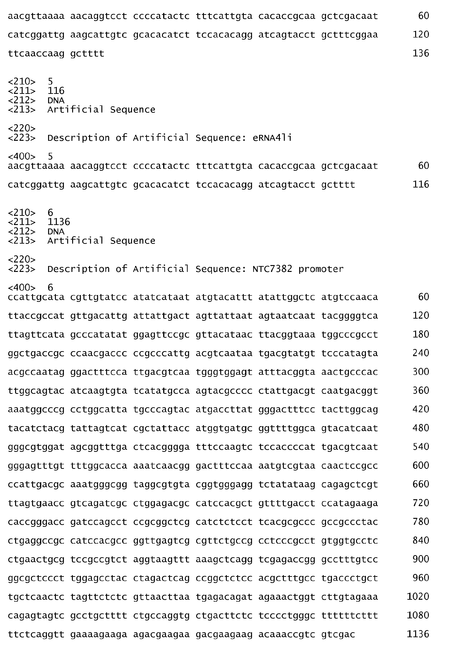


Claims (16)
- A method for production of antibiotic marker-free covalently closed, supercoiled DNA replicon , such as a plasmid, a cosmid, or a bacterial artificial chromosome, in E. coli, comprising the steps of:a. cloning a RNA-OUT antisense RNA into said DNA replicon, andb. transforming said RNA-OUT antisense RNA containing DNA replicon into bacterial cells that express a selectable chromosomal gene under the control of a RNA-IN sense RNA, andc. isolating transformed bacterial cells under selection, andd. propagating said transformed bacterial cells under conditions that accumulate said DNA replicon,whereby said method replaces selection by an antibiotic marker in said DNA replicon, for said DNA replicon isolation or propagation.
- The method of claim 1 wherein said chromosomal gene under control of the RNA-IN sense sequence encodes a levansucrase, and said transformed bacterial cells are isolated by growth in the presence of sucrose.
- The method of claim 1 wherein said antibiotic marker-free DNA replicon is used for production of biological molecules chosen from the group consisting of: plasmids, proteins, RNAs, nucleotides, biosynthetic pathway products, or biosynthetic pathway intermediates, or combinations thereof.
- A composition of matter for a vector comprising:a. an RNA-OUT antisense RNA with at least 90% sequence identity to the sequence set forth as SEQ ID NO: 15, operably linked to,b. a plasmid, cosmid, or bacterial artificial chromosome DNA replicon, whereby said RNA-out antisense RNA is used to select and propagate said DNA replicon.
- A composition of matter for a vector comprising: a. a promoter with at least 95% sequence identity to the sequence set forth as SEQ ID NO: 6 operable linked to, b. a gene of interest, whereby said promoter is used to express said gene of interest.
- A method for modifying the response of a individual or eukaryotic cell to a vector comprising the steps of:a. obtaining a vector comprising at least one immunostimulatory RNA element that comprises a tRNA, optionally a linker, an RNA to be expressed, and an RNA polymerase III promoter arranged in opposite orientation to said tRNA, such that transcription of said RNA is convergent, andb. optionally cloning at least one gene that encode a protein or peptide into said vector, andc. delivering said vector to a eukaryotic cell, andd. evaluating immune response or type 1 interferon production after said delivery of said vector,whereby said RNA element improves immune response and/or improves type 1 interferon production by said individual or said eukaryotic cell.
- The method of claim 6, wherein said vector is chosen from the group consisting of: non-viral vector, viral vector, alphavirus vector, poxvirus vector, lentivirus vector, retrovirus vector, adenovirus vector, adenovirus related virus vector, plasmid vector, midge vector, transcriptionally active PCR fragment vector, minicircle vector, bacteriophage vector.
- A method for modifying the response of an individual to a vector comprising the steps of:a. cloning at least one gene that encodes a protein or peptide into a vector that contains a mitochondrial R region, andb. optionally cloning at least one immunostimulatory RNA element into said vector, andc. delivering said vector to a eukaryotic cell, andd. evaluating immune response after said delivery of said vector,whereby said mitochondrial R region improves immune response to said protein or said peptide
- A method for modifying the response of an individual or eukaryotic cell to at least one vector comprising the steps of:a. obtaining at least one vector containing at least one immunostimulatory RNA element comprising an MDA5 activating VARNA, or an MDA5 activating VARNA derivative, andb. optionally cloning at least one gene encoding a protein or peptide into said vector(s), andc. delivering said vector(s) to said individual or said eukaryotic cell, andd. evaluating immune response or type 1 interferon production after said delivery of said vector(s),whereby said RNA element improves immune response and/or improves type 1 interferon production by said individual or said eukaryotic cell.
- The method of claim 9 , wherein said vector is chosen from the group consisting of: non-viral vector, viral vector, alphavirus vector, poxvirus vector, lentivirus vector, retrovirus vector, adenovirus vector, adenovirus related virus vector, plasmid vector, midge vector, transcriptionally active PCR fragment vector, minicircle vector, bacteriophage vector.
- The method of claim 9, wherein said VARNA comprises VARNAI or VARNAII from any adenoviral serotype, or hybrids thereof, and said VARNA derivative is chosen from the group consisting of: VARNA-short hairpin RNA fusion constructs; VARNA internal deletions or substitutions; VARNA point mutations with at least 90% sequence identity to VARNA; and VARNAI incorporating the pm91 mutation.
- The method of claim 9, wherein said VARNA is combined with at least one additional immunostimulatory RNA element chosen from the group consisting of: tRNA expressed single stranded RNA elements, U6 promoter expressed single stranded RNA elements, and convergent tRNA and U6 promoter expressed double stranded RNA elements.
- The method of claim 9 , wherein said individual is a mammal, bird or fish.
- A composition of matter comprising two immunostimulatory RNA elements, further comprising:a. a VARNA RNA element or a VARNA derivative RNA element, andb. a second RNA element, whereby the combination of said VARNA RNA element with said second RNA element improves type 1 interferon production and/or immune response.
- The composition of claim 14, wherein said VARNA comprises VARNAI or VARNAII from any adenoviral serotype, or hybrids thereof, and said VARNA derivative is chosen from the group consisting of: VARNA-short hairpin RNA fusion constructs; VARNA internal deletions or substitutions; VARNA point mutations with at least 90% sequence identity to VARNA; and VARNAI incorporating the pm91 mutation.
- The composition of claim 14, wherein said second RNA element is chosen from the group consisting of: tRNA expressed single stranded RNA elements; U6 promoter expressed single stranded RNA elements; and convergent tRNA and U6 promoter-expressed double stranded RNA elements.
Priority Applications (1)
| Application Number | Priority Date | Filing Date | Title |
|---|---|---|---|
| EP17176889.8A EP3246409A1 (en) | 2007-05-29 | 2008-05-22 | Antibiotic-resistance-free vectors |
Applications Claiming Priority (2)
| Application Number | Priority Date | Filing Date | Title |
|---|---|---|---|
| US93216007P | 2007-05-29 | 2007-05-29 | |
| EP08767845.4A EP2152889B1 (en) | 2007-05-29 | 2008-05-22 | Vectors and methods for genetic immunization |
Related Parent Applications (2)
| Application Number | Title | Priority Date | Filing Date |
|---|---|---|---|
| EP08767845.4A Division EP2152889B1 (en) | 2007-05-29 | 2008-05-22 | Vectors and methods for genetic immunization |
| EP08767845.4A Division-Into EP2152889B1 (en) | 2007-05-29 | 2008-05-22 | Vectors and methods for genetic immunization |
Related Child Applications (1)
| Application Number | Title | Priority Date | Filing Date |
|---|---|---|---|
| EP17176889.8A Division EP3246409A1 (en) | 2007-05-29 | 2008-05-22 | Antibiotic-resistance-free vectors |
Publications (3)
| Publication Number | Publication Date |
|---|---|
| EP2333091A2 true EP2333091A2 (en) | 2011-06-15 |
| EP2333091A3 EP2333091A3 (en) | 2011-10-05 |
| EP2333091B1 EP2333091B1 (en) | 2017-06-21 |
Family
ID=39964818
Family Applications (3)
| Application Number | Title | Priority Date | Filing Date |
|---|---|---|---|
| EP17176889.8A Withdrawn EP3246409A1 (en) | 2007-05-29 | 2008-05-22 | Antibiotic-resistance-free vectors |
| EP10194048.4A Active EP2333091B1 (en) | 2007-05-29 | 2008-05-22 | Vectors and methods for genetic immunization |
| EP08767845.4A Active EP2152889B1 (en) | 2007-05-29 | 2008-05-22 | Vectors and methods for genetic immunization |
Family Applications Before (1)
| Application Number | Title | Priority Date | Filing Date |
|---|---|---|---|
| EP17176889.8A Withdrawn EP3246409A1 (en) | 2007-05-29 | 2008-05-22 | Antibiotic-resistance-free vectors |
Family Applications After (1)
| Application Number | Title | Priority Date | Filing Date |
|---|---|---|---|
| EP08767845.4A Active EP2152889B1 (en) | 2007-05-29 | 2008-05-22 | Vectors and methods for genetic immunization |
Country Status (4)
| Country | Link |
|---|---|
| US (3) | US9109012B2 (en) |
| EP (3) | EP3246409A1 (en) |
| AU (1) | AU2008262478C1 (en) |
| WO (1) | WO2008153733A2 (en) |
Cited By (4)
| Publication number | Priority date | Publication date | Assignee | Title |
|---|---|---|---|---|
| WO2012085291A1 (en) * | 2010-12-23 | 2012-06-28 | Mologen Ag | Non-coding immunomodulatory dna construct |
| WO2014077866A1 (en) * | 2012-11-19 | 2014-05-22 | Nature Technology Corporation | Replicative minicircle vectors with improved expression |
| US11578331B2 (en) | 2015-09-09 | 2023-02-14 | Gilead Sciences, Inc. | Combination comprising immunostimulatory oligonucleotides |
| US11583581B2 (en) | 2015-09-21 | 2023-02-21 | Gilead Sciences, Inc. | Methods of treating a retroviral infection |
Families Citing this family (23)
| Publication number | Priority date | Publication date | Assignee | Title |
|---|---|---|---|---|
| HUE037173T2 (en) | 2006-08-08 | 2018-08-28 | Univ Bonn Rheinische Friedrich Wilhelms | Structure and use of 5' phosphate oligonucleotides |
| EP2297323A1 (en) * | 2008-05-21 | 2011-03-23 | Hartmann, Gunther | 5' triphosphate oligonucleotide with blunt end and uses thereof |
| US9045759B2 (en) | 2009-01-21 | 2015-06-02 | James Arthur Williams | DNA plasmids with improved copy number |
| US9012226B2 (en) | 2009-03-13 | 2015-04-21 | Nature Technology Corporation | Bacterial strains with improved plasmid stability |
| US9506082B2 (en) | 2010-04-12 | 2016-11-29 | Nature Technology Corporation | Eukaryotic expression vectors resistant to transgene silencing |
| EP2508530A1 (en) | 2011-03-28 | 2012-10-10 | Rheinische Friedrich-Wilhelms-Universität Bonn | Purification of triphosphorylated oligonucleotides using capture tags |
| GB201108879D0 (en) * | 2011-05-25 | 2011-07-06 | Isis Innovation | Vector |
| WO2013119371A2 (en) * | 2012-02-10 | 2013-08-15 | The Board Of Trustees Of The Leland Stanford Junior University | Mini-intronic plasmid vectors |
| AU2013309488A1 (en) | 2012-08-29 | 2015-03-05 | Nature Technology Corporation | DNA plasmids with improved expression |
| EP2711426B1 (en) * | 2012-09-24 | 2015-04-08 | Lonza Biologics plc | Expression vectors comprising chimeric cytomegalovirus promoter and enhancer sequences |
| EP2712870A1 (en) | 2012-09-27 | 2014-04-02 | Rheinische Friedrich-Wilhelms-Universität Bonn | Novel RIG-I ligands and methods for producing them |
| RU2678756C1 (en) * | 2017-08-25 | 2019-01-31 | Селл энд Джин Терапи Лтд | Vtvaf17 gene-therapy dna vector, method for producing thereof, escherichia coli scs110-af strain, method for producing thereof, escherichia coli scs110-af/vtvaf17 strain, bearing vtvaf17 gene-therapy dna vector, method for producing thereof |
| EP3456821B2 (en) | 2017-09-19 | 2024-01-24 | Deutsches Krebsforschungszentrum | Non-integrating dna vectors for the genetic modification of cells |
| WO2019057774A1 (en) | 2017-09-19 | 2019-03-28 | Deutsches Krebsforschungszentrum | Non-integrating dna vectors for the genetic modification of cells |
| DK3768846T3 (en) | 2018-03-21 | 2023-07-24 | Aldevron L L C | VIRAL AND NON-VIRAL NANOPLASMID VECTORS WITH ENHANCED PRODUCTION |
| CA3114205A1 (en) | 2018-10-12 | 2020-04-16 | Amryt Genetics Ltd. | Compositions and methods for transfecting cells |
| MX2021007688A (en) | 2018-12-28 | 2021-08-05 | Amryt Genetics Ltd | Methods for forming polyplexes. |
| KR20220128607A (en) | 2019-09-18 | 2022-09-21 | 인터갈락틱 테라퓨틱스, 인크. | Synthetic DNA Vectors and Methods of Use |
| JP2023538519A (en) | 2020-08-07 | 2023-09-08 | スペースクラフト セブン リミテッド ライアビリティ カンパニー | Plakophilin-2 (PKP2) gene therapy using AAV vectors |
| EP4243839A1 (en) | 2020-11-13 | 2023-09-20 | Catamaran Bio, Inc. | Genetically modified natural killer cells and methods of use thereof |
| US20230055381A1 (en) | 2021-06-25 | 2023-02-23 | Oxford Biomedica Solutions Llc | Adeno-associated virus packaging systems |
| US20230279433A1 (en) | 2021-12-15 | 2023-09-07 | Oxford Biomedica Solutions Llc | Methods and compositions for the production of adeno-associated virus |
| WO2023172514A1 (en) | 2022-03-07 | 2023-09-14 | Catamaran Bio, Inc. | Engineered immune cell therapeutics targeted to her2 and methods of use thereof |
Citations (14)
| Publication number | Priority date | Publication date | Assignee | Title |
|---|---|---|---|---|
| US5922583A (en) | 1995-10-17 | 1999-07-13 | Biostar Inc. | Methods for production of recombinant plasmids |
| WO2000061770A2 (en) | 1999-04-08 | 2000-10-19 | Chiron Corporation | Enhancement of the immune response for vaccine and gene therapy applications |
| WO2002019968A2 (en) | 2000-09-08 | 2002-03-14 | University Of Maryland Biotechnology Institute | Genetically engineered co-expression dna vaccines, construction methods and uses thereof |
| US6500432B1 (en) | 1997-12-16 | 2002-12-31 | Smithkline Beecham Biologicals, S.A. | Method to enhance an immune response of nucleic acid vaccination |
| US20040024140A1 (en) | 2000-09-05 | 2004-02-05 | Yukiko Fujita | Unsaturated polyester resin composition |
| US20040096462A1 (en) | 2001-02-15 | 2004-05-20 | Rangarajan Pundi Narasimhan | Noval vaccine formulation consisting of dna vaccine and inactivated virus |
| US20040234543A1 (en) | 2001-07-16 | 2004-11-25 | Carrera Santiago Duenas | Vaccine formulation potentiated by the combination of a dna and an antigen |
| US6852535B1 (en) | 1994-08-19 | 2005-02-08 | Sirna Therapeutics, Inc. | Polymerase III-based expression of therapeutic RNAS |
| WO2005052167A2 (en) | 2003-11-20 | 2005-06-09 | Cobra Biologics Limited | Plasmid maintenance |
| US20050130184A1 (en) | 2003-07-18 | 2005-06-16 | University Of Massachusetts | Enhanced promoters for synthesis of small hairpin RNA |
| WO2006023546A2 (en) | 2004-08-19 | 2006-03-02 | Nature Technology Corp. | Process for plasmid dna fermentation |
| US20060063232A1 (en) | 2004-09-17 | 2006-03-23 | Boehringer Ingelheim Austria Gmbh | Host-vector system for antibiotic-free CoIE1 plasmid propagation |
| WO2006078979A2 (en) | 2005-01-20 | 2006-07-27 | Nature Technology Corp. | Vectors and methods for genetic immunization |
| US7094598B2 (en) | 2001-10-01 | 2006-08-22 | The United States Of America As Represented By The Department Of Health And Human Services | Development of a preventive vaccine for filovirus infection in primates |
Family Cites Families (6)
| Publication number | Priority date | Publication date | Assignee | Title |
|---|---|---|---|---|
| WO2002036806A2 (en) | 2000-11-01 | 2002-05-10 | The Government Of The United States Of America, Represented By The Secretary, Department Of Health And Human Services | Expression vectors able to elicit improved immune response and methods of using same |
| USH2220H1 (en) * | 2001-08-10 | 2008-07-01 | Snp Consortium | Identification and mapping of single nucleotide polymorphisms in the human genome |
| CN1913919B (en) * | 2003-09-15 | 2011-10-19 | 美国政府健康及人类服务部 | HIV vaccines based on env of multiple clades of hiv |
| JP2005094415A (en) * | 2003-09-18 | 2005-04-07 | Fuji Xerox Co Ltd | Message distribution system |
| WO2005094415A2 (en) * | 2004-02-06 | 2005-10-13 | The Wistar Institute | Recombinant vectors and methods for inducing an immune response |
| EP1805296B1 (en) * | 2004-10-22 | 2009-09-16 | Novozymes A/S | Stable genomic integration of multiple polynucleotide copies |
-
2008
- 2008-05-22 US US12/601,970 patent/US9109012B2/en active Active
- 2008-05-22 EP EP17176889.8A patent/EP3246409A1/en not_active Withdrawn
- 2008-05-22 EP EP10194048.4A patent/EP2333091B1/en active Active
- 2008-05-22 EP EP08767845.4A patent/EP2152889B1/en active Active
- 2008-05-22 AU AU2008262478A patent/AU2008262478C1/en active Active
- 2008-05-22 WO PCT/US2008/006554 patent/WO2008153733A2/en active Application Filing
-
2015
- 2015-06-24 US US14/748,846 patent/US9737620B2/en active Active
- 2015-06-29 US US14/753,438 patent/US9950081B2/en active Active
Patent Citations (14)
| Publication number | Priority date | Publication date | Assignee | Title |
|---|---|---|---|---|
| US6852535B1 (en) | 1994-08-19 | 2005-02-08 | Sirna Therapeutics, Inc. | Polymerase III-based expression of therapeutic RNAS |
| US5922583A (en) | 1995-10-17 | 1999-07-13 | Biostar Inc. | Methods for production of recombinant plasmids |
| US6500432B1 (en) | 1997-12-16 | 2002-12-31 | Smithkline Beecham Biologicals, S.A. | Method to enhance an immune response of nucleic acid vaccination |
| WO2000061770A2 (en) | 1999-04-08 | 2000-10-19 | Chiron Corporation | Enhancement of the immune response for vaccine and gene therapy applications |
| US20040024140A1 (en) | 2000-09-05 | 2004-02-05 | Yukiko Fujita | Unsaturated polyester resin composition |
| WO2002019968A2 (en) | 2000-09-08 | 2002-03-14 | University Of Maryland Biotechnology Institute | Genetically engineered co-expression dna vaccines, construction methods and uses thereof |
| US20040096462A1 (en) | 2001-02-15 | 2004-05-20 | Rangarajan Pundi Narasimhan | Noval vaccine formulation consisting of dna vaccine and inactivated virus |
| US20040234543A1 (en) | 2001-07-16 | 2004-11-25 | Carrera Santiago Duenas | Vaccine formulation potentiated by the combination of a dna and an antigen |
| US7094598B2 (en) | 2001-10-01 | 2006-08-22 | The United States Of America As Represented By The Department Of Health And Human Services | Development of a preventive vaccine for filovirus infection in primates |
| US20050130184A1 (en) | 2003-07-18 | 2005-06-16 | University Of Massachusetts | Enhanced promoters for synthesis of small hairpin RNA |
| WO2005052167A2 (en) | 2003-11-20 | 2005-06-09 | Cobra Biologics Limited | Plasmid maintenance |
| WO2006023546A2 (en) | 2004-08-19 | 2006-03-02 | Nature Technology Corp. | Process for plasmid dna fermentation |
| US20060063232A1 (en) | 2004-09-17 | 2006-03-23 | Boehringer Ingelheim Austria Gmbh | Host-vector system for antibiotic-free CoIE1 plasmid propagation |
| WO2006078979A2 (en) | 2005-01-20 | 2006-07-27 | Nature Technology Corp. | Vectors and methods for genetic immunization |
Non-Patent Citations (84)
| Title |
|---|
| ANDERSSON MG; HAASNOOT PCJ; XU N; BERENJIAN S; BERKHOUT B; AKUSJARVI G., J. VIROL., vol. 79, 2005, pages 9556 - 9565 |
| APPLEQUIST SE; ROLLMAN E; WAREING MD; LIDEN M; ROZELL B; HINKULA J; LJUNGGREN HG, J IMMUNOL., vol. 175, 2005, pages 3882 - 3891 |
| BARCHET W; KRUG A; CELLA M; NEWBY C; FISCHER JAA; DZIONEK A; PEKOSZ A; COLONNA M., EUR. J 1MMUNOL., vol. 35, 2005, pages 236 - 242 |
| BAROUCH DH; YANG ZY; KONG WP; KORIOTH-SCHMITZ B; SUMIDA SM; TRUITT DM; KISHKO MG; ARTHUR JC; MIURA A; MASCOLA JR, J VIROL., vol. 79, 2005, pages 8828 - 8834 |
| BERGLUND P; SMERDOU C; FLEETON MN; TUBULEKAS 1; LILJESTROM P, NATURE BIOTECHNOL, vol. L6, 1998, pages 562 - 565 |
| BOWIE AG; FITZGERALD KA, TRENDS IMMUNOL, vol. 28, 2007, pages 147 - 150 |
| BUCHAN S; GRONEVIK E; MATHIESEN 1; KING C; STEVENSON FK; RICE J., TMMUNOL., vol. 174, 2005, pages 6292 - 6298 |
| BUZAN JM; LOW RL, BIOCHEM. BIOPHYS. RES. COMMUN., vol. 152, 1988, pages 22 |
| CARDON, LR; BURGE C; CLAYTON DA; KARLIN S, PROC NATL. ACAD. SCI, vol. 91, 1994, pages 3799 - 3803 |
| CHANG TH; LIAO CL; LIN YL, MICROB INFECT, vol. 8, 2006, pages 157 - 171 |
| CHANG Z; WESTAWAY S; LI S; ZAIA JA; ROSSI JJ; SCHERER LJ, MOL. THER., vol. 6, 2002, pages 481 - 489 |
| CHATTERGOON MA; SAULINO V; SHAMES JP; STEIN J; MONTANER LJ; WEINER DB., VACCINE, vol. 22, 2004, pages 1744 - 1750 |
| COLLOMBET JM; WHEELER VC; VOGEL F; COUTELLE C., JBIOL CHEM., vol. 272, 1997, pages 5342 |
| DEAN DA; DEAN BS; MULLER S; SMITH LC., EXPERIMENTAL CELL RESEARCH, vol. 253, 1999, pages 713 - 722 |
| DESAI SY; PATEL RC; SEN GC; MALHOTRA P; GHADGE GD; THIMMAPAYA B, J BIOL. CHEM, vol. 270, 1995, pages 3454 - 3461 |
| DIEBOLD SS; KAISHO T; HEMMI H; AKIRA S; E SOUSA CR, SCIENCE, vol. 303, 2004, pages 1529 - 1531 |
| FANG J; BREDOW S; TAISHI P; MAJDE JA; KRUEGER JM, J MED VIROL, vol. 57, 1999, pages 198 - 203 |
| GANTIER MP; WILLIAMS BRG, CYTOKINES GROWTH FACTOR REVIEW, vol. 18, 2007, pages 363 - 371 |
| GARG S; ORAN AE; HON H; JACOB J., JLMMUNOL., vol. 173, 2002, pages 550 - 558 |
| GASPARI, M; FALKENBERG M; LARSSON NG; GUSTAFSSON CM., EMBO J, vol. 23, 2004, pages 46064614 |
| GITLIN L; BARCHET W; GILFILLAN S; CELLA M; BEUTLER B; FLAVELL RA; DIAMOND MS; COLONNA M, PROC. NATL. ACAD. SCI., vol. 103, 2006, pages 8459 - 8464 |
| GURUNATHAN S; KLINMAN DM; SEDER RA, ANNU. REV. IMMUNOL., vol. 18, 2000, pages 927 - 974 |
| HALDIMANN A; WANNER BL, J. BACTERIOL., vol. 183, 2001, pages 6384 - 6393 |
| HASSANI Z; FRANCOIS JC; ALFAMA G ET AL., NUCL. ACID RES., vol. 35, 2007, pages E65 |
| HEIL F; HEMMI H; HOCHREIN H; AMPENBERGER F; KIRSCHNING C; AKIRA S; LIPFORD G; WAGNER H; BAUER S, SCIENCE, vol. 303, 2004, pages 1526 - 1529 |
| HORNUNG V.; ELLEGAST J; KIM S; BRZOZKA K; JUNG A; KATO H ET AL., SCIENCE, vol. 314, 2006, pages 994 - 997 |
| HORNUNG V; GUENTHNER-BILLER M; BOURQUIN C; ABLASSER A; SCHLEE M; UEMATSU S; NORONHA A; MANOHARAN M; AKIRA S; DE FOUGEROLLES A, NATURE MED., vol. 11, 2005, pages 263 - 270 |
| JARCZAK D; KORF M; BEGER C; MANNS MP; KRUGER M, FEBS J, vol. 272, 2005, pages 5910 - 5922 |
| KATO, H; TAKEUCHI 0; SATO S; YONEYAMA M; YAMAMOTO M; MATSUI K ET AL., NATURE, vol. 441, 2006, pages 101 - 105 |
| KAWAI T; TAKAHASHI K; SATO S; BOBAN C; KUMAR H; KATO H; ISHII KH; TAKEUCHI 0; AKIRA S., NATURE IMMUNOL., vol. 6, 2005, pages 981 - 988 |
| KIM TW; HUNG CF; JUANG J; HE L; HARDWICK JM; WU TC, GENE THER, vol. 11, 2004, pages 336 - 342 |
| KOULINTCHENKO M; TEMPERLEY RJ; MASON PA; DIETRICH A; LIGHTOWLERS, RN, HUMAN MOLEC GENET, vol. 15, 2006, pages 143 |
| KUTZLER MA; ROBINSON TM; CHATTERGOON MA; CHOO DK; CHOO AY; CHOE PY; RAMAMATHAN MP; PARKINSON R; KUDCHODKAR S; TAMURA Y, JLMMUNOL, vol. 175, 2005, pages 112 - 123 |
| KUWABARA T; WARASHINA M; KOSEKI S; SANO M; OHKAWA J; NAKAYAMA K; TAIRA K, NUC. ACIDS RES., vol. 29, 2001, pages 2780 - 2788 |
| LAENECKE S; DE LORENZO V; TIMMIS KN; DIAZ E., MALEC. MICROBIOL., vol. 21, 1996, pages 293 - 300 |
| LAMB RA; CHOPPIN PW., ANNU REV BIOCHEM, vol. 52, 1983, pages 467 - 506 |
| LAVIGUEUR A.; LA BRANCHE H; KOMBLIHTT AR; CHABOT B., GENES DEVELOP., vol. 7, 1993, pages 2405 |
| LEE SW; SUNG YC., IMMUNOLOGY, vol. 94, 1998, pages 285 - 289 |
| LEI M; LIU Y; SAMUEL CE, VIROLOGY, vol. 245, 1998, pages 188 - 196 |
| LEITNER WW; HWANG LN; BERGMANN-LEITNER ES; FINKELSTEIN SE; FRANK S; RESTIFO NP, VACCINE, vol. 22, 2004, pages 1537 - 1544 |
| LEITNER WW; HWANG LN; DEVEER MJ; ZHOU A; SILVERMAN RH; WILLIAMS BRG; DUBENSKY TW; YING H; RESTIFO NP, NATURE MED, vol. 9, 2003, pages 33 - 39 |
| LEITNER WW; RESTIFO NP, JCLIN INVEST, vol. 112, 2003, pages 22 - 24 |
| LEITNER WW; YING H; DRIVER DA; DUBENSKY TW; RESTIFO NP, CANCER RESEARCH, vol. 60, 2000, pages 51 - 55 |
| LEMIEUX, P., EXPERT REV. VACCINES, vol. 1, 2002, pages 85 - 93 |
| LOPEZ CB; JOUNT JS; MORAN, TM., J VIROL., vol. 80, 2006, pages 3128 |
| LU S.; CULLEN BR., J. VIRAL., vol. 78, 2004, pages 12868 - 12876 |
| MANOJ S; BABIUK LA; DRUNEN SV; HURK LV, CRITICAL REV CLIN LAB SCI, vol. 41, 2004, pages 1 - 39 |
| MARQUES JT; DEVOSSE T; WANG D; ZAMANIAN-DARYOUSH M; SERBINOWSKI P; HARTMANN R; FUJITA T; BEHLKE MA; WILLIAMS BR, NATURE BIOTECHNOL., vol. 24, 2006, pages 559 - 565 |
| MATSUMOTO S; MIYAGISHI M; AKASHI H; NAGAI R; TAIRA K, J BIOL CHEM, vol. 280, 2005, pages 25687 - 25696 |
| MELCHJORSEN J; JENSEN SB; MALMGAARD L; RASMUSSEN SB; WEBER F; BOWIE AG; MATIKAINEN S; PALUDAN SR, J. VIROL., vol. 79, 2005, pages 12944 - 12951 |
| MOLLER T; FRANCH T; UDESEN C; GERDES K; VALENTIN-HANSEN, GENES DEVEL., vol. 16, 2002, pages 1696 |
| NOCIARI M; OCHERETINA 0; SCHOGGINS JW; FALCK-PEDERSEN E., J. VIROL., vol. 81, 2007, pages 4145 - 4157 |
| OPERSCHALL E; PAVLOVIC J; NAWRATH M; MOLLING K., INTERVIROL, vol. 43, 2000, pages 322 - 330 |
| OPITZ B; REJAIBI A; DAUBER B; ECKHARD J; VINZING M; SCHMECK B; HIPPENSTIEL S; SUTTORP N; WOLFF T., CELL MICROBIOL., vol. 9, 2007, pages 930 - 938 |
| PARRY HD; MATTAJ IW, EMBO J, vol. 9, pages 1097 - 1990 |
| PEBEMARD S; LGGO RD, DIFFERENTIATION, vol. 72, 2004, pages 103 - 111 |
| PEPE CM; MASLESA-GALIC S; SIMONS RW., MOL. MICROBIOL, vol. 13, 1994, pages 1133 |
| PICHLMAIR A; SCHULZ 0; TAN CP; NASLUND TI; LILJESTROM P; WEBER F; SOUSA C., SCIENCE, vol. 314, 2006, pages 997 - 1001 |
| PLUMET S; HERSCHKE F; BOURHIS JM; VALENTIN H; LONGHI S; GERLIER D., PLOS, vol. 2, 2007, pages E279 |
| RAHMAN A; MALHOTRA P; SHAR R; KEWALRAMANI T; THIMMAPAYA B, J VIROLOGY, vol. 69, 1995, pages 4299 - 4307 |
| REYRAT JM; PELICIC V; GICQUEL B; RAPPUOLI R, INFECTION IMMUN., vol. 66, 1998, pages 4011 - 4017 |
| RODRIGUEZ MS; DARGEMONT C; STUTZ F., BIOL CELL, vol. 96, 2004, pages 639 - 655 |
| SAITO T; HIRAI R; LOO YM; OWEN D; JOHNSON CL; SINHA SC; AKIRA S; FUJITA T; GALE, M, PROC. NATL. ACAD. SCI., vol. 104, 2007, pages 582 - 587 |
| SAMANTA M; IWAKIRA D; KANDA T; IMAIZUMI T; TAKADA K., EMBO J., vol. 25, 2006, pages 2407 |
| SASAKI S; AMARA RR; ORAN AE; SMITH JM; ROBINSON HL, NATURE BIOTECHNOL, vol. 19, 2001, pages 543 - 547 |
| SCHENBORN, E., PROMEGA NOTES, vol. 48, 1994, pages 8.12 |
| SCHRAMM L; HERNANDEZN., GENES DEV, vol. 16, 2002, pages 2593 - 2620 |
| SCHRODER M; BOWIE AG, TRENDS IMMUNOL., vol. 26, 2005, pages 462 - 468 |
| SEWARDS R; WISEMAN B; JACOBS HT, MOL. GEN. GENET., vol. 245, 1994, pages 760 |
| SINGH R; GUPTA S; REDDY R., MOL. CELL BIOL., vol. 10, 1990, pages 939 - 946 |
| SINGH R; REDDY R, PROC. NAT. ACAD. SCI. USA, vol. 86, 1989, pages 8280 - 8283 |
| SMULEVITCH S; BEAR J; ALICEA C; ROSATI M; JALAH R; ZOLOTUKHIN AS ET AL., RETROVIROLOGY, vol. 3, 2006, pages 6 |
| STRAT A; GAO L; UTSUKI T; CHENG B; NUTHALAPATY S; MATHIS JM; ODAKA Y; GIORDANO T., NUCL. ACIDRES., vol. 34, 2006, pages 3803 - 3810 |
| SUGIYAMA T; GURSEL M; TAKESHITA F; COBAN C; CONOVER J; KAISHO T; AKIRA S; KLINMAN DM; ISHII KJ., J IMMUNOL., vol. 174, 2005, pages 2273 - 2279 |
| SUMPTER R JR; LOO YM; FOY E; LI K; YONEYAMA M; FUJITA T; LEMON SM; GALE M JR., J. VIROL., vol. 79, 2005, pages 2689 - 2699 |
| SZAFRANSKI P; MELLO CM; SANO T; SMITH CL; KAPLAN DL; CNATOR CR., PROC. NATL. ACAD. SCI., vol. 94, 1997, pages 1059 |
| TOKA FN; PACK CD; ROUSE BT, IMMUNOLOGICAL REVIEWS, vol. 199, 2004, pages 100 - 112 |
| VYAS J; ELIA A; CLEMENS MJ., RNA, vol. 9, 2003, pages 858 - 870 |
| WANG Q; CARMICHAEL GG, MICROB. MOLEC. BIOL. REV., vol. 68, 2004, pages 432 - 452 |
| WEBER F; WAGNER V; KESSLER N; HALLER 0., JLNTERFERON CYTOKINE RES, vol. 26, 2006, pages 1 - 7 |
| WHITMORE MM; DEVEER MJ; EDLING A; OATES RK; SIMONS B; LINDER D; WILLIAMS BRG, CANCER RESEARCH, vol. 64, 2004, pages 5850 - 5860 |
| WINKELMAN JW; CLARK DP, JBACTERIOL., vol. 160, 1984, pages 687 |
| YOON YG; KOOB, MD, NUCL. ACIDS RES., vol. 31, 2003, pages 1407 - 1415 |
| ZUKER, M., NUCLEIC ACIDS RES., vol. 31, 2003, pages 3406 - 15 |
Cited By (10)
| Publication number | Priority date | Publication date | Assignee | Title |
|---|---|---|---|---|
| WO2012085291A1 (en) * | 2010-12-23 | 2012-06-28 | Mologen Ag | Non-coding immunomodulatory dna construct |
| WO2014077866A1 (en) * | 2012-11-19 | 2014-05-22 | Nature Technology Corporation | Replicative minicircle vectors with improved expression |
| WO2014077863A1 (en) * | 2012-11-19 | 2014-05-22 | Nature Technology Corporation | Replicative minicircle vectors with improved expression |
| US10047365B2 (en) | 2012-11-19 | 2018-08-14 | Nature Technology Corporation | Replicative minicircle vectors with improved expression |
| US10167478B2 (en) | 2012-11-19 | 2019-01-01 | Nature Technology Corporation | Replicative minicircle vectors with improved expression |
| US10844388B2 (en) | 2012-11-19 | 2020-11-24 | Nature Technology Corporation | Replicative minicircle vectors with improved expression |
| US11098313B2 (en) | 2012-11-19 | 2021-08-24 | Nature Technology Corporation | Replicative minicircle vectors with improved expression |
| US11851665B2 (en) | 2012-11-19 | 2023-12-26 | Aldevron, L.L.C. | Replicative minicircle vectors with improved expression |
| US11578331B2 (en) | 2015-09-09 | 2023-02-14 | Gilead Sciences, Inc. | Combination comprising immunostimulatory oligonucleotides |
| US11583581B2 (en) | 2015-09-21 | 2023-02-21 | Gilead Sciences, Inc. | Methods of treating a retroviral infection |
Also Published As
| Publication number | Publication date |
|---|---|
| EP2333091A3 (en) | 2011-10-05 |
| AU2008262478B9 (en) | 2014-06-19 |
| EP2152889A2 (en) | 2010-02-17 |
| US9737620B2 (en) | 2017-08-22 |
| EP3246409A1 (en) | 2017-11-22 |
| US20150344909A1 (en) | 2015-12-03 |
| EP2152889B1 (en) | 2017-03-15 |
| WO2008153733A8 (en) | 2016-07-14 |
| AU2008262478C1 (en) | 2014-06-19 |
| US20100303859A1 (en) | 2010-12-02 |
| US9950081B2 (en) | 2018-04-24 |
| AU2008262478A1 (en) | 2008-12-18 |
| AU2008262478B2 (en) | 2014-03-20 |
| US9109012B2 (en) | 2015-08-18 |
| WO2008153733A2 (en) | 2008-12-18 |
| WO2008153733A3 (en) | 2009-05-14 |
| US20160008488A1 (en) | 2016-01-14 |
| EP2333091B1 (en) | 2017-06-21 |
Similar Documents
| Publication | Publication Date | Title |
|---|---|---|
| US9950081B2 (en) | Vectors and methods for genetic immunization | |
| Pepini et al. | Induction of an IFN-mediated antiviral response by a self-amplifying RNA vaccine: implications for vaccine design | |
| JP2023126888A (en) | Compositions comprising circular polyribonucleotides and uses thereof | |
| JP7189943B2 (en) | Non-Integrating DNA Vectors for Genetic Modification of Cells | |
| EP1844142B1 (en) | Vectors and methods for genetic immunization | |
| JP2021138721A (en) | Hiv pre-immunization and immunotherapy | |
| JP2012080889A (en) | Multiple-compartment eukaryotic expression systems | |
| JP2019520814A (en) | Non-integrated virus delivery system and related method | |
| US8796235B2 (en) | Methods for attenuating dengue virus infection | |
| CN116322760A (en) | Coronavirus antigen composition and use thereof | |
| WO2022162027A2 (en) | Method of reducing the immunostimulatory properties of in vitro transcribed rna | |
| JP2021534182A (en) | Immunogenic compositions and their use | |
| US20110243904A1 (en) | Rna interference target for treating aids | |
| US9125845B2 (en) | DNA vaccines, uses for unprocessed rolling circle amplification product and methods for making the same | |
| CN116157148A (en) | Immunogenic compositions and uses thereof | |
| WO2010113647A1 (en) | Vector utilizing borna disease virus and use thereof | |
| KR20230127069A (en) | Self-transcribing RNA/DNA system that provides mRNAs in the cytoplasm | |
| KR20220055399A (en) | Self-transcribing RNA/DNA system that provides mRNAs in the cytoplasm | |
| WO2023133422A1 (en) | Compositions and methods for delivering cargo to a target cell | |
| Andries | mRNA modification and delivery strategies towards the establishment of a platform for safe and effective gene therapy | |
| WO2023133425A1 (en) | Compositions and methods for delivering cargo to a target cell | |
| WO2024006988A2 (en) | Engineered delivery vesicles and uses thereof |
Legal Events
| Date | Code | Title | Description |
|---|---|---|---|
| PUAI | Public reference made under article 153(3) epc to a published international application that has entered the european phase |
Free format text: ORIGINAL CODE: 0009012 |
|
| AC | Divisional application: reference to earlier application |
Ref document number: 2152889 Country of ref document: EP Kind code of ref document: P |
|
| AK | Designated contracting states |
Kind code of ref document: A2 Designated state(s): AT BE BG CH CY CZ DE DK EE ES FI FR GB GR HR HU IE IS IT LI LT LU LV MC MT NL NO PL PT RO SE SI SK TR |
|
| AX | Request for extension of the european patent |
Extension state: AL BA MK RS |
|
| PUAL | Search report despatched |
Free format text: ORIGINAL CODE: 0009013 |
|
| AK | Designated contracting states |
Kind code of ref document: A3 Designated state(s): AT BE BG CH CY CZ DE DK EE ES FI FR GB GR HR HU IE IS IT LI LT LU LV MC MT NL NO PL PT RO SE SI SK TR |
|
| AX | Request for extension of the european patent |
Extension state: AL BA MK RS |
|
| RIC1 | Information provided on ipc code assigned before grant |
Ipc: C12N 15/70 20060101ALI20110831BHEP Ipc: C12N 15/67 20060101ALI20110831BHEP Ipc: A61K 39/00 20060101ALI20110831BHEP Ipc: C12N 15/85 20060101AFI20110831BHEP |
|
| 17P | Request for examination filed |
Effective date: 20120309 |
|
| 17Q | First examination report despatched |
Effective date: 20140416 |
|
| GRAP | Despatch of communication of intention to grant a patent |
Free format text: ORIGINAL CODE: EPIDOSNIGR1 |
|
| INTG | Intention to grant announced |
Effective date: 20161028 |
|
| GRAS | Grant fee paid |
Free format text: ORIGINAL CODE: EPIDOSNIGR3 |
|
| GRAJ | Information related to disapproval of communication of intention to grant by the applicant or resumption of examination proceedings by the epo deleted |
Free format text: ORIGINAL CODE: EPIDOSDIGR1 |
|
| GRAL | Information related to payment of fee for publishing/printing deleted |
Free format text: ORIGINAL CODE: EPIDOSDIGR3 |
|
| GRAP | Despatch of communication of intention to grant a patent |
Free format text: ORIGINAL CODE: EPIDOSNIGR1 |
|
| INTG | Intention to grant announced |
Effective date: 20170322 |
|
| GRAA | (expected) grant |
Free format text: ORIGINAL CODE: 0009210 |
|
| AC | Divisional application: reference to earlier application |
Ref document number: 2152889 Country of ref document: EP Kind code of ref document: P |
|
| AK | Designated contracting states |
Kind code of ref document: B1 Designated state(s): AT BE BG CH CY CZ DE DK EE ES FI FR GB GR HR HU IE IS IT LI LT LU LV MC MT NL NO PL PT RO SE SI SK TR |
|
| REG | Reference to a national code |
Ref country code: GB Ref legal event code: FG4D |
|
| REG | Reference to a national code |
Ref country code: CH Ref legal event code: EP |
|
| REG | Reference to a national code |
Ref country code: IE Ref legal event code: FG4D |
|
| REG | Reference to a national code |
Ref country code: AT Ref legal event code: REF Ref document number: 902979 Country of ref document: AT Kind code of ref document: T Effective date: 20170715 |
|
| REG | Reference to a national code |
Ref country code: DE Ref legal event code: R096 Ref document number: 602008050827 Country of ref document: DE |
|
| REG | Reference to a national code |
Ref country code: NL Ref legal event code: FP |
|
| REG | Reference to a national code |
Ref country code: CH Ref legal event code: NV Representative=s name: FIAMMENGHI-FIAMMENGHI, CH |
|
| PG25 | Lapsed in a contracting state [announced via postgrant information from national office to epo] |
Ref country code: FI Free format text: LAPSE BECAUSE OF FAILURE TO SUBMIT A TRANSLATION OF THE DESCRIPTION OR TO PAY THE FEE WITHIN THE PRESCRIBED TIME-LIMIT Effective date: 20170621 Ref country code: HR Free format text: LAPSE BECAUSE OF FAILURE TO SUBMIT A TRANSLATION OF THE DESCRIPTION OR TO PAY THE FEE WITHIN THE PRESCRIBED TIME-LIMIT Effective date: 20170621 Ref country code: LT Free format text: LAPSE BECAUSE OF FAILURE TO SUBMIT A TRANSLATION OF THE DESCRIPTION OR TO PAY THE FEE WITHIN THE PRESCRIBED TIME-LIMIT Effective date: 20170621 Ref country code: GR Free format text: LAPSE BECAUSE OF FAILURE TO SUBMIT A TRANSLATION OF THE DESCRIPTION OR TO PAY THE FEE WITHIN THE PRESCRIBED TIME-LIMIT Effective date: 20170922 Ref country code: NO Free format text: LAPSE BECAUSE OF FAILURE TO SUBMIT A TRANSLATION OF THE DESCRIPTION OR TO PAY THE FEE WITHIN THE PRESCRIBED TIME-LIMIT Effective date: 20170921 |
|
| REG | Reference to a national code |
Ref country code: LT Ref legal event code: MG4D |
|
| PG25 | Lapsed in a contracting state [announced via postgrant information from national office to epo] |
Ref country code: BG Free format text: LAPSE BECAUSE OF FAILURE TO SUBMIT A TRANSLATION OF THE DESCRIPTION OR TO PAY THE FEE WITHIN THE PRESCRIBED TIME-LIMIT Effective date: 20170921 Ref country code: SE Free format text: LAPSE BECAUSE OF FAILURE TO SUBMIT A TRANSLATION OF THE DESCRIPTION OR TO PAY THE FEE WITHIN THE PRESCRIBED TIME-LIMIT Effective date: 20170621 Ref country code: LV Free format text: LAPSE BECAUSE OF FAILURE TO SUBMIT A TRANSLATION OF THE DESCRIPTION OR TO PAY THE FEE WITHIN THE PRESCRIBED TIME-LIMIT Effective date: 20170621 |
|
| PG25 | Lapsed in a contracting state [announced via postgrant information from national office to epo] |
Ref country code: CZ Free format text: LAPSE BECAUSE OF FAILURE TO SUBMIT A TRANSLATION OF THE DESCRIPTION OR TO PAY THE FEE WITHIN THE PRESCRIBED TIME-LIMIT Effective date: 20170621 Ref country code: RO Free format text: LAPSE BECAUSE OF FAILURE TO SUBMIT A TRANSLATION OF THE DESCRIPTION OR TO PAY THE FEE WITHIN THE PRESCRIBED TIME-LIMIT Effective date: 20170621 Ref country code: SK Free format text: LAPSE BECAUSE OF FAILURE TO SUBMIT A TRANSLATION OF THE DESCRIPTION OR TO PAY THE FEE WITHIN THE PRESCRIBED TIME-LIMIT Effective date: 20170621 Ref country code: EE Free format text: LAPSE BECAUSE OF FAILURE TO SUBMIT A TRANSLATION OF THE DESCRIPTION OR TO PAY THE FEE WITHIN THE PRESCRIBED TIME-LIMIT Effective date: 20170621 |
|
| PG25 | Lapsed in a contracting state [announced via postgrant information from national office to epo] |
Ref country code: PL Free format text: LAPSE BECAUSE OF FAILURE TO SUBMIT A TRANSLATION OF THE DESCRIPTION OR TO PAY THE FEE WITHIN THE PRESCRIBED TIME-LIMIT Effective date: 20170621 Ref country code: ES Free format text: LAPSE BECAUSE OF FAILURE TO SUBMIT A TRANSLATION OF THE DESCRIPTION OR TO PAY THE FEE WITHIN THE PRESCRIBED TIME-LIMIT Effective date: 20170621 Ref country code: IS Free format text: LAPSE BECAUSE OF FAILURE TO SUBMIT A TRANSLATION OF THE DESCRIPTION OR TO PAY THE FEE WITHIN THE PRESCRIBED TIME-LIMIT Effective date: 20171021 Ref country code: IT Free format text: LAPSE BECAUSE OF FAILURE TO SUBMIT A TRANSLATION OF THE DESCRIPTION OR TO PAY THE FEE WITHIN THE PRESCRIBED TIME-LIMIT Effective date: 20170621 |
|
| REG | Reference to a national code |
Ref country code: DE Ref legal event code: R097 Ref document number: 602008050827 Country of ref document: DE |
|
| PLBE | No opposition filed within time limit |
Free format text: ORIGINAL CODE: 0009261 |
|
| STAA | Information on the status of an ep patent application or granted ep patent |
Free format text: STATUS: NO OPPOSITION FILED WITHIN TIME LIMIT |
|
| PG25 | Lapsed in a contracting state [announced via postgrant information from national office to epo] |
Ref country code: DK Free format text: LAPSE BECAUSE OF FAILURE TO SUBMIT A TRANSLATION OF THE DESCRIPTION OR TO PAY THE FEE WITHIN THE PRESCRIBED TIME-LIMIT Effective date: 20170621 |
|
| REG | Reference to a national code |
Ref country code: FR Ref legal event code: PLFP Year of fee payment: 11 |
|
| 26N | No opposition filed |
Effective date: 20180322 |
|
| PG25 | Lapsed in a contracting state [announced via postgrant information from national office to epo] |
Ref country code: SI Free format text: LAPSE BECAUSE OF FAILURE TO SUBMIT A TRANSLATION OF THE DESCRIPTION OR TO PAY THE FEE WITHIN THE PRESCRIBED TIME-LIMIT Effective date: 20170621 |
|
| PG25 | Lapsed in a contracting state [announced via postgrant information from national office to epo] |
Ref country code: MC Free format text: LAPSE BECAUSE OF FAILURE TO SUBMIT A TRANSLATION OF THE DESCRIPTION OR TO PAY THE FEE WITHIN THE PRESCRIBED TIME-LIMIT Effective date: 20170621 |
|
| REG | Reference to a national code |
Ref country code: AT Ref legal event code: UEP Ref document number: 902979 Country of ref document: AT Kind code of ref document: T Effective date: 20170621 |
|
| PG25 | Lapsed in a contracting state [announced via postgrant information from national office to epo] |
Ref country code: MT Free format text: LAPSE BECAUSE OF NON-PAYMENT OF DUE FEES Effective date: 20180522 |
|
| PG25 | Lapsed in a contracting state [announced via postgrant information from national office to epo] |
Ref country code: TR Free format text: LAPSE BECAUSE OF FAILURE TO SUBMIT A TRANSLATION OF THE DESCRIPTION OR TO PAY THE FEE WITHIN THE PRESCRIBED TIME-LIMIT Effective date: 20170621 |
|
| PG25 | Lapsed in a contracting state [announced via postgrant information from national office to epo] |
Ref country code: HU Free format text: LAPSE BECAUSE OF FAILURE TO SUBMIT A TRANSLATION OF THE DESCRIPTION OR TO PAY THE FEE WITHIN THE PRESCRIBED TIME-LIMIT; INVALID AB INITIO Effective date: 20080522 Ref country code: PT Free format text: LAPSE BECAUSE OF FAILURE TO SUBMIT A TRANSLATION OF THE DESCRIPTION OR TO PAY THE FEE WITHIN THE PRESCRIBED TIME-LIMIT Effective date: 20170621 |
|
| PG25 | Lapsed in a contracting state [announced via postgrant information from national office to epo] |
Ref country code: CY Free format text: LAPSE BECAUSE OF FAILURE TO SUBMIT A TRANSLATION OF THE DESCRIPTION OR TO PAY THE FEE WITHIN THE PRESCRIBED TIME-LIMIT Effective date: 20170621 |
|
| REG | Reference to a national code |
Ref country code: NL Ref legal event code: PD Owner name: ALDEVRON, L.L.C.; US Free format text: DETAILS ASSIGNMENT: CHANGE OF OWNER(S), MERGE; FORMER OWNER NAME: NATURE TECHNOLOGY CORPORATION Effective date: 20230315 |
|
| REG | Reference to a national code |
Ref country code: DE Ref legal event code: R081 Ref document number: 602008050827 Country of ref document: DE Owner name: ALDEVRON, L.L.C., SOUTH FARGO, US Free format text: FORMER OWNER: NATURE TECHNOLOGY CORP., LINCOLN, NEBR., US |
|
| REG | Reference to a national code |
Ref country code: LU Ref legal event code: PD Owner name: ALDEVRON, L.L.C.; US Free format text: FORMER OWNER: NATURE TECHNOLOGY CORPORATION Effective date: 20230303 |
|
| REG | Reference to a national code |
Ref country code: GB Ref legal event code: 732E Free format text: REGISTERED BETWEEN 20230309 AND 20230315 |
|
| REG | Reference to a national code |
Ref country code: BE Ref legal event code: PD Owner name: ALDEVRON, L.L.C.; US Free format text: DETAILS ASSIGNMENT: CHANGE OF OWNER(S), MERGE; FORMER OWNER NAME: NATURE TECHNOLOGY CORPORATION Effective date: 20230315 |
|
| P01 | Opt-out of the competence of the unified patent court (upc) registered |
Effective date: 20230517 |
|
| PGFP | Annual fee paid to national office [announced via postgrant information from national office to epo] |
Ref country code: LU Payment date: 20230530 Year of fee payment: 16 |
|
| PGFP | Annual fee paid to national office [announced via postgrant information from national office to epo] |
Ref country code: NL Payment date: 20230526 Year of fee payment: 16 Ref country code: IE Payment date: 20230529 Year of fee payment: 16 Ref country code: FR Payment date: 20230525 Year of fee payment: 16 Ref country code: DE Payment date: 20230530 Year of fee payment: 16 Ref country code: CH Payment date: 20230610 Year of fee payment: 16 |
|
| PGFP | Annual fee paid to national office [announced via postgrant information from national office to epo] |
Ref country code: AT Payment date: 20230504 Year of fee payment: 16 |
|
| PGFP | Annual fee paid to national office [announced via postgrant information from national office to epo] |
Ref country code: BE Payment date: 20230529 Year of fee payment: 16 |
|
| REG | Reference to a national code |
Ref country code: AT Ref legal event code: PC Ref document number: 902979 Country of ref document: AT Kind code of ref document: T Owner name: ALDEVRON, L.L.C., US Effective date: 20230822 |
|
| PGFP | Annual fee paid to national office [announced via postgrant information from national office to epo] |
Ref country code: GB Payment date: 20230529 Year of fee payment: 16 |