EP2363405A1 - Anti-infarction molecules - Google Patents
Anti-infarction molecules Download PDFInfo
- Publication number
- EP2363405A1 EP2363405A1 EP10163802A EP10163802A EP2363405A1 EP 2363405 A1 EP2363405 A1 EP 2363405A1 EP 10163802 A EP10163802 A EP 10163802A EP 10163802 A EP10163802 A EP 10163802A EP 2363405 A1 EP2363405 A1 EP 2363405A1
- Authority
- EP
- European Patent Office
- Prior art keywords
- animal
- molecule
- hibernation
- sample
- infarction
- Prior art date
- Legal status (The legal status is an assumption and is not a legal conclusion. Google has not performed a legal analysis and makes no representation as to the accuracy of the status listed.)
- Withdrawn
Links
- 206010061216 Infarction Diseases 0.000 title claims description 243
- 238000000034 method Methods 0.000 claims abstract description 311
- 230000006266 hibernation Effects 0.000 claims abstract description 164
- 241001465754 Metazoa Species 0.000 claims description 269
- 108090000623 proteins and genes Proteins 0.000 claims description 131
- 108090000765 processed proteins & peptides Proteins 0.000 claims description 121
- 230000000694 effects Effects 0.000 claims description 111
- 210000004369 blood Anatomy 0.000 claims description 110
- 239000008280 blood Substances 0.000 claims description 109
- 102000004169 proteins and genes Human genes 0.000 claims description 107
- 210000004027 cell Anatomy 0.000 claims description 105
- XSQUKJJJFZCRTK-UHFFFAOYSA-N Urea Chemical compound NC(N)=O XSQUKJJJFZCRTK-UHFFFAOYSA-N 0.000 claims description 90
- 210000001519 tissue Anatomy 0.000 claims description 71
- 230000037007 arousal Effects 0.000 claims description 60
- 239000004202 carbamide Substances 0.000 claims description 54
- 241000283923 Marmota monax Species 0.000 claims description 44
- 230000001105 regulatory effect Effects 0.000 claims description 28
- 230000014509 gene expression Effects 0.000 claims description 21
- 206010008118 cerebral infarction Diseases 0.000 claims description 19
- 230000000747 cardiac effect Effects 0.000 claims description 17
- 238000004064 recycling Methods 0.000 claims description 17
- 238000001294 liquid chromatography-tandem mass spectrometry Methods 0.000 claims description 15
- 210000001175 cerebrospinal fluid Anatomy 0.000 claims description 13
- 201000006474 Brain Ischemia Diseases 0.000 claims description 12
- 206010008120 Cerebral ischaemia Diseases 0.000 claims description 12
- 230000002490 cerebral effect Effects 0.000 claims description 12
- 241000124008 Mammalia Species 0.000 claims description 11
- 210000000056 organ Anatomy 0.000 claims description 9
- 238000004949 mass spectrometry Methods 0.000 claims description 8
- 210000002700 urine Anatomy 0.000 claims description 8
- 239000012530 fluid Substances 0.000 claims description 7
- 238000004519 manufacturing process Methods 0.000 claims description 6
- 238000000926 separation method Methods 0.000 claims description 6
- 230000002194 synthesizing effect Effects 0.000 claims description 6
- 238000005227 gel permeation chromatography Methods 0.000 claims description 5
- JQYMGXZJTCOARG-UHFFFAOYSA-N Reactive blue 2 Chemical compound C1=2C(=O)C3=CC=CC=C3C(=O)C=2C(N)=C(S(O)(=O)=O)C=C1NC(C=C1S(O)(=O)=O)=CC=C1NC(N=1)=NC(Cl)=NC=1NC1=CC=CC(S(O)(=O)=O)=C1 JQYMGXZJTCOARG-UHFFFAOYSA-N 0.000 claims description 4
- 241000283925 Spermophilus Species 0.000 claims description 4
- 150000001720 carbohydrates Chemical class 0.000 claims description 4
- 238000004128 high performance liquid chromatography Methods 0.000 claims description 4
- 238000001419 two-dimensional polyacrylamide gel electrophoresis Methods 0.000 claims description 4
- 241000266847 Mephitidae Species 0.000 claims description 3
- 241000555745 Sciuridae Species 0.000 claims description 3
- 241000282458 Ursus sp. Species 0.000 claims description 3
- 238000001042 affinity chromatography Methods 0.000 claims description 3
- 238000004587 chromatography analysis Methods 0.000 claims description 2
- 238000004366 reverse phase liquid chromatography Methods 0.000 claims description 2
- 239000000203 mixture Substances 0.000 abstract description 125
- 208000028867 ischemia Diseases 0.000 abstract description 76
- JWICNZAGYSIBAR-LEEGLKINSA-N (4s)-4-[[2-[[(2s)-2-[[(2s)-2-[[(2s)-2-aminopropanoyl]amino]-3-carboxypropanoyl]amino]-3-hydroxypropanoyl]amino]acetyl]amino]-5-[[2-[[(2s)-3-carboxy-1-[[(2s)-1-[[1-[[(2s)-1-[[(2s)-4-carboxy-1-[[2-[[2-[[2-[[(2s)-1-[[(1s)-1-carboxy-4-(diaminomethylideneamino Chemical compound NC(N)=NCCC[C@@H](C(O)=O)NC(=O)[C@H](C(C)C)NC(=O)CNC(=O)CNC(=O)CNC(=O)[C@H](CCC(O)=O)NC(=O)[C@H](C)NC(=O)C(CC(C)C)NC(=O)[C@@H](NC(=O)[C@H](CC(O)=O)NC(=O)CNC(=O)[C@H](CCC(O)=O)NC(=O)CNC(=O)[C@H](CO)NC(=O)[C@H](CC(O)=O)NC(=O)[C@H](C)N)CC1=CC=CC=C1 JWICNZAGYSIBAR-LEEGLKINSA-N 0.000 description 149
- 101800000974 Fibrinopeptide A Proteins 0.000 description 144
- 102400000525 Fibrinopeptide A Human genes 0.000 description 144
- QXZGBUJJYSLZLT-FDISYFBBSA-N bradykinin Chemical compound NC(=N)NCCC[C@H](N)C(=O)N1CCC[C@H]1C(=O)N1[C@H](C(=O)NCC(=O)N[C@@H](CC=2C=CC=CC=2)C(=O)N[C@@H](CO)C(=O)N2[C@@H](CCC2)C(=O)N[C@@H](CC=2C=CC=CC=2)C(=O)N[C@@H](CCCNC(N)=N)C(O)=O)CCC1 QXZGBUJJYSLZLT-FDISYFBBSA-N 0.000 description 133
- 150000007523 nucleic acids Chemical class 0.000 description 132
- 102000039446 nucleic acids Human genes 0.000 description 130
- 108020004707 nucleic acids Proteins 0.000 description 130
- QXZGBUJJYSLZLT-UHFFFAOYSA-N H-Arg-Pro-Pro-Gly-Phe-Ser-Pro-Phe-Arg-OH Natural products NC(N)=NCCCC(N)C(=O)N1CCCC1C(=O)N1C(C(=O)NCC(=O)NC(CC=2C=CC=CC=2)C(=O)NC(CO)C(=O)N2C(CCC2)C(=O)NC(CC=2C=CC=CC=2)C(=O)NC(CCCN=C(N)N)C(O)=O)CCC1 QXZGBUJJYSLZLT-UHFFFAOYSA-N 0.000 description 128
- 101800004538 Bradykinin Proteins 0.000 description 125
- 102400000967 Bradykinin Human genes 0.000 description 124
- 230000007574 infarction Effects 0.000 description 113
- 235000018102 proteins Nutrition 0.000 description 97
- 239000000523 sample Substances 0.000 description 71
- 102000004196 processed proteins & peptides Human genes 0.000 description 63
- 239000012634 fragment Substances 0.000 description 59
- 239000007924 injection Substances 0.000 description 57
- 238000002347 injection Methods 0.000 description 57
- 239000000463 material Substances 0.000 description 53
- 125000003729 nucleotide group Chemical group 0.000 description 53
- 241000282414 Homo sapiens Species 0.000 description 46
- 210000002381 plasma Anatomy 0.000 description 46
- 235000001014 amino acid Nutrition 0.000 description 44
- 229940024606 amino acid Drugs 0.000 description 43
- 102000005962 receptors Human genes 0.000 description 42
- 108020003175 receptors Proteins 0.000 description 42
- 150000001413 amino acids Chemical class 0.000 description 40
- 239000002773 nucleotide Substances 0.000 description 39
- 206010008089 Cerebral artery occlusion Diseases 0.000 description 38
- 230000034994 death Effects 0.000 description 38
- 231100000517 death Toxicity 0.000 description 38
- 201000007309 middle cerebral artery infarction Diseases 0.000 description 38
- 230000027455 binding Effects 0.000 description 36
- 239000000499 gel Substances 0.000 description 36
- 230000006870 function Effects 0.000 description 35
- 241000699666 Mus <mouse, genus> Species 0.000 description 34
- 238000009396 hybridization Methods 0.000 description 34
- 230000001419 dependent effect Effects 0.000 description 31
- 108020004414 DNA Proteins 0.000 description 30
- 210000002216 heart Anatomy 0.000 description 30
- 208000006011 Stroke Diseases 0.000 description 29
- 241000700159 Rattus Species 0.000 description 27
- 230000000302 ischemic effect Effects 0.000 description 27
- FAPWRFPIFSIZLT-UHFFFAOYSA-M Sodium chloride Chemical compound [Na+].[Cl-] FAPWRFPIFSIZLT-UHFFFAOYSA-M 0.000 description 26
- 150000001875 compounds Chemical class 0.000 description 26
- 230000003993 interaction Effects 0.000 description 26
- 108090000190 Thrombin Proteins 0.000 description 25
- 229960004072 thrombin Drugs 0.000 description 25
- 230000000763 evoking effect Effects 0.000 description 24
- 241000699670 Mus sp. Species 0.000 description 23
- -1 NO donors Substances 0.000 description 23
- 238000003776 cleavage reaction Methods 0.000 description 23
- 230000007017 scission Effects 0.000 description 23
- 239000003112 inhibitor Substances 0.000 description 22
- 238000006243 chemical reaction Methods 0.000 description 21
- 239000013598 vector Substances 0.000 description 21
- 108091032973 (ribonucleotides)n+m Proteins 0.000 description 20
- 238000004422 calculation algorithm Methods 0.000 description 20
- 229940012952 fibrinogen Drugs 0.000 description 20
- 238000001990 intravenous administration Methods 0.000 description 20
- 229920001184 polypeptide Polymers 0.000 description 20
- 108010049003 Fibrinogen Proteins 0.000 description 19
- 102000008946 Fibrinogen Human genes 0.000 description 19
- 238000004458 analytical method Methods 0.000 description 19
- 239000011780 sodium chloride Substances 0.000 description 19
- 238000006467 substitution reaction Methods 0.000 description 19
- PKDBCJSWQUOKDO-UHFFFAOYSA-M 2,3,5-triphenyltetrazolium chloride Chemical compound [Cl-].C1=CC=CC=C1C(N=[N+]1C=2C=CC=CC=2)=NN1C1=CC=CC=C1 PKDBCJSWQUOKDO-UHFFFAOYSA-M 0.000 description 18
- 108091023037 Aptamer Proteins 0.000 description 18
- 210000004556 brain Anatomy 0.000 description 18
- 239000003814 drug Substances 0.000 description 18
- 239000003446 ligand Substances 0.000 description 18
- 239000002502 liposome Substances 0.000 description 18
- 230000008569 process Effects 0.000 description 18
- XLYOFNOQVPJJNP-UHFFFAOYSA-N water Substances O XLYOFNOQVPJJNP-UHFFFAOYSA-N 0.000 description 18
- 108010088751 Albumins Proteins 0.000 description 17
- 102000009027 Albumins Human genes 0.000 description 17
- 102000000469 Angiotensin II receptor type 2 Human genes 0.000 description 17
- 108050008993 Angiotensin II receptor type 2 Proteins 0.000 description 17
- 230000006399 behavior Effects 0.000 description 17
- 239000003623 enhancer Substances 0.000 description 17
- 229940079593 drug Drugs 0.000 description 16
- 230000009467 reduction Effects 0.000 description 16
- 238000011282 treatment Methods 0.000 description 16
- 230000001965 increasing effect Effects 0.000 description 15
- 238000012216 screening Methods 0.000 description 15
- 230000002269 spontaneous effect Effects 0.000 description 15
- 108090000994 Catalytic RNA Proteins 0.000 description 14
- 102000053642 Catalytic RNA Human genes 0.000 description 14
- 230000000692 anti-sense effect Effects 0.000 description 14
- 230000004087 circulation Effects 0.000 description 14
- 238000005516 engineering process Methods 0.000 description 14
- 235000013305 food Nutrition 0.000 description 14
- 230000000670 limiting effect Effects 0.000 description 14
- 230000007246 mechanism Effects 0.000 description 14
- 230000004048 modification Effects 0.000 description 14
- 238000012986 modification Methods 0.000 description 14
- 230000037361 pathway Effects 0.000 description 14
- 230000035479 physiological effects, processes and functions Effects 0.000 description 14
- 238000011084 recovery Methods 0.000 description 14
- 108091092562 ribozyme Proteins 0.000 description 14
- 239000000243 solution Substances 0.000 description 14
- 238000012360 testing method Methods 0.000 description 14
- 102000008873 Angiotensin II receptor Human genes 0.000 description 13
- 108050000824 Angiotensin II receptor Proteins 0.000 description 13
- 206010028980 Neoplasm Diseases 0.000 description 13
- 108010062481 Type 1 Angiotensin Receptor Proteins 0.000 description 13
- 102100026803 Type-1 angiotensin II receptor Human genes 0.000 description 13
- 238000010172 mouse model Methods 0.000 description 13
- 239000000126 substance Substances 0.000 description 13
- 230000002537 thrombolytic effect Effects 0.000 description 13
- 230000036471 bradycardia Effects 0.000 description 12
- 208000006218 bradycardia Diseases 0.000 description 12
- 238000004364 calculation method Methods 0.000 description 12
- KDXKERNSBIXSRK-YFKPBYRVSA-N L-Lysine Natural products NCCCC[C@H](N)C(O)=O KDXKERNSBIXSRK-YFKPBYRVSA-N 0.000 description 11
- 238000002866 fluorescence resonance energy transfer Methods 0.000 description 11
- 238000001727 in vivo Methods 0.000 description 11
- 230000003612 virological effect Effects 0.000 description 11
- KDXKERNSBIXSRK-UHFFFAOYSA-N Lysine Natural products NCCCCC(N)C(O)=O KDXKERNSBIXSRK-UHFFFAOYSA-N 0.000 description 10
- 239000000427 antigen Substances 0.000 description 10
- 108091007433 antigens Proteins 0.000 description 10
- 102000036639 antigens Human genes 0.000 description 10
- 238000013459 approach Methods 0.000 description 10
- 230000007423 decrease Effects 0.000 description 10
- 238000012217 deletion Methods 0.000 description 10
- 230000037430 deletion Effects 0.000 description 10
- 238000000502 dialysis Methods 0.000 description 10
- 238000002474 experimental method Methods 0.000 description 10
- 229920002521 macromolecule Polymers 0.000 description 10
- 208000010125 myocardial infarction Diseases 0.000 description 10
- 230000002829 reductive effect Effects 0.000 description 10
- 241000894007 species Species 0.000 description 10
- 230000008685 targeting Effects 0.000 description 10
- 101150116411 AGTR2 gene Proteins 0.000 description 9
- 108010021625 Immunoglobulin Fragments Proteins 0.000 description 9
- 102000008394 Immunoglobulin Fragments Human genes 0.000 description 9
- 238000010171 animal model Methods 0.000 description 9
- 239000003153 chemical reaction reagent Substances 0.000 description 9
- 239000003937 drug carrier Substances 0.000 description 9
- 238000000338 in vitro Methods 0.000 description 9
- 238000003780 insertion Methods 0.000 description 9
- 230000037431 insertion Effects 0.000 description 9
- 230000001404 mediated effect Effects 0.000 description 9
- 230000026731 phosphorylation Effects 0.000 description 9
- 238000006366 phosphorylation reaction Methods 0.000 description 9
- 239000000047 product Substances 0.000 description 9
- 230000028327 secretion Effects 0.000 description 9
- 230000007958 sleep Effects 0.000 description 9
- 239000000758 substrate Substances 0.000 description 9
- 238000013518 transcription Methods 0.000 description 9
- 230000035897 transcription Effects 0.000 description 9
- IMRYETFJNLKUHK-UHFFFAOYSA-N traseolide Chemical compound CC1=C(C(C)=O)C=C2C(C(C)C)C(C)C(C)(C)C2=C1 IMRYETFJNLKUHK-UHFFFAOYSA-N 0.000 description 9
- 239000004472 Lysine Substances 0.000 description 8
- 241000829100 Macaca mulatta polyomavirus 1 Species 0.000 description 8
- 230000000875 corresponding effect Effects 0.000 description 8
- 230000002255 enzymatic effect Effects 0.000 description 8
- 239000012528 membrane Substances 0.000 description 8
- 238000002360 preparation method Methods 0.000 description 8
- 230000001177 retroviral effect Effects 0.000 description 8
- 239000003981 vehicle Substances 0.000 description 8
- CZGUSIXMZVURDU-JZXHSEFVSA-N Ile(5)-angiotensin II Chemical compound C([C@@H](C(=O)N[C@@H]([C@@H](C)CC)C(=O)N[C@@H](CC=1NC=NC=1)C(=O)N1[C@@H](CCC1)C(=O)N[C@@H](CC=1C=CC=CC=1)C([O-])=O)NC(=O)[C@@H](NC(=O)[C@H](CCCNC(N)=[NH2+])NC(=O)[C@@H]([NH3+])CC([O-])=O)C(C)C)C1=CC=C(O)C=C1 CZGUSIXMZVURDU-JZXHSEFVSA-N 0.000 description 7
- WHUUTDBJXJRKMK-VKHMYHEASA-N L-glutamic acid Chemical compound OC(=O)[C@@H](N)CCC(O)=O WHUUTDBJXJRKMK-VKHMYHEASA-N 0.000 description 7
- 108091034117 Oligonucleotide Proteins 0.000 description 7
- 206010062519 Poor quality sleep Diseases 0.000 description 7
- 230000004913 activation Effects 0.000 description 7
- 229950006323 angiotensin ii Drugs 0.000 description 7
- 238000003556 assay Methods 0.000 description 7
- 230000015572 biosynthetic process Effects 0.000 description 7
- 230000017531 blood circulation Effects 0.000 description 7
- 230000036772 blood pressure Effects 0.000 description 7
- 210000004899 c-terminal region Anatomy 0.000 description 7
- 239000000969 carrier Substances 0.000 description 7
- 230000015556 catabolic process Effects 0.000 description 7
- 230000008859 change Effects 0.000 description 7
- 230000001276 controlling effect Effects 0.000 description 7
- 210000004351 coronary vessel Anatomy 0.000 description 7
- 230000006378 damage Effects 0.000 description 7
- 230000020595 eating behavior Effects 0.000 description 7
- 230000005764 inhibitory process Effects 0.000 description 7
- 150000002500 ions Chemical class 0.000 description 7
- 239000003550 marker Substances 0.000 description 7
- 238000005259 measurement Methods 0.000 description 7
- 230000001338 necrotic effect Effects 0.000 description 7
- 125000002467 phosphate group Chemical group [H]OP(=O)(O[H])O[*] 0.000 description 7
- 230000010410 reperfusion Effects 0.000 description 7
- 230000010076 replication Effects 0.000 description 7
- 150000003839 salts Chemical class 0.000 description 7
- 150000003384 small molecules Chemical class 0.000 description 7
- 238000011699 spontaneously hypertensive rat Methods 0.000 description 7
- 230000035882 stress Effects 0.000 description 7
- 239000000725 suspension Substances 0.000 description 7
- 230000001225 therapeutic effect Effects 0.000 description 7
- 238000005406 washing Methods 0.000 description 7
- 102400000345 Angiotensin-2 Human genes 0.000 description 6
- 101800000733 Angiotensin-2 Proteins 0.000 description 6
- 239000004475 Arginine Substances 0.000 description 6
- 102000004190 Enzymes Human genes 0.000 description 6
- 108090000790 Enzymes Proteins 0.000 description 6
- DHMQDGOQFOQNFH-UHFFFAOYSA-N Glycine Natural products NCC(O)=O DHMQDGOQFOQNFH-UHFFFAOYSA-N 0.000 description 6
- 241000282412 Homo Species 0.000 description 6
- 229930193140 Neomycin Natural products 0.000 description 6
- 108091028043 Nucleic acid sequence Proteins 0.000 description 6
- JLCPHMBAVCMARE-UHFFFAOYSA-N [3-[[3-[[3-[[3-[[3-[[3-[[3-[[3-[[3-[[3-[[3-[[5-(2-amino-6-oxo-1H-purin-9-yl)-3-[[3-[[3-[[3-[[3-[[3-[[5-(2-amino-6-oxo-1H-purin-9-yl)-3-[[5-(2-amino-6-oxo-1H-purin-9-yl)-3-hydroxyoxolan-2-yl]methoxy-hydroxyphosphoryl]oxyoxolan-2-yl]methoxy-hydroxyphosphoryl]oxy-5-(5-methyl-2,4-dioxopyrimidin-1-yl)oxolan-2-yl]methoxy-hydroxyphosphoryl]oxy-5-(6-aminopurin-9-yl)oxolan-2-yl]methoxy-hydroxyphosphoryl]oxy-5-(6-aminopurin-9-yl)oxolan-2-yl]methoxy-hydroxyphosphoryl]oxy-5-(6-aminopurin-9-yl)oxolan-2-yl]methoxy-hydroxyphosphoryl]oxy-5-(6-aminopurin-9-yl)oxolan-2-yl]methoxy-hydroxyphosphoryl]oxyoxolan-2-yl]methoxy-hydroxyphosphoryl]oxy-5-(5-methyl-2,4-dioxopyrimidin-1-yl)oxolan-2-yl]methoxy-hydroxyphosphoryl]oxy-5-(4-amino-2-oxopyrimidin-1-yl)oxolan-2-yl]methoxy-hydroxyphosphoryl]oxy-5-(5-methyl-2,4-dioxopyrimidin-1-yl)oxolan-2-yl]methoxy-hydroxyphosphoryl]oxy-5-(5-methyl-2,4-dioxopyrimidin-1-yl)oxolan-2-yl]methoxy-hydroxyphosphoryl]oxy-5-(6-aminopurin-9-yl)oxolan-2-yl]methoxy-hydroxyphosphoryl]oxy-5-(6-aminopurin-9-yl)oxolan-2-yl]methoxy-hydroxyphosphoryl]oxy-5-(4-amino-2-oxopyrimidin-1-yl)oxolan-2-yl]methoxy-hydroxyphosphoryl]oxy-5-(4-amino-2-oxopyrimidin-1-yl)oxolan-2-yl]methoxy-hydroxyphosphoryl]oxy-5-(4-amino-2-oxopyrimidin-1-yl)oxolan-2-yl]methoxy-hydroxyphosphoryl]oxy-5-(6-aminopurin-9-yl)oxolan-2-yl]methoxy-hydroxyphosphoryl]oxy-5-(4-amino-2-oxopyrimidin-1-yl)oxolan-2-yl]methyl [5-(6-aminopurin-9-yl)-2-(hydroxymethyl)oxolan-3-yl] hydrogen phosphate Polymers Cc1cn(C2CC(OP(O)(=O)OCC3OC(CC3OP(O)(=O)OCC3OC(CC3O)n3cnc4c3nc(N)[nH]c4=O)n3cnc4c3nc(N)[nH]c4=O)C(COP(O)(=O)OC3CC(OC3COP(O)(=O)OC3CC(OC3COP(O)(=O)OC3CC(OC3COP(O)(=O)OC3CC(OC3COP(O)(=O)OC3CC(OC3COP(O)(=O)OC3CC(OC3COP(O)(=O)OC3CC(OC3COP(O)(=O)OC3CC(OC3COP(O)(=O)OC3CC(OC3COP(O)(=O)OC3CC(OC3COP(O)(=O)OC3CC(OC3COP(O)(=O)OC3CC(OC3COP(O)(=O)OC3CC(OC3COP(O)(=O)OC3CC(OC3COP(O)(=O)OC3CC(OC3COP(O)(=O)OC3CC(OC3COP(O)(=O)OC3CC(OC3CO)n3cnc4c(N)ncnc34)n3ccc(N)nc3=O)n3cnc4c(N)ncnc34)n3ccc(N)nc3=O)n3ccc(N)nc3=O)n3ccc(N)nc3=O)n3cnc4c(N)ncnc34)n3cnc4c(N)ncnc34)n3cc(C)c(=O)[nH]c3=O)n3cc(C)c(=O)[nH]c3=O)n3ccc(N)nc3=O)n3cc(C)c(=O)[nH]c3=O)n3cnc4c3nc(N)[nH]c4=O)n3cnc4c(N)ncnc34)n3cnc4c(N)ncnc34)n3cnc4c(N)ncnc34)n3cnc4c(N)ncnc34)O2)c(=O)[nH]c1=O JLCPHMBAVCMARE-UHFFFAOYSA-N 0.000 description 6
- 230000009471 action Effects 0.000 description 6
- 125000000539 amino acid group Chemical group 0.000 description 6
- 239000005557 antagonist Substances 0.000 description 6
- ODKSFYDXXFIFQN-UHFFFAOYSA-N arginine Natural products OC(=O)C(N)CCCNC(N)=N ODKSFYDXXFIFQN-UHFFFAOYSA-N 0.000 description 6
- QVGXLLKOCUKJST-UHFFFAOYSA-N atomic oxygen Chemical compound [O] QVGXLLKOCUKJST-UHFFFAOYSA-N 0.000 description 6
- 230000033228 biological regulation Effects 0.000 description 6
- 210000004900 c-terminal fragment Anatomy 0.000 description 6
- 201000011510 cancer Diseases 0.000 description 6
- 230000015271 coagulation Effects 0.000 description 6
- 238000005345 coagulation Methods 0.000 description 6
- 238000006731 degradation reaction Methods 0.000 description 6
- 208000037265 diseases, disorders, signs and symptoms Diseases 0.000 description 6
- 230000002526 effect on cardiovascular system Effects 0.000 description 6
- 229940088598 enzyme Drugs 0.000 description 6
- 238000002955 isolation Methods 0.000 description 6
- 235000018977 lysine Nutrition 0.000 description 6
- 108020004999 messenger RNA Proteins 0.000 description 6
- 230000002503 metabolic effect Effects 0.000 description 6
- 230000003278 mimic effect Effects 0.000 description 6
- 210000004165 myocardium Anatomy 0.000 description 6
- 229960004927 neomycin Drugs 0.000 description 6
- 239000001301 oxygen Substances 0.000 description 6
- 229910052760 oxygen Inorganic materials 0.000 description 6
- 230000001681 protective effect Effects 0.000 description 6
- 230000036385 rapid eye movement (rem) sleep Effects 0.000 description 6
- 238000011160 research Methods 0.000 description 6
- 238000010561 standard procedure Methods 0.000 description 6
- 235000000346 sugar Nutrition 0.000 description 6
- 238000003786 synthesis reaction Methods 0.000 description 6
- 231100000419 toxicity Toxicity 0.000 description 6
- 230000001988 toxicity Effects 0.000 description 6
- 230000001052 transient effect Effects 0.000 description 6
- 101150059573 AGTR1 gene Proteins 0.000 description 5
- BWGVNKXGVNDBDI-UHFFFAOYSA-N Fibrin monomer Chemical compound CNC(=O)CNC(=O)CN BWGVNKXGVNDBDI-UHFFFAOYSA-N 0.000 description 5
- XUJNEKJLAYXESH-REOHCLBHSA-N L-Cysteine Chemical compound SC[C@H](N)C(O)=O XUJNEKJLAYXESH-REOHCLBHSA-N 0.000 description 5
- ODKSFYDXXFIFQN-BYPYZUCNSA-P L-argininium(2+) Chemical compound NC(=[NH2+])NCCC[C@H]([NH3+])C(O)=O ODKSFYDXXFIFQN-BYPYZUCNSA-P 0.000 description 5
- 241000283973 Oryctolagus cuniculus Species 0.000 description 5
- 208000007536 Thrombosis Diseases 0.000 description 5
- 239000004480 active ingredient Substances 0.000 description 5
- 125000003275 alpha amino acid group Chemical group 0.000 description 5
- 230000003542 behavioural effect Effects 0.000 description 5
- 239000000872 buffer Substances 0.000 description 5
- 208000026106 cerebrovascular disease Diseases 0.000 description 5
- 125000004122 cyclic group Chemical group 0.000 description 5
- 235000018417 cysteine Nutrition 0.000 description 5
- XUJNEKJLAYXESH-UHFFFAOYSA-N cysteine Natural products SCC(N)C(O)=O XUJNEKJLAYXESH-UHFFFAOYSA-N 0.000 description 5
- 230000003247 decreasing effect Effects 0.000 description 5
- 201000010099 disease Diseases 0.000 description 5
- 238000004520 electroporation Methods 0.000 description 5
- 238000001914 filtration Methods 0.000 description 5
- 238000009472 formulation Methods 0.000 description 5
- BRZYSWJRSDMWLG-CAXSIQPQSA-N geneticin Natural products O1C[C@@](O)(C)[C@H](NC)[C@@H](O)[C@H]1O[C@@H]1[C@@H](O)[C@H](O[C@@H]2[C@@H]([C@@H](O)[C@H](O)[C@@H](C(C)O)O2)N)[C@@H](N)C[C@H]1N BRZYSWJRSDMWLG-CAXSIQPQSA-N 0.000 description 5
- 230000000366 juvenile effect Effects 0.000 description 5
- 238000000816 matrix-assisted laser desorption--ionisation Methods 0.000 description 5
- 210000003657 middle cerebral artery Anatomy 0.000 description 5
- 239000002808 molecular sieve Substances 0.000 description 5
- 230000035772 mutation Effects 0.000 description 5
- 238000007911 parenteral administration Methods 0.000 description 5
- 230000036961 partial effect Effects 0.000 description 5
- 230000008488 polyadenylation Effects 0.000 description 5
- 238000011552 rat model Methods 0.000 description 5
- 238000012552 review Methods 0.000 description 5
- URGAHOPLAPQHLN-UHFFFAOYSA-N sodium aluminosilicate Chemical compound [Na+].[Al+3].[O-][Si]([O-])=O.[O-][Si]([O-])=O URGAHOPLAPQHLN-UHFFFAOYSA-N 0.000 description 5
- 230000000638 stimulation Effects 0.000 description 5
- 238000001356 surgical procedure Methods 0.000 description 5
- 230000000451 tissue damage Effects 0.000 description 5
- 231100000827 tissue damage Toxicity 0.000 description 5
- 241000701161 unidentified adenovirus Species 0.000 description 5
- 210000003462 vein Anatomy 0.000 description 5
- 230000002618 waking effect Effects 0.000 description 5
- 206010002091 Anaesthesia Diseases 0.000 description 4
- 102000015427 Angiotensins Human genes 0.000 description 4
- 108010064733 Angiotensins Proteins 0.000 description 4
- 206010053567 Coagulopathies Diseases 0.000 description 4
- 108010047041 Complementarity Determining Regions Proteins 0.000 description 4
- 150000008574 D-amino acids Chemical class 0.000 description 4
- 108010073385 Fibrin Proteins 0.000 description 4
- 102000009123 Fibrin Human genes 0.000 description 4
- WQZGKKKJIJFFOK-GASJEMHNSA-N Glucose Chemical compound OC[C@H]1OC(O)[C@H](O)[C@@H](O)[C@@H]1O WQZGKKKJIJFFOK-GASJEMHNSA-N 0.000 description 4
- 102000018899 Glutamate Receptors Human genes 0.000 description 4
- 108010027915 Glutamate Receptors Proteins 0.000 description 4
- 239000004471 Glycine Substances 0.000 description 4
- ROHFNLRQFUQHCH-YFKPBYRVSA-N L-leucine Chemical compound CC(C)C[C@H](N)C(O)=O ROHFNLRQFUQHCH-YFKPBYRVSA-N 0.000 description 4
- 241001529936 Murinae Species 0.000 description 4
- 108091005461 Nucleic proteins Proteins 0.000 description 4
- 241000277275 Oncorhynchus mykiss Species 0.000 description 4
- 241000283984 Rodentia Species 0.000 description 4
- 238000000692 Student's t-test Methods 0.000 description 4
- 108010022394 Threonine synthase Proteins 0.000 description 4
- 108020004566 Transfer RNA Proteins 0.000 description 4
- KZSNJWFQEVHDMF-UHFFFAOYSA-N Valine Natural products CC(C)C(N)C(O)=O KZSNJWFQEVHDMF-UHFFFAOYSA-N 0.000 description 4
- 241000700605 Viruses Species 0.000 description 4
- 239000000556 agonist Substances 0.000 description 4
- 230000037005 anaesthesia Effects 0.000 description 4
- 230000000320 anti-stroke effect Effects 0.000 description 4
- 239000003146 anticoagulant agent Substances 0.000 description 4
- 230000006907 apoptotic process Effects 0.000 description 4
- 230000036528 appetite Effects 0.000 description 4
- 235000019789 appetite Nutrition 0.000 description 4
- 230000004872 arterial blood pressure Effects 0.000 description 4
- 230000009286 beneficial effect Effects 0.000 description 4
- 125000003178 carboxy group Chemical group [H]OC(*)=O 0.000 description 4
- 238000004132 cross linking Methods 0.000 description 4
- 230000029087 digestion Effects 0.000 description 4
- 102000004419 dihydrofolate reductase Human genes 0.000 description 4
- 238000009510 drug design Methods 0.000 description 4
- 239000000839 emulsion Substances 0.000 description 4
- 230000012202 endocytosis Effects 0.000 description 4
- 229950003499 fibrin Drugs 0.000 description 4
- 230000004927 fusion Effects 0.000 description 4
- FDGQSTZJBFJUBT-UHFFFAOYSA-N hypoxanthine Chemical compound O=C1NC=NC2=C1NC=N2 FDGQSTZJBFJUBT-UHFFFAOYSA-N 0.000 description 4
- 150000002632 lipids Chemical group 0.000 description 4
- 239000007788 liquid Substances 0.000 description 4
- 239000011159 matrix material Substances 0.000 description 4
- 230000004060 metabolic process Effects 0.000 description 4
- BDAGIHXWWSANSR-UHFFFAOYSA-N methanoic acid Natural products OC=O BDAGIHXWWSANSR-UHFFFAOYSA-N 0.000 description 4
- 238000002493 microarray Methods 0.000 description 4
- 238000000302 molecular modelling Methods 0.000 description 4
- 238000002703 mutagenesis Methods 0.000 description 4
- 231100000350 mutagenesis Toxicity 0.000 description 4
- 230000002107 myocardial effect Effects 0.000 description 4
- 208000031225 myocardial ischemia Diseases 0.000 description 4
- 230000001537 neural effect Effects 0.000 description 4
- 238000002823 phage display Methods 0.000 description 4
- 239000008194 pharmaceutical composition Substances 0.000 description 4
- 230000004224 protection Effects 0.000 description 4
- 238000000746 purification Methods 0.000 description 4
- 239000000018 receptor agonist Substances 0.000 description 4
- 229940044601 receptor agonist Drugs 0.000 description 4
- 239000002464 receptor antagonist Substances 0.000 description 4
- 229940044551 receptor antagonist Drugs 0.000 description 4
- QZAYGJVTTNCVMB-UHFFFAOYSA-N serotonin Chemical compound C1=C(O)C=C2C(CCN)=CNC2=C1 QZAYGJVTTNCVMB-UHFFFAOYSA-N 0.000 description 4
- 238000012546 transfer Methods 0.000 description 4
- 239000013603 viral vector Substances 0.000 description 4
- QTBSBXVTEAMEQO-UHFFFAOYSA-N Acetic acid Chemical compound CC(O)=O QTBSBXVTEAMEQO-UHFFFAOYSA-N 0.000 description 3
- WEVYAHXRMPXWCK-UHFFFAOYSA-N Acetonitrile Chemical compound CC#N WEVYAHXRMPXWCK-UHFFFAOYSA-N 0.000 description 3
- 102400000344 Angiotensin-1 Human genes 0.000 description 3
- 101800000734 Angiotensin-1 Proteins 0.000 description 3
- 206010008531 Chills Diseases 0.000 description 3
- 108091035707 Consensus sequence Proteins 0.000 description 3
- 241000701022 Cytomegalovirus Species 0.000 description 3
- 241001269524 Dura Species 0.000 description 3
- WHUUTDBJXJRKMK-UHFFFAOYSA-N Glutamic acid Natural products OC(=O)C(N)CCC(O)=O WHUUTDBJXJRKMK-UHFFFAOYSA-N 0.000 description 3
- 206010020772 Hypertension Diseases 0.000 description 3
- 108060003951 Immunoglobulin Proteins 0.000 description 3
- ONIBWKKTOPOVIA-BYPYZUCNSA-N L-Proline Chemical compound OC(=O)[C@@H]1CCCN1 ONIBWKKTOPOVIA-BYPYZUCNSA-N 0.000 description 3
- COLNVLDHVKWLRT-QMMMGPOBSA-N L-phenylalanine Chemical compound OC(=O)[C@@H](N)CC1=CC=CC=C1 COLNVLDHVKWLRT-QMMMGPOBSA-N 0.000 description 3
- OFOBLEOULBTSOW-UHFFFAOYSA-N Malonic acid Chemical compound OC(=O)CC(O)=O OFOBLEOULBTSOW-UHFFFAOYSA-N 0.000 description 3
- MUBZPKHOEPUJKR-UHFFFAOYSA-N Oxalic acid Chemical compound OC(=O)C(O)=O MUBZPKHOEPUJKR-UHFFFAOYSA-N 0.000 description 3
- 208000002193 Pain Diseases 0.000 description 3
- 108090000526 Papain Proteins 0.000 description 3
- 102000007079 Peptide Fragments Human genes 0.000 description 3
- 108010033276 Peptide Fragments Proteins 0.000 description 3
- 108091093037 Peptide nucleic acid Proteins 0.000 description 3
- 239000004698 Polyethylene Substances 0.000 description 3
- KWYUFKZDYYNOTN-UHFFFAOYSA-M Potassium hydroxide Chemical compound [OH-].[K+] KWYUFKZDYYNOTN-UHFFFAOYSA-M 0.000 description 3
- ONIBWKKTOPOVIA-UHFFFAOYSA-N Proline Natural products OC(=O)C1CCCN1 ONIBWKKTOPOVIA-UHFFFAOYSA-N 0.000 description 3
- DNIAPMSPPWPWGF-UHFFFAOYSA-N Propylene glycol Chemical compound CC(O)CO DNIAPMSPPWPWGF-UHFFFAOYSA-N 0.000 description 3
- 239000004365 Protease Substances 0.000 description 3
- 102100024952 Protein CBFA2T1 Human genes 0.000 description 3
- 108010094028 Prothrombin Proteins 0.000 description 3
- 102100027378 Prothrombin Human genes 0.000 description 3
- 206010063837 Reperfusion injury Diseases 0.000 description 3
- 108091027981 Response element Proteins 0.000 description 3
- MTCFGRXMJLQNBG-UHFFFAOYSA-N Serine Chemical group OCC(N)C(O)=O MTCFGRXMJLQNBG-UHFFFAOYSA-N 0.000 description 3
- HEMHJVSKTPXQMS-UHFFFAOYSA-M Sodium hydroxide Chemical compound [OH-].[Na+] HEMHJVSKTPXQMS-UHFFFAOYSA-M 0.000 description 3
- 108010000499 Thromboplastin Proteins 0.000 description 3
- 102000002262 Thromboplastin Human genes 0.000 description 3
- 206010047139 Vasoconstriction Diseases 0.000 description 3
- 208000027418 Wounds and injury Diseases 0.000 description 3
- 239000011149 active material Substances 0.000 description 3
- 230000001154 acute effect Effects 0.000 description 3
- 230000002411 adverse Effects 0.000 description 3
- 230000003321 amplification Effects 0.000 description 3
- ORWYRWWVDCYOMK-HBZPZAIKSA-N angiotensin I Chemical compound C([C@@H](C(=O)N[C@@H]([C@@H](C)CC)C(=O)N[C@@H](CC=1NC=NC=1)C(=O)N1[C@@H](CCC1)C(=O)N[C@@H](CC=1C=CC=CC=1)C(=O)N[C@@H](CC=1NC=NC=1)C(=O)N[C@@H](CC(C)C)C(O)=O)NC(=O)[C@@H](NC(=O)[C@H](CCCN=C(N)N)NC(=O)[C@@H](N)CC(O)=O)C(C)C)C1=CC=C(O)C=C1 ORWYRWWVDCYOMK-HBZPZAIKSA-N 0.000 description 3
- 239000002333 angiotensin II receptor antagonist Substances 0.000 description 3
- 239000007864 aqueous solution Substances 0.000 description 3
- 210000001367 artery Anatomy 0.000 description 3
- 230000008901 benefit Effects 0.000 description 3
- 238000004166 bioassay Methods 0.000 description 3
- 230000004071 biological effect Effects 0.000 description 3
- 230000037396 body weight Effects 0.000 description 3
- 239000001506 calcium phosphate Substances 0.000 description 3
- 229910000389 calcium phosphate Inorganic materials 0.000 description 3
- 235000011010 calcium phosphates Nutrition 0.000 description 3
- 230000003293 cardioprotective effect Effects 0.000 description 3
- 210000004004 carotid artery internal Anatomy 0.000 description 3
- 125000002091 cationic group Chemical group 0.000 description 3
- 238000001311 chemical methods and process Methods 0.000 description 3
- RNFNDJAIBTYOQL-UHFFFAOYSA-N chloral hydrate Chemical compound OC(O)C(Cl)(Cl)Cl RNFNDJAIBTYOQL-UHFFFAOYSA-N 0.000 description 3
- 229960002327 chloral hydrate Drugs 0.000 description 3
- 230000035602 clotting Effects 0.000 description 3
- 238000005094 computer simulation Methods 0.000 description 3
- 230000003750 conditioning effect Effects 0.000 description 3
- 230000006735 deficit Effects 0.000 description 3
- 238000013461 design Methods 0.000 description 3
- 239000008121 dextrose Substances 0.000 description 3
- 238000010494 dissociation reaction Methods 0.000 description 3
- 230000005593 dissociations Effects 0.000 description 3
- 231100000673 dose–response relationship Toxicity 0.000 description 3
- 210000003527 eukaryotic cell Anatomy 0.000 description 3
- 238000000605 extraction Methods 0.000 description 3
- 238000001502 gel electrophoresis Methods 0.000 description 3
- 230000002068 genetic effect Effects 0.000 description 3
- 210000004602 germ cell Anatomy 0.000 description 3
- 230000002518 glial effect Effects 0.000 description 3
- 125000000291 glutamic acid group Chemical group N[C@@H](CCC(O)=O)C(=O)* 0.000 description 3
- 230000012010 growth Effects 0.000 description 3
- 102000018358 immunoglobulin Human genes 0.000 description 3
- 230000001976 improved effect Effects 0.000 description 3
- 230000000977 initiatory effect Effects 0.000 description 3
- 208000014674 injury Diseases 0.000 description 3
- 230000010354 integration Effects 0.000 description 3
- 230000002452 interceptive effect Effects 0.000 description 3
- 239000000543 intermediate Substances 0.000 description 3
- 230000003834 intracellular effect Effects 0.000 description 3
- 239000007927 intramuscular injection Substances 0.000 description 3
- 238000010255 intramuscular injection Methods 0.000 description 3
- 208000037906 ischaemic injury Diseases 0.000 description 3
- 230000009191 jumping Effects 0.000 description 3
- 230000033001 locomotion Effects 0.000 description 3
- 230000007774 longterm Effects 0.000 description 3
- 210000004698 lymphocyte Anatomy 0.000 description 3
- 125000003588 lysine group Chemical group [H]N([H])C([H])([H])C([H])([H])C([H])([H])C([H])([H])C([H])(N([H])[H])C(*)=O 0.000 description 3
- 239000011859 microparticle Substances 0.000 description 3
- 238000000329 molecular dynamics simulation Methods 0.000 description 3
- 238000012544 monitoring process Methods 0.000 description 3
- 210000003205 muscle Anatomy 0.000 description 3
- 210000002569 neuron Anatomy 0.000 description 3
- 239000002547 new drug Substances 0.000 description 3
- 238000010606 normalization Methods 0.000 description 3
- 238000003199 nucleic acid amplification method Methods 0.000 description 3
- 229940055729 papain Drugs 0.000 description 3
- 235000019834 papain Nutrition 0.000 description 3
- 238000010647 peptide synthesis reaction Methods 0.000 description 3
- 230000010412 perfusion Effects 0.000 description 3
- 239000000825 pharmaceutical preparation Substances 0.000 description 3
- 229940127557 pharmaceutical product Drugs 0.000 description 3
- 229920000573 polyethylene Polymers 0.000 description 3
- 238000006116 polymerization reaction Methods 0.000 description 3
- 230000003389 potentiating effect Effects 0.000 description 3
- 239000000843 powder Substances 0.000 description 3
- 125000002924 primary amino group Chemical group [H]N([H])* 0.000 description 3
- 229940039716 prothrombin Drugs 0.000 description 3
- 210000002345 respiratory system Anatomy 0.000 description 3
- 230000000717 retained effect Effects 0.000 description 3
- 125000003607 serino group Chemical group [H]N([H])[C@]([H])(C(=O)[*])C(O[H])([H])[H] 0.000 description 3
- 238000002415 sodium dodecyl sulfate polyacrylamide gel electrophoresis Methods 0.000 description 3
- 239000007787 solid Substances 0.000 description 3
- 238000010186 staining Methods 0.000 description 3
- 239000010902 straw Substances 0.000 description 3
- 230000004083 survival effect Effects 0.000 description 3
- 238000010189 synthetic method Methods 0.000 description 3
- 238000012353 t test Methods 0.000 description 3
- 238000004885 tandem mass spectrometry Methods 0.000 description 3
- 238000002560 therapeutic procedure Methods 0.000 description 3
- 239000002562 thickening agent Substances 0.000 description 3
- 231100000331 toxic Toxicity 0.000 description 3
- 238000010361 transduction Methods 0.000 description 3
- 230000026683 transduction Effects 0.000 description 3
- 238000001890 transfection Methods 0.000 description 3
- 238000013519 translation Methods 0.000 description 3
- QORWJWZARLRLPR-UHFFFAOYSA-H tricalcium bis(phosphate) Chemical compound [Ca+2].[Ca+2].[Ca+2].[O-]P([O-])([O-])=O.[O-]P([O-])([O-])=O QORWJWZARLRLPR-UHFFFAOYSA-H 0.000 description 3
- 238000010396 two-hybrid screening Methods 0.000 description 3
- 230000025033 vasoconstriction Effects 0.000 description 3
- 238000012800 visualization Methods 0.000 description 3
- AGTPZUQKOYEAOH-MDKUUQCZSA-N (2s)-2-[[(2s)-1-[(2s)-2-[[(2s)-2-[[2-[[(2s)-1-[(2s)-1-[(2s)-2-[[(2s)-2,6-diaminohexanoyl]amino]-5-(diaminomethylideneamino)pentanoyl]pyrrolidine-2-carbonyl]pyrrolidine-2-carbonyl]amino]acetyl]amino]-3-phenylpropanoyl]amino]-3-hydroxypropanoyl]pyrrolidine- Chemical compound CC(C)C[C@@H](C(O)=O)NC(=O)[C@@H]1CCCN1C(=O)[C@H](CO)NC(=O)[C@@H](NC(=O)CNC(=O)[C@H]1N(CCC1)C(=O)[C@H]1N(CCC1)C(=O)[C@H](CCCN=C(N)N)NC(=O)[C@@H](N)CCCCN)CC1=CC=CC=C1 AGTPZUQKOYEAOH-MDKUUQCZSA-N 0.000 description 2
- MWVUEDATXZGOMI-FDISYFBBSA-N (2s)-2-[[(2s)-2-[[(2s)-1-[(2s)-2-[[(2s)-2-[[2-[[(2s)-1-[(2s)-1-[(2s)-2-amino-5-(diaminomethylideneamino)pentanoyl]pyrrolidine-2-carbonyl]pyrrolidine-2-carbonyl]amino]acetyl]amino]-3-phenylpropanoyl]amino]-3-hydroxypropanoyl]pyrrolidine-2-carbonyl]amino]-3 Chemical compound NC(N)=NCCC[C@H](N)C(=O)N1CCC[C@H]1C(=O)N1[C@H](C(=O)NCC(=O)N[C@@H](CC=2C=CC=CC=2)C(=O)N[C@@H](CO)C(=O)N2[C@@H](CCC2)C(=O)N[C@@H](CC=2C=CC(O)=CC=2)C(=O)N[C@@H](CCCN=C(N)N)C(O)=O)CCC1 MWVUEDATXZGOMI-FDISYFBBSA-N 0.000 description 2
- IDXCXSCCZNCXCL-XMADEQCMSA-N (2s)-2-[[(2s)-2-[[(2s)-1-[(2s)-2-[[(2s)-2-[[2-[[(2s,4r)-1-[(2s)-1-[(2s)-2-amino-5-(diaminomethylideneamino)pentanoyl]pyrrolidine-2-carbonyl]-4-hydroxypyrrolidine-2-carbonyl]amino]acetyl]amino]-3-thiophen-2-ylpropanoyl]amino]-3-hydroxypropanoyl]pyrrolidine Chemical compound C1=CC(OC)=CC=C1C[C@@H](CN[C@@H](CCCN=C(N)N)C(O)=O)NC(=O)[C@H]1N(C(=O)[C@H](CO)NC(=O)[C@H](CC=2SC=CC=2)NC(=O)CNC(=O)[C@H]2N(C[C@H](O)C2)C(=O)[C@H]2N(CCC2)C(=O)[C@@H](N)CCCN=C(N)N)CCC1 IDXCXSCCZNCXCL-XMADEQCMSA-N 0.000 description 2
- UWYZHKAOTLEWKK-UHFFFAOYSA-N 1,2,3,4-tetrahydroisoquinoline Chemical compound C1=CC=C2CNCCC2=C1 UWYZHKAOTLEWKK-UHFFFAOYSA-N 0.000 description 2
- RQFCJASXJCIDSX-UHFFFAOYSA-N 14C-Guanosin-5'-monophosphat Natural products C1=2NC(N)=NC(=O)C=2N=CN1C1OC(COP(O)(O)=O)C(O)C1O RQFCJASXJCIDSX-UHFFFAOYSA-N 0.000 description 2
- LNQVTSROQXJCDD-KQYNXXCUSA-N 3'-AMP Chemical compound C1=NC=2C(N)=NC=NC=2N1[C@@H]1O[C@H](CO)[C@@H](OP(O)(O)=O)[C@H]1O LNQVTSROQXJCDD-KQYNXXCUSA-N 0.000 description 2
- OSWFIVFLDKOXQC-UHFFFAOYSA-N 4-(3-methoxyphenyl)aniline Chemical compound COC1=CC=CC(C=2C=CC(N)=CC=2)=C1 OSWFIVFLDKOXQC-UHFFFAOYSA-N 0.000 description 2
- RYVNIFSIEDRLSJ-UHFFFAOYSA-N 5-(hydroxymethyl)cytosine Chemical compound NC=1NC(=O)N=CC=1CO RYVNIFSIEDRLSJ-UHFFFAOYSA-N 0.000 description 2
- KDCGOANMDULRCW-UHFFFAOYSA-N 7H-purine Chemical compound N1=CNC2=NC=NC2=C1 KDCGOANMDULRCW-UHFFFAOYSA-N 0.000 description 2
- LRFVTYWOQMYALW-UHFFFAOYSA-N 9H-xanthine Chemical compound O=C1NC(=O)NC2=C1NC=N2 LRFVTYWOQMYALW-UHFFFAOYSA-N 0.000 description 2
- 239000005541 ACE inhibitor Substances 0.000 description 2
- 102000009346 Adenosine receptors Human genes 0.000 description 2
- 108050000203 Adenosine receptors Proteins 0.000 description 2
- PQSUYGKTWSAVDQ-ZVIOFETBSA-N Aldosterone Chemical compound C([C@@]1([C@@H](C(=O)CO)CC[C@H]1[C@@H]1CC2)C=O)[C@H](O)[C@@H]1[C@]1(C)C2=CC(=O)CC1 PQSUYGKTWSAVDQ-ZVIOFETBSA-N 0.000 description 2
- PQSUYGKTWSAVDQ-UHFFFAOYSA-N Aldosterone Natural products C1CC2C3CCC(C(=O)CO)C3(C=O)CC(O)C2C2(C)C1=CC(=O)CC2 PQSUYGKTWSAVDQ-UHFFFAOYSA-N 0.000 description 2
- 206010002383 Angina Pectoris Diseases 0.000 description 2
- QMMRCKSBBNJCMR-KMZPNFOHSA-N Angiotensin III Chemical compound C([C@@H](C(=O)N[C@@H]([C@@H](C)CC)C(=O)N[C@@H](CC=1NC=NC=1)C(=O)N1[C@@H](CCC1)C(=O)N[C@@H](CC=1C=CC=CC=1)C(O)=O)NC(=O)[C@@H](NC(=O)[C@@H](N)CCCN=C(N)N)C(C)C)C1=CC=C(O)C=C1 QMMRCKSBBNJCMR-KMZPNFOHSA-N 0.000 description 2
- 102400000348 Angiotensin-3 Human genes 0.000 description 2
- 101800000738 Angiotensin-3 Proteins 0.000 description 2
- YCYXHLZRUSJITQ-SRVKXCTJSA-N Arg-Pro-Pro Chemical compound NC(=N)NCCC[C@H](N)C(=O)N1CCC[C@H]1C(=O)N1[C@H](C(O)=O)CCC1 YCYXHLZRUSJITQ-SRVKXCTJSA-N 0.000 description 2
- 102100031680 Beta-catenin-interacting protein 1 Human genes 0.000 description 2
- 239000002083 C09CA01 - Losartan Substances 0.000 description 2
- 239000004072 C09CA03 - Valsartan Substances 0.000 description 2
- GHOSNRCGJFBJIB-UHFFFAOYSA-N Candesartan cilexetil Chemical compound C=12N(CC=3C=CC(=CC=3)C=3C(=CC=CC=3)C3=NNN=N3)C(OCC)=NC2=CC=CC=1C(=O)OC(C)OC(=O)OC1CCCCC1 GHOSNRCGJFBJIB-UHFFFAOYSA-N 0.000 description 2
- 241000282994 Cervidae Species 0.000 description 2
- 102000005853 Clathrin Human genes 0.000 description 2
- 108010019874 Clathrin Proteins 0.000 description 2
- 206010009944 Colon cancer Diseases 0.000 description 2
- 239000003154 D dimer Substances 0.000 description 2
- 230000004544 DNA amplification Effects 0.000 description 2
- 206010011906 Death Diseases 0.000 description 2
- BWGNESOTFCXPMA-UHFFFAOYSA-N Dihydrogen disulfide Chemical compound SS BWGNESOTFCXPMA-UHFFFAOYSA-N 0.000 description 2
- YQYJSBFKSSDGFO-UHFFFAOYSA-N Epihygromycin Natural products OC1C(O)C(C(=O)C)OC1OC(C(=C1)O)=CC=C1C=C(C)C(=O)NC1C(O)C(O)C2OCOC2C1O YQYJSBFKSSDGFO-UHFFFAOYSA-N 0.000 description 2
- 241001539473 Euphoria Species 0.000 description 2
- 206010015535 Euphoric mood Diseases 0.000 description 2
- 108091029865 Exogenous DNA Proteins 0.000 description 2
- 108010000196 Factor XIIIa Proteins 0.000 description 2
- VZCYOOQTPOCHFL-OWOJBTEDSA-N Fumaric acid Chemical compound OC(=O)\C=C\C(O)=O VZCYOOQTPOCHFL-OWOJBTEDSA-N 0.000 description 2
- 108091006027 G proteins Proteins 0.000 description 2
- 102000030782 GTP binding Human genes 0.000 description 2
- 108091000058 GTP-Binding Proteins 0.000 description 2
- 208000032612 Glial tumor Diseases 0.000 description 2
- 206010018338 Glioma Diseases 0.000 description 2
- AEMRFAOFKBGASW-UHFFFAOYSA-N Glycolic acid Chemical compound OCC(O)=O AEMRFAOFKBGASW-UHFFFAOYSA-N 0.000 description 2
- NYHBQMYGNKIUIF-UUOKFMHZSA-N Guanosine Chemical compound C1=NC=2C(=O)NC(N)=NC=2N1[C@@H]1O[C@H](CO)[C@@H](O)[C@H]1O NYHBQMYGNKIUIF-UUOKFMHZSA-N 0.000 description 2
- 206010019468 Hemiplegia Diseases 0.000 description 2
- 241000238631 Hexapoda Species 0.000 description 2
- 101000993469 Homo sapiens Beta-catenin-interacting protein 1 Proteins 0.000 description 2
- 101000869480 Homo sapiens Serum amyloid A-1 protein Proteins 0.000 description 2
- VEXZGXHMUGYJMC-UHFFFAOYSA-N Hydrochloric acid Chemical compound Cl VEXZGXHMUGYJMC-UHFFFAOYSA-N 0.000 description 2
- PMMYEEVYMWASQN-DMTCNVIQSA-N Hydroxyproline Chemical compound O[C@H]1CN[C@H](C(O)=O)C1 PMMYEEVYMWASQN-DMTCNVIQSA-N 0.000 description 2
- 206010021118 Hypotonia Diseases 0.000 description 2
- UGQMRVRMYYASKQ-UHFFFAOYSA-N Hypoxanthine nucleoside Natural products OC1C(O)C(CO)OC1N1C(NC=NC2=O)=C2N=C1 UGQMRVRMYYASKQ-UHFFFAOYSA-N 0.000 description 2
- 108010054477 Immunoglobulin Fab Fragments Proteins 0.000 description 2
- 102000001706 Immunoglobulin Fab Fragments Human genes 0.000 description 2
- 108010064593 Intercellular Adhesion Molecule-1 Proteins 0.000 description 2
- 102100037877 Intercellular adhesion molecule 1 Human genes 0.000 description 2
- 102000004310 Ion Channels Human genes 0.000 description 2
- 108090000862 Ion Channels Proteins 0.000 description 2
- FYSKZKQBTVLYEQ-FSLKYBNLSA-N Kallidin Chemical compound NCCCC[C@H](N)C(=O)N[C@@H](CCCN=C(N)N)C(=O)N1CCC[C@H]1C(=O)N1[C@H](C(=O)NCC(=O)N[C@@H](CC=2C=CC=CC=2)C(=O)N[C@@H](CO)C(=O)N2[C@@H](CCC2)C(=O)N[C@@H](CC=2C=CC=CC=2)C(=O)N[C@@H](CCCN=C(N)N)C(O)=O)CCC1 FYSKZKQBTVLYEQ-FSLKYBNLSA-N 0.000 description 2
- 108010003195 Kallidin Proteins 0.000 description 2
- 108060005987 Kallikrein Proteins 0.000 description 2
- 102000001399 Kallikrein Human genes 0.000 description 2
- 235000019766 L-Lysine Nutrition 0.000 description 2
- DCXYFEDJOCDNAF-REOHCLBHSA-N L-asparagine Chemical compound OC(=O)[C@@H](N)CC(N)=O DCXYFEDJOCDNAF-REOHCLBHSA-N 0.000 description 2
- CKLJMWTZIZZHCS-REOHCLBHSA-N L-aspartic acid Chemical compound OC(=O)[C@@H](N)CC(O)=O CKLJMWTZIZZHCS-REOHCLBHSA-N 0.000 description 2
- 229930195714 L-glutamate Natural products 0.000 description 2
- OUYCCCASQSFEME-QMMMGPOBSA-N L-tyrosine Chemical compound OC(=O)[C@@H](N)CC1=CC=C(O)C=C1 OUYCCCASQSFEME-QMMMGPOBSA-N 0.000 description 2
- KZSNJWFQEVHDMF-BYPYZUCNSA-N L-valine Chemical compound CC(C)[C@H](N)C(O)=O KZSNJWFQEVHDMF-BYPYZUCNSA-N 0.000 description 2
- ROHFNLRQFUQHCH-UHFFFAOYSA-N Leucine Natural products CC(C)CC(N)C(O)=O ROHFNLRQFUQHCH-UHFFFAOYSA-N 0.000 description 2
- 108091061960 Naked DNA Proteins 0.000 description 2
- 206010028851 Necrosis Diseases 0.000 description 2
- MWUXSHHQAYIFBG-UHFFFAOYSA-N Nitric oxide Chemical compound O=[N] MWUXSHHQAYIFBG-UHFFFAOYSA-N 0.000 description 2
- NBIIXXVUZAFLBC-UHFFFAOYSA-N Phosphoric acid Chemical compound OP(O)(O)=O NBIIXXVUZAFLBC-UHFFFAOYSA-N 0.000 description 2
- 108010001014 Plasminogen Activators Proteins 0.000 description 2
- 102000001938 Plasminogen Activators Human genes 0.000 description 2
- 108010071690 Prealbumin Proteins 0.000 description 2
- 108010029485 Protein Isoforms Proteins 0.000 description 2
- 102000001708 Protein Isoforms Human genes 0.000 description 2
- LCTONWCANYUPML-UHFFFAOYSA-N Pyruvic acid Chemical compound CC(=O)C(O)=O LCTONWCANYUPML-UHFFFAOYSA-N 0.000 description 2
- 238000004617 QSAR study Methods 0.000 description 2
- 108700031422 RMP 7 Proteins 0.000 description 2
- 108020004511 Recombinant DNA Proteins 0.000 description 2
- 108090000783 Renin Proteins 0.000 description 2
- 108020005091 Replication Origin Proteins 0.000 description 2
- 240000004808 Saccharomyces cerevisiae Species 0.000 description 2
- 102100032277 Serum amyloid A-1 protein Human genes 0.000 description 2
- 208000037065 Subacute sclerosing leukoencephalitis Diseases 0.000 description 2
- 206010042297 Subacute sclerosing panencephalitis Diseases 0.000 description 2
- QAOWNCQODCNURD-UHFFFAOYSA-N Sulfuric acid Chemical compound OS(O)(=O)=O QAOWNCQODCNURD-UHFFFAOYSA-N 0.000 description 2
- 101150003725 TK gene Proteins 0.000 description 2
- IQFYYKKMVGJFEH-XLPZGREQSA-N Thymidine Chemical compound O=C1NC(=O)C(C)=CN1[C@@H]1O[C@H](CO)[C@@H](O)C1 IQFYYKKMVGJFEH-XLPZGREQSA-N 0.000 description 2
- 108700009124 Transcription Initiation Site Proteins 0.000 description 2
- 102000009190 Transthyretin Human genes 0.000 description 2
- 239000007983 Tris buffer Substances 0.000 description 2
- 108010062475 Type 2 Angiotensin Receptor Proteins 0.000 description 2
- 102000025309 Type 2 Angiotensin Receptor Human genes 0.000 description 2
- 108010073929 Vascular Endothelial Growth Factor A Proteins 0.000 description 2
- 102100039037 Vascular endothelial growth factor A Human genes 0.000 description 2
- PNNCWTXUWKENPE-UHFFFAOYSA-N [N].NC(N)=O Chemical compound [N].NC(N)=O PNNCWTXUWKENPE-UHFFFAOYSA-N 0.000 description 2
- VCEHWDBVPZFHAG-POFDKVPJSA-N [des-Arg(9)]-bradykinin Chemical compound NC(N)=NCCC[C@H](N)C(=O)N1CCC[C@H]1C(=O)N1[C@H](C(=O)NCC(=O)N[C@@H](CC=2C=CC=CC=2)C(=O)N[C@@H](CO)C(=O)N2[C@@H](CCC2)C(=O)N[C@@H](CC=2C=CC=CC=2)C(O)=O)CCC1 VCEHWDBVPZFHAG-POFDKVPJSA-N 0.000 description 2
- CRROPKNGCGVIOG-QCOJBMJGSA-N [des-Phe(8), des-Arg(9)]-bradykinin Chemical compound NC(N)=NCCC[C@H](N)C(=O)N1CCC[C@H]1C(=O)N1[C@H](C(=O)NCC(=O)N[C@@H](CC=2C=CC=CC=2)C(=O)N[C@@H](CO)C(=O)N2[C@@H](CCC2)C(O)=O)CCC1 CRROPKNGCGVIOG-QCOJBMJGSA-N 0.000 description 2
- 230000005856 abnormality Effects 0.000 description 2
- 230000002378 acidificating effect Effects 0.000 description 2
- OIRDTQYFTABQOQ-KQYNXXCUSA-N adenosine Chemical compound C1=NC=2C(N)=NC=NC=2N1[C@@H]1O[C@H](CO)[C@@H](O)[C@H]1O OIRDTQYFTABQOQ-KQYNXXCUSA-N 0.000 description 2
- LNQVTSROQXJCDD-UHFFFAOYSA-N adenosine monophosphate Natural products C1=NC=2C(N)=NC=NC=2N1C1OC(CO)C(OP(O)(O)=O)C1O LNQVTSROQXJCDD-UHFFFAOYSA-N 0.000 description 2
- 230000001919 adrenal effect Effects 0.000 description 2
- 125000003295 alanine group Chemical group N[C@@H](C)C(=O)* 0.000 description 2
- 229960002478 aldosterone Drugs 0.000 description 2
- 210000003484 anatomy Anatomy 0.000 description 2
- 230000033115 angiogenesis Effects 0.000 description 2
- 229940044094 angiotensin-converting-enzyme inhibitor Drugs 0.000 description 2
- 239000004599 antimicrobial Substances 0.000 description 2
- 230000002763 arrhythmic effect Effects 0.000 description 2
- 210000003403 autonomic nervous system Anatomy 0.000 description 2
- 229940125717 barbiturate Drugs 0.000 description 2
- HNYOPLTXPVRDBG-UHFFFAOYSA-N barbituric acid Chemical compound O=C1CC(=O)NC(=O)N1 HNYOPLTXPVRDBG-UHFFFAOYSA-N 0.000 description 2
- WQZGKKKJIJFFOK-VFUOTHLCSA-N beta-D-glucose Chemical compound OC[C@H]1O[C@@H](O)[C@H](O)[C@@H](O)[C@@H]1O WQZGKKKJIJFFOK-VFUOTHLCSA-N 0.000 description 2
- 230000000740 bleeding effect Effects 0.000 description 2
- 208000015294 blood coagulation disease Diseases 0.000 description 2
- 238000010241 blood sampling Methods 0.000 description 2
- 210000004204 blood vessel Anatomy 0.000 description 2
- 239000001045 blue dye Substances 0.000 description 2
- 210000000988 bone and bone Anatomy 0.000 description 2
- 239000006227 byproduct Substances 0.000 description 2
- 239000002775 capsule Substances 0.000 description 2
- 235000014633 carbohydrates Nutrition 0.000 description 2
- 150000001732 carboxylic acid derivatives Chemical class 0.000 description 2
- 206010061592 cardiac fibrillation Diseases 0.000 description 2
- 230000005961 cardioprotection Effects 0.000 description 2
- 230000003197 catalytic effect Effects 0.000 description 2
- 238000004113 cell culture Methods 0.000 description 2
- 230000030833 cell death Effects 0.000 description 2
- 230000006727 cell loss Effects 0.000 description 2
- 230000001413 cellular effect Effects 0.000 description 2
- 230000007248 cellular mechanism Effects 0.000 description 2
- 230000003727 cerebral blood flow Effects 0.000 description 2
- 238000012512 characterization method Methods 0.000 description 2
- HVYWMOMLDIMFJA-DPAQBDIFSA-N cholesterol Chemical group C1C=C2C[C@@H](O)CC[C@]2(C)[C@@H]2[C@@H]1[C@@H]1CC[C@H]([C@H](C)CCCC(C)C)[C@@]1(C)CC2 HVYWMOMLDIMFJA-DPAQBDIFSA-N 0.000 description 2
- 210000000349 chromosome Anatomy 0.000 description 2
- 230000001684 chronic effect Effects 0.000 description 2
- 229930193282 clathrin Natural products 0.000 description 2
- 210000002806 clathrin-coated vesicle Anatomy 0.000 description 2
- 238000001360 collision-induced dissociation Methods 0.000 description 2
- 230000000295 complement effect Effects 0.000 description 2
- 238000006482 condensation reaction Methods 0.000 description 2
- 238000010276 construction Methods 0.000 description 2
- 239000013068 control sample Substances 0.000 description 2
- 230000001054 cortical effect Effects 0.000 description 2
- 238000009792 diffusion process Methods 0.000 description 2
- 239000003085 diluting agent Substances 0.000 description 2
- XBDQKXXYIPTUBI-UHFFFAOYSA-N dimethylselenoniopropionate Natural products CCC(O)=O XBDQKXXYIPTUBI-UHFFFAOYSA-N 0.000 description 2
- 238000009826 distribution Methods 0.000 description 2
- 229940000406 drug candidate Drugs 0.000 description 2
- 239000000975 dye Substances 0.000 description 2
- 230000002124 endocrine Effects 0.000 description 2
- 210000001163 endosome Anatomy 0.000 description 2
- 230000003511 endothelial effect Effects 0.000 description 2
- VFRSADQPWYCXDG-LEUCUCNGSA-N ethyl (2s,5s)-5-methylpyrrolidine-2-carboxylate;2,2,2-trifluoroacetic acid Chemical compound OC(=O)C(F)(F)F.CCOC(=O)[C@@H]1CC[C@H](C)N1 VFRSADQPWYCXDG-LEUCUCNGSA-N 0.000 description 2
- 230000002964 excitative effect Effects 0.000 description 2
- 239000013604 expression vector Substances 0.000 description 2
- 230000002600 fibrillogenic effect Effects 0.000 description 2
- 108010052295 fibrin fragment D Proteins 0.000 description 2
- 235000019253 formic acid Nutrition 0.000 description 2
- 238000003500 gene array Methods 0.000 description 2
- 238000003209 gene knockout Methods 0.000 description 2
- 229930195712 glutamate Natural products 0.000 description 2
- 125000000404 glutamine group Chemical group N[C@@H](CCC(N)=O)C(=O)* 0.000 description 2
- RQFCJASXJCIDSX-UUOKFMHZSA-N guanosine 5'-monophosphate Chemical compound C1=2NC(N)=NC(=O)C=2N=CN1[C@@H]1O[C@H](COP(O)(O)=O)[C@@H](O)[C@H]1O RQFCJASXJCIDSX-UUOKFMHZSA-N 0.000 description 2
- 210000005003 heart tissue Anatomy 0.000 description 2
- 125000000487 histidyl group Chemical group [H]N([H])C(C(=O)O*)C([H])([H])C1=C([H])N([H])C([H])=N1 0.000 description 2
- 238000002744 homologous recombination Methods 0.000 description 2
- 230000006801 homologous recombination Effects 0.000 description 2
- 210000004408 hybridoma Anatomy 0.000 description 2
- 125000001165 hydrophobic group Chemical group 0.000 description 2
- 230000028993 immune response Effects 0.000 description 2
- 229940124452 immunizing agent Drugs 0.000 description 2
- 230000016784 immunoglobulin production Effects 0.000 description 2
- 238000011534 incubation Methods 0.000 description 2
- 238000007689 inspection Methods 0.000 description 2
- NOESYZHRGYRDHS-UHFFFAOYSA-N insulin Chemical compound N1C(=O)C(NC(=O)C(CCC(N)=O)NC(=O)C(CCC(O)=O)NC(=O)C(C(C)C)NC(=O)C(NC(=O)CN)C(C)CC)CSSCC(C(NC(CO)C(=O)NC(CC(C)C)C(=O)NC(CC=2C=CC(O)=CC=2)C(=O)NC(CCC(N)=O)C(=O)NC(CC(C)C)C(=O)NC(CCC(O)=O)C(=O)NC(CC(N)=O)C(=O)NC(CC=2C=CC(O)=CC=2)C(=O)NC(CSSCC(NC(=O)C(C(C)C)NC(=O)C(CC(C)C)NC(=O)C(CC=2C=CC(O)=CC=2)NC(=O)C(CC(C)C)NC(=O)C(C)NC(=O)C(CCC(O)=O)NC(=O)C(C(C)C)NC(=O)C(CC(C)C)NC(=O)C(CC=2NC=NC=2)NC(=O)C(CO)NC(=O)CNC2=O)C(=O)NCC(=O)NC(CCC(O)=O)C(=O)NC(CCCNC(N)=N)C(=O)NCC(=O)NC(CC=3C=CC=CC=3)C(=O)NC(CC=3C=CC=CC=3)C(=O)NC(CC=3C=CC(O)=CC=3)C(=O)NC(C(C)O)C(=O)N3C(CCC3)C(=O)NC(CCCCN)C(=O)NC(C)C(O)=O)C(=O)NC(CC(N)=O)C(O)=O)=O)NC(=O)C(C(C)CC)NC(=O)C(CO)NC(=O)C(C(C)O)NC(=O)C1CSSCC2NC(=O)C(CC(C)C)NC(=O)C(NC(=O)C(CCC(N)=O)NC(=O)C(CC(N)=O)NC(=O)C(NC(=O)C(N)CC=1C=CC=CC=1)C(C)C)CC1=CN=CN1 NOESYZHRGYRDHS-UHFFFAOYSA-N 0.000 description 2
- 230000006662 intracellular pathway Effects 0.000 description 2
- 239000007928 intraperitoneal injection Substances 0.000 description 2
- 238000011835 investigation Methods 0.000 description 2
- AGPKZVBTJJNPAG-UHFFFAOYSA-N isoleucine Natural products CCC(C)C(N)C(O)=O AGPKZVBTJJNPAG-UHFFFAOYSA-N 0.000 description 2
- 229960000310 isoleucine Drugs 0.000 description 2
- 238000012804 iterative process Methods 0.000 description 2
- JVTAAEKCZFNVCJ-UHFFFAOYSA-N lactic acid Chemical compound CC(O)C(O)=O JVTAAEKCZFNVCJ-UHFFFAOYSA-N 0.000 description 2
- 231100000518 lethal Toxicity 0.000 description 2
- 230000001665 lethal effect Effects 0.000 description 2
- 238000001638 lipofection Methods 0.000 description 2
- 239000006193 liquid solution Substances 0.000 description 2
- 239000006194 liquid suspension Substances 0.000 description 2
- 210000004185 liver Anatomy 0.000 description 2
- PSIFNNKUMBGKDQ-UHFFFAOYSA-N losartan Chemical compound CCCCC1=NC(Cl)=C(CO)N1CC1=CC=C(C=2C(=CC=CC=2)C=2NN=NN=2)C=C1 PSIFNNKUMBGKDQ-UHFFFAOYSA-N 0.000 description 2
- 210000004072 lung Anatomy 0.000 description 2
- 210000003712 lysosome Anatomy 0.000 description 2
- 230000001868 lysosomic effect Effects 0.000 description 2
- 210000002540 macrophage Anatomy 0.000 description 2
- 210000004962 mammalian cell Anatomy 0.000 description 2
- HPNSFSBZBAHARI-UHFFFAOYSA-N micophenolic acid Natural products OC1=C(CC=C(C)CCC(O)=O)C(OC)=C(C)C2=C1C(=O)OC2 HPNSFSBZBAHARI-UHFFFAOYSA-N 0.000 description 2
- 239000003094 microcapsule Substances 0.000 description 2
- 210000003470 mitochondria Anatomy 0.000 description 2
- 238000010369 molecular cloning Methods 0.000 description 2
- 238000003032 molecular docking Methods 0.000 description 2
- 230000004001 molecular interaction Effects 0.000 description 2
- 230000009456 molecular mechanism Effects 0.000 description 2
- HPNSFSBZBAHARI-RUDMXATFSA-N mycophenolic acid Chemical compound OC1=C(C\C=C(/C)CCC(O)=O)C(OC)=C(C)C2=C1C(=O)OC2 HPNSFSBZBAHARI-RUDMXATFSA-N 0.000 description 2
- 229960000951 mycophenolic acid Drugs 0.000 description 2
- 230000017074 necrotic cell death Effects 0.000 description 2
- 239000012457 nonaqueous media Substances 0.000 description 2
- 235000021231 nutrient uptake Nutrition 0.000 description 2
- 235000015097 nutrients Nutrition 0.000 description 2
- 230000036407 pain Effects 0.000 description 2
- VLTRZXGMWDSKGL-UHFFFAOYSA-N perchloric acid Chemical compound OCl(=O)(=O)=O VLTRZXGMWDSKGL-UHFFFAOYSA-N 0.000 description 2
- 230000000144 pharmacologic effect Effects 0.000 description 2
- COLNVLDHVKWLRT-UHFFFAOYSA-N phenylalanine Natural products OC(=O)C(N)CC1=CC=CC=C1 COLNVLDHVKWLRT-UHFFFAOYSA-N 0.000 description 2
- 229940127126 plasminogen activator Drugs 0.000 description 2
- 230000003234 polygenic effect Effects 0.000 description 2
- 238000001556 precipitation Methods 0.000 description 2
- 239000003755 preservative agent Substances 0.000 description 2
- 238000002203 pretreatment Methods 0.000 description 2
- 238000012545 processing Methods 0.000 description 2
- 230000000069 prophylactic effect Effects 0.000 description 2
- 238000000159 protein binding assay Methods 0.000 description 2
- 108020001580 protein domains Proteins 0.000 description 2
- 238000000575 proteomic method Methods 0.000 description 2
- 230000003331 prothrombotic effect Effects 0.000 description 2
- 239000003642 reactive oxygen metabolite Substances 0.000 description 2
- 230000010837 receptor-mediated endocytosis Effects 0.000 description 2
- 230000037425 regulation of transcription Effects 0.000 description 2
- 238000007634 remodeling Methods 0.000 description 2
- 239000011347 resin Substances 0.000 description 2
- 229920005989 resin Polymers 0.000 description 2
- 230000004044 response Effects 0.000 description 2
- 230000000284 resting effect Effects 0.000 description 2
- 238000011808 rodent model Methods 0.000 description 2
- 238000005070 sampling Methods 0.000 description 2
- 238000007423 screening assay Methods 0.000 description 2
- 230000003248 secreting effect Effects 0.000 description 2
- 238000010187 selection method Methods 0.000 description 2
- 230000035945 sensitivity Effects 0.000 description 2
- 238000012163 sequencing technique Methods 0.000 description 2
- 230000019491 signal transduction Effects 0.000 description 2
- 238000002741 site-directed mutagenesis Methods 0.000 description 2
- 239000003998 snake venom Substances 0.000 description 2
- 230000009870 specific binding Effects 0.000 description 2
- 238000001228 spectrum Methods 0.000 description 2
- 238000005507 spraying Methods 0.000 description 2
- 230000004936 stimulating effect Effects 0.000 description 2
- 238000003860 storage Methods 0.000 description 2
- 230000009861 stroke prevention Effects 0.000 description 2
- 125000001424 substituent group Chemical group 0.000 description 2
- 230000008093 supporting effect Effects 0.000 description 2
- 238000013268 sustained release Methods 0.000 description 2
- 239000012730 sustained-release form Substances 0.000 description 2
- 230000001360 synchronised effect Effects 0.000 description 2
- 230000009897 systematic effect Effects 0.000 description 2
- ZMZDMBWJUHKJPS-UHFFFAOYSA-N thiocyanic acid Chemical compound SC#N ZMZDMBWJUHKJPS-UHFFFAOYSA-N 0.000 description 2
- 210000000115 thoracic cavity Anatomy 0.000 description 2
- 125000000341 threoninyl group Chemical group [H]OC([H])(C([H])([H])[H])C([H])(N([H])[H])C(*)=O 0.000 description 2
- 230000000699 topical effect Effects 0.000 description 2
- 230000002588 toxic effect Effects 0.000 description 2
- 231100000041 toxicology testing Toxicity 0.000 description 2
- 239000003053 toxin Substances 0.000 description 2
- 231100000765 toxin Toxicity 0.000 description 2
- 108700012359 toxins Proteins 0.000 description 2
- VZCYOOQTPOCHFL-UHFFFAOYSA-N trans-butenedioic acid Natural products OC(=O)C=CC(O)=O VZCYOOQTPOCHFL-UHFFFAOYSA-N 0.000 description 2
- 230000001131 transforming effect Effects 0.000 description 2
- 230000009261 transgenic effect Effects 0.000 description 2
- 230000001960 triggered effect Effects 0.000 description 2
- LENZDBCJOHFCAS-UHFFFAOYSA-N tris Chemical compound OCC(N)(CO)CO LENZDBCJOHFCAS-UHFFFAOYSA-N 0.000 description 2
- 241001430294 unidentified retrovirus Species 0.000 description 2
- 238000011144 upstream manufacturing Methods 0.000 description 2
- 239000004474 valine Substances 0.000 description 2
- 238000009423 ventilation Methods 0.000 description 2
- 230000002861 ventricular Effects 0.000 description 2
- 210000001835 viscera Anatomy 0.000 description 2
- YFGBQHOOROIVKG-BHDDXSALSA-N (2R)-2-[[(2R)-2-[[2-[[2-[[(2S)-2-amino-3-(4-hydroxyphenyl)propanoyl]amino]acetyl]amino]acetyl]amino]-3-phenylpropanoyl]amino]-4-methylsulfanylbutanoic acid Chemical compound C([C@H](C(=O)N[C@H](CCSC)C(O)=O)NC(=O)CNC(=O)CNC(=O)[C@@H](N)CC=1C=CC(O)=CC=1)C1=CC=CC=C1 YFGBQHOOROIVKG-BHDDXSALSA-N 0.000 description 1
- OPCHFPHZPIURNA-MFERNQICSA-N (2s)-2,5-bis(3-aminopropylamino)-n-[2-(dioctadecylamino)acetyl]pentanamide Chemical compound CCCCCCCCCCCCCCCCCCN(CC(=O)NC(=O)[C@H](CCCNCCCN)NCCCN)CCCCCCCCCCCCCCCCCC OPCHFPHZPIURNA-MFERNQICSA-N 0.000 description 1
- DXJRNQYAYGIHLF-VUBDRERZSA-N (2s)-2-[[(2s)-2-[[2-[[(2s)-1-[(2s)-1-[(2s)-2-amino-5-(diaminomethylideneamino)pentanoyl]pyrrolidine-2-carbonyl]pyrrolidine-2-carbonyl]amino]acetyl]amino]-3-phenylpropanoyl]amino]-3-hydroxypropanoic acid Chemical compound NC(N)=NCCC[C@H](N)C(=O)N1CCC[C@H]1C(=O)N1[C@H](C(=O)NCC(=O)N[C@@H](CC=2C=CC=CC=2)C(=O)N[C@@H](CO)C(O)=O)CCC1 DXJRNQYAYGIHLF-VUBDRERZSA-N 0.000 description 1
- UUDAMDVQRQNNHZ-YFKPBYRVSA-N (2s)-2-amino-3-(5-methyl-3-oxo-1,2-oxazol-4-yl)propanoic acid Chemical compound CC=1ON=C(O)C=1C[C@H](N)C(O)=O UUDAMDVQRQNNHZ-YFKPBYRVSA-N 0.000 description 1
- ZPEZUAAEBBHXBT-WCCKRBBISA-N (2s)-2-amino-3-methylbutanoic acid;2-amino-3-methylbutanoic acid Chemical compound CC(C)C(N)C(O)=O.CC(C)[C@H](N)C(O)=O ZPEZUAAEBBHXBT-WCCKRBBISA-N 0.000 description 1
- MZOFCQQQCNRIBI-VMXHOPILSA-N (3s)-4-[[(2s)-1-[[(2s)-1-[[(1s)-1-carboxy-2-hydroxyethyl]amino]-4-methyl-1-oxopentan-2-yl]amino]-5-(diaminomethylideneamino)-1-oxopentan-2-yl]amino]-3-[[2-[[(2s)-2,6-diaminohexanoyl]amino]acetyl]amino]-4-oxobutanoic acid Chemical compound OC[C@@H](C(O)=O)NC(=O)[C@H](CC(C)C)NC(=O)[C@H](CCCN=C(N)N)NC(=O)[C@H](CC(O)=O)NC(=O)CNC(=O)[C@@H](N)CCCCN MZOFCQQQCNRIBI-VMXHOPILSA-N 0.000 description 1
- KLVDUSUYBDMJKR-SANMLTNESA-N (6s)-1-[(4-amino-3-methylphenyl)methyl]-5-(2,2-diphenylacetyl)-6,7-dihydro-4h-imidazo[4,5-c]pyridine-6-carboxylic acid Chemical compound C1=C(N)C(C)=CC(CN2C=3C[C@H](N(CC=3N=C2)C(=O)C(C=2C=CC=CC=2)C=2C=CC=CC=2)C(O)=O)=C1 KLVDUSUYBDMJKR-SANMLTNESA-N 0.000 description 1
- 125000003088 (fluoren-9-ylmethoxy)carbonyl group Chemical group 0.000 description 1
- 125000003287 1H-imidazol-4-ylmethyl group Chemical group [H]N1C([H])=NC(C([H])([H])[*])=C1[H] 0.000 description 1
- IAIGWBZFHIEWJI-UHFFFAOYSA-N 1h-1,4-benzodiazepine-2,5-dione Chemical class N1C(=O)C=NC(=O)C2=CC=CC=C21 IAIGWBZFHIEWJI-UHFFFAOYSA-N 0.000 description 1
- DHNXIUKHARJIHD-UHFFFAOYSA-N 1h-imidazole;1h-pyrrolo[2,3-f]quinoline Chemical compound C1=CNC=N1.N1=CC=CC2=C(NC=C3)C3=CC=C21 DHNXIUKHARJIHD-UHFFFAOYSA-N 0.000 description 1
- PSHUCDDCHMPXTI-UHFFFAOYSA-N 1h-imidazole;thieno[2,3-f]quinoline Chemical class C1=CNC=N1.N1=CC=CC2=C(SC=C3)C3=CC=C21 PSHUCDDCHMPXTI-UHFFFAOYSA-N 0.000 description 1
- LDGWQMRUWMSZIU-LQDDAWAPSA-M 2,3-bis[(z)-octadec-9-enoxy]propyl-trimethylazanium;chloride Chemical compound [Cl-].CCCCCCCC\C=C/CCCCCCCCOCC(C[N+](C)(C)C)OCCCCCCCC\C=C/CCCCCCCC LDGWQMRUWMSZIU-LQDDAWAPSA-M 0.000 description 1
- GOJUJUVQIVIZAV-UHFFFAOYSA-N 2-amino-4,6-dichloropyrimidine-5-carbaldehyde Chemical group NC1=NC(Cl)=C(C=O)C(Cl)=N1 GOJUJUVQIVIZAV-UHFFFAOYSA-N 0.000 description 1
- KMEMIMRPZGDOMG-UHFFFAOYSA-N 2-cyanoethoxyphosphonamidous acid Chemical compound NP(O)OCCC#N KMEMIMRPZGDOMG-UHFFFAOYSA-N 0.000 description 1
- ASJSAQIRZKANQN-CRCLSJGQSA-N 2-deoxy-D-ribose Chemical compound OC[C@@H](O)[C@@H](O)CC=O ASJSAQIRZKANQN-CRCLSJGQSA-N 0.000 description 1
- HAOOXLOWFGXQEQ-UHFFFAOYSA-N 2-methylidene-3h-1,3-thiazole Chemical class C=C1NC=CS1 HAOOXLOWFGXQEQ-UHFFFAOYSA-N 0.000 description 1
- UGWULZWUXSCWPX-UHFFFAOYSA-N 2-sulfanylideneimidazolidin-4-one Chemical compound O=C1CNC(=S)N1 UGWULZWUXSCWPX-UHFFFAOYSA-N 0.000 description 1
- CZMRCDWAGMRECN-UHFFFAOYSA-N 2-{[3,4-dihydroxy-2,5-bis(hydroxymethyl)oxolan-2-yl]oxy}-6-(hydroxymethyl)oxane-3,4,5-triol Chemical compound OCC1OC(CO)(OC2OC(CO)C(O)C(O)C2O)C(O)C1O CZMRCDWAGMRECN-UHFFFAOYSA-N 0.000 description 1
- ZOOGRGPOEVQQDX-UUOKFMHZSA-N 3',5'-cyclic GMP Chemical compound C([C@H]1O2)OP(O)(=O)O[C@H]1[C@@H](O)[C@@H]2N1C(N=C(NC2=O)N)=C2N=C1 ZOOGRGPOEVQQDX-UUOKFMHZSA-N 0.000 description 1
- BTOKZZDMLKQPTR-UHFFFAOYSA-N 3-(4-methylsulfanylphenyl)-2-oxopropanoic acid Chemical compound CSC1=CC=C(CC(=O)C(O)=O)C=C1 BTOKZZDMLKQPTR-UHFFFAOYSA-N 0.000 description 1
- BMYNFMYTOJXKLE-UHFFFAOYSA-N 3-azaniumyl-2-hydroxypropanoate Chemical compound NCC(O)C(O)=O BMYNFMYTOJXKLE-UHFFFAOYSA-N 0.000 description 1
- ZZHVXIPXTCBVBE-UHFFFAOYSA-N 5-(diaminomethylideneamino)-2-[[2-[[1-[3-hydroxy-2-[[3-phenyl-2-[[2-[[1-(pyrrolidine-2-carbonyl)pyrrolidine-2-carbonyl]amino]acetyl]amino]propanoyl]amino]propanoyl]pyrrolidine-2-carbonyl]amino]-3-phenylpropanoyl]amino]pentanoic acid Chemical compound C1CCN(C(=O)C(CO)NC(=O)C(CC=2C=CC=CC=2)NC(=O)CNC(=O)C2N(CCC2)C(=O)C2NCCC2)C1C(=O)NC(C(=O)NC(CCCN=C(N)N)C(O)=O)CC1=CC=CC=C1 ZZHVXIPXTCBVBE-UHFFFAOYSA-N 0.000 description 1
- XQMVBICWFFHDNN-UHFFFAOYSA-N 5-amino-4-chloro-2-phenylpyridazin-3-one;(2-ethoxy-3,3-dimethyl-2h-1-benzofuran-5-yl) methanesulfonate Chemical compound O=C1C(Cl)=C(N)C=NN1C1=CC=CC=C1.C1=C(OS(C)(=O)=O)C=C2C(C)(C)C(OCC)OC2=C1 XQMVBICWFFHDNN-UHFFFAOYSA-N 0.000 description 1
- MSSXOMSJDRHRMC-UHFFFAOYSA-N 9H-purine-2,6-diamine Chemical compound NC1=NC(N)=C2NC=NC2=N1 MSSXOMSJDRHRMC-UHFFFAOYSA-N 0.000 description 1
- 101150099866 AT2 gene Proteins 0.000 description 1
- 241000251468 Actinopterygii Species 0.000 description 1
- 102000007469 Actins Human genes 0.000 description 1
- 108010085238 Actins Proteins 0.000 description 1
- SITWEMZOJNKJCH-WDSKDSINSA-N Ala-Arg Chemical compound C[C@H](N)C(=O)N[C@H](C(O)=O)CCCN=C(N)N SITWEMZOJNKJCH-WDSKDSINSA-N 0.000 description 1
- VJVQKGYHIZPSNS-FXQIFTODSA-N Ala-Ser-Arg Chemical compound C[C@H](N)C(=O)N[C@@H](CO)C(=O)N[C@H](C(O)=O)CCCN=C(N)N VJVQKGYHIZPSNS-FXQIFTODSA-N 0.000 description 1
- 108010053754 Aldehyde reductase Proteins 0.000 description 1
- 102100027265 Aldo-keto reductase family 1 member B1 Human genes 0.000 description 1
- VHUUQVKOLVNVRT-UHFFFAOYSA-N Ammonium hydroxide Chemical compound [NH4+].[OH-] VHUUQVKOLVNVRT-UHFFFAOYSA-N 0.000 description 1
- 108010072661 Angiotensin Amide Proteins 0.000 description 1
- 102000004881 Angiotensinogen Human genes 0.000 description 1
- 108090001067 Angiotensinogen Proteins 0.000 description 1
- 241000256844 Apis mellifera Species 0.000 description 1
- 208000031104 Arterial Occlusive disease Diseases 0.000 description 1
- DCXYFEDJOCDNAF-UHFFFAOYSA-N Asparagine Natural products OC(=O)C(N)CC(N)=O DCXYFEDJOCDNAF-UHFFFAOYSA-N 0.000 description 1
- 108700003860 Bacterial Genes Proteins 0.000 description 1
- DWRXFEITVBNRMK-UHFFFAOYSA-N Beta-D-1-Arabinofuranosylthymine Natural products O=C1NC(=O)C(C)=CN1C1C(O)C(O)C(CO)O1 DWRXFEITVBNRMK-UHFFFAOYSA-N 0.000 description 1
- 102000015081 Blood Coagulation Factors Human genes 0.000 description 1
- 108010039209 Blood Coagulation Factors Proteins 0.000 description 1
- 208000031872 Body Remains Diseases 0.000 description 1
- 101000772006 Bombus ignitus Venom serine protease Bi-VSP Proteins 0.000 description 1
- 241000283690 Bos taurus Species 0.000 description 1
- 102000010183 Bradykinin receptor Human genes 0.000 description 1
- 108050001736 Bradykinin receptor Proteins 0.000 description 1
- 206010048962 Brain oedema Diseases 0.000 description 1
- 240000007124 Brassica oleracea Species 0.000 description 1
- 235000003899 Brassica oleracea var acephala Nutrition 0.000 description 1
- 235000011301 Brassica oleracea var capitata Nutrition 0.000 description 1
- 235000001169 Brassica oleracea var oleracea Nutrition 0.000 description 1
- 101000981884 Brevibacillus parabrevis Valine racemase [ATP-hydrolyzing] Proteins 0.000 description 1
- 239000002126 C01EB10 - Adenosine Substances 0.000 description 1
- 239000002947 C09CA04 - Irbesartan Substances 0.000 description 1
- 239000002053 C09CA06 - Candesartan Substances 0.000 description 1
- YDNKGFDKKRUKPY-JHOUSYSJSA-N C16 ceramide Natural products CCCCCCCCCCCCCCCC(=O)N[C@@H](CO)[C@H](O)C=CCCCCCCCCCCCCC YDNKGFDKKRUKPY-JHOUSYSJSA-N 0.000 description 1
- 241000282836 Camelus dromedarius Species 0.000 description 1
- 241000282461 Canis lupus Species 0.000 description 1
- 241000283707 Capra Species 0.000 description 1
- 102000014914 Carrier Proteins Human genes 0.000 description 1
- 108010078791 Carrier Proteins Proteins 0.000 description 1
- 241001303562 Centrolophus niger Species 0.000 description 1
- 206010008479 Chest Pain Diseases 0.000 description 1
- VEXZGXHMUGYJMC-UHFFFAOYSA-M Chloride anion Chemical compound [Cl-] VEXZGXHMUGYJMC-UHFFFAOYSA-M 0.000 description 1
- 101800001982 Cholecystokinin Proteins 0.000 description 1
- 102400000883 Cholecystokinin-33 Human genes 0.000 description 1
- 101800001100 Cholecystokinin-33 Proteins 0.000 description 1
- 208000017667 Chronic Disease Diseases 0.000 description 1
- 108091026890 Coding region Proteins 0.000 description 1
- 108020004705 Codon Proteins 0.000 description 1
- 208000034656 Contusions Diseases 0.000 description 1
- 206010011086 Coronary artery occlusion Diseases 0.000 description 1
- 239000000055 Corticotropin-Releasing Hormone Substances 0.000 description 1
- 241000557626 Corvus corax Species 0.000 description 1
- 229920000742 Cotton Polymers 0.000 description 1
- MIKUYHXYGGJMLM-GIMIYPNGSA-N Crotonoside Natural products C1=NC2=C(N)NC(=O)N=C2N1[C@H]1O[C@@H](CO)[C@H](O)[C@@H]1O MIKUYHXYGGJMLM-GIMIYPNGSA-N 0.000 description 1
- IVOMOUWHDPKRLL-KQYNXXCUSA-N Cyclic adenosine monophosphate Chemical compound C([C@H]1O2)OP(O)(=O)O[C@H]1[C@@H](O)[C@@H]2N1C(N=CN=C2N)=C2N=C1 IVOMOUWHDPKRLL-KQYNXXCUSA-N 0.000 description 1
- 108010037462 Cyclooxygenase 2 Proteins 0.000 description 1
- ZGERHCJBLPQPGV-ACZMJKKPSA-N Cys-Ser-Gln Chemical compound C(CC(=O)N)[C@@H](C(=O)O)NC(=O)[C@H](CO)NC(=O)[C@H](CS)N ZGERHCJBLPQPGV-ACZMJKKPSA-N 0.000 description 1
- 102000004127 Cytokines Human genes 0.000 description 1
- 108090000695 Cytokines Proteins 0.000 description 1
- NYHBQMYGNKIUIF-UHFFFAOYSA-N D-guanosine Natural products C1=2NC(N)=NC(=O)C=2N=CN1C1OC(CO)C(O)C1O NYHBQMYGNKIUIF-UHFFFAOYSA-N 0.000 description 1
- KDXKERNSBIXSRK-RXMQYKEDSA-N D-lysine Chemical compound NCCCC[C@@H](N)C(O)=O KDXKERNSBIXSRK-RXMQYKEDSA-N 0.000 description 1
- HMFHBZSHGGEWLO-SOOFDHNKSA-N D-ribofuranose Chemical compound OC[C@H]1OC(O)[C@H](O)[C@@H]1O HMFHBZSHGGEWLO-SOOFDHNKSA-N 0.000 description 1
- 230000008836 DNA modification Effects 0.000 description 1
- 238000001712 DNA sequencing Methods 0.000 description 1
- 230000004568 DNA-binding Effects 0.000 description 1
- 108090000626 DNA-directed RNA polymerases Proteins 0.000 description 1
- 102000004163 DNA-directed RNA polymerases Human genes 0.000 description 1
- 241000124784 Daucus pumilus Species 0.000 description 1
- 206010012289 Dementia Diseases 0.000 description 1
- YZCKVEUIGOORGS-OUBTZVSYSA-N Deuterium Chemical compound [2H] YZCKVEUIGOORGS-OUBTZVSYSA-N 0.000 description 1
- 235000017274 Diospyros sandwicensis Nutrition 0.000 description 1
- 108010016626 Dipeptides Proteins 0.000 description 1
- 241000255925 Diptera Species 0.000 description 1
- 206010059866 Drug resistance Diseases 0.000 description 1
- LVGKNOAMLMIIKO-UHFFFAOYSA-N Elaidinsaeure-aethylester Natural products CCCCCCCCC=CCCCCCCCC(=O)OCC LVGKNOAMLMIIKO-UHFFFAOYSA-N 0.000 description 1
- 241000196324 Embryophyta Species 0.000 description 1
- 108010092674 Enkephalins Proteins 0.000 description 1
- 241000283074 Equus asinus Species 0.000 description 1
- 241000588724 Escherichia coli Species 0.000 description 1
- 101150086776 FAM3C gene Proteins 0.000 description 1
- 108010080865 Factor XII Proteins 0.000 description 1
- 102000000429 Factor XII Human genes 0.000 description 1
- 241000233866 Fungi Species 0.000 description 1
- 108010001515 Galectin 4 Proteins 0.000 description 1
- 102100039556 Galectin-4 Human genes 0.000 description 1
- 241000287828 Gallus gallus Species 0.000 description 1
- 108700028146 Genetic Enhancer Elements Proteins 0.000 description 1
- TWHDOEYLXXQYOZ-FXQIFTODSA-N Gln-Asn-Gln Chemical compound C(CC(=O)N)[C@@H](C(=O)N[C@@H](CC(=O)N)C(=O)N[C@@H](CCC(=O)N)C(=O)O)N TWHDOEYLXXQYOZ-FXQIFTODSA-N 0.000 description 1
- FTIJVMLAGRAYMJ-MNXVOIDGSA-N Gln-Ile-Leu Chemical compound CC(C)C[C@@H](C(O)=O)NC(=O)[C@H]([C@@H](C)CC)NC(=O)[C@@H](N)CCC(N)=O FTIJVMLAGRAYMJ-MNXVOIDGSA-N 0.000 description 1
- ZZJVYSAQQMDIRD-UWVGGRQHSA-N Gly-Pro-His Chemical compound NCC(=O)N1CCC[C@H]1C(=O)N[C@@H](Cc1cnc[nH]1)C(O)=O ZZJVYSAQQMDIRD-UWVGGRQHSA-N 0.000 description 1
- 102000003886 Glycoproteins Human genes 0.000 description 1
- 108090000288 Glycoproteins Proteins 0.000 description 1
- 108010043121 Green Fluorescent Proteins Proteins 0.000 description 1
- 102000004144 Green Fluorescent Proteins Human genes 0.000 description 1
- 208000010496 Heart Arrest Diseases 0.000 description 1
- HTTJABKRGRZYRN-UHFFFAOYSA-N Heparin Chemical compound OC1C(NC(=O)C)C(O)OC(COS(O)(=O)=O)C1OC1C(OS(O)(=O)=O)C(O)C(OC2C(C(OS(O)(=O)=O)C(OC3C(C(O)C(O)C(O3)C(O)=O)OS(O)(=O)=O)C(CO)O2)NS(O)(=O)=O)C(C(O)=O)O1 HTTJABKRGRZYRN-UHFFFAOYSA-N 0.000 description 1
- 241000700721 Hepatitis B virus Species 0.000 description 1
- RXVOMIADLXPJGW-GUBZILKMSA-N His-Asp-Glu Chemical compound [H]N[C@@H](CC1=CNC=N1)C(=O)N[C@@H](CC(O)=O)C(=O)N[C@@H](CCC(O)=O)C(O)=O RXVOMIADLXPJGW-GUBZILKMSA-N 0.000 description 1
- 101001018097 Homo sapiens L-selectin Proteins 0.000 description 1
- 101000801481 Homo sapiens Tissue-type plasminogen activator Proteins 0.000 description 1
- 101000890951 Homo sapiens Type-2 angiotensin II receptor Proteins 0.000 description 1
- 241000701024 Human betaherpesvirus 5 Species 0.000 description 1
- 108700000788 Human immunodeficiency virus 1 tat peptide (47-57) Proteins 0.000 description 1
- 241000282620 Hylobates sp. Species 0.000 description 1
- RENBRDSDKPSRIH-HJWJTTGWSA-N Ile-Phe-Met Chemical compound N[C@@H]([C@@H](C)CC)C(=O)N[C@@H](CC1=CC=CC=C1)C(=O)N[C@@H](CCSC)C(=O)O RENBRDSDKPSRIH-HJWJTTGWSA-N 0.000 description 1
- DGAQECJNVWCQMB-PUAWFVPOSA-M Ilexoside XXIX Chemical compound C[C@@H]1CC[C@@]2(CC[C@@]3(C(=CC[C@H]4[C@]3(CC[C@@H]5[C@@]4(CC[C@@H](C5(C)C)OS(=O)(=O)[O-])C)C)[C@@H]2[C@]1(C)O)C)C(=O)O[C@H]6[C@@H]([C@H]([C@@H]([C@H](O6)CO)O)O)O.[Na+] DGAQECJNVWCQMB-PUAWFVPOSA-M 0.000 description 1
- 102000018071 Immunoglobulin Fc Fragments Human genes 0.000 description 1
- 108010091135 Immunoglobulin Fc Fragments Proteins 0.000 description 1
- 206010061218 Inflammation Diseases 0.000 description 1
- 102000004877 Insulin Human genes 0.000 description 1
- 108090001061 Insulin Proteins 0.000 description 1
- 102100034343 Integrase Human genes 0.000 description 1
- 208000032382 Ischaemic stroke Diseases 0.000 description 1
- YQEZLKZALYSWHR-UHFFFAOYSA-N Ketamine Chemical compound C=1C=CC=C(Cl)C=1C1(NC)CCCCC1=O YQEZLKZALYSWHR-UHFFFAOYSA-N 0.000 description 1
- 108010077861 Kininogens Proteins 0.000 description 1
- 102000010631 Kininogens Human genes 0.000 description 1
- QNAYBMKLOCPYGJ-REOHCLBHSA-N L-alanine Chemical compound C[C@H](N)C(O)=O QNAYBMKLOCPYGJ-REOHCLBHSA-N 0.000 description 1
- SITWEMZOJNKJCH-UHFFFAOYSA-N L-alanine-L-arginine Natural products CC(N)C(=O)NC(C(O)=O)CCCNC(N)=N SITWEMZOJNKJCH-UHFFFAOYSA-N 0.000 description 1
- AGPKZVBTJJNPAG-WHFBIAKZSA-N L-isoleucine Chemical compound CC[C@H](C)[C@H](N)C(O)=O AGPKZVBTJJNPAG-WHFBIAKZSA-N 0.000 description 1
- 125000001176 L-lysyl group Chemical group [H]N([H])[C@]([H])(C(=O)[*])C([H])([H])C([H])([H])C([H])([H])C(N([H])[H])([H])[H] 0.000 description 1
- XNSAINXGIQZQOO-UHFFFAOYSA-N L-pyroglutamyl-L-histidyl-L-proline amide Natural products NC(=O)C1CCCN1C(=O)C(NC(=O)C1NC(=O)CC1)CC1=CN=CN1 XNSAINXGIQZQOO-UHFFFAOYSA-N 0.000 description 1
- 102100033467 L-selectin Human genes 0.000 description 1
- TYYLDKGBCJGJGW-UHFFFAOYSA-N L-tryptophan-L-tyrosine Natural products C=1NC2=CC=CC=C2C=1CC(N)C(=O)NC(C(O)=O)CC1=CC=C(O)C=C1 TYYLDKGBCJGJGW-UHFFFAOYSA-N 0.000 description 1
- QIVBCDIJIAJPQS-VIFPVBQESA-N L-tryptophane Chemical compound C1=CC=C2C(C[C@H](N)C(O)=O)=CNC2=C1 QIVBCDIJIAJPQS-VIFPVBQESA-N 0.000 description 1
- 241000282838 Lama Species 0.000 description 1
- 241000270322 Lepidosauria Species 0.000 description 1
- URLZCHNOLZSCCA-VABKMULXSA-N Leu-enkephalin Chemical class C([C@@H](C(=O)N[C@@H](CC(C)C)C(O)=O)NC(=O)CNC(=O)CNC(=O)[C@@H](N)CC=1C=CC(O)=CC=1)C1=CC=CC=C1 URLZCHNOLZSCCA-VABKMULXSA-N 0.000 description 1
- 102400000966 Lysyl-bradykinin Human genes 0.000 description 1
- 241000282553 Macaca Species 0.000 description 1
- 241000289581 Macropus sp. Species 0.000 description 1
- 102400000988 Met-enkephalin Human genes 0.000 description 1
- 108010042237 Methionine Enkephalin Proteins 0.000 description 1
- 241000282947 Muntiacus muntjak Species 0.000 description 1
- 102000004868 N-Methyl-D-Aspartate Receptors Human genes 0.000 description 1
- 108090001041 N-Methyl-D-Aspartate Receptors Proteins 0.000 description 1
- CRJGESKKUOMBCT-VQTJNVASSA-N N-acetylsphinganine Chemical compound CCCCCCCCCCCCCCC[C@@H](O)[C@H](CO)NC(C)=O CRJGESKKUOMBCT-VQTJNVASSA-N 0.000 description 1
- 230000004988 N-glycosylation Effects 0.000 description 1
- 125000001429 N-terminal alpha-amino-acid group Chemical group 0.000 description 1
- 108010057466 NF-kappa B Proteins 0.000 description 1
- 102000003945 NF-kappa B Human genes 0.000 description 1
- RBIXVHPHNGXTCI-QJTYZATASA-N NPC-567 Chemical compound NC(N)=NCCC[C@@H](N)C(=O)N[C@@H](CCCN=C(N)N)C(=O)N1CCC[C@H]1C(=O)N1[C@H](C(=O)NCC(=O)N[C@@H](CC=2C=CC=CC=2)C(=O)N[C@@H](CO)C(=O)N[C@H](CC=2C=CC=CC=2)C(=O)N[C@@H](CC=2C=CC=CC=2)C(=O)N[C@@H](CCCN=C(N)N)C(O)=O)C[C@@H](O)C1 RBIXVHPHNGXTCI-QJTYZATASA-N 0.000 description 1
- GRYLNZFGIOXLOG-UHFFFAOYSA-N Nitric acid Chemical compound O[N+]([O-])=O GRYLNZFGIOXLOG-UHFFFAOYSA-N 0.000 description 1
- 102100029438 Nitric oxide synthase, inducible Human genes 0.000 description 1
- 101710089543 Nitric oxide synthase, inducible Proteins 0.000 description 1
- SNIOPGDIGTZGOP-UHFFFAOYSA-N Nitroglycerin Chemical compound [O-][N+](=O)OCC(O[N+]([O-])=O)CO[N+]([O-])=O SNIOPGDIGTZGOP-UHFFFAOYSA-N 0.000 description 1
- 239000000006 Nitroglycerin Substances 0.000 description 1
- 101710163270 Nuclease Proteins 0.000 description 1
- 230000004989 O-glycosylation Effects 0.000 description 1
- 108020005187 Oligonucleotide Probes Proteins 0.000 description 1
- 102000003840 Opioid Receptors Human genes 0.000 description 1
- 108090000137 Opioid Receptors Proteins 0.000 description 1
- 238000012408 PCR amplification Methods 0.000 description 1
- YSTVFDAKLDMYCR-NDEPHWFRSA-N PD123319 Chemical compound C1=C(C)C(N(C)C)=CC=C1CN1C(C[C@H](N(C2)C(=O)C(C=3C=CC=CC=3)C=3C=CC=CC=3)C(O)=O)=C2N=C1 YSTVFDAKLDMYCR-NDEPHWFRSA-N 0.000 description 1
- 229910019142 PO4 Inorganic materials 0.000 description 1
- 102100035593 POU domain, class 2, transcription factor 1 Human genes 0.000 description 1
- 101710084414 POU domain, class 2, transcription factor 1 Proteins 0.000 description 1
- 102000016387 Pancreatic elastase Human genes 0.000 description 1
- 108010067372 Pancreatic elastase Proteins 0.000 description 1
- 241000392928 Parachromis friedrichsthalii Species 0.000 description 1
- 241001494479 Pecora Species 0.000 description 1
- 108090000284 Pepsin A Proteins 0.000 description 1
- 102000057297 Pepsin A Human genes 0.000 description 1
- 108091005804 Peptidases Proteins 0.000 description 1
- 102000035195 Peptidases Human genes 0.000 description 1
- 241000230405 Phalaenoptilus Species 0.000 description 1
- 101000899004 Phasmahyla jandaia Des-Arg9-bradykinin Proteins 0.000 description 1
- BQMFWUKNOCJDNV-HJWJTTGWSA-N Phe-Val-Ile Chemical compound [H]N[C@@H](CC1=CC=CC=C1)C(=O)N[C@@H](C(C)C)C(=O)N[C@@H]([C@@H](C)CC)C(O)=O BQMFWUKNOCJDNV-HJWJTTGWSA-N 0.000 description 1
- 102000013566 Plasminogen Human genes 0.000 description 1
- 108010051456 Plasminogen Proteins 0.000 description 1
- 241000276498 Pollachius virens Species 0.000 description 1
- 239000002202 Polyethylene glycol Substances 0.000 description 1
- 102000004257 Potassium Channel Human genes 0.000 description 1
- 241000288906 Primates Species 0.000 description 1
- 102100038280 Prostaglandin G/H synthase 2 Human genes 0.000 description 1
- 102000003923 Protein Kinase C Human genes 0.000 description 1
- 108090000315 Protein Kinase C Proteins 0.000 description 1
- 108010001267 Protein Subunits Proteins 0.000 description 1
- 102000002067 Protein Subunits Human genes 0.000 description 1
- 208000028017 Psychotic disease Diseases 0.000 description 1
- CZPWVGJYEJSRLH-UHFFFAOYSA-N Pyrimidine Chemical compound C1=CN=CN=C1 CZPWVGJYEJSRLH-UHFFFAOYSA-N 0.000 description 1
- 108010092799 RNA-directed DNA polymerase Proteins 0.000 description 1
- 241000270940 Rana temporaria Species 0.000 description 1
- 101000890952 Rattus norvegicus Type-1A angiotensin II receptor Proteins 0.000 description 1
- 230000010799 Receptor Interactions Effects 0.000 description 1
- 208000001647 Renal Insufficiency Diseases 0.000 description 1
- 102100028255 Renin Human genes 0.000 description 1
- 102000004167 Ribonuclease P Human genes 0.000 description 1
- 108090000621 Ribonuclease P Proteins 0.000 description 1
- PYMYPHUHKUWMLA-LMVFSUKVSA-N Ribose Natural products OC[C@@H](O)[C@@H](O)[C@@H](O)C=O PYMYPHUHKUWMLA-LMVFSUKVSA-N 0.000 description 1
- 235000014680 Saccharomyces cerevisiae Nutrition 0.000 description 1
- 238000012300 Sequence Analysis Methods 0.000 description 1
- 241000270295 Serpentes Species 0.000 description 1
- 102000007562 Serum Albumin Human genes 0.000 description 1
- 108010071390 Serum Albumin Proteins 0.000 description 1
- BQCADISMDOOEFD-UHFFFAOYSA-N Silver Chemical compound [Ag] BQCADISMDOOEFD-UHFFFAOYSA-N 0.000 description 1
- 241000251131 Sphyrna Species 0.000 description 1
- 241000529895 Stercorarius Species 0.000 description 1
- KDYFGRWQOYBRFD-UHFFFAOYSA-N Succinic acid Natural products OC(=O)CCC(O)=O KDYFGRWQOYBRFD-UHFFFAOYSA-N 0.000 description 1
- 102000019197 Superoxide Dismutase Human genes 0.000 description 1
- 108010012715 Superoxide dismutase Proteins 0.000 description 1
- 241000282898 Sus scrofa Species 0.000 description 1
- 241000283068 Tapiridae Species 0.000 description 1
- 108020005038 Terminator Codon Proteins 0.000 description 1
- 241000270666 Testudines Species 0.000 description 1
- 239000004098 Tetracycline Substances 0.000 description 1
- 241000223892 Tetrahymena Species 0.000 description 1
- GQPQJNMVELPZNQ-GBALPHGKSA-N Thr-Ser-Trp Chemical compound C[C@H]([C@@H](C(=O)N[C@@H](CO)C(=O)N[C@@H](CC1=CNC2=CC=CC=C21)C(=O)O)N)O GQPQJNMVELPZNQ-GBALPHGKSA-N 0.000 description 1
- 108091036066 Three prime untranslated region Proteins 0.000 description 1
- AYFVYJQAPQTCCC-UHFFFAOYSA-N Threonine Natural products CC(O)C(N)C(O)=O AYFVYJQAPQTCCC-UHFFFAOYSA-N 0.000 description 1
- 239000004473 Threonine Substances 0.000 description 1
- 102000006601 Thymidine Kinase Human genes 0.000 description 1
- 108020004440 Thymidine kinase Proteins 0.000 description 1
- 239000000627 Thyrotropin-Releasing Hormone Substances 0.000 description 1
- 101800004623 Thyrotropin-releasing hormone Proteins 0.000 description 1
- 102400000336 Thyrotropin-releasing hormone Human genes 0.000 description 1
- 108090000373 Tissue Plasminogen Activator Proteins 0.000 description 1
- 102000003978 Tissue Plasminogen Activator Human genes 0.000 description 1
- 241001482322 Trachemys scripta Species 0.000 description 1
- 108091023040 Transcription factor Proteins 0.000 description 1
- 102000040945 Transcription factor Human genes 0.000 description 1
- 108700019146 Transgenes Proteins 0.000 description 1
- QIVBCDIJIAJPQS-UHFFFAOYSA-N Tryptophan Natural products C1=CC=C2C(CC(N)C(O)=O)=CNC2=C1 QIVBCDIJIAJPQS-UHFFFAOYSA-N 0.000 description 1
- 108060008682 Tumor Necrosis Factor Proteins 0.000 description 1
- 102000000852 Tumor Necrosis Factor-alpha Human genes 0.000 description 1
- 101710146810 Type-1B angiotensin II receptor Proteins 0.000 description 1
- 102100040372 Type-2 angiotensin II receptor Human genes 0.000 description 1
- LUMQYLVYUIRHHU-YJRXYDGGSA-N Tyr-Ser-Thr Chemical compound [H]N[C@@H](CC1=CC=C(O)C=C1)C(=O)N[C@@H](CO)C(=O)N[C@@H]([C@@H](C)O)C(O)=O LUMQYLVYUIRHHU-YJRXYDGGSA-N 0.000 description 1
- FZADUTOCSFDBRV-RNXOBYDBSA-N Tyr-Tyr-Trp Chemical compound C([C@H](N)C(=O)N[C@@H](CC=1C=CC(O)=CC=1)C(=O)N[C@@H](CC=1C2=CC=CC=C2NC=1)C(O)=O)C1=CC=C(O)C=C1 FZADUTOCSFDBRV-RNXOBYDBSA-N 0.000 description 1
- IVOMOUWHDPKRLL-UHFFFAOYSA-N UNPD107823 Natural products O1C2COP(O)(=O)OC2C(O)C1N1C(N=CN=C2N)=C2N=C1 IVOMOUWHDPKRLL-UHFFFAOYSA-N 0.000 description 1
- ISAKRJDGNUQOIC-UHFFFAOYSA-N Uracil Chemical class O=C1C=CNC(=O)N1 ISAKRJDGNUQOIC-UHFFFAOYSA-N 0.000 description 1
- DAVNYIUELQBTAP-XUXIUFHCSA-N Val-Leu-Ile Chemical compound CC[C@H](C)[C@@H](C(=O)O)NC(=O)[C@H](CC(C)C)NC(=O)[C@H](C(C)C)N DAVNYIUELQBTAP-XUXIUFHCSA-N 0.000 description 1
- GVJUTBOZZBTBIG-AVGNSLFASA-N Val-Lys-Arg Chemical compound CC(C)[C@@H](C(=O)N[C@@H](CCCCN)C(=O)N[C@@H](CCCN=C(N)N)C(=O)O)N GVJUTBOZZBTBIG-AVGNSLFASA-N 0.000 description 1
- 108010019530 Vascular Endothelial Growth Factors Proteins 0.000 description 1
- GXBMIBRIOWHPDT-UHFFFAOYSA-N Vasopressin Natural products N1C(=O)C(CC=2C=C(O)C=CC=2)NC(=O)C(N)CSSCC(C(=O)N2C(CCC2)C(=O)NC(CCCN=C(N)N)C(=O)NCC(N)=O)NC(=O)C(CC(N)=O)NC(=O)C(CCC(N)=O)NC(=O)C1CC1=CC=CC=C1 GXBMIBRIOWHPDT-UHFFFAOYSA-N 0.000 description 1
- 108010004977 Vasopressins Proteins 0.000 description 1
- 102000002852 Vasopressins Human genes 0.000 description 1
- 241000289674 Vombatidae Species 0.000 description 1
- 206010052428 Wound Diseases 0.000 description 1
- MMWCIQZXVOZEGG-HOZKJCLWSA-N [(1S,2R,3S,4S,5R,6S)-2,3,5-trihydroxy-4,6-diphosphonooxycyclohexyl] dihydrogen phosphate Chemical compound O[C@H]1[C@@H](O)[C@H](OP(O)(O)=O)[C@@H](OP(O)(O)=O)[C@H](O)[C@H]1OP(O)(O)=O MMWCIQZXVOZEGG-HOZKJCLWSA-N 0.000 description 1
- ISXSJGHXHUZXNF-LXZPIJOJSA-N [(3s,8s,9s,10r,13r,14s,17r)-10,13-dimethyl-17-[(2r)-6-methylheptan-2-yl]-2,3,4,7,8,9,11,12,14,15,16,17-dodecahydro-1h-cyclopenta[a]phenanthren-3-yl] n-[2-(dimethylamino)ethyl]carbamate;hydrochloride Chemical compound Cl.C1C=C2C[C@@H](OC(=O)NCCN(C)C)CC[C@]2(C)[C@@H]2[C@@H]1[C@@H]1CC[C@H]([C@H](C)CCCC(C)C)[C@@]1(C)CC2 ISXSJGHXHUZXNF-LXZPIJOJSA-N 0.000 description 1
- HMNZFMSWFCAGGW-XPWSMXQVSA-N [3-[hydroxy(2-hydroxyethoxy)phosphoryl]oxy-2-[(e)-octadec-9-enoyl]oxypropyl] (e)-octadec-9-enoate Chemical compound CCCCCCCC\C=C\CCCCCCCC(=O)OCC(COP(O)(=O)OCCO)OC(=O)CCCCCCC\C=C\CCCCCCCC HMNZFMSWFCAGGW-XPWSMXQVSA-N 0.000 description 1
- 238000010521 absorption reaction Methods 0.000 description 1
- 235000011054 acetic acid Nutrition 0.000 description 1
- 230000021736 acetylation Effects 0.000 description 1
- 238000006640 acetylation reaction Methods 0.000 description 1
- 206010000891 acute myocardial infarction Diseases 0.000 description 1
- 230000002730 additional effect Effects 0.000 description 1
- 239000000654 additive Substances 0.000 description 1
- 125000000848 adenin-9-yl group Chemical group [H]N([H])C1=C2N=C([H])N(*)C2=NC([H])=N1 0.000 description 1
- 229960005305 adenosine Drugs 0.000 description 1
- 210000000577 adipose tissue Anatomy 0.000 description 1
- 210000003486 adipose tissue brown Anatomy 0.000 description 1
- 238000012387 aerosolization Methods 0.000 description 1
- 235000004279 alanine Nutrition 0.000 description 1
- 230000001476 alcoholic effect Effects 0.000 description 1
- 230000002152 alkylating effect Effects 0.000 description 1
- 230000000172 allergic effect Effects 0.000 description 1
- HMFHBZSHGGEWLO-UHFFFAOYSA-N alpha-D-Furanose-Ribose Natural products OCC1OC(O)C(O)C1O HMFHBZSHGGEWLO-UHFFFAOYSA-N 0.000 description 1
- 229910000147 aluminium phosphate Inorganic materials 0.000 description 1
- 108010016628 ameroid Proteins 0.000 description 1
- 230000009435 amidation Effects 0.000 description 1
- 238000007112 amidation reaction Methods 0.000 description 1
- 150000003862 amino acid derivatives Chemical class 0.000 description 1
- 125000003277 amino group Chemical group 0.000 description 1
- 239000000908 ammonium hydroxide Substances 0.000 description 1
- 230000003444 anaesthetic effect Effects 0.000 description 1
- 230000000202 analgesic effect Effects 0.000 description 1
- 208000003455 anaphylaxis Diseases 0.000 description 1
- 230000003872 anastomosis Effects 0.000 description 1
- 229940035674 anesthetics Drugs 0.000 description 1
- 229940126317 angiotensin II receptor antagonist Drugs 0.000 description 1
- 239000000584 angiotensin II type 2 receptor blocker Substances 0.000 description 1
- 125000000129 anionic group Chemical group 0.000 description 1
- 230000003042 antagnostic effect Effects 0.000 description 1
- 230000008485 antagonism Effects 0.000 description 1
- 239000002260 anti-inflammatory agent Substances 0.000 description 1
- 229940121363 anti-inflammatory agent Drugs 0.000 description 1
- 230000002253 anti-ischaemic effect Effects 0.000 description 1
- 230000000702 anti-platelet effect Effects 0.000 description 1
- 230000002785 anti-thrombosis Effects 0.000 description 1
- 230000000890 antigenic effect Effects 0.000 description 1
- 229940030225 antihemorrhagics Drugs 0.000 description 1
- 239000003963 antioxidant agent Substances 0.000 description 1
- 235000006708 antioxidants Nutrition 0.000 description 1
- 229940111121 antirheumatic drug quinolines Drugs 0.000 description 1
- 210000000709 aorta Anatomy 0.000 description 1
- 210000000702 aorta abdominal Anatomy 0.000 description 1
- 238000003782 apoptosis assay Methods 0.000 description 1
- 230000004596 appetite loss Effects 0.000 description 1
- 239000008365 aqueous carrier Substances 0.000 description 1
- 239000003125 aqueous solvent Substances 0.000 description 1
- 125000000637 arginyl group Chemical group N[C@@H](CCCNC(N)=N)C(=O)* 0.000 description 1
- 108010029539 arginyl-prolyl-proline Proteins 0.000 description 1
- 108010062796 arginyllysine Proteins 0.000 description 1
- KBZOIRJILGZLEJ-LGYYRGKSSA-N argipressin Chemical compound C([C@H]1C(=O)N[C@@H](CCC(N)=O)C(=O)N[C@@H](CC(N)=O)C(=O)N[C@@H](CSSC[C@@H](C(N[C@@H](CC=2C=CC(O)=CC=2)C(=O)N1)=O)N)C(=O)N1[C@@H](CCC1)C(=O)N[C@@H](CCCN=C(N)N)C(=O)NCC(N)=O)C1=CC=CC=C1 KBZOIRJILGZLEJ-LGYYRGKSSA-N 0.000 description 1
- 150000004982 aromatic amines Chemical class 0.000 description 1
- 230000006383 arousal process Effects 0.000 description 1
- 206010003119 arrhythmia Diseases 0.000 description 1
- 230000006793 arrhythmia Effects 0.000 description 1
- 208000021328 arterial occlusion Diseases 0.000 description 1
- 235000009582 asparagine Nutrition 0.000 description 1
- 229960001230 asparagine Drugs 0.000 description 1
- 125000000613 asparagine group Chemical group N[C@@H](CC(N)=O)C(=O)* 0.000 description 1
- 235000003704 aspartic acid Nutrition 0.000 description 1
- 229940058087 atacand Drugs 0.000 description 1
- 208000010668 atopic eczema Diseases 0.000 description 1
- 230000002238 attenuated effect Effects 0.000 description 1
- 230000002567 autonomic effect Effects 0.000 description 1
- 238000011888 autopsy Methods 0.000 description 1
- 229940000201 avapro Drugs 0.000 description 1
- 230000001580 bacterial effect Effects 0.000 description 1
- 229940049706 benzodiazepine Drugs 0.000 description 1
- 125000003310 benzodiazepinyl group Chemical class N1N=C(C=CC2=C1C=CC=C2)* 0.000 description 1
- WTOFYLAWDLQMBZ-LURJTMIESA-N beta(2-thienyl)alanine Chemical compound OC(=O)[C@@H](N)CC1=CC=CS1 WTOFYLAWDLQMBZ-LURJTMIESA-N 0.000 description 1
- 108010005774 beta-Galactosidase Proteins 0.000 description 1
- 102000005936 beta-Galactosidase Human genes 0.000 description 1
- IQFYYKKMVGJFEH-UHFFFAOYSA-N beta-L-thymidine Natural products O=C1NC(=O)C(C)=CN1C1OC(CO)C(O)C1 IQFYYKKMVGJFEH-UHFFFAOYSA-N 0.000 description 1
- OQFSQFPPLPISGP-UHFFFAOYSA-N beta-carboxyaspartic acid Natural products OC(=O)C(N)C(C(O)=O)C(O)=O OQFSQFPPLPISGP-UHFFFAOYSA-N 0.000 description 1
- 239000011230 binding agent Substances 0.000 description 1
- 230000000975 bioactive effect Effects 0.000 description 1
- 239000013060 biological fluid Substances 0.000 description 1
- 239000012620 biological material Substances 0.000 description 1
- 230000000903 blocking effect Effects 0.000 description 1
- 230000008499 blood brain barrier function Effects 0.000 description 1
- 230000023555 blood coagulation Effects 0.000 description 1
- 239000003114 blood coagulation factor Substances 0.000 description 1
- 210000001218 blood-brain barrier Anatomy 0.000 description 1
- 230000036760 body temperature Effects 0.000 description 1
- 108010070055 bradykinin (1-5) Proteins 0.000 description 1
- 108010044022 bradykinin (2-9) Proteins 0.000 description 1
- 230000006931 brain damage Effects 0.000 description 1
- 231100000874 brain damage Toxicity 0.000 description 1
- 208000006752 brain edema Diseases 0.000 description 1
- 201000008247 brain infarction Diseases 0.000 description 1
- 208000029028 brain injury Diseases 0.000 description 1
- 239000008366 buffered solution Substances 0.000 description 1
- KDYFGRWQOYBRFD-NUQCWPJISA-N butanedioic acid Chemical compound O[14C](=O)CC[14C](O)=O KDYFGRWQOYBRFD-NUQCWPJISA-N 0.000 description 1
- BPKIGYQJPYCAOW-FFJTTWKXSA-I calcium;potassium;disodium;(2s)-2-hydroxypropanoate;dichloride;dihydroxide;hydrate Chemical compound O.[OH-].[OH-].[Na+].[Na+].[Cl-].[Cl-].[K+].[Ca+2].C[C@H](O)C([O-])=O BPKIGYQJPYCAOW-FFJTTWKXSA-I 0.000 description 1
- 238000011088 calibration curve Methods 0.000 description 1
- 229960000932 candesartan Drugs 0.000 description 1
- 230000009084 cardiovascular function Effects 0.000 description 1
- 210000000269 carotid artery external Anatomy 0.000 description 1
- 230000019522 cellular metabolic process Effects 0.000 description 1
- 230000004700 cellular uptake Effects 0.000 description 1
- 238000005119 centrifugation Methods 0.000 description 1
- 229940106189 ceramide Drugs 0.000 description 1
- ZVEQCJWYRWKARO-UHFFFAOYSA-N ceramide Natural products CCCCCCCCCCCCCCC(O)C(=O)NC(CO)C(O)C=CCCC=C(C)CCCCCCCCC ZVEQCJWYRWKARO-UHFFFAOYSA-N 0.000 description 1
- 235000013339 cereals Nutrition 0.000 description 1
- 210000001627 cerebral artery Anatomy 0.000 description 1
- 210000004720 cerebrum Anatomy 0.000 description 1
- 230000000739 chaotic effect Effects 0.000 description 1
- 239000002738 chelating agent Substances 0.000 description 1
- 238000000451 chemical ionisation Methods 0.000 description 1
- 239000003795 chemical substances by application Substances 0.000 description 1
- 229940044683 chemotherapy drug Drugs 0.000 description 1
- QFLBZJPOIZFFJQ-UHFFFAOYSA-N cholecystokinin 33 Chemical compound C=1NC2=CC=CC=C2C=1CC(C(=O)NC(CCSC)C(=O)NC(CC(O)=O)C(=O)NC(CC=1C=CC=CC=1)C(N)=O)NC(=O)CNC(=O)C(CCSC)NC(=O)C(C=1C=CC(OS(O)(=O)=O)=CC=1)NC(=O)C(CC(O)=O)NC(=O)C(CCCNC(N)=N)NC(=O)C(CC(O)=O)NC(=O)C(CO)NC(=O)C(C(C)CC)NC(=O)C(CCCNC(N)=N)NC(=O)C(NC(=O)C(CO)NC(=O)C1N(CCC1)C(=O)C(CC(O)=O)NC(=O)C(CC(C)C)NC(=O)C(CC(N)=O)NC(=O)C(CCC(N)=O)NC(=O)C(CC(C)C)NC(=O)C(CC(N)=O)NC(=O)C(CCCCN)NC(=O)C(NC(=O)C(NC(=O)C(CO)NC(=O)C(CCSC)NC(=O)C(CCCNC(N)=N)NC(=O)CNC(=O)C(CO)NC(=O)C1N(CCC1)C(=O)C(C)NC(=O)C(N)CCCCN)C(C)CC)C(C)C)CC1=CNC=N1 QFLBZJPOIZFFJQ-UHFFFAOYSA-N 0.000 description 1
- 235000012000 cholesterol Nutrition 0.000 description 1
- 230000007012 clinical effect Effects 0.000 description 1
- 238000012875 competitive assay Methods 0.000 description 1
- 230000009137 competitive binding Effects 0.000 description 1
- 230000002860 competitive effect Effects 0.000 description 1
- 238000004590 computer program Methods 0.000 description 1
- 238000012790 confirmation Methods 0.000 description 1
- 230000009091 contractile dysfunction Effects 0.000 description 1
- 239000012059 conventional drug carrier Substances 0.000 description 1
- 208000029078 coronary artery disease Diseases 0.000 description 1
- 230000002596 correlated effect Effects 0.000 description 1
- IDLFZVILOHSSID-OVLDLUHVSA-N corticotropin Chemical compound C([C@@H](C(=O)N[C@@H](CO)C(=O)N[C@@H](CCSC)C(=O)N[C@@H](CCC(O)=O)C(=O)N[C@@H](CC=1NC=NC=1)C(=O)N[C@@H](CC=1C=CC=CC=1)C(=O)N[C@@H](CCCNC(N)=N)C(=O)N[C@@H](CC=1C2=CC=CC=C2NC=1)C(=O)NCC(=O)N[C@@H](CCCCN)C(=O)N1[C@@H](CCC1)C(=O)N[C@@H](C(C)C)C(=O)NCC(=O)N[C@@H](CCCCN)C(=O)N[C@@H](CCCCN)C(=O)N[C@@H](CCCNC(N)=N)C(=O)N[C@@H](CCCNC(N)=N)C(=O)N1[C@@H](CCC1)C(=O)N[C@@H](C(C)C)C(=O)N[C@@H](CCCCN)C(=O)N[C@@H](C(C)C)C(=O)N[C@@H](CC=1C=CC(O)=CC=1)C(=O)N1[C@@H](CCC1)C(=O)N[C@@H](CC(N)=O)C(=O)NCC(=O)N[C@@H](C)C(=O)N[C@@H](CCC(O)=O)C(=O)N[C@@H](CC(O)=O)C(=O)N[C@@H](CCC(O)=O)C(=O)N[C@@H](CO)C(=O)N[C@@H](C)C(=O)N[C@@H](CCC(O)=O)C(=O)N[C@@H](C)C(=O)N[C@@H](CC=1C=CC=CC=1)C(=O)N1[C@@H](CCC1)C(=O)N[C@@H](CC(C)C)C(=O)N[C@@H](CCC(O)=O)C(=O)N[C@@H](CC=1C=CC=CC=1)C(O)=O)NC(=O)[C@@H](N)CO)C1=CC=C(O)C=C1 IDLFZVILOHSSID-OVLDLUHVSA-N 0.000 description 1
- 229960000258 corticotropin Drugs 0.000 description 1
- 150000001907 coumarones Chemical class 0.000 description 1
- 229940097499 cozaar Drugs 0.000 description 1
- 239000006071 cream Substances 0.000 description 1
- 230000037029 cross reaction Effects 0.000 description 1
- 238000012926 crystallographic analysis Methods 0.000 description 1
- 238000005520 cutting process Methods 0.000 description 1
- ZOOGRGPOEVQQDX-UHFFFAOYSA-N cyclic GMP Natural products O1C2COP(O)(=O)OC2C(O)C1N1C=NC2=C1NC(N)=NC2=O ZOOGRGPOEVQQDX-UHFFFAOYSA-N 0.000 description 1
- 229940095074 cyclic amp Drugs 0.000 description 1
- 125000000151 cysteine group Chemical group N[C@@H](CS)C(=O)* 0.000 description 1
- 125000000847 cytosin-1-yl group Chemical group [*]N1C(=O)N=C(N([H])[H])C([H])=C1[H] 0.000 description 1
- 101150047356 dec-1 gene Proteins 0.000 description 1
- 206010061428 decreased appetite Diseases 0.000 description 1
- 239000003405 delayed action preparation Substances 0.000 description 1
- 230000003111 delayed effect Effects 0.000 description 1
- 230000002939 deleterious effect Effects 0.000 description 1
- 230000003544 deproteinization Effects 0.000 description 1
- 238000001212 derivatisation Methods 0.000 description 1
- 238000001514 detection method Methods 0.000 description 1
- 229910052805 deuterium Inorganic materials 0.000 description 1
- 238000011161 development Methods 0.000 description 1
- 230000018109 developmental process Effects 0.000 description 1
- UREBDLICKHMUKA-CXSFZGCWSA-N dexamethasone Chemical compound C1CC2=CC(=O)C=C[C@]2(C)[C@]2(F)[C@@H]1[C@@H]1C[C@@H](C)[C@@](C(=O)CO)(O)[C@@]1(C)C[C@@H]2O UREBDLICKHMUKA-CXSFZGCWSA-N 0.000 description 1
- 229960003957 dexamethasone Drugs 0.000 description 1
- FSEUPUDHEBLWJY-HWKANZROSA-N diacetylmonoxime Chemical compound CC(=O)C(\C)=N\O FSEUPUDHEBLWJY-HWKANZROSA-N 0.000 description 1
- 238000003745 diagnosis Methods 0.000 description 1
- 239000000385 dialysis solution Substances 0.000 description 1
- 239000000539 dimer Substances 0.000 description 1
- 208000035475 disorder Diseases 0.000 description 1
- 230000006334 disulfide bridging Effects 0.000 description 1
- PMMYEEVYMWASQN-UHFFFAOYSA-N dl-hydroxyproline Natural products OC1C[NH2+]C(C([O-])=O)C1 PMMYEEVYMWASQN-UHFFFAOYSA-N 0.000 description 1
- 230000005059 dormancy Effects 0.000 description 1
- 239000003651 drinking water Substances 0.000 description 1
- 235000020188 drinking water Nutrition 0.000 description 1
- 239000006196 drop Substances 0.000 description 1
- 230000004064 dysfunction Effects 0.000 description 1
- 239000003792 electrolyte Substances 0.000 description 1
- 238000000132 electrospray ionisation Methods 0.000 description 1
- 239000003480 eluent Substances 0.000 description 1
- 238000010828 elution Methods 0.000 description 1
- 239000003995 emulsifying agent Substances 0.000 description 1
- 239000002158 endotoxin Substances 0.000 description 1
- 229950002798 enlimomab Drugs 0.000 description 1
- 230000006862 enzymatic digestion Effects 0.000 description 1
- 238000006911 enzymatic reaction Methods 0.000 description 1
- 229940011871 estrogen Drugs 0.000 description 1
- 239000000262 estrogen Substances 0.000 description 1
- 150000002169 ethanolamines Chemical class 0.000 description 1
- LVGKNOAMLMIIKO-QXMHVHEDSA-N ethyl oleate Chemical compound CCCCCCCC\C=C/CCCCCCCC(=O)OCC LVGKNOAMLMIIKO-QXMHVHEDSA-N 0.000 description 1
- 229940093471 ethyl oleate Drugs 0.000 description 1
- 230000005284 excitation Effects 0.000 description 1
- 230000021824 exploration behavior Effects 0.000 description 1
- 230000004424 eye movement Effects 0.000 description 1
- 210000003754 fetus Anatomy 0.000 description 1
- 230000003619 fibrillary effect Effects 0.000 description 1
- 230000020764 fibrinolysis Effects 0.000 description 1
- 239000010408 film Substances 0.000 description 1
- 235000012631 food intake Nutrition 0.000 description 1
- 239000004459 forage Substances 0.000 description 1
- 238000005194 fractionation Methods 0.000 description 1
- 210000001652 frontal lobe Anatomy 0.000 description 1
- 239000001530 fumaric acid Substances 0.000 description 1
- 108020001507 fusion proteins Proteins 0.000 description 1
- 102000037865 fusion proteins Human genes 0.000 description 1
- BTCSSZJGUNDROE-UHFFFAOYSA-N gamma-aminobutyric acid Chemical compound NCCCC(O)=O BTCSSZJGUNDROE-UHFFFAOYSA-N 0.000 description 1
- 239000007789 gas Substances 0.000 description 1
- 238000001476 gene delivery Methods 0.000 description 1
- 239000003193 general anesthetic agent Substances 0.000 description 1
- 238000010353 genetic engineering Methods 0.000 description 1
- 108060003196 globin Proteins 0.000 description 1
- 102000018146 globin Human genes 0.000 description 1
- 239000003292 glue Substances 0.000 description 1
- 235000013922 glutamic acid Nutrition 0.000 description 1
- 239000004220 glutamic acid Substances 0.000 description 1
- ZDXPYRJPNDTMRX-UHFFFAOYSA-N glutamine Natural products OC(=O)C(N)CCC(N)=O ZDXPYRJPNDTMRX-UHFFFAOYSA-N 0.000 description 1
- 229960003711 glyceryl trinitrate Drugs 0.000 description 1
- 230000013595 glycosylation Effects 0.000 description 1
- 238000006206 glycosylation reaction Methods 0.000 description 1
- 125000003630 glycyl group Chemical group [H]N([H])C([H])([H])C(*)=O 0.000 description 1
- 239000008187 granular material Substances 0.000 description 1
- 230000005484 gravity Effects 0.000 description 1
- 239000005090 green fluorescent protein Substances 0.000 description 1
- 125000003738 guanin-9-yl group Chemical group O=C1N([H])C(N([H])[H])=NC2=C1N=C([H])N2[*] 0.000 description 1
- 229940029575 guanosine Drugs 0.000 description 1
- 244000005709 gut microbiome Species 0.000 description 1
- 108010088260 habutobin Proteins 0.000 description 1
- 230000000025 haemostatic effect Effects 0.000 description 1
- 210000002837 heart atrium Anatomy 0.000 description 1
- 208000019622 heart disease Diseases 0.000 description 1
- 230000008642 heat stress Effects 0.000 description 1
- 230000002008 hemorrhagic effect Effects 0.000 description 1
- 229960002897 heparin Drugs 0.000 description 1
- 229920000669 heparin Polymers 0.000 description 1
- HNDVDQJCIGZPNO-UHFFFAOYSA-N histidine Natural products OC(=O)C(N)CC1=CN=CN1 HNDVDQJCIGZPNO-UHFFFAOYSA-N 0.000 description 1
- 235000003642 hunger Nutrition 0.000 description 1
- WJRBRSLFGCUECM-UHFFFAOYSA-N hydantoin Chemical compound O=C1CNC(=O)N1 WJRBRSLFGCUECM-UHFFFAOYSA-N 0.000 description 1
- 229940091173 hydantoin Drugs 0.000 description 1
- 229920001600 hydrophobic polymer Polymers 0.000 description 1
- 125000002887 hydroxy group Chemical group [H]O* 0.000 description 1
- 230000033444 hydroxylation Effects 0.000 description 1
- 238000005805 hydroxylation reaction Methods 0.000 description 1
- 229960002591 hydroxyproline Drugs 0.000 description 1
- 230000000147 hypnotic effect Effects 0.000 description 1
- QURWXBZNHXJZBE-SKXRKSCCSA-N icatibant Chemical compound NC(N)=NCCC[C@@H](N)C(=O)N[C@@H](CCCN=C(N)N)C(=O)N1CCC[C@H]1C(=O)N1[C@H](C(=O)NCC(=O)N[C@@H](CC=2SC=CC=2)C(=O)N[C@@H](CO)C(=O)N2[C@H](CC3=CC=CC=C3C2)C(=O)N2[C@@H](C[C@@H]3CCCC[C@@H]32)C(=O)N[C@@H](CCCN=C(N)N)C(O)=O)C[C@@H](O)C1 QURWXBZNHXJZBE-SKXRKSCCSA-N 0.000 description 1
- 108700023918 icatibant Proteins 0.000 description 1
- 238000003384 imaging method Methods 0.000 description 1
- 230000001900 immune effect Effects 0.000 description 1
- 230000003053 immunization Effects 0.000 description 1
- 238000002649 immunization Methods 0.000 description 1
- 230000002163 immunogen Effects 0.000 description 1
- 230000005847 immunogenicity Effects 0.000 description 1
- 230000001771 impaired effect Effects 0.000 description 1
- 238000000099 in vitro assay Methods 0.000 description 1
- 210000004283 incisor Anatomy 0.000 description 1
- 150000002475 indoles Chemical class 0.000 description 1
- 239000000411 inducer Substances 0.000 description 1
- 239000011261 inert gas Substances 0.000 description 1
- 230000004054 inflammatory process Effects 0.000 description 1
- 238000001802 infusion Methods 0.000 description 1
- 230000002401 inhibitory effect Effects 0.000 description 1
- 150000007529 inorganic bases Chemical class 0.000 description 1
- 229940125396 insulin Drugs 0.000 description 1
- 238000007912 intraperitoneal administration Methods 0.000 description 1
- 238000000752 ionisation method Methods 0.000 description 1
- 230000001057 ionotropic effect Effects 0.000 description 1
- 230000003447 ipsilateral effect Effects 0.000 description 1
- YOSHYTLCDANDAN-UHFFFAOYSA-N irbesartan Chemical compound O=C1N(CC=2C=CC(=CC=2)C=2C(=CC=CC=2)C=2NN=NN=2)C(CCCC)=NC21CCCC2 YOSHYTLCDANDAN-UHFFFAOYSA-N 0.000 description 1
- 229960002198 irbesartan Drugs 0.000 description 1
- 230000002427 irreversible effect Effects 0.000 description 1
- 125000000741 isoleucyl group Chemical group [H]N([H])C(C(C([H])([H])[H])C([H])([H])C([H])([H])[H])C(=O)O* 0.000 description 1
- 150000002537 isoquinolines Chemical class 0.000 description 1
- 229950003188 isovaleryl diethylamide Drugs 0.000 description 1
- 238000005304 joining Methods 0.000 description 1
- 229960003299 ketamine Drugs 0.000 description 1
- 210000003734 kidney Anatomy 0.000 description 1
- 201000006370 kidney failure Diseases 0.000 description 1
- 238000002372 labelling Methods 0.000 description 1
- 101150066555 lacZ gene Proteins 0.000 description 1
- 239000004310 lactic acid Substances 0.000 description 1
- 235000014655 lactic acid Nutrition 0.000 description 1
- 150000002605 large molecules Chemical class 0.000 description 1
- 230000002045 lasting effect Effects 0.000 description 1
- 210000003140 lateral ventricle Anatomy 0.000 description 1
- 210000005246 left atrium Anatomy 0.000 description 1
- 125000001909 leucine group Chemical group [H]N(*)C(C(*)=O)C([H])([H])C(C([H])([H])[H])C([H])([H])[H] 0.000 description 1
- 208000027905 limb weakness Diseases 0.000 description 1
- 231100000861 limb weakness Toxicity 0.000 description 1
- 238000004811 liquid chromatography Methods 0.000 description 1
- 230000004807 localization Effects 0.000 description 1
- 230000005923 long-lasting effect Effects 0.000 description 1
- 229960004773 losartan Drugs 0.000 description 1
- 235000021266 loss of appetite Nutrition 0.000 description 1
- 208000019017 loss of appetite Diseases 0.000 description 1
- 239000006210 lotion Substances 0.000 description 1
- OZFSWVOEXHGDES-INIZCTEOSA-N lubeluzole Chemical compound C([C@@H](O)CN1CCC(CC1)N(C)C=1SC2=CC=CC=C2N=1)OC1=CC=C(F)C(F)=C1 OZFSWVOEXHGDES-INIZCTEOSA-N 0.000 description 1
- 229950009851 lubeluzole Drugs 0.000 description 1
- 108010012988 lysyl-glutamyl-aspartyl-glycine Proteins 0.000 description 1
- VZCYOOQTPOCHFL-UPHRSURJSA-N maleic acid Chemical compound OC(=O)\C=C/C(O)=O VZCYOOQTPOCHFL-UPHRSURJSA-N 0.000 description 1
- 239000011976 maleic acid Substances 0.000 description 1
- 238000010297 mechanical methods and process Methods 0.000 description 1
- 230000005226 mechanical processes and functions Effects 0.000 description 1
- 230000010534 mechanism of action Effects 0.000 description 1
- 201000001441 melanoma Diseases 0.000 description 1
- 238000002844 melting Methods 0.000 description 1
- 230000008018 melting Effects 0.000 description 1
- 102000006240 membrane receptors Human genes 0.000 description 1
- 108020004084 membrane receptors Proteins 0.000 description 1
- 230000007102 metabolic function Effects 0.000 description 1
- 102000006239 metabotropic receptors Human genes 0.000 description 1
- 108020004083 metabotropic receptors Proteins 0.000 description 1
- 229910052751 metal Inorganic materials 0.000 description 1
- 239000002184 metal Substances 0.000 description 1
- 230000011987 methylation Effects 0.000 description 1
- 238000007069 methylation reaction Methods 0.000 description 1
- 238000000520 microinjection Methods 0.000 description 1
- 239000004005 microsphere Substances 0.000 description 1
- 150000007522 mineralic acids Chemical class 0.000 description 1
- 239000000178 monomer Substances 0.000 description 1
- 230000007659 motor function Effects 0.000 description 1
- 230000004118 muscle contraction Effects 0.000 description 1
- 210000002464 muscle smooth vascular Anatomy 0.000 description 1
- 230000003680 myocardial damage Effects 0.000 description 1
- 208000002089 myocardial stunning Diseases 0.000 description 1
- 210000000107 myocyte Anatomy 0.000 description 1
- 210000000754 myometrium Anatomy 0.000 description 1
- 229930014626 natural product Natural products 0.000 description 1
- 230000003880 negative regulation of appetite Effects 0.000 description 1
- 230000002182 neurohumoral effect Effects 0.000 description 1
- 230000004031 neuronal differentiation Effects 0.000 description 1
- 230000007514 neuronal growth Effects 0.000 description 1
- 230000006576 neuronal survival Effects 0.000 description 1
- 230000004112 neuroprotection Effects 0.000 description 1
- 230000000324 neuroprotective effect Effects 0.000 description 1
- 230000000720 neurosecretory effect Effects 0.000 description 1
- 230000003472 neutralizing effect Effects 0.000 description 1
- VVGIYYKRAMHVLU-UHFFFAOYSA-N newbouldiamide Natural products CCCCCCCCCCCCCCCCCCCC(O)C(O)C(O)C(CO)NC(=O)CCCCCCCCCCCCCCCCC VVGIYYKRAMHVLU-UHFFFAOYSA-N 0.000 description 1
- 229910017604 nitric acid Inorganic materials 0.000 description 1
- QJGQUHMNIGDVPM-UHFFFAOYSA-N nitrogen group Chemical group [N] QJGQUHMNIGDVPM-UHFFFAOYSA-N 0.000 description 1
- 239000000346 nonvolatile oil Substances 0.000 description 1
- 238000001668 nucleic acid synthesis Methods 0.000 description 1
- 239000002674 ointment Substances 0.000 description 1
- 239000002751 oligonucleotide probe Substances 0.000 description 1
- 239000004006 olive oil Substances 0.000 description 1
- 235000008390 olive oil Nutrition 0.000 description 1
- 238000011275 oncology therapy Methods 0.000 description 1
- 238000005457 optimization Methods 0.000 description 1
- 210000003463 organelle Anatomy 0.000 description 1
- 150000007524 organic acids Chemical class 0.000 description 1
- 235000005985 organic acids Nutrition 0.000 description 1
- 150000007530 organic bases Chemical class 0.000 description 1
- 150000002895 organic esters Chemical class 0.000 description 1
- 210000001672 ovary Anatomy 0.000 description 1
- 235000006408 oxalic acid Nutrition 0.000 description 1
- 230000001734 parasympathetic effect Effects 0.000 description 1
- WEXRUCMBJFQVBZ-UHFFFAOYSA-N pentobarbital Chemical compound CCCC(C)C1(CC)C(=O)NC(=O)NC1=O WEXRUCMBJFQVBZ-UHFFFAOYSA-N 0.000 description 1
- 229960001412 pentobarbital Drugs 0.000 description 1
- 229940111202 pepsin Drugs 0.000 description 1
- 125000001151 peptidyl group Chemical group 0.000 description 1
- 230000008447 perception Effects 0.000 description 1
- 230000002093 peripheral effect Effects 0.000 description 1
- 235000020030 perry Nutrition 0.000 description 1
- 125000000405 phenylalanyl group Chemical group 0.000 description 1
- NBIIXXVUZAFLBC-UHFFFAOYSA-K phosphate Chemical compound [O-]P([O-])([O-])=O NBIIXXVUZAFLBC-UHFFFAOYSA-K 0.000 description 1
- 239000010452 phosphate Substances 0.000 description 1
- 230000004962 physiological condition Effects 0.000 description 1
- 230000001766 physiological effect Effects 0.000 description 1
- 230000035790 physiological processes and functions Effects 0.000 description 1
- 238000013310 pig model Methods 0.000 description 1
- 230000036470 plasma concentration Effects 0.000 description 1
- 239000013612 plasmid Substances 0.000 description 1
- 229940012957 plasmin Drugs 0.000 description 1
- 229920001223 polyethylene glycol Polymers 0.000 description 1
- 229930001119 polyketide Natural products 0.000 description 1
- 125000000830 polyketide group Chemical group 0.000 description 1
- 229920000642 polymer Polymers 0.000 description 1
- 239000011148 porous material Substances 0.000 description 1
- 230000029279 positive regulation of transcription, DNA-dependent Effects 0.000 description 1
- 230000004481 post-translational protein modification Effects 0.000 description 1
- 230000000291 postprandial effect Effects 0.000 description 1
- 230000001323 posttranslational effect Effects 0.000 description 1
- 108020001213 potassium channel Proteins 0.000 description 1
- 101150018924 pou5f1.3 gene Proteins 0.000 description 1
- 239000002244 precipitate Substances 0.000 description 1
- 230000001376 precipitating effect Effects 0.000 description 1
- 230000035935 pregnancy Effects 0.000 description 1
- 230000002265 prevention Effects 0.000 description 1
- 230000005522 programmed cell death Effects 0.000 description 1
- 230000000770 proinflammatory effect Effects 0.000 description 1
- 230000002035 prolonged effect Effects 0.000 description 1
- 235000019260 propionic acid Nutrition 0.000 description 1
- 235000019833 protease Nutrition 0.000 description 1
- 230000009979 protective mechanism Effects 0.000 description 1
- 235000004252 protein component Nutrition 0.000 description 1
- 230000006916 protein interaction Effects 0.000 description 1
- 108010030416 proteoliposomes Proteins 0.000 description 1
- 230000017854 proteolysis Effects 0.000 description 1
- 230000002797 proteolythic effect Effects 0.000 description 1
- XNSAINXGIQZQOO-SRVKXCTJSA-N protirelin Chemical compound NC(=O)[C@@H]1CCCN1C(=O)[C@@H](NC(=O)[C@H]1NC(=O)CC1)CC1=CN=CN1 XNSAINXGIQZQOO-SRVKXCTJSA-N 0.000 description 1
- 230000010346 psychosocial stress Effects 0.000 description 1
- 239000002213 purine nucleotide Substances 0.000 description 1
- 239000003379 purinergic P1 receptor agonist Substances 0.000 description 1
- 150000003212 purines Chemical group 0.000 description 1
- 229940107700 pyruvic acid Drugs 0.000 description 1
- 238000000718 qrs complex Methods 0.000 description 1
- IUVKMZGDUIUOCP-BTNSXGMBSA-N quinbolone Chemical compound O([C@H]1CC[C@H]2[C@H]3[C@@H]([C@]4(C=CC(=O)C=C4CC3)C)CC[C@@]21C)C1=CCCC1 IUVKMZGDUIUOCP-BTNSXGMBSA-N 0.000 description 1
- 150000003248 quinolines Chemical class 0.000 description 1
- 108091006084 receptor activators Proteins 0.000 description 1
- 230000006798 recombination Effects 0.000 description 1
- 238000005215 recombination Methods 0.000 description 1
- 230000000306 recurrent effect Effects 0.000 description 1
- 230000022532 regulation of transcription, DNA-dependent Effects 0.000 description 1
- 229940124550 renal vasodilator Drugs 0.000 description 1
- 230000003252 repetitive effect Effects 0.000 description 1
- 230000004202 respiratory function Effects 0.000 description 1
- 230000029058 respiratory gaseous exchange Effects 0.000 description 1
- 230000000250 revascularization Effects 0.000 description 1
- 238000010839 reverse transcription Methods 0.000 description 1
- 230000002441 reversible effect Effects 0.000 description 1
- 230000033764 rhythmic process Effects 0.000 description 1
- 238000002098 selective ion monitoring Methods 0.000 description 1
- 229940076279 serotonin Drugs 0.000 description 1
- 210000002966 serum Anatomy 0.000 description 1
- 238000007873 sieving Methods 0.000 description 1
- 230000011664 signaling Effects 0.000 description 1
- 230000007781 signaling event Effects 0.000 description 1
- 229910052709 silver Inorganic materials 0.000 description 1
- 239000004332 silver Substances 0.000 description 1
- 208000019116 sleep disease Diseases 0.000 description 1
- 210000002460 smooth muscle Anatomy 0.000 description 1
- 229910052708 sodium Inorganic materials 0.000 description 1
- 239000011734 sodium Substances 0.000 description 1
- 238000000638 solvent extraction Methods 0.000 description 1
- 238000002798 spectrophotometry method Methods 0.000 description 1
- 238000009987 spinning Methods 0.000 description 1
- 210000000952 spleen Anatomy 0.000 description 1
- 230000009154 spontaneous behavior Effects 0.000 description 1
- 239000007921 spray Substances 0.000 description 1
- 102000009076 src-Family Kinases Human genes 0.000 description 1
- 108010087686 src-Family Kinases Proteins 0.000 description 1
- 230000037351 starvation Effects 0.000 description 1
- 239000008223 sterile water Substances 0.000 description 1
- 230000008080 stochastic effect Effects 0.000 description 1
- 208000023516 stroke disease Diseases 0.000 description 1
- 238000007920 subcutaneous administration Methods 0.000 description 1
- 239000007929 subcutaneous injection Substances 0.000 description 1
- 150000008163 sugars Chemical class 0.000 description 1
- NVBFHJWHLNUMCV-UHFFFAOYSA-N sulfamide Chemical class NS(N)(=O)=O NVBFHJWHLNUMCV-UHFFFAOYSA-N 0.000 description 1
- 230000019635 sulfation Effects 0.000 description 1
- 238000005670 sulfation reaction Methods 0.000 description 1
- 230000036561 sun exposure Effects 0.000 description 1
- 230000000153 supplemental effect Effects 0.000 description 1
- 230000001502 supplementing effect Effects 0.000 description 1
- 230000003319 supportive effect Effects 0.000 description 1
- 239000000829 suppository Substances 0.000 description 1
- 230000001629 suppression Effects 0.000 description 1
- 239000004094 surface-active agent Substances 0.000 description 1
- 230000002459 sustained effect Effects 0.000 description 1
- 230000002889 sympathetic effect Effects 0.000 description 1
- 208000024891 symptom Diseases 0.000 description 1
- 230000000946 synaptic effect Effects 0.000 description 1
- 230000009885 systemic effect Effects 0.000 description 1
- 229960002180 tetracycline Drugs 0.000 description 1
- 229930101283 tetracycline Natural products 0.000 description 1
- 235000019364 tetracycline Nutrition 0.000 description 1
- 150000003522 tetracyclines Chemical class 0.000 description 1
- 150000003530 tetrahydroquinolines Chemical class 0.000 description 1
- 150000007970 thio esters Chemical class 0.000 description 1
- 229930192474 thiophene Natural products 0.000 description 1
- 150000003577 thiophenes Chemical class 0.000 description 1
- 230000002345 thrombinlike Effects 0.000 description 1
- 229940104230 thymidine Drugs 0.000 description 1
- 125000003294 thymin-1-yl group Chemical group [H]N1C(=O)N(*)C([H])=C(C1=O)C([H])([H])[H] 0.000 description 1
- 229940034199 thyrotropin-releasing hormone Drugs 0.000 description 1
- 230000009092 tissue dysfunction Effects 0.000 description 1
- 229960000187 tissue plasminogen activator Drugs 0.000 description 1
- 238000011200 topical administration Methods 0.000 description 1
- 231100000155 toxicity by organ Toxicity 0.000 description 1
- 230000007675 toxicity by organ Effects 0.000 description 1
- 238000002627 tracheal intubation Methods 0.000 description 1
- FGMPLJWBKKVCDB-UHFFFAOYSA-N trans-L-hydroxy-proline Natural products ON1CCCC1C(O)=O FGMPLJWBKKVCDB-UHFFFAOYSA-N 0.000 description 1
- 230000005030 transcription termination Effects 0.000 description 1
- 230000007704 transition Effects 0.000 description 1
- 238000002054 transplantation Methods 0.000 description 1
- 230000008733 trauma Effects 0.000 description 1
- 125000005270 trialkylamine group Chemical group 0.000 description 1
- 108010044292 tryptophyltyrosine Proteins 0.000 description 1
- 229940046728 tumor necrosis factor alpha inhibitor Drugs 0.000 description 1
- 239000002451 tumor necrosis factor inhibitor Substances 0.000 description 1
- OUYCCCASQSFEME-UHFFFAOYSA-N tyrosine Natural products OC(=O)C(N)CC1=CC=C(O)C=C1 OUYCCCASQSFEME-UHFFFAOYSA-N 0.000 description 1
- 230000003827 upregulation Effects 0.000 description 1
- 125000000845 uracil-1-yl group Chemical group [*]N1C(=O)N([H])C(=O)C([H])=C1[H] 0.000 description 1
- 230000004143 urea cycle Effects 0.000 description 1
- 230000002485 urinary effect Effects 0.000 description 1
- 210000004291 uterus Anatomy 0.000 description 1
- 125000002987 valine group Chemical group [H]N([H])C([H])(C(*)=O)C([H])(C([H])([H])[H])C([H])([H])[H] 0.000 description 1
- ACWBQPMHZXGDFX-QFIPXVFZSA-N valsartan Chemical compound C1=CC(CN(C(=O)CCCC)[C@@H](C(C)C)C(O)=O)=CC=C1C1=CC=CC=C1C1=NN=NN1 ACWBQPMHZXGDFX-QFIPXVFZSA-N 0.000 description 1
- 229960004699 valsartan Drugs 0.000 description 1
- 230000008728 vascular permeability Effects 0.000 description 1
- 210000005166 vasculature Anatomy 0.000 description 1
- 230000024883 vasodilation Effects 0.000 description 1
- 229940124549 vasodilator Drugs 0.000 description 1
- 239000003071 vasodilator agent Substances 0.000 description 1
- 229960003726 vasopressin Drugs 0.000 description 1
- 235000015112 vegetable and seed oil Nutrition 0.000 description 1
- 239000008158 vegetable oil Substances 0.000 description 1
- 208000003663 ventricular fibrillation Diseases 0.000 description 1
- 210000005172 vertebrate brain Anatomy 0.000 description 1
- 230000035899 viability Effects 0.000 description 1
- 230000000007 visual effect Effects 0.000 description 1
- 230000004584 weight gain Effects 0.000 description 1
- 235000019786 weight gain Nutrition 0.000 description 1
- 239000002023 wood Substances 0.000 description 1
- 229940075420 xanthine Drugs 0.000 description 1
- 238000001086 yeast two-hybrid system Methods 0.000 description 1
- 238000011680 zucker rat Methods 0.000 description 1
- 238000011684 zucker rat (obese) Methods 0.000 description 1
Images


Classifications
-
- A—HUMAN NECESSITIES
- A61—MEDICAL OR VETERINARY SCIENCE; HYGIENE
- A61K—PREPARATIONS FOR MEDICAL, DENTAL OR TOILETRY PURPOSES
- A61K35/00—Medicinal preparations containing materials or reaction products thereof with undetermined constitution
- A61K35/12—Materials from mammals; Compositions comprising non-specified tissues or cells; Compositions comprising non-embryonic stem cells; Genetically modified cells
- A61K35/14—Blood; Artificial blood
- A61K35/16—Blood plasma; Blood serum
-
- C—CHEMISTRY; METALLURGY
- C07—ORGANIC CHEMISTRY
- C07K—PEPTIDES
- C07K7/00—Peptides having 5 to 20 amino acids in a fully defined sequence; Derivatives thereof
- C07K7/04—Linear peptides containing only normal peptide links
- C07K7/08—Linear peptides containing only normal peptide links having 12 to 20 amino acids
-
- A—HUMAN NECESSITIES
- A61—MEDICAL OR VETERINARY SCIENCE; HYGIENE
- A61P—SPECIFIC THERAPEUTIC ACTIVITY OF CHEMICAL COMPOUNDS OR MEDICINAL PREPARATIONS
- A61P43/00—Drugs for specific purposes, not provided for in groups A61P1/00-A61P41/00
-
- A—HUMAN NECESSITIES
- A61—MEDICAL OR VETERINARY SCIENCE; HYGIENE
- A61P—SPECIFIC THERAPEUTIC ACTIVITY OF CHEMICAL COMPOUNDS OR MEDICINAL PREPARATIONS
- A61P7/00—Drugs for disorders of the blood or the extracellular fluid
- A61P7/02—Antithrombotic agents; Anticoagulants; Platelet aggregation inhibitors
-
- A—HUMAN NECESSITIES
- A61—MEDICAL OR VETERINARY SCIENCE; HYGIENE
- A61P—SPECIFIC THERAPEUTIC ACTIVITY OF CHEMICAL COMPOUNDS OR MEDICINAL PREPARATIONS
- A61P9/00—Drugs for disorders of the cardiovascular system
-
- A—HUMAN NECESSITIES
- A61—MEDICAL OR VETERINARY SCIENCE; HYGIENE
- A61P—SPECIFIC THERAPEUTIC ACTIVITY OF CHEMICAL COMPOUNDS OR MEDICINAL PREPARATIONS
- A61P9/00—Drugs for disorders of the cardiovascular system
- A61P9/10—Drugs for disorders of the cardiovascular system for treating ischaemic or atherosclerotic diseases, e.g. antianginal drugs, coronary vasodilators, drugs for myocardial infarction, retinopathy, cerebrovascula insufficiency, renal arteriosclerosis
-
- C—CHEMISTRY; METALLURGY
- C07—ORGANIC CHEMISTRY
- C07K—PEPTIDES
- C07K14/00—Peptides having more than 20 amino acids; Gastrins; Somatostatins; Melanotropins; Derivatives thereof
- C07K14/435—Peptides having more than 20 amino acids; Gastrins; Somatostatins; Melanotropins; Derivatives thereof from animals; from humans
- C07K14/745—Blood coagulation or fibrinolysis factors
-
- C—CHEMISTRY; METALLURGY
- C07—ORGANIC CHEMISTRY
- C07K—PEPTIDES
- C07K5/00—Peptides containing up to four amino acids in a fully defined sequence; Derivatives thereof
- C07K5/04—Peptides containing up to four amino acids in a fully defined sequence; Derivatives thereof containing only normal peptide links
- C07K5/10—Tetrapeptides
- C07K5/1002—Tetrapeptides with the first amino acid being neutral
- C07K5/1005—Tetrapeptides with the first amino acid being neutral and aliphatic
- C07K5/1008—Tetrapeptides with the first amino acid being neutral and aliphatic the side chain containing 0 or 1 carbon atoms, i.e. Gly, Ala
-
- C—CHEMISTRY; METALLURGY
- C07—ORGANIC CHEMISTRY
- C07K—PEPTIDES
- C07K5/00—Peptides containing up to four amino acids in a fully defined sequence; Derivatives thereof
- C07K5/04—Peptides containing up to four amino acids in a fully defined sequence; Derivatives thereof containing only normal peptide links
- C07K5/10—Tetrapeptides
- C07K5/1002—Tetrapeptides with the first amino acid being neutral
- C07K5/1016—Tetrapeptides with the first amino acid being neutral and aromatic or cycloaliphatic
-
- C—CHEMISTRY; METALLURGY
- C07—ORGANIC CHEMISTRY
- C07K—PEPTIDES
- C07K7/00—Peptides having 5 to 20 amino acids in a fully defined sequence; Derivatives thereof
- C07K7/04—Linear peptides containing only normal peptide links
- C07K7/18—Kallidins; Bradykinins; Related peptides
-
- C—CHEMISTRY; METALLURGY
- C12—BIOCHEMISTRY; BEER; SPIRITS; WINE; VINEGAR; MICROBIOLOGY; ENZYMOLOGY; MUTATION OR GENETIC ENGINEERING
- C12N—MICROORGANISMS OR ENZYMES; COMPOSITIONS THEREOF; PROPAGATING, PRESERVING, OR MAINTAINING MICROORGANISMS; MUTATION OR GENETIC ENGINEERING; CULTURE MEDIA
- C12N5/00—Undifferentiated human, animal or plant cells, e.g. cell lines; Tissues; Cultivation or maintenance thereof; Culture media therefor
- C12N5/06—Animal cells or tissues; Human cells or tissues
- C12N5/0602—Vertebrate cells
- C12N5/0634—Cells from the blood or the immune system
-
- C—CHEMISTRY; METALLURGY
- C12—BIOCHEMISTRY; BEER; SPIRITS; WINE; VINEGAR; MICROBIOLOGY; ENZYMOLOGY; MUTATION OR GENETIC ENGINEERING
- C12N—MICROORGANISMS OR ENZYMES; COMPOSITIONS THEREOF; PROPAGATING, PRESERVING, OR MAINTAINING MICROORGANISMS; MUTATION OR GENETIC ENGINEERING; CULTURE MEDIA
- C12N9/00—Enzymes; Proenzymes; Compositions thereof; Processes for preparing, activating, inhibiting, separating or purifying enzymes
- C12N9/99—Enzyme inactivation by chemical treatment
-
- G—PHYSICS
- G01—MEASURING; TESTING
- G01N—INVESTIGATING OR ANALYSING MATERIALS BY DETERMINING THEIR CHEMICAL OR PHYSICAL PROPERTIES
- G01N33/00—Investigating or analysing materials by specific methods not covered by groups G01N1/00 - G01N31/00
- G01N33/48—Biological material, e.g. blood, urine; Haemocytometers
- G01N33/50—Chemical analysis of biological material, e.g. blood, urine; Testing involving biospecific ligand binding methods; Immunological testing
- G01N33/94—Chemical analysis of biological material, e.g. blood, urine; Testing involving biospecific ligand binding methods; Immunological testing involving narcotics or drugs or pharmaceuticals, neurotransmitters or associated receptors
- G01N33/9453—Cardioregulators, e.g. antihypotensives, antiarrhythmics
-
- A—HUMAN NECESSITIES
- A61—MEDICAL OR VETERINARY SCIENCE; HYGIENE
- A61K—PREPARATIONS FOR MEDICAL, DENTAL OR TOILETRY PURPOSES
- A61K38/00—Medicinal preparations containing peptides
-
- G—PHYSICS
- G01—MEASURING; TESTING
- G01N—INVESTIGATING OR ANALYSING MATERIALS BY DETERMINING THEIR CHEMICAL OR PHYSICAL PROPERTIES
- G01N2500/00—Screening for compounds of potential therapeutic value
- G01N2500/02—Screening involving studying the effect of compounds C on the interaction between interacting molecules A and B (e.g. A = enzyme and B = substrate for A, or A = receptor and B = ligand for the receptor)
Definitions
- compositions and methods that in one respect relate to state dependent methods for identifying molecules of interest. Also disclosed are compositions and methods that in one respect relate to molecules having anti-infarction and anti-ischemic properties.
- Figure 1 shows the effects of arousal during early and late-hibernation on the heartbeat dynamics and mortality in adult and juvenile woodchucks.
- the animals were aroused in mid-December and mid-January by outstretching the limbs for 8 minutes while the animal was on its back.
- This stimulus in mid-January resulted in severe bradycardia and complete arousal, which was associated with death within 6 to 12 hours in 4 of 4 subjects.
- the stimulus in mid-December did not lead to arousal, bradycardia, or death.
- Figure 2 shows identification of state-dependent proteins in D2 and NE2 fractions, as revealed in 2-dimensional SDS gels.
- the upper two panels show the proteins in State #1 (NE2) and in State #2 (D2, 02) with a State #1 overlay upper right).
- State #1 shows the proteins in State #1 (NE2) and in State #2 (D2, 02) with a State #1 overlay upper right).
- the enlargement at the lower left shows the comparison of the State #2 with State #1 overlay in the 32 kDa region (box), and the enlargement at the lower right shows the same comparison in the 66 kDa region (box).
- Those State #2 spots without an associated overlay spot are thus State #2-dependent (oblique dotted line markers).
- One of the effects of these state-dependent pure proteins is seen in the stroke models of Figures 1 .
- the pI range was 4 to 7.
- Figure 3 shows an LC/MS/MS identification of spots, of interest (circled) that are specific to either State #1 NE2, or State #2 D2 materials.
- the spots of interest are located on 2-dimensional BATS gels.
- the BATS gels are less sensitive and quantitative than the 2D SDS gels (previous figure), as only 9 state-dependent spots are found at this 4-7 pI range (left lane pair is 4, right is 7).
- Figure 4 shows that the D2 specific molecule that prevents stroke.
- BATS comparisons of D2 bands with those of SA (a control) and NE2 (its nearest control) indicate three D2 specific molecules (circled).
- the upper three slices show the infarction volumes in the MCAO model in the mouse. Only tissue with functioning mitochondria take up the red (dark) TTC (2%) stain.
- Figure 5 shows a computerized comparison of state dependent 2D gels.
- the D2 gel is compared to its nearest control, NE2.
- CSF and urine gels (hibernation) are compared to their summer controls.
- Each of the 3 gel-pairs employed computerized spatial-alignment of the spots.
- the silver grain density of each spot was normalized to its own gel to compensate for concentration differences in the fluid volumes (i.e., Total protein in each gel is presumed to be constant). The difference between each spot-pair was then calculated.
- Spots that contained greater than 2-fold increases in State 2 (hibernation) are shown in green.
- Those slight increases in State2 spot density, which are thought to be due to concentration differences not compensated by the normalization procedure are shown in filled dark.
- Those spots which showed a 2 fold or greater reduction in State 2 are shown in light.
- Those unchanged between the two states are shown in unfilled dark outlines.
- Figure 6 shows the effect of time of injection of D2 on cerebral infarction size in the mouse of middle cerebral artery occlusion. Pre-treatment with D2 at 2-Hrs prior to the 1-Hr of middle cerebral occlusion resulted in no to minimal infarction (vertical bars -SD). Progressively longer times of injection resulted in progressively larger infarction sizes, but the effect was non-linear. Treating D2 by dialysis in 8M-urea to dislodge the proteins from the albumin carrier and with a membrane cutoff of 10kDa to remove the peptides resulted in a significantly improved effect on infarction size with injection at 1hr.
- Figure 7 shows fingerprinting of polypeptides below 10kDa by LC/MS/MS. Eight molecules in the peptide range are identified to be altered during early-hibernation (D2) compared to late-hibernation (NE). One of these has been identified as Fibrinopeptide A. Of the eight, two are more abundant in late-hibernation (1310 and 2011).
- Figure 12 shows means and standard deviations of FPA-h and other molecular-fraction treatment groups.
- Figure 13 shows the effects on the recycling rate of blood urea in the rat produced by IV injections of D2 or D01 (20mg/kg) or an albumin control (Xeno, 20mg/kg). Each animal was injected at time zero with 1-mg of double-labeled urea (less than 1% of total urea). The presence of single-labeled urea above the native background level can only be explained by the cleavage of the two labeled nitrogens and their recycling back to form the additional single-labeled urea.
- the y-axis expresses for each rat, over time, how much single-labeled urea relative to unlabeled urea (%Mole Fraction) is present above the baseline of native single-labeled urea (Excess).
- %Mole Fraction percentage of single-labeled urea relative to unlabeled urea
- Figure 14 shows the effect of C-terminal fragments of FPAw on infarct volumes in the mouse following transient ischemia. All mice were subjected to 1 hour of cerebral ischemia followed by 24 hours of reperfusion. Animals were injected with nothing, vehicle (saline) or C-terminal fragments of FPAw (at 10 mg/kg) intravenously at the end of ischemia. Animals were sacrificed on day 2 and processed to determine the infarct volume. p ⁇ 0.02 for C-term (I) and C-term (L) compared to nothing, p ⁇ 0.004 for C-term (I) compared to saline, and p ⁇ 0.003 for C-term (L) compared to saline
- Figure 15 shows the effect of C-terminal fragments of FPAw on infarct volumes in the mouse following transient ischemia. All mice were subjected to 1 hour of cerebral ischemia followed by 24 hours of reperfusion. Animals were injected with nothing, vehicle (saline) or C-terminal fragments of FPAw (at 10 mg/kg) intravenously at the end of ischemia. Animals were sacrificed on day 2 and processed to determine the infarct volume. Infarct volumes are listed as mm 3 .
- Figure 16 shows the effect of FPA on infarct volumes in the mouse following transient ischemia. All mice were subjected to 1 hour of cerebral ischemia followed by 24 hours of reperfusion. Animals were injected with vehicle (saline) or FPA (0.625 mg/kg; 2.5 mg/kg; and 10 mg/kg) intravenously at the end of ischemia. Animals were sacrificed on day 2 and processed to determine the infarct volume, p ⁇ 0.0001 for all values compared to control except for FPA 10, p ⁇ 0.0047.
- vehicle saline
- FPA 0.625 mg/kg; 2.5 mg/kg; and 10 mg/kg
- Figure 17 shows the effect of D2 on infarct volumes in the mouse prior to and following transient ischemia. All mice were subjected to 1 hour of cerebral ischemia followed by 24 hours of reperfusion. Animals were injected with vehicle (saline) or D2 (at 5 mg/kg) intravenously prior to or at the end of ischemia. Animals were sacrificed on day 2 and processed to determine the infarct volume. Individual infarct volumes for each animal. Infarct volumes are listed as mm 3 .
- Figure 18 shows the formula for cycle [(N- ⁇ -1-L-lysine, 6-glycine)-bradykinin], a cyclic bradykinin variant, where N-terminal bradykinin arginine is substituted by L-lysine and serine in position 6 of bradykinin is substituted by glycine.
- the cycle is closed with the peptide bond formed by the arginine carbonyl group and the ⁇ -amino group of lysine.
- compositions and methods are not limited to specific synthetic methods or specific recombinant biotechnology methods unless otherwise specified, or to particular reagents unless otherwise specified, as such may, of course, vary. It is also to be understood that the terminology used herein is for the purpose of describing particular embodiments only and is not intended to be limiting.
- MCAO Middle cerebral artery occlusion
- TTC triphenyltetrazolium chloride
- I.V. Intravenous
- FPAw Fibrinopeptide A Woodchuck sequence
- C-terminus (I) C-terminal fragment of FPA isoleucine position 4
- C-terminus (L) C-terminal fragment of FPA leucine position 4.
- Ranges can be expressed herein as from “about” one particular value, and/or to "about” another particular value. When such a range is expressed, another embodiment includes from the one particular value and/or to the other particular value. Similarly, when values are expressed as approximations, by use of the antecedent "about,” it will be understood that the particular value forms another embodiment. It will be further understood that the endpoints of each of the ranges are significant both in relation to the other endpoint, and independently of the other endpoint. It is also understood that there are a number of values disclosed herein, and that each value is also herein disclosed as “about” that particular value in addition to the value itself. For example, if the value “10” is disclosed, then “about 10" is also disclosed.
- Primers are a subset of probes which are capable of supporting some type of enzymatic manipulation and which can hybridize with a target nucleic acid such that the enzymatic manipulation can occur.
- a primer can be made from any combination of nucleotides or nucleotide derivatives or analogs available in the art, which do not interfere with the enzymatic manipulation.
- Probes are molecules capable of interacting with a target nucleic acid, typically in a sequence specific manner, for example through hybridization. The hybridization of nucleic acids is well understood in the art and discussed herein. Typically a probe can be made from any combination of nucleotides or nucleotide derivatives or analogs available in the art.
- compositions and methods related to ischemia such as cardiac ischemia and cerebral ischemia.
- compositions and methods which reduce the infarctions, which can occur due to ischemic events.
- ischemia such as cardiac ischemia and cerebral ischemia.
- compositions and methods which reduce the infarctions, which can occur due to ischemic events.
- the origin of strokes is 80% ischemic and 20% hemorrhagic. Stroke is the third most common cause of death and the main cause of disability in the United States.
- focal cerebral ischemia The outcome and infarction size after focal cerebral ischemia is determined by both "necrotic” (paraptosis) cell death and by delayed neuronal cell loss in the borderzone of ischemia (programmed cell death or apoptosis). Recent therapies have emerged to treat ischemic stroke, however, these treatments are not sufficient.
- first year hibernators is connected to molecules having anti-infarction properties.
- the mortality rate for the adults, using mark and re-capture methods is reported to be around 30%.
- the younger animals den later, awaken later, and those juveniles with lower body-weight are less likely to survive.
- Insufficient brown-fat storage (starvation) is often thought to be the mediating factor, but autopsies consistently show quite sufficient fat in these juvenile victims.
- the state (or substates) of hibernation can be characterized by physiology, endocrine secretion, or behavior, for example.
- An example of physiology is that of slowed heartbeats below the normal physiological range; an example of endocrine secretion is the relative elevation of Fibrinopeptide-A in the circulation; an example of behavior is complete evoked-arousal from a torporous condition with or without unexpected death.
- the onset of hibernation can be defined as occurring when there is a reduction in the normal cardiovascular state of the animal. This reduction can be, for example, the point at which the heart rate of the animal remains at 80% or less its normal resting heart rate. In certain embodiments, the hibernation has begun when there is a statistically significant difference between the resting heart rate and the reduced heart rate, as opposed to reduction caused by sleep for example. A p ⁇ 0.05 would be considered significant.
- the temperature of the animal typically decreases and the animal curls up into a ball. Final arousal is characterized as the state that follows hibernation in which the animal stays aroused and does not return to late-hibernation.
- Final arousal can also be defined as the time at which the heart rate of a hibernating animal increases up to the normal heart rate for that animal after being in a state of hibernation. Typically, in the wild, animals begin to leave their den and forage for food at the time of final arousal.
- onset of hibernation defines a point from which different animals can be normalized. It is disclosed that there is a difference between fractions of blood plasma taken at different times from within the hibernating state of a hibernating animal. Disclosed are substates that are obtained 1, 2, 3, 4, 5, 6, 7, 8, 9, 10, 11, 12, 13, 14, 15, 16, 17, 18, 19, 20 21, 22, 23, 24, 25, 26, 27, 28, 29, 30, 31, 32, 33, 34, 35, 36, 37, 38, 39, 40, 41, 42, 43, 45, 45, 46, 47, 48, 49, 50, 51, 52, 53, 54, 55, 56, 57, 58, 59, 60, 61, 62, 63, 64, 65, 66, 67, 68, 69, 70, 71, 72, 73, 74, 75, 76, 77, 78, 79, 80, 81, 82, 83, 84, 85, 86, 87, 88, 89, 90, 91, 92, 93
- 1 day after the onset of hibernation can be considered a different substate of hibernation than 5 days after the onset of hibernation, which can be a different substate than 1 day before final arousal, for example. It is also understood that substates that comprise a range of days after the onset of hibernation are also disclosed. For example, a range of 1 day to 5 days after hibernation can be a different substate than the substate of 20 days to 25 days.
- substates that contain increased levels of anti-infarction molecules are substates made up from 1-30 days, or 4-25 days or 10-20 days or 13-18 days after onset of hibernation and substates that contain decreased levels of anti-infarction molecules are substates made up from 30-60 days, or 35-55 days or 40-50 days or 43 to 48 days after the onset of hibernation.
- Other disclosed substates are from hibernation onset to 1 second, 1 minute, 1 hour, 1 day, 1 week, 1 month, 2 months, 3 months, 4 months, 5 months, or 6 months after the onset of hibernation.
- Early-hibernation can be defined as the time after the onset of hibernation but before mid-hibernation.
- Mid-hibernation can be defined as the time after early-hibernation and before late-hibernation.
- Late-hibernation can be defined as the time after mid-hibernation to final arousal.
- Early-hibernation can be associated with the state in which when an animal is awoken from hibernation there is a decreased incidence of bradycardia, and death (typically no death). Early-hibernation is also associated with increased FPA and Bradykinin secretion (compared to the mid-hibernation). For example early-hibernation is typically a time when the cardiovascular dynamics reach a low point (e.g., heart rate is decreased to around 4 seconds per beat), and body temperature is reduced to approximately 35°C to 40°C. Furthermore, as disclosed herein during early-hibernation there are molecules in circulation that, among other things, protect the animal from ischemia and induce urea recycling.
- Mid-hibernation can be associated with the state in which when an animal is awoken from hibernation there is an increased incidence of bradycardia, and death. Mid-hibernation is also associated with decreased FPA and Bradykinin secretion (compared to the early-hibernation). Because both sub-states are temporally correlated and occur relative to each other and relative to the onset and ending of hibernation a date can also be associated with the collective term, mid-hibernation.
- mid-hibernation is also typically associated with a hibernating animal conserving energy by reducing the number of molecules in circulation, including those that protect the animal from ischemia. There are very likely other regulatory molecules that stop being secreted, as well.
- Different substates can also be defined by the number of days before the onset of hibernation. From 1 to 75 days before the onset of hibernation are disclosed. For example, appetite is typically suppressed, beginning in some animals in the wild as early as the first week in October, but generally observed in all animals in the wild in the few weeks of mid to late November before hibernation begins. This substate has not been shown to be associated with the Fibrinopeptide-A elevation or the arousal-evoked bradycardia. Similarly urea-recycling molecules are secreted early in hibernation, and are typically present from 1 to 21 days after onset, but cannot be secreted if metabolism is switched off and urea is not being formed. Thus hibernation is viewed as a collection of various physiological, chemical and behavioral sub-states that are temporally displayed as overlapping and non-overlapping sub-epochs.
- tissue samples can then be compared using any technique: GC mass spec, fractionation, or gel chromatography. Differences in the molecular makeup of the tissue samples can then be assessed for various activities related to hibernation or to any other physiological characteristic.
- the molecule representing the differences can then be, for example, further purified, or synthetically produced, or further characterized.
- hibernating animals such as a woodchuck
- Hibernating animals undergo severe cardiovascular stress upon waking from hibernation.
- Hibernating animals are able to handle this stress better during early and late-hibernation, for example, 1 to 30 days and 60 to 90 days after onset of hibernation, respectively, than they are during mid-hibernation, for example, 31 to 59 days after onset of hibernation.
- molecules were found to be present in the early and late plasma of hibernating animals, such as woodchucks, which are capable of helping hibernating animals, such as woodchucks, as well as other animals, handle the stress of waking from a hibernating state. These molecules are present in lower quantities during mid-hibernation. Two such molecules are FPA and its derivatives and Bradykinin and its derivatives and functional variants of each.
- compositions thus can be administered to subjects who have undergone an event that precipitates an infarction, such as "stroke” or "heart attack.”
- proteomic methods e.g., 2D SDS PAGE gel-electrophoresis, LC/MS/MS tandem mass spectroscopy, etc.
- model is a model of stroke, such as a model in a rodent, in which occlusion of the middle cerebral artery for one hour is followed by reflow and later examination of infarction size by using triphenly tetrazolium chloride staining of the remaining viable tissue (TTC, 1%).
- TTC triphenly tetrazolium chloride staining of the remaining viable tissue
- the variants of such stroke models include partial or permanent occlusion of any cerebral artery and all mammalian species, although rodent models are commonplace to conserve testing materials. Many compounds have been tested in this model under a variety of rationales (e.g., one or another of a variety of glutamate receptor inhibitors, tumor necrosis factor inhibitors, etc.).
- hibernation is analogous to sleep in that it is comprised of subtle sub-states. For example, sleep can be divided into two states, synchronized and non-synchronized EEG states. Further investigation shows that there are considerable differences in the underlying physiologies of these two major sub-states. Disclosed herein, hibernation is an analog of desynchronized sleep itself (i.e., REM sleep), as both conditions are associated with neurosecretion, atonia, and loss of autonomic tone (both sympathetic and parasympathetic branches). This means that sub-states of this sleep sub-state exist as well.
- REM sleep desynchronized sleep itself
- autonomic tone both sympathetic and parasympathetic branches
- the relative ischemia produced by the metabolic demand created by the arousal behavior which is not adequately supported by the circulation (inappropriate bradycardia), cannot be countered during mid-hibernation because insufficient anti-infarction molecules are present.
- the molecular fractions extracted from animals in early- and late-hibernation contain molecules that can regulate and allow successful arousal without death, but molecules in mid-hibernation cannot. In the rodent model of stroke it was found that only the early- and late-hibernation fractions contained an anti-infarction molecule.
- hibernation has sub-states. Also disclosed herein, this subtle partitioning into independent sub-states is associated with different profiles of neurosecretion. It is also disclosed herein that these hibernation-related sub-states also typically occur during waking, prior to hibernation, if they relate to ischemia.
- hibernation sub-state-dependent profiles of neurosecretion that lead to loss of appetite.
- a comparison of plasma fractions taken just before and after the hibernators stop eating can result in the isolation and identification of a specific molecule that suppresses appetite in the waking pre-hibernation period.
- Available data to support this differential comparison are shown in Table 15.
- Table 15 Woodchuck Eating Behavior These observations were made on 6 woodchucks captured and studied through hibernation. Food materials provided daily were 1 small carrot, 1 ⁇ 2 apple, 1 leaf of cabbage, and water was provided ad libitum. The food consumption was noted 24-hr later; uneaten food was removed from cage before fresh food was placed in food dish.
- molecules, or partially or substantially purified fractions of molecules that contain molecules related to 1) preventing brain damage in stroke, 2) preventing myocardial damage in heart attack, and 3) recycling blood urea in kidney failure or any other condition.
- FPA FPA
- FPA-related molecules such as molecules having identity to FPA, which have anti-infarction effects.
- anti-infarction molecules are those associated with bruises, low perfusion pressure, positional vessel occlusion, and so forth. The ischemic damage produced by the forming of an inadvertent clot can be offset by the free FPA-fragments or variants or derivatives that are released.
- L-Glutamate is the most widespread excitatory transmitter system in the vertebrate brain and in addition to its actions as a synaptic transmitter it produces long-lasting changes in neuronal metabolism and viability.
- Both ionotropic and metabotropic receptors are linked to multiple intracellular messengers, such as Ca2+, cyclic AMP, reactive oxygen species, and these links initiate multiple signaling cascades that determine neuronal growth, differentiation, and survival.
- Aarts et al. have found two small peptides with some anti-infarction activity that work through the NMDA receptor.
- Aarts et al., Scuence, 298:846850, (2002 ) which is herein incorporated by reference for material at least related to anti-infarction molecules.
- TNF-alpha tumor necrotic factor, alpha
- ICAM-1 intercellular adhesion molecule-1
- preconditioning was first investigated in the myocardium.
- a prior experience with ischemia i.e., "preconditioning” was found to lead to a smaller infarction volume at a later time, after recovery, when an artery was then tied off. It was initially shown that the myocardial mechanism involved the inhibitory g-protein connected to the adenosine receptor ( Hashimi MW, et al., Mol Cell Biochem 186:19-25 ). Since the early adenosine-g i -cyclase hypothesis, the preconditioning phenomenon has been found to be considerably more complex.
- Preconditioning has two phases ( Bolli R. The late phase of preconditioning. Circ Res 2000, 87(11):972-83 ). Unlike the early phase of preconditioning, which lasts 2 to 3 hours and protects against myocardial infarction, but not against "stunning", there is a late phase that lasts 3 to 4 days and does protect against both infarction and stunning of cardiac tissue. This longer lasting affect can thus have greater clinical relevance. Late preconditioning is a polygenic phenomenon that requires the simultaneous activation of multiple stress-responsive genes. Chemical signals released by a sublethal ischemic stress (such as NO, reactive oxygen species, and adenosine) trigger a complex cascade of signaling events.
- a sublethal ischemic stress such as NO, reactive oxygen species, and adenosine
- tissue-savings that results from either cerebral or cardiac preconditioning is moderate, with a major infarction still being manifested.
- This moderate amount of tissue savings for example, is 25% to 35% ( Furuya K, et al., J Cereb Blood Flow Metab 2001 3:226-32 ; Dawson DA, et al., J Cereb Blood Flow Metab 1999, 6:616-23 ; and Nawashiro H, et al., Brain Res 1997, 778(2):265-71 .) for the TNF-ICAM pathway in the brain when treatment is provided pre-ischemia.
- hibernating myocardium is similar to stunning, but is a chronic condition that can be due to either chronic low perfusion or repetitive stunning. For example, when oxygen demands increase in patients with angina, prolonged periods of ischemia occur, resulting in multiple episodes of stunning and this series eventually leads to hibernating myocardium.
- myocytes When myocytes are chronically supplied at a low perfusion rate they simply lose their ability to contract, but they do not die and follow necrotic resolution. Rather, once the circulation is restored, these cells, even after years of dysfunction, recover their contractile properties. Recovery is usually more complete in stunned tissue than in hibernating tissue, and the latter tissue usually has small islands of necrotic damage within its mass.
- pre-conditioning a control group called “pre-conditioning” is run to compare the new drug candidate to this non-drug protection.
- cardiac pre-conditioning there is previous experience in each animal with short-term ischemia sessions that will not produce permanent tissue damage. There is evidence that this physiological protective mechanism is adenosine-receptor-related and g-protein-mediated.
- tissue-protection by this "pre-conditioning" may be related to that of "cardiac stunning” (reversal of non-contracting tissue) or “cardiac hibernation” (partial reversal of non-contracting tissue) that occurs after re-establishment of long-term ischemia, as in coronary artery bypass allographs.
- the difference between stunning and hibernating tissues is whether or not there is any non-reversal of tissue dysfunction.
- Pre-conditioning controls typically only provide tissue savings up to 30% to 50%, which is substantially less than that expected for any new drugs that would have an impact on limiting infarction size.
- the disclosed molecules and fractions can produce 100% reversal of infarction in some of the animals in which 1-hr occlusions are made (mouse-MCAO), including reversals in the central core, and thus the core vs. peripheral damage issue appears to be addressed.
- a tissue savings of 100% was observed at 24 hrs in all of the animals.
- Injection of the relevant molecule(s) show efficacy when injected out to 8-hours after the onset of the 1-hour of ischemia.
- a mouse requires only 1/20th the amount of injection material as the rat, which makes the mouse more efficient for multiple studies.
- step of assaying comprises using the mouse MCAO model of cerebral or myocardial ischemia. Also disclosed are methods wherein the step of assaying comprises using the rat model, or any other animal model, such as mammalian models, of coronary artery occlusion for myocardial or cerbral ischemia.
- temporary cardiac arrest e.g., ventricular fibrillation
- blockade of flow e.g., clamping of aortic output
- infarction can be engendered by trauma from a mechanical blow, coagulopathy from a snake venom, and so on.
- ischemia can be evoked slowly by ameroid constrictors, produced in small distal vessels by infused microspheres, and so on.
- these models can be modulated by global variants such as gene-knockout (e.g., myocardial receptor deletions), bio-behavioral modifications (e.g., psycho-social stress), and so on.
- compositions that have anti-infarction properties Disclosed is a state-dependent method in which albumin fractions (affi-blue gel column, for example) isolated from a mid-hibernation fraction extracted, for example, 6 weeks after hibernation onset and the early-hibernation fraction extracted, for example, 2 weeks after hibernation onset showed 9 proteins and 5 peptides that were differentially expressed between the early and mid-hibernation states. The fractions were filtered (10 kDa cut-off) into protein and peptide sub-fractions and after testing in the mouse MCAO model it was shown that both produced anti-infarction effects.
- albumin fractions affi-blue gel column, for example
- peptides Two of the peptides were shown to be differentially up-regulated and three were identified by LC/MS/MS as matrix artifacts.
- FPA Fibrinopeptide-A
- Bradykinin The FPA molecule was systematically tested in the mouse-MCAO model, by synthesizing various fragments of FPA.
- the C terminal fixed region is an active fragment of FPA, having anti-infarction property.
- Synthesized Bradykinin and Des-Arg-Bradylcinin were also shown to have anti-infarction properties in the mouse MCAO model.
- additional peptides have been identified (below 10 kDa cut-off, LC/MS/MS) in the D2 vs. NE2 differential fractions.
- compositions disclosed can be isolated using state dependent methods disclosed herein or they are related to molecules that can be isolated using the state dependent methods disclosed herein or they are molecules that mimic the function of molecules that can be isolated using the state dependent properties disclosed herein.
- state-dependent methods that compare substates-of- hibernation and before-and-within-sub-states-of hibernation many different molecules. These identifications were and can be achieved with molecular methods of 2D gel electrophoresis and tandem LC/MS/MS proteomic methods.
- the state-dependent comparisons and molecular identifications can occur with any number of other molecular technologies, such as, specifically, that of comparing gene-arrays, and, generally, that of any other comparison technology suitable for identifying the specific state-dependent molecules.
- the state dependent methods can produce plasma, which can be fractionated based on for example size or charge or hydrophobicity. One or all of these procedures, or others, can be employed to increase the specific activity of the sample. The fractions can be monitored for an activity, such as anti-infarction properties in a rat or mouse model, as disclosed herein.
- Raw biomaterials such as blood can first be processed into plasma.
- the plasma can then be purified in a variety of ways.
- the plasma can be purified using an affinity matrix (affi-gel blue, for example), which separates the albumin fraction from other plasma-based materials. This albumin fraction can then be purified using any number of methods, which make use of differences in pore size, tortuosity, charge, molecular weight, hydrophobicity, and/or solubility, for example.
- the albumin fraction can then be fractionated based on size, by for example, using molecular sieves or membranes having cutoff molecular weights of 3.5kD, 7kD, 10kD, 15 kD, 20 kD 30kD, 50 kD, 75 kD, 100 kD, 150 kD, or 200 kD, for example.
- fractions of plasma isolated from a hibernating animal such as a woodchuck, having anti-infarction properties, wherein the fraction is obtained from a hibernating animal, up to 1, 2, 3,4, 5, 6, 7, 8, 9, 10, 15, 20, 25, 30, or 40 days after the onset of hibernation or 1, 2, 3, 4, 5, 6, 7, 8, 9, 10, 15, 20, 25, 30, or 40 days before final arousal from hibernation.
- fractions of plasma isolated from a hibernating animal such as a woodchuck, having anti-infarction properties, wherein the fraction is obtained from a hibernating animal, 1-25 days, or 5-20 days, or 10 18 days or 14 days after the onset of hibernation or 2 to 6 weeks, 3 to 6 weeks, 4 to 6 weeks, or 5 weeks before the end of hibernation for the animal or whole colony.
- the hibernating animal can be any hibernating animal.
- the hibernating animal can be a mammal (e.g., ground squirrel, bear, woodchuck (ground hog), marmot, skunk, bat), insect (e.g., mosquito, yellow jacket, bee), fish (e.g., blackfish), reptile (e.g., snake, turtle), amphibian (e.g., frog), or bird (e.g., poorwills).
- a mammal e.g., ground squirrel, bear, woodchuck (ground hog), marmot, skunk, bat
- insect e.g., mosquito, yellow jacket, bee
- fish e.g., blackfish
- reptile e.g., snake, turtle
- amphibian e.g., frog
- bird e.g., poorwills.
- fractions of a hibernating animal's plasma comprising a molecule, wherein the molecule has anti-infarction activity, and wherein the fraction is isolated by collecting an animal's blood 1, 2, 3, 4, 5, 6, 7, 8, 9, 10, 11, 12, 13, 14, 15, 16, 17, 18, 19, 20, 21, 22, 23, 24, or 25 days after the onset of hibernation.
- fractions wherein the fractions are isolated by collecting the animal's blood 10, 11, 12, 13, 14, 15, 16, 17, 18, 19, or 20 days after the onset of hibernation or wherein the fraction is isolated by collecting the animal's blood 14, 15, or 16 days after the onset of hibernation and wherein the fraction is isolated by collecting the animal's blood 15 days after the onset of hibernation.
- fractions of a hibernating animal's plasma comprising a molecule, wherein the molecule has anti-infarction activity, and wherein the fraction is isolated by collecting a first animal's blood, wherein the first animal is in early hibernation, and wherein the fraction does not comprise any blood from a second animal if the second animal is in mid hibernation.
- fractions of a hibernating animal's plasma comprising a molecule, wherein the molecule has anti-infarction activity, and wherein the fraction is isolated by collecting an animal's blood 1, 2, 3, 4, 5, 6, 7, 8, 9, 10, 11, 12, 13, 14, 15, 16, 17, 18, 19, 20, 21, 22, 23, 24, or 25 days before the final arousal of the animal.
- fractions wherein the fractions are isolated by collecting the animal's blood 10, 11, 12, 13, 14, 15, 16, 17, 18, 19, or 20 days before the final arousal of the animal, wherein the fractions are isolated by collecting an animal's blood 14, 15, or 16 days before the final arousal of the animal, and wherein the fractions are isolated by collecting an animal's blood 15 days before the final arousal of the animal.
- fractions of a hibernating animal's plasma comprising a molecule, wherein the molecule has anti-infarction activity, and wherein the fraction is isolated by collecting a first animal's blood, wherein the first animal is in late hibernation, and wherein the fraction does not comprise any blood from a second animal if the second animal is in mid hibernation.
- fractions wherein the anti-infarction activity is a cerebral anti-infarction activity and fractions wherein the anti-infarction activity is a cardiac anti-infarction activity or both.
- fractions of a hibernating animal's plasma comprising a molecule, wherein the molecule induces urea recycling, and wherein the fraction is isolated by collecting an animal's blood 1, 2, 3, 4, 5, 6, 7, 8, 9, 10, 11, 12, 13, 14, 15, 16, 17, 18, 19, 20, 21, 22, 23, 24, or 25 days after the onset of hibernation.
- fractions having urea recycling activity wherein the fractions are isolated by collecting the animal's blood 10, 11, 12, 13, 14, 15, 16, 17, 18, 19, or 20 days after the onset of hibernation, wherein the fractions are isolated by collecting the animal's blood 14, 15, or 16 days after the onset of hibernation, and wherein the fractions are isolated by collecting the animal's blood 15 days after the onset of hibernation.
- fractions of a hibernating animal's plasma comprising a molecule, wherein the molecule induces urea recycling, and wherein the fraction is isolated by collecting a first animal's blood, wherein the first animal is in early hibernation, and wherein the fraction does not comprise any blood from a second animal if the second animal is in mid hibernation.
- fractions of a hibernating animal's plasma comprising a molecule, wherein the molecule induces urea recycling, and wherein the fraction is isolated by collecting an animal's blood 1, 2, 3, 4, 5, 6, 7, 8, 9,10, 11, 12, 13, 14, 15, 16, 17, 18, 19, 20, 21, 22, 23, 24, or 25 days before the final arousal of the animal.
- fractions wherein the fraction is isolated by collecting the animal's blood 10, 11, 12, 13, 14, 15, 16, 17, 18, 19, or 20 days before the final arousal of the animal, wherein the fractions are isolated by collecting an animal's blood 14, 15, or 16 days before the final arousal of the animal, and wherein the fractions are isolated by collecting an animal's blood 15 days before the final arousal of the animal.
- fractions of a hibernating animal's plasma comprising a molecule, wherein the molecule induces urea recycling, and wherein the fraction is isolated by collecting a first animal's blood, wherein the first animal is in late hibernation, and wherein the fraction does not comprise any blood from a second animal if the second animal is in mid hibernation.
- Also disclosed are methods of purifying a molecule having anti-infarction activity comprising 1) collecting a sample from a hibernating animal, 2) collecting a control sample from a hibernating animal, 3) comparing the sample to the control sample.
- Disclosed are methods of purifying a molecule having anti-infarction activity comprising 1) collecting a sample from a hibernating animal, 2) collecting a second sample from a hibernating animal, 3) and comparing the sample to the second sample.
- Disclosed are methods of purifying a molecule having anti-infarction activity comprising 1) collecting a sample from a hibernating animal, 2) collecting a second sample from a hibernating animal in a different sub-state, 3) and comparing the sample to the second sample.
- Disclosed are methods of purifying a molecule having anti-infarction activity comprising 1) collecting a sample from a hibernating animal at a first time, 2) collecting a second sample from a hibernating animal at a second time, wherein the first time and second time are different, 3) and comparing the sample to the second sample.
- Disclosed are methods of purifying a molecule having anti-infarction activity comprising 1) collecting a blood sample from a hibernating animal, 2) collecting a second blood sample from a hibernating animal, 3) comparing the blood sample to the second blood sample.
- step of comparing the sample and the second sample comprises assaying gene expression in the sample and the second sample and methods wherein the step of comparing the sample and the second sample comprises assaying protein expression in the sample and the second sample.
- sample and second sample are obtained from the blood, urine, spinal fluid, or cerebral spinal fluid, tissues, organs, cells.
- the step of comparing comprises identifying differentially regulated molecules, wherein the differentially regulated molecules are present in different amounts in the sample as compared to the second sample.
- identifying differentially regulated molecules comprises fractionating the sample and the second sample.
- fractionating occurs by collecting molecules of 10 kDA or less.
- fractionating comprises the step of separating the molecules in the blood sample and the second sample by charge, hydrophobicity, hydrophilicity, lipophilicity tortuosity, molecular weight, protein, peptide or carbohydrate affinity chromatography, or solubility.
- identifying comprises analyzing the samples with GC-Mass Spectroscopy, Gel Chromatography, High Performance Liquid Chromatography, or LC/MS/MS mass spectroscopy was performed using standard procedures (Liquid chromatography-tandem with mass spectrometry).
- High Performance Liquid Chromatography comprises reverse phase chromatography and methods wherein the Gel Chromatography comprises two dimensional polyacrylamide gel electrophoresis.
- FPA is a fragment of soluble fibrinogen, which is released upon cleavage of fibrinogen by thrombin.
- the thrombin (IIa) catalyzed cleavage of soluble fibrinogen (Fbg) to form fibrin (Fbn) is the terminal proteolytic event in the coagulation cascade.
- These soluble Fbn monomers spontaneously polymerize to form an insoluble Fbn network which is stabilized by the factor XIIIa catalyzed crosslinking of lys and glu residues of a and g chains. This Fbn network is the major protein component of the haemostatic plug.
- Plasma fibrinogen is a glycoprotein of approximately 340,000 kd, which is synthesized in the liver. It circulates in animals at a concentration of 2.6 mg/ml. It consists of a disulfide linked dimer composed of 3 pairs of disulfide linked non-identical polypeptide chains (Aa, Bb and g).
- FPA is the N-terminus of the Aa chain, which contains factor XIIIa crosslinking sites and 2 phosphorylation sites.
- Fbg is typically fully phosphorylated after synthesis, but circulates at only 20-30% phosphorylation.
- the release of FPA by cleavage at R16-G17 generates Fbn I, exposing a polymerization site (17-20) on the Aa chain. These regions bind to complimentary regions on the D domain of Fbn to form protofibrils.
- Subsequent IIa cleavage of FPB (R14-G15) from the Bb chain exposes additional polymerization sites and promotes lateral growth of the Fbn network.
- FPA constitutes the N-terminal amino acids up to the cleavage site of thrombin, which typically occurs at the R-G cleavage site.
- FPA constitutes the N-terminal amino acids up to the cleavage site of thrombin, which typically occurs at the R-G cleavage site.
- one way of defining FPA is by the cleavage site of thrombin, and is thus a relative system. Typically there will be 11, 12, 13, 14, 15, 16, 17, 18, 19, or 20 amino acids N-terminal to the cleavage site.
- one way of discussing the relative positions of FPA is to discuss the position in terms of the cleavage site, such as, 3 amino acids N-terminal to the cleavage site or 7 amino acids N-terminal to the cleavage site.
- the sequence AEG would be 7, 6, and 5 amino acids terminal to the cleavage site respectively.
- Fibrinogen fragments including the FPA having been solved and modeled with for example, thrombin.
- thrombin Martin, P. D., et al., J Biol Chem 267 pp. 7911 (1992 ); Stubbs, M. T., et al., Eur J Biochem 206 pp. 187 (1992 ); Martin, P. D., et al., Biochemistry 35 pp. 13030 (1996 ); Malkowski, M. G., et al., Biochem J 326 pp. 815 (1997 ).
- These structures can be used to find structural mimics of FPA and variants that retain the function of FPA, such as anti-infarction properties.
- the contacts of thrombin with a sub-region of FPA are known, (for example, GEFLAEGGGV).
- This subregion is known to have anti-infarction properties.
- mimics of this sub-region can be isolated by modeling with thrombin and comparing the contacts of the known sub-region thrombin coordinates with the sub-region of interest.
- Molecules binding thrombin in a similar way would be expected to have the same properties of human FPA and human FPA fragments, such as anti-infarction properties because of their equivalency in a known FPA assay, thrombin binding.
- this information can also be coupled with combinatorial chemistry techniques and screening procedures to isolate molecules having the properties of FPA.
- molecules can be bound to thrombin in some type of binding assay and then these molecules can be competed off with FPA or a fragment thereof. Molecules which are collected in this competitive binding assay will yield molecules having binding properties similar to FPA which allows for the similar functional properties, such as anti-infarction activity.
- fragments can be compared for their identity as well.
- the fragment comprising the 10 amino acids nearest the cleavage site (GEFLAEGGGV) differ between human and woodchuck by only 1 amino acid, indicating a 90% identity between human and woodchuck for this fragment.
- This type of analysis can be performed for any FPA sequence.
- human FPA works in a rat model.
- the human and rat FPA have 11 of 15 differences indicating an identity of 13 %. It is noted however, that positions 3, 4, and 8 are highly conserved and, are also conserved in the rat.
- Table 1 provides a number of FPA sequences. The sequences vary from positions 1 to 5, vary less from positions 6 to 10 and have 100% homology between positions 11 to 16.
- a dosage can be determined by, for example, on the 5 mg/kg effective dose of plasma as being 1/1000 th of 5 mg/kg, of plasma, 0.005 mg/kg of a pharmocaphore. 0.005 mg/kg des-arg-BK worked.
- Amino acid residues in a substrate undergoing cleavage are designated P1,P2, P3, or P4 etc. in the N-terminal direction from the cleaved bond. Cleavage occurs between P1 and P1' with P1 being composed of the amino acid arginine in the FPA molecule and P1' designating the amino acid c-terminal to the arginine in the original fibrinogen molecule.
- FPA is a by-product molecule in the coagulation reaction that results when thrombin interacts with fibrinogen to form insoluble fibrin (a blood clot) plus free FPA.
- Free FPA is commonly regarded to be a good marker of fibrin formation ( Ottani F, Galvani M., Clin Chim Acta 2001 Sep 15;311(1):33-9 .).
- FPA is located at the N-terminal of the fibrinogen molecule which when cleaved leads to the blood clot formation. The clotting reaction occurs when thrombin docks on the FPA variable region while FPA is attached to fibrinogen.
- FPA is elevated in patients with acute myocardial infarction, and increases in association with the activation of clotting factors XI and IX, which are involved in the thrombin activation of fibrinogen ( Minnema MC, et al., Arterioscler Thromb Vasc Biol 2000 Nov;20(11):2489-93 )).
- Factor XII leads to coagulation by assisting FPA cleavage from fibrinogen ( Zito F, et al., Circulation 2000 Oct 24;102(17):2058-62 .).
- a pro-thrombin fragment (f1.2) is increased in association with both FPA and ischemic events in both the heart and the brain ( Cote R, et al., Stroke 2000 Aug;31(8):1856-62 .).
- Urinary FPA is elevated in Emergency Room patients presenting with chest pain, and it is associated with increased risk of mortality ( Sonel A, et al., Circulation 2000 Sep 5;102(10):1107-13 ).
- Thrombolysis by injection of a tissue plasminogen activator that causes plasminogen to convert to plasmin and start the onset of the fibrinolysis that forms the D-dimer, leads to a marked increase in FPA; this increase can occur during inhibition of thrombin by heparin ( Fassbender K, et al., Stroke 1999 Oct;30(10):2101-4 ).
- FPA tissue plasminogen activators
- the elevation of FPA during thrombolysis can be related to its state of phosphorylation.
- High molecular weight fibrinogen i.e., in which the position-3 Ser is phosphorylated
- is pro-thrombotic as phosphorylation aids the docking of thrombin onto this molecule, as described above, to lead to assisted cleaving of FPA ( Maurer MC, et al., Biochemistry 1998 Apr 28;37(17):5888-902 ).
- High molecular weight fibrinogen is what is most associated with additional coronary ischemic events in acute MI patients ( Reganon E, et al., Thromb Haemost 1999 Nov;82(5):1403-5 ). This makes sense, as phosphorylation of fibrinogen leads to a pro-thrombotic state.
- Thrombosis is antithetical to thrombolysis, yet an unexpected result occurs when both are evoked together.
- Fibrinogen found in circulation is 95% phosphorylated at 20- to 35-hrs after TPA-treatment, which results in patency of the occluded coronary vessel.
- the phosphorylated FPA fragment and HMW-fibrinogen are both elevated in patients who did not show coronary patency after thrombolysis treatment ( Reganon E, et al., Thromb Haemost 1993 Dec 20;70(6):978-83 ).
- the implication here is that successful thrombolysis somehow results in reduced phosphorylation of position-3 Ser of free FPA, both in its free state and when bound to fibrinogen.
- Un-phosphorylated free FPA would then seem to be anti-thrombotic.
- Bradykinin is another D2 vs. NE2 specific up-regulated peptide. Bradykinin is a peptide that was discovered in 1909 with known effects described previously including: 1) cardiovascular effects (i.e., vasoconstriction), 2) sensory-pain perception (receptor activator), and 3) blood clotting (i.e., by activation above prothrombin pathways).
- Bradykinin is a proinflammatory peptide (RPPGFSPFR) that is released from kininogen by the enzyme, kallikrein. Bradykinin is also a potent vasodilator, a contractor of a variety of different kinds of extravascular smooth muscle (e.g., bronchial), and an inducer of increased vascular permeability. It also causes pain, a cardinal sign of inflammation.
- RPGFSPFR proinflammatory peptide
- sequences of Bradykinin there are a variety of sequences of Bradykinin.
- sequences of Bradykinin that can be obtained from a variety of different animals (See Table 2 for example).
- Table 2 shows an exemplary list of Bradykinin sequences from a variety of different animals and synthetic sequences.
- the sequences can be compared and a percent identity as discussed herein can be obtained for any of the sequences using standard techniques.
- a consensus sequence can be obtained using techniques discussed herein. It is understood that any comparison of these disclosed sequences that can be performed to produce a percent identity is specifically disclosed along with the specific identity. For example, rainbow trout and woodchuck differ at 4 of the 10 positions.
- the disclosed anti-infarction molecule for example, the C-terminal end of woodchuck FPA work in a rat or mouse model.
- Table 2 provides a number of Bradykinin molecules and variants and Table (BK).
- the anti-infarction properties of BK can be performed by molecules related to BK, such as B9340, and other derivatives of BK having analogs of amino acids or other chemical substituents incorporated into the molecule.
- the anti-infarction properties of BK can be described as set forth herein using the MCAO mouse model of infarction, for exmple.
- Table 2 Bradykinin and exemplary variants
- the anti-infarction properties of Bradykinin can be performed by molecules related to Bradykinin having anti-infarction activities.
- des-arg-BK has activity similar to or greater than BK itself.
- Des describes a side chain.
- des-Arg9 refers to Bradykinin without the C-tenn Arg.
- Bradykinin variants There are a variety of Bradykinin variants, many of which are shown in Table 2. Many of these variants have non-standard amino acid derivatives in them.
- analogs Cereport also known as RMP-7 or lobradamil
- Arg-Pro-Hyp-Gly-Thi-Ser-Pro-Tyr Me-psi(CH2NH)-Arg, SEQ ID NO:90
- lysylbradykinin Brainkinin with a N-terminal lysine added
- Bradykinin with a C-terminal lysine added 7-Methoxycoumarin-4-acetyl [Ala7-(2,4-Dinitrophenyl)Lys9]-Bradykinin trifluoroacetate salt
- MOCAc-Arg-Pro-Pro-Gly-Phe-Ser-Ala-Phe-Lys-DNP SEQ ID NO:91
- D-Arg-[Hyp3, D-Phe7 D-
- Cyclic analogs of Bradykinin have desirable properties such as increased stability and increased activity and increases specific activity. Cyclic analogs of Bradykinin are disclosed in U.S. Patent No. 4,187,217 , which is herein incorporated by reference for material at least related to Bradykinin variants. An example of a cyclic Bradykinin variant is shown in Figure 18 . There are also a number of Bradykinin variants that contain non-peptide bonds and non-amino acid analogs. (for example, U.S. Patent No. 5,162 , 497 , which is herein incorporated by reference at least for material related to Bradykinin and Bradykinin variants.)
- Bradykinin variants and molecules which function like Bradykinin such as functional analogs of Bradykinin, which can be found in for example, the Handbook of Experimental Pharmacology, vol. XXV. Bradykinin, Kallidin and Kallikrein. Ed. E. G. Erdos. Springer-Verlag, Berlin-Heidelberg, New York, 1970, pp. 1-768 , which is herein incorporated by reference at lest for material related to functional Bradykinin analogs.
- the anti-infarction properties of FPA or molecules related to FPA can be assayed by characterizing the reduction of the infarction volume compared to a control.
- the amount of infarcted tissue after an ischemic event can be determined.
- the volume of the infarction can be assayed and the smaller the infarction volume after the ischemic event, the more potent the tested molecule is in preventing or reversing an infarction.
- the mean infarction volume can be determined as a percent of the total volume of tissue.
- the mean infarction volume for the 11-mer FPA-fragment was 18.5%, which was markedly reduced compared to 54.7% for controls (p ⁇ 0. 0001).
- the range of the infarction volumes for a number of different animals tested for the FPA-derivative was 0% to 44% compared to 34% to 71% for the controls.
- the mean infarction volume for BK was 33.2% and 17.0% for daBK and 18.0% for daBK combined with the C-terminis.
- the mean infarction volume for the BK and daBK groups combined (25.2%) was statistically significant compared to 54.7% for controls (p ⁇ 0. 003).
- the range of the infarction volumes for the more effective daBK was 9% to 27% compared to 34% to 71 % for the controls.
- one way to determine the anti-infarction efficacy of a given molecule is to obtain an infarction ratio of the mean infarction volume of a control, (no molecule or carrier, for example) to the mean infarction volume present when the molecule is administered.
- An infarction volume in this infarction ratio is itself a ratio of the amount of infarcted tissue to the total volume of tissue (a % infarcted tissue).
- an anti-infarction molecule such as FPA or BK or derivatives
- anti-infarction molecules such as FPA, BK, or their derivatives, that produce an infarcted volume of less than or equal to 95%, 90%, 85%, 80%, 75%, 70%, 65%, 60%, 55%, 50%, 45%, 40%, 35%, 30%, 29%, 28%, 27%, 26%, 25%, 24%, 23%, 22%, 21%, 20%, 19%, 18%, 17%, 16%, 15%, 14%, 13%, 12%, 11%, 10%, 9%, 8%, 7%, 6%, 5%, 4%, 3%, 2, or 1%.
- This can be determined either as means or as an absolute volume from a single test.
- the disclosed methods have been identified to function when the states are very closely linked in time, physiology, or characteristics, for example.
- One example that has been discussed herein is the identification of molecules associated with the anti-infarction activity that can be found in hibernating animals.
- the method for identification involved comparing nearest neighbor hibernating fractions to identify differences, rather than comparing hibernating fractions to non-hibernating fractions. This nearest neighbor concept can be applied to a number of different physiologies. This allows the finding of within substate, substates, and nearest neighbor substates.
- hibernation, eating behavior, sleep, hypnosis, rocking motion pregnancy, G-force , weightlessness, sex, postprandial euphoria, psychotic episodes, such as bipolar, sun-exposure, for example, the resulting euphoria, dark-light cycle, for example, for the resulting depression, exercise, hyperbaric conditions or stress-related conditions are all states that can have substates and nearest neighbor states.
- physiologies that can be monitored, such as heart beat, blood pressure, breathing rate, EEG, or eye movements. These can be measured using any means.
- HRV heart rate variability
- a linear stochastic algorithm the Standard Deviation, the Power Spectra, etc.
- the predictability i.e., the Sensitivity, Specificity or Relative Risk statistics, however, were not very good, so the algorithm was not clinically useful for an individual patient.
- the PD2i can predict clinical outcomes with high sensitivity and specificity that the other measures cannot. This predictability can be enhanced by a Noise Consideration Algorithm (United States Patent Application to Skinner, 2003).
- This nonlinear algorithm is thought to be successful because it is able to track changes in state that occur in time in the biological organism.
- Most stochastic measures require datastationarity, which means that the generator of the data cannot change state while producing it; that is, its stochastic properties cannot change (the mean, the standard deviation, and other parameters must remain the same).
- biological data series change state constantly.
- this device a device incorporating the PD2i algorithm or analogous algorithm, can be used for detecting physiological data between states and substates. These devices can be used to identify subtle changes of state (i.e., sub-state) that occur, i.e. they can be used to identify physiological differences within a state, which define two substates. These substates can be associated with the release of a targeted molecule.
- the nearest neighbor substate method is characterized by the recognition that two different conditions, i.e. substates, are recognized to be contained within a previous state. For example, in the state of hibernation it is disclosed herein that there are, for example, two different substates that occur related to bradycardia, which have been used to identify molecules that protect against infarction. There is a substate during the state of hibernation where sudden awaking results in high instances of death. The nearest neighbor substate is a point in hibernation when arousal does not result in increased instances of death.
- a comparison of the substate, death induced, with the nearest neighbor substate, or control, where death is not induced, can provide very specific molecular comparisons, which are associated with the physiology.
- a "state" can be thought of as a binary condition, such as sleep vs. awake, hungry vs. satiated, sick vs. well, etc.
- a substate can be characterized as a condition within a state, which is identified and which further sub-divides the state. That defining physiology or behavior must separate a substate from another within the overall state to be the control.
- proteomics and genomics techniques to identify molecular differences physiologically relevant molecules can be identified. This approach as demonstrated herein allows for the identification of many molecular differences, and in certain instances all differences that are associated with the physiology.
- Physiological sub- states in addition to those in which the anti-infarction molecules are present, are transiently present to support a larger overall-state, such as hibernation.
- a larger overall-state such as hibernation.
- nonurinating pregnant hibernating bears make complex molecules in larger and larger amounts for the growing fetus, so they must have an underlying physiology to handle the build-up of blood urea and thus prevent their own uremic toxicity.
- physiological data show that the double-labeled urea injected into normal awake lab rats (1, 2, or 3 mg/rat) can be stimulated by IV injection of hibernation-related fractions of molecules (i.e., D2, D01 at 20 mg/kg) to form single-labeled urea (i.e., "re-cycled" urea).
- hibernation-related fractions of molecules i.e., D2, D01 at 20 mg/kg
- the mean excess above each animal's individualized background is approximately 50% compared to control (IV, Xeno albumin, 20 mg/kg) (p ⁇ 0.025) and becomes even more significant at 6 hrs post injection (p ⁇ 0.01).
- the largest stimulated recycling rate was found to be in excess of a 13-fold increase in the urea-recycling rate.
- compositions identified by screening with disclosed compositions / combinatorial chemistry 7.
- FPA and Bradykinin have anti-infarction properties
- the knowledge of what FPA and Bradykinin interact with can be used to identify molecules that function like FPA and Bradykinin in the particular assay and then these can be tested in the disclosed infarction models to determine their activity.
- the binding information available for FPA and thrombin can be utilized to isolate molecules that bind thrombin like FPA and these then can be tested in the disclosed infarction models.
- selection experiments that isolate competitive inhibitors of FPA and thrombin binding are disclosed.
- anti-infarction molecules including molecules, which can be made by synthesizing molecules, which can be isolated by screening methods.
- FPA and Bradykinin refer to all FPA and Bradykinin variants and derivatives respectively, i.e. they have some level of identity to FPA and Bradykinin.
- Molecules which function like FPA and/or Bradykinin but do not have an amino acid based structure are termed anti-infarction molecules, having a particular property, such as FPA activity or Bradykinin activity.
- an anti-infarction molecule comprising synthesizing the anti-infarction molecule or a variant of the anti-infarction molecule, wherein the anti-infarction molecule can be purified by a method comprising 1) collecting a blood sample from a hibernating animal, 2) collecting a second blood sample from a hibernating animal, 3) comparing the blood sample to the second blood sample, wherein the blood sample is collected less than or equal to 25 days after the onset of hibernation, and the second blood sample is collected greater than 25 days after the onset of hibernation and wherein the anti-infarction molecule is a present in greater amounts in the blood sample than in the second blood sample.
- an anti-infarction molecule comprising synthesizing the anti-infarction molecule or a variant of the anti-infarction molecule, wherein the anti-infarction molecule can be purified by a method comprising 1) collecting a blood sample from a hibernating animal, 2) collecting a second blood sample from a hibernating animal, 3) comparing the blood sample to the second blood sample, wherein the blood sample is collected les than or equal to 25 days before the end of hibernation, and the second blood sample is collected greater than 25 days after the onset of hibernation, but before 25 days before the end of hibernation, and wherein the anti-infarction molecule is a present in greater amounts in the blood sample than in the second blood sample.
- Also disclosed are methods of identifying an anti-infarction molecule comprising administering a molecule to a mouse MCAO model, comparing the anti-infarction activity of the molecule to the anti-infarction activity of FPA in a mouse MCAO model, and selecting the molecule if the anti-infarction activity of the molecule is at least 20% of the activity of FPA.
- Disclosed are methods of identifying an inhibitor of the interaction between Bradykinin and the angiotensin II receptor comprising: contacting a cell expressing the angiotensin II receptor type 2 with the putative inhibitor in the presence of Bradykinin; detecting the amount Bradykinin bound to the angiotensin II receptor type 2; wherein a reduction in Bradykinin binding to the angiotensin II receptor type 2 identifies an inhibitor.
- Disclosed are methods of identifying an inhibitor of the interaction between Bradykinin and the angiotensin II receptor type 2 comprising: contacting a cell expressing the angiotensin II receptor type 2 with a putative inhibitor in the presence of Bradykinin, wherein the angiotensin II receptor type 2 comprises a fluorescence donor, wherein Bradykinin comprises a fluorescence acceptor; and measuring Fluorescence Resonance Energy Transfer (FRET), wherein a decrease in FRET as compared to FRET measurement in a cell that was not contacted with the putative inhibitor indicates the presence of an inhibitor.
- FRET Fluorescence Resonance Energy Transfer
- Disclosed are methods of identifying an inhibitor of the interaction between Bradykinin and an angiotensin II receptor type 2 comprising: contacting a cell system with a putative inhibitor, wherein the cell system comprises an angiotensin II receptor type 2, wherein the cell system comprises Bradykinin, wherein the angiotensin II receptor type 2 comprises a fluorescence donor, and wherein the Bradykinin comprises a fluorescence acceptor; and measuring Fluorescence Resonance Energy Transfer (FRET) before contacting the cell system with the putative inhibitor and after contacting the cell system with the putative inhibitor, wherein a decrease in FRET in the cell system when the putative inhibitor is contacted with the putative inhibitor identifies an inhibitor.
- a cell system refers to a cell containing the necessary components for functioning and assaying as disclosed herein.
- Disclosed are methods of screening for an anti-infarction molecule that modulates the angiotensin II receptor type 2 comprising: contacting an animal model of infarction, known to express the angiotensin II receptor, with a putative anti-infarction molecule in the absence of exogenous Bradykinin; assaying for an infarction; and correlating a absence of infarction with the presence of an anti-infarction mimetic of Bradykinin that modulates the angiotensin II receptor.
- Disclosed are methods of screening for an anti-infarction molecule that modulates the angiotensin II receptor type 2 comprising: contacting an animal model of infarction, known to express the angiotensin II receptor, with a putative anti-infarction molecule in the absence of Bradykinin; detecting the absence or presence of an infarction; and correlating the absence of infarction with the presence of an anti-infarction molecule that modulates the angiotensin II receptor.
- Disclosed are methods of identifying a putative anti-infarction molecule comprising: contacting a cell expressing the angiotensin II receptor type 2 with the putative anti-infarction molecule, in the presence of Bradykinin; detecting a reduction in binding of Bradykinin to the angiotensin II receptor type 2; and correlating the reduction in binding of Bradykinin to the angiotensin II receptor type 2 to the presence of an anti-infarction molecule.
- Disclosed are methods of identifying a putative anti-infarction molecule comprising: contacting a cell expressing the angiotensin II receptor type 2 functionally linked to a fluorescence donor with Bradykinin functionally linked to a fluorescence acceptor and the putative anti-infarction molecule; and measuring Fluorescence Resonance Energy Transfer (FRET), wherein a decrease in FRET as compared to FRET measurement in a cell that was not contacted with the putative anti-infarction molecule indicates the presence of an anti-infarction molecule.
- FRET Fluorescence Resonance Energy Transfer
- the disclosed methods can further comprise testing the isolated compounds or compositions in an animal model of, for example, ischemia.
- the amount of infarction occurring in the presence of the isolated molecules can be measured and compared to the amount of infarction present in the absence of the molecule.
- Animal models, as disclosed herein, can also be used to de novo to isolate anti-infarction molecules as disclosed herein. In particular animal models can be used to competitively compare a putative anti-infarction molecule to for example Bradykinin or FPAor variant or derivative.
- the disclosed methods can also be used with libraries of molecules to screen for active compositions or compounds. Typically the methods will employ a step of isolating the active molecules from the non-active molecules when a library is screened, as disclosed herein.
- infarctions which are regions of necrotic tissue can arise in a variety of ways, but typically an infarction will arise from an ischemia, or a reduction of oxygen to a region of tissue. The loss of oxygen, and the subsequent reperfusion, should it occur, can cause necrotic tissue to arise.
- the disclosed methods are designed to reduce the effects of ischemic events, such as infarctions caused by reperfusion and/or oxygen deprivation. These types of events occur, for example, during a stroke, where there is blood vessel blockage in cerebral tissue causing ischemia or in a cardiac event, such as a heart attack, where blockage of a coronary artery leads to ischemia.
- Treatment of ischemia typically involves reduction of blockage.
- nitroglycerin treats ischemia (opens clogged coronary vessels a little more than normal and makes angina pain from the ischemia go away). Suppression of blood flow causes ischemia.
- the mouse-MCAO model produces ischemia and sets the stage for infarction to occur at, typically 24-hrs. It is the infarction that is prevented/treated, as the flow has been re-established at the time of the injection the disclosed compositions and the tissue is therefore no longer ischemic.
- Anti-infarction molecules are sometimes referred to as "neuro-protectant drugs.” There are a variety of different types of molecules that can have FPA or bradkinin anti-infarction activity. Exemplary compounds are discussed herein.
- Functional nucleic acids are nucleic acid molecules that have a specific function, such as binding a target molecule or catalyzing a specific reaction.
- Functional nucleic acid molecules can be divided into the following categories, which are not meant to be limiting.
- functional nucleic acids include antisense molecules, aptamers, ribozymes, triplex forming molecules, and external guide sequences.
- the functional nucleic acid molecules can act as affectors, inhibitors, modulators, and stimulators of a specific activity possessed by a target molecule, or the functional nucleic acid molecules can possess a de novo activity independent of any other molecules.
- Functional nucleic acid molecules can interact with any macromolecule, such as DNA, RNA, polypeptides, or carbohydrate chains.
- functional nucleic acids can interact with the mRNA of FPA or Bradykinin or molecules they interact with such as thrombin, or fragments thereof, or the genomic DNA of FPA or Bradykinin or molecules they interact with such as thrombin, or fragments thereof or they can interact with the polypeptide FPA or Bradykinin or molecules they interact with such as thrombin, or fragments thereof.
- Often functional nucleic acids are designed to interact with other nucleic acids based on sequence homology between the target molecule and the functional nucleic acid molecule.
- the specific recognition between the functional nucleic acid molecule and the target molecule is not based on sequence homology between the functional nucleic acid molecule and the target molecule, but rather is based on the formation of tertiary structure that allows specific recognition to take place.
- Antisense molecules are designed to interact with a target nucleic acid molecule through either canonical or non-canonical base pairing. The interaction of the antisense molecule and the target molecule is designed to promote the destruction of the target molecule through, for example, RNAseH mediated RNA-DNA hybrid degradation. Alternatively the antisense molecule is designed to interrupt a processing function that normally would take place on the target molecule, such as transcription or replication. Antisense molecules can be designed based on the sequence of the target molecule. Numerous methods for optimization of antisense efficiency by finding the most accessible regions of the target molecule exist. Exemplary methods would be in vitro selection experiments and DNA modification studies using DMS and DEPC.
- antisense molecules bind the target molecule with a dissociation constant (k d )less than 10 -6 . It is more preferred that antisense molecules bind with a k d less than 10 -8 . It is also more preferred that the antisense molecules bind the target molecule with a k d less than 10 -10 . It is also preferred that the antisense molecules bind the target molecule with a k d less than 10 -12 .
- Aptamers are molecules that interact with a target molecule, preferably in a specific way. Typically aptamers are small nucleic acids ranging from 15-50 bases in length that fold into defined secondary and tertiary structures, such as stem-loops or G-quartets. Aptamers can bind small molecules, such as ATP (United States patent 5,631,146 ) and theophiline (United States patent 5,580,737 ), as well as large molecules, such as reverse transcriptase (United States patent 5,786,462 ) and thrombin (United States patent 5,543,293 ). Aptamers can bind very tightly with k d s from the target molecule of less than 10 -12 M.
- the aptamers bind the target molecule with a k d less than 10 -6 . It is more preferred that the aptamers bind the target molecule with a k d less than 10 -8 . It is also more preferred that the aptamers bind the target molecule with a k d less than 10 -10 . It is also preferred that the aptamers bind the target molecule with a k d less than 10 -12 . Aptamers can bind the target molecule with a very high degree of specificity.
- aptamers have been isolated that have greater than a 10000 fold difference in binding affinities between the target molecule and another molecule that differ at only a single position on the molecule ( United States patent 5,543,293 ). It is preferred that the aptamer have a k d with the target molecule at least 10 fold lower than the k d with a background binding molecule. It is more preferred that the aptamer have a k d with the target molecule at least 100 fold lower than the k d with a background binding molecule. It is more preferred that the aptamer have a k d with the target molecule at least 1000 fold lower than the k d with a background binding molecule.
- the aptamer have a k d with the target molecule at least 10000 fold lower than the k d with a background binding molecule. It is preferred when doing the comparison for a polypeptide for example, that the background molecule be a different polypeptide.
- the background protein could be serum albumin.
- Ribozymes are nucleic acid molecules that are capable of catalyzing a chemical reaction, either intramolecularly or intermolecularly. Ribozymes are thus catalytic nucleic acid. It is preferred that the ribozymes catalyze intermolecular reactions.
- ribozymes that catalyze nuclease or nucleic acid polymerase type reactions which are based on ribozymes found in natural systems, such as hammerhead ribozymes, (for example, but not limited to the following United States patents: 5,334,711 , 5,436,330 , 5,616,466 , 5,633,133 , 5,646,020 , 5,652,094 , 5,712,384 , 5,770,715 , 5,856,463 , 5,861,288 , 5,891,683 , 5,891,684 , 5,985,621 , 5,989,908 , 5,998,193 , 5,998,203 , WO 9858058 by Ludwig and Sproat , WO 9858057 by Ludwig and Sproat , and WO 9718312 by Ludwig and Sproat ) hairpin ribozymes (for example, but not limited to the following United States patents: 5,631,115
- ribozymes that are not found in natural systems, but which have been engineered to catalyze specific reactions de novo (for example, but not limited to the following United States patents: 5,580,967 , 5,688,670 , 5,807,718 , and 5,910,408 ).
- Preferred ribozymes cleave RNA or DNA substrates, and more preferably cleave RNA substrates.
- Ribozymes typically cleave nucleic acid substrates through recognition and binding of the target substrate with subsequent cleavage. This recognition is often based mostly on canonical or non-canonical base pair interactions.
- ribozymes particularly good candidates for target specific cleavage of nucleic acids because recognition of the target substrate is based on the target substrates sequence.
- Representative examples of how to make and use ribozymes to catalyze a variety of different reactions can be found in the following non-limiting list of United States patents: 5,646,042 , 5,693,535 , 5,731,295 , 5,811,300 , 5,837,855 , 5,869,253 , 5,877,021 , 5,877,022 , 5,972,699 , 5,972,704 , 5,989,906 , and 6,017,756 .
- Triplex forming functional nucleic acid molecules are molecules that can interact with either double-stranded or single-stranded nucleic acid. When triplex molecules interact with a target region, a structure called a triplex is formed, in which there are three strands of DNA forming a complex dependant on both Watson-Crick and Hoogsteen base-pairing. Triplex molecules are preferred because they can bind target regions with high affinity and specificity. It is preferred that the triplex forming molecules bind the target molecule with a k d less than 10 -6 . It is more preferred that the triplex forming molecules bind with a k d less than 10 -8 .
- the triplex forming molecules bind the target molecule with a k d less than 10 -10 . It is also preferred that the triplex forming molecules bind the target molecule with a k d less than 10 -12 .
- Representative examples of how to make and use triplex forming molecules to bind a variety of different target molecules can be found in the following non-limiting list of United States patents: 5,176,996 , 5,645,985 , 5,650,316 , 5,683,874 , 5,693,773 , 5,834,185 , 5,869,246 , 5,874,566 , and 5,962,426 .
- EGSs External guide sequences
- RNase P RNase P
- RNAse P aids in processing transfer RNA (tRNA) within a cell.
- Bacterial RNAse P can be recruited to cleave virtually any RNA sequence by using an EGS that causes the target RNA:EGS complex to mimic the natural tRNA substrate. ( WO 92/03566 by Yale, and Forster and Altman, Science 238:407-409 (1990 )).
- RNAse P-directed cleavage of RNA can be utilized to cleave desired targets within eukaryotic cells.
- Yuan et al. Proc. Natl. Acad. Sci. USA 89:8006-8010 (1992 ); WO 93/22434 by Yale ; WO 95/24489 by Yale ; Yuan and Altman, EMBO J 14:159-168 (1995 ), and Carrara et al., Proc. Natl. Acad. Sci. (USA) 92:2627-2631 (1995 )).
- compositions can be used as targets for any combinatorial technique to identify molecules or macromolecular molecules that interact with the disclosed compositions in a desired way.
- the nucleic acids, peptides, and related molecules disclosed herein can be used as targets for the combinatorial approaches.
- compositions such as macromolecular molecules
- molecules such as macromolecular molecules
- the molecules identified and isolated when using the disclosed compositions such as, the compositions disclosed in Table 1 or Table 2 or portions thereof, or compositions that interact with sequences in Table 1 or Table 2 or portions thereof, are also disclosed.
- the products produced using the combinatorial or screening approaches that involve the disclosed compositions such as, the compositions disclosed in Table 1 or Table 2 or portions thereof, or compositions that interact with sequences in Table 1 or Table 2 or portions thereof, are also considered herein disclosed.
- Combinatorial chemistry includes but is not limited to all methods for isolating small molecules or macromolecules that are capable of binding either a small molecule or another macromolecule, typically in an iterative process.
- Proteins, oligonucleotides, and sugars are examples of macromolecules.
- oligonucleotide molecules with a given function, catalytic or ligand-binding can be isolated from a complex mixture of random oligonucleotides in what has been referred to as "in vitro genetics" ( Szostak, TIBS 19:89, 1992 ).
- Combinatorial techniques are particularly suited for defining binding interactions between molecules and for isolating molecules that have a specific binding activity, often called aptamers when the macromolecules are nucleic acids.
- phage display libraries have been used to isolate numerous peptides that interact with a specific target. (See for example, United States Patent No. 6,031,071 ; 5,824,520 ; 5,596,079 ; and 5,565,332 which are herein incorporated by reference at least for their material related to phage display and methods relate to combinatorial chemistry)
- RNA molecule is generated in which a puramycin molecule is covalently attached to the 3'-end of the RNA molecule.
- An in vitro translation of this modified RNA molecule causes the correct protein, encoded by the RNA to be translated.
- the growing peptide chain is attached to the puramycin, which is attached to the RNA.
- the protein molecule is attached to the genetic material that encodes it. Normal in vitro selection procedures can now be done to isolate functional peptides. Once the selection procedure for peptide function is complete traditional nucleic acid manipulation procedures are performed to amplify the nucleic acid that codes for the selected functional peptides. After amplification of the genetic material, new RNA is transcribed with puramycin at the 3'-end, new peptide is translated and another functional round of selection is performed. Thus, protein selection can be performed in an iterative manner just like nucleic acid selection techniques.
- the peptide, which is translated, is controlled by the sequence of the RNA attached to the puramycin.
- This sequence can be anything from a random sequence engineered for optimum translation (i.e. no stop codons etc.) or it can be a degenerate sequence of a known RNA molecule to look for improved or altered function of a known peptide.
- the conditions for nucleic acid amplification and in vitro translation are well known to those of ordinary skill in the art and are preferably performed as in Roberts and Szostak ( Roberts R.W. and Szostak J.W. Proc. Natl. Acad. Sci. USA, 94(23)12997-302 (1997 )).
- Cohen et al. modified this technology so that novel interactions between synthetic or engineered peptide sequences could be identified which bind a molecule of choice.
- the benefit of this type of technology is that the selection is done in an intracellular environment.
- the method utilizes a library of peptide molecules that attached to an acidic activation domain.
- a peptide of choice for example an extracellular portion of the compositions disclosed in Table 1 or Table 2 or portions thereof, or compositions that interact with sequences in Table 1 or Table 2 or portions thereof is attached to a DNA binding domain of a transcriptional activation protein, such as Gal 4.
- a transcriptional activation protein such as Gal 4.
- Combinatorial libraries can be made from a wide array of molecules using a number of different synthetic techniques. For example, libraries containing fused 2,4-pyrimidinediones (United States patent 6,025,371 ) dihydrobenzopyrans (United States Patent 6,017,768 and 5,821,130 ), amide alcohols (United States Patent 5,976,894 ), hydroxy-amino acid amides (United States Patent 5,972,719 ) carbohydrates (United States patent 5,965,719 ), 1,4-benzodiazepin-2,5-diones (United States patent 5,962,337 ), cyclics (United States patent 5,958,792 ), biaryl amino acid amides (United States patent 5,948,696 ), thiophenes (United States patent 5,942,387 ), tricyclic Tetrahydroquinolines (United States patent 5,925,527 ), benzofurans (United States patent 5,9
- combinatorial methods and libraries included traditional screening methods and libraries as well as methods and libraries used in iterative processes.
- the disclosed compositions can be used as targets for any molecular modeling technique to identify either the structure of the disclosed compositions or to identify potential or actual molecules, such as small molecules, which interact in a desired way with the disclosed compositions.
- the nucleic acids, peptides, and related molecules disclosed herein can be used as targets in any molecular modeling program or approach.
- compositions such as macromolecular molecules
- molecules such as macromolecular molecules
- the molecules identified and isolated when using the disclosed compositions such as, the compositions disclosed in Table 1 or Table 2 or portions thereof, or compositions that interact with sequences in Table 1 or Table 2 or portions thereof, are also disclosed.
- the products produced using the molecular modeling approaches that involve the disclosed compositions such as, the compositions disclosed in Table 1 or Table 2 or portions thereof, or compositions that interact with sequences in Table 1 or Table 2 or portions thereof, are also considered herein disclosed.
- CHARMm performs the energy minimization and molecular dynamics functions.
- QUANTA performs the construction, graphic modeling and analysis of molecular structure. QUANTA allows interactive construction, modification, visualization, and analysis of the behavior of molecules with each other.
- antibodies is used herein in a broad sense and includes both polyclonal and monoclonal antibodies.
- immunoglobulin molecules also included in the term “antibodies” are fragments or polymers of those immunoglobulin molecules, and human or humanized versions of immunoglobulin molecules or fragments thereof, as long as they are chosen for their ability to mimic FPA or Bradykinin or fragments thereof, such that, for example, anti-infarction properties of FPA, Bradykinin, or fragments thereof, disclosed herein.
- the antibodies can be tested for their desired activity using the in vitro assays described herein, or by analogous methods, after which their in vivo therapeutic and/or prophylactic activities are tested according to known clinical testing methods. Also disclosed are functional equivalents of antibodies.
- the term "monoclonal antibody” as used herein refers to an antibody obtained from a substantially homogeneous population of antibodies, i.e., the individual antibodies within the population are identical except for possible naturally occurring mutations that can be present in a small subset of the antibody molecules.
- the monoclonal antibodies herein specifically include "chimeric" antibodies in which a portion of the heavy and/or light chain is identical with or homologous to corresponding sequences in antibodies derived from a particular species or belonging to a particular antibody class or subclass, while the remainder or the chain(s) is identical with or homologous to corresponding sequences in antibodies derived from another species or belonging to another antibody class or subclass, as well as fragments of such antibodies, as long as they exhibit the desired antagonistic activity (See, U.S. Pat. No. 4,816,567 and Morrison et al., Proc. Natl. Acad. Sci. USA, 81:6851-6855 (1984 )).
- the disclosed monoclonal antibodies can be made using any procedure, which produces monoclonal antibodies.
- monoclonal antibodies can be prepared using hybridoma methods, such as those described by Kohler and Milstein, Nature, 256:495 (1975 ).
- a hybridoma method a mouse or other appropriate host animal is typically immunized with an immunizing agent to elicit lymphocytes that produce or are capable of producing antibodies that will specifically bind to the immunizing agent.
- the monoclonal antibodies can also be made by recombinant DNA methods, such as those described in U.S. Pat. No. 4,816,567 (Cabilly et al. ).
- DNA encoding the disclosed monoclonal antibodies can be readily isolated and sequenced using conventional procedures (e.g., by using oligonucleotide probes that are capable of binding specifically to genes encoding the heavy and light chains of murine antibodies).
- Libraries of antibodies or active antibody fragments can also be generated and screened using phage display techniques, e.g., as described in U.S. Patent No. 5,804,440 to Burton et al. and U.S. Patent No. 6,096,441 to Barbas et al.
- In vitro methods are also suitable for preparing monovalent antibodies.
- Digestion of antibodies to produce fragments thereof, particularly, Fab fragments can be accomplished using routine techniques known in the art. For instance, digestion can be performed using papain. Examples of papain digestion are described in WO 94/29348 published Dec. 22, 1994 and U.S. Pat. No. 4,342,566 .
- Papain digestion of antibodies typically produces two identical antigen binding fragments, called Fab fragments, each with a single antigen binding site, and a residual Fc fragment. Pepsin treatment yields a fragment that has two antigen combining sites and is still capable of cross-linking antigen.
- the fragments can also include insertions, deletions, substitutions, or other selected modifications of particular regions or specific amino acids residues, provided the activity of the antibody or antibody fragment is not significantly altered or impaired compared to the non-modified antibody or antibody fragment. These modifications can provide for some additional property, such as to remove/add amino acids capable of disulfide bonding, to increase its bio-longevity, to alter its secretory characteristics, etc.
- the antibody or antibody fragment must possess a bioactive property, such as specific binding to its cognate antigen.
- Functional or active regions of the antibody or antibody fragment can be identified by mutagenesis of a specific region of the protein, followed by expression and testing of the expressed polypeptide.
- antibody can also refer to a human antibody and/or a humanized antibody.
- Many non-human antibodies e.g., those derived from mice, rats, or rabbits
- are naturally antigenic in humans and thus can give rise to undesirable immune responses when administered to humans. Therefore, the use of human or humanized antibodies in the methods serves to lessen the chance that an antibody administered to a human will evoke an undesirable immune response.
- the human antibodies can be prepared using any technique. Examples of techniques for human monoclonal antibody production include those described by Cole et al. (Monoclonal Antibodies and Cancer Therapy, Alan R. Liss, p. 77, 1985 ) and by Boerner et al. (J. Immunol., 147(1):86-95, 1991 ). Human antibodies (and fragments thereof) can also be produced using phage display libraries ( Hoogenboom et al., J. Mol. Biol., 227:381, 1991 ; Marks et al., J. Mol. Biol., 222:581, 1991 ).
- the human antibodies can also be obtained from transgenic animals.
- transgenic, mutant mice that are capable of producing a full repertoire of human antibodies, in response to immunization, have been described (see, e.g., Jakobovits et al., Proc. Natl. Acad. Sci. USA, 90:2551-255 (1993 ); Jalcobovits et al., Nature, 362:255-258 (1993 ); Bruggermann et al., Year in Immunol., 7:33 (1993 )).
- the homozygous deletion of the antibody heavy chain joining region (J(H)) gene in these chimeric and germ-line mutant mice results in complete inhibition of endogenous antibody production, and the successful transfer of the human germ-line antibody gene array into such germ-line mutant mice results in the production of human antibodies upon antigen challenge.
- J(H) antibody heavy chain joining region
- Antibody humanization techniques generally involve the use of recombinant DNA technology to manipulate the DNA sequence encoding one or more polypeptide chains of an antibody molecule.
- a humanized form of a non-human antibody is a chimeric antibody or antibody chain (or a fragment thereof, such as an Fv, Fab, Fab', or other antigen-binding portion of an antibody) which contains a portion of an antigen binding site from a non-human (donor) antibody integrated into the framework of a human (recipient) antibody.
- a humanized antibody residues from one or more complementarity determining regions (CDRs) of a recipient (human) antibody molecule are replaced by residues from one or more CDRs of a donor (non-human) antibody molecule that is known to have desired antigen binding characteristics (e.g., a certain level of specificity and affinity for the target antigen).
- CDRs complementarity determining regions
- donor non-human antibody molecule that is known to have desired antigen binding characteristics
- Fv framework (FR) residues of the human antibody are replaced by corresponding non-human residues.
- Humanized antibodies can also contain residues, which are found neither in the recipient antibody nor in the imported CDR or framework sequences.
- a humanized antibody has one or more amino acid residues introduced into it from a source, which is non-human.
- humanized antibodies are typically human antibodies in which some CDR residues and possibly some FR residues are substituted by residues from analogous sites in rodent antibodies.
- Humanized antibodies generally contain at least a portion of an antibody constant region (Fc), typically that of a human antibody ( Jones et al., Nature, 321:522-525 (1986 ), Reichmann et al., Nature, 332:323-327 (1988 ), and Presta, Curr. Opin. Struct. Biol., 2:593-596 (1992 )).
- Fc antibody constant region
- humanized antibodies can be generated according to the methods of Winter and co-workers ( Jones et al., Nature, 321:522-525 (1986 ), Riechmann et al., Nature, 332:323-327 (1988 ), Verhoeyen et al., Science, 239:1534-1536 (1988 )), by substituting rodent CDRs or CDR sequences for the corresponding sequences of a human antibody.
- Methods that can be used to produce humanized antibodies are also described in U.S. Patent No. 4,816,567 (Cabilly et al. ), U.S. Patent No.
- nucleic acid approaches for antibody delivery also exist.
- the broadly acting anti FPA or Bradykinin mimicking antibodies and antibody fragments can also be administered to patients or subjects as a nucleic acid preparation (e.g., DNA or RNA) that encodes the antibody or antibody fragment, such that the patient's or subject's own cells take up the nucleic acid and produce and secrete the encoded antibody or antibody fragment.
- the delivery of the nucleic acid can be by any means, as disclosed herein, for example.
- FPA farnesoid protein
- BK a protein kinase
- these molecules are shown to have anti-infarction properties after ischemic events, for example in the cerebrum and the heart. It is understood that these molecules mediate these effects through molecular interactions.
- methods and compositions which use FPA and BK, as well as their variants, to isolate and determine the molecular interactions and signal transduction pathways that are responsible for the anti-infarction effect that these molecules have.
- These molecules can, for example, be used to screen receptors and other known molecules to look for interactions that take place, as well as used in screening assays to identify novel receptors and molecules that they interact with.
- FPA Fibrinopeptide-A
- Bradykinin was also used to screen the same 150 receptors discussed above and as discussed in the Examples. This assay was used to determine whether any of the receptors in a panel of receptors was bound by Bradykinin. This assay indicated that Bradykinin bound the Angiotensin receptor type 2 (AT2). Bradykinin inhibited the binding of ATII binding at the AT2 receptor. A panel of Bradykinin variants and derivatives was used to identify the important binding regions of Bradykinin.
- AT2 Angiotensin receptor type 2
- a panel of Bradykinin variants and derivatives was used to identify the important binding regions of Bradykinin.
- the AT2 receptor is part of the Renin-angiotensin II pathway.
- Angiotensin II plays important roles in the regulation of fluid and sodium and is involved in the Renin cascade.
- Angiotensinogen is converted into Angiotensin I via the enzyme Renin.
- ACE Angiotensin converting enzymes
- Angiotensin II binds Angiotensin II type 1 receptors (AT1) and causes vasoconstriction and Aldosterone secretion among other things, such as controlling the secretion of vasopressin and ACTH.
- Molecules which block the AT1/ATII interaction, are used as therapeutics to treat a variety of cardiac conditions including hypertension.
- Losartan Cozaar®
- Valsartan Valsartan (Diovan®) (80-160 mg daily dose)
- Irbesartan Avapro®
- Candesartan Atacand® (4-16 mg daily dose)
- AT1 blockers have similar effects as ACE inhibitors, in that they decrease the effect of AT1 stimulation by ATII.
- ACE inhibitors also decrease Bradykinin breakdown and this action could be involved in some of the beneficial and adverse effects of that class of drugs. Therefore, a potential for differential clinical effects exists for these two classes of drugs.
- Angiotensin I/hypertensin I is a decapeptide having the sequence DRV YIH PFH L (SEQ ID NO:86).
- ACE hydrolyzes the C-terminal dipeptide (His, Leu) producing ATII having the sequence DRV YIH PF(SEQ ID NO:87).
- Angiotensin III which has the N-terminal Asp, removed from ATII (RVY IHP F, SEQ ID NO:88) is less potent than Angiotensin II.
- Angiotensin III induces the release of aldosterone and it inhibits degradation of enkephalins and potentiates analgesic activity of Met-enkephalin.
- AT1 type 1
- AT2 type 2
- AT1 has three major isoforms (rat AT1A 359 aa; AT1B/AT III, 359 aa; and AT1C, 177 aa, which can be found on Genbank, along with any other isoforms). Structure analysis indicates that the rat AT1 receptors contain seven transmembrane domains, while the N-terminus is extracellular and the C-terminus is intracellular. The binding of ATII with AT1 receptors activates a phosphatidylinositolcalcium cascade.
- AT1 receptors are expressed at least in the liver, kidney, aorta, lung, uterus, ovary, spleen, heart, adrenal and vascular smooth muscle.
- the AT2 gene (chromosome x) encodes 363 aa protein (SEQ ID NO:89, Accession NP_000677, angiotensin II receptor, type 2; angiotensin receptor 2 [Homo apiens])
- Stimulation of the AT1 and AT2 receptors has differing effects.
- the AT1 receptors increase vasoconstriction and the AT2 receptors increase vasodilation.
- the AT2 receptors are thought to increase NO, which can be cardioprotective.
- Gene knockout studies in mice of the AT2 receptor have indicated that loss of the AT2 receptor causes cardiac recupture after myocardial infarction. ( Ichihara S. et al., Circulation 2002 Oct 106:2244-9 ).
- AT 1 and AT2 appear to be up-regulated during myocardial infarction.
- the AT 2 receptor is involved in a renal vasodilator cascade. This cascade includes the production of Bradykinin, nitric oxide, and cyclic GMP.
- AT1 and AT2 are thought to be involved in apoptosis, as blocking of AT1 and AT2 ATII interactions prevents apoptosis, but stimulating AT2 causes apoptosis.
- angiotensin II type 2 receptor antagonists and agonists Disclosed are angiotensin II type 2 receptor antagonists and agonists.
- PD123177 and PD 123319 Timmermans PB, et al., Pharmacol Rev 1993 Jun;45(2):205-51 , Angiotensin II receptors and angiotensin II receptor antagonists, (Incorporated by reference at least for material related to AT1 and AT2 antagonists and agonists and their structures), and CPG42112A [Nicotinyl-Tyr-Lys (2-Arg)-His-Pro-Ile-OH], function as AT2 receptor antagonists.
- ATII type 2 receptor antibodies can function as agonists and antagonists as well.
- Angiotensin receptor agonists and antagonists are discussed for example, in Wilmington, DE 19880 .
- Angiotensin II receptor subtypes selective antagonists and functional correlates, European Heart Journal. 15 Suppl D:79-87, 1994 ; Wilmington, DE 19880-0400 .
- homology and identity mean the same thing as similarity.
- the use of the word homology is used between two non-natural sequences it is understood that this is not necessarily indicating an evolutionary relationship between these two sequences, but rather is looking at the similarity or relatedness between their nucleic acid sequences.
- Many of the methods for determining homology between two evolutionarily related molecules are routinely applied to any two or more nucleic acids or proteins for the purpose of measuring sequence similarity regardless of whether they are evolutionarily related or not.
- variants of genes and proteins herein disclosed typically have at least, about 70, 71, 72, 73, 74, 75, 76, 77, 78, 79, 80, 81, 82, 83, 84, 85, 86, 87, 88, 89, 90, 91, 92, 93, 94, 95, 96, 97, 98, or 99 percent homology to the stated sequence or the native sequence.
- the homology can be calculated after aligning the two sequences so that the homology is at its highest level.
- Another way of calculating homology can be performed by published algorithms. Optimal alignment of sequences for comparison can be conducted by the local homology algorithm of Smith and Waterman Adv. Appl. Math. 2: 482 (1981 ), by the homology alignment algorithm of Needleman and Wunsch, J. MoL Biol. 48: 443 (1970 ), by the search for similarity method of Pearson and Lipman, Proc. Natl. Acad. Sci. U.S.A. 85: 2444 (1988 ), by computerized implementations of these algorithms (GAP, BESTFIT, FASTA, and TFASTA in the Wisconsin Genetics Software Package, Genetics Computer Group, 575 Science Dr., Madison, WI), or by inspection.
- a sequence recited as having a particular percent homology to another sequence refers to sequences that have the recited homology as calculated by any one or more of the calculation methods described above.
- a first sequence has 80 percent homology, as defined herein, to a second sequence if the first sequence is calculated to have 80 percent homology to the second sequence using the Zuker calculation method even if the first sequence does not have 80 percent homology to the second sequence as calculated by any of the other calculation methods.
- a first sequence has 80 percent homology, as defined herein, to a second sequence if the first sequence is calculated to have 80 percent homology to the second sequence using both the Zuker calculation method and the Pearson and Lipman calculation method even if the first sequence does not have 80 percent homology to the second sequence as calculated by the Smith and Waterman calculation method, the Needleman and Wunsch calculation method, the Jaeger calculation methods, or any of the other calculation methods.
- a first sequence has 80 percent homology, as defined herein, to a second sequence if the first sequence is calculated to have 80 percent homology to the second sequence using each of calculation methods (although, in practice, the different calculation methods will often result in different calculated homology percentages).
- hybridization typically means a sequence driven interaction between at least two nucleic acid molecules, such as a primer or a probe and a gene.
- Sequence driven interaction means an interaction that occurs between two nucleotides or nucleotide analogs or nucleotide derivatives in a nucleotide specific manner. For example, G interacting with C or A interacting with T are sequence driven interactions. Typically sequence driven interactions occur on the Watson-Crick face or Hoogsteen face of the nucleotide.
- the hybridization of two nucleic acids is affected by a number of conditions and parameters known to those of skill in the art. For example, the salt concentrations, pH, and temperature of the reaction all affect whether two nucleic acid molecules will hybridize.
- selective hybridization conditions can be defined as stringent hybridization conditions.
- stringency of hybridization is controlled by both temperature and salt concentration of either or both of the hybridization and washing steps.
- the conditions of hybridization to achieve selective hybridization can involve hybridization in high ionic strength solution (6X SSC or 6X SSPE) at a temperature that is about 12-25°C below the Tm (the melting temperature at which half of the molecules dissociate from their hybridization partners) followed by washing at a combination of temperature and salt concentration chosen so that the washing temperature is about 5°C to 20°C below the Tm.
- the temperature and salt conditions are readily determined empirically in preliminary experiments in which samples of reference DNA immobilized on filters are hybridized to a labeled nucleic acid of interest and then washed under conditions of different stringencies. Hybridization temperatures are typically higher for DNA-RNA and RNA-RNA hybridizations. The conditions can be used as described above to achieve stringency, or as is known in the art. ( Sambrook et al., Molecular Cloning: A Laboratory Manual, 2nd Ed., Cold Spring Harbor Laboratory, Cold Spring Harbor, New York, 1989 ; Kunkel et al. Methods Enzymol. 1987:154:367, 1987 which is herein incorporated by reference for material at least related to hybridization of nucleic acids).
- a preferable stringent hybridization condition for a DNA:DNA hybridization can be at about 68°C (in aqueous solution) in 6X SSC or 6X SSPE followed by washing at 68°C.
- Stringency of hybridization and washing if desired, can be reduced accordingly as the degree of complementarity desired is decreased, and further, depending upon the G-C or A-T richness of any area wherein variability is searched for.
- stringency of hybridization and washing if desired, can be increased accordingly as homology desired is increased, and further, depending upon the G-C or A-T richness of any area wherein high homology is desired, all as known in the art.
- selective hybridization conditions are by looking at the amount (percentage) of one of the nucleic acids bound to the other nucleic acid. For example, in some embodiments selective hybridization conditions would be when at least about, 60, 65, 70, 71, 72, 73, 74, 75, 76, 77, 78, 79, 80, 81, 82, 83, 84, 85, 86, 87, 88, 89, 90, 91, 92, 93, 94, 95, 96, 97, 98, 99, 100 percent of the limiting nucleic acid is bound to the non-limiting nucleic acid.
- the non-limiting primer is in for example, 10 or 100 or 1000 fold excess.
- This type of assay can be performed at under conditions where both the limiting and non-limiting primer are for example, 10 fold or 100 fold or 1000 fold below their k d , or where only one of the nucleic acid molecules is 10 fold or 100 fold or 1000 fold or where one or both nucleic acid molecules are above their k d .
- selective hybridization conditions would be when at least about, 60, 65, 70, 71, 72, 73, 74, 75, 76, 77, 78, 79, 80, 81, 82, 83, 84, 85, 86, 87, 88, 89, 90, 91, 92, 93, 94, 95, 96, 97, 98, 99, 100 percent of the primer is enzymatically manipulated under conditions which promote the enzymatic manipulation, for example if the enzymatic manipulation is DNA extension, then selective hybridization conditions would be when at least about 60, 65, 70, 71, 72, 73, 74, 75, 76, 77, 78, 79, 80, 81, 82, 83, 84, 85, 86, 87, 88, 89
- composition or method meets any one of these criteria for determining hybridization either collectively or singly it is a composition or method that is disclosed herein.
- nucleic acid based there are a variety of molecules disclosed herein that are nucleic acid based, including for example the nucleic acids that encode, for example FPA, Bradykinin, or fragment thereof, as well as various functional nucleic acids.
- the disclosed nucleic acids are made up of for example, nucleotides, nucleotide analogs, or nucleotide substitutes. Non-limiting examples of these and other molecules are discussed herein. It is understood that for example, when a vector is expressed in a cell, that the expressed mRNA will typically be made up of A, C, G, and U.
- an antisense molecule is introduced into a cell or cell environment through for example exogenous delivery, it is advantageous that the antisense molecule be made up of nucleotide analogs that reduce the degradation of the antisense molecule in the cellular environment.
- a nucleotide is a molecule that contains a base moiety, a sugar moiety and a phosphate moiety. Nucleotides can be linked together through their phosphate moieties and sugar moieties creating an internucleoside linkage.
- the base moiety of a nucleotide can be adenin-9-yl (A), cytosin-1-yl (C), guanin-9-yl (G), uracil-1-yl (U), and thymin-1-yl (T).
- the sugar moiety of a nucleotide is a ribose or a deoxyribose.
- the phosphate moiety of a nucleotide is pentavalent phosphate.
- a non-limiting example of a nucleotide would be 3'-AMP (3'-adenosine monophosphate) or 5'-GMP (5'-guanosine monophosphate).
- a nucleotide analog is a nucleotide that contains some type of modification to either the base, sugar, or phosphate moieties. Modifications to nucleotides are well known in the art and would include for example, 5-methyleytosine (5-me-C), 5-hydroxymethyl cytosine, xanthine, hypoxanthine, and 2-aminoadenine as well as modifications at the sugar or phosphate moieties.
- Nucleotide substitutes are molecules having similar functional properties to nucleotides, but which do not contain a phosphate moiety, such as peptide nucleic acid (PNA). Nucleotide substitutes are molecules that will recognize nucleic acids in a Watson-Crick or Hoogsteen manner, but which are linked together through a moiety other than a phosphate moiety. Nucleotide substitutes are able to conform to a double helix type structure when interacting with the appropriate target nucleic acid.
- PNA peptide nucleic acid
- conjugates can be chemically linked to the nucleotide or nucleotide analogs.
- conjugates include but are not limited to lipid moieties such as a cholesterol moiety.
- a Watson-Crick interaction is at least one interaction with the Watson-Crick face of a nucleotide, nucleotide analog, or nucleotide substitute.
- the Watson-Crick face of a nucleotide, nucleotide analog, or nucleotide substitute includes the C2, N1, and C6 positions of a purine based nucleotide, nucleotide analog, or nucleotide substitute and the C2, N3, C4 positions of a pyrimidine based nucleotide, nucleotide analog, or nucleotide substitute.
- a Hoogsteen interaction is the interaction that takes place on the Hoogsteen face of a nucleotide or nucleotide analog, which is exposed in the major groove of duplex DNA.
- the Hoogsteen face includes the N7 position and reactive groups (NH2 or O) at the C6 position of purine nucleotides.
- compositions including primers and probes, which are capable of interacting with the FPA, Bradykinin, or fragment thereof, as disclosed herein.
- the primers are used to support DNA amplification reactions.
- the primers will be capable of being extended in a sequence specific manner.
- Extension of a primer in a sequence specific manner includes any methods wherein the sequence and/or composition of the nucleic acid molecule to which the primer is hybridized or otherwise associated directs or influences the composition or sequence of the product produced by the extension of the primer.
- Extension of the primer in a sequence specific manner therefore includes, but is not limited to, PCR, DNA sequencing, DNA extension, DNA polymerization, RNA transcription, or reverse transcription.
- the primers are used for the DNA amplification reactions, such as PCR or direct sequencing. It is understood that in certain embodiments the primers can also be extended using nonenzymatic techniques, where for example, the nucleotides or oligonucleotides used to extend the pnmer are modified such that they will chemically react to extend the primer in a sequence specific manner.
- the disclosed primers hybridize with the FPA nucleic acid, Bradykinin nucleic acid, and/or fragments thereof, or they hybridize with the complement of the FPA nucleic acid, Bradykinin nucleic acid and/or fragments thereof.
- compositions and methods which can be used to deliver nucleic acids to cells, either in vitro or in vivo. These methods and compositions can largely be broken down into two classes: viral based delivery systems and non-viral based delivery systems.
- the nucleic acids can be delivered through a number of direct delivery systems such as, electroporation, lipofection, calcium phosphate precipitation, plasmids, viral vectors, viral nucleic acids, phage nucleic acids, phages, cosmids, or via transfer of genetic material in cells or carriers such as cationic liposomes.
- the nucleic acids can be in the form of naked DNA or RNA, or the nucleic acids can be in a vector for delivering the nucleic acids to the cells, whereby the encoding DNA or DNA or fragment is under the transcriptional regulation of a promoter, as would be well understood by one of ordinary skill in the art as well as enhancers.
- the vector can be a commercially available preparation, such as an adenovirus vector (Quantum Biotechnologies, Inc. (Laval, Quebec, Canada).
- vector delivery can be via a viral system, such as a retroviral vector system, which can package a recombinant retroviral genome (see e.g., Pastan et al., Proc. Natl. Acad. Sci. U.S.A. 85:4486, 1988 ; Miller et al., Mol. Cell. Biol. 6:2895, 1986 ).
- the recombinant retrovirus can then be used to infect and thereby deliver to the infected cells nucleic acid encoding a broadly neutralizing antibody (or active fragment thereof).
- the exact method of introducing the altered nucleic acid into mammalian cells is, of course, not limited to the use of retroviral vectors.
- adenoviral vectors Mitani et al., Hum. Gene Ther. 5:941-948, 1994
- adeno-associated viral (AAV) vectors Goodman et al., Blood 84:1492-1500, 1994
- lentiviral vectors Naidini et al., Science, 272:263-267, 1996
- pseudotyped retroviral vectors Agrawal et al., Exper. Hematol. 24:738-747, 1996 ).
- compositions can be used in conjunction with any of these or other commonly used gene transfer methods.
- the dosage for administration of adenovirus to humans can range from about 10 7 to 10 9 plaque forming units (pfu) per injection but can be as high as 10 12 pfu per injection ( Crystal, Hum. Gene Ther. 8:985-1001, 1997 ; Alvarez and Curiel, Hum. Gene Ther. 8:597-613, 1997 ).
- a subject can receive a single injection, or, if additional injections are necessary, they can be repeated at six month intervals (or other appropriate time intervals, as determined by the skilled practitioner) for an indefinite period and/or until the efficacy of the treatment has been established.
- Parenteral administration of the nucleic acid or vector, if used, is generally characterized by injection.
- Injectables can be prepared in conventional forms, either as liquid solutions or suspensions, solid forms suitable for solution of suspension in liquid prior to injection, or as emulsions.
- a more recently revised approach for parenteral administration involves use of a slow release or sustained release system such that a constant dosage is maintained. See, e.g., U.S. Patent No. 3,610,795 , which is incorporated by reference herein.
- suitable formulations and various routes of administration of therapeutic compounds see, e.g., Remington: The Science and Practice of Pharmacy (19th ed.) ed. A.R. Gennaro, Mack Publishing Company, Easton, PA 1995 .
- Nucleic acids that are delivered to cells, which are to be integrated into the host cell genome typically contain integration sequences. These sequences are often viral related sequences, particularly when viral based systems are used. These viral integration systems can also be incorporated into nucleic acids which are to be delivered using a non-nucleic acid based system of deliver, such as a liposome, so that the nucleic acid contained in the delivery system can become integrated into the host genome.
- Other general techniques for integration into the host genome include, for example, systems designed to promote homologous recombination with the host genome. These systems typically rely on sequence flanking the nucleic acid to be expressed that has enough homology with a target sequence within the host cell genome that recombination between the vector nucleic acid and the target nucleic acid takes place, causing the delivered nucleic acid to be integrated into the host genome. These systems and the methods necessary to promote homologous recombination are known to those of skill in the art.
- compositions can be delivered to the target cells in a variety of ways.
- the compositions can be delivered through electroporation, or through lipofection, or through calcium phosphate precipitation.
- the delivery mechanism chosen will depend in part on the type of cell targeted and whether the delivery is occurring for example in vivo or in vitro.
- compositions can comprise, in addition to the disclosed compositions or vectors for example, lipids such as liposomes, such as cationic liposomes (e.g., DOTMA, DOPE, DC-cholesterol) or anionic liposomes.
- liposomes can further comprise proteins to facilitate targeting a particular cell, if desired.
- Administration of a composition comprising a compound and a cationic liposome can be administered to the blood afferent to a target organ or inhaled into the respiratory tract to target cells of the respiratory tract.
- liposomes see, e.g., Brigham et al. Am. J. Resp. Cell. Mol. Biol.
- the compound can be administered as a component of a microcapsule that can be targeted to specific cell types, such as macrophages, or where the diffusion of the compound or delivery of the compound from the microcapsule is designed for a specific rate or dosage.
- delivery of the compositions to cells can be via a variety of mechanisms.
- delivery can be via a liposome, using commercially available liposome preparations such as LIPOFECTIN, LIPOFECTAMINE (GIBCO-BRL, Inc., Gaithersburg, MD), SUPERFECT (Qiagen, Inc. Hilden, Germany) and TRANSFECTAM (Promega Biotec, Inc., Madison, WI), as well as other liposomes developed according to procedures standard in the art.
- nucleic acid or vector can be delivered in vivo by electroporation, the technology for which is available from Genetronics, Inc. (San Diego, CA) as well as by means of a SONOPORATION machine (ImaRx Pharmaceutical Corp., Arlington, AZ).
- the materials can be in solution, suspension (for example, incorporated into microparticles, liposomes, or cells). These can be targeted to a particular cell type via antibodies, receptors, or receptor ligands.
- the following references are examples of the use of this technology to target specific proteins to tumor tissue ( Senter, et al., Bioconjugate Chem., 2:447-451, (1991 ); Bagshawe, K.D., Br. J. Cancer, 60:275-281, (1989 ); Bagshawe, et al., Br. J.
- Vehicles such as "stealth” and other antibody conjugated liposomes (including lipid mediated drug targeting to colonic carcinoma), receptor mediated targeting of DNA through cell specific ligands, lymphocyte directed tumor targeting, and highly specific therapeutic retroviral targeting of murine glioma cells in vivo.
- the following references are examples of the use of this technology to target specific proteins to tumor tissue ( Hughes et al., Cancer Research, 49:6214-6220, (1989 ); and Litzinger and Huang, Biochimica et Biophysica Acta, 1104:179-187, (1992 )).
- receptors are involved in pathways of endocytosis, either constitutive or ligand induced.
- receptors cluster in clathrin-coated pits, enter the cell via clathrin-coated vesicles, pass through an acidified endosome in which the receptors are sorted, and then either recycle to the cell surface, become stored intracellularly, or are degraded in lysosomes.
- the internalization pathways serve a variety of functions, such as nutrient uptake, removal of activated proteins, clearance of macromolecules, opportunistic entry of viruses and toxins, dissociation and degradation of ligand, and receptor-level regulation. Many receptors follow more than one intracellular pathway, depending on the cell type, receptor concentration, type of ligand, ligand valency, and ligand concentration. Molecular and cellular mechanisms of receptor-mediated endocytosis have been reviewed ( Brown and Greene, DNA and Cell Biology 10:6, 399-409 (1991 )).
- compositions can be administered in a pharmaceutically acceptable carrier and can be delivered to the subjects cells in vivo and/or ex vivo by a variety of mechanisms well known in the art (e.g., uptake of naked DNA, liposome fusion, intramuscular injection of DNA via a gene gun, endocytosis and the like).
- cells or tissues can be removed and maintained outside the body according to standard protocols well known in the art.
- the compositions can be introduced into the cells via any gene transfer mechanism, such as, for example, calcium phosphate mediated gene delivery, electroporation, microinjection or proteoliposomes.
- the transduced cells can then be infused (e.g., in a pharmaceutically acceptable carrier) or homotopically transplanted back into the subject per standard methods for the cell or tissue type. Standard methods are known for transplantation or infusion of various cells into a subject.
- the nucleic acids that are delivered to cells typically contain expression controlling systems.
- the inserted genes in viral and retroviral systems usually contain promoters, and/or enhancers to help control the expression of the desired gene product.
- a promoter is generally a sequence or sequences of DNA that function when in a relatively fixed location in regard to the transcription start site.
- a promoter contains core elements required for basic interaction of RNA polymerase and transcription factors, and can contain upstream elements and response elements.
- Preferred promoters controlling transcription from vectors in mammalian host cells can be obtained from various sources, for example, the genomes of viruses such as: polyoma, Simian Virus 40 (SV40), adenovirus, retroviruses, hepatitis-B virus and most preferably cytomegalovirus, or from heterologous mammalian promoters, e.g. beta actin promoter.
- viruses such as: polyoma, Simian Virus 40 (SV40), adenovirus, retroviruses, hepatitis-B virus and most preferably cytomegalovirus, or from heterologous mammalian promoters, e.g. beta actin promoter.
- the early and late promoters of the SV40 virus are conveniently obtained as an SV40 restriction fragment which also contains the SV40 viral origin of replication ( Fiers et al., Nature, 273: 113 (1978 )).
- the immediate early promoter of the human cytomegalovirus is conveniently obtained as a Hin dIII E restriction fragment ( Greenway, P.J. et al., Gene 18: 355-360 (1982 )).
- promoters from the host cell or related species also are useful herein.
- Enhancer generally refers to a sequence of DNA that functions at no fixed distance from the transcription start site and can be either 5' ( Laimins, L. et al., Proc. Natl. Acad. Sci. 78: 993 (1981 )) or 3' ( Lusky, M.L., et al., Mol. Cell Bio. 3: 1108 (1983 )) to the transcription unit. Furthermore, enhancers can be within an intron ( Banerji, J.L. et al., Cell 33: 729 (1983 )) as well as within the coding sequence itself ( Osbome, T.F., et al., Mol. Cell Bio. 4: 1293 (1984 )).
- Enhancers function to increase transcription from nearby promoters. Enhancers also often contain response elements that mediate the regulation of transcription. Promoters can also contain response elements that mediate the regulation of transcription. Enhancers often determine the regulation of expression of a gene. While many enhancer sequences are now known from mammalian genes (globin, elastase, albumin, -fetoprotein and insulin), typically one will use an enhancer from a eukaryotic cell virus for general expression.
- Preferred examples are the SV40 enhancer on the late side of the replication origin (bp 100-270), the cytomegalovirus early promoter enhancer, the polyoma enhancer on the late side of the replication origin, and adenovirus enhancers.
- the promoter and/or enhancer can be specifically activated either by light or specific chemical events, which trigger their function.
- Systems can be regulated by reagents such as tetracycline and dexamethasone.
- reagents such as tetracycline and dexamethasone.
- irradiation such as gamma irradiation, or alkylating chemotherapy drugs.
- the promoter and/or enhancer region can act as a constitutive promoter and/or enhancer to maximize expression of the region of the transcription unit to be transcribed.
- the promoter and/or enhancer region be active in all eukaryotic cell types, even if it is only expressed in a particular type of cell at a particular time.
- a preferred promoter of this type is the CMV promoter (650 bases).
- Other preferred promoters are SV40 promoters, cytomegalovirus (full length promoter), and retroviral vector LTF.
- GFAP glial fibrillary acetic protein
- Expression vectors used in eukaryotic host cells can also contain sequences necessary for the termination of transcription, which can affect mRNA expression. These regions are transcribed as polyadenylated segments in the untranslated portion of the mRNA encoding tissue factor protein. The 3' untranslated regions also include transcription termination sites. It is preferred that the transcription unit also contains a polyadenylation region. One benefit of this region is that it increases the likelihood that the transcribed unit will be processed and transported like mRNA.
- the identification and use of polyadenylation signals in expression constructs is well established. It is preferred that homologous polyadenylation signals be used in the transgene constructs.
- the polyadenylation region is derived from the SV40 early polyadenylation signal and consists of about 400 bases. It is also preferred that the transcribed units contain other standard sequences alone or in combination with the above sequences improve expression from, or stability of, the constrict.
- the viral vectors can include nucleic acid sequence encoding a marker product. This marker product is used to determine if the gene has been delivered to the cell and once delivered is being expressed.
- Preferred marker genes are the E. Coli lacZ gene, which encodes ⁇ -galactosidase, and green fluorescent protein.
- the marker can be a selectable marker.
- suitable selectable markers for mammalian cells are dihydrofolate reductase (DHFR), thymidine kinase, neomycin, neomycin analog G418, hydromycin, and puramycin.
- DHFR dihydrofolate reductase
- thymidine kinase thymidine kinase
- neomycin neomycin analog G418, hydromycin
- puramycin puramycin.
- selectable markers When such selectable markers are successfully transferred into a mammalian host cell, the transformed mammalian host cell can survive if placed under selective pressure.
- These cells lack the ability to grow without the addition of such nutrients as thymidine or hypoxanthine. Because these cells lack certain genes necessary for a complete nucleotide synthesis pathway, they cannot survive unless the missing nucleotides are provided in a supplemented media.
- An alternative to supplementing the media is to introduce an intact DHFR or TK gene into cells lacking the respective genes, thus altering their growth requirements. Individual cells which were not transformed with the DHFR or TK gene will not be capable of survival in non-supplemented media.
- the second category is dominant selection which refers to a selection scheme used in any cell type and does not require the use of a mutant cell line. These schemes typically use a drug to arrest growth of a host cell. Those cells, which have a novel gene, would express a protein conveying drug resistance and would survive the selection. Examples of such dominant selection use the drugs neomycin, ( Southern P. and Berg, P., J. Molec. Appl. Genet. 1: 327 (1982 )), mycophenolic acid, ( Mulligan, R.C. and Berg, P. Science 209: 1422 (1980 )) or hygromycin, ( Sugden, B. et al., Mol. Cell. Biol. 5: 410-413 (1985 )).
- the three examples employ bacterial genes under eukaryotic control to convey resistance to the appropriate drug G418 or neomycin (geneticin), xgpt (mycophenolic acid) or hygromycin, respectively.
- Others include the neomycin analog G418 and puramycin.
- FPA farnesoid protein
- plasma protein variants and derivatives are well understood to those of skill in the art and in can involve amino acid sequence modifications. For example, amino acid sequence modifications typically fall into one or more of three classes: substitutional, insertional or deletional variants. Insertions include amino and/or carboxyl terminal fusions as well as intrasequence insertions of single or multiple amino acid residues.
- Insertions ordinarily will be smaller insertions than those of amino or carboxyl terminal fusions, for example, on the order of one to four residues.
- Immunogenic fusion protein derivatives such as those described in the examples, are made by fusing a polypeptide sufficiently large to confer immunogenicity to the target sequence by cross-linking in vitro or by recombinant cell culture transformed with DNA encoding the fusion. Deletions are characterized by the removal of one or more amino acid residues from the protein sequence. Typically, no more than about from 2 to 6 residues are deleted at any one site within the protein molecule.
- variants ordinarily are prepared by site specific mutagenesis of nucleotides in the DNA encoding the protein, thereby producing DNA encoding the variant, and thereafter expressing the DNA in recombinant cell culture.
- Techniques for making substitution mutations at predetermined sites in DNA having a known sequence are well known, for example M13 primer mutagenesis and PCR mutagenesis.
- Amino acid substitutions are typically of single residues, but can occur at a number of different locations at once; insertions usually will be on the order of about from 1 to 10 amino acid residues; and deletions will range about from 1 to 30 residues.
- Deletions or insertions preferably are made in adjacent pairs, i.e. a deletion of 2 residues or insertion of 2 residues.
- substitutions, deletions, insertions or any combination thereof can be combined to arrive at a final construct.
- the mutations must not place the sequence out of reading frame and preferably will not create complementary regions that could produce secondary mRNA structure.
- substitutional variants are those in which at least one residue has been removed and a different residue inserted in its place. Such substitutions generally are made in accordance with the following Tables 1 and 2 and are referred to as conservative substitutions.
- Substantial changes in function or immunological identity are made by selecting substitutions that are less conservative than those in Table 5, i.e., selecting residues that differ more significantly in their effect on maintaining (a) the structure of the polypeptide backbone in the area of the substitution, for example as a sheet or helical conformation, (b) the charge or hydrophobicity of the molecule at the target site or (c) the bulk of the side chain.
- substitutions which in general are expected to produce the greatest changes in the protein properties will be those in which (a) a hydrophilic residue, e.g. seryl or threonyl, is substituted for (or by) a hydrophobic residue, e.g.
- an electropositive side chain e.g., lysyl, arginyl, or histidyl
- an electronegative residue e.g., glutamyl or aspartyl
- substitutions include combinations such as, for example, Gly, Ala; Val, Ile, Leu; Asp, Glu; Asn, Gln; Ser, Thr; Lys, Arg; and Phe, Tyr.
- substitutions include combinations such as, for example, Gly, Ala; Val, Ile, Leu; Asp, Glu; Asn, Gln; Ser, Thr; Lys, Arg; and Phe, Tyr.
- Substitutional or deletional mutagenesis can be employed to insert sites for N-glycosylation (Asn-X-Thr/Ser) or O-glycosylation (Ser or Thr).
- Deletions of cysteine or other labile residues also can be desirable.
- Deletions or substitutions of potential proteolysis sites, e.g. Arg is accomplished for example by deleting one of the basic residues or substituting one by glutaminyl or histidyl residues.
- Certain post-translational derivatizations are the result of the action of recombinant host cells on the expressed polypeptide. Glutaminyl and asparaginyl residues are frequently post-translationally deamidated to the corresponding glutamyl and asparyl residues. Alternatively, these residues are deamidated under mildly acidic conditions. Other post-translational modifications include hydroxylation of proline and lysine, phosphorylation of hydroxyl groups of seryl or threonyl residues, methylation of the o-amino groups of lysine, arginine, and histidine side chains ( T.E. Creighton, Proteins: Structure and Molecular Properties, W. H. Freeman & Co., San Francisco pp 79-86 [1983 ]), acetylation of the N-terminal amine and, in some instances, amidation of the C-terminal carboxyl.
- variants and derivatives of the disclosed proteins herein are through defining the variants and derivatives in terms of homology/identity to specific known sequences. Specifically disclosed are variants of these and other proteins herein disclosed which have at least, 70% or 75% or 80% or 85% or 90% or 95% homology to the stated sequence. Those of skill in the art readily understand how to determine the homology of two proteins. For example, the homology can be calculated after aligning the two sequences so that the homology is at its highest level.
- nucleic acids that can encode those protein sequences are also disclosed. This would include all degenerate sequences related to a specific protein sequence, i.e. all nucleic acids having a sequence that encodes one particular protein sequence as well as all nucleic acids, including degenerate nucleic acids, encoding the disclosed variants and derivatives of the protein sequences. Thus, while each particular nucleic acid sequence can not be written out herein, it is understood that each and every sequence is in fact disclosed and described herein through the disclosed protein sequence.
- Molecules can be produced that resemble peptides, but which are not connected via a natural peptide linkage.
- Amino acid analogs and analogs and peptide analogs often have enhanced or desirable properties, such as, more economical production, greater chemical stability, enhanced pharmacological properties (half-life, absorption, potency, efficacy, etc.), altered specificity (e.g., a broad-spectrum of biological activities), reduced antigenicity, and others.
- D-amino acids can be used to generate more stable peptides, because D amino acids are not recognized by peptidases and such.
- Systematic substitution of one or more amino acids of a consensus sequence with a D-amino acid of the same type e.g., D-lysine in place of L-lysine
- D-amino acid of the same type e.g., D-lysine in place of L-lysine
- Cysteine residues can be used to cyclize or attach two or more peptides together. This can be beneficial to constrain peptides into particular conformations. ( Rizo and Gierasch Ann. Rev. Biochem. 61:387 (1992 ), incorporated herein by reference).
- compositions can also be administered in vivo in a pharmaceutically acceptable carrier.
- pharmaceutically acceptable is meant a material that is not biologically or otherwise undesirable, i.e., the material can be administered to a subject, along with the nucleic acid or vector, without causing any undesirable biological effects or interacting in a deleterious manner with any of the other components of the pharmaceutical composition in which it is contained.
- the carrier would naturally be selected to minimize any degradation of the active ingredient and to minimize any adverse side effects in the subject, as would be well known to one of skill in the art.
- compositions can be administered orally, parenterally (e.g., intravenously), by intramuscular injection, by intraperitoneal injection, transdermally, extracorporeally, topically or the like, including topical intranasal administration or administration by inhalant.
- topical intranasal administration means delivery of the compositions into the nose and nasal passages through one or both of the nares and can comprise delivery by a spraying mechanism or droplet mechanism, or through aerosolization of the nucleic acid or vector.
- Administration of the compositions by inhalant can be through the nose or mouth via delivery by a spraying or droplet mechanism. Delivery can also be directly to any area of the respiratory system (e.g., lungs) via intubation.
- compositions required will vary from subject to subject, depending on the species, age, weight and general condition of the subject, the severity of the allergic disorder being treated, the particular nucleic acid or vector used, its mode of administration and the like. Thus, it is not possible to specify an exact amount for every composition. However, an appropriate amount can be determined by one of ordinary skill in the art using only routine experimentation given the teachings herein.
- Parenteral administration of the composition is generally characterized by injection.
- Injectables can be prepared in conventional forms, either as liquid solutions or suspensions, solid forms suitable for solution of suspension in liquid prior to injection, or as emulsions.
- a more recently revised approach for parenteral administration involves use of a slow release or sustained release system such that a constant dosage is maintained. See, e.g., U.S. Patent No. 3,610,795 , which is incorporated by reference herein.
- the materials can be in solution, suspension (for example, incorporated into microparticles, liposomes, or cells). These can be targeted to a particular cell type via antibodies, receptors, or receptor ligands.
- the following references are examples of the use of this technology to target specific proteins to tumor tissue ( Senter, et al., Bioconjugate Chem., 2:447-451, (1991 ); Bagshawe, K.D., Br. J. Cancer, 60:275-281, (1989 ); Bagshawe, et al., Br. J.
- Vehicles such as "stealth” and other antibody conjugated liposomes (including lipid mediated drug targeting to colonic carcinoma), receptor mediated targeting of DNA through cell specific ligands, lymphocyte directed tumor targeting, and highly specific therapeutic retroviral targeting of murine glioma cells in vivo.
- the following references are examples of the use of this technology to target specific proteins to tumor tissue ( Hughes et al., Cancer Research, 49:6214-6220, (1989 ); and Litzinger and Huang, Biochimica et Biophysica Acta, 1104:179-187, (1992 )).
- receptors are involved in pathways of endocytosis, either constitutive or ligand induced.
- receptors cluster in clathrin-coated pits, enter the cell via clathrin-coated vesicles, pass through an acidified endosome in which the receptors are sorted, and then either recycle to the cell surface, become stored intracellularly, or are degraded in lysosomes.
- the internalization pathways serve a variety of functions, such as nutrient uptake, removal of activated proteins, clearance of macromolecules, opportunistic entry of viruses and toxins, dissociation and degradation of ligand, and receptor-level regulation. Many receptors follow more than one intracellular pathway, depending on the cell type, receptor concentration, type of ligand, ligand valency, and ligand concentration. Molecular and cellular mechanisms of receptor-mediated endocytosis have been reviewed ( Brown and Greene, DNA and Cell Biology 10:6, 399-409 (1991 )).
- compositions including antibodies, can be used therapeutically in combination with a pharmaceutically acceptable carrier.
- Suitable carriers and their formulations are described in Remington: The Science and Practice of Pharmacy (19th ed.) ed. A.R. Gennaro, Mack Publishing Company, Easton, PA 1995 .
- an appropriate amount of a pharmaceutically-acceptable salt is used in the formulation to render the formulation isotonic.
- the pharmaceutically-acceptable carrier include, but are not limited to, saline, Ringer's solution and dextrose solution.
- the pH of the solution is preferably from about 5 to about 8, and more preferably from about 7 to about 7.5.
- Further carriers include sustained release preparations such as semipermeable matrices of solid hydrophobic polymers containing the antibody, which matrices are in the form of shaped articles, e.g., films, liposomes or microparticles. It will be apparent to those persons skilled in the art that certain carriers can be more preferable depending upon, for instance, the route of administration and concentration of composition being administered.
- compositions can be administered intramuscularly or subcutaneously. Other compounds will be administered according to standard procedures used by those skilled in the art.
- compositions can include carriers, thickeners, diluents, buffers, preservatives, surface active agents and the like in addition to the molecule of choice.
- Pharmaceutical compositions can also include one or more active ingredients such as antimicrobial agents, antiinflammatory agents, anesthetics, and the like.
- the pharmaceutical composition can be administered in a number of ways depending on whether local or systemic treatment is desired, and on the area to be treated. Administration can be topically (including ophthalmically, vaginally, rectally, intranasally), orally, by inhalation, or parenterally, for example by intravenous drip, subcutaneous, intraperitoneal or intramuscular injection.
- the disclosed antibodies can be administered intravenously, intraperitoneally, intramuscularly, subcutaneously, intracavity, or transdermally.
- Preparations for parenteral administration include sterile aqueous or non-aqueous solutions, suspensions, and emulsions.
- non-aqueous solvents are propylene glycol, polyethylene glycol, vegetable oils such as olive oil, and injectable organic esters such as ethyl oleate.
- Aqueous carriers include water, alcoholic/aqueous solutions, emulsions or suspensions, including saline and buffered media.
- Parenteral vehicles include sodium chloride solution, Ringer's dextrose, dextrose and sodium chloride, lactated Ringer's, or fixed oils.
- Intravenous vehicles include fluid and nutrient replenishers, electrolyte replenishers (such as those based on Ringer's dextrose), and the like. Preservatives and other additives can also be present such as, for example, antimicrobials, anti-oxidants, chelating agents, and inert gases and the like.
- Formulations for topical administration can include ointments, lotions, creams, gels, drops, suppositories, sprays, liquids and powders.
- Conventional pharmaceutical carriers, aqueous, powder or oily bases, thickeners and the like can be necessary or desirable.
- compositions for oral administration include powders or granules, suspensions or solutions in water or non-aqueous media, capsules, sachets, or tablets. Thickeners, flavorings, diluents, emulsifiers, dispersing aids or binders can be desirable..
- compositions can potentially be administered as a pharmaceutically acceptable acid-or base- addition salt, formed by reaction with inorganic acids such as hydrochloric acid, hydrobromic acid, perchloric acid, nitric acid, thiocyanic acid, sulfuric acid, and phosphoric acid, and organic acids such as formic acid, acetic acid, propionic acid, glycolic acid, lactic acid, pyruvic acid, oxalic acid, malonic acid, succinic acid, maleic acid, and fumaric acid, or by reaction with an inorganic base such as sodium hydroxide, ammonium hydroxide, potassium hydroxide, and organic bases such as mono-, di-, trialkyl and aryl amines and substituted ethanolamines.
- inorganic acids such as hydrochloric acid, hydrobromic acid, perchloric acid, nitric acid, thiocyanic acid, sulfuric acid, and phosphoric acid
- organic acids such as formic acid, acetic acid, propionic acid, glyco
- Effective dosages and schedules for administering the compositions can be determined empirically, and making such determinations is within the skill in the art.
- the dosage ranges for the administration of the compositions are those large enough to produce the desired effect in which the symptoms are effected.
- the dosage should not be so large as to cause adverse side effects, such as unwanted cross-reactions, anaphylactic reactions, and the like.
- the dosage will vary with the age, condition, sex and extent of the disease in the patient, route of administration, or whether other drugs are included in the regimen, and can be determined by one of skill in the art.
- the dosage can be adjusted by the individual physician in the event of any counterindications. Dosage can vary, and can be administered in one or more dose administrations daily, for one or several days.
- Guidance can be found in the literature for appropriate dosages for given classes of pharmaceutical products. For example, guidance in selecting appropriate doses for antibodies can be found in the literature on therapeutic uses of antibodies, e.g., Handbook of Monoclonal Antibodies, Ferrone et al., eds., Noges Publications, Park Ridge, N.J., (1985) ch. 22 and pp. 303-357 ; Smith et al., Antibodies in Human Diagnosis and Therapy, Haber et al., eds., Raven Press, New York (1977) pp. 365-389 .
- a typical daily dosage of the antibody used alone might range from about 1 ⁇ g/kg to up to 100 mg/kg of body weight or more per day, depending on the factors mentioned above.
- a disclosed composition such as an antibody or other molecule, such as a fragment of FPA, Bradykinin, or fragment thereof, for forming or mimicking an interaction between FPA, Bradykinin, or fragment thereof, for example, and their cognate receptor
- the efficacy of the therapeutic molecule can be assessed in various ways well known to the skilled practitioner.
- a composition such as an antibody or fragment, disclosed herein, is efficacious in forming or mimicking FPA, Bradykinin, or fragment thereof, receptor interaction in a subject by observing, for example, that the composition reduces the amount of infarction seen in any of the models disclosed herein.
- the anti-infarction activity can be measured using assays as disclosed herein.
- compositions and methods can also be used for example as tools to isolate and test new drug candidates for a variety of ischemia, stroke, and coronary related diseases.
- chips where at least one address is the sequences or part of the sequences set forth in any of the nucleic acid sequences disclosed herein. Also disclosed are chips where at least one address is the sequences or portion of sequences set forth in any of the peptide sequences disclosed herein.
- chips where at least one address is a variant of the sequences or part of the sequences set forth in any of the nucleic acid sequences disclosed herein. Also disclosed are chips where at least one address is a variant of the sequences or portion of sequences set forth in any of the peptide sequences disclosed herein.
- nucleic acids and proteins can be represented as a sequence consisting of the nucleotides of amino acids.
- nucleotide guanosine can be represented by G or g.
- amino acid valine can be represented by Val or V.
- Those of skill in the art understand how to display and express any nucleic acid or protein sequence in any of the variety of ways that exist, each of which is considered herein disclosed.
- display of these sequences on computer readable mediums, such as, commercially available floppy disks, tapes, chips, hard drives, compact disks, and video disks, or other computer readable mediums.
- binary code representations of the disclosed sequences are also disclosed.
- computer readable mediums are. Thus, computer readable mediums on which the nucleic acids or protein sequences are recorded, stored, or saved, are disclosed.
- kits that are drawn to reagents that can be used in practicing the methods disclosed herein.
- the kits can include any reagent or combination of reagent discussed herein or that would be understood to be required or beneficial in the practice of the disclosed methods.
- the kits could include primers to perform the amplification reactions discussed in certain embodiments of the methods, as well as the buffers and enzymes required to use the primers as intended.
- compositions disclosed herein and the compositions necessary to perform the disclosed methods can be made using any method known to those of skill in the art for that particular reagent or compound unless otherwise specifically noted.
- the nucleic acids such as, the oligonucleotides to be used as primers can be made using standard chemical synthesis methods or can be produced using enzymatic methods or any other known method. Such methods can range from standard enzymatic digestion followed by nucleotide fragment isolation (see for example, Sambrook et al., Molecular Cloning: A Laboratory Manual, 2nd Edition (Cold Spring Harbor Laboratory Press, Cold Spring Harbor, N.Y., 1989) Chapters 5, 6 ) to purely synthetic methods, for example, by the cyanoethyl phosphoramidite method using a Milligen or Beckman System 1Plus DNA synthesizer (for example, Model 8700 automated synthesizer of Milligen-Biosearch, Burlington, MA or ABI Model 380B).
- a Milligen or Beckman System 1Plus DNA synthesizer for example, Model 8700 automated synthesizer of Milligen-Biosearch, Burlington, MA or ABI Model 380B.
- One method of producing the disclosed proteins is to link two or more peptides or polypeptides together by protein chemistry techniques.
- peptides or polypeptides can be chemically synthesized using currently available laboratory equipment using either Fmoc (9-fluorenylmethyloxycarbonyl) or Boc (t ert -butyloxycarbonoyl) chemistry. (Applied Biosystems, Inc., Foster City, CA).
- Fmoc 9-fluorenylmethyloxycarbonyl
- Boc t ert -butyloxycarbonoyl
- a peptide or polypeptide can be synthesized and not cleaved from its synthesis resin whereas the other fragment of a peptide or protein can be synthesized and subsequently cleaved from the resin, thereby exposing a terminal group, which is functionally blocked on the other fragment.
- peptide condensation reactions these two fragments can be covalently joined via a peptide bond at their carboxyl and amino termini, respectively, to form an antibody, or fragment thereof.
- peptide or polypeptide is independently synthesized in vivo as described herein. Once isolated, these independent peptides or polypeptides can be linked to form a peptide or fragment thereof via similar peptide condensation reactions.
- enzymatic ligation of cloned or synthetic peptide segments allow relatively short peptide fragments to be joined to produce larger peptide fragments, polypeptides or whole protein domains ( Abrahmsen L et al., Biochemistry, 30:4151 (1991 )).
- native chemical ligation of synthetic peptides can be utilized to synthetically construct large peptides or polypeptides from shorter peptide fragments. This method consists of a two step chemical reaction ( Dawson et al. Synthesis of Proteins by Native Chemical Ligation. Science, 266:776-779 (1994 )).
- the first step is the chemoselective reaction of an unprotected synthetic peptide--thioester with another unprotected peptide segment containing an amino-terminal Cys residue to give a thioester-linked intermediate as the initial covalent product. Without a change in the reaction conditions, this intermediate undergoes spontaneous, rapid intramolecular reaction to form a native peptide bond at the ligation site ( Baggiolini M et al. (1992) FEBS Lett.
- unprotected peptide segments are chemically linked where the bond formed between the peptide segments as a result of the chemical ligation is an unnatural (non-peptide) bond ( Schnolzer, M et al. Science, 256:221 (1992 )).
- This technique has been used to synthesize analogs of protein domains as well as large amounts of relatively pure proteins with full biological activity ( deLisle Milton RC et al., Techniques in Protein Chemistry IV. Academic Press, New York, pp. 257-267 (1992 )).
- proteins related to FPA or Bradykinin such as those set forth in Tables 1 and 2 respectively.
- methods that can be used for making these compositions such as synthetic chemical methods and standard molecular biology methods. It is understood that the methods of making these and the other disclosed compositions are specifically disclosed.
- nucleic acid molecules produced by the process comprising linking in an operative way a nucleic acid molecule comprising a sequence that hybridizes under stringent hybridization conditions to a sequence encoding the sequences in Tables 1 and 2 and a sequence controlling the expression of the nucleic acid.
- nucleic acid molecules produced by the process comprising linking in an operative way a nucleic acid molecule comprising a sequence encoding a peptide set forth in the sequences in Tables 1 and 2 and a sequence controlling an expression of the nucleic acid molecule.
- nucleic acid molecules produced by the process comprising linking in an operative way a nucleic acid molecule comprising a sequence encoding a peptide having 80% identity to a peptide set forth in the sequences in Tables 1 and 2 and a sequence controlling an expression of the nucleic acid molecule.
- nucleic acids produced by the process comprising linking in an operative way a nucleic acid molecule comprising a sequence encoding a peptide having 80% identity to a peptide set forth in the sequences in Tables 1 and 2, wherein any change from the sequences in Tables 1 and 2 are conservative changes and a sequence controlling an expression of the nucleic acid molecule.
- animals produced by the process of transfecting a cell within the animal with any of the nucleic acid molecules disclosed herein or any of the disclosed peptides Disclosed are animals produced by the process of transfecting a cell within the animal any of the nucleic acid molecules disclosed herein, wherein the animal is a mammal. Also disclosed are animals produced by the process of transfecting a cell within the animal any of the nucleic acid molecules disclosed herein, wherein the mammal is mouse, rat, rabbit, cow, sheep, pig, or primate.
- animals produced by the process of adding to the animal any of the cells disclosed herein.
- compositions can be used in a variety of ways as research tools.
- the disclosed compositions and methods can be used in models of infarction in the study of ischemia as well as reagents for the isolation of molecules that affect infarction.
- compositions can be used for example as targets in combinatorial chemistry protocols or other screening protocols to isolate molecules that possess desired functional properties related to infarction.
- compositions can be used as discussed herein as either reagents in micro arrays or as reagents to probe or analyze existing microarrays.
- the compositions can also be used in any known method of screening assays, related to chip/micro arrays.
- the compositions can also be used in any known way of using the computer readable embodiments of the disclosed compositions, for example, to study relatedness or to perform molecular modeling analysis related to the disclosed compositions.
- ischemia is causing the production of necrotic tissue.
- one of the characteristics of a stroke or of a heart attack caused by vasculature occlusion, or of any other state characterized by loss or decrease of blood flow to a particular tissue is the onset of an infarction, or damaged or dead tissue.
- the infarction is caused by an ischemic event, a loss of oxygen because of the loss of blood flow to the tissue.
- An ischemic event is any event where oxygen delivery is reduced to a tissue or cell.
- the disclosed compositions can be used in methods to reduce the infarction caused by an ischemia.
- the disclosed methods comprise the administration of one or more of the disclosed compositions to a subject in need.
- a subject in need is anyone who could undergo, is undergoing, or has undergone an ischemic event. It is understood that the disclosed compositions re capable of 100% tissue recovery in a mouse model of ischemia, as disclosed herein.
- compositions can be administered during the ischemic event or within 0.2 hour, 0.4 hour, 0.6 hour, 0.8 hour, 1 hour, 1.5 hours, 2 hours, 2.5 hours, 3 hours, 3.5 hours, 4 hours, 4.5 hours, 5 hours, 6 hours, 7 hours, 8 hours, 9 hours, 10 hours, 12 hours 15 hours, 20, or more hours after the ischemic event.
- tissue is true "stunned" tissue and not "hibernating" tissue, then savings can occur more than 20 hours, and in fact could function up to years after the ischemic event.
- the main difference between the two types of non-functional tissue is that stunned tissues typically recover completely and hibernating tissue typically has islands of necrosis that remain within it. Also stunned tissue can be non-functional for years, as in slowly closing coronary arteries in heart disease, which are operated and then show return of function when opened.
- compositions can be given at any concentration that is effective to reduce the effects of an ischemia.
- the compositions can be given at an equivalent of at least 0.0001, 0.001, 0.01, 0.05, 0.1, 1, 2, 3, 4, 5, 6, 7, 8, 9, 10, 11, 12, 13, 14, 15, 20, 25, 30, 35, 40, 45, 50, 60, 70, 80, 90, or 100 mg/kg.
- allometric scaling can be used to take doses used in, for example, a mouse model, to arrive at equivalent doses in a human, or other animal.
- the total body surface area can be used to get species equivalents.
- a 10 mg/kg dose in a mouse represents 30 mg/m2 of total body surface area. That dose in humans, i.e. a 30 mg/m2 dose in a human, is equivalent to 0.76 mg/kg for a 70 kg person.
- plasma levels of FPA or its derivatives or Bradykinin or its derivatives or any other active molecules as disclosed herein, after administration that are at least 0.001 ⁇ g/ml, 0.01 ⁇ g/ml, 0.1 ⁇ g/ml, 1.0 ⁇ g/ml, 10 ⁇ g/ml 100 ⁇ g/ml, or 1000 ⁇ g/ml, for example.
- These calculations were assumed a seven liter blood volume, but they can be changed for differing blood volumes using the techniques disclosed herein.
- Dosing can also be adjusted by analyzing the pharmacophordynamics and the pharmacophore kinetics, which adjust, for example, not only for the activity of the active material, but also adjust for the rate at which for example, the active material is metabolized within the subject.
- compositions can be used in combination with each other, as well as in combination with other known therapies for the treatment or prevention of the effects of ischemia.
- human tPA can be applied in certain circumstances to reduces a clot.
- the disclosed compositions can be administered in combination with tPA.
- Another example would be PlavixTM and the compositions making up PlavixTM.
- PlavixTM is an antiplatelet medication that helps keep platelets in the blood from sticking together to form clots. PlavixTM can help protect patients from having another heart attack or stroke.
- Disclosed are methods of reducing an infarction in a subject comprising administering FPA to the subject.
- the FPA comprises a structure having at least 20% or 70%, or any other percentage as disclosed herein, identity to SEQ ID NO:2. Also disclosed are methods wherein amino acids 8, 12, and 13 of SEQID NO:2 are not varied and methods, wherein any variation at amino acids 7, 9, and 15 of SEQ ID NO:2 are conservative substitutions.
- the FPA comprises amino acids having at least 40% or 70%, or any other percentage as disclosed herein, identity to amino acids 6-16 of SEQ ID NO:2, and methods wherein amino acids 8, 12, and 13 are not varied, as well as methods wherein any variation at amino acids 7, 9, and 15 are conservative substitutions.
- an infarction ratio in a mouse MCAO model is greater than or equal to 1.1, 1.5, or 2, or any other ratio as disclosed herein.
- 328 Also disclosed are methods wherein the ratio is determined using mean infarcted volumes. Disclosed are methods, wherein the mean infarction volume in a mouse MCAO model is less than or equal to 90 %, 70%, 50%, or 30% or any percentage between 20% and 100% as disclosed herein.
- Disclosed are methods of reducing an infarction in a subject comprising administering a composition, wherein the composition comprises a peptide sequence set forth in SEQ ID NO:34, SEQ ID NO:40, SEQ ID NO:41, SEQ ID NO:42, SEQ ID NO:43, SEQ ID NO:44, SEQ ID NO:45 SEQ ID NOs: 96-103, AVR, or FVR to the subject, or any of the other functional FPA sequence disclosed herein.
- the FPA has the sequence set forth in SEQ ID NO:29, SEQ ID NO:30, SEQ ID NO:31, SEQ ID NO:32, SEQ ID NO:33, SEQ ID NO:34, SEQ ID NO:35, SEQ ID NO:36, SEQ ID NO:37, SEQ ID NO:38, SEQ ID NO:39, SEQ ID NO:40, SEQ ID NO:41, SEQ ID NO:42, SEQ ID NO:43, SEQ ID NO:44, SEQ ID NO:45, SEQ ID NO:46, SEQ ID NO:47, SEQ ID NO:48, SEQ ID NO:49, SEQ ID NO:50, SEQ ID NO:51, or SEQ ID NO:52, SEQ ID NO:53, SEQ ID NO:54, SEQ ID NO:55, SEQ ID NO:56, SEQ ID NO:89, or SEQ ID NOs:96-103, AVR, or FVR, or any of the other functional FPA sequence disclosed herein.
- FPA has the sequence set forth in SEQ ID NO:46, SEQ ID NO:45, SEQ ID NO:36, SEQ ID NO:50, SEQ ID NO:38, SEQ ID NO:40, or SEQ ID NO:54, SEQ ID NO:32, SEQ ID NO:47, SEQ ID NO:48, SEQ ID NO:52, or SEQ ID NO:39 or any of the other functional FPA sequence disclosed herein.
- infarction is a cerebral infarction and methods wherein the infarction is a cardiac infarction. Also disclosed are methods wherein the infarction is any type of infarction.
- Disclosed are methods of reducing an infarction in a subject comprising administering Bradykinin to the subject.
- the Bradykinin comprises a structure having 60% or 80% or any other percentage as disclosed herein, identity to SEQ ID NO:57. Also disclosed are methods wherein any variation away from SEQ ID NO: 57 are conservative substitutions.
- the Bradykinin comprises a basic amino acid at the N-terminal end and methods wherein the basic amino acid is Arg, Lys, or His.
- the Bradykinin has the sequence set forth in SEQ ID NO:57, SEQ ID NO:58, SEQ ID NO:59, SEQ ID NO:60, SEQ ID NO:61, SEQ ID NO:62, SEQ ID NO:63, SEQ ID NO:64, SEQ ID NO:65, SEQ ID NO:66, SEQ ID NO:67, SEQ ID NO:68, SEQ ID NO:70, SEQ ID NO:71, SEQ ID NO:72, SEQ ID NO:73, SEQ ID NO:75, SEQ ID NO:77, SEQ ID NO:80, SEQ ID NO:81, SEQ ID NO:82, SEQ ID NO:83, SEQ ID NO:84, or SEQ ID NO:85 or any other functional Bradykinin sequence disclosed herein.
- the Bradykinin has the sequence set forth in SEQ ID NO:58, SEQ ID NO:61, SEQ ID NO:62, SEQ ID NO:63, SEQ ID NO:64, SEQ ID NO:77, or SEQ ID NO:81. Also disclosed are methods wherein the Bradykinin has the sequence set forth in SEQ ID NO:58 or SEQID NO:61 or any other functional Bradykinin sequence disclosed herein.
- angiotensin II receptor type 2 antagonist is Bradykinin.
- Disclosed are methods of treating a subject in need of reducing an infarction comprising administering an effective amount of a Bradykinin in a pharmaceutically acceptable form to the subject, wherein Bradykinin modulates the angiotensin II receptor, thus reducing the infarction.
- a purified fraction of molecules (albumin fraction, Affi-gel-blue) extracted from the plasma of hibernating woodchucks (VPF).
- D1 and D2 were collected during early-hibernation of the woodchucks (mid-December, 6 weeks into hibernation) at the Delaware Water Gap Science Institute.
- the plasma samples from individual animals were pooled.
- D1 and D2 were identical, except they were made during different batch-purification runs.
- a simple control fraction was extracted from summer active woodchucks during July (SA). The batch materials were lyophilized and stored at -70° C.
- a "nearest" control fraction was similarly prepared after collection ot the plasma during late-hibernation at the North Eastern Wildlife facility (VPF-C 1). The NE plasma was collected at two different times and not pooled. NE1 was collected in mid-December and NE2 was collected in mid-January.
- LC/MS/MS was performed on peptide-fractions of D2 and NE2, using centrifugal filtering of molecules below 10 kDa.
- One of these differential molecules was identified by conventional fingerprinting, but at least 5 others could not be so easily identified.
- This peptide fraction is of little interest, however, as it does not contain the active ingredient for the stroke-prevention and it is not one of nine proteins observed in the D2 vs. NE2 differential 2D gels.
- the hibernaculum at North Eastern Wildlife is a 1,000 sq ft temperature-regulated facility housing approximately 100 woodchucks. Animals are individually housed in their own wire cages containing a straw nest. Each animal is checked every 3 to 4 days, under red-light conditions, to determine the stage of hibernation. All blood samples taken during hibernation are via intracardiac punctures using hibernation as the anesthetic.
- the hibernaculum at the Delaware Water Gap Science Institute is a 500 sq ft temperature-regulated facility housing up to 20 woodchucks. Animals are individually housed in a sound-attenuated, two-compartment, 3 ⁇ 4-inch, pressed-wood cage with a wire bottom in the eating chamber. The sleeping chamber has a nest of straw. The larger hibernaculum is kept completely dark. The animals are monitored by a listening device and are not checked by up-close observation, except at 6 weeks after the start of hibernation initiation.
- Woodchucks are the source of the hibernation plasma.
- the lab rat was used for toxicity studies, as these are the most commonly used subjects.
- Spontaneously hypertensive rats (SHR) and obese rats (Zucker) were used for the hypertension and appetite studies and for the assessment of toxicity.
- the C57 mouse was the subject for the middle cerebral artery occlusion studies, as these are most commonly used in the stroke model.
- Woodchucks were either sampled for 10 ml of blood by intracardiac puncture, or exanguinated through intracardiac puncture or bleeding into the thoracic cavity after removal of the heart (i.e., hemolyzed plasma). Collections were made at two times, mid-December (6 weeks into hibernation) and mid-January (10 weeks into hibernation). Hibernation was initiated on November 1 by placing the animals in the hibernaculum and providing only water. The temperature of the North Eastern Wildlife facility was constant at 35° C and that of the Delaware Water Gap Science Institute oscillated daily between 35° C to 35° C. After collection, the blood was centrifuged in the cold (NEW) or placed on ice for later room-temperature separations (DWGSI). After separation (5 g for 10 min), the decanted plasma was frozen for storage.
- NW cold
- DWGSI room-temperature separations
- D1 / D2 were prepared from DWGSI plasma collected by means of intracardiac puncture in mid-December; D2hemo was made from hemolyzed blood collected by aortic bleeding into the thoracic cavity. After Affi-gel purification, the material was lyophilized and administered in milligram amounts (Mettler scale) dissolved in saline. The dosage was 50-mg/kg (toxicity studies in rats) and 5-mg/kg (infarction studies in mice). All injections were in tail veins and administered as bolus volumes of 0.1 ml (mice) and 0.5 ml (rats).
- NE1, NE2 were prepared from NEW plasma collected by means of intracardiac puncture in mid-December (NE1) and mid-January (NE2). The lyophilized materials injected were weighed and dissolved as above. The dosage and injection was the same as for D1 and D2.
- SA was prepared the same way as D2, except the plasma was collected during the summer, in August, from awake and active woodchucks sedated with ketamine (25 mg/kg).
- CSF was taken from a dorsal puncture of the dura at the foremen magnum.
- the winter extractions were from anaesthetized woodchucks and the summer extractions were from ketamine-anesthetized woodchucks (35 mg/kg).
- the clear fluid was injected in the mice tail-veins as a 100-microliter volume. Intracerebral injections were into the lateral ventricle (100 microliter volume) through an exposed dura with the overlying bone removed (20 mm-sq area).
- D2 material (30 mg) was dissolved in 600 microliters of urea, NaCl, or water at 50mg/ml.
- the dissolved material was sealed inside 10- or 3.5-Kd cut-off "snakeskin" dialysis tubing.
- the sealed tube was then placed in 100 ml of the appropriate buffer (same as used to dissolve) and allowed to dialyze for 18 hrs at 2-6° C.
- the dialysis buffer was then exchanged for fresh buffer and then once again 3 hrs later.
- the buffer-dialyzed material was then dialyzed exhaustively against pure water for 24-hrs.
- the final dialyzed material was then lyophilized by conventional means (vacuum, water-trap, etc.).
- the tail-vein injection was a dosage of 5 mg/kg in a 0.1 ml volume.
- the ECG was recorded by low-noise amplifiers (UFI 2280 20,000 gain, 0.1 to 1000 Hz) connected to low-noise electrodes (Vermed A10005) that were pasted and taped to the bare palms of the limbs.
- the ECG was digitized at 1000-Hz (National Instruments) and written to disk (Compaq computer).
- the R-wave was identified by the first zero crossing of an upward (positive polarity) trace that had a dV/dt greater than 10 integers but less than 15 (adjusted visually).
- the number of data points between successive R-waves was the measure of the RR interval (msec). Visual confirmation of RR intervals was hand check by display on the computer screen in 10% of the data. Artifacts due to EMG activity were easily identified by outlier statistics.
- the C57 mouse (Charles River) was operated under chloral hydrate anesthesia (IP). An incision was made in the neck in proximity to the right internal carotid artery.
- a polyethylene occluder whose diameter is known to be just larger than the diameter of the middle cerebral artery at its bifurcation with the internal carotid was inserted from a position in the cervical region at the branch of the internal and external carotid arteries.
- the IV materials were injected while the occluder was in place at both the 30-minutes or after 60-minutes injection times. All injections were through a 36-ga hypodermic needle inserted into the tail vein. The occluder was removed in all cases after 1-hr of ischemia.
- the 2 mm brain sections are then placed in a 2% solution of triphenyltetrazolieum chloride and incubated at room temperature for 10 minutes. The sections are then photographed by a video camera and written to disk on a PC. Software from NIMH is then used to determine the area of infarction for each brain slice. The infarction volume is normalized relative to the contralateral side to control for the effects of cerebral edema. The calibrated result is a measure of infarction volume expressed in cubic mm.
- Toxicity (gross organs, arterial blood pressure, eating behavior)
- the SHR (Charles River) were operated under barbiturate anesthesia (35 mg/kg). A polyethylene catheter was placed in the abdominal aorta (Weeks method, 25). The incision was closed and the animal allowed to recover for 24-hrs. Tail-vein injections of test material were made during continuous blood--pressure monitoring. Eating behavior was observed daily in Zucker rats (Charles River) by weight gain. Animals were injected with test material in the tail vein and weighed daily for 3 weeks. They were fed and watered ad libitum. After the completion of the blood pressure or eating behavior study, the animals were sacrificed and the internal organs removed, inspected for abnormalities, and weighed.
- Rats (Sprague Dawly) of both sexes of approximately 350 to 450 gms were anesthetized by sodium pentobarbital and placed on a small animal ventilator (Harvard Apparatus) coupled through a tracheal cannula (PE 200 polyethylene tubing).
- Demand ventilation was monitored.
- a left thoracotomy was made to expose the heart.
- the forced ventilation was then set at the same rate and volume as during the previous period of monitoring.
- a 4-0 surgical silk with a 26-ga taper point half-circle needle was inserted through the field of muscle containing the LAD. The level was at approximately 1/3 of the distance from the atrium to the apex.
- the suture around the LAD was re-tied and a Unisperse Blue dye was injected into the left atrium to fill all of the normally-perfused coronary arteries.
- the heart was extracted, washed in saline, and sliced into 2-mm thick sections through the use of a mold and 6 razor blades. Photographs were taken of both sides of the sliced sections (Cannon digital camera, 2.1 megapixels each). The sliced sections were then placed in an incubator bath containing 1% triphenyl tetrazolium chloride at 38-degrees C. After 30 min of incubation in TTC, the slices were photographed for a second time.
- the blue dye showed the normal zone (NZ), as observed in the first series of photographs.
- the area of risk is what remains in the photographs that lies outside the NZ.
- the dye diffuses out of the tissue during incubation in the TTC and is less intense in the second series of photographs, so the NZ must be determined in the first series.
- TTC stain residual tissue in the area of risk, TTC
- INF infarcted tissue in the area of risk
- Each slice has the two surfaces measures averaged and from these measures the volumetric measure of % infarction size is made for each subject (TTC/TTC+INF).
- the areas of interest in the digital photographs were measured in pixels by Photoshop software and independently confirmed by ImageJ software (freeware from NIH).
- ions were sequenced: m/z 809, 904, 1010, 1012, 1621 and 1662.
- the ions m/z 809 and 1010 are matrix ions from the MALDI ionization process, m/z 1012 is the third isotope of m/z 1010. Analysis of m/z 1662 proved inconclusive, analysis of m/z 904 and 1621 is described herein
- each albumin fraction (D2 and NE#2) was added to 25mM Tris buffer to a total volume of 200 ⁇ l and passed through a 3kd MW Amicon filtration device (Any molecular weight based filtration device can be used). The flow through from each sample was saved as the 3kd peptide fraction of albumin. This material was loaded onto a reverse phase column for LC/MS/MS analysis. Gradient flow was delivered from a MicroTech Scientific Ultra-Plus II binary LC pump and split down to 200 nL/min. An applied potential of 1800 V at the head of the column permitted electrospray ionization of the eluent, which was subsequently introduced to a Q-Tof2 tandem mass spectrometer.
- Rats were operated and implanted with jugular catheters for IV injections, and with subclavian arterial catheters for measurement of mean arterial pressure. After at least 5 days of recovery from surgery the animals were given Neomycin (100 mg capsule in 500 ml drinking water) for 3 days to reduce gut bacteria (i.e., organisms which could possibly recycle some urea). On the day of the blood sampling, the arterial catheter was attached to a Statham strain-gage (calibrated) for measuring blood-pressure. The jugular catheter was attached to a small injection syringe containing 1 mg of double-labeled urea dissolved in 0.5 ml of saline.
- a baseline blood sample was drawn (0.5 ml) through the jugular catheter and then the labeled urea was injected. Then at 15 min, 1 hr, 2 hr, 3 hr, and 6 hr additional blood samples were withdrawn (0.5 ml each). The samples were kept on ice until the plasma was separated. The latter was obtained by spinning at 3 times gravity for 10 minutes and then pipetting. The approximately 0.3-ml samples of plasma were then individually labeled and frozen and stored until shipment to Metabolic Solutions Inc. for analysis.
- a Hewlett-Packard 5890 gas chromatograph coupled to a 5989A mass spectrometer was autotuned in Positive Chemical Ionization (PCI) mode according to the manufacturer's specifications.
- PCI Positive Chemical Ionization
- urea concentration (mmol/L) in plasma a spectrophotometric analysis of a color reaction between urea and diacetyl monoxime was used. This is based on the method of Crocker ( Crocker CL. Rapid determination of urea nitrogen in serum or plasma without deproteinization. Am J Med Technol. 1967 Sep-Oct;33(5):361-5 ) and is available in a kit provided by Sigma Diagnostics (Sigma Procedure No. 535). A concentration curve was prepared and analyzed. The concentration of both the controls and the samples was determined directly from the calibration curve. Finally, the result was converted from blood urea nitrogen (mg/dl) to urea concentration (nnnol/L).
- Table 6 shows the behavioral staging of hibernation and the occurrence of spontaneous and arousal-evoked deaths, each of which were observed independently in two studies using separate hibernation facilities. Table 6 indicates the deaths that were related to, 1) the animal being in deep hibernation (D) or 2) awake during the last observation period (d).
- Study 1 was performed at a large commercial facility (North Eastern Wildlife) and the animals were observed, with minor disturbance every 3 to 4 days, from November through February.
- Study 2 was performed in a small quiet hibernaculum (Delaware Water Gap Science Institute) and the animals were completely undisturbed and monitored only by an intercom system. At approximately 6 weeks into hibernation 9 animals in Study 1 were disturbed on December 21, as indicated by the minus signs.
- Figure 1 shows examples of the severe bradycardia in the cardiac data.
- the RR-intervals contained considerable EMG artifacts at times ("shivering"), but smooth systematic reductions of the RR-intervals appeared and were confirmed by observation of successive QRS complexes.
- the animal was returned to its straw nest and settled in its "balled” position for 52 minutes.
- the electrodes were left attached and the animal was recorded 2 hrs later, after it had become ambulatory, but was not yet sufficiently mobile to move out of its nest.
- a type of "shivering” was manifested as muscular contractions slower than in common shivering and this was apparent all subjects.
- the animals were not monitored between 2 and 6 hours after the beginning of arousal to allow them the opportunity to return to the hibernating condition.
- TTC triphenyltetrazolium chloride
- the paired numbers at the right of each label indicate the number of animals in each group showing 100% savings versus those with less savings.
- the mean savings for the three D2 replication experiments is 77% compared to the NE2 control that defines 0% savings (p ⁇ 0.001).
- the model used 1-Hr occlusion of the middle cerebral artery (chloral hydrate anesthesia) followed by restoration of flow and tail vein injection of material. Infarction size was determined using triphenyltetrazolium chloride (2%) staining and computerized volumetric software. The injections were 10 mg / kg (D2, NE2) IV in tail-vein and 100 ⁇ l (CSF. IV) and 2.5 ⁇ l (CSF. IC).
- the dose-response for infarction volume (i.e., savings) is shown in Table 7. Note the inverted U-shaped curve for the larger dosage. The most effective dose is 5 mg/kg as this is associated with the smallest infarction volume.
- Figure 2 shows 2, two-dimensional, SDS-PAGE gels in which the SA (State #1, NE) and D2 (State #2, D2) materials are visualized as spots of pure proteins of specific pI (x-axis) and mass (y-axis). Many of the small spots in State #1 are gray, without a black density at its epicenter. So a single black pixel was placed by computer visualization in the epicenter of each so that a black-with-clear overlay could be made and superimposed on the State #2 gel. In magnified boxes of the State #2 with State #1 overlay, a small, gray, State-#2 spot with a State #1 single pixel nearby indicates both spots (i.e., proteins) are present in both states.
- Figure 3 shows the location of these identified differential spots on a BATS gel, which is like a 2D gel, except that after the pI gradient is made, it is cut into strips that are placed alternately next to strips made from the other state. Then the mass gradient is run for all of the alternating pairs of strips. Note that not as many State #2 spots are identified on the BATS gel as on the larger higher-resolution 2D-SDS gels. That is, only 9 state-dependent spots (blue circles) were found, at pI of 4 to 7, on the BATS gel compared the 20 found on the higher resolution 2D gel shown in Figure 2 .
- the three proteins identified are found to be structured in the databases (Macroglobulin 2 alpha, Serum amyloid A1, and Transthyretin, which is summer-specific).
- Figure 4 shows a BATS gel with spots of interest (circled) that were for the pI 6 to 6.5 range. This smaller pI range showed the majority of the differential spots also seen in Figure 3 for the 4 to 7 range. NE2, as the best control, shows protein bands not found in SA.
- Figure 5 shows the best and highest-resolution gels with the best and nearest controls. The result is that the fewest state-dependent spots are thus identified of which one underlies the tissue-savings effect in the MCAO model (D2-NE2).
- the CSF and urine panels of the figure compared winter hibernating materials with their respective summer controls. D2 was compared to its nearest neighbor, NE2.
- each pair of corresponding spots was chosen for quantitative comparison by computer software (Genomic Solutions, Inc.) in which the computer calculates the coordinates of the corresponding spot epicenters by the use of a smoothly changed spatial gradient in one 2D gel that keeps changing until it maximizes alignment of all spots.
- the spots were first spatially aligned and then the gray-scale density for each spot was normalized to its whole gel; this normalization was made to compensate for concentration differences between the equal sample-volumes used to make the gels; this method presumes equal protein content for each gel. Then the relative difference in protein abundance between the two states was determined for each spot from its normalized gray-scale densities.
- FIG. 6 illustrates that pretreatment with D2 (5 mg/kg) can almost completely prevent cerebral infarction in the mouse model of MCAO.
- the vertical bars indicate +/- standard deviations around the mean. It is clear that injection at 30 min to 2 hrs after the start of the 1-hr of MCAO can also have a major affect on reducing infarction volume (p ⁇ 0.001), especially after D2 treatment with 8M urea (UREA DIALYSIS). Also a 20% mean reduction in infarction size can still occur with injections out to 6 hrs after the beginning of the MCAO (P ⁇ 0.05).
- Figure 7 shows the LC/MS/MS results for D2 and NE2 when each material is filtered by centrifugal movement through a 10-kDa membrane.
- D2-abundant peptides was identified by molecular fingerprinting and bioinformatics to be Fibrinopeptide A. Since there were so few peptides in the differential D2 vs. NE2 comparison with this method, another differential approach was sought.
- the so called ICAT approach to identifying state-dependent peptides uses differential state-dependent labeling of cysteine by two different isotopes of deuterium (deuterium-2 and deuterium-8). It was found, however, that there were not enough cysteine-labeled molecules in the D2 peptide fraction, that is, below the 10 kDa cutoff, to make this method practical.
- the spontaneous and evoked deaths in January appear to be associated with the loss of one or more molecules circulating in the blood that are present and protective when arousal occurs in early to mid December.
- This circulating December-molecule (D2 fraction) appears to involve a mechanism for protection against ischemic injury (Table 7) and this protection is not present during January (NE2 fraction).
- the protection against ischemic injury must have evolved to prevent tissue damage to the brain and heart during the initiation and recovery from hibernation.
- An example of such injury occurs when the ambulatory behavior associated with wakefulness is not adequately supported by the circulation ( Figure 1 ). This physiological jeopardy is observed in all cases of evoked arousal in mid-January, but not in mid-December.
- the clustering of spontaneous deaths after December 26th is one in which 50% of the animals that die do so within a 3 week period following this date.
- the overall spontaneous mortality rate up to mid-February is 55% (12 of 22 animals).
- the incidence of spontaneous deaths is higher for the juvenile woodchucks (83%, 5 of 6) than the adults (50%, 8 of 16).
- the secretion of the ischemia-protective molecule(s) is transient at the onset of hibernation and a high level does not last throughout hibernation on into January; therefore the molecule must be secreted a second time, as part of the natural arousal process.
- the spontaneous arousals in January that result in death can, like the evoked arousals, be so rapid that insufficient secretion of the protective molecules occurs, that is, as required for survival.
- the protective molecule appears to be a protein whose mass is above 10 kDa and therefore is not washed out during the dialysis to remove the peptides (Table 8). All of the differential up-regulated and/or state-specific molecules that are candidates for the anti-stroke effect are seen in Figure 5 . 4 identified molecules are Macroglobulin 2 Alpha, Serum Amyloid A1, Transthyretin, and Fibrinopeptide A, ( Figures 3 and 7 ).
- the specific anti-stroke molecule can be one of the 9 dark spots identified in Figure 5 (D2-NE2), which uses the "nearest" control. Since the effective molecule is also found in the CSF, a fraction of molecules, which has neuroprotective effects of it own ( Figure 6 , Table 7), the comparison of the two upper panels in Figure 5 can therefore lead to even fewer spots of interest.
- the large green spot to the left of the large albumin spot is the single precisely overlapping spot indicated by the two upper panels. Another to the lower left of this one can be an overlapping spot.
- This same anti-stroke molecule is also likely to have anti-heart attack efficacy, as it would appear to be the molecule that also prevents ischemic death in the recovery from hibernation when cardiovascular causes, such as those demonstrated in Figure 1 , are present. There is no reason to believe that this anti-ischemia molecule is specific for the brain, when it is collected from the blood.
- Figure 7 shows the spectra for both the D2-filtered material (3 kDa. cut-off) and the NE2-filtered material (3 kDa. cut-off).
- the D2 or NE2 flow-through was analyzed by LC tandem mass-spectroscopy (LC/MS/MS).
- the following ions were sequenced: m/z 809, 904, 1010, 1012, 1621 and 1662.
- the ions m/z 809 and 1010 are matrix ions from the MALDI ionization process, m/z 1012 is the third isotope of m/z 1010. Analysis of m/z 1662 proved inconclusive, analysis of m/z 904 and 1621 is described herein.
- Fibrinopeptide A is ion represented by m/z 1621 and Bradykinin is the peak represented by m/z 904. Other spikes were identified as fragments of the matrix that holds the source material (asterisks). Six peptides were more abundant in the NE2 mid-hibernation fraction (minus sign).
- Table 9 shows the sequence of woodchuck FPA.
- Table 9 The sequence of woodchuck Fibrinopeptide-A (FPA-w) determined by LC/MS/MS in combination with tripsination fragments.
- the mouse has L in this position, as does the majority of the other species shown in Table 1, above. Thus fragments with both L or an I at the 9 th position are disclosed.
- the sequence of the molecule represented by the m/z 904 peak was determined to be des-arg 1 Bradykinin (See Figures 10-11 ).
- Table 9 shows the results of testing of FPA-h in the mouse-model of stroke. Statistical significance is found between the mean infarction volumes for FPA-h and NE2 (p ⁇ 0.01). Each cell represents one mouse. The same data are shown in Figure 12 with the means and standard deviations.
- Table 10 shows the raw data for FPH-h injections (IV) in mouse-MCAO model. Each row shows for each mouse the infarction area (mm-sq) in each of four adjacent 2-mm slices (rostral to caudal) and the related areas of the ipsilateral and contralateral hemispheres (mm-sq). The 5 subjects shown in the bottom panel had summer albumin fraction injected as a control (SA). The dosages and total infarction volumes for each animal are shown in Table 11.
- FPA-h human form of FPA
- FPA-w woodchuck form
- the N-terminis of FPA-w is not effective.
- the C-terminis is effective.
- the effectiveness of the C-terminis is potentiated by co-administration of the other upregulated D2 vs. NE2 peptide, Bradykinin.
- Bradykinin affects infarction size and can act to potentiate the FPA infarction effect.
- Data in 8 saline control mice show a mean % infarction size of 52%; data from 8 additional mice treated with the C-terminis of FPA-w showed a mean % infarction size of 35% (P ⁇ 0.5).
- Infarction size was further reduced in 8 other mice to 19% when the 11-mer was accompanied by Bradykinin (P ⁇ .05, Table 12).
- FIG. 13 shows the effect of the hibernation-related albumin fractions (D2, D01 at 20 mg/kg) compared to a control group injected with pure albumin (Xeno albumin, 20 mg/kg).
- Fibrinopeptide A was tested for efficacy in the middle cerebral artery occlusion (MCAO) model in the mouse.
- MCAO middle cerebral artery occlusion
- two preparations of the carboxyl-terminal portion of fibrinopeptide A were tested in the MCAO model.
- One variant contained an isoleucine at position four while the other variant contained a leucine at position four.
- Intravenous (i.v.) injection of fibrinopeptide A variants, including FPA C-terminus 11-mer were started at 1 hour after the initiation of MCAO. Brains were excised and stained with triphenyltetrazolium chloride (TTC) and examined for infarct volume by image analysis. These compounds were compared to no injection and injection of saline into control animals.
- TTC triphenyltetrazolium chloride
- FPA Fibrinopeptide A was tested for efficacy in the middle cerebral artery occlusion (MCAO) model in the mouse.
- Brains were excised and stained with triphenyltetrazolium chloride (TTC) and examined for infarct volume by image analysis.
- TTC triphenyltetrazolium chloride
- Injection of FPA demonstrated variable effects with a dose of 2.5 mg/kg showing the greatest protection (greater than 60%).
- FPA at 0.625 mg/kg and 10 mg/kg demonstrated a greater than 50% protection to the brain. All doses showed a reduction of infarct volume in the mouse brain.
- FPA-2.5 mg/kg was the most effective at limiting the extent of MCAO in the brain following induction of ischemia/reperfusion in the mouse.
- mice were subjected to 1 hour MCAO followed by 24 hours of reperfusion.
- the C-terminal fragments of FPA were injected at the end of ischemia (10 mg/kg), and on the second day mice were sacrificed and examined for infarct volume by TTC staining.
- FPA was injected at the end of ischemia (0.625, 2.5 or 10 mg/kg), and on the second day mice were sacrificed and examined for infarct volume by TTC staining.
- mice Male C57BL/6 (Jackson Laboratory) mice weighing approximately 25 grams each were given free access to food and water before and during the experiment. Animals were acclimated for 1 week prior to experimentation. The animals were injected with the compositions at 200 ⁇ l/mouse at the indicated doses. Mice were injected intravenously 1 hour after the initiation of ischemia.
- mice Each mouse was subjected to one hour of cerebral ischemia followed by 24 hours of reperfusion. At the end of the ischemic period, animals were injected with the composition at the indicated doses and at 24 hours examined for infarct volume.
- the left common carotid artery (CCA) was exposed through a midline incision in the neck.
- the superior thyroid and occipital arteries were electrocoagulated and divided.
- a microsurgical clip was placed around the origin of the internal carotid artery (ICA).
- ICA internal carotid artery
- the distal end of the ECA was ligated with 6-0 silk and transected. A 6-0 silk is tied loosely around the ECA stump.
- the clip is removed and the fire-polished tip of a 5-0 nylon suture (poly-L-lysine coated) was gently inserted into the ECA stump.
- the loop of the 6-0 silk was tightened around the stump and the nylon suture was advanced approximately 11 mm (adjusted for body weight) into and through the internal carotid artery (ICA) after removal of the aneurysm clip, until it rested in the anterior cerebral artery (ACA), thereby occluding the anterior communicating and middle cerebral arteries.
- the animal was returned to home cage after removal from anesthesia. After the nylon suture had been in place for 1 hour, the animal was re-anesthetized, and the suture was removed and the incision closed.
- infarct volume determination the animals were anesthetized with an intraperitoneal injection of sodium pentobarbital (50 mg/kg). The brains were removed, sectioned into 4 2-mm sections through the infarcted region and placed in 2% triphenyltetrazolium chloride (TTC) for 30 minutes at 24 hours. After, the sections were placed in 4% paraformaldehyde over night.
- TTC triphenyltetrazolium chloride
- the infarct area in each section was determined with a computer-assisted image analysis system, consisting of a Power Macintosh computer equipped with a Quick Capture frame grabber card, Hitachi CCD camera mounted on a camera stand. NIH Image Analysis Software, v. 1.55 was used. The images were captured and the total area of infarct was determined over the sections. A single operator blinded to treatment status performed all measurements. Summing the infarct volumes of the sections calculated the total infarct volume.
- mice with ischemic injury that were intraperitoneally the compositions at the indicated doses.
- the Specific Binding was 90%. This is the highest binding molecule used in competition with the tested molecules.
- Table 21 shows the reference data that can be used to compare different levels of binding to the ATII receptor.
- Table 21 Compound IC 50 (nM) Ki (nM) nH Angiotensin II (human) 0.16 0.052 1.1 [Sar 1 ,Ile 8 ]Angiotensin II 0.085 0.028 1.0
- CGP-42112A 0.024 0.0078 1.1 *Saralasin 0.28 0.091 0.9 *Indicates standard reference agent used.
- CGP-42112A nicotinic acid-Tyr-(N-benzoylcarbonyl-Arg)Lys-His-Pro-Ile-OH
- Table 22 and Table 2 show the results obtained in the ATII receptor binding assay for a number of Bradykinin analogs. The results are shown in % inhibition of binding of ATII at the ATII receptor. Table 22 shows the various Bradykinin variants in rank order based on their ability to inhibit. For example, SEQ ID NO: 58 inhibited 98% of the ATII from binding the ATII receptor. Bradykinin (SEQ ID NO: 57) on the other hand inhibited 12% of the ATII from binding at the ATII receptor, which is less than the most active compounds.
- SEQ ID NO:58 differs from SEQ ID NO:12 in that SEQ ID NO:58 has an additional Lys at the N terminal end of the peptide and substitutes the C terminal Phe-Arg of Bradykinin (SEQ ID NO:58) with a Leu.
- SEQ ID NO:58, 98%, SEQ ID NO:61, 92%) SEQ ID NOs: 62, 63, and 64 have a C-term ARG yet show inhibition perhaps due the damping of the negative charge, likely due to the unique structure of these peptides caused by the adamantane derivatives.
- Table 22 Compared with the vehicle-injected group, the infarct area in the brains of the BK variants is show in Table 22.
- the BK variants shown in Table 22 are presented in their order of activity.
- SEQ ID NOs; 58, 61, and 77 were significant to less than a 0.05 p value.
- the p-values were derived by grouping all saline treated animals shown in Table 28 and comparing to experimental animals shown in Table 23 using a 1-tailed t-test. Any p-value found to be less than 0.05 was deemed to be statistically significant.
- Table 22 Name % Inhibition ATII receptor Sequence Chemical name SEQ ID NO.
- Bradykinin variants were tested in the MCAO mouse model as disclosed in the examples herein. The results of these experiments are disclosed in Table 22. The Bradykinin variants with the tightest binding to the AT2 receptor were also the most effective at preventing ischemic injury.
- the data used to generate the data presented in Table 22 are shown in Table 23.
- the numbers designating the columns refer to the animals used in a particular experiment for the designated variant. For example, in certain experiments there were 9 parallel animals undergoing occlusion and subsequent FPA variant injection.
- the infarct area in each section was determined with a computer-assisted image analysis system, consisting of a Power Macintosh computer equipped with a Quick Capture frame grabber card, Hitachi CCD camera mounted on a camera stand. NIH Image Analysis Software, v. 1.55 was used. The images were captured and the total area of infarct was determined over the sections. A single operator blinded to treatment status performed all measurements. Summing the infarct volumes of the sections calculated the total infarct volume.
- D2 was tested for efficacy in the middle cerebral artery occlusion (MCAO) model in the mouse.
- Intravenous (i.v.) injection of D2 was started 2 hours prior to or at various times up to 8 hours after the end of MCAO.
- Brains were excised and stained with triphenyltetrazolium chloride (TTC) and examined for infarct volume by image analysis.
- TTC triphenyltetrazolium chloride
- Injection of D2 demonstrated significant protection of the brain prior to cerebral ischemia and reperfusion injury.
- D2 showed significant protection from ischemia and reperfusion injury when injected up to 6 hours following ischemic injury.
- D2 was administered at 5 mg/kg in a single bolus intravenous injection. Overall, D2 was effective at limiting the extent of MCAO in the brain when treated up to 6 hours following induction of ischemia/reperfusion in the mouse.
- mice were subjected to 1 hour MCAO followed by 24 hours of reperfusion.
- D2 was injected at 2 hours prior to the ischemic injury or at the end of ischemia (0.5, 1, 2, 4, 6, or 8 hr after the start of reperfusion), and on the second day mice were sacrificed and examined for infarct volume by TTC staining. Mice were given a single bolus intravenous injection of D2 at a final dose of 5 mg/kg.
- mice Male C57BL/6 (Jackson Laboratory) mice weighing approximately 25 grams each were given free access to food and water before and during the experiment. Animals were acclimated for 1 week prior to experimentation. The animals were injected with VTI fraction (D2) at 200 ⁇ l/mouse at a dose of 5 mg/kg. Mice were injected intravenously at the indicated times prior to or after the termination of ischemia.
- VTI fraction D2
- mice Each mouse was subjected to one hour of cerebral ischemia followed by 24 hours of reperfusion. Prior to or at the end of the ischemic period (indicated times), animals were injected with D2 at 5 mg/kg and at 24 hours examined for infarct volume.
- the left common carotid artery (CCA) was exposed through a midline incision in the neck.
- the superior thyroid and occipital arteries were electrocoagulated and divided.
- a microsurgical clip was placed around the origin of the internal carotid artery (ICA).
- ICA internal carotid artery
- the distal end of the ECA was ligated with 6-0 silk and transected. A 6-0 silk is tied loosely around the ECA stump.
- the clip is removed and the fire-polished tip of a 5-0 nylon suture (poly-L-lysine coated) was gently inserted into the ECA stump.
- the loop of the 6-0 silk was tightened around the stump and the nylon suture was advanced approximately 11 mm (adjusted for body weight) into and through the internal carotid artery (ICA) after removal of the aneurysm clip, until it rested in the anterior cerebral artery (ACA), thereby occluding the anterior communicating and middle cerebral arteries.
- the animal was returned to home cage after removal from anesthesia. After the nylon suture had been in place for 1 hour, the animal was re-anesthetized, and the suture was removed and the incision closed.
- infarct volume determination the animals were anesthetized with an intraperitoneal injection of sodium pentobarbital (50 mg/kg). The brains were removed, sectioned into 4 2-mm sections through the infarcted region and placed in 2% triphenyltetrazolium chloride (TTC) for 30 minutes at 24 hours after reperfusion. After, the sections were placed in 4% paraformaldehyde over night.
- TTC triphenyltetrazolium chloride
- the infarct area in each section was determined with a computer-assisted image analysis system, consisting of a Power Macintosh computer equipped with a Quick Capture frame grabber card, Hitachi CCD camera mounted on a camera stand. NIH Image Analysis Software, v. 1.55 was used. The images were captured and the total area of infarct was determined over the sections. A single operator blinded to treatment status performed all measurements. Summing the infarct volumes of the sections calculated the total infarct volume.
- mice with ischemic injury that were intraperitoneally injected D2 fraction at a dose of 5 mg/kg. There were zero deaths in this study.
- FPA variants were tested for their efficacy in the middle cerebral artery occlusion (MCAO) model in the mouse. These variants included deletion mutants from both the C- and N-terminal as well as mutants which had an alanine residue substitute for one amino acid at a time starting from the N-term and continuing through the entire sequence to the C-terminal residue.
- Intravenous (i.v.) injection of FPA variant was started, 1 hour after MCAO. Brains were excised and stained with triphenyltetrazolium chloride (TTC) and examined for infarct volume by image analysis. Injection of FPA demonstrated significant protection of the brain prior to cerebral ischemia and reperfusion injury. The FPA variants were administered in a single bolus intravenous injection.
- TTC triphenyltetrazolium chloride
- mice were subjected to 1 hour MCAO followed by 24 hours of reperfusion.
- FPA variants were injected at 1 hour after the start of reperfusion, and on the second day mice were sacrificed and examined for infarct volume by TTC staining. Mice were given a single bolus intravenous injection of FPA variant at a final equivalent dose to the parent peptide (wood chuck C-terminal FPA) of 10 mg/kg.
- mice Male C57BL/6 (Jackson Laboratory) mice weighing approximately 25 grams each were given free access to food and water before and during the experiment. Animals were acclimated for 1 week prior to experimentation. The animals were injected with FPA variants at 200 ⁇ l/mouse at an equivalent dose of 10 mg/kg. Mice were injected intravenously at the indicated times prior to or after the termination of ischemia.
- mice Each mouse was subjected to one hour of cerebral ischemia followed by 24 hours of reperfusion. Prior to or at the end of the ischemic period (indicated times), animals were injected with FPA variant at an equivalent dose of 10 mg/kg and at 24 hours examined for infarct volume.
- the left common carotid artery (CCA) was exposed through a midline incision in the neck. The superior thyroid and occipital arteries were electrocoagulated and divided.
- a microsurgical clip was placed around the origin of the internal carotid artery (ICA). The distal end of the ECA was ligated with 6-0 silk and transected. A 6-0 silk is tied loosely around the ECA stump.
- the clip is removed and the fire-polished tip of a 5-0 nylon suture (poly-L-lysine coated) was gently inserted into the ECA stump.
- the loop of the 6-0 silk was tightened around the stump and the nylon suture was advanced approximately 11 mm (adjusted for body weight) into and through the internal carotid artery (ICA) after removal of the aneurysm clip, until it rested in the anterior cerebral artery (ACA), thereby occluding the anterior communicating and middle cerebral arteries.
- the animal was returned to home cage after removal from anesthesia. After the nylon suture had been in place for 1 hour, the animal was re-anesthetized, and the suture was removed and the incision closed.
- infarct volume determination the animals were anesthetized with an intraperitoneal injection of sodium pentobarbital (50 mg/kg). The brains were removed, sectioned into 4 2-mm sections through the infarcted region and placed in 2% triphenyltetrazolium chloride (TTC) for 30 minutes at 24 hours after reperfusion. After, the sections were placed in 4% paraformaldehyde over night.
- TTC triphenyltetrazolium chloride
- the infarct area in each section was determined with a computer-assisted image analysis system, consisting of a Power Macintosh computer equipped with a Quick Capture frame grabber card, Hitachi CCD camera mounted on a camera stand. NIH Image Analysis Software, v. 1.55 was used. The images were captured and the total area of infarct was determined over the sections. A single operator blinded to treatment status performed all measurements. Summing the infarct volumes of the sections calculated the total infarct volume.
- Test compounds VP-012 - VP039 at an equivalent dose according to MW.
- the infarct area in the brains of the FPA variants is show in Table 26.
- the FPA variants shown in Table 26 are presented in their order of activity.
- SEQ ID NOs; 46, 45, 35, 50, 38, 40, 54, 32, 47, and 48 were significant to less than a 0.05 p value.
- the p-values were derived by grouping all saline treated animals shown in Table 28 and comparing to experimental animals shown in Table 27 using a 1-tailed t-test. Any p-value found to be less than 0.05 was deemed to be statistically significant.
- the data used to generate the data presented in Table 26 are shown in Table 27.
- the numbers designating the columns refer to the animals used in a particular experiment for the designated variant. For example, in certain experiments there were 9 parallel animals undergoing occlusion and subsequent FPA variant injection.
- the infarct area in each section was determined with a computer-assisted image analysis system, consisting of a Power Macintosh computer equipped with a Quick Capture frame grabber card, Hitachi CCD camera mounted on a camera stand. NIH Image Analysis Software, v. 1.55 was used. The images were captured and the total area of infarct was determined over the sections. A single operator blinded to treatment status performed all measurements. Summing the infarct volumes of the sections calculated the total infarct volume. The results are expressed as the mean ⁇ standard deviation (SD).
- SD standard deviation
- Table 28 Saline Treated Control Animals. 42 animals were used as the control group. The controls were over a time period, which corresponded with the time period in which the treatment groups were tested. Over all the saline treated group showed a mean ischemic injury percent of 30.5 with a standard deviation of 3.27. This group was used to generate the p-values shown in Tables 22 and 26.
- a fraction of a hibernating animal's plasma comprising a molecule, wherein the molecule has anti-infarction activity, and wherein the fraction is isolated by collecting a first animal's blood, wherein the first animal is in late hibernation, and wherein the fraction does not comprise any blood from a second animal if the second animal is in mid hibernation.
- the anti-infarction activity is a cerebral anti-infarction activity.
- the anti-infarction activity is a cardiac anti-infarction activity.
- the fraction as defined above, wherein the fraction is isolated by collecting the animal's blood 14, 15, or 16 days after the onset of hibernation.
- the fraction as defined above, wherein the fraction is isolated by collecting the animal's blood 15 days after the onset of hibernation.
- fraction as defined above, wherein the fraction is isolated by collecting the animal's blood 10, 11, 12, 13, 14, 15, 16, 17, 18, 19, or 20 days before the final arousal of the animal.
- the fraction as defined above, wherein the fraction is isolated by collecting an animal's blood 14, 15, or 16 days before the final arousal of the animal.
- the fraction as defined above, wherein the fraction is isolated by collecting an animal's blood 15 days before the final arousal of the animal.
- a fraction of a hibernating animal's plasma comprising a molecule, wherein the molecule induces urea recycling, and wherein the fraction is isolated by collecting a first animal's blood, wherein the first animal is in late hibernation, and wherein the fraction does not comprise any blood from a second animal if the second animal is in mid hibernation.
- a method of purifying a molecule having anti-infarction activity comprising 1) collecting a sample from a hibernating animal, 2) collecting a second sample from a hibernating animal, 3) and comparing the sample to the second sample.
- a method of purifying a molecule having anti-infarction activity comprising 1) collecting a sample from a hibernating animal, 2) collecting a second sample from a hibernating animal in a different sub-state, 3) and comparing the sample to the second sample.
- a method of purifying a molecule having anti-infarction activity comprising 1) collecting a sample from a hibernating animal at a first time, 2) collecting a second sample from a hibernating animal at a second time, wherein the first time and second time are different, 3) and comparing the sample to the second sample.
- a method of purifying a molecule having anti-infarction activity comprising 1) collecting a blood sample from a hibernating animal, 2) collecting a second blood sample from a hibernating animal, 3) comparing the blood sample to the second blood sample.
- the method as defined above, wherein the step of comparing the sample and the second sample comprises assaying gene expression in the sample and the second sample.
- the method as defined above, wherein the step of comparing the sample and the second sample comprises assaying protein expression in the sample and the second sample.
- the sample and second sample are obtained from the blood, urine, spinal fluid, or cerebral spinal fluid, tissues, organs, cells.
- the sample and second sample are obtained from the urine, spinal fluid, or cerebral spinal fluid, tissues, organs, cells.
- the second sample was obtained from an animal in mid-hibernation.
- the sample was obtained from an animal 1-25 days after the onset of hibernation.
- the method as defined above, wherein the second sample was obtained from an animal 26 to 60 days after the onset of hibernation.
- the method as defined above, wherein the step of comparing comprises identifying differentially regulated molecules, wherein the differentially regulated molecules are present in different amounts in the sample as compared to the second sample.
- identifying differentially regulated molecules comprises fractionating the sample and the second sample.
- the fractionating comprises the step of separating the molecules in the blood sample and the second sample by charge, hydrophobicity, hydrophilicity, lipophilicity, tortuosity,, molecular weight, protein, peptide or carbohydrate affinity chromatography, or solubility.
- the affinity separation comprises using affi-gel blue chromatography.
- identifying comprises analyzing the samples with GC-Mass Spectroscopy, Gel Chromatography, or High Performance Liquid Chromatography, LC/MS/MS mass spectroscopy.
- the High Performance Liquid Chromatography comprises reverse phase chromatography.
- the Gel Chromatography comprises two dimensional polyacrylamide gel electrophoresis.
- the method further comprises purifying the differentially regulated molecules obtaining a purified differentially regulated molecule.
- the method further comprises assaying the anti-infarction activity of the purified differentially regulated molecule.
- the step of assaying comprises using the mouse MCAO model of cerebral ischemia.
- the anti-infarction activity is a cerebral anti-infarction activity.
- the anti-infarction activity is a cardiac anti-infarction activity.
- a method of reducing an infarction in a subject comprising administering FPA to the subject.
- the method as defined above, wherein any variation at amino acids 7, 9, and 15 are conservative substitutions.
- the method as defined above, wherein any variation from SEQ ID NO:2 is a conservative substitution.
- any variation at amino acids 7, 9, and 15 are conservative substitutions.
- the method as defined above, wherein any variation from SEQ ID NO:2 is a conservative substitution.
- the method as defined above, wherein the FPA reduces the amount of infarction present in a mouse MCAO model.
- the method as defined above, wherein the infarction is reduced by at least 20%.
- the method as defined above, wherein the infarction is reduced by at least 40%.
- the method as defined above, wherein the infarction is reduced by at least 60%.
- an infarction ratio in a mouse MCAO model is at least 1.1.
- an infarction ratio in a mouse MCAO model is greater or equal than 1.5.
- an infarction ratio in a mouse MCAO model is greater or equal than 2.
- the ratio is determined using mean infarcted volumes.
- the method as defined above, wherein the mean infarction volume in a mouse MCAO model is less than or equal to 90 %.
- the method as defined above, wherein the mean infarction volume in a mouse MCAO model is less than or equal to 70 %.
- a method of reducing an infarction in a subject comprising administering a composition, wherein the composition comprises a peptide sequence set forth in SEQ ID NO:34, SEQ ID NO:40, SEQ ID NO:41, SEQ ID NO:42, SEQ ID NO:43, SEQ ID NO:44, SEQ ID NO:45, SEQ ID NOs: 96-103, AVR, or FVR to the subject.
- the FPA has the sequence set forth in SEQ ID NO:29, SEQ ID NO:30, SEQ ID NO:31, SEQ ID NO:32, SEQ ID NO:33, SEQ ID NO:34, SEQ ID NO:35, SEQ ID NO:36, SEQ ID NO:37, SEQ ID NO:38, SEQ ID NO:39, SEQ ID NO:40, SEQ ID NO:41, SEQ ID NO:42, SEQ ID NO:43, SEQ ID NO:44, SEQ ID NO:45, SEQ ID NO:46, SEQ ID NO:47, SEQ ID NO:48, SEQ ID NO:49, SEQ ID NO:50, SEQ ID NO:51, or SEQ ID NO:52, SEQ ID NO:53, SEQ ID NO:54, SEQ ID NO:55, SEQ ID NO:56, SEQ ID NO:89, SEQ ID NOs: 96-103, AVR, or FVR.
- the FPA has the sequence set forth in SEQ ID NO:46, SEQ ID NO:45, SEQ ID NO:36, SEQ ID NO:50, SEQ ID NO:38, SEQ ID NO:40, or SEQ ID NO:54, SEQ ID NO:32, SEQ ID NO:47, SEQ ID NO:48, SEQ ID NO:52, or SEQ ID NO:39
- the method as defined above, wherein the infarction is a cerebral infarction.
- the method as defined above, wherein the infarction is a cardiac infarction.
- a method of reducing an infarction in a subject comprising administering Bradykinin to the subject.
- the Bradykinin comprises a structure having 60% identity to SEQ ID NO:57.
- the method as defined above, wherein any variation away from SEQ ID NO: 57 are conservative substitutions.
- the method as defined above, wherein the Bradykinin comprises a structure having 80% identity to SEQ ID NO:83.
- the method as defined above, wherein any variation away from SEQ ID NO: 57 are conservative substitutions.
- the method as defined above, wherein the Bradykinin does not have a basic amino acid at the C-terminal end.
- the method as defined above, wherein the basic amino acid is Arg, Lys, or His.
- the method as defined above, wherein the Bradykinin comprises a basic amino acid at the N-terminal end.
- the basic amino acid is Arg, Lys, or His.
- the Bradykinin comprises a basic amino acid at the N-terminal end.
- the basic amino acid is Arg, Lys, or His.
- the Bradykinin has the sequence set forth in SEQ ID NO:57, SEQ ID NO:58, SEQ ID NO:59, SEQ ID NO:60, SEQ ID NO:61, SEQ ID NO:62, SEQ ID NO:63, SEQ ID NO:64, SEQ ID NO:65, SEQ ID NO:66, SEQ ID NO:67, SEQ ID NO:68, SEQ ID NO:70, SEQ ID NO:71, SEQ ID NO:72, SEQ ID NO:73, SEQ ID NO:75, SEQ ID NO:77, SEQ ID NO:80, SEQ ID NO:81, SEQ ID NO:82, SEQ ID NO:83, SEQ ID NO:84, or SEQ ID NO:85.
- the Bradykinin has the sequence set forth in SEQ ID NO:58, SEQ ID NO:61, SEQ ID NO:62, SEQ ID NO:63, SEQ ID NO:64, SEQ ID NO:77, or SEQ ID NO:81.
- the method as defined above, wherein the Bradykinin has the sequence set forth in SEQ ID NO:58.
- the method as defined above, wherein the Bradykinin has the sequence set forth in SEQ ID NO:61.
- the infarction is a cerebral infarction.
- the infarction is a cardiac infarction.
- a method of making an anti-infarction molecule comprising synthesizing the anti-infarction molecule or a variant of the anti-infarction molecule, wherein the anti-infarction molecule can be purified by a method comprising 1) collecting a blood sample from a hibernating animal, 2) collecting a second blood sample from a hibernating animal, 3) comparing the blood sample to the second blood sample, wherein the blood sample is collected les than or equal to 25 days after the onset of hibernation, and the second blood sample is collected greater than 25 days after the onset of hibernation and wherein the anti-infarction molecule is a present in greater amounts in the blood sample than in the second blood sample.
- a method of making an anti-infarction molecule comprising synthesizing the anti-infarction molecule or a variant of the anti-infarction molecule, wherein the anti-infarction molecule can be purified by a method comprising 1) collecting a blood sample from a hibernating animal, 2) collecting a second blood sample from a hibernating animal, 3) comparing the blood sample to the second blood sample, wherein the blood sample is collected les than or equal to 25 days before the end of hibernation, and the second blood sample is collected greater than 25 days after the onset of hibernation, but before 25 days before the end of hibernation, and wherein the anti-infarction molecule is a present in greater amounts in the blood sample than in the second blood sample.
- a method of identifying an anti-infarction molecule comprising administering a molecule to a mouse MCAO model, comparing the anti-infarction activity of the molecule to the anti-infarction activity of FPA in a mouse MCAO model, and selecting the molecule if the anti-infarction activity of the molecule is at least 20% of the activity of FPA.
- a method of reducing an infarction in a subject in need of reducing an infarction comprising administering an effective amount of an angiotensin II receptor type 2 antagonist in a pharmaceutically acceptable form to the subj ect. The method as defined above, wherein the infarction is associated with stroke. The method as defined above, wherein the infarction is associated with cardiac ischemia.
- the angiotensin II receptor type 2 antagonist is Bradykinin.
- the method as defined above, wherein the Bradykinin comprises a structure having 60% identity to SEQ ID NO:57.
- the method as defined above, wherein any variation away from SEQ ID NO: 57 are conservative substitutions.
- the method as defined above, wherein the Bradykinin comprises a structure having 80% identity to SEQ ID NO:83.
- the method as defined above, wherein any variation away from SEQ ID NO: 57 are conservative substitutions.
- the Bradykinin does not have a basic amino acid at the C-terminal end.
- the method as defined above, wherein the basic amino acid is Arg, Lys, or His.
- the Bradykinin comprises a basic amino acid at the N-terminal end.
- the method as defined above, wherein the basic amino acid is Arg, Lys, or His.
- the method as defined above, wherein the Bradykinin comprises a basic amino acid at the N-terminal end.
- the method as defined above, wherein the basic amino acid is Arg, Lys, or His.
- the Bradykinin has the sequence set forth in SEQ ID NO:57, SEQ ID NO:58, SEQ ID NO:59, SEQ ID NO:60, SEQ ID NO:61, SEQ ID NO:62, SEQ ID NO:63, SEQ ID NO:64, SEQ ID NO:65, SEQ ID NO:66, SEQ ID NO:67, SEQ ID NO:68, SEQ ID NO:70, SEQ ID NO:71, SEQ ID NO:72, SEQ ID NO:73, SEQ ID NO:75, SEQ ID NO:77, SEQ ID NO:80, SEQ ID NO:81, SEQ ID NO:82, SEQ ID NO:83, SEQ ID NO:84, or SEQ ID NO:85.
- the Bradykinin has the sequence set forth in SEQ ID NO:58, SEQ ID NO:61, SEQ ID NO:62, SEQ ID NO:63, SEQ ID NO:64, SEQ ID NO:77, or SEQ ID NO:81.
- the method as defined above, wherein the Bradykinin has the sequence set forth in SEQ ID NO:58.
- the method as defined above, wherein the Bradykinin has the sequence set forth in SEQ ID NO:61.
- the infarction is associated with stroke.
- a method of identifying an inhibitor of the interaction between Bradykinin and the angiotensin II receptor type 2 comprising: contacting a cell expressing the angiotensin II receptor type 2 with the putative inhibitor in the presence of Bradykinin; detecting the amount Bradykinin bound to the angiotensin II receptor type 2; wherein a reduction in Bradykinin binding to the angiotensin II receptor type 2 identifies an inhibitor.
- a method of identifying an inhibitor of the interaction between Bradykinin and the angiotensin II receptor type 2 comprising: contacting a cell expressing the angiotensin II receptor type 2 with a putative inhibitor in the presence of Bradykinin, wherein the angiotensin II receptor type 2 comprises a fluorescence donor, wherein Bradykinin comprises a fluorescence acceptor; and measuring Fluorescence Resonance Energy Transfer (FRET), wherein a decrease in FRET as compared to FRET measurement in a cell that was not contacted with the putative inhibitor indicates the presence of an inhibitor.
- FRET Fluorescence Resonance Energy Transfer
- a method of identifying an inhibitor of the interaction between Bradykinin and an angiotensin II receptor type 2 comprising: contacting a cell system with a putative inhibitor, wherein the cell system comprises an angiotensin II receptor type 2, wherein the cell system comprises Bradykinin, wherein the angiotensin II receptor type 2 comprises a fluorescence donor, and wherein the Bradykinin comprises a fluorescence acceptor; and measuring Fluorescence Resonance Energy Transfer (FRET) before contacting the cell system with the putative inhibitor and after contacting the cell system with the putative inhibitor, wherein a decrease in FRET in the cell system when the putative inhibitor is contacted with the putative inhibitor identifies an inhibitor.
- the method as defined above further comprising the step of testing the identified inhibitor in an in vivo model of infarction, and selecting molecules that reduce an infarction in the animal model.
- the method as defined above, wherein the putative inhibitor is found within a library of molecules.
Abstract
Disclosed are compositions and methods for treating ischemia and molecules related to hibernation states.
Description
- 1. This application claims priority to
U.S. Provisional Application Serial No. 60/354,678 filed on February 6, 2002 U.S. Provisional Application Serial No. 60/392,133 filed on June 28, 2002 U.S. Provisional Application Serial No. 60/429,278 filed on November 25, 2002 60/354,678 60/392,133 601429,278 provisional applications are all herein incorporated by this reference in their entirety. - 2. Many small and medium-sized mammals in north-temperate regions enter a prolonged and controlled state of dormancy during the winter months when food is less available. True hibernators, such as ground squirrels, groundhogs, and mice, prepare for hibernation by building up large amount of body fat. Some, such as the groundhog, also lay in stores of food in their burrow. When animals go into hibernation, there are changes that take place in their physiology. Heart rates decrease, metabolism changes, and their ability to be aroused changes.
- 3. Disclosed herein are methods for assessing the state of hibernation of an animal, at various times during hibernation. Also disclosed herein is that hibernating animals are more likely to survive waking events early in hibernation and late in hibernation, than during mid-hibernation. In addition, it is disclosed that plasma fractions obtained from the early state of hibernating animals, but not the mid state of hibernating animals contains molecules that affect ischemia in a rat model, and which can be used in the treatment of ischemia. Furthermore, it is shown herein that these molecules include FPA and its derivatives as well as Bradykinin and its derivatives.
- 4. Disclosed are compositions and methods that in one respect relate to state dependent methods for identifying molecules of interest. Also disclosed are compositions and methods that in one respect relate to molecules having anti-infarction and anti-ischemic properties.
- 5. It is to be understood that both the foregoing general description and the following detailed description are exemplary and explanatory only and are not restrictive of the invention, as claimed.
- 6. The accompanying drawings, which are incorporated in and constitute a part of this specification, illustrate several embodiments and together with the description, serve to explain the principles.
- 7.
Figure 1 shows the effects of arousal during early and late-hibernation on the heartbeat dynamics and mortality in adult and juvenile woodchucks. The animals were aroused in mid-December and mid-January by outstretching the limbs for 8 minutes while the animal was on its back. This stimulus in mid-January resulted in severe bradycardia and complete arousal, which was associated with death within 6 to 12 hours in 4 of 4 subjects. The stimulus in mid-December did not lead to arousal, bradycardia, or death. - 8.
Figure 2 shows identification of state-dependent proteins in D2 and NE2 fractions, as revealed in 2-dimensional SDS gels. The upper two panels show the proteins in State #1 (NE2) and in State #2 (D2, 02) with aState # 1 overlay upper right). In the overlay only a very light epicenter is shown for eachState # 1 spot, leaving its surrounding grays to white as clear. The enlargement at the lower left shows the comparison of theState # 2 withState # 1 overlay in the 32 kDa region (box), and the enlargement at the lower right shows the same comparison in the 66 kDa region (box). ThoseState # 2 spots without an associated overlay spot are thus State #2-dependent (oblique dotted line markers). One of the effects of these state-dependent pure proteins is seen in the stroke models ofFigures 1 . The pI range was 4 to 7. - 9.
Figure 3 shows an LC/MS/MS identification of spots, of interest (circled) that are specific to eitherState # 1 NE2, orState # 2 D2 materials. The spots of interest are located on 2-dimensional BATS gels. The BATS gels are less sensitive and quantitative than the 2D SDS gels (previous figure), as only 9 state-dependent spots are found at this 4-7 pI range (left lane pair is 4, right is 7). - 10.
Figure 4 shows that the D2 specific molecule that prevents stroke. BATS comparisons of D2 bands with those of SA (a control) and NE2 (its nearest control) indicate three D2 specific molecules (circled). The upper three slices show the infarction volumes in the MCAO model in the mouse. Only tissue with functioning mitochondria take up the red (dark) TTC (2%) stain. - 11.
Figure 5 shows a computerized comparison of state dependent 2D gels. The D2 gel is compared to its nearest control, NE2. CSF and urine gels (hibernation) are compared to their summer controls. Each of the 3 gel-pairs employed computerized spatial-alignment of the spots. The silver grain density of each spot was normalized to its own gel to compensate for concentration differences in the fluid volumes (i.e., Total protein in each gel is presumed to be constant). The difference between each spot-pair was then calculated. Spots that contained greater than 2-fold increases in State 2 (hibernation) are shown in green. Those slight increases in State2 spot density, which are thought to be due to concentration differences not compensated by the normalization procedure, are shown in filled dark. Those spots which showed a 2 fold or greater reduction inState 2 are shown in light. Those unchanged between the two states are shown in unfilled dark outlines. - 12.
Figure 6 shows the effect of time of injection of D2 on cerebral infarction size in the mouse of middle cerebral artery occlusion. Pre-treatment with D2 at 2-Hrs prior to the 1-Hr of middle cerebral occlusion resulted in no to minimal infarction (vertical bars -SD). Progressively longer times of injection resulted in progressively larger infarction sizes, but the effect was non-linear. Treating D2 by dialysis in 8M-urea to dislodge the proteins from the albumin carrier and with a membrane cutoff of 10kDa to remove the peptides resulted in a significantly improved effect on infarction size with injection at 1hr. - 13.
Figure 7 shows fingerprinting of polypeptides below 10kDa by LC/MS/MS. Eight molecules in the peptide range are identified to be altered during early-hibernation (D2) compared to late-hibernation (NE). One of these has been identified as Fibrinopeptide A. Of the eight, two are more abundant in late-hibernation (1310 and 2011). - 14.
Figure 8 shows results for a Peptide: Mass 1620.9 Da. - 15.
Figure 9 shows results for a Peptide: Mass 904.4 Da. - 16.
Figure 10 shows results for a Peptide: Mass 904.4 Da. - 17.
Figure 11 shows results for a Peptide: Mass 904.4 Da---*RPPGFSPF. - 18.
Figure 12 shows means and standard deviations of FPA-h and other molecular-fraction treatment groups. - 19.
Figure 13 shows the effects on the recycling rate of blood urea in the rat produced by IV injections of D2 or D01 (20mg/kg) or an albumin control (Xeno, 20mg/kg). Each animal was injected at time zero with 1-mg of double-labeled urea (less than 1% of total urea). The presence of single-labeled urea above the native background level can only be explained by the cleavage of the two labeled nitrogens and their recycling back to form the additional single-labeled urea. The y-axis expresses for each rat, over time, how much single-labeled urea relative to unlabeled urea (%Mole Fraction) is present above the baseline of native single-labeled urea (Excess). At 3 to 6 hours after injection the mean difference between the D2 and D01 group and the Albumin group is statistically significant (P<0.025). No change occurred in mean arterial blood pressure following any of the injections. - 20.
Figure 14 shows the effect of C-terminal fragments of FPAw on infarct volumes in the mouse following transient ischemia. All mice were subjected to 1 hour of cerebral ischemia followed by 24 hours of reperfusion. Animals were injected with nothing, vehicle (saline) or C-terminal fragments of FPAw (at 10 mg/kg) intravenously at the end of ischemia. Animals were sacrificed onday 2 and processed to determine the infarct volume. p < 0.02 for C-term (I) and C-term (L) compared to nothing, p < 0.004 for C-term (I) compared to saline, and p < 0.003 for C-term (L) compared to saline - 21.
Figure 15 shows the effect of C-terminal fragments of FPAw on infarct volumes in the mouse following transient ischemia. All mice were subjected to 1 hour of cerebral ischemia followed by 24 hours of reperfusion. Animals were injected with nothing, vehicle (saline) or C-terminal fragments of FPAw (at 10 mg/kg) intravenously at the end of ischemia. Animals were sacrificed onday 2 and processed to determine the infarct volume. Infarct volumes are listed as mm3. - 22.
Figure 16 shows the effect of FPA on infarct volumes in the mouse following transient ischemia. All mice were subjected to 1 hour of cerebral ischemia followed by 24 hours of reperfusion. Animals were injected with vehicle (saline) or FPA (0.625 mg/kg; 2.5 mg/kg; and 10 mg/kg) intravenously at the end of ischemia. Animals were sacrificed onday 2 and processed to determine the infarct volume, p < 0.0001 for all values compared to control except forFPA 10, p < 0.0047. - 23.
Figure 17 shows the effect of D2 on infarct volumes in the mouse prior to and following transient ischemia. All mice were subjected to 1 hour of cerebral ischemia followed by 24 hours of reperfusion. Animals were injected with vehicle (saline) or D2 (at 5 mg/kg) intravenously prior to or at the end of ischemia. Animals were sacrificed onday 2 and processed to determine the infarct volume. Individual infarct volumes for each animal. Infarct volumes are listed as mm3. - 24.
Figure 18 shows the formula for cycle [(N-ε-1-L-lysine, 6-glycine)-bradykinin], a cyclic bradykinin variant, where N-terminal bradykinin arginine is substituted by L-lysine and serine inposition 6 of bradykinin is substituted by glycine. The cycle is closed with the peptide bond formed by the arginine carbonyl group and the ε-amino group of lysine. - 25. Before the present compounds, compositions, articles, devices, and/or methods are disclosed and described, it is to be understood that the compositions and methods are not limited to specific synthetic methods or specific recombinant biotechnology methods unless otherwise specified, or to particular reagents unless otherwise specified, as such may, of course, vary. It is also to be understood that the terminology used herein is for the purpose of describing particular embodiments only and is not intended to be limiting.
- 26. Abbreviations: MCAO, Middle cerebral artery occlusion; TTC, triphenyltetrazolium chloride; I.V., Intravenous; FPAw, Fibrinopeptide A Woodchuck sequence; C-terminus (I), C-terminal fragment of
FPA isoleucine position 4; and C-terminus (L), C-terminal fragment ofFPA leucine position 4. - 27. As used in the specification and the appended claims, the singular forms "a," "an" and "the" include plural referents unless the context clearly dictates otherwise. Thus, for example, reference to "a pharmaceutical carrier" includes mixtures of two or more such carriers, and the like.
- 28. Ranges can be expressed herein as from "about" one particular value, and/or to "about" another particular value. When such a range is expressed, another embodiment includes from the one particular value and/or to the other particular value. Similarly, when values are expressed as approximations, by use of the antecedent "about," it will be understood that the particular value forms another embodiment. It will be further understood that the endpoints of each of the ranges are significant both in relation to the other endpoint, and independently of the other endpoint. It is also understood that there are a number of values disclosed herein, and that each value is also herein disclosed as "about" that particular value in addition to the value itself. For example, if the value "10" is disclosed, then "about 10" is also disclosed. It is also understood that when a value is disclosed that "less than or equal to" the value, "greater than or equal to the value" and possible ranges between values are also disclosed, as appropriately understood by the skilled artisan. For example, if the value "10" is disclosed the "less than or equal to 10"as well as "greater than or equal to 10" is also disclosed. It is also understood that the throughout the application, data is provided in a number of different formats, and that this data, represents endpoints and starting points, and ranges for any combination of the data points. For example, if a particular data point "10" and a
particular data point 15 are disclosed, it is understood that greater than, greater than or equal to, less than, less than or equal to, and equal to 10 and 15 are considered disclosed as well as between 10 and 15. - 29. Throughout this application, various publications are referenced. The disclosures of these publications in their entireties are hereby incorporated by reference into this application in order to more fully describe the state of the art. The references disclosed are also individually and specifically incorporated by reference herein for the material contained in them that is discussed in the sentence in which the reference is relied upon.
Disclosed are the components to be used to prepare the disclosed compositions as well as the compositions themselves to be used within the methods disclosed herein. These and other materials are disclosed herein, and it is understood that when combinations, subsets, interactions, groups, etc. of these materials are disclosed that while specific reference of each various individual and collective combinations and permutation of these compounds cannot be explicitly disclosed, each is specifically contemplated and described herein. For example, if a particular FPA is disclosed and discussed and a number of modifications that can be made to a number of molecules including the FPA are discussed, specifically contemplated is each and every combination and permutation of FPA and the modifications that are possible unless specifically indicated to the contrary. Thus, if a class of molecules A, B, and C are disclosed as well as a class of molecules D, E, and F and an example of a combination molecule, A-D is disclosed, then even if each is not individually recited each is individually and collectively contemplated meaning combinations, A-E, A-F, B-D, B-E, B-F, C-D, C-E, and C-F are considered disclosed. Likewise, any subset or combination of these is also disclosed. Thus, for example, the sub-group of A-E, B-F, and C-E would be considered disclosed. This concept applies to all aspects of this application including, but not limited to, steps in methods of making and using the disclosed compositions. Thus, if there are a variety of additional steps that can be performed it is understood that each of these additional steps can be performed with any specific embodiment or combination of embodiments of the disclosed methods. - 30. "Optional" or "optionally" means that the subsequently described event or circumstance can or cannot occur, and that the description includes instances where said event or circumstance occurs and instances where it does not.
- 31. "Primers" are a subset of probes which are capable of supporting some type of enzymatic manipulation and which can hybridize with a target nucleic acid such that the enzymatic manipulation can occur. A primer can be made from any combination of nucleotides or nucleotide derivatives or analogs available in the art, which do not interfere with the enzymatic manipulation.
- 32. "Probes" are molecules capable of interacting with a target nucleic acid, typically in a sequence specific manner, for example through hybridization. The hybridization of nucleic acids is well understood in the art and discussed herein. Typically a probe can be made from any combination of nucleotides or nucleotide derivatives or analogs available in the art.
- 33. Disclosed are compositions and methods related to ischemia, such as cardiac ischemia and cerebral ischemia. Disclosed are compositions and methods, which reduce the infarctions, which can occur due to ischemic events. There are over 600,000 new or recurrent strokes each year, with over 157,991 deaths in 1995 (1 of every 14.6 deaths). To date there are 4,000,000 stroke survivors and this number continues to grow. The origin of strokes is 80% ischemic and 20% hemorrhagic. Stroke is the third most common cause of death and the main cause of disability in the United States. The outcome and infarction size after focal cerebral ischemia is determined by both "necrotic" (paraptosis) cell death and by delayed neuronal cell loss in the borderzone of ischemia (programmed cell death or apoptosis). Recent therapies have emerged to treat ischemic stroke, however, these treatments are not sufficient.
- 34. Disclosed are state dependent methods for isolation of desired compounds. Also disclosed are methods for the state dependent isolation of compounds that have anti-infarction properties. Anti-infarction molecules have been identified in the blood of hibernating animals and have been isolated and disclosed herein.
- 35. Disclosed herein, it is understood that the high mortality of first year hibernators is connected to molecules having anti-infarction properties. There is a high mortality among first-year hibernators, up to 77% in a field study of woodchucks (Noonan, R. Groundhog Mortality. Wildlife Control Technology. (Sept): 1-2, 2000. www.wctech.com/hbt.htm, 2000). Up to 77% mortality in juveniles, 30% in adults (in the wild; mark and recapture data). The mortality rate for the adults, using mark and re-capture methods, is reported to be around 30%. The younger animals den later, awaken later, and those juveniles with lower body-weight are less likely to survive. Insufficient brown-fat storage (starvation) is often thought to be the mediating factor, but autopsies consistently show quite sufficient fat in these juvenile victims.
- 36. When mammals go into hibernation there are a number of physiological changes that must take place, both upon the onset of hibernation and as disclosed herein, constantly during hibernation as well. For example, the heart rate of hibernating animals must decrease, as well as a many other metabolic functions including cell replication. In addition, the urea cycle must be altered to prevent urea toxicity to the animal. Thus, there are relative differences between mammals in a state of hibernation vs. mammals not in a state of hibernation. Disclosed herein, there are also differences within the hibernation state of hibernating mammals. Disclosed are methods for assessing the molecular differences between states of hibernation, not just between a hibernating and non-hibernating state. For example, there are physiological differences and molecular differences between mammals that are in an early state of hibernation compared to a later one. The state (or substates) of hibernation can be characterized by physiology, endocrine secretion, or behavior, for example. An example of physiology is that of slowed heartbeats below the normal physiological range; an example of endocrine secretion is the relative elevation of Fibrinopeptide-A in the circulation; an example of behavior is complete evoked-arousal from a torporous condition with or without unexpected death.
- 37. The onset of hibernation can be defined as occurring when there is a reduction in the normal cardiovascular state of the animal. This reduction can be, for example, the point at which the heart rate of the animal remains at 80% or less its normal resting heart rate. In certain embodiments, the hibernation has begun when there is a statistically significant difference between the resting heart rate and the reduced heart rate, as opposed to reduction caused by sleep for example. A p<0.05 would be considered significant. At the onset of hibernation, typically the temperature of the animal also decreases and the animal curls up into a ball. Final arousal is characterized as the state that follows hibernation in which the animal stays aroused and does not return to late-hibernation. Final arousal can also be defined as the time at which the heart rate of a hibernating animal increases up to the normal heart rate for that animal after being in a state of hibernation. Typically, in the wild, animals begin to leave their den and forage for food at the time of final arousal.
- 38. It is understood that the onset of hibernation defines a point from which different animals can be normalized. It is disclosed that there is a difference between fractions of blood plasma taken at different times from within the hibernating state of a hibernating animal. Disclosed are substates that are obtained 1, 2, 3, 4, 5, 6, 7, 8, 9, 10, 11, 12, 13, 14, 15, 16, 17, 18, 19, 20 21, 22, 23, 24, 25, 26, 27, 28, 29, 30, 31, 32, 33, 34, 35, 36, 37, 38, 39, 40, 41, 42, 43, 45, 45, 46, 47, 48, 49, 50, 51, 52, 53, 54, 55, 56, 57, 58, 59, 60, 61, 62, 63, 64, 65, 66, 67, 68, 69, 70, 71, 72, 73, 74, 75, 76, 77, 78, 79, 80, 81, 82, 83, 84, 85, 86, 87, 88, 89, 90, 91, 92, 93, 94, 95, 96, 97, 98, 99, 100 days after the onset of hibernation. Disclosed are substates that are obtained 1, 2, 3, 4, 5, 6, 7, 8, 9, 10, 11, 12, 13, 14, 15, 16, 17, 18, 19, 20 21, 22, 23, 24, 25, 26, 27, 28, 29, 30, 31, 32, 33, 34, 35, 36, 37, 38, 39, 40, 41, 42, 43, 45, 45, 46, 47, 48, 49, 50, 51, 52, 53, 54, 55, 56, 57, 58, 59, 60, 61, 62, 63, 64, 65, 66, 67, 68, 69, 70, 71, 72, 73, 74, 75, 76, 77, 78, 79, 80, 81, 82, 83, 84, 85, 86, 87, 88, 89, 90, 91, 92, 93, 94, 95, 96, 97, 98, 99, 100 days before final arousal. It is understood that 1 day after the onset of hibernation can be considered a different substate of hibernation than 5 days after the onset of hibernation, which can be a different substate than 1 day before final arousal, for example. It is also understood that substates that comprise a range of days after the onset of hibernation are also disclosed. For example, a range of 1 day to 5 days after hibernation can be a different substate than the substate of 20 days to 25 days. For example, substates that contain increased levels of anti-infarction molecules are substates made up from 1-30 days, or 4-25 days or 10-20 days or 13-18 days after onset of hibernation and substates that contain decreased levels of anti-infarction molecules are substates made up from 30-60 days, or 35-55 days or 40-50 days or 43 to 48 days after the onset of hibernation. Also disclosed are substates that take place on a 14 day interval after the onset of hibernation, or on a 14 day interval from the first sample taken. For example,
day day - 39. Early-hibernation can be defined as the time after the onset of hibernation but before mid-hibernation. Mid-hibernation can be defined as the time after early-hibernation and before late-hibernation. Late-hibernation can be defined as the time after mid-hibernation to final arousal.
- 40. Early-hibernation can be associated with the state in which when an animal is awoken from hibernation there is a decreased incidence of bradycardia, and death (typically no death). Early-hibernation is also associated with increased FPA and Bradykinin secretion (compared to the mid-hibernation). For example early-hibernation is typically a time when the cardiovascular dynamics reach a low point (e.g., heart rate is decreased to around 4 seconds per beat), and body temperature is reduced to approximately 35°C to 40°C. Furthermore, as disclosed herein during early-hibernation there are molecules in circulation that, among other things, protect the animal from ischemia and induce urea recycling.
- 41. Mid-hibernation can be associated with the state in which when an animal is awoken from hibernation there is an increased incidence of bradycardia, and death. Mid-hibernation is also associated with decreased FPA and Bradykinin secretion (compared to the early-hibernation). Because both sub-states are temporally correlated and occur relative to each other and relative to the onset and ending of hibernation a date can also be associated with the collective term, mid-hibernation.
- 42. As shown herein, mid-hibernation is also typically associated with a hibernating animal conserving energy by reducing the number of molecules in circulation, including those that protect the animal from ischemia. There are very likely other regulatory molecules that stop being secreted, as well.
- 43. Late-hibernation can be seen as a state where there is an increase in anti-infarction molecules including FPA and Bradykinin relative to the amount of anti-infarction molecules during mid-hibernation, similar to early hibernation. It is understood that the various states of hibernation, early, mid, and late, for example, do not overlap within a given year, or hibernation cycle of a given animal, but they could overlap from a year to year basis. For example, in some years, hibernation can be very short, such that early-hibernation would be over in early December, and in other years, hibernation can be long, such that early-hibernation would end in late December. For example, early-hibernation can be seen as 15 ± 15 days after onset; Mid as 45 ± 15 days after onset, and Late = 60 ± 30 days after onset.
- 44. Different substates can also be defined by the number of days before the onset of hibernation. From 1 to 75 days before the onset of hibernation are disclosed. For example, appetite is typically suppressed, beginning in some animals in the wild as early as the first week in October, but generally observed in all animals in the wild in the few weeks of mid to late November before hibernation begins. This substate has not been shown to be associated with the Fibrinopeptide-A elevation or the arousal-evoked bradycardia. Similarly urea-recycling molecules are secreted early in hibernation, and are typically present from 1 to 21 days after onset, but cannot be secreted if metabolism is switched off and urea is not being formed. Thus hibernation is viewed as a collection of various physiological, chemical and behavioral sub-states that are temporally displayed as overlapping and non-overlapping sub-epochs.
- 45. In general by looking at physiological and behavioral events, that occur either spontaneously or are evoked by experimental means and take place at different times during hibernation, for example, such as arousal-evoked death, it is herein disclosed that different states, such as secretory states, can be described for plasma molecules. Once the different state is described, i.e. by time after hibernation onset, or for example, by assessing a physiological condition for each delineated state, then plasma or other types of tissue samples, such as muscle or neural tissue, can be acquired from the two different states, e.g., early-hibernation and mid-hibernation.
- 46. These tissue samples can then be compared using any technique: GC mass spec, fractionation, or gel chromatography. Differences in the molecular makeup of the tissue samples can then be assessed for various activities related to hibernation or to any other physiological characteristic. The molecule representing the differences can then be, for example, further purified, or synthetically produced, or further characterized.
- 47. Disclosed herein is the fact that hibernating animals, such as a woodchuck, undergo severe cardiovascular stress upon waking from hibernation. Hibernating animals, however, are able to handle this stress better during early and late-hibernation, for example, 1 to 30 days and 60 to 90 days after onset of hibernation, respectively, than they are during mid-hibernation, for example, 31 to 59 days after onset of hibernation. Using the techniques disclosed herein, molecules were found to be present in the early and late plasma of hibernating animals, such as woodchucks, which are capable of helping hibernating animals, such as woodchucks, as well as other animals, handle the stress of waking from a hibernating state. These molecules are present in lower quantities during mid-hibernation. Two such molecules are FPA and its derivatives and Bradykinin and its derivatives and functional variants of each.
- 48. It is also disclosed that these molecules are able to reduce infarctions when administered to animals that have undergone an arterial occlusion. These disclosed compositions thus can be administered to subjects who have undergone an event that precipitates an infarction, such as "stroke" or "heart attack."
- 49. Disclosed are methods for isolating proteins and peptides in a comparison of "state-dependent" fractions of molecules collected from hibernating animals. These small state-dependent differences in physiology are used to collect separate fractions of molecules that, when examined with proteomic methods (e.g., 2D SDS PAGE gel-electrophoresis, LC/MS/MS tandem mass spectroscopy, etc.), lead to both the isolation and identification of the relevant molecule underlying a major effect in one or another of the specific bio-assay models that can be related to hibernation physiologies and models.
- 50. Disclosed are models, wherein the model is a model of stroke, such as a model in a rodent, in which occlusion of the middle cerebral artery for one hour is followed by reflow and later examination of infarction size by using triphenly tetrazolium chloride staining of the remaining viable tissue (TTC, 1%). The variants of such stroke models include partial or permanent occlusion of any cerebral artery and all mammalian species, although rodent models are commonplace to conserve testing materials. Many compounds have been tested in this model under a variety of rationales (e.g., one or another of a variety of glutamate receptor inhibitors, tumor necrosis factor inhibitors, etc.).
- 51. Disclosed herein, hibernation is analogous to sleep in that it is comprised of subtle sub-states. For example, sleep can be divided into two states, synchronized and non-synchronized EEG states. Further investigation shows that there are considerable differences in the underlying physiologies of these two major sub-states. Disclosed herein, hibernation is an analog of desynchronized sleep itself (i.e., REM sleep), as both conditions are associated with neurosecretion, atonia, and loss of autonomic tone (both sympathetic and parasympathetic branches). This means that sub-states of this sleep sub-state exist as well. During the state of rapid eye-movement sleep (REM sleep) there is a remarkable salutary affect on ventricular ectopy and cardiac vulnerability to lethal arrhythmias in a pig-model of myocardial ischemia. The latency between the onset of REM sleep, as determined by EEG criteria, and the onset of the salutary cardiovascular effects, as determined by ECG criteria, was 20 to 30 seconds, a finding that suggested a neurohumoral mechanism of action. Further studies showed that the salutary affect on arrhythmogenesis was conveyed to the heart through the autonomic nervous system and originated from neural activity in the frontal lobes (Skinner, J. E., Reduction of cardiac vulnerability during REM sleep in the pig. In: Sleep Disorders, Basic and Clinical Research, edited by M. Chase and E. D. Weitzman. New York: Spectrum Publications, 1983, pp. 49-63; Skinner, J. E., J. Amer. Coll. Cardiol., 5:88B94B, (1985)), the part of the brain that initiates EEG synchronization and sleep (Skinner, J. E., Douglis, F. M., and Harper, R. M. Higher cerebral regulation of cardiovascular and respiratory function. In: Principles and Practice of Sleep Medicine. Edited by M. H. Kryger, T. Roth, and W. C. Dement. W. B. Saunders Co., Chapter 27, pp. 276-293, (1989)). It proved difficult to sample molecules from the interstitial space of the brain during REM, because the period is so brief. As REM is a highly neurosecretory state of the brain, accompanied by the turnoff of both branches of the autonomic nervous system and by muscle atonia, a similar brain state was searched for and found in mammalian hibernation. The latter was hypothesized to be a long-term condition that would permit the adequate sampling of molecules secreted by non-hibernating animals during REM sleep.
- 52. Disclosed herein, evoked arousal accompanied by severe bradycardia in mid-hibernation invariably led to death; and 2) evoked arousal and its attendant bradycardia, during either early- or late-hibernation, resulted in successful arousals.
- 53. Disclosed herein the relative ischemia produced by the metabolic demand created by the arousal behavior, which is not adequately supported by the circulation (inappropriate bradycardia), cannot be countered during mid-hibernation because insufficient anti-infarction molecules are present. Disclosed herein, the molecular fractions extracted from animals in early- and late-hibernation contain molecules that can regulate and allow successful arousal without death, but molecules in mid-hibernation cannot. In the rodent model of stroke it was found that only the early- and late-hibernation fractions contained an anti-infarction molecule. Differential comparisons with the mid-hibernation molecular fraction, through various proteomic techniques (2D gels, LC/MS/MS), led to the identification of the disclosed anti-infarction molecules. This state-dependent method of purification produced 9 proteins and 5 peptides that were up-regulated during early-hibernation compared to mid-hibernation.
- 54. Disclosed herein, hibernation has sub-states. Also disclosed herein, this subtle partitioning into independent sub-states is associated with different profiles of neurosecretion. It is also disclosed herein that these hibernation-related sub-states also typically occur during waking, prior to hibernation, if they relate to ischemia.
- 55. Also disclosed are hibernation sub-state-dependent profiles of neurosecretion that lead to loss of appetite. Disclosed is that a comparison of plasma fractions taken just before and after the hibernators stop eating can result in the isolation and identification of a specific molecule that suppresses appetite in the waking pre-hibernation period. Available data to support this differential comparison are shown in Table 15.
Table 15 Woodchuck Eating Behavior These observations were made on 6 woodchucks captured and studied through hibernation. Food materials provided daily were 1 small carrot, ½ apple, 1 leaf of cabbage, and water was provided ad libitum. The food consumption was noted 24-hr later; uneaten food was removed from cage before fresh food was placed in food dish. Date Eating Behavior for Animals Summarized for Date of Observation Sept 1 6 of 6 consumed most food, daily Oct 1 6 of 6 consumed most food; 2 ate significantly less, but ate apple Nov 1 2 of 6 consumed most food; 4 ate significantly less, but ate apple Nov 30 2 of 6 ate some food; 3 ate little food; 1 ate very little food Dec 1 All food was removed and animals left undisturbed; within 1 week all 6 were quiet (i.e., in hibernation), as determined by 24-hr monitoring of activity. - 56. The method of discovery of molecules in state-dependent fractions of hibernation-related samples of plasma, cerebrospinal fluid, urine or other biological groupings of molecules are disclosed for both the conditions of wakefulness and hibernation in the hibernating animals.
- 57. Disclosed are uses of the state-dependent methods to capture hibernation-related molecules. Disclosed are molecules, or partially or substantially purified fractions of molecules that contain molecules related to 1) preventing brain damage in stroke, 2) preventing myocardial damage in heart attack, and 3) recycling blood urea in kidney failure or any other condition.
- 58. Disclosed herein are FPA, and FPA-related molecules, such as molecules having identity to FPA, which have anti-infarction effects. Disclosed are anti-infarction molecules. If thrombosis occurs inadvertently in a vessel with the downstream lumen intact (i.e., not in a cut vessel), then before thrombolysis can begin to remove the obstruction, there will be inadvertent ischemia in the downstream tissues. Examples of such inadvertent clotting are those associated with bruises, low perfusion pressure, positional vessel occlusion, and so forth. The ischemic damage produced by the forming of an inadvertent clot can be offset by the free FPA-fragments or variants or derivatives that are released. If the FPA flows out of the body due to a cut or severed vessel, then ischemia is less effectively countered, but if the circulation remains intact, then the unphosphorylated FPA dumped into the circulation can serve the selective advantage of an anti-infarction function pending the resolution of the clot and the restoration of the circulation.
- 59. Protective molecules for ischemia have been sought in the past based on the observation that neurons can destroy themselves by an overactive excitatory process. Glutamate and its receptors, some of which are involved in learning and memory processes, seem to be the root cause of this over-excitation (Michaelis EK. Prog Neurobiol 1998, 54(4):369-15). L-Glutamate is the most widespread excitatory transmitter system in the vertebrate brain and in addition to its actions as a synaptic transmitter it produces long-lasting changes in neuronal metabolism and viability.
- 60. These effects are produced through the activation of two general classes of receptors, those that form ion channels, and those that are linked to G-proteins to control metabolism. The pharmacological and physiological characterization of these various forms of the receptors has led to the definition of three ion-channels and three (groups) of metabolic receptors. More than twenty-five genes are now identified for specific subunits of these receptors and another five proteins are likely to function as receptor subunits or receptor associated proteins (These sequences for these proteins can be found in Genbank and are herein incorporated by reference in their entireties). The regulation of expression of these protein subunits, their localization in neuronal and glial membranes, and their role in determining the physiological properties of glutamate receptors is a field of current investigation. Both ionotropic and metabotropic receptors are linked to multiple intracellular messengers, such as Ca2+, cyclic AMP, reactive oxygen species, and these links initiate multiple signaling cascades that determine neuronal growth, differentiation, and survival. Aarts et al., have found two small peptides with some anti-infarction activity that work through the NMDA receptor. Aarts et al., Scuence, 298:846850, (2002) which is herein incorporated by reference for material at least related to anti-infarction molecules. (Lys-Leu-Ser-Ser-Ile-Glu-Ser-Asp-Val SEQ ID NO: 104) (Tyr-Gly-Arg-Lys-Lys-Arg-Arg-Gln-Arg-Arg-Arg SEQ ID NO:105).
- 61. These L-glutamate findings lead to the suggestion that that if one or more of the glutamate receptors or sub-cellular links is blocked, then perhaps the excitation-death that occurs with glutamate-dumping during ischemic injury can be prevented. Lubeluzole is one such molecule that does indeed block the effects of stimulation of the glutamate receptor (Lesage AS, et al., J Pharmacol Exp Ther 1996, 279:759-66). A clinical trial, however, showed that it does not prove to be medically useful in treating patients with enrollment out to 8 hours after the stroke onset (Grotta J; Cerebrovasc Dis 2001, 12:258-63). There are several other lines of evidence supporting other moderately effective anti-stroke interventions. An endothelial adhesion molecule has been proposed as a leading candidate for the cause of stroke and the necrotic cell death it engenders. The molecule TNF-alpha (tumor necrotic factor, alpha) through a second messenger, ceramide, leads to the up-regulation of ICAM-1 (intercellular adhesion molecule-1), a molecule that causes the binding ofmonocytes and macrophages to the endothelial lining of the small blood vessels (Ginis I, et al., Am J Physiol 1999, 276:C1171-83). This mechanism also manifests ischemic "preconditioning" in the brain (Chen Y, et al., J Cereb Blood Flow Metab 2001, 21:34-40), a phenomenon which has a lot of appeal because it leads to tissue-savings in both brain and heart models of infarction. Intervention in the TNF-ICAM-monocyte mechanism with a murine anti-human ICAM antibody (Enlimomab), however, was found in a small clinical trial to have a negative outcome (Furuya K, et al., Stroke 2001 Nov;32(11):2665-74).
- 62. Preconditioning was first investigated in the myocardium. A prior experience with ischemia (i.e., "preconditioning") was found to lead to a smaller infarction volume at a later time, after recovery, when an artery was then tied off. It was initially shown that the myocardial mechanism involved the inhibitory g-protein connected to the adenosine receptor (Hashimi MW, et al., Mol Cell Biochem 186:19-25). Since the early adenosine-gi-cyclase hypothesis, the preconditioning phenomenon has been found to be considerably more complex.
- 63. Preconditioning has two phases (Bolli R. The late phase of preconditioning. Circ Res 2000, 87(11):972-83). Unlike the early phase of preconditioning, which lasts 2 to 3 hours and protects against myocardial infarction, but not against "stunning", there is a late phase that lasts 3 to 4 days and does protect against both infarction and stunning of cardiac tissue. This longer lasting affect can thus have greater clinical relevance. Late preconditioning is a polygenic phenomenon that requires the simultaneous activation of multiple stress-responsive genes. Chemical signals released by a sublethal ischemic stress (such as NO, reactive oxygen species, and adenosine) trigger a complex cascade of signaling events. These events include the activation of protein kinase C, Src protein tyrosine kinases, and nuclear factor kappa-B and culminates in increased synthesis of inducible NO synthase, cyclooxygenase-2, aldose reductase, Mn superoxide dismutase, and probably other cardio-protective proteins. An analogous sequence can be triggered by a variety of stimuli, such as heat stress, exercise, and cytokines. Thus, late preconditioning appears to be a universal response of the heart to stress in general. Importantly, the cardio-protective effects of late preconditioning can be reproduced pharmacologically with clinically relevant agents (e.g., NO donors, adenosine receptor agonists, endotoxin derivatives, or opioid receptor agonists).
- 64. In both the brain and the heart, however, the tissue-savings that results from either cerebral or cardiac preconditioning is moderate, with a major infarction still being manifested. This moderate amount of tissue savings, for example, is 25% to 35% (Furuya K, et al., J Cereb Blood Flow Metab 2001 3:226-32; Dawson DA, et al., J Cereb Blood Flow Metab 1999, 6:616-23; and Nawashiro H, et al., Brain Res 1997, 778(2):265-71.) for the TNF-ICAM pathway in the brain when treatment is provided pre-ischemia.
- 65. A phenomenon of recovery of non-contiactile function of ischemic cardiac tissue was discovered in the 1980's following revascularization surgery in patients with severe ischemic heart disease (Cooper HA, Braunwald E., Coron Artery Dis 2001 12:387-92). Myocardium that has sustained a transient sublethal injury but has the potential for complete recovery with time is referred to as "stunned" myocardium. Short-lived myocardial stunning is commonly seen after coronary artery bypass surgery.
- 66. "Hibernating" myocardium is similar to stunning, but is a chronic condition that can be due to either chronic low perfusion or repetitive stunning. For example, when oxygen demands increase in patients with angina, prolonged periods of ischemia occur, resulting in multiple episodes of stunning and this series eventually leads to hibernating myocardium. When myocytes are chronically supplied at a low perfusion rate they simply lose their ability to contract, but they do not die and follow necrotic resolution. Rather, once the circulation is restored, these cells, even after years of dysfunction, recover their contractile properties. Recovery is usually more complete in stunned tissue than in hibernating tissue, and the latter tissue usually has small islands of necrotic damage within its mass.
- 67. The relationship between stunned, hibernating, and preconditioned myocardium is not yet clearly understood (Futterman LG, Lemberg L., Am J Crit Care 2000 9:430-6), but like the effects of experience of ischemia in the brain, which alters both the ionic and metabolic glutamate-controlled pathways, the underlying molecular biology is likely to follow polygenic pathways. These pathways of molecular dynamics that are altered will likely depend on the particular state-dependencies that are present at the time of modification, including species differences (Kim SJ, et al., Circ Res 2001 Oct 26;89:831-7).
- 68. Since the introduction of FDA-approved thrombolysis into medicine, using such tissue plasminogen activators as tPA, the models for stroke and heart attack have changed from long-term occlusion of vessels to produce the infarctions to shorter-term ones. To mimic a medically-realistic time between the onset of the occlusion and the injection of the thrombolytic and candidate drugs, the occlusion time in most animal models has been reduced from 24-hours to 1 to 2 hours (i.e., the occlusion is released to mimic thrombolysis and tissue damage is assed histologically at 24-hours).
- 69. Often in cases of cardiac models of ischemia, a control group called "pre-conditioning" is run to compare the new drug candidate to this non-drug protection. In "cardiac pre-conditioning" there is previous experience in each animal with short-term ischemia sessions that will not produce permanent tissue damage. There is evidence that this physiological protective mechanism is adenosine-receptor-related and g-protein-mediated.
- 70. It is thought that the tissue-protection by this "pre-conditioning" may be related to that of "cardiac stunning" (reversal of non-contracting tissue) or "cardiac hibernation" (partial reversal of non-contracting tissue) that occurs after re-establishment of long-term ischemia, as in coronary artery bypass allographs. The difference between stunning and hibernating tissues is whether or not there is any non-reversal of tissue dysfunction.
- 71. Pre-conditioning controls typically only provide tissue savings up to 30% to 50%, which is substantially less than that expected for any new drugs that would have an impact on limiting infarction size.
- 72. It is assumed that even the short-term occlusions will produce some irreversible tissue damage. For example, a 1-hour occlusion might produce some outlying tissue in the volume of ischemia that is capable of reversal of tissue damage, but some of the core tissue will already have been rendered incapable of reversal and will ultimately necrotize.
- 73. The disclosed molecules and fractions can produce 100% reversal of infarction in some of the animals in which 1-hr occlusions are made (mouse-MCAO), including reversals in the central core, and thus the core vs. peripheral damage issue appears to be addressed. With pre-treatment by the disclosed molecule(s) and fractions after 1-hr of occlusion a tissue savings of 100% was observed at 24 hrs in all of the animals. Injection of the relevant molecule(s) show efficacy when injected out to 8-hours after the onset of the 1-hour of ischemia. A mouse requires only 1/20th the amount of injection material as the rat, which makes the mouse more efficient for multiple studies.
- 74. Disclosed are methods, wherein the step of assaying comprises using the mouse MCAO model of cerebral or myocardial ischemia. Also disclosed are methods wherein the step of assaying comprises using the rat model, or any other animal model, such as mammalian models, of coronary artery occlusion for myocardial or cerbral ischemia. Also disclosed are methods utilizing animal models of new vessel angiogenesis, old vessel remodeling where collateral anastomoses open wider to fill distal ends of blocked vessels, reperfusion damage where high-levels of accumulated byproducts of ischemia enter the distal ischemic tissues and/or osmosis-induced organelle or membrane damage occurs in ischemic tissues, and wherein any other organ is made ischemic or wherein all the organs and tissues are made ischemic by whole-body ischemia induced by temporary cardiac arrest (e.g., ventricular fibrillation) or blockade of flow (e.g., clamping of aortic output). There are a variety of other models that are adjunctive to the more conventional ischemia (vessel-occlusion) / infarction (engendered-necrosis) models. For example, infarction can be engendered by trauma from a mechanical blow, coagulopathy from a snake venom, and so on. Similarly ischemia can be evoked slowly by ameroid constrictors, produced in small distal vessels by infused microspheres, and so on. Furthermore these models can be modulated by global variants such as gene-knockout (e.g., myocardial receptor deletions), bio-behavioral modifications (e.g., psycho-social stress), and so on.
- 75. Disclosed are compositions that have anti-infarction properties. Disclosed is a state-dependent method in which albumin fractions (affi-blue gel column, for example) isolated from a mid-hibernation fraction extracted, for example, 6 weeks after hibernation onset and the early-hibernation fraction extracted, for example, 2 weeks after hibernation onset showed 9 proteins and 5 peptides that were differentially expressed between the early and mid-hibernation states. The fractions were filtered (10 kDa cut-off) into protein and peptide sub-fractions and after testing in the mouse MCAO model it was shown that both produced anti-infarction effects. Two of the peptides were shown to be differentially up-regulated and three were identified by LC/MS/MS as matrix artifacts. Using proteomic "fingerprinting" and de novo sequencing, the peptides were identified as Fibrinopeptide-A (FPA) and Bradykinin. The FPA molecule was systematically tested in the mouse-MCAO model, by synthesizing various fragments of FPA. Disclosed herein, the C terminal fixed region is an active fragment of FPA, having anti-infarction property. Synthesized Bradykinin and Des-Arg-Bradylcinin were also shown to have anti-infarction properties in the mouse MCAO model. Using reverse-phase columns, additional peptides have been identified (below 10 kDa cut-off, LC/MS/MS) in the D2 vs. NE2 differential fractions.
- 76. The compositions disclosed can be isolated using state dependent methods disclosed herein or they are related to molecules that can be isolated using the state dependent methods disclosed herein or they are molecules that mimic the function of molecules that can be isolated using the state dependent properties disclosed herein. Disclosed herein art methods of comparing early and mid-hibernation that resulted in the differential-state identifications of 9 proteins. Also disclosed are state-dependent methods that compare substates-of- hibernation and before-and-within-sub-states-of hibernation many different molecules. These identifications were and can be achieved with molecular methods of 2D gel electrophoresis and tandem LC/MS/MS proteomic methods. The state-dependent comparisons and molecular identifications can occur with any number of other molecular technologies, such as, specifically, that of comparing gene-arrays, and, generally, that of any other comparison technology suitable for identifying the specific state-dependent molecules.
- 77. It is understood that when using molecules isolated from an animal using a state dependent method disclosed herein the molecule can be used at varying levels of purity. For example, the state dependent methods can produce plasma, which can be fractionated based on for example size or charge or hydrophobicity. One or all of these procedures, or others, can be employed to increase the specific activity of the sample. The fractions can be monitored for an activity, such as anti-infarction properties in a rat or mouse model, as disclosed herein. Raw biomaterials such as blood can first be processed into plasma. The plasma can then be purified in a variety of ways. The plasma can be purified using an affinity matrix (affi-gel blue, for example), which separates the albumin fraction from other plasma-based materials. This albumin fraction can then be purified using any number of methods, which make use of differences in pore size, tortuosity, charge, molecular weight, hydrophobicity, and/or solubility, for example.
- 78. The albumin fraction can then be fractionated based on size, by for example, using molecular sieves or membranes having cutoff molecular weights of 3.5kD, 7kD, 10kD, 15 kD, 20 kD 30kD, 50 kD, 75 kD, 100 kD, 150 kD, or 200 kD, for example.
- 79. Disclosed are fractions of plasma isolated from a hibernating animal, such as a woodchuck, having anti-infarction properties, wherein the fraction is obtained from a hibernating animal, up to 1, 2, 3,4, 5, 6, 7, 8, 9, 10, 15, 20, 25, 30, or 40 days after the onset of hibernation or 1, 2, 3, 4, 5, 6, 7, 8, 9, 10, 15, 20, 25, 30, or 40 days before final arousal from hibernation. Disclosed are fractions of plasma isolated from a hibernating animal, such as a woodchuck, having anti-infarction properties, wherein the fraction is obtained from a hibernating animal, 1-25 days, or 5-20 days, or 10 18 days or 14 days after the onset of hibernation or 2 to 6 weeks, 3 to 6 weeks, 4 to 6 weeks, or 5 weeks before the end of hibernation for the animal or whole colony.
- 80. Disclosed are fractions of plasma isolated from a hibernating animal. The hibernating animal can be any hibernating animal. For example, the hibernating animal can be a mammal (e.g., ground squirrel, bear, woodchuck (ground hog), marmot, skunk, bat), insect (e.g., mosquito, yellow jacket, bee), fish (e.g., blackfish), reptile (e.g., snake, turtle), amphibian (e.g., frog), or bird (e.g., poorwills).
- 81. Disclosed are plasma fractions and molecules identified within those fractions that have anti-infarction effects for many hours after the onset of ischemia. For example, the results shown in
Figure 6 showpositive results 6 hours after the ischemia. The dialyzed D2 fraction when injected 1-hour after the start of ischemia results in no- to minimal-infarction volume in most of the subjects. Disclosed are molecules having identity to woodchuck FPA and human FPA. (Figure 8 ). Disclosed herein the relevant state-dependent molecules found in the rodent (Woodchuck, rat, mouse) work effectively in stroke-prevention in cross species such as humans. Because of its paramount importance in survival during periods of ischemia (e.g., birth ischemia, relative-ischemia in sublethal exercise, circulatory ischemia, hibernation ischemia, etc.) it is likely that this molecule was functionally conserved across mammalian species during the course of evolution. - 82. Disclosed are fractions of a hibernating animal's plasma comprising a molecule, wherein the molecule has anti-infarction activity, and wherein the fraction is isolated by collecting an animal's
blood - 83. Also disclosed are fractions, wherein the fractions are isolated by collecting the animal's
blood blood blood 15 days after the onset of hibernation. - 84. Disclosed are fractions of a hibernating animal's plasma comprising a molecule, wherein the molecule has anti-infarction activity, and wherein the fraction is isolated by collecting a first animal's blood, wherein the first animal is in early hibernation, and wherein the fraction does not comprise any blood from a second animal if the second animal is in mid hibernation.
- 85. Disclosed are fractions of a hibernating animal's plasma comprising a molecule, wherein the molecule has anti-infarction activity, and wherein the fraction is isolated by collecting an animal's
blood - 86. Also disclosed are fractions, wherein the fractions are isolated by collecting the animal's
blood blood blood 15 days before the final arousal of the animal. - 87. Also disclosed are fractions of a hibernating animal's plasma comprising a molecule, wherein the molecule has anti-infarction activity, and wherein the fraction is isolated by collecting a first animal's blood, wherein the first animal is in late hibernation, and wherein the fraction does not comprise any blood from a second animal if the second animal is in mid hibernation.
- 88. Disclosed are fractions, wherein the anti-infarction activity is a cerebral anti-infarction activity and fractions wherein the anti-infarction activity is a cardiac anti-infarction activity or both.
- 89. Disclosed are fractions of a hibernating animal's plasma comprising a molecule, wherein the molecule induces urea recycling, and wherein the fraction is isolated by collecting an animal's
blood - 90. Also disclosed are fractions having urea recycling activity wherein the fractions are isolated by collecting the animal's
blood blood blood 15 days after the onset of hibernation. - 91. Disclosed are fractions of a hibernating animal's plasma comprising a molecule, wherein the molecule induces urea recycling, and wherein the fraction is isolated by collecting a first animal's blood, wherein the first animal is in early hibernation, and wherein the fraction does not comprise any blood from a second animal if the second animal is in mid hibernation.
- 92. Also disclosed are fractions of a hibernating animal's plasma comprising a molecule, wherein the molecule induces urea recycling, and wherein the fraction is isolated by collecting an animal's
blood - 93. Also disclosed are fractions, wherein the fraction is isolated by collecting the animal's
blood blood blood 15 days before the final arousal of the animal. - 94. Disclosed are fractions of a hibernating animal's plasma comprising a molecule, wherein the molecule induces urea recycling, and wherein the fraction is isolated by collecting a first animal's blood, wherein the first animal is in late hibernation, and wherein the fraction does not comprise any blood from a second animal if the second animal is in mid hibernation.
- 95. Also disclosed are methods of purifying a molecule having anti-infarction activity comprising 1) collecting a sample from a hibernating animal, 2) collecting a control sample from a hibernating animal, 3) comparing the sample to the control sample.
- 96. Disclosed are methods of purifying a molecule having anti-infarction activity comprising 1) collecting a sample from a hibernating animal, 2) collecting a second sample from a hibernating animal, 3) and comparing the sample to the second sample.
- 97. Disclosed are methods of purifying a molecule having anti-infarction activity comprising 1) collecting a sample from a hibernating animal, 2) collecting a second sample from a hibernating animal in a different sub-state, 3) and comparing the sample to the second sample.
- 98. Disclosed are methods of purifying a molecule having anti-infarction activity comprising 1) collecting a sample from a hibernating animal at a first time, 2) collecting a second sample from a hibernating animal at a second time, wherein the first time and second time are different, 3) and comparing the sample to the second sample.
- 99. Disclosed are methods of purifying a molecule having anti-infarction activity comprising 1) collecting a blood sample from a hibernating animal, 2) collecting a second blood sample from a hibernating animal, 3) comparing the blood sample to the second blood sample.
- 100. Also disclosed are methods , wherein the step of comparing the sample and the second sample comprises assaying gene expression in the sample and the second sample and methods wherein the step of comparing the sample and the second sample comprises assaying protein expression in the sample and the second sample.
- 101. Disclosed are methods, wherein the sample and second sample are obtained from the blood, urine, spinal fluid, or cerebral spinal fluid, tissues, organs, cells.
- 102. Disclosed are methods, wherein the animal is a mammal, such as a ground squirrel, bear, woodchuck, marmot, skunk, or bat.
- 103. Disclosed are methods, wherein the blood sample was obtained from an animal in early-hibernation or late-hibernation.
- 104. Disclosed are methods, wherein the second sample was obtained from an animal in mid-hibernation.
- 105. Disclosed are methods, wherein the sample was obtained from an animal 1-25 days after the onset of hibernation.
- 106. Disclosed are methods, wherein the second sample was obtained from an animal 26 to 60 days after the onset of hibernation.
- 107. Disclosed are methods, wherein the step of comparing comprises identifying differentially regulated molecules, wherein the differentially regulated molecules are present in different amounts in the sample as compared to the second sample.
- 108. Disclosed are methods, wherein identifying differentially regulated molecules comprises fractionating the sample and the second sample.
- 109. Also disclosed are methods, wherein the fractionating occurs by collecting molecules of 10 kDA or less.
- 110. Disclosed are methods, wherein the fractionating comprises the step of separating the molecules in the blood sample and the second sample by charge, hydrophobicity, hydrophilicity, lipophilicity tortuosity, molecular weight, protein, peptide or carbohydrate affinity chromatography, or solubility.
- 111. Disclosed are methods wherein the affinity separation comprises using affi-gel blue chromatography.
- 112. Disclosed are methods, wherein identifying comprises analyzing the samples with GC-Mass Spectroscopy, Gel Chromatography, High Performance Liquid Chromatography, or LC/MS/MS mass spectroscopy was performed using standard procedures (Liquid chromatography-tandem with mass spectrometry).
- 113. Disclosed are methods, wherein the High Performance Liquid Chromatography comprises reverse phase chromatography and methods wherein the Gel Chromatography comprises two dimensional polyacrylamide gel electrophoresis.
- 114. Disclosed are methods, wherein the method further comprises purifying the differentially regulated molecules obtaining a purified differentially regulated molecule.
- 115. Disclosed are methods, wherein the method further comprises assaying the anti-infarction activity of the purified differentially regulated molecule.
- 116. Disclosed are methods , wherein the step of assaying comprises using the mouse MCAO model of cerebral ischemia. Also disclosed are methods utilizing models of new vessel angiogenesis, remodeling, reperfusion damage, and tests for whole body ischemia.
- 117. Disclosed are methods, wherein the anti-infarction activity is a cerebral anti-infarction activity and methods wherein the anti-infarction activity is a cardiac anti-infarction activity.
- 118. FPA is a fragment of soluble fibrinogen, which is released upon cleavage of fibrinogen by thrombin. The thrombin (IIa) catalyzed cleavage of soluble fibrinogen (Fbg) to form fibrin (Fbn) is the terminal proteolytic event in the coagulation cascade. These soluble Fbn monomers spontaneously polymerize to form an insoluble Fbn network which is stabilized by the factor XIIIa catalyzed crosslinking of lys and glu residues of a and g chains. This Fbn network is the major protein component of the haemostatic plug.
- 119. Plasma fibrinogen is a glycoprotein of approximately 340,000 kd, which is synthesized in the liver. It circulates in animals at a concentration of 2.6 mg/ml. It consists of a disulfide linked dimer composed of 3 pairs of disulfide linked non-identical polypeptide chains (Aa, Bb and g).
- 120. FPA is the N-terminus of the Aa chain, which contains factor XIIIa crosslinking sites and 2 phosphorylation sites. Fbg is typically fully phosphorylated after synthesis, but circulates at only 20-30% phosphorylation. The release of FPA by cleavage at R16-G17 generates Fbn I, exposing a polymerization site (17-20) on the Aa chain. These regions bind to complimentary regions on the D domain of Fbn to form protofibrils. Subsequent IIa cleavage of FPB (R14-G15) from the Bb chain exposes additional polymerization sites and promotes lateral growth of the Fbn network. Thus, typically FPA constitutes the N-terminal amino acids up to the cleavage site of thrombin, which typically occurs at the R-G cleavage site. Thus, one way of defining FPA is by the cleavage site of thrombin, and is thus a relative system. Typically there will be 11, 12, 13, 14, 15, 16, 17, 18, 19, or 20 amino acids N-terminal to the cleavage site. Thus, one way of discussing the relative positions of FPA is to discuss the position in terms of the cleavage site, such as, 3 amino acids N-terminal to the cleavage site or 7 amino acids N-terminal to the cleavage site. For example, in woodchuck FPA the sequence AEG would be 7, 6, and 5 amino acids terminal to the cleavage site respectively.
- 121. The structure of Fibrinogen fragments including the FPA having been solved and modeled with for example, thrombin. (Martin, P. D., et al., J Biol Chem 267 pp. 7911 (1992); Stubbs, M. T., et al., Eur J Biochem 206 pp. 187 (1992); Martin, P. D., et al., Biochemistry 35 pp. 13030 (1996); Malkowski, M. G., et al., Biochem J 326 pp. 815 (1997). These structures can be used to find structural mimics of FPA and variants that retain the function of FPA, such as anti-infarction properties. For example, the contacts of thrombin with a sub-region of FPA are known, (for example, GEFLAEGGGV). This subregion is known to have anti-infarction properties. Thus, mimics of this sub-region can be isolated by modeling with thrombin and comparing the contacts of the known sub-region thrombin coordinates with the sub-region of interest. Molecules binding thrombin in a similar way would be expected to have the same properties of human FPA and human FPA fragments, such as anti-infarction properties because of their equivalency in a known FPA assay, thrombin binding. As disclosed herein this information can also be coupled with combinatorial chemistry techniques and screening procedures to isolate molecules having the properties of FPA. For example, molecules can be bound to thrombin in some type of binding assay and then these molecules can be competed off with FPA or a fragment thereof. Molecules which are collected in this competitive binding assay will yield molecules having binding properties similar to FPA which allows for the similar functional properties, such as anti-infarction activity.
- 122. It is understood as discussed herein that there are a variety of sequences of FPA. For example, there are sequences of FPA that can be obtained from a variety of different animals. There are also sequences that can be obtained from hibernating animals. Table 1 shows an exemplary list of FPA sequences from a variety of different animals. The sequences can be compared and a percent identity as discussed herein can be obtained for any of the sequences using standard techniques. In addition a consensus sequence can be obtained using techniques discussed herein. It is understood that any comparison of these disclosed sequences that can be performed to produce a percent identity is specifically disclosed along with the specific identity. For example, human and woodchuck differ at 4 of the 15 positions. This produces approximately a 74% identity between human and woodchuck FPA. It is also understood that specific fragments can be compared for their identity as well. For example, the fragment comprising the 10 amino acids nearest the cleavage site (GEFLAEGGGV) differ between human and woodchuck by only 1 amino acid, indicating a 90% identity between human and woodchuck for this fragment. This type of analysis can be performed for any FPA sequence.
- 123. As disclosed herein, human FPA works in a rat model. The human and rat FPA have 11 of 15 differences indicating an identity of 13 %. It is noted however, that
positions positions positions 1 to 5, vary less frompositions 6 to 10 and have 100% homology between positions 11 to 16. - 124. A dosage can be determined by, for example, on the 5 mg/kg effective dose of plasma as being 1/1000th of 5 mg/kg, of plasma, 0.005 mg/kg of a pharmocaphore. 0.005 mg/kg des-arg-BK worked.
Table 1 Sequence Source SEQ ID NO : Variable Conserved Activity % q Region terminal Region (N-to C-terminal) ADTDK GEFLAEGGGVR Woodchuck 1 ADSGE GDFLAEGGGVR Human 2 TDTEDK GEFLSEGGGVR Mouse 3 ATGTT SEFIEAGGDIR Rat 4 TDPDADE GEFLAEGGGVR Arabian Camel 5 TDPDADK GEFLAEGGGVR Lama 6 AEVQDK GEFLAEGGGVR Pig 7 TKTEE GEFISEGGGVR Donkey 8 TKDE GTFIAEGGGVR Kangaroo 9 EDGS GEFLAEGGGVR Buffalo 10 ADTGE GEFLAEGGGVR Gibbon 11 TKATE GEFLAEGGGVR Tapir 12 ADDSDPVG GEFLAEGGGVR Goat 13 ADGSDPAS GEFLTEGGGVR Muntjak 14 ADTGD GDFITEGGGVR Drill 15 TEEGE FLHEGGGVR Horse 16 ADGSDPAG GEFLAEGGGVR Moose 17 TDTKESD FLAEGGGVR Grey Seal 18 TKTE GSFLAEGGGVR Wombat 19 TNSKE GEFIAEGGGVR Wolf 20 TNSKE GEFIAEGGGVR Dog 21 SDPAG GEFLAEGGGVR Deer 22 TETTE GDFIAEGGGVR Rhino 23 EDGSDPPS GDFLTEGGGVR Cow 24 ADTGE GDFLAEGGGVR Macaque 25 VDPGEST FIDEGATGR Rabbit 26 TDGKE GEFIAEGGGVR Bear 27 AQDGK TTFEKEGGGGR Chicken 28 GEFLAEGGGVR 13.4 Synthetic 89 GEFLAEGGGV 27.8 Synthetic 29 GEFLAEGGG 27.2 Synthetic 30 GEFLAEGG 22.9 Synthetic 31 GEFLAEG 17.5 Synthetic 32 GEFLAE 28.7 Synthetic 33 GEFLA 27.3 Synthetic 34 EFLAEGGGVR 26.2 Synthetic 35 FLAEGGGVR 14.1 Synthetic 36 LAEGGGVR 28.4 Synthetic 37 AEGGGVR 15.6 Synthetic 38 EGGGVR 19.9 Synthetic 39 EFLAE 16.4 Synthetic 40 FLAEG 22.2 Synthetic 41 LAEGG 25.8 Synthetic 42 AEGGG 36.8 Synthetic 43 EGGGV 31.1 Synthetic 44 GGGVR 13.9 Synthetic 45 AEFLAEGGGVR 5.6 Synthetic 46 GAFLAEGGGVR 18.3 Synthetic 47 GEALAEGGGVR 18.7 Synthetic 48 GEFAAEGGGVR 22.4 Synthetic 49 GEFLGEGGGVR 15.4 Synthetic 50 GEFLAAGGGVR 21.8 51 GEFLAEAGGVR 19.0 52 GEFLAEGAGVR 28.9 53 GEFLAEGGAVR 16.9 54 GEFLAEGGGAR 32.3 55 GEFLAEGGGVA 24.8 56 AEFLAEGGGPR VP067 96 GEFLAEGGGPR VP068 97 AEGGGPR VP069 98 GGGPR VP070 99 FEFLAEGGGVR VP071 100 AGGGVR VP072 101 FGGVR VP073 102 AGVR VP074 103 AVR VP075 FGVR VP076 104 FVR VP077 - 125. Amino acid residues in a substrate undergoing cleavage are designated P1,P2, P3, or P4 etc. in the N-terminal direction from the cleaved bond. Cleavage occurs between P1 and P1' with P1 being composed of the amino acid arginine in the FPA molecule and P1' designating the amino acid c-terminal to the arginine in the original fibrinogen molecule.
- 126. FPA is a by-product molecule in the coagulation reaction that results when thrombin interacts with fibrinogen to form insoluble fibrin (a blood clot) plus free FPA. Free FPA is commonly regarded to be a good marker of fibrin formation (Ottani F, Galvani M., Clin Chim Acta 2001 .). FPA is located at the N-terminal of the fibrinogen molecule which when cleaved leads to the blood clot formation. The clotting reaction occurs when thrombin docks on the FPA variable region while FPA is attached to fibrinogen. This docking occurs with the aid of the phosphorylation of serine in the 3-position (Maurer MC, et al., Biochemistry 1998 .). Once FPA is cleaved, it exposes the bonds of the remaining fibrinogen to cross-link with those other exposed peptide-bonds of other fibrinogen molecules to form the long fibrin chains FPA levels can be measured and used as a marker of clinical disease states. FPA is elevated in patients with acute myocardial infarction, and increases in association with the activation of clotting factors XI and IX, which are involved in the thrombin activation of fibrinogen (Minnema MC, et al., Arterioscler Thromb Vasc Biol 2000 Nov;20(11):2489-93)). Factor XII leads to coagulation by assisting FPA cleavage from fibrinogen (Zito F, et al., Circulation 2000 .). A pro-thrombin fragment (f1.2) is increased in association with both FPA and ischemic events in both the heart and the brain (Cote R, et al., Stroke 2000 Aug;31(8):1856-62.). Urinary FPA is elevated in Emergency Room patients presenting with chest pain, and it is associated with increased risk of mortality (Sonel A, et al., Circulation 2000 ).
- 127. Animal research has examined FPA correlates. In rabbits, an evolutionary modification of the end of the variable-region of FPA, at the position-7 (Thr) location, prevents the action of Habutobin, a thrombin-like enzyme from a snake venom, from cleaving FPA and precipitating a lethal coagulopathy (Nejime T, et al., Toxicon 2000 Aug;38(8):1029-41). A tissue factor (TF) acting upstream of prothrombin in the overall coagulation pathway appears to mediate the coagulation that forms in models of ischemia/reperfusion injury in the isolated rabbit heart (Armaganian L, et al., Coron Artery Dis 2000 Sep;11(6):481-7.).
- 128. FPA and thrombolysis have been associated (clot dissolving). The D-dimer molecule is produced as a consequence of the thrombolytic conversion of insoluble fibrin (i.e., a clot dissolving action), and evidence indicates that this molecule is increased in some acute and chronic stroke patients without significant elevation of FPA (Ince B, et al., Thromb Res 1999 .). Thrombolysis, however, by injection of a tissue plasminogen activator that causes plasminogen to convert to plasmin and start the onset of the fibrinolysis that forms the D-dimer, leads to a marked increase in FPA; this increase can occur during inhibition of thrombin by heparin (Fassbender K, et al., Stroke 1999 Oct;30(10):2101-4). Somehow FPA can be increased without evoking coagulation, and somehow thrombolysis can occur without FPA levels changing, though it usually does with injection of tissue plasminogen activators (tPA's).
- 129. The elevation of FPA during thrombolysis can be related to its state of phosphorylation. High molecular weight fibrinogen (i.e., in which the position-3 Ser is phosphorylated) is pro-thrombotic, as phosphorylation aids the docking of thrombin onto this molecule, as described above, to lead to assisted cleaving of FPA (Maurer MC, et al., Biochemistry 1998 ). High molecular weight fibrinogen is what is most associated with additional coronary ischemic events in acute MI patients (Reganon E, et al., Thromb Haemost 1999 Nov;82(5):1403-5). This makes sense, as phosphorylation of fibrinogen leads to a pro-thrombotic state.
- 130. Thrombosis is antithetical to thrombolysis, yet an unexpected result occurs when both are evoked together. Fibrinogen found in circulation is 95% phosphorylated at 20- to 35-hrs after TPA-treatment, which results in patency of the occluded coronary vessel. The phosphorylated FPA fragment and HMW-fibrinogen, however, are both elevated in patients who did not show coronary patency after thrombolysis treatment (Reganon E, et al., Thromb Haemost 1993 ). The implication here is that successful thrombolysis somehow results in reduced phosphorylation of position-3 Ser of free FPA, both in its free state and when bound to fibrinogen. Thus there seems to be a negative feedback of thrombolysis to thrombosis that involves the phosphorylation of FPA. Un-phosphorylated free FPA would then seem to be anti-thrombotic.
- 131. Bradykinin is another D2 vs. NE2 specific up-regulated peptide. Bradykinin is a peptide that was discovered in 1909 with known effects described previously including: 1) cardiovascular effects (i.e., vasoconstriction), 2) sensory-pain perception (receptor activator), and 3) blood clotting (i.e., by activation above prothrombin pathways).
- 132. Bradykinin is a proinflammatory peptide (RPPGFSPFR) that is released from kininogen by the enzyme, kallikrein. Bradykinin is also a potent vasodilator, a contractor of a variety of different kinds of extravascular smooth muscle (e.g., bronchial), and an inducer of increased vascular permeability. It also causes pain, a cardinal sign of inflammation.
- 133. It is understood as discussed herein that there are a variety of sequences of Bradykinin. For example, there are sequences of Bradykinin that can be obtained from a variety of different animals (See Table 2 for example). There are also sequences that can be obtained from hibernating animals. Table 2 shows an exemplary list of Bradykinin sequences from a variety of different animals and synthetic sequences. The sequences can be compared and a percent identity as discussed herein can be obtained for any of the sequences using standard techniques. In addition a consensus sequence can be obtained using techniques discussed herein. It is understood that any comparison of these disclosed sequences that can be performed to produce a percent identity is specifically disclosed along with the specific identity. For example, rainbow trout and woodchuck differ at 4 of the 10 positions. This produces approximately a 60% identity between rainbow trout and woodchuck FPA. It is also understood that specific fragments can be compared for their identity as well. For example, the fragment comprising the 10 amino acids nearest the cleavage site (rppgfspf) differ between rainbow trout and woodchuck by 2 amino acids, indicating a 80% identity between human and woodchuck for this fragment. This type of analysis can be performed for any FPA sequence.
- 134. As disclosed herein, the disclosed anti-infarction molecule, for example, the C-terminal end of woodchuck FPA work in a rat or mouse model.
- 135. Table 2 provides a number of Bradykinin molecules and variants and Table (BK). The anti-infarction properties of BK can be performed by molecules related to BK, such as B9340, and other derivatives of BK having analogs of amino acids or other chemical substituents incorporated into the molecule. The anti-infarction properties of BK can be described as set forth herein using the MCAO mouse model of infarction, for exmple.
Table 2 Bradykinin and exemplary variants - 136. The sequences have identity and this identity can be determined as described herein.
Name Sequence Source SEQ ID NO: Activity/ % inhibition ATII binding assay Bradykinin Arg-Pro-Pro-Gly-Phe-Ser-Pro-Phe-Arg Synthetic 57 12 Lys-(Des-Arg9, Leu8)-Bradykinin Lys-Arg-Pro-Pro-Gly-Phe-Ser-Pro-Leu 58 98 Hoe 140 (DArg-Arg-Pro-Hyp-Gly-Thi-Ser-DTic-Oic-Arg 59 29 DPhe7] Bradykinin Arg-Pro-Pro-Gly-Phe-Ser-DPhe-Phe-Arg 60 9 Des-Arg9] Bradykinin Arg-Pro-Pro-Gly-Phe-Ser-Pro-Phe 61 92 [Thi5,8,DPhe7] Bradykinin Arg-Pro-Pro-Gly-Thi-Ser-DPhe-Thi-Arg 62 53 N-Admantaneacety I-DArg0-Hyp3, Thi5,8,DPhe7] Bradykinin 
63 61 N-Adamantanecar bonyl-DArg-Arg-Pro-Hyp-Gly-Thi-Ser-DPhe-Thi-Arg 
64 63 [Lys0] Bradykinin 
65 29 Met-Lys0] Bradykinin 
66 38 Lys0-Ala3] Bradykinin 
67 18 [Tyr0] Bradykinin 
68 33 [Tyr8] Bradykinin Arg-Pro-Pro-Gly-Phe-Ser-Pro-Tyr-Arg 69 -1 Tyr5] Bradykinin Arg-Pro-Pro-Gly-Tyr-Ser-Pro-Phe-Arg 70 13 Ile-Ser0]-Bradykinin 
71 29 [Lys0-Hyp3] Bradykinin 
72 13 [(pCI)Phe5,8] Bradykinin 
73 Bradykinin (1-3) Arg-Pro-Pro 74 -2 Bradykinin (1-5) Arg-Pro-Pro-Gly-Phe 75 1 Bradykinin (1-6) Arg-Pro-Pro-Gly-Phe-Ser 76 -12 Bradykinin (1-7) Arg-Pro-Pro-Gly-Phe-Ser-Pro 77 55 Bradykinin (2-7) Pro-Pro-Gly-Phe-Ser-Pro 78 -6 Bradykinin (2-9) Pro-Pro-Gly-Phe-Ser-Pro-Phe-Arg 79 -13 B9340 
80 35 B9430 
81 70 RPPGFSPFR woodchuck 57 RPPGFSPFR Human 57 ISRPPGFSPFR human 82 KRPPGWSPLR rainbow trout 83 RPPGFTPFR red eared slider turtle 84 RPPGFSPFR common frog 85 Tic=tetra hydro isoquinoline 3' carboxylic acid
Oic=octahydro indo 2' carboxylic acid
Thi=Beta-[2-Thienyl] Alanine
Hyp=hydroxyproline - 137. The anti-infarction properties of Bradykinin can be performed by molecules related to Bradykinin having anti-infarction activities. For example, des-arg-BK has activity similar to or greater than BK itself. Des describes a side chain. For example, des-Arg9 refers to Bradykinin without the C-tenn Arg.
- 138. There are a variety of Bradykinin variants, many of which are shown in Table 2. Many of these variants have non-standard amino acid derivatives in them. For example, analogs Cereport (also known as RMP-7 or lobradamil) (Arg-Pro-Hyp-Gly-Thi-Ser-Pro-Tyr (Me)-psi(CH2NH)-Arg, SEQ ID NO:90), lysylbradykinin (Bradykinin with a N-terminal lysine added), Bradykinin with a C-terminal lysine added, 7-Methoxycoumarin-4-acetyl [Ala7-(2,4-Dinitrophenyl)Lys9]-Bradykinin trifluoroacetate salt (MOCAc-Arg-Pro-Pro-Gly-Phe-Ser-Ala-Phe-Lys-DNP, SEQ ID NO:91), D-Arg-[Hyp3, D-Phe7, Leu8]-Bradykinin (D-Arg-Arg-Pro-Hyp-Gly-Phe-Ser-D-Phe-Leu-Arg, SEQ ID NO:92), D-Arg-[Hyp3, D-Phe7]-Bradykinin (D-Arg-Arg-Pro-Hydroxy-Pro-Gly-Phe-Ser-D-Phe-Phe-Arg, SEQ ID NO:93), D-Arg-[Hyp3, Thi5,8, D-Phe7]-Bradykinin (D-Arg-Arg-Pro-Hydroxy-Pro-Gly-b-(2-Thienyl)Ala-Ser-D-Phe-b-(2-Thienyl)Ala-Arg, SEQ ID NO:94), Lys-(des-Arg9, Leu8)-Bradykinin trifluoroacetate salt (des(Arg10, Leu9)-KallidinH-Lys-Arg-Pro-Pro-Gly-Phe-Ser-Pro-Leu, SEQ ID NO:95) exist.
- 139. Cyclic analogs of Bradykinin have desirable properties such as increased stability and increased activity and increases specific activity. Cyclic analogs of Bradykinin are disclosed in
U.S. Patent No. 4,187,217 , which is herein incorporated by reference for material at least related to Bradykinin variants. An example of a cyclic Bradykinin variant is shown inFigure 18 . There are also a number of Bradykinin variants that contain non-peptide bonds and non-amino acid analogs. (for example,U.S. Patent No. 5,162 ,497 , which is herein incorporated by reference at least for material related to Bradykinin and Bradykinin variants.) - 140. There are also a wide variety of Bradykinin variants and molecules which function like Bradykinin, such as functional analogs of Bradykinin, which can be found in for example, the Handbook of Experimental Pharmacology, vol. XXV. Bradykinin, Kallidin and Kallikrein. Ed. E. G. Erdos. Springer-Verlag, Berlin-Heidelberg, New York, 1970, pp. 1-768, which is herein incorporated by reference at lest for material related to functional Bradykinin analogs.
- 141. The anti-infarction properties of FPA or molecules related to FPA (C-terminis, 11-mer, for example,) or Bradykinin or molecules related to Bradykinin, (DES-arg-Bradykinin, for example), or any other anti-infarction molecules disclosed herein can be assayed by characterizing the reduction of the infarction volume compared to a control. Using the mouse MCAO model, the amount of infarcted tissue after an ischemic event can be determined. The volume of the infarction can be assayed and the smaller the infarction volume after the ischemic event, the more potent the tested molecule is in preventing or reversing an infarction. For example, the mean infarction volume, can be determined as a percent of the total volume of tissue. The mean infarction volume for the 11-mer FPA-fragment was 18.5%, which was markedly reduced compared to 54.7% for controls (p < 0. 0001). The range of the infarction volumes for a number of different animals tested for the FPA-derivative was 0% to 44% compared to 34% to 71% for the controls. The mean infarction volume for BK was 33.2% and 17.0% for daBK and 18.0% for daBK combined with the C-terminis. The mean infarction volume for the BK and daBK groups combined (25.2%) was statistically significant compared to 54.7% for controls (p < 0. 003). The range of the infarction volumes for the more effective daBK was 9% to 27% compared to 34% to 71 % for the controls.
- 142. Thus, one way to determine the anti-infarction efficacy of a given molecule is to obtain an infarction ratio of the mean infarction volume of a control, (no molecule or carrier, for example) to the mean infarction volume present when the molecule is administered. An infarction volume in this infarction ratio is itself a ratio of the amount of infarcted tissue to the total volume of tissue (a % infarcted tissue). These volumes can be determined as disclosed herein. Thus, an anti-infarction molecule, such as FPA or BK or derivatives, can be a molecule whose infarction ratio is greater than 1, or greater than or equal to 1.1, 1.2, 1.3, 1.4, 1.5, 1.6, 1.7, 1.8, 1.9, 2.0, 2.1, 2.2, 2.3, 2.4, 2.5, 2.6, 2.7, 2.8, 2.9, 3.0, 3.1, 3.2, 3.3, 3.4, 3.5, 3.6, 3.7, 3.8, 3.9, 4.0, 4.1, 4.2, 4.3, 4.4, 4.5, 4.6, 4.7, 4.8, 4.9, 5.0, 5.5, 6.0, 6.5, 7.0, 7.5, 8.0, 8.5, 9.0, 10, 11, 12, 13, 14, 15, 16, 17, 18, 20, 25, 30, 35, 40, 45, 50, 60, 65, 70, 75, 80, 85, 90, 100, 500, 1000, 2,000, 5,000, 10,000, 100,000, or more. This can be determined by looking at mean volumes or absolute volumes for a given test. Another way of determining if a molecule is an anti-infarction molecule is to simply look at the absolute amount of infarcted volume present in the MCAO mouse model after administration of the molecule. For example, disclosed are anti-infarction molecules, such as FPA, BK, or their derivatives, that produce an infarcted volume of less than or equal to 95%, 90%, 85%, 80%, 75%, 70%, 65%, 60%, 55%, 50%, 45%, 40%, 35%, 30%, 29%, 28%, 27%, 26%, 25%, 24%, 23%, 22%, 21%, 20%, 19%, 18%, 17%, 16%, 15%, 14%, 13%, 12%, 11%, 10%, 9%, 8%, 7%, 6%, 5%, 4%, 3%, 2, or 1%. This can be determined either as means or as an absolute volume from a single test.
- 143. Disclosed herein are methods for identifying molecules that are related to certain physiological states. The disclosed methods have been identified to function when the states are very closely linked in time, physiology, or characteristics, for example. One example that has been discussed herein is the identification of molecules associated with the anti-infarction activity that can be found in hibernating animals. The method for identification involved comparing nearest neighbor hibernating fractions to identify differences, rather than comparing hibernating fractions to non-hibernating fractions. This nearest neighbor concept can be applied to a number of different physiologies. This allows the finding of within substate, substates, and nearest neighbor substates. For example, hibernation, eating behavior, sleep, hypnosis, rocking motion pregnancy, G-force , weightlessness, sex, postprandial euphoria, psychotic episodes, such as bipolar, sun-exposure, for example, the resulting euphoria, dark-light cycle, for example, for the resulting depression, exercise, hyperbaric conditions or stress-related conditions are all states that can have substates and nearest neighbor states.
- 144. There are a variety of physiologies that can be monitored, such as heart beat, blood pressure, breathing rate, EEG, or eye movements. These can be measured using any means. One means for collecting and analyzing these types of states, is through the use of a non-linear improved Point Correlation Dimension (PD2i) algorithm. For many years it was known that the measurement of heart rate variability (HRV) by a linear stochastic algorithm (the Standard Deviation, the Power Spectra, etc.) could be used to predict arrhythmic death in hospitalized cardiac patients. The predictability (i.e., the Sensitivity, Specificity or Relative Risk statistics), however, were not very good, so the algorithm was not clinically useful for an individual patient. United States Patents,
5,720,924 and5,709,214 show that the analysis of HRV by a nonlinear deterministic algorithm, the PD2i, had much better predictability than the linear stochastic analysis (which are herein incorporated by reference at least for material related to using the PD2i algorithm).
Thus, the analytic measure of the PD2i is deterministic and based on caused variation in data. It does not require stationarity and actually tracks nonstationary changes in the data; and is sensitive to chaotic as well as nonchaotic, linear data. This analytic measure is based on previous analytic measures that are collectively, the algorithms for estimating the correlation dimension, and it is insensitive to data nonstationarities. Because of this feature, the PD2i can predict clinical outcomes with high sensitivity and specificity that the other measures cannot. This predictability can be enhanced by a Noise Consideration Algorithm (United States Patent Application to Skinner, 2003). This nonlinear algorithm is thought to be successful because it is able to track changes in state that occur in time in the biological organism. Most stochastic measures require datastationarity, which means that the generator of the data cannot change state while producing it; that is, its stochastic properties cannot change (the mean, the standard deviation, and other parameters must remain the same). However, biological data series change state constantly. By solving the problem of the data nonstationarity inherent in most, if not all, biological data, the PD2i prediction of arrhythmic death is now clinically useful. Disclosed herein, this device, a device incorporating the PD2i algorithm or analogous algorithm, can be used for detecting physiological data between states and substates. These devices can be used to identify subtle changes of state (i.e., sub-state) that occur, i.e. they can be used to identify physiological differences within a state, which define two substates. These substates can be associated with the release of a targeted molecule. - 145. The nearest neighbor substate method is characterized by the recognition that two different conditions, i.e. substates, are recognized to be contained within a previous state. For example, in the state of hibernation it is disclosed herein that there are, for example, two different substates that occur related to bradycardia, which have been used to identify molecules that protect against infarction. There is a substate during the state of hibernation where sudden awaking results in high instances of death. The nearest neighbor substate is a point in hibernation when arousal does not result in increased instances of death. A comparison of the substate, death induced, with the nearest neighbor substate, or control, where death is not induced, can provide very specific molecular comparisons, which are associated with the physiology.
Thus a "state" can be thought of as a binary condition, such as sleep vs. awake, hungry vs. satiated, sick vs. well, etc. A substate can be characterized as a condition within a state, which is identified and which further sub-divides the state. That defining physiology or behavior must separate a substate from another within the overall state to be the control. When these substates are combined with proteomics and genomics techniques to identify molecular differences physiologically relevant molecules can be identified. This approach as demonstrated herein allows for the identification of many molecular differences, and in certain instances all differences that are associated with the physiology. - 146. Physiological sub- states, in addition to those in which the anti-infarction molecules are present, are transiently present to support a larger overall-state, such as hibernation. For example, nonurinating pregnant hibernating bears make complex molecules in larger and larger amounts for the growing fetus, so they must have an underlying physiology to handle the build-up of blood urea and thus prevent their own uremic toxicity. Disclosed herein, physiological data show that the double-labeled urea injected into normal awake lab rats (1, 2, or 3 mg/rat) can be stimulated by IV injection of hibernation-related fractions of molecules (i.e., D2, D01 at 20 mg/kg) to form single-labeled urea (i.e., "re-cycled" urea). At 3 hrs following injection of the hibernation-related fraction of molecules the mean excess above each animal's individualized background is approximately 50% compared to control (IV, Xeno albumin, 20 mg/kg) (p<0.025) and becomes even more significant at 6 hrs post injection (p<0.01). At 6-hrs post injection the largest stimulated recycling rate was found to be in excess of a 13-fold increase in the urea-recycling rate.
- 147. It is understood that the fact that FPA and Bradykinin have anti-infarction properties means that the knowledge of what FPA and Bradykinin interact with can be used to identify molecules that function like FPA and Bradykinin in the particular assay and then these can be tested in the disclosed infarction models to determine their activity. For example the binding information available for FPA and thrombin can be utilized to isolate molecules that bind thrombin like FPA and these then can be tested in the disclosed infarction models. For example, selection experiments that isolate competitive inhibitors of FPA and thrombin binding are disclosed. Thus, there are a variety of ways to make anti-infarction molecules, including molecules, which can be made by synthesizing molecules, which can be isolated by screening methods.
- 148. It is also understood that a variety of molecules can be isolated, protein variants, antibodies, functional nucleic acids, and peptide mimetics to name a few. What follows is a discussion of these and other molecules, including small molecules that can be identified as having FPA and Bradykinin like anti-infarction properties. It is understood that FPA and Bradykinin refer to all FPA and Bradykinin variants and derivatives respectively, i.e. they have some level of identity to FPA and Bradykinin. Molecules which function like FPA and/or Bradykinin but do not have an amino acid based structure are termed anti-infarction molecules, having a particular property, such as FPA activity or Bradykinin activity.
- 149. Disclosed are methods of making an anti-infarction molecule comprising synthesizing the anti-infarction molecule or a variant of the anti-infarction molecule, wherein the anti-infarction molecule can be purified by a method comprising 1) collecting a blood sample from a hibernating animal, 2) collecting a second blood sample from a hibernating animal, 3) comparing the blood sample to the second blood sample, wherein the blood sample is collected less than or equal to 25 days after the onset of hibernation, and the second blood sample is collected greater than 25 days after the onset of hibernation and wherein the anti-infarction molecule is a present in greater amounts in the blood sample than in the second blood sample.
- 150. Disclosed are methods of making an anti-infarction molecule comprising synthesizing the anti-infarction molecule or a variant of the anti-infarction molecule, wherein the anti-infarction molecule can be purified by a method comprising 1) collecting a blood sample from a hibernating animal, 2) collecting a second blood sample from a hibernating animal, 3) comparing the blood sample to the second blood sample, wherein the blood sample is collected les than or equal to 25 days before the end of hibernation, and the second blood sample is collected greater than 25 days after the onset of hibernation, but before 25 days before the end of hibernation, and wherein the anti-infarction molecule is a present in greater amounts in the blood sample than in the second blood sample.
- 151. Also disclosed are methods of identifying an anti-infarction molecule comprising administering a molecule to a mouse MCAO model, comparing the anti-infarction activity of the molecule to the anti-infarction activity of FPA in a mouse MCAO model, and selecting the molecule if the anti-infarction activity of the molecule is at least 20% of the activity of FPA.
- 152. Disclosed are methods of identifying an inhibitor of the interaction between Bradykinin and the angiotensin II receptor comprising: contacting a cell expressing the angiotensin II
receptor type 2 with the putative inhibitor in the presence of Bradykinin; detecting the amount Bradykinin bound to the angiotensin IIreceptor type 2; wherein a reduction in Bradykinin binding to the angiotensin IIreceptor type 2 identifies an inhibitor. - 153. Disclosed are methods of identifying an inhibitor of the interaction between Bradykinin and the angiotensin II
receptor type 2 comprising: contacting a cell expressing the angiotensin IIreceptor type 2 with a putative inhibitor in the presence of Bradykinin, wherein the angiotensin IIreceptor type 2 comprises a fluorescence donor, wherein Bradykinin comprises a fluorescence acceptor; and measuring Fluorescence Resonance Energy Transfer (FRET), wherein a decrease in FRET as compared to FRET measurement in a cell that was not contacted with the putative inhibitor indicates the presence of an inhibitor. - 154. Disclosed are methods of identifying an inhibitor of the interaction between Bradykinin and an angiotensin II
receptor type 2 comprising: contacting a cell system with a putative inhibitor, wherein the cell system comprises an angiotensin IIreceptor type 2, wherein the cell system comprises Bradykinin, wherein the angiotensin IIreceptor type 2 comprises a fluorescence donor, and wherein the Bradykinin comprises a fluorescence acceptor; and measuring Fluorescence Resonance Energy Transfer (FRET) before contacting the cell system with the putative inhibitor and after contacting the cell system with the putative inhibitor, wherein a decrease in FRET in the cell system when the putative inhibitor is contacted with the putative inhibitor identifies an inhibitor. A cell system refers to a cell containing the necessary components for functioning and assaying as disclosed herein. - 155. Disclosed are methods further comprising the step of testing the identified inhibitor in an in vivo model of infarction, and selecting molecules that reduce an infarction in the animal model.
- 156. Disclosed are methods, wherein the putative inhibitor is found within a library of molecules.
- 157. Disclosed are methods of screening for an anti-infarction molecule that modulates the angiotensin II
receptor type 2 comprising: contacting an animal model of infarction, known to express the angiotensin II receptor, with a putative anti-infarction molecule in the absence of exogenous Bradykinin; assaying for an infarction; and correlating a absence of infarction with the presence of an anti-infarction mimetic of Bradykinin that modulates the angiotensin II receptor. - 158. Disclosed are methods of screening for an anti-infarction molecule that modulates the angiotensin II
receptor type 2 comprising: contacting an animal model of infarction, known to express the angiotensin II receptor, with a putative anti-infarction molecule in the absence of Bradykinin; detecting the absence or presence of an infarction; and correlating the absence of infarction with the presence of an anti-infarction molecule that modulates the angiotensin II receptor. - 159. Disclosed are methods, wherein the putative anti-infarction mimetic is an angiotensin II receptor agonist or antagonist.
- 160. Disclosed are methods of identifying a putative anti-infarction molecule comprising: contacting a cell expressing the angiotensin II
receptor type 2 with the putative anti-infarction molecule, in the presence of Bradykinin; detecting a reduction in binding of Bradykinin to the angiotensin IIreceptor type 2; and correlating the reduction in binding of Bradykinin to the angiotensin IIreceptor type 2 to the presence of an anti-infarction molecule. - 161. It is understood that these methods can be used to find inhibitors of the interaction between Bradykinin and the Angiotensin
II receptor type 2. - 162. Disclosed are methods of identifying a putative anti-infarction molecule comprising: contacting a cell expressing the angiotensin II
receptor type 2 functionally linked to a fluorescence donor with Bradykinin functionally linked to a fluorescence acceptor and the putative anti-infarction molecule; and measuring Fluorescence Resonance Energy Transfer (FRET), wherein a decrease in FRET as compared to FRET measurement in a cell that was not contacted with the putative anti-infarction molecule indicates the presence of an anti-infarction molecule. - 163. It is understood that the disclosed methods can further comprise testing the isolated compounds or compositions in an animal model of, for example, ischemia. For example, the amount of infarction occurring in the presence of the isolated molecules can be measured and compared to the amount of infarction present in the absence of the molecule. Animal models, as disclosed herein, can also be used to de novo to isolate anti-infarction molecules as disclosed herein. In particular animal models can be used to competitively compare a putative anti-infarction molecule to for example Bradykinin or FPAor variant or derivative.
- 164. The disclosed methods can also be used with libraries of molecules to screen for active compositions or compounds. Typically the methods will employ a step of isolating the active molecules from the non-active molecules when a library is screened, as disclosed herein.
- 165. Disclosed are methods of treating subjects in need of reducing an infarction, comprising administering an effective amount of the molecules isolated using the disclosed methods.
- 166. It is understood that the disclosed methods can be practiced for cerebral or myocardial or other types of infarctions.
- 167. It is understood that infarctions, which are regions of necrotic tissue can arise in a variety of ways, but typically an infarction will arise from an ischemia, or a reduction of oxygen to a region of tissue. The loss of oxygen, and the subsequent reperfusion, should it occur, can cause necrotic tissue to arise. The disclosed methods are designed to reduce the effects of ischemic events, such as infarctions caused by reperfusion and/or oxygen deprivation. These types of events occur, for example, during a stroke, where there is blood vessel blockage in cerebral tissue causing ischemia or in a cardiac event, such as a heart attack, where blockage of a coronary artery leads to ischemia.
- 168. Treatment of ischemia typically involves reduction of blockage. For example, nitroglycerin treats ischemia (opens clogged coronary vessels a little more than normal and makes angina pain from the ischemia go away). Suppression of blood flow causes ischemia. The mouse-MCAO model produces ischemia and sets the stage for infarction to occur at, typically 24-hrs. It is the infarction that is prevented/treated, as the flow has been re-established at the time of the injection the disclosed compositions and the tissue is therefore no longer ischemic. Anti-infarction molecules are sometimes referred to as "neuro-protectant drugs." There are a variety of different types of molecules that can have FPA or bradkinin anti-infarction activity. Exemplary compounds are discussed herein.
- 169. Functional nucleic acids are nucleic acid molecules that have a specific function, such as binding a target molecule or catalyzing a specific reaction. Functional nucleic acid molecules can be divided into the following categories, which are not meant to be limiting. For example, functional nucleic acids include antisense molecules, aptamers, ribozymes, triplex forming molecules, and external guide sequences. The functional nucleic acid molecules can act as affectors, inhibitors, modulators, and stimulators of a specific activity possessed by a target molecule, or the functional nucleic acid molecules can possess a de novo activity independent of any other molecules.
- 170. Functional nucleic acid molecules can interact with any macromolecule, such as DNA, RNA, polypeptides, or carbohydrate chains. Thus, functional nucleic acids can interact with the mRNA of FPA or Bradykinin or molecules they interact with such as thrombin, or fragments thereof, or the genomic DNA of FPA or Bradykinin or molecules they interact with such as thrombin, or fragments thereof or they can interact with the polypeptide FPA or Bradykinin or molecules they interact with such as thrombin, or fragments thereof. Often functional nucleic acids are designed to interact with other nucleic acids based on sequence homology between the target molecule and the functional nucleic acid molecule. In other situations, the specific recognition between the functional nucleic acid molecule and the target molecule is not based on sequence homology between the functional nucleic acid molecule and the target molecule, but rather is based on the formation of tertiary structure that allows specific recognition to take place.
- 171. Antisense molecules are designed to interact with a target nucleic acid molecule through either canonical or non-canonical base pairing. The interaction of the antisense molecule and the target molecule is designed to promote the destruction of the target molecule through, for example, RNAseH mediated RNA-DNA hybrid degradation. Alternatively the antisense molecule is designed to interrupt a processing function that normally would take place on the target molecule, such as transcription or replication. Antisense molecules can be designed based on the sequence of the target molecule. Numerous methods for optimization of antisense efficiency by finding the most accessible regions of the target molecule exist. Exemplary methods would be in vitro selection experiments and DNA modification studies using DMS and DEPC. It is preferred that antisense molecules bind the target molecule with a dissociation constant (kd)less than 10-6. It is more preferred that antisense molecules bind with a kd less than 10-8. It is also more preferred that the antisense molecules bind the target molecule with a kd less than 10-10. It is also preferred that the antisense molecules bind the target molecule with a kd less than 10-12. A representative sample of methods and techniques which aid in the design and use of antisense molecules can be found in the following non-limiting list of United States patents:
5,135,917 ,5,294,533 ,5,627,158 ,5,641,754 ,5,691,317 ,5,780,607 ,5,786,138 ,5,849,903 ,5,856,103 ,5,919,772 ,5,955,590 ,5,990,088 ,5,994,320 ,5,998,602 ,6,005,095 ,6,007,995 ,6,013,522 ,6,017,898 ,6,018,042 ,6,025,198 ,6,033,910 ,6,040,296 ,6,046,004 ,6,046,319 , and6,057,437 . - 172. Aptamers are molecules that interact with a target molecule, preferably in a specific way. Typically aptamers are small nucleic acids ranging from 15-50 bases in length that fold into defined secondary and tertiary structures, such as stem-loops or G-quartets. Aptamers can bind small molecules, such as ATP (United States patent
5,631,146 ) and theophiline (United States patent5,580,737 ), as well as large molecules, such as reverse transcriptase (United States patent5,786,462 ) and thrombin (United States patent5,543,293 ). Aptamers can bind very tightly with kds from the target molecule of less than 10-12 M. It is preferred that the aptamers bind the target molecule with a kd less than 10-6. It is more preferred that the aptamers bind the target molecule with a kd less than 10-8. It is also more preferred that the aptamers bind the target molecule with a kd less than 10-10. It is also preferred that the aptamers bind the target molecule with a kd less than 10-12. Aptamers can bind the target molecule with a very high degree of specificity. For example, aptamers have been isolated that have greater than a 10000 fold difference in binding affinities between the target molecule and another molecule that differ at only a single position on the molecule (United States patent 5,543,293 ). It is preferred that the aptamer have a kd with the target molecule at least 10 fold lower than the kd with a background binding molecule. It is more preferred that the aptamer have a kd with the target molecule at least 100 fold lower than the kd with a background binding molecule. It is more preferred that the aptamer have a kd with the target molecule at least 1000 fold lower than the kd with a background binding molecule. It is preferred that the aptamer have a kd with the target molecule at least 10000 fold lower than the kd with a background binding molecule. It is preferred when doing the comparison for a polypeptide for example, that the background molecule be a different polypeptide. For example, when determining the specificity of FPA or Bradykinin or molecules they interact with such as thrombin, or fragments thereof, aptamers, the background protein could be serum albumin. Representative examples of how to make and use aptamers to bind a variety of different target molecules can be found in the following non-limiting list of United States patents:5,476,766 ,5,503,978 ,5,631,146 ,5,731,424 ,5,780,228 ,5,792,613 ,5,795,721 ,5,846,713 ,5,858,660 ,5,861,254 ,5,864,026 ,5,869,641 ,5,958,691 ,6,001,988 ,6,011,020 ,6,013,443 ,6,020,130 ,6,028,186 ,6,030,776 , and6,051,698 . - 173. Ribozymes are nucleic acid molecules that are capable of catalyzing a chemical reaction, either intramolecularly or intermolecularly. Ribozymes are thus catalytic nucleic acid. It is preferred that the ribozymes catalyze intermolecular reactions. There are a number of different types of ribozymes that catalyze nuclease or nucleic acid polymerase type reactions which are based on ribozymes found in natural systems, such as hammerhead ribozymes, (for example, but not limited to the following United States patents:
5,334,711 ,5,436,330 ,5,616,466 ,5,633,133 ,5,646,020 ,5,652,094 ,5,712,384 ,5,770,715 ,5,856,463 ,5,861,288 ,5,891,683 ,5,891,684 ,5,985,621 ,5,989,908 ,5,998,193 ,5,998,203 ,WO 9858058 by Ludwig and Sproat WO 9858057 by Ludwig and Sproat WO 9718312 by Ludwig and Sproat 5,631,115 ,5,646;031 ,5,683,902 ,5,712,384 ,5,856,188 ,5,866,701 ,5,869,339 , and6,022,962 ), and tetrahymena ribozymes (for example, but not limited to the following United States patents:5,595,873 and5,652,107 ). There are also a number of ribozymes that are not found in natural systems, but which have been engineered to catalyze specific reactions de novo (for example, but not limited to the following United States patents:5,580,967 ,5,688,670 ,5,807,718 , and5,910,408 ). Preferred ribozymes cleave RNA or DNA substrates, and more preferably cleave RNA substrates. Ribozymes typically cleave nucleic acid substrates through recognition and binding of the target substrate with subsequent cleavage. This recognition is often based mostly on canonical or non-canonical base pair interactions. This property makes ribozymes particularly good candidates for target specific cleavage of nucleic acids because recognition of the target substrate is based on the target substrates sequence. Representative examples of how to make and use ribozymes to catalyze a variety of different reactions can be found in the following non-limiting list of United States patents:5,646,042 ,5,693,535 ,5,731,295 ,5,811,300 ,5,837,855 ,5,869,253 ,5,877,021 ,5,877,022 ,5,972,699 ,5,972,704 ,5,989,906 , and6,017,756 . - 174. Triplex forming functional nucleic acid molecules are molecules that can interact with either double-stranded or single-stranded nucleic acid. When triplex molecules interact with a target region, a structure called a triplex is formed, in which there are three strands of DNA forming a complex dependant on both Watson-Crick and Hoogsteen base-pairing. Triplex molecules are preferred because they can bind target regions with high affinity and specificity. It is preferred that the triplex forming molecules bind the target molecule with a kd less than 10-6. It is more preferred that the triplex forming molecules bind with a kd less than 10-8. It is also more preferred that the triplex forming molecules bind the target molecule with a kd less than 10-10. It is also preferred that the triplex forming molecules bind the target molecule with a kd less than 10-12. Representative examples of how to make and use triplex forming molecules to bind a variety of different target molecules can be found in the following non-limiting list of United States patents:
5,176,996 ,5,645,985 ,5,650,316 ,5,683,874 ,5,693,773 ,5,834,185 ,5,869,246 ,5,874,566 , and5,962,426 . - 175. External guide sequences (EGSs) are molecules that bind a target nucleic acid molecule forming a complex, and this complex is recognized by RNase P, which cleaves the target molecule. EGSs can be designed to specifically target a RNA molecule of choice. RNAse P aids in processing transfer RNA (tRNA) within a cell. Bacterial RNAse P can be recruited to cleave virtually any RNA sequence by using an EGS that causes the target RNA:EGS complex to mimic the natural tRNA substrate. (
WO 92/03566 by Yale, and Forster - 176. Similarly, eukaryotic EGS/RNAse P-directed cleavage of RNA can be utilized to cleave desired targets within eukaryotic cells. (Yuan et al., Proc. Natl. Acad. Sci. USA 89:8006-8010 (1992);
WO 93/22434 by Yale WO 95/24489 by Yale 5,168,053 ,5,624,824 ,5,683,873 ,5,728,521 ,5,869,248 , and5,877,162 - 177. The disclosed compositions can be used as targets for any combinatorial technique to identify molecules or macromolecular molecules that interact with the disclosed compositions in a desired way. The nucleic acids, peptides, and related molecules disclosed herein can be used as targets for the combinatorial approaches. Also disclosed are the compositions that are identified through combinatorial techniques or screening techniques in which the compositions disclosed in Table 1 or Table 2 or portions thereof, are used as the target in a combinatorial or screening protocol or compositions that interact with sequences in Table 1 or Table 2 or portions thereof, are used as the target in a combinatorial or screening protocol.
- 178. It is understood that when using the disclosed compositions in combinatorial techniques or screening methods, molecules, such as macromolecular molecules, will be identified that have particular desired properties such as inhibition or stimulation or the target molecule's function. The molecules identified and isolated when using the disclosed compositions, such as, the compositions disclosed in Table 1 or Table 2 or portions thereof, or compositions that interact with sequences in Table 1 or Table 2 or portions thereof, are also disclosed. Thus, the products produced using the combinatorial or screening approaches that involve the disclosed compositions, such as, the compositions disclosed in Table 1 or Table 2 or portions thereof, or compositions that interact with sequences in Table 1 or Table 2 or portions thereof, are also considered herein disclosed.
- 179. Combinatorial chemistry includes but is not limited to all methods for isolating small molecules or macromolecules that are capable of binding either a small molecule or another macromolecule, typically in an iterative process. Proteins, oligonucleotides, and sugars are examples of macromolecules. For example, oligonucleotide molecules with a given function, catalytic or ligand-binding, can be isolated from a complex mixture of random oligonucleotides in what has been referred to as "in vitro genetics" (Szostak, TIBS 19:89, 1992). One synthesizes a large pool of molecules bearing random and defined sequences and subjects that complex mixture, for example, approximately 1015 individual sequences in 100 µg of a 100 nucleotide RNA, to some selection and enrichment process. Through repeated cycles of affinity chromatography and PCR amplification of the molecules bound to the ligand on the column, Ellington and Szostak (1990) estimated that 1 in 1010 RNA molecules folded in such a way as to bind a small molecule dyes. DNA molecules with such ligand-binding behavior have been isolated as well (Ellington and Szostak, 1992; Bock et al, 1992). Techniques aimed at similar goals exist for small organic molecules, proteins, antibodies and other macromolecules known to those of skill in the art. Screening sets of molecules for a desired activity whether based on small organic libraries, oligonucleotides, or antibodies is broadly referred to as combinatorial chemistry. Combinatorial techniques are particularly suited for defining binding interactions between molecules and for isolating molecules that have a specific binding activity, often called aptamers when the macromolecules are nucleic acids.
- 180. There are a number of methods for isolating proteins, which either have de novo activity or a modified activity. For example, phage display libraries have been used to isolate numerous peptides that interact with a specific target. (See for example, United States Patent No.
6,031,071 ;5,824,520 ;5,596,079 ; and5,565,332 which are herein incorporated by reference at least for their material related to phage display and methods relate to combinatorial chemistry) - 181. A preferred method for isolating proteins that have a given function is described by Roberts and Szostak (Roberts R.W. and Szostak J.W. Proc. Natl. Acad. Sci. USA, 94(23)12997-302 (1997). This combinatorial chemistry method couples the functional power of proteins and the genetic power of nucleic acids. An RNA molecule is generated in which a puramycin molecule is covalently attached to the 3'-end of the RNA molecule. An in vitro translation of this modified RNA molecule causes the correct protein, encoded by the RNA to be translated. In addition, because of the attachment of the puramycin, a peptidyl acceptor that cannot be extended, the growing peptide chain is attached to the puramycin, which is attached to the RNA. Thus, the protein molecule is attached to the genetic material that encodes it. Normal in vitro selection procedures can now be done to isolate functional peptides. Once the selection procedure for peptide function is complete traditional nucleic acid manipulation procedures are performed to amplify the nucleic acid that codes for the selected functional peptides. After amplification of the genetic material, new RNA is transcribed with puramycin at the 3'-end, new peptide is translated and another functional round of selection is performed. Thus, protein selection can be performed in an iterative manner just like nucleic acid selection techniques. The peptide, which is translated, is controlled by the sequence of the RNA attached to the puramycin. This sequence can be anything from a random sequence engineered for optimum translation (i.e. no stop codons etc.) or it can be a degenerate sequence of a known RNA molecule to look for improved or altered function of a known peptide. The conditions for nucleic acid amplification and in vitro translation are well known to those of ordinary skill in the art and are preferably performed as in Roberts and Szostak (Roberts R.W. and Szostak J.W. Proc. Natl. Acad. Sci. USA, 94(23)12997-302 (1997)).
- 182. Another preferred method for combinatorial methods designed to isolate peptides is described in Cohen et al. (Cohen B.A.,et al., Proc. Natl. Acad. Sci. USA 95(24):14272-7 (1998)). This method utilizes and modifies two-hybrid technology. Yeast two-hybrid systems are useful for the detection and analysis of protein:protein interactions. The two-hybrid system, initially described in the yeast Saccharomyces cerevisiae, is a powerful molecular genetic technique for identifying new regulatory molecules, specific to the protein of interest (Fields and Song, Nature 340:245-6 (1989)). Cohen et al., modified this technology so that novel interactions between synthetic or engineered peptide sequences could be identified which bind a molecule of choice. The benefit of this type of technology is that the selection is done in an intracellular environment. The method utilizes a library of peptide molecules that attached to an acidic activation domain. A peptide of choice, for example an extracellular portion of the compositions disclosed in Table 1 or Table 2 or portions thereof, or compositions that interact with sequences in Table 1 or Table 2 or portions thereof is attached to a DNA binding domain of a transcriptional activation protein, such as
Gal 4. By performing the Two-hybrid technique on this type of system, molecules that bind FPA or Bradykinin or molecules they interact with such as thrombin, or fragments thereof, can be identified. - 183. Using methodology well known to those of skill in the art, in combination with various combinatorial libraries, one can isolate and characterize those small molecules or macromolecules, which bind to or interact with the desired target. The relative binding affinity of these compounds can be compared and optimum compounds identified using competitive binding studies, which are well known to those of skill in the art.
- 184. Techniques for making combinatorial libraries and screening combinatorial libraries to isolate molecules, which bind a desired target, are well known to those of skill in the art. Representative techniques and methods can be found in but are not limited to United States patents
5,084,824 ,5,288,514 ,5,449,754 ,5,506,337 ,5,539,083 ,5,545,568 ,5,556,762 ,5,565,324 ,5,565,332 ,5,573,905 ,5,618,825 ,5,619,680 ,5,627,210 ,5,646,285 ,5,663,046 ,5,670,326 ,5,677,195 ,5,683,899 ,5,688,696 ,5,688,997 ,5,698,685 ,5,712,146 ,5,721,099 ,5,723,598 ,5,741,713 ,5,792,431 ,5,807,683 ,5,807,754 ,5,821,130 ,5,831,014 ,5,834,195 ,5,834,318 ,5,834,588 ,5,840,500 ,5,847,150 ,5,856,107 ,5,856,496 ,5,859,190 ,5,864,010 ,5,874,443 ,5,877,214 ,5,880,972 ,5,886,126 ,5,886,127 ,5,891,737 ,5,916,899 ,5,919,955 ,5,925,527 ,5,939,268 ,5,942,387 ,5,945,070 ,5,948,696 ,5,958,702 ,5,958,792 ,5,962,337 ,5,965,719 ,5,972,719 ,5,976,894 ,5,980,704 ,5,985,356 ,5,999,086 ,6,001,579 ,6,004,617 ,6,008,321 ,6,017,768 ,6,025,371 ,6,030,917 ,6,040,193 ,6,045,671 ,6,045,755 ,6,060,596 , and6,061,636 . - 185. Combinatorial libraries can be made from a wide array of molecules using a number of different synthetic techniques. For example, libraries containing fused 2,4-pyrimidinediones (United States patent
6,025,371 ) dihydrobenzopyrans (United States Patent6,017,768 and5,821,130 ), amide alcohols (United States Patent5,976,894 ), hydroxy-amino acid amides (United States Patent5,972,719 ) carbohydrates (United States patent5,965,719 ), 1,4-benzodiazepin-2,5-diones (United States patent5,962,337 ), cyclics (United States patent5,958,792 ), biaryl amino acid amides (United States patent5,948,696 ), thiophenes (United States patent5,942,387 ), tricyclic Tetrahydroquinolines (United States patent5,925,527 ), benzofurans (United States patent5,919,955 ), isoquinolines (United States patent5,916,899 ), hydantoin and thiohydantoin (United States patent5,859,190 ), indoles (United States patent5,856,496 ), imidazol-pyrido-indole and imidazol-pyrido-benzothiophenes (United States patent5,856,107 ) substituted 2-methylene-2, 3-dihydrothiazoles (United States patent5,847,150 ), quinolines (United States patent5,840,500 ), PNA (United States patent5,831,014 ), containing tags (United States patent5,721,099 ), polyketides (United States patent5,712,146 ), morpholino-subunits (United States patent5,698,685 and5,506,337 ), sulfamides (United States patent5,618,825 ), and benzodiazepines (United States patent5,288,514 ). -
186 . Screening molecules similar to thrombin for inhibition of FPA binding is a method of isolating desired compounds. - 187. As used herein combinatorial methods and libraries included traditional screening methods and libraries as well as methods and libraries used in iterative processes.
- 188. The disclosed compositions can be used as targets for any molecular modeling technique to identify either the structure of the disclosed compositions or to identify potential or actual molecules, such as small molecules, which interact in a desired way with the disclosed compositions. The nucleic acids, peptides, and related molecules disclosed herein can be used as targets in any molecular modeling program or approach.
- 189. It is understood that when using the disclosed compositions in modeling techniques, molecules, such as macromolecular molecules, will be identified that have particular desired properties such as inhibition or stimulation or the target molecule's function. The molecules identified and isolated when using the disclosed compositions, such as, the compositions disclosed in Table 1 or Table 2 or portions thereof, or compositions that interact with sequences in Table 1 or Table 2 or portions thereof, are also disclosed. Thus, the products produced using the molecular modeling approaches that involve the disclosed compositions, such as, the compositions disclosed in Table 1 or Table 2 or portions thereof, or compositions that interact with sequences in Table 1 or Table 2 or portions thereof, are also considered herein disclosed.
- 190. Thus, one way to isolate molecules that bind a molecule of choice is through rational design. This is achieved through structural information and computer modeling. Computer modeling technology allows visualization of the three-dimensional atomic structure of a selected molecule and the rational design of new compounds that will interact with the molecule. The three-dimensional construct typically depends on data from x-ray crystallographic analyses or NMR imaging of the selected molecule. The molecular dynamics require force field data. The computer graphics systems enable prediction of how a new compound will link to the target molecule and allow experimental manipulation of the structures of the compound and target molecule to perfect binding specificity. Prediction of what the molecule-compound interaction will be when small changes are made in one or both requires molecular mechanics software and computationally intensive computers, usually coupled with user-friendly, menu-driven interfaces between the molecular design program and the user.
- 191. Examples of molecular modeling systems are the CHARMm and QUANTA programs, Polygen Corporation, Waltham, MA. CHARMm performs the energy minimization and molecular dynamics functions. QUANTA performs the construction, graphic modeling and analysis of molecular structure. QUANTA allows interactive construction, modification, visualization, and analysis of the behavior of molecules with each other.
- 192. A number of articles review computer modeling of drugs interactive with specific proteins, such as Rotivinen, et al., 1988 ; Ripka, New Scientist 54-57 (June 16, 1988); McKinaly and Rossmann, 1989 Annu. Rev. Pharmacol. Toxiciol. 29, 111-122; Perry and Davies, QSAR: Quantitative Structure-Activity Relationships in Drug Design pp. 189-193 (Alan R. Liss, Inc. 1989); Lewis and Dean, 1989 Proc. R. Soc. Lond. 236, 125-140 and 141-162; and, with respect to a model enzyme for nucleic acid components, Askew, et al., 1989 J. Am. Chem. Soc. 111, 1082-1090. Other computer programs that screen and graphically depict chemicals are available from companies such as BioDesign, Inc., Pasadena, CA., Allelix, Inc, Mississauga, Ontario, Canada, and Hypercube, Inc., Cambridge, Ontario. Although these are primarily designed for application to drugs specific to particular proteins, they can be adapted to design of molecules specifically interacting with specific regions of DNA or RNA, once that region is identified.
- 193. Although described above with reference to design and generation of compounds, which could alter binding, one could also screen libraries of known compounds, including natural products or synthetic chemicals, and biologically active materials, including proteins, for compounds, which alter substrate binding or enzymatic activity.
- 194. The term "antibodies" is used herein in a broad sense and includes both polyclonal and monoclonal antibodies. In addition to intact immunoglobulin molecules, also included in the term "antibodies" are fragments or polymers of those immunoglobulin molecules, and human or humanized versions of immunoglobulin molecules or fragments thereof, as long as they are chosen for their ability to mimic FPA or Bradykinin or fragments thereof, such that, for example, anti-infarction properties of FPA, Bradykinin, or fragments thereof, disclosed herein. The antibodies can be tested for their desired activity using the in vitro assays described herein, or by analogous methods, after which their in vivo therapeutic and/or prophylactic activities are tested according to known clinical testing methods. Also disclosed are functional equivalents of antibodies.
- 195. The term "monoclonal antibody" as used herein refers to an antibody obtained from a substantially homogeneous population of antibodies, i.e., the individual antibodies within the population are identical except for possible naturally occurring mutations that can be present in a small subset of the antibody molecules. The monoclonal antibodies herein specifically include "chimeric" antibodies in which a portion of the heavy and/or light chain is identical with or homologous to corresponding sequences in antibodies derived from a particular species or belonging to a particular antibody class or subclass, while the remainder or the chain(s) is identical with or homologous to corresponding sequences in antibodies derived from another species or belonging to another antibody class or subclass, as well as fragments of such antibodies, as long as they exhibit the desired antagonistic activity (See,
U.S. Pat. No. 4,816,567 and Morrison et al., Proc. Natl. Acad. Sci. USA, 81:6851-6855 (1984)). - 196. The disclosed monoclonal antibodies can be made using any procedure, which produces monoclonal antibodies. For example, monoclonal antibodies can be prepared using hybridoma methods, such as those described by Kohler and Milstein, Nature, 256:495 (1975). In a hybridoma method, a mouse or other appropriate host animal is typically immunized with an immunizing agent to elicit lymphocytes that produce or are capable of producing antibodies that will specifically bind to the immunizing agent.
- 197. The monoclonal antibodies can also be made by recombinant DNA methods, such as those described in
U.S. Pat. No. 4,816,567 (Cabilly et al. ). DNA encoding the disclosed monoclonal antibodies can be readily isolated and sequenced using conventional procedures (e.g., by using oligonucleotide probes that are capable of binding specifically to genes encoding the heavy and light chains of murine antibodies). Libraries of antibodies or active antibody fragments can also be generated and screened using phage display techniques, e.g., as described inU.S. Patent No. 5,804,440 to Burton et al. andU.S. Patent No. 6,096,441 to Barbas et al. - 198. In vitro methods are also suitable for preparing monovalent antibodies. Digestion of antibodies to produce fragments thereof, particularly, Fab fragments, can be accomplished using routine techniques known in the art. For instance, digestion can be performed using papain. Examples of papain digestion are described in
WO 94/29348 published Dec. 22, 1994 U.S. Pat. No. 4,342,566 . Papain digestion of antibodies typically produces two identical antigen binding fragments, called Fab fragments, each with a single antigen binding site, and a residual Fc fragment. Pepsin treatment yields a fragment that has two antigen combining sites and is still capable of cross-linking antigen. - 199. The fragments, whether attached to other sequences or not, can also include insertions, deletions, substitutions, or other selected modifications of particular regions or specific amino acids residues, provided the activity of the antibody or antibody fragment is not significantly altered or impaired compared to the non-modified antibody or antibody fragment. These modifications can provide for some additional property, such as to remove/add amino acids capable of disulfide bonding, to increase its bio-longevity, to alter its secretory characteristics, etc. In any case, the antibody or antibody fragment must possess a bioactive property, such as specific binding to its cognate antigen. Functional or active regions of the antibody or antibody fragment can be identified by mutagenesis of a specific region of the protein, followed by expression and testing of the expressed polypeptide. Such methods are readily apparent to a skilled practitioner in the art and can include site-specific mutagenesis of the nucleic acid encoding the antibody or antibody fragment. (Zoller, M.J. Curr. Opin. Biotechnol. 3:348-354, 1992).
- 200. As used herein, the term "antibody" or "antibodies" can also refer to a human antibody and/or a humanized antibody. Many non-human antibodies (e.g., those derived from mice, rats, or rabbits) are naturally antigenic in humans, and thus can give rise to undesirable immune responses when administered to humans. Therefore, the use of human or humanized antibodies in the methods serves to lessen the chance that an antibody administered to a human will evoke an undesirable immune response.
- 201. The human antibodies can be prepared using any technique. Examples of techniques for human monoclonal antibody production include those described by Cole et al. (Monoclonal Antibodies and Cancer Therapy, Alan R. Liss, p. 77, 1985) and by Boerner et al. (J. Immunol., 147(1):86-95, 1991). Human antibodies (and fragments thereof) can also be produced using phage display libraries (Hoogenboom et al., J. Mol. Biol., 227:381, 1991; Marks et al., J. Mol. Biol., 222:581, 1991).
- 202. The human antibodies can also be obtained from transgenic animals. For example, transgenic, mutant mice that are capable of producing a full repertoire of human antibodies, in response to immunization, have been described (see, e.g., Jakobovits et al., Proc. Natl. Acad. Sci. USA, 90:2551-255 (1993); Jalcobovits et al., Nature, 362:255-258 (1993); Bruggermann et al., Year in Immunol., 7:33 (1993)). Specifically, the homozygous deletion of the antibody heavy chain joining region (J(H)) gene in these chimeric and germ-line mutant mice results in complete inhibition of endogenous antibody production, and the successful transfer of the human germ-line antibody gene array into such germ-line mutant mice results in the production of human antibodies upon antigen challenge.
- 203. Antibody humanization techniques generally involve the use of recombinant DNA technology to manipulate the DNA sequence encoding one or more polypeptide chains of an antibody molecule. Accordingly, a humanized form of a non-human antibody (or a fragment thereof) is a chimeric antibody or antibody chain (or a fragment thereof, such as an Fv, Fab, Fab', or other antigen-binding portion of an antibody) which contains a portion of an antigen binding site from a non-human (donor) antibody integrated into the framework of a human (recipient) antibody.
- 204. To generate a humanized antibody, residues from one or more complementarity determining regions (CDRs) of a recipient (human) antibody molecule are replaced by residues from one or more CDRs of a donor (non-human) antibody molecule that is known to have desired antigen binding characteristics (e.g., a certain level of specificity and affinity for the target antigen). In some instances, Fv framework (FR) residues of the human antibody are replaced by corresponding non-human residues. Humanized antibodies can also contain residues, which are found neither in the recipient antibody nor in the imported CDR or framework sequences. Generally, a humanized antibody has one or more amino acid residues introduced into it from a source, which is non-human. In practice, humanized antibodies are typically human antibodies in which some CDR residues and possibly some FR residues are substituted by residues from analogous sites in rodent antibodies. Humanized antibodies generally contain at least a portion of an antibody constant region (Fc), typically that of a human antibody (Jones et al., Nature, 321:522-525 (1986), Reichmann et al., Nature, 332:323-327 (1988), and Presta, Curr. Opin. Struct. Biol., 2:593-596 (1992)).
- 205. Methods for humanizing non-human antibodies are well known in the art. For example, humanized antibodies can be generated according to the methods of Winter and co-workers (Jones et al., Nature, 321:522-525 (1986), Riechmann et al., Nature, 332:323-327 (1988), Verhoeyen et al., Science, 239:1534-1536 (1988)), by substituting rodent CDRs or CDR sequences for the corresponding sequences of a human antibody. Methods that can be used to produce humanized antibodies are also described in
U.S. Patent No. 4,816,567 (Cabilly et al. ),U.S. Patent No. 5,565,332 (Hoogenboom et al. ),U.S. Patent No. 5,721,367 (Kay et al. ),U.S. Patent No. 5,837,243 (Deo et al. ),U.S. Patent No. 5, 939,598 (Kucherlapati et al. ),U.S. Patent No. 6,130,364 (Jakobovits et al. ), andU.S. Patent No. 6,180,377 (Morgan et al. ). - 206. Administration of the antibodies can be done as disclosed herein. Nucleic acid approaches for antibody delivery also exist. The broadly acting anti FPA or Bradykinin mimicking antibodies and antibody fragments can also be administered to patients or subjects as a nucleic acid preparation (e.g., DNA or RNA) that encodes the antibody or antibody fragment, such that the patient's or subject's own cells take up the nucleic acid and produce and secrete the encoded antibody or antibody fragment. The delivery of the nucleic acid can be by any means, as disclosed herein, for example.
- 207. Disclosed herein the molecules, FPA, and BK, as well as variants of these, are shown to have anti-infarction properties after ischemic events, for example in the cerebrum and the heart. It is understood that these molecules mediate these effects through molecular interactions. Disclosed are methods and compositions which use FPA and BK, as well as their variants, to isolate and determine the molecular interactions and signal transduction pathways that are responsible for the anti-infarction effect that these molecules have. These molecules can, for example, be used to screen receptors and other known molecules to look for interactions that take place, as well as used in screening assays to identify novel receptors and molecules that they interact with.
- 208. For example, FPA was used in a screen of 150 receptors to determine which receptors FPA could interact with. One way of assessing FPA interactions is to look at the amount that FPA can modulate a given receptor relative to its natural ligand. Disclosed in tables 13 and 14 are ligand-receptor binding studies that are, for example, modulated by more than 20% (inhibition or excitation) by the human form of Fibrinopeptide-A (FPA-h).
Table 13 FPAh 10 µM InhibitedBradykinin B1 24% Cholecystokinin 33% Estrogen ERa 33% Inositol Triphosphate IP3 33% Potassium Channel [Ka] 25% Thyrotropin Releasing Hormone 23% Table 14 FPAh 10 µM StimulatedVascular endothelial growth factor 22 % Glutamate AMPA 30% Serotonin 5- HT 2a 21% - 209. Bradykinin was also used to screen the same 150 receptors discussed above and as discussed in the Examples. This assay was used to determine whether any of the receptors in a panel of receptors was bound by Bradykinin. This assay indicated that Bradykinin bound the Angiotensin receptor type 2 (AT2). Bradykinin inhibited the binding of ATII binding at the AT2 receptor. A panel of Bradykinin variants and derivatives was used to identify the important binding regions of Bradykinin.
- 210. The AT2 receptor is part of the Renin-angiotensin II pathway. Angiotensin II plays important roles in the regulation of fluid and sodium and is involved in the Renin cascade. Angiotensinogen is converted into Angiotensin I via the enzyme Renin. There are enzymes called Angiotensin converting enzymes (ACE), which convert Angiotensin I to Angiotensin II. Angiotensin II binds
Angiotensin II type 1 receptors (AT1) and causes vasoconstriction and Aldosterone secretion among other things, such as controlling the secretion of vasopressin and ACTH. Molecules, which block the AT1/ATII interaction, are used as therapeutics to treat a variety of cardiac conditions including hypertension. For example, Losartan (Cozaar®) (25-100 mg daily dose), Valsartan (Diovan®) (80-160 mg daily dose), Irbesartan (Avapro®) (75-300 mg daily dose), Candesartan (Atacand®) (4-16 mg daily dose), are members of this class of compounds, i.e. AT1 blockers. - 211. AT1 blockers have similar effects as ACE inhibitors, in that they decrease the effect of AT1 stimulation by ATII. However, ACE inhibitors also decrease Bradykinin breakdown and this action could be involved in some of the beneficial and adverse effects of that class of drugs. Therefore, a potential for differential clinical effects exists for these two classes of drugs.
- 212. Angiotensin I/hypertensin I is a decapeptide having the sequence DRV YIH PFH L (SEQ ID NO:86). ACE hydrolyzes the C-terminal dipeptide (His, Leu) producing ATII having the sequence DRV YIH PF(SEQ ID NO:87). Angiotensin III which has the N-terminal Asp, removed from ATII (RVY IHP F, SEQ ID NO:88) is less potent than Angiotensin II. Angiotensin III induces the release of aldosterone and it inhibits degradation of enkephalins and potentiates analgesic activity of Met-enkephalin.
- 213. Angiotensin II, interacts with two types of G-protein coupled membrane receptors, AT1 (type 1) and AT2 (type 2). AT1 has three major isoforms (rat AT1A 359 aa; AT1B/AT III, 359 aa; and AT1C, 177 aa, which can be found on Genbank, along with any other isoforms). Structure analysis indicates that the rat AT1 receptors contain seven transmembrane domains, while the N-terminus is extracellular and the C-terminus is intracellular. The binding of ATII with AT1 receptors activates a phosphatidylinositolcalcium cascade. AT1 receptors are expressed at least in the liver, kidney, aorta, lung, uterus, ovary, spleen, heart, adrenal and vascular smooth muscle. The AT2 gene (chromosome x) encodes 363 aa protein (SEQ ID NO:89, Accession NP_000677, angiotensin II receptor,
type 2; angiotensin receptor 2 [Homo apiens]) - 214. SEQ ID NO:89
- 1 gnstlatt sknitsglhf glvnisgnne stlncsqkps dkdhldaipil yyiifvigfl
- 61 nivvvtlfc cqkgpkkvss iyifnlavad llllatlplw atyysyrydw lfgpvmckvf
- 121 gsfltlnmfa siffitcmsv dryqsviypf lsqrrnpwqa syivplvwcm aclsslptfy
- 181 frdvrtieyl gvnacimafp pekyaqwsag ialmknilgf iiplifiatc yfgirkhllk
- 241 tnsygknrit rdqvlkmaaa vvlafiicwl pfhvltflda lawmgvinsc eviavidlal
- 301 pfaillgftn scvnpflycf vgnrfqqklr svfrvpitwl qgkresmscr kssslremet
- 361 fvs
- 215. It is highly expressed in myometrium with lower levels in adrenal.
- 216. Stimulation of the AT1 and AT2 receptors has differing effects. For example, the AT1 receptors increase vasoconstriction and the AT2 receptors increase vasodilation. Also, the AT2 receptors are thought to increase NO, which can be cardioprotective. Gene knockout studies in mice of the AT2 receptor have indicated that loss of the AT2 receptor causes cardiac recupture after myocardial infarction. (Ichihara S. et al., Circulation 2002 Oct 106:2244-9). AT 1 and AT2 appear to be up-regulated during myocardial infarction. The AT2 receptor is involved in a renal vasodilator cascade. This cascade includes the production of Bradykinin, nitric oxide, and cyclic GMP. This role can counter act the action and activity of the AT1 receptor. Both AT1 and AT2 are thought to be involved in apoptosis, as blocking of AT1 and AT2 ATII interactions prevents apoptosis, but stimulating AT2 causes apoptosis. Ono H. and Ishimitsu, Nippon Rinsho, 2002, Oct 60:1987.
- 217. Disclosed are angiotensin II
type 2 receptor antagonists and agonists. For example, PD123177 and PD 123319 (Timmermans PB, et al., Pharmacol Rev 1993 Jun;45(2):205-51, Angiotensin II receptors and angiotensin II receptor antagonists, (Incorporated by reference at least for material related to AT1 and AT2 antagonists and agonists and their structures), and CPG42112A [Nicotinyl-Tyr-Lys (2-Arg)-His-Pro-Ile-OH], function as AT2 receptor antagonists.ATII type 2 receptor antibodies can function as agonists and antagonists as well. - 218. Angiotensin receptor agonists and antagonists are discussed for example, in
Wilmington, DE 19880 . Angiotensin II receptor subtypes: selective antagonists and functional correlates, European Heart Journal. 15 Suppl D:79-87, 1994;Wilmington, DE 19880-0400 . New perspectives in angiotensin system control, Journal of Human Hypertension. 7 Suppl 2:519-31, 1993; and Dinh, D.T., et al., Angiotensin receptors: distribution, signaling and function, Clinical Science (2001) 100, (481-492) (Printed in Great Britain, which are herein incorporated by reference at least for material related to manipulation of the angiotensin pathway and the structure of AT2 and AT1 antagonists and agonists). - 219. A variety of antagonists and their effects on infarction have been discussed in Xu et al. ("AT(1) and AT(2) receptor expression and blockade after acute ischemia-reperfusion in isolated working rat hearts" Am. J. Physiol. Heart Circ. Physiol. 282(4):H206-15 (2002)); Ford et al. ("Angiotensin II reduces infarct size and has no effect on post-ischemic contractile dysfunction in isolated rat hearts" Br. J. Pharmacol. 134(1): 38-45 (2001)); Ford et al. ("Characterization of cardioprotection mediated by AT2 receptor antagonism after ischemia-reperfusion in isolated working rat hearts" J. Cardiovasc. Pharmacol. Ther. 5(3): 211-21 (2000)); and Ford et al. ("Opposite effects of angiotensin AT1 and AT2 receptor antagonists on recovery of mechanical function after ischemia-reperfusion in isolated working rat hearts" Circulation 94(12): 3087-9 (1996)) are all incorporated herein in their entireties by this reference as material related to the modulation of angiotension II
type 2 receptor. It is understood that the material contained in these references can be utilized by applicants to support claims to subject matter that do not include the material contained in these references. - 220. It is understood that as discussed herein the use of the terms homology and identity mean the same thing as similarity. Thus, for example, if the use of the word homology is used between two non-natural sequences it is understood that this is not necessarily indicating an evolutionary relationship between these two sequences, but rather is looking at the similarity or relatedness between their nucleic acid sequences. Many of the methods for determining homology between two evolutionarily related molecules are routinely applied to any two or more nucleic acids or proteins for the purpose of measuring sequence similarity regardless of whether they are evolutionarily related or not.
- 221. In general, it is understood that one way to define any known variants and derivatives or those that might arise, of the disclosed genes and proteins herein, is through defining the variants and derivatives in terms of homology to specific known sequences. This identity of particular sequences disclosed herein is also discussed elsewhere herein. In general, variants of genes and proteins herein disclosed typically have at least, about 70, 71, 72, 73, 74, 75, 76, 77, 78, 79, 80, 81, 82, 83, 84, 85, 86, 87, 88, 89, 90, 91, 92, 93, 94, 95, 96, 97, 98, or 99 percent homology to the stated sequence or the native sequence. Those of skill in the art readily understand how to determine the homology of two proteins or nucleic acids, such as genes. For example, the homology can be calculated after aligning the two sequences so that the homology is at its highest level.
- 222. Another way of calculating homology can be performed by published algorithms. Optimal alignment of sequences for comparison can be conducted by the local homology algorithm of Smith and Waterman Adv. Appl. Math. 2: 482 (1981), by the homology alignment algorithm of Needleman and Wunsch, J. MoL Biol. 48: 443 (1970), by the search for similarity method of Pearson and Lipman, Proc. Natl. Acad. Sci. U.S.A. 85: 2444 (1988), by computerized implementations of these algorithms (GAP, BESTFIT, FASTA, and TFASTA in the Wisconsin Genetics Software Package, Genetics Computer Group, 575 Science Dr., Madison, WI), or by inspection.
- 223. The same types of homology can be obtained for nucleic acids by for example the algorithms disclosed in Zuker, M. Science 244:48-52, 1989, Jaeger et al. Proc. Natl. Acad. Sci. USA 86:7706-7710, 1989, Jaeger et al. Methods Enzymol. 183:281-306, 1989 which are herein incorporated by reference for at least material related to nucleic acid alignment. It is understood that any of the methods typically can be used and that in certain instances the results of these various methods can differ, but the skilled artisan understands if identity is found with at least one of these methods, the sequences would be said to have the stated identity, and be disclosed herein.
- 224. For example, as used herein, a sequence recited as having a particular percent homology to another sequence refers to sequences that have the recited homology as calculated by any one or more of the calculation methods described above. For example, a first sequence has 80 percent homology, as defined herein, to a second sequence if the first sequence is calculated to have 80 percent homology to the second sequence using the Zuker calculation method even if the first sequence does not have 80 percent homology to the second sequence as calculated by any of the other calculation methods. As another example, a first sequence has 80 percent homology, as defined herein, to a second sequence if the first sequence is calculated to have 80 percent homology to the second sequence using both the Zuker calculation method and the Pearson and Lipman calculation method even if the first sequence does not have 80 percent homology to the second sequence as calculated by the Smith and Waterman calculation method, the Needleman and Wunsch calculation method, the Jaeger calculation methods, or any of the other calculation methods. As yet another example, a first sequence has 80 percent homology, as defined herein, to a second sequence if the first sequence is calculated to have 80 percent homology to the second sequence using each of calculation methods (although, in practice, the different calculation methods will often result in different calculated homology percentages).
- 225. The term hybridization typically means a sequence driven interaction between at least two nucleic acid molecules, such as a primer or a probe and a gene. Sequence driven interaction means an interaction that occurs between two nucleotides or nucleotide analogs or nucleotide derivatives in a nucleotide specific manner. For example, G interacting with C or A interacting with T are sequence driven interactions. Typically sequence driven interactions occur on the Watson-Crick face or Hoogsteen face of the nucleotide. The hybridization of two nucleic acids is affected by a number of conditions and parameters known to those of skill in the art. For example, the salt concentrations, pH, and temperature of the reaction all affect whether two nucleic acid molecules will hybridize.
- 226. Parameters for selective hybridization between two nucleic acid molecules are well known to those of skill in the art. For example, in some embodiments selective hybridization conditions can be defined as stringent hybridization conditions. For example, stringency of hybridization is controlled by both temperature and salt concentration of either or both of the hybridization and washing steps. For example, the conditions of hybridization to achieve selective hybridization can involve hybridization in high ionic strength solution (6X SSC or 6X SSPE) at a temperature that is about 12-25°C below the Tm (the melting temperature at which half of the molecules dissociate from their hybridization partners) followed by washing at a combination of temperature and salt concentration chosen so that the washing temperature is about 5°C to 20°C below the Tm. The temperature and salt conditions are readily determined empirically in preliminary experiments in which samples of reference DNA immobilized on filters are hybridized to a labeled nucleic acid of interest and then washed under conditions of different stringencies. Hybridization temperatures are typically higher for DNA-RNA and RNA-RNA hybridizations. The conditions can be used as described above to achieve stringency, or as is known in the art. (Sambrook et al., Molecular Cloning: A Laboratory Manual, 2nd Ed., Cold Spring Harbor Laboratory, Cold Spring Harbor, New York, 1989; Kunkel et al. Methods Enzymol. 1987:154:367, 1987 which is herein incorporated by reference for material at least related to hybridization of nucleic acids). A preferable stringent hybridization condition for a DNA:DNA hybridization can be at about 68°C (in aqueous solution) in 6X SSC or 6X SSPE followed by washing at 68°C. Stringency of hybridization and washing, if desired, can be reduced accordingly as the degree of complementarity desired is decreased, and further, depending upon the G-C or A-T richness of any area wherein variability is searched for. Likewise, stringency of hybridization and washing, if desired, can be increased accordingly as homology desired is increased, and further, depending upon the G-C or A-T richness of any area wherein high homology is desired, all as known in the art.
- 227. Another way to define selective hybridization is by looking at the amount (percentage) of one of the nucleic acids bound to the other nucleic acid. For example, in some embodiments selective hybridization conditions would be when at least about, 60, 65, 70, 71, 72, 73, 74, 75, 76, 77, 78, 79, 80, 81, 82, 83, 84, 85, 86, 87, 88, 89, 90, 91, 92, 93, 94, 95, 96, 97, 98, 99, 100 percent of the limiting nucleic acid is bound to the non-limiting nucleic acid. Typically, the non-limiting primer is in for example, 10 or 100 or 1000 fold excess. This type of assay can be performed at under conditions where both the limiting and non-limiting primer are for example, 10 fold or 100 fold or 1000 fold below their kd, or where only one of the nucleic acid molecules is 10 fold or 100 fold or 1000 fold or where one or both nucleic acid molecules are above their kd.
- 228. Another way to define selective hybridization is by looking at the percentage of primer that gets enzymatically manipulated under conditions where hybridization is required to promote the desired enzymatic manipulation. For example, in some embodiments selective hybridization conditions would be when at least about, 60, 65, 70, 71, 72, 73, 74, 75, 76, 77, 78, 79, 80, 81, 82, 83, 84, 85, 86, 87, 88, 89, 90, 91, 92, 93, 94, 95, 96, 97, 98, 99, 100 percent of the primer is enzymatically manipulated under conditions which promote the enzymatic manipulation, for example if the enzymatic manipulation is DNA extension, then selective hybridization conditions would be when at least about 60, 65, 70, 71, 72, 73, 74, 75, 76, 77, 78, 79, 80, 81, 82, 83, 84, 85, 86, 87, 88, 89, 90, 91, 92, 93, 94, 95, 96, 97, 98, 99, 100 percent of the primer molecules are extended. Preferred conditions also include those suggested by the manufacturer or indicated in the art as being appropriate for the enzyme performing the manipulation.
- 229. Just as with homology, it is understood that there are a variety of methods herein disclosed for determining the level of hybridization between two nucleic acid molecules. It is understood that these methods and conditions can provide different percentages of hybridization between two nucleic acid molecules, but unless otherwise indicated meeting the parameters of any of the methods would be sufficient. For example if 80% hybridization was required and as long as hybridization occurs within the required parameters in any one of these methods it is considered disclosed herein.
- 230. It is understood that those of skill in the art understand that if a composition or method meets any one of these criteria for determining hybridization either collectively or singly it is a composition or method that is disclosed herein.
- 231. There are a variety of molecules disclosed herein that are nucleic acid based, including for example the nucleic acids that encode, for example FPA, Bradykinin, or fragment thereof, as well as various functional nucleic acids. The disclosed nucleic acids are made up of for example, nucleotides, nucleotide analogs, or nucleotide substitutes. Non-limiting examples of these and other molecules are discussed herein. It is understood that for example, when a vector is expressed in a cell, that the expressed mRNA will typically be made up of A, C, G, and U. Likewise, it is understood that if, for example, an antisense molecule is introduced into a cell or cell environment through for example exogenous delivery, it is advantageous that the antisense molecule be made up of nucleotide analogs that reduce the degradation of the antisense molecule in the cellular environment.
- 232. A nucleotide is a molecule that contains a base moiety, a sugar moiety and a phosphate moiety. Nucleotides can be linked together through their phosphate moieties and sugar moieties creating an internucleoside linkage. The base moiety of a nucleotide can be adenin-9-yl (A), cytosin-1-yl (C), guanin-9-yl (G), uracil-1-yl (U), and thymin-1-yl (T). The sugar moiety of a nucleotide is a ribose or a deoxyribose. The phosphate moiety of a nucleotide is pentavalent phosphate. A non-limiting example of a nucleotide would be 3'-AMP (3'-adenosine monophosphate) or 5'-GMP (5'-guanosine monophosphate).
- 233. A nucleotide analog is a nucleotide that contains some type of modification to either the base, sugar, or phosphate moieties. Modifications to nucleotides are well known in the art and would include for example, 5-methyleytosine (5-me-C), 5-hydroxymethyl cytosine, xanthine, hypoxanthine, and 2-aminoadenine as well as modifications at the sugar or phosphate moieties.
- 234. Nucleotide substitutes are molecules having similar functional properties to nucleotides, but which do not contain a phosphate moiety, such as peptide nucleic acid (PNA). Nucleotide substitutes are molecules that will recognize nucleic acids in a Watson-Crick or Hoogsteen manner, but which are linked together through a moiety other than a phosphate moiety. Nucleotide substitutes are able to conform to a double helix type structure when interacting with the appropriate target nucleic acid.
- 235. It is also possible to link other types of molecules (conjugates) to nucleotides or nucleotide analogs to enhance for example, cellular uptake. Conjugates can be chemically linked to the nucleotide or nucleotide analogs. Such conjugates include but are not limited to lipid moieties such as a cholesterol moiety. (Letsinger et al., Proc. Natl. Acad. Sci. USA, 1989,86, 6553-6556),
- 236. A Watson-Crick interaction is at least one interaction with the Watson-Crick face of a nucleotide, nucleotide analog, or nucleotide substitute. The Watson-Crick face of a nucleotide, nucleotide analog, or nucleotide substitute includes the C2, N1, and C6 positions of a purine based nucleotide, nucleotide analog, or nucleotide substitute and the C2, N3, C4 positions of a pyrimidine based nucleotide, nucleotide analog, or nucleotide substitute.
- 237. A Hoogsteen interaction is the interaction that takes place on the Hoogsteen face of a nucleotide or nucleotide analog, which is exposed in the major groove of duplex DNA. The Hoogsteen face includes the N7 position and reactive groups (NH2 or O) at the C6 position of purine nucleotides.
- 238. There are a variety of sequences related to the genes of FPA, Bradykinin, or fragment thereof, which can be found at Genbank, at for example, http ://www.pubmed. gov and these sequences and others are herein incorporated by reference in their entireties as well as for individual subsequences contained therein.
- 239. One particular sequence set forth in Table 1 for human FPA is used herein, as an example, to exemplify the disclosed compositions and methods. It is understood that the description related to this sequence is applicable to any sequence related to FPA or any sequence disclosed herein, unless specifically indicated otherwise. Those of skill in the art understand how to resolve sequence discrepancies and differences and to adjust the compositions and methods relating to a particular sequence to other related sequences (i.e. sequences of FPA or Bradykinin). Primers and/or probes can be designed for any FPA or Bradykinin sequence given the information disclosed herein and known in the art.
- 240. Disclosed are compositions including primers and probes, which are capable of interacting with the FPA, Bradykinin, or fragment thereof, as disclosed herein. In certain embodiments the primers are used to support DNA amplification reactions. Typically the primers will be capable of being extended in a sequence specific manner. Extension of a primer in a sequence specific manner includes any methods wherein the sequence and/or composition of the nucleic acid molecule to which the primer is hybridized or otherwise associated directs or influences the composition or sequence of the product produced by the extension of the primer. Extension of the primer in a sequence specific manner therefore includes, but is not limited to, PCR, DNA sequencing, DNA extension, DNA polymerization, RNA transcription, or reverse transcription. Techniques and conditions that amplify the primer in a sequence specific manner are preferred. In certain embodiments the primers are used for the DNA amplification reactions, such as PCR or direct sequencing. It is understood that in certain embodiments the primers can also be extended using nonenzymatic techniques, where for example, the nucleotides or oligonucleotides used to extend the pnmer are modified such that they will chemically react to extend the primer in a sequence specific manner. Typically the disclosed primers hybridize with the FPA nucleic acid, Bradykinin nucleic acid, and/or fragments thereof, or they hybridize with the complement of the FPA nucleic acid, Bradykinin nucleic acid and/or fragments thereof.
- 241. There are a number of compositions and methods which can be used to deliver nucleic acids to cells, either in vitro or in vivo. These methods and compositions can largely be broken down into two classes: viral based delivery systems and non-viral based delivery systems. For example, the nucleic acids can be delivered through a number of direct delivery systems such as, electroporation, lipofection, calcium phosphate precipitation, plasmids, viral vectors, viral nucleic acids, phage nucleic acids, phages, cosmids, or via transfer of genetic material in cells or carriers such as cationic liposomes. Appropriate means for transfection, including viral vectors, chemical transfectants, or physico-mechanical methods such as electroporation and direct diffusion of DNA, are described by, for example, Wolff, J. A., et al., Science, 247, 1465-1468, (1990); and Wolff, J. A. Nature, 352, 815-818, (1991) Such methods are well known in the art and readily adaptable for use with the compositions and methods described herein. In certain cases, the methods will be modified to specifically function with large DNA molecules. Further, these methods can be used to target certain diseases and cell populations by using the targeting characteristics of the carrier.
- 242. In the methods described herein, which include the administration and uptake of exogenous DNA into the cells of a subject (i.e., gene transduction or transfection), the nucleic acids can be in the form of naked DNA or RNA, or the nucleic acids can be in a vector for delivering the nucleic acids to the cells, whereby the encoding DNA or DNA or fragment is under the transcriptional regulation of a promoter, as would be well understood by one of ordinary skill in the art as well as enhancers. The vector can be a commercially available preparation, such as an adenovirus vector (Quantum Biotechnologies, Inc. (Laval, Quebec, Canada).
- 243. As one example, vector delivery can be via a viral system, such as a retroviral vector system, which can package a recombinant retroviral genome (see e.g., Pastan et al., Proc. Natl. Acad. Sci. U.S.A. 85:4486, 1988; Miller et al., Mol. Cell. Biol. 6:2895, 1986). The recombinant retrovirus can then be used to infect and thereby deliver to the infected cells nucleic acid encoding a broadly neutralizing antibody (or active fragment thereof). The exact method of introducing the altered nucleic acid into mammalian cells is, of course, not limited to the use of retroviral vectors. Other techniques are widely available for this procedure including the use of adenoviral vectors (Mitani et al., Hum. Gene Ther. 5:941-948, 1994), adeno-associated viral (AAV) vectors (Goodman et al., Blood 84:1492-1500, 1994), lentiviral vectors (Naidini et al., Science, 272:263-267, 1996), pseudotyped retroviral vectors (Agrawal et al., Exper. Hematol. 24:738-747, 1996). Physical transduction techniques can also be used, such as liposome delivery and receptor-mediated and other endocytosis mechanisms (see, for example, Schwartzenberger et al., Blood 87:472-478, 1996). The disclosed compositions can be used in conjunction with any of these or other commonly used gene transfer methods.
- 244. As one example, if the antibody-encoding nucleic acid or some other nucleic acid encoding a mimic of FPA or Bradykinin or fragment thereof, or encoding a particular variant of the FPA or Bradykinin or fragment thereof, to be used in the disclosed methods, is delivered to the cells of a subject in an adenovirus vector, the dosage for administration of adenovirus to humans can range from about 107 to 109 plaque forming units (pfu) per injection but can be as high as 1012 pfu per injection (Crystal, Hum. Gene Ther. 8:985-1001, 1997; Alvarez and Curiel, Hum. Gene Ther. 8:597-613, 1997). A subject can receive a single injection, or, if additional injections are necessary, they can be repeated at six month intervals (or other appropriate time intervals, as determined by the skilled practitioner) for an indefinite period and/or until the efficacy of the treatment has been established.
- 245. Parenteral administration of the nucleic acid or vector, if used, is generally characterized by injection. Injectables can be prepared in conventional forms, either as liquid solutions or suspensions, solid forms suitable for solution of suspension in liquid prior to injection, or as emulsions. A more recently revised approach for parenteral administration involves use of a slow release or sustained release system such that a constant dosage is maintained. See, e.g.,
U.S. Patent No. 3,610,795 , which is incorporated by reference herein. For additional discussion of suitable formulations and various routes of administration of therapeutic compounds, see, e.g., Remington: The Science and Practice of Pharmacy (19th ed.) ed. A.R. Gennaro, Mack Publishing Company, Easton, PA 1995. - 246. Nucleic acids that are delivered to cells, which are to be integrated into the host cell genome, typically contain integration sequences. These sequences are often viral related sequences, particularly when viral based systems are used. These viral integration systems can also be incorporated into nucleic acids which are to be delivered using a non-nucleic acid based system of deliver, such as a liposome, so that the nucleic acid contained in the delivery system can become integrated into the host genome.
- 247. Other general techniques for integration into the host genome include, for example, systems designed to promote homologous recombination with the host genome. These systems typically rely on sequence flanking the nucleic acid to be expressed that has enough homology with a target sequence within the host cell genome that recombination between the vector nucleic acid and the target nucleic acid takes place, causing the delivered nucleic acid to be integrated into the host genome. These systems and the methods necessary to promote homologous recombination are known to those of skill in the art.
- 248. The disclosed compositions can be delivered to the target cells in a variety of ways. For example, the compositions can be delivered through electroporation, or through lipofection, or through calcium phosphate precipitation. The delivery mechanism chosen will depend in part on the type of cell targeted and whether the delivery is occurring for example in vivo or in vitro.
- 249. Thus, the compositions can comprise, in addition to the disclosed compositions or vectors for example, lipids such as liposomes, such as cationic liposomes (e.g., DOTMA, DOPE, DC-cholesterol) or anionic liposomes. Liposomes can further comprise proteins to facilitate targeting a particular cell, if desired. Administration of a composition comprising a compound and a cationic liposome can be administered to the blood afferent to a target organ or inhaled into the respiratory tract to target cells of the respiratory tract. Regarding liposomes, see, e.g., Brigham et al. Am. J. Resp. Cell. Mol. Biol. 1:95-100 (1989); Felgner et al. Proc. Natl. Acad. Sci USA 84:7413-7417 (1987);
U.S. Pat. No.4,897,355 . Furthermore, the compound can be administered as a component of a microcapsule that can be targeted to specific cell types, such as macrophages, or where the diffusion of the compound or delivery of the compound from the microcapsule is designed for a specific rate or dosage. - 250. In the methods described above which include the administration and uptake of exogenous DNA into the cells of a subject (i.e., gene transduction or transfection), delivery of the compositions to cells can be via a variety of mechanisms. As one example, delivery can be via a liposome, using commercially available liposome preparations such as LIPOFECTIN, LIPOFECTAMINE (GIBCO-BRL, Inc., Gaithersburg, MD), SUPERFECT (Qiagen, Inc. Hilden, Germany) and TRANSFECTAM (Promega Biotec, Inc., Madison, WI), as well as other liposomes developed according to procedures standard in the art. In addition, the nucleic acid or vector can be delivered in vivo by electroporation, the technology for which is available from Genetronics, Inc. (San Diego, CA) as well as by means of a SONOPORATION machine (ImaRx Pharmaceutical Corp., Tucson, AZ).
- 251. The materials can be in solution, suspension (for example, incorporated into microparticles, liposomes, or cells). These can be targeted to a particular cell type via antibodies, receptors, or receptor ligands. The following references are examples of the use of this technology to target specific proteins to tumor tissue (Senter, et al., Bioconjugate Chem., 2:447-451, (1991); Bagshawe, K.D., Br. J. Cancer, 60:275-281, (1989); Bagshawe, et al., Br. J. Cancer, 58:700-703, (1988); Senter, et al., Bioconjugate Chem., 4:3-9, (1993); Battelli, et al., Cancer Immunol. Immunother., 35:421-425, (1992); Pietersz and McKenzie, Immunolog. Reviews, 129:57-80, (1992); and Roffler, et al., Biochem. Pharmacol, 42:2062-2065, (1991)). These techniques can be used for a variety of other specific cell types. Vehicles such as "stealth" and other antibody conjugated liposomes (including lipid mediated drug targeting to colonic carcinoma), receptor mediated targeting of DNA through cell specific ligands, lymphocyte directed tumor targeting, and highly specific therapeutic retroviral targeting of murine glioma cells in vivo. The following references are examples of the use of this technology to target specific proteins to tumor tissue (Hughes et al., Cancer Research, 49:6214-6220, (1989); and Litzinger and Huang, Biochimica et Biophysica Acta, 1104:179-187, (1992)). In general, receptors are involved in pathways of endocytosis, either constitutive or ligand induced. These receptors cluster in clathrin-coated pits, enter the cell via clathrin-coated vesicles, pass through an acidified endosome in which the receptors are sorted, and then either recycle to the cell surface, become stored intracellularly, or are degraded in lysosomes. The internalization pathways serve a variety of functions, such as nutrient uptake, removal of activated proteins, clearance of macromolecules, opportunistic entry of viruses and toxins, dissociation and degradation of ligand, and receptor-level regulation. Many receptors follow more than one intracellular pathway, depending on the cell type, receptor concentration, type of ligand, ligand valency, and ligand concentration. Molecular and cellular mechanisms of receptor-mediated endocytosis have been reviewed (Brown and Greene, DNA and Cell Biology 10:6, 399-409 (1991)).
- 252. As described above, the compositions can be administered in a pharmaceutically acceptable carrier and can be delivered to the subjects cells in vivo and/or ex vivo by a variety of mechanisms well known in the art (e.g., uptake of naked DNA, liposome fusion, intramuscular injection of DNA via a gene gun, endocytosis and the like).
- 253. If ex vivo methods are employed, cells or tissues can be removed and maintained outside the body according to standard protocols well known in the art. The compositions can be introduced into the cells via any gene transfer mechanism, such as, for example, calcium phosphate mediated gene delivery, electroporation, microinjection or proteoliposomes. The transduced cells can then be infused (e.g., in a pharmaceutically acceptable carrier) or homotopically transplanted back into the subject per standard methods for the cell or tissue type. Standard methods are known for transplantation or infusion of various cells into a subject.
- 254. The nucleic acids that are delivered to cells typically contain expression controlling systems. For example, the inserted genes in viral and retroviral systems usually contain promoters, and/or enhancers to help control the expression of the desired gene product. A promoter is generally a sequence or sequences of DNA that function when in a relatively fixed location in regard to the transcription start site. A promoter contains core elements required for basic interaction of RNA polymerase and transcription factors, and can contain upstream elements and response elements.
- 255. Preferred promoters controlling transcription from vectors in mammalian host cells can be obtained from various sources, for example, the genomes of viruses such as: polyoma, Simian Virus 40 (SV40), adenovirus, retroviruses, hepatitis-B virus and most preferably cytomegalovirus, or from heterologous mammalian promoters, e.g. beta actin promoter. The early and late promoters of the SV40 virus are conveniently obtained as an SV40 restriction fragment which also contains the SV40 viral origin of replication (Fiers et al., Nature, 273: 113 (1978)). The immediate early promoter of the human cytomegalovirus is conveniently obtained as a HindIII E restriction fragment (Greenway, P.J. et al., Gene 18: 355-360 (1982)). Of course, promoters from the host cell or related species also are useful herein.
- 256. Enhancer generally refers to a sequence of DNA that functions at no fixed distance from the transcription start site and can be either 5' (Laimins, L. et al., Proc. Natl. Acad. Sci. 78: 993 (1981)) or 3' (Lusky, M.L., et al., Mol. Cell Bio. 3: 1108 (1983)) to the transcription unit. Furthermore, enhancers can be within an intron (Banerji, J.L. et al., Cell 33: 729 (1983)) as well as within the coding sequence itself (Osbome, T.F., et al., Mol. Cell Bio. 4: 1293 (1984)). They are usually between 10 and 300 bp in length, and they function in cis. Enhancers function to increase transcription from nearby promoters. Enhancers also often contain response elements that mediate the regulation of transcription. Promoters can also contain response elements that mediate the regulation of transcription. Enhancers often determine the regulation of expression of a gene. While many enhancer sequences are now known from mammalian genes (globin, elastase, albumin, -fetoprotein and insulin), typically one will use an enhancer from a eukaryotic cell virus for general expression. Preferred examples are the SV40 enhancer on the late side of the replication origin (bp 100-270), the cytomegalovirus early promoter enhancer, the polyoma enhancer on the late side of the replication origin, and adenovirus enhancers.
- 257. The promoter and/or enhancer can be specifically activated either by light or specific chemical events, which trigger their function. Systems can be regulated by reagents such as tetracycline and dexamethasone. There are also ways to enhance viral vector gene expression by exposure to irradiation, such as gamma irradiation, or alkylating chemotherapy drugs.
- 258. In certain embodiments the promoter and/or enhancer region can act as a constitutive promoter and/or enhancer to maximize expression of the region of the transcription unit to be transcribed. In certain constructs the promoter and/or enhancer region be active in all eukaryotic cell types, even if it is only expressed in a particular type of cell at a particular time. A preferred promoter of this type is the CMV promoter (650 bases). Other preferred promoters are SV40 promoters, cytomegalovirus (full length promoter), and retroviral vector LTF.
- 259. It has been shown that all specific regulatory elements can be cloned and used to construct expression vectors that are selectively expressed in specific cell types such as melanoma cells. The glial fibrillary acetic protein (GFAP) promoter has been used to selectively express genes in cells of glial origin.
- 260. Expression vectors used in eukaryotic host cells (yeast, fungi, insect, plant, animal, human or nucleated cells) can also contain sequences necessary for the termination of transcription, which can affect mRNA expression. These regions are transcribed as polyadenylated segments in the untranslated portion of the mRNA encoding tissue factor protein. The 3' untranslated regions also include transcription termination sites. It is preferred that the transcription unit also contains a polyadenylation region. One benefit of this region is that it increases the likelihood that the transcribed unit will be processed and transported like mRNA. The identification and use of polyadenylation signals in expression constructs is well established. It is preferred that homologous polyadenylation signals be used in the transgene constructs. In certain transcription units, the polyadenylation region is derived from the SV40 early polyadenylation signal and consists of about 400 bases. It is also preferred that the transcribed units contain other standard sequences alone or in combination with the above sequences improve expression from, or stability of, the constrict.
- 261. The viral vectors can include nucleic acid sequence encoding a marker product. This marker product is used to determine if the gene has been delivered to the cell and once delivered is being expressed. Preferred marker genes are the E. Coli lacZ gene, which encodes β-galactosidase, and green fluorescent protein.
- 262. In some embodiments the marker can be a selectable marker. Examples of suitable selectable markers for mammalian cells are dihydrofolate reductase (DHFR), thymidine kinase, neomycin, neomycin analog G418, hydromycin, and puramycin. When such selectable markers are successfully transferred into a mammalian host cell, the transformed mammalian host cell can survive if placed under selective pressure. There are two widely used distinct categories of selective regimes. The first category is based on a cell's metabolism and the use of a mutant cell line, which lacks the ability to grow independent of a supplemented media. Two examples are: CHO DHFR- cells and mouse LTK- cells. These cells lack the ability to grow without the addition of such nutrients as thymidine or hypoxanthine. Because these cells lack certain genes necessary for a complete nucleotide synthesis pathway, they cannot survive unless the missing nucleotides are provided in a supplemented media. An alternative to supplementing the media is to introduce an intact DHFR or TK gene into cells lacking the respective genes, thus altering their growth requirements. Individual cells which were not transformed with the DHFR or TK gene will not be capable of survival in non-supplemented media.
- 263. The second category is dominant selection which refers to a selection scheme used in any cell type and does not require the use of a mutant cell line. These schemes typically use a drug to arrest growth of a host cell. Those cells, which have a novel gene, would express a protein conveying drug resistance and would survive the selection. Examples of such dominant selection use the drugs neomycin, (Southern P. and Berg, P., J. Molec. Appl. Genet. 1: 327 (1982)), mycophenolic acid, (Mulligan, R.C. and Berg, P. Science 209: 1422 (1980)) or hygromycin, (Sugden, B. et al., Mol. Cell. Biol. 5: 410-413 (1985)). The three examples employ bacterial genes under eukaryotic control to convey resistance to the appropriate drug G418 or neomycin (geneticin), xgpt (mycophenolic acid) or hygromycin, respectively. Others include the neomycin analog G418 and puramycin.
- 264. As discussed herein there are numerous variants of the FPA, Bradykinin, and/or fragments thereof that are known and herein contemplated. In addition, to the known functional FPA, Bradykinin, and/or fragments thereof, species homologs, there are derivatives of the FPA, Bradykinin, and/or fragments thereof, which also function in the disclosed methods and compositions. Protein variants and derivatives are well understood to those of skill in the art and in can involve amino acid sequence modifications. For example, amino acid sequence modifications typically fall into one or more of three classes: substitutional, insertional or deletional variants. Insertions include amino and/or carboxyl terminal fusions as well as intrasequence insertions of single or multiple amino acid residues. Insertions ordinarily will be smaller insertions than those of amino or carboxyl terminal fusions, for example, on the order of one to four residues. Immunogenic fusion protein derivatives, such as those described in the examples, are made by fusing a polypeptide sufficiently large to confer immunogenicity to the target sequence by cross-linking in vitro or by recombinant cell culture transformed with DNA encoding the fusion. Deletions are characterized by the removal of one or more amino acid residues from the protein sequence. Typically, no more than about from 2 to 6 residues are deleted at any one site within the protein molecule. These variants ordinarily are prepared by site specific mutagenesis of nucleotides in the DNA encoding the protein, thereby producing DNA encoding the variant, and thereafter expressing the DNA in recombinant cell culture. Techniques for making substitution mutations at predetermined sites in DNA having a known sequence are well known, for example M13 primer mutagenesis and PCR mutagenesis. Amino acid substitutions are typically of single residues, but can occur at a number of different locations at once; insertions usually will be on the order of about from 1 to 10 amino acid residues; and deletions will range about from 1 to 30 residues. Deletions or insertions preferably are made in adjacent pairs, i.e. a deletion of 2 residues or insertion of 2 residues. Substitutions, deletions, insertions or any combination thereof can be combined to arrive at a final construct. The mutations must not place the sequence out of reading frame and preferably will not create complementary regions that could produce secondary mRNA structure. Substitutional variants are those in which at least one residue has been removed and a different residue inserted in its place. Such substitutions generally are made in accordance with the following Tables 1 and 2 and are referred to as conservative substitutions.
- 265.
TABLE 4:Amino Acid Abbreviations Amino Acid Abbreviations alanine AlaA allosoleucine AIle arginine ArgR asparagine AsnN aspartic acid AspD cysteine CysC glutamic acid GluE glutamine GlnQ glycine GlyG histidine HisH isoleucine IleI leucine LeuL lysine LysK phenylalanine PheF proline ProP pyroglutamic acidp Glu serine SerS threonine ThrT tyrosine TyrY tryptophan TrpW valine ValV TABLE 5:Amino Acid Substitutions Original Residue Exemplary Conservative Substitutions, others are known in the art. Ala ser Arg lys, gln Asn gln; his Asp glu Cys ser Gln asn, lys Glu asp Gly pro His asn;gln Ile leu; val Leu ile; val Lys arg; gln; Met Leu; ile Phe met; leu; tyr Ser thr Thr ser Trp tyr Tyr trp; phe Val ile; leu - 266. Substantial changes in function or immunological identity are made by selecting substitutions that are less conservative than those in Table 5, i.e., selecting residues that differ more significantly in their effect on maintaining (a) the structure of the polypeptide backbone in the area of the substitution, for example as a sheet or helical conformation, (b) the charge or hydrophobicity of the molecule at the target site or (c) the bulk of the side chain. The substitutions which in general are expected to produce the greatest changes in the protein properties will be those in which (a) a hydrophilic residue, e.g. seryl or threonyl, is substituted for (or by) a hydrophobic residue, e.g. leucyl, isoleucyl, phenylalanyl, valyl or alanyl; (b) a cysteine or proline is substituted for (or by) any other residue; (c) a residue having an electropositive side chain, e.g., lysyl, arginyl, or histidyl, is substituted for (or by) an electronegative residue, e.g., glutamyl or aspartyl; or (d) a residue having a bulky side chain, e.g., phenylalanine, is substituted for (or by) one not having a side chain, e.g., glycine, in this case, (e) by increasing the number of sites for sulfation and/or glycosylation.
- 267. For example, the replacement of one amino acid residue with another that is biologically and/or chemically similar is known to those skilled in the art as a conservative substitution. For example, a conservative substitution would be replacing one hydrophobic residue for another, or one polar residue for another. The substitutions include combinations such as, for example, Gly, Ala; Val, Ile, Leu; Asp, Glu; Asn, Gln; Ser, Thr; Lys, Arg; and Phe, Tyr. Such conservatively substituted variations of each explicitly disclosed sequence are included within the mosaic polypeptides provided herein.
- 268. Substitutional or deletional mutagenesis can be employed to insert sites for N-glycosylation (Asn-X-Thr/Ser) or O-glycosylation (Ser or Thr). Deletions of cysteine or other labile residues also can be desirable. Deletions or substitutions of potential proteolysis sites, e.g. Arg, is accomplished for example by deleting one of the basic residues or substituting one by glutaminyl or histidyl residues.
- 269. Certain post-translational derivatizations are the result of the action of recombinant host cells on the expressed polypeptide. Glutaminyl and asparaginyl residues are frequently post-translationally deamidated to the corresponding glutamyl and asparyl residues. Alternatively, these residues are deamidated under mildly acidic conditions. Other post-translational modifications include hydroxylation of proline and lysine, phosphorylation of hydroxyl groups of seryl or threonyl residues, methylation of the o-amino groups of lysine, arginine, and histidine side chains (T.E. Creighton, Proteins: Structure and Molecular Properties, W. H. Freeman & Co., San Francisco pp 79-86 [1983]), acetylation of the N-terminal amine and, in some instances, amidation of the C-terminal carboxyl.
- 270. It is understood that one way to define the variants and derivatives of the disclosed proteins herein is through defining the variants and derivatives in terms of homology/identity to specific known sequences. Specifically disclosed are variants of these and other proteins herein disclosed which have at least, 70% or 75% or 80% or 85% or 90% or 95% homology to the stated sequence. Those of skill in the art readily understand how to determine the homology of two proteins. For example, the homology can be calculated after aligning the two sequences so that the homology is at its highest level.
- 271. Another way of calculating homology can be performed by published algorithms. Optimal alignment of sequences for comparison can be conducted by the local homology algorithm of Smith and Waterman Adv. Appl. Math. 2: 482 (1981), by the homology alignment algorithm of Needleman and Wunsch, J. MoL Biol. 48: 443 (1970), by the search for similarity method of Pearson and Lipman, Proc. Natl. Acad. Sci. U.S.A. 85: 2444 (1988), by computerized implementations of these algorithms (GAP, BESTFIT, FASTA, and TFASTA in the Wisconsin Genetics Software Package, Genetics Computer Group, 575 Science Dr., Madison, WI), or by inspection.
- 272. The same types of homology can be obtained for nucleic acids by for example the algorithms disclosed in Zuker, M. Science 244:48-52, 1989, Jaeger et al. Proc. Natl. Acad. Sci. USA 86:7706-7710, 1989, Jaeger et al. Methods Enzymol. 183:281-306, 1989 which are herein incorporated by reference for at least material related to nucleic acid alignment.
- 273. It is understood that the description of conservative mutations and homology can be combined together in any combination, such as embodiments that have at least 70% homology to a particular sequence wherein the variants are conservative mutations.
- 274. As this specification discusses various proteins and protein sequences it is understood that the nucleic acids that can encode those protein sequences are also disclosed. This would include all degenerate sequences related to a specific protein sequence, i.e. all nucleic acids having a sequence that encodes one particular protein sequence as well as all nucleic acids, including degenerate nucleic acids, encoding the disclosed variants and derivatives of the protein sequences. Thus, while each particular nucleic acid sequence can not be written out herein, it is understood that each and every sequence is in fact disclosed and described herein through the disclosed protein sequence. It is also understood that while no amino acid sequence indicates what particular DNA sequence encodes that protein within an organism, where particular variants of a disclosed protein are disclosed herein, the known nucleic acid sequence that encodes that protein in the particular organism from which that protein arises is also known and herein disclosed and described.
- 275. It is understood that there are numerous amino acid and peptide analogs, which can be incorporated into the disclosed compositions. For example, there are numerous D amino acids or amino acids which have a different functional substituent then the amino acids shown in Table 1 and Table 2. The opposite stereo isomers of naturally occurring peptides are disclosed, as well as the stereo isomers of peptide analogs. These amino acids can readily be incorporated into polypeptide chains by charging tRNA molecules with the amino acid of choice and engineering genetic constructs that utilize, for example, amber codons, to insert the analog amino acid into a peptide chain in a site specific way (Thorson et al., Methods in Molec. Biol. 77:43-73 (1991), Zoller, Current Opinion in Biotechnology, 3:348-354 (1992); Ibba, Biotechnology & Genetic Engineering Reviews 13:197-216 (1995), Cahill et al., TIBS, 14(10):400-403 (1989); Benner, TIB Tech, 12:158-163 (1994); Ibba and Hennecke, Bio/technology, 12:678-682 (1994) all of which are herein incorporated by reference at least for material related to amino acid analogs).
- 276. Molecules can be produced that resemble peptides, but which are not connected via a natural peptide linkage. For example, linkages for amino acids or amino acid analogs can include CH2NH--, --CH2S-, --CH2--CH2--, --CH=CH-- (cis and trans), --COCH2--, --CH(OH)CH2--, and --CHH2SO-(These and others can be found in Spatola, A. F. in Chemistry and Biochemistry of Amino Acids, Peptides, and Proteins, B. Weinstein, eds., Marcel Dekker, New York, p. 267 (1983); Spatola, A. F., Vega Data (March 1983), Vol. 1, ); Morley, Trends Pharm Sci (1980) pp. 463-468; Hudson, D. et al., Int J Pept Prot Res 14:177-185 (1979) (--CH2NH--, CH2CH2--); Spatola et al. Life Sci 38:1243-1249 (1986) (--CH H2--S); Hann J. Chem. Soc Perkin Trans. I 307-314 (1982) (--CH--CH--, cis and trans); Almquist et al. J. Med. Chem. 23:1392-1398 (1980) (--COCH2--); Jennings-White et al. Tetrahedron Lett 23:2533 (1982) (--COCH2--); Szelke et al. European Appln,
EP 45665 - 277. Amino acid analogs and analogs and peptide analogs often have enhanced or desirable properties, such as, more economical production, greater chemical stability, enhanced pharmacological properties (half-life, absorption, potency, efficacy, etc.), altered specificity (e.g., a broad-spectrum of biological activities), reduced antigenicity, and others.
D-amino acids can be used to generate more stable peptides, because D amino acids are not recognized by peptidases and such. Systematic substitution of one or more amino acids of a consensus sequence with a D-amino acid of the same type (e.g., D-lysine in place of L-lysine) can be used to generate more stable peptides. Cysteine residues can be used to cyclize or attach two or more peptides together. This can be beneficial to constrain peptides into particular conformations. (Rizo and Gierasch Ann. Rev. Biochem. 61:387 (1992), incorporated herein by reference). - 278. As described above, the compositions can also be administered in vivo in a pharmaceutically acceptable carrier. By "pharmaceutically acceptable" is meant a material that is not biologically or otherwise undesirable, i.e., the material can be administered to a subject, along with the nucleic acid or vector, without causing any undesirable biological effects or interacting in a deleterious manner with any of the other components of the pharmaceutical composition in which it is contained. The carrier would naturally be selected to minimize any degradation of the active ingredient and to minimize any adverse side effects in the subject, as would be well known to one of skill in the art.
- 279. The compositions can be administered orally, parenterally (e.g., intravenously), by intramuscular injection, by intraperitoneal injection, transdermally, extracorporeally, topically or the like, including topical intranasal administration or administration by inhalant. As used herein, "topical intranasal administration" means delivery of the compositions into the nose and nasal passages through one or both of the nares and can comprise delivery by a spraying mechanism or droplet mechanism, or through aerosolization of the nucleic acid or vector. Administration of the compositions by inhalant can be through the nose or mouth via delivery by a spraying or droplet mechanism. Delivery can also be directly to any area of the respiratory system (e.g., lungs) via intubation. The exact amount of the compositions required will vary from subject to subject, depending on the species, age, weight and general condition of the subject, the severity of the allergic disorder being treated, the particular nucleic acid or vector used, its mode of administration and the like. Thus, it is not possible to specify an exact amount for every composition. However, an appropriate amount can be determined by one of ordinary skill in the art using only routine experimentation given the teachings herein.
- 280. Parenteral administration of the composition, if used, is generally characterized by injection. Injectables can be prepared in conventional forms, either as liquid solutions or suspensions, solid forms suitable for solution of suspension in liquid prior to injection, or as emulsions. A more recently revised approach for parenteral administration involves use of a slow release or sustained release system such that a constant dosage is maintained. See, e.g.,
U.S. Patent No. 3,610,795 , which is incorporated by reference herein. - 281. The materials can be in solution, suspension (for example, incorporated into microparticles, liposomes, or cells). These can be targeted to a particular cell type via antibodies, receptors, or receptor ligands. The following references are examples of the use of this technology to target specific proteins to tumor tissue (Senter, et al., Bioconjugate Chem., 2:447-451, (1991); Bagshawe, K.D., Br. J. Cancer, 60:275-281, (1989); Bagshawe, et al., Br. J. Cancer, 58:700-703, (1988); Senter, et al., Bioconjugate Chem., 4:3-9, (1993); Battelli, et al., Cancer Immunol. Immunother., 35:421-425, (1992); Pietersz and McKenzie, Immunolog. Reviews, 129:57-80, (1992); and Roffler, et al., Biochem. Pharmacol, 42:2062-2065, (1991)). Vehicles such as "stealth" and other antibody conjugated liposomes (including lipid mediated drug targeting to colonic carcinoma), receptor mediated targeting of DNA through cell specific ligands, lymphocyte directed tumor targeting, and highly specific therapeutic retroviral targeting of murine glioma cells in vivo. The following references are examples of the use of this technology to target specific proteins to tumor tissue (Hughes et al., Cancer Research, 49:6214-6220, (1989); and Litzinger and Huang, Biochimica et Biophysica Acta, 1104:179-187, (1992)). In general, receptors are involved in pathways of endocytosis, either constitutive or ligand induced. These receptors cluster in clathrin-coated pits, enter the cell via clathrin-coated vesicles, pass through an acidified endosome in which the receptors are sorted, and then either recycle to the cell surface, become stored intracellularly, or are degraded in lysosomes. The internalization pathways serve a variety of functions, such as nutrient uptake, removal of activated proteins, clearance of macromolecules, opportunistic entry of viruses and toxins, dissociation and degradation of ligand, and receptor-level regulation. Many receptors follow more than one intracellular pathway, depending on the cell type, receptor concentration, type of ligand, ligand valency, and ligand concentration. Molecular and cellular mechanisms of receptor-mediated endocytosis have been reviewed (Brown and Greene, DNA and Cell Biology 10:6, 399-409 (1991)).
- 282. The compositions, including antibodies, can be used therapeutically in combination with a pharmaceutically acceptable carrier.
- 283. Suitable carriers and their formulations are described in Remington: The Science and Practice of Pharmacy (19th ed.) ed. A.R. Gennaro, Mack Publishing Company, Easton, PA 1995. Typically, an appropriate amount of a pharmaceutically-acceptable salt is used in the formulation to render the formulation isotonic. Examples of the pharmaceutically-acceptable carrier include, but are not limited to, saline, Ringer's solution and dextrose solution. The pH of the solution is preferably from about 5 to about 8, and more preferably from about 7 to about 7.5. Further carriers include sustained release preparations such as semipermeable matrices of solid hydrophobic polymers containing the antibody, which matrices are in the form of shaped articles, e.g., films, liposomes or microparticles. It will be apparent to those persons skilled in the art that certain carriers can be more preferable depending upon, for instance, the route of administration and concentration of composition being administered.
- 284. Pharmaceutical carriers are known to those skilled in the art. These most typically would be standard carriers for administration of drugs to humans, including solutions such as sterile water, saline, and buffered solutions at physiological pH. The compositions can be administered intramuscularly or subcutaneously. Other compounds will be administered according to standard procedures used by those skilled in the art.
- 285.Pharmaceutical compositions can include carriers, thickeners, diluents, buffers, preservatives, surface active agents and the like in addition to the molecule of choice. Pharmaceutical compositions can also include one or more active ingredients such as antimicrobial agents, antiinflammatory agents, anesthetics, and the like.
- 286. The pharmaceutical composition can be administered in a number of ways depending on whether local or systemic treatment is desired, and on the area to be treated. Administration can be topically (including ophthalmically, vaginally, rectally, intranasally), orally, by inhalation, or parenterally, for example by intravenous drip, subcutaneous, intraperitoneal or intramuscular injection. The disclosed antibodies can be administered intravenously, intraperitoneally, intramuscularly, subcutaneously, intracavity, or transdermally.
- 287. Preparations for parenteral administration include sterile aqueous or non-aqueous solutions, suspensions, and emulsions. Examples of non-aqueous solvents are propylene glycol, polyethylene glycol, vegetable oils such as olive oil, and injectable organic esters such as ethyl oleate. Aqueous carriers include water, alcoholic/aqueous solutions, emulsions or suspensions, including saline and buffered media. Parenteral vehicles include sodium chloride solution, Ringer's dextrose, dextrose and sodium chloride, lactated Ringer's, or fixed oils. Intravenous vehicles include fluid and nutrient replenishers, electrolyte replenishers (such as those based on Ringer's dextrose), and the like. Preservatives and other additives can also be present such as, for example, antimicrobials, anti-oxidants, chelating agents, and inert gases and the like.
- 288.Formulations for topical administration can include ointments, lotions, creams, gels, drops, suppositories, sprays, liquids and powders. Conventional pharmaceutical carriers, aqueous, powder or oily bases, thickeners and the like can be necessary or desirable.
- 289.Compositions for oral administration include powders or granules, suspensions or solutions in water or non-aqueous media, capsules, sachets, or tablets. Thickeners, flavorings, diluents, emulsifiers, dispersing aids or binders can be desirable..
- 290.Some of the compositions can potentially be administered as a pharmaceutically acceptable acid-or base- addition salt, formed by reaction with inorganic acids such as hydrochloric acid, hydrobromic acid, perchloric acid, nitric acid, thiocyanic acid, sulfuric acid, and phosphoric acid, and organic acids such as formic acid, acetic acid, propionic acid, glycolic acid, lactic acid, pyruvic acid, oxalic acid, malonic acid, succinic acid, maleic acid, and fumaric acid, or by reaction with an inorganic base such as sodium hydroxide, ammonium hydroxide, potassium hydroxide, and organic bases such as mono-, di-, trialkyl and aryl amines and substituted ethanolamines.
- 291. Effective dosages and schedules for administering the compositions can be determined empirically, and making such determinations is within the skill in the art. The dosage ranges for the administration of the compositions are those large enough to produce the desired effect in which the symptoms are effected. The dosage should not be so large as to cause adverse side effects, such as unwanted cross-reactions, anaphylactic reactions, and the like. Generally, the dosage will vary with the age, condition, sex and extent of the disease in the patient, route of administration, or whether other drugs are included in the regimen, and can be determined by one of skill in the art. The dosage can be adjusted by the individual physician in the event of any counterindications. Dosage can vary, and can be administered in one or more dose administrations daily, for one or several days. Guidance can be found in the literature for appropriate dosages for given classes of pharmaceutical products. For example, guidance in selecting appropriate doses for antibodies can be found in the literature on therapeutic uses of antibodies, e.g., Handbook of Monoclonal Antibodies, Ferrone et al., eds., Noges Publications, Park Ridge, N.J., (1985) ch. 22 and pp. 303-357; Smith et al., Antibodies in Human Diagnosis and Therapy, Haber et al., eds., Raven Press, New York (1977) pp. 365-389. A typical daily dosage of the antibody used alone might range from about 1 µg/kg to up to 100 mg/kg of body weight or more per day, depending on the factors mentioned above.
- 292. Following administration of a disclosed composition, such as an antibody or other molecule, such as a fragment of FPA, Bradykinin, or fragment thereof, for forming or mimicking an interaction between FPA, Bradykinin, or fragment thereof, for example, and their cognate receptor, the efficacy of the therapeutic molecule can be assessed in various ways well known to the skilled practitioner. For instance, one of ordinary skill in the art will understand that a composition, such as an antibody or fragment, disclosed herein, is efficacious in forming or mimicking FPA, Bradykinin, or fragment thereof, receptor interaction in a subject by observing, for example, that the composition reduces the amount of infarction seen in any of the models disclosed herein. The anti-infarction activity can be measured using assays as disclosed herein. Any change in activity is disclosed, but a 5%, 10 %, 15%, 20%, 25%, 30%, 35%, 40%, 45%, 50%, 55%, 60%, 65%, 70%, 75%, 80%, 90%, or a 95% reduction in infarction relative to controls are disclosed.
- 293. Other molecules that interact with the receptors of FPA and Bradykinin which inhibit interactions with FPA, Bradykinin, and/or fragments thereof, which do not have a specific pharmaceutical function, but which can be used for tracking changes within cellular chromosomes or for the delivery of diagnostic tools for example can be delivered in ways similar to those described for the pharmaceutical products.
- 294. The disclosed compositions and methods can also be used for example as tools to isolate and test new drug candidates for a variety of ischemia, stroke, and coronary related diseases.
- 295. Disclosed are chips where at least one address is the sequences or part of the sequences set forth in any of the nucleic acid sequences disclosed herein. Also disclosed are chips where at least one address is the sequences or portion of sequences set forth in any of the peptide sequences disclosed herein.
- 296. Also disclosed are chips where at least one address is a variant of the sequences or part of the sequences set forth in any of the nucleic acid sequences disclosed herein. Also disclosed are chips where at least one address is a variant of the sequences or portion of sequences set forth in any of the peptide sequences disclosed herein.
- 297. It is understood that the disclosed nucleic acids and proteins can be represented as a sequence consisting of the nucleotides of amino acids. There are a variety of ways to display these sequences, for example the nucleotide guanosine can be represented by G or g. Likewise the amino acid valine can be represented by Val or V. Those of skill in the art understand how to display and express any nucleic acid or protein sequence in any of the variety of ways that exist, each of which is considered herein disclosed. Specifically contemplated herein is the display of these sequences on computer readable mediums, such as, commercially available floppy disks, tapes, chips, hard drives, compact disks, and video disks, or other computer readable mediums. Also disclosed are the binary code representations of the disclosed sequences. Those of skill in the art understand what computer readable mediums are. Thus, computer readable mediums on which the nucleic acids or protein sequences are recorded, stored, or saved, are disclosed.
- 298. Disclosed are computer readable mediums comprising the sequences and information regarding the sequences set forth herein.
- 299. Disclosed herein are kits that are drawn to reagents that can be used in practicing the methods disclosed herein. The kits can include any reagent or combination of reagent discussed herein or that would be understood to be required or beneficial in the practice of the disclosed methods. For example, the kits could include primers to perform the amplification reactions discussed in certain embodiments of the methods, as well as the buffers and enzymes required to use the primers as intended.
- 300. The compositions disclosed herein and the compositions necessary to perform the disclosed methods can be made using any method known to those of skill in the art for that particular reagent or compound unless otherwise specifically noted.
- 301. For example, the nucleic acids, such as, the oligonucleotides to be used as primers can be made using standard chemical synthesis methods or can be produced using enzymatic methods or any other known method. Such methods can range from standard enzymatic digestion followed by nucleotide fragment isolation (see for example, Sambrook et al., Molecular Cloning: A Laboratory Manual, 2nd Edition (Cold Spring Harbor Laboratory Press, Cold Spring Harbor, N.Y., 1989) ) to purely synthetic methods, for example, by the cyanoethyl phosphoramidite method using a Milligen or Beckman System 1Plus DNA synthesizer (for example, Model 8700 automated synthesizer of Milligen-Biosearch, Burlington, MA or ABI Model 380B). Synthetic methods useful for making oligonucleotides are also described by Ikuta et al., Ann. Rev. Biochem. 53:323-356 (1984), (phosphotriester and phosphite-triester methods), and Narang et al., Methods Enzymol., 65:610-620 (1980), (phosphotriester method). Protein nucleic acid molecules can be made using known methods such as those described by Nielsen et al., Bioconjug. Chem. 5:3-7 (1994).
- 302. One method of producing the disclosed proteins is to link two or more peptides or polypeptides together by protein chemistry techniques. For example, peptides or polypeptides can be chemically synthesized using currently available laboratory equipment using either Fmoc (9-fluorenylmethyloxycarbonyl) or Boc (tert -butyloxycarbonoyl) chemistry. (Applied Biosystems, Inc., Foster City, CA). One skilled in the art can readily appreciate that a peptide or polypeptide corresponding to the disclosed proteins, for example, can be synthesized by standard chemical reactions. For example, a peptide or polypeptide can be synthesized and not cleaved from its synthesis resin whereas the other fragment of a peptide or protein can be synthesized and subsequently cleaved from the resin, thereby exposing a terminal group, which is functionally blocked on the other fragment. By peptide condensation reactions, these two fragments can be covalently joined via a peptide bond at their carboxyl and amino termini, respectively, to form an antibody, or fragment thereof. (Grant GA (1992) Synthetic Peptides: A User Guide. W.H. Freeman and Co., N.Y. (1992); Bodansky M and Trost B., Ed. (1993) Principles of Peptide Synthesis. Springer-Verlag Inc., NY (which is herein incorporated by reference at least for material related to peptide synthesis). Alternatively, the peptide or polypeptide is independently synthesized in vivo as described herein. Once isolated, these independent peptides or polypeptides can be linked to form a peptide or fragment thereof via similar peptide condensation reactions.
- 303. For example, enzymatic ligation of cloned or synthetic peptide segments allow relatively short peptide fragments to be joined to produce larger peptide fragments, polypeptides or whole protein domains (Abrahmsen L et al., Biochemistry, 30:4151 (1991)). Alternatively, native chemical ligation of synthetic peptides can be utilized to synthetically construct large peptides or polypeptides from shorter peptide fragments. This method consists of a two step chemical reaction (Dawson et al. Synthesis of Proteins by Native Chemical Ligation. Science, 266:776-779 (1994)). The first step is the chemoselective reaction of an unprotected synthetic peptide--thioester with another unprotected peptide segment containing an amino-terminal Cys residue to give a thioester-linked intermediate as the initial covalent product. Without a change in the reaction conditions, this intermediate undergoes spontaneous, rapid intramolecular reaction to form a native peptide bond at the ligation site (Baggiolini M et al. (1992) FEBS Lett. 307:97-101; Clark-Lewis I et al., J.Biol.Chem., 269:16075 (1994); Clark-Lewis I et al., Biochemistry, 30:3128 (1991); Rajarathnam K et al., Biochemistry 33:6623-30 (1994)).
- 304. Alternatively, unprotected peptide segments are chemically linked where the bond formed between the peptide segments as a result of the chemical ligation is an unnatural (non-peptide) bond (Schnolzer, M et al. Science, 256:221 (1992)). This technique has been used to synthesize analogs of protein domains as well as large amounts of relatively pure proteins with full biological activity (deLisle Milton RC et al., Techniques in Protein Chemistry IV. Academic Press, New York, pp. 257-267 (1992)).
- 305. Disclosed are processes for making the compositions as well as making the intermediates leading to the compositions. For example, disclosed are proteins related to FPA or Bradykinin, such as those set forth in Tables 1 and 2 respectively. There are a variety of methods that can be used for making these compositions, such as synthetic chemical methods and standard molecular biology methods. It is understood that the methods of making these and the other disclosed compositions are specifically disclosed.
- 306. Disclosed are nucleic acid molecules produced by the process comprising linking in an operative way a nucleic acid molecule comprising a sequence that hybridizes under stringent hybridization conditions to a sequence encoding the sequences in Tables 1 and 2 and a sequence controlling the expression of the nucleic acid.
- 307. Disclosed are nucleic acid molecules produced by the process comprising linking in an operative way a nucleic acid molecule comprising a sequence encoding a peptide set forth in the sequences in Tables 1 and 2 and a sequence controlling an expression of the nucleic acid molecule.
- 308. Disclosed are nucleic acid molecules produced by the process comprising linking in an operative way a nucleic acid molecule comprising a sequence encoding a peptide having 80% identity to a peptide set forth in the sequences in Tables 1 and 2 and a sequence controlling an expression of the nucleic acid molecule.
- 309. Disclosed are nucleic acids produced by the process comprising linking in an operative way a nucleic acid molecule comprising a sequence encoding a peptide having 80% identity to a peptide set forth in the sequences in Tables 1 and 2, wherein any change from the sequences in Tables 1 and 2 are conservative changes and a sequence controlling an expression of the nucleic acid molecule.
- 310. Disclosed are cells produced by the process of transforming the cell with any of the disclosed nucleic acids. Disclosed are cells produced by the process of transforming the cell with any of the non-naturally occurring disclosed nucleic acids.
- 311. Disclosed are any of the disclosed peptides produced by the process of expressing any of the from a nucleic acid encoding them disclosed nucleic acids. Disclosed are any of the non-naturally occurring disclosed peptides produced by the process of expressing any of the disclosed nucleic acids. Disclosed are any of the disclosed peptides produced by the process of expressing any of the non-naturally disclosed nucleic acids.
- 312. Disclosed are animals produced by the process of transfecting a cell within the animal with any of the nucleic acid molecules disclosed herein or any of the disclosed peptides. Disclosed are animals produced by the process of transfecting a cell within the animal any of the nucleic acid molecules disclosed herein, wherein the animal is a mammal. Also disclosed are animals produced by the process of transfecting a cell within the animal any of the nucleic acid molecules disclosed herein, wherein the mammal is mouse, rat, rabbit, cow, sheep, pig, or primate.
- 313. Also disclosed are animals produced by the process of adding to the animal any of the cells disclosed herein.
- 314. The disclosed compositions can be used in a variety of ways as research tools. For example, the disclosed compositions and methods can be used in models of infarction in the study of ischemia as well as reagents for the isolation of molecules that affect infarction.
- 315. The compositions can be used for example as targets in combinatorial chemistry protocols or other screening protocols to isolate molecules that possess desired functional properties related to infarction.
- 316. The disclosed compositions can be used as discussed herein as either reagents in micro arrays or as reagents to probe or analyze existing microarrays. The compositions can also be used in any known method of screening assays, related to chip/micro arrays. The compositions can also be used in any known way of using the computer readable embodiments of the disclosed compositions, for example, to study relatedness or to perform molecular modeling analysis related to the disclosed compositions.
- 317. Disclosed are methods for modulating the effects of stroke and coronary disease and attack, or any other disease where ischemia is causing the production of necrotic tissue. For example, one of the characteristics of a stroke or of a heart attack caused by vasculature occlusion, or of any other state characterized by loss or decrease of blood flow to a particular tissue is the onset of an infarction, or damaged or dead tissue. The infarction is caused by an ischemic event, a loss of oxygen because of the loss of blood flow to the tissue. An ischemic event is any event where oxygen delivery is reduced to a tissue or cell. The disclosed compositions can be used in methods to reduce the infarction caused by an ischemia. The disclosed methods comprise the administration of one or more of the disclosed compositions to a subject in need. A subject in need is anyone who could undergo, is undergoing, or has undergone an ischemic event. It is understood that the disclosed compositions re capable of 100% tissue recovery in a mouse model of ischemia, as disclosed herein.
- 318. In certain embodiments the compositions can be administered during the ischemic event or within 0.2 hour, 0.4 hour, 0.6 hour, 0.8 hour, 1 hour, 1.5 hours, 2 hours, 2.5 hours, 3 hours, 3.5 hours, 4 hours, 4.5 hours, 5 hours, 6 hours, 7 hours, 8 hours, 9 hours, 10 hours, 12
hours 15 hours, 20, or more hours after the ischemic event. When the tissue is true "stunned" tissue and not "hibernating" tissue, then savings can occur more than 20 hours, and in fact could function up to years after the ischemic event. The main difference between the two types of non-functional tissue is that stunned tissues typically recover completely and hibernating tissue typically has islands of necrosis that remain within it. Also stunned tissue can be non-functional for years, as in slowly closing coronary arteries in heart disease, which are operated and then show return of function when opened. - 319. The compositions can be given at any concentration that is effective to reduce the effects of an ischemia. For example, the compositions can be given at an equivalent of at least 0.0001, 0.001, 0.01, 0.05, 0.1, 1, 2, 3, 4, 5, 6, 7, 8, 9, 10, 11, 12, 13, 14, 15, 20, 25, 30, 35, 40, 45, 50, 60, 70, 80, 90, or 100 mg/kg. For example, allometric scaling can be used to take doses used in, for example, a mouse model, to arrive at equivalent doses in a human, or other animal. For example, the total body surface area can be used to get species equivalents. For example, a 10 mg/kg dose in a mouse represents 30 mg/m2 of total body surface area. That dose in humans, i.e. a 30 mg/m2 dose in a human, is equivalent to 0.76 mg/kg for a 70 kg person.
- 320. Another way of referring to the dose is to look at the concentrations in the blood. For example, a 0.76 mg/kg dose in a human would amount to approximately 7.6 mg/liter (70 kg x.76 mg/kg= 53.2 mg a 53.2 mg/7 liters = 7.6 mg / liter = 7.6 µg/ml). Thus disclosed are plasma levels of FPA or its derivatives or Bradykinin or its derivatives or any other active molecules as disclosed herein, after administration, that are at least 0.001 µg/ml, 0.01 µg/ml, 0.1 µg/ml, 1.0 µg/ml, 10 µg/
ml 100 µg/ml, or 1000 µg/ml, for example. These calculations were assumed a seven liter blood volume, but they can be changed for differing blood volumes using the techniques disclosed herein. - 321. Dosing can also be adjusted by analyzing the pharmacophordynamics and the pharmacophore kinetics, which adjust, for example, not only for the activity of the active material, but also adjust for the rate at which for example, the active material is metabolized within the subject.
- 322. It is understood that he disclosed compositions can be used in combination with each other, as well as in combination with other known therapies for the treatment or prevention of the effects of ischemia. For example, human tPA can be applied in certain circumstances to reduces a clot. The disclosed compositions can be administered in combination with tPA. Another example would be Plavix™ and the compositions making up Plavix™. Plavix™ is an antiplatelet medication that helps keep platelets in the blood from sticking together to form clots. Plavix™ can help protect patients from having another heart attack or stroke.
- 323. Disclosed are methods of reducing an infarction in a subject comprising administering FPA to the subject.
- 324. Disclosed are methods, wherein the FPA comprises a structure having at least 20% or 70%, or any other percentage as disclosed herein, identity to SEQ ID NO:2. Also disclosed are methods wherein
amino acids amino acids - 325. Disclosed are methods, wherein the FPA comprises amino acids having at least 40% or 70%, or any other percentage as disclosed herein, identity to amino acids 6-16 of SEQ ID NO:2, and methods wherein
amino acids amino acids - 326. Disclosed are methods, wherein the FPA reduces the amount of infarction present in a mouse MCAO model. Also disclosed are methods, wherein the infarction is reduced by at least 20%, 40%, 60%, 80%, or any percent between 20% and 100% as disclosed herein.
- 327. Disclosed are methods, wherein an infarction ratio in a mouse MCAO model is greater than or equal to 1.1, 1.5, or 2, or any other ratio as disclosed herein.
- 328. Also disclosed are methods wherein the ratio is determined using mean infarcted volumes. Disclosed are methods, wherein the mean infarction volume in a mouse MCAO model is less than or equal to 90 %, 70%, 50%, or 30% or any percentage between 20% and 100% as disclosed herein.
- 329. Disclosed are methods of reducing an infarction in a subject comprising administering a composition, wherein the composition comprises a peptide sequence set forth in SEQ ID NO:34, SEQ ID NO:40, SEQ ID NO:41, SEQ ID NO:42, SEQ ID NO:43, SEQ ID NO:44, SEQ ID NO:45 SEQ ID NOs: 96-103, AVR, or FVR to the subject, or any of the other functional FPA sequence disclosed herein.
- 330. Disclosed are methods, wherein the FPA has the sequence set forth in SEQ ID NO:29, SEQ ID NO:30, SEQ ID NO:31, SEQ ID NO:32, SEQ ID NO:33, SEQ ID NO:34, SEQ ID NO:35, SEQ ID NO:36, SEQ ID NO:37, SEQ ID NO:38, SEQ ID NO:39, SEQ ID NO:40, SEQ ID NO:41, SEQ ID NO:42, SEQ ID NO:43, SEQ ID NO:44, SEQ ID NO:45, SEQ ID NO:46, SEQ ID NO:47, SEQ ID NO:48, SEQ ID NO:49, SEQ ID NO:50, SEQ ID NO:51, or SEQ ID NO:52, SEQ ID NO:53, SEQ ID NO:54, SEQ ID NO:55, SEQ ID NO:56, SEQ ID NO:89, or SEQ ID NOs:96-103, AVR, or FVR, or any of the other functional FPA sequence disclosed herein.
- 331. Disclosed are methods, wherein the FPA has the sequence set forth in SEQ ID NO:46, SEQ ID NO:45, SEQ ID NO:36, SEQ ID NO:50, SEQ ID NO:38, SEQ ID NO:40, or SEQ ID NO:54, SEQ ID NO:32, SEQ ID NO:47, SEQ ID NO:48, SEQ ID NO:52, or SEQ ID NO:39 or any of the other functional FPA sequence disclosed herein.
- 332. Disclosed are methods, wherein the infarction is a cerebral infarction and methods wherein the infarction is a cardiac infarction. Also disclosed are methods wherein the infarction is any type of infarction.
- 333. Disclosed are methods of reducing an infarction in a subject comprising administering Bradykinin to the subject.
- 334. Disclosed are methods, wherein the Bradykinin comprises a structure having 60% or 80% or any other percentage as disclosed herein, identity to SEQ ID NO:57. Also disclosed are methods wherein any variation away from SEQ ID NO: 57 are conservative substitutions.
- 335. Disclosed are methods, wherein the Bradykinin does not have a basic amino acid at the C-terminal end, and methods wherein the basic amino acid is Arg, Lys, or His.
- 336. Disclosed are methods, wherein the Bradykinin comprises a basic amino acid at the N-terminal end and methods wherein the basic amino acid is Arg, Lys, or His.
- 337. Disclosed are methods, wherein the Bradykinin has the sequence set forth in SEQ ID NO:57, SEQ ID NO:58, SEQ ID NO:59, SEQ ID NO:60, SEQ ID NO:61, SEQ ID NO:62, SEQ ID NO:63, SEQ ID NO:64, SEQ ID NO:65, SEQ ID NO:66, SEQ ID NO:67, SEQ ID NO:68, SEQ ID NO:70, SEQ ID NO:71, SEQ ID NO:72, SEQ ID NO:73, SEQ ID NO:75, SEQ ID NO:77, SEQ ID NO:80, SEQ ID NO:81, SEQ ID NO:82, SEQ ID NO:83, SEQ ID NO:84, or SEQ ID NO:85 or any other functional Bradykinin sequence disclosed herein.
- 338. Disclosed are methods, wherein the Bradykinin has the sequence set forth in SEQ ID NO:58, SEQ ID NO:61, SEQ ID NO:62, SEQ ID NO:63, SEQ ID NO:64, SEQ ID NO:77, or SEQ ID NO:81. Also disclosed are methods wherein the Bradykinin has the sequence set forth in SEQ ID NO:58 or SEQID NO:61 or any other functional Bradykinin sequence disclosed herein.
- 339. Also disclosed are methods of reducing an infarction in a subject in need of reducing an infarction, comprising administering an effective amount of an angiotensin II receptor antagonist in a pharmaceutically acceptable form to the subject.
- 340. Disclosed are methods of reducing an infarction in a subject, wherein the angiotensin II
receptor type 2 antagonist is Bradykinin. - 341. Disclosed are methods of treating a subject in need of reducing an infarction comprising administering an effective amount of a Bradykinin in a pharmaceutically acceptable form to the subject, wherein Bradykinin modulates the angiotensin II receptor, thus reducing the infarction.
- 342.
- 343. The following examples are put forth so as to provide those of ordinary skill in the art with a complete disclosure and description of how the compounds, compositions, articles, devices and/or methods claimed herein are made and evaluated, and are intended to be purely exemplary and are not intended to limit the scope unless specified. Efforts have been made to ensure accuracy with respect to numbers (e.g., amounts, temperature, etc.), but some errors and deviations should be accounted for. Unless indicated otherwise, parts are parts by weight, temperature is in °C or is at ambient temperature, and pressure is at or near atmospheric.
- 344. Disclosed is a purified fraction of molecules (albumin fraction, Affi-gel-blue) extracted from the plasma of hibernating woodchucks (VPF). Specific sub-fractions, D1 and D2 were collected during early-hibernation of the woodchucks (mid-December, 6 weeks into hibernation) at the Delaware Water Gap Science Institute. The plasma samples from individual animals were pooled. D1 and D2 were identical, except they were made during different batch-purification runs. A simple control fraction was extracted from summer active woodchucks during July (SA). The batch materials were lyophilized and stored at -70° C.
- 345. A "nearest" control fraction was similarly prepared after collection ot the plasma during late-hibernation at the North Eastern Wildlife facility (VPF-C 1). The NE plasma was collected at two different times and not pooled. NE1 was collected in mid-December and NE2 was collected in mid-January.
- 346. Bioassay models that work with intravenous (IV) injection of materials, as opposed to intracerebral (IC) injection, were sought initially to eliminate the complication of having to get medicinal doses of protein and/or peptide molecules across the "blood-brain barrier." The D1/D2, NE1, NE2, and SA materials were thus tested for their IV affects on blood-pressure in 15 Spontaneously Hypertensive Rats (SHR) and for appetite suppression in 25 obese Zucker rats. Additional toxicity effects were assessed by observation of the gross anatomy and weights of the individual internal organs. No effect, such as toxicity, was observed at IV doses (50-mg/kg) known from pilot data to be maximally effective in stimulating the recycling of blood urea (5 mg/kg). Doses of up at least 800 mg/kg of D1 and D2 do not appear to be toxic. In two additional SHR animals, large doses of D2 (800-mg/kg) were injected, in two parts, separated by 30 min. Again no affects were observed on blood pressure, appetite, organ toxicity, or mortality within 24-hrs.
- 347. The D2, NE2, and SA materials were subsequently tested for their affects on tissue-savings in a mouse model of middle cerebral artery occlusion (MCAO). In this model the mouse is operated under chloral hydrate anesthesia and the middle cerebral artery is totally occluded for one hour. The occlusion is then reversed and cerebral blood flow is re-established and confirmed by Doppler-laser measurements. 24-hrs after the initiation of MCAO, the animal is observed for motor behavior and brain infarction size (TTC stain, 2%). D2-injections (tail-vein, 4-mg/kg) in 21 mice at one-hr after the start of a one-hr MCAO showed remarkable tissue savings at 24-hrs in three separate replication experiments (p<0.01, t-test). In contrast, NE2-injections showed no tissue-savings when compared to similar doses of SA (NS, t-test). In 6 of the 21 D2-animals there was 90-100% savings (i.e., minimal to no detectable infarction at 4-mg/kg) and for the D2 group as a whole the tissue savings were 63% (Table 7).
- 348. In a Dose-response Study, a 5-mg/kg IV-dose of D2 was found to be optimal in the MCAO model (12 mice). A subsequent Time-of-Injection Study revealed that pretreatment with D2 (5-mg/kg, IV) injected 2-hrs before the cerebral ischemia (MCAO) resulted in 100% tissue savings in all subjects (6 mice). The same result occurred when injection was as 0.5-hrs after the start of MCAO. Statistically significant reductions in infarction volume at 24-hrs were found for injections at 2, 4, and 6 hrs after the start of MCAO, but not after 8 hrs (Table 7 . This indicates prophylactic as well as post-ischemia treatments (six hours post MACO) are beneficial.
- 349. In a Dialysis Study the D2 material was dialyzed against water, sodium chloride (2M), and urea (8M), for 24-hrs, using molecular sieve membranes passing molecules of 3.5, 7, or 10 kDa (i.e., passing peptides out of D2 into the large volumes of dialysis fluid). Each of the retained fractions with molecules above the membrane cut-offs were found to be effective in tissue savings in the MCAO model. The retained molecules dialyzed against urea, however, were even more effective in that now most of the animals (9 mice) showed no- or minimal-infarction volumes (shown below,
Figure 6 ). This dialysis experiment is consistent with a protein being dislodged from its carrier or an inhibitor peptide being removed, either of which then results in the more consistent tissue savings when D2 is injected at 1-hr after the beginning of the MCAO. - 350. Behavioral studies confirmed the lack of infarction, or its small size, in the D2-injected mice. A free-field exercise was given to each mouse 24-hrs after recovery from the 1-hr of ischemia. Normal mice will jump upon a small box in the field; each animal with prior ischemia was judged in its ability to jump on top of it. In addition, limb flexion was noted, as well as coerced movements (circling behavior). The animals with 90% to 100% savings showed no motor deficits. The controls with cerebral infarctions all showed hemiplegia.
- 351. Initially 2D-gel electrophoresis (SDS-PAGE) was performed comparing SA and D2 fractions to further purify the active component. Approximately 40 proteins, mostly in the 32-kDa range, were identified with this differential method. There were approximately 20 hibernation-specific and 20 up-regulated proteins with spot densities greater than 2-fold that of the SA controls. BATS gels made by cutting the SA and D2 gels into strips of similar pI and then running them side-by-side for the SDS molecular weight separations was another method used to identify the state-dependent 2D proteins. Once a BATS or conventional 2D spot was determined to be of interest, it was then identified using tandem mass-spectroscopy (LC/MS/MS) and bioinformatics "fingerprinting" to determine the sequence.
- 352. Using the NE2 vs. D2 differential comparison, quantitative 2D gels were made in which the software aligns the differential spots, measures their densities quantitatively, and then marks those that show 2-fold or greater differentials. In this manner only 9 pure protein spots were indicated as being up regulated and/or hibemation-specific (
Figures 2 ,3 ,5 ). Some of these spots were also found in CSF differentials, further isolating the ones of interest. These spots can be treated as described herein to further purify and identify active components. - 353. LC/MS/MS was performed on peptide-fractions of D2 and NE2, using centrifugal filtering of molecules below 10 kDa. One of these differential molecules was identified by conventional fingerprinting, but at least 5 others could not be so easily identified. This peptide fraction is of little interest, however, as it does not contain the active ingredient for the stroke-prevention and it is not one of nine proteins observed in the D2 vs. NE2 differential 2D gels.
- 354. It is concluded that a protein molecule(s) in the D2 fraction, but not NE2, or SA, produces an affect on ischemic cerebral tissue in mice that prevents or minimizes stroke after 1-hr of MCAO. The D2-molecule(s) produces no overt signs of toxic effects in rats at greater than 10-times the effective center dose observed in mice. The relevant molecule, after significant cerebral ischemia, can lead to remarkable tissue-savings in the stroke model when injected up to 6-hours after the MCAO. The pure molecular form is actually visualized as one of the 9 proteins seen in
Figures 3 ,5 . - 355. The hibernaculum at North Eastern Wildlife is a 1,000 sq ft temperature-regulated facility housing approximately 100 woodchucks. Animals are individually housed in their own wire cages containing a straw nest. Each animal is checked every 3 to 4 days, under red-light conditions, to determine the stage of hibernation. All blood samples taken during hibernation are via intracardiac punctures using hibernation as the anesthetic.
- 356. The hibernaculum at the Delaware Water Gap Science Institute is a 500 sq ft temperature-regulated facility housing up to 20 woodchucks. Animals are individually housed in a sound-attenuated, two-compartment, ¾-inch, pressed-wood cage with a wire bottom in the eating chamber. The sleeping chamber has a nest of straw. The larger hibernaculum is kept completely dark. The animals are monitored by a listening device and are not checked by up-close observation, except at 6 weeks after the start of hibernation initiation.
- 357. Woodchucks are the source of the hibernation plasma. The lab rat was used for toxicity studies, as these are the most commonly used subjects. Spontaneously hypertensive rats (SHR) and obese rats (Zucker) were used for the hypertension and appetite studies and for the assessment of toxicity. The C57 mouse was the subject for the middle cerebral artery occlusion studies, as these are most commonly used in the stroke model.
- 358. Woodchucks were either sampled for 10 ml of blood by intracardiac puncture, or exanguinated through intracardiac puncture or bleeding into the thoracic cavity after removal of the heart (i.e., hemolyzed plasma). Collections were made at two times, mid-December (6 weeks into hibernation) and mid-January (10 weeks into hibernation). Hibernation was initiated on November 1 by placing the animals in the hibernaculum and providing only water. The temperature of the North Eastern Wildlife facility was constant at 35° C and that of the Delaware Water Gap Science Institute oscillated daily between 35° C to 35° C. After collection, the blood was centrifuged in the cold (NEW) or placed on ice for later room-temperature separations (DWGSI). After separation (5 g for 10 min), the decanted plasma was frozen for storage.
- 359. D1 / D2 were prepared from DWGSI plasma collected by means of intracardiac puncture in mid-December; D2hemo was made from hemolyzed blood collected by aortic bleeding into the thoracic cavity. After Affi-gel purification, the material was lyophilized and administered in milligram amounts (Mettler scale) dissolved in saline. The dosage was 50-mg/kg (toxicity studies in rats) and 5-mg/kg (infarction studies in mice). All injections were in tail veins and administered as bolus volumes of 0.1 ml (mice) and 0.5 ml (rats).
- 360. NE1, NE2 were prepared from NEW plasma collected by means of intracardiac puncture in mid-December (NE1) and mid-January (NE2). The lyophilized materials injected were weighed and dissolved as above. The dosage and injection was the same as for D1 and D2.
- 361. SA was prepared the same way as D2, except the plasma was collected during the summer, in August, from awake and active woodchucks sedated with ketamine (25 mg/kg).
- 362. CSF was taken from a dorsal puncture of the dura at the foremen magnum. The winter extractions were from anaesthetized woodchucks and the summer extractions were from ketamine-anesthetized woodchucks (35 mg/kg). The clear fluid was injected in the mice tail-veins as a 100-microliter volume. Intracerebral injections were into the lateral ventricle (100 microliter volume) through an exposed dura with the overlying bone removed (20 mm-sq area).
- 363. Urine was collected in summer and winter animals, and was used in the 2D PAGE (two-dimensional polyacrylamide gel electrophoresis) gels and 2D BATS (2-dimensional Band Analysis of Two-D Separations) identifications only.
- 364. D2 material (30 mg) was dissolved in 600 microliters of urea, NaCl, or water at 50mg/ml. The dissolved material was sealed inside 10- or 3.5-Kd cut-off "snakeskin" dialysis tubing. The sealed tube was then placed in 100 ml of the appropriate buffer (same as used to dissolve) and allowed to dialyze for 18 hrs at 2-6° C. The dialysis buffer was then exchanged for fresh buffer and then once again 3 hrs later. The buffer-dialyzed material was then dialyzed exhaustively against pure water for 24-hrs. The final dialyzed material was then lyophilized by conventional means (vacuum, water-trap, etc.). The tail-vein injection was a dosage of 5 mg/kg in a 0.1 ml volume.
- 365. The ECG was recorded by low-noise amplifiers (UFI 2280 20,000 gain, 0.1 to 1000 Hz) connected to low-noise electrodes (Vermed A10005) that were pasted and taped to the bare palms of the limbs. The ECG was digitized at 1000-Hz (National Instruments) and written to disk (Compaq computer). The R-wave was identified by the first zero crossing of an upward (positive polarity) trace that had a dV/dt greater than 10 integers but less than 15 (adjusted visually). The number of data points between successive R-waves was the measure of the RR interval (msec). Visual confirmation of RR intervals was hand check by display on the computer screen in 10% of the data. Artifacts due to EMG activity were easily identified by outlier statistics.
- 366. The C57 mouse (Charles River) was operated under chloral hydrate anesthesia (IP). An incision was made in the neck in proximity to the right internal carotid artery. A polyethylene occluder whose diameter is known to be just larger than the diameter of the middle cerebral artery at its bifurcation with the internal carotid was inserted from a position in the cervical region at the branch of the internal and external carotid arteries. The IV materials were injected while the occluder was in place at both the 30-minutes or after 60-minutes injection times. All injections were through a 36-ga hypodermic needle inserted into the tail vein. The occluder was removed in all cases after 1-hr of ischemia. The return of cerebral blood flow though the previously ischemic cortical field was verified by laser-Doppler flow measurement of the exposed cortical surface. The cervical and cranial wounds were then closed and the animal recovered in a wann quiet environment (1-2 hr). After complete recovery the animal was returned to its individual home cage.
- 367. Twenty-four hours after recovery from surgery the animal is examined for motor behavior. The extension or flexion of the limbs contralateral to the ischemic hemisphere was compared to the ipsilateral limbs as controls. The animal was placed in a free-field environment containing a box approximately 2 inches high. Normal mice will jump on top of the box within a few minutes. Any abnormalities in jumping behavior were noted. If the animal does not move spontaneously, it is coerced by gentle prodding from behind. The animal is then decapitated and the cranial bone removed. The dura is cut with micro scissors and the whole brain removed. It is then placed in a metal mold used for rapid brain sectioning with razor blades. The 2 mm brain sections are then placed in a 2% solution of triphenyltetrazolieum chloride and incubated at room temperature for 10 minutes. The sections are then photographed by a video camera and written to disk on a PC. Software from NIMH is then used to determine the area of infarction for each brain slice. The infarction volume is normalized relative to the contralateral side to control for the effects of cerebral edema. The calibrated result is a measure of infarction volume expressed in cubic mm.
- 368. The SHR (Charles River) were operated under barbiturate anesthesia (35 mg/kg). A polyethylene catheter was placed in the abdominal aorta (Weeks method, 25). The incision was closed and the animal allowed to recover for 24-hrs. Tail-vein injections of test material were made during continuous blood--pressure monitoring. Eating behavior was observed daily in Zucker rats (Charles River) by weight gain. Animals were injected with test material in the tail vein and weighed daily for 3 weeks. They were fed and watered ad libitum. After the completion of the blood pressure or eating behavior study, the animals were sacrificed and the internal organs removed, inspected for abnormalities, and weighed.
- 369. Rats (Sprague Dawly) of both sexes of approximately 350 to 450 gms were anesthetized by sodium pentobarbital and placed on a small animal ventilator (Harvard Apparatus) coupled through a tracheal cannula (
PE 200 polyethylene tubing). Demand ventilation was monitored. A left thoracotomy was made to expose the heart. The forced ventilation was then set at the same rate and volume as during the previous period of monitoring. A 4-0 surgical silk with a 26-ga taper point half-circle needle was inserted through the field of muscle containing the LAD. The level was at approximately 1/3 of the distance from the atrium to the apex. The ends of the 4-0 suture were firmly drawn together, tied once, and secured through the use of a quick-release clamp. After 45 minutes of complete LAD occlusion, as verified by the dark blue color of the left ventricular field, the suture was released and the tissue at the site of the previous ligation was massaged to assure the complete release of the intra-ligature pressure. An immediate return of arterial blood was noted, and if reperfusion fibrillation occurred, defibrillation was performed by a cold cotton tissue swab gently rubbed over the surface of the heart. All test materials (D2 and NE2) were injected in the jugular catheter immediately after release of the ligature. Once the fibrillation was converted to a normal sinus rhythm, the animal remained anesthetized by supplemental barbiturate injections for 3 hrs. - 370. At the end of the 3-hrs, the suture around the LAD was re-tied and a Unisperse Blue dye was injected into the left atrium to fill all of the normally-perfused coronary arteries. The heart was extracted, washed in saline, and sliced into 2-mm thick sections through the use of a mold and 6 razor blades. Photographs were taken of both sides of the sliced sections (Cannon digital camera, 2.1 megapixels each). The sliced sections were then placed in an incubator bath containing 1% triphenyl tetrazolium chloride at 38-degrees C. After 30 min of incubation in TTC, the slices were photographed for a second time.
- 371. The blue dye showed the normal zone (NZ), as observed in the first series of photographs. The area of risk is what remains in the photographs that lies outside the NZ. The dye diffuses out of the tissue during incubation in the TTC and is less intense in the second series of photographs, so the NZ must be determined in the first series. In the second series, each surface of the 7 pieces of sliced tissue is examined for TTC stain (recovered tissue in the area of risk, TTC) and infarcted tissue in the area of risk (INF). Each slice has the two surfaces measures averaged and from these measures the volumetric measure of % infarction size is made for each subject (TTC/TTC+INF). The areas of interest in the digital photographs were measured in pixels by Photoshop software and independently confirmed by ImageJ software (freeware from NIH).
- 372. Purification of small molecular weight peptides from albumin fractions: 539 µg of each albumin fraction (D2 and NE2) was added to 25mM Tris buffer to a total volume of 200µl and passed through a 3kd MW Amicon filtration device. The flow through from each sample was saved as the 3kd peptide fraction of albumin.
- 373. One µl of each peptide fraction was diluted 25 or 50 fold and processed through a C18 'Zip-tip' to clean away any MALDI interfering substances. Purified peptides were eluted in MALDI matrix and spotted onto a MALDI target for subsequent analysis.
- 374. 50 ul of 0.1% Formic Acid was added to 50ul of the ICAT solution and placed in a Speed Vac. The volume was reduced to 10 ul and this was injected on a 75uM NanoHPLC column flowing at 200nl/min. The peptides were eluted using either a 40min or 70min gradient directly into a Micromass QTOF2 electrospray mass spectrometer.
- 375. The following ions were sequenced: m/
z z 809 and 1010 are matrix ions from the MALDI ionization process, m/z 1012 is the third isotope of m/z 1010. Analysis of m/z 1662 proved inconclusive, analysis of m/z - 376. 539 µg of each albumin fraction (D2 and NE#2) was added to 25mM Tris buffer to a total volume of 200µl and passed through a 3kd MW Amicon filtration device (Any molecular weight based filtration device can be used). The flow through from each sample was saved as the 3kd peptide fraction of albumin. This material was loaded onto a reverse phase column for LC/MS/MS analysis. Gradient flow was delivered from a MicroTech Scientific Ultra-Plus II binary LC pump and split down to 200 nL/min. An applied potential of 1800 V at the head of the column permitted electrospray ionization of the eluent, which was subsequently introduced to a Q-Tof2 tandem mass spectrometer. Peptide ions observed above a specified threshold during gradient elution automatically triggered the instrument to select the desired m/z in the first mass analyzer and perform low energy collision induced dissociation (CID), with analysis of resultant product ions in the second mass analyzer. Product ion data was manually interpreted.
- 377. Rats were operated and implanted with jugular catheters for IV injections, and with subclavian arterial catheters for measurement of mean arterial pressure. After at least 5 days of recovery from surgery the animals were given Neomycin (100 mg capsule in 500 ml drinking water) for 3 days to reduce gut bacteria (i.e., organisms which could possibly recycle some urea). On the day of the blood sampling, the arterial catheter was attached to a Statham strain-gage (calibrated) for measuring blood-pressure. The jugular catheter was attached to a small injection syringe containing 1 mg of double-labeled urea dissolved in 0.5 ml of saline. A baseline blood sample was drawn (0.5 ml) through the jugular catheter and then the labeled urea was injected. Then at 15 min, 1 hr, 2 hr, 3 hr, and 6 hr additional blood samples were withdrawn (0.5 ml each). The samples were kept on ice until the plasma was separated. The latter was obtained by spinning at 3 times gravity for 10 minutes and then pipetting. The approximately 0.3-ml samples of plasma were then individually labeled and frozen and stored until shipment to Metabolic Solutions Inc. for analysis.
- 378. For stable isotope labeled urea analysis, plasma samples were first deproteinized using acetonitrile. After centrifugation, the urea was cyclized to 2-hyrodypyrimidine and treated with BSFTA. The resulting TMS derivative was converted to the heptafluorobutyl derivative in accordance with the method of Nelson and Ruo (Nelson JE, Ruo TI. Assay of stable isotope labeled urea in biological fluids by selected ion monitoring. Clin Chim Acta. 1988 .). A Hewlett-Packard 5890 gas chromatograph coupled to a 5989A mass spectrometer was autotuned in Positive Chemical Ionization (PCI) mode according to the manufacturer's specifications. A standard curve containing unlabeled, 15N2-Urea (Cambridge Isotope Laboratory) and 15N1-Urea (CDN Isotopes) was prepared and analyzed. Ions representing natural or "unlabeled" urea (m/z = 293), 15N1-Urea (m/z = 294) and 15N2-urea (m/z = 295) were monitored. Total area counts were compared to the standard curve and used to calculate the mole fractions on the final report.
- 379. For the determination of urea concentration (mmol/L) in plasma, a spectrophotometric analysis of a color reaction between urea and diacetyl monoxime was used. This is based on the method of Crocker (Crocker CL. Rapid determination of urea nitrogen in serum or plasma without deproteinization. Am J Med Technol. 1967 Sep-Oct;33(5):361-5) and is available in a kit provided by Sigma Diagnostics (Sigma Procedure No. 535). A concentration curve was prepared and analyzed. The concentration of both the controls and the samples was determined directly from the calibration curve. Finally, the result was converted from blood urea nitrogen (mg/dl) to urea concentration (nnnol/L).
- 380. The usual parametric statistics were used that require random variation, unbiased sampling, and unit normal distributions of samples. Student's t-test between independent samples was used for all comparisons. Beta power for the ranges of sample variation observed in the present data were greater than 90% for n greater than 8.
- 381. Table 6 shows the behavioral staging of hibernation and the occurrence of spontaneous and arousal-evoked deaths, each of which were observed independently in two studies using separate hibernation facilities. Table 6 indicates the deaths that were related to, 1) the animal being in deep hibernation (D) or 2) awake during the last observation period (d).
Study 1 was performed at a large commercial facility (North Eastern Wildlife) and the animals were observed, with minor disturbance every 3 to 4 days, from November through February.Study 2 was performed in a small quiet hibernaculum (Delaware Water Gap Science Institute) and the animals were completely undisturbed and monitored only by an intercom system. At approximately 6 weeks into hibernation 9 animals inStudy 1 were disturbed on December 21, as indicated by the minus signs. Similarly all animals inStudy 2 were disturbed on December 18. The same disturbance in 4 of the same Study-2 animals occurred again on January 12, and resulted in evoked arousal and subsequent death within 12 hours (D). In all cases the disturbance was an approximately 10-min procedure in which the animal was stretched out on its back (blood sampling, ECG recording). - 382. In
Study 1 there were 16 adults of 3.2 to 5.9 kilograms in body weight and 6 juveniles of less than 3.2 kilograms. The juveniles had an incisor width less than 2 mm. Five of six juveniles died spontaneously (83%) during the hibernation period ofNov 1 to February 20, whereas only 7 of 16 adults (44%) died during this period. These counts do not include the animals used for the D2, NE1, and NE2 plasma extractions or for the evoked deaths in Study (all enclosed). - 383. In mid-December, each of 9 subjects in Study 1 (2 juveniles, 7 adults) was aroused by stretching out its limbs while it was on its back; blood samples were also taken. Three woodchucks were not in hibernation, but 5 adults and 1 juvenile were in deep hibernation. The arousal procedure did not result in any deaths within 24 hrs and in all 6 hibernators the hibernation continued. There were 9 cases of spontaneous arousal to full wakefulness (indicated by underlining) before December 26 and 7 cases after this date, and in none was the normal spontaneous transition to wakefulness associated with mortality.
- 384. In
Study 2, each animal was initially observed with ECG electrodes taped to the palms of its limbs. The subjects were 4 adult and 4 juvenile woodchucks all in deep hibernation in mid-December (Dec 21). The 4 juveniles and 4 adults were aroused by outstretching the limbs while the animal was on its back (Table 6). In no case did arousal to full wakefulness result, but there were ECG signs of partial arousal in all cases, as demonstrated inFigure 1 (Arousal). That is, the RR-intervals began to decrease systematically during the 8-min arousal period, but they returned to their previous levels within 2 hrs. None of these animals died. - 385. In mid-January, 2 juveniles and 2 adults (Study 2) were aroused by the same procedure. In all 4 subjects the PR-intervals shortened during and following the arousal procedure, and episodes of severe bradycardia were observed within 2 hrs. Each animal subsequently aroused to full wakefulness, became ambulatory, and then died within a period of 6 to 12 hrs (D).
- 386.
Figure 1 shows examples of the severe bradycardia in the cardiac data. The RR-intervals contained considerable EMG artifacts at times ("shivering"), but smooth systematic reductions of the RR-intervals appeared and were confirmed by observation of successive QRS complexes. In the mid-January trace inFigure 1 , after the arousal, the animal was returned to its straw nest and settled in its "balled" position for 52 minutes. The electrodes were left attached and the animal was recorded 2 hrs later, after it had become ambulatory, but was not yet sufficiently mobile to move out of its nest. A type of "shivering" was manifested as muscular contractions slower than in common shivering and this was apparent all subjects. The animals were not monitored between 2 and 6 hours after the beginning of arousal to allow them the opportunity to return to the hibernating condition. - 387. Within 6 to 12 hrs after the beginning of the arousal procedure, each of the 4 mid-January subjects was found dead outside its nest compartment. In 3 cases the animal was found dead in the eating compartment, and in one case the animal had scaled a 1- foot wall to escape its opened home cage.


- 388. The effects of tail-vein injections of 5 mg/kg of the molecular fractions D2 and its nearest control NE2 were measured. These results indicated that protein/peptide fraction, (D2) can prevent cell death and loss of motor function in a mouse model of stroke C57 mice received an occlusion of the middle cerebral artery via an occluder inserted into the internal carotid artery near its bifurcation with the external branch. After 1-hour of occlusion, D2 or its nearest control NE2 was injected IV (4 mg/kg, tail vein). The occluder was then removed and the animal allowed to recover. Re-establishment of normal blood flow was confirmed by laser-Doppler measurement. Twenty-four hours later the animals were observed behaviorally for motor deficits and then sacrificed. Each brain was quickly removed, cut into 2-mm sections and incubated in triphenyltetrazolium chloride (TTC, 2%). The TTC molecules bind to normally functioning mitochondria and turn viable neurons into darkly stained red tissue. Sections were taken from an animal that received the D2 injection 1-hr after the start of ischemia. The animal subsequently showed normal behavior and no motor deficits. Sections were also taken from an animal that received the NE injection 1-hr after the start of ischemia. All injections were delivered 1-hr after the beginning of MCAO. These results illustrate that an early-hibernation molecule(s) has tissue-savings affects in the mouse MCAO model. Such molecule(s) is not present during later hibernation (NE2) and its lack is associated with the high incidence of spontaneous and evoked mortality.
- 389. The quantified measurements of the infarction volumes (cubic millimeters) are tabulated in Table 7 for each of 3 replication experiments with D2 and its controls (NE2, SA). In a fourth D2 experiment, tail-vein injections occurred at 30-min after the start of middle cerebral occlusion, with the occluder still in place. Stained slices from members of some of these subgroups, as well as from animals injected with cerebrospinal fluid, were collected, and a material that also produced significant tissue savings. The replication experiments (R1-R3) in the standard mouse model of cerebral ischemia used tail-vein injections of D2 and NE2 state-dependent molecular fractions. Each pair of sections was from a single animal with the greatest amount of tissue savings in that group. The paired numbers at the right of each label indicate the number of animals in each group showing 100% savings versus those with less savings. The mean savings for the three D2 replication experiments is 77% compared to the NE2 control that defines 0% savings (p<0.001). The model used 1-Hr occlusion of the middle cerebral artery (chloral hydrate anesthesia) followed by restoration of flow and tail vein injection of material. Infarction size was determined using triphenyltetrazolium chloride (2%) staining and computerized volumetric software. The injections were 10 mg/kg (D2, NE2) IV in tail-vein and 100 µl (CSF. IV) and 2.5 µl (CSF. IC).
- 390. The marked savings of the infarction volumes were confirmed by normal open-field behavior, including jumping on top of a small box placed in the field. All partial savings of infarction volumes were associated with only partial recovery of motor behaviors (limb weakness and inaccuracy in jumping). All controls showed the usual behavioral hemiplegia associated with coerced circling behavior and the general absence of spontaneous behavior in the open field.
- 391. The dose-response for infarction volume (i.e., savings) is shown in Table 7. Note the inverted U-shaped curve for the larger dosage. The most effective dose is 5 mg/kg as this is associated with the smallest infarction volume.
- 392. Considerably larger doses were studied in adult rats (n = 25), without any apparent toxicity. Doses of 50 mg/kg administered by tail-vein injection had no significant affect on gross anatomy of the organs or of organ weight, arterial blood pressure, temperature or eating behavior. The latter was studied over a 3-week period with daily injections. Massive doses, up to 800 mg/kg, were tolerated in two additional SHR rats without affects on either blood pressure or death within 24-hrs.
- 393. The NE2, SA and CD fraction of molecules were all statistically not different from one another. Combining them all together as controls (n = 16) and comparing them to the D2 group (n = 21) resulted in a highly statistically significant difference (p<0.001, t-test) that has high beta power (beta > .90)
Table 7 -Cerebral Infarction Volume (mm3) in Mouse MCAO Model Various Fractions Injected after 1.0 hrs D2 D2hemo NE2 SA HCF-icv HCF-iv 23.12 37.88 55.92 74.68 41.56 34.92 32.41 28.36 69.41 95.41 27.21 21.61 88.24 88.52 78.48 79.88 46.32 59.52 77.81 100.04 91.16 43.24 83.21 55.96 82.31 89.48 0 8.32 62.77 89.58 66.81 37.61 75.11 74.62 78.32 7.24 53.95 42.71 74.86286 84.97 39.58 28.26 Mean 0.01917 3 0.003136076 0.287991 0.0000 1 0.0000 8 P-value D2-fraction injected after 0.5-hr mouse-1 mouse-2 mouse-3 mouse-4 mouse-5 mouse-6 mouse-7 mouse-8 4.49 33.62 106.57 11.62 0.00 4.22 70.93 0.00 mouse-9 mouse-10 8.45 29.35 26.93 Mean 0.000774 P-value Dose-Response, D2-fraction, injection at 1- hr SA 5 mg/ kg D2 1 mg/ kg D2 5 mg/ kg D2 10 mg/ kg D2 20 mg/kg 89.58 18.52 10.52 15.88 47.17 74.62 24.38 3.65 14.92 48.01 82.11 21.45 7.09 15.41 47.59 Mean - 394. As shown in Table 8 below (Molecular Sieve Experiments), placing the D2 material inside closed membrane tubes, with filter cut-offs of 10- and 3.5-kDa, and then dialyzing this material against 8-M urea, 2-M NaCl, or water to wash away the peptides, over a 24-hr period and in the cold (4°C), did not eliminate the active molecule. Rather urea-treatment seemed to increase the activity of the active molecule in D2. Dialysis might increase potency by washing away any possible peptide inhibitor or by dislodging the active ingredient from its carrier protein (albumin). The result was that one or more molecules above 10-kDa in mass are the one or ones that carry the active ingredient, as these are retained in the dialyzed D2 material that is then tested in the MCAO-mouse model and found to be effective.
Table 8 Molecular-Sieve Filtration (10- and 3.5- kDa) and Dialysis (Urea, Salt, Water) of D2: Effects (4 mg/kg) on Infarction Volume (mm3) in the MCAO Mouse-Model (3 per group) D2- Treatment Mouse 1 Mouse 2Mouse 3Mean Std p- value Urea 10 kD 25.10 00.00 5.15 10.08 13.25 0.0026 Urea 3.5 kD 02.50 32.34 11.63 15.49 15.29 0.0053 NaCl 10kD 32.18 29.87 47.75 36.60 09.73 0.0101 NaCl 3.5kD 42.41 10.68 34.33 29.14 16.49 0.0163 H2O 10kD 27.79 33.21 71.66 44.22 23.92 0.1229 H2O 3.5kD 37.25 20.73 46.81 34.93 13.19 0.0156 D2 (untreated) 38.71 32.85 16.22 29.26 11.67 0.0075 NE2-Control 82.31 62.77 75.11 73.40 09.88 - 395.
Figure 2 (upper) shows 2, two-dimensional, SDS-PAGE gels in which the SA (State # 1, NE) and D2 (State # 2, D2) materials are visualized as spots of pure proteins of specific pI (x-axis) and mass (y-axis). Many of the small spots inState # 1 are gray, without a black density at its epicenter. So a single black pixel was placed by computer visualization in the epicenter of each so that a black-with-clear overlay could be made and superimposed on theState # 2 gel. In magnified boxes of theState # 2 withState # 1 overlay, a small, gray, State-#2 spot with aState # 1 single pixel nearby indicates both spots (i.e., proteins) are present in both states. ThoseState # 2 gray spots without anearby State # 1 single pixel are therefore specific toState # 2. In the magnified boxes (lower) the oblique markers indicate several of the State-#2-specific proteins. In a count of all State-#2-specific spots over theentire 2D gel 20 were found. Another 20 spots (approximately) were quite up-regulated inState # 2, as evidenced by at least a 2-fold greater density to the comassie-blue stain (i.e., light gray to dark black epicenters). - 396. Once a spot of interest is identified, then it is subjected to liquid-chromatography combined with tandem mass-spectroscopy (LC/MS/MS). First the spot on the gel is excised and the protein eluted. Then the pure molecules are injected into a mass-spectroscopy beam and their mass measured. While in the beam they are bombarded by a second beam that breaks up the molecules at predetermined amino-acid bonds. This latter beam of fragments produces an output that is as specific to the molecule as a fingerprint is to a person. Once this molecular "fingerprint" is made it is then searched in all of the large databases where its "fingerprint" might be deposited, along with its structure. If the spot of interest is already a known protein, it is easily identified.
- 397.
Figure 3 shows the location of these identified differential spots on a BATS gel, which is like a 2D gel, except that after the pI gradient is made, it is cut into strips that are placed alternately next to strips made from the other state. Then the mass gradient is run for all of the alternating pairs of strips. Note that not asmany State # 2 spots are identified on the BATS gel as on the larger higher-resolution 2D-SDS gels. That is, only 9 state-dependent spots (blue circles) were found, at pI of 4 to 7, on the BATS gel compared the 20 found on the higher resolution 2D gel shown inFigure 2 . The three proteins identified are found to be structured in the databases (Macroglobulin 2 alpha, Serum amyloid A1, and Transthyretin, which is summer-specific). - 398.
Figure 4 shows a BATS gel with spots of interest (circled) that were for thepI 6 to 6.5 range. This smaller pI range showed the majority of the differential spots also seen inFigure 3 for the 4 to 7 range. NE2, as the best control, shows protein bands not found in SA. - 399.
Figure 5 shows the best and highest-resolution gels with the best and nearest controls. The result is that the fewest state-dependent spots are thus identified of which one underlies the tissue-savings effect in the MCAO model (D2-NE2). The CSF and urine panels of the figure compared winter hibernating materials with their respective summer controls. D2 was compared to its nearest neighbor, NE2. In these two 2D-gels, each pair of corresponding spots, was chosen for quantitative comparison by computer software (Genomic Solutions, Inc.) in which the computer calculates the coordinates of the corresponding spot epicenters by the use of a smoothly changed spatial gradient in one 2D gel that keeps changing until it maximizes alignment of all spots. The spots were first spatially aligned and then the gray-scale density for each spot was normalized to its whole gel; this normalization was made to compensate for concentration differences between the equal sample-volumes used to make the gels; this method presumes equal protein content for each gel. Then the relative difference in protein abundance between the two states was determined for each spot from its normalized gray-scale densities. - 400. Instead of the 40 state-dependent increases found for D2 vs. SA only 9 were found for D2 vs. NE (
Figure 5 , upper left panel, dark spots,fig 3 ). Additionally in this latter comparison there were 4 spots found which showed greater than 2-fold reductions during State #2 (hibernation, yellow spots). CSF (hibernation vs. non-hibernation) showed even greater numbers of up-regulated proteins (green spots), some of which were due to concentration differences not compensated by the normalization (blue spots). Urine showed an even larger number of up-regulated proteins and their metabolic fragments. - 401.
Figure 6 illustrates that pretreatment with D2 (5 mg/kg) can almost completely prevent cerebral infarction in the mouse model of MCAO. The vertical bars indicate +/- standard deviations around the mean. It is clear that injection at 30 min to 2 hrs after the start of the 1-hr of MCAO can also have a major affect on reducing infarction volume (p<0.001), especially after D2 treatment with 8M urea (UREA DIALYSIS). Also a 20% mean reduction in infarction size can still occur with injections out to 6 hrs after the beginning of the MCAO (P<0.05). - 402.
Figure 7 shows the LC/MS/MS results for D2 and NE2 when each material is filtered by centrifugal movement through a 10-kDa membrane. One of these D2-abundant peptides was identified by molecular fingerprinting and bioinformatics to be Fibrinopeptide A. Since there were so few peptides in the differential D2 vs. NE2 comparison with this method, another differential approach was sought. - 403. The so called ICAT approach to identifying state-dependent peptides uses differential state-dependent labeling of cysteine by two different isotopes of deuterium (deuterium-2 and deuterium-8). It was found, however, that there were not enough cysteine-labeled molecules in the D2 peptide fraction, that is, below the 10 kDa cutoff, to make this method practical.
- 404. It would appear that the majority of hibernation-related deaths occur late in hibernation, beginning on or after December 26th, and extending into late January (Table 6). These clustered mortalities follow periods of hibernation (5 D) or wakefulness (7 d). Evoked deaths that immediately follow evoked arousal occur in mid-January (4 D), but these do not occur in mid-December. Neither spontaneous nor evoked arousal in mid-December results in such numbers of death as those in mid-January.
- 405. The spontaneous and evoked deaths in January appear to be associated with the loss of one or more molecules circulating in the blood that are present and protective when arousal occurs in early to mid December. This circulating December-molecule (D2 fraction) appears to involve a mechanism for protection against ischemic injury (Table 7) and this protection is not present during January (NE2 fraction).
- 406. The protection against ischemic injury must have evolved to prevent tissue damage to the brain and heart during the initiation and recovery from hibernation. An example of such injury occurs when the ambulatory behavior associated with wakefulness is not adequately supported by the circulation (
Figure 1 ). This physiological jeopardy is observed in all cases of evoked arousal in mid-January, but not in mid-December. - 407. The clustering of spontaneous deaths after December 26th (i.e., to the right of the centered vertical line in Table 6) is one in which 50% of the animals that die do so within a 3 week period following this date. The overall spontaneous mortality rate up to mid-February is 55% (12 of 22 animals). The incidence of spontaneous deaths is higher for the juvenile woodchucks (83%, 5 of 6) than the adults (50%, 8 of 16).
- 408. The spontaneous arousals during mid-December (
Study 1, underlined, 9 cases) are not accompanied by imminent death, a finding which suggests that the ischemia-protective molecule is present in abundance, and this hypothesis is confirmed by the injection of the December plasma fraction of molecules, D2, in the mouse-model of stroke (Table 7). The injection of the mid-January plasma fraction, NE2, has no affect in the stroke model, and it is this absence of the protective molecule that is associated with the high mortality rate during this cluster-death period (Table 7). - 409. Evoked arousal during the mid-December period (all on
Dec 21 inStudy 1, and all onDec 15 in Study 2) does not result in any deaths. Rather the hibernating animals, for which ECG data suggests they are at least partially aroused (Figure 1 , mid-December), all successfully return to hibernating (Figure 1 , Lived). Note that the two deaths on Dec 26 inStudy 1 occurred 5 days after the evoked arousal and are therefore not associated with this stimulus. Evoked arousal during the "cluster-death" period in mid January, in contrast, resulted in ambulatory behavior and severe bradycardia with the subsequent death of 100% of these animals. - 410. Why deaths do not always occur in January during spontaneous arousal (Table 6, underlined) can be explained by either, 1) a better match between metabolic demand (ambulatory behavior) and circulatory support in these surviving animals, or 2) a second active secretion of the ischemia-protective molecule that accompanies the spontaneous arousal, but not the evoked arousal. The first possibility is not likely, as all of the animals experiencing evoked arousal in January (Study 2) died within 12 hours, without any evidence of the development of supportive physiology. The second possibility, however, as interpreted from the observation of the protective effects of the molecule(s) present in early-hibernation and not late-hibernation, would seem to be the more likely. That is, the secretion of the ischemia-protective molecule(s) is transient at the onset of hibernation and a high level does not last throughout hibernation on into January; therefore the molecule must be secreted a second time, as part of the natural arousal process. The spontaneous arousals in January that result in death can, like the evoked arousals, be so rapid that insufficient secretion of the protective molecules occurs, that is, as required for survival.
- 411. The protective molecule appears to be a protein whose mass is above 10 kDa and therefore is not washed out during the dialysis to remove the peptides (Table 8). All of the differential up-regulated and/or state-specific molecules that are candidates for the anti-stroke effect are seen in
Figure 5 .4 identified molecules areMacroglobulin 2 Alpha, Serum Amyloid A1, Transthyretin, and Fibrinopeptide A, (Figures 3 and7 ). - 412. The specific anti-stroke molecule can be one of the 9 dark spots identified in
Figure 5 (D2-NE2), which uses the "nearest" control. Since the effective molecule is also found in the CSF, a fraction of molecules, which has neuroprotective effects of it own (Figure 6 , Table 7), the comparison of the two upper panels inFigure 5 can therefore lead to even fewer spots of interest. The large green spot to the left of the large albumin spot is the single precisely overlapping spot indicated by the two upper panels. Another to the lower left of this one can be an overlapping spot. - 413. Examination of several molecular cuts of the D2 material using the MCAO-bioassay to track which sub-fractions have efficacy, can be performed and is another way of isolating molecules having desired properties.
- 414. This same anti-stroke molecule is also likely to have anti-heart attack efficacy, as it would appear to be the molecule that also prevents ischemic death in the recovery from hibernation when cardiovascular causes, such as those demonstrated in
Figure 1 , are present. There is no reason to believe that this anti-ischemia molecule is specific for the brain, when it is collected from the blood. - 415.
Figure 7 shows the spectra for both the D2-filtered material (3 kDa. cut-off) and the NE2-filtered material (3 kDa. cut-off). In either case the D2 or NE2 flow-through was analyzed by LC tandem mass-spectroscopy (LC/MS/MS). The following ions were sequenced: m/z z 809 and 1010 are matrix ions from the MALDI ionization process, m/z 1012 is the third isotope of m/z 1010. Analysis of m/z 1662 proved inconclusive, analysis of m/z z 1621 and Bradykinin is the peak represented by m/z 904. Other spikes were identified as fragments of the matrix that holds the source material (asterisks). Six peptides were more abundant in the NE2 mid-hibernation fraction (minus sign). - 416. Table 9 shows the sequence of woodchuck FPA.
Table 9 The sequence of woodchuck Fibrinopeptide-A (FPA-w) determined by LC/MS/MS in combination with tripsination fragments. ADTDK GEFLAEGGGV Woodchuck* * The position-9 amino acid, L, may actually be an I, as both amino acids are approximately the same molecular weight and this mass was not adequately resolved in the fragment masses. The mouse has L in this position, as does the majority of the other species shown in Table 1, above. Thus fragments with both L or an I at the 9th position are disclosed. - 417. Following extensive analysis of product ion data the partial peptide sequence indicated was proposed. Note leucine/isoleucine are isomeric and cannot be distinguished by low energy CID. A second 15 µL aliquot of the D2 sample was derivatized (N-acetylation) by the addition of 1 µL neat acetic anhydride. Note N-acetylation derivatizes primary amine groups (N-terminus and lysine side chain). The reaction was left to proceed for 5 min and the resultant solution was analyzed by LC/MS/MS as described above.
- 418. The indicated full peptide sequence was elucidated following consideration of both sets of product ion data. The presence of a lysine at
position 5 greatly affected the fragmentation pattern in the underivatized sample. Conversion to a less basic side-chain following N-acetylation allowed more complete fragmentation to occur upon re-analysis of the modified peptide. Searches of the public database with the deduced sequence indicated that this peptide Fibrinopeptide A. - 419. The sequence of the molecule represented by the m/
z 904 peak was determined to be des-arg1 Bradykinin (SeeFigures 10-11 ). - 420. Table 9 below shows the results of testing of FPA-h in the mouse-model of stroke. Statistical significance is found between the mean infarction volumes for FPA-h and NE2 (p <0.01). Each cell represents one mouse. The same data are shown in
Figure 12 with the means and standard deviations. - 421. These results show statistically significant differences of D2 peptides vs. NE2 peptides in producing a marked reduction in infarction size (P<0.01). They also demonstrate that one of these peptides, FPA-h, at 10 µg /kg (i.e., 1/1000th the dose of D2) is able to produce a statistically significant reduction in infarction size in the mouse-MCAO model.
Table 9 D2 NE2 SAA FPA-h D2-0 D2-200 D2-600 NE2-0 NE-200 NE-600 Dec D01 Jan J02 14.94 57.92 50.66 20.76 9.10 43.50 3.80 76.40 78.40 41.40 35.96 22.98 34.54 60.64 47.84 53.72 13.80 7.60 4.60 50.66 41.70 69.40 3.70 13.80 0.00 64.80 34.76 10.60 28.30 7.20 75.60 45.20 61.30 27.80 6.80 78.42 4.20 11.50 19.60 20.50 48.60 0.00 7.90 9.70 0.00 0.00 - 422. Table 9. Effects of hibernation-related fractions of molecules and synthesized human Fibrinopeptide-A (FPA-h) on infarction size (mm3) in the mouse-MCAO model. D2 = early-hibernation albumin fraction; NE = NE2 mid-hibernation; SAA = serum amyloid A; D2-0, D2-200, D2-600, NE2-0, NE2-200, NE2-600 = concentration of NaCl used to elute material from an anion exchange column. All animals were treated with a dose of 10 µg/ml for all test materials administered.
Table 10 Infarc Ipsilat Contra Infarc Ipsilat Contra Infarc Ipsilat Contra Infarct Ipsi Contra Slice 1 1 1 Slice 2 2 2 Slice 3 3 3 Slice 4 4 4 0 5.16 4.99 0 8.77 8.63 0 10.25 10.26 0 10.94 10.13 0 5.52 5.42 0 8.83 8.77 0 10.45 9.79 0 9.29 9.01 0 4.98 4.98 0 8.89 8.54 0 10.2 9.84 0 10.16 9.44 0.46 6.51 5.57 0.98 9.19 8.53 0.45 10.24 10.87 0 9.67 10.12 0 6.52 NO Sy 0 8.75 9.07 0.75 10.08 9.63 0 8.87 9.7 0 5.78 NO Sy 0 8.92 8.68 0 10.28 9.72 0 9.67 9.69 1.11 6.96 NO Sy 1.88 10.3 8.89 0.64 10.82 10.31 0.35 10.2 9.83 0.34 5.1 5.18 0.45 8.87 9.21 0.76 10.82 10.19 0.43 10.56 10.24 0 5.13 4.79 0.04 8.47 8.51 1.32 10.74 9.43 4.66 11.25 9.34 1.77 6.89 6.03 2.12 10.23 9.51 2.51 11.76 10.88 2.15 10.49 9.32 2.12 6.43 5.71 3.29 10.15 9.14 2.17 11.32 9.94 3.37 10.78 10.38 1.71 6.47 6.96 3.13 9.77 9.3 3.11 10.99 10.32 0.39 11.1 10.16 1.58 7.14 NO Sy 2.26 10.49 9.2 2.29 11.49 10.81 2.58 10.81 10.72 0 5.29 5.07 0 8.8 8.63 2.26 10.69 10.15 5.53 11.28 9.44 2.33 5.36 ' 4.03 5.39 9.25 7.48 7.51 10.4 8.39 7.24 10.48 8.88 2.54 5.09 NO Sy 5.79 10.14 8.28 5.24 11.21 8.84 0 9.47 9.19 5.34 5.92 4.13 7.8 10.18 8.45 4.83 11.29 9.62 0.31 10.83 9.22 2.8 4.44 5.15 6.43 9.98 8.79 6.31 10.83 9.63 3.92 11.86 9.63 3.04 6.28 4.69 5.15 8.91 7.91 5.3 10.09 9 5.29 9.59 9.02 3.5 6.35 5.65 5.04 9.5 8.98 6.61 10.48 9.25 6.04 9.54 9.14 2.7 4.89 4.8 4.41 9.36 7.59 4.51 9.93 9.33 5.04 9.91 8.71 2.84 5.75 5.17 4.02 9.22 8.06 5.42 10.47 8.79 7.12 10.27 8.49 4.09 6.81 5.14 5.87 10.47 7.98 6.64 10.27 9.66 5.02 9 9.1 - 423. Table 10 shows the raw data for FPH-h injections (IV) in mouse-MCAO model. Each row shows for each mouse the infarction area (mm-sq) in each of four adjacent 2-mm slices (rostral to caudal) and the related areas of the ipsilateral and contralateral hemispheres (mm-sq). The 5 subjects shown in the bottom panel had summer albumin fraction injected as a control (SA). The dosages and total infarction volumes for each animal are shown in Table 11.
Table 11 Molecule(s ) IV Dose Group # Ipsilateral % Infarction (IV) (ug/a) Identifier Volume/2 Volume FPA 62.5 2 43.17 0 FPA 250 2 41.51 0 FPA 250 4 41.81 0 FPA 62.5 3 43.71 4.9 FPA 250 6 41.92 1.8 FPA 250 7 42.38 0 FPAtyr 62.5 3 46.86 10 FPAtyr 62.5 5 43.18 5.5 FPA 250 8 43.78 19.1 FPA 15.625 4 48.06 21.9 FPA 15.625 5 47.29 28.9 FPA 62.5 1 47.12 19.9 FPA 62.5 4 48.91 22.1 FPA 62.5 5 44.35 23.8 FPA 250 1 43.41 62.8 FPA 250 3 43.19 34.4 FPA 250 5 46.61 45.3 FPAtyr 62.5 6 45.26 50.4 SA 400000 1 42.05 53.6 SA 400000 2 43.82 59.2 SA 400000 3 42.03 48.8 SA 400000 4 43.72 55.8 SA 400000 5 44.46 58.9 - 424. Table 11 shows the effect of FPA-h injections (IV) on % infarction volume in Mouse-MCAO Model. Each row is related to the same animal as in Table 7 and shows: the molecules injected (FPA= FPA-h, the human sequence), dosage in micrograms/animal, within dosage-group identification number (#), half of the ipsilateral hemisphere volume (mm-cubed) and the % infarction volume of the ipsilateral hemisphere. The lower panel shows the controls (summer albumin fraction, SA). The t-test of the mean difference in the % infarction volume between the upper and lower panels has an associated alpha level of p ≤ 0.00005.
- 425. The first sub-strings of FPA tested were the separated C-terminis and N-terminis. FPA and its peptide fragments were synthesized, and were tested in the mouse-MCAO model. The human form of FPA (FPA-h), but not the woodchuck form (FPA-w) is effective as an anti-infarction molecule as shown in Tables 10 - 12. The N-terminis of FPA-w is not effective. The C-terminis, however, is effective. Furthermore the effectiveness of the C-terminis is potentiated by co-administration of the other upregulated D2 vs. NE2 peptide, Bradykinin.
Table 12 Effects in the mouse-MCAO model (% infarction in ipsilateral hemisphere) of IV-Injection (250 µg/mouse of FPA-h, or its molar equivalent) of synthesized woodchuck Fibrinopeptide A (FPA-w, with either I or L in the 9th position) or its phosphorylated form in the 3rd position (Ph FPA-w), or its sub-string fragments (N-terminis or C-terminis), or Bradykinin (Des-Arg-BK, adBK; Bradykinin, BK), or adBK combined with the (C- terminis. Group means are in bold. FPA(I)-w FPA(L)-w Ph FPA-w N-term C-term(I) 21 49 25 60 44 46 53 24 65 1 34 48 52 64 8 58 43 70 69 10 47 Group 39.7 48.2 43.6 < Means > 64.5 15.7 C-term(L) daBK BK daBK+C- term saline nothing 15 17 23 7 71 34 36 9 16 19 54 48 34 27 27 18 60 70 0 15 67 28 61 53 42 21.2 17 33.2 18 57.6 51.2 Combined Groups*: Controls FPA-w BK C-term-w FPA-h 34 49 17 44 48 53 9 1 70 48 27 8 53 43 15 10 71 21 23 15 54 46 16 36 60 34 27 34 61 58 67 0 42 54.7 44 25.2 18.5 19.4 0.0842 0.00300 0.00010 0.00003 0.0421 * FPA-w (I,L) combined and compared to the combined controls was not statistically significant if a legitimate one-tailed t-test was employed (p<0.0421). In contrast the C-terminis of FPA-w (C-tenn-w) was highly statistically significant (p<0.00010). The BK groups were also highly significantly, individually or combined (p<0.00300). The FPA-h group from the previous table (FPA-h) was the most highly significant group of all (p<0.00003). The combination of daBK with the C-term fragment did not appear statistically better. - 426. Bradykinin affects infarction size and can act to potentiate the FPA infarction effect. Data in 8 saline control mice show a mean % infarction size of 52%; data from 8 additional mice treated with the C-terminis of FPA-w showed a mean % infarction size of 35% (P<0.5). Infarction size was further reduced in 8 other mice to 19% when the 11-mer was accompanied by Bradykinin (P<.05, Table 12).
- 427. The rationale is that pregnant and hibernating polar bears must be making proteins, yet they do not suffer uremic toxicity. Therefore a urea-recycling mechanism is hypothesized. Data in the rat suggests that urea-recycling occurs in a non-hibernating species (lab rat), and the rate is stimulated up to 13-fold when the albumin fraction extracted from the plasma of a hibernating woodchuck is administered IV (10 mg/kg). These results are replicated (
Figure 13 ). According to the label D2 or D01 was used and no differences in anti-infarction efficacy or in urea-recycling efficacy is found between these two fractions. - 428. The affect of D2, or its equivalent hibernation-related material D01, on the stimulation of the recycling of blood urea in the rat is demonstrated in
Figure 13 . The medical indication here is uremic toxicity, an untoward medical condition that occurs in kidney failure and recovery from anesthesia.Figure 13 shows the effect of the hibernation-related albumin fractions (D2, D01 at 20 mg/kg) compared to a control group injected with pure albumin (Xeno albumin, 20 mg/kg). - 429. The data clearly show (P<0.025) that the hibernation-related albumin fractions stimulate the production of single-labeled urea above the baseline of occurrence of natural single-labeled urea (i.e., some N15 occurs in nature). This can only be explained by the cleavage of the double-labeled urea nitrogens and their re-incorporation into single-labeled urea. As time goes on after the original injection (1 mg) of double-labeled urea, some of it is excreted. This also occurs for some of the single-labeled material. The double-labeled pool by 6 hours is only 1/9th (average across all subjects) that at the time of injection. Thus the recycling rate is actually 9-fold higher at 6 hours than that indicated in
Figure 13 by the 5- to 6-fold increase above the controls. - 430. Fibrinopeptide A was tested for efficacy in the middle cerebral artery occlusion (MCAO) model in the mouse. In this study, two preparations of the carboxyl-terminal portion of fibrinopeptide A were tested in the MCAO model. One variant contained an isoleucine at position four while the other variant contained a leucine at position four. Intravenous (i.v.) injection of fibrinopeptide A variants, including FPA C-terminus 11-mer were started at 1 hour after the initiation of MCAO. Brains were excised and stained with triphenyltetrazolium chloride (TTC) and examined for infarct volume by image analysis. These compounds were compared to no injection and injection of saline into control animals. Injection of the C-terminal fragments of FPA demonstrated a protective effect at a dose of 10 mg/kg showing protection or greater than 66%. Overall, the C-terminal portion of FPA-10 mg/kg was effective at limiting the extent of MCAO in the brain following induction of ischemia/reperfusion in the mouse.
- 431. Also provided is a concentration series for FPA. Fibrinopeptide A was tested for efficacy in the middle cerebral artery occlusion (MCAO) model in the mouse. Intravenous (i.v.) injection of VTI compound (fibrinopeptide A, FPA) was started at 1 hour after the initiation of MCAO. Brains were excised and stained with triphenyltetrazolium chloride (TTC) and examined for infarct volume by image analysis. Injection of FPA demonstrated variable effects with a dose of 2.5 mg/kg showing the greatest protection (greater than 60%). FPA at 0.625 mg/kg and 10 mg/kg demonstrated a greater than 50% protection to the brain. All doses showed a reduction of infarct volume in the mouse brain. Overall, FPA-2.5 mg/kg was the most effective at limiting the extent of MCAO in the brain following induction of ischemia/reperfusion in the mouse.
- 432. The mouse model of stroke, middle cerebral artery occlusion (MCAO). Mice were subjected to 1 hour MCAO followed by 24 hours of reperfusion. The C-terminal fragments of FPA were injected at the end of ischemia (10 mg/kg), and on the second day mice were sacrificed and examined for infarct volume by TTC staining. FPA was injected at the end of ischemia (0.625, 2.5 or 10 mg/kg), and on the second day mice were sacrificed and examined for infarct volume by TTC staining. These sections were compared to control and saline injected animals.
- 433. Male C57BL/6 (Jackson Laboratory) mice weighing approximately 25 grams each were given free access to food and water before and during the experiment. Animals were acclimated for 1 week prior to experimentation. The animals were injected with the compositions at 200 µl/mouse at the indicated doses. Mice were injected intravenously 1 hour after the initiation of ischemia.
- 434. Each mouse was subjected to one hour of cerebral ischemia followed by 24 hours of reperfusion. At the end of the ischemic period, animals were injected with the composition at the indicated doses and at 24 hours examined for infarct volume. The left common carotid artery (CCA) was exposed through a midline incision in the neck. The superior thyroid and occipital arteries were electrocoagulated and divided. A microsurgical clip was placed around the origin of the internal carotid artery (ICA). The distal end of the ECA was ligated with 6-0 silk and transected. A 6-0 silk is tied loosely around the ECA stump. The clip is removed and the fire-polished tip of a 5-0 nylon suture (poly-L-lysine coated) was gently inserted into the ECA stump. The loop of the 6-0 silk was tightened around the stump and the nylon suture was advanced approximately 11 mm (adjusted for body weight) into and through the internal carotid artery (ICA) after removal of the aneurysm clip, until it rested in the anterior cerebral artery (ACA), thereby occluding the anterior communicating and middle cerebral arteries. The animal was returned to home cage after removal from anesthesia. After the nylon suture had been in place for 1 hour, the animal was re-anesthetized, and the suture was removed and the incision closed.
- 435. For infarct volume determination, the animals were anesthetized with an intraperitoneal injection of sodium pentobarbital (50 mg/kg). The brains were removed, sectioned into 4 2-mm sections through the infarcted region and placed in 2% triphenyltetrazolium chloride (TTC) for 30 minutes at 24 hours. After, the sections were placed in 4% paraformaldehyde over night. The infarct area in each section was determined with a computer-assisted image analysis system, consisting of a Power Macintosh computer equipped with a Quick Capture frame grabber card, Hitachi CCD camera mounted on a camera stand. NIH Image Analysis Software, v. 1.55 was used. The images were captured and the total area of infarct was determined over the sections. A single operator blinded to treatment status performed all measurements. Summing the infarct volumes of the sections calculated the total infarct volume.
- 436. The results are expressed as the mean ± standard deviation (SD). The significance of difference in the infarct volume data was analyzed using a t-test.
- 437. All groups were subjected to MCAO. Animals (17 animals) were subjected to injection FPA derivatives and 23 animals were subjected to injection for the concentration study. following MCAO. Groups: 1) Nothing, 2) Saline, 3) C-terminal portion of woodchuck FPA (I) at 10 mg/kg, 4) C-terminal portion of woodchuck FPA (L) at 10 mg/kg, 5) FPA at 0.625 mg/kg, 6) FPA at 2.5 mg/kg, 7) FPA at 10 mg/kg.
- 438. The relative severity of ischemia in these studies was assessed. Data from mice with ischemic injury that were intraperitoneally the compositions at the indicated doses.
- 439. Compared with no injection and with vehicle(saline)-injected group, the infarct area in the brains was significantly decreased with the C-terminal FPAw treated animals. C-terminal (I) FPAw (10 mg/kg) showed a 66% reduction in infarct volume. C-terminal (L) FPAw at 10 mg/kg showed a similar change in the decrease in infarct volume 67% (Table 16). Infarct volumes are plotted in
Figures 14 and15 . The percent decrease in infarct volume present in the brains is presented in Table 16.TABLE 16. Percent decrease in infarct in the brain. Compound Percent reduction in Infarct volume Nothing - Vehicle - C-terminus (I) of FPAw 10 mg/ kg 66% C-terminus (L) of FPAw 10 mg/kg 67% Percent decreases are compared to the vehicle control animals. - 440. The data used to produce
Figure 15 is shown in Table 15.Table 17. Nothing Saline C-term (I) C-term (L) 38.250 81.730 51.330 11.360 67.200 64.910 8.630 33.790 82.370 72.370 12.810 39.420 76.820 68.550 17.250 2.670 49.680 - 441. The statistical data for the results is shown in Table 18. Infarct volumes are listed as mm3.
Table 18. Statistical Analysis of the infarct volumes. Nothing Saline C-term (I) C-term (L) Number of values 4 5 4 4 Minimum 38.25 49.68 8.630 2.670 25% Percentile Median 72.01 68.55 15.03 22.58 75% Percentile Maximum 82.37 81.73 51.33 39.42 Mean 66.16 67.45 22.51 21.81 Std. Deviation 19.63 11.74 19.54 17.60 Std. Error 9.817 5.251 9.768 8.800 Lower 95% CI 34.92 52.87 -8.582 -6.194 Upper 95% CI 97.40 82.03 53.59 49.81 - 442. There were 7 deaths in this study. Most of the deaths occurred in the saline and no treatment groups. There was one death in each of the treatment groups.
- 443. Compared with the vehicle-injected groups, the infarct area in the brains was significantly decreased with the FPA treated animals. FPA (2.5 mg/kg) showed a much larger reduction in infarct volume than other doses. FPA at 0.625 mg/kg and 10 mg/kg showed a similar change in the decrease in infarct volume however, not as large as with FPA at 2.5 (Table 19). Infarct volumes are plotted in
Figures 16 . The percent decrease in infarct volume present in the brains is presented in Table 19. As shown in the table, FPA at 2.5 mg/kg showed a 66% decrease in infarct volume compared to vehicle.TABLE 19. Percent decrease in infarct in the brain. Compound Percent reduction in Infarct volume Vehicle - FPA 0.625 mg/ kg 52% FPA 2.5 mg/ kg 66 % FPA 10 mg/kg 57% Percent decreases are compared to the vehicle control animals. Table 20. Individual infarct volumes for each animal. Infarct volumes are listed as mm3. FPA 0.625 FPA 2.5 FPA 10Saline 42.350 34.850 91.580 92.340 50.620 5.180 5.220 76.450 52.180 7.290 47.610 88.120 23.510 25.560 10.390 69.530 41.490 24.870 37.650 90.410 65.190 41.730 71.030 82.430 14.230 58.310 11.940 29.710 6.770 - 444. The statistics for
Figure 16 are shown in Table 21.Table 21. Statistical Analysis of the infarct volumes. FPA 0.625 FPA 2.5 FPA10 Saline Number of values 8 7 8 6 Minimum 14.23 5.180 5.220 69.53 25% Percentile 26.61 16.08 8.580 72.99 Median 41.92 25.56 24.80 85.28 75% Percentile 51.40 50.02 59.32 91.38 Maximum 65.19 58.31 91.58 92.34 Mean 39.91 28.26 35.27 83.21 Std. Deviation 16.67 18.79 32.70 8.863 Std. Error 5.894 7.103 11.56 3.618 Lower 95% CI 25.97 10.87 7.934 73.91 Upper 95% CI 53.85 45.64 62.61 92.51 - 445. There were 8 deaths in the study related to the concentration ranges. Most of the deaths occurred in the 0.625 mg/kg group (6 of 8), whereas 2 of 8 in the 2.5 mg/kg group died. None in the 10 mg/kg died. It is unclear as to why there were losses in the 0.625 mg/kg group.
- 446.This assay measures binding of [125I]CGP-42112A to human angiotensin AT2 receptors. HeLa cells stably transfected with a plasmid encoding the human angiotensin AT2 receptor were used to prepare membranes in modified Tris HCl pH 7.4 by standard techniques. A 0.5 µg aliquot of membranes was incubated with 0.025 nM [125I]CGP-42112A for 3 hours at 37°C. Non-specific binding was estimated in the presence of 10 µM. [Sar1,Ile8]Angiotensin II (Membrane protein may change from lot to lot, the concentration used will be adjusted if necessary.). Membranes were filtered and washed 3 times and the filters were assayed for radioactivity (counted) to determine [121I]CGP-42112A specifically bound. Compounds were screened at 10 µM. The ATII binding studies were performed essentially like that disclosed in Whitebread, S. E. et al.,. Radioligand CGP42112A: a novel high affinity and high selective ligand for the characterization of angiotensin AT2 receptors. Biochem. Biophys. Res. Comm. 181: 1365-1371,1991 (which is herein incorporated by reference at least for material related to ATII receptor characterization.)
- The results indicate that the Kd of ATII for CGP-42112A was 0.012 nM, with a Bmax: of 2,900 fmol/mg protein. The Specific Binding was 90%. This is the highest binding molecule used in competition with the tested molecules.
- 447. Table 21 shows the reference data that can be used to compare different levels of binding to the ATII receptor.
Table 21. Compound IC50 (nM) Ki (nM) nH Angiotensin II (human) 0.16 0.052 1.1 [Sar1,Ile8]Angiotensin II 0.085 0.028 1.0 CGP-42112A 0.024 0.0078 1.1 *Saralasin 0.28 0.091 0.9 *Indicates standard reference agent used.
CGP-42112A=nicotinic acid-Tyr-(N-benzoylcarbonyl-Arg)Lys-His-Pro-Ile-OH - 448. Table 22 and Table 2 show the results obtained in the ATII receptor binding assay for a number of Bradykinin analogs. The results are shown in % inhibition of binding of ATII at the ATII receptor. Table 22 shows the various Bradykinin variants in rank order based on their ability to inhibit. For example, SEQ ID NO: 58 inhibited 98% of the ATII from binding the ATII receptor. Bradykinin (SEQ ID NO: 57) on the other hand inhibited 12% of the ATII from binding at the ATII receptor, which is less than the most active compounds. SEQ ID NO:58 differs from SEQ ID NO:12 in that SEQ ID NO:58 has an additional Lys at the N terminal end of the peptide and substitutes the C terminal Phe-Arg of Bradykinin (SEQ ID NO:58) with a Leu.
- 449. Consistent with the indications of SEQ ID NO:58 and Bradykinin, (SEQ ID NO:57), SEQ ID NO:77, which simply is the first 7 N-terminal amino acids of Bradykinin (SEQ ID NO57), which means the C-terminal Phe-Arg are removed from Bradykinin, inhibits the ATII/ATII receptor binding by 55%. These results indicate that a basic charge is at the C-terminal end of Bradykinin (SEQ ID NO:57), reducing the ability for Bradykinin to compete with ATII for binding at the ATII receptor. Also, addition of a basic charge at the N-terminal end of Bradykinin is associated with an increase in competitive ability with ATII binding of the ATII receptor. (SEQ ID NO:58, 98%, SEQ ID NO:61, 92%). SEQ ID NOs: 62, 63, and 64 have a C-term ARG yet show inhibition perhaps due the damping of the negative charge, likely due to the unique structure of these peptides caused by the adamantane derivatives.
- 450. Table 22. Compared with the vehicle-injected group, the infarct area in the brains of the BK variants is show in Table 22. The BK variants shown in Table 22 are presented in their order of activity. SEQ ID NOs; 58, 61, and 77 were significant to less than a 0.05 p value. The p-values were derived by grouping all saline treated animals shown in Table 28 and comparing to experimental animals shown in Table 23 using a 1-tailed t-test. Any p-value found to be less than 0.05 was deemed to be statistically significant.
Table 22 Name % Inhibition ATII receptor Sequence Chemical name SEQ ID NO. % Ischemic Injury P Value VP041 98 
Lys-(Des-Arg9, Leu8)-Bradykinin 58 12.9 0.0071 VP044 92 
[Des-Arg9] Bradykinin 61 13.2 0.0025 VP060 55 
Bradykinin (1-7) 77 18.1 0.0263 VP045 53 
[Thi5,8,DPhe7] Bradykinin 62 25.7 0.4483 VP046 61 
N-Admantaneacetyl -DArg0-Hyp3, Thi5,8,DPhe7] Bradykinin 63 31.8 0.7653 VP064 70 
B9430 81 34.2 0.5935 VP047 63 
N-Admantanecarbon yl-DArg0-Hyp3, Thi5,8,DPhe7] Bradykinin 64 35.2 0.3779 
Bradykinin 57 VP049 38 
Met-Lys0] Bradykinin 66 VP063 35 
B9340 80 VP051 33 
[Tyr0] Bradykinin 68 VP042 29 
Hoe 140 59 VP048 29 
[Lys0] Bradykinin 65 VP054 29 
Ile-Ser0] -Bradykinin 71 VP050 18 
Lys0-Ala3] Bradykinin 67 VP053 13 
Tyr5] Bradykinin 70 VP055 13 
[Lys0-Hyp3] Bradykinin 72 VP040 12 
Bradykinin 57 VP043 9 
[DPhe7] Bradykinin 60 VP058 1 
Bradykinin (1-5) 75 VP052 -1 
[Tyr8] Bradykinin 69 VP057 -2 
Bradykinin (1-3) 74 VP061 -6 
Bradykinin (2-7) 78 VP059 -12 
Bradykinin (1-6) 76 VP062 -13 
Bradykinin (2-9) 79 - 451. A number of the Bradykinin variants were tested in the MCAO mouse model as disclosed in the examples herein. The results of these experiments are disclosed in Table 22. The Bradykinin variants with the tightest binding to the AT2 receptor were also the most effective at preventing ischemic injury.
- 452. The data used to generate the data presented in Table 22 are shown in Table 23. The numbers designating the columns refer to the animals used in a particular experiment for the designated variant. For example, in certain experiments there were 9 parallel animals undergoing occlusion and subsequent FPA variant injection. The infarct area in each section was determined with a computer-assisted image analysis system, consisting of a Power Macintosh computer equipped with a Quick Capture frame grabber card, Hitachi CCD camera mounted on a camera stand. NIH Image Analysis Software, v. 1.55 was used. The images were captured and the total area of infarct was determined over the sections. A single operator blinded to treatment status performed all measurements. Summing the infarct volumes of the sections calculated the total infarct volume. The results are expressed as the mean ± standard deviation (SD)
Table 23 Animal # 1 2 3 4 5 Total Mean SD VP041 total damage area 7.1 20.4 5.3 0.0 32.9 8.2 8.7 total area 64.2 64.8 63.2 61.6 253.8 63.5 1.4 ischemia injury % 11.1 31.5 8.4 0.0 12.9 13.4 VP044/01 0 total damage area 12.3 4.7 11.4 11.6 3.5 43.5 8.7 4.2 total area 63.1 63.8 72.1 67.3 64.4 330.6 66.1 3.7 ischemia injury % 19.5 7.3 15.9 17.2 5.5 13.2 5.3 VP045 total damage area 21.3 4.5 23.1 20.3 69.2 17.3 8.6 total area 68.1 63.3 71.3 66.3 269.0 67.2 3.3 ischemia injury % 31.2 7.2 32.4 30.6 25.7 12.1 died VP046 total damage area 11.8 30.1 19.9 22.7 84.6 21.1 7.6 total area 66.7 66.4 66.3 66.7 266.1 66.5 0.2 ischemia injury % 17.7 45.3 30.1 34.0 31.8 13.8 died VP047 total damage area 27.5 23.5 20.9 19.9 91.8 23.0 3.4 total area 67.7 63.7 64.0 65.4 260.7 65.2 1.8 ischemia injury % 40.6 37.0 32.6 30.5 35.2 4.0 VP060 total damage area 12.4 14.8 6.6 14.6 18.7 67.1 13.4 4.4 total area 72.1 78.5 69.8 73.5 76.8 370.6 74.1 3.5 ischemia injury % 17.2 18.9 9.4 19.8 24.3 18.1 4.8 VP064 total damage area 11.7 34.6 14.6 32.9 93.8 23.4 12.0 1 total area 64.9 72.8 62.7 74.1 274.6 68.6 5.7 ischemia injury % 18.0 47.5 23.2 44.4 34.2 14.8 died died died - 453. Hibernating woodchuck fraction, D2 was tested for efficacy in the middle cerebral artery occlusion (MCAO) model in the mouse. Intravenous (i.v.) injection of D2 was started 2 hours prior to or at various times up to 8 hours after the end of MCAO. Brains were excised and stained with triphenyltetrazolium chloride (TTC) and examined for infarct volume by image analysis. Injection of D2 demonstrated significant protection of the brain prior to cerebral ischemia and reperfusion injury. In addition, D2 showed significant protection from ischemia and reperfusion injury when injected up to 6 hours following ischemic injury. D2 was administered at 5 mg/kg in a single bolus intravenous injection. Overall, D2 was effective at limiting the extent of MCAO in the brain when treated up to 6 hours following induction of ischemia/reperfusion in the mouse.
- 454. The mouse model of stroke, middle cerebral artery occlusion (MCAO). Mice were subjected to 1 hour MCAO followed by 24 hours of reperfusion. D2 was injected at 2 hours prior to the ischemic injury or at the end of ischemia (0.5, 1, 2, 4, 6, or 8 hr after the start of reperfusion), and on the second day mice were sacrificed and examined for infarct volume by TTC staining. Mice were given a single bolus intravenous injection of D2 at a final dose of 5 mg/kg.
- 455. Male C57BL/6 (Jackson Laboratory) mice weighing approximately 25 grams each were given free access to food and water before and during the experiment. Animals were acclimated for 1 week prior to experimentation. The animals were injected with VTI fraction (D2) at 200 µl/mouse at a dose of 5 mg/kg. Mice were injected intravenously at the indicated times prior to or after the termination of ischemia.
- 456. Each mouse was subjected to one hour of cerebral ischemia followed by 24 hours of reperfusion. Prior to or at the end of the ischemic period (indicated times), animals were injected with D2 at 5 mg/kg and at 24 hours examined for infarct volume. The left common carotid artery (CCA) was exposed through a midline incision in the neck. The superior thyroid and occipital arteries were electrocoagulated and divided. A microsurgical clip was placed around the origin of the internal carotid artery (ICA). The distal end of the ECA was ligated with 6-0 silk and transected. A 6-0 silk is tied loosely around the ECA stump. The clip is removed and the fire-polished tip of a 5-0 nylon suture (poly-L-lysine coated) was gently inserted into the ECA stump. The loop of the 6-0 silk was tightened around the stump and the nylon suture was advanced approximately 11 mm (adjusted for body weight) into and through the internal carotid artery (ICA) after removal of the aneurysm clip, until it rested in the anterior cerebral artery (ACA), thereby occluding the anterior communicating and middle cerebral arteries. The animal was returned to home cage after removal from anesthesia. After the nylon suture had been in place for 1 hour, the animal was re-anesthetized, and the suture was removed and the incision closed.
- 457. For infarct volume determination, the animals were anesthetized with an intraperitoneal injection of sodium pentobarbital (50 mg/kg). The brains were removed, sectioned into 4 2-mm sections through the infarcted region and placed in 2% triphenyltetrazolium chloride (TTC) for 30 minutes at 24 hours after reperfusion. After, the sections were placed in 4% paraformaldehyde over night. The infarct area in each section was determined with a computer-assisted image analysis system, consisting of a Power Macintosh computer equipped with a Quick Capture frame grabber card, Hitachi CCD camera mounted on a camera stand. NIH Image Analysis Software, v. 1.55 was used. The images were captured and the total area of infarct was determined over the sections. A single operator blinded to treatment status performed all measurements. Summing the infarct volumes of the sections calculated the total infarct volume.
- 458. The results are expressed as the mean ± standard deviation (SEM). The significance of difference in the infarct volume data was analyzed using a t-test. All animals were included in the study.
- 459. All groups were subjected to MCAO. Animals (27 animals) were subjected to injection FPA following MCAO. The Groups are as follows.
Group 1, Control mice, no ischemia/reperfusion injury;Group 2, Control mice, 1 hour of ischemia and 24 hours of reperfusion vehicle injected at 1 hour following ischemia;Group Group Group Group Group Group Group 9, 1 hour of ischemia and 24 hour of reperfusion, D2 administered 8 hour after injury. - 460. The relative severity of ischemia in these studies was assessed. Data from mice with ischemic injury that were intraperitoneally injected D2 fraction at a dose of 5 mg/kg. There were zero deaths in this study.
- 461. Compared with the vehicle-injected group, the infarct area in the brains was significantly decreased with the D2 treated animals. D2 (2 hour prior to ischemia) showed the greatest reduction in infarct volume than other times. D2 at 0.5 and 1 hour following reperfusion showed a similar change in the decrease in infarct volume however, not as large as with D2 at 2 hours prior to ischemia (Table 24). Infarct volumes are plotted in
Figure 17 . The percent decrease in infarct volume present in the brains is presented in Table 24 As shown in Table 24, D2 at 2 hours prior to ischemia showed an 88% decrease in infarct volume compared to vehicle.TABLE 24. Percent decrease in infarct in the brain. Compound Percent reduction in Infarct volume Control - Control, ischemic 0% D2 at 2 h pre-ischemic 88% D2 at 0.5 h post-ischemic 80% D2 at 1 h post-ischemic 75% D2 at 2 h post-ischemic 65% D2 at 4 h post-ischemic 34% D2 at 6 h post-ischemic 20% D2 at 8 h post-ischemic 0% Percent decreases are compared to the vehicle control animals. - 462. In addition, at the earlier time points there was significant protection from the detrimental effects of the ischemia and reperfusion injury.
-
Table 25 Animal number 1 2 3 Mean SD Con 0.0 0.0 0.0 0.000 0.000 No 87.960 79.550 109.780 92.430 9.008308 Pre-treat 2-hrs 0.000 8.360 26.780 11.71333 7.910452 Post-treat 0.5-hrs 12.4300 5.9200 36.5100 18.28667 9.303447 Post-treat 1-hr 23.860 4.900 41.690 23.48333 10.62203 Post-treat 2-hr 35.780 12.940 48.560 32.42667 10.41841 Post-treat 4-hr 58.4300 42.7900 81.2600 60.82667 11.1698 Post-treat 6-hr 74.900 55.870 91.220 73.99667 10.21466 Post-treat 8-hr 90.42 113.860 76.950 93.74332 10.78379 - 463. A series of FPA variants were tested for their efficacy in the middle cerebral artery occlusion (MCAO) model in the mouse. These variants included deletion mutants from both the C- and N-terminal as well as mutants which had an alanine residue substitute for one amino acid at a time starting from the N-term and continuing through the entire sequence to the C-terminal residue. Intravenous (i.v.) injection of FPA variant was started, 1 hour after MCAO. Brains were excised and stained with triphenyltetrazolium chloride (TTC) and examined for infarct volume by image analysis. Injection of FPA demonstrated significant protection of the brain prior to cerebral ischemia and reperfusion injury. The FPA variants were administered in a single bolus intravenous injection.
- 464. The mouse model of stroke, middle cerebral artery occlusion (MCAO). Mice were subjected to 1 hour MCAO followed by 24 hours of reperfusion. FPA variants were injected at 1 hour after the start of reperfusion, and on the second day mice were sacrificed and examined for infarct volume by TTC staining. Mice were given a single bolus intravenous injection of FPA variant at a final equivalent dose to the parent peptide (wood chuck C-terminal FPA) of 10 mg/kg.
- 465. Male C57BL/6 (Jackson Laboratory) mice weighing approximately 25 grams each were given free access to food and water before and during the experiment. Animals were acclimated for 1 week prior to experimentation. The animals were injected with FPA variants at 200 µl/mouse at an equivalent dose of 10 mg/kg. Mice were injected intravenously at the indicated times prior to or after the termination of ischemia.
- 466. Each mouse was subjected to one hour of cerebral ischemia followed by 24 hours of reperfusion. Prior to or at the end of the ischemic period (indicated times), animals were injected with FPA variant at an equivalent dose of 10 mg/kg and at 24 hours examined for infarct volume. The left common carotid artery (CCA) was exposed through a midline incision in the neck. The superior thyroid and occipital arteries were electrocoagulated and divided. A microsurgical clip was placed around the origin of the internal carotid artery (ICA). The distal end of the ECA was ligated with 6-0 silk and transected. A 6-0 silk is tied loosely around the ECA stump. The clip is removed and the fire-polished tip of a 5-0 nylon suture (poly-L-lysine coated) was gently inserted into the ECA stump. The loop of the 6-0 silk was tightened around the stump and the nylon suture was advanced approximately 11 mm (adjusted for body weight) into and through the internal carotid artery (ICA) after removal of the aneurysm clip, until it rested in the anterior cerebral artery (ACA), thereby occluding the anterior communicating and middle cerebral arteries. The animal was returned to home cage after removal from anesthesia. After the nylon suture had been in place for 1 hour, the animal was re-anesthetized, and the suture was removed and the incision closed.
- 467. For infarct volume determination, the animals were anesthetized with an intraperitoneal injection of sodium pentobarbital (50 mg/kg). The brains were removed, sectioned into 4 2-mm sections through the infarcted region and placed in 2% triphenyltetrazolium chloride (TTC) for 30 minutes at 24 hours after reperfusion. After, the sections were placed in 4% paraformaldehyde over night. The infarct area in each section was determined with a computer-assisted image analysis system, consisting of a Power Macintosh computer equipped with a Quick Capture frame grabber card, Hitachi CCD camera mounted on a camera stand. NIH Image Analysis Software, v. 1.55 was used. The images were captured and the total area of infarct was determined over the sections. A single operator blinded to treatment status performed all measurements. Summing the infarct volumes of the sections calculated the total infarct volume.
- 468. The results are expressed as the mean ± standard deviation (SEM). The significance of difference in the infarct volume data was analyzed using a t-test. All animals were included in the study.
- 469. The relative severity of ischemia in these studies was assessed. Data from mice with ischemic injury that were intraperitoneally injected FPA variant at a dose equivalent to a
dose 10 mg/kg of full length C-term WC FPA. There were 30 deaths in this study. The doses used are shown in Table 29. Table 30 shows a comparison of human and woodchuck FPA sequences. - 470.
Table 29 Molecule MW Factor mg/kg VP012 935 1.64 16.4 VP013 835.9 1.84 18.4 VP014 778.8 1.97 19.7 VP015 721.8 2.13 21.3 VP016 664.7 2.31 23.1 VP017 535.6 2.87 28.7 VP018 1034.1 1.49 14.9 VP019 905 1.70 17.0 VP020 757.8 2.03 20.3 VP021 644.7 2.38 23.8 VP022 573.6 2.68 26.8 VP023 607.7 2.53 25.3 VP024 535.6 2.87 28.7 VP025 445.5 3.45 34.5 VP026 389.4 3.95 39.5 VP027 417.4 3.68 36.8 VP028 444.5 3.46 34.6 VP029 1105.2 1.39 13.9 VP030 1033.2 1.49 14.9 VP031 1015.1 1.51 15.1 VP032 1049.1 1.46 14.6 VP033 1077.2 1.43 14.3 VP034 1033 1.49 14.9 VP035 1105.2 1.39 13.9 VP036 1105.2 1.39 13.9 VP037 1105.2 1.39 13.9 VP038 1063.1 1.45 14.5 VP039 1006.1 1.53 15.3
Test compounds VP-012 - VP039 at an equivalent dose according to MW.Peptide N-Term Fragment C-Term Fragment Human FPA Position 1 2 3 4 5 6 7 8 9 10 11 12 13 14 15 16 SEQ ID NO:2 Ala Asp Ser Gly Glu Gly Asp Phe Leu Ala Glu Gly Gly Gly Val Ar g WC FPA SEQ ID NO:1 Ala Asp Thr Asp Lys Gly Glu Phe Leu Ala Glu Gly Gly Gly Val Arg - 471. Compared with the vehicle-injected group, the infarct area in the brains of the FPA variants is show in Table 26. The FPA variants shown in Table 26 are presented in their order of activity. SEQ ID NOs; 46, 45, 35, 50, 38, 40, 54, 32, 47, and 48 were significant to less than a 0.05 p value. The p-values were derived by grouping all saline treated animals shown in Table 28 and comparing to experimental animals shown in Table 27 using a 1-tailed t-test. Any p-value found to be less than 0.05 was deemed to be statistically significant.


- 472. The data used to generate the data presented in Table 26 are shown in Table 27. The numbers designating the columns refer to the animals used in a particular experiment for the designated variant. For example, in certain experiments there were 9 parallel animals undergoing occlusion and subsequent FPA variant injection. The infarct area in each section was determined with a computer-assisted image analysis system, consisting of a Power Macintosh computer equipped with a Quick Capture frame grabber card, Hitachi CCD camera mounted on a camera stand. NIH Image Analysis Software, v. 1.55 was used. The images were captured and the total area of infarct was determined over the sections. A single operator blinded to treatment status performed all measurements. Summing the infarct volumes of the sections calculated the total infarct volume. The results are expressed as the mean ± standard deviation (SD).


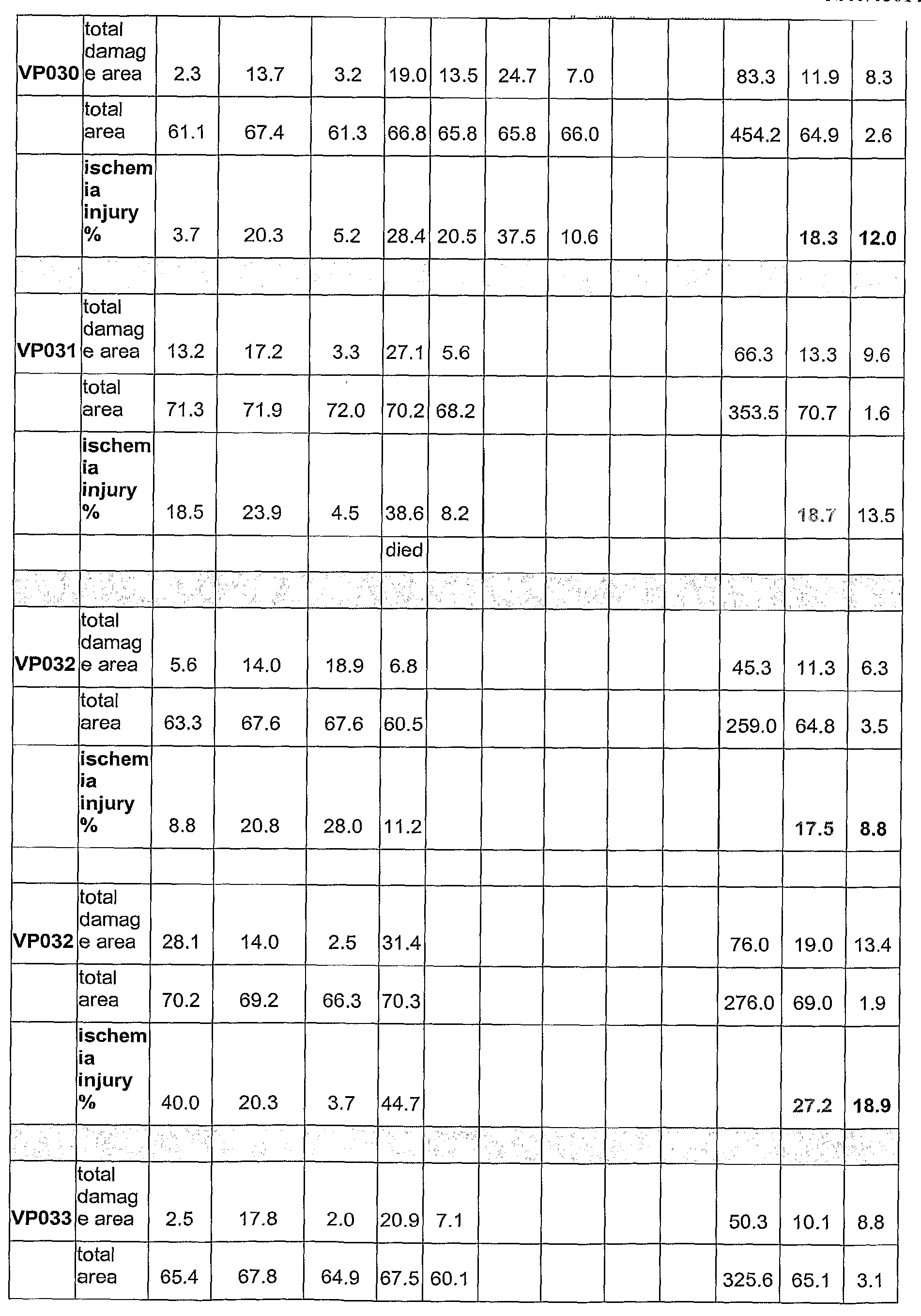


- 473. Table 28. Saline Treated Control Animals. 42 animals were used as the control group. The controls were over a time period, which corresponded with the time period in which the treatment groups were tested. Over all the saline treated group showed a mean ischemic injury percent of 30.5 with a standard deviation of 3.27. This group was used to generate the p-values shown in Tables 22 and 26.
Animal # 1 2 3 4 5 6 7 Total Mean SD CV total damage area 4.27 18.66 20.30 32.36 75.59 18.90 11.51 60.89 total area 60.73 66.32 67.19 69.73 263.97 65.99 3.79 5.75 ischemia injury % 7.03 28.14 30.21 46.41 28.64 16.16 56.43 total damage area 13.72 20.11 29.90 63.73 21.24 8.15 38.36 total area 69.38 63.53 65.45 198.36 66.12 2.98 4.51 ischemia injury % 19.78 31.65 45.68 32.13 12.97 40.37 total damage area 15.51 20.43 31.30 29.30 0.00 96.54 24.14 7.44 30.83 total area 70.38 66.39 70.15 66.39 68.50 273.31 68.33 2.24 3.28 ischemia injury % 22.04 30.77 44.62 44.13 0.00 35.32 10.97 31.07 total damage area 6.31 24.09 27.49 30.65 24.44 112.98 22.60 9.48 41.97 total area 60.24 70.36 67.33 69.67 65.64 333.24 66.65 4.05 6.07 ischemia injury % 10.47 34.24 40.83 43.99 37.23 33.90 15.16 44.72 total damage area 7.29 22.28 21.31 18.77 18.18 10.99 29.37 128.19 18.31 7.33 40.02 total area 57.30 63.10 61.66 66.36 67.65 67.82 69.02 452.91 64.70 4.21 6.51 ischemia injury % 12.72 35.31 34.56 28.29 26.87 16.20 42.55 28.30 10.48 37.03 total damage area 7.29 22.28 21.3 1 18.77 18.18 87.83 17.57 5.99 34.12 total area 57.30 63.10 61.66 66.36 67.65 316.07 63.21 4.09 6.47 ischemia injury % 12.72 35.31 34.56 28.29 26.87 27.79 10.48 37.72 total damage area 1.42 18.51 31.80 15.05 20.78 87.56 17.51 10.96 62.61 total area 67.80 69.25 71.213 67.19 69.73 345.20 69.04 1.60 2.32 ischemia injury % 2.09 26.73 44.64 22.40 29.80 25.13 15.36 61.10 total damage area 13.72 20.11 29.90 63.73 21.24 21.24 8.15 38.36 total area 69.38 63.53 65.45 198.36 66.12 2.98 4.51 ischemia injury % 19.78 31.65 45.6 8 32.37 12.97 40.06 total damage area 17.61 15.23 24.40 15.11 31.12 103.47 20.69 6.95 33.56 total area 65.48 66.91 66.73 65.94 68.56 333.62 66.72 1.18 1.77 ischemia injury % 26.89 22.76 36.57 22.91 45.39 30.91 9.85 31.87 30.50 3.27 10.73 blood
The fraction as defined above, wherein the fraction is isolated by collecting the animal'sblood
The fraction as defined above, wherein the fraction is isolated by collecting the animal'sblood 15 days after the onset of hibernation.
A fraction of a hibernating animal's plasma comprising a molecule, wherein the molecule has anti-infarction activity, and wherein the fraction is isolated by collecting a first animal's blood, wherein the first animal is in early hibernation, and wherein the fraction does not comprise any blood from a second animal if the second animal is in mid hibernation.
A fraction of a hibernating animal's plasma comprising a molecule, wherein the molecule has anti-infarction activity, and wherein the fraction is isolated by collecting an animal'sblood claim 6, wherein the fraction is isolated by collecting the animal'sblood
The fraction as defined above, wherein the fraction is isolated by collecting an animal'sblood
The fraction as defined above, wherein the fraction is isolated by collecting an animal'sblood 15 days before the final arousal of the animal.
A fraction of a hibernating animal's plasma comprising a molecule, wherein the molecule has anti-infarction activity, and wherein the fraction is isolated by collecting a first animal's blood, wherein the first animal is in late hibernation, and wherein the fraction does not comprise any blood from a second animal if the second animal is in mid hibernation.
The method as defined above, wherein the anti-infarction activity is a cerebral anti-infarction activity.
The method as defined above, wherein the anti-infarction activity is a cardiac anti-infarction activity.
A fraction of a hibernating animal's plasma comprising a molecule, wherein the molecule induces urea recycling, and wherein the fraction is isolated by collecting an animal'sblood blood
The fraction as defined above, wherein the fraction is isolated by collecting the animal'sblood
The fraction as defined above, wherein the fraction is isolated by collecting the animal'sblood 15 days after the onset of hibernation.
A fraction of a hibernating animal's plasma comprising a molecule, wherein the molecule induces urea recycling, and wherein the fraction is isolated by collecting a first animal's blood, wherein the first animal is in early hibernation, and wherein the fraction does not comprise any blood from a second animal if the second animal is in mid hibernation.
A fraction of a hibernating animal's plasma comprising a molecule, wherein the molecule induces urea recycling, and wherein the fraction is isolated by collecting an animal'sblood blood
The fraction as defined above, wherein the fraction is isolated by collecting an animal'sblood
The fraction as defined above, wherein the fraction is isolated by collecting an animal'sblood 15 days before the final arousal of the animal.
A fraction of a hibernating animal's plasma comprising a molecule, wherein the molecule induces urea recycling, and wherein the fraction is isolated by collecting a first animal's blood, wherein the first animal is in late hibernation, and wherein the fraction does not comprise any blood from a second animal if the second animal is in mid hibernation.
A method of purifying a molecule having anti-infarction activity comprising 1) collecting a sample from a hibernating animal, 2) collecting a second sample from a hibernating animal, 3) and comparing the sample to the second sample.
A method of purifying a molecule having anti-infarction activity comprising 1) collecting a sample from a hibernating animal, 2) collecting a second sample from a hibernating animal in a different sub-state, 3) and comparing the sample to the second sample.
A method of purifying a molecule having anti-infarction activity comprising 1) collecting a sample from a hibernating animal at a first time, 2) collecting a second sample from a hibernating animal at a second time, wherein the first time and second time are different, 3) and comparing the sample to the second sample. A method of purifying a molecule having anti-infarction activity comprising 1) collecting a blood sample from a hibernating animal, 2) collecting a second blood sample from a hibernating animal, 3) comparing the blood sample to the second blood sample.
The method as defined above, wherein the step of comparing the sample and the second sample comprises assaying gene expression in the sample and the second sample.
The method as defined above, wherein the step of comparing the sample and the second sample comprises assaying protein expression in the sample and the second sample.
The method as defined above, wherein the sample and second sample are obtained from the blood, urine, spinal fluid, or cerebral spinal fluid, tissues, organs, cells.
The method as defined above, wherein the sample and second sample are obtained from the urine, spinal fluid, or cerebral spinal fluid, tissues, organs, cells.
The method as defined above, wherein the animal is a mammal.
The method as defined above, wherein the mammal is a ground squirrel, bear, woodchuck, marmot, skunk, or bat.
The method as defined above, wherein the blood sample was obtained from an animal in early-hibernation or late-hibernation.
The method as defined above, wherein the second sample was obtained from an animal in mid-hibernation.
The method as defined above, wherein the second sample was obtained from an animal in mid-hibernation. The method as defined above, wherein the sample was obtained from an animal 1-25 days after the onset of hibernation.
The method as defined above, wherein the second sample was obtained from an animal 26 to 60 days after the onset of hibernation.
The method as defined above, wherein the second sample was obtained from an animal 26 to 60 days after the onset of hibernation.
The method as defined above, wherein the step of comparing comprises identifying differentially regulated molecules, wherein the differentially regulated molecules are present in different amounts in the sample as compared to the second sample.
The method as defined above, wherein identifying differentially regulated molecules comprises fractionating the sample and the second sample.
The method as defined above, wherein the fractionating occurs by collecting molecules of 10 kDA or less. The method as defined above, wherein the fractionating comprises the step of separating the molecules in the blood sample and the second sample by charge, hydrophobicity, hydrophilicity, lipophilicity, tortuosity,, molecular weight, protein, peptide or carbohydrate affinity chromatography, or solubility. The method as defined above, wherein the affinity separation comprises using affi-gel blue chromatography. The method as defined above, wherein identifying comprises analyzing the samples with GC-Mass Spectroscopy, Gel Chromatography, or High Performance Liquid Chromatography, LC/MS/MS mass spectroscopy.
The method as defined above, wherein the High Performance Liquid Chromatography comprises reverse phase chromatography.
The method as defined above, wherein the Gel Chromatography comprises two dimensional polyacrylamide gel electrophoresis.
The method as defined above, wherein the method further comprises purifying the differentially regulated molecules obtaining a purified differentially regulated molecule.
The method as defined above, wherein the method further comprises assaying the anti-infarction activity of the purified differentially regulated molecule.
The method as defined above, wherein the step of assaying comprises using the mouse MCAO model of cerebral ischemia.
The method as defined above, wherein the anti-infarction activity is a cerebral anti-infarction activity.
The method as defined above, wherein the anti-infarction activity is a cardiac anti-infarction activity.
A method of reducing an infarction in a subject comprising administering FPA to the subject.
The method as defined above, wherein the FPA comprises a structure having at least 20% identity to SEQ ID NO:2. and whereinamino acids
The method as defined above, wherein any variation atamino acids
The method as defined above, wherein the FPA comprises amino acids having at least 40% identity to amino acids 6-16 of SEQ ID NO:2, and whereinamino acids
The method as defined above, wherein any variation atamino acids
The method as defined above, wherein any variation from SEQ ID NO:2 is a conservative substitution.
The method as defined above, wherein the FPA reduces the amount of infarction present in a mouse MCAO model.
The method as defined above, wherein the infarction is reduced by at least 20%.
The method as defined above, wherein the infarction is reduced by at least 40%.
The method as defined above, wherein the infarction is reduced by at least 60%.
The method as defined above, wherein the infarction is reduced by at least 80%.
The method as defined above, wherein an infarction ratio in a mouse MCAO model is at least 1.1.
The method as defined above, wherein an infarction ratio in a mouse MCAO model is greater or equal than 1.5.
The method as defined above, wherein an infarction ratio in a mouse MCAO model is greater or equal than 2. The method as defined above, wherein the ratio is determined using mean infarcted volumes.
The method as defined above, wherein the mean infarction volume in a mouse MCAO model is less than or equal to 90 %.
The method as defined above, wherein the mean infarction volume in a mouse MCAO model is less than or equal to 70 %.
The method as defined above, wherein the mean infarction volume in a mouse MCAO model is less than or equal to 50 %.
The method as defined above, wherein the mean infarction volume in a mouse MCAO model is less than or equal to 30 %.
A method of reducing an infarction in a subject comprising administering a composition, wherein the composition comprises a peptide sequence set forth in SEQ ID NO:34, SEQ ID NO:40, SEQ ID NO:41, SEQ ID NO:42, SEQ ID NO:43, SEQ ID NO:44, SEQ ID NO:45, SEQ ID NOs: 96-103, AVR, or FVR to the subject.
The method as defined above, wherein the FPA has the sequence set forth in SEQ ID NO:29, SEQ ID NO:30, SEQ ID NO:31, SEQ ID NO:32, SEQ ID NO:33, SEQ ID NO:34, SEQ ID NO:35, SEQ ID NO:36, SEQ ID NO:37, SEQ ID NO:38, SEQ ID NO:39, SEQ ID NO:40, SEQ ID NO:41, SEQ ID NO:42, SEQ ID NO:43, SEQ ID NO:44, SEQ ID NO:45, SEQ ID NO:46, SEQ ID NO:47, SEQ ID NO:48, SEQ ID NO:49, SEQ ID NO:50, SEQ ID NO:51, or SEQ ID NO:52, SEQ ID NO:53, SEQ ID NO:54, SEQ ID NO:55, SEQ ID NO:56, SEQ ID NO:89, SEQ ID NOs: 96-103, AVR, or FVR.
The method as defined above, wherein the FPA has the sequence set forth in SEQ ID NO:46, SEQ ID NO:45, SEQ ID NO:36, SEQ ID NO:50, SEQ ID NO:38, SEQ ID NO:40, or SEQ ID NO:54, SEQ ID NO:32, SEQ ID NO:47, SEQ ID NO:48, SEQ ID NO:52, or SEQ ID NO:39
The method as defined above, wherein the infarction is a cerebral infarction.
The method as defined above, wherein the infarction is a cardiac infarction.
A method of reducing an infarction in a subject comprising administering Bradykinin to the subject.
The method as defined above, wherein the Bradykinin comprises a structure having 60% identity to SEQ ID NO:57.
The method as defined above, wherein any variation away from SEQ ID NO: 57 are conservative substitutions.
The method as defined above, wherein the Bradykinin comprises a structure having 80% identity to SEQ ID NO:83.
The method as defined above, wherein any variation away from SEQ ID NO: 57 are conservative substitutions.
The method as defined above, wherein the Bradykinin does not have a basic amino acid at the C-terminal end. The method as defined above, wherein the basic amino acid is Arg, Lys, or His.
The method as defined above, wherein the Bradykinin comprises a basic amino acid at the N-terminal end. The method as defined above, wherein the basic amino acid is Arg, Lys, or His.
The method as defined above, wherein the Bradykinin comprises a basic amino acid at the N-terminal end. The method as defined above, wherein the basic amino acid is Arg, Lys, or His.
The method as defined above, wherein the Bradykinin has the sequence set forth in SEQ ID NO:57, SEQ ID NO:58, SEQ ID NO:59, SEQ ID NO:60, SEQ ID NO:61, SEQ ID NO:62, SEQ ID NO:63, SEQ ID NO:64, SEQ ID NO:65, SEQ ID NO:66, SEQ ID NO:67, SEQ ID NO:68, SEQ ID NO:70, SEQ ID NO:71, SEQ ID NO:72, SEQ ID NO:73, SEQ ID NO:75, SEQ ID NO:77, SEQ ID NO:80, SEQ ID NO:81, SEQ ID NO:82, SEQ ID NO:83, SEQ ID NO:84, or SEQ ID NO:85.
The method as defined above, wherein the Bradykinin has the sequence set forth in SEQ ID NO:58, SEQ ID NO:61, SEQ ID NO:62, SEQ ID NO:63, SEQ ID NO:64, SEQ ID NO:77, or SEQ ID NO:81.
The method as defined above, wherein the Bradykinin has the sequence set forth in SEQ ID NO:58.
The method as defined above, wherein the Bradykinin has the sequence set forth in SEQ ID NO:61.
The method as defined above, wherein the infarction is a cerebral infarction.
The method as defined above, wherein the infarction is a cardiac infarction.
A method of making an anti-infarction molecule comprising synthesizing the anti-infarction molecule or a variant of the anti-infarction molecule, wherein the anti-infarction molecule can be purified by a method comprising 1) collecting a blood sample from a hibernating animal, 2) collecting a second blood sample from a hibernating animal, 3) comparing the blood sample to the second blood sample, wherein the blood sample is collected les than or equal to 25 days after the onset of hibernation, and the second blood sample is collected greater than 25 days after the onset of hibernation and wherein the anti-infarction molecule is a present in greater amounts in the blood sample than in the second blood sample.
A method of making an anti-infarction molecule comprising synthesizing the anti-infarction molecule or a variant of the anti-infarction molecule, wherein the anti-infarction molecule can be purified by a method comprising 1) collecting a blood sample from a hibernating animal, 2) collecting a second blood sample from a hibernating animal, 3) comparing the blood sample to the second blood sample, wherein the blood sample is collected les than or equal to 25 days before the end of hibernation, and the second blood sample is collected greater than 25 days after the onset of hibernation, but before 25 days before the end of hibernation, and wherein the anti-infarction molecule is a present in greater amounts in the blood sample than in the second blood sample.
A method of identifying an anti-infarction molecule comprising administering a molecule to a mouse MCAO model, comparing the anti-infarction activity of the molecule to the anti-infarction activity of FPA in a mouse MCAO model, and selecting the molecule if the anti-infarction activity of the molecule is at least 20% of the activity of FPA.
A method of reducing an infarction in a subject in need of reducing an infarction, comprising administering an effective amount of an angiotensin IIreceptor type 2 antagonist in a pharmaceutically acceptable form to the subj ect.
The method as defined above, wherein the infarction is associated with stroke.
The method as defined above, wherein the infarction is associated with cardiac ischemia.
The method as defined above, wherein the angiotensin IIreceptor type 2 antagonist is Bradykinin.
The method as defined above, wherein the Bradykinin comprises a structure having 60% identity to SEQ ID NO:57.
The method as defined above, wherein any variation away from SEQ ID NO: 57 are conservative substitutions.
The method as defined above, wherein the Bradykinin comprises a structure having 80% identity to SEQ ID NO:83.
The method as defined above, wherein any variation away from SEQ ID NO: 57 are conservative substitutions.
The method as defined above, wherein the Bradykinin does not have a basic amino acid at the C-terminal end. The method as defined above, wherein the basic amino acid is Arg, Lys, or His.
The method as defined above, wherein the Bradykinin comprises a basic amino acid at the N-terminal end. The method as defined above, wherein the basic amino acid is Arg, Lys, or His.
The method as defined above, wherein the Bradykinin comprises a basic amino acid at the N-terminal end. The method as defined above, wherein the basic amino acid is Arg, Lys, or His.
The method as defined above, wherein the Bradykinin has the sequence set forth in SEQ ID NO:57, SEQ ID NO:58, SEQ ID NO:59, SEQ ID NO:60, SEQ ID NO:61, SEQ ID NO:62, SEQ ID NO:63, SEQ ID NO:64, SEQ ID NO:65, SEQ ID NO:66, SEQ ID NO:67, SEQ ID NO:68, SEQ ID NO:70, SEQ ID NO:71, SEQ ID NO:72, SEQ ID NO:73, SEQ ID NO:75, SEQ ID NO:77, SEQ ID NO:80, SEQ ID NO:81, SEQ ID NO:82, SEQ ID NO:83, SEQ ID NO:84, or SEQ ID NO:85.
The method as defined above, wherein the Bradykinin has the sequence set forth in SEQ ID NO:58, SEQ ID NO:61, SEQ ID NO:62, SEQ ID NO:63, SEQ ID NO:64, SEQ ID NO:77, or SEQ ID NO:81.
The method as defined above, wherein the Bradykinin has the sequence set forth in SEQ ID NO:58.
The method as defined above, wherein the Bradykinin has the sequence set forth in SEQ ID NO:61.
The method as defined above, wherein the infarction is associated with stroke.
A method of identifying an inhibitor of the interaction between Bradykinin and the angiotensin IIreceptor type 2 comprising: contacting a cell expressing the angiotensin IIreceptor type 2 with the putative inhibitor in the presence of Bradykinin; detecting the amount Bradykinin bound to the angiotensin IIreceptor type 2; wherein a reduction in Bradykinin binding to the angiotensin IIreceptor type 2 identifies an inhibitor. A method of identifying an inhibitor of the interaction between Bradykinin and the angiotensin IIreceptor type 2 comprising: contacting a cell expressing the angiotensin IIreceptor type 2 with a putative inhibitor in the presence of Bradykinin, wherein the angiotensin IIreceptor type 2 comprises a fluorescence donor, wherein Bradykinin comprises a fluorescence acceptor; and measuring Fluorescence Resonance Energy Transfer (FRET), wherein a decrease in FRET as compared to FRET measurement in a cell that was not contacted with the putative inhibitor indicates the presence of an inhibitor.
A method of identifying an inhibitor of the interaction between Bradykinin and an angiotensin IIreceptor type 2 comprising: contacting a cell system with a putative inhibitor, wherein the cell system comprises an angiotensin IIreceptor type 2, wherein the cell system comprises Bradykinin, wherein the angiotensin IIreceptor type 2 comprises a fluorescence donor, and wherein the Bradykinin comprises a fluorescence acceptor; and measuring Fluorescence Resonance Energy Transfer (FRET) before contacting the cell system with the putative inhibitor and after contacting the cell system with the putative inhibitor, wherein a decrease in FRET in the cell system when the putative inhibitor is contacted with the putative inhibitor identifies an inhibitor.
The method as defined above further comprising the step of testing the identified inhibitor in an in vivo model of infarction, and selecting molecules that reduce an infarction in the animal model. The method as defined above, wherein the putative inhibitor is found within a library of molecules.


Claims (29)
- A fraction of a hibernating animal's plasma comprising a molecule, wherein the molecule has anti-infarction activity, and wherein the fraction is isolated by collecting an animal's blood 1, 2, 3, 4, 5, 6, 7, 8, 9, 10, 11, 12, 13, 14, 15, 16, 17, 18, 19, 20, 21, 22, 23, 24, or 25 days after the onset of hibernation.
- The fraction of claim 1, wherein the fraction is isolated by collecting the animal's blood 10, 11, 12, 13, 14, 15, 16, 17, 18, 19, or 20 days after the onset of hibernation, preferably 14, 15, or 16 days after the onset of hibernation, most preferably 15 days after the onset of hibernation.
- A fraction of a hibernating animal's plasma comprising a molecule, wherein the molecule has anti-infarction activity, and wherein the fraction is isolated by collecting a first animal's blood, wherein the first animal is in early hibernation, and wherein the fraction does not comprise any blood from a second animal if the second animal is in mid hibernation.
- A fraction of a hibernating animal's plasma comprising a molecule, wherein the molecule has anti-infarction activity, and wherein the fraction is isolated by collecting an animal's blood 1, 2, 3, 4, 5, 6, 7, 8, 9, 10, 11, 12, 13, 14, 15, 16, 17, 18, 19, 20, 21, 22, 23, 24, or 25 days before the final arousal of the animal.
- The fraction of claim 4, wherein the fraction is isolated by collecting the animal's blood 10, 11, 12, 13, 14, 15, 16, 17, 18, 19, or 20 days before the final arousal of the animal, preferably 14, 15, or 16 days before the final arousal of the animal, most preferably 15 days before the final arousal of the animal.
- A fraction of a hibernating animal's plasma comprising a molecule, wherein the molecule has anti-infarction activity, and wherein the fraction is isolated by collecting a first animal's blood, wherein the first animal is in late hibernation, and wherein the fraction does not comprise any blood from a second animal if the second animal is in mid hibernation.
- The fraction of claim 1, wherein the anti-infarction activity is a cerebral anti-infarction activity, or a cardiac anti-infarction activity.
- A fraction of a hibernating animal's plasma comprising a molecule, wherein the molecule induces urea recycling, and wherein the fraction is isolated by collecting an animal's blood 1, 2, 3, 4, 5, 6, 7, 8, 9, 10, 11, 12, 13, 14, 15, 16, 17, 18, 19, 20, 21, 22, 23, 24, or 25 days after the onset of hibernation.
- The fraction of claim 8, wherein the fraction is isolated by collecting the animal's blood 10, 11, 12, 13, 14, 15, 16, 17, 18, 19, or 20 days after the onset of hibernation, preferably 14, 15, or 16 days after the onset of hibernation, most preferably 15 days after the onset of hibernation.
- A fraction of a hibernating animal's plasma comprising a molecule, wherein the molecule induces urea recycling, and wherein the fraction is isolated by collecting a first animal's blood, wherein the first animal is in early hibernation, and wherein the fraction does not comprise any blood from a second animal if the second animal is in mid hibernation.
- A fraction of a hibernating animal's plasma comprising a molecule, wherein the molecule induces urea recycling, and wherein the fraction is isolated by collecting an animal's blood 1, 2, 3, 4, 5, 6, 7, 8, 9, 10, 11, 12, 13, 14, 15, 16, 17, 18, 19, 20, 21, 22, 23, 24, or 25 days before the final arousal of the animal.
- The fraction of claim 11, wherein the fraction is isolated by collecting the animal's blood 10, 11, 12, 13, 14, 15, 16, 17, 18, 19, or 20 days before the final arousal of the animal, preferably 14, 15, or 16 days before the final arousal of the animal, most preferably 15 days before the final arousal of the animal.
- A fraction of a hibernating animal's plasma comprising a molecule, wherein the molecule induces urea recycling, and wherein the fraction is isolated by collecting a first animal's blood, wherein the first animal is in late hibernation, and wherein the fraction does not comprise any blood from a second animal if the second animal is in mid hibernation.
- A method of purifying a molecule having anti-infarction activity comprising 1) collecting a sample from a hibernating animal, 2) collecting a second sample from a hibernating animal, 3) and comparing the sample to the second sample.
- A method of purifying a molecule having anti-infarction activity comprising 1) collecting a sample from a hibernating animal, 2) collecting a second sample from a hibernating animal in a different sub-state, 3) and comparing the sample to the second sample.
- A method of purifying a molecule having anti-infarction activity comprising 1) collecting a sample from a hibernating animal at a first time, 2) collecting a second sample from a hibernating animal at a second time, wherein the first time and second time are different, 3) and comparing the sample to the second sample.
- A method of purifying a molecule having anti-infarction activity comprising 1) collecting a blood sample from a hibernating animal, 2) collecting a second blood sample from a hibernating animal, 3) comparing the blood sample to the second blood sample.
- The method of claims 14 to 17, wherein the step of comparing the sample and the second sample comprises(i) assaying gene expression in the sample and the second sample, or(ii) assaying protein expression in the sample and the second sample.
- The method of claims 14 to 16, wherein the sample and second sample are obtained from the blood, urine, spinal fluid, or cerebral spinal fluid, tissues, organs, cells.
- The method of claim 17, wherein the sample and second sample are obtained from the urine, spinal fluid, or cerebral spinal fluid, tissues, organs, cells.
- The method of claims 14 to 17, wherein the animal is a mammal, preferably is a ground squirrel, bear, woodchuck, marmot, skunk, or bat.
- The method of claims 14 to 17, wherein the blood sample was obtained from an animal in early-hibernation or late-hibernation.
- The method of claims 14 to 17 or 22, wherein the second sample was obtained from an animal in mid-hibernation.
- The method of claims 14 to 17, wherein the sample was obtained from an animal 1-25 days after the onset of hibernation.
- The method of claims 14 to 17 or 24, wherein the second sample was obtained from an animal 26 to 60 days after the onset of hibernation.
- The method of claims 14 to 17, wherein the step of comparing comprises identifying differentially regulated molecules, wherein the differentially regulated molecules are present in different amounts in the sample as compared to the second sample.
- The method of claim 26, wherein(a) identifying differentially regulated molecules comprises fractionating the sample and the second sample, preferably the fractionating occurring by collecting molecules of 10 kDA or less, or the fractionating comprising the step of separating the molecules in the blood sample and the second sample by charge, hydrophobicity, hydrophilicity, lipophilicity, tortuosity" molecular weight, protein, peptide or carbohydrate affinity chromatography or solubility, most preferably the affinity separation comprises using affi-gel blue chromatography; or(b) identifying comprises analyzing the samples with GC-Mass Spectroscopy, Gel Chromatography, or High Performance Liquid Chromatography, LC/MS/MS mass spectroscopy, preferably the High Performance Liquid Chromatography comprising reverse phase chromatography; or the Gel Chromatography comprising two dimensional polyacrylamide gel electrophoresis; or(c) the method further comprises purifying the differentially regulated molecules obtaining a purified differentially regulated molecule, preferably the method further comprising assaying the anti-infarction activity of the purified differentially regulated molecule, most preferably(i) the step of assaying comprises using the mouse MCAO model of cerebral ischemia,(ii) the anti-infarction activity is a cerebral anti-infarction activity, or(iii) the anti-infarction activity is a cardiac anti-infarction activity.
- A method of making an anti-infarction molecule comprising synthesizing the anti-infarction molecule or a variant of the anti-infarction molecule, wherein the anti-infarction molecule can be purified by a method comprising 1) collecting a blood sample from a hibernating animal, 2) collecting a second blood sample from a hibernating animal, 3) comparing the blood sample to the second blood sample, wherein the blood sample is collected les than or equal to 25 days after the onset of hibernation, and the second blood sample is collected greater than 25 days after the onset of hibernation and wherein the anti-infarction molecule is a present in greater amounts in the blood sample than in the second blood sample.
- A method of making an anti-infarction molecule comprising synthesizing the anti-infarction molecule or a variant of the anti-infarction molecule, wherein the anti-infarction molecule can be purified by a method comprising 1) collecting a blood sample from a hibernating animal, 2) collecting a second blood sample from a hibernating animal, 3) comparing the blood sample to the second blood sample, wherein the blood sample is collected les than or equal to 25 days before the end of hibernation, and the second blood sample is collected greater than 25 days after the onset of hibernation, but before 25 days before the end of hibernation, and wherein the anti-infarction molecule is a present in greater amounts in the blood sample than in the second blood sample.
Applications Claiming Priority (4)
| Application Number | Priority Date | Filing Date | Title |
|---|---|---|---|
| US35467802P | 2002-02-06 | 2002-02-06 | |
| US39213302P | 2002-06-28 | 2002-06-28 | |
| US42927802P | 2002-11-25 | 2002-11-25 | |
| EP03707755A EP1572168B1 (en) | 2002-02-06 | 2003-02-05 | Anti-infarction molecules |
Related Parent Applications (1)
| Application Number | Title | Priority Date | Filing Date |
|---|---|---|---|
| EP03707755.9 Division | 2003-02-05 |
Publications (1)
| Publication Number | Publication Date |
|---|---|
| EP2363405A1 true EP2363405A1 (en) | 2011-09-07 |
Family
ID=27739154
Family Applications (3)
| Application Number | Title | Priority Date | Filing Date |
|---|---|---|---|
| EP10163802A Withdrawn EP2363405A1 (en) | 2002-02-06 | 2003-02-05 | Anti-infarction molecules |
| EP10163798A Withdrawn EP2311852A1 (en) | 2002-02-06 | 2003-02-05 | Anti-infarction molecules |
| EP03707755A Expired - Lifetime EP1572168B1 (en) | 2002-02-06 | 2003-02-05 | Anti-infarction molecules |
Family Applications After (2)
| Application Number | Title | Priority Date | Filing Date |
|---|---|---|---|
| EP10163798A Withdrawn EP2311852A1 (en) | 2002-02-06 | 2003-02-05 | Anti-infarction molecules |
| EP03707755A Expired - Lifetime EP1572168B1 (en) | 2002-02-06 | 2003-02-05 | Anti-infarction molecules |
Country Status (16)
| Country | Link |
|---|---|
| US (1) | US7811992B2 (en) |
| EP (3) | EP2363405A1 (en) |
| JP (2) | JP2005532263A (en) |
| KR (2) | KR20100094569A (en) |
| CN (1) | CN1856504B (en) |
| AT (1) | ATE469167T1 (en) |
| AU (1) | AU2003209030B2 (en) |
| BR (1) | BR0307470A (en) |
| CA (1) | CA2475112A1 (en) |
| DE (1) | DE60332756D1 (en) |
| EA (1) | EA010860B1 (en) |
| HK (1) | HK1083202A1 (en) |
| MX (1) | MXPA04007586A (en) |
| TW (1) | TW200307554A (en) |
| WO (1) | WO2003065997A2 (en) |
| ZA (1) | ZA200407103B (en) |
Families Citing this family (10)
| Publication number | Priority date | Publication date | Assignee | Title |
|---|---|---|---|---|
| US7670833B2 (en) * | 2002-08-08 | 2010-03-02 | Biochain Institute, Inc. | High throughput analysis for molecular fractions |
| EP1566183A1 (en) * | 2004-02-20 | 2005-08-24 | ZLB Behring GmbH | Use of factor XIII for stimulating the perfusion of ischemic tissue |
| US8226977B2 (en) | 2004-06-04 | 2012-07-24 | Teva Pharmaceutical Industries Ltd. | Pharmaceutical composition containing irbesartan |
| CN101153054B (en) * | 2006-09-30 | 2012-02-22 | 北京市肿瘤防治研究所 | Peptide for treatment of vascellum generation and uses thereof |
| KR20100058555A (en) * | 2007-09-11 | 2010-06-03 | 몬도바이오테크 래보래토리즈 아게 | Use of bpp-b as a therapeutic agent |
| EP2187937A1 (en) * | 2007-09-11 | 2010-05-26 | Mondobiotech Laboratories AG | Use of the peptide pro-gly-thr-cys-glu-ile-cys-ala-tyr-ala-ala-cys-thr-gly-cys as a therapeutic agent |
| JP5676455B2 (en) * | 2008-10-08 | 2015-02-25 | ベッドロック・インヴェンションズ・エルエルシー | Apparatus for measuring and processing shivering (vibration) during temperature control for treatment and temperature management system for treatment |
| MX2011009475A (en) * | 2009-03-09 | 2011-12-08 | Wade M Melling | Immunomodulatory therapeutic agents. |
| US10786543B2 (en) | 2015-09-22 | 2020-09-29 | The Johns Hopkins University | P75NTR antagonists and treatment of acute and chronic cardiac disease |
| WO2022261336A1 (en) * | 2021-06-09 | 2022-12-15 | Fauna Bio Incorporated | Methods and compositions comprising functional genomics of hibernating mammals |
Citations (182)
| Publication number | Priority date | Publication date | Assignee | Title |
|---|---|---|---|---|
| US3610795A (en) | 1968-10-17 | 1971-10-05 | Intitut De Rech De La Siderurg | Apparatus for continuously melting of metal |
| US4187217A (en) | 1977-12-14 | 1980-02-05 | Chipens Gunar I | Bradykinin cyclic analog-cyclo-[(N.sup.ε -1-L-lysine, 6-glycine)-bradykinin] |
| US4342566A (en) | 1980-02-22 | 1982-08-03 | Scripps Clinic & Research Foundation | Solid phase anti-C3 assay for detection of immune complexes |
| US4816567A (en) | 1983-04-08 | 1989-03-28 | Genentech, Inc. | Recombinant immunoglobin preparations |
| US4897355A (en) | 1985-01-07 | 1990-01-30 | Syntex (U.S.A.) Inc. | N[ω,(ω-1)-dialkyloxy]- and N-[ω,(ω-1)-dialkenyloxy]-alk-1-yl-N,N,N-tetrasubstituted ammonium lipids and uses therefor |
| US5084824A (en) | 1990-03-29 | 1992-01-28 | National Semiconductor Corporation | Simulation model generation from a physical data base of a combinatorial circuit |
| WO1992003566A1 (en) | 1990-08-17 | 1992-03-05 | Yale University | Therapeutic ribozyme compositions |
| US5135917A (en) | 1990-07-12 | 1992-08-04 | Nova Pharmaceutical Corporation | Interleukin receptor expression inhibiting antisense oligonucleotides |
| US5162497A (en) | 1987-09-24 | 1992-11-10 | The Administrators Of The Tulane Educational Fund | Bradykinin analogs with non-peptide bond |
| US5176996A (en) | 1988-12-20 | 1993-01-05 | Baylor College Of Medicine | Method for making synthetic oligonucleotides which bind specifically to target sites on duplex DNA molecules, by forming a colinear triplex, the synthetic oligonucleotides and methods of use |
| WO1993022434A2 (en) | 1992-04-28 | 1993-11-11 | Yale University | Targeted cleavage of rna using eukaryotic ribonuclease p and external guide sequence |
| US5288514A (en) | 1992-09-14 | 1994-02-22 | The Regents Of The University Of California | Solid phase and combinatorial synthesis of benzodiazepine compounds on a solid support |
| US5294533A (en) | 1988-07-05 | 1994-03-15 | Baylor College Of Medicine | Antisense oligonucleotide antibiotics complementary to the macromolecular synthesis operon, methods of treating bacterial infections and methods for identification of bacteria |
| US5334711A (en) | 1991-06-20 | 1994-08-02 | Europaisches Laboratorium Fur Molekularbiologie (Embl) | Synthetic catalytic oligonucleotide structures |
| WO1994029348A2 (en) | 1993-06-03 | 1994-12-22 | Therapeutic Antibodies Inc. | Production of antibody fragments |
| US5436330A (en) | 1989-12-19 | 1995-07-25 | Agency Of Industrial Science & Technology | Hammerhead ribozymes with enhanced stability provided by an additional 3' ha |
| US5449754A (en) | 1991-08-07 | 1995-09-12 | H & N Instruments, Inc. | Generation of combinatorial libraries |
| WO1995024489A1 (en) | 1994-03-07 | 1995-09-14 | Yale University | Targeted cleavage of rna using ribonuclease p targeting and cleavage sequences |
| US5476766A (en) | 1990-06-11 | 1995-12-19 | Nexstar Pharmaceuticals, Inc. | Ligands of thrombin |
| US5503978A (en) | 1990-06-11 | 1996-04-02 | University Research Corporation | Method for identification of high affinity DNA ligands of HIV-1 reverse transcriptase |
| US5506337A (en) | 1985-03-15 | 1996-04-09 | Antivirals Inc. | Morpholino-subunit combinatorial library and method |
| US5539083A (en) | 1994-02-23 | 1996-07-23 | Isis Pharmaceuticals, Inc. | Peptide nucleic acid combinatorial libraries and improved methods of synthesis |
| US5543293A (en) | 1990-06-11 | 1996-08-06 | Nexstar Pharmaceuticals, Inc. | DNA ligands of thrombin |
| US5556762A (en) | 1990-11-21 | 1996-09-17 | Houghten Pharmaceutical Inc. | Scanning synthetic peptide combinatorial libraries: oligopeptide mixture sets having a one predetermined residue at a single, predetermined position, methods of making and using the same |
| US5565332A (en) | 1991-09-23 | 1996-10-15 | Medical Research Council | Production of chimeric antibodies - a combinatorial approach |
| US5565324A (en) | 1992-10-01 | 1996-10-15 | The Trustees Of Columbia University In The City Of New York | Complex combinatorial chemical libraries encoded with tags |
| US5573905A (en) | 1992-03-30 | 1996-11-12 | The Scripps Research Institute | Encoded combinatorial chemical libraries |
| US5580967A (en) | 1994-05-13 | 1996-12-03 | The Scripps Research Institute | Optimized catalytic DNA-cleaving ribozymes |
| US5580737A (en) | 1990-06-11 | 1996-12-03 | Nexstar Pharmaceuticals, Inc. | High-affinity nucleic acid ligands that discriminate between theophylline and caffeine |
| US5595873A (en) | 1994-05-13 | 1997-01-21 | The Scripps Research Institute | T. thermophila group I introns that cleave amide bonds |
| US5596079A (en) | 1991-12-16 | 1997-01-21 | Smith; James R. | Mimetics of senescent cell derived inhibitors of DNA synthesis |
| US5616466A (en) | 1993-11-05 | 1997-04-01 | Cantor; Glenn H. | Ribozyme-mediated inhibition of bovine leukemia virus |
| US5619680A (en) | 1994-11-25 | 1997-04-08 | Berkovich; Semyon | Methods and apparatus for concurrent execution of serial computing instructions using combinatorial architecture for program partitioning |
| US5618825A (en) | 1994-03-11 | 1997-04-08 | Pharmacopeia, Inc. | Combinatorial sulfonamide library |
| US5624824A (en) | 1989-03-24 | 1997-04-29 | Yale University | Targeted cleavage of RNA using eukaryotic ribonuclease P and external guide sequence |
| US5627158A (en) | 1990-11-02 | 1997-05-06 | Cho-Chung; Yoon S. | Antisense oligonucleotides of human regulatory subunit RI-.sub.α of camp dependent protein kinases for the treatment of cancer |
| US5627210A (en) | 1995-02-06 | 1997-05-06 | Chiron Corporation | Branched combinatorial libraries |
| US5631146A (en) | 1995-01-19 | 1997-05-20 | The General Hospital Corporation | DNA aptamers and catalysts that bind adenosine or adenosine-5'-phosphates and methods for isolation thereof |
| US5631115A (en) | 1992-07-02 | 1997-05-20 | Sankyo Company, Limited | Looped, hairpin ribozyme |
| WO1997018312A1 (en) | 1995-11-14 | 1997-05-22 | Vimrx Holdings, Ltd. | Chimeric oligomers having an rna-cleavage activity |
| US5633133A (en) | 1994-07-14 | 1997-05-27 | Long; David M. | Ligation with hammerhead ribozymes |
| US5641754A (en) | 1994-01-10 | 1997-06-24 | The Board Of Regents Of The University Of Nebraska | Antisense oligonucleotide compositions for selectively killing cancer cells |
| US5646020A (en) | 1992-05-14 | 1997-07-08 | Ribozyme Pharmaceuticals, Inc. | Hammerhead ribozymes for preferred targets |
| US5646285A (en) | 1995-06-07 | 1997-07-08 | Zymogenetics, Inc. | Combinatorial non-peptide libraries |
| US5645985A (en) | 1991-11-26 | 1997-07-08 | Gilead Sciences, Inc. | Enhanced triple-helix and double-helix formation with oligomers containing modified pyrimidines |
| US5646042A (en) | 1992-08-26 | 1997-07-08 | Ribozyme Pharmaceuticals, Inc. | C-myb targeted ribozymes |
| US5646031A (en) | 1995-05-16 | 1997-07-08 | Northern Illinois University | SArMV and sCYMVI hairpin ribozymes |
| US5650316A (en) | 1994-06-06 | 1997-07-22 | Research Development Foundation | Uses of triplex forming oligonucleotides for the treatment of human diseases |
| US5652107A (en) | 1993-01-15 | 1997-07-29 | The Public Health Research Institute Of The City Of New York, Inc. | Diagnostic assays and kits for RNA using RNA binary probes and a ribozyme ligase |
| US5652094A (en) | 1992-01-31 | 1997-07-29 | University Of Montreal | Nucleozymes |
| US5663046A (en) | 1994-06-22 | 1997-09-02 | Pharmacopeia, Inc. | Synthesis of combinatorial libraries |
| US5670326A (en) | 1994-04-05 | 1997-09-23 | Pharmagenics, Inc. | Reiterative method for screening combinatorial libraries |
| US5677195A (en) | 1991-11-22 | 1997-10-14 | Affymax Technologies N.V. | Combinatorial strategies for polymer synthesis |
| US5683899A (en) | 1994-02-03 | 1997-11-04 | University Of Hawaii | Methods and compositions for combinatorial-based discovery of new multimeric molecules |
| US5683874A (en) | 1991-03-27 | 1997-11-04 | Research Corporation Technologies, Inc. | Single-stranded circular oligonucleotides capable of forming a triplex with a target sequence |
| US5683902A (en) | 1994-05-13 | 1997-11-04 | Northern Illinois University | Human papilloma virus inhibition by a hairpin ribozyme |
| US5683873A (en) | 1995-01-13 | 1997-11-04 | Innovir Laboratories, Inc. | EGS-mediated inactivation of target RNA |
| US5688997A (en) | 1994-05-06 | 1997-11-18 | Pharmacopeia, Inc. | Process for preparing intermediates for a combinatorial dihydrobenzopyran library |
| US5688670A (en) | 1994-09-01 | 1997-11-18 | The General Hospital Corporation | Self-modifying RNA molecules and methods of making |
| US5688696A (en) | 1994-12-12 | 1997-11-18 | Selectide Corporation | Combinatorial libraries having a predetermined frequency of each species of test compound |
| US5693535A (en) | 1992-05-14 | 1997-12-02 | Ribozyme Pharmaceuticals, Inc. | HIV targeted ribozymes |
| US5693773A (en) | 1995-06-07 | 1997-12-02 | Hybridon Incorporated | Triplex-forming antisense oligonucleotides having abasic linkers targeting nucleic acids comprising mixed sequences of purines and pyrimidines |
| US5709214A (en) | 1996-05-02 | 1998-01-20 | Enhanced Cardiology, Inc. | PD2i electrophysiological analyzer |
| US5712146A (en) | 1993-09-20 | 1998-01-27 | The Leland Stanford Junior University | Recombinant combinatorial genetic library for the production of novel polyketides |
| US5712384A (en) | 1994-01-05 | 1998-01-27 | Gene Shears Pty Ltd. | Ribozymes targeting retroviral packaging sequence expression constructs and recombinant retroviruses containing such constructs |
| US5721367A (en) | 1990-08-29 | 1998-02-24 | Pharming B.V. | Homologous recombination in mammalian cells |
| US5720924A (en) | 1993-04-23 | 1998-02-24 | Boehringer Mannheim Gmbh | Storage system for test elements |
| US5721099A (en) | 1992-10-01 | 1998-02-24 | Trustees Of Columbia University In The City Of New York | Complex combinatorial chemical libraries encoded with tags |
| US5731424A (en) | 1990-06-11 | 1998-03-24 | Nexstar Pharmaceuticals, Inc. | High affinity TGFβ nucleic acid ligands and inhibitors |
| US5731295A (en) | 1992-12-07 | 1998-03-24 | Ribozyme Pharmaceuticals, Inc. | Method of reducing stromelysin RNA via ribozymes |
| US5741713A (en) | 1995-06-21 | 1998-04-21 | Martek Biosciences Corporation | Combinatorial libraries of labeled biochemical compounds and methods for producing same |
| US5770715A (en) | 1995-03-22 | 1998-06-23 | Toagosei Co., Ltd. | Hammerhead-like nucleic acid analogues and their synthesis |
| US5780228A (en) | 1990-06-11 | 1998-07-14 | Nexstar Pharmaceuticals, Inc. | High affinity nucleic acid ligands to lectins |
| US5780607A (en) | 1995-10-13 | 1998-07-14 | Hoffmann-La Roche Inc. | Antisense oligomers |
| US5786138A (en) | 1993-01-29 | 1998-07-28 | Board Of Supervisors Of Louisiana State University And Agricultural And Mechanical College | Hyperstabilizing antisense nucleic acid binding agents |
| US5792613A (en) | 1996-06-12 | 1998-08-11 | The Curators Of The University Of Missouri | Method for obtaining RNA aptamers based on shape selection |
| US5792431A (en) | 1996-05-30 | 1998-08-11 | Smithkline Beecham Corporation | Multi-reactor synthesizer and method for combinatorial chemistry |
| US5795721A (en) | 1990-06-11 | 1998-08-18 | Nexstar Pharmaceuticals, Inc. | High affinity nucleic acid ligands of ICP4 |
| US5804440A (en) | 1992-09-30 | 1998-09-08 | The Scripps Research Institute | Human neutralizing monoclonal antibodies to human immunodeficiency virus |
| US5807718A (en) | 1994-12-02 | 1998-09-15 | The Scripps Research Institute | Enzymatic DNA molecules |
| US5807754A (en) | 1995-05-11 | 1998-09-15 | Arqule, Inc. | Combinatorial synthesis and high-throughput screening of a Rev-inhibiting arylidenediamide array |
| US5807683A (en) | 1992-11-19 | 1998-09-15 | Combichem, Inc. | Combinatorial libraries and methods for their use |
| US5811300A (en) | 1992-12-07 | 1998-09-22 | Ribozyme Pharmaceuticals, Inc. | TNF-α ribozymes |
| US5824520A (en) | 1995-05-09 | 1998-10-20 | The United States Of America As Represented By The Department Of Health And Human Services | Phage-display of immunoglobulin heavy chain libraries for identification of inhibitors of intracellular constituents |
| US5834185A (en) | 1986-10-28 | 1998-11-10 | Johns Hopkins University | Formation of triple helix complexes of single stranded nucleic acids using nucleoside oligomers which comprise pyrimidine analogs, triple helix complexes formed thereby and oligomers used in their formation |
| US5834195A (en) | 1994-03-23 | 1998-11-10 | The Penn State Research Foundation | Method for identifying members of combinatorial libraries |
| US5834588A (en) | 1995-07-14 | 1998-11-10 | Yale University | (Cyanomethylene) phosphoranes as carbonyl 1,1-dipole synthons for use in constructing combinatorial libraries |
| US5834318A (en) | 1995-05-10 | 1998-11-10 | Bayer Corporation | Screening of combinatorial peptide libraries for selection of peptide ligand useful in affinity purification of target proteins |
| US5837243A (en) | 1995-06-07 | 1998-11-17 | Medarex, Inc. | Therapeutic compounds comprised of anti-Fc receptor antibodies |
| US5837855A (en) | 1994-10-11 | 1998-11-17 | Ribozyme Pharmaceutical, Inc. | Hairpin ribozymes |
| US5840500A (en) | 1996-07-11 | 1998-11-24 | Trega Biosciences, Inc. | Quinoline derivatives and quinoline combinatorial libraries |
| US5847150A (en) | 1996-04-24 | 1998-12-08 | Novo Nordisk A/S | Solid phase and combinatorial synthesis of substituted 2-methylene-2, 3-dihydrothiazoles and of arrays of substituted 2-methylene-2, 3-dihydrothiazoles |
| US5846713A (en) | 1990-06-11 | 1998-12-08 | Nexstar Pharmaceuticals, Inc. | High affinity HKGF nucleic acid ligands and inhibitors |
| US5849903A (en) | 1995-11-21 | 1998-12-15 | Icn Pharmaceuticals, Inc. | Antisense oligonucleotides for IL-8 and IL-8 receptor |
| WO1998058057A1 (en) | 1997-06-19 | 1998-12-23 | Innovir Laboratories, Inc. | Compositions inducing cleavage of rna motifs |
| WO1998058058A1 (en) | 1997-06-19 | 1998-12-23 | Ribozyme Pharmaceuticals, Inc. | Hammerhead ribozymes with extended cleavage rule |
| US5856103A (en) | 1994-10-07 | 1999-01-05 | Board Of Regents The University Of Texas | Method for selectively ranking sequences for antisense targeting |
| US5856463A (en) | 1995-09-18 | 1999-01-05 | Prydz; Hans Peter Blankenborg | PSKH-1 ribozymes |
| US5856188A (en) | 1988-09-20 | 1999-01-05 | The Board Of Regents For Northern Illinois University Of Dekalb | Hairpin ribozymes |
| US5856496A (en) | 1996-05-23 | 1999-01-05 | Pharmacia & Upjohn S.P.A. | Combinatorial solid phase synthesis of a library of indole derivatives |
| US5856107A (en) | 1997-02-04 | 1999-01-05 | Trega Biosciences, Inc. | Combinatorial libraries of imidazol-pyrido-indole and imidazol-pyrido-benzothiophene derivatives, methods of making the libraries and compounds therein |
| US5858660A (en) | 1994-09-20 | 1999-01-12 | Nexstar Pharmaceuticlas, Inc. | Parallel selex |
| US5859190A (en) | 1997-02-04 | 1999-01-12 | Trega Biosciences, Inc. | Combinatorial libraries of hydantoin and thiohydantoin derivatives, methods of making the libraries and compounds therein |
| US5861288A (en) | 1993-10-18 | 1999-01-19 | Ribozyme Pharmaceuticals, Inc. | Catalytic DNA |
| US5861254A (en) | 1997-01-31 | 1999-01-19 | Nexstar Pharmaceuticals, Inc. | Flow cell SELEX |
| US5864026A (en) | 1990-06-11 | 1999-01-26 | Nexstar Pharmaceuticals, Inc. | Systematic evolution of ligands by exponential enrichment: tissue selex |
| US5869253A (en) | 1992-05-14 | 1999-02-09 | Ribozyme Pharmaceuticals, Inc. | Method and reagent for inhibiting hepatitis C virus replication |
| US5869641A (en) | 1990-06-11 | 1999-02-09 | Nexstar Pharmaceuticals, Inc. | High affinity nucleic acid ligands of CD4 |
| US5869246A (en) | 1994-10-14 | 1999-02-09 | Taiho Pharmaceutical Co., Ltd. | Triplex Oligonucleotides targeted to p120 |
| US5874566A (en) | 1996-10-25 | 1999-02-23 | Hisamitsu Pharmaceutical Co. Inc. | Il-15 triplex oligonucleotides |
| US5874443A (en) | 1995-10-19 | 1999-02-23 | Trega Biosciences, Inc. | Isoquinoline derivatives and isoquinoline combinatorial libraries |
| US5877021A (en) | 1995-07-07 | 1999-03-02 | Ribozyme Pharmaceuticals, Inc. | B7-1 targeted ribozymes |
| US5877214A (en) | 1996-09-12 | 1999-03-02 | Merck & Co., Inc. | Polyaryl-poly(ethylene glycol) supports for solution-phase combinatorial synthesis |
| US5877162A (en) | 1996-03-14 | 1999-03-02 | Innovir Laboratories, Inc. | Short external guide sequences |
| US5877022A (en) | 1994-09-23 | 1999-03-02 | Ribozyme Pharmaceuticals, Inc | Ribozymes targeted to APO(a) RNA |
| US5880972A (en) | 1996-02-26 | 1999-03-09 | Pharmacopeia, Inc. | Method and apparatus for generating and representing combinatorial chemistry libraries |
| US5886126A (en) | 1996-08-28 | 1999-03-23 | University Of South Florida | Combinatorial method of forming cascade polymer surfaces |
| US5886127A (en) | 1996-08-28 | 1999-03-23 | University Of South Florida | Combinatorial method of forming cascade polymer surfaces |
| US5891737A (en) | 1995-06-07 | 1999-04-06 | Zymogenetics, Inc. | Combinatorial non-peptide libraries |
| US5891684A (en) | 1992-10-15 | 1999-04-06 | Ribozyme Pharmaceuticals, Inc. | Base-modified enzymatic nucleic acid |
| US5891683A (en) | 1993-09-02 | 1999-04-06 | Ribozyme Pharmaceuticals, Inc. | Non-nucleotide containing enzymatic nucleic acid |
| US5910408A (en) | 1995-06-07 | 1999-06-08 | The General Hospital Corporation | Catalytic DNA having ligase activity |
| US5916899A (en) | 1996-10-18 | 1999-06-29 | Trega Biosciences, Inc. | Isoquinoline derivatives and isoquinoline combinatorial libraries |
| US5919772A (en) | 1993-12-01 | 1999-07-06 | Mcgill University | Antisense oligonucleotides having tumorigenicity-inhibiting activity |
| US5919955A (en) | 1996-05-23 | 1999-07-06 | Pharmacia & Upjohn S.P.A. | Combinatorial solid phase synthesis of a library of benzofuran derivatives |
| US5925527A (en) | 1997-02-04 | 1999-07-20 | Trega Biosciences, Inc. | Tricyclic Tetrahydroquinoline derivatives and tricyclic tetrahydroquinoline combinatorial libraries |
| US5939268A (en) | 1994-07-26 | 1999-08-17 | The Scripps Research Institute | Combinatorial libraries of molecules and methods for producing same |
| US5939598A (en) | 1990-01-12 | 1999-08-17 | Abgenix, Inc. | Method of making transgenic mice lacking endogenous heavy chains |
| US5942387A (en) | 1996-08-26 | 1999-08-24 | Eli Lilly And Company | Combinatorial process for preparing substituted thiophene libraries |
| US5945070A (en) | 1996-10-31 | 1999-08-31 | Merck & Co., Inc. | Reaction vessel filter for combinatorial chemistry or biological use |
| US5948696A (en) | 1997-06-16 | 1999-09-07 | Pharmacopeia, Inc. | Combinatorial biaryl amino acid amide libraries |
| US5955590A (en) | 1996-07-15 | 1999-09-21 | Worcester Foundation For Biomedical Research | Conjugates of minor groove DNA binders with antisense oligonucleotides |
| US5958702A (en) | 1995-02-06 | 1999-09-28 | Benner; Steven Albert | Receptor-assisted combinatorial chemistry |
| US5958792A (en) | 1995-06-07 | 1999-09-28 | Chiron Corporation | Combinatorial libraries of substrate-bound cyclic organic compounds |
| US5958691A (en) | 1990-06-11 | 1999-09-28 | Nexstar Pharmaceuticals, Inc. | High affinity nucleic acid ligands containing modified nucleotides |
| US5962337A (en) | 1995-06-29 | 1999-10-05 | Pharmacopeia, Inc. | Combinatorial 1,4-benzodiazepin-2,5-dione library |
| US5962426A (en) | 1993-06-25 | 1999-10-05 | Yale University | Triple-helix forming oligonucleotides for targeted mutagenesis |
| US5965719A (en) | 1996-11-15 | 1999-10-12 | Sunsorb Biotech, Inc. | Combinatorial synthesis of carbohydrate libraries |
| US5972719A (en) | 1996-11-05 | 1999-10-26 | Pharmacopeia, Inc. | Combinatorial hydroxy-amino acid amide libraries |
| US5972699A (en) | 1992-05-14 | 1999-10-26 | Ribozyme Pharmaceuticals, Inc. | Method and reagent for inhibiting herpes simplex virus replication |
| US5976894A (en) | 1997-04-14 | 1999-11-02 | Pharmacopeia, Inc. | Combinatorial amide alcohol libraries |
| US5980704A (en) | 1995-06-07 | 1999-11-09 | David Sarnoff Research Center Inc. | Method and system for inhibiting cross-contamination in fluids of combinatorial chemistry device |
| US5985621A (en) | 1994-03-29 | 1999-11-16 | Ribozyme Pharmaceuticals, Inc. | 2'-deoxy-2'-alkylnucleotide containing nucleic acid |
| US5985356A (en) | 1994-10-18 | 1999-11-16 | The Regents Of The University Of California | Combinatorial synthesis of novel materials |
| US5990088A (en) | 1993-06-04 | 1999-11-23 | The United States Of America As Represented By The Department Of Health And Human Services | Method for treating kaposi's sarcoma with antisense oligonucleotides |
| US5989908A (en) | 1994-09-12 | 1999-11-23 | City Of Hope | Modulation of drug and radiation resistant genes |
| US5989906A (en) | 1992-05-14 | 1999-11-23 | Ribozyme Pharmaceuticals, Inc. | Method and reagent for inhibiting P-glycoprotein (mdr-1-gene) |
| US5994320A (en) | 1995-02-06 | 1999-11-30 | Regents Of The University Of Minnesota | Antisense oligonucleotides and methods for treating central nervous system tumors |
| US5998602A (en) | 1996-02-15 | 1999-12-07 | The Cleveland Clinic Fouindation And Government | RNase L activators and antisense oligonucleotides effective to treat RSV infections |
| US5998203A (en) | 1996-04-16 | 1999-12-07 | Ribozyme Pharmaceuticals, Inc. | Enzymatic nucleic acids containing 5'-and/or 3'-cap structures |
| US5999086A (en) | 1996-09-27 | 1999-12-07 | Siemens Aktiengesellschaft | Circuit arrangement with combinatorial blocks arranged between registers |
| US5998193A (en) | 1994-06-24 | 1999-12-07 | Gene Shears Pty., Ltd. | Ribozymes with optimized hybridizing arms, stems, and loops, tRNA embedded ribozymes and compositions thereof |
| US6001579A (en) | 1993-10-01 | 1999-12-14 | The Trustees Of Columbia University | Supports and combinatorial chemical libraries thereof encoded by non-sequencable tags |
| US6001988A (en) | 1990-06-11 | 1999-12-14 | Nexstar Pharmaceuticals, Inc. | High affinity nucleic acid ligands to lectins |
| US6005095A (en) | 1995-03-03 | 1999-12-21 | Consiglio Nazionale Delle Ricerche | Antisense transcript associated to tumor cells having a T(14;18) translocation and oligodeoxynucleotides useful in the diagnosis and treatment of said tumor cells |
| US6004617A (en) | 1994-10-18 | 1999-12-21 | The Regents Of The University Of California | Combinatorial synthesis of novel materials |
| US6007995A (en) | 1998-06-26 | 1999-12-28 | Isis Pharmaceuticals Inc. | Antisense inhibition of TNFR1 expression |
| US6008321A (en) | 1998-03-16 | 1999-12-28 | Pharmacopeia, Inc. | Universal linker for combinatorial synthesis |
| US6011020A (en) | 1990-06-11 | 2000-01-04 | Nexstar Pharmaceuticals, Inc. | Nucleic acid ligand complexes |
| US6013443A (en) | 1995-05-03 | 2000-01-11 | Nexstar Pharmaceuticals, Inc. | Systematic evolution of ligands by exponential enrichment: tissue SELEX |
| US6013522A (en) | 1999-02-23 | 2000-01-11 | Isis Pharmaceuticals Inc. | Antisense inhibition of human Smad1 expression |
| US6018042A (en) | 1994-08-09 | 2000-01-25 | Novartis Ag | Antitumor antisense oligonucleotides |
| US6017756A (en) | 1992-05-14 | 2000-01-25 | Ribozyme Pharmaceuticals, Inc. | Method and reagent for inhibiting hepatitis B virus replication |
| US6017768A (en) | 1994-05-06 | 2000-01-25 | Pharmacopeia, Inc. | Combinatorial dihydrobenzopyran library |
| US6020130A (en) | 1995-06-07 | 2000-02-01 | Nexstar Pharmaceuticals, Inc. | Nucleic acid ligands that bind to and inhibit DNA polymerases |
| US6025371A (en) | 1996-10-28 | 2000-02-15 | Versicor, Inc. | Solid phase and combinatorial library syntheses of fused 2,4-pyrimidinediones |
| US6025198A (en) | 1999-06-25 | 2000-02-15 | Isis Pharmaceuticals Inc. | Antisense modulation of Ship-2 expression |
| US6028186A (en) | 1991-06-10 | 2000-02-22 | Nexstar Pharmaceuticals, Inc. | High affinity nucleic acid ligands of cytokines |
| US6030917A (en) | 1996-07-23 | 2000-02-29 | Symyx Technologies, Inc. | Combinatorial synthesis and analysis of organometallic compounds and catalysts |
| US6031071A (en) | 1996-01-24 | 2000-02-29 | Biophage, Inc. | Methods of generating novel peptides |
| US6030776A (en) | 1990-06-11 | 2000-02-29 | Nexstar Pharmaceuticals, Inc. | Parallel SELEX |
| US6033910A (en) | 1999-07-19 | 2000-03-07 | Isis Pharmaceuticals Inc. | Antisense inhibition of MAP kinase kinase 6 expression |
| US6040296A (en) | 1995-06-07 | 2000-03-21 | East Carolina University | Specific antisense oligonucleotide composition & method for treatment of disorders associated with bronchoconstriction and lung inflammation |
| US6046004A (en) | 1997-02-27 | 2000-04-04 | Lorne Park Research, Inc. | Solution hybridization of nucleic acids with antisense probes having modified backbones |
| US6045755A (en) | 1997-03-10 | 2000-04-04 | Trega Biosciences,, Inc. | Apparatus and method for combinatorial chemistry synthesis |
| US6046319A (en) | 1997-10-22 | 2000-04-04 | University Technologies International, Inc. | Antisense oligodeoxynucleotides regulating expression of TNF-α |
| US6045671A (en) | 1994-10-18 | 2000-04-04 | Symyx Technologies, Inc. | Systems and methods for the combinatorial synthesis of novel materials |
| US6051698A (en) | 1997-06-06 | 2000-04-18 | Janjic; Nebojsa | Vascular endothelial growth factor (VEGF) nucleic acid ligand complexes |
| US6057437A (en) | 1997-07-25 | 2000-05-02 | Taogosei Co. Ltd. | Antisense nucleic acid compounds inhibiting vascular endothelial growth factor |
| US6096441A (en) | 1997-06-30 | 2000-08-01 | Usinor | Austenoferritic stainless steel having a very low nickel content and a high tensile elongation |
| US6130364A (en) | 1995-03-29 | 2000-10-10 | Abgenix, Inc. | Production of antibodies using Cre-mediated site-specific recombination |
| US6180377B1 (en) | 1993-06-16 | 2001-01-30 | Celltech Therapeutics Limited | Humanized antibodies |
Family Cites Families (9)
| Publication number | Priority date | Publication date | Assignee | Title |
|---|---|---|---|---|
| NO812612L (en) | 1980-08-06 | 1982-02-08 | Ferring Pharma Ltd | ENZYME inhibitor. |
| US5648331A (en) * | 1994-08-26 | 1997-07-15 | G.D. Searle & Co. | Method of inhibiting tissue ischemia and reperfusion injury |
| US5767269A (en) * | 1996-10-01 | 1998-06-16 | Hamilton Civic Hospitals Research Development Inc. | Processes for the preparation of low-affinity, low molecular weight heparins useful as antithrombotics |
| FR2756566B1 (en) * | 1996-12-04 | 1999-01-08 | Fournier Ind & Sante | BRADYKININ B2 RECEPTOR AGONIST PEPTIDES, METHOD OF PREPARATION AND USE IN THERAPEUTICS |
| ATE220330T1 (en) | 1998-05-01 | 2002-07-15 | Univ Kentucky Res Found | DELTORPHIN FOR THE TREATMENT OF ISCHEMIA |
| NZ507775A (en) | 1998-05-01 | 2003-04-29 | Zymogenetics Inc | Method for treating cytokine mediated hepatic injury using deltorphins |
| US6294519B1 (en) * | 1998-05-01 | 2001-09-25 | University Of Kentucky Research Foundation | Method for treating ischemia |
| JP2000262495A (en) * | 1999-03-18 | 2000-09-26 | Kanagawa Acad Of Sci & Technol | Method for judging life of mammal, and method for judging resistance of mammal |
| US6815425B1 (en) * | 1999-10-22 | 2004-11-09 | The United States Of America As Represented By The Secretary Of The Army | Pharmaceutical composition containing pGLU-GLU-PRO-NH2 and method for treating diseases and injuries to the brain, spinal cord and retina using same |
-
2003
- 2003-02-05 WO PCT/US2003/003614 patent/WO2003065997A2/en active Application Filing
- 2003-02-05 CA CA002475112A patent/CA2475112A1/en not_active Abandoned
- 2003-02-05 JP JP2003565423A patent/JP2005532263A/en active Pending
- 2003-02-05 BR BRPI0307470-6A patent/BR0307470A/en not_active IP Right Cessation
- 2003-02-05 MX MXPA04007586A patent/MXPA04007586A/en active IP Right Grant
- 2003-02-05 CN CN038076349A patent/CN1856504B/en not_active Expired - Fee Related
- 2003-02-05 US US10/359,363 patent/US7811992B2/en not_active Expired - Fee Related
- 2003-02-05 AT AT03707755T patent/ATE469167T1/en not_active IP Right Cessation
- 2003-02-05 EP EP10163802A patent/EP2363405A1/en not_active Withdrawn
- 2003-02-05 EP EP10163798A patent/EP2311852A1/en not_active Withdrawn
- 2003-02-05 KR KR1020107015299A patent/KR20100094569A/en not_active Application Discontinuation
- 2003-02-05 EA EA200401032A patent/EA010860B1/en not_active IP Right Cessation
- 2003-02-05 AU AU2003209030A patent/AU2003209030B2/en not_active Ceased
- 2003-02-05 DE DE60332756T patent/DE60332756D1/en not_active Expired - Lifetime
- 2003-02-05 KR KR10-2004-7012233A patent/KR20040096554A/en not_active Application Discontinuation
- 2003-02-05 EP EP03707755A patent/EP1572168B1/en not_active Expired - Lifetime
- 2003-02-06 TW TW092102455A patent/TW200307554A/en unknown
-
2004
- 2004-09-06 ZA ZA200407103A patent/ZA200407103B/en unknown
-
2006
- 2006-03-14 HK HK06103270.9A patent/HK1083202A1/en not_active IP Right Cessation
-
2010
- 2010-03-26 JP JP2010073845A patent/JP2010202653A/en active Pending
Patent Citations (201)
| Publication number | Priority date | Publication date | Assignee | Title |
|---|---|---|---|---|
| US3610795A (en) | 1968-10-17 | 1971-10-05 | Intitut De Rech De La Siderurg | Apparatus for continuously melting of metal |
| US4187217A (en) | 1977-12-14 | 1980-02-05 | Chipens Gunar I | Bradykinin cyclic analog-cyclo-[(N.sup.ε -1-L-lysine, 6-glycine)-bradykinin] |
| US4342566A (en) | 1980-02-22 | 1982-08-03 | Scripps Clinic & Research Foundation | Solid phase anti-C3 assay for detection of immune complexes |
| US4816567A (en) | 1983-04-08 | 1989-03-28 | Genentech, Inc. | Recombinant immunoglobin preparations |
| US4897355A (en) | 1985-01-07 | 1990-01-30 | Syntex (U.S.A.) Inc. | N[ω,(ω-1)-dialkyloxy]- and N-[ω,(ω-1)-dialkenyloxy]-alk-1-yl-N,N,N-tetrasubstituted ammonium lipids and uses therefor |
| US5698685A (en) | 1985-03-15 | 1997-12-16 | Antivirals Inc. | Morpholino-subunit combinatorial library and method |
| US5506337A (en) | 1985-03-15 | 1996-04-09 | Antivirals Inc. | Morpholino-subunit combinatorial library and method |
| US5834185A (en) | 1986-10-28 | 1998-11-10 | Johns Hopkins University | Formation of triple helix complexes of single stranded nucleic acids using nucleoside oligomers which comprise pyrimidine analogs, triple helix complexes formed thereby and oligomers used in their formation |
| US5162497A (en) | 1987-09-24 | 1992-11-10 | The Administrators Of The Tulane Educational Fund | Bradykinin analogs with non-peptide bond |
| US5294533A (en) | 1988-07-05 | 1994-03-15 | Baylor College Of Medicine | Antisense oligonucleotide antibiotics complementary to the macromolecular synthesis operon, methods of treating bacterial infections and methods for identification of bacteria |
| US5869339A (en) | 1988-09-20 | 1999-02-09 | The Board Of Regents For Northern Illinois University Of Dekalb | HIV targeted hairpin ribozymes |
| US5856188A (en) | 1988-09-20 | 1999-01-05 | The Board Of Regents For Northern Illinois University Of Dekalb | Hairpin ribozymes |
| US5866701A (en) | 1988-09-20 | 1999-02-02 | The Board Of Regents For Northern Illinois University Of Dekalb | HIV targeted hairpin ribozymes |
| US5176996A (en) | 1988-12-20 | 1993-01-05 | Baylor College Of Medicine | Method for making synthetic oligonucleotides which bind specifically to target sites on duplex DNA molecules, by forming a colinear triplex, the synthetic oligonucleotides and methods of use |
| US5728521A (en) | 1989-03-24 | 1998-03-17 | Yale University | Targeted cleavage of RNA using eukaryotic ribonuclease P and external guide sequence |
| US5168053A (en) | 1989-03-24 | 1992-12-01 | Yale University | Cleavage of targeted RNA by RNAase P |
| US5624824A (en) | 1989-03-24 | 1997-04-29 | Yale University | Targeted cleavage of RNA using eukaryotic ribonuclease P and external guide sequence |
| US5436330A (en) | 1989-12-19 | 1995-07-25 | Agency Of Industrial Science & Technology | Hammerhead ribozymes with enhanced stability provided by an additional 3' ha |
| US5939598A (en) | 1990-01-12 | 1999-08-17 | Abgenix, Inc. | Method of making transgenic mice lacking endogenous heavy chains |
| US5084824A (en) | 1990-03-29 | 1992-01-28 | National Semiconductor Corporation | Simulation model generation from a physical data base of a combinatorial circuit |
| US5795721A (en) | 1990-06-11 | 1998-08-18 | Nexstar Pharmaceuticals, Inc. | High affinity nucleic acid ligands of ICP4 |
| US5958691A (en) | 1990-06-11 | 1999-09-28 | Nexstar Pharmaceuticals, Inc. | High affinity nucleic acid ligands containing modified nucleotides |
| US5476766A (en) | 1990-06-11 | 1995-12-19 | Nexstar Pharmaceuticals, Inc. | Ligands of thrombin |
| US5543293A (en) | 1990-06-11 | 1996-08-06 | Nexstar Pharmaceuticals, Inc. | DNA ligands of thrombin |
| US5869641A (en) | 1990-06-11 | 1999-02-09 | Nexstar Pharmaceuticals, Inc. | High affinity nucleic acid ligands of CD4 |
| US5846713A (en) | 1990-06-11 | 1998-12-08 | Nexstar Pharmaceuticals, Inc. | High affinity HKGF nucleic acid ligands and inhibitors |
| US5786462A (en) | 1990-06-11 | 1998-07-28 | Nexstar Pharmaceuticals, Inc. | High affinity ssDNA ligands of HIV-1 reverse transcriptase |
| US6030776A (en) | 1990-06-11 | 2000-02-29 | Nexstar Pharmaceuticals, Inc. | Parallel SELEX |
| US5864026A (en) | 1990-06-11 | 1999-01-26 | Nexstar Pharmaceuticals, Inc. | Systematic evolution of ligands by exponential enrichment: tissue selex |
| US5503978A (en) | 1990-06-11 | 1996-04-02 | University Research Corporation | Method for identification of high affinity DNA ligands of HIV-1 reverse transcriptase |
| US5580737A (en) | 1990-06-11 | 1996-12-03 | Nexstar Pharmaceuticals, Inc. | High-affinity nucleic acid ligands that discriminate between theophylline and caffeine |
| US6001988A (en) | 1990-06-11 | 1999-12-14 | Nexstar Pharmaceuticals, Inc. | High affinity nucleic acid ligands to lectins |
| US5780228A (en) | 1990-06-11 | 1998-07-14 | Nexstar Pharmaceuticals, Inc. | High affinity nucleic acid ligands to lectins |
| US6011020A (en) | 1990-06-11 | 2000-01-04 | Nexstar Pharmaceuticals, Inc. | Nucleic acid ligand complexes |
| US5731424A (en) | 1990-06-11 | 1998-03-24 | Nexstar Pharmaceuticals, Inc. | High affinity TGFβ nucleic acid ligands and inhibitors |
| US5135917A (en) | 1990-07-12 | 1992-08-04 | Nova Pharmaceutical Corporation | Interleukin receptor expression inhibiting antisense oligonucleotides |
| WO1992003566A1 (en) | 1990-08-17 | 1992-03-05 | Yale University | Therapeutic ribozyme compositions |
| US5721367A (en) | 1990-08-29 | 1998-02-24 | Pharming B.V. | Homologous recombination in mammalian cells |
| US5627158A (en) | 1990-11-02 | 1997-05-06 | Cho-Chung; Yoon S. | Antisense oligonucleotides of human regulatory subunit RI-.sub.α of camp dependent protein kinases for the treatment of cancer |
| US5691317A (en) | 1990-11-02 | 1997-11-25 | Cho-Chung; Yoon S. | Antisense oligonucleotides of human regulatory subunit RI-alpha of cAMP dependent protein kinases for the treatment of cancer |
| US5556762A (en) | 1990-11-21 | 1996-09-17 | Houghten Pharmaceutical Inc. | Scanning synthetic peptide combinatorial libraries: oligopeptide mixture sets having a one predetermined residue at a single, predetermined position, methods of making and using the same |
| US5683874A (en) | 1991-03-27 | 1997-11-04 | Research Corporation Technologies, Inc. | Single-stranded circular oligonucleotides capable of forming a triplex with a target sequence |
| US6028186A (en) | 1991-06-10 | 2000-02-22 | Nexstar Pharmaceuticals, Inc. | High affinity nucleic acid ligands of cytokines |
| US5334711A (en) | 1991-06-20 | 1994-08-02 | Europaisches Laboratorium Fur Molekularbiologie (Embl) | Synthetic catalytic oligonucleotide structures |
| US5449754A (en) | 1991-08-07 | 1995-09-12 | H & N Instruments, Inc. | Generation of combinatorial libraries |
| US5565332A (en) | 1991-09-23 | 1996-10-15 | Medical Research Council | Production of chimeric antibodies - a combinatorial approach |
| US5677195A (en) | 1991-11-22 | 1997-10-14 | Affymax Technologies N.V. | Combinatorial strategies for polymer synthesis |
| US6040193A (en) | 1991-11-22 | 2000-03-21 | Affymetrix, Inc. | Combinatorial strategies for polymer synthesis |
| US5645985A (en) | 1991-11-26 | 1997-07-08 | Gilead Sciences, Inc. | Enhanced triple-helix and double-helix formation with oligomers containing modified pyrimidines |
| US5596079A (en) | 1991-12-16 | 1997-01-21 | Smith; James R. | Mimetics of senescent cell derived inhibitors of DNA synthesis |
| US5652094A (en) | 1992-01-31 | 1997-07-29 | University Of Montreal | Nucleozymes |
| US5573905A (en) | 1992-03-30 | 1996-11-12 | The Scripps Research Institute | Encoded combinatorial chemical libraries |
| US6060596A (en) | 1992-03-30 | 2000-05-09 | The Scripps Research Institute | Encoded combinatorial chemical libraries |
| US5723598A (en) | 1992-03-30 | 1998-03-03 | The Scripps Research Institute | Encoded combinatorial chemical libraries |
| WO1993022434A2 (en) | 1992-04-28 | 1993-11-11 | Yale University | Targeted cleavage of rna using eukaryotic ribonuclease p and external guide sequence |
| US5693535A (en) | 1992-05-14 | 1997-12-02 | Ribozyme Pharmaceuticals, Inc. | HIV targeted ribozymes |
| US5869253A (en) | 1992-05-14 | 1999-02-09 | Ribozyme Pharmaceuticals, Inc. | Method and reagent for inhibiting hepatitis C virus replication |
| US5989906A (en) | 1992-05-14 | 1999-11-23 | Ribozyme Pharmaceuticals, Inc. | Method and reagent for inhibiting P-glycoprotein (mdr-1-gene) |
| US5972704A (en) | 1992-05-14 | 1999-10-26 | Ribozyme Pharmaceuticals, Inc. | HIV nef targeted ribozymes |
| US5646020A (en) | 1992-05-14 | 1997-07-08 | Ribozyme Pharmaceuticals, Inc. | Hammerhead ribozymes for preferred targets |
| US5972699A (en) | 1992-05-14 | 1999-10-26 | Ribozyme Pharmaceuticals, Inc. | Method and reagent for inhibiting herpes simplex virus replication |
| US6017756A (en) | 1992-05-14 | 2000-01-25 | Ribozyme Pharmaceuticals, Inc. | Method and reagent for inhibiting hepatitis B virus replication |
| US5631115A (en) | 1992-07-02 | 1997-05-20 | Sankyo Company, Limited | Looped, hairpin ribozyme |
| US5646042A (en) | 1992-08-26 | 1997-07-08 | Ribozyme Pharmaceuticals, Inc. | C-myb targeted ribozymes |
| US5545568A (en) | 1992-09-14 | 1996-08-13 | The Regents Of The University Of California | Solid phase and combinatorial synthesis of compounds on a solid support |
| US5288514A (en) | 1992-09-14 | 1994-02-22 | The Regents Of The University Of California | Solid phase and combinatorial synthesis of benzodiazepine compounds on a solid support |
| US5804440A (en) | 1992-09-30 | 1998-09-08 | The Scripps Research Institute | Human neutralizing monoclonal antibodies to human immunodeficiency virus |
| US5565324A (en) | 1992-10-01 | 1996-10-15 | The Trustees Of Columbia University In The City Of New York | Complex combinatorial chemical libraries encoded with tags |
| US5721099A (en) | 1992-10-01 | 1998-02-24 | Trustees Of Columbia University In The City Of New York | Complex combinatorial chemical libraries encoded with tags |
| US5891684A (en) | 1992-10-15 | 1999-04-06 | Ribozyme Pharmaceuticals, Inc. | Base-modified enzymatic nucleic acid |
| US5807683A (en) | 1992-11-19 | 1998-09-15 | Combichem, Inc. | Combinatorial libraries and methods for their use |
| US5811300A (en) | 1992-12-07 | 1998-09-22 | Ribozyme Pharmaceuticals, Inc. | TNF-α ribozymes |
| US5731295A (en) | 1992-12-07 | 1998-03-24 | Ribozyme Pharmaceuticals, Inc. | Method of reducing stromelysin RNA via ribozymes |
| US5652107A (en) | 1993-01-15 | 1997-07-29 | The Public Health Research Institute Of The City Of New York, Inc. | Diagnostic assays and kits for RNA using RNA binary probes and a ribozyme ligase |
| US5786138A (en) | 1993-01-29 | 1998-07-28 | Board Of Supervisors Of Louisiana State University And Agricultural And Mechanical College | Hyperstabilizing antisense nucleic acid binding agents |
| US5720924A (en) | 1993-04-23 | 1998-02-24 | Boehringer Mannheim Gmbh | Storage system for test elements |
| WO1994029348A2 (en) | 1993-06-03 | 1994-12-22 | Therapeutic Antibodies Inc. | Production of antibody fragments |
| US5990088A (en) | 1993-06-04 | 1999-11-23 | The United States Of America As Represented By The Department Of Health And Human Services | Method for treating kaposi's sarcoma with antisense oligonucleotides |
| US6180377B1 (en) | 1993-06-16 | 2001-01-30 | Celltech Therapeutics Limited | Humanized antibodies |
| US5962426A (en) | 1993-06-25 | 1999-10-05 | Yale University | Triple-helix forming oligonucleotides for targeted mutagenesis |
| US5891683A (en) | 1993-09-02 | 1999-04-06 | Ribozyme Pharmaceuticals, Inc. | Non-nucleotide containing enzymatic nucleic acid |
| US5712146A (en) | 1993-09-20 | 1998-01-27 | The Leland Stanford Junior University | Recombinant combinatorial genetic library for the production of novel polyketides |
| US6001579A (en) | 1993-10-01 | 1999-12-14 | The Trustees Of Columbia University | Supports and combinatorial chemical libraries thereof encoded by non-sequencable tags |
| US5861288A (en) | 1993-10-18 | 1999-01-19 | Ribozyme Pharmaceuticals, Inc. | Catalytic DNA |
| US5616466A (en) | 1993-11-05 | 1997-04-01 | Cantor; Glenn H. | Ribozyme-mediated inhibition of bovine leukemia virus |
| US5919772A (en) | 1993-12-01 | 1999-07-06 | Mcgill University | Antisense oligonucleotides having tumorigenicity-inhibiting activity |
| US5712384A (en) | 1994-01-05 | 1998-01-27 | Gene Shears Pty Ltd. | Ribozymes targeting retroviral packaging sequence expression constructs and recombinant retroviruses containing such constructs |
| US5641754A (en) | 1994-01-10 | 1997-06-24 | The Board Of Regents Of The University Of Nebraska | Antisense oligonucleotide compositions for selectively killing cancer cells |
| US5683899A (en) | 1994-02-03 | 1997-11-04 | University Of Hawaii | Methods and compositions for combinatorial-based discovery of new multimeric molecules |
| US5831014A (en) | 1994-02-23 | 1998-11-03 | Isis Pharmaceuticals, Inc. | PNA combinatorial libraries and improved methods of synthesis |
| US5864010A (en) | 1994-02-23 | 1999-01-26 | Isis Pharmaceuticals, Inc. | Peptide nucleic acid combinatorial libraries |
| US5539083A (en) | 1994-02-23 | 1996-07-23 | Isis Pharmaceuticals, Inc. | Peptide nucleic acid combinatorial libraries and improved methods of synthesis |
| US5869248A (en) | 1994-03-07 | 1999-02-09 | Yale University | Targeted cleavage of RNA using ribonuclease P targeting and cleavage sequences |
| WO1995024489A1 (en) | 1994-03-07 | 1995-09-14 | Yale University | Targeted cleavage of rna using ribonuclease p targeting and cleavage sequences |
| US5618825A (en) | 1994-03-11 | 1997-04-08 | Pharmacopeia, Inc. | Combinatorial sulfonamide library |
| US5834195A (en) | 1994-03-23 | 1998-11-10 | The Penn State Research Foundation | Method for identifying members of combinatorial libraries |
| US5985621A (en) | 1994-03-29 | 1999-11-16 | Ribozyme Pharmaceuticals, Inc. | 2'-deoxy-2'-alkylnucleotide containing nucleic acid |
| US5670326A (en) | 1994-04-05 | 1997-09-23 | Pharmagenics, Inc. | Reiterative method for screening combinatorial libraries |
| US5821130A (en) | 1994-05-06 | 1998-10-13 | Pharmacopeia, Inc. | Combinatorial dihydrobenzopyran library |
| US6017768A (en) | 1994-05-06 | 2000-01-25 | Pharmacopeia, Inc. | Combinatorial dihydrobenzopyran library |
| US5688997A (en) | 1994-05-06 | 1997-11-18 | Pharmacopeia, Inc. | Process for preparing intermediates for a combinatorial dihydrobenzopyran library |
| US5595873A (en) | 1994-05-13 | 1997-01-21 | The Scripps Research Institute | T. thermophila group I introns that cleave amide bonds |
| US5580967A (en) | 1994-05-13 | 1996-12-03 | The Scripps Research Institute | Optimized catalytic DNA-cleaving ribozymes |
| US5683902A (en) | 1994-05-13 | 1997-11-04 | Northern Illinois University | Human papilloma virus inhibition by a hairpin ribozyme |
| US5650316A (en) | 1994-06-06 | 1997-07-22 | Research Development Foundation | Uses of triplex forming oligonucleotides for the treatment of human diseases |
| US5663046A (en) | 1994-06-22 | 1997-09-02 | Pharmacopeia, Inc. | Synthesis of combinatorial libraries |
| US5998193A (en) | 1994-06-24 | 1999-12-07 | Gene Shears Pty., Ltd. | Ribozymes with optimized hybridizing arms, stems, and loops, tRNA embedded ribozymes and compositions thereof |
| US5633133A (en) | 1994-07-14 | 1997-05-27 | Long; David M. | Ligation with hammerhead ribozymes |
| US5939268A (en) | 1994-07-26 | 1999-08-17 | The Scripps Research Institute | Combinatorial libraries of molecules and methods for producing same |
| US6018042A (en) | 1994-08-09 | 2000-01-25 | Novartis Ag | Antitumor antisense oligonucleotides |
| US5688670A (en) | 1994-09-01 | 1997-11-18 | The General Hospital Corporation | Self-modifying RNA molecules and methods of making |
| US5989908A (en) | 1994-09-12 | 1999-11-23 | City Of Hope | Modulation of drug and radiation resistant genes |
| US5858660A (en) | 1994-09-20 | 1999-01-12 | Nexstar Pharmaceuticlas, Inc. | Parallel selex |
| US5877022A (en) | 1994-09-23 | 1999-03-02 | Ribozyme Pharmaceuticals, Inc | Ribozymes targeted to APO(a) RNA |
| US5856103A (en) | 1994-10-07 | 1999-01-05 | Board Of Regents The University Of Texas | Method for selectively ranking sequences for antisense targeting |
| US5837855A (en) | 1994-10-11 | 1998-11-17 | Ribozyme Pharmaceutical, Inc. | Hairpin ribozymes |
| US6022962A (en) | 1994-10-11 | 2000-02-08 | Ribozyme Pharmaceutical, Inc. | Hairpin ribozymes |
| US5869246A (en) | 1994-10-14 | 1999-02-09 | Taiho Pharmaceutical Co., Ltd. | Triplex Oligonucleotides targeted to p120 |
| US6004617A (en) | 1994-10-18 | 1999-12-21 | The Regents Of The University Of California | Combinatorial synthesis of novel materials |
| US6045671A (en) | 1994-10-18 | 2000-04-04 | Symyx Technologies, Inc. | Systems and methods for the combinatorial synthesis of novel materials |
| US5985356A (en) | 1994-10-18 | 1999-11-16 | The Regents Of The University Of California | Combinatorial synthesis of novel materials |
| US5619680A (en) | 1994-11-25 | 1997-04-08 | Berkovich; Semyon | Methods and apparatus for concurrent execution of serial computing instructions using combinatorial architecture for program partitioning |
| US5807718A (en) | 1994-12-02 | 1998-09-15 | The Scripps Research Institute | Enzymatic DNA molecules |
| US5688696A (en) | 1994-12-12 | 1997-11-18 | Selectide Corporation | Combinatorial libraries having a predetermined frequency of each species of test compound |
| US5683873A (en) | 1995-01-13 | 1997-11-04 | Innovir Laboratories, Inc. | EGS-mediated inactivation of target RNA |
| US5631146A (en) | 1995-01-19 | 1997-05-20 | The General Hospital Corporation | DNA aptamers and catalysts that bind adenosine or adenosine-5'-phosphates and methods for isolation thereof |
| US5958702A (en) | 1995-02-06 | 1999-09-28 | Benner; Steven Albert | Receptor-assisted combinatorial chemistry |
| US5627210A (en) | 1995-02-06 | 1997-05-06 | Chiron Corporation | Branched combinatorial libraries |
| US5994320A (en) | 1995-02-06 | 1999-11-30 | Regents Of The University Of Minnesota | Antisense oligonucleotides and methods for treating central nervous system tumors |
| US6005095A (en) | 1995-03-03 | 1999-12-21 | Consiglio Nazionale Delle Ricerche | Antisense transcript associated to tumor cells having a T(14;18) translocation and oligodeoxynucleotides useful in the diagnosis and treatment of said tumor cells |
| US5770715A (en) | 1995-03-22 | 1998-06-23 | Toagosei Co., Ltd. | Hammerhead-like nucleic acid analogues and their synthesis |
| US6130364A (en) | 1995-03-29 | 2000-10-10 | Abgenix, Inc. | Production of antibodies using Cre-mediated site-specific recombination |
| US6013443A (en) | 1995-05-03 | 2000-01-11 | Nexstar Pharmaceuticals, Inc. | Systematic evolution of ligands by exponential enrichment: tissue SELEX |
| US5824520A (en) | 1995-05-09 | 1998-10-20 | The United States Of America As Represented By The Department Of Health And Human Services | Phage-display of immunoglobulin heavy chain libraries for identification of inhibitors of intracellular constituents |
| US5834318A (en) | 1995-05-10 | 1998-11-10 | Bayer Corporation | Screening of combinatorial peptide libraries for selection of peptide ligand useful in affinity purification of target proteins |
| US5807754A (en) | 1995-05-11 | 1998-09-15 | Arqule, Inc. | Combinatorial synthesis and high-throughput screening of a Rev-inhibiting arylidenediamide array |
| US5646031A (en) | 1995-05-16 | 1997-07-08 | Northern Illinois University | SArMV and sCYMVI hairpin ribozymes |
| US5693773A (en) | 1995-06-07 | 1997-12-02 | Hybridon Incorporated | Triplex-forming antisense oligonucleotides having abasic linkers targeting nucleic acids comprising mixed sequences of purines and pyrimidines |
| US6040296A (en) | 1995-06-07 | 2000-03-21 | East Carolina University | Specific antisense oligonucleotide composition & method for treatment of disorders associated with bronchoconstriction and lung inflammation |
| US5837243A (en) | 1995-06-07 | 1998-11-17 | Medarex, Inc. | Therapeutic compounds comprised of anti-Fc receptor antibodies |
| US5891737A (en) | 1995-06-07 | 1999-04-06 | Zymogenetics, Inc. | Combinatorial non-peptide libraries |
| US5980704A (en) | 1995-06-07 | 1999-11-09 | David Sarnoff Research Center Inc. | Method and system for inhibiting cross-contamination in fluids of combinatorial chemistry device |
| US5646285A (en) | 1995-06-07 | 1997-07-08 | Zymogenetics, Inc. | Combinatorial non-peptide libraries |
| US6020130A (en) | 1995-06-07 | 2000-02-01 | Nexstar Pharmaceuticals, Inc. | Nucleic acid ligands that bind to and inhibit DNA polymerases |
| US5910408A (en) | 1995-06-07 | 1999-06-08 | The General Hospital Corporation | Catalytic DNA having ligase activity |
| US5958792A (en) | 1995-06-07 | 1999-09-28 | Chiron Corporation | Combinatorial libraries of substrate-bound cyclic organic compounds |
| US5741713A (en) | 1995-06-21 | 1998-04-21 | Martek Biosciences Corporation | Combinatorial libraries of labeled biochemical compounds and methods for producing same |
| US5962337A (en) | 1995-06-29 | 1999-10-05 | Pharmacopeia, Inc. | Combinatorial 1,4-benzodiazepin-2,5-dione library |
| US5877021A (en) | 1995-07-07 | 1999-03-02 | Ribozyme Pharmaceuticals, Inc. | B7-1 targeted ribozymes |
| US5834588A (en) | 1995-07-14 | 1998-11-10 | Yale University | (Cyanomethylene) phosphoranes as carbonyl 1,1-dipole synthons for use in constructing combinatorial libraries |
| US5856463A (en) | 1995-09-18 | 1999-01-05 | Prydz; Hans Peter Blankenborg | PSKH-1 ribozymes |
| US5780607A (en) | 1995-10-13 | 1998-07-14 | Hoffmann-La Roche Inc. | Antisense oligomers |
| US5874443A (en) | 1995-10-19 | 1999-02-23 | Trega Biosciences, Inc. | Isoquinoline derivatives and isoquinoline combinatorial libraries |
| WO1997018312A1 (en) | 1995-11-14 | 1997-05-22 | Vimrx Holdings, Ltd. | Chimeric oligomers having an rna-cleavage activity |
| US5849903A (en) | 1995-11-21 | 1998-12-15 | Icn Pharmaceuticals, Inc. | Antisense oligonucleotides for IL-8 and IL-8 receptor |
| US6017898A (en) | 1995-11-21 | 2000-01-25 | Icn Pharmaceuticals, Inc. | Inhibition of tumor growth by antisense oligonucleotides for IL-8 and IL-8 receptor |
| US6031071A (en) | 1996-01-24 | 2000-02-29 | Biophage, Inc. | Methods of generating novel peptides |
| US5998602A (en) | 1996-02-15 | 1999-12-07 | The Cleveland Clinic Fouindation And Government | RNase L activators and antisense oligonucleotides effective to treat RSV infections |
| US6061636A (en) | 1996-02-26 | 2000-05-09 | Pharmacopeia, Inc. | Technique for representing combinatorial chemistry libraries resulting from selective combination of synthons |
| US5880972A (en) | 1996-02-26 | 1999-03-09 | Pharmacopeia, Inc. | Method and apparatus for generating and representing combinatorial chemistry libraries |
| US5877162A (en) | 1996-03-14 | 1999-03-02 | Innovir Laboratories, Inc. | Short external guide sequences |
| US5998203A (en) | 1996-04-16 | 1999-12-07 | Ribozyme Pharmaceuticals, Inc. | Enzymatic nucleic acids containing 5'-and/or 3'-cap structures |
| US5847150A (en) | 1996-04-24 | 1998-12-08 | Novo Nordisk A/S | Solid phase and combinatorial synthesis of substituted 2-methylene-2, 3-dihydrothiazoles and of arrays of substituted 2-methylene-2, 3-dihydrothiazoles |
| US5709214A (en) | 1996-05-02 | 1998-01-20 | Enhanced Cardiology, Inc. | PD2i electrophysiological analyzer |
| US5919955A (en) | 1996-05-23 | 1999-07-06 | Pharmacia & Upjohn S.P.A. | Combinatorial solid phase synthesis of a library of benzofuran derivatives |
| US5856496A (en) | 1996-05-23 | 1999-01-05 | Pharmacia & Upjohn S.P.A. | Combinatorial solid phase synthesis of a library of indole derivatives |
| US5792431A (en) | 1996-05-30 | 1998-08-11 | Smithkline Beecham Corporation | Multi-reactor synthesizer and method for combinatorial chemistry |
| US5792613A (en) | 1996-06-12 | 1998-08-11 | The Curators Of The University Of Missouri | Method for obtaining RNA aptamers based on shape selection |
| US5840500A (en) | 1996-07-11 | 1998-11-24 | Trega Biosciences, Inc. | Quinoline derivatives and quinoline combinatorial libraries |
| US5955590A (en) | 1996-07-15 | 1999-09-21 | Worcester Foundation For Biomedical Research | Conjugates of minor groove DNA binders with antisense oligonucleotides |
| US6030917A (en) | 1996-07-23 | 2000-02-29 | Symyx Technologies, Inc. | Combinatorial synthesis and analysis of organometallic compounds and catalysts |
| US5942387A (en) | 1996-08-26 | 1999-08-24 | Eli Lilly And Company | Combinatorial process for preparing substituted thiophene libraries |
| US5886126A (en) | 1996-08-28 | 1999-03-23 | University Of South Florida | Combinatorial method of forming cascade polymer surfaces |
| US5886127A (en) | 1996-08-28 | 1999-03-23 | University Of South Florida | Combinatorial method of forming cascade polymer surfaces |
| US5877214A (en) | 1996-09-12 | 1999-03-02 | Merck & Co., Inc. | Polyaryl-poly(ethylene glycol) supports for solution-phase combinatorial synthesis |
| US5999086A (en) | 1996-09-27 | 1999-12-07 | Siemens Aktiengesellschaft | Circuit arrangement with combinatorial blocks arranged between registers |
| US5916899A (en) | 1996-10-18 | 1999-06-29 | Trega Biosciences, Inc. | Isoquinoline derivatives and isoquinoline combinatorial libraries |
| US5874566A (en) | 1996-10-25 | 1999-02-23 | Hisamitsu Pharmaceutical Co. Inc. | Il-15 triplex oligonucleotides |
| US6025371A (en) | 1996-10-28 | 2000-02-15 | Versicor, Inc. | Solid phase and combinatorial library syntheses of fused 2,4-pyrimidinediones |
| US5945070A (en) | 1996-10-31 | 1999-08-31 | Merck & Co., Inc. | Reaction vessel filter for combinatorial chemistry or biological use |
| US5972719A (en) | 1996-11-05 | 1999-10-26 | Pharmacopeia, Inc. | Combinatorial hydroxy-amino acid amide libraries |
| US5965719A (en) | 1996-11-15 | 1999-10-12 | Sunsorb Biotech, Inc. | Combinatorial synthesis of carbohydrate libraries |
| US5861254A (en) | 1997-01-31 | 1999-01-19 | Nexstar Pharmaceuticals, Inc. | Flow cell SELEX |
| US5925527A (en) | 1997-02-04 | 1999-07-20 | Trega Biosciences, Inc. | Tricyclic Tetrahydroquinoline derivatives and tricyclic tetrahydroquinoline combinatorial libraries |
| US5859190A (en) | 1997-02-04 | 1999-01-12 | Trega Biosciences, Inc. | Combinatorial libraries of hydantoin and thiohydantoin derivatives, methods of making the libraries and compounds therein |
| US5856107A (en) | 1997-02-04 | 1999-01-05 | Trega Biosciences, Inc. | Combinatorial libraries of imidazol-pyrido-indole and imidazol-pyrido-benzothiophene derivatives, methods of making the libraries and compounds therein |
| US6046004A (en) | 1997-02-27 | 2000-04-04 | Lorne Park Research, Inc. | Solution hybridization of nucleic acids with antisense probes having modified backbones |
| US6045755A (en) | 1997-03-10 | 2000-04-04 | Trega Biosciences,, Inc. | Apparatus and method for combinatorial chemistry synthesis |
| US5976894A (en) | 1997-04-14 | 1999-11-02 | Pharmacopeia, Inc. | Combinatorial amide alcohol libraries |
| US6051698A (en) | 1997-06-06 | 2000-04-18 | Janjic; Nebojsa | Vascular endothelial growth factor (VEGF) nucleic acid ligand complexes |
| US5948696A (en) | 1997-06-16 | 1999-09-07 | Pharmacopeia, Inc. | Combinatorial biaryl amino acid amide libraries |
| WO1998058058A1 (en) | 1997-06-19 | 1998-12-23 | Ribozyme Pharmaceuticals, Inc. | Hammerhead ribozymes with extended cleavage rule |
| WO1998058057A1 (en) | 1997-06-19 | 1998-12-23 | Innovir Laboratories, Inc. | Compositions inducing cleavage of rna motifs |
| US6096441A (en) | 1997-06-30 | 2000-08-01 | Usinor | Austenoferritic stainless steel having a very low nickel content and a high tensile elongation |
| US6057437A (en) | 1997-07-25 | 2000-05-02 | Taogosei Co. Ltd. | Antisense nucleic acid compounds inhibiting vascular endothelial growth factor |
| US6046319A (en) | 1997-10-22 | 2000-04-04 | University Technologies International, Inc. | Antisense oligodeoxynucleotides regulating expression of TNF-α |
| US6008321A (en) | 1998-03-16 | 1999-12-28 | Pharmacopeia, Inc. | Universal linker for combinatorial synthesis |
| US6007995A (en) | 1998-06-26 | 1999-12-28 | Isis Pharmaceuticals Inc. | Antisense inhibition of TNFR1 expression |
| US6013522A (en) | 1999-02-23 | 2000-01-11 | Isis Pharmaceuticals Inc. | Antisense inhibition of human Smad1 expression |
| US6025198A (en) | 1999-06-25 | 2000-02-15 | Isis Pharmaceuticals Inc. | Antisense modulation of Ship-2 expression |
| US6033910A (en) | 1999-07-19 | 2000-03-07 | Isis Pharmaceuticals Inc. | Antisense inhibition of MAP kinase kinase 6 expression |
Non-Patent Citations (146)
| Title |
|---|
| AARTS ET AL., SCUENCE, vol. 298, 2002, pages 846850 |
| ABRAHMSEN L ET AL., BIOCHEMISTRY, vol. 30, 1991, pages 4151 |
| AGRAWAL ET AL., EXPER. HEMATOL., vol. 24, 1996, pages 738 - 747 |
| ALMQUIST ET AL., J. MED. CHEM., vol. 23, 1980, pages 1392 - 1398 |
| ALVAREZ; CURIEL, HUM. GENE THER., vol. 8, 1997, pages 597 - 613 |
| ARMAGANIAN L ET AL., CORON ARTERY DIS, vol. 11, no. 6, September 2000 (2000-09-01), pages 481 - 7 |
| ASKEW ET AL., J. AM. CHERN. SOC., vol. 111, 1989, pages 1082 - 1090 |
| BAGGIOLINI METAL., FEBS LETT., vol. 307, 1992, pages 97 - 101 |
| BAGSHAWE ET AL., BR. J. CANCER, vol. 58, 1988, pages 700 - 703 |
| BAGSHAWE, K.D., BR. J. CANCER, vol. 60, 1989, pages 275 - 281 |
| BANERJI, J.L. ET AL., CELL, vol. 33, 1983, pages 729 |
| BATTELLI ET AL., CANCER IMMUNOL. IMMUNOTHER., vol. 35, 1992, pages 421 - 425 |
| BENNER, TIB TECH, vol. 12, 1994, pages 158 - 163 |
| BOEMER ET AL., J. IMMUNOL., vol. 147, no. 1, 1991, pages 86 - 95 |
| BOLLI R: "The late phase of preconditioning", CIRC RES, vol. 87, no. 11, 2000, pages 972 - 83 |
| BRIGHAM ET AL., AM. I RESP. CELL. MOL. BIOL., vol. 1, 1989, pages 95 - 100 |
| BROWN; GREENE, DNA AND CELL BIOLOGY, vol. 10, no. 6, 1991, pages 399 - 409 |
| BRUGGERMANN ET AL., YEAR IN IMMUNOL., vol. 7, 1993, pages 33 |
| CAHILL ET AL., TIBS, vol. 14, no. 10, 1989, pages 400 - 403 |
| CAREY H V ET AL: "Hibernation induces oxidative stress and activation of NF-kappaB in ground squirrel intestine", JOURNAL OF COMPARATIVE PHYSIOLOGY B BIOCHEMICAL SYSTEMIC AND ENVIRONMENTAL PHYSIOLOGY, vol. 170, no. 7, November 2000 (2000-11-01), pages 551 - 559, XP002437280, ISSN: 0174-1578 * |
| CARRARA ET AL., PROC. NATL. ACAD. SCI. (USA), vol. 92, 1995, pages 2627 - 2631 |
| CHEN Y ET AL., J CEREB BLOOD FLOW METAB, vol. 21, 2001, pages 34 - 40 |
| CLARK-LEWIS I ET AL., BIOCHEMISTRY, vol. 30, 1991, pages 3128 |
| CLARK-LEWIS I ET AL., J.BIOL.CHEM., vol. 269, 1994, pages 16075 |
| COHEN B.A., PROC. NATL. ACAD. SCI. USA, vol. 95, no. 24, 1998, pages 14272 - 7 |
| COOPER HA; BRAUNWALD E., CORON ARTERY DIS, vol. 12, 2001, pages 387 - 92 |
| COTE R ET AL., STROKE, vol. 31, no. 8, August 2000 (2000-08-01), pages 1856 - 62 |
| CROCKER CL: "Rapid determination of urea nitrogen in serum or plasma without deproteinization.", AM J MED TECHNOL., vol. 33, no. 5, September 1967 (1967-09-01), pages 361 - 5 |
| CRYSTAL, HUM. GENE THER., vol. 8, 1997, pages 985 - 1001 |
| DAWSON DA ET AL., J CEREB BLOOD FLOW METAB, vol. 6, 1999, pages 616 - 23 |
| DAWSON ET AL.: "Synthesis of Proteins by Native Chemical Ligation", SCIENCE, vol. 266, 1994, pages 776 - 779, XP002064666, DOI: doi:10.1126/science.7973629 |
| DINH, D.T. ET AL.: "Angiotensin receptors: distribution, signaling and function", CLINICAL SCIENCE, vol. 100, 2001, pages 481 - 492 |
| EUROPEAN HEART JOURNAL., no. 15, 1994, pages 79 - 87 |
| FASSBENDER K ET AL., STROKE, vol. 30, no. 10, October 1999 (1999-10-01), pages 2101 - 4 |
| FEIGNER ET AL., PROC. NATL. ACAD. SCI USA, vol. 84, 1987, pages 7413 - 7417 |
| FIELDS; SONG, NATURE, vol. 340, 1989, pages 245 - 6 |
| FIERS ET AL., NATURE, vol. 273, 1978, pages 113 |
| FORD ET AL.: "Angiotensin II reduces infarct size and has no effect on post-ischemic contractile dysfunction in isolated rat hearts", BR. J. PHARMACOL., vol. 134, no. 1, 2001, pages 38 - 45 |
| FORD ET AL.: "Characterization of cardioprotection mediated by AT2 receptor antagonism after ischemia-reperfusion in isolated working rat hearts", J. CARDIOVASC. PHARMACOL. THER., vol. 5, no. 3, 2000, pages 211 - 21 |
| FORD ET AL.: "Opposite effects of angiotensin AT1 and AT2 receptor antagonists on recovery of mechanical function after ischemia-reperfusion in isolated working rat hearts", CIRCULATION, vol. 94, no. 12, 1996, pages 3087 - 9, XP002121450 |
| FURUYA K ET AL., J CEREB BLOOD FLOW METAB, vol. 3, 2001, pages 226 - 32 |
| FURUYA K ET AL., STROKE, vol. 32, no. 11, November 2001 (2001-11-01), pages 2665 - 74 |
| FUTTERMAN LG; LEMBERG L., AIN J CRIT CARE, vol. 9, 2000, pages 430 - 6 |
| GINIS I ET AL., AM J PHYSIOL, vol. 276, 1999, pages C 1171 - 83 |
| GOODMAN ET AL., BLOOD, vol. 84, 1994, pages 1492 - 1500 |
| GREENWAY, P.J. ET AL., GENE, vol. 18, 1982, pages 355 - 360 |
| GROTTA J, CEREBROVASC DIS, vol. 12, 2001, pages 258 - 63 |
| HANN J. CHEM. SOC PERKIN TRANS., 1982, pages 1307 - 314 |
| HASHIMI MW ET AL., MOL CELL BIOCHEM, vol. 186, pages 19 - 25 |
| HOLLADAY ET AL., TETRAHEDRON. LETT, vol. 24, 1983, pages 4401 - 4404 |
| HOOGENBOOM, J. MOL. BIOL., vol. 227, 1991, pages 381 |
| HRUBY LIFE SCI, vol. 31, 1982, pages 189 - 199 |
| HUDSON, D. ET AL., INT J PEPT PROT RES, vol. 14, 1979, pages 177 - 185 |
| HUGHES ET AL., CANCER RESEARCH, vol. 49, 1989, pages 6214 - 6220 |
| IBBA, BIOTECHNOLOGY & GENETIC ENGINEERING REVIEWS, vol. 13, 1995, pages 197 - 216 |
| IBBA; HENNECKE, BIO/TECHNOLOGY, vol. 12, 1994, pages 678 - 682 |
| ICHIHARA S. ET AL., CIRCULATION, vol. 6, 10 October 2002 (2002-10-10), pages 2244 - 9 |
| IKUTA ET AL., ANN. REV. BIOCHEM., vol. 53, 1984, pages 323 - 356 |
| INCE B ET AL., THROMB RES, vol. 96, no. 3, 1 November 1999 (1999-11-01), pages 169 - 74 |
| JAEGER ET AL., METHODS ENZYMOL., vol. 183, 1989, pages 281 - 306 |
| JAEGER ET AL., PROC. NATL. ACAD. SCI. USA, vol. 86, 1989, pages 7706 - 7710 |
| JAKOBOVITS ET AL., NATURE, vol. 362, 1993, pages 255 - 258 |
| JAKOBOVITS ET AL., PROC. NATL. ACAD. SCI. USA, vol. 90, 1993, pages 2551 - 255 |
| JENNINGS-WHITE ET AL., TETRAHEDRON LETT, vol. 23, 1982, pages 2533 |
| JONES ET AL., NATURE, vol. 321, 1986, pages 522 - 525 |
| JOURNAL OF HUMAN HYPERTENSION., vol. 7, no. 2, 1993, pages 519 - 31 |
| KIM SJ ET AL., CIRC RES, vol. 89, 26 October 2001 (2001-10-26), pages 831 - 7 |
| KOHLER; MILSTEIN, NATURE, vol. 256, 1975, pages 495 |
| KONDO N ET AL: "IDENTIFICATION OF NOVEL BLOOD PROTEINS SPECIFIC FOR MAMMALIAN HIBERNATION", JOURNAL OF BIOLOGICAL CHEMISTRY, AMERICAN SOCIETY OF BIOLOCHEMICAL BIOLOGISTS, BIRMINGHAM,, US, vol. 267, no. 1, 5 January 1992 (1992-01-05), pages 473 - 478, XP002165020, ISSN: 0021-9258 * |
| KUNKEL ET AL., METHODS ENZYMOL., vol. 154, 1987, pages 367 |
| LAIMINS, L. ET AL., PROC. NATL. ACAD. SCI., vol. 78, 1981, pages 993 |
| LESAGE AS ET AL., J PHARMACOL EXP THER, vol. 279, 1996, pages 759 - 66 |
| LETSINGER ET AL., PROC. NATL. ACAD. SCI. USA, vol. 86, 1989, pages 6553 - 6556 |
| LEWIS; DEAN, PROC. R. SOC. LOND., vol. 236, 1989, pages 125141 - 140162 |
| LITZINGER; HUANG, BIOCHIMICA ET BIOPHVSICA ACTA, vol. 1104, 1992, pages 179 - 187 |
| LITZINGER; HUANG, BIOCHIMICA ET BIOPHYSICA ACTA, vol. 1104, 1992, pages 179 - 187 |
| LUSKY, M.L. ET AL., MOL. CELL BIO., vol. 3, 1983, pages 1108 |
| MALKOWSKI, M. G. ET AL., BIOCLZEM J, vol. 326, 1997, pages 815 |
| MARKS ET AL., J. MOL. BIOL., vol. 222, 1991, pages 581 |
| MARTIN, P. D. ET AL., BIOCHEMISTRY, vol. 35, 1996, pages 13030 |
| MARTIN, P. D. ET AL., JBIOL CHERN, vol. 267, 1992, pages 7911 |
| MAURER MC ET AL., BIOCHEMISTRY, vol. 37, no. 17, 28 April 1998 (1998-04-28), pages 5888 - 902 |
| MCKINALY; ROSSMANN, ANNU. REV. PHARMACAL. TOXICIOL., vol. 29, 1989, pages 111 - 122 |
| MICHAELIS EK., PROG NEUROBIOL, vol. 54, no. 4, 1998, pages 369 - 15 |
| MILLER ET AL., MOL. CELL. BIOL., vol. 6, 1986, pages 2895 |
| MINNEMA MC ET AL., ARTERIOSCLER THROMB VASE BIOL, vol. 20, no. 11, November 2000 (2000-11-01), pages 2489 - 93 |
| MITANI ET AL., HUM. GENE THER., vol. 5, 1994, pages 941 - 948 |
| MORLEY, TRENDS PHARM SCI, 1980, pages 463 - 468 |
| MORRISON ET AL., PROC. NATL. ACAD. SCI. USA, vol. 81, 1984, pages 6851 - 6855 |
| MULLIGAN, R.C.; BERG, P., SCIENCE, vol. 209, 1980, pages 1422 |
| NAIDINI ET AL., SCIENCE, vol. 272, 1996, pages 263 - 267 |
| NARANG ET AL., METHODS ENZYMOL., vol. 65, 1980, pages 610 - 620 |
| NAWASHIRO H ET AL., BRAIN RES, vol. 778, no. 2, 1997, pages 265 - 71 |
| NEEDLEMAN; WUNSCH, J. MOL BIOL., vol. 48, 1970, pages 443 |
| NEJIME T ET AL., TOXICON, vol. 38, no. 8, August 2000 (2000-08-01), pages 1029 - 41 |
| NELSON JE; RUO TI.: "Assay of stable isotope labeled urea in biological fluids by selected ion monitoring", CLIN CHIIN ACTA., vol. 175, no. 1, 30 June 1988 (1988-06-30), pages 59 - 65, XP023394856, DOI: doi:10.1016/0009-8981(88)90035-6 |
| NERI SERNERI G G ET AL: "INCREASED FIBRINO PEPTIDE A FORMATION AND THROMBOXANE A-2 PRODUCTION IN PATIENTS WITH ISCHEMIC HEART DISEASE RELATIONSHIPS TO CORONARY PATHO ANATOMY RISK FACTORS AND CLINICAL MANIFESTATIONS", AMERICAN HEART JOURNAL, vol. 101, no. 2, 1981, pages 185 - 194, XP002458968, ISSN: 0002-8703 * |
| NIELSEN ET AL., BIOCONJUG. CHEM., vol. 5, 1994, pages 3 - 7 |
| NOONAN, R.: "Groundhog Mortality", WILDLIFE CONTROL TECHNOLOGY, 1 September 2000 (2000-09-01), Retrieved from the Internet <URL:www.wctech.com/hbt.htm> |
| ONO H.; ISHIMITSU, NIPPON RINSHO, vol. 60, October 2002 (2002-10-01), pages 1987 |
| OSBOME, T.F. ET AL., MOL. CELL BIO., vol. 4, 1984, pages 1293 |
| OTTANI F; GALVANI M., CLIN CHIM ACTA, vol. 311, no. 1, 15 September 2001 (2001-09-15), pages 33 - 9 |
| PASTAN ET AL., PROC. NATL. ACAD. SCI. US.A, vol. 85, 1988, pages 4486 |
| PEARSON; LIPMAN, PROC. NATL. ACAD. SCI. U.S.A., vol. 85, 1988, pages 2444 |
| PIETERSZ; MCKENZIE, IMMUNOLOG. REVIEWS, vol. 129, 1992, pages 57 - 80 |
| PRESTA, CURR. OPIN. STRUCT. BIOL., vol. 2, 1992, pages 593 - 596 |
| RAEBURN CHRISTOPHER D ET AL: "Ischemic preconditioning: fact or fantasy?", JOURNAL OF CARDIAC SURGERY 2002 NOV-DEC, vol. 17, no. 6, November 2002 (2002-11-01), pages 536 - 542, XP002437281, ISSN: 0886-0440 * |
| RAJARATHNAM K ET AL., BIOCHEMISTRY, vol. 33, 1994, pages 6623 - 30 |
| REGANON E ET AL., THROMB HAEMOST, vol. 70, no. 6, 20 December 1993 (1993-12-20), pages 978 - 83 |
| REGANON E ET AL., THROMB HAEMOST, vol. 82, no. 5, November 1999 (1999-11-01), pages 1403 - 5 |
| REICHMANN ET AL., NATURE, vol. 332, 1988, pages 323 - 327 |
| RIECHMANN ET AL., NATURE, vol. 332, 1988, pages 323 - 327 |
| RIPKA, NEW SCIENTIST, 16 June 1988 (1988-06-16), pages 54 - 57 |
| RIZO, GIERASCH ANN. REV. BIOCHEM., vol. 61, 1992, pages 387 |
| ROBERTS R.W.; SZOSTAK J.W., PROC. NATL. ACAD. SCI. USA, vol. 94, no. 23, 1997, pages 12997 - 302 |
| ROFFLER ET AL., BIOCHEM. PHARMACOL, vol. 42, 1991, pages 2062 - 2065 |
| ROTIVINEN ET AL., ACTA PHARMACEUTICA FENNICA, vol. 97, 1988, pages 159 - 166 |
| RUBIN LISA E ET AL: "Protective role of bradykinin in cardiac anaphylaxis: Coronary-vasodilating and antiarrhythmic activities mediated by autocrine/paracrine mechanisms", CIRCULATION RESEARCH, vol. 76, no. 3, 1995, pages 434 - 440, XP002437279, ISSN: 0009-7330 * |
| SCHNOLZER, M ET AL., SCIENCE, vol. 256, 1992, pages 221 |
| SCHWARTZENBERGER ET AL., BLOOD, vol. 87, 1996, pages 472 - 478 |
| SCIENCE, vol. 238, 1990, pages 407 - 409 |
| SENTER ET AL., BIOCONIUGATE CHEM., vol. 4, 1993, pages 3 - 9 |
| SENTER ET AL., BIOCONJUGATE CHEM., vol. 2, 1991, pages 447 - 451 |
| SKINNER, J. E., J. AMER. COLL. CARDIOL., vol. 5, 1985, pages 88B94B |
| SMITH; WATERMAN, ADV. APPL. MATH., vol. 2, 1981, pages 482 |
| SONEL A ET AL., CIRCULATION, vol. 102, no. 10, 5 September 2000 (2000-09-05), pages 1107 - 13 |
| SOUTHEM P.; BERG, P., J. MOLEC. APPL. GENET., vol. 1, 1982, pages 327 |
| SPATOLA ET AL., LIFE SCI, vol. 38, 1986, pages 1243 - 1249 |
| SPATOLA, A. F., VEGA DATA, vol. 1, no. 3, March 1983 (1983-03-01) |
| STUBBS, M. T. ET AL., EUR J BIOCHEM, vol. 206, 1992, pages 187 |
| SUGDEN, B. ET AL., MOL. CELL. BIOL., vol. 5, 1985, pages 410 - 413 |
| SZELKE ET AL., EUROPEAN APPLN, EP 45665 CA (1982), vol. 97, 1982, pages 39405 |
| SZOSTAK, TIBS, vol. 19, 1992, pages 89 |
| THORSON ET AL., METHODS IN MOLEC. BIOL., vol. 77, 1991, pages 43 - 73 |
| TIMMERMANS PB ET AL., PHANNACOL REV, vol. 45, no. 2, June 1993 (1993-06-01), pages 205 - 51 |
| VERHOEYEN ET AL., SCIENCE, vol. 239, 1988, pages 1534 - 1536 |
| WHITEBREAD, S. E. ET AL.: "Radioligand CGP42112A: a novel high affinity and high selective ligand for the characterization of angiotensin ATz receptors", BIOCHEM. BIOPHYS. RES. COMM., vol. 181, 1991, pages 1365 - 1371 |
| WOLFF, J. A. ET AL., SCIENCE, vol. 247, 1990, pages 1465 - 1468 |
| WOLFF, J. A., NATURE, vol. 352, 1991, pages 815 - 818 |
| XU ET AL.: "AT(1) and AT(2) receptor expression and blockade after acute ischemia-reperfusion in isolated working rat hearts", AM. J. PHYSIOL. HEART CIRC. PHYSIOL., vol. 282, no. 4, 2002, pages H206 - 15 |
| YUAN ET AL., PROC. NATL. ACAD. SCI. USA, vol. 89, 1992, pages 8006 - 8010 |
| YUAN; ALTMAN, EMBO J, vol. 14, 1995, pages 159 - 168 |
| ZITO F ET AL., CIRCULATION, vol. 102, no. 17, 24 October 2000 (2000-10-24), pages 2058 - 62 |
| ZOLLER, CURRENT OPINION IN BIOTECHNOLOGY, vol. 3, 1992, pages 348 - 354 |
| ZOLLER, M.J., CUI-R. OPIN. BIOTECHNOL., vol. 3, 1992, pages 348 - 354 |
| ZUKER, M., SCIENCE, vol. 244, 1989, pages 48 - 52 |
Also Published As
| Publication number | Publication date |
|---|---|
| EA200401032A1 (en) | 2005-12-29 |
| CN1856504A (en) | 2006-11-01 |
| TW200307554A (en) | 2003-12-16 |
| KR20100094569A (en) | 2010-08-26 |
| JP2010202653A (en) | 2010-09-16 |
| ZA200407103B (en) | 2006-10-25 |
| WO2003065997A2 (en) | 2003-08-14 |
| CN1856504B (en) | 2010-12-29 |
| EP1572168A2 (en) | 2005-09-14 |
| EP1572168B1 (en) | 2010-05-26 |
| JP2005532263A (en) | 2005-10-27 |
| WO2003065997A3 (en) | 2005-09-22 |
| MXPA04007586A (en) | 2005-06-08 |
| BR0307470A (en) | 2006-12-19 |
| US7811992B2 (en) | 2010-10-12 |
| DE60332756D1 (en) | 2010-07-08 |
| AU2003209030B2 (en) | 2009-02-05 |
| HK1083202A1 (en) | 2006-06-30 |
| EP1572168A4 (en) | 2008-01-09 |
| EA010860B1 (en) | 2008-12-30 |
| EP2311852A1 (en) | 2011-04-20 |
| KR20040096554A (en) | 2004-11-16 |
| CA2475112A1 (en) | 2003-08-14 |
| ATE469167T1 (en) | 2010-06-15 |
| US20030228371A1 (en) | 2003-12-11 |
| AU2003209030A1 (en) | 2003-09-02 |
Similar Documents
| Publication | Publication Date | Title |
|---|---|---|
| EP1062232B1 (en) | Molecules that home to various selected organs or tissues | |
| US6248864B1 (en) | Compounds and methods and modulating tissue permeability | |
| JP2010202653A (en) | Anti-infarction molecule | |
| US6107271A (en) | Neuroactive peptides | |
| WO2008011006A2 (en) | Methods for treating pain and screening analgesic compounds | |
| JP6000245B2 (en) | Novel peptide that inhibits analgesic action and ASIC channel | |
| JP2005511017A (en) | Homing peptide | |
| US7476509B2 (en) | Compounds and methods for modulating functions of nonclassical cadherins | |
| JPH08501535A (en) | Peptide drugs for treating diseases | |
| US7456153B2 (en) | Compounds and methods for modulating functions of classical cadherins | |
| JP5671347B2 (en) | Peptide having binding affinity for antibody recognizing epitope on α1 loop 2 or β2 loop 1 of adrenergic receptor | |
| CA2535279A1 (en) | Compounds that modulate neuronal growth and their uses | |
| JP2001524826A (en) | Peptides for treating systemic lupus erythematosus | |
| JPH09505563A (en) | Compounds effective in antiallergic treatment | |
| US20220195072A1 (en) | Targeted anticoagulant | |
| WO2014075137A1 (en) | Peptides incorporating amino-substituted lactams for treatment of retinopathy | |
| JPH08176193A (en) | Long-term induction-enhancing peptide |
Legal Events
| Date | Code | Title | Description |
|---|---|---|---|
| PUAI | Public reference made under article 153(3) epc to a published international application that has entered the european phase |
Free format text: ORIGINAL CODE: 0009012 |
|
| AC | Divisional application: reference to earlier application |
Ref document number: 1572168 Country of ref document: EP Kind code of ref document: P |
|
| AK | Designated contracting states |
Kind code of ref document: A1 Designated state(s): AT BE BG CH CY CZ DE DK EE ES FI FR GB GR HU IE IT LI LU MC NL PT SE SI SK TR |
|
| STAA | Information on the status of an ep patent application or granted ep patent |
Free format text: STATUS: THE APPLICATION IS DEEMED TO BE WITHDRAWN |
|
| 18D | Application deemed to be withdrawn |
Effective date: 20120308 |