EP2481756A2 - Compositions and methods for diagnosing and treating cancer - Google Patents
Compositions and methods for diagnosing and treating cancer Download PDFInfo
- Publication number
- EP2481756A2 EP2481756A2 EP20120154780 EP12154780A EP2481756A2 EP 2481756 A2 EP2481756 A2 EP 2481756A2 EP 20120154780 EP20120154780 EP 20120154780 EP 12154780 A EP12154780 A EP 12154780A EP 2481756 A2 EP2481756 A2 EP 2481756A2
- Authority
- EP
- European Patent Office
- Prior art keywords
- antibody
- antibodies
- cells
- tumor
- cancer
- Prior art date
- Legal status (The legal status is an assumption and is not a legal conclusion. Google has not performed a legal analysis and makes no representation as to the accuracy of the status listed.)
- Granted
Links
Images


Classifications
-
- C—CHEMISTRY; METALLURGY
- C07—ORGANIC CHEMISTRY
- C07K—PEPTIDES
- C07K16/00—Immunoglobulins [IGs], e.g. monoclonal or polyclonal antibodies
- C07K16/18—Immunoglobulins [IGs], e.g. monoclonal or polyclonal antibodies against material from animals or humans
- C07K16/28—Immunoglobulins [IGs], e.g. monoclonal or polyclonal antibodies against material from animals or humans against receptors, cell surface antigens or cell surface determinants
- C07K16/30—Immunoglobulins [IGs], e.g. monoclonal or polyclonal antibodies against material from animals or humans against receptors, cell surface antigens or cell surface determinants from tumour cells
-
- A—HUMAN NECESSITIES
- A61—MEDICAL OR VETERINARY SCIENCE; HYGIENE
- A61K—PREPARATIONS FOR MEDICAL, DENTAL OR TOILETRY PURPOSES
- A61K45/00—Medicinal preparations containing active ingredients not provided for in groups A61K31/00 - A61K41/00
- A61K45/06—Mixtures of active ingredients without chemical characterisation, e.g. antiphlogistics and cardiaca
-
- A—HUMAN NECESSITIES
- A61—MEDICAL OR VETERINARY SCIENCE; HYGIENE
- A61P—SPECIFIC THERAPEUTIC ACTIVITY OF CHEMICAL COMPOUNDS OR MEDICINAL PREPARATIONS
- A61P1/00—Drugs for disorders of the alimentary tract or the digestive system
- A61P1/04—Drugs for disorders of the alimentary tract or the digestive system for ulcers, gastritis or reflux esophagitis, e.g. antacids, inhibitors of acid secretion, mucosal protectants
-
- A—HUMAN NECESSITIES
- A61—MEDICAL OR VETERINARY SCIENCE; HYGIENE
- A61P—SPECIFIC THERAPEUTIC ACTIVITY OF CHEMICAL COMPOUNDS OR MEDICINAL PREPARATIONS
- A61P1/00—Drugs for disorders of the alimentary tract or the digestive system
- A61P1/18—Drugs for disorders of the alimentary tract or the digestive system for pancreatic disorders, e.g. pancreatic enzymes
-
- A—HUMAN NECESSITIES
- A61—MEDICAL OR VETERINARY SCIENCE; HYGIENE
- A61P—SPECIFIC THERAPEUTIC ACTIVITY OF CHEMICAL COMPOUNDS OR MEDICINAL PREPARATIONS
- A61P11/00—Drugs for disorders of the respiratory system
-
- A—HUMAN NECESSITIES
- A61—MEDICAL OR VETERINARY SCIENCE; HYGIENE
- A61P—SPECIFIC THERAPEUTIC ACTIVITY OF CHEMICAL COMPOUNDS OR MEDICINAL PREPARATIONS
- A61P13/00—Drugs for disorders of the urinary system
- A61P13/08—Drugs for disorders of the urinary system of the prostate
-
- A—HUMAN NECESSITIES
- A61—MEDICAL OR VETERINARY SCIENCE; HYGIENE
- A61P—SPECIFIC THERAPEUTIC ACTIVITY OF CHEMICAL COMPOUNDS OR MEDICINAL PREPARATIONS
- A61P15/00—Drugs for genital or sexual disorders; Contraceptives
-
- A—HUMAN NECESSITIES
- A61—MEDICAL OR VETERINARY SCIENCE; HYGIENE
- A61P—SPECIFIC THERAPEUTIC ACTIVITY OF CHEMICAL COMPOUNDS OR MEDICINAL PREPARATIONS
- A61P25/00—Drugs for disorders of the nervous system
-
- A—HUMAN NECESSITIES
- A61—MEDICAL OR VETERINARY SCIENCE; HYGIENE
- A61P—SPECIFIC THERAPEUTIC ACTIVITY OF CHEMICAL COMPOUNDS OR MEDICINAL PREPARATIONS
- A61P35/00—Antineoplastic agents
-
- A—HUMAN NECESSITIES
- A61—MEDICAL OR VETERINARY SCIENCE; HYGIENE
- A61P—SPECIFIC THERAPEUTIC ACTIVITY OF CHEMICAL COMPOUNDS OR MEDICINAL PREPARATIONS
- A61P35/00—Antineoplastic agents
- A61P35/02—Antineoplastic agents specific for leukemia
-
- A—HUMAN NECESSITIES
- A61—MEDICAL OR VETERINARY SCIENCE; HYGIENE
- A61P—SPECIFIC THERAPEUTIC ACTIVITY OF CHEMICAL COMPOUNDS OR MEDICINAL PREPARATIONS
- A61P43/00—Drugs for specific purposes, not provided for in groups A61P1/00-A61P41/00
-
- C—CHEMISTRY; METALLURGY
- C07—ORGANIC CHEMISTRY
- C07K—PEPTIDES
- C07K16/00—Immunoglobulins [IGs], e.g. monoclonal or polyclonal antibodies
- C07K16/18—Immunoglobulins [IGs], e.g. monoclonal or polyclonal antibodies against material from animals or humans
- C07K16/28—Immunoglobulins [IGs], e.g. monoclonal or polyclonal antibodies against material from animals or humans against receptors, cell surface antigens or cell surface determinants
- C07K16/2896—Immunoglobulins [IGs], e.g. monoclonal or polyclonal antibodies against material from animals or humans against receptors, cell surface antigens or cell surface determinants against molecules with a "CD"-designation, not provided for elsewhere
-
- C—CHEMISTRY; METALLURGY
- C07—ORGANIC CHEMISTRY
- C07K—PEPTIDES
- C07K16/00—Immunoglobulins [IGs], e.g. monoclonal or polyclonal antibodies
- C07K16/46—Hybrid immunoglobulins
- C07K16/468—Immunoglobulins having two or more different antigen binding sites, e.g. multifunctional antibodies
-
- A—HUMAN NECESSITIES
- A61—MEDICAL OR VETERINARY SCIENCE; HYGIENE
- A61K—PREPARATIONS FOR MEDICAL, DENTAL OR TOILETRY PURPOSES
- A61K39/00—Medicinal preparations containing antigens or antibodies
- A61K2039/505—Medicinal preparations containing antigens or antibodies comprising antibodies
-
- C—CHEMISTRY; METALLURGY
- C07—ORGANIC CHEMISTRY
- C07K—PEPTIDES
- C07K2317/00—Immunoglobulins specific features
- C07K2317/70—Immunoglobulins specific features characterized by effect upon binding to a cell or to an antigen
- C07K2317/73—Inducing cell death, e.g. apoptosis, necrosis or inhibition of cell proliferation
- C07K2317/732—Antibody-dependent cellular cytotoxicity [ADCC]
-
- C—CHEMISTRY; METALLURGY
- C07—ORGANIC CHEMISTRY
- C07K—PEPTIDES
- C07K2317/00—Immunoglobulins specific features
- C07K2317/70—Immunoglobulins specific features characterized by effect upon binding to a cell or to an antigen
- C07K2317/73—Inducing cell death, e.g. apoptosis, necrosis or inhibition of cell proliferation
- C07K2317/734—Complement-dependent cytotoxicity [CDC]
-
- C—CHEMISTRY; METALLURGY
- C07—ORGANIC CHEMISTRY
- C07K—PEPTIDES
- C07K2317/00—Immunoglobulins specific features
- C07K2317/70—Immunoglobulins specific features characterized by effect upon binding to a cell or to an antigen
- C07K2317/76—Antagonist effect on antigen, e.g. neutralization or inhibition of binding
Definitions
- the present invention relates to the field of oncology and provides novel compositions and methods for diagnosing and treating cancer.
- the present invention provides antibodies against a cancer stem cell marker for the diagnosis and treatment of solid tumors.
- Cancer is one of the leading causes of death in the developed world, with over one million people diagnosed with cancer and 500,000 deaths per year in the United States alone. Overall it is estimated that more than 1 in 3 people will develop some form of cancer during their lifetime. There are more than 200 different types of cancer, four of which-breast, lung, colorectal, and prostate-account for over half of all new cases ( Jemal et al., 2003, Cancer J. Clin. 53:5-26 ).
- Breast cancer is the most common cancer in women, with an estimate 12% of women at risk of developing the disease during their lifetime. Although mortality rates have decreased due to earlier detection and improved treatments, breast cancer remains a leading cause of death in middle-aged women, and metastatic breast cancer is still an incurable disease. On presentation, most patients with metastatic breast cancer have only one or two organ systems affected, but as the disease progresses, multiple sites usually become involved. The most common sites of metastatic involvement are locoregional recurrences in the skin and soft tissues of the chest wall, as well as in axilla and supraclavicular areas. The most common site for distant metastasis is the bone (30 - 40% of distant metastasis), followed by the lungs and liver. And although only approximately 1-5% of women with newly diagnosed breast cancer have distant metastasis at the time of diagnosis, approximately 50% of patients with local disease eventually relapse with metastasis within five years. At present the median survival from the manifestation of distant metastases is about three years.
- TAM tumor-node-metastasis
- Current methods of diagnosing and staging breast cancer include the tumor-node-metastasis (TNM) system that relies on tumor size, tumor presence in lymph nodes, and the presence of distant metastases (American Joint Committee on Cancer: AJCC Cancer Staging Manual. Philadelphia, Pa.: Lippincott-Raven Publishers, 5th ed., 1997, pp 171-180 ; Harris, J R: "Staging of breast carcinoma” in Harris, J. R., Hellman, S., Henderson, I. C., Kinne D. W. (eds.): Breast Diseases. Philadelphia, Lippincott, 1991 ). These parameters are used to provide a prognosis and select an appropriate therapy.
- ER-positive breast cancers typically respond more readily to hormonal therapies such as tamoxifen or aromatase inhibitors than ER-negative tumors.
- hormonal therapies such as tamoxifen or aromatase inhibitors than ER-negative tumors.
- Prostate cancer is the most common cancer in men in the developed world, representing an estimated 33% of all new cancer cases in the U.S., and is the second most frequent cause of death ( Jemal et al., 2003, CA Cancer J. Clin. 53:5-26 ). Since the introduction of the prostate specific antigen (PSA) blood test, early detection of prostate cancer has dramatically improved survival rates; the five year survival rate for patients with local and regional stage prostate cancers at the time of diagnosis is nearing 100%. Yet more than 50% of patients will eventually develop locally advanced or metastatic disease ( Muthuramalingam et al., 2004, Clin. Oncol. 16:505-16 ).
- PSA prostate specific antigen
- Colorectal cancer is the third most common cancer and the fourth most frequent cause of cancer deaths worldwide ( Weitz et al., 2005, Lancet 365:153-65 ). Approximately 5-10% of all colorectal cancers are hereditary with one of the main forms being familial adenomatous polyposis (FAP), an autosomal dominant disease in which about 80% of affected individuals contain a gennline mutation in the adenomatous polyposis coli (APC) gene. Colorectal carcinomas invade locally by circumferential growth and elsewhere by lymphatic, hematogenous, transperitoneal, and perineural spread. The most common site of extralymphatic involvement is the liver, with the lungs the most frequently affected extra-abdominal organ. Other sites of hematogenous spread include the bones, kidneys, adrenal glands, and brain.
- FAP familial adenomatous polyposis
- APC adenomatous polyposis coli
- the current staging system for colorectal cancer is based on the degree of tumor penetration through the bowel wall and the presence or absence of nodal involvement.
- This staging system is defined by three major Duke's classifications: Duke's A disease is confined to submucosa layers of colon or rectum; Duke's B disease has tumors that invade through the muscularis intestinal and may penetrate the wall of the colon or rectum; and Duke's C disease includes any degree of bowel wall invasion with regional lymph node metastasis.
- Lung cancer is the most common cancer worldwide, the third most commonly diagnosed cancer in the United States, and by far the most frequent cause of cancer deaths ( Spiro et al., 2002, Am. J. Respir. Crit. Care Med. 166:1166-96 ; Jemal et al., 2003, CA Cancer J. Clin. 53:5-26 ). Cigarette smoking is believed responsible for an estimated 87% of all lung cancers making it the most deadly preventable disease. Lung cancer is divided into two major types that account for over 90% of all lung cancers: small cell lung cancer (SCLC) and non-small cell lung cancer (NSCLC). SCLC accounts for 15-20% of cases and is characterized by its origin in large central airways and histological composition of sheets of small cells with little cytoplasm.
- SCLC small cell lung cancer
- NSCLC non-small cell lung cancer
- SCLC is more aggressive than NSCLC, growing rapidly and metastasizing early.
- NSCLC accounts for 80-85% of all cases and is further divided into three major subtypes based on histology: adenocarcinoma, squamous cell carcinoma (epidermoid carcinoma), and large cell undifferentiated carcinoma.
- Lung cancer typically presents late in its course, and thus has a median survival of only 6-12 months after diagnosis and an overall 5 year survival rate of only 5-10%. Although surgery offers the best chance of a cure, only a small fraction of lung cancer patients are eligible with the majority relying on chemotherapy and radiotherapy. Despite attempts to manipulate the timing and dose intensity of these therapies, survival rates have increased little over the last 15 years ( Spiro et al., 2002, Am. J. Respir. Crit. Care Med. 166:1166-96 ).
- breast cancers are a mixture of cancer cells and normal cells, including mesenchymal (stromal) cells, inflammatory cells, and endothelial cells.
- mesenchymal (stromal) cells including mesenchymal (stromal) cells, inflammatory cells, and endothelial cells.
- Stem cells are cells that: (1) have extensive proliferative capacity; 2) are capable of asymmetric cell division to generate one or more kinds of progeny with reduced proliferative and/or developmental potential; and (3) are capable of symmetric cell divisions for self-renewal or self-maintenance.
- the best-studied example of adult cell renewal by the differentiation of stem cells is the hematopoietic system where developmentally immature precursors (hematopoietic stem and progenitor cells) respond to molecular signals to form the varied blood and lymphoid cell types.
- Other cells including cells of the gut, breast ductal system, and skin are constantly replenished from a small population of stem cells in each tissue, and recent studies suggest that most other adult tissues also harbor stem cells, including the brain.
- Solid tumor stem cell Tumors derived from a "solid tumor stem cell” (or “cancer stem cell” from a solid tumor) subsequently undergoes chaotic development through both symmetric and asymmetric rounds of cell divisions.
- solid tumors contain a distinct and limited (possibly even rare) subset of cells that share the properties of normal "stem cells", in that they extensively proliferate and efficiently give rise both to additional solid tumor stem cells (self-renewal) and to the majority of tumor cells of a solid tumor that lack tumorigenic potential.
- mutations within a long-lived stem cell population may initiate the formation of cancer stem cells that underlie the growth and maintenance of tumors and whose presence contributes to the failure of current therapeutic approaches.
- an isolated monoclonal antibody that specifically binds to an extracellular domain of a human FZD8 receptor and inhibits growth of tumor cells. Also provided is an isolated antibody that specifically binds to an extracellular domain of two or more human FZD receptors and inhibits growth of tumor cells.
- a pharmaceutical composition comprising an antibody of the present disclosure and a pharmaceutically acceptable vehicle is provided. Further provided is a method of treating cancer comprising administering an antibody of the present disclosure in an amount effective to inhibit tumor cell growth.
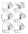
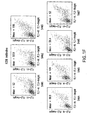
- FIG. 1 Analysis of Specific Binding of anti-FZD10, anti-FZD7, anti-FZD5, anti-FZD6, anti-FZD4, and anti-FZD8 Antibodies to their Corresponding Membrane-Associated Receptors.
- HEK293 cells expressing full-length FZD10, FZD7, FZD5, FZD6, FZD4, and FZD8 without (A) or with (B, C, D, E, and F) co-transfection of GFP were incubated with anti-FZD antibodies or control IgG and sorted by FACS.
- A FACs analysis of antibodies against FZD10 are shown compared with an IgG isotype negative control for each antibody.
- a FLAG-tagged construct matched with anti-FLAG antibodies is shown as a positive control (bottom).
- B FACs analysis of antibodies against FZD7 in cells expressing FZD7 and GFP compared to a control IgG.
- a FLAG-tagged construct matched with anti-FLAG antibodies is shown as a positive control (bottom, far right).
- C FACs analysis of antibodies against FZD5 in cells expressing FZD5 and GFP compared to a control IgG. Serum from an animal immunized with FZD5 antigen is shown on the bottom, right.
- D FACs analysis of antibodies against FZD6 in cells expressing FZD6 and GFP compared to a control IgG.
- a FLAG-tagged construct matched with anti-FLAG antibodies is shown as a positive control (bottom, right).
- E FACs analysis of antibodies against FZD4 in cells expressing FZD4 and GFP compared to a control IgG.
- a FLAG-tagged construct matched with anti-FLAG antibodies is shown as a positive control (bottom, right).
- F FACs analysis of antibodies against FZD8 in cells expressing FZD8 and GFP.
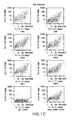
- FIG. 2 FZD Fc Soluble Receptors Inhibit Wnt Signaling.
- HEK 293 cells stably transfected with 8xTCF-luciferase reporter were incubated with increasing concentrations of FZD Fc soluble receptors in the presence of different Wnt ligands including Wnt1, Wnt2, Wnt3, Wnt3a, and Wnt7b.
- FZD4 Fc, FZD5 Fc, and FZD8 Fc fusion proteins inhibited Wnt signaling mediated by all five Wnt ligands as shown by loss of luciferase activity.
- FIG. 3 Identification of anti-FZD5 Antibodies that Interfere with Wnt3a Ligand Binding.
- Wnt signaling in HEK 293 cells transfected with the Wnt 8xTCF-luciferase reporter vector was measured by luciferase activity in the presence of Wnt3a and thirty-two different antibodies against FZD5, either alone (left bar) or in the presence of soluble FZD5 Fc (right bar).
- Antibodies that interfere with binding between FZD5 Fc and Wnt3a result in significant activation of Wnt signaling (right bar).
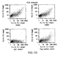
- FIG. 4 Reduction of Tumor Growth by anti-FZD6 and anti-FZD5 Antibodies.
- Tumor growth in NOD/SCID mice injected with UM-C4 colon tumor cells and treated with either anti-FZD6 or anti-FZD5 antibodies is plotted on the x-axis in mm3 over 8 weeks.
- Treatment with anti-FZD6 antibody 23M2 (open bars) and anti-FZD5 antibody 44M13 (dashed bars) significantly reduced tumor growth as compared to PBS injected controls (filled bars).
- antibody is used to mean an immunoglobulin molecule that recognizes and specifically binds to a target, such as a protein, polypeptide, peptide, carbohydrate, polynucleotide, lipid, or combinations of the foregoing through at least one antigen recognition site within the variable region of the immunoglobulin molecule.
- antibodies of the present invention include antagonist antibodies that specifically bind to a cancer stem cell marker protein and interfere with, for example, ligand binding, receptor dimerization, expression of a cancer stem cell marker protein, and/or downstream signaling of a cancer stem cell marker protein.
- disclosed antibodies include agonist antibodies that specifically bind to a cancer stem cell marker protein and promote, for example, ligand binding, receptor dimerization, and/or signaling by a cancer stem cell marker protein. In certain embodiments, disclosed antibodies do not interfere with or promote the biological activity of a cancer stem cell marker protein but inhibit tumor growth by, for example, antibody internalization and/or recognized by the immune system.
- an antibody encompasses intact polyclonal antibodies, intact monoclonal antibodies, antibody fragments (such as Fab, Fab', F(ab')2, and Fv fragments), single chain Fv (scFv) mutants, multispecific antibodies such as bispecific antibodies generated from at least two intact antibodies, chimeric antibodies, humanized antibodies, human antibodies, fusion proteins comprising an antigen determination portion of an antibody, and any other modified immunoglobulin molecule comprising an antigen recognition site so long as the antibodies exhibit the desired biological activity.
- An antibody can be of any the five major classes of immunoglobulins: IgA, IgD, IgE, IgG, and IgM, or subclasses (isotypes) thereof (e.g.
- IgG1, IgG2, IgG3, IgG4, IgA1 and IgA2) based on the identity of their heavy-chain constant domains referred to as alpha, delta, epsilon, gamma, and mu, respectively.
- the different classes of immunoglobulins have different and well known subunit structures and three-dimensional configurations.
- Antibodies can be naked or conjugated to other molecules such as toxins, radioisotopes, etc.
- antibody fragment refers to a portion of an intact antibody and refers to the antigenic determining variable regions of an intact antibody.
- antibody fragments include, but are not limited to Fab, Fab', F(ab')2, and Fv fragments, linear antibodies, single chain antibodies, and multispecific antibodies formed from antibody fragments.
- an “Fv antibody” refers to the minimal antibody fragment that contains a complete antigen-recognition and -binding site either as two-chains, in which one heavy and one light chain variable domain form a non-covalent dimer, or as a single-chain (scFv), in which one heavy and one light chain variable domain are covalently linked by a flexible peptide linker so that the two chains associate in a similar dimeric structure.
- scFv single-chain
- the complementary determining regions (CDRs) of each variable domain interact to define the antigen-binding specificity of the Fv dimer.
- a single variable domain or half of an Fv can be used to recognize and bind antigen, although generally with lower affinity.
- a “monoclonal antibody” as used herein refers to homogenous antibody population involved in the highly specific recognition and binding of a single antigenic determinant, or epitope. This is in contrast to polyclonal antibodies that typically include different antibodies directed against different antigenic determinants.
- the term “monoclonal antibody” encompasses both intact and full-length monoclonal antibodies as well as antibody fragments (such as Fab, Fab', F(ab')2, Fv), single chain (scFv) mutants, fusion proteins comprising an antibody portion, and any other modified immunoglobulin molecule comprising an antigen recognition site.
- “monoclonal antibody” refers to such antibodies made in any number of manners including but not limited to by hybridoma, phage selection, recombinant expression, and transgenic animals.
- humanized antibody refers to forms of non-human (e.g. murine) antibodies that are specific immunoglobulin chains, chimeric immunoglobulins, or fragments thereof that contain minimal non-human sequences.
- humanized antibodies are human immunoglobulins in which residues from the complementary determining region (CDR) are replaced by residues from the CDR of a non-human species (e.g. mouse, rat, rabbit, hamster) that have the desired specificity, affinity, and capability.
- CDR complementary determining region
- FR Fv framework region residues of a human immunoglobulin are replaced with the corresponding residues in an antibody from a non-human species that has the desired specificity, affinity, and capability.
- the humanized antibody can be further modified by the substitution of additional residue either in the Fv framework region and/or within the replaced non-human residues to refine and optimize antibody specificity, affinity, and/or capability.
- the humanized antibody will comprise substantially all of at least one, and typically two or three, variable domains containing all or substantially all of the CDR regions that correspond to the non-human immunoglobulin whereas all or substantially all of the FR regions are those of a human immunoglobulin consensus sequence.
- the humanized antibody can also comprise at least a portion of an immunoglobulin constant region or domain (Fc), typically that of a human immunoglobulin. Examples of methods used to generate humanized antibodies are described in U.S. Pat. 5,225,539 .
- human antibody as used herein means an antibody produced by a human or an antibody having an amino acid sequence corresponding to an antibody produced by a human made using any technique known in the art. This definition of a human antibody includes intact or full-length antibodies, fragments thereof, and/or antibodies comprising at least one human heavy and/or light chain polypeptide such as, for example, an antibody comprising murine light chain and human heavy chain polypeptides.
- Hybrid antibodies are immunoglobulin molecules in which pairs of heavy and light chains from antibodies with different antigenic determinant regions are assembled together so that two different epitopes or two different antigens can be recognized and bound by the resulting tetramer.
- chimeric antibodies refers to antibodies wherein the amino acid sequence of the immunoglobulin molecule is derived from two or more species.
- the variable region of both light and heavy chains corresponds to the variable region of antibodies derived from one species of mammals (e.g. mouse, rat, rabbit, etc) with the desired specificity, affinity, and capability while the constant regions are homologous to the sequences in antibodies derived from another (usually human) to avoid eliciting an immune response in that species.
- epitopes or "antigenic determinant” are used interchangeably herein and refer to that portion of an antigen capable of being recognized and specifically bound by a particular antibody.
- the antigen is a polypeptide
- epitopes can be formed both from contiguous amino acids and noncontiguous amino acids juxtaposed by tertiary folding of a protein. Epitopes formed from contiguous amino acids are typically retained upon protein denaturing, whereas epitopes formed by tertiary folding are typically lost upon protein denaturing.
- An epitope typically includes at least 3, and more usually, at least 5 or 8-10 amino acids in a unique spatial conformation.
- an antibody “selectively binds” or “specifically binds” means that the antibody reacts or associates more frequently, more rapidly, with greater duration, with greater affinity, or with some combination of the above to an epitope than with alternative substances, including unrelated proteins.
- “Selectively binds” or “specifically binds” means, for instance, that an antibody binds to a protein with a KD of at least about 0.1 mM, but more usually at least about 1 ⁇ M.
- “Selectively binds” or “specifically binds” means at times that an antibody binds to a protein at times with a KD of at least about 0.1 ⁇ M or better, and at other times at least about 0.01 ⁇ M or better. Because of the sequence identity between homologous proteins in different species, specific binding can include an antibody that recognizes a cancer stem cell marker protein in more than one species.
- non-specific binding and “background binding” when used in reference to the interaction of an antibody and a protein or peptide refer to an interaction that is not dependent on the presence of a particular structure (i.e., the antibody is binding to proteins in general rather that a particular structure such as an epitope).
- isolated or purified refer to material that is substantially or essentially free from components that normally accompany it in its native state. Purity and homogeneity are typically determined using analytical chemistry techniques such as polyacrylamide gel electrophoresis or high performance liquid chromatography.
- a protein e.g. an antibody
- nucleic acid of the present disclosure that is the predominant species present in a preparation is substantially purified.
- an isolated nucleic acid is separated from open reading frames that naturally flank the gene and encode proteins other than protein encoded by the gene.
- An isolated antibody is separated from other non-immunoglobulin proteins and from other immunoglobulin proteins with different antigen binding specificity. It can also mean that the nucleic acid or protein is in some embodiments at least 80% pure, in some embodiments at least 85% pure, in some embodiments at least 90% pure, in some embodiments at least 95% pure, and in some embodiments at least 99% pure.
- cancer and “cancerous” refer to or describe the physiological condition in mammals in which a population of cells are characterized by unregulated cell growth.
- examples of cancer include, but are not limited to, carcinoma, lymphoma, blastoma, sarcoma, and leukemia.
- cancers include squamous cell cancer, small-cell lung cancer, non-small cell lung cancer, adenocarcinoma of the lung, squamous carcinoma of the lung, cancer of the peritoneum, hepatocellular cancer, gastrointestinal cancer, pancreatic cancer, glioblastoma, cervical cancer, ovarian cancer, liver cancer, bladder cancer, hepatoma, breast cancer, colon cancer, colorectal cancer, endometrial or uterine carcinoma, salivary gland carcinoma, kidney cancer, liver cancer, prostate cancer, vulval cancer, thyroid cancer, hepatic carcinomas and various types of head and neck cancers.
- proliferative disorder and “proliferative disease” refer to disorders associated with abnormal cell proliferation such as cancer.
- Tumor and neoplasm refer to any mass of tissue that result from excessive cell growth or proliferation, either benign (noncancerous) or malignant (cancerous) including pre-cancerous lesions.
- Metalastasis refers to the process by which a cancer spreads or transfers from the site of origin to other regions of the body with the development of a similar cancerous lesion at the new location.
- a “metastatic” or “metastasizing” cell is one that loses adhesive contacts with neighboring cells and migrates via the bloodstream or lymph from the primary site of disease to invade neighboring body structures.
- cancer stem cell refers to a population of cells from a solid tumor that: (1) have extensive proliferative capacity; 2) are capable of asymmetric cell division to generate one or more kinds of differentiated progeny with reduced proliferative or developmental potential; and (3) are capable of symmetric cell divisions for self-renewal or self-maintenance.
- Cancer stem cells undergo self-renewal versus differentiation in a chaotic manner to form tumors with abnormal cell types that can change over time as mutations occur.
- Solid tumor stem cells differ from the "cancer stem line" provided by U.S. Pat. No. 6,004,528 .
- the "cancer stem line” is defined as a slow growing progenitor cell type that itself has few mutations but which undergoes symmetric rather than asymmetric cell divisions as a result of tumorigenic changes that occur in the cell's environment.
- This "cancer stem line” hypothesis thus proposes that highly mutated, rapidly proliferating tumor cells arise largely as a result of an abnormal environment, which causes relatively normal stem cells to accumulate and then undergo mutations that cause them to become tumor cells.
- solid tumor stem cell model is fundamentally different from the "cancer stem line” model and as a result exhibits utilities not offered by the "cancer stem line” model.
- solid tumor stem cells are not “mutationally spared”.
- the "mutationally spared cancer stem line" described by U.S. Pat. No. 6,004,528 can be considered a pre-cancerous lesion, while solid tumor stem cells are cancer cells that may themselves contain the mutations that are responsible for tumorigenesis starting at the pre-cancerous stage through later stage cancer. That is, solid tumor stem cells (“cancer stem cells”) would be included among the highly mutated cells that are distinguished from the "cancer stem line" in U.S. Pat. No.
- the genetic mutations that lead to cancer can be largely intrinsic within the solid tumor stem cells as well as being environmental.
- the solid tumor stem cell model predicts that isolated solid tumor stem cells can give rise to additional tumors upon transplantation (thus explaining metastasis) while the "cancer stem line" model would predict that transplanted "cancer stem line” cells would not be able to give rise to a new tumor, since it was their abnormal environment that was tumorigenic. Indeed, the ability to transplant dissociated, and phenotypically isolated human solid tumor stem cells to mice (into an environment that is very different from the normal tumor environment) where they still form new tumors distinguishes the present invention from the "cancer stem line” model.
- solid tumor stem cells likely divide both symmetrically and asymmetrically, such that symmetric cell division is not an obligate property.
- solid tumor stem cells can divide rapidly or slowly, depending on many variables, such that a slow proliferation rate is not a defining characteristic.
- cancer cell refers to the total population of cells derived from a tumor or a pre-cancerous lesion including both non-tumorigenic cells, which comprise the bulk of the tumor cell population, and tumorigenic stem cells (cancer stem cells).
- tumorigenic refers to the functional features of a solid tumor stem cell including the properties of self-renewal (giving rise to additional tumorigenic cancer stem cells) and proliferation to generate all other tumor cells (giving rise to differentiated and thus non-tumorigenic tumor cells) that allow solid tumor stem cells to form a tumor.
- These properties of self-renewal and proliferation to generate all other tumor cells confer on the cancer stem cells of this invention the ability to form palpable tumors upon serial transplantation into an immunocompromised mouse compared to the majority of tumor cells that are unable to form tumors upon the serial transplantation.
- Tumor cells i.e. non-tumorigenic tumor cells, may form a tumor upon transplantation into an immunocompromised mouse a limited number of times (for example one or two times) after obtaining the tumor cells from a solid tumor.
- stem cell cancer marker(s) refers to a gene or genes or a protein, polypeptide, or peptide expressed by the gene or genes whose expression level, alone or in combination with other genes, is correlated with the presence of tumorigenic cancer cells compared to non-tumorigenic cells.
- the correlation can relate to either an increased or decreased expression of the gene (e.g. increased or decreased levels of mRNA or the peptide encoded by the gene).
- tumor cells as used herein, the terms “unfractionated tumor cells”, “presorted tumor cells”, “bulk tumor cells”, and their grammatical equivalents are used interchangeably to refer to a tumor cell population isolated from a patient sample (e.g. a tumor biopsy or pleural effusion) that has not been segregated, or fractionated, based on cell surface marker expression.
- a patient sample e.g. a tumor biopsy or pleural effusion
- non-ESA+CD44+ tumor cells As used herein, the terms “non-ESA+CD44+ tumor cells”, “non-ESA+44+”, “sorted non-tuinorigenic tumor cells”, “non-tuinorigenic tumor cells,” “non-stem cells,” “tumor cells” and their grammatical equivalents are used interchangeably to refer to a tumor population from which the cancer stem cells of this invention have been segregated, or removed, based on cell surface marker expression.
- biopsy tissue refers to a sample of tissue or fluid that is removed from a subject for the purpose of determining if the sample contains cancerous tissue. In some embodiments, biopsy tissue or fluid is obtained because a subject is suspected of having cancer, and the biopsy tissue or fluid is then examined for the presence or absence of cancer.
- the term “subject” refers to any animal (e.g., a mammal), including, but not limited to humans, non-human primates, rodents, and the like, which is to be the recipient of a particular treatment.
- the terms “subject” and “patient” are used interchangeably herein in reference to a human subject.
- “Pharmaceutically acceptable” refers to approved or approvable by a regulatory agency of the Federal or a state government or listed in the U.S. Pharmacopeia or other generally recognized pharmacopeia for use in animals, including humans.
- “Pharmaceutically acceptable salt” refers to a salt of a compound that is pharmaceutically acceptable and that possesses the desired pharmacological activity of the parent compound.
- “Pharmaceutically acceptable excipient, carrier or adjuvant” refers to an excipient, carrier or adjuvant that can be administered to a subject, together with at least one antibody of the present disclosure, and which does not destroy the pharmacological activity thereof and is nontoxic when administered in doses sufficient to deliver a therapeutic amount of the compound.
- “Pharmaceutically acceptable vehicle” refers to a diluent, adjuvant, excipient, or carrier with which at least one antibody of the present disclosure is administered.
- Prodrug refers to a derivative of a therapeutically effective compound that requires a transformation within the body to produce the therapeutically effective compound. Prodrugs can be pharmacologically inactive until converted to the therapeutically effective parent compound.
- therapeutically effective amount refers to an amount of an antibody, polypeptide, polynucleotide, small organic molecule, or other drug effective to "treat” a disease or disorder in a subject or mammal.
- the therapeutically effective amount of the drug can reduce the number of cancer cells; reduce the tumor size; inhibit or stop cancer cell infiltration into peripheral organs including, for example, the spread of cancer into soft tissue and bone; inhibit'and stop tumor metastasis; inhibit and stop tumor growth; relieve to some extent one or more of the symptoms associated with the cancer, reduce morbidity and mortality; improve quality of life; or a combination of such effects.
- the drug prevents growth and/or kills existing cancer cells, it can be referred to as cytostatic and/or cytotoxic.
- diagnosis refers to any information that is useful in determining whether a patient has a disease or condition and/or in classifying the disease or condition into a phenotypic category or any category having significance with regards to the prognosis of or likely response to treatment (either treatment in general or any particular treatment) of the disease or condition.
- diagnosis refers to providing any type of diagnostic information, including, but not limited to, whether a subject is likely to have a condition (such as a tumor), information related to the nature or classification of a tumor as for example a high risk tumor or a low risk tumor, information related to prognosis and/or information useful in selecting an appropriate treatment. Selection of treatment can include the choice of a particular chemotherapeutic agent or other treatment modality such as surgery or radiation or a choice about whether to withhold or deliver therapy.
- the terms “providing a prognosis”, “prognostic information”, or “predictive information” refer to providing information regarding the impact of the presence of cancer (e.g., as determined by the diagnostic methods of the present invention) on a subject's future health (e.g., expected morbidity or mortality, the likelihood of getting cancer, and the risk of metastasis).
- Terms such as “treating” or “treatment” or “to treat” or “alleviating” or “to alleviate” refer to both 1) therapeutic measures that cure, slow down, lessen symptoms of, and/or halt progression of a diagnosed pathologic condition or disorder and 2) prophylactic or preventative measures that prevent and/or slow the development of a targeted pathologic condition or disorder.
- those in need of treatment include those already with the disorder; those prone to have the disorder; and those in whom the disorder is to be prevented.
- a subject is successfully "treated” according to the methods of the present invention if the patient shows one or more of the following: a reduction in the number of or complete absence of cancer cells; a reduction in the tumor size; inhibition of or an absence of cancer cell infiltration into peripheral organs including, for example, the spread of cancer into soft tissue and bone; inhibition of or an absence of tumor metastasis; inhibition or an absence of tumor growth; relief of one or more symptoms associated with the specific cancer; reduced morbidity and mortality; improvement in quality of life; or some combination of effects.
- polynucleotide or “nucleic acid” refer to a polymer composed of a multiplicity of nucleotide units (ribonucleotide or deoxyribonucleotide or related structural variants) linked via phosphodiester bonds, including but not limited to, DNA or RNA.
- the term encompasses sequences that include any of the known base analogs of DNA and RNA.
- gene refers to a nucleic acid (e.g., DNA) sequence that comprises coding sequences necessary for the production of a polypeptide, precursor, or RNA (e.g., rRNA, tRNA).
- the polypeptide can be encoded by a full length coding sequence or by any portion of the coding sequence so long as the desired activity or functional properties (e.g., enzymatic activity, ligand binding, signal transduction, immunogenicity, etc.) of the full-length or fragment are retained.
- the term also encompasses the coding region of a structural gene and the sequences located adjacent to the coding region on both the 5' and 3' ends for a distance of about 1 kb or more on either end such that the gene corresponds to the length of the full-length mRNA. Sequences located 5' of the coding region and present on the mRNA are referred to as 5' non-translated sequences.
- genomic form or clone of a gene contains the coding region interrupted with non-coding sequences termed "introns” or “intervening regions” or “intervening sequences.”
- Introns are segments of a gene that are transcribed into nuclear RNA (hnRNA); introns can contain regulatory elements such as enhancers. Introns are removed or “spliced out” from the nuclear or primary transcript; introns therefore are absent in the messenger RNA (mRNA) transcript.
- genomic forms of a gene can also include sequences located on both the 5' and 3' end of the sequences that are present on the RNA transcript. These sequences are referred to as "flanking" sequences or regions (these flanking sequences are located 5' or 3' to the non-translated sequences present on the mRNA transcript).
- the 5' flanking region can contain regulatory sequences such as promoters and enhancers that control or influence the transcription of the gene.
- the 3' flanking region can contain sequences that direct the termination of transcription, post transcriptional cleavage and polyadenylation.
- recombinant when used with reference to a cell, nucleic acid, protein or vector indicates that the cell, nucleic acid, protein or vector has been modified by the introduction of a heterologous nucleic acid or protein, the alteration of a native nucleic acid or protein, or that the cell is derived from a cell so modified.
- recombinant cells express genes that are not found within the native (non-recombinant) form of the cell or express native genes that are overexpressed or otherwise abnormally expressed such as, for example, expressed as non-naturally occurring fragments or splice variants.
- nucleic acid By the term “recombinant nucleic acid” herein is meant nucleic acid, originally formed in vitro, in general, by the manipulation of nucleic acid, e.g., using polymerases and endonucleases, in a form not normally found in nature. In this manner, operably linkage of different sequences is achieved.
- an isolated nucleic acid, in a linear form, or an expression vector formed in vitro by ligating DNA molecules that are not normally joined are both considered recombinant for the purposes of this invention.
- a recombinant nucleic acid is made and introduced into a host cell or organism, it will replicate non-recombinantly, i.e., using the in vivo cellular machinery of the host cell rather than in vitro manipulations; however, such nucleic acids, once produced recombinantly, although subsequently replicated non-recombinantly, are still considered recombinant for the purposes of the invention.
- a "recombinant protein” is a protein made using recombinant techniques, i.e., through the expression of a recombinant nucleic acid as depicted above.
- heterologous gene refers to a gene that is not in its natural environment.
- a heterologous gene includes a gene from one species introduced into another species.
- a heterologous gene also includes a gene native to an organism that has been altered in some way (e.g., mutated, added in multiple copies, linked to non-native regulatory sequences, etc).
- Heterologous genes are distinguished from endogenous genes in that the heterologous gene sequences are typically joined to DNA sequences that are not found naturally associated with the gene sequences in the chromosome or are associated with portions of the chromosome not found in nature (e.g., genes expressed in loci where the gene is not normally expressed).
- vector is used in reference to nucleic acid molecules that transfer DNA segment(s) from one cell to another.
- vehicle is sometimes used interchangeably with “vector.”
- Vectors are often derived from plasmids, bacteriophages, or plant or animal viruses.
- Ligation refers to the process of forming phosphodiester bonds between two double stranded nucleic acid fragments. Unless otherwise provided, ligation can be accomplished using known buffers and conditions with 10 units to T4 DNA ligase ("ligase") per 0.5 ug of approximately equimolar amounts of the DNA fragments to be ligated. Ligation of nucleic acid can serve to link two proteins together in-frame to produce a single protein, or fusion protein.
- ligase T4 DNA ligase
- RNA expression refers to the process of converting genetic information encoded in a gene into RNA (e.g., mRNA, rRNA, tRNA, or snRNA) through “transcription” of the gene (e.g., via the enzymatic action of an RNA polymerase), and for protein encoding genes, into protein through “translation” of mRNA.
- Gene expression can be regulated at many stages in the process.
- Up-regulation” or “activation” refers to regulation that increases the production of gene expression products (e.g., RNA or protein), while “down-regulation” or “repression” refers to regulation that decrease production.
- Molecules e.g., transcription factors
- activators e.g., transcription factors
- polypeptide peptide
- protein protein fragment
- amino acid polymers in which one or more amino acid residue is an artificial chemical mimetic of a corresponding naturally occurring amino acid, as well as to naturally occurring amino acid polymers and non-naturally occurring amino acid polymers.
- amino acid refers to naturally occurring and synthetic amino acids, as well as amino acid analogs and amino acid mimetics that function similarly to the naturally occurring amino acids.
- Naturally occurring amino acids are those encoded by the genetic code, as well as those amino acids that are later modified, e.g., hydroxyproline, gamma-carboxyglutainate, and O-phosphoserine.
- Amino acid analogs refers to compounds that have the same basic chemical structure as a naturally occurring amino acid, e.g., an alpha carbon that is bound to a hydrogen, a carboxyl group, an amino group, and an R group, e.g., homoserine, norleucine, methionine sulfoxide, methionine methyl sulfonium. Such analogs can have modified R groups (e.g., norleucine) or modified peptide backbones, but retain the same basic chemical structure as a naturally occurring amino acid.
- Amino acid mimetics refers to chemical compounds that have a structure that is different from the general chemical structure of an amino acid, but that functions similarly to a naturally occurring amino acid.
- amino acid variants refers to amino acid sequences. With respect to particular nucleic acid sequences, conservatively modified variants refers to those nucleic acids which encode identical or essentially identical amino acid sequences, or where the nucleic acid does not encode an amino acid sequence, to essentially identical or associated (e.g., naturally contiguous) sequences. Because of the degeneracy of the genetic code, a large number of functionally identical nucleic acids encode most proteins. For instance, the codons GCA, GCC, GCG and GCU all encode the amino acid alanine.
- nucleic acid variations are "silent variations," which are one species of conservatively modified variations. Every nucleic acid sequence herein which encodes a polypeptide also describes silent variations of the nucleic acid.
- AUG which is ordinarily the only codon for methionine
- TGG which is ordinarily the only codon for tryptophan
- nucleic acid which encodes a polypeptide is implicit in a described sequence with respect to the expression product, but not with respect to actual probe sequences.
- amino acid sequences one of skill will recognize that individual substitutions, deletions or additions to a nucleic acid, peptide, polypeptide, or protein sequence which alters, adds or deletes a single amino acid or a small percentage of amino acids in the encoded sequence is a "conservatively modified variant" including where the alteration results in the substitution of an amino acid with a chemically similar amino acid.
- Conservative substitution tables providing functionally similar amino acids are well known in the art.
- conservatively modified variants are in addition to and do not exclude polymorphic variants, interspecies homologs, and alleles of the invention.
- conservative substitutions include: 1) Alanine (A), Glycine (G); 2) Aspartic acid (D), Glutamic acid (E); 3) Asparagine (N), Glutamine (Q); 4) Arginine (R), Lysine (K); 5) Isoleucine (I), Leucine (L), Methionine (M), Valine (V); 6) Phenylalanine (F), Tyrosine (Y), Tryptophan (W); 7) Serine (S), Threonine (T); and 8) Cysteine (C), Methionine (M) (see, e.g., Creighton, Proteins (1984)).
- epitope tagged refers to a chimeric polypeptide comprising a cancer stem cell marker protein, or a domain sequence or portion thereof, fused to an "epitope tag".
- the epitope tag polypeptide comprises enough amino acid residues to provide an epitope for recognition by an antibody, yet is short enough such that it does not interfere with the activity of the cancer stem cell marker protein.
- Suitable epitope tags generally have at least six amino acid residues, usually between about 8 to about 50 amino acid residues, and at times between about 10 to about 20 residues. Commonly used epitope tags include Fc, HA, His, and FLAG tags.
- an isolated antibody that specifically binds to an extracellular domain of a human FZD8 receptor and inhibits growth of tumor cells.
- the antibody is a monoclonal antibody.
- the antibody is a chimeric antibody.
- the antibody is a humanized antibody.
- the antibody is a human antibody.
- an isolated antibody that specifically binds to an extracellular domain of two or more human FZD receptors and inhibits growth of tumor cells.
- the antibody specifically binds to the extracellular domain of human FZD2 and FZD6.
- the antibody specifically binds to the extracellular domain of human FZD7 and FZD10.
- the antibody specifically binds to the extracellular domain of human FZD4 and FZD5.
- the antibody specifically binds to the extracellular domain of human FZD4 and FZD8.
- the antibody specifically binds to the extracellular domain of human FZD5 and FZD8.
- the antibody is a monoclonal antibody.
- the antibody is a chimeric antibody.
- the antibody is a humanized antibody.
- the antibody is a human antibody.
- an isolated antibody that specifically binds to the extracellular domain of three or more human FZD receptors.
- the antibody specifically binds to the extracellular domain of human FZD4, FZD5, and FZD8.
- the antibody is a monoclonal antibody.
- the antibody is a chimeric antibody.
- the antibody is a humanized antibody.
- the antibody is a human antibody.
- composition comprising an antibody of the present disclosure and a pharmaceutically acceptable vehicle.
- hybridoma that produces an antibody of the present disclosure.
- a method of treating cancer comprising administering a antibody or a pharmaceutical composition of the present disclosure in an amount effective to inhibit tumor cell growth.
- the antibody is conjugated to a cytotoxic moiety.
- the method further comprises administering at least one additional therapeutic agent appropriate for effecting combination therapy.
- the tumor cells are chosen from a breast tumor, colorectal tumor, lung tumor, prostate tumor, pancreatic tumor, and a head and neck tumor.
- solid tumors consist of a heterogeneous population of cells. That the majority of these cells lack tumorigenicity suggested that the development and maintenance of solid tumors also relies on a small population of stem cells (i.e., tumorigenic cancer cells) with the capacity to proliferate and efficiently give rise both to additional tumor stem cells (self-renewal) and to the majority of more differentiated tumor cells that lack tumorigenic potential (i.e., non-tumorigenic cancer cells).
- stem cells i.e., tumorigenic cancer cells
- self-renewal additional tumor stem cells
- non-tumorigenic cancer cells i.e., non-tumorigenic cancer cells
- Stem cells from solid tumors have more recently been isolated based on their expression of a unique pattern of cell-surface receptors and on the assessment of their properties of self-renewal and proliferation in culture and in xenograft animal models.
- An ESA+ CD44+ CD24-/low Lineage- population greater than 50-fold enriched for the ability to form tumors relative to unfractionated tumor cells was discovered ( A1-Hajj et al., 2003, Proc. Nat'l. Acad. Sci.
- the ability to isolate tumorigenic cancer stem cells from the bulk of non-tumorigenic tumor cells has led to the identification of cancer stem cell markers, genes with differential expression in cancer stem cells compared to non-tumorigenic tumor cells or normal breast epithelium, using microarray analysis.
- the present invention employs the knowledge of these identified cancer stem cell markers to diagnosis and treat cancer.
- the cancer stem cell markers of the present invention relate to a human FZD receptor, including for example, human FZD4, FZD5, and FZD8 as markers of cancer stem cells, implicating the Wnt signaling pathway in the maintenance of cancer stem cells and as a target for treating cancer via the elimination of these tumorigenic cells.
- the Wnt signaling pathway is one of several critical regulators of embryonic pattern formation, post-embryonic tissue maintenance, and stem cell biology. More specifically, Wnt signaling plays an important role in the generation of cell polarity and cell fate specification including self-renewal by stem cell populations. Unregulated activation of the Wnt pathway is associated with numerous human cancers where it can alter the developmental fate of tumor cells to maintain them in an undifferentiated and proliferative state. Thus carcinogenesis can proceed by usurping homeostatic mechanisms controlling normal development and tissue repair by stem cells (reviewed in Reya & Clevers, 2005, Nature 434:843 ; Beachy et al., 2004, Nature 432:324
- Wnt signaling pathway was first elucidated in the Drosophila developmental mutant wingless ( wg ) and from the murine proto-oncogene int-1, now Wnt1 ( Nusse & Varmus, 1982, Cell 31:99-109 ; Van Ooyen & Nusse, 1984, Cell 39:233-40 ; Cabrera et al., 1987, Cell 50:659-63 ; Rijsewijk et al., 1987, Cell 50:649-57 ). Wnt genes encode secreted lipid-modified glycoproteins of which 19 have been identified in mammals.
- Fzd Frizzled
- LDL low-density lipoprotein
- the Fzd receptors are seven transmembrane domain proteins of the G-protein coupled receptor (GPCR) superfamily and contain a large extracellular N-terminal ligand binding domain with 10 conserved cysteines, known as a cysteine-rich domain (CRD) or Fri domain.
- GPCR G-protein coupled receptor
- CRD cysteine-rich domain
- FZD1-10 There are ten human FZD receptors: FZD1-10. Different Fzd CRDs have different binding affinities for specific Wnts ( Wu & Nusse, 2002, J. Biol. Chem.
- Fzd receptors have been grouped into those that activate the canonical ⁇ -catenin pathway and those that activate non-canonical pathways described below ( Miller et al., 1999, Oncogene 18:7860-72 ).
- FZD receptors interact with LRP5/6, single pass transmembrane proteins with four extracellular EGF-like domains separated by six YWTD amino acid repeats ( Johnson et al., 2004, J. Bone Mineral Res. 19:1749 ).
- the canonical Wnt signaling pathway activated upon receptor binding is mediated by the cytoplasmic protein Dishevelled (Dsh) interacting directly with the Fzd receptor and results in the cytoplasmic stabilization and accumulation of ⁇ -catenin.
- Dsh Dishevelled
- ⁇ -catenin is localized to a cytoplasmic destruction complex that includes the tumor suppressor proteins adenomatous polyposis coli (APC) and auxin. These proteins function as critical scaffolds to allow glycogen synthase kinase (GSK)-3 ⁇ to bind and phosphorylate ⁇ -catenin, marking it for degradation via the ubiquitin/proteasome pathway.
- Activation of Dsh results in phophorylation of GSK3 ⁇ and the dissociation of the destruction complex.
- Accumulated cytoplasmic ⁇ -catenin is then transported into the nucleus where it interacts with the DNA-binding proteins of the Tcf/Lef family to activate transcription.
- Wnt ligands In addition to the canonical signaling pathway, Wnt ligands also active ⁇ -catenin-independent pathways ( Veeman et al., 2003, Dev. Cell 5:367-77 ). Non-canonical Wnt signaling has been implicated in numerous processes but most convincingly in gastrulation movements via a mechanism similar to the Drosophila planar cell polarity (PCP) pathway. Other potential mechanisms of non-canonical Wnt signaling include calcium flux, JNK, and both small and heterotrimeric G-proteins. Antagonism is often observed between the canonical and non-canonical pathways, and some evidence indicates that non-canonical signaling can suppress cancer formation ( Olson & Gibo, 1998, Exp. Cell Res.
- Fzd receptors act as negative regulators of the canonical Wnt signaling pathway.
- FZD6 represses Wnt-3a-induced canonical signaling when co-expressed with FZD1 via the TAK1-NLK pathway ( Golan et al., 2004, JBC 279:14879-88 ).
- Fzd2 antagonized canonical Wnt signaling in the presence of Wnt-5a via the TAK1-NLK MAPK cascade ( Ishitani et al., 2003, Mol. Cell. Biol. 23:131-9 ).
- HSCs Hematopoietic stem cells
- Wnt signaling is implicated both in their normal maintenance as well as in leukemic transformation ( Reya & Clevers, 2005, Nature 434:843 ).
- HSCs are a rare population of cells that reside in a stomal niche within the adult bone marrow. These cells are characterized both by a unique gene expression profile as well as an ability to continuously give rise to more differentiated progenitor cells to reconstitute the entire hematopoietic system.
- Both HSCs and the cells of their stromal microenvironment express Wnt ligands, and Wnt reporter activation is present in HSCs in vivo.
- both ⁇ -catenin and purified Wnt3A promote self-renewal of murine HSCs in vitro and enhance their ability to reconstitute the hematopoietic system in vivo
- Wnt5A promotes expansion of human hematopoietic progenitors in vitro and repopulation in a NOD-SCID xenotransplant model ( Reya et al., 2003, Nature 423:409-14 ; Willert et al., 2003, Nature 423:448-52 ; Van Den Berg et al., 1998, Blood 92:3189-202 ; Murdoch et al., 2003, Proc. Nat'l Acad. Sci. 100:3422-7 ).
- Wnt signaling has been found to play a role in the oncogenic growth of both myeloid and lymphoid lineages.
- GMPs granulocyte-macrophage progenitors
- Wnt signaling displays activated Wnt signaling on which they are depended for growth and renewal ( Jamieson et al., 2004, N. Engl. J. Med. 351:657-67 )
- Wnt signaling can sustain cancerous self-renewal ( Reya & Clevers 2005, Nature 434:843 ).
- the canonical Wnt signaling pathway also plays a central role in the maintenance of stem cell populations in the small intestine and colon, and the inappropriate activation of this pathway plays a prominent role in colorectal cancers ( Reya & Clevers, 2005, Nature 434:843 ).
- the absorptive epithelium of the intestines is arranged into villi and crypts.
- Stem cells reside in the crypts and slowly divide to produce rapidly proliferating cells which give rise to all the differentiated cell populations that move up out of the crypts to occupy the intestinal villi.
- the Wnt signaling cascade plays a dominant role in controlling cell fates along the crypt-villi axis and is essential for the maintenance of the stem cell population.
- Colorectal cancer is most commonly initiated by activating mutations in the Wnt signaling cascade. Approximately 5-10% of all colorectal cancers are hereditary with one of the main forms being familial adenomatous polyposis (FAP), an autosomal dominant disease in which about 80% of affected individuals contain a germline mutation in the adenomatous polyposis coli ( APC ) gene. Mutations have also been identified in other Wnt pathway components including auxin and ⁇ -catenin.
- FAP familial adenomatous polyposis
- APC adenomatous polyposis coli
- adenomas are clonal outgrowths of epithelial cell containing a second inactivated allele, and the large number of FAP adenomas inevitably results in the development of adenocarcinomas through addition mutations in oncogenes and/or tumor suppressor genes. Furthermore, activation of the Wnt signaling pathway, including gain-of-function mutations in APC and ⁇ -catenin, can induce hyperplastic development and tumor growth in mouse models ( Oshima et al., 1997, Cancer Res. 57:1644-9 ; Harada et al., 1999, EMBO J. 18:5931-42 ).
- Wnt1 originally int1
- Wnt1 an oncogene in mammary tumors transformed by the nearby insertion of a murine virus
- Nusse & Varmus 1982, Cell 31:99-109
- Additional evidence for the role of Wnt signaling in breast cancer has since accumulated.
- transgenic overexpression of ⁇ -catenin in the mammary glands results in hyperplasias and adenocarcinomas ( Imbert et al., 2001, J. Cell Biol.
- FZD10, FZD8, FZD7, FZD4, and FZD5 are five of ten identified human Wnt receptors.
- Fzd10 is expressed with Wnt7a in the neural tube, limb buds, and Mullerian duct ( Nunnally & Parr, 2004, Dev. Genes Evol. 214:144-8 ) and can act as a receptor for Wnt-7a during limb bud development ( Kawakami et al., 2000, Dev. Growth Differ. 42:561-9 ).
- Fzd10 is co-expressed with Wnt7b in the lungs, and cell transfection studies have demonstrated that the Fzd10/LRP5 co-receptor activates the canonical Wnt signaling pathway in response to Wnt7b ( Wang et al., 2005, Mol. Cell Biol. 25:5022-30 ).
- FZD10 mRNA is upregulated in numerous cancer cell lines, including cervical, gastric, and glioblastoma cell lines, and in primary cancers including approximately 40% of primary gastric cancers, colon cancers, and synovial sarcomas ( Saitoh et al., 2002, Int. J. Oncol. 20:117-20 ; Terasaki et al., 2002, Int. J.
- FZD8 is upregulated in several human cancer cell lines, primary gastric cancers, and renal carcinomas ( Saitoh et al., 2001, Int. J. Oncol. 18:991-96 ; Kirikoshi et al., 2001, Int. J. Oncol. 19:111-5 ; Janssens et al., 2004, Tumor Biol. 25:161-71 ).
- FZD7 is expressed throughout the gastrointestinal tract and is up-regulated in one out of six cases of human primary gastric cancer ( Kirikoshi et al., 2001, Int. J. Oncol. 19:111-5 ).
- FZD7 ectodomain by a colon cancer cell line induced morphological changes and decreased tumor growth in a xenograft model ( Vincan et al., 2005, Differentiation 73:142-53 ).
- FZD5 plays an essential role in yolk sac and placental angiogenesis ( Ishikawa et al., 2001, Dev. 128:25-33 ) and is upregulated in renal carcinomas in association with activation of Wnt/ ⁇ -cateun signaling ( Janssens et al., 2004, Tumor Biology 25:161-71 ).
- FZD4 is highly expressed in intestinal crypt epithelial cells and is one of several factors that display differential expression in normal versus neoplastic tissue ( Gregorieff et al., 2005, Gastroenterology 129:626-38 ). The identification of FZD receptors as markers of cancer stem cells thus makes these proteins ideal targets for cancer therapeutics.
- the present invention provides a cancer stem cell marker the expression of which can be analyzed to diagnosis or monitor a disease associated with expression of a cancer stem cell marker.
- expression of a cancer stem cell marker is determined by polynucleotide expression such as, for example, mRNA encoding the cancer stem cell marker.
- the polynucleotide can be detected and quantified by any of a number of means well known to those of skill in the art.
- mRNA encoding a cancer stem cell marker is detected by in situ hybridization of tissue sections from, from example, a patient biopsy.
- RNA is isolated from a tissue and detected by, for example, Northern blot, quantitative RT-PCR, or microarrays. For example, total RNA can be extracted from a tissue sample and primers that specifically hybridize and amplify a cancer stem cell marker can be used to detect expression of a cancer stem cell marker polynucleotide using RT-PCR.
- expression of a cancer stem cell marker can be determined by detection of the corresponding polypeptide.
- the polypeptide can be detected and quantified by any of a number of means well known to those of skill in the art.
- a cancer stem cell marker polypeptide is detected using analytic biochemical methods such as, for example, electrophoresis, capillary electrophoresis, high performance liquid chromatography (HPLC) or thin layer chromatography (TLC).
- HPLC high performance liquid chromatography
- TLC thin layer chromatography
- the isolated polypeptide can also be sequenced according to standard techniques.
- a cancer stem cell marker protein is detected with antibodies raised against the protein using, for example, immunofluorescence or immunohistochemistry on tissue sections.
- antibodies against a cancer stem cell marker can detect expression using, for example, ELISA, FACS, Western blot, immunoprecipitation or protein microarrays.
- cancer stem cells can be isolated from a patient biopsy and expression of a cancer stem cell marker protein detected with fluorescently labeled antibodies using FACS.
- the cells expressing a cancer stem cell marker can be detected in vivo using labeled antibodies in typical imaging system.
- antibodies labeled with paramagnetic isotopes can be used for magnetic resonance imaging (MRT).
- a diagnostic assay comprises determining the expression or not of a cancer stem cell marker in tumor cells using, for example, immunohistochemistry, in situ hybridization, or RT-PCR. In other embodiments, a diagnostic assay comprises determining expression levels of a cancer stem cell marker using, for example, quantitative RT-PCR. In some embodiments, a diagnostic assay further comprises determining expression levels of a cancer stem cell marker compared to a control tissue such as, for example, normal epithelium.
- Detection of a cancer stem cell marker expression can then be used to provide a prognosis and select a therapy.
- a prognosis can be based on any known risk expression of a cancer stem cell marker indicates.
- detection of a cancer stem cell marker can be used to select an appropriate therapy including, for example, treatment with antibodies against the detected cancer stem cell marker protein.
- the antibody specifically binds to the extracellular domain of a cancer stem cell marker protein such as a human FZD receptor.
- a suitable antibody is an agent that can have one or more of the following effects, for example: interfere with the expression of a cancer stem cell marker; interfere with activation of a cancer stem cell signal transduction pathway by, for example, sterically inhibiting interactions between a cancer stem cell marker and its ligand, receptor or co-receptors; activate a cancer stem cell signal transduction pathway by, for example, acting as a ligand or promoting the binding of an endogenous ligand; or bind to a cancer stem cell marker and inhibit tumor cell proliferation.
- antibodies against a cancer stem cell marker act extracellularly to modulate the function of a cancer stem cell marker protein.
- extracellular binding of an antibody against a cancer stem cell marker can inhibit the signaling of a cancer stem cell marker protein by, for example, inhibiting intrinsic activation (e.g. kinase activity) of a cancer stem cell marker and/or by sterically inhibiting the interaction, for example, of a cancer stem cell marker with its ligand, with its receptor, with a co-receptor, or with the extracellular matrix.
- intrinsic activation e.g. kinase activity
- extracellular binding of an antibody against a cancer stem cell marker can downregulate cell-surface expression of a cancer stem cell marker such as, for example, by internalization of a cancer stem cell marker protein or decreasing cell surface trafficking of a cancer stem cell marker.
- extracellular binding of an antibody against a cancer stem cell marker can promote the signaling of a cancer stem cell marker protein by, for example, acting as a decoy ligand or increasing ligand binding.
- antibodies against a cancer stem cell marker bind to a cancer stem cell marker protein and have one or more of the following effects: inhibit proliferation of tumor cells, trigger cell death of tumor cells, or prevent metastasis of tumor cells.
- antibodies against a cancer stem cell marker trigger cell death via a conjugated toxin, chemotherapeutic agent, radioisotope, or other such agent.
- an antibody against a cancer stem cell marker is conjugated to a toxin that is activated in tumor cells expressing the cancer stem cell marker by protein internalization.
- antibodies against a cancer stem cell marker mediate cell death of a cell expressing the cancer stem cell marker protein via antibody-dependent cellular cytotoxicity (ADCC).
- ADCC antibody-dependent cellular cytotoxicity
- ADCC involves cell lysis by effector cells that recognize the Fc portion of an antibody.
- antibodies against a cancer stem cell marker trigger cell death of a cell expressing a cancer stem cell marker protein by activating complement-dependent cytotoxicity (CDC).
- CDC involves binding of serum complement to the Fc portion of an antibody and subsequent activation of the complement protein cascade, resulting in cell membrane damage and eventual cell death.
- Biological activity of antibodies is known to be determined, to a large extent, by the constant domains or Fc region of the antibody molecule ( Uananue and Benacerraf, Textbook of Immunology, 2nd Edition, Williams & Wilkins, p. 218 (1984 )).
- Antibodies of different classes and subclasses differ in this respect, as do antibodies of the same subclass but from different species.
- IgM is the most efficient class of antibodies to bind complement, followed by IgG1, IgG3, and IgG2 whereas IgG4 appears quite deficient in activating the complement cascade ( Dillman, 1994, J. Clin. Oncol. 12:1497 ; Jefferis et al., 1998, Immunol. Rev. 163:59-76 ).
- antibodies of those classes having the desired biological activity are prepared.
- any particular antibody against a cancer stem cell to mediate lysis of the target cell by complement activation and/or ADCC can be assayed.
- the cells of interest are grown and labeled in vitro; the antibody is added to the cell culture in combination with either serum complement or immune cells which can be activated by the antigen antibody complexes. Cytolysis of the target cells is detected, for example, by the release of label from the lysed cells.
- antibodies can be screened using the patient's own serum as a source of complement and/or immune cells. The antibody that is capable of activating complement or mediating ADCC in the in vitro test can then be used therapeutically in that particular patient.
- antibodies against a cancer stem cell marker can trigger cell death inhibiting angiogenesis.
- Angiogenesis is the process by which new blood vessels form from pre-existing vessels and is a fundamental process required for normal growth, for example, during embryonic development, wound healing, and in response to ovulation. Solid tumor growth larger than 1-2 mm 2 also requires angiogenesis to supply nutrients and oxygen without which tumor cells die.
- an antibody against a cancer stem cell marker targets vascular cells that express the cancer stem cell marker including, for example, endothelial cells, smooth muscle cells, or components of the extracellular matrix required for vascular assembly.
- an antibody against a cancer stem cell marker inhibits growth factor signaling required by vascular cell recruitment, assembly, maintenance, or survival.
- the antibodies against a cancer stem cell marker find use in the diagnostic and therapeutic methods described herein.
- the antibodies of the present invention are used to detect the expression of a cancer stem cell marker protein in biological samples such as, for example, a patient tissue biopsy, pleural effusion, or blood sample. Tissue biopsies can be sectioned and protein detected using, for example, immunofluorescence or immunohistochemistry.
- individual cells from a sample can be isolated, and protein expression detected on fixed or live cells by FACS analysis.
- antibodies can be used on protein arrays to detect expression of a cancer stem cell marker, for example, on tumor cells, in cell lysates, or in other protein samples.
- the antibodies of the present invention are used to inhibit the growth of tumor cells by contacting the antibodies with tumor cells in in vitro cell based assays , in vivo animal models, etc. In certain embodiments, the antibodies are used to treat cancer in a patient by administering a therapeutically effective amount of an antibody against a cancer stem cell marker.
- Polyclonal antibodies can be prepared by any known method. Polyclonal antibodies are raised by immunizing an animal (e.g. a rabbit, rat, mouse, donkey, etc) by multiple subcutaneous or intraperitoneal injections of the relevant antigen (a purified peptide, fragment, full-length recombinant protein, fusion protein, etc) optionally conjugated to keyhole limpet hemocyanin (KLH), serum albumin, etc. diluted in sterile saline and combined with an adjuvant (e.g. Complete or Incomplete Freund's Adjuvant) to form a stable emulsion. The polyclonal antibody is then recovered from blood, ascites and the like, of an animal so immunized.
- an adjuvant e.g. Complete or Incomplete Freund's Adjuvant
- the polyclonal antibodies can be purified from serum or ascites according to standard methods in the art including affinity chromatography, ion-exchange chromatography, gel electrophoresis, dialysis, etc.
- Monoclonal antibodies can be prepared using hybridoma methods, such as those described by Kohler and Milstein (1975) Nature 256:495 .
- a mouse, hamster, or other appropriate host animal is immunized as described above to elicit the production by lymphocytes of antibodies that will specifically bind to an immunizing antigen.
- Lymphocytes can also be immunized in vitro. Following immunization, the lymphocytes are isolated and fused with a suitable myeloma cell line using, for example, polyethylene glycol, to form hybridoma cells that can then be selected away from unfused lymphocytes and myeloma cells.
- Hybridomas that produce monoclonal antibodies directed specifically against a chosen antigen as determined by immunoprecipitation, immunoblotting, or by an in vitro binding assay e.g. radioimmunoassay (RIA); enzyme-linked immunosorbent assay (ELISA)
- an in vitro binding assay e.g. radioimmunoassay (RIA); enzyme-linked immunosorbent assay (ELISA)
- RIA radioimmunoassay
- ELISA enzyme-linked immunosorbent assay
- monoclonal antibodies can also be made using recombinant DNA methods as described in U.S. Patent 4,816,567 .
- the polynucleotides encoding a monoclonal antibody are isolated from mature B-cells or hybridoma cell, such as by RT-PCR using oligonucleotide primers that specifically amplify the genes encoding the heavy and light chains of the antibody, and their sequence is determined using conventional procedures.
- the isolated polynucleotides encoding the heavy and light chains are then cloned into suitable expression vectors, which when transfected into host cells such as E.
- monoclonal antibodies are generated by the host cells.
- recombinant monoclonal antibodies or fragments thereof of the desired species can be isolated from phage display libraries expressing CDRs of the desired species as described ( McCafferty et al., 1990, Nature, 348:552-554 ; Clackson et al., 1991, Nature, 352:624-628 ; and Marks et al., 1991, J. Mol. Biol., 222:581-597 ).
- the polynucleotide(s) encoding a monoclonal antibody can further be modified in a number of different manners using recombinant DNA technology to generate alternative antibodies.
- the constant domains of the light and heavy chains of, for example, a mouse monoclonal antibody can be substituted 1) for those regions of, for example, a human antibody to generate a chimeric antibody or 2) for a non-immunoglobulin polypeptide to generate a fusion antibody.
- the constant regions are truncated or removed to generate the desired antibody fragment of a monoclonal antibody. Site-directed or high-density mutagenesis of the variable region can be used to optimize specificity, affinity, etc. of a monoclonal antibody.
- the monoclonal antibody against a cancer stem cell marker is a humanized antibody.
- Humanized antibodies are antibodies that contain minimal sequences from non-human (e.g murine) antibodies within the variable regions. Such antibodies are used therapeutically to reduce antigenicity and HAMA (human anti-mouse antibody) responses when administered to a human subject.
- HAMA human anti-mouse antibody
- humanized antibodies are typically human antibodies with minimum to no non-human sequences.
- a human antibody is an antibody produced by a human or an antibody having an amino acid sequence corresponding to an antibody produced by a human.
- Humanized antibodies can be produced using various techniques known in the art.
- An antibody can be humanized by substituting the CDR of a human antibody with that of a non-human antibody (e.g. mouse, rat, rabbit, hamster, etc.) having the desired specificity, affinity, and capability ( Jones et al., 1986, Nature, 321:522-525 ; Riechmann et al., 1988, Nature, 332:323-327 ; Verhoeyen et al., 1988, Science, 239:1534-1536 ).
- the humanized antibody can be further modified by the substitution of additional residue either in the Fv framework region and/or within the replaced non-human residues to refine and optimize antibody specificity, affinity, and/or capability.
- Human antibodies can be directly prepared using various techniques known in the art. Immortalized human B lymphocytes immunized in vitro or isolated from an immunized individual that produce an antibody directed against a target antigen can be generated ( See, e.g., Cole et al., Monoclonal Antibodies and Cancer Therapy, Alan R. Liss, p. 77 (1985 ); Boemer et al., 1991, J. Immunol., 147 (1):86-95 ; and U.S. Patent 5,750,373 ). Also, the human antibody can be selected from a phage library, where that phage library expresses human antibodies ( Vaughan et al., 1996, Nat.
- Humanized antibodies can also be made in transgenic mice containing human immunoglobulin loci that are capable upon immunization of producing the full repertoire of human antibodies in the absence of endogenous immunoglobulin production. This approach is described in U.S. Patents 5,545,807 ; 5,545,806 ; 5,569,825 ; 5,625,126 ; 5,633,425 ; and 5,661,016 .
- Bispecific antibodies are antibodies that are capable of specifically recognizing and binding at least two different epitopes.
- the different epitopes can either be within the same molecule (e.g. the same cancer stem cell marker polypeptide) or on different molecules such that both, for example, the antibodies can specifically recognize and bind a cancer stem cell marker as well as, for example, 1) an effector molecule on a leukocyte such as a T-cell receptor (e.g. CD3) or Fc receptor (e.g. CD64, CD32, or CD16) or 2) a cytotoxic agent as described in detail below.
- Bispecific antibodies can be intact antibodies or antibody fragments.
- bispecific antibodies can bind to two different epitopes, at least one of which originates in a polypeptide of the invention.
- an anti-antigenic arm of an immunoglobulin molecule can be combined with an arm which binds to a triggering molecule on a leukocyte such as a T cell receptor molecule (e.g. CD2, CD3, CD28, or B7), or Fc receptors for IgG so as to focus cellular defense mechanisms to the cell expressing the particular antigen.
- Bispecific antibodies can also be used to direct cytotoxic agents to cells which express a particular antigen.
- bispecific antibodies possess an antigen-binding arm and an arm which binds a cytotoxic agent or a radionuclide chelator, such as EOTUBE, DPTA, DOTA, or TETA.
- a cytotoxic agent or a radionuclide chelator such as EOTUBE, DPTA, DOTA, or TETA.
- Techniques for making bispecific antibodies are common in the art ( Millstein et al., 1983, Nature 305:537-539 ; Brennan et al., 1985, Science 229:81 ; Suresh et al, 1986, Methods in Enzymol. 121:120 ; Traunecker et al., 1991, EMBO J. 10:3655-3659 ; Shalaby et al., 1992, J Exp. Med. 175:217-225 ; Kostelny et al., 1992, J.
- an antibody fragment to, for example, increase tumor penetration.
- Various techniques are known for the production of antibody fragments. Traditionally, these fragments are derived via proteolytic digestion of intact antibodies (for example Morimoto et al., 1993, Journal of Biochemical and Biophysical Methods 24:107-117 ; Brennan et al., 1985, Science, 229:81 ).
- antibody fragments are produced recombinantly. Fab, Fv, and scFv antibody fragments can all be expressed in and secreted from E. coli or other host cells, thus allowing the production of large amounts of these fragments.
- Such antibody fragments can also be isolated from the antibody phage libraries discussed above.
- the antibody fragment can also be linear antibodies as described in U.S. Patent 5,641,870 , for example, and can be monospecific or bispecific. Other techniques for the production of antibody fragments will be apparent to the skilled practitioner.
- techniques can be adapted for the production of single-chain antibodies specific to a polypeptide of the invention (see U.S. Pat. No. 4,946,778 ).
- methods can be adapted for the construction of Fab expression libraries ( Huse, et al., Science 246:1275-1281 (1989 )) to allow rapid and effective identification of monoclonal Fab fragments with the desired specificity for a FZD receptor, or derivatives, fragments, analogs or homologs thereof.
- Antibody fragments that contain the idiotypes to a polypeptide of the invention may be produced by techniques in the art including, but not limited to: (a) an F(ab')2 fragment produced by pepsin digestion of an antibody molecule; (b) an Fab fragment generated by reducing the disulfide bridges of an F(ab')2 fragment, (c) an Fab fragment generated by the treatment of the antibody molecule with papain and a reducing agent, and (d) Fv fragments.
- Heteroconjugate antibodies are also within the scope of the present invention.
- Heteroconjugate antibodies are composed of two covalently joined antibodies. Such antibodies have, for example, been proposed to target immune cells to unwanted cells ( U.S. Pat. No. 4,676,980 ). It is contemplated that the antibodies can be prepared in vitro using known methods in synthetic protein chemistry, including those involving crosslinking agents.
- immunotoxins can be constructed using a disulfide exchange reaction or by forming a thioether bond. Examples of suitable reagents for this purpose include iminothiolate and methyl-4-mercaptobutyrimidate.
- modified antibodies can comprise any type of variable region that provides for the association of the antibody with the polypeptides of a human FZD receptor.
- the variable region may comprise or be derived from any type of mammal that can be induced to mount a humoral response and generate immunoglobulins against the desired tumor associated antigen.
- the variable region of the modified antibodies can be, for example, of human, murine, non-human primate (e.g. cynomolgus monkeys, macaques, etc.) or lupine origin. In some embodiments both the variable and constant regions of the modified immunoglobulins are human.
- variable regions of compatible antibodies can be engineered or specifically tailored to improve the binding properties or reduce the immunogenicity of the molecule.
- variable regions useful in the present invention can be humanized or otherwise altered through the inclusion of imported amino acid sequences.
- variable domains in both the heavy and light chains are altered by at least partial replacement of one or more CDRs and, if necessary, by partial framework region replacement and sequence changing.
- the CDRs may be derived from an antibody of the same class or even subclass as the antibody from which the framework regions are derived, it is envisaged that the CDRs will be derived from an antibody of different class and preferably from an antibody from a different species. It may not be necessary to replace all of the CDRs with the complete CDRs from the donor variable region to transfer the antigen binding capacity of one variable domain to another. Rather, it may only be necessary to transfer those residues that are necessary to maintain the activity of the antigen binding site. Given the explanations set forth in U.S. Pat. Nos. 5,585,089 , 5,693,761 and 5,693,762 , it will be well within the competence of those skilled in the art, either by carrying out routine experimentation or by trial and error testing to obtain a functional antibody with reduced immunogenicity.
- the modified antibodies of this invention will comprise antibodies, or immunoreactive fragments thereof, in which at least a fraction of one or more of the constant region domains has been deleted or otherwise altered so as to provide desired biochemical characteristics such as increased tumor localization or reduced serum half-life when compared with an antibody of approximately the same immunogenicity comprising a native or unaltered constant region.
- the constant region of the modified antibodies will comprise a human constant region.
- Modifications to the constant region compatible with this invention comprise additions, deletions or substitutions of one or more amino acids in one or more domains.
- modified antibodies disclosed herein may comprise alterations or modifications to one or more of the three heavy chain constant domains (CH1, CH2 or CH3) and/or to the light chain constant domain (CL).
- modified constant regions wherein one or more domains are partially or entirely deleted are contemplated.
- the modified antibodies will comprise domain deleted constructs or variants wherein the entire CH2 domain has been removed ( ⁇ CH2 constructs).
- the omitted constant region domain will be replaced by a short amino acid spacer (e.g. 10 residues) that provides some of the molecular flexibility typically imparted by the absent constant region.
- the constant region mediates several effector functions.
- binding of the C1 component of complement to antibodies activates the complement system.
- Activation of complement is important in the opsonisation and lysis of cell pathogens.
- the activation of complement also stimulates the inflammatory response and can also be involved in autoimmune hypersensitivity.
- antibodies bind to cells via the Fc region, with a Fc receptor site on the antibody Fc region binding to a Fc receptor (FcR) on a cell.
- Fc receptors There are a number of Fc receptors which are specific for different classes of antibody, including IgG (gamma receptors), IgE (eta receptors), IgA (alpha receptors) and IgM (mu receptors).
- ADCC antibody-dependent cell-mediated cytotoxicity
- antibodies comprising constant regions modified as described herein provide for altered effector functions that, in turn, affect the biological profile of the administered antibody.
- the deletion or inactivation (through point mutations or other means) of a constant region domain may reduce Fc receptor binding of the circulating modified antibody thereby increasing tumor localization.
- constant region modifications consistent with this invention, moderate complement binding and thus reduce the serum half life and nonspecific association of a conjugated cytotoxin.
- modifications of the constant region may be used to eliminate disulfide linkages or oligosaccharide moieties that allow for enhanced localization due to increased antigen specificity or antibody flexibility.
- modifications to the constant region in accordance with this invention may easily be made using well known biochemical or molecular engineering techniques well within the purview of the skilled artisan.
- the modified antibodies may be engineered to fuse the CH3 domain directly to the lunge region of the respective modified antibodies.
- compatible constructs could be expressed wherein the CH2 domain has been deleted and the remaining CH3 domain (modified or unmodified) is joined to the hinge region with a 5-20 amino acid spacer.
- Such a spacer may be added, for instance, to ensure that the regulatory elements of the constant domain remain free and accessible or that the hinge region remains flexible.
- amino acid spacers can, in some cases, prove to be immunogenic and elicit an unwanted immune response against the construct. Accordingly, any spacer added to the construct be relatively non-immunogenic or, even omitted altogether if the desired biochemical qualities of the modified antibodies may be maintained.
- the antibodies of the present invention may be provided by the partial deletion or substitution of a few or even a single amino acid.
- the mutation of a single amino acid in selected areas of the CH2 domain may be enough to substantially reduce Fc binding and thereby increase tumor localization.
- Such partial deletions of the constant regions may improve selected characteristics of the antibody (serum half-life) while leaving other desirable functions associated with the subject constant region domain intact.
- the constant regions of the disclosed antibodies may be modified through the mutation or substitution of one or more amino acids that enhances the profile of the resulting construct.
- a conserved binding site e.g. Fc binding
- Certain embodiments can comprise the addition of one or more amino acids to the constant region to enhance desirable characteristics such as effector function or provide for more cytotoxin or carbohydrate attachment. In such embodiments it can be desirable to insert or replicate specific sequences derived from selected constant region domains.
- the present invention further embraces variants and equivalents which are substantially homologous to the chimeric, humanized and human antibodies, or antibody fragments thereof, set forth herein.
- These can contain, for example, conservative substitution mutations, i.e. the substitution of one or more amino acids by similar amino acids.
- conservative substitution refers to the substitution of an amino acid with another within the same general class such as, for example, one acidic amino acid with another acidic amino acid, one basic amino acid with another basic amino acid or one neutral amino acid by another neutral amino acid. What is intended by a conservative amino acid substitution is well known in the art.
- the invention also pertains to immunoconjugates comprising an antibody conjugated to a cytotoxic agent.
- Cytotoxic agents include chemotherapeutic agents, growth inhibitory agents, toxins (e.g., an enzymatically active toxin of bacterial, fungal, plant, or animal origin, or fragments thereof), radioactive isotopes (i.e., a radioconjugate), etc.
- Chemotherapeutic agents useful in the generation of such immunoconjugates include, for example, methotrexate, adriamicin, doxorubicin, melphalan, mitomycin C, chlorambucil, daunorubicin or other intercalating agents.
- Enzymatically active toxins and fragments thereof that can be used include diphtheria A chain, nonbinding active fragments of diphtheria toxin, exotoxin A chain, ricin A chain, abrin A chain, modeccin A chain, alpha-sarcin, Aleurites fordii proteins, dianthin proteins, Phytolaca americana proteins (PAPI, PAPII, and PAP-S), momordica charantia inhibitor, curcin, crotin, sapaonaria officinalis inhibitor, gelonin, mitogellin, restrictocin, phenomycin, enomycin, and the tricothecenes.
- radionuclides are available for the production of radioconjugated antibodies including 212 Bi, 131 I, 131 In, 90 Y, and 186 Re.
- Conjugates of the antibody and cytotoxic agent are made using a variety of bifunctional protein-coupling agents such as N-succinimidyl-3-(2-pyridyidithiol) propionate (SPDP), iminothiolane (IT), bifunctional derivatives of imidoesters (such as dimethyl adipimidate HCL), active esters (such as disuccinimidyl suberate), aldehydes (such as glutareldehyde), bis-azido compounds (such as bis(p-azidobenzoyl) hexanediamine), bis-diazonium derivatives (such as bis-(p-diazoniumbenzoyl)-ethylenediamine), diisocyanates (such as tolyene 2,6-diisocyanate), and bis-active
- Conjugates of an antibody and one or more small molecule toxins such as a calicheamicin, maytansinoids, a trichothene, and CC1065, and the derivatives of these toxins that have toxin activity, can also be used.
- Conjugate antibodies are composed of two covalently joined antibodies. Such antibodies have, for example, been proposed to target immune cells to unwanted cells ( U.S. Pat. No. 4,676,980 ). It is contemplated that the antibodies can be prepared in vitro using known methods in synthetic protein chemistry, including those involving crosslinking agents. For example, immunotoxins can be constructed using a disulfide exchange reaction or by forming a thioether bond. Examples of suitable reagents for this purposes include iminothiolate and methyl-4-mercaptobutyrimidate.
- the antibodies of the present invention can be used in any one of a number of conjugated (i.e. an immunoconjugate) or unconjugated forms.
- the antibodies of this invention can be used in a nonconjugated or "naked” form to harness the subject's natural defense mechanisms including complement-dependent cytotoxicity (CDC) and antibody dependent cellular toxicity (ADCC) to eliminate the malignant cells.
- CDC complement-dependent cytotoxicity
- ADCC antibody dependent cellular toxicity
- the antibodies can be conjugated to radioisotopes, such as 90 Y, 125 I, 131 I, 12 3 I, 111 In, 105 Rh, 153 Sm, 67 CU, 67 Ga, 166 Ho, 177 LU, 186 Re and 188 Re using anyone of a number of well known chelators or direct labeling.
- the disclosed compositions can comprise antibodies coupled to drugs, prodrugs or biological response modifiers such as methotrexate, adriamycin, and lymphokines such as interferon.
- Still other embodiments of the present invention comprise the use of antibodies conjugated to specific biotoxins such as ricin or diptheria toxin.
- the modified antibodies can be complexed with other immunologically active ligands (e.g. antibodies or fragments thereof) wherein the resulting molecule binds to both the neoplastic cell and an effector cell such as a T cell.
- immunologically active ligands e.g. antibodies or fragments thereof
- the resulting molecule binds to both the neoplastic cell and an effector cell such as a T cell.
- the antibodies of the present invention can be assayed for immunospecific binding by any method known in the art.
- the immunoassays which can be used include, but are not limited to, competitive and non-competitive assay systems using techniques such as BIAcore analysis, FACS analysis, immunofluorescence, immunocytochemistry, Western blots, radioimmunoassays, ELISA, "sandwich” immunoassays, immunoprecipitation assays, precipitin reactions, gel diffusion precipitin reactions, immunodiffusion assays, agglutination assays, complement-fixation assays, immunoradiometric assays, fluorescent immunoassays, and protein A immunoassays.
- the immunospecificity of an antibody against a cancer stem cell marker is determined using ELISA.
- An ELISA assay comprises preparing antigen, coating wells of a 96 well microtiter plate with antigen, adding the antibody against a cancer stem cell marker conjugated to a detectable compound such as an enzymatic substrate (e.g. horseradish peroxidase or alkaline phosphatase) to the well, incubating for a period of time and detecting the presence of the antigen.
- a detectable compound such as an enzymatic substrate (e.g. horseradish peroxidase or alkaline phosphatase)
- the antibody against a cancer stem cell marker is not conjugated to a detectable compound, but instead a second conjugated antibody that recognizes the antibody against a cancer stem cell marker is added to the well.
- the antibody against a cancer stem cell marker can be coated to the well and a second antibody conjugated to a detectable compound can be added following the addition of the antigen to the coated well.
- a second antibody conjugated to a detectable compound can be added following the addition of the antigen to the coated well.
- the binding affinity of an antibody to a cancer stem cell marker antigen and the off-rate of an antibody-antigen interaction can be determined by competitive binding assays.
- a competitive binding assay is a radioimmunoassay comprising the incubation of labeled antigen (e.g. 3H or 125I), or fragment or variant thereof, with the antibody of interest in the presence of increasing amounts of unlabeled antigen followed by the detection of the antibody bound to the labeled antigen.
- the affinity of the antibody against a cancer stem cell marker and the binding off-rates can be determined from the data by scatchard plot analysis.
- BIAcore kinetic analysis is used to determine the binding on and off rates of antibodies against a cancer stem cell marker.
- BIAcore kinetic analysis comprises analyzing the binding and dissociation of antibodies from chips with immobilized cancer stem cell marker antigens on their surface.
- the invention encompasses isolated polynucleotides that encode a polypeptide comprising an antibody, or fragment thereof, against a human FZD receptor.
- polynucleotide encoding a polypeptide encompasses a polynucleotide which includes only coding sequences for the polypeptide as well as a polynucleotide which includes additional coding and/or non-coding sequences.
- the polynucleotides of the invention can be in the form of RNA or in the form of DNA.
- DNA includes cDNA, genomic DNA, and synthetic DNA; and can be double-stranded or single-stranded, and if single stranded can be the coding strand or non-coding (anti-sense) strand.
- the present invention further relates to variants of the hereinabove described polynucleotides encoding, for example, fragments, analogs, and derivatives.
- the variant of the polynucleotide can be a naturally occurring allelic variant of the polynucleotide or a non-naturally occurring variant of the polynucleotide.
- the polynucleotide can have a coding sequence which is a naturally occurring allelic variant of the coding sequence of the disclosed polypeptides.
- allelic variant is an alternate form of a polynucleotide sequence that have, for example, a substitution, deletion, or addition of one or more nucleotides, which does not substantially alter the function of the encoded polypeptide.
- the polynucleotides comprise the coding sequence for the mature polypeptide fused in the same reading frame to a polynucleotide which aids, for example, in expression and secretion of a polypeptide from a host cell (e.g. a leader sequence which functions as a secretory sequence for controlling transport of a polypeptide from the cell).
- the polypeptide having a leader sequence is a preprotein and can have the leader sequence cleaved by the host cell to form the mature form of the polypeptide.
- the polynucleotides can also encode for a proprotein which is the mature protein plus additional 5' amino acid residues.
- a mature protein having a prosequence is a proprotein and is an inactive form of the protein. Once the prosequence is cleaved an active mature protein remains.
- the polynucleotides comprise the coding sequence for the mature polypeptide fused in the same reading frame to a marker sequence that allows, for example, for purification of the encoded polypeptide.
- the marker sequence can be a hexa-histidine tag supplied by a pQE-9 vector to provide for purification of the mature polypeptide fused to the marker in the case of a bacterial host, or the marker sequence can be a hemagglutinin (HA) tag derived from the influenza hemagglutinin protein when a mammalian host (e.g. COS-7 cells) is used.
- a mammalian host e.g. COS-7 cells
- the present invention provides isolated nucleic acid molecules having a nucleotide sequence at least 80% identical, at least 85% identical, at least 90% identical, at least 95% identical, and in some embodiments, at least 96%, 97%, 98% or 99% identical to a polynucleotide encoding a polypeptide comprising an antibody, or fragment thereof, against a human FZD receptor.
- nucleotide sequence at least, for example, 95% “identical" to a reference nucleotide sequence is intended that the nucleotide sequence of the polynucleotide is identical to the reference sequence except that the polynucleotide sequence can include up to five point mutations per each 100 nucleotides of the reference nucleotide sequence.
- a polynucleotide having a nucleotide sequence at least 95% identical to a reference nucleotide sequence up to 5% of the nucleotides in the reference sequence can be deleted or substituted with another nucleotide, or a number of nucleotides up to 5% of the total nucleotides in the reference sequence can be inserted into the reference sequence.
- These mutations of the reference sequence can occur at the amino- or carboxy-terminal positions of the reference nucleotide sequence or anywhere between those terminal positions, interspersed either individually among nucleotides in the reference sequence or in one or more contiguous groups within the reference sequence.
- nucleic acid molecule is at least 80% identical, at least 85% identical, at least 90% identical, and in some embodiments, at least 95%, 96%, 97%, 98%, or 99% identical to a reference sequence can be determined conventionally using known computer programs such as the Bestfit program (Wisconsin Sequence Analysis Package, Version 8 for Unix, Genetics Computer Group, University Research Park, 575 Science Drive, Madison, WI 53711). Bestfit uses the local homology algorithm of Smith and Waterman, Advances in Applied Mathematics 2: 482 489 (1981 ), to find the best segment of homology between two sequences.
- Bestfit program Wiconsin Sequence Analysis Package, Version 8 for Unix, Genetics Computer Group, University Research Park, 575 Science Drive, Madison, WI 53711. Bestfit uses the local homology algorithm of Smith and Waterman, Advances in Applied Mathematics 2: 482 489 (1981 ), to find the best segment of homology between two sequences.
- the parameters are set such that the percentage of identity is calculated over the full length of the reference nucleotide sequence and that gaps in homology of up to 5% of the total number of nucleotides in the reference sequence are allowed.
- the polynucleotide variants can contain alterations in the coding regions, non-coding regions, or both. In some embodiments the polynucleotide variants contain alterations which produce silent substitutions, additions, or deletions, but do not alter the properties or activities of the encoded polypeptide. In some embodiments, nucleotide variants are produced by silent substitutions due to the degeneracy of the genetic code. Polynucleotide variants can be produced for a variety of reasons, e.g., to optimize codon expression for a particular host (change codons in the human mRNA to those preferred by a bacterial host such as E. coli ).
- polypeptides of the present invention can be recombinant polypeptides, natural polypeptides, or synthetic polypeptides comprising an antibody, or fragment thereof, against a human FZD receptor. It will be recognized in the art that some amino acid sequences of the invention can be varied without significant effect of the structure or function of the protein. Thus, the invention further includes variations of the polypeptides which show substantial activity or which include regions of an antibody, or fragment thereof, against a human FZD receptor protein. Such mutants include deletions, insertions, inversions, repeats, and type substitutions.
- polypeptides and analogs can be further modified to contain additional chemical moieties not normally part of the protein.
- Those derivatized moieties can improve the solubility, the biological half life or absorption of the protein.
- the moieties can also reduce or eliminate any desirable side effects of the proteins and the like. An overview for those moieties can be found in REMINGTON'S PHARMACEUTICAL SCIENCES, 20th ed., Mack Publishing Co., Easton, PA (2000 ).
- the isolated polypeptides described herein can be produced by any suitable method known in the art. Such methods range from direct protein synthetic methods to constructing a DNA sequence encoding isolated polypeptide sequences and expressing those sequences in a suitable transformed host.
- a DNA sequence is constructed using recombinant technology by isolating or synthesizing a DNA sequence encoding a wild-type protein of interest.
- the sequence can be mutagenized by site-specific mutagenesis to provide functional analogs thereof. See, e.g. Zoeller et al., Proc. Nat'l. Acad. Sci. USA 81:5662-5066 (1984 ) and U.S. Pat. No. 4,588,585 .
- a DNA sequence encoding a polypeptide of interest would be constructed by chemical synthesis using an oligonucleotide synthesizer.
- Such oligonucleotides can be designed based on the amino acid sequence of the desired polypeptide and selecting those codons that are favored in the host cell in which the recombinant polypeptide of interest will be produced. Standard methods can be applied to synthesize an isolated polynucleotide sequence encoding an isolated polypeptide of interest. For example, a complete amino acid sequence can be used to construct a back-translated gene.
- a DNA oligomer containing a nucleotide sequence coding for the particular isolated polypeptide can be synthesized. For example, several small oligonucleotides coding for portions of the desired polypeptide can be synthesized and then ligated. The individual oligonucleotides typically contain 5' or 3' overhangs for complementary assembly.
- the polynucleotide sequences encoding a particular isolated polypeptide of interest will be inserted into an expression vector and operatively linked to an expression control sequence appropriate for expression of the protein in a desired host. Proper assembly can be confirmed by nucleotide sequencing, restriction mapping, and expression of a biologically active polypeptide in a suitable host. As is well known in the art, in order to obtain high expression levels of a transfected gene in a host, the gene must be operatively linked to transcriptional and translational expression control sequences that are functional in the chosen expression host.
- Recombinant expression vectors are used to amplify and express DNA encoding cancer stem cell marker polypeptide fusions.
- Recombinant expression vectors are replicable DNA constructs which have synthetic or cDNA-derived DNA fragments encoding a cancer stem cell marker polypeptide fusion or a bioequivalent analog operatively linked to suitable transcriptional or translational regulatory elements derived from mammalian, microbial, viral or insect genes.
- a transcriptional unit generally comprises an assembly of (1) a genetic element or elements having a regulatory role in gene expression, for example, transcriptional promoters or enhancers, (2) a structural or coding sequence which is transcribed into mRNA and translated into protein, and (3) appropriate transcription and translation initiation and termination sequences, as described in detail below.
- Such regulatory elements can include an operator sequence to control transcription.
- the ability to replicate in a host, usually conferred by an origin of replication, and a selection gene to facilitate recognition of transformants can additionally be incorporated.
- DNA regions are operatively linked when they are functionally related to each other. For example, DNA for a signal peptide (secretory leader) is operatively linked to DNA for a polypeptide if it is expressed as a precursor which participates in the secretion of the polypeptide; a promoter is operatively linked to a coding sequence if it controls the transcription of the sequence; or a ribosome binding site is operatively linked to a coding sequence if it is positioned so as to permit translation.
- signal peptide secretory leader
- a promoter is operatively linked to a coding sequence if it controls the transcription of the sequence
- a ribosome binding site is operatively linked to a coding sequence if it is positioned so as to permit translation.
- operatively linked means contiguous and, in the case of secretory leaders, means contiguous and in reading frame.
- Structural elements intended for use in yeast expression systems include a leader sequence enabling extracellular secretion of translated protein by a host cell.
- recombinant protein is expressed without a leader or transport sequence, it can include an N-terminal methionine residue. This residue can optionally be subsequently cleaved from the expressed recombinant protein to provide a final product.
- Useful expression vectors for eukaryotic hosts include, for example, vectors comprising expression control sequences from SV40, bovine papilloma virus, adenovims and cytomegalovirus.
- Useful expression vectors for bacterial hosts include known bacterial plasmids, such as plasmids from Esherichia coli, including pCR 1, pBR322, pMB9 and their derivatives, wider host range plasmids, such as M13 and filamentous single-stranded DNA phages.
- Suitable host cells for expression of a cancer stem cell marker protein include prokaryotes, yeast, insect or higher eukaryotic cells under the control of appropriate promoters.
- Prokaryotes include gram negative or gram positive organisms, for example E. coli or bacilli.
- Higher eukaryotic cells include established cell lines of mammalian origin as described below. Cell-free translation systems could also be employed.
- Appropriate cloning and expression vectors for use with bacterial, fungal, yeast, and mammalian cellular hosts are described by Pouwels et al. (Cloning Vectors: A Laboratory Manual, Elsevier, N.Y., 1985 ), the relevant disclosure of which is hereby incorporated by reference.
- mammalian or insect cell culture systems are also advantageously employed to express recombinant protein.
- Expression of recombinant proteins in mammalian cells can be performed because such proteins are generally correctly folded, appropriately modified and completely functional.
- suitable mammalian host cell lines include the COS-7 lines of monkey kidney cells, described by Gluzman (Cell 23:175, 1981 ), and other cell lines capable of expressing an appropriate vector including, for example, L cells, C127, 3T3, Chinese hamster ovary (CHO), HeLa and BHK cell lines.
- Mammalian expression vectors can comprise nontranscribed elements such as an origin of replication, a suitable promoter and enhance linked to the gene to be expressed, and other 5' or 3' flanking nontranscribed sequences, and 5' or 3' nontranslated sequences, such as necessary ribosome binding sites, a polyadenylation site, splice donor and acceptor sites, and transcriptional termination sequences.
- nontranscribed elements such as an origin of replication, a suitable promoter and enhance linked to the gene to be expressed, and other 5' or 3' flanking nontranscribed sequences, and 5' or 3' nontranslated sequences, such as necessary ribosome binding sites, a polyadenylation site, splice donor and acceptor sites, and transcriptional termination sequences.
- the proteins produced by a transformed host can be purified according to any suitable method.
- standard methods include chromatography (e.g., ion exchange, affinity and sizing column chromatography), centrifugation, differential solubility, or by any other standard technique for protein purification.
- Affinity tags such as hexahistidine, maltose binding domain, influenza coat sequence and glutathione-S-transferase can be attached to the protein to allow easy purification by passage over an appropriate affinity column.
- Isolated proteins can also be physically characterized using such techniques as proteolysis, nuclear magnetic resonance and x-ray crystallography.
- supernatants from systems which secrete recombinant protein into culture media can be first concentrated using a commercially available protein concentration filter, for example, an Amicon or Millipore Pellicon ultrafiltration unit. Following the concentration step, the concentrate can be applied to a suitable purification matrix.
- a suitable purification matrix for example, an anion exchange resin can be employed, for example, a matrix or substrate having pendant diethylaminoethyl (DEAE) groups.
- the matrices can be acrylamide, agarose, dextran, cellulose or other types commonly employed in protein purification.
- a cation exchange step can be employed. Suitable cation exchangers include various insoluble matrices comprising sulfopropyl or carboxymethyl groups.
- RP-HPLC reversed-phase high performance liquid chromatography
- hydrophobic RP-HPLC media e.g., silica gel having pendant methyl or other aliphatic groups
- Recombinant protein produced in bacterial culture can be isolated, for example, by initial extraction from cell pellets, followed by one or more concentration, salting-out, aqueous ion exchange or size exclusion chromatography steps. High performance liquid chromatography (HPLC) can be employed for final purification steps.
- Microbial cells employed in expression of a recombinant protein can be disrupted by any convenient method, including freeze-thaw cycling, sonication, mechanical disruption, or use of cell lysing agents.
- the present invention provides methods for inhibiting the growth of tumorigenic cells expressing a cancer stem cell marker using the antibodies against a cancer stem cell marker described herein.
- the method of inhibiting the growth of tumorigenic cells expressing a cancer stem cell marker comprises contacting the cell with an antibody against a cancer stem cell marker in vitro.
- an immortalized cell line or a cancer cell line that expresses a cancer stem cell marker is cultured in medium to which is added an antibody against the expressed cancer stem cell marker to inhibit cell growth.
- tumor cells comprising tumor stem cells are isolated from a patient sample such as, for example, a tissue biopsy, pleural effusion, or blood sample and cultured in medium to which is added an antibody against a cancer stem cell marker to inhibit cell growth.
- the method of inhibiting the growth of tumorigenic cells expressing a cancer stem cell marker comprises contacting the cell with an antibody against a cancer stem cell marker in vivo. In certain embodiments, contacting a tumorigenic cell with an antibody against a cancer stem cell marker is undertaken in an animal model. For example, xenografts expressing a cancer stem cell marker are grown in immunocompromised mice (e.g. NOD/SCID mice) that are administered an antibody against a cancer stem cell marker to inhibit tumor growth.
- immunocompromised mice e.g. NOD/SCID mice
- cancer stem cells that express a cancer stem cell marker are isolated from a patient sample such as, for example, a tissue biopsy, pleural effusion, or blood sample and injected into immunocompromised mice that are then administered an antibody against the cancer stem cell marker to inhibit tumor cell growth.
- the antibody against a cancer stem cell marker is administered at the same time or shortly after introduction of tumorigenic cells into the animal to prevent tumor growth.
- the antibody against a cancer stem cell marker is administered as a therapeutic after the tumorigenic cells have grown to a specified size.
- the present invention further provides pharmaceutical compositions comprising antibodies that target a cancer stem cell marker. These pharmaceutical compositions find use in inhibiting tumor cell growth and treating cancer in human patients.
- Formulations are prepared for storage and use by combining a purified antibody of the present invention with a pharmaceutically acceptable vehicle (e.g. carrier, excipient) ( Remington, The Science and Practice of Pharmacy 20th Edition Mack Publishing, 2000 ).
- a pharmaceutically acceptable vehicle e.g. carrier, excipient
- Suitable pharmaceutically acceptable vehicles include, but are not limited to, nontoxic buffers such as phosphate, citrate, and other organic acids; salts such as sodium chloride; antioxidants including ascorbic acid and methionine; preservatives (e.g.
- octadecyldimethylbenzyl ammonium chloride hexamethonium chloride; benzalkonium chloride; benzethonium chloride; phenol, butyl or benzyl alcohol; alkyl parabens, such as methyl or propyl paraben; catechol; resorcinol; cyclohexanol; 3-pentanol; and m-cresol); low molecular weight polypeptides (e.g.
- proteins such as serum albumin, gelatin, or immunoglobulins
- hydrophilic polymers such as polyvinylpyrrolidone
- amino acids such as glycine, glutamine, asparagine, histidine, arginine, or lysine
- carbohydrates such as monosaccharides, disaccharides, glucose, mannose, or dextrins
- chelating agents such as EDTA
- sugars such as sucrose, mannitol, trehalose or sorbitol
- salt-forming counter-ions such as sodium
- metal complexes e.g. Zn-protein complexes
- non-ionic surfactants such as TWEEN or polyethylene glycol (PEG).
- the pharmaceutical composition of the present invention can be administered in any number of ways for either local or systemic treatment.
- Administration can be topical (such as to mucous membranes including vaginal and rectal delivery) such as transdermal patches, ointments, lotions, creams, gels, drops, suppositories, sprays, liquids and powders; pulmonary (e.g., by inhalation or insufflation of powders or aerosols, including by nebulizer; intratracheal, intranasal, epidermal and transdermal); oral; or parenteral including intravenous, intraarterial, subcutaneous, intraperitoneal or intramuscular injection or infusion; or intracranial (e.g., intrathecal or intraventricular) administration.
- topical such as to mucous membranes including vaginal and rectal delivery
- transdermal patches such as transdermal patches, ointments, lotions, creams, gels, drops, suppositories, sprays, liquids and
- the therapeutic formulation can be in unit dosage form.
- Such formulations include tablets, pills, capsules, powders, granules, solutions or suspensions in water or non-aqueous media, or suppositories for oral, parenteral, or rectal administration or for administration by inhalation.
- solid compositions such as tablets the principal active ingredient is mixed with a pharmaceutical carrier.
- Conventional tableting ingredients include corn starch, lactose, sucrose, sorbitol, talc, stearic acid, magnesium stearate, dicalcium phosphate or gums, and other diluents (e.g.
- a solid preformulation composition containing a homogeneous mixture of a compound of the present invention, or a non-toxic pharmaceutically acceptable salt thereof.
- the solid preformulation composition is then subdivided into unit dosage forms of the type described above.
- the tablets, pills, etc of the novel composition can be coated or otherwise compounded to provide a dosage form affording the advantage of prolonged action.
- the tablet or pill can comprise an inner composition covered by an outer component.
- the two components can be separated by an enteric layer that serves to resist disintegration and permits the inner component to pass intact through the stomach or to be delayed in release.
- enteric layers or coatings such materials including a number of polymeric acids and mixtures of polymeric acids with such materials as shellac, cetyl alcohol and cellulose acetate.
- compositions include antibodies of the present invention complexed with liposomes ( Epstein, et al., 1985, Proc. Natl. Acad. Sci. USA 82:3688 ; Hwang, et al., 1980, Proc. Natl. Acad. Sci. USA 77:4030 ; and U.S. Patent 4,485,045 and 4,544,545 ).
- Liposomes with enhanced circulation time are disclosed in U.S. Patent 5,013,556 .
- Some liposomes can be generated by the reverse phase evaporation with a lipid composition comprising phosphatidylcholine, cholesterol, and PEG-derivatized phosphatidylethanolamine (PEG-PE). Liposomes are extruded through filters of defined pore size to yield liposomes with the desired diameter.
- microcapsules can also be entrapped in microcapsules.
- microcapsules are prepared, for example, by coacervation techniques or by interfacial polymerization, for example, hydroxymethylcellulose or gelatin-microcapsules and poly-(methylmethacylate) microcapsules, respectively, in colloidal drug delivery systems (for example, liposomes, albumin microspheres, microemulsions, nano-particles and nanocapsules) or in macroemulsions as described in Remington, The Science and Practice of Pharmacy 20th Ed. Mack Publishing (2000 ).
- colloidal drug delivery systems for example, liposomes, albumin microspheres, microemulsions, nano-particles and nanocapsules
- macroemulsions as described in Remington, The Science and Practice of Pharmacy 20th Ed. Mack Publishing (2000 ).
- sustained-release preparations can be prepared. Suitable examples of sustained-release preparations include semipermeable matrices of solid hydrophobic polymers containing the antibody, which matrices are in the form of shaped articles (e.g. films, or microcapsules). Examples of sustained-release matrices include polyesters, hydrogels such as poly(2-hydroxyethyl-methacrylate) or poly(v nylalcohol), polylactides ( U.S.
- Patent 3,773,919 copolymers of L-glutamic acid and 7 ethyl-L-glutamate, nondegradable ethylene-vinyl acetate, degradable lactic acid-glycolic acid copolymers such as the LUPRON DEPOT TM (injectable microspheres composed of lactic acid-glycolic acid copolymer and leuprolide acetate), sucrose acetate isobutyrate, and poly-D-(-)-3-hydroxybutyric acid.
- LUPRON DEPOT TM injectable microspheres composed of lactic acid-glycolic acid copolymer and leuprolide acetate
- sucrose acetate isobutyrate sucrose acetate isobutyrate
- poly-D-(-)-3-hydroxybutyric acid poly-D-(-)-3-hydroxybutyric acid.
- the treatment involves the combined administration of an antibody of the present invention and a chemotherapeutic agent or cocktail of multiple different chemotherapeutic agents.
- Treatment with an antibody can occur prior to, concurrently with, or subsequent to administration of chemotherapies.
- Chemotherapies contemplated by the invention include chemical substances or drugs which are known in the art and are commercially available, such as Doxorubicin, 5-Fluorouracil, Cytosine arabinoside ("Ara-C"), Cyclophosphamide, Thiotepa, Busulfan, Cytoxin, Taxol, Methotrexate, Cisplatin, Melphalan, Vinblastine and Carboplatin.
- Combined administration can include co-administration, either in a single pharmaceutical formulation or using separate formulations, or consecutive administration in either order but generally within a time period such that all active agents can exert their biological activities simultaneously.
- Preparation and dosing schedules for such chemotherapeutic agents can be used according to manufacturers' instructions or as determined empirically by the skilled practitioner. Preparation and dosing schedules for such chemotherapy are also described in Chemotherapy Service Ed., M. C. Perry, Williams & Wilkins, Baltimore, Md. (1992 ).
- the treatment involves the combined administration of an antibody of the present invention and radiation therapy.
- Treatment with the antibody can occur prior to, concurrently with, or subsequent to administration of radiation therapy. Any dosing schedules for such radiation therapy can be used as determined by the skilled practitioner.
- the treatment can involve the combined administration of antibodies of the present invention with other antibodies against additional tumor associated antigens including, but not limited to, antibodies that bind to EGFR, HER2, and VEGF.
- treatment can include administration of one or more cytokines, can be accompanied by surgical removal of cancer cells or any other therapy deemed necessary by a treating physician.
- the appropriate dosage of an antibody of the present invention depends on the type of disease to be treated, the severity and course of the disease, the responsiveness of the disease, whether the antibody is administered for therapeutic or preventative purposes, previous therapy, patient's clinical history, and so on all at the discretion of the treating physician.
- the antibody can be administered one time or over a series of treatments lasting from several days to several months, or until a cure is effected or a diminution of the disease state is achieved (e.g. reduction in tumor size).
- Optimal dosing schedules can be calculated from measurements of drug accumulation in the body of the patient and will vary depending on the relative potency of an individual antibody. The administering physician can easily determine optimum dosages, dosing methodologies and repetition rates.
- dosage is from 0.01 ⁇ g to 100 mg per kg of body weight, and can be given once or more daily, weekly, monthly or yearly.
- the treating physician can estimate repetition rates for dosing based on measured residence times and concentrations of the drug in bodily fluids or tissues.
- kits comprising the antibodies described herein and that can be used to perform the methods described herein.
- a kit comprises at least one purified antibody against a cancer stem cell marker in one or more containers.
- the kits contain all of the components necessary and/or sufficient to perform a detection assay, including all controls, directions for performing assays, and any necessary software for analysis and presentation of results.
- the disclosed antibodies of the present invention can be readily incorporated into one of the established kit formats which are well known in the art.
- Embodiments of the present disclosure can be further defined by reference to the following examples, which describe in detail preparation of antibodies of the present disclosure and methods for using antibodies of the present disclosure. It will be apparent to those skilled in the art that many modifications, both to materials and methods, may be practiced without departing from the scope of the present disclosure. Wherever possible, the same reference numbers will be used throughout the drawings to refer to the same or like parts.
- Recombinant polypeptide fragments of the extracellular domain of human FZD receptors were generated as antigens for antibody production. Standard recombinant DNA technology was used to isolate polynucleotides encoding amino acids 1-227 of FZD10 (SEQ ID NO: 1), amino acids 1-255 of FZD7 (SEQ ID NO: 2), amino acids 1-233 of FZD5 (SEQ ID NO. 3), amino acids 1-207 of FZD6 (SEQ ID NO: 4), amino acids 1-224 of FZD4 (SEQ ID NO: 5); and amino acids 1-158 of FZD8 (SEQ ID NO: 6).
- polynucleotides were ligated in-frame N-terminal to either a human Fc-tag or histidine-tag and cloned into a transfer plasmid vector for baculovirus mediated expression in insect cells. Standard transfection, infection, and cell culture protocols were used to produce recombinant insect cells expressing the corresponding FZD polypeptides ( O'Reilley et al., Baculovirus expression vectors: A Laboratory Manual, Oxford: Oxford University Press (1994 )).
- FZD10 antigen protein comprises approximately amino acid 21 through amino acid 227.
- FZD7 antigen protein comprises approximately amino acids 31 and 32, thus FZD7 antigen protein comprises approximately amino acids 32 to 255.
- the predicated cleavage of FZD5 is between amino acids 26 and 27, thus FZD5 antigen protein comprises approximately amino acid 27 through amino acid 233.
- the predicated cleavage of FZD6 is between amino acids 17 and 18, thus FZD6 antigen protein comprises approximately amino acid 18 through amino acid 207.
- the predicated cleavage of FZD4 is between amino acids 39 and 40, thus FZD4 antigen protein comprises approximately amino acid 40 through amino acid 224.
- the predicated cleavage of FZD8 is between amino acids 27 and 28, thus FZD8 antigen protein comprises approximately amino acid 28 through amino acid 158.
- mice were immunized with purified FZD10, FZD7, FZD5, FZD6, FZD4, and FZD8 antigen protein (Antibody Solutions; Mountain View, CA) using standard techniques. Blood from individual mice was screened approximately 70 days after initial immunization for antigen recognition using ELISA and FACS analysis (described in detail below). The two animals with the highest antibody titers were selected for final antigen boost after which spleen cells were isolated for hybridoma production. Hybridoma cells were plated at 1 cell per well in 96 well plates, and the supernatant from each well screened by ELISA and FACS analysis against antigen protein.
- hybridomas with high antibody titer were selected and scaled up in static flask culture.
- Antibodies were purified from the hybridoma supernatant using protein A or protein G agarose chromatography. Purified monoclonal antibodies were again tested by FACS and are isotyped to select for IgG and IgM antibodies.
- Mammalian expression plasmid vectors comprising a CMV promoter upstream of polynucleotides that encode fragments of the extracellular FZD domain are generated using standard recombinant DNA technology. Recombinant proteins are then expressed in cultured mammalian cells by transient transfection. Twenty-four to 48 hours following transfection, cells are harvested and cell lysate protein separated on SDS-PAGE acrylamide gels for Western blotting using antibodies from mice immunized with FZD antigen. Antibodies that recognize the ligand binding domain of FZD can be further analyzed for competitive binding with Wnt proteins by ELISA.
- the SPOTs system is used (Sigma Genosys, The Woodlands, TX). A series of 10-residue linear peptides overlapping by one amino acid and covering the entire FZD extracellular domain are synthesized and covalently bound to a cellulose membrane by the SPOT synthesis technique. The membrane is preincubated for 8 hours at room temperature with blocking buffer and hybridized with antibody overnight at 4oC.
- HRP horseradish peroxidase
- FACs analysis was used.
- HEK293 cells were transfected with an expression vector encoding a full-length cDNA clone of the corresponding FZD either alone (FZD10) or co-transfected with a vector expressing GFP (FZD7, FZD5, FZD6, FZD4, and FZD8).
- FZD10, FZD7, FZD6, and FZD4 expression vectors the Flag epitope tag was introduced at the amino-terminus, which allowed verification of expression of the tagged FZD receptors on the cell surface.
- variable regions of the heavy-chain and light-chain of the selected monoclonal antibody are isolated by RT-PCR from hybridoma cells and ligated in-frame to human IgG1 heavy-chain and kappa light chain constant regions, respectively, in mammalian expression vectors.
- a human Ig expression vector such as TCAE 5.3 is used that contains the human IgG1 heavy-chain and kappa light-chain constant region genes on the same plasmid ( Preston et al., 1998, Infection & Immunity 66:4137-42 ).
- Expression vectors encoding chimeric heavy- and light-chains are then co-transfected into Chinese hamster ovary (CHO) cells for chimeric antibody production. Immunoreactivity and affinity of chimeric antibodies are compared to parental murine antibodies by ELISA and FACS.
- chimeric antibodies against a FZD receptor can undergo further humanization.
- HACA human anti-chimeric antibody
- key aspects of the specificity determining motifs of the antibody potentially including elements from both the three short hypervariable sequences, or complementary determining regions (CDRs), and/or the framework regions required to correctly position the CDR regions of the antibody heavy- and light-chain variable domains described above are engineered using recombinant DNA technology into the germline DNA sequences of human heavy- and light-chain antibody genes, respectively, and then cloned into a mammalian expression vector for expression in CHO cells.
- the immunoreactivity and affinity of the humanized antibodies are compared to parental chimeric antibodies by ELISA and FACS. Additionally, site-directed or high-density mutagenesis of the variable region can be used to optimize specificity, affinity, etc. of the humanized antibody.
- human antibodies that specifically recognize the extracellular domain of a FZD receptor are isolated using phage display technology.
- a phage display antibody library containing human antibody variable domains displayed as single chain Fv or as fab domains is screened for specific and high affinity recognition of a FZD receptor antigen described above.
- the identified variable domain antibody sequences are then reformatted into an Ig expression vector containing human IgG1 heavy-chain and kappa light-chain for expression of human antibodies in CHO cells.
- Soluble proteins comprising the N-terminal Frizzled, or Fri, ligand binding domains of either FZD4, FZD5, and FZD8 fused to human Fc bind to and prevent signaling by all classes of Wnt ligands that signal by mechanisms that include stabilization of beta catenin including Wnt1, Wnt2, Wnt3, Wnt3a, and Wnt7b ( Fig. 2 ).
- HEK 293 cells stably transfected with 8xTCF-luciferase reporter were incubated with increasing amounts of FZD Fc soluble receptors in the presence of different Wnt ligands including Wnt1, Wnt2, Wnt3, Wnt3a and Wnt7b.
- FZD4 Fc, FZD5 Fc and FZD8 Fc fusion proteins inhibited Wnt signaling mediated by all five Wnt ligands ( Fig. 2 ).
- antibodies that specifically recognize two or more of FZD4, FZD5, and FZD8 receptors are produced.
- antibodies are generated as described in detail in Example 1 by immunizing mice with one or more of the FZD receptor antigens. Antibodies generated against each FZD receptor are then tested for cross-reactivity with other FZD receptors. Antibodies that specifically recognize FZD2 & FZD6; FZD7 & FZD10; FZD4 & FZD5; FZD4 & FZD8; FZD5 & FZD8; and FZD4, FZD5, & FZD8 are then identified and tested for the ability to prevent tumor cell growth as described in detail below.
- a phage display library is used to identify antibodies that recognize multiple FZD family members.
- a region of high homology among the N-terminal extracellular domains of human FZD receptors is used to screen the library for phage displaying an antigen-binding domain that specifically recognizes two or more FZD receptors.
- a homologues region of human FZD receptors is expressed as a FZD-Fc protein, and the recombinant protein is coated on an appropriate surface at 10 ⁇ g/mL.
- a human phage library is then panned through two rounds of enrichment (See e.g., Griffiths et al., EMBO J. 12:715-34 ).
- Genes encoding the antigen binding domain are then recovered from the phage and used to construct a complete human antibody molecule by joining the antigen binding domain with constant regions for expression in a suitable host cell line.
- antibodies that recognize FZD2 & FZD6; FZD7 & FZD10; FZD4 & FZD5; FZD4 & FZD8; FZD5 & FZD8; and FZD4, FZD5, & FZD8 are identified and tested for the ability to prevent tumor cell growth as described in detail below.
- antibodies that antagonize the Wnt signaling pathway by interfering with signaling via the canonical Wnt signaling pathway are developed.
- antibodies that specifically recognize two or more of FZD4, FZD5, and FZD8 are identified via phage display.
- antibodies that antagonize the Wnt signaling pathway by activating the antagonistic non-canonical Wnt signaling pathway are developed.
- antibodies that specifically recognize FZD6 and FZD2 are identified via phage display.
- This example describes representative in vitro assays to test the activity of antibodies generated against a FZD receptor on cell proliferation, pathway activation, and cytotoxicity.
- FZD receptor The expression of a FZD receptor by different cancer cell lines is quantified using Taqman analysis.
- Cell lines identified as expressing a FZD receptor are plated at a density of 104 cell per well in 96-well tissue culture microplates and allowed to spread for 24 hours. Subsequently cells are cultured for an additional 12 hours in fresh DMEM with 2% FCS at which point anti-FZD antibodies versus control antibodies are added to the culture medium in the presence of 10 ⁇ mol/L BrdU. Following BrdU labeling, the culture media is removed, and the cells fixed at room temperature for 30 minutes in ethanol and reacted for 90 minutes with peroxidase-conjugated monoclonal anti-BrdU antibody (clone BMG 6H8, Fab fragments).
- the substrate is developed in a solution containing tetramethylbenzidine and stopped after 15 minutes with 25 ⁇ l of 1 mol/L H 2 SO 4 .
- the color reaction is measured with an automatic ELISA plate reader using a 450 nm filter (UV Microplate Reader; Bio-Rad Laboratories, Richmond, CA). All experiments are performed in triplicate. The ability of anti-FZD antibodies to inhibit cell proliferation compared to control antibodies is determined.
- the ability of antibodies against a FZD receptor to block activation of the Wnt signaling pathway is determined in vitro.
- HEK 293 cells cultured in DMEM supplemented with antibiotics and 10% FCS are co-transfected with 1) Wnt7B and FZD10 expression vectors to activate the Wnt signaling pathway; 2) a TCF/Luc wild-type or mutant reporter vector containing three or eight copies of the TCF-binding domain upstream of a firefly luciferase reporter gene to measure canonical Wnt signaling levels ( Gazit et al., 1999, Oncogene 18:5959-66 ); and 3) a Renilla luciferase reporter (Promega; Madison, WI) as an internal control for transfection efficiency.
- Anti-FZD10 and control antibodies are then added to the cell culture medium. Forty-eight hours following transfection, luciferase levels are measured using a dual luciferase assay kit (Promega; Madison, WI) with firefly luciferase activity normalized to Renilla luciferase activity. Three independent experiments are preformed in triplicate. The ability of FZD10 antibodies to inhibit Wnt pathway activation is thus determined.
- the ability of antibodies against human FZD10 receptor to interfere with Wnt ligand binding is determined in vitro.
- HEK 293 cells were transfected with the Wnt responsive luciferase reporter TOPFLASH (Upstate Group LLC; catalog # 21-170) and a Wnt3A expression vector to activate endogenous Wnt signaling in transected cells.
- the ability of antibodies that specifically bind to two or more human FZD receptors to antagonize FZD-mediated canonical Wnt signaling by, for example, acting as an agonist of non-canonical Wnt signaling is determined in vitro.
- HEK 293 cells are transfected with the Wnt responsive luciferase reporter TOPFLASH. Forty-eight hours post-transfection, antibodies that specifically recognize human FZD2 and FZD6 or an isotype control are added to the culture medium along with a Wnt ligand such as, for example, Wnt-3a. Activation of canonical Wnt signaling in the presence and absence of antibodies is then determined by measuring luciferase activity.
- cancer cell lines expressing a FZD receptor or cancer stem cells isolated from a patient sample passaged as a xenograft in immunocompromised mice are used to measure complement dependent cytotoxicity (CDC) mediated by an antibody against a FZD receptor.
- CDC complement dependent cytotoxicity
- Cells are suspended in 200 ul RPMI 1640 culture medium supplemented with antibiotics and 5% FBS at 106 cells/ml. Suspended cells are then mixed with 200 ⁇ l serum or heat-inactivated serum with antibodies against a FZD receptor or control antibodies in triplicate. Cell mixtures are incubated for 1 to 4 hours at 37°C in 5% CO 2 .
- Treated cells are then collected, resuspended in 100 ⁇ l FITC-labeled annexin V diluted in culture medium and incubated at room temperature for 10 minutes.
- FITC-labeled annexin V diluted in culture medium and incubated at room temperature for 10 minutes.
- a propidium iodide solution 25 ⁇ g/ml
- HBSS HBSS
- Cells are collected, resuspended in culture medium and analyzed by flow cytometry.
- Flow cytometry of FITC stained cells provides total cell counts, and propidium iodide uptake by dead cells as a percentage of total cell numbers is used to measure cell death in the presence of serum and antibodies against a FZD compared to heat-inactivated serum and control antibodies. The ability of anti-FZD antibodies to mediated complement-dependent cytotoxicity is thus determined.
- cancer cell lines expressing a FZD receptor or cancer stem cells isolated from a patients sample passaged as a xenograft in immunocompromised mice are used to measure antibody dependent cellular cytotoxicity (ADCC) mediated by an antibody against a FZD receptor.
- ADCC antibody dependent cellular cytotoxicity
- Cells are suspended in 200 ⁇ l phenol red-free RPMI 1640 culture medium supplemented with antibiotics and 5% FBS at 10 6 cells/ml.
- Peripheral blood mononuclear cells are isolated from heparinized peripheral blood by Ficoll-Paque density gradient centrifugation for use as effector cells.
- Target cells are then mixed with PBMC effector cells (E) at E/T ratios of 25:1, 10:1, and 5:1 in 96-well plates in the presence of at least one FZD receptor antibody or a control antibody.
- Controls include incubation of target cells alone and effector cells alone in the presence of antibody.
- Cell mixtures are incubated for 1 to 6 hours at 37°C in 5% CO 2 .
- Released lactate dehydrogenase (LDH) a stable cytosolic enzyme released upon cell lysis, is then measured by a colorimetric assay (CytoTox96 Non-radioactive Cytotoxicity Assay; Promega; Madison, WI).
- % cytotoxicity 100 x experimental LDH release - effector spontaneous LDH release - target spontaneous LDH release / target maximal LDH release - target spontaneous LDH release . The ability of antibodies against a FZD receptor to mediated antibody dependent cellular cytotoxicity is thus determined.
- tumor cells from a patient sample solid tumor biopsy or pleural effusion
- tumor tissue is removed under sterile conditions, cut up into small pieces, minced completely using sterile blades, and single cell suspensions obtained by enzymatic digestion and mechanical disruption.
- pleural effusion cells or the resulting tumor pieces are mixed with ultra-pure collagenase III in culture medium (200-250 units of collagenase per mL) and incubated at 37°C for 3-4 hours with pipetting up and down through a 10-mL pipette every 15-20 minutes.
- Digested cells are filtered through a 45 ⁇ M nylon mesh, washed with RPMI/20% FBS, and washed twice with HBSS.
- Dissociated tumor cells are then injected subcutaneously into the mammary fat pads of NOD/SCID mice to elicit tumor growth.
- dissociated tumor cells are first sorted into tumorigenic and non-tumorigenic cells based on cell surface markers before injection into experimental animals. Specifically, tumor cells dissociated as described above are washed twice with Hepes buffered saline solution (HBSS) containing 2% heat-inactivated calf serum (HICS) and resuspended at 106 cells per 100 ⁇ l. Antibodies are added and the cells incubated for 20 minutes on ice followed by two washes with HBSS/2% HICS.
- HBSS Hepes buffered saline solution
- HICS heat-inactivated calf serum
- Antibodies include anti-ESA (Biomeda, Foster City, CA), anti-CD44, anti-CD24, and Lineage markers anti-CD2, -CD3, -CD10, -CD16, -CD 18, -CD31, -CD64, and -CD140b (collectively referred to as Lin; PharMingen, San Jose, CA). Antibodies are directly conjugated to fluorochromes to positively or negatively select cells expressing these markers. Mouse cells are eliminated by selecting against H2Kd+ cells, and dead cells are eliminated by using the viability dye 7AAD. Flow cytometry is performed on a FACSVantage (Becton Dickinson, Franklin Lakes, NJ). Side scatter and forward scatter profiles are used to eliminate cell clumps. Isolated ESA+, CD44+, CD24-/low, Lin-tumorigenic cells are then injected subcutaneously into NOD/SCID mice to elicit tumor growth.
- anti-FZD6 and anti-FZD5 antibodies were analyzed for their ability to reduce the growth of UM-C4 colon tumor cells.
- Dissociated UM-C4 cells (10,000 per animal) were injected subcutaneously into the flank region of 6-8 week old NOD/SCID mice. Two days after tumor cell injection, animals were injected intraperitoneal (i.p.) with 10 mg/kg either anti-FZD6 or anti-FZD5 receptor antibodies two times per week. Tumor growth was monitored weekly until growth was detected, after which point tumor growth was measured twice weekly for a total of 8 weeks.
- Treatment of animals with anti-FZD6 antibody 23M2 and anti-FZD5 antibody 44M13 significantly reduced tumor growth as compared to PBS injected controls ( Fig. 4 ).
- tumor cells from a patient sample that have been passaged as a xenograft in mice are prepared for repassaging into experimental animals. Tumor tissue is removed, cut up into small pieces, minced completely using sterile blades, and single cell suspensions obtained by enzymatic digestion and mechanical disruption. Dissociated tumor cells are then injected subcutaneously either into the mammary fat pads, for breast tumors, or into the flank, for non-breast tumors, of NOD/SCID mice to elicit tumor growth.
- ESA+, CD44+, CD24-/low, Lin- tumorigenic tumor cells are isolated as described in detail above and injected.
- tumor growth Following tumor cell injection, animals are monitored for tumor growth. Once tumors reach an average size of approximately 150 to 200 mm, antibody treatment begins. Each animal receives 100 ⁇ g FZD receptor antibodies or control antibodies i.p. two to five times per week for a total of 6 weeks. Tumor size is assessed twice a week during these 6 weeks. The ability of FZD receptor antibodies to prevent further tumor growth or to reduce tumor size compared to control antibodies is thus determined.
- tumors are harvested for further analysis.
- a portion of the tumor is analyzed by immunofluorescence to assess antibody penetration into the tumor and tumor response.
- a portion of each harvested tumor from anti-FZD receptor treated and control antibody treated mice is fresh-frozen in liquid nitrogen, embedded in O.C.T., and cut on a cryostat as 10 ⁇ m sections onto glass slides.
- a portion of each tumor is formalin-fixed, paraffin-embedded, and cut on a microtome as 10 ⁇ m section onto glass slides. Sections are post-fixed and incubated with chromophore labeled antibodies that specifically recognize injected antibodies to detect anti-FZD receptor or control antibodies present in the tumor biopsy.
- antibodies that detect different tumor and tumor-recruited cell types such as, for example, anti-VE cadherin (CD144) or anti-PECAM-1 (CD31) antibodies to detect vascular endothelial cells, anti-smooth muscle alpha-actin antibodies to detect vascular smooth muscle cells, anti-Ki67 antibodies to detect proliferating cells, TUNEL assays to detect dying cells, anti- ⁇ -catenin antibodies to detect Wnt signaling, and anti-intracellular domain (ICD) Notch fragment antibodies to detect Notch signaling can be used to assess the effects of antibody treatment on, for example, angiogenesis, tumor growth and tumor morphology.
- ICD intracellular domain
- the effect of anti-FZD receptor antibody treatment on tumor cell gene expression is also assessed.
- Total RNA is extracted from a portion of each harvested tumor from FZD antibody treated and control antibody treated mice and used for quantitative RT-PCR.
- Expression levels of FZD receptors, components of Wnt signaling pathway including, for example, Wnt1 and ⁇ -catenin, as well as addition cancer stem cell markers previously identified (e.g. CD44) are analyzed relative to the housekeeping gene GAPDH as an internal control. Changes in tumor cell gene expression upon FZD receptor antibody treatment are thus determined.
- tumor samples from FZD versus control antibody treated mice are cut up into small pieces, minced completely using sterile blades, and single cell suspensions obtained by enzymatic digestion and mechanical disruption. Dissociated tumor cells are then analyzed by FACS analysis for the presence of tumorigenic cancer stem cells based on ESA+, CD44+, CD24-/low, Lin- surface cell marker expression as described in detail above.
- ESA+, CD44+, CD24-/low, Lin-expression following anti-FZD antibody treatment can then assessed.
- ESA+, CD44+, CD24-/low, Lin- cancer stem cells isolated from FZD antibody treated versus control antibody treated mice are re-injected subcutaneously into the mammary fat pads of NOD/SCID mice.
- the tumorigenicity of cancer stem cells based on the number of injected cells required for consistent tumor formation is then determined.
- This example describes methods for treating cancer using antibodies against a FZD receptor to target tumors comprising cancer stem cells and/or tumor cells in which FZD receptor expression has been detected.
- the presence of cancer stem cell marker expression can first be determined from a tumor biopsy. Tumor cells from a biopsy from a patient diagnosed with cancer are removed under sterile conditions.
- the tissue biopsy is fresh-frozen in liquid nitrogen, embedded in O.C.T., and cut on a cryostat as 10 ⁇ m sections onto glass slides.
- the tissue biopsy is formalin-fixed, paraffin-embedded, and cut on a microtome as 10 ⁇ m section onto glass slides. Sections are incubated with antibodies against a FZD receptor to detect protein expression.
- cancer stem cells can also be determined. Tissue biopsy samples are cut up into small pieces, minced completely using sterile blades, and cells subject to enzymatic digestion and mechanical disruption to obtain a single cell suspension. Dissociated tumor cells are then incubated with anti-ESA, -CD44, -CD24, -Lin, and - FZD antibodies to detect cancer stem cells, and the presence of ESA+, CD44+, CD24-/low, Lin-, FZD+ tumor stem cells is determined by flow cytometry as described in detail above.
- cancer patients whose tumors are diagnosed as expressing a FZD receptor are treated with anti-FZD receptor antibodies.
- humanized or human monoclonal anti-FZD receptor antibodies generated as described above are purified and formulated with a suitable pharmaceutical vehicle for injection.
- patients are treated with the FZD antibodies at least once a month for at least 10 weeks.
- patients are treated with the FZD antibodies at least once a week for at least about 14 weeks.
- Each administration of the antibody should be a pharmaceutically effective dose. In some embodiments, between about 2 to about 100 mg/ml of an anti-FZD antibody is administered. In some embodiments, and between about 5 to about 40 mg/ml of an anti-FZD antibody is administered.
- the antibody can be administered prior to, concurrently with, or after standard radiotherapy regimens or chemotherapy regimens using one or more chemotherapeutic agent, such as oxaliplatin, fluorouracil, leucovorin, or streptozocin.
- chemotherapeutic agent such as oxaliplatin, fluorouracil, leucovorin, or streptozocin.
- Patients are monitored to determine whether such treatment has resulted in an anti-tumor response, for example, based on tumor regression, reduction in the incidences of new tumors, lower tumor antigen expression, decreased numbers of cancer stem cells, or other means of evaluating disease prognosis.
- chemotherapeutic agent such as oxaliplatin, fluorouracil, leucovorin, or streptozocin.
Abstract
Description
- The present invention relates to the field of oncology and provides novel compositions and methods for diagnosing and treating cancer. The present invention provides antibodies against a cancer stem cell marker for the diagnosis and treatment of solid tumors.
- Cancer is one of the leading causes of death in the developed world, with over one million people diagnosed with cancer and 500,000 deaths per year in the United States alone. Overall it is estimated that more than 1 in 3 people will develop some form of cancer during their lifetime. There are more than 200 different types of cancer, four of which-breast, lung, colorectal, and prostate-account for over half of all new cases (Jemal et al., 2003, Cancer J. Clin. 53:5-26).
- Breast cancer is the most common cancer in women, with an estimate 12% of women at risk of developing the disease during their lifetime. Although mortality rates have decreased due to earlier detection and improved treatments, breast cancer remains a leading cause of death in middle-aged women, and metastatic breast cancer is still an incurable disease. On presentation, most patients with metastatic breast cancer have only one or two organ systems affected, but as the disease progresses, multiple sites usually become involved. The most common sites of metastatic involvement are locoregional recurrences in the skin and soft tissues of the chest wall, as well as in axilla and supraclavicular areas. The most common site for distant metastasis is the bone (30 - 40% of distant metastasis), followed by the lungs and liver. And although only approximately 1-5% of women with newly diagnosed breast cancer have distant metastasis at the time of diagnosis, approximately 50% of patients with local disease eventually relapse with metastasis within five years. At present the median survival from the manifestation of distant metastases is about three years.
- Current methods of diagnosing and staging breast cancer include the tumor-node-metastasis (TNM) system that relies on tumor size, tumor presence in lymph nodes, and the presence of distant metastases (American Joint Committee on Cancer: AJCC Cancer Staging Manual. Philadelphia, Pa.: Lippincott-Raven Publishers, 5th ed., 1997, pp 171-180; Harris, J R: "Staging of breast carcinoma" in Harris, J. R., Hellman, S., Henderson, I. C., Kinne D. W. (eds.): Breast Diseases. Philadelphia, Lippincott, 1991). These parameters are used to provide a prognosis and select an appropriate therapy. The morphologic appearance of the tumor can also be assessed but because tumors with similar histopathologic appearance can exhibit significant clinical variability, this approach has serious limitations. Finally, assays for cell surface markers can be used to divide certain tumors types into subclasses. For example, one factor considered in the prognosis and treatment of breast cancer is the presence of the estrogen receptor (ER) as ER-positive breast cancers typically respond more readily to hormonal therapies such as tamoxifen or aromatase inhibitors than ER-negative tumors. Yet these analyses, though useful, are only partially predictive of the clinical behavior of breast tumors, and there is much phenotypic diversity present in breast cancers that current diagnostic tools fail to detect and current therapies fail to treat.
- Prostate cancer is the most common cancer in men in the developed world, representing an estimated 33% of all new cancer cases in the U.S., and is the second most frequent cause of death (Jemal et al., 2003, CA Cancer J. Clin. 53:5-26). Since the introduction of the prostate specific antigen (PSA) blood test, early detection of prostate cancer has dramatically improved survival rates; the five year survival rate for patients with local and regional stage prostate cancers at the time of diagnosis is nearing 100%. Yet more than 50% of patients will eventually develop locally advanced or metastatic disease (Muthuramalingam et al., 2004, Clin. Oncol. 16:505-16).
- Currently radical prostatectomy and radiation therapy provide curative treatment for the majority of localized prostate tumors. However, therapeutic options are very limited for advanced cases. For metastatic disease, androgen ablation with luteinising hormone-releasing hormone (LHRH) agonist alone or in combination with anti-androgens is the standard treatment. Yet despite maximal androgen blockage, the disease nearly always progresses with the majority developing androgen-independent disease. At present there is no uniformly accepted treatment for hormone refractory prostate cancer, and chemotherapeutic regimes are commonly used (Muthuramalingam et al., 2004, Clin. Oncol. 16:505-16; Trojan et al., 2005, Anticancer Res. 25:551-61).
- Colorectal cancer is the third most common cancer and the fourth most frequent cause of cancer deaths worldwide (Weitz et al., 2005, Lancet 365:153-65). Approximately 5-10% of all colorectal cancers are hereditary with one of the main forms being familial adenomatous polyposis (FAP), an autosomal dominant disease in which about 80% of affected individuals contain a gennline mutation in the adenomatous polyposis coli (APC) gene. Colorectal carcinomas invade locally by circumferential growth and elsewhere by lymphatic, hematogenous, transperitoneal, and perineural spread. The most common site of extralymphatic involvement is the liver, with the lungs the most frequently affected extra-abdominal organ. Other sites of hematogenous spread include the bones, kidneys, adrenal glands, and brain.
- The current staging system for colorectal cancer is based on the degree of tumor penetration through the bowel wall and the presence or absence of nodal involvement. This staging system is defined by three major Duke's classifications: Duke's A disease is confined to submucosa layers of colon or rectum; Duke's B disease has tumors that invade through the muscularis propria and may penetrate the wall of the colon or rectum; and Duke's C disease includes any degree of bowel wall invasion with regional lymph node metastasis. While surgical resection is highly effective for early stage colorectal cancers, providing cure rates of 95% in Duke's A patients, the rate is reduced to 75% in Duke's B patients and the presence of positive lymph node in Duke's C disease predicts a 60% likelihood of recurrence within five years. Treatment of Duke's C patients with a post surgical course of chemotherapy reduces the recurrence rate to 40%-50% and is now the standard of care for these patients.
- Lung cancer is the most common cancer worldwide, the third most commonly diagnosed cancer in the United States, and by far the most frequent cause of cancer deaths (Spiro et al., 2002, Am. J. Respir. Crit. Care Med. 166:1166-96; Jemal et al., 2003, CA Cancer J. Clin. 53:5-26). Cigarette smoking is believed responsible for an estimated 87% of all lung cancers making it the most deadly preventable disease. Lung cancer is divided into two major types that account for over 90% of all lung cancers: small cell lung cancer (SCLC) and non-small cell lung cancer (NSCLC). SCLC accounts for 15-20% of cases and is characterized by its origin in large central airways and histological composition of sheets of small cells with little cytoplasm. SCLC is more aggressive than NSCLC, growing rapidly and metastasizing early. NSCLC accounts for 80-85% of all cases and is further divided into three major subtypes based on histology: adenocarcinoma, squamous cell carcinoma (epidermoid carcinoma), and large cell undifferentiated carcinoma.
- Lung cancer typically presents late in its course, and thus has a median survival of only 6-12 months after diagnosis and an overall 5 year survival rate of only 5-10%. Although surgery offers the best chance of a cure, only a small fraction of lung cancer patients are eligible with the majority relying on chemotherapy and radiotherapy. Despite attempts to manipulate the timing and dose intensity of these therapies, survival rates have increased little over the last 15 years (Spiro et al., 2002, Am. J. Respir. Crit. Care Med. 166:1166-96).
- These four cancers, as well as many others, present as solid tumors that are composed of heterogeneous cell populations. For example, breast cancers are a mixture of cancer cells and normal cells, including mesenchymal (stromal) cells, inflammatory cells, and endothelial cells. Several models of cancer provide different explanations for the presence of this heterogeneity. One model, the classic model of cancer, holds that phenotypically distinct cancer cell populations all have the capacity to proliferate and give rise to a new tumor. In the classical model, tumor cell heterogeneity results from environmental factors as well as ongoing mutations within cancer cells resulting in a diverse population of tumorigenic cells. This model rests on the idea that all populations of tumor cells have some degree of tumorigenic potential. (Pandis et al., 1998, Genes, Chromosomes & Cancer 12:122-129; Kuukasjrvi et al., 1997, Cancer Res. 57:1597-1604; Bonsing et al., 1993, Cancer 71:382-391; Bonsing et al., 2000, Genes Chromosomes & Cancer 82: 173-183; Beerman H et al., 1991, Cytometry 12:147-54; Aubele M & Werner M, 1999, Analyt. Cell. Path. 19:53; Shen L et al., 2000, Cancer Res. 60:3884).
- An alternative model for the observed solid tumor cell heterogeneity derives from the impact of stem cells on tumor development. According to this model cancer arises from dysregulation of the mechanisms that control normal tissue development and maintenance. (Beachy et al., 2004, Nature 432:324). During normal animal development, cells of most or all tissues are derived from normal precursors, called stem cells (Morrison et al., 1997, Cell 88:287-98; Morrison et al., 1997, Curr. Opin. Immunol. 9:216-21; Morrison et al., 1995, Annu. Rev. Cell. Dev. Biol. 11:35-71). Stem cells are cells that: (1) have extensive proliferative capacity; 2) are capable of asymmetric cell division to generate one or more kinds of progeny with reduced proliferative and/or developmental potential; and (3) are capable of symmetric cell divisions for self-renewal or self-maintenance. The best-studied example of adult cell renewal by the differentiation of stem cells is the hematopoietic system where developmentally immature precursors (hematopoietic stem and progenitor cells) respond to molecular signals to form the varied blood and lymphoid cell types. Other cells, including cells of the gut, breast ductal system, and skin are constantly replenished from a small population of stem cells in each tissue, and recent studies suggest that most other adult tissues also harbor stem cells, including the brain. Tumors derived from a "solid tumor stem cell" (or "cancer stem cell" from a solid tumor) subsequently undergoes chaotic development through both symmetric and asymmetric rounds of cell divisions. In this stem cell model, solid tumors contain a distinct and limited (possibly even rare) subset of cells that share the properties of normal "stem cells", in that they extensively proliferate and efficiently give rise both to additional solid tumor stem cells (self-renewal) and to the majority of tumor cells of a solid tumor that lack tumorigenic potential. Indeed, mutations within a long-lived stem cell population may initiate the formation of cancer stem cells that underlie the growth and maintenance of tumors and whose presence contributes to the failure of current therapeutic approaches.
- The stem cell nature of cancer was first revealed in the blood cancer, acute myeloid leukemia (AML) (Lapidot et al., 1994, Nature 17:645-8). More recently it has been demonstrated that malignant human breast tumors similarly harbor a small, distinct population of cancer stem cells enriched for the ability to form tumors in immunodeficient mice. An ESA+, CD44+, CD24-/low, Lin- cell population was found to be 50-fold enriched for tumorigenic cells compared to unfractionated tumor cells (Al-Hajj et al., 2003, Proc. Nat'l Acad. Sci. 100:3983-8). The ability to prospectively isolate the tumorigenic cancer cells has permitted investigation of critical biological pathways that underlie tumorigenicity in these cells, and thus promises the development of better diagnostic assays and therapeutics for cancer patients. It is toward this purpose that this invention is directed.
- Provided is an isolated monoclonal antibody that specifically binds to an extracellular domain of a human FZD8 receptor and inhibits growth of tumor cells. Also provided is an isolated antibody that specifically binds to an extracellular domain of two or more human FZD receptors and inhibits growth of tumor cells. A pharmaceutical composition comprising an antibody of the present disclosure and a pharmaceutically acceptable vehicle is provided. Further provided is a method of treating cancer comprising administering an antibody of the present disclosure in an amount effective to inhibit tumor cell growth.
- Additional objects and advantages of the invention will be set forth in part in the description which follows, and in part will be obvious from the description, or may be learned by practice of the invention. The objects and advantages of the invention will be realized and attained by means of the elements and combinations particularly pointed out in the appended claims. It is to be understood that both the foregoing general description and the following detailed description are exemplary and explanatory only and are not restrictive of the invention, as claimed. The accompanying drawings, which are incorporated in and constitute a part of this specification, illustrate several embodiments of the invention and, together with the description, serve to explain the principles of the invention. In the specification and the appended claims, the singular forms "a," "an," and "the" include plural reference unless the context clearly dictates otherwise.
-
Figure 1 : Analysis of Specific Binding of anti-FZD10, anti-FZD7, anti-FZD5, anti-FZD6, anti-FZD4, and anti-FZD8 Antibodies to their Corresponding Membrane-Associated Receptors. HEK293 cells expressing full-length FZD10, FZD7, FZD5, FZD6, FZD4, and FZD8 without (A) or with (B, C, D, E, and F) co-transfection of GFP were incubated with anti-FZD antibodies or control IgG and sorted by FACS. (A) FACs analysis of antibodies against FZD10 are shown compared with an IgG isotype negative control for each antibody. A FLAG-tagged construct matched with anti-FLAG antibodies is shown as a positive control (bottom). (B) FACs analysis of antibodies against FZD7 in cells expressing FZD7 and GFP compared to a control IgG. A FLAG-tagged construct matched with anti-FLAG antibodies is shown as a positive control (bottom, far right). (C) FACs analysis of antibodies against FZD5 in cells expressing FZD5 and GFP compared to a control IgG. Serum from an animal immunized with FZD5 antigen is shown on the bottom, right. (D) FACs analysis of antibodies against FZD6 in cells expressing FZD6 and GFP compared to a control IgG. A FLAG-tagged construct matched with anti-FLAG antibodies is shown as a positive control (bottom, right). (E) FACs analysis of antibodies against FZD4 in cells expressing FZD4 and GFP compared to a control IgG. A FLAG-tagged construct matched with anti-FLAG antibodies is shown as a positive control (bottom, right). (F) FACs analysis of antibodies against FZD8 in cells expressing FZD8 and GFP. -
Figure 2 : FZD Fc Soluble Receptors Inhibit Wnt Signaling.HEK 293 cells stably transfected with 8xTCF-luciferase reporter were incubated with increasing concentrations of FZD Fc soluble receptors in the presence of different Wnt ligands including Wnt1, Wnt2, Wnt3, Wnt3a, and Wnt7b. FZD4 Fc, FZD5 Fc, and FZD8 Fc fusion proteins inhibited Wnt signaling mediated by all five Wnt ligands as shown by loss of luciferase activity. -
Figure 3 : Identification of anti-FZD5 Antibodies that Interfere with Wnt3a Ligand Binding. Wnt signaling inHEK 293 cells transfected with the Wnt 8xTCF-luciferase reporter vector was measured by luciferase activity in the presence of Wnt3a and thirty-two different antibodies against FZD5, either alone (left bar) or in the presence of soluble FZD5 Fc (right bar). Antibodies that interfere with binding between FZD5 Fc and Wnt3a result in significant activation of Wnt signaling (right bar). -
Figure 4 : Reduction of Tumor Growth by anti-FZD6 and anti-FZD5 Antibodies. Tumor growth in NOD/SCID mice injected with UM-C4 colon tumor cells and treated with either anti-FZD6 or anti-FZD5 antibodies is plotted on the x-axis in mm3 over 8 weeks. Treatment with anti-FZD6 antibody 23M2 (open bars) and anti-FZD5 antibody 44M13 (dashed bars) significantly reduced tumor growth as compared to PBS injected controls (filled bars). - The term "antibody" is used to mean an immunoglobulin molecule that recognizes and specifically binds to a target, such as a protein, polypeptide, peptide, carbohydrate, polynucleotide, lipid, or combinations of the foregoing through at least one antigen recognition site within the variable region of the immunoglobulin molecule. In certain embodiments, antibodies of the present invention include antagonist antibodies that specifically bind to a cancer stem cell marker protein and interfere with, for example, ligand binding, receptor dimerization, expression of a cancer stem cell marker protein, and/or downstream signaling of a cancer stem cell marker protein. In certain embodiments, disclosed antibodies include agonist antibodies that specifically bind to a cancer stem cell marker protein and promote, for example, ligand binding, receptor dimerization, and/or signaling by a cancer stem cell marker protein. In certain embodiments, disclosed antibodies do not interfere with or promote the biological activity of a cancer stem cell marker protein but inhibit tumor growth by, for example, antibody internalization and/or recognized by the immune system. As used herein, the term "antibody" encompasses intact polyclonal antibodies, intact monoclonal antibodies, antibody fragments (such as Fab, Fab', F(ab')2, and Fv fragments), single chain Fv (scFv) mutants, multispecific antibodies such as bispecific antibodies generated from at least two intact antibodies, chimeric antibodies, humanized antibodies, human antibodies, fusion proteins comprising an antigen determination portion of an antibody, and any other modified immunoglobulin molecule comprising an antigen recognition site so long as the antibodies exhibit the desired biological activity. An antibody can be of any the five major classes of immunoglobulins: IgA, IgD, IgE, IgG, and IgM, or subclasses (isotypes) thereof (e.g. IgG1, IgG2, IgG3, IgG4, IgA1 and IgA2), based on the identity of their heavy-chain constant domains referred to as alpha, delta, epsilon, gamma, and mu, respectively. The different classes of immunoglobulins have different and well known subunit structures and three-dimensional configurations. Antibodies can be naked or conjugated to other molecules such as toxins, radioisotopes, etc.
- As used herein, the term "antibody fragment" refers to a portion of an intact antibody and refers to the antigenic determining variable regions of an intact antibody. Examples of antibody fragments include, but are not limited to Fab, Fab', F(ab')2, and Fv fragments, linear antibodies, single chain antibodies, and multispecific antibodies formed from antibody fragments.
- An "Fv antibody" refers to the minimal antibody fragment that contains a complete antigen-recognition and -binding site either as two-chains, in which one heavy and one light chain variable domain form a non-covalent dimer, or as a single-chain (scFv), in which one heavy and one light chain variable domain are covalently linked by a flexible peptide linker so that the two chains associate in a similar dimeric structure. In this configuration the complementary determining regions (CDRs) of each variable domain interact to define the antigen-binding specificity of the Fv dimer. Alternatively a single variable domain (or half of an Fv) can be used to recognize and bind antigen, although generally with lower affinity.
- A "monoclonal antibody" as used herein refers to homogenous antibody population involved in the highly specific recognition and binding of a single antigenic determinant, or epitope. This is in contrast to polyclonal antibodies that typically include different antibodies directed against different antigenic determinants. The term "monoclonal antibody" encompasses both intact and full-length monoclonal antibodies as well as antibody fragments (such as Fab, Fab', F(ab')2, Fv), single chain (scFv) mutants, fusion proteins comprising an antibody portion, and any other modified immunoglobulin molecule comprising an antigen recognition site. Furthermore, "monoclonal antibody" refers to such antibodies made in any number of manners including but not limited to by hybridoma, phage selection, recombinant expression, and transgenic animals.
- As used herein, the term "humanized antibody" refers to forms of non-human (e.g. murine) antibodies that are specific immunoglobulin chains, chimeric immunoglobulins, or fragments thereof that contain minimal non-human sequences. Typically, humanized antibodies are human immunoglobulins in which residues from the complementary determining region (CDR) are replaced by residues from the CDR of a non-human species (e.g. mouse, rat, rabbit, hamster) that have the desired specificity, affinity, and capability. In some instances, the Fv framework region (FR) residues of a human immunoglobulin are replaced with the corresponding residues in an antibody from a non-human species that has the desired specificity, affinity, and capability. The humanized antibody can be further modified by the substitution of additional residue either in the Fv framework region and/or within the replaced non-human residues to refine and optimize antibody specificity, affinity, and/or capability. In general, the humanized antibody will comprise substantially all of at least one, and typically two or three, variable domains containing all or substantially all of the CDR regions that correspond to the non-human immunoglobulin whereas all or substantially all of the FR regions are those of a human immunoglobulin consensus sequence. The humanized antibody can also comprise at least a portion of an immunoglobulin constant region or domain (Fc), typically that of a human immunoglobulin. Examples of methods used to generate humanized antibodies are described in
U.S. Pat. 5,225,539 . - The term "human antibody" as used herein means an antibody produced by a human or an antibody having an amino acid sequence corresponding to an antibody produced by a human made using any technique known in the art. This definition of a human antibody includes intact or full-length antibodies, fragments thereof, and/or antibodies comprising at least one human heavy and/or light chain polypeptide such as, for example, an antibody comprising murine light chain and human heavy chain polypeptides.
- "Hybrid antibodies" are immunoglobulin molecules in which pairs of heavy and light chains from antibodies with different antigenic determinant regions are assembled together so that two different epitopes or two different antigens can be recognized and bound by the resulting tetramer.
- The term "chimeric antibodies" refers to antibodies wherein the amino acid sequence of the immunoglobulin molecule is derived from two or more species. Typically, the variable region of both light and heavy chains corresponds to the variable region of antibodies derived from one species of mammals (e.g. mouse, rat, rabbit, etc) with the desired specificity, affinity, and capability while the constant regions are homologous to the sequences in antibodies derived from another (usually human) to avoid eliciting an immune response in that species.
- The term "epitope" or "antigenic determinant" are used interchangeably herein and refer to that portion of an antigen capable of being recognized and specifically bound by a particular antibody. When the antigen is a polypeptide, epitopes can be formed both from contiguous amino acids and noncontiguous amino acids juxtaposed by tertiary folding of a protein. Epitopes formed from contiguous amino acids are typically retained upon protein denaturing, whereas epitopes formed by tertiary folding are typically lost upon protein denaturing. An epitope typically includes at least 3, and more usually, at least 5 or 8-10 amino acids in a unique spatial conformation.
- That an antibody "selectively binds" or "specifically binds" means that the antibody reacts or associates more frequently, more rapidly, with greater duration, with greater affinity, or with some combination of the above to an epitope than with alternative substances, including unrelated proteins. "Selectively binds" or "specifically binds" means, for instance, that an antibody binds to a protein with a KD of at least about 0.1 mM, but more usually at least about 1 µM. "Selectively binds" or "specifically binds" means at times that an antibody binds to a protein at times with a KD of at least about 0.1 µM or better, and at other times at least about 0.01 µM or better. Because of the sequence identity between homologous proteins in different species, specific binding can include an antibody that recognizes a cancer stem cell marker protein in more than one species.
- As used herein, the terms "non-specific binding" and "background binding" when used in reference to the interaction of an antibody and a protein or peptide refer to an interaction that is not dependent on the presence of a particular structure (i.e., the antibody is binding to proteins in general rather that a particular structure such as an epitope).
- The terms "isolated" or "purified" refer to material that is substantially or essentially free from components that normally accompany it in its native state. Purity and homogeneity are typically determined using analytical chemistry techniques such as polyacrylamide gel electrophoresis or high performance liquid chromatography. A protein (e.g. an antibody) or nucleic acid of the present disclosure that is the predominant species present in a preparation is substantially purified. In particular, an isolated nucleic acid is separated from open reading frames that naturally flank the gene and encode proteins other than protein encoded by the gene. An isolated antibody is separated from other non-immunoglobulin proteins and from other immunoglobulin proteins with different antigen binding specificity. It can also mean that the nucleic acid or protein is in some embodiments at least 80% pure, in some embodiments at least 85% pure, in some embodiments at least 90% pure, in some embodiments at least 95% pure, and in some embodiments at least 99% pure.
- As used herein, the terms "cancer" and "cancerous" refer to or describe the physiological condition in mammals in which a population of cells are characterized by unregulated cell growth. Examples of cancer include, but are not limited to, carcinoma, lymphoma, blastoma, sarcoma, and leukemia. More particular examples of such cancers include squamous cell cancer, small-cell lung cancer, non-small cell lung cancer, adenocarcinoma of the lung, squamous carcinoma of the lung, cancer of the peritoneum, hepatocellular cancer, gastrointestinal cancer, pancreatic cancer, glioblastoma, cervical cancer, ovarian cancer, liver cancer, bladder cancer, hepatoma, breast cancer, colon cancer, colorectal cancer, endometrial or uterine carcinoma, salivary gland carcinoma, kidney cancer, liver cancer, prostate cancer, vulval cancer, thyroid cancer, hepatic carcinomas and various types of head and neck cancers.
- The terms "proliferative disorder" and "proliferative disease" refer to disorders associated with abnormal cell proliferation such as cancer.
- "Tumor" and "neoplasm" as used herein refer to any mass of tissue that result from excessive cell growth or proliferation, either benign (noncancerous) or malignant (cancerous) including pre-cancerous lesions.
- "Metastasis" as used herein refers to the process by which a cancer spreads or transfers from the site of origin to other regions of the body with the development of a similar cancerous lesion at the new location. A "metastatic" or "metastasizing" cell is one that loses adhesive contacts with neighboring cells and migrates via the bloodstream or lymph from the primary site of disease to invade neighboring body structures.
- The terms "cancer stem cell", "tumor stem cell", or "solid tumor stem cell" are used interchangeably herein and refer to a population of cells from a solid tumor that: (1) have extensive proliferative capacity; 2) are capable of asymmetric cell division to generate one or more kinds of differentiated progeny with reduced proliferative or developmental potential; and (3) are capable of symmetric cell divisions for self-renewal or self-maintenance. These properties of "cancer stem cells", "tumor stem cells" or "solid tumor stem cells" confer on those cancer stem cells the ability to form palpable tumors upon serial transplantation into an immunocompromised mouse compared to the majority of tumor cells that fail to form tumors. Cancer stem cells undergo self-renewal versus differentiation in a chaotic manner to form tumors with abnormal cell types that can change over time as mutations occur. Solid tumor stem cells differ from the "cancer stem line" provided by
U.S. Pat. No. 6,004,528 . In that patent, the "cancer stem line" is defined as a slow growing progenitor cell type that itself has few mutations but which undergoes symmetric rather than asymmetric cell divisions as a result of tumorigenic changes that occur in the cell's environment. This "cancer stem line" hypothesis thus proposes that highly mutated, rapidly proliferating tumor cells arise largely as a result of an abnormal environment, which causes relatively normal stem cells to accumulate and then undergo mutations that cause them to become tumor cells.U.S. Pat. No. 6,004,528 proposes that such a model can be used to enhance the diagnosis of cancer. The solid tumor stem cell model is fundamentally different from the "cancer stem line" model and as a result exhibits utilities not offered by the "cancer stem line" model. First, solid tumor stem cells are not "mutationally spared". The "mutationally spared cancer stem line" described byU.S. Pat. No. 6,004,528 can be considered a pre-cancerous lesion, while solid tumor stem cells are cancer cells that may themselves contain the mutations that are responsible for tumorigenesis starting at the pre-cancerous stage through later stage cancer. That is, solid tumor stem cells ("cancer stem cells") would be included among the highly mutated cells that are distinguished from the "cancer stem line" inU.S. Pat. No. 6,004,528 . Second, the genetic mutations that lead to cancer can be largely intrinsic within the solid tumor stem cells as well as being environmental. The solid tumor stem cell model predicts that isolated solid tumor stem cells can give rise to additional tumors upon transplantation (thus explaining metastasis) while the "cancer stem line" model would predict that transplanted "cancer stem line" cells would not be able to give rise to a new tumor, since it was their abnormal environment that was tumorigenic. Indeed, the ability to transplant dissociated, and phenotypically isolated human solid tumor stem cells to mice (into an environment that is very different from the normal tumor environment) where they still form new tumors distinguishes the present invention from the "cancer stem line" model. Third, solid tumor stem cells likely divide both symmetrically and asymmetrically, such that symmetric cell division is not an obligate property. Fourth, solid tumor stem cells can divide rapidly or slowly, depending on many variables, such that a slow proliferation rate is not a defining characteristic. - The terms "cancer cell", "tumor cell" and grammatical equivalents refer to the total population of cells derived from a tumor or a pre-cancerous lesion including both non-tumorigenic cells, which comprise the bulk of the tumor cell population, and tumorigenic stem cells (cancer stem cells).
- As used herein "tumorigenic" refers to the functional features of a solid tumor stem cell including the properties of self-renewal (giving rise to additional tumorigenic cancer stem cells) and proliferation to generate all other tumor cells (giving rise to differentiated and thus non-tumorigenic tumor cells) that allow solid tumor stem cells to form a tumor. These properties of self-renewal and proliferation to generate all other tumor cells confer on the cancer stem cells of this invention the ability to form palpable tumors upon serial transplantation into an immunocompromised mouse compared to the majority of tumor cells that are unable to form tumors upon the serial transplantation. Tumor cells, i.e. non-tumorigenic tumor cells, may form a tumor upon transplantation into an immunocompromised mouse a limited number of times (for example one or two times) after obtaining the tumor cells from a solid tumor.
- As used herein, the terms "stem cell cancer marker(s)", "cancer stem cell marker(s)", "tumor stem cell marker(s)", or "solid tumor stem cell marker(s)" refer to a gene or genes or a protein, polypeptide, or peptide expressed by the gene or genes whose expression level, alone or in combination with other genes, is correlated with the presence of tumorigenic cancer cells compared to non-tumorigenic cells. The correlation can relate to either an increased or decreased expression of the gene (e.g. increased or decreased levels of mRNA or the peptide encoded by the gene).
- As used herein, the terms "unfractionated tumor cells", "presorted tumor cells", "bulk tumor cells", and their grammatical equivalents are used interchangeably to refer to a tumor cell population isolated from a patient sample (e.g. a tumor biopsy or pleural effusion) that has not been segregated, or fractionated, based on cell surface marker expression.
- As used herein, the terms "non-ESA+CD44+ tumor cells", "non-ESA+44+", "sorted non-tuinorigenic tumor cells", "non-tuinorigenic tumor cells," "non-stem cells," "tumor cells" and their grammatical equivalents are used interchangeably to refer to a tumor population from which the cancer stem cells of this invention have been segregated, or removed, based on cell surface marker expression.
- As used herein, the terms "biopsy" or "biopsy tissue" refer to a sample of tissue or fluid that is removed from a subject for the purpose of determining if the sample contains cancerous tissue. In some embodiments, biopsy tissue or fluid is obtained because a subject is suspected of having cancer, and the biopsy tissue or fluid is then examined for the presence or absence of cancer.
- As used herein, the term "subject" refers to any animal (e.g., a mammal), including, but not limited to humans, non-human primates, rodents, and the like, which is to be the recipient of a particular treatment. Typically, the terms "subject" and "patient" are used interchangeably herein in reference to a human subject.
- "Pharmaceutically acceptable" refers to approved or approvable by a regulatory agency of the Federal or a state government or listed in the U.S. Pharmacopeia or other generally recognized pharmacopeia for use in animals, including humans.
- "Pharmaceutically acceptable salt" refers to a salt of a compound that is pharmaceutically acceptable and that possesses the desired pharmacological activity of the parent compound.
- "Pharmaceutically acceptable excipient, carrier or adjuvant" refers to an excipient, carrier or adjuvant that can be administered to a subject, together with at least one antibody of the present disclosure, and which does not destroy the pharmacological activity thereof and is nontoxic when administered in doses sufficient to deliver a therapeutic amount of the compound.
- "Pharmaceutically acceptable vehicle" refers to a diluent, adjuvant, excipient, or carrier with which at least one antibody of the present disclosure is administered.
- "Prodrug" refers to a derivative of a therapeutically effective compound that requires a transformation within the body to produce the therapeutically effective compound. Prodrugs can be pharmacologically inactive until converted to the therapeutically effective parent compound.
- The term "therapeutically effective amount" refers to an amount of an antibody, polypeptide, polynucleotide, small organic molecule, or other drug effective to "treat" a disease or disorder in a subject or mammal. In the case of cancer, the therapeutically effective amount of the drug can reduce the number of cancer cells; reduce the tumor size; inhibit or stop cancer cell infiltration into peripheral organs including, for example, the spread of cancer into soft tissue and bone; inhibit'and stop tumor metastasis; inhibit and stop tumor growth; relieve to some extent one or more of the symptoms associated with the cancer, reduce morbidity and mortality; improve quality of life; or a combination of such effects. To the extent the drug prevents growth and/or kills existing cancer cells, it can be referred to as cytostatic and/or cytotoxic.
- As used herein, "providing a diagnosis" or "diagnostic information" refers to any information that is useful in determining whether a patient has a disease or condition and/or in classifying the disease or condition into a phenotypic category or any category having significance with regards to the prognosis of or likely response to treatment (either treatment in general or any particular treatment) of the disease or condition. Similarly, diagnosis refers to providing any type of diagnostic information, including, but not limited to, whether a subject is likely to have a condition (such as a tumor), information related to the nature or classification of a tumor as for example a high risk tumor or a low risk tumor, information related to prognosis and/or information useful in selecting an appropriate treatment. Selection of treatment can include the choice of a particular chemotherapeutic agent or other treatment modality such as surgery or radiation or a choice about whether to withhold or deliver therapy.
- As used herein, the terms "providing a prognosis", "prognostic information", or "predictive information" refer to providing information regarding the impact of the presence of cancer (e.g., as determined by the diagnostic methods of the present invention) on a subject's future health (e.g., expected morbidity or mortality, the likelihood of getting cancer, and the risk of metastasis).
- Terms such as "treating" or "treatment" or "to treat" or "alleviating" or "to alleviate" refer to both 1) therapeutic measures that cure, slow down, lessen symptoms of, and/or halt progression of a diagnosed pathologic condition or disorder and 2) prophylactic or preventative measures that prevent and/or slow the development of a targeted pathologic condition or disorder. Thus those in need of treatment include those already with the disorder; those prone to have the disorder; and those in whom the disorder is to be prevented. A subject is successfully "treated" according to the methods of the present invention if the patient shows one or more of the following: a reduction in the number of or complete absence of cancer cells; a reduction in the tumor size; inhibition of or an absence of cancer cell infiltration into peripheral organs including, for example, the spread of cancer into soft tissue and bone; inhibition of or an absence of tumor metastasis; inhibition or an absence of tumor growth; relief of one or more symptoms associated with the specific cancer; reduced morbidity and mortality; improvement in quality of life; or some combination of effects.
- As used herein, the terms "polynucleotide" or "nucleic acid" refer to a polymer composed of a multiplicity of nucleotide units (ribonucleotide or deoxyribonucleotide or related structural variants) linked via phosphodiester bonds, including but not limited to, DNA or RNA. The term encompasses sequences that include any of the known base analogs of DNA and RNA. The term "gene" refers to a nucleic acid (e.g., DNA) sequence that comprises coding sequences necessary for the production of a polypeptide, precursor, or RNA (e.g., rRNA, tRNA). The polypeptide can be encoded by a full length coding sequence or by any portion of the coding sequence so long as the desired activity or functional properties (e.g., enzymatic activity, ligand binding, signal transduction, immunogenicity, etc.) of the full-length or fragment are retained. The term also encompasses the coding region of a structural gene and the sequences located adjacent to the coding region on both the 5' and 3' ends for a distance of about 1 kb or more on either end such that the gene corresponds to the length of the full-length mRNA. Sequences located 5' of the coding region and present on the mRNA are referred to as 5' non-translated sequences. Sequences located 3' or downstream of the coding region and present on the mRNA are referred to as 3' non-translated sequences. The term "gene" encompasses both cDNA and genomic forms of a gene. A genomic form or clone of a gene contains the coding region interrupted with non-coding sequences termed "introns" or "intervening regions" or "intervening sequences." Introns are segments of a gene that are transcribed into nuclear RNA (hnRNA); introns can contain regulatory elements such as enhancers. Introns are removed or "spliced out" from the nuclear or primary transcript; introns therefore are absent in the messenger RNA (mRNA) transcript. The mRNA functions during translation to specify the sequence or order of amino acids in a nascent polypeptide. In addition to containing introns, genomic forms of a gene can also include sequences located on both the 5' and 3' end of the sequences that are present on the RNA transcript. These sequences are referred to as "flanking" sequences or regions (these flanking sequences are located 5' or 3' to the non-translated sequences present on the mRNA transcript). The 5' flanking region can contain regulatory sequences such as promoters and enhancers that control or influence the transcription of the gene. The 3' flanking region can contain sequences that direct the termination of transcription, post transcriptional cleavage and polyadenylation.
- The term "recombinant" when used with reference to a cell, nucleic acid, protein or vector indicates that the cell, nucleic acid, protein or vector has been modified by the introduction of a heterologous nucleic acid or protein, the alteration of a native nucleic acid or protein, or that the cell is derived from a cell so modified. Thus, e.g., recombinant cells express genes that are not found within the native (non-recombinant) form of the cell or express native genes that are overexpressed or otherwise abnormally expressed such as, for example, expressed as non-naturally occurring fragments or splice variants. By the term "recombinant nucleic acid" herein is meant nucleic acid, originally formed in vitro, in general, by the manipulation of nucleic acid, e.g., using polymerases and endonucleases, in a form not normally found in nature. In this manner, operably linkage of different sequences is achieved. Thus an isolated nucleic acid, in a linear form, or an expression vector formed in vitro by ligating DNA molecules that are not normally joined, are both considered recombinant for the purposes of this invention. It is understood that once a recombinant nucleic acid is made and introduced into a host cell or organism, it will replicate non-recombinantly, i.e., using the in vivo cellular machinery of the host cell rather than in vitro manipulations; however, such nucleic acids, once produced recombinantly, although subsequently replicated non-recombinantly, are still considered recombinant for the purposes of the invention. Similarly, a "recombinant protein" is a protein made using recombinant techniques, i.e., through the expression of a recombinant nucleic acid as depicted above.
- As used herein, the term "heterologous gene" refers to a gene that is not in its natural environment. For example, a heterologous gene includes a gene from one species introduced into another species. A heterologous gene also includes a gene native to an organism that has been altered in some way (e.g., mutated, added in multiple copies, linked to non-native regulatory sequences, etc). Heterologous genes are distinguished from endogenous genes in that the heterologous gene sequences are typically joined to DNA sequences that are not found naturally associated with the gene sequences in the chromosome or are associated with portions of the chromosome not found in nature (e.g., genes expressed in loci where the gene is not normally expressed).
- As used herein, the term "vector" is used in reference to nucleic acid molecules that transfer DNA segment(s) from one cell to another. The term "vehicle" is sometimes used interchangeably with "vector." Vectors are often derived from plasmids, bacteriophages, or plant or animal viruses.
- "Ligation" refers to the process of forming phosphodiester bonds between two double stranded nucleic acid fragments. Unless otherwise provided, ligation can be accomplished using known buffers and conditions with 10 units to T4 DNA ligase ("ligase") per 0.5 ug of approximately equimolar amounts of the DNA fragments to be ligated. Ligation of nucleic acid can serve to link two proteins together in-frame to produce a single protein, or fusion protein.
- As used herein, the term "gene expression" refers to the process of converting genetic information encoded in a gene into RNA (e.g., mRNA, rRNA, tRNA, or snRNA) through "transcription" of the gene (e.g., via the enzymatic action of an RNA polymerase), and for protein encoding genes, into protein through "translation" of mRNA. Gene expression can be regulated at many stages in the process. "Up-regulation" or "activation" refers to regulation that increases the production of gene expression products (e.g., RNA or protein), while "down-regulation" or "repression" refers to regulation that decrease production. Molecules (e.g., transcription factors) that are involved in up-regulation or down-regulation are often called "activators" and "repressors," respectively.
- The terms "polypeptide," "peptide," "protein," and "protein fragment" are used interchangeably herein to refer to a polymer of amino acid residues. The terms apply to amino acid polymers in which one or more amino acid residue is an artificial chemical mimetic of a corresponding naturally occurring amino acid, as well as to naturally occurring amino acid polymers and non-naturally occurring amino acid polymers.
- The term "amino acid" refers to naturally occurring and synthetic amino acids, as well as amino acid analogs and amino acid mimetics that function similarly to the naturally occurring amino acids. Naturally occurring amino acids are those encoded by the genetic code, as well as those amino acids that are later modified, e.g., hydroxyproline, gamma-carboxyglutainate, and O-phosphoserine. Amino acid analogs refers to compounds that have the same basic chemical structure as a naturally occurring amino acid, e.g., an alpha carbon that is bound to a hydrogen, a carboxyl group, an amino group, and an R group, e.g., homoserine, norleucine, methionine sulfoxide, methionine methyl sulfonium. Such analogs can have modified R groups (e.g., norleucine) or modified peptide backbones, but retain the same basic chemical structure as a naturally occurring amino acid. Amino acid mimetics refers to chemical compounds that have a structure that is different from the general chemical structure of an amino acid, but that functions similarly to a naturally occurring amino acid.
- "Conservatively modified variants" applies to both amino acid and nucleic acid sequences. "Amino acid variants" refers to amino acid sequences. With respect to particular nucleic acid sequences, conservatively modified variants refers to those nucleic acids which encode identical or essentially identical amino acid sequences, or where the nucleic acid does not encode an amino acid sequence, to essentially identical or associated (e.g., naturally contiguous) sequences. Because of the degeneracy of the genetic code, a large number of functionally identical nucleic acids encode most proteins. For instance, the codons GCA, GCC, GCG and GCU all encode the amino acid alanine. Thus, at every position where an alanine is specified by a codon, the codon can be altered to another of the corresponding codons described without altering the encoded polypeptide. Such nucleic acid variations are "silent variations," which are one species of conservatively modified variations. Every nucleic acid sequence herein which encodes a polypeptide also describes silent variations of the nucleic acid. One of skill will recognize that in certain contexts each codon in a nucleic acid (except AUG, which is ordinarily the only codon for methionine, and TGG, which is ordinarily the only codon for tryptophan) can be modified to yield a functionally identical molecule. Accordingly, silent variations of a nucleic acid which encodes a polypeptide is implicit in a described sequence with respect to the expression product, but not with respect to actual probe sequences. As to amino acid sequences, one of skill will recognize that individual substitutions, deletions or additions to a nucleic acid, peptide, polypeptide, or protein sequence which alters, adds or deletes a single amino acid or a small percentage of amino acids in the encoded sequence is a "conservatively modified variant" including where the alteration results in the substitution of an amino acid with a chemically similar amino acid. Conservative substitution tables providing functionally similar amino acids are well known in the art. Such conservatively modified variants are in addition to and do not exclude polymorphic variants, interspecies homologs, and alleles of the invention. Typically conservative substitutions include: 1) Alanine (A), Glycine (G); 2) Aspartic acid (D), Glutamic acid (E); 3) Asparagine (N), Glutamine (Q); 4) Arginine (R), Lysine (K); 5) Isoleucine (I), Leucine (L), Methionine (M), Valine (V); 6) Phenylalanine (F), Tyrosine (Y), Tryptophan (W); 7) Serine (S), Threonine (T); and 8) Cysteine (C), Methionine (M) (see, e.g., Creighton, Proteins (1984)).
- The term "epitope tagged" as used herein refers to a chimeric polypeptide comprising a cancer stem cell marker protein, or a domain sequence or portion thereof, fused to an "epitope tag". The epitope tag polypeptide comprises enough amino acid residues to provide an epitope for recognition by an antibody, yet is short enough such that it does not interfere with the activity of the cancer stem cell marker protein. Suitable epitope tags generally have at least six amino acid residues, usually between about 8 to about 50 amino acid residues, and at times between about 10 to about 20 residues. Commonly used epitope tags include Fc, HA, His, and FLAG tags.
- Provided is an isolated antibody that specifically binds to an extracellular domain of a human FZD8 receptor and inhibits growth of tumor cells. In certain embodiments the antibody is a monoclonal antibody. In certain embodiments the antibody is a chimeric antibody. In certain embodiments the antibody is a humanized antibody. In certain embodiments the antibody is a human antibody.
- Also provided is an isolated antibody that specifically binds to an extracellular domain of two or more human FZD receptors and inhibits growth of tumor cells. In certain embodiments the antibody specifically binds to the extracellular domain of human FZD2 and FZD6. In certain embodiments the antibody specifically binds to the extracellular domain of human FZD7 and FZD10. In certain embodiments the antibody specifically binds to the extracellular domain of human FZD4 and FZD5. In certain embodiments the antibody specifically binds to the extracellular domain of human FZD4 and FZD8. In certain embodiments the antibody specifically binds to the extracellular domain of human FZD5 and FZD8. In some embodiments the antibody is a monoclonal antibody. In some embodiments the antibody is a chimeric antibody. In some embodiments the antibody is a humanized antibody. In some embodiments the antibody is a human antibody.
- Further provided is an isolated antibody that specifically binds to the extracellular domain of three or more human FZD receptors. In certain embodiments the antibody specifically binds to the extracellular domain of human FZD4, FZD5, and FZD8. In some embodiments the antibody is a monoclonal antibody. In some embodiments the antibody is a chimeric antibody. In some embodiments the antibody is a humanized antibody. In some embodiments the antibody is a human antibody.
- Also provided is a pharmaceutical composition comprising an antibody of the present disclosure and a pharmaceutically acceptable vehicle.
- Also provided is a hybridoma that produces an antibody of the present disclosure.
- Further provided is a method of treating cancer comprising administering a antibody or a pharmaceutical composition of the present disclosure in an amount effective to inhibit tumor cell growth. In certain embodiments the antibody is conjugated to a cytotoxic moiety. In certain embodiments the method further comprises administering at least one additional therapeutic agent appropriate for effecting combination therapy. In certain embodiments the tumor cells are chosen from a breast tumor, colorectal tumor, lung tumor, prostate tumor, pancreatic tumor, and a head and neck tumor.
- Like the tissues in which they originate, solid tumors consist of a heterogeneous population of cells. That the majority of these cells lack tumorigenicity suggested that the development and maintenance of solid tumors also relies on a small population of stem cells (i.e., tumorigenic cancer cells) with the capacity to proliferate and efficiently give rise both to additional tumor stem cells (self-renewal) and to the majority of more differentiated tumor cells that lack tumorigenic potential (i.e., non-tumorigenic cancer cells). The concept of cancer stem cells was first introduced soon after the discovery of HSC and was established experimentally in acute myelogenous leukemia (AML) (Park et al., 1971, J. Natl. Cancer Inst. 46:411-22; Lapidot et al., 1994, Nature 367:645-8; Bonnet & Dick, 1997, Nat. Med. 3:730-7; Hope et al., 2004, Nat. Immunol. 5:738-43). Stem cells from solid tumors have more recently been isolated based on their expression of a unique pattern of cell-surface receptors and on the assessment of their properties of self-renewal and proliferation in culture and in xenograft animal models. An ESA+ CD44+ CD24-/low Lineage- population greater than 50-fold enriched for the ability to form tumors relative to unfractionated tumor cells was discovered (A1-Hajj et al., 2003, Proc. Nat'l. Acad. Sci. 100:3983-8). The ability to isolate tumorigenic cancer stem cells from the bulk of non-tumorigenic tumor cells has led to the identification of cancer stem cell markers, genes with differential expression in cancer stem cells compared to non-tumorigenic tumor cells or normal breast epithelium, using microarray analysis. The present invention employs the knowledge of these identified cancer stem cell markers to diagnosis and treat cancer.
- The cancer stem cell markers of the present invention relate to a human FZD receptor, including for example, human FZD4, FZD5, and FZD8 as markers of cancer stem cells, implicating the Wnt signaling pathway in the maintenance of cancer stem cells and as a target for treating cancer via the elimination of these tumorigenic cells. The Wnt signaling pathway is one of several critical regulators of embryonic pattern formation, post-embryonic tissue maintenance, and stem cell biology. More specifically, Wnt signaling plays an important role in the generation of cell polarity and cell fate specification including self-renewal by stem cell populations. Unregulated activation of the Wnt pathway is associated with numerous human cancers where it can alter the developmental fate of tumor cells to maintain them in an undifferentiated and proliferative state. Thus carcinogenesis can proceed by usurping homeostatic mechanisms controlling normal development and tissue repair by stem cells (reviewed in Reya & Clevers, 2005, Nature 434:843; Beachy et al., 2004, Nature 432:324).
- The Wnt signaling pathway was first elucidated in the Drosophila developmental mutant wingless (wg) and from the murine proto-oncogene int-1, now Wnt1 (Nusse & Varmus, 1982, Cell 31:99-109; Van Ooyen & Nusse, 1984, Cell 39:233-40; Cabrera et al., 1987, Cell 50:659-63; Rijsewijk et al., 1987, Cell 50:649-57). Wnt genes encode secreted lipid-modified glycoproteins of which 19 have been identified in mammals. These secreted ligands activate a receptor complex consisting of a Frizzled (Fzd) receptor family member and low-density lipoprotein (LDL) receptor-related protein 5 or 6 (LPR5/6). The Fzd receptors are seven transmembrane domain proteins of the G-protein coupled receptor (GPCR) superfamily and contain a large extracellular N-terminal ligand binding domain with 10 conserved cysteines, known as a cysteine-rich domain (CRD) or Fri domain. There are ten human FZD receptors: FZD1-10. Different Fzd CRDs have different binding affinities for specific Wnts (Wu & Nusse, 2002, J. Biol. Chem. 277:41762-9), and Fzd receptors have been grouped into those that activate the canonical β-catenin pathway and those that activate non-canonical pathways described below (Miller et al., 1999, Oncogene 18:7860-72). To form the receptor complex that binds the FZD ligands, FZD receptors interact with LRP5/6, single pass transmembrane proteins with four extracellular EGF-like domains separated by six YWTD amino acid repeats (Johnson et al., 2004, J. Bone Mineral Res. 19:1749).
- The canonical Wnt signaling pathway activated upon receptor binding is mediated by the cytoplasmic protein Dishevelled (Dsh) interacting directly with the Fzd receptor and results in the cytoplasmic stabilization and accumulation of β-catenin. In the absence of a Wnt signal, β-catenin is localized to a cytoplasmic destruction complex that includes the tumor suppressor proteins adenomatous polyposis coli (APC) and auxin. These proteins function as critical scaffolds to allow glycogen synthase kinase (GSK)-3β to bind and phosphorylate β-catenin, marking it for degradation via the ubiquitin/proteasome pathway. Activation of Dsh results in phophorylation of GSK3β and the dissociation of the destruction complex. Accumulated cytoplasmic β-catenin is then transported into the nucleus where it interacts with the DNA-binding proteins of the Tcf/Lef family to activate transcription.
- In addition to the canonical signaling pathway, Wnt ligands also active β-catenin-independent pathways (Veeman et al., 2003, Dev. Cell 5:367-77). Non-canonical Wnt signaling has been implicated in numerous processes but most convincingly in gastrulation movements via a mechanism similar to the Drosophila planar cell polarity (PCP) pathway. Other potential mechanisms of non-canonical Wnt signaling include calcium flux, JNK, and both small and heterotrimeric G-proteins. Antagonism is often observed between the canonical and non-canonical pathways, and some evidence indicates that non-canonical signaling can suppress cancer formation (Olson & Gibo, 1998, Exp. Cell Res. 241:134; Topol et al., 2003, J Cell Biol. 162:899-908). Thus in certain contexts, Fzd receptors act as negative regulators of the canonical Wnt signaling pathway. For example, FZD6 represses Wnt-3a-induced canonical signaling when co-expressed with FZD1 via the TAK1-NLK pathway (Golan et al., 2004, JBC 279:14879-88). Similarly, Fzd2 antagonized canonical Wnt signaling in the presence of Wnt-5a via the TAK1-NLK MAPK cascade (Ishitani et al., 2003, Mol. Cell. Biol. 23:131-9).
- Hematopoietic stem cells (HSCs) are the best understood stem cells in the body, and Wnt signaling is implicated both in their normal maintenance as well as in leukemic transformation (Reya & Clevers, 2005, Nature 434:843). HSCs are a rare population of cells that reside in a stomal niche within the adult bone marrow. These cells are characterized both by a unique gene expression profile as well as an ability to continuously give rise to more differentiated progenitor cells to reconstitute the entire hematopoietic system. Both HSCs and the cells of their stromal microenvironment express Wnt ligands, and Wnt reporter activation is present in HSCs in vivo. Furthermore, both β-catenin and purified Wnt3A promote self-renewal of murine HSCs in vitro and enhance their ability to reconstitute the hematopoietic system in vivo while Wnt5A promotes expansion of human hematopoietic progenitors in vitro and repopulation in a NOD-SCID xenotransplant model (Reya et al., 2003, Nature 423:409-14; Willert et al., 2003, Nature 423:448-52; Van Den Berg et al., 1998, Blood 92:3189-202; Murdoch et al., 2003, Proc. Nat'l Acad. Sci. 100:3422-7).
- More recently Wnt signaling has been found to play a role in the oncogenic growth of both myeloid and lymphoid lineages. For example, granulocyte-macrophage progenitors (GMPs) from chronic myelogenous leukemias display activated Wnt signaling on which they are depended for growth and renewal (Jamieson et al., 2004, N. Engl. J. Med. 351:657-67) And while leukemias do not appear to harbor mutations within the Wnt pathway, autocrine and/or paracrine Wnt signaling can sustain cancerous self-renewal (Reya & Clevers 2005, Nature 434:843).
- The canonical Wnt signaling pathway also plays a central role in the maintenance of stem cell populations in the small intestine and colon, and the inappropriate activation of this pathway plays a prominent role in colorectal cancers (Reya & Clevers, 2005, Nature 434:843). The absorptive epithelium of the intestines is arranged into villi and crypts. Stem cells reside in the crypts and slowly divide to produce rapidly proliferating cells which give rise to all the differentiated cell populations that move up out of the crypts to occupy the intestinal villi. The Wnt signaling cascade plays a dominant role in controlling cell fates along the crypt-villi axis and is essential for the maintenance of the stem cell population. Disruption of Wnt signaling either by genetic loss of Tcf7/2 by homologous recombination (Korinek et al., 1998, Nat. Genet. 19:379) or overexpression of Dickkopf-1 (Dkkl), a potent secreted Wnt antagonist (Pinto et al., 2003, Genes Dev. 17:1709-13; Kuhnert et al., 2004, Proc. Nat'l. Acad. Sci. 101:266-71), results in depletion of intestinal stem cell populations.
- Colorectal cancer is most commonly initiated by activating mutations in the Wnt signaling cascade. Approximately 5-10% of all colorectal cancers are hereditary with one of the main forms being familial adenomatous polyposis (FAP), an autosomal dominant disease in which about 80% of affected individuals contain a germline mutation in the adenomatous polyposis coli (APC) gene. Mutations have also been identified in other Wnt pathway components including auxin and β-catenin. Individual adenomas are clonal outgrowths of epithelial cell containing a second inactivated allele, and the large number of FAP adenomas inevitably results in the development of adenocarcinomas through addition mutations in oncogenes and/or tumor suppressor genes. Furthermore, activation of the Wnt signaling pathway, including gain-of-function mutations in APC and β-catenin, can induce hyperplastic development and tumor growth in mouse models (Oshima et al., 1997, Cancer Res. 57:1644-9; Harada et al., 1999, EMBO J. 18:5931-42).
- A role for Wnt signaling in cancer was first uncovered with the identification of Wnt1 (originally int1) as an oncogene in mammary tumors transformed by the nearby insertion of a murine virus (Nusse & Varmus, 1982, Cell 31:99-109). Additional evidence for the role of Wnt signaling in breast cancer has since accumulated. For instance, transgenic overexpression of β-catenin in the mammary glands results in hyperplasias and adenocarcinomas (Imbert et al., 2001, J. Cell Biol. 153:555-68; Michaelson & Leder, 2001, Oncogene 20:5093-9) whereas loss of Wnt signaling disrupts normal mammary gland development (Tepera et al., 2003, J. Cell Sc. 116:1137-49; Hatsell et al., 2003, J Mammary Gland Biol. Neoplasia 8:145-58). More recently mammary stem cells have been shown to be activated by Wnt signaling (Liu et al., 2004, Proc. Nat'l Acad.Sci. 101:4158). In human breast cancer, β-catenin accumulation implicates activated Wnt signaling in over 50% of carcinomas, and though specific mutations have not been identified, upregulation of Frizzled receptor expression has been observed (Brennan & Brown, 2004, J Mammary Gland Neoplasia 9:119-31; Malovanovic et al., 2004, Int. J. Oncol. 25:1337-42).
- FZD10, FZD8, FZD7, FZD4, and FZD5 are five of ten identified human Wnt receptors. In the mouse embryo Fzd10 is expressed with Wnt7a in the neural tube, limb buds, and Mullerian duct (Nunnally & Parr, 2004, Dev. Genes Evol. 214:144-8) and can act as a receptor for Wnt-7a during limb bud development (Kawakami et al., 2000, Dev. Growth Differ. 42:561-9). Fzd10 is co-expressed with Wnt7b in the lungs, and cell transfection studies have demonstrated that the Fzd10/LRP5 co-receptor activates the canonical Wnt signaling pathway in response to Wnt7b (Wang et al., 2005, Mol. Cell Biol. 25:5022-30 ). FZD10 mRNA is upregulated in numerous cancer cell lines, including cervical, gastric, and glioblastoma cell lines, and in primary cancers including approximately 40% of primary gastric cancers, colon cancers, and synovial sarcomas (Saitoh et al., 2002, Int. J. Oncol. 20:117-20; Terasaki et al., 2002, Int. J. Mol. Med. 9:107-12; Nagayama et al., 2005, Oncogene 1-12). FZD8 is upregulated in several human cancer cell lines, primary gastric cancers, and renal carcinomas (Saitoh et al., 2001, Int. J. Oncol. 18:991-96; Kirikoshi et al., 2001, Int. J. Oncol. 19:111-5; Janssens et al., 2004, Tumor Biol. 25:161-71). FZD7 is expressed throughout the gastrointestinal tract and is up-regulated in one out of six cases of human primary gastric cancer (Kirikoshi et al., 2001, Int. J. Oncol. 19:111-5). Expression of the FZD7 ectodomain by a colon cancer cell line induced morphological changes and decreased tumor growth in a xenograft model (Vincan et al., 2005, Differentiation 73:142-53). FZD5 plays an essential role in yolk sac and placental angiogenesis (Ishikawa et al., 2001, Dev. 128:25-33) and is upregulated in renal carcinomas in association with activation of Wnt/β-cateun signaling (Janssens et al., 2004, Tumor Biology 25:161-71). FZD4 is highly expressed in intestinal crypt epithelial cells and is one of several factors that display differential expression in normal versus neoplastic tissue (Gregorieff et al., 2005, Gastroenterology 129:626-38). The identification of FZD receptors as markers of cancer stem cells thus makes these proteins ideal targets for cancer therapeutics.
- The present invention provides a cancer stem cell marker the expression of which can be analyzed to diagnosis or monitor a disease associated with expression of a cancer stem cell marker. In some embodiments, expression of a cancer stem cell marker is determined by polynucleotide expression such as, for example, mRNA encoding the cancer stem cell marker. The polynucleotide can be detected and quantified by any of a number of means well known to those of skill in the art. In some embodiments, mRNA encoding a cancer stem cell marker is detected by in situ hybridization of tissue sections from, from example, a patient biopsy. In some embodiments, RNA is isolated from a tissue and detected by, for example, Northern blot, quantitative RT-PCR, or microarrays. For example, total RNA can be extracted from a tissue sample and primers that specifically hybridize and amplify a cancer stem cell marker can be used to detect expression of a cancer stem cell marker polynucleotide using RT-PCR.
- In certain embodiments, expression of a cancer stem cell marker can be determined by detection of the corresponding polypeptide. The polypeptide can be detected and quantified by any of a number of means well known to those of skill in the art. In some embodiments, a cancer stem cell marker polypeptide is detected using analytic biochemical methods such as, for example, electrophoresis, capillary electrophoresis, high performance liquid chromatography (HPLC) or thin layer chromatography (TLC). The isolated polypeptide can also be sequenced according to standard techniques. In some embodiments, a cancer stem cell marker protein is detected with antibodies raised against the protein using, for example, immunofluorescence or immunohistochemistry on tissue sections. Alternatively antibodies against a cancer stem cell marker can detect expression using, for example, ELISA, FACS, Western blot, immunoprecipitation or protein microarrays. For example, cancer stem cells can be isolated from a patient biopsy and expression of a cancer stem cell marker protein detected with fluorescently labeled antibodies using FACS. In another method, the cells expressing a cancer stem cell marker can be detected in vivo using labeled antibodies in typical imaging system. For example, antibodies labeled with paramagnetic isotopes can be used for magnetic resonance imaging (MRT).
- In some embodiments of the present invention, a diagnostic assay comprises determining the expression or not of a cancer stem cell marker in tumor cells using, for example, immunohistochemistry, in situ hybridization, or RT-PCR. In other embodiments, a diagnostic assay comprises determining expression levels of a cancer stem cell marker using, for example, quantitative RT-PCR. In some embodiments, a diagnostic assay further comprises determining expression levels of a cancer stem cell marker compared to a control tissue such as, for example, normal epithelium.
- Detection of a cancer stem cell marker expression can then be used to provide a prognosis and select a therapy. A prognosis can be based on any known risk expression of a cancer stem cell marker indicates. Furthermore, detection of a cancer stem cell marker can be used to select an appropriate therapy including, for example, treatment with antibodies against the detected cancer stem cell marker protein. In certain embodiments, the antibody specifically binds to the extracellular domain of a cancer stem cell marker protein such as a human FZD receptor.
- In the context of the present invention, a suitable antibody is an agent that can have one or more of the following effects, for example: interfere with the expression of a cancer stem cell marker; interfere with activation of a cancer stem cell signal transduction pathway by, for example, sterically inhibiting interactions between a cancer stem cell marker and its ligand, receptor or co-receptors; activate a cancer stem cell signal transduction pathway by, for example, acting as a ligand or promoting the binding of an endogenous ligand; or bind to a cancer stem cell marker and inhibit tumor cell proliferation.
- In certain embodiments, antibodies against a cancer stem cell marker act extracellularly to modulate the function of a cancer stem cell marker protein. In some embodiments, extracellular binding of an antibody against a cancer stem cell marker can inhibit the signaling of a cancer stem cell marker protein by, for example, inhibiting intrinsic activation (e.g. kinase activity) of a cancer stem cell marker and/or by sterically inhibiting the interaction, for example, of a cancer stem cell marker with its ligand, with its receptor, with a co-receptor, or with the extracellular matrix. In some embodiments, extracellular binding of an antibody against a cancer stem cell marker can downregulate cell-surface expression of a cancer stem cell marker such as, for example, by internalization of a cancer stem cell marker protein or decreasing cell surface trafficking of a cancer stem cell marker. In some embodiments, extracellular binding of an antibody against a cancer stem cell marker can promote the signaling of a cancer stem cell marker protein by, for example, acting as a decoy ligand or increasing ligand binding.
- In certain embodiments, antibodies against a cancer stem cell marker bind to a cancer stem cell marker protein and have one or more of the following effects: inhibit proliferation of tumor cells, trigger cell death of tumor cells, or prevent metastasis of tumor cells. In certain embodiments, antibodies against a cancer stem cell marker trigger cell death via a conjugated toxin, chemotherapeutic agent, radioisotope, or other such agent. For example, an antibody against a cancer stem cell marker is conjugated to a toxin that is activated in tumor cells expressing the cancer stem cell marker by protein internalization. In certain embodiments, antibodies against a cancer stem cell marker mediate cell death of a cell expressing the cancer stem cell marker protein via antibody-dependent cellular cytotoxicity (ADCC). ADCC involves cell lysis by effector cells that recognize the Fc portion of an antibody. Many lymphocytes, monocytes, tissue macrophages, granulocytes and eosinophiles, for example, have Fc receptors and can mediate cytolysis (Dillman, 1994, J. Clin. Oncol. 12:1497).
- In certain embodiments, antibodies against a cancer stem cell marker trigger cell death of a cell expressing a cancer stem cell marker protein by activating complement-dependent cytotoxicity (CDC). CDC involves binding of serum complement to the Fc portion of an antibody and subsequent activation of the complement protein cascade, resulting in cell membrane damage and eventual cell death. Biological activity of antibodies is known to be determined, to a large extent, by the constant domains or Fc region of the antibody molecule (Uananue and Benacerraf, Textbook of Immunology, 2nd Edition, Williams & Wilkins, p. 218 (1984)). Antibodies of different classes and subclasses differ in this respect, as do antibodies of the same subclass but from different species. Of human antibodies, IgM is the most efficient class of antibodies to bind complement, followed by IgG1, IgG3, and IgG2 whereas IgG4 appears quite deficient in activating the complement cascade (Dillman, 1994, J. Clin. Oncol. 12:1497; Jefferis et al., 1998, Immunol. Rev. 163:59-76). According to the present invention, antibodies of those classes having the desired biological activity are prepared.
- The ability of any particular antibody against a cancer stem cell to mediate lysis of the target cell by complement activation and/or ADCC can be assayed. The cells of interest are grown and labeled in vitro; the antibody is added to the cell culture in combination with either serum complement or immune cells which can be activated by the antigen antibody complexes. Cytolysis of the target cells is detected, for example, by the release of label from the lysed cells. In fact, antibodies can be screened using the patient's own serum as a source of complement and/or immune cells. The antibody that is capable of activating complement or mediating ADCC in the in vitro test can then be used therapeutically in that particular patient.
- In certain embodiments, antibodies against a cancer stem cell marker can trigger cell death inhibiting angiogenesis. Angiogenesis is the process by which new blood vessels form from pre-existing vessels and is a fundamental process required for normal growth, for example, during embryonic development, wound healing, and in response to ovulation. Solid tumor growth larger than 1-2 mm2 also requires angiogenesis to supply nutrients and oxygen without which tumor cells die. In certain embodiments, an antibody against a cancer stem cell marker targets vascular cells that express the cancer stem cell marker including, for example, endothelial cells, smooth muscle cells, or components of the extracellular matrix required for vascular assembly. In certain embodiments, an antibody against a cancer stem cell marker inhibits growth factor signaling required by vascular cell recruitment, assembly, maintenance, or survival.
- The antibodies against a cancer stem cell marker find use in the diagnostic and therapeutic methods described herein. In certain embodiments, the antibodies of the present invention are used to detect the expression of a cancer stem cell marker protein in biological samples such as, for example, a patient tissue biopsy, pleural effusion, or blood sample. Tissue biopsies can be sectioned and protein detected using, for example, immunofluorescence or immunohistochemistry. In addition, individual cells from a sample can be isolated, and protein expression detected on fixed or live cells by FACS analysis. In certain embodiments, antibodies can be used on protein arrays to detect expression of a cancer stem cell marker, for example, on tumor cells, in cell lysates, or in other protein samples. In certain embodiments, the antibodies of the present invention are used to inhibit the growth of tumor cells by contacting the antibodies with tumor cells in in vitro cell based assays, in vivo animal models, etc. In certain embodiments, the antibodies are used to treat cancer in a patient by administering a therapeutically effective amount of an antibody against a cancer stem cell marker.
- Polyclonal antibodies can be prepared by any known method. Polyclonal antibodies are raised by immunizing an animal (e.g. a rabbit, rat, mouse, donkey, etc) by multiple subcutaneous or intraperitoneal injections of the relevant antigen (a purified peptide, fragment, full-length recombinant protein, fusion protein, etc) optionally conjugated to keyhole limpet hemocyanin (KLH), serum albumin, etc. diluted in sterile saline and combined with an adjuvant (e.g. Complete or Incomplete Freund's Adjuvant) to form a stable emulsion. The polyclonal antibody is then recovered from blood, ascites and the like, of an animal so immunized. Collected blood is clotted, and the serum decanted, clarified by centrifugation, and assayed for antibody titer. The polyclonal antibodies can be purified from serum or ascites according to standard methods in the art including affinity chromatography, ion-exchange chromatography, gel electrophoresis, dialysis, etc.
- Monoclonal antibodies can be prepared using hybridoma methods, such as those described by Kohler and Milstein (1975) Nature 256:495. Using the hybridoma method, a mouse, hamster, or other appropriate host animal, is immunized as described above to elicit the production by lymphocytes of antibodies that will specifically bind to an immunizing antigen. Lymphocytes can also be immunized in vitro. Following immunization, the lymphocytes are isolated and fused with a suitable myeloma cell line using, for example, polyethylene glycol, to form hybridoma cells that can then be selected away from unfused lymphocytes and myeloma cells. Hybridomas that produce monoclonal antibodies directed specifically against a chosen antigen as determined by immunoprecipitation, immunoblotting, or by an in vitro binding assay (e.g. radioimmunoassay (RIA); enzyme-linked immunosorbent assay (ELISA)) can then be propagated either in vitro culture using standard methods (Goding, Monoclonal Antibodies: Principles and Practice, Academic Press, 1986) or in vivo as ascites tumors in an animal. The monoclonal antibodies can then be purified from the culture medium or ascites fluid as described for polyclonal antibodies above.
- Alternatively monoclonal antibodies can also be made using recombinant DNA methods as described in
U.S. Patent 4,816,567 . The polynucleotides encoding a monoclonal antibody are isolated from mature B-cells or hybridoma cell, such as by RT-PCR using oligonucleotide primers that specifically amplify the genes encoding the heavy and light chains of the antibody, and their sequence is determined using conventional procedures. The isolated polynucleotides encoding the heavy and light chains are then cloned into suitable expression vectors, which when transfected into host cells such as E. coli cells, simian COS cells, Chinese hamster ovary (CHO) cells, or myeloma cells that do not otherwise produce immunoglobulin protein, monoclonal antibodies are generated by the host cells. Also, recombinant monoclonal antibodies or fragments thereof of the desired species can be isolated from phage display libraries expressing CDRs of the desired species as described (McCafferty et al., 1990, Nature, 348:552-554; Clackson et al., 1991, Nature, 352:624-628; and Marks et al., 1991, J. Mol. Biol., 222:581-597). - The polynucleotide(s) encoding a monoclonal antibody can further be modified in a number of different manners using recombinant DNA technology to generate alternative antibodies. In some embodiments, the constant domains of the light and heavy chains of, for example, a mouse monoclonal antibody can be substituted 1) for those regions of, for example, a human antibody to generate a chimeric antibody or 2) for a non-immunoglobulin polypeptide to generate a fusion antibody. In some embodiments, the constant regions are truncated or removed to generate the desired antibody fragment of a monoclonal antibody. Site-directed or high-density mutagenesis of the variable region can be used to optimize specificity, affinity, etc. of a monoclonal antibody.
- In some embodiments, of the present invention the monoclonal antibody against a cancer stem cell marker is a humanized antibody. Humanized antibodies are antibodies that contain minimal sequences from non-human (e.g murine) antibodies within the variable regions. Such antibodies are used therapeutically to reduce antigenicity and HAMA (human anti-mouse antibody) responses when administered to a human subject. In practice, humanized antibodies are typically human antibodies with minimum to no non-human sequences. A human antibody is an antibody produced by a human or an antibody having an amino acid sequence corresponding to an antibody produced by a human.
- Humanized antibodies can be produced using various techniques known in the art. An antibody can be humanized by substituting the CDR of a human antibody with that of a non-human antibody (e.g. mouse, rat, rabbit, hamster, etc.) having the desired specificity, affinity, and capability (Jones et al., 1986, Nature, 321:522-525; Riechmann et al., 1988, Nature, 332:323-327; Verhoeyen et al., 1988, Science, 239:1534-1536). The humanized antibody can be further modified by the substitution of additional residue either in the Fv framework region and/or within the replaced non-human residues to refine and optimize antibody specificity, affinity, and/or capability.
- Human antibodies can be directly prepared using various techniques known in the art. Immortalized human B lymphocytes immunized in vitro or isolated from an immunized individual that produce an antibody directed against a target antigen can be generated (See, e.g., Cole et al., Monoclonal Antibodies and Cancer Therapy, Alan R. Liss, p. 77 (1985); Boemer et al., 1991, J. Immunol., 147 (1):86-95; and
U.S. Patent 5,750,373 ). Also, the human antibody can be selected from a phage library, where that phage library expresses human antibodies (Vaughan et al., 1996, Nat. Biotech., 14:309-314; Sheets et al., 1998, Proc. Nat'l. Acad. Sci., 95:6157-6162; Hoogenboom and Winter, 1991, J Mol. Biol., 227:381; Marks et al., 1991, J. Mol. Biol., 222:581). Humanized antibodies can also be made in transgenic mice containing human immunoglobulin loci that are capable upon immunization of producing the full repertoire of human antibodies in the absence of endogenous immunoglobulin production. This approach is described inU.S. Patents 5,545,807 ;5,545,806 ;5,569,825 ;5,625,126 ;5,633,425 ; and5,661,016 . - This invention also encompasses bispecific antibodies that specifically recognize a cancer stem cell marker. Bispecific antibodies are antibodies that are capable of specifically recognizing and binding at least two different epitopes. The different epitopes can either be within the same molecule (e.g. the same cancer stem cell marker polypeptide) or on different molecules such that both, for example, the antibodies can specifically recognize and bind a cancer stem cell marker as well as, for example, 1) an effector molecule on a leukocyte such as a T-cell receptor (e.g. CD3) or Fc receptor (e.g. CD64, CD32, or CD16) or 2) a cytotoxic agent as described in detail below. Bispecific antibodies can be intact antibodies or antibody fragments.
- Exemplary bispecific antibodies can bind to two different epitopes, at least one of which originates in a polypeptide of the invention. Alternatively, an anti-antigenic arm of an immunoglobulin molecule can be combined with an arm which binds to a triggering molecule on a leukocyte such as a T cell receptor molecule (e.g. CD2, CD3, CD28, or B7), or Fc receptors for IgG so as to focus cellular defense mechanisms to the cell expressing the particular antigen. Bispecific antibodies can also be used to direct cytotoxic agents to cells which express a particular antigen. These antibodies possess an antigen-binding arm and an arm which binds a cytotoxic agent or a radionuclide chelator, such as EOTUBE, DPTA, DOTA, or TETA. Techniques for making bispecific antibodies are common in the art (Millstein et al., 1983, Nature 305:537-539; Brennan et al., 1985, Science 229:81; Suresh et al, 1986, Methods in Enzymol. 121:120; Traunecker et al., 1991, EMBO J. 10:3655-3659; Shalaby et al., 1992, J Exp. Med. 175:217-225; Kostelny et al., 1992, J. Immunol. 148:1547-1553; Gruber et al., 1994, J Immunol. 152:5368; and
U.S. Patent 5,731,168 ). Antibodies with more than two valencies are also contemplated. For example, trispecific antibodies can be prepared (Tutt et al., J. Immunol. 147:60 (1991)) - In certain embodiments are provided an antibody fragment to, for example, increase tumor penetration. Various techniques are known for the production of antibody fragments. Traditionally, these fragments are derived via proteolytic digestion of intact antibodies (for example Morimoto et al., 1993, Journal of Biochemical and Biophysical Methods 24:107-117; Brennan et al., 1985, Science, 229:81). In certain embodiments, antibody fragments are produced recombinantly. Fab, Fv, and scFv antibody fragments can all be expressed in and secreted from E. coli or other host cells, thus allowing the production of large amounts of these fragments. Such antibody fragments can also be isolated from the antibody phage libraries discussed above. The antibody fragment can also be linear antibodies as described in
U.S. Patent 5,641,870 , for example, and can be monospecific or bispecific. Other techniques for the production of antibody fragments will be apparent to the skilled practitioner. - According to the present invention, techniques can be adapted for the production of single-chain antibodies specific to a polypeptide of the invention (see
U.S. Pat. No. 4,946,778 ). In addition, methods can be adapted for the construction of Fab expression libraries (Huse, et al., Science 246:1275-1281 (1989)) to allow rapid and effective identification of monoclonal Fab fragments with the desired specificity for a FZD receptor, or derivatives, fragments, analogs or homologs thereof. Antibody fragments that contain the idiotypes to a polypeptide of the invention may be produced by techniques in the art including, but not limited to: (a) an F(ab')2 fragment produced by pepsin digestion of an antibody molecule; (b) an Fab fragment generated by reducing the disulfide bridges of an F(ab')2 fragment, (c) an Fab fragment generated by the treatment of the antibody molecule with papain and a reducing agent, and (d) Fv fragments. - It can further be desirable, especially in the case of antibody fragments, to modify an antibody in order to increase its serum half-life. This can be achieved, for example, by incorporation of a salvage receptor binding epitope into the antibody fragment by mutation of the appropriate region in the antibody fragment or by incorporating the epitope into a peptide tag that is then fused to the antibody fragment at either end or in the middle (e.g., by DNA or peptide synthesis).
- Heteroconjugate antibodies are also within the scope of the present invention. Heteroconjugate antibodies are composed of two covalently joined antibodies. Such antibodies have, for example, been proposed to target immune cells to unwanted cells (
U.S. Pat. No. 4,676,980 ). It is contemplated that the antibodies can be prepared in vitro using known methods in synthetic protein chemistry, including those involving crosslinking agents. For example, immunotoxins can be constructed using a disulfide exchange reaction or by forming a thioether bond. Examples of suitable reagents for this purpose include iminothiolate and methyl-4-mercaptobutyrimidate. - For the purposes of the present invention, it should be appreciated that modified antibodies can comprise any type of variable region that provides for the association of the antibody with the polypeptides of a human FZD receptor. In this regard, the variable region may comprise or be derived from any type of mammal that can be induced to mount a humoral response and generate immunoglobulins against the desired tumor associated antigen. As such, the variable region of the modified antibodies can be, for example, of human, murine, non-human primate (e.g. cynomolgus monkeys, macaques, etc.) or lupine origin. In some embodiments both the variable and constant regions of the modified immunoglobulins are human. In other embodiments the variable regions of compatible antibodies (usually derived from a non-human source) can be engineered or specifically tailored to improve the binding properties or reduce the immunogenicity of the molecule. In this respect, variable regions useful in the present invention can be humanized or otherwise altered through the inclusion of imported amino acid sequences.
- The variable domains in both the heavy and light chains are altered by at least partial replacement of one or more CDRs and, if necessary, by partial framework region replacement and sequence changing. Although the CDRs may be derived from an antibody of the same class or even subclass as the antibody from which the framework regions are derived, it is envisaged that the CDRs will be derived from an antibody of different class and preferably from an antibody from a different species. It may not be necessary to replace all of the CDRs with the complete CDRs from the donor variable region to transfer the antigen binding capacity of one variable domain to another. Rather, it may only be necessary to transfer those residues that are necessary to maintain the activity of the antigen binding site. Given the explanations set forth in
U.S. Pat. Nos. 5,585,089 ,5,693,761 and5,693,762 , it will be well within the competence of those skilled in the art, either by carrying out routine experimentation or by trial and error testing to obtain a functional antibody with reduced immunogenicity. - Alterations to the variable region notwithstanding, those skilled in the art will appreciate that the modified antibodies of this invention will comprise antibodies, or immunoreactive fragments thereof, in which at least a fraction of one or more of the constant region domains has been deleted or otherwise altered so as to provide desired biochemical characteristics such as increased tumor localization or reduced serum half-life when compared with an antibody of approximately the same immunogenicity comprising a native or unaltered constant region. In some embodiments, the constant region of the modified antibodies will comprise a human constant region. Modifications to the constant region compatible with this invention comprise additions, deletions or substitutions of one or more amino acids in one or more domains. That is, the modified antibodies disclosed herein may comprise alterations or modifications to one or more of the three heavy chain constant domains (CH1, CH2 or CH3) and/or to the light chain constant domain (CL). In some embodiments of the invention modified constant regions wherein one or more domains are partially or entirely deleted are contemplated. In some embodiments the modified antibodies will comprise domain deleted constructs or variants wherein the entire CH2 domain has been removed (ΔCH2 constructs). In some embodiments the omitted constant region domain will be replaced by a short amino acid spacer (e.g. 10 residues) that provides some of the molecular flexibility typically imparted by the absent constant region.
- Besides their configuration, it is known in the art that the constant region mediates several effector functions. For example, binding of the C1 component of complement to antibodies activates the complement system. Activation of complement is important in the opsonisation and lysis of cell pathogens. The activation of complement also stimulates the inflammatory response and can also be involved in autoimmune hypersensitivity. Further, antibodies bind to cells via the Fc region, with a Fc receptor site on the antibody Fc region binding to a Fc receptor (FcR) on a cell. There are a number of Fc receptors which are specific for different classes of antibody, including IgG (gamma receptors), IgE (eta receptors), IgA (alpha receptors) and IgM (mu receptors). Binding of antibody to Fc receptors on cell surfaces triggers a number of important and diverse biological responses including engulfment and destruction of antibody-coated particles, clearance of immune complexes, lysis of antibody-coated target cells by killer cells (called antibody-dependent cell-mediated cytotoxicity, or ADCC), release of inflammatory mediators, placental transfer and control of immunoglobulin production. Although various Fc receptors and receptor sites have been studied to a certain extent, there is still much which is unknown about their location, structure and functioning.
- While not limiting the scope of the present invention, it is believed that antibodies comprising constant regions modified as described herein provide for altered effector functions that, in turn, affect the biological profile of the administered antibody. For example, the deletion or inactivation (through point mutations or other means) of a constant region domain may reduce Fc receptor binding of the circulating modified antibody thereby increasing tumor localization. In other cases it may be that constant region modifications, consistent with this invention, moderate complement binding and thus reduce the serum half life and nonspecific association of a conjugated cytotoxin. Yet other modifications of the constant region may be used to eliminate disulfide linkages or oligosaccharide moieties that allow for enhanced localization due to increased antigen specificity or antibody flexibility. Similarly, modifications to the constant region in accordance with this invention may easily be made using well known biochemical or molecular engineering techniques well within the purview of the skilled artisan.
- It will be noted that the modified antibodies may be engineered to fuse the CH3 domain directly to the lunge region of the respective modified antibodies. In other constructs it may be desirable to provide a peptide spacer between the hinge region and the modified CH2 and/or CH3 domains. For example, compatible constructs could be expressed wherein the CH2 domain has been deleted and the remaining CH3 domain (modified or unmodified) is joined to the hinge region with a 5-20 amino acid spacer. Such a spacer may be added, for instance, to ensure that the regulatory elements of the constant domain remain free and accessible or that the hinge region remains flexible. However, it should be noted that amino acid spacers can, in some cases, prove to be immunogenic and elicit an unwanted immune response against the construct. Accordingly, any spacer added to the construct be relatively non-immunogenic or, even omitted altogether if the desired biochemical qualities of the modified antibodies may be maintained.
- Besides the deletion of whole constant region domains, it will be appreciated that the antibodies of the present invention may be provided by the partial deletion or substitution of a few or even a single amino acid. For example, the mutation of a single amino acid in selected areas of the CH2 domain may be enough to substantially reduce Fc binding and thereby increase tumor localization. Similarly, it may be desirable to simply delete that part of one or more constant region domains that control the effector function (e.g. complement CLQ binding) to be modulated. Such partial deletions of the constant regions may improve selected characteristics of the antibody (serum half-life) while leaving other desirable functions associated with the subject constant region domain intact. Moreover, as alluded to above, the constant regions of the disclosed antibodies may be modified through the mutation or substitution of one or more amino acids that enhances the profile of the resulting construct. In this respect it may be possible to disrupt the activity provided by a conserved binding site (e.g. Fc binding) while substantially maintaining the configuration and immunogenic profile of the modified antibody. Certain embodiments can comprise the addition of one or more amino acids to the constant region to enhance desirable characteristics such as effector function or provide for more cytotoxin or carbohydrate attachment. In such embodiments it can be desirable to insert or replicate specific sequences derived from selected constant region domains.
- The present invention further embraces variants and equivalents which are substantially homologous to the chimeric, humanized and human antibodies, or antibody fragments thereof, set forth herein. These can contain, for example, conservative substitution mutations, i.e. the substitution of one or more amino acids by similar amino acids. For example, conservative substitution refers to the substitution of an amino acid with another within the same general class such as, for example, one acidic amino acid with another acidic amino acid, one basic amino acid with another basic amino acid or one neutral amino acid by another neutral amino acid. What is intended by a conservative amino acid substitution is well known in the art.
- The invention also pertains to immunoconjugates comprising an antibody conjugated to a cytotoxic agent. Cytotoxic agents include chemotherapeutic agents, growth inhibitory agents, toxins (e.g., an enzymatically active toxin of bacterial, fungal, plant, or animal origin, or fragments thereof), radioactive isotopes (i.e., a radioconjugate), etc. Chemotherapeutic agents useful in the generation of such immunoconjugates include, for example, methotrexate, adriamicin, doxorubicin, melphalan, mitomycin C, chlorambucil, daunorubicin or other intercalating agents. Enzymatically active toxins and fragments thereof that can be used include diphtheria A chain, nonbinding active fragments of diphtheria toxin, exotoxin A chain, ricin A chain, abrin A chain, modeccin A chain, alpha-sarcin, Aleurites fordii proteins, dianthin proteins, Phytolaca americana proteins (PAPI, PAPII, and PAP-S), momordica charantia inhibitor, curcin, crotin, sapaonaria officinalis inhibitor, gelonin, mitogellin, restrictocin, phenomycin, enomycin, and the tricothecenes. A variety of radionuclides are available for the production of radioconjugated antibodies including 212Bi, 131I, 131In, 90Y, and 186Re. Conjugates of the antibody and cytotoxic agent are made using a variety of bifunctional protein-coupling agents such as N-succinimidyl-3-(2-pyridyidithiol) propionate (SPDP), iminothiolane (IT), bifunctional derivatives of imidoesters (such as dimethyl adipimidate HCL), active esters (such as disuccinimidyl suberate), aldehydes (such as glutareldehyde), bis-azido compounds (such as bis(p-azidobenzoyl) hexanediamine), bis-diazonium derivatives (such as bis-(p-diazoniumbenzoyl)-ethylenediamine), diisocyanates (such as tolyene 2,6-diisocyanate), and bis-active fluorine compounds (such as 1,5-difluoro-2,4-dinitrobenzene). Conjugates of an antibody and one or more small molecule toxins, such as a calicheamicin, maytansinoids, a trichothene, and CC1065, and the derivatives of these toxins that have toxin activity, can also be used.
- Conjugate antibodies are composed of two covalently joined antibodies. Such antibodies have, for example, been proposed to target immune cells to unwanted cells (
U.S. Pat. No. 4,676,980 ). It is contemplated that the antibodies can be prepared in vitro using known methods in synthetic protein chemistry, including those involving crosslinking agents. For example, immunotoxins can be constructed using a disulfide exchange reaction or by forming a thioether bond. Examples of suitable reagents for this purposes include iminothiolate and methyl-4-mercaptobutyrimidate. - Regardless of how useful quantities are obtained, the antibodies of the present invention can be used in any one of a number of conjugated (i.e. an immunoconjugate) or unconjugated forms. Alternatively, the antibodies of this invention can be used in a nonconjugated or "naked" form to harness the subject's natural defense mechanisms including complement-dependent cytotoxicity (CDC) and antibody dependent cellular toxicity (ADCC) to eliminate the malignant cells. In some embodiments, the antibodies can be conjugated to radioisotopes, such as 90Y, 125I, 131I, 123I, 111In, 105Rh, 153Sm, 67CU, 67Ga, 166Ho, 177LU, 186Re and 188Re using anyone of a number of well known chelators or direct labeling. In other embodiments, the disclosed compositions can comprise antibodies coupled to drugs, prodrugs or biological response modifiers such as methotrexate, adriamycin, and lymphokines such as interferon. Still other embodiments of the present invention comprise the use of antibodies conjugated to specific biotoxins such as ricin or diptheria toxin. In yet other embodiments the modified antibodies can be complexed with other immunologically active ligands (e.g. antibodies or fragments thereof) wherein the resulting molecule binds to both the neoplastic cell and an effector cell such as a T cell. The selection of which conjugated or unconjugated modified antibody to use will depend of the type and stage of cancer, use of adjunct treatment (e.g., chemotherapy or external radiation) and patient condition. It will be appreciated that one skilled in the art could readily make such a selection in view of the teachings herein.
- The antibodies of the present invention can be assayed for immunospecific binding by any method known in the art. The immunoassays which can be used include, but are not limited to, competitive and non-competitive assay systems using techniques such as BIAcore analysis, FACS analysis, immunofluorescence, immunocytochemistry, Western blots, radioimmunoassays, ELISA, "sandwich" immunoassays, immunoprecipitation assays, precipitin reactions, gel diffusion precipitin reactions, immunodiffusion assays, agglutination assays, complement-fixation assays, immunoradiometric assays, fluorescent immunoassays, and protein A immunoassays. Such assays are routine and well known in the art (see, e.g., Ausubel et al, eds, 1994, Current Protocols in Molecular Biology, Vol. 1, John Wiley & Sons, Inc., New York, which is incorporated by reference herein in its entirety).
- In some embodiments, the immunospecificity of an antibody against a cancer stem cell marker is determined using ELISA. An ELISA assay comprises preparing antigen, coating wells of a 96 well microtiter plate with antigen, adding the antibody against a cancer stem cell marker conjugated to a detectable compound such as an enzymatic substrate (e.g. horseradish peroxidase or alkaline phosphatase) to the well, incubating for a period of time and detecting the presence of the antigen. In some embodiments, the antibody against a cancer stem cell marker is not conjugated to a detectable compound, but instead a second conjugated antibody that recognizes the antibody against a cancer stem cell marker is added to the well. In some embodiments, instead of coating the well with the antigen, the antibody against a cancer stem cell marker can be coated to the well and a second antibody conjugated to a detectable compound can be added following the addition of the antigen to the coated well. One of skill in the art would be knowledgeable as to the parameters that can be modified to increase the signal detected as well as other variations of ELISAs known in the art (see e.g. Ausubel et al, eds, 1994, Current Protocols in Molecular Biology, Vol. 1, John Wiley & Sons, Inc., New York at 11.2.1).
- The binding affinity of an antibody to a cancer stem cell marker antigen and the off-rate of an antibody-antigen interaction can be determined by competitive binding assays. One example of a competitive binding assay is a radioimmunoassay comprising the incubation of labeled antigen (e.g. 3H or 125I), or fragment or variant thereof, with the antibody of interest in the presence of increasing amounts of unlabeled antigen followed by the detection of the antibody bound to the labeled antigen. The affinity of the antibody against a cancer stem cell marker and the binding off-rates can be determined from the data by scatchard plot analysis. In some embodiments, BIAcore kinetic analysis is used to determine the binding on and off rates of antibodies against a cancer stem cell marker. BIAcore kinetic analysis comprises analyzing the binding and dissociation of antibodies from chips with immobilized cancer stem cell marker antigens on their surface.
- In certain embodiments, the invention encompasses isolated polynucleotides that encode a polypeptide comprising an antibody, or fragment thereof, against a human FZD receptor. Thus, the term "polynucleotide encoding a polypeptide" encompasses a polynucleotide which includes only coding sequences for the polypeptide as well as a polynucleotide which includes additional coding and/or non-coding sequences. The polynucleotides of the invention can be in the form of RNA or in the form of DNA. DNA includes cDNA, genomic DNA, and synthetic DNA; and can be double-stranded or single-stranded, and if single stranded can be the coding strand or non-coding (anti-sense) strand.
- The present invention further relates to variants of the hereinabove described polynucleotides encoding, for example, fragments, analogs, and derivatives. The variant of the polynucleotide can be a naturally occurring allelic variant of the polynucleotide or a non-naturally occurring variant of the polynucleotide. In certain embodiments, the polynucleotide can have a coding sequence which is a naturally occurring allelic variant of the coding sequence of the disclosed polypeptides. As known in the art, an allelic variant is an alternate form of a polynucleotide sequence that have, for example, a substitution, deletion, or addition of one or more nucleotides, which does not substantially alter the function of the encoded polypeptide.
- In certain embodiments the polynucleotides comprise the coding sequence for the mature polypeptide fused in the same reading frame to a polynucleotide which aids, for example, in expression and secretion of a polypeptide from a host cell (e.g. a leader sequence which functions as a secretory sequence for controlling transport of a polypeptide from the cell). The polypeptide having a leader sequence is a preprotein and can have the leader sequence cleaved by the host cell to form the mature form of the polypeptide. The polynucleotides can also encode for a proprotein which is the mature protein plus additional 5' amino acid residues. A mature protein having a prosequence is a proprotein and is an inactive form of the protein. Once the prosequence is cleaved an active mature protein remains.
- In certain embodiments the polynucleotides comprise the coding sequence for the mature polypeptide fused in the same reading frame to a marker sequence that allows, for example, for purification of the encoded polypeptide. For example, the marker sequence can be a hexa-histidine tag supplied by a pQE-9 vector to provide for purification of the mature polypeptide fused to the marker in the case of a bacterial host, or the marker sequence can be a hemagglutinin (HA) tag derived from the influenza hemagglutinin protein when a mammalian host (e.g. COS-7 cells) is used.
- In certain embodiments, the present invention provides isolated nucleic acid molecules having a nucleotide sequence at least 80% identical, at least 85% identical, at least 90% identical, at least 95% identical, and in some embodiments, at least 96%, 97%, 98% or 99% identical to a polynucleotide encoding a polypeptide comprising an antibody, or fragment thereof, against a human FZD receptor.
- By a polynucleotide having a nucleotide sequence at least, for example, 95% "identical" to a reference nucleotide sequence is intended that the nucleotide sequence of the polynucleotide is identical to the reference sequence except that the polynucleotide sequence can include up to five point mutations per each 100 nucleotides of the reference nucleotide sequence. In other words, to obtain a polynucleotide having a nucleotide sequence at least 95% identical to a reference nucleotide sequence, up to 5% of the nucleotides in the reference sequence can be deleted or substituted with another nucleotide, or a number of nucleotides up to 5% of the total nucleotides in the reference sequence can be inserted into the reference sequence. These mutations of the reference sequence can occur at the amino- or carboxy-terminal positions of the reference nucleotide sequence or anywhere between those terminal positions, interspersed either individually among nucleotides in the reference sequence or in one or more contiguous groups within the reference sequence.
- As a practical matter, whether any particular nucleic acid molecule is at least 80% identical, at least 85% identical, at least 90% identical, and in some embodiments, at least 95%, 96%, 97%, 98%, or 99% identical to a reference sequence can be determined conventionally using known computer programs such as the Bestfit program (Wisconsin Sequence Analysis Package, Version 8 for Unix, Genetics Computer Group, University Research Park, 575 Science Drive, Madison, WI 53711). Bestfit uses the local homology algorithm of Smith and Waterman, Advances in Applied Mathematics 2: 482 489 (1981), to find the best segment of homology between two sequences. When using Bestfit or any other sequence alignment program to determine whether a particular sequence is, for instance, 95% identical to a reference sequence according to the present invention, the parameters are set such that the percentage of identity is calculated over the full length of the reference nucleotide sequence and that gaps in homology of up to 5% of the total number of nucleotides in the reference sequence are allowed.
- The polynucleotide variants can contain alterations in the coding regions, non-coding regions, or both. In some embodiments the polynucleotide variants contain alterations which produce silent substitutions, additions, or deletions, but do not alter the properties or activities of the encoded polypeptide. In some embodiments, nucleotide variants are produced by silent substitutions due to the degeneracy of the genetic code. Polynucleotide variants can be produced for a variety of reasons, e.g., to optimize codon expression for a particular host (change codons in the human mRNA to those preferred by a bacterial host such as E. coli).
- The polypeptides of the present invention can be recombinant polypeptides, natural polypeptides, or synthetic polypeptides comprising an antibody, or fragment thereof, against a human FZD receptor. It will be recognized in the art that some amino acid sequences of the invention can be varied without significant effect of the structure or function of the protein. Thus, the invention further includes variations of the polypeptides which show substantial activity or which include regions of an antibody, or fragment thereof, against a human FZD receptor protein. Such mutants include deletions, insertions, inversions, repeats, and type substitutions.
- The polypeptides and analogs can be further modified to contain additional chemical moieties not normally part of the protein. Those derivatized moieties can improve the solubility, the biological half life or absorption of the protein. The moieties can also reduce or eliminate any desirable side effects of the proteins and the like. An overview for those moieties can be found in REMINGTON'S PHARMACEUTICAL SCIENCES, 20th ed., Mack Publishing Co., Easton, PA (2000).
- The isolated polypeptides described herein can be produced by any suitable method known in the art. Such methods range from direct protein synthetic methods to constructing a DNA sequence encoding isolated polypeptide sequences and expressing those sequences in a suitable transformed host. In some embodiments, a DNA sequence is constructed using recombinant technology by isolating or synthesizing a DNA sequence encoding a wild-type protein of interest. Optionally, the sequence can be mutagenized by site-specific mutagenesis to provide functional analogs thereof. See, e.g. Zoeller et al., Proc. Nat'l. Acad. Sci. USA 81:5662-5066 (1984) and
U.S. Pat. No. 4,588,585 . - In some embodiments a DNA sequence encoding a polypeptide of interest would be constructed by chemical synthesis using an oligonucleotide synthesizer. Such oligonucleotides can be designed based on the amino acid sequence of the desired polypeptide and selecting those codons that are favored in the host cell in which the recombinant polypeptide of interest will be produced. Standard methods can be applied to synthesize an isolated polynucleotide sequence encoding an isolated polypeptide of interest. For example, a complete amino acid sequence can be used to construct a back-translated gene. Further, a DNA oligomer containing a nucleotide sequence coding for the particular isolated polypeptide can be synthesized. For example, several small oligonucleotides coding for portions of the desired polypeptide can be synthesized and then ligated. The individual oligonucleotides typically contain 5' or 3' overhangs for complementary assembly.
- Once assembled (by synthesis, site-directed mutagenesis or another method), the polynucleotide sequences encoding a particular isolated polypeptide of interest will be inserted into an expression vector and operatively linked to an expression control sequence appropriate for expression of the protein in a desired host. Proper assembly can be confirmed by nucleotide sequencing, restriction mapping, and expression of a biologically active polypeptide in a suitable host. As is well known in the art, in order to obtain high expression levels of a transfected gene in a host, the gene must be operatively linked to transcriptional and translational expression control sequences that are functional in the chosen expression host.
- Recombinant expression vectors are used to amplify and express DNA encoding cancer stem cell marker polypeptide fusions. Recombinant expression vectors are replicable DNA constructs which have synthetic or cDNA-derived DNA fragments encoding a cancer stem cell marker polypeptide fusion or a bioequivalent analog operatively linked to suitable transcriptional or translational regulatory elements derived from mammalian, microbial, viral or insect genes. A transcriptional unit generally comprises an assembly of (1) a genetic element or elements having a regulatory role in gene expression, for example, transcriptional promoters or enhancers, (2) a structural or coding sequence which is transcribed into mRNA and translated into protein, and (3) appropriate transcription and translation initiation and termination sequences, as described in detail below. Such regulatory elements can include an operator sequence to control transcription. The ability to replicate in a host, usually conferred by an origin of replication, and a selection gene to facilitate recognition of transformants can additionally be incorporated. DNA regions are operatively linked when they are functionally related to each other. For example, DNA for a signal peptide (secretory leader) is operatively linked to DNA for a polypeptide if it is expressed as a precursor which participates in the secretion of the polypeptide; a promoter is operatively linked to a coding sequence if it controls the transcription of the sequence; or a ribosome binding site is operatively linked to a coding sequence if it is positioned so as to permit translation. Generally, operatively linked means contiguous and, in the case of secretory leaders, means contiguous and in reading frame. Structural elements intended for use in yeast expression systems include a leader sequence enabling extracellular secretion of translated protein by a host cell. Alternatively, where recombinant protein is expressed without a leader or transport sequence, it can include an N-terminal methionine residue. This residue can optionally be subsequently cleaved from the expressed recombinant protein to provide a final product.
- The choice of expression control sequence and expression vector will depend upon the choice of host. A wide variety of expression host/vector combinations can be employed. Useful expression vectors for eukaryotic hosts, include, for example, vectors comprising expression control sequences from SV40, bovine papilloma virus, adenovims and cytomegalovirus. Useful expression vectors for bacterial hosts include known bacterial plasmids, such as plasmids from Esherichia coli, including
pCR 1, pBR322, pMB9 and their derivatives, wider host range plasmids, such as M13 and filamentous single-stranded DNA phages. - Suitable host cells for expression of a cancer stem cell marker protein include prokaryotes, yeast, insect or higher eukaryotic cells under the control of appropriate promoters. Prokaryotes include gram negative or gram positive organisms, for example E. coli or bacilli. Higher eukaryotic cells include established cell lines of mammalian origin as described below. Cell-free translation systems could also be employed. Appropriate cloning and expression vectors for use with bacterial, fungal, yeast, and mammalian cellular hosts are described by Pouwels et al. (Cloning Vectors: A Laboratory Manual, Elsevier, N.Y., 1985), the relevant disclosure of which is hereby incorporated by reference.
- Various mammalian or insect cell culture systems are also advantageously employed to express recombinant protein. Expression of recombinant proteins in mammalian cells can be performed because such proteins are generally correctly folded, appropriately modified and completely functional. Examples of suitable mammalian host cell lines include the COS-7 lines of monkey kidney cells, described by Gluzman (Cell 23:175, 1981), and other cell lines capable of expressing an appropriate vector including, for example, L cells, C127, 3T3, Chinese hamster ovary (CHO), HeLa and BHK cell lines. Mammalian expression vectors can comprise nontranscribed elements such as an origin of replication, a suitable promoter and enhance linked to the gene to be expressed, and other 5' or 3' flanking nontranscribed sequences, and 5' or 3' nontranslated sequences, such as necessary ribosome binding sites, a polyadenylation site, splice donor and acceptor sites, and transcriptional termination sequences. Baculovirus systems for production of heterologous proteins in insect cells are reviewed by Luckow and Summers, Bio/Technology 6:47 (1988).
- The proteins produced by a transformed host can be purified according to any suitable method. Such standard methods include chromatography (e.g., ion exchange, affinity and sizing column chromatography), centrifugation, differential solubility, or by any other standard technique for protein purification. Affinity tags such as hexahistidine, maltose binding domain, influenza coat sequence and glutathione-S-transferase can be attached to the protein to allow easy purification by passage over an appropriate affinity column. Isolated proteins can also be physically characterized using such techniques as proteolysis, nuclear magnetic resonance and x-ray crystallography.
- For example, supernatants from systems which secrete recombinant protein into culture media can be first concentrated using a commercially available protein concentration filter, for example, an Amicon or Millipore Pellicon ultrafiltration unit. Following the concentration step, the concentrate can be applied to a suitable purification matrix. Alternatively, an anion exchange resin can be employed, for example, a matrix or substrate having pendant diethylaminoethyl (DEAE) groups. The matrices can be acrylamide, agarose, dextran, cellulose or other types commonly employed in protein purification. Alternatively, a cation exchange step can be employed. Suitable cation exchangers include various insoluble matrices comprising sulfopropyl or carboxymethyl groups. Finally, one or more reversed-phase high performance liquid chromatography (RP-HPLC) steps employing hydrophobic RP-HPLC media, e.g., silica gel having pendant methyl or other aliphatic groups, can be employed to further purify a cancer stem cell protein-Fc composition. Some or all of the foregoing purification steps, in various combinations, can also be employed to provide a homogeneous recombinant protein.
- Recombinant protein produced in bacterial culture can be isolated, for example, by initial extraction from cell pellets, followed by one or more concentration, salting-out, aqueous ion exchange or size exclusion chromatography steps. High performance liquid chromatography (HPLC) can be employed for final purification steps. Microbial cells employed in expression of a recombinant protein can be disrupted by any convenient method, including freeze-thaw cycling, sonication, mechanical disruption, or use of cell lysing agents.
- The present invention provides methods for inhibiting the growth of tumorigenic cells expressing a cancer stem cell marker using the antibodies against a cancer stem cell marker described herein. In certain embodiments, the method of inhibiting the growth of tumorigenic cells expressing a cancer stem cell marker comprises contacting the cell with an antibody against a cancer stem cell marker in vitro. For example, an immortalized cell line or a cancer cell line that expresses a cancer stem cell marker is cultured in medium to which is added an antibody against the expressed cancer stem cell marker to inhibit cell growth. In some embodiments, tumor cells comprising tumor stem cells are isolated from a patient sample such as, for example, a tissue biopsy, pleural effusion, or blood sample and cultured in medium to which is added an antibody against a cancer stem cell marker to inhibit cell growth.
- In some embodiments, the method of inhibiting the growth of tumorigenic cells expressing a cancer stem cell marker comprises contacting the cell with an antibody against a cancer stem cell marker in vivo. In certain embodiments, contacting a tumorigenic cell with an antibody against a cancer stem cell marker is undertaken in an animal model. For example, xenografts expressing a cancer stem cell marker are grown in immunocompromised mice (e.g. NOD/SCID mice) that are administered an antibody against a cancer stem cell marker to inhibit tumor growth. In some embodiments, cancer stem cells that express a cancer stem cell marker are isolated from a patient sample such as, for example, a tissue biopsy, pleural effusion, or blood sample and injected into immunocompromised mice that are then administered an antibody against the cancer stem cell marker to inhibit tumor cell growth. In some embodiments, the antibody against a cancer stem cell marker is administered at the same time or shortly after introduction of tumorigenic cells into the animal to prevent tumor growth. In some embodiments, the antibody against a cancer stem cell marker is administered as a therapeutic after the tumorigenic cells have grown to a specified size.
- The present invention further provides pharmaceutical compositions comprising antibodies that target a cancer stem cell marker. These pharmaceutical compositions find use in inhibiting tumor cell growth and treating cancer in human patients.
- Formulations are prepared for storage and use by combining a purified antibody of the present invention with a pharmaceutically acceptable vehicle (e.g. carrier, excipient) (Remington, The Science and Practice of Pharmacy 20th Edition Mack Publishing, 2000). Suitable pharmaceutically acceptable vehicles include, but are not limited to, nontoxic buffers such as phosphate, citrate, and other organic acids; salts such as sodium chloride; antioxidants including ascorbic acid and methionine; preservatives (e.g. octadecyldimethylbenzyl ammonium chloride; hexamethonium chloride; benzalkonium chloride; benzethonium chloride; phenol, butyl or benzyl alcohol; alkyl parabens, such as methyl or propyl paraben; catechol; resorcinol; cyclohexanol; 3-pentanol; and m-cresol); low molecular weight polypeptides (e.g. less than about 10 amino acid residues); proteins such as serum albumin, gelatin, or immunoglobulins; hydrophilic polymers such as polyvinylpyrrolidone; amino acids such as glycine, glutamine, asparagine, histidine, arginine, or lysine; carbohydrates such as monosaccharides, disaccharides, glucose, mannose, or dextrins; chelating agents such as EDTA; sugars such as sucrose, mannitol, trehalose or sorbitol; salt-forming counter-ions such as sodium; metal complexes (e.g. Zn-protein complexes); and non-ionic surfactants such as TWEEN or polyethylene glycol (PEG).
- The pharmaceutical composition of the present invention can be administered in any number of ways for either local or systemic treatment. Administration can be topical (such as to mucous membranes including vaginal and rectal delivery) such as transdermal patches, ointments, lotions, creams, gels, drops, suppositories, sprays, liquids and powders; pulmonary (e.g., by inhalation or insufflation of powders or aerosols, including by nebulizer; intratracheal, intranasal, epidermal and transdermal); oral; or parenteral including intravenous, intraarterial, subcutaneous, intraperitoneal or intramuscular injection or infusion; or intracranial (e.g., intrathecal or intraventricular) administration.
- The therapeutic formulation can be in unit dosage form. Such formulations include tablets, pills, capsules, powders, granules, solutions or suspensions in water or non-aqueous media, or suppositories for oral, parenteral, or rectal administration or for administration by inhalation. In solid compositions such as tablets the principal active ingredient is mixed with a pharmaceutical carrier. Conventional tableting ingredients include corn starch, lactose, sucrose, sorbitol, talc, stearic acid, magnesium stearate, dicalcium phosphate or gums, and other diluents (e.g. water) to form a solid preformulation composition containing a homogeneous mixture of a compound of the present invention, or a non-toxic pharmaceutically acceptable salt thereof. The solid preformulation composition is then subdivided into unit dosage forms of the type described above. The tablets, pills, etc of the novel composition can be coated or otherwise compounded to provide a dosage form affording the advantage of prolonged action. For example, the tablet or pill can comprise an inner composition covered by an outer component. Furthermore, the two components can be separated by an enteric layer that serves to resist disintegration and permits the inner component to pass intact through the stomach or to be delayed in release. A variety of materials can be used for such enteric layers or coatings, such materials including a number of polymeric acids and mixtures of polymeric acids with such materials as shellac, cetyl alcohol and cellulose acetate.
- Pharmaceutical formulations include antibodies of the present invention complexed with liposomes (Epstein, et al., 1985, Proc. Natl. Acad. Sci. USA 82:3688; Hwang, et al., 1980, Proc. Natl. Acad. Sci. USA 77:4030; and
U.S. Patent 4,485,045 and4,544,545 ). Liposomes with enhanced circulation time are disclosed inU.S. Patent 5,013,556 . Some liposomes can be generated by the reverse phase evaporation with a lipid composition comprising phosphatidylcholine, cholesterol, and PEG-derivatized phosphatidylethanolamine (PEG-PE). Liposomes are extruded through filters of defined pore size to yield liposomes with the desired diameter. - The antibodies can also be entrapped in microcapsules. Such microcapsules are prepared, for example, by coacervation techniques or by interfacial polymerization, for example, hydroxymethylcellulose or gelatin-microcapsules and poly-(methylmethacylate) microcapsules, respectively, in colloidal drug delivery systems (for example, liposomes, albumin microspheres, microemulsions, nano-particles and nanocapsules) or in macroemulsions as described in Remington, The Science and Practice of Pharmacy 20th Ed. Mack Publishing (2000).
- In addition sustained-release preparations can be prepared. Suitable examples of sustained-release preparations include semipermeable matrices of solid hydrophobic polymers containing the antibody, which matrices are in the form of shaped articles (e.g. films, or microcapsules). Examples of sustained-release matrices include polyesters, hydrogels such as poly(2-hydroxyethyl-methacrylate) or poly(v nylalcohol), polylactides (
U.S. Patent 3,773,919 ), copolymers of L-glutamic acid and 7 ethyl-L-glutamate, nondegradable ethylene-vinyl acetate, degradable lactic acid-glycolic acid copolymers such as the LUPRON DEPOT TM (injectable microspheres composed of lactic acid-glycolic acid copolymer and leuprolide acetate), sucrose acetate isobutyrate, and poly-D-(-)-3-hydroxybutyric acid. - In some embodiments, the treatment involves the combined administration of an antibody of the present invention and a chemotherapeutic agent or cocktail of multiple different chemotherapeutic agents. Treatment with an antibody can occur prior to, concurrently with, or subsequent to administration of chemotherapies. Chemotherapies contemplated by the invention include chemical substances or drugs which are known in the art and are commercially available, such as Doxorubicin, 5-Fluorouracil, Cytosine arabinoside ("Ara-C"), Cyclophosphamide, Thiotepa, Busulfan, Cytoxin, Taxol, Methotrexate, Cisplatin, Melphalan, Vinblastine and Carboplatin. Combined administration can include co-administration, either in a single pharmaceutical formulation or using separate formulations, or consecutive administration in either order but generally within a time period such that all active agents can exert their biological activities simultaneously. Preparation and dosing schedules for such chemotherapeutic agents can be used according to manufacturers' instructions or as determined empirically by the skilled practitioner. Preparation and dosing schedules for such chemotherapy are also described in Chemotherapy Service Ed., M. C. Perry, Williams & Wilkins, Baltimore, Md. (1992).
- In other embodiments, the treatment involves the combined administration of an antibody of the present invention and radiation therapy. Treatment with the antibody can occur prior to, concurrently with, or subsequent to administration of radiation therapy. Any dosing schedules for such radiation therapy can be used as determined by the skilled practitioner.
- In other embodiments, the treatment can involve the combined administration of antibodies of the present invention with other antibodies against additional tumor associated antigens including, but not limited to, antibodies that bind to EGFR, HER2, and VEGF. Furthermore, treatment can include administration of one or more cytokines, can be accompanied by surgical removal of cancer cells or any other therapy deemed necessary by a treating physician.
- For the treatment of the disease, the appropriate dosage of an antibody of the present invention depends on the type of disease to be treated, the severity and course of the disease, the responsiveness of the disease, whether the antibody is administered for therapeutic or preventative purposes, previous therapy, patient's clinical history, and so on all at the discretion of the treating physician. The antibody can be administered one time or over a series of treatments lasting from several days to several months, or until a cure is effected or a diminution of the disease state is achieved (e.g. reduction in tumor size). Optimal dosing schedules can be calculated from measurements of drug accumulation in the body of the patient and will vary depending on the relative potency of an individual antibody. The administering physician can easily determine optimum dosages, dosing methodologies and repetition rates. In general, dosage is from 0.01 µg to 100 mg per kg of body weight, and can be given once or more daily, weekly, monthly or yearly. The treating physician can estimate repetition rates for dosing based on measured residence times and concentrations of the drug in bodily fluids or tissues.
- The present invention provides kits comprising the antibodies described herein and that can be used to perform the methods described herein. In certain embodiments, a kit comprises at least one purified antibody against a cancer stem cell marker in one or more containers. In some embodiments, the kits contain all of the components necessary and/or sufficient to perform a detection assay, including all controls, directions for performing assays, and any necessary software for analysis and presentation of results. One skilled in the art will readily recognize that the disclosed antibodies of the present invention can be readily incorporated into one of the established kit formats which are well known in the art.
- Embodiments of the present disclosure can be further defined by reference to the following examples, which describe in detail preparation of antibodies of the present disclosure and methods for using antibodies of the present disclosure. It will be apparent to those skilled in the art that many modifications, both to materials and methods, may be practiced without departing from the scope of the present disclosure. Wherever possible, the same reference numbers will be used throughout the drawings to refer to the same or like parts.
- Recombinant polypeptide fragments of the extracellular domain of human FZD receptors were generated as antigens for antibody production. Standard recombinant DNA technology was used to isolate polynucleotides encoding amino acids 1-227 of FZD10 (SEQ ID NO: 1), amino acids 1-255 of FZD7 (SEQ ID NO: 2), amino acids 1-233 of FZD5 (SEQ ID NO. 3), amino acids 1-207 of FZD6 (SEQ ID NO: 4), amino acids 1-224 of FZD4 (SEQ ID NO: 5); and amino acids 1-158 of FZD8 (SEQ ID NO: 6). These polynucleotides were ligated in-frame N-terminal to either a human Fc-tag or histidine-tag and cloned into a transfer plasmid vector for baculovirus mediated expression in insect cells. Standard transfection, infection, and cell culture protocols were used to produce recombinant insect cells expressing the corresponding FZD polypeptides (O'Reilley et al., Baculovirus expression vectors: A Laboratory Manual, Oxford: Oxford University Press (1994)).
- Cleavage of the endogenous signal sequence of human FZD receptors was approximated using cleavage prediction software SignalP 3.0, however the actual in vivo cleavage point can differ by a couple of amino acids either direction. The predicated cleavage of FZD10 is between amino acids 20 and 21, thus FZD10 antigen protein comprises approximately amino acid 21 through amino acid 227. The predicated cleavage of FZD7 is between amino acids 31 and 32, thus FZD7 antigen protein comprises approximately amino acids 32 to 255. The predicated cleavage of FZD5 is between
amino acids 26 and 27, thus FZD5 antigen protein comprises approximatelyamino acid 27 through amino acid 233. The predicated cleavage of FZD6 is between amino acids 17 and 18, thus FZD6 antigen protein comprises approximately amino acid 18 through amino acid 207. The predicated cleavage of FZD4 is between amino acids 39 and 40, thus FZD4 antigen protein comprises approximately amino acid 40 through amino acid 224. The predicated cleavage of FZD8 is betweenamino acids 27 and 28, thus FZD8 antigen protein comprises approximately amino acid 28 through amino acid 158. - Antigen protein was purified from insect cell conditioned medium using Protein A and Ni++-chelate affinity chromatography. Purified antigen protein was dialyzed against PBS (pH=7), concentrated to approximately 1 mg/ml, and sterile filtered in preparation for immunization.
- Mice (n=3) were immunized with purified FZD10, FZD7, FZD5, FZD6, FZD4, and FZD8 antigen protein (Antibody Solutions; Mountain View, CA) using standard techniques. Blood from individual mice was screened approximately 70 days after initial immunization for antigen recognition using ELISA and FACS analysis (described in detail below). The two animals with the highest antibody titers were selected for final antigen boost after which spleen cells were isolated for hybridoma production. Hybridoma cells were plated at 1 cell per well in 96 well plates, and the supernatant from each well screened by ELISA and FACS analysis against antigen protein. Several hybridomas with high antibody titer were selected and scaled up in static flask culture. Antibodies were purified from the hybridoma supernatant using protein A or protein G agarose chromatography. Purified monoclonal antibodies were again tested by FACS and are isotyped to select for IgG and IgM antibodies.
- To identify antibodies that recognize specific regions of the FZD extracellular domain including the cysteine-rich domain, epitope mapping is performed. Mammalian expression plasmid vectors comprising a CMV promoter upstream of polynucleotides that encode fragments of the extracellular FZD domain are generated using standard recombinant DNA technology. Recombinant proteins are then expressed in cultured mammalian cells by transient transfection. Twenty-four to 48 hours following transfection, cells are harvested and cell lysate protein separated on SDS-PAGE acrylamide gels for Western blotting using antibodies from mice immunized with FZD antigen. Antibodies that recognize the ligand binding domain of FZD can be further analyzed for competitive binding with Wnt proteins by ELISA.
- To identify specific epitopes within the extracellular domains recognized by an antibody against FZD the SPOTs system is used (Sigma Genosys, The Woodlands, TX). A series of 10-residue linear peptides overlapping by one amino acid and covering the entire FZD extracellular domain are synthesized and covalently bound to a cellulose membrane by the SPOT synthesis technique. The membrane is preincubated for 8 hours at room temperature with blocking buffer and hybridized with antibody overnight at 4oC. The membrane is then washed, incubated with a secondary antibody conjugated to horseradish peroxidase (HRP) (Amersham Bioscience, Piscataway, NJ), re-washed, and visualized with signal development solution containing 3-amino-9-ethylcarbazole. Specific epitopes recognized by an antibody are thus determined.
- To select monoclonal antibodies produced by hybridomas clones that recognize native cell-surface FZD protein, FACs analysis was used. HEK293 cells were transfected with an expression vector encoding a full-length cDNA clone of the corresponding FZD either alone (FZD10) or co-transfected with a vector expressing GFP (FZD7, FZD5, FZD6, FZD4, and FZD8). In the case of FZD10, FZD7, FZD6, and FZD4 expression vectors, the Flag epitope tag was introduced at the amino-terminus, which allowed verification of expression of the tagged FZD receptors on the cell surface. Twenty-four to 48-hours post-transfection, cells were collected in suspension and incubated on ice with anti-FZD antibodies, FLAG antibodies, immune serum (for FZD5 expressing cells), or control IgG to detect background antibody binding. The cells were washed and primary antibodies detected with anti-mouse secondary antibodies conjugated to a fluorescent chromophore. Labeled cells were then sorted by FACS to identify anti-FZD antibodies that specifically recognize cell surface expression of the corresponding FZD receptor. Antibodies that recognize FZD10 (
Fig. 1A ); FZD7 (Fig. 1B ); FZD5 (Fig. 1C ); FZD6 (Fig. ID); FZD4 (Fig. IE); and FZD8 (Fig. IF) were identified. Antibodies that recognize FZD1, FZD2, FZD3, and FZD9 are similarly generated using the extracellular ligand binding as an antigen for immunization of mice. - After monoclonal antibodies that specifically recognize a FZD receptor are identified, these antibodies are modified to overcome the human anti-mouse antibody (HAMA) immune response when rodent antibodies are used as therapeutics agents. The variable regions of the heavy-chain and light-chain of the selected monoclonal antibody are isolated by RT-PCR from hybridoma cells and ligated in-frame to human IgG1 heavy-chain and kappa light chain constant regions, respectively, in mammalian expression vectors. Alternatively a human Ig expression vector such as TCAE 5.3 is used that contains the human IgG1 heavy-chain and kappa light-chain constant region genes on the same plasmid (Preston et al., 1998, Infection & Immunity 66:4137-42). Expression vectors encoding chimeric heavy- and light-chains are then co-transfected into Chinese hamster ovary (CHO) cells for chimeric antibody production. Immunoreactivity and affinity of chimeric antibodies are compared to parental murine antibodies by ELISA and FACS.
- As chimeric antibody therapeutics are still frequently antigenic, producing a human anti-chimeric antibody (HACA) immune response, chimeric antibodies against a FZD receptor can undergo further humanization. To generate humanized antibodies , key aspects of the specificity determining motifs of the antibody, potentially including elements from both the three short hypervariable sequences, or complementary determining regions (CDRs), and/or the framework regions required to correctly position the CDR regions of the antibody heavy- and light-chain variable domains described above are engineered using recombinant DNA technology into the germline DNA sequences of human heavy- and light-chain antibody genes, respectively, and then cloned into a mammalian expression vector for expression in CHO cells. The immunoreactivity and affinity of the humanized antibodies are compared to parental chimeric antibodies by ELISA and FACS. Additionally, site-directed or high-density mutagenesis of the variable region can be used to optimize specificity, affinity, etc. of the humanized antibody.
- In some embodiments, human antibodies that specifically recognize the extracellular domain of a FZD receptor are isolated using phage display technology. A phage display antibody library containing human antibody variable domains displayed as single chain Fv or as fab domains is screened for specific and high affinity recognition of a FZD receptor antigen described above. The identified variable domain antibody sequences are then reformatted into an Ig expression vector containing human IgG1 heavy-chain and kappa light-chain for expression of human antibodies in CHO cells.
- To target more than one human FZD receptor, antibodies that specifically recognize multiple members of the FZD receptor family are generated. Soluble proteins comprising the N-terminal Frizzled, or Fri, ligand binding domains of either FZD4, FZD5, and FZD8 fused to human Fc bind to and prevent signaling by all classes of Wnt ligands that signal by mechanisms that include stabilization of beta catenin including Wnt1, Wnt2, Wnt3, Wnt3a, and Wnt7b (
Fig. 2 ). Specifically,HEK 293 cells stably transfected with 8xTCF-luciferase reporter were incubated with increasing amounts of FZD Fc soluble receptors in the presence of different Wnt ligands including Wnt1, Wnt2, Wnt3, Wnt3a and Wnt7b. FZD4 Fc, FZD5 Fc and FZD8 Fc fusion proteins inhibited Wnt signaling mediated by all five Wnt ligands (Fig. 2 ). Thus in certain embodiments, antibodies that specifically recognize two or more of FZD4, FZD5, and FZD8 receptors are produced. - In certain embodiments, antibodies are generated as described in detail in Example 1 by immunizing mice with one or more of the FZD receptor antigens. Antibodies generated against each FZD receptor are then tested for cross-reactivity with other FZD receptors. Antibodies that specifically recognize FZD2 & FZD6; FZD7 & FZD10; FZD4 & FZD5; FZD4 & FZD8; FZD5 & FZD8; and FZD4, FZD5, & FZD8 are then identified and tested for the ability to prevent tumor cell growth as described in detail below.
- In certain embodiments, a phage display library is used to identify antibodies that recognize multiple FZD family members. A region of high homology among the N-terminal extracellular domains of human FZD receptors is used to screen the library for phage displaying an antigen-binding domain that specifically recognizes two or more FZD receptors. For example, a homologues region of human FZD receptors is expressed as a FZD-Fc protein, and the recombinant protein is coated on an appropriate surface at 10 µg/mL. A human phage library is then panned through two rounds of enrichment (See e.g., Griffiths et al., EMBO J. 12:715-34). Genes encoding the antigen binding domain are then recovered from the phage and used to construct a complete human antibody molecule by joining the antigen binding domain with constant regions for expression in a suitable host cell line. In certain embodiments, antibodies that recognize FZD2 & FZD6; FZD7 & FZD10; FZD4 & FZD5; FZD4 & FZD8; FZD5 & FZD8; and FZD4, FZD5, & FZD8 are identified and tested for the ability to prevent tumor cell growth as described in detail below.
- In certain embodiments, antibodies that antagonize the Wnt signaling pathway by interfering with signaling via the canonical Wnt signaling pathway are developed. For example, antibodies that specifically recognize two or more of FZD4, FZD5, and FZD8 are identified via phage display. In certain embodiments, antibodies that antagonize the Wnt signaling pathway by activating the antagonistic non-canonical Wnt signaling pathway are developed. For example, antibodies that specifically recognize FZD6 and FZD2 are identified via phage display.
- This example describes representative in vitro assays to test the activity of antibodies generated against a FZD receptor on cell proliferation, pathway activation, and cytotoxicity.
- The expression of a FZD receptor by different cancer cell lines is quantified using Taqman analysis. Cell lines identified as expressing a FZD receptor are plated at a density of 104 cell per well in 96-well tissue culture microplates and allowed to spread for 24 hours. Subsequently cells are cultured for an additional 12 hours in fresh DMEM with 2% FCS at which point anti-FZD antibodies versus control antibodies are added to the culture medium in the presence of 10 µmol/L BrdU. Following BrdU labeling, the culture media is removed, and the cells fixed at room temperature for 30 minutes in ethanol and reacted for 90 minutes with peroxidase-conjugated monoclonal anti-BrdU antibody (clone BMG 6H8, Fab fragments). The substrate is developed in a solution containing tetramethylbenzidine and stopped after 15 minutes with 25 µl of 1 mol/L H2SO4. The color reaction is measured with an automatic ELISA plate reader using a 450 nm filter (UV Microplate Reader; Bio-Rad Laboratories, Richmond, CA). All experiments are performed in triplicate. The ability of anti-FZD antibodies to inhibit cell proliferation compared to control antibodies is determined.
- In certain embodiments, the ability of antibodies against a FZD receptor to block activation of the Wnt signaling pathway is determined in vitro. For example,
HEK 293 cells cultured in DMEM supplemented with antibiotics and 10% FCS are co-transfected with 1) Wnt7B and FZD10 expression vectors to activate the Wnt signaling pathway; 2) a TCF/Luc wild-type or mutant reporter vector containing three or eight copies of the TCF-binding domain upstream of a firefly luciferase reporter gene to measure canonical Wnt signaling levels (Gazit et al., 1999, Oncogene 18:5959-66); and 3) a Renilla luciferase reporter (Promega; Madison, WI) as an internal control for transfection efficiency. Anti-FZD10 and control antibodies are then added to the cell culture medium. Forty-eight hours following transfection, luciferase levels are measured using a dual luciferase assay kit (Promega; Madison, WI) with firefly luciferase activity normalized to Renilla luciferase activity. Three independent experiments are preformed in triplicate. The ability of FZD10 antibodies to inhibit Wnt pathway activation is thus determined. - In some embodiments, the ability of antibodies against human FZD10 receptor to interfere with Wnt ligand binding is determined in vitro. For example,
HEK 293 cells were transfected with the Wnt responsive luciferase reporter TOPFLASH (Upstate Group LLC; catalog # 21-170) and a Wnt3A expression vector to activate endogenous Wnt signaling in transected cells. Soluble FZD5 Fc containing the extracellular domain (amino acids 1-233) of FZD5 linked in-frame to human IgG1 Fc was added to the culture medium of transfected cells to bind Wnt3A either alone (Fig. 3 ; HT medium Wnt3A, right bar) or in the presence of various antibodies generated against FZD5 (Fig. 1C ; 44M1-32). In the absence of FZD5 antibodies, FZD5 Fc completely eliminated Wnt signaling in transfected cells as measured by luciferase activity, whereas addition of FZD5 antibodies that interfere with ligand binding by FZD5 Fc to Wnt3A, restored Wnt signaling (Fig. 3 ). - In some embodiments, the ability of antibodies that specifically bind to two or more human FZD receptors (e.g. FZD2 and FZD6) to antagonize FZD-mediated canonical Wnt signaling by, for example, acting as an agonist of non-canonical Wnt signaling is determined in vitro. For example,
HEK 293 cells are transfected with the Wnt responsive luciferase reporter TOPFLASH. Forty-eight hours post-transfection, antibodies that specifically recognize human FZD2 and FZD6 or an isotype control are added to the culture medium along with a Wnt ligand such as, for example, Wnt-3a. Activation of canonical Wnt signaling in the presence and absence of antibodies is then determined by measuring luciferase activity. - In certain embodiments, cancer cell lines expressing a FZD receptor or cancer stem cells isolated from a patient sample passaged as a xenograft in immunocompromised mice (as described in detail below) are used to measure complement dependent cytotoxicity (CDC) mediated by an antibody against a FZD receptor. Cells are suspended in 200 ul RPMI 1640 culture medium supplemented with antibiotics and 5% FBS at 106 cells/ml. Suspended cells are then mixed with 200 µl serum or heat-inactivated serum with antibodies against a FZD receptor or control antibodies in triplicate. Cell mixtures are incubated for 1 to 4 hours at 37°C in 5% CO2. Treated cells are then collected, resuspended in 100 µl FITC-labeled annexin V diluted in culture medium and incubated at room temperature for 10 minutes. One hundred microliters of a propidium iodide solution (25 µg/ml) diluted in HBSS is added and incubated for 5 minutes at room temperature. Cells are collected, resuspended in culture medium and analyzed by flow cytometry. Flow cytometry of FITC stained cells provides total cell counts, and propidium iodide uptake by dead cells as a percentage of total cell numbers is used to measure cell death in the presence of serum and antibodies against a FZD compared to heat-inactivated serum and control antibodies. The ability of anti-FZD antibodies to mediated complement-dependent cytotoxicity is thus determined.
- In certain embodiments, cancer cell lines expressing a FZD receptor or cancer stem cells isolated from a patients sample passaged as a xenograft in immunocompromised mice (as described in detail below) are used to measure antibody dependent cellular cytotoxicity (ADCC) mediated by an antibody against a FZD receptor. Cells are suspended in 200 µl phenol red-free RPMI 1640 culture medium supplemented with antibiotics and 5% FBS at 106 cells/ml. Peripheral blood mononuclear cells (PBMCs) are isolated from heparinized peripheral blood by Ficoll-Paque density gradient centrifugation for use as effector cells. Target cells (T) are then mixed with PBMC effector cells (E) at E/T ratios of 25:1, 10:1, and 5:1 in 96-well plates in the presence of at least one FZD receptor antibody or a control antibody. Controls include incubation of target cells alone and effector cells alone in the presence of antibody. Cell mixtures are incubated for 1 to 6 hours at 37°C in 5% CO2. Released lactate dehydrogenase (LDH), a stable cytosolic enzyme released upon cell lysis, is then measured by a colorimetric assay (CytoTox96 Non-radioactive Cytotoxicity Assay; Promega; Madison, WI). Absorbance data at 490 nm are collected with a standard 96-well plate reader and background corrected. The percentage of specific cytotoxicity is calculated according to the formula:

The ability of antibodies against a FZD receptor to mediated antibody dependent cellular cytotoxicity is thus determined. - This example describes the use of anti-FZD receptor antibodies to prevent tumor growth in a xenograft model. In certain embodiments, tumor cells from a patient sample (solid tumor biopsy or pleural effusion) that have been passaged as a xenograft in mice are prepared for repassaging into experimental animals. Tumor tissue is removed under sterile conditions, cut up into small pieces, minced completely using sterile blades, and single cell suspensions obtained by enzymatic digestion and mechanical disruption. Specifically, pleural effusion cells or the resulting tumor pieces are mixed with ultra-pure collagenase III in culture medium (200-250 units of collagenase per mL) and incubated at 37°C for 3-4 hours with pipetting up and down through a 10-mL pipette every 15-20 minutes. Digested cells are filtered through a 45 µM nylon mesh, washed with RPMI/20% FBS, and washed twice with HBSS. Dissociated tumor cells are then injected subcutaneously into the mammary fat pads of NOD/SCID mice to elicit tumor growth.
- In certain embodiments, dissociated tumor cells are first sorted into tumorigenic and non-tumorigenic cells based on cell surface markers before injection into experimental animals. Specifically, tumor cells dissociated as described above are washed twice with Hepes buffered saline solution (HBSS) containing 2% heat-inactivated calf serum (HICS) and resuspended at 106 cells per 100 µl. Antibodies are added and the cells incubated for 20 minutes on ice followed by two washes with HBSS/2% HICS. Antibodies include anti-ESA (Biomeda, Foster City, CA), anti-CD44, anti-CD24, and Lineage markers anti-CD2, -CD3, -CD10, -CD16, -CD 18, -CD31, -CD64, and -CD140b (collectively referred to as Lin; PharMingen, San Jose, CA). Antibodies are directly conjugated to fluorochromes to positively or negatively select cells expressing these markers. Mouse cells are eliminated by selecting against H2Kd+ cells, and dead cells are eliminated by using the viability dye 7AAD. Flow cytometry is performed on a FACSVantage (Becton Dickinson, Franklin Lakes, NJ). Side scatter and forward scatter profiles are used to eliminate cell clumps. Isolated ESA+, CD44+, CD24-/low, Lin-tumorigenic cells are then injected subcutaneously into NOD/SCID mice to elicit tumor growth.
- In certain embodiments anti-FZD6 and anti-FZD5 antibodies were analyzed for their ability to reduce the growth of UM-C4 colon tumor cells. Dissociated UM-C4 cells (10,000 per animal) were injected subcutaneously into the flank region of 6-8 week old NOD/SCID mice. Two days after tumor cell injection, animals were injected intraperitoneal (i.p.) with 10 mg/kg either anti-FZD6 or anti-FZD5 receptor antibodies two times per week. Tumor growth was monitored weekly until growth was detected, after which point tumor growth was measured twice weekly for a total of 8 weeks. Treatment of animals with anti-FZD6 antibody 23M2 and anti-FZD5 antibody 44M13 significantly reduced tumor growth as compared to PBS injected controls (
Fig. 4 ). - This example describes the use of anti-FZD receptor antibodies to treat cancer in a xenograft model. In certain embodiments, tumor cells from a patient sample (solid tumor biopsy or pleural effusion) that have been passaged as a xenograft in mice are prepared for repassaging into experimental animals. Tumor tissue is removed, cut up into small pieces, minced completely using sterile blades, and single cell suspensions obtained by enzymatic digestion and mechanical disruption. Dissociated tumor cells are then injected subcutaneously either into the mammary fat pads, for breast tumors, or into the flank, for non-breast tumors, of NOD/SCID mice to elicit tumor growth. Alternatively, ESA+, CD44+, CD24-/low, Lin- tumorigenic tumor cells are isolated as described in detail above and injected.
- Following tumor cell injection, animals are monitored for tumor growth. Once tumors reach an average size of approximately 150 to 200 mm, antibody treatment begins. Each animal receives 100 µg FZD receptor antibodies or control antibodies i.p. two to five times per week for a total of 6 weeks. Tumor size is assessed twice a week during these 6 weeks. The ability of FZD receptor antibodies to prevent further tumor growth or to reduce tumor size compared to control antibodies is thus determined.
- At the end point of antibody treatment, tumors are harvested for further analysis. In some embodiments a portion of the tumor is analyzed by immunofluorescence to assess antibody penetration into the tumor and tumor response. A portion of each harvested tumor from anti-FZD receptor treated and control antibody treated mice is fresh-frozen in liquid nitrogen, embedded in O.C.T., and cut on a cryostat as 10 µm sections onto glass slides. In some embodiments, a portion of each tumor is formalin-fixed, paraffin-embedded, and cut on a microtome as 10 µm section onto glass slides. Sections are post-fixed and incubated with chromophore labeled antibodies that specifically recognize injected antibodies to detect anti-FZD receptor or control antibodies present in the tumor biopsy. Furthermore antibodies that detect different tumor and tumor-recruited cell types such as, for example, anti-VE cadherin (CD144) or anti-PECAM-1 (CD31) antibodies to detect vascular endothelial cells, anti-smooth muscle alpha-actin antibodies to detect vascular smooth muscle cells, anti-Ki67 antibodies to detect proliferating cells, TUNEL assays to detect dying cells, anti-β-catenin antibodies to detect Wnt signaling, and anti-intracellular domain (ICD) Notch fragment antibodies to detect Notch signaling can be used to assess the effects of antibody treatment on, for example, angiogenesis, tumor growth and tumor morphology.
- In certain embodiments, the effect of anti-FZD receptor antibody treatment on tumor cell gene expression is also assessed. Total RNA is extracted from a portion of each harvested tumor from FZD antibody treated and control antibody treated mice and used for quantitative RT-PCR. Expression levels of FZD receptors, components of Wnt signaling pathway including, for example, Wnt1 and β-catenin, as well as addition cancer stem cell markers previously identified (e.g. CD44) are analyzed relative to the housekeeping gene GAPDH as an internal control. Changes in tumor cell gene expression upon FZD receptor antibody treatment are thus determined.
- In addition, the effect of anti-FZD receptor antibody treatment on the presence of cancer stem cells in a tumor is assessed. Tumor samples from FZD versus control antibody treated mice are cut up into small pieces, minced completely using sterile blades, and single cell suspensions obtained by enzymatic digestion and mechanical disruption. Dissociated tumor cells are then analyzed by FACS analysis for the presence of tumorigenic cancer stem cells based on ESA+, CD44+, CD24-/low, Lin- surface cell marker expression as described in detail above.
- The tumorigenicity of cells isolated based on ESA+, CD44+, CD24-/low, Lin-expression following anti-FZD antibody treatment can then assessed. ESA+, CD44+, CD24-/low, Lin- cancer stem cells isolated from FZD antibody treated versus control antibody treated mice are re-injected subcutaneously into the mammary fat pads of NOD/SCID mice. The tumorigenicity of cancer stem cells based on the number of injected cells required for consistent tumor formation is then determined.
- This example describes methods for treating cancer using antibodies against a FZD receptor to target tumors comprising cancer stem cells and/or tumor cells in which FZD receptor expression has been detected. The presence of cancer stem cell marker expression can first be determined from a tumor biopsy. Tumor cells from a biopsy from a patient diagnosed with cancer are removed under sterile conditions. In some embodiments the tissue biopsy is fresh-frozen in liquid nitrogen, embedded in O.C.T., and cut on a cryostat as 10 µm sections onto glass slides. In some embodiments, the tissue biopsy is formalin-fixed, paraffin-embedded, and cut on a microtome as 10 µm section onto glass slides. Sections are incubated with antibodies against a FZD receptor to detect protein expression.
- The presence of cancer stem cells can also be determined. Tissue biopsy samples are cut up into small pieces, minced completely using sterile blades, and cells subject to enzymatic digestion and mechanical disruption to obtain a single cell suspension. Dissociated tumor cells are then incubated with anti-ESA, -CD44, -CD24, -Lin, and - FZD antibodies to detect cancer stem cells, and the presence of ESA+, CD44+, CD24-/low, Lin-, FZD+ tumor stem cells is determined by flow cytometry as described in detail above.
- Cancer patients whose tumors are diagnosed as expressing a FZD receptor are treated with anti-FZD receptor antibodies. In certain embodiments, humanized or human monoclonal anti-FZD receptor antibodies generated as described above are purified and formulated with a suitable pharmaceutical vehicle for injection. In some embodiments, patients are treated with the FZD antibodies at least once a month for at least 10 weeks. In some embodiments, patients are treated with the FZD antibodies at least once a week for at least about 14 weeks. Each administration of the antibody should be a pharmaceutically effective dose. In some embodiments, between about 2 to about 100 mg/ml of an anti-FZD antibody is administered. In some embodiments, and between about 5 to about 40 mg/ml of an anti-FZD antibody is administered. The antibody can be administered prior to, concurrently with, or after standard radiotherapy regimens or chemotherapy regimens using one or more chemotherapeutic agent, such as oxaliplatin, fluorouracil, leucovorin, or streptozocin. Patients are monitored to determine whether such treatment has resulted in an anti-tumor response, for example, based on tumor regression, reduction in the incidences of new tumors, lower tumor antigen expression, decreased numbers of cancer stem cells, or other means of evaluating disease prognosis. The following clauses, representing aspects of the invention, are part of the description.
- 1. An isolated antibody that specifically binds to an extracellular domain of two or more human FZD receptors and inhibits growth of tumor cells.
- 2. The antibody of
clause 1 wherein the antibody specifically binds to the extracellular domain of human FZD2 and FZD6. - 3. The antibody of
clause 1 wherein the antibody specifically binds to the extracellular domain of human FZD7 andFZD 10. - 4. The antibody of
clause 1 wherein the antibody specifically binds to the extracellular domain of human FZD4 and FZD5. - 5. The antibody of
clause 1 wherein the antibody specifically binds to the extracellular domain of human FZD4 and FZD8. - 6. The antibody of
clause 1 wherein the antibody specifically binds to the extracellular domain of human FZD5 and FZD8. - 7. An isolated antibody that specifically binds to the extracellular domain of three or more human FZD receptors.
- 8. The antibody of clause 7 wherein the antibody specifically binds to the extracellular domain of human FZD4, FZD5, and FZD8.
- 9. The antibody of any one of
clauses 1 to 8 wherein the antibody is a monoclonal antibody. - 10. The antibody of clause 9 wherein the antibody is a chimeric antibody.
- 11. The antibody of clause 9 wherein the antibody is a humanized antibody.
- 12. The antibody of clause 9 wherein the antibody is a human antibody.
- 13. A pharmaceutical composition comprising the antibody of any one of
clauses 1 to 12 and a pharmaceutically acceptable vehicle. - 14. A hybridoma that produces the antibody of any one of
clauses 1 to 12. - 15. A method of treating cancer, the method comprising administering the antibody of any one of
clauses 1 to 12 or the pharmaceutical composition of clause 13 in an amount effective to inhibit tumor cell growth. - 16. The method of clause 15 wherein the antibody is conjugated to a cytotoxic moiety.
- 17. The method of clause 15 further comprising administering at least one additional therapeutic agent appropriate for effecting combination therapy.
- 18. The method of clause 15 wherein the tumor cells are chosen from a breast tumor, colorectal tumor, lung tumor, pancreatic tumor, prostate tumor, and a head and neck tumor.
- Other embodiments of the invention will be apparent to those skilled in the art from consideration of the specification and practice of the invention disclosed herein. It is intended that the specification and examples be considered as exemplary only, with a true scope and spirit of the invention being indicated by the claims.


Claims (15)
- An isolated antibody that comprises an antigen recognition site that specifically binds to an extracellular domain of two or more human FZD receptors and inhibits growth of tumor cells.
- An antibody according to claim 1 wherein the two or more human FZD receptors comprise human FZD5 and FZD8.
- An antibody according to claim 1, wherein the two or more human FZD receptors comprise two or more of FZD5, FZD8 and FZD4.
- An antibody according to claim 1 or claim 2, wherein the antigen recognition site specifically binds to the extracellular domain of three or more human FZD receptors.
- An antibody according to claim 4, wherein the three or more human FZD receptors comprise human FZD5, FZD8, and FZD4.
- An antibody according to any of the preceding claims, wherein the antibody is a monoclonal antibody.
- An antibody according to claim 6, wherein the antibody is a human antibody.
- An antibody according to claim 6, wherein the antibody is a chimeric or humanized antibody.
- An antibody according to claim 6, wherein the antibody is an antibody fragment.
- A pharmaceutical composition comprising an antibody according to any of the preceding claims and a pharmaceutically acceptable vehicle.
- A host cell containing a vector comprising polynucleotides encoding heavy and light chains suitable for expression of an antibody according to any of claims 1 to 8.
- An antibody according to any of claims 1 to 9, or a pharmaceutical composition according to claim 10, for use in a method of treating cancer, the method comprising administering the antibody or the pharmaceutical composition in an amount effective to inhibit tumor cell growth.
- An antibody or pharmaceutical composition for use according to claim 12 wherein the antibody is conjugated to a cytotoxic moiety.
- An antibody or pharmaceutical composition for use according to claim 12 further comprising administering at least one additional therapeutic agent appropriate for effecting combination therapy.
- An antibody or pharmaceutical composition for use according to claim 12, wherein the tumor cells are chosen from a breast tumor, colorectal tumor, lung tumor, pancreatic tumor, prostate tumor, and a head and neck tumor.
Priority Applications (1)
| Application Number | Priority Date | Filing Date | Title |
|---|---|---|---|
| PL12154780T PL2481756T3 (en) | 2005-10-31 | 2006-10-31 | Compositions and methods for diagnosing and treating cancer |
Applications Claiming Priority (3)
| Application Number | Priority Date | Filing Date | Title |
|---|---|---|---|
| US73146805P | 2005-10-31 | 2005-10-31 | |
| US81296606P | 2006-06-13 | 2006-06-13 | |
| EP06844236A EP1961065A4 (en) | 2005-10-31 | 2006-10-31 | Compositions and methods for diagnosing and treating cancer |
Related Parent Applications (2)
| Application Number | Title | Priority Date | Filing Date |
|---|---|---|---|
| EP06844236A Division EP1961065A4 (en) | 2005-10-31 | 2006-10-31 | Compositions and methods for diagnosing and treating cancer |
| EP06844236.7 Division | 2006-10-31 |
Publications (3)
| Publication Number | Publication Date |
|---|---|
| EP2481756A2 true EP2481756A2 (en) | 2012-08-01 |
| EP2481756A3 EP2481756A3 (en) | 2012-08-08 |
| EP2481756B1 EP2481756B1 (en) | 2017-08-02 |
Family
ID=38006447
Family Applications (6)
| Application Number | Title | Priority Date | Filing Date |
|---|---|---|---|
| EP06844236A Withdrawn EP1961065A4 (en) | 2005-10-31 | 2006-10-31 | Compositions and methods for diagnosing and treating cancer |
| EP12154780.6A Active EP2481756B1 (en) | 2005-10-31 | 2006-10-31 | Compositions and methods for diagnosing and treating cancer |
| EP20120154787 Withdrawn EP2479192A1 (en) | 2005-10-31 | 2006-10-31 | Compositions and Methods for Diagnosing and Treating Cancer |
| EP12156146.8A Active EP2500360B1 (en) | 2005-10-31 | 2006-10-31 | Compositions and methods for diagnosing and treating cancer |
| EP07752161.5A Active EP1978993B1 (en) | 2005-10-31 | 2006-10-31 | Compositions and methods for treating cancer based on human fzd receptors |
| EP12156151.8A Active EP2511298B1 (en) | 2005-10-31 | 2006-10-31 | Compositions and Methods for Treating Cancer based on human FZD receptors |
Family Applications Before (1)
| Application Number | Title | Priority Date | Filing Date |
|---|---|---|---|
| EP06844236A Withdrawn EP1961065A4 (en) | 2005-10-31 | 2006-10-31 | Compositions and methods for diagnosing and treating cancer |
Family Applications After (4)
| Application Number | Title | Priority Date | Filing Date |
|---|---|---|---|
| EP20120154787 Withdrawn EP2479192A1 (en) | 2005-10-31 | 2006-10-31 | Compositions and Methods for Diagnosing and Treating Cancer |
| EP12156146.8A Active EP2500360B1 (en) | 2005-10-31 | 2006-10-31 | Compositions and methods for diagnosing and treating cancer |
| EP07752161.5A Active EP1978993B1 (en) | 2005-10-31 | 2006-10-31 | Compositions and methods for treating cancer based on human fzd receptors |
| EP12156151.8A Active EP2511298B1 (en) | 2005-10-31 | 2006-10-31 | Compositions and Methods for Treating Cancer based on human FZD receptors |
Country Status (17)
| Country | Link |
|---|---|
| US (2) | US9850311B2 (en) |
| EP (6) | EP1961065A4 (en) |
| JP (4) | JP5368798B2 (en) |
| CN (1) | CN102827287B (en) |
| AU (2) | AU2006308870B2 (en) |
| CA (2) | CA2628221A1 (en) |
| DK (3) | DK2500360T3 (en) |
| ES (3) | ES2618785T3 (en) |
| HK (2) | HK1175792A1 (en) |
| HR (1) | HRP20151172T1 (en) |
| HU (2) | HUE031122T2 (en) |
| ME (1) | ME02226B (en) |
| PL (3) | PL2481756T3 (en) |
| PT (2) | PT1978993T (en) |
| RS (1) | RS54215B1 (en) |
| SI (1) | SI2500360T1 (en) |
| WO (3) | WO2007142711A2 (en) |
Families Citing this family (26)
| Publication number | Priority date | Publication date | Assignee | Title |
|---|---|---|---|---|
| BRPI0507489A (en) | 2004-02-06 | 2007-07-10 | Morphosys Ag | anti-cd38 human antibodies and their uses |
| DE102004050620A1 (en) * | 2004-10-13 | 2006-04-20 | Eberhard-Karls-Universität Tübingen | Monoclonal antibody against Frizzled receptors |
| AU2006308870B2 (en) * | 2005-10-31 | 2012-08-16 | Oncomed Pharmaceuticals, Inc. | Compositions and methods for diagnosing and treating cancer |
| US7723477B2 (en) | 2005-10-31 | 2010-05-25 | Oncomed Pharmaceuticals, Inc. | Compositions and methods for inhibiting Wnt-dependent solid tumor cell growth |
| WO2009030285A1 (en) * | 2007-09-07 | 2009-03-12 | Ablynx N.V. | Binding molecules with multiple binding sites, compositions comprising the same and uses thereof |
| NZ592338A (en) * | 2008-09-26 | 2012-11-30 | Oncomed Pharm Inc | Frizzled-binding agents and uses thereof |
| US8541544B2 (en) * | 2008-10-27 | 2013-09-24 | Dainippon Sumitomo Pharma Co., Ltd. | Molecular marker for cancer stem cell |
| JP2011026294A (en) * | 2009-06-26 | 2011-02-10 | Canon Inc | Compound |
| WO2011004379A1 (en) * | 2009-07-10 | 2011-01-13 | Tel Hashomer Medical Research Infrastructure And Services Ltd. | Compositions and methods for treating cancer |
| TWI535445B (en) * | 2010-01-12 | 2016-06-01 | 安可美德藥物股份有限公司 | Wnt antagonists and methods of treatment and screening |
| CA2786745A1 (en) * | 2010-01-12 | 2011-07-21 | Oncomed Pharmaceuticals, Inc. | Wnt-binding agents and uses thereof |
| NZ602700A (en) | 2010-04-01 | 2014-10-31 | Oncomed Pharm Inc | Frizzled-binding agents and uses thereof |
| US8431532B2 (en) | 2010-06-28 | 2013-04-30 | Five Prime Therepeutics, Inc. | FZD8 extracellular domains and FZD8 extracellular domain fusion molecules and treatments using same |
| AU2012271413B2 (en) * | 2011-06-17 | 2017-07-13 | President And Fellows Of Harvard College | Frizzled 2 as a target for therapeutic antibodies in the treatment of cancer |
| US9352039B2 (en) | 2012-02-09 | 2016-05-31 | The Regents Of The University Of Michigan | Method of reducing the number of EMT and MET type breast cancer stem cells |
| JP2015536933A (en) | 2012-10-23 | 2015-12-24 | オンコメッド ファーマシューティカルズ インコーポレイテッド | Methods of treating neuroendocrine tumors using Wnt pathway binding agents |
| JP2016510411A (en) | 2013-02-04 | 2016-04-07 | オンコメッド ファーマシューティカルズ インコーポレイテッド | Methods and monitoring of treatment with WNT pathway inhibitors |
| US9168300B2 (en) | 2013-03-14 | 2015-10-27 | Oncomed Pharmaceuticals, Inc. | MET-binding agents and uses thereof |
| JP6463029B2 (en) * | 2013-08-02 | 2019-01-30 | 有未 伊谷 | Monoclonal antibody specifically recognizing human mesenchymal stem cells and method for isolating and / or evaluating quality of human mesenchymal stem cells using the same |
| WO2015101998A1 (en) * | 2014-01-02 | 2015-07-09 | The Chaim Sheba Medical Center | Frizzled receptor antibodies for treatment of cancer |
| AU2015297347B2 (en) * | 2014-08-01 | 2021-05-27 | PuREC Co., Ltd | Method for evaluating quality of human mesenchymal stem cell, and monoclonal antibody for use in said method |
| IL290309B2 (en) * | 2015-11-06 | 2024-04-01 | Ventana Medical Systems Inc | Representative diagnostics |
| TWI762516B (en) | 2016-10-06 | 2022-05-01 | 日商腫瘤療法 科學股份有限公司 | Monoclonal antibodies against FZD10 and their uses |
| WO2018152296A1 (en) | 2017-02-15 | 2018-08-23 | New Jersey Institute Of Technology | Enhanced sensitivity and specificity for point-of-care (poc) micro biochip |
| US11020740B2 (en) * | 2017-10-24 | 2021-06-01 | New Jersey Institute Of Technology | Microfluidic biochip with enhanced sensitivity |
| CN110835372B (en) * | 2019-11-06 | 2020-12-01 | 上海健康医学院 | Targeting Frizzled7 monoclonal antibody, and preparation method and application thereof |
Citations (20)
| Publication number | Priority date | Publication date | Assignee | Title |
|---|---|---|---|---|
| US3773919A (en) | 1969-10-23 | 1973-11-20 | Du Pont | Polylactide-drug mixtures |
| US4485045A (en) | 1981-07-06 | 1984-11-27 | Research Corporation | Synthetic phosphatidyl cholines useful in forming liposomes |
| US4544545A (en) | 1983-06-20 | 1985-10-01 | Trustees University Of Massachusetts | Liposomes containing modified cholesterol for organ targeting |
| US4588585A (en) | 1982-10-19 | 1986-05-13 | Cetus Corporation | Human recombinant cysteine depleted interferon-β muteins |
| US4676980A (en) | 1985-09-23 | 1987-06-30 | The United States Of America As Represented By The Secretary Of The Department Of Health And Human Services | Target specific cross-linked heteroantibodies |
| US4816567A (en) | 1983-04-08 | 1989-03-28 | Genentech, Inc. | Recombinant immunoglobin preparations |
| US4946778A (en) | 1987-09-21 | 1990-08-07 | Genex Corporation | Single polypeptide chain binding molecules |
| US5013556A (en) | 1989-10-20 | 1991-05-07 | Liposome Technology, Inc. | Liposomes with enhanced circulation time |
| US5225539A (en) | 1986-03-27 | 1993-07-06 | Medical Research Council | Recombinant altered antibodies and methods of making altered antibodies |
| US5545806A (en) | 1990-08-29 | 1996-08-13 | Genpharm International, Inc. | Ransgenic non-human animals for producing heterologous antibodies |
| US5545807A (en) | 1988-10-12 | 1996-08-13 | The Babraham Institute | Production of antibodies from transgenic animals |
| US5569825A (en) | 1990-08-29 | 1996-10-29 | Genpharm International | Transgenic non-human animals capable of producing heterologous antibodies of various isotypes |
| US5585089A (en) | 1988-12-28 | 1996-12-17 | Protein Design Labs, Inc. | Humanized immunoglobulins |
| US5625126A (en) | 1990-08-29 | 1997-04-29 | Genpharm International, Inc. | Transgenic non-human animals for producing heterologous antibodies |
| US5633425A (en) | 1990-08-29 | 1997-05-27 | Genpharm International, Inc. | Transgenic non-human animals capable of producing heterologous antibodies |
| US5641870A (en) | 1995-04-20 | 1997-06-24 | Genentech, Inc. | Low pH hydrophobic interaction chromatography for antibody purification |
| US5661016A (en) | 1990-08-29 | 1997-08-26 | Genpharm International Inc. | Transgenic non-human animals capable of producing heterologous antibodies of various isotypes |
| US5731168A (en) | 1995-03-01 | 1998-03-24 | Genentech, Inc. | Method for making heteromultimeric polypeptides |
| US5750373A (en) | 1990-12-03 | 1998-05-12 | Genentech, Inc. | Enrichment method for variant proteins having altered binding properties, M13 phagemids, and growth hormone variants |
| US6004528A (en) | 1997-09-18 | 1999-12-21 | Bergstein; Ivan | Methods of cancer diagnosis and therapy targeted against the cancer stemline |
Family Cites Families (207)
| Publication number | Priority date | Publication date | Assignee | Title |
|---|---|---|---|---|
| US3687808A (en) | 1969-08-14 | 1972-08-29 | Univ Leland Stanford Junior | Synthetic polynucleotides |
| US4109496A (en) | 1977-12-20 | 1978-08-29 | Norris Industries | Trapped key mechanism |
| US4323546A (en) | 1978-05-22 | 1982-04-06 | Nuc Med Inc. | Method and composition for cancer detection in humans |
| US4411990A (en) | 1979-06-13 | 1983-10-25 | University Patents, Inc. | Primary bioassay of human tumor stem cells |
| US4873191A (en) | 1981-06-12 | 1989-10-10 | Ohio University | Genetic transformation of zygotes |
| US4612282A (en) | 1981-12-15 | 1986-09-16 | The United States Of America As Represented By The Secretary Of The Department Of Health And Human Services | Monoclonal antibodies reactive with human breast cancer |
| US4670393A (en) | 1982-03-22 | 1987-06-02 | Genentech, Inc. | DNA vectors encoding a novel human growth hormone-variant protein |
| EP0154316B1 (en) | 1984-03-06 | 1989-09-13 | Takeda Chemical Industries, Ltd. | Chemically modified lymphokine and production thereof |
| US5019497A (en) | 1984-11-09 | 1991-05-28 | Lennart Olsson | Human squamous lung carcinoma cell specific antigens and antibodies |
| US5034506A (en) | 1985-03-15 | 1991-07-23 | Anti-Gene Development Group | Uncharged morpholino-based polymers having achiral intersubunit linkages |
| US5080891A (en) | 1987-08-03 | 1992-01-14 | Ddi Pharmaceuticals, Inc. | Conjugates of superoxide dismutase coupled to high molecular weight polyalkylene glycols |
| US5087570A (en) | 1988-05-10 | 1992-02-11 | Weissman Irving L | Homogeneous mammalian hematopoietic stem cell composition |
| US4981785A (en) | 1988-06-06 | 1991-01-01 | Ventrex Laboratories, Inc. | Apparatus and method for performing immunoassays |
| US4968103A (en) | 1988-07-22 | 1990-11-06 | Photofinish Cosmetics Inc. | Method of making a brush |
| US5223409A (en) | 1988-09-02 | 1993-06-29 | Protein Engineering Corp. | Directed evolution of novel binding proteins |
| US5994617A (en) | 1988-09-19 | 1999-11-30 | Hsc Research Development Corporation | Engraftment of immune-deficient mice with human cells |
| US5688666A (en) | 1988-10-28 | 1997-11-18 | Genentech, Inc. | Growth hormone variants with altered binding properties |
| US5534617A (en) | 1988-10-28 | 1996-07-09 | Genentech, Inc. | Human growth hormone variants having greater affinity for human growth hormone receptor at site 1 |
| DE68925966T2 (en) | 1988-12-22 | 1996-08-29 | Kirin Amgen Inc | CHEMICALLY MODIFIED GRANULOCYTE COLONY EXCITING FACTOR |
| GB8901778D0 (en) | 1989-01-27 | 1989-03-15 | Univ Court Of The University O | Manipulatory technique |
| US6583115B1 (en) | 1989-10-12 | 2003-06-24 | Ohio University/Edison Biotechnology Institute | Methods for treating acromegaly and giantism with growth hormone antagonists |
| US5061620A (en) | 1990-03-30 | 1991-10-29 | Systemix, Inc. | Human hematopoietic stem cell |
| US5614396A (en) | 1990-06-14 | 1997-03-25 | Baylor College Of Medicine | Methods for the genetic modification of endogenous genes in animal cells by homologous recombination |
| US5489677A (en) | 1990-07-27 | 1996-02-06 | Isis Pharmaceuticals, Inc. | Oligonucleoside linkages containing adjacent oxygen and nitrogen atoms |
| US5602240A (en) | 1990-07-27 | 1997-02-11 | Ciba Geigy Ag. | Backbone modified oligonucleotide analogs |
| US5252714A (en) | 1990-11-28 | 1993-10-12 | The University Of Alabama In Huntsville | Preparation and use of polyethylene glycol propionaldehyde |
| US5856441A (en) | 1991-05-03 | 1999-01-05 | Yale University | Serrate fragments and derivatives |
| IE20030749A1 (en) | 1991-05-03 | 2003-11-12 | Indiana University Foundation | Human notch and delta binding domains in torporythmic proteins, and methods based thereon |
| IL101728A (en) | 1991-05-03 | 2007-08-19 | Univ Yale | Human notch and delta, binding domains in toporythmic proteins, and methods based thereon |
| US6429186B1 (en) | 1991-05-10 | 2002-08-06 | Genentech, Inc. | Ligand antagonists for treatment of breast cancer |
| US5714331A (en) | 1991-05-24 | 1998-02-03 | Buchardt, Deceased; Ole | Peptide nucleic acids having enhanced binding affinity, sequence specificity and solubility |
| US5539082A (en) | 1993-04-26 | 1996-07-23 | Nielsen; Peter E. | Peptide nucleic acids |
| US5719262A (en) | 1993-11-22 | 1998-02-17 | Buchardt, Deceased; Ole | Peptide nucleic acids having amino acid side chains |
| WO1994004679A1 (en) | 1991-06-14 | 1994-03-03 | Genentech, Inc. | Method for making humanized antibodies |
| US5750376A (en) | 1991-07-08 | 1998-05-12 | Neurospheres Holdings Ltd. | In vitro growth and proliferation of genetically modified multipotent neural stem cells and their progeny |
| US5565332A (en) | 1991-09-23 | 1996-10-15 | Medical Research Council | Production of chimeric antibodies - a combinatorial approach |
| US5753229A (en) | 1991-09-25 | 1998-05-19 | Mordoh; Jose | Monoclonal antibodies reactive with tumor proliferating cells |
| US6353150B1 (en) | 1991-11-22 | 2002-03-05 | Hsc Research And Development Limited Partnership | Chimeric mammals with human hematopoietic cells |
| US5869282A (en) | 1991-12-11 | 1999-02-09 | Imperial Cancer Research Technology, Ltd. | Nucleotide and protein sequences of the serrate gene and methods based thereon |
| US6004924A (en) | 1991-12-11 | 1999-12-21 | Imperial Cancer Research Technology, Ltd. | Protein sequences of serrate gene products |
| EP0552108B1 (en) | 1992-01-17 | 1999-11-10 | Lakowicz, Joseph R. | Energy transfer phase-modulation fluoro-immunoassay |
| US5376313A (en) | 1992-03-27 | 1994-12-27 | Abbott Laboratories | Injection molding a plastic assay cuvette having low birefringence |
| US5786158A (en) | 1992-04-30 | 1998-07-28 | Yale University | Therapeutic and diagnostic methods and compositions based on notch proteins and nucleic acids |
| US5654183A (en) | 1992-07-27 | 1997-08-05 | California Institute Of Technology | Genetically engineered mammalian neural crest stem cells |
| US5849553A (en) | 1992-07-27 | 1998-12-15 | California Institute Of Technology | Mammalian multipotent neural stem cells |
| NZ256154A (en) | 1992-07-27 | 1997-02-24 | California Inst Of Techn | Production of mammalian multipotent neural stem cells, antibodies |
| US5672499A (en) | 1992-07-27 | 1997-09-30 | California Institute Of Technology | Immoralized neural crest stem cells and methods of making |
| US5589376A (en) | 1992-07-27 | 1996-12-31 | California Institute Of Technology | Mammalian neural crest stem cells |
| DE69333969T2 (en) | 1992-10-30 | 2006-09-14 | The General Hospital Corp., Boston | Interactive trap system for the isolation of proteins |
| GB9223084D0 (en) | 1992-11-04 | 1992-12-16 | Imp Cancer Res Tech | Compounds to target cells |
| JP3106021B2 (en) | 1992-11-30 | 2000-11-06 | キヤノン株式会社 | Pattern data compression method and apparatus and output method and apparatus |
| DE69424406T2 (en) | 1993-02-19 | 2000-10-26 | Nippon Shinyaku Co Ltd | MEDICINAL COMPOSITION CONTAINING A NUCLEIC ACID COPOLYMER |
| US5876978A (en) | 1993-04-06 | 1999-03-02 | Medical College Of Ohio | Method for quantitative measurement of gene expression using multiplex competitive reverse transcriptase-polymerase chain reaction |
| US5639606A (en) | 1993-04-06 | 1997-06-17 | The University Of Rochester | Method for quantitative measurement of gene expression using multiplex competitive reverse transcriptase-polymerase chain reaction |
| US5643765A (en) | 1993-04-06 | 1997-07-01 | University Of Rochester | Method for quantitative measurement of gene expression using multiplex competitive reverse transcriptase-polymerase chain reaction |
| US5538848A (en) | 1994-11-16 | 1996-07-23 | Applied Biosystems Division, Perkin-Elmer Corp. | Method for detecting nucleic acid amplification using self-quenching fluorescence probe |
| NZ277946A (en) * | 1993-11-30 | 1998-03-25 | Lxr Biotechnology Inc | Apoptosis modulating protein and nucleotide sequence compositions using bcl-2 homologs termed cdn-1, cdn-2 and cdn3 |
| US5599677A (en) | 1993-12-29 | 1997-02-04 | Abbott Laboratories | Immunoassays for prostate specific antigen |
| DE69535178T2 (en) | 1994-06-10 | 2006-12-14 | Genvec, Inc. | ADENOVER VECTOR SYSTEMS AND CELL LINES |
| US6156305A (en) | 1994-07-08 | 2000-12-05 | Baxter International Inc. | Implanted tumor cells for the prevention and treatment of cancer |
| US5650317A (en) | 1994-09-16 | 1997-07-22 | Michigan State University | Human breast epithelial cell type with stem cell and luminal epithelial cell characteristics |
| JP3565859B2 (en) | 1994-10-28 | 2004-09-15 | ザ・トラステイーズ・オブ・ザ・ユニバーシテイ・オブ・ペンシルベニア | Improved adenovirus and uses thereof |
| US6218166B1 (en) | 1994-12-09 | 2001-04-17 | John Wayne Cancer Institute | Adjuvant incorporation into antigen carrying cells: compositions and methods |
| US5736396A (en) | 1995-01-24 | 1998-04-07 | Case Western Reserve University | Lineage-directed induction of human mesenchymal stem cell differentiation |
| US5872154A (en) | 1995-02-24 | 1999-02-16 | The Trustees Of The University Of Pennsylvania | Method of reducing an immune response to a recombinant adenovirus |
| US5707618A (en) | 1995-03-24 | 1998-01-13 | Genzyme Corporation | Adenovirus vectors for gene therapy |
| US5633161A (en) | 1995-03-29 | 1997-05-27 | Millennium Pharmaceuticals, Inc. | Murine gene fomy030 coding for tumor progression inhibitor |
| US5821108A (en) | 1995-04-07 | 1998-10-13 | The Board Of Trustees Of The Leland Stanford Junior University | Enrichment for a thymocyte subset having progenitor cell activity using c-kit as a selection marker |
| US5739277A (en) * | 1995-04-14 | 1998-04-14 | Genentech Inc. | Altered polypeptides with increased half-life |
| US6019978A (en) | 1995-06-05 | 2000-02-01 | The Wistar Institute Of Anatomy And Biology | Replication-defective adenovirus human type 5 recombinant as a vaccine carrier |
| ATE278794T1 (en) | 1995-06-15 | 2004-10-15 | Crucell Holland Bv | PACKAGING SYSTEMS FOR HUMAN, HUMAN ADENOVIRUSES, FOR USE IN GENE THERAPY |
| US6117985A (en) | 1995-06-16 | 2000-09-12 | Stemcell Technologies Inc. | Antibody compositions for preparing enriched cell preparations |
| ES2334953T3 (en) | 1995-06-28 | 2010-03-17 | Imperial Cancer Research Technology Limited | SEQUENCES OF NUCLEOTIDES AND PROTEINS OF DELTA GENES OF VERTEBRATES AND METHODS BASED ON THE SAME. |
| ES2338431T3 (en) | 1995-09-21 | 2010-05-07 | Genentech, Inc. | VARIANTS OF HUMAN GROWTH HORMONE. |
| US5780300A (en) | 1995-09-29 | 1998-07-14 | Yale University | Manipulation of non-terminally differentiated cells using the notch pathway |
| US5986170A (en) | 1995-11-13 | 1999-11-16 | Corixa Corporation | Murine model for human carcinoma |
| ATE319827T1 (en) | 1995-11-17 | 2006-03-15 | Asahi Chemical Ind | POLYPEPTIDE THAT SUPPRESSES DIFFERENTIATION |
| WO1997023782A1 (en) | 1995-12-22 | 1997-07-03 | Abbott Laboratories | Fluorescence polarization immunoassay diagnostic method |
| US5994316A (en) | 1996-02-21 | 1999-11-30 | The Immune Response Corporation | Method of preparing polynucleotide-carrier complexes for delivery to cells |
| WO1997037004A1 (en) | 1996-03-30 | 1997-10-09 | Boehringer Mannheim Gmbh | Method for the production of activated marked tumor-specific t cells and use thereof in treatment of tumors |
| US5753506A (en) | 1996-05-23 | 1998-05-19 | Cns Stem Cell Technology, Inc. | Isolation propagation and directed differentiation of stem cells from embryonic and adult central nervous system of mammals |
| CA2256481A1 (en) | 1996-05-31 | 1997-12-04 | The National American Red Cross | Therapeutic and diagnostic methods and compositions based on jagged/notch proteins and nucleic acids |
| US6716974B1 (en) | 1996-05-31 | 2004-04-06 | Maine Medical Center Research Institute | Therapeutic and diagnostic methods and compositions based on jagged/notch proteins and nucleic acids |
| US5885529A (en) | 1996-06-28 | 1999-03-23 | Dpc Cirrus, Inc. | Automated immunoassay analyzer |
| FR2751986B1 (en) | 1996-08-01 | 1998-12-31 | Inst Nat Sante Rech Med | GENE INVOLVED IN CADASIL, DIAGNOSTIC METHOD AND THERAPEUTIC APPLICATION |
| JP2000517185A (en) | 1996-08-29 | 2000-12-26 | ザ リージェンツ オブ ザ ユニバーシティ オブ カリフォルニア | Novel metalloprotease family KUZ |
| DK0932678T4 (en) * | 1996-09-24 | 2010-07-12 | Genentech Inc | A family of genes encoding apoptosis-related peptides, peptides encoded therein, and methods for their use |
| ATE229810T1 (en) | 1996-10-11 | 2003-01-15 | Univ California | CANCER IMMUNOTHERAPY USING TUMOR CELLS COMBINED WITH LYMPHOCYTE MIXTURES |
| US5994132A (en) | 1996-10-23 | 1999-11-30 | University Of Michigan | Adenovirus vectors |
| PT1864999E (en) * | 1996-11-27 | 2009-06-25 | Genentech Inc | Affinity purification of polypeptide on protein a matrix |
| US5859535A (en) | 1997-02-12 | 1999-01-12 | The United States Of America As Represented By The Secretary Of The Navy | System for determining size and location of defects in material by use of microwave radiation |
| US6198107B1 (en) | 1997-03-07 | 2001-03-06 | Clare Chemical Research, Inc. | Fluorometric detection using visible light |
| US6080912A (en) | 1997-03-20 | 2000-06-27 | Wisconsin Alumni Research Foundation | Methods for creating transgenic animals |
| US5861832A (en) | 1997-03-27 | 1999-01-19 | Lucent Technologies Inc. | Analog-to-digital converter having amplifier and comparator stages |
| US20020137890A1 (en) | 1997-03-31 | 2002-09-26 | Genentech, Inc. | Secreted and transmembrane polypeptides and nucleic acids encoding the same |
| ATE342358T1 (en) | 1997-04-04 | 2006-11-15 | Millennium Biotherapeutics Inc | NEW HUMAN DELTA3 COMPOSITION AND THEIR THERAPEUTIC AND DIAGNOSTIC USES |
| US6121045A (en) | 1997-04-04 | 2000-09-19 | Millennium Biotherapeutics, Inc. | Human Delta3 nucleic acid molecules |
| US20030180784A1 (en) | 1997-04-04 | 2003-09-25 | Millennium Pharmaceuticals, Inc. | Novel human Delta3 compositions and therapeutic and diagnostic uses therefor |
| US5830730A (en) | 1997-05-08 | 1998-11-03 | The Regents Of The University Of California | Enhanced adenovirus-assisted transfection composition and method |
| ES2284205T3 (en) | 1997-05-14 | 2007-11-01 | Asahi Kasei Kabushiki Kaisha | NEW DIFFERENTIATION INHIBITOR. |
| AU8162898A (en) | 1997-06-18 | 1999-01-04 | Trustees Of Columbia University In The City Of New York, The | Angiogenic modulation by notch signal transduction |
| US6379925B1 (en) | 1997-06-18 | 2002-04-30 | The Trustees Of Columbia University In The City Of New York | Angiogenic modulation by notch signal transduction |
| US6136952A (en) | 1997-06-25 | 2000-10-24 | University Of Washington | Human jagged polypeptide, encoding nucleic acids and methods of use |
| EP0998565A1 (en) | 1997-07-11 | 2000-05-10 | Introgene B.V. | Interleukin-3 gene therapy for cancer |
| US7361336B1 (en) * | 1997-09-18 | 2008-04-22 | Ivan Bergstein | Methods of cancer therapy targeted against a cancer stem line |
| US6197523B1 (en) | 1997-11-24 | 2001-03-06 | Robert A. Levine | Method for the detection, identification, enumeration and confirmation of circulating cancer and/or hematologic progenitor cells in whole blood |
| US6506559B1 (en) | 1997-12-23 | 2003-01-14 | Carnegie Institute Of Washington | Genetic inhibition by double-stranded RNA |
| US5981225A (en) | 1998-04-16 | 1999-11-09 | Baylor College Of Medicine | Gene transfer vector, recombinant adenovirus particles containing the same, method for producing the same and method of use of the same |
| US6252050B1 (en) * | 1998-06-12 | 2001-06-26 | Genentech, Inc. | Method for making monoclonal antibodies and cross-reactive antibodies obtainable by the method |
| JP2002523018A (en) | 1998-07-27 | 2002-07-30 | アムジエン・インコーポレーテツド | Delta-related polypeptide |
| MXPA01001727A (en) | 1998-08-14 | 2001-11-27 | Aventis Pharm Prod Inc | Adenovirus formulations for gene therapy. |
| WO2000012738A1 (en) | 1998-08-27 | 2000-03-09 | Aventis Pharma S.A. | Targeted adenovirus vectors for delivery of heterologous genes |
| GB9819681D0 (en) * | 1998-09-09 | 1998-11-04 | Smithkline Beecham Plc | Novel compounds |
| US6660843B1 (en) * | 1998-10-23 | 2003-12-09 | Amgen Inc. | Modified peptides as therapeutic agents |
| EP1133519B1 (en) | 1998-11-02 | 2008-08-13 | Curis, Inc. | Functional antagonists of hedgehog activity |
| US6291516B1 (en) | 1999-01-13 | 2001-09-18 | Curis, Inc. | Regulators of the hedgehog pathway, compositions and uses related thereto |
| US20030003572A1 (en) | 1999-03-05 | 2003-01-02 | David J. Anderson | Isolation and enrichment of neural stem cells from uncultured tissue based on cell-surface marker expression |
| US20030119029A1 (en) | 1999-04-30 | 2003-06-26 | Regents Of The University Of Michigan | Compositions and methods relating to novel benzodiazepine compounds and targets thereof |
| IL146842A0 (en) | 1999-06-01 | 2002-07-25 | Biogen Inc | Polymer conjugates of hedgehog proteins and uses |
| US6703221B1 (en) | 1999-08-19 | 2004-03-09 | Chiron Corporation | Notch receptor ligands and uses thereof |
| WO2001026643A1 (en) | 1999-10-14 | 2001-04-19 | The Wistar Institute | Inhibition of tumorigenic properties of melanoma cells |
| US6380362B1 (en) | 1999-12-23 | 2002-04-30 | Genesis Research & Development Corporation Ltd. | Polynucleotides, polypeptides expressed by the polynucleotides and methods for their use |
| US20040048249A1 (en) | 2000-01-21 | 2004-03-11 | Tang Y. Tom | Novel nucleic acids and secreted polypeptides |
| EP1254108A1 (en) | 2000-01-24 | 2002-11-06 | MERCK SHARP & DOHME LTD. | Gamma-secretase inhibitors |
| CA2399832C (en) * | 2000-02-11 | 2011-09-20 | Stephen D. Gillies | Enhancing the circulating half-life of antibody-based fusion proteins |
| US7115653B2 (en) | 2000-03-30 | 2006-10-03 | Curis, Inc. | Small organic molecule regulators of cell proliferation |
| AU2001268513A1 (en) | 2000-06-17 | 2002-01-02 | Third Wave Technologies, Inc. | Nucleic acid accessible hybridization sites |
| CA2414015A1 (en) | 2000-06-21 | 2001-12-27 | Incyte Genomics, Inc. | Human receptors |
| US6632620B1 (en) | 2000-06-22 | 2003-10-14 | Andrew N. Makarovskiy | Compositions for identification and isolation of stem cells |
| DE10031380A1 (en) | 2000-06-28 | 2002-01-10 | Merck Patent Gmbh | Process for the transfer of alkylidene groups to organic compounds |
| GB0016681D0 (en) | 2000-07-06 | 2000-08-23 | Merck Sharp & Dohme | Therapeutic compounds |
| WO2002008262A2 (en) | 2000-07-26 | 2002-01-31 | Stanford University | Bstp-5 proteins and related reagents and methods of use thereof |
| US6984522B2 (en) | 2000-08-03 | 2006-01-10 | Regents Of The University Of Michigan | Isolation and use of solid tumor stem cells |
| US20020151487A1 (en) | 2000-08-31 | 2002-10-17 | Loyola University Chicago | Method and reagents for epithelial barrier formation and treatment of malignant and benign skin disorders by modulating the notch pathway |
| US6689744B2 (en) | 2000-09-22 | 2004-02-10 | Genentech, Inc. | Notch receptor agonists and uses |
| US20020169300A1 (en) * | 2001-01-30 | 2002-11-14 | Waterman Marian L. | Method of detection and treatment of colon cancer by analysis of beta-catenin-sensitive isoforms of lymphoid enhancer factor-1 |
| US7413873B2 (en) * | 2001-01-30 | 2008-08-19 | The Regents Of The University Of California | Method of detection and treatment of colon cancer |
| US20030064384A1 (en) | 2001-04-02 | 2003-04-03 | Mien-Chie Hung | Beta-catenin is a strong and independent prognostic factor for cancer |
| EP1381631B1 (en) | 2001-04-24 | 2010-10-13 | Bayer Corporation | Human anti timp-1 antibodies |
| US7713526B2 (en) * | 2001-05-01 | 2010-05-11 | The Regents Of The University Of California | Wnt and frizzled receptors as targets for immunotherapy in head and neck squamous cell carcinomas |
| US7682607B2 (en) * | 2001-05-01 | 2010-03-23 | The Regents Of The University Of California | Wnt and frizzled receptors as targets for immunotherapy in head and neck squamous cell carcinomas |
| US20030044409A1 (en) * | 2001-05-01 | 2003-03-06 | Carson Dennis A. | Immunologic compositions and methods for studying and treating cancers expressing frizzled antigens |
| CA2449977A1 (en) | 2001-06-15 | 2002-12-27 | Genentech, Inc. | Human growth hormone antagonists |
| US7171311B2 (en) | 2001-06-18 | 2007-01-30 | Rosetta Inpharmatics Llc | Methods of assigning treatment to breast cancer patients |
| US20040023244A1 (en) | 2001-06-21 | 2004-02-05 | Griffin Jennifer A | Receptors |
| CN101974480A (en) | 2001-06-22 | 2011-02-16 | 干细胞股份有限公司 | Liver engrafting cells, assays, and uses thereof |
| WO2003000893A2 (en) | 2001-06-26 | 2003-01-03 | Decode Genetics Ehf. | Nucleic acids encoding g protein-coupled receptors |
| CA2452152A1 (en) | 2001-07-02 | 2002-10-10 | Sinan Tas | Use of cyclopamine in the treatment of psoriasis |
| BR0117063A (en) | 2001-07-02 | 2004-07-27 | Sinan Tas | Use of cyclopamine to treat basal cell carcinoma and other tumors |
| AU2002317105A1 (en) | 2001-07-05 | 2003-01-21 | University Of British Columbia | Methods for identifying therapeutic agents for treating diseases involving wnt polypeptides and wnt receptors |
| US6683091B2 (en) | 2001-08-03 | 2004-01-27 | Schering Corporation | Gamma Secretase inhibitors |
| AU2002355477B2 (en) | 2001-08-03 | 2008-09-25 | Medical Research Council | Method of identifying a consensus sequence for intracellular antibodies |
| US7138370B2 (en) | 2001-10-11 | 2006-11-21 | Amgen Inc. | Specific binding agents of human angiopoietin-2 |
| WO2003042246A2 (en) | 2001-11-14 | 2003-05-22 | Lorantis Limited | Inhibitors of the notch signalling pathway for use in the treatment of cancer |
| US7053547B2 (en) | 2001-11-29 | 2006-05-30 | Universal Display Corporation | Increased emission efficiency in organic light-emitting devices on high-index substrates |
| US20070237770A1 (en) * | 2001-11-30 | 2007-10-11 | Albert Lai | Novel compositions and methods in cancer |
| JP2005511754A (en) | 2001-12-07 | 2005-04-28 | リージェンツ オブ ザ ユニバーシティ オブ ミシガン | Predictive identification and characterization of breast cancer stem cells |
| IL162732A0 (en) | 2001-12-26 | 2005-11-20 | Immunomedics Inc | Methods of generating multispecific, multivalent agents from hv and vl domains |
| US6713206B2 (en) * | 2002-01-14 | 2004-03-30 | Board Of Trustees Of University Of Illinois | Electrochemical cells comprising laminar flow induced dynamic conducting interfaces, electronic devices comprising such cells, and methods employing same |
| GB0201674D0 (en) | 2002-01-25 | 2002-03-13 | Lorantis Ltd | Medical treatment |
| US20030185829A1 (en) | 2002-03-12 | 2003-10-02 | Erich Koller | Jagged 2 inhibitors for inducing apoptosis |
| EP1554392A4 (en) * | 2002-05-06 | 2007-08-08 | Us Gov Health & Human Serv | Identification of novel broadly cross-reactive neutralizing human monoclonal antibodies using sequential antigen panning of phage display libraries |
| WO2003099781A2 (en) | 2002-05-24 | 2003-12-04 | Boehringer Ingelheim Pharmaceuticals, Inc. | METHODS FOR THE IDENTIFICATION OF IKKα FUNCTION AND OTHER GENES USEFUL FOR TREATMENT OF INFLAMMATORY DISEASES |
| EP1534331B1 (en) | 2002-06-21 | 2014-10-29 | Johns Hopkins University School of Medicine | Membrane associated tumor endothelium markers |
| AU2003256079A1 (en) | 2002-08-30 | 2004-03-19 | Japan As Represented By President Of The University Of Tokyo | Method for treating synovial sarcoma |
| US7803370B2 (en) * | 2002-08-30 | 2010-09-28 | Oncotherapy Science, Inc. | Method for treating synovial sarcoma |
| US20040058217A1 (en) | 2002-09-20 | 2004-03-25 | Ohlsen Leroy J. | Fuel cell systems having internal multistream laminar flow |
| GB0221952D0 (en) * | 2002-09-20 | 2002-10-30 | Novartis Forschungsstiftung | Wnt mediated ErbB1 signalling,compositions and uses related thereto |
| WO2004032838A2 (en) * | 2002-10-04 | 2004-04-22 | The Regents Of The University Of California | Methods for treating cancer by inhibiting wnt signaling |
| EP1567008A4 (en) | 2002-12-06 | 2008-10-08 | Univ Leland Stanford Junior | Protection of stem cells from cytotoxic agents by modulation of beta-catenin signaling pathways |
| US7803783B2 (en) * | 2002-12-06 | 2010-09-28 | The Board Of Trustees Of The Leland Stanford Junior University | Use of WNT inhibitors to augment therapeutic index of chemotherapy |
| WO2004073657A2 (en) | 2003-02-19 | 2004-09-02 | Protein Design Labs, Inc. | Methods of diagnosis of cancer and other diseases, composition and methods of screening for modulators of cancer and other diseases |
| TWI353991B (en) * | 2003-05-06 | 2011-12-11 | Syntonix Pharmaceuticals Inc | Immunoglobulin chimeric monomer-dimer hybrids |
| JP2007516693A (en) * | 2003-06-09 | 2007-06-28 | ザ・リージェンツ・オブ・ザ・ユニバーシティ・オブ・ミシガン | Compositions and methods for the treatment and diagnosis of cancer |
| ES2381841T3 (en) * | 2003-07-11 | 2012-06-01 | Oncotherapy Science, Inc. | Method to treat synovial sarcoma |
| US6894522B2 (en) | 2003-10-06 | 2005-05-17 | International Business Machines Corporation | Specific site backside underlaying and micromasking method for electrical characterization of semiconductor devices |
| US20060040883A1 (en) | 2004-05-14 | 2006-02-23 | The Regents Of The University Of California | Methods for treating cancer using anti-Wnt2 monoclonal antibodies and siRNA |
| JP5025475B2 (en) | 2004-09-21 | 2012-09-12 | ロード アイランド ホスピタル エー ライフスパン−パートナー | Methods for Wnt protein and cancer detection and treatment |
| WO2006036179A2 (en) | 2004-09-21 | 2006-04-06 | Rhode Island Hospital, A Lifespan-Partner | Frizzled proteins and detection and treatment of cancer |
| WO2006036175A2 (en) * | 2004-09-21 | 2006-04-06 | Rhode Island Hospital, A Lifespan-Partner | Wnt proteins and detection and treatment of cancer |
| SI1797127T1 (en) * | 2004-09-24 | 2017-09-29 | Amgen Inc. | Modified fc molecules |
| DE102004050620A1 (en) | 2004-10-13 | 2006-04-20 | Eberhard-Karls-Universität Tübingen | Monoclonal antibody against Frizzled receptors |
| WO2006055635A2 (en) | 2004-11-15 | 2006-05-26 | Mount Sinai School Of Medicine Of New York University | Compositions and methods for altering wnt autocrine signaling |
| ATE480642T1 (en) | 2004-11-24 | 2010-09-15 | Hoffmann La Roche | METHOD FOR DETERMINING GENOTOXICITY |
| US7635530B2 (en) * | 2005-03-21 | 2009-12-22 | The Board Of Trustees Of The University Of Illinois | Membraneless electrochemical cell and microfluidic device without pH constraint |
| EP1902318A1 (en) * | 2005-05-30 | 2008-03-26 | AstraZeneca AB | Methods for identifying fzd8 modulators and the use of such modulators for treating osteoarthritis |
| US20070072239A1 (en) | 2005-09-26 | 2007-03-29 | Wyeth | Pharmaceutical compositions and methods of using secreted frizzled related protein |
| AU2006294511B2 (en) | 2005-09-28 | 2011-11-17 | Zymogenetics, Inc | IL-17A and IL-17F antagonists and methods of using the same |
| US7723477B2 (en) * | 2005-10-31 | 2010-05-25 | Oncomed Pharmaceuticals, Inc. | Compositions and methods for inhibiting Wnt-dependent solid tumor cell growth |
| AU2006308870B2 (en) | 2005-10-31 | 2012-08-16 | Oncomed Pharmaceuticals, Inc. | Compositions and methods for diagnosing and treating cancer |
| GB0603683D0 (en) | 2006-02-23 | 2006-04-05 | Novartis Ag | Organic compounds |
| WO2007134876A2 (en) | 2006-05-24 | 2007-11-29 | Bayer Schering Pharma Aktiengesellschaft | HIGH AFFINITY HUMAN AND HUMANIZED ANTI-α5β1 INTEGRIN FUNCTION BLOCKING ANTIBODIES WITH REDUCED IMMUNOGENICITY |
| RU2412203C2 (en) | 2006-06-21 | 2011-02-20 | Онкотерапи Сайенс, Инк. | Tumour-targeted monoclonal antibodies against fzd10 and their application |
| TW200815469A (en) | 2006-06-23 | 2008-04-01 | Astrazeneca Ab | Compounds |
| US20080118432A1 (en) * | 2006-09-07 | 2008-05-22 | Ivan Bergstein | Monitoring cancer stem cells |
| PE20120806A1 (en) | 2006-09-08 | 2012-07-25 | Genentech Inc | ANTAGONISTS OF THE WNT GENE AND THEIR USE IN THE DIAGNOSIS AND TREATMENT OF WNT-MEDIATED DISORDERS |
| CN101636179B (en) | 2006-11-07 | 2012-10-10 | 默沙东公司 | Antagonists of pcsk9 |
| MX2009005058A (en) | 2006-11-14 | 2009-05-25 | Novartis Ag | Methods of treating, diagnosing or detecting cancer. |
| EP3156414B1 (en) | 2007-09-26 | 2019-12-04 | Intrexon Corporation | Synthetic 5'utrs, expression vectors, and methods for increasing transgene expression |
| AR068767A1 (en) | 2007-10-12 | 2009-12-02 | Novartis Ag | ANTIBODIES AGAINST SCLEROSTIN, COMPOSITIONS AND METHODS OF USE OF THESE ANTIBODIES TO TREAT A PATHOLOGICAL DISORDER MEDIATIONED BY SCLEROSTIN |
| US20110020368A1 (en) | 2008-03-25 | 2011-01-27 | Nancy Hynes | Treating cancer by down-regulating frizzled-4 and/or frizzled-1 |
| FR2936255B1 (en) | 2008-09-19 | 2010-10-15 | Galderma Res & Dev | MODULATORS OF FZD2 IN THE TREATMENT OF ALOPECIA |
| US20130295106A1 (en) * | 2008-09-26 | 2013-11-07 | Oncomed Pharmaceuticals, Inc. | Frizzled-Binding Agents And Uses Thereof |
| NZ592338A (en) * | 2008-09-26 | 2012-11-30 | Oncomed Pharm Inc | Frizzled-binding agents and uses thereof |
| WO2010038756A1 (en) | 2008-09-30 | 2010-04-08 | 協和発酵キリン株式会社 | PHARMACEUTICAL COMPOSITION FOR TREATING BONE DISEASES WHICH COMPRISES PROTEIN COMPRISING Frizzled1, Frizzled2 OR Frizzled7 EXTRACELLULAR CYSTEINE-RICH DOMAIN |
| NZ602700A (en) * | 2010-04-01 | 2014-10-31 | Oncomed Pharm Inc | Frizzled-binding agents and uses thereof |
| US8431532B2 (en) | 2010-06-28 | 2013-04-30 | Five Prime Therepeutics, Inc. | FZD8 extracellular domains and FZD8 extracellular domain fusion molecules and treatments using same |
-
2006
- 2006-10-31 AU AU2006308870A patent/AU2006308870B2/en not_active Ceased
- 2006-10-31 CA CA002628221A patent/CA2628221A1/en not_active Abandoned
- 2006-10-31 PT PT77521615T patent/PT1978993T/en unknown
- 2006-10-31 CN CN201210285145.1A patent/CN102827287B/en active Active
- 2006-10-31 JP JP2008539171A patent/JP5368798B2/en active Active
- 2006-10-31 HU HUE07752161A patent/HUE031122T2/en unknown
- 2006-10-31 US US11/589,993 patent/US9850311B2/en active Active
- 2006-10-31 EP EP06844236A patent/EP1961065A4/en not_active Withdrawn
- 2006-10-31 ES ES07752161.5T patent/ES2618785T3/en active Active
- 2006-10-31 WO PCT/US2007/005443 patent/WO2007142711A2/en active Application Filing
- 2006-10-31 EP EP12154780.6A patent/EP2481756B1/en active Active
- 2006-10-31 HU HUE12156146A patent/HUE025908T2/en unknown
- 2006-10-31 JP JP2008538957A patent/JP2009513708A/en not_active Withdrawn
- 2006-10-31 EP EP20120154787 patent/EP2479192A1/en not_active Withdrawn
- 2006-10-31 EP EP12156146.8A patent/EP2500360B1/en active Active
- 2006-10-31 ES ES12156146.8T patent/ES2547421T3/en active Active
- 2006-10-31 WO PCT/US2006/042375 patent/WO2007053577A2/en active Application Filing
- 2006-10-31 AU AU2006344359A patent/AU2006344359B2/en not_active Ceased
- 2006-10-31 PL PL12154780T patent/PL2481756T3/en unknown
- 2006-10-31 CA CA002628116A patent/CA2628116A1/en not_active Abandoned
- 2006-10-31 PL PL12156146T patent/PL2500360T3/en unknown
- 2006-10-31 EP EP07752161.5A patent/EP1978993B1/en active Active
- 2006-10-31 RS RS20150577A patent/RS54215B1/en unknown
- 2006-10-31 SI SI200631984T patent/SI2500360T1/en unknown
- 2006-10-31 DK DK12156146.8T patent/DK2500360T3/en active
- 2006-10-31 DK DK12154780.6T patent/DK2481756T3/en active
- 2006-10-31 ME MEP-2015-164A patent/ME02226B/en unknown
- 2006-10-31 DK DK07752161.5T patent/DK1978993T3/en active
- 2006-10-31 EP EP12156151.8A patent/EP2511298B1/en active Active
- 2006-10-31 PL PL07752161T patent/PL1978993T3/en unknown
- 2006-10-31 WO PCT/US2006/042374 patent/WO2007133250A2/en active Application Filing
- 2006-10-31 ES ES12154780.6T patent/ES2641087T3/en active Active
- 2006-10-31 PT PT121561468T patent/PT2500360E/en unknown
-
2012
- 2012-08-23 JP JP2012183807A patent/JP5837466B2/en active Active
-
2013
- 2013-03-18 HK HK13103296.0A patent/HK1175792A1/en not_active IP Right Cessation
- 2013-05-29 JP JP2013112663A patent/JP2013241409A/en active Pending
- 2013-05-31 HK HK13106435.5A patent/HK1179275A1/en not_active IP Right Cessation
-
2015
- 2015-11-03 HR HRP20151172TT patent/HRP20151172T1/en unknown
-
2017
- 2017-11-20 US US15/817,998 patent/US20180222997A1/en not_active Abandoned
Patent Citations (22)
| Publication number | Priority date | Publication date | Assignee | Title |
|---|---|---|---|---|
| US3773919A (en) | 1969-10-23 | 1973-11-20 | Du Pont | Polylactide-drug mixtures |
| US4485045A (en) | 1981-07-06 | 1984-11-27 | Research Corporation | Synthetic phosphatidyl cholines useful in forming liposomes |
| US4588585A (en) | 1982-10-19 | 1986-05-13 | Cetus Corporation | Human recombinant cysteine depleted interferon-β muteins |
| US4816567A (en) | 1983-04-08 | 1989-03-28 | Genentech, Inc. | Recombinant immunoglobin preparations |
| US4544545A (en) | 1983-06-20 | 1985-10-01 | Trustees University Of Massachusetts | Liposomes containing modified cholesterol for organ targeting |
| US4676980A (en) | 1985-09-23 | 1987-06-30 | The United States Of America As Represented By The Secretary Of The Department Of Health And Human Services | Target specific cross-linked heteroantibodies |
| US5225539A (en) | 1986-03-27 | 1993-07-06 | Medical Research Council | Recombinant altered antibodies and methods of making altered antibodies |
| US4946778A (en) | 1987-09-21 | 1990-08-07 | Genex Corporation | Single polypeptide chain binding molecules |
| US5545807A (en) | 1988-10-12 | 1996-08-13 | The Babraham Institute | Production of antibodies from transgenic animals |
| US5693761A (en) | 1988-12-28 | 1997-12-02 | Protein Design Labs, Inc. | Polynucleotides encoding improved humanized immunoglobulins |
| US5585089A (en) | 1988-12-28 | 1996-12-17 | Protein Design Labs, Inc. | Humanized immunoglobulins |
| US5693762A (en) | 1988-12-28 | 1997-12-02 | Protein Design Labs, Inc. | Humanized immunoglobulins |
| US5013556A (en) | 1989-10-20 | 1991-05-07 | Liposome Technology, Inc. | Liposomes with enhanced circulation time |
| US5569825A (en) | 1990-08-29 | 1996-10-29 | Genpharm International | Transgenic non-human animals capable of producing heterologous antibodies of various isotypes |
| US5625126A (en) | 1990-08-29 | 1997-04-29 | Genpharm International, Inc. | Transgenic non-human animals for producing heterologous antibodies |
| US5633425A (en) | 1990-08-29 | 1997-05-27 | Genpharm International, Inc. | Transgenic non-human animals capable of producing heterologous antibodies |
| US5661016A (en) | 1990-08-29 | 1997-08-26 | Genpharm International Inc. | Transgenic non-human animals capable of producing heterologous antibodies of various isotypes |
| US5545806A (en) | 1990-08-29 | 1996-08-13 | Genpharm International, Inc. | Ransgenic non-human animals for producing heterologous antibodies |
| US5750373A (en) | 1990-12-03 | 1998-05-12 | Genentech, Inc. | Enrichment method for variant proteins having altered binding properties, M13 phagemids, and growth hormone variants |
| US5731168A (en) | 1995-03-01 | 1998-03-24 | Genentech, Inc. | Method for making heteromultimeric polypeptides |
| US5641870A (en) | 1995-04-20 | 1997-06-24 | Genentech, Inc. | Low pH hydrophobic interaction chromatography for antibody purification |
| US6004528A (en) | 1997-09-18 | 1999-12-21 | Bergstein; Ivan | Methods of cancer diagnosis and therapy targeted against the cancer stemline |
Non-Patent Citations (113)
| Title |
|---|
| "AJCC Cancer Staging Manual", 1997, PHILADELPHIA, PA.: LIPPINCOTT-RAVEN PUBLISHERS, pages: 171 - 180 |
| "Bestfit uses the local homology algorithm of Smith and Waterman", ADVANCES IN APPLIED MATHEMATICS, vol. 2, 1981, pages 482 489 |
| "Cloning Vectors: A Laboratory Manual", 1985, ELSEVIER, N.Y. |
| "Current Protocols in Molecular Biology", vol. 1, 1994, JOHN WILEY & SONS, INC. |
| "Remington, The Science and Practice of Pharmacy", 2000, MACK PUBLISHING |
| "REMINGTON'S PHARMACEUTICAL SCIENCES", 2000, MACK PUBLISHING CO. |
| AL-HAJJ ET AL., PROC NAT'L ACAD. SCI., vol. 100, 2003, pages 3983 - 8 |
| AL-HAJJ ET AL., PROC. NAT'L. ACAD. SCI., vol. 100, 2003, pages 3983 - 8 |
| AUBELE M; WERNER M, ANALYT. CELL. PATH., vol. 19, 1999, pages 53 |
| BEACHY ET AL., NATURE, vol. 432, 2004, pages 324 |
| BEERMAN H ET AL., CYTOMETRY, vol. 12, 1991, pages 147 - 54 |
| BOEMER ET AL., J. IMNIUNOL., vol. 147, no. 1, 1991, pages 86 - 95 |
| BONNET; DICK, NAT. MED., vol. 3, 1997, pages 730 - 7 |
| BONSING ET AL., CANCER, vol. 71, 1993, pages 382 - 391 |
| BONSING ET AL., GENES CHROMOSOMES & CANCER, vol. 82, 2000, pages 173 - 183 |
| BRENNAN ET AL., SCIENCE, vol. 229, 1985, pages 81 |
| BRENNAN; BROWN, J. MAMMARY GLAND NEOPLASIA, vol. 9, 2004, pages 119 - 31 |
| CABRERA ET AL., CELL, vol. 50, 1987, pages 659 - 63 |
| CLACKSON ET AL., NATURE, vol. 352, 1991, pages 624 - 628 |
| COLE ET AL.: "Monoclonal Antibodies and Cancer Therapy", 1985, ALAN R. LISS, pages: 77 |
| DILLMAN, J. CLIN. ONCOL., vol. 12, 1994, pages 1497 |
| EPSTEIN ET AL., PROC. NATL. ACAD. SCI. USA, vol. 82, 1985, pages 3688 |
| GAZIT ET AL., ONCOGENE, vol. 18, 1999, pages 5959 - 66 |
| GLUZMAN, CELL, vol. 23, 1981, pages 175 |
| GODING: "Monoclonal Antibodies: Principles and Practice", 1986, ACADEMIC PRESS |
| GOLAN ET AL., JBC, vol. 279, 2004, pages 14879 - 88 |
| GREGORIEFF ET AL., GASTROENTEROLOGY, vol. 129, 2005, pages 626 - 38 |
| GRIFFITHS ET AL., EMBO J., vol. 12, pages 715 - 34 |
| GRUBER ET AL., J. IMMUNOL., vol. 152, 1994, pages 5368 |
| HARADA ET AL., EMBO J., vol. 18, 1999, pages 5931 - 42 |
| HARRIS, J R: "Breast Diseases", 1991, PHILADELPHIA, LIPPINCOTT, article "Staging of breast carcinoma" |
| HATSELL ET AL., J. MAMMARY GLAND BIOL. NEOPLASIA, vol. 8, 2003, pages 145 - 58 |
| HOOGENBOOM; WINTER, J. MOL. BIOL., vol. 227, 1991, pages 381 |
| HOPE ET AL., NAT. IMMUNOL., vol. 5, 2004, pages 738 - 43 |
| HUSE ET AL., SCIENCE, vol. 246, 1989, pages 1275 - 1281 |
| HWANG ET AL., PROC. NATL. ACAD. SCI. USA, vol. 77, 1980, pages 4030 |
| IMBERT ET AL., J. CELL BIOL., vol. 153, 2001, pages 555 - 68 |
| ISHIKAWA ET AL., DEV., vol. 128, 2001, pages 25 - 33 |
| ISHITANI ET AL., MOL. CELL. BIOL., vol. 23, 2003, pages 131 - 9 |
| JAMIESON ET AL., N. EYAGL. J. MED., vol. 351, 2004, pages 657 - 67 |
| JANSSENS ET AL., TUMOR BIOL., vol. 25, 2004, pages 161 - 71 |
| JANSSENS ET AL., TUMOR BIOLOGY, vol. 25, 2004, pages 161 - 71 |
| JEFFERIS ET AL., IMMUNOL. REV., vol. 163, 1998, pages 59 - 76 |
| JEMAL ET AL., CA CANCER J. CLIN., vol. 53, 2003, pages 5 - 26 |
| JEMAL ET AL., CANCER J. CLIN., vol. 53, 2003, pages 5 - 26 |
| JOHNSON ET AL., J. BONE MINERAL RES., vol. 19, 2004, pages 1749 |
| JONES ET AL., NATURE, vol. 321, 1986, pages 522 - 525 |
| KAWAKAMI ET AL., DEV. GROWTH DIFFER., vol. 42, 2000, pages 561 - 9 |
| KIRIKOSHI ET AL., INT. J. ONCOL., vol. 19, 2001, pages 111 - 5 |
| KOHLER; MILSTEIN, NATURE, vol. 256, 1975, pages 495 |
| KORINEK ET AL., NAT. GENET., vol. 19, 1998, pages 379 |
| KOSTELNY ET AL., J. 1MMUNO, vol. 148, 1992, pages 1547 - 1553 |
| KUHNERT ET AL., PROC. NAT'L. ACAD. SCI., vol. 101, 2004, pages 266 - 71 |
| KUUKASJRVI ET AL., CANCER RES., vol. 57, 1997, pages 1597 - 1604 |
| LAPIDOT ET AL., NATURE, vol. 17, 1994, pages 645 - 8 |
| LAPIDOT ET AL., NATURE, vol. 367, 1994, pages 645 - 8 |
| LIU ET AL., PROC. NAT'L ACAD.SCI., vol. 101, 2004, pages 4158 |
| LUCKOW; SUMMERS, BIO/TECHNOLOGY, vol. 6, 1988, pages 47 |
| M. C. PERRY: "Chemotherapy Service Ed", 1992, WILLIAMS & WILKINS |
| MALOVANOVIC ET AL., INT. J. ONCOL., vol. 25, 2004, pages 1337 - 42 |
| MARKS ET AL., J. MOL. BIOL., vol. 222, 1991, pages 581 |
| MARKS ET AL., J. MOL. BIOL., vol. 222, 1991, pages 581 - 597 |
| MCCAFFERTY ET AL., NATURE, vol. 348, 1990, pages 552 - 554 |
| MICHAELSON; LEDER, ONCOGENE, vol. 20, 2001, pages 5093 - 9 |
| MILLER ET AL., ONCOGENE, vol. 18, 1999, pages 7860 - 72 |
| MILLSTEIN ET AL., NATURE, vol. 305, 1983, pages 537 - 539 |
| MORIMOTO ET AL., JOURNAL OF BIOCHEMICAL AND BIOPHYSICAL METHODS, vol. 24, 1993, pages 107 - 117 |
| MORRISON ET AL., ANNU. REV. CELL. DEV. BIOL., vol. 11, 1995, pages 35 - 71 |
| MORRISON ET AL., CELL, vol. 88, 1997, pages 287 - 98 |
| MORRISON ET AL., CURR. OPIN. 1MMUNOL, vol. 9, 1997, pages 216 - 21 |
| MURDOCH ET AL., PROC. NAT'L ACAD. SCI., vol. 100, 2003, pages 3422 - 7 |
| MUTHURAMALINGAM ET AL., CLIN. ONCOL, vol. 16, 2004, pages 505 - 16 |
| MUTHURAMALINGAM ET AL., CLIN. ONCOL., vol. 16, 2004, pages 505 - 16 |
| NAGAYAMA ET AL., ONCOGENE, 2005, pages 1 - 12 |
| NUNNALLY; PARR, DEV. GENES EVOL., vol. 214, 2004, pages 144 - 8 |
| NUSSE; VARMUS, CELL, vol. 31, 1982, pages 99 - 109 |
| OLSON; GIBO, EXP. CELL RES., vol. 241, 1998, pages 134 |
| O'REILLEY ET AL.: "Baculovirus expression vectors: A Laboratorv Manual", 1994, OXFORD UNIVERSITY PRESS |
| OSHIMA ET AL., CANCER RES., vol. 57, 1997, pages 1644 - 9 |
| PANDIS ET AL., GENES, CHROMOSOMES & CANCER, vol. 12, 1998, pages 122 - 129 |
| PARK ET AL., J NATL. CANCER INST., vol. 46, 1971, pages 411 - 22 |
| PINTO ET AL., GENES DEV., vol. 17, 2003, pages 1709 - 13 |
| PRESTON ET AL., INFECTION & IMMUNITY, vol. 66, 1998, pages 4137 - 42 |
| REMINGTON: "The Science and Practice of Pharmacy", 2000, MACK PUBLISHING |
| REYA ET AL., NATURE, vol. 423, 2003, pages 409 - 14 |
| REYA; CLEVERS, NATURE, vol. 434, 2005, pages 843 |
| RIECHMANN ET AL., NATURE, vol. 332, 1988, pages 323 - 327 |
| RIJSEWIJK ET AL., CELL, vol. 50, 1987, pages 649 - 57 |
| SAITOH ET AL., INT. J. ONCOL., vol. 18, 2001, pages 991 - 96 |
| SAITOH ET AL., INT. J. ONCOL., vol. 20, 2002, pages 117 - 20 |
| SHALABY ET AL., J. EXP. MED., vol. 175, 1992, pages 217 - 225 |
| SHEETS ET AL., PROC. NAT'L. ACAD. SCI., vol. 95, 1998, pages 6157 - 6162 |
| SHEN L ET AL., CANCER RES., vol. 60, 2000, pages 3884 |
| SPIRO ET AL., AM. J. RESPIR. CRIT. CARE MED., vol. 166, 2002, pages 1166 - 96 |
| SURESH ET AL., METHODS IN ENZYMOL., vol. 121, 1986, pages 120 |
| TEPERA ET AL., CELL SC., vol. 116, 2003, pages 1137 - 49 |
| TERASAK ET AL., INT. J. MOL. MED., vol. 9, 2002, pages 107 - 12 |
| TOPOL ET AL., J. CELL BIOL., vol. 162, 2003, pages 899 - 908 |
| TRAUNECKER ET AL., EMBO J., vol. 10, 1991, pages 3655 - 3659 |
| TROJAN ET AL., ANTICANCER RES., vol. 25, 2005, pages 551 - 61 |
| TUTT ET AL., J. IMMUNOL., vol. 147, 1991, pages 60 |
| UANANUE; BENACERRAF: "Textbook of Immunology", 1984, WILLIAMS & WILKINS, pages: 218 |
| VAN DEN BERG ET AL., BLOOD, vol. 92, 1998, pages 3189 - 202 |
| VAN OOYEN; NUSSE, CELL, vol. 39, 1984, pages 233 - 40 |
| VAUGHAN ET AL., NAT. BIOTECH., vol. 14, 1996, pages 309 - 314 |
| VEEMAN ET AL., DEV. CELL, vol. 5, 2003, pages 367 - 77 |
| VERHOEYEN ET AL., SCIENCE, vol. 239, 1988, pages 1534 - 1536 |
| VINCAN ET AL., DIFFERENTIATION, vol. 73, 2005, pages 142 - 53 |
| WANG ET AL., MOL. CELL BIOL., vol. 25, 2005, pages 5022 - 30 |
| WEITZ ET AL., LANCET, vol. 365, 2005, pages 153 - 65 |
| WILLERT ET AL., NATURE, vol. 423, 2003, pages 448 - 52 |
| WU; NUSSE, J. BIOL. CHEM., vol. 277, 2002, pages 41762 - 9 |
| ZOELLER ET AL., PROC. NAT'L. ACAD. SCI. USA, vol. 81, 1984, pages 5662 - 5066 |
Also Published As
Similar Documents
| Publication | Publication Date | Title |
|---|---|---|
| US20180222997A1 (en) | Compositions and methods for diagnosing and treating cancer | |
| US9617340B2 (en) | Compositions and methods for diagnosing and treating cancer | |
| US8784811B2 (en) | Method of treating cancer using antibodies to a non-ligand binding region of NOTCH1 | |
| US8802103B2 (en) | Compositions and methods for diagnosing and treating cancer | |
| JP2009513708A6 (en) | Compositions and methods for diagnosis and treatment of cancer | |
| US20090208491A1 (en) | Compositions and Methods for Diagnosing and Treating Cancer | |
| US20140186342A1 (en) | Compositions and Methods for Diagnosing and Treating Cancer |
Legal Events
| Date | Code | Title | Description |
|---|---|---|---|
| PUAL | Search report despatched |
Free format text: ORIGINAL CODE: 0009013 |
|
| PUAI | Public reference made under article 153(3) epc to a published international application that has entered the european phase |
Free format text: ORIGINAL CODE: 0009012 |
|
| AC | Divisional application: reference to earlier application |
Ref document number: 1961065 Country of ref document: EP Kind code of ref document: P |
|
| AK | Designated contracting states |
Kind code of ref document: A2 Designated state(s): AT BE BG CH CY CZ DE DK EE ES FI FR GB GR HU IE IS IT LI LT LU LV MC NL PL PT RO SE SI SK TR |
|
| AX | Request for extension of the european patent |
Extension state: AL BA HR MK RS |
|
| AK | Designated contracting states |
Kind code of ref document: A3 Designated state(s): AT BE BG CH CY CZ DE DK EE ES FI FR GB GR HU IE IS IT LI LT LU LV MC NL PL PT RO SE SI SK TR |
|
| AX | Request for extension of the european patent |
Extension state: AL BA HR MK RS |
|
| RIC1 | Information provided on ipc code assigned before grant |
Ipc: C07K 16/28 20060101AFI20120702BHEP Ipc: C07K 16/46 20060101ALI20120702BHEP Ipc: C07K 16/30 20060101ALI20120702BHEP Ipc: A61K 45/06 20060101ALI20120702BHEP Ipc: A61K 38/17 20060101ALI20120702BHEP |
|
| 17P | Request for examination filed |
Effective date: 20121011 |
|
| REG | Reference to a national code |
Ref country code: HK Ref legal event code: DE Ref document number: 1174047 Country of ref document: HK |
|
| 17Q | First examination report despatched |
Effective date: 20130510 |
|
| 17Q | First examination report despatched |
Effective date: 20130710 |
|
| GRAP | Despatch of communication of intention to grant a patent |
Free format text: ORIGINAL CODE: EPIDOSNIGR1 |
|
| RIC1 | Information provided on ipc code assigned before grant |
Ipc: A61K 45/06 20060101ALI20170130BHEP Ipc: C07K 16/28 20060101AFI20170130BHEP Ipc: C07K 16/46 20060101ALI20170130BHEP Ipc: A61K 38/17 20060101ALI20170130BHEP Ipc: C07K 16/30 20060101ALI20170130BHEP Ipc: A61K 39/00 20060101ALI20170130BHEP |
|
| INTG | Intention to grant announced |
Effective date: 20170216 |
|
| GRAS | Grant fee paid |
Free format text: ORIGINAL CODE: EPIDOSNIGR3 |
|
| GRAA | (expected) grant |
Free format text: ORIGINAL CODE: 0009210 |
|
| AC | Divisional application: reference to earlier application |
Ref document number: 1961065 Country of ref document: EP Kind code of ref document: P |
|
| AK | Designated contracting states |
Kind code of ref document: B1 Designated state(s): AT BE BG CH CY CZ DE DK EE ES FI FR GB GR HU IE IS IT LI LT LU LV MC NL PL PT RO SE SI SK TR |
|
| AX | Request for extension of the european patent |
Extension state: AL BA HR MK RS |
|
| REG | Reference to a national code |
Ref country code: GB Ref legal event code: FG4D |
|
| REG | Reference to a national code |
Ref country code: CH Ref legal event code: EP Ref country code: AT Ref legal event code: REF Ref document number: 914372 Country of ref document: AT Kind code of ref document: T Effective date: 20170815 |
|
| REG | Reference to a national code |
Ref country code: IE Ref legal event code: FG4D |
|
| REG | Reference to a national code |
Ref country code: FR Ref legal event code: PLFP Year of fee payment: 12 |
|
| REG | Reference to a national code |
Ref country code: DE Ref legal event code: R096 Ref document number: 602006053237 Country of ref document: DE |
|
| REG | Reference to a national code |
Ref country code: DK Ref legal event code: T3 Effective date: 20171003 |
|
| REG | Reference to a national code |
Ref country code: SE Ref legal event code: TRGR |
|
| REG | Reference to a national code |
Ref country code: ES Ref legal event code: FG2A Ref document number: 2641087 Country of ref document: ES Kind code of ref document: T3 Effective date: 20171107 |
|
| REG | Reference to a national code |
Ref country code: NL Ref legal event code: FP |
|
| REG | Reference to a national code |
Ref country code: LT Ref legal event code: MG4D |
|
| PG25 | Lapsed in a contracting state [announced via postgrant information from national office to epo] |
Ref country code: LT Free format text: LAPSE BECAUSE OF FAILURE TO SUBMIT A TRANSLATION OF THE DESCRIPTION OR TO PAY THE FEE WITHIN THE PRESCRIBED TIME-LIMIT Effective date: 20170802 |
|
| PG25 | Lapsed in a contracting state [announced via postgrant information from national office to epo] |
Ref country code: IS Free format text: LAPSE BECAUSE OF FAILURE TO SUBMIT A TRANSLATION OF THE DESCRIPTION OR TO PAY THE FEE WITHIN THE PRESCRIBED TIME-LIMIT Effective date: 20171202 Ref country code: BG Free format text: LAPSE BECAUSE OF FAILURE TO SUBMIT A TRANSLATION OF THE DESCRIPTION OR TO PAY THE FEE WITHIN THE PRESCRIBED TIME-LIMIT Effective date: 20171102 Ref country code: LV Free format text: LAPSE BECAUSE OF FAILURE TO SUBMIT A TRANSLATION OF THE DESCRIPTION OR TO PAY THE FEE WITHIN THE PRESCRIBED TIME-LIMIT Effective date: 20170802 |
|
| REG | Reference to a national code |
Ref country code: GR Ref legal event code: EP Ref document number: 20170402919 Country of ref document: GR Effective date: 20180330 |
|
| PG25 | Lapsed in a contracting state [announced via postgrant information from national office to epo] |
Ref country code: CZ Free format text: LAPSE BECAUSE OF FAILURE TO SUBMIT A TRANSLATION OF THE DESCRIPTION OR TO PAY THE FEE WITHIN THE PRESCRIBED TIME-LIMIT Effective date: 20170802 Ref country code: RO Free format text: LAPSE BECAUSE OF FAILURE TO SUBMIT A TRANSLATION OF THE DESCRIPTION OR TO PAY THE FEE WITHIN THE PRESCRIBED TIME-LIMIT Effective date: 20170802 |
|
| REG | Reference to a national code |
Ref country code: DE Ref legal event code: R097 Ref document number: 602006053237 Country of ref document: DE |
|
| PG25 | Lapsed in a contracting state [announced via postgrant information from national office to epo] |
Ref country code: MC Free format text: LAPSE BECAUSE OF FAILURE TO SUBMIT A TRANSLATION OF THE DESCRIPTION OR TO PAY THE FEE WITHIN THE PRESCRIBED TIME-LIMIT Effective date: 20170802 Ref country code: EE Free format text: LAPSE BECAUSE OF FAILURE TO SUBMIT A TRANSLATION OF THE DESCRIPTION OR TO PAY THE FEE WITHIN THE PRESCRIBED TIME-LIMIT Effective date: 20170802 Ref country code: SK Free format text: LAPSE BECAUSE OF FAILURE TO SUBMIT A TRANSLATION OF THE DESCRIPTION OR TO PAY THE FEE WITHIN THE PRESCRIBED TIME-LIMIT Effective date: 20170802 |
|
| PLBE | No opposition filed within time limit |
Free format text: ORIGINAL CODE: 0009261 |
|
| STAA | Information on the status of an ep patent application or granted ep patent |
Free format text: STATUS: NO OPPOSITION FILED WITHIN TIME LIMIT |
|
| 26N | No opposition filed |
Effective date: 20180503 |
|
| PG25 | Lapsed in a contracting state [announced via postgrant information from national office to epo] |
Ref country code: LU Free format text: LAPSE BECAUSE OF NON-PAYMENT OF DUE FEES Effective date: 20171031 |
|
| PG25 | Lapsed in a contracting state [announced via postgrant information from national office to epo] |
Ref country code: SI Free format text: LAPSE BECAUSE OF FAILURE TO SUBMIT A TRANSLATION OF THE DESCRIPTION OR TO PAY THE FEE WITHIN THE PRESCRIBED TIME-LIMIT Effective date: 20170802 |
|
| REG | Reference to a national code |
Ref country code: FR Ref legal event code: PLFP Year of fee payment: 13 |
|
| PGFP | Annual fee paid to national office [announced via postgrant information from national office to epo] |
Ref country code: FR Payment date: 20180920 Year of fee payment: 13 |
|
| PGFP | Annual fee paid to national office [announced via postgrant information from national office to epo] |
Ref country code: DK Payment date: 20180925 Year of fee payment: 13 Ref country code: FI Payment date: 20180926 Year of fee payment: 13 Ref country code: NL Payment date: 20180928 Year of fee payment: 13 Ref country code: GB Payment date: 20180926 Year of fee payment: 13 Ref country code: GR Payment date: 20180927 Year of fee payment: 13 |
|
| PGFP | Annual fee paid to national office [announced via postgrant information from national office to epo] |
Ref country code: IE Payment date: 20180926 Year of fee payment: 13 |
|
| PGFP | Annual fee paid to national office [announced via postgrant information from national office to epo] |
Ref country code: DE Payment date: 20180917 Year of fee payment: 13 Ref country code: AT Payment date: 20180926 Year of fee payment: 13 Ref country code: PL Payment date: 20181002 Year of fee payment: 13 Ref country code: SE Payment date: 20181008 Year of fee payment: 13 |
|
| PGFP | Annual fee paid to national office [announced via postgrant information from national office to epo] |
Ref country code: CH Payment date: 20181030 Year of fee payment: 13 Ref country code: ES Payment date: 20181102 Year of fee payment: 13 Ref country code: BE Payment date: 20181016 Year of fee payment: 13 Ref country code: IT Payment date: 20181018 Year of fee payment: 13 Ref country code: TR Payment date: 20181008 Year of fee payment: 13 |
|
| PG25 | Lapsed in a contracting state [announced via postgrant information from national office to epo] |
Ref country code: HU Free format text: LAPSE BECAUSE OF FAILURE TO SUBMIT A TRANSLATION OF THE DESCRIPTION OR TO PAY THE FEE WITHIN THE PRESCRIBED TIME-LIMIT; INVALID AB INITIO Effective date: 20061031 |
|
| PG25 | Lapsed in a contracting state [announced via postgrant information from national office to epo] |
Ref country code: CY Free format text: LAPSE BECAUSE OF NON-PAYMENT OF DUE FEES Effective date: 20170802 |
|
| REG | Reference to a national code |
Ref country code: DE Ref legal event code: R119 Ref document number: 602006053237 Country of ref document: DE |
|
| REG | Reference to a national code |
Ref country code: FI Ref legal event code: MAE |
|
| REG | Reference to a national code |
Ref country code: DK Ref legal event code: EBP Effective date: 20191031 |
|
| PG25 | Lapsed in a contracting state [announced via postgrant information from national office to epo] |
Ref country code: PT Free format text: LAPSE BECAUSE OF FAILURE TO SUBMIT A TRANSLATION OF THE DESCRIPTION OR TO PAY THE FEE WITHIN THE PRESCRIBED TIME-LIMIT Effective date: 20170802 |
|
| REG | Reference to a national code |
Ref country code: CH Ref legal event code: PL |
|
| REG | Reference to a national code |
Ref country code: NL Ref legal event code: MM Effective date: 20191101 |
|
| REG | Reference to a national code |
Ref country code: SE Ref legal event code: EUG |
|
| PG25 | Lapsed in a contracting state [announced via postgrant information from national office to epo] |
Ref country code: GR Free format text: LAPSE BECAUSE OF NON-PAYMENT OF DUE FEES Effective date: 20200506 Ref country code: DE Free format text: LAPSE BECAUSE OF NON-PAYMENT OF DUE FEES Effective date: 20200501 Ref country code: FI Free format text: LAPSE BECAUSE OF NON-PAYMENT OF DUE FEES Effective date: 20191031 Ref country code: CH Free format text: LAPSE BECAUSE OF NON-PAYMENT OF DUE FEES Effective date: 20191031 Ref country code: LI Free format text: LAPSE BECAUSE OF NON-PAYMENT OF DUE FEES Effective date: 20191031 |
|
| REG | Reference to a national code |
Ref country code: BE Ref legal event code: MM Effective date: 20191031 |
|
| REG | Reference to a national code |
Ref country code: AT Ref legal event code: MM01 Ref document number: 914372 Country of ref document: AT Kind code of ref document: T Effective date: 20191031 |
|
| PG25 | Lapsed in a contracting state [announced via postgrant information from national office to epo] |
Ref country code: SE Free format text: LAPSE BECAUSE OF NON-PAYMENT OF DUE FEES Effective date: 20191101 Ref country code: NL Free format text: LAPSE BECAUSE OF NON-PAYMENT OF DUE FEES Effective date: 20191101 Ref country code: BE Free format text: LAPSE BECAUSE OF NON-PAYMENT OF DUE FEES Effective date: 20191031 |
|
| GBPC | Gb: european patent ceased through non-payment of renewal fee |
Effective date: 20191031 |
|
| PG25 | Lapsed in a contracting state [announced via postgrant information from national office to epo] |
Ref country code: DK Free format text: LAPSE BECAUSE OF NON-PAYMENT OF DUE FEES Effective date: 20191031 Ref country code: GB Free format text: LAPSE BECAUSE OF NON-PAYMENT OF DUE FEES Effective date: 20191031 Ref country code: IT Free format text: LAPSE BECAUSE OF NON-PAYMENT OF DUE FEES Effective date: 20191031 Ref country code: IE Free format text: LAPSE BECAUSE OF NON-PAYMENT OF DUE FEES Effective date: 20191031 Ref country code: FR Free format text: LAPSE BECAUSE OF NON-PAYMENT OF DUE FEES Effective date: 20191031 |
|
| PG25 | Lapsed in a contracting state [announced via postgrant information from national office to epo] |
Ref country code: AT Free format text: LAPSE BECAUSE OF NON-PAYMENT OF DUE FEES Effective date: 20191031 |
|
| REG | Reference to a national code |
Ref country code: ES Ref legal event code: FD2A Effective date: 20210303 |
|
| PG25 | Lapsed in a contracting state [announced via postgrant information from national office to epo] |
Ref country code: ES Free format text: LAPSE BECAUSE OF NON-PAYMENT OF DUE FEES Effective date: 20191101 |
|
| REG | Reference to a national code |
Ref country code: AT Ref legal event code: UEP Ref document number: 914372 Country of ref document: AT Kind code of ref document: T Effective date: 20170802 |
|
| PG25 | Lapsed in a contracting state [announced via postgrant information from national office to epo] |
Ref country code: PL Free format text: LAPSE BECAUSE OF NON-PAYMENT OF DUE FEES Effective date: 20191031 |
|
| PG25 | Lapsed in a contracting state [announced via postgrant information from national office to epo] |
Ref country code: TR Free format text: LAPSE BECAUSE OF NON-PAYMENT OF DUE FEES Effective date: 20191031 |