EP2495597A1 - Operation microscope with two enlargement systems - Google Patents
Operation microscope with two enlargement systems Download PDFInfo
- Publication number
- EP2495597A1 EP2495597A1 EP11156350A EP11156350A EP2495597A1 EP 2495597 A1 EP2495597 A1 EP 2495597A1 EP 11156350 A EP11156350 A EP 11156350A EP 11156350 A EP11156350 A EP 11156350A EP 2495597 A1 EP2495597 A1 EP 2495597A1
- Authority
- EP
- European Patent Office
- Prior art keywords
- magnification
- surgical microscope
- factor
- magnification system
- front lens
- Prior art date
- Legal status (The legal status is an assumption and is not a legal conclusion. Google has not performed a legal analysis and makes no representation as to the accuracy of the status listed.)
- Withdrawn
Links
Images

Classifications
-
- G—PHYSICS
- G02—OPTICS
- G02B—OPTICAL ELEMENTS, SYSTEMS OR APPARATUS
- G02B21/00—Microscopes
- G02B21/0004—Microscopes specially adapted for specific applications
- G02B21/0012—Surgical microscopes
-
- A—HUMAN NECESSITIES
- A61—MEDICAL OR VETERINARY SCIENCE; HYGIENE
- A61B—DIAGNOSIS; SURGERY; IDENTIFICATION
- A61B90/00—Instruments, implements or accessories specially adapted for surgery or diagnosis and not covered by any of the groups A61B1/00 - A61B50/00, e.g. for luxation treatment or for protecting wound edges
- A61B90/20—Surgical microscopes characterised by non-optical aspects
-
- G—PHYSICS
- G02—OPTICS
- G02B—OPTICAL ELEMENTS, SYSTEMS OR APPARATUS
- G02B21/00—Microscopes
- G02B21/18—Arrangements with more than one light path, e.g. for comparing two specimens
- G02B21/20—Binocular arrangements
- G02B21/22—Stereoscopic arrangements
Definitions
- the invention relates to a surgical microscope comprising a front lens, a glimpse and a first afocal magnification system arranged between the front lens and the glimpse.
- the front lens, the first magnification system and the insight define an observation beam path.
- the first magnification system has a variable magnification factor.
- magnification system has a variable magnification factor. At high magnification, details in the surgical field can be viewed in high resolution. At a low magnification factor, the surgeon gets an overview of the surgical field.
- the invention is based on the object to present a surgical microscope, which can be used flexibly. Based on the above-mentioned prior art, the object is achieved with the features of claim 1. According to the invention, a second afocal magnification system is arranged between the front lens and the view. Advantageous embodiments can be found in the subclaims.
- An afocal magnification system designates a unit of optical elements arranged so that two light rays entering the magnification system parallel to each other and parallel to the optical axis of the magnification system also emerge in parallel, the distance between the light rays at the entrance being different as at the exit. In the magnification system no intermediate image is generated regularly. Even a picture reversal does not take place in the magnification system regularly.
- a magnification system may include both the case that the distance of the incident light beams is larger than the distance of the exiting light beams, and the case that the distance of the incident light beams is smaller than that Distance of the exiting light rays.
- a variable magnification factor it can be included as a special case that the distance of the incoming light beams and the exiting light beams coincides, ie no enlargement or reduction takes place.
- expansion factor refers to the quotient of the largest possible and the smallest possible magnification factor.
- An insight is a unit of optical elements that are arranged so that an image can be generated from light rays entering parallel, so that the image can be viewed through an eyepiece.
- the front lens, the magnification systems and the insight are arranged so that an enlarged image of an object arranged in front of the front lens can be seen in the eyepiece. It does not matter in which order the first magnification system and the second magnification system follow each other.
- the surgical microscope is usually stereoscopic, so it is provided for each eye a separate observation beam path.
- the invention has recognized that the insertion of a second magnification system between the front lens and the view is possible, without the operating microscope otherwise having to be adapted. Since further adjustments do not have to be made, this solution is cost effective despite the additional required second magnification system. In addition, the high optical quality is retained. This is especially true when compared to a single magnification system designed for large strain factors. Experiments have shown that the optical quality decreases when one tries to increase the elongation factor compared to today's magnification systems.
- magnification range is extended compared to a surgical microscope that includes only the first magnification system.
- the second magnification system can assume different states in the observation beam path.
- the second magnification system has a fixed magnification factor.
- the larger overall expansion factor may result in this case from the fact that the second magnification system is arranged in a first state in the observation beam path and is arranged in a second state outside the observation beam path.
- the second magnification system may be movably suspended from the surgical microscope.
- the second magnification system may be formed as a module that can be separated from the surgical microscope.
- the second magnification system has a fixed magnification factor, it is preferably greater than 1, where 1 denotes the case that no magnification takes place.
- the magnification factor is preferably smaller than the expansion factor of the first magnification system.
- the magnification range which the surgical microscope has when the two magnification systems are connected in series then closes seamlessly with the magnification range which is given without the second magnification system.
- the variable magnification of the first magnification system is located at one stop and the second magnification system is arranged in the observation beam path.
- the variable magnification of the first magnification system is located at the other stop and the second magnification system is located outside the observation beam path. From these two limits of the magnification range, the total expansion factor can be determined.
- the second magnification system may alternatively have a variable magnification factor.
- the magnification factor of the second magnification system may be adjustable separately from the magnification factor of the first magnification system. It is more convenient for the user if the setting of the magnification factors of the two magnification systems is coupled with one another.
- the coupling is preferably such that the entire magnification range can be continuously traversed from the state where both magnification systems have the smallest magnification factor to the state where both magnification systems have the largest magnification factor.
- the total expansion factor can be determined from these two limits.
- the surgical microscope can be set up so that only a single operating element is provided for setting both magnification systems.
- the magnification systems can be provided for this purpose with electric motors which are actuated via the operating element.
- variable magnification factor of a magnification system can be based on different mechanisms.
- the magnification system may comprise a plurality of sub-magnification systems which have fixed but different magnification factors and which are selectively introduced into the beam path (Galilean changer). Also possible is a magnification system, in which the magnification factor is continuously adjustable (zoom system).
- the overall expansion factor offered by the combination of the first and second magnification systems is preferably greater than 10, more preferably greater than 20, more preferably greater than 30. If the overall expansion factor of the magnification systems is large, a large magnification range can be covered with the surgical microscope ,
- the front lens may be a fixed focal length lens.
- the front lens then defines a fixed working distance to be maintained between the front lens and the object.
- the surgical microscope can be used more flexibly if a front lens with variable focal length is used (vario front lens).
- the working distance between the front lens and the object can then be set differently depending on requirements.
- vario-front lenses surgical microscopes generally do not achieve the magnification factor that is achieved with fixed-lens front lenses. The use of the invention in combination with a Vario front lens is therefore of particular interest.
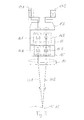
- a stereoscopic surgical microscope in Fig. 1 comprises a front lens 14, a first magnification system 15, a second magnification system 16 and an eyepiece 17 with eyepieces 19A and 19B. These elements are arranged relative to one another in such a way that when viewed through the eyepieces 19A, 19B an enlarged image of an object 18 arranged in front of the front lens 14 is seen. After passing through the common fron lens 14, the observation beam path splits into a beam path 20A for one eye and a beam path 20B for the other eye. The surgical microscope is therefore stereoscopic.
- the front lens 14 has a fixed focal length.
- the working distance between the front lens 14 and the object 18 corresponds to the focal length of the front lens 14 and is thus fixed.
- a beam path 20A, 20B emanating from a point of the object 18 is converted by the front lens 14 into a parallel beam, which then enters the first magnification system 15. The operation of such an afocal magnification system will be explained below.
- the first magnification system 15 includes a left magnification system 15A for the beam path 20A and a right one Magnification system 15B for the beam path 20B.
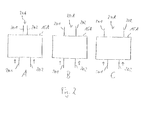
- the operation of a magnification system is based on Fig. 2 explained on the example of the left magnification system 15A.
- a parallel beam 20A entering the magnification system 15A emerges again as a parallel beam 20A.
- Two parallel light beams 201, 202 of the beam 20A have a different distance from each other depending on the set magnification factor of the magnification system 15A when entering the magnification system 15A than at the exit.
- the magnification of the magnification system 15A is set to a value less than 1.
- the distance between the two light beams 201, 202, whose direction is indicated by arrows, is greater on entry into the magnification system 15A than at the exit.
- the magnification factor is 1. In this special case, no magnification takes place, but the light beams 201, 202 have the same distance to each other at the entrance and exit.
- the magnification factor is greater than 1 and the distance between the light beams 201, 202 is greater at the exit than at the entrance.
- the right magnification system 15B works analogously.
- the magnification systems 15A, 15B are coupled together so that the magnification factor is always set in common. It's a one in Fig. 1 not shown control element is provided via which is acted upon mechanically on the optical elements of the magnification system 15 to change their distance from one another.
- the magnification factor of the magnification system 15 can be varied continuously between 1/6 and 6. The as a quotient
- the expansion factor of the magnification system 15 determined by these two values is six.
- the second magnification system 16 adjoining the first magnification system 15 is also stereoscopic (16A, 16B), but has a fixed magnification factor in the range between 1.5 and 3.
- the second magnification system 16 is designed as a module which is optionally introduced into the beam path of the surgical microscope be or can be removed from this.
- the module can be connected for this purpose, for example, pivotally connected to the housing of the surgical microscope. Also possible are embodiments in which the module is completely separated from the surgical microscope. Since the fixed magnification factor of the second magnification system 16 is smaller than the expansion factor of the first magnification system 15, the magnification can be adjusted continuously.
- the magnification area accessible with the second magnification system 16 seamlessly joins the magnification area that is accessible without the second magnification system 16.
- the glimpse 17, which adjoins the second magnification system 16 has a self-magnification of 8.
- the magnification factor of the surgical microscope as a whole is the product of the magnification factors of the magnification systems 15, 16 and the glare 17 and the quotient of 250 mm / focal length front lens.
- the value 250 mm indicates the reference distance.
- the embodiment according to Fig. 3 differs from Fig. 1 in two ways.
- the front lens 14 is a zoom front lens whose focal length varies as needed can be.
- the second magnification system 16 has a variable magnification factor.
- Each of the magnification systems 15, 16 is equipped with an electric motor, not shown, with which the magnification factor can be adjusted.
- the electric motors are coupled with each other so that they can be operated via a common operating element. With the control both electric motors are operated simultaneously, so that the entire magnification range can be continuously passed through from the smallest possible magnification factor to the largest possible magnification factor.
Abstract
Description
Die Erfindung betrifft ein Operationsmikroskop, das eine Frontlinse, einen Einblick und ein zwischen der Frontlinse und dem Einblick angeordnetes erstes afokales Vergrößerungssystem umfasst. Die Frontlinse, das erste Vergrößerungssystem und der Einblick definieren einen Beobachtungsstrahlengang. Das erste Vergrößerungssystem hat einen variablen Vergrößerungsfaktor.The invention relates to a surgical microscope comprising a front lens, a glimpse and a first afocal magnification system arranged between the front lens and the glimpse. The front lens, the first magnification system and the insight define an observation beam path. The first magnification system has a variable magnification factor.
Solche Operationsmikroskope sind in verbreiteter Verwendung und dienen dazu, dem Chirurgen bei einer Operation eine vergrößerte Ansicht des Operationsfelds zu liefern. Die Frontlinse des Operationsmikroskops wird in passendem Abstand zum Operationsfeld angeordnet und der Chirurg sieht in einem Okular des Einblicks das vergrößerte Bild. Damit das Operationsmikroskop an die jeweiligen Gegebenheiten angepasst werden kann, hat das Vergrößerungssystem einen variablen Vergrößerungsfaktor. Bei hohem Vergrößerungsfaktor können Details im Operationsfeld in hoher Auflösung betrachtet werden. Bei niedrigem Vergrößerungsfaktor verschafft der Chirurg sich einen Überblick über das Operationsfeld.Such surgical microscopes are in widespread use and serve to provide the surgeon with an enlarged view of the surgical field during surgery. The front lens of the surgical microscope is placed at an appropriate distance from the surgical field and the surgeon sees in an eyepiece of the magnified image. So that the surgical microscope can be adapted to the respective conditions, the magnification system has a variable magnification factor. At high magnification, details in the surgical field can be viewed in high resolution. At a low magnification factor, the surgeon gets an overview of the surgical field.
Es hat sich gezeigt, dass die mit den heute gängigen Operationsmikroskopen mögliche Vergrößerung den Anforderungen nicht immer genügt. Mit den zur Verfügung stehenden Hilfsmitteln können Operationen in immer kleinerem Maßstab und mit immer höherer Genauigkeit durchgeführt werden. Operationsmikroskope sollen deswegen eine hohe Vergrößerung erlauben, während andererseits die Möglichkeit, das Operationsfeld im Überblick zu betrachten, nicht verloren gehen soll.It has been shown that the enlargement possible with today's surgical microscopes does not always meet the requirements. With the tools available, operations can become smaller and smaller be carried out with ever higher accuracy. Surgical microscopes should therefore allow a high magnification, while on the other hand, the possibility to look at the surgical field at a glance should not be lost.
Der Erfindung liegt die Aufgabe zu Grunde, ein Operationsmikroskop vorzustellen, das flexibler eingesetzt werden kann. Ausgehend vom eingangs genannten Stand der Technik wird die Aufgabe gelöst mit den Merkmalen des Anspruchs 1. Erfindungsgemäß ist zwischen der Frontlinse und dem Einblick ein zweites afokales Vergrößerungssystem angeordnet. Vorteilhafte Ausführungsformen finden sich in den Unteransprüchen.The invention is based on the object to present a surgical microscope, which can be used flexibly. Based on the above-mentioned prior art, the object is achieved with the features of claim 1. According to the invention, a second afocal magnification system is arranged between the front lens and the view. Advantageous embodiments can be found in the subclaims.
Zunächst werden einige Begriffe erläutert. Ein afokales Vergrößerungssystem bezeichnet eine Einheit von optischen Elementen, die so angeordnet sind, dass zwei Lichtstrahlen, die parallel zueinander und parallel zur optischen Achse des Vergrößerungssystems in das Vergrößerungssystem eintreten, auch parallel wieder austreten, wobei der Abstand zwischen den Lichtstrahlen beim Eintritt ein anderer ist als beim Austritt. In dem Vergrößerungssystem wird regelmäßig kein Zwischenbild erzeugt. Auch eine Bildumkehr findet in dem Vergrößerungssystem regelmäßig nicht statt.First, some terms are explained. An afocal magnification system designates a unit of optical elements arranged so that two light rays entering the magnification system parallel to each other and parallel to the optical axis of the magnification system also emerge in parallel, the distance between the light rays at the entrance being different as at the exit. In the magnification system no intermediate image is generated regularly. Even a picture reversal does not take place in the magnification system regularly.
Wenn der Vergrößerungsfaktor eines Vergrößerungssystems verändert wird, verändert sich bei gleich bleibendem Abstand der eintretenden Lichtstrahlen der Abstand der austretenden Lichtstrahlen voneinander. Ein Vergrößerungssystem kann sowohl den Fall umfassen, dass der Abstand der eintretenden Lichtstrahlen größer ist als der Abstand der austretenden Lichtstrahlen, als auch den Fall, dass der Abstand der eintretenden Lichtstrahlen kleiner ist als der Abstand der austretenden Lichtstrahlen. Bei einem variablen Vergrößerungsfaktor kann als Sonderfall umfasst sein, dass der Abstand der eintretenden Lichtstrahlen und der austretenden Lichtstrahlen übereinstimmt, also keine Vergrößerung oder Verkleinerung stattfindet. Der Begriff Dehnungsfaktor bezeichnet den Quotienten aus dem größtmöglichen und dem kleinstmöglichen Vergrößerungsfaktor.When the magnification of a magnification system is changed, the distance of the exiting light beams from each other changes with the distance of the incoming light beams remaining the same. A magnification system may include both the case that the distance of the incident light beams is larger than the distance of the exiting light beams, and the case that the distance of the incident light beams is smaller than that Distance of the exiting light rays. In the case of a variable magnification factor, it can be included as a special case that the distance of the incoming light beams and the exiting light beams coincides, ie no enlargement or reduction takes place. The term expansion factor refers to the quotient of the largest possible and the smallest possible magnification factor.
Ein Einblick ist eine Einheit von optischen Elementen, die so angeordnet sind, dass aus parallel eintretenden Lichtstrahlen eine Abbildung erzeugt werden kann, so dass die Abbildung durch ein Okular betrachtet werden kann. In dem Operationsmikroskop sind die Frontlinse, die Vergrößerungssysteme und der Einblick so angeordnet, dass in dem Okular eine vergrößerte Abbildung eines vor der Frontlinse angeordneten Objekts zu sehen ist. Dabei ist es unerheblich in welcher Reihenfolge das erste Vergrößerungssystem und das zweite Vergrößerungssystem aufeinanderfolgen. Das Operationsmikroskop ist meist stereoskopisch, es ist also für jedes Auge ein eigener Beobachtungsstrahlengang vorgesehen.An insight is a unit of optical elements that are arranged so that an image can be generated from light rays entering parallel, so that the image can be viewed through an eyepiece. In the surgical microscope, the front lens, the magnification systems and the insight are arranged so that an enlarged image of an object arranged in front of the front lens can be seen in the eyepiece. It does not matter in which order the first magnification system and the second magnification system follow each other. The surgical microscope is usually stereoscopic, so it is provided for each eye a separate observation beam path.
Die Erfindung hat erkannt, dass die Einfügung eines zweiten Vergrößerungssystems zwischen der Frontlinse und dem Einblick möglich ist, ohne dass das Operationsmikroskop im Übrigen angepasst werden müsste. Da weitere Anpassungen nicht vorgenommen werden müssen, ist diese Lösung trotz des zusätzlich erforderlichen zweiten Vergrößerungssystems kostengünstig. Es bleibt zudem die hohe optische Qualität erhalten. Dies gilt insbesondere im Vergleich mit einem auf großen Dehnungsfaktor ausgelegten einzelnen Vergrößerungssystem. Versuche haben gezeigt, dass die optische Qualität abnimmt, wenn man versucht, den Dehnungsfaktor gegenüber den heute gängigen Vergrößerungssystemen zu erhöhen.The invention has recognized that the insertion of a second magnification system between the front lens and the view is possible, without the operating microscope otherwise having to be adapted. Since further adjustments do not have to be made, this solution is cost effective despite the additional required second magnification system. In addition, the high optical quality is retained. This is especially true when compared to a single magnification system designed for large strain factors. Experiments have shown that the optical quality decreases when one tries to increase the elongation factor compared to today's magnification systems.
Durch die Hintereinanderschaltung zweier Vergrößerungssysteme erhält man einen Gesamtdehnungsfaktor der größer ist als der Dehnungsfaktor eines einzelnen Vergrößerungssystems. Wenn der Gesamtdehnungsfaktor groß ist, wird auch der mit dem Operationsmikroskop zugängliche Vergrößerungsbereich erweitert, wobei der Begriff Vergrößerungsbereich den Bereich zwischen dem kleinstmöglichen und dem größtmöglichen Vergrößerungsfaktor bezeichnet. Erweitert ist der Vergrößerungsbereich verglichen mit einem Operationsmikroskop, das nur das erste Vergrößerungssystem umfasst. Das zweite Vergrößerungssystem kann dazu verschiedene Zustände im Beobachtungsstrahlengang annehmen. In einer Ausführungsform hat das zweite Vergrößerungssystem einen festen Vergrößerungsfaktor. Der größere Gesamtdehnungsfaktor kann sich in diesem Fall daraus ergeben, dass das zweite Vergrößerungssystem in einem ersten Zustand im Beobachtungsstrahlengang angeordnet ist und in einem zweiten Zustand außerhalb des Beobachtungsstrahlengangs angeordnet ist. Um den Wechsel zwischen diesen Zuständen zu ermöglichen, kann das zweite Vergrößerungssystem beweglich an dem Operationsmikroskop aufgehängt sein. Alternativ kann das zweite Vergrößerungssystem kann als Modul ausgebildet sein, das von dem Operationsmikroskop getrennt werden kann.By connecting two magnification systems in series one obtains a total expansion factor which is greater than the expansion factor of a single magnification system. If the overall stretch factor is large, the magnification range accessible by the surgical microscope is also expanded, the term magnification range meaning the range between the smallest possible and the largest possible magnification factor. The magnification range is extended compared to a surgical microscope that includes only the first magnification system. The second magnification system can assume different states in the observation beam path. In one embodiment, the second magnification system has a fixed magnification factor. The larger overall expansion factor may result in this case from the fact that the second magnification system is arranged in a first state in the observation beam path and is arranged in a second state outside the observation beam path. In order to enable the change between these states, the second magnification system may be movably suspended from the surgical microscope. Alternatively, the second magnification system may be formed as a module that can be separated from the surgical microscope.
Wenn das zweite Vergrößerungssystem einen festen Vergrößerungsfaktor hat, ist dieser vorzugsweise größer als 1, wobei 1 den Fall bezeichnet, dass keine Vergrößerung stattfindet. Andererseits ist der Vergrößerungsfaktor vorzugsweise kleiner als der Dehnungsfaktor des ersten Vergrößerungssystems. Der Vergrößerungsbereich, den das Operationsmikroskop bei Hintereinanderschaltung beider Vergrößerungssysteme hat, schließt sich dann nahtlos an den Vergrößerungsbereich an, der ohne das zweite Vergrößerungssystem gegeben ist. An der einen Grenze des Vergrößerungsbereichs ist die variable Vergrößerung des ersten Vergrößerungssystems am einen Anschlag und das zweite Vergrößerungssystem ist im Beobachtungsstrahlengang angeordnet. An der zweiten Grenze des Vergrößerungsbereichs ist die variable Vergrößerung des ersten Vergrößerungssystems am anderen Anschlag und das zweite Vergrößerungssystem ist außerhalb des Beobachtungsstrahlengangs angeordnet. Aus diesen beiden Grenzen des Vergrößerungsbereichs kann der Gesamtdehnungsfaktor ermittelt werden.If the second magnification system has a fixed magnification factor, it is preferably greater than 1, where 1 denotes the case that no magnification takes place. On the other hand, the magnification factor is preferably smaller than the expansion factor of the first magnification system. The magnification range which the surgical microscope has when the two magnification systems are connected in series then closes seamlessly with the magnification range which is given without the second magnification system. At the one boundary of the enlargement range, the variable magnification of the first magnification system is located at one stop and the second magnification system is arranged in the observation beam path. At the second limit of the magnification range, the variable magnification of the first magnification system is located at the other stop and the second magnification system is located outside the observation beam path. From these two limits of the magnification range, the total expansion factor can be determined.
Das zweite Vergrößerungssystem kann alternativ einen variablen Vergrößerungsfaktor haben. Der Vergrößerungsfaktor des zweiten Vergrößerungssystems kann separat vom Vergrößerungsfaktor des ersten Vergrößerungssystems einstellbar sein. Komfortabler für den Benutzer ist es, wenn die Einstellung der Vergrößerungsfaktoren der beiden Vergrößerungssysteme miteinander gekoppelt ist. Die Koppelung ist vorzugsweise derart, dass der gesamte Vergrößerungsbereich von dem Zustand, in dem beide Vergrößerungssysteme den kleinsten Vergrößerungsfaktor haben, bis zu dem Zustand, in dem beide Vergrößerungssysteme den größten Vergrößerungsfaktor haben, kontinuierlich durchfahren werden kann. Der Gesamtdehnungsfaktor kann aus diesen beiden Grenzen ermittelt werden. Das Operationsmikroskop kann so eingerichtet sein, dass für die Einstellung beider Vergrößerungssysteme nur ein einzelnes Bedienelement vorgesehen ist. Die Vergrößerungssysteme können dazu mit Elektromotoren versehen sein, die über das Bedienelement betätigt werden.The second magnification system may alternatively have a variable magnification factor. The magnification factor of the second magnification system may be adjustable separately from the magnification factor of the first magnification system. It is more convenient for the user if the setting of the magnification factors of the two magnification systems is coupled with one another. The coupling is preferably such that the entire magnification range can be continuously traversed from the state where both magnification systems have the smallest magnification factor to the state where both magnification systems have the largest magnification factor. The total expansion factor can be determined from these two limits. The surgical microscope can be set up so that only a single operating element is provided for setting both magnification systems. The magnification systems can be provided for this purpose with electric motors which are actuated via the operating element.
Der variable Vergrößerungsfaktor eines Vergrößerungssystems kann auf unterschiedlichen Mechanismen beruhen. Das Vergrößerungssystem kann beispielsweise eine Mehrzahl von Unter-Vergrößerungssystemen umfassen, die feste, aber unterschiedliche Vergrößerungsfaktoren haben und die wahlweise in den Strahlengang eingebracht werden (Galilei-Wechsler). Möglich ist auch ein Vergrößerungssystem, bei dem der Vergrößerungsfaktor kontinuierlich einstellbar ist (Zoom-System) .The variable magnification factor of a magnification system can be based on different mechanisms. The magnification system For example, it may comprise a plurality of sub-magnification systems which have fixed but different magnification factors and which are selectively introduced into the beam path (Galilean changer). Also possible is a magnification system, in which the magnification factor is continuously adjustable (zoom system).
Der Gesamtdehnungsfaktor, den die Kombination aus dem ersten und dem zweiten Vergrößerungssystem bietet, ist vorzugweise größer als 10, weiter vorzugsweise größer als 20, weiter vorzugsweise größer als 30. Wenn der Gesamtdehnungsfaktor der Vergrößerungssysteme groß ist, kann mit dem Operationsmikroskop ein umfangreicher Vergrößerungsbereich abgedeckt werden.The overall expansion factor offered by the combination of the first and second magnification systems is preferably greater than 10, more preferably greater than 20, more preferably greater than 30. If the overall expansion factor of the magnification systems is large, a large magnification range can be covered with the surgical microscope ,
Die Frontlinse kann eine Linse mit fester Brennweite sein. Durch die Frontlinse wird dann ein fester Arbeitsabstand definiert, der zwischen der Frontlinse und dem Objekt einzuhalten ist. Flexibler einsetzbar ist das Operationsmikroskop, wenn eine Frontlinse mit variabler Brennweite eingesetzt wird (Vario-Frontlinse). Der Arbeitsabstand zwischen der Frontlinse und dem Objekt kann dann je nach Bedarf unterschiedlich eingestellt werden. Mit Vario-Frontlinsen erreichen Operationsmikroskope allgemein nicht den Vergrößerungsfaktor, der bei Frontlinsen mit fester Brennweite erreicht wird. Die Anwendung der Erfindung in Kombination mit einer Vario-Frontlinse ist deswegen von besonderem Interesse.The front lens may be a fixed focal length lens. The front lens then defines a fixed working distance to be maintained between the front lens and the object. The surgical microscope can be used more flexibly if a front lens with variable focal length is used (vario front lens). The working distance between the front lens and the object can then be set differently depending on requirements. With vario-front lenses, surgical microscopes generally do not achieve the magnification factor that is achieved with fixed-lens front lenses. The use of the invention in combination with a Vario front lens is therefore of particular interest.
Die Erfindung wird nachfolgend unter Bezugnahme auf die beigefügten Zeichnungen anhand vorteilhafter Ausführungsformen beispielhaft beschrieben. Es zeigen:
- Fig. 1:
- eine erste Ausführungsform eines erfindungsgemä-ßen Operationsmikroskops;
- Fig. 2:
- eine schematische Darstellung eines Vergröße-rungssystems mit variablem Vergrößerungsfaktor; und
- Fig. 3:
- eine zweite Ausführungsform eines erfindungsgemä-ßen Operationsmikroskops.
- Fig. 1:
- a first embodiment of a surgical microscope according to the invention;
- Fig. 2:
- a schematic representation of a zoom magnification system with variable magnification factor; and
- 3:
- A second embodiment of a surgical microscope according to the invention.
Eins stereoskopisches Operationsmikroskop in
Die Frontlinse 14 hat eine feste Brennweite. Der Arbeitsabstand zwischen der Frontlinse 14 und dem Objekt 18 entspricht der Brennweite der Frontlinse 14 und ist damit fest vorgegeben. Ein von einem Punkt des Objekts 18 ausgehender Strahlengang 20A, 20B wird durch die Frontlinse 14 in ein paralleles Strahlenbündel umgewandelt, das dann in das erste Vergrößerungssystem 15 eintritt. Die Funktionsweise eines solchen afokalen Vergrößerungssystems wird nachfolgend erläutert.The
Das erste Vergrößerungssystem 15 umfasst ein linkes Vergrößerungssystem 15A für den Strahlengang 20A und ein rechtes Vergrößerungssystem 15B für den Strahlengang 20B. Die Funktionsweise eines Vergrößerungssystems wird anhand von
In
Das rechte Vergrößerungssystem 15B funktioniert analog. Die Vergrößerungssysteme 15A, 15B sind so miteinander gekoppelt, dass der Vergrößerungsfaktor immer gemeinsam eingestellt wird. Es ist dazu ein in
Das an das erste Vergrößerungssystem 15 anschließende zweite Vergrößerungssystem 16 ist ebenfalls stereoskopisch (16A, 16B), hat aber einen festen Vergrößerungsfaktor im Bereich zwischen 1,5 und 3. Das zweite Vergrößerungssystem 16 ist als Modul ausgebildet, das wahlweise in den Strahlengang des Operationsmikroskops eingebracht werden bzw. aus diesem entfernt werden kann. Das Modul kann zu diesem Zweck beispielsweise schwenkbar mit dem Gehäuse des Operationsmikroskops verbunden sein. Möglich sind auch Ausführungsformen, bei denen das Modul vollständig von dem Operationsmikroskop getrennt wird. Da der feste Vergrößerungsfaktor des zweiten Vergrößerungssystems 16 kleiner ist als der Dehnungsfaktor des ersten Vergrößerungssystems 15, kann die Vergrößerung kontinuierlich eingestellt werden. Der Vergrößerungsbereich, der mit dem zweiten Vergrößerungssystem 16 zugänglich ist, schließt sich nahtlos an den Vergrößerungsbereich an, der ohne das zweite Vergrößerungssystem 16 zugänglich ist.The
Der Einblick 17, der sich an das zweite Vergrößerungssystem 16 anschließt, hat eine Eigenvergrößerung von 8. Der Vergrößerungsfaktor des Operationsmikroskops insgesamt ergibt sich als Produkt aus den Vergrößerungsfaktoren der Vergrößerungssysteme 15, 16 und des Einblicks 17 sowie dem Quotienten aus 250mm/Brennweite Frontlinse. Der Wert 250 mm gibt dabei die Bezugssehweite an.The
Die Ausführungsform gemäß
Claims (11)
Priority Applications (1)
| Application Number | Priority Date | Filing Date | Title |
|---|---|---|---|
| EP11156350A EP2495597A1 (en) | 2011-03-01 | 2011-03-01 | Operation microscope with two enlargement systems |
Applications Claiming Priority (1)
| Application Number | Priority Date | Filing Date | Title |
|---|---|---|---|
| EP11156350A EP2495597A1 (en) | 2011-03-01 | 2011-03-01 | Operation microscope with two enlargement systems |
Publications (1)
| Publication Number | Publication Date |
|---|---|
| EP2495597A1 true EP2495597A1 (en) | 2012-09-05 |
Family
ID=44123230
Family Applications (1)
| Application Number | Title | Priority Date | Filing Date |
|---|---|---|---|
| EP11156350A Withdrawn EP2495597A1 (en) | 2011-03-01 | 2011-03-01 | Operation microscope with two enlargement systems |
Country Status (1)
| Country | Link |
|---|---|
| EP (1) | EP2495597A1 (en) |
Citations (4)
| Publication number | Priority date | Publication date | Assignee | Title |
|---|---|---|---|---|
| US5612816A (en) * | 1992-04-28 | 1997-03-18 | Carl-Zeiss-Stiftung | Endoscopic attachment for a stereoscopic viewing system |
| WO2005040866A2 (en) * | 2003-10-23 | 2005-05-06 | Zeiss Carl Ag | Projection optics with adjustable refractive power and method for adjusting the refractive power thereof |
| EP1731941A1 (en) * | 2004-03-31 | 2006-12-13 | Olympus Corporation | Observing device and fluorescent light observing device |
| DE102005050171A1 (en) * | 2005-10-19 | 2007-04-26 | Carl Zeiss Surgical Gmbh | Optical enlargement variation system e.g. for operations microscope, has positioning device for third module which has given first spacing from first module in first working state |
-
2011
- 2011-03-01 EP EP11156350A patent/EP2495597A1/en not_active Withdrawn
Patent Citations (4)
| Publication number | Priority date | Publication date | Assignee | Title |
|---|---|---|---|---|
| US5612816A (en) * | 1992-04-28 | 1997-03-18 | Carl-Zeiss-Stiftung | Endoscopic attachment for a stereoscopic viewing system |
| WO2005040866A2 (en) * | 2003-10-23 | 2005-05-06 | Zeiss Carl Ag | Projection optics with adjustable refractive power and method for adjusting the refractive power thereof |
| EP1731941A1 (en) * | 2004-03-31 | 2006-12-13 | Olympus Corporation | Observing device and fluorescent light observing device |
| DE102005050171A1 (en) * | 2005-10-19 | 2007-04-26 | Carl Zeiss Surgical Gmbh | Optical enlargement variation system e.g. for operations microscope, has positioning device for third module which has given first spacing from first module in first working state |
Similar Documents
| Publication | Publication Date | Title |
|---|---|---|
| DE102006036300B4 (en) | High performance stereo microscope | |
| EP1326117B2 (en) | Ophthalmoscopic front end attachment and surgical microscope | |
| EP3017334B1 (en) | Image capture method for microscope system and corresponding microscope system | |
| WO2006008025A1 (en) | Device for machining an object by means of laser radiation | |
| DE19541420B4 (en) | Stereo microscope arrangement | |
| DE102012223712A1 (en) | VARIABLE PICTURE SYSTEM WITH LENS FIXED | |
| WO2007045500A1 (en) | Microscopy system | |
| DE102009012707A1 (en) | Microscope with several optical systems in the imaging beam path | |
| DE102006050846B4 (en) | Stereomicroscope with beam splitter device | |
| WO2002027379A2 (en) | Image reversion system, additional ophthalmoscopy module and operational microscope | |
| DE102007029893A1 (en) | Microscope with centered illumination | |
| DE102010002722B4 (en) | Afocal zoom system for a microscope, microscope with such a zoom system and method for operating such a zoom system | |
| EP1424582B1 (en) | Stereomicroscope | |
| WO2014068058A1 (en) | Stereomicroscope with stereovariator | |
| DE10255960A1 (en) | stereomicroscope | |
| DE102010003640A1 (en) | Video stereomicroscope | |
| DE102006022592B4 (en) | Microscope with lighting unit | |
| DE202013011877U1 (en) | microscope system | |
| CH697533B1 (en) | Illumination and observation device. | |
| DE102007029896B3 (en) | Stereo microscope i.e. operation microscope, has illumination unit provided with defecting units integrated in lens component, where position of deflecting units is adjustable dependent of focal length change of lens to centre illumination | |
| DE102004029056A1 (en) | Variable aperture, illumination device, optical observation device and optical observation device | |
| DE102012006749B4 (en) | Stereo microscope | |
| DE102013201632B4 (en) | Microscope with zoom aperture coupling and zoom system for a microscope | |
| DE102005005568B4 (en) | Tube for an observation device and observation device | |
| DE102007029894A1 (en) | Microscope with centered illumination |
Legal Events
| Date | Code | Title | Description |
|---|---|---|---|
| PUAI | Public reference made under article 153(3) epc to a published international application that has entered the european phase |
Free format text: ORIGINAL CODE: 0009012 |
|
| AK | Designated contracting states |
Kind code of ref document: A1 Designated state(s): AL AT BE BG CH CY CZ DE DK EE ES FI FR GB GR HR HU IE IS IT LI LT LU LV MC MK MT NL NO PL PT RO RS SE SI SK SM TR |
|
| AX | Request for extension of the european patent |
Extension state: BA ME |
|
| STAA | Information on the status of an ep patent application or granted ep patent |
Free format text: STATUS: THE APPLICATION IS DEEMED TO BE WITHDRAWN |
|
| 18D | Application deemed to be withdrawn |
Effective date: 20130306 |