EP2547301B1 - Patterned neo-epithelialization dressings, systems, and methods - Google Patents
Patterned neo-epithelialization dressings, systems, and methods Download PDFInfo
- Publication number
- EP2547301B1 EP2547301B1 EP11710611.2A EP11710611A EP2547301B1 EP 2547301 B1 EP2547301 B1 EP 2547301B1 EP 11710611 A EP11710611 A EP 11710611A EP 2547301 B1 EP2547301 B1 EP 2547301B1
- Authority
- EP
- European Patent Office
- Prior art keywords
- pressure
- patient
- epithelium
- interface member
- neo
- Prior art date
- Legal status (The legal status is an assumption and is not a legal conclusion. Google has not performed a legal analysis and makes no representation as to the accuracy of the status listed.)
- Not-in-force
Links
- 238000000034 method Methods 0.000 title description 9
- 239000012530 fluid Substances 0.000 claims description 42
- 206010063560 Excessive granulation tissue Diseases 0.000 claims description 33
- 210000001126 granulation tissue Anatomy 0.000 claims description 33
- 239000006260 foam Substances 0.000 claims description 25
- 239000011148 porous material Substances 0.000 claims description 19
- 238000007789 sealing Methods 0.000 claims description 19
- 210000003491 skin Anatomy 0.000 claims description 15
- 210000001519 tissue Anatomy 0.000 claims description 12
- 210000002615 epidermis Anatomy 0.000 claims description 6
- 206010052428 Wound Diseases 0.000 description 36
- 208000027418 Wounds and injury Diseases 0.000 description 36
- 239000000463 material Substances 0.000 description 15
- 102000010834 Extracellular Matrix Proteins Human genes 0.000 description 7
- 108010037362 Extracellular Matrix Proteins Proteins 0.000 description 7
- 210000000981 epithelium Anatomy 0.000 description 7
- 210000002744 extracellular matrix Anatomy 0.000 description 7
- 210000004027 cell Anatomy 0.000 description 6
- 230000008021 deposition Effects 0.000 description 6
- 229920000642 polymer Polymers 0.000 description 6
- 238000013459 approach Methods 0.000 description 5
- 102000004169 proteins and genes Human genes 0.000 description 5
- 108090000623 proteins and genes Proteins 0.000 description 5
- 230000008901 benefit Effects 0.000 description 4
- 230000012292 cell migration Effects 0.000 description 4
- 229920001971 elastomer Polymers 0.000 description 4
- 210000002950 fibroblast Anatomy 0.000 description 4
- 229920001296 polysiloxane Polymers 0.000 description 4
- 102000008186 Collagen Human genes 0.000 description 3
- 108010035532 Collagen Proteins 0.000 description 3
- 230000015572 biosynthetic process Effects 0.000 description 3
- 229920001436 collagen Polymers 0.000 description 3
- 238000001514 detection method Methods 0.000 description 3
- 239000000806 elastomer Substances 0.000 description 3
- 230000037361 pathway Effects 0.000 description 3
- 229920006264 polyurethane film Polymers 0.000 description 3
- 230000029663 wound healing Effects 0.000 description 3
- 230000004075 alteration Effects 0.000 description 2
- 230000005540 biological transmission Effects 0.000 description 2
- 230000008933 bodily movement Effects 0.000 description 2
- 238000005266 casting Methods 0.000 description 2
- 238000004891 communication Methods 0.000 description 2
- 230000013228 contact guidance Effects 0.000 description 2
- 210000004207 dermis Anatomy 0.000 description 2
- 238000011161 development Methods 0.000 description 2
- 230000018109 developmental process Effects 0.000 description 2
- 210000000416 exudates and transudate Anatomy 0.000 description 2
- 230000003179 granulation Effects 0.000 description 2
- 238000005469 granulation Methods 0.000 description 2
- 102000006495 integrins Human genes 0.000 description 2
- 108010044426 integrins Proteins 0.000 description 2
- 210000002510 keratinocyte Anatomy 0.000 description 2
- 238000004519 manufacturing process Methods 0.000 description 2
- 239000012528 membrane Substances 0.000 description 2
- 230000005012 migration Effects 0.000 description 2
- 238000013508 migration Methods 0.000 description 2
- 238000012544 monitoring process Methods 0.000 description 2
- -1 open-cell Polymers 0.000 description 2
- 206010033675 panniculitis Diseases 0.000 description 2
- 229920002635 polyurethane Polymers 0.000 description 2
- 239000004814 polyurethane Substances 0.000 description 2
- 230000001737 promoting effect Effects 0.000 description 2
- 210000004304 subcutaneous tissue Anatomy 0.000 description 2
- 239000000126 substance Substances 0.000 description 2
- 238000002560 therapeutic procedure Methods 0.000 description 2
- 229920001169 thermoplastic Polymers 0.000 description 2
- 238000003466 welding Methods 0.000 description 2
- 229920002943 EPDM rubber Polymers 0.000 description 1
- 229920000181 Ethylene propylene rubber Polymers 0.000 description 1
- 102000009123 Fibrin Human genes 0.000 description 1
- 108010073385 Fibrin Proteins 0.000 description 1
- BWGVNKXGVNDBDI-UHFFFAOYSA-N Fibrin monomer Chemical compound CNC(=O)CNC(=O)CN BWGVNKXGVNDBDI-UHFFFAOYSA-N 0.000 description 1
- 244000043261 Hevea brasiliensis Species 0.000 description 1
- 229920000459 Nitrile rubber Polymers 0.000 description 1
- 239000005062 Polybutadiene Substances 0.000 description 1
- 239000004820 Pressure-sensitive adhesive Substances 0.000 description 1
- 229920001247 Reticulated foam Polymers 0.000 description 1
- 101710172711 Structural protein Proteins 0.000 description 1
- NIXOWILDQLNWCW-UHFFFAOYSA-N acrylic acid group Chemical group C(C=C)(=O)O NIXOWILDQLNWCW-UHFFFAOYSA-N 0.000 description 1
- 239000000853 adhesive Substances 0.000 description 1
- 230000001070 adhesive effect Effects 0.000 description 1
- 239000000560 biocompatible material Substances 0.000 description 1
- 239000008280 blood Substances 0.000 description 1
- 210000004369 blood Anatomy 0.000 description 1
- 210000004204 blood vessel Anatomy 0.000 description 1
- 229920005549 butyl rubber Polymers 0.000 description 1
- 230000001413 cellular effect Effects 0.000 description 1
- 239000004568 cement Substances 0.000 description 1
- 230000003399 chemotactic effect Effects 0.000 description 1
- 210000002808 connective tissue Anatomy 0.000 description 1
- 230000008602 contraction Effects 0.000 description 1
- 230000008878 coupling Effects 0.000 description 1
- 238000010168 coupling process Methods 0.000 description 1
- 238000005859 coupling reaction Methods 0.000 description 1
- 210000004292 cytoskeleton Anatomy 0.000 description 1
- 238000010586 diagram Methods 0.000 description 1
- 210000002249 digestive system Anatomy 0.000 description 1
- 238000009826 distribution Methods 0.000 description 1
- 230000005489 elastic deformation Effects 0.000 description 1
- 239000013536 elastomeric material Substances 0.000 description 1
- 230000003511 endothelial effect Effects 0.000 description 1
- 238000005530 etching Methods 0.000 description 1
- 239000005038 ethylene vinyl acetate Substances 0.000 description 1
- 229950003499 fibrin Drugs 0.000 description 1
- 239000000499 gel Substances 0.000 description 1
- 239000003102 growth factor Substances 0.000 description 1
- 230000035876 healing Effects 0.000 description 1
- 230000002209 hydrophobic effect Effects 0.000 description 1
- 230000002706 hydrostatic effect Effects 0.000 description 1
- 229920002681 hypalon Polymers 0.000 description 1
- 210000002865 immune cell Anatomy 0.000 description 1
- 230000006698 induction Effects 0.000 description 1
- 208000015181 infectious disease Diseases 0.000 description 1
- 238000003475 lamination Methods 0.000 description 1
- 239000007788 liquid Substances 0.000 description 1
- 239000011159 matrix material Substances 0.000 description 1
- 230000003278 mimic effect Effects 0.000 description 1
- 238000012986 modification Methods 0.000 description 1
- 230000004048 modification Effects 0.000 description 1
- 238000000465 moulding Methods 0.000 description 1
- 229920003052 natural elastomer Polymers 0.000 description 1
- 229920001194 natural rubber Polymers 0.000 description 1
- 238000000059 patterning Methods 0.000 description 1
- 229920001084 poly(chloroprene) Polymers 0.000 description 1
- 229920001200 poly(ethylene-vinyl acetate) Polymers 0.000 description 1
- 229920002857 polybutadiene Polymers 0.000 description 1
- 229920000728 polyester Polymers 0.000 description 1
- 229920001195 polyisoprene Polymers 0.000 description 1
- 229920001021 polysulfide Polymers 0.000 description 1
- 239000005077 polysulfide Substances 0.000 description 1
- 150000008117 polysulfides Polymers 0.000 description 1
- 238000003825 pressing Methods 0.000 description 1
- 230000008569 process Effects 0.000 description 1
- 102000004196 processed proteins & peptides Human genes 0.000 description 1
- 108090000765 processed proteins & peptides Proteins 0.000 description 1
- 230000035752 proliferative phase Effects 0.000 description 1
- 230000010069 protein adhesion Effects 0.000 description 1
- 230000009467 reduction Effects 0.000 description 1
- 230000008929 regeneration Effects 0.000 description 1
- 238000011069 regeneration method Methods 0.000 description 1
- 230000000241 respiratory effect Effects 0.000 description 1
- 239000005060 rubber Substances 0.000 description 1
- 230000004936 stimulating effect Effects 0.000 description 1
- 229920003048 styrene butadiene rubber Polymers 0.000 description 1
- 238000006467 substitution reaction Methods 0.000 description 1
- 230000009772 tissue formation Effects 0.000 description 1
- 230000017423 tissue regeneration Effects 0.000 description 1
- 238000012876 topography Methods 0.000 description 1
- 230000007704 transition Effects 0.000 description 1
- 230000002792 vascular Effects 0.000 description 1
- 230000000007 visual effect Effects 0.000 description 1
- 230000037303 wrinkles Effects 0.000 description 1
Images


Classifications
-
- A—HUMAN NECESSITIES
- A61—MEDICAL OR VETERINARY SCIENCE; HYGIENE
- A61M—DEVICES FOR INTRODUCING MEDIA INTO, OR ONTO, THE BODY; DEVICES FOR TRANSDUCING BODY MEDIA OR FOR TAKING MEDIA FROM THE BODY; DEVICES FOR PRODUCING OR ENDING SLEEP OR STUPOR
- A61M1/00—Suction or pumping devices for medical purposes; Devices for carrying-off, for treatment of, or for carrying-over, body-liquids; Drainage systems
- A61M1/90—Negative pressure wound therapy devices, i.e. devices for applying suction to a wound to promote healing, e.g. including a vacuum dressing
- A61M1/91—Suction aspects of the dressing
- A61M1/915—Constructional details of the pressure distribution manifold
-
- A—HUMAN NECESSITIES
- A61—MEDICAL OR VETERINARY SCIENCE; HYGIENE
- A61F—FILTERS IMPLANTABLE INTO BLOOD VESSELS; PROSTHESES; DEVICES PROVIDING PATENCY TO, OR PREVENTING COLLAPSING OF, TUBULAR STRUCTURES OF THE BODY, e.g. STENTS; ORTHOPAEDIC, NURSING OR CONTRACEPTIVE DEVICES; FOMENTATION; TREATMENT OR PROTECTION OF EYES OR EARS; BANDAGES, DRESSINGS OR ABSORBENT PADS; FIRST-AID KITS
- A61F13/00—Bandages or dressings; Absorbent pads
- A61F13/00051—Accessories for dressings
- A61F13/00063—Accessories for dressings comprising medicaments or additives, e.g. odor control, PH control, debriding, antimicrobic
-
- A61F13/01021—
-
- A—HUMAN NECESSITIES
- A61—MEDICAL OR VETERINARY SCIENCE; HYGIENE
- A61P—SPECIFIC THERAPEUTIC ACTIVITY OF CHEMICAL COMPOUNDS OR MEDICINAL PREPARATIONS
- A61P17/00—Drugs for dermatological disorders
- A61P17/02—Drugs for dermatological disorders for treating wounds, ulcers, burns, scars, keloids, or the like
-
- A61F13/05—
-
- A—HUMAN NECESSITIES
- A61—MEDICAL OR VETERINARY SCIENCE; HYGIENE
- A61F—FILTERS IMPLANTABLE INTO BLOOD VESSELS; PROSTHESES; DEVICES PROVIDING PATENCY TO, OR PREVENTING COLLAPSING OF, TUBULAR STRUCTURES OF THE BODY, e.g. STENTS; ORTHOPAEDIC, NURSING OR CONTRACEPTIVE DEVICES; FOMENTATION; TREATMENT OR PROTECTION OF EYES OR EARS; BANDAGES, DRESSINGS OR ABSORBENT PADS; FIRST-AID KITS
- A61F13/00—Bandages or dressings; Absorbent pads
- A61F2013/00089—Wound bandages
-
- A—HUMAN NECESSITIES
- A61—MEDICAL OR VETERINARY SCIENCE; HYGIENE
- A61M—DEVICES FOR INTRODUCING MEDIA INTO, OR ONTO, THE BODY; DEVICES FOR TRANSDUCING BODY MEDIA OR FOR TAKING MEDIA FROM THE BODY; DEVICES FOR PRODUCING OR ENDING SLEEP OR STUPOR
- A61M2210/00—Anatomical parts of the body
- A61M2210/04—Skin
Definitions
- the present disclosure relates generally to medical treatment systems and, more particularly, to patterned neo-epithelialization dressings, system, and methods.
- reduced pressure may be used for, among other things, reduced-pressure therapy to encourage development of granulation tissue at a tissue site.
- Granulation tissue is connective tissue that forms on wounds during tissue repair.
- Granulation tissue is typically defined to include new blood vessels, immune cells, fibroblasts, and provisional extracellular matrix.
- Granulation tissue typically signals the proliferative phase of wound healing.
- Reduced-pressure therapy typically involves manifolding, or distributing, reduced pressure to the tissue site.
- US 2008/0275409 A1 describes a wound healing device which transmits micromechanical forces locally for promoting wound healing.
- US 2007/032762 discloses an apparatus including a fluid permeable dressing and a cover membrane.
- a fluid reservoir is coupled to the cover membrane, the fluid vessel including an inlet port configured to receive a fluid and an outlet port fluidically coupled to the fluid permeable dressing.
- An illustrative, non-limiting embodiment of a system for treating a wound having granulation tissue on a patient includes a patterned neo-epithelium dressing for disposing proximate the wound.
- the patterned neo-epithelium dressing for treating a wound having granulation tissue includes an interface member having a first side and a second, patient-facing side for placing proximate to the granulation tissue and a plurality of three-dimensional features formed on the second, patient-facing side of the interface member.
- the system further includes a sealing member for placing over the patterned neo-epithelium dressing and the patient's epidermis, a reduced-pressure interface fluidly coupled to the sealing member, and a reduced-pressure source fluidly coupled to the reduced-pressure interface.
- An illustrative, non-limiting embodiment of a patterned neo-epithelium dressing for treating a wound having granulation tissue includes an interface member having a first side and a second, patient-facing side for placing proximate the granulation tissue and a plurality of three-dimensional features formed on the second, patient-facing side of the interface member.
- An illustrative, non-limiting embodiment of a method of treating a wound site of a patient includes optionally forming granulation tissue at the wound site, deploying a patterned neo-epithelium dressing proximate the granulation tissue, and applying a contact pressure on the patterned neo-epithelium dressing.
- the patterned neo-epithelium dressing for treating a wound having granulation tissue includes an interface member having a first side and a second, patient-facing side for placing proximate the granulation tissue.
- the patterned neo-epithelium dressing also includes a plurality of three-dimensional features formed on the second, patient-facing side of the interface member.
- An illustrative, non-limiting embodiment of a method of treating a wound site of a patient includes directing flow of endogenous fluids to cause patterned protein deposition, causing guidance of the migrating epithelium on the patterned deposition of proteins to form a neo-epithelium, and forming fissures in the neo-epithelium.
- An illustrative, non-limiting embodiment of a method of manufacturing a patterned neo-epithelium dressing for treating a wound having granulation tissue includes forming an interface member having a first side and a second, patient-facing side for placing proximate the granulation tissue, and forming a plurality of three-dimensional features on the second, patient-facing side of the interface member.
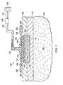
- a system 100 for treating a wound 102 on a patient 104 that includes a patterned neo-epithelium dressing 106 is presented.
- the wound 102 may extend through epidermis 108 and into dermis 110.
- the wound 102 extends into subcutaneous tissue 112.
- the patterned neo-epithelium dressing 106 which has a first side 114 and a second, patient- facing side 116, is shown with the second, patient-facing side 116 against granulation tissue 118.
- neo-epithelium tissue will grow from wound edges 120 and is directed and formed under the influence of the patterned neo- epithelium dressing 106.
- a sealing member 122 forms a fluid seal over the patterned neo-epithelium dressing 106.
- Fluid seal or “seal,” means a seal adequate to maintain reduced pressure at a desired site given the particular reduced-pressure source or subsystem involved.
- the sealing member 122 has a first side 124 and a second, patient-facing side 126.
- the sealing member 122 may be any material that provides a fluid seal.
- the sealing member 122 may, for example, be an impermeable or semipermeable, elastomeric material.
- “Elastomeric” means having the properties of an elastomer and generally refers to a polymeric material that has rubber-like properties.
- elastomers have ultimate elongations greater than 100% and a significant amount of resilience.
- the resilience of a material refers to the material's ability to recover from an elastic deformation.
- elastomers may include, but are not limited to, natural rubbers, polyisoprene, styrene butadiene rubber, chloroprene rubber, polybutadiene, nitrile rubber, butyl rubber, ethylene propylene rubber, ethylene propylene diene monomer, chlorosulfonated polyethylene, polysulfide rubber, polyurethane, EVA film, co-polyester, and silicones.
- sealing member materials include a silicone drape, 3M Tegaderm® drape, acrylic drape such as one available from Avery Dennison.
- An attachment device 128 may be used to hold the sealing member 122 against the patient's epidermis 108 or another layer, such as a gasket or additional sealing member.
- the attachment device 128 may take numerous forms.
- the attachment device 128 may be a medically acceptable, pressure-sensitive adhesive that extends about a periphery 130, a portion of, or the entirety of the sealing member 122.
- a reduced-pressure interface 132 is fluidly coupled to the second, patient-facing side 126 of the sealing member 122. Reduced pressure developed by a reduced-pressure source 134 is delivered through a reduced-pressure delivery conduit 136 to the reduced-pressure interface 132.
- the reduced-pressure interface 132 is a T . R . A . C . ® Pad or Sensa T.R.A.C. ® Pad available from KCI of San Antonio, Texas.
- the reduced-pressure interface 132 allows the reduced pressure to be delivered to the second, patient-facing side 126 of the sealing member 122 and ultimately to the patterned neo-epithelium dressing 106.
- the reduced-pressure source 134 provides reduced pressure.
- the reduced-pressure source 134 may be any device for supplying a reduced pressure, such as a vacuum pump, wall suction, micro-pump, or other source. While the amount and nature of reduced pressure applied to a tissue site will typically vary according to the application, the reduced pressure will typically be between -5 mm Hg and -500 mm Hg and more typically between -50 mm Hg and -200 mm Hg. For example, and not by way of limitation, the pressure may be -90, -100, -110, -120, -130, -140, -150, -160, -170, -180,-190, -200 mm Hg or another pressure.
- the granulation tissue 118 may be developed using the system 100 but with a manifold (not shown) in the location where the patterned neo-epithelium dressing 106 is presently shown.
- the term "manifold” as used herein generally refers to a substance or structure that is provided to assist in applying reduced pressure to, delivering fluids to, or removing fluids from the wound 102.
- the manifold typically includes a plurality of flow channels or pathways that distribute fluids provided to and removed from the tissue site around the manifold.
- the flow channels or pathways are interconnected to improve distribution of fluids provided to or removed from the wound 102.
- the manifold may be a biocompatible material that is capable of being placed in contact with the wound 102 and distributing reduced pressure to the wound 102.
- manifolds may include, for example, without limitation, devices that have structural elements arranged to form flow channels, such as, for example, cellular foam, open-cell foam, porous tissue collections, liquids, gels, and foams that include, or cure to include, flow channels.
- the manifold may be porous and may be made from foam, gauze, felted mat, or any other material suited to a particular biological application.
- the manifold is a porous foam and includes a plurality of interconnected cells or pores that act as flow channels.
- the porous foam may be a polyurethane, open-cell, reticulated foam such as GranuFoam® material manufactured by Kinetic Concepts, Incorporated of San Antonio, Texas.
- reduced pressure generally refers to a pressure less than the ambient pressure at a tissue site that is being subjected to treatment. In most cases, this reduced pressure will be less than the atmospheric pressure at which the patient is located. Alternatively, the reduced pressure may be less than a hydrostatic pressure at the tissue site. Unless otherwise indicated, values of pressure stated herein are gauge pressures.
- the reduced pressure delivered may be constant or varied (patterned or random) and may be delivered continuously or intermittently.
- vacuum and negative pressure may be used to describe the pressure applied to the wound 102, the actual pressure applied to the wound 102 may be more than the pressure normally associated with a complete vacuum. Consistent with the use herein, an increase in reduced pressure or vacuum pressure typically refers to a relative reduction in absolute pressure.
- the reduced-pressure conduit 136 may have one or more devices, such as device 138.
- the device 138 may be a fluid reservoir, or collection member, to hold exudates and other fluids removed.
- Other examples of devices 138 that may be included on the reduced-pressure conduit 136 or otherwise fluidly coupled to the reduced-pressure conduit 136 include the following non-limiting examples: a pressure-feedback device, a volume detection system, a blood detection system, an infection detection system, a flow monitoring system, or a temperature monitoring system.
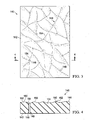
- the patterned neo-epithelium dressing 106 has an interface member 140 having a first side 142 and a second, patient-facing side 144 for placing proximate the granulation tissue 118 and a plurality of three-dimensional features 146 formed on the second, patient-facing side 144 of the interface member 140.
- the interface member 140 may be formed from any medical-grade polymers, thermoplastic polymers, resorbable polymers or materials, biologically derived polymers such as collagen, or other suitable materials, e.g., silicones, polyurethane films.
- the interface member 140 may also be formed using foam, for example, the embodiment shown in FIGURES 6A and 6B .
- the interface member 140 may be formed by casting, molding, or other techniques that form the interface member 140. As used herein, unless otherwise indicated, "or" does not require mutual exclusivity.
- the interface member 140 has a plurality of pores large enough to allow fluid transmission and small enough to limit cell migration through the pores.
- the average pore size is below the minimum size through which cells are typically capable of migrating (giving the interface member 140 a relatively "smooth" overall texture in many embodiments) to prevent tissue ingrowth into the interface member 140 and to promote lateral cell migration parallel to a surface 150 of the interface member 140.
- Select pores may exceed the minimum size for cell migration, but be contained in sufficiently low density on the material surface to maintain acceptable levels of non-adherence to the wound 102.
- the average pore size remains in the acceptable range.
- the interface member may have a plurality of pores having an average pore size greater than 5 micrometers or microns ( ⁇ m) and smaller than 1000 ⁇ m.
- the average pore size may be 10, 40, 80, 100, 120, 150, 200, 250, 300, 350, 400, 450, 500, 550, 600, 650, 700, 750, . 800, 850, 900, or 950 ⁇ m or any dimension between these or other sizes. While the term "pore" is used, it should be understood that the pores may include slits or other apertures. Where dimensions of pores are specifically given, a generally round pore should be understood and the dimension applies to the diameter.
- the formation process of the interface member 140 may form the plurality of three-dimensional features 146 or the three-dimensional features 146 may be formed separately and then coupled as part of the interface member 140.
- the plurality of three-dimensional features 146 may be chemically etched, imprinted, or formed later as an aspect of the interface member 140.
- the interface member 140 is formed with one or more fluid passageways 148, such as channels 149, that fluidly couple the first side 142 and second, patient-facing side 144 of the interface member 140.
- the fluid passageways 148 may be apertures, conduits, or inherent porous pathways in the subsisting material of the interface member 140.
- the three-dimensional features 146 may include a plurality of ridges 152 or a plurality of grooves 154.
- the three-dimensional features 146 help direct the flow of fluids, e.g., endogenous fluids, such as exudates, to one or more of the fluid passageways 148.
- the three-dimensional features 146 or a portion of the three-dimensional features 146 may be coated with one or more proteins, e.g., growth factors, integrins, integrin receptors, antibodies, peptides, aptomers, or other suitable materials.
- the three-dimensional features 146 may be formed as a pattern on the surface 150 that mimics or substantially replicates a human skin pattern. At least three approaches may be used to develop the pattern for the three-dimensional features 146.
- a "generic human skin pattern” may be used that includes a pattern that is modeled on a general or generic pattern for human skin. This pattern may not be specific to a particular location on a body, but is a more general pattern having wrinkles and general features.
- a "location-specific human skin pattern” may be used.
- a general skin pattern is used that is patterned on the general features for a specific area of a body.
- a generic representation of skin on the back of a hand may be used for wound on the back of a hand.
- an "intact analogous human skin pattern” may be used.
- the pattern may be developed based on the specific patient's skin near the wound or on a duplicate body part.
- the pattern mimics or substantially replicates the skin near the wound or on the duplicate body part.
- either skin near the wound would be used as a model or the intact skin on the right hand would be used to form the three-dimensional features 146.
- This latter approach produces a custom symmetric dressing.
- the three-dimensional features 146 may be organized in other patterns such as a radial pattern to direct migration from a periphery to a center of the patterned neo-epithelium dressing 106.
- FIGURE 5 another illustrative embodiment of a system 200 for treating a wound 202.
- the system 200 is analogous in many respects to system 100 of FIGURE 1 and analogous elements have been indicated by indexing the reference numerals by 100.
- the wound 202 is shown going through epidermis 208 and dermis 210 and almost into subcutaneous tissue 212.
- Granulation tissue 218 is shown formed on the bed of the wound 202 and neo-epithelium 219 is shown formed over the granulation tissue 218.
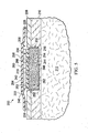
- the system 200 includes a patterned neo-epithelium dressing 206 that includes an interface member 240, which has a first side 242 and a second, patient-facing side 244.
- the interface member 240 includes a thin member 256 and a foam member 258.
- the thin member 256 such as a polyurethane film or member made from other materials listed herein, has a first side 260 and a second, patient-facing side 262.
- the foam member 258 has a first side 264 and a second, patient-facing side 266.
- the first side 260 of the thin member 256 is adjacent to the second, patient-facing side 266 of the foam member 258 and may be coupled thereto by any known technique, including without limitation welding (e.g., ultrasonic or RF welding), flame lamination, bonding, adhesives, or cements.
- the thin member 256 may be formed from any medical-grade polymers, thermoplastic polymers, resorbable polymers or materials, biologically derived polymers such as collagen, or other suitable materials, e.g., silicones, polyurethane films.
- a plurality of three-dimensional features 246 may be formed on the second, patient-facing side 262 of the thin member 256.
- the three-dimensional features 246 may be formed by imprinting, etching, or casting, or other techniques onto the thin member 256.
- the three-dimensional features 246 may include a plurality of ridges 252 or a plurality of grooves 254.
- the three-dimensional features 246 may be formed as a pattern on the surface that mimics or substantially replicates a human skin pattern.
- a contact pressure, or an inward pressure, is developed on the patterned neo-epithelium dressing 206.
- the contact pressure is developed using the foam member 258 as a bolster and applying a sealing member 222 over the foam member 258 to create the contact force.
- Reduced pressure could also be used in the system 100 of FIGURE 1 .
- An attachment device 228 may be used to form a fluid seal with the sealing member 222 and the patient's epidermis 208.
- the second, patient-facing side 244 and the first side 242 of the interface member 240 are in fluid communication through pores, which form fluid passageways, in the interface member 240.
- channels may be formed.
- the foam member 258 may be a hydrophilic foam that wicks fluids from the thin member 256.
- the foam member 258 may be an open-cell foam. In still another embodiment, the foam member 258 may be hydrophobic foam.
- the patterned neo-epithelium dressing 306 is presented on granulation tissue 318.
- the patterned neo-epithelium dressing 306 is formed from a foam 368 having rigid portions 370 and less rigid portions 372 that are apparent under reduced pressure as shown in FIGURE 6B .
- the foam 368 forms a plurality of three-dimensional features 346 in the form of ridges 352 and grooves 354 when placed under reduced pressure.
- a first side 369 and a second, patient-facing side 371 are in fluid communication via fluid passageways formed by open cells in the foam 368.
- the three-dimensional features 346 may be formed as a pattern on the surface that mimics or substantially replicates a human skin pattern.
- granulation tissue 118, 218, 318 may be formed by placing the manifold (not shown) proximate the wound 102, 202 and forming a fluid seal using a sealing member 122, 222. Reduced pressure is applied to facilitate formation of the granulation tissue 118, 218, 318. Alternatively, the granulation tissue may be formed without assistance.
- the patterned neo-epithelium dressing 106, 206, 306 may be placed proximate the granulation tissue 118, 218, 318 and covered by the sealing member 122, 222 to transition from granulation to epithelialization.
- Contact pressure is developed by using reduced pressure, a pressure wrap, a foam bolster with tensioning member or sealing member pressing on the bolster.
- reduced pressure is used to hold contact pressure and to remove fluids through the fluid passageways 148.
- the reduced pressure pulls endogenous fluids from the wound 102, 202 directed by the three-dimensional features 146 to the fluid passageways 148.
- patterned proteins or extracellular matrix (ECM), e.g., fibrin or collagen
- ECM extracellular matrix
- migrating epithelium migrates from the wound edges 120, the epithelium is guided by the patterned protein deposition and forms the neo-epithelium in the desired pattern.
- the ridges 152 form fissures (e.g., fissures 253 in FIG.
- neo-epithelium in the neo-epithelium.
- fissures or grooves in the neo-epithelium act as points of stress relief for flexion when exposed to bodily movement.
- the formation of the neo-epithelium in this way involves tissue formation according to the integrated principles of fluid flow, contact guidance, microstrain; and mechanotransduction.
- contact pressure is provided without reduced pressure.
- the three-dimensional features 146 may be used primarily to direct cell migration.
- a hydrophilic member may be used to help manage fluids.
- the patterned neo-epithelium dressing 106, 206 may influence protein adhesion, cell behavior (migration), and ECM production by the surface topography, or the three-dimensional features 146.
- the three-dimensional features 146 may also influence orientation of cells and ECM within the granulation tissue and thereby the neo-epithelium. In this manner, the features transmit contact guidance to a pericellular (cell-derived) matrix.
- the fibroblasts of the granulation tissue 118, 218 may start to align when placed in contact with the grooves 154 or ridges 152 of the patterned neo-epithelium dressing 106.
- the fibroblasts may align cytoskeleton, or the scaffolding, in substantially the same direction as directed by the three-dimensional features 146 of the patterned neo-epithelium dressing 106.
- the keratinocytes may follow the pattern expressed by the fibroblasts.
- the fissures formed mimic those in intact skin.
- the three-dimensional features 146, 246 direct elements within the granulation tissue 118, 218 of healing wounds that could translate to the development of the overlying neo-epithelium 219 and result in a patterned epithelium with appropriate creases or fissures and ECM deposition for improved regeneration and functionality, including physiologically-equivalent flexion of the tissue and aesthetic appearance. This flexion is supported by the patterned deposition of ECM both within the underlying granulation layers and in the neo-epithelium. These structures provide points of stress relief and structural support to enhance bodily movement. In addition, the rate of re-epithelialization may be increased using the systems 100, 200.
- surface patterning or wrinkling on the surface of the epithelium may be induced upon introduction of fluids, application of negative pressure, or induction by electrical, light, or other stimulatory device.
- backing layers or other layers may be added to the neo-epithelium dressing. While the systems 100, 200 and patterned neo-epithelium dressings 106, 206, 306 are shown in the context of epithelium on a wound bed, similar approaches may be taken to pattern the surface of other epithelial or endothelial linings including those within the vascular, respiratory, visual, and digestive systems.
- an interface member may be formed with a thin member coupled to a foam and wherein the thin member contracts after coupling to the foam. The contraction creates the ridges and grooves.
Description
- The present disclosure relates generally to medical treatment systems and, more particularly, to patterned neo-epithelialization dressings, system, and methods.
- Depending on the medical circumstances, reduced pressure may be used for, among other things, reduced-pressure therapy to encourage development of granulation tissue at a tissue site. Granulation tissue is connective tissue that forms on wounds during tissue repair. Granulation tissue is typically defined to include new blood vessels, immune cells, fibroblasts, and provisional extracellular matrix. Granulation tissue typically signals the proliferative phase of wound healing. Reduced-pressure therapy typically involves manifolding, or distributing, reduced pressure to the tissue site.
-
US 2008/0275409 A1 describes a wound healing device which transmits micromechanical forces locally for promoting wound healing. -
US 2007/032762 discloses an apparatus including a fluid permeable dressing and a cover membrane. A fluid reservoir is coupled to the cover membrane, the fluid vessel including an inlet port configured to receive a fluid and an outlet port fluidically coupled to the fluid permeable dressing. - The invention is disclosed by the features of independent claim 1. An illustrative, non-limiting embodiment of a system for treating a wound having granulation tissue on a patient includes a patterned neo-epithelium dressing for disposing proximate the wound. The patterned neo-epithelium dressing for treating a wound having granulation tissue includes an interface member having a first side and a second, patient-facing side for placing proximate to the granulation tissue and a plurality of three-dimensional features formed on the second, patient-facing side of the interface member. The system further includes a sealing member for placing over the patterned neo-epithelium dressing and the patient's epidermis, a reduced-pressure interface fluidly coupled to the sealing member, and a reduced-pressure source fluidly coupled to the reduced-pressure interface.
- An illustrative, non-limiting embodiment of a patterned neo-epithelium dressing for treating a wound having granulation tissue includes an interface member having a first side and a second, patient-facing side for placing proximate the granulation tissue and a plurality of three-dimensional features formed on the second, patient-facing side of the interface member.
- An illustrative, non-limiting embodiment of a method of treating a wound site of a patient includes optionally forming granulation tissue at the wound site, deploying a patterned neo-epithelium dressing proximate the granulation tissue, and applying a contact pressure on the patterned neo-epithelium dressing. The patterned neo-epithelium dressing for treating a wound having granulation tissue includes an interface member having a first side and a second, patient-facing side for placing proximate the granulation tissue. The patterned neo-epithelium dressing also includes a plurality of three-dimensional features formed on the second, patient-facing side of the interface member.
- An illustrative, non-limiting embodiment of a method of treating a wound site of a patient includes directing flow of endogenous fluids to cause patterned protein deposition, causing guidance of the migrating epithelium on the patterned deposition of proteins to form a neo-epithelium, and forming fissures in the neo-epithelium.
- An illustrative, non-limiting embodiment of a method of manufacturing a patterned neo-epithelium dressing for treating a wound having granulation tissue includes forming an interface member having a first side and a second, patient-facing side for placing proximate the granulation tissue, and forming a plurality of three-dimensional features on the second, patient-facing side of the interface member.
- Other features and advantages of the illustrative embodiments will become apparent with reference to the drawings and detailed description that follow.
-
-
FIGURE 1 is a schematic diagram with a portion shown in cross section of an illustrative, non-limiting embodiment of a system for treating a wound on a patient; -
FIGURE 2 is a schematic, perspective view of an illustrative, non-limiting embodiment of a patterned neo-epithelium dressing; -
FIGURE 3 is a schematic, bottom view of the patterned neo-epithelium dressing ofFIGURE 2 ; -
FIGURE 4 is a schematic, cross-sectional view of the patterned neo-epithelium dressing ofFIGURE 3 taken along line 4-4; -
FIGURE 5 is a schematic, cross-sectional view of an illustrative, non-limiting embodiment of a patterned neo-epithelium dressing; -
FIGURE 6A is a schematic, cross-sectional view of an illustrative, non-limiting embodiment of a patterned neo-epithelium dressing shown without reduced pressure applied; and -
FIGURE 6B is the patterned neo-epithelium dressing ofFIGURE 6A shown with reduced pressure applied. - In the following detailed description of the illustrative embodiments, reference is made to the accompanying drawings that form a part hereof. These embodiments are described in sufficient detail to enable those skilled in the art to practice the invention, and it is understood that other embodiments may be utilized and that logical structural, mechanical, electrical, and chemical changes may be made without departing from the scope of the invention. To avoid detail not necessary to enable those skilled in the art to practice the embodiments described herein, the description may omit certain information known to those skilled in the art. The following detailed description is, therefore, not to be taken in a limiting sense, and the scope of the illustrative embodiments are defined only by the appended claims.
- Referring primarily to
FIGURES 1-4 , and initially toFIGURE 1 , asystem 100 for treating awound 102 on apatient 104 that includes a patternedneo-epithelium dressing 106 is presented. Thewound 102 may extend throughepidermis 108 and intodermis 110. In some instances, thewound 102 extends intosubcutaneous tissue 112. In the illustrative embodiment, the patternedneo-epithelium dressing 106, which has afirst side 114 and a second, patient- facingside 116, is shown with the second, patient-facingside 116 againstgranulation tissue 118. As will be described further below, neo-epithelium tissue will grow fromwound edges 120 and is directed and formed under the influence of the patterned neo-epithelium dressing 106. - A sealing
member 122 forms a fluid seal over the patternedneo-epithelium dressing 106. "Fluid seal," or "seal," means a seal adequate to maintain reduced pressure at a desired site given the particular reduced-pressure source or subsystem involved. The sealingmember 122 has afirst side 124 and a second, patient-facingside 126. The sealingmember 122 may be any material that provides a fluid seal. The sealingmember 122 may, for example, be an impermeable or semipermeable, elastomeric material. "Elastomeric" means having the properties of an elastomer and generally refers to a polymeric material that has rubber-like properties. More specifically, most elastomers have ultimate elongations greater than 100% and a significant amount of resilience. The resilience of a material refers to the material's ability to recover from an elastic deformation. Examples of elastomers may include, but are not limited to, natural rubbers, polyisoprene, styrene butadiene rubber, chloroprene rubber, polybutadiene, nitrile rubber, butyl rubber, ethylene propylene rubber, ethylene propylene diene monomer, chlorosulfonated polyethylene, polysulfide rubber, polyurethane, EVA film, co-polyester, and silicones. Additional, specific examples of sealing member materials include a silicone drape, 3M Tegaderm® drape, acrylic drape such as one available from Avery Dennison. - An
attachment device 128 may be used to hold the sealingmember 122 against the patient'sepidermis 108 or another layer, such as a gasket or additional sealing member. Theattachment device 128 may take numerous forms. For example, theattachment device 128 may be a medically acceptable, pressure-sensitive adhesive that extends about aperiphery 130, a portion of, or the entirety of the sealingmember 122. - A reduced-
pressure interface 132 is fluidly coupled to the second, patient-facingside 126 of the sealingmember 122. Reduced pressure developed by a reduced-pressure source 134 is delivered through a reduced-pressure delivery conduit 136 to the reduced-pressure interface 132. In one illustrative embodiment, the reduced-pressure interface 132 is a T.R.A.C.® Pad or Sensa T.R.A.C.® Pad available from KCI of San Antonio, Texas. The reduced-pressure interface 132 allows the reduced pressure to be delivered to the second, patient-facingside 126 of the sealingmember 122 and ultimately to the patternedneo-epithelium dressing 106. - The reduced-
pressure source 134 provides reduced pressure. The reduced-pressure source 134 may be any device for supplying a reduced pressure, such as a vacuum pump, wall suction, micro-pump, or other source. While the amount and nature of reduced pressure applied to a tissue site will typically vary according to the application, the reduced pressure will typically be between -5 mm Hg and -500 mm Hg and more typically between -50 mm Hg and -200 mm Hg. For example, and not by way of limitation, the pressure may be -90, -100, -110, -120, -130, -140, -150, -160, -170, -180,-190, -200 mm Hg or another pressure. - In some embodiments, before the patterned
neo-epithelium dressing 106 is deployed ongranulation tissue 118. Thegranulation tissue 118 may be developed using thesystem 100 but with a manifold (not shown) in the location where the patternedneo-epithelium dressing 106 is presently shown. The term "manifold" as used herein generally refers to a substance or structure that is provided to assist in applying reduced pressure to, delivering fluids to, or removing fluids from thewound 102. The manifold typically includes a plurality of flow channels or pathways that distribute fluids provided to and removed from the tissue site around the manifold. In one illustrative embodiment, the flow channels or pathways are interconnected to improve distribution of fluids provided to or removed from thewound 102. The manifold may be a biocompatible material that is capable of being placed in contact with thewound 102 and distributing reduced pressure to thewound 102. Examples of manifolds may include, for example, without limitation, devices that have structural elements arranged to form flow channels, such as, for example, cellular foam, open-cell foam, porous tissue collections, liquids, gels, and foams that include, or cure to include, flow channels. The manifold may be porous and may be made from foam, gauze, felted mat, or any other material suited to a particular biological application. In one embodiment, the manifold is a porous foam and includes a plurality of interconnected cells or pores that act as flow channels. The porous foam may be a polyurethane, open-cell, reticulated foam such as GranuFoam® material manufactured by Kinetic Concepts, Incorporated of San Antonio, Texas. - As used herein, "reduced pressure" generally refers to a pressure less than the ambient pressure at a tissue site that is being subjected to treatment. In most cases, this reduced pressure will be less than the atmospheric pressure at which the patient is located. Alternatively, the reduced pressure may be less than a hydrostatic pressure at the tissue site. Unless otherwise indicated, values of pressure stated herein are gauge pressures. The reduced pressure delivered may be constant or varied (patterned or random) and may be delivered continuously or intermittently. Although the terms "vacuum" and "negative pressure" may be used to describe the pressure applied to the
wound 102, the actual pressure applied to thewound 102 may be more than the pressure normally associated with a complete vacuum. Consistent with the use herein, an increase in reduced pressure or vacuum pressure typically refers to a relative reduction in absolute pressure. - The reduced-
pressure conduit 136 may have one or more devices, such asdevice 138. For example, thedevice 138 may be a fluid reservoir, or collection member, to hold exudates and other fluids removed. Other examples ofdevices 138 that may be included on the reduced-pressure conduit 136 or otherwise fluidly coupled to the reduced-pressure conduit 136 include the following non-limiting examples: a pressure-feedback device, a volume detection system, a blood detection system, an infection detection system, a flow monitoring system, or a temperature monitoring system. - Referring now primarily to
FIGURES 2-4 , the patternedneo-epithelium dressing 106 has aninterface member 140 having afirst side 142 and a second, patient-facingside 144 for placing proximate thegranulation tissue 118 and a plurality of three-dimensional features 146 formed on the second, patient-facingside 144 of theinterface member 140. Theinterface member 140 may be formed from any medical-grade polymers, thermoplastic polymers, resorbable polymers or materials, biologically derived polymers such as collagen, or other suitable materials, e.g., silicones, polyurethane films. Theinterface member 140 may also be formed using foam, for example, the embodiment shown inFIGURES 6A and 6B . Theinterface member 140 may be formed by casting, molding, or other techniques that form theinterface member 140. As used herein, unless otherwise indicated, "or" does not require mutual exclusivity. - The
interface member 140 has a plurality of pores large enough to allow fluid transmission and small enough to limit cell migration through the pores. The average pore size is below the minimum size through which cells are typically capable of migrating (giving the interface member 140 a relatively "smooth" overall texture in many embodiments) to prevent tissue ingrowth into theinterface member 140 and to promote lateral cell migration parallel to asurface 150 of theinterface member 140. Select pores may exceed the minimum size for cell migration, but be contained in sufficiently low density on the material surface to maintain acceptable levels of non-adherence to thewound 102. The average pore size remains in the acceptable range. At the other end of the range of the pore size, to allow for fluid control of thewound 102, pores of adequate size to allow for fluid transmission are typically incorporated throughout theinterface member 140 or in organized patterns on theinterface member 140 for promoting direct fluid flow. In one embodiment, the interface member may have a plurality of pores having an average pore size greater than 5 micrometers or microns (µm) and smaller than 1000µm. In other non-limiting embodiments, the average pore size may be 10, 40, 80, 100, 120, 150, 200, 250, 300, 350, 400, 450, 500, 550, 600, 650, 700, 750, . 800, 850, 900, or 950 µm or any dimension between these or other sizes. While the term "pore" is used, it should be understood that the pores may include slits or other apertures. Where dimensions of pores are specifically given, a generally round pore should be understood and the dimension applies to the diameter. - The formation process of the
interface member 140 may form the plurality of three-dimensional features 146 or the three-dimensional features 146 may be formed separately and then coupled as part of theinterface member 140. In another embodiment, the plurality of three-dimensional features 146 may be chemically etched, imprinted, or formed later as an aspect of theinterface member 140. - The
interface member 140 is formed with one or morefluid passageways 148, such aschannels 149, that fluidly couple thefirst side 142 and second, patient-facingside 144 of theinterface member 140. Thefluid passageways 148 may be apertures, conduits, or inherent porous pathways in the subsisting material of theinterface member 140. - The three-
dimensional features 146 may include a plurality ofridges 152 or a plurality ofgrooves 154. The three-dimensional features 146 help direct the flow of fluids, e.g., endogenous fluids, such as exudates, to one or more of thefluid passageways 148. The three-dimensional features 146 or a portion of the three-dimensional features 146 may be coated with one or more proteins, e.g., growth factors, integrins, integrin receptors, antibodies, peptides, aptomers, or other suitable materials. - The three-
dimensional features 146 may be formed as a pattern on thesurface 150 that mimics or substantially replicates a human skin pattern. At least three approaches may be used to develop the pattern for the three-dimensional features 146. First, a "generic human skin pattern" may be used that includes a pattern that is modeled on a general or generic pattern for human skin. This pattern may not be specific to a particular location on a body, but is a more general pattern having wrinkles and general features. - Second, a "location-specific human skin pattern" may be used. With this second approach, a general skin pattern is used that is patterned on the general features for a specific area of a body. For example, a generic representation of skin on the back of a hand may be used for wound on the back of a hand.
- Third, an "intact analogous human skin pattern" may be used. With this third approach, the pattern may be developed based on the specific patient's skin near the wound or on a duplicate body part. The pattern mimics or substantially replicates the skin near the wound or on the duplicate body part. For example, if a wound were on the back of the patient's left hand, either skin near the wound would be used as a model or the intact skin on the right hand would be used to form the three-
dimensional features 146. This latter approach produces a custom symmetric dressing. In still other embodiments, the three-dimensional features 146 may be organized in other patterns such as a radial pattern to direct migration from a periphery to a center of the patternedneo-epithelium dressing 106. - Referring now primarily to
FIGURE 5 , another illustrative embodiment of asystem 200 for treating awound 202. Thesystem 200 is analogous in many respects tosystem 100 ofFIGURE 1 and analogous elements have been indicated by indexing the reference numerals by 100. Thewound 202 is shown going throughepidermis 208 anddermis 210 and almost intosubcutaneous tissue 212.Granulation tissue 218 is shown formed on the bed of thewound 202 andneo-epithelium 219 is shown formed over thegranulation tissue 218. - The
system 200 includes a patternedneo-epithelium dressing 206 that includes aninterface member 240, which has afirst side 242 and a second, patient-facingside 244. Theinterface member 240 includes athin member 256 and afoam member 258. Thethin member 256, such as a polyurethane film or member made from other materials listed herein, has afirst side 260 and a second, patient-facingside 262. Thefoam member 258 has afirst side 264 and a second, patient-facingside 266. Thefirst side 260 of thethin member 256 is adjacent to the second, patient-facingside 266 of thefoam member 258 and may be coupled thereto by any known technique, including without limitation welding (e.g., ultrasonic or RF welding), flame lamination, bonding, adhesives, or cements. Thethin member 256 may be formed from any medical-grade polymers, thermoplastic polymers, resorbable polymers or materials, biologically derived polymers such as collagen, or other suitable materials, e.g., silicones, polyurethane films. - A plurality of three-
dimensional features 246 may be formed on the second, patient-facingside 262 of thethin member 256. The three-dimensional features 246 may be formed by imprinting, etching, or casting, or other techniques onto thethin member 256. As before, the three-dimensional features 246 may include a plurality ofridges 252 or a plurality ofgrooves 254. The three-dimensional features 246 may be formed as a pattern on the surface that mimics or substantially replicates a human skin pattern. - A contact pressure, or an inward pressure, is developed on the patterned
neo-epithelium dressing 206. In this embodiment, the contact pressure is developed using thefoam member 258 as a bolster and applying a sealingmember 222 over thefoam member 258 to create the contact force. Reduced pressure could also be used in thesystem 100 ofFIGURE 1 . Anattachment device 228 may be used to form a fluid seal with the sealingmember 222 and the patient'sepidermis 208. - The second, patient-facing
side 244 and thefirst side 242 of theinterface member 240 are in fluid communication through pores, which form fluid passageways, in theinterface member 240. In addition to the pores or alternatively, channels (not shown but analogous tochannels 149 inFIG. 1 ) may be formed. - The
foam member 258 may be a hydrophilic foam that wicks fluids from thethin member 256. Thefoam member 258 may be an open-cell foam. In still another embodiment, thefoam member 258 may be hydrophobic foam. - Referring now primarily to
FIGURES 6A and 6B , another illustrative, non-limiting embodiment of a patternedneo-epithelium dressing 306 is presented ongranulation tissue 318. The patternedneo-epithelium dressing 306 is formed from afoam 368 havingrigid portions 370 and lessrigid portions 372 that are apparent under reduced pressure as shown inFIGURE 6B . Because of the differing rigidity, thefoam 368 forms a plurality of three-dimensional features 346 in the form ofridges 352 andgrooves 354 when placed under reduced pressure. Afirst side 369 and a second, patient-facingside 371 are in fluid communication via fluid passageways formed by open cells in thefoam 368. The three-dimensional features 346 may be formed as a pattern on the surface that mimics or substantially replicates a human skin pattern. - Referring now to
FIGURES 1-6B , in use, according to one illustrative embodiment,granulation tissue wound member granulation tissue granulation tissue neo-epithelium dressing granulation tissue member - In many embodiments, reduced pressure is used to hold contact pressure and to remove fluids through the
fluid passageways 148. The reduced pressure pulls endogenous fluids from thewound dimensional features 146 to thefluid passageways 148. As the endogenous fluids flow along the path directed by the three-dimensional features 146, patterned proteins or extracellular matrix (ECM), (e.g., fibrin or collagen) are deposited or formed. As migrating epithelium migrates from the wound edges 120, the epithelium is guided by the patterned protein deposition and forms the neo-epithelium in the desired pattern. Theridges 152 form fissures (e.g., fissures 253 inFIG. 5 ) in the neo-epithelium. These fissures or grooves in the neo-epithelium act as points of stress relief for flexion when exposed to bodily movement. The formation of the neo-epithelium in this way involves tissue formation according to the integrated principles of fluid flow, contact guidance, microstrain; and mechanotransduction. - In one embodiment, contact pressure is provided without reduced pressure. In this instance, the three-
dimensional features 146 may be used primarily to direct cell migration. In addition, a hydrophilic member may be used to help manage fluids. - The patterned
neo-epithelium dressing dimensional features 146. The three-dimensional features 146 may also influence orientation of cells and ECM within the granulation tissue and thereby the neo-epithelium. In this manner, the features transmit contact guidance to a pericellular (cell-derived) matrix. The fibroblasts of thegranulation tissue grooves 154 orridges 152 of the patternedneo-epithelium dressing 106. The fibroblasts may align cytoskeleton, or the scaffolding, in substantially the same direction as directed by the three-dimensional features 146 of the patternedneo-epithelium dressing 106. The keratinocytes may follow the pattern expressed by the fibroblasts. - The fissures formed mimic those in intact skin. The three-
dimensional features granulation tissue overlying neo-epithelium 219 and result in a patterned epithelium with appropriate creases or fissures and ECM deposition for improved regeneration and functionality, including physiologically-equivalent flexion of the tissue and aesthetic appearance. This flexion is supported by the patterned deposition of ECM both within the underlying granulation layers and in the neo-epithelium. These structures provide points of stress relief and structural support to enhance bodily movement. In addition, the rate of re-epithelialization may be increased using thesystems neo-epithelium dressings dimensional features - In other embodiments, surface patterning or wrinkling on the surface of the epithelium may be induced upon introduction of fluids, application of negative pressure, or induction by electrical, light, or other stimulatory device. In other embodiments, backing layers or other layers may be added to the neo-epithelium dressing. While the
systems neo-epithelium dressings - In another embodiment, an interface member may be formed with a thin member coupled to a foam and wherein the thin member contracts after coupling to the foam. The contraction creates the ridges and grooves.
- Although the present invention and its advantages have been disclosed in the context of certain illustrative, non-limiting embodiments, it should be understood that various changes, substitutions, permutations, and alterations can be made without departing from the scope of the invention as defined by the appended claims. It will be appreciated that any feature that is described in connection to any one embodiment may also be applicable to any other embodiment, and descriptions related one embodiment may be applied to other embodiment as indicated by the context.
- It will be understood that the benefits and advantages described above may relate to one embodiment or may relate to several embodiments. It will further be understood that reference to 'an' item refers to one or more of those items.
- The steps of the methods described herein may be carried out in any suitable order, or simultaneously where appropriate.
- Where appropriate, aspects of any of the examples described above may be combined with aspects of any of the other examples described to form further examples having comparable or different properties and addressing the same or different problems.
- It will be understood that the above description of preferred embodiments is given by way of example only and that various modifications may be made by those skilled in the art. The above specification, examples and data provide a complete description of the structure and use of exemplary embodiments of the invention. Although various embodiments of the invention have been described above with a certain degree of particularity, or with reference to one or more individual embodiments, those skilled in the art could make numerous alterations to the disclosed embodiments without departing from the scope of the claims.
Claims (4)
- A system for treating a tissue site having granulation tissue on a patient, the system comprising:a patterned neo-epithelium dressing (106) for disposing proximate to granulation tissue on the tissue site, wherein the patterned neo-epithelium dressing comprises:an interface member (140) having a first side (142) and a second,patient-facing side (142) for placing proximate the granulation tissue;a fluid passageway (148) that fluidly couples the first and second sides (142, 144) of the interface member (140) for delivery of pressure to the second side (144);a sealing member (122) for placing over the patterned neo-epithelium dressing and the patient's epidermis;a reduced-pressure interface (132) fluidly coupled to the sealing member (122); anda reduced-pressure source (134) fluidly coupled to the reduced-pressure interface (132),wherein the interface member (140) is characterized by a plurality of three-dimensional features formed on the second, patient-facing side (144) of the interface member (140) and comprising a pattern that mimics the pattern of human skin.
- The system of claim 1, wherein the interface member (140) is formed with the fluid passageway (148).
- The system of claim 1, the interface member (140) has a plurality of pores having an average pore size larger than 5µm and smaller than 1000µm.
- The system of claim 1, wherein the interface member (140) comprises a foam having rigid portions and less rigid portions, wherein, under reduced pressure, the less rigid portions compress more than the more rigid portions.
Priority Applications (1)
| Application Number | Priority Date | Filing Date | Title |
|---|---|---|---|
| EP15163540.6A EP2921149B8 (en) | 2010-03-16 | 2011-03-11 | Patterned neo-epithelialization dressings, systems, and methods |
Applications Claiming Priority (4)
| Application Number | Priority Date | Filing Date | Title |
|---|---|---|---|
| US31423610P | 2010-03-16 | 2010-03-16 | |
| US31427410P | 2010-03-16 | 2010-03-16 | |
| US13/045,302 US9358158B2 (en) | 2010-03-16 | 2011-03-10 | Patterned neo-epithelialization dressings, systems, and methods |
| PCT/US2011/028189 WO2011115851A1 (en) | 2010-03-16 | 2011-03-11 | Patterned neo-epithelialization dressings, systems, and methods |
Related Child Applications (1)
| Application Number | Title | Priority Date | Filing Date |
|---|---|---|---|
| EP15163540.6A Division EP2921149B8 (en) | 2010-03-16 | 2011-03-11 | Patterned neo-epithelialization dressings, systems, and methods |
Publications (2)
| Publication Number | Publication Date |
|---|---|
| EP2547301A1 EP2547301A1 (en) | 2013-01-23 |
| EP2547301B1 true EP2547301B1 (en) | 2015-05-06 |
Family
ID=44647787
Family Applications (3)
| Application Number | Title | Priority Date | Filing Date |
|---|---|---|---|
| EP15163540.6A Active EP2921149B8 (en) | 2010-03-16 | 2011-03-11 | Patterned neo-epithelialization dressings, systems, and methods |
| EP11710611.2A Not-in-force EP2547301B1 (en) | 2010-03-16 | 2011-03-11 | Patterned neo-epithelialization dressings, systems, and methods |
| EP11710618.7A Not-in-force EP2547302B1 (en) | 2010-03-16 | 2011-03-14 | Epithelialization methods, dressings, and systems |
Family Applications Before (1)
| Application Number | Title | Priority Date | Filing Date |
|---|---|---|---|
| EP15163540.6A Active EP2921149B8 (en) | 2010-03-16 | 2011-03-11 | Patterned neo-epithelialization dressings, systems, and methods |
Family Applications After (1)
| Application Number | Title | Priority Date | Filing Date |
|---|---|---|---|
| EP11710618.7A Not-in-force EP2547302B1 (en) | 2010-03-16 | 2011-03-14 | Epithelialization methods, dressings, and systems |
Country Status (8)
| Country | Link |
|---|---|
| US (6) | US9358158B2 (en) |
| EP (3) | EP2921149B8 (en) |
| JP (2) | JP5968867B2 (en) |
| CN (2) | CN102781382B (en) |
| AU (2) | AU2011227598B2 (en) |
| CA (2) | CA2790413C (en) |
| TW (1) | TW201138878A (en) |
| WO (2) | WO2011115851A1 (en) |
Families Citing this family (42)
| Publication number | Priority date | Publication date | Assignee | Title |
|---|---|---|---|---|
| GB0808376D0 (en) | 2008-05-08 | 2008-06-18 | Bristol Myers Squibb Co | Wound dressing |
| GB0817796D0 (en) | 2008-09-29 | 2008-11-05 | Convatec Inc | wound dressing |
| US20110054420A1 (en) * | 2009-08-27 | 2011-03-03 | Christopher Brian Locke | Reduced-pressure wound dressings and systems for re-epithelialization and granulation |
| US8690844B2 (en) * | 2009-08-27 | 2014-04-08 | Kci Licensing, Inc. | Re-epithelialization wound dressings and systems |
| US9358158B2 (en) * | 2010-03-16 | 2016-06-07 | Kci Licensing, Inc. | Patterned neo-epithelialization dressings, systems, and methods |
| US8790699B2 (en) * | 2010-04-23 | 2014-07-29 | Warsaw Orthpedic, Inc. | Foam-formed collagen strand |
| GB201015656D0 (en) | 2010-09-20 | 2010-10-27 | Smith & Nephew | Pressure control apparatus |
| GB201020236D0 (en) | 2010-11-30 | 2011-01-12 | Convatec Technologies Inc | A composition for detecting biofilms on viable tissues |
| US9440010B2 (en) * | 2010-12-07 | 2016-09-13 | Kci Licensing, Inc. | Drape having microstrain inducing projections for treating a wound site |
| WO2012078781A1 (en) | 2010-12-08 | 2012-06-14 | Convatec Technologies Inc. | Integrated system for assessing wound exudates |
| CN103347562B (en) | 2010-12-08 | 2016-08-10 | 康沃特克科技公司 | Wound exudate system accessory |
| GB201115182D0 (en) | 2011-09-02 | 2011-10-19 | Trio Healthcare Ltd | Skin contact material |
| US9084845B2 (en) | 2011-11-02 | 2015-07-21 | Smith & Nephew Plc | Reduced pressure therapy apparatuses and methods of using same |
| BR112014010647A2 (en) * | 2011-11-02 | 2017-04-25 | Smith & Nephew | pressure therapy devices and methods of use thereof |
| GB2497406A (en) | 2011-11-29 | 2013-06-12 | Webtec Converting Llc | Dressing with a perforated binder layer |
| GB201120693D0 (en) | 2011-12-01 | 2012-01-11 | Convatec Technologies Inc | Wound dressing for use in vacuum therapy |
| EP2787943B2 (en) * | 2011-12-07 | 2018-02-28 | KCI Licensing, Inc. | Synthetic granulating gauze for use with reduced-pressure treatment systems |
| JP2015506734A (en) * | 2011-12-22 | 2015-03-05 | エーテーハー チューリヒ | Patch structure for controlling wound healing |
| US9554944B2 (en) * | 2012-08-20 | 2017-01-31 | Alessandro Barberio | Medical protruded pads or dressings for wound care including use with orthopedic and prosthetic devices |
| EP2711033A1 (en) * | 2012-09-25 | 2014-03-26 | Paul Hartmann AG | System for wound treatment |
| JP6044307B2 (en) * | 2012-12-04 | 2016-12-14 | セイコーエプソン株式会社 | Liquid ejector |
| US20150354096A1 (en) | 2012-12-20 | 2015-12-10 | Convatec Technologies Inc. | Processing of chemically modified cellulosic fibres |
| JP6947506B2 (en) | 2014-02-14 | 2021-10-13 | アトミック メディカル イノベーションズ,インコーポレイティド | System for tissue healing |
| EP3354241B1 (en) | 2014-05-09 | 2020-12-30 | 3M Innovative Properties Company | Disruptive dressing for use with negative pressure and fluid instillation |
| US10898217B2 (en) | 2014-05-09 | 2021-01-26 | Kci Licensing, Inc. | Dressing providing apertures with multiple orifice sizes for negative-pressure therapy |
| CN106255480B (en) | 2014-05-09 | 2020-01-21 | 凯希特许有限公司 | Dressing with a shrink layer for a linear tissue site |
| CN107106723B (en) * | 2014-12-30 | 2020-10-23 | 3M创新有限公司 | Negative pressure wound dressing with absorbent adhesive sealing layer |
| US10456301B2 (en) * | 2015-08-14 | 2019-10-29 | Healthko, LLC | Thermal reflective layer for a wound care dressing |
| CN109310528B (en) | 2016-03-30 | 2021-07-20 | 康沃特克科技公司 | Detecting microbial infection in a wound |
| US11740241B2 (en) | 2016-03-30 | 2023-08-29 | Synovo Gmbh | Construct including an anchor, an enzyme recognition site and an indicator region for detecting microbial infection in wounds |
| CA3030151A1 (en) | 2016-07-08 | 2018-01-11 | Convatec Technologies Inc. | Fluid collection apparatus |
| JP7071957B2 (en) | 2016-07-08 | 2022-05-19 | コンバテック・テクノロジーズ・インコーポレイテッド | Flexible negative pressure system |
| CA3030153C (en) | 2016-07-08 | 2023-10-24 | Convatec Technologies Inc. | Fluid flow sensing |
| AU2018231771A1 (en) * | 2017-03-09 | 2019-09-26 | Secretary, Department Of Biotechnology | A wound dressing for combined negative pressure and fluid delivery system |
| JP7136812B2 (en) * | 2017-05-16 | 2022-09-13 | スリーエム イノベイティブ プロパティズ カンパニー | Absorbable Negative Pressure Dressing System for Postoperative Breast Wounds |
| WO2020028514A1 (en) * | 2018-08-03 | 2020-02-06 | Kci Licensing, Inc. | Flexible and conformable wound dressing with enhanced fluid absorption capability |
| EP3876886B1 (en) * | 2018-11-08 | 2023-09-27 | 3M Innovative Properties Company | Dressing with protruding layer allowing for cleansing of wound bed macro deformations |
| CN113507908A (en) * | 2018-11-08 | 2021-10-15 | 3M创新知识产权公司 | Wound dressing with semi-rigid support for increased disruption using perforated dressing and negative pressure wound therapy |
| WO2020159859A1 (en) * | 2019-02-01 | 2020-08-06 | Kci Licensing, Inc. | Partially transparent wound dressing |
| US11771819B2 (en) | 2019-12-27 | 2023-10-03 | Convatec Limited | Low profile filter devices suitable for use in negative pressure wound therapy systems |
| US11331221B2 (en) | 2019-12-27 | 2022-05-17 | Convatec Limited | Negative pressure wound dressing |
| USD951428S1 (en) * | 2020-05-11 | 2022-05-10 | Xiamen Suneetek Medical Equipment Co., Ltd. | Suction disk for negative pressure wound therapy |
Citations (3)
| Publication number | Priority date | Publication date | Assignee | Title |
|---|---|---|---|---|
| US5755814A (en) * | 1993-03-17 | 1998-05-26 | Berg; Richard A. | Dermal-epidermal in vitro test system |
| US20080275409A1 (en) | 2007-05-01 | 2008-11-06 | The Brigham And Women's Hospital, Inc. | Wound healing device |
| WO2008154158A2 (en) | 2007-05-24 | 2008-12-18 | Applied Tissue Technologies Llc | Woundtreatment device employing negative pressure |
Family Cites Families (168)
| Publication number | Priority date | Publication date | Assignee | Title |
|---|---|---|---|---|
| US1355846A (en) | 1920-02-06 | 1920-10-19 | David A Rannells | Medical appliance |
| US2547758A (en) | 1949-01-05 | 1951-04-03 | Wilmer B Keeling | Instrument for treating the male urethra |
| US2632443A (en) | 1949-04-18 | 1953-03-24 | Eleanor P Lesher | Surgical dressing |
| GB692578A (en) | 1949-09-13 | 1953-06-10 | Minnesota Mining & Mfg | Improvements in or relating to drape sheets for surgical use |
| US2682873A (en) | 1952-07-30 | 1954-07-06 | Johnson & Johnson | General purpose protective dressing |
| NL189176B (en) | 1956-07-13 | 1900-01-01 | Hisamitsu Pharmaceutical Co | PLASTER BASED ON A SYNTHETIC RUBBER. |
| US2969057A (en) | 1957-11-04 | 1961-01-24 | Brady Co W H | Nematodic swab |
| US3066672A (en) | 1960-09-27 | 1962-12-04 | Jr William H Crosby | Method and apparatus for serial sampling of intestinal juice |
| US3367332A (en) | 1965-08-27 | 1968-02-06 | Gen Electric | Product and process for establishing a sterile area of skin |
| US3520300A (en) | 1967-03-15 | 1970-07-14 | Amp Inc | Surgical sponge and suction device |
| US3568675A (en) | 1968-08-30 | 1971-03-09 | Clyde B Harvey | Fistula and penetrating wound dressing |
| US3682180A (en) | 1970-06-08 | 1972-08-08 | Coilform Co Inc | Drain clip for surgical drain |
| BE789293Q (en) | 1970-12-07 | 1973-01-15 | Parke Davis & Co | MEDICO-SURGICAL DRESSING FOR BURNS AND SIMILAR LESIONS |
| US3826254A (en) | 1973-02-26 | 1974-07-30 | Verco Ind | Needle or catheter retaining appliance |
| DE2527706A1 (en) | 1975-06-21 | 1976-12-30 | Hanfried Dr Med Weigand | DEVICE FOR THE INTRODUCTION OF CONTRAST AGENTS INTO AN ARTIFICIAL INTESTINAL OUTLET |
| DE2640413C3 (en) | 1976-09-08 | 1980-03-27 | Richard Wolf Gmbh, 7134 Knittlingen | Catheter monitor |
| NL7710909A (en) | 1976-10-08 | 1978-04-11 | Smith & Nephew | COMPOSITE STRAPS. |
| GB1562244A (en) | 1976-11-11 | 1980-03-05 | Lock P M | Wound dressing materials |
| US4080970A (en) | 1976-11-17 | 1978-03-28 | Miller Thomas J | Post-operative combination dressing and internal drain tube with external shield and tube connector |
| US4139004A (en) | 1977-02-17 | 1979-02-13 | Gonzalez Jr Harry | Bandage apparatus for treating burns |
| US4184510A (en) | 1977-03-15 | 1980-01-22 | Fibra-Sonics, Inc. | Valued device for controlling vacuum in surgery |
| US4165748A (en) | 1977-11-07 | 1979-08-28 | Johnson Melissa C | Catheter tube holder |
| US4256109A (en) | 1978-07-10 | 1981-03-17 | Nichols Robert L | Shut off valve for medical suction apparatus |
| SE414994B (en) | 1978-11-28 | 1980-09-01 | Landstingens Inkopscentral | VENKATETERFORBAND |
| BR7908937A (en) | 1978-12-06 | 1981-06-30 | Svedman Paul | DEVICE FOR TREATING FABRICS, FOR EXAMPLE, SKIN |
| US4266545A (en) | 1979-04-06 | 1981-05-12 | Moss James P | Portable suction device for collecting fluids from a closed wound |
| US4284079A (en) | 1979-06-28 | 1981-08-18 | Adair Edwin Lloyd | Method for applying a male incontinence device |
| US4261363A (en) | 1979-11-09 | 1981-04-14 | C. R. Bard, Inc. | Retention clips for body fluid drains |
| US4569348A (en) | 1980-02-22 | 1986-02-11 | Velcro Usa Inc. | Catheter tube holder strap |
| ATE14835T1 (en) | 1980-03-11 | 1985-08-15 | Schmid Eduard | SKIN GRAFT PRESSURE BANDAGE. |
| US4297995A (en) | 1980-06-03 | 1981-11-03 | Key Pharmaceuticals, Inc. | Bandage containing attachment post |
| US4333468A (en) | 1980-08-18 | 1982-06-08 | Geist Robert W | Mesentery tube holder apparatus |
| US4465485A (en) | 1981-03-06 | 1984-08-14 | Becton, Dickinson And Company | Suction canister with unitary shut-off valve and filter features |
| US4392853A (en) | 1981-03-16 | 1983-07-12 | Rudolph Muto | Sterile assembly for protecting and fastening an indwelling device |
| US4373519A (en) | 1981-06-26 | 1983-02-15 | Minnesota Mining And Manufacturing Company | Composite wound dressing |
| US4392858A (en) | 1981-07-16 | 1983-07-12 | Sherwood Medical Company | Wound drainage device |
| US4419097A (en) | 1981-07-31 | 1983-12-06 | Rexar Industries, Inc. | Attachment for catheter tube |
| AU550575B2 (en) | 1981-08-07 | 1986-03-27 | Richard Christian Wright | Wound drainage device |
| SE429197B (en) | 1981-10-14 | 1983-08-22 | Frese Nielsen | SAR TREATMENT DEVICE |
| DE3146266A1 (en) | 1981-11-21 | 1983-06-01 | B. Braun Melsungen Ag, 3508 Melsungen | COMBINED DEVICE FOR A MEDICAL SUCTION DRAINAGE |
| US4551139A (en) | 1982-02-08 | 1985-11-05 | Marion Laboratories, Inc. | Method and apparatus for burn wound treatment |
| JPS58141948A (en) * | 1982-02-16 | 1983-08-23 | Nissan Motor Co Ltd | Joint mechanism for trim parts of car |
| US4475909A (en) | 1982-05-06 | 1984-10-09 | Eisenberg Melvin I | Male urinary device and method for applying the device |
| EP0100148B1 (en) | 1982-07-06 | 1986-01-08 | Dow Corning Limited | Medical-surgical dressing and a process for the production thereof |
| NZ206837A (en) | 1983-01-27 | 1986-08-08 | Johnson & Johnson Prod Inc | Thin film adhesive dressing:backing material in three sections |
| US4548202A (en) | 1983-06-20 | 1985-10-22 | Ethicon, Inc. | Mesh tissue fasteners |
| US4540412A (en) | 1983-07-14 | 1985-09-10 | The Kendall Company | Device for moist heat therapy |
| US4543100A (en) | 1983-11-01 | 1985-09-24 | Brodsky Stuart A | Catheter and drain tube retainer |
| US4525374A (en) | 1984-02-27 | 1985-06-25 | Manresa, Inc. | Treating hydrophobic filters to render them hydrophilic |
| GB2157958A (en) | 1984-05-03 | 1985-11-06 | Ernest Edward Austen Bedding | Ball game net support |
| US4897081A (en) | 1984-05-25 | 1990-01-30 | Thermedics Inc. | Percutaneous access device |
| US5215522A (en) | 1984-07-23 | 1993-06-01 | Ballard Medical Products | Single use medical aspirating device and method |
| GB8419745D0 (en) | 1984-08-02 | 1984-09-05 | Smith & Nephew Ass | Wound dressing |
| US4872450A (en) | 1984-08-17 | 1989-10-10 | Austad Eric D | Wound dressing and method of forming same |
| US4826494A (en) | 1984-11-09 | 1989-05-02 | Stryker Corporation | Vacuum wound drainage system |
| US4655754A (en) | 1984-11-09 | 1987-04-07 | Stryker Corporation | Vacuum wound drainage system and lipids baffle therefor |
| US4605399A (en) | 1984-12-04 | 1986-08-12 | Complex, Inc. | Transdermal infusion device |
| US5037397A (en) | 1985-05-03 | 1991-08-06 | Medical Distributors, Inc. | Universal clamp |
| US4640688A (en) | 1985-08-23 | 1987-02-03 | Mentor Corporation | Urine collection catheter |
| US4710165A (en) | 1985-09-16 | 1987-12-01 | Mcneil Charles B | Wearable, variable rate suction/collection device |
| US4758220A (en) | 1985-09-26 | 1988-07-19 | Alcon Laboratories, Inc. | Surgical cassette proximity sensing and latching apparatus |
| US4733659A (en) | 1986-01-17 | 1988-03-29 | Seton Company | Foam bandage |
| EP0256060A1 (en) | 1986-01-31 | 1988-02-24 | OSMOND, Roger L. W. | Suction system for wound and gastro-intestinal drainage |
| US4838883A (en) | 1986-03-07 | 1989-06-13 | Nissho Corporation | Urine-collecting device |
| JPS62281965A (en) | 1986-05-29 | 1987-12-07 | テルモ株式会社 | Catheter and catheter fixing member |
| GB8621884D0 (en) | 1986-09-11 | 1986-10-15 | Bard Ltd | Catheter applicator |
| GB2195255B (en) | 1986-09-30 | 1991-05-01 | Vacutec Uk Limited | Apparatus for vacuum treatment of an epidermal surface |
| US4743232A (en) | 1986-10-06 | 1988-05-10 | The Clinipad Corporation | Package assembly for plastic film bandage |
| US4733859A (en) * | 1986-10-09 | 1988-03-29 | Bio-Mechanisms, Inc. | Exercise apparatus |
| DE3634569A1 (en) | 1986-10-10 | 1988-04-21 | Sachse Hans E | CONDOM CATHETER, A URINE TUBE CATHETER FOR PREVENTING RISING INFECTIONS |
| JPS63135179A (en) | 1986-11-26 | 1988-06-07 | 立花 俊郎 | Subcataneous drug administration set |
| GB8628564D0 (en) | 1986-11-28 | 1987-01-07 | Smiths Industries Plc | Anti-foaming agent suction apparatus |
| GB8706116D0 (en) | 1987-03-14 | 1987-04-15 | Smith & Nephew Ass | Adhesive dressings |
| US4787888A (en) | 1987-06-01 | 1988-11-29 | University Of Connecticut | Disposable piezoelectric polymer bandage for percutaneous delivery of drugs and method for such percutaneous delivery (a) |
| US4863449A (en) | 1987-07-06 | 1989-09-05 | Hollister Incorporated | Adhesive-lined elastic condom cathether |
| US5176663A (en) | 1987-12-02 | 1993-01-05 | Pal Svedman | Dressing having pad with compressibility limiting elements |
| US4906240A (en) | 1988-02-01 | 1990-03-06 | Matrix Medica, Inc. | Adhesive-faced porous absorbent sheet and method of making same |
| US4985019A (en) | 1988-03-11 | 1991-01-15 | Michelson Gary K | X-ray marker |
| GB8812803D0 (en) | 1988-05-28 | 1988-06-29 | Smiths Industries Plc | Medico-surgical containers |
| US4919654A (en) | 1988-08-03 | 1990-04-24 | Kalt Medical Corporation | IV clamp with membrane |
| US5000741A (en) | 1988-08-22 | 1991-03-19 | Kalt Medical Corporation | Transparent tracheostomy tube dressing |
| DE69017479T2 (en) | 1989-01-16 | 1995-07-13 | Roussel Uclaf | Azabicyclohepten derivatives and their salts, processes for their preparation, their use as medicaments and preparations containing them. |
| GB8906100D0 (en) | 1989-03-16 | 1989-04-26 | Smith & Nephew | Laminates |
| US5527293A (en) | 1989-04-03 | 1996-06-18 | Kinetic Concepts, Inc. | Fastening system and method |
| US5100396A (en) | 1989-04-03 | 1992-03-31 | Zamierowski David S | Fluidic connection system and method |
| US4969880A (en) | 1989-04-03 | 1990-11-13 | Zamierowski David S | Wound dressing and treatment method |
| US5261893A (en) | 1989-04-03 | 1993-11-16 | Zamierowski David S | Fastening system and method |
| JP2719671B2 (en) | 1989-07-11 | 1998-02-25 | 日本ゼオン株式会社 | Wound dressing |
| US5358494A (en) | 1989-07-11 | 1994-10-25 | Svedman Paul | Irrigation dressing |
| US5232453A (en) | 1989-07-14 | 1993-08-03 | E. R. Squibb & Sons, Inc. | Catheter holder |
| US4982019A (en) * | 1989-07-31 | 1991-01-01 | The United States Of America As Represented By The Secretary Of The Navy | Volatile divalent metal alkoxides |
| GB2235877A (en) | 1989-09-18 | 1991-03-20 | Antonio Talluri | Closed wound suction apparatus |
| US5134994A (en) | 1990-02-12 | 1992-08-04 | Say Sam L | Field aspirator in a soft pack with externally mounted container |
| US5092858A (en) | 1990-03-20 | 1992-03-03 | Becton, Dickinson And Company | Liquid gelling agent distributor device |
| JP2941918B2 (en) | 1990-09-19 | 1999-08-30 | テルモ株式会社 | Weighing device |
| US5149331A (en) | 1991-05-03 | 1992-09-22 | Ariel Ferdman | Method and device for wound closure |
| US5278100A (en) | 1991-11-08 | 1994-01-11 | Micron Technology, Inc. | Chemical vapor deposition technique for depositing titanium silicide on semiconductor wafers |
| US5636643A (en) | 1991-11-14 | 1997-06-10 | Wake Forest University | Wound treatment employing reduced pressure |
| US5645081A (en) | 1991-11-14 | 1997-07-08 | Wake Forest University | Method of treating tissue damage and apparatus for same |
| US5279550A (en) | 1991-12-19 | 1994-01-18 | Gish Biomedical, Inc. | Orthopedic autotransfusion system |
| US5167613A (en) | 1992-03-23 | 1992-12-01 | The Kendall Company | Composite vented wound dressing |
| FR2690617B1 (en) | 1992-04-29 | 1994-06-24 | Cbh Textile | TRANSPARENT ADHESIVE DRESSING. |
| US5249550A (en) * | 1992-09-14 | 1993-10-05 | Hines Lisa C | Disposable litter box |
| GB9302970D0 (en) | 1993-02-15 | 1993-03-31 | Smith & Nephew | Absorbant dressing,manufacture and use |
| DE4306478A1 (en) | 1993-03-02 | 1994-09-08 | Wolfgang Dr Wagner | Drainage device, in particular pleural drainage device, and drainage method |
| US6241747B1 (en) | 1993-05-03 | 2001-06-05 | Quill Medical, Inc. | Barbed Bodily tissue connector |
| US5342376A (en) | 1993-05-03 | 1994-08-30 | Dermagraphics, Inc. | Inserting device for a barbed tissue connector |
| US5344415A (en) | 1993-06-15 | 1994-09-06 | Deroyal Industries, Inc. | Sterile system for dressing vascular access site |
| US5437651A (en) | 1993-09-01 | 1995-08-01 | Research Medical, Inc. | Medical suction apparatus |
| US5511157A (en) * | 1993-12-13 | 1996-04-23 | International Business Machines Corporation | Connection of sliders to 3D objects to allow easy user manipulation and viewing of objects |
| US5549584A (en) | 1994-02-14 | 1996-08-27 | The Kendall Company | Apparatus for removing fluid from a wound |
| GB9403135D0 (en) * | 1994-02-18 | 1994-04-06 | Univ Glasgow | Wound healing device |
| US5556375A (en) | 1994-06-16 | 1996-09-17 | Hercules Incorporated | Wound dressing having a fenestrated base layer |
| US5607388A (en) | 1994-06-16 | 1997-03-04 | Hercules Incorporated | Multi-purpose wound dressing |
| US5664270A (en) | 1994-07-19 | 1997-09-09 | Kinetic Concepts, Inc. | Patient interface system |
| AU698477B2 (en) | 1994-08-22 | 1998-10-29 | Kci Licensing, Inc. | Wound drainage equipment |
| DE29504378U1 (en) | 1995-03-15 | 1995-09-14 | Mtg Medizinisch Tech Geraeteba | Electronically controlled low-vacuum pump for chest and wound drainage |
| GB9523253D0 (en) | 1995-11-14 | 1996-01-17 | Mediscus Prod Ltd | Portable wound treatment apparatus |
| SE9601853L (en) * | 1996-05-14 | 1997-06-09 | Moelnlycke Ab | Wound dressing and manufacturing process therefore |
| US6135116A (en) | 1997-07-28 | 2000-10-24 | Kci Licensing, Inc. | Therapeutic method for treating ulcers |
| AU755496B2 (en) | 1997-09-12 | 2002-12-12 | Kci Licensing, Inc. | Surgical drape and suction head for wound treatment |
| GB9719520D0 (en) | 1997-09-12 | 1997-11-19 | Kci Medical Ltd | Surgical drape and suction heads for wound treatment |
| US7232456B2 (en) | 1997-10-17 | 2007-06-19 | Chernoff W Gregory | Tissue treatment method |
| JP3589558B2 (en) * | 1997-12-15 | 2004-11-17 | 株式会社資生堂 | Artificial stratum corneum |
| US6071267A (en) | 1998-02-06 | 2000-06-06 | Kinetic Concepts, Inc. | Medical patient fluid management interface system and method |
| GB9805214D0 (en) * | 1998-03-11 | 1998-05-06 | Univ Glasgow | Cell adhesion |
| WO2000016822A1 (en) | 1998-09-21 | 2000-03-30 | The Brigham And Women's Hospital, Inc. | Compositions and methods for tissue repair |
| US6488643B1 (en) | 1998-10-08 | 2002-12-03 | Kci Licensing, Inc. | Wound healing foot wrap |
| US6287316B1 (en) | 1999-03-26 | 2001-09-11 | Ethicon, Inc. | Knitted surgical mesh |
| US7799004B2 (en) | 2001-03-05 | 2010-09-21 | Kci Licensing, Inc. | Negative pressure wound treatment apparatus and infection identification system and method |
| US6856821B2 (en) | 2000-05-26 | 2005-02-15 | Kci Licensing, Inc. | System for combined transcutaneous blood gas monitoring and vacuum assisted wound closure |
| US6346938B1 (en) * | 1999-04-27 | 2002-02-12 | Harris Corporation | Computer-resident mechanism for manipulating, navigating through and mensurating displayed image of three-dimensional geometric model |
| US6991643B2 (en) | 2000-12-20 | 2006-01-31 | Usgi Medical Inc. | Multi-barbed device for retaining tissue in apposition and methods of use |
| US6333029B1 (en) * | 1999-06-30 | 2001-12-25 | Ethicon, Inc. | Porous tissue scaffoldings for the repair of regeneration of tissue |
| US6306424B1 (en) | 1999-06-30 | 2001-10-23 | Ethicon, Inc. | Foam composite for the repair or regeneration of tissue |
| ATE266443T1 (en) | 2000-02-24 | 2004-05-15 | Venetec Int Inc | UNIVERSAL CATHETER FASTENING SYSTEM |
| GB0011202D0 (en) * | 2000-05-09 | 2000-06-28 | Kci Licensing Inc | Abdominal wound dressing |
| JP2004509658A (en) * | 2000-05-22 | 2004-04-02 | コフィー,アーサー,シー. | Combination of small intestinal submucosa and vacuum bandage and its use |
| US7700819B2 (en) * | 2001-02-16 | 2010-04-20 | Kci Licensing, Inc. | Biocompatible wound dressing |
| US6540705B2 (en) | 2001-02-22 | 2003-04-01 | Core Products International, Inc. | Ankle brace providing upper and lower ankle adjustment |
| US6990220B2 (en) * | 2001-06-14 | 2006-01-24 | Igo Technologies Inc. | Apparatuses and methods for surgical navigation |
| FR2829379A1 (en) | 2001-09-07 | 2003-03-14 | Michel Guillot | Cicatrisation orthosis comprises moulded elastic plate with curved grooves to exert differential pressure on scar tissue |
| US6907579B2 (en) * | 2001-10-30 | 2005-06-14 | Hewlett-Packard Development Company, L.P. | User interface and method for interacting with a three-dimensional graphical environment |
| US20040126405A1 (en) * | 2002-12-30 | 2004-07-01 | Scimed Life Systems, Inc. | Engineered scaffolds for promoting growth of cells |
| US7976519B2 (en) | 2002-12-31 | 2011-07-12 | Kci Licensing, Inc. | Externally-applied patient interface system and method |
| US7578954B2 (en) * | 2003-02-24 | 2009-08-25 | Corium International, Inc. | Method for manufacturing microstructures having multiple microelements with through-holes |
| DE10335611A1 (en) | 2003-08-04 | 2005-03-03 | Liedtke, Rainer K., Dr. | Plaster for obtaining a skin liquid primarily for diagnostic purposes comprises means for vacuum generation, for collection and transfer of the liquid, and for insertion of test strips and/or sensors or their contact elements |
| US8353861B2 (en) | 2003-09-18 | 2013-01-15 | Texmac, Inc. | Applicator for applying functional substances into human skin |
| GB0518825D0 (en) * | 2005-09-15 | 2005-10-26 | Smith & Nephew | Apparatus with actives from tissue - sai |
| US7884258B2 (en) | 2004-04-13 | 2011-02-08 | Boehringer Technologies, L.P. | Wound contact device |
| US7951124B2 (en) | 2004-04-13 | 2011-05-31 | Boehringer Technologies, Lp | Growth stimulating wound dressing with improved contact surfaces |
| US20080281421A1 (en) | 2004-05-24 | 2008-11-13 | Frederick Cahn | Wound Closure System and Methods |
| GB2415382A (en) | 2004-06-21 | 2005-12-28 | Johnson & Johnson Medical Ltd | Wound dressings for vacuum therapy |
| JP2006149818A (en) * | 2004-11-30 | 2006-06-15 | Nitto Denko Corp | Percutaneous administration device |
| JP4244040B2 (en) * | 2005-03-10 | 2009-03-25 | 任天堂株式会社 | Input processing program and input processing apparatus |
| US8043250B2 (en) | 2005-05-18 | 2011-10-25 | Nanomed Devices, Inc. | High-aspect-ratio microdevices and methods for transdermal delivery and sampling of active substances |
| US7837673B2 (en) * | 2005-08-08 | 2010-11-23 | Innovative Therapies, Inc. | Wound irrigation device |
| US7608066B2 (en) * | 2005-08-08 | 2009-10-27 | Innovative Therapies, Inc. | Wound irrigation device pressure monitoring and control system |
| JP2007130417A (en) * | 2005-11-08 | 2007-05-31 | Nano Device & System Research Inc | Percutaneous administration device |
| EP2059204A4 (en) * | 2006-07-07 | 2010-12-01 | Boehringer Technologies Lp | Growth stimulating wound dressing with improved contact surfaces |
| WO2008104609A1 (en) | 2007-03-01 | 2008-09-04 | Coloplast A/S | Pressure-distributing elements for use with negative pressure therapy |
| US20100178319A1 (en) * | 2007-03-27 | 2010-07-15 | Lars Lindgren | Topical Dermal Delivery Device For Nitric Oxide Delivery |
| AU2008251426B2 (en) * | 2007-05-10 | 2013-10-10 | Solventum Intellectual Properties Company | Reduced pressure wound dressing having a wound contact surface with columnar protrusions |
| WO2009001976A1 (en) | 2007-06-28 | 2008-12-31 | Ef Tech Co., Ltd. | Method of duplicating nano pattern texture on object's surface by nano imprinting and electroforming |
| GB0813040D0 (en) * | 2008-07-16 | 2008-08-20 | First Water Ltd | Compositions for use as or in wound dressings |
| CA2731427C (en) | 2008-08-08 | 2020-01-28 | Tyco Healthcare Group Lp | Wound dressing of continuous fibers |
| US8529528B2 (en) * | 2008-12-24 | 2013-09-10 | Kci Licensing, Inc. | Reduced-pressure wound treatment systems and methods employing microstrain-inducing manifolds |
| US9358158B2 (en) * | 2010-03-16 | 2016-06-07 | Kci Licensing, Inc. | Patterned neo-epithelialization dressings, systems, and methods |
-
2011
- 2011-03-10 US US13/045,302 patent/US9358158B2/en active Active
- 2011-03-11 US US13/045,663 patent/US8808258B2/en active Active
- 2011-03-11 JP JP2013500111A patent/JP5968867B2/en not_active Expired - Fee Related
- 2011-03-11 AU AU2011227598A patent/AU2011227598B2/en not_active Ceased
- 2011-03-11 EP EP15163540.6A patent/EP2921149B8/en active Active
- 2011-03-11 WO PCT/US2011/028189 patent/WO2011115851A1/en active Application Filing
- 2011-03-11 CA CA2790413A patent/CA2790413C/en active Active
- 2011-03-11 EP EP11710611.2A patent/EP2547301B1/en not_active Not-in-force
- 2011-03-11 CN CN201180012311.4A patent/CN102781382B/en not_active Expired - Fee Related
- 2011-03-14 AU AU2011227484A patent/AU2011227484B2/en not_active Ceased
- 2011-03-14 CA CA2790419A patent/CA2790419C/en not_active Expired - Fee Related
- 2011-03-14 WO PCT/US2011/028352 patent/WO2011115911A1/en active Application Filing
- 2011-03-14 JP JP2013500126A patent/JP5963207B2/en not_active Expired - Fee Related
- 2011-03-14 EP EP11710618.7A patent/EP2547302B1/en not_active Not-in-force
- 2011-03-14 CN CN201180012242.7A patent/CN102791232B/en not_active Expired - Fee Related
- 2011-03-16 TW TW100108959A patent/TW201138878A/en unknown
-
2014
- 2014-07-08 US US14/325,505 patent/US10682258B2/en active Active
-
2016
- 2016-06-01 US US15/170,585 patent/US9962294B2/en active Active
-
2018
- 2018-04-03 US US15/944,061 patent/US20180235813A1/en not_active Abandoned
-
2020
- 2020-05-04 US US16/866,203 patent/US20200261275A1/en not_active Abandoned
Patent Citations (3)
| Publication number | Priority date | Publication date | Assignee | Title |
|---|---|---|---|---|
| US5755814A (en) * | 1993-03-17 | 1998-05-26 | Berg; Richard A. | Dermal-epidermal in vitro test system |
| US20080275409A1 (en) | 2007-05-01 | 2008-11-06 | The Brigham And Women's Hospital, Inc. | Wound healing device |
| WO2008154158A2 (en) | 2007-05-24 | 2008-12-18 | Applied Tissue Technologies Llc | Woundtreatment device employing negative pressure |
Also Published As
Similar Documents
| Publication | Publication Date | Title |
|---|---|---|
| EP2547301B1 (en) | Patterned neo-epithelialization dressings, systems, and methods | |
| EP3062753B1 (en) | Hybrid sealing tape | |
| EP2666488B1 (en) | Anisotropic drapes | |
| WO2019083607A1 (en) | High-density evaporative bridge dressing | |
| EP3634522A1 (en) | Customizable composite dressings for improved granulation and reduced maceration negative-pressure treatment | |
| WO2020251703A1 (en) | Composite dressing for tissue closure with negative pressure | |
| WO2021001737A1 (en) | Customizable dressings for negative-pressure treatment of large areas | |
| US20220409801A1 (en) | Conformable dressing for negative-pressure treatment of large areas | |
| EP3958807B1 (en) | Hybrid adhesive tissue cover with application stretch | |
| US20240016664A1 (en) | Negative-pressure therapy dressing with expandable depth | |
| WO2021171161A1 (en) | Wound filler with wound cleansing | |
| WO2023237971A1 (en) | Negative pressure wound therapy apparatuses and systems |
Legal Events
| Date | Code | Title | Description |
|---|---|---|---|
| PUAI | Public reference made under article 153(3) epc to a published international application that has entered the european phase |
Free format text: ORIGINAL CODE: 0009012 |
|
| 17P | Request for examination filed |
Effective date: 20120806 |
|
| AK | Designated contracting states |
Kind code of ref document: A1 Designated state(s): AL AT BE BG CH CY CZ DE DK EE ES FI FR GB GR HR HU IE IS IT LI LT LU LV MC MK MT NL NO PL PT RO RS SE SI SK SM TR |
|
| DAX | Request for extension of the european patent (deleted) | ||
| TPAC | Observations filed by third parties |
Free format text: ORIGINAL CODE: EPIDOSNTIPA |
|
| 17Q | First examination report despatched |
Effective date: 20130918 |
|
| GRAP | Despatch of communication of intention to grant a patent |
Free format text: ORIGINAL CODE: EPIDOSNIGR1 |
|
| INTG | Intention to grant announced |
Effective date: 20150106 |
|
| GRAS | Grant fee paid |
Free format text: ORIGINAL CODE: EPIDOSNIGR3 |
|
| GRAA | (expected) grant |
Free format text: ORIGINAL CODE: 0009210 |
|
| AK | Designated contracting states |
Kind code of ref document: B1 Designated state(s): AL AT BE BG CH CY CZ DE DK EE ES FI FR GB GR HR HU IE IS IT LI LT LU LV MC MK MT NL NO PL PT RO RS SE SI SK SM TR |
|
| REG | Reference to a national code |
Ref country code: GB Ref legal event code: FG4D |
|
| REG | Reference to a national code |
Ref country code: CH Ref legal event code: EP |
|
| REG | Reference to a national code |
Ref country code: IE Ref legal event code: FG4D |
|
| REG | Reference to a national code |
Ref country code: AT Ref legal event code: REF Ref document number: 725173 Country of ref document: AT Kind code of ref document: T Effective date: 20150615 Ref country code: CH Ref legal event code: PK Free format text: EIN PRIORITAETSAKTENZEICHEN WURDE BERICHTIGT: US 201113045302 / 10.03.2011 |
|
| REG | Reference to a national code |
Ref country code: DE Ref legal event code: R096 Ref document number: 602011016278 Country of ref document: DE Effective date: 20150618 |
|
| REG | Reference to a national code |
Ref country code: NL Ref legal event code: T3 |
|
| REG | Reference to a national code |
Ref country code: AT Ref legal event code: MK05 Ref document number: 725173 Country of ref document: AT Kind code of ref document: T Effective date: 20150506 |
|
| REG | Reference to a national code |
Ref country code: LT Ref legal event code: MG4D |
|
| PG25 | Lapsed in a contracting state [announced via postgrant information from national office to epo] |
Ref country code: ES Free format text: LAPSE BECAUSE OF FAILURE TO SUBMIT A TRANSLATION OF THE DESCRIPTION OR TO PAY THE FEE WITHIN THE PRESCRIBED TIME-LIMIT Effective date: 20150506 Ref country code: FI Free format text: LAPSE BECAUSE OF FAILURE TO SUBMIT A TRANSLATION OF THE DESCRIPTION OR TO PAY THE FEE WITHIN THE PRESCRIBED TIME-LIMIT Effective date: 20150506 Ref country code: HR Free format text: LAPSE BECAUSE OF FAILURE TO SUBMIT A TRANSLATION OF THE DESCRIPTION OR TO PAY THE FEE WITHIN THE PRESCRIBED TIME-LIMIT Effective date: 20150506 Ref country code: PT Free format text: LAPSE BECAUSE OF FAILURE TO SUBMIT A TRANSLATION OF THE DESCRIPTION OR TO PAY THE FEE WITHIN THE PRESCRIBED TIME-LIMIT Effective date: 20150907 Ref country code: NO Free format text: LAPSE BECAUSE OF FAILURE TO SUBMIT A TRANSLATION OF THE DESCRIPTION OR TO PAY THE FEE WITHIN THE PRESCRIBED TIME-LIMIT Effective date: 20150806 Ref country code: LT Free format text: LAPSE BECAUSE OF FAILURE TO SUBMIT A TRANSLATION OF THE DESCRIPTION OR TO PAY THE FEE WITHIN THE PRESCRIBED TIME-LIMIT Effective date: 20150506 |
|
| PG25 | Lapsed in a contracting state [announced via postgrant information from national office to epo] |
Ref country code: IS Free format text: LAPSE BECAUSE OF FAILURE TO SUBMIT A TRANSLATION OF THE DESCRIPTION OR TO PAY THE FEE WITHIN THE PRESCRIBED TIME-LIMIT Effective date: 20150906 Ref country code: AT Free format text: LAPSE BECAUSE OF FAILURE TO SUBMIT A TRANSLATION OF THE DESCRIPTION OR TO PAY THE FEE WITHIN THE PRESCRIBED TIME-LIMIT Effective date: 20150506 Ref country code: GR Free format text: LAPSE BECAUSE OF FAILURE TO SUBMIT A TRANSLATION OF THE DESCRIPTION OR TO PAY THE FEE WITHIN THE PRESCRIBED TIME-LIMIT Effective date: 20150807 Ref country code: RS Free format text: LAPSE BECAUSE OF FAILURE TO SUBMIT A TRANSLATION OF THE DESCRIPTION OR TO PAY THE FEE WITHIN THE PRESCRIBED TIME-LIMIT Effective date: 20150506 Ref country code: BG Free format text: LAPSE BECAUSE OF FAILURE TO SUBMIT A TRANSLATION OF THE DESCRIPTION OR TO PAY THE FEE WITHIN THE PRESCRIBED TIME-LIMIT Effective date: 20150806 Ref country code: LV Free format text: LAPSE BECAUSE OF FAILURE TO SUBMIT A TRANSLATION OF THE DESCRIPTION OR TO PAY THE FEE WITHIN THE PRESCRIBED TIME-LIMIT Effective date: 20150506 |
|
| PG25 | Lapsed in a contracting state [announced via postgrant information from national office to epo] |
Ref country code: EE Free format text: LAPSE BECAUSE OF FAILURE TO SUBMIT A TRANSLATION OF THE DESCRIPTION OR TO PAY THE FEE WITHIN THE PRESCRIBED TIME-LIMIT Effective date: 20150506 Ref country code: DK Free format text: LAPSE BECAUSE OF FAILURE TO SUBMIT A TRANSLATION OF THE DESCRIPTION OR TO PAY THE FEE WITHIN THE PRESCRIBED TIME-LIMIT Effective date: 20150506 |
|
| REG | Reference to a national code |
Ref country code: DE Ref legal event code: R026 Ref document number: 602011016278 Country of ref document: DE |
|
| REG | Reference to a national code |
Ref country code: FR Ref legal event code: PLFP Year of fee payment: 6 |
|
| PLBI | Opposition filed |
Free format text: ORIGINAL CODE: 0009260 |
|
| PG25 | Lapsed in a contracting state [announced via postgrant information from national office to epo] |
Ref country code: PL Free format text: LAPSE BECAUSE OF FAILURE TO SUBMIT A TRANSLATION OF THE DESCRIPTION OR TO PAY THE FEE WITHIN THE PRESCRIBED TIME-LIMIT Effective date: 20150506 Ref country code: RO Free format text: LAPSE BECAUSE OF NON-PAYMENT OF DUE FEES Effective date: 20150506 Ref country code: SK Free format text: LAPSE BECAUSE OF FAILURE TO SUBMIT A TRANSLATION OF THE DESCRIPTION OR TO PAY THE FEE WITHIN THE PRESCRIBED TIME-LIMIT Effective date: 20150506 Ref country code: CZ Free format text: LAPSE BECAUSE OF FAILURE TO SUBMIT A TRANSLATION OF THE DESCRIPTION OR TO PAY THE FEE WITHIN THE PRESCRIBED TIME-LIMIT Effective date: 20150506 |
|
| PLAX | Notice of opposition and request to file observation + time limit sent |
Free format text: ORIGINAL CODE: EPIDOSNOBS2 |
|
| 26 | Opposition filed |
Opponent name: SMITH & NEPHEW, INC. Effective date: 20160205 |
|
| PG25 | Lapsed in a contracting state [announced via postgrant information from national office to epo] |
Ref country code: IT Free format text: LAPSE BECAUSE OF FAILURE TO SUBMIT A TRANSLATION OF THE DESCRIPTION OR TO PAY THE FEE WITHIN THE PRESCRIBED TIME-LIMIT Effective date: 20150506 |
|
| PG25 | Lapsed in a contracting state [announced via postgrant information from national office to epo] |
Ref country code: SI Free format text: LAPSE BECAUSE OF FAILURE TO SUBMIT A TRANSLATION OF THE DESCRIPTION OR TO PAY THE FEE WITHIN THE PRESCRIBED TIME-LIMIT Effective date: 20150506 |
|
| PLBB | Reply of patent proprietor to notice(s) of opposition received |
Free format text: ORIGINAL CODE: EPIDOSNOBS3 |
|
| PG25 | Lapsed in a contracting state [announced via postgrant information from national office to epo] |
Ref country code: BE Free format text: LAPSE BECAUSE OF FAILURE TO SUBMIT A TRANSLATION OF THE DESCRIPTION OR TO PAY THE FEE WITHIN THE PRESCRIBED TIME-LIMIT Effective date: 20150506 |
|
| PG25 | Lapsed in a contracting state [announced via postgrant information from national office to epo] |
Ref country code: LU Free format text: LAPSE BECAUSE OF FAILURE TO SUBMIT A TRANSLATION OF THE DESCRIPTION OR TO PAY THE FEE WITHIN THE PRESCRIBED TIME-LIMIT Effective date: 20160311 Ref country code: MC Free format text: LAPSE BECAUSE OF FAILURE TO SUBMIT A TRANSLATION OF THE DESCRIPTION OR TO PAY THE FEE WITHIN THE PRESCRIBED TIME-LIMIT Effective date: 20150506 |
|
| REG | Reference to a national code |
Ref country code: CH Ref legal event code: PL |
|
| RAP2 | Party data changed (patent owner data changed or rights of a patent transferred) |
Owner name: KCI LICENSING, INC. |
|
| RAP2 | Party data changed (patent owner data changed or rights of a patent transferred) |
Owner name: KCI LICENSING, INC. |
|
| PG25 | Lapsed in a contracting state [announced via postgrant information from national office to epo] |
Ref country code: LI Free format text: LAPSE BECAUSE OF NON-PAYMENT OF DUE FEES Effective date: 20160331 Ref country code: CH Free format text: LAPSE BECAUSE OF NON-PAYMENT OF DUE FEES Effective date: 20160331 |
|
| REG | Reference to a national code |
Ref country code: FR Ref legal event code: PLFP Year of fee payment: 7 |
|
| PLAB | Opposition data, opponent's data or that of the opponent's representative modified |
Free format text: ORIGINAL CODE: 0009299OPPO |
|
| REG | Reference to a national code |
Ref country code: DE Ref legal event code: R100 Ref document number: 602011016278 Country of ref document: DE |
|
| PG25 | Lapsed in a contracting state [announced via postgrant information from national office to epo] |
Ref country code: SE Free format text: LAPSE BECAUSE OF FAILURE TO SUBMIT A TRANSLATION OF THE DESCRIPTION OR TO PAY THE FEE WITHIN THE PRESCRIBED TIME-LIMIT Effective date: 20150506 |
|
| PLCK | Communication despatched that opposition was rejected |
Free format text: ORIGINAL CODE: EPIDOSNREJ1 |
|
| STAA | Information on the status of an ep patent application or granted ep patent |
Free format text: STATUS: THE PATENT HAS BEEN GRANTED |
|
| R26 | Opposition filed (corrected) |
Opponent name: SMITH & NEPHEW, INC. Effective date: 20160205 |
|
| RAP2 | Party data changed (patent owner data changed or rights of a patent transferred) |
Owner name: KCI LICENSING, INC. |
|
| PG25 | Lapsed in a contracting state [announced via postgrant information from national office to epo] |
Ref country code: MT Free format text: LAPSE BECAUSE OF FAILURE TO SUBMIT A TRANSLATION OF THE DESCRIPTION OR TO PAY THE FEE WITHIN THE PRESCRIBED TIME-LIMIT Effective date: 20150506 |
|
| PLBN | Opposition rejected |
Free format text: ORIGINAL CODE: 0009273 |
|
| STAA | Information on the status of an ep patent application or granted ep patent |
Free format text: STATUS: OPPOSITION REJECTED |
|
| 27O | Opposition rejected |
Effective date: 20170626 |
|
| REG | Reference to a national code |
Ref country code: FR Ref legal event code: PLFP Year of fee payment: 8 |
|
| PG25 | Lapsed in a contracting state [announced via postgrant information from national office to epo] |
Ref country code: HU Free format text: LAPSE BECAUSE OF FAILURE TO SUBMIT A TRANSLATION OF THE DESCRIPTION OR TO PAY THE FEE WITHIN THE PRESCRIBED TIME-LIMIT; INVALID AB INITIO Effective date: 20110311 Ref country code: CY Free format text: LAPSE BECAUSE OF FAILURE TO SUBMIT A TRANSLATION OF THE DESCRIPTION OR TO PAY THE FEE WITHIN THE PRESCRIBED TIME-LIMIT Effective date: 20150506 Ref country code: SM Free format text: LAPSE BECAUSE OF FAILURE TO SUBMIT A TRANSLATION OF THE DESCRIPTION OR TO PAY THE FEE WITHIN THE PRESCRIBED TIME-LIMIT Effective date: 20150506 |
|
| PG25 | Lapsed in a contracting state [announced via postgrant information from national office to epo] |
Ref country code: MT Free format text: LAPSE BECAUSE OF FAILURE TO SUBMIT A TRANSLATION OF THE DESCRIPTION OR TO PAY THE FEE WITHIN THE PRESCRIBED TIME-LIMIT Effective date: 20160331 Ref country code: TR Free format text: LAPSE BECAUSE OF FAILURE TO SUBMIT A TRANSLATION OF THE DESCRIPTION OR TO PAY THE FEE WITHIN THE PRESCRIBED TIME-LIMIT Effective date: 20150506 Ref country code: MK Free format text: LAPSE BECAUSE OF FAILURE TO SUBMIT A TRANSLATION OF THE DESCRIPTION OR TO PAY THE FEE WITHIN THE PRESCRIBED TIME-LIMIT Effective date: 20150506 |
|
| REG | Reference to a national code |
Ref country code: DE Ref legal event code: R082 Ref document number: 602011016278 Country of ref document: DE Representative=s name: SIMMONS & SIMMONS LLP, DE |
|
| PG25 | Lapsed in a contracting state [announced via postgrant information from national office to epo] |
Ref country code: AL Free format text: LAPSE BECAUSE OF FAILURE TO SUBMIT A TRANSLATION OF THE DESCRIPTION OR TO PAY THE FEE WITHIN THE PRESCRIBED TIME-LIMIT Effective date: 20150506 |
|
| PGFP | Annual fee paid to national office [announced via postgrant information from national office to epo] |
Ref country code: NL Payment date: 20200302 Year of fee payment: 10 Ref country code: IE Payment date: 20200227 Year of fee payment: 10 |
|
| REG | Reference to a national code |
Ref country code: GB Ref legal event code: 732E Free format text: REGISTERED BETWEEN 20201203 AND 20201209 |
|
| REG | Reference to a national code |
Ref country code: DE Ref legal event code: R082 Ref document number: 602011016278 Country of ref document: DE Representative=s name: SIMMONS & SIMMONS LLP, DE Ref country code: DE Ref legal event code: R081 Ref document number: 602011016278 Country of ref document: DE Owner name: 3M INNOVATIVE PROPERTIES COMPANY, ST. PAUL, US Free format text: FORMER OWNER: KCI LICENSING, INC., SAN ANTONIO, TEX., US |
|
| REG | Reference to a national code |
Ref country code: NL Ref legal event code: MM Effective date: 20210401 |
|
| PG25 | Lapsed in a contracting state [announced via postgrant information from national office to epo] |
Ref country code: NL Free format text: LAPSE BECAUSE OF NON-PAYMENT OF DUE FEES Effective date: 20210401 Ref country code: IE Free format text: LAPSE BECAUSE OF NON-PAYMENT OF DUE FEES Effective date: 20210311 |
|
| PGFP | Annual fee paid to national office [announced via postgrant information from national office to epo] |
Ref country code: GB Payment date: 20220225 Year of fee payment: 12 Ref country code: DE Payment date: 20220217 Year of fee payment: 12 |
|
| PGFP | Annual fee paid to national office [announced via postgrant information from national office to epo] |
Ref country code: FR Payment date: 20220221 Year of fee payment: 12 |
|
| REG | Reference to a national code |
Ref country code: DE Ref legal event code: R119 Ref document number: 602011016278 Country of ref document: DE |
|
| GBPC | Gb: european patent ceased through non-payment of renewal fee |
Effective date: 20230311 |
|
| PG25 | Lapsed in a contracting state [announced via postgrant information from national office to epo] |
Ref country code: GB Free format text: LAPSE BECAUSE OF NON-PAYMENT OF DUE FEES Effective date: 20230311 |
|
| PG25 | Lapsed in a contracting state [announced via postgrant information from national office to epo] |
Ref country code: GB Free format text: LAPSE BECAUSE OF NON-PAYMENT OF DUE FEES Effective date: 20230311 Ref country code: FR Free format text: LAPSE BECAUSE OF NON-PAYMENT OF DUE FEES Effective date: 20230331 Ref country code: DE Free format text: LAPSE BECAUSE OF NON-PAYMENT OF DUE FEES Effective date: 20231003 |