EP2559773A1 - Methods for determining hepatocellular carcinoma subtype and detecting hepatic cancer stem cells - Google Patents
Methods for determining hepatocellular carcinoma subtype and detecting hepatic cancer stem cells Download PDFInfo
- Publication number
- EP2559773A1 EP2559773A1 EP12179595A EP12179595A EP2559773A1 EP 2559773 A1 EP2559773 A1 EP 2559773A1 EP 12179595 A EP12179595 A EP 12179595A EP 12179595 A EP12179595 A EP 12179595A EP 2559773 A1 EP2559773 A1 EP 2559773A1
- Authority
- EP
- European Patent Office
- Prior art keywords
- mir
- hcc
- sample
- subtype
- biomarkers
- Prior art date
- Legal status (The legal status is an assumption and is not a legal conclusion. Google has not performed a legal analysis and makes no representation as to the accuracy of the status listed.)
- Granted
Links
Images


Classifications
-
- C—CHEMISTRY; METALLURGY
- C12—BIOCHEMISTRY; BEER; SPIRITS; WINE; VINEGAR; MICROBIOLOGY; ENZYMOLOGY; MUTATION OR GENETIC ENGINEERING
- C12Q—MEASURING OR TESTING PROCESSES INVOLVING ENZYMES, NUCLEIC ACIDS OR MICROORGANISMS; COMPOSITIONS OR TEST PAPERS THEREFOR; PROCESSES OF PREPARING SUCH COMPOSITIONS; CONDITION-RESPONSIVE CONTROL IN MICROBIOLOGICAL OR ENZYMOLOGICAL PROCESSES
- C12Q1/00—Measuring or testing processes involving enzymes, nucleic acids or microorganisms; Compositions therefor; Processes of preparing such compositions
- C12Q1/68—Measuring or testing processes involving enzymes, nucleic acids or microorganisms; Compositions therefor; Processes of preparing such compositions involving nucleic acids
- C12Q1/6876—Nucleic acid products used in the analysis of nucleic acids, e.g. primers or probes
- C12Q1/6883—Nucleic acid products used in the analysis of nucleic acids, e.g. primers or probes for diseases caused by alterations of genetic material
- C12Q1/6886—Nucleic acid products used in the analysis of nucleic acids, e.g. primers or probes for diseases caused by alterations of genetic material for cancer
-
- A—HUMAN NECESSITIES
- A61—MEDICAL OR VETERINARY SCIENCE; HYGIENE
- A61K—PREPARATIONS FOR MEDICAL, DENTAL OR TOILETRY PURPOSES
- A61K31/00—Medicinal preparations containing organic active ingredients
- A61K31/70—Carbohydrates; Sugars; Derivatives thereof
- A61K31/7088—Compounds having three or more nucleosides or nucleotides
-
- A—HUMAN NECESSITIES
- A61—MEDICAL OR VETERINARY SCIENCE; HYGIENE
- A61K—PREPARATIONS FOR MEDICAL, DENTAL OR TOILETRY PURPOSES
- A61K45/00—Medicinal preparations containing active ingredients not provided for in groups A61K31/00 - A61K41/00
-
- A—HUMAN NECESSITIES
- A61—MEDICAL OR VETERINARY SCIENCE; HYGIENE
- A61P—SPECIFIC THERAPEUTIC ACTIVITY OF CHEMICAL COMPOUNDS OR MEDICINAL PREPARATIONS
- A61P35/00—Antineoplastic agents
-
- G—PHYSICS
- G01—MEASURING; TESTING
- G01N—INVESTIGATING OR ANALYSING MATERIALS BY DETERMINING THEIR CHEMICAL OR PHYSICAL PROPERTIES
- G01N33/00—Investigating or analysing materials by specific methods not covered by groups G01N1/00 - G01N31/00
- G01N33/48—Biological material, e.g. blood, urine; Haemocytometers
- G01N33/50—Chemical analysis of biological material, e.g. blood, urine; Testing involving biospecific ligand binding methods; Immunological testing
- G01N33/53—Immunoassay; Biospecific binding assay; Materials therefor
- G01N33/569—Immunoassay; Biospecific binding assay; Materials therefor for microorganisms, e.g. protozoa, bacteria, viruses
- G01N33/56966—Animal cells
-
- G—PHYSICS
- G01—MEASURING; TESTING
- G01N—INVESTIGATING OR ANALYSING MATERIALS BY DETERMINING THEIR CHEMICAL OR PHYSICAL PROPERTIES
- G01N33/00—Investigating or analysing materials by specific methods not covered by groups G01N1/00 - G01N31/00
- G01N33/48—Biological material, e.g. blood, urine; Haemocytometers
- G01N33/50—Chemical analysis of biological material, e.g. blood, urine; Testing involving biospecific ligand binding methods; Immunological testing
- G01N33/53—Immunoassay; Biospecific binding assay; Materials therefor
- G01N33/574—Immunoassay; Biospecific binding assay; Materials therefor for cancer
- G01N33/57407—Specifically defined cancers
- G01N33/57438—Specifically defined cancers of liver, pancreas or kidney
-
- G—PHYSICS
- G01—MEASURING; TESTING
- G01N—INVESTIGATING OR ANALYSING MATERIALS BY DETERMINING THEIR CHEMICAL OR PHYSICAL PROPERTIES
- G01N33/00—Investigating or analysing materials by specific methods not covered by groups G01N1/00 - G01N31/00
- G01N33/48—Biological material, e.g. blood, urine; Haemocytometers
- G01N33/50—Chemical analysis of biological material, e.g. blood, urine; Testing involving biospecific ligand binding methods; Immunological testing
- G01N33/68—Chemical analysis of biological material, e.g. blood, urine; Testing involving biospecific ligand binding methods; Immunological testing involving proteins, peptides or amino acids
- G01N33/6893—Chemical analysis of biological material, e.g. blood, urine; Testing involving biospecific ligand binding methods; Immunological testing involving proteins, peptides or amino acids related to diseases not provided for elsewhere
-
- C—CHEMISTRY; METALLURGY
- C12—BIOCHEMISTRY; BEER; SPIRITS; WINE; VINEGAR; MICROBIOLOGY; ENZYMOLOGY; MUTATION OR GENETIC ENGINEERING
- C12Q—MEASURING OR TESTING PROCESSES INVOLVING ENZYMES, NUCLEIC ACIDS OR MICROORGANISMS; COMPOSITIONS OR TEST PAPERS THEREFOR; PROCESSES OF PREPARING SUCH COMPOSITIONS; CONDITION-RESPONSIVE CONTROL IN MICROBIOLOGICAL OR ENZYMOLOGICAL PROCESSES
- C12Q2600/00—Oligonucleotides characterized by their use
- C12Q2600/106—Pharmacogenomics, i.e. genetic variability in individual responses to drugs and drug metabolism
-
- C—CHEMISTRY; METALLURGY
- C12—BIOCHEMISTRY; BEER; SPIRITS; WINE; VINEGAR; MICROBIOLOGY; ENZYMOLOGY; MUTATION OR GENETIC ENGINEERING
- C12Q—MEASURING OR TESTING PROCESSES INVOLVING ENZYMES, NUCLEIC ACIDS OR MICROORGANISMS; COMPOSITIONS OR TEST PAPERS THEREFOR; PROCESSES OF PREPARING SUCH COMPOSITIONS; CONDITION-RESPONSIVE CONTROL IN MICROBIOLOGICAL OR ENZYMOLOGICAL PROCESSES
- C12Q2600/00—Oligonucleotides characterized by their use
- C12Q2600/112—Disease subtyping, staging or classification
-
- C—CHEMISTRY; METALLURGY
- C12—BIOCHEMISTRY; BEER; SPIRITS; WINE; VINEGAR; MICROBIOLOGY; ENZYMOLOGY; MUTATION OR GENETIC ENGINEERING
- C12Q—MEASURING OR TESTING PROCESSES INVOLVING ENZYMES, NUCLEIC ACIDS OR MICROORGANISMS; COMPOSITIONS OR TEST PAPERS THEREFOR; PROCESSES OF PREPARING SUCH COMPOSITIONS; CONDITION-RESPONSIVE CONTROL IN MICROBIOLOGICAL OR ENZYMOLOGICAL PROCESSES
- C12Q2600/00—Oligonucleotides characterized by their use
- C12Q2600/158—Expression markers
-
- C—CHEMISTRY; METALLURGY
- C12—BIOCHEMISTRY; BEER; SPIRITS; WINE; VINEGAR; MICROBIOLOGY; ENZYMOLOGY; MUTATION OR GENETIC ENGINEERING
- C12Q—MEASURING OR TESTING PROCESSES INVOLVING ENZYMES, NUCLEIC ACIDS OR MICROORGANISMS; COMPOSITIONS OR TEST PAPERS THEREFOR; PROCESSES OF PREPARING SUCH COMPOSITIONS; CONDITION-RESPONSIVE CONTROL IN MICROBIOLOGICAL OR ENZYMOLOGICAL PROCESSES
- C12Q2600/00—Oligonucleotides characterized by their use
- C12Q2600/178—Oligonucleotides characterized by their use miRNA, siRNA or ncRNA
Definitions
- HCC Hepatocellular carcinoma
- biomarkers that can identify the subtype of HCC in a mammal, as well as methods of providing appropriate treatment based on the subtype of HCC.
- the invention provides a method of determining the subtype of HCC in a subject, the method comprising a) obtaining a sample from the subject, b) assaying the sample to detect at least 1 biomarkers, and c) correlating the biomarkers detected with an HCC subtype in the subject.
- the biomarkers are selected from the group consisting of the biomarkers identified by SEQ ID NOs: 1-39.
- the invention also provides a method of detecting a HCC stem cell in a sample.
- the inventive method comprises a) obtaining a sample, b) assaying the sample to detect the presence of a mir-181 biomarker, and c) correlating the presence or absence of the mir-181 biomarker with the presence or absence of the HCC stem cell in the sample.
- the invention also provides methods and compositions for treating subjects with HCC that take advantage of the biomarkers associated with HCC stem cells.
- Figure 1A shows the expression of mir-181a1 in log(2) ratio (of tumor to nontumor tissue) in HSC-HCC cells based on microRNA analysis.
- Figure 1B shows the expression of mir-181a2 in log(2) ratio (of tumor to nontumor tissue) in HSC, DBE, HP, and MH-HCC cells based on microRNA analysis.
- Figure 1C shows the expression of mir-181b1 in log(2) ratio (of tumor to nontumor tissue) in HSC, DBE, HP, and MH-HCC cells based on microRNA analysis.
- Figure 1D shows the expression of mir-181b2 in log(2) ratio (of tumor to nontumor tissue) in HSC, DBE, HP, and MH-HCC cells based on microRNA analysis.
- Figure 1E shows the expression of mir-181c in log(2) ratio (of tumor to nontumor tissue) in HSC, DBE, HP, and MH-HCC cells based on microRNA analysis.
- Figure 1F shows the expression of mir-181a in log(2) ratio (of tumor to nontumor tissue) in HSC, DBE, HP, and MH-HCC cells as determined by RT-PCR.
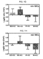
- Figure 1G shows the expression of mir-181b in log(2) ratio (of tumor to nontumor tissue) in HSC, DBE, HP, and MH-HCC cells as determined by RT-PCR.
- Figure 1H shows the expression of mir-181c in log(2) ratio (of tumor to nontumor tissue) in HSC, DBE, HP, and MH-HCC cells as determined by RT-PCR.
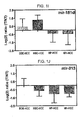
- Figure 1I shows the expression of mir-181d in log(2) ratio (of tumor to nontumor tissue) in HSC, DBE, HP, and MH-HCC cells as determined by RT-PCR.
- Figure 1J shows the expression of mir-213 in log(2) ratio (of tumor to nontumor tissue) in HSC, DBE, HP, and MH-HCC cells as determined by RT-PCR.
- Figure 2A shows a scatter plot of mir-181a1.
- Figure 2B shows a scatter plot of mir-181a2.
- Figure 2C shows a scatter plot of mir-181b1.
- Figure 2D shows a scatter plot of mir-181b2.
- Figure 2E shows a scatter plot of mir-181c.
- Figure 3A a graph showing the fold production of the mir-181a, mir-181b, mir-181c, and mir-181d at 0, 2, and 8 days in ESC media versus regular culture.
- Figure 3B is a graph showing the fold of the CAR and UGT2B7 at 0, 2, and 8 days in ESC media versus regular culture.
- Figure 3C a graph showing the fold production of CCND1 and TACSTD1 at 0, 2, and 8 days in ESC media versus regular culture.
- Figure 3D a graph showing the fold production of the mir-181a, mir-181b, mir-181c, and mir-181d at 0, 1, 2, and 8 days following withdrawal of ESC media.
- Figure 3E a graph showing the fold production of CAR and UGT2B7 at 0, 1, 2, and 8 days following withdrawal of ESC media.
- Figure 3F a graph showing the fold production of CCND1 and TCSTD1 at 0, 1, 2, and 8 days following withdrawal of ESC media.
- Figure 4 is a graph of the relative expression of mir-181b in pMSCV-hTR and pMSCV-mir-181b1 treated HuH1 cells.
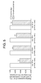
- Figure 5 is a graph of the relative expression of mir-181s in HuH7 cells transfected with 2'-O-methyl antisense versus control.
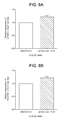
- Figure 6A is a graph of the relative expression of CCND1 in pMSCV-hTR and pMSCV-mir-181b1 treated HuH1 cells.
- Figure 6B is a graph of the relative expression of TACTD1 in pMSCV-hTR and p-MSCV-mir-181b1 treated HuH1 cells.
- Figure 6C is a graph of the relative expression of DKK1 in pMSCV-hTR and pMSCV-mir-181b1 treated HuH1 cells.
- Figure 6D is a graph of the relative expression of CCND1 in control and antisense treated HuH7 cells.
- Figure 6E is a graph of the relative expression of TACSTD1 in control and antisense treated HuH7 cells.
- Figure 6F is a graph of the relative expression of DKK1 in control and antisense treated HuH7 cells.
- Figure 7A shows the predicted binding site of mir-181a, mir-181b, mir-181c, and mir-181d at the 611-632 3'-UTR of DKK1.
- Figure 7B shows predicted binding sites of mir-181a, mir-181b, mir-181c, and mir-181d at the 771-799 3'-UTR of DKK1.
- Figure 8A is a predicted TCF-4 binding site for mir-181a1 and mir-181b1.
- Figure 8B is a predicted TCF-4 binding site for mir-181a2 and mir-181b2.
- Figure 8C is a predicted TCF-4 binding site for mir-181c and mir-181d.
- Figure 8D is another predicted TCF-4 binding site for mir-181c and mir-181d.
- Figure 9 is a graph of the fold of mir-181a, mir-181b, mir-181c, and mir-181d in each cell line (Hep3b type B (HSC-HCC), MHCC97 type C (HP-HCC), Smmc7721 type D (MH-HCC)) versus primary hepatocytes.
- HSC-HCC Hep3b type B
- HP-HCC MHCC97 type C
- Smmc7721 type D MH-HCC
- Figure 10 is a graph of the number of miRNAs with increased and decreased expression in HSC-HCC, BDE-HCC, HP-HCC, and MH-HCC subtypes.
- Micro RNAs are small non-coding RNA gene products (e.g., ⁇ 22 nt) that exist in many organisms and play key regulatory roles in mRNA translation and degradation by base pairing to partially complementary sites of the mRNA, predominantly in the 3' untranslated region.
- Lee Science, 294(5543):862-864 (2001 ); Lau, Science, 294(5543):858-862 (2001 ); Lagos, Science, 294(5543):853-858 (2001 ).
- miRNAs are expressed as long precursor RNAs that are processed by Drosha, a cellular nuclease, and subsequently transported to the cytoplasm by an Exportin-5-dependent mechanism.
- miRNAs are then cleaved by the DICER enzyme, resulting in approximately 17-24 nt miRNAs that associate with a RNA-induced silencing-like complex.
- Lee EMBO J, 21(17):4663-4670 (2002 ); Hutvagner, Science, 297(5589):2056-2060 (2002 ).
- the invention is predicated on the finding miRNA biomarkers are associated with HCC subtypes.
- the HCC subtypes refer to hepatic stem cell-like HCC (HSC-HCC), which is epithelial cell adhesion molecule (EpCAM)+ alpha-fetoprotein (AFP)+; bile duct epithelium-like HCC (BDE-HCC), which is EpCAM+ AFP-; hepatocytic progenitor-like HCC (HP-HCC), which is EpCAM- AFP+; and mature hepatocyte-like HCC (MH-HCC), which is EpCAM- AFP-.
- HCC-HCC hepatic stem cell-like HCC
- EpCAM epithelial cell adhesion molecule
- AFP alpha-fetoprotein
- BDE-HCC bile duct epithelium-like HCC
- HP-HCC hepatocytic progenitor-like HCC
- the invention provides a method of determining an HCC subtype in a subject comprising a) obtaining a sample from the subject, b) analyzing the sample for the expression of 1 or more biomarkers, and c) correlating the expression of the 1 or more biomarkers with the subtype of HCC in the subject.
- the expression of the biomarkers may be decreased or increased relative to normal control.
- the biomarkers are identified by SEQ ID NOs: 1-39 (see Table 1). In the inventive method, it is preferred that 2 or more, 5 or more, 10 or more, 15 or more, 20 or more, 25 or more, 30 or more, or 35 or more biomarkers are analyzed. More preferably, all 39 biomarkers are analyzed.
- the biomarkers identified by SEQ ID NOs: 1-19 are analyzed.
- the biomarkers identified by SEQ ID NOs: 2, 9-17, and 19-35 are analyzed.
- the biomarkers identified by SEQ ID NOs: 1-8, 11-13, 17-18, 23, 28-29, and 33-39 are analyzed.
- the biomarkers identified by SEQ ID NOs: 1, 8-12, 14-17, and 19-39 are analyzed.
- HCC stem cells are associated with (i.e., they express) the mir-181 family of miRNA biomarkers, particularly, mir-181a1, mir-181a2, mir-181b1, mir-181b2, and mir-181c, and that presence of HCC stem cells in a sample are indicative of the HSC-HCC subtype, which is associated with poor prognosis.
- the invention provides a method of detecting the presence of HCC stem cells in a sample comprising a) obtaining a sample, b) assaying the sample to detect the presence of a mir-181 biomarker, and c) correlating the presence or absence of the mir-181 biomarker with the presence or absence of the HCC stem cell in the sample.
- EpCAM+AFP+HCC stem cells may be detected by any suitable methods, e.g., immunofluorescence, immunohistochemistry, frozen activator cell sorting, side population methods, cell surface marker detection methods or in situ hybridization.
- the cell-permeable DNA-binding dye Hoechst 33342 is loaded into the cell population of interest; stem cells and early progenitors subsequently pump this dye out via an ATP-binding cassette membrane pump-dependent mechanism, resulting in a low-fluorescence "tail" when the cells are analyzed by flow cytometry.
- the method further comprises correlating the presence of the HCC stem cell in the sample with presence of HSC-HCC subtype in the sample.
- the detection of HCC stem cells in a sample may allow for earlier detection of the HSC-HCC subtype in a subject and thus lead to a greater likelihood of successful treatment and survival.
- biomarkers is used interchangeably with “miRNA” and refers to those biomarkers associated with HCC, which include at least the 39 biomarkers in Table 1.
- some (i.e., 1, 2, 3, 4, 5, 7, 7, 8, 9, 10, 15, 20, 25, 30, or 35) or all 39 of the biomarkers may be detected.
- at least 2 or more, more preferably at least 5 or more biomarkers are detected.
- the biomarker may be one or more of mir-181a1, mir-181a2, mir-181b1, mir-181b2, and mir-181c, preferably.
- some (i.e., 1, 2, 3, or 4) or all 5 of the mir-181 biomarkers are detected.
- RNA molecules are then separated by gel electrophoresis on agarose gels according to standard techniques, and transferred to nitrocellulose filters by, e.g., the so-called "Northern" blotting technique.
- the RNA is then immobilized on the filters by heating. Detection and quantification of specific RNA is accomplished using appropriately labeled DNA or RNA probes complementary to the RNA in question. See, for example, Molecular Cloning: A Laboratory Manual, J. Sambrook et al., eds., 2nd edition, Cold Spring Harbor Laboratory Press, 1989, Chapter 7 , the entire disclosure of which is incorporated by reference.
- the nucleic acid probe can be labeled with, e.g., a radionuclide such as 3 H, 32 P, 33 P, 14 C, or 35 S; a heavy metal; or a ligand capable of functioning as a specific binding pair member for a labeled ligand (e.g., biotin, avidin or an antibody), a fluorescent molecule, a chemiluminescent molecule, an enzyme or the like.
- a radionuclide such as 3 H, 32 P, 33 P, 14 C, or 35 S
- a heavy metal e.g., a ligand capable of functioning as a specific binding pair member for a labeled ligand (e.g., biotin, avidin or an antibody), a fluorescent molecule, a chemiluminescent molecule, an enzyme or the like.
- Probes can be labeled to high specific activity by either the nick translation method of Rigby et al, J. Mol. Biol., 113:237-251(1977 ) or by the random priming method of Fienberg, Anal. Biochem., 132:6-13 (1983 ), the entire disclosures of which are herein incorporated by reference.
- the latter can be a method for synthesizing 32 P-labeled probes of high specific activity from RNA templates. For example, by replacing preexisting nucleotides with highly radioactive nucleotides according to the nick translation method, it is possible to prepare 32 P-labeled nucleic acid probes with a specific activity well in excess of 10 8 cpm/microgram.
- Autoradiographic detection of hybridization can then be performed by exposing hybridized filters to photographic film. Densitometric scanning of the photographic films exposed by the hybridized filters provides an accurate measurement of biomarker levels. Using another approach, biomarker levels can be quantified by computerized imaging systems, such the Molecular Dynamics 400-B 2D Phosphorimager (Amersham Biosciences, Piscataway, N.J).
- the random-primer method can be used to incorporate an analogue, for example, the dTTP analogue 5-(N-(N-biotinyl-epsilon-aminocaproyl)-3-aminoallyl)deoxyuridine triphosphate, into the probe molecule.
- analogue for example, the dTTP analogue 5-(N-(N-biotinyl-epsilon-aminocaproyl)-3-aminoallyl)deoxyuridine triphosphate
- the biotinylated probe oligonucleotide can be detected by reaction with biotin-binding proteins, such as avidin, streptavidin, and antibodies (e.g., anti-biotin antibodies) coupled to fluorescent dyes or enzymes that produce color reactions.
- determining the levels of RNA expression can be accomplished using the technique of in situ hybridization.
- This technique requires fewer cells than the Northern blotting technique, and involves depositing whole cells onto a microscope cover slip and probing the nucleic acid content of the cell with a solution containing radioactive or otherwise labeled nucleic acid (e.g., cDNA or RNA) probes.
- This technique is particularly well-suited for analyzing tissue biopsy samples from subjects.
- the practice of the in situ hybridization technique is described in more detail in U.S. Patent No. 5,427,916 , the entire disclosure of which is incorporated herein by reference.
- the relative number of mi-RNAs in a sample can also be determined by reverse transcription, followed by amplification of the reverse-transcribed transcripts by polymerase chain reaction (RT-PCR).
- RT-PCR polymerase chain reaction
- the levels of RNA transcripts can be quantified in comparison with an internal standard, for example, the level of mRNA from a standard gene present in the same sample.
- a suitable gene for use as an internal standard includes, e.g., myosin or glyceraldehyde-3-phosphate dehydrogenase (G3PDH).
- G3PDH glyceraldehyde-3-phosphate dehydrogenase
- an oligolibrary in microchip format may be constructed containing a set of probe oligonucleotides specific for a set of biomarker genes.
- the oligolibrary may contain probes corresponding to all known biomarkers from the human genome.
- the microchip oligolibrary may be expanded to include additional miRNAs as they are discovered.
- the microchip is prepared from gene-specific oligonucleotide probes generated from known miRNAs.
- the array may contain two different oligonucleotide probes for each miRNA, one containing the active sequence and the other being specific for the precursor of the miRNA.
- the array may also contain controls such as one or more mouse sequences differing from human orthologs by only a few bases, which can serve as controls for hybridization stringency conditions.
- tRNAs from both species may also be printed on the microchip, providing an internal, relatively stable positive control for specific hybridization.
- One or more appropriate controls for non-specific hybridization may also be included on the microchip. For this purpose, sequences are selected based upon the absence of any homology with any known miRNAs.
- the microchip may be fabricated by techniques known in the art. For example, probe oligonucleotides of an appropriate length, e.g., 20 nucleotides, are 5'-amine modified at position C6 and printed using suitable available microarray systems, e.g., the GENEMACHINE OmniGrid 100 Microarrayer and Amersham CODELINK activated slides. Labeled cDNA oligomer corresponding to the target RNAs is prepared by reverse transcribing the target RNA with labeled primer. Following first strand synthesis, the RNA/DNA hybrids are denatured to degrade the RNA templates. The labeled target cDNAs thus prepared are then hybridized to the microarray chip under hybridizing conditions, e.g.
- the labeled cDNA oligomer is a biotin-labeled cDNA, prepared from a biotin-labeled primer.
- microarray is then processed by direct detection of the biotin-containing transcripts using, e.g., Streptavidin-Alexa647 conjugate, and scanned utilizing conventional scanning methods. Image intensities of each spot on the array are proportional to the abundance of the corresponding biomarker in the subject sample.
- the use of the array has one or more advantages for miRNA expression detection.
- the relatively limited number of miRNAs allows the construction of a common microarray for several species, with distinct oligonucleotide probes for each. Such a tool would allow for analysis of trans-species expression for each known biomarker under various conditions.
- the subject may be a human or animal presenting with symptoms of HCC.
- the subject is a human.
- the subject may or may not also have hepatitis B virus or cirrhosis (such as alcohol induced, primary biliary cirrhosis, genetic haemchromatosis, autoimmune hepatitis, primary sclerosing cholangitis).
- the HCC may be a solitary tumor, multinodular tumor, and/or a metastatic lesion.
- the sample obtained from the subject may be liver tissue, which can be tumor tissue or normal tissue.
- the sample may be from the subject's serum or plasma, frozen biopsy tissue, paraffin embedded biopsy tissue, and combinations thereof.
- the invention further provides a method for determining the prognosis of a subject by determining whether the subject has the HSC HCC, BDE-HCC, HP-HCC, or MH-HCC subtype.
- the inventive method of prognosis may be utilized in lieu of current methods of prognosis.
- the inventive method may be utilized in conjunction with conventional methods of prognosis.
- the traditional prognostic approaches may include spiral computed tomography (CT) of the liver and thorax, magnetic resonance imaging (MRI) with contrast enhancement or angiography with lipiodol injection, and biopsy, as well as current staging systems.
- the method further provides a treatment regimen that may be devised for the subject on the basis of the HCC subtype in the subject.
- the inventive method allows for a more personalized approach to medicine as the aggressiveness of treatment may be tailored to the subtype of HCC in the subject.
- the invention takes advantage of the association between the biomarkers and the HCC subtypes. Accordingly, the invention provides methods of treatment comprising administering a therapeutically effective amount of a composition comprising a reagent comprising nucleic acid complementary to at least one of the biomarkers associated with HSC-HCC, BDE-HCC, HP-HCC, or MH-HCC.
- the invention takes advantage of the association between the mir-181 biomarkers and HCC stem cells in order to determine the HCC subtype in a subject and, optionally, correlate the HCC-subtype in the patient with a prognosis.
- the mir-181 biomarkers are associated with the hepatic stem cell-like (HSC) HCC subtype, which is EpCAM and AFP positive.
- HSC hepatic stem cell-like
- EpCAM is a transmembrane protein containing three extracellular domains and one cytoplasmic domain. The function of EpCAM and the regulatory mechanism of its expression are largely unknown but are thought to involve cell-cell adhesion ( Winter, Exp. Cell. Res., 285(1): 50-58 (2003 )). EpCAM and AFP are not expressed in mature liver tissue.
- the HSC HCC subtype typically has a poor prognosis and survival outcome ( Lee, Hepatology, 40(3): 667-676 (2004 ); Lee, Nat. Med., 12(4): 410-416 (2006 )). Accordingly, the invention provides a method of determining whether the HCC detected is the HSC HCC subtype. The determination of the HCC subtype is particularly useful in determining the appropriate treatment for the subject, particularly because the EPCAM+ AFP+ HCC is associated with Wnt-ß-catenin signaling. Wnt-ß-catenin signaling is critical for maintaining the function of stem cells and abnormal activation has been linked to many human cancers, including HCC.
- the mir-181s can contribute Wnt-ß-catenin signaling activation, possibly through Dickkoph-1 (i.e., DKK1) and nemo-like kinase (i.e., NLK), which are inhibitors of the Wnt-ß-catenin pathway.
- the invention takes advantage of the regulatory link between mir-181s and HCC stem cells, and provides methods of prognosis, and treatment based thereon.
- Treatment options may include traditional treatments as well as gene therapy approaches that specifically target the miRNAs described herein.
- Traditional treatment of HCC includes, for example, percutaneous ethanol injection (PEI), radiofrequency ablation, chemoembolisation, and chemotherapy. Treatment is determined based on the status of the subject and guidelines are known in the art. (See for example, Ryder, Gut, 52: 1-8 (2003 )).
- the invention further provides pharmaceutical compositions for use in the inventive treatment methods.
- the invention provides a composition comprising a therapeutically effective amount of a reagent comprising a nucleic acid or nucleic acids complementary to at least one, preferably at least two of the biomarkers selected from those identified by SEQ ID NOs: 1-39 and a pharmaceutically acceptable carrier.
- the reagent may comprise nucleic acids complementary to at least 5 or more, 10 or more, 15 or more, 20 or more, 25 or more, 30 or more, or 35 or more of the biomarkers.
- the reagent may comprise only the nucleic acids or the nucleic acids in combination with delivery reagents such as recombinant plasmids, viral vectors, liposomes, etc.
- the composition comprises nucleic acids complementary to the biomarkers identified by SEQ ID NOs: 1-19, even more preferably, the composition comprises nucleic acids complementary to mir-181a1, mir-181a2, mir-181b1, mir-181b2, and mir-181c, and a pharmaceutically acceptable carrier.
- the composition comprises nucleic acids complementary to at least one, preferably at least 2 biomarkers identified by SEQ ID NOs: 2, 9-17, and 19-35, and a pharmaceutically acceptable carrier.
- the composition comprises nucleic acids complementary to at least one, preferably at least two biomarkers identified by SEQ ID NOs: 1-8,11-13, 17-18, 23, 28, 29, and 33-39, and a pharmaceutically acceptable carrier.
- the composition comprises nucleic acids complementary to at least one, preferably to at least two biomarkers identified by SEQ ID NOs: 1, 8-12, 14-17, and 19-39, and a pharmaceutically acceptable carrier.
- composition may bind and/or render ineffective (i.e., inhibit) the biomarkers, or alternatively, alter the expression of the gene coding for the biomarkers, thereby altering the amounts or levels of biomarkers produced, the technology for which are well known within the art.
- an effective amount of at least one composition which inhibits at least one of the biomarkers can also be administered to the subject.
- inhibiting means that the biomarker levels and/or production of biomarker gene product from the corresponding gene in the cancer cell after treatment is less than the amount produced prior to treatment.
- a composition that increases the expression of one or more of the biomarkers may be administered.
- One skilled in the art can readily determine whether biomarker levels or gene expression has been inhibited or increased in a cancer cell, using for example the techniques for determining biomarker transcript level discussed above.
- an "effective amount" of a composition that inhibits the biomarkers or biomarker gene expression is an amount sufficient to inhibit proliferation of a cancer cell in a subject suffering from HCC.
- an effective amount of an inhibiting composition to be administered to a given subject by taking into account factors such as the size and weight of the subject; the extent of disease penetration; the age, health and sex of the subject; the route of administration; and whether the administration is regional or systemic.
- an effective amount of the expression-altering composition can be based on the approximate weight of a tumor mass to be treated.
- the approximate weight of a tumor mass can be determined by calculating the approximate volume of the mass, wherein one cubic centimeter of volume is roughly equivalent to one gram. Therefore, in one embodiment, an effective amount based on the weight of a tumor mass can utilized.
- an effective amount of the composition can be based on the approximate or estimated body weight of a subject to be treated. Preferably, such effective amounts are administered parenterally or enterally.
- the composition can be administered to the subject once (e.g. as a single injection or deposition).
- the composition can be administered once or twice daily to a subject for a period of from about three to about twenty-eight days, more preferably from about seven to about ten days.
- the composition may be administered once a day for seven days.
- a dosage regimen comprises multiple administrations, it is understood that the effective amount of the composition administered to the subject can comprise the total amount of composition administered over the entire dosage regimen.
- Suitable compositions for inhibiting biomarker gene expression include double-stranded RNA (such as short- or small-interfering RNA or "siRNA”), antisense nucleic acids, and enzymatic RNA molecules such as ribozymes. Each of these compositions can be targeted to a given biomarker gene product and destroy or induce the destruction of the target biomarker gene product.
- siRNA short- or small-interfering RNA or "siRNA”
- antisense nucleic acids such as ribozymes.
- enzymatic RNA molecules such as ribozymes.
- RNA interference of the biomarker gene can be inhibited by inducing RNA interference of the biomarker gene with an isolated double-stranded RNA ("dsRNA") molecule which has at least 90%, for example 95%, 98%, 99% or 100%, sequence homology with at least a portion of the biomarker gene product.
- dsRNA isolated double-stranded RNA
- the dsRNA molecule is a "short or small interfering RNA" or "siRNA.”
- siRNA useful in the present methods comprise short double-stranded RNA from about 17 nucleotides to about 29 nucleotides in length, preferably from about 19 to about 25 nucleotides in length.
- the siRNA comprise a sense RNA strand and a complementary antisense RNA strand annealed together by standard Watson-Crick base-pairing interactions (hereinafter "base-paired").
- the sense strand comprises a nucleic acid sequence which is substantially identical to a nucleic acid sequence contained within the target biomarker gene product.
- the siRNA is "substantially identical" to a target sequence contained within the target nucleic sequence, is a nucleic acid sequence that is identical to the target sequence, or that differs from the target sequence by one or two nucleotides.
- the sense and antisense strands of the siRNA can comprise two complementary, single-stranded RNA molecules, or can comprise a single molecule in which two complementary portions are base-paired and are covalently linked by a single-stranded "hairpin" area.
- the siRNA can also be an altered RNA that differs from naturally-occurring RNA by the addition, deletion, substitution and/or alteration of one or more nucleotides.
- Such alterations can include addition of non-nucleotide material, such as to the end(s) of the siRNA or to one or more internal nucleotides of the siRNA, or modifications that make the siRNA resistant to nuclease digestion, or the substitution of one or more nucleotides in the siRNA with deoxyribonucleotides.
- the siRNA can also comprise a 3' overhang.
- a "3' overhang” refers to at least one unpaired nucleotide extending from the 3'-end of a duplexed RNA strand.
- the siRNA comprises at least one 3' overhang of from 1 to about 6 nucleotides (which includes ribonucleotides or deoxyribonucleotides) in length, preferably from 1 to about 5 nucleotides in length, more preferably from 1 to about 4 nucleotides in length, and particularly preferably from about 2 to about 4 nucleotides in length.
- the 3' overhang is present on both strands of the siRNA, and is 2 nucleotides in length.
- each strand of the siRNA can comprise 3' overhangs of dithymidylic acid ("TT") or diuridylic acid (“uu").
- the siRNA can be produced chemically or biologically, or can be expressed from a recombinant plasmid or viral vector for the isolated biomarker gene products.
- Exemplary methods for producing and testing dsRNA or siRNA molecules are described in U.S. Published Patent Application No. 2002/0173478 and U.S. Patent No. 7,148,342 , the entire disclosures of which are herein incorporated by reference.
- an antisense nucleic acid refers to a nucleic acid molecule that binds to target RNA by means of RNA-RNA or RNA-DNA or RNA-peptide nucleic acid interactions, which alters the activity of the target RNA.
- Antisense nucleic acids suitable for use in the present methods are single-stranded nucleic acids (e.g., RNA, DNA, RNA-DNA chimeras, PNA) that generally comprise a nucleic acid sequence complementary to a contiguous nucleic acid sequence in a biomarker gene product.
- the antisense nucleic acid comprises a nucleic acid sequence that is 50-100% complementary, more preferably 75-100% complementary, and most preferably 95-100% complementary to a contiguous nucleic acid sequence in an biomarker gene product.
- Antisense nucleic acids can also contain modifications to the nucleic acid backbone or to the sugar and base moieties (or their equivalent) to enhance target specificity, nuclease resistance, delivery or other properties related to efficacy of the molecule. Such modifications include cholesterol moieties, duplex intercalators such as acridine or the inclusion of one or more nuclease-resistant groups.
- Antisense nucleic acids can be produced chemically or biologically, or can be expressed from a recombinant plasmid or viral vector, as described above for the isolated biomarker gene products. Exemplary methods for producing and testing are within the skill in the art; see, e.g., Stein, Science, 261:1004 (1993 ) and U.S. Patent No. 5,849,902 to Woolf et al. , the entire disclosures of which are herein incorporated by reference.
- an "enzymatic nucleic acid” refers to a nucleic acid comprising a substrate binding region that has complementarity to a contiguous nucleic acid sequence of a biomarker gene product, and which is able to specifically cleave the biomarker gene product.
- the enzymatic nucleic acid substrate binding region is 50-100% complementary, more preferably 75-100% complementary, and most preferably 95-100% complementary to a contiguous nucleic acid sequence in a biomarker gene product.
- the enzymatic nucleic acids can also comprise modifications at the base, sugar, and/or phosphate groups.
- An exemplary enzymatic nucleic acid for use in the present methods is a ribozyme.
- the enzymatic nucleic acids can be produced chemically or biologically, or can be expressed from a recombinant plasmid or viral vector, as described above for the isolated biomarker gene products.
- Exemplary methods for producing and testing dsRNA or siRNA molecules are described in Werner, Nucl. Acids Res., 23:2092-96 (1995 ); Hammann, Antisense and Nucleic Acid Drug Dev., 9:25-31 (1999 ); and U.S. Patent No. 4,987,071 , the entire disclosures of which are herein incorporated by reference.
- Administration of at least one composition for inhibiting at least one biomarker or expression of a biomarker gene will inhibit the proliferation of cancer cells in a subject who has HCC.
- to "inhibit the proliferation of a cancer cell” means to kill the cell, or permanently or temporarily arrest or slow the growth of the cell.
- Inhibition of cancer cell proliferation can be inferred if the number of such cells in the subject remains constant or decreases after administration of the inventive composition.
- An inhibition of cancer cell proliferation can also be inferred if the absolute number of such cells increases, but the rate of tumor growth decreases.
- the number of cancer cells in a subject's body can be determined by direct measurement, or by estimation from the size of primary or metastatic tumor masses.
- the number of cancer cells in a subject can be measured by immunohistological methods, flow cytometry, or other techniques designed to detect characteristic surface markers of cancer cells.
- the size of a tumor mass can be ascertained by direct visual observation, or by diagnostic imaging methods, such as X-ray, magnetic resonance imaging, ultrasound, and scintigraphy. Diagnostic imaging methods used to ascertain size of the tumor mass can be employed with or without contrast agents, as is known in the art.
- the size of a tumor mass can also be ascertained by physical means, such as palpation of the tissue mass or measurement of the tissue mass with a measuring instrument, such as a caliper.
- compositions can be administered to a subject by any method suitable for delivering these compositions to the cancer cells of the subject.
- the compositions can be administered by methods suitable to transfect cells of the subject with these compositions.
- the cells are transfected with a plasmid or viral vector comprising sequences encoding at least one biomarker gene product or biomarker gene expression inhibiting composition.
- Transfection methods for eukaryotic cells include, e.g., direct injection of the nucleic acid into the nucleus or pronucleus of a cell; electroporation; liposome transfer or transfer mediated by lipophilic materials; receptor mediated nucleic acid delivery, bioballistic or particle acceleration; calcium phosphate precipitation, and transfection mediated by viral vectors.
- cells can be transfected with a liposomal transfer composition, e.g., DOTAP (N-[1-(2,3-dioleoyloxy)propyl]-N,N,N-trimethyl-ammonium methylsulfate, Boehringer-Mannheim) or an equivalent, such as LIPOFECTIN.
- DOTAP N-[1-(2,3-dioleoyloxy)propyl]-N,N,N-trimethyl-ammonium methylsulfate, Boehringer-Mannheim
- LIPOFECTIN LIPOFECTIN
- the composition can also be administered to a subject by any suitable enteral or parenteral administration route.
- Suitable enteral administration routes for the present methods include, e.g., oral, rectal, or intranasal delivery.
- Suitable parenteral administration routes include, e.g., intravascular administration (e.g., intravenous bolus injection, intravenous infusion, intra-arterial bolus injection, intra-arterial infusion and catheter instillation into the vasculature); peri- and intra-tissue injection (e.g., peri-tumoral and intra-tumoral injection, intra-retinal injection, or subretinal injection); subcutaneous injection or deposition, including subcutaneous infusion (such as by osmotic pumps); direct application to the tissue of interest, for example by a catheter or other placement device (e.g., a retinal pellet or a suppository or an implant comprising a porous, non-porous, or gelatinous material); and inhalation.
- the composition can be administered to the subject either as naked RNA, in combination with a delivery reagent, or as a nucleic acid (e.g., a recombinant plasmid or viral vector) comprising sequences that express the biomarker gene product or expression inhibiting composition.
- a delivery reagent e.g., the Mirus Transit TKO lipophilic reagent; lipofectin; lipofectamine; cellfectin; polycations (e.g., polylysine), and liposomes.
- Recombinant plasmids and viral vectors comprising sequences that express the biomarker or biomarker gene expression inhibiting compositions, and techniques for delivering such plasmids and vectors to cancer cells, are discussed above.
- liposomes are used to deliver a biomarker or biomarker gene expression-inhibiting composition (or nucleic acids comprising sequences encoding them) to a subject. Liposomes can also increase the blood half-life of the gene products or nucleic acids.
- Liposomes suitable for use in the invention can be formed from standard vesicle-forming lipids, which generally include neutral or negatively charged phospholipids and a sterol, such as cholesterol.
- the selection of lipids is generally guided by consideration of factors such as the desired liposome size and half-life of the liposomes in the blood stream.
- a variety of methods are known for preparing liposomes, for example, as described in Szoka, Ann. Rev. Biophys. Bioeng., 9:467 (1980 ); and U.S. Patent Nos. 4,235,871 , 4,501,728 , 4,837,028 , and 5,019,369 , the entire disclosures of which are herein incorporated by reference.
- the liposomes for use in the present methods can comprise a ligand molecule that targets the liposome to cancer cells.
- Ligands which bind to receptors prevalent in cancer cells such as monoclonal antibodies that bind to tumor cell antigens, are preferred.
- compositions of the present invention may include a pharmaceutically acceptable carrier.
- pharmaceutically-acceptable carrier means one or more compatible solid or liquid fillers, diluents, other excipients, or encapsulating substances which are suitable for administration into a human or veterinary patient.
- carrier denotes an organic or inorganic ingredient, natural or synthetic, with which the active ingredient is combined to facilitate the application.
- the components of the pharmaceutical compositions also are capable of being co-mingled with the molecules of the present invention, and with each other, in a manner so as not to substantially impair the desired pharmaceutical efficacy.
- “Pharmaceutically acceptable” materials are capable of administration to a patient without the production of undesirable physiological effects such as nausea, dizziness, rash, or gastric upset. It is, for example, desirable for a therapeutic composition comprising pharmaceutically acceptable excipients not to be immunogenic when administered to a human patient for therapeutic purposes.
- the pharmaceutical compositions may contain suitable buffering agents, including: acetic acid in a salt; citric acid in a salt; boric acid in a salt; and phosphoric acid in a salt.
- suitable buffering agents including: acetic acid in a salt; citric acid in a salt; boric acid in a salt; and phosphoric acid in a salt.
- suitable preservatives such as: benzalkonium chloride, chlorobutanol, parabens and thimerosal.
- compositions may conveniently be presented in unit dosage form and may be prepared by any of the methods well known in the art of pharmacy. All methods include the step of bringing the active agent into association with a carrier that constitutes one or more accessory ingredients. In general, the compositions are prepared by uniformly and intimately bringing the active composition into association with a liquid carrier, a finely divided solid carrier, or both, and then, if necessary, shaping the product.
- compositions suitable for parenteral administration conveniently comprise a sterile aqueous preparation of the inventive composition, which is preferably isotonic with the blood of the recipient.
- This aqueous preparation may be formulated according to known methods using suitable dispersing or wetting agents and suspending agents.
- the sterile injectable preparation also may be a sterile injectable solution or suspension in a non-toxic parenterally-acceptable diluent or solvent, for example, as a solution in 1, 3-butane diol.
- acceptable vehicles and solvents that may be employed are water, Ringer's solution, and isotonic sodium chloride solution.
- sterile, fixed oils are conventionally employed as a solvent or suspending medium.
- any bland fixed oil may be employed including synthetic mono-or di-glycerides.
- fatty acids such as oleic acid may be used in the preparation of injectables.
- Carrier formulation suitable for oral, subcutaneous, intravenous, intramuscular, etc. administrations can be found in Remington's Pharmaceutical Sciences, Mack Publishing Co., Easton, PA which is incorporated herein in its entirety by reference thereto.
- the delivery systems of the invention are designed to include time-released, delayed release or sustained release delivery systems such that the delivering of the inventive composition occurs prior to, and with sufficient time, to cause sensitization of the site to be treated.
- inventive composition may be used in conjunction with other therapeutic agents or therapies. Such systems can avoid repeated administrations of the inventive composition, increasing convenience to the subject and the physician, and may be particularly suitable for certain compositions of the present invention.
- release delivery systems are available and known to those of ordinary skill in the art. They include polymer base systems such as poly(lactide-glycolide), copolyoxalates, polycaprolactones, polyesteramides, polyorthoesters, polyhydroxybutyric acid, and polyanhydrides. Microcapsules of the foregoing polymers containing drags are described in, for example, U.S. Patent No. 5,075,109 .
- Delivery systems also include non-polymer systems that are: lipids including sterols such as cholesterol, cholesterol esters and fatty acids or neutral fats such as mono-di-and tri-glycerides; hydrogel release systems; sylastic systems; peptide based systems; wax coatings; compressed tablets using conventional binders and excipients; partially fused implants; and the like.
- lipids including sterols such as cholesterol, cholesterol esters and fatty acids or neutral fats such as mono-di-and tri-glycerides
- hydrogel release systems such as cholesterol, cholesterol esters and fatty acids or neutral fats such as mono-di-and tri-glycerides
- sylastic systems such as cholesterol, cholesterol esters and fatty acids or neutral fats such as mono-di-and tri-glycerides
- peptide based systems such as fatty acids
- wax coatings such as those described in U.S. Patent Nos.
- the invention further provides a method of assessing the efficacy of treatment of HCC in a subject by determining whether there are any remaining HCC stem cells remaining in the liver of the subject following a course of treatment.
- a sample is obtained from the subject and assayed to detect the presence or absence of a mir-181 biomarker.
- the presence or absence of a mir-181 biomarker is then correlated with the presence or absence, respectively, of EpCAM+ AFP+ HCC in a subject. This information is used to determine whether treatment of the HCC in the subject has or has not been effective.
- Hepatic tissues were obtained with informed consent from subjects who underwent radical resection between 2002 and 2003 at the Liver Cancer Institute and Zhongshan Hospital (Fudan University, Shanghai, China). The study was approved by the Institutional Review Board of the Liver Cancer Institute and National Institutes of Health.
- the sample enrollment criteria included those with a history of HBV infection or HBV-related liver cirrhosis, HCC diagnosed by two independent pathologists, detailed information on clinical presentation and pathological characteristics, as well as detailed follow-up data for at least 3 years, which included intrahepatic recurrence, intrahepatic venous metastasis, lymph node involvement, extrahepatic metastasis, disease-free, overall survival, and cause of death.
- the updated TNM classification is superior to other staging systems, including CLIP and OKUDA, for HCC subjects who undergo resection and was therefore chosen to stratify early stage subjects (TNM stage I and II) for analysis of miRNA prediction capacity.
- Varotti Eur J. Surg Oncol, 31(7):760-767 (2005 ); Huang et al., J. Gastroenterol Hepatol, 20(5):765-771 (2005 ).
- a prospective study revealed that the BCLC system was superior to the new TNM classification system updated in 2002, therefore, Cox proportional hazards modeling based on early stage subjects categorized by BCLC (Stage 0 and A) was also performed.
- Gene expression profiles were conducted in primary HCC and corresponding noncancerous hepatic tissues from 244 Chinese HCC subjects.
- the testing cases included 43 multinodular and 67 solitary HCC. Of the 43 multinodular HCC cases, 18 developed intrahepatic recurrence and one developed extrahepatic metastatis in addition to an intrahepatic recurrence. Of the 67 solitary HCC cases, 4 subjects had a solitary tumor with an appearance of aggregated nodules, 10 developed intra- and/or extrahepatic metastasis while 49 developed intrahepatic recurrence confirmed at follow-up (3yr). In addition, eight normal liver tissues from disease-free subjects (described in Budhu, Cancer Cell, 10(2):99-111 (2006 )) were included as normal controls.
- RNA isolation and miRNA arrays were carried out as described in Ye, Nat Med, 9(4):416-423 (2003 ); Calin, N Engl J. Med, 353(17):1793-1802 (2005 ).
- RNA was isolated in a pairwise fashion from tumor or non-tumor tissue and samples were selected in random order for miRNA analysis to avoid grouping bias. A total of 488 microarrays were performed.
- the microarray platform (V 2.0) was composed of 250 non-redundant human and 200 mouse miRNAs. To examine the robustness of the miRNA microarray platform, miRNA was analyzed to determine whether expression could differentiate 244 tissues from their paired surrounding noncancerous hepatic tissues.
- Unsupervised hierarchical clustering analysis was performed by the GENESIS software version 1.5 developed by Alexander Sturn (IBMT-TUG, Graz, Austria).
- the BRB ArrayTools Software V3.3 was used for supervised analysis as previously described ( Ye, Nat Med, 9(4):416-423 (2003 ); Budhu, Cancer Cell, 10(2):99-111 (2006 )).
- the Kaplan-Meier survival analysis was used to compare subject survival based on prediction results, using Excel-based WinSTAT software.
- the statistical p value was generated by the Cox-Mantel log-rank test.
- Cox proportional hazards regression was used to analyze the effect of sixteen clinical variables on subject survival or recurrence using STATA 9.2 (College Station, TX).
- TargetScan analysis was based on a website tool developed by Ben Lewis ( Lewis, Cell, 120(1):15-20 (2005 )). Cox proportional hazards regression was used to analyze the effect of clinical variables on subject overall and relapse-free survival, including age, sex, HBV active status, pre-resection AFP, cirrhosis, alanine transferase (ALT), Child-Pugh score, tumor size, tumor encapsulation, nodular type, status of microvascular invasion, Edmondson grade, and several HCC prognosis staging systems, including BCLC staging ( Llovet, Semin Liver Dis, 19(3):329-338 (1999 )); CLIP classification (" The Cancer of the Liver Italian Program", Hepatology, 28(3):751-755 (1998 )), Okuda staging ( Okuda, Cancer, 56(4):918-928 (1985 )), and TNF classification ( American Joint Committee on Cancer (AJCC)/International Union against Cancer (UICC)'
- TACSTD1, BAMBI, DKK1, CCND1, CTNNB1, and MYC expression were measured in triplicate using Applied Biosystems 7700 Sequence Detection System (Foster City, CA). Probes used were: TACSTD1, Hs00158980_m1; CTNNB1, HS00170025_m1; BAMBI, HS00180818, DKK1, Hs00183740_m1, CCND1, Hs00277039_m1, CTNNB1, MYC, Hs00153408_m1; 18S, Hs999999901_s1 (Applied Biosystems). All procedures were performed according to manufacturer suggestion.
- Immunohistochemical Analysis was performed using Envision+ kits (DAKO USA, Carpinteria, CA) according to manufacturer instruction. Primary antibodies were used as follows: anti-ß-catenin monoclonal antibody clone 14 (BD Transduction Laboratories, San Jose, CA) and anti-EpCAM monoclonal antibody clone VU-1D9 (Oncogene Research Products, San Diego, CA).
- EMSA Recombinant Tcf-4 was expressed in E. coli as GST fusion protein and extracted. EMSA was performed using LightShift Chemiluminescent EMSA kit (Pierce, Rockford, IL) according to manufacturer instructions. Double-stranded DNA oligonucleotides containing the putative Tcf binding sites of EpCAM promoter and 10 adjacent nucleotides upstream and downstream were constructed and used as probes. Mutant TBE1 and TBE2 probes were also used.
- Known Hep3B type B, MHCC97 type C, Smmc7721 type D, HUH1 and HUH7 HCC cell lines were cultured routinely. Cells were transfected with pMSCV-mir-181b-1 for functional assays. HUH7 cells were also treated with 2'-O-methyl mir-181s antisense, an inhibitor of mir-181s.
- HCC tissue and surrounding non-HCC tissue samples Utilizing paired HCC tissue and surrounding non-HCC tissue samples from a total of 230 HCC patients, a total of 209 non-redundant miRNAs were found to provide 97% accuracy in correctly identifying the samples (multivariate p ⁇ 0.01). Heterogeneity of the samples was evident and the samples were clustered based on the four HCC subtypes (HSC, BDE, HP, and MH).
- mir-181s are associated with HSC-HCC and contribute to the function of liver cancer stem cells.
- HSC-HCC and BDE-HCC refer to HCCs with stem cell-like features and bile duct epithelium-like features, respectively.
- Mir-181 expression based on miRNA microarray analysis of miRNA precursors in each HCC subtype versus corresponding non-HCC tissues from 230 patients is shown in Figure 1A-E for mir-181a1, mir-181a2, mir-181b1, mir-181b2 and mir-181c, respectively.
- FIG. 1F-J shows RT-PCR analysis of all mature mir-181s s in 40 HCC and non-HCC sample pairs. Scatter plot analysis of pre-mir-181 s and mature mir-181 s is shown in Figure 2 , with r-values representing Spearman's correlation coefficient.
- mir-181 expression was positively correlated with Wnt- ⁇ -catenin signaling activation and negatively correlated with many mature hepatocyte genes in both clinical specimens and cultured HCC cell lines.
- Hierarchical clustering was conducted of 5 pre-mir-181s, 15 hepatocyte-specific genes, and 5 beta-catenin associated genes whose expression was significantly correlated with each other (p ⁇ 0.001) from correlation analysis between microarray data and mRNA array data.
- mir-181 expression was positively correlated with beta-catenin protein level ( Fig. 9 ).
- ESC culture media which is a basal medium optimized for growth of undifferentiated embryonic stem (ES) cells
- mir-181 and beta-catenin regulated genes was increased and the expression of hepatocyte-specific genes was decreased as analyzed by qRT-PCR ( Figs. 3A-C ) as well as immunoblotting using antibodies to beta-catenin and actin (as a control).
- qRT-PCR Figs. 3A-C
- Figs. 3D-F Gene expression was measured in triplicate and is shown as mean ⁇ SD.
- mir-181b was detected by RT-PCR and expression was compared to that ofpMSCV-hTR cells. Gene expression was measured in triplicate and is shown as mean ⁇ SD in Fig 4 . As shown, mir-181 was over expressed in the HuH1 cells.
- HuH7 cells were treated with 2'-O-methyl mir-181s antisense and the expression of all mir-181 s was subsequently detected.
- beta-catenin regulated genes (CCND1, TACSTD1, and DKK1) was detected by RT-PCR and compared to expression by pMSCV-hTR cells ( Figs. 6A-C ).
- Cell lysates of cell lines were also analyzed by immunoblots with antibodies to ⁇ -catenin and actin.
- beta-catenin regulated genes (CCND1, TACSTD1, and DKK1) was detected by RT-PCR and compared to the expression of pMSCV-hTR cells ( Figs. 6D-F ).
- Cell lysates of cell lines were also analyzed by immunoblots with antibodies to ⁇ -catenin and actin.
- Mir-181 s affect wnt-beta-catenin expression. It is possible that this occurs through a functional feedback link.
- DKK1 is an inhibitor of beta-catenin. Beta-catenin induces mir-181 as well as DKK1, which subsequently inhibits beta-catenin. It is thought that mir-181 acts to inhibit the inhibitory activity of DKK1.
- Predicted mir-181 s binding sites in DKK1 3'-UTR are shown in Fig. 7A-B .
- the BC001539, homo sapien dickkopf homolog 1 cDNA was used.
- Figure 7A shows the binding sites in the position of 611-632 of DKK1 3'-UTR.
- Figure 7B shows the predicted binding sites in the position of 771-799 of DKK1 3'-UTR.
- Figs. 8A-D The predicted transcription factor-4 (TCF-4) binding sites ((A/T)(A/T)CAAAG) OR (CTTTG(A/T)(A/T)) in mir-181s' promoters are shown in Figs. 8A-D . 6,060 base pairs were analyzed at the upstream of transcriptional start site.
- Figure 8A shows the promoter of mir-181a1 and mir-181b1 in Chromosome 1, for which the NW_926128, homo sapiens chromosome 1 genomic contig was used.
- Figure 8B shows the promoter of mir-181 a2 and mir-181b2 in Chromosome 9, for which the NT_008470 homo sapien chromosome 9 genomic contig was used.
- both EST genes are predicted in the region of mir-181c and mir-181d locating, which have different transcriptional start sites ( Figs. 8C-D ).
- the promoter of mir-181c and mir-181d in Chromosome 19 in Fig. 8C is the promoter from ENSESTT00000290819.
- the promoter of mir-181c and mir-181d in Chromosome 19 in Fig. 8D is the promoter from ENSESTT00000290818.
Abstract
Description
- Hepatocellular carcinoma (HCC) is the third leading cause of cancer death worldwide. HCC is very heterogeneous in terms of its clinical presentation and genomic and transcriptomic patterns. The heterogeneity in HCC and lack of appropriate biomarkers for its detection and subtype identification has hampered patient prognosis and treatment stratification.
- Accordingly, there is a desire for one or more biomarkers that can identify the subtype of HCC in a mammal, as well as methods of providing appropriate treatment based on the subtype of HCC.
- The invention provides a method of determining the subtype of HCC in a subject, the method comprising a) obtaining a sample from the subject, b) assaying the sample to detect at least 1 biomarkers, and c) correlating the biomarkers detected with an HCC subtype in the subject. In this regard, the biomarkers are selected from the group consisting of the biomarkers identified by SEQ ID NOs: 1-39.
- The invention also provides a method of detecting a HCC stem cell in a sample. In one embodiment the inventive method comprises a) obtaining a sample, b) assaying the sample to detect the presence of a mir-181 biomarker, and c) correlating the presence or absence of the mir-181 biomarker with the presence or absence of the HCC stem cell in the sample.
- The invention also provides methods and compositions for treating subjects with HCC that take advantage of the biomarkers associated with HCC stem cells.
-
Figure 1A shows the expression of mir-181a1 in log(2) ratio (of tumor to nontumor tissue) in HSC-HCC cells based on microRNA analysis. -
Figure 1B shows the expression of mir-181a2 in log(2) ratio (of tumor to nontumor tissue) in HSC, DBE, HP, and MH-HCC cells based on microRNA analysis. -
Figure 1C shows the expression of mir-181b1 in log(2) ratio (of tumor to nontumor tissue) in HSC, DBE, HP, and MH-HCC cells based on microRNA analysis. -
Figure 1D shows the expression of mir-181b2 in log(2) ratio (of tumor to nontumor tissue) in HSC, DBE, HP, and MH-HCC cells based on microRNA analysis. -
Figure 1E shows the expression of mir-181c in log(2) ratio (of tumor to nontumor tissue) in HSC, DBE, HP, and MH-HCC cells based on microRNA analysis. -
Figure 1F shows the expression of mir-181a in log(2) ratio (of tumor to nontumor tissue) in HSC, DBE, HP, and MH-HCC cells as determined by RT-PCR. -
Figure 1G shows the expression of mir-181b in log(2) ratio (of tumor to nontumor tissue) in HSC, DBE, HP, and MH-HCC cells as determined by RT-PCR. -
Figure 1H shows the expression of mir-181c in log(2) ratio (of tumor to nontumor tissue) in HSC, DBE, HP, and MH-HCC cells as determined by RT-PCR. -
Figure 1I shows the expression of mir-181d in log(2) ratio (of tumor to nontumor tissue) in HSC, DBE, HP, and MH-HCC cells as determined by RT-PCR. -
Figure 1J shows the expression of mir-213 in log(2) ratio (of tumor to nontumor tissue) in HSC, DBE, HP, and MH-HCC cells as determined by RT-PCR. -
Figure 2A shows a scatter plot of mir-181a1. -
Figure 2B shows a scatter plot of mir-181a2. -
Figure 2C shows a scatter plot of mir-181b1. -
Figure 2D shows a scatter plot of mir-181b2. -
Figure 2E shows a scatter plot of mir-181c. -
Figure 3A a graph showing the fold production of the mir-181a, mir-181b, mir-181c, and mir-181d at 0, 2, and 8 days in ESC media versus regular culture. -
Figure 3B is a graph showing the fold of the CAR and UGT2B7 at 0, 2, and 8 days in ESC media versus regular culture. -
Figure 3C a graph showing the fold production of CCND1 and TACSTD1 at 0, 2, and 8 days in ESC media versus regular culture. -
Figure 3D a graph showing the fold production of the mir-181a, mir-181b, mir-181c, and mir-181d at 0, 1, 2, and 8 days following withdrawal of ESC media. -
Figure 3E a graph showing the fold production of CAR and UGT2B7 at 0, 1, 2, and 8 days following withdrawal of ESC media. -
Figure 3F a graph showing the fold production of CCND1 and TCSTD1 at 0, 1, 2, and 8 days following withdrawal of ESC media. -
Figure 4 is a graph of the relative expression of mir-181b in pMSCV-hTR and pMSCV-mir-181b1 treated HuH1 cells. -
Figure 5 is a graph of the relative expression of mir-181s in HuH7 cells transfected with 2'-O-methyl antisense versus control. -
Figure 6A is a graph of the relative expression of CCND1 in pMSCV-hTR and pMSCV-mir-181b1 treated HuH1 cells. -
Figure 6B is a graph of the relative expression of TACTD1 in pMSCV-hTR and p-MSCV-mir-181b1 treated HuH1 cells. -
Figure 6C is a graph of the relative expression of DKK1 in pMSCV-hTR and pMSCV-mir-181b1 treated HuH1 cells. -
Figure 6D is a graph of the relative expression of CCND1 in control and antisense treated HuH7 cells. -
Figure 6E is a graph of the relative expression of TACSTD1 in control and antisense treated HuH7 cells. -
Figure 6F is a graph of the relative expression of DKK1 in control and antisense treated HuH7 cells. -
Figure 7A shows the predicted binding site of mir-181a, mir-181b, mir-181c, and mir-181d at the 611-632 3'-UTR of DKK1. -
Figure 7B shows predicted binding sites of mir-181a, mir-181b, mir-181c, and mir-181d at the 771-799 3'-UTR of DKK1. -
Figure 8A is a predicted TCF-4 binding site for mir-181a1 and mir-181b1. -
Figure 8B is a predicted TCF-4 binding site for mir-181a2 and mir-181b2. -
Figure 8C is a predicted TCF-4 binding site for mir-181c and mir-181d. -
Figure 8D is another predicted TCF-4 binding site for mir-181c and mir-181d. -
Figure 9 is a graph of the fold of mir-181a, mir-181b, mir-181c, and mir-181d in each cell line (Hep3b type B (HSC-HCC), MHCC97 type C (HP-HCC), Smmc7721 type D (MH-HCC)) versus primary hepatocytes. -
Figure 10 is a graph of the number of miRNAs with increased and decreased expression in HSC-HCC, BDE-HCC, HP-HCC, and MH-HCC subtypes. - Micro RNAs (or miRNAs) are small non-coding RNA gene products (e.g., ~22 nt) that exist in many organisms and play key regulatory roles in mRNA translation and degradation by base pairing to partially complementary sites of the mRNA, predominantly in the 3' untranslated region. Lee, Science, 294(5543):862-864 (2001); Lau, Science, 294(5543):858-862 (2001); Lagos, Science, 294(5543):853-858 (2001). miRNAs are expressed as long precursor RNAs that are processed by Drosha, a cellular nuclease, and subsequently transported to the cytoplasm by an Exportin-5-dependent mechanism. Yi, Genes Dev, 17(24):3011-3016 (2003); Gregory, Cancer Res., 65(9):3509-3512 (2005). miRNAs are then cleaved by the DICER enzyme, resulting in approximately 17-24 nt miRNAs that associate with a RNA-induced silencing-like complex. Lee, EMBO J, 21(17):4663-4670 (2002); Hutvagner, Science, 297(5589):2056-2060 (2002).
- The invention is predicated on the finding miRNA biomarkers are associated with HCC subtypes. For purposes of the invention, the HCC subtypes refer to hepatic stem cell-like HCC (HSC-HCC), which is epithelial cell adhesion molecule (EpCAM)+ alpha-fetoprotein (AFP)+; bile duct epithelium-like HCC (BDE-HCC), which is EpCAM+ AFP-; hepatocytic progenitor-like HCC (HP-HCC), which is EpCAM- AFP+; and mature hepatocyte-like HCC (MH-HCC), which is EpCAM- AFP-. The invention provides a set of biomarkers useful in identifying each HCC subtype.
- In one embodiment, the invention provides a method of determining an HCC subtype in a subject comprising a) obtaining a sample from the subject, b) analyzing the sample for the expression of 1 or more biomarkers, and c) correlating the expression of the 1 or more biomarkers with the subtype of HCC in the subject. The expression of the biomarkers may be decreased or increased relative to normal control. The biomarkers are identified by SEQ ID NOs: 1-39 (see Table 1). In the inventive method, it is preferred that 2 or more, 5 or more, 10 or more, 15 or more, 20 or more, 25 or more, 30 or more, or 35 or more biomarkers are analyzed. More preferably, all 39 biomarkers are analyzed. For the determination of the HSC-HCC subtype, preferably at least the biomarkers identified by SEQ ID NOs: 1-19 are analyzed. For the determination of the BDE-HCC subtype, preferably at least the biomarkers identified by SEQ ID NOs: 2, 9-17, and 19-35 are analyzed. For the determination of the HP-HCC subtype, preferably at least the biomarkers identified by SEQ ID NOs: 1-8, 11-13, 17-18, 23, 28-29, and 33-39 are analyzed. For the determination of the MH-HCC subtype, preferably at least the biomarkers identified by SEQ ID NOs: 1, 8-12, 14-17, and 19-39 are analyzed.
In addition, it has been discovered that in contrast to mature liver cells, HCC stem cells are associated with (i.e., they express) the mir-181 family of miRNA biomarkers, particularly, mir-181a1, mir-181a2, mir-181b1, mir-181b2, and mir-181c, and that presence of HCC stem cells in a sample are indicative of the HSC-HCC subtype, which is associated with poor prognosis. Accordingly, in one embodiment, the invention provides a method of detecting the presence of HCC stem cells in a sample comprising a) obtaining a sample, b) assaying the sample to detect the presence of a mir-181 biomarker, and c) correlating the presence or absence of the mir-181 biomarker with the presence or absence of the HCC stem cell in the sample. For example, alternatively, EpCAM+AFP+HCC stem cells may be detected by any suitable methods, e.g., immunofluorescence, immunohistochemistry, frozen activator cell sorting, side population methods, cell surface marker detection methods or in situ hybridization. For instance, in the side population technique, the cell-permeable DNA-binding dye Hoechst 33342 is loaded into the cell population of interest; stem cells and early progenitors subsequently pump this dye out via an ATP-binding cassette membrane pump-dependent mechanism, resulting in a low-fluorescence "tail" when the cells are analyzed by flow cytometry. In one embodiment, the method further comprises correlating the presence of the HCC stem cell in the sample with presence of HSC-HCC subtype in the sample. Advantageously, the detection of HCC stem cells in a sample may allow for earlier detection of the HSC-HCC subtype in a subject and thus lead to a greater likelihood of successful treatment and survival. - As used here, the term "biomarkers" is used interchangeably with "miRNA" and refers to those biomarkers associated with HCC, which include at least the 39 biomarkers in Table 1. In the inventive method, some (i.e., 1, 2, 3, 4, 5, 7, 7, 8, 9, 10, 15, 20, 25, 30, or 35) or all 39 of the biomarkers may be detected. Preferably, at least 2 or more, more preferably at least 5 or more biomarkers are detected. In embodiments where a mir-181 biomarker is detected, the biomarker may be one or more of mir-181a1, mir-181a2, mir-181b1, mir-181b2, and mir-181c, preferably. In this regard, some (i.e., 1, 2, 3, or 4) or all 5 of the mir-181 biomarkers are detected.
- Suitable techniques for determining the presence and level of expression of the biomarkers in samples are within the skill in the art. According to one such method, total cellular RNA can be purified from cells by homogenization in the presence of nucleic acid extraction buffer, followed by centrifugation. Nucleic acids are precipitated, and DNA is removed by treatment with DNase and precipitation. The RNA molecules are then separated by gel electrophoresis on agarose gels according to standard techniques, and transferred to nitrocellulose filters by, e.g., the so-called "Northern" blotting technique. The RNA is then immobilized on the filters by heating. Detection and quantification of specific RNA is accomplished using appropriately labeled DNA or RNA probes complementary to the RNA in question. See, for example, Molecular Cloning: A Laboratory Manual, J. Sambrook et al., eds., 2nd edition, Cold Spring Harbor Laboratory Press, 1989, Chapter 7, the entire disclosure of which is incorporated by reference.
- Methods for preparation of labeled DNA and RNA probes, and the conditions for hybridization thereof to target nucleotide sequences, are described in Molecular Cloning: A Laboratory Manual, J. Sambrook et al., eds., 2nd edition, Cold Spring Harbor Laboratory Press, 1989, , the disclosures of which are herein incorporated by reference. For example, the nucleic acid probe can be labeled with, e.g., a radionuclide such as 3H, 32P, 33P, 14C, or 35S; a heavy metal; or a ligand capable of functioning as a specific binding pair member for a labeled ligand (e.g., biotin, avidin or an antibody), a fluorescent molecule, a chemiluminescent molecule, an enzyme or the like.
- Probes can be labeled to high specific activity by either the nick translation method of Rigby et al, J. Mol. Biol., 113:237-251(1977) or by the random priming method of Fienberg, Anal. Biochem., 132:6-13 (1983), the entire disclosures of which are herein incorporated by reference. The latter can be a method for synthesizing 32P-labeled probes of high specific activity from RNA templates. For example, by replacing preexisting nucleotides with highly radioactive nucleotides according to the nick translation method, it is possible to prepare 32P-labeled nucleic acid probes with a specific activity well in excess of 108 cpm/microgram. Autoradiographic detection of hybridization can then be performed by exposing hybridized filters to photographic film. Densitometric scanning of the photographic films exposed by the hybridized filters provides an accurate measurement of biomarker levels. Using another approach, biomarker levels can be quantified by computerized imaging systems, such the Molecular Dynamics 400-B 2D Phosphorimager (Amersham Biosciences, Piscataway, N.J).
- Where radionuclide labeling of DNA or RNA probes is not practical, the random-primer method can be used to incorporate an analogue, for example, the dTTP analogue 5-(N-(N-biotinyl-epsilon-aminocaproyl)-3-aminoallyl)deoxyuridine triphosphate, into the probe molecule. The biotinylated probe oligonucleotide can be detected by reaction with biotin-binding proteins, such as avidin, streptavidin, and antibodies (e.g., anti-biotin antibodies) coupled to fluorescent dyes or enzymes that produce color reactions.
- In addition to Northern and other RNA blotting hybridization techniques, determining the levels of RNA expression can be accomplished using the technique of in situ hybridization. This technique requires fewer cells than the Northern blotting technique, and involves depositing whole cells onto a microscope cover slip and probing the nucleic acid content of the cell with a solution containing radioactive or otherwise labeled nucleic acid (e.g., cDNA or RNA) probes. This technique is particularly well-suited for analyzing tissue biopsy samples from subjects. The practice of the in situ hybridization technique is described in more detail in
U.S. Patent No. 5,427,916 , the entire disclosure of which is incorporated herein by reference. - The relative number of mi-RNAs in a sample can also be determined by reverse transcription, followed by amplification of the reverse-transcribed transcripts by polymerase chain reaction (RT-PCR). The levels of RNA transcripts can be quantified in comparison with an internal standard, for example, the level of mRNA from a standard gene present in the same sample. A suitable gene for use as an internal standard includes, e.g., myosin or glyceraldehyde-3-phosphate dehydrogenase (G3PDH). The methods for quantitative RT-PCR and variations thereof are within the skill in the art.
- In some instances, it may be desirable to simultaneously determine the expression level of a plurality of different biomarker genes in a sample. In certain instances, it may be desirable to determine the expression level of the transcripts of all known biomarker genes correlated with HCC. Assessing cancer-specific expression levels for hundreds of biomarker genes is time consuming and requires a large amount of total RNA (at least 20 µg for each Northern blot) and autoradiographic techniques that require radioactive isotopes. To overcome these limitations, an oligolibrary in microchip format may be constructed containing a set of probe oligonucleotides specific for a set of biomarker genes. For example, the oligolibrary may contain probes corresponding to all known biomarkers from the human genome. The microchip oligolibrary may be expanded to include additional miRNAs as they are discovered.
- The microchip is prepared from gene-specific oligonucleotide probes generated from known miRNAs. For example, the array may contain two different oligonucleotide probes for each miRNA, one containing the active sequence and the other being specific for the precursor of the miRNA. The array may also contain controls such as one or more mouse sequences differing from human orthologs by only a few bases, which can serve as controls for hybridization stringency conditions. tRNAs from both species may also be printed on the microchip, providing an internal, relatively stable positive control for specific hybridization. One or more appropriate controls for non-specific hybridization may also be included on the microchip. For this purpose, sequences are selected based upon the absence of any homology with any known miRNAs.
- The microchip may be fabricated by techniques known in the art. For example, probe oligonucleotides of an appropriate length, e.g., 20 nucleotides, are 5'-amine modified at position C6 and printed using suitable available microarray systems, e.g., the GENEMACHINE OmniGrid 100 Microarrayer and Amersham CODELINK activated slides. Labeled cDNA oligomer corresponding to the target RNAs is prepared by reverse transcribing the target RNA with labeled primer. Following first strand synthesis, the RNA/DNA hybrids are denatured to degrade the RNA templates. The labeled target cDNAs thus prepared are then hybridized to the microarray chip under hybridizing conditions, e.g. 6 times SSPE/30% formamide at 25 degrees C for 18 hours, followed by washing in 0.75 times TNT at 37 degrees C, for 40 minutes. At positions on the array where the immobilized probe DNA recognizes a complementary target cDNA in the sample, hybridization occurs. The labeled target cDNA marks the exact position on the array where binding occurs, allowing automatic detection and quantification. The output consists of a list of hybridization events, indicating the relative abundance of specific cDNA sequences, and therefore the relative abundance of the corresponding complementary biomarker, in the subject sample. In an example, the labeled cDNA oligomer is a biotin-labeled cDNA, prepared from a biotin-labeled primer. The microarray is then processed by direct detection of the biotin-containing transcripts using, e.g., Streptavidin-Alexa647 conjugate, and scanned utilizing conventional scanning methods. Image intensities of each spot on the array are proportional to the abundance of the corresponding biomarker in the subject sample.
- The use of the array has one or more advantages for miRNA expression detection. First, the global expression of several hundred genes can be identified in a same sample at one time point. Second, through careful design of the oligonucleotide probes, expression of both mature and precursor molecules can be identified. Third, in comparison with Northern blot analysis, the chip requires a small amount of RNA, and provides reproducible results using as low as 2.5 µg of total RNA. The relatively limited number of miRNAs (a few hundred per species) allows the construction of a common microarray for several species, with distinct oligonucleotide probes for each. Such a tool would allow for analysis of trans-species expression for each known biomarker under various conditions.
- The subject may be a human or animal presenting with symptoms of HCC. Preferably, the subject is a human. The subject may or may not also have hepatitis B virus or cirrhosis (such as alcohol induced, primary biliary cirrhosis, genetic haemchromatosis, autoimmune hepatitis, primary sclerosing cholangitis). The HCC may be a solitary tumor, multinodular tumor, and/or a metastatic lesion.
- The sample obtained from the subject may be liver tissue, which can be tumor tissue or normal tissue. Alternatively, the sample may be from the subject's serum or plasma, frozen biopsy tissue, paraffin embedded biopsy tissue, and combinations thereof.
- The invention further provides a method for determining the prognosis of a subject by determining whether the subject has the HSC HCC, BDE-HCC, HP-HCC, or MH-HCC subtype. The inventive method of prognosis may be utilized in lieu of current methods of prognosis. Alternatively, the inventive method may be utilized in conjunction with conventional methods of prognosis. When a combined approach is utilized, the traditional prognostic approaches may include spiral computed tomography (CT) of the liver and thorax, magnetic resonance imaging (MRI) with contrast enhancement or angiography with lipiodol injection, and biopsy, as well as current staging systems.
- The method further provides a treatment regimen that may be devised for the subject on the basis of the HCC subtype in the subject. In this regard, the inventive method allows for a more personalized approach to medicine as the aggressiveness of treatment may be tailored to the subtype of HCC in the subject.
- In one embodiment, the invention takes advantage of the association between the biomarkers and the HCC subtypes. Accordingly, the invention provides methods of treatment comprising administering a therapeutically effective amount of a composition comprising a reagent comprising nucleic acid complementary to at least one of the biomarkers associated with HSC-HCC, BDE-HCC, HP-HCC, or MH-HCC.
- In another embodiment, the invention takes advantage of the association between the mir-181 biomarkers and HCC stem cells in order to determine the HCC subtype in a subject and, optionally, correlate the HCC-subtype in the patient with a prognosis. The mir-181 biomarkers are associated with the hepatic stem cell-like (HSC) HCC subtype, which is EpCAM and AFP positive. EpCAM is a transmembrane protein containing three extracellular domains and one cytoplasmic domain. The function of EpCAM and the regulatory mechanism of its expression are largely unknown but are thought to involve cell-cell adhesion (Winter, Exp. Cell. Res., 285(1): 50-58 (2003)). EpCAM and AFP are not expressed in mature liver tissue. The HSC HCC subtype typically has a poor prognosis and survival outcome (Lee, Hepatology, 40(3): 667-676 (2004); Lee, Nat. Med., 12(4): 410-416 (2006)). Accordingly, the invention provides a method of determining whether the HCC detected is the HSC HCC subtype. The determination of the HCC subtype is particularly useful in determining the appropriate treatment for the subject, particularly because the EPCAM+ AFP+ HCC is associated with Wnt-ß-catenin signaling. Wnt-ß-catenin signaling is critical for maintaining the function of stem cells and abnormal activation has been linked to many human cancers, including HCC. The mir-181s can contribute Wnt-ß-catenin signaling activation, possibly through Dickkoph-1 (i.e., DKK1) and nemo-like kinase (i.e., NLK), which are inhibitors of the Wnt-ß-catenin pathway. The invention takes advantage of the regulatory link between mir-181s and HCC stem cells, and provides methods of prognosis, and treatment based thereon.
- Treatment options may include traditional treatments as well as gene therapy approaches that specifically target the miRNAs described herein. Traditional treatment of HCC includes, for example, percutaneous ethanol injection (PEI), radiofrequency ablation, chemoembolisation, and chemotherapy. Treatment is determined based on the status of the subject and guidelines are known in the art. (See for example, Ryder, Gut, 52: 1-8 (2003)).
- The invention further provides pharmaceutical compositions for use in the inventive treatment methods. In this regard, the invention provides a composition comprising a therapeutically effective amount of a reagent comprising a nucleic acid or nucleic acids complementary to at least one, preferably at least two of the biomarkers selected from those identified by SEQ ID NOs: 1-39 and a pharmaceutically acceptable carrier. Alternatively, the reagent may comprise nucleic acids complementary to at least 5 or more, 10 or more, 15 or more, 20 or more, 25 or more, 30 or more, or 35 or more of the biomarkers. The reagent may comprise only the nucleic acids or the nucleic acids in combination with delivery reagents such as recombinant plasmids, viral vectors, liposomes, etc. Preferably, for the treatment of HSC-HCC, the composition comprises nucleic acids complementary to the biomarkers identified by SEQ ID NOs: 1-19, even more preferably, the composition comprises nucleic acids complementary to mir-181a1, mir-181a2, mir-181b1, mir-181b2, and mir-181c, and a pharmaceutically acceptable carrier. Preferably, for the treatment of BDE-HCC, the composition comprises nucleic acids complementary to at least one, preferably at least 2 biomarkers identified by SEQ ID NOs: 2, 9-17, and 19-35, and a pharmaceutically acceptable carrier. Preferably, for the treatment of HP-HCC, the composition comprises nucleic acids complementary to at least one, preferably at least two biomarkers identified by SEQ ID NOs: 1-8,11-13, 17-18, 23, 28, 29, and 33-39, and a pharmaceutically acceptable carrier. Preferably, for the treatment of MH-HCC, the composition comprises nucleic acids complementary to at least one, preferably to at least two biomarkers identified by SEQ ID NOs: 1, 8-12, 14-17, and 19-39, and a pharmaceutically acceptable carrier. The composition may bind and/or render ineffective (i.e., inhibit) the biomarkers, or alternatively, alter the expression of the gene coding for the biomarkers, thereby altering the amounts or levels of biomarkers produced, the technology for which are well known within the art.
- In the practice of the present treatment methods, an effective amount of at least one composition which inhibits at least one of the biomarkers can also be administered to the subject. As used herein, "inhibiting" means that the biomarker levels and/or production of biomarker gene product from the corresponding gene in the cancer cell after treatment is less than the amount produced prior to treatment. In another embodiment, a composition that increases the expression of one or more of the biomarkers may be administered. One skilled in the art can readily determine whether biomarker levels or gene expression has been inhibited or increased in a cancer cell, using for example the techniques for determining biomarker transcript level discussed above.
- As used herein, an "effective amount" of a composition that inhibits the biomarkers or biomarker gene expression is an amount sufficient to inhibit proliferation of a cancer cell in a subject suffering from HCC. One skilled in the art can readily determine an effective amount of an inhibiting composition to be administered to a given subject, by taking into account factors such as the size and weight of the subject; the extent of disease penetration; the age, health and sex of the subject; the route of administration; and whether the administration is regional or systemic.
- For example, an effective amount of the expression-altering composition can be based on the approximate weight of a tumor mass to be treated. The approximate weight of a tumor mass can be determined by calculating the approximate volume of the mass, wherein one cubic centimeter of volume is roughly equivalent to one gram. Therefore, in one embodiment, an effective amount based on the weight of a tumor mass can utilized. Alternatively, an effective amount of the composition can be based on the approximate or estimated body weight of a subject to be treated. Preferably, such effective amounts are administered parenterally or enterally.
- One skilled in the art can also readily determine an appropriate dosage regimen for administering a composition that alters biomarker levels or gene expression to a given subject. For example, the composition can be administered to the subject once (e.g. as a single injection or deposition). Alternatively, the composition can be administered once or twice daily to a subject for a period of from about three to about twenty-eight days, more preferably from about seven to about ten days. Alternatively, the composition may be administered once a day for seven days. Where a dosage regimen comprises multiple administrations, it is understood that the effective amount of the composition administered to the subject can comprise the total amount of composition administered over the entire dosage regimen.
- Suitable compositions for inhibiting biomarker gene expression include double-stranded RNA (such as short- or small-interfering RNA or "siRNA"), antisense nucleic acids, and enzymatic RNA molecules such as ribozymes. Each of these compositions can be targeted to a given biomarker gene product and destroy or induce the destruction of the target biomarker gene product.
- For example, expression of a given biomarker gene can be inhibited by inducing RNA interference of the biomarker gene with an isolated double-stranded RNA ("dsRNA") molecule which has at least 90%, for example 95%, 98%, 99% or 100%, sequence homology with at least a portion of the biomarker gene product. In a preferred embodiment, the dsRNA molecule is a "short or small interfering RNA" or "siRNA."
- siRNA useful in the present methods comprise short double-stranded RNA from about 17 nucleotides to about 29 nucleotides in length, preferably from about 19 to about 25 nucleotides in length. The siRNA comprise a sense RNA strand and a complementary antisense RNA strand annealed together by standard Watson-Crick base-pairing interactions (hereinafter "base-paired"). The sense strand comprises a nucleic acid sequence which is substantially identical to a nucleic acid sequence contained within the target biomarker gene product.
- As used herein, the siRNA is "substantially identical" to a target sequence contained within the target nucleic sequence, is a nucleic acid sequence that is identical to the target sequence, or that differs from the target sequence by one or two nucleotides. The sense and antisense strands of the siRNA can comprise two complementary, single-stranded RNA molecules, or can comprise a single molecule in which two complementary portions are base-paired and are covalently linked by a single-stranded "hairpin" area.
- The siRNA can also be an altered RNA that differs from naturally-occurring RNA by the addition, deletion, substitution and/or alteration of one or more nucleotides. Such alterations can include addition of non-nucleotide material, such as to the end(s) of the siRNA or to one or more internal nucleotides of the siRNA, or modifications that make the siRNA resistant to nuclease digestion, or the substitution of one or more nucleotides in the siRNA with deoxyribonucleotides.
- One or both strands of the siRNA can also comprise a 3' overhang. As used herein, a "3' overhang" refers to at least one unpaired nucleotide extending from the 3'-end of a duplexed RNA strand. Thus, in one embodiment, the siRNA comprises at least one 3' overhang of from 1 to about 6 nucleotides (which includes ribonucleotides or deoxyribonucleotides) in length, preferably from 1 to about 5 nucleotides in length, more preferably from 1 to about 4 nucleotides in length, and particularly preferably from about 2 to about 4 nucleotides in length. In a preferred embodiment, the 3' overhang is present on both strands of the siRNA, and is 2 nucleotides in length. For example, each strand of the siRNA can comprise 3' overhangs of dithymidylic acid ("TT") or diuridylic acid ("uu").
- The siRNA can be produced chemically or biologically, or can be expressed from a recombinant plasmid or viral vector for the isolated biomarker gene products. Exemplary methods for producing and testing dsRNA or siRNA molecules are described in
U.S. Published Patent Application No. 2002/0173478 andU.S. Patent No. 7,148,342 , the entire disclosures of which are herein incorporated by reference. - Expression of a given biomarker gene can also be inhibited by an antisense nucleic acid. As used herein, an "antisense nucleic acid" refers to a nucleic acid molecule that binds to target RNA by means of RNA-RNA or RNA-DNA or RNA-peptide nucleic acid interactions, which alters the activity of the target RNA. Antisense nucleic acids suitable for use in the present methods are single-stranded nucleic acids (e.g., RNA, DNA, RNA-DNA chimeras, PNA) that generally comprise a nucleic acid sequence complementary to a contiguous nucleic acid sequence in a biomarker gene product. Preferably, the antisense nucleic acid comprises a nucleic acid sequence that is 50-100% complementary, more preferably 75-100% complementary, and most preferably 95-100% complementary to a contiguous nucleic acid sequence in an biomarker gene product.
- Antisense nucleic acids can also contain modifications to the nucleic acid backbone or to the sugar and base moieties (or their equivalent) to enhance target specificity, nuclease resistance, delivery or other properties related to efficacy of the molecule. Such modifications include cholesterol moieties, duplex intercalators such as acridine or the inclusion of one or more nuclease-resistant groups.
- Antisense nucleic acids can be produced chemically or biologically, or can be expressed from a recombinant plasmid or viral vector, as described above for the isolated biomarker gene products. Exemplary methods for producing and testing are within the skill in the art; see, e.g., Stein, Science, 261:1004 (1993) and
U.S. Patent No. 5,849,902 to Woolf et al. , the entire disclosures of which are herein incorporated by reference. - Expression of a given biomarker gene can also be inhibited by an enzymatic nucleic acid. As used herein, an "enzymatic nucleic acid" refers to a nucleic acid comprising a substrate binding region that has complementarity to a contiguous nucleic acid sequence of a biomarker gene product, and which is able to specifically cleave the biomarker gene product. Preferably, the enzymatic nucleic acid substrate binding region is 50-100% complementary, more preferably 75-100% complementary, and most preferably 95-100% complementary to a contiguous nucleic acid sequence in a biomarker gene product. The enzymatic nucleic acids can also comprise modifications at the base, sugar, and/or phosphate groups. An exemplary enzymatic nucleic acid for use in the present methods is a ribozyme.
- The enzymatic nucleic acids can be produced chemically or biologically, or can be expressed from a recombinant plasmid or viral vector, as described above for the isolated biomarker gene products. Exemplary methods for producing and testing dsRNA or siRNA molecules are described in Werner, Nucl. Acids Res., 23:2092-96 (1995); Hammann, Antisense and Nucleic Acid Drug Dev., 9:25-31 (1999); and
U.S. Patent No. 4,987,071 , the entire disclosures of which are herein incorporated by reference. - Administration of at least one composition for inhibiting at least one biomarker or expression of a biomarker gene will inhibit the proliferation of cancer cells in a subject who has HCC. As used herein, to "inhibit the proliferation of a cancer cell" means to kill the cell, or permanently or temporarily arrest or slow the growth of the cell. Inhibition of cancer cell proliferation can be inferred if the number of such cells in the subject remains constant or decreases after administration of the inventive composition. An inhibition of cancer cell proliferation can also be inferred if the absolute number of such cells increases, but the rate of tumor growth decreases.
- The number of cancer cells in a subject's body can be determined by direct measurement, or by estimation from the size of primary or metastatic tumor masses. For example, the number of cancer cells in a subject can be measured by immunohistological methods, flow cytometry, or other techniques designed to detect characteristic surface markers of cancer cells.
- The size of a tumor mass can be ascertained by direct visual observation, or by diagnostic imaging methods, such as X-ray, magnetic resonance imaging, ultrasound, and scintigraphy. Diagnostic imaging methods used to ascertain size of the tumor mass can be employed with or without contrast agents, as is known in the art. The size of a tumor mass can also be ascertained by physical means, such as palpation of the tissue mass or measurement of the tissue mass with a measuring instrument, such as a caliper.
- The inventive compositions can be administered to a subject by any method suitable for delivering these compositions to the cancer cells of the subject. For example, the compositions can be administered by methods suitable to transfect cells of the subject with these compositions. Preferably, the cells are transfected with a plasmid or viral vector comprising sequences encoding at least one biomarker gene product or biomarker gene expression inhibiting composition.
- Transfection methods for eukaryotic cells are well known in the art, and include, e.g., direct injection of the nucleic acid into the nucleus or pronucleus of a cell; electroporation; liposome transfer or transfer mediated by lipophilic materials; receptor mediated nucleic acid delivery, bioballistic or particle acceleration; calcium phosphate precipitation, and transfection mediated by viral vectors.
- For example, cells can be transfected with a liposomal transfer composition, e.g., DOTAP (N-[1-(2,3-dioleoyloxy)propyl]-N,N,N-trimethyl-ammonium methylsulfate, Boehringer-Mannheim) or an equivalent, such as LIPOFECTIN. The amount of nucleic acid used is not critical to the practice of the invention; acceptable results may be achieved with 0.1-100 micrograms of nucleic acid/105 cells. For example, a ratio of about 0.5 micrograms of plasmid vector in 3 micrograms of DOTAP per 105 cells can be used.
- The composition can also be administered to a subject by any suitable enteral or parenteral administration route. Suitable enteral administration routes for the present methods include, e.g., oral, rectal, or intranasal delivery. Suitable parenteral administration routes include, e.g., intravascular administration (e.g., intravenous bolus injection, intravenous infusion, intra-arterial bolus injection, intra-arterial infusion and catheter instillation into the vasculature); peri- and intra-tissue injection (e.g., peri-tumoral and intra-tumoral injection, intra-retinal injection, or subretinal injection); subcutaneous injection or deposition, including subcutaneous infusion (such as by osmotic pumps); direct application to the tissue of interest, for example by a catheter or other placement device (e.g., a retinal pellet or a suppository or an implant comprising a porous, non-porous, or gelatinous material); and inhalation. Preferred administration routes are injection, infusion and direct injection into the tumor.
- In the present methods, the composition can be administered to the subject either as naked RNA, in combination with a delivery reagent, or as a nucleic acid (e.g., a recombinant plasmid or viral vector) comprising sequences that express the biomarker gene product or expression inhibiting composition. Suitable delivery reagents include, e.g., the Mirus Transit TKO lipophilic reagent; lipofectin; lipofectamine; cellfectin; polycations (e.g., polylysine), and liposomes.
- Recombinant plasmids and viral vectors comprising sequences that express the biomarker or biomarker gene expression inhibiting compositions, and techniques for delivering such plasmids and vectors to cancer cells, are discussed above.
- In a preferred embodiment, liposomes are used to deliver a biomarker or biomarker gene expression-inhibiting composition (or nucleic acids comprising sequences encoding them) to a subject. Liposomes can also increase the blood half-life of the gene products or nucleic acids.
- Liposomes suitable for use in the invention can be formed from standard vesicle-forming lipids, which generally include neutral or negatively charged phospholipids and a sterol, such as cholesterol. The selection of lipids is generally guided by consideration of factors such as the desired liposome size and half-life of the liposomes in the blood stream. A variety of methods are known for preparing liposomes, for example, as described in Szoka, Ann. Rev. Biophys. Bioeng., 9:467 (1980); and
U.S. Patent Nos. 4,235,871 ,4,501,728 ,4,837,028 , and5,019,369 , the entire disclosures of which are herein incorporated by reference. - The liposomes for use in the present methods can comprise a ligand molecule that targets the liposome to cancer cells. Ligands which bind to receptors prevalent in cancer cells, such as monoclonal antibodies that bind to tumor cell antigens, are preferred.
- The compositions of the present invention may include a pharmaceutically acceptable carrier. The term "pharmaceutically-acceptable carrier" as used herein means one or more compatible solid or liquid fillers, diluents, other excipients, or encapsulating substances which are suitable for administration into a human or veterinary patient. The term "carrier" denotes an organic or inorganic ingredient, natural or synthetic, with which the active ingredient is combined to facilitate the application. The components of the pharmaceutical compositions also are capable of being co-mingled with the molecules of the present invention, and with each other, in a manner so as not to substantially impair the desired pharmaceutical efficacy. "Pharmaceutically acceptable" materials are capable of administration to a patient without the production of undesirable physiological effects such as nausea, dizziness, rash, or gastric upset. It is, for example, desirable for a therapeutic composition comprising pharmaceutically acceptable excipients not to be immunogenic when administered to a human patient for therapeutic purposes.
- The pharmaceutical compositions may contain suitable buffering agents, including: acetic acid in a salt; citric acid in a salt; boric acid in a salt; and phosphoric acid in a salt. The pharmaceutical compositions also may contain, optionally, suitable preservatives, such as: benzalkonium chloride, chlorobutanol, parabens and thimerosal.
- The pharmaceutical compositions may conveniently be presented in unit dosage form and may be prepared by any of the methods well known in the art of pharmacy. All methods include the step of bringing the active agent into association with a carrier that constitutes one or more accessory ingredients. In general, the compositions are prepared by uniformly and intimately bringing the active composition into association with a liquid carrier, a finely divided solid carrier, or both, and then, if necessary, shaping the product.
- Compositions suitable for parenteral administration conveniently comprise a sterile aqueous preparation of the inventive composition, which is preferably isotonic with the blood of the recipient. This aqueous preparation may be formulated according to known methods using suitable dispersing or wetting agents and suspending agents. The sterile injectable preparation also may be a sterile injectable solution or suspension in a non-toxic parenterally-acceptable diluent or solvent, for example, as a solution in 1, 3-butane diol. Among the acceptable vehicles and solvents that may be employed are water, Ringer's solution, and isotonic sodium chloride solution. In addition, sterile, fixed oils are conventionally employed as a solvent or suspending medium. For this purpose any bland fixed oil may be employed including synthetic mono-or di-glycerides. In addition, fatty acids such as oleic acid may be used in the preparation of injectables. Carrier formulation suitable for oral, subcutaneous, intravenous, intramuscular, etc. administrations can be found in Remington's Pharmaceutical Sciences, Mack Publishing Co., Easton, PA which is incorporated herein in its entirety by reference thereto.
- The delivery systems of the invention are designed to include time-released, delayed release or sustained release delivery systems such that the delivering of the inventive composition occurs prior to, and with sufficient time, to cause sensitization of the site to be treated. The inventive composition may be used in conjunction with other therapeutic agents or therapies. Such systems can avoid repeated administrations of the inventive composition, increasing convenience to the subject and the physician, and may be particularly suitable for certain compositions of the present invention.
- Many types of release delivery systems are available and known to those of ordinary skill in the art. They include polymer base systems such as poly(lactide-glycolide), copolyoxalates, polycaprolactones, polyesteramides, polyorthoesters, polyhydroxybutyric acid, and polyanhydrides. Microcapsules of the foregoing polymers containing drags are described in, for example,
U.S. Patent No. 5,075,109 . Delivery systems also include non-polymer systems that are: lipids including sterols such as cholesterol, cholesterol esters and fatty acids or neutral fats such as mono-di-and tri-glycerides; hydrogel release systems; sylastic systems; peptide based systems; wax coatings; compressed tablets using conventional binders and excipients; partially fused implants; and the like. Specific examples include, but are not limited to: (a) erosional systems in which the active composition is contained in a form within a matrix such as those described inU.S. Patent Nos. 4,452,775 ,4,667,014 ,4,748,034 and5,239,660 and (b) diffusional systems in which an active component permeates at a controlled rate from a polymer such as described inU.S. Patent Nos. 3,832,253 , and3,854,480 . In addition, pump-based hardware delivery systems can be used, some of which are adapted for implantation. - The invention further provides a method of assessing the efficacy of treatment of HCC in a subject by determining whether there are any remaining HCC stem cells remaining in the liver of the subject following a course of treatment. In this regard, a sample is obtained from the subject and assayed to detect the presence or absence of a mir-181 biomarker. The presence or absence of a mir-181 biomarker is then correlated with the presence or absence, respectively, of EpCAM+ AFP+ HCC in a subject. This information is used to determine whether treatment of the HCC in the subject has or has not been effective.
- The following examples further illustrate the invention but, of course, should not be construed as in any way limiting its scope.
- The following techniques were utilized for the examples set forth below.
- Clinical specimens. Hepatic tissues were obtained with informed consent from subjects who underwent radical resection between 2002 and 2003 at the Liver Cancer Institute and Zhongshan Hospital (Fudan University, Shanghai, China). The study was approved by the Institutional Review Board of the Liver Cancer Institute and National Institutes of Health. The sample enrollment criteria included those with a history of HBV infection or HBV-related liver cirrhosis, HCC diagnosed by two independent pathologists, detailed information on clinical presentation and pathological characteristics, as well as detailed follow-up data for at least 3 years, which included intrahepatic recurrence, intrahepatic venous metastasis, lymph node involvement, extrahepatic metastasis, disease-free, overall survival, and cause of death. The updated TNM classification is superior to other staging systems, including CLIP and OKUDA, for HCC subjects who undergo resection and was therefore chosen to stratify early stage subjects (TNM stage I and II) for analysis of miRNA prediction capacity. Varotti, Eur J. Surg Oncol, 31(7):760-767 (2005); Huang et al., J. Gastroenterol Hepatol, 20(5):765-771 (2005). A prospective study revealed that the BCLC system was superior to the new TNM classification system updated in 2002, therefore, Cox proportional hazards modeling based on early stage subjects categorized by BCLC (
Stage 0 and A) was also performed. Gene expression profiles were conducted in primary HCC and corresponding noncancerous hepatic tissues from 244 Chinese HCC subjects. Among them, 93% had underlying cirrhosis and 68% had a serum alpha-fetoprotein (AFP) level > 20 ng/mL. A total of 134 well-defined cases were used as the training group. Among them, 30 had primary HCC lesions accompanied by tumor emboli found in the major branches of the portal vein (n=25), inferior vena cava (n=2), or common bile duct (n=4; one also with tumor thrombi in inferior vena cava), and 104 had solitary HCC with no metastatis/recurrence found at follow-up (3yr). In the validation analysis, a testing group of 110 independent cases was used whose prognosis could not be accurately determined at the time of resection by several HCC staging mechanisms. The testing cases included 43 multinodular and 67 solitary HCC. Of the 43 multinodular HCC cases, 18 developed intrahepatic recurrence and one developed extrahepatic metastatis in addition to an intrahepatic recurrence. Of the 67 solitary HCC cases, 4 subjects had a solitary tumor with an appearance of aggregated nodules, 10 developed intra- and/or extrahepatic metastasis while 49 developed intrahepatic recurrence confirmed at follow-up (3yr). In addition, eight normal liver tissues from disease-free subjects (described in Budhu, Cancer Cell, 10(2):99-111 (2006)) were included as normal controls. - RNA isolation and miRNA arrays. The RNA isolation and miRNA array methodology were carried out as described in Ye, Nat Med, 9(4):416-423 (2003); Calin, N Engl J. Med, 353(17):1793-1802 (2005). In the analysis of the 244 HCC cases, RNA was isolated in a pairwise fashion from tumor or non-tumor tissue and samples were selected in random order for miRNA analysis to avoid grouping bias. A total of 488 microarrays were performed. The microarray platform (V 2.0) was composed of 250 non-redundant human and 200 mouse miRNAs. To examine the robustness of the miRNA microarray platform, miRNA was analyzed to determine whether expression could differentiate 244 tissues from their paired surrounding noncancerous hepatic tissues. Using a supervised class comparison method with univariate paired t-test and a multivariate test with 1000 permutations of the class label with the false discovery rate set to ≤ 1 with a 99% confidence, 209 non-redundant miRNAs were identified that could significantly discriminate HCC tumor tissues (T) from their paired nontumor tissue (NT). These significant miRNAs clearly separated T and NT samples, illustrated by hierarchical clustering analysis. Multivariate class prediction algorithm analyses with 10% cross-validation and 100 random permutations indicated that these miRNAs can provide a statistically significant prediction of T and NT samples (p< 0.01) with greater than 97% accuracy by the nearest neighbor predictor. These initial analyses indicated that the miRNA arrays were robust and could identify a significant difference between tumor and noncancerous hepatic tissue.
- Statistical analysis. Unsupervised hierarchical clustering analysis was performed by the GENESIS software version 1.5 developed by Alexander Sturn (IBMT-TUG, Graz, Austria). The BRB ArrayTools Software V3.3 was used for supervised analysis as previously described (Ye, Nat Med, 9(4):416-423 (2003); Budhu, Cancer Cell, 10(2):99-111 (2006)). The Kaplan-Meier survival analysis was used to compare subject survival based on prediction results, using Excel-based WinSTAT software. The statistical p value was generated by the Cox-Mantel log-rank test. Cox proportional hazards regression was used to analyze the effect of sixteen clinical variables on subject survival or recurrence using STATA 9.2 (College Station, TX). The statistical significance was defined as p < 0.05. TargetScan analysis was based on a website tool developed by Ben Lewis (Lewis, Cell, 120(1):15-20 (2005)). Cox proportional hazards regression was used to analyze the effect of clinical variables on subject overall and relapse-free survival, including age, sex, HBV active status, pre-resection AFP, cirrhosis, alanine transferase (ALT), Child-Pugh score, tumor size, tumor encapsulation, nodular type, status of microvascular invasion, Edmondson grade, and several HCC prognosis staging systems, including BCLC staging (Llovet, Semin Liver Dis, 19(3):329-338 (1999)); CLIP classification ("The Cancer of the Liver Italian Program", Hepatology, 28(3):751-755 (1998)), Okuda staging (Okuda, Cancer, 56(4):918-928 (1985)), and TNF classification (American Joint Committee on Cancer (AJCC)/International Union Against Cancer (UICC)'s TNM Classification of Malignant Tumours, 6th Edition, Hoboken, NJ, John Wiley & Sons 2002).
- qRT-PCR. Total RNA was extracted using TRIzol (Invitrogen, Carlsbad, CA). TACSTD1, BAMBI, DKK1, CCND1, CTNNB1, and MYC expression were measured in triplicate using Applied Biosystems 7700 Sequence Detection System (Foster City, CA). Probes used were: TACSTD1, Hs00158980_m1; CTNNB1, HS00170025_m1; BAMBI, HS00180818, DKK1, Hs00183740_m1, CCND1, Hs00277039_m1, CTNNB1, MYC, Hs00153408_m1; 18S, Hs999999901_s1 (Applied Biosystems). All procedures were performed according to manufacturer suggestion.
- Immunohistochemical Analysis. Immunohistochemical analysis was performed using Envision+ kits (DAKO USA, Carpinteria, CA) according to manufacturer instruction. Primary antibodies were used as follows: anti-ß-catenin monoclonal antibody clone 14 (BD Transduction Laboratories, San Jose, CA) and anti-EpCAM monoclonal antibody clone VU-1D9 (Oncogene Research Products, San Diego, CA).
- Immunofluorescence. Cells were cultured on chamber slides and treated with indicated chemicals for 48h. Cells were then fixed with 4% paraformaldehyde for 10 min, methanol for 20 min and incubated in phosphate-buffered saline. Samples were blocked with 10% normal donkey serum for 1 h at room temperature and stained with primary antibodies for 1 h at 37 °C, followed by Alexa 568 Texas Red-conjugated anti-mouse antibodies (Molecular Probes, Eugene, OR).
- EMSA. Recombinant Tcf-4 was expressed in E. coli as GST fusion protein and extracted. EMSA was performed using LightShift Chemiluminescent EMSA kit (Pierce, Rockford, IL) according to manufacturer instructions. Double-stranded DNA oligonucleotides containing the putative Tcf binding sites of EpCAM promoter and 10 adjacent nucleotides upstream and downstream were constructed and used as probes. Mutant TBE1 and TBE2 probes were also used.
- Cell lines, andtisense and plasmids. Known Hep3B type B, MHCC97 type C, Smmc7721 type D, HUH1 and HUH7 HCC cell lines were cultured routinely. Cells were transfected with pMSCV-mir-181b-1 for functional assays. HUH7 cells were also treated with 2'-O-methyl mir-181s antisense, an inhibitor of mir-181s.
- This example demonstrates that miRNA expression can differentiate HCC tissue from non-cancerous tissue and can distinguish among four subtypes of HCC.
- Utilizing paired HCC tissue and surrounding non-HCC tissue samples from a total of 230 HCC patients, a total of 209 non-redundant miRNAs were found to provide 97% accuracy in correctly identifying the samples (multivariate p< 0.01). Heterogeneity of the samples was evident and the samples were clustered based on the four HCC subtypes (HSC, BDE, HP, and MH).
- Expression of significant miRNAs among the four HCC subtypes were sought. Hierarchical clustering revealed that 39 pre-miRNA genes showed significant altered expression in the four HCC subtypes (p< 0.002, FDR< 0.05) from overlapping genes based on both class comparison and class prediction with a 10-fold cross validation to establish prediction accuracy (Table 1). Of the 39 miRNAs, some were up-regulated and others were down-regulated in each subtype (
Fig. 10 ).Table 1 HCC Group gene symbol gene location Parametric p-value FDR Permutation p-value Geom mean of intensities HCC Normal Liver Non-HCC up /down SEQ.ID No. mature sequencce BDE HSC HPC MH BDE HSC HPC MH HSC 1 let-7a-1 9q22.32 ugagguaguagguuguauagu 0.0002 0.0089 0.0003 720 491 442 592 720 4030 2910 2282 2409 up 2 let-7a-2 11q24.1 ugagguaguagguuguauaguu 0.0003 0.0101 0.0003 1592 1035 878 1136 1570 191 194 203 192 up 3 let-7a-3 22q13.31 ugagguaguagguuguaugguu 0.0027 0.0362 0.0018 1406 996 999 1185 1450 1246 1386 1474 1317 up 4 let-7b 22q13.31 ugagguaguagguugugugguu 0.0041 0.0455 0.0037 2134 1675 1529 1826 2202 147 180 119 128 up 5 let-7c 21q21.1 ugagguaguagguuguaugguu 0.0019 0.0281 0.0013 1614 1226 1048 1353 2017 1048 1055 1159 980 up 6 let-7d 9q22.32 agagguaguagguugcauagu 0.0002 0.0087 0.0002 893 632 556 670 976 1191 1071 1011 934 up 7 let-7f-2 Xp11.22 ugagguaguagauuguauagu 0.0028 0.0362 0.0026 482 345 326 422 478 733 661 692 661 up 8 let-7g 3p21.2 ugagguaguaguuuguacagu 0.0006 0.0138 0.0003 1043 766 658 801 962 2050 1419 1441 1363 up 9 mir-129-1 7q32.1 cuuuuugcggucugggcuugcu 0.0004 0.0116 0.0003 274 265 215 170 178 3002 3067 2779 2859 up 10 mir-129-2 11p11.2 cuuuuugcggucugggcuugcu 0.0002 0.0085 0.0001 484 338 404 446 608 2199 2007 2127 1989 up 11 mir-181b-1 1q31.3 aacauucauugcugucgguggg 0.0000 0.0001 0.0000 1182 1344 926 719 608 615 525 513 509 up 12 mir-181b-2 9q33.3 aacauucauugcugucgguggg 0.0000 0.0007 0.0000 1298 1613 1153 988 864 1434 1431 1332 1294 up 13 mir-196a-2 12q13.13 uagguaguuucauguuguugg 0.0036 0.0410 0.0045 2465 1585 1226 1533 1791 2477 2742 2470 2432 up 14 mir-337 14q32.31 uccagcuccuauaugaugccuuu 0.0001 0.0084 0.0001 217 204 168 89 215 1080 1029 1023 1013 up 15 mir-93 7q22.1 aaagugcuguucgugcagguag 0.0025 0.0343 0.0023 1973 1950 1589 1273 901 2303 2082 1983 1947 up 16 mir-17 13q31.3 caaagugcuuacagugcagguagu 0.0001 0.0079 0.0001 1608 2957 1994 1650 1376 1972 2305 2050 2171 down 17 mir-181c 19p13.12 aacauucaaccugucggugagu 0.0000 0.0011 0.0000 564 886 561 537 515 584 739 763 741 down 18 mir-301 17q23.2 cagugcaauaguauugucaaagc 0.0044 0.0473 0.0040 6047 11619 14523 13198 25310 13590 16251 15375 14177 down 19 mir-92-2 Xq26.2 uauugcacuugucccggccug 0.0003 0.0101 0.0002 10397 19133 14127 11409 18228 7590 13765 15979 15213 down BDE 20 mir-106a Xq26.2 aaaagugcuuacagugcagguagc 0.0002 0.0085 0.0001 1471 2316 1733 1318 1239 1848 1767 1727 1699 up 21 mir-106b 7q22.1 uaaagugcugacagugcagau 0.0006 0.0135 0.0006 860 1078 860 712 520 up 9 mir-129-1 7q32.1 cuuuuugcggucugggcuugcu 0.0004 0.0116 0.0003 274 265 215 170 178 191 194 203 192 up 16 mir-17 13q31.3 caaagugcuuacagugcagguagu 0.0001 0.0079 0.0001 1608 2957 1994 1650 1376 1972 2305 2050 2171 up 22 mir-181a-1 1q31.3 aacauucaacgcugucggugagu 0.0004 0.0108 0.0003 1081 1355 991 921 804 1024 1137 1132 1098 up 23 mir-181a-2 9q33.3 aacauucaacgcugucggugagu 0.0000 0.0039 0.0000 639 858 588 526 499 719 767 815 768 up 11 mir-181b-1 1q31.3 aacauucauugcugucgguggg 0.0000 0.0001 0.0000 1182 1344 926 719 608 1048 1055 1159 980 up 12 mir-181b-2 9q33.3 aacauucauugcugucgguggg 0.0000 0.0007 0.0000 1298 1613 1153 988 864 1246 1386 1474 1317 up 17 mir-181c 19p13.12 aacauucaaccugucggugagu 0.0000 0.0011 0.0000 564 886 561 537 515 584 739 763 741 up 24 mir-20a 13q31.3 uaaagugcuuauagugcagguag 0.0005 0.0123 0.0007 1201 1842 1376 1042 718 up 25 mir-221 Xp11.3 agcuacauugucugcuggguuu 0.0002 0.0085 0.0004 2023 3332 2103 1956 678 891 1121 1219 1160 up 26 mir-222 Xp11.3 agcuacaucuggcuacugggucuc 0.0006 0.0135 0.0004 1573 1920 1492 1237 640 761 769 968 846 up 27 mir-25 7q22.1 cauugcacuugucucggucuga 0.0000 0.0039 0.0000 2713 3722 2714 2141 3056 up 28 mir-32 9q31.3 uauugcacauuacuaaguugc 0.0000 0.0007 0.0000 1140 1536 1227 800 1296 1366 1242 1219 1060 up 29 mir-323 14q32.31 gcacauuacacggucgaccucu 0.0001 0.0079 0.0001 200 213 203 83 up 14 mir-337 14q32.31 uccagcuccuauaugaugccuuu 0.0001 0.0084 0.0001 217 204 168 89 215 147 180 119 128 up 30 mir-92-I 13q31.3 uauugcacuugucccggccug 0.0014 0.0218 0.0012 17617 25783 21298 17727 21489 24398 24972 24312 22036 up 19 mir-92-2 Xq26.2 uauugcacuugucccggccug 0.0003 0.0101 0.0002 10397 19133 14127 11409 18228 13590 16251 15375 14177 up 15 mir-93 7q22.1 aaagugcuguucgugcagguag 0.0025 0.0343 0.0023 1973 1950 1589 1273 901 2050 1419 1441 1363 up 2 let-7a-2 11 q24.1 ugagguaguagguuguauaguu 0.0003 0.0101 0.0003 1592 1035 878 1136 1570 2303 2082 1983 1947 down 31 mir-122a 18q21.31 uggagugugacaaugguguuugu 0.0032 0.0391 0.0049 687 529 651 848 1338 1730 1609 1491 1625 down 32 mir-125b-1 11q24.1 ucccugagacccuaacuuguga 0.0007 0.0138 0.0003 1467 905 923 1510 3329 4016 3551 3319 3336 down 33 mir-125b-2 2q21.1 ucccugagacccuaacuuguga 0.0007 0.0145 0.0009 1696 1154 1202 1801 3245 3608 3620 3529 3471 down 10 mir-129-2 11p11.2 cuuuuugcggucugggcuugcu 0.0002 0.0085 00001 484 338 404 446 608 615 525 513 509 down 34 mir-29a 7q32.3 uagcaccaucugaaaucgguu 0.0002 0.0085 0.0004 1477 1030 964 1630 1150 2289 2304 2036 2166 down 35 mir-29b-2 1q32.2 uagcaccauuugaaaucaguguu 0.0004 0.0116 0.0009 1076 984 926 1510 1234 1690 2229 2032 2034 down HP 28 mir-32 9q31.3 uauugcacauuacuaaguugc 0.0000 0.0007 00000 1140 1536 1227 800 1296 1366 1242 1219 1060 up 29 mir-323 14q32.31 gcacauuacacggucgaccucu 0.0001 0.0079 0.0001 200 213 203 83 up 18 mir-301 17q23.2 cagugcaauaguauugucaaagc 0.0044 0.0473 0.0040 6047 11619 14523 13198 25310 7590 13765 15979 15213 up 36 mir-324 17p13.1 cgcauccccuagggcauuggugu 0.0038 0.0418 0.0035 413 432 434 359 415 379 384 405 403 up 37 mir-99b 19q13.41 cacccguagaaccgaccuugcg 0.0036 0.0410 0.0034 239 248 257 200 165 200 179 202 190 up 1 let-7a-1 9q22.32 ugagguaguagguuguauagu 0.0002 0.0089 0.0003 720 491 442 592 720 1191 1071 1011 934 down 2 let-7a-2 11 q24.1 ugagguaguagguuguauaguu 0.0003 0.0101 0.0003 1592 1035 878 1136 1570 2303 2082 1983 1947 down 3 let-7a-3 22q13.31 ugagguaguagguuguaugguu 0.0027 0.0362 0.0018 1406 996 999 1185 1450 2199 2007 2127 1989 down 4 let-7b 22q13.31 ugagguaguagguugugugguu 0.0041 0.0455 0.0037 2134 1675 1529 1826 2202 3002 3067 2779 2859 down 5 let-7c 21q21.1 ugagguaguagguuguaugguu 0.0019 0.0281 0.0013 1614 1226 1048 1353 2017 2477 2742 2470 2432 down 6 let-7d 9q22.32 agagguaguagguugcauagu 0.0002 0.0087 0.0002 893 632 556 670 976 1080 1029 1023 1013 down 7 let-7f-2 Xp11.22 ugagguaguagauuguauagu 0.0028 0.0362 0.0026 482 345 326 422 478 733 661 692 661 down 8 let-7g 3p21 2 ugagguaguaguuuguacagu 0.0006 0.0138 0.0003 1043 766 658 801 962 1434 1431 1332 1294 down 33 mir-125b-2 21q21.1 ucccugagacccuaacuuguga 0.0007 0.0 145 0.0009 1696 1154 1202 1801 3245 3608 3620 3529 3471 down 23 mir-181a-2 9q33.3 aacauucaacgcugucggugagu 0.0000 0.0039 0.0000 639 858 588 526 499 719 767 815 768 down 11 mir-181 b-1 1q31.3 aacauucauugcugucgguggg 0.0000 0.0001 0.0000 1182 1344 926 719 608 1048 1055 1159 980 down 12 mir-181b-2 9q33.3 aacauucauugcugucgguggg 0.0000 0.0007 0.0000 1298 1613 1153 988 864 1246 1386 1474 1317 down 17 mir-181c 19p13.12 aacauucaaccugucggugagu 0.0000 0.0011 0.0000 564 886 561 537 515 584 739 763 741 down 13 mir-196a-2 12q13.13 uagguaguuucauguuguugg 0.0036 0.0410 0.0045 2465 1585 1226 1533 1791 4030 2910 2282 2409 down 34 mir-29a 7q32.3 uagcaccaucugaaaucgguu 0.0002 0.0085 0.0004 1477 1030 964 1630 1150 2289 2304 2036 2166 down 38 mir-29b-1 7q32.3 uagcaccauuugaaaucaguguu 0.0008 0.0166 0.0022 1194 904 749 1308 918 1866 2062 1751 1813 down 35 mir-29b-2 1q32.2 uagcaccauuugaaaucaguguu 0.0004 0.0116 0.0009 1076 984 926 1510 1234 1690 2229 2032 2034 down 39 mir-29c 1q32.2 uagcaccauuugaaaucggu 0.0015 0.0232 0.0020 1047 917 820 1308 1018 1619 2165 1895 1797 down MH 1 let-7a-1 9q22.32 ugagguaguagguuguauagu 0.0002 0.0089 0.0003 720 491 442 592 720 615 525 513 509 up 31 mir-122a 18q21.31 uggagugugacaaugguguuugu 0.0032 0.0391 0.0049 687 529 651 848 1338 1191 1071 1011 934 up 32 mir-125b-1 11 q24.1 ucccugagacccuaacuuguga 0.0007 0.0138 0.0003 1467 905 923 1510 3329 1730 1609 1491 1625 up 33 mir-125b-2 21q21.1 ucccugagacccuaacuuguga 0.0007 0.0145 0.0009 1696 1154 1202 1801 3245 4016 3551 3319 3336 up 10 mir-129-2 11p11.2 cuuuuugcggucugggcuugcu 0.0002 0.0085 0.0001 484 338 404 446 608 3608 3620 3529 3471 up 34 mir-29a 7q32.3 uagcaccaucugaaaucgguu 0.0002 0.0085 0.0004 1477 1030 964 1630 1150 2289 2304 2036 2166 up 38 mir-29b-1 7q32.3 uagcaccauuugaaaucaguguu 0.0008 0.0166 0.0022 1194 904 749 1308 918 1866 2062 1751 1813 up 35 mir-29b-2 1q32.2 uagcaccauuugaaaucaguguu 0 0004 0.0116 0.0009 1076 984 926 1510 1234 1690 2229 2032 2034 up 39 mir-29c 1q32.2 uagcaccauuugaaaucggu 0.0015 0.0232 0.0020 1047 917 820 1308 1018 1619 2165 1895 1797 up 9 let-7g 3p21.2 ugagguaguaguuuguacagu 0.0006 0.0138 0.0003 1043 766 658 801 962 1434 1431 1332 1294 down 20 mir-106a Xq26.2 aaaagugcuuacagugcagguagc 0.0002 0.0085 0.0001 1471 2316 1733 1318 1239 719 767 815 768 down 21 mir-106b 7q22.1 uaaagugcugacagugcagau 0.0006 0.0135 0.0006 860 1078 860 712 520 1048 1055 1159 980 down 9 mir-129-1 7q32.1 cuuuuugcggucugggcuugcu 0.0004 0.0116 0.0003 274 265 215 170 178 1246 1386 1474 1317 down 16 mir-17 13q31.3 caaagugcuuacagugcagguagu 0.0001 0.0079 0.0001 1608 2957 1994 1650 1376 1972 2305 2050 2171 down 22 mir-181a-1 1q31.3 aacauucaacgcugucggugagu , 0.0004 0.0108 0.0003 1081 1355 991 921 804 1024 1137 1132 1098 down 23 mir-181 a-2 9q33.3 aacauucaacgcugucggugagu 0.0000 0.0039 0.0000 639 858 588 526 499 584 739 763 741 down 11 mir-181b-1 1q31.3 aacauucauugcugucgguggg 0.0000 0.0001 0.0000 1182 1344 926 719 608 13590 16251 15375 14177 down 12 mir-181b-2 9q33.3 aacauucauugcugucgguggg 0.0000 0.0007 0.0000 1298 1613 1153 988 864 191 194 203 192 down 17 mir-181c 19p13.12 aacauucaaccugucggugagu 0.0000 0.0011 0.0000 564 886 561 537 515 147 180 119 128 down 24 mir-20a 13q31.3 uaaagugcuuauagugcagguag 0.0005 0.0123 0.0007 1201 1842 1376 1042 718 2050 1419 1441 1363 down 25 mir-221 Xp11.3 agcuacauugucugcuggguuu 0.0002 0.0085 0.0004 2023 3332 2103 1956 678 1848 1767 1727 1699 down 26 mir-222 Xp11.3 agcuacaucuggcuacugggucuc 0.0006 0.0135 0.0004 1573 1920 1492 1237 640 down 27 mir-25 7q22.1 cauugcacuugucucggucuga 0.0000 0.0039 0.0000 2713 3722 2714 2141 3056 down 28 mir-32 9q31.3 uauugcacauuacuaaguugc 0.0000 0.0007 0.0000 1140 1536 1227 800 1296 891 1121 1219 1160 down 29 mir-323 14q32.31 gcacauuacacggucgaccucu 0.0001 0.0079 0.0001 200 213 203 83 761 769 968 846 down 36 mir-324 17p13.1 cgcauccccuagggcauuggugu 0.0038 0.0418 0.0035 413 432 434 359 415 down 14 mir-337 14q32.31 uccagcuccuauaugaugccuuu 0.0001 0.0084 0.0001 217 204 168 89 215 1366 1242 1219 1060 down 30 mir-92-1 13q31.3 uauugcacuugucccggccug 0.0014 0.0218 0.0012 17617 25783 21298 17727 21489 down 19 mir-92-2 Xq26.2 uauugcacuugucccggccug 0.0003 0.0101 0.0002 10397 19133 14127 11409 18228 24398 24972 24312 22036 down 15 mir-93 7q22.1 aaagugcuguucgugcagguag 0.0025 0.0343 0.0023 1973 1950 1589 1273 901 379 384 405 403 down 37 mir-99b 19q13.41 cacccguagaaccgaccuugcg 0.0036 0.0410 0.0034 239 248 257 200 165 200 179 202 190 down - This example demonstrates that mir-181s are associated with HSC-HCC and contribute to the function of liver cancer stem cells.
- The expression levels of mir-181 s in both precursors (A) and mature miRNAs (B) are significantly increased in HSC-HCCs and BDE-HCCs but decreased in HP- and MH-HCCs, versus their corresponding non-HCC tissues. HSC-HCC and BDE-HCC refer to HCCs with stem cell-like features and bile duct epithelium-like features, respectively. Mir-181 expression, based on miRNA microarray analysis of miRNA precursors in each HCC subtype versus corresponding non-HCC tissues from 230 patients is shown in
Figure 1A-E for mir-181a1, mir-181a2, mir-181b1, mir-181b2 and mir-181c, respectively. Gene expression ratios are shown (mean ± 95% CI) in log2 scale.Figures 1F-J shows RT-PCR analysis of all mature mir-181s s in 40 HCC and non-HCC sample pairs. Scatter plot analysis of pre-mir-181 s and mature mir-181 s is shown inFigure 2 , with r-values representing Spearman's correlation coefficient. - Next, mir-181 expression was positively correlated with Wnt-β-catenin signaling activation and negatively correlated with many mature hepatocyte genes in both clinical specimens and cultured HCC cell lines. Hierarchical clustering was conducted of 5 pre-mir-181s, 15 hepatocyte-specific genes, and 5 beta-catenin associated genes whose expression was significantly correlated with each other (p< 0.001) from correlation analysis between microarray data and mRNA array data. In 3 different types of HCC cell lines, mir-181 expression was positively correlated with beta-catenin protein level (
Fig. 9 ). - After culturing HuH1 cells with ESC culture media, which is a basal medium optimized for growth of undifferentiated embryonic stem (ES) cells, the expression of mir-181 and beta-catenin regulated genes was increased and the expression of hepatocyte-specific genes was decreased as analyzed by qRT-PCR (
Figs. 3A-C ) as well as immunoblotting using antibodies to beta-catenin and actin (as a control). Following withdrawal of ESC media, the expression of the above genes was changed reversely, as analyzed by qRT-PCR (Figs. 3D-F ). Gene expression was measured in triplicate and is shown as mean ± SD. - This example demonstrates that mir-181 expression is involved in the activation of wnt-beta-catenin signaling.
- After transfecting pMSCV-mir-181b-1 to HuH1 cells, mir-181b was detected by RT-PCR and expression was compared to that ofpMSCV-hTR cells. Gene expression was measured in triplicate and is shown as mean ± SD in
Fig 4 . As shown, mir-181 was over expressed in the HuH1 cells. - HuH7 cells were treated with 2'-O-methyl mir-181s antisense and the expression of all mir-181 s was subsequently detected. A significate decrease in gene expression (compared to a control oligo), which was measured in triplicate, is shown as mean ± SD in
Fig. 5 . - Following mir-181 overexpression in HuH1 cells, the expression of beta-catenin regulated genes (CCND1, TACSTD1, and DKK1) was detected by RT-PCR and compared to expression by pMSCV-hTR cells (
Figs. 6A-C ). Cell lysates of cell lines were also analyzed by immunoblots with antibodies to β-catenin and actin. - Following mir-181 downregulation in HuH7 cells, the expression of beta-catenin regulated genes (CCND1, TACSTD1, and DKK1) was detected by RT-PCR and compared to the expression of pMSCV-hTR cells (
Figs. 6D-F ). Cell lysates of cell lines were also analyzed by immunoblots with antibodies to β-catenin and actin. - Mir-181 s affect wnt-beta-catenin expression. It is possible that this occurs through a functional feedback link. DKK1 is an inhibitor of beta-catenin. Beta-catenin induces mir-181 as well as DKK1, which subsequently inhibits beta-catenin. It is thought that mir-181 acts to inhibit the inhibitory activity of DKK1. Predicted mir-181 s binding sites in DKK1 3'-UTR are shown in
Fig. 7A-B . The BC001539, homo sapien dickkopf homolog 1 cDNA was used.Figure 7A shows the binding sites in the position of 611-632 of DKK1 3'-UTR.Figure 7B shows the predicted binding sites in the position of 771-799 of DKK1 3'-UTR. - The predicted transcription factor-4 (TCF-4) binding sites ((A/T)(A/T)CAAAG) OR (CTTTG(A/T)(A/T)) in mir-181s' promoters are shown in
Figs. 8A-D . 6,060 base pairs were analyzed at the upstream of transcriptional start site.Figure 8A shows the promoter of mir-181a1 and mir-181b1 inChromosome 1, for which the NW_926128, homo sapienschromosome 1 genomic contig was used.Figure 8B shows the promoter of mir-181 a2 and mir-181b2 in Chromosome 9, for which the NT_008470 homo sapien chromosome 9 genomic contig was used. In the Sanger Database, both EST genes are predicted in the region of mir-181c and mir-181d locating, which have different transcriptional start sites (Figs. 8C-D ). The promoter of mir-181c and mir-181d in Chromosome 19 inFig. 8C is the promoter from ENSESTT00000290819. The promoter of mir-181c and mir-181d in Chromosome 19 inFig. 8D is the promoter from ENSESTT00000290818. - All references, including publications, patent applications, and patents, cited herein are hereby incorporated by reference to the same extent as if each reference were individually and specifically indicated to be incorporated by reference and were set forth in its entirety herein.
- The use of the terms "a" and "an" and "the" and similar referents in the context of describing the invention (especially in the context of the following claims) are to be construed to cover both the singular and the plural, unless otherwise indicated herein or clearly contradicted by context. The tenns "comprising," "having," "including," and "containing" are to be construed as open-ended terms (i.e., meaning "including, but not limited to,") unless otherwise noted. Recitation of ranges of values herein are merely intended to serve as a shorthand method of referring individually to each separate value falling within the range, unless otherwise indicated herein, and each separate value is incorporated into the specification as if it were individually recited herein. All methods described herein can be performed in any suitable order unless otherwise indicated herein or otherwise clearly contradicted by context. The use of any and all examples, or exemplary language (e.g., "such as") provided herein, is intended merely to better illuminate the invention and does not pose a limitation on the scope of the invention unless otherwise claimed. No language in the specification should be construed as indicating any non-claimed element as essential to the practice of the invention.
- Preferred embodiments of this invention are described herein, including the best mode known to the inventors for carrying out the invention. Variations of those preferred embodiments may become apparent to those of ordinary skill in the art upon reading the foregoing description. The inventors expect skilled artisans to employ such variations as appropriate, and the inventors intend for the invention to be practiced otherwise than as specifically described herein. Accordingly, this invention includes all modifications and equivalents of the subject matter recited in the claims appended hereto as permitted by applicable law. Moreover, any combination of the above-described elements in all possible variations thereof is encompassed by the invention unless otherwise indicated herein or otherwise clearly contradicted by context.
The Appendix comprises the claims of the parent application set out as clauses, and these are included here to preserve all subject matter. They represent additional embodiments of the present invention and may form the basis of subsequent claims.


Claims (15)
- A method of determining a hepatocellular carcinoma (HCC) in a subject comprisinga) obtaining a sample from the subject,b) analyzing the sample for the expression of one or more biomarkers of miR-181 family of biomarkers, andc) correlating the expression of one or more of the miR-181 family of biomarkers with a subtype of HCC in the subject, andd) determining whether the over-expression and/or under-expression, as compared to expression levels in normal liver tissue and non-HCC tissue, is indicative of a HCC.
- The method of claim 1, wherein the HCC comprises one or more of:(i) hepatic stem cell-like hepatocellular carcinoma (HSC-HCC) and the miR-181 family consist of: over-expressed miR-181b-1 and miR-181b-2, an under-expressed miR-181c; or(ii) bile duct epithelium-like hepatocellular carcinoma (BDE-HCC) and the miR-181 family consists of: over-expressed miR-181a-1, miR-181a-2, miR-181b-1, miR-181b-2 and miR-181c; or(iii) hepatocytic progenitor-like hepatocellular carcinoma (HP-HCC) and the miR-181 family consists of: under-expressed miR-181a-2, miR-181b-1, miR-181b-2 and miR-181c; or(iv) mature hepatocyte-like hepatocellular carcinoma (MH-HCC) and the miR-181 family consists of: under-expressed miR-181a-1, miR-181a-2, miR-181b-1, miR-181b-2 and miR-181c.
- The method of claim 1 or 2, further comprising determining the prognosis of the subject.
- The method of claim 1, 2 or 3, comprising one or more of:selecting the sample from the group consisting of liver tumor tissue, liver normal tissue, frozen biopsy tissue, paraffin-embedded biopsy tissue, serum, plasma, and combinations thereof; andanalyzing the sample by one or more methods selected from the group consisting of micro array techniques, PCR amplification, RNA hybridization, in situ hybridization, gel electrophoresis, and combinations thereof.
- The method of claim 2, further comprising detecting HSC-HCC subtype in the sample by:detecting the presence of epithelial cell adhesion molecule EpCAM+ alpha-fetoprotein AFP+ stem cells in the sample, andcorrelating the presence of EpCAM+ AFP+ stem cells with the presence of the HSC-HCC subtype in the sample,wherein the presence of EpCAM+ AFP+ indicates the presence of the HSC-HCC in the sample.
- The method of claim 2, further comprising detecting BDE-HCC subtype in the sample by:detecting the presence of epithelial cell adhesion molecule EpCAM+ alpha-fetoprotein AFP-stem cells in the sample, andcorrelating the presence of EpCAM+ AFP- stem cells with the presence of the HSC-HCC subtype in the sample,wherein the presence of EpCAM+ AFP- indicates the presence of the BDE-HCC in the sample.
- The method of claim 2, further comprising detecting HP-HCC subtype in the sample by:detecting the presence of epithelial cell adhesion molecule EpCAM- AFP+ stem cells in the sample, andcorrelating the presence of EpCAM- AFP+ stem cells with the presence of the HSC-HCC subtype in the sample,wherein the presence of EpCAM- AFP+ indicates the presence of the HP-HCC in the sample.
- The method of claim 2, further comprising detecting MH-HCC subtype in the sample by:detecting the presence of epithelial cell adhesion molecule EpCAM- alpha-fetoprotein AFP- stem cells in the sample, andcorrelating the presence of EpCAM- AFP- stem cells with the presence of the HSC-HCC subtype in the sample,wherein the presence of EpCAM- AFP- indicates the presence of the MH-HCC in the sample.
- The method of claim 5, 6, 7 or 8, including detecting the HCC subtype by assaying the sample for at least one of one or more of the miR-181 family of biomarker, by methods selected from the group consisting of:immunofluorescence, in situ hybridization, immunohistochemical analysis, frozen activator cell sorting, side population analysis, cell surface marker detection methods, and combinations thereof.
- An inhibitor selected from a group consisting of at least one inhibitor of one or more of the miR-181 family of biomarker for use in treating a subject with the HCC subtype , wherein the inhibitor optionally comprises a reagent comprising a nucleic acid complementary to at least one biomarker selected from the group consisting of one or more of the miR-181 family of biomarkers, and combinations thereof.
- The inhibitor of claim 10, wherein the miR-181 family of biomarkers is one or more of miR-181b-1 and miR-181b-2 and the HCC subtype is HSC-HCC subtype; wherein the inhibitor optionally comprises a first agent comprising a nucleic acid complementary to at least one of miR-181b-1 and miR-181b-2, and combinations thereof; and/or
a second agent that increases the expression of miR-181c, wherein the second agent comprising a nucleic acid complementary to miR-181c. - The inhibitor of claim 10, wherein the miR-181 family of biomarkers is one or more of miR-181a-1,miR-181a-2, miR-181b-1, miR-181b-2 and miR-181c and the HCC subtype is BDE-HCC subtype; wherein the inhibitor optionally comprises a first agent comprising a nucleic acid complementary to at least one of miR-181a-1, miR-181a-2, miR-181b-1, miR-181b-2 and miR-181c, and combinations thereof.
- The inhibitor of claim 10, wherein the miR-181 family of biomarkers is one or more of miR-181a-2, miR-181b-1, miR-181b-2 and miR-181c and the HCC subtype is HP-HCC subtype; wherein the agent optionally comprising a nucleic acid complementary to at least one of: miR-181 a-2, miR-181b-1, miR-181b-2 and miR-181c, and combinations thereof.
- The inhibitor of claim 10, wherein the miR-181 family of biomarkers is one or more of miR-181a-1, miR-181a-2, miR-181b-1, miR-181b-2 and miR-181c and the HCC is MH-HCC subtype; wherein the agent comprising a nucleic acid complementary to at least one of: miR-181 a-1, miR-181a-2, miR-181b-1, miR-181b-2 and miR-181c, and combinations thereof.
- A composition comprising a therapeutically effective amount of a reagent comprising a nucleic acid complementary to at least one biomarker selected from a group consisting of one or more of the miR-181 family of biomarkers, and optionally comprising a beta-catenin inhibitor. wherein the miR-181 family of biomarkers optionally comprises one or more of miR-181a-1, miR-181a-2, miR-181b-1, miR-181b-2 and miR-181c.
Applications Claiming Priority (2)
| Application Number | Priority Date | Filing Date | Title |
|---|---|---|---|
| US94283307P | 2007-06-08 | 2007-06-08 | |
| EP08768266.2A EP2152900B1 (en) | 2007-06-08 | 2008-06-09 | Methods for determining hepatocellular carcinoma subtype |
Related Parent Applications (3)
| Application Number | Title | Priority Date | Filing Date |
|---|---|---|---|
| EP08768266.2A Division EP2152900B1 (en) | 2007-06-08 | 2008-06-09 | Methods for determining hepatocellular carcinoma subtype |
| EP08768266.2A Division-Into EP2152900B1 (en) | 2007-06-08 | 2008-06-09 | Methods for determining hepatocellular carcinoma subtype |
| EP08768266.2 Division | 2008-06-09 |
Publications (2)
| Publication Number | Publication Date |
|---|---|
| EP2559773A1 true EP2559773A1 (en) | 2013-02-20 |
| EP2559773B1 EP2559773B1 (en) | 2015-04-22 |
Family
ID=40130411
Family Applications (3)
| Application Number | Title | Priority Date | Filing Date |
|---|---|---|---|
| EP20120179595 Not-in-force EP2559773B1 (en) | 2007-06-08 | 2008-06-09 | Methods for determining a hepatocellular carcinoma subtype |
| EP08768266.2A Not-in-force EP2152900B1 (en) | 2007-06-08 | 2008-06-09 | Methods for determining hepatocellular carcinoma subtype |
| EP12179592.6A Not-in-force EP2559772B1 (en) | 2007-06-08 | 2008-06-09 | Methods for determining a hepatocellular carcinoma subtype |
Family Applications After (2)
| Application Number | Title | Priority Date | Filing Date |
|---|---|---|---|
| EP08768266.2A Not-in-force EP2152900B1 (en) | 2007-06-08 | 2008-06-09 | Methods for determining hepatocellular carcinoma subtype |
| EP12179592.6A Not-in-force EP2559772B1 (en) | 2007-06-08 | 2008-06-09 | Methods for determining a hepatocellular carcinoma subtype |
Country Status (8)
| Country | Link |
|---|---|
| US (2) | US8465917B2 (en) |
| EP (3) | EP2559773B1 (en) |
| JP (1) | JP5690588B2 (en) |
| CN (3) | CN105950706A (en) |
| AU (1) | AU2008262252B2 (en) |
| CA (1) | CA2690144A1 (en) |
| ES (3) | ES2537349T3 (en) |
| WO (1) | WO2008153987A2 (en) |
Families Citing this family (30)
| Publication number | Priority date | Publication date | Assignee | Title |
|---|---|---|---|---|
| CN102533966B (en) | 2005-08-01 | 2014-03-12 | 俄亥俄州立大学研究基金会 | Micro-RNA-based methods and compositions for diagnosis, prognosis and treatment of breast cancer |
| EP1937280B1 (en) | 2005-09-12 | 2014-08-27 | The Ohio State University Research Foundation | Compositions for the therapy of bcl2-associated cancers |
| AU2007205234B2 (en) | 2006-01-05 | 2012-07-12 | The Ohio State University Research Foundation | MicroRNA-based methods and compositions for the diagnosis, prognosis and treatment of lung cancer |
| AU2007205257B2 (en) | 2006-01-05 | 2013-07-25 | The Ohio State University Research Foundation | MicroRNA expression abnormalities in pancreatic endocrine and acinar tumors |
| CN103642900B (en) | 2006-01-05 | 2016-04-13 | 俄亥俄州立大学研究基金会 | For the diagnosis and treatment of the method and composition based on Microrna of solid carcinoma |
| JP5523825B2 (en) | 2006-03-20 | 2014-06-18 | ジ・オハイオ・ステイト・ユニバーシティ・リサーチ・ファウンデイション | MicroRNA fingerprints during human megakaryocyte formation |
| EP2369017B8 (en) | 2006-07-13 | 2014-03-12 | The Ohio State University Research Foundation | Micro-RNA-based methods and compositions for the diagnosis and treatment of colon related diseases |
| CA2663027A1 (en) | 2006-09-19 | 2008-08-14 | The Ohio State University Research Foundation | Tcl1 expression in chronic lymphocytic leukemia (cll) regulated by mir-29 and mir-181 |
| ES2425416T3 (en) | 2006-11-01 | 2013-10-15 | The Ohio State University Research Foundation | Signature of microRNA expression to predict survival and metastasis in hepatocellular carcinoma |
| CN103555825B (en) | 2007-01-31 | 2015-09-30 | 俄亥俄州立大学研究基金会 | For the method and composition based on microRNA of the diagnosis of acute myelocytic leukemia (AML), prognosis and treatment |
| EP2559773B1 (en) | 2007-06-08 | 2015-04-22 | The Government of the United States of America as represented by the Secretary of the Department of Health and Human Services | Methods for determining a hepatocellular carcinoma subtype |
| CA2690749A1 (en) | 2007-06-15 | 2008-12-24 | The Ohio State University Research Foundation | Oncogenic all-1 fusion proteins for targeting drosha-mediated microrna processing |
| WO2009018303A2 (en) | 2007-07-31 | 2009-02-05 | The Ohio State University Research Foundation | Methods for reverting methylation by targeting dnmt3a and dnmt3b |
| EP2657353B1 (en) | 2007-08-03 | 2017-04-12 | The Ohio State University Research Foundation | Ultraconserved regions encoding ncRNAs |
| CA2926831A1 (en) | 2007-08-22 | 2009-02-26 | The Ohio State University Research Foundation | Methods and compositions for inducing deregulation of epha7 and erk phosphorylation in human acute leukemias |
| CN102137927B (en) | 2007-10-26 | 2014-03-12 | 俄亥俄州立大学研究基金会 | Methods for identifying fragile histidine triad (Fhit) interaction and uses thereof |
| ES2433940T3 (en) | 2008-06-11 | 2013-12-13 | The Government Of The United States Of America As Represented By The Secretary Of The Department Of Health And Human Services | Use of the miR-26 family as a predictive marker of hepatocellular carcinoma and sensitivity to therapy |
| ITMI20091538A1 (en) * | 2009-09-07 | 2011-03-08 | Istituto Naz Di Genetica Mole Colare Ingm | PROFILES OF EXPRESSION OF MICRO-RNA IN THE PERIPHERAL BLOOD OF PATIENTS WITH HEPATOCARCINOMA OR HEPATIC CIRROSIS AND THEIR USES |
| CA2781547A1 (en) | 2009-11-23 | 2011-05-26 | The Ohio State University | Materials and methods useful for affecting tumor cell growth, migration and invasion |
| WO2012065049A1 (en) | 2010-11-12 | 2012-05-18 | The Ohio State University Research Foundation | Materials and methods related to microrna-21, mismatch repair, and colorectal cancer |
| EP2640368B1 (en) | 2010-11-15 | 2020-12-30 | The Ohio State University Research Foundation | Controlled release mucoadhesive systems |
| AU2012225506B2 (en) | 2011-03-07 | 2016-11-17 | The Ohio State University | Mutator activity induced by microRNA-155 (miR-155) links inflammation and cancer |
| CN102353791A (en) * | 2011-06-30 | 2012-02-15 | 倪润洲 | Novel method for separating and detecting AFP (Alpha-Fetoprotein Isoform) subtype |
| US9249468B2 (en) | 2011-10-14 | 2016-02-02 | The Ohio State University | Methods and materials related to ovarian cancer |
| WO2013067048A1 (en) * | 2011-10-31 | 2013-05-10 | The Ohio State University | Materials and methods related to mir-221 and hepatocellular carcinoma |
| JP2015501843A (en) | 2011-12-13 | 2015-01-19 | オハイオ・ステイト・イノベーション・ファウンデーション | Methods and compositions relating to miR-21 and miR-29a, exosome inhibition, and cancer metastasis |
| CN104685065B (en) | 2012-01-20 | 2017-02-22 | 俄亥俄州立大学 | Breast cancer biomarker signatures for invasiveness and prognosis |
| US8846316B2 (en) | 2012-04-30 | 2014-09-30 | Industrial Technology Research Institute | Biomarker for human liver cancer |
| CN104990905B (en) * | 2015-06-30 | 2017-12-29 | 曲凯 | A kind of hepatoma Metastasis diagnostic kit based on solid-phase enzyme-linked immune fluorescence spot |
| KR101917771B1 (en) | 2017-06-01 | 2018-11-12 | 한국화학연구원 | Micro rna for detecting non-alcoholic fatty liver disease derived liver cancer stem cells and using thereof |
Citations (17)
| Publication number | Priority date | Publication date | Assignee | Title |
|---|---|---|---|---|
| US3832253A (en) | 1973-03-21 | 1974-08-27 | Baxter Laboratories Inc | Method of making an inflatable balloon catheter |
| US3854480A (en) | 1969-04-01 | 1974-12-17 | Alza Corp | Drug-delivery system |
| US4235871A (en) | 1978-02-24 | 1980-11-25 | Papahadjopoulos Demetrios P | Method of encapsulating biologically active materials in lipid vesicles |
| US4452775A (en) | 1982-12-03 | 1984-06-05 | Syntex (U.S.A.) Inc. | Cholesterol matrix delivery system for sustained release of macromolecules |
| US4501728A (en) | 1983-01-06 | 1985-02-26 | Technology Unlimited, Inc. | Masking of liposomes from RES recognition |
| US4667014A (en) | 1983-03-07 | 1987-05-19 | Syntex (U.S.A.) Inc. | Nonapeptide and decapeptide analogs of LHRH, useful as LHRH antagonists |
| US4748034A (en) | 1983-05-13 | 1988-05-31 | Nestec S.A. | Preparing a heat stable aqueous solution of whey proteins |
| US4837028A (en) | 1986-12-24 | 1989-06-06 | Liposome Technology, Inc. | Liposomes with enhanced circulation time |
| US4987071A (en) | 1986-12-03 | 1991-01-22 | University Patents, Inc. | RNA ribozyme polymerases, dephosphorylases, restriction endoribonucleases and methods |
| US5019369A (en) | 1984-10-22 | 1991-05-28 | Vestar, Inc. | Method of targeting tumors in humans |
| US5075109A (en) | 1986-10-24 | 1991-12-24 | Southern Research Institute | Method of potentiating an immune response |
| US5239660A (en) | 1990-10-31 | 1993-08-24 | Nec Corporation | Vector processor which can be formed by an integrated circuit of a small size |
| US5427916A (en) | 1988-05-09 | 1995-06-27 | Temple University Of The Commonwealth System Of Higher Education | Method for predicting the effectiveness of antineoplastic therapy in individual patients |
| US5849902A (en) | 1996-09-26 | 1998-12-15 | Oligos Etc. Inc. | Three component chimeric antisense oligonucleotides |
| US20020173478A1 (en) | 2000-11-14 | 2002-11-21 | The Trustees Of The University Of Pennsylvania | Post-transcriptional gene silencing by RNAi in mammalian cells |
| WO2005013901A2 (en) * | 2003-07-31 | 2005-02-17 | Isis Pharmaceuticals, Inc. | Oligomeric compounds and compositions for use in modulation of small non-coding rnas |
| US7148342B2 (en) | 2002-07-24 | 2006-12-12 | The Trustees Of The University Of Pennyslvania | Compositions and methods for sirna inhibition of angiogenesis |
Family Cites Families (152)
| Publication number | Priority date | Publication date | Assignee | Title |
|---|---|---|---|---|
| US4196265A (en) | 1977-06-15 | 1980-04-01 | The Wistar Institute | Method of producing antibodies |
| US4172124A (en) | 1978-04-28 | 1979-10-23 | The Wistar Institute | Method of producing tumor antibodies |
| US4608337A (en) | 1980-11-07 | 1986-08-26 | The Wistar Institute | Human hybridomas and the production of human monoclonal antibodies by human hybridomas |
| US4701409A (en) | 1984-11-15 | 1987-10-20 | The Wistar Institute | Detection of B-cell neoplasms |
| US4693975A (en) | 1984-11-20 | 1987-09-15 | The Wistar Institute | Human hybridroma fusion partner for production of human monoclonal antibodies |
| US5015568A (en) | 1986-07-09 | 1991-05-14 | The Wistar Institute | Diagnostic methods for detecting lymphomas in humans |
| US5202429A (en) | 1986-07-09 | 1993-04-13 | The Wistar Institute | DNA molecules having human BCL-2 gene sequences |
| US5198338A (en) | 1989-05-31 | 1993-03-30 | Temple University | Molecular probing for human t-cell leukemia and lymphoma |
| US5149628A (en) | 1989-11-15 | 1992-09-22 | Temple University | Methods for detecting bcl-3 gene in human leukemias |
| US6040140A (en) | 1991-12-11 | 2000-03-21 | Thomas Jefferson University | Methods for screening and treating leukemias resulting from all-1 region chromosome abnormalities |
| US5633135A (en) | 1991-12-11 | 1997-05-27 | Thomas Jefferson University | Chimeric nucleic acids and proteins resulting from ALL-1 region chromosome abnormalities |
| WO1993012136A1 (en) | 1991-12-11 | 1993-06-24 | Thomas Jefferson University | Detection and treatment of acute leukemias resulting from chromosome abnormalities in the all-1 region |
| JPH08502889A (en) | 1992-10-29 | 1996-04-02 | トーマス・ジェファーソン・ユニバーシティ | Method of detecting micrometastases of prostate cancer |
| US5674682A (en) | 1992-10-29 | 1997-10-07 | Thomas Jefferson University | Nucleic acid primers for detecting micrometastasis of prostate cancer |
| US7175995B1 (en) | 1994-10-27 | 2007-02-13 | Thomas Jefferson University | TCL-1 protein and related methods |
| US5985598A (en) | 1994-10-27 | 1999-11-16 | Thomas Jefferson University | TCL-1 gene and protein and related methods and compositions |
| US5695944A (en) | 1995-05-05 | 1997-12-09 | Thomas Jefferson University | Modulation of bcl-2 phosphorylation |
| US5567586A (en) | 1995-05-18 | 1996-10-22 | Thomas Jefferson University | Methods of indentifying solid tumors with chromosome abnormalities in the ALL-1 region |
| US6242212B1 (en) | 1996-02-09 | 2001-06-05 | Thomas Jefferson University | Fragile histidine triad (FHIT) nucleic acids and methods of producing FHIT proteins |
| US5928884A (en) | 1996-02-09 | 1999-07-27 | Croce; Carlo M. | FHIT proteins and nucleic acids and methods based thereon |
| WO1998035707A1 (en) | 1997-02-18 | 1998-08-20 | Thomas Jefferson University | Compositions that bind to pancreatic cancer cells and methods of using the same |
| AU6951098A (en) | 1997-04-04 | 1998-10-30 | The Texas A & M University System | Noninvasive detection of colonic biomarkers using fecal messenger rna |
| WO2000003685A2 (en) | 1998-07-20 | 2000-01-27 | Thomas Jefferson University | Nitrilase homologs |
| AU5128999A (en) | 1998-07-24 | 2000-02-14 | Yeda Research And Development Co. Ltd. | Prevention of metastasis with 5-aza-2'-deoxycytidine |
| US7141417B1 (en) | 1999-02-25 | 2006-11-28 | Thomas Jefferson University | Compositions, kits, and methods relating to the human FEZ1 gene, a novel tumor suppressor gene |
| CA2367906A1 (en) | 1999-03-15 | 2000-09-21 | Thomas Jefferson University | Tcl-1b gene and protein and related methods and compositions |
| US7163801B2 (en) | 1999-09-01 | 2007-01-16 | The Burnham Institute | Methods for determining the prognosis for cancer patients using tucan |
| US6891031B2 (en) | 2000-02-18 | 2005-05-10 | The Regents Of The University Of California | Coordinate cytokine regulatory sequences |
| US20010026796A1 (en) | 2000-03-14 | 2001-10-04 | Croce Carlo M. | TCL1 enhances Akt kinase activity and mediates its nuclear translocation |
| EP1276879A4 (en) | 2000-04-11 | 2004-12-22 | Univ Jefferson | MUIR-TORRE-LIKE SYNDROME IN Fhit DEFICIENT MICE |
| US20020086331A1 (en) | 2000-05-16 | 2002-07-04 | Carlo Croce | Crystal structure of worm NitFhit reveals that a Nit tetramer binds two Fhit dimers |
| US7060811B2 (en) | 2000-10-13 | 2006-06-13 | Board Of Regents, The University Of Texas System | WWOX: a tumor suppressor gene mutated in multiple cancers |
| US20040033502A1 (en) | 2001-03-28 | 2004-02-19 | Amanda Williams | Gene expression profiles in esophageal tissue |
| US20050176025A1 (en) | 2001-05-18 | 2005-08-11 | Sirna Therapeutics, Inc. | RNA interference mediated inhibition of B-cell CLL/Lymphoma-2 (BCL-2) gene expression using short interfering nucleic acid (siNA) |
| EP2385122B1 (en) | 2001-09-28 | 2018-04-25 | Max-Planck-Gesellschaft zur Förderung der Wissenschaften e.V. | MicroRNA molecules |
| US7371736B2 (en) | 2001-11-07 | 2008-05-13 | The Board Of Trustees Of The University Of Arkansas | Gene expression profiling based identification of DKK1 as a potential therapeutic targets for controlling bone loss |
| GB0128898D0 (en) | 2001-12-03 | 2002-01-23 | Biotech Res Ventures Pte Ltd | Materials and methods relating to the stabilization and activation of a tumour suppressor protein |
| NZ535101A (en) | 2002-03-08 | 2007-07-27 | Eisai Co Ltd | Macrocyclic compounds useful as pharmaceuticals |
| US20060084059A1 (en) | 2002-04-08 | 2006-04-20 | Tai-Tung Yip | Serum biomarkers in hepatocellular carcinoma |
| JP2005523712A (en) | 2002-04-29 | 2005-08-11 | トマス ジェファソン ユニバーシティ | Human chronic lymphocytic leukemia modeled in mice with targeted TCL1 expression |
| US7217568B2 (en) | 2002-05-31 | 2007-05-15 | The Board Of Trustees Of The Leland Stanford Junior University | Methods of identifying and isolating stem cells and cancer stem cells |
| US20050260639A1 (en) | 2002-09-30 | 2005-11-24 | Oncotherapy Science, Inc. | Method for diagnosing pancreatic cancer |
| WO2004033659A2 (en) | 2002-10-11 | 2004-04-22 | Thomas Jefferson University | Novel tumor suppressor gene and compositions and methods for making and using the same |
| US20050266443A1 (en) | 2002-10-11 | 2005-12-01 | Thomas Jefferson University | Novel tumor suppressor gene and compositions and methods for making and using the same |
| JP4939055B2 (en) | 2002-11-13 | 2012-05-23 | トマス ジェファソン ユニバーシティ | Compositions and methods for diagnosis and treatment of cancer |
| US7250496B2 (en) | 2002-11-14 | 2007-07-31 | Rosetta Genomics Ltd. | Bioinformatically detectable group of novel regulatory genes and uses thereof |
| WO2004071464A2 (en) | 2003-02-12 | 2004-08-26 | Johns Hopkins University School Of Medicine | Diagnostic application of differentially-expressed genes in lympho-hematopoietic stem cells |
| WO2004079013A1 (en) | 2003-03-03 | 2004-09-16 | Arizona Board Of Regents On Behalf Of The University Of Arizona | Ecto-5’-nucleotidase (cd73) used in the diagnosis and the treatment of pancreatic cancer |
| US7183384B2 (en) | 2003-03-06 | 2007-02-27 | A & G Pharmaceutical, Inc. | Monoclonal antibody 7H11 reactive with human cancer |
| WO2004098377A2 (en) | 2003-05-02 | 2004-11-18 | Thomas Jefferson University | Methods and compositions for diagnosis and therapy of parkin-associated disorders |
| WO2006031210A1 (en) | 2003-05-29 | 2006-03-23 | Board Of Regents, The University Of Texas Systems | Jabi as a prognostic marker and a therapeutic target for human cancer |
| AU2004289953B2 (en) | 2003-06-18 | 2008-09-25 | Genelux Corporation | Modified recombinant vaccina viruses and other microorganisms, uses thereof |
| US8106180B2 (en) | 2003-08-07 | 2012-01-31 | Whitehead Institute For Biomedical Research | Methods and products for expression of micro RNAs |
| US20050037362A1 (en) | 2003-08-11 | 2005-02-17 | Eppendorf Array Technologies, S.A. | Detection and quantification of siRNA on microarrays |
| US8412541B2 (en) | 2003-08-14 | 2013-04-02 | Edda Technology, Inc. | Method and system for intelligent qualitative and quantitative analysis for medical diagnosis |
| US20050084883A1 (en) | 2003-08-25 | 2005-04-21 | The Johns Hopkins University School Of Medicine | Method of diagnosis and treatment of pancreatic endocrine neoplasms based on differential gene expression analysis |
| AU2004276823A1 (en) | 2003-09-22 | 2005-04-07 | Merck And Co., Inc | Synthetic lethal screen using RNA interference |
| CN1890382A (en) | 2003-09-24 | 2007-01-03 | 肿瘤疗法科学股份有限公司 | Method for diagnosing hepatocellular carcinomas |
| WO2005047477A2 (en) | 2003-11-07 | 2005-05-26 | University Of Massachusetts | Interspersed repetitive element rnas as substrates, inhibitors and delivery vehicles for rnai |
| US20050164252A1 (en) | 2003-12-04 | 2005-07-28 | Yeung Wah Hin A. | Methods using non-genic sequences for the detection, modification and treatment of any disease or improvement of functions of a cell |
| CA2550447A1 (en) | 2003-12-19 | 2005-07-07 | The Regents Of The University Of California | Methods and materials for assessing prostate cancer therapies |
| WO2005078139A2 (en) | 2004-02-09 | 2005-08-25 | Thomas Jefferson University | DIAGNOSIS AND TREATMENT OF CANCERS WITH MicroRNA LOCATED IN OR NEAR CANCER-ASSOCIATED CHROMOSOMAL FEATURES |
| US20050256072A1 (en) | 2004-02-09 | 2005-11-17 | University Of Massachusetts | Dual functional oligonucleotides for use in repressing mutant gene expression |
| CN101088089A (en) | 2004-02-23 | 2007-12-12 | 鹿特丹伊拉斯姆斯大学医疗中心 | Classification, diagnosis and prognosis of acute myeloid leukemia by gene expression profiling |
| US7365058B2 (en) | 2004-04-13 | 2008-04-29 | The Rockefeller University | MicroRNA and methods for inhibiting same |
| CA2566519C (en) | 2004-05-14 | 2020-04-21 | Rosetta Genomics Ltd. | Micrornas and uses thereof |
| EP2471921A1 (en) | 2004-05-28 | 2012-07-04 | Asuragen, Inc. | Methods and compositions involving microRNA |
| US7635563B2 (en) | 2004-06-30 | 2009-12-22 | Massachusetts Institute Of Technology | High throughput methods relating to microRNA expression analysis |
| US20060037088A1 (en) | 2004-08-13 | 2006-02-16 | Shulin Li | Gene expression levels as predictors of chemoradiation response of cancer |
| US7893034B2 (en) | 2004-09-02 | 2011-02-22 | Yale University | Regulation of oncogenes by microRNAs |
| US20060057670A1 (en) * | 2004-09-10 | 2006-03-16 | The Chinese University Of Hong Kong | Genomic markers of hepatitis B virus associated with hepatocellular carcinomas |
| US7592441B2 (en) | 2004-10-04 | 2009-09-22 | Rosetta Genomics Ltd | Liver cancer-related nucleic acids |
| US7642348B2 (en) | 2004-10-04 | 2010-01-05 | Rosetta Genomics Ltd | Prostate cancer-related nucleic acids |
| FR2877350B1 (en) | 2004-11-03 | 2010-08-27 | Centre Nat Rech Scient | IDENTIFICATION AND USE OF miRNAs INVOLVED IN THE DIFFERENTIATION OF CELLS FROM MYELOID LEUKEMIA |
| EP2302054B1 (en) | 2004-11-12 | 2014-07-16 | Asuragen, Inc. | Methods and compositions involving miRNA and miRNA inhibitor molecules |
| US7361752B2 (en) | 2004-12-14 | 2008-04-22 | Alnylam Pharmaceuticals, Inc. | RNAi modulation of MLL-AF4 and uses thereof |
| US20060185027A1 (en) | 2004-12-23 | 2006-08-17 | David Bartel | Systems and methods for identifying miRNA targets and for altering miRNA and target expression |
| WO2006069584A2 (en) | 2004-12-29 | 2006-07-06 | Exiqon A/S | NOVEL OLIGONUCLEOTIDE COMPOSITIONS AND PROBE SEQUENCES USEFUL FOR DETECTION AND ANALYSIS OF microRNAs AND THEIR TARGET mRNAs |
| US8071306B2 (en) | 2005-01-25 | 2011-12-06 | Merck Sharp & Dohme Corp. | Methods for quantitating small RNA molecules |
| JP2008528003A (en) | 2005-01-25 | 2008-07-31 | ロゼッタ インファーマティックス エルエルシー | Method for quantifying small RNA molecules |
| US20070065840A1 (en) | 2005-03-23 | 2007-03-22 | Irena Naguibneva | Novel oligonucleotide compositions and probe sequences useful for detection and analysis of microRNAS and their target mRNAS |
| GB2425311A (en) * | 2005-04-15 | 2006-10-25 | Ist Superiore Sanita | Micro RNA against kit protein |
| EP2631293A3 (en) * | 2005-04-29 | 2013-11-20 | The Rockefeller University | Human microRNAs and methods for inhibiting same |
| US20090209621A1 (en) * | 2005-06-03 | 2009-08-20 | The Johns Hopkins University | Compositions and methods for decreasing microrna expression for the treatment of neoplasia |
| US20070065844A1 (en) | 2005-06-08 | 2007-03-22 | Massachusetts Institute Of Technology | Solution-based methods for RNA expression profiling |
| US20060292616A1 (en) | 2005-06-23 | 2006-12-28 | U.S. Genomics, Inc. | Single molecule miRNA-based disease diagnostic methods |
| CN102533966B (en) | 2005-08-01 | 2014-03-12 | 俄亥俄州立大学研究基金会 | Micro-RNA-based methods and compositions for diagnosis, prognosis and treatment of breast cancer |
| US20070213292A1 (en) | 2005-08-10 | 2007-09-13 | The Rockefeller University | Chemically modified oligonucleotides for use in modulating micro RNA and uses thereof |
| AU2006279906B2 (en) | 2005-08-10 | 2012-05-10 | Alnylam Pharmaceuticals, Inc. | Chemically modified oligonucleotides for use in modulating micro RNA and uses thereof |
| EP1937280B1 (en) | 2005-09-12 | 2014-08-27 | The Ohio State University Research Foundation | Compositions for the therapy of bcl2-associated cancers |
| JP2009511482A (en) | 2005-10-05 | 2009-03-19 | ジ・オハイオ・ステイト・ユニバーシティ・リサーチ・ファウンデイション | WWOX gene, vectors containing the same, and use in the treatment of cancer |
| US7390792B2 (en) | 2005-12-15 | 2008-06-24 | Board Of Regents, The University Of Texas System | MicroRNA1 therapies |
| CN103642900B (en) | 2006-01-05 | 2016-04-13 | 俄亥俄州立大学研究基金会 | For the diagnosis and treatment of the method and composition based on Microrna of solid carcinoma |
| AU2007205257B2 (en) | 2006-01-05 | 2013-07-25 | The Ohio State University Research Foundation | MicroRNA expression abnormalities in pancreatic endocrine and acinar tumors |
| AU2007205234B2 (en) | 2006-01-05 | 2012-07-12 | The Ohio State University Research Foundation | MicroRNA-based methods and compositions for the diagnosis, prognosis and treatment of lung cancer |
| WO2007084486A2 (en) | 2006-01-13 | 2007-07-26 | Battelle Memorial Institute | Animal model for assessing copd-related diseases |
| JP5523825B2 (en) | 2006-03-20 | 2014-06-18 | ジ・オハイオ・ステイト・ユニバーシティ・リサーチ・ファウンデイション | MicroRNA fingerprints during human megakaryocyte formation |
| WO2007112097A2 (en) | 2006-03-24 | 2007-10-04 | Children's Medical Center Corporation | Novel signature self renewal gene expression programs |
| US7667090B2 (en) | 2006-04-24 | 2010-02-23 | The Ohio State University Research Foundation | Transgenic mouse model of B cell malignancy |
| EP2369017B8 (en) | 2006-07-13 | 2014-03-12 | The Ohio State University Research Foundation | Micro-RNA-based methods and compositions for the diagnosis and treatment of colon related diseases |
| US20080193943A1 (en) | 2006-09-05 | 2008-08-14 | Abbott Laboratories | Companion diagnostic assays for cancer therapy |
| CA2663027A1 (en) | 2006-09-19 | 2008-08-14 | The Ohio State University Research Foundation | Tcl1 expression in chronic lymphocytic leukemia (cll) regulated by mir-29 and mir-181 |
| CA2663962A1 (en) | 2006-09-19 | 2008-03-27 | Asuragen, Inc. | Mir-15, mir-26, mir-31,mir-145, mir-147, mir-188, mir-215, mir-216, mir-331, mmu-mir-292-3p regulated genes and pathways as targets for therapeutic intervention |
| AU2007299804A1 (en) | 2006-09-19 | 2008-03-27 | Asuragen, Inc. | MiR-200 regulated genes and pathways as targets for therapeutic intervention |
| WO2008036765A2 (en) | 2006-09-19 | 2008-03-27 | Asuragen, Inc. | Micrornas differentially expressed in pancreatic diseases and uses thereof |
| KR101221066B1 (en) * | 2006-10-30 | 2013-02-12 | 삼성전자주식회사 | Side emitting lens, backlight unit and liquid crystal display having the same |
| BRPI0718100A2 (en) | 2006-10-31 | 2013-11-05 | Achillion Pharmaceuticals Inc | PHARMACEUTICAL COMPOSITIONS OF ELVUCITABINE |
| ES2425416T3 (en) | 2006-11-01 | 2013-10-15 | The Ohio State University Research Foundation | Signature of microRNA expression to predict survival and metastasis in hepatocellular carcinoma |
| US8293684B2 (en) | 2006-11-29 | 2012-10-23 | Exiqon | Locked nucleic acid reagents for labelling nucleic acids |
| WO2008070082A2 (en) | 2006-12-04 | 2008-06-12 | The Johns Hopkins University | Stem-progenitor cell specific micro-ribonucleic acids and uses thereof |
| CN101622350A (en) | 2006-12-08 | 2010-01-06 | 奥斯瑞根公司 | miR-126 regulated genes and pathways as targets for therapeutic intervention |
| EP2104737B1 (en) | 2006-12-08 | 2013-04-10 | Asuragen, INC. | Functions and targets of let-7 micro rnas |
| EP2104735A2 (en) | 2006-12-08 | 2009-09-30 | Asuragen, INC. | Mir-21 regulated genes and pathways as targets for therapeutic intervention |
| WO2008073919A2 (en) | 2006-12-08 | 2008-06-19 | Asuragen, Inc. | Mir-20 regulated genes and pathways as targets for therapeutic intervention |
| CA2671270A1 (en) | 2006-12-29 | 2008-07-17 | Asuragen, Inc. | Mir-16 regulated genes and pathways as targets for therapeutic intervention |
| CN103555825B (en) | 2007-01-31 | 2015-09-30 | 俄亥俄州立大学研究基金会 | For the method and composition based on microRNA of the diagnosis of acute myelocytic leukemia (AML), prognosis and treatment |
| US7745134B2 (en) * | 2007-04-10 | 2010-06-29 | National Taiwan University | Predicting post-treatment survival in cancer patients with microRNAs |
| JP5592251B2 (en) | 2007-04-30 | 2014-09-17 | ジ・オハイオ・ステイト・ユニバーシティ・リサーチ・ファウンデイション | Method for distinguishing pancreatic cancer from normal pancreatic function and / or chronic pancreatitis |
| US20090005336A1 (en) | 2007-05-08 | 2009-01-01 | Zhiguo Wang | Use of the microRNA miR-1 for the treatment, prevention, and diagnosis of cardiac conditions |
| US20090131354A1 (en) | 2007-05-22 | 2009-05-21 | Bader Andreas G | miR-126 REGULATED GENES AND PATHWAYS AS TARGETS FOR THERAPEUTIC INTERVENTION |
| US20090232893A1 (en) | 2007-05-22 | 2009-09-17 | Bader Andreas G | miR-143 REGULATED GENES AND PATHWAYS AS TARGETS FOR THERAPEUTIC INTERVENTION |
| US20090099034A1 (en) | 2007-06-07 | 2009-04-16 | Wisconsin Alumni Research Foundation | Reagents and Methods for miRNA Expression Analysis and Identification of Cancer Biomarkers |
| EP2167138A2 (en) | 2007-06-08 | 2010-03-31 | Asuragen, INC. | Mir-34 regulated genes and pathways as targets for therapeutic intervention |
| EP2559773B1 (en) | 2007-06-08 | 2015-04-22 | The Government of the United States of America as represented by the Secretary of the Department of Health and Human Services | Methods for determining a hepatocellular carcinoma subtype |
| CA2690749A1 (en) | 2007-06-15 | 2008-12-24 | The Ohio State University Research Foundation | Oncogenic all-1 fusion proteins for targeting drosha-mediated microrna processing |
| WO2009018303A2 (en) | 2007-07-31 | 2009-02-05 | The Ohio State University Research Foundation | Methods for reverting methylation by targeting dnmt3a and dnmt3b |
| EP2657353B1 (en) | 2007-08-03 | 2017-04-12 | The Ohio State University Research Foundation | Ultraconserved regions encoding ncRNAs |
| CA2926831A1 (en) | 2007-08-22 | 2009-02-26 | The Ohio State University Research Foundation | Methods and compositions for inducing deregulation of epha7 and erk phosphorylation in human acute leukemias |
| US20090061424A1 (en) | 2007-08-30 | 2009-03-05 | Sigma-Aldrich Company | Universal ligation array for analyzing gene expression or genomic variations |
| JP5401460B2 (en) | 2007-09-06 | 2014-01-29 | ジ・オハイオ・ステイト・ユニバーシティ・リサーチ・ファウンデイション | MicroRNA signature in human ovarian cancer |
| CN103937876B (en) | 2007-10-11 | 2016-08-17 | 俄亥俄州立大学研究基金会 | For diagnosing and treat the method and composition of adenocarcinoma of esophagus |
| CN102137927B (en) | 2007-10-26 | 2014-03-12 | 俄亥俄州立大学研究基金会 | Methods for identifying fragile histidine triad (Fhit) interaction and uses thereof |
| AU2008321253B2 (en) | 2007-11-12 | 2014-01-16 | Masaryk Memorial Cancer Institute | Therapeutic applications of p53 isoforms in regenerative medicine, aging and cancer |
| US20090123933A1 (en) | 2007-11-12 | 2009-05-14 | Wake Forest University Health Sciences | Microrna biomarkers in lupus |
| EP2225396A4 (en) | 2007-11-30 | 2011-03-02 | Univ Ohio State Res Found | Microrna expression profiling and targeting in peripheral blood in lung cancer |
| WO2009070805A2 (en) | 2007-12-01 | 2009-06-04 | Asuragen, Inc. | Mir-124 regulated genes and pathways as targets for therapeutic intervention |
| WO2009086156A2 (en) | 2007-12-21 | 2009-07-09 | Asuragen, Inc. | Mir-10 regulated genes and pathways as targets for therapeutic intervention |
| DK2245199T3 (en) | 2008-02-01 | 2014-02-03 | Gen Hospital Corp | APPLICATION OF MICROVESCIES IN DIAGNOSTICATION, PROGRAMMING AND TREATMENT OF MEDICAL DISEASES AND CONDITIONS |
| WO2009100430A2 (en) | 2008-02-08 | 2009-08-13 | Asuragen, Inc | miRNAs DIFFERENTIALLY EXPRESSED IN LYMPH NODES FROM CANCER PATIENTS |
| US20110034538A1 (en) | 2008-02-28 | 2011-02-10 | The Ohio State University Research Foundation | MicroRNA-Based Methods and Compositions for the Diagnosis, Prognosis and Treatment of Gastric Cancer |
| CA2717026A1 (en) | 2008-02-28 | 2009-09-03 | The Ohio State University Research Foundation | Microrna signatures associated with human chronic lymphocytic leukemia (ccl) and uses thereof |
| CN104031984A (en) | 2008-02-28 | 2014-09-10 | 俄亥俄州立大学研究基金会 | Microrna-based methods and compositions for the diagnosis, prognosis and treatment of prostate related disorders |
| CA2717030A1 (en) | 2008-02-28 | 2009-09-03 | The Ohio State University Research Foundation | Microrna signatures associated with cytogenetics and prognosis in acute myeloid leukemia (aml) and uses thereof |
| WO2009111643A2 (en) | 2008-03-06 | 2009-09-11 | Asuragen, Inc. | Microrna markers for recurrence of colorectal cancer |
| US20090253780A1 (en) | 2008-03-26 | 2009-10-08 | Fumitaka Takeshita | COMPOSITIONS AND METHODS RELATED TO miR-16 AND THERAPY OF PROSTATE CANCER |
| WO2009137807A2 (en) | 2008-05-08 | 2009-11-12 | Asuragen, Inc. | Compositions and methods related to mirna modulation of neovascularization or angiogenesis |
| WO2009149418A2 (en) | 2008-06-06 | 2009-12-10 | Asuragen, Inc. | Novel compositions for the in vivo delivery of rnai agents |
| ES2433940T3 (en) | 2008-06-11 | 2013-12-13 | The Government Of The United States Of America As Represented By The Secretary Of The Department Of Health And Human Services | Use of the miR-26 family as a predictive marker of hepatocellular carcinoma and sensitivity to therapy |
| US20110152357A1 (en) | 2008-08-12 | 2011-06-23 | The Ohio State University | Micro-RNA-Based Compositions and Methods for the Diagnosis, Prognosis and Treatment of Multiple Myeloma |
| EP2352524A1 (en) | 2008-11-21 | 2011-08-10 | The Ohio State University Research Foundation | Tcl1 AS A TRANSCRIPTIONAL REGULATOR |
| CA2745746A1 (en) | 2008-12-05 | 2010-06-10 | The Ohio State University Research Foundation | Microrna-based methods and compositions for the diagnosis and treatment of ovarian cancer |
| CA2753562A1 (en) | 2009-02-26 | 2010-09-02 | The Ohio State University Research Foundation | Micrornas in never-smokers and related materials and methods |
-
2008
- 2008-06-09 EP EP20120179595 patent/EP2559773B1/en not_active Not-in-force
- 2008-06-09 EP EP08768266.2A patent/EP2152900B1/en not_active Not-in-force
- 2008-06-09 WO PCT/US2008/007196 patent/WO2008153987A2/en active Application Filing
- 2008-06-09 CA CA2690144A patent/CA2690144A1/en not_active Abandoned
- 2008-06-09 CN CN201610142734.2A patent/CN105950706A/en active Pending
- 2008-06-09 AU AU2008262252A patent/AU2008262252B2/en not_active Ceased
- 2008-06-09 ES ES12179592.6T patent/ES2537349T3/en active Active
- 2008-06-09 JP JP2010511218A patent/JP5690588B2/en not_active Expired - Fee Related
- 2008-06-09 CN CN201310394618.6A patent/CN103602724B/en not_active Expired - Fee Related
- 2008-06-09 US US12/663,586 patent/US8465917B2/en not_active Expired - Fee Related
- 2008-06-09 CN CN200880025276.8A patent/CN101711287B/en not_active Expired - Fee Related
- 2008-06-09 EP EP12179592.6A patent/EP2559772B1/en not_active Not-in-force
- 2008-06-09 ES ES12179595.9T patent/ES2536907T3/en active Active
- 2008-06-09 ES ES08768266.2T patent/ES2527648T3/en active Active
-
2013
- 2013-06-12 US US13/916,154 patent/US20140018409A1/en not_active Abandoned
Patent Citations (17)
| Publication number | Priority date | Publication date | Assignee | Title |
|---|---|---|---|---|
| US3854480A (en) | 1969-04-01 | 1974-12-17 | Alza Corp | Drug-delivery system |
| US3832253A (en) | 1973-03-21 | 1974-08-27 | Baxter Laboratories Inc | Method of making an inflatable balloon catheter |
| US4235871A (en) | 1978-02-24 | 1980-11-25 | Papahadjopoulos Demetrios P | Method of encapsulating biologically active materials in lipid vesicles |
| US4452775A (en) | 1982-12-03 | 1984-06-05 | Syntex (U.S.A.) Inc. | Cholesterol matrix delivery system for sustained release of macromolecules |
| US4501728A (en) | 1983-01-06 | 1985-02-26 | Technology Unlimited, Inc. | Masking of liposomes from RES recognition |
| US4667014A (en) | 1983-03-07 | 1987-05-19 | Syntex (U.S.A.) Inc. | Nonapeptide and decapeptide analogs of LHRH, useful as LHRH antagonists |
| US4748034A (en) | 1983-05-13 | 1988-05-31 | Nestec S.A. | Preparing a heat stable aqueous solution of whey proteins |
| US5019369A (en) | 1984-10-22 | 1991-05-28 | Vestar, Inc. | Method of targeting tumors in humans |
| US5075109A (en) | 1986-10-24 | 1991-12-24 | Southern Research Institute | Method of potentiating an immune response |
| US4987071A (en) | 1986-12-03 | 1991-01-22 | University Patents, Inc. | RNA ribozyme polymerases, dephosphorylases, restriction endoribonucleases and methods |
| US4837028A (en) | 1986-12-24 | 1989-06-06 | Liposome Technology, Inc. | Liposomes with enhanced circulation time |
| US5427916A (en) | 1988-05-09 | 1995-06-27 | Temple University Of The Commonwealth System Of Higher Education | Method for predicting the effectiveness of antineoplastic therapy in individual patients |
| US5239660A (en) | 1990-10-31 | 1993-08-24 | Nec Corporation | Vector processor which can be formed by an integrated circuit of a small size |
| US5849902A (en) | 1996-09-26 | 1998-12-15 | Oligos Etc. Inc. | Three component chimeric antisense oligonucleotides |
| US20020173478A1 (en) | 2000-11-14 | 2002-11-21 | The Trustees Of The University Of Pennsylvania | Post-transcriptional gene silencing by RNAi in mammalian cells |
| US7148342B2 (en) | 2002-07-24 | 2006-12-12 | The Trustees Of The University Of Pennyslvania | Compositions and methods for sirna inhibition of angiogenesis |
| WO2005013901A2 (en) * | 2003-07-31 | 2005-02-17 | Isis Pharmaceuticals, Inc. | Oligomeric compounds and compositions for use in modulation of small non-coding rnas |
Non-Patent Citations (31)
| Title |
|---|
| "American Joint Committee on Cancer (AJCC)/International Union Against Cancer (UICC)'s TNM Classification of Malignant Tumours", 2002, JOHN WILEY & SONS |
| "Molecular Cloning: A Laboratory Manual", 1989, COLD SPRING HARBOR LABORATORY PRESS |
| "Remington's Pharmaceutical Sciences", MACK PUBLISHING CO. |
| "The Cancer of the Liver Italian Program", HEPATOLOGY, vol. 28, no. 3, 1998, pages 751 - 755 |
| BUDHU, CANCER CELL, vol. 10, no. 2, 2006, pages 99 - 111 |
| CALIN, N ENGL J. MED, vol. 353, no. 17, 2005, pages 1793 - 1802 |
| FIENBERG, ANAL. BIOCHEM., vol. 132, 1983, pages 6 - 13 |
| GREGORY, CANCER RES., vol. 65, no. 9, 2005, pages 3509 - 3512 |
| HAMMANN, ANTISENSE AND NUCLEIC ACID DRUG DEV., vol. 9, 1999, pages 25 - 31 |
| HUANG ET AL., J GASTROENTEROL HEPATOL, vol. 20, no. 5, 2005, pages 765 - 771 |
| HUTVAGNER, SCIENCE, vol. 297, no. 5589, 2002, pages 2056 - 2060 |
| LAGOS, SCIENCE, vol. 294, no. 5543, 2001, pages 853 - 858 |
| LAU, SCIENCE, vol. 294, no. 5543, 2001, pages 858 - 862 |
| LEE, EMBO J, vol. 21, no. 17, 2002, pages 4663 - 4670 |
| LEE, HEPATOLOGY, vol. 40, no. 3, 2004, pages 667 - 676 |
| LEE, NAT. MED., vol. 12, no. 4, 2006, pages 410 - 416 |
| LEE, SCIENCE, vol. 294, no. 5543, 2001, pages 862 - 864 |
| LEWIS, CELL, vol. 120, no. 1, 2005, pages 15 - 20 |
| LI QI-JING ET AL: "miR-181a is an intrinsic modulator of T cell sensitivity and selection", CELL, CELL PRESS, US, vol. 129, no. 1, 6 April 2007 (2007-04-06), pages 147 - 161, XP008127889, ISSN: 0092-8674, DOI: 10.1016/J.CELL.2007.03.008 * |
| LLOVET, SEMIN LIVER DIS, vol. 19, no. 3, 1999, pages 329 - 338 |
| MURAKAMI Y ET AL: "Comprehensive analysis of microRNA expression patterns in hepatocellular carcinoma and non-tumorous tissues", ONCOGENE, NATURE PUBLISHING GROUP, GB, vol. 25, no. 17, 1 April 2006 (2006-04-01), pages 2537 - 2545, XP002529509, ISSN: 0950-9232, DOI: 10.1038/SJ.ONC.1209283 * |
| OKUDA, CANCER, vol. 56, no. 4, 1985, pages 918 - 928 |
| RIGBY ET AL., J. MOL. BIOL., vol. 113, 1977, pages 237 - 251 |
| RYDER, GUT, vol. 52, 2003, pages 1 - 8 |
| STEIN, SCIENCE, vol. 261, 1993, pages 1004 |
| SZOKA, ANN. REV. BIOPLIYS. BIOENG., vol. 9, 1980, pages 467 |
| VAROTTI, EUR J. SURG ONCOL, vol. 31, no. 7, 2005, pages 760 - 767 |
| WERNER, NUCL. ACIDS RES., vol. 23, 1995, pages 2092 - 96 |
| WINTER, EXP. CELL. RES., vol. 285, no. 1, 2003, pages 50 - 58 |
| YE, NAT MED, vol. 9, no. 4, 2003, pages 416 - 423 |
| YI, GENES DEV, vol. 17, no. 24, 2003, pages 3011 - 3016 |
Also Published As
| Publication number | Publication date |
|---|---|
| EP2559772B1 (en) | 2015-04-15 |
| EP2152900A2 (en) | 2010-02-17 |
| EP2559773B1 (en) | 2015-04-22 |
| US20140018409A1 (en) | 2014-01-16 |
| CN103602724A (en) | 2014-02-26 |
| CN105950706A (en) | 2016-09-21 |
| AU2008262252B2 (en) | 2013-09-12 |
| WO2008153987A2 (en) | 2008-12-18 |
| EP2152900A4 (en) | 2010-08-04 |
| CN103602724B (en) | 2016-03-16 |
| ES2537349T3 (en) | 2015-06-05 |
| WO2008153987A3 (en) | 2009-01-29 |
| US20100197770A1 (en) | 2010-08-05 |
| JP2010528659A (en) | 2010-08-26 |
| CA2690144A1 (en) | 2008-12-18 |
| ES2527648T3 (en) | 2015-01-28 |
| CN101711287A (en) | 2010-05-19 |
| US8465917B2 (en) | 2013-06-18 |
| EP2152900B1 (en) | 2014-10-15 |
| JP5690588B2 (en) | 2015-03-25 |
| CN101711287B (en) | 2016-04-27 |
| ES2536907T3 (en) | 2015-05-29 |
| EP2559772A1 (en) | 2013-02-20 |
| AU2008262252A1 (en) | 2008-12-18 |
Similar Documents
| Publication | Publication Date | Title |
|---|---|---|
| EP2559773B1 (en) | Methods for determining a hepatocellular carcinoma subtype | |
| US11220713B2 (en) | MicroRNAs as biomarkers for endometriosis | |
| JP5723156B2 (en) | Methods and compositions for diagnosis and treatment of esophageal adenocarcinoma | |
| US20210130905A1 (en) | Micro-rna biomarkers and methods of using same | |
| WO2016150475A1 (en) | Circulating micrornas for the diagnosis of breast cancer | |
| EP2942399B1 (en) | Method for the diagnosis of breast cancer | |
| AU2013207631B2 (en) | Methods for determining hepatocellular carcinoma subtype and detecting hepatic cancer stem cells | |
| WO2015006543A1 (en) | Method for predicting and detecting tumor metastasis in kidney cancer |
Legal Events
| Date | Code | Title | Description |
|---|---|---|---|
| PUAI | Public reference made under article 153(3) epc to a published international application that has entered the european phase |
Free format text: ORIGINAL CODE: 0009012 |
|
| AC | Divisional application: reference to earlier application |
Ref document number: 2152900 Country of ref document: EP Kind code of ref document: P |
|
| AK | Designated contracting states |
Kind code of ref document: A1 Designated state(s): AT BE BG CH CY CZ DE DK EE ES FI FR GB GR HR HU IE IS IT LI LT LU LV MC MT NL NO PL PT RO SE SI SK TR |
|
| RIN1 | Information on inventor provided before grant (corrected) |
Inventor name: WANG, XIN WEI Inventor name: JI, JUNFANG Inventor name: CROCE, CARLO, M. Inventor name: YAMASHITA, TARO |
|
| RAP1 | Party data changed (applicant data changed or rights of an application transferred) |
Owner name: THE GOVERNMENT OF THE UNITED STATES OF AMERICA AS Owner name: THE OHIO STATE UNIVERSITY RESEARCH FOUNDATION |
|
| 17P | Request for examination filed |
Effective date: 20130820 |
|
| RBV | Designated contracting states (corrected) |
Designated state(s): AT BE BG CH CY CZ DE DK EE ES FI FR GB GR HR HU IE IS IT LI LT LU LV MC MT NL NO PL PT RO SE SI SK TR |
|
| 17Q | First examination report despatched |
Effective date: 20131031 |
|
| GRAP | Despatch of communication of intention to grant a patent |
Free format text: ORIGINAL CODE: EPIDOSNIGR1 |
|
| INTG | Intention to grant announced |
Effective date: 20141110 |
|
| GRAS | Grant fee paid |
Free format text: ORIGINAL CODE: EPIDOSNIGR3 |
|
| GRAA | (expected) grant |
Free format text: ORIGINAL CODE: 0009210 |
|
| AC | Divisional application: reference to earlier application |
Ref document number: 2152900 Country of ref document: EP Kind code of ref document: P |
|
| AK | Designated contracting states |
Kind code of ref document: B1 Designated state(s): AT BE BG CH CY CZ DE DK EE ES FI FR GB GR HR HU IE IS IT LI LT LU LV MC MT NL NO PL PT RO SE SI SK TR |
|
| REG | Reference to a national code |
Ref country code: GB Ref legal event code: FG4D |
|
| REG | Reference to a national code |
Ref country code: CH Ref legal event code: EP |
|
| REG | Reference to a national code |
Ref country code: AT Ref legal event code: REF Ref document number: 723287 Country of ref document: AT Kind code of ref document: T Effective date: 20150515 |
|
| REG | Reference to a national code |
Ref country code: IE Ref legal event code: FG4D |
|
| REG | Reference to a national code |
Ref country code: ES Ref legal event code: FG2A Ref document number: 2536907 Country of ref document: ES Kind code of ref document: T3 Effective date: 20150529 |
|
| REG | Reference to a national code |
Ref country code: DE Ref legal event code: R096 Ref document number: 602008037871 Country of ref document: DE Effective date: 20150603 |
|
| REG | Reference to a national code |
Ref country code: NL Ref legal event code: VDEP Effective date: 20150422 |
|
| REG | Reference to a national code |
Ref country code: AT Ref legal event code: MK05 Ref document number: 723287 Country of ref document: AT Kind code of ref document: T Effective date: 20150422 |
|
| REG | Reference to a national code |
Ref country code: LT Ref legal event code: MG4D |
|
| PG25 | Lapsed in a contracting state [announced via postgrant information from national office to epo] |
Ref country code: NL Free format text: LAPSE BECAUSE OF FAILURE TO SUBMIT A TRANSLATION OF THE DESCRIPTION OR TO PAY THE FEE WITHIN THE PRESCRIBED TIME-LIMIT Effective date: 20150422 |
|
| PG25 | Lapsed in a contracting state [announced via postgrant information from national office to epo] |
Ref country code: FI Free format text: LAPSE BECAUSE OF FAILURE TO SUBMIT A TRANSLATION OF THE DESCRIPTION OR TO PAY THE FEE WITHIN THE PRESCRIBED TIME-LIMIT Effective date: 20150422 Ref country code: HR Free format text: LAPSE BECAUSE OF FAILURE TO SUBMIT A TRANSLATION OF THE DESCRIPTION OR TO PAY THE FEE WITHIN THE PRESCRIBED TIME-LIMIT Effective date: 20150422 Ref country code: NO Free format text: LAPSE BECAUSE OF FAILURE TO SUBMIT A TRANSLATION OF THE DESCRIPTION OR TO PAY THE FEE WITHIN THE PRESCRIBED TIME-LIMIT Effective date: 20150722 Ref country code: LT Free format text: LAPSE BECAUSE OF FAILURE TO SUBMIT A TRANSLATION OF THE DESCRIPTION OR TO PAY THE FEE WITHIN THE PRESCRIBED TIME-LIMIT Effective date: 20150422 Ref country code: PT Free format text: LAPSE BECAUSE OF FAILURE TO SUBMIT A TRANSLATION OF THE DESCRIPTION OR TO PAY THE FEE WITHIN THE PRESCRIBED TIME-LIMIT Effective date: 20150824 |
|
| PG25 | Lapsed in a contracting state [announced via postgrant information from national office to epo] |
Ref country code: AT Free format text: LAPSE BECAUSE OF FAILURE TO SUBMIT A TRANSLATION OF THE DESCRIPTION OR TO PAY THE FEE WITHIN THE PRESCRIBED TIME-LIMIT Effective date: 20150422 Ref country code: LV Free format text: LAPSE BECAUSE OF FAILURE TO SUBMIT A TRANSLATION OF THE DESCRIPTION OR TO PAY THE FEE WITHIN THE PRESCRIBED TIME-LIMIT Effective date: 20150422 Ref country code: IS Free format text: LAPSE BECAUSE OF FAILURE TO SUBMIT A TRANSLATION OF THE DESCRIPTION OR TO PAY THE FEE WITHIN THE PRESCRIBED TIME-LIMIT Effective date: 20150822 Ref country code: GR Free format text: LAPSE BECAUSE OF FAILURE TO SUBMIT A TRANSLATION OF THE DESCRIPTION OR TO PAY THE FEE WITHIN THE PRESCRIBED TIME-LIMIT Effective date: 20150723 |
|
| REG | Reference to a national code |
Ref country code: DE Ref legal event code: R097 Ref document number: 602008037871 Country of ref document: DE |
|
| PG25 | Lapsed in a contracting state [announced via postgrant information from national office to epo] |
Ref country code: MC Free format text: LAPSE BECAUSE OF FAILURE TO SUBMIT A TRANSLATION OF THE DESCRIPTION OR TO PAY THE FEE WITHIN THE PRESCRIBED TIME-LIMIT Effective date: 20150422 Ref country code: DK Free format text: LAPSE BECAUSE OF FAILURE TO SUBMIT A TRANSLATION OF THE DESCRIPTION OR TO PAY THE FEE WITHIN THE PRESCRIBED TIME-LIMIT Effective date: 20150422 Ref country code: EE Free format text: LAPSE BECAUSE OF FAILURE TO SUBMIT A TRANSLATION OF THE DESCRIPTION OR TO PAY THE FEE WITHIN THE PRESCRIBED TIME-LIMIT Effective date: 20150422 Ref country code: IT Free format text: LAPSE BECAUSE OF FAILURE TO SUBMIT A TRANSLATION OF THE DESCRIPTION OR TO PAY THE FEE WITHIN THE PRESCRIBED TIME-LIMIT Effective date: 20150422 |
|
| REG | Reference to a national code |
Ref country code: CH Ref legal event code: PL |
|
| PLBE | No opposition filed within time limit |
Free format text: ORIGINAL CODE: 0009261 |
|
| STAA | Information on the status of an ep patent application or granted ep patent |
Free format text: STATUS: NO OPPOSITION FILED WITHIN TIME LIMIT |
|
| PG25 | Lapsed in a contracting state [announced via postgrant information from national office to epo] |
Ref country code: PL Free format text: LAPSE BECAUSE OF FAILURE TO SUBMIT A TRANSLATION OF THE DESCRIPTION OR TO PAY THE FEE WITHIN THE PRESCRIBED TIME-LIMIT Effective date: 20150422 Ref country code: SK Free format text: LAPSE BECAUSE OF FAILURE TO SUBMIT A TRANSLATION OF THE DESCRIPTION OR TO PAY THE FEE WITHIN THE PRESCRIBED TIME-LIMIT Effective date: 20150422 Ref country code: LU Free format text: LAPSE BECAUSE OF FAILURE TO SUBMIT A TRANSLATION OF THE DESCRIPTION OR TO PAY THE FEE WITHIN THE PRESCRIBED TIME-LIMIT Effective date: 20150609 Ref country code: CZ Free format text: LAPSE BECAUSE OF FAILURE TO SUBMIT A TRANSLATION OF THE DESCRIPTION OR TO PAY THE FEE WITHIN THE PRESCRIBED TIME-LIMIT Effective date: 20150422 Ref country code: RO Free format text: LAPSE BECAUSE OF NON-PAYMENT OF DUE FEES Effective date: 20150422 |
|
| REG | Reference to a national code |
Ref country code: IE Ref legal event code: MM4A |
|
| 26N | No opposition filed |
Effective date: 20160125 |
|
| PG25 | Lapsed in a contracting state [announced via postgrant information from national office to epo] |
Ref country code: CH Free format text: LAPSE BECAUSE OF NON-PAYMENT OF DUE FEES Effective date: 20150630 Ref country code: LI Free format text: LAPSE BECAUSE OF NON-PAYMENT OF DUE FEES Effective date: 20150630 Ref country code: IE Free format text: LAPSE BECAUSE OF NON-PAYMENT OF DUE FEES Effective date: 20150609 |
|
| PG25 | Lapsed in a contracting state [announced via postgrant information from national office to epo] |
Ref country code: SI Free format text: LAPSE BECAUSE OF FAILURE TO SUBMIT A TRANSLATION OF THE DESCRIPTION OR TO PAY THE FEE WITHIN THE PRESCRIBED TIME-LIMIT Effective date: 20150422 |
|
| REG | Reference to a national code |
Ref country code: FR Ref legal event code: PLFP Year of fee payment: 9 |
|
| PG25 | Lapsed in a contracting state [announced via postgrant information from national office to epo] |
Ref country code: BE Free format text: LAPSE BECAUSE OF FAILURE TO SUBMIT A TRANSLATION OF THE DESCRIPTION OR TO PAY THE FEE WITHIN THE PRESCRIBED TIME-LIMIT Effective date: 20150422 |
|
| PG25 | Lapsed in a contracting state [announced via postgrant information from national office to epo] |
Ref country code: MT Free format text: LAPSE BECAUSE OF FAILURE TO SUBMIT A TRANSLATION OF THE DESCRIPTION OR TO PAY THE FEE WITHIN THE PRESCRIBED TIME-LIMIT Effective date: 20150422 |
|
| PG25 | Lapsed in a contracting state [announced via postgrant information from national office to epo] |
Ref country code: BG Free format text: LAPSE BECAUSE OF FAILURE TO SUBMIT A TRANSLATION OF THE DESCRIPTION OR TO PAY THE FEE WITHIN THE PRESCRIBED TIME-LIMIT Effective date: 20150422 Ref country code: HU Free format text: LAPSE BECAUSE OF FAILURE TO SUBMIT A TRANSLATION OF THE DESCRIPTION OR TO PAY THE FEE WITHIN THE PRESCRIBED TIME-LIMIT; INVALID AB INITIO Effective date: 20080609 |
|
| REG | Reference to a national code |
Ref country code: FR Ref legal event code: PLFP Year of fee payment: 10 |
|
| PG25 | Lapsed in a contracting state [announced via postgrant information from national office to epo] |
Ref country code: CY Free format text: LAPSE BECAUSE OF FAILURE TO SUBMIT A TRANSLATION OF THE DESCRIPTION OR TO PAY THE FEE WITHIN THE PRESCRIBED TIME-LIMIT Effective date: 20150422 Ref country code: SE Free format text: LAPSE BECAUSE OF FAILURE TO SUBMIT A TRANSLATION OF THE DESCRIPTION OR TO PAY THE FEE WITHIN THE PRESCRIBED TIME-LIMIT Effective date: 20150422 |
|
| PGFP | Annual fee paid to national office [announced via postgrant information from national office to epo] |
Ref country code: GB Payment date: 20170627 Year of fee payment: 10 Ref country code: FR Payment date: 20170627 Year of fee payment: 10 |
|
| PG25 | Lapsed in a contracting state [announced via postgrant information from national office to epo] |
Ref country code: TR Free format text: LAPSE BECAUSE OF FAILURE TO SUBMIT A TRANSLATION OF THE DESCRIPTION OR TO PAY THE FEE WITHIN THE PRESCRIBED TIME-LIMIT Effective date: 20150422 |
|
| PGFP | Annual fee paid to national office [announced via postgrant information from national office to epo] |
Ref country code: ES Payment date: 20170705 Year of fee payment: 10 Ref country code: DE Payment date: 20170628 Year of fee payment: 10 |
|
| REG | Reference to a national code |
Ref country code: DE Ref legal event code: R119 Ref document number: 602008037871 Country of ref document: DE |
|
| GBPC | Gb: european patent ceased through non-payment of renewal fee |
Effective date: 20180609 |
|
| PG25 | Lapsed in a contracting state [announced via postgrant information from national office to epo] |
Ref country code: DE Free format text: LAPSE BECAUSE OF NON-PAYMENT OF DUE FEES Effective date: 20190101 Ref country code: GB Free format text: LAPSE BECAUSE OF NON-PAYMENT OF DUE FEES Effective date: 20180609 Ref country code: FR Free format text: LAPSE BECAUSE OF NON-PAYMENT OF DUE FEES Effective date: 20180630 |
|
| REG | Reference to a national code |
Ref country code: ES Ref legal event code: FD2A Effective date: 20190916 |
|
| PG25 | Lapsed in a contracting state [announced via postgrant information from national office to epo] |
Ref country code: ES Free format text: LAPSE BECAUSE OF NON-PAYMENT OF DUE FEES Effective date: 20180610 |