EP2562268A1 - Noninvasive diagnosis of fetal aneuploidy by sequencing - Google Patents
Noninvasive diagnosis of fetal aneuploidy by sequencing Download PDFInfo
- Publication number
- EP2562268A1 EP2562268A1 EP12183946A EP12183946A EP2562268A1 EP 2562268 A1 EP2562268 A1 EP 2562268A1 EP 12183946 A EP12183946 A EP 12183946A EP 12183946 A EP12183946 A EP 12183946A EP 2562268 A1 EP2562268 A1 EP 2562268A1
- Authority
- EP
- European Patent Office
- Prior art keywords
- chromosome
- dna
- sequencing
- sequence tags
- sample
- Prior art date
- Legal status (The legal status is an assumption and is not a legal conclusion. Google has not performed a legal analysis and makes no representation as to the accuracy of the status listed.)
- Granted
Links
- 230000001605 fetal effect Effects 0.000 title claims abstract description 137
- 238000012163 sequencing technique Methods 0.000 title claims abstract description 116
- 208000036878 aneuploidy Diseases 0.000 title claims abstract description 46
- 231100001075 aneuploidy Toxicity 0.000 title claims description 37
- 238000003745 diagnosis Methods 0.000 title description 4
- 210000000349 chromosome Anatomy 0.000 claims abstract description 315
- 238000000034 method Methods 0.000 claims abstract description 124
- 230000008774 maternal effect Effects 0.000 claims abstract description 74
- 239000012634 fragment Substances 0.000 claims abstract description 43
- 239000000523 sample Substances 0.000 claims description 106
- 238000009826 distribution Methods 0.000 claims description 62
- 210000004027 cell Anatomy 0.000 claims description 20
- 238000007847 digital PCR Methods 0.000 claims description 17
- 230000002159 abnormal effect Effects 0.000 claims description 15
- 150000007523 nucleic acids Chemical class 0.000 claims description 14
- 230000003321 amplification Effects 0.000 claims description 9
- 239000000203 mixture Substances 0.000 claims description 9
- 238000003199 nucleic acid amplification method Methods 0.000 claims description 9
- 108020004707 nucleic acids Proteins 0.000 claims description 6
- 102000039446 nucleic acids Human genes 0.000 claims description 6
- 230000002441 reversible effect Effects 0.000 claims description 6
- 210000002966 serum Anatomy 0.000 claims description 5
- 238000010998 test method Methods 0.000 claims description 4
- 239000007850 fluorescent dye Substances 0.000 claims description 2
- 239000007790 solid phase Substances 0.000 claims description 2
- 238000001308 synthesis method Methods 0.000 claims description 2
- 210000003754 fetus Anatomy 0.000 abstract description 31
- 238000013507 mapping Methods 0.000 abstract description 19
- 238000005070 sampling Methods 0.000 abstract description 14
- 230000003322 aneuploid effect Effects 0.000 abstract description 9
- 238000010606 normalization Methods 0.000 abstract description 8
- 238000011002 quantification Methods 0.000 abstract description 3
- 230000004069 differentiation Effects 0.000 abstract description 2
- 108020004414 DNA Proteins 0.000 description 279
- 210000002381 plasma Anatomy 0.000 description 135
- 230000035935 pregnancy Effects 0.000 description 41
- 210000004369 blood Anatomy 0.000 description 24
- 239000008280 blood Substances 0.000 description 24
- 108700009124 Transcription Initiation Site Proteins 0.000 description 19
- 238000004458 analytical method Methods 0.000 description 19
- 108010047956 Nucleosomes Proteins 0.000 description 16
- 210000001623 nucleosome Anatomy 0.000 description 16
- 230000002759 chromosomal effect Effects 0.000 description 14
- 201000010374 Down Syndrome Diseases 0.000 description 13
- 238000002360 preparation method Methods 0.000 description 13
- 108091028043 Nucleic acid sequence Proteins 0.000 description 12
- 230000032686 female pregnancy Effects 0.000 description 12
- 208000037280 Trisomy Diseases 0.000 description 10
- 230000000875 corresponding effect Effects 0.000 description 10
- 108090000623 proteins and genes Proteins 0.000 description 10
- 238000012360 testing method Methods 0.000 description 10
- 238000004364 calculation method Methods 0.000 description 9
- 238000003556 assay Methods 0.000 description 8
- 239000011324 bead Substances 0.000 description 8
- 238000006243 chemical reaction Methods 0.000 description 8
- 238000005516 engineering process Methods 0.000 description 8
- 239000000463 material Substances 0.000 description 8
- 239000013610 patient sample Substances 0.000 description 8
- 206010053884 trisomy 18 Diseases 0.000 description 8
- 201000006360 Edwards syndrome Diseases 0.000 description 7
- 208000007159 Trisomy 18 Syndrome Diseases 0.000 description 7
- 206010044688 Trisomy 21 Diseases 0.000 description 7
- 238000002669 amniocentesis Methods 0.000 description 7
- 210000004252 chorionic villi Anatomy 0.000 description 7
- 238000012937 correction Methods 0.000 description 7
- 238000001514 detection method Methods 0.000 description 7
- 230000002068 genetic effect Effects 0.000 description 7
- 239000011159 matrix material Substances 0.000 description 7
- 230000036961 partial effect Effects 0.000 description 7
- 230000008569 process Effects 0.000 description 7
- 239000002773 nucleotide Substances 0.000 description 6
- 238000001712 DNA sequencing Methods 0.000 description 5
- 201000009928 Patau syndrome Diseases 0.000 description 5
- 206010044686 Trisomy 13 Diseases 0.000 description 5
- 208000006284 Trisomy 13 Syndrome Diseases 0.000 description 5
- 210000002593 Y chromosome Anatomy 0.000 description 5
- 230000000692 anti-sense effect Effects 0.000 description 5
- 125000003729 nucleotide group Chemical group 0.000 description 5
- 206010011385 Cri-du-chat syndrome Diseases 0.000 description 4
- 238000009015 Human TaqMan MicroRNA Assay kit Methods 0.000 description 4
- 108091092878 Microsatellite Proteins 0.000 description 4
- 230000001640 apoptogenic effect Effects 0.000 description 4
- 210000002230 centromere Anatomy 0.000 description 4
- 238000012217 deletion Methods 0.000 description 4
- 230000037430 deletion Effects 0.000 description 4
- 230000000694 effects Effects 0.000 description 4
- 102000054765 polymorphisms of proteins Human genes 0.000 description 4
- 230000000717 retained effect Effects 0.000 description 4
- 108091093088 Amplicon Proteins 0.000 description 3
- 206010061764 Chromosomal deletion Diseases 0.000 description 3
- 208000031404 Chromosome Aberrations Diseases 0.000 description 3
- 102000053602 DNA Human genes 0.000 description 3
- 108091092584 GDNA Proteins 0.000 description 3
- 208000036830 Normal foetus Diseases 0.000 description 3
- 238000013459 approach Methods 0.000 description 3
- 230000008901 benefit Effects 0.000 description 3
- 238000007405 data analysis Methods 0.000 description 3
- 238000011161 development Methods 0.000 description 3
- 208000037265 diseases, disorders, signs and symptoms Diseases 0.000 description 3
- 238000001962 electrophoresis Methods 0.000 description 3
- 239000000839 emulsion Substances 0.000 description 3
- 238000009396 hybridization Methods 0.000 description 3
- 238000005259 measurement Methods 0.000 description 3
- 108020004999 messenger RNA Proteins 0.000 description 3
- 230000035772 mutation Effects 0.000 description 3
- 210000005259 peripheral blood Anatomy 0.000 description 3
- 239000011886 peripheral blood Substances 0.000 description 3
- 230000003252 repetitive effect Effects 0.000 description 3
- 230000035945 sensitivity Effects 0.000 description 3
- 239000007787 solid Substances 0.000 description 3
- 238000003786 synthesis reaction Methods 0.000 description 3
- 108091061744 Cell-free fetal DNA Proteins 0.000 description 2
- 206010008805 Chromosomal abnormalities Diseases 0.000 description 2
- 208000031639 Chromosome Deletion Diseases 0.000 description 2
- 208000004254 Emanuel syndrome Diseases 0.000 description 2
- 108091060211 Expressed sequence tag Proteins 0.000 description 2
- 108010033040 Histones Proteins 0.000 description 2
- 101000780650 Homo sapiens Protein argonaute-1 Proteins 0.000 description 2
- 206010068052 Mosaicism Diseases 0.000 description 2
- 102100034183 Protein argonaute-1 Human genes 0.000 description 2
- 108091081021 Sense strand Proteins 0.000 description 2
- 238000000692 Student's t-test Methods 0.000 description 2
- 238000005251 capillar electrophoresis Methods 0.000 description 2
- 238000010367 cloning Methods 0.000 description 2
- 230000000295 complement effect Effects 0.000 description 2
- 230000003247 decreasing effect Effects 0.000 description 2
- 238000002405 diagnostic procedure Methods 0.000 description 2
- 238000010252 digital analysis Methods 0.000 description 2
- 201000010099 disease Diseases 0.000 description 2
- 238000011156 evaluation Methods 0.000 description 2
- 238000002474 experimental method Methods 0.000 description 2
- 238000013467 fragmentation Methods 0.000 description 2
- 238000006062 fragmentation reaction Methods 0.000 description 2
- 238000010448 genetic screening Methods 0.000 description 2
- 238000003780 insertion Methods 0.000 description 2
- 230000037431 insertion Effects 0.000 description 2
- 230000003426 interchromosomal effect Effects 0.000 description 2
- 238000012417 linear regression Methods 0.000 description 2
- 238000012986 modification Methods 0.000 description 2
- 230000004048 modification Effects 0.000 description 2
- 208000030454 monosomy Diseases 0.000 description 2
- 238000002663 nebulization Methods 0.000 description 2
- 238000009828 non-uniform distribution Methods 0.000 description 2
- 238000003793 prenatal diagnosis Methods 0.000 description 2
- 238000012545 processing Methods 0.000 description 2
- 238000003753 real-time PCR Methods 0.000 description 2
- 238000012216 screening Methods 0.000 description 2
- 210000003765 sex chromosome Anatomy 0.000 description 2
- 238000000527 sonication Methods 0.000 description 2
- 238000007619 statistical method Methods 0.000 description 2
- 230000008685 targeting Effects 0.000 description 2
- 230000005945 translocation Effects 0.000 description 2
- 206010044689 trisomy 22 Diseases 0.000 description 2
- 108091032973 (ribonucleotides)n+m Proteins 0.000 description 1
- 208000004675 22q11 Deletion Syndrome Diseases 0.000 description 1
- 208000010543 22q11.2 deletion syndrome Diseases 0.000 description 1
- FWMNVWWHGCHHJJ-SKKKGAJSSA-N 4-amino-1-[(2r)-6-amino-2-[[(2r)-2-[[(2r)-2-[[(2r)-2-amino-3-phenylpropanoyl]amino]-3-phenylpropanoyl]amino]-4-methylpentanoyl]amino]hexanoyl]piperidine-4-carboxylic acid Chemical compound C([C@H](C(=O)N[C@H](CC(C)C)C(=O)N[C@H](CCCCN)C(=O)N1CCC(N)(CC1)C(O)=O)NC(=O)[C@H](N)CC=1C=CC=CC=1)C1=CC=CC=C1 FWMNVWWHGCHHJJ-SKKKGAJSSA-N 0.000 description 1
- 108700028369 Alleles Proteins 0.000 description 1
- 206010003805 Autism Diseases 0.000 description 1
- 208000020706 Autistic disease Diseases 0.000 description 1
- 238000012935 Averaging Methods 0.000 description 1
- 206010068051 Chimerism Diseases 0.000 description 1
- 108010077544 Chromatin Proteins 0.000 description 1
- 208000036086 Chromosome Duplication Diseases 0.000 description 1
- 108091026890 Coding region Proteins 0.000 description 1
- 201000003883 Cystic fibrosis Diseases 0.000 description 1
- IGXWBGJHJZYPQS-SSDOTTSWSA-N D-Luciferin Chemical compound OC(=O)[C@H]1CSC(C=2SC3=CC=C(O)C=C3N=2)=N1 IGXWBGJHJZYPQS-SSDOTTSWSA-N 0.000 description 1
- 238000007400 DNA extraction Methods 0.000 description 1
- 230000007067 DNA methylation Effects 0.000 description 1
- 238000013382 DNA quantification Methods 0.000 description 1
- CYCGRDQQIOGCKX-UHFFFAOYSA-N Dehydro-luciferin Natural products OC(=O)C1=CSC(C=2SC3=CC(O)=CC=C3N=2)=N1 CYCGRDQQIOGCKX-UHFFFAOYSA-N 0.000 description 1
- 208000000398 DiGeorge Syndrome Diseases 0.000 description 1
- 206010013883 Dwarfism Diseases 0.000 description 1
- KCXVZYZYPLLWCC-UHFFFAOYSA-N EDTA Chemical compound OC(=O)CN(CC(O)=O)CCN(CC(O)=O)CC(O)=O KCXVZYZYPLLWCC-UHFFFAOYSA-N 0.000 description 1
- 108090000790 Enzymes Proteins 0.000 description 1
- 102000004190 Enzymes Human genes 0.000 description 1
- 108700024394 Exon Proteins 0.000 description 1
- BJGNCJDXODQBOB-UHFFFAOYSA-N Fivefly Luciferin Natural products OC(=O)C1CSC(C=2SC3=CC(O)=CC=C3N=2)=N1 BJGNCJDXODQBOB-UHFFFAOYSA-N 0.000 description 1
- 101150082209 Fmr1 gene Proteins 0.000 description 1
- 208000001914 Fragile X syndrome Diseases 0.000 description 1
- 206010071602 Genetic polymorphism Diseases 0.000 description 1
- 208000023105 Huntington disease Diseases 0.000 description 1
- 208000035752 Live birth Diseases 0.000 description 1
- DDWFXDSYGUXRAY-UHFFFAOYSA-N Luciferin Natural products CCc1c(C)c(CC2NC(=O)C(=C2C=C)C)[nH]c1Cc3[nH]c4C(=C5/NC(CC(=O)O)C(C)C5CC(=O)O)CC(=O)c4c3C DDWFXDSYGUXRAY-UHFFFAOYSA-N 0.000 description 1
- 208000001804 Monosomy 5p Diseases 0.000 description 1
- 208000034079 Monosomy 9p Diseases 0.000 description 1
- 206010028980 Neoplasm Diseases 0.000 description 1
- 108091034117 Oligonucleotide Proteins 0.000 description 1
- 229920001213 Polysorbate 20 Polymers 0.000 description 1
- 201000010769 Prader-Willi syndrome Diseases 0.000 description 1
- 238000011529 RT qPCR Methods 0.000 description 1
- 108091081062 Repeated sequence (DNA) Proteins 0.000 description 1
- 238000012952 Resampling Methods 0.000 description 1
- 240000004808 Saccharomyces cerevisiae Species 0.000 description 1
- 238000012300 Sequence Analysis Methods 0.000 description 1
- 241000283907 Tragelaphus oryx Species 0.000 description 1
- 108091023040 Transcription factor Proteins 0.000 description 1
- 102000040945 Transcription factor Human genes 0.000 description 1
- 210000001766 X chromosome Anatomy 0.000 description 1
- 230000001594 aberrant effect Effects 0.000 description 1
- 239000002253 acid Substances 0.000 description 1
- 150000007513 acids Chemical class 0.000 description 1
- 125000003275 alpha amino acid group Chemical group 0.000 description 1
- 208000005980 beta thalassemia Diseases 0.000 description 1
- 230000004071 biological effect Effects 0.000 description 1
- 230000000903 blocking effect Effects 0.000 description 1
- 238000005119 centrifugation Methods 0.000 description 1
- 230000008859 change Effects 0.000 description 1
- 125000003636 chemical group Chemical group 0.000 description 1
- 210000003483 chromatin Anatomy 0.000 description 1
- 239000013611 chromosomal DNA Substances 0.000 description 1
- 201000001329 chromosome 9p deletion syndrome Diseases 0.000 description 1
- 231100000005 chromosome aberration Toxicity 0.000 description 1
- 230000002860 competitive effect Effects 0.000 description 1
- 239000002299 complementary DNA Substances 0.000 description 1
- 238000004590 computer program Methods 0.000 description 1
- 238000010276 construction Methods 0.000 description 1
- 238000007796 conventional method Methods 0.000 description 1
- 230000002596 correlated effect Effects 0.000 description 1
- 238000009223 counseling Methods 0.000 description 1
- 230000001186 cumulative effect Effects 0.000 description 1
- 238000004925 denaturation Methods 0.000 description 1
- 230000036425 denaturation Effects 0.000 description 1
- 238000009795 derivation Methods 0.000 description 1
- 238000013461 design Methods 0.000 description 1
- 239000010432 diamond Substances 0.000 description 1
- 208000035475 disorder Diseases 0.000 description 1
- 239000000975 dye Substances 0.000 description 1
- 230000001973 epigenetic effect Effects 0.000 description 1
- 230000005284 excitation Effects 0.000 description 1
- 238000001917 fluorescence detection Methods 0.000 description 1
- PCHJSUWPFVWCPO-UHFFFAOYSA-N gold Chemical compound [Au] PCHJSUWPFVWCPO-UHFFFAOYSA-N 0.000 description 1
- 238000009499 grossing Methods 0.000 description 1
- 230000003394 haemopoietic effect Effects 0.000 description 1
- 229920001519 homopolymer Polymers 0.000 description 1
- 210000003917 human chromosome Anatomy 0.000 description 1
- 238000002032 lab-on-a-chip Methods 0.000 description 1
- 231100000518 lethal Toxicity 0.000 description 1
- 230000001665 lethal effect Effects 0.000 description 1
- 230000000670 limiting effect Effects 0.000 description 1
- 230000004807 localization Effects 0.000 description 1
- 208000018773 low birth weight Diseases 0.000 description 1
- 231100000533 low birth weight Toxicity 0.000 description 1
- 208000004141 microcephaly Diseases 0.000 description 1
- 210000003205 muscle Anatomy 0.000 description 1
- 238000007481 next generation sequencing Methods 0.000 description 1
- 238000002966 oligonucleotide array Methods 0.000 description 1
- 201000003738 orofaciodigital syndrome VIII Diseases 0.000 description 1
- 238000007427 paired t-test Methods 0.000 description 1
- 230000009984 peri-natal effect Effects 0.000 description 1
- 210000002826 placenta Anatomy 0.000 description 1
- 230000003169 placental effect Effects 0.000 description 1
- 238000003752 polymerase chain reaction Methods 0.000 description 1
- 108091033319 polynucleotide Proteins 0.000 description 1
- 102000040430 polynucleotide Human genes 0.000 description 1
- 239000002157 polynucleotide Substances 0.000 description 1
- 235000010486 polyoxyethylene sorbitan monolaurate Nutrition 0.000 description 1
- 239000000256 polyoxyethylene sorbitan monolaurate Substances 0.000 description 1
- 239000000376 reactant Substances 0.000 description 1
- 230000002829 reductive effect Effects 0.000 description 1
- 238000003757 reverse transcription PCR Methods 0.000 description 1
- 238000012552 review Methods 0.000 description 1
- 238000007480 sanger sequencing Methods 0.000 description 1
- 238000000926 separation method Methods 0.000 description 1
- 238000006467 substitution reaction Methods 0.000 description 1
- 238000012353 t test Methods 0.000 description 1
- 238000005382 thermal cycling Methods 0.000 description 1
- 230000002103 transcriptional effect Effects 0.000 description 1
- 238000012546 transfer Methods 0.000 description 1
- 238000002054 transplantation Methods 0.000 description 1
- 238000002604 ultrasonography Methods 0.000 description 1
- 238000011144 upstream manufacturing Methods 0.000 description 1
- 201000000866 velocardiofacial syndrome Diseases 0.000 description 1
- 238000012070 whole genome sequencing analysis Methods 0.000 description 1
Images


Classifications
-
- C—CHEMISTRY; METALLURGY
- C12—BIOCHEMISTRY; BEER; SPIRITS; WINE; VINEGAR; MICROBIOLOGY; ENZYMOLOGY; MUTATION OR GENETIC ENGINEERING
- C12Q—MEASURING OR TESTING PROCESSES INVOLVING ENZYMES, NUCLEIC ACIDS OR MICROORGANISMS; COMPOSITIONS OR TEST PAPERS THEREFOR; PROCESSES OF PREPARING SUCH COMPOSITIONS; CONDITION-RESPONSIVE CONTROL IN MICROBIOLOGICAL OR ENZYMOLOGICAL PROCESSES
- C12Q1/00—Measuring or testing processes involving enzymes, nucleic acids or microorganisms; Compositions therefor; Processes of preparing such compositions
- C12Q1/68—Measuring or testing processes involving enzymes, nucleic acids or microorganisms; Compositions therefor; Processes of preparing such compositions involving nucleic acids
- C12Q1/6876—Nucleic acid products used in the analysis of nucleic acids, e.g. primers or probes
- C12Q1/6883—Nucleic acid products used in the analysis of nucleic acids, e.g. primers or probes for diseases caused by alterations of genetic material
-
- C—CHEMISTRY; METALLURGY
- C12—BIOCHEMISTRY; BEER; SPIRITS; WINE; VINEGAR; MICROBIOLOGY; ENZYMOLOGY; MUTATION OR GENETIC ENGINEERING
- C12Q—MEASURING OR TESTING PROCESSES INVOLVING ENZYMES, NUCLEIC ACIDS OR MICROORGANISMS; COMPOSITIONS OR TEST PAPERS THEREFOR; PROCESSES OF PREPARING SUCH COMPOSITIONS; CONDITION-RESPONSIVE CONTROL IN MICROBIOLOGICAL OR ENZYMOLOGICAL PROCESSES
- C12Q1/00—Measuring or testing processes involving enzymes, nucleic acids or microorganisms; Compositions therefor; Processes of preparing such compositions
- C12Q1/68—Measuring or testing processes involving enzymes, nucleic acids or microorganisms; Compositions therefor; Processes of preparing such compositions involving nucleic acids
- C12Q1/6869—Methods for sequencing
-
- G—PHYSICS
- G01—MEASURING; TESTING
- G01N—INVESTIGATING OR ANALYSING MATERIALS BY DETERMINING THEIR CHEMICAL OR PHYSICAL PROPERTIES
- G01N33/00—Investigating or analysing materials by specific methods not covered by groups G01N1/00 - G01N31/00
- G01N33/48—Biological material, e.g. blood, urine; Haemocytometers
-
- C—CHEMISTRY; METALLURGY
- C12—BIOCHEMISTRY; BEER; SPIRITS; WINE; VINEGAR; MICROBIOLOGY; ENZYMOLOGY; MUTATION OR GENETIC ENGINEERING
- C12Q—MEASURING OR TESTING PROCESSES INVOLVING ENZYMES, NUCLEIC ACIDS OR MICROORGANISMS; COMPOSITIONS OR TEST PAPERS THEREFOR; PROCESSES OF PREPARING SUCH COMPOSITIONS; CONDITION-RESPONSIVE CONTROL IN MICROBIOLOGICAL OR ENZYMOLOGICAL PROCESSES
- C12Q2600/00—Oligonucleotides characterized by their use
- C12Q2600/156—Polymorphic or mutational markers
-
- G—PHYSICS
- G01—MEASURING; TESTING
- G01N—INVESTIGATING OR ANALYSING MATERIALS BY DETERMINING THEIR CHEMICAL OR PHYSICAL PROPERTIES
- G01N2800/00—Detection or diagnosis of diseases
- G01N2800/38—Pediatrics
- G01N2800/385—Congenital anomalies
- G01N2800/387—Down syndrome; Trisomy 18; Trisomy 13
Definitions
- the present invention relates to the field of molecular diagnostics, and more particularly to the field of prenatal genetic diagnosis.
- Fetal aneuploidy and other chromosomal aberrations affect 9 out of 1000 live births (1).
- the gold standard for diagnosing chromosomal abnormalities is karyotyping of fetal cells obtained via invasive procedures such as chorionic villus sampling and amniocentesis. These procedures impose small but potentially significant risks to both the fetus and the mother (2).
- Non-invasive screening of fetal aneuploidy using maternal serum markers and ultrasound are available but have limited reliability (3-5). There is therefore a desire to develop non-invasive genetic tests for fetal chromosomal abnormalities.
- sequence tag refers to a relatively short (e.g., 15-100) nucleic acid sequence that can be used to identify a certain larger sequence, e.g., be mapped to a chromosome or genomic region or gene. These can be ESTs or expressed sequence tags obtained from mRNA.
- Venter et al. "The sequence of the human genome," Science, 2001 Feb 16;291(5507):1304-51 discloses the sequence of the human genome, which information is publicly available from NCBI.
- Another reference genomic sequence is a current NCBI build as obtained from the UCSC genome gateway.
- Wheeler et al. "The complete genome of an individual by massively parallel DNA sequencing," Nature, 2008 Apr 17;452(7189):872-6 discloses the DNA sequence of a diploid genome of a single individual, James D. Watson, sequenced to 7.4-fold redundancy in two months using massively parallel sequencing in picolitre-size reaction vessels. Comparison of the sequence to the reference genome led to the identification of 3.3 million single nucleotide polymorphisms, of which 10,654 cause amino-acid substitution within the coding sequence.
- the present invention comprises a method for analyzing a maternal sample, e.g., from peripheral blood. It is not invasive into the fetal space, as is amniocentesis or chorionic villi sampling.
- fetal DNA which is present in the maternal plasma is used.
- the fetal DNA is in one aspect of the invention enriched due to the bias in the method towards shorter DNA fragments, which tend to be fetal DNA.
- the method is independent of any sequence difference between the maternal and fetal genome.
- the DNA obtained preferably from a peripheral blood draw, is a mixture of fetal and maternal DNA.
- the DNA obtained is at least partially sequenced, in a method which gives a large number of short reads.
- This method does not require the sequence differentiation of fetal versus maternal DNA, because the summed contribution of both maternal and fetal sequences in a particular chromosome or chromosome portion will be different as between an intact, diploid chromosome and an aberrant chromosome, i.e., with an extra copy, missing portion or the like. In other words, the method does not rely on a priori sequence information that would distinguish fetal DNA from maternal DNA.
- the abnormal distribution of a fetal chromosome or portion of a chromosome i.e., a gross deletion or insertion
- the median count of autosomal values i.e., number of sequence tags per autosome
- the term "chromosome portion" is used herein to denote either an entire chromosome or a significant fragment of a chromosome. For example, moderate Down syndrome has been associated with partial trisomy 21q22.2 ⁇ qter.
- the present method can be applied to arbitrarily small fractions of fetal DNA. It has been demonstrated to be accurate down to 6% fetal DNA concentration. Exemplified below is the successful use of shotgun sequencing and mapping of DNA to detect fetal trisomy 21 (Down syndrome), trisomy 18 (Edward syndrome), and trisomy 13 (Patau syndrome), carried out non-invasively using cell-free fetal DNA in maternal plasma. This forms the basis of a universal, polymorphism-independent non-invasive diagnostic test for fetal aneuploidy. The sequence data also allowed us to characterize plasma DNA in unprecedented detail, suggesting that it is enriched for nucleosome bound fragments. The method may also be employed so that the sequence data obtained may be further analyzed to obtain information regarding polymorphisms and mutations.
- the present invention comprises, in certain aspects, a method of testing for an abnormal distribution of a specified chromosome portion in a mixed sample of normally and abnormally distributed chromosome portions obtained from a single subject, such as a mixture of fetal and maternal DNA in a maternal plasma sample.
- a method of testing for an abnormal distribution of a specified chromosome portion in a mixed sample of normally and abnormally distributed chromosome portions obtained from a single subject such as a mixture of fetal and maternal DNA in a maternal plasma sample.
- One carries out sequence determinations on the DNA fragments in the sample, obtaining sequences from multiple chromosome portions of the mixed sample to obtain a number of sequence tags of sufficient length of determined sequence to be assigned to a chromosome location within a genome and of sufficient number to reflect abnormal distribution.
- Using a reference sequence one assigns the sequence tags to their corresponding chromosomes including at least the specified chromosome by comparing the sequence to reference genomic sequence.
- the present method also involves correcting for nonuniform distribution sequence tags to different chromosomal portions.
- a number of windows of defined length are created along a chromosome, the windows being on the order of kilobases in length, whereby a number of sequence tags will fall into many of the windows and the windows covering each entire chromosome in question, with exceptions for non-informative regions, e.g., centromere regions and repetitive regions.
- Various average numbers i.e., median values, are calculated for different windows and compared.
- the present invention may comprise a computer programmed to analyze sequence data obtained from a mixture of maternal and fetal chromosomal DNA.
- Each autosome (chr. 1-22) is computationally segmented into contiguous, non-overlapping windows. (A sliding window could also be used).
- Each window is of sufficient length to contain a significant number of reads (sequence tags, having about 20-100 bp of sequence) and not still have a number of windows per chromosome.
- a window will be between 10kb and 100kb, more typically between 40 and 60 kb. There would, then, for example, accordingly be approximately between 3,000 and 100,000 windows per chromosome.
- Windows may vary widely in the number of sequence tags that they contain, based on location (e.g., near a centromere or repeating region) or G/C content, as explained below.
- the median (i.e., middle value in the set) count per window for each chromosome is selected; then the median of the autosomal values is used to account for differences in total number of sequence tags obtained for different chromosomes and distinguish interchromosomal variation from sequencing bias from aneuploidy.
- This mapping method may also be applied to discern partial deletions or insertions in a chromosome.
- the present method also provides a method for correcting for bias resulting from G/C content. For example, some the Solexa sequencing method was found to produce more sequence tags from fragments with increased G/C content. By assigning a weight to each sequence tag based on the G/C content of a window in which the read falls.
- the window for GC calculation is preferably smaller than the window for sequence tag density calculation.
- Sequence tag density means the normalized value of sequence tags for a defined window of a sequence on a chromosome (in a preferred embodiment the window is about 50kb), where the sequence tag density is used for comparing different samples and for subsequent analysis.
- a "sequence tag” is a DNA sequence of sufficient length that it may be assigned specifically to one of chromosomes 1-22, X or Y. It does not necessarily need to be, but may be non-repetitive within a single chromosome. A certain, small degree of mismatch (0-1) may be allowed to account for minor polymorphisms that may exist between the reference genome and the individual genomes (maternal and fetal) being mapped. The value of the sequence tag density is normalized within a sample.
- sequence tag density as calculated in this way would ideally be about 1 for a disomic chromosome.
- sequence tag densities can vary according to sequencing artifacts, most notably G/C bias; this is corrected as described. This method does not require the use of an external standard, but, rather, provides an internal reference, derived from al of the sequence tags (genomic sequences), which may be, for example, a single chromosome or a calculated value from all autosomes.
- T21 means trisomy 21.
- T13 means trisomy 13.
- Aneuploidy is used in a general sense to mean the presence or absence of an entire chromosome, as well as the presence of partial chromosomal duplications or deletions or kilobase or greater size, as opposed to genetic mutations or polymorphisms where sequence differences exist.
- Massively parallel sequencing means techniques for sequencing millions of fragments of nucleic acids, e.g., using attachment of randomly fragmented genomic DNA to a planar, optically transparent surface and solid phase amplification to create a high density sequencing flow cell with millions of clusters, each containing ⁇ 1,000 copies of template per sq. cm. These templates are sequenced using four-color DNA sequencing-by-synthesis technology. See, products offered by Illumina, Inc., San Diego, California. In the present work, sequences were obtained, as described below, with an Illumina/Solexa 1G Genome Analyzer. The Solexa/Illumina method referred to below relies on the attachment of randomly fragmented genomic DNA to a planar, optically transparent surface.
- the plasma DNA does not need to be sheared. Attached DNA fragments are extended and bridge amplified to create an ultra-high density sequencing flow cell with ⁇ 50 million clusters, each containing ⁇ 1,000 copies of the same template. These templates are sequenced using a robust four-color DNA sequencing-by-synthesis technology that employs reversible terminators with removable fluorescent dyes. This novel approach ensures high accuracy and true base-by-base sequencing, eliminating sequence-context specific errors and enabling sequencing through homopolymers and repetitive sequences.
- High-sensitivity fluorescence detection is achieved using laser excitation and total internal reflection optics. Short sequence reads are aligned against a reference genome and genetic differences are called using specially developed data analysis pipeline software.
- Solexa's oligonucleotide adapters are ligated onto the fragments, yielding a fully-representative genomic library of DNA templates without cloning.
- Single molecule clonal amplification involves six steps: Template hybridization, template amplification, linearization, blocking 3' ends, denaturation and primer hybridization.
- Solexa's Sequencing-by-Synthesis utilizes four proprietary nucleotides possessing reversible fluorophore and termination properties. Each sequencing cycle occurs in the presence of all four nucleotides.
- the presently used sequencing is preferably carried out without a preamplification or cloning step, but may be combined with amplification-based methods in a microfluidic chip having reaction chambers for both PCR and microscopic template-based sequencing. Only about 30 bp of random sequence information are needed to identify a sequence as belonging to a specific human chromosome. Longer sequences can uniquely identify more particular targets. In the present case, a large number of 25bp reads were obtained, and due to the large number of reads obtained, the 50% specificity enabled sufficient sequence tag representation.
- the complexity of the system lies primarily in the sample preparation and in the microfabricated, massively parallel platform, which contains 1.6 million picoliter-sized reactors in a 6.4-cm 2 slide.
- Sample preparation starts with fragmentation of the genomic DNA, followed by the attachment of adaptor sequences to the ends of the DNA pieces.
- the adaptors allow the DNA fragments to bind to tiny beads (around 28 ⁇ in diameter). This is done under conditions that allow only one piece of DNA to bind to each bead.
- the beads are encased in droplets of oil that contain all of the reactants needed to amplify the DNA using a standard tool called the polymerase chain reaction.
- the oil droplets form part of an emulsion so that each bead is kept apart from its neighbor, ensuring the amplification is uncontaminated.
- Each bead ends up with roughly 10 million copies of its initial DNA fragment.
- the DNA-template-carrying beads are loaded into the pico liter reactor wells — each well having space for just one bead.
- the technique uses a sequencing-by-synthesis method developed by Uhlen and colleagues, in which DNA complementary to each template strand is synthesized.
- the nucleotide bases used for sequencing release a chemical group as the base forms a bond with the growing DNA chain, and this group drives a light-emitting reaction in the presence of specific enzymes and luciferin. Sequential washes of each of the four possible nucleotides are run over the plate, and a detector senses which of the wells emit light with each wash to determine the sequence of the growing strand. This method has been adopted commercially by 454 Life Sciences.
- Non-invasive prenatal diagnosis of aneuploidy has been a challenging problem because fetal DNA constitutes a small percentage of total DNA in maternal blood (13) and intact fetal cells are even rarer (6, 7, 9, 31, 32).
- fetal DNA constitutes a small percentage of total DNA in maternal blood (13) and intact fetal cells are even rarer (6, 7, 9, 31, 32).
- By directly sequencing maternal plasma DNA we could detect fetal trisomy 21 as early as 14th week of gestation.
- Using cell-free DNA instead of intact cells allows one to avoid complexities associated with microchimerism and foreign cells that might have colonized the mother; these cells occur at such low numbers that their contribution to the cell-free DNA is negligible (33, 34).
- cell-free fetal DNA clears from the blood to undetectable levels within a few hours of delivery and therefore is not carried forward from one pregnancy to the next (
- Rare forms of aneuploidy caused by unbalanced translocations and partial duplication of a chromosome are in principle detectable by the approach of shotgun sequencing, since the density of sequence tags in the triplicated region of the chromosome would be higher than the rest of the chromosome. Detecting incomplete aneuploidy caused by mosaicism is also possible in principle but may be more challenging, since it depends not only on the concentration of fetal DNA in maternal plasma but also the degree of fetal mosaicism. Further studies are required to determine the effectiveness of shotgun sequencing in detecting these rare forms of aneuploidy.
- the present method is applicable to large chromosomal deletions, such as 5p-Syndrome (five p minus), also known as Cat Cry Syndrome or Cri du Chat Syndrome.
- 5p-Syndrome is characterized at birth by a high-pitched cry, low birth weight, poor muscle tone, microcephaly, and potential medical complications.
- amenable disorders addressed by the present methods are p-, monosomy 9P, otherwise known as Alfi's Syndrome or 9P-, 22q11.2 deletion syndrome, Emanuel Syndrome, also known in the medical literature as the Supernumerary Der(22) Syndrome, trisomy 22, Unbalanced 11/22 Translocation or partial trisomy 11/22, Microdeletion and Microduplication at 16p 11.2, which is associated with autism, and other deletions or imbalances, including those that are presently unknown.
- Mapping shotgun sequence information i.e., sequence information from a fragment whose physical genomic position is unknown
- Plasma DNA samples used in this study were obtained about 15 to 30 minutes after amniocentesis or chorionic villus sampling. Since these invasive procedures disrupt the interface between the placenta and maternal circulation, there have been discussions whether the amount of fetal DNA in maternal blood might increase following invasive procedures. Neither of the studies to date have observed a significant effect (41, 42).
- the average fetal DNA fraction estimated from sequencing data is higher than the values estimated from digital PCR data by an average factor of two (p ⁇ 0.005, paired t-test on all male pregnancies that have complete set of data).
- One possible explanation for this is that the PCR step during Solexa library preparation preferentially amplifies shorter fragments, which others have found to be enriched for fetal DNA (22, 23). Our own measurements of length distribution on one sample do not support this explanation, but nor can we reject it at this point. It should also be pointed out that using the sequence tags we find some variation of fetal fraction even in the same sample depending on which chromosome we use to make the calculation ( Figure 7 , Table 1).
- nucleosome occupancy throughout the eukaryotic genome is not necessarily uniform and depends on factors such as function, expression, or sequence of the region (30, 43), the representation of sequences from different loci in cell-free maternal plasma may not be equal, as one usually expects in genomic DNA extracted from intact cells.
- the quantity of a particular locus may not be representative of the quantity of the entire chromosome and care must be taken when one designs assays for measuring gene dosage in cell-free maternal plasma DNA that target only a few loci.
- Shotgun sequencing can potentially reveal many more previously unknown features of cell-free nucleic acids such as plasma mRNA distributions, as well as epigenetic features of plasma DNA such as DNA methylation and histone modification, in fields including perinatology, oncology and transplantation, thereby improving our understanding of the basic biology of pregnancy, early human development and disease.
- sequencing equipment was used in the present illustrative examples, namely the Solexa/Illumina sequencing platform and the 454/Roche platform. It will be apparent to those skilled in the art that a number of different sequencing methods and variations can be used.
- One sequencing method that can be used to advantage in the present methods involves paired end sequencing. Fluorescently labeled sequencing primers could be used to simultaneously sequence both strands of a dsDNA template, as described e.g., in Wiemann et al. (Anal. Biochem. 224: 117 [1995 ]; Anal. Biochem. 234: 166 [1996 ].
- Paired End reads are approximately 84-nucleotide DNA fragments that have a 44-mer adaptor sequence in the middle flanked by a 20-mer sequence on each side.
- the two flanking 20-mers are segments of DNA that were originally located approximately 2.5 kb apart in the genome of interest.
- paired end reads By using paired end reads in the present method, one may obtain more sequence information from a given plasma DNA fragment, and, significantly, one may also obtain sequence information from both ends of the fragment.
- the fragment is mapped to the human genome as explained here elsewhere. After mapping both ends, one may deduce the length of the starting fragment. Since fetal DNA is known to be shorter than maternal DNA fragments circulating in plasma, one may use this information about the length of the DNA fragment to effectively increase the weight given to sequences obtained from shorter (e.g., about 300 bp or less) DNA fragments. Methods for weighting are given below.
- Another method for increasing sensitivity to fetal DNA is to focus on certain regions within the human genome.
- Other microdeletions and microduplications are set forth in Table 1 of US 2005//0181410, published Aug. 18 2005 under the title "Methods and apparatuses for achieving precision genetic diagnosis.”
- sequence-based methodologies such as sequencing by array, or capture beads with specific genomic sequences used as capture probes.
- sequence-based methodologies such as sequencing by array, or capture beads with specific genomic sequences used as capture probes.
- the use of a sequencing array can be implemented as described in Chetverin et al., "Oligonucleotide arrays: new concepts and possibilities," Biotechnology (N Y). 1994 Nov;12(11):1093-9 , as well as Rothberg, US 2002/0012930 A1 entitled “Method of Sequencing a Nucleic Acid,” and Reeve et al., “Sequencing by Hybridization,” US 6,399,364 .
- the target nucleic acid to be sequenced may be genomic DNA, cDNA or RNA.
- the sample is rendered single stranded and captured under hybridizing conditions with a number of single stranded probes which are catalogued by bar coding or by physical separation in an array.
- Emulsion PCR as used in the 454 system, the SOLiD system, and Polonator (Dover Systems) and others may also be used, where capture is directed to specific target sequences, e.g., genome sequences mapping uniquely to chromosome 21 or other chromosome of interest, or to a chromosome region such as 15q11 (Prader-Willi syndrome), or excessive CGG repeats in the FMR1 gene (fragile X syndrome).
- the subsequencing method is in one aspect contrary to conventional massively parallel sequencing methodologies, which seek to obtain all of the sequence information in a sample.
- This alternative method selectively ignores certain sequence information by using a sequencing method which selectively captures sample molecules containing certain predefined sequences.
- the method proceeds as described below, where one obtains a large number of sequence reads from one or more reference chromosomes, which are compared to a large number of reads obtained from a chromosome of interest, after accounting for variations arising from chromosomal length, G/C content, repeat sequences and the like.
- the present methods involve analyzing sequence data in a defined chromosomal sliding "window," such as contiguous, nonoverlapping 50Kb regions spread across a chromosome.
- Partial trisomies of 13q, 8p (8p23.1), 7q, distal 6p, 5p, 3q (3q25.1), 2q, 1q (1q42.1 and 1q21-qter), partial Xpand monosomy 4q35.1 have been reported, among others.
- partial duplications of the long arm of chromosome 18 can result in Edwards syndrome in the case of a duplication of 18q21.1-qter (See, Mewar et al., "Clinical and molecular evaluation of four patients with partial duplications of the long arm of chromosome 18," Am J Hum Genet. 1993 Dec;53(6):1269-78 ).
- Plasma DNA ⁇ 47XX +21 35 1.6 761 8.0 8206694 P2 Plasma DNA ⁇ 47XY +21 18 1.4 585 5.2 7751384 P6 Plasma DNA ⁇ 47XX +21 14 1.6 410 4.3 6699183 P7 Plasma DNA ⁇ 47XY +21 18 2.2 266 3.8 8324473 P14 Plasma DNA ⁇ 47XX +21 23 3.2 57 1.2 8924944 P17 Plasma DNA ⁇ 47XX +21 16 2.3 210 3.2 11599833 P19 Plasma DNA ⁇ 46XY 18 3.2 333 7.0 7305417 P20 Plasma DNA ⁇ 47XY +21 18 1.3 408 3.6 11454876 P23 Plasma DNA ⁇ 46XY 10 1.6 258 2.7 11851612 P26 Plasma DNA ⁇ 46XY 13 3.0 340 6.7 11471297 P31 Plasma
- the amount of DNA is in Plasma (cell equivalent/ml plasma)*.
- the approximate amount of input DNA is that use for Sequencing Library Construction (ng).
- ng Sequencing Library Construction
- this number represents the number of reads with at least 90% accuracy and 90% coverage when mapped to hg18.
- Insufficient materials were available for quantifying fetal DNA % with digital PCR for these samples (either no samples remained for analysis or there was insufficient sampling).
- Sequenced on Solexa/Illumina platform; ⁇ Sequenced on 454/Roche platform Sample P13 was the first to be analyzed by shotgun sequencing.
- each sample represents a different patient, e.g., P1 in the first row.
- the total number of sequence tags varied but was frequently was in the 10 million range, using the Solexa technology.
- the 454 technology used for P25 and P13 gave a lower number of reads.
- the median count per 50kb window for each chromosome was selected.
- the median of the autosomal values i.e., 22 chromosomes
- the inter-chromosomal variation within each sample was also consistent among all samples (including genomic DNA control).
- the mean sequence tag density of each chromosome correlates with the G/C content of the chromosome (p ⁇ 10 -9 ) ( Figure 5A, 5B).
- the standard deviation of sequence tag density for each chromosome also correlates with the absolute degree of deviation in chromosomal G/C content from the genome-wide G/C content (p ⁇ 10 -12 ) ( Figure 5A, 5C).
- chromosome 21 sequence tag density for all 9 T21 pregnancies is clearly separated from that of pregnancies bearing disomy 21 fetuses (p ⁇ 10 -5 ), Student's t-test) ( Figure 1A and 1B ) .
- the coverage of chromosome 21 for T21 cases is about ⁇ 4-18% higher (average ⁇ 11%) than that of the disomy 21 cases.
- sequence tag density of chromosome 21 for T21 cases should be (1+ ⁇ /2) of that of disomy 21 pregnancies, where ⁇ is the fraction of total plasma DNA originating from the fetus, such increase in chromosome 21 coverage in T21 cases corresponds to a fetal DNA fraction of ⁇ 8% ⁇ 35% (average ⁇ 23%) (Table 1, Figure 2 ).
- ⁇ is the fraction of total plasma DNA originating from the fetus
- Plasma DNA of pregnant women carrying T18 fetuses (2 cases) and a T13 fetus (1 case) were also directly sequenced. Over-representation was observed for chromosome 18 and chromosome 13 in T18 and T13 cases respectively ( Figure 1A ). While there were not enough positive samples to measure a representative distribution, it is encouraging that all of these three positives are outliers from the distribution of disomy values. The T18 are large outliers and are clearly statistically significant (p ⁇ 10 -7 ), while the statistical significance of the single T13 case is marginal (p ⁇ 0.05). Fetal DNA fraction was also calculated from the over-represented chromosome as described above ( Figure 2 , Table 1).
- the fraction of fetal DNA in maternal cell-free plasma DNA is usually determined by comparing the amount of fetal specific locus (such as the SRY locus on chromosome Y in male pregnancies) to that of a locus on any autosome that is common to both the mother and the fetus using quantitative real-time PCR (13, 22, 23).
- fetal specific locus such as the SRY locus on chromosome Y in male pregnancies
- We applied a similar duplex assay on a digital PCR platform see Methods to compare the counts of the SRY locus and a locus on chromosome 1 in male pregnancies. SRY locus was not detectable in any plasma DNA samples from female pregnancies.
- digital PCR that for the majority samples, fetal DNA constituted ⁇ 10% of total DNA in maternal plasma (Table 2), agreeing with previously reported values (13).
- the percentage of fetal DNA among total cell-free DNA in maternal plasma can also be calculated from the density of sequence tags of the sex chromosomes for male pregnancies.
- sequence tag density of chromosome Y of plasma DNA from male pregnancies we estimated fetal DNA percentage to be on average - 19% (range: 4-44%) for all male pregnancies (Table 2, above, Figure 2 ).
- sequence tag density of chromosome X in male pregnancies should be (1-e/2) of that of female pregnancies, where e is fetal DNA fraction.
- nucleosome positioning around transcription start sites.
- Experimental data from yeast and human have suggested that nucleosomes are depleted in promoters upstream of transcription start sites and nucleosomes are well-positioned near transcription start sites (27-30).
- a peak in the sense strand represents the beginning of a nucleosome while a peak in the antisense strand represents the end.
- Shown in Figures 10 and 12 are results which may be obtained when sequence tag numbers are treated statistically based on data from the reference human genome. That is, for example, sequence tags from fragments with higher GC content may be overrepresented, and suggest an aneuploidy where none exists.
- the sequence tag information itself may not be informative, since only a small portion of the fragment ordinarily will be sequenced, while it is the overall G/C content of the fragment that causes the bias.
- a method described in detail in Examples 8 and 10, for correcting for this bias, and this method may facilitate analysis of samples which otherwise would not produce statistically significant results.
- This method for correcting for G/C bias of sequence reads from massively parallel sequencing of a genome, comprises the step of dividing the genome into a number of windows within each chromosome and calculating the G/C content of each window. These windows need not be the same as the windows used for calculating sequence tag density; they may be on the order of 10kb-30kb in length, for example. One then calculates the relationship between sequence coverage and G/C content of each window by determining a number of reads per a given window and a G/C content of that window. The G/C content of each window is known from the human genome reference sequence. Certain windows will be ignored, i.e., with no reads or no G/C content.
- Microfluidic digital PCR was used to quantify the amount of total and fetal DNA using Taqman assays targeting at the EIF2C1 locus on chromosome 1 (Forward: 5' GTTCGGCTTTCACCAGTCT 3' (SEQ ID NO: 1); Reverse: 5' CTCCATAGCTCTCCCCACTC 3' (SEQ ID NO: 2); Probe: 5' HEX-GCCCTGCCATGTGGAAGAT-BHQ1 3' (SEQ ID NO: 3); amplicon size: 81bp) and the SRY locus on chromosome Y (Forward: 5' CGCTTAACATAGCAGAAGCA 3'(SEQ ID NO: 4); Reverse: 5' AGTTTCGAACTCTGGCACCT 3'(SEQ ID NO: 5); Probe: 5' FAM-TGTCGCACTCTCCTTGTTTTTGACA-BHQ1 3'(SEQ ID NO: 6); amplicon size: 84bp) respectively.
- a Taqman assay targeting at DYS 14 (Forward: 5' ATCGTCCATTTCCAGAATCA 3'(SEQ ID NO: 6); Reverse: 5' GTTGACAGCCGTGGAATC 3' (SEQ ID NO: 7); Probe: 5' FAM-TGCCACAGACTGAACTGAATGATTTTC-BHQ1 3' (SEQ ID NO: 8); amplicon size: 84bp), a multi-copy locus on chromosome Y, was used for the initial determination of fetal sex from cell-free plasma DNA with traditional real-time PCR.
- PCR reactions were performed with 1x iQ Supermix (Bio-Rad), 0.1% Tween-20 (microfluidic digital PCR only), 300nM primers, and 150nM probes.
- the PCR thermal cycling protocol was 95C for 10 min, followed by 40 cycles of 95C for 15s and 60C for 1 min. Primers and probes were purchased form IDT.
- a total of 19 cell-free plasma DNA samples including 18 from pregnant women and 1 from a male blood donor, and genomic DNA sample from whole blood of the same male donor, were sequenced on the Solexa/Illumina platform. ⁇ 1 to 8ng of DNA fragments extracted from 1.3 to 5.6ml cell-free plasma was used for sequencing library preparation (Table 1). Library preparation was carried out according to manufacturer's protocol with slight modifications. Because cell-free plasma DNA was fragmented in nature, no further fragmentation by nebulization or sonication was done on plasma DNA samples.
- Genomic DNA from male donor's whole blood was sonicated (Misonix XL-2020) (24 cycles of 30s sonication and 90s pause), yielding fragments with size between 50 and 400bp, with a peak at 150bp. ⁇ 2ng of the sonicated genomic DNA was used for library preparation. Briefly, DNA samples were blunt ended and ligated to universal adaptors. The amount of adaptors used for ligation was 500 times less than written on the manufacturer's protocol. 18 cycles of PCR were performed to enrich for fragments with adaptors using primers complementary to the adaptors.
- the size distributions of the sequencing libraries were analyzed with DNA 1000 Kit on the 2100 Bioanalyzer (Agilent) and quantified with microfluidic digital PCR (Fluidigm). The libraries were then sequenced using the Solexa 1G Genome Analyzer according to manufacturer's instructions.
- Cell-free plasma DNA from a pregnant woman carrying a normal male fetus was also sequenced on the 454/Roche platform. Fragments of DNA extracted from 5.6ml of cell-free plasma (equivalent to ⁇ 4.9ng of DNA) were used for sequencing library preparation.
- the sequencing library was prepared according to manufacturer's protocol, except that no nebulization was performed on the sample and quantification was done with microfluidic digital PCR instead of capillary electrophoresis.
- the library was then sequenced on the 454 Genome Sequencer FLX System according to manufacturer's instructions.
- Electropherograms of Solexa sequencing libraries were prepared from cell-free plasma DNA obtained from 18 pregnant women and 1 male donor. Solexa library prepared from sonicated whole blood genomic DNA from the male donor was also examined. For libraries prepared from cell-free DNA, all had peaks at average 261bp (range: 256-264bp). The actual peak size of DNA fragments in plasma DNA is -168bp (after removal of Solexa universal adaptor (92bp)). This corresponds to the size of a chromatosome.
- Solexa sequencing produced 36 to 50bp reads.
- the first 25bp of each read was mapped to the human genome build 36 (hg18) using ELAND from the Solexa data analysis pipeline.
- the reads that were uniquely mapped to the human genome having at most 1 mismatch were retained for analysis.
- a sliding window of 50kb was applied across each chromosome, except in regions of assembly gaps and microsatellites, and the number of sequence tags falling within each window was counted and the median value was chosen to be the representative of the chromosome.
- chromosome X fetal DNA was estimated as 2*(1-x), where x was the ratio of chromosome X sequence tag density of each male pregnancy to the median chromosome X sequence tag density of all female pregnancies.
- chromosome Y fetal DNA fraction was estimated as the ratio of chromosome Y sequence tag density of each male pregnancy to that of male donor plasma DNA. Because a small number of chromosome Y sequences were detected in female pregnancies, we only considered sequence tags falling within transcribed regions on chromosome Y and subtracted the median number of tags in female pregnancies from all samples; this amounted to a correction of a few percent.
- a confidence interval gives an estimated range of values, which is likely to include an unknown population parameter, the estimated range being calculated from a given set of sample data. (Definition taken from Valerie J. Easton and John H. McColl's Statistics Glossary v1.1)
- sequence tags that mapped uniquely to the human genome with at most 1 mismatch (on average -5 million) for analysis.
- the distribution of reads across each chromosome was examined. Because the distribution of sequence tags across each chromosome was non-uniform (possibly technical artifacts), we divided the length of each chromosome into non-overlapping sliding window with a fixed width (in this particular analysis, a 50kbp window was used), skipping regions of genome assembly gaps and regions with known microsatellite repeats.
- the width of the window is should be large enough such that there are a sufficient number of sequence tags in each window, and should be small enough such that there are sufficient number of windows to form a distribution.
- the window width can be reduced.
- the number of sequence tags in each window was counted.
- the distribution of the number of sequence tags per 50kb for each chromosome was examined.
- the median value of the number of sequence tags per 50kb (or 'sequence tag density') for each chromosome was chosen in order to suppress the effects of any under- or over-represented regions within the chromosome. Because the total number of sequence tags obtained for each sample was different, in order to compare among samples, we normalized each chromosomal sequence tag density value (except chromosome Y) by the median sequence tag density among all autosomes (non-sex chromosomes).
- reads were aligned to the human genome build 36 (hg18 see hyper text transfer protocol (http) genome.ucsc.edu/cgi-bin/hgGateway) using the 454 Reference Mapper. Reads having accuracy of greater than or equal to 90% and coverage (i.e., fraction of read mapped) greater than or equal to 90% were retained for analysis. To study the size distribution of total and fetal DNA, the number of retained reads falling within each 10bp window between 50bp to 330bp was counted.
- the number of reads falling within different size ranges may be studied, i.e., reads of between 50-60 bp, 60-70 bp, 70-80 bp, etc., up to about 320-330 bp, which is around the maximum read length obtained.
- TSS transcription start sites
- the distribution of sequence tags around transcription start sites (TSS) of RefSeq genes were analyzed (data not shown). The plots were similar to Figure 4 . Each plot represented the distribution for each plasma DNA or gDNA sample. Data are obtained from three different sequencing runs (P1, P6, P52, P53, P26, P40, P42 were sequenced together; male genomic DNA, male plasma DNA, P2, P7, P14, P19, P31 were sequenced together; P17, P20, P23, P57, P59, P64 were sequenced together). The second batch of samples suffers greater G/C bias as observed from inter- and intra-chromosomal variation. Their distributions around TSS have similar trends with more tags at the TSS.
- EXAMPLE 7 Calculating fetal DNA fraction in maternal plasma of male pregnancies:
- Digital PCR is the amplification of single DNA molecule.
- DNA sample is diluted and distributed across multiple compartments such that on average there is less than 1 copy of DNA per compartment.
- a compartment displaying fluorescence at the end of a PCR represents the presence of at least one DNA molecule.
- x be the ratio of ChrX sequence tag density of male to female pregnancies.
- the denominator of this ratio is taken to be the median sequence tag density of all female pregnancies.
- x be the ratio of trisomic chromosome sequence counts (or sequence tag density) of trisomic to disomic pregnancies.
- the denominator of this ratio is taken to be the median sequence tag density of all disomic pregnancies.
- EXAMPLE 8 Correction of sequence tag density bias resulting from G/C or A/T content among different chromosomes in a sample
- This example shows a refinement of results indicating sequences mapping to different chromosomes and permitting the determination of the count of different chromosomes or regions thereof. That is, the results as shown in Figure 1A may be corrected to eliminate the variations in sequence tag density shown for chromosomes higher in G/C content, shown towards the right of the Figure. This spread of values results from sequencing bias in the method used, where a greater number of reads tend to be obtained depending on G/C content.
- Figure 10 is an overlay which shows the results from a number of different samples, as indicated in the legend.
- sequence tag density values in Figs 1 and 10 were normalized to those of a male genomic DNA control, since the density values are not always 1 for all the chromosomes (even after GC correction) but are consistent among a sample. For example, after GC correction, values from all samples for chr19 cluster around 0.8 (not shown). Adjusting the data to a nominal value of 1 can be done by plotting the value relative to the male gDNA control. This makes the values for all chromosomes cluster around 1
- chromosome sequence tag densities can be seen as significantly above a median sequence tag density; disomic chromosomes are clustered about a line running along a density value of about 1.
- results from chromosome 19 far right, highest in G/C content
- Samples (such as P13 in the present study) which could not have been unambiguously interpreted now may be. Since G/C content is the opposite of A/T content, the present method will correct for both. Either G/C bias or A/T bias can result from different sequencing methods.
- FIG. 12 illustrates the results of an analysis of patients P13, P19, P31, P23, P26, P40, P42, P1, P2, P6, P7, P14, P17, P20, P52, P53, P57, P59 and P64, with their respective karyotypes indicated, as in Table 1, above.
- the dotted line shows the 99% confidence interval, and outliers may be quickly identified. It may be seen by looking below the line that male fetuses have less chromosome X (solid triangles). An exception is P19, where it is believed that there were not enough total reads for this analysis.
- the t statistic for 99% confidence for large number of samples is 3.09. Any chromosome with a representative t statistic outside -3.09 to 3.09 is determined as non-disomic.
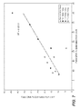
- Figure 13 and Figure 14 show results obtained from 19 patient plasma DNA samples, 1 donor plasma DNA sample, and duplicate runs of a donor gDNA sample. It is estimated in Figure 13 that the minimum fetal DNA % of which over-representation of chr21 can be detected at the best sampling rate ( ⁇ 70k reads mapped to chr21) is ⁇ 6%. (indicated by solid lines in Fig. 13 ). The lines are drawn between about 0.7 X10 5 reads and 6% fetal DNA concentration. It can be expected that higher numbers of reads (not exemplified here) the needed fetal DNA percentage will drop, probably to about 4%.
- t statistic t y 2 ⁇ - y 1 ⁇ s 2 2 n 2 + s 1 2 n 1 ,
- y 2 - y 1 is the difference in means (or amount of over- or under-representation of a particular chromosome) to be measured
- s is the standard deviation of the number of reads per 50kb in a particular chromosome
- 2 * y 2 ⁇ y 1 ⁇ - 1 * corresponds to the minimum fetal DNA % of which any over- or under-representation of chromosomes can be detected.
- the standard deviation equals to the square root of the mean.
- the 99.9% confidence interval of the median for each chromosome is estimated from bootstrapping 5000 samples from the 50kb read distribution data using the percentile method.
- the half width of the confidence interval is estimated as 0.5*confidence interval.
Abstract

Description
- This application claims priority from
U.S. Provisional Patent Application No. 61/098,758, filed on September 20, 2008 - This invention was made with U.S. Government support under NIH Director's Pioneer Award DP1 OD000251. The U.S. Government has certain rights in this invention.
- Applicants assert that the text copy of the Sequence Listing is identical to the Sequence Listing in computer readable form found on the accompanying computer file. Applicants incorporate the contents of the sequence listing by reference in its entirety.
- The present invention relates to the field of molecular diagnostics, and more particularly to the field of prenatal genetic diagnosis.
- Presented below is background information on certain aspects of the present invention as they may relate to technical features referred to in the detailed description, but not necessarily described in detail. That is, certain components of the present invention may be described in greater detail in the materials discussed below. The discussion below should not be construed as an admission as to the relevance of the information to the claimed invention or the prior art effect of the material described.
- Fetal aneuploidy and other chromosomal aberrations affect 9 out of 1000 live births (1). The gold standard for diagnosing chromosomal abnormalities is karyotyping of fetal cells obtained via invasive procedures such as chorionic villus sampling and amniocentesis. These procedures impose small but potentially significant risks to both the fetus and the mother (2). Non-invasive screening of fetal aneuploidy using maternal serum markers and ultrasound are available but have limited reliability (3-5). There is therefore a desire to develop non-invasive genetic tests for fetal chromosomal abnormalities.
- Since the discovery of intact fetal cells in maternal blood, there has been intense interest in trying to use them as a diagnostic window into fetal genetics (6-9). While this has not yet moved into practical application (10), the later discovery that significant amounts of cell-free fetal nucleic acids also exist in maternal circulation has led to the development of new non-invasive prenatal genetic tests for a variety of traits (11, 12). However, measuring aneuploidy remains challenging due to the high background of maternal DNA; fetal DNA often constitutes <10% of total DNA in maternal cell-free plasma (13).
- Recently developed methods for aneuploidy rely on detection focus on allelic variation between the mother and the fetus. Lo et al. demonstrated that allelic ratios of placental specific mRNA in maternal plasma could be used to detect
trisomy 21 in certain populations (14). - Similarly, they also showed the use of allelic ratios of imprinted genes in maternal plasma DNA to diagnose trisomy 18 (15). Dhallan et al. used fetal specific alleles in maternal plasma DNA to detect trisomy 21 (16). However, these methods are limited to specific populations because they depend on the presence of genetic polymorphisms at specific loci. We and others argued that it should be possible in principle to use digital PCR to create a universal, polymorphism independent test for fetal aneuploidy using maternal plasma DNA (17-19).
- An alternative method to achieve digital quantification of DNA is direct shotgun sequencing followed by mapping to the chromosome of origin and enumeration of fragments per chromosome. Recent advances in DNA sequencing technology allow massively parallel sequencing (20), producing tens of millions of short sequence tags in a single run and enabling a deeper sampling than can be achieved by digital PCR. As is known in the art, the term "sequence tag" refers to a relatively short (e.g., 15-100) nucleic acid sequence that can be used to identify a certain larger sequence, e.g., be mapped to a chromosome or genomic region or gene. These can be ESTs or expressed sequence tags obtained from mRNA.
- Science 309:1476 (2 Sept. 2005) News Focus "An Earlier Look at Baby's Genes" describes attempts to develop tests for Down Syndrome using maternal blood. Early attempts to detect Down Syndrome using fetal cells from maternal blood were called "just modestly encouraging." The report also describes work by Dennis Lo to detect the Rh gene in a fetus where it is absent in the mother. Other mutations passed on from the father have reportedly been detected as well, such as cystic fibrosis, beta-thalassemia, a type of dwarfism and Huntington's disease. However, these results have not always been reproducible.
- Venter et al., "The sequence of the human genome," Science, 2001 Feb 16;291(5507):1304-51 discloses the sequence of the human genome, which information is publicly available from NCBI. Another reference genomic sequence is a current NCBI build as obtained from the UCSC genome gateway.
- Wheeler et al., "The complete genome of an individual by massively parallel DNA sequencing," Nature, 2008 Apr 17;452(7189):872-6 discloses the DNA sequence of a diploid genome of a single individual, James D. Watson, sequenced to 7.4-fold redundancy in two months using massively parallel sequencing in picolitre-size reaction vessels. Comparison of the sequence to the reference genome led to the identification of 3.3 million single nucleotide polymorphisms, of which 10,654 cause amino-acid substitution within the coding sequence.
-
Quake et al., US 2007/0202525 entitled "Non-invasive fetal genetic screening by digital analysis," published August 30, 2007, discloses a process in which maternal blood containing fetal DNA is diluted to a nominal value of approximately 0.5 genome equivalent of DNA per reaction sample. - Chiu et al., "Noninvasive prenatal diagnosis of fetal chromosomal aneuploidy by massively parallel genomic DNA sequencing of DNA in maternal plasma," Proc. Natl. Acad. Sci. 105(51):20458-20463 (December 23, 2008) discloses a method for determining fetal aneuploidy using massively parallel sequencing. Disease status determination (aneuploidy) was made by calculating a "z score." Z scores were compared with reference values, from a population restricted to euploid male fetuses. The authors noted in passing that G/C content affected the coefficient of variation.
- Lo et al., "Diagnosing Fetal Chromosomal Aneuploidy Using Massively Parallel Genomic Sequencing,"
US 2009/0029377, published January 29, 2009 , discloses a method in which respective amounts of a clinically-relevant chromosome and of background chromosomes are determined from results of massively parallel sequencing. It was found that the percentage representation of sequences mapped tochromosome 21 is higher in a pregnant woman carrying a trisomy 21 fetus when compared with a pregnant woman carrying a normal fetus. For the four pregnant women each carrying a euploid fetus, a mean of 1.345% of their plasma DNA sequences were aligned tochromosome 21. - Lo et al., Determining a Nucleic Acid Sequence Imbalance,"
US 2009/0087847 published April 2, 2009 , discloses a method for determining whether a nucleic acid sequence imbalance exists, such as an aneuploidy, the method comprising deriving a first cutoff value from an average concentration of a reference nucleic acid sequence in each of a plurality of reactions, wherein the reference nucleic acid sequence is either the clinically relevant nucleic acid sequence or the background nucleic acid sequence; comparing the parameter to the first cutoff value; and based on the comparison, determining a classification of whether a nucleic acid sequence imbalance exists. - The following brief summary is not intended to include all features and aspects of the present invention, nor does it imply that the invention must include all features and aspects discussed in this summary.
- The present invention comprises a method for analyzing a maternal sample, e.g., from peripheral blood. It is not invasive into the fetal space, as is amniocentesis or chorionic villi sampling. In the preferred method, fetal DNA which is present in the maternal plasma is used. The fetal DNA is in one aspect of the invention enriched due to the bias in the method towards shorter DNA fragments, which tend to be fetal DNA. The method is independent of any sequence difference between the maternal and fetal genome. The DNA obtained, preferably from a peripheral blood draw, is a mixture of fetal and maternal DNA. The DNA obtained is at least partially sequenced, in a method which gives a large number of short reads. These short reads act as sequence tags, in that a significant fraction of the reads are sufficiently unique to be mapped to specific chromosomes or chromosomal locations known to exist in the human genome. They are mapped exactly, or may be mapped with one mismatch, as in the examples below. By counting the number of sequence tags mapped to each chromosome (1-22, X and Y), the over- or under- representation of any chromosome or chromosome portion in the mixed DNA contributed by an aneuploid fetus can be detected. This method does not require the sequence differentiation of fetal versus maternal DNA, because the summed contribution of both maternal and fetal sequences in a particular chromosome or chromosome portion will be different as between an intact, diploid chromosome and an aberrant chromosome, i.e., with an extra copy, missing portion or the like. In other words, the method does not rely on a priori sequence information that would distinguish fetal DNA from maternal DNA. The abnormal distribution of a fetal chromosome or portion of a chromosome (i.e., a gross deletion or insertion) may be determined in the present method by enumeration of sequence tags as mapped to different chromosomes. The median count of autosomal values (i.e., number of sequence tags per autosome) is used as a normalization constant to account for differences in total number of sequence tags is used for comparison between samples and between chromosomes The term "chromosome portion" is used herein to denote either an entire chromosome or a significant fragment of a chromosome. For example, moderate Down syndrome has been associated with partial trisomy 21q22.2→qter. By analyzing sequence tag density in predefined subsections of chromosomes (e.g., 10 to 100 kb windows), a normalization constant can be calculated, and chromosomal subsections quantified (e.g., 21q22.2). With large enough sequence tag counts, the present method can be applied to arbitrarily small fractions of fetal DNA. It has been demonstrated to be accurate down to 6% fetal DNA concentration. Exemplified below is the successful use of shotgun sequencing and mapping of DNA to detect fetal trisomy 21 (Down syndrome), trisomy 18 (Edward syndrome), and trisomy 13 (Patau syndrome), carried out non-invasively using cell-free fetal DNA in maternal plasma. This forms the basis of a universal, polymorphism-independent non-invasive diagnostic test for fetal aneuploidy. The sequence data also allowed us to characterize plasma DNA in unprecedented detail, suggesting that it is enriched for nucleosome bound fragments. The method may also be employed so that the sequence data obtained may be further analyzed to obtain information regarding polymorphisms and mutations.
- Thus, the present invention comprises, in certain aspects, a method of testing for an abnormal distribution of a specified chromosome portion in a mixed sample of normally and abnormally distributed chromosome portions obtained from a single subject, such as a mixture of fetal and maternal DNA in a maternal plasma sample. One carries out sequence determinations on the DNA fragments in the sample, obtaining sequences from multiple chromosome portions of the mixed sample to obtain a number of sequence tags of sufficient length of determined sequence to be assigned to a chromosome location within a genome and of sufficient number to reflect abnormal distribution. Using a reference sequence, one assigns the sequence tags to their corresponding chromosomes including at least the specified chromosome by comparing the sequence to reference genomic sequence. Often there will be on the order of millions of short sequence tags that are assigned to certain chromosomes, and, importantly, certain positions along the chromosomes. One then may determine a first number of sequence tags mapped to at least one normally distributed chromosome portion and a second number of sequence tags mapped to the specified chromosome portion, both chromosomes being in one mixed sample. The present method also involves correcting for nonuniform distribution sequence tags to different chromosomal portions. This is explained in detail below, where a number of windows of defined length are created along a chromosome, the windows being on the order of kilobases in length, whereby a number of sequence tags will fall into many of the windows and the windows covering each entire chromosome in question, with exceptions for non-informative regions, e.g., centromere regions and repetitive regions. Various average numbers, i.e., median values, are calculated for different windows and compared. By counting sequence tags within a series of predefined windows of equal lengths along different chromosomes, more robust and statistically significant results may be obtained. The present method also involves calculating a differential between the first number and the second number which is determinative of whether or not the abnormal distribution exists.
- In certain aspects, the present invention may comprise a computer programmed to analyze sequence data obtained from a mixture of maternal and fetal chromosomal DNA. Each autosome (chr. 1-22) is computationally segmented into contiguous, non-overlapping windows. (A sliding window could also be used). Each window is of sufficient length to contain a significant number of reads (sequence tags, having about 20-100 bp of sequence) and not still have a number of windows per chromosome. Typically, a window will be between 10kb and 100kb, more typically between 40 and 60 kb. There would, then, for example, accordingly be approximately between 3,000 and 100,000 windows per chromosome. Windows may vary widely in the number of sequence tags that they contain, based on location (e.g., near a centromere or repeating region) or G/C content, as explained below. The median (i.e., middle value in the set) count per window for each chromosome is selected; then the median of the autosomal values is used to account for differences in total number of sequence tags obtained for different chromosomes and distinguish interchromosomal variation from sequencing bias from aneuploidy. This mapping method may also be applied to discern partial deletions or insertions in a chromosome. The present method also provides a method for correcting for bias resulting from G/C content. For example, some the Solexa sequencing method was found to produce more sequence tags from fragments with increased G/C content. By assigning a weight to each sequence tag based on the G/C content of a window in which the read falls. The window for GC calculation is preferably smaller than the window for sequence tag density calculation.
-
-
Figure 1 is a scatter plot graph showing sequence tag densities from eighteen samples, having five different genotypes, as indicated in the figure legend. Fetal aneuploidy is detectable by the over-representation of the affected chromosome in maternal blood.Figure 1A shows sequence tag density relative to the corresponding value of genomic DNA control; chromosomes are ordered by increasing G/C content. The samples shown as indicated, are plasma from a woman bearing a T21 fetus; plasma from a woman bearing a T18 fetus; plasma from a normal adult male; plasma from a woman bearing a normal fetus; plasma from a woman bearing a T13 fetus. Sequence tag densities vary more with increasing chromosomal G/C content.Figure 1B is a detail fromFig. 1A , showingchromosome 21 sequence tag density relative to themedian chromosome 21 sequence tag density of the normal cases. Note that the values of 3disomy 21 cases overlap at 1.0. The dashed line represents the upper boundary of the 99% confidence interval constructed from all disomy 21 samples. The chromosomes are listed inFigure 1A in order of G/C content, from low to high. This figure suggests that one would prefer to use as a reference chromosome in the mixed sample with a mid level of G/C content, as it can be seen that the data there are more tightly grouped. That is,chromosomes Figure 1B represents an enlargement of thechromosome 21 data. -
Figure 2 is a scatter plot graph showing fetal DNA fraction and gestational age. The fraction of fetal DNA in maternal plasma correlates with gestational age. Fetal DNA fraction was estimated by three different ways: 1. From the additional amount ofchromosomes Figure 2 suggests that the present method may be employed at a very early stage of pregnancy. The data were obtained from the 10-week stage and later because that is the earliest stage at which chorionic villi sampling is done. (Amniocentesis is done later). From the level of the confidence interval, one would expect to obtain meaningful data as early as 4 weeks gestational age, or possibly earlier. -
Figure 3 is a histogram showing size distribution of maternal and fetal DNA in maternal plasma. It shows the size distribution of total and chromosome Y specific fragments obtained from 454 sequencing of maternal plasma DNA from a normal male pregnancy. The distribution is normalized to sum to 1. The numbers of total reads and reads mapped to the Y-chromosome are 144992 and 178 respectively. Inset: Cumulative fetal DNA fraction as a function of sequenced fragment size. The error bars correspond to the standard error of the fraction estimated assuming the error of the counts of sequenced fragments follow Poisson statistics. -
Figure 4 is a pair of line graphs showing distribution of sequence tags around transcription start sites (TSS) of ReSeq genes on all autosomes and chromosome X from plasma DNA sample of a normal male pregnancy (top,Fig. 4A ) and randomly sheared genomic DNA control (bottom,Fig. 4B ). The number of tags within each 5bp window was counted within ±1000bp region around each TSS, taking into account the strand each sequence tag mapped to. The counts from all transcription start sites for each 5bp window were summed and normalized to the median count among the 400 windows. A moving average was used to smooth the data. A peak in the sense strand represents the beginning of a nucleosome, while a peak in the anti-sense strand represents the end of a nucleosome. In the plasma DNA sample shown here, five well-positioned nucleosomes are observed downstream of transcription start sites and are represented as grey ovals. The number below within each oval represents the distance in base pairs between adjacent peaks in the sense and anti-sense strands, corresponding to the size of the inferred nucleosome. No obvious pattern is observed for the genomic DNA control. -
Figure 5A is a scatter plot graph showing the mean sequence tag density for each chromosome of all samples, including cell-free plasma DNA from pregnant women and male donor, as well as genomic DNA control from male donor, is plotted above. Exceptions arechromosomes Figure 5B is a scatter plot of mean sequence tag density for each chromosome versus G/C content of the chromosome. The correlation coefficient is 0.927, and the correlation is statistically significant (p<10-9). -
Figure 5C is a scatter plot of the standard deviation of sequence tag density of each chromosome versus G/C content of the chromosome. The correlation coefficient between standard deviation of sequence tag density and the absolute deviation of chromosomal G/C content from the genome-wide G/C content is 0.963, and the correlation is statistically significant (p<10-12). -
Figure 6 is a scatter plot graph showing percent difference of chromosome X sequence tag density of all samples as compared to the median chromosome X sequence tag density of all female pregnancies. All male pregnancies show under-representation of chromosome X. -
Figure 7 is a scatter plot graph showing a comparison of the estimation of fetal DNA fraction for cell-free DNA samples from 12 male pregnancies using sequencing data from chromosomes X and Y. The dashed line represents a simple linear regression line, with a slope of 0.85. The R2 value represents the square of the correlation coefficient. There is a statistically significant correlation between fetal DNA fraction estimated from chromosomes X and Y (p=0.0015). -
Figure 8 is a line graph showing length distribution of sequenced fragments from maternal cell-free plasma DNA sample of a normal male pregnancy at 1bp resolution. Sequencing was done on the 454/Roche platform. Reads that have at least 90% mapping to the human genome with greater than or equal to 90% accuracy are retained, totaling 144992 reads. Y-axis represents the number of reads obtained. The median length is 177bp while the mean length is 180bp. -
Figure 9 is a schematic illustrating how sequence tag distribution is used to detect the over and under-representation of any chromosome, i.e., a trisomy (over representation) or a missing chromosome (typically an X or Y chromosome, since missing autosomes are generally lethal). As shown in left panels A and C, one first plots the number of reads obtained versus a window that is mapped to a chromosome coordinate that represents the position of the read along the chromosome. That is, chromosome 1 (panel A) can be seen to have about 2.8 x 10⁸ bp. It would have this number divided by 50kb windows. These values are replotted (panels B and D) to show the distribution of the number of sequence tags/50kb window. The term "bin" is equivalent to a window. From this analysis, one can determine a median number of reads M for each chromosome, which, for purposes of illustration, may be observed along the x axis at the approximate center of the distribution and may be said to be higher if there are more sequence tags attributable to that chromosome. Forchromosome 1, illustrated in panels A and B, one obtains a median M1. By taking the median M of all 22 autosomes, one obtains a normalization constant N that can be used to correct for differences in sequences obtained in different runs, as can be seen in Table 1. Thus, the normalized sequence tag density forchromosome 1 would be M1/N; forchromosome 22 it would be M22/N. Close examination of panel A, for example would show that towards the zero end of the chromosome, this procedure obtained about 175 reads per 50kb window. In the middle, near the centromere, there were no reads, because this portion of the chromosome is ill defined in the human genome library. - That is, in the left panels (A and C), one plots the distribution of reads per chromosome coordinate, i.e., chromosomal position in terms of number of reads within each 50kb non-overlapping sliding window. Then, one determines the distribution of the number of sequence tags for each 50 kb window, and obtains a median number of sequence tags per chromosome for all autosomes and chromosome X (Examples of chr 1 [top] and chr 22 [bottom] are illustrated here). These results are referred to as M. The median of the 22 values of M (from all autosomes,
chromosomes 1 through 22) is used as the normalization constant N. The normalized sequence tag density of each chromosome is M/N (e.g., chr 1: M1/N; chr 22: M22/N). Such normalization is necessary to compare different patient samples since the total number of sequence tags (thus, the sequence tag density) for each patient sample is different (the total number of sequence tags fluctuates between -8 to -12 million). The analysis thus flows from frequency of reads per coordinate (A and C) to # reads per window (B and D) to a combination of all chromosomes. -
Figure 10 is a scatter plot graph showing data from different samples, as inFigure 1 , except that bias for G/C sampling has been eliminated. -
Figure 11 is a scatter plot graph showing the weight given to different sequence samples according to percentage of G/C content, with lower weight given to samples with a higher G/C content. G/C content ranges from about 30% to about 70%; weight can range over a factor of about 3. -
Figure 12 is a scatter plot graph which illustrates results of selected patients as indicated on the x axis, and, for each patient, a distribution of chromosome representation on the Y axis, as deviating from a representative t statistic, indicated as zero. -
Figure 13 is a scatter plot graph showing the minimum fetal DNA percentage of which over- or under-representation of a chromosome could be detected with a 99.9% confidence level forchromosomes -
Figure 14 is a scatter plot graph showing a linear relationship between log10 of minimum fetal DNA percentage that is needed versus log10 of the number of reads required. - Unless defined otherwise, all technical and scientific terms used herein have the same meaning as commonly understood by those of ordinary skill in the art to which this invention belongs. Although any methods and materials similar or equivalent to those described herein can be used in the practice or testing of the present invention, the preferred methods and materials are described. Generally, nomenclatures utilized in connection with, and techniques of, cell and molecular biology and chemistry are those well known and commonly used in the art. Certain experimental techniques, not specifically defined, are generally performed according to conventional methods well known in the art and as described in various general and more specific references that are cited and discussed throughout the present specification. For purposes of the clarity, following terms are defined below.
- "Sequence tag density" means the normalized value of sequence tags for a defined window of a sequence on a chromosome (in a preferred embodiment the window is about 50kb), where the sequence tag density is used for comparing different samples and for subsequent analysis. A "sequence tag" is a DNA sequence of sufficient length that it may be assigned specifically to one of chromosomes 1-22, X or Y. It does not necessarily need to be, but may be non-repetitive within a single chromosome. A certain, small degree of mismatch (0-1) may be allowed to account for minor polymorphisms that may exist between the reference genome and the individual genomes (maternal and fetal) being mapped. The value of the sequence tag density is normalized within a sample. This can be done by counting the number of tags falling within each window on a chromosome; obtaining a median value of the total sequence tag count for each chromosome; obtaining a median value of all of the autosomal values; and using this value as a normalization constant to account for the differences in total number of sequence tags obtained for different samples. A sequence tag density as calculated in this way would ideally be about 1 for a disomic chromosome. As further described below, sequence tag densities can vary according to sequencing artifacts, most notably G/C bias; this is corrected as described. This method does not require the use of an external standard, but, rather, provides an internal reference, derived from al of the sequence tags (genomic sequences), which may be, for example, a single chromosome or a calculated value from all autosomes.
- "T21" means
trisomy 21. - "T18" _means
trisomy 18. - "T13" means
trisomy 13. - "Aneuploidy" is used in a general sense to mean the presence or absence of an entire chromosome, as well as the presence of partial chromosomal duplications or deletions or kilobase or greater size, as opposed to genetic mutations or polymorphisms where sequence differences exist.
- "Massively parallel sequencing" means techniques for sequencing millions of fragments of nucleic acids, e.g., using attachment of randomly fragmented genomic DNA to a planar, optically transparent surface and solid phase amplification to create a high density sequencing flow cell with millions of clusters, each containing ∼1,000 copies of template per sq. cm. These templates are sequenced using four-color DNA sequencing-by-synthesis technology. See, products offered by Illumina, Inc., San Diego, California. In the present work, sequences were obtained, as described below, with an Illumina/Solexa 1G Genome Analyzer. The Solexa/Illumina method referred to below relies on the attachment of randomly fragmented genomic DNA to a planar, optically transparent surface. In the present case, the plasma DNA does not need to be sheared. Attached DNA fragments are extended and bridge amplified to create an ultra-high density sequencing flow cell with ≥ 50 million clusters, each containing ∼1,000 copies of the same template. These templates are sequenced using a robust four-color DNA sequencing-by-synthesis technology that employs reversible terminators with removable fluorescent dyes. This novel approach ensures high accuracy and true base-by-base sequencing, eliminating sequence-context specific errors and enabling sequencing through homopolymers and repetitive sequences.
- High-sensitivity fluorescence detection is achieved using laser excitation and total internal reflection optics. Short sequence reads are aligned against a reference genome and genetic differences are called using specially developed data analysis pipeline software.
- Copies of the protocol for whole genome sequencing using Soelxa technology may be found at BioTechniques® Protocol Guide 2007 Published December 2006: p 29, www.biotechniques.com/default.asp? page=protocol&subsection=article_display&id=112378. Solexa's oligonucleotide adapters are ligated onto the fragments, yielding a fully-representative genomic library of DNA templates without cloning. Single molecule clonal amplification involves six steps: Template hybridization, template amplification, linearization, blocking 3' ends, denaturation and primer hybridization. Solexa's Sequencing-by-Synthesis utilizes four proprietary nucleotides possessing reversible fluorophore and termination properties. Each sequencing cycle occurs in the presence of all four nucleotides.
- The presently used sequencing is preferably carried out without a preamplification or cloning step, but may be combined with amplification-based methods in a microfluidic chip having reaction chambers for both PCR and microscopic template-based sequencing. Only about 30 bp of random sequence information are needed to identify a sequence as belonging to a specific human chromosome. Longer sequences can uniquely identify more particular targets. In the present case, a large number of 25bp reads were obtained, and due to the large number of reads obtained, the 50% specificity enabled sufficient sequence tag representation.
- Further description of a massively parallel sequencing method, which employed the below referenced 454 method is found in Rogers and Ventner, "Genomics: Massively parallel sequencing," Nature, 437, 326-327 (15 September 2005). As described there, Rothberg and colleagues (Margulies, M. et al. Nature 437, 376―380 (2005)), have developed a highly parallel system capable of sequencing 25 million bases in a four-hour period — about 100 times faster than the current state-of-the-art Sanger sequencing and capillary-based electrophoresis platform. The method could potentially allow one individual to prepare and sequence an entire genome in a few days. The complexity of the system lies primarily in the sample preparation and in the microfabricated, massively parallel platform, which contains 1.6 million picoliter-sized reactors in a 6.4-cm2 slide. Sample preparation starts with fragmentation of the genomic DNA, followed by the attachment of adaptor sequences to the ends of the DNA pieces. The adaptors allow the DNA fragments to bind to tiny beads (around 28 µ in diameter). This is done under conditions that allow only one piece of DNA to bind to each bead. The beads are encased in droplets of oil that contain all of the reactants needed to amplify the DNA using a standard tool called the polymerase chain reaction. The oil droplets form part of an emulsion so that each bead is kept apart from its neighbor, ensuring the amplification is uncontaminated. Each bead ends up with roughly 10 million copies of its initial DNA fragment. To perform the sequencing reaction, the DNA-template-carrying beads are loaded into the pico liter reactor wells — each well having space for just one bead. The technique uses a sequencing-by-synthesis method developed by Uhlen and colleagues, in which DNA complementary to each template strand is synthesized. The nucleotide bases used for sequencing release a chemical group as the base forms a bond with the growing DNA chain, and this group drives a light-emitting reaction in the presence of specific enzymes and luciferin. Sequential washes of each of the four possible nucleotides are run over the plate, and a detector senses which of the wells emit light with each wash to determine the sequence of the growing strand. This method has been adopted commercially by 454 Life Sciences.
- Further examples of massively parallel sequencing are given in
US 20070224613 by Strathmann, published September 27, 2007 , entitled "Massively Multiplexed Sequencing." Also, for a further description of massively parallel sequencing, seeUS 2003/0022207 to Balasubramanian, et al., published January 30, 2003 , entitled "Arrayed polynucleotides and their use in genome analysis." - Non-invasive prenatal diagnosis of aneuploidy has been a challenging problem because fetal DNA constitutes a small percentage of total DNA in maternal blood (13) and intact fetal cells are even rarer (6, 7, 9, 31, 32). We showed in this study the successful development of a truly universal, polymorphism-independent non-invasive test for fetal aneuploidy. By directly sequencing maternal plasma DNA, we could detect
fetal trisomy 21 as early as 14th week of gestation. Using cell-free DNA instead of intact cells allows one to avoid complexities associated with microchimerism and foreign cells that might have colonized the mother; these cells occur at such low numbers that their contribution to the cell-free DNA is negligible (33, 34). Furthermore, there is evidence that cell-free fetal DNA clears from the blood to undetectable levels within a few hours of delivery and therefore is not carried forward from one pregnancy to the next (35-37). - Rare forms of aneuploidy caused by unbalanced translocations and partial duplication of a chromosome are in principle detectable by the approach of shotgun sequencing, since the density of sequence tags in the triplicated region of the chromosome would be higher than the rest of the chromosome. Detecting incomplete aneuploidy caused by mosaicism is also possible in principle but may be more challenging, since it depends not only on the concentration of fetal DNA in maternal plasma but also the degree of fetal mosaicism. Further studies are required to determine the effectiveness of shotgun sequencing in detecting these rare forms of aneuploidy.
- The present method is applicable to large chromosomal deletions, such as 5p-Syndrome (five p minus), also known as Cat Cry Syndrome or Cri du Chat Syndrome. 5p-Syndrome is characterized at birth by a high-pitched cry, low birth weight, poor muscle tone, microcephaly, and potential medical complications. Similarly amenable disorders addressed by the present methods are p-, monosomy 9P, otherwise known as Alfi's Syndrome or 9P-, 22q11.2 deletion syndrome, Emanuel Syndrome, also known in the medical literature as the Supernumerary Der(22) Syndrome,
trisomy 22, Unbalanced 11/22 Translocation orpartial trisomy 11/22, Microdeletion and Microduplication at 16p 11.2, which is associated with autism, and other deletions or imbalances, including those that are presently unknown. - An advantage of using direct sequencing to measure aneuploidy non-invasively is that it is able to make full use of the sample, while PCR based methods analyze only a few targeted sequences. In this study, we obtained on average 5 million reads per sample in a single run, of which ∼66,000 mapped to
chromosome 21. Since those 5 million reads represent only a portion of one human genome, in principle less than one genomic equivalent of DNA is sufficient for the detection of aneuploidy using direct sequencing. In practice, a larger amount of DNA was used since there is sample loss during sequencing library preparation, but it may be possible to further reduce the amount of blood required for analysis. - Mapping shotgun sequence information (i.e., sequence information from a fragment whose physical genomic position is unknown) can be done in a number of ways, which involve alignment of the obtained sequence with a matching sequence in a reference genome. See, Li et al., "Mapping short DNA sequencing reads and calling variants using mapping quality score," Genome Res., 2008 . [Epub ahead of print].
- We observed that certain chromosomes have large variations in the counts of sequenced fragments (from sample to sample, and that this depends strongly on the G/C content (
Figure 1A ) It is unclear at this point whether this stems from PCR artifacts during sequencing library preparation or cluster generation, the sequencing process itself, or whether it is a true biological effect relating to chromatin structure. We strongly suspect that it is an artifact since we also observe G/C bias on genomic DNA control, and such bias on the Solexa sequencing platform has recently been reported (38, 39). It has a practical consequence since the sensitivity to aneuploidy detection will vary from chromosome to chromosome; fortunately the most common human aneuploidies (such as 13, 18, and 21) have low variation and therefore high detection sensitivity. Both this problem and the sample volume limitations may possibly be resolved by the use of single molecule sequencing technologies, which do not require the use of PCR for library preparation (40). - Plasma DNA samples used in this study were obtained about 15 to 30 minutes after amniocentesis or chorionic villus sampling. Since these invasive procedures disrupt the interface between the placenta and maternal circulation, there have been discussions whether the amount of fetal DNA in maternal blood might increase following invasive procedures. Neither of the studies to date have observed a significant effect (41, 42).
- Our results support this conclusion, since using the digital PCR assay we estimated that fetal DNA constituted less than or equal to 10% of total cell-free DNA in the majority of our maternal plasma samples. This is within the range of previously reported values in maternal plasma samples obtained prior to invasive procedures (13). It would be valuable to have a direct measurement addressing this point in a future study.
- The average fetal DNA fraction estimated from sequencing data is higher than the values estimated from digital PCR data by an average factor of two (p<0.005, paired t-test on all male pregnancies that have complete set of data). One possible explanation for this is that the PCR step during Solexa library preparation preferentially amplifies shorter fragments, which others have found to be enriched for fetal DNA (22, 23). Our own measurements of length distribution on one sample do not support this explanation, but nor can we reject it at this point. It should also be pointed out that using the sequence tags we find some variation of fetal fraction even in the same sample depending on which chromosome we use to make the calculation (
Figure 7 , Table 1). This is most likely due to artifacts and errors in the sequencing and mapping processes, which are substantial ― recall that only half of the sequence tags map to the human genome with one error or less. Finally, it is also possible that the PCR measurements are biased since they are only sampling a tiny fraction of the fetal genome. - Our sequencing data suggest that the majority of cell-free plasma DNA is of apoptotic origin and shares features of nucleosomal DNA. Since nucleosome occupancy throughout the eukaryotic genome is not necessarily uniform and depends on factors such as function, expression, or sequence of the region (30, 43), the representation of sequences from different loci in cell-free maternal plasma may not be equal, as one usually expects in genomic DNA extracted from intact cells. Thus, the quantity of a particular locus may not be representative of the quantity of the entire chromosome and care must be taken when one designs assays for measuring gene dosage in cell-free maternal plasma DNA that target only a few loci.
- Historically, due to risks associated with chorionic villus sampling and amniocentesis, invasive diagnosis of fetal aneuploidy was primarily offered to women who were considered at risk of carrying an aneuploid fetus based on evaluation of risk factors such as maternal age, levels of serum markers, and ultrasonographic findings. Recently, an American College of Obstetricians and Gynecologists (ACOG) Practice Bulletin recommended that "invasive diagnostic testing for aneuploidy should be available to all women, regardless of maternal age" and that "pretest counseling should include a discussion of the risks and benefits of invasive testing compared with screening tests"(2).
- A noninvasive genetic test based on the results described here and in future large-scale studies would presumably carry the best of both worlds: minimal risk to the fetus while providing true genetic information. The costs of the assay are already fairly low; the sequencing cost per sample as of this writing is about $700 and the cost of sequencing is expected to continue to drop dramatically in the near future.
- Shotgun sequencing can potentially reveal many more previously unknown features of cell-free nucleic acids such as plasma mRNA distributions, as well as epigenetic features of plasma DNA such as DNA methylation and histone modification, in fields including perinatology, oncology and transplantation, thereby improving our understanding of the basic biology of pregnancy, early human development and disease.
- Commercially available sequencing equipment was used in the present illustrative examples, namely the Solexa/Illumina sequencing platform and the 454/Roche platform. It will be apparent to those skilled in the art that a number of different sequencing methods and variations can be used. One sequencing method that can be used to advantage in the present methods involves paired end sequencing. Fluorescently labeled sequencing primers could be used to simultaneously sequence both strands of a dsDNA template, as described e.g., in Wiemann et al. (Anal. Biochem. 224: 117 [1995]; Anal. Biochem. 234: 166 [1996]. Recent examples of this technique have demonstrated multiplex co-sequencing using the four-color dye terminator reaction chemistry pioneered by Prober et al. (Science 238: 336 [1987]). Solexa/Illumina offers a "Paired End Module" to its Genome Analyzer. Using this module, after the Genome Analyzer has completed the first sequencing read, the Paired-End Module directs the resynthesis of the original templates and the second round of cluster generation. The Paired-End Module is connected to the Genome Analyzer through a single fluidic connection. In addition, 454 has developed a protocol to generate a library of Paired End reads. These Paired End reads are approximately 84-nucleotide DNA fragments that have a 44-mer adaptor sequence in the middle flanked by a 20-mer sequence on each side. The two flanking 20-mers are segments of DNA that were originally located approximately 2.5 kb apart in the genome of interest.
- By using paired end reads in the present method, one may obtain more sequence information from a given plasma DNA fragment, and, significantly, one may also obtain sequence information from both ends of the fragment. The fragment is mapped to the human genome as explained here elsewhere. After mapping both ends, one may deduce the length of the starting fragment. Since fetal DNA is known to be shorter than maternal DNA fragments circulating in plasma, one may use this information about the length of the DNA fragment to effectively increase the weight given to sequences obtained from shorter (e.g., about 300 bp or less) DNA fragments. Methods for weighting are given below.
- Another method for increasing sensitivity to fetal DNA is to focus on certain regions within the human genome. One may use sequencing methods which select a priori sequences which map to the chromosomes of interest (as described here elsewhere, such as 18, 21, 13, X and Y). One may also choose to focus, using this method, on partial chromosomal deletions, such as 22q11 deletion syndrome. Other microdeletions and microduplications are set forth in Table 1 of
US 2005//0181410, published Aug. 18 2005 under the title "Methods and apparatuses for achieving precision genetic diagnosis." - In sequencing selected subsequences, one may employ sequence-based methodologies such as sequencing by array, or capture beads with specific genomic sequences used as capture probes. The use of a sequencing array can be implemented as described in Chetverin et al., "Oligonucleotide arrays: new concepts and possibilities," Biotechnology (N Y). 1994 Nov;12(11):1093-9, as well as
Rothberg, US 2002/0012930 A1 entitled "Method of Sequencing a Nucleic Acid," and Reeve et al., "Sequencing by Hybridization,"US 6,399,364 . In these methods, the target nucleic acid to be sequenced may be genomic DNA, cDNA or RNA. The sample is rendered single stranded and captured under hybridizing conditions with a number of single stranded probes which are catalogued by bar coding or by physical separation in an array. Emulsion PCR, as used in the 454 system, the SOLiD system, and Polonator (Dover Systems) and others may also be used, where capture is directed to specific target sequences, e.g., genome sequences mapping uniquely tochromosome 21 or other chromosome of interest, or to a chromosome region such as 15q11 (Prader-Willi syndrome), or excessive CGG repeats in the FMR1 gene (fragile X syndrome). - The subsequencing method is in one aspect contrary to conventional massively parallel sequencing methodologies, which seek to obtain all of the sequence information in a sample. This alternative method selectively ignores certain sequence information by using a sequencing method which selectively captures sample molecules containing certain predefined sequences. One may also use the sequencing steps exactly as exemplified, but in mapping the sequence fragments obtained, give greater weight to sequences which map to areas known to be more reliable in their coverage, such as exons. Otherwise, the method proceeds as described below, where one obtains a large number of sequence reads from one or more reference chromosomes, which are compared to a large number of reads obtained from a chromosome of interest, after accounting for variations arising from chromosomal length, G/C content, repeat sequences and the like.
- One may also focus on certain regions within the human genome according to the present methods in order to identify partial monosomies and partial trisomies. As described below, the present methods involve analyzing sequence data in a defined chromosomal sliding "window," such as contiguous, nonoverlapping 50Kb regions spread across a chromosome. Partial trisomies of 13q, 8p (8p23.1), 7q, distal 6p, 5p, 3q (3q25.1), 2q, 1q (1q42.1 and 1q21-qter), partial Xpand monosomy 4q35.1 have been reported, among others. For example, partial duplications of the long arm of
chromosome 18 can result in Edwards syndrome in the case of a duplication of 18q21.1-qter (See, Mewar et al., "Clinical and molecular evaluation of four patients with partial duplications of the long arm of ). - Cell-free plasma DNA from 18 pregnant women and a male donor, as well as whole blood genomic DNA from the same male donor, were sequenced on the Solexa/Illumina platform. We obtained on average -10 million 25bp sequence tags per sample. About 50% (i.e., -5 million) of the reads mapped uniquely to the human genome with at most 1 mismatch against the human genome, covering ∼4% of the entire genome. An average of -154,000, -135,000, ∼66,000 sequence tags mapped to
chromosomes Table 1. Sample Fetal Karyotype Gestational Age (weeks) Volume of Plasma Amt of DNA Approx Amount of Input DNA * Total Number of Sequence Tags P1 Plasma DNA§ 47XX +21 35 1.6 761 8.0 8206694 P2 Plasma DNA§ 47XY +21 18 1.4 585 5.2 7751384 P6 Plasma DNA§ 47XX +21 14 1.6 410 4.3 6699183 P7 Plasma DNA§ 47XY +21 18 2.2 266 3.8 8324473 P14 Plasma DNA§ 47XX +21 23 3.2 57 1.2 8924944 P17 Plasma DNA§ 47XX +21 16 2.3 210 3.2 11599833 P19 Plasma DNA§ 46XY 18 3.2 333 7.0 7305417 P20 Plasma DNA§ 47XY +21 18 1.3 408 3.6 11454876 P23 Plasma DNA§ 46XY 10 1.6 258 2.7 11851612 P26 Plasma DNA§ 46XY 13 3.0 340 6.7 11471297 P31 Plasma DNA§ 46XY 20 2.2 278 4.0 8967562 P40 Plasma DNA§ 46XY 11 2.6 217 3.7 9205197 P42 Plasma DNA§ 46XY 11 3.0 276 5.5 8364774 P52 Plasma DNA§ 47XY +21 25 1.6 645 6.8 9192596 P53 Plasma DNA§ 47XX +21 19 1.6 539 5.7 9771887 P57 Plasma DNA§ 47XX +18 23 2.0 199 2.6 15041417 P59 Plasma DNA§ 47XY +18 21 2.0 426 5.6 11910483 P64 Plasma DNA§ 47XY +13 17 1.8 204 2.4 12097478 Male Donor Plasma DNA§ - - 1.8 485 5.8 6669125 Male Donor Whole Blood Genomic DNA§ - - - - 2.1 8519495 P25 Plasma DNA¶ 46XY 11 5.6 132 4.9 242599 P13 Plasma DNA§ 46XY 18 5.6 77 2.9 4168455 Table 2. Sample Number of Sequence Tags Mapped Uniquely to the Human Genome (hg18) with At Most 1 Mismatch % Fetal DNA Estimated By Digital PCR with SRY Assay (male fetuses) % Fetal DNA Estimated by ChrY Sequence Tags (male fetuses) % Fetal DNA Estimated by Depletion of ChrX Sequence Tags (male fetuses) % Fetal DNA Estimated by Addition of Trisomic Chromosome Sequence Tags (aneuploid fetuses) Overall G/C content Of Sequence Tags (%) P1 Plasma DNA§ 4632637 - - - 35.0 43.65 P2 Plasma DNA§ 4313884 6.4 15.4 21.6 15.5 48.72 P6 Plasma DNA§ 3878383 - - - 22. 9 44.78 P7 Plasma DNA§ 4294865 9.1 31.0 33.8 28.6 48.07 P14 Plasma DNA§ 3603767 - - - 30.5 46.38 P17 Plasma DNA§ 5968932 - - - 7.8 44.29 P19 Plasma DNA§ 3280521 <5.9‡ 4.14 21.5 - 50.09 P20 Plasma DNA§ 6032684 10.0 15.7 11.3 11.5 44.02 P23 Plasma DNA§ 6642795 5.3 12.2 9.6 - 43.80 P26 Plasma DNA§ 3851477 10.3 18.2 14.2 - 42.51 P31 Plasma DNA§ 4683777 Missing data‡ 13.2 17.0 - 48.27 P40 Plasma DNA§ 4187561 8.6 20.0 17.1 - 42.65 P42 Plasma DNA§ 4315527 <4.4‡ 9.7 7.9 - 44.14 P52 Plasma DNA§ 5126837 6.3 25.0 26.3 26.4 44.34 P53 Plasma DNA§ 5434222 - - - 25.8 44.18 P57 Plasma DNA§ 7470487 - - - 23.0 42.89 P59 Plasma DNA§ 6684871 26.4 44.0 39.8 45.1 43.64 P64 Plasma DNA§ 6701148 <4.4‡ 14.0 8.9 16.7 44.21 Male Donor Plasma DNA§ 3692931 - - - - 48.30 Male Donor Whole Blood Genomic DNA§ 5085412 - - - - 46.53 P25 Plasma DNA¶ 144992† - - - - 41.38 P13 Plasma DNA§ 2835333 9.8 5.7 n/a∥ - 39.60 The volume of plasma is the volume used for Sequencing Library Creation (ml). The amount of DNA is in Plasma (cell equivalent/ml plasma)*. The approximate amount of input DNA is that use for Sequencing Library Construction (ng).
*As quantified by digital PCR withEIF2C 1 Taqman Assay, converting from copies to ng assuming 6.6pg/cell equivalent.
†For 454 sequencing, this number represents the number of reads with at least 90% accuracy and 90% coverage when mapped to hg18.
‡Insufficient materials were available for quantifying fetal DNA % with digital PCR for these samples (either no samples remained for analysis or there was insufficient sampling).
§Sequenced on Solexa/Illumina platform; ¶Sequenced on 454/Roche platform
∥Sample P13 was the first to be analyzed by shotgun sequencing. It was a normal fetus and the chromosome value was clearly disomic. However, there were some irregularities with this sample and it was not included in further analysis. This sample was sequenced on a different Solexa instrument than the rest of the samples of this study, and it was sequenced in the presence of a number of samples of unknown origin. The G/C content of this sample was lower than the G/C bias of the human genome, while the rest of the samples are above. It had the lowest number of reads, and also the smallest number of reads mapped successfully to the human genome. This sample appeared to be outlier in sequence tag density for most chromosomes and the fetal DNA fraction calculated from chromosomes X was not well defined. For these reasons we suspect that the irregularities are due to technical problems with the sequencing process. - In Table 1 and Table 2, each sample represents a different patient, e.g., P1 in the first row. The total number of sequence tags varied but was frequently was in the 10 million range, using the Solexa technology. The 454 technology used for P25 and P13 gave a lower number of reads.
- We observed a non-uniform distribution of sequence tags across each chromosome. This pattern of intra-chromosomal variation was common among all samples, including randomly sheared genomic DNA, indicating the observed variation was most probably due to sequencing artifacts. We applied an arbitrary sliding window of 50kb across each chromosome and counted the number of tags falling within each window. The window can be varied in size to account for larger numbers of reads (in which cases a smaller window, e.g., 10 kb, gives a more detailed picture of a chromosome) or a smaller number of reads, in which case a larger window (e.g., 100kb) may still be used and will detect gross chromosome deletions, omissions or duplications. The median count per 50kb window for each chromosome was selected. The median of the autosomal values (i.e., 22 chromosomes) was used as a normalization constant to account for the differences in total number of sequence tags obtained for different samples. The inter-chromosomal variation within each sample was also consistent among all samples (including genomic DNA control). The mean sequence tag density of each chromosome correlates with the G/C content of the chromosome (p<10-9) (
Figure 5A, 5B). The standard deviation of sequence tag density for each chromosome also correlates with the absolute degree of deviation in chromosomal G/C content from the genome-wide G/C content (p<10-12) (Figure 5A, 5C). The G/C content of sequenced tags of all samples (including the genomic DNA control) was on average 10% higher than the value of the sequenced human genome (41 %) (21)(Table 2), suggesting that there is a strong G/C bias stemming from the sequencing process. We plotted inFigure 1A the sequence tag density for each chromosome (ordered by increasing G/C content) relative to the corresponding value of the genomic DNA control to remove such bias. - The distribution of
chromosome 21 sequence tag density for all 9 T21 pregnancies is clearly separated from that ofpregnancies bearing disomy 21 fetuses (p<10-5), Student's t-test) (Figure 1A and1B ). The coverage ofchromosome 21 for T21 cases is about ∼4-18% higher (average ∼11%) than that of thedisomy 21 cases. Because the sequence tag density ofchromosome 21 for T21 cases should be (1+ε/2) of that ofdisomy 21 pregnancies, where ε is the fraction of total plasma DNA originating from the fetus, such increase inchromosome 21 coverage in T21 cases corresponds to a fetal DNA fraction of ∼8% ― 35% (average ∼23%) (Table 1,Figure 2 ). We constructed a 99% confidence interval of the distribution ofchromosome 21 sequence tag density ofdisomy 21 pregnancies. The values for all 9 T21 cases lie outside the upper boundary of the confidence interval and those for all 9 disomy 21 cases lie below the boundary (Figure 1B ). If we used the upper bound of the confidence interval as a threshold value for detecting T21, the minimum fraction of fetal DNA that would be detected is ∼2%. - Plasma DNA of pregnant women carrying T18 fetuses (2 cases) and a T13 fetus (1 case) were also directly sequenced. Over-representation was observed for
chromosome 18 andchromosome 13 in T18 and T13 cases respectively (Figure 1A ). While there were not enough positive samples to measure a representative distribution, it is encouraging that all of these three positives are outliers from the distribution of disomy values. The T18 are large outliers and are clearly statistically significant (p<10-7), while the statistical significance of the single T13 case is marginal (p<0.05). Fetal DNA fraction was also calculated from the over-represented chromosome as described above (Figure 2 , Table 1). - Using digital Taqman PCR for a single locus on
chromosome 1, we estimated the average cell-free DNA concentration in the sequenced maternal plasma samples to be -360 cell equivalent/ml of plasma (range: 57 to 761 cell equivalent/ml plasma) (Table 1), in rough accordance to previously reported values (13). The cohort included 12 male pregnancies (6 normal cases, 4 T21 cases, 1 T18 case and 1 T13 case) and 6 female pregnancies (5 T21 cases and 1 T18 case). DYS14, a multi-copy locus on chromosome Y, was detectable in maternal plasma by real-time PCR in all these pregnancies but not in any of the female pregnancies (data not shown). The fraction of fetal DNA in maternal cell-free plasma DNA is usually determined by comparing the amount of fetal specific locus (such as the SRY locus on chromosome Y in male pregnancies) to that of a locus on any autosome that is common to both the mother and the fetus using quantitative real-time PCR (13, 22, 23). We applied a similar duplex assay on a digital PCR platform (see Methods) to compare the counts of the SRY locus and a locus onchromosome 1 in male pregnancies. SRY locus was not detectable in any plasma DNA samples from female pregnancies. We found with digital PCR that for the majority samples, fetal DNA constituted §10% of total DNA in maternal plasma (Table 2), agreeing with previously reported values (13). - The percentage of fetal DNA among total cell-free DNA in maternal plasma can also be calculated from the density of sequence tags of the sex chromosomes for male pregnancies. By comparing the sequence tag density of chromosome Y of plasma DNA from male pregnancies to that of adult male plasma DNA, we estimated fetal DNA percentage to be on average - 19% (range: 4-44%) for all male pregnancies (Table 2, above,
Figure 2 ). Because human males have 1 fewer chromosome X than human females, the sequence tag density of chromosome X in male pregnancies should be (1-e/2) of that of female pregnancies, where e is fetal DNA fraction. We indeed observed under-representation of chromosome X in male pregnancies as compared to that of female pregnancies (Figure 5 ). Based on the data from chromosome X, we estimated fetal DNA percentage to be on average ∼19% (range: 8-40%) for all male pregnancies (Table 2, above,Figure 2 ). The fetal DNA percentage estimated from chromosomes X and Y for each male pregnancy sample correlated with each other (p=0.0015) (Figure 7 ). - We plotted in
Figure 2 the fetal DNA fraction calculated from the over-representation of trisomic chromosome in aneuploid pregnancies, and the under-representation of chromosome X and the presence of chromosome Y for male pregnancies against gestational age. The average fetal DNA fraction for each sample correlates with gestational age (p=0.0051), a trend that is also previously reported (13). - We analyzed the sequencing libraries with a commercial lab-on-a-chip capillary electrophoresis system. There is a striking consistency in the peak fragment size, as well as the distribution around the peak, for all plasma DNA samples, including those from pregnant women and male donor. The peak fragment size was on average 261bp (range: 256-264bp). Subtracting the total length of the Solexa adaptors (92bp) from 260bp gives 169bp as the actual peak fragment size. This size corresponds to the length of DNA wrapped in a chromatosome, which is a nucleosome bound to a H1 histone (24). Because the library preparation includes an 18-cycle PCR, there are concerns that the distribution might be biased. To verify that the size distribution observed in the electropherograms is not an artifact of PCR, we also sequenced cell-free plasma DNA from a pregnant woman carrying a male fetus using the 454 platform. The sample preparation for this system uses emulsion PCR, which does not require competitive amplification of the sequencing libraries and creates product that is largely independent of the amplification efficiency. The size distribution of the reads mapped to unique locations of the human genome resembled those of the Solexa sequencing libraries, with a predominant peak at 176bp, after subtracting the length of 454 universal adaptors (
Figure 3 andFigure 8 ). These findings suggest that the majority of cell-free DNA in the plasma is derived from apoptotic cells, in accordance with previous findings (22, 23, 25, 26). - Of particular interest is the size distribution of maternal and fetal DNA in maternal cell-free plasma. Two groups have previously shown that the majority of fetal DNA has size range of that of mono-nucleosome (<200-300bp), while maternal DNA is longer. Because 454 sequencing has a targeted read-length of 250bp, we interpreted the small peak at around 250bp (
Figure 3 andFigure 8 ) as the instrumentation limit from sequencing higher molecular weight fragments. We plotted the distribution of all reads and those mapped to Y-chromosome (Figure 3 ). We observed a slight depletion of Y-chromosome reads in the higher end of the distribution. Reads <220bp constitute 94% of Y-chromosome and 87% of the total reads. Our results are not in complete agreement with previous findings in that we do not see as dramatic an enrichment of fetal DNA at short lengths (22, 23). Future studies will be needed to resolve this point and to eliminate any potential residual bias in the 454 sample preparation process, but it is worth noting that the ability to sequence single plasma samples permits one to measure the distribution in length enrichments across many individual patients rather than measuring the average length enrichment of pooled patient samples. - Since our observations of the size distribution of cell-free plasma DNA suggested that plasma DNA is mainly apoptotic of origin, we investigated whether features of nucleosomal DNA and positioning are found in plasma DNA. One such feature is nucleosome positioning around transcription start sites. Experimental data from yeast and human have suggested that nucleosomes are depleted in promoters upstream of transcription start sites and nucleosomes are well-positioned near transcription start sites (27-30). We applied a 5bp window spanning +/- 1000bp of transcription start sites of all RefSeq genes and counted the number of tags mapping to the sense and antisense strands within each window. A peak in the sense strand represents the beginning of a nucleosome while a peak in the antisense strand represents the end. After smoothing, we saw that for most plasma DNA samples, at least 3 well-positioned nucleosomes downstream of transcription start sites could be detected, and in some cases, up to 5 well-positioned nucleosomes could be detected, in rough accordance to the results of Schones et al. (27) (
Figure 4 ). We applied the same analysis on sequence tags of randomly sheared genomic DNA and observed no obvious pattern in tag localization, although the density of tags was higher at the transcription start site (Figure 4 ). - Shown in
Figures 10 and12 are results which may be obtained when sequence tag numbers are treated statistically based on data from the reference human genome. That is, for example, sequence tags from fragments with higher GC content may be overrepresented, and suggest an aneuploidy where none exists. The sequence tag information itself may not be informative, since only a small portion of the fragment ordinarily will be sequenced, while it is the overall G/C content of the fragment that causes the bias. Thus there is provided a method, described in detail in Examples 8 and 10, for correcting for this bias, and this method may facilitate analysis of samples which otherwise would not produce statistically significant results. This method, for correcting for G/C bias of sequence reads from massively parallel sequencing of a genome, comprises the step of dividing the genome into a number of windows within each chromosome and calculating the G/C content of each window. These windows need not be the same as the windows used for calculating sequence tag density; they may be on the order of 10kb-30kb in length, for example. One then calculates the relationship between sequence coverage and G/C content of each window by determining a number of reads per a given window and a G/C content of that window. The G/C content of each window is known from the human genome reference sequence. Certain windows will be ignored, i.e., with no reads or no G/C content. One then assigns a weight to the number of reads per a given window (i.e., the number of sequence tags assigned to that window) based on G/C content, where the weight has a relationship to G/C content such that increasing numbers of reads with increasing G/C content results in decreasing weight per increasing G/C content. - The examples below describe the direct sequencing of cell-free DNA from plasma of pregnant women with high throughput shotgun sequencing technology, obtaining on average 5 million sequence tags per patient sample. The sequences obtained were mapped to specific chromosomal locations. This enabled us to measure the over- and under-representation of chromosomes from an aneuploid fetus. The sequencing approach is polymorphism-independent and therefore universally applicable for the non-invasive detection of fetal aneuploidy. Using this method we successfully identified all 9 cases oftrisomy 21 (Down syndrome), 2 cases of
trisomy - The study was approved by the Institutional Review Board of Stanford University. Pregnant women at risk for fetal aneuploidy were recruited at the Lucile Packard Children Hospital Perinatal Diagnostic Center of Stanford University during the period of April 2007 to May 2008. Informed consent was obtained from each participant prior to the blood draw. Blood was collected 15 to 30 minutes after amniocentesis or chorionic villus sampling except for 1 sample that was collected during the third trimester. Karyotype analysis was performed via amniocentesis or chorionic villus sampling to confirm fetal karyotype. 9 trisomy 21 (T21), 2 trisomy 18 (T18), 1 trisomy 13 (T13) and 6 normal singleton pregnancies were included in this study. The gestational age of the subjects at the time of blood draw ranged from 10 to 35 weeks (Table 1). Blood sample from a male donor was obtained from the Stanford Blood Center.
- 7 to 15ml of peripheral blood drawn from each subject and donor was collected in EDTA tubes. Blood was centrifuged at 1600g for 10 minutes. Plasma was transferred to microcentrifuge tubes and centrifuged at 16000g for 10 minutes to remove residual cells. The two centrifugation steps were performed within 24 hours after blood collection. Cell-free plasma was stored at -80C until further processing and was frozen and thawed only once before DNA extraction. DNA was extracted from cell-free plasma using QIAamp DNA Micro Kit (Qiagen) or NucleoSpin Plasma Kit (Macherey-Nagel) according to manufacturers' instructions. Genomic DNA was extracted from 200µl whole blood of the donors using QIAamp DNA Blood Mini Kit (Qiagen). Microfluidic digital PCR (Fluidigm) was used to quantify the amount of total and fetal DNA using Taqman assays targeting at the EIF2C1 locus on chromosome 1 (Forward: 5' GTTCGGCTTTCACCAGTCT 3' (SEQ ID NO: 1); Reverse: 5' CTCCATAGCTCTCCCCACTC 3' (SEQ ID NO: 2); Probe: 5' HEX-GCCCTGCCATGTGGAAGAT-BHQ1 3' (SEQ ID NO: 3); amplicon size: 81bp) and the SRY locus on chromosome Y (Forward: 5' CGCTTAACATAGCAGAAGCA 3'(SEQ ID NO: 4); Reverse: 5' AGTTTCGAACTCTGGCACCT 3'(SEQ ID NO: 5); Probe: 5' FAM-TGTCGCACTCTCCTTGTTTTTGACA-BHQ1 3'(SEQ ID NO: 6); amplicon size: 84bp) respectively. A Taqman assay targeting at DYS 14 (Forward: 5' ATCGTCCATTTCCAGAATCA 3'(SEQ ID NO: 6); Reverse: 5' GTTGACAGCCGTGGAATC 3' (SEQ ID NO: 7); Probe: 5' FAM-TGCCACAGACTGAACTGAATGATTTTC-BHQ1 3' (SEQ ID NO: 8); amplicon size: 84bp), a multi-copy locus on chromosome Y, was used for the initial determination of fetal sex from cell-free plasma DNA with traditional real-time PCR. PCR reactions were performed with 1x iQ Supermix (Bio-Rad), 0.1% Tween-20 (microfluidic digital PCR only), 300nM primers, and 150nM probes. The PCR thermal cycling protocol was 95C for 10 min, followed by 40 cycles of 95C for 15s and 60C for 1 min. Primers and probes were purchased form IDT.
- A total of 19 cell-free plasma DNA samples, including 18 from pregnant women and 1 from a male blood donor, and genomic DNA sample from whole blood of the same male donor, were sequenced on the Solexa/Illumina platform. ∼1 to 8ng of DNA fragments extracted from 1.3 to 5.6ml cell-free plasma was used for sequencing library preparation (Table 1). Library preparation was carried out according to manufacturer's protocol with slight modifications. Because cell-free plasma DNA was fragmented in nature, no further fragmentation by nebulization or sonication was done on plasma DNA samples.
- Genomic DNA from male donor's whole blood was sonicated (Misonix XL-2020) (24 cycles of 30s sonication and 90s pause), yielding fragments with size between 50 and 400bp, with a peak at 150bp. ∼2ng of the sonicated genomic DNA was used for library preparation. Briefly, DNA samples were blunt ended and ligated to universal adaptors. The amount of adaptors used for ligation was 500 times less than written on the manufacturer's protocol. 18 cycles of PCR were performed to enrich for fragments with adaptors using primers complementary to the adaptors. The size distributions of the sequencing libraries were analyzed with
DNA 1000 Kit on the 2100 Bioanalyzer (Agilent) and quantified with microfluidic digital PCR (Fluidigm). The libraries were then sequenced using the Solexa 1G Genome Analyzer according to manufacturer's instructions. - Cell-free plasma DNA from a pregnant woman carrying a normal male fetus was also sequenced on the 454/Roche platform. Fragments of DNA extracted from 5.6ml of cell-free plasma (equivalent to ∼4.9ng of DNA) were used for sequencing library preparation. The sequencing library was prepared according to manufacturer's protocol, except that no nebulization was performed on the sample and quantification was done with microfluidic digital PCR instead of capillary electrophoresis. The library was then sequenced on the 454 Genome Sequencer FLX System according to manufacturer's instructions.
- Electropherograms of Solexa sequencing libraries were prepared from cell-free plasma DNA obtained from 18 pregnant women and 1 male donor. Solexa library prepared from sonicated whole blood genomic DNA from the male donor was also examined. For libraries prepared from cell-free DNA, all had peaks at average 261bp (range: 256-264bp). The actual peak size of DNA fragments in plasma DNA is -168bp (after removal of Solexa universal adaptor (92bp)). This corresponds to the size of a chromatosome.
- Solexa sequencing produced 36 to 50bp reads. The first 25bp of each read was mapped to the human genome build 36 (hg18) using ELAND from the Solexa data analysis pipeline. The reads that were uniquely mapped to the human genome having at most 1 mismatch were retained for analysis. To compare the coverage of the different chromosomes, a sliding window of 50kb was applied across each chromosome, except in regions of assembly gaps and microsatellites, and the number of sequence tags falling within each window was counted and the median value was chosen to be the representative of the chromosome. Because the total number of sequence tags for each sample was different, for each sample, we normalized the sequence tag density of each chromosome (except chromosome Y) to the median sequence tag density among autosomes. The normalized values were used for comparison among samples in subsequent analysis. We estimated fetal DNA fraction from
chromosome 21 for T21 cases,chromosome 18 from T18 cases,chromosome 13 from T13 case, and chromosomes X and Y for male pregnancies. Forchromosome disomy 21 pregnancies as t*s/vN, where N is the number ofdisomy 21 pregnancies, t is the t-statistic corresponding to a=0.005 with degree of freedom equals N-1, and s is the standard deviation. A confidence interval gives an estimated range of values, which is likely to include an unknown population parameter, the estimated range being calculated from a given set of sample data. (Definition taken from Valerie J. Easton and John H. McColl's Statistics Glossary v1.1) - To investigate the distribution of sequence tags around transcription start sites, a sliding window of5bp was applied from -1000bp to +1000bp of transcription start sites of all RefSeq genes on all chromosomes except chromosome Y. The number of sequence tags mapped to the sense and antisense strands within each window was counted. Moving average with a window of 10 data points was used to smooth the data. All analyses were done with Matlab.
- We selected the sequence tags that mapped uniquely to the human genome with at most 1 mismatch (on average -5 million) for analysis. The distribution of reads across each chromosome was examined. Because the distribution of sequence tags across each chromosome was non-uniform (possibly technical artifacts), we divided the length of each chromosome into non-overlapping sliding window with a fixed width (in this particular analysis, a 50kbp window was used), skipping regions of genome assembly gaps and regions with known microsatellite repeats. The width of the window is should be large enough such that there are a sufficient number of sequence tags in each window, and should be small enough such that there are sufficient number of windows to form a distribution. With increasing sequencing depth (i.e., increasing total number of sequence tags), the window width can be reduced. The number of sequence tags in each window was counted. The distribution of the number of sequence tags per 50kb for each chromosome was examined. The median value of the number of sequence tags per 50kb (or 'sequence tag density') for each chromosome was chosen in order to suppress the effects of any under- or over-represented regions within the chromosome. Because the total number of sequence tags obtained for each sample was different, in order to compare among samples, we normalized each chromosomal sequence tag density value (except chromosome Y) by the median sequence tag density among all autosomes (non-sex chromosomes).
- For the 454/Roche data, reads were aligned to the human genome build 36 (hg18 see hyper text transfer protocol (http) genome.ucsc.edu/cgi-bin/hgGateway) using the 454 Reference Mapper. Reads having accuracy of greater than or equal to 90% and coverage (i.e., fraction of read mapped) greater than or equal to 90% were retained for analysis. To study the size distribution of total and fetal DNA, the number of retained reads falling within each 10bp window between 50bp to 330bp was counted. The number of reads falling within different size ranges may be studied, i.e., reads of between 50-60 bp, 60-70 bp, 70-80 bp, etc., up to about 320-330 bp, which is around the maximum read length obtained.
- Information regarding G/C content, location of transcription start sites of RefSeq genes, location of assembly gaps and microsatellites were obtained from the UCSC Genome Browser.
- The distribution of sequence tags around transcription start sites (TSS) of RefSeq genes were analyzed (data not shown). The plots were similar to
Figure 4 . Each plot represented the distribution for each plasma DNA or gDNA sample. Data are obtained from three different sequencing runs (P1, P6, P52, P53, P26, P40, P42 were sequenced together; male genomic DNA, male plasma DNA, P2, P7, P14, P19, P31 were sequenced together; P17, P20, P23, P57, P59, P64 were sequenced together). The second batch of samples suffers greater G/C bias as observed from inter- and intra-chromosomal variation. Their distributions around TSS have similar trends with more tags at the TSS. Such trend is not as prominent as in the distributions of samples sequenced in other runs. Nonetheless, at least 3 well-positioned nucleosomes were detectable downstream of transcription start sites for most plasma DNA samples, suggesting that cell-free plasma DNA shares features of nucleosomal DNA, a piece of evidence that this DNA is of apoptotic origin. - Digital PCR is the amplification of single DNA molecule. DNA sample is diluted and distributed across multiple compartments such that on average there is less than 1 copy of DNA per compartment. A compartment displaying fluorescence at the end of a PCR represents the presence of at least one DNA molecule.
- Assay for Total DNA: EIF2C1 (Chromosome 1)
- Assay for Fetal DNA: SRY (Chromosome Y)
- The count of positive compartments from the microfluidic digital PCR chip of each assay is converted to the most probable count according to the method described in the supporting information of the following reference: Warren L, Bryder D, Weissman IL, Quake SR (2006) Transcription factor profiling in individual hematopoietic progenitors by digital RT-PCR. Proc Nat Acad Sci, 103: 17807-12.

- From ChrX:
Let fetal DNA fraction be εMaternal Contribution Male Fetus Contribution Female Fetus Contribution ChrX 2(1-ε) ε 2ε
Female pregnancies ChrX sequence tag density (fetal and maternal) =2(1-ε)+2ε= 2 - Let x be the ratio of ChrX sequence tag density of male to female pregnancies. In this study, the denominator of this ratio is taken to be the median sequence tag density of all female pregnancies.
- Thus, fetal DNA fraction ε= 2(1-x)
- From ChrY:

- Note that in these derivations, we assume that the total number of sequence tags obtained is the same for all samples. In reality, the total number of sequence tags obtained for different sample is different, and we have taken into account such differences in our estimation of fetal DNA fraction by normalizing the sequence tag density of each chromosome to the median of the autosomal sequence tag densities for each sample.
- Let fetal DNA fraction be ε
Maternal Contribution Trisomic Fetus Contribution Disomic Fetus Contribution Trisomic Chromosome 2(1-ε) 3ε 2ε - Trisomic pregnancies trisomic chromosome sequence counts (fetal and maternal)

- Disomic pregnancies trisomic chromosome sequence counts (fetal and maternal)

- Let x be the ratio of trisomic chromosome sequence counts (or sequence tag density) of trisomic to disomic pregnancies. In this study, the denominator of this ratio is taken to be the median sequence tag density of all disomic pregnancies.
- Thus, fetal DNA fraction ε = 2(x-1).
- This example shows a refinement of results indicating sequences mapping to different chromosomes and permitting the determination of the count of different chromosomes or regions thereof. That is, the results as shown in
Figure 1A may be corrected to eliminate the variations in sequence tag density shown for chromosomes higher in G/C content, shown towards the right of the Figure. This spread of values results from sequencing bias in the method used, where a greater number of reads tend to be obtained depending on G/C content. The results of the method of this example are shown inFigure 10. Figure 10 is an overlay which shows the results from a number of different samples, as indicated in the legend. The sequence tag density values inFigs 1 and10 were normalized to those of a male genomic DNA control, since the density values are not always 1 for all the chromosomes (even after GC correction) but are consistent among a sample. For example, after GC correction, values from all samples for chr19 cluster around 0.8 (not shown). Adjusting the data to a nominal value of 1 can be done by plotting the value relative to the male gDNA control. This makes the values for all chromosomes cluster around 1 - Outlying chromosome sequence tag densities can be seen as significantly above a median sequence tag density; disomic chromosomes are clustered about a line running along a density value of about 1. As can be seen there, the results from chromosome 19 (far right, highest in G/C content), for example, show a similar value when disomic as other disomic chromosomes. The variations between chromosomes with low and high G/C content are eliminated from the data to be examined. Samples (such as P13 in the present study) which could not have been unambiguously interpreted now may be. Since G/C content is the opposite of A/T content, the present method will correct for both. Either G/C bias or A/T bias can result from different sequencing methods. For example, it has been reported by others that the Solexa method results in a higher number of reads from sequences where the G/C content is high. See, Dohm et al., "Substantial biases in ultra-short read data sets from high-throughput DNA sequencing," Nuc. Acids Res. 36(16), e105; doi:10.1093/nar/gkn425. The procedure of the present example follows the following steps:
- a. Calculate G/C content of the human genome. Calculate the G/C content of every 20kb non-overlapping window of each chromosome of the human genome (HG18) using the hgG/CPercent script of the UCSC Genome Browser's "kent source tree," which contains different utility programs, available to the public under license. The output file contains the coordinate of each 20kb bin and the corresponding G/C content. It was found that a large number of reads were obtained higher G/C ranges (about 55-70%) and very few reads were obtained at lower G/C content percentages, with essentially none below about 30% G/C (data not shown). Because the actual length of a sequenced DNA fragment is not known (we only sequenced the first 25bp of one end of a piece of DNA on the flow cell), and it's the G/C content of the entire piece of DNA that contributed to sequencing bias, an arbitrary window of known human genomic DNA sequence is chosen for determining G/C content of different reads. We chose a 20kb window to look at the relationship between number of reads and GC content. The window can be much smaller e.g., 10kb or 5kb, but a size of 20kb makes computation easier.
- b. Calculate the relationship between sequence coverage and G/C content. Assign weight to each read according to G/C content. For each sample, the number of read per 20kb bin is counted. The number of read is plotted against G/C content. The average number of read is calculated for every 0.1 % G/C content, ignoring bins with no reads, bins with zero G/C percent, and bins with over-abundant reads. The reciprocal of the average number of reads for a particular G/C percent relative to the global median number of read is calculated as the weight. Each read is then assigned a weight depending on the G/C percent of the 20kb window it falls into.
- c. Investigate the distribution of reads across each autosome and chromosome X. In this step, the number of reads, both unweighted and weighted, in each non-overlapping 50kb window is recorded. For counting, we chose a 50kb window in order to obtain a reasonable number of reads per window and reasonable number of windows per chromosome to look at the distributions. Window size may be selected based on the number of reads obtained in a given experiment, and may vary over a wide range. For example, 30K-100K may be used. Known microsatellite regions are ignored. A graph showing the results of chr1 of P7 is shown in
Figure 11 , which illustrates the weight distribution of this step (c) from sample P7, where the weight assigned to different G/C contents is shown; Reads with higher G/C content are overly represented than average and thus are given less weight. - d. Investigate the distribution of reads across chrY. Calculate the number of chrY reads in transcribed regions after applying weight to reads on chrY. Chromosome Y is treated individually because it is short and has many repeats. Even female genome sequence data will map in some part to chromosome Y, due to sequencing and alignment errors. The number of chrY reads in transcribed regions after applying weight to reads on chrY is used to calculate percentage of fetal DNA in the sample.
- This example shows another refinement of results as obtained using the previous examples. In this case, multiple patient samples are analyzed in a single process.
Figure 12 illustrates the results of an analysis of patients P13, P19, P31, P23, P26, P40, P42, P1, P2, P6, P7, P14, P17, P20, P52, P53, P57, P59 and P64, with their respective karyotypes indicated, as in Table 1, above. The dotted line shows the 99% confidence interval, and outliers may be quickly identified. It may be seen by looking below the line that male fetuses have less chromosome X (solid triangles). An exception is P19, where it is believed that there were not enough total reads for this analysis. It may be seen by looking above the line that trisomy 21 patients (solid circles) areP - Step 1: Calculate a t statistic for each chromosome relative to all other chromosome in a sample. Each t statistic tells the value of each chromosome median relative to other chromosomes, taking into account the number of reads mapped to each chromosome (since the variation of the median scales with the number of reads). As described above, the present analyses yielded about 5 million reads per sample. Although one may obtain 3-10 million reads per sample, these are short reads, typically only about 20-100 bp, so one has actually only sequenced, for example about 300 million of the 3 billion bp in the human genome. Thus, statistical methods are used where one has a small sample and the standard deviation of the population (3 billion, or 47 million for chromosome 21) is unknown and it is desired to estimate it from the sample number of reads in order to determine the significance of a numerical variation. One way to do this is by calculating Student's t-distribution, which may be used in place of a normal distribution expected from a larger sample. The t-statistic is the value obtained when the t-distribution is calculated. The formula used for this calculation is given below. Using the methods presented here, other t-tests can be used.
- Step 2: Calculate the average t statistic matrix by averaging the values from all samples with disomic chromosomes. Each patient sample data is placed in a t matrix, where the row is chr1 to chr22, and the column is also chr1 to chr22. Each cell represents the t value when comparing the chromosomes in the corresponding row and column (i.e., position (2,1) in the matrix is the t-value of when testing chr2 and chr1) the diagonal of the matrix is 0 and the matrix is symmetric. The number of reads mapping to a chromosome is compared individually to each of chr1-22.
- Step 3: Subtract the average t statistic matrix from the t statistic matrix of each patient sample. For each chromosome, the median of the difference in t statistic is selected as the representative value.
- The t statistic for 99% confidence for large number of samples is 3.09. Any chromosome with a representative t statistic outside -3.09 to 3.09 is determined as non-disomic.
- In this example, a method is presented that was used to calculate the minimum concentration of fetal DNA in a sample that would be needed to detect an aneuploidy, based on a certain number of reads obtained for that chromosome (except chromosome Y).
Figure 13 andFigure 14 show results obtained from 19 patient plasma DNA samples, 1 donor plasma DNA sample, and duplicate runs of a donor gDNA sample. It is estimated inFigure 13 that the minimum fetal DNA % of which over-representation of chr21 can be detected at the best sampling rate (∼70k reads mapped to chr21) is ∼6%. (indicated by solid lines inFig. 13 ). The lines are drawn between about 0.7 X10⁵ reads and 6% fetal DNA concentration. It can be expected that higher numbers of reads (not exemplified here) the needed fetal DNA percentage will drop, probably to about 4%. - In
Figure 14 , the data fromFigure 13 are presented in a logarithmic scale. This shows that the minimum required fetal DNA concentration scales linearly with the number of reads in a square root relationship (slope of -.5). These calculations were carried out as follows: - For large n (n>30), t statistic

y2 -y1 is the difference in means (or amount of over- or under-representation of a particular chromosome) to be measured; s is the standard deviation of the number of reads per 50kb in a particular chromosome; n is the number of samples (i.e., the number of 50kb windows per chromosome). Since the number of 50kb windows per chromosome is fixed, n 1 = n 2 . If we assume that


representation 


- After correction of G/C bias, the number of reads per 50kb window for all chromosomes (except chromosome Y) is normally distributed. However, we observed outliers in some chromosomes (e.g., a sub-region in
chromosome 9 has near zero representation; a sub-region inchromosome 20 near the centromere has unusually high representation) that affect the calculation of standard deviation and the mean. We therefore chose to calculate confidence interval of the median instead of the mean to avoid the effect of outliers in the calculation of confidence interval. We do not expect the confidence interval of the median and the mean to be very different if the small number of outliers has been removed. The 99.9% confidence interval of the median for each chromosome is estimated from bootstrapping 5000 samples from the 50kb read distribution data using the percentile method. The half width of the confidence interval is estimated as 0.5*confidence interval. We plot 2*(half width of confidence interval of median)/median*100% vs. number of reads mapped to each chromosome for all samples. - Bootstrap resampling and other computer-implemented calculations described here were carried out in MATLAB®, available from The Mathworks, Natick, MA.
- The above specific description is meant to exemplify and illustrate the invention and should not be seen as limiting the scope of the invention, which is defined by the literal and equivalent scope of the appended claims. Any patents or publications mentioned in this specification are intended to convey details of methods and materials useful in carrying out certain aspects of the invention which may not be explicitly set out but which would be understood by workers in the field. Such patents or publications are hereby incorporated by reference to the same extent as if each was specifically and individually incorporated by reference, as needed for the purpose of describing and enabling the method or material referred to.
- The present invention is further described in the following numbered paragraphs:
- 1. A method of testing for an abnormal distribution of a specified chromosome portion in a mixed sample of normally and abnormally distributed chromosome portions obtained from a subject, comprising:
- (a) obtaining sequences from multiple chromosome portions of the mixed sample to obtain a number of sequence tags of sufficient length of determined sequence to be assigned to a chromosome location within a genome;
- (b) assigning the sequence tags to corresponding chromosome portions including at least the specified chromosome by comparing the sequence to a reference genomic sequence;
- (c) determining values for numbers of sequence tags mapping to chromosome portions by using a number of windows of defined length within normally and abnormally distributed chromosome portions to obtain a first value and a second value therefrom; and
- (d) using the values from step (c) to determine a differential, between the first value and the second value, which is determinative of whether or not the abnormal distribution exists.
- 2. The method of
paragraph 1 wherein to determine a differential includes the step of comparing a normalized sequence tag density of the specified DNA chromosome portion to a normalized sequence tag density of another DNA chromosome portion in said mixed sample, wherein all autosomes are used to calculate the normalized sequence tag density. - 3. The method of
paragraph 1 wherein the mixed sample is a mixture of maternal and fetal DNA. - 4. The method of
paragraph 1 wherein the mixed sample is a mixture of normal and genetically altered DNA from a tumor. - 5. The method of
paragraph 1 wherein the sequencing is massively parallel sequencing. - 6. The method of
paragraph 1 wherein the abnormal distribution is an aneuploidy of at least one ofchromosome - 7. The method of
paragraph 1 wherein the step of assigning sequence tags to corresponding chromosome portions allows one mismatch. - 8. The method of
paragraph 1 wherein the sequence tags are about 25-100 bp in length. - 9. The method of
paragraph 8 wherein at least about 1 million sequence tags are obtained. - 10. The method of
paragraph 8 further comprising the steps of calculating a normalized sequence tag density of the specified DNA chromosome portion to a normalized sequence tag density of another DNA chromosome portion in said mixed sample. - 11. The method of
paragraph 10 wherein the step of calculating a differential includes the step of comparing a normalized sequence tag density of the specified DNA chromosome portion to a normalized sequence tag density of another DNA chromosome portion in said mixed sample, wherein all autosomes are used to calculate the normalized sequence tag density. - 12. The method of
paragraph 11 further comprising the step of measuring over- and under-representation of a chromosome by determining a sequence tag density for each chromosome in the sample, namely chromosomes 1-22, X and also chromosome Y if present. - 13. A method for measuring a fetal DNA fraction in a sample of mixed maternal and fetal DNA, wherein a chromosome having trisomy is measured by being (1 + ε/2) of that of a corresponding disomy, where ε represents the fetal DNA fraction.
- 14. A noninvasive method for enumerating an abnormally distributed specified chromosome or portion thereof in a mixed sample of DNA from the plasma of a pregnant subject, comprising:
- (a) sequencing multiple portions of the mixed sample of DNA to obtain at least one million sequence tags of sufficient length of determined sequence to be assigned to a chromosome location within a genome;
- (b) determining a number representing a number of sequence tags within a number of discrete windows each of which is a subsequence of a chromosome and is between about 10Kb and 100Kb in length; and
- (c) calculating a differential between a number on a normal chromosome and a number on the abnormally distributed chromosome which is determinative of whether or not the abnormally distributed chromosome is present or not.
- 15. The method of
paragraph 14 wherein the chromosome is any one of X, Y, 18, 21, 17 or 13. - 16. The method of
paragraph 14 wherein said calculating a differential comprises obtaining a sequence density of the abnormally distributed chromosome and comparing it to a value of a disomic chromosome. - 17. The method of
paragraph 14 wherein said number of discrete windows are comprised of sliding non overlapping windows of 10-100 kb extending along substantially an entire chromosome. - 18. The method of
paragraph 14 further comprising the step of measuring a number of sequence tags within transcriptional start sites. - 19. A method of determining an abnormally distributed chromosome portion of interest in a mixed sample of normally and abnormally distributed DNA molecules, comprising:
- (a)sequencing DNA in said sample by massively parallel sequencing to obtain a number of sequence tags
- (b)mapping said sequence tags to specific chromosome portions, each chromosomal portion being comprised in a sliding window of a predetermined length;
- (c)determining numbers of sequence tags mapped to each sliding window on at least each autosome;
- (d)determining a mean of said numbers for each autosome and a second mean for at least all autosomes;
- (e)calculating a normalized value from all autosomes, using said second mean; and
- (f) comparing normalized values among autosomes to determine any abnormally distributed autosomal chromosome portion of interest.
- 20. The method of
paragraph 19 further comprising the step of calculating a relationship between numbers of sequence tags and GC content associated with sequence tags in a given sliding window and correcting for a higher number of reads resulting from a change in GC content. - 21. The method of
paragraph 19 further comprising the step of calculating a t statistic for each chromosome relative to other chromosomes in the mixed sample, whereby each t statistic indicates a value of a chromosome relative to other chromosomes in a sample, said value being indicative of disomy. - 22. The method of
paragraph 19 further comprising the step of calculating a normalized value for chromosome X and, if present, Y. - 23. The method of
paragraph 19 wherein said mapping includes mapping sequences with one mismatch. - 24. A method for correcting for GC bias of sequence reads from massively parallel sequencing of a genome, comprising the steps of:
- (a)dividing the genome into a number of windows within each chromosome and calculating the GC content of each window;
- (b)calculating the relationship between sequence coverage and GC content of each window by determining a number of reads per a given window and a GC content of that window; and
- (c)assigning a weight to said number of reads per a given window based on GC content, where the weight has a relationship to GC content such that increasing numbers of reads with increasing GC content results in decreasing weight per increasing GC content.
-
- 1. Cunningham F, et al. (2002) in Williams Obstetrics (McGraw-Hill Professional, New York), p. 942.
- 2. (2007) ACOG Practice Bulletin No. 88, December 2007. Invasive prenatal testing for aneuploidy. Obstet Gynecol, 110: 1459-1467.
- 3. Wapner R, et al. (2003) First-trimester screening for .
- 4. Alfirevic Z, Neilson JP (2004) Antenatal screening for Down's syndrome. Bmj 329: 811-812.
- 5. Malone FD, et al. (2005) First-trimester or second-trimester screening, or both, for Down's syndrome. N Engl J Med, 353: 2001-2011.
- 6. Herzenberg LA, et al. (1979) Fetal cells in the blood of pregnant women: detection and enrichment by fluorescence-activated cell sorting. Proc Natl Acad Sci USA, 76: 1453-1455.
- 7. Bianchi DW, et al. (1990) Isolation of fetal DNA from nucleated erythrocytes in maternal blood. Proc Natl Acad Sci USA, 87: 3279-3283.
- 8. Cheung MC, Goldberg JD, Kan YW (1996) Prenatal diagnosis of sickle cell anaemia and thalassaemia by analysis of fetal cells in maternal blood. Nat Genet, 14: 264-268.
- 9. Bianchi DW, et al. (1997) PCR quantitation of fetal cells in maternal blood in normal and aneuploid pregnancies. Am J Hum Genet, 61: 822-829.
- 10. Bianchi DW, et al. (2002) Fetal gender and aneuploidy detection using fetal cells in maternal blood: analysis of NIFTY I data. National Institute of Child Health and Development Fetal Cell Isolation Study. Prenat Diagn, 22: 609-615.
- 11. Lo YM, et al. (1997) Presence of fetal DNA in maternal plasma and serum. Lancet, 350: 485-487.
- 12. Dennis Lo YM, Chiu RW (2007) Prenatal diagnosis: progress through plasma nucleic acids. Nat Rev Genet, 8: 71-77.
- 13. Lo YM, et al. (1998) Quantitative analysis of fetal DNA in maternal plasma and serum: implications for noninvasive prenatal diagnosis. Am J Hum Genet, 62: 768-775.
- 14. Lo YM, et al. (2007) Plasma placental RNA allelic ratio permits noninvasive prenatal chromosomal aneuploidy detection. Nat Med, 13: 218-223.
- 15. Tong YK, et al. (2006) Noninvasive prenatal detection of .
- 16. Dhallan R, et al. (2007) A non-invasive test for prenatal diagnosis based on fetal DNA present in maternal blood: a preliminary study. Lancet, 369: 474-481.
- 17. Fan HC, Quake SR (2007) Detection of aneuploidy with digital polymerase chain reaction. Anal Chem, 79: 7576-7579.
- 18. Lo YM, et al. (2007) Digital PCR for the molecular detection of fetal chromosomal aneuploidy. Proc Natl Acad Sci USA, 104: 13116-13121.
- 19. Quake SR, Fan HC. (2006). Non-invasive fetal genetic screening by digital analysis.
USA Provisional Patent Application No. 60/764,420 - 20. Lander ES, et al. (2001) Initial sequencing and analysis of the human genome. Nature, 409: 860-921.
- 21. Chan KC, et al. (2004) Size distributions of maternal and fetal DNA in maternal plasma. Clin Chem, 50: 88-92.
- 22. Li Y, et al. (2004) Size separation of circulatory DNA in maternal plasma permits ready detection of fetal DNA polymorphisms. Clin Chem, 50: 1002-1011.
- 23. Cooper G, Hausman R (2007) in The cell: a molecular approach (Sinauer Associates, Inc, Sunderland), p. 168.
- 24. Jahr S, et al. (2001) DNA fragments in the blood plasma of cancer patients: quantitations and evidence for their origin from apoptotic and necrotic cells. Cancer Res, 61: 1659-1665.
- 25. Giacona MB, et al. (1998) Cell-free DNA in human blood plasma: length measurements in patients with pancreatic cancer and healthy controls. Pancreas, 17: 89-97.
- 26. Schones DE, et al. (2008) Dynamic regulation of nucleosome positioning in the human genome. Cell, 132: 887-898.
- 27. Ozsolak F, Song JS, Liu XS, Fisher DE (2007) High-throughput mapping of the chromatin structure of human promoters. Nat Biotechnol, 25: 244-248.
- 28. Yuan GC, et al. (2005) Genome-scale identification of nucleosome positions in S. cerevisiae. Science, 309: 626-630.
- 29. Lee W, et al. (2007) A high-resolution atlas of nucleosome occupancy in yeast. Nat Genet, 39: 1235-1244.
- 30. Sohda S, et al. (1997) The proportion of fetal nucleated red blood cells in maternal blood: estimation by FACS analysis. Prenat Diagn, 17: 743-752.
- 31. Hamada H, et al. (1993) Fetal nucleated cells in maternal peripheral blood: frequency and relationship to gestational age. Hum Genet, 91: 427-432.
- 32. Nelson JL (2008) Your cells are my cells. Sci Am, 298: 64-71.
- 33. Khosrotehrani K, Bianchi DW (2003) Fetal cell microchimerism: helpful or harmful to the parous woman? Curr Opin Obstet Gynecol, 15: 195-199.
- 34. Lo YM, et al. (1999) Rapid clearance of fetal DNA from maternal plasma. Am J Hum Genet, 64: 218-224.
- 35. Smid M, et al. (2003) No evidence of fetal DNA persistence in maternal plasma after pregnancy. Hum Genet, 112: 617-618.
- 36. Rijnders RJ, Christiaens GC, Soussan AA, van der Schoot CE (2004) Cell-free fetal DNA is not present in plasma of nonpregnant mothers. Clin Chem, 50: 679-681; author reply 681.
- 37. Hillier LW, et al. (2008) Whole-genome sequencing and variant discovery in C. elegans. Nat Methods, 5: 183-188.
- 38. Dohm JC, Lottaz C, Borodina T, Himmelbauer H (2008) Substantial biases in ultra-short read data sets from high-throughput DNA sequencing. Nucleic Acids Res.
- 39. Harris TD, et al. (2008) Single-molecule DNA sequencing of a viral genome. Science, 320: 106-109.
- 40. Samura O, et al. (2003) Cell-free fetal DNA in maternal circulation after amniocentesis. Clin Chem, 49: 1193-1195.
- 41. Lo YM, et al. (1999) Increased fetal DNA concentrations in the plasma of pregnant women carrying fetuses with .
- 42. Segal E, et al. (2006) A genomic code for nucleosome positioning. Nature, 442: 772-778.


Claims (15)
- A method of testing for an abnormal distribution of a chromosome in a sample comprising a mixture of maternal and fetal DNA, comprising the steps of:(a) obtaining a mixture of maternal and fetal DNA from said sample;(b) sequencing predefined subsequences of the maternal and fetal DNA to obtain a plurality of sequence tags aligning to the predefined subsequences, wherein said sequence tags are of sufficient length to be assigned to a specific predefined subsequence, wherein the predefined subsequences are from a plurality of different chromosomes, and wherein said plurality of different chromosomes comprise at least one first chromosome suspected of having an abnormal distribution in said sample and at least one second chromosome presumed to be normally distributed in said sample;(c) assigning the plurality of sequence tags to their corresponding predetermined subsequences;(d) determining a number of sequence tags aligning to the predetermined subsequences of said first chromosome and a number of sequence tags to the predetermined subsequences of the second chromosome; and(e) comparing the numbers from step (d) to determine the presence or absence of an abnormal distribution of said first chromosome.
- The method of claim 1 wherein the sample is a maternal serum or plasma sample, wherein_the abnormal distribution of said first chromosome is a fetal aneuploidy, and wherein said second chromosome is a euploid chromosome.
- The method of claim 2 wherein the sequencing comprises massively parallel sequencing of the predefined subsequences.
- The method of claim 3 wherein said massively parallel sequencing comprises attaching DNA fragments to an optically transparent surface, conducing solid phase amplification of the attached DNA fragments to create a high density sequencing flow cell with millions of DNA clusters, and sequencing the DNA clusters by a four-color DNA sequencing-by-synthesis method employing reversible terminators with removable fluorescent dyes.
- The method of claim 2 wherein the fetal aneuploidy is an aneuploidy of a chromosome selected from the group consisting of chromosome 13, chromosome 18 and chromosome 21.
- The method of claim 2 wherein the step of assigning sequence tags to corresponding chromosome portions allows one mismatch.
- The method of claim 2 wherein the length of the sequence tags is from about 25bp to about 100bp in length.
- The method of claim 2 wherein the DNA is genomic DNA.
- The method of claim 2 wherein said sequencing comprises selectively sequencing nucleic acid molecules comprising the predefined sequences.
- The method of claim 9 wherein said sequencing comprises the use of a sequencing array.
- The method of claim 10 wherein said selected defined subsequences of the genomic DNA are rendered single-stranded and captured under hybridizing conditions by single-stranded probes physically separated on an array.
- The method of claim 2 further comprising determination of fetal DNA fraction of the DNA obtained from the maternal serum or plasma sample.
- The method of claim 12 wherein the fetal DNA fraction is determined by digital PCR.
- A method of testing for an abnormal distribution of a chromosome in a sample comprising a mixture of maternal and fetal DNA, comprising the steps of:(a) obtaining a mixture of maternal and fetal DNA from said sample;(b) sequencing predefined subsequences of the maternal and fetal DNA to obtain a plurality of sequence tags aligning to the predefined subsequences, wherein said sequence tags are of sufficient length to be assigned to a specific predefined subsequence, wherein the predefined subsequences are from a plurality of different chromosomes, and wherein said plurality of different chromosomes comprise at least one first chromosome suspected of having an abnormal distribution in said sample and at least one second chromosome presumed to be normally distributed in said sample;(c) assigning the plurality of sequence tags to their corresponding predetermined subsequences;(d) determining a relative number of sequence tags aligning to the predetermined subsequences of said first chromosome and to the predetermined subsequences of said second chromosome;(e) determining a weight for correcting for G/C bias and applying the weight to the numbers of sequence tags determined in step (d) to obtain a corrected number of sequence tags assigned to the predefined subsequences of the first chromosome and a corrected number of sequence tags assigned to the predefined subsequences of the second chromosome; and(f) comparing the corrected number of sequence tags aligning to the predetermined subsequences of said first chromosome to the corrected number of sequence tags aligning to the predetermined subsequences of said second chromosome to determine the presence or absence of an abnormal distribution of said first chromosome.
- The method of claim 14 wherein the sample is a maternal serum or plasma sample, wherein_the abnormal distribution of said first chromosome is a fetal aneuploidy, and wherein said second chromosome is a euploid chromosome.
Priority Applications (2)
| Application Number | Priority Date | Filing Date | Title |
|---|---|---|---|
| SI200931632A SI2562268T1 (en) | 2008-09-20 | 2009-09-16 | Noninvasive diagnosis of fetal aneuploidy by sequencing |
| HRP20170437TT HRP20170437T1 (en) | 2008-09-20 | 2017-03-17 | Noninvasive diagnosis of fetal aneuploidy by sequencing |
Applications Claiming Priority (2)
| Application Number | Priority Date | Filing Date | Title |
|---|---|---|---|
| US9875808P | 2008-09-20 | 2008-09-20 | |
| EP09815105.3A EP2334812B1 (en) | 2008-09-20 | 2009-09-16 | Noninvasive diagnosis of fetal aneuploidy by sequencing |
Related Parent Applications (3)
| Application Number | Title | Priority Date | Filing Date |
|---|---|---|---|
| EP09815105.3A Division EP2334812B1 (en) | 2008-09-20 | 2009-09-16 | Noninvasive diagnosis of fetal aneuploidy by sequencing |
| EP09815105.3A Division-Into EP2334812B1 (en) | 2008-09-20 | 2009-09-16 | Noninvasive diagnosis of fetal aneuploidy by sequencing |
| EP09815105.3 Division | 2009-09-16 |
Publications (2)
| Publication Number | Publication Date |
|---|---|
| EP2562268A1 true EP2562268A1 (en) | 2013-02-27 |
| EP2562268B1 EP2562268B1 (en) | 2016-12-21 |
Family
ID=42040105
Family Applications (5)
| Application Number | Title | Priority Date | Filing Date |
|---|---|---|---|
| EP09815105.3A Active EP2334812B1 (en) | 2008-09-20 | 2009-09-16 | Noninvasive diagnosis of fetal aneuploidy by sequencing |
| EP18156359.4A Active EP3378951B1 (en) | 2008-09-20 | 2009-09-16 | Noninvasive diagnosis of aneuploidy by sequencing |
| EP12183946.8A Active EP2562268B1 (en) | 2008-09-20 | 2009-09-16 | Noninvasive diagnosis of fetal aneuploidy by sequencing |
| EP15175622.8A Active EP2952589B1 (en) | 2008-09-20 | 2009-09-16 | Noninvasive diagnosis of fetal aneuploidy by sequencing |
| EP20174078.4A Pending EP3751005A3 (en) | 2008-09-20 | 2009-09-16 | Noninvasive diagnosis of fetal aneuploidy by sequencing |
Family Applications Before (2)
| Application Number | Title | Priority Date | Filing Date |
|---|---|---|---|
| EP09815105.3A Active EP2334812B1 (en) | 2008-09-20 | 2009-09-16 | Noninvasive diagnosis of fetal aneuploidy by sequencing |
| EP18156359.4A Active EP3378951B1 (en) | 2008-09-20 | 2009-09-16 | Noninvasive diagnosis of aneuploidy by sequencing |
Family Applications After (2)
| Application Number | Title | Priority Date | Filing Date |
|---|---|---|---|
| EP15175622.8A Active EP2952589B1 (en) | 2008-09-20 | 2009-09-16 | Noninvasive diagnosis of fetal aneuploidy by sequencing |
| EP20174078.4A Pending EP3751005A3 (en) | 2008-09-20 | 2009-09-16 | Noninvasive diagnosis of fetal aneuploidy by sequencing |
Country Status (17)
| Country | Link |
|---|---|
| US (9) | US20100112575A1 (en) |
| EP (5) | EP2334812B1 (en) |
| AU (1) | AU2009293354B2 (en) |
| CA (4) | CA3069082C (en) |
| CY (2) | CY1118802T1 (en) |
| DK (2) | DK2334812T3 (en) |
| ES (2) | ES2620294T3 (en) |
| HK (1) | HK1217354A1 (en) |
| HR (2) | HRP20170437T1 (en) |
| HU (2) | HUE031848T2 (en) |
| IL (2) | IL211794A (en) |
| LT (2) | LT2562268T (en) |
| PL (2) | PL2334812T3 (en) |
| PT (2) | PT2334812T (en) |
| SG (1) | SG10201500567VA (en) |
| SI (2) | SI2334812T1 (en) |
| WO (1) | WO2010033578A2 (en) |
Cited By (6)
| Publication number | Priority date | Publication date | Assignee | Title |
|---|---|---|---|---|
| US9353414B2 (en) | 2008-09-20 | 2016-05-31 | The Board Of Trustees Of The Leland Stanford Junior University | Noninvasive diagnosis of fetal aneuploidy by sequencing |
| US9493831B2 (en) | 2010-01-23 | 2016-11-15 | Verinata Health, Inc. | Methods of fetal abnormality detection |
| US10095831B2 (en) | 2016-02-03 | 2018-10-09 | Verinata Health, Inc. | Using cell-free DNA fragment size to determine copy number variations |
| US10318704B2 (en) | 2014-05-30 | 2019-06-11 | Verinata Health, Inc. | Detecting fetal sub-chromosomal aneuploidies |
| US10741269B2 (en) | 2013-10-21 | 2020-08-11 | Verinata Health, Inc. | Method for improving the sensitivity of detection in determining copy number variations |
| US11072814B2 (en) | 2014-12-12 | 2021-07-27 | Verinata Health, Inc. | Using cell-free DNA fragment size to determine copy number variations |
Families Citing this family (184)
| Publication number | Priority date | Publication date | Assignee | Title |
|---|---|---|---|---|
| US8529744B2 (en) | 2004-02-02 | 2013-09-10 | Boreal Genomics Corp. | Enrichment of nucleic acid targets |
| WO2005072854A1 (en) | 2004-02-02 | 2005-08-11 | The University Of British Columbia | Scodaphoresis and methods and apparatus for moving and concentrating particles |
| US9186685B2 (en) | 2012-01-13 | 2015-11-17 | The University Of British Columbia | Multiple arm apparatus and methods for separation of particles |
| US10337054B2 (en) | 2004-02-02 | 2019-07-02 | Quantum-Si Incorporated | Enrichment of nucleic acid targets |
| US8518228B2 (en) | 2011-05-20 | 2013-08-27 | The University Of British Columbia | Systems and methods for enhanced SCODA |
| US20100216153A1 (en) * | 2004-02-27 | 2010-08-26 | Helicos Biosciences Corporation | Methods for detecting fetal nucleic acids and diagnosing fetal abnormalities |
| US8024128B2 (en) * | 2004-09-07 | 2011-09-20 | Gene Security Network, Inc. | System and method for improving clinical decisions by aggregating, validating and analysing genetic and phenotypic data |
| US11111543B2 (en) | 2005-07-29 | 2021-09-07 | Natera, Inc. | System and method for cleaning noisy genetic data and determining chromosome copy number |
| US10081839B2 (en) | 2005-07-29 | 2018-09-25 | Natera, Inc | System and method for cleaning noisy genetic data and determining chromosome copy number |
| US20070027636A1 (en) * | 2005-07-29 | 2007-02-01 | Matthew Rabinowitz | System and method for using genetic, phentoypic and clinical data to make predictions for clinical or lifestyle decisions |
| US11111544B2 (en) | 2005-07-29 | 2021-09-07 | Natera, Inc. | System and method for cleaning noisy genetic data and determining chromosome copy number |
| US10083273B2 (en) | 2005-07-29 | 2018-09-25 | Natera, Inc. | System and method for cleaning noisy genetic data and determining chromosome copy number |
| US9424392B2 (en) | 2005-11-26 | 2016-08-23 | Natera, Inc. | System and method for cleaning noisy genetic data from target individuals using genetic data from genetically related individuals |
| US20070178501A1 (en) * | 2005-12-06 | 2007-08-02 | Matthew Rabinowitz | System and method for integrating and validating genotypic, phenotypic and medical information into a database according to a standardized ontology |
| US8532930B2 (en) | 2005-11-26 | 2013-09-10 | Natera, Inc. | Method for determining the number of copies of a chromosome in the genome of a target individual using genetic data from genetically related individuals |
| US8515679B2 (en) * | 2005-12-06 | 2013-08-20 | Natera, Inc. | System and method for cleaning noisy genetic data and determining chromosome copy number |
| HUE030215T2 (en) * | 2006-02-02 | 2017-04-28 | Univ Leland Stanford Junior | Non-invasive fetal genetic screening by digital analysis |
| US20080050739A1 (en) | 2006-06-14 | 2008-02-28 | Roland Stoughton | Diagnosis of fetal abnormalities using polymorphisms including short tandem repeats |
| EP2029779A4 (en) | 2006-06-14 | 2010-01-20 | Living Microsystems Inc | Use of highly parallel snp genotyping for fetal diagnosis |
| US8137912B2 (en) | 2006-06-14 | 2012-03-20 | The General Hospital Corporation | Methods for the diagnosis of fetal abnormalities |
| US8372584B2 (en) | 2006-06-14 | 2013-02-12 | The General Hospital Corporation | Rare cell analysis using sample splitting and DNA tags |
| KR101829565B1 (en) | 2007-07-23 | 2018-03-29 | 더 차이니즈 유니버시티 오브 홍콩 | Determining a nucleic acid sequence imbalance |
| US20100112590A1 (en) | 2007-07-23 | 2010-05-06 | The Chinese University Of Hong Kong | Diagnosing Fetal Chromosomal Aneuploidy Using Genomic Sequencing With Enrichment |
| BRPI0816393A2 (en) | 2007-09-07 | 2015-03-03 | Fluidigm Corp | METHOD FOR DETERMINING THE NUMBER OF COPIES REGARDING A TARGET POLINUCLEOTIDE SEQUENCE IN A GENOME OF AN INDIVIDUAL |
| CA2713313A1 (en) | 2008-02-01 | 2009-08-06 | The University Of British Columbia | Methods and apparatus for particle introduction and recovery |
| US20110033862A1 (en) * | 2008-02-19 | 2011-02-10 | Gene Security Network, Inc. | Methods for cell genotyping |
| US20110092763A1 (en) * | 2008-05-27 | 2011-04-21 | Gene Security Network, Inc. | Methods for Embryo Characterization and Comparison |
| ES2620431T3 (en) | 2008-08-04 | 2017-06-28 | Natera, Inc. | Methods for the determination of alleles and ploidy |
| US8476013B2 (en) | 2008-09-16 | 2013-07-02 | Sequenom, Inc. | Processes and compositions for methylation-based acid enrichment of fetal nucleic acid from a maternal sample useful for non-invasive prenatal diagnoses |
| US8962247B2 (en) | 2008-09-16 | 2015-02-24 | Sequenom, Inc. | Processes and compositions for methylation-based enrichment of fetal nucleic acid from a maternal sample useful for non invasive prenatal diagnoses |
| US20100298453A1 (en) * | 2009-01-26 | 2010-11-25 | Invista North America S.A R.L. | Board stock foam having biobased content |
| US8877028B2 (en) | 2009-04-21 | 2014-11-04 | The University Of British Columbia | System and methods for detection of particles |
| US20120185176A1 (en) | 2009-09-30 | 2012-07-19 | Natera, Inc. | Methods for Non-Invasive Prenatal Ploidy Calling |
| AU2010311535B2 (en) | 2009-10-26 | 2015-05-21 | Lifecodexx Ag | Means and methods for non-invasive diagnosis of chromosomal aneuploidy |
| US9926593B2 (en) | 2009-12-22 | 2018-03-27 | Sequenom, Inc. | Processes and kits for identifying aneuploidy |
| US9323888B2 (en) | 2010-01-19 | 2016-04-26 | Verinata Health, Inc. | Detecting and classifying copy number variation |
| US20110245085A1 (en) | 2010-01-19 | 2011-10-06 | Rava Richard P | Methods for determining copy number variations |
| US20120100548A1 (en) | 2010-10-26 | 2012-04-26 | Verinata Health, Inc. | Method for determining copy number variations |
| US9260745B2 (en) | 2010-01-19 | 2016-02-16 | Verinata Health, Inc. | Detecting and classifying copy number variation |
| US20120010085A1 (en) | 2010-01-19 | 2012-01-12 | Rava Richard P | Methods for determining fraction of fetal nucleic acids in maternal samples |
| US10388403B2 (en) | 2010-01-19 | 2019-08-20 | Verinata Health, Inc. | Analyzing copy number variation in the detection of cancer |
| EP2513341B1 (en) * | 2010-01-19 | 2017-04-12 | Verinata Health, Inc | Identification of polymorphic sequences in mixtures of genomic dna by whole genome sequencing |
| CA2786565C (en) | 2010-01-19 | 2017-04-25 | Verinata Health, Inc. | Partition defined detection methods |
| CN102753703B (en) * | 2010-04-23 | 2014-12-24 | 深圳华大基因健康科技有限公司 | Detection method of fetal chromosomal aneuploidy |
| US11332793B2 (en) | 2010-05-18 | 2022-05-17 | Natera, Inc. | Methods for simultaneous amplification of target loci |
| US10316362B2 (en) | 2010-05-18 | 2019-06-11 | Natera, Inc. | Methods for simultaneous amplification of target loci |
| US11408031B2 (en) | 2010-05-18 | 2022-08-09 | Natera, Inc. | Methods for non-invasive prenatal paternity testing |
| US11322224B2 (en) | 2010-05-18 | 2022-05-03 | Natera, Inc. | Methods for non-invasive prenatal ploidy calling |
| US20190010543A1 (en) | 2010-05-18 | 2019-01-10 | Natera, Inc. | Methods for simultaneous amplification of target loci |
| AU2011255641A1 (en) | 2010-05-18 | 2012-12-06 | Natera, Inc. | Methods for non-invasive prenatal ploidy calling |
| US11326208B2 (en) | 2010-05-18 | 2022-05-10 | Natera, Inc. | Methods for nested PCR amplification of cell-free DNA |
| US9677118B2 (en) | 2014-04-21 | 2017-06-13 | Natera, Inc. | Methods for simultaneous amplification of target loci |
| US11339429B2 (en) | 2010-05-18 | 2022-05-24 | Natera, Inc. | Methods for non-invasive prenatal ploidy calling |
| US11939634B2 (en) | 2010-05-18 | 2024-03-26 | Natera, Inc. | Methods for simultaneous amplification of target loci |
| US11332785B2 (en) | 2010-05-18 | 2022-05-17 | Natera, Inc. | Methods for non-invasive prenatal ploidy calling |
| US11031095B2 (en) | 2010-08-06 | 2021-06-08 | Ariosa Diagnostics, Inc. | Assay systems for determination of fetal copy number variation |
| US20130040375A1 (en) | 2011-08-08 | 2013-02-14 | Tandem Diagnotics, Inc. | Assay systems for genetic analysis |
| US11203786B2 (en) | 2010-08-06 | 2021-12-21 | Ariosa Diagnostics, Inc. | Detection of target nucleic acids using hybridization |
| US20130261003A1 (en) | 2010-08-06 | 2013-10-03 | Ariosa Diagnostics, In. | Ligation-based detection of genetic variants |
| US8700338B2 (en) | 2011-01-25 | 2014-04-15 | Ariosa Diagnosis, Inc. | Risk calculation for evaluation of fetal aneuploidy |
| US20120034603A1 (en) | 2010-08-06 | 2012-02-09 | Tandem Diagnostics, Inc. | Ligation-based detection of genetic variants |
| US10533223B2 (en) | 2010-08-06 | 2020-01-14 | Ariosa Diagnostics, Inc. | Detection of target nucleic acids using hybridization |
| US10167508B2 (en) | 2010-08-06 | 2019-01-01 | Ariosa Diagnostics, Inc. | Detection of genetic abnormalities |
| US20140342940A1 (en) | 2011-01-25 | 2014-11-20 | Ariosa Diagnostics, Inc. | Detection of Target Nucleic Acids using Hybridization |
| WO2012019323A1 (en) * | 2010-08-13 | 2012-02-16 | 深圳华大基因科技有限公司 | Method for analyzing cell chromosome |
| WO2012027483A2 (en) * | 2010-08-24 | 2012-03-01 | Bio Dx, Inc. | Defining diagnostic and therapeutic targets of conserved free floating fetal dna in maternal circulating blood |
| EP2649199A2 (en) | 2010-12-07 | 2013-10-16 | Stanford University | Non-invasive determination of fetal inheritance of parental haplotypes at the genome-wide scale |
| RU2620959C2 (en) | 2010-12-22 | 2017-05-30 | Натера, Инк. | Methods of noninvasive prenatal paternity determination |
| US9994897B2 (en) | 2013-03-08 | 2018-06-12 | Ariosa Diagnostics, Inc. | Non-invasive fetal sex determination |
| US10131947B2 (en) | 2011-01-25 | 2018-11-20 | Ariosa Diagnostics, Inc. | Noninvasive detection of fetal aneuploidy in egg donor pregnancies |
| US8756020B2 (en) | 2011-01-25 | 2014-06-17 | Ariosa Diagnostics, Inc. | Enhanced risk probabilities using biomolecule estimations |
| US11270781B2 (en) | 2011-01-25 | 2022-03-08 | Ariosa Diagnostics, Inc. | Statistical analysis for non-invasive sex chromosome aneuploidy determination |
| BR112013020220B1 (en) | 2011-02-09 | 2020-03-17 | Natera, Inc. | METHOD FOR DETERMINING THE PLOIDIA STATUS OF A CHROMOSOME IN A PREGNANT FETUS |
| LT3078752T (en) * | 2011-04-12 | 2018-11-26 | Verinata Health, Inc. | Resolving genome fractions using polymorphism counts |
| GB2484764B (en) | 2011-04-14 | 2012-09-05 | Verinata Health Inc | Normalizing chromosomes for the determination and verification of common and rare chromosomal aneuploidies |
| WO2012141712A1 (en) * | 2011-04-14 | 2012-10-18 | Verinata Health, Inc. | Normalizing chromosomes for the determination and verification of common and rare chromosomal aneuploidies |
| US9411937B2 (en) | 2011-04-15 | 2016-08-09 | Verinata Health, Inc. | Detecting and classifying copy number variation |
| RS55518B1 (en) * | 2011-05-31 | 2017-05-31 | Berry Genomics Co Ltd | A device for detecting copy number of fetal chromosomes or tumor cell chromosomes |
| US20140235474A1 (en) | 2011-06-24 | 2014-08-21 | Sequenom, Inc. | Methods and processes for non invasive assessment of a genetic variation |
| AU2012261664B2 (en) * | 2011-06-29 | 2014-07-03 | Bgi Genomics Co., Ltd. | Noninvasive detection of fetal genetic abnormality |
| CA2840418C (en) * | 2011-07-26 | 2019-10-29 | Verinata Health, Inc. | Method for determining the presence or absence of different aneuploidies in a sample |
| US8712697B2 (en) | 2011-09-07 | 2014-04-29 | Ariosa Diagnostics, Inc. | Determination of copy number variations using binomial probability calculations |
| CN103764841B (en) * | 2011-09-21 | 2016-06-29 | 深圳华大基因股份有限公司 | Determine the method and system of unicellular chromosomal aneuploidy |
| US9367663B2 (en) * | 2011-10-06 | 2016-06-14 | Sequenom, Inc. | Methods and processes for non-invasive assessment of genetic variations |
| US9984198B2 (en) | 2011-10-06 | 2018-05-29 | Sequenom, Inc. | Reducing sequence read count error in assessment of complex genetic variations |
| US10424394B2 (en) | 2011-10-06 | 2019-09-24 | Sequenom, Inc. | Methods and processes for non-invasive assessment of genetic variations |
| JP6073902B2 (en) * | 2011-10-06 | 2017-02-01 | セクエノム, インコーポレイテッド | Methods and processes for non-invasive assessment of genetic variation |
| CA2850785C (en) | 2011-10-06 | 2022-12-13 | Sequenom, Inc. | Methods and processes for non-invasive assessment of genetic variations |
| US10196681B2 (en) | 2011-10-06 | 2019-02-05 | Sequenom, Inc. | Methods and processes for non-invasive assessment of genetic variations |
| US8688388B2 (en) * | 2011-10-11 | 2014-04-01 | Sequenom, Inc. | Methods and processes for non-invasive assessment of genetic variations |
| EP2768978B1 (en) | 2011-10-18 | 2017-11-22 | Multiplicom NV | Fetal chromosomal aneuploidy diagnosis |
| WO2013060762A1 (en) * | 2011-10-25 | 2013-05-02 | Roche Diagnostics Gmbh | Method for diagnosing a disease based on plasma-dna distribution |
| AU2012327251A1 (en) | 2011-10-27 | 2013-05-23 | Verinata Health, Inc. | Set membership testers for aligning nucleic acid samples |
| KR101922129B1 (en) | 2011-12-05 | 2018-11-26 | 삼성전자주식회사 | Method and apparatus for compressing and decompressing genetic information using next generation sequencing(NGS) |
| WO2013086352A1 (en) * | 2011-12-07 | 2013-06-13 | Chronix Biomedical | Prostate cancer associated circulating nucleic acid biomarkers |
| WO2013086744A1 (en) * | 2011-12-17 | 2013-06-20 | 深圳华大基因研究院 | Method and system for determining whether genome is abnormal |
| CN105392894B (en) * | 2012-01-20 | 2018-05-29 | 深圳华大基因股份有限公司 | It determines in sample genome with the presence or absence of method, system and the computer-readable medium of copy number variation |
| WO2013109981A1 (en) | 2012-01-20 | 2013-07-25 | Sequenom, Inc. | Diagnostic processes that factor experimental conditions |
| ES2930180T3 (en) * | 2012-03-02 | 2022-12-07 | Sequenom Inc | Methods for enriching for cancer nucleic acid from a biological sample |
| US9892230B2 (en) | 2012-03-08 | 2018-02-13 | The Chinese University Of Hong Kong | Size-based analysis of fetal or tumor DNA fraction in plasma |
| WO2013138527A1 (en) | 2012-03-13 | 2013-09-19 | The Chinese University Of Hong Kong | Methods for analyzing massively parallel sequencing data for noninvasive prenatal diagnosis |
| WO2013148496A1 (en) * | 2012-03-26 | 2013-10-03 | The Johns Hopkins University | Rapid aneuploidy detection |
| CN204440396U (en) * | 2012-04-12 | 2015-07-01 | 维里纳塔健康公司 | For determining the kit of fetus mark |
| US9512477B2 (en) | 2012-05-04 | 2016-12-06 | Boreal Genomics Inc. | Biomarker anaylsis using scodaphoresis |
| US9920361B2 (en) | 2012-05-21 | 2018-03-20 | Sequenom, Inc. | Methods and compositions for analyzing nucleic acid |
| US10504613B2 (en) | 2012-12-20 | 2019-12-10 | Sequenom, Inc. | Methods and processes for non-invasive assessment of genetic variations |
| US10289800B2 (en) | 2012-05-21 | 2019-05-14 | Ariosa Diagnostics, Inc. | Processes for calculating phased fetal genomic sequences |
| US11261494B2 (en) * | 2012-06-21 | 2022-03-01 | The Chinese University Of Hong Kong | Method of measuring a fractional concentration of tumor DNA |
| US10497461B2 (en) | 2012-06-22 | 2019-12-03 | Sequenom, Inc. | Methods and processes for non-invasive assessment of genetic variations |
| CA2878979C (en) | 2012-07-13 | 2021-09-14 | Sequenom, Inc. | Processes and compositions for methylation-based enrichment of fetal nucleic acid from a maternal sample useful for non-invasive prenatal diagnoses |
| CA2878280A1 (en) | 2012-07-19 | 2014-01-23 | Ariosa Diagnostics, Inc. | Multiplexed sequential ligation-based detection of genetic variants |
| CA2877493C (en) | 2012-07-24 | 2020-08-25 | Natera, Inc. | Highly multiplex pcr methods and compositions |
| US10876152B2 (en) | 2012-09-04 | 2020-12-29 | Guardant Health, Inc. | Systems and methods to detect rare mutations and copy number variation |
| KR102210852B1 (en) | 2012-09-04 | 2021-02-01 | 가던트 헬쓰, 인크. | Systems and methods to detect rare mutations and copy number variation |
| US11913065B2 (en) | 2012-09-04 | 2024-02-27 | Guardent Health, Inc. | Systems and methods to detect rare mutations and copy number variation |
| US20160040229A1 (en) | 2013-08-16 | 2016-02-11 | Guardant Health, Inc. | Systems and methods to detect rare mutations and copy number variation |
| US10482994B2 (en) | 2012-10-04 | 2019-11-19 | Sequenom, Inc. | Methods and processes for non-invasive assessment of genetic variations |
| TWI489305B (en) * | 2012-11-21 | 2015-06-21 | Bgi Diagnosis Co Ltd | Non-invasive detection of fetus genetic abnormality |
| US10643738B2 (en) | 2013-01-10 | 2020-05-05 | The Chinese University Of Hong Kong | Noninvasive prenatal molecular karyotyping from maternal plasma |
| US20130309666A1 (en) | 2013-01-25 | 2013-11-21 | Sequenom, Inc. | Methods and processes for non-invasive assessment of genetic variations |
| KR101614471B1 (en) * | 2013-02-28 | 2016-04-21 | 주식회사 테라젠이텍스 | Method and apparatus for diagnosing fetal chromosomal aneuploidy using genomic sequencing |
| EP3597774A1 (en) | 2013-03-13 | 2020-01-22 | Sequenom, Inc. | Primers for dna methylation analysis |
| CA3156663A1 (en) * | 2013-03-15 | 2014-09-18 | Verinata Health, Inc. | Generating cell-free dna libraries directly from blood |
| US9340835B2 (en) | 2013-03-15 | 2016-05-17 | Boreal Genomics Corp. | Method for separating homoduplexed and heteroduplexed nucleic acids |
| EP2981921B1 (en) | 2013-04-03 | 2023-01-18 | Sequenom, Inc. | Methods and processes for non-invasive assessment of genetic variations |
| IL309903A (en) | 2013-05-24 | 2024-03-01 | Sequenom Inc | Methods and processes for non-invasive assessment of genetic variations |
| EP3540076A1 (en) | 2013-06-21 | 2019-09-18 | Sequenom, Inc. | Methods and processes for non-invasive assessment of genetic variations |
| US20160154931A1 (en) * | 2013-07-17 | 2016-06-02 | Bgi Genomics Co., Limited | Method and device for detecting chromosomal aneuploidy |
| US10577655B2 (en) | 2013-09-27 | 2020-03-03 | Natera, Inc. | Cell free DNA diagnostic testing standards |
| US9499870B2 (en) | 2013-09-27 | 2016-11-22 | Natera, Inc. | Cell free DNA diagnostic testing standards |
| US10262755B2 (en) | 2014-04-21 | 2019-04-16 | Natera, Inc. | Detecting cancer mutations and aneuploidy in chromosomal segments |
| IL295860B2 (en) | 2013-10-04 | 2024-01-01 | Sequenom Inc | Methods and processes for non-invasive assessment of genetic variations |
| JP6680680B2 (en) | 2013-10-07 | 2020-04-15 | セクエノム, インコーポレイテッド | Methods and processes for non-invasive assessment of chromosomal alterations |
| US10851414B2 (en) * | 2013-10-18 | 2020-12-01 | Good Start Genetics, Inc. | Methods for determining carrier status |
| CN103525939B (en) * | 2013-10-28 | 2015-12-02 | 博奥生物集团有限公司 | The method and system of Non-invasive detection foetal chromosome aneuploidy |
| SG11201604923XA (en) | 2013-12-28 | 2016-07-28 | Guardant Health Inc | Methods and systems for detecting genetic variants |
| EP3736344A1 (en) | 2014-03-13 | 2020-11-11 | Sequenom, Inc. | Methods and processes for non-invasive assessment of genetic variations |
| CN109971852A (en) | 2014-04-21 | 2019-07-05 | 纳特拉公司 | Detect the mutation and ploidy in chromosome segment |
| EP3149202A1 (en) * | 2014-05-26 | 2017-04-05 | Ebios Futura S.r.l. | Method of prenatal diagnosis |
| US20160026759A1 (en) * | 2014-07-22 | 2016-01-28 | Yourgene Bioscience | Detecting Chromosomal Aneuploidy |
| EP3172341A4 (en) | 2014-07-25 | 2018-03-28 | University of Washington | Methods of determining tissues and/or cell types giving rise to cell-free dna, and methods of identifying a disease or disorder using same |
| US11783911B2 (en) | 2014-07-30 | 2023-10-10 | Sequenom, Inc | Methods and processes for non-invasive assessment of genetic variations |
| US10364467B2 (en) | 2015-01-13 | 2019-07-30 | The Chinese University Of Hong Kong | Using size and number aberrations in plasma DNA for detecting cancer |
| CN107771221B (en) | 2015-02-10 | 2021-11-02 | 香港中文大学 | Mutation detection for cancer screening and fetal analysis |
| US11479812B2 (en) | 2015-05-11 | 2022-10-25 | Natera, Inc. | Methods and compositions for determining ploidy |
| US10395759B2 (en) | 2015-05-18 | 2019-08-27 | Regeneron Pharmaceuticals, Inc. | Methods and systems for copy number variant detection |
| WO2016185284A1 (en) | 2015-05-20 | 2016-11-24 | Boreal Genomics, Inc. | Method for isolating target nucleic acid using heteroduplex binding proteins |
| WO2016189388A1 (en) | 2015-05-22 | 2016-12-01 | Nipd Genetics Ltd | Multiplexed parallel analysis of targeted genomic regions for non-invasive prenatal testing |
| AU2016293025A1 (en) | 2015-07-13 | 2017-11-02 | Agilent Technologies Belgium Nv | System and methodology for the analysis of genomic data obtained from a subject |
| EP3118323A1 (en) | 2015-07-13 | 2017-01-18 | Cartagenia N.V. | System and methodology for the analysis of genomic data obtained from a subject |
| CN108603228B (en) | 2015-12-17 | 2023-09-01 | 夸登特健康公司 | Method for determining tumor gene copy number by analyzing cell-free DNA |
| KR102326769B1 (en) | 2016-03-25 | 2021-11-17 | 카리우스, 인코포레이티드 | Synthetic nucleic acid spike-ins |
| WO2018022890A1 (en) | 2016-07-27 | 2018-02-01 | Sequenom, Inc. | Genetic copy number alteration classifications |
| CA3037366A1 (en) | 2016-09-29 | 2018-04-05 | Myriad Women's Health, Inc. | Noninvasive prenatal screening using dynamic iterative depth optimization |
| US11485996B2 (en) | 2016-10-04 | 2022-11-01 | Natera, Inc. | Methods for characterizing copy number variation using proximity-litigation sequencing |
| US10011870B2 (en) | 2016-12-07 | 2018-07-03 | Natera, Inc. | Compositions and methods for identifying nucleic acid molecules |
| EP3574424A1 (en) | 2017-01-24 | 2019-12-04 | Sequenom, Inc. | Methods and processes for assessment of genetic variations |
| AU2018225348A1 (en) | 2017-02-21 | 2019-07-18 | Natera, Inc. | Compositions, methods, and kits for isolating nucleic acids |
| CA3059370C (en) | 2017-04-12 | 2022-05-10 | Karius, Inc. | Methods for concurrent analysis of dna and rna in mixed samples |
| US11342047B2 (en) | 2017-04-21 | 2022-05-24 | Illumina, Inc. | Using cell-free DNA fragment size to detect tumor-associated variant |
| WO2019008172A1 (en) | 2017-07-07 | 2019-01-10 | Nipd Genetics Public Company Limited | Target-enriched multiplexed parallel analysis for assessment of tumor biomarkers |
| AU2018296568A1 (en) | 2017-07-07 | 2020-01-16 | Medicover Public Co Ltd | Enrichment of targeted genomic regions for multiplexed parallel analysis |
| EP3649259B1 (en) | 2017-07-07 | 2022-05-25 | Nipd Genetics Public Company Limited | Target-enriched multiplexed parallel analysis for assessment of risk for genetic conditions |
| PL3649258T3 (en) | 2017-07-07 | 2022-10-03 | Nipd Genetics Public Company Limited | Target-enriched multiplexed parallel analysis for assessment of fetal dna samples |
| HUE055063T2 (en) | 2017-07-26 | 2021-10-28 | Trisomytest S R O | A method for non-invasive prenatal detection of fetal chromosome aneuploidy from maternal blood based on bayesian network |
| JP6955732B2 (en) | 2017-08-04 | 2021-10-27 | ビリオントゥーワン, インコーポレイテッドBillionToOne, Inc. | Sequencing output determination and analysis of target-related molecules in quantification of biological targets |
| WO2019025004A1 (en) | 2017-08-04 | 2019-02-07 | Trisomytest, S.R.O. | A method for non-invasive prenatal detection of fetal sex chromosomal abnormalities and fetal sex determination for singleton and twin pregnancies |
| EP3662480A4 (en) | 2017-08-04 | 2021-05-19 | BillionToOne, Inc. | Target-associated molecules for characterization associated with biological targets |
| US11519024B2 (en) | 2017-08-04 | 2022-12-06 | Billiontoone, Inc. | Homologous genomic regions for characterization associated with biological targets |
| JP2021500883A (en) | 2017-10-27 | 2021-01-14 | ジュノ ダイアグノスティックス,インク. | Devices, systems, and methods for ultratrace liquid biopsy |
| FI3735470T3 (en) | 2018-01-05 | 2024-02-21 | Billiontoone Inc | Quality control templates for ensuring validity of sequencing-based assays |
| AU2019251504A1 (en) | 2018-04-14 | 2020-08-13 | Natera, Inc. | Methods for cancer detection and monitoring by means of personalized detection of circulating tumor DNA |
| GB201810571D0 (en) | 2018-06-27 | 2018-08-15 | Cs Genetics Ltd | Reagents and methods for the analysis of circulating microparticles |
| US11525159B2 (en) | 2018-07-03 | 2022-12-13 | Natera, Inc. | Methods for detection of donor-derived cell-free DNA |
| CA3118990A1 (en) | 2018-11-21 | 2020-05-28 | Karius, Inc. | Direct-to-library methods, systems, and compositions |
| CN113330121A (en) | 2018-12-17 | 2021-08-31 | 纳特拉公司 | Method for circulating cell analysis |
| CN109637586B (en) * | 2018-12-27 | 2020-11-17 | 北京优迅医学检验实验室有限公司 | Method and device for correcting sequencing depth |
| CN109628579B (en) * | 2019-01-13 | 2022-11-15 | 清华大学 | Detection method for determining whether chromosome number in biological sample is abnormal |
| GB201909325D0 (en) | 2019-06-28 | 2019-08-14 | Cs Genetics Ltd | Reagents and methods for analysis for microparticles |
| CN111627498B (en) * | 2020-05-21 | 2022-10-04 | 北京吉因加医学检验实验室有限公司 | Method and device for correcting GC bias of sequencing data |
| CN111755068B (en) * | 2020-06-19 | 2021-02-19 | 深圳吉因加医学检验实验室 | Method and device for identifying tumor purity and absolute copy number based on sequencing data |
| CN117597456A (en) | 2021-04-22 | 2024-02-23 | 纳特拉公司 | Method for determining the rate of tumor growth |
| AU2022323972A1 (en) | 2021-08-02 | 2024-01-25 | Natera, Inc. | Methods for detecting neoplasm in pregnant women |
| WO2023133131A1 (en) | 2022-01-04 | 2023-07-13 | Natera, Inc. | Methods for cancer detection and monitoring |
Citations (8)
| Publication number | Priority date | Publication date | Assignee | Title |
|---|---|---|---|---|
| US20020012930A1 (en) | 1999-09-16 | 2002-01-31 | Rothberg Jonathan M. | Method of sequencing a nucleic acid |
| US6399364B1 (en) | 1998-03-19 | 2002-06-04 | Amersham Biosciences Uk Limited | Sequencing by hybridization |
| US20030022207A1 (en) | 1998-10-16 | 2003-01-30 | Solexa, Ltd. | Arrayed polynucleotides and their use in genome analysis |
| WO2005039389A2 (en) * | 2003-10-22 | 2005-05-06 | 454 Corporation | Sequence-based karyotyping |
| US20050181410A1 (en) | 2004-02-13 | 2005-08-18 | Shaffer Lisa G. | Methods and apparatuses for achieving precision genetic diagnoses |
| WO2007092473A2 (en) * | 2006-02-02 | 2007-08-16 | The Board Of Trustees Of The Leland Stanford Junior University | Non-invasive fetal genetic screening by digital analysis |
| US20070224613A1 (en) | 2006-02-18 | 2007-09-27 | Strathmann Michael P | Massively Multiplexed Sequencing |
| US20090029377A1 (en) | 2007-07-23 | 2009-01-29 | The Chinese University Of Hong Kong | Diagnosing fetal chromosomal aneuploidy using massively parallel genomic sequencing |
Family Cites Families (417)
| Publication number | Priority date | Publication date | Assignee | Title |
|---|---|---|---|---|
| US4508625A (en) | 1982-10-18 | 1985-04-02 | Graham Marshall D | Magnetic separation using chelated magnetic ions |
| US4675286A (en) | 1985-01-28 | 1987-06-23 | Aspen Diagnostics, Inc. | Fetal cell separation and testing |
| US5164598A (en) | 1985-08-05 | 1992-11-17 | Biotrack | Capillary flow device |
| US5721098A (en) | 1986-01-16 | 1998-02-24 | The Regents Of The University Of California | Comparative genomic hybridization |
| US7115709B1 (en) | 1986-01-16 | 2006-10-03 | The Regents Of The University Of California | Methods of staining target chromosomal DNA employing high complexity nucleic acid probes |
| US4800159A (en) * | 1986-02-07 | 1989-01-24 | Cetus Corporation | Process for amplifying, detecting, and/or cloning nucleic acid sequences |
| US4789628A (en) | 1986-06-16 | 1988-12-06 | Vxr, Inc. | Devices for carrying out ligand/anti-ligand assays, methods of using such devices and diagnostic reagents and kits incorporating such devices |
| US4971904A (en) | 1987-06-26 | 1990-11-20 | E. I. Du Pont De Nemours And Company | Heterogeneous immunoassay |
| US4977078A (en) | 1987-12-22 | 1990-12-11 | Olympus Optical Co., Ltd. | Plate substrate immunoassay device and method for performing a multi-test immunoassay on a specimen |
| CA1339210C (en) | 1988-05-31 | 1997-08-05 | John Lewicki | Recombinant techniques for production of novel natriuretic and vasodilator peptides |
| US5215926A (en) | 1988-06-03 | 1993-06-01 | Cellpro, Inc. | Procedure for designing efficient affinity cell separation processes |
| ATE161960T1 (en) | 1988-12-06 | 1998-01-15 | Flinders Technologies Pty Ltd | ISOLATION OF FETAL CELLS FROM MATERNAL BLOOD TO PERFORM PRENATAL DIAGNOSTICS |
| CA1340565C (en) | 1989-06-29 | 1999-05-25 | Thomas B. Okarma | Device and process for cell capture and recovery |
| US5641628A (en) * | 1989-11-13 | 1997-06-24 | Children's Medical Center Corporation | Non-invasive method for isolation and detection of fetal DNA |
| ATE162631T1 (en) | 1989-11-13 | 1998-02-15 | Childrens Medical Center | A NON-INVASIVE METHOD FOR SEPARATION AND DETECTION OF FETAL DNA |
| US6176962B1 (en) * | 1990-02-28 | 2001-01-23 | Aclara Biosciences, Inc. | Methods for fabricating enclosed microchannel structures |
| US5770029A (en) | 1996-07-30 | 1998-06-23 | Soane Biosciences | Integrated electrophoretic microdevices |
| US5153117A (en) | 1990-03-27 | 1992-10-06 | Genetype A.G. | Fetal cell recovery method |
| CA2081203A1 (en) | 1990-04-23 | 1991-10-24 | Ronald J. Berenson | Method for enriching fetal cells from maternal blood |
| US5646001A (en) | 1991-03-25 | 1997-07-08 | Immunivest Corporation | Affinity-binding separation and release of one or more selected subset of biological entities from a mixed population thereof |
| AU2765992A (en) * | 1991-10-03 | 1993-05-03 | Indiana University Foundation | Method for screening for alzheimer's disease |
| US5726026A (en) * | 1992-05-01 | 1998-03-10 | Trustees Of The University Of Pennsylvania | Mesoscale sample preparation device and systems for determination and processing of analytes |
| US5486335A (en) * | 1992-05-01 | 1996-01-23 | Trustees Of The University Of Pennsylvania | Analysis based on flow restriction |
| US5304487A (en) | 1992-05-01 | 1994-04-19 | Trustees Of The University Of Pennsylvania | Fluid handling in mesoscale analytical devices |
| JP3558294B2 (en) | 1992-05-01 | 2004-08-25 | トラスティーズ・オブ・ザ・ユニバーシティ・オブ・ペンシルベニア | Polynucleotide amplification analysis using microfabrication equipment |
| US5296375A (en) * | 1992-05-01 | 1994-03-22 | Trustees Of The University Of Pennsylvania | Mesoscale sperm handling devices |
| US5498392A (en) * | 1992-05-01 | 1996-03-12 | Trustees Of The University Of Pennsylvania | Mesoscale polynucleotide amplification device and method |
| US5637469A (en) * | 1992-05-01 | 1997-06-10 | Trustees Of The University Of Pennsylvania | Methods and apparatus for the detection of an analyte utilizing mesoscale flow systems |
| US6156270A (en) | 1992-05-21 | 2000-12-05 | Biosite Diagnostics, Inc. | Diagnostic devices and apparatus for the controlled movement of reagents without membranes |
| US6143576A (en) | 1992-05-21 | 2000-11-07 | Biosite Diagnostics, Inc. | Non-porous diagnostic devices for the controlled movement of reagents |
| US5629147A (en) * | 1992-07-17 | 1997-05-13 | Aprogenex, Inc. | Enriching and identifying fetal cells in maternal blood for in situ hybridization |
| WO1994003638A1 (en) | 1992-07-30 | 1994-02-17 | Applied Biosystems, Inc. | Method of detecting aneuploidy by amplified short tandem repeats |
| DE4244715C2 (en) | 1992-08-26 | 1995-06-14 | Kuebler Gmbh Dr | Process for the preparation of individual-specific anti-tumor antigen monoclonal antibodies |
| WO1994007138A1 (en) | 1992-09-14 | 1994-03-31 | Fodstad Oystein | Detection of specific target cells in specialized or mixed cell population and solutions containing mixed cell populations |
| EP1757694A3 (en) | 1992-11-05 | 2008-02-27 | Sloan Kettering Institute For Cancer Research | Prostate-specific membrane antigen |
| US6953668B1 (en) | 1992-11-05 | 2005-10-11 | Sloan-Kettering Institute For Cancer Research | Prostate-specific membrane antigen |
| US5427663A (en) | 1993-06-08 | 1995-06-27 | British Technology Group Usa Inc. | Microlithographic array for macromolecule and cell fractionation |
| US5556773A (en) | 1993-08-06 | 1996-09-17 | Yourno; Joseph | Method and apparatus for nested polymerase chain reaction (PCR) with single closed reaction tubes |
| AU7921194A (en) | 1993-09-27 | 1995-04-18 | Oncor, Inc. | Methods for detecting and analyzing individual rare cells in a population |
| WO1996026272A1 (en) | 1995-02-24 | 1996-08-29 | Sloan-Kettering Institute For Cancer Research | Prostate-specific membrane antigen and uses thereof |
| US5432054A (en) | 1994-01-31 | 1995-07-11 | Applied Imaging | Method for separating rare cells from a population of cells |
| US5798042A (en) | 1994-03-07 | 1998-08-25 | Regents Of The University Of California | Microfabricated filter with specially constructed channel walls, and containment well and capsule constructed with such filters |
| NO180658C (en) | 1994-03-10 | 1997-05-21 | Oeystein Fodstad | Method and Device for Detecting Specific Target Cells in Specialized or Mixed Cell Populations and Solutions Containing Mixed Cell Populations |
| US5962332A (en) | 1994-03-17 | 1999-10-05 | University Of Massachusetts | Detection of trinucleotide repeats by in situ hybridization |
| US5840502A (en) | 1994-08-31 | 1998-11-24 | Activated Cell Therapy, Inc. | Methods for enriching specific cell-types by density gradient centrifugation |
| US5707799A (en) * | 1994-09-30 | 1998-01-13 | Abbott Laboratories | Devices and methods utilizing arrays of structures for analyte capture |
| US5662813A (en) | 1994-10-21 | 1997-09-02 | Bioseparations, Inc. | Method for separation of nucleated fetal erythrocytes from maternal blood samples |
| ATE269160T1 (en) | 1994-11-14 | 2004-07-15 | Univ Pennsylvania | MINIATURIZED SAMPLE PREPARATION DEVICES AND SYSTEMS FOR DETECTING AND TREATING ANALYTES |
| US5750339A (en) | 1994-11-30 | 1998-05-12 | Thomas Jefferson University | Methods for identifying fetal cells |
| US5709943A (en) * | 1995-05-04 | 1998-01-20 | Minnesota Mining And Manufacturing Company | Biological adsorption supports |
| US5715946A (en) * | 1995-06-07 | 1998-02-10 | Reichenbach; Steven H. | Method and apparatus for sorting particles suspended in a fluid |
| US5639669A (en) | 1995-06-07 | 1997-06-17 | Ledley; Robert | Separation of fetal cells from maternal blood |
| US5856174A (en) | 1995-06-29 | 1999-01-05 | Affymetrix, Inc. | Integrated nucleic acid diagnostic device |
| US6582904B2 (en) | 1995-11-16 | 2003-06-24 | Michael W. Dahm | Method of quantifying tumour cells in a body fluid and a suitable test kit |
| US20030119724A1 (en) | 1995-11-22 | 2003-06-26 | Ts`O Paul O.P. | Ligands to enhance cellular uptake of biomolecules |
| CA2238379A1 (en) | 1995-11-22 | 1997-06-12 | The Johns-Hopkins University | Ligands to enhance cellular uptake of biomolecules |
| US6718053B1 (en) | 1996-11-27 | 2004-04-06 | Chromavision Medical Systems, Inc. | Method and apparatus for automated image analysis of biological specimens |
| NO961031D0 (en) | 1996-03-13 | 1996-03-13 | Det Norske Radiumshospital Tum | Procedure for killing unwanted target cells |
| US5972721A (en) | 1996-03-14 | 1999-10-26 | The United States Of America As Represented By The Secretary Of The Air Force | Immunomagnetic assay system for clinical diagnosis and other purposes |
| CA2248981C (en) | 1996-03-15 | 2009-11-24 | The Penn State Research Foundation | Detection of extracellular tumor-associated nucleic acid in blood plasma or serum using nucleic acid amplification assays |
| US5891651A (en) | 1996-03-29 | 1999-04-06 | Mayo Foundation For Medical Education And Research | Methods of recovering colorectal epithelial cells or fragments thereof from stool |
| US7297331B2 (en) | 1996-04-03 | 2007-11-20 | The Rogosin Institute | Beads containing restricted cancer cells producing material suppressing cancer cell proliferation |
| EP0891550A1 (en) | 1996-04-05 | 1999-01-20 | The Johns Hopkins University School Of Medicine | A method of enriching rare cells |
| US6958245B2 (en) | 1996-04-25 | 2005-10-25 | Bioarray Solutions Ltd. | Array cytometry |
| US6387707B1 (en) | 1996-04-25 | 2002-05-14 | Bioarray Solutions | Array Cytometry |
| EP0920627B1 (en) | 1996-06-07 | 2004-05-12 | Immunivest Corporation | Magnetic separation employing external and internal gradients |
| US6890426B2 (en) | 1996-06-07 | 2005-05-10 | Immunivest Corporation | Magnetic separation apparatus and methods |
| NZ333346A (en) | 1996-06-28 | 2000-03-27 | Caliper Techn Corp | High-throughput screening assay systems in microscale fluidic devices |
| IT1294964B1 (en) | 1996-07-12 | 1999-04-23 | Domenico Valerio | INSULATION AND CULTURE OF FETAL CELLS FROM THE MATERNAL PERIPHERAL BLOOD |
| US6074827A (en) | 1996-07-30 | 2000-06-13 | Aclara Biosciences, Inc. | Microfluidic method for nucleic acid purification and processing |
| US6300077B1 (en) | 1996-08-14 | 2001-10-09 | Exact Sciences Corporation | Methods for the detection of nucleic acids |
| US6100029A (en) | 1996-08-14 | 2000-08-08 | Exact Laboratories, Inc. | Methods for the detection of chromosomal aberrations |
| AU4164597A (en) | 1996-08-26 | 1998-03-19 | Princeton University | Reversibly sealable microstructure sorting devices |
| JP2002503334A (en) | 1996-09-04 | 2002-01-29 | テクニカル ユニバーシティ オブ デンマーク | Microflow system for particle separation and analysis |
| GB9620209D0 (en) | 1996-09-27 | 1996-11-13 | Cemu Bioteknik Ab | Method of sequencing DNA |
| NO308755B1 (en) | 1996-12-20 | 2000-10-23 | Iystein Fodstad | Method for characterizing abnormal cells, use and kits for carrying out the method |
| US6235474B1 (en) | 1996-12-30 | 2001-05-22 | The Johns Hopkins University | Methods and kits for diagnosing and determination of the predisposition for diseases |
| US6008007A (en) | 1997-01-31 | 1999-12-28 | Oncotech, Inc. | Radiation resistance assay for predicting treatment response and clinical outcome |
| GB9704444D0 (en) * | 1997-03-04 | 1997-04-23 | Isis Innovation | Non-invasive prenatal diagnosis |
| US20010051341A1 (en) * | 1997-03-04 | 2001-12-13 | Isis Innovation Limited | Non-invasive prenatal diagnosis |
| US6623983B1 (en) | 1997-03-25 | 2003-09-23 | Immunivest Corporation | Apparatus and methods for capture and analysis of particulate entities |
| US6391622B1 (en) | 1997-04-04 | 2002-05-21 | Caliper Technologies Corp. | Closed-loop biochemical analyzers |
| TR199902553T2 (en) | 1997-04-14 | 2000-03-21 | Micromet Gesellschaft F�R Biomedizinische Forschung Mbh | New methods and uses for the production of antigen receptors against the human body. |
| US6066449A (en) | 1997-04-15 | 2000-05-23 | The Trustees Of Columbia University In The City Of New York | Method of detecting metastatic thyroid cancer |
| US6143496A (en) | 1997-04-17 | 2000-11-07 | Cytonix Corporation | Method of sampling, amplifying and quantifying segment of nucleic acid, polymerase chain reaction assembly having nanoliter-sized sample chambers, and method of filling assembly |
| WO1998049344A1 (en) | 1997-04-28 | 1998-11-05 | Lockheed Martin Energy Research Corporation | Method and apparatus for analyzing nucleic acids |
| US6632619B1 (en) | 1997-05-16 | 2003-10-14 | The Governors Of The University Of Alberta | Microfluidic system and methods of use |
| US6156273A (en) | 1997-05-27 | 2000-12-05 | Purdue Research Corporation | Separation columns and methods for manufacturing the improved separation columns |
| US6566101B1 (en) | 1997-06-16 | 2003-05-20 | Anthony P. Shuber | Primer extension methods for detecting nucleic acids |
| US6294331B1 (en) | 1997-08-08 | 2001-09-25 | The United States Of America As Represented By The Department Of Health And Human Services | Methods for assessing genetic and phenotypic markers by simultaneous multicolor visualization of chromogenic dyes using brightfield microscopy and spectral imaging |
| US6368871B1 (en) | 1997-08-13 | 2002-04-09 | Cepheid | Non-planar microstructures for manipulation of fluid samples |
| US6540895B1 (en) | 1997-09-23 | 2003-04-01 | California Institute Of Technology | Microfabricated cell sorter for chemical and biological materials |
| US5842787A (en) * | 1997-10-09 | 1998-12-01 | Caliper Technologies Corporation | Microfluidic systems incorporating varied channel dimensions |
| US5962234A (en) | 1997-10-20 | 1999-10-05 | Applied Imaging Corporation | Use of anti-embryonic epsilon hemoglobin antibodies to identify fetal cells |
| US6111096A (en) * | 1997-10-31 | 2000-08-29 | Bbi Bioseq, Inc. | Nucleic acid isolation and purification |
| US6120985A (en) | 1997-10-31 | 2000-09-19 | Bbi Bioseq, Inc. | Pressure-enhanced extraction and purification |
| CA2253965C (en) | 1997-11-22 | 2003-01-21 | Robert A. Levine | Method for the detection, identification, enumeration and confirmation of circulating cancer cells and/or hematologic progenitor cells in whole blood |
| US6197523B1 (en) * | 1997-11-24 | 2001-03-06 | Robert A. Levine | Method for the detection, identification, enumeration and confirmation of circulating cancer and/or hematologic progenitor cells in whole blood |
| CA2318880C (en) | 1998-02-04 | 2010-07-27 | Perkin-Elmer Corporation | Determination of a genotype of an amplification product at multiple allelic sites |
| US20020172987A1 (en) | 1998-02-12 | 2002-11-21 | Terstappen Leon W.M.M. | Methods and reagents for the rapid and efficient isolation of circulating cancer cells |
| JP2002503814A (en) | 1998-02-12 | 2002-02-05 | イムニベスト・コーポレイション | Methods and reagents for rapid and efficient isolation of circulating cancer cells |
| CA2322282A1 (en) | 1998-02-27 | 1999-09-02 | Cli Oncology, Inc. | Method and compositions for differential detection of primary tumor cells and metastatic cells |
| US20070092917A1 (en) | 1998-05-01 | 2007-04-26 | Isabelle Guyon | Biomarkers for screening, predicting, and monitoring prostate disease |
| US6200765B1 (en) * | 1998-05-04 | 2001-03-13 | Pacific Northwest Cancer Foundation | Non-invasive methods to detect prostate cancer |
| ATE530891T1 (en) | 1998-05-22 | 2011-11-15 | California Inst Of Techn | MINIATURIZED CELL SORTER |
| US6576478B1 (en) | 1998-07-14 | 2003-06-10 | Zyomyx, Inc. | Microdevices for high-throughput screening of biomolecules |
| US6897073B2 (en) | 1998-07-14 | 2005-05-24 | Zyomyx, Inc. | Non-specific binding resistant protein arrays and methods for making the same |
| FR2782730B1 (en) * | 1998-08-25 | 2002-05-17 | Biocom Sa | CELL SEPARATION PROCESS FOR THE ISOLATION OF PATHOGENIC CELLS, PARTICULARLY RARE CANCERES, EQUIPMENT AND REAGENT FOR IMPLEMENTING THE PROCESS AND APPLICATION OF THE PROCESS |
| US6245227B1 (en) | 1998-09-17 | 2001-06-12 | Kionix, Inc. | Integrated monolithic microfabricated electrospray and liquid chromatography system and method |
| AU755499B2 (en) * | 1998-09-18 | 2002-12-12 | Micromet Ag | DNA amplification of a single cell |
| US6656697B1 (en) * | 1998-09-28 | 2003-12-02 | Lifescan, Inc. | Diagnostics based on tetrazolium compounds |
| US6637463B1 (en) | 1998-10-13 | 2003-10-28 | Biomicro Systems, Inc. | Multi-channel microfluidic system design with balanced fluid flow distribution |
| WO2000026666A2 (en) | 1998-10-29 | 2000-05-11 | Cell Works Inc. | Multiple marker characterization of single cells |
| ES2310055T3 (en) | 1998-12-16 | 2008-12-16 | Novartis Vaccines & Diagnostic | CYCLIN DEPENDENT HUMAN KINASE (HPNQALRE). |
| US6887693B2 (en) | 1998-12-24 | 2005-05-03 | Cepheid | Device and method for lysing cells, spores, or microorganisms |
| GB9828785D0 (en) | 1998-12-30 | 1999-02-17 | Amersham Pharm Biotech Ab | Sequencing systems |
| AU2876900A (en) | 1999-02-10 | 2000-08-29 | Cell Works Inc. | Class characterization of circulating cancer cells isolated from body fluids andmethods of use |
| US6858439B1 (en) * | 1999-03-15 | 2005-02-22 | Aviva Biosciences | Compositions and methods for separation of moieties on chips |
| AU3567900A (en) * | 1999-03-30 | 2000-10-16 | Solexa Ltd. | Polynucleotide sequencing |
| US6942771B1 (en) | 1999-04-21 | 2005-09-13 | Clinical Micro Sensors, Inc. | Microfluidic systems in the electrochemical detection of target analytes |
| US6511967B1 (en) * | 1999-04-23 | 2003-01-28 | The General Hospital Corporation | Use of an internalizing transferrin receptor to image transgene expression |
| US6994969B1 (en) | 1999-04-30 | 2006-02-07 | Methexis Genomics, N.V. | Diagnostic sequencing by a combination of specific cleavage and mass spectrometry |
| US7166573B1 (en) | 1999-05-28 | 2007-01-23 | Ludwig Institute For Cancer Research | Breast, gastric and prostate cancer associated antigens and uses therefor |
| US6818395B1 (en) * | 1999-06-28 | 2004-11-16 | California Institute Of Technology | Methods and apparatus for analyzing polynucleotide sequences |
| US6440706B1 (en) * | 1999-08-02 | 2002-08-27 | Johns Hopkins University | Digital amplification |
| US7192698B1 (en) | 1999-08-17 | 2007-03-20 | Purdue Research Foundation | EphA2 as a diagnostic target for metastatic cancer |
| US6613581B1 (en) * | 1999-08-26 | 2003-09-02 | Caliper Technologies Corp. | Microfluidic analytic detection assays, devices, and integrated systems |
| EP1218547A4 (en) * | 1999-10-15 | 2005-04-20 | Ventana Med Syst Inc | Method of detecting single gene copies in-situ |
| US20060128006A1 (en) | 1999-11-10 | 2006-06-15 | Gerhardt Antimony L | Hydrodynamic capture and release mechanisms for particle manipulation |
| CA2390375A1 (en) | 1999-11-10 | 2001-05-17 | Massachusetts Institute Of Technology | Cell analysis and sorting apparatus for manipulation of cells |
| US6692952B1 (en) | 1999-11-10 | 2004-02-17 | Massachusetts Institute Of Technology | Cell analysis and sorting apparatus for manipulation of cells |
| US6875619B2 (en) | 1999-11-12 | 2005-04-05 | Motorola, Inc. | Microfluidic devices comprising biochannels |
| US6361958B1 (en) * | 1999-11-12 | 2002-03-26 | Motorola, Inc. | Biochannel assay for hybridization with biomaterial |
| WO2001037192A1 (en) | 1999-11-18 | 2001-05-25 | Ikonisys, Inc. | Method and apparatus for computer controlled cell based diagnosis |
| AU2001227913A1 (en) | 2000-01-13 | 2001-07-24 | Immunivest Corporation | Ferrofluid based arrays |
| CA2398107C (en) | 2000-01-28 | 2013-11-19 | Althea Technologies, Inc. | Methods for analysis of gene expression |
| DE60142228D1 (en) * | 2000-03-27 | 2010-07-08 | Univ Jefferson | COMPOSITIONS AND METHODS FOR IDENTIFYING AND TARGETING CANCER CELLS FROM THE DIGESTIVE CHANNEL |
| AU2001253079A1 (en) | 2000-04-03 | 2001-10-15 | Corixa Corporation | Methods, compositions and kits for the detection and monitoring of breast cancer |
| US20030170631A1 (en) | 2000-04-03 | 2003-09-11 | Corixa Corporation | Methods, compositions and kits for the detection and monitoring of breast cancer |
| US7510834B2 (en) * | 2000-04-13 | 2009-03-31 | Hidetoshi Inoko | Gene mapping method using microsatellite genetic polymorphism markers |
| US7641856B2 (en) | 2004-05-14 | 2010-01-05 | Honeywell International Inc. | Portable sample analyzer with removable cartridge |
| KR101054732B1 (en) * | 2000-07-18 | 2011-08-05 | 더 유나이티드 스테이츠 오브 아메리카 애즈 리프리젠티드 바이 더 세크레터리 오브 더 디파트먼트 오브 헬쓰 앤드 휴먼 써비시즈 | How to Identify Biological Conditions Based on Hidden Patterns of Biological Data |
| US6984522B2 (en) | 2000-08-03 | 2006-01-10 | Regents Of The University Of Michigan | Isolation and use of solid tumor stem cells |
| AU2000274922A1 (en) | 2000-08-08 | 2002-02-18 | Aviva Biosciences Corporation | Methods for manipulating moieties in microfluidic systems |
| US20040005582A1 (en) * | 2000-08-10 | 2004-01-08 | Nanobiodynamics, Incorporated | Biospecific desorption microflow systems and methods for studying biospecific interactions and their modulators |
| US6610499B1 (en) | 2000-08-31 | 2003-08-26 | The Regents Of The University Of California | Capillary array and related methods |
| GB0021617D0 (en) | 2000-09-02 | 2000-10-18 | Imp College Innovations Ltd | Diagnosis and treatment of cancer |
| CA2422162C (en) | 2000-09-09 | 2012-10-23 | The Research Foundation Of State University Of New York | Method and compositions for isolating metastatic cancer cells, and use in measuring metastatic potential of a cancer thereof |
| US20020164825A1 (en) | 2000-09-09 | 2002-11-07 | Wen-Tien Chen | Cell separation matrix |
| EP1334347A1 (en) | 2000-09-15 | 2003-08-13 | California Institute Of Technology | Microfabricated crossflow devices and methods |
| CA2421828A1 (en) | 2000-09-30 | 2002-04-11 | Xiao-Bo Wang | Apparatuses containing multiple force generating elements and uses thereof |
| US6689615B1 (en) * | 2000-10-04 | 2004-02-10 | James Murto | Methods and devices for processing blood samples |
| WO2002031506A1 (en) | 2000-10-09 | 2002-04-18 | Aviva Biosciences Coropration | Compositions and methods for separation of moieties on chips |
| CA2424941A1 (en) | 2000-10-10 | 2002-04-18 | Aviva Biosciences Corporation | An integrated biochip system for sample preparation and analysis |
| US6664056B2 (en) * | 2000-10-17 | 2003-12-16 | The Chinese University Of Hong Kong | Non-invasive prenatal monitoring |
| US20030007894A1 (en) * | 2001-04-27 | 2003-01-09 | Genoptix | Methods and apparatus for use of optical forces for identification, characterization and/or sorting of particles |
| CA2428757A1 (en) | 2000-11-15 | 2002-07-18 | Roche Diagnostics Corporation | Methods and reagents for identifying rare fetal cells in the maternal circulation |
| US6495340B2 (en) | 2000-11-28 | 2002-12-17 | Medis El Ltd. | Cell carrier grids |
| JP2004536027A (en) | 2000-12-01 | 2004-12-02 | ジョーンズ・ホプキンス・ユニーバーシティー | Glycosylated / galactosylated peptide conjugates, bifunctional linkers, and nucleotide monomers / polymers, and related compositions and methods of use |
| US6770434B2 (en) | 2000-12-29 | 2004-08-03 | The Provost, Fellows And Scholars Of The College Of The Holy & Undivided Trinity Of Queen Elizabeth Near Dublin | Biological assay method |
| US6993169B2 (en) | 2001-01-11 | 2006-01-31 | Trestle Corporation | System and method for finding regions of interest for microscopic digital montage imaging |
| WO2002057786A2 (en) | 2001-01-18 | 2002-07-25 | The Regents Of The University Of Michigan | Compositions and methods for the use of fibronectin fragments in the diagnosis of cancer |
| US20020110835A1 (en) | 2001-02-13 | 2002-08-15 | Rajan Kumar | Microfluidic devices and methods |
| AU2002251946A1 (en) * | 2001-02-14 | 2002-08-28 | Science And Technology Corporation @ Unm | Nanostructured devices for separation and analysis |
| US6913697B2 (en) | 2001-02-14 | 2005-07-05 | Science & Technology Corporation @ Unm | Nanostructured separation and analysis devices for biological membranes |
| GB0104690D0 (en) | 2001-02-26 | 2001-04-11 | Cytogenetic Dna Services Ltd | Diagnostic test |
| US20050196785A1 (en) * | 2001-03-05 | 2005-09-08 | California Institute Of Technology | Combinational array for nucleic acid analysis |
| US6939670B2 (en) | 2001-03-12 | 2005-09-06 | Monogen, Inc. | Cell-based detection and differentiation of lung cancer |
| US20030190602A1 (en) | 2001-03-12 | 2003-10-09 | Monogen, Inc. | Cell-based detection and differentiation of disease states |
| US20020159920A1 (en) * | 2001-04-03 | 2002-10-31 | Weigl Bernhard H. | Multiple redundant microfluidic structures cross reference to related applications |
| US20020164816A1 (en) * | 2001-04-06 | 2002-11-07 | California Institute Of Technology | Microfluidic sample separation device |
| AU2002307152A1 (en) * | 2001-04-06 | 2002-10-21 | California Institute Of Technology | Nucleic acid amplification utilizing microfluidic devices |
| US20020155587A1 (en) * | 2001-04-20 | 2002-10-24 | Sequenom, Inc. | System and method for testing a biological sample |
| EP1392814B1 (en) * | 2001-04-25 | 2007-06-13 | Cornell Research Foundation, Inc. | Devices and methods for pharmacokinetic-based cell culture system |
| FR2824144B1 (en) * | 2001-04-30 | 2004-09-17 | Metagenex S A R L | METHOD OF PRENATAL DIAGNOSIS ON FETAL CELLS ISOLATED FROM MATERNAL BLOOD |
| US7262030B2 (en) | 2001-05-09 | 2007-08-28 | Virginia Commonwealth University | Multiple sequencible and ligatible structures for genomic analysis |
| US20020166760A1 (en) | 2001-05-11 | 2002-11-14 | Prentiss Mara G. | Micromagentic systems and methods for microfluidics |
| DE10127079A1 (en) * | 2001-06-02 | 2002-12-12 | Ulrich Pachmann | Method for the quantitative detection of vital epithelial tumor cells in a body fluid |
| US7118907B2 (en) | 2001-06-06 | 2006-10-10 | Li-Cor, Inc. | Single molecule detection systems and methods |
| US7189507B2 (en) | 2001-06-18 | 2007-03-13 | Pdl Biopharma, Inc. | Methods of diagnosis of ovarian cancer, compositions and methods of screening for modulators of ovarian cancer |
| US20050037388A1 (en) * | 2001-06-22 | 2005-02-17 | University Of Geneva | Method for detecting diseases caused by chromosomal imbalances |
| US7381535B2 (en) | 2002-07-10 | 2008-06-03 | The Board Of Trustees Of The Leland Stanford Junior | Methods and compositions for detecting receptor-ligand interactions in single cells |
| US6783928B2 (en) | 2001-07-17 | 2004-08-31 | Georgi Hvichia | Microstructures for cell proliferation assays and semen analysis |
| US7993908B2 (en) | 2001-07-17 | 2011-08-09 | Parsortix, Inc. | Microstructure for particle and cell separation, identification, sorting, and manipulation |
| KR20040030984A (en) | 2001-08-23 | 2004-04-09 | 이뮤니베스트 코포레이션 | Analysis of circulating tumor cells, fragments, and debris |
| US7863012B2 (en) | 2004-02-17 | 2011-01-04 | Veridex, Llc | Analysis of circulating tumor cells, fragments, and debris |
| US6927028B2 (en) | 2001-08-31 | 2005-08-09 | Chinese University Of Hong Kong | Non-invasive methods for detecting non-host DNA in a host using epigenetic differences between the host and non-host DNA |
| AUPR749901A0 (en) | 2001-09-06 | 2001-09-27 | Monash University | Method of identifying chromosomal abnormalities and prenatal diagnosis |
| DE10143776A1 (en) * | 2001-09-06 | 2003-04-03 | Adnagen Ag | Selection and determination of specific cells, useful particularly for diagnosis and monitoring of tumors, by antibody-mediated selection then detecting specific mRNA |
| ES2253533T3 (en) | 2001-09-06 | 2006-06-01 | Adnagen Ag | PROCEDURE FOR QUALITATIVE AND / OR QUANTITATIVE DETECTION OF CELLS. |
| ES2284927T3 (en) | 2001-09-06 | 2007-11-16 | Adnagen Ag | PROCEDURE AND KIT FOR DIAGNOSIS OR CONTROL OF CANCER TREATMENT. |
| US20070042360A1 (en) * | 2001-09-17 | 2007-02-22 | Eos Biotechnology, Inc. | Methods of diagnosis of cancer, compositions and methods of screening for modulators of cancer |
| US7202045B2 (en) | 2001-09-19 | 2007-04-10 | Regents Of The University Of Michigan | Detection and treatment of cancers of the lung |
| AU2002343394A1 (en) | 2001-09-21 | 2003-04-01 | The Regents Of The University Of California | Methods of cytodiagnostic staging of neoplasia and squamous cell carcinoma |
| US8980568B2 (en) | 2001-10-11 | 2015-03-17 | Aviva Biosciences Corporation | Methods and compositions for detecting non-hematopoietic cells from a blood sample |
| US20030072682A1 (en) | 2001-10-11 | 2003-04-17 | Dan Kikinis | Method and apparatus for performing biochemical testing in a microenvironment |
| US8986944B2 (en) | 2001-10-11 | 2015-03-24 | Aviva Biosciences Corporation | Methods and compositions for separating rare cells from fluid samples |
| US6949355B2 (en) | 2001-10-11 | 2005-09-27 | Aviva Biosciences | Methods, compositions, and automated systems for separating rare cells from fluid samples |
| US7166443B2 (en) | 2001-10-11 | 2007-01-23 | Aviva Biosciences Corporation | Methods, compositions, and automated systems for separating rare cells from fluid samples |
| KR20040047971A (en) | 2001-10-26 | 2004-06-05 | 이뮤니베스트 코포레이션 | Multiparameter analysis of comprehensive nucleic acids and morphological features on the same sample |
| US7262269B2 (en) | 2001-10-26 | 2007-08-28 | The Regents Of University Of California | Method for screening combinational bead library; ligands for cancer cells |
| US20030232350A1 (en) | 2001-11-13 | 2003-12-18 | Eos Biotechnology, Inc. | Methods of diagnosis of cancer, compositions and methods of screening for modulators of cancer |
| US20050244843A1 (en) | 2001-11-16 | 2005-11-03 | Wen-Tien Chen | Blood test prototypes and methods for the detection of circulating tumor and endothelial cells |
| DE60232013D1 (en) | 2001-11-20 | 2009-05-28 | Exact Sciences Corp | AUTOMATIC SAMPLE PREPARATION METHOD AND DEVICES |
| AU2002219137A1 (en) | 2001-11-22 | 2003-06-10 | Adnagen Ag | Diagnosis kit, dna chip, and methods for diagnosing or supervising the treatment of testicular cancer |
| US7691333B2 (en) * | 2001-11-30 | 2010-04-06 | Fluidigm Corporation | Microfluidic device and methods of using same |
| EP1463796B1 (en) | 2001-11-30 | 2013-01-09 | Fluidigm Corporation | Microfluidic device and methods of using same |
| KR100966779B1 (en) | 2001-12-11 | 2010-06-29 | 가부시키가이샤 네테크 | Blood cell separation system |
| WO2003060486A1 (en) | 2002-01-10 | 2003-07-24 | Board Of Regents, The University Of Texas System | Flow sorting system and methods regarding same |
| US7383134B2 (en) | 2002-01-15 | 2008-06-03 | Piper James R | Method and/or system for analyzing biological samples using a computer system |
| WO2003066191A1 (en) | 2002-02-04 | 2003-08-14 | Colorado School Of Mines | Laminar flow-based separations of colloidal and cellular particles |
| WO2003068672A2 (en) * | 2002-02-12 | 2003-08-21 | Kionix, Inc. | Fabrication of ultra-shallow channels for microfluidic devices and systems |
| KR20040105717A (en) * | 2002-02-14 | 2004-12-16 | 이뮤니베스트 코포레이션 | Methods and algorithms for cell enumeration in a low-cost cytometer |
| US7764821B2 (en) | 2002-02-14 | 2010-07-27 | Veridex, Llc | Methods and algorithms for cell enumeration in a low-cost cytometer |
| US7893248B2 (en) * | 2002-02-20 | 2011-02-22 | Sirna Therapeutics, Inc. | RNA interference mediated inhibition of Myc and/or Myb gene expression using short interfering nucleic acid (siNA) |
| US6977162B2 (en) * | 2002-03-01 | 2005-12-20 | Ravgen, Inc. | Rapid analysis of variations in a genome |
| EP2799555B1 (en) | 2002-03-13 | 2017-02-22 | Genomic Health, Inc. | Gene expression profiling in biopsied tumor tissues |
| US7223371B2 (en) | 2002-03-14 | 2007-05-29 | Micronics, Inc. | Microfluidic channel network device |
| US7214427B2 (en) | 2002-03-21 | 2007-05-08 | Aviva Biosciences Corporation | Composite beads comprising magnetizable substance and electro-conductive substance |
| US20040241707A1 (en) | 2002-04-01 | 2004-12-02 | Gao Chun L. | Enhanced diagnostic potential of prostate-specific antigen expressing cells |
| US7312085B2 (en) | 2002-04-01 | 2007-12-25 | Fluidigm Corporation | Microfluidic particle-analysis systems |
| US7141369B2 (en) | 2002-04-25 | 2006-11-28 | Semibio Technology, Inc. | Measuring cellular metabolism of immobilized cells |
| JP2005524833A (en) | 2002-05-03 | 2005-08-18 | イムニベスト・コーポレイション | Devices and methods for analytical cell imaging |
| US20070178478A1 (en) | 2002-05-08 | 2007-08-02 | Dhallan Ravinder S | Methods for detection of genetic disorders |
| US7727720B2 (en) | 2002-05-08 | 2010-06-01 | Ravgen, Inc. | Methods for detection of genetic disorders |
| US7442506B2 (en) * | 2002-05-08 | 2008-10-28 | Ravgen, Inc. | Methods for detection of genetic disorders |
| US7272252B2 (en) | 2002-06-12 | 2007-09-18 | Clarient, Inc. | Automated system for combining bright field and fluorescent microscopy |
| US6838056B2 (en) | 2002-07-08 | 2005-01-04 | Innovative Micro Technology | Method and apparatus for sorting biological cells with a MEMS device |
| US7229838B2 (en) | 2002-07-08 | 2007-06-12 | Innovative Micro Technology | MEMS actuator and method of manufacture for MEMS particle sorting device |
| US7220594B2 (en) | 2002-07-08 | 2007-05-22 | Innovative Micro Technology | Method and apparatus for sorting particles with a MEMS device |
| US7214348B2 (en) * | 2002-07-26 | 2007-05-08 | Applera Corporation | Microfluidic size-exclusion devices, systems, and methods |
| US7150834B2 (en) * | 2003-07-31 | 2006-12-19 | Arryx, Inc. | Multiple laminar flow-based rate zonal or isopycnic separation with holographic optical trapping of blood cells and other static components |
| US20060008807A1 (en) * | 2002-08-23 | 2006-01-12 | O'hara Shawn M | Multiparameter analysis of comprehensive nucleic acids and morphological features on the same sample |
| US7106905B2 (en) * | 2002-08-23 | 2006-09-12 | Hewlett-Packard Development Company, L.P. | Systems and methods for processing text-based electronic documents |
| US7252976B2 (en) | 2002-08-28 | 2007-08-07 | Board Of Regents The University Of Texas System | Quantitative RT-PCR to AC133 to diagnose cancer and monitor angiogenic activity in a cell sample |
| US20040043506A1 (en) * | 2002-08-30 | 2004-03-04 | Horst Haussecker | Cascaded hydrodynamic focusing in microfluidic channels |
| AU2002951346A0 (en) * | 2002-09-05 | 2002-09-26 | Garvan Institute Of Medical Research | Diagnosis of ovarian cancer |
| CN1684977A (en) | 2002-09-27 | 2005-10-19 | 奥里迪斯生物医学研究及开发有限责任公司 | Polypeptides and nucleic acids encoding these and their use for the prevention, diagnosis or treatment of liver disorders and epithelial cancer |
| ES2375724T3 (en) | 2002-09-27 | 2012-03-05 | The General Hospital Corporation | MICROFLUDE DEVICE FOR SEPERATION OF CELLS AND ITS USES. |
| ATE463292T1 (en) | 2002-10-23 | 2010-04-15 | Univ Princeton | METHOD FOR CONTINUOUS PARTICLE SEPARATION USING OBSTACLE ARRAYS ASYMMETRICALLY ALIGNED IN FIELDS |
| EP1585806A2 (en) * | 2002-10-30 | 2005-10-19 | Pointilliste, Inc. | Methods for producing polypeptide-tagged collections and capture systems containing the tagged polypeptides |
| US6811385B2 (en) | 2002-10-31 | 2004-11-02 | Hewlett-Packard Development Company, L.P. | Acoustic micro-pump |
| AU2002952696A0 (en) | 2002-11-14 | 2002-11-28 | Genomics Research Partners Pty Ltd | Status determination |
| US7704687B2 (en) | 2002-11-15 | 2010-04-27 | The Johns Hopkins University | Digital karyotyping |
| CN1720438A (en) * | 2002-11-29 | 2006-01-11 | 日本电气株式会社 | Separation equipment and separation method |
| DE10259703A1 (en) | 2002-12-19 | 2004-07-08 | Ivonex Gmbh | separation process |
| CN100379882C (en) * | 2003-01-17 | 2008-04-09 | 香港中文大学 | Circulating mRNA as diagnostic markers for pregnancy-related disorders |
| US6746503B1 (en) | 2003-01-30 | 2004-06-08 | The Regents Of The University Of California | Precision gap particle separator |
| EP1604184A4 (en) | 2003-02-27 | 2010-10-27 | Stephen A Lesko | Standardized evaluation of therapeutic efficacy based on cellular biomarkers |
| JP4593557B2 (en) | 2003-02-27 | 2010-12-08 | ベリデックス・リミテッド・ライアビリティ・カンパニー | Circulating tumor cells (CTC): early assessment of time to progression, survival and response to therapy in patients with metastatic cancer |
| EP1599608A4 (en) | 2003-03-05 | 2007-07-18 | Genetic Technologies Ltd | Identification of fetal dna and fetal cell markers in maternal plasma or serum |
| US20040209299A1 (en) | 2003-03-07 | 2004-10-21 | Rubicon Genomics, Inc. | In vitro DNA immortalization and whole genome amplification using libraries generated from randomly fragmented DNA |
| DE10313201A1 (en) | 2003-03-21 | 2004-10-07 | Steag Microparts Gmbh | Microstructured separator and microfluidic process for separating liquid components from a liquid containing particles |
| US20070042368A1 (en) * | 2003-03-24 | 2007-02-22 | Corixa Corporation | Detection and monitoring of lung cancer |
| AU2003901671A0 (en) | 2003-04-02 | 2003-05-01 | The University Of Adelaide | Comparative genomic hybridization |
| US7476363B2 (en) * | 2003-04-03 | 2009-01-13 | Fluidigm Corporation | Microfluidic devices and methods of using same |
| US7666361B2 (en) * | 2003-04-03 | 2010-02-23 | Fluidigm Corporation | Microfluidic devices and methods of using same |
| US20050145496A1 (en) * | 2003-04-03 | 2005-07-07 | Federico Goodsaid | Thermal reaction device and method for using the same |
| US7604965B2 (en) * | 2003-04-03 | 2009-10-20 | Fluidigm Corporation | Thermal reaction device and method for using the same |
| AU2004226167A1 (en) | 2003-04-03 | 2004-10-14 | Institut National De La Sante Et De La Recherche Medicale (Inserm) | Antibodies with enhanced ability to immunomodulate cell functions |
| US7319010B2 (en) | 2003-05-12 | 2008-01-15 | The Regents Of The University Of Michigan | Detection and treatment of cancers of the colon |
| WO2005020784A2 (en) | 2003-05-23 | 2005-03-10 | Mount Sinai School Of Medicine Of New York University | Surrogate cell gene expression signatures for evaluating the physical state of a subject |
| WO2005010147A2 (en) | 2003-06-13 | 2005-02-03 | The General Hospital Corporation | Device and method for contacting pocoliter volumes of fluids |
| AU2004250131A1 (en) | 2003-06-13 | 2004-12-29 | The General Hospital Corporation | Microfluidic systems for size based removal of red blood cells and platelets from blood |
| US20070092881A1 (en) | 2003-07-10 | 2007-04-26 | Central Institute For Experimental Animals | Gene markers of tumor metastasis |
| US7461048B2 (en) | 2003-07-21 | 2008-12-02 | Aureon Laboratories, Inc. | Systems and methods for treating, diagnosing and predicting the occurrence of a medical condition |
| WO2005014792A2 (en) * | 2003-08-08 | 2005-02-17 | The General Hospital Corporation D/B/A Massachusetts General Hospital | Preservation of biomaterials with transported preservation agents |
| EP1664716A4 (en) | 2003-08-15 | 2008-08-13 | Smithkline Beecham Corp | Biomarkers in cancer |
| AU2004269406B2 (en) | 2003-08-28 | 2010-12-16 | Progenity, Inc. | Methods and apparatus for sorting cells using an optical switch in a microfluidic channel network |
| EP2354253A3 (en) | 2003-09-05 | 2011-11-16 | Trustees of Boston University | Method for non-invasive prenatal diagnosis |
| US20070178458A1 (en) | 2003-09-05 | 2007-08-02 | O'brien Philippa | Methods of diagnosis and prognosis of ovarian cancer II |
| US20070092444A1 (en) | 2003-09-11 | 2007-04-26 | The Uab Research Foundation | Inhibition of inward sodium currents in cancer |
| CA2539108A1 (en) | 2003-09-18 | 2005-03-31 | Immunivest Corporation | Operator independent programmable sample preparation and analysis system |
| US20070202109A1 (en) | 2003-09-24 | 2007-08-30 | Oncotherapy Science, Inc. | Method Of Diagnosing Breast Cancer |
| CN1930303B (en) * | 2003-10-08 | 2013-11-20 | 波士顿大学信托人 | Methods for prenatal diagnosis of chromosomal abnormalities |
| DE60328193D1 (en) | 2003-10-16 | 2009-08-13 | Sequenom Inc | Non-invasive detection of fetal genetic traits |
| US8062849B2 (en) | 2003-10-28 | 2011-11-22 | The Johns Hopkins University | Quantitative multiplex methylation-specific PCR |
| JP2007515947A (en) * | 2003-10-30 | 2007-06-21 | タフツ−ニュー イングランド メディカル センター | Prenatal diagnosis using acellular fetal DNA in amniotic fluid |
| EP1706720A4 (en) | 2003-10-31 | 2007-02-28 | Vitatex Inc | Blood test prototypes and methods for the detection of circulating tumor and endothelial cells |
| WO2005049168A2 (en) | 2003-11-17 | 2005-06-02 | Immunivest Corporation | Method and apparatus for pre-enrichment and recovery of cells from densified whole blood |
| WO2005047532A1 (en) | 2003-11-17 | 2005-05-26 | Gribbles Molecular Science Pty Ltd | Improved method of performing genetic analyses on reproductive tract cell samples |
| JP3845416B2 (en) * | 2003-12-01 | 2006-11-15 | 株式会社ポストゲノム研究所 | Gene tag acquisition method |
| WO2005056838A1 (en) | 2003-12-08 | 2005-06-23 | The Clinic For Special Children | Association of tspyl polymorphisms with siddt syndrome |
| JP5060134B2 (en) | 2003-12-12 | 2012-10-31 | ガバメント オブ ザ ユナイテッド ステイツ オブ アメリカ・アズ リプレゼンテッド バイ ザ セクレタリー・デパートメント オブ ヘルス アンド ヒューマン サービシーズ | Epitope of human cytotoxic T lymphocytes and its non-variable number of non-VNTR (non-variable number of nucleotide repeat sequences) of MUC-1 |
| US20050147977A1 (en) | 2003-12-29 | 2005-07-07 | Tae-Woong Koo | Methods and compositions for nucleic acid detection and sequence analysis |
| EP1561507A1 (en) | 2004-01-27 | 2005-08-10 | Future Diagnostics B.V. | System for characterising a fluid, microfluidic device for characterising or analysing concentration components, a method of characterising or analysing such concentrations and a measurement device |
| US20050181353A1 (en) | 2004-02-17 | 2005-08-18 | Rao Galla C. | Stabilization of cells and biological specimens for analysis |
| US20060046258A1 (en) * | 2004-02-27 | 2006-03-02 | Lapidus Stanley N | Applications of single molecule sequencing |
| US20050191636A1 (en) | 2004-03-01 | 2005-09-01 | Biocept, Inc. | Detection of STRP, such as fragile X syndrome |
| US20070077578A1 (en) | 2004-03-02 | 2007-04-05 | Primagen Holding B.V. | Diagnosis of (a risk of ) disease and monitoring of therapy |
| WO2005084380A2 (en) | 2004-03-03 | 2005-09-15 | The General Hospital Corporation | System for delivering a diluted solution |
| WO2005085861A2 (en) | 2004-03-03 | 2005-09-15 | Oridis Biomed Forschungs- Und Entwicklungs Gmbh | Nucleic acids and encoded polypeptides for use in liver disorders and epithelial cancer |
| US20060121624A1 (en) | 2004-03-03 | 2006-06-08 | Huang Lotien R | Methods and systems for fluid delivery |
| US20050266433A1 (en) | 2004-03-03 | 2005-12-01 | Ravi Kapur | Magnetic device for isolation of cells and biomolecules in a microfluidic environment |
| DE602005011585D1 (en) | 2004-03-23 | 2009-01-22 | Amminex As | USE OF AN AMMONIUM STORAGE DEVICE IN THE MANUFACTURE OF ENERGY |
| US7390388B2 (en) | 2004-03-25 | 2008-06-24 | Hewlett-Packard Development Company, L.P. | Method of sorting cells on a biodevice |
| WO2005098046A2 (en) | 2004-04-01 | 2005-10-20 | Immunivest Corporation | Methods for the determination of cell specific biomarkers |
| WO2005098034A1 (en) | 2004-04-08 | 2005-10-20 | Hiltrud Brauch | Ercc2 polymorphisms |
| US20050282196A1 (en) | 2004-04-30 | 2005-12-22 | Jose Costa | Methods and compositions for cancer diagnosis |
| JP2008500611A (en) | 2004-05-03 | 2008-01-10 | サイジーン ラボラトリーズ インク. | Method and system for providing anonymous testing and reporting based on a comprehensive knowledge database and selective access to testing results and reports |
| US7468249B2 (en) | 2004-05-05 | 2008-12-23 | Biocept, Inc. | Detection of chromosomal disorders |
| WO2005111624A2 (en) | 2004-05-07 | 2005-11-24 | Beckman Coulter, Inc. | Mhc bridging system for detecting ctl-mediated lysis of antigen presenting cells |
| US7622281B2 (en) * | 2004-05-20 | 2009-11-24 | The Board Of Trustees Of The Leland Stanford Junior University | Methods and compositions for clonal amplification of nucleic acid |
| WO2005116264A2 (en) | 2004-05-24 | 2005-12-08 | Immunivest Corporation | A blood test to monitor the genetic changes of progressive cancer using immunomagnetic enrichment and fluorescence in situ hybridization (fish) |
| US7709194B2 (en) | 2004-06-04 | 2010-05-04 | The Chinese University Of Hong Kong | Marker for prenatal diagnosis and monitoring |
| EP1607485A1 (en) | 2004-06-14 | 2005-12-21 | Institut National De La Sante Et De La Recherche Medicale (Inserm) | Method for quantifying VEGF121 isoform in a biological sample |
| DE102004036285A1 (en) | 2004-07-27 | 2006-02-16 | Advalytix Ag | Method for determining the frequency of sequences of a sample |
| US7820382B2 (en) | 2004-08-03 | 2010-10-26 | Bauer A Robert | Method for the early detection of breast cancer, lung cancer, pancreatic cancer and colon polyps, growths and cancers as well as other gastrointestinal disease conditions and the preoperative and postoperative monitoring of transplanted organs from the donor and in the recipient and their associated conditions related and unrelated to the organ transplantation |
| CA2576059A1 (en) | 2004-08-04 | 2006-04-27 | Spinx, Inc. | Devices and methods for interfacing microfluidic devices with fluid handling devices |
| EP1781986B8 (en) | 2004-08-17 | 2014-03-05 | Veridex, LLC | A diagnostic imaging device for the analysis of circulating rare cells |
| US20060051265A1 (en) * | 2004-09-08 | 2006-03-09 | Health Research, Inc. | Apparatus and method for sorting microstructures in a fluid medium |
| WO2006041453A1 (en) | 2004-09-30 | 2006-04-20 | Immunivest Corporation | Circulating tumor cells (ctc’s): apoptotic assessment in prostate cancer patients |
| ES2301268B1 (en) | 2004-10-25 | 2009-05-01 | Centro De Investigacion Biomolecular Aplicada Salamanca, S.L. | USE OF GEN SLUG, OR ITS REPLICATION, TRANSCRIPTION OR EXPRESSION PRODUCTS, IN THE IDENTIFICATION, DIAGNOSIS, PREVENTION OR TREATMENT OF CANCER DISSEMINATION AND / OR METASTASIS DEVELOPMENT. |
| US20060094109A1 (en) | 2004-11-02 | 2006-05-04 | Immunivest Corporation | Device and method for analytical cell imaging |
| AU2006204858A1 (en) | 2005-01-13 | 2006-07-20 | Perkinelmer Health Sciences, Inc. | Microfluidic rare cell detection device |
| KR20070116585A (en) | 2005-01-18 | 2007-12-10 | 바이오셉트 인코포레이티드 | Cell separation using microchannel having patterned posts |
| US20060252087A1 (en) | 2005-01-18 | 2006-11-09 | Biocept, Inc. | Recovery of rare cells using a microchannel apparatus with patterned posts |
| US8158410B2 (en) | 2005-01-18 | 2012-04-17 | Biocept, Inc. | Recovery of rare cells using a microchannel apparatus with patterned posts |
| US20060205057A1 (en) | 2005-02-18 | 2006-09-14 | Wayner Danial D M | Microfluidic microarray with high surface area active regions |
| WO2007024264A2 (en) | 2005-02-23 | 2007-03-01 | Bioimagene, Inc. | Method and system for digital image based tissue independent simultaneous nucleus, cytoplasm and membrane quantitation |
| US8383357B2 (en) | 2005-03-16 | 2013-02-26 | OSI Pharmaceuticals, LLC | Biological markers predictive of anti-cancer response to epidermal growth factor receptor kinase inhibitors |
| EP1859050B1 (en) * | 2005-03-18 | 2012-10-24 | The Chinese University Of Hong Kong | A method for the detection of chromosomal aneuploidies |
| EP2612928A3 (en) | 2005-03-18 | 2013-09-11 | The Chinese University Of Hong Kong | Markers for prenatal diagnosis and monitoring |
| AU2006226744B2 (en) * | 2005-03-23 | 2012-02-23 | Velocys, Inc. | Surface features in microprocess technology |
| FR2883488B1 (en) | 2005-03-24 | 2010-12-10 | Inst Nat Sante Rech Med | METHOD AND DEVICE FOR SEPARATING BY VERTICAL FILTRATION BIOLOGICAL PARTICLES CONTAINED IN A LIQUID |
| US20070026418A1 (en) * | 2005-07-29 | 2007-02-01 | Martin Fuchs | Devices and methods for enrichment and alteration of circulating tumor cells and other particles |
| US20070026415A1 (en) * | 2005-07-29 | 2007-02-01 | Martin Fuchs | Devices and methods for enrichment and alteration of circulating tumor cells and other particles |
| JP2008538282A (en) | 2005-04-05 | 2008-10-23 | セルポイント ダイアグノスティクス, インコーポレイテッド | Device and method for enrichment and modification of circulating tumor cells and other particles |
| US20070026414A1 (en) * | 2005-07-29 | 2007-02-01 | Martin Fuchs | Devices and methods for enrichment and alteration of circulating tumor cells and other particles |
| US20060223178A1 (en) | 2005-04-05 | 2006-10-05 | Tom Barber | Devices and methods for magnetic enrichment of cells and other particles |
| US20070196820A1 (en) | 2005-04-05 | 2007-08-23 | Ravi Kapur | Devices and methods for enrichment and alteration of cells and other particles |
| US20070026413A1 (en) * | 2005-07-29 | 2007-02-01 | Mehmet Toner | Devices and methods for enrichment and alteration of circulating tumor cells and other particles |
| US20070026417A1 (en) * | 2005-07-29 | 2007-02-01 | Martin Fuchs | Devices and methods for enrichment and alteration of circulating tumor cells and other particles |
| GB0509500D0 (en) | 2005-05-10 | 2005-06-15 | Revealcyte | Method of fetal cell enrichment |
| US20070054287A1 (en) * | 2005-05-31 | 2007-03-08 | Applera Corporation | Method for identifying medically important cell populations using micro rna as tissue specific biomarkers |
| US20070037173A1 (en) * | 2005-08-12 | 2007-02-15 | Allard Jeffrey W | Circulating tumor cells (CTC's): early assessment of time to progression, survival and response to therapy in metastatic cancer patients |
| EP2703499A1 (en) | 2005-06-02 | 2014-03-05 | Fluidigm Corporation | Analysis using microfluidic partitioning devices to generate single cell samples |
| CA2611409A1 (en) * | 2005-06-08 | 2006-12-14 | Compugen Ltd. | Novel nucleotide and amino acid sequences, and assays and methods of use thereof for diagnosis |
| EP1907858A4 (en) | 2005-06-13 | 2009-04-08 | Univ Michigan | Compositions and methods for treating and diagnosing cancer |
| WO2007001259A1 (en) * | 2005-06-16 | 2007-01-04 | Government Of The United States Of America, Represented By The Secretary, Department Of Health And Human Services | Methods and materials for identifying polymorphic variants, diagnosing susceptibilities, and treating disease |
| US20070059680A1 (en) * | 2005-09-15 | 2007-03-15 | Ravi Kapur | System for cell enrichment |
| US8921102B2 (en) * | 2005-07-29 | 2014-12-30 | Gpb Scientific, Llc | Devices and methods for enrichment and alteration of circulating tumor cells and other particles |
| US20070026419A1 (en) * | 2005-07-29 | 2007-02-01 | Martin Fuchs | Devices and methods for enrichment and alteration of circulating tumor cells and other particles |
| US20070026416A1 (en) * | 2005-07-29 | 2007-02-01 | Martin Fuchs | Devices and methods for enrichment and alteration of circulating tumor cells and other particles |
| US8173413B2 (en) * | 2005-08-11 | 2012-05-08 | University Of Washington | Separation and concentration of biological cells and biological particles using a one-dimensional channel |
| US7901950B2 (en) | 2005-08-12 | 2011-03-08 | Veridex, Llc | Method for assessing disease states by profile analysis of isolated circulating endothelial cells |
| JP2009504168A (en) | 2005-08-17 | 2009-02-05 | メディクシス エス.エー. | Composition and method for confirming CK19 expression |
| EP1758193A1 (en) * | 2005-08-22 | 2007-02-28 | LG Electronics Inc. | Fuel cell with water-cooled power converter |
| US7507534B2 (en) * | 2005-09-01 | 2009-03-24 | National Health Research Institutes | Rapid efficacy assessment method for lung cancer therapy |
| AU2006287175A1 (en) | 2005-09-01 | 2007-03-08 | Precision Therapeutics, Inc. | Chemo-sensitivity assays using tumor cells exhibiting persistent phenotypic characteristics |
| WO2007033167A2 (en) | 2005-09-12 | 2007-03-22 | The Regents Of The University Of Michigan | Compositions and methods for detecting and treating cancer |
| US20070059719A1 (en) * | 2005-09-15 | 2007-03-15 | Michael Grisham | Business methods for prenatal Diagnosis |
| US20070059781A1 (en) | 2005-09-15 | 2007-03-15 | Ravi Kapur | System for size based separation and analysis |
| WO2007030949A2 (en) | 2005-09-15 | 2007-03-22 | Val-Chum, S.E.C. | Methods of diagnosing ovarian cancer and kits therefor |
| US20070059718A1 (en) * | 2005-09-15 | 2007-03-15 | Mehmet Toner | Systems and methods for enrichment of analytes |
| US20070059774A1 (en) | 2005-09-15 | 2007-03-15 | Michael Grisham | Kits for Prenatal Testing |
| US20070059683A1 (en) * | 2005-09-15 | 2007-03-15 | Tom Barber | Veterinary diagnostic system |
| US20070059716A1 (en) * | 2005-09-15 | 2007-03-15 | Ulysses Balis | Methods for detecting fetal abnormality |
| ES2374450T3 (en) | 2005-09-20 | 2012-02-16 | OSI Pharmaceuticals, LLC | ANTI-BANGEOUS RESPONSE BIOLOGICAL MARKERS FOR KINNER INHIBITORS OF THE GROWTH FACTOR RECEIVER 1 SIMILAR TO INSULIN. |
| CA2623445A1 (en) | 2005-09-21 | 2007-03-29 | Ccc Diagnostics, Llc | Comprehensive diagnostic testing procedures for personalized anticancer chemotherapy (pac) |
| WO2007038264A2 (en) | 2005-09-22 | 2007-04-05 | Biogen Idec Ma Inc. | Gapr-1 methods |
| GB0519405D0 (en) | 2005-09-23 | 2005-11-02 | Univ Aberdeen | Cancer therapy prognosis and target |
| AU2006297033B2 (en) | 2005-09-27 | 2011-09-08 | University Of Saskatchewan | Use of N-myristoyltransferase on non-tumor tissue for cancer diagnosis |
| EP1770171A1 (en) | 2005-09-29 | 2007-04-04 | Universität Zu Köln | DNA microarray for rapid identification of Candida albicans in blood cultures. |
| US8158107B2 (en) | 2005-09-30 | 2012-04-17 | National Jewish Health | Genes and proteins associated with angiogenesis and uses thereof |
| US20070202106A1 (en) | 2005-10-06 | 2007-08-30 | Baylor Research Institute | Compositions and methods for the treatment of cancer |
| US8343756B2 (en) | 2005-10-07 | 2013-01-01 | The General Hospital Corporation | Devices and methods for cell manipulation |
| WO2007048076A2 (en) | 2005-10-21 | 2007-04-26 | The General Hospital Corporation | Method for the diagnosis and prognosis of cancer |
| US20110052570A1 (en) | 2005-10-26 | 2011-03-03 | Children's Medical Center Corporation | Method to prognose response to anti-egfr therapeutics |
| WO2007053648A2 (en) | 2005-10-31 | 2007-05-10 | The Regents Of The University Of Michigan | Compositions and methods for treating and diagnosing cancer |
| US20100062426A1 (en) | 2005-11-03 | 2010-03-11 | O'hara Shawn Mark | AGR2 and TFF3 Regulation in the Diagnosis and Treatment of Cancer |
| US20070178067A1 (en) | 2005-11-09 | 2007-08-02 | John Maier | System and method for cytological analysis by raman spectroscopic imaging |
| US20070122856A1 (en) | 2005-11-10 | 2007-05-31 | Aurelium Biopharma Inc. | Tissue diagnostics for ovarian cancer |
| ES2356080T3 (en) | 2005-11-10 | 2011-04-04 | Bristol-Myers Squibb Pharma Company | MOESINA, CAVEOLINA AND PROTEINA 1 ASSOCIATED WITH YES AS MARKERS OF RESPONSE TO DASATINIB IN CANCERES DE MAMA. |
| US8642278B2 (en) | 2005-11-22 | 2014-02-04 | University Of South Florida | Inhibition of cell proliferation |
| US20070238094A1 (en) | 2005-12-09 | 2007-10-11 | Baylor Research Institute | Diagnosis, prognosis and monitoring of disease progression of systemic lupus erythematosus through blood leukocyte microarray analysis |
| US7566573B2 (en) | 2005-12-19 | 2009-07-28 | Idexx Laboratories, Inc. | Dual standard curve immunoassay |
| CA2634591A1 (en) | 2005-12-22 | 2007-07-05 | Memorial Sloan-Kettering Cancer Center | Method for detection of cancer cells using virus |
| WO2007079229A2 (en) | 2005-12-29 | 2007-07-12 | Cellpoint Diagnostics, Inc. | Devices and methods for enrichment and alteration of circulating tumor cells and other particles |
| EP1803822A1 (en) | 2005-12-30 | 2007-07-04 | Adnagen AG | Method for the individual characterization of therapeutic target molecules and use thereof |
| WO2007082144A2 (en) | 2006-01-05 | 2007-07-19 | Mayo Foundation For Medical Education And Research | B7-h1 and survivin in cancer |
| WO2007082154A2 (en) | 2006-01-05 | 2007-07-19 | Mayo Foundation For Medical Education And Research | B7-h1 and b7-h4 in cancer |
| WO2007080583A2 (en) | 2006-01-10 | 2007-07-19 | Applied Spectral Imaging Ltd. | Methods and systems for analyzing biological samples |
| US20070160974A1 (en) | 2006-01-10 | 2007-07-12 | South Eastern Sydney And Illawarra Area Health Service | Human embryonic stem cell clones |
| US7842465B2 (en) | 2006-01-17 | 2010-11-30 | Palo Alto Research Center Incorporated | Immunocytostaining methods for enhanced dye ratio discrimination in rare event detection |
| US7548145B2 (en) | 2006-01-19 | 2009-06-16 | Innovative Micro Technology | Hysteretic MEMS thermal device and method of manufacture |
| US20090075272A1 (en) | 2006-01-20 | 2009-03-19 | Hollmann C Annette | Method to Identify CD40-Sensitive Cells Using Gene Expression |
| EP1989329A2 (en) | 2006-01-25 | 2008-11-12 | Board of Regents, The University of Texas System | Detection and diagnosis of smoking related cancers |
| JP2009525468A (en) | 2006-01-30 | 2009-07-09 | ザ・スクリプス・リサーチ・インステイチユート | Method for detecting circulating tumor cells and method for diagnosing cancer in a mammalian subject |
| AU2007211251A1 (en) | 2006-01-31 | 2007-08-09 | Georgetown University | Method to detect breast cancer cells |
| WO2007092713A2 (en) | 2006-02-02 | 2007-08-16 | Trustees Of The University Of Pennsylvania | Microfluidic system and method for analysis of gene expression in cell-containing samples and detection of disease |
| US7976842B2 (en) | 2006-02-09 | 2011-07-12 | Micromet Ag | Treatment of metastatic breast cancer |
| EP1818413A1 (en) | 2006-02-10 | 2007-08-15 | Fundacion para la Investigacion Clinica y Molecular del Cancer de Pulmon | Method of determining outcome of non small-cell lung cancer according to XRCC3 polymorphism |
| CA2643194A1 (en) | 2006-02-21 | 2007-08-30 | The Trustees Of Princeton University | High-yield activation of polymer surfaces for covalent attachment of molecules |
| US20100166707A1 (en) | 2006-02-24 | 2010-07-01 | Government Of The United States Of America, Repres | Extracellular matrix/metastasis modifier genes for the prevention or inhibition of metastasis or growth of tumor and for characterization of tumor |
| DK2351858T3 (en) * | 2006-02-28 | 2015-04-07 | Univ Louisville Res Found | Detection of fetal chromosomal abnormalities using tandem single nucleotide polymorphisms |
| EP1832661A1 (en) | 2006-03-09 | 2007-09-12 | Adnagen AG | Method for the individual staging of tumor diseases |
| US8337755B2 (en) | 2006-03-13 | 2012-12-25 | Veridex, Llc | Operator independent programmable sample preparation and analysis system |
| US20080038733A1 (en) * | 2006-03-28 | 2008-02-14 | Baylor College Of Medicine | Screening for down syndrome |
| US20090181358A1 (en) | 2006-03-30 | 2009-07-16 | Gross Susan J | Marker for Chromosomal Abnormalities |
| US8058055B2 (en) * | 2006-04-07 | 2011-11-15 | Agilent Technologies, Inc. | High resolution chromosomal mapping |
| US7901884B2 (en) * | 2006-05-03 | 2011-03-08 | The Chinese University Of Hong Kong | Markers for prenatal diagnosis and monitoring |
| US7754428B2 (en) | 2006-05-03 | 2010-07-13 | The Chinese University Of Hong Kong | Fetal methylation markers |
| US7735652B2 (en) * | 2006-06-01 | 2010-06-15 | The Trustees Of Princeton University | Apparatus and method for continuous particle separation |
| US20080090239A1 (en) | 2006-06-14 | 2008-04-17 | Daniel Shoemaker | Rare cell analysis using sample splitting and dna tags |
| EP2061801A4 (en) * | 2006-06-14 | 2009-11-11 | Living Microsystems Inc | Diagnosis of fetal abnormalities by comparative genomic hybridization analysis |
| US8137912B2 (en) * | 2006-06-14 | 2012-03-20 | The General Hospital Corporation | Methods for the diagnosis of fetal abnormalities |
| US8372584B2 (en) * | 2006-06-14 | 2013-02-12 | The General Hospital Corporation | Rare cell analysis using sample splitting and DNA tags |
| US20080050739A1 (en) | 2006-06-14 | 2008-02-28 | Roland Stoughton | Diagnosis of fetal abnormalities using polymorphisms including short tandem repeats |
| EP2029779A4 (en) | 2006-06-14 | 2010-01-20 | Living Microsystems Inc | Use of highly parallel snp genotyping for fetal diagnosis |
| CA2655269A1 (en) | 2006-06-16 | 2007-12-21 | Sequenom, Inc. | Methods and compositions for the amplification, detection and quantification of nucleic acid from a sample |
| GB0616045D0 (en) | 2006-08-11 | 2006-09-20 | Univ Bristol | Blood cell separation |
| AU2008213634B2 (en) | 2007-02-08 | 2013-09-05 | Sequenom, Inc. | Nucleic acid-based tests for RhD typing, gender determination and nucleic acid quantification |
| US20100112590A1 (en) * | 2007-07-23 | 2010-05-06 | The Chinese University Of Hong Kong | Diagnosing Fetal Chromosomal Aneuploidy Using Genomic Sequencing With Enrichment |
| US20090053719A1 (en) | 2007-08-03 | 2009-02-26 | The Chinese University Of Hong Kong | Analysis of nucleic acids by digital pcr |
| US20090299645A1 (en) * | 2008-03-19 | 2009-12-03 | Brandon Colby | Genetic analysis |
| EP2334812B1 (en) | 2008-09-20 | 2016-12-21 | The Board of Trustees of The Leland Stanford Junior University | Noninvasive diagnosis of fetal aneuploidy by sequencing |
-
2009
- 2009-09-16 EP EP09815105.3A patent/EP2334812B1/en active Active
- 2009-09-16 ES ES12183946.8T patent/ES2620294T3/en active Active
- 2009-09-16 HU HUE09815105A patent/HUE031848T2/en unknown
- 2009-09-16 DK DK09815105.3T patent/DK2334812T3/en active
- 2009-09-16 PL PL09815105T patent/PL2334812T3/en unknown
- 2009-09-16 SI SI200931631A patent/SI2334812T1/en unknown
- 2009-09-16 AU AU2009293354A patent/AU2009293354B2/en active Active
- 2009-09-16 HU HUE12183946A patent/HUE031849T2/en unknown
- 2009-09-16 SG SG10201500567VA patent/SG10201500567VA/en unknown
- 2009-09-16 EP EP18156359.4A patent/EP3378951B1/en active Active
- 2009-09-16 EP EP12183946.8A patent/EP2562268B1/en active Active
- 2009-09-16 WO PCT/US2009/057136 patent/WO2010033578A2/en active Application Filing
- 2009-09-16 CA CA3069082A patent/CA3069082C/en active Active
- 2009-09-16 PT PT98151053T patent/PT2334812T/en unknown
- 2009-09-16 CA CA3069081A patent/CA3069081C/en active Active
- 2009-09-16 ES ES09815105.3T patent/ES2620012T3/en active Active
- 2009-09-16 EP EP15175622.8A patent/EP2952589B1/en active Active
- 2009-09-16 EP EP20174078.4A patent/EP3751005A3/en active Pending
- 2009-09-16 US US12/560,708 patent/US20100112575A1/en not_active Abandoned
- 2009-09-16 LT LTEP12183946.8T patent/LT2562268T/en unknown
- 2009-09-16 PL PL12183946T patent/PL2562268T3/en unknown
- 2009-09-16 SI SI200931632A patent/SI2562268T1/en unknown
- 2009-09-16 CA CA2737643A patent/CA2737643C/en active Active
- 2009-09-16 DK DK12183946.8T patent/DK2562268T3/en active
- 2009-09-16 LT LTEP09815105.3T patent/LT2334812T/en unknown
- 2009-09-16 PT PT121839468T patent/PT2562268T/en unknown
- 2009-09-16 CA CA3069077A patent/CA3069077C/en active Active
-
2010
- 2010-01-29 US US12/696,509 patent/US8195415B2/en active Active
-
2011
- 2011-03-17 IL IL211794A patent/IL211794A/en active IP Right Grant
- 2011-05-06 US US13/102,717 patent/US8682594B2/en active Active
- 2011-08-25 US US13/218,317 patent/US20110319272A1/en not_active Abandoned
-
2012
- 2012-04-20 US US13/452,083 patent/US8296076B2/en active Active
-
2013
- 2013-06-19 US US13/921,881 patent/US9404157B2/en active Active
-
2014
- 2014-01-30 US US14/168,714 patent/US9353414B2/en active Active
-
2016
- 2016-01-18 IL IL243655A patent/IL243655A/en active IP Right Grant
- 2016-05-10 HK HK16105339.1A patent/HK1217354A1/en unknown
- 2016-06-28 US US15/195,135 patent/US10669585B2/en active Active
-
2017
- 2017-03-16 CY CY20171100339T patent/CY1118802T1/en unknown
- 2017-03-17 HR HRP20170437TT patent/HRP20170437T1/en unknown
- 2017-03-17 HR HRP20170435TT patent/HRP20170435T1/en unknown
- 2017-03-20 CY CY20171100354T patent/CY1118757T1/en unknown
-
2020
- 2020-04-20 US US16/852,663 patent/US20200270700A1/en active Pending
Patent Citations (10)
| Publication number | Priority date | Publication date | Assignee | Title |
|---|---|---|---|---|
| US6399364B1 (en) | 1998-03-19 | 2002-06-04 | Amersham Biosciences Uk Limited | Sequencing by hybridization |
| US20030022207A1 (en) | 1998-10-16 | 2003-01-30 | Solexa, Ltd. | Arrayed polynucleotides and their use in genome analysis |
| US20020012930A1 (en) | 1999-09-16 | 2002-01-31 | Rothberg Jonathan M. | Method of sequencing a nucleic acid |
| WO2005039389A2 (en) * | 2003-10-22 | 2005-05-06 | 454 Corporation | Sequence-based karyotyping |
| US20050181410A1 (en) | 2004-02-13 | 2005-08-18 | Shaffer Lisa G. | Methods and apparatuses for achieving precision genetic diagnoses |
| WO2007092473A2 (en) * | 2006-02-02 | 2007-08-16 | The Board Of Trustees Of The Leland Stanford Junior University | Non-invasive fetal genetic screening by digital analysis |
| US20070202525A1 (en) | 2006-02-02 | 2007-08-30 | The Board Of Trustees Of The Leland Stanford Junior University | Non-invasive fetal genetic screening by digital analysis |
| US20070224613A1 (en) | 2006-02-18 | 2007-09-27 | Strathmann Michael P | Massively Multiplexed Sequencing |
| US20090029377A1 (en) | 2007-07-23 | 2009-01-29 | The Chinese University Of Hong Kong | Diagnosing fetal chromosomal aneuploidy using massively parallel genomic sequencing |
| US20090087847A1 (en) | 2007-07-23 | 2009-04-02 | The Chinese University Of Hong Kong | Determining a nucleic acid sequence imbalance |
Non-Patent Citations (59)
| Title |
|---|
| "ACOG Practice Bulletin No. 88, December 2007. Invasive prenatal testing for aneuploidy", OBSTET GYNECOL, vol. 110, 2007, pages 1459 - 1467 |
| ALFIREVIC Z; NEILSON JP: "Antenatal screening for Down's syndrome", BMJ, vol. 329, 2004, pages 811 - 812 |
| ANAL. BIOCHEM., vol. 234, 1996, pages 166 |
| BIANCHI DW ET AL.: "Fetal gender and aneuploidy detection using fetal cells in maternal blood: analysis of NIFTY I data. National Institute of Child Health and Development Fetal Cell Isolation Study", PRENAT DIAGN, vol. 22, 2002, pages 609 - 615 |
| BIANCHI DW ET AL.: "Isolation of fetal DNA from nucleated erythrocytes in maternal blood", PROC NATL ACAD SCI USA, vol. 87, 1990, pages 3279 - 3283, XP000226739, DOI: doi:10.1073/pnas.87.9.3279 |
| BIANCHI DW ET AL.: "PCR quantitation of fetal cells in maternal blood in normal and aneuploid pregnancies", AM JHUM GENET, vol. 61, 1997, pages 822 - 829, XP055197404 |
| BIOTECHNIQUES® PROTOCOL GUIDE 2007, December 2006 (2006-12-01), pages 29, Retrieved from the Internet <URL:www.biotechniques.com/default.asp? page=protocol&subsection=article _display &id= 112378> |
| CHAN KC ET AL.: "Size distributions of maternal and fetal DNA in maternal plasma", CLIN CHEM, vol. 50, 2004, pages 88 - 92, XP002413187, DOI: doi:10.1373/clinchem.2003.024893 |
| CHETVERIN ET AL.: "Oligonucleotide arrays: new concepts and possibilities", BIOTECHNOLOGY (N Y, vol. 12, no. 11, November 1994 (1994-11-01), pages 1093 - 9, XP002007723 |
| CHEUNG MC; GOLDBERG JD; KAN YW: "Prenatal diagnosis of sickle cell anaemia and thalassaemia by analysis of fetal cells in maternal blood", NAT GENET, vol. 14, 1996, pages 264 - 268 |
| CHIU ET AL.: "Noninvasive prenatal diagnosis of fetal chromosomal aneuploidy by massively parallel genomic DNA sequencing of DNA in maternal plasma", PROC. NATL. ACAD. SCI., vol. 105, no. 51, 23 December 2008 (2008-12-23), pages 20458 - 20463, XP055284693, DOI: doi:10.1073/pnas.0810641105 |
| COOPER G; HAUSMAN R: "The cell: a molecular approach", 2007, SINAUER ASSOCIATES, INC, pages: 168 |
| CUNNINGHAM F ET AL.: "Williams Obstetrics", 2002, MCGRAW-HILL PROFESSIONAL, pages: 942 |
| DENNIS LO YM; CHIU RW: "Prenatal diagnosis: progress through plasma nucleic acids", NAT REV GENET, vol. 8, 2007, pages 71 - 77, XP007905874, DOI: doi:10.1038/nrg1982 |
| DHALLAN R ET AL.: "A non-invasive test for prenatal diagnosis based on fetal DNA present in maternal blood: a preliminary study", LANCET, vol. 369, 2007, pages 474 - 481, XP005879775, DOI: doi:10.1016/S0140-6736(07)60115-9 |
| DOHM ET AL.: "Substantial biases in ultra-short read data sets from high-throughput DNA sequencing", NUC. ACIDS RES., vol. 36, no. 16, pages E105 |
| DOHM JC; LOTTAZ C; BORODINA T; HIMMELBAUER H: "Substantial biases in ultra-short read data sets from high-throughput DNA sequencing", NUCLEIC ACIDS RES., 2008 |
| FAN HC; QUAKE SR: "Detection of aneuploidy with digital polymerase chain reaction", ANAL CHEM, vol. 79, 2007, pages 7576 - 7579, XP007905914, DOI: doi:10.1021/ac0709394 |
| GIACONA MB ET AL.: "Cell-free DNA in human blood plasma: length measurements in patients with pancreatic cancer and healthy controls", PANCREAS, vol. 17, 1998, pages 89 - 97 |
| HAMADA H ET AL.: "Fetal nucleated cells in maternal peripheral blood: frequency and relationship to gestational age", HUM GENET, vol. 91, 1993, pages 427 - 432 |
| HARRIS TD ET AL.: "Single-molecule DNA sequencing of a viral genome", SCIENCE, vol. 320, 2008, pages 106 - 109, XP055072779, DOI: doi:10.1126/science.1150427 |
| HERZENBERG LA ET AL.: "Fetal cells in the blood of pregnant women: detection and enrichment by fluorescence-activated cell sorting", PROC NATL ACAD SCI USA, vol. 76, 1979, pages 1453 - 1455 |
| HILLIER LW ET AL.: "Whole-genome sequencing and variant discovery in C. elegans", NAT METHODS, vol. 5, 2008, pages 183 - 188, XP055331219, DOI: doi:10.1038/nmeth.1179 |
| JAHR S ET AL.: "DNA fragments in the blood plasma of cancer patients: quantitations and evidence for their origin from apoptotic and necrotic cells", CANCER RES, vol. 61, 2001, pages 1659 - 1665, XP002494286 |
| KHOSROTEHRANI K; BIANCHI DW: "Fetal cell microchimerism: helpful or harmful to the parous woman", CURR OPIN OBSTET GYNECOL, vol. 15, 2003, pages 195 - 199 |
| LANDER ES ET AL.: "Initial sequencing and analysis of the human genome", NATURE, vol. 409, 2001, pages 860 - 921, XP001056473, DOI: doi:10.1038/35057062 |
| LEE W ET AL.: "A high-resolution atlas of nucleosome occupancy in yeast", NAT GENET, vol. 39, 2007, pages 1235 - 1244 |
| LI ET AL.: "Mapping short DNA sequencing reads and calling variants using mapping quality score", GENOME RES., 19 August 2008 (2008-08-19) |
| LI Y ET AL.: "Size separation of circulatory DNA in maternal plasma permits ready detection of fetal DNA polymorphisms", CLIN CHEM, vol. 50, 2004, pages 1002 - 1011, XP002510472, DOI: doi:10.1373/CLINCHEM.2003.029835 |
| LO Y M DENNIS ET AL: "Noninvasive prenatal diagnosis of fetal chromosomal aneuploidies by maternal plasma nucleic acid analysis", CLINICAL CHEMISTRY, AMERICAN ASSOCIATION FOR CLINICAL CHEMISTRY, WASHINGTON, DC, vol. 54, no. 3, 17 January 2008 (2008-01-17), pages 461 - 466, XP001536860, ISSN: 0009-9147, DOI: 10.1373/CLINCHEM.2007.100016 * |
| LO YM ET AL.: "Digital PCR for the molecular detection of fetal chromosomal aneuploidy", PROC NATL ACAD SCI USA, vol. 104, 2007, pages 13116 - 13121 |
| LO YM ET AL.: "Increased fetal DNA concentrations in the plasma of pregnant women carrying fetuses with trisomy 21", CLIN CHEM, vol. 45, 1999, pages 1747 - 1751, XP002208446 |
| LO YM ET AL.: "Plasma placental RNA allelic ratio permits noninvasive prenatal chromosomal aneuploidy detection", NAT MED, vol. 13, 2007, pages 218 - 223, XP007905910, DOI: doi:10.1038/nm1530 |
| LO YM ET AL.: "Presence of fetal DNA in maternal plasma and serum.", LANCET, vol. 350, 1997, pages 485 - 487, XP005106839, DOI: doi:10.1016/S0140-6736(97)02174-0 |
| LO YM ET AL.: "Quantitative analysis of fetal DNA in maternal plasma and serum: implications for noninvasive prenatal diagnosis", AM JHUM GENET, vol. 62, 1998, pages 768 - 775, XP009002852, DOI: doi:10.1086/301800 |
| LO YM ET AL.: "Rapid clearance of fetal DNA from maternal plasma", AM J HUM GENET, vol. 64, 1999, pages 218 - 224 |
| MALONE FD ET AL.: "First-trimester or second-trimester screening, or both, for Down's syndrome", N ENGL J MED, vol. 353, 2005, pages 2001 - 2011 |
| MARDIS ER: "Next-Generation DNA Sequencing Methods", ANNU REV GENOMICS HUM GENET, vol. 9, 2008, pages 387 - 402, XP002512993, DOI: doi:10.1146/ANNUREV.GENOM.9.081307.164359 |
| MARGULIES, M. ET AL., NATURE, vol. 437, 2005, pages 376 - 380 |
| MEWAR ET AL.: "Clinical and molecular evaluation of four patients with partial duplications of the long arm of chromosome 18", AM JHUM GENET., vol. 53, no. 6, December 1993 (1993-12-01), pages 1269 - 78 |
| NELSON JL: "Your cells are my cells", SCI AM, vol. 298, 2008, pages 64 - 71 |
| OZSOLAK F; SONG JS; LIU XS; FISHER DE: "High-throughput mapping of the chromatin structure of human promoters", NAT BIOTECHNOL, vol. 25, 2007, pages 244 - 248, XP055247809, DOI: doi:10.1038/nbt1279 |
| PROBER ET AL., SCIENCE, vol. 238, 1987, pages 336 |
| QUAKE SR; FAN HC., NON-INVASIVE FETAL GENETIC SCREENING BY DIGITAL ANALYSIS, 2006 |
| RIJNDERS RJ; CHRISTIAENS GC; SOUSSAN AA; VAN DER SCHOOT CE: "Cell-free fetal DNA is not present in plasma of nonpregnant mothers", CLIN CHEM, vol. 50, 2004, pages 679 - 681 |
| ROGERS; VENTNER: "Genomics: Massively parallel sequencing", NATURE, vol. 437, 15 September 2005 (2005-09-15), pages 326 - 327 |
| SAMURA 0 ET AL.: "Cell-free fetal DNA in maternal circulation after amniocentesis", CLIN CHEM, vol. 49, 2003, pages 1193 - 1195 |
| SCHONES DE ET AL.: "Dynamic regulation of nucleosome positioning in the human genome", CELL, vol. 132, 2008, pages 887 - 898, XP055162247, DOI: doi:10.1016/j.cell.2008.02.022 |
| SCIENCE, vol. 309, 2 September 2005 (2005-09-02), pages 1476 |
| SEGAL E ET AL.: "A genomic code for nucleosome positioning", NATURE, vol. 442, 2006, pages 772 - 778 |
| SMID M ET AL.: "No evidence of fetal DNA persistence in maternal plasma after pregnancy", HUM GENET, vol. 112, 2003, pages 617 - 618 |
| SOHDA S ET AL.: "The proportion of fetal nucleated red blood cells in maternal blood: estimation by FACS analysis", PRENAT DIAGN, vol. 17, 1997, pages 743 - 752 |
| TONG YK ET AL.: "Noninvasive prenatal detection of fetal trisomy 18 by epigenetic allelic ratio analysis in maternal plasma: Theoretical and empirical considerations", CLIN CHEM, vol. 52, 2006, pages 2194 - 2202 |
| VENTER ET AL.: "The sequence of the human genome", SCIENCE, vol. 291, no. 5507, 16 February 2001 (2001-02-16), pages 1304 - 51, XP001061683, DOI: doi:10.1126/science.1058040 |
| WAPNER R ET AL.: "First-trimester screening for trisomies 21 and 18", N ENGL JMED, vol. 349, 2003, pages 1405 - 1413 |
| WARREN L; BRYDER D; WEISSMAN IL; QUAKE SR: "Transcription factor profiling in individual hematopoietic progenitors by digital RT-PCR", PROC NAT ACAD SCI, vol. 103, 2006, pages 17807 - 12, XP002479982, DOI: doi:10.1073/pnas.0608512103 |
| WHEELER ET AL.: "The complete genome of an individual by massively parallel DNA sequencing", NATURE, vol. 452, no. 7189, 17 April 2008 (2008-04-17), pages 872 - 6, XP055329056, DOI: doi:10.1038/nature06884 |
| WIEMANN ET AL., ANAL. BIOCHEM., vol. 224, 1995, pages 117 |
| YUAN GC ET AL.: "Genome-scale identification of nucleosome positions in S. cerevisiae", SCIENCE, vol. 309, 2005, pages 626 - 630 |
Cited By (10)
| Publication number | Priority date | Publication date | Assignee | Title |
|---|---|---|---|---|
| US9353414B2 (en) | 2008-09-20 | 2016-05-31 | The Board Of Trustees Of The Leland Stanford Junior University | Noninvasive diagnosis of fetal aneuploidy by sequencing |
| US9404157B2 (en) | 2008-09-20 | 2016-08-02 | The Board Of Trustees Of The Leland Stanford Junior University | Noninvasive diagnosis of fetal aneuploidy by sequencing |
| US10669585B2 (en) | 2008-09-20 | 2020-06-02 | The Board Of Trustees Of The Leland Stanford Junior University | Noninvasive diagnosis of fetal aneuploidy by sequencing |
| US9493831B2 (en) | 2010-01-23 | 2016-11-15 | Verinata Health, Inc. | Methods of fetal abnormality detection |
| US10718020B2 (en) | 2010-01-23 | 2020-07-21 | Verinata Health, Inc. | Methods of fetal abnormality detection |
| US10741269B2 (en) | 2013-10-21 | 2020-08-11 | Verinata Health, Inc. | Method for improving the sensitivity of detection in determining copy number variations |
| US10318704B2 (en) | 2014-05-30 | 2019-06-11 | Verinata Health, Inc. | Detecting fetal sub-chromosomal aneuploidies |
| US11072814B2 (en) | 2014-12-12 | 2021-07-27 | Verinata Health, Inc. | Using cell-free DNA fragment size to determine copy number variations |
| US10095831B2 (en) | 2016-02-03 | 2018-10-09 | Verinata Health, Inc. | Using cell-free DNA fragment size to determine copy number variations |
| US11430541B2 (en) | 2016-02-03 | 2022-08-30 | Verinata Health, Inc. | Using cell-free DNA fragment size to determine copy number variations |
Also Published As
Similar Documents
| Publication | Publication Date | Title |
|---|---|---|
| US20200270700A1 (en) | Noninvasive diagnosis of fetal aneuploidy by sequencing | |
| EP3529377B1 (en) | Gestational age assessment by methylation and size profiling of maternal plasma dna | |
| AU2021200569B2 (en) | Noninvasive diagnosis of fetal aneuploidy by sequencing |
Legal Events
| Date | Code | Title | Description |
|---|---|---|---|
| PUAI | Public reference made under article 153(3) epc to a published international application that has entered the european phase |
Free format text: ORIGINAL CODE: 0009012 |
|
| 17P | Request for examination filed |
Effective date: 20120911 |
|
| AC | Divisional application: reference to earlier application |
Ref document number: 2334812 Country of ref document: EP Kind code of ref document: P |
|
| AK | Designated contracting states |
Kind code of ref document: A1 Designated state(s): AT BE BG CH CY CZ DE DK EE ES FI FR GB GR HR HU IE IS IT LI LT LU LV MC MK MT NL NO PL PT RO SE SI SK SM TR |
|
| 17Q | First examination report despatched |
Effective date: 20130220 |
|
| REG | Reference to a national code |
Ref country code: HK Ref legal event code: DE Ref document number: 1177767 Country of ref document: HK |
|
| GRAP | Despatch of communication of intention to grant a patent |
Free format text: ORIGINAL CODE: EPIDOSNIGR1 |
|
| GRAS | Grant fee paid |
Free format text: ORIGINAL CODE: EPIDOSNIGR3 |
|
| INTG | Intention to grant announced |
Effective date: 20161018 |
|
| GRAA | (expected) grant |
Free format text: ORIGINAL CODE: 0009210 |
|
| AC | Divisional application: reference to earlier application |
Ref document number: 2334812 Country of ref document: EP Kind code of ref document: P |
|
| AK | Designated contracting states |
Kind code of ref document: B1 Designated state(s): AT BE BG CH CY CZ DE DK EE ES FI FR GB GR HR HU IE IS IT LI LT LU LV MC MK MT NL NO PL PT RO SE SI SK SM TR |
|
| REG | Reference to a national code |
Ref country code: GB Ref legal event code: FG4D |
|
| REG | Reference to a national code |
Ref country code: CH Ref legal event code: EP |
|
| REG | Reference to a national code |
Ref country code: IE Ref legal event code: FG4D |
|
| REG | Reference to a national code |
Ref country code: AT Ref legal event code: REF Ref document number: 855585 Country of ref document: AT Kind code of ref document: T Effective date: 20170115 |
|
| REG | Reference to a national code |
Ref country code: DE Ref legal event code: R096 Ref document number: 602009043310 Country of ref document: DE |
|
| PG25 | Lapsed in a contracting state [announced via postgrant information from national office to epo] |
Ref country code: LV Free format text: LAPSE BECAUSE OF FAILURE TO SUBMIT A TRANSLATION OF THE DESCRIPTION OR TO PAY THE FEE WITHIN THE PRESCRIBED TIME-LIMIT Effective date: 20161221 |
|
| REG | Reference to a national code |
Ref country code: HR Ref legal event code: TUEP Ref document number: P20170437 Country of ref document: HR |
|
| REG | Reference to a national code |
Ref country code: RO Ref legal event code: EPE |
|
| REG | Reference to a national code |
Ref country code: DK Ref legal event code: T3 Effective date: 20170323 |
|
| REG | Reference to a national code |
Ref country code: PT Ref legal event code: SC4A Ref document number: 2562268 Country of ref document: PT Date of ref document: 20170329 Kind code of ref document: T Free format text: AVAILABILITY OF NATIONAL TRANSLATION Effective date: 20170321 |
|
| REG | Reference to a national code |
Ref country code: CH Ref legal event code: PCOW Free format text: NEW ADDRESS: OFFICE OF THE GENERAL COUNSEL BUILDING 170 3RD FLOOR MAIN QUAD P.O. BOX 20386, STANFORD, CA 94305-2038 (US) Ref country code: CH Ref legal event code: NV Representative=s name: E. BLUM AND CO. AG PATENT- UND MARKENANWAELTE , CH |
|
| REG | Reference to a national code |
Ref country code: SE Ref legal event code: TRGR |
|
| REG | Reference to a national code |
Ref country code: NL Ref legal event code: FP |
|
| REG | Reference to a national code |
Ref country code: DE Ref legal event code: R082 Ref document number: 602009043310 Country of ref document: DE Representative=s name: MUELLER-BORE & PARTNER PATENTANWAELTE PARTG MB, DE Ref country code: DE Ref legal event code: R081 Ref document number: 602009043310 Country of ref document: DE Owner name: THE BOARD OF TRUSTEES OF THE LELAND STANFORD J, US Free format text: FORMER OWNER: THE BOARD OF TRUSTEES OF THE LELAND STANFORD JUNIOR UNIVERSITY, PALO ALTO, CALIF., US |
|
| RAP2 | Party data changed (patent owner data changed or rights of a patent transferred) |
Owner name: THE BOARD OF TRUSTEES OF THE LELAND STANFORD JUNIO |
|
| REG | Reference to a national code |
Ref country code: NO Ref legal event code: T2 Effective date: 20161221 |
|
| REG | Reference to a national code |
Ref country code: EE Ref legal event code: FG4A Ref document number: E013550 Country of ref document: EE Effective date: 20170320 |
|
| REG | Reference to a national code |
Ref country code: HR Ref legal event code: T1PR Ref document number: P20170437 Country of ref document: HR |
|
| REG | Reference to a national code |
Ref country code: ES Ref legal event code: FG2A Ref document number: 2620294 Country of ref document: ES Kind code of ref document: T3 Effective date: 20170628 |
|
| REG | Reference to a national code |
Ref country code: GR Ref legal event code: EP Ref document number: 20170400878 Country of ref document: GR Effective date: 20170804 |
|
| REG | Reference to a national code |
Ref country code: HU Ref legal event code: AG4A Ref document number: E031849 Country of ref document: HU |
|
| REG | Reference to a national code |
Ref country code: DE Ref legal event code: R097 Ref document number: 602009043310 Country of ref document: DE |
|
| REG | Reference to a national code |
Ref country code: FR Ref legal event code: PLFP Year of fee payment: 9 |
|
| REG | Reference to a national code |
Ref country code: SK Ref legal event code: T3 Ref document number: E 24152 Country of ref document: SK |
|
| PLBE | No opposition filed within time limit |
Free format text: ORIGINAL CODE: 0009261 |
|
| STAA | Information on the status of an ep patent application or granted ep patent |
Free format text: STATUS: NO OPPOSITION FILED WITHIN TIME LIMIT |
|
| 26N | No opposition filed |
Effective date: 20170922 |
|
| REG | Reference to a national code |
Ref country code: LU Ref legal event code: HC Owner name: THE BOARD OF TRUSTEES OF THE LELAND STANFORD JUNI Free format text: FORMER OWNER: THE BOARD OF TRUSTEES OF THE LELAND STANFORD JUNIOR UNIVERSITY Effective date: 20171129 |
|
| REG | Reference to a national code |
Ref country code: HK Ref legal event code: GR Ref document number: 1177767 Country of ref document: HK |
|
| REG | Reference to a national code |
Ref country code: FR Ref legal event code: PLFP Year of fee payment: 10 |
|
| REG | Reference to a national code |
Ref country code: HR Ref legal event code: ODRP Ref document number: P20170437 Country of ref document: HR Payment date: 20190910 Year of fee payment: 11 |
|
| REG | Reference to a national code |
Ref country code: AT Ref legal event code: UEP Ref document number: 855585 Country of ref document: AT Kind code of ref document: T Effective date: 20161221 |
|
| REG | Reference to a national code |
Ref country code: HR Ref legal event code: ODRP Ref document number: P20170437 Country of ref document: HR Payment date: 20200817 Year of fee payment: 12 |
|
| REG | Reference to a national code |
Ref country code: HR Ref legal event code: ODRP Ref document number: P20170437 Country of ref document: HR Payment date: 20210728 Year of fee payment: 13 |
|
| REG | Reference to a national code |
Ref country code: HR Ref legal event code: ODRP Ref document number: P20170437 Country of ref document: HR Payment date: 20220907 Year of fee payment: 14 |
|
| PGFP | Annual fee paid to national office [announced via postgrant information from national office to epo] |
Ref country code: TR Payment date: 20220819 Year of fee payment: 14 |
|
| PGFP | Annual fee paid to national office [announced via postgrant information from national office to epo] |
Ref country code: MK Payment date: 20220905 Year of fee payment: 14 |
|
| P01 | Opt-out of the competence of the unified patent court (upc) registered |
Effective date: 20230527 |
|
| REG | Reference to a national code |
Ref country code: HR Ref legal event code: ODRP Ref document number: P20170437 Country of ref document: HR Payment date: 20230905 Year of fee payment: 15 |
|
| PGFP | Annual fee paid to national office [announced via postgrant information from national office to epo] |
Ref country code: SM Payment date: 20230912 Year of fee payment: 15 Ref country code: RO Payment date: 20230913 Year of fee payment: 15 Ref country code: NO Payment date: 20230906 Year of fee payment: 15 Ref country code: NL Payment date: 20230905 Year of fee payment: 15 Ref country code: MC Payment date: 20230922 Year of fee payment: 15 Ref country code: LU Payment date: 20230906 Year of fee payment: 15 Ref country code: IE Payment date: 20230913 Year of fee payment: 15 Ref country code: GB Payment date: 20230913 Year of fee payment: 15 Ref country code: FI Payment date: 20230905 Year of fee payment: 15 Ref country code: EE Payment date: 20230904 Year of fee payment: 15 Ref country code: CZ Payment date: 20230907 Year of fee payment: 15 Ref country code: BG Payment date: 20230915 Year of fee payment: 15 Ref country code: AT Payment date: 20230905 Year of fee payment: 15 |
|
| PGFP | Annual fee paid to national office [announced via postgrant information from national office to epo] |
Ref country code: SK Payment date: 20230904 Year of fee payment: 15 Ref country code: SI Payment date: 20230905 Year of fee payment: 15 Ref country code: SE Payment date: 20230913 Year of fee payment: 15 Ref country code: PT Payment date: 20230906 Year of fee payment: 15 Ref country code: PL Payment date: 20230904 Year of fee payment: 15 Ref country code: IS Payment date: 20230906 Year of fee payment: 15 Ref country code: HU Payment date: 20230818 Year of fee payment: 15 Ref country code: HR Payment date: 20230905 Year of fee payment: 15 Ref country code: GR Payment date: 20230904 Year of fee payment: 15 Ref country code: FR Payment date: 20230927 Year of fee payment: 15 Ref country code: DK Payment date: 20230905 Year of fee payment: 15 Ref country code: BE Payment date: 20230905 Year of fee payment: 15 |
|
| PGFP | Annual fee paid to national office [announced via postgrant information from national office to epo] |
Ref country code: MT Payment date: 20230904 Year of fee payment: 15 Ref country code: LV Payment date: 20230905 Year of fee payment: 15 Ref country code: LT Payment date: 20230904 Year of fee payment: 15 |
|
| PGFP | Annual fee paid to national office [announced via postgrant information from national office to epo] |
Ref country code: ES Payment date: 20231002 Year of fee payment: 15 |
|
| PGFP | Annual fee paid to national office [announced via postgrant information from national office to epo] |
Ref country code: IT Payment date: 20230927 Year of fee payment: 15 Ref country code: DE Payment date: 20231102 Year of fee payment: 15 Ref country code: CY Payment date: 20230912 Year of fee payment: 15 Ref country code: CH Payment date: 20231001 Year of fee payment: 15 |
|
| PGFP | Annual fee paid to national office [announced via postgrant information from national office to epo] |
Ref country code: MK Payment date: 20230904 Year of fee payment: 15 |