EP2818178A1 - Polypeptide vaccine and vaccination strategy against mycobacterium - Google Patents
Polypeptide vaccine and vaccination strategy against mycobacterium Download PDFInfo
- Publication number
- EP2818178A1 EP2818178A1 EP14176196.5A EP14176196A EP2818178A1 EP 2818178 A1 EP2818178 A1 EP 2818178A1 EP 14176196 A EP14176196 A EP 14176196A EP 2818178 A1 EP2818178 A1 EP 2818178A1
- Authority
- EP
- European Patent Office
- Prior art keywords
- vaccine
- immunogenic composition
- tuberculosis
- bcg
- cells
- Prior art date
- Legal status (The legal status is an assumption and is not a legal conclusion. Google has not performed a legal analysis and makes no representation as to the accuracy of the status listed.)
- Ceased
Links
Images


Classifications
-
- A—HUMAN NECESSITIES
- A61—MEDICAL OR VETERINARY SCIENCE; HYGIENE
- A61K—PREPARATIONS FOR MEDICAL, DENTAL OR TOILETRY PURPOSES
- A61K39/00—Medicinal preparations containing antigens or antibodies
- A61K39/02—Bacterial antigens
- A61K39/04—Mycobacterium, e.g. Mycobacterium tuberculosis
-
- A—HUMAN NECESSITIES
- A61—MEDICAL OR VETERINARY SCIENCE; HYGIENE
- A61P—SPECIFIC THERAPEUTIC ACTIVITY OF CHEMICAL COMPOUNDS OR MEDICINAL PREPARATIONS
- A61P31/00—Antiinfectives, i.e. antibiotics, antiseptics, chemotherapeutics
- A61P31/04—Antibacterial agents
- A61P31/06—Antibacterial agents for tuberculosis
Definitions
- the present invention relates generally to the field of recombinant vaccines and methods of vaccination. More specifically the present invention relates to recombinant M. tuberculosis protein administration for use in a prime-boost immunization strategy.
- the need to find new treatments or vaccination strategies for tuberculosis is stressed by the increasing worldwide HIV infection rate such that, presently, 250,000 TB deaths are HIV associated.
- Tuberculosis itself is the second largest killer of civilization with more than 2 million deaths occurring worldwide annually (World Health Organization 2006 Tuberculosis facts).
- the attenuated Mycobacterium bovis bacillus Calmette-Guerin (BCG) vaccine is the only tuberculosis vaccine currently licensed for human use.
- the BCG vaccine is effective against severe pediatric and extra-pulmonary forms of tuberculosis.
- protection against adult pulmonary tuberculosis in developing countries is poor, with adult protection varying between 0 to 80% ( Fine P.E.M., Lancet 2000; 346:1339-1345 ).
- BCG vaccine As the only effective vaccine for TB is the BCG vaccine, current research efforts are focused on improving BCG efficacy ( Dietrich G., Vaccine, 2003; 21:667-670 ).
- recombinant BCG vaccine over expressing fusion protein of the antigen Ag85B, the early secreted antigen (ESAT-6) and IFN- ⁇ increased specific antibody titers and cellular immune responses relative to standard BCG vaccine, recombinant BCG vaccine expressing Ag85B alone, or recombinant BCG vaccine expressing a fusion protein of Ag85B and ESAT-6 ( Xu Y., FEMS Immunology and Medical Microbiology, 2007;51:480-487 ).
- ESAT-6 a protein produced by virulent Mycobacterium tuberculosis, is absent in standard BCG vaccine strains and is currently undergoing intense study as a potential vaccine subunit against tuberculosis.
- Prime-boost strategies are currently under investigation as a method of improving BCG immunogenicity ( Goonetilleke N.P., Journal of Immunology 2003; 171:1602-1609 ; Kaufmann S.H., Nature Reviews Immunology 2001; 1:20-30 ).
- Prime-boost strategies commonly employ DNA vaccines. For example, when a DNA vaccine expressing Ag85B was administered in a murine M. tuberculosis. model followed by boosting with BCG vaccine, improved protective efficacy over BCG vaccine alone was observed ( Feng C.G., Infectious Immunology 2001; 69:4174-4176 ). Similarly, DNA injection encoding the M. tuberculosis proteins Apa, HSP-65 and HSP-70 subsequently followed by conventional BCG vaccination also improved protection against tuberculosis challenge in mice ( Ferraz, Infection and Immunity 2004; 72:6945-6950 ).
- tuberculosis is primarily a respiratory disease.
- protection against infection and subsequent eradication of disease may best be accomplished by direct administration to the respiratory mucosa ( Kallenius, et al. Tuberculosis (Edinb), 2007; 87:257-66 ).
- Intranasal vaccination may have advantages over other routes of administration such as, intranasal vaccination is not influenced by a preformed systemic immunity whereas parenteral vaccination is less effective in individuals with preexisting antibodies ( van Savage J.M., Journal of Infectious Disease 1990; 161:487-492 ).
- Circumventing the existence of preexisting antibodies is important in geographical regions where an improved vaccine against tuberculosis is most needed.
- Prior Th2 background immunity resulting from prior exposure to helminthes and saprophytic mycobacteria has been suggested to decrease the ability of BCG vaccine in inducing immunoprotection ( Rook, Vaccine, 2005; 23:2115-2120 ).
- intranasal vaccination might be effective in preventing M. tuberculosis infections in the host ( Kauffman SH., Nature Reviews of Immunology 2006; 6:699-704 ).
- Animal studies of intranasal vaccination showed increased protective efficacy as compared to subcutaneous route of vaccination ( Giri, PK. et al. FEMS Immunology and Medical Microbiology, 2005; 45:87-93 ; Chen, L. et al. Infection and Immunity, 2004; 72:238-246 ).
- a vaccine increases an immune response in a subject wherein the vaccine includes at least one M. tuberculosis.
- polypeptide wherein the polypeptide is optionally Ag85A, Ag85B, MPT-64, Pst-S1, Apa, GroES, GroEL, Dnak, CFP-10, Rv0831c, and Rv1324, portions thereof, combinations thereof, or multiples thereof.
- These recombinant proteins are optionally purified in their natural form or they further comprise a tag suitable for increasing purification.
- the M. tuberculosis polypeptides are optionally recombinant.
- An inventive vaccine optionally contains an emulsion.
- Suitable emulsification agents include supramolecular biovectors (SMBV), nanoparticles, liposomes, or combinations thereof.
- An inventive vaccine optionally contains an adjuvant.
- Suitable adjuvants illustratively include dimethyl dioctadecyl-ammonium bromide (DDA); monophosphoryl lipid A (MPL); LTK63, lipophilic quaternary ammonium salt-DDA, DDA-MPL, aluminum salts, aluminum hydroxyide, aluminum phosphate, potassium aluminum phosphate, Montanide ISA-51, ISA-720, microparticles, immunostimulatory complexes, liposomes, virosomes, virus-like particles, CpG oligonucleotides, cholera toxin, heat-labile toxin from E.
- DDA dimethyl dioctadecyl-ammonium bromide
- MPL monophosphoryl lipid A
- LTK63 lipophilic quaternary ammonium salt-DDA, DDA-MPL, aluminum salts, aluminum hydroxyide, aluminum phosphate, potassium aluminum phosphate, Montanide
- nanoparticles illustratively including calcium phosphate nanoparticles, combination of soybean oil, emulsifying agents, and ethanol to form a nanoemulsion; AS04, ZADAXIN, or combinations thereof.
- Administration is optionally via routes including intradermal, transdermal, subcutaneous, intramuscular, intranasal, aerosolized, oral, sublingual, intravaginal, per-rectal, intravenous, intramucosal, or other methods of delivery known in the art.
- the process of increasing an immune response optionally employs administering to a subject a second vaccine which is optionally Ag85A, Ag85B, MPT-64, Pst-S1, Apa, GroES, GroEL, Dnak, CFP-10, Rv0831c and Rv1324 or combinations thereof, epitopes of above mentioned polypeptides or peptides thereof.
- a second vaccine which is optionally Ag85A, Ag85B, MPT-64, Pst-S1, Apa, GroES, GroEL, Dnak, CFP-10, Rv0831c and Rv1324 or combinations thereof, epitopes of above mentioned polypeptides or peptides thereof.
- the administration of a BCG vaccine occurs prior to the administration of a recombinant tuberculosis polypeptide.
- administration of a BCG vaccine might occur subsequent to administration of a recombinant tuberculosis polypeptide or optionally administration of a BCG vaccine occurs simultaneously to the administration of a recombinant tuberculosis polypeptide(s), epitope(s) or peptide(s).
- the BCG vaccine is optionally recombinant (expressing one or more above mentioned polypeptides) or natural.
- administration of either a BCG vaccine and/or a recombinant tuberculosis polypeptide occurs prior to, concurrent with, or after the subject is exposed to mycobacterium infections or developed a disease.
- a pharmaceutical package comprising at least one polypeptide selected from the group comprising Ag85A, Ag85B, MPT-64, Pst-S1, Apa, GroES, GroEL, Dnak, CFP-10, Rv0831c and Rv1324 or combinations thereof.
- an emulsification agent and an adjuvant are optionally a dimethyl dioctadecyl-ammonium bromide.
- the adjuvant is monophosphoryl lipid A.
- the increased prevalence of HIV increases the need for an M. tuberculosis vaccine that has efficacy in both pediatric and adult patients in the developed and developing worlds.
- the instant invention has utility as a new vaccine against Mycobacterium.
- the instant invention provides a vaccine that used alone or in conjunction with BCG, increases the immune response of a subject.
- the instant invention utilizes at least one M. tuberculosis polypeptide.
- Polypeptides suitable in the instant invention include any polypeptide expressed by virulent M. tuberculosis within a subject.
- Polypeptides suitable for use in the instant invention optionally include Ag85A, Ag85B, MPT-64, Pst-S1, Apa, GroES, GroEL, Dnak, CFP-10, Rv0831c and Rv1324, portions thereof, combinations thereof, or multiples. Multiples thereof illustratively mean more than one polypeptide sequence type or more than one copy of a single polypeptide sequence. Polypeptides suitable in the instant invention are optionally recombinant or naturally derived.
- polypeptides suitable in the instant invention are recombinant and obtained by methods known in the art.
- a nucleotide sequence is cloned into a plasmid which is transfected into E. coli and expressed.
- the expressed polypeptides from E. coli optionally include a tag sequence.
- tags suitable for use in the instant invention include poly-histidine, CBP, CYD (covalent yet dissociable NorpD peptide), strep-2, FLAG, HPC or heavy chain of protein C peptide tag, or GST and MBP protein fusion tag systems. It is appreciated that other tag systems are similarly operable.
- recombinant polypeptides are expressed in E. coli and purified using an affinity tag system followed by enzymatic cleavage of the tag such as by incorporating a factor Xa, thrombin, or other enzyme cleavage site in the expressed polypeptide.
- affinity tag system followed by enzymatic cleavage of the tag such as by incorporating a factor Xa, thrombin, or other enzyme cleavage site in the expressed polypeptide.
- a multi-component vaccine is employed.
- the multi-subunit vaccine optionally contains a set of individual polypeptides or a single or family of fusion proteins wherein each of the proteins optionally represents a single protein expressed by virulent M. tuberculosis.
- Preferably a nine polypeptide vaccine is employed. It is appreciated that each individual antigen or polypeptide is individually suitable for use in the instant invention.
- administration of a multi-component vaccine increases the immunogenicity of each of the individual components.
- the preferred embodiment of a nine subunit vaccine demonstrates synergistic immunogenicity.
- subject is illustratively a living organism capable of mounting an immune response to challenge from a vaccine.
- a subject include a human, any lower primate, dog, cat, rabbit, rat, mouse, guinea pig, pig, hamster, horse, donkey, cattle, possum, badger, goat, or other mammals or non-mammals.
- immune response is illustratively any alteration of a subject's immune system in response to challenge from a vaccine, infectious or otherwise foreign organism, tissue, cell, antigen, antibody, nucleotide strand, or other immune stimulating substance recognized in the art.
- immune responses include in vitro secretion of IL-2, IL-4, or IFN- ⁇ in CD4 + or CD8 + T-cells; protection from challenge after M. tuberculosis H37Rv or other infectious organism; alteration in nitrite levels; Th1 and Th2 cytokine responses in various immune compartments; alteration in allotype and isotype antibody levels; in vitro recognition of antigen; B-cell responses; inhibition of growth of M. tuberculosis bacilli in infected macrophages; survival; or other response known in the art.
- polypeptide is illustratively a chain of two or more amino acid residues.
- a polypeptide suitable for use in the instant invention is the amino acid sequence for Rv1860 (Apa) protein, whole recombinant or natural protein, mutants thereof, portions, epitopes or peptides thereof, homologs thereof, or the Apa sequence combined with other peptide sequences(s).
- the Apa sequence is found at accession number YP_177849.
- the inventive vaccine is a multi-component vaccine.
- a multi-component vaccine illustratively includes nine polypeptide antigens such as Ag85A, Ag85B, MPT-64, Pst-S1, Apa, GroES, GroEL, DnaK, and CFP-10.
- intranasal (i.n.) delivery of the above cited multi-subunit vaccine results in lung specific immune responses to each of nine individual antigens.
- the recognition level of each antigen is unique with the level of IFN- ⁇ secreting cells ordered from:
- a vaccine contains a single polypeptide antigen.
- the antigen is the mycobacterial Apa secretion protein also known as the 45/47-kDa protein complex, or as the protein from gene modD.
- the mycobacterial Apa secretion protein also known as the 45/47-kDa protein complex, or as the protein from gene modD.
- Apa is secreted as a glycosylated protein with nine glycoforms.
- the responsible receptors for eliciting immune responses may be nonspecific in nature such that mannoslyation of many antigens are recognizable by a single receptor type.
- Stahl, PD, and Ezekowitz, RAB, Curr Opin Immunol, 1998; 10:50-55 the presence or absence of glycosylation could alter the uptake or processing of such molecules by macrophages or dendritic cells.
- Antigenicity of Apa is believed to be related to the presence of glycosylation.
- the effect of glycosylation on T and B cell immunogenicity of APA has not been evaluated in detail following protein subunit vaccination.
- the prime-boost strategy of Romain, et al. was successful in eliciting an improved immune response following priming with a trivalent DNA vaccine including vectors encoding Apa, Hsp70, and Hsp65 followed by boosting a BCG vaccine.
- the use of DNA vaccines produces glycosylated antigens (Apa, etc.) as the antigens are expressed in host cells.
- the priming antigens were subjected to mammalian cell glycosylation during expression and any antigenicity or immunogenicity is expected to be the result of glycosylated antigen exposure.
- the polypeptide is produced without glycosylation.
- polypeptides are synthesized in E. coli which is recognized in the art as incapable of properly glycosylating a protein relative to a mammalian cell type or M. tuberculosis. It is appreciated the other synthetic means that do not glycosylate the polypeptide are similarly suitable. Thus, in this embodiment protein(s) are free from glycosylation.
- the instant invention is suitable as a stand alone vaccine, a priming vaccine, or as a boosting vaccine.
- the instant invention is used in a prime-boost strategy. More preferably, the instant invention is used as priming vaccine.
- priming vaccine the instant invention is illustratively followed by immunization with BCG vaccine or other subunit/DNA vaccine(s).
- a single subunit priming vaccine is employed.
- a vaccine wherein the polypeptide is Apa. It is appreciated that other polypeptides are similarly suitable.
- immunization with the Apa protein is optionally followed by immunization with a single subunit or multicomponent vaccine illustratively employing more than one protein in addition to or at the exclusion of Apa.
- Delivery of subsequent vaccinations is optionally within a short or long time period.
- the second vaccination is delivered within a single day or week.
- the second vaccination is delivered after one year or after few years. It is appreciated that a delivery of a second vaccine may be at any time during the subject's lifetime.
- the second vaccine is protein subunit vaccine or even BCG vaccine (normal or recombinant). Delivery of a second or subsequent vaccine is optionally by the same delivery route as the first immunization or by an alternative route.
- a prime vaccine is delivered by the intranasal route.
- a subsequent boost vaccine is also delivered by the intranasal route.
- delivery of BCG vaccine occurs prior to boosting with a single or multicomponent vaccine.
- BCG vaccination is boosted by delivery of a multicomponent vaccine.
- BCG is boosted by vaccination with a single component vaccine wherein the single component is a polypeptide encoding Apa protein. It is similarly appreciated that delivery of the boost vaccine is optionally delivered at any time during the subject's lifetime.
- the instant invention is optionally used as a combination vaccine.
- a BCG vaccine is supplemented with a single or multicomponent vaccine. This strategy allows for simultaneous delivery of a single polypeptide, multicomponent, and a BCG vaccine in any combination.
- a single or multicomponent vaccine comprising a single or multiple polypeptides is delivered by the intranasal route and a BCG vaccine is delivered by an intradermal or subcutaneous route. More preferably, a single or multicomponent vaccine is delivered by an intranasal route and a boost BCG vaccine is delivered also by an intranasal route. Alternatively, BCG vaccine is given by intranasal or percutaneous route and a boost single or multicomponant vaccine is also delivered by the same route. It is appreciated that methods of delivery known in the art are suitable for delivering vaccine by these or other routes of entry.
- Delivery of the inventive vaccine is optionally administered prior to, during, or following active or inactive infection with TB or after development of disease alone or in conjunction with antitubercular chemotherapy.
- delivery of a single or multicomponent vaccine is optionally prophylactic, postinfection or therapeutic.
- the instant invention is designed to prevent or eliminate disease, or to prevent infection.
- delivery of a single or multicomponent vaccine is therapeutic.
- the polypeptide is optionally delivered as naked polypeptide, in aqueous solution, in an emulsion, or in other suitable delivery compositions.
- the instant invention is delivered as a vaccine or as a vaccine component of a pharmaceutical package.
- a polypeptide (or multiple polypeptides) or immunogenic peptides or epitopes are present in an emulsion comprised of suitable emulsification agents.
- a multicomponent vaccine is emulsified or encapsulated in a suitable vaccine carrier. More preferably, a single subunit vaccine is emulsified.
- a polypeptide encoding Apa is emulsified.
- Suitable emulsification agents illustratively include supramolecular biovectors (SMBV), nanoparticles such as described by Major, M, et al., Biochim. Biophys. Acta, 1997; 1327:32-40 , De Migel, I, et al., Pharm. Res., 2000; 17:817-824 , U.S. Patent Nos.
- SMBV supramolecular biovectors
- nanoparticles such as described by Major, M, et al., Biochim. Biophys. Acta, 1997; 1327:32-40 , De Migel, I, et al., Pharm. Res., 2000; 17:817-824 , U.S. Patent Nos.
- the instant invention includes codelivery of polypeptides with an adjuvant.
- Suitable adjuvants illustratively include dimethyl dioctadecyl-ammonium bromide (DDA); monophosphoryl lipid A (MPL); E. coli heat-labile enterotoxin, genetically modified derivatives thereof, LTK63, Trehalose Dimycolate and synthetic derivatives, lipophilic quaternary ammonium salt-DDA, DDA-MPL, DDA-TDM, DDA-TDB, IC-31, aluminum salts, aluminum hydroxyide, aluminum phosphate, potassium aluminum phosphate, those described in U.S.
- DDA dimethyl dioctadecyl-ammonium bromide
- MPL monophosphoryl lipid A
- E. coli heat-labile enterotoxin genetically modified derivatives thereof, LTK63, Trehalose Dimycolate and synthetic derivatives
- Patent Publication 20070212329 antigen-sparing adjuvants, Montanide ISA-51, ISA-720, microparticles, immunostimulatory complexes, liposomes, virosomes, virus-like particles, CpG oligonucleotides, cholera toxin, heat-labile toxin from E.
- nanoparticles illustratively including calcium phosphate nanoparticles, combination of soybean oil, emulsifying agents, and ethanol to form a nanoemulsion; AS04 produced by Glaxo-Smith Kline (Middlesex, UK); ZADAXIN available from SciClone Pharmaceuticals (Hong Kong); other agents known in the art, combinations thereof, or fragments thereof.
- the BCG (Copenhagen) vaccine was provided as a TB preclinical vaccine reference standard by the Center for Biologics Evaluation and Research, Food and Drug Administration (FDA), Bethesda, USA. Lyophilized BCG vaccine was resuspended in vaccine diluent (diluted Sauton medium; Statens Serum Institute, Copenhagen, Denmark).
- a single dose consisted of 10 ⁇ g of each polypeptide emulsified in DDA (250 ⁇ g/dose, Sigma-Aldrich, St. Louis, MO) and MPL (derived from Salmonella minnesota Re 595; 25 ⁇ g/dose, Sigma-Aldrich, St. Louis, MO).
- the emulsion was prepared as described by Sable SB et al. Vaccine, 2005; 23: 4175-4184 . Briefly, MPL was first mixed with endotoxin-free sterile water (Burdick & Jackson, Muskegon, MI) containing 0.2% triethylamine (Fisher Scientific, Fair Lawn, NJ).
- the mixture was subjected to three rounds of heating in a 70° C water bath for 30 s and then sonication for 30 s.
- An emulsion was prepared by suspending DDA in sterile water and a homogeneous dispersion of the powder was obtained by heating the suspension at 80° C for 5-10 min in water bath. After cooling to room temperature, MPL and antigens were mixed with DDA just before use.
- polypeptides were cloned, expressed and isolated. Illustratively, isolation and characterization was performed as described by Sable, S., et al., Infection and immunity, 2005; 73:3547-3558 and Sable, S, et al., Vaccine, 2005; 23:4175-4184 . Briefly, primers used for cloning and sequencing were synthesized at the Biotechnology Core Facility, Centers for Disease Control and Prevention. The following PCR primers for Rv0164 ( mtsp-17 / TB18 .
- Rv0831c ( cfp-31 ) and Rv1324 ( cfp-32 ) gene sequences found in the NCBI database were designed to specifically amplify the corresponding gene and to introduce a BamH1/Ndel restriction enzyme recognition site: Rv0164 forward, GCT CAT ATG ATG ACG GCA ATC TCG TGC TC (SEQ ID NO 1); Rv0164 reverse, GCT GGA TCC TTA GCT GGC CGC CAG CTG CTC GGC GC (SEQ ID NO 2); Rv0831c forward, GCT CAT ATG CTC CCC GAG ACA AAT CAG G (SEQ ID NO 3); and Rv0831c reverse, GCT GGA TCC TTA CTG GCG AAG CAG CTC AT (SEQ ID NO 4); Rv1324 forward, GCT CAT ATG ACG CGT CCG CGA CCC CCG C (SEQ ID NO 5); and Rv1324 reverse, GCT GGA TCC TCA GTA
- DNA fragments were obtained by PCR amplification of M. tuberculosis H37Rv chromosomal DNA with these primer sets, purified on agarose gels, and cloned into TOPO TA cloning vector (Invitrogen, CA, USA). Putative recombinant E. coli (TOP 10) colonies were selected on Luria-Bertani medium with kanamycin (25 ⁇ g/ml). Plasmids were extracted and inserts were confirmed by PCR, restriction enzyme digestion and sequencing. BamH1/Ndel cleaved inserts were cloned in frame into the expression vector pET19-b (Novagen, CA, USA) and the resulting plasmids were confirmed by sequencing.
- the pET19-b expression vector containing the gene of interest was subsequently used to transform E. coli BL-21 (DE3) cells (Novagen; EMD Biosciences, CA, USA).
- the gene encoding the respective recombinant protein was induced in Luria-Bertani medium containing 100 ⁇ g/ml ampicillin with 0.25 mM IPTG (isopropyl P-D-thiogalactoside) while incubating 16 h in a 25° C water bath.
- the cells were lysed using the Bugbuster kit (Novagen, CA, USA) as per the manufactures instructions, and the recombinant protein was purified from the inclusion bodies using the nickel-nitrilotriacetic acid (Ni-NTA) agarose matrix (His ⁇ Bind purification kit; Novagen, CA, USA) after solubilization in the presence of 8 M urea, or from the soluble periplasmic fraction as per the manufacturers protocol. Endotoxin was removed from the columns prior to elution using 0.5% ASB-14 in wash buffer, while on-column refolding was performed as described by Oganesyan, N, et al., 2005; 1345-711X.
- Ni-NTA nickel-nitrilotriacetic acid
- His ⁇ Bind purification kit Novagen, CA, USA
- the recombinant protein preparations were pooled and dialyzed against 0.1 M PBS (pH 7.2).
- the dialyzed Rv0164 and Rv0831c were further purified by fast protein liquid chromatography using an analytical superdex-200 column (10/300; Amersham Pharmacia Biotech) equilibrated with the same buffer.
- Rv1324 was purified using UNO Q1 anion-exchange column (Bio-Rad, CA, USA) after dialyzing against 20 mM Tris-HC1 (pH 8.2) and using 0-1 M NaC1 linear gradient. Fractions were analyzed by SDS-PAGE, buffer exchanged with PBS (pH 7.2) and concentrated on Centricon YM-3 (Millipore, MA, USA).
- the purified recombinant proteins were filter (0.22 ⁇ M; Millipore, MA, USA) sterilized and quantified using microBCA protein estimation method (QuantiPro BCA Assay Kit; Sigma-Aldrich, MO, USA). The protein identity was further confirmed by LC-MS-MS.
- the lipopolysaccharide (LPS) contents in the protein preparations were determined by the Limulus amoebocyte lysate assay (LAL-QCL-1000 Assay Kit; Cambrex, MD, USA) as per the manufactures protocol.
- the proteins were aliquoted and stored at -70° C until further use.
- the expression of the recombinant proteins was consistently achieved at yields ranging from 1-2 mg/L for Rv0164, 4-5 mg/L for Rv0831c and 6-8 mg/L for Rv1324 protein as evaluated by micro BCA method.
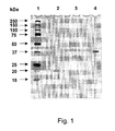
- Silver staining of the gel ( FIG. 1 ) performed under reducing conditions (SDS-PAGE; 4-20 % gradient) revealed the molecular weight of 20.0 kDa for purified recombinant Rv0164, 33.0 kDa for recombinant Rv0831c and 37.0 kDa for recombinant Rv1324 protein.
- the identity of purified proteins was further confirmed by LC-MS-MS.
- LPS endotoxin
- BCG vaccine was delivered by subcutaneous vaccination by administration of 50 ⁇ l of a BCG suspension (7x10 5 CFUs) injected above the gluteus superficialis and biceps femoralis muscles of both hind legs using a 26 gauge needle.
- BCG vaccine was administered by applying a total of 30 ⁇ l of BCG vaccine (7x10 5 CFUs) to the external nares (15 ⁇ l per nostril) using a fine tip micropipette and allowing the mouse to inhale the suspension into the lungs naturally.
- BCG vaccine diluent was used as a control for either route of vaccination.
- Vaccination of human subjects is performed similarly.
- BCG dosing for human subjects is between 1-8 x10 5 CFU administered intranasally or by subcutaneous injection.
- Multicomponent vaccine was delivered to mice by the intranasal route three times at 2-week intervals as described above except using 90 ⁇ g of recombinant M . tuberculosis. protein mixture per dose (10 ⁇ g of each polypeptide). Dosage of human subjects involves the administration of 10-1000 ⁇ g of each polypeptide. The dose range for large animal subjects approaches that of humans with similarly sized receiving compartments. Small animals required dosage at the low end of the spectrum as the receiving compartment is proportionally smaller in size. For all studies sham immunization was performed by administration of phosphate buffered saline (PBS) (pH 7.2) DDA-MPL.
- PBS phosphate buffered saline
- Sample collection Collection of blood from small animals was performed by cardiac puncture under anesthesia at targeted time points. Large animals and human subjects were/will be drawn intravenously into suitable anticoagulant. Urine was collected by established procedures. Nasal lavage was performed by repeated flushing of the nares and associated upper respiratory tract of the sacrificed mouse with 200 ⁇ l of PBS (pH 7.2) containing complete EDTA-free protease inhibitor cocktail (Roche Diagnostics GmbH, Mannheim, Germany). Serum, urine and nasal washings were stored at -20° C until use.
- samples from the lungs, spleen, nasal associated lymphoid tissue (NALT), cervical lymph nodes (CLN), inguinal lymph nodes (ILN), mesenteric lymph nodes (MLN) and femur and tibial bone marrow (BM) were aseptically removed and placed into RPMI 1640 supplemented with 100 IU/ml penicillin, 50 ⁇ g/ml streptomycin, 1 mM L-glutamine, 25 mM HEPES, 1 mM sodium pyruvate, 5 x 10 -5 M ß-mercaptoethanol, vitamins and nonessential amino acids (Gibco-Invitrogen, Grand Island, NY) and 10% endotoxin-tested heat-inactivated fetal calf serum (FCS; Atlas Biologicals, Fort Collins, CO). Thoracic and peritoneal exudates cells were isolated by washing the respective cavities with RPMI 1640 media.
- NALT nasal associated lymphoid tissue
- CLN cervical lymph no
- mice were bled by cardiac puncture under anesthesia and their lungs were perfused via the right ventricle with PBS containing 10U ml -1 heparin to remove intravascular leukocytes. The lungs were then perfused with an enzyme mixture containing 1 mg/ml collagenase type IV (Sigma-Aldrich, St. Louis, MO) and 25U ml -1 DNase (Roche, Penzberg, Germany) in supplemented RPMI and sliced into small pieces in a sterile dish and the fragments were incubated in the enzyme mixture at 37° C for 1 h.
- an enzyme mixture containing 1 mg/ml collagenase type IV (Sigma-Aldrich, St. Louis, MO) and 25U ml -1 DNase (Roche, Penzberg, Germany) in supplemented RPMI and sliced into small pieces in a sterile dish and the fragments were incubated in the enzyme mixture at 37° C for 1 h.
- the digested lung fragments were pressed with a 5 ml syringe plunger through a 70- ⁇ m pore size cell strainer (BD Falcon, Bedford, MA) to obtain a single cell suspension and erythrocytes were lysed with RBC lysis buffer (eBioscience, San Diego, CA) for 4-5 min at room temperature.
- the lung cells were washed, recovered by centrifugation, and resuspended in supplemented RPMI for counting using the trypan blue dye exclusion method.
- the NALT was isolated as described ( Asanuma, H, et al. J Immunol Methods, 1997; 202:123-131 ), and BM was isolated by flushing cavities of femurs and tibias with RPMI.
- Single cell suspensions of spleen, lymph nodes, BM or NALT were obtained by gently grinding the respective organs through a 70- ⁇ m cell strainer into 10-20 ml supplemented RPMI. Cell suspensions were centrifuged at 300xg for 10 min and the erythrocytes were removed by treatment with RBC lysis buffer when necessary. Cells were washed several times with fresh RPMI and the cell concentration was adjusted accordingly.
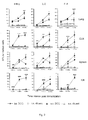
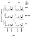
- Th1 responses from intranasal and subcutaneous BCG administration Following intranasal or subcutaneous administration the frequency and distribution of interferon- ⁇ (IFN- ⁇ , IL-2, and IL-4 secreting antigen-specific T-cells in lungs, spleen and respective draining lymph nodes were measured over the course of 12-weeks. Cells were isolated as described above with the total number of cells obtained independent of the type of immunization performed. M. tuberculosis. WCL-specific IFN- ⁇ , IL-2 and IL-4 responses were evaluated in the lungs and spleen after both i.n. and s.c BCG immunization ( FIG. 1 ) by IFN- ⁇ , IL-2, and IL-4 ELISPOT assay kits according to the manufacturers protocol. (BD-Biosciences, San Diego, CA).
- IFN- ⁇ , IL-2 and IL-4 secreting cells were found in the lung and draining CLN after i.n. immunization than after subcutaneous (s.c.) immunization at all three time points evaluated except more WCL-specific IL-4-secreting SFUs were observed in the lungs after s.c. BCG-immunization than after i.n. immunization at the 12 week time point (p ⁇ 0.0001).
- s.c. BCG-immunization induced higher WCL-specific IFN- ⁇ , IL-2 and IL-4 responses in the ILN, which drains the flank (the site of vaccination), and spleen as compared to i.n.
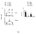
- Nitric Oxide production following BCG immunization alone Nitrite (NO 2 - ) accumulation in the supernatant of cultured cells was measured as an indicator of nitric oxide (NO) production by a Griess assay using a sodium nitrite standard as described by Sable, S, et al., Eur Respir J, 2007; 29:337-346 .
- WCL stimulation of lung cell cultures from mice intranasally immunized with BCG produced significantly higher nitrite levels at all the three time points compared to intranasally diluent immunized (p ⁇ 0.0001) or subcutaneously BCG immunized mice (p ⁇ 0.0001) ( FIG. 3A ).
- WCL-induced nitrite levels in the spleen cell cultures following subcutaneous BCG immunization were significantly higher than those observed following subcutaneous diluent immunization at all the three time points (p ⁇ 0.0001 ) and intranasal BCG immunization at the 3 week ( p ⁇ 0.0001) and 6 week (p ⁇ 0.05) time points.
- intranasal BCG immunization induced increased levels of WCL-specific nitrite levels in the spleen as compared to subcutaneous BCG immunization (p ⁇ 0.05), although the levels in each case were relatively low ( FIG. 3A ).
- thoracic and peritoneal exudate cells following intranasal BCG immunization were found to produce significantly higher levels of nitrite after stimulation with WCL (p ⁇ 0.0001) than those isolated following subcutaneous BCG immunization at 12 weeks ( FIG. 3B ).
- BM derived dendritic cells were used at 5:1 lymphocytes/DC.
- cells were stimulated in triplicate or quadruplet with either 100 ⁇ l of 10 ⁇ g/ml purified recombinant M. tuberculosis. antigen, antigen combination, WCL, STCF, or Con-A in supplemented RPMI as a positive control for cell viability and reactivity or medium alone as a negative control.
- the proliferation was expressed as mean counts per minute (CPM) of antigen stimulated cultures after subtracting mean counts per minute of cultures without antigen and the stimulation index (SI) was calculated by dividing mean counts per minute in antigen-stimulated wells by mean counts per minute in unstimulated wells.
- CPM mean counts per minute
- SI stimulation index
- Intranasal BCG immunization induced both STCF- and WCL-specific long-term T-cell responses in the lungs, the local lymph nodes draining the nasal passage (i.e. CLN) and the spleen ( FIG. 4 ). Strong cytokine responses were also observed at 6 weeks post-immunization in the MLN which drains the gastrointestinal tract. However, the STCF- and WCL-specific responses in MLN declined at 30 weeks. Peritoneal exudate cells (PEC) demonstrated an increased response from 6 weeks to 30 weeks of STCF- and WCL-specific Th1 and Th2 cytokines secreting cells following intranasal BCG immunization.
- PEC Peritoneal exudate cells
- Both STCF- and WCL-specific IFN- ⁇ and IL-2 secreting SFUs also increased from 6 weeks to 30 weeks in bone marrow cells while WCL-specific IL-4 SFUs decreased from 6 weeks to 30 weeks.
- NALT demonstrated both STCF- and WCL-specific proliferation at early (SI 12.15 and 18.60 at 6 weeks respectively; mean CPM of unstimulated culture 720) and late (SI 10.26 and 12.52 at 30 weeks respectively; mean CPM of unstimulated culture 660) time points.
- STCF- and WCL-specific antibody allotype (IgG and IgA) and isotype (IgG1 and IgG2a) levels were evaluated in serum, nasal lavage and urine of intranasal BCG immunized mice. Immunization via the intranasal route induced significantly elevated antigen-specific antibody responses at 30 weeks as compared to the 6 week time point ( FIG. 5 ) and was characterized by predominant antigen-specific IgG allotype levels in serum and IgA levels in nasal lavage and urine.
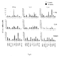
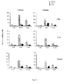
- IFN- ⁇ responses induced by polypeptides of M. tuberculosis following intranasal BCG immunization The ability of M. tuberculosis polypeptides to be recognized by T-and B-lymphocytes at 3 and 30 weeks was evaluated and is illustrated in FIG. 6 .
- Antigen-specific allotype and isotype responses in serum, nasal lavage and urine were low at 3 weeks. However, at 30 weeks the strongest antibody response observed was against Ag85 complex proteins (mean serum IgG A 492 1.189 ⁇ 0.009 for Ag85B) followed by Pst-S1, while Apa-specific antibody responses were moderate (mean A 492 range 0.3-0.1) as compared to rest of the antigens (mean A 492 ⁇ 0.1).
- T-cell and antibody responses following multicomponent polypeptide vaccination The frequency of Th1 and Th2 cytokine secreting cells in different immune compartments after in vitro stimulation with individual polypeptides is illustrated in FIG. 7 . Following intranasal immunization with polypeptide multicomponent cocktail-DDA-MPL, all nine polypeptides were more strongly recognized by lung T-lymphocytes than those derived from other organs as evaluated by ELISPOT ( FIG. 7 ) and T-cell proliferation ( 3 H thymidine uptake) assay.
- the order of recognition of individual polypeptides in terms of induction of IFN- ⁇ secreting cells at the level of the lungs was Apa>MPT-64>Dnak>Pst-S1>GroEL>GroES>Ag85A>Ag85B>CFP-10.
- the antigen recognition pattern was similar in all immune compartments evaluated with the exception of NALT, MLN, and BM. Although Apa induced both IFN- ⁇ and IL-4 producing cells at the majority of sites following immunization, the frequency of IL-2 secreting cells was low. On the other hand, Ag85 complex (A and B), Pst-S1 and Dnak were observed to be prominent inducers of IL-2 secreting cells.
- Intranasal immunization induced predominantly immunogen-specific IgG levels in serum while both IgA and IgG levels were observed in the nasal lavage ( FIG. 8 ).
- Apa induced prominent IgA, IgG1 and IgG2a isotype responses in nasal lavage, serum, and urine respectively.
- Pst-S1, Ag85B and Ag85A also induced strong humoral responses.
- nonadherent cells were removed by gentle washing to obtain adherent macrophage population.
- Effector cells, lung cells and splenocytes (2x10 5 cells/ml) from BCG-immunized or sham-immunized mice were cultured for 5 days in RPMI medium alone to serve as rested T-cell negative controls or stimulated for 5 days with WCL (20 ⁇ g/ml) or Apa (10 ⁇ g/ml) in 24-well plates.
- WCL (20 ⁇ g/ml) or Apa (10 ⁇ g/ml) in 24-well plates.
- macrophages were infected with M. tuberculosis at a multiplicity of infection of 1.
- Infected macrophages were seeded at 1 ⁇ 10 6 cells ml -1 in 96-well plates (100 ⁇ l well -1 ) and co-cultured with nonadherent lung or splenic effector cells (100 ⁇ l well -1 ) at a 1:1 ratio for 72 hr at 37° C with 5% CO 2 .
- the enumeration of CFU at 72 hr was performed by lysing the macrophages with 0.06% sodium dodecyl sulfate (Sigma-Aldrich, St.
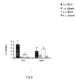
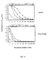
- Lung T-cells isolated 6 weeks after intranasal BCG immunization and expanded with Apa exhibited greater inhibition of the growth of M. tuberculosis. in peritoneal macrophages compared to medium-only expanded T-cells. ( FIG. 9 ) Further, inhibition was significantly higher than that imparted by lung cells isolated following subcutaneous BCG immunization (p ⁇ 0.01).
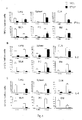
- Induction of proliferative responses following intranasal immunization with single component vaccine The ability of individual polypeptides to induce proliferative responses in cells isolated from lungs, CLN, or spleen was evaluated following intranasal immunization with a single or multiple polypeptide vaccine comprising polypeptide encapsulated with cationic liposomes. 3 [H] thymidine incorporation was evaluated as a measurement of proliferative response. Lung cells demonstrated higher incorporation than cells isolated from spleen following in vitro stimulation with representative polypeptides. ( FIG.
- Rv0831c, Rv1324, and Rv0164 (10 ⁇ g of each protein/dose)
- Rv0831c and Rv1324 induced significantly better proliferation in target organ cultures after in vitro stimulation as compared to Rv0164 ( Fig. 11 ).
- Rv0831c When three polypeptides were intranasally coadministered, Rv0831c induced higher numbers of cytokine secreting cells than Rv1324 and Rv0164. The levels observed in the lungs for Apa protein was more than two-fold higher than Rv0831c (compare FIGS. 7 and 12 ).
- Immunogen specific allotype and isotype response in serum and nasal lavage following single or multicomponent intranasal immunization The allotype and isotype immunoglobulin response was measured 2 and 4 weeks post immunization by ELISA. Briefly, total immunoglobulin G (IgG), immunoglobulin A (IgA), and IgG isotypes IgG1 and IgG2a specific to purified recombinant antigens and antigen combination were estimated.
- the plates were washed four times with PBS-T, and 100 ⁇ l of horseradish peroxidase-conjugated anti-mouse secondary antibodies (anti-mouse IgG and IgA, Sigma-Aldrich, St. Louis, MO and anti-mouse IgG1 and IgG2a, BD-Biosciences, San Diego, CA) diluted 1:1,000 in PBS-T containing 1% BSA were added to respective wells. After 90 min the plates were washed six times with PBS-T. The reaction was developed with o-phenylenediamine (Sigma-Aldrich, St. Louis, MO) and hydrogen peroxide in citrate substrate buffer (pH 5.0). The reaction was stopped after 20 min by adding 100 ⁇ l of 1 M H 2 SO 4 and the absorbance was measured at 492 nm (A 492 ).
- horseradish peroxidase-conjugated anti-mouse secondary antibodies anti-mouse IgG and IgA, Sigma
- Immunization with Rv1324 demonstrated a strong immunogen specific antibody response in excess of that observed for Rv0831c and Rv0164.
- FIG. 13 Intranasal Rv1324 immunization induced antibody response was characterized by strong immunogen specific IgG response with both IgG1 and IgG2a isotype levels in the serum and mixed IgA and IgG response with predominant IgG1 isotype levels in the nasal lavage as evaluated at both 2 and 4 week time points.
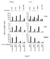
- Example 15 Immunogenicity of native and recombinant Apa based experimental subunit vaccines in mice.
- mice were immunized by the intranasal route three times at 2-week intervals using 10 ⁇ g of either native M. tuberculosis Apa, recombinant E. coli expressed Apa or recombinant Ag85A individually [10 ug of each immunogen emulsified individually in Dimethyl- dioctadecyl-ammonium bromide (DDA; 250 ⁇ g/dose, Sigma-Aldrich, St. Louis, MO) and monophosphoryl lipid A (MPL derived from Salmonella minnesota Re 595; 25 ⁇ g/dose, Sigma-Aldrich, St. Louis, MO)].
- DDA Dimethyl- dioctadecyl-ammonium bromide
- MPL monophosphoryl lipid A
- MPL was first mixed with endotoxin-free sterile water (Burdick & Jackson, Muskegon, MI) containing 0.2% triethylamine (Fisher Scientific, Fair Lawn, NJ). The mixture was heated in a 70° C water bath for 30 s and then sonicated for 30 s. The heating and sonicating procedure was repeated twice.
- DDA was suspended in sterile water and a homogeneous dispersion of the powder was obtained by heating the suspension at 80° C for 5-10 min in water bath. After cooling to room temperature, MPL and antigens were mixed with DDA just before use.
- the sham-immunized mice received PBS (pH 7.2) emulsified in DDA-MPL.
- mice were bled by cardiac puncture under anesthesia and sacrificed at 2 and 4 weeks post-immunization.
- Lungs, spleen and cervical lymph nodes (CLN) were aseptically removed and placed into RPMI 1640 supplemented with 100 IU ml -1 penicillin, 50 ⁇ g ml -1 streptomycin, 1 mM L-glutamine, 25 mM HEPES, 1 mM sodium pyruvate, 5 x 10 -5 M ß-mercaptoethanol, vitamins and nonessential amino acids (Gibco-Invitrogen, Grand Island, NY) and 10% endotoxin-tested heat-inactivated fetal calf serum (FCS; Atlas Biologicals, Fort Collins, CO).
- FCS endotoxin-tested heat-inactivated fetal calf serum
- lymphoid or extra-lymphoid organs were pooled from 4 mice for each treatment group and cells were extracted for analysis of in vitro M. tuberculosis antigen-specific cellular responses. Antigen-specific antibody responses were evaluated using pooled nasal lavage and serum collected from each treatment group.
- mice were bled by cardiac puncture under anesthesia and their lungs were perfused via the right ventricle with PBS containing 10U ml -1 heparin to remove intravascular leukocytes.
- the lungs were then perfused with an enzyme mixture containing 1 mg/ml collagenase type IV (Sigma-Aldrich, St. Louis, MO) and 25U ml -1 DNase (Roche, Penzberg, Germany) in supplemented RPMI and sliced into small pieces in a sterile dish and the fragments were incubated in the enzyme mixture at 37° C for 1 h.
- an enzyme mixture containing 1 mg/ml collagenase type IV (Sigma-Aldrich, St. Louis, MO) and 25U ml -1 DNase (Roche, Penzberg, Germany) in supplemented RPMI and sliced into small pieces in a sterile dish and the fragments were incubated in the enzyme mixture at 37° C for 1 h.
- the digested lung fragments were pressed with a 5 ml syringe plunger through a 70- ⁇ m pore size cell strainer (BD Falcon, Bedford, MA) to obtain a single cell suspension and erythrocytes were lysed with RBC lysis buffer (eBioscience, San Diego, CA) for 4-5 min at room temperature.
- the lung cells were washed, recovered by centrifugation, and resuspended in supplemented RPMI for counting using the trypan blue dye exclusion method.
- the single cell suspensions of spleen and lymph nodes were obtained by gently grinding the respective organs through a 70- ⁇ m cell strainer into 10-20 ml supplemented RPMI.
- the cell suspensions were centrifuged at 300xg for 10 min and the erythrocytes were removed by treatment with RBC lysis buffer when necessary. Cells were washed several times with fresh RPMI and the cell concentration was adjusted accordingly.
- IFN interferon
- IL-2 IL-2
- IL-4 Mouse ELISPOT set
- IL-17 Mouse ELISPOT set, eBioscience kits
- IFN interferon
- 96 well ELISPOT plates were coated with 100 ⁇ l of 5 ⁇ g ml -1 capture antibody in PBS (pH 7.2) and incubated overnight at 4° C. Free binding sites were blocked with 200 ⁇ l of supplemented RPMI containing 10% FCS for 2h at room temperature.
- Cell concentration was adjusted to 1x10 6 and 2x 1 0 6 cells ml -1 for all sites and added to appropriate wells. No BM-derived DCs or macrophages were added to supplement the antigen presenting cells already present in the cell suspension as described previously for antigenicity or immunogenicity studies.
- cells were stimulated in triplicate with either 10 ⁇ g ml -1 of individual purified M . tuberculosis antigens, WCL, Concanavalin A (Con-A; Sigma-Aldrich, St. Louis, MO), or medium alone in a 100 ⁇ l volume.
- tuberculosis native Apa nApa
- RApa recombinant APA
- rAg85A recombinant Ag85A
- control DDA-MPL adjuvent to induce Th1 response IFN- ⁇ and IL-2
- BALB/c mice immunized intranasally with respective protein subunit vaccine or DDA-MPL adjuvant alone for lung, cervical, lymph nodes (CLN) or spleen cells.
- Intranasal rAg85A-DDA-MPL subunit vaccination was used as a positive control for evaluation of immunogenicity of native or recombinant Apa.
- Four weeks post immunization time point to be used for M. tuberculosis.
- tuberculosis native Apa tuberculosis native Apa
- recombinant APA recombinant Ag85A and control DDA-MPL to induce Th2 or Th17 response in BALB/c mice immunized intranasally with respective protein subunit vaccine or DDA-MPL adjuvant alone for lung, CLN and spleen cells.
- IL-4 antigen-specific Th2
- Th17 Th17
- ELISPOT assay expressed as spot forming units (SFUs)/ million cells of organ. The results are presented as means ⁇ standard deviation of three to six determinations.
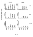
- T-cell responses induced by native or recombinant Apa were also comparable with those induced by rAg85A immunization. No difference was also observed in three vaccine immunized groups to induce T-cell (Th1, Th2 and Th17) responses following in vitro live M. bovis BCG stimulation of respective lung, CLN and spleen cell cultures indicating that three vaccine group might induce similar T-cell cytokine responses following M. tuberculosis. encounter or in vivo experimental challenge ( Figs. 17 and 18 ). In Fig.
Abstract
Description
- This application claims priority of United States Provisional Patent Application Serial No.
61/020,573 filed January 11, 2008 - The invention described herein may be manufactured, used and licensing by or for the United States Government.
- The present invention relates generally to the field of recombinant vaccines and methods of vaccination. More specifically the present invention relates to recombinant M. tuberculosis protein administration for use in a prime-boost immunization strategy.
- Each
year 8 to 10 million people worldwide develop tuberculosis. Globally the incidence of tuberculosis is growing at a rate of 1% a year primarily due to rapid increase in disease prevalence in Africa. In other regions successful control efforts have begun to stabilize disease incidence. Nevertheless, approximately 2,000,000,000 people, equal to one-third of the world's population, are estimated to be infected with Mycobacterium tuberculosis bacilli, the microbes that cause TB. (World Health Organization 2006 Tuberculosis Facts). - The need to find new treatments or vaccination strategies for tuberculosis is stressed by the increasing worldwide HIV infection rate such that, presently, 250,000 TB deaths are HIV associated. Tuberculosis itself is the second largest killer of mankind with more than 2 million deaths occurring worldwide annually (World Health Organization 2006 Tuberculosis facts). The attenuated Mycobacterium bovis bacillus Calmette-Guerin (BCG) vaccine is the only tuberculosis vaccine currently licensed for human use. The BCG vaccine is effective against severe pediatric and extra-pulmonary forms of tuberculosis. However, protection against adult pulmonary tuberculosis in developing countries is poor, with adult protection varying between 0 to 80% (Fine P.E.M., Lancet 2000; 346:1339-1345). The variable efficacy of tuberculosis vaccination appears to be geographically centered. For example in the United Kingdom approximately 75% protection has been observed (Hart P.D. and Sutherland I., BMJ, 1977; 2, 293-295). In contrast, clinical studies in India and Malawi failed to show consistent protection against pulmonary tuberculosis (Fine, PE, et al., Scand J Infec Dis, 2001; 33:243-45; Ponnighaus J.M., Lancet, 1992; 339:636-639).
- As the only effective vaccine for TB is the BCG vaccine, current research efforts are focused on improving BCG efficacy (Dietrich G., Vaccine, 2003; 21:667-670). For example, recombinant BCG vaccine over expressing fusion protein of the antigen Ag85B, the early secreted antigen (ESAT-6) and IFN-γ increased specific antibody titers and cellular immune responses relative to standard BCG vaccine, recombinant BCG vaccine expressing Ag85B alone, or recombinant BCG vaccine expressing a fusion protein of Ag85B and ESAT-6 (Xu Y., FEMS Immunology and Medical Microbiology, 2007;51:480-487). ESAT-6, a protein produced by virulent Mycobacterium tuberculosis, is absent in standard BCG vaccine strains and is currently undergoing intense study as a potential vaccine subunit against tuberculosis. For example, DNA vaccines encoding ESAT-6 combined with immunization with BCG in mice subsequently challenged with tuberculosis H37Rv showed improved ESAT-6 specific interferon gamma (Fan X., Scandinavian Journal of Immunology Oct. 4, 2007; 66:523-528).
- In addition to studies of new subunit vaccines, prime-boost strategies are currently under investigation as a method of improving BCG immunogenicity (Goonetilleke N.P., Journal of Immunology 2003; 171:1602-1609; Kaufmann S.H., Nature Reviews Immunology 2001; 1:20-30). Prime-boost strategies commonly employ DNA vaccines. For example, when a DNA vaccine expressing Ag85B was administered in a murine M. tuberculosis. model followed by boosting with BCG vaccine, improved protective efficacy over BCG vaccine alone was observed (Feng C.G., Infectious Immunology 2001; 69:4174-4176). Similarly, DNA injection encoding the M. tuberculosis proteins Apa, HSP-65 and HSP-70 subsequently followed by conventional BCG vaccination also improved protection against tuberculosis challenge in mice (Ferraz, Infection and Immunity 2004; 72:6945-6950).
- Traditional immunizations are generally administered via an intramuscular or subcutaneous route. However, tuberculosis is primarily a respiratory disease. Thus, protection against infection and subsequent eradication of disease may best be accomplished by direct administration to the respiratory mucosa (Kallenius, et al. Tuberculosis (Edinb), 2007; 87:257-66). Intranasal vaccination may have advantages over other routes of administration such as, intranasal vaccination is not influenced by a preformed systemic immunity whereas parenteral vaccination is less effective in individuals with preexisting antibodies (van Savage J.M., Journal of Infectious Disease 1990; 161:487-492).
- Circumventing the existence of preexisting antibodies is important in geographical regions where an improved vaccine against tuberculosis is most needed. Prior Th2 background immunity resulting from prior exposure to helminthes and saprophytic mycobacteria has been suggested to decrease the ability of BCG vaccine in inducing immunoprotection (Rook, Vaccine, 2005; 23:2115-2120). Further, it is envisaged that intranasal vaccination might be effective in preventing M. tuberculosis infections in the host (Kauffman SH., Nature Reviews of Immunology 2006; 6:699-704). Animal studies of intranasal vaccination showed increased protective efficacy as compared to subcutaneous route of vaccination (Giri, PK. et al. FEMS Immunology and Medical Microbiology, 2005; 45:87-93; Chen, L. et al. Infection and Immunity, 2004; 72:238-246).
- While studies of live or killed BCG vaccine, protein subunit vaccines, recombinant bacterial vector vaccines, plasma DNA vaccines or combinatorial immunization approaches in both human and animal systems have been subjected to preliminary study, little is known as to which method produces the most robust immune response and the greatest level of protection in the subject. Further, detail concerning immune response characteristics induced by each vaccine type is yet to be fully elucidated. The increased prevalence of tuberculosis infection and increased resistance, particularly in the developing world, creates a need for an improved tuberculosis vaccine and vaccination strategy.
- A vaccine is provided that increases an immune response in a subject wherein the vaccine includes at least one M. tuberculosis. polypeptide wherein the polypeptide is optionally Ag85A, Ag85B, MPT-64, Pst-S1, Apa, GroES, GroEL, Dnak, CFP-10, Rv0831c, and Rv1324, portions thereof, combinations thereof, or multiples thereof. These recombinant proteins are optionally purified in their natural form or they further comprise a tag suitable for increasing purification. The M. tuberculosis polypeptides are optionally recombinant.
- An inventive vaccine optionally contains an emulsion. Suitable emulsification agents include supramolecular biovectors (SMBV), nanoparticles, liposomes, or combinations thereof.
- An inventive vaccine optionally contains an adjuvant. Suitable adjuvants illustratively include dimethyl dioctadecyl-ammonium bromide (DDA); monophosphoryl lipid A (MPL); LTK63, lipophilic quaternary ammonium salt-DDA, DDA-MPL, aluminum salts, aluminum hydroxyide, aluminum phosphate, potassium aluminum phosphate, Montanide ISA-51, ISA-720, microparticles, immunostimulatory complexes, liposomes, virosomes, virus-like particles, CpG oligonucleotides, cholera toxin, heat-labile toxin from E. coli, lipoproteins, dendritic cells, IL-12, GM-CSF, nanoparticles illustratively including calcium phosphate nanoparticles, combination of soybean oil, emulsifying agents, and ethanol to form a nanoemulsion; AS04, ZADAXIN, or combinations thereof.
- Also provided is a process of increasing an immune response in a subject wherein an M. tuberculosis polypeptide is administered to the subject. Administration is optionally via routes including intradermal, transdermal, subcutaneous, intramuscular, intranasal, aerosolized, oral, sublingual, intravaginal, per-rectal, intravenous, intramucosal, or other methods of delivery known in the art. The process of increasing an immune response optionally employs administering to a subject a second vaccine which is optionally Ag85A, Ag85B, MPT-64, Pst-S1, Apa, GroES, GroEL, Dnak, CFP-10, Rv0831c and Rv1324 or combinations thereof, epitopes of above mentioned polypeptides or peptides thereof. Optionally the administration of a BCG vaccine occurs prior to the administration of a recombinant tuberculosis polypeptide. Alternatively administration of a BCG vaccine might occur subsequent to administration of a recombinant tuberculosis polypeptide or optionally administration of a BCG vaccine occurs simultaneously to the administration of a recombinant tuberculosis polypeptide(s), epitope(s) or peptide(s). The BCG vaccine is optionally recombinant (expressing one or more above mentioned polypeptides) or natural. Furthermore the administration of either a BCG vaccine and/or a recombinant tuberculosis polypeptide occurs prior to, concurrent with, or after the subject is exposed to mycobacterium infections or developed a disease.
- Also provided is a pharmaceutical package comprising at least one polypeptide selected from the group comprising Ag85A, Ag85B, MPT-64, Pst-S1, Apa, GroES, GroEL, Dnak, CFP-10, Rv0831c and Rv1324 or combinations thereof. Also an emulsification agent and an adjuvant. The emulsification agent is optionally a dimethyl dioctadecyl-ammonium bromide. Optionally the adjuvant is monophosphoryl lipid A.
-
-
Figure 1 represents an SDS-PAGE (4-20% gradient gel) of purified new recombinant M. tuberculosis proteins processed by silver stain whereinlane 1 represents molecular weight markers,lane 2 is Rv0164,lane 3 is Rv0831c, andlane 4 is Rv1324; -
Figure 2 represents the kinetics of T-cell responses induced by intranasal or subcutaneous BCG vaccination; -
Figure 3 represents kinetics of NO response induced by intranasal or subcutaneous BCG vaccination; -
Figure 4 represents the distribution of M. tuberculosis whole cell lysate (WCL) and short term culture filtrate (STCF) specific T-cells in local and peripheral immune compartments of subjects at early (6 weeks) and late (30 weeks) time points after intranasal BCG vaccination wherein the results are presented as means ± standard deviation of three to six determinations after subtracting the SFUs from respective unstimulated cultures; -
Figure 5 represents M. tuberculosis whole cell lysate (WCL) and short term culture filtrate (STCF) specific antibody levels in local and peripheral body fluids of subjects at early (6 weeks) and late (30 weeks) time points after intranasal BCG vaccination wherein the results of ELISA measurements are presented as mean absorbance of triplicate wells at 492 nm ± standard deviation after subtracting the absorbance of control wells; -
Figure 6 represents the ability of M. tuberculosis recombinant antigens to induce T-cell responses in intranasally BCG-vaccinated subjects at early (3 weeks) and late (30 weeks) time points wherein the results of ELISPOT assays are presented as means ± standard deviation of duplicate determinations after subtracting the SFUs from respective unstimulated cultures; -
Figure 7 represents the ability of M. tuberculosis recombinant antigens to induce T-cell response in subjects immunized intranasally with themulticomponent subunit vaccine 2 weeks post immunization wherein the results of ELISPOT assays are presented as means ± standard deviation of duplicate determinations after subtracting the SFUs from respective unstimulated cultures; -
Figure 8 represents M. tuberculosis recombinant antigen-specific antibody response in subjects immunized intranasally with themulticomponent subunit vaccine 2 weeks post immunization wherein results are presented as mean absorbance at 492 nm ± standard deviation from triplicate determination after subtracting the absorbance of control wells; -
Figure 9 represents T-cells expanded with Apa inhibiting intracellular growth of M. tuberculosis in macrophages at 6 weeks post-immunization; -
Figure 10 represents polypeptide immunogen specific proliferative responses in intranasally immunized subjects wherein Rv0164, Rv0831c, and Rv1324 are individually encapsulated in cationic liposome and individually administered; -
Figure 11 represents polypeptide immunogen specific proliferative responses in intranasally immunized subjects with the combination of Rv0164, Rv0831c, and Rv1324 encapsulated in cationic liposome; -
Figure 12 represents the distribution of polypeptide immunogen specific T-cells in lungs, CLN, and spleen of subjects intranasally immunized with a single polypeptide encoding either Rv0164, Rv0831c, or Rv1324; -
Figure 13 represents polypeptide immunogen specific allotype and isotype antibody responses in the nasal lavage and serum from subjects immunized with cationic liposome encapsulated recombinant Rv0164, Rv0831c, or Rv1324 at 2 and 4 weeks post immunization; -
Figure 14 represents polypeptide immunogen specific allotype and isotype antibody responses in the nasal lavage and serum from subjects immunized with cationic liposome encapsulated recombinant Rv0164, Rv0831c, and Rv1324 combination at 2 and 4 weeks post immunization; -
Figure 15 represents the comparative ability of M. tuberculosis native Apa (nApa), recombinant APA (rApa), recombinant Ag85A (rAg85A) and control DDA-MPL adjuvant to induce Th1 response (IFN-γ and IL-2) in BALB/c mice immunized intranasally with respective protein subunit vaccine or DDA-MPL adjuvant alone for lung, cervical, lymph nodes (CLN) or spleen cells; -
Figure 16 represents the comparative ability of M. tuberculosis native Apa, recombinant APA, recombinant Ag85A and control DDA-MPL to induce Th2 or Th17 response in BALB/c mice immunized intranasally with respective protein subunit vaccine or DDA-MPL adjuvant alone for lung, CLN and spleen cells; -
Figure 17 represents the frequency of Th1 (IFN-γ and IL-2) cytokine-secreting cells in the lung, cervical lymph node (CLN), and spleen cell cultures of subunit and sham immunized mice following in vitro M. bovis BCG challenge at four weeks post-immunization in lung, CLN and spleen cell cultures; and -
Figure 18 represents the frequency of Th2 (IL-4) and Th17 (IL-17) cytokine-secreting cells in the lung, cervical lymph node (CLN), and spleen cell cultures of subunit and sham immunized mice following in vitro M. bovis BCG challenge at four weeks post-immunization. - The increased prevalence of HIV increases the need for an M. tuberculosis vaccine that has efficacy in both pediatric and adult patients in the developed and developing worlds. The instant invention has utility as a new vaccine against Mycobacterium.
- To answer the wide range of BCG vaccine efficacy against tuberculosis, a novel inventive strategy is provided to boost the immune response generated following administration of a BCG vaccine. Toward this end, secreted proteins of M. tuberculosis represent a valuable source of antigens for use in boosting the efficacy of BCG vaccine. The instant invention provides a vaccine that used alone or in conjunction with BCG, increases the immune response of a subject. In a preferred embodiment the instant invention utilizes at least one M. tuberculosis polypeptide. Polypeptides suitable in the instant invention include any polypeptide expressed by virulent M. tuberculosis within a subject. Polypeptides suitable for use in the instant invention optionally include Ag85A, Ag85B, MPT-64, Pst-S1, Apa, GroES, GroEL, Dnak, CFP-10, Rv0831c and Rv1324, portions thereof, combinations thereof, or multiples. Multiples thereof illustratively mean more than one polypeptide sequence type or more than one copy of a single polypeptide sequence. Polypeptides suitable in the instant invention are optionally recombinant or naturally derived.
- Preferably polypeptides suitable in the instant invention are recombinant and obtained by methods known in the art. Illustratively, a nucleotide sequence is cloned into a plasmid which is transfected into E. coli and expressed. To ease purification procedures the expressed polypeptides from E. coli optionally include a tag sequence. Illustrative examples of tags suitable for use in the instant invention include poly-histidine, CBP, CYD (covalent yet dissociable NorpD peptide), strep-2, FLAG, HPC or heavy chain of protein C peptide tag, or GST and MBP protein fusion tag systems. It is appreciated that other tag systems are similarly operable. In a preferred embodiment recombinant polypeptides are expressed in E. coli and purified using an affinity tag system followed by enzymatic cleavage of the tag such as by incorporating a factor Xa, thrombin, or other enzyme cleavage site in the expressed polypeptide. Methods of tag cleavage are known in the art and any effective method is appreciated to be suitable for use in the instant invention.
- In a preferred embodiment a multi-component vaccine is employed. The multi-subunit vaccine optionally contains a set of individual polypeptides or a single or family of fusion proteins wherein each of the proteins optionally represents a single protein expressed by virulent M. tuberculosis. Preferably a nine polypeptide vaccine is employed. It is appreciated that each individual antigen or polypeptide is individually suitable for use in the instant invention. Unexpectedly, administration of a multi-component vaccine increases the immunogenicity of each of the individual components. Thus, the preferred embodiment of a nine subunit vaccine demonstrates synergistic immunogenicity.
- The term subject is illustratively a living organism capable of mounting an immune response to challenge from a vaccine. Non-limiting examples of a subject include a human, any lower primate, dog, cat, rabbit, rat, mouse, guinea pig, pig, hamster, horse, donkey, cattle, possum, badger, goat, or other mammals or non-mammals.
- The term immune response is illustratively any alteration of a subject's immune system in response to challenge from a vaccine, infectious or otherwise foreign organism, tissue, cell, antigen, antibody, nucleotide strand, or other immune stimulating substance recognized in the art. Non-limiting examples of immune responses include in vitro secretion of IL-2, IL-4, or IFN-γ in CD4+ or CD8+ T-cells; protection from challenge after M. tuberculosis H37Rv or other infectious organism; alteration in nitrite levels; Th1 and Th2 cytokine responses in various immune compartments; alteration in allotype and isotype antibody levels; in vitro recognition of antigen; B-cell responses; inhibition of growth of M. tuberculosis bacilli in infected macrophages; survival; or other response known in the art.
- The term polypeptide is illustratively a chain of two or more amino acid residues. In a preferred embodiment, a polypeptide suitable for use in the instant invention is the amino acid sequence for Rv1860 (Apa) protein, whole recombinant or natural protein, mutants thereof, portions, epitopes or peptides thereof, homologs thereof, or the Apa sequence combined with other peptide sequences(s). The Apa sequence is found at accession number YP_177849.
- Preferably, the inventive vaccine is a multi-component vaccine. A multi-component vaccine illustratively includes nine polypeptide antigens such as Ag85A, Ag85B, MPT-64, Pst-S1, Apa, GroES, GroEL, DnaK, and CFP-10. Representative Mycobacterium tuberculosis H37Rv polypeptides operative as candidates for inclusion in a vaccine along with their respective nucleotide sequences:
- Ag 85 A (Rv3804c): Gene ID: 886132; protein ID: NP_218321
- Ag 85 B (Rv1886c): Gene ID: 885785; protein ID: NP_216402
- MPT-64 (Rv1980c): Gene ID: 885925; protein ID: NP_216496
- Pst-S1 (Rv0934): Gene ID: 885724; protein ID: YP_177770
- Apa (Rv1860): Gene ID: 885896; protein ID: YP_177849
- GroES (Rv3418c): Gene ID: 887583; protein ID: NP_217935
- GroEL (Rv0440): Gene ID: 886354; protein ID: NP_214954
- Dnak (Rv0350): Gene ID: 885946; protein ID: NP_214864
- CFP-10 (Rv3874): Gene ID: 886194; protein ID: NP_218391
- CFP-31 (Rv0831c): Gene ID: 885349; protein ID: NP_215346
- CFP-32 (Rv1324): Gene ID: 886897; protein ID: NP_215840
- MTSP-17/CFP-15 (Rv0164): Gene ID: 886267; protein ID: YP_177617
- Surprisingly, intranasal (i.n.) delivery of the above cited multi-subunit vaccine results in lung specific immune responses to each of nine individual antigens. The recognition level of each antigen is unique with the level of IFN-γ secreting cells ordered from:
- Apa>MPT-64>DnaK>Pst-S1>GroEL>GroES>Ag85A>Ag85B>CFP-10.
- This antigen recognition pattern was also observed in other immune compartments. However, a multiplexed micro-sphere based cytokine immunoassay showed the greatest response to Ag85A, Ag85B, and Pst-S1 when IL-2 levels were measured. (Table 2.)
- In a preferred embodiment a vaccine contains a single polypeptide antigen. Preferably, the antigen is the mycobacterial Apa secretion protein also known as the 45/47-kDa protein complex, or as the protein from gene modD. (Romain, F. et al., Infect. Immun., 1993; 61:724-750.) Apa is secreted as a glycosylated protein with nine glycoforms. (Horn, C., et al., J. Biol. Chem., 1999; 274:32023-32030.) Prior studies demonstrated that glycosylation of Apa is required for antigenicity in vitro and in vivo. (Romain, R, et al., Infect Immun, 1999; 67:5567-5572). These authors provide three possible explanations for the requirement of glycosylation in eliciting an immune response. First, glycopeptide specific responses have been reported when immunizing with other glycopeptides implying that both the peptide backbone and the glycosylation interact with the T-lymphocyte receptor (Carbone, FR, and Gleeson PA, Glycobiology, 1997; 7:725-730; Deck B, et al., J Immunol, 1995; 155:1074-1078; Haurum, JS, et al., J Exp Med, 1994; 180:739-744). Second, the responsible receptors for eliciting immune responses may be nonspecific in nature such that mannoslyation of many antigens are recognizable by a single receptor type. (Stahl, PD, and Ezekowitz, RAB, Curr Opin Immunol, 1998; 10:50-55). Finally, the presence or absence of glycosylation could alter the uptake or processing of such molecules by macrophages or dendritic cells. Antigenicity of Apa is believed to be related to the presence of glycosylation. However, the effect of glycosylation on T and B cell immunogenicity of APA has not been evaluated in detail following protein subunit vaccination.
- Correspondingly, the prime-boost strategy of Romain, et al. was successful in eliciting an improved immune response following priming with a trivalent DNA vaccine including vectors encoding Apa, Hsp70, and Hsp65 followed by boosting a BCG vaccine. The use of DNA vaccines produces glycosylated antigens (Apa, etc.) as the antigens are expressed in host cells. In the case of Romain, the priming antigens were subjected to mammalian cell glycosylation during expression and any antigenicity or immunogenicity is expected to be the result of glycosylated antigen exposure.
- In a preferred embodiment of the instant invention, the polypeptide is produced without glycosylation. In a non-limiting example, polypeptides are synthesized in E. coli which is recognized in the art as incapable of properly glycosylating a protein relative to a mammalian cell type or M. tuberculosis. It is appreciated the other synthetic means that do not glycosylate the polypeptide are similarly suitable. Thus, in this embodiment protein(s) are free from glycosylation. As the high degree of cellular recognition of Apa, for example, is attributable to the presence of glycosylation, and all prior studies analyzing the immunogenicity or antigenicity of Apa were attributable to glycosylated antigen, it was expected that the absence of glycosylation would not yield efficacious results. Surprisingly in the instant invention, vaccination using non-glycosylated polypeptides resulted in robust immunogenicity.
- The instant invention is suitable as a stand alone vaccine, a priming vaccine, or as a boosting vaccine. Preferably, the instant invention is used in a prime-boost strategy. More preferably, the instant invention is used as priming vaccine. When used as priming vaccine the instant invention is illustratively followed by immunization with BCG vaccine or other subunit/DNA vaccine(s). In an embodiment of the invention, a single subunit priming vaccine is employed. Preferably, a vaccine wherein the polypeptide is Apa. It is appreciated that other polypeptides are similarly suitable. In this embodiment immunization with the Apa protein is optionally followed by immunization with a single subunit or multicomponent vaccine illustratively employing more than one protein in addition to or at the exclusion of Apa. Delivery of subsequent vaccinations is optionally within a short or long time period. In a nonlimiting example, the second vaccination is delivered within a single day or week. Alternatively, the second vaccination is delivered after one year or after few years. It is appreciated that a delivery of a second vaccine may be at any time during the subject's lifetime.
- Preferably, the second vaccine is protein subunit vaccine or even BCG vaccine (normal or recombinant). Delivery of a second or subsequent vaccine is optionally by the same delivery route as the first immunization or by an alternative route. In a preferred example, a prime vaccine is delivered by the intranasal route. A subsequent boost vaccine is also delivered by the intranasal route.
- In a preferred embodiment, delivery of BCG vaccine occurs prior to boosting with a single or multicomponent vaccine. Preferably, BCG vaccination is boosted by delivery of a multicomponent vaccine. More preferably, BCG is boosted by vaccination with a single component vaccine wherein the single component is a polypeptide encoding Apa protein. It is similarly appreciated that delivery of the boost vaccine is optionally delivered at any time during the subject's lifetime.
- The instant invention is optionally used as a combination vaccine. Illustratively, a BCG vaccine is supplemented with a single or multicomponent vaccine. This strategy allows for simultaneous delivery of a single polypeptide, multicomponent, and a BCG vaccine in any combination.
- In a preferred embodiment, a single or multicomponent vaccine comprising a single or multiple polypeptides is delivered by the intranasal route and a BCG vaccine is delivered by an intradermal or subcutaneous route. More preferably, a single or multicomponent vaccine is delivered by an intranasal route and a boost BCG vaccine is delivered also by an intranasal route. Alternatively, BCG vaccine is given by intranasal or percutaneous route and a boost single or multicomponant vaccine is also delivered by the same route. It is appreciated that methods of delivery known in the art are suitable for delivering vaccine by these or other routes of entry.
- Delivery of the inventive vaccine is optionally administered prior to, during, or following active or inactive infection with TB or after development of disease alone or in conjunction with antitubercular chemotherapy. Thus, delivery of a single or multicomponent vaccine is optionally prophylactic, postinfection or therapeutic. In a preferred embodiment, the instant invention is designed to prevent or eliminate disease, or to prevent infection. Optionally, delivery of a single or multicomponent vaccine is therapeutic.
- The polypeptide is optionally delivered as naked polypeptide, in aqueous solution, in an emulsion, or in other suitable delivery compositions. In a preferred embodiment the instant invention is delivered as a vaccine or as a vaccine component of a pharmaceutical package. Optionally, a polypeptide (or multiple polypeptides) or immunogenic peptides or epitopes are present in an emulsion comprised of suitable emulsification agents. In a preferred embodiment a multicomponent vaccine is emulsified or encapsulated in a suitable vaccine carrier. More preferably, a single subunit vaccine is emulsified. Most preferably, a polypeptide encoding Apa is emulsified. Suitable emulsification agents illustratively include supramolecular biovectors (SMBV), nanoparticles such as described by Major, M, et al., Biochim. Biophys. Acta, 1997; 1327:32-40, De Migel, I, et al., Pharm. Res., 2000; 17:817-824,
U.S. Patent Nos. 6,017,513 ,7,097,849 ,7,041,705 ,6,979,456 ,6,846,917 ,6,663,861 ,6,544,646 ,6,541,030 ,6,368,602 , Castignolles, N., et al., Vaccine, 1996; 14:1353-1360, Prieur, E., et al., Vaccine, 1996; 14:511-520, Baudner B, et al., Infect Immun, 2002; 70:4785-4790; liposomes such as described by El Guink et al., Vaccine, 1989; 7:147-151, and inU.S. Patent No. 4,196,191 ; or other agents known in the art. Agents suitable for use are generally available commercially. - Optionally, the instant invention includes codelivery of polypeptides with an adjuvant. Suitable adjuvants illustratively include dimethyl dioctadecyl-ammonium bromide (DDA); monophosphoryl lipid A (MPL); E. coli heat-labile enterotoxin, genetically modified derivatives thereof, LTK63, Trehalose Dimycolate and synthetic derivatives, lipophilic quaternary ammonium salt-DDA, DDA-MPL, DDA-TDM, DDA-TDB, IC-31, aluminum salts, aluminum hydroxyide, aluminum phosphate, potassium aluminum phosphate, those described in
U.S. Patent Publication 20070212329 , antigen-sparing adjuvants, Montanide ISA-51, ISA-720, microparticles, immunostimulatory complexes, liposomes, virosomes, virus-like particles, CpG oligonucleotides, cholera toxin, heat-labile toxin from E. coli, lipoproteins, dendritic cells, IL-12, GM-CSF, nanoparticles illustratively including calcium phosphate nanoparticles, combination of soybean oil, emulsifying agents, and ethanol to form a nanoemulsion; AS04 produced by Glaxo-Smith Kline (Middlesex, UK); ZADAXIN available from SciClone Pharmaceuticals (Hong Kong); other agents known in the art, combinations thereof, or fragments thereof. - The present invention is further illustrated with respect to the following non-limiting examples.
- BCG, single, and multicomponent vaccine production: The BCG (Copenhagen) vaccine was provided as a TB preclinical vaccine reference standard by the Center for Biologics Evaluation and Research, Food and Drug Administration (FDA), Bethesda, USA. Lyophilized BCG vaccine was resuspended in vaccine diluent (diluted Sauton medium; Statens Serum Institute, Copenhagen, Denmark).
- For single or multicomponent vaccine production a single dose consisted of 10 µg of each polypeptide emulsified in DDA (250 µg/dose, Sigma-Aldrich, St. Louis, MO) and MPL (derived from Salmonella minnesota Re 595; 25 µg/dose, Sigma-Aldrich, St. Louis, MO). The emulsion was prepared as described by Sable SB et al. Vaccine, 2005; 23: 4175-4184. Briefly, MPL was first mixed with endotoxin-free sterile water (Burdick & Jackson, Muskegon, MI) containing 0.2% triethylamine (Fisher Scientific, Fair Lawn, NJ). The mixture was subjected to three rounds of heating in a 70° C water bath for 30 s and then sonication for 30 s. An emulsion was prepared by suspending DDA in sterile water and a homogeneous dispersion of the powder was obtained by heating the suspension at 80° C for 5-10 min in water bath. After cooling to room temperature, MPL and antigens were mixed with DDA just before use.
- Polypeptide Production: Nine recombinant M. tuberculosis H37Rv proteins (Table 1), whole cell lysate (WCL), and total short term culture filtrate proteins (STCF) were obtained through the TB Vaccine Testing and Research Material contract funded by National Institute of Health (NIH)/National Institute of Allergy and Infectious Diseases (NIAID) at Department of Microbiology, Immunology and Pathology, Colorado State University, Fort Collins, Colorado.
Table 1 Vaccine candidate Singer annotation Calculated molecular wt (kDa)a Observed molecular wt (kDa)b Lot number Endotoxin Level (ng/mg)d Function Ag85A Rv3804c 35.55 30.5 05.rEC.04.28.Ag85A 1.37 Mycolyl Transferase Fibronectin binding Ag85B Rv 1886c 34.45 29.5 05.rEC.04.25.Ag85B 1.43 Mycolyl Transferase Fibronectin binding MPT-64 Rv1980c 24.72 24.0 05.rEC.05.25.MPT64 1.23 Unknown Pst-S1 Rv0934 38.11 38.0 05.rEC.07.12. PBts 11.90 Phosphate transported Apa Rv 1860 32.59 45-47 05.rEC.06.03.ModD 1.77 Fibronectin binding/host cell attachment GroES Rv3418c 10.67 10.0-12.0 02.rEc.06.12.GroES 0.69 Chaperonin(HSP10)/ATPase GroEL Rv0440 56.70 65.0 05.rEC.06.08.GroEL2 2.09 Chaperonin(HSP65) Dnak Rv0350 66.70 70.0 03.rEC.07.21.DnaK 0.86 Chaperonin(HSP70)/ATPase CFP-10 Rv3874 10.66 10.0 05.rEC.07.12.CFP10 1.67 Unknown/ESAT-6 family member - Alternatively, specific polypeptides were cloned, expressed and isolated. Illustratively, isolation and characterization was performed as described by Sable, S., et al., Infection and immunity, 2005; 73:3547-3558 and Sable, S, et al., Vaccine, 2005; 23:4175-4184. Briefly, primers used for cloning and sequencing were synthesized at the Biotechnology Core Facility, Centers for Disease Control and Prevention. The following PCR primers for Rv0164 (mtsp-17/ TB18.5), Rv0831c (cfp-31) and Rv1324 (cfp-32) gene sequences found in the NCBI database were designed to specifically amplify the corresponding gene and to introduce a BamH1/Ndel restriction enzyme recognition site: Rv0164 forward, GCT CAT ATG ATG ACG GCA ATC TCG TGC TC (SEQ ID NO 1); Rv0164 reverse, GCT GGA TCC TTA GCT GGC CGC CAG CTG CTC GGC GC (SEQ ID NO 2); Rv0831c forward, GCT CAT ATG CTC CCC GAG ACA AAT CAG G (SEQ ID NO 3); and Rv0831c reverse, GCT GGA TCC TTA CTG GCG AAG CAG CTC AT (SEQ ID NO 4); Rv1324 forward, GCT CAT ATG ACG CGT CCG CGA CCC CCG C (SEQ ID NO 5); and Rv1324 reverse, GCT GGA TCC TCA GTA CAG CGC GTT GGC GAG ((SEQ ID NO 6). DNA fragments were obtained by PCR amplification of M. tuberculosis H37Rv chromosomal DNA with these primer sets, purified on agarose gels, and cloned into TOPO TA cloning vector (Invitrogen, CA, USA). Putative recombinant E. coli (TOP 10) colonies were selected on Luria-Bertani medium with kanamycin (25 µg/ml). Plasmids were extracted and inserts were confirmed by PCR, restriction enzyme digestion and sequencing. BamH1/Ndel cleaved inserts were cloned in frame into the expression vector pET19-b (Novagen, CA, USA) and the resulting plasmids were confirmed by sequencing.
- The pET19-b expression vector containing the gene of interest was subsequently used to transform E. coli BL-21 (DE3) cells (Novagen; EMD Biosciences, CA, USA). The gene encoding the respective recombinant protein was induced in Luria-Bertani medium containing 100µg/ml ampicillin with 0.25 mM IPTG (isopropyl P-D-thiogalactoside) while incubating 16 h in a 25° C water bath. The cells were lysed using the Bugbuster kit (Novagen, CA, USA) as per the manufactures instructions, and the recombinant protein was purified from the inclusion bodies using the nickel-nitrilotriacetic acid (Ni-NTA) agarose matrix (His·Bind purification kit; Novagen, CA, USA) after solubilization in the presence of 8 M urea, or from the soluble periplasmic fraction as per the manufacturers protocol. Endotoxin was removed from the columns prior to elution using 0.5% ASB-14 in wash buffer, while on-column refolding was performed as described by Oganesyan, N, et al., 2005; 1345-711X. The recombinant protein preparations were pooled and dialyzed against 0.1 M PBS (pH 7.2). The dialyzed Rv0164 and Rv0831c were further purified by fast protein liquid chromatography using an analytical superdex-200 column (10/300; Amersham Pharmacia Biotech) equilibrated with the same buffer. Rv1324 was purified using UNO Q1 anion-exchange column (Bio-Rad, CA, USA) after dialyzing against 20 mM Tris-HC1 (pH 8.2) and using 0-1 M NaC1 linear gradient. Fractions were analyzed by SDS-PAGE, buffer exchanged with PBS (pH 7.2) and concentrated on Centricon YM-3 (Millipore, MA, USA). The purified recombinant proteins were filter (0.22 µM; Millipore, MA, USA) sterilized and quantified using microBCA protein estimation method (QuantiPro BCA Assay Kit; Sigma-Aldrich, MO, USA). The protein identity was further confirmed by LC-MS-MS. The lipopolysaccharide (LPS) contents in the protein preparations were determined by the Limulus amoebocyte lysate assay (LAL-QCL-1000 Assay Kit; Cambrex, MD, USA) as per the manufactures protocol. The proteins were aliquoted and stored at -70° C until further use.
- The expression of the recombinant proteins was consistently achieved at yields ranging from 1-2 mg/L for Rv0164, 4-5 mg/L for Rv0831c and 6-8 mg/L for Rv1324 protein as evaluated by micro BCA method. Silver staining of the gel (
FIG. 1 ) performed under reducing conditions (SDS-PAGE; 4-20 % gradient) revealed the molecular weight of 20.0 kDa for purified recombinant Rv0164, 33.0 kDa for recombinant Rv0831c and 37.0 kDa for recombinant Rv1324 protein. The identity of purified proteins was further confirmed by LC-MS-MS. Before the proteins were used for immunization of subjects or in in vitro immunological assays, the preparations were analyzed for contamination with endotoxin (LPS). In all the cases, LPS was present in amounts that are not suspected to interfere with in vivo or in vitro immunogenicity experiments (9.0 EU of LPS/mg of Rv0164, 16 EU of LPS/mg of Rv0831c and Rv1324). - Vaccination of Subjects: For mouse subjects, BCG vaccine was delivered by subcutaneous vaccination by administration of 50 µl of a BCG suspension (7x10⁵ CFUs) injected above the gluteus superficialis and biceps femoralis muscles of both hind legs using a 26 gauge needle. BCG vaccine was administered by applying a total of 30 µl of BCG vaccine (7x10⁵ CFUs) to the external nares (15 µl per nostril) using a fine tip micropipette and allowing the mouse to inhale the suspension into the lungs naturally. For all studies, BCG vaccine diluent was used as a control for either route of vaccination. Vaccination of human subjects is performed similarly. BCG dosing for human subjects is between 1-8 x10⁵ CFU administered intranasally or by subcutaneous injection.
- Multicomponent vaccine was delivered to mice by the intranasal route three times at 2-week intervals as described above except using 90 µg of recombinant M. tuberculosis. protein mixture per dose (10 µg of each polypeptide). Dosage of human subjects involves the administration of 10-1000 µg of each polypeptide. The dose range for large animal subjects approaches that of humans with similarly sized receiving compartments. Small animals required dosage at the low end of the spectrum as the receiving compartment is proportionally smaller in size. For all studies sham immunization was performed by administration of phosphate buffered saline (PBS) (pH 7.2) DDA-MPL.
- Sample collection: Collection of blood from small animals was performed by cardiac puncture under anesthesia at targeted time points. Large animals and human subjects were/will be drawn intravenously into suitable anticoagulant. Urine was collected by established procedures. Nasal lavage was performed by repeated flushing of the nares and associated upper respiratory tract of the sacrificed mouse with 200 µl of PBS (pH 7.2) containing complete EDTA-free protease inhibitor cocktail (Roche Diagnostics GmbH, Mannheim, Germany). Serum, urine and nasal washings were stored at -20° C until use.
- For mouse studies, samples from the lungs, spleen, nasal associated lymphoid tissue (NALT), cervical lymph nodes (CLN), inguinal lymph nodes (ILN), mesenteric lymph nodes (MLN) and femur and tibial bone marrow (BM) were aseptically removed and placed into RPMI 1640 supplemented with 100 IU/ml penicillin, 50 µg/ml streptomycin, 1 mM L-glutamine, 25 mM HEPES, 1 mM sodium pyruvate, 5 x 10⁻⁵ M ß-mercaptoethanol, vitamins and nonessential amino acids (Gibco-Invitrogen, Grand Island, NY) and 10% endotoxin-tested heat-inactivated fetal calf serum (FCS; Atlas Biologicals, Fort Collins, CO). Thoracic and peritoneal exudates cells were isolated by washing the respective cavities with RPMI 1640 media.
- Isolation of immune cells: To isolate lung cells, mice were bled by cardiac puncture under anesthesia and their lungs were perfused via the right ventricle with PBS containing 10U ml-1 heparin to remove intravascular leukocytes. The lungs were then perfused with an enzyme mixture containing 1 mg/ml collagenase type IV (Sigma-Aldrich, St. Louis, MO) and 25U ml-1 DNase (Roche, Penzberg, Germany) in supplemented RPMI and sliced into small pieces in a sterile dish and the fragments were incubated in the enzyme mixture at 37° C for 1 h. The digested lung fragments were pressed with a 5 ml syringe plunger through a 70-µm pore size cell strainer (BD Falcon, Bedford, MA) to obtain a single cell suspension and erythrocytes were lysed with RBC lysis buffer (eBioscience, San Diego, CA) for 4-5 min at room temperature. The lung cells were washed, recovered by centrifugation, and resuspended in supplemented RPMI for counting using the trypan blue dye exclusion method. The NALT was isolated as described (Asanuma, H, et al. J Immunol Methods, 1997; 202:123-131), and BM was isolated by flushing cavities of femurs and tibias with RPMI. Single cell suspensions of spleen, lymph nodes, BM or NALT were obtained by gently grinding the respective organs through a 70-µm cell strainer into 10-20 ml supplemented RPMI. Cell suspensions were centrifuged at 300xg for 10 min and the erythrocytes were removed by treatment with RBC lysis buffer when necessary. Cells were washed several times with fresh RPMI and the cell concentration was adjusted accordingly.
- Th1 responses from intranasal and subcutaneous BCG administration: Following intranasal or subcutaneous administration the frequency and distribution of interferon-γ (IFN-γ, IL-2, and IL-4 secreting antigen-specific T-cells in lungs, spleen and respective draining lymph nodes were measured over the course of 12-weeks. Cells were isolated as described above with the total number of cells obtained independent of the type of immunization performed. M. tuberculosis. WCL-specific IFN-γ, IL-2 and IL-4 responses were evaluated in the lungs and spleen after both i.n. and s.c BCG immunization (
FIG. 1 ) by IFN-γ, IL-2, and IL-4 ELISPOT assay kits according to the manufacturers protocol. (BD-Biosciences, San Diego, CA). - More IFN-γ, IL-2 and IL-4 secreting cells were found in the lung and draining CLN after i.n. immunization than after subcutaneous (s.c.) immunization at all three time points evaluated except more WCL-specific IL-4-secreting SFUs were observed in the lungs after s.c. BCG-immunization than after i.n. immunization at the 12 week time point (p<0.0001). Conversely, s.c. BCG-immunization induced higher WCL-specific IFN-γ, IL-2 and IL-4 responses in the ILN, which drains the flank (the site of vaccination), and spleen as compared to i.n. immunization at all time points evaluated except for IFN-γ and IL-2, where more WCL-specific SFUs were observed in the spleens of the i.n. group than the s.c. group at the 12 week time point (p<0.01 and 0.05 for IFN-γ and IL-2 respectively).
- Nitric Oxide production following BCG immunization alone: Nitrite (NO2 -) accumulation in the supernatant of cultured cells was measured as an indicator of nitric oxide (NO) production by a Griess assay using a sodium nitrite standard as described by Sable, S, et al., Eur Respir J, 2007; 29:337-346. Supernatants (100 µl) from 1 × 10⁶ cells ml-1 of each condition stimulated with 10 µg ml-1 WCL, E. coli LPS-TLR-4 ligand (InvivoGen, San Diego, CA) or RPMI medium alone after 96 h of culture at 37° C were assayed in triplicate, and the absorbance was measured at 550 nm.
- WCL stimulation of lung cell cultures from mice intranasally immunized with BCG produced significantly higher nitrite levels at all the three time points compared to intranasally diluent immunized (p<0.0001) or subcutaneously BCG immunized mice (p<0.0001) (
FIG. 3A ). WCL-induced nitrite levels in the spleen cell cultures following subcutaneous BCG immunization were significantly higher than those observed following subcutaneous diluent immunization at all the three time points (p< 0.0001 ) and intranasal BCG immunization at the 3 week ( p<0.0001) and 6 week (p<0.05) time points. However, at the 12 week time point intranasal BCG immunization induced increased levels of WCL-specific nitrite levels in the spleen as compared to subcutaneous BCG immunization (p<0.05), although the levels in each case were relatively low (FIG. 3A ). - Furthermore, thoracic and peritoneal exudate cells following intranasal BCG immunization were found to produce significantly higher levels of nitrite after stimulation with WCL (p<0.0001) than those isolated following subcutaneous BCG immunization at 12 weeks (
FIG. 3B ). - The ability of i.n. BCG immunization to induce mycobacterium-specific Th1 (IFN-γ and IL-2) and Th2 (IL-4) cytokine responses in various local and distant immune compartments was evaluated using ELISPOT and lymphocyte proliferation at 6 and 30 weeks post immunization and two M. tuberculosis antigen preparations, STCF and WCL.
- Cells isolated from different sites were seeded in sterile 96-well flat-bottom tissue culture plates (Costar, Corning, NY) at 1x10⁶ cells/ml in 100 µl of supplemented RPMI-1640. BM derived dendritic cells (DC) were used at 5:1 lymphocytes/DC. For each treatment group, cells were stimulated in triplicate or quadruplet with either 100 µl of 10 µg/ml purified recombinant M. tuberculosis. antigen, antigen combination, WCL, STCF, or Con-A in supplemented RPMI as a positive control for cell viability and reactivity or medium alone as a negative control. Cultures were incubated in a humid atmosphere containing 5% CO2 at 37° C for 72 h. 1 µCi of 3H-thymidine (Perkin Elmer, Wellesley, MA) was added to each well and after 18-20 h incubation the cells were harvested on glass fiber filters (Perkin Elmer, Wellesley, MA) using an automated cell harvester (TOMTEC, Inc. Hamden, CT). Once dry, the radioactivity incorporated was counted using a β-scintillation counter (Perkin Elmer, Wellesley, MA). The proliferation was expressed as mean counts per minute (CPM) of antigen stimulated cultures after subtracting mean counts per minute of cultures without antigen and the stimulation index (SI) was calculated by dividing mean counts per minute in antigen-stimulated wells by mean counts per minute in unstimulated wells.
- Intranasal BCG immunization induced both STCF- and WCL-specific long-term T-cell responses in the lungs, the local lymph nodes draining the nasal passage (i.e. CLN) and the spleen (
FIG. 4 ). Strong cytokine responses were also observed at 6 weeks post-immunization in the MLN which drains the gastrointestinal tract. However, the STCF- and WCL-specific responses in MLN declined at 30 weeks. Peritoneal exudate cells (PEC) demonstrated an increased response from 6 weeks to 30 weeks of STCF- and WCL-specific Th1 and Th2 cytokines secreting cells following intranasal BCG immunization. Both STCF- and WCL-specific IFN-γ and IL-2 secreting SFUs also increased from 6 weeks to 30 weeks in bone marrow cells while WCL-specific IL-4 SFUs decreased from 6 weeks to 30 weeks. NALT demonstrated both STCF- and WCL-specific proliferation at early (SI 12.15 and 18.60 at 6 weeks respectively; mean CPM of unstimulated culture 720) and late (SI 10.26 and 12.52 at 30 weeks respectively; mean CPM of unstimulated culture 660) time points. - STCF- and WCL-specific antibody allotype (IgG and IgA) and isotype (IgG1 and IgG2a) levels were evaluated in serum, nasal lavage and urine of intranasal BCG immunized mice. Immunization via the intranasal route induced significantly elevated antigen-specific antibody responses at 30 weeks as compared to the 6 week time point (
FIG. 5 ) and was characterized by predominant antigen-specific IgG allotype levels in serum and IgA levels in nasal lavage and urine. - IFN-γ responses induced by polypeptides of M. tuberculosis following intranasal BCG immunization: The ability of M. tuberculosis polypeptides to be recognized by T-and B-lymphocytes at 3 and 30 weeks was evaluated and is illustrated in
FIG. 6 . - Recognition of all antigens was higher in the lungs and CLN at the 3 week time point than in cells from the spleen. Among all antigens evaluated, Apa induced the strongest IFN-γ response at 3 weeks, while GroEL induced the strongest IFN-γ response at 30 weeks. In general, the IFN-γ responses induced by Ag85A, Ag85B, Pst-S1 and GroES were moderate compared to those induced by Apa or GroEL. The Apa-induced IL-2 response was low as compared to the GroEL-induced response at all time points evaluated. The pattern of antigen recognition evaluated at 30 weeks in MLN, PEC and BM was similar to that observed in lung, CLN and spleen (
FIG. 5 ), although the magnitude of antigen-specific responses varied from organ to organ. - Antigen-specific allotype and isotype responses in serum, nasal lavage and urine were low at 3 weeks. However, at 30 weeks the strongest antibody response observed was against Ag85 complex proteins (mean serum IgG A492 1.189 ± 0.009 for Ag85B) followed by Pst-S1, while Apa-specific antibody responses were moderate (mean A492 range 0.3-0.1) as compared to rest of the antigens (mean A492 < 0.1).
- T-cell and antibody responses following multicomponent polypeptide vaccination: The frequency of Th1 and Th2 cytokine secreting cells in different immune compartments after in vitro stimulation with individual polypeptides is illustrated in
FIG. 7 . Following intranasal immunization with polypeptide multicomponent cocktail-DDA-MPL, all nine polypeptides were more strongly recognized by lung T-lymphocytes than those derived from other organs as evaluated by ELISPOT (FIG. 7 ) and T-cell proliferation (3H thymidine uptake) assay. The order of recognition of individual polypeptides in terms of induction of IFN-γ secreting cells at the level of the lungs was Apa>MPT-64>Dnak>Pst-S1>GroEL>GroES>Ag85A>Ag85B>CFP-10. The antigen recognition pattern was similar in all immune compartments evaluated with the exception of NALT, MLN, and BM. Although Apa induced both IFN-γ and IL-4 producing cells at the majority of sites following immunization, the frequency of IL-2 secreting cells was low. On the other hand, Ag85 complex (A and B), Pst-S1 and Dnak were observed to be prominent inducers of IL-2 secreting cells. - The results of multiplexed microsphere-based cytokine immunoassays to measure cytokine induction by individual polypeptides demonstrated that elevated levels of IL-12(p70), TNF-α, GM-CSF, IL-4 and IL-10 were secreted in lung cell culture supernatants in response to Apa stimulation. Among the purified antigens evaluated Ag85A, Ag85B and Pst-S1 induced the strongest IL-2 response while IL-2 levels produced by Apa were lowest (Table 2).
- The ability of individual polypeptides to induce cytotoxic T-cell responses following multicomponent-subunit immunization was evaluated. Only Apa induced significant macrophage cytotoxicity (mean percentage cytotoxicity 30%) as observed by reduced neutral red uptake by target cells (data not shown).
Table 2 Cytokine Released (pg/ml)* Protein (s) used for in vitro stimulation No Ag Ag85A Ag85B MPT-64 Pst-S1 Apa GroES GroEL Dusk CFP-10 Cocktail stcf WCL IL-2 18.4 859.3 863.4 424.1 739.9 176.6 525.4 393.5 659.9 367.6 700.0 609.0 778.0 IL-12 (p70) BDL 4.2 8.3 24.0 1.0 24.0 BDL 8.3 4.2 BDL 16.2 20.1 31.7 TNF-α 4.1 23.2 32.7 47.0 32.7 47.5 27.9 42.2 23.2 13.7 56.5 58.9 258.6 GNC SF 75.0 130.1 161.1 162.8 111.9 203.9 136.4 106.9 157.5 96.3 594.7 467.3 753.6 IL-4 2.9 33.3 24.9 42.1 20.3 67.9 13.8 16.2 14.7 15.7 54.0 35.0 44.0 IL-5 193.7 267.1 352.9 266.7 118.6 203.6 212.8 148.4 382.1 209.1 745.4 579.7 876.5 IL-10 43.9 51.3 35.8 55.0 43.9 102.9 51.3 51.3 62.2 43.9 131.5 156.0 399.1 - Polypeptide specific antibody responses following intranasal multicomponent immunization: Intranasal immunization induced predominantly immunogen-specific IgG levels in serum while both IgA and IgG levels were observed in the nasal lavage (
FIG. 8 ). Apa induced prominent IgA, IgG1 and IgG2a isotype responses in nasal lavage, serum, and urine respectively. Pst-S1, Ag85B and Ag85A also induced strong humoral responses. - Inhibition of M. tuberculosis bacilli in infected macrophages by Apa expanded lymphocytes following BCG immunization: The in vitro effect of antigen-expanded effector T-lymphocytes on intracellular M. tuberculosis growth in macrophages was carried out as described by Worku and Hoft, Infect Immun, 2003; 71:1763-1773 with the following modifications. Peritoneal macrophages from BCG-immunized or sham-immunized mice were prepared by culturing the peritoneal exudate cells in 24-well plates (Costar, Cambridge, Mass.) and macrophages allowed to differentiate for 5 days in the presence of peritoneal T-cells at 37° C. After 5 days nonadherent cells were removed by gentle washing to obtain adherent macrophage population. Effector cells, lung cells and splenocytes (2x10⁵ cells/ml) from BCG-immunized or sham-immunized mice were cultured for 5 days in RPMI medium alone to serve as rested T-cell negative controls or stimulated for 5 days with WCL (20 µg/ml) or Apa (10 µg/ml) in 24-well plates. On
day 6, macrophages were infected with M. tuberculosis at a multiplicity of infection of 1. After 4 hr, the extracellular bacilli were removed by centrifugation at low speed (<1,000 rpm) and the macrophages resuspended in fresh medium. Infected macrophages were seeded at 1 × 10⁶ cells ml-1 in 96-well plates (100 µl well-1) and co-cultured with nonadherent lung or splenic effector cells (100 µl well-1) at a 1:1 ratio for 72 hr at 37° C with 5% CO2. The enumeration of CFU at 72 hr was performed by lysing the macrophages with 0.06% sodium dodecyl sulfate (Sigma-Aldrich, St. Louis, MO) for 15 min. Three sets of serial 10-fold dilutions of the lysates from each well were prepared in 0.05% Tween-80 (Sigma-Aldrich, St. Louis, MO) and plated on 7H11 agar. Colonies were counted after 3-4 weeks of incubation at 37° C with 5% CO2. Percentages of M. tuberculosis. growth inhibition were determined by using the following formula: percent inhibition = 100- [100 × [(CFU from antigen stimulated T-cells) / (CFU from medium-rested T-cells)]]. - Lung T-cells isolated 6 weeks after intranasal BCG immunization and expanded with Apa exhibited greater inhibition of the growth of M. tuberculosis. in peritoneal macrophages compared to medium-only expanded T-cells. (
FIG. 9 ) Further, inhibition was significantly higher than that imparted by lung cells isolated following subcutaneous BCG immunization (p<0.01). - Induction of proliferative responses following intranasal immunization with single component vaccine: The ability of individual polypeptides to induce proliferative responses in cells isolated from lungs, CLN, or spleen was evaluated following intranasal immunization with a single or multiple polypeptide vaccine comprising polypeptide encapsulated with cationic liposomes. 3[H] thymidine incorporation was evaluated as a measurement of proliferative response. Lung cells demonstrated higher incorporation than cells isolated from spleen following in vitro stimulation with representative polypeptides. (
FIG. 10 ) The polypeptides Rv0831c and Rv1324 induced comparable proliferative response in different target organs while the response induced by Rv0164 was low as evaluated at 2 and 4 week time points. Immunization with polypeptide encoding Apa also induces a strong proliferative response following intranasal immunization that is observed both at 2 week post immunization - Following immunization with a combination of Rv0831c, Rv1324, and Rv0164 (10 µg of each protein/dose), Rv0831c and Rv1324 induced significantly better proliferation in target organ cultures after in vitro stimulation as compared to Rv0164 (
Fig. 11 ). - Frequency of immunogen specific cytokine secreting cells following single or multicomponent intranasal immunization: The ability of Rv0831c, Rv1324, or Rv0164 to induce Th1 and Th2 cytokine secreting cells was evaluated by ELISPOT as described in the previous example. Intranasal immunization with Rv0831c demonstrated a higher number of IFN-γ, IL-2 and IL-4 secreting cells compared to immunization with Rv1324 or Rv0164 at 2 and 4 weeks post immunization. (
FIG. 12 ) Immunization with Apa produces high levels of Th1 and Th2 cytokine secreting cells. The frequency of individual immunogen specific cytokine secreting cells was higher at the level of lungs among the three organs evaluated, comparable to the results of proliferation assays. - When three polypeptides were intranasally coadministered, Rv0831c induced higher numbers of cytokine secreting cells than Rv1324 and Rv0164. The levels observed in the lungs for Apa protein was more than two-fold higher than Rv0831c (compare
FIGS. 7 and12 ). - Immunogen specific allotype and isotype response in serum and nasal lavage following single or multicomponent intranasal immunization: The allotype and isotype immunoglobulin response was measured 2 and 4 weeks post immunization by ELISA. Briefly, total immunoglobulin G (IgG), immunoglobulin A (IgA), and IgG isotypes IgG1 and IgG2a specific to purified recombinant antigens and antigen combination were estimated. Individual antigens or antigen mixture suspended at a concentration of 2 µg/ml in 100 µl of coating buffer (0.05 M Carbonate, pH 9.5) were allowed to bind to wells of MaxiSorp ELISA plates (Nalge Nunc International, Rochester, NY) for 2 h at 37° C. After three washes with PBS-T, the wells were blocked overnight at 4° C with 3% bovine serum albumin (BSA, Sigma-Aldrich, St. Louis, MO) in PBS-T. Serum or nasal lavage samples (100 µl) were added per well at a 1:200 or 1:100 dilutions respectively in PBS-T containing 1% BSA. Antigen antibody binding was allowed to proceed for 2 h at 37° C. The plates were washed four times with PBS-T, and 100 µl of horseradish peroxidase-conjugated anti-mouse secondary antibodies (anti-mouse IgG and IgA, Sigma-Aldrich, St. Louis, MO and anti-mouse IgG1 and IgG2a, BD-Biosciences, San Diego, CA) diluted 1:1,000 in PBS-T containing 1% BSA were added to respective wells. After 90 min the plates were washed six times with PBS-T. The reaction was developed with o-phenylenediamine (Sigma-Aldrich, St. Louis, MO) and hydrogen peroxide in citrate substrate buffer (pH 5.0). The reaction was stopped after 20 min by adding 100 µl of 1 M H2SO4 and the absorbance was measured at 492 nm (A492).
- Immunization with Rv1324 demonstrated a strong immunogen specific antibody response in excess of that observed for Rv0831c and Rv0164. (
FIG. 13 ) Intranasal Rv1324 immunization induced antibody response was characterized by strong immunogen specific IgG response with both IgG1 and IgG2a isotype levels in the serum and mixed IgA and IgG response with predominant IgG1 isotype levels in the nasal lavage as evaluated at both 2 and 4 week time points. - The titer of Rv1324 specific allotypes and isotypes in the serum and nasal lavage of i.n. cationic liposome encapsulated Rv1324 immunized mice was subsequently evaluated at 2 week time point and is depicted in
FIG. 14 . - For subunit vaccination, BALB/c mice were immunized by the intranasal route three times at 2-week intervals using 10 µg of either native M. tuberculosis Apa, recombinant E. coli expressed Apa or recombinant Ag85A individually [10 ug of each immunogen emulsified individually in Dimethyl- dioctadecyl-ammonium bromide (DDA; 250 µg/dose, Sigma-Aldrich, St. Louis, MO) and monophosphoryl lipid A (MPL derived from Salmonella minnesota Re 595; 25 µg/dose, Sigma-Aldrich, St. Louis, MO)]. MPL was first mixed with endotoxin-free sterile water (Burdick & Jackson, Muskegon, MI) containing 0.2% triethylamine (Fisher Scientific, Fair Lawn, NJ). The mixture was heated in a 70° C water bath for 30 s and then sonicated for 30 s. The heating and sonicating procedure was repeated twice. To prepare the emulsion, DDA was suspended in sterile water and a homogeneous dispersion of the powder was obtained by heating the suspension at 80° C for 5-10 min in water bath. After cooling to room temperature, MPL and antigens were mixed with DDA just before use. The sham-immunized mice received PBS (pH 7.2) emulsified in DDA-MPL.
- Following vaccination, mice were bled by cardiac puncture under anesthesia and sacrificed at 2 and 4 weeks post-immunization. Lungs, spleen and cervical lymph nodes (CLN) were aseptically removed and placed into RPMI 1640 supplemented with 100 IU ml-1 penicillin, 50 µg ml-1 streptomycin, 1 mM L-glutamine, 25 mM HEPES, 1 mM sodium pyruvate, 5 x 10⁻⁵ M ß-mercaptoethanol, vitamins and nonessential amino acids (Gibco-Invitrogen, Grand Island, NY) and 10% endotoxin-tested heat-inactivated fetal calf serum (FCS; Atlas Biologicals, Fort Collins, CO). In each case, the respective lymphoid or extra-lymphoid organs were pooled from 4 mice for each treatment group and cells were extracted for analysis of in vitro M. tuberculosis antigen-specific cellular responses. Antigen-specific antibody responses were evaluated using pooled nasal lavage and serum collected from each treatment group.
- To isolate lung cells, mice were bled by cardiac puncture under anesthesia and their lungs were perfused via the right ventricle with PBS containing 10U ml-1 heparin to remove intravascular leukocytes. The lungs were then perfused with an enzyme mixture containing 1 mg/ml collagenase type IV (Sigma-Aldrich, St. Louis, MO) and 25U ml-1 DNase (Roche, Penzberg, Germany) in supplemented RPMI and sliced into small pieces in a sterile dish and the fragments were incubated in the enzyme mixture at 37° C for 1 h. The digested lung fragments were pressed with a 5 ml syringe plunger through a 70-µm pore size cell strainer (BD Falcon, Bedford, MA) to obtain a single cell suspension and erythrocytes were lysed with RBC lysis buffer (eBioscience, San Diego, CA) for 4-5 min at room temperature. The lung cells were washed, recovered by centrifugation, and resuspended in supplemented RPMI for counting using the trypan blue dye exclusion method. The single cell suspensions of spleen and lymph nodes were obtained by gently grinding the respective organs through a 70-µm cell strainer into 10-20 ml supplemented RPMI. The cell suspensions were centrifuged at 300xg for 10 min and the erythrocytes were removed by treatment with RBC lysis buffer when necessary. Cells were washed several times with fresh RPMI and the cell concentration was adjusted accordingly.
- Commercially available interferon (IFN)-γ, IL-2, IL-4 (Mouse ELISPOT set; BD-Biosciences, San Diego, CA) and IL-17 (Mouse ELISPOT set, eBioscience) kits were used to enumerate frequencies of M. tuberculosis antigen-specific cells according to the manufacturer's protocol. In brief, 96 well ELISPOT plates were coated with 100 µl of 5 µg ml-1 capture antibody in PBS (pH 7.2) and incubated overnight at 4° C. Free binding sites were blocked with 200 µl of supplemented RPMI containing 10% FCS for 2h at room temperature. Cell concentration was adjusted to 1x10⁶ and
2x 10⁶ cells ml-1 for all sites and added to appropriate wells. No BM-derived DCs or macrophages were added to supplement the antigen presenting cells already present in the cell suspension as described previously for antigenicity or immunogenicity studies. For each treatment group, cells were stimulated in triplicate with either 10 µg ml-1 of individual purified M. tuberculosis antigens, WCL, Concanavalin A (Con-A; Sigma-Aldrich, St. Louis, MO), or medium alone in a 100 µl volume. After 36 h of incubation at 37° C in a humidified atmosphere containing 5% CO2, the unattached cells were aspirated from the well and the remaining cells were lysed with distilled water. The wells were washed again with PBS containing 0.05% Tween-20 (PBS-T) and the site of cytokine secretion was detected with a biotin-labeled anti-mouse cytokine antibody and horseradish peroxidase-conjugated streptavidin. The enzyme reaction was developed using 3-amino-9-ethylcarbazole (AEC) substrate reagent set (BD-Bioscience, San Diego, CA). The number of spot forming units (SFUs) per well were counted automatically using an ELISPOT reader (Cellular Technology Limited, Cleveland, OH). - Immunization of mice with either native or recombinant Apa in DDA-MPL adjuvant induced comparable Th1 (IFN-γ and IL-2) (
FIG. 15 ), Th2 (IL-4) and Th17 (IL-17) (Fig. 16 ) cytokine responses in lungs, cervical lymph nodes (CLN), and spleen as evaluated by ELISPOT assay at 4 weeks post-immunization. InFig. 15 , the comparative ability of M. tuberculosis native Apa (nApa), recombinant APA (RApa), recombinant Ag85A (rAg85A), and control DDA-MPL adjuvent to induce Th1 response (IFN-γ and IL-2) in BALB/c mice immunized intranasally with respective protein subunit vaccine or DDA-MPL adjuvant alone for lung, cervical, lymph nodes (CLN) or spleen cells. Intranasal rAg85A-DDA-MPL subunit vaccination was used as a positive control for evaluation of immunogenicity of native or recombinant Apa. Four weeks post immunization (time point to be used for M. tuberculosis. challenge of vaccinated mice), the frequencies of antigen-specific Th1 (IFN-γ and IL-2) cytokine-secreting cells in lungs, cervical lymph nodes (CLN), and spleen were enumerated by ELISPOT assay and expressed as spot forming units (SFUs)/ million cells of organ. The results are presented as means ± standard deviation of three to six determinations. InFig. 16 , the comparative ability of M. tuberculosis native Apa, recombinant APA, recombinant Ag85A and control DDA-MPL to induce Th2 or Th17 response in BALB/c mice immunized intranasally with respective protein subunit vaccine or DDA-MPL adjuvant alone for lung, CLN and spleen cells. Four weeks post immunization the frequencies of antigen-specific Th2 (IL-4) and Th17 (IL-17) cytokine-secreting cells in lungs, cervical lymph nodes (CLN), and spleen were enumerated by ELISPOT assay and expressed as spot forming units (SFUs)/ million cells of organ. The results are presented as means ± standard deviation of three to six determinations. The T-cell responses induced by native or recombinant Apa were also comparable with those induced by rAg85A immunization. No difference was also observed in three vaccine immunized groups to induce T-cell (Th1, Th2 and Th17) responses following in vitro live M. bovis BCG stimulation of respective lung, CLN and spleen cell cultures indicating that three vaccine group might induce similar T-cell cytokine responses following M. tuberculosis. encounter or in vivo experimental challenge (Figs. 17 and18 ). InFig. 17 , the frequency of Th1 (IFN-γ and IL-2) cytokine-secreting cells in the lung, cervical lymph node (CLN), and spleen cell cultures of subunit and sham immunized mice following in vitro M. bovis BCG challenge at four weeks post-immunization in lung, CLN and spleen cell cultures. Live M. bovis BCG Copenhagen CFUs were used to stimulate respective organ cell culture (1:10 ratio) without antibiotic for 36 hr. IFN-γ and IL-2 secreting cells were enumerated by ELISPOT assay and expressed as spot forming units (SFUs)/ million cells of organ. The results are presented as means ± standard deviation of three to six determinations. - All reagents necessary for using the instant invention are available from sources known in the art or are readily synthesized by techniques described herein or by methods recognized in the art.
- References cited or otherwise present herein are indicative of the level of skill in the art to which the invention pertains. These references are hereby incorporated by reference to the same extent as if each individual reference was explicitly and individually incorporated herein.
-
- 1. Anonymous. 2006. Emergence of Mycobacterium tuberculosis with extensive resistance to second-line drugs--worldwide, 2000-2004. Centers for Disease Control and Prevention. MMWR 55:301-305.
- 2. Agger, E. M., and P. Andersen. 2002. A novel TB vaccine; towards a strategy based on our understanding of BCG failure. Vaccine 21:7-14.
- 3. Andersen, P., and T. M. Doherty. 2005. The success and failure of BCG - implications for a novel tuberculosis vaccine. Nat Rev Microbiol 3:656-662.
- 4. Arulanandam, B. P., R. H. Raeder, J. G. Nedrud, D. J. Bucher, J. Le, and D. W. Metzger. 2001. IgA immunodeficiency leads to inadequate Th cell priming and increased susceptibility to influenza virus infection. J Immunol 166:226-231.
- 5. Asanuma, H., A. H. Thompson, T. Iwasaki, Y. Sato, Y. Inaba, C. Aizawa, T. Kurata, and S. Tamura. 1997. Isolation and characterization of mouse nasal-associated lymphoid tissue. J Immunol Methods 202:123-131.
- 6. Beatty, W. L., and D. G. Russell. 2000. Identification of mycobacterial surface proteins released into subcellular compartments of infected macrophages. Infect Immun 68:6997-7002.
- 7. Carpenter, Z. K., E. D. Williamson, and J. E. Eyles. 2005. Mucosal delivery of microparticle encapsulated ESAT-6 induces robust cell-mediated responses in the lung milieu. J Control Release 104:67-77.
- 8. Castanon-Arreola, M., Y. Lopez-Vidal, C. Espitia-Pinzon, and R. Hernandez-Pando. 2005. A new vaccine against tuberculosis shows greater protection in a mouse model with progressive pulmonary tuberculosis. Tuberculosis (Edinburgh, Scotland) 85:115-126.
- 9. Chen, L., J. Wang, A. Zganiacz, and Z. Xing. 2004. Single intranasal mucosal Mycobacterium bovis BCG vaccination confers improved protection compared to subcutaneous vaccination against pulmonary tuberculosis. Infect Immun 72:238-246.
- 10. D'Souza, S., V. Rosseels, O. Denis, A. Tanghe, N. De Smet, F. Jurion, K. Palfliet, N. Castiglioni, A. Vanonckelen, C. Wheeler, and K. Huygen. 2002. Improved tuberculosis DNA vaccines by formulation in cationic lipids. Infect Immun 70:3681-3688.
- 11. Denis, O., E. Lozes, and K. Huygen. 1997. Induction of cytotoxic T-cell responses against culture filtrate antigens in Mycobacterium bovis bacillus Calmette-Guerin-infected mice. Infect Immun 65:676-684.
- 12. Di Rosa, F., and R. Pabst. 2005. The bone marrow: a nest for migratory memory T cells. Trends Immunol 26:360-366.
- 13. Dietrich, J., C. Andersen, R. Rappuoli, T. M. Doherty, C. G. Jensen, and P. Andersen. 2006. Mucosal administration of Ag85B-ESAT-6 protects against infection with Mycobacterium tuberculosis and boosts prior bacillus Calmette-Guerin immunity. J Immunol 177:6353-6360.
- 14. Dobos, K. M., K. H. Khoo, K. M. Swiderek, P. J. Brennan, and J. T. Belisle. 1996. Definition of the full extent of glycosylation of the 45-kilodalton glycoprotein of Mycobacterium tuberculosis. J Bacteriol 178:2498-2506.
- 15. Falero-Diaz, G., S. Challacombe, D. Banerjee, G. Douce, A. Boyd, and J. Ivanyi. 2000. Intranasal vaccination of mice against infection with Mycobacterium tuberculosis. Vaccine 18:3223-3229.
- 16. Fattorini, L., R. Creti, R. Nisini, R. Pietrobono, Y. Fan, A. Stringaro, G. Arancia, O. Serlupi-Crescenzi, E. Iona, and G. Orefici. 2002. Recombinant GroES in combination with CpG oligodeoxynucleotides protects mice against Mycobacterium avium infection. J Med Microbiol 51:1071-1079.
- 17. Ferraz, J. C., E. Stavropoulos, M. Yang, S. Coade, C. Espitia, D. B. Lowrie, M. J. Colston, and R. E. Tascon. 2004. A heterologous DNA priming-Mycobacterium bovis BCG boosting immunization strategy using mycobacterial Hsp70, Hsp65, and Apa antigens improves protection against tuberculosis in mice. Infect Immun 72:6945-6950.
- 18. Giri, P. K., S. B. Sable, I. Verma, and G. K. Khuller. 2005. Comparative evaluation of intranasal and subcutaneous route of immunization for development of mucosal vaccine against experimental tuberculosis. FEMS Immunol Med Microbiol 45:87-93.
- 19. Giri, P. K., I. Verma, and G. K. Khuller. 2006. Enhanced immunoprotective potential of Mycobacterium tuberculosis Ag85 complex protein based vaccine against airway Mycobacterium tuberculosis challenge following intranasal administration. FEMS Immunol Med Microbiol 47:233-241.
- 20. Goonetilleke, N. P., H. McShane, C. M. Hannan, R. J. Anderson, R. H. Brookes, and A. V. Hill. 2003. Enhanced immunogenicity and protective efficacy against Mycobacterium tuberculosis of bacille Calmette-Guerin vaccine using mucosal administration and boosting with a recombinant modified vaccinia virus Ankara. J Immunol 171:1602-1609.
- 21. Grode, L., P. Seiler, S. Baumann, J. Hess, V. Brinkmann, A. Nasser Eddine, P. Mann, C. Goosmann, S. Bandermann, D. Smith, G. J. Bancroft, J. M. Reyrat, D. van Soolingen, B. Raupach, and S. H. Kaufmann. 2005. Increased vaccine efficacy against tuberculosis of recombinant Mycobacterium bovis bacille Calmette-Guerin mutants that secrete listeriolysin. J Clin Invest 115:2472-2479.
- 22. Grover, A., M. F. Ahmed, B. Singh, I. Verma, P. Sharma, and G. K. Khuller. 2006. A multivalent combination of experimental antituberculosis DNA vaccines based on Ag85B and regions of difference antigens. Microbes Infect / Institut Pasteur 8:2390-2399.
- 23. Hagiwara, Y., J. R. McGhee, K. Fujihashi, R. Kobayashi, N. Yoshino, K. Kataoka, Y. Etani, M. N. Kweon, S. Tamura, T. Kurata, Y. Takeda, H. Kiyono, and K. Fujihashi. 2003. Protective mucosal immunity in aging is associated with functional CD4+ T cells in nasopharyngeal-associated lymphoreticular tissue. J Immunol 170:1754-1762.
- 24. Haile, M., B. Hamasur, T. Jaxmar, D. Gavier-Widen, M. A. Chambers, B. Sanchez, U. Schroder, G. Kallenius, S. B. Svenson, and A. Pawlowski. 2005. Nasal boost with adjuvanted heat-killed BCG or arabinomannan-protein conjugate improves primary BCG-induced protection in C57BL/6 mice. Tuberculosis (Edinburgh, Scotland) 85:107-114.
- 25. Hamasur, B., M. Haile, A. Pawlowski, U. Schroder, A. Williams, G. Hatch, G. Hall, P. Marsh, G. Kallenius, and S. B. Svenson. 2003. Mycobacterium tuberculosis arabinomannan-protein conjugates protect against tuberculosis. Vaccine 21:4081-4093.
- 26. Horn, C., A. Namane, P. Pescher, M. Riviere, F. Romain, G. Puzo, O. Barzu, and G. Marchal. 1999. Decreased capacity of recombinant 45/47-kDa molecules (Apa) of Mycobacterium tuberculosis to stimulate T lymphocyte responses related to changes in their mannosylation pattern. J Biol Chem 274:32023-32030.
- 27. Horwitz, M. A., G. Harth, B. J. Dillon, and S. Maslesa-Galic. 2000. Recombinant bacillus calmette-guerin (BCG) vaccines expressing the Mycobacterium tuberculosis 30-kDa major secretory protein induce greater protective immunity against tuberculosis than conventional BCG vaccines in a highly susceptible animal model. Proc Natil Acad Sci U S A 97:13853-13858.
- 28. Inaba, K., M. Inaba, N. Romani, H. Aya, M. Deguchi, S. Ikehara, S. Muramatsu, and R. M. Steinman. 1992. Generation of large numbers of dendritic cells from mouse bone marrow cultures supplemented with granulocyte/macrophage colony-stimulating factor. J Exp Med 176:1693-1702.
- 29. Kallenius, G., A. Pawlowski, P. Brandtzaeg, and S. Svenson. 2007. Should a new tuberculosis vaccine be administered intranasally? Tuberculosis (Edinburgh, Scotland).
- 30. Kamath, A. T., C. G. Feng, M. Macdonald, H. Briscoe, and W. J. Britton. 1999. Differential protective efficacy of DNA vaccines expressing secreted proteins of Mycobacterium tuberculosis. Infect Immun 67:1702-1707.
- 31. Kaufmann, S. H. 2006. Envisioning future strategies for vaccination against tuberculosis. Nat Rev Immunol 6:699-704.
- 32. Kawanishi, H., and J. Kiely. 1989. Immune-related alterations in aged gut-associated lymphoid tissues in mice. Dig Dis Sci 34:175-184.
- 33. Kumar, P., R. R. Amara, V. K. Challu, V. K. Chadda, and V. Satchidanandam. 2003. The Apa protein of Mycobacterium tuberculosis stimulates gamma interferon-secreting CD4+ and CD8+ T cells from purified protein derivative-positive individuals and affords protection in a guinea pig model. Infect Immun 71:1929-1937.
- 34. Kuroda, K., E. J. Brown, W. B. Telle, D. G. Russell, and T. L. Ratliff. 1993. Characterization of the internalization of bacillus Calmette-Guerin by human bladder tumor cells. J Clinical Invest 91:69-76.
- 35. Lefrancois, L., and D. Masopust. 2002. T cell immunity in lymphoid and non-lymphoid tissues. Curr Opin Immunol 14:503-508.
- 36. McShane, H., A. A. Pathan, C. R. Sander, S. M. Keating, S. C. Gilbert, K. Huygen, H. A. Fletcher, and A. V. Hill. 2004. Recombinant modified vaccinia virus Ankara expressing antigen 85A boosts BCG-primed and naturally acquired antimycobacterial immunity in humans. Nat Med 10:1240-1244.
- 37. Mustafa, A. S., K. E. Lundin, and F. Oftung. 1993. Human T cells recognize mycobacterial heat shock proteins in the context of multiple HLA-DR molecules: studies with healthy subjects vaccinated with Mycobacterium bovis BCG and Mycobacterium leprae. Infect Immun 61:5294-5301.
- 38. Parida, S. K., K. Huygen, B. Ryffel, and T. Chakraborty. 2005. Novel Bacterial Delivery System with Attenuated Salmonella typhimurium Carrying Plasmid Encoding Mtb Antigen 85A for Mucosal Immunization: Establishment of Proof of Principle in TB Mouse Model. Ann N Y Acad Sci 1056:366-378.
- 39. Quiding-Jarbrink, M., I. Nordstrom, G. Granstrom, A. Kilander, M. Jertborn, E. C. Butcher, A. I. Lazarovits, J. Holmgren, and C. Czerkinsky. 1997. Differential expression of tissue-specific adhesion molecules on human circulating antibody-forming cells after systemic, enteric, and nasal immunizations. A molecular basis for the compartmentalization of effector B cell responses. J Clin Invest 99:1281-1286.
- 40. Reinhardt, R. L., A. Khoruts, R. Merica, T. Zell, and M. K. Jenkins. 2001. Visualizing the generation of memory CD4 T cells in the whole body. Nature 410:101-105.
- 41. Rodriguez, A., A. Tjarnlund, J. Ivanji, M. Singh, I. Garcia, A. Williams, P. D. Marsh, M. Troye-Blomberg, and C. Fernandez. 2005. Role of IgA in the defense against respiratory infections IgA deficient mice exhibited increased susceptibility to intranasal infection with Mycobacterium bovis BCG. Vaccine 23:2565-2572.
- 42. Romain, F., C. Horn, P. Pescher, A. Namane, M. Riviere, G. Puzo, O. Barzu, and G. Marchal. 1999. Deglycosylation of the 45/47-kilodalton antigen complex of Mycobacterium tuberculosis decreases its capacity to elicit in vivo or in vitro cellular immune responses. Infect Immun 67:5567-5572.
- 43. Romain, F., A. Laqueyrerie, P. Militzer, P. Pescher, P. Chavarot, M. Lagranderie, G. Auregan, M. Gheorghiu, and G. Marchal. 1993. Identification of a .
- 44. Rook, G. A., K. Dheda, and A. Zumla. 2005. Do successful tuberculosis vaccines need to be immunoregulatory rather than merely Th1-boosting? Vaccine 23:2115-2120.
- 45. Rudin, A., E. L. Johansson, C. Bergquist, and J. Holmgren. 1998. Differential kinetics and distribution of antibodies in serum and nasal and vaginal secretions after nasal and oral vaccination of humans. Infect Immun 66:3390-3396.
- 46. Sable, S. B., D. Goyal, I. Verma, D. Behera, and G. K. Khuller. 2007. Lung and blood mononuclear cell responses of tuberculosis patients to mycobacterial proteins. Eur Respir J 29:337-346.
- 47. Sable, S. B., I. Verma, and G. K. Khuller. 2005. Multicomponent antituberculous subunit vaccine based on immunodominant antigens of Mycobacterium tuberculosis. Vaccine 23:4175-4184.
- 48. Sereinig, S., M. Stukova, N. Zabolotnyh, B. Ferko, C. Kittel, J. Romanova, T. Vinogradova, H. Katinger, O. Kiselev, and A. Egorov. 2006. Influenza virus NS vectors expressing the Mycobacterium tuberculosis ESAT-6 protein induce CD4+ Th1 immune response and protect animals against tuberculosis challenge. Clin Vaccine Immunol 13:898-904.
- 49. Silva, C. L., M. F. Silva, R. C. Pietro, and D. B. Lowrie. 1994. Protection against tuberculosis by passive transfer with T-cell clones recognizing mycobacterial heat-shock protein 65. Immunology 83:341-346.
- 50. Skeiky, Y. A., M. R. Alderson, P. J. Ovendale, J. A. Guderian, L. Brandt, D. C. Dillon, A. Campos-Neto, Y. Lobet, W. Dalemans, I. M. Orme, and S. G. Reed. 2004. Differential immune responses and protective efficacy induced by components of a tuberculosis polyprotein vaccine, Mtb72F, delivered as naked DNA or recombinant protein. J Immunol 172:7618-7628.
- 51. Smith, C., and C. Cheers. 2005. Synergism between active listeriolysin O and dimethyldioctadecylammonium bromide to activate CD8(+) T cells. Vaccine 23:4481-4488.
- 52. Sullivan, V. J., J. A. Mikszta, P. Laurent, J. Huang, and B. Ford. 2006. Noninvasive delivery technologies: respiratory delivery of vaccines. Expert Opin Drug Deliv 3:87-95.
- 53. Takahashi, H., K. Sasaki, M. Takahashi, N. Shigemori, S. Honda, H. Arimitsu, S. Ochi, N. Ohara, and T. Tsuji. 2006. Mutant Escherichia coli enterotoxin as a mucosal adjuvant induces specific Th1 responses of CD4+ and CD8+ T cells to nasal killed-bacillus calmette-guerin in mice. Vaccine 24:3591-3598.
- 54. Van Savage, J., M. D. Decker, K. M. Edwards, S. H. Sell, and D. T. Karzon. 1990. Natural history of pertussis antibody in the infant and effect on vaccine response. J Infect Dis 161:487-492.
- 55. Wang, J., L. Thorson, R. W. Stokes, M. Santosuosso, K. Huygen, A. Zganiacz, M. Hitt, and Z. Xing. 2004. Single mucosal, but not parenteral, immunization with recombinant adenoviral-based vaccine provides potent protection from pulmonary tuberculosis. J Immunol 173:6357-6365.
- 56. Weinrich Olsen, A., L. A. van Pinxteren, L. Meng Okkels, P. Birk Rasmussen, and P. Andersen. 2001. Protection of mice with a tuberculosis subunit vaccine based on a fusion protein of antigen 85b and esat-6. Infect Immun 69:2773-2778.
- 57. Williams, A., R. Reljic, I. Naylor, S. O. Clark, G. Falero-Diaz, M. Singh, S. Challacombe, P. D. Marsh, and J. Ivanyi. 2004. Passive protection with immunoglobulin A antibodies against tuberculous early infection of the lungs. Immunology 111:328-333.
- 58. Worku, S., and D. F. Hoft. 2003. Differential effects of control and antigen-specific T cells on intracellular mycobacterial growth. Infect Immun 71:1763-1773.
- 59. Zhao, W., J. S. Schorey, M. Bong-Mastek, J. Ritchey, E. J. Brown, and T. L. Ratliff. 2000. Role of a bacillus Calmette-Guerin fibronectin attachment protein in BCG-induced antitumor activity. Int J Cancer 86:83-88 . Andersen, C. S., J. Dietrich, E. M. Agger, N. Y. Lycke, K. Lovgren, and P.
- 60. Andersen. 2007. The combined CTA1-DD/ISCOMs vector is an effective intranasal adjuvant for boosting prior Mycobacterium bovis BCG immunity to Mycobacterium tuberculosis. Infection and immunity 75:408-416.
- 61. Brandt, L., M. Elhay, I. Rosenkrands, E. B. Lindblad, and P. Andersen. 2000. ESAT-6 subunit vaccination against Mycobacterium tuberculosis. Infection and immunity 68:791-795.
- 62. Fine, P. E. 2001. BCG: the challenge continues. Scandinavian journal of infectious diseases 33:243-245.
- 63. Fine, P. E. 1995. Variation in protection by BCG: implications of and for heterologous immunity. Lancet 346:1339-1345.
- 64. Haile, M., U. Schroder, B. Hamasur, A. Pawlowski, T. Jaxmar, G. Kallenius, and S. B. Svenson. 2004. Immunization with heat-killed Mycobacterium bovis bacille Calmette-Guerin (BCG) in Eurocine L3 adjuvant protects against tuberculosis. Vaccine 22:1498-1508.
- 65. Hesseling, A. C., B. J. Marais, R. P. Gie, H. S. Schaaf, P. E. Fine, P. Godfrey-Faussett, and N. Beyers. 2007. The risk of disseminated Bacille Calmette-Guerin (BCG) disease in HIV-infected children. Vaccine 25:14-18.
- 66. Lim, J. H., H. J. Kim, K. S. Lee, E. K. Jo, C. H. Song, S. B. Jung, S. Y. Kim, J. S. Lee, T. H. Paik, and J. K. Park. 2004. Identification of the new T-cell-stimulating antigens from Mycobacterium tuberculosis culture filtrate. FEMS microbiology letters 232:51-59.
- 67. Oganesyan, N., S. H. Kim, and R. Kim. Sept. 2004. On-column protein refolding for crystallization. PharmGenomics: 22-25.
- 68. Orme, I. M. 2006. Preclinical testing of new vaccines for tuberculosis: a comprehensive review. Vaccine 24:2-19.
- 69. Sable, S. B., M. Kalra, I. Verma, and G. K. Khuller. 2007. Tuberculosis subunit vaccine design: the conflict of antigenicity and immunogenicity. Clinical immunology (Orlando, Fla 122:239-251.
- 70. Sable, S. B., R. Kumar, M. Kalra, I. Verma, G. K. Khuller, K. Dobos, and J. T. Belisle. 2005. Peripheral blood and pleural fluid mononuclear cell responses to low-molecular-mass secretory polypeptides of Mycobacterium tuberculosis in human models of immunity to tuberculosis. Infection and immunity 73:3547-3558.
- 71. Sable, S. B., I. Verma, D. Behera, and G. K. Khuller. 2005. Human immune recognition-based multicomponent subunit vaccines against tuberculosis. Eur Respir J 25:902-910.
- 72. Santosuosso, M., S. McCormick, X. Zhang, A. Zganiacz, and Z. Xing. 2006. Intranasal boosting with an adenovirus-vectored vaccine markedly enhances protection by parenteral Mycobacterium bovis BCG immunization against pulmonary tuberculosis. Infection and immunity 74:4634-4643.
- 73. Xu, Y., Zhu, B., Wang, Q., Chen, J., Qie, Y., Wang, J., Wang, H., Wang, B., and Wang, H. 2007 Recombinant BCG coexpressing Ag85B, ESAT-6 and mouse-IFN-γ confers effective protection against Mycobacterium tuberculosis in C57BL/6 mice FEMS Immunology and Medical Microbiology, 51:480-487.


Claims (14)
- An immunogenic composition comprising:a non-glycosylated M. tuberculosis polypeptide of Rv1324.
- The immunogenic composition of claim 1, further comprising M. tuberculosis polypeptides of Ag85A, Ag85B, MPT-64, Pst-S1, Apa, GroES, GroEL, Dnak, CFP-10, and Rv0831c.
- The immunogenic composition of any one of claims 1 or 2, wherein said polypeptide is recombinant.
- The immunogenic composition of claim 1, wherein said polypeptide consists of non-glycosylated Rv1324.
- The immunogenic composition of any one of claims 1 or 2, wherein said polypeptide further comprises a tag suitable for purification.
- The immunogenic composition of claims 1 or 2, further comprising an adjuvant.
- A pharmaceutical package comprising:the immunogenic composition of any one of claims 1 or 2 alone or in combination with BCG vaccine;an emulsification agent; andan adjuvant.
- The immunogenic composition of any one of claims 1 or 2, further comprising an adjuvant, or the pharmaceutical package of claim 7, wherein said adjuvant is dimethyl dioctadecyl-ammonium bromide (DDA); monophosphoryl lipid A (MPL); LTK63, lipophilic quaternary ammonium salt-DDA, Trehalose dimycolate and synthetic derivatives, DDA-MPL, DDA-TDM, DDA-TDB, IC-31, aluminum salts, aluminum hydroxyide, aluminum phosphate, potassium aluminum phosphate, Montanide ISA-51, ISA-720, microparticles, immunostimulatory complexes, liposomes, virosomes, virus-like particles, CpG oligonucleotides, cholera toxin, heat-labile toxin from E. coli, lipoproteins, dendritic cells, IL-12, GM-CSF, nanoparticles illustratively including calcium phosphate nanoparticles, a combination of soybean oil, emulsifying agents, and ethanol to form a nanoemulsion; AS04, ZADAXIN, or combinations thereof.
- The immunogenic composition of any one of claims 1 or 2, further comprising an emulsifying agent or an encapsulating agent.
- The immunogenic composition of claim 9, or the pharmaceutical package of claim 7, wherein said emulsification agent is supramolecular biovectors (SMBV), nanoparticles, liposomes, or combinations thereof.
- An immunogenic composition according to claim 1, 2 or 4 for use as first immunogenic composition in creating an immune response in subject cell tissue.
- The immunogenic composition of claim 11, further comprising a second immunogenic composition, said second immunogenic composition is BCG vaccine.
- The immunogenic composition for use according to claim 11, wherein said first immunogenic composition is administered prior to administering said second immunogenic composition.
- The immunogenic composition for use according to of claim 11, wherein said first immunogenic composition is administered after the subject cell tissue is exposed to M. tuberculosis.
Applications Claiming Priority (2)
| Application Number | Priority Date | Filing Date | Title |
|---|---|---|---|
| US2057308P | 2008-01-11 | 2008-01-11 | |
| EP09700748A EP2244720A4 (en) | 2008-01-11 | 2009-01-12 | Polypeptide vaccine and vaccination strategy against mycobacterium |
Related Parent Applications (1)
| Application Number | Title | Priority Date | Filing Date |
|---|---|---|---|
| EP09700748A Division EP2244720A4 (en) | 2008-01-11 | 2009-01-12 | Polypeptide vaccine and vaccination strategy against mycobacterium |
Publications (1)
| Publication Number | Publication Date |
|---|---|
| EP2818178A1 true EP2818178A1 (en) | 2014-12-31 |
Family
ID=40853787
Family Applications (2)
| Application Number | Title | Priority Date | Filing Date |
|---|---|---|---|
| EP09700748A Withdrawn EP2244720A4 (en) | 2008-01-11 | 2009-01-12 | Polypeptide vaccine and vaccination strategy against mycobacterium |
| EP14176196.5A Ceased EP2818178A1 (en) | 2008-01-11 | 2009-01-12 | Polypeptide vaccine and vaccination strategy against mycobacterium |
Family Applications Before (1)
| Application Number | Title | Priority Date | Filing Date |
|---|---|---|---|
| EP09700748A Withdrawn EP2244720A4 (en) | 2008-01-11 | 2009-01-12 | Polypeptide vaccine and vaccination strategy against mycobacterium |
Country Status (6)
| Country | Link |
|---|---|
| US (1) | US8741313B2 (en) |
| EP (2) | EP2244720A4 (en) |
| CN (1) | CN101969976A (en) |
| BR (1) | BRPI0905725B8 (en) |
| WO (1) | WO2009089535A2 (en) |
| ZA (1) | ZA201005742B (en) |
Families Citing this family (28)
| Publication number | Priority date | Publication date | Assignee | Title |
|---|---|---|---|---|
| US9597379B1 (en) | 2010-02-09 | 2017-03-21 | David Gordon Bermudes | Protease inhibitor combination with therapeutic proteins including antibodies |
| US8771669B1 (en) | 2010-02-09 | 2014-07-08 | David Gordon Bermudes | Immunization and/or treatment of parasites and infectious agents by live bacteria |
| US8524220B1 (en) | 2010-02-09 | 2013-09-03 | David Gordon Bermudes | Protease inhibitor: protease sensitivity expression system composition and methods improving the therapeutic activity and specificity of proteins delivered by bacteria |
| CN101850111B (en) * | 2010-04-07 | 2013-07-31 | 四川大学 | Novel vaccine used for preventing tuberculosis |
| CN101822829B (en) * | 2010-04-07 | 2013-07-31 | 四川大学 | Recombinant BCG vaccine for tuberculosis prevention |
| BR112013017267B1 (en) * | 2011-01-04 | 2021-06-22 | Archivel Farma, Sl | LIPOSOME FORMULATION, SUSPENSION AND PHARMACEUTICAL COMPOSITION |
| EP2471511A1 (en) * | 2011-01-04 | 2012-07-04 | Archivel Farma, SL | Liposome formulation suitable for treating or preventing tuberculosis |
| BR112014017217B1 (en) | 2012-01-12 | 2023-01-17 | Archivel Farma, S.L | AGENT AND PHARMACEUTICAL COMPOSITION |
| BR102012003790A2 (en) * | 2012-02-17 | 2014-04-22 | Fundacao Butantan | RECOMBINANT MYCOBACTERIUM CEPA, IMMUNOGENIC COMPOSITION AND USE |
| RU2539026C1 (en) * | 2013-06-11 | 2015-01-10 | Федеральное государственное бюджетное учреждение "Центральный научно-исследовательский институт туберкулеза" Российской академии медицинских наук | RECOMBINANT PLASMID pESAT6-CFP10-DBD, RECOMBINANT STRAIN Escherichia coli M15 [pREP4, pESAT6-CFP10-DBD], METHOD FOR PREPARING, IMMOBILISING, CONCENTRATING AND PURIFYING RECOMBINANT PROTEIN ESAT6-CFP10-DBD ON DEXTRAN, RECOMBINANT PROTEIN ESAT6-CFP10-DBD AND IMMUNOGENIC COMPOSITION CONTAINING PROTEIN ESAT6-CFP10-DBD |
| CA2922431C (en) * | 2013-08-30 | 2022-01-11 | Longhorn Vaccines And Diagnostics, Llc | Enhancing immunity to tuberculosis |
| US10370437B2 (en) | 2013-08-30 | 2019-08-06 | Longhorn Vaccines And Diagnostics, Llc | Antibodies that modulate immunity to drug resistant and latent MTB infections |
| US10414819B2 (en) | 2013-08-30 | 2019-09-17 | Longhorn Vaccines And Diagnostics, Llc | Monoclonal antibodies that modulate immunity to MTB and enhance immune clearance |
| US9737592B1 (en) | 2014-02-14 | 2017-08-22 | David Gordon Bermudes | Topical and orally administered protease inhibitors and bacterial vectors for the treatment of disorders and methods of treatment |
| US9616114B1 (en) | 2014-09-18 | 2017-04-11 | David Gordon Bermudes | Modified bacteria having improved pharmacokinetics and tumor colonization enhancing antitumor activity |
| MX2019003035A (en) | 2016-09-16 | 2019-09-13 | Infectious Disease Res Inst | Vaccines comprising mycobacterium leprae polypeptides for the prevention, treatment, and diagnosis of leprosy. |
| US11180535B1 (en) | 2016-12-07 | 2021-11-23 | David Gordon Bermudes | Saccharide binding, tumor penetration, and cytotoxic antitumor chimeric peptides from therapeutic bacteria |
| US11129906B1 (en) | 2016-12-07 | 2021-09-28 | David Gordon Bermudes | Chimeric protein toxins for expression by therapeutic bacteria |
| US11642405B2 (en) * | 2017-06-19 | 2023-05-09 | Washington University | Nanoemulsion and methods of use thereof |
| JP2021512144A (en) * | 2018-01-26 | 2021-05-13 | ザ リージェンツ オブ ザ ユニヴァーシティ オブ カリフォルニアThe Regents of the University of California | Intranasal delivery of tuberculosis vaccine containing cyclic dinucleotide adjuvant |
| WO2020059895A1 (en) * | 2018-09-17 | 2020-03-26 | 주식회사 큐라티스 | Adjuvant and vaccine composition comprising sting agonist |
| KR102317403B1 (en) * | 2019-01-28 | 2021-10-29 | 주식회사 바이오앱 | Vaccine composition for preventing tuberculosis containing a glycosylated Ag85A protein and method for producing the vaccine composition |
| US11471497B1 (en) | 2019-03-13 | 2022-10-18 | David Gordon Bermudes | Copper chelation therapeutics |
| CN110240637A (en) * | 2019-06-04 | 2019-09-17 | 华中农业大学 | The immune-related molecular chaperone protein GroEL2 of Mycobacterium bovis and its encoding gene and application |
| CN110354098B (en) * | 2019-07-23 | 2021-01-05 | 中国农业大学 | Preparation and application of recombinant protein CFP-10 nanoparticles |
| US10973908B1 (en) | 2020-05-14 | 2021-04-13 | David Gordon Bermudes | Expression of SARS-CoV-2 spike protein receptor binding domain in attenuated salmonella as a vaccine |
| CN116761623A (en) * | 2020-09-23 | 2023-09-15 | 通用医疗公司 | BCG vaccination for the prevention of COVID19 and other infectious diseases |
| CN114404582B (en) * | 2021-12-20 | 2023-08-11 | 南京鼓楼医院 | Method for treating tumors by mycobacteria-specific immunity and antigenic peptides used therefor |
Citations (11)
| Publication number | Priority date | Publication date | Assignee | Title |
|---|---|---|---|---|
| US4196191A (en) | 1975-09-29 | 1980-04-01 | Burroughs Wellcome Co. | Biological preparations |
| US6017513A (en) | 1996-12-27 | 2000-01-25 | Biovector Therapeutics, S.A. | Mucosal administration of substances to mammals |
| US6368602B1 (en) | 2000-06-16 | 2002-04-09 | Hadasit Medical Research Services And Development Ltd | Mucosal immunization against hepatitis A virus (HAV) through rectal administration of HAV vaccine |
| US6541030B2 (en) | 2000-09-27 | 2003-04-01 | Verion Inc. | Instant water dissolvable encapsulate and process |
| US6544646B2 (en) | 2000-04-27 | 2003-04-08 | Verion Inc. | Zero order release and temperature-controlled microcapsules and process for the preparation thereof |
| US6663861B2 (en) | 2000-11-09 | 2003-12-16 | Antibodyshop A/S | Method of producing antibodies by immunization with conjugates of molecules coupled to charge-modified proteins |
| US6846917B2 (en) | 2001-01-23 | 2005-01-25 | Massachusetts Institute Of Technology | Solid- and solution-phase synthesis of heparin and other glycosaminoglycans |
| WO2005061534A2 (en) * | 2003-12-23 | 2005-07-07 | Statens Serum Institut | Improved tuberculosis vaccines |
| US6979456B1 (en) | 1998-04-01 | 2005-12-27 | Jagotec Ag | Anticancer compositions |
| US7041705B2 (en) | 1998-08-19 | 2006-05-09 | Jagotec Ag | Injectable aqueous dispersions of propofol |
| US20070212329A1 (en) | 2003-10-13 | 2007-09-13 | Bruck Claudine E M | Vaccine Compositions Comprising An Interleukin 18 And Saponin Adjuvant System |
Family Cites Families (16)
| Publication number | Priority date | Publication date | Assignee | Title |
|---|---|---|---|---|
| GB9525083D0 (en) | 1995-12-07 | 1996-02-07 | Danbiosyst Uk | Vaccine compositions |
| US7037510B2 (en) | 1997-04-18 | 2006-05-02 | Statens Serum Institut | Hybrids of M. tuberculosis antigens |
| US6630169B1 (en) | 1999-03-31 | 2003-10-07 | Nektar Therapeutics | Particulate delivery systems and methods of use |
| US6558670B1 (en) | 1999-04-19 | 2003-05-06 | Smithkline Beechman Biologicals S.A. | Vaccine adjuvants |
| WO2002004018A2 (en) * | 2000-07-10 | 2002-01-17 | Colorado State University Research Foundation | Mid-life vaccine and methods for boosting anti-mycobacterial immunity |
| FR2818647B1 (en) * | 2000-12-21 | 2006-09-29 | Pasteur Institut | IMMUNOGENIC GLYCOPEPTIDES, SCREENING, PREPARATION AND APPLICATIONS |
| EP1401866A2 (en) * | 2001-02-22 | 2004-03-31 | Institut Pasteur | Comparative mycobacterial genomics as a tool for identifying targets for the diagnosis, prophylaxis or treatment of mycobacterioses |
| US7745141B2 (en) * | 2001-06-21 | 2010-06-29 | New York University | Mycobacterial proteins as early antigens for serodiagnosis and vaccines |
| EP1463526A4 (en) * | 2001-08-02 | 2006-08-30 | Univ New York | Early detection of mycobacterial disease using peptides |
| US20050075298A1 (en) | 2002-01-31 | 2005-04-07 | Wei Chen | Methods and composition for delivering nucleic acids and/or proteins to the intestinal mucosa |
| US20040009937A1 (en) | 2002-01-31 | 2004-01-15 | Wei Chen | Methods and composition for delivering nucleic acids and/or proteins to the respiratory system |
| EP1523331B1 (en) * | 2002-07-13 | 2013-02-27 | Statens Serum Institut | Therapeutic tb vaccine |
| CN1798762B (en) | 2003-04-23 | 2010-04-28 | 马普科技促进协会 | Tuberculosis vaccine with improved efficacy |
| EP1649869A1 (en) * | 2004-10-21 | 2006-04-26 | Vakzine Projekt Management GmbH | Combination of a recombinant mycobacterium and a biologically active agent as a vaccine |
| KR100609813B1 (en) * | 2005-02-24 | 2006-08-08 | 학교법인연세대학교 | Prophylactic tb vaccines |
| EP2163255B1 (en) * | 2005-06-23 | 2017-08-09 | Statens Serum Institut | Tuberculosis vaccines comprising antigens expressed during the latent infection phase |
-
2009
- 2009-01-12 EP EP09700748A patent/EP2244720A4/en not_active Withdrawn
- 2009-01-12 US US12/812,541 patent/US8741313B2/en active Active
- 2009-01-12 EP EP14176196.5A patent/EP2818178A1/en not_active Ceased
- 2009-01-12 WO PCT/US2009/030754 patent/WO2009089535A2/en active Application Filing
- 2009-01-12 CN CN2009801088394A patent/CN101969976A/en active Pending
- 2009-01-12 BR BRPI0905725A patent/BRPI0905725B8/en active IP Right Grant
-
2010
- 2010-08-11 ZA ZA2010/05742A patent/ZA201005742B/en unknown
Patent Citations (12)
| Publication number | Priority date | Publication date | Assignee | Title |
|---|---|---|---|---|
| US4196191A (en) | 1975-09-29 | 1980-04-01 | Burroughs Wellcome Co. | Biological preparations |
| US6017513A (en) | 1996-12-27 | 2000-01-25 | Biovector Therapeutics, S.A. | Mucosal administration of substances to mammals |
| US6979456B1 (en) | 1998-04-01 | 2005-12-27 | Jagotec Ag | Anticancer compositions |
| US7041705B2 (en) | 1998-08-19 | 2006-05-09 | Jagotec Ag | Injectable aqueous dispersions of propofol |
| US7097849B2 (en) | 1998-08-19 | 2006-08-29 | Jagotec Ag | Injectable aqueous dispersions of propofol |
| US6544646B2 (en) | 2000-04-27 | 2003-04-08 | Verion Inc. | Zero order release and temperature-controlled microcapsules and process for the preparation thereof |
| US6368602B1 (en) | 2000-06-16 | 2002-04-09 | Hadasit Medical Research Services And Development Ltd | Mucosal immunization against hepatitis A virus (HAV) through rectal administration of HAV vaccine |
| US6541030B2 (en) | 2000-09-27 | 2003-04-01 | Verion Inc. | Instant water dissolvable encapsulate and process |
| US6663861B2 (en) | 2000-11-09 | 2003-12-16 | Antibodyshop A/S | Method of producing antibodies by immunization with conjugates of molecules coupled to charge-modified proteins |
| US6846917B2 (en) | 2001-01-23 | 2005-01-25 | Massachusetts Institute Of Technology | Solid- and solution-phase synthesis of heparin and other glycosaminoglycans |
| US20070212329A1 (en) | 2003-10-13 | 2007-09-13 | Bruck Claudine E M | Vaccine Compositions Comprising An Interleukin 18 And Saponin Adjuvant System |
| WO2005061534A2 (en) * | 2003-12-23 | 2005-07-07 | Statens Serum Institut | Improved tuberculosis vaccines |
Non-Patent Citations (117)
| Title |
|---|
| AGGER, E. M.; P. ANDERSEN: "A novel TB vaccine; towards a strategy based on our understanding of BCG failure", VACCINE, vol. 21, 2002, pages 7 - 14, XP004393280, DOI: doi:10.1016/S0264-410X(02)00447-4 |
| ANDERSEN, C. S.; J. DIETRICH; E. M. AGGER; N. Y. LYCKE; K. LOVGREN; P. 60. ANDERSEN: "The combined CTAI-DD/ISCOMs vector is an effective intranasal adjuvant for boosting prior Mycobacterium bovis BCG immunity to Mycobacterium tuberculosis", INFECTION AND IMMUNITY, vol. 75, 2007, pages 408 - 416 |
| ANDERSEN, P.; T. M. DOHERTY: "The success and failure of BCG - implications for a novel tuberculosis vaccine", NAT REV MICROBIOL, vol. 3, 2005, pages 656 - 662 |
| ANONYMOUS: "Emergence of Mycobacterium tuberculosis with extensive resistance to second-line drugs--worldwide, 2000-2004", MMWR, vol. 55, 2006, pages 301 - 305 |
| ARULANANDAM, B. P.; R. H. RAEDER; J. G. NEDRUD; D. J. BUCHER; J. LE; D. W. METZGER: "IgA immunodeficiency leads to inadequate Th cell priming and increased susceptibility to influenza virus infection", J IMMUNOL, vol. 166, 2001, pages 226 - 231 |
| ASANUMA, H ET AL., J IMMUNOL METHODS, vol. 202, 1997, pages 123 - 131 |
| ASANUMA, H.; A. H. THOMPSON; T. IWASAKI; Y. SATO; Y. INABA; C. AIZAWA; T. KURATA; S. TAMURA: "Isolation and characterization of mouse nasal-associated lymphoid tissue", J IMMUNOL METHODS, vol. 202, 1997, pages 123 - 131, XP004126272, DOI: doi:10.1016/S0022-1759(96)00243-8 |
| BAUDNER B ET AL., INFECT IMMUN, vol. 70, 2002, pages 4785 - 4790 |
| BEATTY, W. L.; D. G. RUSSELL: "Identification of mycobacterial surface proteins released into subcellular compartments of infected macrophages", INFECT IMMUN, vol. 68, 2000, pages 6997 - 7002 |
| BRANDT, L.; M. ELHAY; I. ROSENKRANDS; E. B. LINDBLAD; P. ANDERSEN: "ESAT-6 subunit vaccination against Mycobacterium tuberculosis", INFECTION AND IMMUNITY, vol. 68, 2000, pages 791 - 795, XP002937281, DOI: doi:10.1128/IAI.68.2.791-795.2000 |
| CARBONE, FR; GLEESON PA, GLYCOBIOLOGY, vol. 7, 1997, pages 725 - 730 |
| CARPENTER, Z. K.; E. D. WILLIAMSON; J. E. EYLES: "Mucosal delivery of microparticle encapsulated ESAT-6 induces robust cell-mediated responses in the lung milieu", J CONTROL RELEASE, vol. 104, 2005, pages 67 - 77, XP004872701, DOI: doi:10.1016/j.jconrel.2005.01.014 |
| CASTANON-ARREOLA, M.; Y. LOPEZ-VIDAL; C. ESPITIA-PINZON; R. HERNANDEZ-PANDO: "A new vaccine against tuberculosis shows greater protection in a mouse model with progressive pulmonary tuberculosis", TUBERCULOSIS (EDINBURGH, SCOTLAND, vol. 85, 2005, pages 115 - 126, XP004742192, DOI: doi:10.1016/j.tube.2004.10.004 |
| CASTIGNOLLES, N. ET AL., VACCINE, vol. 14, 1996, pages 1353 - 1360 |
| CHEN, L. ET AL., INFECTION AND IMMUNITY, vol. 72, 2004, pages 238 - 246 |
| CHEN, L.; J. WANG; A. ZGANIACZ; Z. XING: "Single intranasal mucosal Mycobacterium bovis BCG vaccination confers improved protection compared to subcutaneous vaccination against pulmonary tuberculosis", INFECT IMMUN, vol. 72, 2004, pages 238 - 246, XP002496487, DOI: doi:10.1128/IAI.72.1.238-246-2004 |
| DE MIGEL, I ET AL., PHARM. RES., vol. 17, 2000, pages 817 - 824 |
| DECK B ET AL., JLMMUNOL, vol. 155, 1995, pages 1074 - 1078 |
| DENIS, 0.; E. LOZES; K. HUYGEN: "Induction of cytotoxic T-cell responses against culture filtrate antigens in Mycobacterium bovis bacillus Calmette-Guerin-infected mice", INFECT IMMUN, vol. 65, 1997, pages 676 - 684 |
| DI ROSA, F.; R. PABST: "The bone marrow: a nest for migratory memory T cells", TRENDS IMMUNOL, vol. 26, 2005, pages 360 - 366, XP004949102, DOI: doi:10.1016/j.it.2005.04.011 |
| DIETRICH G., VACCINE, vol. 21, 2003, pages 667 - 670 |
| DIETRICH, J.; C. ANDERSEN; R. RAPPUOLI; T. M. DOHERTY; C. G. JENSEN; P. ANDERSEN: "Mucosal administration of Ag85B-ESAT-6 protects against infection with Mycobacterium tuberculosis and boosts prior bacillus Calmette-Guerin immunity", J IMMUNOL, vol. 177, 2006, pages 6353 - 6360, XP009129406 |
| DOBOS, K. M.; K. H. KHOO; K. M. SWIDEREK; P. J. BRENNAN; J. T. BELISLE: "Definition of the full extent of glycosylation of the 45-kilodalton glycoprotein of Mycobacterium tuberculosis", J BACTERIOL, vol. 178, 1996, pages 2498 - 2506, XP002180487 |
| D'SOUZA, S.; V. ROSSEELS; O. DENIS; A. TANGHE; N. DE SMET; F. JURION; K. PALFLIET; N. CASTIGLIONI; A. VANONCKELEN; C. WHEELER: "Improved tuberculosis DNA vaccines by formulation in cationic lipids", INFECT IMMUN, vol. 70, 2002, pages 3681 - 3688 |
| EL GUINK ET AL., VACCINE, vol. 7, 1989, pages 147 - 151 |
| FALERO-DIAZ, G.; S. CHALLACOMBE; D. BANERJEE; G. DOUCE; A. BOYD; J. IVANYI: "Intranasal vaccination of mice against infection with Mycobacterium tuberculosis", VACCINE, vol. 18, 2000, pages 3223 - 3229, XP004205865, DOI: doi:10.1016/S0264-410X(00)00134-1 |
| FAN X., SCANDINAVIAN JOURNAL OF IMMUNOLOGY, vol. 66, 4 October 2007 (2007-10-04), pages 523 - 528 |
| FATTORINI, L.; R. CRETI; R. NISINI; R. PIETROBONO; Y. FAN; A. STRINGARO; G. ARANCIA; O. SERLUPI-CRESCENZI; E. IONA; G. OREFICI: "Recombinant GroES in combination with CpG oligodeoxynucleotides protects mice against Mycobacterium avium infection", J MED MICROBIOL, vol. 51, 2002, pages 1071 - 1079 |
| FENG C.G., INFECTIOUS IMMUNOLOGY, vol. 69, 2001, pages 4174 - 4176 |
| FERRAZ, INFECTION AND IMMUNITY, vol. 72, 2004, pages 6945 - 6950 |
| FERRAZ, J. C.; E. STAVROPOULOS; M. YANG; S. COADE; C. ESPITIA; D. B. LOWRIE; M. J. COLSTON; R. E. TASCON: "A heterologous DNA priming-Mycobacterium bovis BCG boosting immunization strategy using mycobacterial Hsp70, Hsp65, and Apa antigens improves protection against tuberculosis in mice", INFECT IMMUN, vol. 72, 2004, pages 6945 - 6950 |
| FINE P.E.M., LANCET, vol. 346, 2000, pages 1339 - 1345 |
| FINE, P. E: "BCG: the challenge continues", SCANDINAVIAN JOURNAL OF INFECTIOUS DISEASES, vol. 33, 2001, pages 243 - 245 |
| FINE, P. E: "Variation in protection by BCG: implications of and for heterologous immunity", LANCET, vol. 346, 1995, pages 1339 - 1345 |
| FINE, PE ET AL., SCAND J INFEC DIS, vol. 33, 2001, pages 243 - 45 |
| GEVORKIAN GOAR ET AL: "Peptide mimotopes of Mycobacterium tuberculosis carbohydrate immunodeterminants.", THE BIOCHEMICAL JOURNAL 15 APR 2005, vol. 387, no. Pt 2, 15 April 2005 (2005-04-15), pages 411 - 417, XP002732246, ISSN: 1470-8728 * |
| GIRI, P. K.; I. VERMA; G. K. KHULLER: "Enhanced immunoprotective potential of Mycobacterium tuberculosis Ag85 complex protein based vaccine against airway Mycobacterium tuberculosis challenge following intranasal administration", FEMS IMMUNOL MED MICROBIOL, vol. 47, 2006, pages 233 - 241 |
| GIRI, P. K.; S. B. SABLE; I. VERMA; G. K. KHULLER: "Comparative evaluation of intranasal and subcutaneous route of immunization for development of mucosal vaccine against experimental tuberculosis", FEMS IMMUNOL MED MICROBIOL, vol. 45, 2005, pages 87 - 93, XP025316667, DOI: doi:10.1016/j.femsim.2005.02.009 |
| GIRI, PK. ET AL., FEMS IMMUNOLOGY AND MEDICAL MICROBIOLOGY, vol. 45, 2005, pages 87 - 93 |
| GOONETILLEKE N.P., JOURNAL OF IMMUNOLOGY, vol. 171, 2003, pages 1602 - 1609 |
| GOONETILLEKE, N. P.; H. MCSHANE; C. M. HANNAN; R. J. ANDERSON; R. H. BROOKES; A. V. HILL: "Enhanced immunogenicity and protective efficacy against Mycobacterium tuberculosis of bacille Calmette-Guerin vaccine using mucosal administration and boosting with a recombinant modified vaccinia virus Ankara", J IMMUNOL, vol. 171, 2003, pages 1602 - 1609, XP009129402 |
| GRODE, L.; P. SEILER; S. BAUMANN; J. HESS; V. BRINKMANN; A. NASSER EDDINE; P. MANN; C. GOOSMANN; S. BANDERMANN; D. SMITH: "Increased vaccine efficacy against tuberculosis of recombinant Mycobacterium bovis bacille Calmette-Guerin mutants that secrete listeriolysin", J CLIN INVEST, vol. 115, 2005, pages 2472 - 2479, XP002360644, DOI: doi:10.1172/JCI24617 |
| GROVER, A.; M. F. AHMED; B. SINGH; I. VERMA; P. SHARMA; G. K. KHULLER: "A multivalent combination of experimental antituberculosis DNA vaccines based on Ag85B and regions of difference antigens", MICROBES INFECT / INSTITUT PASTEUR, vol. 8, 2006, pages 2390 - 2399, XP028072075, DOI: doi:10.1016/j.micinf.2006.04.025 |
| HA SANG-JUN ET AL: "Enhanced immunogenicity and protective efficacy with the use of interleukin-12-encapsulated microspheres plus AS01B in tuberculosis subunit vaccination.", INFECTION AND IMMUNITY, vol. 74, no. 8, August 2006 (2006-08-01), pages 4954 - 4959, XP002688006, ISSN: 0019-9567 * |
| HAGIWARA, Y.; J. R. MCGHEE; K. FUJIHASHI; R. KOBAYASHI; N. YOSHINO; K. KATAOKA; Y. ETANI; M. N. KWEON; S. TAMURA; T. KURATA: "Protective mucosal immunity in aging is associated with functional CD4+ T cells in nasopharyngeal-associated lymphoreticular tissue", J IMMUNOL, vol. 170, 2003, pages 1754 - 1762 |
| HAILE, M.; B. HAMASUR; T. JAXMAR; D. GAVIER-WIDEN; M. A. CHAMBERS; B. SANCHEZ; U. SCHRODER; G. KALLENIUS; S. B. SVENSON; A. PAWLOW: "Nasal boost with adjuvanted heat-killed BCG or arabinomannan-protein conjugate improves primary BCG-induced protection in C57BL/6 mice", TUBERCULOSIS (EDINBURGH, SCOTLAND, vol. 85, 2005, pages 107 - 114, XP004742191, DOI: doi:10.1016/j.tube.2004.09.013 |
| HAILE, M.; U. SCHRODER; B. HAMASUR; A. PAWLOWSKI; T. JAXMAR; G. KALLENIUS; S. B. SVENSON: "Immunization with heat-killed Mycobacterium bovis bacille Calmette-Guerin (BCG) in Eurocine L3 adjuvant protects against tuberculosis", VACCINE, vol. 22, 2004, pages 1498 - 1508, XP004500396, DOI: doi:10.1016/j.vaccine.2003.10.016 |
| HAMASUR, B.; M. HAILE; A. PAWLOWSKI; U. SCHRODER; A. WILLIAMS; G. HATCH; G. HALL; P. MARSH; G. KALLENIUS; S. B. SVENSON: "Mycobacterium tuberculosis arabinomannan-protein conjugates protect against tuberculosis", VACCINE, vol. 21, 2003, pages 4081 - 4093, XP004446186, DOI: doi:10.1016/S0264-410X(03)00274-3 |
| HART P.D.; SUTHERLAND I., BMJ, vol. 2, 1977, pages 293 - 295 |
| HAURUM, JS ET AL., J EXP MED, vol. 180, 1994, pages 739 - 744 |
| HESSELING, A. C.; B. J. MARAIS; R. P. GIE; H. S. SCHAAF; P. E. FINE; P. GODFREY-FAUSSETT; N. BEYERS: "The risk of disseminated Bacille Calmette-Guerin (BCG) disease in HIV-infected children", VACCINE, vol. 25, 2007, pages 14 - 18, XP005797679, DOI: doi:10.1016/j.vaccine.2006.07.020 |
| HORN, C. ET AL., J. BIOL. CHEM., vol. 274, 1999, pages 32023 - 32030 |
| HORN, C.; A. NAMANE; P. PESCHER; M. RIVIERE; F. ROMAIN; G. PUZO; O. BARZU; G. MARCHAL: "Decreased capacity of recombinant 45/47-kDa molecules (Apa) of Mycobacterium tuberculosis to stimulate T lymphocyte responses related to changes in their mannosylation pattern", J BIOL CHEM, vol. 274, 1999, pages 32023 - 32030 |
| HORWITZ, M. A.; G. HARTH; B. J. DILLON; S. MASLESA-GALIC: "Recombinant bacillus calmette-guerin (BCG) vaccines expressing the Mycobacterium tuberculosis 30-kDa major secretory protein induce greater protective immunity against tuberculosis than conventional BCG vaccines in a highly susceptible animal model", PROC NATIL ACAD SCI U S A, vol. 97, 2000, pages 13853 - 13858, XP002185379, DOI: doi:10.1073/pnas.250480397 |
| INABA, K.; M. INABA; N. ROMANI; H. AYA; M. DEGUCHI; S. IKEHARA; S. MURAMATSU; R. M. STEINMAN: "Generation of large numbers of dendritic cells from mouse bone marrow cultures supplemented with granulocyte/macrophage colony-stimulating factor", J EXP MED, vol. 176, 1992, pages 1693 - 1702, XP000386312, DOI: doi:10.1084/jem.176.6.1693 |
| KALLENIUS ET AL., TUBERCULOSIS (EDINB, vol. 87, 2007, pages 257 - 66 |
| KALLENIUS, G.; A. PAWLOWSKI; P. BRANDTZAEG; S. SVENSON: "Should a new tuberculosis vaccine be administered intranasally?", TUBERCULOSIS (EDINBURGH, SCOTLAND, 2007 |
| KAMATH, A. T.; C. G. FENG; M. MACDONALD; H. BRISCOE; W. J. BRITTON: "Differential protective efficacy of DNA vaccines expressing secreted proteins of Mycobacterium tuberculosis", INFECT IMMUN, vol. 67, 1999, pages 1702 - 1707, XP002145594 |
| KAUFFMAN SH., NATURE REVIEWS OF IMMUNOLOGY, vol. 6, 2006, pages 699 - 704 |
| KAUFMANN S.H., NATURE REVIEWS IMMUNOLOGY, vol. 1, 2001, pages 20 - 30 |
| KAUFMANN, S. H.: "Envisioning future strategies for vaccination against tuberculosis", NAT REV IMMUNOL, vol. 6, 2006, pages 699 - 704 |
| KAWANISHI, H.; J. KIELY: "Immune-related alterations in aged gut-associated lymphoid tissues in mice", DIG DIS SCI, vol. 34, 1989, pages 175 - 184 |
| KUMAR, P.; R. R. AMARA; V. K. CHALLU; V. K. CHADDA; V. SATCHIDANANDAM: "The Apa protein of Mycobacterium tuberculosis stimulates gamma interferon-secreting CD4+ and CD8+ T cells from purified protein derivative-positive individuals and affords protection in a guinea pig model", INFECT IMMUN, vol. 71, 2003, pages 1929 - 1937 |
| KURODA, K.; E. J. BROWN; W. B. TELLE; D. G. RUSSELL; T. L. RATLIFF: "Characterization of the internalization of bacillus Calmette-Guerin by human bladder tumor cells", J CLINICAL INVEST, vol. 91, 1993, pages 69 - 76 |
| LAKEY D L ET AL: "Enhanced production of recombinant Mycobacterium tuberculosis antigens in Escherichia coli by replacement of low-usage codons", INFECTION AND IMMUNITY, AMERICAN SOCIETY FOR MICROBIOLOGY, US, vol. 68, no. 1, 1 January 2000 (2000-01-01), pages 233 - 238, XP002274745, ISSN: 0019-9567, DOI: 10.1128/IAI.68.1.233-238.2000 * |
| LEFRANCOIS, L.; D. MASOPUST: "T cell immunity in lymphoid and non-lymphoid tissues", CURR OPIN IMMUNOL, vol. 14, 2002, pages 503 - 508, XP004367718, DOI: doi:10.1016/S0952-7915(02)00360-6 |
| LIM, J. H.; H. J. KIM; K. S. LEE; E. K. JO; C. H. SONG; S. B. JUNG; S. Y. KIM; J. S. LEE; T. H. PAIK; J. K. PARK: "Identification of the new T-cell-stimulating antigens from Mycobacterium tuberculosis culture filtrate", FEMS MICROBIOLOGY LETTERS, vol. 232, 2004, pages 51 - 59 |
| MAJOR, M ET AL., BIOCHIM. BIOPHYS. ACTA, vol. 1327, 1997, pages 32 - 40 |
| MCSHANE H ET AL: "Boosting BCG with MVA85A: the first candidate subunit vaccine for tuberculosis in clinical trials", TUBERCULOSIS, vol. 85, no. 1-2, 1 January 2005 (2005-01-01), ELSEVIER, GB, pages 47 - 52, XP004742184, ISSN: 1472-9792, DOI: 10.1016/J.TUBE.2004.09.015 * |
| MCSHANE, H.; A. A. PATHAN; C. R. SANDER; S. M. KEATING; S. C. GILBERT; K. HUYGEN; H. A. FLETCHER; A. V. HILL: "Recombinant modified vaccinia virus Ankara expressing antigen 85A boosts BCG-primed and naturally acquired antimycobacterial immunity in humans", NAT MED, vol. 10, 2004, pages 1240 - 1244, XP002375252, DOI: doi:10.1038/nm1128 |
| MUSTAFA, A. S.; K. E. LUNDIN; F. OFTUNG: "Human T cells recognize mycobacterial heat shock proteins in the context of multiple HLA-DR molecules: studies with healthy subjects vaccinated with Mycobacterium bovis BCG and Mycobacterium leprae", INFECT IMMUN, vol. 61, 1993, pages 5294 - 5301 |
| OGANESYAN, N.; S. H. KIM; R. KIM.: "On-column protein refolding for crystallization", PHARMGENOMICS, September 2004 (2004-09-01), pages 22 - 25 |
| ORME, I. M: "Preclinical testing of new vaccines for tuberculosis: a comprehensive review", VACCINE, vol. 24, 2006, pages 2 - 19, XP028010634, DOI: doi:10.1016/j.vaccine.2005.07.078 |
| PARIDA, S. K.; K. HUYGEN; B. RYFFEL; T. CHAKRABORTY: "Novel Bacterial Delivery System with Attenuated Salmonella typhimurium Carrying Plasmid Encoding Mtb Antigen 85A for Mucosal Immunization: Establishment of Proof of Principle in TB Mouse Model", ANN N Y ACAD SCI, vol. 1056, 2005, pages 366 - 378 |
| PONNIGHAUS J.M., LANCET, vol. 339, 1992, pages 636 - 639 |
| PRIEUR, E. ET AL., VACCINE, vol. 14, 1996, pages 511 - 520 |
| QUIDING-JARBRINK, M.; I. NORDSTROM; G. GRANSTROM; A. KILANDER; M. JERTBORN; E. C. BUTCHER; A. I. LAZAROVITS; J. HOLMGREN; C. CZERK: "Differential expression of tissue-specific adhesion molecules on human circulating antibody-forming cells after systemic, enteric, and nasal immunizations. A molecular basis for the compartmentalization of effector B cell responses", J CLIN INVEST, vol. 99, 1997, pages 1281 - 1286 |
| REINHARDT, R. L.; A. KHORUTS; R. MERICA; T. ZELL; M. K. JENKINS: "Visualizing the generation of memory CD4 T cells in the whole body", NATURE, vol. 410, 2001, pages 101 - 105 |
| RODRIGUEZ, A.; A. TJARNLUND; J. IVANJI; M. SINGH; I. GARCIA; A. WILLIAMS; P. D. MARSH; M. TROYE-BLOMBERG; C. FERNANDEZ: "Role of IgA in the defense against respiratory infections IgA deficient mice exhibited increased susceptibility to intranasal infection with Mycobacterium bovis BCG", VACCINE, vol. 23, 2005, pages 2565 - 2572, XP004789511, DOI: doi:10.1016/j.vaccine.2004.11.032 |
| ROMAIN, F. ET AL., INFECT. IMMUN., vol. 61, 1993, pages 724 - 750 |
| ROMAIN, F.; A. LAQUEYRERIE; P. MILITZER; P. PESCHER; P. CHAVAROT; M. LAGRANDERIE; G. AUREGAN; M. GHEORGHIU; G. MARCHAL: "Identification of a Mycobacterium bovis BCG 45/47-kilodalton antigen complex, an immunodominant target for antibody response after immunization with living bacteria", INFECT IMMUN, vol. 61, 1993, pages 742 - 750, XP002001490 |
| ROMAIN, F.; C. HORN; P. PESCHER; A. NAMANE; M. RIVIERE; G. PUZO; O. BARZU; G. MARCHAL: "Deglycosylation of the 45/47-kilodalton antigen complex of Mycobacterium tuberculosis decreases its capacity to elicit in vivo or in vitro cellular immune responses", INFECT IMMUN, vol. 67, 1999, pages 5567 - 5572, XP002180488 |
| ROMAIN, R ET AL., INFECT IMMUN, vol. 67, 1999, pages 5567 - 5572 |
| ROOK, G. A.; K. DHEDA; A. ZUMLA: "Do successful tuberculosis vaccines need to be immunoregulatory rather than merely Thl-boosting?", VACCINE, vol. 23, 2005, pages 2115 - 2120 |
| ROOK, VACCINE, vol. 23, 2005, pages 2115 - 2120 |
| RUDIN, A.; E. L. JOHANSSON; C. BERGQUIST; J. HOLMGREN: "Differential kinetics and distribution of antibodies in serum and nasal and vaginal secretions after nasal and oral vaccination of humans", INFECT IMMUN, vol. 66, 1998, pages 3390 - 3396 |
| SABLE S B ET AL: "Multicomponent antituberculous subunit vaccine based on immunodominant antigens of Mycobacterium tuberculosis", VACCINE, vol. 23, no. 32, 14 July 2005 (2005-07-14), ELSEVIER LTD, GB, pages 4175 - 4184, XP027651905, ISSN: 0264-410X, [retrieved on 20050714] * |
| SABLE SB ET AL., VACCINE, vol. 23, 2005, pages 4175 - 4184 |
| SABLE SURAJ B ET AL: "Cellular immune responses to nine Mycobacterium tuberculosis vaccine candidates following intranasal vaccination.", PLOS ONE, vol. 6, no. 7, E22718, 2011, pages 1 - 14, XP002687982, ISSN: 1932-6203 * |
| SABLE, S ET AL., EUR RESPIR J, vol. 29, 2007, pages 337 - 346 |
| SABLE, S ET AL., VACCINE, vol. 23, 2005, pages 4175 - 4184 |
| SABLE, S. B.; D. GOYAL; I. VERMA; D. BEHERA; G. K. KHULLER: "Lung and blood mononuclear cell responses of tuberculosis patients to mycobacterial proteins", EUR RESPIR J, vol. 29, 2007, pages 337 - 346 |
| SABLE, S. B.; I. VERMA; D. BEHERA; G. K. KHULLER: "Human immune recognition-based multicomponent subunit vaccines against tuberculosis", EUR RESPIR J, vol. 25, 2005, pages 902 - 910 |
| SABLE, S. B.; I. VERMA; G. K. KHULLER: "Multicomponent antituberculous subunit vaccine based on immunodominant antigens of Mycobacterium tuberculosis", VACCINE, vol. 23, 2005, pages 4175 - 4184, XP004942566, DOI: doi:10.1016/j.vaccine.2005.03.040 |
| SABLE, S. B.; M. KALRA; I. VERMA; G. K. KHULLER: "Tuberculosis subunit vaccine design: the conflict of antigenicity and immunogenicity", CLINICAL IMMUNOLOGY (ORLANDO, FLA, vol. 122, 2007, pages 239 - 251, XP008139046, DOI: doi:10.1016/j.clim.2006.10.010 |
| SABLE, S. B.; R. KUMAR; M. KALRA; I. VERMA; G. K. KHULLER; K. DOBOS; J. T. BELISLE: "Peripheral blood and pleural fluid mononuclear cell responses to low-molecular-mass secretory polypeptides of Mycobacterium tuberculosis in human models of immunity to tuberculosis", INFECTION AND IMMUNITY, vol. 73, 2005, pages 3547 - 3558, XP055079635, DOI: doi:10.1128/IAI.73.6.3547-3558.2005 |
| SABLE, S. ET AL., INFECTION AND IMMUNITY, vol. 73, 2005, pages 3547 - 3558 |
| SANTOSUOSSO, M.; S. MCCORMICK; X. ZHANG; A. ZGANIACZ; Z. XING: "Intranasal boosting with an adenovirus-vectored vaccine markedly enhances protection by parenteral Mycobacterium bovis BCG immunization against pulmonary tuberculosis", INFECTION AND IMMUNITY, vol. 74, 2006, pages 4634 - 4643, XP009129410 |
| SEREINIG, S.; M. STUKOVA; N. ZABOLOTNYH; B. FERKO; C. KITTEL; J. ROMANOVA; T. VINOGRADOVA; H. KATINGER; O. KISELEV; A. EGOROV: "Influenza virus NS vectors expressing the Mycobacterium tuberculosis ESAT-6 protein induce CD4+ Thl immune response and protect animals against tuberculosis challenge", CLIN VACCINE IMMUNOL, vol. 13, 2006, pages 898 - 904, XP002604135, DOI: doi:10.1128/cvi.00056-06 |
| SILVA, C. L.; M. F. SILVA; R. C. PIETRO; D. B. LOWRIE: "Protection against tuberculosis by passive transfer with T-cell clones recognizing mycobacterial heat-shock protein 65", IMMUNOLOGY, vol. 83, 1994, pages 341 - 346 |
| SKEIKY, Y. A.; M. R. ALDERSON; P. J. OVENDALE; J. A. GUDERIAN; L. BRANDT; D. C. DILLON; A. CAMPOS-NETO; Y. LOBET; W. DALEMANS; I.: "Differential immune responses and protective efficacy induced by components of a tuberculosis polyprotein vaccine, Mtb72F, delivered as naked DNA or recombinant protein", J IMMUNOL, vol. 172, 2004, pages 7618 - 7628, XP002399915 |
| SMITH, C.; C. CHEERS: "Synergism between active listeriolysin 0 and dimethyldioctadecylammonium bromide to activate CD8(+) T cells", VACCINE, vol. 23, 2005, pages 4481 - 4488, XP004979015, DOI: doi:10.1016/j.vaccine.2005.04.029 |
| STAHL, PD; EZEKOWITZ, RAB, CURR OPIN IMMUNOL, vol. 10, 1998, pages 50 - 55 |
| SULLIVAN, V. J.; J. A. MIKSZTA; P. LAURENT; J. HUANG; B. FORD: "Noninvasive delivery technologies: respiratory delivery of vaccines", EXPERT OPIN DRUG DELIV, vol. 3, 2006, pages 87 - 95 |
| SURAJ B SABLEA ET AL: "Tuberculosis subunit vaccine development: Impact of physicochemical properties of mycobacterial test antigens", VACCINE, ELSEVIER LTD, GB, vol. 25, no. 9, 1 January 2006 (2006-01-01), pages 1553 - 1566, XP008139045, ISSN: 0264-410X, [retrieved on 20070219], DOI: 10.1016/J.VACCINE.2006.11.014 * |
| TAKAHASHI, H.; K. SASAKI; M. TAKAHASHI; N. SHIGEMORI; S. HONDA; H. ARIMITSU; S. OCHI; N. OHARA; T. TSUJI: "Mutant Escherichia coli enterotoxin as a mucosal adjuvant induces specific Thl responses of CD4+ and CD8+ T cells to nasal killed-bacillus calmette-guerin in mice", VACCINE, vol. 24, 2006, pages 3591 - 3598 |
| VAN SAVAGE J.M., JOURNAL OF INFECTIOUS DISEASE, vol. 161, 1990, pages 487 - 492 |
| VAN SAVAGE, J.; M. D. DECKER; K. M. EDWARDS; S. H. SELL; D. T. KARZON: "Natural history of pertussis antibody in the infant and effect on vaccine response", J INFECT DIS, vol. 161, 1990, pages 487 - 492 |
| WANG JUN ET AL: "Single mucosal, but not parenteral, immunization with recombinant adenoviral-based vaccine provides potent protection from pulmonary tuberculosis", THE JOURNAL OF IMMUNOLOGY, THE AMERICAN ASSOCIATION OF IMMUNOLOGISTS, US, vol. 173, no. 10, 15 November 2004 (2004-11-15), pages 6357 - 6365, XP002329297, ISSN: 0022-1767 * |
| WANG, J.; L. THORSON; R. W. STOKES; M. SANTOSUOSSO; K. HUYGEN; A. ZGANIACZ; M. HITT; Z. XING: "Single mucosal, but not parenteral, immunization with recombinant adenoviral-based vaccine provides potent protection from pulmonary tuberculosis", J IMMUNOL, vol. 173, 2004, pages 6357 - 6365, XP002329297 |
| WEINRICH OLSEN, A.; L. A. VAN PINXTEREN; L. MENG OKKELS; P. BIRK RASMUSSEN; P. ANDERSEN: "Protection of mice with a tuberculosis subunit vaccine based on a fusion protein of antigen 85b and esat-6", INFECT IMMUN, vol. 69, 2001, pages 2773 - 2778, XP002285350, DOI: doi:10.1128/IAI.69.5.2773-2778.2001 |
| WILLIAMS, A.; R. RELJIC; I. NAYLOR; S. O. CLARK; G. FALERO-DIAZ; M. SINGH; S. CHALLACOMBE; P. D. MARSH; J. IVANYI: "Passive protection with immunoglobulin A antibodies against tuberculous early infection of the lungs", IMMUNOLOGY, vol. 111, 2004, pages 328 - 333, XP055021393, DOI: doi:10.1111/j.1365-2567.2004.01809.x |
| WORKU, S.; D. F. HOFT: "Differential effects of control and antigen-specific T cells on intracellular mycobacterial growth", INFECT IMMUN, vol. 71, 2003, pages 1763 - 1773 |
| WORKU; HOFT, INFECT IMMUN, vol. 71, 2003, pages 1763 - 1773 |
| XU Y., FEMS IMMUNOLOGY AND MEDICAL MICROBIOLOGY, vol. 51, 2007, pages 480 - 487 |
| XU, Y.; ZHU, B.; WANG, Q.; CHEN, J.; QIE, Y.; WANG, J.; WANG, H.; WANG, B.; WANG, H: "Recombinant BCG coexpressing Ag85B, ESAT-6 and mouse-IFN-y confers effective protection against Mycobacterium tuberculosis in C57BL/6 mice", FEMS IMMUNOLOGY AND MEDICAL MICROBIOLOGY, vol. 51, 2007, pages 480 - 487 |
| ZHAO, W.; J. S. SCHOREY; M. BONG-MASTEK; J. RITCHEY; E. J. BROWN; T. L. RATLIFF: "Role of a bacillus Calmette-Guerin fibronectin attachment protein in BCG-induced antitumor activity", INT J CANCER, vol. 86, 2000, pages 83 - 88, XP002401487, DOI: doi:10.1002/(SICI)1097-0215(20000401)86:1<83::AID-IJC13>3.0.CO;2-R |
Also Published As
| Publication number | Publication date |
|---|---|
| EP2244720A4 (en) | 2013-01-16 |
| WO2009089535A2 (en) | 2009-07-16 |
| ZA201005742B (en) | 2011-04-28 |
| US20110027349A1 (en) | 2011-02-03 |
| BRPI0905725A2 (en) | 2015-12-01 |
| US8741313B2 (en) | 2014-06-03 |
| BRPI0905725B1 (en) | 2020-12-29 |
| CN101969976A (en) | 2011-02-09 |
| EP2244720A2 (en) | 2010-11-03 |
| BRPI0905725B8 (en) | 2021-05-25 |
| WO2009089535A3 (en) | 2009-10-01 |
Similar Documents
| Publication | Publication Date | Title |
|---|---|---|
| US8741313B2 (en) | Polypeptide vaccine and vaccination strategy against mycobacterium | |
| Van Dis et al. | STING-activating adjuvants elicit a Th17 immune response and protect against Mycobacterium tuberculosis infection | |
| US7288261B2 (en) | Mid-life vaccine and methods for boosting anti-mycobacterial immunity | |
| JP4940479B2 (en) | Mycobacterium tuberculosis fusion protein and its application | |
| Ahmed et al. | A novel nanoemulsion vaccine induces mucosal Interleukin-17 responses and confers protection upon Mycobacterium tuberculosis challenge in mice | |
| Parlane et al. | Vaccines displaying mycobacterial proteins on biopolyester beads stimulate cellular immunity and induce protection against tuberculosis | |
| Sable et al. | Cellular immune responses to nine Mycobacterium tuberculosis vaccine candidates following intranasal vaccination | |
| US10722567B2 (en) | Compositions and methods for the treatment or prevention of tuberculosis | |
| Glenn et al. | Transcutaneous immunization and immunostimulant strategies | |
| JP6655549B2 (en) | Novel methods for inducing an immune response | |
| Kohama et al. | Mucosal immunization with recombinant heparin-binding haemagglutinin adhesin suppresses extrapulmonary dissemination of Mycobacterium bovis bacillus Calmette-Guerin (BCG) in infected mice | |
| Kalra et al. | Supplementation with RD antigens enhances the protective efficacy of BCG in tuberculous mice | |
| Skinner et al. | The ability of heat‐killed Mycobacterium vaccae to stimulate a cytotoxic T‐cell response to an unrelated protein is associated with a 65 kilodalton heat‐shock protein | |
| Teixeira et al. | DNA vaccine using Mycobacterium bovis Ag85B antigen induces partial protection against experimental infection in BALB/c mice | |
| EP2794885A1 (en) | Prevention and treatment of mycobacterium infection | |
| Elhay et al. | Immunological requirements for a subunit vaccine against tuberculosis | |
| Shi et al. | Cell‐mediated immune responses and protective efficacy against infection with Mycobacterium tuberculosis induced by Hsp65 and hIL‐2 fusion protein in mice | |
| Kumar et al. | How far have we reached in tuberculosis vaccine development? | |
| Roche et al. | Vaccination with DNA of the Mycobacterium tuberculosis 85B antigen protects mouse foot pad against infection with M. leprae | |
| WO2001078774A2 (en) | Recombinant intracellular pathogen vaccines and methods for use | |
| Gu et al. | The Mycobacterium bovis BCG prime-Rv0577 DNA boost vaccination induces a durable Th1 immune response in mice | |
| RU2724896C1 (en) | Polyantigenic vaccine for preventing and adjunctive treatment of tuberculosis | |
| JP6216371B2 (en) | Salmonella TyphiTy21a expressing Yersinia pestis F1-V fusion protein and uses thereof | |
| Nisa | Novel subunit and live vaccine strategies to protect against tuberculosis | |
| AU2001253558B2 (en) | Recombinant intracellular pathogen vaccines and methods for use |
Legal Events
| Date | Code | Title | Description |
|---|---|---|---|
| PUAI | Public reference made under article 153(3) epc to a published international application that has entered the european phase |
Free format text: ORIGINAL CODE: 0009012 |
|
| 17P | Request for examination filed |
Effective date: 20140708 |
|
| AC | Divisional application: reference to earlier application |
Ref document number: 2244720 Country of ref document: EP Kind code of ref document: P |
|
| AK | Designated contracting states |
Kind code of ref document: A1 Designated state(s): AT BE BG CH CY CZ DE DK EE ES FI FR GB GR HR HU IE IS IT LI LT LU LV MC MK MT NL NO PL PT RO SE SI SK TR |
|
| RIN1 | Information on inventor provided before grant (corrected) |
Inventor name: CHERUVU, MANI Inventor name: SABLE, SURAJ Inventor name: PLIKAYTIS, BONNIE B. Inventor name: AMARA, RAMA ROA Inventor name: SHINNICK, THOMAS, M |
|
| R17P | Request for examination filed (corrected) |
Effective date: 20150629 |
|
| RBV | Designated contracting states (corrected) |
Designated state(s): AT BE BG CH CY CZ DE DK EE ES FI FR GB GR HR HU IE IS IT LI LT LU LV MC MK MT NL NO PL PT RO SE SI SK TR |
|
| 17Q | First examination report despatched |
Effective date: 20160603 |
|
| STAA | Information on the status of an ep patent application or granted ep patent |
Free format text: STATUS: THE APPLICATION HAS BEEN REFUSED |
|
| 18R | Application refused |
Effective date: 20171126 |